1.Gastrointestinal System
69
Gastrointestinal system
-
Upload
nelsonjd20195 -
Category
Documents
-
view
227 -
download
0
Transcript of 1.Gastrointestinal System
8/11/2019 1.Gastrointestinal System
http://slidepdf.com/reader/full/1gastrointestinal-system 1/69
Gastrointestinal
system
8/11/2019 1.Gastrointestinal System
http://slidepdf.com/reader/full/1gastrointestinal-system 2/69
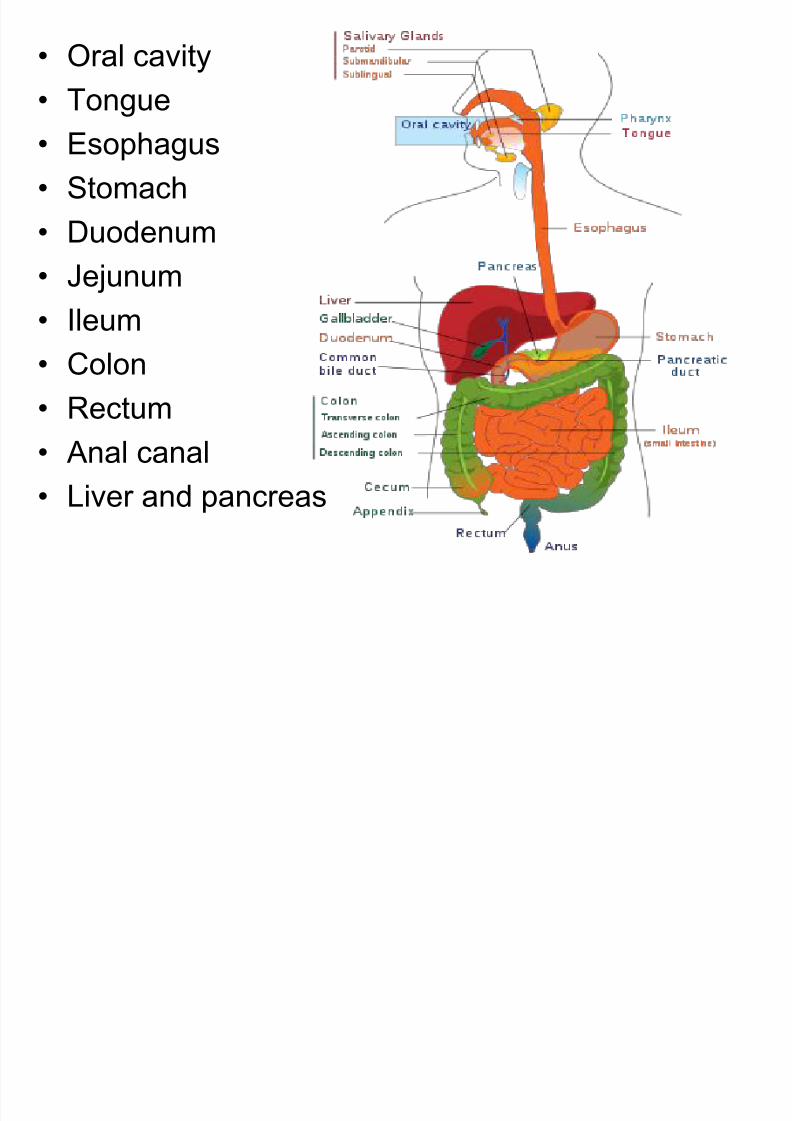
• Oral cavity
• Tongue
• Esophagus
• Stomach
• Duodenum
• Jejunum• Ileum
• Colon
• Rectum• Anal canal
• Liver and pancreas

8/11/2019 1.Gastrointestinal System
http://slidepdf.com/reader/full/1gastrointestinal-system 3/69
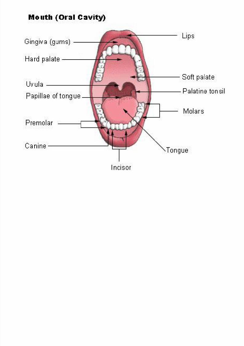
Oral cavity
• Begins in the Mouth , otherwise known as
the "Buccal Cavity“.
• Divided into vestibule, lips, cheeks, oral
cavity proper and teeth.
• Vestibule:- narrow space bounded
externally by the lips and the cheeks, and
internally by the teeth and gums.
• Parotid duct opens on the inner surface of
the cheek opposite the crown of the 2nd
molar tooth.
8/11/2019 1.Gastrointestinal System
http://slidepdf.com/reader/full/1gastrointestinal-system 4/69
• Lips:- are fleshy folds lined externally byskin and internally by the mucous member.
• Cheeks:- are fleshy flaps, continuing in thefront with the lips, and the junction is
indicated my the nasolabial sulcus which
extend from the side of the nose to the
angle of the mouth.
8/11/2019 1.Gastrointestinal System
http://slidepdf.com/reader/full/1gastrointestinal-system 5/69
• Oral cavity proper :- bounded
-anterolaterally by the teeth, the gum
and the alveolar arches of the jaws.
-Roof:- formed by hard and soft palate.
-Floor:- by tongue posteriorly andsublingual region anteriorly, below the
tongue.
• Posteriorly, the cavity communicates with
the pharynx.
8/11/2019 1.Gastrointestinal System
http://slidepdf.com/reader/full/1gastrointestinal-system 6/69
8/11/2019 1.Gastrointestinal System
http://slidepdf.com/reader/full/1gastrointestinal-system 7/69
• Gums (Gingivae):- are soft tissues which
envelop the alveolar processes of the
upper and lower jaws and surround the
neck of the teeth.

8/11/2019 1.Gastrointestinal System
http://slidepdf.com/reader/full/1gastrointestinal-system 8/69
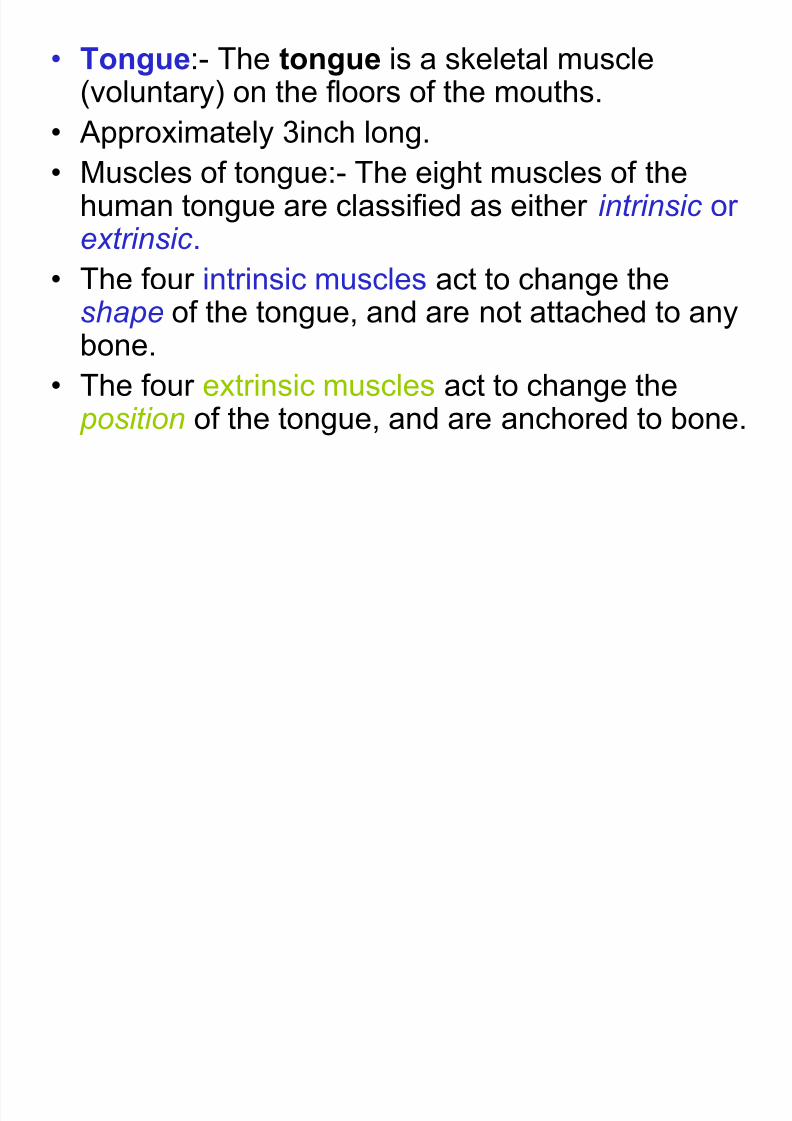
Tongue
8/11/2019 1.Gastrointestinal System
http://slidepdf.com/reader/full/1gastrointestinal-system 9/69
• Tongue:- The tongue is a skeletal muscle(voluntary) on the floors of the mouths.
• Approximately 3inch long.• Muscles of tongue:- The eight muscles of the
human tongue are classified as either intrinsic orextrinsic .
• The four intrinsic muscles act to change theshape of the tongue, and are not attached to anybone.
• The four extrinsic muscles act to change the
position of the tongue, and are anchored to bone.
8/11/2019 1.Gastrointestinal System
http://slidepdf.com/reader/full/1gastrointestinal-system 10/69
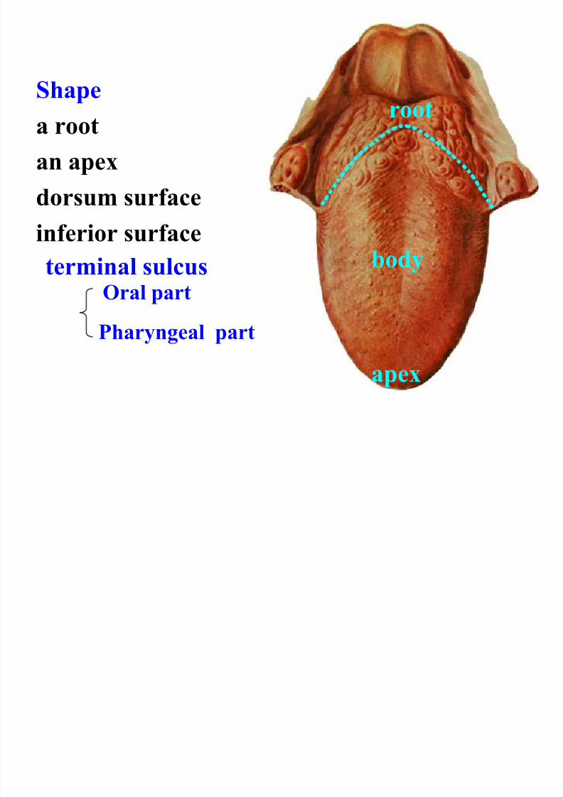
Shapea root
an apex
dorsum surfaceinferior surface
apex
root
body
Oral part
Pharyngeal part
terminal sulcus
8/11/2019 1.Gastrointestinal System
http://slidepdf.com/reader/full/1gastrointestinal-system 11/69
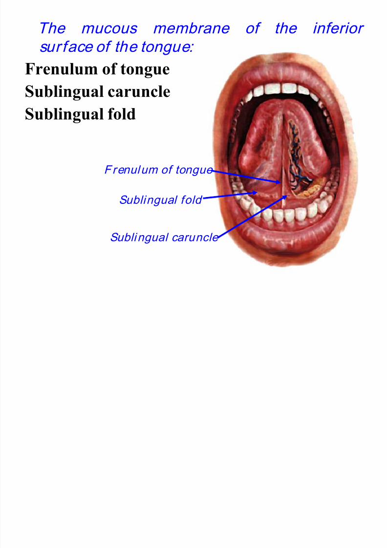
The mucous membrane of the inferior
surface of the tongue:
Frenulum of tongueSublingual caruncle
Sublingual fold
Frenulum of tongue
Sublingual caruncle
Sublingual fold
8/11/2019 1.Gastrointestinal System
http://slidepdf.com/reader/full/1gastrointestinal-system 12/69
• Extrinsic muscles:- originate from bone and
extend to the tongue and their main functionsare altering the tongue's position allowing for
protrusion, retraction, and side-to-side
movement.
-Genioglossus
-Hyoglossus
-Styloglossus
-Palatoglossus
8/11/2019 1.Gastrointestinal System
http://slidepdf.com/reader/full/1gastrointestinal-system 13/69
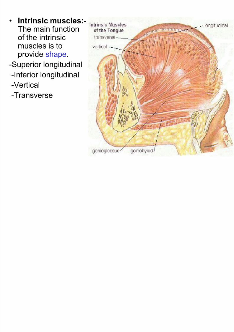
• Intrinsic muscles:-The main function
of the intrinsicmuscles is toprovide shape.
-Superior longitudinal
-Inferior longitudinal
-Vertical
-Transverse
8/11/2019 1.Gastrointestinal System
http://slidepdf.com/reader/full/1gastrointestinal-system 14/69
• Blood supply;- lingual artery, tonsillar and
pharyngeal artery.
• Lymphatic drainage:- tip of the tongue
drain into submental nodes, and right and
left halves of anterior 2/3rd drain into
submandibular nodes, and posterior 1/3rd
drain into Jugulo-omohyoid lymph node.
8/11/2019 1.Gastrointestinal System
http://slidepdf.com/reader/full/1gastrointestinal-system 15/69
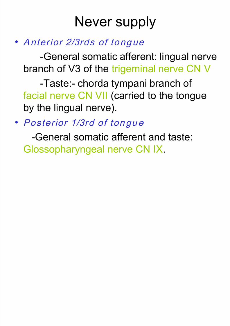
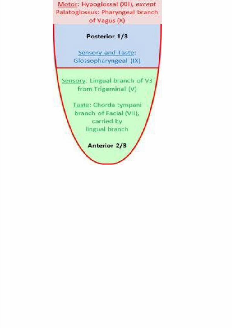
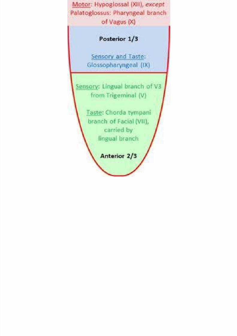
Never supply
• Anter ior 2/3rds of tongue
-General somatic afferent: lingual nerve
branch of V3 of the trigeminal nerve CN V
-Taste:- chorda tympani branch offacial nerve CN VII (carried to the tongue
by the lingual nerve).
• Poster ior 1/3rd of tongue
-General somatic afferent and taste:
Glossopharyngeal nerve CN IX.
8/11/2019 1.Gastrointestinal System
http://slidepdf.com/reader/full/1gastrointestinal-system 16/69
8/11/2019 1.Gastrointestinal System
http://slidepdf.com/reader/full/1gastrointestinal-system 17/69
• Motor:- All intrinsic and extrinsic muscles
of the tongue are supplied by the
hypoglossal nerve (CN XII), except for one
of the extrinsic muscles, Palatoglossus,which is innervated by CN X of the
pharyngeal plexus.
8/11/2019 1.Gastrointestinal System
http://slidepdf.com/reader/full/1gastrointestinal-system 18/69
8/11/2019 1.Gastrointestinal System
http://slidepdf.com/reader/full/1gastrointestinal-system 19/69
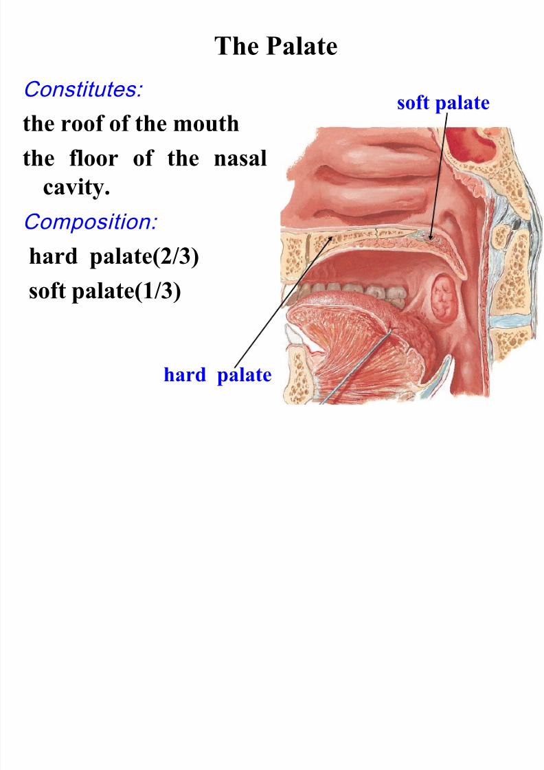
The Palate
Constitutes:the roof of the mouth
the floor of the nasal
cavity.Composition:
hard palate(2/3)
soft palate(1/3)
hard palate
soft palate
8/11/2019 1.Gastrointestinal System
http://slidepdf.com/reader/full/1gastrointestinal-system 20/69
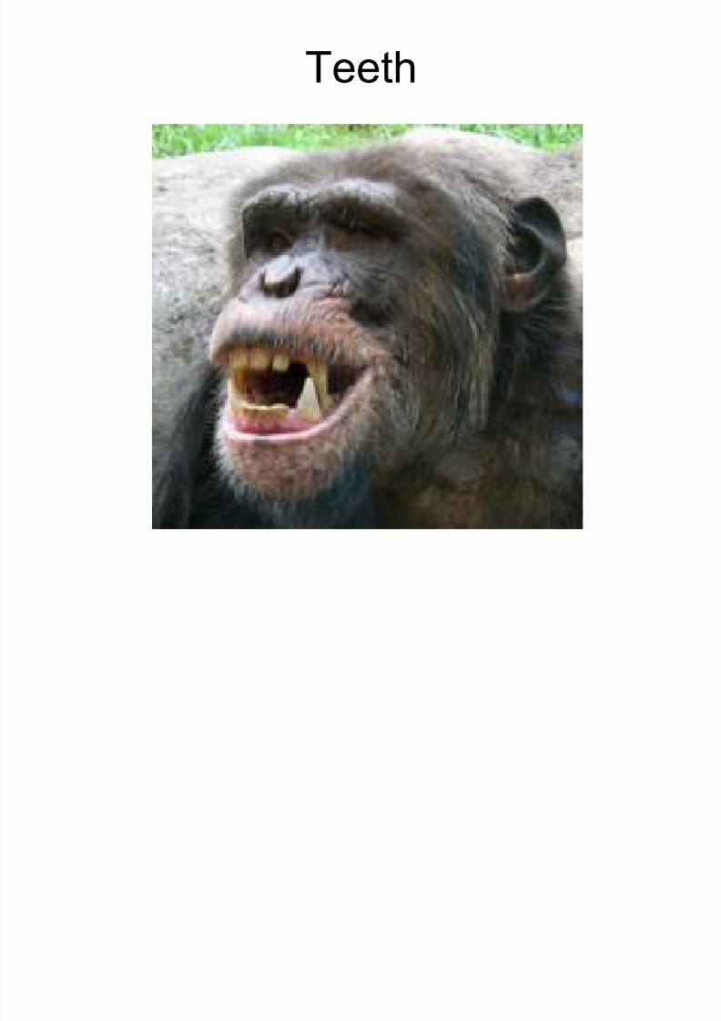
Teeth
8/11/2019 1.Gastrointestinal System
http://slidepdf.com/reader/full/1gastrointestinal-system 21/69
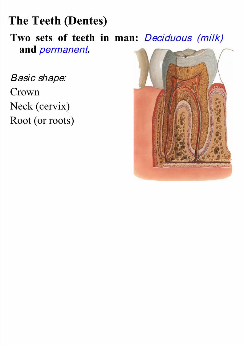
The Teeth (Dentes)
Two sets of teeth in man: Deciduous (milk)
and permanent .
Basic shape:
Crown
Neck (cervix)
Root (or roots)
8/11/2019 1.Gastrointestinal System
http://slidepdf.com/reader/full/1gastrointestinal-system 22/69
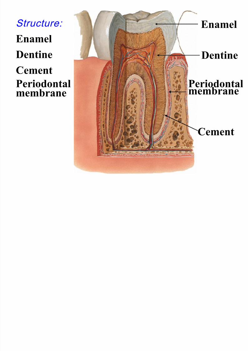
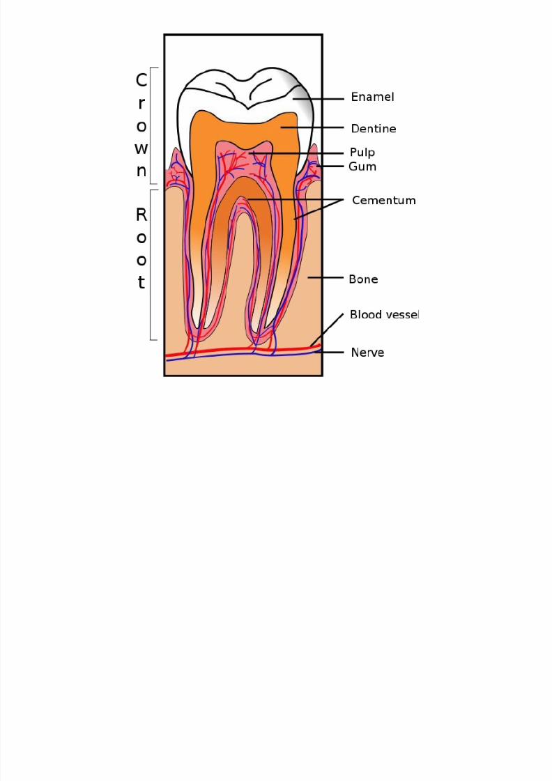
Structure:
EnamelDentine
Cement
Periodontalmembrane
Dentine
Enamel
Cement
Periodontalmembrane
8/11/2019 1.Gastrointestinal System
http://slidepdf.com/reader/full/1gastrointestinal-system 23/69
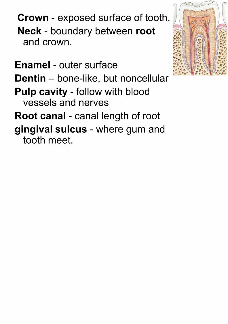
Crown - exposed surface of tooth.
Neck - boundary between root
and crown.
Enamel - outer surface
Dentin – bone-like, but noncellularPulp cavity - follow with blood
vessels and nerves
Root canal - canal length of rootgingival sulcus - where gum andtooth meet.
8/11/2019 1.Gastrointestinal System
http://slidepdf.com/reader/full/1gastrointestinal-system 24/69
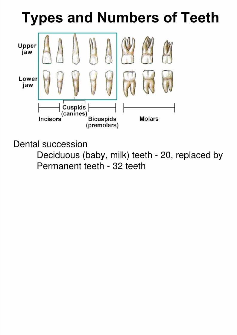
Types and Numbers of Teeth
Dental succession
Deciduous (baby, milk) teeth - 20, replaced by
Permanent teeth - 32 teeth
8/11/2019 1.Gastrointestinal System
http://slidepdf.com/reader/full/1gastrointestinal-system 25/69
8/11/2019 1.Gastrointestinal System
http://slidepdf.com/reader/full/1gastrointestinal-system 26/69
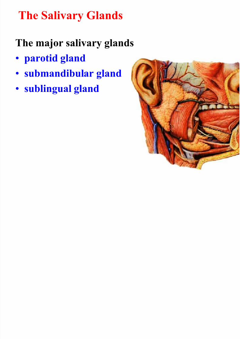
The Salivary Glands
The major salivary glands• parotid gland
• submandibular gland
• sublingual gland
8/11/2019 1.Gastrointestinal System
http://slidepdf.com/reader/full/1gastrointestinal-system 27/69
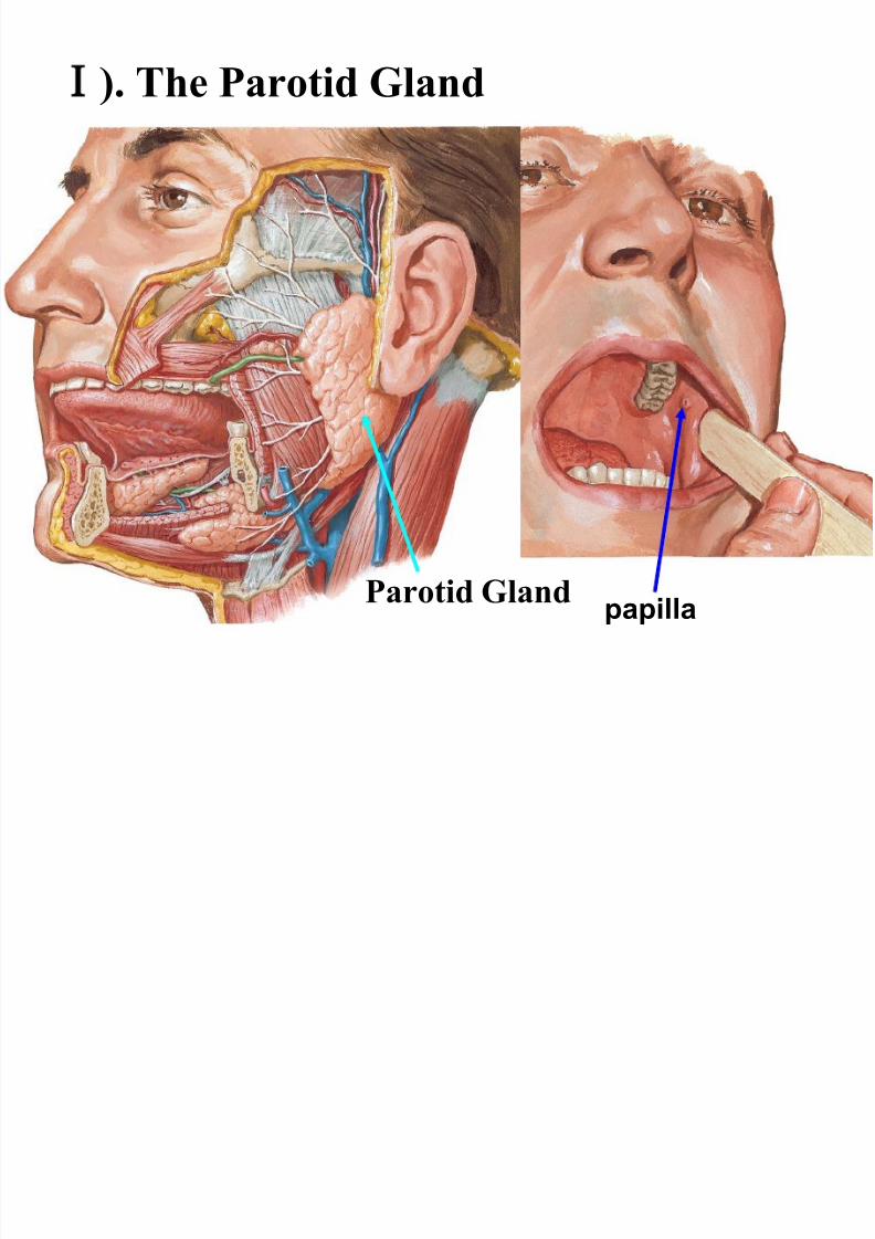
). The Parotid Gland
Parotid Glandpapilla
8/11/2019 1.Gastrointestinal System
http://slidepdf.com/reader/full/1gastrointestinal-system 28/69
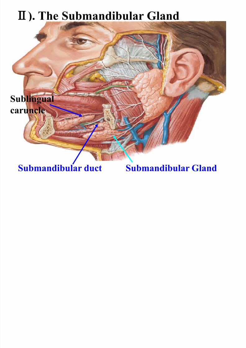
). The Submandibular Gland
Submandibular GlandSubmandibular duct
Sublingual
caruncle
8/11/2019 1.Gastrointestinal System
http://slidepdf.com/reader/full/1gastrointestinal-system 29/69
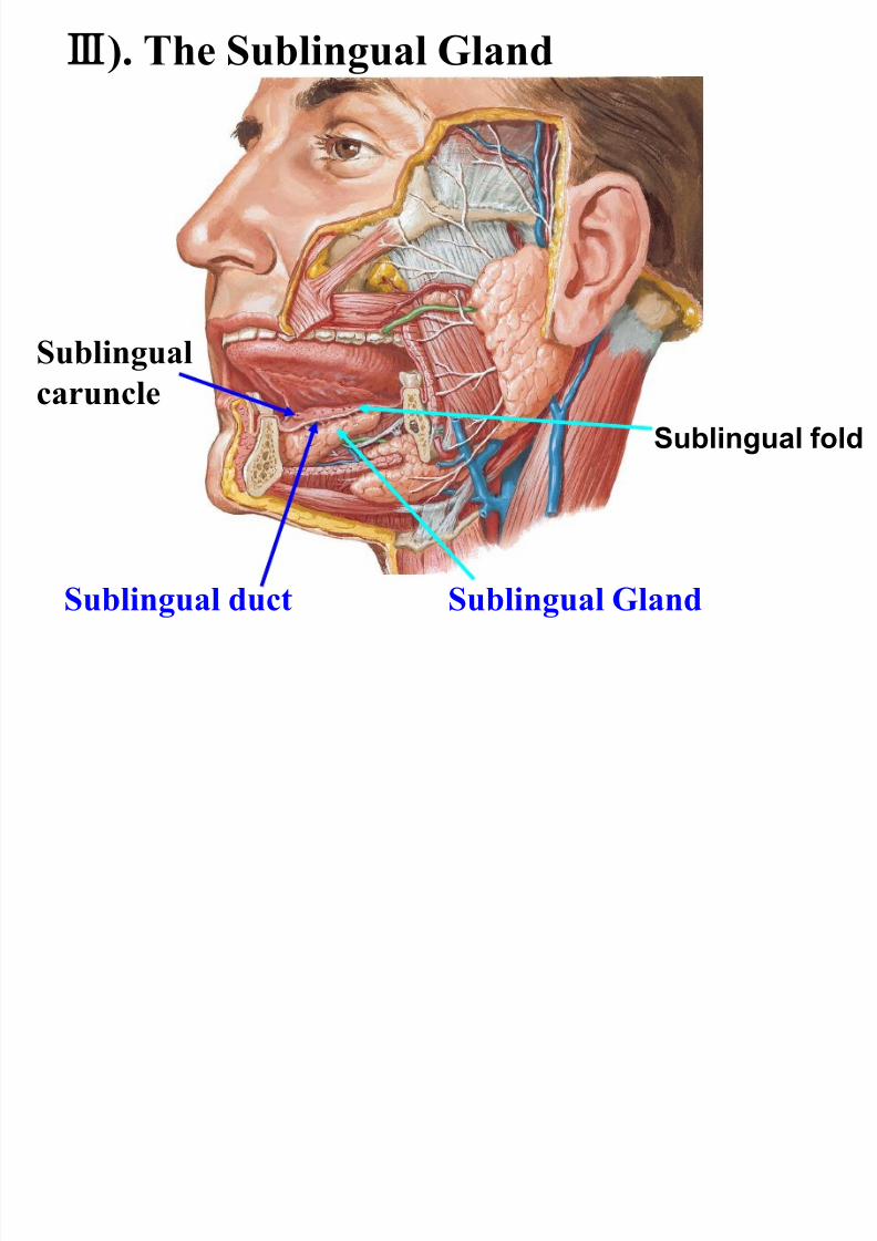
Sublingual GlandSublingual duct
Sublingual
caruncle
Ⅲ). The Sublingual Gland
Sublingual fold
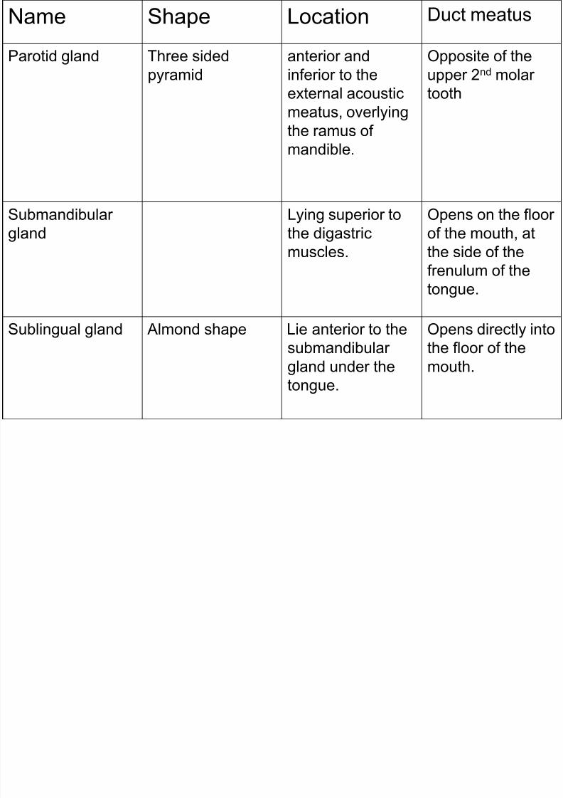
Name Shape Location Duct meatus
8/11/2019 1.Gastrointestinal System
http://slidepdf.com/reader/full/1gastrointestinal-system 30/69
Name Shape Location Duct meatus
Parotid gland Three sided
pyramid
anterior and
inferior to the
external acousticmeatus, overlying
the ramus of
mandible.
Opposite of the
upper 2nd molar
tooth
Submandibular
gland
Lying superior to
the digastric
muscles.
Opens on the floor
of the mouth, at
the side of the
frenulum of the
tongue.
Sublingual gland Almond shape Lie anterior to the
submandibular
gland under the
tongue.
Opens directly into
the floor of the
mouth.
8/11/2019 1.Gastrointestinal System
http://slidepdf.com/reader/full/1gastrointestinal-system 31/69
Pharynx
………go thro respiratory system..

8/11/2019 1.Gastrointestinal System
http://slidepdf.com/reader/full/1gastrointestinal-system 32/69
Esophagus
8/11/2019 1.Gastrointestinal System
http://slidepdf.com/reader/full/1gastrointestinal-system 33/69
• The esophagus is a narrow musculartube through which food passes from the
pharynx to the stomach.
• It is usually about 25 cm long.
• The esophagus is continuous with part ofthe pharynx at the level of the C6 vertebra.
• The abdominal part of the oesophagus isonly about 1.25cm.
8/11/2019 1.Gastrointestinal System
http://slidepdf.com/reader/full/1gastrointestinal-system 34/69
• The esophagus passes through posterior
mediastinum in thorax and enters
abdomen through a hole in the diaphragmat the level of the tenth thoracic vertebrae
(T10).
• Nonkeratinized stratified squamousepithelium.
• Muscularis externa
- upper 3rd is compose of striated muscles
- middle 3rd mixed type
- lower 3rd smooth muscles.
8/11/2019 1.Gastrointestinal System
http://slidepdf.com/reader/full/1gastrointestinal-system 35/69
Esophageal constrictions
• Normally, the esophagus has 4 constrictions atthe following levels.
• At the esophageal inlet, where the pharynx joinsthe esophagus, behind the cricoidcartilage,15 cm from the incisor teeth.
• Where it is crossed by the aortic arch, 25cm from the incisor teeth.
• Where it is crossed by the left bronchus,27.5 cm from the incisor teeth.
• Where it pierces the diaphragm, 37.5 cm fromthe incisor teeth).
• The distances from the incisor teeth areimportant.
8/11/2019 1.Gastrointestinal System
http://slidepdf.com/reader/full/1gastrointestinal-system 36/69
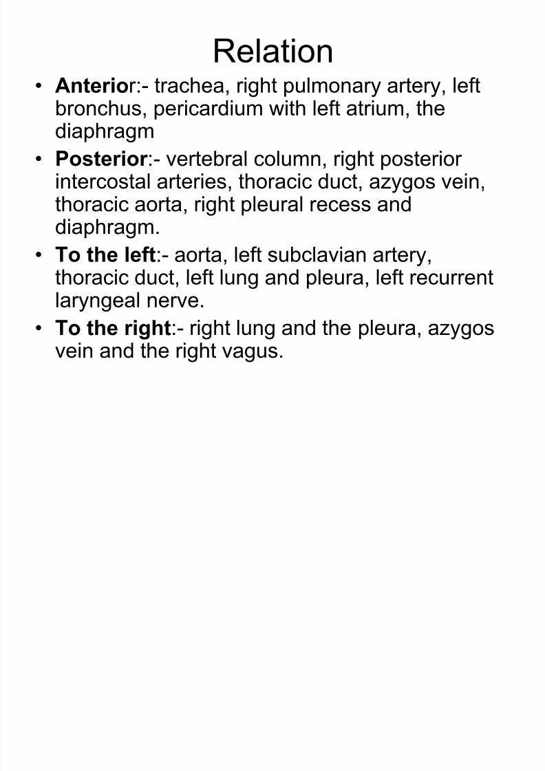
Relation• Anterior:- trachea, right pulmonary artery, left
bronchus, pericardium with left atrium, thediaphragm
• Posterior :- vertebral column, right posteriorintercostal arteries, thoracic duct, azygos vein,thoracic aorta, right pleural recess anddiaphragm.
• To the left:- aorta, left subclavian artery,
thoracic duct, left lung and pleura, left recurrentlaryngeal nerve.
• To the right:- right lung and the pleura, azygosvein and the right vagus.
8/11/2019 1.Gastrointestinal System
http://slidepdf.com/reader/full/1gastrointestinal-system 37/69
• Arterial supply:- cervical part by branch ofinferior thyroid artery, thoracic part by
esophageal branch of the aorta, andabdominal part by branches of left gastricartery.
• Venous drainage:- upper part drain intobrachiocephalic vein, middle part goes toazygos vein and lower part goes to leftgastric vein.
• The lower end is one of the sites forprotosystemic anastamosis.
8/11/2019 1.Gastrointestinal System
http://slidepdf.com/reader/full/1gastrointestinal-system 38/69
• Lymphatic drainage:- cervical part drain into
deep cervical nodes, thoracic part to the
posterior mediastinal nodes and abdominal partto left gastric nodes.
• Nerve supply:-
- Parasympathetic, upper half by recurrentlaryngeal nerve and lower half by oesophageal
plexus. Nerves are sensor, motor and
secretomotor to the oesophagus.
- Sympathetic:- upper half by fibers of middlecervical ganglion and lower half by upper four
thoracic ganglia. Nerves are vasomotor.

8/11/2019 1.Gastrointestinal System
http://slidepdf.com/reader/full/1gastrointestinal-system 39/69
Stomach
8/11/2019 1.Gastrointestinal System
http://slidepdf.com/reader/full/1gastrointestinal-system 40/69
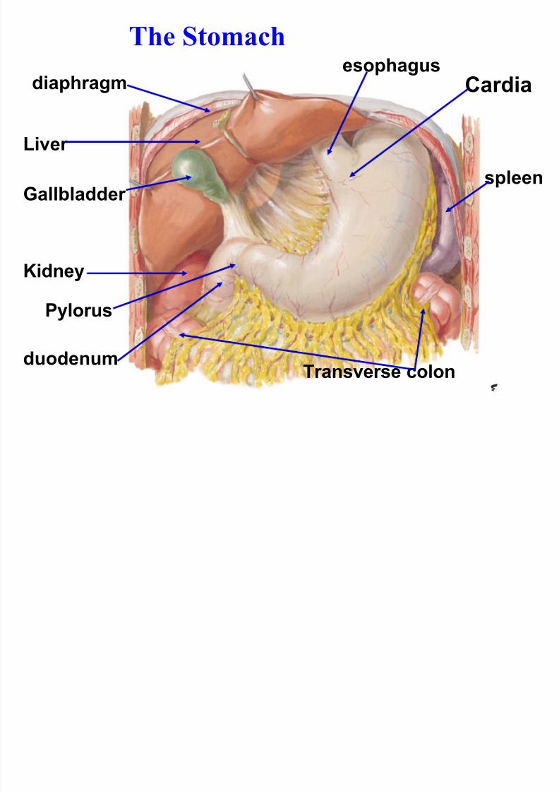
The Stomachesophagus
duodenum
Liver
Kidney
Transverse colon
spleen
Gallbladder
diaphragm Cardia
Pylorus
8/11/2019 1.Gastrointestinal System
http://slidepdf.com/reader/full/1gastrointestinal-system 41/69
• Also known as gaster or venter, and is a
muscular bag, hollow, dilated part of the
digestion system.
• It is located between the esophagus and
the small intestine.
• It acts as a reservoir of food and helps indigestion of carbohydrates, proteins and
fats.
8/11/2019 1.Gastrointestinal System
http://slidepdf.com/reader/full/1gastrointestinal-system 42/69
• Location:- it lies obliquely in the upper andleft part of the abdomen, occupying theepigastric, umbilical and lefthypochondriac region.
• Most of the stomach lies under cover ofthe left costal margin and the ribs.
• Shape and position:- when empty,stomach is J-shaped (vertical), whenpartially distended, it become pyriform.
• Size:- it is 25cm long, and mean capacityis one ounce (30ml) at birth, one litre atpuberty and 1.5 to 2litres or more in adults.
8/11/2019 1.Gastrointestinal System
http://slidepdf.com/reader/full/1gastrointestinal-system 43/69
• External features:-
- two orifices or openings,
- two curvatures or borders, and
- two surfaces.
• Two orifices:- cardiac or i f ice is joined by lower
end of oesophagus and it lies behind the 7th costal cartilage 2.5cm from the junction with the
sternum, at the level of T11 vertebra.
- py lor ic o r i f ice opens into duodenum, it lies
to the right of the medial plane, at level of lower
border of L1 vertebra.

8/11/2019 1.Gastrointestinal System
http://slidepdf.com/reader/full/1gastrointestinal-system 44/69
8/11/2019 1.Gastrointestinal System
http://slidepdf.com/reader/full/1gastrointestinal-system 45/69
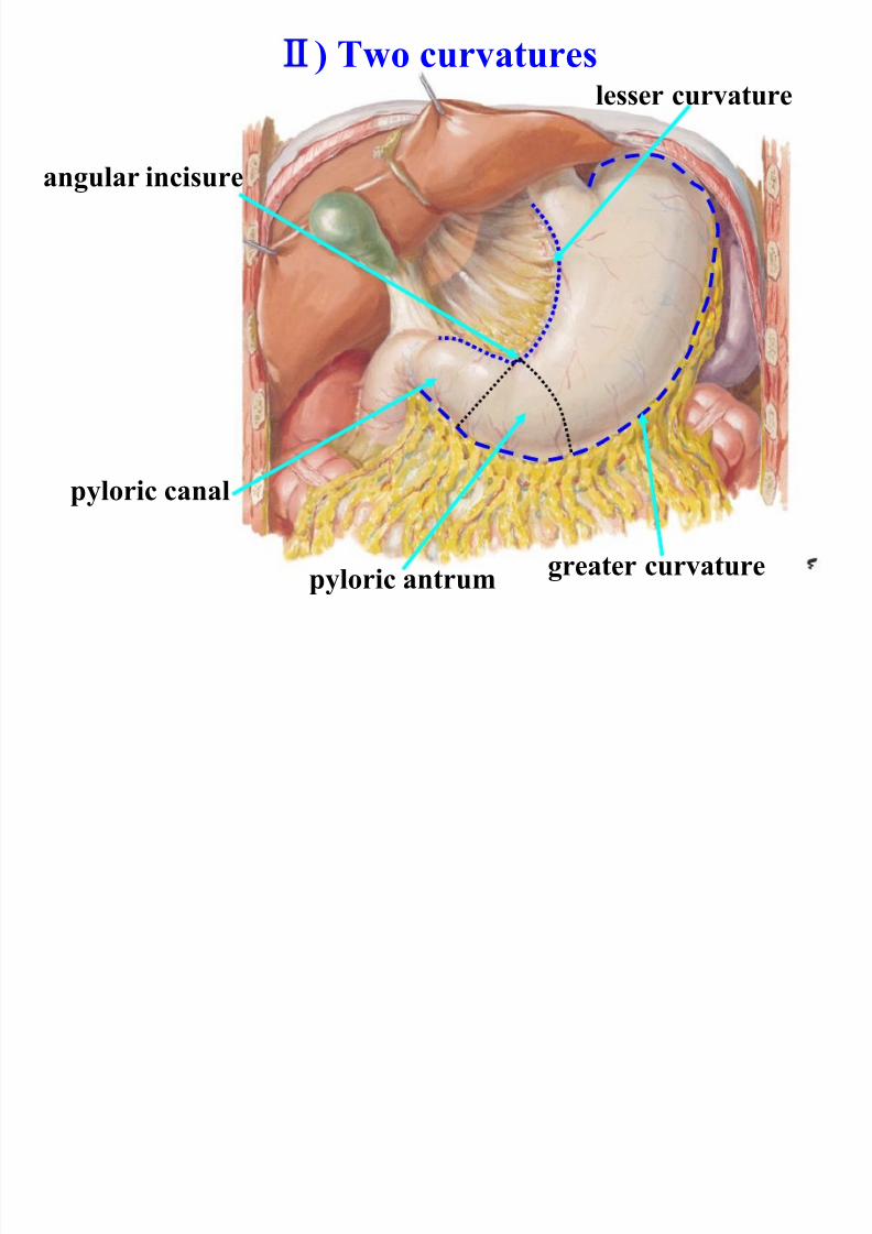
• Two curvatures:- lesser cu rvature isconcave and forms the right border of the
stomach and attached to lesser omentum.The most dependent part is the curvatureis marked by the angular notch or incisuraangularis.
- greater cu rvatu re is convex and formthe left border and attached to greateromentum, the gastrosplenic ligament and
the gastrophrenic ligament. At upper end itpresent cardiac notch which separates itform oesophagus.
8/11/2019 1.Gastrointestinal System
http://slidepdf.com/reader/full/1gastrointestinal-system 46/69
) Two curvatures
pyloric antrum
pyloric canal
greater curvature
lesser curvature
angular incisure
8/11/2019 1.Gastrointestinal System
http://slidepdf.com/reader/full/1gastrointestinal-system 47/69
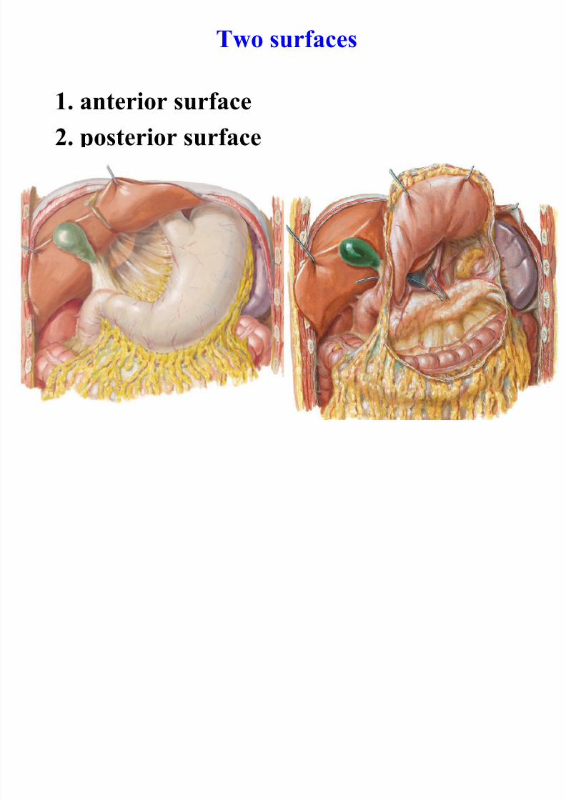
• Two surfaces:- anterior or anterosuperior
surface faces forward and upward
-Posterior or posteroinferior surface
faces backward and downward.
8/11/2019 1.Gastrointestinal System
http://slidepdf.com/reader/full/1gastrointestinal-system 48/69
8/11/2019 1.Gastrointestinal System
http://slidepdf.com/reader/full/1gastrointestinal-system 49/69
• The stomach is divided into 2 parts:-
- Cardiac and
- Pylorus
..by the line drawn downwards and to the
left form the incisura angularis.

8/11/2019 1.Gastrointestinal System
http://slidepdf.com/reader/full/1gastrointestinal-system 50/69
8/11/2019 1.Gastrointestinal System
http://slidepdf.com/reader/full/1gastrointestinal-system 51/69
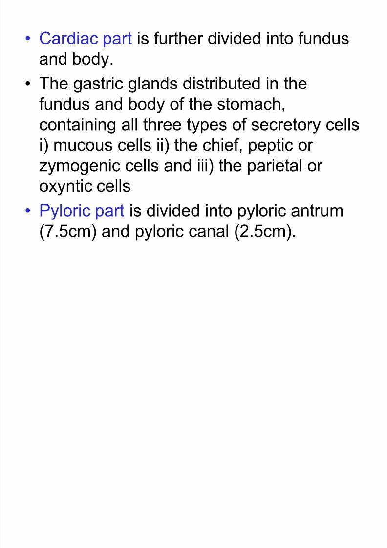
• Cardiac part is further divided into fundus
and body.
• The gastric glands distributed in the
fundus and body of the stomach,
containing all three types of secretory cells
i) mucous cells ii) the chief, peptic orzymogenic cells and iii) the parietal or
oxyntic cells
• Pyloric part is divided into pyloric antrum(7.5cm) and pyloric canal (2.5cm).
8/11/2019 1.Gastrointestinal System
http://slidepdf.com/reader/full/1gastrointestinal-system 52/69
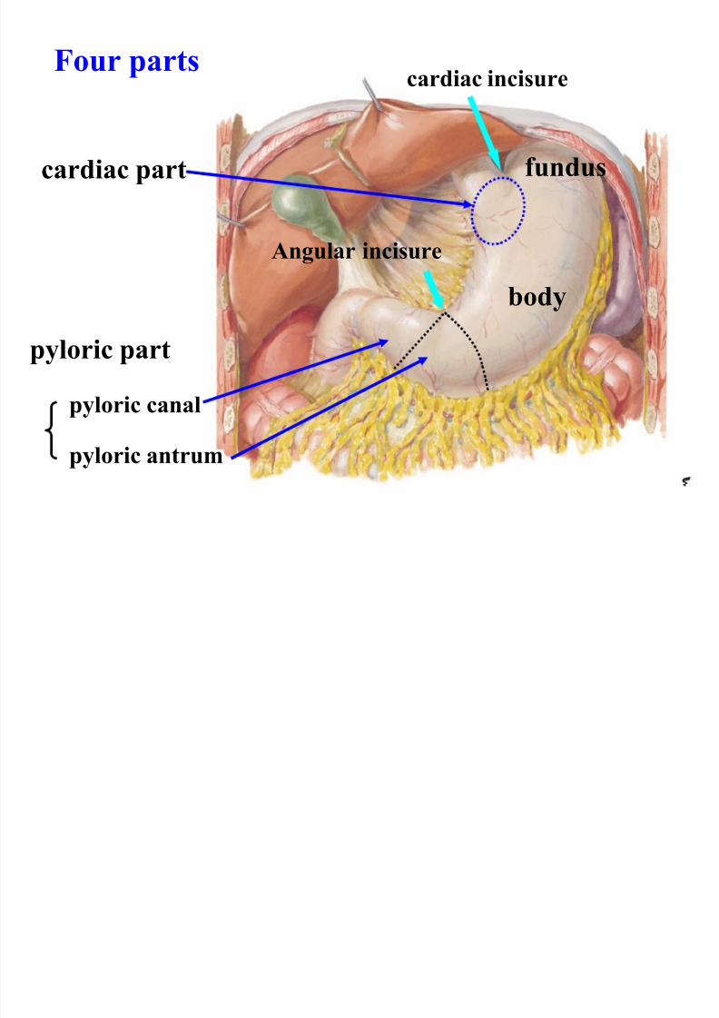
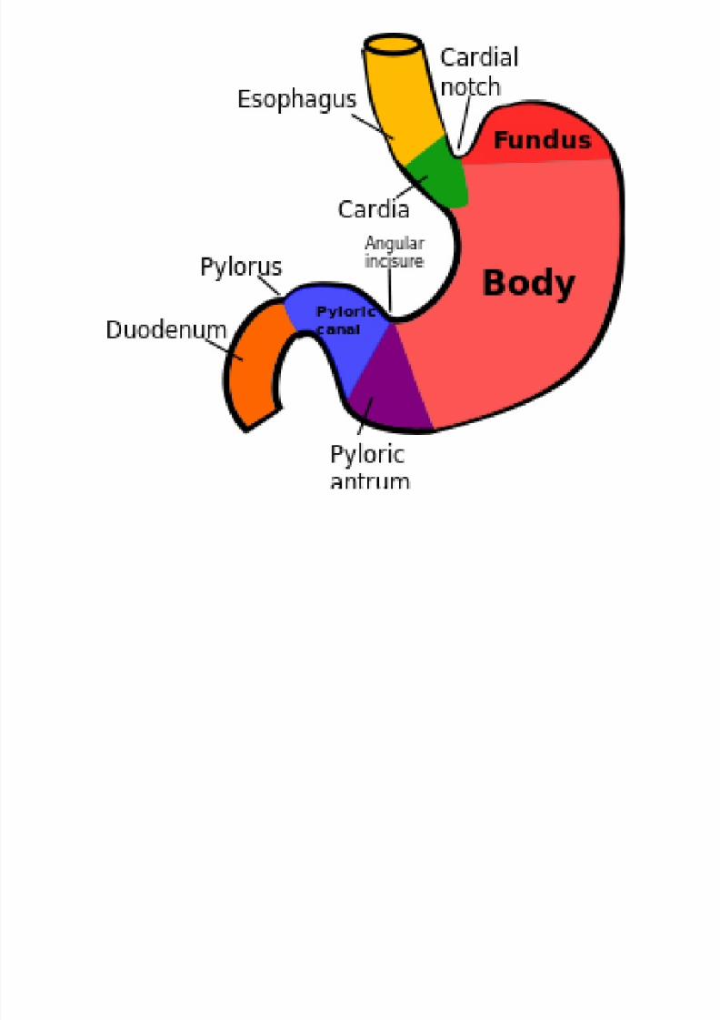
Four parts
fundus
body
pyloric part
pyloric antrum
pyloric canal
Angular incisure
cardiac part
cardiac incisure
8/11/2019 1.Gastrointestinal System
http://slidepdf.com/reader/full/1gastrointestinal-system 53/69
8/11/2019 1.Gastrointestinal System
http://slidepdf.com/reader/full/1gastrointestinal-system 54/69
Relations of the Stomach
• Peritoneal relations:- stomach is lined by the
peritoneum on both its surface. At lesser
curvature it forms lesser omentum and at greater
curvature it forms the greater omentum.
• Near the fundus the two layers meet to form
gastrosplenic ligament and near the cardiac end
the peritoneum on the posterior surface formsgastrophrenic ligament.
8/11/2019 1.Gastrointestinal System
http://slidepdf.com/reader/full/1gastrointestinal-system 55/69
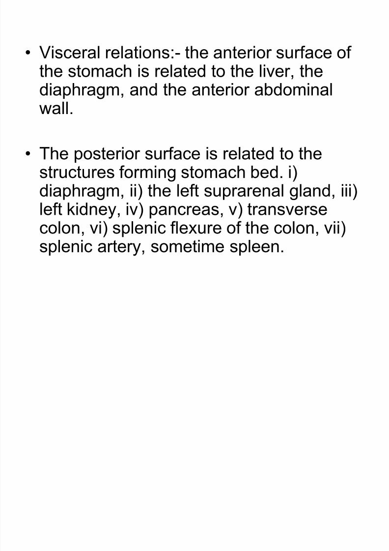
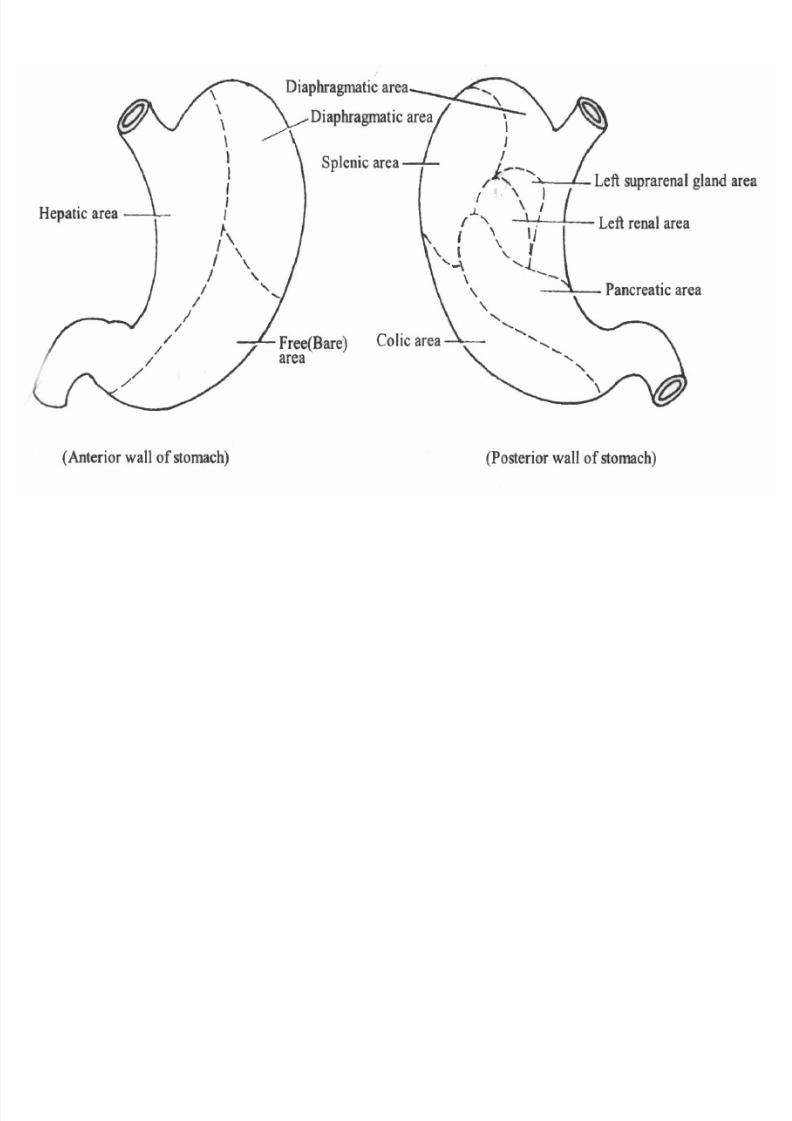
• Visceral relations:- the anterior surface of
the stomach is related to the liver, thediaphragm, and the anterior abdominalwall.
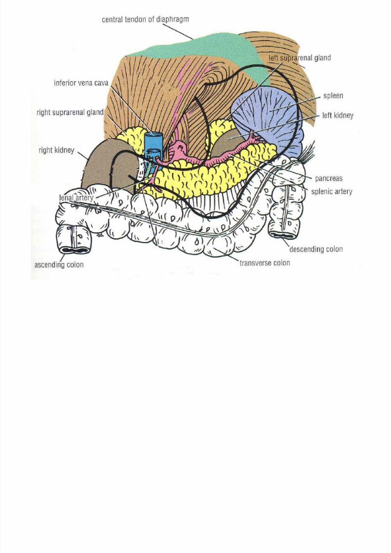
• The posterior surface is related to thestructures forming stomach bed. i)diaphragm, ii) the left suprarenal gland, iii)
left kidney, iv) pancreas, v) transversecolon, vi) splenic flexure of the colon, vii)splenic artery, sometime spleen.
8/11/2019 1.Gastrointestinal System
http://slidepdf.com/reader/full/1gastrointestinal-system 56/69
8/11/2019 1.Gastrointestinal System
http://slidepdf.com/reader/full/1gastrointestinal-system 57/69
8/11/2019 1.Gastrointestinal System
http://slidepdf.com/reader/full/1gastrointestinal-system 58/69
Interior of Stomach
• Mucosa
• Submucosa
• Muscularis
• Serosa ……………………….

8/11/2019 1.Gastrointestinal System
http://slidepdf.com/reader/full/1gastrointestinal-system 59/69
8/11/2019 1.Gastrointestinal System
http://slidepdf.com/reader/full/1gastrointestinal-system 60/69
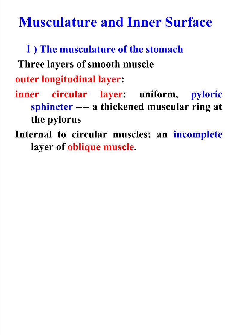
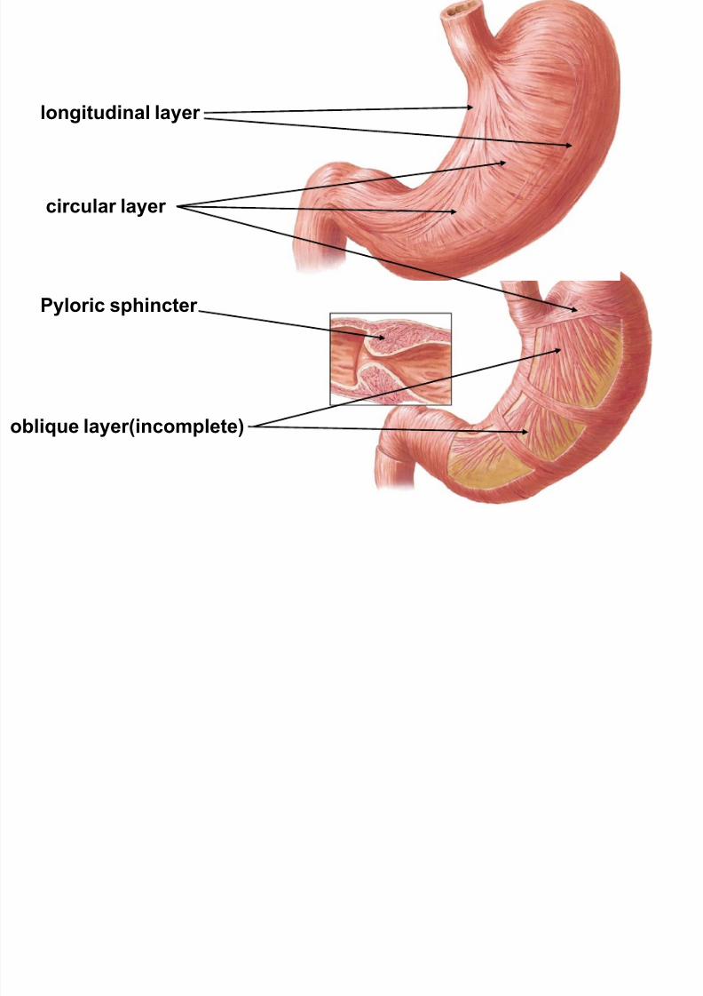
Musculature and Inner Surface
) The musculature of the stomach
Three layers of smooth muscle
outer longitudinal layer:
inner circular layer: uniform, pyloric
sphincter ---- a thickened muscular ring at
the pylorus
Internal to circular muscles: an incomplete
layer of oblique muscle.
8/11/2019 1.Gastrointestinal System
http://slidepdf.com/reader/full/1gastrointestinal-system 61/69
longitudinal layer
circular layer
oblique layer(incomplete)
Pyloric sphincter
8/11/2019 1.Gastrointestinal System
http://slidepdf.com/reader/full/1gastrointestinal-system 62/69
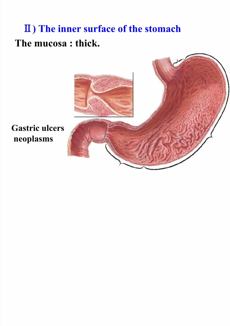
) The inner surface of the stomach
The mucosa : thick.
Gastric ulcers
neoplasms
8/11/2019 1.Gastrointestinal System
http://slidepdf.com/reader/full/1gastrointestinal-system 63/69
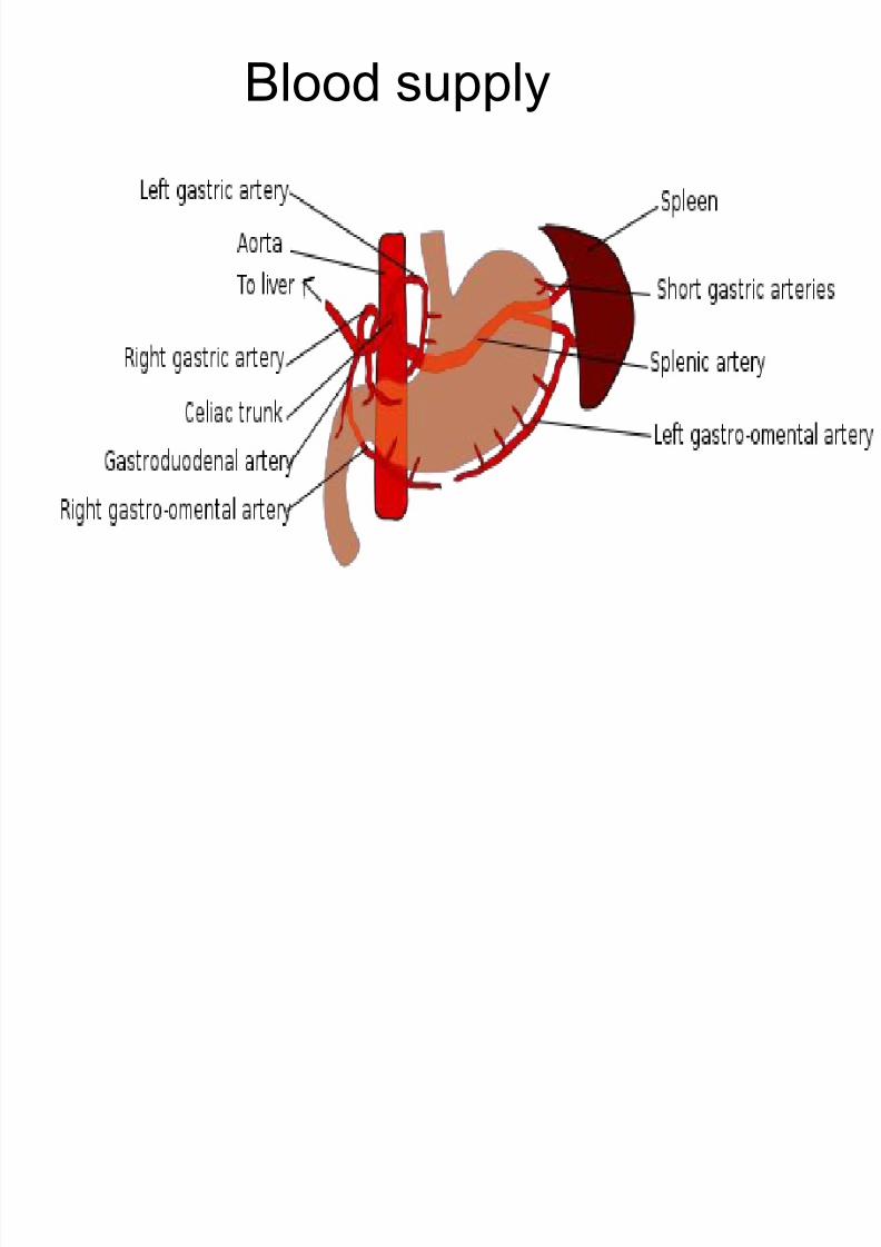
Blood supply
Lymphatic drainage
8/11/2019 1.Gastrointestinal System
http://slidepdf.com/reader/full/1gastrointestinal-system 64/69
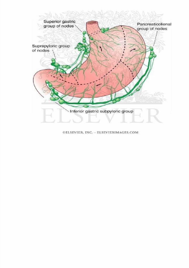
Lymphatic drainage
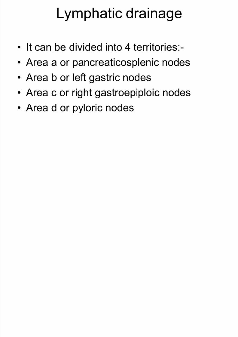
• It can be divided into 4 territories:-
• Area a or pancreaticosplenic nodes
• Area b or left gastric nodes
• Area c or right gastroepiploic nodes
• Area d or pyloric nodes
8/11/2019 1.Gastrointestinal System
http://slidepdf.com/reader/full/1gastrointestinal-system 65/69
8/11/2019 1.Gastrointestinal System
http://slidepdf.com/reader/full/1gastrointestinal-system 66/69
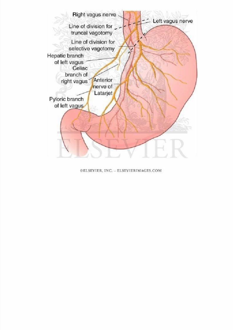
Nerve supply
• The stomach is supplied by sympathetic andparasympathetic nerves.
• Sympathetic nerves are derived from T6 to T10segments of the spinal cord, via the greatersplanchnic nerves and coeliac and hepaticplexuses.
• Pathway for the pain sensations from thestomach.
8/11/2019 1.Gastrointestinal System
http://slidepdf.com/reader/full/1gastrointestinal-system 67/69
• Parasympathetic nerves are derived from the vagus,through oesophagus plexus and gastric nerves.
• Anterior or left vagal fibres divides into i) gastricbranch for anterior surface of the fundus and bodyof stomach, ii) two pylorus branches
• Posterior or right vagal fibres divides into i) smaller
gastric branches for the posterior surface of thefundus, body and antrum, ii) larger, coeliacbranches for the coeliac plexus.
• It stimulates to increase the motility of the stomach
and secretion of the gastric juice rich in pepsin andHCL
8/11/2019 1.Gastrointestinal System
http://slidepdf.com/reader/full/1gastrointestinal-system 68/69
Functions
8/11/2019 1.Gastrointestinal System
http://slidepdf.com/reader/full/1gastrointestinal-system 69/69
Functions• Reservoir of food.
• Mixing of food with the gastric juice by peristalticmovement.
• Secrets gastric juice via gastric gland help indigestion of the food.
• Produce HCL which destroys many organismspresent in food and drinks.
• Protect the gastric mucosa from HCL byproducing mucus.
• Absorption of drugs, water, salt, alcohol etc.• Produce intrinsic factor which helps in the
absorption of Vitamin B12.