Case Report Nadir G¶r¼len E Zamanl± °n - jcam.com.tr
3
Kardiyak Hidatik Kist / Cardiac Hydatid Cyst Unusual Presentation of Alveolar Echinococ with Simultane Intramyocardial and Lung: Case Report Nadir Görülen Eş Zamanlı İntramiyokardiyal ve Akciğer Yerleşimli Alveolar Ekinokokkus: Olgu Sunumu DOI: 10.4328/JCAM.2042 Received: 04.09.2013 Accepted: 01.10.2013 Printed: 01.10.2013 J Clin Anal Med 2013;4(suppl 2): 129-31 Corresponding Author: Bayram Metin, Bozok Üniversitesi, Tıp Fakültesi, Göğüs Cerrahi ABD. Yozgat, Türkiye. T.: +905072385361 E-Mail: [email protected] Özet Alveolar kist hidatik (AKH) özellikle kuzey yarım kürede tilkilerden geçen Ecino- kokkus Multilokularisin neden olduğu bir hastalıktır.Endemik bölgelerde insidan- sı 0.03-1.2/100 000 oranındadır. Karaciğer primer odak olarak bilinmesine rağ- men akciğer, dalak, pankreas, retroperitoneal alan, beyin kemik ve yumuşak doku- lar da sık yerleştiği alanlardır.Pulmoner hidatik kist oranı % 7-20 olarak bilinme- sine karşın eş zamanlı intramiyokardiyal ve akciğer tutulumu literatürde nadir gö- rülen bir durumdur. Anahtar Kelimeler Alveolar Kist Hidatik; İntramiyokardiyal Tutulum; Kardiyopulmoner Cerrahi Abstract Alveolar echinococcosis (AE), observed in the Northern Hemisphere, is caused by the larval stage of the fox tapeworm Echinococcus multilocularis. In endemic ar- eas, annual incidence of AE ranges from 0.03 to 1.2/100 000 inhabitants. The liver is the primary focus of the disease but extrahepatic is also possible such as lung, spleen, pancreas, retroperitoneum, brain, bone and soſt tissue. While pulmonary involvements occur in 7 to 20%, to our best knowledge; simultanous intramyocardial and lung involvement is a very rare clinical entity reported in the literature . Keywords Alveolar Echinococcosis; Intramyocardial Involvement; Cardiopulmonary Surgery Bayram Metin 1 , Hüseyin Ede 2 , Yavuz Selim İntepe 3 , Hasan Ekim 4 , Neziha Yılmaz 5 , Esef Bolat 6 , Şener Yıldırım 7 ¹Bozok University Faculty of Medicine, Department of Thoracic Surgery, Yozgat, ²Bozok University Faculty of Medicine, Department of Cardiology, Yozgat, ³Bozok University Faculty of Medicine, Department of Chest Diseases, Yozgat, 4 Bozok University Faculty of Medicine, Department of Cardiovascular Surgery, Yozgat, 5 Bozok University Faculty of Medicine, Depatment of infectious diseases, Yozgat, 6 Bozok University Faculty of Medicine, Department of Aneasthesiology, Yozgat, 7 Yozgat State Hospital, Thoracic Surgery Clinic, Yozgat, Turkey Journal of Clinical and Analytical Medicine | 129
Transcript of Case Report Nadir G¶r¼len E Zamanl± °n - jcam.com.tr
| Journal of Clinical and Analytical Medicine1
Kardiyak Hidatik Kist / Cardiac Hydatid Cyst
Unusual Presentation of Alveolar Echinococ with Simultane Intramyocardial and Lung: Case Report
Nadir Görülen Eş Zamanlı İntramiyokardiyal ve Akciğer Yerleşimli Alveolar Ekinokokkus: Olgu Sunumu
DOI: 10.4328/JCAM.2042 Received: 04.09.2013 Accepted: 01.10.2013 Printed: 01.10.2013 J Clin Anal Med 2013;4(suppl 2): 129-31Corresponding Author: Bayram Metin, Bozok Üniversitesi, Tıp Fakültesi, Göğüs Cerrahi ABD. Yozgat, Türkiye.T.: +905072385361 E-Mail: [email protected]
Özet
Alveolar kist hidatik (AKH) özellikle kuzey yarım kürede tilkilerden geçen Ecino-
kokkus Multilokularisin neden olduğu bir hastalıktır.Endemik bölgelerde insidan-
sı 0.03-1.2/100 000 oranındadır. Karaciğer primer odak olarak bilinmesine rağ-
men akciğer, dalak, pankreas, retroperitoneal alan, beyin kemik ve yumuşak doku-
lar da sık yerleştiği alanlardır.Pulmoner hidatik kist oranı % 7-20 olarak bilinme-
sine karşın eş zamanlı intramiyokardiyal ve akciğer tutulumu literatürde nadir gö-
rülen bir durumdur.
Anahtar Kelimeler
Alveolar Kist Hidatik; İntramiyokardiyal Tutulum; Kardiyopulmoner Cerrahi
Abstract
Alveolar echinococcosis (AE), observed in the Northern Hemisphere, is caused by
the larval stage of the fox tapeworm Echinococcus multilocularis. In endemic ar-
eas, annual incidence of AE ranges from 0.03 to 1.2/100 000 inhabitants. The
liver is the primary focus of the disease but extrahepatic is also possible such
as lung, spleen, pancreas, retroperitoneum, brain, bone and soft tissue. While
pulmonary involvements occur in 7 to 20%, to our best knowledge; simultanous
intramyocardial and lung involvement is a very rare clinical entity reported in the
literature .
Keywords
Alveolar Echinococcosis; Intramyocardial Involvement; Cardiopulmonary Surgery
Bayram Metin1, Hüseyin Ede2, Yavuz Selim İntepe3, Hasan Ekim4, Neziha Yılmaz5, Esef Bolat6, Şener Yıldırım7
¹Bozok University Faculty of Medicine, Department of Thoracic Surgery, Yozgat, ²Bozok University Faculty of Medicine, Department of Cardiology, Yozgat, ³Bozok University Faculty of Medicine, Department of Chest Diseases, Yozgat, 4Bozok University Faculty of Medicine, Department of Cardiovascular Surgery, Yozgat,
5Bozok University Faculty of Medicine, Depatment of infectious diseases, Yozgat, 6Bozok University Faculty of Medicine, Department of Aneasthesiology, Yozgat, 7Yozgat State Hospital, Thoracic Surgery Clinic, Yozgat, Turkey
Journal of Clinical and Analytical Medicine | 129

| Journal of Clinical and Analytical Medicine
Kardiyak Hidatik Kist / Cardiac Hydatid Cyst
2
IntroductionAlveolar echinococcosis (AE), observed in the Northern Hemi-sphere, is caused by the larval stage of the fox tapeworm Echi-nococcus multilocularis [1]. Human is infected after ingesting eggs, the metacestode cells of E. multilocularis which prolif-erates in the liver [2]. It is a potentially fatal, chronically pro-gressive parasitic infection characterized by a long asymptom-atic period and development of an invasive tumor-like lesion throughout this period [3, 4]. So early diagnosis of AE is very difficult because of long latent or asymptomatic period which may be as long as 15 years [2-4]. In endemic areas, annual in-cidence of AE ranges from 0.03 to 1.2/100 000 inhabitants [2]. The liver is the primary focus of the disease but extrahepatic involvement such as lung, spleen, pancreas, retroperitoneum, brain, bone and soft tissue. While pulmonary involvements oc-cur in 7 to 20%, to our best knowledge; simultanous intramyo-cardial and lung involvement sparing liver is a very rare clinical entity reported in the literature [5, 6]. Here, we present a very rare case of alveolar echinococcosis with simultanous intramyocardial and lung involvement sparing liver which was completely removed at the same session via cardiopulmonary surgery.
Case ReportA 47-year-old woman was admitted to our hospital with a his-tory of hydoptysis six months before admission. She didn’t have any complaint otherwise. Physical examination did not reveal any abnormal findings: her lungs were normal on auscultation, no cardiac murmur or gallop rhythm was noted, and biochemi-cal laboratory test results were within normal limits. Myocardi-al-specific enzyme values were within the normal range.The patient’s chest X-ray was normal, except for increase in convexity on left border of the heart; and the lesion paratrache-ally located with sharp border, homogenous in content at upper zone of the left lung; and second one; sharp-bordered, paracar-diac lesion with air-fluid content located at mid-zone of the left lung (Figure 1A). The Electrocardiography (ECG) showed normal
sinus rhythm with T-wave inversion in leads I, aVL. (Figure 2A) transthoracic echocardiography showed a multivesicular cystic mass on apical part of the left posterolateral ventricular wall and protruded inward the ventricular cavity (Figure 2B,2C). The
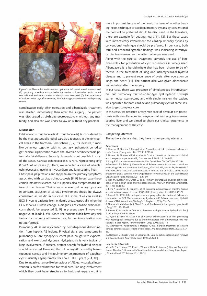
patient’s abdominal ultrasonography and cranial computed to-mography (CT) were normal. The patient’s chest multislice CT and Magnetic Resonance Imaging (MRI) showed multivesicular cystic masses in the left ventricule lateral wall, at apical seg-ment of the upper lobe of the left lung and at superior segment of the lower lobe of the left lung (Figure 1B-1D).Serologically, specific Echinococcus granulosus and multilocu-laris antibodies were investigated by commercial ELISA (No-vaLisa, Echinococcus IgG Novatec, Germany For E. Granulosus; Bordier Affinity Products SA, Switzerland for E.multicularis) and haemagglutination-inhibition test (HAI) (Hydatidose, Fumuoze laboratories, France) kits. Echinococcus multilocularis antibod-ies were positive with ELISA. Hydatid cyst antibodies was found to be positive 1/320 titer in the HAI.The cardiac multivesicular cyst in the left ventricle wall was re-moved with cystotomy-capitonage procedure and then the cav-ity was washed with a hypertonic saline solution under cardio-pulmonary bypass by conventional technique following median sternotomy. Gross appearence of the heart confirmed intramyo-cardial involvement (Figure 3A-D). Then, left pleural membrane was incised by using same median sternotomy and left lung was explorated for location of the pulmonary cysts, firstly the cyst located paravertebrally at left upperlobe apical segment, and then secondly the one located at left inferior lobe superior segment were removed with cystotomy-capitonage procedure and then the cavity was washed with a hypertonic saline so-lution by thoracic surgeon. The patient didn’t experience any
Figure 1. (A) Chest X-ray findings (B-C) Magnetic Resonance Imaging (MRI) re-vealed multivesicular cystic masses in the left ventricule lateral wall. (D) Chest multislice showed ruptured cyst in superior segment of the lower lobe of the left lung.
Figure 2. (A) The Electrocardiography showed normal sinus rhythm with T-wave inversion in leads I, aVL. (B-C) Transthoracic echocardiography showed a multi-vesicular cystic mass on apical part of the left posterolateral ventricular wall and protruded inward the ventricular cavity.
| Journal of Clinical and Analytical Medicine130
Kardiyak Hidatik Kist / Cardiac Hydatid Cyst
| Journal of Clinical and Analytical Medicine
Kardiyak Hidatik Kist / Cardiac Hydatid Cyst
3
complication early after operation and albendazole treatment was started immediately then after the surgery. The patient was discharged at sixth day postoperatively without any mor-bidity. And also she was under follow-up without any problem.
DiscussionEchinococcus multilocularis (E. multilocularis) is considered to be the most potentially lethal parasitic zoonosis in the nontropi-cal areas in the Northern Hemisphere [6, 7]. Its invasive, tumor-like behaviour together with its long asympthomatic period to get clinical significance makes the alveolar echinococcosis po-tentially fatal disease. So early diagnosis is not possible in most of the cases. Cardiac echinococcosis is rare, representing only 0.5–2% of all cases [8]. Here, we reported a case of alveolar echinococcosis involving myocardium and lung sparing liver.Chest pain, palpitations and dyspnea are the primary symptoms associated with cardiac echinococcosis [8, 9]. In our case those complaints never existed, so it supports also asmpthomatic na-ture of the disease. That is so, whenever pulmonary cysts are in concern, exclusion of cardiac involvement should be always considered as we did in our case. But some clues can exist in ECG. In young patients from endemic areas, especially when the ECG shows a T-wave change, a diagnosis of cardiac echinococ-cosis should be suspected [8, 9]. In present case, T wave was negative at leads I, aVL. Since the patient didn’t have any risk factor for coronary atherosclerosis, further investigation was not performed.Pulmonary AE is mainly caused by hematogenous dissemina-tion from hepatic AE lesions. Physical signs and symptoms in pulmonary AE are hydoptysis, chest pain, cough with expecto-ration and exertional dyspnea. Hydoptysosis is very typical of lung involvement. If present, prompt search for hydatid disease should be started. However, the pulmonary AE caused by hema-togenous spread and intrapulmonary enlargement of daughter cyst is usually asymptomatic for about 10-15 years [2-4, 10]. Due to invasive, tumor-like behaviour of AE, early surgical inter-vention is preferred method for total cure. For lung involvement which they don’t have structures to limit cyst expansion, it is
more important. In case of the heart; the issue of whether beat-ing heart technique or cardiopulmonary bypass by conventional method will be preferred should be discussed. In the literature, there are example for beating heart [11, 12]. But those cases with intracavitary involvement the cardiopulmonary bypass by conventional technique should be preferred. In our case, both MRI and echocardiographic findings was indicating intramyo-cardial involvement so the latter technique was used. Along with the surgical treatment, currently the use of ben-zidimizoles for prevention of cyst recurrences is widely used. Albendazole is a benzidimizole that has been shown to be ef-fective in the treatment of lung and intramyocardial hydatid disease and to prevent recurrence of cysts after operation on lungs and heart [11]. The patient also was given albendazole immediately after the surgery. In our case, there was presence of simultaneous intramyocar-dial and pulmonary multivesicular-type cyst hydatid. Through same median sternotomy and with single incision, the patient was operated for both cardiac and pulmonary cyst at same ses-sion to get complete cure. In this case, we reported a very rare case of alveolar echinococ-cosis with simultaneous intramyocardial and lung involvement sparing liver and we aimed to share our clinical experience in the management of the case.
Competing interestsThe authors declare that they have no competing interests.
References1. Piarroux M, Piarroux R, Knapp J, et al. Populations at risk for alveolar echinococ-cosis, france. Emerg Infect Dis. 2013;19:721-8. 2. Nunnari G, Pinzone MR, Gruttadauria S, et al. Hepatic echinococcosis: clinical and therapeutic aspects. World J Gastroenterol. 2012 ;18:1448-583. Craig P. Echinococcus multilocularis. Curr Opin Infect Dis. 2003;16: 437-44.4-Pawlowski ZS, Eckert J, Vuitton D, et al. Echinococcosis in humans: clinical as-pects, diagnosis and treatment. In: Eckert J, Gemmell MA, Meslin FX, Pawlowski Z (eds) WHO/OIE Manual on echinococcosis in humans and animals: a public health problem of global concern. World Organization for Animal Health and World Health Organisation, Paris, France,2001. p.47-595. Nell M, Burgkart RH, Gradl G, et al. Primary extrahepatic alveolar echinococ-cosis of the lumbar spine and the psoas muscle. Ann Clin Microbiol Antimicrob. 2011 Apr 15;10:136. Kern P, Bardonnet K, Renner E, et al. European echinococcosis registry: human alveolar echinococcosis, Europe, 1982-2000. Emerg Infect Dis 2003;9:343-97. Rausch RL, 1995. Life cycle patterns and geographic distribution of Echinococ-cus species, In: RCA Thompson and AJ Lymbery (ed.) Echinococcus and Hydatid disease. CAB International, Wallingford, England. 1995.p.89-134.8. Thameur H, Abdelmoula S, Chenik S, et al. Cardiopericardial hydatid cysts. World J Surg 2001; 25: 58–67.9. Iltumur K, Karabulut A, Toprak N. Recurrent multiple cardiac hydatidosis. Eur J Echocardiogr 2005; 6: 294-6.10. Aydinli B, Aydin U, Yazici P, et al. Alveolar echinococcosis of liver presenting with neurological symptoms due to brain metastases with simultaneous lung me-tastasis: a case report. Turkiye Parazitol Derg. 2008;32:371-4.11. Djoshibaev S, Kudaiberdiev T, Maralov A, et al. Surgical treatment of isolated cardiac echinococciasis: report of five cases. Anadolu Kardiyol Derg. 2003;3:137-43.12. Rossouw GJ, Knott-Craig CJ, Erasmus PE. Cardiac echinococcosis: cyst removal in a beating heart. Ann Thorac Surg. 1992;53:328-9.
How to cite this article:Metin B, Ede H, İntepe YS, Ekim H, Yılmaz N, Bolat E, Yıldırım Ş. Unusual Presenta-tion of Alveolar Echinococ with Simultane Intramyocardial and Lung: Case Report. J Clin Anal Med 2013;4(suppl 2): 129-31.
Figure 3. (A) The cardiac multivesicular cyst in the left ventricle wall was exposed,(B) cystotomy procedure was applied to the cardiac multivesicular cyst in the left ventricle wall and inner content of the cyst was evacuted, (C) The appearence of multivesicular csyt after removal, (D) Capitonage procedure was with primary suture.
Journal of Clinical and Analytical Medicine | 131
Kardiyak Hidatik Kist / Cardiac Hydatid Cyst


![Case Studies [10]](https://static.fdocuments.nl/doc/165x107/5851c1471a28abfa398caf9b/case-studies-10.jpg)