Hoe haal jij meer uit sociale media? - Anna-Maria Giannattasio & Stephan Fellinger
RESEARCH ARTICLE
External quality assessment study for
ebolavirus PCR-diagnostic promotes
international preparedness during the 2014 –
2016 Ebola outbreak in West Africa
Heinz Ellerbrok1*, Sonja Jacobsen1, Pranav Patel1¤, Toni Rieger2, Markus Eickmann3,
Stephan Becker3, Stephan Gunther2, Dhamari Naidoo4, Livia Schrick1, Kathrin Keeren1,
Angelina Targosz1, Anette Teichmann1, Pierre Formenty4, Matthias Niedrig1
1 Centre for Biological Threats and Special Pathogens, Robert Koch Institute, Berlin, Germany,
2 Department of Virology, Bernhard-Nocht-Institute for Tropical Medicine, Hamburg, Germany, 3 Institute of
Virology, Philipps University, Marburg, Germany, 4 Infectious Hazard Management department World Health
Organization, Geneva, Switzerland
¤ Current address: TIB Molbiol, Berlin, Germany
Abstract
During the recent Ebola outbreak in West Africa several international mobile laboratories
were deployed to the mainly affected countries Guinea, Sierra Leone and Liberia to provide
ebolavirus diagnostic capacity. Additionally, imported cases and small outbreaks in other
countries required global preparedness for Ebola diagnostics. Detection of viral RNA by
reverse transcription polymerase chain reaction has proven effective for diagnosis of ebola-
virus disease and several assays are available. However, reliability of these assays is
largely unknown and requires serious evaluation. Therefore, a proficiency test panel of 11
samples was generated and distributed on a global scale. Panels were analyzed by 83
expert laboratories and 106 data sets were returned. From these 78 results were rated opti-
mal and 3 acceptable, 25 indicated need for improvement. While performance of the labora-
tories deployed to West Africa was superior to the overall performance there was no
significant difference between the different assays applied.
Author summary
For the highly infectious and deadly ebolavirus disease (EVD) to date neither specific
treatment nor vaccines are available. Rapid and adequate isolation of patients is the only
option to contain and to combat spreading of the disease. Reliable and sensitive diagnosis
that allows efficient identification of infected individuals is a pre-requisite for outbreak
management. External Quality Assurance (EQA) studies are a vital tool to assess individ-
ual diagnostic laboratory performance particularly important during the outbreak of
novel emerging infections. Therefore, a panel of inactivated ebolavirus samples was gener-
ated in order to perform an EQA for ebolavirus diagnostic during the recent outbreak in
PLOS Neglected Tropical Diseases | https://doi.org/10.1371/journal.pntd.0005570 May 1, 2017 1 / 13
a1111111111
a1111111111
a1111111111
a1111111111
a1111111111
OPENACCESS
Citation: Ellerbrok H, Jacobsen S, Patel P, Rieger
T, Eickmann M, Becker S, et al. (2017) External
quality assessment study for ebolavirus PCR-
diagnostic promotes international preparedness
during the 2014 – 2016 Ebola outbreak in West
Africa. PLoS Negl Trop Dis 11(5): e0005570.
https://doi.org/10.1371/journal.pntd.0005570
Editor: Anne Rimoin, University of California, Los
Angeles, UNITED STATES
Received: January 3, 2017
Accepted: April 14, 2017
Published: May 1, 2017
Copyright: © 2017 Ellerbrok et al. This is an open
access article distributed under the terms of the
Creative Commons Attribution License, which
permits unrestricted use, distribution, and
reproduction in any medium, provided the original
author and source are credited.
Data Availability Statement: All relevant data are
within the paper and its Supporting Information
files.
Funding: SG was supported by the EVAg project
that received funding from the European Union’s
Horizon 2020 research and innovation programme
under grant agreement No 653316. The funder had
no role in study desing, data collection and
analysis, decision to publish, or preparation of the
manuscript.
West Africa to assess performance of mobile laboratories sent to the outbreak countries
from different parts of the world. Further, the panel was provided to laboratories in other
parts of the world to improve global preparedness in case EVD would spread through
international travel or evacuation of infected international staff members deployed to
West Africa to fight the disease. While 73.6% of all results reported during this study were
rated optimal the performance of the laboratories from the outbreak countries was even
better with 82.1% of the results rated optimal.
Introduction
The Ebola outbreak in West Africa that started in December 2013 in the southeast of Guinea
[1] has developed into the largest yet documented outbreak. While the human infection initiat-
ing this outbreak most likely was a zoonotic bat to human transmission of a Zaire ebolavirus
variant [1, 2] subsequent spreading of ebolavirus disease (EVD) occurred via infected bodily
fluids through close human-to-human contact. The core area of the outbreak was limited to
the three most affected countries Guinea, Liberia, and Sierra Leone. The virus variant mean-
while has been named Makona (EBOV/Mak) after the Makona River in the Guinea/Liberia/
Sierra Leone border region [3]. Close to 29 000 individuals were infected with EBOV/Mak and
more than 11 300 died from EVD as of March 27, 2016. But imported cases and small out-
breaks were also reported in Mali, Nigeria, Senegal, Europe and the United States [4]. EVD
symptoms which can comprise high fever, nausea, vomiting, diarrhea, exanthema, coughing,
and hemorrhage are highly unspecific, in particular in a region where malaria is highly
endemic and where also other infectious diseases occur that might interfere with a clinical
diagnosis of EVD [5]. Therefore, a quick and reliable diagnostic of suspected patients is of high
priority to identify, isolate and treat infectious patients. Detection of viral RNA by reverse tran-
scription polymerase chain reaction (RT-PCR) has proven effective for diagnosis of ebolavirus
infection from acute cases since serology is only useful in the later stage of illness.
Several ebolavirus-specific RT-PCR assays have been published and commercial assays are
available as well [6]. However the quality of these assays in particular regarding sensitivity and
specificity are largely unknown and require a serious evaluation.
External Quality Assurance (EQA) studies are a vital tool to assess individual diagnostic lab-
oratory performance and became especially important to assess technical capacities during
outbreaks of novel emerging infections. Participants could benchmark the quality of their
diagnostic performance, identify possible weaknesses and improve their diagnostic capabilities
accordingly, allowing the most accurate EVD diagnostic in order to rapidly identify and isolate
new cases.
Material and methods
Participants
Participating laboratories from Africa were nominated by the WHO Geneva office with a
strong focus on the outbreak countries. Ebola-PCR EQA panels were also distributed among
the members of the European Network for Diagnostic of Imported Viral Diseases (ENIVD),
the German National Laboratory Network for Diagnostic of Biothreat Agents (NaLaDiBa) and
the Global Health Security Action Group Laboratory Network (GHSAG-LN). Participation
was free of charge. Laboratories were coded and after evaluation of the results participants
received a table with all data sets but only their own laboratory was identified.
International EQA for Ebola PCR-diagnostic
PLOS Neglected Tropical Diseases | https://doi.org/10.1371/journal.pntd.0005570 May 1, 2017 2 / 13
Competing interests: I have read the journal’s
policy and the authors of this manuscript have the
following competing interests: PP has left the
Robert Koch Institute and is now employed by TIB
Molbiol, Berlin. The Company had no role in this
study, preparation of the manuscript or any
decision related to the publication process
Preparation of inactivated virus stocks
Viruses were grown on Vero E6 cells. Supernatant was inactivated by heat treatment (1h,
56˚C), subsequent gamma irradiation on dry ice at 25–30 kgray (Synergy Health Radeberg
GmbH, Radeberg, Germany), and tested for inactivation by cultivation in tissue culture. Cul-
tures were passaged three times on Vero E6 cells. In supernatants no replication of virus was
detected by specific real-time RT-PCR, thus confirming absence of infectivity. Inactivated
virus stocks were stored at -80˚C until further use.
Generation of ebola PCR EQA panels
To determine sensitivity of the ebolavirus diagnostic performed by the participating laborato-
ries a 10-fold serial dilution of the Zaire ebolavirus (EBOV Gabon 2003, Genbank Acc. No.
EF490230) preparation in distilled water and lyophylisation reagent (OPS Diagnostics, Leba-
non, USA) was generated. Dilutions from 10−2 to 10−6 as well as 10−3 and 10−4 dilution steps of
an early field isolate from the outbreak region Mak-C05 (GIN/2014/Makona-Gueckedou-C05,
GenBank Acc. No. KJ660348), were included. To test for reproducibility the 10−4 sample of
this isolate was included in duplicate. Marburgvirus isolate Popp (Genbank Acc.No. Z29337)
was included as a 10−3 dilution of the virus stock. Two negative controls contained human
plasma from blood donors. Aliquots of 100 μl of virus were freeze-dried together with 100 μl of
2x Lyophilization reagent (Ops diagnostics, NJ, USA) in 0.5ml glass vials with plugs (SP Indus-
tries, USA) in a freeze dryer (Epsilon 2-6D, Martin Christ Gefriertrocknungsanlagen GmbH,
Germany). The samples of the panel were encoded with randomly distributed numbers from 1
to 11 (Fig 1) and stored at 4˚C in the dark.
Validation and shipment of panels
Sets of freeze-dried samples were pre-tested by three expert laboratories. Stability of samples
was verified after 3 months at 4˚C and an additional 4 weeks at room temperature (approx.
22˚C) and reconfirmed after 6 months at 4˚C. No obvious loss of genome copies was detected.
Due to the continuing demand for the Ebola EQA panel preparation of a second set of pan-
els became necessary. Starting from the inactivated virus stocks used for panel-1 pre-dilutions
identical to panel-1 were made. To allow joint evaluation of results great care was taken to pre-
pare panel-2 according to the same specifications as panel-1. Therefore, both panels were pre-
tested by real-time RT PCR side by side. While most corresponding samples of the 2 panels
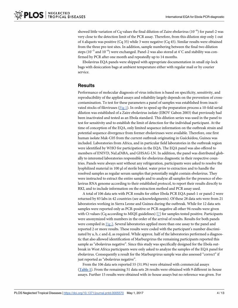
Fig 1. Panel composition for ebola EQA. The panel was composed of a serial dilution of Zaire ebolavirus isolate Gabon 2003 to test for sensitivity of
diagnostic, a recent field isolate from West Africa (Makona-Gueckedou) to test for specificity and reproducibility, Marburgvirus to test for specificity and two
negative samples with human plasma to test for contamination. Two panels (Panel-1 and Panel-2) were generated with identical dilution steps. Sample
numbers, virus composition and corresponding dilution are shown.
https://doi.org/10.1371/journal.pntd.0005570.g001
International EQA for Ebola PCR-diagnostic
PLOS Neglected Tropical Diseases | https://doi.org/10.1371/journal.pntd.0005570 May 1, 2017 3 / 13
showed little variation of Cq values the final dilution of Zaire ebolavirus (10−6) for panel-2 was
very close to the detection limit of the PCR assay. Therefore, from this dilution step only 1 out
of 4 aliquots was positive (Cq 35) while 3 were negative (Cq 45). Similar results were obtained
from the three pre-test sites. In addition, sample numbering between the final two dilution
steps (10−5 and 10−6) were exchanged. Panel-2 was also stored at 4˚C and stability was con-
firmed by PCR after one month and repeatedly up to 14 months.
Ebolavirus EQA panels were shipped with appropriate documentation in small zip-lock
bags with desiccation bags at ambient temperature either with regular mail or by courier
service.
Results
Performance of molecular diagnosis of virus infection is based on specificity, sensitivity, and
reproducibility of the applied assays and reliability largely depends on the prevention of cross-
contamination. To test for these parameters a panel of samples was established from inacti-
vated stocks of filoviruses (Fig 1). In order to speed up the preparation process a 10-fold serial
dilution was established of a Zaire ebolavirus isolate (EBOV Gabon 2003) that previously had
been inactivated and tested as an Ebola standard. This dilution series was used in the panel to
test for sensitivity and to establish the limit of detection for the individual participant. At the
time of conception of the EQA, only limited sequence information on the outbreak strain and
potential sequence divergence from former ebolaviruses were available. Therefore, one first
human isolate Mak-C05 from the current outbreak originating in Gueckedou, Guinea was
included. Laboratories from Africa, and in particular field laboratories in the outbreak region
were identified by WHO for participation in the EQA. The EQA panel was also offered to
members of ENIVD, NaLaDiBA, and GHSAG-LN. In addition, the panel was distributed glob-
ally to interested laboratories responsible for ebolavirus diagnostic in their respective coun-
tries. Panels were always sent without any refrigeration, participants were asked to resolve the
lyophilized material in 100 μl of sterile bidest. water prior to extraction and to handle the
resolved samples as regular serum samples that potentially might contain ebolavirus. They
were instructed to extract the entire sample and to analyze all samples for the presence of ebo-
lavirus RNA genome according to their established protocol, to report their results directly to
RKI, and to include information on the extraction method and PCR assay used.
A total of 106 data sets with PCR results for either Ebola PCR EQA panel-1 or panel-2 were
returned by 83 labs in 42 countries (see acknowledgments). Of these 28 data sets were from 21
laboratories working in Sierra Leone and Guinea during the outbreak. While for 12 data sets
samples were reported only as PCR-positive or PCR-negative all other 94 results were given
with Ct values (Cq according to MIQE guidelines) [7] for samples tested positive. Participants
were anonymized with numbers in the order of the arrival of results. Results for both panels
were compiled in Fig 2. Several laboratories applied more than one assay to the panel and
reported 2 or more results. These results were coded with the participant’s number discrimi-
nated by a, b, c and d, as required. While approx. half of the laboratories performed a diagnos-
tic that also allowed identification of Marburgvirus the remaining participants reported this
sample as “ebolavirus negative”. Since this study was specifically designed for the Ebola out-
break in West Africa participants were only asked to analyze the samples of the EQA panel for
ebolavirus. Consequently a result for the Marburgvirus sample was also assessed “correct” if
just reported as “ebolavirus negative”.
From the 106 data sets reported 55 (51.9%) were obtained with commercial assays
(Table 1). From the remaining 51 data sets 26 results were obtained with 9 different in-house
assays. Further 15 results were obtained with in-house assays but no reference was given. For
International EQA for Ebola PCR-diagnostic
PLOS Neglected Tropical Diseases | https://doi.org/10.1371/journal.pntd.0005570 May 1, 2017 4 / 13
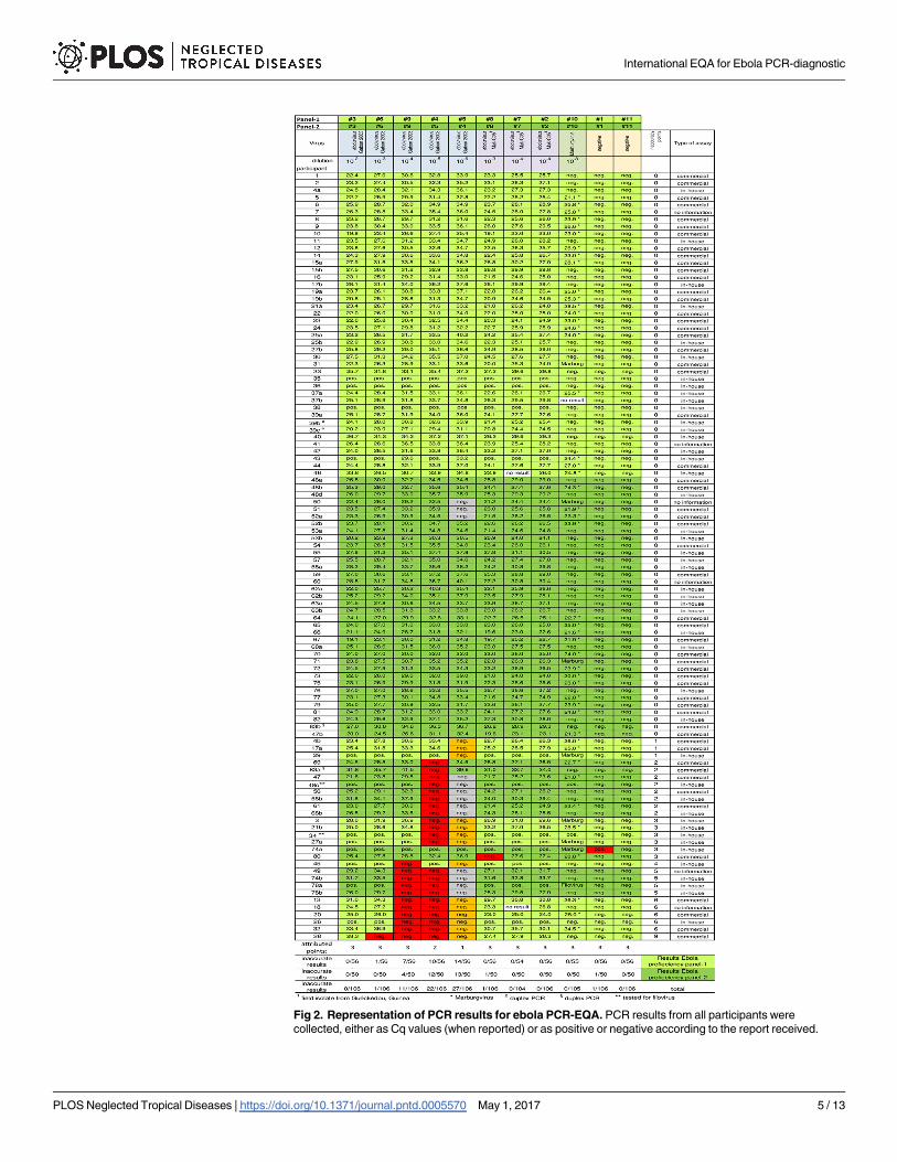
Fig 2. Representation of PCR results for ebola PCR-EQA. PCR results from all participants were
collected, either as Cq values (when reported) or as positive or negative according to the report received.
International EQA for Ebola PCR-diagnostic
PLOS Neglected Tropical Diseases | https://doi.org/10.1371/journal.pntd.0005570 May 1, 2017 5 / 13
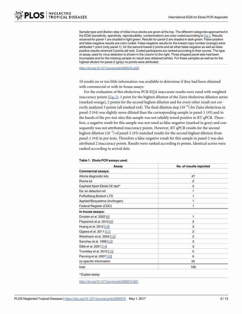
10 results no or too little information was available to determine if they had been obtained
with commercial or with in-house assays.
For the evaluation of this ebolavirus PCR EQA inaccurate results were rated with weighted
inaccuracy points (Fig 2), 1 point for the highest dilution of the Zaire ebolavirus dilution series
(marked orange), 2 points for the second highest dilution and for every other result not cor-
rectly analyzed 3 points (all marked red). The final dilution step (10−6) for Zaire ebolavirus in
panel-2 (#4) was slightly more diluted than the corresponding sample in panel-1 (#5) and in
the hands of the pre-test sites this sample was not reliably tested positive in RT qPCR. There-
fore, a negative result for this sample was not rated as false-negative (marked in grey) and con-
sequently was not attributed inaccuracy points. However, RT qPCR results for the second
highest dilution (10−5) of panel-2 (#5) matched results for the second highest dilution from
panel-1 (#4) in pre-tests. Therefore a false-negative result for this sample in panel-2 was also
attributed 2 inaccuracy points. Results were ranked according to points. Identical scores were
ranked according to arrival date.
Sample type and dilution step of initial virus stocks are given at the top. The different categories approached in
the EQA (sensitivity, specificity, reproducibility, contamination) are color coded according to Fig 1. Results
received for panel-1 are shaded in light green. Results for panel-2 are shaded in dark green. False-positive
and false-negative results are color coded. False-negative results for the lowest copy number (orange) were
attributed 1 point (only panel-1), for the second lowest 2 points and all other false-negative as well as false-
positive results received 3 points (all red). Coded participants are ranked according to their scores. The type
of assay used for virus detection is shown in the column to the right. Three shipped panel sets had been
incomplete and for the missing sample no result was obtained (white). For these samples as well as for the
highest dilution for panel-2 (grey) no points were attributed.
https://doi.org/10.1371/journal.pntd.0005570.g002
Table 1. Ebola PCR assays used.
Assay No. of results reported
Commercial assays:
Altona diagnostic kits 47
Roche kit 2
Cepheid Xpert Ebola CE test* 2
Da’ an detection kit 1
PuRuiKang Biotech LTD 1
Applied Biosystems (Invitrogen) 1
Federal Register (CDC) 1
In-house assays:
Drosten et al. 2002 [8] 1
Fitzpatrick et al. 2015 [9] 2
Huang et al. 2012 [10] 2
Ogawa et al. 2011 [11] 2
Weidmann et al. 2004 [12] 2
Sanchez et al. 1999 [13] 3
Gibb et al. 2001 [14] 3
Trombley et al. 2010 [15] 5
Panning et al. 2007 [16] 6
no specific information 25
total 106
*Duplex assay
https://doi.org/10.1371/journal.pntd.0005570.t001
International EQA for Ebola PCR-diagnostic
PLOS Neglected Tropical Diseases | https://doi.org/10.1371/journal.pntd.0005570 May 1, 2017 6 / 13
78 data sets (73.6%) reported by 67 participants were rated optimal since all samples were
identified correctly. Three additional results were rated acceptable since only the highest dilu-
tion of the panel had been analyzed false-negative (1 inaccuracy point) suggesting slightly
reduced sensitivity for the assay performed. The remaining 25 results (23.6%) reported by 23
participants showed a clear need for improvement (2 to 9 inaccuracy points). However, out of
these 23 participants 8 had reported additional results with an alternative assay that was rated
optimal. This reduces the number of laboratories with urgent need for an improved assay to
15. Out of the 25 results not rated optimal or acceptable 10 were obtained with commercial, 13
with in-house assays. For the remaining 2 results there was no information available on the
assay used.
All participants either identified the Marburgvirus sample correctly as PCR positive for
Marburgvirus or reported it as “ebolavirus negative”. Only one participant reported a false-
positive result for one of the two negative controls, all other participants identified the negative
samples correctly.
Most participants used real-time RT-PCR assays for their analyses. While 102 data sets
came with Cq values for positive samples only for 10 results copy numbers had been calculated
and consequently quantification could not be considered for evaluation. Therefore, Cq values
were taken as a semi-quantitative indicator and used for statistical analysis. For this purpose all
negative results were translated into a Cq value of 45, since in many laboratories real-time
PCR is routinely performed with a maximum of 45 cycles. For all positive samples Cq values
were taken as reported but reduced to 1 position after decimal point if required. Since not all
participants reported details on extraction procedure (e.g. extraction kit, elution volume) and
volume of purified RNA applied to the PCR reaction this approach introduces an inaccuracy.
Further, a few laboratories did not follow all the instructions provided with the panel and they
only extracted part of the sample or pre-diluted the sample prior to testing. However, most
participants (as far as indicated in the reports) used the entire sample for extraction and took
1/10 to 1/12 of the extracted RNA for PCR analysis. Additional variations might have been
introduced since several different PCR assays and different real-time PCR instruments were
used. These parameters might to some extend affect amplification efficiency and Cq value
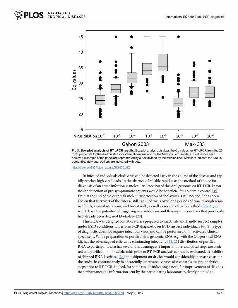
obtained for a given sample. Despite these ambiguities box-plot analysis showed a good over-
all correlation for the panel between dilution and the mean for Cq values while overall varia-
tion of Cq values for a sample in the panel increased with the degree of dilution seen by an
increase in the size of the boxes representing the 25 to 75 percentile (Fig 3). Reproducibility of
results was tested with the identical samples #2 and #7. Good reproducibility was seen for 93
data sets. For these the difference between Cq values was not more than 1. For the remaining
13 results only for 4 data sets the difference was more than 2.
Discussion
This study was initiated in late summer of 2014 in the light of the rapidly developing ebolavirus
outbreak in West Africa with already high numbers of cases, disastrous predictions for the
future development, and under the threat of global spreading through international air travel
[17]. At present identification can be achieved best with sensitive, reliable, and rapid diagnostic
PCR assays [18]. While WHO aimed at evaluating various laboratories that had been deployed
to the outbreak countries in West Africa in the combined international effort to fight the dis-
ease, an additional motivation of RKI, ENIVD, NaLaDiBa, GHSAG-LN and WHO was to pro-
mote preparedness and evaluate quality of ebolavirus diagnostic beyond the outbreak region
and on a global scale by supporting the development of diagnostic capacities and improving
capabilities to allow rapid diagnosis of potentially emerging suspect cases.
International EQA for Ebola PCR-diagnostic
PLOS Neglected Tropical Diseases | https://doi.org/10.1371/journal.pntd.0005570 May 1, 2017 7 / 13
In infected individuals ebolavirus can be detected early in the course of the disease and rap-
idly reaches high viral loads. In the absence of reliable rapid tests the method of choice for
diagnosis of an acute infection is molecular detection of the viral genome via RT-PCR. In par-
ticular detection of pre-symptomatic patients would be beneficial for epidemic control [19].
Even at the end of the outbreak molecular detection of ebolavirus is still needed. It has been
shown that survivors of the disease still can shed virus over long periods of time through semi-
nal fluids, vaginal secretions, and breast milk, as well as several other body fluids [20, 21, 22]
which have the potential of triggering new infections and flare-ups in countries that previously
had already been declared Ebola-free [23].
This EQA was designed for laboratories prepared to inactivate and handle suspect samples
under BSL3 conditions to perform PCR diagnostic on EVD suspect individuals [6]. This type
of diagnostic does not require infectious virus and can be performed on inactivated clinical
specimens. While preparation of purified viral genomic RNA, e.g. with the Qiagen viral RNA
kit, has the advantage of efficiently eliminating infectivity [24, 25] distribution of purified
RNA to participants also has several disadvantages: i) important pre-analytical steps are omit-
ted and purification of nucleic acids prior to RT-PCR analysis cannot be evaluated, ii) stability
of shipped RNA is critical [26] and shipment on dry ice would considerably increase costs for
the study. In contrast analysis of carefully inactivated viruses also controls the pre-analytical
steps prior to RT-PCR. Indeed, for some results indicating a need for improvement of diagnos-
tic performance the information sent by the participating laboratories clearly pointed to
Fig 3. Box plot analysis of RT qPCR results. Box plot analysis displays the Cq values for RT qPCR from the 25
to 75 percentile for the dilution steps for Zaire ebolavirus and for the Makona field isolate. Cq values for each
ebolavirus sample of the panel are represented by a box divided by the median line. Whiskers indicate the 5 to 95
percentile, individual outliers are indicated with dots.
https://doi.org/10.1371/journal.pntd.0005570.g003
International EQA for Ebola PCR-diagnostic
PLOS Neglected Tropical Diseases | https://doi.org/10.1371/journal.pntd.0005570 May 1, 2017 8 / 13
problems at the level of sample preparation and not to the PCR analysis itself. While heat-treat-
ment and gamma-irradiation reliably inactivate infectious viruses [27] the genomic RNA is
still protected from degradation through RNases within viral particles although the Ebolavirus
morphology of the viral particles has been altered through the inactivation process (M. Laue,
personal communication). This combined inactivation was used as a safe inactivation method
that has been used for different viruses including hemorrhagic fever viruses in other EQA
studies [28]. Lyophylization of the inactivated material allows long-term storage and shipment
at ambient temperature. Our most recent test of an Ebola-EQA panel set after 14 months of
storage at 4˚C confirmed unchanged quality of the material even for samples with low copy
numbers. The samples from these sets could still be used to spike human blood and semen for
validation studies [29].
This EQA study for Ebola PCR-diagnostic was developed during the largest hemorrhagic
fever outbreak reported to date [4] and which—with the imminent threat of global spreading
—was an enormous challenge for international public health. This had multiple consequences
for the development of this particular EQA. The principle of former EQAs organized by
ENIVD always had been to keep individual performance of participants confidential. Perfor-
mance was only revealed to the individual participant after final evaluation of all results. This
principle of confidentiality was kept for the majority of participants. However, the laboratories
operating in the outbreak countries have been coordinated and supported as well as nomi-
nated for participation in the EQA by the Emerging and Dangerous Pathogens Laboratory
Network at WHO headquarters in Geneva. In order to allow inclusion into the global evalua-
tion of the public health situation performance of all those laboratories working in the out-
break countries was also revealed to WHO which shared results with the Ministries of Health
for the respective outbreak countries. Participants were alerted to this fact by including this
information into the documents provided with the EQA panel.
Since one of the main intentions of this study was to evaluate the laboratories deployed to
the outbreak countries it is interesting to note that from 28 data sets received from 21 laborato-
ries operating in Guinea or Sierra Leone during the outbreak 23 (82.1%) were optimal while 5
(17.9%) results were lacking sensitivity. Since the initial virus load for most of suspect EVD
patients diagnosed in the outbreak countries was quite high [30], sensitivity of detection meth-
ods is most likely not the most important issue in an outbreak situation for the majority of
samples. However, alternative sampling methods required for collection of blood samples
from small children or because of religious or cultural reservations might affect sensitivity
of PCR diagnostic [31] and therefore might require the most sensitive performance of the ana-
lytical method for certain patients also in an outbreak situation. Compared to the overall per-
formance in this study with 73.6% of optimal results performance of laboratories from the
outbreak countries was better. This may not surprise considering that the majority of mobile
laboratories had technical expertise with EVD and / or BSL3 practice. Also, these laboratories
had built a solid working routine during the outbreak. And although laboratory staff was
exchanged on a regular base established and improved protocols were passed on from group
to group and were even further refined if the necessity occurred (H. Ellerbrok, personal com-
munication). Also, initial protocols and working routines were established from experts in the
field and staff scientists volunteering to serve in these laboratories were also skilled and highly
motivated bringing a solid working routine from their home institutions. Therefore, the results
from this study revealing such an elevated performance for the deployed laboratories also
show that international cooperation is a role model how to handle an emergency in future out-
breaks. The panel was useful to provide confidence to the Ministries of Health in the outbreak
countries that the international deployed laboratories were technically accurate. Further, the
International EQA for Ebola PCR-diagnostic
PLOS Neglected Tropical Diseases | https://doi.org/10.1371/journal.pntd.0005570 May 1, 2017 9 / 13
panel allowed increase of testing capacity beyond the limited number of laboratories with
prior experience in working with EVD.
Acknowledgments
We thank all participating laboratories and in particular those colleagues in the deployed diag-
nostic units in the outbreak countries who despite their stressful working routine found the
time to participate in this EQA. We are grateful to Regina Schadler for administrative assis-
tance and to Cristina Domingo and Andreas Nitsche for helpful discussions.
The participation of the following laboratories in this EQA is gratefully acknowledged:
Algeria: Pasteur Institute of Algeria; Argentina: Instituto Nacional de Enfermedades Infecio-
sas A.N.L.I.S. Dr. Carlos G. Malbran; Austria: Department of Virology, Medical University of
Vienna; Belgium: Central Laboratory of Clinical Biology Institute of Tropical Medicine; Bra-
zil: Laboratorio de Vırus Respiratorios e do Sarampo; Bulgaria: National Center of Infectious
and Parasitic Diseases; Canada: Public Health Agency of Canada; Central African Republic:
Institut Pasteur de Bangui; Cote d’Ivoire: Institut Pasteur de Cote d’Ivoire; Croatia: University
Hospital for Infectious Diseases Dr.Fran Mihaljevic; Egypt: International Emerging Infections
Program-Damanhour Laboratory; Cairo; Gabon: Centre International de Recherches Medi-
cales de Franceville; Germany: Bernard-Nocht Institute for Tropical Medicine, Hamburg;
Friedrich-Loffler-Institute, Riems; Virology department, University of Heidelberg; Institute
for Microbiology of the German Army, Munich; Institute of Virology, Philipps-University
Marburg; Institute of Virology, University of Freiburg; Baden Wurttemberg State Health
Office, Stuttgart; Lower Saxony State Health Office (LNGA), Hannover; Robert Koch Institute,
Berlin; Virology department, Technical University of Munich; Virology department, Univer-
sity of Bonn; Virology department, University of Saarland; Bundeswehr Research Institute for
Protective Technologies and NBC Protection, Munster; Ghana: Virology Department Nogu-
chi Memorial Institute for Medical Research; Greece: Aristotle University of Thessaloniki;
Public Health Laboratories, Hellenic Pasteur Institute; Guinea: Donka Hosptital, Conakry;
DTRA Tatoma Ebola Diagnostic Center, Conakry; European Mobile Lab, Coyah; EUWAM_-
lab, Conakry; Public Health Agency Canada lab, Boke; Iran: Pasteur Institute of Iran; Ireland:
National Virus Reference Laboratory, University College Dublin; Italy: Microbiology and
Virology laboratory, Ospedale Amedeo di Savoia; Department of Molecular Medicin, Univer-
sity of Padova; Istituto Nazionale per le Malattie Infettive ’Lazzaro Spallanzani’; Japan:
National Institute of Infectious Diseases; Kenya: Center for Virus Research, Kenya Medical
Research Institute; Liberia: UNC at Chapel Hill, NC, Phebe Hospital PCR Lab; Madagascar:
Institut Pasteur Madagascar; Mali: Faculty of Medicine and Pharmacy, Serefo Laboratories;
Malta: Molecular Diagnostics, Mater Dei Hospital; Maroco: Institute Pasteur du Maroco;
Mexico: Instituto de Diagnostico y Referencia Epidemiologicos; Netherlands: Erasmus MC;
Norway: Division of Infectious Disease Control, Norwegian Institute of Public Health; Pan-
ama: Instituto Conmemorativo Gorgas de Estudios de la Salud, Panama City; Poland:
National Institute of Public Health–National Institute of Hygiene; Portugal: Center for Vec-
tors and Infectious Diseases Research, National Institute of Health; Sierra Leone: CDC Labo-
ratory, Bo; Central Public Health Reference Laboratory EVD; China Jui Lab, Freetown; Dutch
Mobile Laboratory Freetown PCMH; European Mobile Lab, Hastings; NICD Ebola Mobile
Laboratory, Freetown; NICD /MLU, Lakka, Freetown; Nigerian-European Mobile Lab, Kam-
bia; PHAC-NML, Magburaka Lab; PHE Kerry Town Laboratory; PHE Makeni Laboratory;
PHE, Port Loko Lab; Slovakia: Institute of Virology/Inst.Zoology -Slovak Academy of Sci-
ences; Slovenia: Institute of Microbiology and Immunology, Faculty of Medicine; South
Africa: National Institute for Communicable Diseases, National Health Laboratory Service;
International EQA for Ebola PCR-diagnostic
PLOS Neglected Tropical Diseases | https://doi.org/10.1371/journal.pntd.0005570 May 1, 2017 10 / 13
Spain: Centro Nacional de Microbiologia, Instituto de Salud Carlos III; Switzerland: Labor
Spiez; Turkey: Virology Department, Public Health Institutions of Turkey; Uganda: Uganda
Virus Research Institute; United Kingdom: Public Health England; USA: NIAID/NIH, Rocky
Mountain Laboratories.
Some laboratories participated in both, the analysis of panel-1 and later in the analysis of
panel-2. In general laboratories are named as indicated on the reports.
Author Contributions
Conceptualization: HE MN DN PF.
Data curation: HE SJ.
Formal analysis: HE SJ LS KK.
Investigation: LS KK ATa ATe.
Project administration: HE MN.
Resources: TR ME SB SG.
Supervision: HE MN.
Validation: PP TR ME.
Writing – original draft: HE.
Writing – review & editing: SJ PP TR ME SB SG DN LS KK ATa ATe PF MN.
References1. Baize S, Pannetier D, Oestereich L, Rieger T, Koivogui L, Magassouba N, et al. Emergence of Zaire
Ebola virus disease in Guinea. N Engl J Med. 2014; 371(15):1418–1425. https://doi.org/10.1056/
NEJMoa1404505 PMID: 24738640
2. Marı Saez A, Weiss S, Nowak K, Lapeyre V, Zimmermann F, Dux A, et al. Investigating the zoonotic ori-
gin of the West African Ebola epidemic. EMBO Mol Med. 2014; 7:17–23.
3. Kuhn JH, Andersen KG, Baize S, Bào Y, Bavari S, Berthet N, et al. Nomenclature- and database-com-
patible names for the two Ebola virus variants that emerged in Guinea and the Democratic Republic of
the Congo in 2014. Viruses. 2014; 6:4760–4799. https://doi.org/10.3390/v6114760 PMID: 25421896
4. World Health Organization. Ebola situation report. 2016 [cited 2016 Mar 30] http://apps.who.int/ebola/
current-situation/ebola-situation-report-30-march-2016
5. Martin P, Laupland KB, Frost EH, Valiquette L. Laboratory diagnosis of Ebola virus disease. Intensive
Care Med. 2015; 41:895–898. https://doi.org/10.1007/s00134-015-3671-y PMID: 25636586
6. Reusken C, Niedrig M, Pas S, Anda P, Baize S, Charrel R, et al. Identification of essential outstanding
questions for an adequate European laboratory response to Ebolavirus Zaire West Africa 2014. J Clin
Virol. 2015; 62:124–134. PMID: 25692204
7. Bustin SA, Benes V, Garson JA, Hellemans J, Huggett J, Kubista M, et al. The MIQE guidelines: mini-
mum information for publication of quantitative real-time PCR experiments. Clin Chem. 2009; 55
(4):611–622 https://doi.org/10.1373/clinchem.2008.112797 PMID: 19246619
8. Drosten C, Gottig S, Schilling S, Asper M, Panning M, Schmitz H, Gunther S. Rapid detection and quan-
tification of RNA of Ebola and Marburg viruses, Lassa virus, Crimean-Congo hemorrhagic fever virus,
Rift Valley fever virus, dengue virus, and yellow fever virus by real-time reverse transcription-PCR. J
Clin Microbiol. 2002; 40(7):2323–2330. https://doi.org/10.1128/JCM.40.7.2323-2330.2002 PMID:
12089242
9. Fitzpatrick G, Vogt F, Moi Gbabai OB, Decroo T, Keane M, De Clerck H, et al. The Contribution of Ebola
Viral Load at Admission and Other Patient Characteristics to Mortality in a Medecins Sans Frontières
Ebola Case Management Centre, Kailahun, Sierra Leone, June-October 2014. J Infect Dis. 2015; 212
(11):1752–1758. https://doi.org/10.1093/infdis/jiv304 PMID: 26002981
International EQA for Ebola PCR-diagnostic
PLOS Neglected Tropical Diseases | https://doi.org/10.1371/journal.pntd.0005570 May 1, 2017 11 / 13
10. Huang Y, Wei H, Wang Y, Shi Z, Raoul H, Yuan Z. Rapid detection of filoviruses by real-time TaqMan
polymerase chain reaction assays. Virol Sin. 2012; 27(5):273–277. https://doi.org/10.1007/s12250-
012-3252-y PMID: 23001480
11. Ogawa H, Miyamoto H, Ebihara H, Ito K, Morikawa S, Feldmann H, Takada A. Detection of all known
filovirus species by reverse transcription-polymerase chain reaction using a primer set specific for the
viral nucleoprotein gene. J Virol Methods 2011; 171(1):310–313. https://doi.org/10.1016/j.jviromet.
2010.11.010 PMID: 21093485
12. Weidmann M, Muhlberger E, Hufert FT. Rapid detection protocol for filoviruses. J Clin Virol. 2004; 30
(1):94–99. https://doi.org/10.1016/j.jcv.2003.09.004 PMID: 15072761
13. Sanchez A, Ksiazek TG, Rollin PE, Miranda ME, Trappier SG, Khan AS, et al. Detection and molecular
characterization of Ebola viruses causing disease in human and nonhuman primates. J Infect Dis.
1999; 179 Suppl 1:S164–169.
14. Gibb TR, Norwood DA Jr, Woollen N, Henchal EA. Development and evaluation of a fluorogenic 5’-
nuclease assay to identify Marburg virus. Mol Cell Probes 2001; 15(5):259–626. https://doi.org/10.
1006/mcpr.2001.0369 PMID: 11735297
15. Trombley AR, Wachter L, Garrison J, Buckley-Beason VA, Jahrling J, Hensley LE, et al. Comprehen-
sive panel of real-time TaqMan polymerase chain reaction assays for detection and absolute quantifica-
tion of filoviruses, arenaviruses, and New World hantaviruses. Am J Trop Med Hyg. 2010; 82(5):954–
960. https://doi.org/10.4269/ajtmh.2010.09-0636 PMID: 20439981
16. Panning M, Laue T, Olschlager S, Eickmann M, Becker S, Raith S, et al. Diagnostic reverse-transcrip-
tion. polymerase chain reaction kit for filoviruses based on the strain collections of all European bio-
safety level 4 laboratories. J Infect Dis. 2007; 196 Suppl 2:S199–204.
17. WHO Ebola Response Team. Ebola virus disease in West Africa—the first 9 months of the epidemic
and forward projections. N Engl J Med. 2014; 371:1481–1495. https://doi.org/10.1056/
NEJMoa1411100 PMID: 25244186
18. Towner JS, Rollin PE, Bausch DG, Sanchez A, Crary SM, Vincent M, et al. Rapid diagnosis of Ebola
hemorrhagic fever by reverse transcription-PCR in an outbreak setting and assessment of patient viral
load as a predictor of outcome. J Virol. 2004; 78:4330–4143. https://doi.org/10.1128/JVI.78.8.4330-
4341.2004 PMID: 15047846
19. Chowell D, Castillo-Chavez C, Krishna S; Qiu X, Anderson KS. Modelling the effect of early detection of
Ebola. Lancet Infect Dis. 2015; 15:148–149. https://doi.org/10.1016/S1473-3099(14)71084-9 PMID:
25749063
20. Chughtai AA, Barnes M, Macintyre CR. Persistence of Ebola virus in various body fluids during conva-
lescence: evidence and implications for disease transmission and control. Epidemiol Infect. 2016;
144:1652–1660. https://doi.org/10.1017/S0950268816000054 PMID: 26808232
21. Fischer WA 2nd, Wohl DA. Confronting Ebola as a Sexually Transmitted Infection. Clin Infect Dis. 2016;
62(10):1272–1276. https://doi.org/10.1093/cid/ciw123 PMID: 26936667
22. Uyeki TM, Erickson BR, Brown S, McElroy AK, Cannon D, Gibbons A, et al. Ebola Virus Persistence in
Semen of Male Survivors. Clin Infect Dis. 2016; 62:1552–1555. https://doi.org/10.1093/cid/ciw202
PMID: 27045122
23. Mate SE, Kugelman JR, Nyenswah TG, Ladner JT, Wiley MR, Cordier-Lassalle T, et al. Molecular Evi-
dence of Sexual Transmission of Ebola Virus. N Engl J Med. 2015; 373:2448–2454. https://doi.org/10.
1056/NEJMoa1509773 PMID: 26465384
24. Smither SJ, Weller SA, Phelps A, Eastaugh L, Ngugi S, O’Brien LM, et al. Buffer AVL Alone Does Not
Inactivate Ebola Virus in a Representative Clinical Sample Type. J Clin Microbiol. 2015; 53:3148–3154.
https://doi.org/10.1128/JCM.01449-15 PMID: 26179307
25. Haddock E, Feldmann F, Feldmann H. Effective Chemical Inactivation of EbolaVirus. Emerg Infect Dis.
2016 Jul; 22(7):1292–4. https://doi.org/10.3201/eid2207.160233 PMID: 27070504
26. Escadafal C, Olschlager S, Avsič-Zupanc T, Papa A, Vanhomwegen J, Wolfel R, et al. First international
external quality assessment of molecular detection of Crimean-Congo hemorrhagic fever virus. PLoS
Negl Trop Dis. 2012; 6:e1706. https://doi.org/10.1371/journal.pntd.0001706 PMID: 22745842
27. Elliott LH, McCormick JB, Johnson KM. Inactivation of Lassa, Marburg, and Ebola viruses by gamma
irradiation. J Clin Microbiol. 1982; 16:704–708. PMID: 7153317
28. Niedrig M, Schmitz H., Becker S., Gunther S., ter Meulen J., Meyer H., Ellerbrok H., Nitsche A., Gelder-
blom H.R., Drosten C. First International Quality Assurance Study on the Rapid Detection of Viral
Agents of Bioterrorism J. Clin. Microbiol. 2004; 42:1753–55 https://doi.org/10.1128/JCM.42.4.1753-
1755.2004 PMID: 15071040
International EQA for Ebola PCR-diagnostic
PLOS Neglected Tropical Diseases | https://doi.org/10.1371/journal.pntd.0005570 May 1, 2017 12 / 13
29. Loftis AJ, Quellie S, Chason K, Sumo E, Toukolon M, Otieno Y, et al. Validation of the Cepheid GeneX-
pert for Detecting Ebola Virus in Semen. 2017; J Infect Dis. 215:344–350. https://doi.org/10.1093/
infdis/jiw562 PMID: 27932614
30. de La Vega MA, Caleo G, Audet J, Qiu X, Kozak RA, Brooks JI, et al. Ebola viral load at diagnosis asso-
ciates with patient outcome and outbreak evolution. J Clin Invest. 2015; 125:4421–4428. https://doi.org/
10.1172/JCI83162 PMID: 26551677
31. Strecker T, Palyi B, Ellerbrok H, Jonckheere S, de Clerck H, Bore JA, et al. Field Evaluation of Capillary
Blood Samples as a Collection Specimen for the Rapid Diagnosis of Ebola Virus Infection During an
Outbreak Emergency. Clin Infect Dis. 2015; 61:669–675. https://doi.org/10.1093/cid/civ397 PMID:
25991465
International EQA for Ebola PCR-diagnostic
PLOS Neglected Tropical Diseases | https://doi.org/10.1371/journal.pntd.0005570 May 1, 2017 13 / 13