De wondere wereld van de keratinocyten - wondzorg.net · 27/02/2018 8 Keratinocyten Besluit...
9
27/02/2018 1 De wondere wereld van de keratinocyten Lieve Vandecandelaere verpleegkundige Inhoud Functie huid Bouw huid Wat zijn keratinocyten Ontstaan keratinocyten Welke rol spelen keratinocyten in de wondheling Keratinocyten en wondzorg
-
Upload
nguyenduong -
Category
Documents
-
view
212 -
download
0
Transcript of De wondere wereld van de keratinocyten - wondzorg.net · 27/02/2018 8 Keratinocyten Besluit...
27/02/2018
1
De wondere wereld van de keratinocyten
Lieve Vandecandelaere
verpleegkundige
Inhoud
�Functie huid
�Bouw huid
�Wat zijn keratinocyten
�Ontstaan keratinocyten
�Welke rol spelen keratinocyten in de wondheling
�Keratinocyten en wondzorg
27/02/2018
2
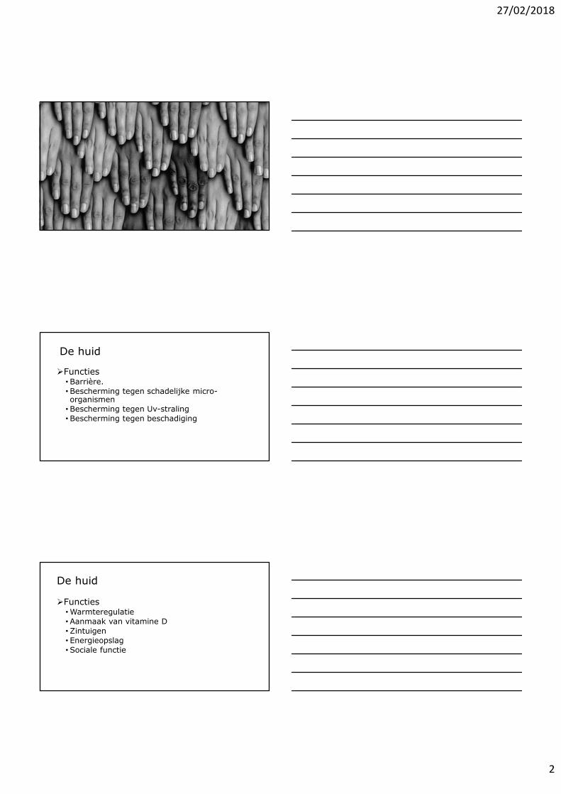
De huid
�Functies• Barrière. • Bescherming tegen schadelijke micro-organismen• Bescherming tegen Uv-straling• Bescherming tegen beschadiging
De huid
�Functies•Warmteregulatie• Aanmaak van vitamine D• Zintuigen• Energieopslag• Sociale functie
27/02/2018
3
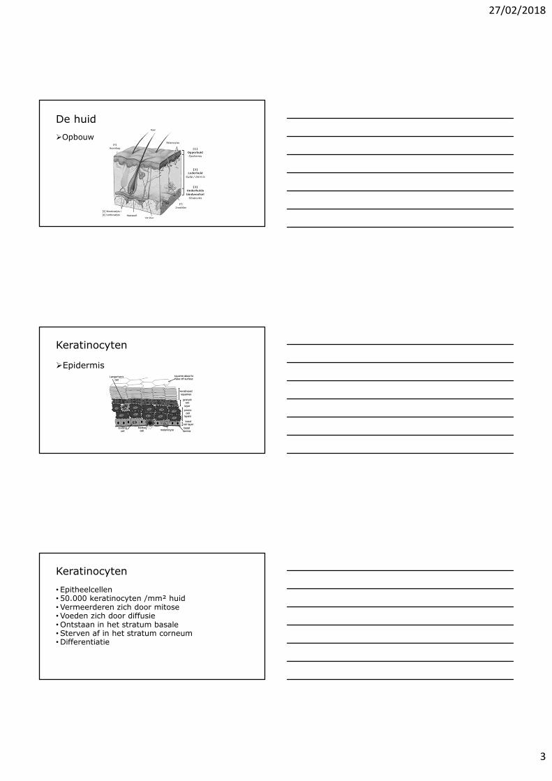
De huid
�Opbouw
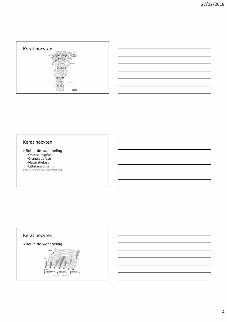
Keratinocyten
�Epidermis
Keratinocyten
• Epitheelcellen• 50.000 keratinocyten /mm² huid• Vermeerderen zich door mitose• Voeden zich door diffusie•Ontstaan in het stratum basale• Sterven af in het stratum corneum•Differentiatie
27/02/2018
4
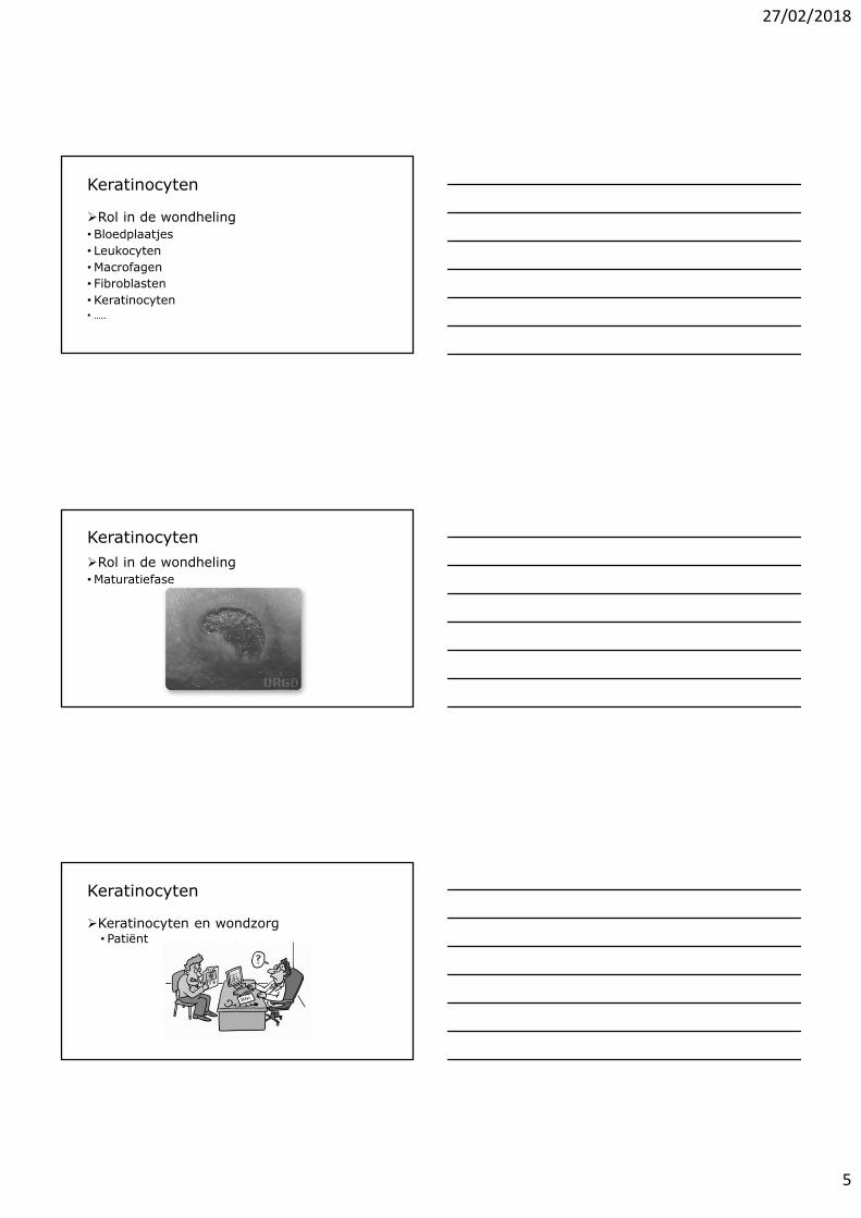
Keratinocyten
Keratinocyten
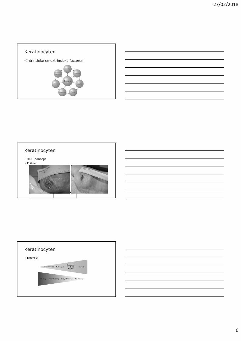
�Rol in de wondheling•Ontstekingsfase•Granulatiefase•Maturatiefase• Littekenvorming
(http://www.digiwond.ugent.be/page43998.html)
Keratinocyten
�Rol in de wondheling
27/02/2018
5
Keratinocyten
�Rol in de wondheling• Bloedplaatjes
• Leukocyten
•Macrofagen
• Fibroblasten
• Keratinocyten• …..
Keratinocyten
�Rol in de wondheling•Maturatiefase
Keratinocyten
�Keratinocyten en wondzorg• Patiënt
27/02/2018
6
Keratinocyten
• Intrinsieke en extrinsieke factoren
Keratinocyten
• TIME-concept
�Tissue
Keratinocyten
�Infectie
27/02/2018
7
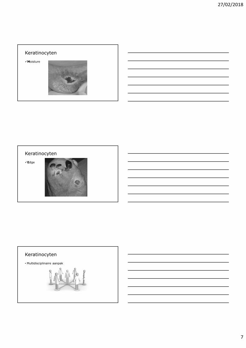
Keratinocyten
�Moisture
Keratinocyten
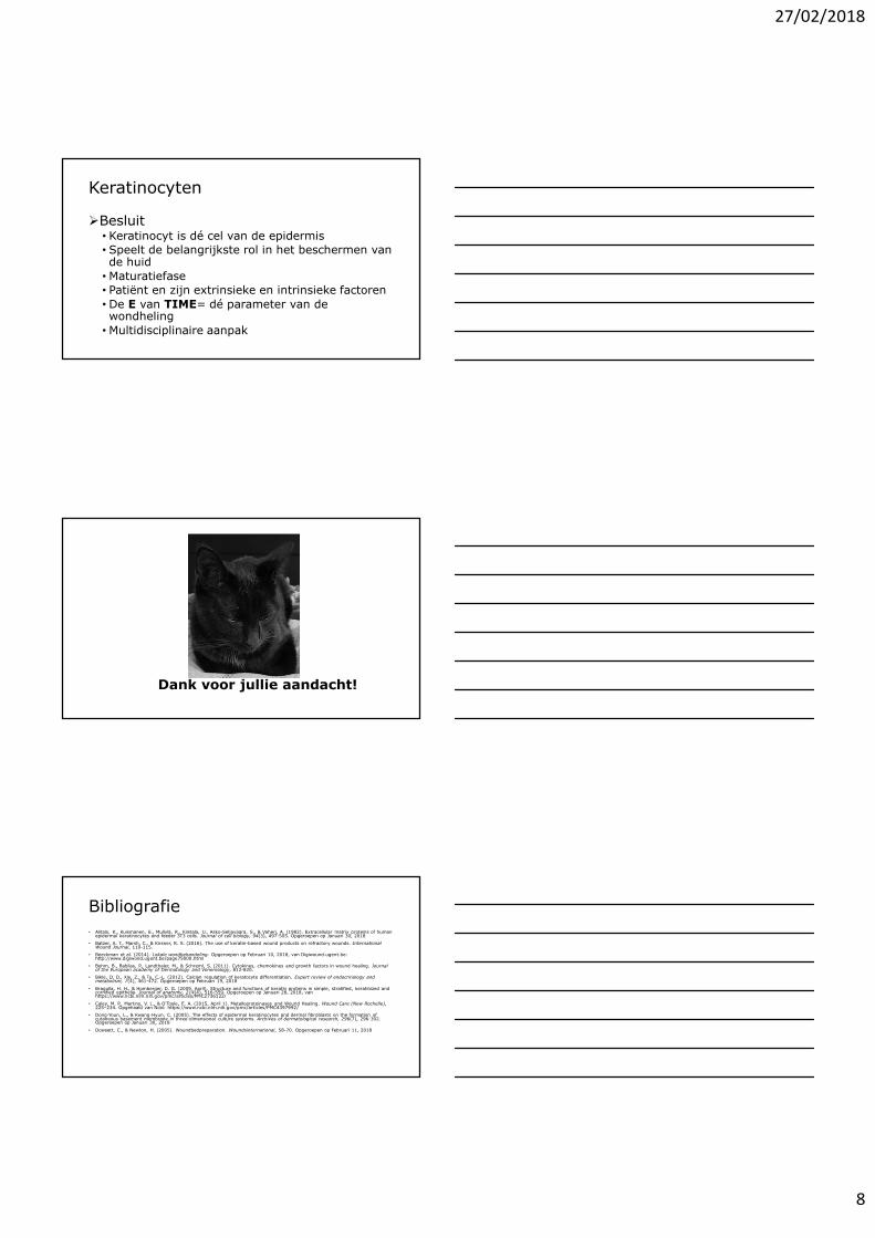
�Edge
Keratinocyten
• Multidisciplinaire aanpak
27/02/2018
8
Keratinocyten
�Besluit• Keratinocyt is dé cel van de epidermis• Speelt de belangrijkste rol in het beschermen van de huid•Maturatiefase• Patiënt en zijn extrinsieke en intrinsieke factoren• De E van TIME= dé parameter van de wondheling•Multidisciplinaire aanpak
Dank voor jullie aandacht!
Bibliografie
• Alitalo, K., Kuismanen, E., Mullylä, R., Kiistala, U., Asko-Seljavaara, S., & Vaheri, A. (1982). Extracellular matrix proteins of human epidermal keratinocytes and feeder 3T3 cells. Journal of cell biology, 94(3), 497-505. Opgeroepen op Januari 30, 2018
• Batzer, A. T., Marsh, C., & Kirsner, R. S. (2016). The use of keratin-based wound products on refractory wounds. International Wound Journal, 110-115.
• Beeckman et al. (2014). Lokale wondbehandeling. Opgeroepen op Februari 10, 2018, van Digiwound.ugent.be: http://www.digiwond.ugent.be/page75000.html
• Behm, B., Babilas, P., Landthaler, M., & Schreml, S. (2011). Cytokines, chemokines and growth factors in wound healing. Journal of the European academy of Dermatology and Venereology, 812-820.
• Bikle, D. D., Xie, Z., & Tu, C.-L. (2012). Calcian regulation of keratocyte differentiation. Expert review of endocrinology and metabolism, 7(4), 461-472. Opgeroepen op Februari 19, 2018
• Bragulla, H. H., & Homberger, D. G. (2009, April). Structure and functions of keratin proteins in simple, stratified, keratinized and cornified epithelia. Journal of anatomy, 214(4), 516-559. Opgeroepen op Januari 28, 2018, van https://www.ncbi.nlm.nih.gov/pmc/articles/PMC2736122/
• Caley, M. P., Martins, V. L., & O'Toole, E. A. (2015, April 1). Metalloproteinases and Wound Healing. Wound Care (New Rochelle), 225–234. Opgehaald van Ncbi: https://www.ncbi.nlm.nih.gov/pmc/articles/PMC4397992/
• Dong-Youn, L., & Kwang-Hyun, C. (2005). The effects of epidermal keratinocytes and dermal fibroblasts on the formation of cutaneous basement membrane in three-dimensional culture systems. Archives of dermatological research, 296(7), 296-302. Opgeroepen op Januari 30, 2018
• Dowsett, C., & Newton, H. (2005). Woundbedpreparation. Woundsinternational, 58-70. Opgeroepen op Februari 11, 2018

27/02/2018
9
Bibliografie
• Embyologic, Histologic and anatomic aspects. (2018). Opgeroepen op Februari 8, 2018, van Derm101: https://www.derm101.com/inflammatory/embryologic-histologic-and-anatomic-aspects/epidermis/
• Functies vande huid. (2017). Opgeroepen op Februari 8, 2018, van Huidhuis.nl: https://www.huidhuis.nl/thema/functies-van-de-huid
• Gibson, D., Cullen, B., Legerstee, R., Harding, K. G., & Schultz, G. (2009). MMP's made easy. Wounds International, 1-5.
• Gryson, L. (2017, Oktober). MMP’s and wounds. België.
• Jüngen, I. (2016). Verschijnselen van een wond. Bijzijn XL, 16-19.
• Junqueira, L. C., & Carneiro, J. (2007, September). Functionele histologie. Reed Business.
• Karantza, V. (2011). Keratins in health and cancer: more than mere epithelial cell markers. Oncogene, 127-138.
• Koopman-Kuijl, C. A., & de Goederen-Geleijnse, M. M. (2015). Wondbedpreparatie model deel 1. WCS, 42-46.
• Kort overzicht van de huid. (2009, Januari 24). Opgehaald van Zo werkt het lichaam: http://www.zowerkthetlichaam.nl/135/kort-overzicht-van-de-huid/
• Leers, M. P. (2003). Cytokeratine en zijn afbraakprodukten als potentiële aangrijpingspunten. Nederlands tijdschrift voor klinische chemie, 20-25.
• Maquart, F. X., & Momboisse, J. (2014). Extracellular matrix and wound healingMatrice extracellulaire et cicatrisation. Pathologie biologie, 62(2), 91-95. Opgeroepen op Januari 30, 2018
• Marco. (2009). Collageen. Nederlandstijdschrift voor wondgenezing. Opgeroepen op Februari 10, 2018
• Matsui, T., & Amagai, M. (2015). Dissecting the formation, structure and barrier function of the stratum corneum. Intenationalimmunology, 269-280.
Bibliografie
• Mekkes, J. R. (2013, November 24). Extra Cellulaire Matrix componenten / Biomaterialen. Opgehaald van Wondbedekkers: http://www.wondbedekkers.nl/ecmcomponents/extracellulaire-matrix-productinformatie.htm
• Mekkes, J. R. (2017, Augustus 26). Gekweekte autologe kerantinocyten transplantatie- bioseed (Baxter) methode, kerantinocytensuspensie in tissuecoll. Opgehaald van Huidziekten: https://www.huidziekten.nl/afbeeldingen/chirurgie/bioseedtransplantatie.htm
• Moll, R., Divo, M., & Langbein, L. (2008). The human keratins: biology and pathology. Histochemistry and Cell Biology, 705-733.
• Oceanside museam of art. (2017, Augustus). A time to heal. Opgeroepen op Februari 21, 2018, van oma-online.org: http://oma-online.org/heal/
• Opbouw van de huid. (2016). Opgehaald van Healing Wounds and Tender Care: http://www.healingwoundsandtendercare.com/de-huid/
• Paster, I., Stojadinovic, O., Yin, N. C., Raminez, H., Nusbaum, A. G., Sawaya, A., . . . Tomic-Camic, M. (2014). Epithelialization in woundhealing:a comprehensive review. Advances in woundcare, 3(7), 445-464. Opgeroepen op Februari 20, 2018
• Peckham, M. (sd). Layers in the Epidermis. Opgehaald van The Histology Guide: http://www.histology.leeds.ac.uk/skin/epidermis_layers.php
• Rangaraj, A., Harding, K., & Leaper, D. (2011). Role of Collagen in Wound Management. Wounds UK, 54.
• Schultz, G. S., & Mast, B. A. (1998). Molecular analysis of the environments of healing and chronic wounds: cytokines, proteases and grouth factors. Opgeroepen op Januari 27, 2018, van Woundsaustralia.com: http://www.woundsaustralia.com.au/journal/0701_01.pdf
• Schultz, G. S., Ladwig, G., & Wysocki, A. (2005). Extracellular matrix: review of its roles in acute and chronic wounds. World widewounds. Opgeroepen op Februari 8, 2018
• Schultz, G., & Dowsett, C. (2012). Woundbed preparation revisited. Wounds international, 3(1), 25-29. Opgeroepen op Februari 9, 2018
• Schultz, G., Bjarnsholt, T., James, G. A., Leaper, D. J., Mc Bain, A. J., Malone, M. J., . . . Randall, W. D. (2017). Consensus guidelines for the identification and treatment of biofilms in chronic nonhealing wounds. Woundrepair and regeneration, 744-757.
Bibliografie
• Stichting Transmurale Zorg Den Haag e.o. (2012). Transmurale richtlijn algemene wondzorg. Opgeroepen op Februari 11, 2018, van tmz.mdl-solutions.nl: https://tmz.mdl-solutions.nl/uploads/ckeditor/attachments/39/1-_Richtlijn_Algemene_Wondzorg_regio_Haaglanden_2012_e.v..pdf
• Structure and Function of the Skin. (2014). Opgeroepen op Februari 8, 2018, van Clinimed.co.uk: http://www.clinimed.co.uk/Wound-Care/Education/Wound-Essentials/Structure-and-Function-of-the-Skin.aspx
• Tissue types. (sd). Opgeroepen op Februari 9, 2018, van Skilledwoundcare.com: https://www.skilledwoundcare.com/tissue-types
• Toestand van de wonde en huid. (sd). Opgeroepen op Februari 10, 2018, van Coloplast.be: https://www.coloplast.be/wond/wondzorg/opleiding/
• University of Leeds. (2003). Layers in the Epidermis. Opgeroepen op januari 25, 2018, van Histology Guide: http://www.histology.leeds.ac.uk/skin/epidermis_layers.php
• Urgomedical. (2017, September 5). De huid. Opgehaald van Urgomedical: http://www.urgomedical.nl/samen-inzicht-krijgen/wonden-en-genezing/
• van Leen, M. (2007). Matrix Metalloproteinasen in normale en afwijkende wondgenezing. Nederlands tijdschrift voor wondverzorging, 22-26.
• Vandeputte, J., & Gryson, L. (2007). Wonzorg in de praktijk. Mechelen: Wolters Kluwer Belgium NV.
• Vlaanderen, W. g. (2016). Handboek wondzorg. Houten: Bohn Stafleu van Loghum.
• Werner, S., Krieg, T., & Smola, H. (2007). Keratinocyte–Fibroblast Interactions in Wound Healing. Journal of investigativedermatology, 127(5), 998-1008. Opgeroepen op januari 31, 2018
• Wikipedia. (2017, november 16). Cel (biologie). Opgeroepen op december 15, 2017, van wikipedia.org: https://nl.wikipedia.org/wiki/Cel_(biologie)


![Kapellekensbaan # 3 [W]ONDERWEG · Kapellekensbaan # 3 [W]ONDERWEG Zomer en vakantie doen aan buiten denken: er met de hele familie op uit trekken, [onder]weg zijn naar wondere oorden,](https://static.fdocuments.nl/doc/165x107/5ff477bc33efae347556afa9/kapellekensbaan-3-wonderweg-kapellekensbaan-3-wonderweg-zomer-en-vakantie.jpg)