RX THORAX: BASIC - brugse.dierenartsenkringen.be · Techniek Hoge kV (inherent hoog contrast...
35
Dr. Sarah Claerhoudt, DVM, PhD 21/2/2019 RX THORAX: BASIC
Transcript of RX THORAX: BASIC - brugse.dierenartsenkringen.be · Techniek Hoge kV (inherent hoog contrast...
Dr. Sarah Claerhoudt, DVM, PhD
21/2/2019
RX THORAX: BASIC
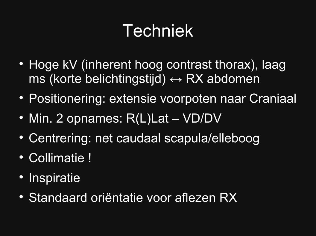
Techniek
Hoge kV (inherent hoog contrast thorax), laag ms (korte belichtingstijd) ↔ RX abdomen
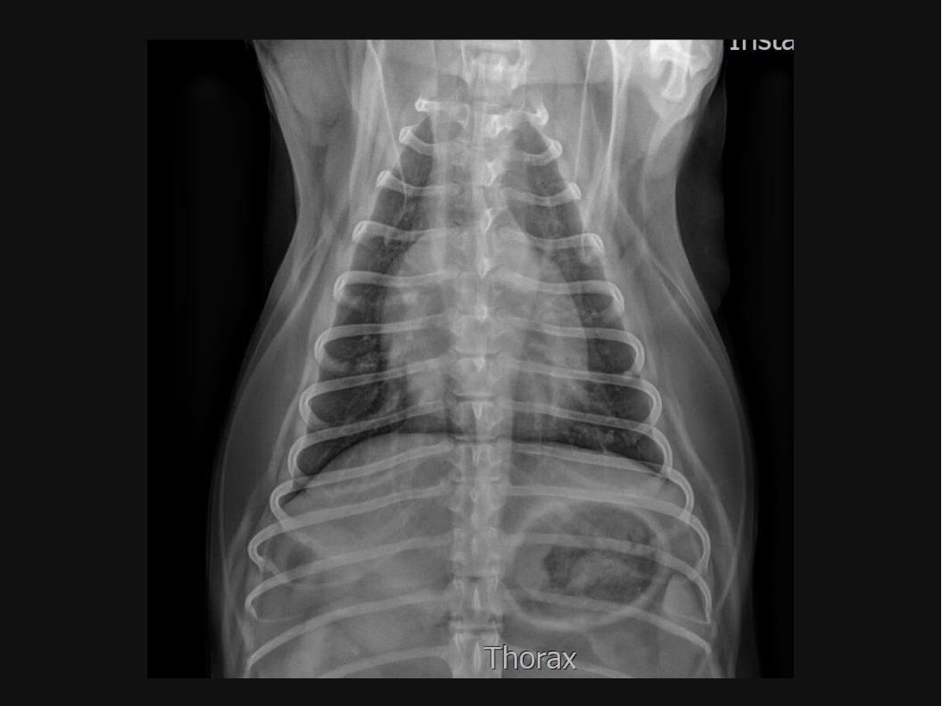
Positionering: extensie voorpoten naar Craniaal Min. 2 opnames: R(L)Lat – VD/DV Centrering: net caudaal scapula/elleboog Collimatie ! Inspiratie Standaard oriëntatie voor aflezen RX
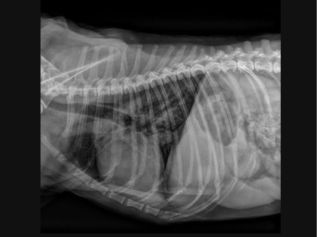
RX thorax
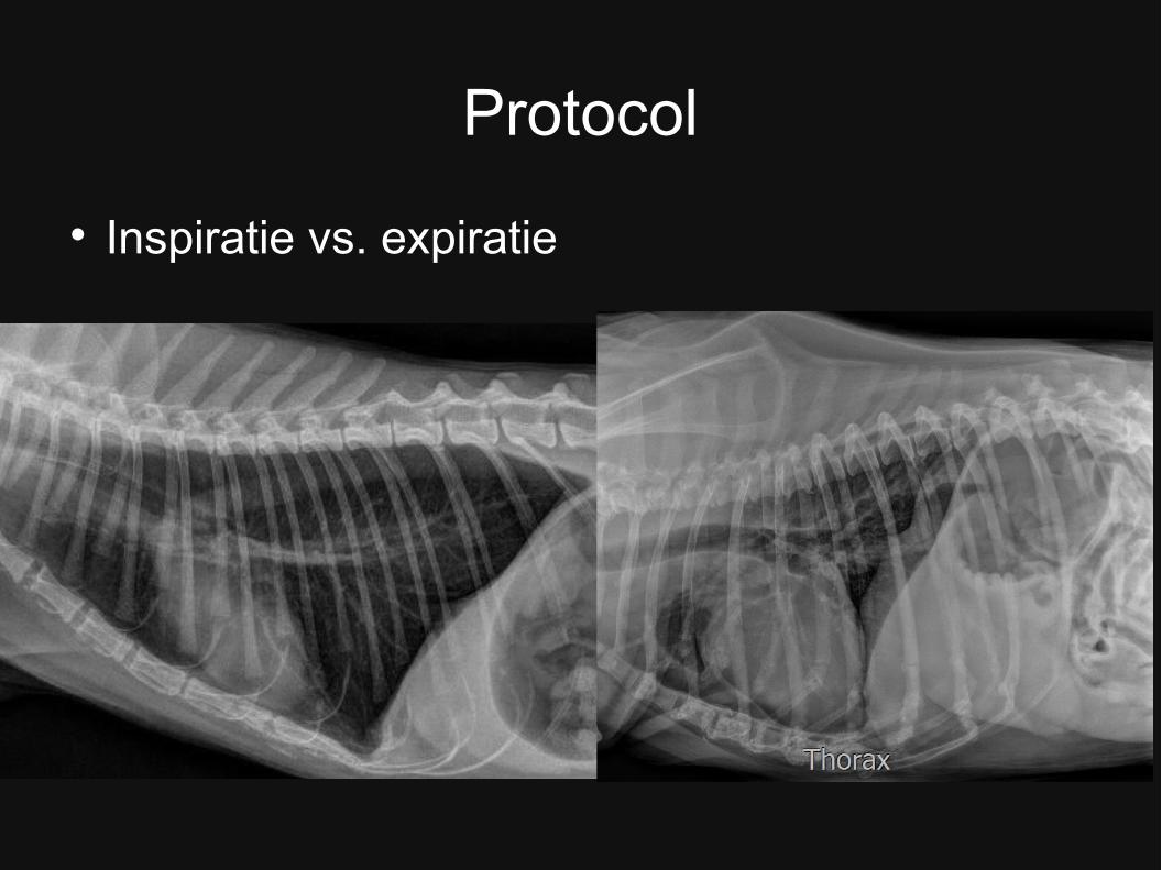
Protocol
Inspiratie vs. expiratie
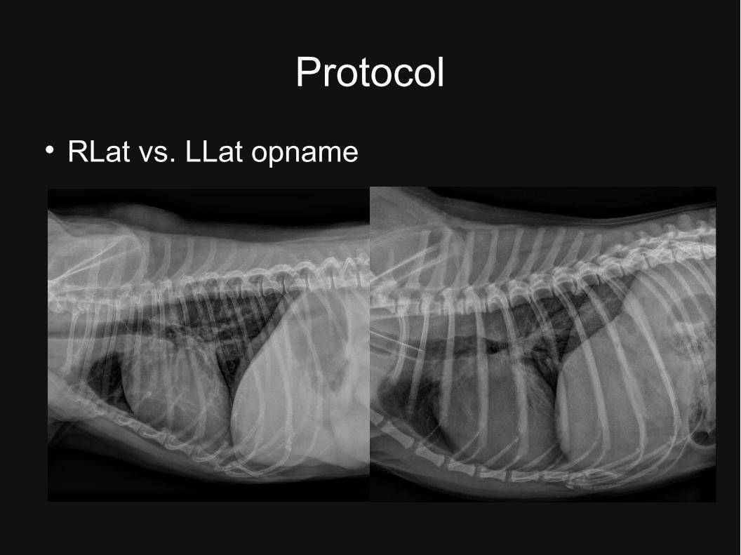
Protocol
RLat vs. LLat opname
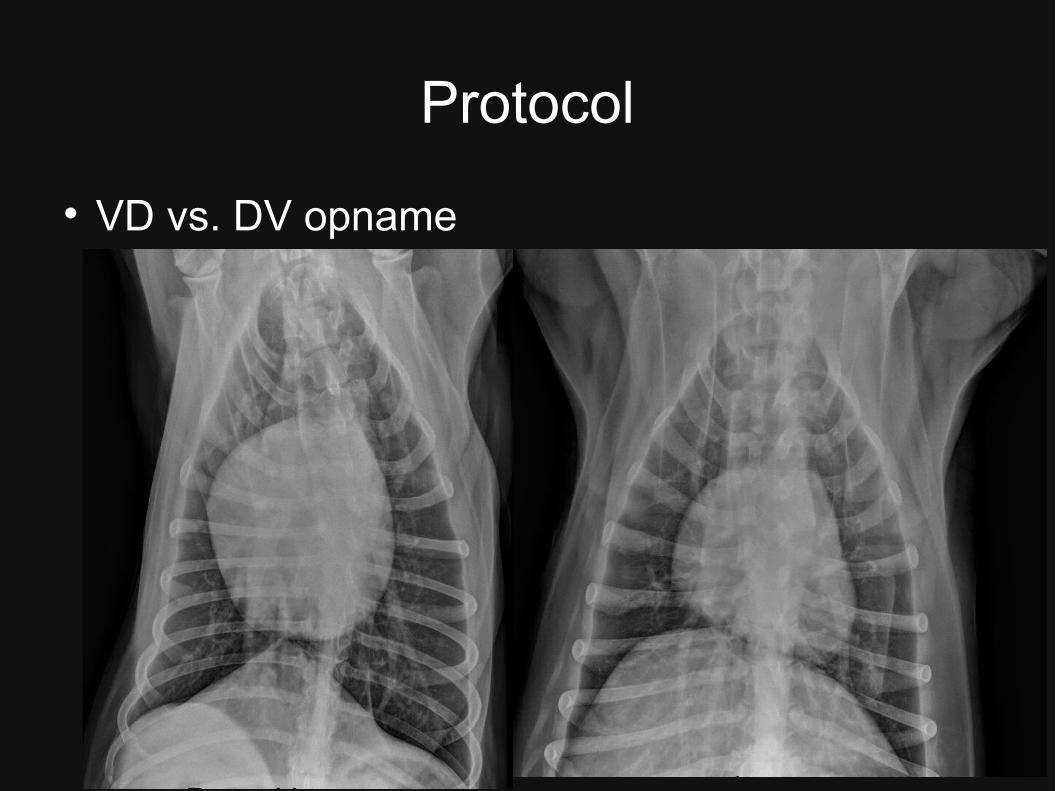
Protocol
VD vs. DV opname
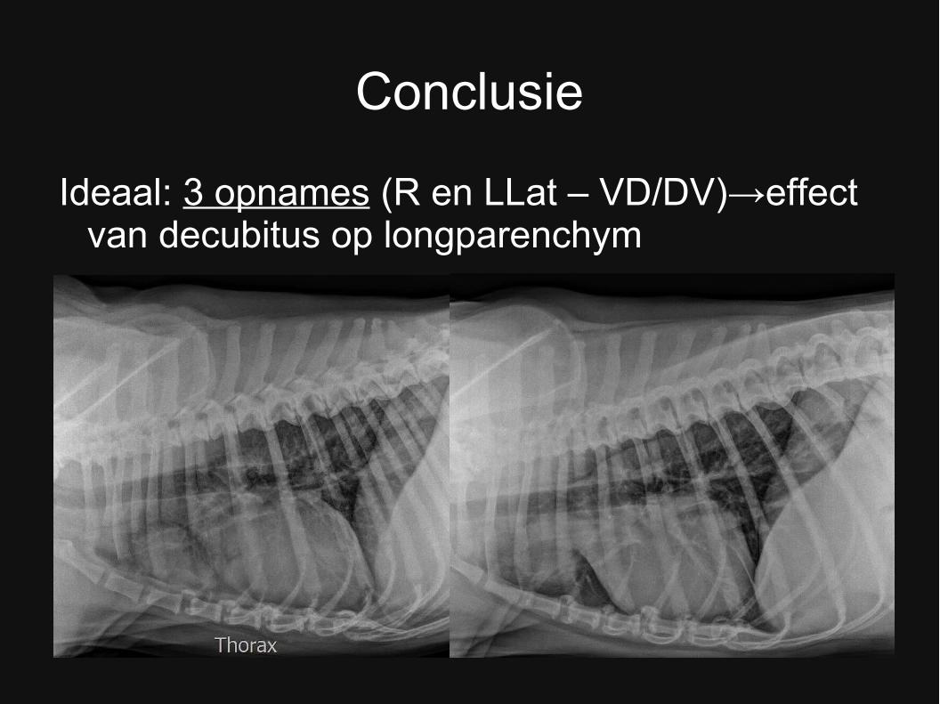
Conclusie
Ideaal: 3 opnames (R en LLat – VD/DV)→effect van decubitus op longparenchym

Beoordelen: Standaard Protocol
– Beenderige structuren rondom thorax
– Craniaal abdomen
– Trachea
– Hartschaduw
– Bloedvaten (Ao, CVC)
– Diafragma
– Long parenchym
– Pleura/pleurale ruimte
– Mediastinum
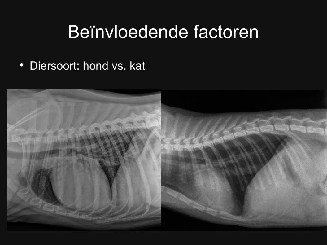
Beïnvloedende factoren
Diersoort: hond vs. kat
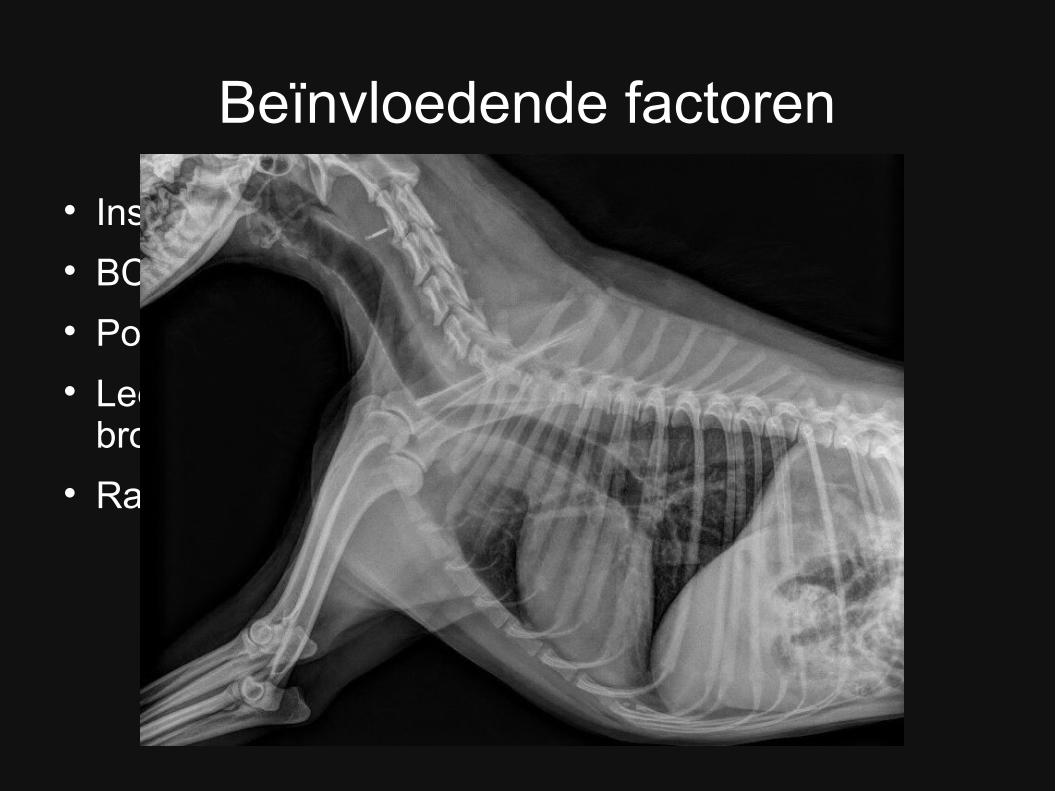
Beïnvloedende factoren
Inspiratie vs. Expiratie BCS Positie Hals: kop in neutrale positie Leeftijd: jong (thymus!) vs. Oud (calcificaties
bronchen) Ras
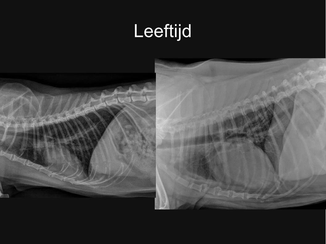
Leeftijd
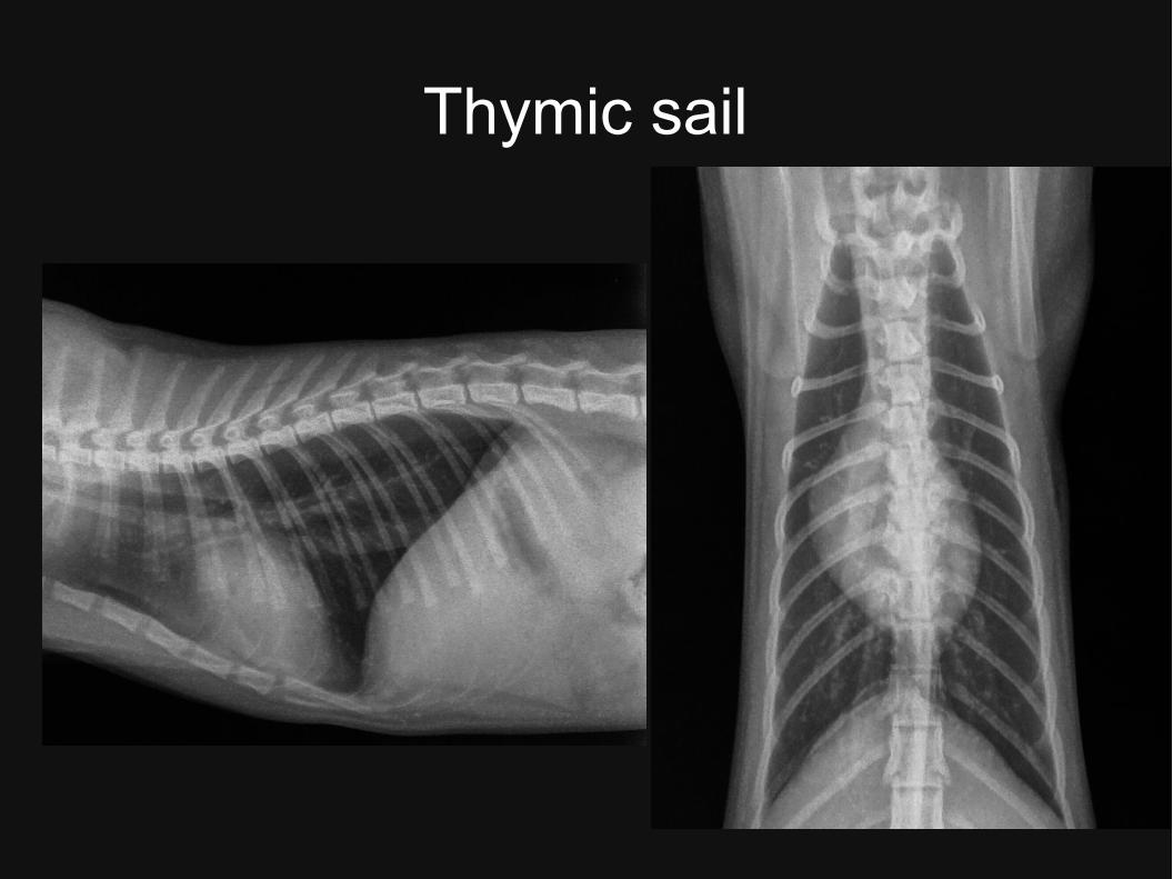
Thymic sail
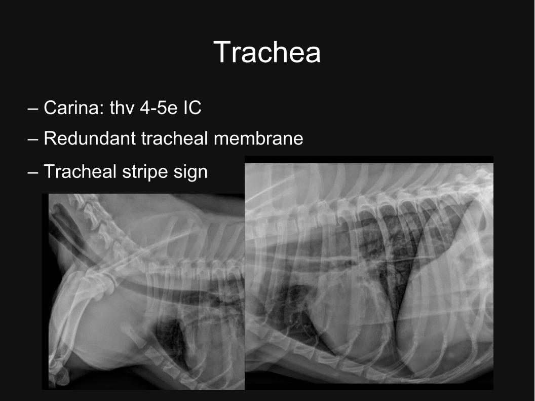
Trachea
– Carina: thv 4-5e IC
– Redundant tracheal membrane
– Tracheal stripe sign
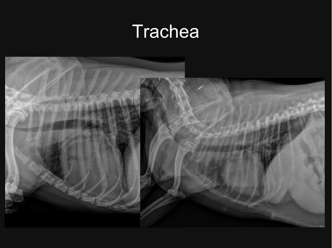
Trachea
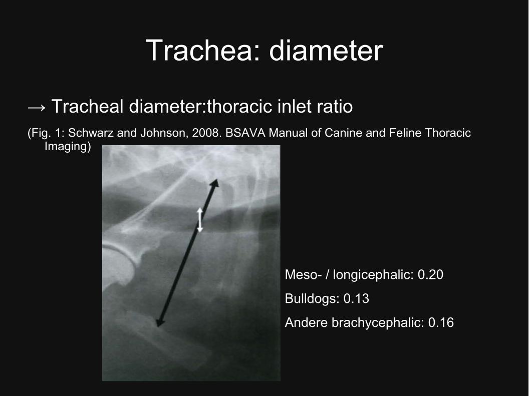
Trachea: diameter
→ Tracheal diameter:thoracic inlet ratio(Fig. 1: Schwarz and Johnson, 2008. BSAVA Manual of Canine and Feline Thoracic
Imaging)
Meso- / longicephalic: 0.20
Bulldogs: 0.13
Andere brachycephalic: 0.16
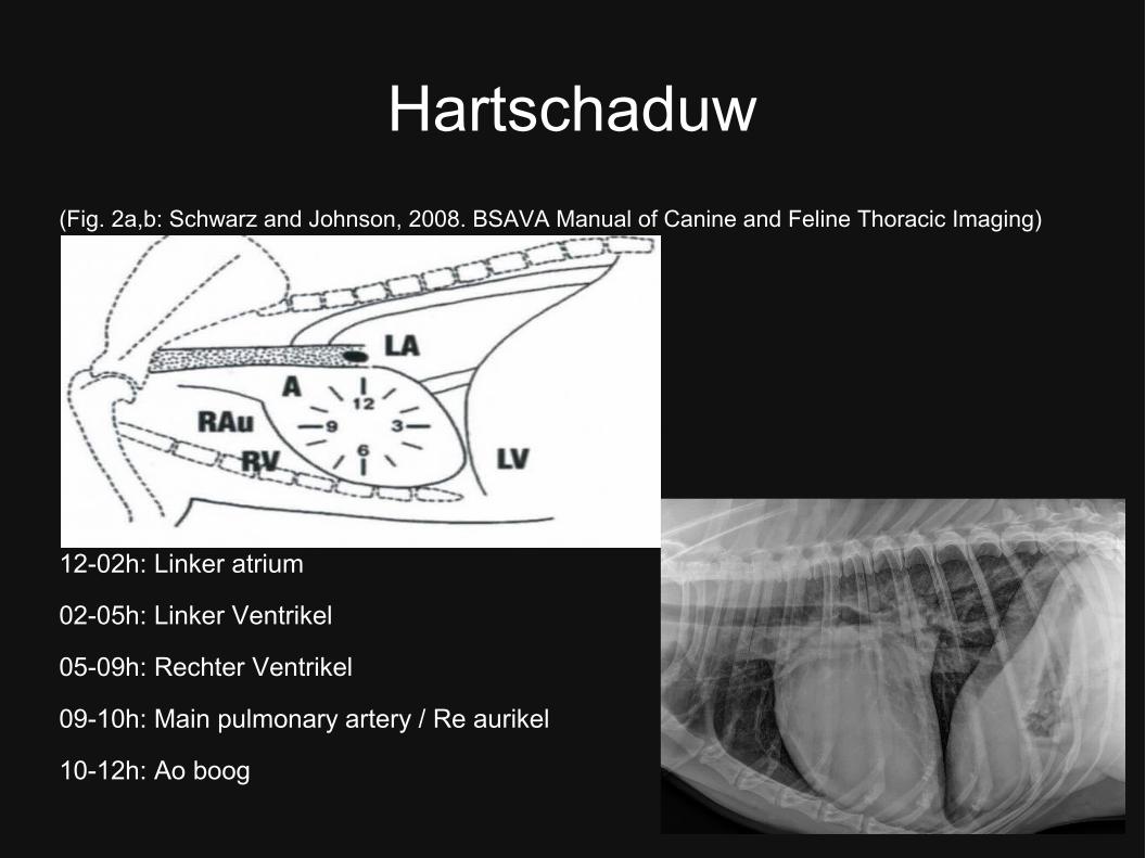
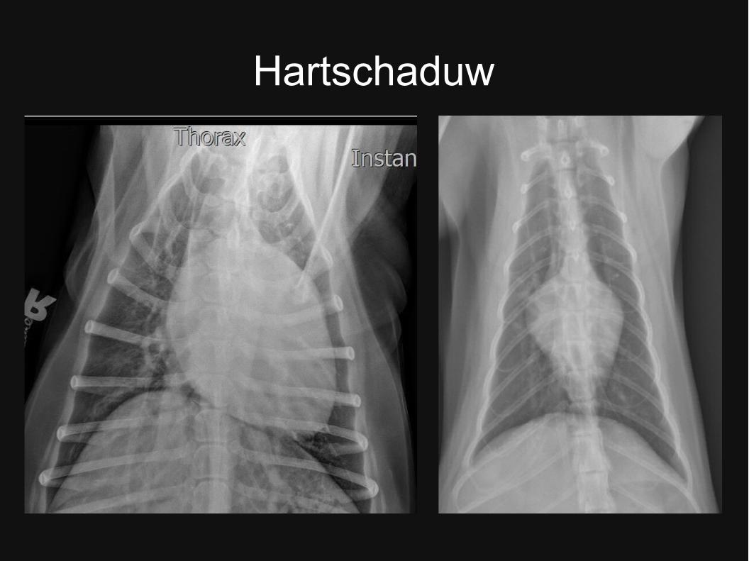
Hartschaduw
(Fig. 2a,b: Schwarz and Johnson, 2008. BSAVA Manual of Canine and Feline Thoracic Imaging)
12-02h: Linker atrium
02-05h: Linker Ventrikel
05-09h: Rechter Ventrikel
09-10h: Main pulmonary artery / Re aurikel
10-12h: Ao boog
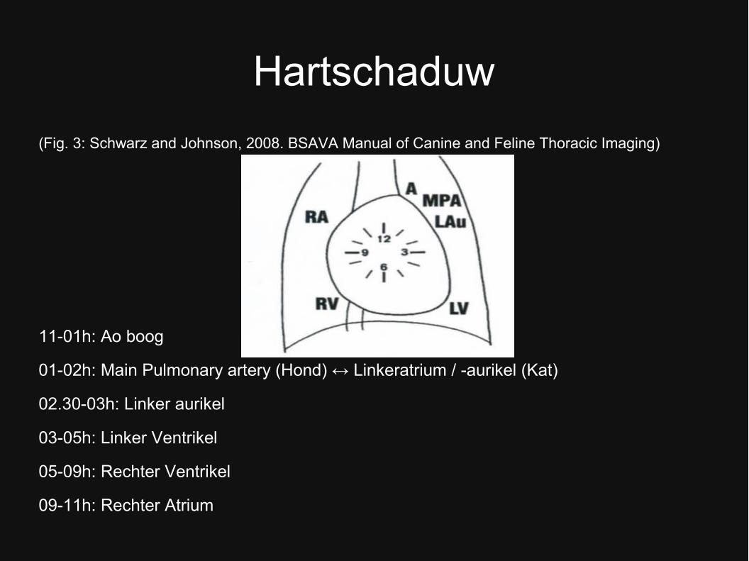
Hartschaduw
(Fig. 3: Schwarz and Johnson, 2008. BSAVA Manual of Canine and Feline Thoracic Imaging)
11-01h: Ao boog
01-02h: Main Pulmonary artery (Hond) ↔ Linkeratrium / -aurikel (Kat)
02.30-03h: Linker aurikel
03-05h: Linker Ventrikel
05-09h: Rechter Ventrikel
09-11h: Rechter Atrium
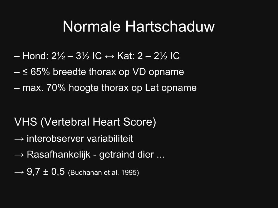
Normale Hartschaduw
– Hond: 2½ – 3½ IC ↔ Kat: 2 – 2½ IC
– ≤ 65% breedte thorax op VD opname
– max. 70% hoogte thorax op Lat opname
VHS (Vertebral Heart Score)
→ interobserver variabiliteit
→ Rasafhankelijk - getraind dier ...
→ 9,7 ± 0,5 (Buchanan et al. 1995)
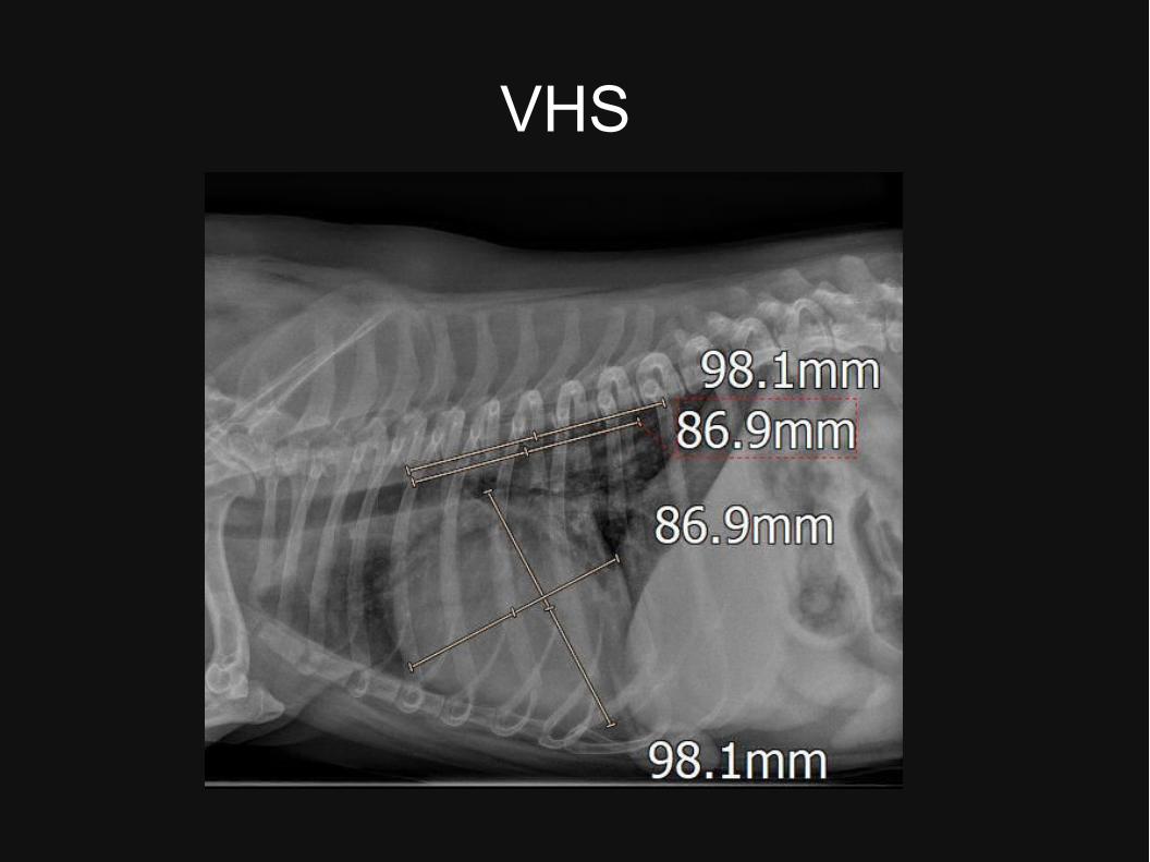
VHS
VHS
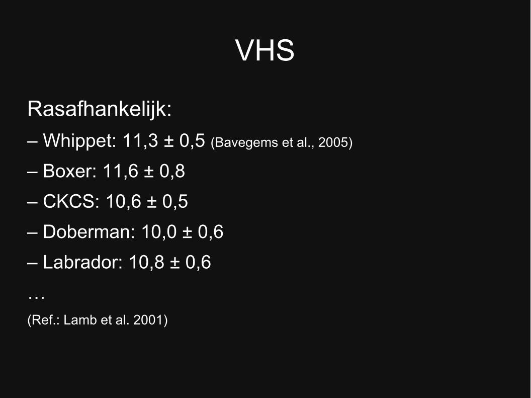
Rasafhankelijk:
– Whippet: 11,3 ± 0,5 (Bavegems et al., 2005)
– Boxer: 11,6 ± 0,8
– CKCS: 10,6 ± 0,5
– Doberman: 10,0 ± 0,6
– Labrador: 10,8 ± 0,6
… (Ref.: Lamb et al. 2001)
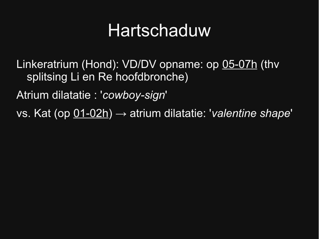
Hartschaduw
Linkeratrium (Hond): VD/DV opname: op 05-07h (thv splitsing Li en Re hoofdbronche)
Atrium dilatatie : 'cowboy-sign'
vs. Kat (op 01-02h) → atrium dilatatie: 'valentine shape'
Hartschaduw
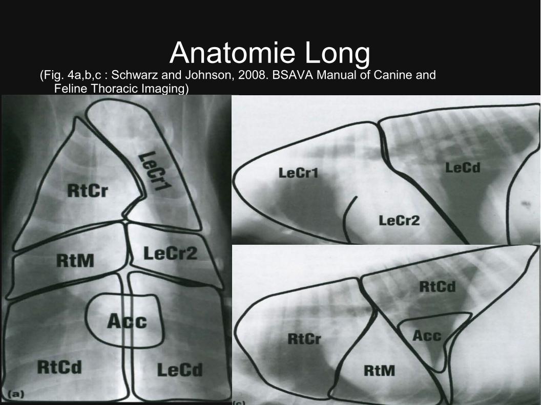
Anatomie Long(Fig. 4a,b,c : Schwarz and Johnson, 2008. BSAVA Manual of Canine and
Feline Thoracic Imaging)
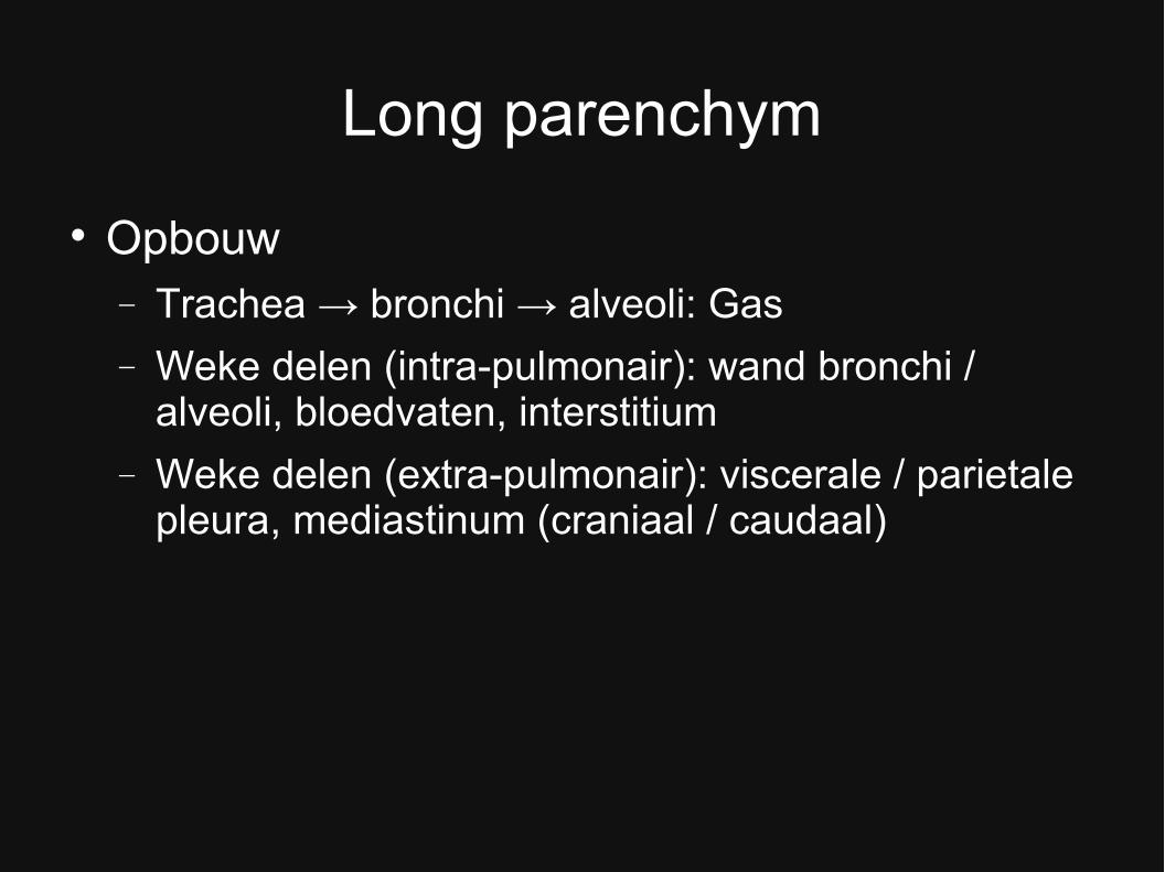
Long parenchym
Opbouw Trachea → bronchi → alveoli: Gas
Weke delen (intra-pulmonair): wand bronchi / alveoli, bloedvaten, interstitium
Weke delen (extra-pulmonair): viscerale / parietale pleura, mediastinum (craniaal / caudaal)
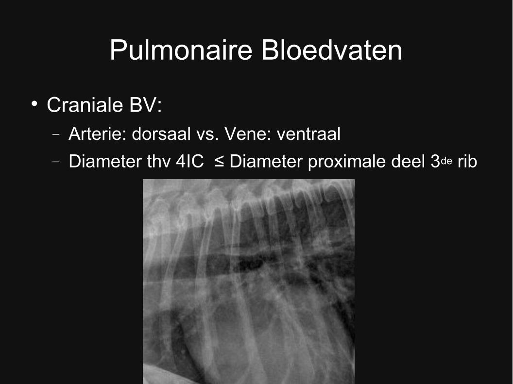
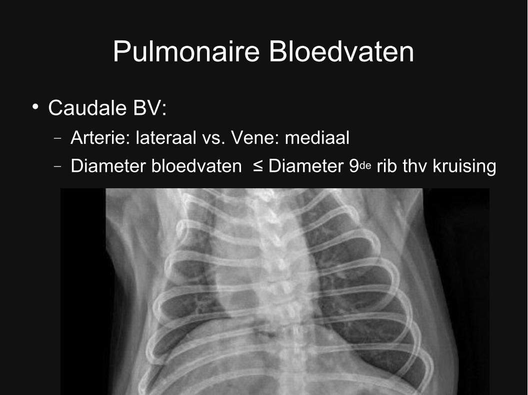
Pulmonaire Bloedvaten
Craniale BV: Arterie: dorsaal vs. Vene: ventraal
Diameter thv 4IC ≤ Diameter proximale deel 3de rib
Pulmonaire Bloedvaten
Caudale BV: Arterie: lateraal vs. Vene: mediaal
Diameter bloedvaten ≤ Diameter 9de rib thv kruising
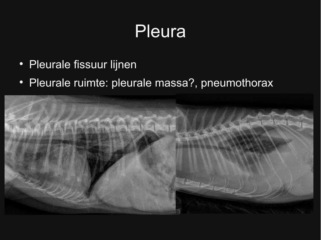
Pleura
Pleurale fissuur lijnen Pleurale ruimte: pleurale massa?, pneumothorax
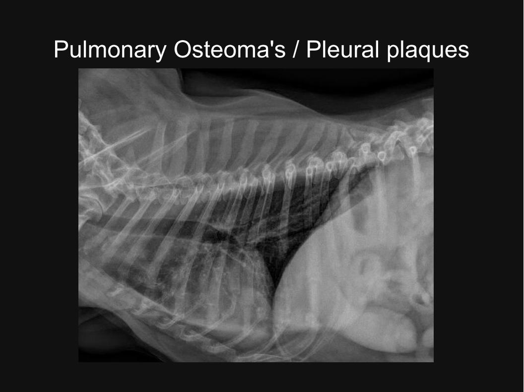
Pulmonary Osteoma's / Pleural plaques
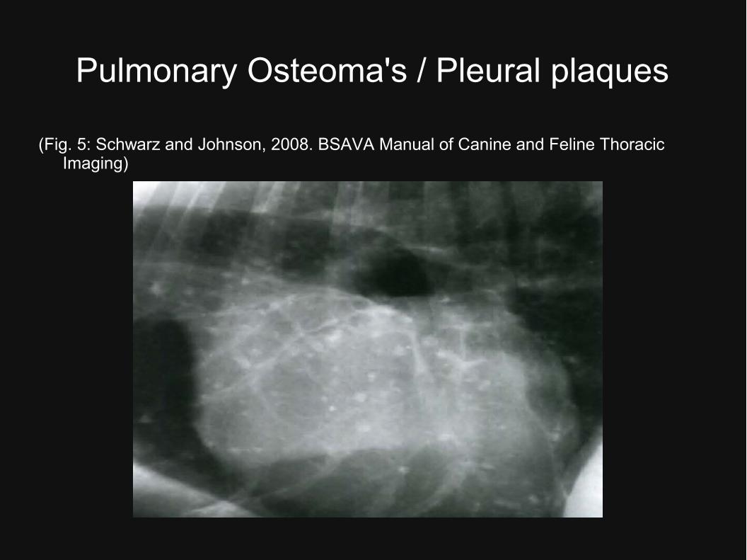
Pulmonary Osteoma's / Pleural plaques
(Fig. 5: Schwarz and Johnson, 2008. BSAVA Manual of Canine and Feline Thoracic Imaging)
Mediastinum
Craniaal, middle & caudaal deel Hond: VD/DV: Craniaal mediastinum < 2x breedte
vertebra (uitz. Brachycephalic en obese dieren)
vs. Kat: breedte = breedte vertebra Mediastinale structuren normaal zichtbaar:
– Trachea, Carina, Hoofdbronchen
– Aorta
– CVC
– (Thymus)
– (Oesophagus)
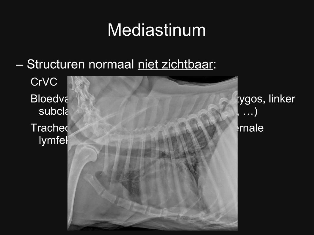
Mediastinum
– Structuren normaal niet zichtbaar:CrVC
Bloedvaten en zenuwen (oa. n.vagus, v.azygos, linker subclavian arterie, brachiocephalic trunk, …)
Tracheobronchiale, cr. mediastinale en sternale lymfeknopen
Bedankt voor uw aandacht!
Referenties
1. Buchanan J.W. et al. 1995. Vertebral Scale System to measure canine heart size in radiographs. JAVMA 206 (2): 194-199.
2. Lamb C.R. et al. 2001. Use of breed-specific ranges for the vertebral heart scale as an aid to the radiographic diagnosis of cardiac disease in dogs. Veterinary Record 148: 707-711.
3. Bavegems V. et al. 2005. Vertebral heart size ranges specific for Whippets. Veterinary Radiology & Ultrasound 46 (5): 400-403.

![Blocks voor thorax en abdomen [Alleen-lezen] - uzleuven.be · Analgesie van abdomen en/of thorax Beste analgesie = centraal zenuwblock: epiduraal/intrathecaal ... Dorsale ramus spieren/huid](https://static.fdocuments.nl/doc/165x107/5d29390488c993f3778ccd6f/blocks-voor-thorax-en-abdomen-alleen-lezen-analgesie-van-abdomen-enof-thorax.jpg)