RIJKSMUSEUM VAN NATUURLIJKE HISTORIE TE LEIDENzoologische mededelingen . uitgegeven door het ....
34
ZOOLOGISCHE MEDEDELINGEN UITGEGEVEN DOOR HET RIJKSMUSEUM VAN NATUURLIJKE HISTORIE TE LEIDEN (MINISTERIE VAN WELZIJN, VOLKSGEZONDHEID EN CULTUUR) Deel 61 no. 16 23 september 1987 ISSN 0024-0672 NEW OSORNOPHRYNE (AMPHIBIA: ANURA: BUFONIDAE) FROM THE ATLANTIC VERSANT OF THE ANDES IN ECUADOR by MARINUS S. HOOGMOED Hoogmoed, M.S.: New Osornophryne (Amphibia: Anura: Bufonidae) from the Atlantic ver- sant of the Andes in Ecuador. Zool. Med. Leiden 61 (16), 23-ix-1987: 209-242, figs. 1-20, table 1. — ISSN 0024-0672. Key words: Anura, Bufonidae; Osornophryne; new species; breeding habits; internal fertiliza- tion; Neotropics, Andes; Ecuador. Two species of the little known genus Osornophryne are described from the Atlantic (Amazo- nian) versant of the Andes in Ecuador. O. guacamayo spec. nov. occurs at 2100 m, has well developed cranial ridges, rather peculiar feet with toes IV and V of about equal length and much longer than the others. It is rather gaudily patterned with black and yellow. O. antisana spec. nov. occurs at 3600 m and has a pointed snout, indistinct cranial ridges and is rather drably coloured. The single available adult male has a cloacal tube that runs down the back of the thighs and opens ventrally. It is speculated that this peculiar structure is indicative of internal fertilization. It is con- cluded that inguinal amplexus in this genus is not a primitive, but an advanced character. A key to the known species is presented. M.S. Hoogmoed, Rijksmuseum van Natuurlijke Historie, Postbus 9517, 2300 RA Leiden, The Netherlands. RESUMEN Se describe dos nuevos especies de Osornophryne, basandose en material recojido en Antisana por un grupo de PUCE, y en el Cordillera de Guacamayos, un extension oriental del Andes, por un investigador suizo. O. antisana spec. nov. esta caracterizada por su piel lisa, su hocico apun- tado, sus manos y pies compactos con membranas interdigitales bien desarrolladas, las dimen- siones de la cabeza y de las patas traseras, y sobre todo, para la presencia en el holotipo macho de un tubo cloacal con apertura ventral. Se disputa las consecuencias posibles para la reproduc- ción de este espécie, se propone que la fecundación puede ser interna, y que la espécie puede ser ovovivipara o vivipara. Faltan hembras fecundas para controlar estas especulaciónes. O. guacamayo spec. nov. se caracteriza por su cabeza grande con bollos óseos en el parte posterior, por su pie con dedos IV y V distintos y mas largos que los otros y por su coloración negro y amarillo. Se présenta una clave para el género Osornophryne y se discuta la afinidad de las espécies conocidas. Se concluye que el amplexus pélvico no es un carácter primitivo, pero por el contrario es un carácter derivado. 209
Transcript of RIJKSMUSEUM VAN NATUURLIJKE HISTORIE TE LEIDENzoologische mededelingen . uitgegeven door het ....
ZOOLOGISCHE MEDEDELINGEN U I T G E G E V E N D O O R H E T
RIJKSMUSEUM VAN NATUURLIJKE HISTORIE TE LEIDEN ( M I N I S T E R I E V A N W E L Z I J N , V O L K S G E Z O N D H E I D E N C U L T U U R )
Deel 61 no. 16 23 september 1987 ISSN 0024-0672
NEW OSORNOPHRYNE (AMPHIBIA: ANURA: BUFONIDAE) FROM T H E ATLANTIC VERSANT OF T H E ANDES IN ECUADOR
by
MARINUS S. HOOGMOED
Hoogmoed, M.S.: New Osornophryne (Amphibia: Anura: Bufonidae) from the Atlantic ver-sant of the Andes in Ecuador.
Zool. Med. Leiden 61 (16), 23-ix-1987: 209-242, figs. 1-20, table 1. — ISSN 0024-0672. Key words: Anura, Bufonidae; Osornophryne; new species; breeding habits; internal fertiliza-
tion; Neotropics, Andes; Ecuador. Two species of the little known genus Osornophryne are described from the Atlantic (Amazo
nian) versant of the Andes in Ecuador. O. guacamayo spec. nov. occurs at 2100 m, has well developed cranial ridges, rather peculiar feet with toes IV and V of about equal length and much longer than the others. It is rather gaudily patterned with black and yellow. O. antisana spec. nov. occurs at 3600 m and has a pointed snout, indistinct cranial ridges and is rather drably coloured. The single available adult male has a cloacal tube that runs down the back of the thighs and opens ventrally. It is speculated that this peculiar structure is indicative of internal fertilization. It is con-cluded that inguinal amplexus in this genus is not a primitive, but an advanced character. A key to the known species is presented.
M.S. Hoogmoed, Rijksmuseum van Natuurlijke Historie, Postbus 9517, 2300 RA Leiden, The Netherlands.
R E S U M E N
Se describe dos nuevos especies de Osornophryne, basandose en material recojido en Antisana por un grupo de P U C E , y en el Cordillera de Guacamayos, un extension oriental del Andes, por un investigador suizo. O. antisana spec. nov. esta caracterizada por su piel lisa, su hocico apun-tado, sus manos y pies compactos con membranas interdigitales bien desarrolladas, las dimensiones de la cabeza y de las patas traseras, y sobre todo, para la presencia en el holotipo macho de un tubo cloacal con apertura ventral. Se disputa las consecuencias posibles para la reproduc-ción de este espécie, se propone que la fecundación puede ser interna, y que la espécie puede ser ovovivipara o vivipara. Faltan hembras fecundas para controlar estas especulaciónes. O. guacamayo spec. nov. se caracteriza por su cabeza grande con bollos óseos en el parte posterior, por su pie con dedos IV y V distintos y mas largos que los otros y por su coloración negro y amarillo. Se présenta una clave para el género Osornophryne y se discuta la afinidad de las espécies conocidas. Se concluye que el amplexus pélvico no es un carácter primitivo, pero por el contrario es un carácter derivado.
209
210 ZOOLOGISCHE MEDEDELINGEN 61 (1987)
I N T R O D U C T I O N
The genus Osornophryne was erected by Ruiz-Carranza & Hernández-Camacho in 1976 to accommodate Atelopus bufoniformis (Peracca) and the newly described O. percrassa, rather peculiar small toads from the high A n dean valleys in southern Colombia and northern Ecuador, which differ from other bufonids a.o. by the absence of the hypobranchial I, by a reduced number of phalanges in fingers and toes, which are connected by extensive webbing thus forming plates, by a reduced number of vertebrae, by a massive firmisternal shoulder-girdle, by strongly dilated and flattened sacral diapophyses, by lateral crests along the coccyx, and by inguinal amplexus. Ruiz-Carranza & Hernández-Camacho (1976) provided a detailed and adequate account of the new genus, providing extensive data not only on external and internal morphology (including skeleton and muscles), but also on the behaviour of the two species. One of the most striking characters of Osor-nophryne is the reduction in number of presacral vertebrae, which, however, is variable: O. percrassa has six, including a separate atlas; O. bufoniformis has five, including an atlas-complex which arose by fusion of the atlas and the first trunk vertebra (Ruiz-Carranza & Hernández-Camacho, 1976: 107). Can-natella (1986a: 203) studied more skelettal material of O. bufoniformis and found that in the majority of specimens studied (cleared and stained, and X -rayed) there was no fusion into an atlas-complex. Thus, apparently the normal situation in Osornophryne is six presacral vertebrae, including a separate atlas, with occasional specimens having five, including an atlas-complex. A c cording to Cannatella's (1986a: 203) analysis,Osornophryne forms a natural group with Frostius Cannatella and Atelopus Duméril & Bibron. Within this group two alternative arrangements are possible, a choice between which would just be based on a judgement about the subjective value of two characters. Cannatella (1986b: 620) described a third species, Osornophryne talipes, the type specimens of which originally were considered identical with O. bufoniformis, but could be distinguished on the basis of its fleshy, pointed rostrum, smooth dorsal skin and the relative size of the legs and head.
During studies of toads of the so-called Bufo 'typhonius' group, a few specimens of Osornophryne were found, which had been collected during recent fieldwork by parties of the Pontifícia Universidad Católica del Ecuador, and of the Muséum d'Histoire Naturelle of Genève ( M H N G ) . They proved to belong to two new species, one represented by a single male, the other by two females.
H O O G M O E D : NEW OSORNOPHRYNE FROM ECUADOR 211
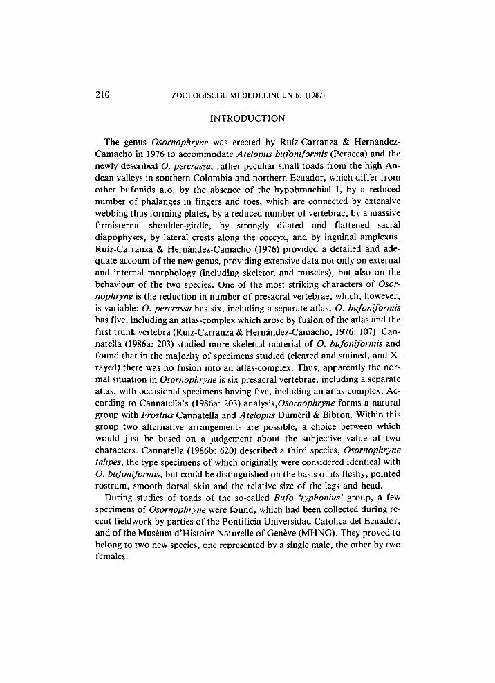
Fig. 1. Distribution of the genus Osornophryne in Colombia and Ecuador (adapted from Cannatella, 1986b: 621). Dots: O. bufoniformis (Peracca), star: O. talipes Cannatella, triangle: O. antisana spec, nov., square: O. guacamayo spec. nov. Numbers 1-8 correspond with those of Cannatella (1986b: 621), 9. Antisana, 10. Cordillera de Guacamayo, 11. Cerros Llanganati.
212 ZOOLOGISCHE MEDEDELINGEN 61 (1987)
T A X O N O M I C P A R T
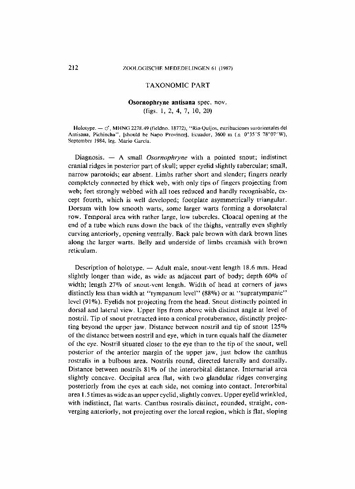
Osornophryne antisana spec. nov. (figs. 1, 2, 4, 7, 10, 20)
Holotype. — cf", M H N G 2278.49 (fieldno. 18772), "Rio Quijos, estribaciones surorientales del Antisana, Pichincha", [should be Napo Province], Ecuador, 3600 m ( ± 0°35 'S 78°07'W), September 1984, leg. Mario Garcia.
Diagnosis. — A small Osornophryne with a pointed snout; indistinct cranial ridges in posterior part of skull; upper eyelid slightly tubercular; small, narrow parotoids; ear absent. Limbs rather short and slender; fingers nearly completely connected by thick web, with only tips of fingers projecting from web; feet strongly webbed with all toes reduced and hardly recognisable, except fourth, which is well developed; footplate asymmetrically triangular. Dorsum with low smooth warts, some larger warts forming a dorsolateral row. Temporal area with rather large, low tubercles. Cloacal opening at the end of a tube which runs down the back of the thighs, ventrally even slightly curving anteriorly, opening ventrally. Back pale brown with dark brown lines along the larger warts. Belly and underside of limbs creamish with brown reticulum.
Description of holotype. — Adult male, snout-vent length 18.6 mm. Head slightly longer than wide, as wide as adjacent part of body; depth 60% of width; length 27% of snout-vent length. Width of head at corners of jaws distinctly less than width at "tympanum lever' (88%) or at "supratympanic" level (91%). Eyelids not projecting from the head. Snout distinctly pointed in dorsal and lateral view. Upper lips from above with distinct angle at level of nostril. Tip of snout protracted into a conical protuberance, distinctly projecting beyond the upper jaw. Distance between nostril and tip of snout 125% of the distance between nostril and eye, which in turn equals half the diameter of the eye. Nostril situated closer to the eye than to the tip of the snout, well posterior of the anterior margin of the upper jaw, just below the canthus rostralis in a bulbous area. Nostrils round, directed laterally and dorsally. Distance between nostrils 81% of the interorbital distance. Internarial area slightly concave. Occipital area flat, with two glandular ridges converging posteriorly from the eyes at each side, not coming into contact. Interorbital area 1.5 times as wide as an upper eyelid, slightly convex. Upper eyelid wrinkled, with indistinct, flat warts. Canthus rostralis distinct, rounded, straight, converging anteriorly, not projecting over the loreal region, which is flat, sloping
H O O G M O E D : NEW OSORNOPHRYNE FROM ECUADOR 213
steeply to the upper lips. Area between nostrils and tip of snout distinctly concave. Lips not flaring. Eyes with horizontally oval pupil. Lower eyelid translucent. Temporal region vertical, covered with flat warts. Ear absent. "Supratympanic" area rounded. In parotoid area a long and very narrow
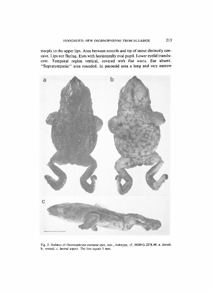
Fig. 2. Habitus of Osornophryne antisana spec. nov., holotype, cf, M H N G 2278.49. a. dorsal, b. ventral, c. lateral aspect. The line equals 5 mm.
214 ZOOLOGISCHE MEDEDELINGEN 61 (1987)
gland, posteriorly continued to the groin as an oblique row of low glands on the flanks (fig. 4).
Side of neck with low, rounded tubercles; flanks and, especially, the belly and underside of limbs coarsely tuberculate, with low, round tubercles; all tubercles with round pore. Throat and chest indistinctly coarsely areolate. Back smooth with wrinkles, round, low warts, and paravertebral rows of glands from shoulder to thighs (forming the extension of the converging glandular ridges on the occipital region) (fig. 2).
Cloacal opening directed ventrally at the end of a tube running from end of urostyl, over back of thighs and continuing anteriorly for a short distance on the ventral surface of the thighs (fig. 10).
Fingers connected by thick webbing, forming a plate with tubercular ventral
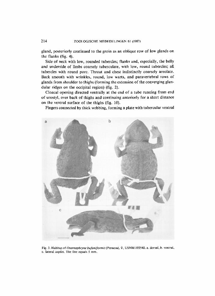
Fig. 3. Habitus of Osornophryne bufoniformis (Peracca), 9, USNM 193540. a. dorsal, b. ventral, c. lateral aspect. The line equals 5 mm.
H O O G M O E D : NEW OSORNOPHRYNE FROM ECUADOR 215
surface, from which only the tips of the toes protrude (fig. 7). Fingers distinguishable dorsally: first very wide with roughly circular nuptial pad on dorsal and medial surface second narrower, elongate, with entire dorsal sur
face covered by more widely spaced nuptial asperities. Tip of third toe with a few nuptial asperities. Webbing formula: I(0)(0)II(0)(1 >/4)ΙΙΙ(1 lÁH0)lW. Fingers depressed, with rounded tips. Dorsal surface of fingers III and IV and of wrist with striking transverse grooves. Length of fingers: I < II < IV < III. Phalangeal formula: 1232. Forearm thick.
Tarsus with a few low, scattered warts. Toes connected by thick webbing,
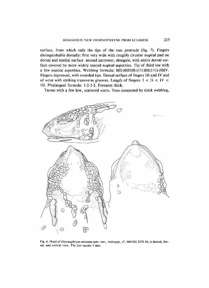
Fig. 4. Head of Osornophryne antisana spec, nov., holotype, cf, M H N G 2278.49, in lateral, dor
sal, and ventral view. The line equals 1 mm.
216 ZOOLOGISCHE MEDEDELINGEN 61 (1987)
forming an asymmetrically triangular plate with deeply grooved tubercular ventral surface. Toes hardly distinguishable ventrally or dorsally; toes I, II and III very much reduced, but forming an area at the medial side of toe IV which is very well developed, wide and long, toe V hardly noticeable at lateral side of toe IV. Toes depressed with rounded tips. Webbing formula: 1(0)-(0)II(0)-(0)III(0)-(2)IV(2)-(0)V. Dorsal surface of foot with transverse grooves. Length of toes: I < II < V < III < IV. Phalangeal formula: 1?-1?-2-4-1 (based on X-ray photographs which were difficult to interpret because of the flexed position of the toes).
When the hindlimbs are flexed at right angles to the sagittal plane, the heels
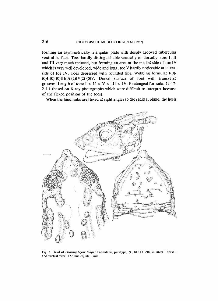
Fig. 5. Head of Osornophryne talipes Cannatella, paratype, cf, KU 131798, in lateral, dorsal, and ventral view. The line equals 1 mm.
H O O G M O E D : NEW OSORNOPHRYNE FROM ECUADOR 217
touch; when carried forward along the body the heel does not reach the axil. Tibia 25-26% of the snout-vent length.
Five presacral vertebrae, all of which bear rather short transverse processes, which distinctly differ in shape: those of vertebra 1 are straight rods, of equal width throughout, those of vertebra 2 are dilated, those of vertebra 3 are curved, rod-shaped (with the convexity directed posteriorly), those of vertebrae 4 and 5 are curved (convexity directed anteriorly) and bluntly pointed. The processes of vertebrae 1 and 5 are directed anteriorly, those of 3 and 4 posteriorly and those of 2 are transverse. Transverse width of the processes: 2 < 3 < 1
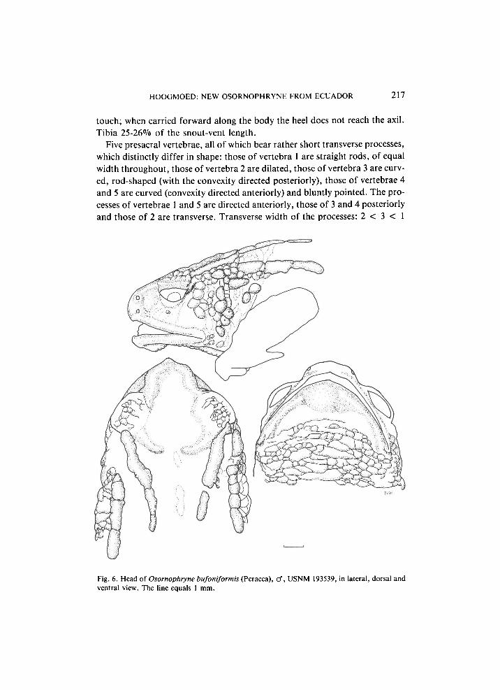
Fig. 6. Head of Osornophryne bufoniformis (Peracca), cf, USNM 193539, in lateral, dorsal and ventral view. The line equals 1 mm.
218 ZOOLOGISCHE MEDEDELINGEN 61 (1987)
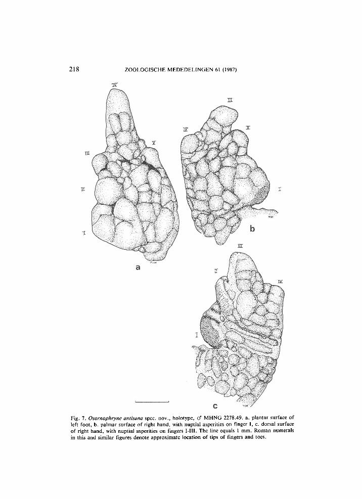
Fig. 7. Osornophryne antisana spec. nov., holotype, cf M H N G 2278.49. a. plantar surface of left foot, b. palmar surface of right hand, with nuptial asperities on finger I, c. dorsal surface of right hand, with nuptial asperities on fingers I-III. The line equals 1 mm. Roman numerals in this and similar figures denote approximate location of tips of fingers and toes.
H O O G M O E D : NEW OSORNOPHRYNE FROM ECUADOR 219
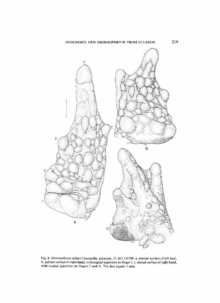
Fig. 8. Osornophryne talipes Cannatella, paratype, cf, KU 131798. a. plantar surface of left foot, b. palmar surface of right hand, with nuptial asperities on finger I, c. dorsal surface of right hand, with nuptial asperities on fingers I and II. The line equals 1 mm.
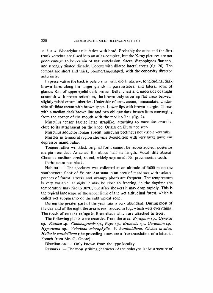
220 ZOOLOGISCHE MEDEDELINGEN 61 (1987)
< 5 < 4. Bicondylar articulation with head. Probably the atlas and the first trunk vertebra are fused into an atlas-complex, but the X-ray pictures are not good enough to be certain of that conclusion. Sacral diapophyses flattened and strongly dilated distally. Coccyx with dilated lateral crests (fig. 20). The femora are short and thick, boomerang-shaped, with the concavity directed anteriorly.
In preservative the back is pale brown with short, narrow, longitudinal dark brown lines along the larger glands in paravertebral and lateral rows of glands. Rim of upper eyelid dark brown. Belly, chest and underside of thighs creamish with brown reticulum, the brown only covering flat areas between slightly raised cream tubercles. Underside of arms cream, immaculate. Underside of tibiae cream with brown spots. Lower lips with brown margin. Throat with a median dark brown line and two oblique dark brown lines converging from the corner of the mouth with the median line (fig. 2).
Musculus tensor fasciae latae straplike, attaching to musculus cruralis, close to its attachment on the knee. Origin on ilium not seen.
Musculus adductor longus absent, musculus pectineus not visible ventrally. Muscles in temporal region showing S-condition with very large musculus
depressor mandibulae. Tongue rather wrinkled, original form cannot be reconstructed; posterior
margin rounded. Attached for about half its length. Vocal slits absent. Choanae medium-sized, round, widely separated. No prevomerine teeth.
Peritoneum not black. Habitat. — The specimen was collected at an altitude of 3600 m on the
southeastern flank of Volcan Antisana in an area of meadows with isolated patches of forest. Creeks and swampy places are frequent. The temperature is very variable: at night it may be close to freezing, in the daytime the temperature may rise to 30°C, but after showers it may drop rapidly. This is the typical landscape of the upper limit of the wet altitudinal forest, which is called wet subparamo of the subtropical zone.
During the greater part of the year rain is very abundant. During most of the day and of the night the area is enshrouded in fog, which wets everything. The toads often take refuge in Bromeliads which are attached to trees.
The following plants were recorded from the area: Eryngium sp., Gynoxis sp., Festuca sp., Calamagrostis sp., Puya sp., Bromelia sp., Geranium sp., Hypericum sp., Valeriana microphylla, V. humboldiana, Olchus lavatus, Hallenia weadelliana (the preceding notes are a free translation of a letter in French from M r . G . Onore).
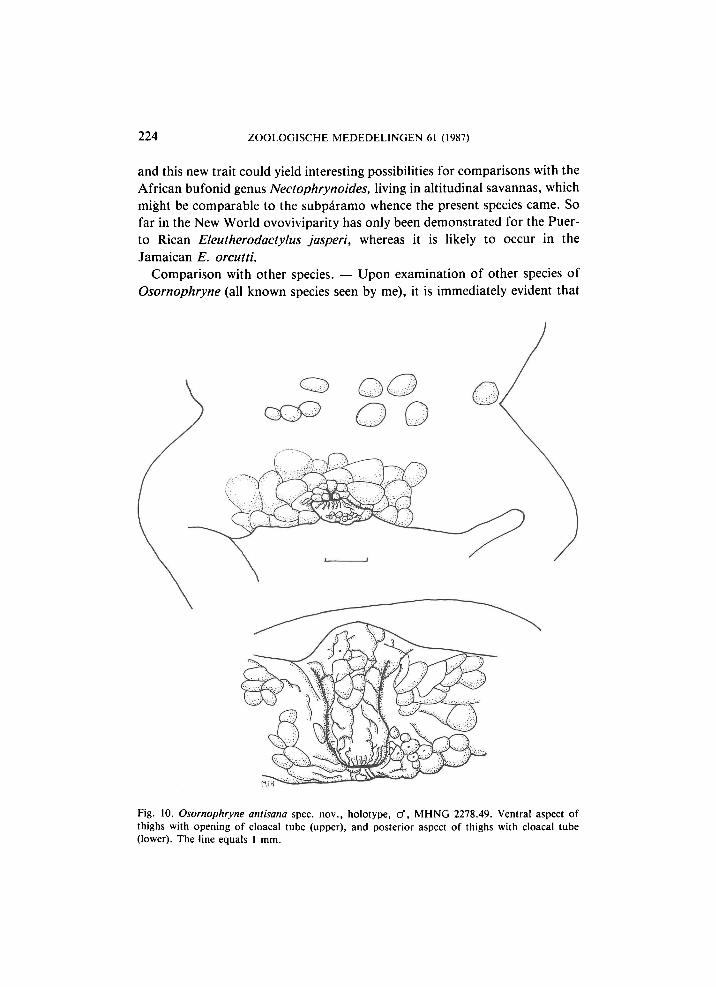
Distribution. — Only known from the type-locality. Remarks. — The most striking character of the holotype is the structure of
H O O G M O E D : NEW OSORNOPHRYNE FROM ECUADOR 221
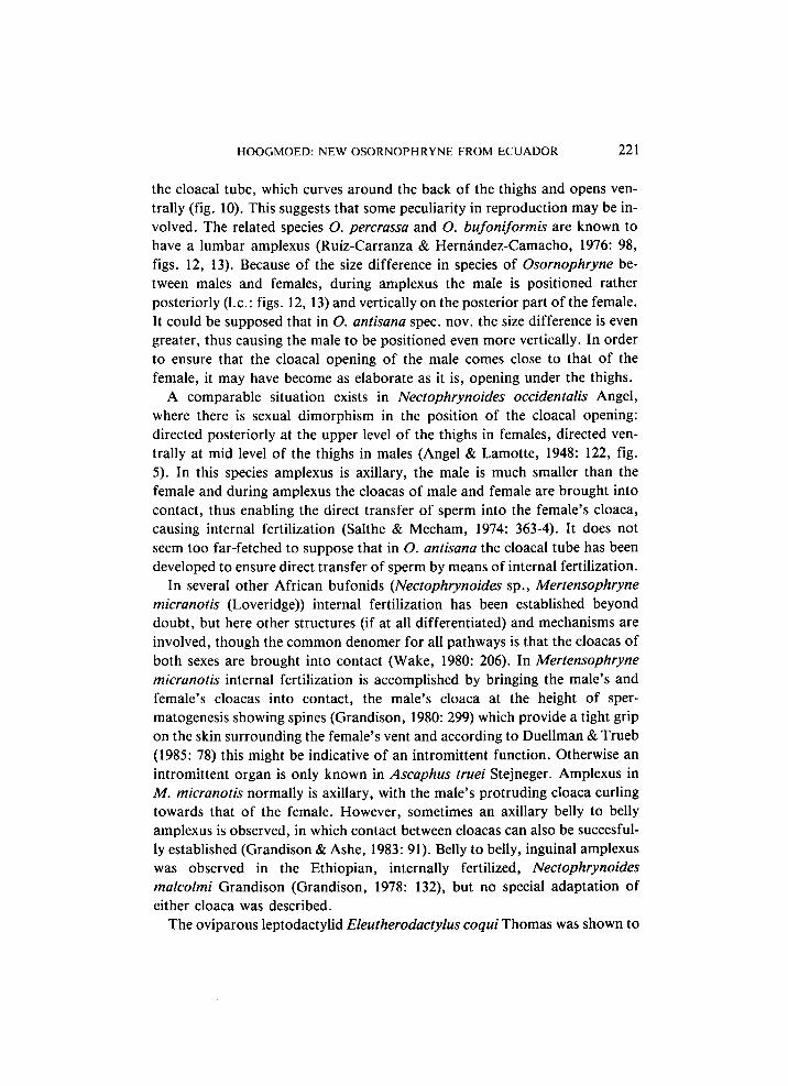
the cloacal tube, which curves around the back of the thighs and opens ventrally (fig. 10). This suggests that some peculiarity in reproduction may be involved. The related species O. percrassa and O. bufoniformis are known to have a lumbar amplexus (Ruiz-Carranza & Hernández-Camacho, 1976: 98, figs. 12, 13). Because of the size difference in species of Osornophryne be-tween males and females, during amplexus the male is positioned rather posteriorly (I.e.: figs. 12, 13) and vertically on the posterior part of the female. It could be supposed that in O. antisana spec. nov. the size difference is even greater, thus causing the male to be positioned even more vertically. In order to ensure that the cloacal opening of the male comes close to that of the female, it may have become as elaborate as it is, opening under the thighs.
A comparable situation exists in Nectophrynoides occidentalis Angel, where there is sexual dimorphism in the position of the cloacal opening: directed posteriorly at the upper level of the thighs in females, directed ventrally at mid level of the thighs in males (Angel & Lamotte, 1948: 122, fig. 5). In this species amplexus is axillary, the male is much smaller than the female and during amplexus the cloacas of male and female are brought into contact, thus enabling the direct transfer of sperm into the female's cloaca, causing internal fertilization (Salthe & Mecham, 1974: 363-4). It does not seem too far-fetched to suppose that in O. antisana the cloacal tube has been developed to ensure direct transfer of sperm by means of internal fertilization.
In several other African bufonids (Nectophrynoides sp., Mertensophryne micranotis (Loveridge)) internal fertilization has been established beyond doubt, but here other structures (if at all differentiated) and mechanisms are involved, though the common denomer for all pathways is that the cloacas of both sexes are brought into contact (Wake, 1980: 206). In Mertensophryne micranotis internal fertilization is accomplished by bringing the male's and female's cloacas into contact, the male's cloaca at the height of spermatogenesis showing spines (Grandison, 1980: 299) which provide a tight grip on the skin surrounding the female's vent and according to Duellman & Trueb (1985: 78) this might be indicative of an intromittent function. Otherwise an intromittent organ is only known in Ascaphus truei Stejneger. Amplexus in M . micranotis normally is axillary, with the male's protruding cloaca curling towards that of the female. However, sometimes an axillary belly to belly amplexus is observed, in which contact between cloacas can also be succesful-ly established (Grandison & Ashe, 1983: 91). Belly to belly, inguinal amplexus was observed in the Ethiopian, internally fertilized, Nectophrynoides malcolmi Grandison (Grandison, 1978: 132), but no special adaptation of either cloaca was described.
The oviparous leptodactylid Eleutherodactylus coqui Thomas was shown to
222 ZOOLOGISCHE MEDEDELINGEN 61 (1987)
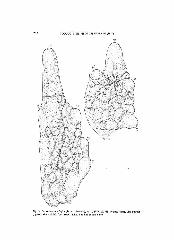
Fig. 9. Osornophryne bufoniformis (Peracca), cf, USNM 193539, plantar (left), and palmar (right) surface of left foot, resp., hand. The line equals 1 mm.
H O O G M O E D : NEW OSORNOPHRYNE FROM ECUADOR 223
have internal fertilization by cloacal apposition, without special morphological modifications to the cloacal region (Townsend, 1987: 31; Towns-end et al . , 1981: 470). For the ovo viviparous E. jasperi Drewry & Jones internal fertilization can be inferred from the development of the eggs in the oviducts (Wake, 1978: 131). For E. orcutti Dunn live-bearing has not yet been proved, but cirumstantial evidence strongly points in that direction (Lynn & Grant, 1940: 58; Wake, 1978: 131).
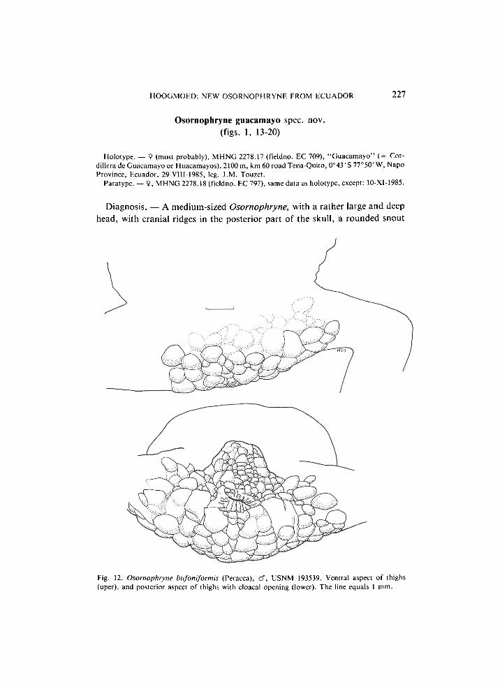
Investigation of male and female Osornophryne bufoniformis at my disposal ( U S N M 193539-40) did not show any difference in position of the vent, in both it is situated at the upper level of the thighs (figs. 3, 12) and directed posteriorly, its distance from the tip of the coccyx being 10.5% of the snout-vent length in the male, 11.5% in the female. The single female of O. percrassa ( U S N M 151325) examined also had its cloaca-opening directed posteriorly, at the upper level of the thighs. Unfortunately Ruiz-Carranza & Hernández-Camacho (1976) do not provide any data on the position of the cloacal opening in males of O. percrassa, but there is no reason to suppose it differs from that in the females, as they would undoubtedly have noticed this and included it in their description.
Eggs in O. percrassa are few, large and unpigmented, which represents a derived condition (Ruiz-Carranza & Hernández-Camacho, 1976: 118). Such eggs often are found in species with direct, extra-aquatic development, but several cases are known (e.g. Rana graminea Boulenger [= R. livida Blyth]) in which such eggs are deposited in water (Ruiz-Carranza & Hernández-Camacho, 1976: 119). In the related bufonid genus Atelopus, which also has unpigmented eggs, these are deposited at the edge of streams, with the tadpoles living in the streams (Lescure, 1981: 212). O. percrassa apparently is unable to swim and can only float passively, reason for Ruiz-Carranza & Hernández-Camacho (1976: 119, 120) to suppose that Osornophryne certainly would not lay its eggs in creeks, pools or bromeliads, consequently only leaving open the possibility of laying them in small depressions that could be inundated or in wet places where direct development.would be possible. The structure of the cloacal tube in O. antisana to me suggests an even further stage in reproduction, viz., internal fertilization, followed either by deposition of the fertilized eggs (which then could go through a (terrestrial) tadpole stage or develop directly) or even live-bearing, either through ovoviviparity or viviparity. Anyway, its external morphology suggests an interesting reproductive mode which deserves further research. Intensive sampling, covering all seasons, is necessary to obtain a good idea of the morphology, ecology, distribution, ethology and reproductive habits of this new species.
Ovoviviparity or even viviparity is not known for South American anurans,
224 ZOOLOGISCHE MEDEDELINGEN 61 (1987)
and this new trait could yield interesting possibilities for comparisons with the African bufonid genus Nectophrynoides, living in altitudinal savannas, which might be comparable to the subpáramo whence the present species came. So far in the New World ovoviviparity has only been demonstrated for the Puerto Rican Eleutherodactylus jasperi, whereas it is likely to occur in the Jamaican E. orcutti.
Comparison with other species. — Upon examination of other species of Osornophryne (all known species seen by me), it is immediately evident that
Fig. 10. Osornophryne antisana spec, nov., holotype, cf, M H N G 2278.49. Ventral aspect of thighs with opening of cloacal tube (upper), and posterior aspect of thighs with cloacal tube (lower). The line equals 1 mm.
H O O G M O E D : NEW OSORNOPHRYNE FROM ECUADOR 225
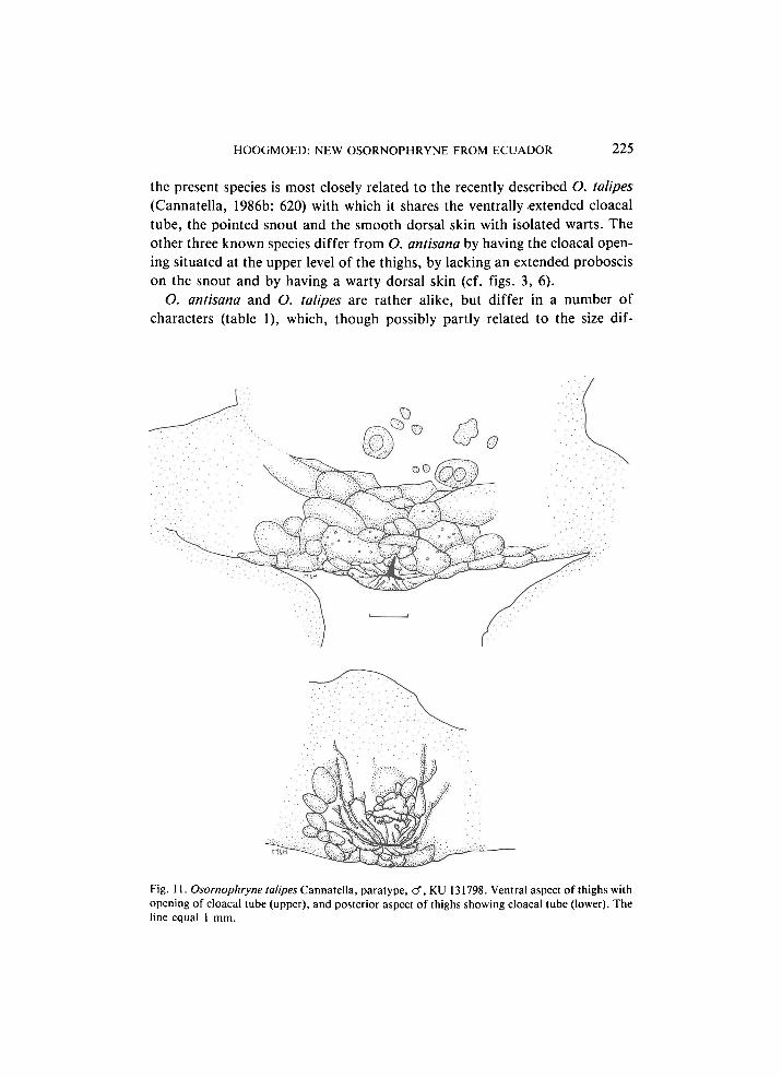
the present species is most closely related to the recently described O. talipes (Cannatella, 1986b: 620) with which it shares the ventrally extended cloacal tube, the pointed snout and the smooth dorsal skin with isolated warts. The other three known species differ from O. antisana by having the cloacal opening situated at the upper level of the thighs, by lacking an extended proboscis on the snout and by having a warty dorsal skin (cf. figs. 3, 6).
O. antisana and O. talipes are rather alike, but differ in a number of characters (table 1), which, though possibly partly related to the size dif-
Fig. 11. Osornophryne talipes Cannatella, paratype, cf, KU 131798. Ventral aspect of thighs with opening of cloacal tube (upper), and posterior aspect of thighs showing cloacal tube (lower). The line equal 1 mm.
226 ZOOLOGISCHE MEDEDELINGEN 61 (1987)
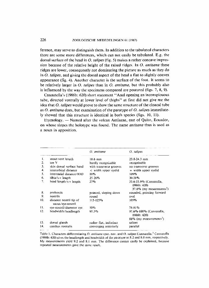
ference, may serve to distinguish them. In addition to the tabulated characters there are some more differences, which can not easily be tabulated. E.g . the dorsal surface of the head in O. talipes (fig. 5) makes a rather concave impression because of the relative height of the raised ridges. In O. antisana these ridges are lower, consequently not dominating the picture as much as they do in O. talipes, and giving the dorsal aspect of the head a flat to slightly convex appearance (fig. 4). Another character is the surface of the foot. It seems to be relatively larger in O. talipes than in O. antisana, but this probably also is influenced by the way the specimens compared are postured (figs. 7, 8, 9).
Cannatella's (1986b: 620) short statement " A n a l opening an inconspicuous tube, directed ventrally at lower level of thighs'' at first did not give me the idea that O. talipes would prove to show the same structure of the cloacal tube as O. antisana does, but examination of the paratype of O. talipes immediately showed that this structure is identical in both species (figs. 10, 11).
Etymology. — Named after the volcan Antisana, east of Quito, Ecuador, on whose slopes the holotype was found. The name antisana thus is used as a noun in apposition.
O. antisana O. talipes
1. snout-vent length 18.6 mm 23.8-24.3 mm 2. toe V hardly recognisable recognisable 3. skin dorsal surface hand with transverse grooves no transverse grooves 4. interorbital distance < width upper eyelid = width upper eyelid 5. internarial distance/IOD 81% 109% 6. tibia/s-v length 25-26% 30-31% 7. head length/s-v length 27% 33.6-33.9% (Cannatella,
1986b: 620) 37.8% (my meaurements1)
8. proboscis pointed, sloping down rounded, pointing forward 9. nostrils round oval
10. distance nostril-tip of 115-125% 105% snout/eye-nostril
11. eye-nostril/diameter eye 50% 78-81% 12. headwidth/headlength 95.5% 97.6%-100% (Cannatella,
1986b: 620) 88% (my measurements1)
13. dorsal glands rather flat, indistinct salient 14. canthus rostralis converging anteriorly parallel
Table 1. Characters differentiating O. antisana spec. nov. and O. talipes Cannatella.1 Cannatella (1986b: 620) gives the headlength and headwidth of the paratype as 8.2 and 8.0 mm, respectively. My measurements yield 9.2 and 8.1 mm. The difference cannot easily be explained, because repeated measurements gave the same result.
HOOGMOED: NEW OSORNOPHRYNE FROM ECUADOR 227
Osornophryne guacamayo spec. nov. (figs. 1, 13-20)
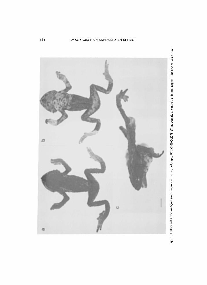
Holotype. — 9 (most probably), M H N G 2278.17 (fieldno. EC 709), "Guacamayo" (= Cordillera de Guacamayo or Huacamayos), 2100 m, km 60 road Tena-Quito, 0°43 ' S 77°50' W, Napo Province, Ecuador, 29-VIII-1985, leg. J . M . Touzet.
Paratype. — 9, M H N G 2278.18 (fieldno. EC 797), same data as holotype, except: 10-XI-1985.
Diagnosis. — A medium-sized Osornophryne, with a rather large and deep head, with cranial ridges in the posterior part of the skull, a rounded snout
Fig. 12. Osornophryne bufoniformis (Peracca), cf, USNM 193539. Ventral aspect of thighs (uper), and posterior aspect of thighs with cloacal opening (lower). The line equals 1 mm.
ZOOLOGISCHE MEDEDELINGEN 61 (1987)
H O O G M O E D : NEW OSORNOPHRYNE FROM ECUADOR 229
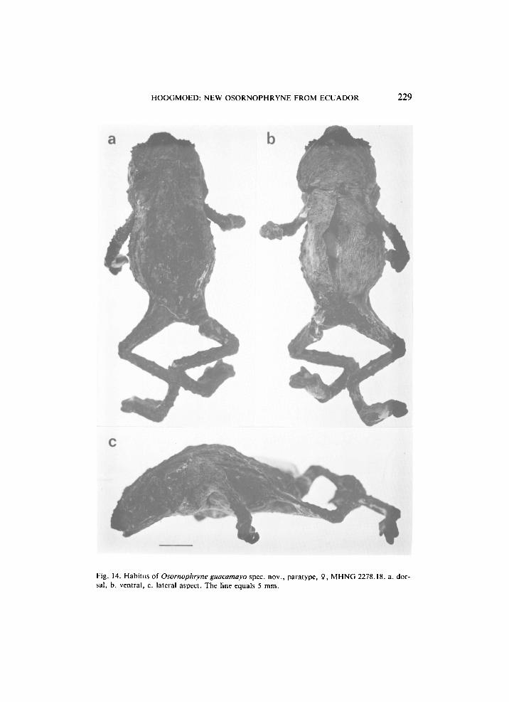
Fig. 14. Habitus of Osornophryne guacamayo spec, nov., paratype, 9, M H N G 2278.18. a. dorsal, b. ventral, c. lateral aspect. The line equals 5 mm.
230 ZOOLOGISCHE MEDEDELINGEN 61 (1987)
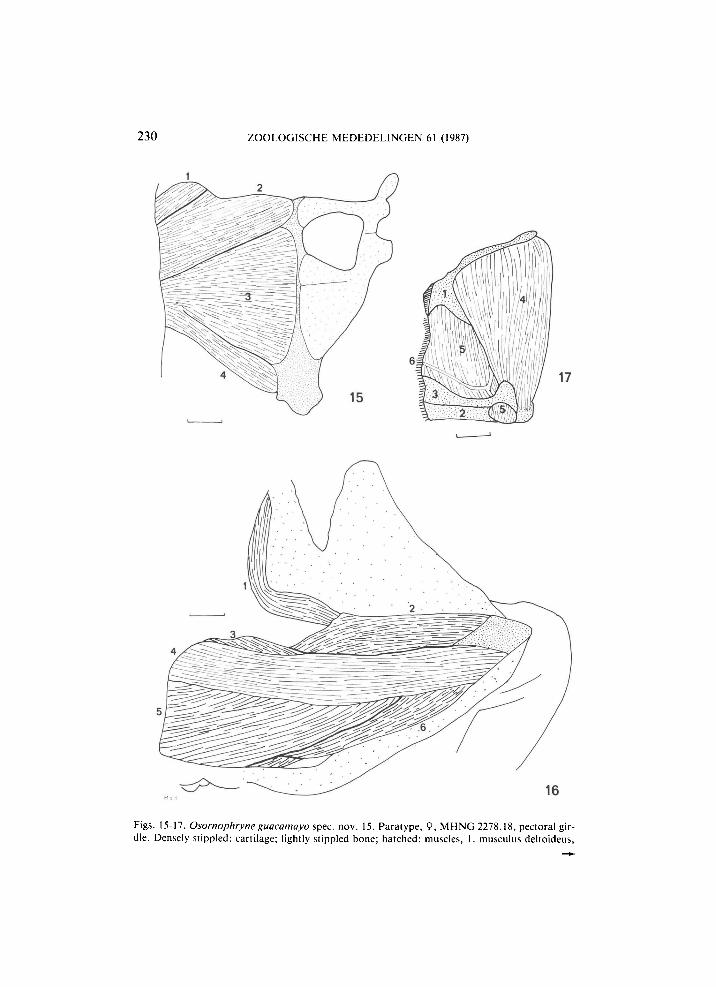
Figs. 15-17. Osornophryne guacamayo spec. nov. 15. Paratype, 9, M H N G 2278.18, pectoral girdle. Densely stippled: cartilage; lightly stippled bone; hatched: muscles, 1. musculus deltoideus,
H O O G M O E D : NEW OSORNOPHRYNE FROM ECUADOR 231
with conical tip, strongly tubercular upper eyelids; no parotoids; ear absent. Limbs long and rather slender; fingers connected by thick web, with third and fourth extending well beyond the web; feet heavily webbed with fourth and fifth toe much longer than the other three, forming a unit in which the separate toes are well visible. Dorsum with many, irregularly scattered tubercles of different sizes. Temporal region with large and small tubercles, underlain by yellowish glandular tissue. Back blackish with two oblique yellow dorsolateral lines, extending from shoulders to groin; some indistinct, isolated yellow spots. Ventral parts yellow with black spots. Peritoneum black.
Description. — Snout-vent length 35.5 (31.3) mm (values for the paratype are given in parentheses, but should be considered with some reserve, because the specimen is rather buckled and hard, so not all measurements could be exact and are approximations). Head wider than long (longer than wide), slightly wider than adjacent part of body, about half as deep as wide; length 31.8 (36.1)% of the snout-vent length. Width of head at corners of jaws slightly narrower (slightly wider) than width at tympanum level, and distinctly narrower than width at the supratympanic level. Eyelids not projecting from the head. Snout rounded with an acuminate tip in dorsal profile, bluntly pointed in lateral profile. Shape of snout determined by a conical, fleshy ridge; this conical structure projecting beyond the upper jaw (rictus of mouth). Distance between nostrils 1.5 times that between nostril and eye, 71(74)% of the interorbital distance. Internarial area distinctly concave. A large, rounded tubercle over each nostril. Top of head flat, with bony protuberances, raised fleshy area around the orbits, an oblique bony ridge from the posterior corner of the eye extending posteriorly and medially, posteriorly increasing in height and ending in a bony boss at the end of the head, and isolated, large, rounded tubercles. Interorbital area 1.5 times as wide as an upper eyelid, flat between the raised circumorbital ridges, with warts. Upper eyelid with 3-4 enlarged, projecting tubercles along the outer margin, remainder with numerous conical
2. m. coracoradialis, 3. m. pectoralis (portio epicoracoidea), 4. m. pectoralis (portio sternalis). The line equals 1 mm. 16. Holotype, 9?, M H N G 2278.17. Ventral view of left thigh after removal of skin. Densely stippled: bone; lightly stippled: inner surface of uplifted skin; hatched: muscles, 1. musculus tensor fasciae latae, 2. m. cruralis, 3. m. pectineus, 4. m. sartorius, 5. m. adductor magnus, 6. m. gracilis. There is no m. adductor longus. The line equals 1 mm. 17. Paratype, 9, M H N G 2278.18. Left temporal region after removal of skin. Stippled area: bone; hatched area: muscle; heavly cross-hatched: inner surface of uplifted skin. S-condition of McDiarmid (1971: 10): 1. squamosal, 2. mandíbula, 3. maxilla, 4. musculus depressor mandibulae, 5. m. adductor mandibulae posterior subexternus, 6. mandibular branch of trigeminal nerve. The line equals 1 mm.
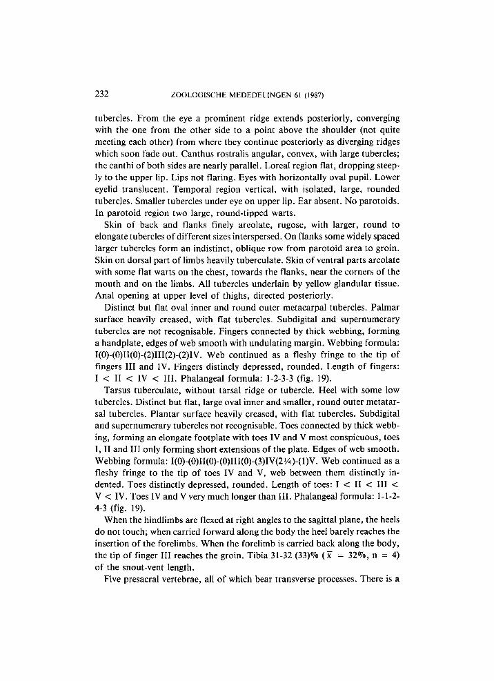
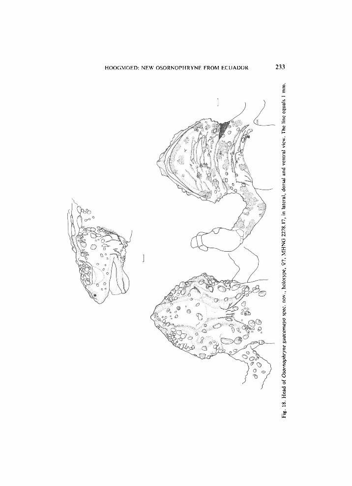
232 ZOOLOGISCHE MEDEDELINGEN 61 (1987)
tubercles. From the eye a prominent ridge extends posteriorly, converging with the one from the other side to a point above the shoulder (not quite meeting each other) from where they continue posteriorly as diverging ridges which soon fade out. Canthus rostralis angular, convex, with large tubercles; the canthi of both sides are nearly parallel. Loreal region flat, dropping steep
ly to the upper lip. Lips not flaring. Eyes with horizontally oval pupil. Lower eyelid translucent. Temporal region vertical, with isolated, large, rounded tubercles. Smaller tubercles under eye on upper l ip. Ear absent. No parotoids. In parotoid region two large, roundtipped warts.
Skin of back and flanks finely areolate, rugose, with larger, round to elongate tubercles of different sizes interspersed. On flanks some widely spaced larger tubercles form an indistinct, oblique row from parotoid area to groin. Skin on dorsal part of limbs heavily tuberculate. Skin of ventral parts areolate with some flat warts on the chest, towards the flanks, near the corners of the mouth and on the limbs. A l l tubercles underlain by yellow glandular tissue. Anal opening at upper level of thighs, directed posteriorly.
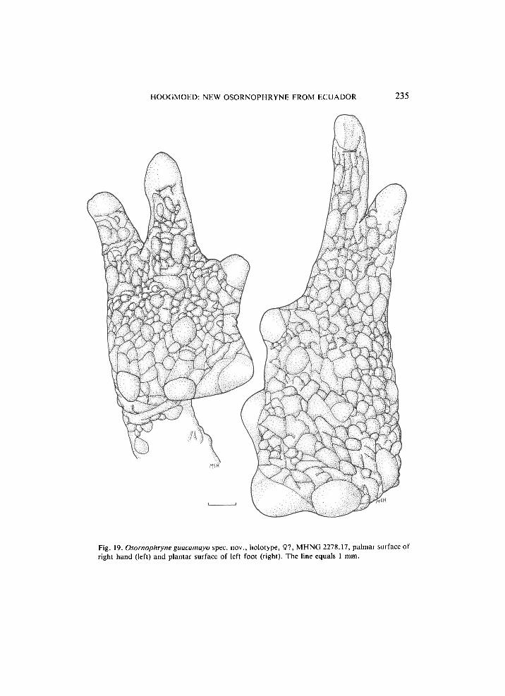
Distinct but flat oval inner and round outer metacarpal tubercles. Palmar surface heavily creased, with flat tubercles. Subdigital and supernumerary tubercles are not recognisable. Fingers connected by thick webbing, forming a handplate, edges of web smooth with undulating margin. Webbing formula: I(0)(0)II(0)(2)III(2)(2)IV. Web continued as a fleshy fringe to the tip of fingers III and IV. Fingers distincly depressed, rounded. Length of fingers: I < II < IV < III. Phalangeal formula: 1233 (fig. 19).
Tarsus tuberculate, without tarsal ridge or tubercle. Heel with some low tubercles. Distinct but flat, large oval inner and smaller, round outer metatar
sal tubercles. Plantar surface heavily creased, with flat tubercles. Subdigital and supernumerary tubercles not recognisable. Toes connected by thick webb
ing, forming an elongate footplate with toes IV and V most conspicuous, toes I, II and III only forming short extensions of the plate. Edges of web smooth. Webbing formula: I(0)(0)II(0)(0)III(0)(3)IV(21/4)(l)V. Web continued as a fleshy fringe to the tip of toes IV and V , web between them distinctly in
dented. Toes distinctly depressed, rounded. Length of toes: I < II < III < V < IV. Toes IV and V very much longer than III. Phalangeal formula: 112
43 (fig. 19). When the hindlimbs are flexed at right angles to the sagittal plane, the heels
do not touch; when carried forward along the body the heel barely reaches the insertion of the forelimbs. When the forelimb is carried back along the body, the tip of finger III reaches the groin. Tibia 3132 (33)% (x = 32%, η = 4) of the snoutvent length.
Five presacral vertebrae, all of which bear transverse processes. There is a
H O O G M O E D : NEW OSORNOPHRYNE FROM ECUADOR 233
H
-Ό
Ο Ζ χ 2
I 1 •s:
ÖD
234 ZOOLOGISCHE MEDEDELINGEN 61 (1987)
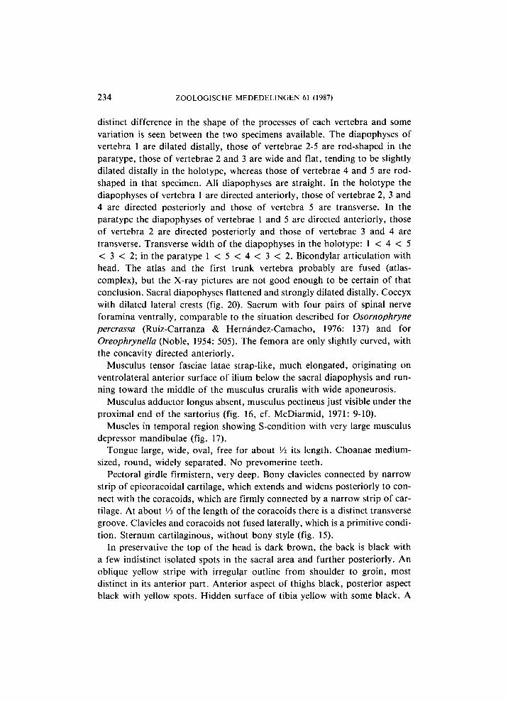
distinct difference in the shape of the processes of each vertebra and some variation is seen between the two specimens available. The diapophyses of vertebra 1 are dilated distally, those of vertebrae 2-5 are rod-shaped in the paratype, those of vertebrae 2 and 3 are wide and flat, tending to be slightly dilated distally in the holotype, whereas those of vertebrae 4 and 5 are rod-shaped in that specimen. A l l diapophyses are straight. In the holotype the diapophyses of vertebra 1 are directed anteriorly, those of vertebrae 2, 3 and 4 are directed posteriorly and those of vertebra 5 are transverse. In the paratype the diapophyses of vertebrae 1 and 5 are directed anteriorly, those of vertebra 2 are directed posteriorly and those of vertebrae 3 and 4 are transverse. Transverse width of the diapophyses in the holotype: 1 < 4 < 5 < 3 < 2; in the paratype 1 < 5 < 4 < 3 < 2 . Bicondylar articulation with head. The atlas and the first trunk vertebra probably are fused (atlas-complex), but the X-ray pictures are not good enough to be certain of that conclusion. Sacral diapophyses flattened and strongly dilated distally. Coccyx with dilated lateral crests (fig. 20). Sacrum with four pairs of spinal nerve foramina ventrally, comparable to the situation described for Osornophryne percrassa (Ruiz-Carranza & Hernández-Camacho, 1976: 137) and for Oreophrynella (Noble, 1954: 505). The femora are only slightly curved, with the concavity directed anteriorly.
Musculus tensor fasciae latae strap-like, much elongated, originating on ventrolateral anterior surface of ilium below the sacral diapophysis and running toward the middle of the musculus cruralis with wide aponeurosis.
Musculus adductor longus absent, musculus pectineus just visible under the proximal end of the sartorius (fig. 16, cf. McDiarmid, 1971: 9-10).
Muscles in temporal region showing S-condition with very large musculus depressor mandibulae (fig. 17).
Tongue large, wide, oval, free for about Vi its length. Choanae medium-sized, round, widely separated. No prevomerine teeth.
Pectoral girdle firmistern, very deep. Bony clavicles connected by narrow strip of epicoracoidal cartilage, which extends and widens posteriorly to connect with the coracoids, which are firmly connected by a narrow strip of cartilage. A t about Vi of the length of the coracoids there is a distinct transverse groove. Clavicles and coracoids not fused laterally, which is a primitive condition. Sternum cartilaginous, without bony style (fig. 15).
In preservative the top of the head is dark brown, the back is black with a few indistinct isolated spots in the sacral area and further posteriorly. A n oblique yellow stripe with irregular outline from shoulder to groin, most distinct in its anterior part. Anterior aspect of thighs black, posterior aspect black with yellow spots. Hidden surface of tibia yellow with some black. A
H O O G M O E D : NEW OSORNOPHRYNE FROM E C U A D O R 235
Fig. 19. Osornophryne guacamayo spec, nov., holotype, 9?, M H N G 2278.17, palmar surface of right hand (left) and plantar surface of left foot (right). The line equals 1 mm.
236 ZOOLOGISCHE MEDEDELINGEN 61 (1987)
yellow spot on base of hand (inner two fingers) and foot (inner three toes). A short yellow stripe from mouth to insertion of forelimb. Ventral parts yellow with irregular black (on venter) and brown (on chest and throat) spots. Underside of limbs variegated black and yellow. Palms and soles immaculate, grey.
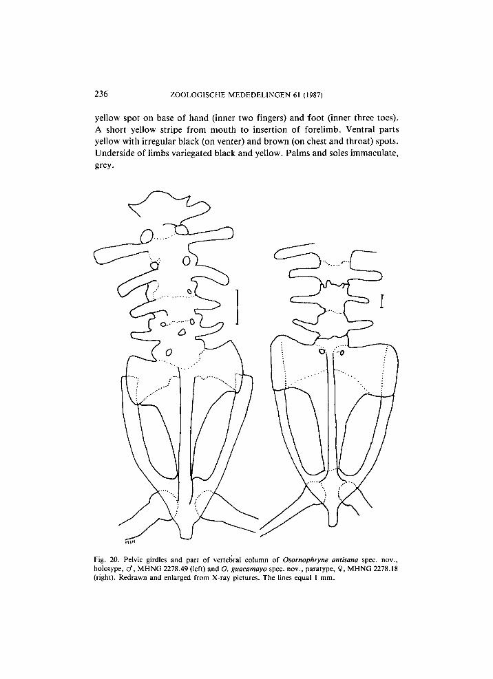
Fig. 20. Pelvic girdles and part of vertebral column of Osornophryne antisana spec, nov., holotype, cf", M H N G 2278.49 (left) and O. guacamayo spec, nov., paratype, 9, M H N G 2278.18 (right). Redrawn and enlarged from X-ray pictures. The lines equal 1 mm.
H O O G M O E D : NEW OSORNOPHRYNE FROM ECUADOR 237
Peritoneum black. Note. — The holotype is an eviscerate specimen, the viscera having been
removed for parasitological research. Its sex was established by comparison with the paratype, which is a desiccated female with enlarged oviducts and an amorphous jelly-like mass in its belly.
Habitat. — Both specimens available were collected in the late afternoon along a roadside, on moss in rock crevices at an altitude of 2100 m, in very wet tropical cloud-forest on a mountain ridge. Forest-floor with many loose stones, vegetation covered by mosses and epiphytes, stones covered by about 10 cm of moss. The animals were very slow. Other herpetofauna collected here included Bolitoglossa palmata (Werner) and Caeciliidae (Dr. V . Mahnert in litt., based on collector's data).
Etymology. — Guacamayo is the Ecuadorian name for macaw, also it is the name of the eastern spur of the Andes whence came the type-specimens. In loose reference to the gaudy colours and to its type localty, the specific epiteth "guacamayo" has been chosen. It is here used as a noun in apposition.
Remarks. — This species differs from all other known species of the genus by the remarkable shape of its feet, in which the fourth and fifth toe are well developed and separate, by the structure of its head and by its gaudily patterned belly.
K E Y T O T H E SPECIES O F OSORNOPHRYNE
1. Skin on back smooth with dispersed glands and glandular ridges; tip of snout with a distinct, fleshy proboscis; cloacal tube (in males) descending along the posterior aspect of the thighs, opening ventrally beneath the thighs 2
- Skin on back rugose, with tubercles; tip of snout truncate, rounded or rounded with an acuminate to bluntly pointed tip (not fleshy); no cloacal tube, cloacal opening at upper level of thighs 3
2. Head length 27%, tibia 25 - 26% of the snout-vent length; snout-vent length 18.6 mm; distance eye-nostril 50% eye diameter antisana
- Head length 34 - 38%, tibia 30 - 31% of the snout-vent length; snout-vent length 23.8 - 24.3 mm; distance eye-nostril 78-81% eye diameter
talipes 3. Toes IV and V much longer than I-III, distinct from each other; head with
prominent bony ridges in posterior part guacamayo - Only toe IV much longer than the rest, V very much reduced and forming
part of the footplate; head not deep, without bony ridges 4
238 ZOOLOGISCHE MEDEDELINGEN 61 (1987)
4. Tip of snout abruptly truncate or sloping anteriorly, not projecting beyond the mouth; head as long as wide or wider than long; anterior part of back with (para)vertebral rows of glands in contact percrassa
- Tip of snout slightly projecting beyond the mouth; head about as long as wide; paravertebral lines of glands separated widely bufoniformis
DISCUSSION
The southermost locality recorded for Osornophryne by Cannatella (1986b: 621) was the type locality of O. talipes, Nudo de Mojanda. Both Antisana and the Cordillera de Guacamayos are further south, though in the same general area. Sofar Osornophryne was only known from elevations between 2700 m and 3700 m. The specimens of O. guacamayo come from an altitude of 2100 m, thus considerably expanding the vertical distribution to lower levels and consequently to another type of habitat as well. Thus, Hoogmoed's (1985: 257) statement that the genus is only known from the páramo-region, has to be changed. The genus is now known to occur from upper tropical cloud-forest on the Amazonian versant of the Andes, to páramo in its central, higher parts. The Göteborg specimen of O. bufoniformis from "Cerros Llanganati" ( = Cordillera de las Llanganates) extends the known distribution of the genus still further south (fig. 1).
According to Ruiz-Carranza & Hernández-Camacho (1976: 103-104) Osornophryne (bufoniformis + percrassa) has a reduced phalangeal formula in the hand (2-2-3-2) and a normal formula in the foot (2-2-3-4-3). These data are not corroborated by my own data, which indicate that reduction in O. bufoniformis also occurs in the foot and there are even indications that the number of phalanges in the foot is more reduced in males (1-1-2-4-2) than in females (2-2-3-4-2) (based on the examination of X-ray pictures of one male and one female only). Alberch & Gale (1985: 10) also provide data on the phalangeal formula of the foot of Osornophryne different from those of Ruiz-Carranza & Hernández-Camacho (1976: 103-104) and much more in agreement with my own observations: bufoniformis: 1 -2-3( 2 )-4-_1 -(2), percrassa: l -2-3( 2)-4-l (underlined indicates that digit has lost skeletal elements). The species here described show that there are other possibilities as well. In O. antisana (and probably O. talipes as well) the reduction of the number of phalanges in digits of the hand has progressed further, with the inner digit reduced to one phalange only, the outer to two; the same occurs in the foot, where all digits except IV (four phalanges) are shortened to one or two (in toe III) phalanges. In O. guacamayo reduction of the digits in the hand
H O O G M O E D : NEW OSORNOPHRYNE FROM ECUADOR 239
has only occurred in the inner finger, which has one phalange left, whereas the outer finger has three phalanges; the foot has 1-1-2-4-3 phalanges, which represents a further reduction of the inner three phalanges only. Apparently reduction in the number of phalanges was realised along different pathways. Starting from an original phalangeal formula of 2-2-3-3 for the hand and of 2-2-3-4-3 for the foot, it is possible to envisage two pathways along which reduction of the number of phalanges could occur: the first one leading to the clubfoot and the rather compact hand present in antisana, bufoniformis, percrassa and talipes, the second one leading to the two-pronged foot and rather unaltered hand of O. guacamayo. In both pathways reduction does not occur in the third finger and fourth toe. In the pathway leading to the clubfoot and compact hand, at least the outer digits have a reduced number of phalanges. Reduction seems to effect the outer digits first, then the inner ones, and subsequently progress toward the longest digit, without effecting this one. These observations contrast with those of Alberch & Gale (1985: 11), who, on the basis of their data (they only studied feet) concluded that reduction always effected the first toe first. The fact that in the female O. bufoniformis ( U S N M 193540) examined by me the inner toe has two phalanges upsets that pattern, because in that toe no phalangeal reduction has occurred, whereas it did occur in the fifth toe. As this only concerns one specimen, this could be the exception to the rule. However, I also have to refute Alberch & Gale's (1985: 11) conclusion that in Osornophryne after reduction in toes I and V has taken place, toe III is reduced. In the species and specimens examined by me this does not hold true: in bufoniformis reduction took place in toes I, II, III and V (all - 1 phalange) in the male, in toe V ( - 1 phalange) only in the female; in antisana reduction took place in toes I, II, III ( - 1 phalange) and V ( - 2 phalanges), in guacamayo it occurred in toes I, II and III ( - 1 phalange). Thus, the "rules" formulated by Alberch & Gale (1985: 10) on the basis of ten species with reduced phalangeal formula, are already upset by data derived from two newly described species.
Both bufoniformis and percrassa show the same (slight) degree of reduction and can be considered closest to the ancestral form, whereas in antisana and talipes reduction has progressed further. In the pathway leading to the two-pronged foot and rather unaltered hand of guacamayo, only the innermost digit(s) is (are) show(s) a reduced number of phalanges.
Comparing the shape of the foot and the phalangeal formulas, I come to the conclusion that O. guacamayo appears as the least specialised species in this respect, because its fingers and toes (despite reduction of the number of phalanges in the inner finger and the innermost three toes) are still quite well recognisable as entities. However, considering the habitat of the species and
240 ZOOLOGISCHE MEDEDELINGEN 61 (1987)
the way reduction in digits has taken place in related species, I assume this first impression is incorrect and that on the contrary the shape of hands and feet, which probably are good grasping organs, is derived and an adaptation to the species' living in mossy crevices and the locomotion connected with this.
Ruiz-Carranza & Hernández-Camacho (1976: 111) suggest that the reduction of the length of the digits represents a major adaptation toward terrestrial habits. The fact that O. antisana shows the most extreme form of reduction, but allegedly was found in arboreal bromeliads, seems to refute this suggestion. On the other hand the species with the best developed digits (O. guacamayo), was found on moss in rock crevices, which seems to indicate a terrestrial way of life, though these crevices also may have been above the ground in vertical rockfaces.
Inguinal amplexus generally is considered a trait of the Archaeobatrachia, whereas in the Neobatrachia (to which the Bufonidae belong) it only occurs exceptionally, mostly among Australian Myobatrachidae and some pelodryadin Hylidae, but also in a few South American Leptodactylidae and African Bufonidae (Grandison, 1978). Ruiz-Carranza & Hernández-Camacho (1976: 118) and Hoogmoed (1985: 258) consider the inguinal amplexus in Osornophryne a remaining primitive character of great phylogenetic interest. I now only agree with part of this conclusion, namely that this trait is of great phylogenetic interest. However, contrary to what Ruiz-Carranza & Hernández-Camacho (1976: 118) and Hoogmoed (1985: 258) believe, I now think the inguinal amplexus in Osornophryne is not a primitive but an advanced character-state and in this I concur with Duellman & Trueb (1985: 532). I come to this conclusion on the basis of the structure of the cloacal tube in male O. antisana and O. talipes and the assumption that fertilization in these species is internal (see p. 221). In that case, inguinal amplexus is a character that developed independently and secondarily from axillary amplexus as part of the complex of morphological and behavioural changes that occurred to realise internal fertilization. Inguinal amplexus and internal fertilization apparently are an adaptation to the scarcity of standing water in the environment, and parallel to it the inability of Osornophryne to survive in water for longer periods possibly developed. Considering the other character-states for this genus (most of which are advanced) (Hoogmoed, 1985: 258; Cannatella, 1986a: 202), this conclusion does not come as a surprise.
Apparently Osornophryne forms a very specialised group within the "atelopodid" toads. Cannatella's (1986a) analysis of the group shows that Atelopus, Frostius and Osornophryne are rather close together, whereas data provided by Hoogmoed (1985) also showed that Atelopus (at that stage still
HOOGMOED: NEW OSORNOPHRYNE FROM ECUADOR 241
including the Frostius to be) and Osornophryne were closely related. Can
natella (1985a: 204) discussed the possibility whether the three genera should be subsumed under a single genus Atelopus, but concluded that the genera preferably should be maintained in order to emphasize the morphological, ecological and behavioural distinctiveness of the three taxa. The new data on Osornophryne only strengthen his views.
A C K N O W L E D G E M E N T S
It is my pleasure to thank my colleagues V. Mahnert and J .L . Perret (Muséum d'Histoire Naturelle, Genève (MHNG)) who provided me with newly acquired bufonid material from Ecuador and with useful fielddata. They, and also G. Nilson (Naturhistoriska Museet, Göteborg (NHMG)) provided working space during visits to their respective institutions. W.R. Heyer and R.I. Crombie (National Museum of Natural History (USNM)) and W.E. Duellman (Museum of Natural History, Kansas University (KU)) kindly loaned me material of Osornophryne in their collections. The Xray pictures on which part of the study of skelettal characters was based, were made by Mr. S. Zijlstra of the Zoological Laboratory of the University of Leiden.
Additional material of Osornophryne studied
Osornophryne bufoniformis (Peracca). Ecuador: Napo Province, Santa Barbara: lcf, 9, USNM 19353940, January 1963, leg. M . Olalla; Tungurahua Province, Cerros Llanganati (E. Rio San José, 3300 m): 19 N H M G 196211248 (Ba ex. 1079), 30/31 January 1960, leg. R. Blomberg.
Osornophryne percrassa RuizCarranza & HernándezCamacho. Colombia: Depto. Tolima, out of Manizales on old road to Nevada de Ruiz: 19, USNM 151325, 24III1963, leg. W.A. Thornton & C. Marinkelle.
Osornophryne talipes Cannatella. Ecuador: Imbabura Province, north slope of Nudo de Mo
janda, 3400 m: lcf, KU 131798, 2VIII1970, leg. J.D. Lynch & F. Ortiz.
R E F E R E N C E S
Alberch, P., & E . A . Gale, 1985. A developmental analysis of an evolutionary trend: digital reduc
tion in amphibians. Evolution 39 (1): 823, figs. 18, table 1. Angel, F., & M . Lamotte, 1948. Nouvelles observations sur Nectophrynoides occidentalis Angel.
Remarques sur le genre Nectophrynoides. Ann. des Sc. Nat., Zool. (11)X: 115147, figs. 1
15. Cannatella, D.C. , 1986a. A new genus of Bufonid (Anura) from South America, and
phylogenetic relationships of the neotropical genera. Herpetologica 42 (2): 197205. Cannatella, D.C. , 1986b. A new species of Osornophryne (Anura: Bufonidae) from the Andes
of Ecuador. — Copeia 1986 (3): 618622, figs. 12. Duellman, W.E. , & L. Trueb, 1985. Biology of Amphibians, ixvii, 1670, many figs. New York. Grandison, A . G . C . , 1978. The occurrence of Nectophrynoides (Anura Bufonidae) in Ethiopia.
A new concept of the genus with a description of a new species. Monit. Zool. Italiano (Ν.S.) Suppl. 11: 119172, figs. 123, tables 13.
242 ZOOLOGISCHE MEDEDELINGEN 61 (1987)
Grandison, A . G . C . , 1980. Aspects of breeding morphology in Mertensophryne micranotis (Anura: Bufonidae): secondary sexual characters, eggs and tadpole. - Bull. Br. Mus. nat. Hist. (Zool.) 39 (5): 299-304, figs. 1-3.
Grandison, A . G . C . , & S. Ashe, 1983. The distribution, behavioural ecology and breeding strategy of the Pygmy Toad, Mertensophryne micranotis (Lov.). - Bull. Br. Mus. nat. Hist. (Zool.) 45 (2): 85-93, fig. 1, table 1.
Hoogmoed, M.S., 1985. A new genus of toads (Amphibia: Anura: Bufonidae) from the Pacific slopes of the Andes in northern Ecuador and southern Colombia, with the description of two new species. - Zool. Med. Leiden 59 (22): 251-274, figs. 1-18, tables 1-5.
Lescure, J . , 1981. Contribution à l'étude des Amphibiens de Guyane française IX. Le têtard gastromyzophore d'Atelopus flavescens Duméril et Bibron (Anura, Bufonidae). - Amphibia-Reptilia 2 (3): 209-215, figs. 1-2, tables 1-2.
Lynn, W . G . , & C. Grant, 1940. The herpetology of Jamaica. - Bull. Inst. Jamaica, Sc. Ser. 1: 4 pp., 1-148, pis. 1-12, figs. 1-3, tables.
McDiarmid, R.W., 1971. Comparative morphology and evolution of frogs of the neotropical genera Atelopus, Dendrophryniscus, Melanophryniscus, and Oreophrynella. - Bull. L . A . C . M . Nat. Hist. Science 12: 1-66, frontispiece, figs. 1-11, tables 1-4.
Noble, G.K. , 1954. The biology of the Amphibia: 18 pp., 1-577, figs. 1-174. New York (reprint edition Dover Publ.).
Ruiz-Carranza, P . M . , & J.I. Hernández-Camacho, 1976. Osornophryne, género nuevo de anfíbios bufonidos de Colombia y Ecuador. - Caldasia (Zool.) 11 (54): 93-148, figs. 1-16, tables 1-3.
Salthe, S.M., & J.S. Mecham, 1974. Reproductive courtship patterns. In: B. Lofts (ed.): Physiology of the Amphibia. II: 309-521. New York and London.
Townsend, D.S., 1987. Fatherhood in frogdom. - Natural History 1987 (5): 28-35, 6 figs. Townsend, D.S., M . M . Stewart, F . H . Pouch & P.F. Brussard, 1981. Internal fertilization in an
oviparous frog. - Science 212 (4493): 469-471, table 1. Wake, M . H . , 1978. The reproductive biology of Eleutherodactylus jasperi (Amphibia, Anura,
Leptodactylidae), with comments on the evolution of live-bearing systems. - J . Herpet. 12 (2): 121-133, figs. 1-10, table 1.
Wake, M . H . , 1980. The reproductive biology of Nectophrynoides malcolmi (Amphibia: Bufonidae), with comments on the evolution of reproductive modes in the genus Nectophrynoides. - Copeia 1980 (2): 193-209, figs. 1-7, table 4.


![mm Wimt c im ^ i - Tussen Vecht en Eem...Frans Ernst Blaauw en Gooilust doordrs. Peter J. A. van Mensch [Rijksmuseum van Natuurlijke Historie, Leiden] en Ed E. van Mensch [histo rische](https://static.fdocuments.nl/doc/165x107/5f8cf2aa2e7f6f12e1521cf9/mm-wimt-c-im-i-tussen-vecht-en-eem-frans-ernst-blaauw-en-gooilust-doordrs.jpg)