Portable and accurate diagnostics for COVID-19: Combined ... · 4/3/2020 · [email protected]...
25
Portable and accurate diagnostics for COVID-19: 1 Combined use of the miniPCR® thermocycler and 2 a well-plate reader for SARS-Co2 virus detection 3 4 Everardo González-González 1,2 , Grissel Trujillo-de Santiago 1,3 , Itzel Montserrat Lara- 5 Mayorga 1,2 , Sergio Omar Martínez-Chapa 3 , Mario Moisés Alvarez 1,2* 6 7 1 Centro de Biotecnología-FEMSA, Tecnologico de Monterrey, CP 64849, Monterrey, 8 Nuevo León, México 9 2 Departamento de Bioingeniería, Tecnologico de Monterrey, CP 64849, Monterrey, Nuevo 10 León, México 11 3 Departamento de Ingeniería Mecátrónica y Eléctrica, Tecnologico de Monterrey, CP 12 64849, Monterrey, Nuevo León, México 13 14 *Corresponding authors. E-mails: [email protected] 15 16 Abstract 17 The COVID-19 pandemic has crudely demonstrated the value of massive and rapid 18 diagnostics. By the first week of April, more than 900,000 positive cases of COVID-19 19 have been reported worldwide, although this number could be greatly underestimated. In 20 . CC-BY-NC-ND 4.0 International license It is made available under a is the author/funder, who has granted medRxiv a license to display the preprint in perpetuity. (which was not certified by peer review) The copyright holder for this preprint this version posted April 7, 2020. . https://doi.org/10.1101/2020.04.03.20052860 doi: medRxiv preprint
Transcript of Portable and accurate diagnostics for COVID-19: Combined ... · 4/3/2020 · [email protected]...
Portable and accurate diagnostics for COVID-19: 1
Combined use of the miniPCR® thermocycler and 2
a well-plate reader for SARS-Co2 virus detection 3
4
Everardo González-González1,2
, Grissel Trujillo-de Santiago1,3
, Itzel Montserrat Lara-5
Mayorga1,2
, Sergio Omar Martínez-Chapa3, Mario Moisés Alvarez
1,2* 6
7
1 Centro de Biotecnología-FEMSA, Tecnologico de Monterrey, CP 64849, Monterrey, 8
Nuevo León, México 9
2 Departamento de Bioingeniería, Tecnologico de Monterrey, CP 64849, Monterrey, Nuevo 10
León, México 11
3 Departamento de Ingeniería Mecátrónica y Eléctrica, Tecnologico de Monterrey, CP 12
64849, Monterrey, Nuevo León, México 13
14
*Corresponding authors. E-mails: [email protected] 15
16
Abstract 17
The COVID-19 pandemic has crudely demonstrated the value of massive and rapid 18
diagnostics. By the first week of April, more than 900,000 positive cases of COVID-19 19
have been reported worldwide, although this number could be greatly underestimated. In 20
. CC-BY-NC-ND 4.0 International licenseIt is made available under a is the author/funder, who has granted medRxiv a license to display the preprint in perpetuity. (which was not certified by peer review)
The copyright holder for this preprint this version posted April 7, 2020. .https://doi.org/10.1101/2020.04.03.20052860doi: medRxiv preprint

the case of an epidemic emergency, the first line of response should be based on 21
commercially available and validated resources. Here, we demonstrate the combined use of 22
the miniPCR®, a commercial compact and portable PCR device recently available on the 23
market, and a commercial well-plate reader as a diagnostic system for detecting SARS-24
CoV2 nucleic acids. We used the miniPCR to detect and amplify SARS-CoV2 DNA 25
sequences using the sets of initiators recommended by the World Health Organization for 26
targeting three different regions that encode for the N protein. Prior to amplification, 27
samples were combined with a DNA intercalating reagent (i.e., EvaGreen® Dye). Sample 28
fluorescence after amplification was then read using a commercial 96-well plate reader. 29
This straightforward method allows the detection and amplification of SARS-CoV2 nucleic 30
acids in the range of ~625 to 2×10⁵ DNA copies. The accuracy and simplicity of this 31
diagnostics strategy may provide a cost-efficient and reliable alternative for COVID-19 32
pandemic testing, particularly in underdeveloped regions where RT-QPCR instrument 33
availability may be limited. In addition, the portability, ease of use, and reproducibility of 34
the miniPCR® makes it a reliable alternative for deployment in point-of-care SARS-CoV2 35
detection efforts during pandemics. 36
37
Key words: mini-PCR, point-of-care, SARS-CoV2, COVID-19, diagnostic, portable, 38
nucleic acid amplification 39
40
Introduction 41
The development of cost-efficient diagnostic point-of-care (POC) systems for the 42
opportune diagnosis of infectious diseases has been recognized as a niche of high relevance 43
. CC-BY-NC-ND 4.0 International licenseIt is made available under a is the author/funder, who has granted medRxiv a license to display the preprint in perpetuity. (which was not certified by peer review)
The copyright holder for this preprint this version posted April 7, 2020. .https://doi.org/10.1101/2020.04.03.20052860doi: medRxiv preprint
[1,2]. The recent pandemic/epidemic episodes associated with viral diseases (e.g., 44
pandemic Influenza A/H1N1/2009 [3], Ebola in West Africa in 2013-2015 [4], and Zika in 45
Southeast Asia and Latin-America in 2016 [5,6]) were clear reminders of the need for 46
portable, low-cost, and easy-to-use diagnostic systems that can effectively address epidemic 47
episodes in remote or underprivileged areas [7–10]. Nevertheless, the COVID-19 pandemic 48
has broadsided most countries, with only a few (i.e., South Korea [11], China, Singapore 49
[12], and Taiwan [13]) showing an ability to deploy massive efforts for rapid and accurate 50
detection of positive infection cases. The swift and massive testing of thousands of possibly 51
infected subjects has been an important component of the strategy of these countries that 52
has helped to effectively mitigate the spreading of COVID-19 among their populations 53
[11,13,14]. 54
Many methodologies have been proposed to deliver cost-effective and accurate diagnosis 55
(i.e., methods based on immunoassays or specific gene hybridization [15,16]); however, 56
nucleic acid amplification, and particularly real-time quantitative PCR (RT-qPCR), 57
continues to be the gold standard for the detection of viral diseases in early stages [17,18]. 58
For example, for the last two pandemic events involving influenza A/H1N1/2009 and 59
COVID-19 [19], the Centers for Disease Control (CDC) and the World Health 60
Organization (WHO) recommended RT-qPCR methods as the gold standard for official 61
detection of positive cases. Unfortunately, conducting RT-qPCR diagnostics often depends 62
on access to centralized laboratory facilities for testing [20–22]. To resolve this drawback, 63
multiple studies have proposed and validated the use of compact PCR-based methods and 64
devices for POC settings [23,24]. However, during epidemic episodes, resourcing of 65
. CC-BY-NC-ND 4.0 International licenseIt is made available under a is the author/funder, who has granted medRxiv a license to display the preprint in perpetuity. (which was not certified by peer review)
The copyright holder for this preprint this version posted April 7, 2020. .https://doi.org/10.1101/2020.04.03.20052860doi: medRxiv preprint

incompletely developed technologies is impractical and leaves commercially available 66
diagnostic platforms as the first line of defense in epidemiological emergencies. 67
The first wave of miniaturized PCR machines has only recently become commercially 68
available [25]. The miniPCR® from Amplyus (MA, USA) was one of the earliest highly 69
compact PCR units on the international market [26]. The original miniPCR® units reached 70
the marketplace in 2015, with an approximate cost of $600 USD (versus $3000 USD for a 71
conventional PCR thermocycler) [25], but only a few papers have been published that have 72
addressed validation of the use of miniPCR® systems as diagnostic tools [27–31]. 73
We recently published a comparison of the performance of the miniPCR and a commercial 74
thermal cycler for the identification of artificial Zika and Ebola genetic sequences. Our 75
experiments using a wide variety of primers sets and template concentrations revealed no 76
differences in performance between either thermal cycler type [32]. The commercial 77
availability, low price (as compared to conventional thermocyclers), portability, and user 78
friendliness of the miniPCR® make it an attractive and tangible solution that effectively 79
brings PCR analysis to the POC. In the present study, we demonstrate the convenience of 80
using the miniPCR® (www.minipcr.com) for the detection and amplification of synthetic 81
samples of SARS-CoV2 [19], the causal viral agent of the current COVID-19 pandemic. 82
83
Materials and Methods 84
Equipment specifications: We ran equivalent sets of amplification experiments in a 85
miniPCR from Amplyus (MA, USA). The unit has dimensions of 20 × 5 × 15 cm, weighs 86
. CC-BY-NC-ND 4.0 International licenseIt is made available under a is the author/funder, who has granted medRxiv a license to display the preprint in perpetuity. (which was not certified by peer review)
The copyright holder for this preprint this version posted April 7, 2020. .https://doi.org/10.1101/2020.04.03.20052860doi: medRxiv preprint

0.7 kg, and requires 120V (AC) and 3.5 A to operate. The miniPCR can run 8 87
amplifications in parallel. 88
A commercial power supply (PowerPac from Bio-Rad, CA, USA) was used to operate the 89
electrophoresis unit used to run the agarose gels to reveal the amplification products 90
obtained by the miniPCR thermocycler. A Bio-Rad ChemiDoc XRS imaging system was 91
used for end-point PCR detection. Alternatively, the miniPCR unit has its own blueGel 92
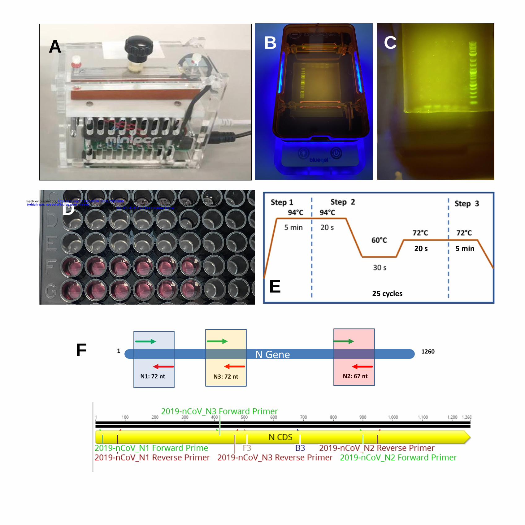
electrophoresis unit (Figure 1 A, B), powered by 120 V AC, and photo-documentation can 93
be done using a smartphone camera. 94
We also used a Synergy HT microplate reader (BioTek Instruments, VT, USA) to detect 95
the fluorescence induced by an intercalating reagent in positive samples from the PCR 96
reactions. 97
Controls for validation 98
We used a plasmid containing the complete N gene from 2019-nCoV, SARS, and MERS as 99
positive controls at a concentration of 200,000 copies/µL (Integrated DNA Technologies, 100
IA, USA). Samples containing different concentrations of synthetic nucleic acids of SARS-101
CoV2 were prepared by successive dilutions from stocks containing 200,000 copies mL-1
102
ng/L of viral nucleic acids. We used a plasmid containing the GP gene from Ebola Virus 103
(EBOV) as a negative control. The production of this EBOV genetic material has been 104
documented previously by our group [32]. 105
Amplification mix: We used REDTaq Ready Mix from Sigma-Aldrich (USA), and followed 106
the recommended protocol: 10 μL Readymix, 0.5 μM of forward primer, 0.5 μM of reverse 107
primer,1μL of DNA template (~ 625 to 2x10⁵ DNA copies), 1μl of EvaGreen® Dye, and 108
nuclease free water to final volume of reaction 20 μL. 109
. CC-BY-NC-ND 4.0 International licenseIt is made available under a is the author/funder, who has granted medRxiv a license to display the preprint in perpetuity. (which was not certified by peer review)
The copyright holder for this preprint this version posted April 7, 2020. .https://doi.org/10.1101/2020.04.03.20052860doi: medRxiv preprint
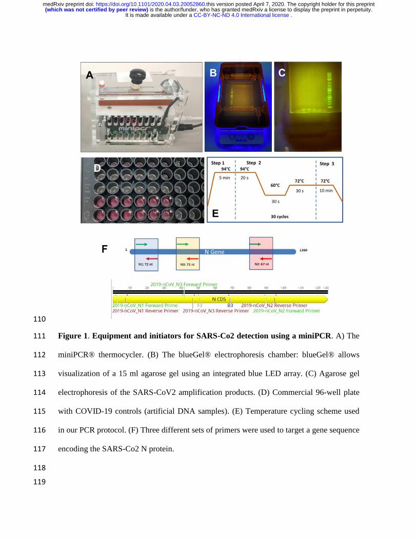
110
Figure 1. Equipment and initiators for SARS-Co2 detection using a miniPCR. A) The 111
miniPCR® thermocycler. (B) The blueGel® electrophoresis chamber: blueGel® allows 112
visualization of a 15 ml agarose gel using an integrated blue LED array. (C) Agarose gel 113
electrophoresis of the SARS-CoV2 amplification products. (D) Commercial 96-well plate 114
with COVID-19 controls (artificial DNA samples). (E) Temperature cycling scheme used 115
in our PCR protocol. (F) Three different sets of primers were used to target a gene sequence 116
encoding the SARS-Co2 N protein. 117
118
119
. CC-BY-NC-ND 4.0 International licenseIt is made available under a is the author/funder, who has granted medRxiv a license to display the preprint in perpetuity. (which was not certified by peer review)
The copyright holder for this preprint this version posted April 7, 2020. .https://doi.org/10.1101/2020.04.03.20052860doi: medRxiv preprint
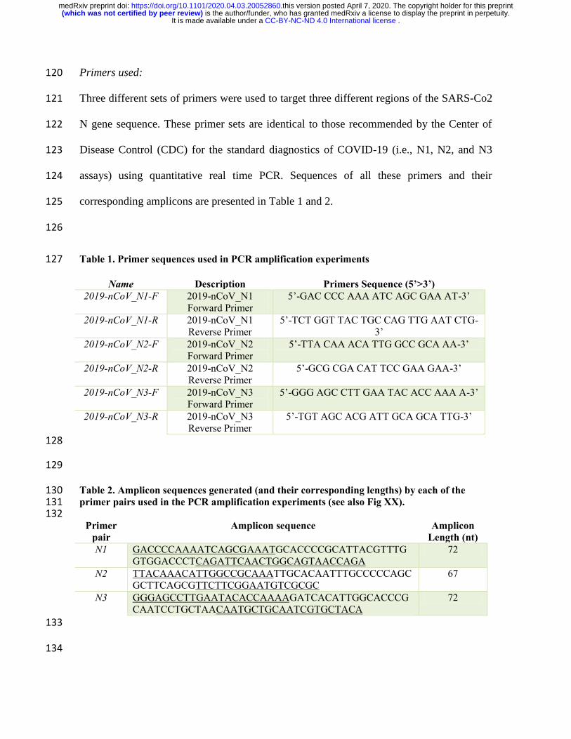
Primers used: 120
Three different sets of primers were used to target three different regions of the SARS-Co2 121
N gene sequence. These primer sets are identical to those recommended by the Center of 122
Disease Control (CDC) for the standard diagnostics of COVID-19 (i.e., N1, N2, and N3 123
assays) using quantitative real time PCR. Sequences of all these primers and their 124
corresponding amplicons are presented in Table 1 and 2. 125
126
Table 1. Primer sequences used in PCR amplification experiments 127
Name Description Primers Sequence (5’>3’)
2019-nCoV_N1-F 2019-nCoV_N1
Forward Primer
5’-GAC CCC AAA ATC AGC GAA AT-3’
2019-nCoV_N1-R 2019-nCoV_N1
Reverse Primer
5’-TCT GGT TAC TGC CAG TTG AAT CTG-
3’
2019-nCoV_N2-F 2019-nCoV_N2
Forward Primer
5’-TTA CAA ACA TTG GCC GCA AA-3’
2019-nCoV_N2-R 2019-nCoV_N2
Reverse Primer
5’-GCG CGA CAT TCC GAA GAA-3’
2019-nCoV_N3-F 2019-nCoV_N3
Forward Primer
5’-GGG AGC CTT GAA TAC ACC AAA A-3’
2019-nCoV_N3-R 2019-nCoV_N3
Reverse Primer
5’-TGT AGC ACG ATT GCA GCA TTG-3’
128
129
Table 2. Amplicon sequences generated (and their corresponding lengths) by each of the 130 primer pairs used in the PCR amplification experiments (see also Fig XX). 131 132
Primer
pair
Amplicon sequence Amplicon
Length (nt)
N1 GACCCCAAAATCAGCGAAATGCACCCCGCATTACGTTTG
GTGGACCCTCAGATTCAACTGGCAGTAACCAGA
72
N2 TTACAAACATTGGCCGCAAATTGCACAATTTGCCCCCAGC
GCTTCAGCGTTCTTCGGAATGTCGCGC
67
N3 GGGAGCCTTGAATACACCAAAAGATCACATTGGCACCCG
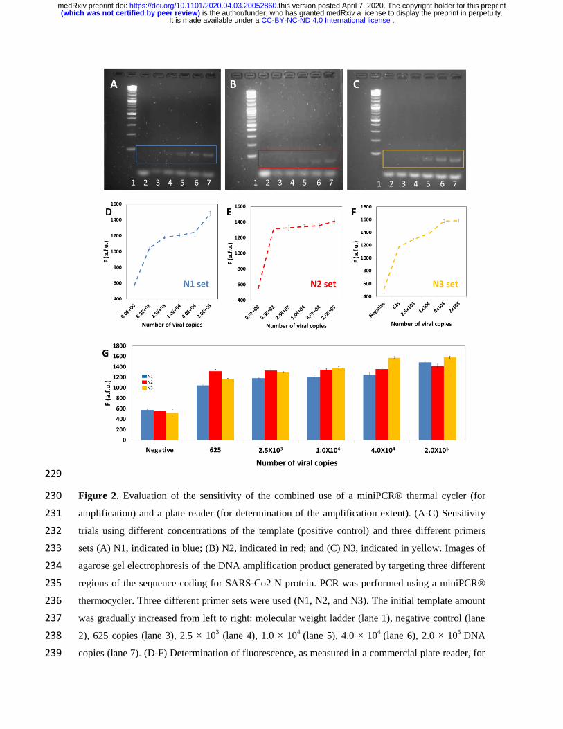
CAATCCTGCTAACAATGCTGCAATCGTGCTACA
72
133
134
. CC-BY-NC-ND 4.0 International licenseIt is made available under a is the author/funder, who has granted medRxiv a license to display the preprint in perpetuity. (which was not certified by peer review)
The copyright holder for this preprint this version posted April 7, 2020. .https://doi.org/10.1101/2020.04.03.20052860doi: medRxiv preprint
Amplification protocols: For all PCR experiments, we used the same three-stage protocol 135
(see Fig 1D) consisting of a denat ration sta e at for 5 min, followed y 25 cycles of 136
94 °C for 20s, 60 °C for 30s, and 72 °C for 20s, and then a final stage at 72 °C for 5 min, 137
for a total duration of 60 minutes in the miniPCR® thermocycler. 138
139
Documentation of PCR products: We analyzed 10 μL of each P R prod ct sin 2% 140
agarose electrophoresis in Tris-acetic acid-EDTA (TAE) buffer (Sigma-Aldrich, MO, 141
USA). Gels were dyed with GelGreen (Biotium, CA, USA) using a 1:10,000 dilution and a 142
current of 110 V supplied by a Bio-Rad PowerPac HC power supply (Bio-Rad, CA, USA) 143
for 40 min. We used the Quick-Load Purple 2-Log DNA ladder (NEB, MA, USA) as a 144
molecular weight marker. We analyzed the gels by UV transillumination using a Bio-Rad 145
ChemiDoc XRS imaging system. 146
In some of our experiments, we also used the blueGel unit, a portable electrophoresis unit 147
sold by MiniPCR from Amplyus (MA, USA). This is a compact electrophoresis unit (23 × 148
10 × 7 cm) that weighs 350 g. In these experiments, we analyzed 10 μL of PCR product 149
using 2% agarose electrophoresis tris-borate-EDTA buffer (TBE). Gels were dyed with 150
Gel- Green (CA, USA) using a 1:10,000 dilution, and a current of 48 V was supplied by the 151
blueGel built-in power supply (AC 100–240 V, 50–60 Hz). 152
As a third method of detection and to read the amplification product, we evaluated the 153
amplification products by detecting the fluorescence emitted by a DNA intercalating agent, 154
the EvaGreen® Dye, in the Synergy HT microplate reader (BioTek Instruments, VT, USA). 155
Briefly, 20 μL of the PCR reaction mix was placed in distinct wells of a 96-well plate, after 156
completion of the PCR program. Each well was made to a final volume of 150 μL by 157
adding 130 μL of n clease free water and the samples were well mixed by pipetting. These 158
. CC-BY-NC-ND 4.0 International licenseIt is made available under a is the author/funder, who has granted medRxiv a license to display the preprint in perpetuity. (which was not certified by peer review)
The copyright holder for this preprint this version posted April 7, 2020. .https://doi.org/10.1101/2020.04.03.20052860doi: medRxiv preprint
experiments were run in triplicate. The following conditions were used in the microplate 159
reader: excitation of 485/20, emission of 528/20, gain of 75. Fluorescence readings were 160
made from the above at room temperature. 161
162
Results and Discussion 163
An actual epidemic emergency does not provide the required timeframe for testing new 164
diagnostic strategies; therefore, the first line of response must be based on commercially 165
available and validated resources [32,33]. Here, we demonstrate that the combined use of a 166
commercial and portable PCR unit (the miniPCR) and a 96-well plate reader is potentially 167
adequate for the fast deployment of diagnostic efforts in the context of COVID-19 168
pandemics. We show the combined ability of both units to amplify and identify different 169
synthetic genetic sequences of SARS-CoV2 (see Materials and Methods). 170
171
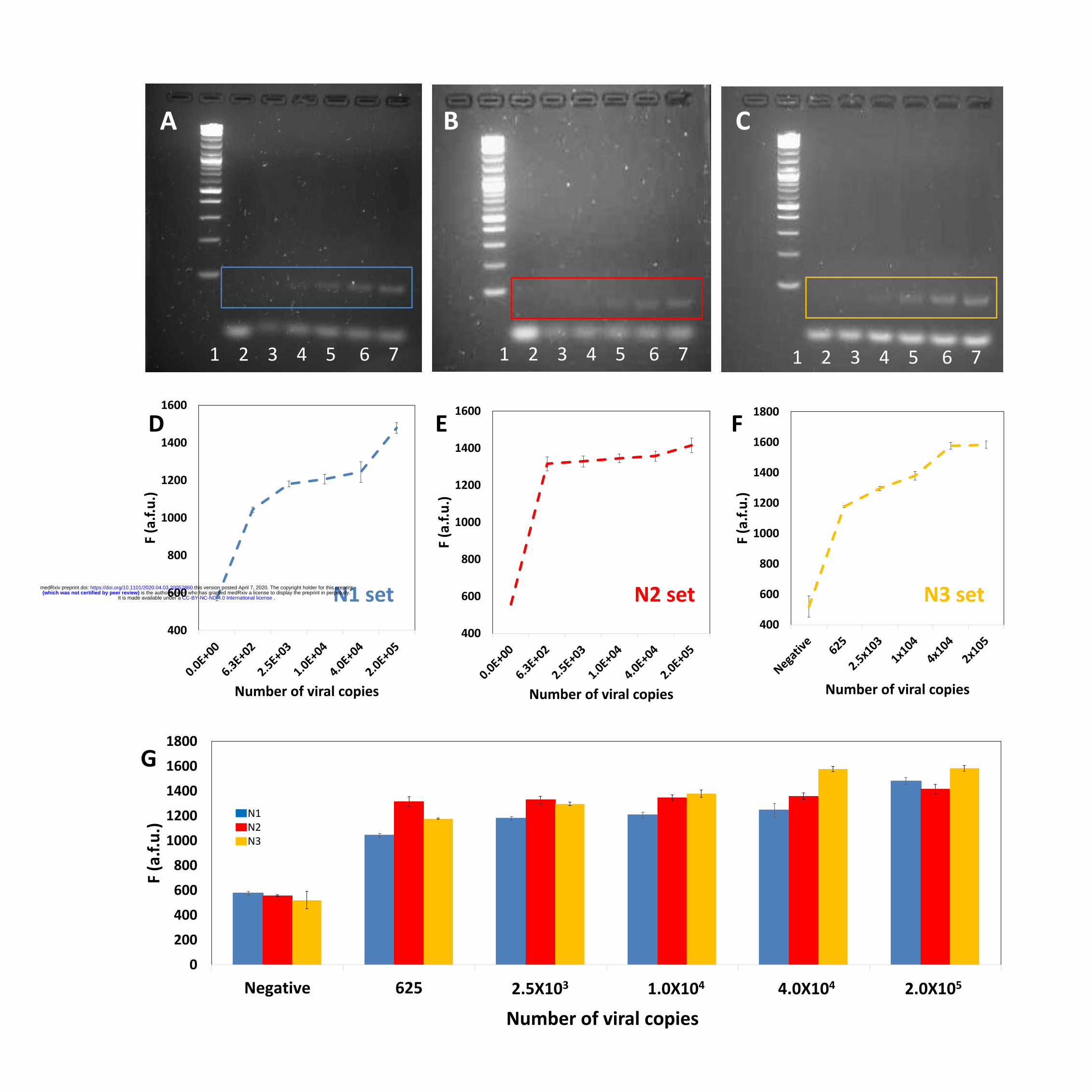
Analysis of sensitivity 172
We conducted a series of experiments to assess the sensitivity of the PCR reactions 173
conducted in the miniPCR thermocycler using the three sets of primers recommended by 174
CDC to diagnose infection by SARS-CoV2. Table 1 shows the sets of primers used to 175
target genetic sequences that code for the expression of the SARS-CoV2 N protein. Table 2 176
shows the sequence of the DNA products (amplicons) generated by successful targeting of 177
these regions with the N1, N2, and N3 primer pairs. 178
Figures 2A-C show the PCR products of the amplification reactions conducted using three 179
different primer pairs. In all cases, different concentrations of SARS-CoV2 genetic 180
material, in the range of 2.0 × 10⁵
to 625 DNA copies, were used as reaction templates. If 181
. CC-BY-NC-ND 4.0 International licenseIt is made available under a is the author/funder, who has granted medRxiv a license to display the preprint in perpetuity. (which was not certified by peer review)
The copyright holder for this preprint this version posted April 7, 2020. .https://doi.org/10.1101/2020.04.03.20052860doi: medRxiv preprint
we put this range in the proper clinical context, the actual viral load of COVID-19 in nasal 182
swabs from patients has been estimated to fall within the range of 105 to 10
6 viral copies 183
per mL [19]. The amplification proceeds with sufficient quality to allow proper 184
visualization of the amplification products in electrophoresis gels, even at low nucleic acid 185
concentrations. Figure 2A-C shows agarose gels containing the amplification products of 186
each one of three experiments, where the three different sets of primers (namely N1, N2, 187
and N3) were used to amplify the same range of concentrations of template. The 188
miniPCR® was able to generate a visible band of amplification products for all three 189
primer sets and across the whole range of synthetic viral loads. 190
Conventionally, the products of amplification in final point PCR are primarily detected on 191
agarose gels using conventional electrophoresis techniques conducted with conventional 192
lab equipment. However, as previously mentioned, the miniPCR® system is 193
commercialized with its own “blueGel®” electrophoretic unit (Figure 1B-C). The 194
blueGel® has several important advantages: it is fully portable, its size allows optimization 195
of reagent usage (agarose gel 15 mL, buffer 25 mL), the built-in power supply allows 196
visualization of band separation in real time (this can shorten the electrophoretic time by up 197
to 5 minutes), and exposure to ethidium bromide and UV light is completely avoided by the 198
use of GelGreen® dye and detection with blue light. Therefore, the blueGel system 199
represents a valid and portable solution for detecting PCR amplification products. 200
Nevertheless, running an experiment aimed at visualizing amplification products, as with 201
any standard gel electrophoresis procedure, requires time. A good separation of bands 202
typically involves a processing time of 35 to 60 minutes from the loading of the 203
amplification product to the final documentation through photography. 204
. CC-BY-NC-ND 4.0 International licenseIt is made available under a is the author/funder, who has granted medRxiv a license to display the preprint in perpetuity. (which was not certified by peer review)
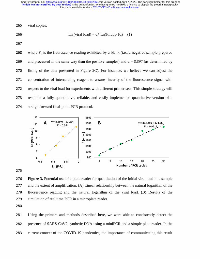
The copyright holder for this preprint this version posted April 7, 2020. .https://doi.org/10.1101/2020.04.03.20052860doi: medRxiv preprint
As an alternative, we show here that the amount of amplification product can be 205
quantitatively evaluated using a commercial 96-well plate reader. To do this, we used an 206
intercalating agent during amplification in the miniPCR apparatus. Figure 2a shows the 207
fluorescence readings associated with the analysis of the different dilutions of synthetic 208
SARS-CoV2 samples previously revealed by gel electrophoresis. We ran triplicate 209
reactions for each dilution and for each primer data set. The fluorescence readings were 210
capable of clearly discriminating between positive and negative samples across the whole 211
range of dilutions tested (from 2 × 10⁵ to 625 copies). This observation holds true for each 212
of the three primer sets tested. Note that the use of a plate reader, instead of a conventional 213
gel electrophoresis unit, presupposes a significant savings in time. Up to 96 PCR reactions 214
can be read in a matter of 5 to 10 minutes. This implies that an array of 12 miniPCR units 215
and a plate reader could equal the throughput of a traditional RT-QPCR platform, but at one 216
third of the capital cost. In addition, during emergencies and particularly in developing 217
countries, attaining or buying regular thermal cyclers and plate readers is much easier than 218
purchasing or accessing RT-qPCR systems. 219
In addition, our results suggest that fluorescence readings using a plate reader exhibit high 220
reproducibility and robustness. Overall, we obtained small standard deviations (in the range 221
of 6 to 40 arbitrary fluorescence units [a.f.u.]) and a small average variance coefficient 222
(2.6%) in fluorescence readings across the whole range of values of viral copies tested. We 223
observed similar variability indicators in experiments using different primer pairs. For 224
instance, we observed variance coefficients of 2.31%, 2.15%, and 3.34% when using 225
primer sets N1, N2, and N3, respectively. If we considered only fluorescence readings from 226
positive samples, we observed variance coefficients of 2.23%, 2.34%, and 1.31% when 227
using primer sets N1, N2, and N3, respectively. 228
. CC-BY-NC-ND 4.0 International licenseIt is made available under a is the author/funder, who has granted medRxiv a license to display the preprint in perpetuity. (which was not certified by peer review)
The copyright holder for this preprint this version posted April 7, 2020. .https://doi.org/10.1101/2020.04.03.20052860doi: medRxiv preprint
229
Figure 2. Evaluation of the sensitivity of the combined use of a miniPCR® thermal cycler (for 230
amplification) and a plate reader (for determination of the amplification extent). (A-C) Sensitivity 231
trials using different concentrations of the template (positive control) and three different primers 232
sets (A) N1, indicated in blue; (B) N2, indicated in red; and (C) N3, indicated in yellow. Images of 233
agarose gel electrophoresis of the DNA amplification product generated by targeting three different 234
regions of the sequence coding for SARS-Co2 N protein. PCR was performed using a miniPCR® 235
thermocycler. Three different primer sets were used (N1, N2, and N3). The initial template amount 236
was gradually increased from left to right: molecular weight ladder (lane 1), negative control (lane 237
2), 625 copies (lane 3), 2.5 × 10³ (lane 4), 1.0 × 10
4 (lane 5), 4.0 × 10
4 (lane 6), 2.0 × 10
5 DNA 238
copies (lane 7). (D-F) Determination of fluorescence, as measured in a commercial plate reader, for 239
. CC-BY-NC-ND 4.0 International licenseIt is made available under a is the author/funder, who has granted medRxiv a license to display the preprint in perpetuity. (which was not certified by peer review)
The copyright holder for this preprint this version posted April 7, 2020. .https://doi.org/10.1101/2020.04.03.20052860doi: medRxiv preprint

different dilutions of SARS-CoV2 synthetic DNA templates. Results using three different primer 240
sets are shown: (A) N1, indicated in blue; (B) N2, indicated in red; and (C) N3, indicated in yellow. 241
(G) Summary and comparison of fluorescence readings form synthetic samples of SARS-CoV2 in a 242
wide span of dilutions. Results using three different primer sets are shown: (A) N1, indicated in 243
blue; (B) N2, indicated in red; and (C) N3, indicated in yellow. 244
245
Figure 2G consolidates the fluorescence readings obtained from miniPCR amplifications 246
using synthetic SARS-CoV2 samples and the primer sets N1 (blue bars), N2 (red bars), and 247
N3 (yellow bars). Overall, this data set is consistent. These results suggest that any of the 248
primer sets tested (N1, N2, or N3) may be used to amplify SARS-CoV2 genetic material in 249
the miniPCR. However, for the experimental conditions tested (i.e., the nature and 250
concentration of the intercalating agent, the concentrations of primers, and the 251
concentration of enzyme, among others), we observe differences in the performance of each 252
primer pair. For example, primer sets N1 and N3 appear to promote amplifications in which 253
the observed fluorescence is proportional to the initial concentration of DNA template (i.e., 254
the viral load). By contrast, primer pair N2 appears to generate amplification product with 255
high fluorescence emissions even at low values of the initial final copy numbers. Note that 256
all fluorescence readings for positive samples shown in Figure 2E exhibit a fluorescence 257
reading between 1300 and 1400 a.f.u. 258
Furthermore, measuring the fluorescence with the plate reader may add a quantitative 259
element to the analysis of positive COVID-19 samples. In principle, samples with higher 260
viral loads will exhibit higher fluorescence if processed through the same PCR program 261
(i.e., exposed to the same number of cycles). For example, for amplifications using primer 262
set N3, we observe a linear relationship between the natural logarithm of the number of 263
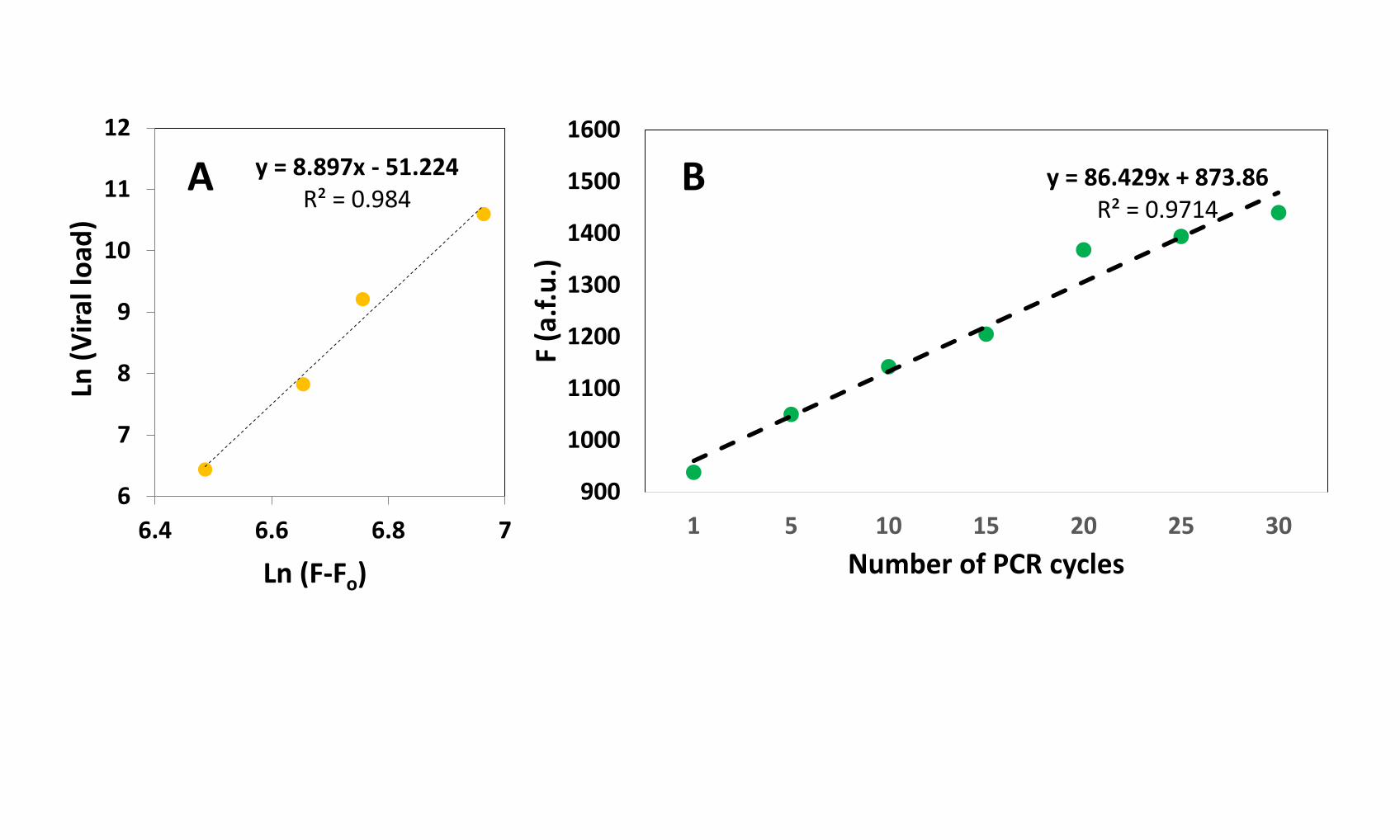
viral copies and the natural logarithm of fluorescence signal for the range of 625 to 40,000 264
. CC-BY-NC-ND 4.0 International licenseIt is made available under a is the author/funder, who has granted medRxiv a license to display the preprint in perpetuity. (which was not certified by peer review)
The copyright holder for this preprint this version posted April 7, 2020. .https://doi.org/10.1101/2020.04.03.20052860doi: medRxiv preprint
viral copies: 265
Ln (viral load) = α* Ln(Fsample- Fo) (1) 266
267
where Fo is the fluorescence reading exhibited by a blank (i.e., a negative sample prepared 268
and processed in the same way than the positive samples) and α = 8.897 (as determined by 269
fitting of the data presented in Figure 2C). For instance, we believe we can adjust the 270
concentration of intercalating reagent to assure linearity of the fluorescence signal with 271
respect to the viral load for experiments with different primer sets. This simple strategy will 272
result in a fully quantitative, reliable, and easily implemented quantitative version of a 273
straightforward final-point PCR protocol. 274
275
Figure 3. Potential use of a plate reader for quantitation of the initial viral load in a sample 276
and the extent of amplification. (A) Linear relationship between the natural logarithm of the 277
fluorescence reading and the natural logarithm of the viral load. (B) Results of the 278
simulation of real time PCR in a microplate reader. 279
280
Using the primers and methods described here, we were able to consistently detect the 281
presence of SARS-CoV2 synthetic DNA using a miniPCR and a simple plate reader. In the 282
current context of the COVID-19 pandemics, the importance of communicating this result 283
. CC-BY-NC-ND 4.0 International licenseIt is made available under a is the author/funder, who has granted medRxiv a license to display the preprint in perpetuity. (which was not certified by peer review)
The copyright holder for this preprint this version posted April 7, 2020. .https://doi.org/10.1101/2020.04.03.20052860doi: medRxiv preprint
does not reside in its novelty but in its practicality. In our experiments, we have used the 284
three sets of primers designed and recommended by the CDC to identify the presence of 285
SARS-CoV2, the causal agent of COVID-19. These primer pairs, aimed at identifying three 286
different regions encoding for the N protein of SARS-CoV2, have been widely validated 287
and used for diagnostic purposes in actual COVID-19 patients, Here we simply translated 288
widely tested protocols from the framework of an RT-qPCR apparatus (the gold standard 289
platform recommended for analyzing and confirming positive cases) to execution in a 290
miniaturized and already commercial POC thermal cycler. While the cost of a commercial 291
RT-qPCR apparatus falls in the range of $10,000 to $40,000 USD, the commercial value of 292
the miniPCR is under $800 USD. This difference is significant, especially when 293
considering the need for rational investment of resources during an epidemic crisis. 294
While the quantitative capabilities of testing in a RT-QPCR platform are undisputable, the 295
capacity of many countries for rapid, effective, and massive establishment of diagnostic 296
centers based on RT-qPCR is questionable. The current pandemic scenarios experienced in 297
the USA, Italy, France, and Spain, among others, have crudely demonstrated that 298
centralized labs are not an ideal solution during emergencies. Portable diagnostic systems 299
may provide the required flexibility and speed of response that RT-qPCR platforms cannot 300
deliver. 301
To further illustrate the deterministic and quantitative dependence between the 302
concentration of amplification product and the fluorescence signal, as measured in a plate 303
reader, we simulated some real-time amplification experiments. To that end, we conducted 304
amplification reactions using initial amounts of 4 × 10⁴ copies of synthetic SARS-CoV2 in 305
the miniPCR cycler. We added the intercalating agent, EvaGreen® Dye, to the reaction mix 306
at the initial time and extracted samples after 1, 5, 10, 15, 20, 25, and 30 PCR cycles. The 307
. CC-BY-NC-ND 4.0 International licenseIt is made available under a is the author/funder, who has granted medRxiv a license to display the preprint in perpetuity. (which was not certified by peer review)
The copyright holder for this preprint this version posted April 7, 2020. .https://doi.org/10.1101/2020.04.03.20052860doi: medRxiv preprint

fluorescence from these samples was then measured in a plate reader. We observed a linear 308
increase in fluorescence as more PCR cycles were performed, which highlights the 309
quantitative nature of the intercalating reaction. 310
Our results suggest that using a commercial plate reader to determine the extent of advance 311
of PCR amplifications is a practical, reliable, reproducible, and robust alternative to the use 312
of gel electrophoresis. Moreover, fluorescence reading of PCR products may lead to precise 313
quantification of viral loads. 314
315
Conclusions 316
The challenge of POC detection of viral threats is of paramount importance, particularly in 317
underdeveloped regions and in emergency situations (i.e., natural disasters or epidemic 318
outbreaks). In the context of an emergency, time is very limited (as are other resources) to 319
conduct research or develop new technologies; therefore, the use of commercially available 320
and tested technologies is an obvious first countermeasure. Our research extends the 321
validation of the miniPCR technology to the as yet unexplored topic of detection of 322
COVID-19. 323
We used the set of primers developed by the CDC and recommended by the WHO for 324
conducting the standard PCR diagnostics of COVID-19. These primers target three 325
different regions of the viral nucleic acids encoding for the N protein. In our experiments, 326
we corroborate that the miniPCR apparatus is capable of amplifying small amounts of 327
SARS-CoV2 synthetic nucleic acids. We were able to detect and amplify 64 copies of 328
genes encoding for the N protein of SARS-CoV2. 329
. CC-BY-NC-ND 4.0 International licenseIt is made available under a is the author/funder, who has granted medRxiv a license to display the preprint in perpetuity. (which was not certified by peer review)
The copyright holder for this preprint this version posted April 7, 2020. .https://doi.org/10.1101/2020.04.03.20052860doi: medRxiv preprint
The use of the miniPCR® is intuitive and simple; the user can easily follow the advance of 330
the iterative temperature cycling using a laptop. Despite its compact size, the miniPCR® 331
allows the performance of a full amplification protocol in a similar time as achieved with a 332
conventional thermocycler. The mini-PCR® thermocycler exhibits the essential attributes 333
of a POC system: (a) the use of small volumes, (b) low capital cost, (c) portability, (d) and 334
a fast, accurate, and selective response. 335
Moreover, the combined use of the miniPCR thermocycler and a 96-well plate reader 336
enables the possibility of obtaining immediate readings of the amplification products, 337
thereby providing faster (and potentially quantitative) diagnostic results in shorter times 338
than when gel electrophoresis techniques are used. Therefore, the combined use of these 339
already two commercially available devices—a miniPCR thermocycler and a 96-well plate 340
reader—has great potential for use during epidemic emergencies. 341
342
Acknowledgments 343
EGG acknowledges funding from a doctoral scholarship provided by CONACyT (Consejo 344
Nacional de Ciencia y Tecnología, México). GTdS and MMA acknowledge the 345
institutional funding received from Tecnológico de Monterrey (Grant 002EICIS01), and 346
funding provided by CONACyT (Consejo Nacional de Ciencia y Tecnología, México) 347
through grants (SNI 26048, SNI 256730, and Scholarships 635891, 856068, and 814593). 348
349
Supporting Information 350
Excel data file S1: Excel file that contains raw data 351
352
. CC-BY-NC-ND 4.0 International licenseIt is made available under a is the author/funder, who has granted medRxiv a license to display the preprint in perpetuity. (which was not certified by peer review)
The copyright holder for this preprint this version posted April 7, 2020. .https://doi.org/10.1101/2020.04.03.20052860doi: medRxiv preprint
References 353
1. Yager P, Domingo GJ, Gerdes J. Point-of-Care Diagnostics for Global Health. Annu 354
Rev Biomed Eng. Annual Reviews ; 2008;10: 107–144. 355
doi:10.1146/annurev.bioeng.10.061807.160524 356
2. Alvarez MM, Aizenberg J, Analoui M, Andrews AM, Bisker G, Boyden ES, et al. 357
Emerging Trends in Micro- and Nanoscale Technologies in Medicine: From Basic 358
Discoveries to Translation. ACS Nano. American Chemical Society; 2017;11: 5195–359
5214. doi:10.1021/acsnano.7b01493 360
3. Dawood FS, Jain S, Finelli L, Shaw MW, Lindstrom S, Garten RJ, et al. Emergence 361
of a Novel Swine-Origin Influenza A (H1N1) Virus in Humans. N Engl J Med. 362
Massachussetts Medical Society; 2009;360: 2605–2615. 363
doi:10.1056/NEJMoa0903810 364
4. Kaushik A, Tiwari S, Dev Jayant R, Marty A, Nair M. Towards detection and 365
diagnosis of Ebola virus disease at point-of-care. Biosensors and Bioelectronics. 366
Elsevier Ltd; 2016. pp. 254–272. doi:10.1016/j.bios.2015.08.040 367
5. Petersen LR, Jamieson DJ, Powers AM, Honein MA. Zika Virus. Baden LR, editor. 368
N Engl J Med. Massachussetts Medical Society; 2016;374: 1552–1563. 369
doi:10.1056/NEJMra1602113 370
6. Fauci AS, Morens DM. Zika Virus in the Americas — Yet Another Arbovirus 371
Threat. N Engl J Med. Massachussetts Medical Society; 2016;374: 601–604. 372
doi:10.1056/NEJMp1600297 373
7. Mauk MG, Song J, Bau HH, Liu C. Point-of-Care Molecular Test for Zika Infection. 374
Clin Lab Int. NIH Public Access; 2017;41: 25–27. Available: 375
. CC-BY-NC-ND 4.0 International licenseIt is made available under a is the author/funder, who has granted medRxiv a license to display the preprint in perpetuity. (which was not certified by peer review)
The copyright holder for this preprint this version posted April 7, 2020. .https://doi.org/10.1101/2020.04.03.20052860doi: medRxiv preprint
http://www.ncbi.nlm.nih.gov/pubmed/28819345 376
8. Kaushik A, Tiwari S, Dev Jayant R, Marty A, Nair M. Towards detection and 377
diagnosis of Ebola virus disease at point-of-care. Biosens Bioelectron. NIH Public 378
Access; 2016;75: 254–72. doi:10.1016/j.bios.2015.08.040 379
9. Jansen van Vuren P, Grobbelaar A, Storm N, Conteh O, Konneh K, Kamara A, et al. 380
Comparative Evaluation of the Diagnostic Performance of the Prototype Cepheid 381
GeneXpert Ebola Assay. J Clin Microbiol. American Society for Microbiology; 382
2016;54: 359–67. doi:10.1128/JCM.02724-15 383
10. Duchesne L, Lacombe K. Innovative technologies for point-of-care testing of viral 384
hepatitis in low-resource and decentralized settings. J Viral Hepat. John Wiley & 385
Sons, Ltd (10.1111); 2018;25: 108–117. doi:10.1111/jvh.12827 386
11. Cohen J, Kupferschmidt K. Countries test tactics in “war” against COVID-19. 387
Science. American Association for the Advancement of Science; 2020;367: 1287–388
1288. doi:10.1126/science.367.6484.1287 389
12. Pung R, Chiew CJ, Young BE, Chin S, I-C Chen M, Clapham HE, et al. Articles 390
Investigation of three clusters of COVID-19 in Singapore: implications for 391
surveillance and response measures. Lancet. Elsevier; 2020;19: 1–8. 392
doi:10.1016/S0140-6736(20)30528-6 393
13. Wang CJ, Ng CY, Brook RH. Response to COVID-19 in Taiwan: Big Data 394
Analytics, New Technology, and Proactive Testing. JAMA - Journal of the 395
American Medical Association. American Medical Association; 2020. 396
doi:10.1001/jama.2020.3151 397
14. Salathé M, Althaus CL, Neher R, Stringhini S, Hodcroft E, Fellay J, et al. COVID-398
19 epidemic in Switzerland: on the importance of testing, contact tracing and 399
. CC-BY-NC-ND 4.0 International licenseIt is made available under a is the author/funder, who has granted medRxiv a license to display the preprint in perpetuity. (which was not certified by peer review)
The copyright holder for this preprint this version posted April 7, 2020. .https://doi.org/10.1101/2020.04.03.20052860doi: medRxiv preprint

isolation. Swiss Med Wkly. EMH Media; 2020;150: w20225. 400
doi:10.4414/smw.2020.20225 401
15. Alvarez MM, López-Pacheco F, Aguilar-Yañez JM, Portillo-Lara R, Mendoza-402
Ochoa GI, García-Echauri S, et al. Specific Recognition of Influenza A/H1N1/2009 403
Antibodies in Human Serum: A Simple Virus-Free ELISA Method. Jeyaseelan S, 404
editor. PLoS One. Public Library of Science; 2010;5: e10176. 405
doi:10.1371/journal.pone.0010176 406
16. Yen C-W, de Puig H, Tam JO, Gómez-Márquez J, Bosch I, Hamad-Schifferli K, et 407
al. Multicolored silver nanoparticles for multiplexed disease diagnostics: 408
distinguishing dengue, yellow fever, and Ebola viruses. Lab Chip. The Royal Society 409
of Chemistry; 2015;15: 1638–1641. doi:10.1039/C5LC00055F 410
17. Nguyen Van JC, Caméléna F, Dahoun M, Pilmis B, Mizrahi A, Lourtet J, et al. 411
Prospective evaluation of the Alere i Influenza A&B nucleic acid amplification 412
versus Xpert Flu/RSV. Diagn Microbiol Infect Dis. Elsevier Inc.; 2016;85: 19–22. 413
doi:10.1016/j.diagmicrobio.2015.11.012 414
18. Murphy J, Bustin SA. Reliability of real-time reverse-transcription PCR in clinical 415
diagnostics: gold standard or substandard? Expert Rev Mol Diagn. Taylor & Francis; 416
2009;9: 187–197. doi:10.1586/14737159.9.2.187 417
19. Wang W, Xu Y, Gao R, Lu R, Han K, Wu G, et al. Detection of SARS-CoV-2 in 418
Different Types of Clinical Specimens. JAMA - Journal of the American Medical 419
Association. American Medical Association; 2020. doi:10.1001/jama.2020.3786 420
20. Kozel TR, Burnham-Marusich AR. Point-of-Care Testing for Infectious Diseases: 421
Past, Present, and Future. J Clin Microbiol. American Society for Microbiology; 422
2017;55: 2313–2320. doi:10.1128/JCM.00476-17 423
. CC-BY-NC-ND 4.0 International licenseIt is made available under a is the author/funder, who has granted medRxiv a license to display the preprint in perpetuity. (which was not certified by peer review)
The copyright holder for this preprint this version posted April 7, 2020. .https://doi.org/10.1101/2020.04.03.20052860doi: medRxiv preprint
21. Su W, Gao X, Jiang L, Qin J. Microfluidic platform towards point-of-care 424
diagnostics in infectious diseases. J Chromatogr A. Elsevier; 2015;1377: 13–26. 425
doi:10.1016/J.CHROMA.2014.12.041 426
22. Drancourt M, Michel-Lepage A, Boyer S, Raoult D. The Point-of-Care Laboratory 427
in Clinical Microbiology. Clin Microbiol Rev. American Society for Microbiology; 428
2016;29: 429–47. doi:10.1128/CMR.00090-15 429
23. Qiu X, Ge S, Gao P, Li K, Yang S, Zhang S, et al. A smartphone-based point-of-care 430
diagnosis of H1N1 with microfluidic convection PCR. Microsyst Technol. Springer 431
Berlin Heidelberg; 2017;23: 2951–2956. doi:10.1007/s00542-016-2979-z 432
24. Petralia S, Conoci S. PCR technologies for point of care testing: Progress and 433
perspectives. ACS Sensors. American Chemical Society; 2017. pp. 876–891. 434
doi:10.1021/acssensors.7b00299 435
25. Marx V. PCR heads into the field. Nat Methods. 2015;12: 393–397. 436
doi:10.1038/nmeth.3369 437
26. Kwon H-S, Park H-C, Lee K, An S, Oh Y-L, Ahn E-R, et al. Performance of 438
MiniPCR TM
mini8, a portable thermal cycler. Anal Sci Technol. The Korean Society 439
of Analytical Science; 2016;29: 79–84. doi:10.5806/AST.2016.29.2.79 440
27. Guevara EE, Frankel DC, Ranaivonasy J, Richard AF, Ratsirarson J, Lawler RR, et 441
al. A simple, economical protocol for DNA extraction and amplification where there 442
is no lab. Conserv Genet Resour. Springer Netherlands; 2018;10: 119–125. 443
doi:10.1007/s12686-017-0758-5 444
28. Pomerantz A, Peñafiel N, Arteaga A, Bustamante L, Pichardo F, Coloma LA, et al. 445
Real-time DNA barcoding in a rainforest using nanopore sequencing: opportunities 446
for rapid biodiversity assessments and local capacity building. Gigascience. Oxford 447
. CC-BY-NC-ND 4.0 International licenseIt is made available under a is the author/funder, who has granted medRxiv a license to display the preprint in perpetuity. (which was not certified by peer review)
The copyright holder for this preprint this version posted April 7, 2020. .https://doi.org/10.1101/2020.04.03.20052860doi: medRxiv preprint
University Press; 2018;7. doi:10.1093/gigascience/giy033 448
29. Boguraev A-S, Christensen HC, Bonneau AR, Pezza JA, Nichols NM, Giraldez AJ, 449
et al. Successful amplification of DNA aboard the International Space Station. npj 450
Microgravity. 2017;3. doi:10.1038/s41526-017-0033-9 451
30. Montague TG, Almansoori A, Gleason EJ, Copeland DS, Foley K, Kraves S, et al. 452
Gene expression studies using a miniaturized thermal cycler system on board the 453
International Space Station. Reddy S V., editor. PLoS One. Public Library of 454
Science; 2018;13: e0205852. doi:10.1371/journal.pone.0205852 455
31. Zaky WI, Tomaino FR, Pilotte N, Laney SJ, Williams SA. Backpack PCR: A point-456
of-collection diagnostic platform for the rapid detection of Brugia parasites in 457
mosquitoes. McCarthy JS, editor. PLoS Negl Trop Dis. Public Library of Science; 458
2018;12: e0006962. doi:10.1371/journal.pntd.0006962 459
32. Gonzá Lez-Gonzá Lez E, Mendoza-Ramos JL, Pedrozaid SC, Cuellar-Monterrubio 460
A, Má Rquez-Ipiña AR, Lira-Serhanid D, et al. Validation of use of the miniPCR 461
thermocycler for Ebola and Zika virus detection. 2019; 462
doi:10.1371/journal.pone.0215642 463
33. Chin CD, Linder V, Sia SK. Commercialization of microfluidic point-of-care 464
diagnostic devices{. doi:10.1039/c2lc21204h 465
466
. CC-BY-NC-ND 4.0 International licenseIt is made available under a is the author/funder, who has granted medRxiv a license to display the preprint in perpetuity. (which was not certified by peer review)
The copyright holder for this preprint this version posted April 7, 2020. .https://doi.org/10.1101/2020.04.03.20052860doi: medRxiv preprint
N Gene 1 1260
N1: 72 nt N3: 72 nt N2: 67 nt
A B C
D
F
25 cycles
20 s 5 min
E
. CC-BY-NC-ND 4.0 International licenseIt is made available under a is the author/funder, who has granted medRxiv a license to display the preprint in perpetuity. (which was not certified by peer review)
The copyright holder for this preprint this version posted April 7, 2020. .https://doi.org/10.1101/2020.04.03.20052860doi: medRxiv preprint
1 2 3 4 5 6 7
A B C
1 2 3 4 5 6 7 1 2 3 4 5 6 7
400
600
800
1000
1200
1400
1600
F (a
.f.u
.)
Number of viral copies
N1 set
400
600
800
1000
1200
1400
1600
F (a
.f.u
.)
Number of viral copies
D E
400
600
800
1000
1200
1400
1600
1800
F (a
.f.u
.)
Number of viral copies
F
N2 set N3 set
0
200
400
600
800
1000
1200
1400
1600
1800
Negative 625 2.5x10³ 1x10⁴ 4x10⁴ 2x10⁵
F (a
.f.u
.)
Number of viral copies
N1 N2 N3
G
2.5X10³ 1.0X10⁴ 4.0X10⁴ 2.0X10⁵
. CC-BY-NC-ND 4.0 International licenseIt is made available under a is the author/funder, who has granted medRxiv a license to display the preprint in perpetuity. (which was not certified by peer review)
The copyright holder for this preprint this version posted April 7, 2020. .https://doi.org/10.1101/2020.04.03.20052860doi: medRxiv preprint
y = 86.429x + 873.86 R² = 0.9714
900
1000
1100
1200
1300
1400
1500
1600
1 5 10 15 20 25 30F
(a.f
.u.)
Number of PCR cycles
A B y = 8.897x - 51.224
R² = 0.984
6
7
8
9
10
11
12
6.4 6.6 6.8 7
Ln (F-Fo)
Ln (
Vir
al lo
ad)
. CC-BY-NC-ND 4.0 International licenseIt is made available under a is the author/funder, who has granted medRxiv a license to display the preprint in perpetuity. (which was not certified by peer review)
The copyright holder for this preprint this version posted April 7, 2020. .https://doi.org/10.1101/2020.04.03.20052860doi: medRxiv preprint


![sf]/f ]gfefO/; /f ]u (COVID-19)](https://static.fdocuments.nl/doc/165x107/6288ff7fe55d8e051e130154/sff-gfefo-f-u-covid-19.jpg)