Glycans on influenza hemagglutinin affect receptor binding and
6
Glycans on influenza hemagglutinin affect receptor binding and immune response Cheng-Chi Wang a,b,1 , Juine-Ruey Chen a,c,1 , Yung-Chieh Tseng a , Che-Hsiung Hsu a,d,e , Yu-Fu Hung a , Shih-Wei Chen a , Chin-Mei Chen f , Kay-Hooi Khoo f , Ting-Jen Cheng a , Yih-Shyun E. Cheng a , Jia-Tsrong Jan a , Chung-Yi Wu a , Che Ma a,2 , and Chi-Huey Wong a,2 a Genomics Research Center, d Chemical Biology and Molecular Biophysics, Taiwan International Graduate Program, and f Institute of Biological Chemistry, Academia Sinica, Taipei 115, Taiwan; b Institute of Biochemical Sciences, National Taiwan University, Taipei 106, Taiwan; c Institute of Biochemistry and Molecular Biology, National Yang-Ming University, Taipei 112, Taiwan; and e Institute of Bioinformatics and Structural Biology, National Tsing Hua University, Hsinchu 300, Taiwan Contributed by Chi-Huey Wong, September 1, 2009 (sent for review August 3, 2009) Recent cases of avian influenza H5N1 and the swine-origin 2009 H1N1 have caused a great concern that a global disaster like the 1918 influenza pandemic may occur again. Viral transmission begins with a critical interaction between hemagglutinin (HA) glycoprotein, which is on the viral coat of influenza, and sialic acid (SA) containing glycans, which are on the host cell surface. To elucidate the role of HA glycosylation in this important interaction, various defined HA gly- coforms were prepared, and their binding affinity and specificity were studied by using a synthetic SA microarray. Truncation of the N-glycan structures on HA increased SA binding affinities while decreasing specificity toward disparate SA ligands. The contribution of each monosaccharide and sulfate group within SA ligand structures to HA binding energy was quantitatively dissected. It was found that the sulfate group adds nearly 100-fold (2.04 kcal/mol) in binding energy to fully glycosylated HA, and so does the biantennary glycan to the monoglycosylated HA glycoform. Antibodies raised against HA protein bearing only a single N-linked GlcNAc at each glycosylation site showed better binding affinity and neutralization activity against influenza subtypes than the fully glycosylated HAs elicited. Thus, removal of structurally nonessential glycans on viral surface glycop- roteins may be a very effective and general approach for vaccine design against influenza and other human viruses. flu vaccine glycan binding glycosylation T he highly pathogenic H5N1 and the 2009 swine-origin inf luenza A (H1N1) viruses have caused global outbreaks and raised a great concern that further changes in the viruses may occur to bring about a deadly pandemic (1, 2). Important contributions to our understanding of influenza infections have come from the studies on hemagglutinin (HA), a viral coat glycoprotein that binds to specific sialylated glycan receptors in the respiratory tract, allowing the virus to enter the cell (3–6). To cross the species barrier and infect the human population, avian HA must change its receptor- binding preference from a terminally sialylated glycan that contains 2,3 (avian)-linked to 2,6 (human)-linked sialic acid motifs (7), and this switch could occur through only two mutations, as in the 1918 pandemic (8). Understanding the factors that affect inf luenza binding to glycan receptors is thus critical for developing methods to control any future crossover inf luenza strains that have pandemic potential. HA is a homotrimeric transmembrane protein with an ectodo- main composed of a globular head and a stem region (3). Both regions carry N-linked oligosaccharides (9), which affect the func- tional properties of HA (10, 11). Among different subtypes of inf luenza A viruses, there is extensive variation in the glycosylation sites of the head region, whereas the stem oligosaccharides are more conserved and required for fusion activity (11). Glycans near antigenic peptide epitopes interfere with antibody recognition (12), and glycans near the proteolytic activation site of HA modulate cleavage and influence the infectivity of influenza virus (13). Mutational deletion of HA glycosylation sites can affect viral receptor binding (14). Our analysis of HA sequences revealed that the peptide sequences around the glycosylation sites are highly conserved (Fig. S1), which suggests a central functional significance for HA glycosylation; however, little is known regarding how the structure and composition of its glycans affect HA activity, includ- ing structure, receptor binding, and immune response. Results Creating Defined HA Glycoforms for Quantitative Glycan Microarray Profiling. The glycan microarray is a powerful tool for investigat- ing carbohydrate–protein interactions (15–18) and provides a new platform for influenza virus subtyping (16–18). Although powerful, understanding HA–glycan interactions by array anal- ysis has been complicated by two issues. First, HA binding specificity is affected by the spatial arrangement and composi- tion of the arrayed glycans and the binding detection method used (19). Second, the changes in the peptide sequence at or near glycosylation sites may alter HA’s 3D structure, and thus recep- tor-binding specificity and affinity. Indeed, HAs from different H5N1 subtypes have different glycan-binding patterns (18). Mutagenesis of glycosylation sites on H1 and H3 has been studied in the whole-viral system (16, 20). However, it is not known how changes in glycosylation affect receptor-binding specificity and affinity, especially with regard to the most pathogenic H5N1 HA. To address this question, we have devel- oped a glycan microarray comprising extensive structural ana- logs of the HA-binding ligand, and several defined glycoforms of HA were prepared by using the H5 consensus sequence (21) for quantitative binding analysis. Although previous studies have used HA from insect cell expression (16), glycosylation in insect cells differs from mammalian cells, with a marked difference being that complex type N-glycans terminating in galactose and sialic acid are not produced in insect cells. To generate native fully glycosylated HA variants (HA fg ), human embryonic kidney (HEK293) cells were used. To generate high-mannose-type glycosylation (HA hm ), HEK293S cells, which are deficient in N-acetylglucosaminyltransferase I (GnTI ), were used. To fur- ther address the effect of HA glycan structure on HA receptor- binding affinity and specificity, sugar residues were enzymati- cally removed from the expressed HAs. Sialic acid residues were removed from HA fg by neuraminidase (NA) treatment to pro- Author contributions: C.-Y.W., C.M., and C.-H.W. designed research; C.-C.W., J.-R.C., Y.-C.T., C.-H.H., Y.-F.H., S.-W.C., C.-M.C., K.-H.K., and J.-T.J. performed research; T.-J.C. and Y.-S.E.C. contributed new reagents/analytic tools; C.-C.W. and J.-R.C. analyzed data; and C.-C.W., J.-R.C., C.-Y.W., C.M., and C.-H.W. wrote the paper. The authors declare no conflict of interest. 1 C.-C.W. and J.-R.C. contributed equally to this work. 2 To whom correspondence may be addressed. E-mail: [email protected] or [email protected]. This article contains supporting information online at www.pnas.org/cgi/content/full/ 0909696106/DCSupplemental. www.pnas.orgcgidoi10.1073pnas.0909696106 PNAS October 27, 2009 vol. 106 no. 43 18137–18142 IMMUNOLOGY CHEMISTRY
Transcript of Glycans on influenza hemagglutinin affect receptor binding and
Glycans on influenza hemagglutinin affect receptorbinding and immune responseCheng-Chi Wanga,b,1, Juine-Ruey Chena,c,1, Yung-Chieh Tsenga, Che-Hsiung Hsua,d,e, Yu-Fu Hunga, Shih-Wei Chena,Chin-Mei Chenf, Kay-Hooi Khoof, Ting-Jen Chenga, Yih-Shyun E. Chenga, Jia-Tsrong Jana, Chung-Yi Wua, Che Maa,2,and Chi-Huey Wonga,2
aGenomics Research Center, dChemical Biology and Molecular Biophysics, Taiwan International Graduate Program, and fInstitute of Biological Chemistry,Academia Sinica, Taipei 115, Taiwan; bInstitute of Biochemical Sciences, National Taiwan University, Taipei 106, Taiwan; cInstitute of Biochemistry andMolecular Biology, National Yang-Ming University, Taipei 112, Taiwan; and eInstitute of Bioinformatics and Structural Biology, National Tsing HuaUniversity, Hsinchu 300, Taiwan
Contributed by Chi-Huey Wong, September 1, 2009 (sent for review August 3, 2009)
Recent cases of avian influenza H5N1 and the swine-origin 2009 H1N1have caused a great concern that a global disaster like the 1918influenza pandemic may occur again. Viral transmission begins witha critical interaction between hemagglutinin (HA) glycoprotein, whichis on the viral coat of influenza, and sialic acid (SA) containing glycans,which are on the host cell surface. To elucidate the role of HAglycosylation in this important interaction, various defined HA gly-coforms were prepared, and their binding affinity and specificitywere studied by using a synthetic SA microarray. Truncation of theN-glycan structures on HA increased SA binding affinities whiledecreasing specificity toward disparate SA ligands. The contributionof each monosaccharide and sulfate group within SA ligand structuresto HA binding energy was quantitatively dissected. It was found thatthe sulfate group adds nearly 100-fold (2.04 kcal/mol) in bindingenergy to fully glycosylated HA, and so does the biantennary glycanto the monoglycosylated HA glycoform. Antibodies raised against HAprotein bearing only a single N-linked GlcNAc at each glycosylationsite showed better binding affinity and neutralization activity againstinfluenza subtypes than the fully glycosylated HAs elicited. Thus,removal of structurally nonessential glycans on viral surface glycop-roteins may be a very effective and general approach for vaccinedesign against influenza and other human viruses.
flu vaccine � glycan binding � glycosylation
The highly pathogenic H5N1 and the 2009 swine-origin influenzaA (H1N1) viruses have caused global outbreaks and raised a
great concern that further changes in the viruses may occur to bringabout a deadly pandemic (1, 2). Important contributions to ourunderstanding of influenza infections have come from the studieson hemagglutinin (HA), a viral coat glycoprotein that binds tospecific sialylated glycan receptors in the respiratory tract, allowingthe virus to enter the cell (3–6). To cross the species barrier andinfect the human population, avian HA must change its receptor-binding preference from a terminally sialylated glycan that contains�2,3 (avian)-linked to �2,6 (human)-linked sialic acid motifs (7),and this switch could occur through only two mutations, as in the1918 pandemic (8). Understanding the factors that affect influenzabinding to glycan receptors is thus critical for developing methodsto control any future crossover influenza strains that have pandemicpotential.
HA is a homotrimeric transmembrane protein with an ectodo-main composed of a globular head and a stem region (3). Bothregions carry N-linked oligosaccharides (9), which affect the func-tional properties of HA (10, 11). Among different subtypes ofinfluenza A viruses, there is extensive variation in the glycosylationsites of the head region, whereas the stem oligosaccharides are moreconserved and required for fusion activity (11). Glycans nearantigenic peptide epitopes interfere with antibody recognition (12),and glycans near the proteolytic activation site of HA modulatecleavage and influence the infectivity of influenza virus (13).Mutational deletion of HA glycosylation sites can affect viral
receptor binding (14). Our analysis of HA sequences revealed thatthe peptide sequences around the glycosylation sites are highlyconserved (Fig. S1), which suggests a central functional significancefor HA glycosylation; however, little is known regarding how thestructure and composition of its glycans affect HA activity, includ-ing structure, receptor binding, and immune response.
ResultsCreating Defined HA Glycoforms for Quantitative Glycan MicroarrayProfiling. The glycan microarray is a powerful tool for investigat-ing carbohydrate–protein interactions (15–18) and provides anew platform for influenza virus subtyping (16–18). Althoughpowerful, understanding HA–glycan interactions by array anal-ysis has been complicated by two issues. First, HA bindingspecificity is affected by the spatial arrangement and composi-tion of the arrayed glycans and the binding detection methodused (19). Second, the changes in the peptide sequence at or nearglycosylation sites may alter HA’s 3D structure, and thus recep-tor-binding specificity and affinity. Indeed, HAs from differentH5N1 subtypes have different glycan-binding patterns (18).Mutagenesis of glycosylation sites on H1 and H3 has beenstudied in the whole-viral system (16, 20). However, it is notknown how changes in glycosylation affect receptor-bindingspecificity and affinity, especially with regard to the mostpathogenic H5N1 HA. To address this question, we have devel-oped a glycan microarray comprising extensive structural ana-logs of the HA-binding ligand, and several defined glycoforms ofHA were prepared by using the H5 consensus sequence (21) forquantitative binding analysis. Although previous studies haveused HA from insect cell expression (16), glycosylation in insectcells differs from mammalian cells, with a marked differencebeing that complex type N-glycans terminating in galactose andsialic acid are not produced in insect cells. To generate nativefully glycosylated HA variants (HAfg), human embryonic kidney(HEK293) cells were used. To generate high-mannose-typeglycosylation (HAhm), HEK293S cells, which are deficient inN-acetylglucosaminyltransferase I (GnTI�), were used. To fur-ther address the effect of HA glycan structure on HA receptor-binding affinity and specificity, sugar residues were enzymati-cally removed from the expressed HAs. Sialic acid residues wereremoved from HAfg by neuraminidase (NA) treatment to pro-
Author contributions: C.-Y.W., C.M., and C.-H.W. designed research; C.-C.W., J.-R.C., Y.-C.T.,C.-H.H., Y.-F.H., S.-W.C., C.-M.C., K.-H.K., and J.-T.J. performed research; T.-J.C. and Y.-S.E.C.contributed new reagents/analytic tools; C.-C.W. and J.-R.C. analyzed data; and C.-C.W.,J.-R.C., C.-Y.W., C.M., and C.-H.W. wrote the paper.
The authors declare no conflict of interest.
1C.-C.W. and J.-R.C. contributed equally to this work.
2To whom correspondence may be addressed. E-mail: [email protected] [email protected].
This article contains supporting information online at www.pnas.org/cgi/content/full/0909696106/DCSupplemental.
www.pnas.org�cgi�doi�10.1073�pnas.0909696106 PNAS � October 27, 2009 � vol. 106 � no. 43 � 18137–18142
IMM
UN
OLO
GY
CHEM
ISTR
Y
duce desialylated HA (HAds). Endoglycosidase H (Endo H) wasused to truncate all of the glycan structures down to a singleGlcNAc residue to produce monoglycosylated HA (HAmg).Thus, a total of four glycoform variants of HA were generated(Fig. 1 and Fig. S2), and the glycan structures are verified by massspectral analysis (Figs. S3 and S4 and Table S1). Circulardichroism of the variants confirmed that their secondary struc-tures are similar (Fig. 1 A). It is noted that an attempt to expressfunctional HA in Escherichia coli failed because of the lack ofglycosylation.
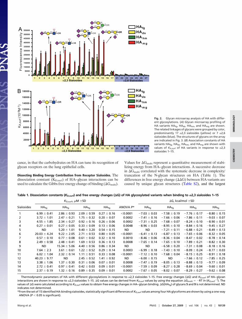
The synthetic sialic acid glycan array consisted of 17 of the �2,3(glycans 1–17) and 7 of the �2,6 (glycans 21–27) sialosides designedto explore the glycan specificity of influenza viruses (see Fig. 3). Thesynthetic sialosides with a five-carbon linker terminated with aminewere prepared and covalently attached onto NHS-coated glassslides by forming an amide bond under aqueous conditions at roomtemperature. The printing procedure was based on the standardmicroarray robotic printing technology, as reported previously (15,22). We applied the HA variants to the sialic acid slides and thenhybridized them with primary antibody, followed by detection witha secondary antibody conjugated to Cy3. This analysis indicatedthat the H5N1 HA consensus sequence specifically binds to �2,3sialosides but not �2,6 sialosides (Fig. 2A), in accordance withprevious studies (16, 18). To our surprise, the binding strength with�2,3 sialosides grew successively stronger from HAfg, HAds, andHAhm, to HAmg (Fig. 2A) by qualitative binding via relativefluorescence intensity.
We next prepared a quantitative array to determine surfacedissociation constants (17). To avoid any skewing by antibody
layering, HA was directly labeled with the fluorescent dye Cy3 (19).Direct binding assays were performed by serial dilution of Cy3-labeled HAs to establish the relative binding intensities. Thedissociation constants on the surface were determined by plottingthe HA concentrations against fluorescence intensity for each ofthe 24 sialosides printed on the glass slide. The dissociation constantKD,surf values were calculated based on the Langmuir isotherms (seeFig. 2B and Fig. S5). The monovalent HA–sialoside binding isweak, exhibiting dissociation constants in the millimolar range(KD � 2.5 � 10�3 M) (23); however, HA is involved in multivalentinteractions with sialosides on the host cell surface, which can beseen in the quantitative array profiling (Table 1).
All HA glycoforms showed strong binding to receptor glycanswith a sulfate group at the 6 position of the third GlcNAc residuefrom the nonreducing end (glycans 4 and 7). This sulfate group isimportant for binding to H5 HA (16, 18). In addition, it wasobserved that glycan 4 is the best ligand for HAfg, whereas glycans13–15 are better ligands than glycan 6 for HAmg, indicating apossible multivalent interaction within the ligand-binding site, orthe exposure of more receptor-binding domains to bigger bianten-nary sialosides (glycans 13 and 14). Interestingly, HA bindingsubstantially increases as its N-glycan structures become less com-plex (Fig. 2B). However, although the KD,surf values for HAmg showstronger and similar binding to a few SA glycans, the other HAvariants exhibit weaker and more specific binding to glycan ligands(Fig. 2B and Table S2). Thus, binding specificity and bindingaffinity may have an inverse relationship that is modulated by glycanstructure. This modulation may have important biological signifi-
A
(Heterogeneous complex type)
Fuc
GlcNAc
Gal
Man
Neu5Ac
(Heterogeneousdesialyated complextype)
(High Mannose Type)
(GlcNAc only) -8e+6
-6e+6
-4e+6
-2e+6
0
2e+6
4e+6
6e+6
8e+6
HAfgHAdsHAhmHAmg
HEK293
HAfg treatedwith NA
HEK293GNTI deficient
HAhm treatedwith Endo H
Wavelength (nm)
HAfg
HAds
HAhm
HAmg
HA variant Expression andenzymatic method Major glycoform
HAfg HAds HAhm HAmg
200 300280260240220
B
[q],
deg
cm2
dmol
-1
Fig. 1. Schematic overviews and circular dichroism spectra of HAs with different glycosylations. (A) Four variants of HA proteins with different glycosylations:HAfg, HA [a consensus sequence (ref. 21) expressed in HEK293E cells with the typical complex type N-glycans]; HAds, NA-treated HA resulting in removal of sialicacids from HAfg; HAhm, HA expressed in GnTI� HEK293S cells with the high-mannose-type N-glycans; and HAmg, Endo H-treated HA with GlcNAc only at itsN-glycosylation sites. Circular dichroism spectra of HAfg, HAds, HAhm, and HAmg demonstrate that the secondary structures of the four HA proteins with differentglycosylations are similar. (B) Structure representation of HAfg, HAds, HAhm, and HAmg with different N-glycans attached at their N-glycosylation sites. The proteinstructures are created with Protein Data Bank ID code 2FK0 (Viet04 HA), colored in gray, and the N-linked glycans are displayed in green. All N-glycans aremodeled by GlyProt (39), and the graphics are generated by PyMOL (www.pymol.org).
18138 � www.pnas.org�cgi�doi�10.1073�pnas.0909696106 Wang et al.
cance, in that the carbohydrates on HA can tune its recognition ofglycan receptors on the lung epithelial cells.
Dissecting Binding Energy Contribution from Receptor Sialosides. Thedissociation constant (KD,surf) of HA–glycan interactions can beused to calculate the Gibbs free energy change of binding (�Gmulti).
Values for �Gmulti represent a quantitative measurement of stabi-lizing energy from HA–glycan interactions. A successive decreasein �Gmulti correlated with the systematic decrease in complexity/truncation of the N-glycan structures on HA (Table 1). Thedifferences in free energy change (��G) between HA variants arecaused by unique glycan structures (Table S2), and the largest
Table 1. Dissociation constants (KD,surf) and free energy changes (�G) of HA glycosylated variants when binding to �2,3 sialosides 1–15
Sialosides
KD,surf, �M �SD
ANOVA P*
�G, kcal/mol �SD
HAfg HAfg HAfg HAfg HAfg HAfg HAfg HAfg
1 6.99 � 0.41 2.86 � 0.93 2.09 � 0.59 0.27 � 0.16 �0.0001 �7.03 � 0.03 �7.58 � 0.19 �7.76 � 0.17 �8.80 � 0.152 3.72 � 1.01 2.47 � 0.21 1.75 � 0.32 0.20 � 0.07 0.0002 �7.41 � 0.16 �7.66 � 0.06 �7.86 � 0.11 �9.03 � 0.073 4.55 � 1.85 2.34 � 0.27 0.92 � 0.16 0.26 � 0.06 0.0002 �7.31 � 0.25 �7.68 � 0.07 �8.24 � 0.10 �8.90 � 0.014 0.27 � 0.01 0.27 � 0.05 0.33 � 0.09 0.13 � 0.06 0.0048 �8.96 � 0.03 �8.95 � 0.10 �8.84 � 0.16 �9.45 � 0.275 ND 5.20 � 1.01 9.40 � 3.20 0.54 � 0.15 ND ND �7.21 � 0.11 �6.88 � 0.21 �8.49 � 0.136 20.03 � 4.24 9.22 � 2.05 2.71 � 0.53 0.80 � 0.05 �0.0001 �6.41 � 0.13 �6.87 � 0.13 �7.65 � 0.06 �8.32 � 0.057 0.57 � 0.10 0.77 � 0.08 0.61 � 0.02 0.32 � 0.10 0.0010 �8.46 � 0.06 �8.36 � 0.04 �8.47 � 0.02 �8.78 � 0.148 2.49 � 0.58 2.48 � 0.41 1.69 � 0.53 0.36 � 0.13 0.0008 �7.65 � 0.14 �7.65 � 0.10 �7.89 � 0.21 �8.82 � 0.309 ND 15.34 � 5.06 4.40 � 0.56 0.86 � 0.34 ND ND �6.58 � 0.20 �7.31 � 0.08 �8.18 � 0.16
10 7.64 � 2.3 3.61 � 0.61 1.22 � 0.52 0.29 � 0.14 0.0003 �6.99 � 0.18 �7.43 � 0.10 �8.09 � 0.24 �8.77 � 0.0311 6.02 � 1.04 2.32 � 0.14 1.11 � 0.51 0.33 � 0.08 �0.0001 �7.12 � 0.10 �7.68 � 0.04 �8.15 � 0.25 �8.91 � 0.1812 40.23 � 9.77 ND 2.45 � 0.52 1.41 � 0.92 ND �6.00 � 0.15 ND �7.66 � 0.12 �7.85 � 0.2513 3.38 � 1.06 1.37 � 0.30 0.31 � 0.06 0.07 � 0.01 0.0008 �7.47 � 0.19 �8.05 � 0.13 �8.88 � 0.13 �9.77 � 0.0914 2.72 � 0.41 0.97 � 0.41 0.42 � 0.03 0.09 � 0.01 �0.0001 �7.59 � 0.09 �8.27 � 0.28 �8.69 � 0.04 �9.60 � 0.0115 2.37 � 0.19 1.32 � 0.16 0.89 � 0.35 0.09 � 0.01 0.0002 �7.67 � 0.05 �8.02 � 0.07 �8.29 � 0.27 �9.62 � 0.08
Thermodynamic parameters of HA with different glycosylations in response to �2,3 sialosides 1–15. Free energy changes (�G) and KD,surf of HA–glycaninteractions are shown in response to �2,3 sialosides 1–15. �G values can be derived from KD,surf values by using the equation �Gmulti � �RT ln (KD,surf
�1). Thevalues of �G were calculated according to KD,surf values to obtain free energy changes in HA–glycan binding. �G(HAfg) of glycans 5 and 9 is not determined. NDindicates not determined.*From the set of 15 identified HA-binding sialosides, statistically significant differences of KD,surf values among four HA glycoforms are shown by using a one-wayANOVA (P � 0.05 is significant).
A
B
Fig. 2. Glycan microarray analysis of HA with differ-ent glycosylations. (A) Glycan microarray profiling ofHA variants HAfg, HAds, HAhm, and HAmg are shown.The related linkages of glycans were grouped by color,predominantly 17 �2,3 sialosides (yellow) or 7 �2,6sialosides (blue). The structures of glycans on the arrayare indicated in Fig. 3. (B) Association constants of HAvariants HAfg, HAds, HAhm, and HAmg are shown withvalues of KA,surf of HA variants in response to �2,3sialosides 1–15.
Wang et al. PNAS � October 27, 2009 � vol. 106 � no. 43 � 18139
IMM
UN
OLO
GY
CHEM
ISTR
Y
difference is between HAfg and HAmg (��G HAfg 3 HAmg; seeTable S2), which is consistent with the largest difference in bindingenergy resulting from trimming off most of the N-glycan down toa single GlcNAc. It is noted that values of ��G are similar exceptfor glycans 4 and 7 (Table S2), indicating that glycans on HA do notsignificantly affect the binding affinity with sulfated �2,3 trisaccha-ride (16).
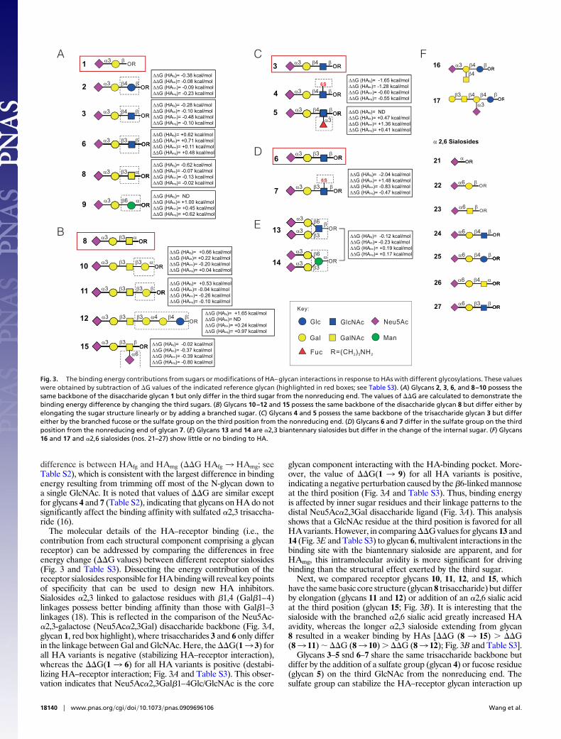
The molecular details of the HA–receptor binding (i.e., thecontribution from each structural component comprising a glycanreceptor) can be addressed by comparing the differences in freeenergy change (��G values) between different receptor sialosides(Fig. 3 and Table S3). Dissecting the energy contribution of thereceptor sialosides responsible for HA binding will reveal key pointsof specificity that can be used to design new HA inhibitors.Sialosides �2,3 linked to galactose residues with �1,4 (Gal�1–4)linkages possess better binding affinity than those with Gal�1–3linkages (18). This is reflected in the comparison of the Neu5Ac-�2,3-galactose (Neu5Ac�2,3Gal) disaccharide backbone (Fig. 3A,glycan 1, red box highlight), where trisaccharides 3 and 6 only differin the linkage between Gal and GlcNAc. Here, the ��G(13 3) forall HA variants is negative (stabilizing HA–receptor interaction),whereas the ��G(1 3 6) for all HA variants is positive (destabi-lizing HA–receptor interaction; Fig. 3A and Table S3). This obser-vation indicates that Neu5Ac�2,3Gal�1–4Glc/GlcNAc is the core
glycan component interacting with the HA-binding pocket. More-over, the value of ��G(1 3 9) for all HA variants is positive,indicating a negative perturbation caused by the �6-linked mannoseat the third position (Fig. 3A and Table S3). Thus, binding energyis affected by inner sugar residues and their linkage patterns to thedistal Neu5Ac�2,3Gal disaccharide ligand (Fig. 3A). This analysisshows that a GlcNAc residue at the third position is favored for allHA variants. However, in comparing ��G values for glycans 13 and14 (Fig. 3E and Table S3) to glycan 6, multivalent interactions in thebinding site with the biantennary sialoside are apparent, and forHAmg, this intramolecular avidity is more significant for drivingbinding than the structural effect exerted by the third sugar.
Next, we compared receptor glycans 10, 11, 12, and 15, whichhave the same basic core structure (glycan 8 trisaccharide) but differby elongation (glycans 11 and 12) or addition of an �2,6 sialic acidat the third position (glycan 15; Fig. 3B). It is interesting that thesialoside with the branched �2,6 sialic acid greatly increased HAavidity, whereas the longer �2,3 sialoside extending from glycan8 resulted in a weaker binding by HAs [��G (8 3 15) � ��G(83 11) � ��G (83 10) � ��G (83 12); Fig. 3B and Table S3].
Glycans 3–5 snd 6–7 share the same trisaccharide backbone butdiffer by the addition of a sulfate group (glycan 4) or fucose residue(glycan 5) on the third GlcNAc from the nonreducing end. Thesulfate group can stabilize the HA–receptor glycan interaction up
OR
OR
OR
OR
OR
OROROROR
OR
OR
OR
OR
OR
OR
OR
OR
1
2
3
6
8
9
∆∆G (HAfg)= -0.38 kcal/mol∆∆G (HAds)= -0.08 kcal/mol∆∆G (HAhm)= -0.09 kcal/mol∆∆G (HAmg)= -0.23 kcal/mol
∆∆G (HAfg)= -0.28 kcal/mol∆∆G (HAds)= -0.10 kcal/mol∆∆G (HAhm)= -0.48 kcal/mol∆∆G (HAmg)= -0.10 kcal/mol
∆∆G (HAfg)= +0.62 kcal/mol∆∆G (HAds)= +0.71 kcal/mol∆∆G (HAhm)= +0.11 kcal/mol∆∆G (HAmg)= +0.48 kcal/mol
∆∆G (HAfg)= -0.62 kcal/mol∆∆G (HAds)= -0.07 kcal/mol∆∆G (HAhm)= -0.13 kcal/mol∆∆G (HAmg)= -0.02 kcal/mol
∆∆G (HAfg)= ND∆∆G (HAds)= +1.00 kcal/mol∆∆G (HAhm)= +0.45 kcal/mol∆∆G (HAmg)= +0.62 kcal/mol
Glc
Gal
GlcNAc
GalNAc
Neu5Ac
Man
Fuc R=(CH ) NH2 5 2
Key:
OR
OR
6S
OR
ORRR
OROR3
4
5
∆∆G (HAfg)= -1.65 kcal/mol∆∆G (HAds)= -1.28 kcal/mol∆∆G (HAhm)= -0.60 kcal/mol∆∆G (HAmg)= -0.55 kcal/mol
∆∆G (HAfg)= ND∆∆G (HAds)= +0.47 kcal/mol∆∆G (HAhm)= +1.36 kcal/mol∆∆G (HAmg)= +0.41 kcal/mol
OROR
6S
ORORRR
7
6
∆∆G (HAfg)= -2.04 kcal/mol∆∆G (HAds)= +1.48 kcal/mol∆∆G (HAhm)= -0.83 kcal/mol∆∆G (HAmg)= -0.47 kcal/mol
OROR
OROR
16
17
OR
OR
OROR
OR
OROROROR
OROROROR
OROROROR
OR
α 2,6 Sialosides
21
22
23
24
25
26
27
A
OR
OR
13
14
∆∆G (HAfg)= -0.12 kcal/mol∆∆G (HAds)= -0.23 kcal/mol∆∆G (HAhm)= +0.19 kcal/mol∆∆G (HAmg)= +0.17 kcal/mol
E
D
C
OROROROR
OR
ORORORO
OROROROR
OROR
8
10
11
12
15
∆∆G (HAfg)= +0.66 kcal/mol∆∆G (HAds)= +0.22 kcal/mol∆∆G (HAhm)= -0.20 kcal/mol∆∆G (HAmg)= +0.04 kcal/mol
∆∆G (HAfg)= +0.53 kcal/mol∆∆G (HAds)= -0.04 kcal/mol∆∆G (HAhm)= -0.26 kcal/mol∆∆G (HAmg)= -0.10 kcal/mol
∆∆G (HAfg)= +1.65 kcal/mol∆∆G (HAds)= ND∆∆G (HAhm)= +0.24 kcal/mol∆∆G (HAmg)= +0.97 kcal/mol
∆∆G (HAfg)= -0.02 kcal/mol∆∆G (HAds)= -0.37 kcal/mol∆∆G (HAhm)= -0.39 kcal/mol∆∆G (HAmg)= -0.80 kcal/mol
B
Fβα3
α3 β4 β
α3 β4 β
α3 β3 β
α3 β3 α
α3 β6 α
α3 β3 α
α3 β3 β3 α
α3 β3 β3 β
α3 β3 β3 α4 β4 β
α3 β3 β
α6
α3 β4 β
α3 β4 β
α3 β4 β
α3
α3 β3 β
α3 β3 β
α3
α3
α3
α3
β3
β3
β6
β6
β
α
α3 β4 β
β4
β3 β4 β4 β
α3
α
α6 β
α6 β
α6 β4 β
α6 β4 β
α6 β4 α
α6 β3 β
Fig. 3. The binding energy contributions from sugars or modifications of HA–glycan interactions in response to HAs with different glycosylations. These valueswere obtained by subtraction of �G values of the indicated reference glycan (highlighted in red boxes; see Table S3). (A) Glycans 2, 3, 6, and 8–10 possess thesame backbone of the disaccharide glycan 1 but only differ in the third sugar from the nonreducing end. The values of ��G are calculated to demonstrate thebinding energy difference by changing the third sugars. (B) Glycans 10–12 and 15 possess the same backbone of the disaccharide glycan 8 but differ either byelongating the sugar structure linearly or by adding a branched sugar. (C) Glycans 4 and 5 possess the same backbone of the trisaccharide glycan 3 but differeither by the branched fucose or the sulfate group on the third position from the nonreducing end. (D) Glycans 6 and 7 differ in the sulfate group on the thirdposition from the nonreducing end of glycan 7. (E) Glycans 13 and 14 are �2,3 biantennary sialosides but differ in the change of the internal sugar. (F) Glycans16 and 17 and �2,6 sialosides (nos. 21–27) show little or no binding to HA.
18140 � www.pnas.org�cgi�doi�10.1073�pnas.0909696106 Wang et al.
to 2.044 kcal/mol [��G (6 3 7)], the largest energy gap betweentwo receptor sialosides. Among all of the HA variants, the fullyglycosylated variant showed the most significant differences in freeenergy changes, with values of ��G(33 4) HAfg (�1.653 kcal/mol)and ��G(63 7) HAfg (�2.044 kcal/mol), and the size of the freeenergy gain lessened as the glycan structure became more simpli-fied; i.e., HAfg � HAds � HAhm � HAmg. Thus, sulfated glycansdramatically enhance HA binding, and fully glycosylated HAmaximizes this effect (Fig. 3 C and D), which may be important forH5N1 pathogenesis. On the other hand, the fucosylated receptoranalogs greatly destabilize HA binding, with all glycosylated HAvariants showing a positive ��G(3 3 5) (Fig. 3C). These largedifferences in ��G(33 4) and ��G(33 5) are likely caused byan important binding interaction in the receptor-binding pocket,which the sulfate group maximizes and the fucose sterically blocks.The weak binding of HAfg is unlikely due to the competition of itssialylglycans, because removal of sialic acid has a small effect onbinding, and HAfg still exhibits a strong affinity for certain specificsialylglycans.
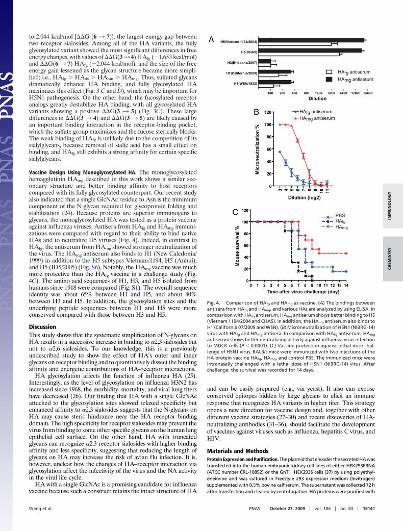
Vaccine Design Using Monoglycosylated HA. The monoglycosylatedhemagglutinin HAmg described in this work shows a similar sec-ondary structure and better binding affinity to host receptorscompared with its fully glycosylated counterpart. Our recent studyalso indicated that a single GlcNAc residue to Asn is the minimumcomponent of the N-glycan required for glycoprotein folding andstabilization (24). Because proteins are superior immunogens toglycans, the monoglycosylated HA was tested as a protein vaccineagainst influenza viruses. Antisera from HAfg and HAmg immuni-zations were compared with regard to their ability to bind nativeHAs and to neutralize H5 viruses (Fig. 4). Indeed, in contrast toHAfg, the antiserum from HAmg showed stronger neutralization ofthe virus. The HAmg antiserum also binds to H1 (New Caledonia/1999) in addition to the H5 subtypes Vietnam/1194, H5 (Anhui),and H5 (ID5/2005) (Fig. S6). Notably, the HAmg vaccine was muchmore protective than the HAfg vaccine in a challenge study (Fig.4C). The amino acid sequences of H1, H3, and H5 isolated fromhumans since 1918 were compared (Fig. S1). The overall sequenceidentity was about 65% between H1 and H5, and about 40%between H3 and H5. In addition, the glycosylation sites and theunderlying peptide sequences between H1 and H5 were moreconserved compared with those between H3 and H5.
DiscussionThis study shows that the systematic simplification of N-glycans onHA results in a successive increase in binding to �2,3 sialosides butnot to �2,6 sialosides. To our knowledge, this is a previouslyundescribed study to show the effect of HA’s outer and innerglycans on receptor binding and to quantitatively dissect the bindingaffinity and energetic contributions of HA–receptor interactions.
HA glycosylation affects the function of influenza HA (25).Interestingly, as the level of glycosylation on influenza H3N2 hasincreased since 1968, the morbidity, mortality, and viral lung titershave decreased (26). Our finding that HA with a single GlcNAcattached to the glycosylation sites showed relaxed specificity butenhanced affinity to �2,3 sialosides suggests that the N-glycans onHA may cause steric hindrance near the HA–receptor bindingdomain. The high specificity for receptor sialosides may prevent thevirus from binding to some other specific glycans on the human lungepithelial cell surface. On the other hand, HA with truncatedglycans can recognize �2,3 receptor sialosides with higher bindingaffinity and less specificity, suggesting that reducing the length ofglycans on HA may increase the risk of avian flu infection. It is,however, unclear how the changes of HA–receptor interaction viaglycosylation affect the infectivity of the virus and the NA activityin the viral life cycle.
HA with a single GlcNAc is a promising candidate for influenzavaccine because such a construct retains the intact structure of HA
and can be easily prepared (e.g., via yeast). It also can exposeconserved epitopes hidden by large glycans to elicit an immuneresponse that recognizes HA variants in higher titer. This strategyopens a new direction for vaccine design and, together with otherdifferent vaccine strategies (27–30) and recent discoveries of HA-neutralizing antibodies (31–36), should facilitate the developmentof vaccines against viruses such as influenza, hepatitis C virus, andHIV.
Materials and MethodsProtein Expression and Purification. The plasmid that encodes the secreted HA wastransfected into the human embryonic kidney cell lines of either HEK293EBNA(ATCC number CRL-10852) or the GnTI� HEK293S cells (37) by using polyethyl-eneimine and was cultured in Freestyle 293 expression medium (Invitrogen)supplemented with 0.5% bovine calf serum. The supernatant was collected 72 hafter transfection and cleared by centrifugation. HA proteins were purified with
A
B
C
Time after virus challenge (day)
Mou
se s
urvi
val %
Mic
rone
utra
lizat
ion
%
Dilution (log2)
3 4 5 6 7 8 9 10 11
0
20
40
60
80
100
120 HAfg antiserumHAmg antiserum
0 1 2 3 4 5 6 7 8 9 10 11 12 13 140
20
40
60
80
100
120
H5(Vietnam 1194/2004)
H5(CHA5)
H3(Brisbane/2007)
H1(California/2009)
H1(WSN/1933)
100 200 400 800 1600 3200 6400 12800 25600
HAfg antiserum
HAmg antiserum
PBSHAfgHAmg
Dilution
Fig. 4. Comparison of HAfg and HAmg as vaccine. (A) The bindings betweenantisera from HAfg and HAmg, and various HAs are analyzed by using ELISA. Incomparison with HAfg antiserum, HAmg antiserum shows better binding to H5(Vietnam 1194/2004 and CHA5). In addition, the HAmg antiserum also binds toH1 (California 07/2009 and WSN). (B) Microneutralization of H5N1 (NIBRG-14)virus with HAfg and HAmg antisera. In comparison with HAfg antiserum, HAmg
antiserum shows better neutralizing activity against influenza virus infectionto MDCK cells (P � 0.0001). (C) Vaccine protection against lethal-dose chal-lenge of H5N1 virus. BALB/c mice were immunized with two injections of theHA protein vaccine HAfg, HAmg, and control PBS. The immunized mice wereintranasally challenged with a lethal dose of H5N1 (NIBRG-14) virus. Afterchallenge, the survival was recorded for 14 days.
Wang et al. PNAS � October 27, 2009 � vol. 106 � no. 43 � 18141
IMM
UN
OLO
GY
CHEM
ISTR
Y
Nickel-chelation chromatography as previously described (38) to obtain fullyglycosylated HAfg and high-mannose-type HAhm. To obtain the HA protein with-out sialylation—the desialylated HAds—the purified protein was treated with 20mM Clostridium NA (Sigma) for 2 h at 37 °C. After the NA treatment, the proteinwas purified again to be separated from the NA. The purified HAhm was treatedwith Endo H (NEB) for 2 h at 37 °C to produce HA protein with a single GlcNAc atthe glycosylation sites, the monoglycosylated HAmg.
Glycan Microarray Fabrication. Twenty-four sialic acid-containing glycans de-signed for HAs were prepared chemically and used for array fabrication. Microar-rays were printed (BioDot; Cartesian Technologies) by robotic pin (SMP3;TeleChem International) deposition of 0.7 nL of various concentrations ofamine-containing glycans in printing buffer (300 mM phosphate buffer, pH 8.5,containing 0.005% Tween 20) from a 384-well plate onto NHS-coated glass slides(Nexterion H slide; SCHOTT North America). The slides for sialosides were spottedwith solutions of glycans 1–17 and 21–27 with concentrations of 100 �M in eachrow for one glycan from bottom to top, with 12 replicates horizontally placed ineach subarray, and each slide was designed for 16 grids for further incubationexperiments. Printed slides were allowed to react in an atmosphere of 80%humidity for an hour followed by desiccation overnight, and they were stored atroom temperature in a desiccator until use. Before the binding assay, these slideswere blocked with ethanolamine (50 mM ethanolamine in borate buffer, pH 9.2)and then washed with water and PBS buffer, pH 7.4, twice.
Indirect Binding Assay. HA glycosylated variants were prepared in 0.005% Tween20/PBS buffer, pH 7.4, and added to cover the grid on glycan array with applica-tion of a coverslip. After incubation in a humidified chamber with shaking for 1 h,the slides were washed three times with 0.005% Tween 20/PBS buffer, pH 7.4.Next, rabbit anti-H5N1 HA antibody was added to the slides and incubated in ahumidified chamber for 1 h. After washing the slides with 0.005% Tween 20/PBSbuffer three times, Cy3-conjugated goat anti-rabbit IgG antibody was added tothe slides and incubated in a humidified chamber for another 1 h. The slides werewashedthreetimeswith0.05%Tween20/PBSbuffer,pH7.4; threetimeswithPBSbuffer, pH 7.4; and three times with H2O, and then dried. The slides were scannedat 595 nm (for Cy3) with a microarray fluorescence chip reader (GenePix Pro 6.0;Molecular Devices).
Direct Binding Assay. Cy3-labeled HA proteins with different glycosylations wereprepared in 0.005% Tween 20/PBS buffer, pH 7.4, and added to cover the grid onglycan array with application of a coverslip. After incubation in a humidifiedchamber with shaking for 1 h, the slides were washed three times with 0.005%Tween 20/PBS buffer, pH 7.4; three times with PBS buffer, pH 7.4; and three timeswith H2O, and then dried. The slides were scanned at 595 nm (for Cy3) with amicroarray fluorescence chip reader (GenePix Pro 6.0; Molecular Devices).
Microneutralization Assay. The freshly prepared H5N1 (NIBRG-14) virus (NationalInstitute for Biological Standards and Control, Potters Bar, U.K.) was quantifiedwith the median tissue culture infectious dose (TCID50). The 100-fold TCID50 ofvirus was mixed in equal volume with 2-fold serial dilutions of serum stocksolution in 96-well plates and incubated for 1 h at 37 °C. The mixture was addedonto the MDCK cells (1.5 � 104 cells per well) on the plates, followed by incuba-tion at 37 °C for 16–20 h. The cells were washed with PBS, fixed in acetone/methanol solution (vol/vol 1:1), and blocked with 5% skim milk. The viral antigenwas detected by indirect ELISA with an mAb against influenza A NP (35).
Mice, Vaccination, and Challenge. Female 6- to 8-week-old BALB/c mice (n � 15)were immunized intramuscularly with 20 �g of purified HAfg or HAmg proteins in50 �L of PBS, pH 7.4, and mixed with 50 �L of 1 mg/mL aluminum hydroxide(Alum; Sigma) at weeks 0 and 2. Blood was collected 14 days after immunization,and serum samples were collected from each mouse. The immunized mice werechallenged intranasally with a genetically modified H5N1 virus, NIBRG-14, with alethal dose (100-fold lethal dose to 50% of mice). The mice were monitored dailyfor 14 days after the challenge for survival. All animal experiments were evalu-ated and approved by the Institutional Animal Care and Use Committee ofAcademia Sinica.
ACKNOWLEDGMENTS. We sincerely thank S.-H. Ma for virus challenge experi-ments, C.-Y. Su and S.-Y. Wang for virus neutralization experiments, and P. J.Reeves (University of Essex, United Kingdom) for providing GnTI� HEK293S cells.We thank Academia Sinica for financial support. MALDI-MS profiles of theN-glycanswereacquiredattheNationalResearchProgramforGenomicMedicineCore Facilities for Proteomics and Glycomics, supported by the Taiwan NationalScience Council.
1. Garten RJ, et al. (2009) Antigenic and genetic characteristics of swine-origin 2009 A(H1N1)influenza viruses circulating in humans. Science 325:197–201.
2. Neumann G, Noda T, Kawaoka Y (2009) Emergence and pandemic potential of swine-origin H1N1 influenza virus. Nature 459:931–939.
3. Kuiken T, et al. (2006) Host species barriers to influenza virus infections. Science 312:394–397.
4. Maines TR, et al. (2009) Transmission and pathogenesis of swine-origin 2009 A(H1N1)influenza viruses in ferrets and mice. Science 325:484–487.
5. Skehel JJ, Wiley DC (2000) Receptor binding and membrane fusion in virus entry: Theinfluenza hemagglutinin. Annu Rev Biochem 69:531–569.
6. van Riel D, et al. (2006) H5N1 virus attachment to lower respiratory tract. Science 312:399–399.
7. Connor RJ, Kawaoka Y, Webster RG, Paulson JC (1994) Receptor specificity in human,avian, and equine H2 and H3 influenza virus isolates. Virology 205:17–23.
8. Tumpey TM, et al. (2007) A two-amino acid change in the hemagglutinin of the 1918influenza virus abolishes transmission. Science 315:655–659.
9. Keil W, et al. (1985) Carbohydrates of influenza-virus–structural elucidation of the indi-vidual glycans of the FPV hemagglutinin by two-dimensional H-1 NMR and methylationanalysis. EMBO J 4:2711–2720.
10. Chen ZY, Aspelund A, Jin H (2008) Stabilizing the glycosylation pattern of influenza Bhemagglutinin following adaptation to growth in eggs. Vaccine 26:361–371.
11. Ohuchi R, Ohuchi M, Garten W, Klenk HD (1997) Oligosaccharides in the stem regionmaintain the influenza virus hemagglutinin in the metastable form required for fusionactivity. J Virol 71:3719–3725.
12. Skehel JJ, et al. (1984) A carbohydrate side-chain on hemagglutinins of Hong Konginfluenza-viruses inhibits recognition by a monoclonal-antiboby. Proc Natl Acad Sci USA81:1779–1783.
13. Deshpande KL, Fried VA, Ando M, Webster RG (1987) Glycosylation affects cleavage of anH5N2 influenza-virus hemagglutinin and regulates virulence. Proc Natl Acad Sci USA84:36–40.
14. Gunther I, Glatthaar B, Doller G, Garten W (1993) A H1-hemagglutinin of a humaninfluenza A-virus with a carbohydrate-modulated receptor-binding site and an unusualcleavage site. Virus Res 27:147–160.
15. Blixt O, et al. (2004) Printed covalent glycan array for ligand profiling of diverse glycanbinding proteins. Proc Natl Acad Sci USA 101:17033–17038.
16. Chandrasekaran A, et al. (2008) Glycan topology determines human adaptation of avianH5N1 virus hemagglutinin. Nat Biotechnol 26:107–113.
17. Liang PH, Wang SK, Wong CH (2007) Quantitative analysis of carbohydrate-proteininteractions using glycan microarrays: Determination of surface and solution dissociationconstants. J Am Chem Soc 129:11177–11184.
18. Stevens J,etal. (2008)RecentavianH5N1virusesexhibit increasedpropensity foracquiringhuman receptor specificity. J Mol Biol 381:1382–1394.
19. Srinivasan A, et al. (2008) Quantitative biochemical rationale for differences in transmis-sibility of 1918 pandemic influenza A viruses. Proc Natl Acad Sci USA 105:2800–2805.
20. Deom CM, Caton AJ, Schulze IT (1986) Host cell-mediated selection of a mutant influenzaAvirus thathas lostacomplexoligosaccharidefromthetipof thehemagglutinin.ProcNatlAcad Sci USA 83:3771–3775.
21. Chen MW, et al. (2008) A consensus-hemagglutinin-based DNA vaccine that protects miceagainst divergent H5N1 influenza viruses. Proc Natl Acad Sci USA 105:13538–13543.
22. WangCC,etal. (2008)GlycanmicroarrayofGloboHandrelatedstructures forquantitativeanalysis of breast cancer. Proc Natl Acad Sci USA 105:11661–11666.
23. Sauter NK, et al. (1989) Hemagglutinins from 2 influenza-virus variants bind to sialic-acidderivatives with millimolar dissociation-constants - A 500-MHz proton nuclear magnetic-resonance study. Biochemistry 28:8388–8396.
24. Hanson SR, et al. (2009) The core trisaccharide of an N-linked glycoprotein intrinsicallyaccelerates folding and enhances stability. Proc Natl Acad Sci USA 106:3131–3136.
25. Wagner R, Heuer D, Wolff T, Herwig A, Klenk HD (2002) N-glycans attached to the stemdomain of haemagglutinin efficiently regulate influenza A virus replication. J Gen Virol83:601–609.
26. Vigerust DJ, et al. (2007) N-linked glycosylation attenuates H3N2 influenza viruses. J Virol81:8593–8600.
27. Hoffmann E, Lipatov AS, Webby RJ, Govorkova EA, Webster RG (2005) Role of specifichemagglutinin amino acids in the immunogenicity and protection of H5N1 influenza virusvaccines. Proc Natl Acad Sci USA 102:12915–12920.
28. Huleatt JW, et al. (2008) Potent immunogenicity and efficacy of a universal influenzavaccine candidate comprising a recombinant fusion protein linking influenza M2e to theTLR5 ligand flagellin. Vaccine 26:201–214.
29. Scanlan CN, et al. (2007) Inhibition of mammalian glycan biosynthesis produces non-selfantigens for a broadly neutralising, HIV-1 specific antibody. J Mol Biol 372:16–22.
30. Yang ZY, et al. (2007) Immunization by avian H5 influenza hemagglutinin mutants withaltered receptor binding specificity. Science 317:825–828.
31. Ekiert DC, et al. (2009) Antibody recognition of a highly conserved influenza virus epitope.Science 324:246–251.
32. Kashyap AK, et al. (2008) Combinatorial antibody libraries from survivors of the TurkishH5N1 avian influenza outbreak reveal virus neutralization strategies. Proc Natl Acad SciUSA 105:5986–5991.
33. Scheid JF, et al. (2009) Broad diversity of neutralizing antibodies isolated from memory Bcells in HIV-infected individuals. Nature 458:636–640.
34. Stevens J, et al. (2006) Structure and receptor specificity of the hemagglutinin from anH5N1 influenza virus. Science 312:404–410.
35. Sui JH, et al. (2009) Structural and functional bases for broad-spectrum neutralization ofavian and human influenza A viruses. Nat Struct Mol Biol 16:265–273.
36. Throsby M, et al. (2008) Heterosubtypic neutralizing monoclonal antibodies cross-protective against H5N1 and H1N1 recovered from human IgM memory B cells. PLoS ONE3:e3942.
37. Reeves PJ, Callewaert N, Contreras R, Khorana HG (2002) Structure and function inrhodopsin: High-level expression of rhodopsin with restricted and homogeneous N-glycosylation by a tetracycline-inducible N-acetylglucosaminyltransferase I-negativeHEK293S stable mammalian cell line. Proc Natl Acad Sci USA 99:13419–13424.
38. Wei CJ, et al. (2008) Comparative efficacy of neutralizing antibodies elicited by recombi-nant hemagglutinin proteins from avian H5N1 influenza virus. J Virol 82:6200–6208.
39. Bohne-Lang A, von der Lieth CW (2005) GlyProt: In silico glycosylation of proteins. NucleicAcids Res 33:W214–W219.
18142 � www.pnas.org�cgi�doi�10.1073�pnas.0909696106 Wang et al.


![The Spread of Influenza A(H1N1)pdm09 Virus in …stacks.cdc.gov/view/cdc/22753/cdc_22753_DS1.pdfdiffusion (movement from larger to smaller towns) [9]. The spread of influenza epidemics](https://static.fdocuments.nl/doc/165x107/5f5ea10c0e994a5b2f0916c7/the-spread-of-influenza-ah1n1pdm09-virus-in-diffusion-movement-from-larger-to.jpg)