Transmembran-Rezeptoren · Enzym-gekoppelte Rezeptoren Ionenkanal-gekoppelte Rezeptoren...
52
1 Transmembran-Rezeptoren Enzym- gekoppelte Rezeptoren Ionenkanal- gekoppelte Rezeptoren G-Protein- gekoppelte Rezeptoren
Transcript of Transmembran-Rezeptoren · Enzym-gekoppelte Rezeptoren Ionenkanal-gekoppelte Rezeptoren...
1
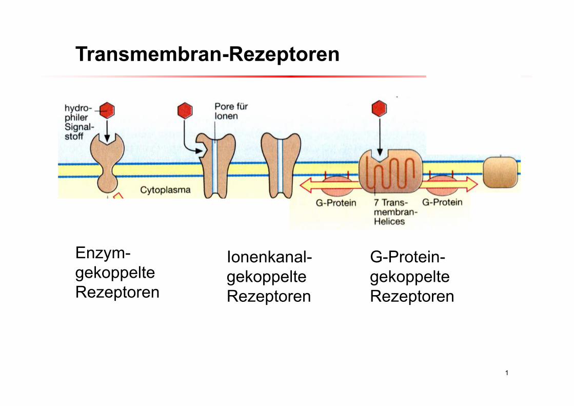
Transmembran-Rezeptoren
Enzym-gekoppelteRezeptoren
Ionenkanal-gekoppelteRezeptoren
G-Protein-gekoppelteRezeptoren
2
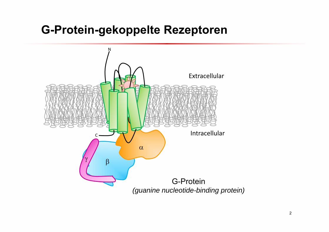
G-Protein-gekoppelte RezeptorenN
C
Extracellular
Intracellular
G-Protein(guanine nucleotide-binding protein)
3
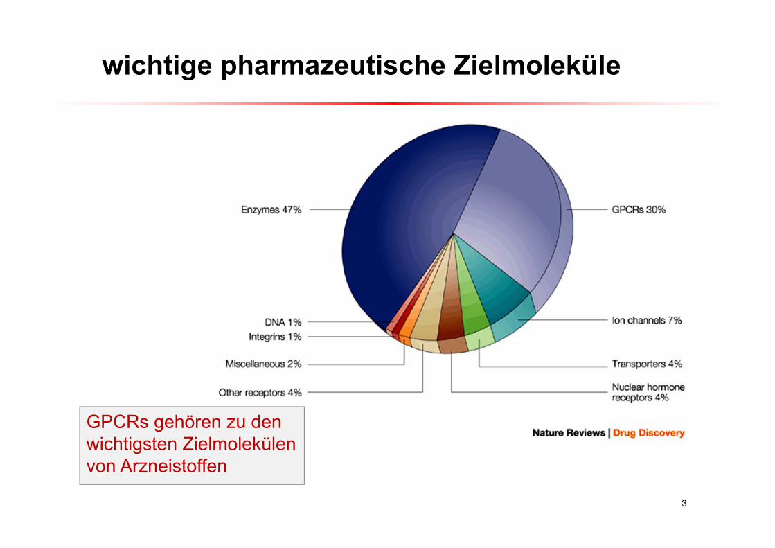
wichtige pharmazeutische Zielmoleküle
GPCRs gehören zu den wichtigsten Zielmolekülenvon Arzneistoffen
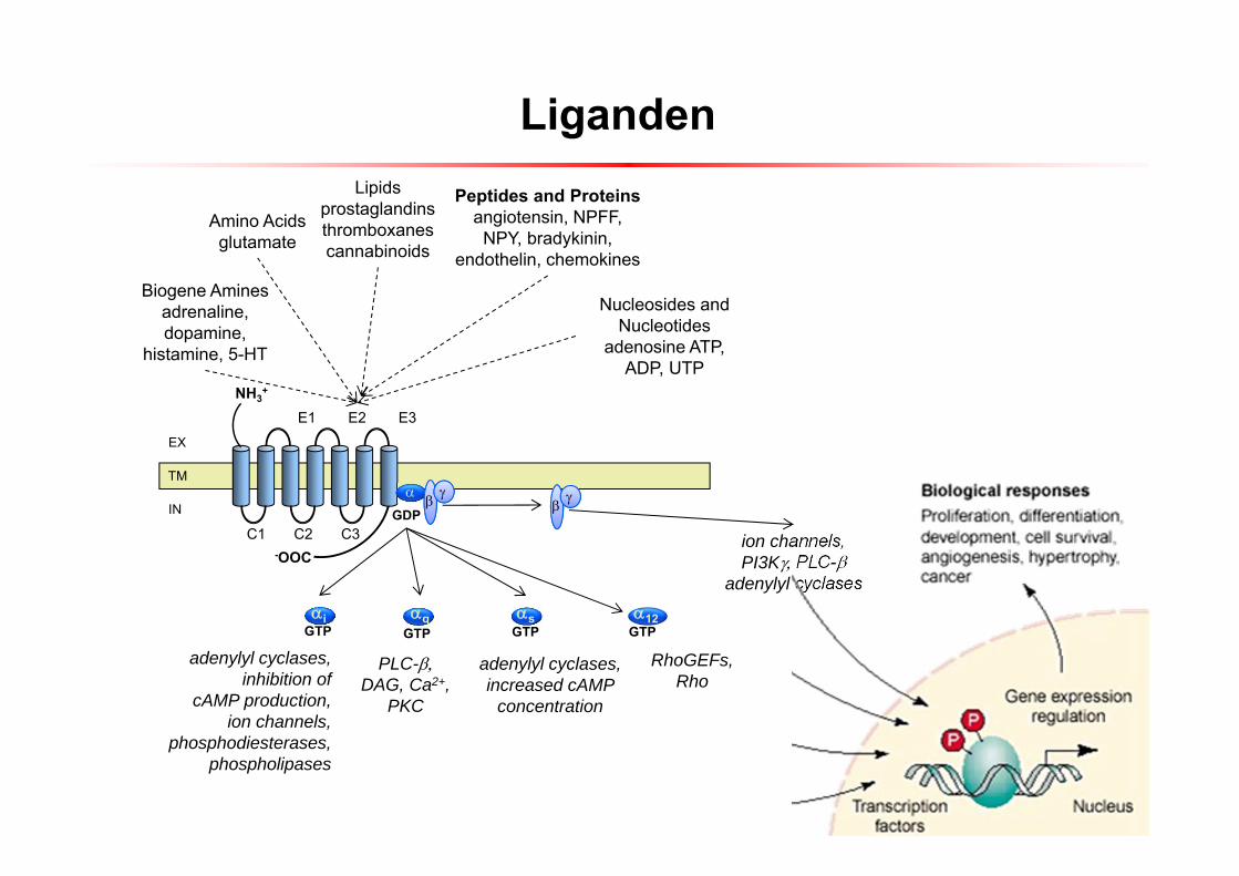
Liganden
4
C1 C2 C3
NH3+
-OOC
E1 E2 E3
GDP
Biogene Aminesadrenaline, dopamine,
histamine, 5-HT
Amino Acidsglutamate
Lipidsprostaglandinsthromboxanescannabinoids
Peptides and Proteinsangiotensin, NPFF, NPY, bradykinin,
endothelin, chemokines
Nucleosides and Nucleotides
adenosine ATP, ADP, UTP
ion channels,PI3KPLC-
adenylyl cyclases
adenylyl cyclases,inhibition of
cAMP production,ion channels,
phosphodiesterases,phospholipases
PLC-DAG, Ca2+,
PKC
iGTP
qGTP
sGTP
adenylyl cyclases,increased cAMP
concentration
TM
RhoGEFs,Rho
12GTP
EX
IN
5
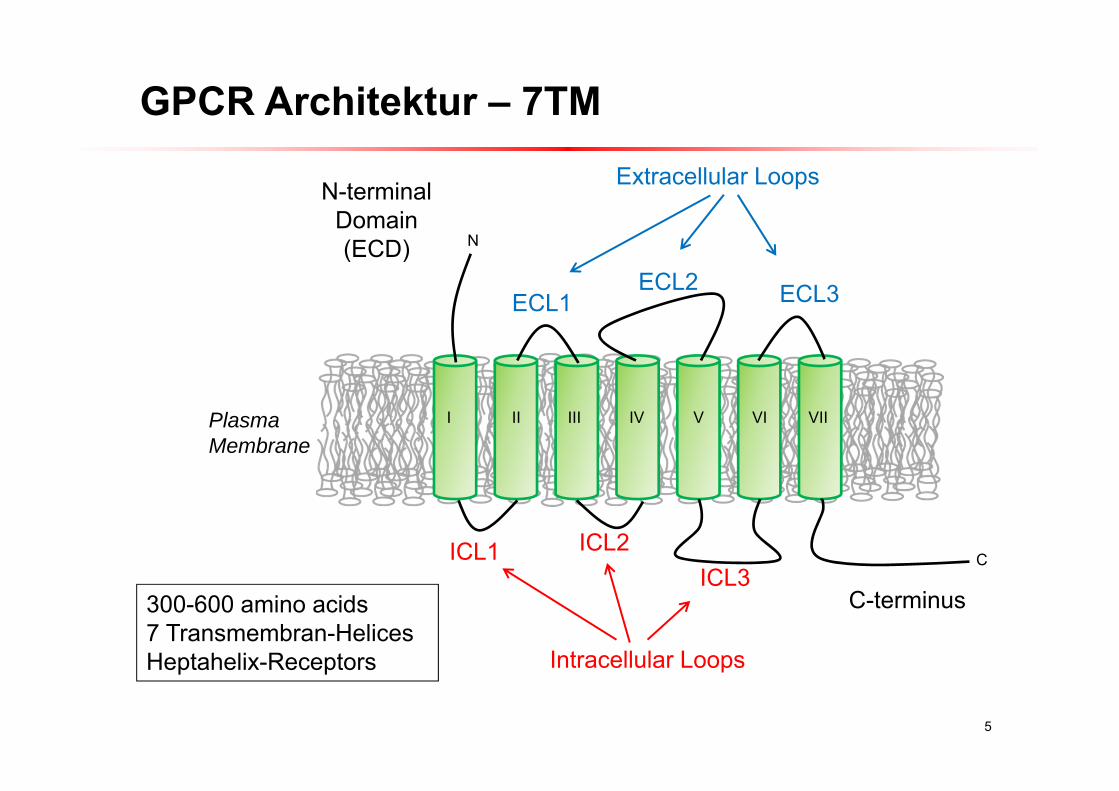
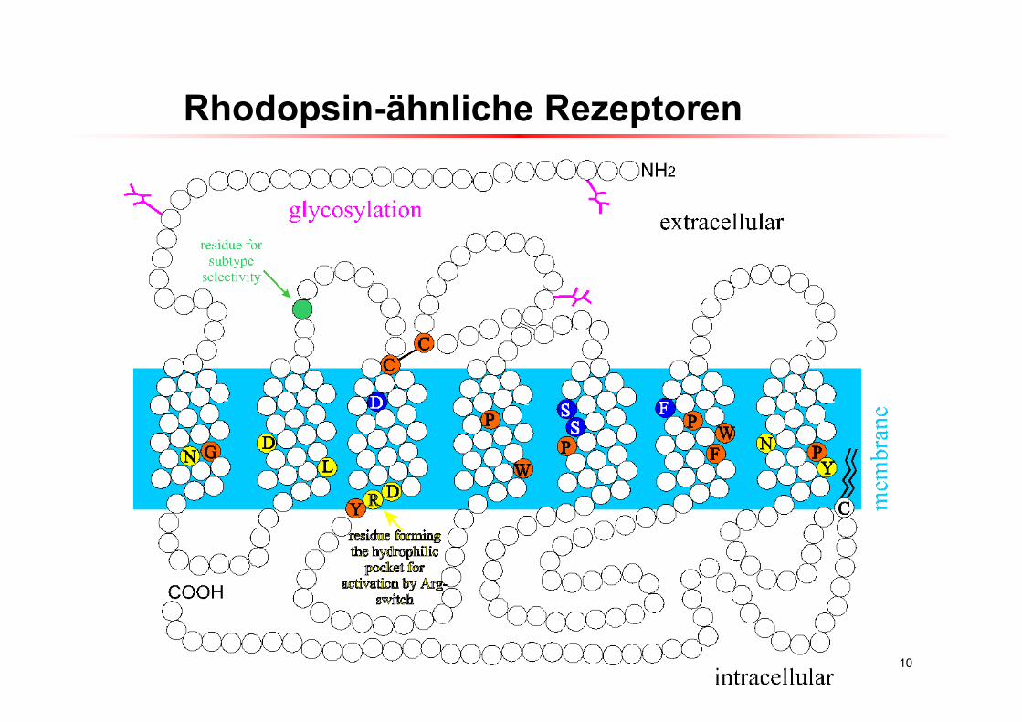
GPCR Architektur – 7TM
N
I II III IV V VI VII
Extracellular Loops
Intracellular Loops
N-terminalDomain(ECD)
C-terminus
ECL1ECL2 ECL3
CICL1 ICL2ICL3
PlasmaMembrane
300-600 amino acids7 Transmembran-Helices Heptahelix-Receptors
6
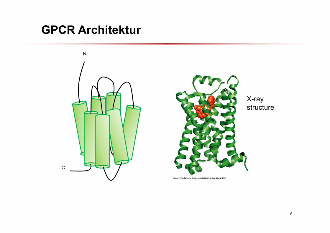
GPCR Architektur
N
C
X-raystructure
7
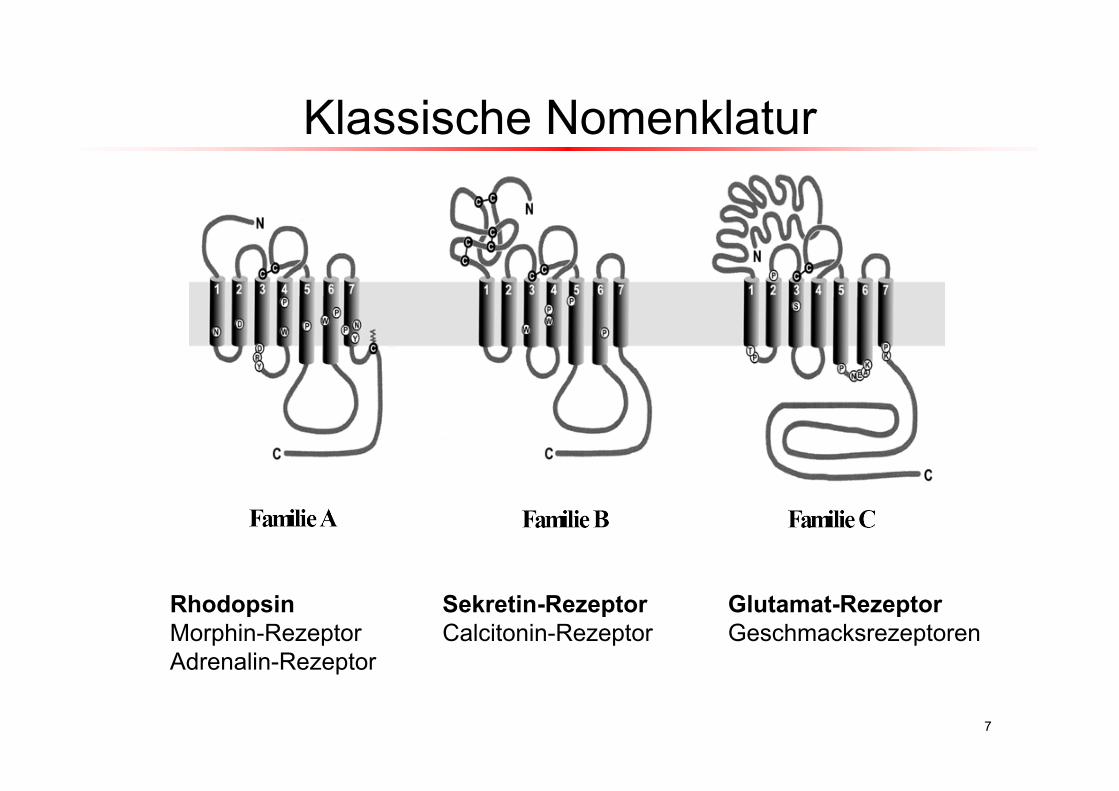
RhodopsinMorphin-RezeptorAdrenalin-Rezeptor
Sekretin-RezeptorCalcitonin-Rezeptor
Glutamat-RezeptorGeschmacksrezeptoren
Klassische Nomenklatur
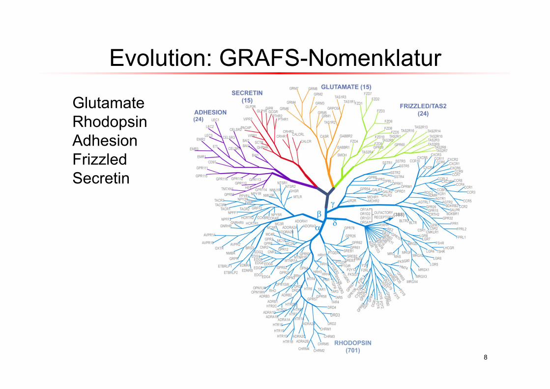
Evolution: GRAFS-Nomenklatur
8
GlutamateRhodopsinAdhesionFrizzledSecretin
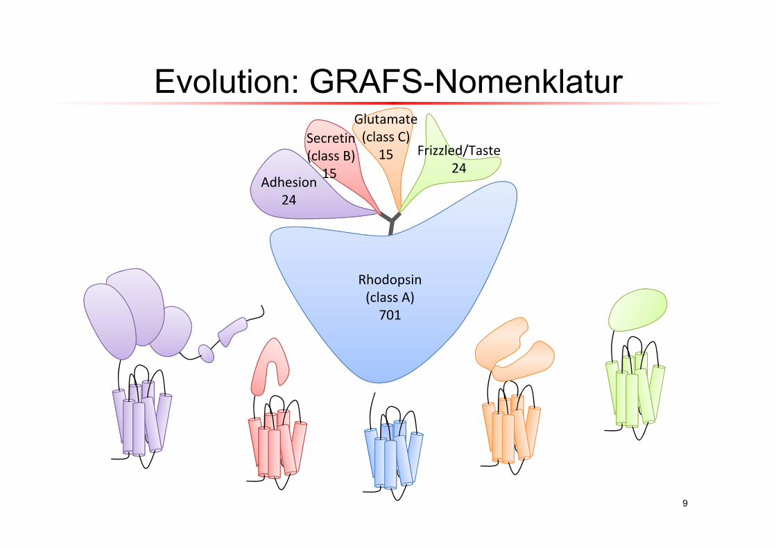
9
N
Frizzled/Taste24
Secretin(class B)
15
Rhodopsin(class A)701
Glutamate(class C)
15
Adhesion24
Evolution: GRAFS-Nomenklatur
10
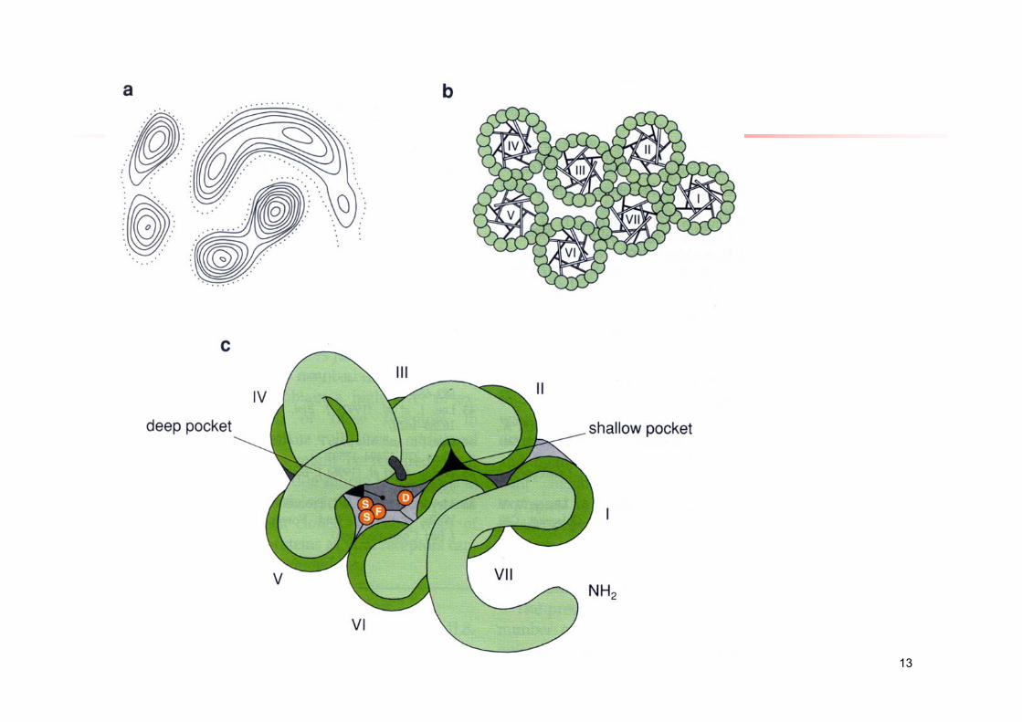
Rhodopsin-ähnliche Rezeptoren
11
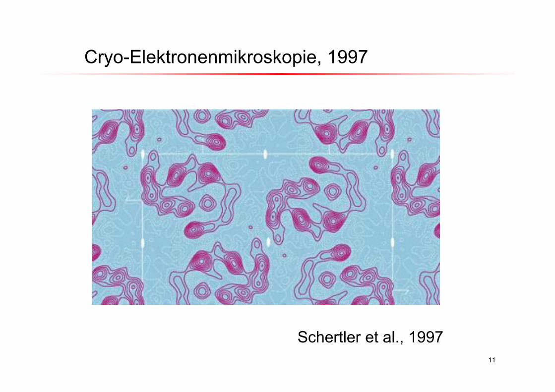
Cryo-Elektronenmikroskopie, 1997
Schertler et al., 1997
12
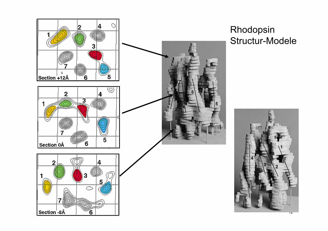
RhodopsinStructur-Modele
13
14
zytoplasmatisch extrazellulaer
V
VI
IV
III
VIIVIII
II
I
I
III IV VVIVII
II
VIII
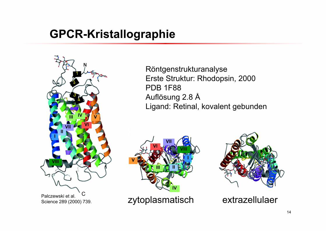
CPalczewski et al.Science 289 (2000) 739.
RöntgenstrukturanalyseErste Struktur: Rhodopsin, 2000PDB 1F88Auflösung 2.8 ÅLigand: Retinal, kovalent gebunden
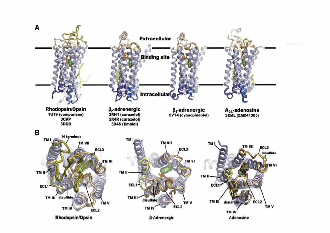
GPCR-Kristallographie
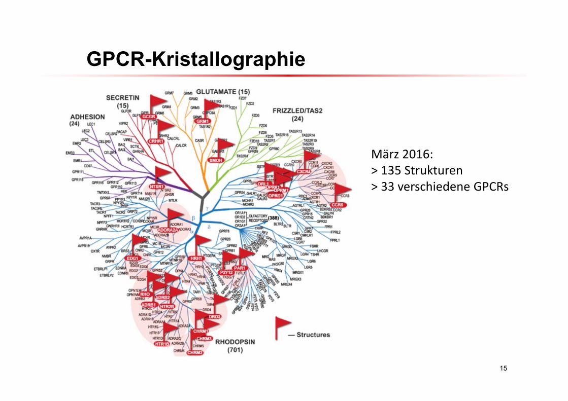
15
März 2016:> 135 Strukturen> 33 verschiedene GPCRs
GPCR-Kristallographie
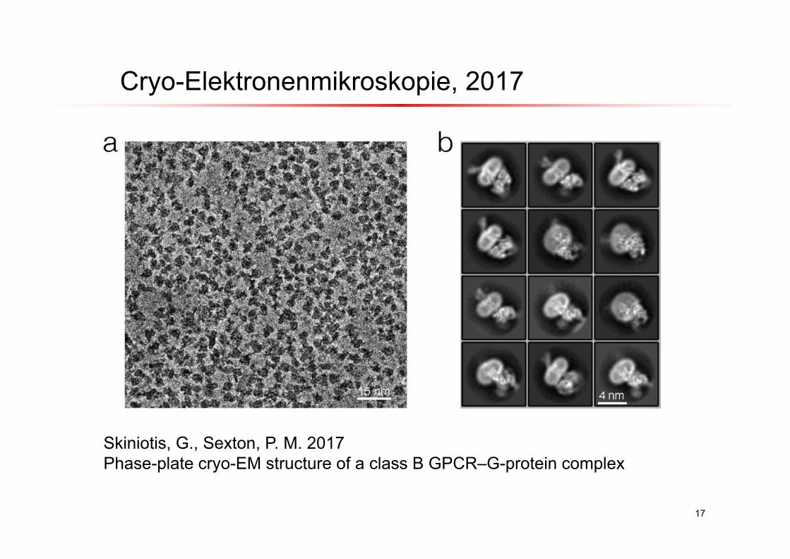
17
Skiniotis, G., Sexton, P. M. 2017Phase-plate cryo-EM structure of a class B GPCR–G-protein complex
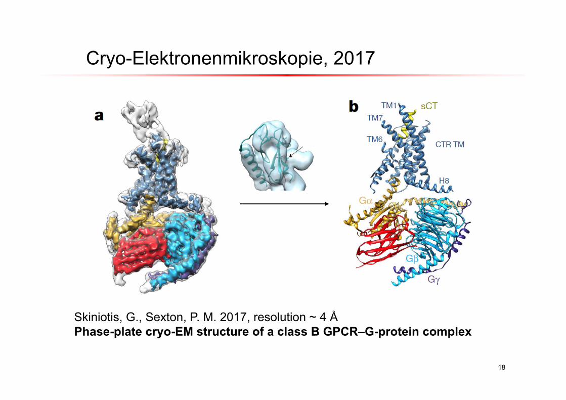
Cryo-Elektronenmikroskopie, 2017
18
Skiniotis, G., Sexton, P. M. 2017, resolution ~ 4 ÅPhase-plate cryo-EM structure of a class B GPCR–G-protein complex
Cryo-Elektronenmikroskopie, 2017
19
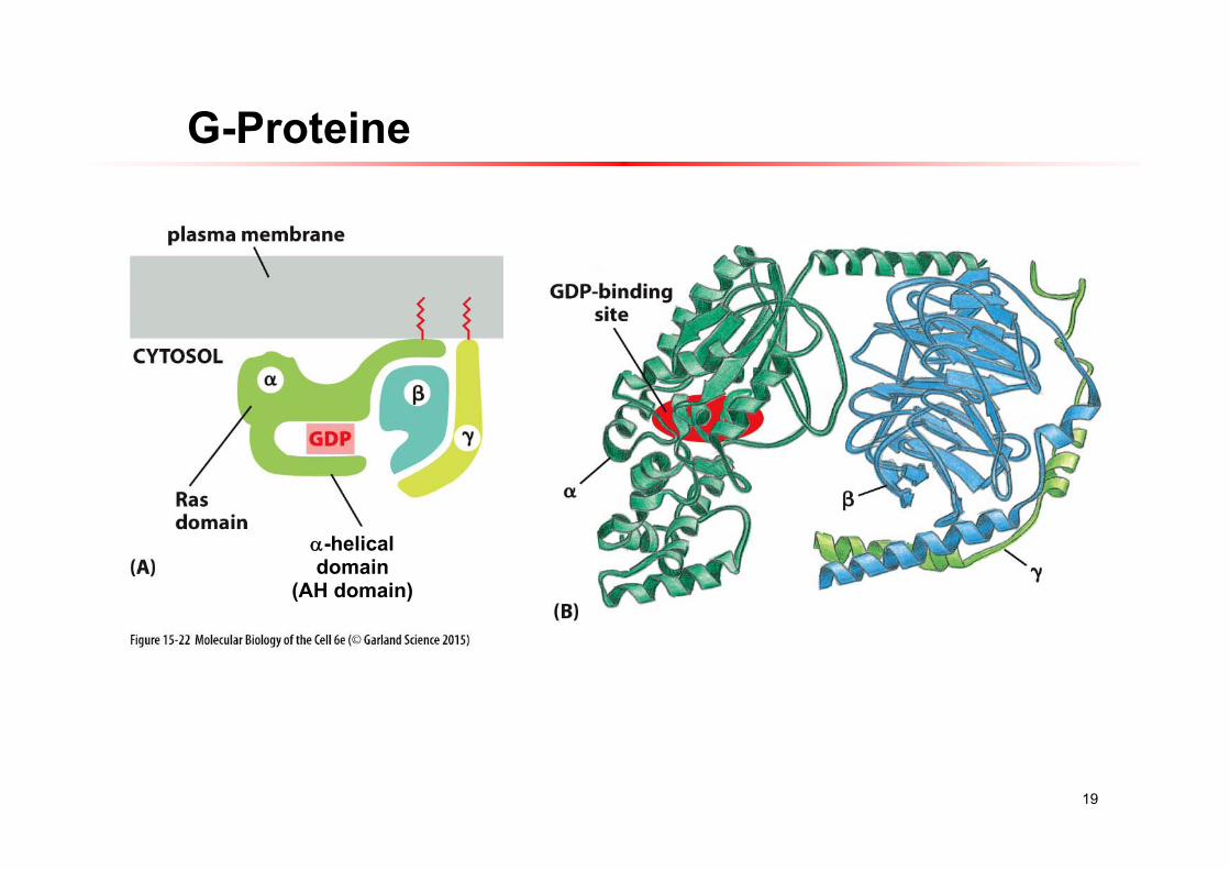
G-Proteine
-helicaldomain
(AH domain)
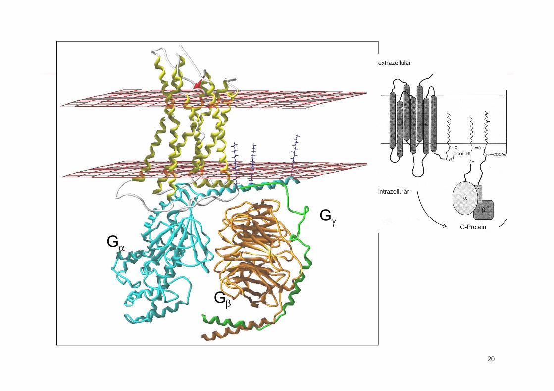
20
G
G
G
21
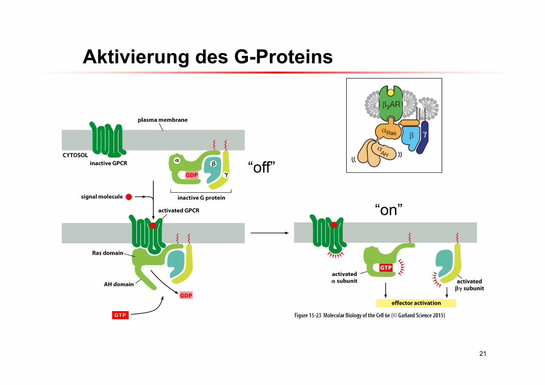
Aktivierung des G-Proteins
GTP
“off”
“on”
22
GPCR-G-Protein Aktivierung
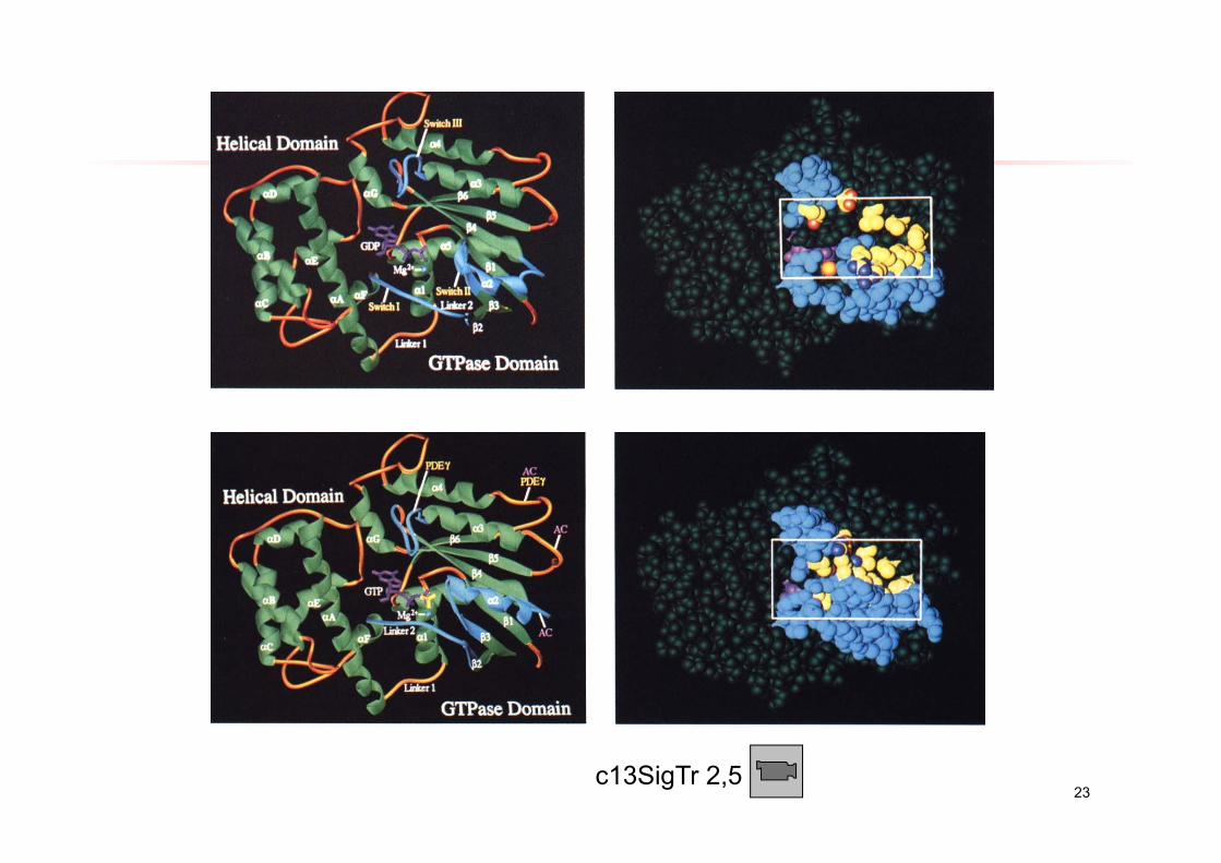
23c13SigTr 2,5
24
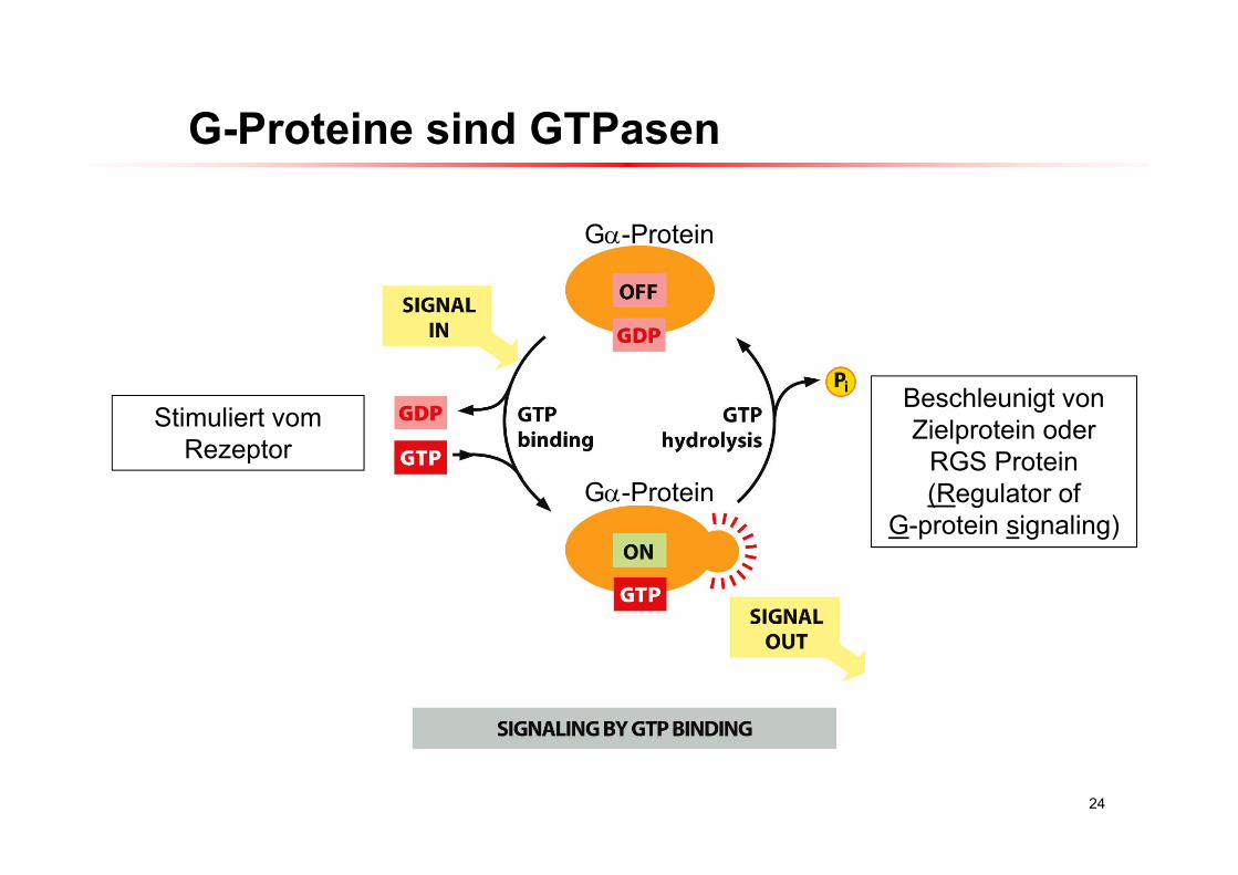
G-Proteine sind GTPasen
G-Protein
G-Protein
Beschleunigt von Zielprotein oder
RGS Protein(Regulator of
G-protein signaling)
Stimuliert vomRezeptor
25
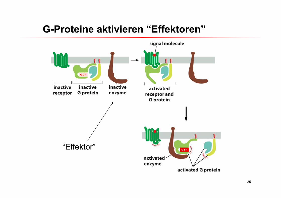
G-Proteine aktivieren “Effektoren”
“Effektor” GTP
GDP
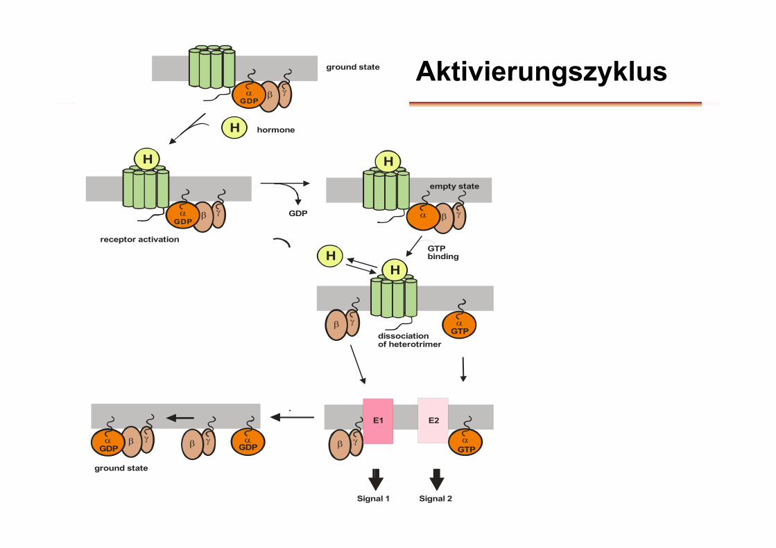
Aktivierungszyklus
hormone
GDP
receptor activation
dissociationof heterotrimer
ground state
GDP
H
GDP
H H
empty state
GTP
E1 E2
GTP
GDP
GTPase RGS
ground state
HH
GDP
GTPbinding
Signal 2Signal 1
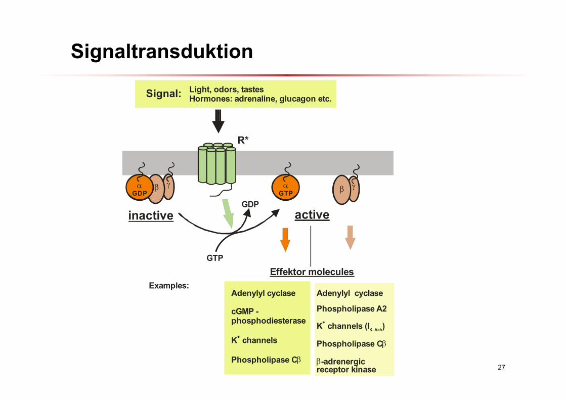
27
GDP GTP
R*
inactive active
Signal: Light, odors, tastesHormones: adrenaline, glucagon etc.
GTP
GDP
Effektor molecules
Adenylyl cyclase
cGMP - phosphodiesterase
K channels
Phospholipase C
+
Adenylyl cyclase
Phospholipase A2
K channels (I )
Phospholipase C
-adrenergic receptor kinase
+K. Ach
Examples:
Signaltransduktion
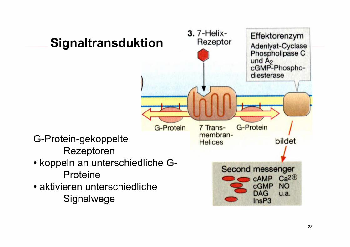
28
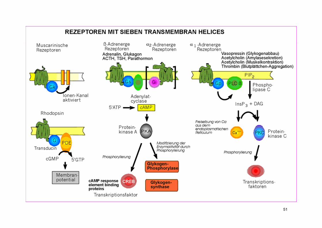
G-Protein-gekoppelteRezeptoren
• koppeln an unterschiedliche G-Proteine
• aktivieren unterschiedliche Signalwege
Signaltransduktion
29
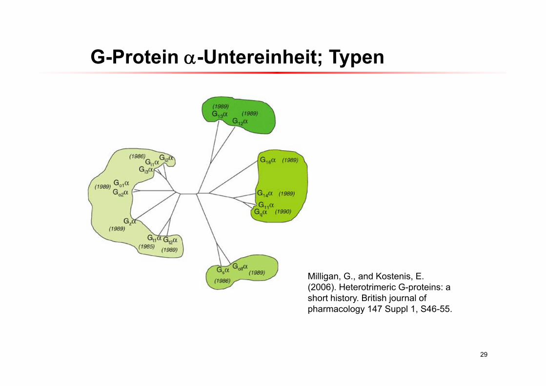
G-Protein -Untereinheit; Typen
Milligan, G., and Kostenis, E. (2006). Heterotrimeric G-proteins: a short history. British journal of pharmacology 147 Suppl 1, S46-55.
30
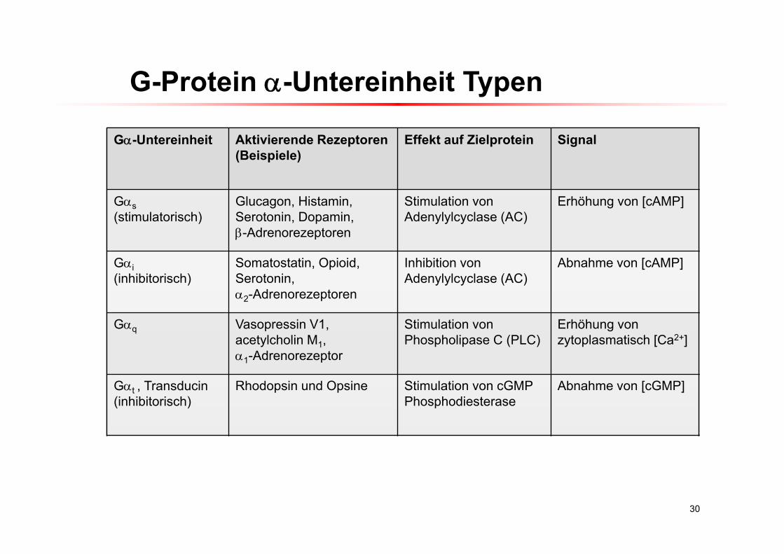
G-Protein -Untereinheit Typen
G-Untereinheit Aktivierende Rezeptoren(Beispiele)
Effekt auf Zielprotein Signal
Gs(stimulatorisch)
Glucagon, Histamin, Serotonin, Dopamin,-Adrenorezeptoren
Stimulation von Adenylylcyclase (AC)
Erhöhung von [cAMP]
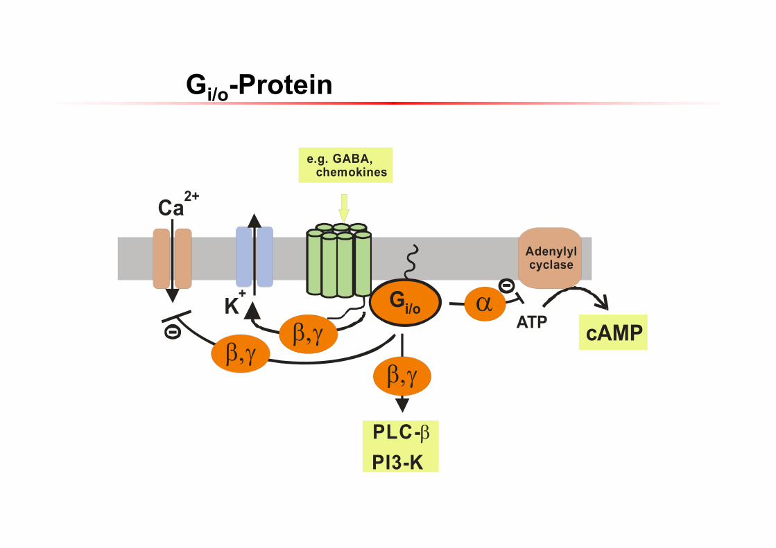
Gi(inhibitorisch)
Somatostatin, Opioid,Serotonin, 2-Adrenorezeptoren
Inhibition von Adenylylcyclase (AC)
Abnahme von [cAMP]
Gq Vasopressin V1, acetylcholin M1, 1-Adrenorezeptor
Stimulation von Phospholipase C (PLC)
Erhöhung von zytoplasmatisch [Ca2+]
Gt , Transducin(inhibitorisch)
Rhodopsin und Opsine Stimulation von cGMPPhosphodiesterase
Abnahme von [cGMP]
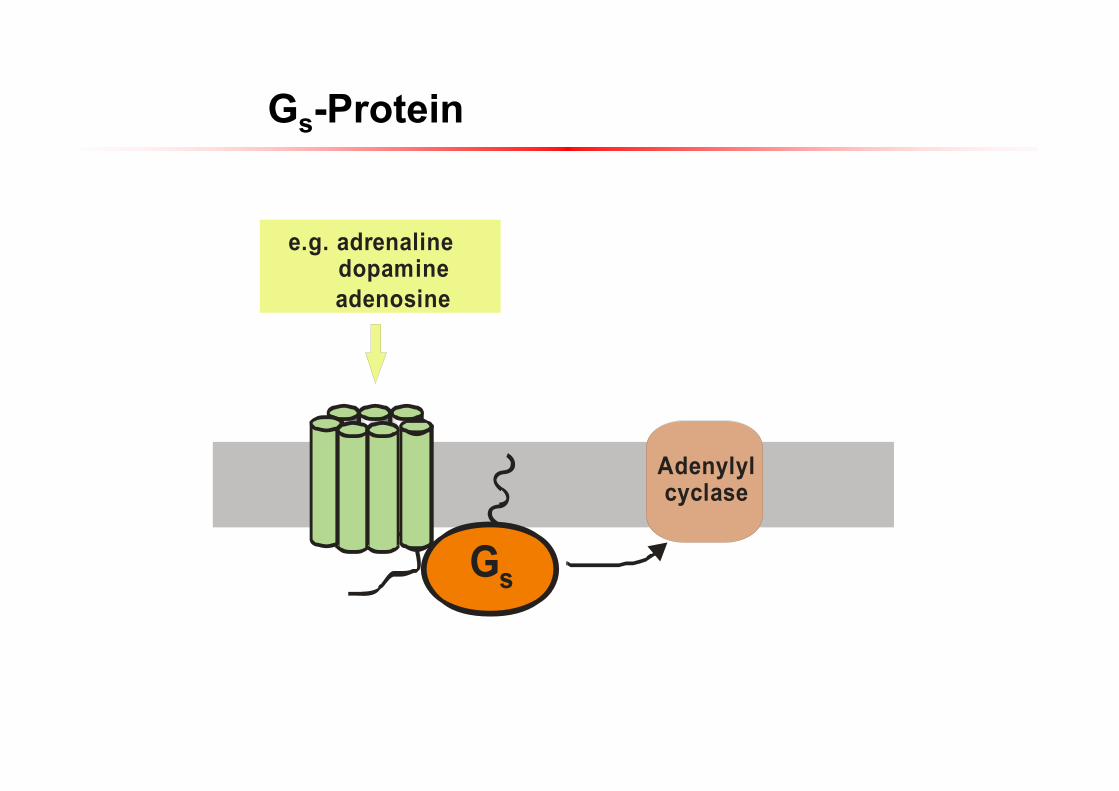
Gs
Adenylylcyclase
e.g. adrenaline dopamine adenosine
Gs-Protein
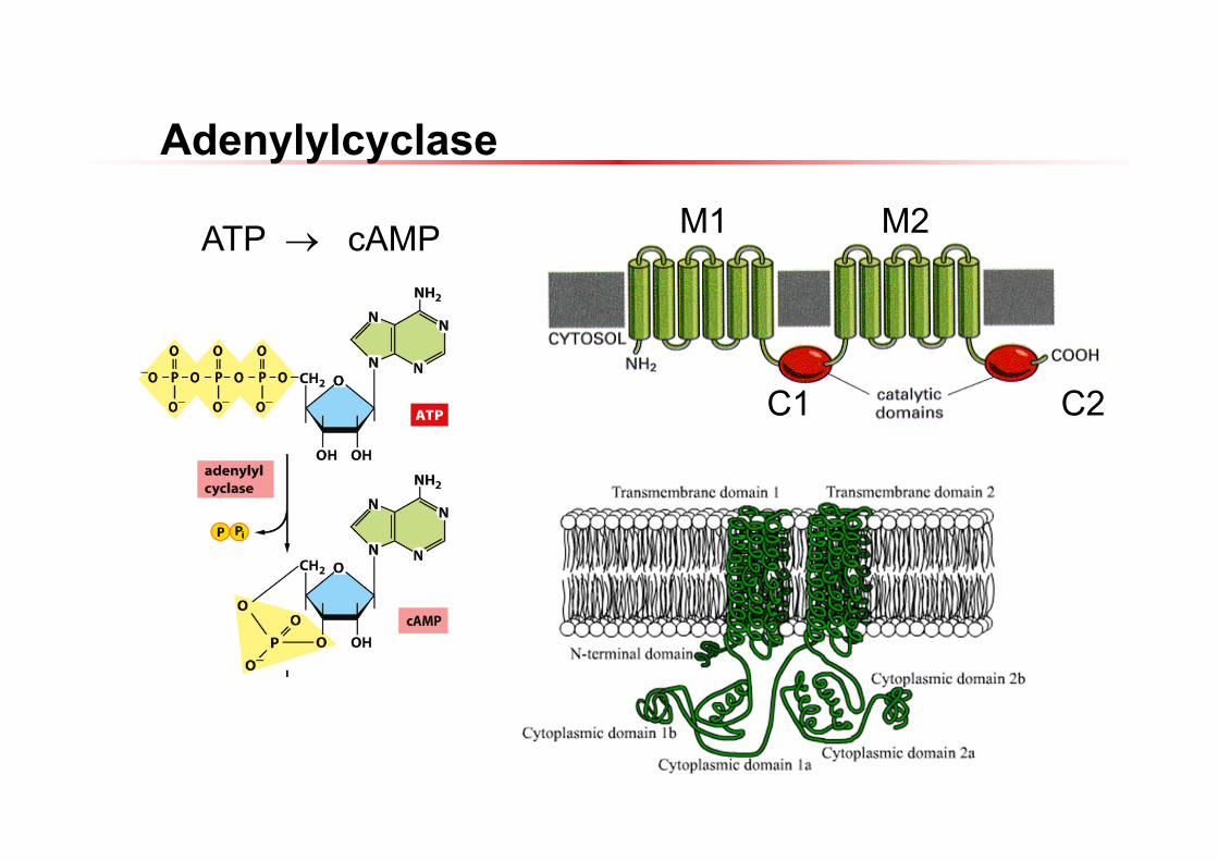
Adenylylcyclase
M1 M2
C1 C2
ATP cAMP
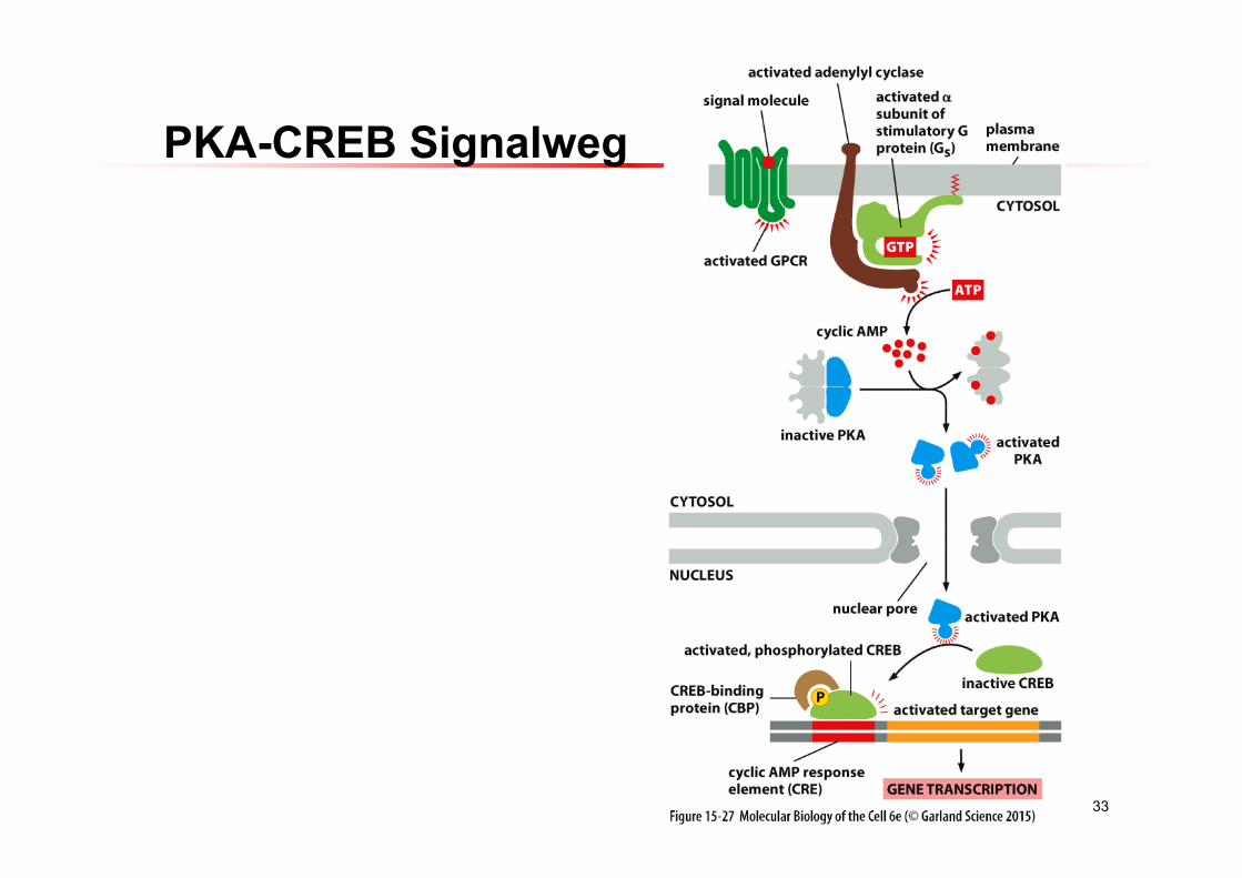
33
PKA-CREB Signalweg
ATP cAMP
Adenylylcyclase
Gi/o
PLC-PI3-K
Ca2+
K+
e.g. GABA, chemokines
Gi/o-Protein
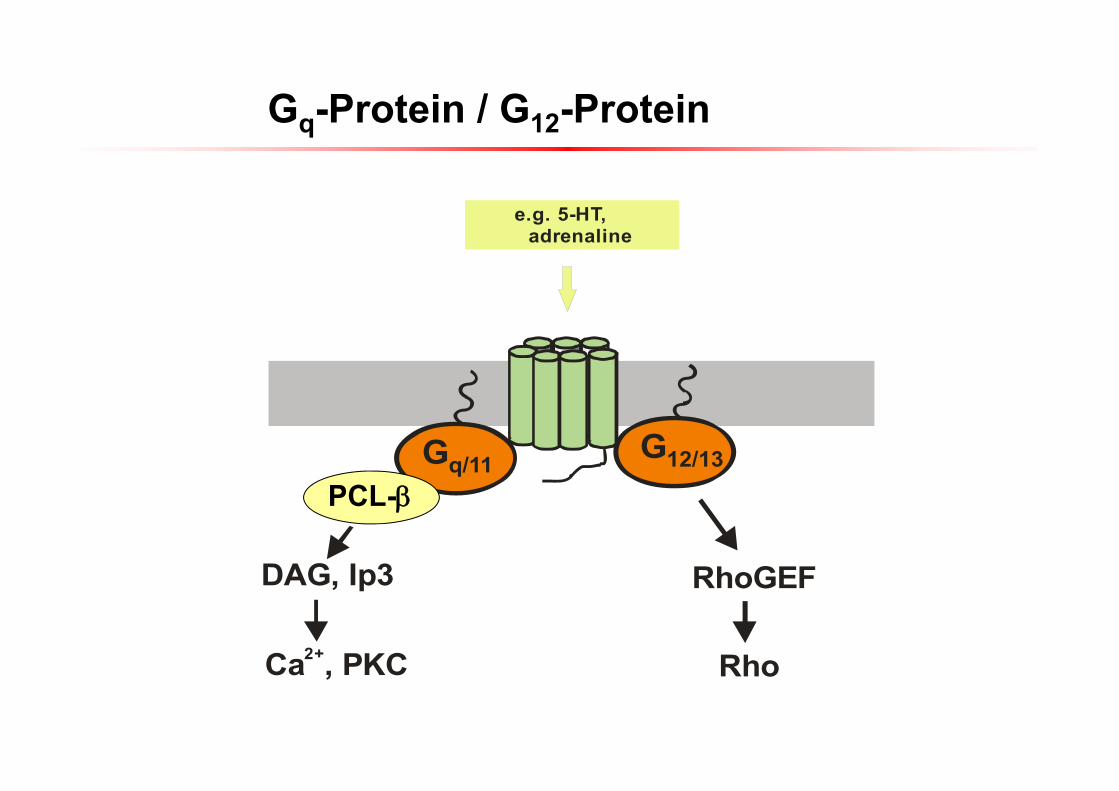
Gq/11 G12/13
RhoGEF
Rho
PLC-
Ca , PKC2+
e.g. 5-HT, adrenaline
DAG, Ip3
Gq-Protein / G12-Protein
PCL-
36
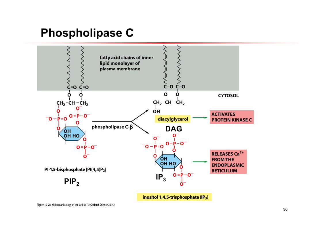
Phospholipase C
PIP2IP3
DAG
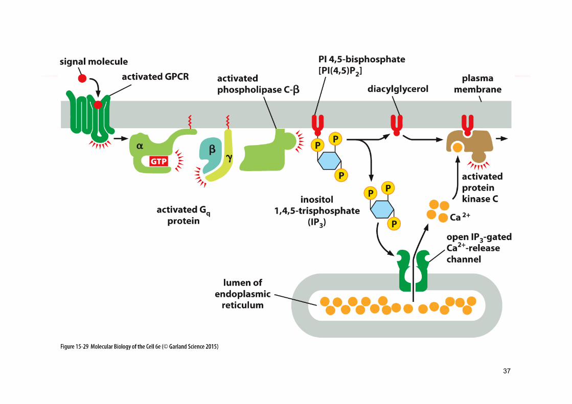
37
38
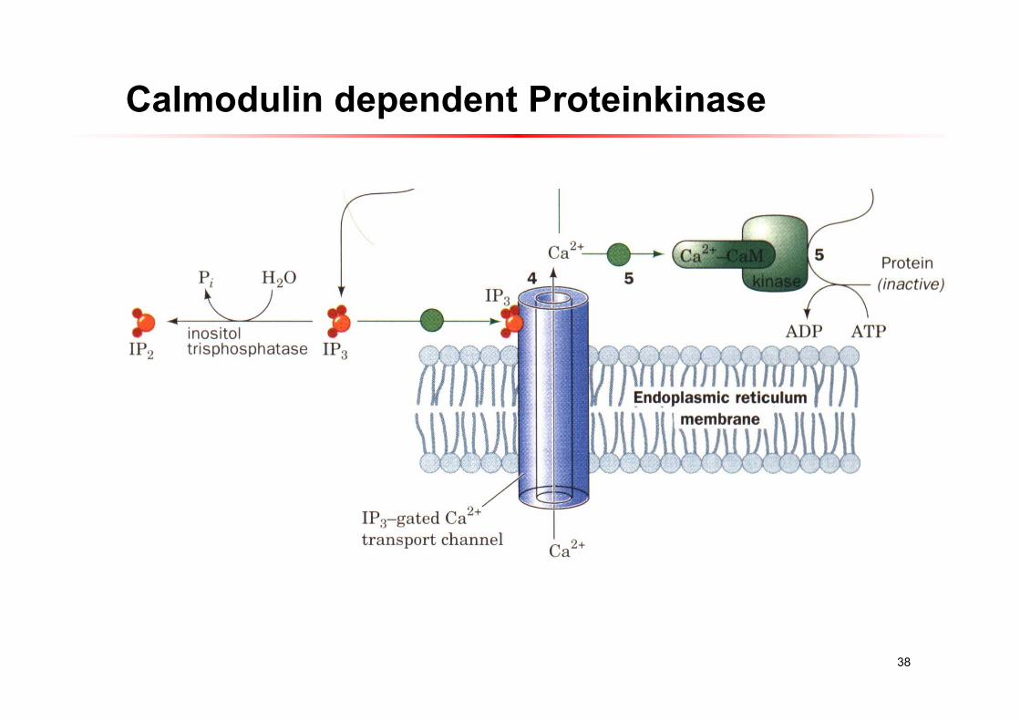
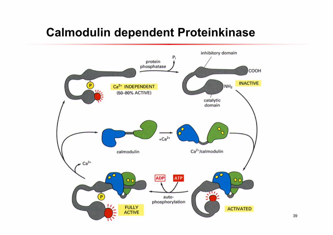
Calmodulin dependent Proteinkinase
39
Calmodulin dependent Proteinkinase
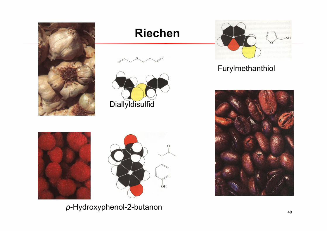
Riechen
40
Diallyldisulfid
Furylmethanthiol
p-Hydroxyphenol-2-butanon
41
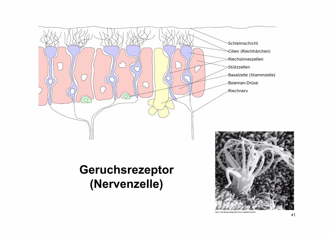
Geruchsrezeptor(Nervenzelle)
42
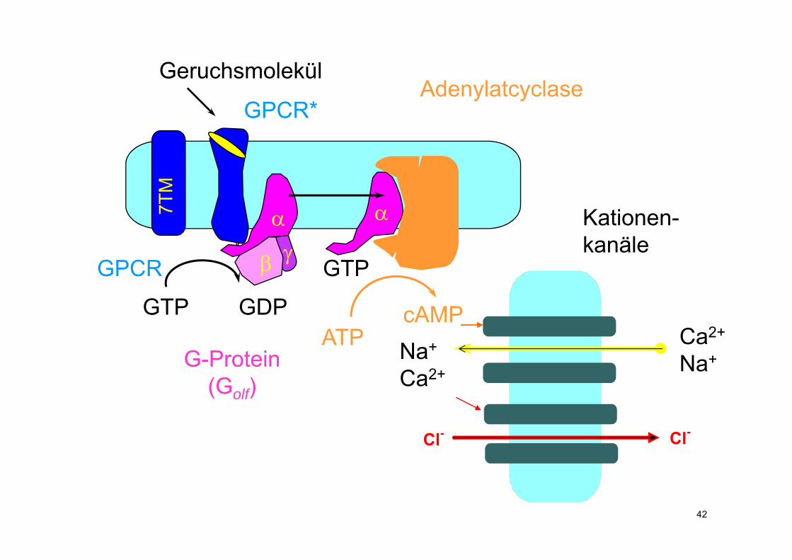
GPCR
GPCR*
G-Protein(Golf)
GTP GDP
GTP
Adenylatcyclase
ATPcAMP
7TM
Geruchsmolekül
Kationen-kanäle
Ca2+
Na+Na+
Ca2+
Cl- Cl-
43
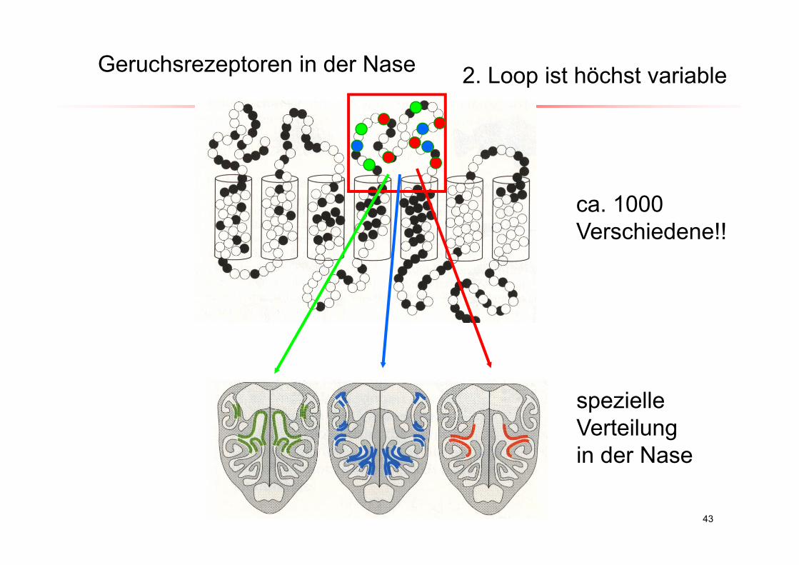
Geruchsrezeptoren in der Nase
ca. 1000 Verschiedene!!
2. Loop ist höchst variable
spezielle Verteilungin der Nase
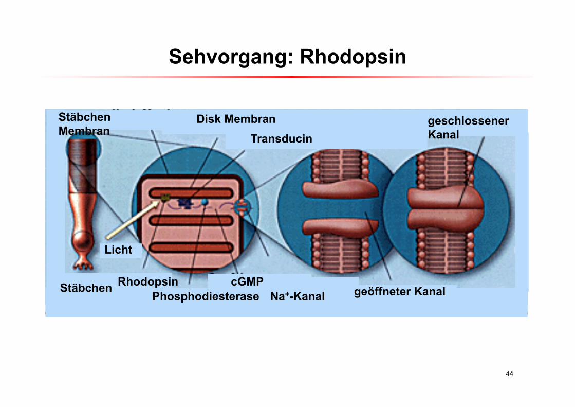
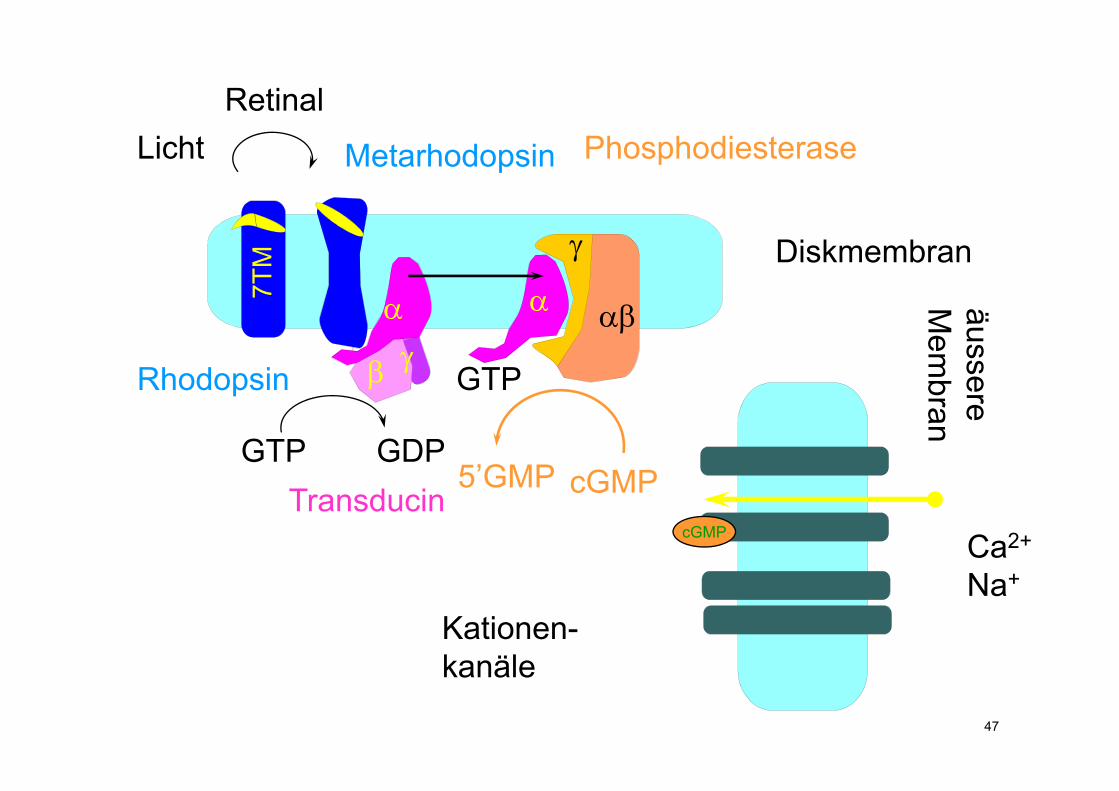
Sehvorgang: Rhodopsin
44
TransducinDisk Membran
Licht
RhodopsinPhosphodiesterase
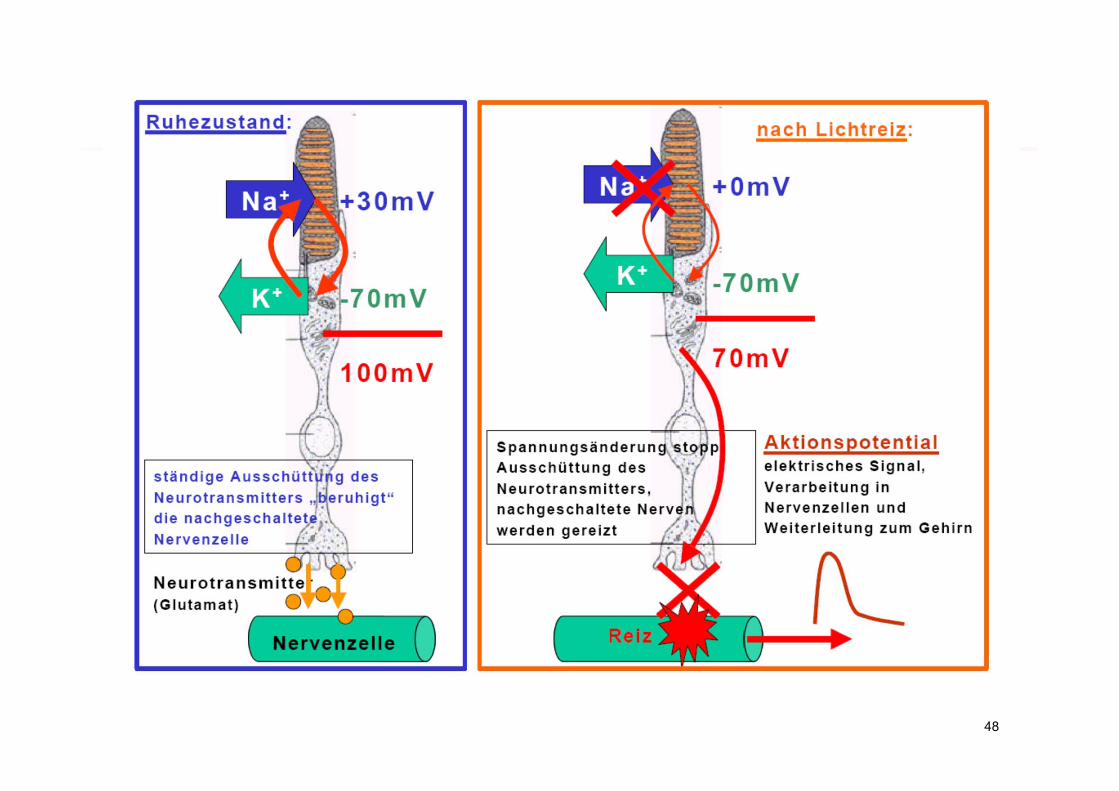
cGMPNa+-Kanal geöffneter Kanal
geschlossener Kanal
Stäbchen
Stäbchen Membran
45
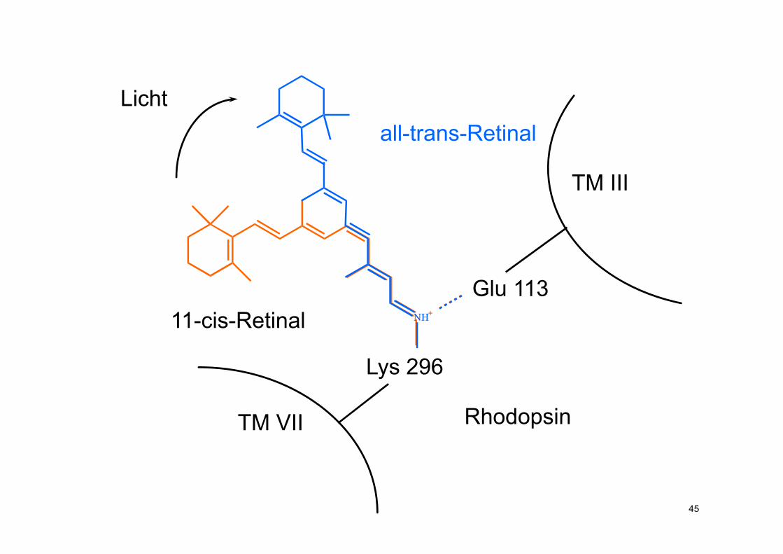
11-cis-Retinal NH+
Lys 296
TM VII
Glu 113NH+
all-trans-Retinal Licht
Rhodopsin
TM III
46
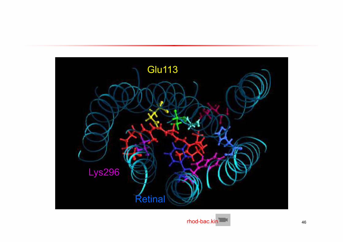
Glu113
Lys296
Retinal
rhod-bac.kin
47
Licht
Rhodopsin
Metarhodopsin
Transducin
GTP GDP
GTP
Phosphodiesterase
5’GMP cGMP
7TM
Retinal
Ca2+
Na+
Kationen-kanäle
äussereM
embran
Diskmembran
cGMP
48
t
49
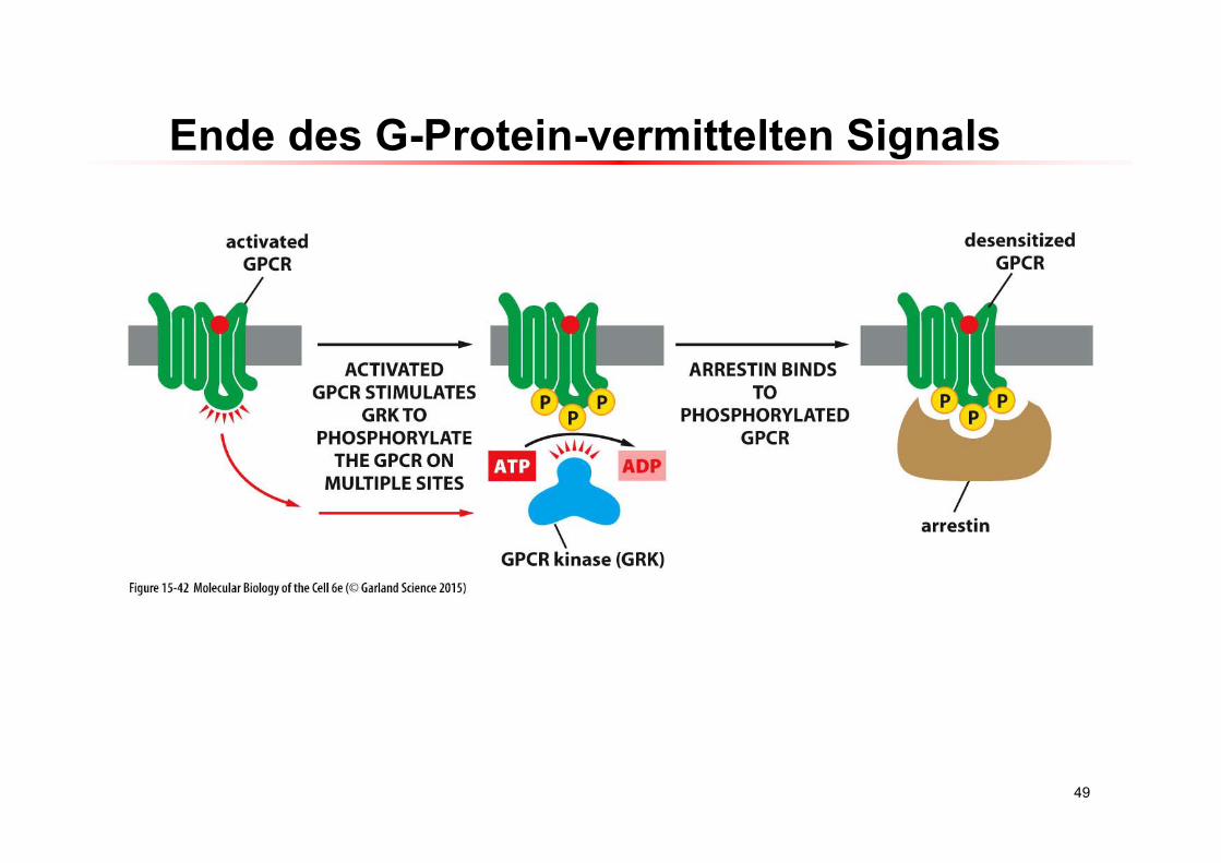
Ende des G-Protein-vermittelten Signals
50
https://www.youtube.com/watch?v=V_0EcUr_txk
51
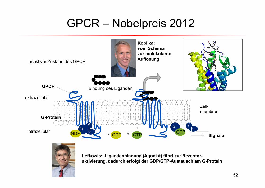
GPCR – Nobelpreis 2012
52
α βγ
extrazellulär
intrazellulärα γ
βGDP GTP- +
Bindung des Liganden
GDP
inaktiver Zustand des GPCR
GTP
GPCR
G-Protein
Signale
Zell-membran
Lefkowitz: Ligandenbindung (Agonist) führt zur Rezeptor-aktivierung, dadurch erfolgt der GDP/GTP-Austausch am G-Protein
Kobilka: vom Schemazur molekularenAuflösung