TheStudyoftheReactionofPectin-Ag(0) NanocompositesFormation
10
Hindawi Publishing Corporation International Journal of Carbohydrate Chemistry Volume 2012, Article ID 459410, 9 pages doi:10.1155/2012/459410 Research Article The Study of the Reaction of Pectin-Ag(0) Nanocomposites Formation Nadezhda Viktorovna Ivanova, Natalya Nikolaevna Trofimova, Lydmila Alexandrovna Es’kova, and Vasilyi Anatolievich Babkin A.E. Favorsky Institute of Chemistry, Russian Academy of Sciences, Siberian Branch, Irkutsk 664033, Russia Correspondence should be addressed to Natalya Nikolaevna Trofimova, [email protected] Received 31 May 2012; Accepted 10 October 2012 Academic Editor: Yuriy A. Knirel Copyright © 2012 Nadezhda Viktorovna Ivanova et al. This is an open access article distributed under the Creative Commons Attribution License, which permits unrestricted use, distribution, and reproduction in any medium, provided the original work is properly cited. Pectin polysaccharides (PSs) were isolated from a bark of Larix sibirica Ledeb. Structure of PS fragments determined by chemical transformations, chromatography, and spectroscopic analyses was found to be a linear galacturonane comprising 1,4-linked α- D-Gal p A residues and a rhamnogalacturonan I (RG-I). The fifth part of galacturonane residues was methyl esterified at at C-2 and/or C-3 and C-6 atoms. Some of RG-I side chains were identified as arabinogalactan subunits with highly branched structure consisting of linear backbone with → 3,6)-β-D-Gal p -(1 → residues, substituted at C-6 by neutral side chains. This side chains contained → 2,5)-α-L-Ara f -(1 → and → 3,5)-α-L-Ara f -(1 → residues and terminal arabinose in the pyranose and furanose form. It was found that “pectin-Ag(0)” nanobiocomposites were formed via the interaction between PS aqueous solutions and silver nitrate, with PS playing both reducing and stabilizing functions. It was shown that the content of Ag(0) particles in “pectin-Ag(0)” depended on the reaction conditions and can range from 0.1 to 72 %, the size of Ag(0) particles being 3–27 nm. Using 13 C NMR technique, it was revealed that PS underwent destructive changes and they they were more considerable, more than the lot of Ag(I) that was inputed into the reactionary medium. 1. Introduction Two species of the genus Larix Mill, Larix sibirica Ledeb and Larix gmelinii (Rupr.) Rupr., are considered to be the most abundant trees in the Russian Federation and the total stock of their wood recourses exceeds 26 billion m 3 . Traditionally, the main economic benefit of the larch wood is a manufacture of roundwood (timber), the value of which is determined by a high quality of lumber obtained from this tree. Sometimes, the larch wood can be used in insignificant quantities in pulp-and-paper manufacture to produce pulp. Currently, about 40% of this valuable wood (bark, sawdust) is utilized as wastes. Such inexpedient approach to the utilization of larch wood does not allow using the richest potential of this renewable raw material. Meanwhile, biolog- ically active compounds contained in larch biomass can be successfully applied for the manufacture of medical, food, and agricultural products. The development of complex technologies for chemical processing of larch biomass and waste timber will considerably raise the economic value of this biological resource. However, the larch bark does not find industrial applica- tion. Annual volume of waste produced by wood-processing industry and the pulp-and-paper enterprises is more than 30 million m 3 . It represents a serious environmental challenge because the bark is poorly exposed to biodegradation. At the same time, the chemical compounds of the bark can be used as a source of valuable biologically active substances including polysaccharides. Earlier we have showed that the content of pectin poly- saccharides in the larch bark is about 12% from the weight of absolutely dry raw material (a.d.r.m.) and the larch bark can be promising alternative raw material. The method of pectin isolation has been developed, and common physical and chemical characteristics and membranotropic activity of pectin have been investigated.
Transcript of TheStudyoftheReactionofPectin-Ag(0) NanocompositesFormation

Hindawi Publishing CorporationInternational Journal of Carbohydrate ChemistryVolume 2012, Article ID 459410, 9 pagesdoi:10.1155/2012/459410
Research Article
The Study of the Reaction of Pectin-Ag(0)Nanocomposites Formation
Nadezhda Viktorovna Ivanova, Natalya Nikolaevna Trofimova,Lydmila Alexandrovna Es’kova, and Vasilyi Anatolievich Babkin
A.E. Favorsky Institute of Chemistry, Russian Academy of Sciences, Siberian Branch, Irkutsk 664033, Russia
Correspondence should be addressed to Natalya Nikolaevna Trofimova, [email protected]
Received 31 May 2012; Accepted 10 October 2012
Academic Editor: Yuriy A. Knirel
Copyright © 2012 Nadezhda Viktorovna Ivanova et al. This is an open access article distributed under the Creative CommonsAttribution License, which permits unrestricted use, distribution, and reproduction in any medium, provided the original work isproperly cited.
Pectin polysaccharides (PSs) were isolated from a bark of Larix sibirica Ledeb. Structure of PS fragments determined by chemicaltransformations, chromatography, and spectroscopic analyses was found to be a linear galacturonane comprising 1,4-linked α-D-GalpA residues and a rhamnogalacturonan I (RG-I). The fifth part of galacturonane residues was methyl esterified at at C-2and/or C-3 and C-6 atoms. Some of RG-I side chains were identified as arabinogalactan subunits with highly branched structureconsisting of linear backbone with→ 3,6)-β-D-Galp-(1→ residues, substituted at C-6 by neutral side chains. This side chainscontained→ 2,5)-α-L-Araf -(1→ and→ 3,5)-α-L-Araf -(1→ residues and terminal arabinose in the pyranose and furanose form.It was found that “pectin-Ag(0)” nanobiocomposites were formed via the interaction between PS aqueous solutions and silvernitrate, with PS playing both reducing and stabilizing functions. It was shown that the content of Ag(0) particles in “pectin-Ag(0)”depended on the reaction conditions and can range from 0.1 to 72 %, the size of Ag(0) particles being 3–27 nm. Using 13C NMRtechnique, it was revealed that PS underwent destructive changes and they they were more considerable, more than the lot of Ag(I)that was inputed into the reactionary medium.
1. Introduction
Two species of the genus Larix Mill, Larix sibirica Ledeband Larix gmelinii (Rupr.) Rupr., are considered to be themost abundant trees in the Russian Federation and thetotal stock of their wood recourses exceeds 26 billion m3.Traditionally, the main economic benefit of the larch woodis a manufacture of roundwood (timber), the value of whichis determined by a high quality of lumber obtained from thistree. Sometimes, the larch wood can be used in insignificantquantities in pulp-and-paper manufacture to produce pulp.Currently, about 40% of this valuable wood (bark, sawdust)is utilized as wastes. Such inexpedient approach to theutilization of larch wood does not allow using the richestpotential of this renewable raw material. Meanwhile, biolog-ically active compounds contained in larch biomass can besuccessfully applied for the manufacture of medical, food,and agricultural products. The development of complex
technologies for chemical processing of larch biomass andwaste timber will considerably raise the economic value ofthis biological resource.
However, the larch bark does not find industrial applica-tion. Annual volume of waste produced by wood-processingindustry and the pulp-and-paper enterprises is more than 30million m3. It represents a serious environmental challengebecause the bark is poorly exposed to biodegradation. Atthe same time, the chemical compounds of the bark can beused as a source of valuable biologically active substancesincluding polysaccharides.
Earlier we have showed that the content of pectin poly-saccharides in the larch bark is about 12% from the weightof absolutely dry raw material (a.d.r.m.) and the larch barkcan be promising alternative raw material. The method ofpectin isolation has been developed, and common physicaland chemical characteristics and membranotropic activity ofpectin have been investigated.

2 International Journal of Carbohydrate Chemistry
Residue 1
Residue 2
Residue 3
v/v), 0.5% ammonium oxalate
Extract
Larch bark
Extraction with ethyl
acetate, 70◦C, 3 h
Extraction with
water, 70◦C, 3 h
Water extract
Extraction with mixture (1/1,
and 0.5% oxalic acid, 80◦C, 3 h
Pectin substances (PS)
(1) Precipitation with C2H5OH
(2) Dialysis
(3) Lyophilic drying
Ethyl acetate extract
Figure 1: Scheme of pectin substances extraction from larch bark.
It has been established and patented that the larch barkpectin can possess reducing and stabilizing properties in theformation of nanobiocomposites with precious metals ions.
The present work aims at the following.
(1) The study of general characteristics of pectin sub-stances (PS) extracted from bark of L. sibirica Ledeb.and structure investigation of its main linear and sidechains.
(2) The study of PS interaction with silver nitrate andelucidation of alteration of pectin structural char-acteristics during the formation of “pectin-Ag(0)”nanobiocomposite.
2. Methods and Materials
Pectin substances were extracted from the bark of L. sibiricaLedeb. according to the scheme depicted in Figure 1.Larch air-dried bark (500 g) which was preliminary grindedand treated with ethyl acetate was extracted with distilledwater at 70◦C for 3 h. The residue of raw material waspoured into a mixture (1 : 1, v/v) 0.5% aqueous solution ofammonium oxalate and 0.5% aqueous solution of oxalic acidand heated at 80◦C for 2 h. The extract was concentrated.
Polysaccharides were precipitated with triple volume of ethylalcohol or acetone and dried with lyophilization. As a result,PS was obtained.
PS (50 mg) was dissolved in 20 mL of water. Pektinaza(2 mg, Sigma, USA) aqueous solution was added. Themixture was temperature-controlled at 37◦C for 3 h. Then, areaction mixture was heated for 5 min in water bath at100◦C. Coagulated protein was separated by centrifugation.Obtained supernatant was concentrated up to 5 mL, 96%ethyl alcohol was added (4 volumes). Deposition was sepa-rated by centrifugation. Alcoholic supernatant was concen-trated and analyzed by paper chromatography (PC).
Paper chromatography was carried out on “Filtrak FN-13” paper with a descending method in n-butanol-pyridine-water system (volume correlations 6 : 4 : 3, resp.). To definecarbohydrates, the paper was poured with aniline phthalateand heated at 105◦C.
Glucuronic acids content in PS was defined accordingto the reaction with 3,5-dimethyl phenol in the presenceof concentrated H2SO4, protein using Lowry method [1]with the calibrating schedule for a bovine serum albumin80000 Da.
Gas-liquid chromatography (GLC) was carried out on aHewlett-Packard 4890A (the USA) chromatograph equipped

International Journal of Carbohydrate Chemistry 3
with flame-ionization detector, RTX-1 (0.25 mm × 30 m)capillary column, argon carrier gas, 1 : 60 dumping. Temper-ature rate: 175◦C (1 min)–250◦C (2 min), Δ3◦/min.
Total acid hydrolysis PS (5 mg) was carried out with theimplementation of 2 M trifluoroacetic acid (TFA) (2 mL)which contained mio-inositol (1 mg/mL). The mixture insoldered ampoule was heated for 5 h at 100◦C, and the acidwas removed by repeated dry evaporation with methanoladdition.
Ion-exchange chromatography PS (100 mg) was carriedout on DEAE-cellulose (25 × 2 cm) column. NaCl solu-tions were used as an eluent with increasing concentration(0.01 M–1 M, 60 mL/h elution’s speed, fractions’ selection by12 mL). Pick correspondent fractions at the output bentswere combined, dialyzed, and lyophilized. As a result, PS 1–4fractions were obtained. Monosaccharides of each fractionafter hydrolysis of PS 1–4 and acetilation were defined byGLC.
Monosaccharides acetilation: each PS 1–4 fraction wasdissolved in 1 M ammonia solution (1 mL) and 5 mg ofNaBH4 was added. The mixture was kept at room tempera-ture during one day. Then, NaBH4 abundance was destroyedby adding 2-3 drops of concentrated acetic acid; 0.2 mLof dry pyridine and acetic anhydride were added to a dryresidue. The mixture was being acetilized at 100◦C during1 h. The solution was evaporated up to pyridine and aceticanhydride abundance removal, first adding 1 mL of tolueneand then 1 mL of methanol. The obtained acetate mixture ofPS 1–4 polyol fractions was dissolved in 0.2 mL of dry chlo-roform and moved quantitatively to Appendorf tubes, con-centrated up to 0.1-0.2 mL, and analyzed with GLC method.
PS (5 mg) partial acidic hydrolysis was carried out with0.05 M TFA (2 mL) which contained mio-inositol (1 mg/mL).The mixture in soldered ampoule was heated at 100◦C for3 h. The acid was removed by repeated dry evaporation withmethanol addition to give PS-s.
PS (5 mg) partial acidic hydrolysis was carried out with0.01 M TFA (2 mL) which contained mio-inositol (1 mg/mL).The mixture in soldered ampoule was heated at 100◦C for3 h. The acid was removed by repeated dry evaporation withmethanol addition to afford PS-g.
IR spectra were registered on a FT-IR (RAM II) BrukkerVertex 70 spectrometer in pellets with KBr.
13C NMR spectra were recorded on a Bruker DRX 500instrument (Germany) using 3–5% carbohydrate solution inD2O at 55 and 70◦C; the internal standard was DMSO-d6.
Synthesis of PS-g1 and PS-g1 nanobiocomposites “pec-tin-Ag(0)” was carried out according to the [2]. To 2 mL ofthe aqueous solution containing 1 g pectin, added 2 mL of0.12–5.92 mmol silver nitrate solution and kept at roomtemperature for 30 min, added 10% sodium hydroxide solu-tion up to pH 11, boiled on a water bath of 15 min andfiltered through the paper filter. Obtained products werepurified from low-molecular impurity by reprecipitationsfrom ethanol, dried in vacuum over CaCl2. The silver contentin nanobiocomposites was determined titrimetrically withammonium thiocyanate after preliminary metal translationin the ionic form by treatment with nitric acid by [3].
UV-Vis spectra of nanobiocomposite aqueous watersolutions were recorded on a Perkin Elmer Lambda 35 inst-rument in ultraviolet and visible areas.
3. Results and Discussions
Pectin substances are abundant in land and water plants,as well as some freshwater algae [6]. Being an importantcomponent of cell walls, they are involved in ion exchange,water metabolism, and cell wall structure formation. Theystimulate seed germination and germ growth, provide turgor,and so forth.
Unique physicochemical properties of pectin make itindispensable material in medical, food, and cosmetic indus-tries as gelling agent, thickener, stabilizer, and dietary fiber.Since recently, it is extensively used as a matrix carrier forbiologically active components in drugs. Pectins exert diversephysiological activities such as immunomodulating, hepato-protective, anticarcinogenic, and antimetastatic, that allowthem to be applied as drugs and biologically active foodadditives.
The pectins are typically isolated from economicallyvaluable pants, for example, citrus, apple, sugar beet, andsunflower head pith. Other plants such as amaranth, smallmallow, duckweed, SILENE, and coffee beans, have beenreported as potential sources of pectins [7–10].
The methods for isolation of pectin polysaccharides fromplant tissues are numerous. Among the classic ones is hydrol-ysis extraction of dry raw material particles of certain sizes[11] with hot water, organic and inorganic acid solutions aswell as salts, alkali or their mixtures as extracting solutions.Basic parameters of the pectin isolation process such asa raw material preprocessing, hydromodulus, temperature,extraction duration, medium pH and precipitator can bevaried depending on characteristics of the raw material [12].
3.1. Isolation and Structural Study of Main and Side Chainsof PS. Pectin substances were extracted from the bark of L.sibirica Ledeb. according to the scheme depicted in Figure 1.Pectinase treatment of a PS sample, isolated from larchbark by the above method and further analysis of thehydrolysis products by paper chromatography (PC), hasshown that significant destruction of PS occurs to form freeD-galacturonic acid. High positive value of the rotation angle(+245.3◦, c 0.1, H2O) indicates α-D-configuration of D-galactopyranosyluronic acid residues.
Table 1 shows main maxima of the absorption bands inthe IR spectra of PS and their assignment proving PS pectinnature of the samples [13]. Thus, enzyme hydrolysis andIR spectroscopy data prove that the polysaccharide isolatedfrom larch bark refers to pectin group.
To determine the sugar composition of PS the totalacid hydrolysis has been carried out. Monosaccharide iden-tification in hydrolysate has been performed by gas-liquidchromatography. It has been shown that the sample consistsof galacturonic acid and monosaccharides of arabinose,galactose, rhamnose, glucose, mannose, and (in minorquantity) xylose (Table 2). Dominating monosaccharides aregalactose and arabinose in a ratio of 2.7 : 1.
4 International Journal of Carbohydrate Chemistry
Table 1: Absorption band maximums in IR spectra of PS and theirassignment.
Frequency (ν, cm−1) Assignment
3460 ν(OH), ν(H2O)
3260 ν(NH)
2962, 2872 ν(CH3)
2573 ν(OH),
1730 ν(C=O)B COOH
1640 δ(OH)λ1540 δ(NH)
1380–1450 δ(C–CH3), ν(C–O) pyranose rings
1331 δ(OH) in pyranose rings
1265 ν(C–O) in esters
1150 ν(C–O–C)
1095 ν(C–C)
1027 ν(C–OH)
890 δ(C1–H) in glucopyranose ring
766, 629, 528 Pulse vibrations of pyranose ring
Partial acid hydrolysis of PS by 0.05 M TFA gives polysac-charide PS-s representing a linear polysaccharide with insi-gnificant amount of side chain subunits. It is also confirmedby the decrease of the relative content of neutral monosac-charides of PS-s in comparison with their content in initialpolysaccharide PS (GLC analysis of polyols peracetates of thecorresponding sugars in the studied substances, Table 2) andincrease of galacturonic acid content from 38.4% in PS up to88% in PS-s.
Values of chemical shifts (CSs) of carbon atoms in 13CNMR spectrum of PS-s (Table 3) as compared to data [4, 5]correspond to those of carbon atoms in D-galacturonic acidresidues in pyranose constituting linear fragment of thepectin molecule (pectin core).
The signal of anomeric carbon atom at 101.9 ppm indi-cates both the (1→ 4)-bonding between D-galacturonic acidresidues and α-configuration of C-1 anomeric atom. Signalat 176.2 ppm is assigned to C-6 atom and points to thefree carboxyl group in D-galacturonic acid residue. Besides,galacturonic acid residues esterified by methoxyl are presentthe PS-s molecule that follows from signals with CS at172.2 ppm (C-6-OCH3) and 54.4 ppm (–OCH3). Ratio ofintegrated signal intensities of carbon atoms in the methoxyland carboxyl groups is indicative of high degree of galactur-onan methoxylation.
Ion exchange column chromatography of PS on DEAEcellulose by 0.01–0.2 M sodium chloride aqueous solutionshas yielded four polysaccharide fractions, PS 1–4, whosechemical characteristics are summarized in Table 4.
In the fractions PS-1 and PS-2, galactose and arabinoseare predominant (18.26/52.96% and 11.65/30.83%, resp.),thus, they belong to acidic arabinogalactans. Acidic characterof PS is caused bythe presence of D-galacturonic acidresidues, the content of which in PS-1 is 5 times less thanin PS-2, while in PS-3 and PS-4 it is a major monosaccharidethat allows PS to pectins to be assigned. Content of neutral
Ser
Th
r
Asp
Glu
Pro
Gly
Ala
Val Ile
Leu
Tyr
Ph
e
His
Lys
Arg
Met
7
6
5
4
3
2
1
0
Yie
ld (
mas
s %
)
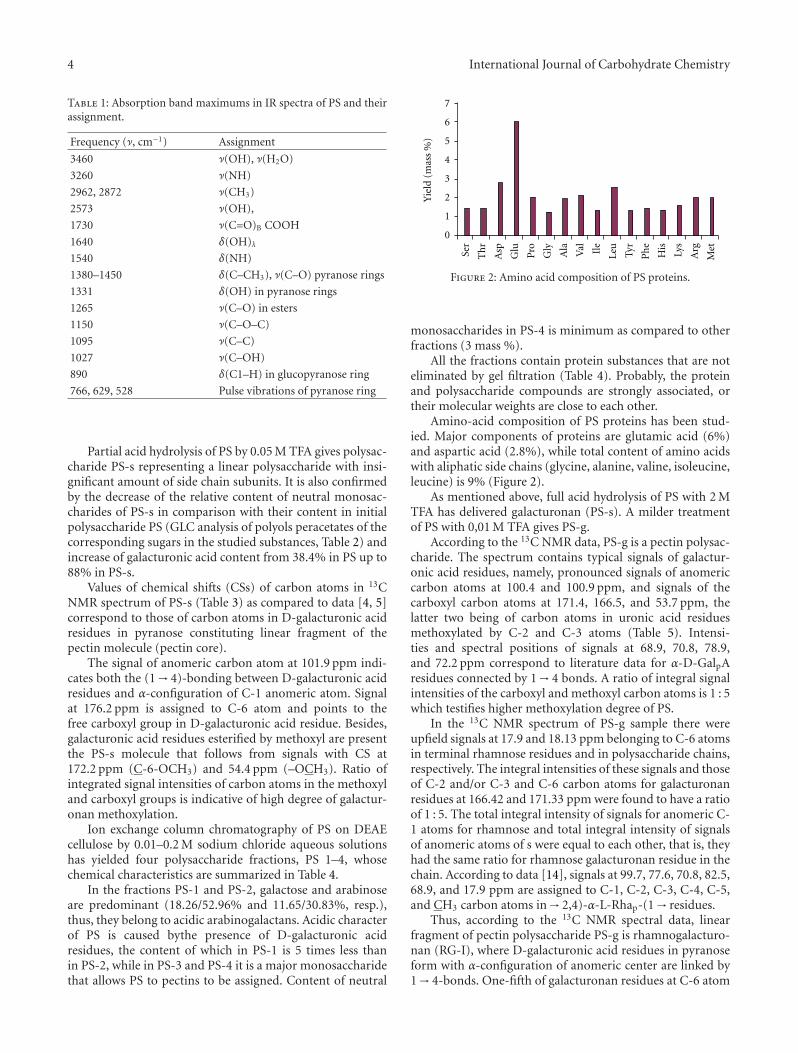
Figure 2: Amino acid composition of PS proteins.
monosaccharides in PS-4 is minimum as compared to otherfractions (3 mass %).
All the fractions contain protein substances that are noteliminated by gel filtration (Table 4). Probably, the proteinand polysaccharide compounds are strongly associated, ortheir molecular weights are close to each other.
Amino-acid composition of PS proteins has been stud-ied. Major components of proteins are glutamic acid (6%)and aspartic acid (2.8%), while total content of amino acidswith aliphatic side chains (glycine, alanine, valine, isoleucine,leucine) is 9% (Figure 2).
As mentioned above, full acid hydrolysis of PS with 2 MTFA has delivered galacturonan (PS-s). A milder treatmentof PS with 0,01 M TFA gives PS-g.
According to the 13C NMR data, PS-g is a pectin polysac-charide. The spectrum contains typical signals of galactur-onic acid residues, namely, pronounced signals of anomericcarbon atoms at 100.4 and 100.9 ppm, and signals of thecarboxyl carbon atoms at 171.4, 166.5, and 53.7 ppm, thelatter two being of carbon atoms in uronic acid residuesmethoxylated by C-2 and C-3 atoms (Table 5). Intensi-ties and spectral positions of signals at 68.9, 70.8, 78.9,and 72.2 ppm correspond to literature data for α-D-GalpAresidues connected by 1→ 4 bonds. A ratio of integral signalintensities of the carboxyl and methoxyl carbon atoms is 1 : 5which testifies higher methoxylation degree of PS.
In the 13C NMR spectrum of PS-g sample there wereupfield signals at 17.9 and 18.13 ppm belonging to C-6 atomsin terminal rhamnose residues and in polysaccharide chains,respectively. The integral intensities of these signals and thoseof C-2 and/or C-3 and C-6 carbon atoms for galacturonanresidues at 166.42 and 171.33 ppm were found to have a ratioof 1 : 5. The total integral intensity of signals for anomeric C-1 atoms for rhamnose and total integral intensity of signalsof anomeric atoms of s were equal to each other, that is, theyhad the same ratio for rhamnose galacturonan residue in thechain. According to data [14], signals at 99.7, 77.6, 70.8, 82.5,68.9, and 17.9 ppm are assigned to C-1, C-2, C-3, C-4, C-5,and CH3 carbon atoms in→ 2,4)-α-L-Rhap-(1→ residues.
Thus, according to the 13C NMR spectral data, linearfragment of pectin polysaccharide PS-g is rhamnogalacturo-nan (RG-I), where D-galacturonic acid residues in pyranoseform with α-configuration of anomeric center are linked by1→ 4-bonds. One-fifth of galacturonan residues at C-6 atom

International Journal of Carbohydrate Chemistry 5
Table 2: Monosaccharide compositions determined by GLC analysis of PS and PS-s samples.
SampleContent, %
Gal A Rha Ara Xyl Man Glc Gal
PS 38.4 1.6 6.8 traces 2.5 3.7 18.4
PS-s 87.86 0.97 2.75 traces 0.73 0.82 3.02
Table 3: Chemical shifts values of carbon atoms in D-galacturonic acid residues in the 13C NMR spectrum of PS-s (δ, ppm, D2O) and of[4, 5].
Residue C-1 C-2 C-3 C-4 C-5 C-6 C-6–(OCH3) –OCH3
→ 4)-α-D-GalpA-(1→of PS-s 101.9 68.9 72.1 79.2 73.4 176.2 172.2 54.4
→ 4)-α-D-GalpA-(1→of apple pectin [5] 100.7 69.4 69.8 79.4 72.2 175.2 — —
→ 4)-β-D-GalpA-(1→ [4] 103.4 72.4 74.3 78.3 76.0 175.2 — —
Table 4: Chemical characterization of PS sample after DEAE-cellulose fractioning.
Sample∗ Yield, %Content, %
GalpA ProteinMonosaccharides
Rha Ara Xyl Man Glu Gal
PS-1 12.1 5.67 6.9 traces 18.26 1.54 2.53 5.95 52.92
PS-2 5.9 29.12 7.3 0.53 11.65 1.02 2.71 8.81 30.83
PS-3 17.0 65.93 5.7 1.91 4.45 0.75 1.18 1.12 9.42
PS-4 37.0 79.87 3.6 0.35 0.93 0.18 0.24 0.21 1.06
Note: ∗PS-1 isolated with use of 0.01 M NaCl solution, PS-2—0.1 M NaCl solution, PS-3 and PS-4—0.2 M NaCl solution.
Table 5: Chemical shifts of carbon atom signals in 13C NMR spectrum of PS-g.
Residue C1 C2 C3 C4 C5 C6 –OCH3 (CH3–)
→ 4)-α-D-GalpA-(1→ 100.4 68.9 70.8 78.9 72.2 171.4 —
2-Me-α-D-GalpA-(1→ 100.9 166.5 69.6 78.9 73.8 171.4 53.7
3-Me-α-D-GalpA-(1→ 100.9 68.9 166.5 78.9 73.8 171.4 53.7
→ 2,4)-α-L-Rhap-(1→ 99.7 77.6 70.8 82.5 68.9 — 17.9
β-D-Galp-(1→ 104.64 71.7 74.1 69.6 76.1 62.0 —
→ 6)-β-D-Galp-(1→ 104.38 71.7 73.8 69.6 74.3 70.8 —
→ 3,6)-β-D-Galp-(1→ 104.64 71.7 82.5 69.57 74.1 71.4 —
α-L-Araf-(1→ 108.6 80.7 78.9 84.9 62.0 — —
β-L-Arap-(1→ 101.1 69.6 n.d. n.d. n.d. —
→ 3,5)-α-L-Araf-(1→ 108.6 80.7 84.9 83.2 67.8 — —
→ 2,5)-α-L-Araf-(1→ 108.0 84.9 77.6 83.2 67.8 — —
Note. n.d.: not determined.
is esterified by methoxy groups. The ratio between 2,4-substituted rhamnopyranosyl and galacturonosyl residues(1 : 5) indicates that the main chain structure of the pectinpolysaccharide is highly branched at C-4 atoms of rhamno-pyranosyl residues.
Further 13C NMR studies of PS-g sample shows that ara-binogalactan subunits are present in rhamnogalacturonan asside chains. In the 13C NMR spectrum of PS-g sample, inthe region of anomeric carbon signals, apart from the signalsof anomeric carbons in galacturonopyranosyl residues of thegalactan core, appear the signals at 101.1, 104.38, 104.64, and108.6 ppm. According to [15], intensities and values of CScan be assigned to signals of anomeric carbon atoms in β-L-Arap, α-L-Araf , and β-D-Galp residues. The most upfield
signal (δ 101.1 ppm) is attributable to terminal β-L-Arap
residues. Signals at 104.38 and 104.64 ppm belong to C-1in β-D-Galp residues while CS values of C-2, C-3, C-4, C-5,and C-6 atom signals have been determined by the compar-ison with literature data for β-D-galactopyranosyl residues.Bonding at C-3 and C-6 positions of β-D-galactopyranose isproved by downfield shifts of these signals by 8.7 and 8.8–9.4 ppm, respectively, due to glycosylation of these atomsas compared to their positions in nonsubstituted 1→ 3,6bonded β-D-Galp residues. Signals at 08.6 ppm, like thoseat 80.7, 78.9, 84.9, and 62.0 ppm, are assigned to termi-nal α-L-arabinofuranose. According to signal intensities ofanomeric atoms of arabinose and galactose, a ratio of thesemonosacharides is 1 : 2.

6 International Journal of Carbohydrate Chemistry
Hence, according to spectral data for PS-g fragment ofthe pectin polysaccharide from larch bark, highly branchedarabinogalactan is detected as side chains consisting of linearchains with→ 3,6)-β-D-Galp-(1→ residues with branchingat C-6 atoms. Side chains of arabinogalactan fragmentcontain terminal arabinose both in the pyranose and infuranose form as well as→ 2,5)-α-L-Araf -(1→ and→ 3,5)-α-L-Araf -(1→ residues as intermediate fragments.
It is known that the reactivity of polysaccharide moleculeis due to terminal sugars, mainly localized in side chains [16].In addition, conformation features of the macromolecule,caused by intramolecular stabilization bonds between func-tional groups in side chains, are responsible for biologicalactivity of a polysaccharide.
Arabinogalactan is a prevailing polysaccharide of larchwood; hence, its presence in the polysaccharides bark isowing to biogenetic reasons. It has been reported thatarabinogalactan is present in the pectin substances of cellwalls of plants both as associable bonding and accompanyingcomponent [17].
3.2. Study of Ag(I) Redox Reaction and Ag(0) NanoparticlesFormation and Stabilization in Pectin Polysaccharide Matrix.Significant interest to nanosize metals is caused by their hightechnological potential as important magnetic materials,catalysts, nonlinear-optical medium, and biologically activeagents. So, for example, it has been established that silvernanoparticles possess rare combination of valuable features,that is, unique optical properties due to surface plasmonresonance, highly developed surface, catalytic activity [18],and antimicrobial activity, which is even more expressed thanin ionic silver [19].
One of the widespread approaches to the synthesis ofmetal nanoparticles involves the reduction reaction of metalions in a polymeric solution. As a rule, the high-molecularcompound (polysaccharide) employed in this case acts as aprotective polymeric screen ensuring both the size of metalnanoparticles and stabilization of the nanobiocompositeformed [20]. Borohydrides, aluminium hydrides, aminob-oranes, hypophosphites, hydroquinone, formalin, light, andradiation are used as reducing agents here [21].
Another approach to the nanocomposites synthesis isbased on the nanocomposites self-organization, where thepolymers play a role of reducing agent and nanostabilizingmedium [22]. In this case, synergism of properties of thepolymeric matrix (biological activity, hydrodynamical char-acteristics) and those of the metal core (optical, biological,thermophysical, electric) takes place which provides forpromising performance characteristics of the nanocompos-ites formed. According to this approach, nanobiocompositeshave been prepared using natural polysaccharides: ara-binogalactan [22], galactomannan, carboxymethylcellulose,geparin [23], sea seaweed polysaccharides [24], pectin [2],and so forth.
Study of the Ag(I) redox reaction and Ag(0) nanopar-ticles formation and stabilization in pectin polysaccharidematrix was carried out depending on the reaction condi-tions, in particular, pH value, initial reagents ration (metalsalt/pectin), and reaction time.
The absorption spectra of pectin and silver nitrate aque-ous solutions at different reaction time are depicted inFigure 3(a). It has been found that at pH 3.5 reductionreaction Ag(I) proceeds very slowly. It is supported by theappearance of a bond in the absorption spectrum at λ 280–470 nm only 24 h after the reaction beginning (Figure 3(a)).Wide maximum of the low intensive bond indicates theformation of primary centers of metal silver. Besides, thereaction rate is so slow that even 96 h is not enough forthe generation of totally recovered Ag(0) centers. For thereaction of pectin with Ag(I) at pH 7, a symmetric bond atλmax 420 nm is observed in the electron spectra thus provingthe formation of Ag(0) nanoparticles (Figure 3(b), line 2).Total conversion of silver cations, which is experimentallyevaluated by the growth of the absorption bond intensity,is reached for about 24 h. The reduction Ag(I) with pectinat pH 11-12 occurs instantly just after the reagents mixing(Figure 3(b), line 4 and Figure 3(c) line 1) and is completedin 30 min. The reduction under these conditions is alsoaccompanied by variations of Ag(0) particle sizes whichfollows from the shift in Plasmon pick position into short-wave region by 10 nm (Figure 3(c), line 5).
Thus, the increase in rate of Ag(I) reduction with pectinat elevated pH value allows one to assume the direct partici-pation of OH− in this reaction.
In the alkaline medium, polysaccharides are known toundergo diverse transformations [25–27] such as depolymer-ization, alkaline hydrolysis, and oxidizing destruction.
13C NMR spectroscopy data (Tables 5 and 6) indicatethat these processes take place during the interaction of PSwith Ag(I) in the alkaline medium. The data of 13C NMRspectrum of PS-g1, containing 20% of Ag(0), are given inTable 6.
The comparison of 13C NMR spectra of PS-g1 and PS-g has shown that PS-g1 represents a partially destructedacidic polysaccharide. This is evidenced from the presence(13C NMR) of characteristic signals of anomeric carbons at100.3 ppm in the→ 4)-D-GalpA-(1→ and 6-Me-α-D-Galp-(1→ residues in rhamnogalacturonan (Table 6). At the sametime, the number of C-6 substituted acid residues (8 : 1)as compared to their content in the starting polysaccharide(5 : 1) has been decreased. Also, the signals of carbon atomsin galactopyranosyl residues that have different locations inlinear chain, that is, terminal 1→ 6- and 1→ 3,6-substitutedgalactopyranosyl residues, have been detected in the spec-trum. The values of chemical shifts of carbon atoms at 108.6,80.7, 78.9, 84.9, and 62.0 ppm are assigned to C-1-6 atomsof terminal α-L-Araf . RG-I region of the PS-g1 sample alsoundergoes destructive changes. First, its labile side chainscontaining highly branched arabinozil fragments involve inthe alkaline hydrolysis. The fact of the destruction is alsosupported by the presence (in 13C NMR spectra of PS-g1 and PS-g samples) of signals of carbon atoms assignedto galactopyranosyl residues linked by 1→ 3- and 1→ 3,6-bonds. Arabinose residues in furanoside cycles are presentonly as terminal residues.
The structure of PS-g2 differs from that of PS-g1 bythe content of Ag(0) (72%). According to 13C NMR data(Table 7), carbohydrate counterpart of the former represents

International Journal of Carbohydrate Chemistry 7
0
0.2
0.6
0.4
0.8
1
240 340 440 540 640
5
4
3
2
1
λ (nm)
D
(a)
0
0.5
1
1.5
220 320 420 520 620 720
4
3
2
1
λ (nm)
D
(b)
0
0.5
1
1.5
2
2.5
220 320 420 520 620 720
1
5
4
3
2
λ (nm)
D
(c)
Figure 3: Absorption spectra of a mixture of aqueous solutions of pectin (0.5%) and silver nitrate (0.1%) in a ratio of 1 : 1 depending on (a)reaction duration: 1 min (1), 24 h (2), 48 h (3), 72 h (4), 96 h (5); (b) pH value: 3.5 (1), 7 (2), 9.7 (3), 11.5 (4); (c) reaction duration at pH11.5: 1 min (1), 30 min (2), 60 min (3), 180 min (4), 24 h (5).
a product of deeper destruction of the initial polysaccharidePS-g. First of all, the signals of carbon atoms, characteristicfor acid polysaccharides and its methoxylated derivatives[28], are absent in low-field region of the spectrum at 166–177 ppm. Also, the signals of carbon atoms of rhamnoseresidues are not observed in the spectrum. It has beenestablished that the main structural component of PS-g2sample is the linear fragment comprising 1→ 3-linked galac-topyranosides residues and bearing the side fragments in theform of terminal β-L-Arap and β-D-Galp residues and/or oly-gosaccharides with 1→ 3-linked galactopyranosides residuesand attached by β-L-Arap and β-D-Galp terminal residues.
Thus, the comparison of structural characteristics of theinitial pectin with those of “pectin-Ag(0)” nanobiocompos-ites PS-g1 and PS-g allows one to assume that in the reactionstudied the pectin polysaccharide acts a reducing agent and
also undergoes destructive changes. Pectin carboxy-groupsand other groups (–OH, =O) are contained in structure ofside chains of RG-I participate in formation of zero-valencesilver participates.
The destruction of PS-g1 backbone in rhamnosyl residueis observed upon the addition of larger quantities of Ag(I) tothe reaction. As a result, RG-I is abstracted, the main chainof which is less destructed by this moment and stabilizes theAg(0) nanoparticles.
Efficiency of stabilization functions has been estimatedby influence of quantitative ratio of the initial pectin sub-stances taken in reaction: metal salt (mmol)/pectin (1 g) onthe content of Ag(0) nanoparticles and their sizes in formednanobiocomposites “pectin-Ag(0).” It has been shown thatin area 0.1–2.5 mmol AgNO3/1 g of pectin the content ofzero-valent silver in received nanobiocomposite is indirectly

8 International Journal of Carbohydrate Chemistry
Table 6: Chemical shifts (δ, ppm, D2O) of carbon atom signals of the carbohydrate residues in 13C NMR spectrum of PS-g1 sample.
Residue C-1 C-2 C-3 C-4 C-5 C-6 –OCH3
→ 4)-α-D-GalpA-(1→ 100.3 69.5 70.2 78.0 72.2 176.8
6-Me-α-D-GalpA-(1→ 100.3 69.5 70.0 78.0 74.1 174.7 55.4
→ 2,4)-α-L-Rhap-(1→ 99.7 77.6 71.0 82.5 72.8 18.0
β-D-Galp-(1→ 104.7 71.7 74.1 69.5 76.6 62.5
→ 6)-β-D-Galp-(1→ 105.1 71.7 74.1 69.5 72.7 70.3
→ 3,6)-β-D-Galp-(1→ 105.1 71.7 82.7 70.1 72.7 71.7
α-L-Araf-(1→ 108.6 80.7 78.9 84.9 62.0 —
Table 7: Chemical shifts (δ, ppm, D2O) of carbon atoms of the carbohydrate residues in 13C NMR spectrum of PS-g2 sample.
Residue C-1 C-2 C-3 C-4 C-5 C-6
β-D-Galp-(1→ 104.8 71.7 74.2 69.6 76.6 62.5
→ 6)-β-D-Galp-(1→ 104.8 71.7 74.2 69.6 72.8 70.1
→ 3,6)-β-D-Galp-(1→ 104.8 71.7 82.8 70.1 74.9 71.7
10
20
30
40
0 1 2 3 4 5 6 7 8
Qu
anti
ty o
f n
anop
arti
cles
of
Ag(
0) (
%)
AgNO3/pectin (mmol/g)
0
Figure 4: Influence of a ratio of initial metal salt/pectin on the con-tent of Ag(0) in formed nanobiocomposites.
proportional relationship of quantity the salt taken intreatment (Figure 4). In these conditions the size of receivedsilver nanoparticles, according to data electronic microscopy,is 5–7 nm. With further increase of the initial ratio ofmetal salt/pectin the bends in the curve are observed, whichsuggests that a part of the resulting Ag(0) is not involvedin formation nanobiocomposites. Results of the Roentgenphase analysis of this insoluble phase (Figure 5) showed thatit is a metal silver with well-formed crystalline cubic struc-ture. At a ratio of initial components from 5 up to 6.5 mmolAgNO3/1 g of pectin the gain of Ag(0) in nanosize area isnot significant, and allows to receive the nanobiocomposite“pectin-Ag(0)” with the content of Ag(0) nanoparticals at36.4% whose size is in area from 6 up to 15 nanometers.
The content of Ag(0) nanoparticals in nanobiocomposite“pectin-Ag(0)” can be increased up to 72% using the mostnanobiocomposite as an initial component. It should benoted that thus the size of nanoparticals in nanobiocompos-ite increases and is from 4 up to 27 nm.
2Q (deg)
8070605040302010
311220d=
2260
27
d=
7.01
394
Lin
(co
un
ts)
240220200180160140120100
80604020
0
111I
Silver 3C
Figure 5: Diffractogram of precipitate formed in the reaction med-ium at use of initial components in area from 2.5 up to 6.5 mmol ofAgNO3/1 g of pectin.
4. Conclusions
In conclusion, the interaction of aqueous solutions of pectinwith silver nitrate affords “pectin-Ag(0)” nanobiocompos-ites, where pectin plays reducing and stabilizing roles. Thereaction rate essentially increases when pH values of thereaction mixture are close to alkaline ones. The ratio ofthe starting reactants influences the content of Ag(0) in thenanobiocomposites and their sizes: the more is Ag(I) per 1 gof pectin, the lower amount of Ag(0) nanoparticles is formed.
It has been established (13C NMR) that when 0.1–2.5 molof AgNO3 and 1 g of pectin are employed in the reaction, thePS structure is preserved. The increase of Ag(I) amount upto 6.5 mmol leads to the pectin destruction with abstractingthe side fragments and destruction of rhamnogalacturonancore. Under ratio of AgNO3/pectin equaling 6.5 mmol/1 g,the formation of the nanocomposite is stopped due to thetotal destruction of pectin polysaccharide.

International Journal of Carbohydrate Chemistry 9
Thus, in the reaction of “pectin-Ag(0)” nanobiocompos-ites formation, pectin acts as having reducing and stabilizingfunctions and regulates the sizes of Ag(0) nanoparticles.
References
[1] O. H. Lowry, N. J. Rosebrough, A. L. Farr, and R. J. Randall,“Protein measurement with the Folin phenol reagent,” TheJournal of Biological Chemistry, vol. 193, no. 1, pp. 265–275,1951.
[2] V. A. Babkin, N. V. Ivanova, N. N. Trofimova et al., “Isolationof pectin, possessing membrane stabilizing activity and abilityto reduce silver ions, from larch wood,” Silver nanocompositesstabilized by pectin. Pat RU, 2403263, 2010.
[3] A. P. Krechkov, The Foundations of Analytic Chemistry, vol. 2,Khimya, 1978.
[4] R. G. Ovodova, O. A. Bushneva, A. S. Shashkov, A. O. Chizhov,and Y. S. Ovodov, “Structural studies on pectin from marshcinquefoil Comarum palustre L,” Biokhimiya, vol. 70, no. 8, pp.1051–1062, 2005.
[5] P. Odonomazig, D. Balga, and A. Ebrigerova, “Structuresof pectin polysaccharides isolated from the Siberian apricot(Armaniaca sibirica Lam),” Carbohydrate Research, vol. 226,pp. 353–358, 1992.
[6] Y. S. Ovodov, V. V. Golovchenko, E. A. Gunter, and S. V. Popov,Pectin Substances in Plants of European North of Russia, UroRAN, Ekaterinburg, Russia, 2009.
[7] S. T. Minzanova, V. F. Mironov et al., “Technological aspectsof receiving fodder additives from the amarant,” DokladyChemistry, vol. 3, pp. 363–366, 2007.
[8] V. N. Bubenchikov, I. L. Drozdova, and V. I. Kochkarov,Method of Isolation of the Polysaccharides Possessing Anti-Inflammatory Activity, Pat RU, 2002.
[9] R. G. Ovodova, S. V. Popov, O. A. Bushneva et al., “Branchingof the galacturonan backbone of comaruman, a pectin fromthe marsh cinquefoil Comarum palustre L,” Russian Journal ofBioorganic Chemistry, vol. 71, no. 5, pp. 652–671, 2006.
[10] M. K. Gelgay, L. V. Donchenko, and A. I. Reshetnyak, “Inno-vative technology of pectin from secondary sources of rawmaterial after processing coffee,” New Technologies, vol. 6, pp.15–18, 2008.
[11] G. B. Aimukhomedova and N. P. Shelukhina, Pectin Substancesand Their Determination Methods, Ilim, Frunze, Russia, 1964.
[12] N. V. Ivanova, O. V. Popova, and V. A. Babkin, “Analysis ofinfluecing of the different factors on an output and somecharacteristics of pectin materials of a Larch bark,” KhimiyaRastitel’nogo Syr’ya, no. 4, pp. 43–46, 2003.
[13] K. Nakanisi, Infrared Spectra and Structure of Organic Com-pounds, Moscow, Russia, 1965.
[14] R. G. Ovodova, V. V. Golovchenko, A. S. Shashkov, S. V. Popov,and Y. S. Ovodov, “Structural studies and physiological activityof lemnan, a pectin from Lemna minor L,” Russian Journal ofBioorganic Chemistry, vol. 26, no. 10, pp. 669–676, 2000.
[15] S. Karacsonyi, V. Kovacik, J. Alfoldi et al., “Chemical and 13C-n.m.r. studies of an arabinogalactan from Larix sibirica L,”Carbohydrate Research, vol. 134, no. 2, pp. 265–274, 1984.
[16] S. D. Achubaeva, Chemical Reactions of Pectinaceous Sub-stances, Ilim, Frunze, Russia, 1984.
[17] A. E. Clarke, R. L. Anderson, and B. A. Stone, “Form and func-tion of arabinogalactans and arabinogalactan-proteins,” Phy-tochemistry, vol. 18, no. 4, pp. 521–540, 1979.
[18] Y. A. Krutyakov, A. A. Kudrinskiy, A. Y. Olenin, and G. V.Lisichkin, “Synthesis and properties of silver nanoparticles:
advances and prospects,” Russian Chemical Reviews, vol. 77,no. 3, pp. 233–257, 2008.
[19] A. A. Kornevskii, V. V. Sorokin, and G. I. Karavaiko, “Co-operating of silver ions with the cages of Canlida utilis,” Micro-biologia, vol. 6, pp. 1085–1092, 1993.
[20] O. E. Litmanovich, “Psevdomatrix sintez of polimer-metalnanocomposite sols: interakion of macromolecules with metalnanoparticles,” Chinese Journal of Polymer Science C, vol. 58,no. 7, pp. 1370–1396, 2008.
[21] A. D. Pomogailo, “Polymer-immobilised nanoscale and clustermetal particles,” Uspekhi Khimii, vol. 66, no. 8, pp. 785–791,1997.
[22] B. A. Trofimov, B. G. Sukhov, G. P. Aleksandrova et al., “Nano-composites with magnetic, optical, catalytic, and biologicallyactive properties based on arabinogalactan,” Doklady Chem-istry, vol. 393, no. 4-6, pp. 287–288, 2003.
[23] B. A. Trofimov, B. G. Sukhov, G. P. Aleksandrova et al., “Poly-functional self-organized hybrid nanobiocomposetes on basisof natural polymers,” http://edu-cons.net/atlas last/doc/302/1(2).pdf.
[24] I. N. Yurkova, D. A. Panov, and V. A. Ryabushko, “Study ofoptical properties of nanobiocomposites on silver and marinealga polysaccharides base,” Uchenye Zapiski Tavricheskogo Uni-versiteta Series Biologiya and Khimiya, vol. 22, no. 1, pp. 203–207, 2009.
[25] O. P. Golova and N. V. Nosova, “Oxidative-alkaline destruc-tion of cellulose,” Russian Chemical Reviews, vol. 42, no. 4, pp.743–767, 1973.
[26] P. A. Gachonidze, “Saccharine acids,” Russian Chemical Re-views, vol. 49, no. 4, pp. 743–767, 1980.
[27] B. P. Nikolskii, Novyi Spravochnik Chimika I Technologa, 2009.
[28] I. V. Norgov, Progress in Carbohydrate Chemistry, Nauka, Mos-cow, Russia, 1985.

Submit your manuscripts athttp://www.hindawi.com
Hindawi Publishing Corporationhttp://www.hindawi.com Volume 2014
Inorganic ChemistryInternational Journal of
Hindawi Publishing Corporation http://www.hindawi.com Volume 2014
International Journal ofPhotoenergy
Hindawi Publishing Corporationhttp://www.hindawi.com Volume 2014
Carbohydrate Chemistry
International Journal of
Hindawi Publishing Corporationhttp://www.hindawi.com Volume 2014
Journal of
Chemistry
Hindawi Publishing Corporationhttp://www.hindawi.com Volume 2014
Advances in
Physical Chemistry
Hindawi Publishing Corporationhttp://www.hindawi.com
Analytical Methods in Chemistry
Journal of
Volume 2014
Bioinorganic Chemistry and ApplicationsHindawi Publishing Corporationhttp://www.hindawi.com Volume 2014
SpectroscopyInternational Journal of
Hindawi Publishing Corporationhttp://www.hindawi.com Volume 2014
The Scientific World JournalHindawi Publishing Corporation http://www.hindawi.com Volume 2014
Medicinal ChemistryInternational Journal of
Hindawi Publishing Corporationhttp://www.hindawi.com Volume 2014
Chromatography Research International
Hindawi Publishing Corporationhttp://www.hindawi.com Volume 2014
Applied ChemistryJournal of
Hindawi Publishing Corporationhttp://www.hindawi.com Volume 2014
Hindawi Publishing Corporationhttp://www.hindawi.com Volume 2014
Theoretical ChemistryJournal of
Hindawi Publishing Corporationhttp://www.hindawi.com Volume 2014
Journal of
Spectroscopy
Analytical ChemistryInternational Journal of
Hindawi Publishing Corporationhttp://www.hindawi.com Volume 2014
Journal of
Hindawi Publishing Corporationhttp://www.hindawi.com Volume 2014
Quantum Chemistry
Hindawi Publishing Corporationhttp://www.hindawi.com Volume 2014
Organic Chemistry International
ElectrochemistryInternational Journal of
Hindawi Publishing Corporation http://www.hindawi.com Volume 2014
Hindawi Publishing Corporationhttp://www.hindawi.com Volume 2014
CatalystsJournal of