THE EVOLUTION OF EARLY ARTHRITIS AND...The pathology of RA does not suddenly start with the onset of...
202
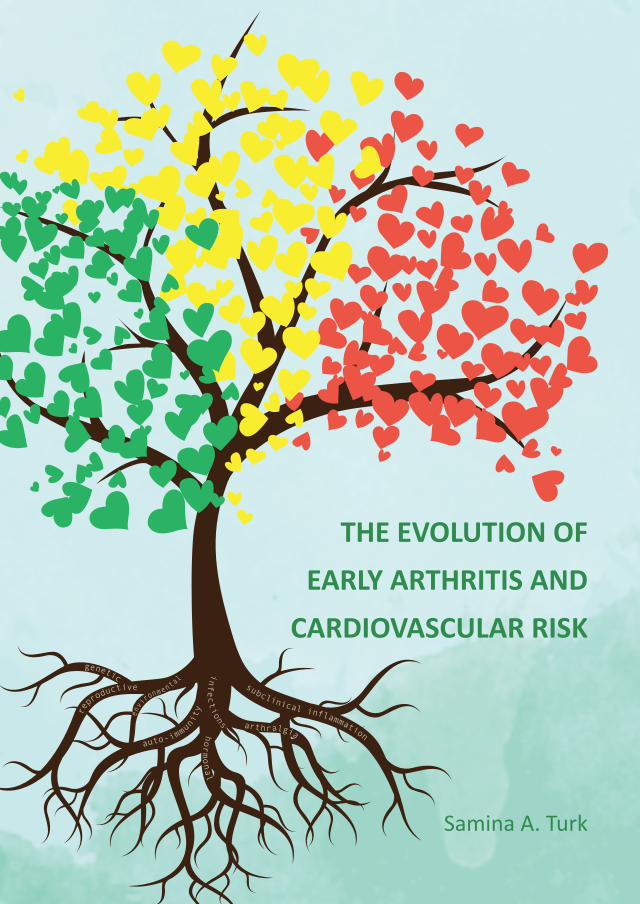
THE EVOLUTION OF EARLY ARTHRITIS AND CARDIOVASCULAR RISK g e n e t i c r e p r o d u c t ive e n v i r o nm e n t a l s u b c l i n i c al i n f l a m m a t i o n aut o - i m m u n i t y a r t h r a l g i a i n f e c t i o n s h o r m o n a l Samina A. Turk
Transcript of THE EVOLUTION OF EARLY ARTHRITIS AND...The pathology of RA does not suddenly start with the onset of...
THE EVOLUTION OF EARLY ARTHRITIS AND
CARDIOVASCULAR RISKgenetic
re
produ
ctive
envi
ronme
ntal subclinical
inflammationauto-
immunity
arthralgia
infections
hormonal
Samina A. Turk
Samina A. Turk
THE EVOLU
TION
OF EARLY ARTHRITIS AN
D CARDIOVASCU
LAR RISK
UITNODIGING
Voor het bijwonen van de openbare verdediging van het
proefschrift:
THE EVOLUTION OF EARLY ARTHRITIS AND
CARDIOVASCULAR RISK
Donderdag 10 januari 2019 om 10.00 uur
In de Agnietenkapel van de Universiteit van Amsterdam
Oudezijds Voorburgwal 231 te Amsterdam
Na afloop van de promotie is er een receptie ter plaatse
Samina A. Turk
Paranimfen
Marian van Beers-Tas06-617188447
Inge van Klink- de Kruijff06-39571477

THE EVOLUTION OF EARLY ARTHRITIS AND
CARDIOVASCULAR RISK
Samina A. Turk

ISBN: 978-94-6332-442-7
Lay-out and design by Loes KemaPrinted by GVO drukers & vormgevers | gvo.nl
Copyright © 2019 by Samina A. Turk.All rights reserved. No part of this publication may be reproduced or transmitted in any form or by any means, electronic or mechanical, including photocopy, recording or any information storage and retrieval system, without written permission of the author.
Financial support for printing of this thesis was provided by UMC- Amsterdam, Reade, UCB Pharma BV, TEVA Nederland BV, Pfizer BV.
THE EVOLUTION OF EARLY ARTHRITIS AND
CARDIOVASCULAR RISK
ACADEMISCH PROEFSCHRIFT
Ter verkrijging van de graad van doctor
aan de Universiteit van Amsterdam
op gezag van de Rector Magnificus
prof. dr. ir. K.I.J. Maex
ten overstaan van een door het College voor Promoties ingestelde commissie,
in het openbaar te verdedigen in de Agnietenkapel
op donderdag 10 januari 2019, te 10.00 uur
Door Samina Antonia Turk
Geboren te Uithoorn
Promotiecommissie:
Promotores: prof. dr. D. van Schaardenburg AMC-UvA prof. dr. M.T. Nurmohamed Vrije Universiteit Amsterdam
Copromotor: prof. dr. W.F. Lems Vrije Universiteit Amsterdam
Overige leden: prof. dr. R.F. van Vollenhoven AMC-UvA prof. dr. R.B.M. Landewé AMC-UvA prof. dr. K. Raza University of Birmingham dr. W.H. Bos Reade prof. dr. S. Middeldorp AMC-UvA prof. dr. R.J.G. Peters AMC-UvA prof. dr. Y.M. Smulders Vrije Universiteit Amsterdam
Faculteit: Geneeskunde
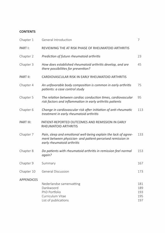
CONTENTS
Chapter 1 General Introduction 7
PART I: REVIEWING THE AT RISK PHASE OF RHEUMATOID ARTHRITIS
Chapter 2 Prediction of future rheumatoid arthritis 23
Chapter 3 How does established rheumatoid arthritis develop, and are there possibilities for prevention?
45
PART II: CARDIOVASCULAR RISK IN EARLY RHEUMATOID ARTHRITIS
Chapter 4 An unfavorable body composition is common in early arthritis patients: a case control study
75
Chapter 5 The relation between cardiac conduction times, cardiovascular risk factors and inflammation in early arthritis patients
95
Chapter 6 Change in cardiovascular risk after initiation of anti-rheumatic treatment in early rheumatoid arthritis
113
PART III: PATIENT-REPORTED OUTCOMES AND REMISSION IN EARLY RHEUMATOID ARTHRITIS
Chapter 7 Pain, sleep and emotional well-being explain the lack of agree-ment between physician- and patient-perceived remission in early rheumatoid arthritis
133
Chapter 8 Do patients with rheumatoid arthritis in remission feel normal again?
153
Chapter 9 Summary 167
Chapter 10 General Discussion 173
APPENDICESNederlandse samenvattingDankwoordPhD Portfolio Curriculum VitaeList of publications
181189193195197


CHAPTER 1
GENERAL INTRODUCTION

GENERAL INTRODUCTION
9
1
RHEUMATOID ARTHRITIS
Rheumatoid arthritis (RA) is a chronic systemic autoimmune disease, which affects many persons (around 1% of the world population), especially females(1). RA is characterized by (mainly synovial) inflammation, that can occur in any joint and gives rise to symptoms such as pain, swelling and stiffness(2). The inflammation may also lead to destruction of bone and cartilage, causing functional limitations, and therefore poses a personal and socioeconomic burden(3;4). However, advances in anti-rheumatic treatment have led to a substantial improvement in clinical outcomes and many patients nowadays reach a state of low disease activity or even remission(5). It is critical to suppress the inflammation early for a good prognosis. Unfortunately, even with treatment, a complete cure of the RA is not yet possible(6-8). Therefore, efforts are underway to try to stop the development of RA before the diagnosis; i.e. in the at-risk phase. Once these interventions are successful, the primary prevention of RA comes within reach.
THE EVOLUTION OF RHEUMATOID ARTHRITIS
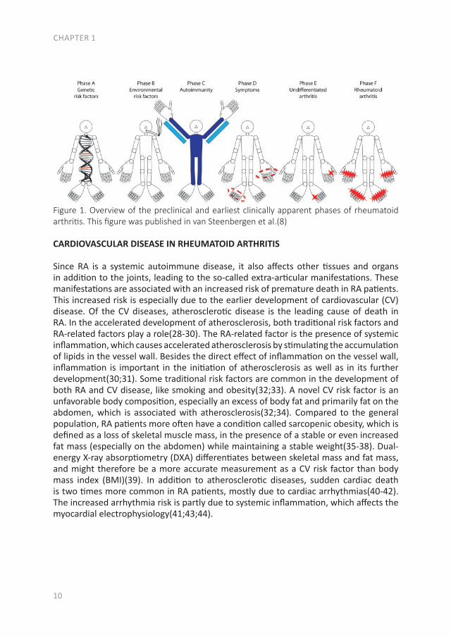
The pathology of RA does not suddenly start with the onset of clinical arthritis, but is preceded by an at-risk phase with variable symptoms and often autoimmunity before the appearance of clinical arthritis. There are different phases in the development of RA, which are presented in figure 1 (from phase A until F)(9). In the first phase there is an interaction between genetics (A) and environmental risk factors (B). For example, the frequency of the serotypes HLA-DR4 is higher in RA patients and polymorphisms on a number of loci are associated with the susceptibility of RA, like protein tyrosine phosphatase 22 and peptidylarginin deiminase 4(10;11). Even more interesting, from the viewpoint of intervention, are the environmental risk factors, as these can potentially be modified. Many environmental risk factors are suggested to contribute to the development of RA, of which smoking is best documented(8;12-14). The role of other factors is less clear, such as alcohol, sugar-sweetened beverages or red meat consumption(15-20). After the exposure to risk factors, a phase of pre-clinical auto-immunity can sometimes be identified. In phase C the RA autoantibodies, such as rheumatoid factor (RF) and anti-cyclic citrullinated peptide (anti-CCP), are produced. Studies have shown that these antibodies are often present years prior to the onset of arthritis(21;22). Especially the presence of both RF and anti-CCP is highly specific for the future development of RA(9). Some inflammatory markers can also be elevated in this phase, like C-reactive protein (CRP), tumor necrosis factor α (TNF-α) and interleukin-6 (IL-6)(23-27). In phase D symptoms like arthralgia and joint stiffness are present, without clinical evidence of arthritis. In phase E, patients develop undifferentiated arthritis, and finally some of these patients enter phase F where classical clinical features of RA are present. Not all patients pass through all of these phases, and sometimes phases occur in a different order(8).
CHAPTER 1
10
Figure 1. Overview of the preclinical and earliest clinically apparent phases of rheumatoid arthritis. This figure was published in van Steenbergen et al.(8)
CARDIOVASCULAR DISEASE IN RHEUMATOID ARTHRITIS
Since RA is a systemic autoimmune disease, it also affects other tissues and organs in addition to the joints, leading to the so-called extra-articular manifestations. These manifestations are associated with an increased risk of premature death in RA patients. This increased risk is especially due to the earlier development of cardiovascular (CV) disease. Of the CV diseases, atherosclerotic disease is the leading cause of death in RA. In the accelerated development of atherosclerosis, both traditional risk factors and RA-related factors play a role(28-30). The RA-related factor is the presence of systemic inflammation, which causes accelerated atherosclerosis by stimulating the accumulation of lipids in the vessel wall. Besides the direct effect of inflammation on the vessel wall, inflammation is important in the initiation of atherosclerosis as well as in its further development(30;31). Some traditional risk factors are common in the development of both RA and CV disease, like smoking and obesity(32;33). A novel CV risk factor is an unfavorable body composition, especially an excess of body fat and primarily fat on the abdomen, which is associated with atherosclerosis(32;34). Compared to the general population, RA patients more often have a condition called sarcopenic obesity, which is defined as a loss of skeletal muscle mass, in the presence of a stable or even increased fat mass (especially on the abdomen) while maintaining a stable weight(35-38). Dual-energy X-ray absorptiometry (DXA) differentiates between skeletal mass and fat mass, and might therefore be a more accurate measurement as a CV risk factor than body mass index (BMI)(39). In addition to atherosclerotic diseases, sudden cardiac death is two times more common in RA patients, mostly due to cardiac arrhythmias(40-42). The increased arrhythmia risk is partly due to systemic inflammation, which affects the myocardial electrophysiology(41;43;44).
RA might be explained by environmental risk factors(phase B). Many environmental risk factors have beenstudied (details are available from the correspondingauthor upon request), and smoking is the best replicatedenvironmental risk factor (14,15). Smoking predisposesto RA particularly in patients who carry specific HLA–DRB1 alleles, e.g., smokers carrying two HLA–DRB1alleles have a 21-fold increased risk of developing anti–citrullinated protein antibody (ACPA)–positive RA (16).Despite the high odds ratio in this subgroup, a largemajority of smokers do not develop RA. Weaker inter-actions have also been demonstrated between othergenes and smoking (17).
The genetic and environmental risk factors con-ceptually constitute the earliest preclinical phases ofRA. These risk factors have already been known forsome time, and an extensive discussion of these riskfactors is beyond the scope of this review.
The next two preclinical phases, “developingsystemic autoimmunity associated with RA” (phase C)and “symptoms without clinical arthritis” (phase D),
have been studied in the last few years. This has beendone using mainly two different study designs: nestedcase–control studies and prospective cohort studies. Aswill be discussed, the design of the study determined thesort of outcome that was obtained and the conclusionsthat can be drawn.
From RA back to pre-RA systemic autoimmunity
Studies associating RA with systemic auto-immune responses in the preclinical phase (phase C,with or without phase A and B) were mainly performedusing a nested case–control study design (also calledcase–control studies in a cohort) (Figure 2B). In thistype of study, RA cases were identified who weremembers of a predefined data set, e.g., a cohort of blooddonors from whom blood samples were obtained at leastonce (11,18). For each RA patient, a specified numberof matched controls who had not developed RA wasselected from the same data set. Consequently, bloodsamples from RA cases that were collected and stored
Figure 2. Overview of the preclinical phases of rheumatoid arthritis (RA) and the designs of the studies of the preclinical phases that have beenperformed. A, The six phases of preclinical and earliest clinically apparent RA, as defined by the European League Against Rheumatism study groupfor risk factors for RA. B, Nested case–control study design. A predefined set of subjects (e.g., blood donors) is followed up. From this set of subjects,RA cases are identified and for each case, a specified number of matched controls who have not developed RA is selected. Biomarkers are comparedbetween preclinically collected samples from these cases and the controls. C, Prospective cohort study design. Autoantibody-positive arthralgiapatients are identified and followed up prospectively.
PRECLINICAL PHASE OF RA 2221
GENERAL INTRODUCTION
11
1
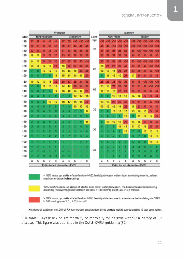
Risk table: 10-year risk on CV mortality or morbidity for persons without a history of CV diseases. This figure was published in the Dutch CVRM guidelines(52)
CHAPTER 1
12
CARDIOVASCULAR RISK
As RA patients have an increased CV risk, CV risk management should be done(45-47). Different CV risk models were developed for the general population to calculate the 10-year risk of CV disease, in which features such as gender, age, smoking status, systolic blood pressure and the total cholesterol: high-density lipoprotein (TC:HDL) ratio are included(48-50). However, many differences between CV risk models have been found and these CV risk models do not adequately predict CV-risk in the RA population(51). In the Netherlands, the Dutch Systemic COronary Risk Evaluation (SCORE) is used in the Dutch CV-risk management guidelines(48;52). The calculated CV risk category (low, medium or high risk obtained with the SCORE), the total cholesterol/HDL ratio, low-density lipoprotein (LDL) and the systolic blood pressure lead to an advice for lifestyle changes and possibly preventive treatment with antihypertensives and/or statins(52). In the Dutch SCORE a correction for RA patients is already taken into account, by adding 15 years to the actual age of a patient(48). However, lifestyle changes and CV preventive medication are naturally not enough to achieve a good result in the treatment of RA patients, the most important part is to suppress the inflammation(53;54).
PATIENT-REPORTED OUTCOMES
As suppressing the inflammation is important to prevent destruction of the joints as well as to lower the risk of CV disease, the treatment goal in RA patients is to attain a state of low disease activity or even remission. This is increasingly achieved with early initiation of targeted anti-rheumatic treatment(5). However, there are several definitions of clinical response and remission and different instruments are used to measure this, which leads to a substantial variation in the proportion of patients classified as being in remission(55;56). In addition, a disagreement between physician-perceived and patient-perceived remission is common(57-60). While the physician often determines remission based on physical examination and laboratory values, patients have a different perspective(61;62). Previous literature identified three main themes of patients’ perspective on remission: 1) reduction or absence of symptoms, 2) reduction of daily impact and, 3) return to normality. The items that are important for patients are not so much the presence of clinical arthritis, but rather pain, fatigue and sleep(63). The reduction in symptoms and impact of the disease on daily life would eventually mean a return to normality. However, the next problem then is the definition of ‘normality’.
GENERAL INTRODUCTION
13
1
AIM AND OUTLINE OF THIS THESIS
This thesis is devoted to the early phase of RA, focusing on three areas: disease development, cardiovascular comorbidity and remission from the perspective of the patient.
Part I is divided in two chapters wherein the at risk phase of RA is reviewed, to understand the processes in the preclinical phase that lead to the development of clinical arthritis. Chapter 2 focuses on the risk factors for the development of RA and how different risk factors are combined in risk models for the prediction of RA. Chapter 3 updates the risk factors for developing RA and focuses on the transition between ‘early RA’ and ‘established RA’. Finally, interventions to prevent the transition from the at-risk phase to clinical arthritis as well as from undifferentiated arthritis to RA, were reviewed.
With the evolution of RA, a systemic disease, extra-articular manifestations can develop as well. In Part II the focus is on CV disease, the major comorbid condition in early RA patients. The studies that are described in these three chapters were performed in patients of the early arthritis clinic (EAC) at Reade (formerly the Jan van Breemen Institute). This ongoing observational cohort started in 1995 and includes patients with at least one swollen joint, a short duration and no prior treatment with disease-modifying antirheumatic drugs (DMARDs). In this cohort questionnaires were completed, physical examinations were performed, radiographs were taken and blood was obtained. After 2008 CV measurements were added to traditional RA measurements, such as an electrocardiogram (ECG), ankle-brachial index (ABI) measurement, lipid profile and (whole body) DXA-scans. As an unfavorable body composition is a risk factor for both CV disease and the development of clinical arthritis, we compared body composition between patients at the clinical onset of arthritis with the general population in chapter 4, to determine if an unfavorable body composition is already present at the onset of arthritis. Furthermore, as RA patients have a greater risk of sudden cardiac death, we determined the prevalence of conduction disorders and traditional CV risk factors in chapter 5. The final section of this part is about CV risk prediction. As atherosclerosis is the leading cause of death in RA patients, prevention of a CV disease is very important. Different CV risk prediction models exist, which determine if lifestyle changes or preventive treatment for CV diseases are necessary. However, it is unknown when in the course of RA CV risk management should be applied and which risk model should be used. Therefore, in chapter 6 we studied if there are changes in CV risk and CV risk prevention advices between risk models and before and after one month of anti-rheumatic treatment.
The effect of modern anti-rheumatic treatment on disease activity is described in Part III, with a focus on patient-reported outcomes (PROs). Disagreement between definitions of response and remission as well as disagreement about remission between physician and patient is common. In chapter 7 we determined the frequency of patients that
CHAPTER 1
14
achieve remission according to the different response and remission definitions, as well as the agreement between these definitions. The patient’s perspective is increasingly recognized, but their perspective about determinants of disease activity that they associate with remission is unknown. Therefore, the disagreement between physician and patient was also studied in the same chapter. In the patients who were in remission according to the physician, we determined the differences in clinical variables and PROs between patients who did or did not perceive themselves as being in remission. As returning to normality is one of the three major themes of patient-perceived remission, chapter 8 focuses on this theme. As normality has no accepted definition yet, we assessed the ability of the normality scale to discriminate between remission and non-remission states according to the patient and to the American College of Rheumatology and the European League Against Rheumatism (ACR/EULAR) Boolean criteria.
Finally, in chapter 9 and chapter 10 the findings of this thesis are summarized and discussed, and implications for future research are given.
GENERAL INTRODUCTION
15
1
REFERENCES
1. Smolen JS, Aletaha D, McInnes IB. Rheumatoid arthritis. Lancet 2016 Oct 22;388(10055):2023-38.
2. Bellucci E, Terenzi R, La Paglia GM, Gentileschi S, Tripoli A, Tani C, et al. One year in review 2016: pathogenesis of rheumatoid arthritis. Clin Exp Rheumatol 2016 Sep;34(5):793-801.
3. Jacobs P, Bissonnette R, Guenther LC. Socioeconomic burden of immune-mediated inflammatory diseases--focusing on work productivity and disability. J Rheumatol Suppl 2011 Nov;88:55-61.
4. Verma MK, Sobha K. Understanding the major risk factors in the beginning and the progression of rheumatoid arthritis: current scenario and future prospects. Inflamm Res 2015 Sep;64(9):647-59.
5. Schett G, Emery P, Tanaka Y, Burmester G, Pisetsky DS, Naredo E, et al. Tapering biologic and conventional DMARD therapy in rheumatoid arthritis: current evidence and future directions. Ann Rheum Dis 2016 Aug;75(8):1428-37.
6. Guidelli GM, Barskova T, Brizi MG, Lepri G, Parma A, Talarico R, et al. One year in review: novelties in the treatment of rheumatoid arthritis. Clin Exp Rheumatol 2015 Jan;33(1):102-8.
7. Sanmarti R, Ruiz-Esquide V, Hernandez MV. Rheumatoid arthritis: a clinical overview of new diagnostic and treatment approaches. Curr Top Med Chem 2013;13(6):698-704.
8. van Steenbergen HW, Huizinga TW, van der Helm-van Mil AH. The preclinical phase of rheumatoid arthritis: what is acknowledged and what needs to be assessed? Arthritis Rheum 2013 Sep;65(9):2219-32.
9. Paul BJ, Kandy HI, Krishnan V. Pre-rheumatoid arthritis and its prevention. Eur J Rheumatol 2017 Jun;4(2):161-5.
10. Kurko J, Besenyei T, Laki J, Glant TT, Mikecz K, Szekanecz Z. Genetics of rheumatoid arthritis - a comprehensive review. Clin Rev Allergy Immunol 2013 Oct;45(2):170-9.
11. Yamamoto K, Okada Y, Suzuki A, Kochi Y. Genetics of rheumatoid arthritis in Asia--present and future. Nat Rev Rheumatol 2015 Jun;11(6):375-9.
12. Carlens C, Hergens MP, Grunewald J, Ekbom A, Eklund A, Hoglund CO, et al. Smoking, use of moist snuff, and risk of chronic inflammatory diseases. Am J Respir Crit Care Med 2010 Jun 1;181(11):1217-22.
13. Di GD, Discacciati A, Orsini N, Wolk A. Cigarette smoking and risk of rheumatoid arthritis: a dose-response meta-analysis. Arthritis Res Ther 2014 Mar 5;16(2):R61.
14. Kallberg H, Ding B, Padyukov L, Bengtsson C, Ronnelid J, Klareskog L, et al. Smoking is a major preventable risk factor for rheumatoid arthritis: estimations of risks after various exposures to cigarette smoke. Ann Rheum Dis 2011 Mar;70(3):508-11.
15. Benito-Garcia E, Feskanich D, Hu FB, Mandl LA, Karlson EW. Protein, iron, and meat consumption and risk for rheumatoid arthritis: a prospective cohort study. Arthritis Res Ther 2007;9(1):R16.
16. Grant WB. The role of meat in the expression of rheumatoid arthritis. Br J Nutr 2000 Nov;84(5):589-95.

CHAPTER 1
16
17. Hu Y, Costenbader KH, Gao X, Al-Daabil M, Sparks JA, Solomon DH, et al. Sugar-sweetened soda consumption and risk of developing rheumatoid arthritis in women. Am J Clin Nutr 2014 Sep;100(3):959-67.
18. Jin Z, Xiang C, Cai Q, Wei X, He J. Alcohol consumption as a preventive factor for developing rheumatoid arthritis: a dose-response meta-analysis of prospective studies. Ann Rheum Dis 2014 Nov;73(11):1962-7.
19. Pattison DJ, Symmons DP, Lunt M, Welch A, Luben R, Bingham SA, et al. Dietary risk factors for the development of inflammatory polyarthritis: evidence for a role of high level of red meat consumption. Arthritis Rheum 2004 Dec;50(12):3804-12.
20. Scott IC, Tan R, Stahl D, Steer S, Lewis CM, Cope AP. The protective effect of alcohol on developing rheumatoid arthritis: a systematic review and meta-analysis. Rheumatology (Oxford) 2013 May;52(5):856-67.
21. Rantapaa-Dahlqvist S, de Jong BA, Berglin E, Hallmans G, Wadell G, Stenlund H, et al. Antibodies against cyclic citrullinated peptide and IgA rheumatoid factor predict the development of rheumatoid arthritis. Arthritis Rheum 2003 Oct;48(10):2741-9.
22. Nielen MM, van SD, Reesink HW, van de Stadt RJ, van der Horst-Bruinsma IE, de Koning MH, et al. Specific autoantibodies precede the symptoms of rheumatoid arthritis: a study of serial measurements in blood donors. Arthritis Rheum 2004 Feb;50(2):380-6.
23. Masi AT, Aldag JC, Sipes J. Do elevated levels of serum C-reactive protein predict rheumatoid arthritis in men: correlations with pre-RA status and baseline positive rheumatoid factors. J Rheumatol 2001 Oct;28(10):2359-61.
24. Nielen MM, van SD, Reesink HW, Twisk JW, van de Stadt RJ, van der Horst-Bruinsma IE, et al. Increased levels of C-reactive protein in serum from blood donors before the onset of rheumatoid arthritis. Arthritis Rheum 2004 Aug;50(8):2423-7.
25. Jorgensen KT, Wiik A, Pedersen M, Hedegaard CJ, Vestergaard BF, Gislefoss RE, et al. Cytokines, autoantibodies and viral antibodies in premorbid and postdiagnostic sera from patients with rheumatoid arthritis: case-control study nested in a cohort of Norwegian blood donors. Ann Rheum Dis 2008 Jun;67(6):860-6.
26. Kokkonen H, Soderstrom I, Rocklov J, Hallmans G, Lejon K, Rantapaa DS. Up-regulation of cytokines and chemokines predates the onset of rheumatoid arthritis. Arthritis Rheum 2010 Feb;62(2):383-91.
27. Deane KD, O’Donnell CI, Hueber W, Majka DS, Lazar AA, Derber LA, et al. The number of elevated cytokines and chemokines in preclinical seropositive rheumatoid arthritis predicts time to diagnosis in an age-dependent manner. Arthritis Rheum 2010 Nov;62(11):3161-72.
28. Cojocaru M, Cojocaru IM, Silosi I, Vrabie CD, Tanasescu R. Extra-articular Manifestations in Rheumatoid Arthritis. Maedica (Buchar ) 2010 Dec;5(4):286-91.
29. Gerli R, Sherer Y, Bocci EB, Vaudo G, Moscatelli S, Shoenfeld Y. Precocious atherosclerosis in rheumatoid arthritis: role of traditional and disease-related cardiovascular risk factors. Ann N Y Acad Sci 2007 Jun;1108:372-81.
30. Mahmoudi M, Aslani S, Fadaei R, Jamshidi AR. New insights to the mechanisms underlying atherosclerosis in rheumatoid arthritis. Int J Rheum Dis 2017 Mar;20(3):287-97.
GENERAL INTRODUCTION
17
1
31. El-Barbary AM, Kassem EM, El-Sergany MA, Essa SA, Eltomey MA. Association of anti-modified citrullinated vimentin with subclinical atherosclerosis in early rheumatoid arthritis compared with anti-cyclic citrullinated peptide. J Rheumatol 2011 May;38(5):828-34.
32. Bastien M, Poirier P, Lemieux I, Despres JP. Overview of epidemiology and contribution of obesity to cardiovascular disease. Prog Cardiovasc Dis 2014 Jan;56(4):369-81.
33. Lahiri M, Luben RN, Morgan C, Bunn DK, Marshall T, Lunt M, et al. Using lifestyle factors to identify individuals at higher risk of inflammatory polyarthritis (results from the European Prospective Investigation of Cancer-Norfolk and the Norfolk Arthritis Register--the EPIC-2-NOAR Study). Ann Rheum Dis 2014 Jan;73(1):219-26.
34. Yusuf S, Hawken S, Ounpuu S, Bautista L, Franzosi MG, Commerford P, et al. Obesity and the risk of myocardial infarction in 27,000 participants from 52 countries: a case-control study. Lancet 2005 Nov 5;366(9497):1640-9.
35. Book C, Karlsson MK, Akesson K, Jacobsson LT. Early rheumatoid arthritis and body composition. Rheumatology (Oxford) 2009 Sep;48(9):1128-32.
36. Book C, Karlsson MK, Nilsson JA, Akesson K, Jacobsson LT. Changes in body composition after 2 years with rheumatoid arthritis. Scand J Rheumatol 2011 Mar;40(2):95-100.
37. Dao HH, Do QT, Sakamoto J. Abnormal body composition phenotypes in Vietnamese women with early rheumatoid arthritis. Rheumatology (Oxford) 2011 Jul;50(7):1250-8.
38. Dessein PH, Solomon A, Hollan I. Metabolic abnormalities in patients with inflammatory rheumatic diseases. Best Pract Res Clin Rheumatol 2016 Oct;30(5):901-15.
39. Andreoli A, Scalzo G, Masala S, Tarantino U, Guglielmi G. Body composition assessment by dual-energy X-ray absorptiometry (DXA). Radiol Med 2009 Mar;114(2):286-300.
40. Lazzerini PE, Acampa M, Capecchi PL, Hammoud M, Maffei S, Bisogno S, et al. Association between high sensitivity C-reactive protein, heart rate variability and corrected QT interval in patients with chronic inflammatory arthritis. Eur J Intern Med 2013 Jun;24(4):368-74.
41. Lazzerini PE, Capecchi PL, Acampa M, Galeazzi M, Laghi-Pasini F. Arrhythmic risk in rheumatoid arthritis: the driving role of systemic inflammation. Autoimmun Rev 2014 Sep;13(9):936-44.
42. Seferovic PM, Ristic AD, Maksimovic R, Simeunovic DS, Ristic GG, Radovanovic G, et al. Cardiac arrhythmias and conduction disturbances in autoimmune rheumatic diseases. Rheumatology (Oxford) 2006 Oct;45 Suppl 4:iv39-iv42.
43. Kenigsberg DN, Khanal S, Kowalski M, Krishnan SC. Prolongation of the QTc interval is seen uniformly during early transmural ischemia. J Am Coll Cardiol 2007 Mar 27;49(12):1299-305.
44. Sordillo PP, Sordillo DC, Helson L. Review: The Prolonged QT Interval: Role of Pro-inflammatory Cytokines, Reactive Oxygen Species and the Ceramide and Sphingosine-1 Phosphate Pathways. In Vivo 2015 Nov;29(6):619-36.
45. Law MR, Morris JK, Wald NJ. Use of blood pressure lowering drugs in the prevention of cardiovascular disease: meta-analysis of 147 randomised trials in the context of expectations from prospective epidemiological studies. BMJ 2009 May 19;338:b1665.
CHAPTER 1
18
46. Parsons C, Murad MH, Andersen S, Mookadam F, Labonte H. The effect of antihypertensive treatment on the incidence of stroke and cognitive decline in the elderly: a meta-analysis. Future Cardiol 2016 Mar;12(2):237-48.
47. Silverman MG, Ference BA, Im K, Wiviott SD, Giugliano RP, Grundy SM, et al. Association Between Lowering LDL-C and Cardiovascular Risk Reduction Among Different Therapeutic Interventions: A Systematic Review and Meta-analysis. JAMA 2016 Sep 27;316(12):1289-97.
48. URL: www.scoremeter.nl. (accessed June 2017).49. URL: https://heartscore.escardio.org/2012. (accessed June 2017).50. URL:https://www.mdcalc.com/framingham-coronary-heart-disease-risk-score.
(accessed June 2017).51. Galarza-Delgado DA, Azpiri-Lopez JR, Colunga-Pedraza IJ, Cardenas-de la Garza JA,
Vera-Pineda R, Serna-Pena G, et al. Assessment of six cardiovascular risk calculators in Mexican mestizo patients with rheumatoid arthritis according to the EULAR 2015/2016 recommendations for cardiovascular risk management. Clin Rheumatol 2017 Jun;36(6):1387-93.
52. NHG Dutch CVRM guidelines. 2017. 53. Arts EE, Fransen J, den Broeder AA, van Riel PLCM, Popa CD. Low disease activity
(DAS28</=3.2) reduces the risk of first cardiovascular event in rheumatoid arthritis: a time-dependent Cox regression analysis in a large cohort study. Ann Rheum Dis 2017 Jun 12.
54. Holmqvist M, Ljung L, Askling J. Acute coronary syndrome in new-onset rheumatoid arthritis: a population-based nationwide cohort study of time trends in risks and excess risks. Ann Rheum Dis 2017 Jul 14.
55. Sung YK, Cho SK, Kim D, Yoon BY, Choi CB, Cha HS, et al. Factors Contributing to Discordance between the 2011 ACR/EULAR Criteria and Physician Clinical Judgment for the Identification of Remission in Patients with Rheumatoid Arthritis. J Korean Med Sci 2016 Dec;31(12):1907-13.
56. van Tuyl LH, Smolen JS, Wells GA, Scholte-Voshaar M, Hoogland W, Boers M. Patient perspective on remission in rheumatoid arthritis. J Rheumatol 2011 Aug;38(8):1735-8.
57. Barton JL, Imboden J, Graf J, Glidden D, Yelin EH, Schillinger D. Patient-physician discordance in assessments of global disease severity in rheumatoid arthritis. Arthritis Care Res (Hoboken) 2010 Jun;62(6):857-64.
58. Castrejon I, Yazici Y, Samuels J, Luta G, Pincus T. Discordance of global estimates by patients and their physicians in usual care of many rheumatic diseases: association with 5 scores on a Multidimensional Health Assessment Questionnaire (MDHAQ) that are not found on the Health Assessment Questionnaire (HAQ). Arthritis Care Res (Hoboken) 2014 Jun;66(6):934-42.
59. Khan NA, Spencer HJ, Abda E, Aggarwal A, Alten R, Ancuta C, et al. Determinants of discordance in patients’ and physicians’ rating of rheumatoid arthritis disease activity. Arthritis Care Res (Hoboken) 2012 Feb;64(2):206-14.
60. Wolfe F, Boers M, Felson D, Michaud K, Wells GA. Remission in rheumatoid arthritis: physician and patient perspectives. J Rheumatol 2009 May;36(5):930-3.
GENERAL INTRODUCTION
19
1
61. Janta I, Naredo E, Martinez-Estupinan L, Nieto JC, De lT, I, Valor L, et al. Patient self-assessment and physician’s assessment of rheumatoid arthritis activity: which is more realistic in remission status? A comparison with ultrasonography. Rheumatology (Oxford) 2013 Dec;52(12):2243-50.
62. Nicolau G, Yogui MM, Vallochi TL, Gianini RJ, Laurindo IM, Novaes GS. Sources of discrepancy in patient and physician global assessments of rheumatoid arthritis disease activity. J Rheumatol 2004 Jul;31(7):1293-6.
63. van Tuyl LH, Hewlett S, Sadlonova M, Davis B, Flurey C, Hoogland W, et al. The patient perspective on remission in rheumatoid arthritis: ‘You’ve got limits, but you’re back to being you again’. Ann Rheum Dis 2015 Jun;74(6):1004-10.

PART IREVIEWING THE AT RISK PHASE
OF RHEUMATOID ARTHRITIS


CHAPTER 2
PREDICTION OF FUTURE RHEUMATOID ARTHRITIS
Samina A. Turk*Marian H. van Beers-Tas*Dirkjan van Schaardenburg
Jan van Breemen Research Institute | Reade, Amsterdam, The Netherlands
Rheum Dis Clin North Am – November 2014
*S.A. Turk and M.H. van Beers-Tas contributed equally to this work.
CHAPTER 2
24
KEYPOINTS
• Risk factors for rheumatoid arthritis (RA) include family history, birth weight, smoking, silica, alcohol nonuse, obesity, diabetes mellitus, autoantibodies, and genetic variants.
• Symptoms, antibodies, and inflammatory biomarkers can be useful in late at-risk stages, and genetic scores plus environmental factors more useful in early at-risk stages.
• Prediction models of RA can help to select candidates for intervention studies.
• The best target populations for screening are relatives of patients with RA and (seropositive) patients with arthralgia. However, only a minority of persons at risk can thus be recognized.
• Screening for RA risk is still experimental, because there is no validated screening tool and no proven therapy to prevent disease.
PREDICTION OF FUTURE RHEUMATOID ARTHRITIS
25
2INTRODUCTION
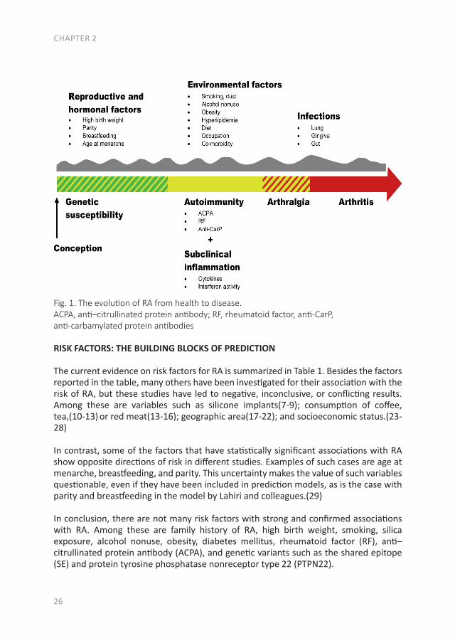
Rheumatoid arthritis (RA) on average becomes clinically manifest around the age of 55 years. During the healthy part of life, the risk of future RA is determined by genetic, reproductive and environmental factors (Fig. 1, green bar). Over time, people at risk for RA may pass through a phase of autoimmunity, accompanied by subclinical inflammation,(1) followed by a symptomatic phase, which may last a few months to several years. In the symptomatic phase, markers of autoimmunity and inflammation increase before the onset of clinical arthritis.(2) Therefore, prediction can be based on different characteristics in the asymptomatic phase and in the symptomatic phase.
The expectation that intervening in the preclinical phase of RA could be beneficial is based on the success of treatment of RA within 1 to 2 years after onset of clinical disease.(3,4) The new criteria for RA from 2010 with a focus on early signs such as involvement of even only a few small joints together with serology and acute phase reactants facilitate treatment in the earliest clinical phase(5,6) and the further characterization of the preclinical phase offers new opportunities for intervention studies even before clinically apparent arthritis occurs. Because RA is the most prevalent inflammatory rheumatic disease, with a high burden for the patient and society, it seems the ideal candidate rheumatic disease for screening and intervention programs. However, a lot of steps need to be taken before such programs can be offered to persons at risk.
This article summarizes the present knowledge on risk factors for RA, including genetic, reproductive, and hormonal factors; environmental exposures; biomarkers; personal characteristics and symptoms; and how these can be combined in risk models attempting to increase the accuracy of the prediction of RA. Genetic risk and gene-environmental interactions are dealt with elsewhere in this issue and are only mentioned here in relation to their roles in prediction models. Risk scores from such models require further validation, but could be used to select candidates for intervention studies.
METHODS
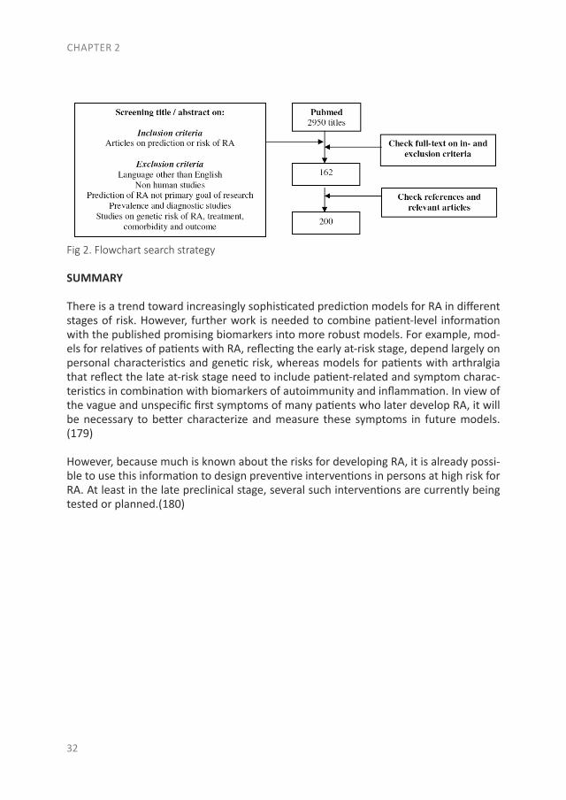
We searched the PubMed database on January 29, 2014, for the terms risk, predic-tion, and development in relation to RA. After excluding articles not directly related to prediction of RA, such as studies on prevalence, diagnosis, treatment, outcome, or comorbidities of RA, more than 200 articles remained on this topic after screening 2000 abstracts. Additional articles were added that were found after the search date until May 1, 2014, by screening rheumatologic journals.
CHAPTER 2
26
Fig. 1. The evolution of RA from health to disease. ACPA, anti–citrullinated protein antibody; RF, rheumatoid factor, anti-CarP, anti-carbamylated protein antibodies
RISK FACTORS: THE BUILDING BLOCKS OF PREDICTION
The current evidence on risk factors for RA is summarized in Table 1. Besides the factors reported in the table, many others have been investigated for their association with the risk of RA, but these studies have led to negative, inconclusive, or conflicting results. Among these are variables such as silicone implants(7-9); consumption of coffee, tea,(10-13) or red meat(13-16); geographic area(17-22); and socioeconomic status.(23-28)
In contrast, some of the factors that have statistically significant associations with RA show opposite directions of risk in different studies. Examples of such cases are age at menarche, breastfeeding, and parity. This uncertainty makes the value of such variables questionable, even if they have been included in prediction models, as is the case with parity and breastfeeding in the model by Lahiri and colleagues.(29)
In conclusion, there are not many risk factors with strong and confirmed associations with RA. Among these are family history of RA, high birth weight, smoking, silica exposure, alcohol nonuse, obesity, diabetes mellitus, rheumatoid factor (RF), anti–citrullinated protein antibody (ACPA), and genetic variants such as the shared epitope (SE) and protein tyrosine phosphatase nonreceptor type 22 (PTPN22).
PREDICTION OF FUTURE RHEUMATOID ARTHRITIS
27
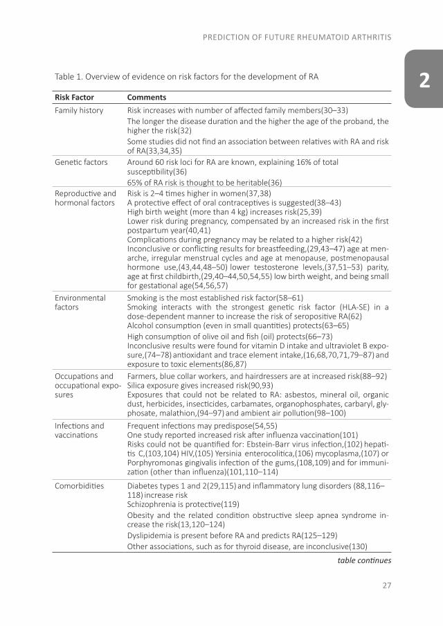
2Table 1. Overview of evidence on risk factors for the development of RA
Risk Factor CommentsFamily history Risk increases with number of affected family members(30–33)
The longer the disease duration and the higher the age of the proband, the higher the risk(32) Some studies did not find an association between relatives with RA and risk of RA(33,34,35)
Genetic factors Around 60 risk loci for RA are known, explaining 16% of totalsusceptibility(36)65% of RA risk is thought to be heritable(36)
Reproductive and hormonal factors
Risk is 2–4 times higher in women(37,38)A protective effect of oral contraceptives is suggested(38–43)High birth weight (more than 4 kg) increases risk(25,39)Lower risk during pregnancy, compensated by an increased risk in the first postpartum year(40,41)Complications during pregnancy may be related to a higher risk(42)Inconclusive or conflicting results for breastfeeding,(29,43–47) age at men-arche, irregular menstrual cycles and age at menopause, postmenopausal hormone use,(43,44,48–50) lower testosterone levels,(37,51–53) parity, age at first childbirth,(29,40–44,50,54,55) low birth weight, and being small for gestational age(54,56,57)
Environmental factors
Smoking is the most established risk factor(58–61)Smoking interacts with the strongest genetic risk factor (HLA-SE) in a dose-dependent manner to increase the risk of seropositive RA(62)Alcohol consumption (even in small quantities) protects(63–65)High consumption of olive oil and fish (oil) protects(66–73)Inconclusive results were found for vitamin D intake and ultraviolet B expo-sure,(74–78) antioxidant and trace element intake,(16,68,70,71,79–87) and exposure to toxic elements(86,87)
Occupations and occupational expo-sures
Farmers, blue collar workers, and hairdressers are at increased risk(88–92)Silica exposure gives increased risk(90,93)Exposures that could not be related to RA: asbestos, mineral oil, organic dust, herbicides, insecticides, carbamates, organophosphates, carbaryl, gly-phosate, malathion,(94–97) and ambient air pollution(98–100)
Infections and vaccinations
Frequent infections may predispose(54,55)One study reported increased risk after influenza vaccination(101)Risks could not be quantified for: Ebstein-Barr virus infection,(102) hepati-tis C,(103,104) HIV,(105) Yersinia enterocolitica,(106) mycoplasma,(107) or Porphyromonas gingivalis infection of the gums,(108,109) and for immuni-zation (other than influenza)(101,110–114)
Comorbidities Diabetes types 1 and 2(29,115) and inflammatory lung disorders (88,116–118) increase riskSchizophrenia is protective(119)Obesity and the related condition obstructive sleep apnea syndrome in-crease the risk(13,120–124)Dyslipidemia is present before RA and predicts RA(125–129)Other associations, such as for thyroid disease, are inconclusive(130)
table continues
CHAPTER 2
28
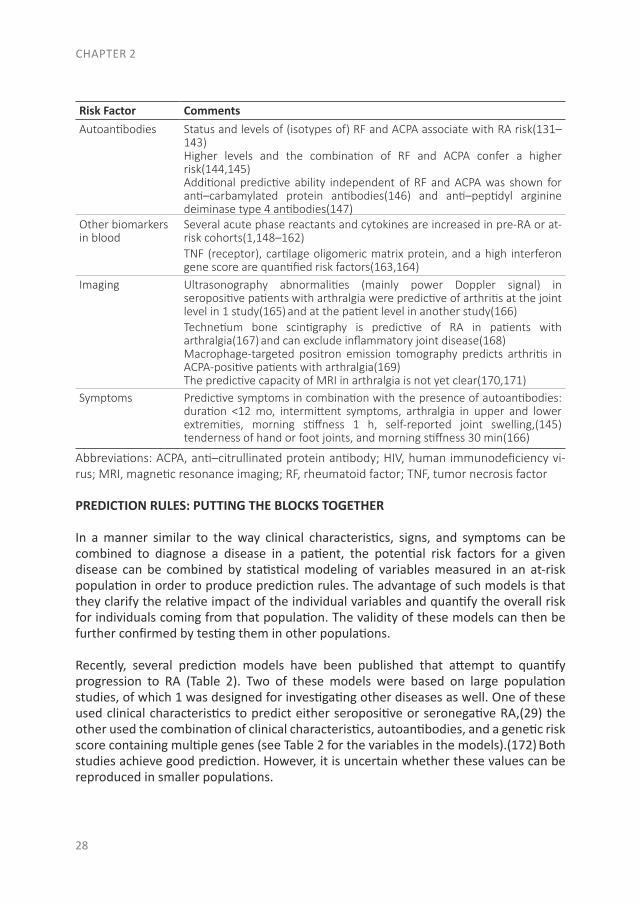
Risk Factor CommentsAutoantibodies Status and levels of (isotypes of) RF and ACPA associate with RA risk(131–
143) Higher levels and the combination of RF and ACPA confer a higher risk(144,145) Additional predictive ability independent of RF and ACPA was shown for anti–carbamylated protein antibodies(146) and anti–peptidyl arginine deiminase type 4 antibodies(147)
Other biomarkers in blood
Several acute phase reactants and cytokines are increased in pre-RA or at-risk cohorts(1,148–162)TNF (receptor), cartilage oligomeric matrix protein, and a high interferon gene score are quantified risk factors(163,164)
Imaging Ultrasonography abnormalities (mainly power Doppler signal) in seropositive patients with arthralgia were predictive of arthritis at the joint level in 1 study(165) and at the patient level in another study(166)Technetium bone scintigraphy is predictive of RA in patients with arthralgia(167) and can exclude inflammatory joint disease(168)Macrophage-targeted positron emission tomography predicts arthritis in ACPA-positive patients with arthralgia(169)The predictive capacity of MRI in arthralgia is not yet clear(170,171)
Symptoms Predictive symptoms in combination with the presence of autoantibodies: duration <12 mo, intermittent symptoms, arthralgia in upper and lower extremities, morning stiffness 1 h, self-reported joint swelling,(145)
tenderness of hand or foot joints, and morning stiffness 30 min(166)
Abbreviations: ACPA, anti–citrullinated protein antibody; HIV, human immunodeficiency vi-rus; MRI, magnetic resonance imaging; RF, rheumatoid factor; TNF, tumor necrosis factor
PREDICTION RULES: PUTTING THE BLOCKS TOGETHER
In a manner similar to the way clinical characteristics, signs, and symptoms can be combined to diagnose a disease in a patient, the potential risk factors for a given disease can be combined by statistical modeling of variables measured in an at-risk population in order to produce prediction rules. The advantage of such models is that they clarify the relative impact of the individual variables and quantify the overall risk for individuals coming from that population. The validity of these models can then be further confirmed by testing them in other populations.
Recently, several prediction models have been published that attempt to quantify progression to RA (Table 2). Two of these models were based on large population studies, of which 1 was designed for investigating other diseases as well. One of these used clinical characteristics to predict either seropositive or seronegative RA,(29) the other used the combination of clinical characteristics, autoantibodies, and a genetic risk score containing multiple genes (see Table 2 for the variables in the models).(172) Both studies achieve good prediction. However, it is uncertain whether these values can be reproduced in smaller populations.
PREDICTION OF FUTURE RHEUMATOID ARTHRITIS
29
2Three other studies investigated the development of RA in ACPA-positive and/or RF-positive patients with arthralgia.(121,145,166) The patients were partly recruited in primary care, and partly in the rheumatology clinic. The models were based on clinical characteristics, symptoms, and antibody characteristics, in 1 study supplemented by ultrasonographic power Doppler signal (see Table 2).(166) All 3 models provide good discrimination between persons who do or do not develop RA. However, they require ongoing validation as other studies select and follow such cohorts of people at risk for RA. Similar studies from North America designed to predict RA in first-degree relatives of patients with RA are underway but have not yet gathered enough arthritis cases to enable the construction of prediction models.(149,173) These studies are hampered by the low frequency of autoantibodies or of increased biomarkers in relatives of patients with RA.
Measuring the risk of RA is also a matter of timing. During the early at-risk stage, before the onset of autoimmunity, clinicians can only measure genetic susceptibility and environmental factors (see the left part of Fig. 1). The predictive capability of models in this situation is becoming good, with areas under the curve of 72% to 77% for the prediction of ACPA-positive RA.(174) However, the measured risk is a lifetime risk, which makes it an abstract figure for the individual person at risk. Prediction including a time frame becomes possible nearer to the onset of clinical RA, when the aspects of symptoms, autoimmunity, and inflammation can be taken into account. In the Amsterdam risk model, points can be gathered for clinical characteristics, symptoms, and serology, with more points for high levels of ACPA or positivity for both ACPA and RF.(145) The more points, the higher the risk and the sooner the onset of arthritis can be expected (Fig. 2). This prediction reflects studies in pre-RA blood donors, in which autoantibody levels increase during the 1 to 3 years before the onset of clinical arthritis.(2,138) In an US cohort of 81 patients with clinical RA from whom stored serum was available from 1 to 12 years before disease onset, a biomarker profile including autoantibodies and cytokines was identified that predicts the imminent onset of clinical arthritis within 2 years.(160) Autoantibody epitope spreading by itself in the preclinical phase also predicts progression to classifiable RA.(143)
SCREENING STRATEGIES
Many medical, ethical, and economic issues need to be addressed before screening for risk of future RA can be offered to certain categories of unaffected persons. Basic requirements for screening groups of people to predict a disease are (1) a defined population to test; (2) the existence of an asymptomatic (or nonspecific symptomatic) phase; (3) the availability of a test with good accuracy, low rates of side effects, and low cost; and (4) the availability of a cost-effective intervention in the at-risk phase. Only the second requirement of an asymptomatic phase is clearly fulfilled at present. Regarding items 3 and 4, no single test can identify those at risk for RA and no intervention exists
CHAPTER 2
30
with proven efficacy in the at-risk situation.(175,176) All efforts to predict RA and treat persons with an increased risk for RA are therefore currently regarded as investigational. The test for RA will eventually be a validated, cost-effective, and accurate prediction rule that is easy to apply. For comparison, consider the screening programs for colonic cancer, which have recently been established in several countries. All persons more than a certain age are offered screening, which leads to huge numbers of colonoscopies. The high cost of this procedure and the possibility of serious side effects need to be weighed against the benefit of removing polyps that would cause a high morbidity and mortality if left unnoticed.
Regarding item 1, careful consideration is needed to decide which population(s) should be screened or tested. The choices from general to specific are general population, relatives of patients with RA, persons with musculoskeletal symptoms, or persons with RA-specific autoimmunity. Because RA is not highly prevalent in most populations, with the possible exception of North American native peoples,(177,178) at this time it is not practical to test the general population for RA. Two recognizable target groups then remain: relatives of patients with RA and persons with musculoskeletal symptoms. The latter are found both in general practice and in rheumatology clinics. After history taking and physical examination, it must be decided which patients should proceed to further testing for RA risk, and which test to use. At present most clinicians use the RF and/or ACPA test, which are widely available and easy to perform. Except for patients with only RF positivity just above the reference range, the results give useful information. The question of who to test in general practice cannot accurately be answered at this time. This question requires structured longitudinal follow-up of patients in general practice, or the following of cohorts with clinically suspect arthralgia in rheumatology clinics.
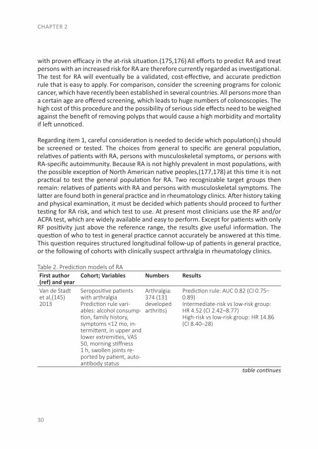
Table 2. Prediction models of RAFirst author (ref) and year
Cohort; Variables Numbers Results
Van de Stadt et al,(145)
2013
Seropositive patients with arthralgiaPrediction rule vari-ables: alcohol consump-tion, family history, symptoms <12 mo, in-termittent, in upper and lower extremities, VAS 50, morning stiffness1 h, swollen joints re-ported by patient, auto-antibody status
Arthralgia: 374 (131 developed arthritis)
Prediction rule: AUC 0.82 (CI 0.75–0.89)Intermediate-risk vs low-risk group: HR 4.52 (CI 2.42–8.77)High-risk vs low-risk group: HR 14.86 (CI 8.40–28)
table continues
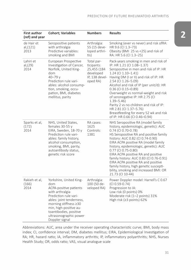
PREDICTION OF FUTURE RHEUMATOID ARTHRITIS
31
2First author (ref) and year
Cohort; Variables Numbers Results
de Hair et al,(121)2013
Seropositive patients with arthralgia Predictive variables: smoking and BMI
Arthralgia: 55 (15 deve-loped arthri-tis)
Smoking (ever vs never) and risk ofRA:HR 9.6 (CI 1.3–73)Obesity (BMI 25 vs <25) and risk of RA: HR 5.6 (CI 1.3–25)
Lahiri et al,(29)
2014
European Prospective Investigation of Cancer,Norfolk, United King-dom40–79 yPrediction rule vari-ables: alcohol consump-tion, smoking, occu-pation, BMI, diabetes mellitus, parity
Total par-ticipants: 25,455 (184 developed IP, 138 devel-oped RA)
Pack-years smoking in men and risk ofIP: HR 1.21 (CI 1.08–1.37)Seropositive in men and risk of IP: HR 1.24 (CI 1.10–1.41)Having DM (I or II) and risk of IP: HR 2.54 (CI 1.26–5.09)Alcohol and risk of IP (per unit/d): HR 0.36 (CI 0.15–0.89)Overweight vs normal-weight and riskof seronegative IP: HR 2.75 (CI1.39–5.46)Parity 2 vs no children and risk of IP: HR 2.81 (CI 1.37–5.76)Breastfeeding for every 52 wk and risk of IP: HR 0.66 (CI 0.46–0.94)
Sparks et al, (172)
2014
NHS, United States, females 30-55 yEIRA, Sweden, 18-70 yPrediction rule vari-ables: family history, alcohol consumption, smoking, BMI, parity, autoantibody status, genetic risk score
RA cases: 1625Controls: 1381
NHS Seropositive RA (model family history, epidemiologic, genetic): AUC 0.74 (CI 0.70-0.78)HS Seropositive RA and positive family history: AUC 0.82 (CI 0.74-0.90)EIRA ACPA positive RA (model family history, epidemiologic, genetic): AUC 0.77 (CI 0.75-0.80)EIRA ACPA positive RA and positive family history: AUC 0.83 (CI 0.76-0.91)EIRA ACPA positive RA and positive family history, high genetic suscepti-bility, smoking and increased BMI: OR 21.73 (CI 10-44)
Rakieh et al, (166)2014
Yorkshire, United King-domACPA-positive patients with arthralgia Prediction rule vari-ables: joint tenderness, morning stiffness ≥30 min, high-positive au-toantibodies, positive ultrasonographic power Doppler signal
Arthralgia: 100 (50 de-veloped RA)
Power Doppler model: Harrell’s C 0.67 (CI 0.59-0.74)Progression to IA:Low risk (0 points) 0%Moderate risk (1–2 points) 31%High risk (≥3 points) 62%
Abbreviations: AUC, area under the receiver operating characteristic curve; BMI, body mass index; CI, confidence interval; DM, diabetes mellitus; EIRA, Epidemiological Investigation of RA; HR, hazard ratio; IA, inflammatory arthritis; IP, inflammatory polyarthritis; NHS, Nurses Health Study; OR, odds ratio; VAS, visual analogue scale
CHAPTER 2
32
Fig 2. Flowchart search strategy
SUMMARY
There is a trend toward increasingly sophisticated prediction models for RA in different stages of risk. However, further work is needed to combine patient-level information with the published promising biomarkers into more robust models. For example, mod-els for relatives of patients with RA, reflecting the early at-risk stage, depend largely on personal characteristics and genetic risk, whereas models for patients with arthralgia that reflect the late at-risk stage need to include patient-related and symptom charac-teristics in combination with biomarkers of autoimmunity and inflammation. In view of the vague and unspecific first symptoms of many patients who later develop RA, it will be necessary to better characterize and measure these symptoms in future models.(179)
However, because much is known about the risks for developing RA, it is already possi-ble to use this information to design preventive interventions in persons at high risk for RA. At least in the late preclinical stage, several such interventions are currently being tested or planned.(180)
PREDICTION OF FUTURE RHEUMATOID ARTHRITIS
33
2REFERENCES
1. Nielen MM, van SD, Reesink HW, et al. Simultaneous development of acute phase response and autoantibodies in preclinical rheumatoid arthritis. Ann Rheum Dis 2006;65(4):535–7.
2. van de Stadt LA, de Koning MH, van de Stadt RJ, et al. Development of the anticitrulli-nated protein antibody repertoire prior to the onset of rheumatoid arthritis. Arthritis Rheum 2011;63(11):3226–33.
3. Boers M, Verhoeven AC, Markusse HM, et al. Randomised comparison of combined step-down prednisolone, methotrexate and sulphasalazine with sulphasalazine alone in early rheumatoid arthritis. Lancet 1997;350(9074):309–18.
4. Goekoop-Ruiterman YP, de Vries-Bouwstra JK, Allaart CF, et al. Clinical and radiographic outcomes of four different treatment strategies in patients with early rheumatoid ar-thritis (the BeSt study): a randomized, controlled trial. Arthritis Rheum 2008;58(2 Sup-pl):S126–35.
5. Aletaha D, Neogi T, Silman AJ, et al. 2010 Rheumatoid arthritis classification criteria: an American College of Rheumatology/European League Against Rheumatism collaborative initiative. Ann Rheum Dis 2010;69(9):1580–8.
6. Britsemmer K, Ursum J, Gerritsen M, et al. Validation of the 2010 ACR/EULAR classifica-tion criteria for rheumatoid arthritis: slight improvement over the 1987 ACR criteria. Ann Rheum Dis 2011;70(8):1468–70.
7. Janowsky EC, Kupper LL, Hulka BS. Meta-analyses of the relation between silicone breast implants and the risk of connective-tissue diseases. N Engl J Med 2000;342(11):781–90.
8. Perkins LL, Clark BD, Klein PJ, et al. A meta-analysis of breast implants and connective tissue disease. Ann Plast Surg 1995;35(6):561–70.
9. Wong O. A critical assessment of the relationship between silicone breast implants and connective tissue diseases. Regul Toxicol Pharmacol 1996;23(1 Pt 1):74–85.
10. Heliovaara M, Aho K, Knekt P, et al. Coffee consumption, rheumatoid factor, and the risk of rheumatoid arthritis. Ann Rheum Dis 2000;59(8):631–5.
11. Karlson EW, Mandl LA, Aweh GN, et al. Coffee consumption and risk of rheumatoid ar-thritis. Arthritis Rheum 2003;48(11):3055–60.
12. Mikuls TR, Cerhan JR, Criswell LA, et al. Coffee, tea, and caffeine consumption and risk of rheumatoid arthritis: results from the Iowa Women’s Health Study. Arthritis Rheum 2002;46(1):83–91.
13. Pedersen M, Jacobsen S, Klarlund M, et al. Environmental risk factors differ between rheumatoid arthritis with and without auto-antibodies against cyclic citrullinated pep-tides. Arthritis Res Ther 2006;8(4):R133.
14. Benito-Garcia E, Feskanich D, Hu FB, et al. Protein, iron, and meat consumption and risk for rheumatoid arthritis: a prospective cohort study. Arthritis Res Ther 2007;9(1):R16.
15. Grant WB. The role of meat in the expression of rheumatoid arthritis. Br J Nutr 2000;84(5):589–95.
CHAPTER 2
34
16. Pattison DJ, Symmons DP, Lunt M, et al. Dietary risk factors for the development of in-flammatory polyarthritis: evidence for a role of high level of red meat consumption. Arthritis Rheum 2004;50(12):3804–12.
17. Alamanos Y, Voulgari PV, Drosos AA. Incidence and prevalence of rheumatoid arthritis, based on the 1987 American College of Rheumatology criteria: a systematic review. Se-min Arthritis Rheum 2006;36(3):182–8.
18. Costenbader KH, Chang SC, Laden F, et al. Geographic variation in rheumatoid arthritis incidence among women in the United States. Arch Intern Med 2008; 168(15):1664–70.
19. Kallberg H, Vieira V, Holmqvist M, et al. Regional differences regarding risk of develop-ing rheumatoid arthritis in Stockholm County, Sweden: results from the Swedish Epi-demiological Investigation of Rheumatoid Arthritis (EIRA) study. Scand J Rheumatol 2013;42(5):337–43.
20. Li X, Sundquist J, Sundquist K. Risks of rheumatic diseases in first- and secondgeneration immigrants in Sweden: a nationwide followup study. Arthritis Rheum 2009;60(6):1588–96.
21. Silman A, Bankhead C, Rowlingson B, et al. Do new cases of rheumatoid arthritis cluster in time or in space? Int J Epidemiol 1997;26(3):628–34.
22. Silman A, Harrison B, Barrett E, et al. The existence of geographical clusters of cas-es of inflammatory polyarthritis in a primary care based register. Ann Rheum Dis 2000;59(2):152–4.
23. Bankhead C, Silman A, Barrett B, et al. Incidence of rheumatoid arthritis is not related to indicators of socioeconomic deprivation. J Rheumatol 1996;23(12): 2039–42.
24. Bengtsson C, Nordmark B, Klareskog L, et al. Socioeconomic status and the risk of de-veloping rheumatoid arthritis: results from the Swedish EIRA study. Ann Rheum Dis 2005;64(11):1588–94.
25. Jacobsson LT, Jacobsson ME, Askling J, et al. Perinatal characteristics and risk of rheuma-toid arthritis. BMJ 2003;326(7398):1068–9.
26. Olsson AR, Skogh T, Wingren G. Aetiological factors of importance for the development of rheumatoid arthritis. Scand J Rheumatol 2004;33(5):300–6.
27. Pedersen M, Jacobsen S, Klarlund M, et al. Socioeconomic status and risk of rheumatoid arthritis: a Danish case-control study. J Rheumatol 2006;33(6):1069–74.
28. Uhlig T, Hagen KB, Kvien TK. Current tobacco smoking, formal education, and the risk of rheumatoid arthritis. J Rheumatol 1999;26(1):47–54.
29. Lahiri M, Luben RN, Morgan C, et al. Using lifestyle factors to identify individuals at high-er risk of inflammatory polyarthritis (results from the European Prospective Investiga-tion of Cancer-Norfolk and the Norfolk Arthritis Register–the EPIC-2-NOAR Study). Ann Rheum Dis 2014;73(1):219–26.
30. Koumantaki Y, Giziaki E, Linos A, et al. Family history as a risk factor for rheumatoid arthri-tis: a case-control study. J Rheumatol 1997;24(8):1522–6.
31. Svendsen AJ, Holm NV, Kyvik K, et al. Relative importance of genetic effects in rheu-matoid arthritis: historical cohort study of Danish nationwide twin population. BMJ 2002;324(7332):264–6.
PREDICTION OF FUTURE RHEUMATOID ARTHRITIS
35
232. Deighton CM, Roberts DF, Walker DJ. Effect of disease severity on rheumatoid arthritis concordance in same sexed siblings. Ann Rheum Dis 1992;51(8):943–5.
33. Hemminki K, Li X, Sundquist J, et al. Familial associations of rheumatoid arthritis with autoimmune diseases and related conditions. Arthritis Rheum 2009;60(3): 661–8.
34. Larkin JG. Family history of rheumatoid arthritis–a non-predictor of inflammatory dis-ease? Rheumatology (Oxford) 2010;49(3):608–9.
35. Jones MA, Silman AJ, Whiting S, et al. Occurrence of rheumatoid arthritis is not increased in the first degree relatives of a population based inception cohort of inflammatory poly-arthritis. Ann Rheum Dis 1996;55(2):89–93.
36. Viatte S, Plant D, Raychaudhuri S. Genetics and epigenetics of rheumatoid arthritis. Nat Rev Rheumatol 2013;9(3):141–53.
37. Karlson EW, Chibnik LB, McGrath M, et al. A prospective study of androgen levels, hor-mone-related genes and risk of rheumatoid arthritis. Arthritis Res Ther 2009;11(3):R97.
38. Colebatch AN, Edwards CJ. The influence of early life factors on the risk of developing rheumatoid arthritis. Clin Exp Immunol 2011;163(1):11–6.
39. Mandl LA, Costenbader KH, Simard JF, et al. Is birthweight associated with risk of rheu-matoid arthritis? Data from a large cohort study. Ann Rheum Dis 2009; 68(4):514–8.
40. Peschken CA, Robinson DB, Hitchon CA, et al. Pregnancy and the risk of rheumatoid arthritis in a highly predisposed North American Native population. J Rheumatol 2012;39(12):2253–60.
41. Spector TD, Roman E, Silman AJ. The pill, parity, and rheumatoid arthritis. Arthritis Rheum 1990;33(6):782–9.
42. Jorgensen KT, Pedersen BV, Jacobsen S, et al. National cohort study of reproductive risk factors for rheumatoid arthritis in Denmark: a role for hyperemesis, gestational hyper-tension and pre-eclampsia? Ann Rheum Dis 2010;69(2): 358–63.
43. Karlson EW, Mandl LA, Hankinson SE, et al. Do breast-feeding and other reproductive fac-tors influence future risk of rheumatoid arthritis? Results from the Nurses’ Health Study. Arthritis Rheum 2004;50(11):3458–67.
44. Pikwer M, Bergstrom U, Nilsson JA, et al. Early menopause is an independent predictor of rheumatoid arthritis. Ann Rheum Dis 2012;71(3):378–81.
45. Berglin E, Kokkonen H, Einarsdottir E, et al. Influence of female hormonal factors, in rela-tion to autoantibodies and genetic markers, on the development of rheumatoid arthritis in northern Sweden: a case-control study. Scand J Rheumatol 2010;39(6):454–60.
46. Pikwer M, Bergstrom U, Nilsson JA, et al. Breast feeding, but not use of oral contra-ceptives, is associated with a reduced risk of rheumatoid arthritis. Ann Rheum Dis 2009;68(4):526–30.
47. Brennan P, Silman A. Breast-feeding and the onset of rheumatoid arthritis. Arthritis Rheum 1994;37(6):808–13.
48. Carette S, Marcoux S, Gingras S. Postmenopausal hormones and the incidence of rheu-matoid arthritis. J Rheumatol 1989;16(7):911–3.
49. Deighton CM, Sykes H, Walker DJ. Rheumatoid arthritis, HLA identity, and age at men-arche. Ann Rheum Dis 1993;52(5):322–6.
CHAPTER 2
36
50. Hernandez AM, Liang MH, Willett WC, et al. Reproductive factors, smoking, and the risk for rheumatoid arthritis. Epidemiology 1990;1(4):285–91.
51. Heikkila R, Aho K, Heliovaara M, et al. Serum androgen-anabolic hormones and the risk of rheumatoid arthritis. Ann Rheum Dis 1998;57(5):281–5.
52. Pikwer M, Giwercman A, Bergstrom U, et al. Association between testosterone levels and risk of future rheumatoid arthritis in men: a population-based casecontrol study. Ann Rheum Dis 2014;73(3):573–9.
53. Masi AT, Aldag JC, Chatterton RT. Sex hormones and risks of rheumatoid arthritis and developmental or environmental influences. Ann N Y Acad Sci 2006;1069:223–35.
54. Carlens C, Jacobsson L, Brandt L, et al. Perinatal characteristics, early life infections and later risk of rheumatoid arthritis and juvenile idiopathic arthritis. Ann Rheum Dis 2009;68(7):1159–64.
55. Rogers MA, Levine DA, Blumberg N, et al. Antigenic challenge in the etiology of autoim-mune disease in women. J Autoimmun 2012;38(2–3):J97–102.
56. Ma KK, Nelson JL, Guthrie KA, et al. Adverse pregnancy outcomes and risk of subsequent rheumatoid arthritis. Arthritis Rheumatol 2014;66(3):508–12.
57. Simard JF, Costenbader KH, Hernan MA, et al. Early life factors and adult-onset rheuma-toid arthritis. J Rheumatol 2010;37(1):32–7.
58. Carlens C, Hergens MP, Grunewald J, et al. Smoking, use of moist snuff, and risk of chron-ic inflammatory diseases. Am J Respir Crit Care Med 2010; 181(11):1217–22.
59. Di GD, Discacciati A, Orsini N, et al. Cigarette smoking and risk of rheumatoid arthritis: a dose-response meta-analysis. Arthritis Res Ther 2014;16(2):R61.
60. Kallberg H, Ding B, Padyukov L, et al. Smoking is a major preventable risk factor for rheu-matoid arthritis: estimations of risks after various exposures to cigarette smoke. Ann Rheum Dis 2011;70(3):508–11.
61. Sugiyama D, Nishimura K, Tamaki K, et al. Impact of smoking as a risk factor for de-veloping rheumatoid arthritis: a meta-analysis of observational studies. Ann Rheum Dis 2010;69(1):70–81.
62. Klareskog L, Stolt P, Lundberg K, et al. A new model for an etiology of rheumatoid arthri-tis: smoking may trigger HLA-DR (shared epitope)-restricted immune reactions to auto-antigens modified by citrullination. Arthritis Rheum 2006;54(1):38–46.
63. Jin Z, Xiang C, Cai Q, et al. Alcohol consumption as a preventive factor for developing rheumatoid arthritis: a dose-response meta-analysis of prospective studies. Ann Rheum Dis 2013. [Epub ahead of print].
64. Scott IC, Tan R, Stahl D, et al. The protective effect of alcohol on developing rheumatoid arthritis: a systematic review and meta-analysis. Rheumatology (Oxford) 2013;52(5):856–67.
65. van de Stadt LA, van SD. Alcohol consumption protects against arthritis development in seropositive arthralgia patients. Ann Rheum Dis 2012;71(8):1431–2.
66. Di GD, Wallin A, Bottai M, et al. Long-term intake of dietary long-chain n-3 polyunsatu-rated fatty acids and risk of rheumatoid arthritis: a prospective cohort study of women. Ann Rheum Dis 2013. [Epub ahead of print].
PREDICTION OF FUTURE RHEUMATOID ARTHRITIS
37
267. Linos A, Kaklamanis E, Kontomerkos A, et al. The effect of olive oil and fish consumption on rheumatoid arthritis–a case control study. Scand J Rheumatol 1991;20(6):419–26.
68. Linos A, Kaklamani VG, Kaklamani E, et al. Dietary factors in relation to rheumatoid ar-thritis: a role for olive oil and cooked vegetables? Am J Clin Nutr 1999; 70(6):1077–82.
69. Oliver JE, Silman AJ. Risk factors for the development of rheumatoid arthritis. Scand J Rheumatol 2006;35(3):169–74.
70. Pattison DJ, Harrison RA, Symmons DP. The role of diet in susceptibility to rheumatoid arthritis: a systematic review. J Rheumatol 2004;31(7):1310–9.
71. Pedersen M, Stripp C, Klarlund M, et al. Diet and risk of rheumatoid arthritis in a prospec-tive cohort. J Rheumatol 2005;32(7):1249–52.
72. Rosell M, Wesley AM, Rydin K, et al. Dietary fish and fish oil and the risk of rheumatoid arthritis. Epidemiology 2009;20(6):896–901.
73. Shapiro JA, Koepsell TD, Voigt LF, et al. Diet and rheumatoid arthritis in women: a possi-ble protective effect of fish consumption. Epidemiology 1996;7(3): 256–63.
74. Song GG, Bae SC, Lee YH. Association between vitamin D intake and the risk of rheuma-toid arthritis: a meta-analysis. Clin Rheumatol 2012;31(12):1733–9.
75. Nielen MM, van SD, Lems WF, et al. Vitamin D deficiency does not increase the risk of rheumatoid arthritis: comment on the article by Merlino, et al. Arthritis Rheum 2006;54(11):3719–20.
76. Feser M, Derber LA, Deane KD, et al. Plasma 25,OH vitamin D concentrations are not as-sociated with rheumatoid arthritis (RA)-related autoantibodies in individuals at elevated risk for RA. J Rheumatol 2009;36(5):943–6.
77. Hiraki LT, Munger KL, Costenbader KH, et al. Dietary intake of vitamin D during adoles-cence and risk of adult-onset systemic lupus erythematosus and rheumatoid arthritis. Arthritis Care Res (Hoboken) 2012;64(12):1829–36.
78. Arkema EV, Hart JE, Bertrand KA, et al. Exposure to ultraviolet-B and risk of develop-ing rheumatoid arthritis among women in the Nurses’ Health Study. Ann Rheum Dis 2013;72(4):506–11.
79. Cerhan JR, Saag KG, Merlino LA, et al. Antioxidant micronutrients and risk of rheumatoid arthritis in a cohort of older women. Am J Epidemiol 2003;157(4): 345–54.
80. Comstock GW, Burke AE, Hoffman SC, et al. Serum concentrations of alpha tocopherol, beta carotene, and retinol preceding the diagnosis of rheumatoid arthritis and systemic lupus erythematosus. Ann Rheum Dis 1997;56(5):323–5.
81. Heliovaara M, Knekt P, Aho K, et al. Serum antioxidants and risk of rheumatoid arthritis. Ann Rheum Dis 1994;53(1):51–3.
82. Knekt P, Heliovaara M, Aho K, et al. Serum selenium, serum alpha-tocopherol, and the risk of rheumatoid arthritis. Epidemiology 2000;11(4):402–5.
83. Pattison DJ, Silman AJ, Goodson NJ, et al. Vitamin C and the risk of developing inflammato-ry polyarthritis: prospective nested case-control study. Ann Rheum Dis 2004;63(7):843–7.
84. Costenbader KH, Kang JH, Karlson EW. Antioxidant intake and risks of rheumatoid arthri-tis and systemic lupus erythematosus in women. Am J Epidemiol 2010;172(2):205–16.
CHAPTER 2
38
85. Pattison DJ, Symmons DP, Lunt M, et al. Dietary beta-cryptoxanthin and inflamma-tory polyarthritis: results from a population-based prospective study. Am J Clin Nutr 2005;82(2):451–5.
86. Afridi HI, Kazi TG, Brabazon D, et al. Association between essential trace and toxic ele-ments in scalp hair samples of smokers rheumatoid arthritis subjects. Sci Total Environ 2011;412–413:93–100.
87. Afridi HI, Kazi TG, Brabazon D, et al. Interaction between zinc, cadmium, and lead in scalp hair samples of Pakistani and Irish smokers rheumatoid arthritis subjects in relation to controls. Biol Trace Elem Res 2012;148(2):139–47.
88. Bergstrom U, Jacobsson LT, Nilsson JA, et al. Pulmonary dysfunction, smoking, socioeco-nomic status and the risk of developing rheumatoid arthritis. Rheumatology (Oxford) 2011;50(11):2005–13.
89. Cooper GS, Miller FW, Germolec DR. Occupational exposures and autoimmune diseases. Int Immunopharmacol 2002;2(2–3):303–13.
90. Khuder SA, Peshimam AZ, Agraharam S. Environmental risk factors for rheumatoid arthri-tis. Rev Environ Health 2002;17(4):307–15.
91. Olsson AR, Skogh T, Wingren G. Occupational determinants for rheumatoid arthritis. Scand J Work Environ Health 2000;26(3):243–9.
92. Turner S, Cherry N. Rheumatoid arthritis in workers exposed to silica in the pottery indus-try. Occup Environ Med 2000;57(7):443–7.
93. Stolt P, Yahya A, Bengtsson C, et al. Silica exposure among male current smokers is asso-ciated with a high risk of developing ACPA-positive rheumatoid arthritis. Ann Rheum Dis 2010;69(6):1072–6.
94. De Roos AJ, Cooper GS, Alavanja MC, et al. Rheumatoid arthritis among women in the Agricultural Health Study: risk associated with farming activities and exposures. Ann Epi-demiol 2005;15(10):762–70.
95. Olsson AR, Skogh T, Axelson O, et al. Occupations and exposures in the work environment as determinants for rheumatoid arthritis. Occup Environ Med 2004; 61(3):233–8.
96. Sverdrup B, Kallberg H, Bengtsson C, et al. Association between occupational exposure to mineral oil and rheumatoid arthritis: results from the Swedish EIRA case-control study. Arthritis Res Ther 2005;7(6):R1296–303.
97. Stolt P, Kallberg H, Lundberg I, et al. Silica exposure is associated with increased risk of developing rheumatoid arthritis: results from the Swedish EIRA study. Ann Rheum Dis 2005;64(4):582–6.
98. Gan RW, Deane KD, Zerbe GO, et al. Relationship between air pollution and positivity of RA-related autoantibodies in individuals without established RA: a report on SERA. Ann Rheum Dis 2013;72(12):2002–5.
99. Hart JE, Kallberg H, Laden F, et al. Ambient air pollution exposures and risk of rheumatoid arthritis. Arthritis Care Res (Hoboken) 2013;65(7):1190–6.
100. Hart JE, Kallberg H, Laden F, et al. Ambient air pollution exposures and risk of rheu-matoid arthritis: results from the Swedish EIRA case-control study. Ann Rheum Dis 2013;72(6):888–94.
PREDICTION OF FUTURE RHEUMATOID ARTHRITIS
39
2101. Ray P, Black S, Shinefield H, et al. Risk of rheumatoid arthritis following vaccination with tetanus, influenza and hepatitis B vaccines among persons 15-59 years of age. Vaccine 2011;29(38):6592–7.
102. Blaschke S, Schwarz G, Moneke D, et al. Epstein-Barr virus infection in peripheral blood mononuclear cells, synovial fluid cells, and synovial membranes of patients with rheu-matoid arthritis. J Rheumatol 2000;27(4):866–73.
103. Buskila D, Shnaider A, Neumann L, et al. Musculoskeletal manifestations and autoan-tibody profile in 90 hepatitis C virus infected Israeli patients. Semin Arthritis Rheum 1998;28(2):107–13.
104. Sawada T, Hirohata S, Inoue T, et al. Development of rheumatoid arthritis after hepatitis C virus infection. Arthritis Rheum 1991;34(12):1620–1.
105. Medina-Rodriguez F, Guzman C, Jara LJ, et al. Rheumatic manifestations in human immu-nodeficiency virus positive and negative individuals: a study of 2 populations with similar risk factors. J Rheumatol 1993;20(11):1880–4.
106. Saebo A, Lassen J. Yersinia enterocolitica: an inducer of chronic inflammation. Int J Tissue React 1994;16(2):51–7.
107. Schaeverbeke T, Vernhes JP, Lequen L, et al. Mycoplasmas and arthritides. Rev Rhum Engl Ed 1997;64(2):120–8.
108. Hitchon CA, Chandad F, Ferucci ED, et al. Antibodies to Porphyromonas gingivalis are associated with anticitrullinated protein antibodies in patients with rheumatoid arthritis and their relatives. J Rheumatol 2010;37(6):1105–12.
109. Mikuls TR, Thiele GM, Deane KD, et al. Porphyromonas gingivalis and diseaserelated autoantibodies in individuals at increased risk of rheumatoid arthritis. Arthritis Rheum 2012;64(11):3522–30.
110. Bengtsson C, Kapetanovic MC, Kallberg H, et al. Common vaccinations among adults do not increase the risk of developing rheumatoid arthritis: results from the Swedish EIRA study. Ann Rheum Dis 2010;69(10):1831–3.
111. Cohen AD, Shoenfeld Y. Vaccine-induced autoimmunity. J Autoimmun 1996; 9(6):699–703.
112. Schattner A. Consequence or coincidence? The occurrence, pathogenesis and signifi-cance of autoimmune manifestations after viral vaccines. Vaccine 2005; 23(30):3876–86.
113. Shoenfeld Y, Aron-Maor A. Vaccination and autoimmunity-‘vaccinosis’: a dangerous liai-son? J Autoimmun 2000;14(1):1–10.
114. Symmons DP, Chakravarty K. Can immunisation trigger rheumatoid arthritis? Ann Rheum Dis 1993;52(12):843–4.
115. Boyer JF, Gourraud PA, Cantagrel A, et al. Traditional cardiovascular risk factors in rheu-matoid arthritis: a meta-analysis. Joint Bone Spine 2011;78(2):179–83.
116. Reynisdottir G, Karimi R, Joshua V, et al. Structural changes and antibody enrichment in the lungs are early features of anti-citrullinated protein antibodypositive rheumatoid arthritis. Arthritis Rheumatol 2014;66(1):31–9.
117. Verstappen SM, Lunt M, Luben RN, et al. Demographic and disease-related predictors of abnormal lung function in patients with established inflammatory polyarthritis and a comparison with the general population. Ann Rheum Dis 2013;72(9):1517–23.
CHAPTER 2
40
118. Demoruelle MK, Weisman MH, Simonian PL, et al. Brief report: airways abnormalities and rheumatoid arthritis-related autoantibodies in subjects without arthritis: early injury or initiating site of autoimmunity? Arthritis Rheum 2012; 64(6):1756–61.
119. Oken RJ, Schulzer M. At issue: schizophrenia and rheumatoid arthritis: the negative asso-ciation revisited. Schizophr Bull 1999;25(4):625–38.
120. Crowson CS, Matteson EL, Davis JM III, et al. Contribution of obesity to the rise in inci-dence of rheumatoid arthritis. Arthritis Care Res (Hoboken) 2013;65(1): 71–7.
121. de Hair MJ, Landewe RB, van de Sande MG, et al. Smoking and overweight determine the likelihood of developing rheumatoid arthritis. Ann Rheum Dis 2013;72(10):1654–8.
122. Symmons DP, Bankhead CR, Harrison BJ, et al. Blood transfusion, smoking, and obesity as risk factors for the development of rheumatoid arthritis: results from a primary care-based incident case-control study in Norfolk, England. Arthritis Rheum 1997;40(11):1955–61.
123. Wesley A, Bengtsson C, Elkan AC, et al. Association between body mass index and an-ti-citrullinated protein antibody-positive and anti-citrullinated protein antibody-negative rheumatoid arthritis: results from a population-based casecontrol study. Arthritis Care Res (Hoboken) 2013;65(1):107–12.
124. Kang JH, Lin HC. Obstructive sleep apnea and the risk of autoimmune diseases: a longi-tudinal population-based study. Sleep Med 2012;13(6):583–8.
125. Maki-Petaja KM, Booth AD, Hall FC, et al. Ezetimibe and simvastatin reduce inflamma-tion, disease activity, and aortic stiffness and improve endothelial function in rheumatoid arthritis. J Am Coll Cardiol 2007;50(9):852–8.
126. Steiner G, Urowitz MB. Lipid profiles in patients with rheumatoid arthritis: mechanisms and the impact of treatment. Semin Arthritis Rheum 2009;38(5):372–81.
127. van de Stadt LA, van Sijl AM, van SD, et al. Dyslipidaemia in patients with seropositive arthralgia predicts the development of arthritis. Ann Rheum Dis 2012; 71(11):1915–6.
128. van Halm VP, Nielen MM, Nurmohamed MT, et al. Lipids and inflammation: serial mea-surements of the lipid profile of blood donors who later developed rheumatoid arthritis. Ann Rheum Dis 2007;66(2):184–8.
129. Myasoedova E, Crowson CS, Kremers HM, et al. Total cholesterol and LDL levels decrease before rheumatoid arthritis. Ann Rheum Dis 2010;69(7):1310–4.
130. Dobson R, Giovannoni G. Autoimmune disease in people with multiple sclerosis and their relatives: a systematic review and meta-analysis. J Neurol 2013; 260(5):1272–85.
131. Avouac J, Gossec L, Dougados M. Diagnostic and predictive value of anticyclic citrullinat-ed protein antibodies in rheumatoid arthritis: a systematic literature review. Ann Rheum Dis 2006;65(7):845–51.
132. Berglin E, Padyukov L, Sundin U, et al. A combination of autoantibodies to cyclic citrul-linated peptide (CCP) and HLA-DRB1 locus antigens is strongly associated with future onset of rheumatoid arthritis. Arthritis Res Ther 2004;6(4):R303–8.
133. Brink M, Hansson M, Mathsson L, et al. Multiplex analyses of antibodies against citrul-linated peptides in individuals prior to development of rheumatoid arthritis. Arthritis Rheum 2013;65(4):899–910.
134. Dorner T, Hansen A. Autoantibodies in normals–the value of predicting rheumatoid ar-thritis. Arthritis Res Ther 2004;6(6):282–4.

PREDICTION OF FUTURE RHEUMATOID ARTHRITIS
41
2135. Kokkonen H, Mullazehi M, Berglin E, et al. Antibodies of IgG, IgA and IgM isotypes against cyclic citrullinated peptide precede the development of rheumatoid arthritis. Arthritis Res Ther 2011;13(1):R13.
136. Majka DS, Holers VM. Can we accurately predict the development of rheumatoid arthri-tis in the preclinical phase? Arthritis Rheum 2003;48(10):2701–5.
137. Nielen MM, van SD, Reesink HW, et al. Specific autoantibodies precede the symptoms of rheumatoid arthritis: a study of serial measurements in blood donors. Arthritis Rheum 2004;50(2):380–6.
138. Rantapaa-Dahlqvist S, de Jong BA, Berglin E, et al. Antibodies against cyclic citrullinat-ed peptide and IgA rheumatoid factor predict the development of rheumatoid arthritis. Arthritis Rheum 2003;48(10):2741–9.
139. Saraux A, Berthelot JM, Chales G, et al. Value of laboratory tests in early prediction of rheumatoid arthritis. Arthritis Rheum 2002;47(2):155–65.
140. Scofield RH. Autoantibodies as predictors of disease. Lancet 2004;363(9420): 1544–6.141. Koivula MK, Heliovaara M, Rissanen H, et al. Antibodies binding to citrullinated telo-
peptides of type I and type II collagens and to mutated citrullinated vimentin syner-gistically predict the development of seropositive rheumatoid arthritis. Ann Rheum Dis 2012;71(10):1666–70.
142. Nielsen SF, Bojesen SE, Schnohr P, et al. Elevated rheumatoid factor and long term risk of rheumatoid arthritis: a prospective cohort study. BMJ 2012;345:e5244.
143. van de Stadt LA, van der Horst AR, de Koning MH, et al. The extent of the anticitrullinat-ed protein antibody repertoire is associated with arthritis development in patients with seropositive arthralgia. Ann Rheum Dis 2011;70(1):128–33.
144. Bos WH, Wolbink GJ, Boers M, et al. Arthritis development in patients with arthralgia is strongly associated with anti-citrullinated protein antibody status: a prospective cohort study. Ann Rheum Dis 2010;69(3):490–4.
145. van de Stadt LA, Witte BI, Bos WH, et al. A prediction rule for the development of arthritis in seropositive arthralgia patients. Ann Rheum Dis 2013;72(12):1920–6.
146. Shi J, van de Stadt LA, Levarht EW, et al. Anti-carbamylated protein antibodies are pres-ent in arthralgia patients and predict the development of rheumatoid arthritis. Arthritis Rheum 2013;65(4):911–5.
147. Kolfenbach JR, Deane KD, Derber LA, et al. Autoimmunity to peptidyl arginine deiminase type 4 precedes clinical onset of rheumatoid arthritis. Arthritis Rheum 2010;62(9):2633–9.
148. Aho K, Palosuo T, Knekt P, et al. Serum C-reactive protein does not predict rheumatoid arthritis. J Rheumatol 2000;27(5):1136–8.
149. Barra L, Summers K, Bell D, et al. Serum cytokine profile of unaffected first-degree rela-tives of patients with rheumatoid arthritis. J Rheumatol 2014;41(2):280–5.
150. Deane KD, O’Donnell CI, Hueber W, et al. The number of elevated cytokines and chemo-kines in preclinical seropositive rheumatoid arthritis predicts time to diagnosis in an age-dependent manner. Arthritis Rheum 2010;62(11):3161–72.
151. Edwards CJ, Cooper C. Early environmental factors and rheumatoid arthritis. Clin Exp Immunol 2006;143(1):1–5.
CHAPTER 2
42
152. El-Gabalawy HS, Robinson DB, Smolik I, et al. Familial clustering of the serum cytokine pro-file in the relatives of rheumatoid arthritis patients. Arthritis Rheum 2012;64(6):1720–9.
153. Hughes-Austin JM, DeaneKD, Derber LA, etal.Multiplecytokinesand chemokines are as-sociated with rheumatoid arthritis-related autoimmunity in first-degree relatives with-out rheumatoid arthritis: studies of the Aetiology of Rheumatoid Arthritis (SERA). Ann Rheum Dis 2013;72(6):901–7.
154. Jorgensen KT, Wiik A, Pedersen M, et al. Cytokines, autoantibodies and viral antibodies in premorbid and postdiagnostic sera from patients with rheumatoid arthritis: case-control study nested in a cohort of Norwegian blood donors. Ann Rheum Dis 2008;67(6):860–6.
155. Karlson EW, Chibnik LB, Tworoger SS, et al. Biomarkers of inflammation and development of rheumatoid arthritis in women from two prospective cohort studies. Arthritis Rheum 2009;60(3):641–52.
156. Limper M, van de Stadt L, Bos W, et al. The acute-phase response is not predictive for the development of arthritis in seropositive arthralgia - a prospective cohort study. J Rheumatol 2012;39(10):1914–7.
157. Masi AT, Aldag JC, Sipes J. Do elevated levels of serum C-reactive protein predict rheuma-toid arthritis in men: correlations with pre-RA status and baseline positive rheumatoid factors. J Rheumatol 2001;28(10):2359–61.
158. Nielen MM, van SD, Reesink HW, et al. Increased levels of C-reactive protein in se-rum from blood donors before the onset of rheumatoid arthritis. Arthritis Rheum 2004;50(8):2423–7.
159. Rantapaa-Dahlqvist S, Boman K, Tarkowski A, et al. Up regulation of monocyte chemoat-tractant protein-1 expression in anti-citrulline antibody and immunoglobulin M rheuma-toid factor positive subjects precedes onset of inflammatory response and development of overt rheumatoid arthritis. Ann Rheum Dis 2007; 66(1):121–3.
160. Sokolove J, Bromberg R, Deane KD, et al. Autoantibody epitope spreading in the pre-clin-ical phase predicts progression to rheumatoid arthritis. PLoS One 2012;7(5):e35296.
161. Turesson C, Bergstrom U, Jacobsson LT, et al. Increased cartilage turnover and circulating autoantibodies in different subsets before the clinical onset of rheumatoid arthritis. Ann Rheum Dis 2011;70(3):520–2.
162. Woolf AD, Hall ND, Goulding NJ, et al. Predictors of the long-term outcome of early syno-vitis: a 5-year follow-up study. Br J Rheumatol 1991;30(4):251–4.
163. van Baarsen LG, Wijbrandts CA, Timmer TC, et al. Synovial tissue heterogeneity in rheu-matoid arthritis in relation to disease activity and biomarkers in peripheral blood. Arthri-tis Rheum 2010;62(6):1602–7.
164. Lubbers J, Brink M, van de Stadt LA, et al. The type I IFN signature as a biomarker of pre-clinical rheumatoid arthritis. Ann Rheum Dis 2013;72(5):776–80.
165. van de Stadt LA, Bos WH, Meursinge RM, et al. The value of ultrasonography in pre-dicting arthritis in auto-antibody positive arthralgia patients: a prospective cohort study. Arthritis Res Ther 2010;12(3):R98.
166. Rakieh C, Nam JL, Hunt L, et al. Predicting the development of clinical arthritis in anti-CCP positive individuals with non-specific musculoskeletal symptoms: a prospective observa-tional cohort study. Ann Rheum Dis 2014. [Epub ahead of print].
PREDICTION OF FUTURE RHEUMATOID ARTHRITIS
43
2167. de Bois MH, Arndt JW, Speyer I, et al. Technetium-99m labelled human immunoglobulin scintigraphy predicts rheumatoid arthritis in patients with arthralgia. Scand J Rheumatol 1996;25(3):155–8.
168. Shearman J, Esdaile J, Hawkins D, et al. Predictive value of radionuclide joint scintigrams. Arthritis Rheum 1982;25(1):83–6.
169. Gent YY, Voskuyl AE, Kloet RW, et al. Macrophage positron emission tomography imaging as a biomarker for preclinical rheumatoid arthritis: findings of a prospective pilot study. Arthritis Rheum 2012;64(1):62–6.
170. van Steenbergen HW, van Nies JA, Huizinga TW, et al. Characterising arthralgia in the preclinical phase of rheumatoid arthritis using MRI. Ann Rheum Dis 2014. [Epub ahead of print].
171. van Steenbergen HW, van Nies JA, Huizinga TW, et al. Subclinical inflammation on MRI of hand and foot of anti-citrullinated peptide antibody-negative arthralgia patients at risk for rheumatoid arthritis. Arthritis Res Ther 2014;16(2):R92.
172. Sparks JA, Chen CY, Jiang X, et al. Improved performance of epidemiologic and genet-ic risk models for rheumatoid arthritis serologic phenotypes using family history. Ann Rheum Dis 2014. [Epub ahead of print].
173. Young KA, Deane KD, Derber LA, et al. Relatives without rheumatoid arthritis show reactivity to anti-citrullinated protein/peptide antibodies that are associated with ar-thritis-related traits: studies of the etiology of rheumatoid arthritis. Arthritis Rheum 2013;65(8):1995–2004.
174. Karlson EW, Ding B, Keenan BT, et al. Association of environmental and genetic factors and gene-environment interactions with risk of developing rheumatoid arthritis. Arthritis Care Res (Hoboken) 2013;65(7):1147–56.
175. Bos WH, Dijkmans BA, Boers M, et al. Effect of dexamethasone on autoantibody levels and arthritis development in patients with arthralgia: a randomised trial. Ann Rheum Dis 2010;69(3):571–4.
176. Karlson EW, Shadick NA, Cook NR, et al. Vitamin E in the primary prevention of rheuma-toid arthritis: the Women’s Health Study. Arthritis Rheum 2008;59(11): 1589–95.
177. del PA, Knowler WC, Pettitt DJ, et al. The incidence of rheumatoid arthritis is predict-ed by rheumatoid factor titer in a longitudinal population study. Arthritis Rheum 1988;31(10):1239–44.
178. Peschken CA, Hitchon CA, Robinson DB, et al. Rheumatoid arthritis in a North Ameri-can native population: longitudinal followup and comparison with a white population. J Rheumatol 2010;37(8):1589–95.
179. Stack RJ, Sahni M, Mallen CD, et al. Symptom complexes at the earliest phases of rheu-matoid arthritis: a synthesis of the qualitative literature. Arthritis Care Res (Hoboken) 2013;65(12):1916–26.
180. Available at: http://www.trialregister.nl/trialreg/admin/rctsearch.asp?Term5prairi. Ac-cessed on June 20, 2014.

CHAPTER 3
HOW DOES ESTABLISHED RHEUMATOID ARTHRITIS DEVELOP, AND ARE THERE POSSIBILITIES FOR
PREVENTION?
Marian H. van Beers-Tas*1
Samina A. Turk*1
Dirkjan van Schaardenburg1,2
1Amsterdam Rheumatology and immunology Center | Reade, Amsterdam, The Nether-lands2Amsterdam Rheumatology and immunology Center | Academic Medical Center, Am-sterdam, The Netherlands
Best Pract Res Clin Rheumatol - Augustus 2015
*S.A. Turk and M.H. van Beers-Tas contributed equally to this work.
CHAPTER 3
46
ABSTRACT
Established rheumatoid arthritis (RA) is a chronic state with more or less joint damage and inflammation, which persists after a phase of early arthritis. Autoimmunity is the main determinant of persistence. Although the autoimmune response is already fully developed in the phase of early arthritis, targeted treatment within the first months produces better results than delayed treatment. Prevention of established RA currently depends on the success of remission-targeted treatment of early disease. Early recognition is aided by the new criteria for RA. Further improvement may be possible by even earlier recognition and treatment in the at-risk phase. This requires the improvement of prediction models and strategies, and more intervention studies. Such interventions should also be directed at modifiable risk factors such as smoking and obesity. The incidence of RA has declined for decades in parallel with the decrease of smoking rates; however, a recent increase has occurred that is associated with obesity.
HOW DOES ESTABLISHED RHEUMATOID ARTHRITIS DEVELOP
47
3
INTRODUCTION
The concept of “established rheumatoid arthritis” (RA) appears to be clear for the clinician. The picture arises of a patient with a “longstanding disease” that has caused a certain amount of joint and comorbid damage, and it remains in a fixed state with more or less active disease. The counterpart is the concept of “early (rheumatoid) arthritis,” a more fluid state of recent synovitis where everything is still possible, including spontaneous or induced complete remission. Although the contrasting states are clear, the transition between them is gradual and less well-defined. It is reasonable to expect that causative factors for RA also influence the course of the disease, in this case the progression from early to established RA. For example, anti-citrullinated protein antibodies (ACPAs) are associated with both the risk of developing RA and the risk of a severe, unremitting course of RA.
In this chapter, we review risk factors for the development of early RA and for the transition to established RA. The concept of undifferentiated arthritis (UA) as a separate entity in a continuum from health to RA is undergoing changes due to new definitions. Finally, we focus on efforts to prevent RA from occurring (primary prevention) or from progressing from UA to RA (secondary prevention).
Apart from the uncertainty over the transitions between the different phases of RA, there is also considerable uncertainty over the question whether rheumatoid arthritis (RA) is a modern or an ancient disease. The name RA first appears in the medical literature in 1876(1), and the first unequivocal description of RA dates from 1631(2). There is a scarcity of descriptions of the disease in Europe between 1700-1900(3). This, combined with the fact that evidence of erosions compatible with RA has been found in ancient skeletons in North America, but not in Europe or the Middle East(4), has led to the suggestion that RA may be a communicable disease brought to the Old World after contact with the New World(1). A good candidate factor for such an effect may be tobacco smoking, a habit imported from the New World that increased tremendously in the late 19th century followed by a decrease in the second half of the 20th century, roughly in parallel with changes in the incidence of RA.
RISK FACTORS FOR RA DEVELOPMENT
The risk of developing RA is determined by genetic susceptibility combined with environmental factors(5, 6). Certain environmental factors operate already early in life, and they may help to lay the foundation for autoimmunity. In a large part of those later developing seropositive RA, there is a phase of autoimmunity and subclinical inflammation, during which another transient cause of inflammation such as an infection is thought to trigger the onset of clinically apparent disease(7).
CHAPTER 3
48
In the following, we present a short overview of genetic and environmental risk factors for RA, with a focus on recent publications. Due to the preclinical phase that many later patients go through, biomarkers of autoimmunity and inflammation can also be used as risk factors or predictors of disease. Recently, several prediction models have been constructed using information from various cohorts of persons at risk of RA.
Genetic risk factorsApproximately 65% of RA risk has been shown to be heritable, and > 100 risk loci are now known. Most of these confer a low risk, and together they explain approximately 16% of total susceptibility(8). It has become clear that ACPA-negative and ACPA-positive disease have a genetically different background (5, 9). The major histocompatibility complex (MHC) class II, DR beta 1 (human leukocyte antigen (HLA)-DRB1) alleles play a central role in the genetic risk of “seropositive” (ACPA and/or rheumatoid factor (RF)-positive) RA, mainly in patients who are ACPA positive(5). Multiple alleles from this complex are associated with RA, which all share a region of similarity termed the shared epitope (SE). Besides these, several non-HLA genes have been identified. Most of the evidence comes from genome-wide association studies (GWAS)(10). Until now, most GWAS investigating RA have been performed in seropositive individuals with a European background(10). Recently, a review was published of specific genetic risk in Asian populations(11). Although most single-nucleotide polymorphisms (SNPs) have the same effect sizes for developing RA in European and Asian people, some differences are found, mainly for PADI4 and PTPN22, which are more strongly associated with RA in Asian populations. Furthermore, the genetic risk in certain high-risk populations of North American Natives has been described, showing that most of the risk is conferred by a high prevalence of the SE in this population(12). Evidence is lacking for many other populations. However, it seems that common SNPs found in ACPA-positive individuals with a European background also make individuals with a different ethnicity more susceptible to developing RA(9). This was also shown, to a lesser extent, for ACPA-negative patients.
A disadvantage of the GWAS method is that the implicated SNPs are not necessarily causally linked to the development of RA itself. Moreover, until now, they cannot be used for individual prediction because of their low effect sizes. Most have odds ratios (ORs) for developing ACPA-positive RA of 1.1-1.2, with a few exceptions having an individual OR of around 2.0 (e.g., locus 1p13 on the PTPN22 gene, and 6p21 on the HLA*04 genes)(9).
Genetic risk scores Given the many involved genes with small effect sizes, genetic risk scores (GRS) have been developed to help individual prediction of RA by adding up multiple validated genetic risk loci. In the next step these can be combined with environmental factors in prediction models. GRS for RA usually take both the number of alleles an individual possesses and the effect size of the alleles into account. Published GRS prediction
HOW DOES ESTABLISHED RHEUMATOID ARTHRITIS DEVELOP
49
3
models for RA, some including environmental factors, are presented in Table 1. In the case of multiple publications from one cohort, only the last publication is shown.
In summary, these studies show ORs of different models of around 2.0, and a wide variation of area under the curves (AUC) from a low value of 0.54 to a high value of 0.89 (with the highest values also including clinical parameters). A relatively high specificity for identifying individuals at risk (75-90%) is unfortunately accompanied by a very low sensitivity (30-45%). Therefore, apart from the disadvantage of its high cost, genetic risk prediction is thus still not precise enough to be used in current clinical practice, even though more and more genetic loci are being discovered. However, a recent study shows that a GRS plus environmental factors in family members of RA patients provides enough discrimination to enable the selection of high-risk subjects for intervention studies(13). To support future research, Nagai et al. have made the open access database “RAvariome,” which was developed to list all RA-associated genetic variants and check reproducibility over different ethnicities(14). Their website (http://hinv.jp/hinv/rav/) also provides a “genetic risk predictor,” which gives the lifetime risk on developing RA per individual. Unfortunately, as with the different GRS, the timing of RA development cannot be predicted by using this database.
Table 1. Prediction models for development of rheumatoid arthritis using genetic, clinical and behavioral (smoking) dataReference Cohort; variables Numbers Resultsvan der Helm
2010(15)
Early arthritis cohort, the NetherlandsGenetic loci: HLA-DRB1 SE alleles, 11 SNPsClinical parameter: smoking
570 UA Model with genetic loci combined: AUC 0.54 (CI: 0.48-0.59)Genetic loci and clinical parameter: AUC 0.89 (CI: 0.86-0.95)
Kurree-man
2011(9)
EHR cohort, USA
Genetic loci: 1 HLA allele, 29 SNPs
1552 ACPA+RA
1504 con-trols
European ancestry: AUC 0.71 (CI: 0.68-0.73)African ancestry: AUC 0.63 (CI: 0.56-0.70)East Asian ancestry: AUC 0.74 (CI: 0.59-0.89)Hispanic ancestry: AUC 0.66 (CI: 0.56-0.76)
Scott
2013(16)
WTCCC and UKRAGG, UK
Genetic loci: 25 HLA alleles, 31 SNPs
Clinical parameter: smoking
WTCCC/UKRAGG: 1516/2623 RA 1647/1500 controls
HLA-SNP model WTCCC: AUC 0.80 (CI: 0.78-0.81) UKRAGG: AUC 0.76 (CI: 0.72-0.79)HLA-SNP-smoking model WTCCC: AUC 0.84 (CI: 0.81-0.87) UKRAGG: AUC 0.86 (CI: 0.80-0.91)
table continues
CHAPTER 3
50
Reference Cohort; variables Numbers ResultsYarwood
2015(17)
Immunochip ConsortiumValidation in CORRONAGenetic loci: 45 SNPs, imput-ed amino acids at HLA-DRB1 (11, 71 and 74) and HLA-DPB1 (position 9) HLA-B ( position 9) Clinical parameters: gender, smoking
Immuno-chip/CORRONA:11366/2206 RA 15489/1863 controls
Genetic loci combined - Immunochip: OR 2.0 (CI: 2.0-2.0), AUC 0.74, sens 35%, spec 91%Genetic loci combined - CORRONA: OR 2.0 (CI: 1.9-2.1), AUC 0.72, sens 30%, spec 92%Addition of smoking improved the AUC to 0.80, without improving sens and spec
Sparks
2015(13)
NHS, USA (only females)Validation in EIRA, SwedenGenetic loci: 8 HLA alleles, 31 SNPsClinical parameters: family history, epidemiologic fac-tors, HLA-smoking interac-tion
NHS/EIRA:381/1752 RA 410/1361 controls
Genetic loci combined - NHS: AUC 0.62 (CI: 0.58-0.67)Genetic loci and clinical parameters: AUC 0.74 (CI: 0.70-0.78)Genetic loci combined - EIRA: AUC 0.58 (CI: 0.55-0.60)Genetic loci and clinical parameters: AUC 0.69 (CI: 0.67-0.72)
ACPA= anti-citrullinated protein antibodies, AUC= area under the receiver operating characteristic curve, CI= confidence interval (excluding 0,50 means statistically significant predictive value), CORRONA= Consortium of Rheumatology Researchers of North America registry, EHR= Electronic Health Records, EIRA= Epidemiologic Investigation of Rheumatoid Arthritis, HLA= human leucocyte antigen, NHS= Nurses’ Health study, RA= rheumatoid arthritis, SE= shared epitope, sens= sensitivity, SNPs= single-nucleotide polymorhisms, spec= specificity, UA= undifferentiated arthritis, UK= United Kingdom, UKRAGG= RA Genetics Group Consortium UK, USA= united states of America, WTCCC= Wellcome Trust Case Control Consortium
Environmental and behavioral factorsNew risk factors for RA are being found, and systematic reviews have reevaluated established or controversial risk factors. The present situation is summarized in Table 2(5, 6).
One controversial factor was alcohol consumption, which was shown earlier to be protective, even in small quantities(6). Two reviews (18, 19) confirmed this protective effect, although the effect size was small (summary ORs of 0.78 and 0.86, respectively), and one only found the effect in individuals later developing ACPA-positive RA. A nonlinear relationship was found in the dose-response meta-analysis. Lu et al. confirmed the finding that the association between alcohol and less development of RA was stronger in seropositive women(20). Second, fish consumption (number of servings per week) was addressed in a systematic review(21). This dose-response meta-analysis showed an inverse association between fish consumption of one to three servings per week versus never consumption and the risk of RA, with a relative risk (RR) of 0.76 (CI: 0.57-1.01) (not statistically significant). Third, the meta-analysis of the consumption of coffee and tea showed that only the use of coffee was related to RA development.
HOW DOES ESTABLISHED RHEUMATOID ARTHRITIS DEVELOP
51
3
The RR of total coffee intake was 1.3 for developing seropositive RA(22). Fourth, much controversy exists about reproductive factors and sex hormone levels in both women and men in relation to RA. This holds true for menstrual cycle, parity, pregnancy, age at menopause, hormone use, and male testosterone levels. More recently published articles still show varying results, as also reflected in a recent review(23). A publication that was not included in this review reported that pregnancy complications, namely preeclampsia, and poor self-rated health during pregnancy were related to a higher risk of later RA(24). Baydoun et al. investigated reproductive history and postmenopausal RA, but only found menopausal age below 40 years to confer the risk of RA after menopause(25). Moreover, no significant relationship could be found between the use of oral contraceptives and the development of RA in two reviews incorporating a total of 28 studies(26). Two other publications produced conflicting results of testosterone levels in men. One did not show a difference between testosterone levels in pre-RA cases versus controls(27) and the other found lower testosterone levels before the diagnosis of RF-negative RA(28). Finally, a recent article publishes information about geographic area and RA incidence, and prevalence and mortality rates(29). Although the focus was more on the burden of disease, the authors do present data showing that RA is more prevalent in Northern countries as compared with countries near the equator.
More focus has been directed lately toward different dietary components and the risk of RA development. Already in 2004 a review suggested the possible role of diet, but it could not quantify the risk(30). Recent publications have focused more on different types of diet. No significant relations could be found for a Mediterranean type diet(31), a carbohydrate-restricted diet(31) and sodium intake (which only led to a significantly increased risk when combined with smoking)(32). Interestingly, sugar-sweetened soda consumption ≥1 serving/day (compared with <1 serving/month) was significantly related to the development of both seropositive and late-onset-seropositive RA (age after 55 years) in women with hazard ratios (HRs) of 1.63 and 2.62, respectively (corrected for other lifestyle components)(33). The amount of added sugar in these drinks may contribute to the pathogenesis of RA by inducing obesity, insulin resistance, and inflammation. In light of the recent rise in obesity prevalence and RA incidence (see subsequently), this might be an important point of interest, and suggests a possibility to intervene in the at-risk subjects.
Most environmental risk factors seem to be more related to seropositive than to seronegative RA. However, obesity was shown to be related mainly to seronegative RA in most publications(5, 34, 35), with only one report also showing a higher risk of ACPA-positive RA in women(36). All underline the importance of obesity as a risk factor. As obesity may be in part related to little exercise, it was hypothesized that regular exercise protects against RA. This was confirmed by two studies which showed regular physical activity indeed leads to less RA, and, if it did occur, patients presented with milder disease(37, 38).
CHAPTER 3
52
Besides obesity several other comorbidities have since long been linked to the development of RA, such as diabetes mellitus and schizophrenia. Recently, two other diseases have been investigated. Sleep disorders (without sleep apnea) had an HR of 1.45(39) and autoimmune thyroid disease was seen more frequently in RA cases than in controls (together with more thyroxin substitution before RA development)(40).
The exact mechanism as to how systemic autoimmunity advances into local inflammation in the joints still needs to be further investigated(7). It is thought that infections may trigger the onset of clinically apparent disease. Some recent publications have focused on the presence of infections before RA onset and specific pathogens. Prior infection-related medical visits and bacterial colonization are shown to predispose the development of RA, mostly in the year preceding diagnosis(41, 42). However, another study found a decreased risk of gastrointestinal and urinary tract infections and no relation for other infections(43). So far, no specific pathogen could be quantitatively linked to RA development(6). Regarding the related subject of vaccination, only one out of many studies reported an increased risk <1 year after tetanus vaccination(44).
Finally, de Roos et al. investigated living in the proximity to traffic, ambient air pollution, and community noise. They found a higher risk of RA when living within 50 m from the highway (OR 1.37), but they could not relate this to ambient air pollution or noise(45). In this study, it is good to note that it was not possible to correct for confounding factors such as low social economic status, nonwhite race, and smoking. Therefore, the results may be biased. Besides, another study could also not find a relationship between air pollution and the development of RA(46).
A distinction was made between traditional risk factors, meaning generally accepted risk factors before at least 4 years ago (most already presented in previous edition of Best Practice & Research), and the ones receiving more attention over the past years and generating new insights.
Table 2. Environmental risk factors for development of rheumatoid arthritisTraditional risk factorsValidated risk factor CommentFamily history 65% of RA risk is thought to be heritable
Female gender Females have 2-4 times higher risk
Ageing Onset usually around sixth decade of life
Smoking One of the main risk factors, dose-dependent risk effect
Lower education level Possibly linked to lifestyle or certain occupations
Silica exposure Industrial exposure: mining, construction, agriculture, electronicsPregnancy Increased risk in the year after childbirth
table continues
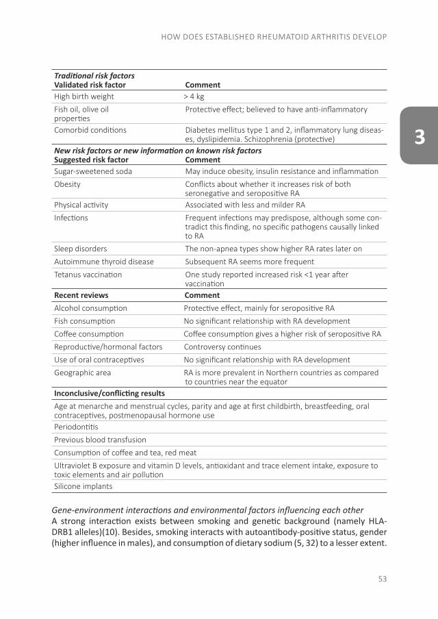
HOW DOES ESTABLISHED RHEUMATOID ARTHRITIS DEVELOP
53
3
Traditional risk factorsValidated risk factor CommentHigh birth weight > 4 kg
Fish oil, olive oil Protective effect; believed to have anti-inflammatory propertiesComorbid conditions Diabetes mellitus type 1 and 2, inflammatory lung diseas- es, dyslipidemia. Schizophrenia (protective)New risk factors or new information on known risk factors Suggested risk factor CommentSugar-sweetened soda May induce obesity, insulin resistance and inflammation
Obesity Conflicts about whether it increases risk of both seronegative and seropositive RAPhysical activity Associated with less and milder RA
Infections Frequent infections may predispose, although some con- tradict this finding, no specific pathogens causally linked to RA
Sleep disorders The non-apnea types show higher RA rates later on
Autoimmune thyroid disease Subsequent RA seems more frequent
Tetanus vaccination One study reported increased risk <1 year after vaccinationRecent reviews CommentAlcohol consumption Protective effect, mainly for seropositive RA
Fish consumption No significant relationship with RA development
Coffee consumption Coffee consumption gives a higher risk of seropositive RA
Reproductive/hormonal factors Controversy continues
Use of oral contraceptives No significant relationship with RA development
Geographic area RA is more prevalent in Northern countries as compared to countries near the equatorInconclusive/conflicting results Age at menarche and menstrual cycles, parity and age at first childbirth, breastfeeding, oral contraceptives, postmenopausal hormone use Periodontitis
Previous blood transfusion
Consumption of coffee and tea, red meat
Ultraviolet B exposure and vitamin D levels, antioxidant and trace element intake, exposure to toxic elements and air pollutionSilicone implants
Gene-environment interactions and environmental factors influencing each otherA strong interaction exists between smoking and genetic background (namely HLA-DRB1 alleles)(10). Besides, smoking interacts with autoantibody-positive status, gender (higher influence in males), and consumption of dietary sodium (5, 32) to a lesser extent.
CHAPTER 3
54
Furthermore, adding positive family history of RA to genetic risk models increases the predictive capacity. However, in general, the gene-environment interactions add too little information to the models to be of clinical use(13).
Autoimmunity and biomarkersApproximately two-thirds of RA patients test positive for RF and/or ACPA at diagnosis, underlying their importance in this disease. Other antibodies preceding and predicting a diagnosis of RA, independent of RF and ACPA status, are anti-carbamylated protein antibodies and anti-peptidyl arginine deiminase type 4 antibodies(6). The discovery of new related autoantibody systems may in the future give more insight into the pathogenesis of RA.
Other blood-based biomarkers such as acute phase reactants or cytokines were not found to have predictive capacity for RA(6).
Clinical prediction modelsQuantifying progression to RA with genetic modeling alone is not ready for clinical use, as we have shown earlier. Several studies have taken a different approach by using a combination of clinical characteristics, symptoms, and sometimes imaging findings. The resulting prediction rules are summarized in Table 3. Validation is still needed for all models. With this restriction, they can be useful to inform persons with musculoskeletal symptoms about their risk of arthritis/RA, especially in the presence of RA-related antibodies.
CHANGING INCIDENCE RATES AND MODE OF PRESENTATION OF RA
In 1979, it was hypothesized that RA as a disease entity would disappear eventually (51). Currently, more evidence exists of a pattern of rises and falls over the decades. Over the first half of the 20th century, no data are available. Alamanos et al. summarized studies on incidence and prevalence rates of RA (according to the 1987 American College of Rheumatology (ACR) criteria) from the second half of the 20th century(52). Two out of the three studies, which evaluated time trends of RA occurrence, reported a declining RA incidence of 15% and 47% in 1 and 4 decade(s), respectively (1980-1990 in MN, USA, and 1955-1994 in Finland). In Greece, the incidence remained stable between 1987 and 1995. Studies in Japan and of North American Natives in the USA have also noted a declining incidence of RA(53, 54). The decline in incidence combined with a shift toward higher age at the onset of disease has been attributed to a so-called birth cohort effect(55). This is a term used in social science to describe characteristics of an area of study over time among individuals who are defined by certain early life influences. Following generations will benefit or be harmed by these influences of their ancestors, in this case leading to a decline in RA incidence. However, which specific risk factors would be implicated in the decline of the incidence has not been specified.
HOW DOES ESTABLISHED RHEUMATOID ARTHRITIS DEVELOP
55
3
More recent studies in Denmark(56) and Minnesota(57) suggest that the incidence may be rising again, with annual increases of RA incidence of 6% (1995-2001) and 2.5% (1995-2007), respectively, remarkably only in women. However, in Finland, a further annual decline of 2% was seen for RF-positive RA over the period 1980-2000(55). It was speculated that a combination of environmental changes leading to either increased risk or loss of protection plays a role in the increasing RA incidence found in the above-mentioned countries. As alluded to in the earlier text, obesity seems to be an important emerging risk factor of RA development. Crowson et al. linked the recent increase of obesity in the population to the higher incidence of RA(58). It was calculated that an increase in obesity could explain 52% of the increase in the incidence of RA among women in the period 1995-2007. Also, other factors may play a role, for example, lower doses of hormones in the oral contraceptives over the years, slower decline of smoking rates in women compared with men, and more vitamin D deficiency(57).
Another important note about changes in incidence rates over time is that the timing of the measurement and used RA criteria can vary between studies, and it also depends on the duration of the study period, mode of presentation, awareness of the disease by general practitioners, and the delay of referral after symptom onset. In the following, we describe two of these factors in more detail. First, the new ACR/European League Against Rheumatism (EULAR) 2010 criteria for RA (see subsequent discussion) are more sensitive than the earlier criteria, which will probably lead to earlier detection (and treatment) and thereby affect the measurement of incidence rates in the coming years(59). Second, within Europe, the variation in the delay of first assessment of RA patients is substantial, with a median range of 16-38 weeks per center and a difference at its highest of 34% in seeing patients within 12 weeks of symptom onset(60). This could partly explain differences of changes in incidence rates across European countries, and even less is known about such a variation outside Europe.
In conclusion, relevant trends are a steady decrease of worldwide RA incidence during the period 1955-1995, followed by a recent increase in at least Denmark and the USA, probably explained in part by changing environmental factors. Furthermore, factors such as differences in the use of RA criteria and differences in the awareness of RA across countries can affect the incidence rates over time.
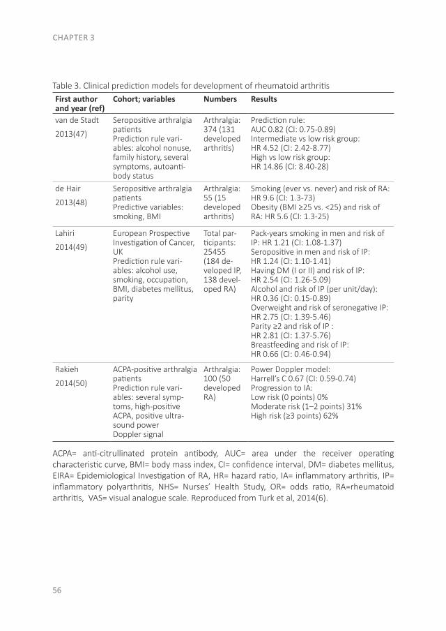
CHAPTER 3
56
Table 3. Clinical prediction models for development of rheumatoid arthritisFirst author and year (ref)
Cohort; variables Numbers Results
van de Stadt
2013(47)
Seropositive arthralgia patientsPrediction rule vari-ables: alcohol nonuse, family history, several symptoms, autoanti-body status
Arthralgia: 374 (131 developed arthritis)
Prediction rule: AUC 0.82 (CI: 0.75-0.89)Intermediate vs low risk group: HR 4.52 (CI: 2.42-8.77)High vs low risk group: HR 14.86 (CI: 8.40-28)
de Hair
2013(48)
Seropositive arthralgia patientsPredictive variables: smoking, BMI
Arthralgia: 55 (15 developed arthritis)
Smoking (ever vs. never) and risk of RA: HR 9.6 (CI: 1.3-73)Obesity (BMI ≥25 vs. <25) and risk of RA: HR 5.6 (CI: 1.3-25)
Lahiri
2014(49)
European Prospective Investigation of Cancer, UK Prediction rule vari-ables: alcohol use, smoking, occupation, BMI, diabetes mellitus, parity
Total par-ticipants: 25455 (184 de-veloped IP, 138 devel-oped RA)
Pack-years smoking in men and risk of IP: HR 1.21 (CI: 1.08-1.37)Seropositive in men and risk of IP: HR 1.24 (CI: 1.10-1.41)Having DM (I or II) and risk of IP: HR 2.54 (CI: 1.26-5.09)Alcohol and risk of IP (per unit/day): HR 0.36 (CI: 0.15-0.89)Overweight and risk of seronegative IP: HR 2.75 (CI: 1.39-5.46)Parity ≥2 and risk of IP : HR 2.81 (CI: 1.37-5.76)Breastfeeding and risk of IP: HR 0.66 (CI: 0.46-0.94)
Rakieh
2014(50)
ACPA-positive arthralgia patientsPrediction rule vari-ables: several symp-toms, high-positive ACPA, positive ultra-sound powerDoppler signal
Arthralgia: 100 (50 developed RA)
Power Doppler model: Harrell’s C 0.67 (CI: 0.59-0.74)Progression to IA:Low risk (0 points) 0%Moderate risk (1–2 points) 31%High risk (≥3 points) 62%
ACPA= anti-citrullinated protein antibody, AUC= area under the receiver operating characteristic curve, BMI= body mass index, CI= confidence interval, DM= diabetes mellitus, EIRA= Epidemiological Investigation of RA, HR= hazard ratio, IA= inflammatory arthritis, IP= inflammatory polyarthritis, NHS= Nurses’ Health Study, OR= odds ratio, RA=rheumatoid arthritis, VAS= visual analogue scale. Reproduced from Turk et al, 2014(6).
HOW DOES ESTABLISHED RHEUMATOID ARTHRITIS DEVELOP
57
3
UA, PAST AND PRESENT
The term “UA” suggests that the condition in the patient concerned is in a stage of transition from an unspecified type of arthritis toward either RA, another arthritis-associated diagnosis, or spontaneous remission. The incidence of UA ranges from 41 (in Finland) to 149 (in Sweden) per 100.000 adults, and 13-54% of these patients will eventually develop RA, according to the 1987 ACR criteria(61). In the past, the transition from UA to RA was equivalent to fulfilling the 1987 ACR criteria for RA(62) after a phase with arthritis in which these criteria were not yet fulfilled. In practice, this mainly applied to the progression from oligoarthritis to polyarthritis and/or the development of erosive disease, as other elements of the criteria set such as RF or nodules do not often appear in early arthritis, if not present at the first presentation(63). Therefore, the transition from UA to RA could be viewed as the development of a more severe arthritis in inadequately controlled early RA, which made this an outcome of interest. The main predictor of the transition was the ACPA status of the patient(64).
The 2010 ACR/EULAR criteria for RA aim to increase sensitivity in early disease(65), which is mainly achieved by a focus on small joint involvement and serology. Thus, a patient with one swollen finger joint of 6 weeks duration and a high-titer ACPA will already classify as RA. The consequence is that the subgroup of UA in early arthritis patients is strongly reduced, and it is now composed mainly of seronegative (oligo-) arthritis. On average, these “2010 UA” patients will have a milder and more heterogeneous disease than “1987 UA” patients(66). Although both the 1987 and 2010 criteria for RA are classification and not diagnostic criteria, the 2010 criteria were specifically developed for use in early disease, and they reflect the trend among clinicians to diagnose RA earlier and even in the presence of only a few involved joints.
Just as was the case with 1987 UA patients, a part of 2010 UA patients will remit and a part will go on to have a severe disease course. In a recent study of three early arthritis cohorts, the Leiden prediction rule (developed to predict 1987 RA in 1987 UA patients) and the ACPA status failed to predict the development of 2010 RA in 2010 UA patients(67). New biomarkers are needed that can help to detect the 2010 UA patients at high risk of disease progression, so that they may be considered for more aggressive therapy than the remaining UA patients, for whom symptomatic treatment may be sufficient. An example is anti-CarP antibodies, which were shown to predict radiographic damage in early ACPA-negative RA patients(68). Next to blood-based biomarkers, imaging modalities such as ultrasound or magnetic resonance imaging (MRI) may prove to be useful in this respect(69, 70).
CHAPTER 3
58
WHEN DOES EARLY RA BECOME ESTABLISHED RA?
This question gives rise to the suggestion that there is a difference between the pathology at the beginning of the disease and what is found later on, and that this distinction has clinical significance. In fact, this is closely related to the concept of a therapeutic “window of opportunity,” which states that treatment initiated at an early stage of the disease is more successful than when it is started later on. “Early” would mean that there is joint inflammation of recent onset, which may at this stage still resolve without further consequences or at least decrease to a barely detectable minimum, if treated adequately. “Established” on the other hand would mean the inflammation is there to stay, more or less pronounced, whatever intervention is applied. Moreover, the concept of “established” RA will generally include damage to the joints, and diverse comorbidities with their complications such as osteoporosis or cardiovascular disease, which may arise as a consequence of the ongoing inflammation.
To begin with, the pathology of RA does not suddenly start around the onset of clinical arthritis. RA-specific systemic autoimmunity as well as nonspecific subclinical inflammation occurs in concert on average 5 years before the onset of symptoms(71, 72). During the period of presymptomatic autoimmunity, there is a maturation of the immune response to citrullinated and carbamylated antigens, which is consistent with an increasing break of tolerance(73). Thus, the number and levels of different ACPA specificities increase toward the onset of arthritis; however, there is no further increase once clinical arthritis has begun(74). Accordingly, the number and type of ACPA specificities do not differ largely between early and late disease(75). Anti-immunoglobulin G (IgG) antibodies or RF arise later and less frequently than ACPA, and they may continue to increase in prevalence after the onset of arthritis(74, 76).
The synovial infiltrate of knee joints of RA patients that had not been clinically swollen before, nevertheless, showed chronic inflammation(77). In animal models of RA, inflammation in joint pathological specimens precedes clinically detectable inflammation. Persons at an increased risk of RA have increased numbers of T-cells in their knee synovium even if they did not yet have knee symptoms(78), again suggesting that the transition to chronic inflammation takes place before the onset of clinically apparent arthritis. Once the symptoms begin, a higher number of recognized ACPA specificities are associated with a higher rate of transition to clinical arthritis(73). This means that once a person notices the first symptoms of RA, the pathological immune response has matured to a large extent, but not completely.
Although the immunological driving processes of RA do not seem to differ between early and late RA, it is well known that better clinical results can be obtained by treating RA patients early and aggressively(79). A recent analysis concluded that this window of opportunity starts to close 4 months after the onset of symptoms(80). This implies it is still possible during that period to interrupt certain processes perpetuating the
HOW DOES ESTABLISHED RHEUMATOID ARTHRITIS DEVELOP
59
3
chronicity of inflammation. One of these could be the total burden of inflammation, which builds up in the early clinical phase. It is conceivable that once a critical mass of inflammatory tissue has been reached, it is no longer possible to control it effectively. This theory is difficult to test, as there is no technique available at present, which can reliably test the total load of inflammatory tissue in a person.
PRIMARY PREVENTION OF RA
The different stages of RA development offer opportunities for preventive interventions, varying from (primary) prevention of the development of arthritis in the at-risk phase to (secondary) prevention of progression from UA to RA or from early to established RA.
The list of risk factors for RA (Table 2) shows that there are several opportunities for lifestyle changes to help prevent RA. Smoking is the strongest environmental risk factor for RA, in particular for ACPA-positive RA, and it has been calculated in Denmark and Sweden that population-wide cessation of smoking would result in more than one-third less cases of ACPA positive RA(81, 82). Other potentially modifiable factors include dietary changes, weight reduction and dental care to reduce periodontitis. These are currently being addressed in the PRE-RA Family Study Boston, which is exploring the attitudes of family members of RA patients toward a lifestyle intervention based on a genetic plus environmental risk assessment(83). Participants are randomized to receive feedback and education concerning their personalized RA risk based on demographics, RA-associated behaviors, genetics, and biomarkers or to receive standard RA information. Four behavioral RA risk factors are included in the risk estimate: smoking, excess body weight, poor oral health, and low fish intake. The trial outcomes will be changes in willingness to alter behaviors. As we learn more about these relations, such information programs can be refined. At present, the most important advice is for family members of ACPA-positive RA patients, to refrain from smoking(82).
The concept of primary prevention of RA with drugs has become possible through the recognition of a prolonged at-risk phase with variable symptoms and/or autoimmunity before the outbreak of clinical RA. The first clinical trial was a post hoc analysis of the effect of vitamin E in a study designed to prevent coronary heart disease in the general population(84). Although the trial was negative the for prevention of both heart disease and RA, there was a trend toward protection against RF-positive RA. The next study was a trial of two intramuscular injections of 100 mg dexamethasone or placebo in ACPA and/or RF-positive arthralgia patients(85). Furthermore, this trial did not affect the onset of arthritis, although autoantibody levels were suppressed for 6 months. Meanwhile, trials of rituximab (Prevention of RA by B cell-directed therapy (PRAIRI) trial, NTR1969; www.trialregister.nl), of abatacept (Arthritis Prevention In the Pre-clinical Phase of Rheumatoid Arthritis (APIPPRA) trial; www.isrctn.com/ISRCTN46017566) and of atorvastatin (STAtins in the Prevention of RA (STAPRA) trial; NTR5265; www.trialregister.nl) in the same patient category are ongoing.
CHAPTER 3
60
Some clinicians confronted with seropositive arthralgia patients will try antimalarial treatment. Apart from being a relatively nontoxic RA remedy, the rationale for this treatment comes from the experience with antimalarials in the treatment of palindromic rheumatism, a rather ill-defined syndrome of intermittently occurring peripheral arthritis. A subgroup of those patients is RF- or ACPA-positive with a tendency to develop RA(86), and this tendency was found to be markedly reduced in a retrospective survey in those taking antimalarials(87). Another retrospective study reported a marked reduction in frequency and duration of attacks in palindromic rheumatism patients taking chloroquin(88).
In conclusion, no intervention has yet showed an effect in a randomized controlled trial in the primary prevention of RA. The scarcity of data gives rise to the suggestion that it is not easy to perform clinical trials in the at-risk phase of RA, and that positive outcomes are not readily obtained. A major ethical issue with intervening pharmacologically in this phase, is that persons are exposed to potentially toxic drugs, whereas a part of the study subjects will never develop RA.
SECONDARY PREVENTION OF RA
One of the explicit goals of the 2010 ACR/EULAR criteria for RA was to facilitate the performance of trials in early RA(65), in order to make even better use of the window of opportunity in early disease. The underlying idea was that it would be easier to design a trial for patients who were classified as RA instead of as UA. Nevertheless, already before the publication of the 2010 criteria, a number of trials had been conducted with the intention to prevent the progression of early disease, mostly not classifying as RA according to the 1987 ACR criteria(62). Part of the outcome measures of these trials was a reduction of the transition of UA to RA, which means that a successful outcome could be regarded as secondary 1987 ACR criteria prevention of RA.
The results of the PROMPT study of methotrexate to prevent progression of UA to RA (1987 criteria) and its long-term follow-up showed less progression to RA, but only in ACPA positive patients and only as long as the treatment was continued(89). Other trials in early oligoarthritis or UA have noted some transient benefit from treatment with intramuscular (STIVEA trial) or intraarticular corticosteroids compared to placebo or nonsteroidal anti-inflammatory drugs(90, 91). However, the Stop Arthritis Very Early (SAVE) trial observed that the development of 1987 RA was not delayed by intramuscular glucocorticoid treatment in oligoarthritis patients(92).
Biologics have also been tested for this indication. Three months of infliximab did not prevent progression to 1987 RA after 1 year(93). Six months of abatacept slightly reduced the progression of UA to 1987 RA from 67 to 46%(94). Abatacept treatment also had an impact on radiographic and MRI inhibition, which was maintained for 6 months after treatment stopped. The STREAM study, a trial of aggressive treatment including
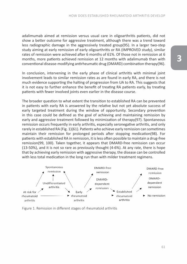
HOW DOES ESTABLISHED RHEUMATOID ARTHRITIS DEVELOP
61
3
adalimumab aimed at remission versus usual care in oligoarthritis patients, did not show a better outcome for aggressive treatment, although there was a trend toward less radiographic damage in the aggressively treated group(95). In a larger two-step study aiming at early remission of early oligoarthritis or RA (IMPROVED study), similar rates of remission were achieved after 6 months of 61%. Of those not in remission at 6 months, more patients achieved remission at 12 months with adalimumab than with conventional disease-modifying antirheumatic drug (DMARD) combination therapy(96).
In conclusion, intervening in the early phase of clinical arthritis with minimal joint involvement leads to similar remission rates as are found in early RA, and there is not much evidence supporting the halting of progression from UA to RA. This suggests that it is not easy to further enhance the benefit of treating RA patients early, by treating patients with fewer involved joints even earlier in the disease course.
The broader question to what extent the transition to established RA can be prevented in patients with early RA is answered by the relative but not yet absolute success of early targeted treatment during the window of opportunity. Secondary prevention in this case could be defined as the goal of achieving and maintaining remission by early and aggressive treatment followed by minimization of therapy(97). Spontaneous remission occurs frequently in early arthritis, especially seronegative arthritis, and only rarely in established RA (Fig. 1)(61). Patients who achieve early remission can sometimes maintain their remission for prolonged periods after stopping medication(98). For patients with established RA in remission, it is less often possible to maintain a drug-free remission(99, 100). Taken together, it appears that DMARD-free remission can occur (13-50%), and it is not so rare as previously thought (4-6%). At any rate, there is hope that by achieving early remission with aggressive therapy, the disease can be controlled with less total medication in the long run than with milder treatment regimens.
Figure 1. Remission in different stages of rheumatoid arthritis
CHAPTER 3
62
SUMMARY
The increasingly successful management of RA now leads to more patients achieving early and sustained remission, and this will lead to less patients progressing to the classical state of established RA. A next goal in the management of RA can be the improved recognition and intervention in the early or even at-risk phase of RA.
Prediction depends on the knowledge of risk factors. Recent advances in the risk factor assessment of RA include alcohol consumption as a confirmed protective factor, whereas fish consumption could not be confirmed as a protective factor. New risk factors are coffee consumption, sugar consumption, sleep disorders, and thyroid disease, whereas exercise and recent infections have been put forward as protective factors. Increasingly, risk factors are being combined to establish prediction rules. Those containing genetic risk plus environmental factors are not yet ready for general use. However, new prediction rules for arthralgia subjects using clinical characteristics, serology, and sometimes imaging are quite simple to perform, and they can be used to inform patients of their risk of RA.
Interestingly, RA incidence seems to have been declining since 1955, when formal measurements started, at least until the end of the last century. However, recent reports suggest that the incidence is on the rise again, mainly in seronegative females, and that this can be ascribed largely to the recent increase in obesity. When comparing trends in different countries, it becomes necessary to take into account the large variation between countries in the public and physician awareness of the need to identify RA early. The problem of assessing UA has been reduced considerably by the introduction of the 2010 RA criteria. Many former UA patients can now be classified as RA, leaving a smaller group of UA patients with more heterogeneous and milder disease. Treating UA patients early gives results similar to early treatment of RA. In line with the concept of an early “window of opportunity,” a few studies have attempted to treat patients at an even earlier stage, before clinical arthritis becomes apparent. These primary prevention studies with pharmacological interventions have not yet produced positive results. Although these efforts are continued, the identification of modifiable risk factors for RA such as smoking, obesity, and lack of exercise should incite physicians to promote healthy behavior in persons at risk of RA.
HOW DOES ESTABLISHED RHEUMATOID ARTHRITIS DEVELOP
63
3
RESEARCH AGENDA
• Improve prediction models of RA by integrating personal characteristics, symp-toms and genetic information with new biomarkers.
• Establish simple prediction aids for different situations, for example, in the gen-eral practitioner (GP) office, the rheumatology clinic, or the general public.
• Controlled intervention studies in persons at risk of RA in different stages.
• Improved identification of UA with poor prognosis.
PRACTICE POINTS
• Worldwide RA incidence showed a steady decrease during the period 1955-1995, followed by a recent increase in at least Denmark and the USA.
• New possible risk factors for the development of RA are non-alcohol use, coffee consumption, sugar-sweetened soda intake, obesity, physical inactivity, sleep disorders, and thyroid disease.
• Possible options for primary prevention of RA include dietary changes, weight reduction and dental care. No drug intervention has proven to be effective in the prevention of RA.
• With the advent of the 2010 ACR/EULAR criteria, the subgroup of UA in early ar-thritis patients is strongly reduced, and it contains mainly seronegative (oligo-) arthritis patients with a mild disease course.
• Secondary prevention of RA is becoming less of an issue due to the high sensi-tivity of the 2010 ACR/EULAR criteria in early disease, and the tendency to treat early arthritis rapidly.
REFERENCES
1. Short CL. The antiquity of rheumatoid arthritis. Arthritis and rheumatism 1974;17(3):193-205.
2. Dequeker J. Siebrandus Sixtius: evidence of rheumatoid arthritis of the robust reaction type in a seventeenth century Dutch priest. Annals of the rheumatic diseases 1992;51(4):561-562.
3. Storey GO, Comer M, Scott DL. Chronic arthritis before 1876: early British cases suggesting rheumatoid arthritis. Annals of the rheumatic diseases 1994;53(9):557-560.
4. Rothschild BM, Turner KR, DeLuca MA. Symmetrical erosive peripheral polyarthritis in the Late Archaic Period of Alabama. Science 1988;241(4872):1498-501.
5. *Scott IC, Steer S, Lewis CM, Cope AP. Precipitating and perpetuating factors of rheumatoid arthritis immunopathology: linking the triad of genetic predisposition, environmental risk factors and autoimmunity to disease pathogenesis. Best practice and research clinical rheumatology 2011;25(4):447-468.
6. *Turk SA, van Beers-Tas MH, van Schaardenburg D. Prediction of future rheumatoid arthritis. Rheumatic diseases clinics of North America 2014;40(4):753-770.
7. *Arend WP, Firestein GS. Pre-rheumatoid arthritis: predisposition and transition to clinical synovitis. Nature reviews rheumatology 2012;8(10):573-586.
8. Yarwood A, Huizinga TW, Worthington J. The genetics of rheumatoid arthritis: risk and protection in different stages of the evolution of RA. Rheumatology 2014 [Epub 2014 Sep 18].
9. Kurreeman F, Liao K, Chibnik L, Hickey B, Stahl E, Gainer V, et al. Genetic basis of autoantibody positive and negative rheumatoid arthritis risk in a multi-ethnic cohort derived from electronic health records. The American journal of human genetics 2011;88(1):57-69.
10. Sparks JA, Costenbader KH. Genetics, environment, and gene-environment interactions in the development of systemic rheumatic diseases. Rheumatic diseases clinics of North America 2014;40(4):637-657.
11. Yamamoto K, Okada Y, Suzuki A, Kochi Y. Genetics of rheumatoid arthritis in Asia-present and future. Nature reviews rheumatology 2015 [Epub 2015 Feb 10].
12. El-Gabalawy HS, Robinson DB, Daha NA, Oen KG, Smolik I, Elias B, et al. Non-HLA genes modulate the risk of rheumatoid arthritis associated with HLA-DRB1 in a susceptible North American Native population. Genes and immunity 2011;12(7):568-574.
13. *Sparks JA, Chen CY, Jiang X, Askling J, Hiraki LT, Malspeis S, et al. Improved performance of epidemiologic and genetic risk models for rheumatoid arthritis serologic phenotypes using family history. Annals of the rheumatic diseases 2015;74(8):1522-1529.
14. Nagai Y, Imanishi T. RAvariome: a genetic risk variants database for rheumatoid arthritis based on assessment of reproducibility between or within human populations. Database 2013 [2013:bat073].
15. van der Helm-van Mil AH, Toes RE, Huizinga TW. Genetic variants in the prediction of rheumatoid arthritis. Annals of the rheumatic diseases 2010;69(9):1694-1696.
CHAPTER 3
64
16. Scott IC, Seegobin SD, Steer S, Tan R, Forabosco P, Hinks A, et al. Predicting the risk of rheumatoid arthritis and its age of onset through modelling genetic risk variants with smoking. PLoS genetics 2013;9(9) [Epub 2013 Sep 19].
17. Yarwood A, Han B, Raychaudhuri S, Bowes J, Lunt M, Pappas DA, et al. A weighted genetic risk score using all known susceptibility variants to estimate rheumatoid arthritis risk. Annals of the rheumatic diseases 2015;74(1):170-176.
18. Jin Z, Xiang C, Cai Q, Wei X, He J. Alcohol consumption as a preventive factor for developing rheumatoid arthritis: a dose-response meta-analysis of prospective studies. Annals of the rheumatic diseases 2014;73(11):1962-1967.
19. Scott IC, Tan R, Stahl D, Steer S, Lewis CM, Cope AP. The protective effect of alcohol on developing rheumatoid arthritis: a systematic review and meta-analysis. Rheumatology 2013;52(5):856-867.
20. Lu B, Solomon DH, Costenbader KH, Karlson EW. Alcohol consumption and risk of incident rheumatoid arthritis in women: a prospective study. Arthritis and rheumatology 2014;66(8):1998-2005.
21. Di Giuseppe D, Crippa A, Orsini N, Wolk A. Fish consumption and risk of rheumatoid arthritis: a dose-response meta-analysis. Arthritis research and therapy 2014;16(5):446.
22. Lee YH, Bae SC, Song GG. Coffee or tea consumption and the risk of rheumatoid arthritis: a meta-analysis. Clinical rheumatology 2014;33(11):1575-1583.
23. Marder W, Somers EC. Is pregnancy a risk factor for rheumatic autoimmune diseases? Current opinion in rheumatology 2014;26(3):321-328.
24. Jorgensen KT, Harpsoe MC, Jacobsen S, Jess T, Frisch M. Increased risk of rheumatoid arthritis in women with pregnancy complications and poor self-rated health: a study within the Danish National Birth Cohort. Rheumatology 2014;53(8):1513-1519.
25. Beydoun HA, el-Amin R, McNeal M, Perry C, Archer DF. Reproductive history and postmenopausal rheumatoid arthritis among women 60 years or older: Third National Health and Nutrition Examination Survey. Menopause: the journal of the North American menopause society 2013;20(9):930-935.
26. Chen Q, Jin Z, Xiang C, Cai Q, Shi W, He J. Absence of protective effect of oral contraceptive use on the development of rheumatoid arthritis: a meta-analysis of observational studies. International journal of rheumatic diseases 2014;17(7):725-737.
27. Masi AT, Rehman AA, Chatterton RT, Wang H, Goertzen NJ, Elmore KB, et al. Controlled cohort study of serum gonadal and adrenocortical steroid levels in males prior to onset of rheumatoid arthritis (pre-RA): a comparison to pre-RA females and sex differences among the study groups. International journal of rheumatology 2013 [Epub 2013 Nov 24].
28. Pikwer M, Giwercman A, Bergstrom U, Nilsson JA, Jacobsson LT, Turesson C. Association between testosterone levels and risk of future rheumatoid arthritis in men: a population-based case-control study. Annals of the rheumatic diseases 2014;73(3):573-579.
29. Cross M, Smith E, Hoy D, Carmona L, Wolfe F, Vos T, et al. The global burden of rheumatoid arthritis: estimates from the global burden of disease 2010 study. Annals of the rheumatic diseases. 2014;73(7):1316-1322.
HOW DOES ESTABLISHED RHEUMATOID ARTHRITIS DEVELOP
65
3
30. Pattison DJ, Harrison RA, Symmons DP. The role of diet in susceptibility to rheumatoid arthritis: a systematic review. The Journal of rheumatology 2004;31(7):1310-1319.
31. Sundstrom B, Johansson I, Rantapaa-Dahlqvist S. Diet and alcohol as risk factors for rheumatoid arthritis: a nested case-control study. Rheumatology internal 2015;35(3):533-539.
32. Sundstrom B, Johansson I, Rantapaa-Dahlqvist S. Interaction between dietary sodium and smoking increases the risk for rheumatoid arthritis: results from a nested case-control study. Rheumatology 2015;54(3):487-493.
33. Hu Y, Costenbader KH, Gao X, Al-Daabil M, Sparks JA, Solomon DH, et al. Sugar-sweetened soda consumption and risk of developing rheumatoid arthritis in women. The American journal of clinical nutrition 2014;100(3):959-967.
34. Finckh A, Turesson C. The impact of obesity on the development and progression of rheumatoid arthritis. Annals of the rheumatic diseases 2014;73(11):1911-1913.
35. Wesley A, Bengtsson C, Elkan AC, Klareskog L, Alfredsson L, Wedren S. Association between body mass index and anti-citrullinated protein antibody-positive and anti-citrullinated protein antibody-negative rheumatoid arthritis: results from a population-based case-control study. Arthritis care and research 2013;65(1):107-112.
36. Lu B, Hiraki LT, Sparks JA, Malspeis S, Chen CY, Awosogba JA, et al. Being overweight or obese and risk of developing rheumatoid arthritis among women: a prospective cohort study. Annals of the rheumatic diseases 2014;73(11):1914-1922.
37. Di Giuseppe D, Bottai M, Askling J, Wolk A. Physical activity and risk of rheumatoid arthritis in women: a population-based prospective study. Arthritis research and therapy 2015;17(1):40-7.
38. Sandberg ME, Wedren S, Klareskog L, Lundberg IE, Opava CH, Alfredsson L, et al. Patients with regular physical activity before onset of rheumatoid arthritis present with milder disease. Annals of the rheumatic diseases 2014;73(8):1541-1544.
39. Hsiao YH, Chen YT, Tseng CM, Wu LA, Lin WC, Su VY, et al. Sleep disorders and increased risk of autoimmune diseases in individuals without sleep apnea. Sleep 2015;38(4):581-586.
40. Pan XF, Gu JQ, Shan ZY. Increased risk of thyroid autoimmunity in rheumatoid arthritis: a systematic review and meta-analysis. Endocrine 2015;50(1):79-86.
41. Arleevskaya MI, Gabdoulkhakova AG, Filina YV, Miftakhova RR, Bredberg A, Tsybulkin AP. A transient peak of infections during onset of rheumatoid arthritis: a 10-year prospective cohort study. British medical journal open 2014;4(8) [Epub 2014 Sep 3].
42. Rogers MA, Levine DA, Blumberg N, Fisher GG, Kabeto M, Langa KM. Antigenic challenge in the etiology of autoimmune disease in women. Journal of autoimmunity 2012;38(2-3):J97-J102.
43. Sandberg ME, Bengtsson C, Klareskog L, Alfredsson L, Saevarsdottir S. Recent infections are associated with decreased risk of rheumatoid arthritis: a population-based case-control study. Annals of the rheumatic diseases 2015;74(5):904-907.
44. Ray P, Black S, Shinefield H, Dillon A, Carpenter D, Lewis E, et al. Risk of rheumatoid arthritis following vaccination with tetanus, influenza and hepatitis B vaccines among persons 15-59 years of age. Vaccine 2011;29(38):6592-6597.
CHAPTER 3
66
45. De Roos AJ, Koehoorn M, Tamburic L, Davies HW, Brauer M. Proximity to traffic, ambient air pollution, and community noise in relation to incident rheumatoid arthritis. Environmental health perspectives 2014;122(10):1075-1080.
46. Hart JE, Kallberg H, Laden F, Bellander T, Costenbader KH, Holmqvist M, et al. Ambient air pollution exposures and risk of rheumatoid arthritis: results from the Swedish EIRA case-control study. Annals of the rheumatic diseases 2013;72(6):888-894.
47. *van de Stadt LA, Witte BI, Bos WH, van Schaardenburg D. A prediction rule for the development of arthritis in seropositive arthralgia patients. Annals of the rheumatic diseases 2013;72(12):1920-1926.
48. de Hair MJ, Landewe RB, van de Sande MG, van Schaardenburg D, van Baarsen LG, Gerlag DM, et al. Smoking and overweight determine the likelihood of developing rheumatoid arthritis. Annals of the rheumatic diseases 2013;72(10):1654-1658.
49. *Lahiri M, Luben RN, Morgan C, Bunn DK, Marshall T, Lunt M, et al. Using lifestyle factors to identify individuals at higher risk of inflammatory polyarthritis (results from the European Prospective Investigation of Cancer-Norfolk and the Norfolk Arthritis Register--the EPIC-2-NOAR Study). Annals of the rheumatic diseases 2014;73(1):219-226.
50. Rakieh C, Nam JL, Hunt L, Hensor EM, Das S, Bissell LA, et al. Predicting the development of clinical arthritis in anti-CCP positive individuals with non-specific musculoskeletal symptoms: a prospective observational cohort study. Annals of the rheumatic diseases. 2015;74(9):1659-1666.
51. Buchanan WW, Murdoch RM. Hypothesis: that rheumatoid arthritis will disappear. The Journal of rheumatology 1979;6(3):324-329.
52. Alamanos Y, Voulgari PV, Drosos AA. Incidence and prevalence of rheumatoid arthritis, based on the 1987 American College of Rheumatology criteria: a systematic review. Semininars in arthritis and rheumatism 2006;36(3):182-188.
53. Jacobsson LT, Hanson RL, Knowler WC, Pillemer S, Pettitt DJ, McCance DR, et al. Decreasing incidence and prevalence of rheumatoid arthritis in Pima Indians over a twenty-five-year period. Arthritis and rheumatism 1994;37(8):1158-1165.
54. Shichikawa K, Inoue K, Hirota S, Maeda A, Ota H, Kimura M, et al. Changes in the incidence and prevalence of rheumatoid arthritis in Kamitonda, Wakayama, Japan, 1965-1996. Annals of the rheumatic diseases 1999;58(12):751-756.
55. Kaipiainen-Seppanen O, Kautiainen H. Declining trend in the incidence of rheumatoid factor-positive rheumatoid arthritis in Finland 1980-2000. The Journal of rheumatology 2006;33(11):2132-2138.
56. Pedersen JK, Svendsen AJ, Horslev-Petersen K. Incidence of Rheumatoid Arthritis in the Southern part of Denmark from 1995 to 2001. The open rheumatology journal 2007;1:18-23.
57. Myasoedova E, Crowson CS, Kremers HM, Therneau TM, Gabriel SE. Is the incidence of rheumatoid arthritis rising? Results from Olmsted County, Minnesota, 1955-2007. Arthritis and rheumatism 2010;62(6):1576-1582.
58. Crowson CS, Matteson EL, Davis JM, 3rd, Gabriel SE. Contribution of obesity to the rise in incidence of rheumatoid arthritis. Arthritis care and research 2013;65(1):71-77.
HOW DOES ESTABLISHED RHEUMATOID ARTHRITIS DEVELOP
67
3
59. Humphreys JH, Symmons DP. Postpublication validation of the 2010 American College of Rheumatology/European League Against Rheumatism classification criteria for rheumatoid arthritis: where do we stand? Current opinion in rheumatology 2013;25(2):157-163.
60. Raza K, Stack R, Kumar K, Filer A, Detert J, Bastian H, et al. Delays in assessment of patients with rheumatoid arthritis: variations across Europe. Annals of the rheumatic diseases 2011;70(10):1822-5.
61. Hazes JM, Luime JJ. The epidemiology of early inflammatory arthritis. Nature reviews rheumatology 2011;7(7):381-390.
62. Arnett FC, Edworthy SM, Bloch DA, McShane DJ, Fries JF, Cooper NS, et al. The American Rheumatism Association 1987 revised criteria for the classification of rheumatoid arthritis. Arthritis and rheumatism 1988;31(3):315-324.
63. Ursum J, Bos WH, van de Stadt RJ, Dijkmans BA, van Schaardenburg D. Different properties of ACPA and IgM-RF derived from a large dataset: further evidence of two distinct autoantibody systems. Arthritis research and therapy 2009;11(3):R75.
64. 64. van der Helm-van Mil AH, le Cessie S, van Dongen H, Breedveld FC, Toes RE, Huizinga TW. A prediction rule for disease outcome in patients with recent-onset undifferentiated arthritis: how to guide individual treatment decisions. Arthritis and rheumatism 2007;56(2):433-440.
65. Aletaha D, Neogi T, Silman AJ, Funovits J, Felson DT, Bingham CO, 3rd, et al. 2010 rheumatoid arthritis classification criteria: an American College of Rheumatology/European League Against Rheumatism collaborative initiative. Annals of the rheumatic diseases 2010;69(9):1580-1588.
66. *Krabben A, Huizinga TW, van der Helm-van Mil AH. Undifferentiated arthritis characteristics and outcomes when applying the 2010 and 1987 criteria for rheumatoid arthritis. Annals of the rheumatic diseases 2012;71(2):238-241.
67. Krabben A, Abhishek A, Britsemmer K, Filer A, Huizinga TW, Raza K, et al. Risk of rheumatoid arthritis development in patients with unclassified arthritis according to the 2010 ACR/EULAR criteria for rheumatoid arthritis Rheumatology 2013;52(7):1265-1270.
68. Shi J, Knevel R, Suwannalai P, van der Linden MP, Janssen GM, van Veelen PA, et al. Autoantibodies recognizing carbamylated proteins are present in sera of patients with rheumatoid arthritis and predict joint damage. Proceedings of the national academy of sciences of the United States of America 2011;108(42):17372-17377.
69. Duer-Jensen A, Horslev-Petersen K, Hetland ML, Bak L, Ejbjerg BJ, Hansen MS, et al. Bone edema on magnetic resonance imaging is an independent predictor of rheumatoid arthritis development in patients with early undifferentiated arthritis. Arthritis and rheumatism 2011;63(8):2192-2202.
70. Filer A, de Pablo P, Allen G, Nightingale P, Jordan A, Jobanputra P, et al. Utility of ultrasound joint counts in the prediction of rheumatoid arthritis in patients with very early synovitis. Annals of the rheumatic diseases 2011;70(3):500-507.
CHAPTER 3
68
71. Nielen MM, van Schaardenburg D, Reesink HW, Twisk JW, van de Stadt RJ, van der Horst-Bruinsma IE, et al. Simultaneous development of acute phase response and autoantibodies in preclinical rheumatoid arthritis. Annals of the rheumatic diseases 2006;65(4):535-537.
72. Nielen MM, van Schaardenburg D, Reesink HW, van de Stadt RJ, van der Horst-Bruinsma IE, de Koning MH, et al. Specific autoantibodies precede the symptoms of rheumatoid arthritis: a study of serial measurements in blood donors. Arthritis and rheumatism 2004;50(2):380-386.
73. *van de Stadt LA, van der Horst AR, de Koning MH, Bos WH, Wolbink GJ, van de Stadt RJ, et al. The extent of the anti-citrullinated protein antibody repertoire is associated with arthritis development in patients with seropositive arthralgia. Annals of the rheumatic diseases 2011;70(1):128-133.
74. Ursum J, Bos WH, van Dillen N, Dijkmans BA, van Schaardenburg D. Levels of anti-citrullinated protein antibodies and IgM rheumatoid factor are not associated with outcome in early arthritis patients: a cohort study. Arthritis research and therapy 2010;12(1):R8.
75. van der Woude D, Syversen SW, van der Voort EI, Verpoort KN, Goll GL, van der Linden MP, et al. The ACPA isotype profile reflects long-term radiographic progression in rheumatoid arthritis. Annals of the rheumatic diseases 2010;69(6):1110-1116.
76. van de Stadt LA, de Vrieze H, Derksen NI, Brouwer M, Wouters D, van Schaardenburg D, et al. Antibodies to IgG4 hinge can be found in rheumatoid arthritis patients during all stages of disease and may exacerbate chronic antibody-mediated inflammation. Arthritis and rheumatology 2014;66(5):1133-1140.
77. Kraan MC, Versendaal H, Jonker M, Bresnihan B, Post WJ, t Hart BA, et al. Asymptomatic synovitis precedes clinically manifest arthritis. Arthritis and rheumatism 1998;41(8):1481-1488.
78. de Hair MJ, van de Sande MG, Ramwadhdoebe TH, Hansson M, Landewe R, van der Leij C, et al. Features of the synovium of individuals at risk of developing rheumatoid arthritis: implications for understanding preclinical rheumatoid arthritis. Arthritis and rheumatology 2014;66(3):513-522.
79. Finckh A, Liang MH, van Herckenrode CM, de Pablo P. Long-term impact of early treatment on radiographic progression in rheumatoid arthritis: A meta-analysis. Arthritis and rheumatism 2006;55(6):864-872.
80. *van Nies JA, Tsonaka R, Gaujoux-Viala C, Fautrel B, van der Helm-van Mil AH. Evaluating relationships between symptom duration and persistence of rheumatoid arthritis: does a window of opportunity exist? Results on the Leiden Early Arthritis Clinic and ESPOIR cohorts. Annals of the rheumatic diseases 2015;74(5):806-812.
81. Kallberg H, Ding B, Padyukov L, Bengtsson C, Ronnelid J, Klareskog L, et al. Smoking is a major preventable risk factor for rheumatoid arthritis: estimations of risks after various exposures to cigarette smoke. Annals of the rheumatic diseases 2011;70(3):508-511.
HOW DOES ESTABLISHED RHEUMATOID ARTHRITIS DEVELOP
69
3
82. Pedersen M, Jacobsen S, Garred P, Madsen HO, Klarlund M, Svejgaard A, et al. Strong combined gene-environment effects in anti-cyclic citrullinated peptide-positive rheumatoid arthritis: a nationwide case-control study in Denmark. Arthritis and rheumatism 2007;56(5):1446-1453.
83. Sparks JA, Iversen MD, Miller Kroouze R, Mahmoud TG, Triedman NA, Kalia SS, et al. Personalized Risk Estimator for Rheumatoid Arthritis (PRE-RA) Family Study: rationale and design for a randomized controlled trial evaluating rheumatoid arthritis risk education to first-degree relatives. Contemporary clinical trials 2014;39(1):145-157.
84. Karlson EW, Shadick NA, Cook NR, Buring JE, Lee IM. Vitamin E in the primary prevention of rheumatoid arthritis: the Women’s Health Study. Arthritis and rheumatism. 2008;59(11):1589-1595.
85. Bos WH, Dijkmans BA, Boers M, van de Stadt RJ, van Schaardenburg D. Effect of dexamethasone on autoantibody levels and arthritis development in patients with arthralgia: a randomised trial. Annals of the rheumatic diseases 2010;69(3):571-574.
86. Gonzalez-Lopez L, Gamez-Nava JI, Jhangri GS, Ramos-Remus C, Russell AS, Suarez-Almazor ME. Prognostic factors for the development of rheumatoid arthritis and other connective tissue diseases in patients with palindromic rheumatism. The Journal of rheumatology 1999;26(3):540-545.
87. Gonzalez-Lopez L, Gamez-Nava JI, Jhangri G, Russell AS, Suarez-Almazor ME. Decreased progression to rheumatoid arthritis or other connective tissue diseases in patients with palindromic rheumatism treated with antimalarials. The Journal of rheumatology 2000;27(1):41-46.
88. Youssef W, Yan A, Russell AS. Palindromic rheumatism: a response to chloroquine. The Journal of rheumatology. 1991;18(1):35-37.
89. van Dongen H, van Aken J, Lard LR, Visser K, Ronday HK, Hulsmans HM, et al. Efficacy of methotrexate treatment in patients with probable rheumatoid arthritis: a double-blind, randomized, placebo-controlled trial. Arthritis and rheumatism 2007;56(5):1424-1432.
90. Marzo-Ortega H, Green MJ, Keenan AM, Wakefield RJ, Proudman S, Emery P. A randomized controlled trial of early intervention with intraarticular corticosteroids followed by sulfasalazine versus conservative treatment in early oligoarthritis. Arthritis and rheumatism 2007;57(1):154-160.
91. Verstappen SM, McCoy MJ, Roberts C, Dale NE, Hassell AB, Symmons DP. Beneficial effects of a 3-week course of intramuscular glucocorticoid injections in patients with very early inflammatory polyarthritis: results of the STIVEA trial. Annals of the rheumatic diseases 2010;69(3):503-509.
92. Machold KP, Landewe R, Smolen JS, Stamm TA, van der Heijde DM, Verpoort KN, et al. The Stop Arthritis Very Early (SAVE) trial, an international multicentre, randomised, double-blind, placebo-controlled trial on glucocorticoids in very early arthritis. Annals of the rheumatic diseases 2010;69(3):495-502.
93. Saleem B, Mackie S, Quinn M, Nizam S, Hensor E, Jarrett S, et al. Does the use of tumour necrosis factor antagonist therapy in poor prognosis, undifferentiated arthritis prevent progression to rheumatoid arthritis? Annals of the rheumatic diseases 2008;67(8):1178-1180.
CHAPTER 3
70
94. Emery P, Durez P, Dougados M, Legerton CW, Becker JC, Vratsanos G, et al. Impact of T-cell costimulation modulation in patients with undifferentiated inflammatory arthritis or very early rheumatoid arthritis: a clinical and imaging study of abatacept (the ADJUST trial). Annals of the rheumatic diseases 2010;69(3):510-516.
95. van Eijk IC, Nielen MM, van der Horst-Bruinsma I, Tijhuis GJ, Boers M, Dijkmans BA, et al. Aggressive therapy in patients with early arthritis results in similar outcome compared with conventional care: the STREAM randomized trial. Rheumatology 2012;51(4):686-694.
96. *Heimans L, Wevers-de Boer KV, Visser K, Goekoop RJ, van Oosterhout M, Harbers JB, et al. A two-step treatment strategy trial in patients with early arthritis aimed at achieving remission: the IMPROVED study. Annals of the rheumatic diseases 2014;73(7):1356-1361.
97. Smolen JS, Landewe R, Breedveld FC, Buch M, Burmester G, Dougados M, et al. EULAR recommendations for the management of rheumatoid arthritis with synthetic and biological disease-modifying antirheumatic drugs: 2013 update. Annals of the rheumatic diseases 2014;73(3):492-509.
98. van Herwaarden N, den Broeder AA, Jacobs W, van der Maas A, Bijlsma JW, van Vollenhoven RF, et al. Down-titration and discontinuation strategies of tumor necrosis factor-blocking agents for rheumatoid arthritis in patients with low disease activity. The Cochrane database of systematic reviews. 2014;9:CD010455.
99. van Herwaarden N, van der Maas A, Minten MJ, van den Hoogen FH, Kievit W, van Vollenhoven RF, et al. Disease activity guided dose reduction and withdrawal of adalimumab or etanercept compared with usual care in rheumatoid arthritis: open label, randomised controlled, non-inferiority trial. British medical journal 2015;350:h1389.
100. Fautrel B, Pham T, Alfaiate T, Gandjbakhch F, Foltz V, Morel J, et al. Step-down strategy of spacing TNF-blocker injections for established rheumatoid arthritis in remission: results of the multicentre non-inferiority randomised open-label controlled trial (STRASS: Spacing of TNF-blocker injections in Rheumatoid ArthritiS Study). Annals of the rheumatic diseases 2015 [Epub 2015 Jun 23].
HOW DOES ESTABLISHED RHEUMATOID ARTHRITIS DEVELOP
71
3

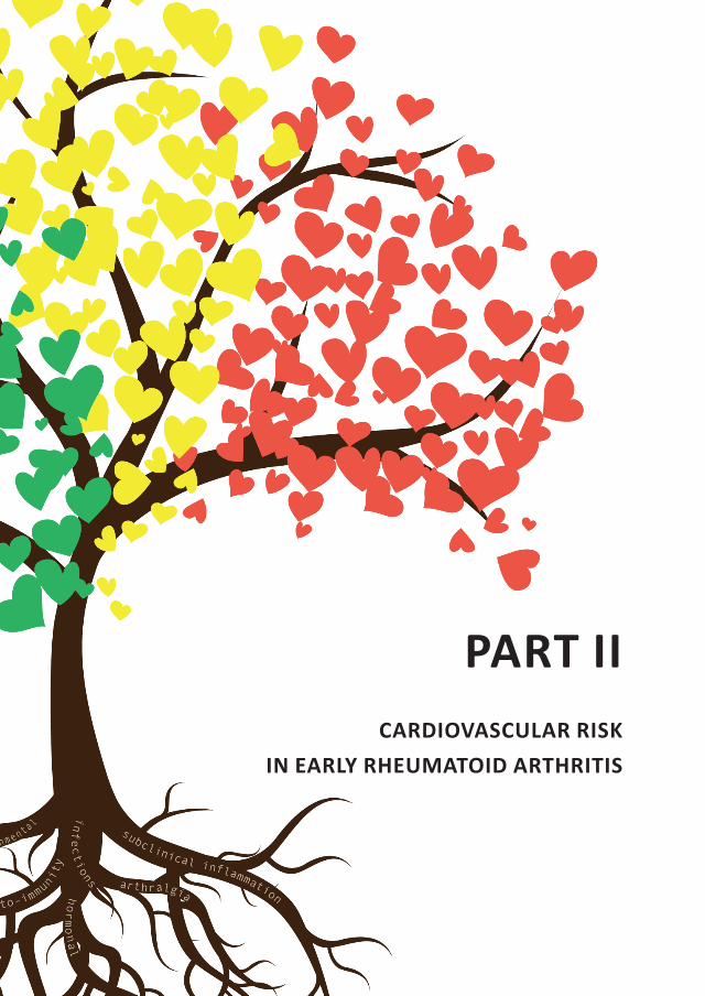
PART IICARDIOVASCULAR RISK
IN EARLY RHEUMATOID ARTHRITIS

CHAPTER 4
AN UNFAVORABLE BODY COMPOSITION IS COMMON IN EARLY ARTHRITIS PATIENTS:
A CASE CONTROL STUDY
Samina A. Turk1
Dirkjan van Schaardenburg1,2
Maarten Boers3,4
Sylvia de Boer1
Cindy Fokker1
Willem F. Lems1,3
Michael T. Nurmohamed1,3
¹ Amsterdam Rheumatology and immunology Center | Reade, Amsterdam, Netherlands² Amsterdam Rheumatology and immunology Center | Academic Medical Center, Am-sterdam, Netherlands³ Amsterdam Rheumatology and immunology Center | VU medical center, Amsterdam, Netherlands4 Department of Epidemiology & Biostatistics, VU medical center, Amsterdam, Nether-lands
PLoS One – March 2018
CHAPTER 4
76
ABSTRACT
BACKGROUND. An unfavorable body composition is often present in chronic arthritis patients. This unfavorable composition is a loss of muscle mass, with a stable or increased (abdominal) fat mass. Since it is unknown when this unfavorable composition develops, we compared body composition in disease-modifying antirheumatic drugs (DMARD)-naive early arthritis patients with non-arthritis controls and explored the association, in early arthritis patients, with disease activity and traditional cardiovascular risk factors.
METHODS. 317 consecutive early arthritis patients (84% rheumatoid arthritis according to 2010 ACR/EULAR criteria) and 1268 age-/gender-/ethnicity-matched non-arthritis controls underwent a Dual-energy X-ray absorptiometry scan to assess fat percentage, fat mass index, fat mass distribution and appendicular lean (muscle) mass index. Additionally, disease activity, health assessment questionnaire (HAQ), acute phase proteins, lipid profile and blood pressure were evaluated.
RESULTS. Loss of muscle mass (corrected for age suspected muscle mass) was 4-5 times more common in early arthritis patients, with a significantly lower mean appendicular lean mass index (females 6% and males 7% lower, p<0.01). Patients had more fat distributed to the trunk (females p<0.01, males p=0.07) and females had a 4% higher mean fat mass index (p<0.01). An unfavorable body composition was associated with a higher blood pressure and an atherogenic lipid profile. There was no relationship with disease activity, HAQ or acute phase proteins.
CONCLUSION. Loss of muscle mass is 4-5 times more common in early arthritis patients, and is in early arthritis patients associated with a higher blood pressure and an atherogenic lipid profile. Therefore, cardiovascular risk is already increased at the clinical onset of arthritis making cardiovascular risk management necessary in early arthritis patients.
AN UNFAVORABLE BODY COMPOSITION
77
4
INTRODUCTION
Inflammatory arthritis, especially rheumatoid arthritis (RA), is associated with an increased mortality(1;2) and mainly due to cardiovascular (CV) disease(3-6).The increased CV risk is attributed to both the presence of inflammation and an increased prevalence of traditional CV risk factors(7). Atherosclerosis, which may already accelerate in the preclinical phase of inflammatory arthritis(5), is independently associated with a high body mass index (BMI)(8;9), more specifically, with an excess of body fat and especially fat located on the abdomen(10). Therefore, body composition is a better predictor for CV disease than BMI(9;11;12). Body composition refers to different compartments of the body, notably fat mass and fat-free mass. Fat-free mass is also known as lean mass and includes body water, bone, organs, but primarily muscle(13). Several studies have documented that inflammatory arthritis patients have an unfavorable body composition compared to healthy controls(14-19). Their condition comprises a loss of skeletal muscle mass (more than suspected for their age), in the presence of stable or even increased fat mass (especially on the abdomen), resulting in a stable weight(20).
This unfavorable body composition is associated with CV comorbidity and a reduced life expectancy(21), but treat to target therapy did not improve patients’ body composition, while it did improve disease activity(14;22;23). Therefore, early detection is important for preventive measures. Currently it is unknown at what point in the course of the disease an unfavorable body composition develops. Several studies found an unfavorable body composition several months after diagnosis, but no research has been performed at the onset of arthritis(14;22). Therefore, the objective of this study was to compare body composition between patients at the clinical onset of arthritis with the general population. Exploratory analyses were performed to determine the relation between body composition with other traditional CV risk factors and disease factors in early arthritis patients.
MATERIALS AND METHODS
Study population and assessmentsThe study population comprised a cohort of consecutive patients with early arthritis from the ‘Early Arthritis Cohort’ at Reade in Amsterdam, The Netherlands. This ongoing cohort includes patients of 18 years or older, with at least two swollen joints or one swollen joint with a positive rheumatoid factor (RF) and/or anti-citrullinated protein antibody (ACPA), a symptom duration of less than 2 years and no prior treatment with disease-modifying antirheumatic drugs (DMARDs). Patients with a diagnosis of crystal arthropathy, spondyloarthritis, osteoarthritis, systemic lupus erythematosus, Sjögren’s syndrome or infectious arthritis were excluded. No exclusion criteria for cardiovascular diseases were applied. Data were used of patients included from June 2008 until January 2016. Approval was obtained from Ethics Committee of the Slotervaart Hospital and
CHAPTER 4
78
Reade, Amsterdam, The Netherlands (project number P0120), and of all participating patients a written informed consent according to the Declaration of Helsinki was obtained.
Body composition was measured with the Lunar Dual-energy X-ray absorptiometry (DXA) (GE Corporate, Madison, WI, USA) before or within one month after starting treatment. Total body mass, total body fat mass, truncal and fat mass of the arms and legs were measured, whereas lean mass was used as a surrogate measure of muscle mass and is reported for the arms and legs (appendicular lean mass)(24). Patients were interviewed to record details about symptom history, clinical characteristics, medication use and demographics, and underwent a physical examination. Disease activity was measured with the tender and swollen joints count of 28 joints and the Disease Activity Score of 28 joints with ESR (DAS28-ESR) was calculated, and physical functioning was measured by the health assessment questionnaire Disability Index(HAQ-DI)(24;25). Blood pressure was assessed once and measured according to the standard hospital procedures. Laboratory assessments consisted of erythrocyte sedimentation rate (ESR), RF, ACPA, and lipid profile (total cholesterol (TChol), triglycerides, low-density lipoprotein (LDL) and high-density lipoprotein (HDL)- levels).
Control group and assessmentsEarly arthritis patients were matched with non-arthritis controls, from the Rotterdam Study II(26) for ethnicity (Caucasian, African or Asian), gender and age (with a range of +/-3 years) in a 1:4 ratio. The Rotterdam II open cohort study enrolls people aged 50 years or over and living in the district Ommoord of the city Rotterdam in The Netherlands and who were willing to participate. No exclusion criteria were applied. The study has been approved by the medical Ethical Committee of the Erasmus MC, Rotterdam and all participants provided written informed consent. Enrollment to the Rotterdam Study-II started in 2000. 3011 participants of the 4472 invitees were added to this cohort of which 2739 underwent a DXA scan, therefore representing a good overview of the total Rotterdam population. In Rotterdam the Lunar Prodigy device (GE Corporate, Madison, WI, USA) was used to assess body composition. Differences between the iDXA and the Prodigy device were negligible, hence cross-calibration was not necessary(27;28).
Statistical analyses Data were analyzed with SPSS Version 21.0 (SPSS, Chicago, Illinois, USA). The body composition parameters which were used are BMI, fat mass index (FMI, Total body fat mass [kg]/ length² [m]), percentage of fat distributed to the trunk ((Truncal fat mass [kg]/ total body fat mass [kg]) x 100%), android to gynoid fat mass ratio (Android fat mass [kg]/ gynoid fat mass [kg]) and appendicular lean mass index (ALMI, Lean mass of arms and legs [kg]/ length² [m]). For the definition of obesity, the cut offs of Gallaher et al. were applied (see Table 1)(29). From the literature no cut off values for a more than average loss of muscle mass suspected for age were available for our study population. Baumgartner et al. defined sarcopenia (low muscle mass for age) as appendicular
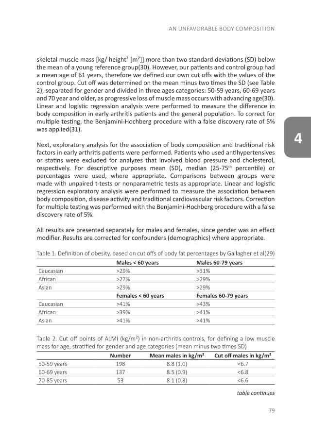
AN UNFAVORABLE BODY COMPOSITION
79
4
skeletal muscle mass [kg/ height² [m²]] more than two standard deviations (SD) below the mean of a young reference group(30). However, our patients and control group had a mean age of 61 years, therefore we defined our own cut offs with the values of the control group. Cut off was determined on the mean minus two times the SD (see Table 2), separated for gender and divided in three ages categories: 50-59 years, 60-69 years and 70 year and older, as progressive loss of muscle mass occurs with advancing age(30). Linear and logistic regression analysis were performed to measure the difference in body composition in early arthritis patients and the general population. To correct for multiple testing, the Benjamini-Hochberg procedure with a false discovery rate of 5% was applied(31).
Next, exploratory analysis for the association of body composition and traditional risk factors in early arthritis patients were performed. Patients who used antihypertensives or statins were excluded for analyzes that involved blood pressure and cholesterol, respectively. For descriptive purposes mean (SD), median (25-75th percentile) or percentages were used, where appropriate. Comparisons between groups were made with unpaired t-tests or nonparametric tests as appropriate. Linear and logistic regression exploratory analysis were performed to measure the association between body composition, disease activity and traditional cardiovascular risk factors. Correction for multiple testing was performed with the Benjamini-Hochberg procedure with a false discovery rate of 5%.
All results are presented separately for males and females, since gender was an effect modifier. Results are corrected for confounders (demographics) where appropriate.
Table 1. Definition of obesity, based on cut offs of body fat percentages by Gallagher et al(29)Males < 60 years Males 60-79 years
Caucasian >29% >31%African >27% >29%Asian >29% >29%
Females < 60 years Females 60-79 yearsCaucasian >41% >43%African >39% >41%Asian >41% >41%
Table 2. Cut off points of ALMI (kg/m²) in non-arthritis controls, for defining a low muscle mass for age, stratified for gender and age categories (mean minus two times SD)
Number Mean males in kg/m² Cut off males in kg/m²50-59 years 198 8.8 (1.0) <6.760-69 years 137 8.5 (0.9) <6.870-85 years 53 8.1 (0.8) <6.6
table continues
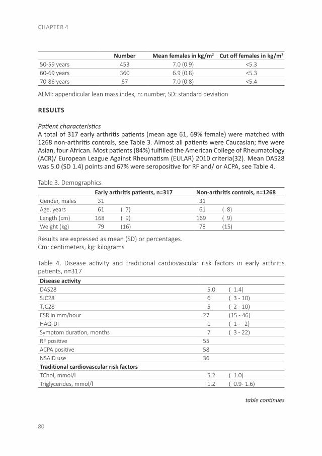
CHAPTER 4
80
Number Mean females in kg/m2 Cut off females in kg/m2
50-59 years 453 7.0 (0.9) <5.360-69 years 360 6.9 (0.8) <5.370-86 years 67 7.0 (0.8) <5.4
ALMI: appendicular lean mass index, n: number, SD: standard deviation
RESULTS
Patient characteristicsA total of 317 early arthritis patients (mean age 61, 69% female) were matched with 1268 non-arthritis controls, see Table 3. Almost all patients were Caucasian; five were Asian, four African. Most patients (84%) fulfilled the American College of Rheumatology (ACR)/ European League Against Rheumatism (EULAR) 2010 criteria(32). Mean DAS28 was 5.0 (SD 1.4) points and 67% were seropositive for RF and/ or ACPA, see Table 4.
Table 3. DemographicsEarly arthritis patients, n=317 Non-arthritis controls, n=1268
Gender, males 31 31Age, years 61 ( 7) 61 ( 8)Length (cm) 168 ( 9) 169 ( 9)Weight (kg) 79 (16) 78 (15)
Results are expressed as mean (SD) or percentages.Cm: centimeters, kg: kilograms
Table 4. Disease activity and traditional cardiovascular risk factors in early arthritis patients, n=317Disease activity DAS28 5.0 ( 1.4)SJC28 6 ( 3 - 10)TJC28 5 ( 2 - 10)ESR in mm/hour 27 (15 - 46)HAQ-DI 1 ( 1 - 2)Symptom duration, months 7 ( 3 - 22)RF positive 55ACPA positive 58NSAID use 36Traditional cardiovascular risk factors TChol, mmol/l 5.2 ( 1.0)Triglycerides, mmol/l 1.2 ( 0.9- 1.6)
table continues
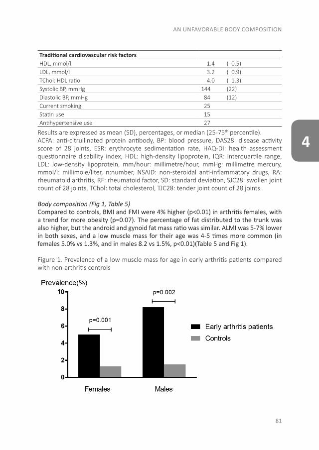
AN UNFAVORABLE BODY COMPOSITION
81
4
Traditional cardiovascular risk factors HDL, mmol/l 1.4 ( 0.5)LDL, mmol/l 3.2 ( 0.9)TChol: HDL ratio 4.0 ( 1.3)Systolic BP, mmHg 144 (22)Diastolic BP, mmHg 84 (12)Current smoking 25Statin use 15Antihypertensive use 27
Results are expressed as mean (SD), percentages, or median (25-75th percentile).ACPA: anti-citrullinated protein antibody, BP: blood pressure, DAS28: disease activity score of 28 joints, ESR: erythrocyte sedimentation rate, HAQ-DI: health assessment questionnaire disability index, HDL: high-density lipoprotein, IQR: interquartile range, LDL: low-density lipoprotein, mm/hour: millimetre/hour, mmHg: millimetre mercury, mmol/l: millimole/liter, n:number, NSAID: non-steroidal anti-inflammatory drugs, RA: rheumatoid arthritis, RF: rheumatoid factor, SD: standard deviation, SJC28: swollen joint count of 28 joints, TChol: total cholesterol, TJC28: tender joint count of 28 joints
Body composition (Fig 1, Table 5)Compared to controls, BMI and FMI were 4% higher (p<0.01) in arthritis females, with a trend for more obesity (p=0.07). The percentage of fat distributed to the trunk was also higher, but the android and gynoid fat mass ratio was similar. ALMI was 5-7% lower in both sexes, and a low muscle mass for their age was 4-5 times more common (in females 5.0% vs 1.3%, and in males 8.2 vs 1.5%, p<0.01)(Table 5 and Fig 1).
Figure 1. Prevalence of a low muscle mass for age in early arthritis patients compared with non-arthritis controls
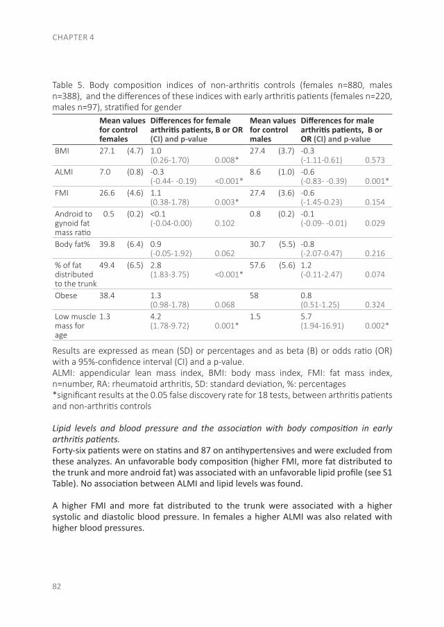
CHAPTER 4
82
Table 5. Body composition indices of non-arthritis controls (females n=880, males n=388), and the differences of these indices with early arthritis patients (females n=220, males n=97), stratified for gender
Mean values for control females
Differences for female arthritis patients, B or OR (CI) and p-value
Mean values for control males
Differences for male arthritis patients, B or OR (CI) and p-value
BMI 27.1 (4.7) 1.0 (0.26-1.70) 0.008*
27.4 (3.7) -0.3 (-1.11-0.61) 0.573
ALMI 7.0 (0.8) -0.3 (-0.44- -0.19) <0.001*
8.6 (1.0) -0.6 (-0.83- -0.39) 0.001*
FMI 26.6 (4.6) 1.1 (0.38-1.78) 0.003*
27.4 (3.6) -0.6 (-1.45-0.23) 0.154
Android to gynoid fat mass ratio
0.5 (0.2) <0.1 (-0.04-0.00) 0.102
0.8 (0.2) -0.1 (-0.09- -0.01) 0.029
Body fat% 39.8 (6.4) 0.9 (-0.05-1.92) 0.062
30.7 (5.5) -0.8 (-2.07-0.47) 0.216
% of fat distributed to the trunk
49.4 (6.5) 2.8 (1.83-3.75) <0.001*
57.6 (5.6) 1.2 (-0.11-2.47) 0.074
Obese 38.4 1.3 (0.98-1.78) 0.068
58 0.8 (0.51-1.25) 0.324
Low muscle mass for age
1.3 4.2 (1.78-9.72) 0.001*
1.5 5.7(1.94-16.91) 0.002*
Results are expressed as mean (SD) or percentages and as beta (B) or odds ratio (OR) with a 95%-confidence interval (CI) and a p-value.ALMI: appendicular lean mass index, BMI: body mass index, FMI: fat mass index, n=number, RA: rheumatoid arthritis, SD: standard deviation, %: percentages*significant results at the 0.05 false discovery rate for 18 tests, between arthritis patients and non-arthritis controls
Lipid levels and blood pressure and the association with body composition in early arthritis patients.Forty-six patients were on statins and 87 on antihypertensives and were excluded from these analyzes. An unfavorable body composition (higher FMI, more fat distributed to the trunk and more android fat) was associated with an unfavorable lipid profile (see S1 Table). No association between ALMI and lipid levels was found.
A higher FMI and more fat distributed to the trunk were associated with a higher systolic and diastolic blood pressure. In females a higher ALMI was also related with higher blood pressures.
AN UNFAVORABLE BODY COMPOSITION
83
4
Disease activity and body composition in early arthritis patientsIn female patients, a higher ESR was associated with a higher FMI and patients with a longer symptom duration had more fat distributed to the trunk. In males, no association between body composition and disease activity was found. However, a longer symptom duration was related to a higher FMI and ALMI. See S2 Table.
Sensitivity analysisAnalyzes were repeated in patients who did and did not fulfill the ACR/ EULAR 2010 criteria and showed similar results (data not shown). Finally, we found that an unfavorable body composition was associated with an unfavorable lipid profile and higher blood pressure in patients without cardiovascular treatment. Therefore, analyses including patients with statins or antihypertensives were performed, where the same results were obtained (data not shown).
DISCUSSION
Our study is the first to reveal that an unfavorable body composition is already present at the onset of inflammatory arthritis. A low muscle mass for age was rare, but substantially more prevalent in patients than in matched controls. In our patients, these findings could also be linked to cardiovascular risk factors, regardless of disease activity.The mechanisms causing an unfavorable body composition in inflammatory arthritis are incompletely understood. By itself, a loss in muscle mass can result from reductions in physical activity and hormone levels as well as change in diet, all part of normal aging(20). This also results in obesity, whereas the mean weight in the general population is nowadays already high and still increasing(33). Whereas obesity itself is also an risk factor for the development of arthritis(34). In inflammatory arthritis, pain, fatigue and joint stiffness further reduce physical activity(35). Cells involved in inflammation produce pro-inflammatory molecules, like IL-6, that increase muscle metabolism with subsequent muscle wasting(36). Furthermore, arthritis is associated with an insulin resistance, which may lead to muscle protein degradation(37).
Our findings are in line with the results observed in established arthritis(16;19). In established arthritis patients an association between body composition and glucocorticoid use was found(19;38;39). Only a minority of the arthritis patients received steroids during a few days. As our population underwent the DXA scan before or within one month after the start of treatment, glucocorticoid use was of minimum influence(40). Hence, altogether the influence of steroids appears to be negligible.
In the present study, the number of early arthritis patients with a low muscle mass for age was low, which might explain that no associations between disease activity and an unfavorable body composition could be demonstrated. The relation between different aspects of disease activity and components of body composition were inconclusive. Nevertheless, a longer symptom duration was associated with an increased FMI and
CHAPTER 4
84
ALMI, however due to the small beta coefficients this was not clinical relevant (every one month increase in symptom duration, was associated with an increase in FMI and ALMI of 0.04 and 0.01, respectively). Moreover, in females a higher ESR was associated with an increase in FMI (10 points increase in ESR was associated with a 0.4 points increase in FMI). However, ESR might not be representative of intramuscular activity, therefore we recommend for future research to further analyze the association between disease activity and an unfavorable body composition by extending the measurements with CRP and IL-6 levels(19).
In early arthritis patients an unfavorable body composition was associated with a higher blood pressure and higher lipid levels (with lower HDL levels), which is according to previous literature, similar to the general population(41-43). However, as inflammation generally leads to a decreased TC and HDL level, but an increased TC:HDL ratio, it is in RA patients difficult to interpret the lipid levels(44;45). The combination of an unfavorable body composition, hypertension and an atherogenic lipid profile might be a clustering of risk factors, known as metabolic syndrome, as overweight is often associated with hypertension and hypercholesterolemia(46). This association between an unfavorable body composition and traditional risk factors might help explain the increased prevalence of CV disease in arthritis patients. Van Halm et al. already showed a more atherogenic lipid profile in blood donors who later developed RA, which was partly explained by the presence of inflammation(47;48). There are a number of factors that are associated with both body composition, lipid profile and blood pressure, including lifestyle factors such as a diet high in fat, sugar and sodium, insufficient physical activity and family history(49). Hormones derived from adipose tissue have also been linked to an increased blood pressure, include leptin and adiponectin(50). As inflammatory arthritis and CV diseases are multifactorial disorders, overlapping risk factors and a shared etiology for the development of both diseases have to be considered. Smoking and metabolic syndrome are important risk factors for the development of both arthritis and CV disease(51;52). The development of arthritis and CV disease are also, partially, explained to common susceptibility genes, however must more research on this area is necessary(52). A suggested shared etiology is periodontal disease which is generated by microorganisms, like Porphyromonas gingivalis (Pg). In RA patients an antibody response to Pg is common and Pg also contributes to the pathogenesis of atherosclerosis(49;53;54). Future studies are needed to determine if an unfavorable body composition already exists before the onset of arthritis. Hence, DXA scans should be performed in patients with a high risk for developing RA.
This will further define the optimal moment for a DXA scan and might give clues how we can prevent this unfavorable body composition, as RA treatment itself does not appear to improve body composition(14;22;23).
AN UNFAVORABLE BODY COMPOSITION
85
4
The strengths of this study include a good match with an excellent population cohort, the large sample size for both males and females. Unfortunately, no widely accepted definition of a low muscle mass for age exists(55). Nevertheless, both muscle mass and muscle strength are probably a main component. A limitation of the current study is that no assessments of muscle strength and physical performance were done. Therefore, we defined our own definition for sarcopenia low muscle mass for age, based on the muscle mass of the non-arthritis control group, so our main question could still be answered as we focussed on the difference between patients and non-arthritis controls. Another limitation is the possibility of selection bias, as the selection was inherently different between arthritis patients and non-arthritis controls. The subjects were selected from different locations, which can give differences in demographics. However, we matched on age, gender and ethnicity and added the values of body composition after the matching to limit this bias. Another aspect are the DXA machines that were utilized. The arthritis patients and the non-arthritis controls are measured in different DXA machines and the machines were not directly calibrated.
CONCLUSION
In conclusion, unfavorable body composition occurs early in the development of inflammatory arthritis, and this was associated with an increased CV risk. Therefore, we suggest that CV risk management should already be initiated at disease onset.
ACKNOWLEDGEMENTS
This work was supported by the Rotterdam Study. The authors are grateful to the study participants, the nurses, the participating general practitioners and pharmacists and the staff from the Rotterdam Study (particularly Hannie van J. Van den Boogert - de Visser, Frank van Rooij, Katerina Trajanoska and Fernando Rivadeneira for the acquisition and provision of the DXA data).
CHAPTER 4
86
REFERENCES
1. Braun J, Kruger K, Manger B, Schneider M, Specker C, Trappe HJ. Cardiovascular Comorbidity in Inflammatory Rheumatological Conditions. Dtsch Arztebl Int 2017;114:197-203.
2. Nurmohamed MT, Heslinga M, Kitas GD. Cardiovascular comorbidity in rheumatic diseases. Nat Rev Rheumatol 2015;11:693-704.
3. Holmqvist ME, Wedren S, Jacobsson LT, Klareskog L, Nyberg F, Rantapaa-Dahlqvist S, et al. Rapid increase in myocardial infarction risk following diagnosis of rheumatoid arthritis amongst patients diagnosed between 1995 and 2006. J Intern Med 2010;268:578-85.
4. Innala L, Moller B, Ljung L, Magnusson S, Smedby T, Sodergren A, et al. Cardiovascular events in early RA are a result of inflammatory burden and traditional risk factors: a five year prospective study. Arthritis Res Ther 2011;13:R131.
5. Kerola AM, Kauppi MJ, Kerola T, Nieminen TV. How early in the course of rheumatoid arthritis does the excess cardiovascular risk appear? Ann Rheum Dis 2012;71:1606-15.
6. Kerola AM, Kerola T, Kauppi MJ, Kautiainen H, Virta LJ, Puolakka K, et al. Cardiovascular comorbidities antedating the diagnosis of rheumatoid arthritis. Ann Rheum Dis 2013;72:1826-9.
7. Cugno M, Ingegnoli F, Gualtierotti R, Fantini F. Potential effect of anti-tumour necrosis factor-alpha treatment on reducing the cardiovascular risk related to rheumatoid arthritis. Curr Vasc Pharmacol 2010;8:285-92.
8. Panoulas VF, Metsios GS, Pace AV, John H, Treharne GJ, Banks MJ, et al. Hypertension in rheumatoid arthritis. Rheumatology (Oxford) 2008;47:1286-98.
9. Yusuf S, Hawken S, Ounpuu S, Bautista L, Franzosi MG, Commerford P, et al. Obesity and the risk of myocardial infarction in 27,000 participants from 52 countries: a case-control study. Lancet 2005 ;366:1640-9.
10. Bastien M, Poirier P, Lemieux I, Despres JP. Overview of epidemiology and contribution of obesity to cardiovascular disease. Prog Cardiovasc Dis 2014;56:369-81.
11. Megnien JL, Denarie N, Cocaul M, Simon A, Levenson J. Predictive value of waist-to-hip ratio on cardiovascular risk events. Int J Obes Relat Metab Disord 1999;23:90-7.
12. Schneider HJ, Friedrich N, Klotsche J, Pieper L, Nauck M, John U, et al. The predictive value of different measures of obesity for incident cardiovascular events and mortality. J Clin Endocrinol Metab 2010;95:1777-85.
13. Walsmith J, Roubenoff R. Cachexia in rheumatoid arthritis. Int J Cardiol 2002;85:89-99.14. Book C, Karlsson MK, Akesson K, Jacobsson LT. Early rheumatoid arthritis and body
composition. Rheumatology (Oxford) 2009;48:1128-32.15. Book C, Karlsson MK, Nilsson JA, Akesson K, Jacobsson LT. Changes in body composition
after 2 years with rheumatoid arthritis. Scand J Rheumatol 2011;40:95-100.16. Dao HH, Do QT, Sakamoto J. Abnormal body composition phenotypes in Vietnamese
women with early rheumatoid arthritis. Rheumatology (Oxford) 2011;50:1250-8.17. Dessein PH, Solomon A, Hollan I. Metabolic abnormalities in patients with inflammatory
rheumatic diseases. Best Pract Res Clin Rheumatol 2016;30:901-15.
AN UNFAVORABLE BODY COMPOSITION
87
4
18. Elkan AC, Hakansson N, Frostegard J, Cederholm T, Hafstrom I. Rheumatoid cachexia is associated with dyslipidemia and low levels of atheroprotective natural antibodies against phosphorylcholine but not with dietary fat in patients with rheumatoid arthritis: a cross-sectional study. Arthritis Res Ther 2009;11:R37.
19. Elkan AC, Engvall IL, Cederholm T, Hafstrom I. Rheumatoid cachexia, central obesity and malnutrition in patients with low-active rheumatoid arthritis: feasibility of anthropometry, Mini Nutritional Assessment and body composition techniques. Eur J Nutr 2009;48:315-22.
20. Tay L, Ding YY, Leung BP, Ismail NH, Yeo A, Yew S, et al. Sex-specific differences in risk factors for sarcopenia amongst community-dwelling older adults. Age (Dordr ) 2015;37:121.
21. Toss F, Wiklund P, Nordstrom P, Nordstrom A. Body composition and mortality risk in later life. Age Ageing 2012;41:677-81.
22. Lemmey AB, Wilkinson TJ, Clayton RJ, Sheikh F, Whale J, Jones HS, et al. Tight control of disease activity fails to improve body composition or physical function in rheumatoid arthritis patients. Rheumatology (Oxford) 2016;55:1736-45.
23. Marcora SM, Chester KR, Mittal G, Lemmey AB, Maddison PJ. Randomized phase 2 trial of anti-tumor necrosis factor therapy for cachexia in patients with early rheumatoid arthritis. Am J Clin Nutr 2006;84:1463-72.
24. Kim J, Shen W, Gallagher D, Jones A, Jr., Wang Z, Wang J, et al. Total-body skeletal muscle mass: estimation by dual-energy X-ray absorptiometry in children and adolescents. Am J Clin Nutr 2006;84:1014-20.
25. Bruce B, Fries JF. The Health Assessment Questionnaire (HAQ). Clin Exp Rheumatol 2005;23:S14-S18.
26. Hofman A, Brusselle GG, Darwish MS, van Duijn CM, Franco OH, Goedegebure A, et al. The Rotterdam Study: 2016 objectives and design update. Eur J Epidemiol 2015;30:661-708.
27. Krueger D, Vallarta-Ast N, Checovich M, Gemar D, Binkley N. BMD measurement and precision: a comparison of GE Lunar Prodigy and iDXA densitometers. J Clin Densitom 2012;15:21-5.
28. Saarelainen J, Hakulinen M, Rikkonen T, Kroger H, Koivumaa-Honkanen H, Honkanen R, et al. Inclusion of Regional Body Composition Parameters Improves Bone Mineral Density Cross-Calibration Between GE Lunar Prodigy and iDXA Densitometers. J Clin Densitom 2017;20:97-105.
29. Gallagher D, Heymsfield SB, Heo M, Jebb SA, Murgatroyd PR, Sakamoto Y. Healthy percentage body fat ranges: an approach for developing guidelines based on body mass index. Am J Clin Nutr 2000;72:694-701.
30. Baumgartner RN, Koehler KM, Gallagher D, Romero L, Heymsfield SB, Ross RR, et al. Epidemiology of sarcopenia among the elderly in New Mexico. Am J Epidemiol 1998;147:755-63.
31. Glickman ME, Rao SR, Schultz MR. False discovery rate control is a recommended alternative to Bonferroni-type adjustments in health studies. J Clin Epidemiol 2014;67:850-7.
CHAPTER 4
88
32. Aletaha D, Neogi T, Silman AJ, Funovits J, Felson DT, Bingham CO, III, et al. 2010 Rheumatoid arthritis classification criteria: an American College of Rheumatology/European League Against Rheumatism collaborative initiative. Arthritis Rheum 2010;62:2569-81.
33. Koopman C, Vaartjes I, Blokstra A, Verschuren WM, Visser M, Deeg DJ, et al. Trends in risk factors for coronary heart disease in the Netherlands. BMC Public Health 2016;16:835.
34. Unriza-Puin S, Bautista-Molano W, Lafaurie GI, Valle-Onate R, Chalem P, Chila-Moreno L, et al. Are obesity, ACPAs and periodontitis conditions that influence the risk of developing rheumatoid arthritis in first-degree relatives? Clin Rheumatol 2017;36:799-806.
35. Hall J, Grant J, Blake D, Taylor G, Garbutt G. Cardiorespiratory responses to aquatic treadmill walking in patients with rheumatoid arthritis. Physiother Res Int 2004;9:59-73.
36. Rall LC, Roubenoff R. Rheumatoid cachexia: metabolic abnormalities, mechanisms and interventions. Rheumatology (Oxford) 2004;43:1219-23.
37. Hotamisligil GS, Murray DL, Choy LN, Spiegelman BM. Tumor necrosis factor alpha inhibits signaling from the insulin receptor. Proc Natl Acad Sci U S A 1994;91:4854-8.
38. Arshad A, Rashid R, Benjamin K. The effect of disease activity on fat-free mass and resting energy expenditure in patients with rheumatoid arthritis versus noninflammatory arthropathies/soft tissue rheumatism. Mod Rheumatol 2007;17:470-5.
39. Resmini E, Farkas C, Murillo B, Barahona MJ, Santos A, Martinez-Momblan MA, et al. Body composition after endogenous (Cushing’s syndrome) and exogenous (rheumatoid arthritis) exposure to glucocorticoids. Horm Metab Res 2010;42:613-8.
40. Konijn NP, van Tuyl LH, Boers M, van de Ven PM, den UD, Ter Wee MM, et al. The short-term effects of two high-dose, step-down prednisolone regimens on body composition in early rheumatoid arthritis. Rheumatology (Oxford) 2016;55:1615-22.
41. Niederauer CM, Binkley TL, Specker BL. Effect of truncal adiposity on plasma lipid and lipoprotein concentrations. J Nutr Health Aging 2006;10:154-60.
42. Peppa M, Koliaki C, Hadjidakis DI, Garoflos E, Papaefstathiou A, Katsilambros N, et al. Regional fat distribution and cardiometabolic risk in healthy postmenopausal women. Eur J Intern Med 2013;24:824-31.
43. Sardinha LB, Teixeira PJ, Guedes DP, Going SB, Lohman TG. Subcutaneous central fat is associated with cardiovascular risk factors in men independently of total fatness and fitness. Metabolism 2000;49:1379-85.
44. Georgiadis AN, Papavasiliou EC, Lourida ES, Alamanos Y, Kostara C, Tselepis AD, et al. Atherogenic lipid profile is a feature characteristic of patients with early rheumatoid arthritis: effect of early treatment--a prospective, controlled study. Arthritis Res Ther 2006;8:R82.
45. Navarro-Millan I, Charles-Schoeman C, Yang S, Bathon JM, Bridges SL, Jr., Chen L, et al. Changes in lipoproteins associated with methotrexate or combination therapy in early rheumatoid arthritis: results from the treatment of early rheumatoid arthritis trial. Arthritis Rheum 2013;65:1430-8.
46. Tune JD, Goodwill AG, Sassoon DJ, Mather KJ. Cardiovascular consequences of metabolic syndrome. Transl Res 2017.
AN UNFAVORABLE BODY COMPOSITION
89
4
47. Nielen MM, van SD, Reesink HW, Twisk JW, van de Stadt RJ, van der Horst-Bruinsma IE, et al. Increased levels of C-reactive protein in serum from blood donors before the onset of rheumatoid arthritis. Arthritis Rheum 2004;50:2423-7.
48. van Halm VP, Nielen MM, Nurmohamed MT, van SD, Reesink HW, Voskuyl AE, et al. Lipids and inflammation: serial measurements of the lipid profile of blood donors who later developed rheumatoid arthritis. Ann Rheum Dis 2007;66:184-8.
49. Bender P, Burgin WB, Sculean A, Eick S. Serum antibody levels against Porphyromonas gingivalis in patients with and without rheumatoid arthritis - a systematic review and meta-analysis. Clin Oral Investig 2017;21:33-42.
50. Kelly RK, Magnussen CG, Sabin MA, Cheung M, Juonala M. Development of hypertension in overweight adolescents: a review. Adolesc Health Med Ther 2015;6:171-87.
51. Lahiri M, Luben RN, Morgan C, Bunn DK, Marshall T, Lunt M, et al. Using lifestyle factors to identify individuals at higher risk of inflammatory polyarthritis (results from the European Prospective Investigation of Cancer-Norfolk and the Norfolk Arthritis Register--the EPIC-2-NOAR Study). Ann Rheum Dis 2014;73:219-26.
52. Mahmoudi M, Aslani S, Fadaei R, Jamshidi AR. New insights to the mechanisms underlying atherosclerosis in rheumatoid arthritis. Int J Rheum Dis 2017.
53. Bale BF, Doneen AL, Vigerust DJ. High-risk periodontal pathogens contribute to the pathogenesis of atherosclerosis. Postgrad Med J 2017;93:215-20.
54. Rosenstein ED, Greenwald RA, Kushner LJ, Weissmann G. Hypothesis: the humoral immune response to oral bacteria provides a stimulus for the development of rheumatoid arthritis. Inflammation 2004;28:311-8.
55. McLean RR, Kiel DP. Developing consensus criteria for sarcopenia: an update. J Bone Miner Res 2015;30:588-92.
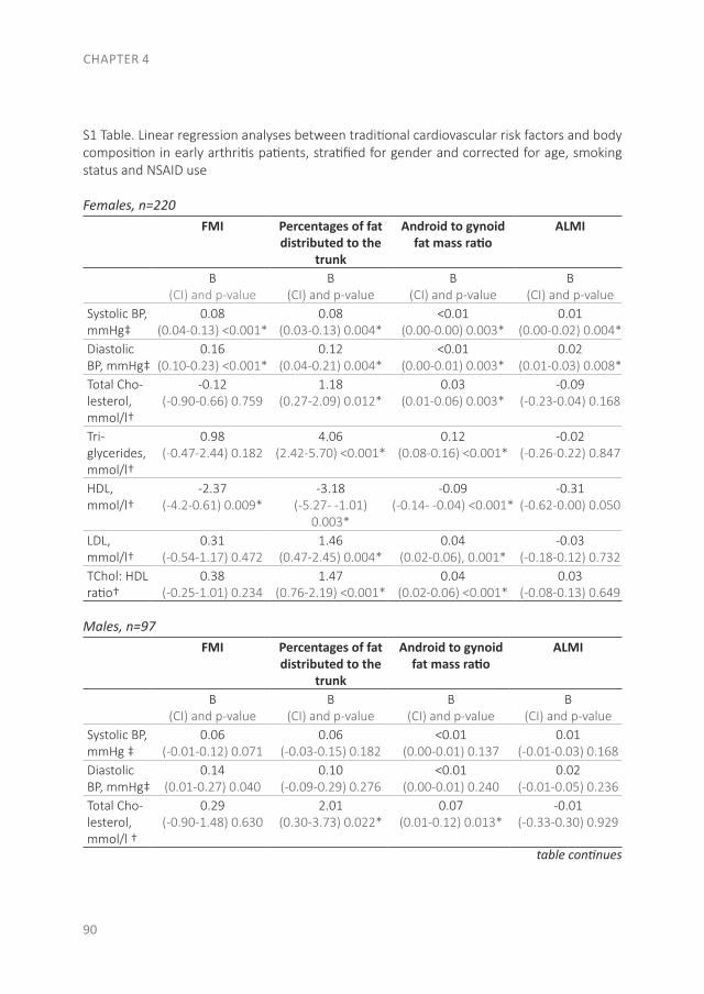
CHAPTER 4
90
S1 Table. Linear regression analyses between traditional cardiovascular risk factors and body composition in early arthritis patients, stratified for gender and corrected for age, smoking status and NSAID use
Females, n=220FMI Percentages of fat
distributed to the trunk
Android to gynoid fat mass ratio
ALMI
B (CI) and p-value
B (CI) and p-value
B (CI) and p-value
B(CI) and p-value
Systolic BP, mmHg‡
0.08 (0.04-0.13) <0.001*
0.08 (0.03-0.13) 0.004*
<0.01 (0.00-0.00) 0.003*
0.01 (0.00-0.02) 0.004*
Diastolic BP, mmHg‡
0.16 (0.10-0.23) <0.001*
0.12 (0.04-0.21) 0.004*
<0.01 (0.00-0.01) 0.003*
0.02 (0.01-0.03) 0.008*
Total Cho-lesterol, mmol/l†
-0.12 (-0.90-0.66) 0.759
1.18 (0.27-2.09) 0.012*
0.03 (0.01-0.06) 0.003*
-0.09 (-0.23-0.04) 0.168
Tri-glycerides, mmol/l†
0.98 (-0.47-2.44) 0.182
4.06 (2.42-5.70) <0.001*
0.12 (0.08-0.16) <0.001*
-0.02 (-0.26-0.22) 0.847
HDL, mmol/l†
-2.37 (-4.2-0.61) 0.009*
-3.18 (-5.27- -1.01)
0.003*
-0.09 (-0.14- -0.04) <0.001*
-0.31 (-0.62-0.00) 0.050
LDL, mmol/l†
0.31 (-0.54-1.17) 0.472
1.46 (0.47-2.45) 0.004*
0.04 (0.02-0.06), 0.001*
-0.03 (-0.18-0.12) 0.732
TChol: HDL ratio†
0.38 (-0.25-1.01) 0.234
1.47 (0.76-2.19) <0.001*
0.04 (0.02-0.06) <0.001*
0.03 (-0.08-0.13) 0.649
Males, n=97FMI Percentages of fat
distributed to the trunk
Android to gynoid fat mass ratio
ALMI
B (CI) and p-value
B (CI) and p-value
B (CI) and p-value
B(CI) and p-value
Systolic BP, mmHg ‡
0.06 (-0.01-0.12) 0.071
0.06 (-0.03-0.15) 0.182
<0.01 (0.00-0.01) 0.137
0.01 (-0.01-0.03) 0.168
Diastolic BP, mmHg‡
0.14 (0.01-0.27) 0.040
0.10 (-0.09-0.29) 0.276
<0.01 (0.00-0.01) 0.240
0.02 (-0.01-0.05) 0.236
Total Cho-lesterol, mmol/l †
0.29 (-0.90-1.48) 0.630
2.01 (0.30-3.73) 0.022*
0.07 (0.01-0.12) 0.013*
-0.01 (-0.33-0.30) 0.929
table continues
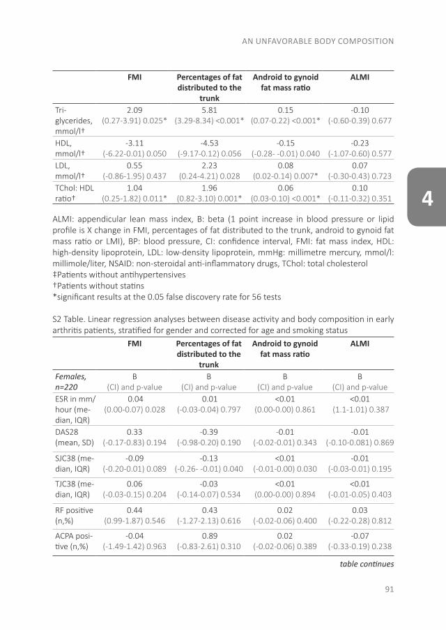
AN UNFAVORABLE BODY COMPOSITION
91
4
FMI Percentages of fat distributed to the
trunk
Android to gynoid fat mass ratio
ALMI
Tri-glycerides, mmol/l†
2.09 (0.27-3.91) 0.025*
5.81 (3.29-8.34) <0.001*
0.15 (0.07-0.22) <0.001*
-0.10 (-0.60-0.39) 0.677
HDL, mmol/l†
-3.11 (-6.22-0.01) 0.050
-4.53 (-9.17-0.12) 0.056
-0.15 (-0.28- -0.01) 0.040
-0.23 (-1.07-0.60) 0.577
LDL, mmol/l†
0.55 (-0.86-1.95) 0.437
2.23(0.24-4.21) 0.028
0.08 (0.02-0.14) 0.007*
0.07 (-0.30-0.43) 0.723
TChol: HDL ratio†
1.04 (0.25-1.82) 0.011*
1.96 (0.82-3.10) 0.001*
0.06 (0.03-0.10) <0.001*
0.10 (-0.11-0.32) 0.351
ALMI: appendicular lean mass index, B: beta (1 point increase in blood pressure or lipid profile is X change in FMI, percentages of fat distributed to the trunk, android to gynoid fat mass ratio or LMI), BP: blood pressure, CI: confidence interval, FMI: fat mass index, HDL: high-density lipoprotein, LDL: low-density lipoprotein, mmHg: millimetre mercury, mmol/l: millimole/liter, NSAID: non-steroidal anti-inflammatory drugs, TChol: total cholesterol‡Patients without antihypertensives†Patients without statins*significant results at the 0.05 false discovery rate for 56 tests
S2 Table. Linear regression analyses between disease activity and body composition in early arthritis patients, stratified for gender and corrected for age and smoking status
FMI Percentages of fat distributed to the
trunk
Android to gynoid fat mass ratio
ALMI
Females, n=220
B (CI) and p-value
B (CI) and p-value
B (CI) and p-value
B (CI) and p-value
ESR in mm/hour (me-dian, IQR)
0.04(0.00-0.07) 0.028
0.01(-0.03-0.04) 0.797
<0.01(0.00-0.00) 0.861
<0.01 (1.1-1.01) 0.387
DAS28 (mean, SD)
0.33(-0.17-0.83) 0.194
-0.39(-0.98-0.20) 0.190
-0.01(-0.02-0.01) 0.343
-0.01(-0.10-0.081) 0.869
SJC38 (me-dian, IQR)
-0.09(-0.20-0.01) 0.089
-0.13(-0.26- -0.01) 0.040
<0.01(-0.01-0.00) 0.030
-0.01(-0.03-0.01) 0.195
TJC38 (me-dian, IQR)
0.06(-0.03-0.15) 0.204
-0.03(-0.14-0.07) 0.534
<0.01(0.00-0.00) 0.894
<0.01(-0.01-0.05) 0.403
RF positive (n,%)
0.44(0.99-1.87) 0.546
0.43(-1.27-2.13) 0.616
0.02(-0.02-0.06) 0.400
0.03(-0.22-0.28) 0.812
ACPA posi-tive (n,%)
-0.04(-1.49-1.42) 0.963
0.89(-0.83-2.61) 0.310
0.02(-0.02-0.06) 0.389
-0.07(-0.33-0.19) 0.238
table continues
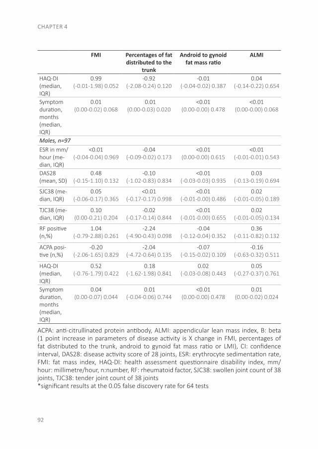
CHAPTER 4
92
FMI Percentages of fat distributed to the
trunk
Android to gynoid fat mass ratio
ALMI
HAQ-DI (median, IQR)
0.99(-0.01-1.98) 0.052
-0.92(-2.08-0.24) 0.120
-0.01(-0.04-0.02) 0.387
0.04(-0.14-0.22) 0.654
Symptom duration, months (median, IQR)
0.01(0.00-0.02) 0.068
0.01(0.00-0.03) 0.020
<0.01(0.00-0.00) 0.478
<0.01(0.00-0.00) 0.068
Males, n=97
ESR in mm/hour (me-dian, IQR)
<0.01(-0.04-0.04) 0.969
-0.04(-0.09-0.02) 0.173
<0.01(0.00-0.00) 0.615
<0.01(-0.01-0.01) 0.543
DAS28 (mean, SD)
0.48(-0.15-1.10) 0.132
-0.10(-1.02-0.83) 0.834
<0.01(-0.03-0.03) 0.935
0.03(-0.13-0.19) 0.694
SJC38 (me-dian, IQR)
0.05(-0.06-0.17) 0.365
<0.01(-0.17-0.17) 0.998
<0.01(-0.01-0.00) 0.486
0.02(-0.01-0.05) 0.189
TJC38 (me-dian, IQR)
0.10(0.00-0.21) 0.204
-0.02(-0.17-0.14) 0.844
<0.01(-0.01-0.00) 0.655
0.02(-0.01-0.05) 0.134
RF positive (n,%)
1.04(-0.79-2.88) 0.261
-2.24(-4.90-0.43) 0.098
-0.04(-0.12-0.04) 0.352
0.36(-0.11-0.82) 0.132
ACPA posi-tive (n,%)
-0.20(-2.06-1.65) 0.829
-2.04(-4.72-0.64) 0.135
-0.07(-0.15-0.02) 0.109
-0.16(-0.63-0.32) 0.511
HAQ-DI (median, IQR)
0.52(-0.76-1.79) 0.422
0.18(-1.62-1.98) 0.841
0.02(-0.03-0.08) 0.443
0.05(-0.27-0.37) 0.761
Symptom duration, months (median, IQR)
0.04(0.00-0.07) 0.044
0.01(-0.04-0.06) 0.744
<0.01(0.00-0.00) 0.478
0.01(0.00-0.02) 0.024
ACPA: anti-citrullinated protein antibody, ALMI: appendicular lean mass index, B: beta (1 point increase in parameters of disease activity is X change in FMI, percentages of fat distributed to the trunk, android to gynoid fat mass ratio or LMI), CI: confidence interval, DAS28: disease activity score of 28 joints, ESR: erythrocyte sedimentation rate, FMI: fat mass index, HAQ-DI: health assessment questionnaire disability index, mm/hour: millimetre/hour, n:number, RF: rheumatoid factor, SJC38: swollen joint count of 38 joints, TJC38: tender joint count of 38 joints*significant results at the 0.05 false discovery rate for 64 tests


CHAPTER 5
THE RELATION BETWEEN CARDIAC CONDUCTION TIMES, CARDIOVASCULAR RISK FACTORS AND
INFLAMMATION IN EARLY ARTHRITIS PATIENTS
Samina A. Turk1
Sjoerd C. Heslinga1
Jill Dekker2
Linda Britsemmer1
Véronique van der Lugt1
Willem F. Lems1,3
Dirkjan van Schaardenburg1,4
Michael T. Nurmohamed1,3
¹ Amsterdam Rheumatology and immunology Center | Reade, Amsterdam, Netherlands² Department of Cardiology, VU medical center, Amsterdam, Netherlands ³ Amsterdam Rheumatology and immunology Center | VU medical center, Amsterdam, Netherlands4 Amsterdam Rheumatology and immunology Center | Academic Medical Center, Am-sterdam, Netherlands
J Rheumatol - May 2017
CHAPTER 5
96
ABSTRACT
OBJECTIVE. To investigate the prevalence of conduction disorders in patients with early arthritis and the relationship with inflammation and traditional cardiovascular (CV) risk factors.
METHODS. Patients with rheumatoid arthritis (RA) have a 2-fold higher risk of sudden cardiac death, possibly owing to conduction disorders. This increased risk might already be present at the clinical onset of arthritis. Therefore, we assessed electrocardiography, blood pressure, 28-joint Disease Activity Score (DAS28), lipid profile, erythrocyte sedimentation rate (ESR), and C-reactive protein (CRP) level in 480 patients with early arthritis at baseline and after 1 year.
RESULTS. The prevalence of conduction disorders was 12.5%. Conduction times at baseline were not associated with DAS28, ESR, or CRP levels and did not change during antirheumatic treatment. Baseline and the improvement in DAS28 (European League Against Rheumatism response), ESR, and CRP were significantly associated with heart rate, lipid profile and blood pressure. Elevated total cholesterol and blood pressure were associated with an increased QRS time. The change in heart rate differed 7.3 bpm between patients with the least versus largest DAS improvement.
CONCLUSION. The prevalence of conduction disorders in patients with early arthritis was 12.5%, which is similar to the general population and was not associated with changes in inflammation markers. However, a high cholesterol was associated with a prolonged QRS time. Therefore, the emphasis of CV risk management in arthritis should not only be on treatment of disease activity but also on traditional CV risk factors. The relationship between the improvement in disease activity and heart rate is remarkable, because this could imply a 10-year CV mortality risk difference of 24%.
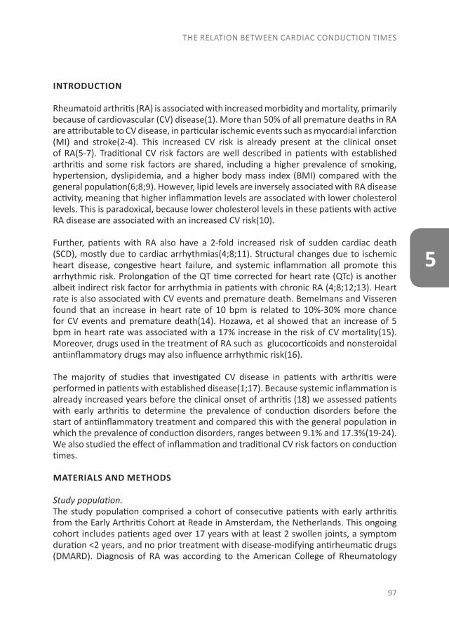
THE RELATION BETWEEN CARDIAC CONDUCTION TIMES
97
5
INTRODUCTION
Rheumatoid arthritis (RA) is associated with increased morbidity and mortality, primarily because of cardiovascular (CV) disease(1). More than 50% of all premature deaths in RA are attributable to CV disease, in particular ischemic events such as myocardial infarction (MI) and stroke(2-4). This increased CV risk is already present at the clinical onset of RA(5-7). Traditional CV risk factors are well described in patients with established arthritis and some risk factors are shared, including a higher prevalence of smoking, hypertension, dyslipidemia, and a higher body mass index (BMI) compared with the general population(6;8;9). However, lipid levels are inversely associated with RA disease activity, meaning that higher inflammation levels are associated with lower cholesterol levels. This is paradoxical, because lower cholesterol levels in these patients with active RA disease are associated with an increased CV risk(10).
Further, patients with RA also have a 2-fold increased risk of sudden cardiac death (SCD), mostly due to cardiac arrhythmias(4;8;11). Structural changes due to ischemic heart disease, congestive heart failure, and systemic inflammation all promote this arrhythmic risk. Prolongation of the QT time corrected for heart rate (QTc) is another albeit indirect risk factor for arrhythmia in patients with chronic RA (4;8;12;13). Heart rate is also associated with CV events and premature death. Bemelmans and Visseren found that an increase in heart rate of 10 bpm is related to 10%-30% more chance for CV events and premature death(14). Hozawa, et al showed that an increase of 5 bpm in heart rate was associated with a 17% increase in the risk of CV mortality(15). Moreover, drugs used in the treatment of RA such as glucocorticoids and nonsteroidal antiinflammatory drugs may also influence arrhythmic risk(16).
The majority of studies that investigated CV disease in patients with arthritis were performed in patients with established disease(1;17). Because systemic inflammation is already increased years before the clinical onset of arthritis (18) we assessed patients with early arthritis to determine the prevalence of conduction disorders before the start of antiinflammatory treatment and compared this with the general population in which the prevalence of conduction disorders, ranges between 9.1% and 17.3%(19-24). We also studied the effect of inflammation and traditional CV risk factors on conduction times.
MATERIALS AND METHODS
Study population. The study population comprised a cohort of consecutive patients with early arthritis from the Early Arthritis Cohort at Reade in Amsterdam, the Netherlands. This ongoing cohort includes patients aged over 17 years with at least 2 swollen joints, a symptom duration <2 years, and no prior treatment with disease-modifying antirheumatic drugs (DMARD). Diagnosis of RA was according to the American College of Rheumatology
CHAPTER 5
98
(ACR)/ European League Against Rheumatism (EULAR) 2010 criteria for RA(25). Patients were excluded if they had a diagnosis of crystal arthropathy, spondyloarthritis, osteoarthritis, systemic lupus erythematosus, Sjögren syndrome or infectious arthritis. Data came from patients included between November 2008 and July 2014. Approval was obtained from the local ethics committee (P0120, Ethics Committee of the Slotervaart Hospital and Reade, Amsterdam, the Netherlands), and all participating patients signed written informed consent according to the Declaration of Helsinki.
Patient characteristics.At baseline, patients were interviewed to record details about symptom history, clinical characteristics, medication use and demographics, and underwent a physical examination. Follow-up data were collected after 52 weeks. Disease activity was measured with the Disease Activity Score of 28 joints (DAS28) and the EULAR response was determined(26). Physical examination included weight, height, blood pressure, ankle brachial index (ABI), and an electrocardiogram (ECG). Blood pressure was measured manually according to the standard hospital procedures. Blood sample measurements included erythrocyte sedimentation rate (ESR), C-reactive protein (CRP), and lipid profile, consisting of total cholesterol (TChol), triglycerides, low-density lipoprotein (LDL), and high-density lipoprotein (HDL).
Treatment.After the baseline visit, treatment could be initiated with methotrexate (MTX), prednisone, hydroxychloroquine (HCQ), sulfasalazine (SSZ), or a combination of these. Patients who used β-blockers or the calcium channel blockers verapamil or diltiazem, antihypertensive drugs or statins were excluded from analyses that involved conduction times, blood pressure, and cholesterol, respectively.
ECG details. A standard resting 12-lead ECG was annually performed with the Mortara Eli 205C. Heart rate in bpm, QRS, QT, QTc and PQ time in milliseconds was recorded. All baseline ECG were reviewed by a cardiologist (JD) who was unaware of the patient characteristics. These disorders were noted: Atrioventricular block (AV), (incomplete) left bundle branch block (LBBB), (incomplete) right bundle branch block (RBBB), left anterior fascicular block (LAFB), left posterior fascicular block, a prolonged QTc time, and other intraventricular conduction disorders. A prolonged QTc time was defined as a QTc time of >450 milliseconds for men and >460 milliseconds for women.
Statistical analysis. For descriptive purposes mean (SD), median (interquartile range (IQR)), or percentages were used, where appropriate. Independent Student t test was used for continuous variables with a normal distribution, and the nonparametric Mann-Whitney U test was used for continuous variables that had a skewed distribution. For dichotomous variables Pearson’s chi-square test was used. Fisher’s exact test was used with variables
THE RELATION BETWEEN CARDIAC CONDUCTION TIMES
99
5
in the cross-table smaller than 5. Linear or logistic regression analysis was performed to assess associations between conduction times and clinical and laboratory data. A p-value <0.05 was considered significant. Data were analyzed with SPSS Version 21.0 (SPSS).
RESULTS
Baseline patient data. The study included 480 consecutive patients with early arthritis. Of them, 406 (85%) fulfilled the ACR/EULAR 2010 criteria for RA. The mean age was 53 years and 28% were men. Further descriptive data are in Table 1. At baseline, 63 patients used a statin and 87 patients used 1 or more antihypertensive drug; of them, 25 used a β-blocker or calcium channel blocker. During the first year, 359 patients started antirheumatic treatment. Eighty patients used monotherapy MTX, 21 HCQ, and 5 prednisone. All other patients used a combination of 2 or more of these drugs.
Table 1. Demographics. Values are mean (SD) unless otherwise indicatedBaseline characteristics, n=480 Values
Age, yrs 53 (13.3)
Sex, male (n, %) 135 (28.0)
Symptom duration, mos, median (IQR) 6.0 (3.0-17.8)
VAS pain, median (IQR) 52.0 (28.0-70.0)
DAS28 4.8 (1.4)
ESR, mm/h, median (IQR) 20.0 (9.0-38.0)
CRP, mg/l, median (IQR) 7.5 (2.0-20.0)
RF-positive, n (%) 228 (50.9)
ACPA-positive, n (%) 256 (57.3)
TJC 28, median (IQR) 5.0 (2.0-9.0)
SJC 28, median (IQR) 5.0 (3.0-9.0)
Conduction
Conduction disorder, n (%) 60 (12.5)
HR, bpm * 66.7 (11.5)
QRS, ms * 93.8 (11.7)
QT, ms * 402.6 (30.7)
table continues
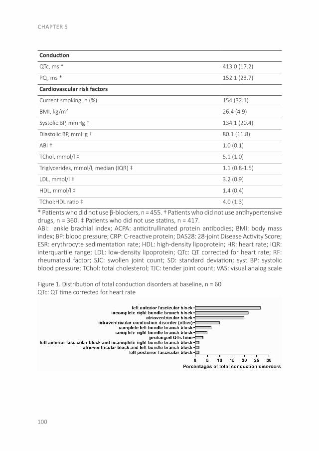
CHAPTER 5
100
Conduction
QTc, ms * 413.0 (17.2)
PQ, ms * 152.1 (23.7)
Cardiovascular risk factors
Current smoking, n (%) 154 (32.1)
BMI, kg/m² 26.4 (4.9)
Systolic BP, mmHg † 134.1 (20.4)
Diastolic BP, mmHg † 80.1 (11.8)
ABI † 1.0 (0.1)
TChol, mmol/l ‡ 5.1 (1.0)
Triglycerides, mmol/l, median (IQR) ‡ 1.1 (0.8-1.5)
LDL, mmol/l ‡ 3.2 (0.9)
HDL, mmol/l ‡ 1.4 (0.4)
TChol:HDL ratio ‡ 4.0 (1.3)
* Patients who did not use β-blockers, n = 455. † Patients who did not use antihypertensive drugs, n = 360. ‡ Patients who did not use statins, n = 417.ABI: ankle brachial index; ACPA: anticitrullinated protein antibodies; BMI: body mass index; BP: blood pressure; CRP: C-reactive protein; DAS28: 28-joint Disease Activity Score; ESR: erythrocyte sedimentation rate; HDL: high-density lipoprotein; HR: heart rate; IQR: interquartile range; LDL: low-density lipoprotein; QTc: QT corrected for heart rate; RF: rheumatoid factor; SJC: swollen joint count; SD: standard deviation; syst BP: systolic blood pressure; TChol: total cholesterol; TJC: tender joint count; VAS: visual analog scale
Figure 1. Distribution of total conduction disorders at baseline, n = 60QTc: QT time corrected for heart rate
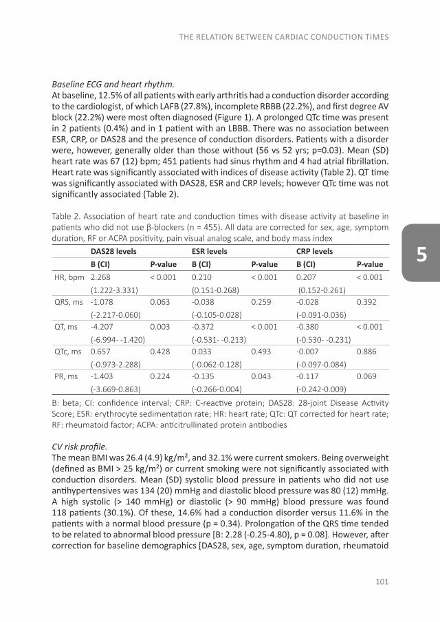
THE RELATION BETWEEN CARDIAC CONDUCTION TIMES
101
5
Baseline ECG and heart rhythm. At baseline, 12.5% of all patients with early arthritis had a conduction disorder according to the cardiologist, of which LAFB (27.8%), incomplete RBBB (22.2%), and first degree AV block (22.2%) were most often diagnosed (Figure 1). A prolonged QTc time was present in 2 patients (0.4%) and in 1 patient with an LBBB. There was no association between ESR, CRP, or DAS28 and the presence of conduction disorders. Patients with a disorder were, however, generally older than those without (56 vs 52 yrs; p=0.03). Mean (SD) heart rate was 67 (12) bpm; 451 patients had sinus rhythm and 4 had atrial fibrillation. Heart rate was significantly associated with indices of disease activity (Table 2). QT time was significantly associated with DAS28, ESR and CRP levels; however QTc time was not significantly associated (Table 2).
Table 2. Association of heart rate and conduction times with disease activity at baseline in patients who did not use β-blockers (n = 455). All data are corrected for sex, age, symptom duration, RF or ACPA positivity, pain visual analog scale, and body mass index
DAS28 levels ESR levels CRP levelsB (CI) P-value B (CI) P-value B (CI) P-value
HR, bpm 2.268
(1.222-3.331)
< 0.001 0.210
(0.151-0.268)
< 0.001 0.207
(0.152-0.261)
< 0.001
QRS, ms -1.078
(-2.217-0.060)
0.063 -0.038
(-0.105-0.028)
0.259 -0.028
(-0.091-0.036)
0.392
QT, ms -4.207
(-6.994- -1.420)
0.003 -0.372
(-0.531- -0.213)
< 0.001 -0.380
(-0.530- -0.231)
< 0.001
QTc, ms 0.657
(-0.973-2.288)
0.428 0.033
(-0.062-0.128)
0.493 -0.007
(-0.097-0.084)
0.886
PR, ms -1.403
(-3.669-0.863)
0.224 -0.135
(-0.266-0.004)
0.043 -0.117
(-0.242-0.009)
0.069
B: beta; CI: confidence interval; CRP: C-reactive protein; DAS28: 28-joint Disease Activity Score; ESR: erythrocyte sedimentation rate; HR: heart rate; QTc: QT corrected for heart rate; RF: rheumatoid factor; ACPA: anticitrullinated protein antibodies
CV risk profile. The mean BMI was 26.4 (4.9) kg/m², and 32.1% were current smokers. Being overweight (defined as BMI > 25 kg/m²) or current smoking were not significantly associated with conduction disorders. Mean (SD) systolic blood pressure in patients who did not use antihypertensives was 134 (20) mmHg and diastolic blood pressure was 80 (12) mmHg. A high systolic (> 140 mmHg) or diastolic (> 90 mmHg) blood pressure was found 118 patients (30.1%). Of these, 14.6% had a conduction disorder versus 11.6% in the patients with a normal blood pressure (p = 0.34). Prolongation of the QRS time tended to be related to abnormal blood pressure [B: 2.28 (-0.25-4.80), p = 0.08]. However, after correction for baseline demographics [DAS28, sex, age, symptom duration, rheumatoid
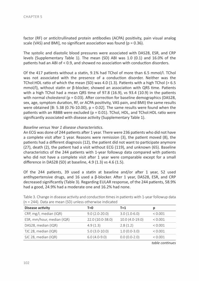
CHAPTER 5
102
factor (RF) or anticitrullinated protein antibodies (ACPA) positivity, pain visual analog scale (VAS) and BMI], no significant association was found (p = 0.36).
The systolic and diastolic blood pressures were associated with DAS28, ESR, and CRP levels (Supplementary Table 1). The mean (SD) ABI was 1.0 (0.1) and 16.0% of the patients had an ABI of < 0.9, and showed no association with conduction disorders.
Of the 417 patients without a statin, 9.1% had TChol of more than 6.5 mmol/l. TChol was not associated with the presence of a conduction disorder. Neither was the TChol:HDL ratio of which the mean (SD) was 4.0 (1.3). Patients with a high TChol (> 6.5 mmol/l), without statin or β-blocker, showed an association with QRS time. Patients with a high TChol had a mean QRS time of 97.8 (16.9), vs 93.4 (10.9) in the patients with normal cholesterol (p = 0.03). After correction for baseline demographics (DAS28, sex, age, symptom duration, RF, or ACPA positivity, VAS pain, and BMI) the same results were obtained [B: 5.38 (0.76-10.00), p = 0.02]. The same results were found when the patients with an RBBB were excluded (p = 0.01). TChol, HDL, and TChol:HDL ratio were significantly associated with disease activity (Supplementary Table 1).
Baseline versus Year 1 disease characteristics. An ECG was done of 244 patients after 1 year. There were 236 patients who did not have a complete visit after 1 year. Reasons were remission (3), the patient moved (8), the patients had a different diagnosis (12), the patient did not want to participate anymore (27), death (2), the patient had a visit without ECG (119), and unknown (65). Baseline characteristics of the 244 patients with 1-year followup data compared with patients who did not have a complete visit after 1 year were comparable except for a small difference in DAS28 (SD) at baseline, 4.9 (1.3) vs 4.6 (1.5).
Of the 244 patients, 39 used a statin at baseline and/or after 1 year, 52 used antihypertensive drugs, and 16 used a β-blocker. After 1 year, DAS28, ESR, and CRP decreased significantly (Table 3). Regarding EULAR response, of the 244 patients, 58.9% had a good, 24.9% had a moderate one and 16.2% had none.
Table 3. Change in disease activity and conduction times in patients with 1-year followup data (n = 244). Data are mean (SD) unless otherwise indicatedDisease activity T=0 T=1 pCRP, mg/l, median (IQR) 9.0 (2.0-20.0) 3.0 (1.0-6.0) < 0.001
ESR, mm/hour, median (IQR) 22.0 (10.0-38.0) 10.0 (4.0-19.0) < 0.001
DAS28, median (IQR) 4.9 (1.3) 2.8 (1.2) < 0.001
TJC 28, median (IQR) 5.0 (3.0-10.0) 1.0 (0.0-3.0) < 0.001
SJC 28, median (IQR) 6.0 (4.0-9.0) 0.0 (0.0-2.0) < 0.001
table continues
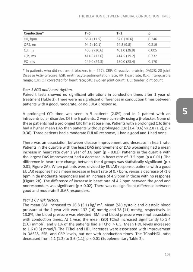
THE RELATION BETWEEN CARDIAC CONDUCTION TIMES
103
5
Conduction* T=0 T=1 pHR, bpm 66.4 (11.5) 67.0 (10.6) 0.246
QRS, ms 94.2 (10.1) 94.8 (9.8) 0.219
QT, ms 405.2 (30.6) 401.0 (28.9) 0.005
QTc, ms 414.5 (17.6) 414.5 (19.2) 0.732
PQ, ms 149.0 (24.3) 150.0 (23.4) 0.170
* In patients who did not use β-blockers (n = 227). CRP: C-reactive protein; DAS28: 28-joint Disease Activity Score; ESR: erythrocyte sedimentation rate; HR: heart rate; IQR: interquartile range; QTc: QT corrected for heart rate; SJC: swollen joint count; TJC: tender joint count
Year 1 ECG and heart rhythm.Paired t tests showed no significant alterations in conduction times after 1 year of treatment (Table 3). There were no significant differences in conduction times between patients with a good, moderate, or no EULAR response.
A prolonged QTc time was seen in 5 patients (2.0%) and in 1 patient with an intraventricular disorder. Of the 5 patients, 2 were currently using a β-blocker. None of these patients had a prolonged QTc time at baseline. Patients with a prolonged QTc time had a higher mean DAS than patients without prolonged QTc [3.4 (0.6) vs 2.8 (1.2), p = 0.30]. Three patients had a moderate EULAR response, 1 had a good and 1 had none.
There was an association between disease improvement and decrease in heart rate. Patients in the quartile with the least DAS improvement or DAS worsening had a mean increase in heart rate over 1 year of 3.8 bpm (p = 0.02). Patients in the quartile with the largest DAS improvement had a decrease in heart rate of -3.5 bpm (p = 0.01). The difference in heart rate change between the 4 groups was statistically significant (p < 0.01; Figure 2A). When patients were divided by EULAR response, patients with a good EULAR response had a mean increase in heart rate of 0.7 bpm, versus a decrease of -1.6 bpm in de moderate responders and an increase of 4.9 bpm in those with no response (Figure 2B). The difference of increase in heart rate of 4.2 bpm between the good and nonresponders was significant (p = 0.02). There was no significant difference between good and moderate EULAR responders.
Year 1 CV risk factors. The mean BMI increased to 26.8 (5.1) kg/ m². Mean (SD) systolic and diastolic blood pressure at the 1-year visit were 132 (16) mmHg and 78 (11) mmHg, respectively. In 13.8%, the blood pressure was elevated. BMI and blood pressure were not associated with conduction times. At 1 year, the mean (SD) TChol increased significantly to 5.4 (1.0) mmol/l, and 8.1% of the patients had a TChol > 6.5. Mean HDL levels increased to 1.6 (0.5) mmol/l. The TChol and HDL increases were associated with improvement in DAS28, ESR, and CRP levels, but not with conduction times. The TChol:HDL ratio decreased from 4.1 (1.2) to 3.6 (1.1); p < 0.01 (Supplementary Table 2).
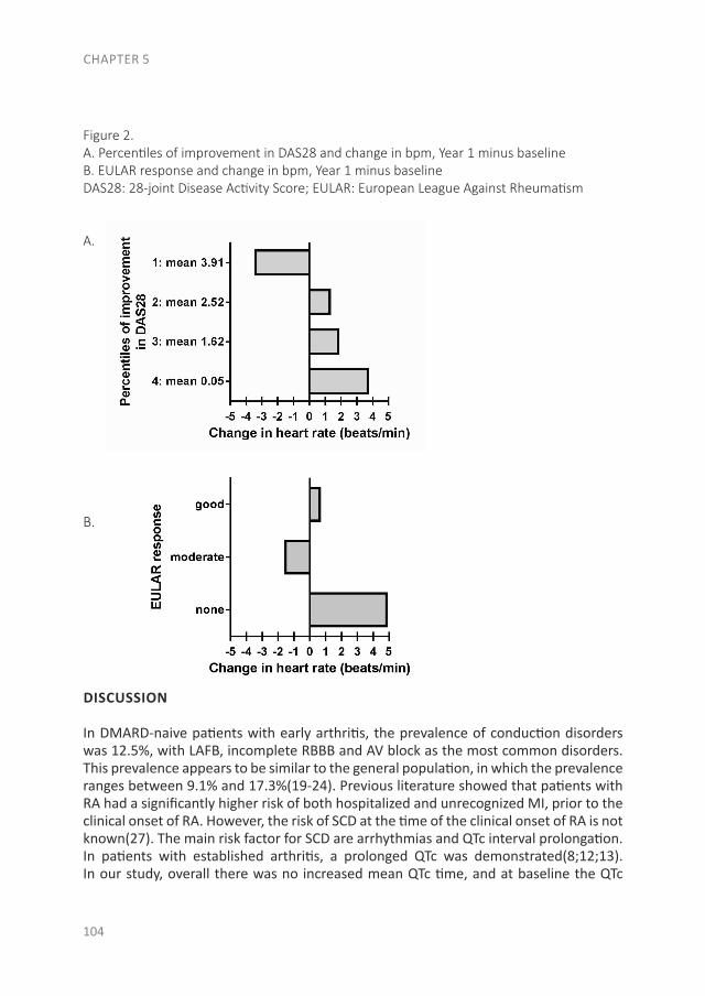
CHAPTER 5
104
Figure 2.A. Percentiles of improvement in DAS28 and change in bpm, Year 1 minus baseline B. EULAR response and change in bpm, Year 1 minus baseline DAS28: 28-joint Disease Activity Score; EULAR: European League Against Rheumatism
A.
B.
DISCUSSION
In DMARD-naive patients with early arthritis, the prevalence of conduction disorders was 12.5%, with LAFB, incomplete RBBB and AV block as the most common disorders. This prevalence appears to be similar to the general population, in which the prevalence ranges between 9.1% and 17.3%(19-24). Previous literature showed that patients with RA had a significantly higher risk of both hospitalized and unrecognized MI, prior to the clinical onset of RA. However, the risk of SCD at the time of the clinical onset of RA is not known(27). The main risk factor for SCD are arrhythmias and QTc interval prolongation. In patients with established arthritis, a prolonged QTc was demonstrated(8;12;13). In our study, overall there was no increased mean QTc time, and at baseline the QTc
THE RELATION BETWEEN CARDIAC CONDUCTION TIMES
105
5
time was prolonged in only 0.4% of the patients, a level comparable to the general population(8;19). Unfortunately, the 2 patients with a prolonged QTc time at baseline did not have an ECG after 1 year of treatment. Five patients (2.0%) developed a prolonged QTc time after 1 year. Of those patients, only 1 reached a good EULAR response.
Multiple factors affect the functioning of ion channels in myocardial cells and therefore conduction times: genetic abnormality, a cardiac disease (such as MI, owing to transmural ischemia), electrolyte levels and some medications(28). Another important factor is reactive oxygen species which affects the ion channels on the cardiac myocytes and is stimulated by cytokines such as tumor necrosis factor-α (TNF-α) and interleukine-6 (IL-6)(29). However, in our study conduction times were not associated with disease and inflammation markers (DAS28, EULAR response, ESR, or CRP levels). Although disease activity improved during 1 year of antiinflammatory treatment, this did not translate into a significant effect on conduction times. Because inflammation is considered the major pathophysiological link between arthritis and conduction disorders, the studied population might explain this difference. Patients with early arthritis have been exposed to inflammatory activity for a shorter period of time, which in our study was a median symptom duration of 6 months, compared with established chronic arthritis. However, it could also be that patients with established RA have prolonged exposure to more traditional CV risk factors, particularly dyslipidemia, which is already present in patients with early arthritis(30;31). This is important because of the association between traditional CV risk factors and conduction disorders; we found that a high total cholesterol at baseline was associated with a prolonged QRS time. However, this association disappeared after 1 year of treatment. After correction for RBBB, because RBBB can be physiologic, the same results were obtained. However, Kurl, et al. found that QRS duration is an independent predictor of the risk of SCD, where each 10-ms increase in QRS duration was associated with a 27% higher risk for SCD(32). In our present study this would mean that the 4.4-ms increased mean in QRS time in the patients with a high TChol resembles a 11.9% higher risk for SCD, compared with the patients with a normal TChol. It has been suggested that TNF-α, Interferon-γ, and IL-1 can stimulate the production of ceramide. Ceramide are lipid molecules that partly consist of fatty acids, they downregulate ion channels in cardiac myocytes and can affect conduction times(29). Therefore, both CV risk management as well as disease control are important and should be performed in all patients with arthritis(33). Our study strengthens the notion that antiinflammatory treatment, in an early stage of the disease, leads to a significant improvement in several important CV risk factors, including the TChol:HDL ratio and blood pressure(34;35). Interestingly, patients with early arthritis with lower inflammation markers had a lower heart rate compared with those with high inflammation markers. In the general population heart rate is positively associated with CRP, as demonstrated by Nanchen, et al. who found, in 4084 adults with a known CV risk factor, that an increased heart rate was associated with systemic inflammation(36). In patients with RA, the association between inflammation and heart rate has not been previously described, particularly not the improvement in inflammation and the
CHAPTER 5
106
change in heart rate. In our study, every 10-point increase in CRP or ESR was associated with an increase in heart rate of 2 bpm, which remained after correction for the VAS pain. This could imply an increased CV mortality of about 7%(14;15). This relationship between disease activity and heart rate is remarkable because it would imply a 10-year CV risk difference of 24% between no/least improvement and substantial improvement in DAS28 score(14;15). Patients with higher inflammation markers also had higher blood pressure, of which every 10-point increase in CRP or ESR was associated with an increase in systolic blood pressure of 1.7 mmHg. However, according to Ward, et al., this increase in CV risk has less clinical relevance(37).
Our present study shows that patients with early arthritis have the same prevalence of conduction disorders as the general population before anti-rheumatic treatment. Hence, in this population a mandatory screening ECG appears unnecessary. In contrast, in patients with chronic arthritis a prolonged QTc time is proven and therefore a standard ECG could be considered in established arthritis(1;4;8;12;13). For further research it would be interesting to match the patients with a healthy control group and repeat the ECG several years after rheumatic treatment, to investigate whether longer exposure to systemic inflammation increases conduction times and hence, conduction disorders.
Strengths of this study are the large number of consecutive patients and that the population reflects a heterogeneous population from a tertiary center. A limitation is that in addition to β-blockers and calcium channel blockers (verapamil and diltiazem), there are other medications that could affect conduction times (such as antibiotics, antipsychotics, or antidepressants). Unfortunately no data were available on these medications.
In early arthritis patients the prevalence of conduction disorders is comparable to the general population. However, the prevalence of traditional CV risk factors was increased in patients with a higher inflammatory load and the factors were associated with an increased QRS time. CV risk factors improved after inflammatory treatment. In particular, the difference in pulse rates between patients with persistent inflammation and patients with low disease activity or remission is remarkable. Therefore, the focus in patients with early arthritis should be on both CV risk management and optimizing antiinflammatory treatment.
THE RELATION BETWEEN CARDIAC CONDUCTION TIMES
107
5
REFERENCES
1. Nurmohamed MT, Heslinga M, Kitas GD. Cardiovascular comorbidity in rheumatic diseases. Nat Rev Rheumatol 2015;11:693-704.
2. Innala L, Moller B, Ljung L, Magnusson S, Smedby T, Sodergren A et al. Cardiovascular events in early RA are a result of inflammatory burden and traditional risk factors: a five year prospective study. Arthritis Res Ther 2011;13:R131.
3. John H, Kitas G, Toms T, Goodson N. Cardiovascular co-morbidity in early rheumatoid arthritis. Best Pract Res Clin Rheumatol 2009;23:71-82.
4. Lazzerini PE, Capecchi PL, Acampa M, Galeazzi M, Laghi-Pasini F. Arrhythmic risk in rheumatoid arthritis: the driving role of systemic inflammation. Autoimmun Rev 2014;13:936-944.
5. Holmqvist ME, Wedren S, Jacobsson LT, Klareskog L, Nyberg F, Rantapaa-Dahlqvist S et al. Rapid increase in myocardial infarction risk following diagnosis of rheumatoid arthritis amongst patients diagnosed between 1995 and 2006. J Intern Med 2010;268:578-585.
6. Kerola AM, Kauppi MJ, Kerola T, Nieminen TV. How early in the course of rheumatoid arthritis does the excess cardiovascular risk appear? Ann Rheum Dis 2012;71:1606-1615.
7. Kerola AM, Kerola T, Kauppi MJ, Kautiainen H, Virta LJ, Puolakka K et al. Cardiovascular comorbidities antedating the diagnosis of rheumatoid arthritis. Ann Rheum Dis 2013;72:1826-1829.
8. Lazzerini PE, Acampa M, Capecchi PL, Hammoud M, Maffei S, Bisogno S et al. Association between high sensitivity C-reactive protein, heart rate variability and corrected QT interval in patients with chronic inflammatory arthritis. Eur J Intern Med 2013;24:368-374.
9. Panoulas VF, Metsios GS, Pace AV, John H, Treharne GJ, Banks MJ et al. Hypertension in rheumatoid arthritis. Rheumatology (Oxford) 2008;47:1286-1298.
10. Myasoedova E, Crowson CS, Kremers HM, Roger VL, Fitz-Gibbon PD, Therneau TM et al. Lipid paradox in rheumatoid arthritis: the impact of serum lipid measures and systemic inflammation on the risk of cardiovascular disease. Ann Rheum Dis 2011;70:482-487.
11. Seferovic PM, Ristic AD, Maksimovic R, Simeunovic DS, Ristic GG, Radovanovic G et al. Cardiac arrhythmias and conduction disturbances in autoimmune rheumatic diseases. Rheumatology (Oxford) 2006;45 Suppl 4:iv39-iv42.
12. Chauhan K, Ackerman MJ, Crowson CS, Matteson EL, Gabriel SE. Population-based study of QT interval prolongation in patients with rheumatoid arthritis. Clin Exp Rheumatol 2015;33:84-89.
13. Panoulas VF, Toms TE, Douglas KM, Sandoo A, Metsios GS, Stavropoulos-Kalinoglou A et al. Prolonged QTc interval predicts all-cause mortality in patients with rheumatoid arthritis: an association driven by high inflammatory burden. Rheumatology (Oxford) 2014;53:131-137.
14. Bemelmans RH, Visseren FL. [The resting heart rate]. Ned Tijdschr Geneeskd 2014;158:A6931.
CHAPTER 5
108
15. Hozawa A, Ohkubo T, Kikuya M, Ugajin T, Yamaguchi J, Asayama K et al. Prognostic value of home heart rate for cardiovascular mortality in the general population: the Ohasama study. Am J Hypertens 2004;17:1005-1010.
16. Roubille C, Richer V, Starnino T, McCourt C, McFARLANE A, Fleming P et al. The effects of tumour necrosis factor inhibitors, methotrexate, non-steroidal anti-inflammatory drugs and corticosteroids on cardiovascular events in rheumatoid arthritis, psoriasis and psoriatic arthritis: a systematic review and meta-analysis. Ann Rheum Dis 2015;74:480-489.
17. Hollan I, Dessein PH, Ronda N, Wasko MC, Svenungsson E, Agewall S et al. Prevention of cardiovascular disease in rheumatoid arthritis. Autoimmun Rev 2015;14:952-969.
18. Nielen MM, van SD, Reesink HW, Twisk JW, van de Stadt RJ, van der Horst-Bruinsma IE et al. Simultaneous development of acute phase response and autoantibodies in preclinical rheumatoid arthritis. Ann Rheum Dis 2006;65:535-537.
19. Anttonen O, Junttila MJ, Rissanen H, Reunanen A, Viitasalo M, Huikuri HV. Prevalence and prognostic significance of short QT interval in a middle-aged Finnish population. Circulation 2007;116:714-720.
20. AVERILL KH, LAMB LE. Electrocardiographic findings in 67,375 asymptomatic subjects. I. Incidence of abnormalities. Am J Cardiol 1960;6:76-83.
21. Chow GV, Marine JE, Fleg JL. Epidemiology of arrhythmias and conduction disorders in older adults. Clin Geriatr Med 2012;28:539-553.
22. Haataja P, Nikus K, Kahonen M, Huhtala H, Nieminen T, Jula A et al. Prevalence of ventricular conduction blocks in the resting electrocardiogram in a general population: the Health 2000 Survey. Int J Cardiol 2013;167:1953-1960.
23. HISS RG, LAMB LE. Electrocardiographic findings in 122,043 individuals. Circulation 1962;25:947-961.
24. Kreger BE, Anderson KM, Kannel WB. Prevalence of intraventricular block in the general population: the Framingham Study. Am Heart J 1989;117:903-910.
25. Aletaha D, Neogi T, Silman AJ, Funovits J, Felson DT, Bingham CO, III et al. 2010 Rheumatoid arthritis classification criteria: an American College of Rheumatology/European League Against Rheumatism collaborative initiative. Arthritis Rheum 2010;62:2569-2581.
26. Fransen J, van Riel PL. The Disease Activity Score and the EULAR response criteria. Clin Exp Rheumatol 2005;23:S93-S99.
27. Maradit-Kremers H, Crowson CS, Nicola PJ, Ballman KV, Roger VL, Jacobsen SJ et al. Increased unrecognized coronary heart disease and sudden deaths in rheumatoid arthritis: a population-based cohort study. Arthritis Rheum 2005;52:402-411.
28. Kenigsberg DN, Khanal S, Kowalski M, Krishnan SC. Prolongation of the QTc interval is seen uniformly during early transmural ischemia. J Am Coll Cardiol 2007;49:1299-1305.
29. Sordillo PP, Sordillo DC, Helson L. Review: The Prolonged QT Interval: Role of Pro-inflammatory Cytokines, Reactive Oxygen Species and the Ceramide and Sphingosine-1 Phosphate Pathways. In Vivo 2015;29:619-636.
THE RELATION BETWEEN CARDIAC CONDUCTION TIMES
109
5
30. Gherghe AM, Dougados M, Combe B, Landewe R, Mihai C, Berenbaum F et al. Cardiovascular and selected comorbidities in early arthritis and early spondyloarthritis, a comparative study: results from the ESPOIR and DESIR cohorts. RMD Open 2015;1:e000128.
31. Innala L, Sjoberg C, Moller B, Ljung L, Smedby T, Sodergren A et al. Co-morbidity in patients with early rheumatoid arthritis - inflammation matters. Arthritis Res Ther 2016;18:33.
32. Kurl S, Makikallio TH, Rautaharju P, Kiviniemi V, Laukkanen JA. Duration of QRS complex in resting electrocardiogram is a predictor of sudden cardiac death in men. Circulation 2012;125:2588-2594.
33. Peters MJ, Symmons DP, McCarey D, Dijkmans BA, Nicola P, Kvien TK et al. EULAR evidence-based recommendations for cardiovascular risk management in patients with rheumatoid arthritis and other forms of inflammatory arthritis. Ann Rheum Dis 2010;69:325-331.
34. Lewington S, Whitlock G, Clarke R, Sherliker P, Emberson J, Halsey J et al. Blood cholesterol and vascular mortality by age, sex, and blood pressure: a meta-analysis of individual data from 61 prospective studies with 55,000 vascular deaths. Lancet 2007;370:1829-1839.
35. Ajeganova S, Andersson ML, Frostegard J, Hafstrom I. Disease factors in early rheumatoid arthritis are associated with differential risks for cardiovascular events and mortality depending on age at onset: a 10-year observational cohort study. J Rheumatol 2013;40:1958-1966.
36. Intzilakis T, Hartmann G, Mouridsen MR, Eugen-Olsen J, Kumarathurai P, Madsbad S et al. Soluble urokinase plasminogen activator receptor, C-reactive protein and triglyceride are associated with heart rate variability in non-diabetic Danes. Eur J Clin Invest 2013;43:457-468.
37. Ward AM, Takahashi O, Stevens R, Heneghan C. Home measurement of blood pressure and cardiovascular disease: systematic review and meta-analysis of prospective studies. J Hypertens 2012;30:449-456.
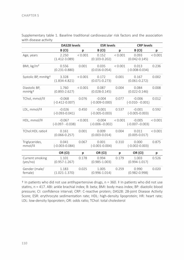
CHAPTER 5
110
Supplementary table 1. Baseline traditional cardiovascular risk factors and the association with disease activity
DAS28 levels ESR levels CRP levelsB (CI) p B (CI) p B (CI) p
Age, years 2.250 (1.412-3.089)
< 0.001 0.152 (0.103-0.202)
< 0.001 0.093 (0.042-0.145)
< 0.001
BMI, kg/m² 0.556 (0.231-0.880)
0.001 0.035 (0.016-0.054)
< 0.001 0.013 (-0.008-0.034)
0.236
Systolic BP, mmHg† 3.328 (1.834-4.821)
< 0.001 0.172 (0.071-0.273)
0.001 0.167 (0.061-0.272)
0.002
Diastolic BP, mmHg†
1.760 (0.893-2.627)
< 0.001 0.087 (0.028-0.145)
0.004 0.084 (0.022-0.146)
0.008
TChol, mmol/l‡ -0.068 (-0.412-0.007)
0.076 -0.004 (-0.009-0.000)
0.077 -0.006 (-0.010- -0.001)
0.012
LDL, mmol/l‡ -0.026(-0.093-0.041)
0.450 -0.001 (-0.005-0.003)
0.537 -0.001 (-0.005-0.003)
0.592
HDL, mmol/l‡ -0.067 (-0.097- -0.038)
< 0.001 -0.004(-0.006--0.002)
< 0.001 -0.005 (-0.007--0.003)
< 0.001
TChol:HDL ratio‡ 0.161 (0.066-0.257)
0.001 0.009 (0.003-0.014)
0.004 0.011 (0.005-0.017)
< 0.001
Triglycerides, mmol/l‡
0.041 (-0.003-0.084)
0.067 0.001 (-0.001-0.004)
0.310 0.000 (-0.002-0.003)
0.875
OR (CI) p OR (CI) p OR (CI) pCurrent smoking (yes/no)
1.101 (0.957-1.267)
0.178 0.994 (0.985-1.003)
0.179 1.003 (0.994-1.017)
0.526
Gender (male/ female)
1.183 (1.021-1.370)
0.025 1.005 (0.996-1.014)
0.259 0.990 (0.982-0.998)
0.020
† In patients who did not use antihypertensive drugs, n = 360. ‡ In patients who did not use statins, n = 417. ABI: ankle brachial index; B: beta; BMI: body mass index; BP: diastolic blood pressure; CI: confidence interval; CRP: C-reactive protein; DAS28: 28-joint Disease Activity Score; ESR: erythrocyte sedimentation rate; HDL: high-density lipoprotein; HR: heart rate; LDL: low-density lipoprotein; OR: odds ratio; TChol: total cholesterol
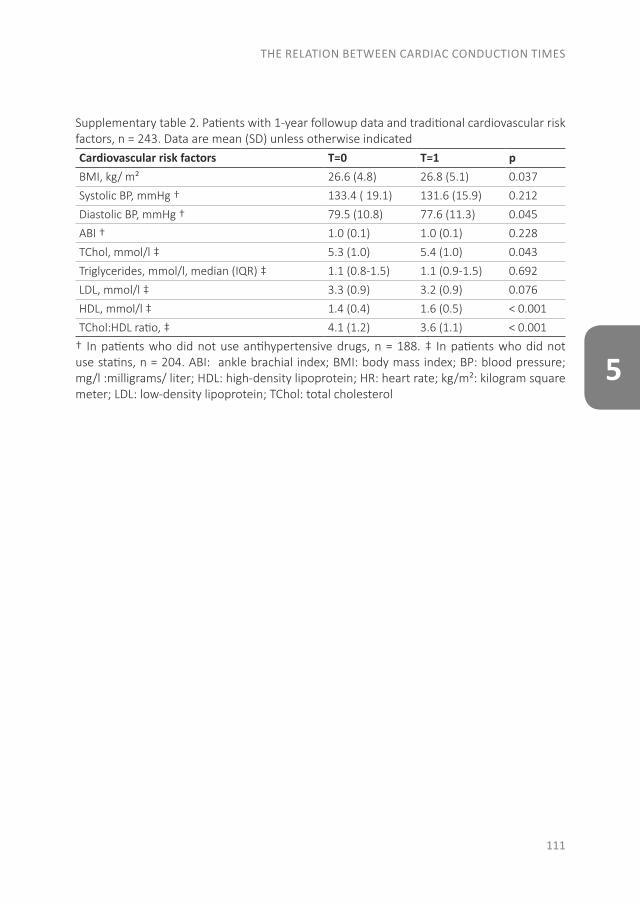
THE RELATION BETWEEN CARDIAC CONDUCTION TIMES
111
5
Supplementary table 2. Patients with 1-year followup data and traditional cardiovascular risk factors, n = 243. Data are mean (SD) unless otherwise indicatedCardiovascular risk factors T=0 T=1 pBMI, kg/ m² 26.6 (4.8) 26.8 (5.1) 0.037
Systolic BP, mmHg † 133.4 ( 19.1) 131.6 (15.9) 0.212
Diastolic BP, mmHg † 79.5 (10.8) 77.6 (11.3) 0.045
ABI † 1.0 (0.1) 1.0 (0.1) 0.228
TChol, mmol/l ‡ 5.3 (1.0) 5.4 (1.0) 0.043
Triglycerides, mmol/l, median (IQR) ‡ 1.1 (0.8-1.5) 1.1 (0.9-1.5) 0.692
LDL, mmol/l ‡ 3.3 (0.9) 3.2 (0.9) 0.076
HDL, mmol/l ‡ 1.4 (0.4) 1.6 (0.5) < 0.001
TChol:HDL ratio, ‡ 4.1 (1.2) 3.6 (1.1) < 0.001
† In patients who did not use antihypertensive drugs, n = 188. ‡ In patients who did not use statins, n = 204. ABI: ankle brachial index; BMI: body mass index; BP: blood pressure; mg/l :milligrams/ liter; HDL: high-density lipoprotein; HR: heart rate; kg/m²: kilogram square meter; LDL: low-density lipoprotein; TChol: total cholesterol

CHAPTER 6
CHANGE IN CARDIOVASCULAR RISK AFTER INITIATION OF ANTI-RHEUMATIC TREATMENT
IN EARLY RHEUMATOID ARTHRITIS
Samina A. Turk1
Maaike Heslinga1
Jos Twisk2
Véronique van der Lugt1
Willem F. Lems1,3
Dirkjan van Schaardenburg1,4
Michael T. Nurmohamed1,3
¹ Amsterdam Rheumatology and immunology Center| Reade, Amsterdam, Netherlands² Department of Epidemiology & Biostatistics, VU medical center, Amsterdam, Netherlands³ Amsterdam Rheumatology and immunology Center| VU medical center, Amsterdam, Netherlands4 Amsterdam Rheumatology and immunology Center| Academic Medical Center, Amster-dam, Netherlands
A shorter version of this chapter was accepted for publication as Letter in Clinical and Experimental Rheumatology
CHAPTER 6
114
ABSTRACT
OBJECTIVES. To determine if cardiovascular (CV) risk scores, traditional risk factors and the resulting indication for preventive treatment change after initiation of anti-rheumatic treatment in early rheumatoid arthritis (RA) patients.
METHODS. Disease activity, blood pressure, acute phase proteins and lipid profile were evaluated in early RA patients at baseline and after four weeks of anti-rheumatic treatment. CV risk scores (Dutch Systematic Coronary Risk Evaluation (SCORE) and European Heart SCORE) and indication for preventive CV treatment (according to the Dutch CV risk management guidelines) were determined.
RESULTS. One hundred and four consecutive RA patients were included, 7% had a history of CV disease. At baseline, 29.9% and 3.1% were classified as high risk according to the Dutch SCORE and Heart SCORE, respectively. According to the Dutch CV-risk management guidelines that use the Dutch SCORE, all high risk patients had at baseline an indication for (adaptations of) preventive treatment. From the CV risk score the components blood pressure and TC:HDL ratio decreased during anti-rheumatic treatment and 9% of the patients switched their CV risk category. In total 13% of the patients had a change in advice for preventive CV treatment after one month of anti-rheumatic treatment.
CONCLUSION. CV risk management is important in RA patients, however the timing of assessment, as well as the use of a particular CV risk model, influences the advice about the need for CV preventive treatment. Further research is needed to determine which risk model is optimal and when in the course of RA it should be applied.
CHANGE IN CARDIOVASCULAR RISK
115
6
INTRODUCTION
Ischemic heart diseases and strokes are the most common causes of death, accounting together for 15 million deaths in 2015(1). Different cardiovascular (CV) risk models exist, which estimate the 10-year risk of fatal and non-fatal CV diseases (CVD), and indicate if an antihypertensive and/or statin is necessary to lower the chance of a future CV event(2-5). Rheumatoid arthritis (RA) is associated with an increased risk of CVD, with atherosclerotic diseases being the leading cause of death(6;7). CV risk models were developed for the general population and do not perform well in the RA population(4). Therefore, the European League Against Rheumatism (EULAR) recommends to use a modified risk score for RA patients, by applying a multiplication factor of 1.5 to the CV risk scores(8). In the Dutch Systematic COronary Risk Evaluation (SCORE) a correction for RA patients is already taken into account(2;4;5).
The increased risk in RA patients for CVD has multiple causes. RA and CVD are both multifactorial disorders, with some shared risk factors (smoking, metabolic syndrome), common susceptibility genes and they might even have a shared etiology(6;9-13). However, most interesting is the influence of inflammation on CVD. Current evidence supports an important role of inflammation in the formation of an atherosclerotic plaque(14;15). Previous literature showed that improvement in RA disease activity is associated with an increase in cholesterol levels, and a decrease in TC:HDL ratio; an important CV risk predictor(16). However, all previous studies assessed the change in lipid profile six months or later after initiation of anti-rheumatic treatment(16-19). It is unclear whether this effect is already present early after initiating treatment and what effect this would have on CV risk and optimal CV risk management. Therefore, different CV risk scores (Dutch SCORE and European Heart SCORE), the traditional risk factors and indication for preventive treatment were determined in early RA patients before and after the first four weeks of anti-rheumatic treatment. Exploratory analysis were performed to determine the effect of inflammation on CV risk score, as well as the relation between inflammation, CV risk scores and the different components of the risk score.
METHODS
Study populationThe ‘Early Arthritis Cohort’ at Reade in Amsterdam, the Netherlands, includes patients aged 18 years and older, with no prior treatment with disease-modifying antirheumatic drugs (DMARDs). Patients in this cohort who fulfilled the ACR/EULAR 2010 criteria for RA(20) and started treatment with methotrexate and glucocorticoids, between June 2014 and March 2017, were included in this study. Patients with insulin-dependent diabetes mellitus were excluded. All patients gave written informed consent according to the Declaration of Helsinki and approval was obtained from the local ethics committee (Ethics Committee of the Slotervaart Hospital and Reade, Amsterdam, The Netherlands).
CHAPTER 6
116
MeasurementsAt baseline, patients were interviewed to record details about symptom history, disease history (special focus on CVD), clinical characteristics, demographics and medication use (including antihypertensives and statins).
At baseline and after four weeks, disease activity was measured with the Disease Activity Score of 44 joints (DAS44) and physical functioning by the Health Assessment Questionnaire (HAQ). Body mass index (BMI) was calculated from height and weight and blood pressure was measured manually according to the standard hospital procedures. Blood sample measurements at baseline were rheumatoid factor (RF) and anti-citrullinated protein antibody (ACPA), and at baseline and four weeks: C-reactive protein (CRP), erythrocyte sedimentation rate (ESR) and lipid profile, consisting of total cholesterol (TC), triglycerides, low-density lipoprotein (LDL) and high-density lipoprotein (HDL) cholesterol.
Cardiovascular riskCVD history was defined as an objectively confirmed by specialists history of coronary heart disease (myocardial infarction, angina pectoris, percutaneous coronary intervention and coronary artery bypass surgery, cerebral vascular disease and peripheral arterial disease). Patients with a CVD history were excluded from CV risk analyses. CV risk at baseline and after four weeks of treatment was retrospectively determined using the official online sites, according to two different risk scores: Dutch Systematic COronary Risk Evaluation (SCORE) and the European Heart SCORE(2;3). The Dutch SCORE risk model uses gender, age, smoking status, systolic blood pressure (SBP) and the TC:HDL ratio. To account for RA (or diabetes) as risk factor the Dutch CV-risk management (CV-RM) guideline adds 15 years to the actual age in order to calculate the 10-year CV risk. A risk<10% is classified as low, between 10% and 20% intermediate and a risk ≥20% as high risk. According to the Dutch CV-RM guideline, preventive treatment with an antihypertensive or statin is indicated in high risk patients with a SBP >140 mmHg or a LDL> 2.5 mmol/l, respectively(21). The European Heart SCORE risk model predicts the 10-year risk of a fatal heart attack, stroke or other circulatory problems in low risk regions of Europe by gender, age, SBP, TC:HDL ratio and smoking status. To calculate this Heart SCORE risk the results were multiplied with 1.5, which is suggested for RA patients in the updated EULAR 2015/2016 recommendations(8). The Heart SCORE considers a risk of <5% as low/medium, between 5 and 10% as high and ≥10% as very high. To be able to compare the Heart SCORE with the Dutch SCORE, we considered a Heart SCORE risk of <5% as low, a risk between 5 and 10% as medium and a risk ≥10% as high.
Statistical analysesPatient characteristics were expressed as number (percentage), means ± standard deviation (SD), when normally distributed or median [interquartile range], when skewed distributed.
CHANGE IN CARDIOVASCULAR RISK
117
6
Changes in inflammation markers and (components of) the risk scores over four weeks of treatment were analyzed with a paired t-test (normal distributed) or Wilcoxon test (skewed distributed). The relation between the two CV risk scores was determined with a Spearman correlation coefficient, the percentage of agreement, as well as a weighted kappa. Kappa can be interpreted as the percentage of agreement after correcting for chance(<0 indicates no agreement, 0 to 0.2 slight, 0.21 to 0.40 fair, 0.41 to 0.60 moderate, 0.61 to 0.80 substantial and 0.81 to 1.0 as almost perfect agreement)(22;23). The numbers of patients that were reassigned to another CV risk group (low, medium or high) after four weeks of treatment, according to the two risk scores were calculated, and Stuart-Maxwell analyses were applied.
To analyse the association between CV risk score and disease activity, tobit mixed model analyses were performed. Tobit mixed model analysis can be used when the outcome is either left- or right censored (like the maximum risk score in de CVD models)(24). The individual components of the CV risk scores were compared with disease activity, by linear mixed model analyses and excluded patients who used antihypertensive drugs or statins in analysis which involved blood pressure or cholesterol, respectively. In the mixed model analyses time and the interaction between time and the independent variable were added to assess the relationship at the different time points and all analyses were performed separately for males and females and were adjusted for age and smoking. The tobit and Stuart-Maxwell analyses were performed with Stata (version 14), all other statistical analyses were performed using SPSS (version 21.0).
RESULTS
In total 153 patients were eligible to participate, of which 104 were included in the current analyses. Reasons not to include patients for analyses were: three patients did not reach week four, 37 patients did not start on methotrexate in combination with prednisolone, four patients dropped out before week four, three patients had no complete data at baseline and two patients did not fulfil the ACR/EULAR 2010 criteria for RA.
The mean age of the included patients was 49 years and 67% was female. The mean DAS44 was 3.5 which decreased after one month of anti-rheumatic treatment to 1.6. A history of CVD was present in seven patients (7%). The following conditions were present: one patient with a myocardial infarction, one patient with a percutaneous coronary intervention, one patient with a coronary artery bypass surgery, three patients with a cerebral vascular disease and one patient with peripheral arterial disease. No patients experienced a CV event during the first four weeks of anti-rheumatic treatment. Twenty-six (25.0%) patients smoked at baseline, and one quit smoking during these four weeks. Five patients used a statin and 16 patients used antihypertensive drugs, which did not change during follow-up (Table 1).
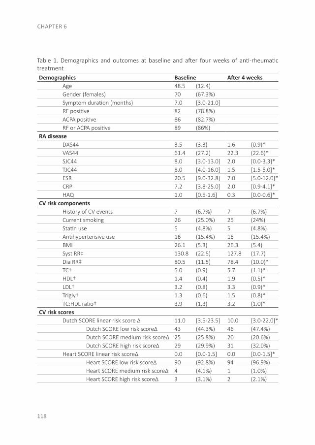
CHAPTER 6
118
Table 1. Demographics and outcomes at baseline and after four weeks of anti-rheumatic treatmentDemographics Baseline After 4 weeks Age 48.5 (12.4) Gender (females) 70 (67.3%) Symptom duration (months) 7.0 [3.0-21.0] RF positive 82 (78.8%) ACPA positive 86 (82.7%) RF or ACPA positive 89 (86%)RA disease DAS44 3.5 (3.3) 1.6 (0.9)* VAS44 61.4 (27.2) 22.3 (22.6)* SJC44 8.0 [3.0-13.0] 2.0 [0.0-3.3]* TJC44 8.0 [4.0-16.0] 1.5 [1.5-5.0]* ESR 20.5 [9.0-32.8] 7.0 [5.0-12.0]* CRP 7.2 [3.8-25.0] 2.0 [0.9-4.1]* HAQ 1.0 [0.5-1.6] 0.3 [0.0-0.6]*CV risk components History of CV events 7 (6.7%) 7 (6.7%) Current smoking 26 (25.0%) 25 (24%) Statin use 5 (4.8%) 5 (4.8%) Antihypertensive use 16 (15.4%) 16 (15.4%) BMI 26.1 (5.3) 26.3 (5.4) Syst RR‡ 130.8 (22.5) 127.8 (17.7) Dia RR‡ 80.5 (11.5) 78.4 (10.0)* TC† 5.0 (0.9) 5.7 (1.1)* HDL† 1.4 (0.4) 1.9 (0.5)* LDL† 3.2 (0.8) 3.3 (0.9)* Trigly† 1.3 (0.6) 1.5 (0.8)* TC:HDL ratio† 3.9 (1.3) 3.2 (1.0)*CV risk scores Dutch SCORE linear risk score ∆ 11.0 [3.5-23.5] 10.0 [3.0-22.0]* Dutch SCORE low risk score∆ 43 (44.3%) 46 (47.4%) Dutch SCORE medium risk score∆ 25 (25.8%) 20 (20.6%) Dutch SCORE high risk score∆ 29 (29.9%) 31 (32.0%) Heart SCORE linear risk score∆ 0.0 [0.0-1.5] 0.0 [0.0-1.5]* Heart SCORE low risk score∆ 90 (92.8%) 94 (96.9%) Heart SCORE medium risk score∆ 4 (4.1%) 1 (1.0%) Heart SCORE high risk score∆ 3 (3.1%) 2 (2.1%)
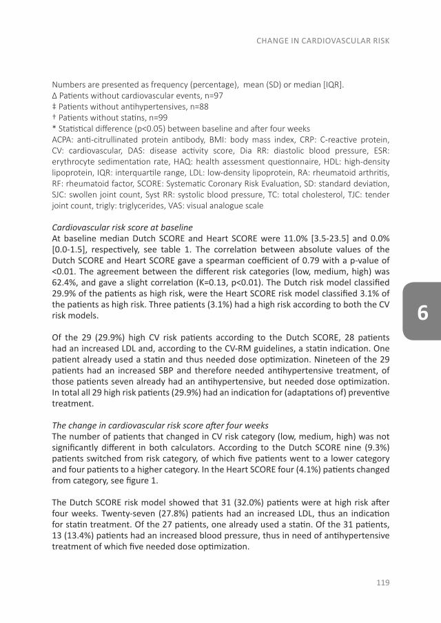
CHANGE IN CARDIOVASCULAR RISK
119
6
Numbers are presented as frequency (percentage), mean (SD) or median [IQR].∆ Patients without cardiovascular events, n=97 ‡ Patients without antihypertensives, n=88† Patients without statins, n=99* Statistical difference (p<0.05) between baseline and after four weeksACPA: anti-citrullinated protein antibody, BMI: body mass index, CRP: C-reactive protein, CV: cardiovascular, DAS: disease activity score, Dia RR: diastolic blood pressure, ESR: erythrocyte sedimentation rate, HAQ: health assessment questionnaire, HDL: high-density lipoprotein, IQR: interquartile range, LDL: low-density lipoprotein, RA: rheumatoid arthritis, RF: rheumatoid factor, SCORE: Systematic Coronary Risk Evaluation, SD: standard deviation, SJC: swollen joint count, Syst RR: systolic blood pressure, TC: total cholesterol, TJC: tender joint count, trigly: triglycerides, VAS: visual analogue scale
Cardiovascular risk score at baselineAt baseline median Dutch SCORE and Heart SCORE were 11.0% [3.5-23.5] and 0.0% [0.0-1.5], respectively, see table 1. The correlation between absolute values of the Dutch SCORE and Heart SCORE gave a spearman coefficient of 0.79 with a p-value of <0.01. The agreement between the different risk categories (low, medium, high) was 62.4%, and gave a slight correlation (Ƙ=0.13, p<0.01). The Dutch risk model classified 29.9% of the patients as high risk, were the Heart SCORE risk model classified 3.1% of the patients as high risk. Three patients (3.1%) had a high risk according to both the CV risk models.
Of the 29 (29.9%) high CV risk patients according to the Dutch SCORE, 28 patients had an increased LDL and, according to the CV-RM guidelines, a statin indication. One patient already used a statin and thus needed dose optimization. Nineteen of the 29 patients had an increased SBP and therefore needed antihypertensive treatment, of those patients seven already had an antihypertensive, but needed dose optimization. In total all 29 high risk patients (29.9%) had an indication for (adaptations of) preventive treatment.
The change in cardiovascular risk score after four weeksThe number of patients that changed in CV risk category (low, medium, high) was not significantly different in both calculators. According to the Dutch SCORE nine (9.3%) patients switched from risk category, of which five patients went to a lower category and four patients to a higher category. In the Heart SCORE four (4.1%) patients changed from category, see figure 1.
The Dutch SCORE risk model showed that 31 (32.0%) patients were at high risk after four weeks. Twenty-seven (27.8%) patients had an increased LDL, thus an indication for statin treatment. Of the 27 patients, one already used a statin. Of the 31 patients, 13 (13.4%) patients had an increased blood pressure, thus in need of antihypertensive treatment of which five needed dose optimization.
CHAPTER 6
120
In total three patients were indicated for a statin at baseline, but not anymore after four weeks and two patients did not need statins at baseline, but did after four weeks. Six patients had an antihypertensive indication at baseline and two doses optimization, but not at four weeks and two patients needed antihypertensives based at the values of four weeks, but not at baseline. This included one patient who was indicated for a statin and an antihypertensive at baseline, but not after four weeks, and one patient the other way around. In conclusion, in 13 (13.4%) patients the advice for (adaptations of) preventive treatment changed during the first four weeks of anti-rheumatic treatment.
Figure 1. Number of patients that changed from cardiovascular risk score category, during the first month of anti-rheumatic treatment, according to the Dutch SCORE (A) and European Heart SCORE (B), n=97
Stuart-maxwell analyses for Dutch SCORE, p=0.247.
Stuart-maxwell analyses for Heart SCORE risk score, p=0.135.
B.
A.
B.
A.
B.
A.
B.
A.
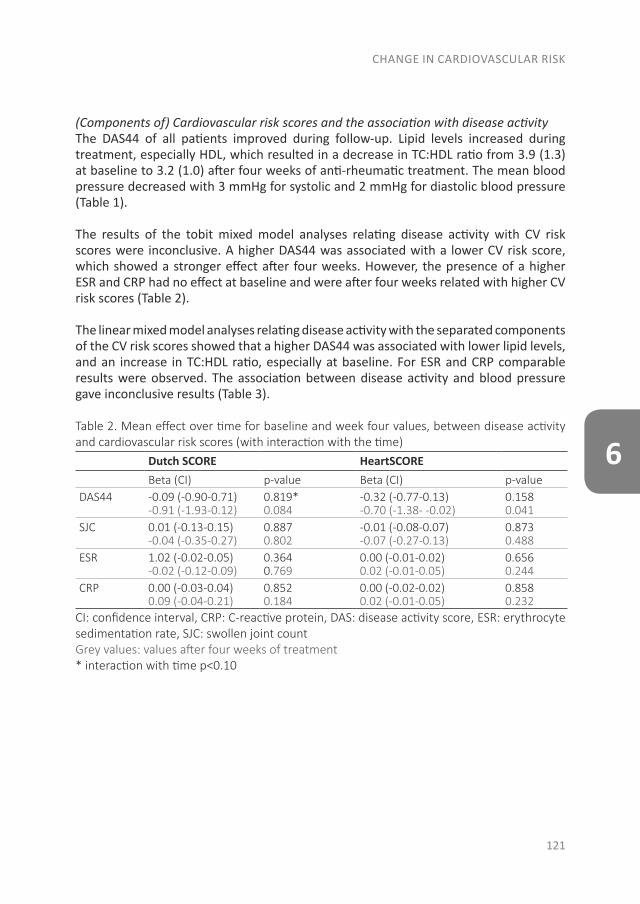
CHANGE IN CARDIOVASCULAR RISK
121
6
(Components of) Cardiovascular risk scores and the association with disease activityThe DAS44 of all patients improved during follow-up. Lipid levels increased during treatment, especially HDL, which resulted in a decrease in TC:HDL ratio from 3.9 (1.3) at baseline to 3.2 (1.0) after four weeks of anti-rheumatic treatment. The mean blood pressure decreased with 3 mmHg for systolic and 2 mmHg for diastolic blood pressure (Table 1).
The results of the tobit mixed model analyses relating disease activity with CV risk scores were inconclusive. A higher DAS44 was associated with a lower CV risk score, which showed a stronger effect after four weeks. However, the presence of a higher ESR and CRP had no effect at baseline and were after four weeks related with higher CV risk scores (Table 2).
The linear mixed model analyses relating disease activity with the separated components of the CV risk scores showed that a higher DAS44 was associated with lower lipid levels, and an increase in TC:HDL ratio, especially at baseline. For ESR and CRP comparable results were observed. The association between disease activity and blood pressure gave inconclusive results (Table 3).
Table 2. Mean effect over time for baseline and week four values, between disease activity and cardiovascular risk scores (with interaction with the time)
Dutch SCORE HeartSCOREBeta (CI) p-value Beta (CI) p-value
DAS44 -0.09 (-0.90-0.71)-0.91 (-1.93-0.12)
0.819* 0.084
-0.32 (-0.77-0.13)-0.70 (-1.38- -0.02)
0.1580.041
SJC 0.01 (-0.13-0.15)-0.04 (-0.35-0.27)
0.8870.802
-0.01 (-0.08-0.07)-0.07 (-0.27-0.13)
0.8730.488
ESR 1.02 (-0.02-0.05)-0.02 (-0.12-0.09)
0.3640.769
0.00 (-0.01-0.02)0.02 (-0.01-0.05)
0.6560.244
CRP 0.00 (-0.03-0.04)0.09 (-0.04-0.21)
0.8520.184
0.00 (-0.02-0.02)0.02 (-0.01-0.05)
0.8580.232
CI: confidence interval, CRP: C-reactive protein, DAS: disease activity score, ESR: erythrocyte sedimentation rate, SJC: swollen joint countGrey values: values after four weeks of treatment* interaction with time p<0.10
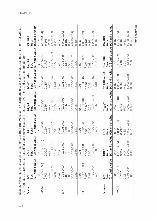
CHAPTER 6
122
Tabl
e 3.
Mix
ed m
odel
bet
wee
n co
mpo
nent
s of
the
card
iova
scul
ar r
isk
scor
e an
d di
seas
e ac
tivity
, at b
asel
ine
and
after
four
wee
ks o
f an
ti-rh
eum
atic
trea
tmen
t, c
orre
cted
for a
ge, s
mok
ing
stat
us, i
nter
actio
n ov
er ti
me
and
sepa
rate
d fo
r gen
der
Mal
esTC
†Be
ta(C
I) an
d p-
valu
e
HDL†
Beta
(CI)
and
p-va
lue
LDL†
Beta
(CI)
and
p-va
lue
Trig
ly†
Beta
(CI)
and
p-va
lue
TC:H
DL ra
tio†
Beta
(CI)
and
p-va
lue
Syst
RR‡
Beta
(CI)
and
p-va
lue
Dia
RR‡
Beta
(CI)
and
p-va
lue
DAS
44-0
.28
(-0.
49- -
0.06
) 0.
013
-0.1
0(-
0.48
-0.2
8)
0.60
8
-0.1
3(-
0.20
- -0.
05)
0.00
1*0.
02(-
0.11
-0.1
5)
0.76
4
-0.1
5(-
0.35
-0.0
4)
0.11
9-0
.11
(-0.
46-0
.23)
0.
511
-0.0
1(-
0.18
-0.1
6)
0.92
6-0
.05
(-0.
25-0
.25)
0.
755
0.20
(-0.
06-0
.46)
0.
135
-0.0
4(-
0.50
-0.4
2)
0.87
8
0.36
(-4.
03-4
.74)
0.
871*
-5.7
9(-
13.6
5-2.
07)
0.14
4
1.37
(-1.
09-3
.84)
0.
266
-0.7
8(-
5.19
-3.6
3)
0.72
3
ESR
-0.0
1(-
0.02
-0.0
0)
0.01
10.
00(-
0.05
-0.0
3)
0.88
8
-0.0
1(-
0.01
-0.0
0)
0.00
10.
00(-
0.01
-0.0
2)
0.65
6
-0.0
1(-
0.02
-0.0
0)
0.21
00.
00(-
0.04
-0.0
4)
0.83
2
<0.0
1(-
0.01
-0.0
1)
0.53
6-0
.01
(-0.
05-0
.02)
0.
426
0.01
(-0.
01-0
.02)
0.
277
0.01
(-0.
05-0
.06)
0.
825
0.05
(-0.
15-0
.25)
0.
617
0.56
(-0.
51-1
.62)
0.
296
0.09
(-0.
02-0
.20)
0.
107
0.47
(-0.
10-1
.04)
0.
104
CRP
-0.0
2(-
0.03
- -0.
01)
0.00
2-0
.03
(-0.
09-0
.04)
0.
431
-0.0
1(-
0.01
-0.0
0)
0.00
00.
00(-
0.02
-0.0
3)
0.73
0
-0.0
1(-
0.02
-0.0
0)
0.03
2-0
.02
(-0.
09-0
.04)
0.
465
<0.0
1(-
0.01
-0.0
1)
0.73
4*-0
.06
(-0.
12- -
0.01
) 0.
026
0.01
(-0.
01-0
.02)
0.
280
-0.0
2(-
0.10
-0.0
7)
0.64
8
0.09
(-0.
12-0
.29)
0.
402
0.98
(-0.
48-2
.44)
0.
182
0.08
(-0.
03-0
.19)
0.
161
0.16
(-0.
65-0
.98)
0.
687
Fem
ales
TC†
Beta
(CI)
and
p-va
lue
HDL†
Beta
(CI)
and
p-va
lue
LDL†
Beta
(CI)
and
p-va
lue
Trig
ly†
Beta
(CI)
and
p-va
lue
TC:H
DL ra
tio†
Beta
(CI)
and
p-va
lue
Syst
RR‡
Beta
(CI)
and
p-va
lue
Dia
RR‡
Beta
(CI)
and
p-va
lue
DAS
44-0
.02
(-0.
19-0
.14)
0.
761*
0.25
(0.0
6-0.
44)
0.01
1
-0.0
4(-
0.13
-0.0
5)
0.35
20.
04(-
0.07
-0.1
4)
0.49
0
-0.0
2(-
0.15
-0.1
2)
0.79
4*0.
15(-
0.01
-0.3
1)
0.07
0
-0.0
2(-
0.15
-0.1
0)
0.74
20.
07(-
0.07
-0.2
2)
0.32
7
0.16
(-0.
02-0
.33)
0.
085
0.11
(-0.
09-0
.32)
0.
281
-0.8
2(-
4.39
-2.7
4)
0.64
8-1
.29
(-5.
66-3
.08)
0.
558
-0.1
6(-
2.32
-1.9
9)
0.88
2-0
.61
(-3.
25-2
.02)
0.
645
tabl
e co
ntinu
es
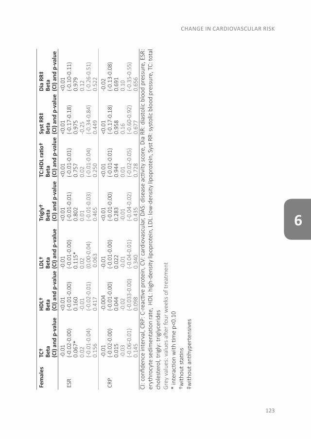
CHANGE IN CARDIOVASCULAR RISK
123
6
Fem
ales
TC†
Beta
(CI)
and
p-va
lue
HDL†
Beta
(CI)
and
p-va
lue
LDL†
Beta
(CI)
and
p-va
lue
Trig
ly†
Beta
(CI)
and
p-va
lue
TC:H
DL ra
tio†
Beta
(CI)
and
p-va
lue
Syst
RR‡
Beta
(CI)
and
p-va
lue
Dia
RR‡
Beta
(CI)
and
p-va
lue
ESR
-0.0
1(-
0.02
-0.0
0)
0.06
7*0.
02(-
0.01
-0.0
4)
0.15
6
<0.0
1(-
0.01
-0.0
0)
0.16
0-0
.01
(-0.
02-0
.01)
0.
417
-0.0
1(-
0.01
-0.0
0)
0.11
5*0.
02(0
.00-
0.04
) 0
.063
<0.0
1(-
0.01
-0.0
1)
0.80
20.
01(-
0.01
-0.0
3)
0.46
5
<0.0
1(-
0.01
-0.0
1)
0.75
70.
02(-
0.01
-0.0
4)
0.25
0
<0.0
1(-
0.17
-0.1
8)
0.97
5-0
.25
(-0.
34-0
.84)
0.
449
<0.0
1(-
0.10
-0.1
1)
0.97
90.
12(-
0.26
-0.5
1)
0.52
2
CRP
-0.0
1(-
0.02
-0.0
0)
0.01
5-0
.03
(-0.
06-0
.01)
0.
145
-0.0
04(-
0.01
-0.0
0)
0.04
4-0
.02
(-0.
033-
0.00
) 0.
098
-0.0
1(-
0.01
-0.0
0)
0.02
2-0
.01
(-0.
04-0
.01)
0.
340
<0.0
1(-
0.01
-0.0
0)
0.28
3-0
.01
(-0.
04-0
.02)
0.
435
<0.0
1(-
0.01
-0.0
1)
0.94
40.
01(-
0.02
-0.0
5)
0.72
8
<0.0
1(-
0.17
-0.1
8)
0.95
80.
16(-
0.60
-0.9
2)
0.67
7
-0.0
2(-
0.13
-0.0
8)
0.69
10.
10(-
0.35
-0.5
5)
0.65
6CI
: con
fiden
ce in
terv
al, C
RP: C
-rea
ctive
pro
tein
, CV:
car
diov
ascu
lar,
DAS
: dis
ease
acti
vity
sco
re, D
ia R
R: d
iast
olic
blo
od p
ress
ure,
ESR
: er
ythr
ocyt
e se
dim
enta
tion
rate
, H
DL:
hig
h-de
nsity
lipo
prot
ein,
LD
L: lo
w-d
ensi
ty li
popr
otei
n, S
yst R
R: sy
stol
ic b
lood
pre
ssur
e, T
C: to
tal
chol
este
rol,
trig
ly: t
rigly
cerid
esG
rey
valu
es: v
alue
s aft
er fo
ur w
eeks
of t
reat
men
t*
inte
racti
on w
ith ti
me
p<0.
10†w
ithou
t sta
tins
‡with
out a
ntihy
pert
ensi
ves
CHAPTER 6
124
DISCUSSION
Comparison between the Dutch SCORE and Heart SCORE CV risk models revealed a slight agreement between low, medium and high CV risk categories.
According to the Dutch CV-RM guidelines, 30% of the early RA patients had an indication for (adaptations of) preventive treatment at baseline. However, 13% of all the patients had a different indication after four weeks of anti-rheumatic treatment. If baseline CV risk assessment would be applied, this would lead to potential overtreatment in 10% of all the patients.
The risk estimation of the two different CV risk calculators resulted in a significant difference in CV risk score. Clinically this will have an impact on the therapy and prevention strategies chosen, as a patient who is regarded as low risk by one calculator could be classified as high risk by another, and vice versa(7). The Dutch SCORE estimates more patients as high risk, 10 times more often than the Heart SCORE. Overestimating the CV risk can lead to unnecessary treatment, while underestimating may result in CV diseases which could have been prevented. This is partly explained as the Dutch SCORE measures the 10-year risk on CV morbidity and mortality, and the Heart SCORE only assesses the risk on mortality and does not take non-fatal CV events into account(5). However, morbidity can cause functional limitations, therefore it is important for patients and society, and should be taken into account in a CV risk model. In conclusion, it is important to know the limitations of the CV risk calculator which estimated the CV risk of your patient.
CV preventive treatment is proven to be effective, therefore it is important to assess CV risk(25-27). As an increased CV risk is already present in early RA, and might even be present in the preclinical phase, CV risk management should be applied early in the disease course(5;28-32). In the present study, we found that, according to the Dutch SCORE, already 30% of the patients were classified as high CV risk at the onset of RA. However, many patients were classified in a different risk category after the first month of anti-rheumatic treatment. In 13% of the patients, this led to a change in preventive treatment advice. This percentage would probably be lower when applying Heart SCORE, as more patients were calculated as low risk according to this risk model, and so less patients switched from risk category during follow-up. Still, both at baseline and after four weeks of anti-rheumatic treatment many patients needed CV preventive treatment according to the Dutch CV-RM guidelines and did not receive this, reflecting under-treatment, confirming previous reports(33-35).
A higher disease activity was associated with an increased TC:HDL ratio, however the effect of disease activity on the Dutch CV risk SCORE gave inconclusive results. On the one hand, an increase in DAS44 was associated with lower CV risk, but on the other hand, higher ESR and CRP were associated with higher CV risk scores. Because
CHANGE IN CARDIOVASCULAR RISK
125
6
inflammation generally leads to an increased TC:HDL ratio, a higher CV risk is expected if markers of inflammation are high(18;19). This association was opposite for DAS44, a possibility is that other components of the DAS like the visual analogue scale and/or tender joint count disturb this association. The association between measures of inflammation and cholesterol levels, especially TC:HDL ratio was strongest at baseline, when all patients had a high disease activity. At four weeks, nearly all patients had low disease activity, which explains why this association was not present anymore after four weeks. Although there was no unambiguous association between disease activity and CV risk score, we do think that it is important to calculate CV risk during a time of low disease activity.
The change in CV risk score and so the advice about preventive treatment is probably correlated with the reduction of disease activity or the initiation of anti-rheumatic treatment. Previous literature described a reduction in acute myocardial infarction with the use of methotrexate (RR 0.81), but a dose-dependent increase with glucocorticoid use (RR 1.32)(36-38). An improvement in TC:HDL ratio was found after one and two years of COBRA-light treatment, however this did not had a favorable effect on CV risk prediction(18;19). Furthermore, lower disease activity (obtained with anti-rheumatic treatment) was associated with a lower blood pressure(39;40).
Unfortunately, we could not take into account some additional CV risk factors, such as renal function, physical activity and family history of CVD. These factors are considered as CV risk modifying factors which can be an additional reason to give CV risk prevention treatment. Therefore, the lack of these factors may have influenced our results. In addition to the Dutch SCORE and Heart SCORE, different CV risk scores are available. For example, the Framingham risk score is commonly used in the United States. However, this score is not generally applied in Europe and is limited to estimating the 10-year risk of a myocardial infarction and coronary heart disease-related death, thus underestimating the total atherosclerotic vascular disease risk(31;41). Especially, risk calculators that correct for the systemic inflammation are interesting. For example, the QRISK-2 and QRISK-3 calculators are used to predict CV risk in the United Kingdom, these calculators take RA into account as a separate CV risk factor. As a zip code is also a component of these algorithms, these calculators are not feasible in other countries(5;42). The Reynolds Risk score includes high-sensitivity CRP levels into the risk model, however this risk score is not recommended for patients with a systemic inflammatory disease, as CRP levels will be increased due to the inflammatory disease.(5) Further efforts were already performed to develop a RA-specific risk calculator, however, a new calculator (including DAS or HAQ) did not demonstrate an improvement compared to the current CV risk models which are used in the general population(43;44). The influence of fluctuations in disease activity over time in RA patients is difficult to incorporate in risk prediction models; single disease activity measurements (as DAS, CRP and ESR) are maybe not good enough, a biomarker that measures cumulative RA disease activity might fulfill this unmet need(44).
CHAPTER 6
126
In conclusion, CV risk management is important early in the course of RA, as preventive CV treatment is proven to be effective and should be applied as early as possible(5;25-27;31). However, the timing of CV risk assessment, as well as the availability of different CV risk models, influences the advice on the need for (adaptations of) CV preventive treatment. Further research is needed to determine which risk model is optimal and when in the course of RA it should be applied.
ACKNOWLEDGMENTS We would like to thank all patients, as well as all doctors who enrolled patients in this study, and all research nurses who were involved in patient management.
COMPETING INTERESTS Non declared for this study.
CONTRIBUTORSHIP All authors have substantially contributed to the conception or design of the work; the acquisition, analysis, and interpretation of data for the work. The authors revised the article critically and approved to the final version to be published. All agree to be accountable for all aspects of the work in ensuring that questions related to the accuracy or integrity of any part of the work are appropriately investigated and resolved.
FUNDING This study was performed without funding.
ETHICAL APPROVAL Medical Ethics Committee of the Slotervaart Hospital and Reade approved the protocol; patients gave written informed consent before inclusion, and the study was conducted in accordance with the Declaration of Helsinki/Good Clinical Practice
PROVENANCE AND PEER REVIEW Not commissioned, externally peer reviewed
CHANGE IN CARDIOVASCULAR RISK
127
6
REFERENCES
1. URL: http://www.who.int (accessed June 2017). 2. URL: http://www.scoremeter.nl (accessed June 2017). 3. URL: https://heartscore.escardio.org/2012/calc.aspx. (accessed June 2017).4. Arts EE, Popa C, Den Broeder AA, Semb AG, Toms T, Kitas GD, et al. Performance of
four current risk algorithms in predicting cardiovascular events in patients with early rheumatoid arthritis. Ann Rheum Dis 2015;74:668-74.
5. Bonek K, Gluszko P. Cardiovascular risk assessment in rheumatoid arthritis - controversies and the new approach. Reumatologia 2016;54:128-35.
6. Baghdadi LR, Woodman RJ, Shanahan EM, Mangoni AA. The impact of traditional cardiovascular risk factors on cardiovascular outcomes in patients with rheumatoid arthritis: a systematic review and meta-analysis. PLoS One 2015;10:e0117952.
7. Galarza-Delgado DA, Azpiri-Lopez JR, Colunga-Pedraza IJ, Cardenas-de la Garza JA, Vera-Pineda R, Serna-Pena G, et al. Assessment of six cardiovascular risk calculators in Mexican mestizo patients with rheumatoid arthritis according to the EULAR 2015/2016 recommendations for cardiovascular risk management. Clin Rheumatol 2017;36:1387-93.
8. Agca R, Heslinga SC, Rollefstad S, Heslinga M, McInnes IB, Peters MJ, et al. EULAR recommendations for cardiovascular disease risk management in patients with rheumatoid arthritis and other forms of inflammatory joint disorders: 2015/2016 update. Ann Rheum Dis 2017;76:17-28.
9. Bale BF, Doneen AL, Vigerust DJ. High-risk periodontal pathogens contribute to the pathogenesis of atherosclerosis. Postgrad Med J 2017;93:215-20.
10. Bender P, Burgin WB, Sculean A, Eick S. Serum antibody levels against Porphyromonas gingivalis in patients with and without rheumatoid arthritis - a systematic review and meta-analysis. Clin Oral Investig 2017;21:33-42.
11. Lahiri M, Luben RN, Morgan C, Bunn DK, Marshall T, Lunt M, et al. Using lifestyle factors to identify individuals at higher risk of inflammatory polyarthritis (results from the European Prospective Investigation of Cancer-Norfolk and the Norfolk Arthritis Register--the EPIC-2-NOAR Study). Ann Rheum Dis 2014;73:219-26.
12. Mahmoudi M, Aslani S, Fadaei R, Jamshidi AR. New insights to the mechanisms underlying atherosclerosis in rheumatoid arthritis. Int J Rheum Dis 2017;20:287-97.
13. Rosenstein ED, Greenwald RA, Kushner LJ, Weissmann G. Hypothesis: the humoral immune response to oral bacteria provides a stimulus for the development of rheumatoid arthritis. Inflammation 2004;28:311-8.
14. Arts EE, Fransen J, Den Broeder AA, Popa CD, Van Riel PL. The effect of disease duration and disease activity on the risk of cardiovascular disease in rheumatoid arthritis patients. Ann Rheum Dis 2015;74:998-1003.
15. Libby P, Ridker PM. Inflammation and atherosclerosis: role of C-reactive protein in risk assessment. Am J Med 2004;116 Suppl 6A:9S-16S.
CHAPTER 6
128
16. Charles-Schoeman C, Wang X, Lee YY, Shahbazian A, Navarro-Millan I, Yang S, et al. Association of Triple Therapy With Improvement in Cholesterol Profiles Over Two-Year Followup in the Treatment of Early Aggressive Rheumatoid Arthritis Trial. Arthritis Rheumatol 2016;68:577-86.
17. Chatterjee S, Sarkate P, Ghosh S, Biswas M, Ghosh A. Early, structured disease modifying anti-rheumatic drug (DMARD) therapy reduces cardiovascular risk in rheumatoid arthritis--a single centre study using non-biologic drugs. J Assoc Physicians India 2013;61:531-4.
18. Georgiadis AN, Papavasiliou EC, Lourida ES, Alamanos Y, Kostara C, Tselepis AD, et al. Atherogenic lipid profile is a feature characteristic of patients with early rheumatoid arthritis: effect of early treatment--a prospective, controlled study. Arthritis Res Ther 2006;8:R82.
19. Navarro-Millan I, Charles-Schoeman C, Yang S, Bathon JM, Bridges SL, Jr., Chen L, et al. Changes in lipoproteins associated with methotrexate or combination therapy in early rheumatoid arthritis: results from the treatment of early rheumatoid arthritis trial. Arthritis Rheum 2013;65:1430-8.
20. Aletaha D, Neogi T, Silman AJ, Funovits J, Felson DT, Bingham CO, III, et al. 2010 rheumatoid arthritis classification criteria: an American College of Rheumatology/European League Against Rheumatism collaborative initiative. Ann Rheum Dis 2010;69:1580-8.
21. Nederlands Huisartsen Genootschap. Multidisciplinary guideline on cardiovascular risk management 2011. Bohn Stafleu van Loghum, 2011.
22. Balogh E, Madruga DJ, Orr C, Mullan R, Harty L, FitzGerald O, et al. Comparison of remission criteria in a tumour necrosis factor inhibitor treated rheumatoid arthritis longitudinal cohort: patient global health is a confounder. Arthritis Res Ther 2013;15:R221.
23. Landis JR, Koch GG. The measurement of observer agreement for categorical data. Biometrics 1977 Mar;33(1):159-74.
24. Twisk J, Rijmen F. Longitudinal tobit regression: a new approach to analyze outcome variables with floor or ceiling effects. J Clin Epidemiol 2009;62:953-8.
25. Law MR, Morris JK, Wald NJ. Use of blood pressure lowering drugs in the prevention of cardiovascular disease: meta-analysis of 147 randomised trials in the context of expectations from prospective epidemiological studies. BMJ 2009;338:b1665.
26. Parsons C, Murad MH, Andersen S, Mookadam F, Labonte H. The effect of antihypertensive treatment on the incidence of stroke and cognitive decline in the elderly: a meta-analysis. Future Cardiol 2016;12:237-48.
27. Silverman MG, Ference BA, Im K, Wiviott SD, Giugliano RP, Grundy SM, et al. Association Between Lowering LDL-C and Cardiovascular Risk Reduction Among Different Therapeutic Interventions: A Systematic Review and Meta-analysis. JAMA 2016;316:1289-97.
28. Holmqvist ME, Wedren S, Jacobsson LT, Klareskog L, Nyberg F, Rantapaa-Dahlqvist S, et al. No increased occurrence of ischemic heart disease prior to the onset of rheumatoid arthritis: results from two Swedish population-based rheumatoid arthritis cohorts. Arthritis Rheum 2009;60:2861-9.
29. Holmqvist ME, Wedren S, Jacobsson LT, Klareskog L, Nyberg F, Rantapaa-Dahlqvist S, et al. Rapid increase in myocardial infarction risk following diagnosis of rheumatoid arthritis amongst patients diagnosed between 1995 and 2006. J Intern Med 2010;268:578-85.
CHANGE IN CARDIOVASCULAR RISK
129
6
30. Sodergren A, Karp K, Boman K, Eriksson C, Lundstrom E, Smedby T, et al. Atherosclerosis in early rheumatoid arthritis: very early endothelial activation and rapid progression of intima media thickness. Arthritis Res Ther 2010;12:R158.
31. Ton VK, Martin SS, Blumenthal RS, Blaha MJ. Comparing the new European cardiovascular disease prevention guideline with prior American Heart Association guidelines: an editorial review. Clin Cardiol 2013;36:E1-E6.
32. van Halm VP, Nielen MM, Nurmohamed MT, van SD, Reesink HW, Voskuyl AE, et al. Lipids and inflammation: serial measurements of the lipid profile of blood donors who later developed rheumatoid arthritis. Ann Rheum Dis 2007;66:184-8.
33. Panoulas VF, Douglas KM, Milionis HJ, Stavropoulos-Kalinglou A, Nightingale P, Kita MD, et al. Prevalence and associations of hypertension and its control in patients with rheumatoid arthritis. Rheumatology (Oxford) 2007;46:1477-82.
34. van Breukelen-van der Stoep DF, van ZD, Klop B, van de Geijn GJ, Janssen HJ, van der Meulen N, et al. Marked underdiagnosis and undertreatment of hypertension and hypercholesterolaemia in rheumatoid arthritis. Rheumatology (Oxford) 2016;55:1210-6.
35. van den Oever IAM, Heslinga M, Griep EN, Griep-Wentink HRM, Schotsman R, Cambach W, et al. Cardiovascular risk management in rheumatoid arthritis patients still suboptimal: the Implementation of Cardiovascular Risk Management in Rheumatoid Arthritis project. Rheumatology (Oxford) 2017.
36. Davis JM, III, Maradit KH, Crowson CS, Nicola PJ, Ballman KV, Therneau TM, et al. Glucocorticoids and cardiovascular events in rheumatoid arthritis: a population-based cohort study. Arthritis Rheum 2007;56:820-30.
37. Solomon DH, Avorn J, Katz JN, Weinblatt ME, Setoguchi S, Levin R, et al. Immunosuppressive medications and hospitalization for cardiovascular events in patients with rheumatoid arthritis. Arthritis Rheum 2006;54:3790-8.
38. Suissa S, Bernatsky S, Hudson M. Antirheumatic drug use and the risk of acute myocardial infarction. Arthritis Rheum 2006;55:531-6.
39. Kerola AM, Kauppi MJ, Kerola T, Nieminen TV. How early in the course of rheumatoid arthritis does the excess cardiovascular risk appear? Ann Rheum Dis 2012;71:1606-15.
40. Klarenbeek NB, van der Kooij SM, Huizinga TJ, Goekoop-Ruiterman YP, Hulsmans HM, van Krugten MV, et al. Blood pressure changes in patients with recent-onset rheumatoid arthritis treated with four different treatment strategies: a post hoc analysis from the BeSt trial. Ann Rheum Dis 2010;69:1342-5.
41. URL:https://www.mdcalc.com/framingham-coronary-heart-disease-risk-score (accessed June 2017).
42. Hippisley-Cox J, Coupland C, Brindle P. Development and validation of QRISK3 risk prediction algorithms to estimate future risk of cardiovascular disease: prospective cohort study. BMJ 2017;357:j2099.
43. Arts EE, Popa CD, Den Broeder AA, Donders R, Sandoo A, Toms T, et al. Prediction of cardiovascular risk in rheumatoid arthritis: performance of original and adapted SCORE algorithms. Ann Rheum Dis 2016;75:674-80.
44. Crowson CS, Rollefstad S, Kitas GD, Van Riel PL, Gabriel SE, Semb AG. Challenges of developing a cardiovascular risk calculator for patients with rheumatoid arthritis. PLoS One 2017;12:e0174656.

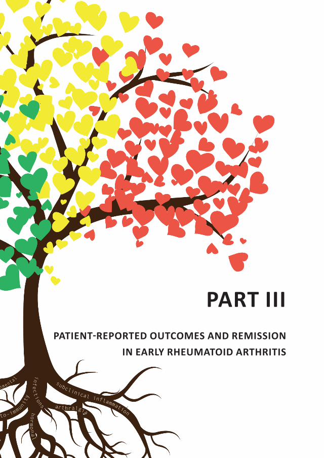
PART IIIPATIENT-REPORTED OUTCOMES AND REMISSION
IN EARLY RHEUMATOID ARTHRITIS

CHAPTER 7
PAIN, SLEEP AND EMOTIONAL WELL-BEING EXPLAIN THE LACK OF AGREEMENT BETWEEN PHYSICIAN-
AND PATIENT-PERCEIVED REMISSION IN EARLY RHEUMATOID ARTHRITIS
Samina A. Turk1
Linda A. Rasch2
Dirkjan van Schaardenburg1,3
Willem F. Lems1,2
Marjolein Sanberg1
Lilian H.D. van Tuyl2
Marieke M. ter Wee2,4
¹ Amsterdam Rheumatology and immunology Center| Reade, Amsterdam, Netherlands² Amsterdam Rheumatology and immunology Center| VU medical center, Amsterdam, Netherlands ³ Amsterdam Rheumatology and immunology Center| Academic Medical Center, Am-sterdam, Netherlands 4 Department of Epidemiology & Biostatistics, VU medical center, Amsterdam, Nether-lands
BMC Rheumatology - June 2018
CHAPTER 7
134
ABSTRACT
BACKGROUND. Clinical response and remission are defined in multiple ways and measured with different instruments, resulting in substantial variation of the proportion of patients classified as being in remission. Therefore, the agreement between patient-perceived, physician-perceived remission and clinical response and remission definitions was determined in early rheumatoid arthritis (RA) patients. And secondly, differences in clinical and patient-reported outcomes, in patients in physician-perceived remission, between patients in and not in self-perceived remission were assessed.
METHODS. In 84 early RA patients, who received methotrexate and glucocorticoids, DAS44, ACR/EULAR Boolean-based remission, EULAR good and ACR70 response were determined after 12 weeks. Agreement between patient-perceived (phrased: “Would you say that, at this moment, your disease activity is as good as gone?”), physician-perceived remission (based on a visual analogue scale for global disease severity) and clinical response and remission definitions were calculated with the percentage of agreement and with kappa values (which corrects for change). In patients in physician-perceived remission, improvement in clinical and patient-reported outcomes (RAID) were compared between patients in and not in self-perceived remission.
RESULTS. Agreement between the assessed outcome measures differed enormously. The agreement between physician-perceived and patient-perceived remission was 64% (kappa 0.25, p<0.01). Physician-perceived remission had the best agreement with EULAR good response (79%), and patient-perceived remission with EULAR good and ACR70 response (both 69%). Patients not in self-perceived remission improved less on RAID components, especially on pain, sleep and emotional well-being.
CONCLUSION. One-third of the early RA patients disagreed with the physician on being in remission. Those patients had less improvement on RAID components, especially on pain, sleep and emotional well-being. Together with the variability in clinical response and remission definitions, these results highlight the need to increase patient involvement in their own health care decisions.
PAIN, SLEEP AND EMOTIONAL WELL-BEING
135
7
BACKGROUND
Since rheumatoid arthritis (RA) patients are at risk for joint damage due to inflammation(1), the treatment goal in these patients is to attain a state of absence of disease activity, or remission(2). However, clinical response and remission are defined in multiple ways and measured with different instruments, resulting in substantial variation of the proportion of patients classified as being in remission(3;4). A particularly common difference is seen between the physician and the patients view on the RA disease activity(5-9).
The response to treatment as determined by the physician, is often based on the disease activity score (DAS), which is mainly based on physical examination and laboratory values(10;11). The DAS also contains a patient-reported outcome (PRO), i.e. the patient global assessment, however this global view lacks information on the patient’s perspective on remission(12). Furthermore, PROs such as fatigue and physical well-being, which have a large impact on daily life, are not taken directly into account(7). Nowadays, the importance of the patient’s perspective is increasingly recognized. Even though the patient’s perspective on remission is increasingly being studied and understood(12;13), it is unknown which determinants of disease activity explain the lack of agreement between physician- and patient-perceived remission. Patient satisfaction, the relationship between patient and physician, and treatment compliance can all be improved when patient and physician agree on the state of the disease(14-17), which can be reached by taking the opinion of the patient into account and thus with applying shared decision-making(18-20).
The objective of this study was twofold. First, the frequencies were examined of patients that achieved physician-perceived remission, patient-perceived remission, DAS44 remission, European League Against Rheumatism (EULAR) good response, American College of Rheumatology (ACR) 70 response, and ACR/EULAR Boolean-based definition of remission(21-23). With this data, the agreement between patient- and physician-perceived remission with and between the different clinical definitions of response and remission was determined. Second, the differences in clinical outcomes and PROs, in patients who did and did not agree with their physician on being in remission were assessed. Our hypothesis was that we would find significant differences in patients achieving remission according to the different response and remission criteria, compared to those who do not. Secondly, we hypothesised that there would be a lack of agreement between patient and physician perceived remission and several PROs.
METHODS
Study populationThe study population is part of a cohort of consecutive patients with early arthritis from the ‘Early Arthritis Cohort’ at Reade in Amsterdam, The Netherlands. This ongoing
CHAPTER 7
136
cohort includes patients aged 18 years and older with no prior treatment with disease-modifying antirheumatic drugs (DMARDs). Patients who fulfilled the ACR/ EULAR 2010 criteria for RA(24), and consented to start treatment with methotrexate (escalated to 25 mg/ week) with 5 mg folic acid and glucocorticoids (30 mg/ day tapered to 7,5 mg in nine weeks)(25), between June 2014 and December 2016, were selected for inclusion. Approval was obtained from the local ethics committee (P0120, Ethics Committee of the Slotervaart Hospital and Reade, Amsterdam, The Netherlands) and all patients gave written informed consent according to the Declaration of Helsinki.
MeasurementsPatients were interviewed by research nurses, at baseline and after 12 weeks to record clinical characteristics as well as the DAS44. Erythrocyte sedimentation rate (ESR), C-reactive protein (CRP), rheumatoid factor (RF) and anti-citrullinated protein (ACPA) were determined. The Health Assessment Questionnaire (HAQ) and Rheumatoid Arthritis Impact of Disease (RAID) questionnaires were filled out(4;26;27). The RAID evaluates the impact of RA on daily activities and comprises 7 domains that are evaluated as continuous variables from zero (best) to ten (worst).
Patient- and physician perceived remission were determined after 12 weeks of treatment. To assess patient-perceived remission the following question was phrased: “Would you say that, at this moment, your disease activity is as good as gone? Yes or no?”(13). Patients answering ‘yes’ were in ‘self-perceived remission’. Physician-perceived remission was assessed at the moment the physician assessed the patient in the outpatient clinic, using the ‘VAS physician’, phrased as: “How active do you think the rheumatoid arthritis of your patient is today?” and scored on a visual analogue (VAS) scale of 0-100 mm. Where a VAS ≤10 mm was defined as physician-perceived remission, according to the ACR/EULAR Boolean-based definition of remission(22).
Response after 12 weeks of treatment was determined, using the following clinical response and remission definitions: DAS44 remission (DAS44 <1.6 points at week 12), EULAR good response (defined as DAS44 improvement of 1.2 points and a DAS44 score at week 12 ≤ 2.4)(23), ACR70 response(21), and ACR/EULAR Boolean-based remission(22).
Statistical analysesFor descriptive purposes, mean (standard deviation (SD)), median (interquartile range (IQR)) or frequencies (percentages) were used. Differences between baseline and week 12 data were determined by the paired t-test when outcome variables were normally distributed. Otherwise, the Wilcoxon signed- rank test was applied.
First, the frequencies of patients who achieved DAS44 remission, EULAR good response, ACR70 response and Boolean-based remission were calculated, as well as the number of patients who were in physician- and patient-perceived remission. Second, the agreement of physician-perceived remission and patient-perceived remission with and between all
PAIN, SLEEP AND EMOTIONAL WELL-BEING
137
7
clinical response and remission definitions were calculated, using the percentage of agreement as well as kappa values, according to the interpretation of Landis and Koch (<0 indicates no agreement, 0 to 0.2 slight, 0.21 to 0.40 fair, 0.41 to 0.60 moderate, 0.61 to 0.80 substantial and 0.81 to 1.0 as almost perfect agreement). Kappa can be interpreted as the percentage of agreement after correcting for chance(28;29). Third, analyses were performed in a subgroup of patients in physician-perceived remission. In this group, the differences between patients in and not in self-perceived remission, were assessed on several outcome measures: the improvement on clinical, laboratory and questionnaire data. This was analysed with the independent t-test (normal distribution) or the Mann-Whitney U test (skewed distribution). A p-value <0.05 was considered statistically significant, and all analyses were performed with SPSS software (version 21).
RESULTS
Total populationIn total 84 patients with early RA of the ‘Early Arthritis Cohort’ were included. At baseline 10 patients did not complete the RAID questionnaire, and after 12 weeks three patients did not fill out the RAID questionnaire.
The mean (SD) age of the included patients was 50 (12) years, and 67% were female (Table 1). Mean (SD) DAS44 at baseline was 3.4 (1.2) and the seven questions on the RAID all had a median score between 4.0 and 7.0 at baseline. After 12 weeks of treatment, mean DAS44 (SD) improved to 1.4 (0.9) (p<0.01), and all questions on the RAID improved to a median score between 2.0 and 4.0 (p<0.01).
Patients who fulfil the different response and remission criteria After 12 weeks of treatment, 65 patients (77%) reached an EULAR good response, 25 patients (30%) an ACR70 response and 23 patients (27%) were in Boolean-based remission (Additional file 1). Fifty-one patients (61%) reached DAS44 <1.6 and 50 patients (60%) had a HAQ score <0.5.
All analyses were repeated for a cut-off of VAS physician remission of ≤20 mm and showed similar results (data not shown) .
Remission according to the physician and patient According to physician-perceived remission, 55 patients (66%) were in remission after 12 weeks of treatment.
Patients in self-perceived remission versus those not at week 12 (n=45, 54%) had a significantly lower DAS, tender joint count (TJC) and swollen joint count (SJC) of 44 joints and scored lower on all questions on the RAID (Table 1). The VAS physician was lower in patients who perceived themselves in remission, compared to those who did not 5.0
CHAPTER 7
138
(2.5-9.5) versus 13.0 (7.0-34.0) (p<0.01), respectively. Differences at baseline were seen between patients in and not in self-perceived remission after 12 weeks. Patients in self-perceived remission scored significantly lower at baseline on the VAS global, the HAQ, and on the RAID questions about functional disability assessment, fatigue and physical well-being. Of the clinical outcomes, only ESR was significantly lower in patients in self-perceived remission compared to those who were not: 15.0 (7.0-30.0) versus 23.0 (15.0-40.0) mm/hour (p<0.01; Table 1), respectively.
Agreement between physician, patient and clinical response and remission definitionsThe agreement between physician-perceived remission and patient-perceived remission was 67% (kappa 0.32, p<0.01; Table 2).
The physician-perceived remission had the best agreement with EULAR good response: 79% agreement, with a kappa of 0.48; p<0.01(Table 2).
The agreement with patient-perceived remission was highest for EULAR good response as well as ACR70 response: both 69% (kappa 0.36 (p<0.01) and 0.40 (p<0.01), respectively). The agreement with Boolean-based remission was slightly lower and the lowest agreement was seen with DAS44 remission.
Concordance between the different clinical response and remission definitions differed enormously. For example, the agreement between EULAR good response and DAS44 remission was 83% (kappa 0.62, p<0.01), but the agreement between EULAR good response and ACR70 response was 52% (kappa 0.22, p<0.01; Table 2). Agreement between physician- and patient perceived remission differed as well within the different response and remission criteria (Additional file 1).
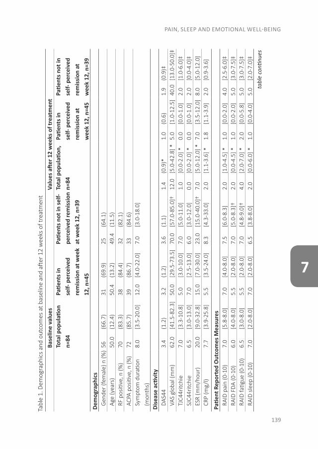
PAIN, SLEEP AND EMOTIONAL WELL-BEING
139
7
Tabl
e 1.
Dem
ogra
phic
s an
d ou
tcom
es a
t bas
elin
e an
d aft
er 1
2 w
eeks
of t
reat
men
tBa
selin
e va
lues
Valu
es a
fter 1
2 w
eeks
of t
reat
men
tTo
tal p
opul
ation
n=
84Pa
tient
s in
self-
per
ceiv
ed
rem
issi
on a
t wee
k 12
, n=4
5
Patie
nts n
ot in
self-
pe
rcei
ved
rem
issi
on
at w
eek
12, n
=39
Tota
l pop
ulati
on,
n=84
Patie
nts i
n se
lf- p
erce
ived
re
mis
sion
at
wee
k 12
, n=4
5
Patie
nts n
ot in
se
lf- p
erce
ived
re
mis
sion
at
wee
k 12
, n=3
9De
mog
raph
ics
Gen
der (
fem
ale)
n (%
)56
(6
6.7)
31
(69.
9)25
(6
4.1)
Age
(yea
rs)
50.0
(1
2.4)
50.4
(1
3.2)
49.4
(1
1.5)
RF p
ositi
ve, n
(%)
70
(83.
3)38
(8
4.4)
32
(82.
1)AC
PA p
ositi
ve, n
(%)
72
(85.
7)39
(8
6.7)
33
(84.
6)Sy
mpt
om d
urati
on
(mon
ths)
8.0
[3.5
-20.
0]12
.0
[4.0
-22.
0]7.
0 [3
.0-1
8.0]
Dise
ase
activ
ityD
AS44
3.4
(1
.2)
3.2
(1
.2)
3.6
(1
.1)
1.4
(0
.9)*
1.0
(0
.6)
1.9
(0.9
)‡VA
S gl
obal
(mm
)62
.0
[41.
5-82
.3]
50.0
[2
9.5-
73.5
]70
.0
[57.
0-85
.0]†
12.0
[5
.0-4
2.8]
*5.
0
[1.0
-12.
5]40
.0
[13.
0-50
.0]‡
TJC4
4ritc
hie
7.0
[3
.3-1
0.8]
5.0
[3
.0-1
0.0]
7.0
[5
.0-1
1.0]
1.0
[0
.0-2
.0] *
0.0
[0
.0-1
.0]
2.0
[1
.0-6
.0]‡
SJC4
4ritc
hie
6.5
[3
.0-1
3.0]
7.0
[2
.5-1
3.0]
6.0
[3
.0-1
2.0]
0.0
[0
.0-2
.0] *
0.0
[0
.0-1
.0]
2.0
[0
.0-4
.0]‡
ESR
(mm
/hou
r)20
.0
[9.0
-32.
8]15
.0
[7.0
-30.
0]23
.0
[15.
0-40
.0]†
7.0
[5
.0-1
2.0]
*7.
0
[3.5
-12.
0]8.
0
[5.0
-12.
0]CR
P (m
g/l)
7.7
[3
.9-2
5.8]
5.5
[3
.5-2
4.0]
8.3
[4
.3-3
3.0]
2.0
[1
.1-3
.6] *
1.8
[1
.1-3
.9]
2.0
[0
.9-3
.6]
Patie
nt R
epor
ted
Out
com
es M
easu
res
RAID
pai
n (0
-10)
7.0
[5
.8-8
.0]
7.0
[4
.0-8
.0]
7.5
[6
.0-8
.3]
2.0
[1
.0-4
.5] *
1.0
[0
.0-2
.0]
4.0
[2
.5-6
.0]‡
RAID
FD
A (0
-10)
6.0
[4
.0-8
.0]
5.5
[2
.0-8
.0]
7.0
[5
.0-8
.3]†
2.0
[0
.0-4
.5] *
1.0
[0
.0-2
.0]
5.0
[3
.0-7
.5]‡
RAID
fatig
ue (0
-10)
6.5
[3
.0-8
.0]
5.5
[2
.0-8
.0]
7.0
[4
.8-9
.0]†
4.0
[2
.0-7
.0] *
2.0
[0
.0-5
.8]
5.0
[3
.0-7
.5]‡
RAID
sle
ep (0
-10)
7.0
[2
.0-8
.0]
7.0
[2
.0-8
.0]
6.5
[3
.8-8
.0]
2.0
[0
.0-6
.0] *
1.0
[0
.0-4
.0]
5.0
[2
.0-7
.0]‡
tabl
e co
ntinu
es
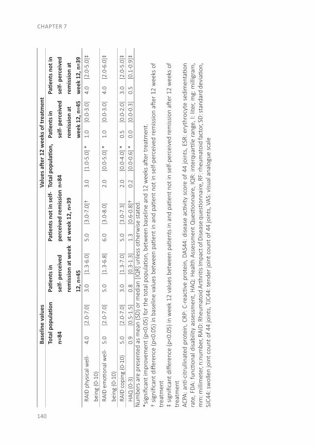
CHAPTER 7
140
Base
line
valu
esVa
lues
afte
r 12
wee
ks o
f tre
atm
ent
Tota
l pop
ulati
on
n=84
Patie
nts i
n se
lf- p
erce
ived
re
mis
sion
at w
eek
12, n
=45
Patie
nts n
ot in
self-
pe
rcei
ved
rem
issi
on
at w
eek
12, n
=39
Tota
l pop
ulati
on,
n=84
Patie
nts i
n se
lf- p
erce
ived
re
mis
sion
at
wee
k 12
, n=4
5
Patie
nts n
ot in
se
lf- p
erce
ived
re
mis
sion
at
wee
k 12
, n=3
9RA
ID p
hysi
cal w
ell-
bein
g (0
-10)
4.0
[2
.0-7
.0]
3.0
[1
.3-6
.0]
5.0
[3
.0-7
.0]†
3.0
[1
.0-5
.0] *
1.0
[0
.0-3
.0]
4.0
[2
.0-5
.0]‡
RAID
em
otion
al w
ell-
bein
g (0
-10)
5.0
[2
.0-7
.0]
5.0
[1
.3-6
.8]
6.0
[3
.0-8
.0]
2.0
[0
.0-5
.0] *
1.0
[0
.0-3
.0]
4.0
[2
.0-6
.0]‡
RAID
cop
ing
(0-1
0)5.
0
[2.0
-7.0
]3.
0
[1.3
-7.0
]5.
0
[3.0
-7.3
]2.
0
[0.0
-4.0
] *0.
5
[0.0
-2.0
]3.
0
[2.0
-5.0
]‡H
AQ (0
-3)
0.9
[0
.5-1
.5]
0.8
[0
.3-1
.3]
1.3
[0
.6-0
.8]†
0.2
[0
.0-0
.6] *
0.0
[0
.0-0
.3]
0.5
[0
.1-0
.9]‡
Num
bers
are
pre
sent
ed a
s m
ean
(SD
) or m
edia
n [IQ
R] u
nles
s ot
herw
ise
stat
ed.
*sig
nific
ant i
mpr
ovem
ent (
p<0.
05) f
or th
e to
tal p
opul
ation
, bet
wee
n ba
selin
e an
d 12
wee
ks a
fter
trea
tmen
t.†
sign
ifica
nt d
iffer
ence
(p<0
.05)
in b
asel
ine
valu
es b
etw
een
patie
nt in
and
pati
ent
not
in s
elf-
perc
eive
d re
mis
sion
aft
er 1
2 w
eeks
of
trea
tmen
t‡
sign
ifica
nt d
iffer
ence
(p<0
.05)
in w
eek
12 v
alue
s be
twee
n pa
tient
s in
and
pati
ent n
ot in
sel
f-pe
rcei
ved
rem
issi
on a
fter
12
wee
ks o
f tr
eatm
ent
ACPA
: anti
-citr
ullin
ated
pro
tein
, CRP
: C-r
eacti
ve p
rote
in, D
AS44
: dis
ease
acti
vity
sco
re o
f 44
join
ts, E
SR: e
ryth
rocy
te s
edim
enta
tion
rate
, FD
A: fu
nctio
nal d
isab
ility
ass
essm
ent,
HAQ
: Hea
lth A
sses
smen
t Q
uesti
onna
ire, I
QR:
inte
rqua
rtile
ran
ge, l
: lite
r, m
g: m
illig
ram
, m
m: m
illim
eter
, n:n
umbe
r, RA
ID: R
heum
atoi
d Ar
thriti
s Im
pact
of D
isea
se q
uesti
onna
ire, R
F: rh
eum
atoi
d fa
ctor
, SD
: sta
ndar
d de
viati
on,
SJC4
4: s
wol
len
join
t cou
nt o
f 44
join
ts, T
JC44
: ten
der j
oint
cou
nt o
f 44
join
ts, V
AS: v
isua
l ana
logu
e sc
ale
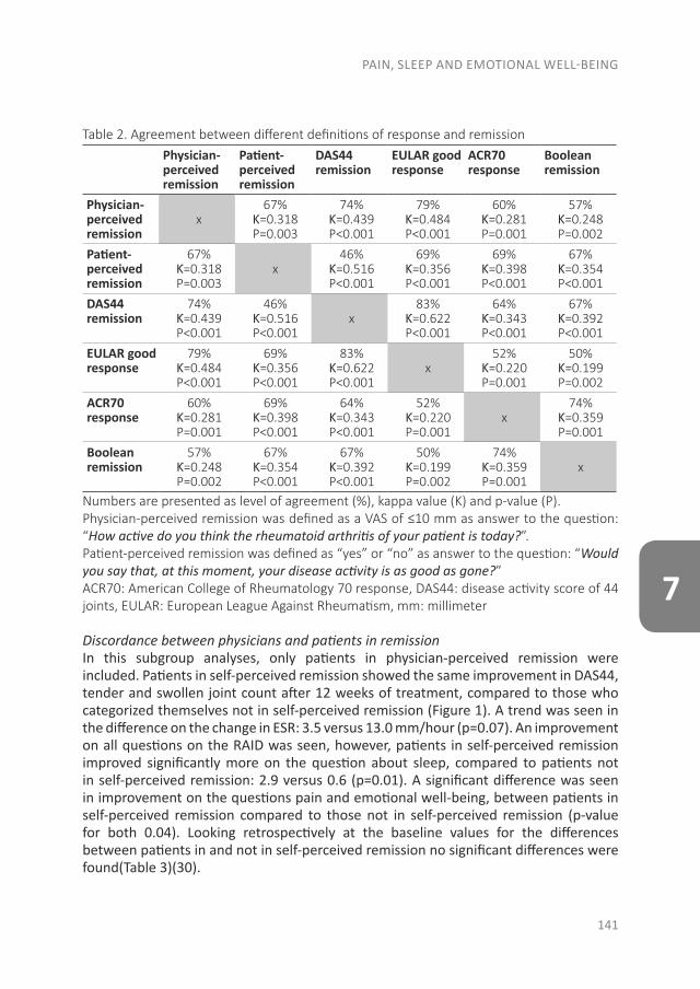
PAIN, SLEEP AND EMOTIONAL WELL-BEING
141
7
Table 2. Agreement between different definitions of response and remissionPhysician-perceived remission
Patient-perceived remission
DAS44 remission
EULAR good response
ACR70 response
Boolean remission
Physician-perceived remission
x67%
Ƙ=0.318P=0.003
74%Ƙ=0.439P<0.001
79%Ƙ=0.484P<0.001
60%Ƙ=0.281P=0.001
57%Ƙ=0.248P=0.002
Patient-perceived remission
67%Ƙ=0.318P=0.003
x46%
Ƙ=0.516P<0.001
69%Ƙ=0.356P<0.001
69%Ƙ=0.398P<0.001
67%Ƙ=0.354P<0.001
DAS44 remission
74%Ƙ=0.439P<0.001
46%Ƙ=0.516P<0.001
x83%
Ƙ=0.622P<0.001
64%Ƙ=0.343P<0.001
67%Ƙ=0.392P<0.001
EULAR good response
79%Ƙ=0.484P<0.001
69%Ƙ=0.356P<0.001
83%Ƙ=0.622P<0.001
x52%
Ƙ=0.220P=0.001
50%Ƙ=0.199P=0.002
ACR70 response
60%Ƙ=0.281P=0.001
69%Ƙ=0.398P<0.001
64%Ƙ=0.343P<0.001
52%Ƙ=0.220P=0.001
x74%
Ƙ=0.359P=0.001
Boolean remission
57%Ƙ=0.248P=0.002
67%Ƙ=0.354P<0.001
67%Ƙ=0.392P<0.001
50%Ƙ=0.199P=0.002
74%Ƙ=0.359P=0.001
x
Numbers are presented as level of agreement (%), kappa value (K) and p-value (P).Physician-perceived remission was defined as a VAS of ≤10 mm as answer to the question: “How active do you think the rheumatoid arthritis of your patient is today?”.Patient-perceived remission was defined as “yes” or “no” as answer to the question: “Would you say that, at this moment, your disease activity is as good as gone?”ACR70: American College of Rheumatology 70 response, DAS44: disease activity score of 44 joints, EULAR: European League Against Rheumatism, mm: millimeter
Discordance between physicians and patients in remissionIn this subgroup analyses, only patients in physician-perceived remission were included. Patients in self-perceived remission showed the same improvement in DAS44, tender and swollen joint count after 12 weeks of treatment, compared to those who categorized themselves not in self-perceived remission (Figure 1). A trend was seen in the difference on the change in ESR: 3.5 versus 13.0 mm/hour (p=0.07). An improvement on all questions on the RAID was seen, however, patients in self-perceived remission improved significantly more on the question about sleep, compared to patients not in self-perceived remission: 2.9 versus 0.6 (p=0.01). A significant difference was seen in improvement on the questions pain and emotional well-being, between patients in self-perceived remission compared to those not in self-perceived remission (p-value for both 0.04). Looking retrospectively at the baseline values for the differences between patients in and not in self-perceived remission no significant differences were found(Table 3)(30).
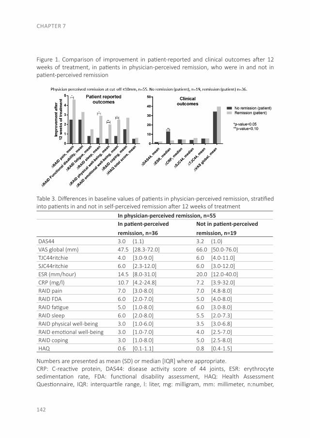
CHAPTER 7
142
Figure 1. Comparison of improvement in patient-reported and clinical outcomes after 12 weeks of treatment, in patients in physician-perceived remission, who were in and not in patient-perceived remission
Table 3. Differences in baseline values of patients in physician-perceived remission, stratified into patients in and not in self-perceived remission after 12 weeks of treatment
In physician-perceived remission, n=55In patient-perceived remission, n=36
Not in patient-perceived remission, n=19
DAS44 3.0 (1.1) 3.2 (1.0)VAS global (mm) 47.5 [28.3-72.0] 66.0 [50.0-76.0]TJC44ritchie 4.0 [3.0-9.0] 6.0 [4.0-11.0]SJC44ritchie 6.0 [2.3-12.0] 6.0 [3.0-12.0]ESR (mm/hour) 14.5 [8.0-31.0] 20.0 [12.0-40.0]CRP (mg/l) 10.7 [4.2-24.8] 7.2 [3.9-32.0]RAID pain 7.0 [3.0-8.0] 7.0 [4.8-8.0]RAID FDA 6.0 [2.0-7.0] 5.0 [4.0-8.0]RAID fatigue 5.0 [1.0-8.0] 6.0 [3.0-8.0]RAID sleep 6.0 [2.0-8.0] 5.5 [2.0-7.3]RAID physical well-being 3.0 [1.0-6.0] 3.5 [3.0-6.8]RAID emotional well-being 3.0 [1.0-7.0] 4.0 [2.5-7.0]RAID coping 3.0 [1.0-8.0] 5.0 [2.5-8.0]HAQ 0.6 [0.1-1.1] 0.8 [0.4-1.5]
Numbers are presented as mean (SD) or median [IQR] where appropriate.CRP: C-reactive protein, DAS44: disease activity score of 44 joints, ESR: erythrocyte sedimentation rate, FDA: functional disability assessment, HAQ: Health Assessment Questionnaire, IQR: interquartile range, l: liter, mg: milligram, mm: millimeter, n:number,
PAIN, SLEEP AND EMOTIONAL WELL-BEING
143
7
RAID: Rheumatoid Arthritis Impact of Disease questionnaire, SD: standard deviation, SJC44: swollen joint count of 44 joints, TJC44: tender joint count of 44 joints, VAS: visual analogue scale.*significant difference (p<0.05) in baseline values for patients in and not in self-perceived remission after 12 weeks of treatment
DISCUSSION
More than one-third of early RA patients disagreed with their physician on being in remission after 12 weeks of treatment. The agreement between physician and patient was higher in patients who did achieve DAS44 remission, ACR70 response or were in Boolean-based remission. Patients who judged themselves as not being in self-perceived remission showed less improvement on the RAID questions on sleep, pain and emotional well-being, compared with patients who judged themselves as being in self-perceived remission.
In this study all patients received the same anti-rheumatic treatment, which led to an improvement in disease activity of all patients. The improvement of mean two points in the DAS44 score after 12 weeks of treatment was similar to the results of the COBRA-light trial. The improvement on RAID was in agreement with the results of the study of Ledingham et al.(31;32). Clinical response and remission definitions in RA are defined in several ways and the stringency of these different definitions has been shown to vary widely and lead to enormous differences in results, which is comparable to our results as 61% reached DAS44 remission, while 27% of the patients achieved Boolean-based remission(33).
Our results showed a similar percentage of agreement between physician- and patient-perceived remission, as in existing literature an agreement between 51% and 79% is seen(5-9). For example, the Danish DANBIO registry found a 51% agreement between 8300 RA patients and physicians. Disagreement in this study was defined as a difference of >20 mm on the global assessment between the patient and the physician(8). However, the DANBIO registry described patients with a mean disease duration of seven years and patients with lower disease activity, while the current study included patients at the onset of RA, who generally have a higher disease activity. The higher agreement between patients and physicians in the present study was probably due to higher disease activity scores, as a higher swollen joint count is found to be associated with lower odds of discordance(5). This is also visible in the variability of agreement between physicians and patients within different response and remission definitions, as agreement was higher in patients who achieved ACR70 response or Boolean remission. In the present study, both patients and physicians had the best agreement with EULAR good response, which was predictable as (improvement in) DAS is the most commonly used measurement in clinical practice(33).
CHAPTER 7
144
A difference was not found in the improvement of DAS44 score, SJC or TJC between patients who did and did not agree with the physician on being in remission. But, where physicians focus on disease activity (inflammation), patients also incorporate other domains(7;10;11). Patients who did not agree with their physician on being in remission did show less improvement on components of the RAID about sleep, pain and emotional well-being. A non-significant difference in fatigue was found between patients who did and did not perceive themselves in remission. This is in contrast with other studies, in which fatigue was an important explanation of patients perception of disease activity (7,34). However, in these studies fatigue covered fatigue and sleep problems, which was separated in our study. This might explain the difference as instead of the disease itself, the side effects of medication, especially glucocorticoids, can also explain a part of the sleep difficulties and fatigue symptoms. In this study, all patients received the same dose of glucocorticoids, however some patients may experience more side effects than others. Patients who did not agree also showed more improvement in ESR after 12 weeks. These patients showed a trend of a higher ESR at baseline, but no significant difference was found after 12 weeks of anti-rheumatic treatment. We hypothesized that patients who did not perceive themselves in remission, had more low grade inflammation during the 12 weeks, which might be associated with more fatigue and sleep difficulties(34;35). At baseline their mean ESR was higher, but they improved more in ESR to reach the same ESR levels at week 12 as patients who were in self-perceived remission. However, this was not seen for CRP-levels. The comparison between the RAID score and the discordance of physician- and patient-perceived remission has not yet been performed before, as far as we know, which is a strength of this study. Our study has some limitations. First, there is no widely accepted cut-off point for discordance and therefore we used the same cut-off as the ACR/EULAR Boolean-based definition of remission(23) were a VAS≤10 mm was accepted to define physician-perceived remission. We also performed a sensitivity analysis with a cut-off VAS≤20mm, which showed similar results. However, similar results were found in a study performed in 800 RA patients, where a median VAS physician of 15 mm was found in patients who were in physician-perceived remission(9). Second, the number of patients included in this study was small, which influences the possibility to find significant relationships in the data. However, this was of minimal influence as similar results in previous articles were found(5-9). Finally, we did not take adverse effects as well as other concomitant diseases into account that could influence the self-assessment of RA activity. However, the measurements of RA disease activity that are used in general care, do not consider this aspect either(10). Nonetheless, for future perspectives questions on adverse effects, comorbidity and mental state might be useful. Future studies are needed to confirm our findings and to determine the optimal set of patient-reported outcomes. And eventually to compare the current treat-to-target treatment strategy with patient-reported outcome guided treatment.
PAIN, SLEEP AND EMOTIONAL WELL-BEING
145
7
CONCLUSIONS
In conclusion, more than one-third of the patients disagreed with their physician on being in remission. This might have consequences for patient satisfaction, the relationship between patient and physician and treatment compliance of the patient. Patients who disagreed with their physician on being in remission showed less improvement on questions about sleep, pain and emotional well-being of the RAID. However, not only patients and physicians showed discordance, there were also many differences between clinical response and remission definitions. This makes it necessary to increase patient involvement in their own health care decisions, improving shared decision making.
LIST OF ABBREVIATIONS
ACPA: anti-citrullinated proteinACR: American College of RheumatologyCRP: C-reactive proteinDAS: disease activity scoreDMARDs: disease modifying antirheumatic drugsESR: Erythrocyte sedimentation rateEULAR: European League Against RheumatismHAQ: Health Assessment QuestionnaireIQR: interquartile rangeRA: rheumatoid arthritisSD: standard deviationPRO: patient-reported outcomeRAID: Rheumatoid Arthritis Impact of DiseaseRF: rheumatoid factorTJC: tender joint countSJC: swollen joint countVAS: visual analogue scale
CHAPTER 7
146
DECLARATIONS
Ethics approval and consent to participateMedical Ethics Committees of the Slotervaart Hospital and Reade approved the protocol (P0120); patients gave written informed consent before inclusion, and the study was conducted in accordance with the Declaration of Helsinki/Good Clinical Practice.
Consent for publicationNot applicable.
Availability of data and materialThe datasets used and analysed during the current study are available from the corresponding author on reasonable request.
Competing interests The authors declare that they have no competing interests.
FundingThis study was performed without funding.
Author’s contributionsMS saw the patients in the outpatient clinic and collected the data. ST, LR and MW analyzed the data. And DS, WL, LT, MW and ST contributed in interpreting the data and writing the manuscript. All authors read and approved the final manuscript.
AcknowledgementsWe would like to thank all patients, as well as all doctors who enrolled patients in this study, and all research nurses who were involved in patient management.
PAIN, SLEEP AND EMOTIONAL WELL-BEING
147
7
REFERENCES
1. Smolen JS, Aletaha D, McInnes IB. Rheumatoid arthritis. Lancet 2016;388:2023-38.2. Shammas RM, Ranganath VK, Paulus HE. Remission in rheumatoid arthritis. Curr
Rheumatol Rep 2010;12:355-62.3. Sung YK, Cho SK, Kim D, Yoon BY, Choi CB, Cha HS, et al. Factors Contributing to
Discordance between the 2011 ACR/EULAR Criteria and Physician Clinical Judgment for the Identification of Remission in Patients with Rheumatoid Arthritis. J Korean Med Sci 2016;31:1907-13.
4. van Tuyl LH, Smolen JS, Wells GA, Scholte-Voshaar M, Hoogland W, Boers M. Patient perspective on remission in rheumatoid arthritis. J Rheumatol 2011 Aug;38:1735-8.
5. Barton JL, Imboden J, Graf J, Glidden D, Yelin EH, Schillinger D. Patient-physician discordance in assessments of global disease severity in rheumatoid arthritis. Arthritis Care Res (Hoboken) 2010;62:857-64.
6. Castrejon I, Yazici Y, Samuels J, Luta G, Pincus T. Discordance of global estimates by patients and their physicians in usual care of many rheumatic diseases: association with 5 scores on a Multidimensional Health Assessment Questionnaire (MDHAQ) that are not found on the Health Assessment Questionnaire (HAQ). Arthritis Care Res (Hoboken) 2014;66:934-42.
7. Khan NA, Spencer HJ, Abda E, Aggarwal A, Alten R, Ancuta C, et al. Determinants of discordance in patients’ and physicians’ rating of rheumatoid arthritis disease activity. Arthritis Care Res (Hoboken) 2012;64:206-14.
8. Lindstrom EC, Krogh NS, Pincus T, Dreyer L, Ellingsen T, Glintborg B, et al. Discordance of Global Assessments by Patient and Physician Is Higher in Female than in Male Patients Regardless of the Physician’s Sex: Data on Patients with Rheumatoid Arthritis, Axial Spondyloarthritis, and Psoriatic Arthritis from the DANBIO Registry. J Rheumatol 2015 ;42:1781-5.
9. Wolfe F, Boers M, Felson D, Michaud K, Wells GA. Remission in rheumatoid arthritis: physician and patient perspectives. J Rheumatol 2009;36:930-3.
10. Janta I, Naredo E, Martinez-Estupinan L, Nieto JC, De lT, I, Valor L, et al. Patient self-assessment and physician’s assessment of rheumatoid arthritis activity: which is more realistic in remission status? A comparison with ultrasonography. Rheumatology (Oxford) 2013;52:2243-50.
11. Nicolau G, Yogui MM, Vallochi TL, Gianini RJ, Laurindo IM, Novaes GS. Sources of discrepancy in patient and physician global assessments of rheumatoid arthritis disease activity. J Rheumatol 2004;31:1293-6.
12. van Tuyl LH, Sadlonova M, Hewlett S, Davis B, Flurey C, Goel N, et al. The patient perspective on absence of disease activity in rheumatoid arthritis: a survey to identify key domains of patient-perceived remission. Ann Rheum Dis 2017;76:855-61.
13. van Tuyl LH, Hewlett S, Sadlonova M, Davis B, Flurey C, Hoogland W, et al. The patient perspective on remission in rheumatoid arthritis: ‘You’ve got limits, but you’re back to being you again’. Ann Rheum Dis 2015;74:1004-10.
CHAPTER 7
148
14. Berrios-Rivera JP, Street RL, Jr., Garcia Popa-Lisseanu MG, Kallen MA, Richardson MN, Janssen NM, et al. Trust in physicians and elements of the medical interaction in patients with rheumatoid arthritis and systemic lupus erythematosus. Arthritis Rheum 2006 15;55:385-93.
15. Dobkin PL, Sita A, Sewitch MJ. Predictors of adherence to treatment in women with fibromyalgia. Clin J Pain 2006;22:286-94.
16. Jackson CA, Clatworthy J, Robinson A, Horne R. Factors associated with non-adherence to oral medication for inflammatory bowel disease: a systematic review. Am J Gastroenterol 2010;105:525-39.
17. Starfield B, Wray C, Hess K, Gross R, Birk PS, D’Lugoff BC. The influence of patient-practitioner agreement on outcome of care. Am J Public Health 1981;71:127-31.
18. Lofland JH, Johnson PT, Ingham MP, Rosemas SC, White JC, Ellis L. Shared decision-making for biologic treatment of autoimmune disease: influence on adherence, persistence, satisfaction, and health care costs. Patient Prefer Adherence 2017;11:947-58.
19. Stacey D, Legare F, Lewis K, Barry MJ, Bennett CL, Eden KB, et al. Decision aids for people facing health treatment or screening decisions. Cochrane Database Syst Rev 2017;4:CD001431.
20. Veilleux S, Noiseux I, Lachapelle N, Kohen R, Vachon L, Guay BW, et al. Patients’ perception of their involvement in shared treatment decision making: Key factors in the treatment of inflammatory bowel disease. Patient Educ Couns 2017.
21. Felson DT, Anderson JJ, Boers M, Bombardier C, Furst D, Goldsmith C, et al. American College of Rheumatology. Preliminary definition of improvement in rheumatoid arthritis. Arthritis Rheum 1995;38:727-35.
22. Felson DT, Smolen JS, Wells G, Zhang B, van Tuyl LH, Funovits J, et al. American College of Rheumatology/European League against Rheumatism provisional definition of remission in rheumatoid arthritis for clinical trials. Ann Rheum Dis 2011;70:404-13.
23. van Gestel AM, Prevoo ML, van ‘t Hof MA, van Rijswijk MH, van de Putte LB, van Riel PL. Development and validation of the European League Against Rheumatism response criteria for rheumatoid arthritis. Comparison with the preliminary American College of Rheumatology and the World Health Organization/International League Against Rheumatism Criteria. Arthritis Rheum 1996;39:34-40.
24. Aletaha D, Neogi T, Silman AJ, Funovits J, Felson DT, Bingham CO, III, et al. 2010 rheumatoid arthritis classification criteria: an American College of Rheumatology/European League Against Rheumatism collaborative initiative. Ann Rheum Dis 2010;69:1580-8.
25. den UD, ter WM, Boers M, Kerstens P, Voskuyl A, Nurmohamed M, et al. A non-inferiority trial of an attenuated combination strategy (‘COBRA-light’) compared to the original COBRA strategy: clinical results after 26 weeks. Ann Rheum Dis 2014;73:1071-8.
26. Gossec L, Paternotte S, Aanerud GJ, Balanescu A, Boumpas DT, Carmona L, et al. Finalisation and validation of the rheumatoid arthritis impact of disease score, a patient-derived composite measure of impact of rheumatoid arthritis: a EULAR initiative. Ann Rheum Dis 2011;70:935-42.
PAIN, SLEEP AND EMOTIONAL WELL-BEING
149
7
27. Norton S, Fu B, Scott DL, Deighton C, Symmons DP, Wailoo AJ, et al. Health Assessment Questionnaire disability progression in early rheumatoid arthritis: systematic review and analysis of two inception cohorts. Semin Arthritis Rheum 2014;44:131-44.
28. Balogh E, Madruga DJ, Orr C, Mullan R, Harty L, FitzGerald O, et al. Comparison of remission criteria in a tumour necrosis factor inhibitor treated rheumatoid arthritis longitudinal cohort: patient global health is a confounder. Arthritis Res Ther 2013;15:R221.
29. Landis JR, Koch GG. The measurement of observer agreement for categorical data. Biometrics 1977;33:159-74.
30. Ledingham JM, Snowden N, Rivett A, Galloway J, Ide Z, Firth J, et al. Patient- and clinician-reported outcomes for patients with new presentation of inflammatory arthritis: observations from the National Clinical Audit for Rheumatoid and Early Inflammatory Arthritis. Rheumatology (Oxford) 2017;56:231-8.
31. Ter Wee MM, den UD, Boers M, Kerstens P, Nurmohamed M, van SD, et al. Intensive combination treatment regimens, including prednisolone, are effective in treating patients with early rheumatoid arthritis regardless of additional etanercept: 1-year results of the COBRA-light open-label, randomised, non-inferiority trial. Ann Rheum Dis 2015;74:1233-40.
32. Gulfe A, Geborek P, Saxne T. Response criteria for rheumatoid arthritis in clinical practice: how useful are they? Ann Rheum Dis 2005;64:1186-9.
33. Koike T, Kazuma K, Kawamura S. The relationship between fatigue, coping behavior, and inflammation in patients with rheumatoid arthritis. Mod Rheumatol 2000;10:141-9.
34. Rohleder N, Aringer M, Boentert M. Role of interleukin-6 in stress, sleep, and fatigue. Ann N Y Acad Sci 2012;1261:88-96.
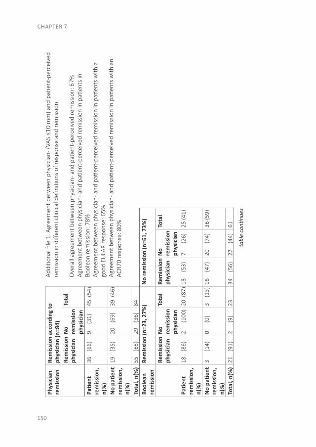
CHAPTER 7
150
Phys
icia
n re
mis
sion
Rem
issi
on a
ccor
ding
to
phys
icia
n (n
=84)
Addi
tiona
l file
1. A
gree
men
t bet
wee
n ph
ysic
ian-
(VAS
≤10
mm
) and
pati
ent-
perc
eive
d re
mis
sion
in d
iffer
ent c
linic
al d
efini
tions
of r
espo
nse
and
rem
issi
on
Ove
rall
agre
emen
t bet
wee
n ph
ysic
ian-
and
pati
ent-
perc
eive
d re
mis
sion
: 67%
Agre
emen
t bet
wee
n ph
ysic
ian-
and
pati
ent-
perc
eive
d re
mis
sion
in p
atien
ts in
Bo
olea
n re
mis
sion
: 78%
Agre
emen
t bet
wee
n ph
ysic
ian-
and
pati
ent-
perc
eive
d re
mis
sion
in p
atien
ts w
ith a
go
od E
ULA
R re
spon
se: 6
5%Ag
reem
ent b
etw
een
phys
icia
n- a
nd p
atien
t-pe
rcei
ved
rem
issi
on in
pati
ents
with
an
ACR7
0 re
spon
se: 8
0%
Rem
issi
on
phys
icia
nN
o re
mis
sion
ph
ysic
ian
Tota
l
Patie
nt
rem
issi
on,
n(%
)
36
(66)
9
(31)
45 (
54)
No
patie
nt
rem
issi
on,
n(%
)
19
(35)
20
(69)
39 (
46)
Tota
l, n(
%)
55
(65)
29
(36)
84Bo
olea
n re
mis
sion
Rem
issi
on (n
=23,
27%
)N
o re
mis
sion
(n=6
1, 7
3%)
Rem
issi
on
phys
icia
nN
o re
mis
sion
ph
ysic
ian
Tota
lRe
mis
sion
ph
ysic
ian
No
rem
issi
on
phys
icia
n
Tota
l
Patie
nt
rem
issi
on,
n(%
)
18
(86)
2
(100
)20
(87
)18
(
53)
7
(26)
25 (4
1)
No
patie
nt
rem
issi
on,
n(%
)
3
(14)
0
(0)
3
(13)
16
(47)
20
(74)
36 (5
9)
Tota
l, n(
%)
21
(91)
2
(9)
2334
(5
6)27
(4
4)61
tabl
e co
ntinu
es
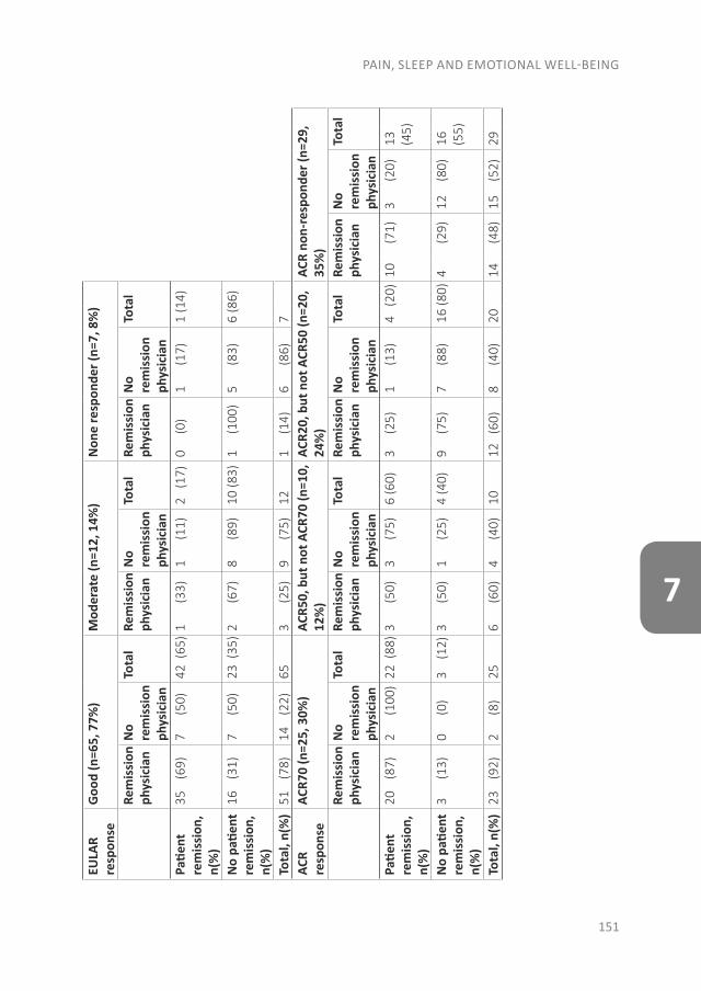
PAIN, SLEEP AND EMOTIONAL WELL-BEING
151
7
EULA
R re
spon
seG
ood
(n=6
5, 7
7%)
Mod
erat
e (n
=12,
14%
)N
one
resp
onde
r (n=
7, 8
%)
Rem
issi
on
phys
icia
nN
o re
mis
sion
ph
ysic
ian
Tota
lRe
mis
sion
ph
ysic
ian
No
rem
issi
on
phys
icia
n
Tota
lRe
mis
sion
ph
ysic
ian
No
rem
issi
on
phys
icia
n
Tota
l
Patie
nt
rem
issi
on,
n(%
)
35
(69)
7
(50)
42 (
65)
1
(33)
1
(11)
2 (
17)
0
(0)
1
(17)
1 (1
4)
No
patie
nt
rem
issi
on,
n(%
)
16
(31)
7
(50)
23 (
35)
2
(67)
8
(89)
10 (8
3)1
(1
00)
5
(83)
6 (8
6)
Tota
l, n(
%)
51
(78)
14
(22)
653
(2
5)9
(7
5)12
1
(14)
6
(86)
7AC
R re
spon
seAC
R70
(n=2
5, 3
0%)
ACR5
0, b
ut n
ot A
CR70
(n=1
0,
12%
)AC
R20,
but
not
ACR
50 (n
=20,
24
%)
ACR
non-
resp
onde
r (n=
29,
35%
)Re
mis
sion
ph
ysic
ian
No
rem
issi
on
phys
icia
n
Tota
lRe
mis
sion
ph
ysic
ian
No
rem
issi
on
phys
icia
n
Tota
lRe
mis
sion
ph
ysic
ian
No
rem
issi
on
phys
icia
n
Tota
lRe
mis
sion
ph
ysic
ian
No
rem
issi
on
phys
icia
n
Tota
l
Patie
nt
rem
issi
on,
n(%
)
20
(87)
2
(100
)22
(88
)3
(5
0)3
(7
5)6
(60)
3
(25)
1
(13)
4 (
20)
10
(71)
3
(20)
13
(45)
No
patie
nt
rem
issi
on,
n(%
)
3
(13)
0
(0)
3
(12)
3
(50)
1
(25)
4 (4
0)9
(7
5)7
(8
8)16
(80)
4
(29)
12
(80)
16
(55)
Tota
l, n(
%)
23
(92)
2
(8)
256
(6
0)4
(4
0)10
12
(60)
8
(40)
2014
(4
8)15
(5
2)29

CHAPTER 8
DO PATIENTS WITH RHEUMATOID ARTHRITIS IN REMISSION FEEL NORMAL AGAIN?
Linda A. Rasch1
Samina A. Turk2
Maarten Boers2,3
Willem F. Lems1,2
Dirkjan van Schaardenburg1,4
Lilian H.D. van Tuyl1
¹ Amsterdam Rheumatology and immunology Center| VU medical center, Amsterdam, Netherlands ² Amsterdam Rheumatology and immunology Center| Reade, Amsterdam, Netherlands³ Department of Epidemiology & Biostatistics, VU medical center, Amsterdam, Nether-lands 4 Amsterdam Rheumatology and immunology Center| Academic Medical Center, Am-sterdam, Netherlands Submitted for publication
CHAPTER 8
154
ABSTRACT
OBJECTIVE. Rheumatoid arthritis (RA) patients identified a return to normal as one of the three major themes of remission. Therefore, this study investigated the relationship between patient perceived concepts of normality and remission, and the ability of a ‘normality scale’ to discriminate between remission and non-remission states.
METHODS. Newly diagnosed RA patients with a high disease activity or unfavourable prognostic factors, received treat-to-target combination therapy. At baseline, week 13 and week 26, disease impact was assessed, including a scale measuring the perception of normality (score 7-35, higher scores indicating higher feelings of normality), Disease Activity Score of 28 joints, and two definitions of remission: 1. patient perceived remission (“Would you say that, at this moment, your disease activity is as good as gone?”); 2. ACR/EULAR Boolean remission.
RESULTS. Forty-seven patients completed all assessments: 34 (72%) females, mean±SD age 50±14 years. Normality score at baseline was 20±7, and increased significantly to 23±8 in week 13 and to 24±8 in week 26 (both, p≤0.001). Compared to patients not in remission, patients in remission felt more normal: this was significant for patients in self-perceived remission at week 13 (p<0.001) and week 26 (p=0.007), and for patients in Boolean remission at week 26 (p<0.001).
CONCLUSION. Patients in self-perceived remission felt more normal after 13 and 26 weeks; patients in Boolean remission only after 26 weeks of therapy. The normality scale discriminates between patients in and not in remission, and aligns with patient-perceived remission. Further research is needed to study normality as a patient-relevant target of RA treatment.
DO PATIENTS WITH RHEUMATOID ARTHRITIS IN REMISSION FEEL NORMAL AGAIN?
155
8
INTRODUCTION
Rheumatoid arthritis (RA) is a chronic illness characterized by inflammation of the joints that strongly impacts quality of life. However, improved treatment strategies now more frequently result in remission (1), so that ‘returning back to normal’ might be a realistic goal for some patients.
According to the American College of Rheumatology and the European League Against Rheumatism (ACR/EULAR) definition, remission in RA is achieved when tender joint count, swollen joint count, patient global assessment of disease activity (PtGA) and C-reactive protein (CRP) level are all ≤1, or when the simple sum of the tender joint count, swollen joint count, PtGA, physician global assessment (PhGA) and CRP level is ≤3.3 (2). The definition contains one patient reported outcome, namely the PtGA, formulated as “Considering all of the ways your arthritis has affected you, how do you feel your arthritis is today?” with the anchors very well and very poor (2). Although some have suggested that the PtGA should be removed from the remission definition for the sake of diagnostic accuracy (3), others, especially patients and clinicians have suggested that with only one patient-reported outcome the definition lacks information from the patients’ perspective on remission (4).
Qualitative research from our group identified three major themes of patient perceived remission: 1) reduction or absence of symptoms, 2) reduction of impact, 3) return to perceived normality (5). This return to perceived normality was either referred to as a return to the ‘old’ normal life, as well as a ‘new’ normal situation referring to work, family role and perception of others (5). This theme has also emerged in other studies, where maintaining a normal life, returning to normality, or perceptions of normality were identified as important treatment outcomes to RA patients (6-8).
While ‘returning back to normal’ was identified by patients as one of the three most important themes of patient perceived remission, ‘normality’ is a complicated concept and a consensus definition is not documented in literature. Normality is often equated to quality of life or well-being, but it might be more than that. Sanderson et al defined normality as ‘the person conceptualises the illness as normal, which involves incorporating the illness more fully into a person’s identity and public self through a redefinition of values and beliefs’ (9). In 2009, a ‘normality scale’ was developed to assess the perception of normality in RA patients (9). This study aims to investigate the relationship between patient perceived concepts of normality and remission, and the ability of the normality scale to discriminate between remission and non-remission states.
CHAPTER 8
156
PATIENTS AND METHODS
Study populationAll newly diagnosed RA patients (ACR/EULAR 2010 criteria (10)) aged 18 years or above with a disease duration of less than two years, and either high disease activity or unfavourable prognostic factors, were invited to participate in this study. Recruitment took place in the outpatient rheumatology department of the Amsterdam Rheumatology and immunology Center, locations VU University medical center and Reade, the Netherlands. Treatment according to a treat-to-target regime comprised initial methotrexate and step-down prednisolone; if good EULAR response was not achieved after 13 weeks, patients were randomized to continue treatment or intensify treatment. Exclusion criteria were: a history of DMARD use, the use of >7.5 mg/day of glucocorticoids in the past four weeks, or fulfilling any contra-indications for the use of the study medication. All patients that had completed the week 26 visit in January 2016 were eligible for inclusion in the current study.
This study was approved by the local Ethics Committee and qualified as a study carrying no extra risk for the participants. All patients received written information about the study and provided informed consent for their participation.
MeasurementsMeasurements took place at baseline and after 13 and 26 weeks of treatment, and included:
• The normality scaleThe ‘normality scale’, developed in 2009, is a questionnaire that measures the perception of normality in RA patients in 7 items (Figure 1, column A) (9). The items were derived from interviews with RA patients about living with their chronic condition (11). The scale was translated from English to Dutch by two separate forward and backward translations. This resulted in the final Dutch version used for this study (Figure 1, column B). All items were scored on a 5-point Likert scale: 1 for strongly disagree and 5 for strongly agree for items 1 and 3-7; reverse scoring for item 2. Simple addition leads to a total score that ranges between 7 and 35, with higher scores indicating a higher feeling of normality.
• Disease activity The WHO-ILAR core set (12) evaluated disease activity. Joints were assessed by a trained researcher or (research) nurse. PtGA as well as assessor global assessment (AssGA) and patient pain were expressed on a 0-100 scale. Since the PhGA was not assessed at all time points, AssGA was used instead. The AssGA is similar to the PhGA, only assessed by the researcher or (research) nurse instead of the physician. Physical functioning was assessed with the Health Assessment Questionnaire (HAQ). Disease Activity Score of 44 joints (DAS44) was calculated, which was converted to the Disease Activity Score
DO PATIENTS WITH RHEUMATOID ARTHRITIS IN REMISSION FEEL NORMAL AGAIN?
157
8
of 28 joints (DAS28) (13). Finally, both CRP and erythrocyte sedimentation rate were determined.
• RemissionRemission was measured in two ways: 1) patient perceived remission in patients answering ‘yes’ to the question “Would you say that, at this moment, your disease activity is as good as gone?”. This definition was developed during our work on the patient perspective on remission in close collaboration with patient research partners (4); 2) ACR/EULAR Boolean-based definition of remission (2).
Figure 1. Normality scaleItem A. Original (English) B. Dutch translation1. On most days, I don’t think about my
arthritisOp de meeste dagen denk ik niet aan mijn reuma
2. My life is not normal now that I live with RA Mijn leven is niet normaal nu dat ik reuma heb
3. I socialise as much as someone without arthritis
Mijn sociale leven is hetzelfde als dat van iemand zonder reuma
4. I sleep as well as before symptoms of my RA started
Ik slaap even goed als voordat mijn reuma-klachten begonnen
5. I have maintained my normal life since I was diagnosed with RA
Ik heb mijn normale leven behouden sinds ik de diagnose reuma kreeg
6. When I am well, my life is relatively normal Wanneer het goed met me gaat is mijn leven redelijk normaal
7. I have as much energy as someone my age without arthritis
Ik heb evenveel energie als iemand van mijn leeftijd zonder reuma
The items are scored with a 5-point Likert scale. Items 1 and 3-7 are scored as 1= strongly disagree / helemaal mee oneens, 2= partially disagree / een beetje mee oneens, 3= neither agree or disagree / niet mee eens, niet mee oneens, 4= partially agree / een beetje mee eens, 5= strongly agree / helemaal mee eens. Item 2 is scored in reverse with 1= strongly agree / helemaal mee eens, 2= partially agree / een beetje mee eens, 3= neither agree or disagree / niet mee eens, niet mee oneens, 4= partially disagree / een beetje mee oneens, 5= strongly disagree / helemaal mee oneens. The scale can be used free of charge.
Statistical analyses Descriptive statistics summarized patient characteristics: mean ± standard deviation, median [25th and 75th percentile], and frequency (percentage) where appropriate. The normality scale items were examined both as total score and individually (by calculating mean item scores). Paired t-tests compared changes over time in the total study population, and after stratification for being in remission or not. Linear regression assessed the unadjusted relationship between normality (dependent) and remission (independent variable). McNemar tests assessed concordance between patient self-reported remission and the ACR/EULAR Boolean based remission definition. P-values
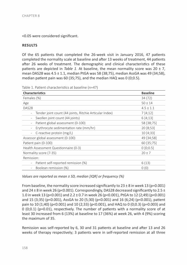
CHAPTER 8
158
<0.05 were considered significant.
RESULTS
Of the 65 patients that completed the 26-week visit in January 2016, 47 patients completed the normality scale at baseline and after 13 weeks of treatment, 44 patients after 26 weeks of treatment. The demographic and clinical characteristics of these patients are depicted in Table 1. At baseline, the mean normality score was 20 ± 7, mean DAS28 was 4.5 ± 1.1, median PtGA was 58 (38;75), median AssGA was 49 (34;58), median patient pain was 60 (35;75), and the median HAQ was 0 (0;0.5).
Table 1. Patient characteristics at baseline (n=47)Characteristics BaselineFemales (%) 34 (72)Age 50 ± 14DAS28 4.5 ± 1.1
- Tender joint count (44 joints, Ritchie Articular Index) 7 [4;12]- Swollen joint count (44 joints) 6 [4;13]- Patient global assessment (0-100) 58 [38;75]- Erythrocyte sedimentation rate (mm/hr) 20 [8;53]- C-reactive protein (mg/L) 10 [4;33]
Assessor global assessment (0-100) 49 [34;58]Patient pain (0-100) 60 [35;75]Health Assessment Questionnaire (0-3) 0 [0;0.5]Normality score (7-35) 20 ± 7Remission:
- Patient self-reported remission (%) 6 (13)- Boolean remission (%) 0 (0)
Values are reported as mean ± SD, median [IQR] or frequency (%)
From baseline, the normality score increased significantly to 23 ± 8 in week 13 (p=0.001) and 24 ± 8 in week 26 (p<0.001). Correspondingly, DAS28 decreased significantly to 2.5 ± 1.0 in week 13 (p<0.001) and 2.2 ± 0.7 in week 26 (p<0.001), PtGA to 12 (2;49) (p<0.001) and 15 (3;35) (p<0.001), AssGA to 20 (5;30) (p<0.001) and 16 (6;24) (p<0.001), patient pain to 10 (1;40) (p<0.001) and 10 (2;33) (p<0.001), and HAQ to 0 (0;0.3) (p=0.003) and 0 (0;0.1) (p=0.01), respectively. The number of patients with a normality score of at least 30 increased from 6 (13%) at baseline to 17 (36%) at week 26, with 4 (9%) scoring the maximum of 35.
Remission was self-reported by 6, 30 and 31 patients at baseline and after 13 and 26 weeks of therapy respectively; 3 patients were in self-reported remission at all three
DO PATIENTS WITH RHEUMATOID ARTHRITIS IN REMISSION FEEL NORMAL AGAIN?
159
8
time points, and another 4 at both 13 and 26 weeks. Correspondingly, 0, 13 and 13 patients were in Boolean remission respectively, with 8 at both 13 and 26 weeks. Self-reported and Boolean remission were often not concordant (Table 2).
Table 2. Concordance between patient self-reported remission and the ACR/EULAR Boolean based remission definition (in percentage)
Patient self-reported remissionWeek 13 Week 26
(n)Yes(30)
No(17)
Total(47)
Yes(31)
No(13)
Total(44)
ACR/EULAR Boolean remission
Yes (13) 24 4 28 27 2 30 (13)No (34) 40 32 72 43 27 70 (31)Total (47) 64 36 100 70 30 100 (44)
Compared to patients not in remission, those in remission felt more normal at follow up (Figure 2). At week 26, the results for normality were numerically similar and significant for both self-perceived and Boolean remission definitions: patients in remission scored mean (95%CI) 6.6 (1.9; 11.1, p=0.007) and 8.2 (3.5; 12.9, p=0.001) higher on the normality scale, respectively. At week 13, only patients in self-perceived remission scored significantly higher: 7.6 (3.6; 11.7, p<0.001) and 3.8 (–1.0; 8.7, p=0.119), respectively.
Figure 2. Normality scores, categorized by presence of (n/N): Patient perceived remission (week 0: 6/47; week 13: 30/47; week 26: 32/46); ACR/EULAR Boolean remission (week 0: 0/47; week 13: 13/47; week 26: 13/45)
* significant difference in normality score between patients in, and patients not in remission (p<0.05)
CHAPTER 8
160
Of the separate normality items of the normality scale, more than half of the patients answered item 6 with ‘strongly agree’ to the statement “When I am well, my life is relatively normal” (24, 26, and 24 patients respectively at baseline, after 13 weeks, and after 26 weeks). This is also the item with the highest item score at all time points, followed by item 3 “I socialise as much as someone without arthritis”. The other items have a wider range in their scores. Furthermore, item 1 (“On most days, I don’t think about my arthritis”) show large improvements in the scores: at baseline, most patients disagree with this sentence (17 patients strongly disagree and 11 patients partially disagree), while after 26 weeks most patients agree with the sentence (15 patients strongly agree and 13 patients partially agree).
Of the 65 eligible patients, 18 patients had incomplete data and had to be excluded from these analyses. These patients did not differ significantly from those included with regards to gender, age, or disease activity at baseline. However, patients with incomplete data were less likely to experience remission: 18% and 7% were in remission at week 13 and week 26 respectively, compared to 28% and 29% in the included patients (significant at 26 weeks: p=0.03).
DISCUSSION
This study demonstrates that the perception of normality as measured by the normality scale is sensitive to change in the context of the start of treatment in early RA in a treat-to-target regime aiming at clinical remission, and that it discriminates between patients in and not in remission. Of note is that the responsivity of this fully patient-reported outcome is similar to that of the DAS28, widely regarded as one of the most sensitive indices of disease activity. Finally, occurrence of patient-perceived remission was discordant with Boolean remission, preponderantly in the direction of Boolean being more strict than the patient perception.
Research on the perception of normality in patients with RA is limited. Almén et al. studied perceptions of RA patients relating particular topics. One of these topics was ‘normal life’ that they expressed as: regaining full health, living a normal live, not to be regarded as differing from ordinary people, not be seen as different because of disability, feeling no limitations, managing the household, and normal social functioning, in sum “…to be able to live the life I had before” (14). The chronic, progressive nature of RA likely complicates a comparison of life with RA to that before RA; nevertheless, in our study of early RA patients 9% indicated their life had returned to normal as it was before the start of RA, scoring the maximum score on each item of the normality scale after 26 weeks of therapy.
Research on normality in RA is limited. The normality scale used in this study was developed as part of a doctoral thesis (9), but has to date not been published in a scientific journal, limiting its exposure and dissemination. This is the first study to
DO PATIENTS WITH RHEUMATOID ARTHRITIS IN REMISSION FEEL NORMAL AGAIN?
161
8
validate aspects of the scale in the context of RA remission. A limitation of this study is its size (47 patients only). Most likely, selection also occurred in the included patients, increasing the proportion of patients experiencing remission. However, as remission is still relatively infrequent, such a selection is actually favourable in this study of normality in the context of remission, as it creates more balanced groups, and does not influence the scores of the normality scales in either remission or non-remission states.
To our knowledge, no other instrument is available that measures the feeling of normality, specifically in patients with RA. More or less simultaneously to the development of the normality scale, O’Neal et al. developed a normalization assessment measure for rheumatic conditions containing 20 questions (15). However, this assessment includes, but is not limited to, RA since it is also developed in patients with lupus, osteoarthritis, and fibromyalgia. A comparison of the scales developed by Sanderson and O’Neal and their relation to disease activity measures would be of interest.
The discordance between patient perceived remission and Boolean remission was picked up by the normality score, that appeared more sensitive to occurrence of the former, at least in the first 13 weeks. These results confirm our impression that patient perception of remission is not (completely) the same as the clinician’s, and that at least part of this difference lies in information captured by the normality scale.
The normality scale was originally designed in Bristol and contains 7 items (9). All items are carefully formulated and selected out of interview data with patients about living with RA. In the analyses of separate items, an outlier occurred in the answers of one of the items: more than half of the patients answered ‘strongly agree’ to item 6 “When I am well, my life is relatively normal”. This is in contrast to the answers that were given to all other items, where is more diversity in the number of patients that chose for a specific answer. According to the formulation of the question, this might not be very surprising, as a RA patient that is feeling well does only have a few or maybe even no complaints like pain. The large improvements in the scores for item 1 (“On most days, I don’t think about my arthritis”) are also notable, given that all patients in the trial are still taking anti-rheumatic drugs on a daily basis. This suggests they no longer think about their RA every time they take a pill, pointing to adaptation. This is in accordance with the definition of normality of Sanderson et al: ‘the person conceptualises the illness as normal, which involves incorporating the illness more fully into a person’s identity and public self through a redefinition of values and beliefs’ (9). In other words, it may be that patients adjust their expectations and values, they get used to a new way of life and they adapt to ‘a new normal situation’. However, when the normality anchor shifts, patients will feel more normal when the disease improves, even when the problems are not completely gone. This is a problem when there are still unresolved problems that should be addressed by the therapy.
The study population comprised patients with newly diagnosed and treatment-naive RA starting anti-rheumatic treatment according to recent EULAR guidelines, i.e. methotrexate and prednisolone (1). Response was rapid and sustained, and characterized by relatively high rates of both self-reported and Boolean remission. It is highly likely that the perception of normality will further increase over time, once more patients reach remission and once the natural process of accepting and adapting to a chronic illness is further completed. It is therefore of interest to see if the perception of normality will further increase after one or two years after diagnosis. Furthermore, additional research is needed to see whether progressive disease (development of deformities or functional limitation) is reflected in the normality score and to what extent. In the context of states of low disease activity and remission, further data in specific disease states is needed to develop cut-off points.
To conclude, the feeling of normality appears to be a relevant aspect of remission as experienced by the patient. The normality scale is responsive, discriminates between patients in and not in remission, and aligns with patient-perceived remission. As treatment should be targeted at patient-relevant outcomes and given the favorable measurement properties suggested by this small study, it warrants further exploration as measurement instrument in the context of remission.
CHAPTER 8
162
REFERENCES
1. Smolen JS, Landewe R, Bijlsma J, Burmester G, Chatzidionysiou K, Dougados M, et al. EULAR recommendations for the management of rheumatoid arthritis with synthetic and biological disease-modifying antirheumatic drugs: 2016 update. Ann Rheum Dis. 2017;76(6):960-77.
2. Felson DT, Smolen JS, Wells G, Zhang B, van Tuyl LH, Funovits J, et al. American College of Rheumatology/European League Against Rheumatism provisional definition of remission in rheumatoid arthritis for clinical trials. Arthritis Rheum. 2011;63(3):573-86.
3. Masri KR, Shaver TS, Shahouri SH, Wang S, Anderson JD, Busch RE, et al. Validity and reliability problems with patient global as a component of the ACR/EULAR remission criteria as used in clinical practice. J Rheumatol. 2012;39(6):1139-45.
4. van Tuyl LH, Sadlonova M, Davis B, Flurey C, Goel N, Hewlett SE, et al. Remission in Rheumatoid Arthritis: Working Toward Incorporation of the Patient Perspective at OMERACT 12. J Rheumatol. 2016;43(1):203-7.
5. van Tuyl LH, Hewlett S, Sadlonova M, Davis B, Flurey C, Hoogland W, et al. The patient perspective on remission in rheumatoid arthritis: ‘You’ve got limits, but you’re back to being you again’. Ann Rheum Dis. 2015;74(6):1004-10.
6. McPherson KM, Brander P, Taylor WJ, McNaughton HK. Living with arthritis--what is important? Disabil Rehabil. 2001;23(16):706-21.
7. Carr A, Hewlett S, Hughes R, Mitchell H, Ryan S, Carr M, et al. Rheumatology outcomes: the patient’s perspective. J Rheumatol. 2003;30(4):880-3.
8. Hewlett S, Carr M, Ryan S, Kirwan J, Richards P, Carr A, et al. Outcomes generated by patients with rheumatoid arthritis: how important are they? Musculoskeletal Care. 2005;3(3):131-42.
9. Sanderson T. Well-being and the prioritization of treatment outcomes in rheumatoid arthritis [Doctoral thesis]. Bristol: University of the West of England; 2009.
10. Aletaha D, Neogi T, Silman AJ, Funovits J, Felson DT, Bingham CO, 3rd, et al. 2010 Rheumatoid arthritis classification criteria: an American College of Rheumatology/European League Against Rheumatism collaborative initiative. Arthritis Rheum. 2010;62(9):2569-81.
11. Sanderson T, Calnan M, Morris M, Richards P, Hewlett S. Shifting normalities: interactions of changing conceptions of a normal life and the normalisation of symptoms in rheumatoid arthritis. Sociol Health Illn. 2011;33(4):618-33.
12. Felson DT, Anderson JJ, Boers M, Bombardier C, Chernoff M, Fried B, et al. The American College of Rheumatology preliminary core set of disease activity measures for rheumatoid arthritis clinical trials. The Committee on Outcome Measures in Rheumatoid Arthritis Clinical Trials. Arthritis Rheum. 1993;36(6):729-40.
13. van Gestel AM, Haagsma CJ, van Riel PL. Validation of rheumatoid arthritis improvement criteria that include simplified joint counts. Arthritis Rheum. 1998;41(10):1845-50.
DO PATIENTS WITH RHEUMATOID ARTHRITIS IN REMISSION FEEL NORMAL AGAIN?
163
8

14. Ahlmen M, Nordenskiold U, Archenholtz B, Thyberg I, Ronnqvist R, Linden L, et al. Rheumatology outcomes: the patient’s perspective. A multicentre focus group interview study of Swedish rheumatoid arthritis patients. Rheumatology (Oxford). 2005;44(1):105-10.
15. O’Neal C. The development of the Normalization Assessment measure. [Doctoral thesis]. Nashville, Tennessee: Vanderbilt University; 2007.
CHAPTER 8
164


CHAPTER 9
SUMMARY
THE EVOLUTION OF EARLY ARTHRITIS AND CARDIOVASCULAR RISK

SUMMARY
169
9
Rheumatoid arthritis (RA) is a systemic autoimmune disease, which is characterized by inflammation. This thesis is devoted to the early phase of RA and focused on three areas: (rheumatoid) arthritis development, cardiovascular comorbidity and remission from the perspective of the patient.
PART I: REVIEWING THE AT-RISK PHASE OF RHEUMATOID ARTHRITIS
The adverse consequences on physical functioning make RA a personal and socioeco-nomic burden. To avoid this burden, the onset of RA should ideally be prevented, and in this context the following questions are relevant: which risk factors are known for the development of RA? How can these risk factors be combined in a risk model for the prediction of RA? Which persons have an increased risk to develop RA? To answer these questions, we reviewed in chapter 2 and 3 the data from studies on risk factors for de-veloping RA. Risk factors for RA include among others family history, high birth weight, smoking, silica exposure, alcohol nonuse, coffee consumption, sugar-sweetened soda intake, physical inactivity, obesity, diabetes mellitus, sleep disorders, thyroid disease, the presence of autoantibodies and genetic variants. Several risk models have been published. However, the measured risk is a life-time population risk, hence, for an indi-vidual person at risk this is difficult to interpret. Currently, no drug or other intervention exists with proven efficacy in the at-risk phase to prevent the development of RA. How-ever, certain lifestyle changes may provide a risk reduction, such as smoking cessation, dietary changes, and weight reduction.
Another relevant question is when to speak of ‘early RA’ and when of ‘established RA’ and can this transition be prevented? In chapter 3 we argue that there is no biological basis for a ‘phase of transition’ from early to established RA. The pathology already starts before the onset of early RA, which suggests that the onset of chronic inflamma-tion also takes place before the onset of clinically apparent arthritis. The immunological driving process of RA does not seem to differ between early and established RA, howev-er, it is well known that a better prognosis can be obtained with an early diagnosis and early aggressive treatment.
PART II: CARDIOVASCULAR RISK IN EARLY RHEUMATOID ARTHRITIS
In part II we focused on cardiovascular (CV) disease, a comorbid condition in RA patients, as the inflammation not only affects the joints, but also the cardiovascular system. In chapter 4 the presence of an unfavorable body composition was analysed in early ar-thritis patients and compared to the general population. We analyzed 317 dual-energy x-ray absorptiometry (DXA) scans of patients at the onset of arthritis and compared them with 1268 age-, gender- and ethnicity-matched non-arthritis scans of controls. A low muscle mass for ages was 4-5 times more common in early arthritis patients than in controls. Furthermore, in early arthritis patients an unfavorable body composition was associated with a higher blood pressure and an atherogenic lipid profile, indicating an
CHAPTER 9
170
increased cardiovascular risk. However, no relationship with disease activity or acute phase proteins was found. This might be explained by the small number of early arthri-tis patients with a low muscle mass for age, as well as that only a single measurement of disease activity is not representative for the (subclinical) inflammation before the onset of arthritis.
In chapter 5 the prevalence of conduction disorders in early arthritis patients was deter-mined, as well as the relationship with inflammation and traditional CV risk factors. As the risk of sudden cardiac death is increased in RA patients, we assessed, using electro-cardiography, whether the prevalence of conduction disorders was already increased at the onset of arthritis. Of the 480 early arthritis patients, 12.5% had a conduction disorder. This prevalence is similar to the general population. This similar prevalence at the onset of arthritis might be explained by the cumulative time of exposure to inflam-mation. Early arthritis patients have been exposed for a shorter period, and it might be that exposure for a longer period of time to inflammation causes conduction disorders (especially QTc prolongation). Conduction times at baseline were not associated with inflammation markers and did not change after one year of anti-rheumatic treatment. However, a higher inflammatory load was associated with an increased heart rate, higher blood pressure, and an atherogenic lipid profile. A higher blood pressure and a more atherogenic lipid profile were also associated with an increased QRS time. CV risk factors improved after anti-rheumatic treatment. Especially the improvement in heart rate with the improvement in disease activity was outstanding. Therefore, the focus in patients with early arthritis should be on CV risk management as well as on optimizing anti-inflammatory treatment.
Next, we determined the role of anti-rheumatic treatment on CV risk, traditional risk factors and the indication for preventive CV treatment. For this, in chapter 6, CV risk was determined retrospectively according to two CV risk scores (Dutch Systemic Coronary Risk Evaluation (SCORE) and European HeartSCORE, both corrected for RA patients), before and after four weeks of anti-rheumatic treatment in 104 early RA patients. Ex-ploratory analyses were performed to determine the effect of inflammation on CV risk score, as well as the relation between inflammation, CV risk scores and the different components of the risk scores. In total 7% of the RA patients already had a history of CV disease at the moment of diagnosis. Comparison between the two different CV risk models revealed only a slight agreement, of which the Dutch SCORE classified 30% of patients as being at high risk for CV disease, and the European HeartSCORE classified 3% as high risk. According to the Dutch CV risk management guideline, all the patients who were scored as high risk using the Dutch SCORE had an indication for (adaptations of) preventive treatment. After anti-rheumatic treatment, the lipid profile and blood pressure improved. In total 13% had a different advice for preventive CV treatment after four weeks of anti-rheumatic treatment. Inconclusive results were found regarding the association between disease activity and CV risk scores. Nevertheless, a higher total cholesterol (TC): high-density lipoprotein (HDL) ratio (implying an increased cardiovas-
SUMMARY
171
9
cular risk) was associated with a higher disease activity. Therefore, we recommend to calculate CV risk during a time of low disease activity.
PART III: PATIENT-REPORTED OUTCOMES AND REMISSION IN EARLY RHEUMA-TOID ARTHRITIS
The goal in RA patients is to achieve a state of low disease activity or even remission. However, many different definitions for remission exist. Therefore, in chapter 7 the fre-quencies of patients that achieved response or remission after 3 months of anti-rheu-matic treatment, according to different definitions, were examined. We found that the agreement between the assessed outcome measures varied widely. Of the 84 RA patients, 64% of the patients agreed with the physician about being in remission. To determine why some patients did not agree with the physician, the clinical outcomes and patient-reported outcomes were also assessed in patients who did and in patients who did not agree with their physician on being in remission. The results showed that patients not in self-perceived remission improved less on components of the Rheu-matoid Arthritis Impact of Disease (RAID) questionnaire, especially on pain, sleep and emotional well-being. Together with the variability in clinical response and remission definitions, these results highlight the need to increase patient involvement in their own healthcare decisions.
Previous literature identified three themes of patient-perceived remission, of which ‘re-turn to normality’ was a major theme. Although a consensus definition of ‘normality’ is not documented in the literature, a ‘normality scale’ has been developed. In chapter 8 the patient-perceived concepts of normality and remission, and the ability of the nor-mality scale to discriminate between remission and non-remission was investigated. Therefore, 47 RA patients were followed for 6 months after initiation of treat-to-target anti-rheumatic treatment and ACR/EULAR Boolean remission criteria plus the normal-ity scale were assessed before, after 13 weeks and after 26 weeks of treatment. The perception of normality increased with 20% after treatment. Patients in self-perceived remission had a higher feeling of normality at week 13 and week 26, while patients in ACR/EULAR Boolean remission only had a higher feeling of normality at week 26. This study showed that the normality scale is able to discriminate between patients in and not in remission.


CHAPTER 10
GENERAL DISCUSSION

GENERAL DISCUSSION
175
10
CONCLUSION, DISCUSSION AND FUTURE PERSPECTIVES
From part I of this thesis we conclude that there are many risk factors of varying strength for the development of RA, most of which confer only a low risk on their own. It is im-portant to note that there is not yet a proven intervention, medicinal or otherwise, to prevent RA during the at-risk phase. However, primary prevention of RA is an important and widely studied topic, and we look forward to the results of several drug trials, such as the STAtins To Prevent Rheumatoid Arthritis (STAPRA) study, Arthritis Prevention in the Pre-Clinical Phase of RA with Abatacept (APIPPRA) study, TREAT EARLIER study (us-ing methotrexate and intramuscular corticoids) and the Strategy to Prevent the Onset of Clinically-Apparent Rheumatoid Arthritis (StopRA) study in which RA prevention is attempted with hydroxychloroquine. Until primary prevention has been developed, it is of great importance that patients are diagnosed with their disease as early as possible, for a prompt initiation of anti-rheumatic treatment.
Part II showed that the body composition of arthritis patients was already unfavorable at the moment of diagnosis in comparison with the general population. For future per-spectives, it would be of interest to determine body composition before the onset of arthritis, i.e. in persons with an increased risk of arthritis, to determine when this un-favorable composition develops and whether this could possibly be prevented. Also, it should be determined whether the unfavorable body composition, which is present at the onset of arthritis, can be normalized. Furthermore, the prevalence of conduction disorders was not increased at baseline, but it would be interesting to follow these pa-tients over time to determine if they develop more conduction disorders when they are exposed to inflammation for a longer period of time. Remarkably, we found a decreased heart rate of 7 beats/ min when comparing the lowest with the largest improvement of disease activity. On a population level this would mean a ten-year CV mortality risk difference of 24% between no or minimal versus substantial improvement in Disease Activity Score (DAS).
The last chapter showed that different CV risk results are obtained, depending on which CV risk prediction tool was used. Therefore, follow-up research is necessary to deter-mine which CV risk screening model is optimal in RA patients. Of interest would be to see in ten years which CV risk model best predicted the CV outcome of the pres-ent cohort and if additional corrections for RA-associated factors can improve this risk prediction model. Obviously, this model should be validated in other cohorts. Another important conclusion is that the advice for preventive CV risk medication, according to the Dutch SCORE and risk management guidelines, varied widely before and after four weeks of anti-rheumatic treatment. Overall, RA patients should be in remission or have minimal disease activity in the long-term course of their disease. Therefore, it appears rational to perform CV risk management at a time of low disease activity. Our findings indicate that CV risk management should not be implemented at disease onset but at least four weeks after initiation of anti-rheumatic treatment. To further optimize
CHAPTER 10
176
the ideal moment to assess CV risk management additional research is necessary. For instance, the current literature is contradictory about the effect of prednisolone on the lipid profile. Therefore, an even more optimal moment might be a situation of low dis-ease activity after cessation of prednisolone, but this remains to be shown.
Part III demonstrates that it is difficult to develop a definition of remission that compris-es all of its aspects. Many different definitions exist, and the amount of patients that fulfill these varied widely. After three months of treatment, one-third of the patients disagree with the physician about being in remission. Patients take other domains of disease activity into account (e.g. pain, sleep and emotional wellbeing) than physicians. Patient satisfaction, the relationship between patient and physician, and treatment compliance can all be improved when patient and physician agree on the state of the disease. Therefore, it is important to increase patient involvement in their own health-care decisions, and so improve shared decision making. Physician- and patient-per-ceived remission both had the best agreement with European League Against Rheu-matism (EULAR) good response. However, further research is necessary to determine which definition of response or remission, is best to use in clinical practice and which predicts a good prognosis without leading to overtreatment. Possibly new definitions of remission, with more patient-reported outcomes, can earlier sustain or further improve an optimal state of physical functioning. A good candidate could be the normality scale, as patients indicated returning back to normality as one of the three important domains of remission, and the normality scale is able to discriminate between remission and non-remission. The perception of normality also increased after initiation of anti-rheu-matic treatment. The definition of remission according to the patient is very important, therefore it would be interesting to see if perception of normality will further increase over time and if the normality scale is able to predict disease activity, and eventually might even be able to replace the DAS.


APPENDICES


NEDERLANDSE SAMENVATTING EN DISCUSSIEDE EVOLUTIE VAN VROEGE ARTRITIS
EN CARDIOVASCULAIR RISICO

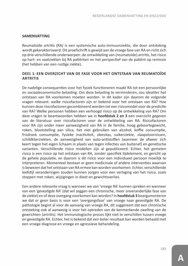
NEDERLANDSE SAMENVATTING EN DISCUSSIE
183
A
SAMENVATTING
Reumatoïde artritis (RA) is een systemische auto-immuunziekte, die door ontsteking wordt gekarakteriseerd. Dit proefschrift is gewijd aan de vroege fase van RA en richt zich op drie verschillende onderwerpen: de ontwikkeling van (reumatoïde) artritis, het risico op hart- en vaatziekten bij RA patiënten en het perspectief van de patiënt op remissie (het hebben van een rustige ziekte).
DEEL 1: EEN OVERZICHT VAN DE FASE VOOR HET ONTSTAAN VAN REUMATOÏDE ARTRITIS
De nadelige consequenties voor het fysiek functioneren maakt RA tot een persoonlijke en sociaaleconomische belasting. Om deze belasting te verminderen, zou idealiter het ontstaan van RA voorkomen moeten worden. In dit kader zijn daarom de volgende vragen relevant: welke risicofactoren zijn er bekend voor het ontstaan van RA? Hoe kunnen deze risicofactoren gecombineerd worden tot een risicomodel voor de predictie van RA? Welke personen hebben een verhoogd risico op de ontwikkeling van RA? Om deze vragen te beantwoorden hebben we in hoofdstuk 2 en 3 een overzicht gegeven van de literatuur over risicofactoren voor de ontwikkeling van RA. Risicofactoren voor RA zijn onder meer aanwezigheid van RA in de familie, hoog geboortegewicht, roken, blootstelling aan silica, het niet gebruiken van alcohol, koffie consumptie, frisdrank consumptie, fysieke inactiviteit, obesitas, suikerziekte, slaapstoornissen, schildklierziekten, de aanwezigheid van auto-antistoffen (wanneer de afweer zich keert tegen het eigen lichaam in plaats van tegen infecties van buitenaf) en genetische varianten. Verschillende risico modellen zijn al gepubliceerd. Echter, het gemeten risico is een risico op het ontstaan van RA, zonder specifiek tijdelement, en gericht op de gehele populatie, en daarom is dit risico voor een individueel persoon moeilijk te interpreteren. Momenteel bestaan er geen medicinale of andere interventies waarvan is bewezen dat het ontstaan van RA ermee kan worden voorkomen. Echter, verschillende leefstijl veranderingen zouden kunnen zorgen voor een verlaging van het risico, zoals stoppen met roken, wijzigingen in dieet en gewichtsverlies.
Een andere relevante vraag is wanneer we van ‘vroege RA’ kunnen spreken en wanneer van een ‘gevestigde RA’ (dat wil zeggen een chronische, meer onveranderlijke fase van de ziekte) en of deze overgang voorkomen kan worden? In hoofdstuk 3 beargumenteren we dat er geen basis is voor een ‘overgangsfase’ van vroege naar gevestigde RA. De pathologie begint al voor de aanvang van vroege RA, dit suggereert dat een chronische ontsteking ook al aanwezig is voor het optreden van de kenmerkende zwelling van de gewrichten (artritis). Het immunologische proces lijkt niet te verschillen tussen vroege en gevestigde RA. Echter, het is bekend dat een beter resultaat kan worden behaald met een vroege diagnose en vroege en agressieve behandeling.
APPENDICES
184
DEEL II: CARDIOVASCULAIR RISICO BIJ VROEGE REUMATOÏDE ARTRITIS
In deel 2 ligt de focus op hart- en vaatziekten oftewel cardiovasculaire (CV) ziekten, een veel voorkomende en bijkomende aandoening in RA patiënten. De ontsteking treft namelijk niet alleen de gewrichten, maar ook het cardiovasculaire systeem. In hoofdstuk 4 is de aanwezigheid van een ongunstige lichaamssamenstelling geanalyseerd in vroege artritis patiënten en vergeleken met de algemene bevolking. Daarvoor hebben we 317 dual-energy x-ray absorptiometry (DXA) scans, van patiënten op het moment van de diagnose artritis, vergeleken met 1268 scans van voor leeftijd, geslacht en etniciteit gematchte personen uit de algemene bevolking. Een verlies in spiermassa welke soms wordt gecompenseerd met een toename in (abdominale) vetmassa, waren weliswaar zeldzaam, echter kwam dit 4 tot 5 keer vaker voor bij vroege artritis patiënten in vergelijking met de algemene bevolking. Verder was in vroege artritis patiënten een ongunstige lichaamssamenstelling geassocieerd met een hogere bloeddruk en een ongunstig cholesterol profiel, wat in combinatie leidt tot een verhoogd cardiovasculair risico. Er werd geen relatie gevonden met ziekteactiviteit of acute fase eiwitten (een maat voor ontsteking in het bloed). Dit zou verklaard kunnen worden door het kleine aantal vroege artritis patiënten met een lage spiermassa, en doordat één ziekteactiviteit meting niet representatief is voor de mate van (subklinische) ontsteking in de voorafgaande periode voor het ontstaan van de artritis.
In hoofdstuk 5 is het voorkomen van hartgeleidingsstoornissen in vroege artritis patiënten gemeten, evenals de relatie met ontsteking en traditionele CV risicofactoren. Omdat het risico op plotselinge hartdood verhoogd is in RA patiënten, hebben we, door middel van een elektrocardiogram, bepaald of de prevalentie van geleidingsstoornissen al verhoogd was op het moment van de diagnose artritis. Van de 480 vroege artritis patiënten had 12.5% een geleidingsstoornis. Deze prevalentie is gelijk aan de prevalentie in de algemene bevolking. De overeenkomst in prevalentie kan verklaard worden door de totale tijd dat er blootstelling aan ontsteking heeft plaats gevonden. Vroege artritis patiënten zijn voor een kortere periode blootgesteld, en het zou kunnen dat een langere blootstelling aan ontsteking geleidingsstoornissen veroorzaakt (vooral QTc verlenging). Geleidingstijden op het tijdstip van de diagnose waren niet geassocieerd met ontstekingsparameters en dit veranderde niet na een jaar anti-reumatische behandeling. Echter, hogere inflammatoire waarden waren geassocieerd met een toename in hartslag, een hogere bloeddruk en een ongunstig cholesterol profiel. Een hogere bloeddruk en een ongunstig cholesterol profiel waren ook geassocieerd met een toegenomen QRS tijd. CV risicofactoren verbeterden na anti-reumatische behandeling. Vooral de verbetering in hartfrequentie die samenhing met de verbetering in ziekteactiviteit was opvallend. Daarom moet de focus in vroege artritis patiënten liggen op zowel CV risico management als op het optimaliseren van de anti-inflammatoire behandeling.
NEDERLANDSE SAMENVATTING EN DISCUSSIE
185
A
Vervolgens bepaalden we de rol van anti-reumatische behandeling op het CV risico, de traditionele risicofactoren en de indicatie voor preventieve CV behandeling. Hiervoor hebben we in hoofdstuk 6 het CV risico retrospectief bepaald, volgens twee verschillende CV risico scores (Dutch Systemic Coronary Risk Evaluation (SCORE) en de European HeartSCORE, beide gecorrigeerd voor RA patiënten). Deze scores hebben we berekend vóór en vier weken na de start van anti-reumatische behandeling in 104 vroege RA patiënten. Explorerende analyses zijn uitgevoerd om het effect te bepalen van inflammatie op CV risico scores en de relatie tussen inflammatie, CV risico scores en de verschillende componenten van de risico scores. In totaal had 7% van de RA patiënten al een CV ziekte doorgemaakt op het moment van de diagnose RA. De mate van overeenkomst tussen de twee verschillende CV risico modellen was gering: waar de Dutch SCORE 30% van de patiënten als hoog risico classificeerde, classificeerde de European HeartSCORE slechts 3% als hoog. Volgens de Nederlandse CV risico management richtlijnen hadden alle patiënten met een hoog CV risico classificatie een indicatie voor (wijzigingen in) preventieve CV behandeling. Na start van de anti-reumatische behandeling verbeterden het cholesterol profiel en de bloeddruk. In totaal had 13% een ander advies na 4 weken anti-reumatische behandeling ten aanzien van preventieve CV behandeling. Resultaten betreffende de associatie tussen ziekteactiviteit en CV risico scores zijn niet conclusief. Maar een hoger totaal cholesterol (TC): high-density lipoproteïne (HDL) ratio (wat een toegenomen CV risico impliceert) was geassocieerd met een hogere ziekteactiviteit. Daarom bevelen wij aan om het CV risico te meten op een moment van lage ziekteactiviteit.
DEEL III: PATIËNT-GERAPPORTEERDE UITKOMSTEN EN REMISSIE BIJ VROEGE REUMATOÏDE ARTRITIS
Het doel van de behandeling van RA patiënten is om een lage ziekteactiviteit te bereiken of zelfs remissie. Er bestaan echter veel verschillende definities voor remissie. Daarom hebben we in hoofdstuk 7 het aantal patiënten bepaald die een goede respons of remissie behaalden na 3 maanden anti-reumatische behandeling, volgens verschillende definities. We vonden dat de overeenkomst tussen het aantal patiënten dat respons of remissie bereikten erg varieerde tussen de verschillende definities. 64% van de 84 RA patiënten, waren het met de arts eens over de aanwezigheid van remissie. Om te bepalen waarom sommige patiënten het niet met de arts over remissie eens waren, hebben we de klinische uitkomsten en de patiënt-gerapporteerde uitkomsten gemeten in patiënten die het wel en niet met de arts eens waren. Het resultaat liet zien dat patiënten die zelf vonden dat ze niet in remissie waren, minder verbeterden op onderdelen van de Rheumatoid Arthritis Impact of Disease (RAID) vragenlijst, vooral op de vragen over pijn, slaap en emotioneel welbevinden. Dit samen met de variatie in klinische respons en remissie definities, wijzen ons erop dat niet alle aspecten van remissie gemeten worden met de gangbare instrumenten en dat we patiënten moeten betrekken bij beslissingen over de zorg voor hun gezondheid.
APPENDICES
186
Eerdere literatuur heeft drie thema’s geïdentificeerd over patiënt-gerapporteerde remissie, waarvan ‘de terugkeer naar normaliteit’ een belangrijk thema was. Hoewel een algemene definitie van de normale toestand ofwel ‘normaliteit’ niet is omschreven in de literatuur, is er wel een ‘normaliteit schaal’ ontwikkeld. In hoofdstuk 8 is gekeken naar wat patiënten als normaliteit en remissie beschouwen en is er gekeken naar de mogelijkheid van de normaliteit schaal om onderscheid te maken tussen remissie en niet-remissie. Hiervoor zijn 47 RA patiënten gevolgd gedurende de eerste zes maanden na start van een op het bereiken van remissie gerichte anti-reumatische behandeling. De ACR/EULAR Boolean remissie criteria en de normaliteit schaal zijn bepaald vóór, na 13 weken en na 26 weken behandeling. De perceptie van normaliteit nam met 20% toe na behandeling. Patiënten die zelf vonden dat ze in remissie waren hadden een hoger gevoel van normaliteit op week 13 en week 26, terwijl patiënten in ACR/EULAR Boolean remissie alleen een hoger gevoel van normaliteit hadden op week 26. Deze studie liet zien dat de normaliteit schaal onderscheid kan maken tussen patiënten in en niet in remissie.
DISCUSSIE, CONCLUSIE EN TOEKOMST PERSPECTIEVEN
Uit deel I van dit proefschrift kunnen we concluderen dat er vele risicofactoren zijn, met een variabele invloed, op de ontwikkeling van RA, waarvan de meeste op zichzelf een laag risico geven. Het is belangrijk om te realiseren dat er momenteel geen bewezen interventie bestaat, medicinaal of anderszins, om RA te kunnen voorkomen in de fase waarin mensen een verhoogd risico lopen op RA. Echter, primaire preventie van RA is een belangrijk en veel bestudeerd onderwerp, wij kijken dan ook uit naar de resultaten van verschillende medicatie trials, zoals de STAtins To Prevent Rheumatoid Arthritis (STAPRA) studie, Arthritis Prevention in the Pre-Clinical Phase of RA with Abatacept (APIPPRA) studie, TREAT EARLIER study (met methotrexaat and intramusculaire corticosteroiden) en de Strategy to Prevent the Onset of Clinically-Apparent Rheumatoid Arthritis (StopRA) studie, waarin geprobeerd wordt RA te voorkomen met hydroxychloroquine. Totdat primaire preventie is ontwikkeld, is het belangrijk dat patiënten zo vroeg mogelijk gediagnosticeerd worden met hun ziekte, zodat een snelle start van anti-reumatische therapie kan plaatsvinden.
Deel II liet zien dat artritis patiënten op het moment van de diagnose al een ongunstige lichaamssamenstelling hebben in vergelijking met de algemene bevolking. Voor toekomstig onderzoek zou het interessant zijn om de lichaamssamenstelling te bepalen vóór het ontstaan van artritis, dat wil zeggen bij personen met een verhoogd risico op artritis, om zo te kunnen bepalen wanneer deze ongunstige samenstelling ontstaat en om te bepalen of dit mogelijk voorkomen kan worden. Ook is het belangrijk om te onderzoeken of de ongunstige lichaamssamenstelling, die aanwezig is op het moment van de diagnose artritis, genormaliseerd kan worden. Verder was de prevalentie van hartgeleidingsstoornissen ten tijde van de diagnose niet verhoogd, maar het zou interessant zijn om deze patiënten over de tijd te volgen om te zien of
NEDERLANDSE SAMENVATTING EN DISCUSSIE
187
A
zij geleidingsstoornissen ontwikkelen naarmate de tijd van blootstelling aan ontsteking toeneemt. Merkwaardig was het verschil van 7 hartslagen per minuut tussen de patiënten met de kleinste versus de grootste verbetering in ziekteactiviteit. Op populatie niveau zou dit een gemiddelde tien-jaars CV mortaliteit risico verschil opleveren van 24% tussen geen of minimale versus substantiële verbetering in ziekte-activiteit gemeten met de Disease Activity Score (DAS).
Het laatste hoofdstuk liet zien dat verschillende CV risico resultaten verkregen werden, afhankelijk van welk CV risico predictie model werd gebruikt. Daarom is vervolg onderzoek noodzakelijk om te bepalen welk CV risico screening model optimaal is voor RA patiënten. Interessant zou zijn om over tien jaar te bepalen welk CV risico model het beste de CV gebeurtenissen voorspelde in het huidige cohort en tevens of aanvullende correcties voor RA geassocieerde factoren dit risico model nog verder kan verbeteren. Een andere belangrijke conclusie is dat het advies voor preventieve CV risico behandeling, volgens de Dutch SCORE en risico management richtlijnen, erg varieerde vóór en na vier weken behandeling met anti-reumatica. In het algemeen zouden RA patiënten op de lange termijn van hun ziekte in remissie moeten zijn of in ieder geval een minimale ziekteactiviteit hebben bereikt. Daarom lijkt het rationeel om CV risico management uit te voeren op een moment van lage ziekteactiviteit. Onze bevindingen laten zien dat het CV risico management niet op het moment van de diagnose uitgevoerd moet worden, maar minimaal vier weken na de start van anti-reumatische behandeling. Om het ideale tijdsmoment voor CV risico management verder te optimaliseren is verder onderzoek noodzakelijk. Bijvoorbeeld, de huidige literatuur is tegenstrijdig over het effect van prednisolon op het lipiden profiel. Daarom zou een optimaler moment een situatie kunnen zijn met lage ziekteactiviteit en na het staken van de prednisolon, maar dit moet nog bepaald worden.
Deel III demonstreert dat het moeilijk is om een definitie voor remissie te ontwikkelen, die alle aspecten van remissie bevat. Veel verschillende definities bestaan, en de hoeveelheid patiënten die aan de verschillende definities voldoet verschilt erg. Na drie maanden behandeling was een-derde van de patiënten het niet eens met de arts over het al of niet behaald hebben van remissie. Patiënten nemen andere domeinen van ziekteactiviteit mee in hun beoordeling (zoals pijn, slaap en emotioneel welbevinden) dan artsen. Patiënt-tevredenheid, de relatie tussen patiënt en arts, en therapietrouw kunnen alle verbeterd worden wanneer de patiënt en de arts het eens zijn over de status van de ziekte. Daarom is het belangrijk om patiënten meer te betrekken bij beslissingen rondom hun gezondheid, en zo gedeelde besluitvorming te verbeteren. Door de arts en door de patiënt waargenomen remissie kwamen het beste overeen met een goede respons volgens de European League Against Rheumatism (EULAR). Echter is verder onderzoek noodzakelijk om te bepalen welke definitie van respons of remissie het beste is om te gebruiken in de dagelijkse praktijk en welke hiervan een goede uitkomst voorspelt, zonder patiënten bloot te stellen aan overbehandeling. Mogelijk zijn er nieuwe definities van remissie, met meer door de patiënt gerapporteerde
APPENDICES
188
uitkomstmaten, die eerder in het ziekteproces toegepast kunnen worden of een betere uitkomst van de ziekte kunnen bereiken. Een goede kandidaat zou de normaliteit schaal kunnen zijn, aangezien patiënten de terugkeer naar normaliteit als één van de drie belangrijkste domeinen van remissie beschouwen. Tevens is de normaliteit schaal in staat om het onderscheid te maken tussen remissie en niet-remissie. De waarneming van normaliteit nam toe na het starten van anti-reumatische behandeling. De definitie van remissie volgens de patiënt is erg belangrijk, daarom zou het interessant zijn om te bepalen of het normaliteit gevoel verder toeneemt over de tijd en of de normaliteit schaal in staat is om ziekteactiviteit te voorspellen, en op deze manier zelfs de DAS zou kunnen vervangen.
DANKWOORD
189
A
DANKWOORD
Veel mensen hebben bijgedragen aan dit proefschrift en verdienen daarom een eervolle vermelding.
Klinisch onderzoek is niet mogelijk zonder deelnemers, daarom wil ik als eerste alle patiënten bedanken die aan verschillende onderzoeken wilden deelnemen. Ondanks dat ze vaak net de diagnose reuma hadden gekregen, waren er velen bereid om ook nog het wetenschappelijk onderzoekgedeelte aan te horen en om deel te nemen, hartelijk dank! En natuurlijk alle reumatologen, AIOS en ANIOS die patiënten doorverwezen voor wetenschappelijk onderzoek, bedankt voor de verwijzingen.
Ook wil ik graag mijn promotoren en copromotor bedanken. Promotor Prof. dr. D. van Schaardenburg, beste Dirkjan, wat was ik blij dat jij me een baan aanbood, wel op een ander project, aangezien de vacature waarvoor ik gesolliciteerd had al vervuld was. Nadat de inhoud van mijn boekje een aantal keer was gewijzigd, konden we er ons vol op storten. De wekelijkse gesprekjes en de wekelijkse overleggen hebben ertoe geleid dat mijn promotietraject vlot werd afgerond. Hopelijk is er in de toekomst ook nog ruimte voor onderzoek, er zijn genoeg projecten! Prof. dr. M.T. Nurmohamed, beste Mike, mijn tweede promotor, bedankt voor de mogelijkheid om onderzoek te doen op het cardiovasculaire gebied. Je hebt me voor dit onderwerp enthousiast gemaakt! Het woord ‘consecutive’, hetgeen wat jij in ieder van mijn artikelen hebt toegevoegd, is denk ik heel toepasselijk. De hoeveelheid opeenvolgende studies waar jij je mee bezig hield was verbazingwekkend, onbegrijpelijk hoe je dit allemaal kan combineren! Copromotor dr. W.F. Lems, beste Willem, de derde professor op rij. Bedankt voor je nuchtere blik en je positieve instelling, ik vond het een erg prettige samenwerking.
Ook de leescommissie wil ik bedanken voor het beoordelen van dit proefschrift: prof. Dr. R.F. van Vollenhoven, prof. Dr. R.B.M. Landewé, prof. Dr. K. Raza, prof. Dr. S. Middeldorp, prof. Dr. R.J.G. Peters, dr. W.H. Bos en prof. Dr. Y.M. Smulders, hartelijk dank.
Promotie is onmogelijk zonder hulp van andere afdelingen. Daarom wil ik als eerste graag de verpleegkundigen bedanken, hartelijk dank voor jullie inzet! Met name Véronique, Sylvia en Marjolein wil ik bedanken voor het includeren van alle ‘Early Arthritis Patiënten’ (later zorgpad artritis) en natuurlijk alle COBRA-bezoeken. Verder Maaike en Marja voor alle STAPRA-patiënten, hopelijk lukt het om spoedig veel patiënten te includeren. En natuurlijk de verpleegkundigen (Dorien, Astrid, Judith, Marjolein, Marja, Martine, Maaike, Sylvia) op de dagbehandeling: het was gezellig. Wilfred bedankt voor de vele overleggen over de patiënten op de dagbehandeling, fijn dat je daar altijd tijd voor wist vrij te maken. Daarnaast wil ik alle secretaresses bedanken, vooral Ada, Afra, Brigitta, Kavyo en Simone, voor het eindeloos mappen voorbereiden, patiënten bellen en afspraken maken; zonder jullie lukte het niet! Hetzelfde geldt voor alle hartfilmpjes die altijd op kamer A9 gemaakt konden worden, dankjewel. En Linda voor het invoeren

APPENDICES
190
en drs. J. Dekker voor het beoordelen van al die hartfilmpjes. En de apotheek, Renske en Judith, bedankt voor alle COBRA-medicatie en natuurlijk de gezelligheid in de apotheek! Graag bedank ik ook de radiologie afdeling, alle röntgenfoto’s en DXA-scans, Nazan, Ilse, Yvonne en Cindy, bedankt voor jullie geduld.
Collega-onderzoekers, bedankt voor jullie input en gezelligheid! Sjoerd en Maaike, bedankt voor jullie cardiovasculaire ondersteuning. Jill, je was een gezellige buuf. Willem, je nuchtere mannelijke blik werd gewaardeerd. Leonieke, bedankt voor de gezelligheid, jammer dat je meestal op het AMC was. Ingrid, bedankt voor je statistische en database hulp. Laurette, hopelijk heb ik alles overgedragen, het gaat je zeker lukken je krijgt een mooi boekje. Hetzelfde geldt natuurlijk voor Annelies, Daisy en Wendy! Linda en Nicki van het VUmc, bedankt voor de COBRA samenwerking. Na wat hobbels hebben wij een mooie studie (bijna) afgerond! Maaike, Merel, Erik en Milad, bedankt voor de hardloopsessies iedere dinsdag en vrijdag, goed voor de afleiding en om even te kunnen ‘sparren’. Zet het voort! De collega’s die de promotie eerder hadden afgerond; Charlotte, Inge, Alper, Eva, jullie waren een voorbeeld! De biobank; Toni en Corrie, bedankt voor het verwerken van alle samples. De researchafdeling: Remke en Ellen, bedankt voor de gezelligheid.
Voor alle statistische problemen wil ik graag Maarten Boers, Jos Twisk en Marieke ter Wee bedanken. Maarten, we hebben geen makkelijke artikelen gehad, bedankt voor je rust en je goede adviezen. Jos bedankt voor het onderwijs en je Stata hulp, ik kan nu zeggen dat ik het begrijp! Marieke bedankt voor het PRO artikel, het artikel dat het snelste en vlotste is voltooid!
Buiten Reade zijn er ook nog veel mensen die ik wil bedanken. Uiteraard mijn vriendinnen van geneeskunde: Diane, Tamara, Michelle, Nina, Inge en Suk-Yee. Bedankt voor de gezellige uitjes en dat ik bij jullie mijn hart kon luchten. Vriendinnen van de middelbare school: Noortje, Birgit, Danielle, Daphne, Bianca, Lidewij en Anouk. Ik ben blij dat we elkaar nog steeds regelmatig zien, bedankt voor de nodige afleiding. Dear Chami, I am glad you became my neighbor at Reade for your internship. Unfortunately, you needed to go back to Madrid, but I am sure we will see each other on a regular bases (congresses are a good reunion). Meiden van dames 3 van Qui Vive, wat fijn dat we nog steeds een team zijn en iedere week gezellig hockeyen, bedankt! De ‘boodschappen-club’, Marja, Sara, Alie en Myron, iedere vrijdagavond door de supermarkten en daarna thee met taart, altijd een goede (en nuttige) afsluiting van de week! De yoga-club, fijn dat er ook af en toe ontspanning was.
DANKWOORD
191
A
Mijn paranimf en mijn back-up: Marian. Bedankt voor je samenwerking, je statistische kennis en je rust. Twee kinderen en een promotie, ik weet niet hoe je het hebt gedaan; maar wauw! Hopelijk komt die opleiding tot kinderarts er snel. En Inge, paranimf, hartelijk dank voor je steun! De uitjes en bruitlofts app waren erg fijn om af en toe te kunnen ‘luchten’. Inmiddels allebei getrouwd, en hopelijk binnenkort allebei gepromoveerd: je kan het!
Tot slot wil ik nog de belangrijkste mensen bedanken, mijn familie. Alle verjaardagen en feestjes die gevierd werden waren een welkome afleiding. Ondanks de omvang van de familie zien we elkaar regelmatig, ik besef hoe kostbaar dat is. Myron en Ronald, ik vergeet jullie trouwuitnodiging over de app nooit, nu een mooi gezin met Imme en Freek erbij. Papa en mama, inmiddels met pensioen, maar niet minder druk. Altijd in voor leuke activiteiten en een 24/7 (klus)hulp die paraat staat. Nieuwe meubels, ramen lappen, ontplofte tuinslang of een accu die het niet zou doen, ik waardeer jullie hulp en het vertrouwen dat jullie in mij hebben. Uiteraard ook mijn schoonouders, Pieter en Irene, bedankt voor jullie interesse en de leuke tussendoor uitjes.
Lieve Bart, mijn promotietraject was een grote zoektocht, maar het allerbelangrijkste wat ik gevonden heb ben jij! Dank voor je luisterend oor en steun tijdens dit traject. We hebben inmiddels al vele mooie reizen gemaakt, een huis gekocht en zijn zelfs getrouwd, ik kijk uit naar de vele jaren die nog volgen!

PHD PORTFOLIO
193
A
PHD PORTFOLIO
Name PhD Student S.A. TurkPhD period sept 2013- sept 2017Names PhD supervisors Prof. dr. D. van Schaardenburg and prof. dr. M.T. NurmohamedCoursesGood Clinical Practice (GCP) 2014Sharp van der Heijde scoring method 2014Meldcode huiselijk geweld en kindermishandeling 2014EpidM: Principes van epidemiologische data-analyse 2014EpidM: regressietechnieken 2014EpidM: longitudinale data-analyse 2015Praktisch projectmanagement 2016Basis cursus Regelgeving en Organisatie Klinisch Wetenschappelijk Onderzoek (BROK)
2017
Presentations14-3-3η is an independent predictor of radiographic changes in early RA, EULAR Annual European Congress of Rheumatology in Paris, France (poster presentation)
2014
93% of early RA patients are positive for 14-3-3 η markers and 14-3-3 η auto-antibodies inform a favourable prognosis, irrespective of RF or ACPA status, EULAR Annual European Congress of Rheumatology in Paris, France (poster presentation)
2014
Conduction disorders & heart rate in early rheumatoid arthritis and the effects of anti-inflammatory treatment thereon, EULAR Annual European Congress of Rheumatology in Rome, Italy (poster presentation)
2015
Determination of the lipid profile in active disease leads to incorrect cardiovascular risk predication in early rheumatoid arthritis patients, EULAR Annual European Congress of Rheumatology in London, UK (poster presentation)
2016
Determination of the lipid profile in active disease leads to incorrect cardiovascular risk predication in early rheumatoid arthritis patients, ACR/ARHP Annual Scientific Meeting in Washington, USA (poster presentation)
2016
Glucocorticoid treatment for 12 Weeks in early rheumatoid arthritis is related to an increase in BMI, ACR/ARHP Annual Scientific Meeting in Washington, USA (poster presentation)
2016
Physician, but not patients, agree that EULAR good response indicates remission after 12 weeks treatment of early rheumatoid arthritis, ACR/ARHP Annual Scientific Meeting in Washington, USA (poster presentation)
2016
Unfavorable body composition already at the onset of clinical arthritis, EULAR Annual European Congress of Rheumatology in Madrid, Spain (poster presentation)
2017
Unfavorable body composition already at the onset of clinical arthritis, NVR (Nederlandse Vereniging voor Reumatologie) Najaarsdagen in Arnhem, the Netherlands (oral presentation)
2017
APPENDICES
194
Patient reported outcomes explain the lack of agreement between physician and patient perceived remission in early rheumatoid arthritis, NVR (Nederlandse Vereniging voor Reumatologie) Najaarsdagen in Arnhem, the Netherlands (oral presentation)
2017
ConferencesEULAR Annual European Congress of Rheumatology in Paris, France 2014NVR (Nederlandse Vereniging voor Reumatologie) Najaarsdagen in Arnhem, the Netherlands
2014
EULAR Annual European Congress of Rheumatology in Rome, Italy 2015NVR (Nederlandse Vereniging voor Reumatologie) Najaarsdagen in Arnhem, the Netherlands
2015
ACR/ARHP Annual Scientific Meeting in San Francisco, USA 2015EULAR Annual European Congress of Rheumatology in London, UK 2016ACR/ARHP Annual Scientific Meeting in Washington, USA 2016EULAR Annual European Congress of Rheumatology in Madrid, Spain 2017NVR (Nederlandse Vereniging voor Reumatologie) Najaarsdagen in Arnhem, the Netherlands
2017
Seminars and workshopsWeekly department research seminars 2013-2017LEAN workshop 2016
LecturingDyslipidemia in the pathogenesis of RA. Can RA be prevented?, symposium cardiovascular rheumatology, Amsterdam, The Netherlands (oral presentation)
2015
Supervising L. Britsemmer, conduction disorders in early RA, Rheumatology 2014
CURRICULUM VITAE
195
A
CURRICULUM VITAE
Samina Turk, auteur van dit proefschrift, werd geboren op 27 april 1989, in De Kwakel. Zij behaalde in 2007 haar vwo-diploma, met het profiel ‘natuur en gezondheid’ en ‘natuur en techniek’, aan het Alkwin Kollege in Uithoorn. Waarna zij startte zij met de studie geneeskunde aan het VU medisch centrum te Amsterdam en in 2010 haar Bachelor of Science behaalde. Vervolgens ging zij voor een half jaar naar Edmonton, Canada om onderzoek te doen naar de ziekte van Crohn, als wetenschappelijke stage. Om in 2011 haar co-schappen te continueren, waarvoor zij haar huisartsenstage voltooide in Boyup Brook, Australië en een semi-arts stage deed op de MDL-afdeling van het Kennemer Gasthuis. In 2013 haalde zij haar Master of Science en startte haar promotietraject op de reumatologie afdeling van Reade (inmiddels het Amsterdam Rheumatology & immunology Center | Reade) onder leiding van Prof. dr. D. van Schaardenburg, Prof. dr. M.T. Nurmohamed en Prof. dr. W.F. Lems. Hier heeft ze de STAPRA studie (STAtins to Prevent Rheumatoid Arthritis) opgezet en samen met het VUmc de COBRA T2T opgestart. In haar proefschrift heeft zij zich gericht op de ontwikkeling van vroege artritis, het risico op cardiovasculaire ziekten in reumatoïde artritis (RA) patiënten en het behalen van remissie vanuit het perspectief van de RA patiënt. Op verschillende congressen heeft ze de resultaten van dit onderzoek mogen presenteren. Dit werk heeft geleid tot het proefschrift dat voor u ligt.
Op 1 november 2017 startte zij in het kader van de opleiding tot reumatoloog met de vooropleiding interne geneeskunde in het Spaarne Gasthuis.

LIST OF PUBLICATIONS
197
A
List of publications
Turk SA, Rasch LA, van Schaardenburg D, Lems WF, Sanberg M, van Tuyl LHD, ter Wee MM. Pain, sleep and emotional well-being explain the lack of agreement between phy-sician- and patient-perceived remission in early rheumatoid arthritis. BMC rheumatolo-gy.2018. doi: 10.1186/s41927-018-0024-9.
Turk SA, Heslinga M, Twisk J, van der Lugt V, Lems WF, van Schaardenburg D, Nurmo-hamed MT.Letter: Change in cardiovascular risk after initiation of anti-rheumatic treat-ment in early rheumatoid arthritis. Clin and exp Rheumatology 2018; accepted for pub-lication.
Turk SA, van Schaardenburg D, Boers M, de Boer S, Fokker C, Lems WF, Nurmohamed MT. An unfavorable body composition is common in early arthritis patients: a case con-trol study. PLoS one 2018;13(3):e0193377.
Turk SA, Heslinga SC, Dekker J, Britsemmer L, van dL, V, Lems WF, van Schaardenburg D, Nurmohamed MT. Conduction Times, Cardiovascular Risk Factors, and Inflammation in Patients with Early Arthritis.J Rheumatol 2017 May;44:580-6.
Teshima CW, Goodman KJ, El-Kalla M, Turk S, El-Matary W, Valcheva R, Danchak R, Gor-don M, Ho P, Mullins A, Wong D, Kao D, Meddings J, Huynh H, Dieleman LA. Increased Intestinal Permeability in Relatives of Patients With Crohn’s Disease Is Not Associated With Small Bowel Ulcerations.Clin Gastroenterol Hepatol 2017 Sep; 15(9):1413-18.
Chin Jen Sem JP, van der Leeden M, Visscher CM, Britsemmer K, Turk SA, Dekker J, van Schaardenburg D, Lobbezoo F. Prevalence, Course, and Associated Factors of Pain in the Temporomandibular Joint in Early Rheumatoid Arthritis: Results of a Longitudinal Cohort Study.J Oral Facial Pain Headache 2017;31:233-9.
Lubbers J, van Beers-Tas MH, Vosslamber S, Turk SA, de RS, Mantel E, Wesseling JG, Reijm M, van Hoogstraten IM, Bijlsma JW, van Schaardenburg D, Bontkes HJ, Verwij CL.Changes in peripheral blood lymphocyte subsets during arthritis development in arthralgia pa-tients. Arthritis Res Ther 2016 Sep;18:205.
de Jong TD, Lubbers J, Turk S, Vosslamber S, Mantel E, Bontkes HJ, van der Laken CJ, Bi-jlsma JW, van Schaardenburg D, Verweij CL. The type I interferon signature in leukocyte subsets from peripheral blood of patients with early arthritis: a major contribution by granulocytes. Arthritis Res Ther 2016 Jul;18:165.
APPENDICES
198
Maksymowych WP, Boire G, van SD, Wichuk S, Turk S, Boers M, Siminovitch KA, Bykerk V, Keystone E, Tak PP, van Kuijk AW, Landewé R, van der Heijde D, Murphy M, Marotta A. 14-3-3eta Autoantibodies: Diagnostic Use in Early Rheumatoid Arthritis.J Rheumatol 2015 Sep;42(9):1587-94.
Plasencia C, Kneepkens EL, Wolbink G, Krieckaert CL, Turk S, Navarro-Compan V, L’Ami M, Nurmohamed MT, van der Horst-Bruinsma I, Jurado T, Diego C, Bonilla G, Villalba A, Peiteado D, Nuno L, van der Kleij D, Rispens T, Martin-Mola E, Balsa A, Pascual-Salcedo D. Comparing Tapering Strategy to Standard Dosing Regimen of Tumor Necrosis Factor Inhibitors in Patients with Spondyloarthritis in Low Disease Activity. J Rheumatol 2015 Sep;42:1638-46.
van Beers-Tas MH, Turk SA, van Schaardenburg D. How does established rheumatoid ar-thritis develop, and are there possibilities for prevention? Best Pract Res Clin Rheumatol 2015 Aug;29:527-42.
Turk SA, van Beers-Tas MH, van Schaardenburg D.Prediction of future rheumatoid arthri-tis. Rheum Dis Clin North Am 2014 Nov;40:753-70.
R. Agca, S. Turk, D. Jansen, F. Turkstra, M. Gerritsen, M.T. Nurmohamed.Statine stoppen bij start colchicine? NTvR 2014 June;17:6-13.
Submitted for publicationRasch LA, Turk SA, Boers M, Lems WF, van Schaardenburg D, van Tuyl LHD. Do patients with rheumatoid arthritis in remission feel normal again?


THE EVOLUTION OF EARLY ARTHRITIS AND
CARDIOVASCULAR RISKgenetic
re
produ
ctive
envi
ronme
ntal subclinical
inflammationauto-
immun
ity
arthralgia
infections
hormonal
Samina A. Turk
Samina A. Turk
THE EVOLU
TION
OF EARLY ARTHRITIS AN
D CARDIOVASCU
LAR RISK
UITNODIGING
Voor het bijwonen van de openbare verdediging van het
proefschrift:
THE EVOLUTION OF EARLY ARTHRITIS AND
CARDIOVASCULAR RISK
Donderdag 10 januari 2019 om 10.00 uur
In de Agnietenkapel van de Universiteit van Amsterdam
Oudezijds Voorburgwal 231 te Amsterdam
Na afloop van de promotie is er een receptie ter plaatse
Samina A. Turk
Paranimfen
Marian van Beers-Tas06-617188447
Inge van Klink- de Kruijff06-39571477