Keli Xu NIH Public Access Author Manuscript Amanda J. Loch...
24
Lunatic Fringe Deficiency Cooperates with the Met/Caveolin Gene Amplicon to Induce Basal-like Breast Cancer Keli Xu 1,11 , Jerry Usary 3 , Philaretos C. Kousis 1 , Aleix Prat 3 , Dong-Yu Wang 4 , Jessica R. Adams 1,6 , Wei Wang 1 , Amanda J. Loch 1 , Tao Deng 5 , Wei Zhao 3 , Robert Darrell Cardiff 8 , Keejung Yoon 9,12 , Nicholas Gaiano 9 , Vicki Ling 1,2 , Joseph Beyene 2,13 , Eldad Zacksenhaus 5 , Tom Gridley 10 , Wey L. Leong 4 , Cynthia J. Guidos 1,7 , Charles M. Perou 3 , and Sean E. Egan 1,6,* 1 Program in Developmental and Stem Cell Biology, The Hospital for Sick Children, Toronto, ON, M5G 1L7, Canada 2 Child Health Evaluative Sciences, The Hospital for Sick Children, Toronto, ON, M5G 1L7, Canada 3 Lineberger Comprehensive Cancer Center, Departments of Genetics and Pathology, University of North Carolina, Chapel Hill, NC 27599, USA 4 The Campbell Family Cancer Research Institute and Surgical Oncology Princess Margaret Hospital, and the Department of General Surgery University Health Network, Toronto, ON M5S 1A1, Canada 5 Division of Cell and Molecular Biology, Toronto General Research Institute, University Health Network, Toronto, ON M5S 1A1, Canada 6 Department of Molecular Genetics, University of Toronto, Toronto, ON M5S 1A1, Canada 7 Department of Immunology, University of Toronto, Toronto, ON M5S 1A1, Canada 8 Center for Comparative Medicine, University of California, Davis, CA 95616, USA 9 Institute for Cell Engineering, Johns Hopkins University School of Medicine, Baltimore, MD 21205, USA 10 Center for Molecular Medicine, Maine Medical Center Research Institute, 81 Research Drive, Scarborough, ME 04074, USA SUMMARY Basal-like breast cancers (BLBC) express a luminal progenitor gene signature. Notch receptor signaling promotes luminal cell fate specification in the mammary gland, while suppressing stem cell self-renewal. Here we show that deletion of Lfng, a sugar transferase that prevents Notch activation by Jagged ligands, enhances stem/progenitor cell proliferation. Mammary-specific ©2012 Elsevier Inc. * Correspondence: [email protected] DOI 10.1016/j.ccr.2012.03.041. 11 Present address: Cancer Institute, University of Mississippi Medical Center, 2500 North State Street, Jackson, MS 39216, USA 12 Present address: College of Biotechnology and Bioengineering, Sungkyunkwan University, Suwon, Gyeonggi-do 440-746, Republic of Korea 13 Present address: Program in Population Genomics, Department of Clinical Epidemiology and Biostatistics, McMaster University, Hamilton, ON L8S 4L8, Canada ACCESSION NUMBERS Transcriptional profiling data and aCGH data have been deposited in the GEO database (http:// www.ncbi.nlm.nih.gov/geo/; accession numbers GSE28712 and GSE35855, respectively). SUPPLEMENTAL INFORMATION Supplemental Information includes five figures, one table, and Supplemental Experimental Procedures and can be found with this article online at doi:10.1016/j.ccr.2012.03.041. NIH Public Access Author Manuscript Cancer Cell. Author manuscript; available in PMC 2013 May 25. Published in final edited form as: Cancer Cell. 2012 May 15; 21(5): 626–641. doi:10.1016/j.ccr.2012.03.041. NIH-PA Author Manuscript NIH-PA Author Manuscript NIH-PA Author Manuscript
Transcript of Keli Xu NIH Public Access Author Manuscript Amanda J. Loch...

Lunatic Fringe Deficiency Cooperates with the Met/CaveolinGene Amplicon to Induce Basal-like Breast Cancer
Keli Xu1,11, Jerry Usary3, Philaretos C. Kousis1, Aleix Prat3, Dong-Yu Wang4, Jessica R.Adams1,6, Wei Wang1, Amanda J. Loch1, Tao Deng5, Wei Zhao3, Robert Darrell Cardiff8,Keejung Yoon9,12, Nicholas Gaiano9, Vicki Ling1,2, Joseph Beyene2,13, EldadZacksenhaus5, Tom Gridley10, Wey L. Leong4, Cynthia J. Guidos1,7, Charles M. Perou3, andSean E. Egan1,6,*
1Program in Developmental and Stem Cell Biology, The Hospital for Sick Children, Toronto, ON,M5G 1L7, Canada2Child Health Evaluative Sciences, The Hospital for Sick Children, Toronto, ON, M5G 1L7,Canada3Lineberger Comprehensive Cancer Center, Departments of Genetics and Pathology, Universityof North Carolina, Chapel Hill, NC 27599, USA4The Campbell Family Cancer Research Institute and Surgical Oncology Princess MargaretHospital, and the Department of General Surgery University Health Network, Toronto, ON M5S1A1, Canada5Division of Cell and Molecular Biology, Toronto General Research Institute, University HealthNetwork, Toronto, ON M5S 1A1, Canada6Department of Molecular Genetics, University of Toronto, Toronto, ON M5S 1A1, Canada7Department of Immunology, University of Toronto, Toronto, ON M5S 1A1, Canada8Center for Comparative Medicine, University of California, Davis, CA 95616, USA9Institute for Cell Engineering, Johns Hopkins University School of Medicine, Baltimore, MD21205, USA10Center for Molecular Medicine, Maine Medical Center Research Institute, 81 Research Drive,Scarborough, ME 04074, USA
SUMMARYBasal-like breast cancers (BLBC) express a luminal progenitor gene signature. Notch receptorsignaling promotes luminal cell fate specification in the mammary gland, while suppressing stemcell self-renewal. Here we show that deletion of Lfng, a sugar transferase that prevents Notchactivation by Jagged ligands, enhances stem/progenitor cell proliferation. Mammary-specific
©2012 Elsevier Inc.*Correspondence: [email protected] DOI 10.1016/j.ccr.2012.03.041.11Present address: Cancer Institute, University of Mississippi Medical Center, 2500 North State Street, Jackson, MS 39216, USA12Present address: College of Biotechnology and Bioengineering, Sungkyunkwan University, Suwon, Gyeonggi-do 440-746,Republic of Korea13Present address: Program in Population Genomics, Department of Clinical Epidemiology and Biostatistics, McMaster University,Hamilton, ON L8S 4L8, Canada
ACCESSION NUMBERS Transcriptional profiling data and aCGH data have been deposited in the GEO database (http://www.ncbi.nlm.nih.gov/geo/; accession numbers GSE28712 and GSE35855, respectively).
SUPPLEMENTAL INFORMATION Supplemental Information includes five figures, one table, and Supplemental ExperimentalProcedures and can be found with this article online at doi:10.1016/j.ccr.2012.03.041.
NIH Public AccessAuthor ManuscriptCancer Cell. Author manuscript; available in PMC 2013 May 25.
Published in final edited form as:Cancer Cell. 2012 May 15; 21(5): 626–641. doi:10.1016/j.ccr.2012.03.041.
NIH
-PA Author Manuscript
NIH
-PA Author Manuscript
NIH
-PA Author Manuscript
deletion of Lfng induces basal-like and claudin-low tumors with accumulation of Notchintracellular domain fragments, increased expression of proliferation-associated Notch targets,amplification of the Met/Caveolin locus, and elevated Met and Igf-1R signaling. Human BL breasttumors, commonly associated with JAGGED expression, elevated MET signaling, andCAVEOLIN accumulation, express low levels of LFNG. Thus, reduced LFNG expressionfacilitates JAG/NOTCH luminal progenitor signaling and cooperates with MET/CAVEOLINbasal-type signaling to promote BLBC.
INTRODUCTIONPatients with BLBC show reduced survival compared to those with more common luminaltumors, and this disease frequently occurs in young patients, as well as in women withAfrican ancestry. Basal-like tumors express markers of myoepithelium, but show a geneexpression signature related to that of luminal progenitor cells (Cheang et al., 2008; Lim etal., 2009; Perou and Børresen-Dale, 2011; Prat et al., 2010). Indeed, luminal progenitorsmay be the cell-of-origin for most BLBC (Molyneux et al., 2010). In the mouse system,activated Notch1 can induce commitment of mammary stem cells (MaSC) into luminalprogenitors, and promote proliferation of luminal progenitor cells in vitro and in vivo(Bouras et al., 2008). Similarly in humans, increased NOTCH3 expression and function canpromote luminal progenitor cell fate specification, at least in vitro (Raouf et al., 2008).
On the basis of studies with the Mouse Mammary Tumor Virus (MMTV), which can inducemammary tumor formation through insertional activation of Notch genes, a role for Notchsignaling in human breast cancer was anticipated (Callahan and Smith, 2000). Most humanbreast tumors express Notch ligands and receptors (Parr et al., 2004; Pece et al., 2004;Reedijk et al., 2005; Stylianou et al., 2006). High-level expression of the JAGGED1 ligand,as well as NOTCH1 and/or NOTCH3, is associated with poor overall survival (Reedijk etal., 2005). Recent studies reveal that signaling through multiple Notch receptors activatesproliferation and/or survival of breast cancer cells (Harrison et al., 2010; Haughian et al.,2012; Osipo et al., 2008; Sansone et al., 2007; Yamaguchi et al., 2008). Interestingly,JAGGED-dependent Notch pathway activation has been associated with triple negative(ERα−, PR− and HER2−) tumors, and specifically with basal-like tumors and cell lines(Cohen et al., 2010; Dong et al., 2010; Haughian et al., 2012; Lee et al., 2008a, 2008b;Leong et al., 2007; Reedijk et al., 2008; Sansone et al., 2007).
Met, a cell surface tyrosine kinase receptor involved in epithelial-mesenchymal transition, isfrequently expressed at high levels in BLBC (Elsheikh et al., 2008; Gastaldi et al., 2010; Luet al., 2008; Ponzo and Park, 2010; Ravid et al., 2005; Salani et al., 2008; Savage et al.,2007), and many basal-like tumors show elevated Met signaling (Hochgräfe et al., 2010). Inaddition, Caveolin1 and 2, which facilitate Igf-1R signaling, are also overexpressed(Elsheikh et al., 2008; Gastaldi et al., 2010; Lu et al., 2008; Ponzo and Park, 2010; Ravid etal., 2005; Salani et al., 2008; Savage et al., 2007). Interestingly, the genes coding for MET,CAV1, and CAV2 are located in the same region of Chromosome 7q31 and this locus isoverexpressed in many BLBC (Elsheikh et al., 2008; Gastaldi et al., 2010; Ponzo and Park,2010; Savage et al., 2007).
Fringe proteins are N-acetylglucosamine transferases that modify Notch receptors to controlligand-mediated activation (Haines and Irvine, 2003). These proteins enhance Notchactivation by Delta-family ligands, while inhibiting Notch activation by Serrate/Jaggedligands (Haines and Irvine, 2003). Lfng, one of three Fringe genes in mammals, controlsNotch signaling in many developing tissues (Cohen et al., 1997; Johnston et al., 1997).Interestingly, LFNG is expressed at high levels in MaSC and/or bipotent progenitor cells of
Xu et al. Page 2
Cancer Cell. Author manuscript; available in PMC 2013 May 25.
NIH
-PA Author Manuscript
NIH
-PA Author Manuscript
NIH
-PA Author Manuscript

the human breast (Raouf et al., 2008). However, its role in the regulation of Notch signalingof the developing or adult mammary gland remains unknown, as is its potential forrestricting Notch-dependent oncogenic signaling in this context. In this study, we usedconditional mutant mice to define the function of Lfng in mammary epithelium. In addition,we tested for its expression and potential role in human breast cancer.
RESULTSA Lfng Expression Boundary in Mammary Development
To define where and when Notch is activated in the developing mammary gland, we usedLacZ knock-in mice for various Notch pathway genes. Typically, boundaries betweenFringe-expressing cells and nonexpressing cells are sites of consequential Notch signaling(Irvine, 1999). Therefore, we examined Lfng expression by performing X-gal staining onmammary glands from six-week-old LfngLacZ/+ virgin females (Zhang and Gridley, 1998).Interestingly, Lfng expression was restricted to basal cells, in particular to cap cells ofterminal end buds (TEBs) (Figure 1A), which have MaSC activity (Bai and Rohrschneider,2010). This result is consistent with studies on cells purified from the human mammarygland, which show that LFNG expression is > 20-fold enriched in stem and/or bipotentprogenitor cells as compared with luminal restricted progenitors (Raouf et al., 2008). TwoNotch ligands, Dll1 and Jagged1, show distinct expression at this stage. Weak X-gal stainingfrom a Dll1lacZ/+ allele (Hrabĕ de Angelis et al., 1997) was observed in cap cells of themammary TEB. In contrast, their basally located myoepithelial descendants showed intensestaining. X-gal staining in pubescent Jagged1β–Geo/+ (Xu et al., 2010) mice was strongest instromal cells surrounding each TEB (Figure 1A).
In mature adults, after TEB regression, LfnglacZ expression was barely detected, whileDll1lacZ expression remained strong in myoepithelial cells. Interestingly, Jagged1 expressionswitched from stroma to myoepithelium in the mature gland (Figure 1A). JAGGED1expression is also high in basal cells of the mature human gland (Reedijk et al., 2005). Next,we used antibodies to stain for Notch1 and Notch4, which were expressed at low levels inluminal and basal cells, respectively (Figure S1A available online). Using β-Geo/+ knock-inmice (Xu et al., 2010), Notch3 expression was found to be high in body cells of the TEB, aswell as in luminal epithelial cells of mature ducts, whereas Notch2 expression was strong instroma and weak in epithelium (Figure S1B). These data are consistent with recent Notchligand and receptor expression analysis by rtPCR and immunohistochemistry (Bouras et al.,2008; Raafat et al., 2011; Raouf et al., 2008).
Lfng Controls Notch Activation and Suppresses Mammary Epithelial ProliferationFringe controls Notch activation at compartment boundaries in the developing fruit fly(Irvine, 1999). Our expression analysis in the mammary gland reveals a similar boundary atthe TEB-ductal junction, where Lfng may differentially regulate Notch activation inducedby Dll1 and/or Jagged1. To define Lfng function in this context, we analyzed mammarydevelopment in Lfng mutant mice. Whole-mount analysis showed evidence of epithelialhyperplasia in virgin mammary glands from Lfng mutants (Figure 1B). Sections frommutant and control glands were stained with antibodies against luminal and basallyrestricted cytokeratins, keratin 8 (K8) and 14 (K14), respectively. Most mutant glandsshowed decreased K8 expression in body cells of the TEB. Also, multiple layers of K14+
basal cells were observed in some locations. Cells did not co-express K8 and K14 in wild-type or mutant glands during puberty (Figure 1C). We next tested for altered cellproliferation by staining for Ki67. Indeed, Lfng mutant glands showed increased cellproliferation in mature ducts (Figure 1D).
Xu et al. Page 3
Cancer Cell. Author manuscript; available in PMC 2013 May 25.
NIH
-PA Author Manuscript
NIH
-PA Author Manuscript
NIH
-PA Author Manuscript
Fringe proteins control Notch signaling by enhancing Delta-while inhibiting Serrate/Jagged-mediated activation (Haines and Irvine, 2003). Based on high-level Lfng expression in capcells of the TEB, and the known cell autonomous function of Fringe proteins (Panin et al.,1997), Lfng likely facilitates Dll-mediated Notch signaling or blocks Jagged1-mediatedNotch activation in MaSC and/or bipotential progenitors of the cap layer. The TransgenicNotch Reporter (TNR) mouse has an artificial Notch-responsive promoter with multipleRBPJκ-binding sites regulating expression of eGFP (Duncan et al., 2005). This mouse hasbeen used successfully to study Notch/p63 interaction in the developing mammary gland(Yalcin-Ozuysal et al., 2010). Therefore, to test for alterations in Notch signaling associatedwith Lfng deletion, we crossed TNR reporter mice with Lfng mutants. To follow Notchsignaling at multiple levels within the developmental hierarchy, lineage-depleted mammaryepithelial cells were stained for surface markers CD24, CD49f, CD61, and Sca1.Remarkably, Lfng null glands showed a more than 2-fold (2.37 ± 0.07, p < 0.05) enrichmentof CD24+CD49fhi cells, a population known to contain mammary stem/early progenitorcells (Figure 1E). We gated on this population and found that most cells were CD61+Sca1−,characteristic of MaSC (Visvader, 2009) (Figure 1E). In some cases this was associated withdecreased numbers of CD24hiC49f+ CD61+ luminal progenitor cells (Figure 1E). Thus, ourflow cytometry analysis indicated that deletion of Lfng caused accumulation of stem/bipotent progenitor cells, likely at the expense of luminal progenitor cells in the developingmammary gland. Interestingly, Lfng−/−; TNR mutants showed 40% decrease in GFP+
lineage-depleted mammary epithelial cells as compared to Lfng+/+ or +/−; TNR controls(Figure S1C). Specifically, in mutant mice, fewer cells with active Notch signaling wereobserved in MaSC/early progenitor-containing (CD24+CD49fhi) and luminal progenitor-containing (CD24hi CD49f+) compartments (Figure S1D) (Shackleton et al., 2006; Stingl etal., 2006). Thus, deletion of Lfng leads to decreased Notch signaling in MaSC andprogenitors in puberty (likely as a result of reduced Dll1-mediated Notch activation at thisstage), and is associated with expansion of the immature cell compartment. These data areconsistent with those of Bouras et al., who showed that shRNA-mediated knockdown ofRBPJκ, and thus disruption of canonical Notch signaling, caused expansion of theCD24+CD29hi compartment (note: this is the same as the CD24+CD49fhi compartment), andpromotion of MaSC self-renewal (Bouras et al., 2008).
Lfng Is a Suppressor of Basal-like Tumor Formation in the Mouse Mammary GlandNext, we tested for Lfng function by generating a Cre-conditional Lfng mutant mouse (Xu etal., 2010). This mouse was crossed to MMTV-Cre line A, which shows robust expression inmammary epithelium (Wagner et al., 1997). Many Lfngflox/flox; MMTV-Cre mutant micedeveloped mammary tumors starting at 10 months of age (Figure 2A). Histological analysisrevealed three types of Lfng mutant tumors: approximately 60% showed glandulardifferentiation (type I); one-third mainly consisted of mesenchymal/spindle-shaped cells(type II); and about 5% had areas containing multinucleated giant cells (type III) (Figure2B). All three histological types were triple-negative (ERα-, PR-, Her2/Neu-negative)(Figure 2C and data not shown), and highly proliferative (Figure 2D). Interestingly, tumorsof all three types had cells co-expressing luminal (K8) and basal markers (K14). Notably,type I tumors contained a higher percentage (75.3% versus 16.8% in type II) of cellsexpressing one or both lineage-specific keratin (Figure 2E).
Next, we used transcriptional profiling to define molecular subtype for 11 Lfng mutanttumors (Herschkowitz et al., 2007). Interestingly, in unsupervised cluster analysis, thesetumors grouped with basal-like and claudin-low models but were otherwise quite unique.Specifically, six type I tumors clustered together with Brca1/p53 tumors, DMBA-inducedtumors and Wap-SV40T121-induced tumors, all of which are related to BLBC, whereas fivespindle/EMT or type II Lfng mutant mammary tumors clustered nearby, together with
Xu et al. Page 4
Cancer Cell. Author manuscript; available in PMC 2013 May 25.
NIH
-PA Author Manuscript
NIH
-PA Author Manuscript
NIH
-PA Author Manuscript
DMBA-induced spindle tumors as well as with Brca1/p53 tumors, which have previouslybeen identified as claudin-low (Herschkowitz et al., 2007) (Figures 3A and 3B). Theclaudin-low signature is related to that observed in MaSCs (Hennessy et al., 2009).Interestingly, many mouse mammary tumors that were induced by activated Met clusterwith similar basal-like mouse models (Ponzo et al., 2009). We also performed enrichmentmap analysis to identify differentially expressed gene sets in Lfngflox/flox; MMTV-Cremammary tumors as compared to those expressed in other mouse breast cancer models.Interestingly, gene sets implicated in leukocyte activation and proliferation, inflammation,cytokine and chemokine signaling, as well as extracellular matrix remodeling wereoverrepresented in Lfng mutant tumors (Figure 3C and Table S1).
We next analyzed tumors by flow cytometry. Lfng mutant tumors were composed of cellswith marker profiles that were distinct from profiles seen in the normal mammary gland. Asdiscussed previously, the characteristic mammary epithelial profile includes CD24+CD49fhi
cells enriched for MaSC/bipotent progenitors, CD24hiCD49f+ cells that are enriched forluminal progenitors, with a third compartment of CD24−CD49f− cells. In contrast, type ILfng mutant tumors had an increased fraction of CD49f+ cells, approximately half of whichexpressed CD24 (Figure 4A). Most of the CD49f+ cells also expressed CD61, with lowerexpression of Sca1 noted. Finally, there was an increased number of CD24+CD61+ cells andmost of these were CD49f+Sca1−, suggesting that a luminal progenitor-like compartment isexpanded in these tumors (Figures 4B and S2A) (Visvader, 2009; Visvader and Smith,2011). Next, fluorescence-activated cell sorting was used to fractionate tumor cells and totest for tumor initiating activity. Specifically, we separated type I tumors into three distinctpopulations (CD24+CD49f+, CD24−CD49f−, and CD24−CD49f+) and injected matrigelsuspensions of each into mammary fat pads of FVB recipient mice. Most tumor initiatingactivity was found within the CD24+ CD49f+ luminal progenitor-like population (Figure4C).
Immunohistochemical analysis showed type I tumors also expressed high levels of CD61,contained Aldh1+ cells, and expressed low levels of Gata3 (Figures 4D and S2B) (Ginestieret al., 2007; Visvader, 2009; Visvader and Smith, 2011). Type II tumors show fewer CD61+
cells, and were almost completely negative for Aldh1. However, Vimentin and Twist, twomarkers associated with EMT, were broadly expressed in type II, but not type I, tumors(Figure 4D). Phospho-Akt, a marker of PI3K signaling, was evident in type II tumors and ata somewhat lower level in type I tumors (Figure 4D). Thus, based on histology,transcriptional profiling, flow cytometry and marker analysis, type I lesions were basal-liketumors with features of luminal progenitors (Cheang et al., 2008; Perou and Børresen-Dale,2011), and type II were similar to claudin-low tumors with EMT-like features, a phenotypeassociated with MaSC-like cells (Cardiff, 2010; Taube et al., 2010).
Because Lfng regulates Notch signaling in mammary epithelium as shown previously, wetested for altered expression of known Notch target genes in these tumors. Indeed, all 11Lfngflox/flox; MMTV-Cre mammary tumors showed reduced expression of Hey1, Hey2, andNotch3, but elevated expression of Dll4 and the Notch target genes uPA, c-Myc, and CyclinD1 (Cohen et al., 2010; Klinakis et al., 2006; Shimizu et al., 2011) (Figure 5A). As for othermodels, high-level expression of Hey1, Hey2, Notch3, c-Myc, Cyclin D1, and Notch4(Int3), as well as low-level expression of p63 and Dll1 was observed in tumors induced byactivated Notch4 (Wap-Int-3). In contrast, low-level expression of Hey1, Notch1, c-Myc(endogenous), Cyclin D1, p63, and Dll1 was observed in MMTV-Myc tumors. Finally,Hey1 was highly expressed in Her2/Neu and Polyoma Middle T-induced tumors (Figure5A).
Xu et al. Page 5
Cancer Cell. Author manuscript; available in PMC 2013 May 25.
NIH
-PA Author Manuscript
NIH
-PA Author Manuscript
NIH
-PA Author Manuscript

To directly test for activation of Notch signaling in Lfngflox/flox;MMTV-Cre mammarytumors, we performed western blot analysis using antibodies that recognize N-terminalsequences of cleavage-generated intracellular domain (ICD) fragments from Notch1 and 2.Indeed, higher levels of Notch1ICD and Notch2ICD were present in Lfng mutant tumors ascompared to control non–tumor-bearing glands from the same animal (Figure 5B). Specificantibodies to detect murine Notch3ICD and Notch4ICD are currently unavailable; however,expression and processing of both proteins were changed in Lfng mutant tumors (FigureS3). For Notch4, increased accumulation of a C-terminal fragment consistent withNotch4ICD was observed (Figure S3). Finally, Jagged1 protein was expressed in Lfngmutant tumors, as were the protein products of Notch target genes, Cyclin D1 and c-Myc(Figure 5C). Thus, Lfng gene deletion results in Notch activation in these tumors, and isconsistent with induction of Jagged/Notch target genes, CyclinD1 and c-Myc.
Met/Caveolin Gene Amplification and Signaling in Lfng Mutant TumorsNext, to identify cooperative events in Lfng mutant tumors, we performed aCGH (arraycomparative genomic hybridization) on DNA isolated from 5 Lfng conditional mutanttumors as compared to non-tumor DNA isolated from control tissue. While copy numbergains and losses were noted in many regions across the genome, the only commonabnormality involved amplification of a small locus at chromosome 6A2 (Figures S4A andS4B). This was observed in 4 out of 5 tumor samples, the exception being aLfngflox/+;MMTV-Cre mammary tumor. The overlapping region with copy number gains ineach of the four tumors contained 13 genes including the tyrosine kinase Met, andneighboring Cav1 and 2 genes (Figure 6A). This locus is amplified and/or overexpressed inBLBC in humans and in mammary tumors from Brca1δ11/co; p53+/−; MMTV-Cre mice(Savage et al., 2007; Smolen et al., 2006). To determine which, if any, of these genes wereoverexpressed in Lfng mutant tumors, we screened our transcriptional profiling data forexpression of genes within this region. Indeed, expression of several, including Met, Cav1and 2, was significantly elevated in Lfng mutant tumors (Figure 6B). Interestingly, high-level MET expression has been noted in aggressive human breast cancer, particularly inbreast tumors with EMT features (Gastaldi et al., 2010; Ponzo and Park, 2010), andexpression of oncogenic Met can induce basal-like mammary tumors in transgenic mice(Graveel et al., 2009; Ponzo et al., 2009). We next used western analysis to test for elevatedMet accumulation and activation in Lfng mutant tumors. As shown in Figure 6C, Lfngmutant tumors showed dramatically increased Met expression and activation. OverexpressedMet could still depend on HGF ligand for activation. Indeed, Hgfα was identified asoverrepresented in gene expression signatures from Lfng mutant tumors in comparison toother mouse models of breast cancer analyzed (Table S1), and HGFα expression wasdetected by western analysis of Lfng mutant tumors (data not shown). Next, we analyzedCaveolin 1/2 expression in Lfng tumors. In the normal mammary gland, Caveolin 1 is veryhighly expressed in endothelial cells, adipocytes, and basally localized myoepithelium.High-level expression was also seen in type I and II tumor cells (Figure 6D). We analyzedCaveolin 2 expression by western blot, and in each case, it was elevated in tumor cells incomparison to nontumorous mammary tissue from the same animal (Figure 6E). Caveolinexpression has been linked to enhanced Insulin and Igf-1R signaling (Lu et al., 2008; Ravidet al., 2005; Salani et al., 2008), and elevated Igf-1R signaling can induce mammary tumorsin mice (Jones et al., 2007). We therefore analyzed InsR and Igf-1R signaling at the level ofIrs1/2 tyrosine phosphorylation. Indeed, Irs tyrosine phosphorylation was elevated in Lfngmutant mammary tumors (Figure 6E). Thus, Lfng mutant tumors have selected foractivation of receptors that are more highly expressed or active in basal cells (Hvid et al.,2011; Niranjan et al., 1995).
Xu et al. Page 6
Cancer Cell. Author manuscript; available in PMC 2013 May 25.
NIH
-PA Author Manuscript
NIH
-PA Author Manuscript
NIH
-PA Author Manuscript
Low-Level LFNG Expression Is a Hallmark of Basal-like Breast Cancer in HumansBecause deletion of Lfng caused basal-like and claudin-low mammary tumors in mice, weanalyzed LFNG expression in human breast cancer. First, we screened publically availablegene expression data from 676 human breast cancers with linked clinical-pathologicalinformation. Interestingly, reduced LFNG expression was associated with high tumor grade,with ERα/PR/HER2 triple-negative status, and most significantly, with the basal-likemolecular subtype (Figures S5A and S5B). To the contrary, elevated LFNG expression wasnoted in ER+ tumors and HER2+ tumors (Figures S5A and S5B). Nevins and colleagueshave recently identified gene expression signatures for activation of signaling pathways inbreast cancer (Gatza et al., 2010). We therefore tested for activation of these pathways inrelation to LFNG gene expression. Several oncogenic pathways including those associatedwith a high rate of proliferation (Myc and E2F1), as well as stem cell signaling (β-catenin),show increased activity in tumors with low LFNG expression, whereas the p53 pathwayactivity is significantly depressed in LFNGlow tumors (Figure S5B). As Met was amplifiedand overexpressed in our Lfng mutant mammary tumors, we also tested for METexpression. As previously noted, basal-like and triple negative breast tumors expressedelevated levels of MET (Figure S5B and S5C) (Graveel et al., 2009; Ponzo et al., 2009).Perhaps not surprisingly, low LFNG expression was correlated with elevated MET levels(Figure S5C).
Finally, a number of studies have highlighted a potential role for Notch receptor signaling inhuman breast cancer. Therefore, we performed cluster analysis on the UNC publiclyavailable microarray data set (UNC337, GSE18229), and evaluated expression of NOTCHpathway genes in each subtype (Prat et al., 2010). Once again, low-level LFNG expressionwas noted in basal-like tumors from this cohort, and also in a group of claudin-low tumors(Figures 7A and 7B). HES1, and to a lesser extent HEY1, showed reduced expression in asubset of basal tumors (Figure 7A). In contrast, c-Myc and NOTCH1 expression wereelevated in BLBC (Figures 7A and 7B). Thus, LFNG expression is consistently low inhuman basal-like and a subset of claudin-low breast cancers. These data help explain howJagged-mediated Notch activation stimulates proliferation (Cohen et al., 2010) as well asinvasion (Shimizu et al., 2011) of BLBC and other triple-negative breast tumors.
DISCUSSIONWith the discovery of Notch4 as a target in MMTV-induced mammary tumor formation, itbecame clear that elevated Notch signaling was oncogenic in mammary epithelium(Callahan and Smith, 2000). Indeed, translocations that activate NOTCH1 or 2 have beenidentified in some triple-negative breast cancers and cell lines (Robinson et al., 2011). In thisstudy we have identified a role for Lfng deletion (mouse) or downregulation (human) inBLBC, associated with activation of NotchICD accumulation and induction of oncogenicNotch target gene expression. Multiple Notch receptors are expressed in the mammary glandand several of these are believed to function in breast cancer (Harrison et al., 2010;Haughian et al., 2012; Osipo et al., 2008; Sansone et al., 2007; Yamaguchi et al., 2008).Based on accumulation of luminal progenitor-like cells in Lfng mutant basal-like tumors,our data reinforce the finding that elevated Notch signaling enhances proliferation of thiscompartment (Bouras et al., 2008). Interestingly, a low level of LFNG is observed in thevast majority of BLBCs (Figure 7). It will be important to see how these tumors compare inphenotype to the small fraction of triple-negative tumors with Notch-activatingtranslocations (Robinson et al., 2011).
Deletion of Lfng in our conditional mutant mice results in reduced expression of the Notchreporter gene during puberty. Based on the known function of Fringe proteins to enhanceDelta ligand–mediated signaling, these data suggest that Dll1, expressed in myoepithelial
Xu et al. Page 7
Cancer Cell. Author manuscript; available in PMC 2013 May 25.
NIH
-PA Author Manuscript
NIH
-PA Author Manuscript
NIH
-PA Author Manuscript
cells, functions to activate Notch in Lfng-expressing MaSCs or bipotent progenitors. Inadults, Jagged1 expression is enhanced within the epithelial compartment (Figure 1)(Reedijk et al., 2005). Loss of Lfng in this setting would be expected to increase Jagged/Notch signaling and to induce luminal cell fate commitment as well as proliferation ofluminal progenitors (Bouras et al., 2008). It is this effect, in older animals, that is likelyresponsible for establishing both lineage bias and progenitor compartment expansion to setthe stage for transformation.
Human BLBCs contain dozens of mutations that presumably cooperate to transformmammary progenitor cells (Ding et al., 2010). Recent data indicate that cooperativeinteractions occur between mutations in a number of tumor suppressors, including RB1,TP53, BRCA1, PTEN, and PTPN12 (Carey et al., 2010; Foulkes et al., 2010; Herschkowitzet al., 2008; Holstege et al., 2010; Jiang et al., 2010, 2011; Kobayashi, 2008; Rakha et al.,2008; Saal et al., 2008; Sun et al., 2011). Despite this, it has not been clear how oncogenicpathways interact to control lineage in basal tumors. As a first step to define cooperativepathways in Lfng mutant tumors, we performed aCGH analysis and found Met/Cav geneamplification and enhanced signaling to represent a common event in this context. Indeed,low-level LFNG expression and elevated MET signaling are both associated with BLBC inhumans (Gastaldi et al., 2010; Ponzo and Park, 2010). Interestingly, this same amplicon isselected for in brain tumors that occur in Pten/p53 conditional mutant mice, but not in braintumors from mice with conditional deletion of Pten, p53, and Rb (Chow et al., 2011),suggesting that Met/Cav gene amplification/overexpression may perform similar oncogenicfunctions as Rb gene deletion, an event also associated with BLBC in humans and mice(Herschkowitz et al., 2008; Jiang et al., 2010). The consistent selection for amplification ofthe Met/Cav locus in Lfng mutant tumors is striking, and speaks to an emerging concept incancer whereby genes that function synergistically to enhance signaling through a specificoncogenic signaling pathway will frequently be co-selected during tumor formation orprogression. For example, the chromosome 17q amplicon in HER2+ breast tumors encodesthe HER2 receptor and also the Grb7 adaptor protein, which binds to HER2 protein tofacilitate signal transduction (Andrechek et al., 2003; Stein et al., 1994). In addition, the9p24 amplicon associated with mediastinal B cell lymphoma contains several genes thatinteract synergistically to enhance IL-13 signaling (Rui et al., 2010). The consistentselection for amplification of the Met/Cav gene locus in Lfng mutant tumors, and commonoverexpression of this locus in human BLBC, also leads to synergistic interactions. Asdepicted in Figure 8, Lfng gene loss or reduced expression of LFNG in human tumorscooperates with Met and Cav1/2 gene amplification at multiple levels. First, elevated Notchsignaling as a result of decreased Lfng function, promotes specification and proliferation ofluminal progenitors (Bouras et al., 2008). In addition, Met amplification would naturallyresult in elevated Met signaling. This effect should be enhanced through absence of Hes/Hey-mediated silencing of the Met gene promoter (Stella et al., 2005). Finally, Caveolin1/2overexpression would be expected to enhance signaling through the insulin receptor and/orIgf-1R (Lu et al., 2008; Ravid et al.,2005; Salani et al., 2008). Indeed, our Lfng mutantmodel of BLBC shows elevated Notch signaling to proliferation/invasion Notch target genesas well as reduced Hes/Hey expression, together with elevated Met and InsR/Igf-1Rsignaling. Interestingly, elevated Notch signaling and Met/Cav overexpression couldpotentially also connect through induction of uPA (Lee et al., 2006; Monaghan-Benson etal., 2008; Shimizu et al., 2011) and activation of HGF (Naldini et al., 1995). The specificcombination of oncogenic mutations would therefore have the effect of promoting a luminalprogenitor fate with lower integrin expression, and therefore reduced survival signaling fromthe basement membrane. The selection for elevated Met and InsR/Igf-1R signaling can thusprovide a basal survival-type signal for these luminal progenitor tumor cells. Thus,combination therapy targeting Notch, Met and InsR/Igf-1R or other elements in this hybrid
Xu et al. Page 8
Cancer Cell. Author manuscript; available in PMC 2013 May 25.
NIH
-PA Author Manuscript
NIH
-PA Author Manuscript
NIH
-PA Author Manuscript

luminal/basal signaling network may prove effective for treatment of BLBC with low LFNGgene expression plus overexpression of the MET/CAV locus on chromosome 7q31.
EXPERIMENTAL PROCEDURESMice
Mice were housed under standard condition and protocols approved by animal carecommittees at the Hospital for Sick Children and Toronto Center for Phenogenomics. Thespecific mice used for this study are described elsewhere (Duncan et al., 2005; Hrabĕ deAngelis et al., 1997; Xu et al., 2010; Zhang and Gridley, 1998). Note, the Lfng− null allelewas generated through expression of Cre in the germline of female Lfngflox/+; MMTV-Cremice. Lfngflox/flox; MMTV-Cre experimental animals were generated by crossingLfngflox/flox females with Lfngflox/flox; MMTV-Cre males, which did not result in Cre-mediated deletion in the germline.
Mammary Gland Whole-Mounts, X-Gal Staining, Immunohistochemistry, and WesternAnalysis
These techniques were performed as per standard protocols and are described inSupplemental Experimental Procedures.
Flow Cytometry and Tumor Cell TransplantationMouse mammary tissues were dissociated in Epicult-B medium plus collagenase/hyaluronidase (StemCell Technologies), and single cell suspensions were generatedaccording to manufacturer’s protocols. Lineage-depleted (Lin−) mammary epithelial cellswere prepared using a Mouse Mammary Stem Cell Enrichment Kit (StemCellTechnologies). Lin− single cells were suspended in HBSS with calf serum and HEPES, andstained with saturating concentrations of fluorochrome-conjugated antibodies as listed inSupplemental Experimental Procedures. Fluorescence was recorded using BD LSR-II flowcytometer and analyzed with FlowJo 9.1 (Treestar). Dead cells were excluded based onpropidium iodide staining. Distinct populations of tumor cells were serially diluted,suspended in matrigel, and then injected into the mammary fat pad of 4-week-old FVB mice(Liu et al., 2007).
Array Comparative Genomic HybridizationGenomic DNAs were purified using DNAeasy kits (QIAGEN) combined with phenol-chloroform extraction. Array CGH was conducted at the Center for Applied Genomics,Hospital for Sick Children. Briefly, 2.0 mg genomic DNA was labeled using a BioPrime kit(Invitrogen), hybridized to Agilent mouse 1 × 1M CGH arrays, and scanned. Genomic DNAfrom non-tumor mammary tissue of the same animal was used as reference for tumorsamples.
Microarray Gene Expression AnalysisMouse mammary tumor RNA was purified using an RNeasy mini Kit (QIAGEN).Microarray hybridizations were performed as described in Herschkowitz et al. except thatsamples were hybridized to custom 180K Agilent microarrays (BARCODE25503) and werescanned using an Agilent Microarray Scanner with Feature Extraction software(Herschkowitz et al., 2007). Analysis of the North Carolina breast cancer data set has beendescribed (Prat et al., 2010). See Supplemental Experimental Procedures for gene expressionanalysis of mouse mammary tumors and analysis of LFNG expression in 676 human breastcancers.
Xu et al. Page 9
Cancer Cell. Author manuscript; available in PMC 2013 May 25.
NIH
-PA Author Manuscript
NIH
-PA Author Manuscript
NIH
-PA Author Manuscript

StatisticsAll data are presented as mean ± standard error (SE). For two group comparisons, two-tailedStudent’s t test was used. For fold changes compared to 1, one-tailed one sample t test wasused. A p value of 0.05 or less was considered statistically significant. Mouse tumor-freesurvival was analyzed by the Kaplan-Meier method and compared by a nonparametric log-rank test. Frequency of tumor-initiating cells was calculated using L-Calc software(StemCell Technologies).
Supplementary MaterialRefer to Web version on PubMed Central for supplementary material.
AcknowledgmentsThe authors thank Chao Lu, Jo-Anne Herbrick, Steve Scherer, and Jeff MacDonald in the Center for AppliedGenomics at the Hospital for Sick Children as well as technicians at The Toronto Center for Phenogenomics. Wealso thank members of the Egan and Zacksenhaus labs and colleagues at the Hospital for Sick Children. Finally, wethank Dr. Daniele Merico for bioinformatic analysis, Dr. Jeff Liu for TIC calculations, Drs. Morag Park and SamAparicio for discussions on BLBC, and anonymous reviewers for helpful comments on the manuscript. K.X. wassupported by a CIHR fellowship. S.E.E.’s lab has been supported by funds from the Canadian Cancer SocietyResearch Institute, from Susan G. Komen for the Cure, and from Genome Canada. Work in C.M.P.’s lab has beensupported by funds from the NCI Breast SPORE program (P50-CA58223-09A1), by R01-CA138255 and R01-CA148761, by the Sociedad Española de Oncología Médica (SEOM), and by the Breast Cancer ResearchFoundation.
REFERENCESAndrechek ER, Laing MA, Girgis-Gabardo AA, Siegel PM, Cardiff RD, Muller WJ. Gene expression
profiling of neu-induced mammary tumors from transgenic mice reveals genetic and morphologicalsimilarities to ErbB2-expressing human breast cancers. Cancer Res. 2003; 63:4920–4926. [PubMed:12941816]
Bai L, Rohrschneider LR. s-SHIP promoter expression marks activated stem cells in developing mousemammary tissue. Genes Dev. 2010; 24:1882–1892. [PubMed: 20810647]
Bouras T, Pal B, Vaillant F, Harburg G, Asselin-Labat ML, Oakes SR, Lindeman GJ, Visvader JE.Notch signaling regulates mammary stem cell function and luminal cell-fate commitment. CellStem Cell. 2008; 3:429–441. [PubMed: 18940734]
Callahan R, Smith GH. MMTV-induced mammary tumorigenesis: gene discovery, progression tomalignancy and cellular pathways. Oncogene. 2000; 19:992–1001. [PubMed: 10713682]
Cardiff RD. The pathology of EMT in mouse mammary tumorigenesis. J. Mammary Gland Biol.Neoplasia. 2010; 15:225–233. [PubMed: 20521088]
Carey L, Winer E, Viale G, Cameron D, Gianni L. Triple-negative breast cancer: disease entity or titleof convenience? Nat. Rev. Clin. Oncol. 2010; 7:683–692. [PubMed: 20877296]
Cheang MC, Voduc D, Bajdik C, Leung S, McKinney S, Chia SK, Perou CM, Nielsen TO. Basal-likebreast cancer defined by five biomarkers has superior prognostic value than triple-negativephenotype. Clin. Cancer Res. 2008; 14:1368–1376. [PubMed: 18316557]
Chow LM, Endersby R, Zhu X, Rankin S, Qu C, Zhang J, Broniscer A, Ellison DW, Baker SJ.Cooperativity within and among Pten, p53, and Rb pathways induces high-grade astrocytoma inadult brain. Cancer Cell. 2011; 19:305–316. [PubMed: 21397855]
Cohen B, Bashirullah A, Dagnino L, Campbell C, Fisher WW, Leow CC, Whiting E, Ryan D, ZinykD, Boulianne G, et al. Fringe boundaries coincide with Notch-dependent patterning centres inmammals and alter Notch-dependent development in Drosophila. Nat. Genet. 1997; 16:283–288.[PubMed: 9207795]
Cohen B, Shimizu M, Izrailit J, Ng NF, Buchman Y, Pan JG, Dering J, Reedijk M. Cyclin D1 is adirect target of JAG1-mediated Notch signaling in breast cancer. Breast Cancer Res. Treat. 2010;123:113–124. [PubMed: 19915977]
Xu et al. Page 10
Cancer Cell. Author manuscript; available in PMC 2013 May 25.
NIH
-PA Author Manuscript
NIH
-PA Author Manuscript
NIH
-PA Author Manuscript

Ding L, Ellis MJ, Li S, Larson DE, Chen K, Wallis JW, Harris CC, McLellan MD, Fulton RS, FultonLL, et al. Genome remodelling in a basal-like breast cancer metastasis and xenograft. Nature.2010; 464:999–1005. [PubMed: 20393555]
Dong Y, Li A, Wang J, Weber JD, Michel LS. Synthetic lethality through combined Notch-epidermalgrowth factor receptor pathway inhibition in basal-like breast cancer. Cancer Res. 2010; 70:5465–5474. [PubMed: 20570903]
Duncan AW, Rattis FM, DiMascio LN, Congdon KL, Pazianos G, Zhao C, Yoon K, Cook JM, WillertK, Gaiano N, Reya T. Integration of Notch and Wnt signaling in hematopoietic stem cellmaintenance. Nat. Immunol. 2005; 6:314–322. [PubMed: 15665828]
Elsheikh SE, Green AR, Rakha EA, Samaka RM, Ammar AA, Powe D, Reis-Filho JS, Ellis IO.Caveolin 1 and Caveolin 2 are associated with breast cancer basal-like and triple-negativeimmunophenotype. Br. J. Cancer. 2008; 99:327–334. [PubMed: 18612310]
Foulkes WD, Smith IE, Reis-Filho JS. Triple-negative breast cancer. N. Engl. J. Med. 2010;363:1938–1948. [PubMed: 21067385]
Gastaldi S, Comoglio PM, Trusolino L. The Met oncogene and basal-like breast cancer: another culpritto watch out for? Breast Cancer Res. 2010; 12:208. [PubMed: 20804567]
Gatza ML, Lucas JE, Barry WT, Kim JW, Wang Q, Crawford MD, Datto MB, Kelley M, Mathey-Prevot B, Potti A, Nevins JR. A pathway-based classification of human breast cancer. Proc. Natl.Acad. Sci. USA. 2010; 107:6994–6999. [PubMed: 20335537]
Ginestier C, Hur MH, Charafe-Jauffret E, Monville F, Dutcher J, Brown M, Jacquemier J, Viens P,Kleer CG, Liu S, et al. ALDH1 is a marker of normal and malignant human mammary stem cellsand a predictor of poor clinical outcome. Cell Stem Cell. 2007; 1:555–567. [PubMed: 18371393]
Graveel CR, DeGroot JD, Su Y, Koeman J, Dykema K, Leung S, Snider J, Davies SR, Swiatek PJ,Cottingham S, et al. Met induces diverse mammary carcinomas in mice and is associated withhuman basal breast cancer. Proc. Natl. Acad. Sci. USA. 2009; 106:12909–12914. [PubMed:19567831]
Haines N, Irvine KD. Glycosylation regulates Notch signalling. Nat. Rev. Mol. Cell Biol. 2003;4:786–797. [PubMed: 14570055]
Harrison H, Farnie G, Howell SJ, Rock RE, Stylianou S, Brennan KR, Bundred NJ, Clarke RB.Regulation of breast cancer stem cell activity by signaling through the Notch4 receptor. CancerRes. 2010; 70:709–718. [PubMed: 20068161]
Haughian JM, Pinto MP, Harrell JC, Bliesner BS, Joensuu KM, Dye WW, Sartorius CA, Tan AC,Heikkila P, Perou CM, Horwitz KB. Maintenance of hormone responsiveness in luminal breastcancers by suppression of Notch. Proc. Natl. Acad. Sci. USA. 2012; 109:2742–2742. [PubMed:21969591]
Hennessy BT, Gonzalez-Angulo AM, Stemke-Hale K, Gilcrease MZ, Krishnamurthy S, Lee JS,Fridlyand J, Sahin A, Agarwal R, Joy C, et al. Characterization of a naturally occurring breastcancer subset enriched in epithelial-to-mesenchymal transition and stem cell characteristics.Cancer Res. 2009; 69:4116–4124. [PubMed: 19435916]
Herschkowitz JI, Simin K, Weigman VJ, Mikaelian I, Usary J, Hu Z, Rasmussen KE, Jones LP,Assefnia S, Chandrasekharan S, et al. Identification of conserved gene expression featuresbetween murine mammary carcinoma models and human breast tumors. Genome Biol. 2007;8:R76. [PubMed: 17493263]
Herschkowitz JI, He X, Fan C, Perou CM. The functional loss of the retinoblastoma tumour suppressoris a common event in basal-like and luminal B breast carcinomas. Breast Cancer Res. 2008;10:R75. [PubMed: 18782450]
Hochgräfe F, Zhang L, O’Toole SA, Browne BC, Pinese M, Porta Cubas A, Lehrbach GM, CroucherDR, Rickwood D, Boulghourjian A, et al. Tyrosine phosphorylation profiling reveals the signalingnetwork characteristics of Basal breast cancer cells. Cancer Res. 2010; 70:9391–9401. [PubMed:20861192]
Holstege H, Horlings HM, Velds A, Langerød A, Børresen-Dale AL, van de Vijver MJ, Nederlof PM,Jonkers J. BRCA1-mutated and basal-like breast cancers have similar aCGH profiles and a highincidence of protein truncating TP53 mutations. BMC Cancer. 2010; 10:654. [PubMed: 21118481]
Xu et al. Page 11
Cancer Cell. Author manuscript; available in PMC 2013 May 25.
NIH
-PA Author Manuscript
NIH
-PA Author Manuscript
NIH
-PA Author Manuscript
Hrabĕ de Angelis M, McIntyre J 2nd, Gossler A. Maintenance of somite borders in mice requires theDelta homologue DII1. Nature. 1997; 386:717–721. [PubMed: 9109488]
Hvid H, Fels JJ, Kirk RK, Thorup I, Jensen HE, Hansen BF, Oleksiewicz MB. In situ phosphorylationof Akt and ERK1/2 in rat mammary gland, colon, and liver following treatment with humaninsulin and IGF-1. Toxicol. Pathol. 2011; 39:623–640. [PubMed: 21558470]
Irvine KD. Fringe, Notch, and making developmental boundaries. Curr. Opin. Genet. Dev. 1999;9:434–441. [PubMed: 10449349]
Jiang Z, Deng T, Jones R, Li H, Herschkowitz JI, Liu JC, Weigman VJ, Tsao MS, Lane TF, PerouCM, Zacksenhaus E. Rb deletion in mouse mammary progenitors induces luminal-B or basal-like/EMT tumor subtypes depending on p53 status. J. Clin. Invest. 2010; 120:3296–3309. [PubMed:20679727]
Jiang Z, Jones R, Liu JC, Deng T, Robinson T, Chung PED, Wang S, Herschkowitz JI, Egan SE,Perou CM, Zacksenhaus E. RB1 and p53 at the crossroad of EMT and triple-negative breastcancer. Cell Cycle. 2011; 10:1563–1570. [PubMed: 21502814]
Johnston SH, Rauskolb C, Wilson R, Prabhakaran B, Irvine KD, Vogt TF. A family of mammalianFringe genes implicated in boundary determination and the Notch pathway. Development. 1997;124:2245–2254. [PubMed: 9187150]
Jones RA, Campbell CI, Gunther EJ, Chodosh LA, Petrik JJ, Khokha R, Moorehead RA. Transgenicoverexpression of IGF-IR disrupts mammary ductal morphogenesis and induces tumor formation.Oncogene. 2007; 26:1636–1644. [PubMed: 16953219]
Klinakis A, Szabolcs M, Politi K, Kiaris H, Artavanis-Tsakonas S, Efstratiadis A. Myc is a Notch1transcriptional target and a requisite for Notch1-induced mammary tumorigenesis in mice. Proc.Natl. Acad. Sci. USA. 2006; 103:9262–9267. [PubMed: 16751266]
Kobayashi S. Basal-like subtype of breast cancer: a review of its unique characteristics and theirclinical significance. Breast Cancer. 2008; 15:153–158. [PubMed: 18311481]
Lee CW, Raskett CM, Prudovsky I, Altieri DC. Molecular dependence of estrogen receptor-negativebreast cancer on a notch-survivin signaling axis. Cancer Res. 2008a; 68:5273–5281. [PubMed:18593928]
Lee CW, Simin K, Liu Q, Plescia J, Guha M, Khan A, Hsieh CC, Altieri DC. A functional Notch-survivin gene signature in basal breast cancer. Breast Cancer Res. 2008b; 10:R97. [PubMed:19025652]
Lee KH, Choi EY, Kim MK, Hyun MS, Jang BI, Kim TN, Kim SW, Song SK, Kim JH, Kim JR.Regulation of hepatocyte growth factor-mediated urokinase plasminogen activator secretion byMEK/ERK activation in human stomach cancer cell lines. Exp. Mol. Med. 2006; 38:27–35.[PubMed: 16520550]
Leong KG, Niessen K, Kulic I, Raouf A, Eaves C, Pollet I, Karsan A. Jagged1-mediated Notchactivation induces epithelial-to-mesenchymal transition through Slug-induced repression of E-cadherin. J. Exp. Med. 2007; 204:2935–2948. [PubMed: 17984306]
Lim E, Vaillant F, Wu D, Forrest NC, Pal B, Hart AH, Asselin-Labat ML, Gyorki DE, Ward T,Partanen A, et al. kConFab. Aberrant luminal progenitors as the candidate target population forbasal tumor development in BRCA1 mutation carriers. Nat. Med. 2009; 15:907–913. [PubMed:19648928]
Liu JC, Deng T, Lehal RS, Kim J, Zacksenhaus E. Identification of tumorsphere- and tumor-initiatingcells in HER2/Neu-induced mammary tumors. Cancer Res. 2007; 67:8671–8681. [PubMed:17875707]
Lu X, Kambe F, Cao X, Yamauchi M, Seo H. Insulin-like growth factor-I activation of Akt survivalcascade in neuronal cells requires the presence of its cognate receptor in caveolae. Exp. Cell Res.2008; 314:342–351. [PubMed: 18022157]
Molyneux G, Geyer FC, Magnay FA, McCarthy A, Kendrick H, Natrajan R, Mackay A, GrigoriadisA, Tutt A, Ashworth A, et al. BRCA1 basal-like breast cancers originate from luminal epithelialprogenitors and not from basal stem cells. Cell Stem Cell. 2010; 7:403–417. [PubMed: 20804975]
Monaghan-Benson E, Mastick CC, McKeown-Longo PJ. A dual role for caveolin-1 in the regulationof fibronectin matrix assembly by uPAR. J. Cell Sci. 2008; 121:3693–3703. [PubMed: 18957516]
Xu et al. Page 12
Cancer Cell. Author manuscript; available in PMC 2013 May 25.
NIH
-PA Author Manuscript
NIH
-PA Author Manuscript
NIH
-PA Author Manuscript

Naldini L, Vigna E, Bardelli A, Follenzi A, Galimi F, Comoglio PM. Biological activation of pro-HGF(hepatocyte growth factor) by urokinase is controlled by a stoichiometric reaction. J. Biol. Chem.1995; 270:603–611. [PubMed: 7822285]
Niranjan B, Buluwela L, Yant J, Perusinghe N, Atherton A, Phippard D, Dale T, Gusterson B,Kamalati T. HGF/SF: a potent cytokine for mammary growth, morphogenesis and development.Development. 1995; 121:2897–2908. [PubMed: 7555716]
Osipo C, Patel P, Rizzo P, Clementz AG, Hao L, Golde TE, Miele L. ErbB-2 inhibition activatesNotch-1 and sensitizes breast cancer cells to a gamma-secretase inhibitor. Oncogene. 2008;27:5019–5032. [PubMed: 18469855]
Panin VM, Papayannopoulos V, Wilson R, Irvine KD. Fringe modulates Notch-ligand interactions.Nature. 1997; 387:908–912. [PubMed: 9202123]
Parr C, Watkins G, Jiang WG. The possible correlation of Notch-1 and Notch-2 with clinical outcomeand tumour clinicopathological parameters in human breast cancer. Int. J. Mol. Med. 2004;14:779–786. [PubMed: 15492845]
Pece S, Serresi M, Santolini E, Capra M, Hulleman E, Galimberti V, Zurrida S, Maisonneuve P, VialeG, Di Fiore PP. Loss of negative regulation by Numb over Notch is relevant to human breastcarcinogenesis. J. Cell Biol. 2004; 167:215–221. [PubMed: 15492044]
Perou CM, Børresen-Dale AL. Systems Biology and Genomics of Breast Cancer. Cold Spring Harb.Perspect. Biol. 2011:3.
Ponzo MG, Lesurf R, Petkiewicz S, O’Malley FP, Pinnaduwage D, Andrulis IL, Bull SB, Chughtai N,Zuo D, Souleimanova M, et al. Met induces mammary tumors with diverse histologies and isassociated with poor outcome and human basal breast cancer. Proc. Natl. Acad. Sci. USA. 2009;106:12903–12908. [PubMed: 19617568]
Ponzo MG, Park M. The Met receptor tyrosine kinase and basal breast cancer. Cell Cycle. 2010;9:1043–1050. [PubMed: 20237428]
Prat A, Parker JS, Karginova O, Fan C, Livasy C, Herschkowitz JI, He X, Perou CM. Phenotypic andmolecular characterization of the claudin-low intrinsic subtype of breast cancer. Breast CancerRes. 2010; 12:R68. [PubMed: 20813035]
Raafat A, Goldhar AS, Klauzinska M, Xu K, Amirjazil I, McCurdy D, Lashin K, Salomon D,Vonderhaar BK, Egan S, Callahan R. Expression of Notch receptors, ligands, and target genesduring development of the mouse mammary gland. J. Cell. Physiol. 2011; 226:1940–1952.[PubMed: 21506125]
Rakha EA, Reis-Filho JS, Ellis IO. Basal-like breast cancer: a critical review. J. Clin. Oncol. 2008;26:2568–2581. [PubMed: 18487574]
Raouf A, Zhao Y, To K, Stingl J, Delaney A, Barbara M, Iscove N, Jones S, McKinney S, Emerman J,et al. Transcriptome analysis of the normal human mammary cell commitment and differentiationprocess. Cell Stem Cell. 2008; 3:109–118. [PubMed: 18593563]
Ravid D, Maor S, Werner H, Liscovitch M. Caveolin-1 inhibits cell detachment-induced p53activation and anoikis by upregulation of insulin like growth factor-I receptors and signaling.Oncogene. 2005; 24:1338–1347. [PubMed: 15592498]
Reedijk M, Odorcic S, Chang L, Zhang H, Miller N, McCready DR, Lockwood G, Egan SE. High-level coexpression of JAG1 and NOTCH1 is observed in human breast cancer and is associatedwith poor overall survival. Cancer Res. 2005; 65:8530–8537. [PubMed: 16166334]
Reedijk M, Pinnaduwage D, Dickson BC, Mulligan AM, Zhang H, Bull SB, O’Malley FP, Egan SE,Andrulis IL. JAG1 expression is associated with a basal phenotype and recurrence in lymph node-negative breast cancer. Breast Cancer Res. Treat. 2008; 111:439–448. [PubMed: 17990101]
Robinson DR, Kalyana-Sundaram S, Wu YM, Shankar S, Cao X, Ateeq B, Asangani IA, Iyer M,Maher CA, Grasso CS, et al. Functionally recurrent rearrangements of the MAST kinase andNotch gene families in breast cancer. Nat. Med. 2011; 17:1646–1651. [PubMed: 22101766]
Rui L, Emre NC, Kruhlak MJ, Chung HJ, Steidl C, Slack G, Wright GW, Lenz G, Ngo VN, ShafferAL, et al. Cooperative epigenetic modulation by cancer amplicon genes. Cancer Cell. 2010;18:590–605. [PubMed: 21156283]
Xu et al. Page 13
Cancer Cell. Author manuscript; available in PMC 2013 May 25.
NIH
-PA Author Manuscript
NIH
-PA Author Manuscript
NIH
-PA Author Manuscript

Saal LH, Gruvberger-Saal SK, Persson C, Lövgren K, Jumppanen M, Staaf J, Jönsson G, Pires MM,Maurer M, Holm K, et al. Recurrent gross mutations of the PTEN tumor suppressor gene in breastcancers with deficient DSB repair. Nat. Genet. 2008; 40:102–107. [PubMed: 18066063]
Salani B, Briatore L, Garibaldi S, Cordera R, Maggi D. Caveolin-1 down-regulation inhibits insulin-like growth factor-I receptor signal transduction in H9C2 rat cardiomyoblasts. Endocrinology.2008; 149:461–465. [PubMed: 18039791]
Sansone P, Storci G, Tavolari S, Guarnieri T, Giovannini C, Taffurelli M, Ceccarelli C, Santini D,Paterini P, Marcu KB, et al. IL-6 triggers malignant features in mammospheres from human ductalbreast carcinoma and normal mammary gland. J. Clin. Invest. 2007; 117:3988–4002. [PubMed:18060036]
Savage K, Lambros MB, Robertson D, Jones RL, Jones C, Mackay A, James M, Hornick JL, PereiraEM, Milanezi F, et al. Caveolin 1 is overexpressed and amplified in a subset of basal-like andmetaplastic breast carcinomas: a morphologic, ultrastructural, immunohistochemical, and in situhybridization analysis. Clin. Cancer Res. 2007; 13:90–101. [PubMed: 17200343]
Shackleton M, Vaillant F, Simpson KJ, Stingl J, Smyth GK, Asselin-Labat ML, Wu L, Lindeman GJ,Visvader JE. Generation of a functional mammary gland from a single stem cell. Nature. 2006;439:84–88. [PubMed: 16397499]
Shimizu M, Cohen B, Goldvasser P, Berman H, Virtanen C, Reedijk M. Plasminogen activator uPA isa direct transcriptional target of the JAG1-Notch receptor signaling pathway in breast cancer.Cancer Res. 2011; 71:277–286. [PubMed: 21199807]
Smolen GA, Muir B, Mohapatra G, Barmettler A, Kim WJ, Rivera MN, Haserlat SM, Okimoto RA,Kwak E, Dahiya S, et al. Frequent met oncogene amplification in a Brca1/Trp53 mouse model ofmammary tumorigenesis. Cancer Res. 2006; 66:3452–3455. [PubMed: 16585167]
Stein D, Wu J, Fuqua SA, Roonprapunt C, Yajnik V, D’Eustachio P, Moskow JJ, Buchberg AM,Osborne CK, Margolis B. The SH2 domain protein GRB-7 is co-amplified, overexpressed and in atight complex with HER2 in breast cancer. EMBO J. 1994; 13:1331–1340. [PubMed: 7907978]
Stella MC, Trusolino L, Pennacchietti S, Comoglio PM. Negative feedback regulation of Met-dependent invasive growth by Notch. Mol. Cell. Biol. 2005; 25:3982–3996. [PubMed: 15870272]
Stingl J, Eirew P, Ricketson I, Shackleton M, Vaillant F, Choi D, Li HI, Eaves CJ. Purification andunique properties of mammary epithelial stem cells. Nature. 2006; 439:993–997. [PubMed:16395311]
Stylianou S, Clarke RB, Brennan K. Aberrant activation of notch signaling in human breast cancer.Cancer Res. 2006; 66:1517–1525. [PubMed: 16452208]
Sun T, Aceto N, Meerbrey KL, Kessler JD, Zhou C, Migliaccio I, Nguyen DX, Pavlova NN, BoteroM, Huang J, et al. Activation of multiple proto-oncogenic tyrosine kinases in breast cancer via lossof the PTPN12 phosphatase. Cell. 2011; 144:703–718. [PubMed: 21376233]
Taube JH, Herschkowitz JI, Komurov K, Zhou AY, Gupta S, Yang J, Hartwell K, Onder TT, GuptaPB, Evans KW, et al. Core epithelial-to-mesenchymal transition interactome gene-expressionsignature is associated with claudin-low and metaplastic breast cancer subtypes. Proc. Natl. Acad.Sci. USA. 2010; 107:15449–15454. [PubMed: 20713713]
Visvader JE. Keeping abreast of the mammary epithelial hierarchy and breast tumorigenesis. GenesDev. 2009; 23:2563–2577. [PubMed: 19933147]
Visvader JE, Smith GH. Murine Mammary Epithelial Stem Cells: Discovery, Function, and CurrentStatus. Cold Spring Harb. Perspect. Biol. 2011:3.
Wagner KU, Wall RJ, St-Onge L, Gruss P, Wynshaw-Boris A, Garrett L, Li M, Furth PA,Hennighausen L. Cre-mediated gene deletion in the mammary gland. Nucleic Acids Res. 1997;25:4323–4330. [PubMed: 9336464]
Xu K, Nieuwenhuis E, Cohen BL, Wang W, Canty AJ, Danska JS, Coultas L, Rossant J, Wu MY,Piscione TD, et al. Lunatic Fringe-mediated Notch signaling is required for lung alveogenesis.Am. J. Physiol. Lung Cell. Mol. Physiol. 2010; 298:L45–L56. [PubMed: 19897741]
Yalcin-Ozuysal O, Fiche M, Guitierrez M, Wagner KU, Raffoul W, Brisken C. Antagonistic roles ofNotch and p63 in controlling mammary epithelial cell fates. Cell Death Differ. 2010; 17:1600–1612. [PubMed: 20379195]
Xu et al. Page 14
Cancer Cell. Author manuscript; available in PMC 2013 May 25.
NIH
-PA Author Manuscript
NIH
-PA Author Manuscript
NIH
-PA Author Manuscript

Yamaguchi N, Oyama T, Ito E, Satoh H, Azuma S, Hayashi M, Shimizu K, Honma R, Yanagisawa Y,Nishikawa A, et al. NOTCH3 signaling pathway plays crucial roles in the proliferation of ErbB2-negative human breast cancer cells. Cancer Res. 2008; 68:1881–1888. [PubMed: 18339869]
Zhang N, Gridley T. Defects in somite formation in lunatic fringe-deficient mice. Nature. 1998;394:374–377. [PubMed: 9690472]
Xu et al. Page 15
Cancer Cell. Author manuscript; available in PMC 2013 May 25.
NIH
-PA Author Manuscript
NIH
-PA Author Manuscript
NIH
-PA Author Manuscript
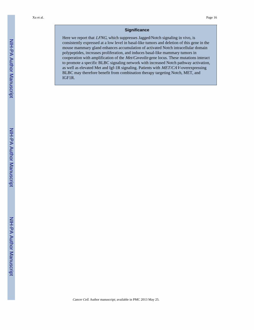
Significance
Here we report that LFNG, which suppresses Jagged/Notch signaling in vivo, isconsistently expressed at a low level in basal-like tumors and deletion of this gene in themouse mammary gland enhances accumulation of activated Notch intracellular domainpolypeptides, increases proliferation, and induces basal-like mammary tumors incooperation with amplification of the Met/Caveolin gene locus. These mutations interactto promote a specific BLBC signaling network with increased Notch pathway activation,as well as elevated Met and Igf-1R signaling. Patients with MET/CAV-overexpressingBLBC may therefore benefit from combination therapy targeting Notch, MET, andIGF1R.
Xu et al. Page 16
Cancer Cell. Author manuscript; available in PMC 2013 May 25.
NIH
-PA Author Manuscript
NIH
-PA Author Manuscript
NIH
-PA Author Manuscript
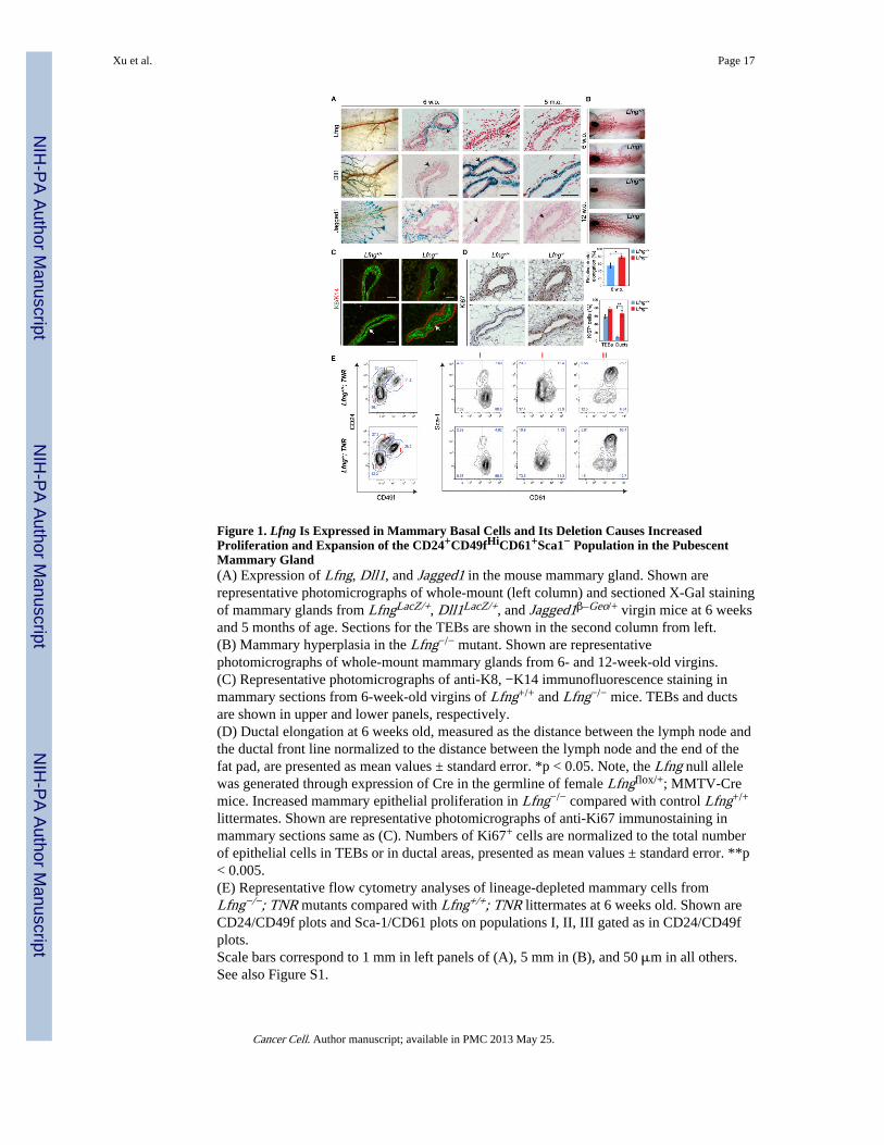
Figure 1. Lfng Is Expressed in Mammary Basal Cells and Its Deletion Causes IncreasedProliferation and Expansion of the CD24+CD49fHiCD61+Sca1− Population in the PubescentMammary Gland(A) Expression of Lfng, Dll1, and Jagged1 in the mouse mammary gland. Shown arerepresentative photomicrographs of whole-mount (left column) and sectioned X-Gal stainingof mammary glands from LfngLacZ/+, Dll1LacZ/+, and Jagged1β–Geo/+ virgin mice at 6 weeksand 5 months of age. Sections for the TEBs are shown in the second column from left.(B) Mammary hyperplasia in the Lfng−/− mutant. Shown are representativephotomicrographs of whole-mount mammary glands from 6- and 12-week-old virgins.(C) Representative photomicrographs of anti-K8, −K14 immunofluorescence staining inmammary sections from 6-week-old virgins of Lfng+/+ and Lfng−/− mice. TEBs and ductsare shown in upper and lower panels, respectively.(D) Ductal elongation at 6 weeks old, measured as the distance between the lymph node andthe ductal front line normalized to the distance between the lymph node and the end of thefat pad, are presented as mean values ± standard error. *p < 0.05. Note, the Lfng null allelewas generated through expression of Cre in the germline of female Lfngflox/+; MMTV-Cremice. Increased mammary epithelial proliferation in Lfng−/− compared with control Lfng+/+
littermates. Shown are representative photomicrographs of anti-Ki67 immunostaining inmammary sections same as (C). Numbers of Ki67+ cells are normalized to the total numberof epithelial cells in TEBs or in ductal areas, presented as mean values ± standard error. **p< 0.005.(E) Representative flow cytometry analyses of lineage-depleted mammary cells fromLfng−/−; TNR mutants compared with Lfng+/+; TNR littermates at 6 weeks old. Shown areCD24/CD49f plots and Sca-1/CD61 plots on populations I, II, III gated as in CD24/CD49fplots.Scale bars correspond to 1 mm in left panels of (A), 5 mm in (B), and 50 μm in all others.See also Figure S1.
Xu et al. Page 17
Cancer Cell. Author manuscript; available in PMC 2013 May 25.
NIH
-PA Author Manuscript
NIH
-PA Author Manuscript
NIH
-PA Author Manuscript

Figure 2. Deletion of Lfng Induced Mammary Tumors in Mice(A) Kaplan-Meier mammary tumor-free survival curve for control Lfngflox/flox andLfngflox/flox; MMTV-Cre conditional mutant mice.(B) Representative photomicrographs of H&E stained sections showing three histologicaltypes of Lfngflox/flox; MMTV-Cre mammary tumors.(C) Anti-ERα immunostaining on two main types of Lfngflox/flox; MMTV-Cre mammarytumors. n: normal tissue; t: tumor. Note positive ERα staining in normal tissue adjacent tothe tumor.(D) Representative photomicrographs and quantification of anti-Ki67 immunostaining ontwo main types of Lfngflox/flox; MMTV-Cre mammary tumors. Data are derived from twosections in each of three Type I and Type II tumors presented as mean values ± standarderror.(E) Representative photomicrographs of anti-K8, −K14 immunofluorescence staining onthree types of Lfngflox/flox; MMTV-Cre mammary tumors. Arrows point to cells showingco-expression of K8 and K14.Scale bars correspond to 100 μm in (B), 50 μm in (C), (D) and (E).
Xu et al. Page 18
Cancer Cell. Author manuscript; available in PMC 2013 May 25.
NIH
-PA Author Manuscript
NIH
-PA Author Manuscript
NIH
-PA Author Manuscript

Figure 3. Lfngflox/flox; MMTV-Cre Tumors Cluster with Basal-like and Claudin-Low MouseMammary Tumor Models(A) Overview of expression of reference genes in tumors from various mouse models ofbreast cancer, including 11 tumors from Lfngflox/flox; MMTV-Cre mice. Colored bars at leftcorrespond to regions shown in (B).(B) Expression of selected genes representing the Claudin gene cluster, luminal gene cluster,proliferation-associated gene cluster, CK14 basal-like gene cluster, CK5 basal-like genecluster, and EMT gene cluster. Expression data from type I and type II Lfngflox/flox; MMTV-Cre tumors are contained within yellow boxes. Clusters of tumor models are highlighted atthe bottom. DMBA, 7,12-dimethylbenz[a]-anthracene.(C) Enrichment map for gene sets over-represented in list of genes that are differentiallyexpressed comparing Lfngflox/flox; MMTV-Cre mouse mammary tumors to other mousemodels of breast cancer.See also Table S1.
Xu et al. Page 19
Cancer Cell. Author manuscript; available in PMC 2013 May 25.
NIH
-PA Author Manuscript
NIH
-PA Author Manuscript
NIH
-PA Author Manuscript
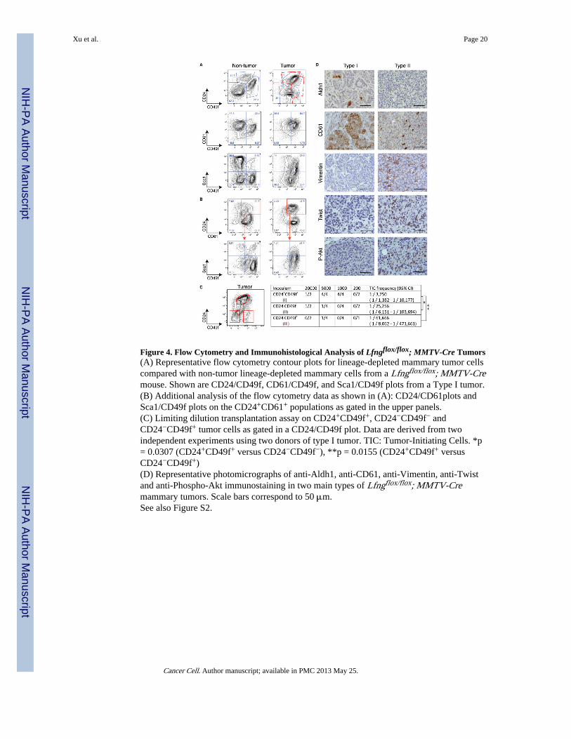
Figure 4. Flow Cytometry and Immunohistological Analysis of Lfngflox/flox; MMTV-Cre Tumors(A) Representative flow cytometry contour plots for lineage-depleted mammary tumor cellscompared with non-tumor lineage-depleted mammary cells from a Lfngflox/flox; MMTV-Cremouse. Shown are CD24/CD49f, CD61/CD49f, and Sca1/CD49f plots from a Type I tumor.(B) Additional analysis of the flow cytometry data as shown in (A): CD24/CD61plots andSca1/CD49f plots on the CD24+CD61+ populations as gated in the upper panels.(C) Limiting dilution transplantation assay on CD24+CD49f+, CD24−CD49f− andCD24−CD49f+ tumor cells as gated in a CD24/CD49f plot. Data are derived from twoindependent experiments using two donors of type I tumor. TIC: Tumor-Initiating Cells. *p= 0.0307 (CD24+CD49f+ versus CD24−CD49f−), **p = 0.0155 (CD24+CD49f+ versusCD24−CD49f+)(D) Representative photomicrographs of anti-Aldh1, anti-CD61, anti-Vimentin, anti-Twistand anti-Phospho-Akt immunostaining in two main types of Lfngflox/flox; MMTV-Cremammary tumors. Scale bars correspond to 50 μm.See also Figure S2.
Xu et al. Page 20
Cancer Cell. Author manuscript; available in PMC 2013 May 25.
NIH
-PA Author Manuscript
NIH
-PA Author Manuscript
NIH
-PA Author Manuscript
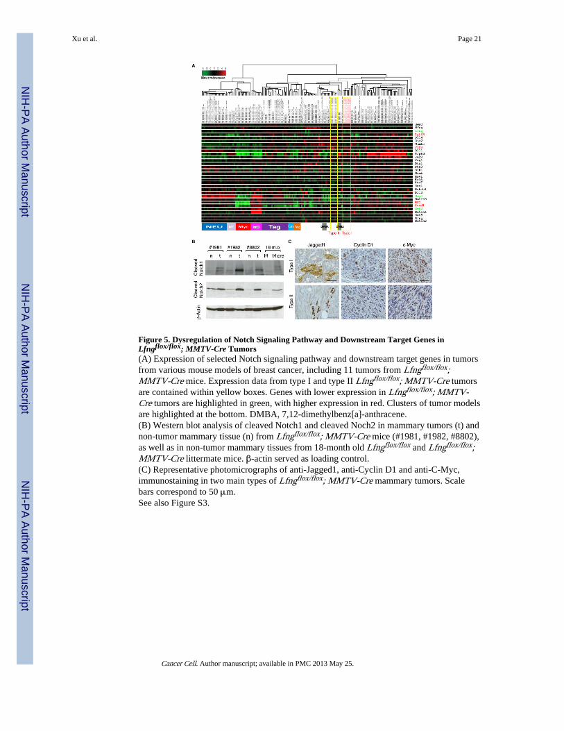
Figure 5. Dysregulation of Notch Signaling Pathway and Downstream Target Genes inLfngflox/flox; MMTV-Cre Tumors(A) Expression of selected Notch signaling pathway and downstream target genes in tumorsfrom various mouse models of breast cancer, including 11 tumors from Lfngflox/flox;MMTV-Cre mice. Expression data from type I and type II Lfngflox/flox; MMTV-Cre tumorsare contained within yellow boxes. Genes with lower expression in Lfngflox/flox; MMTV-Cre tumors are highlighted in green, with higher expression in red. Clusters of tumor modelsare highlighted at the bottom. DMBA, 7,12-dimethylbenz[a]-anthracene.(B) Western blot analysis of cleaved Notch1 and cleaved Noch2 in mammary tumors (t) andnon-tumor mammary tissue (n) from Lfngflox/flox; MMTV-Cre mice (#1981, #1982, #8802),as well as in non-tumor mammary tissues from 18-month old Lfngflox/flox and Lfngflox/flox;MMTV-Cre littermate mice. β-actin served as loading control.(C) Representative photomicrographs of anti-Jagged1, anti-Cyclin D1 and anti-C-Myc,immunostaining in two main types of Lfngflox/flox; MMTV-Cre mammary tumors. Scalebars correspond to 50 μm.See also Figure S3.
Xu et al. Page 21
Cancer Cell. Author manuscript; available in PMC 2013 May 25.
NIH
-PA Author Manuscript
NIH
-PA Author Manuscript
NIH
-PA Author Manuscript

Figure 6. Met/Caveolin Amplicon and Signaling of Met and IgfR in Lfngflox/flox; MMTV-CreMammary Tumors(A) aCGH analysis of DNA isolated from four Lfngflox/flox; MMTV-Cre mouse tumors andone Lfngflox/+; MMTV-Cre tumor showing a commonly amplified locus among all fourLfngflox/flox; MMTV-Cre tumors on chromosome 6. Red bar corresponds to the overlappingregion containing 13 genes.(B) Expression of the commonly amplified genes as shown in (A) in 11 Lfngflox/flox;MMTV-Cre tumors in comparison with expression of these genes in other mouse models ofbreast cancer. Expression data from type I and type II Lfngflox/flox; MMTV-Cre tumors arecontained within yellow boxes.(C) Western blot analysis of Met and phospho-Met in mammary tumors (t) and non-tumormammary tissue (n) from Lfngflox/flox; MMTV-Cre mice (#1981, #1982, #8802), as well asin mammary tissue from 18-month old Lfngflox/flox and Lfngflox/flox; MMTV-Cre littermatemice. β-actin served as loading control.(D) Representative photomicrographs of anti-Caveolin-1 immunostaining in two main typesof Lfngflox/flox; MMTV-Cre mammary tumors. Red arrows indicate positive staining inblood vessels. Black arrow points to a mammary duct adjacent to the tumor. Note, non-tumor mammary tissues show strong staining in adipocytes and weak staining inmyoepithelial cells. Scale bars correspond to 50 μm.(E) Western blot analysis of Caveolin-2 and Phospho-IRS-1/2 in mammary tumors (t) andnon-tumor mammary tissue (n) from Lfngflox/flox; MMTV-Cre mice.See also Figure S4.
Xu et al. Page 22
Cancer Cell. Author manuscript; available in PMC 2013 May 25.
NIH
-PA Author Manuscript
NIH
-PA Author Manuscript
NIH
-PA Author Manuscript
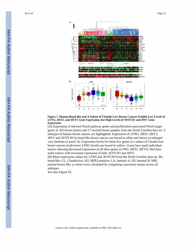
Figure 7. Human Basal-like and A Subset of Claudin-Low Breast Cancers Exhibit Low Levels ofLFNG, HES1, and HEY1 Gene Expression, but High Levels of NOTCH1 and MYC GeneExpression(A) Expression of selected Notch pathway genes and proliferation-associated Notch targetgenes in 320 breast tumors and 17 normal breast samples from the North Carolina data set. 5subtypes of human breast cancers are highlighted. Expression of LFNG, HES1, HEY1,MYC and NOTCH1 in basal-like breast cancers are boxed in white and shown in enlargedview (bottom of panel A). Expression levels for these five genes in a subset of Claudin-lowbreast cancers (with lower LFNG levels) are boxed in yellow. Green bars mark individualtumors showing decreased expression of all three genes (LFNG, HES1, HEY1). Red barsmark tumors with increased expression of both NOTCH1 and MYC.(B) Mean expression values for LFNG and NOTCH1 from the North Carolina data set. BL,basal-like; CL, Claudin-low; H2, HER2-positive; LA, luminal A; LB, luminal B; NBL,normal breast-like. p values were calculated by comparing expression means across allsubtypes.See also Figure S5.
Xu et al. Page 23
Cancer Cell. Author manuscript; available in PMC 2013 May 25.
NIH
-PA Author Manuscript
NIH
-PA Author Manuscript
NIH
-PA Author Manuscript

Figure 8. Model of Cooperation between Lfng Deficiency and Met/Caveolin Amplification/Overexpression in Basal-like Breast CancerLfng modifies Notch receptor to inhibit its activation by Jagged ligand and enhance itsactivation by Dll ligand. Loss of Lfng results in increased Jagged1-mediated Notchactivation and upregulation of c-Myc, Cyclin D1, Igf1R and uPA, leading to increasedproliferation. Loss of Lfng may decrease Dll1-mediated Notch activation in bipotentmammary progenitor cells, causing expansion of basal cells. Decreased expression of Hes/Hey Notch target genes may de-repress the Met promoter. Selected as cooperative event inLfng deficiency induced mammary tumorigenesis, the Met/caveolin amplicon increasesabundance of Met and Caveolin1/2, the latter proteins are predicted to enhance signalingthrough Igf-1R and IRS. Signaling through Met and Igf-1R stimulate proliferation. TGN,trans Golgi network.
Xu et al. Page 24
Cancer Cell. Author manuscript; available in PMC 2013 May 25.
NIH
-PA Author Manuscript
NIH
-PA Author Manuscript
NIH
-PA Author Manuscript