
A roadmap to Individualized Irinotecan Dosing - repub.eur.nl Floris Aart de.pdf · van een...
140
Transcript of A roadmap to Individualized Irinotecan Dosing - repub.eur.nl Floris Aart de.pdf · van een...


A ROADMAP TO
INDIVIDUALIZED
IRINOTECAN DOSING

Stellingen behorende bij het proefschrift
A Roadmap to individualized irinotecan dosing
Floris de Jong, Erasmus MC, Rotterdam, the Netherlands
Het doseren van irinotecan op basis van lichaamsoppervlakte (BSA) is niets anders dan het toedienen van een standaard dosis vermenigvuldigd met een willekeurig getal, namelijk de verhouding tussen het
lichaamsoppervlak van de patiënt en dat van een gemiddelde patiënt. Dit proefschrift.
Het antibioticum neomycine heeft geen substantieel toegevoegde waarde bij het voorkomen van diarree na irinotecan therapie. Dit proefschrift.
Fenotypering illustreert dat farmacokinetiek zich ook een a priori rol in de klinische praktijk kan verwerven. Dit proefschrift.
Vooralsnog is het regulier voorschrijven van medicinale cannabis in de oncologische praktijk een brug te ver. Dit proefschrift.
Het stelselmatig vooraf screenen op de aanwezigheid van het UGT1A1*28 genotype is niet zinnig zolang de voorspellende waarde hiervan voor irinotecan toxiciteit onduidelijk is en aan de uitslag geen behandel-advies gekoppeld kan worden. Dit proefschrift.
Het algemeen levende beeld dat ‘natuurlijke producten’ onschuldig en ongevaarlijk zijn, moet bij kanker-patiënten, hun omgeving en hun behandelaars veranderen in een meer reële en verantwoordelijke attitude. Tascilar et al. The Oncologist, 11, 467-476, 2006
Er worden nog steeds te weinig limited sampling modellen ontworpen en vervolgens is het klinische gebruik te gering.
Farmacogenetica evolueert steeds meer naar een vakgebied waar eerst de genotypisch variatie geobserveerd wordt voordat fenotypische consequenties onderzocht worden. Relling and Dervieux, Nat. Rev. Cancer 1, 99–108, 2001
Transitions in clinical practice may prove at least as challenging as resolving the original structure of the helix. Modified from: Bell, Nature 421, 414-416, 2003
Het aanhalen van wetenschappelijk onderzoek gebeurt in de media te geregeld kritiekloos en buiten de juiste context.
Don’t fight the facts, perception is reality.
A ROADMAP TO
INDIVIDUALIZED
IRINOTECAN DOSING
Routeboek voor
het individualiseren van irinotecan therapie
proefschrift
ter verkrijging van de graad van doctor aan de
Erasmus Universiteit Rotterdam
op gezag van de
rector magnificus
Prof.dr. S.W.J. Lamberts
en volgens besluit van het College voor Promoties.
De openbare verdediging zal plaatsvinden op
woensdag 11 oktober 2006 om 15.45 uur
door
Floris Aart de Jong
geboren te Rotterdam

Promotiecommissie
Promotor: Prof.dr. J. Verweij
Overige leden: Prof.dr. J.H. Beijnen
Prof.dr. G. Stoter Prof.dr. E.G.E. de Vries
Copromotor: Dr. A.H.J. Mathijssen
De vuursteen die door het staal geslagen werd, verwonderde zich zeer en zei tot hem met strenge stem, ‘Welke arrogantie beweegt jou om mij te molesteren?’ ‘Val me niet lastig, want je hebt mij bij vergissing gekozen; ik heb nooit iemand kwaad gedaan.’ Waarop het staal antwoordde, ‘Als je geduld heb zul je zien wat een geweldig resultaat er uit jou voortkomt.’
Door deze woorden was de vuursteen gekalmeerd en doorstond geduldig zijn marteling. Daarop zag hij dat hij het leven schonk aan het wonderbare element vuur dat door zijn kracht een rol ging spelen bij oneindig veel zaken.
Dit wordt gezegd van diegenen die ontmoedigd zijn aan het begin van hun studie, maar die zich daarna voornemen om zichzelf te overwinnen en zich geduldig te richten op die studies waaruit men dingen ziet voortkomen die fantastisch zijn.
Leonard da Vinci (1452–1519)
Voor mijn ouders
6
Cover design: John van Lit, T&IC, Delft, the Netherlands
Lay-out: John van Lit, T&IC, Delft, the Netherlands
Printed by: Drukkerij Akxifo, Poeldijk, the Netherlands
ISBN-10: 90-9021065-2 ISBN-13: 978-90-9021065-0
Copyright: FA de Jong, Rotterdam, the Netherlands, 2006
All rights reserved. No part of this publication may be reproduced, stored in a retrieval system, or transmitted in any form or by any means be it electronic, mechanical, photocopying, recording, or otherwise without the prior permission in writing from the author.
No responsibility is assumed by the author, publisher, editor, or printer for any injury and/or damage to persons or property as a matter of products liability, negligence or otherwise, or from any use or operation of any methods, instructions or ideas contained in the material herein.
The (financial) support of Farmalyse, Pfizer, and T&IC for this publication of this thesis is gratefully acknowledged.
7
Contents
chapter 1 1 Introduction chapter 2 5 Flat-fixed dosing of irinotecan: influence on pharmacokinetic and
pharmacodynamic variability
FA de Jong, RH Mathijssen, R Xie, J Verweij, A Sparreboom
chapter 3A 11 Medicinal cannabis in oncology practice: still a bridge too far?
FA de Jong, FK Engels, RH Mathijssen, L van Zuylen, J Verweij, RP Peters, A Sparreboom
chapter 3B 19 Influence of medicinal cannabis on the clinical pharmacokinetics of irinotecan and docetaxel
FK Engels, FA de Jong, A Sparreboom, RA Mathôt, WJ Loos, JJ Kitzen, P de Bruijn, J Verweij, RH Mathijssen
chapter 4 31 Role of pharmacogenetics in irinotecan therapy
FA de Jong, MJ de Jonge, J Verweij, RH Mathijssen
chapter 5 47 ABCG2 pharmacogenetics: ethnic differences in allele frequency and assessment of influence on irinotecan disposition
FA de Jong, S Marsh, RH Mathijssen, C King, J Verweij, A Sparreboom, HL McLeod chapter 6 57 Irinotecan-induced diarrhea: functional significance of the polymorphic
ABCC2 transporter protein
FA de Jong, T Scott-Horton, DL Kroetz, HL McLeod, LE Friberg, RH Mathijssen, J Verweij, S Marsh, A Sparreboom
chapter 7 67 Prophylactic treatment of irinotecan-induced diarrhea with neomycin and potential role for UGT1A1*28 genotype screening: a randomized, placebo-controlled, double-blind study
FA de Jong, DF Kehrer, RH Mathijssen, G-J Creemers, P de Bruijn, RH van Schaik, AS Planting, A van der Gaast, FA Eskens, JT Janssen, JB Ruit, J Verweij, A Sparreboom, MJ de Jonge
chapter 8 79 Prediction of irinotecan pharmacokinetics by use of cytochrome P450 3A4 phenotyping probes
RH Mathijssen, FA de Jong, RH van Schaik, ER Lepper, LE Friberg, T Rietveld, P de Bruijn, WJ Graveland, WD Figg, J Verweij, A Sparreboom
chapter 9 91 Summary, conclusions, and future perspectives chapter 10 99 References
115 Samenvatting, discussie en toekomstsperspectief 121 Dankwoord 125 Curriculum vitae 126 List of publications

8

9
Chapter 1
Introduction

Introduction
2
Introduction

Introduction Introduction
In the early 1960s,the cytotoxic potential of camptothecin, a plant alkaloid isolated from Camptotheca acuminata (family Nyssaceae), was discovered.1 Unfortunately, severe and unpredictable adverse effects, particularly myelosuppression, diarrhea, and hemorrhagic cystitis were seen in early clinical studies, which limited its further clinical development.2-4 Later on, these adverse effects have been related to the water-insolubility of camptothecin which finding resulted in a renewed interest and the development
of various (semi-)synthetic water-soluble derivatives, including the prodrug irinotecan (CPT-11; Campto®; Camptosar®). These camptothecin topoisomerase I inhibitors all reversibly stabilize the cleavable complex, i.e., the covalent interaction between DNA and the enzyme topoisomerase I, resulting in single-strand DNA breaks and thus in termination of DNA replication, subsequently followed by cell death.5-7 Nowadays, irinotecan is one of the treatment options in first and second line metastatic and/or nonresectable colorectal cancer, significantly improving duration and quality of life.8-11 In patients with tumor progression after 5-fluorouracil (5-FU)-based chemotherapy response rates of 20% to single agent irinotecan treatment
have been reported, while response rates in combination schedules may be even higher.12-14 Responses in various other tumor types, including melanoma, non-Hodgkin lymphoma, gastric, esophagus, cervical, and lung cancer have been reported as well.15-22
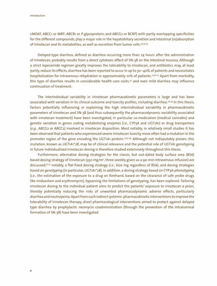
Multiple polymorphic pathways are involved in the biotransformation of irinotecan (see figure 1). To be activated, irinotecan needs to be hydrolyzed into its active metabolite SN-38 by carboxylesterases (CES), enzymes which are found in plasma, in the intestinal epithelial lining, and abundantly in the liver.23
Although recently intratumoral activation of irinotecan by these enzymes has been positively related with chemosensitivity,24-26 the role of systemic circulating SN-38 cannot be disregarded. The primary pathway of elimination of SN-38 is a phase II glucuronic acid conjugation reaction that results in the formation of SN-38G (SN-38 glucuronide), and that is mediated by UDP glucuronosyltransferase 1A (UGT1A) isoforms, amongst others present in the epithelial tissue lining of the digestive tract and plentifully in the liver.27-29
After biliary excretion into the intestinal lumen, SN-38G can be deglucuronidated back into SN-38 by bacterial β-glucuronidases.30,31 Irinotecan metabolism also consists of a predominantly intrahepatic cytochrome P450 3A (CYP3A) mediated oxidation, whereby the inactive substances NPC and APC are formed.32 Although with lower affinity, carboxylesterases (CES) can activate these metabolites into SN-38 as well.33 Furthermore, several adenosine triphosphate (ATP) binding cassette (ABC) transporters (ABCC2 or
Irinotecan is (
, (,
,
) ,
i i l i I i i , ilii ial (β l i ).
Figure 1. Activation and metabolism of irinotecan. oxidized by cytochrome P450 3A isozymes CYP3A), enzymes
abundantly present in the liver into its inactive metabolites APC en NPC. Carboxylesterases CES), which are found in plasma, in the intestinal epithelial lining in tumor tissue, and in high content in the liver are responsible for the conversion of irinotecan in its 1,000 times more active metabolite SN-38. NPC, and maybe APC as well, can be activated by CES as well. Uridine diphosphate
glucuronosyltransferases (UGT 1A isoforms in the liver but also in the intestinal epithelial lining are responsible for the detoxification of SN-38
nto ts g ucuron de SN-38G. n the ntest nes hepatob ary excreted
SN-38G can be re-act vated by bacter enzymes -g ucuron dases
3
Introduction
cMOAT; ABCC1 or MRP; ABCB1 or P-glycoprotein; and ABCG2 or BCRP) with partly overlapping specificities for the different compounds, play a major role in the hepatobiliary secretion and intestinal (re)absorption of irinotecan and its metabolites, as well as excretion from tumor cells.29,34,35
Delayed-type diarrhea, defined as diarrhea occurring more than 24 hours after the administration of irinotecan, probably results from a direct cytotoxic effect of SN-38 on the intestinal mucosa. Although a strict loperamide regimen greatly improves the tolerability to irinotecan, and antibiotics may, at least
partly, reduce its effects, diarrhea has been reported to occur in up to 30–40% of patients and necessitates hospitalization for intravenous rehydration in approximately 10% of patients.13,36-42 Apart from morbidity, this type of diarrhea results in considerable health-care costs,43 and even mild diarrhea may influence continuation of treatment.
The interindividual variability in irinotecan pharmacokinetic parameters is large and has been associated with variation in its clinical outcome and toxicity profiles, including diarrhea.29,44 In this thesis, factors potentially influencing or explaining the high interindividual variability in pharmacokinetic parameters of irinotecan and SN-38 (and thus subsequently the pharmacodynamic variability associated with irinotecan treatment) have been investigated, in particular co-medication (medical cannabis) and genetic variation in genes coding metabolizing enzymes (i.e., CYP3A and UGT1A1) or drug transporters (e.g., ABCG2 or ABCC2) involved in irinotecan disposition. Most notably, in relatively small studies it has been observed that patients who experienced severe irinotecan toxicity more often had a mutation in the promoter region of the gene encoding the UGT1A1 protein.27,45-49 Although not indisputably proven, this mutation, known as UGT1A1*28, may be of clinical relevance and the potential role of UGT1A1 genotyping in future individualized irinotecan dosing is therefore studied extensively throughout this thesis.
Furthermore, alternative dosing strategies for the classic, but out-dated body surface area (BSA) based dosing strategy of irinotecan (350 mg/m2, three-weekly given as a 90-min intravenous infusion) are discussed;50,51 notably, a flat-fixed dosing strategy (i.e., 600 mg regardless of BSA), and dosing strategies based on genotyping (in particular, UGT1A1*28). In addition, a dosing strategy based on CYP3A-phenotyping
(i.e., the estimation of the exposure to a drug on forehand, based on the clearance of safe probe drugs like midazolam and erythromycin), bypassing the limitations of genotyping, has been explored. Tailoring irinotecan dosing to the individual patient aims to predict the patients’ exposure to irinotecan a priori, thereby potentially reducing the risks of unwanted pharmacodynamic adverse effects, particularly diarrhea and neutropenia. Apart from such indirect systemic-pharmacokinetic interventions to improve the tolerability of irinotecan therapy, direct pharmacological interventions aimed to protect against delayed type diarrhea by prophylactic neomycin coadministration (through the prevention of the intraluminal formation of SN-38) have been investigated.
4
Introduction
5
Chapter 2
Flat-fixed dosing of irinotecan: influence on pharmacokinetic and
pharmacodynamic variability
FA de Jong, RH Mathijssen, R Xie, J Verweij, A Sparreboom
Clinical Cancer Research 10(12):4068–4071, 2004
6
Chapter 2 - Flat-fixed dosing of irinotecan: influence on pharmacokinetic and pharmacodynamic variability
ABSTRACT
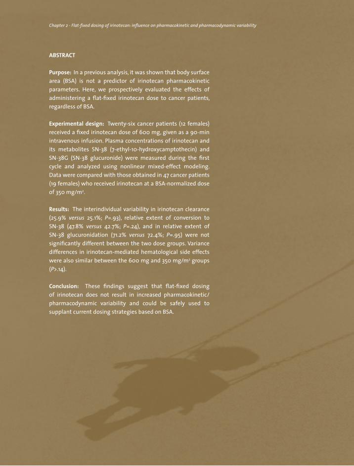
Purpose: In a previous analysis, it was shown that body surface area (BSA) is not a predictor of irinotecan pharmacokinetic parameters. Here, we prospectively evaluated the effects of administering a flat-fixed irinotecan dose to cancer patients, regardless of BSA.
Experimental design: Twenty-six cancer patients (12 females) received a fixed irinotecan dose of 600 mg, given as a 90-min intravenous infusion. Plasma concentrations of irinotecan and its metabolites SN-38 (7-ethyl-10-hydroxycamptothecin) and SN-38G (SN-38 glucuronide) were measured during the first
cycle and analyzed using nonlinear mixed-effect modeling. Data were compared with those obtained in 47 cancer patients (19 females) who received irinotecan at a BSA-normalized dose of 350 mg/m2.
Results: The interindividual variability in irinotecan clearance (25.9% versus 25.1%; P=.93), relative extent of conversion to SN-38 (47.8% versus 42.7%; P=.24), and in relative extent of SN-38 glucuronidation (71.2% versus 72.4%; P=.95) were not
significantly different between the two dose groups. Variance differences in irinotecan-mediated hematological side effects were also similar between the 600 mg and 350 mg/m2 groups (P>.14).
Conclusion: These findings suggest that flat-fixed dosing of irinotecan does not result in increased pharmacokinetic/
pharmacodynamic variability and could be safely used to supplant current dosing strategies based on BSA.
Chapter 2 - Flat-fixed dosing of irinotecan: influence on pharmacokinetic and pharmacodynamic variability
INTRODUCTION
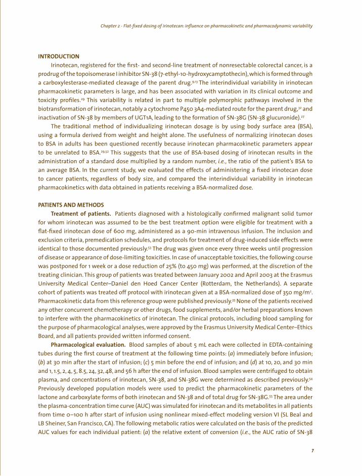
Irinotecan, registered for the first- and second-line treatment of nonresectable colorectal cancer, is a prodrug of the topoisomerase I inhibitor SN-38 (7-ethyl-10-hydroxycamptothecin), which is formed through a carboxylesterase-mediated cleavage of the parent drug.9,13 The interindividual variability in irinotecan pharmacokinetic parameters is large, and has been associated with variation in its clinical outcome and toxicity profiles.29 This variability is related in part to multiple polymorphic pathways involved in the biotransformation of irinotecan, notably a cytochrome P450 3A4-mediated route for the parent drug,32 and inactivation of SN-38 by members of UGT1A, leading to the formation of SN-38G (SN-38 glucuronide).27
The traditional method of individualizing irinotecan dosage is by using body surface area (BSA), using a formula derived from weight and height alone. The usefulness of normalizing irinotecan doses to BSA in adults has been questioned recently because irinotecan pharmacokinetic parameters appear to be unrelated to BSA.29,52 This suggests that the use of BSA-based dosing of irinotecan results in the administration of a standard dose multiplied by a random number, i.e., the ratio of the patient’s BSA to an average BSA. In the current study, we evaluated the effects of administering a fixed irinotecan dose to cancer patients, regardless of body size, and compared the interindividual variability in irinotecan pharmacokinetics with data obtained in patients receiving a BSA-normalized dose.
PATIENTS AND METHODS
Treatment of patients. Patients diagnosed with a histologically confirmed malignant solid tumor for whom irinotecan was assumed to be the best treatment option were eligible for treatment with a flat-fixed irinotecan dose of 600 mg, administered as a 90-min intravenous infusion. The inclusion and exclusion criteria, premedication schedules, and protocols for treatment of drug-induced side effects were identical to those documented previously.53 The drug was given once every three weeks until progression of disease or appearance of dose-limiting toxicities. In case of unacceptable toxicities, the following course was postponed for 1 week or a dose reduction of 25% (to 450 mg) was performed, at the discretion of the treating clinician. This group of patients was treated between January 2002 and April 2003 at the Erasmus University Medical Center–Daniel den Hoed Cancer Center (Rotterdam, the Netherlands). A separate cohort of patients was treated off protocol with irinotecan given at a BSA-normalized dose of 350 mg/m2. Pharmacokinetic data from this reference group were published previously.35 None of the patients received any other concurrent chemotherapy or other drugs, food supplements, and/or herbal preparations known to interfere with the pharmacokinetics of irinotecan. The clinical protocols, including blood sampling for the purpose of pharmacological analyses, were approved by the Erasmus University Medical Center–Ethics Board, and all patients provided written informed consent.
Pharmacological evaluation. Blood samples of about 5 mL each were collected in EDTA-containing tubes during the first course of treatment at the following time points: (a) immediately before infusion; (b) at 30 min after the start of infusion; (c) 5 min before the end of infusion; and (d) at 10, 20, and 30 min and 1, 1.5, 2, 4, 5, 8.5, 24, 32, 48, and 56 h after the end of infusion. Blood samples were centrifuged to obtain plasma, and concentrations of irinotecan, SN-38, and SN-38G were determined as described previously.54
Previously developed population models were used to predict the pharmacokinetic parameters of the lactone and carboxylate forms of both irinotecan and SN-38 and of total drug for SN-38G.55 The area under the plasma-concentration time curve (AUC) was simulated for irinotecan and its metabolites in all patients from time 0–100 h after start of infusion using nonlinear mixed-effect modeling version VI (SL Beal and LB Sheiner, San Francisco, CA). The following metabolic ratios were calculated on the basis of the predicted AUC values for each individual patient: (a) the relative extent of conversion (i.e., the AUC ratio of SN-38
7

Chapter 2 - Flat-fixed dosing of irinotecan: influence on pharmacokinetic and pharmacodynamic variabilityChapter 2 - Flat-fixed dosing of irinotecan: influence on pharmacokinetic and pharmacodynamic variability
to irinotecan, expressed as a percentage); (b) the relative extent of glucuronidation (i.e., the AUC ratio of SN-38G to SN-38); and (c) the biliary index (i.e., the ratio of irinotecan AUC to the relative extent of glucuronidation).
Toxicity was evaluated and graded according to the National Cancer Institute–Common Toxicity Criteria, version 2.0 (http://ctep.cancer.gov/forms/CTCv20_4-30-992.pdf). Hematological pharmacodynamics were assessed by analysis of the absolute nadir values of blood cell counts and by the relative hematological toxicity, i.e., the percentage decrease in blood cell count, which was defined as follows: percentage decrease = [(pretherapy value – nadir value) / (pretherapy value)] * 100%.
Statistical considerations. Group sample sizes of 25 (fixed dose) and 50 (BSA-normalized dose) were calculated to achieve approximately 60% power to detect a ratio of 2.00 between the parameter variances in the respective groups, using a two-sided F-test with a significance level (α) of .05. All pharmacokinetic data are presented as mean values with the coefficient of variation in parenthesis, unless stated otherwise. The coefficient of variation was defined as the ratio of SD and the observed mean. A modified Levene test was used to test for equality of variances between the fixed dose and BSA-normalized dose groups. Statistical calculations were performed using Number Cruncher Statistical Systems 2001 and Power Analysis and Sample Size 2001 (NCSS, Kaysville, UT).
RESULTS
A total of 26 cancer patients with a median age of 57 years (range, 38–73 years) and a median BSA of 1.85 m2 (range, 1.45–2.31 m2) received at least one course of irinotecan at a dose of 600 mg (see table 1). In the reference group, 47 cancer patients with a median age of 53 years (range, 37–71 years) and a median BSA of 1.87 m2 (range, 1.40–2.36 m2) received a BSA-corrected dose of 350 mg/m2. Patient demographic
a 2
N) 26 14/12
) 53
71 73 2) 1.85
1 1 N, %)
2 (4) 8 (31) 32 (68) (19) 13 (28)
a
;
Table 1. Patient demographics
Variable 600 mg group 350 mg/m group
Baseline screening
Number of patients entered ( 47
Males/Females 28/19
Age (years 57 (38–73) (37–71) Length (m) 1.72 (1.55–1.86) 1.73 (1.55–1.92) Weight (kg) (48–109) (45–108) Body surface area (m (1.45–2.31) 1.87 (1.40–2.36) Performance score (0–1) (0–1) Tumor types (
SCLC / NSCLC 13 (50) Gastrointestinal Miscellaneous 5
Infusion duration (h) 1.50 (1.47–1.78) 1.50 (0.75–2.25)
Values represent the median, with range in parenthesis (unless stated otherwise).
Abbreviations: N, number of patients; SCLC, small cell lung cancer NSCLC, non-small cell lung cancer.
8
Chapter 2 - Flat-fixed dosing of irinotecan: influence on pharmacokinetic and pharmacodynamic variability Chapter 2 - Flat-fixed dosing of irinotecan: influence on pharmacokinetic and pharmacodynamic variability
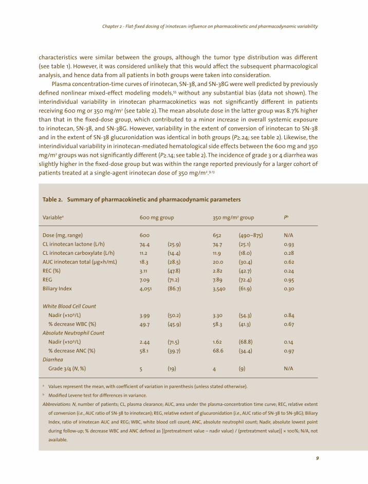
characteristics were similar between the groups, although the tumor type distribution was different
(see table 1). However, it was considered unlikely that this would affect the subsequent pharmacological analysis, and hence data from all patients in both groups were taken into consideration.
Plasma concentration-time curves of irinotecan, SN-38, and SN-38G were well predicted by previously defined nonlinear mixed-effect modeling models,55 without any substantial bias (data not shown). The interindividual variability in irinotecan pharmacokinetics was not significantly different in patients receiving 600 mg or 350 mg/m2 (see table 2). The mean absolute dose in the latter group was 8.7% higher than that in the fixed-dose group, which contributed to a minor increase in overall systemic exposure to irinotecan, SN-38, and SN-38G. However, variability in the extent of conversion of irinotecan to SN-38 and in the extent of SN-38 glucuronidation was identical in both groups (P≥.24; see table 2). Likewise, the interindividual variability in irinotecan-mediated hematological side effects between the 600 mg and 350 mg/m2 groups was not significantly different (P≥.14; see table 2). The incidence of grade 3 or 4 diarrhea was slightly higher in the fixed-dose group but was within the range reported previously for a larger cohort of
.9,13 patients treated at a single-agent irinotecan dose of 350 mg/m2
a 2 Pb
Dose (mg, 600 652
11.2 11.9 (18.0) * 18.3 (28.5)
3.11 2.82 (71.2)
4,051 (61.9)
Nadir (*109/L) 3.99 3.30 58.3
Nadir (*109/L) (71.5) 1.62 (68.8) 58.1 68.6
Diarrhea
/4 (N, %) 5 (19) 4 (9)
a
b
i i , i ; , pl l ; l i i ; , l i
i (i , i iri ); , l i l i i (i , i ); Bili
I , i iri ; i l ll ; , l il ; ir, l l i
i ll ; [( l i l ) / ( l )] * ; N/A,
il l
Table 2. Summary of pharmacokinetic and pharmacodynamic parameters
Variable 600 mg group 350 mg/m group
range) (490–875) N/A
CL irinotecan lactone (L/h) 74.4 (25.9) 74.7 (25.1) 0.93
CL irinotecan carboxylate (L/h) (14.4) 0.28
AUC irinotecan total (µg h/mL) 20.0 (30.4) 0.62
REC (%) (47.8) (42.7) 0.24
REG 7.09 7.89 (72.4) 0.95
Biliary Index (86.7) 3,540 0.30
White Blood Cell Count (50.2) (54.3) 0.84
% decrease WBC (%) 49.7 (45.9) (41.3) 0.67 Absolute Neutrophil Count
2.44 0.14 % decrease ANC (%) (39.7) (34.4) 0.97
Grade 3 N/A
Values represent the mean, with coefficient of variation in parenthesis (unless stated otherwise).
Modified Levene test for differences in variance.
Abbrev at ons: N number of pat ents CL asma c earance AUC, area under the p asma-concentrat on t me curve REC re at ve extent
of convers on .e. AUC rat o of SN-38 to notecan REG re at ve extent of g ucuron dat on .e. AUC rat o of SN-38 to SN-38G ary
ndex rat o of notecan AUC and REG WBC, wh te b ood ce count ANC abso ute neutroph count Nad abso ute owest po nt
dur ng fo ow-up % decrease WBC and ANC defined as pretreatment va ue – nad r va ue pretreatment va ue 100% not
ava ab e.
9
Chapter 2 - Flat-fixed dosing of irinotecan: influence on pharmacokinetic and pharmacodynamic variability
DISCUSSION
In the current exploratory study, we demonstrated that fixed dosing of irinotecan, regardless of body size, can be safely used in adult cancer patients as an alternative to the conventional BSA-corrected dosing strategy. Indeed, the interindividual variability in pharmacokinetic and pharmacodynamic parameters, expressed as the percentage coefficient of variation, did not change significantly in the fixed-dose group as compared with the BSA-based dose regimen. Observations similar to those described here for irinotecan have been published previously for the anthracycline epirubicin,56 and, more recently, for paclitaxel.57
It can be anticipated that implementation of the flat-fixed dosing concept in routine clinical practice would have significant economic implications.58 The ability to manufacture a unit dose has obvious benefits for the pharmaceutical company involved. Similarly, reconstituting a fixed dose without subsequent
individualization for different patients is more efficient and cost-effective than preparing individualized doses and would eliminate a significant source of error in attempting to obtain precise dosing.59 In addition, drug preparation and administration errors are very common for intravenously administered drugs,and are usually the result of systematic error (inaccuracy of the calculation algorithms) and inevitable convergence error, including use of inaccurate height and weight for BSA calculation.61
The 600-mg dose used in the fixed-dose group was selected on the basis of the assumption of an average BSA for cancer patients of 1.73 m2, which was the mean value in a European Organization for Research and Treatment of Cancer database that included 3,000 patients, both males and females, treated for sarcomas, lymphomas, and rectal cancers during the period 1990–1998 (J. Verweij, unpublished data). The actual mean BSA value in the present patient cohorts was 1.86 m2, and this led to a mean absolute dose in the BSA-normalized dose group of slightly more than 600 mg. It is therefore proposed that future clinical trials should evaluate the administration of fixed doses of irinotecan calculated on the basis of an average BSA in any given adult population, i.e., fixed dose (in mg) = conventional dose (in mg/m2) *
mean BSA (in m2). Because the pharmacokinetic behavior of irinotecan is dose and time independent,29 the modus operandi can also be applied to irinotecan administered as a 30-min infusion and/or at the reduced doses commonly given in weekly regimens.
One limitation of this trial is the relatively small sample size in both arms.However,the pharmacokinetic and pharmacodynamic parameters were almost identical between the cohorts, and it is doubtful that
even a very large trial would detect a clinically-relevant alteration in the variances. Likewise, although the study was not designed to examine response and survival data, differences in antitumor activity between the dose groups are not expected. We suggest implementation of a fixed dosing strategy for irinotecan, independent of BSA, until better dosing methods become available, which might, for example, be based on factors known to impact on irinotecan elimination pathways (e.g., measures of hepatic dysfunction and UGT1A genotype).46,49,62
ACKNOWLEDGEMENTS
We thank Peter de Bruijn and Inge MC van den Bos for technical support. This work was funded in part by the Cornelis Vrolijk Fund (Rotterdam, the Netherlands).
10
60

11
Chapter 3A
Medicinal cannabis in oncology practice: still a bridge too far?
FA de Jong, FK Engels, RH Mathijssen, L van Zuylen, J Verweij, RP Peters, A Sparreboom
Journal of Clinical Oncology 23(13):2886–2891, 2005

12
Chapter 3A - Medicinal cannabis in oncology practice: still a bridge too far?Chapter 3A - Medicinal cannabis in oncology practice: still a bridge too far?

Chapter 3A - Medicinal cannabis in oncology practice: still a bridge too far?
INTRODUCTION
In the 1980s and 1990s, most of the interest in cannabis (marijuana, hashish) focused solely on how to restrict its recreational use. However, in some specific areas of medicine, we have now entered an era in which the focus of attention has increasingly shifted to the clinical and controlled use of medicinal cannabis. Evidence with regards to the claimed benefits of medicinal cannabis is largely based on anecdotal case reports, arising from those situations in which physicians or patients themselves resorted to (prescribed) cannabis because conventional therapy was inadequate or was not effective. Several patient representative groups (especially for patients with cancer, multiple sclerosis, and AIDS) claim favorable effects of medicinal cannabis and advocate its use. However, it remains unclear to what extent
therapeutic effects can be attributed to definable, physiological, i.e., objective, effects or to the benefit of psychomimetic or psychological effects. In the Netherlands, a national agency on medicinal cannabis (the Office of Medicinal Cannabis,The Hague, the Netherlands) has been established by the Dutch government
following an ongoing public debate, involving diverse social and medical-oriented areas, combined with the growing need and wish to initiate well-designed clinical trials with cannabis. In the past, Dutch patients were forced to frequent illegal coffeeshops for their supply of medicinal cannabis, or even to produce cannabis themselves at home. Since September 2003, under the responsibility of the Office of Medicinal Cannabis, the production and distribution of a legal, standardized cannabis product for medical treatment
purposes has become a reality. This office claims that for the following indications a reasonable chance of effect can be expected of medicinal cannabis: spasticity with pain (for instance in multiple sclerosis and spinal cord injury), nausea and vomiting (chemotherapy, radiotherapy and HIV-medication related), chronic neuralgic pain, Gilles de la Tourette syndrome, and palliative treatment of cancer and HIV/AIDS.63
In this commentary, we will discuss the position of (medicinal) cannabis in oncology practice, viewed from the Dutch experience.
PHARMACOLOGY OF CANNABIS
To date, the majority of clinical research has been conducted with isolated cannabinoids, i.e., pharmaceutical extracts of Cannabis sativa L. or synthetically derived single compounds which act on the cannabinoid receptors CB1 and CB2.64 In this commentary we will refer to medicinal cannabis as an integral total product prepared from the cannabis plant, such as marijuana. As experience has been gathered predominantly with the main cannabinoid ∆9-tetrahydrocannabinol (THC), relevant data on synthetically derived cannabinoids will be discussed as well. THC is synthetically produced and commercially available as dronabinol (Marinol®; Solvay Pharmaceuticals Inc, Marietta, GA), and is formulated as capsules for oral administration.65 Clinical studies using marijuana have also been conducted, particularly in palliative treatment of HIV/AIDS.
With the introduction of legal medicinal grade cannabis in the Netherlands, a well-defined product
with a known and constant THC content that meets pharmaceutical quality standards is now available by prescription to Dutch patients.Indeed,the standardized production process greatly reduces the large degree of variability in THC content, and that of two other main cannabinoids, cannabinol (CBN) and cannabidiol (CBD). Until now, two cannabis varieties have been available for medicinal purpose, one of which contains approximately 18% THC and 0.8% CBD, the other of which contains 11.5% THC.63 Currently, the degree of variability in content uniformity is below 15%,63 which is low for herbal products (phytoproducts), and only marginally exceeds the specifications (variability <10%) that apply to licensed drugs. Nonetheless, the recommended routes of administration (inhalation by smoking or vaporization, or oral intake as tea) introduce a large degree of variability in an individual’s exposure to THC and other cannabis constituents.64
13
Chapter 3A - Medicinal cannabis in oncology practice: still a bridge too far?
In addition, when cannabis is smoked or inhaled in a joint, waterpipe, or vaporizer, THC concentrations in the blood rise and decline rapidly. Peak concentrations and its coupled psychomimetic effects are seen within minutes, and after an hour levels are decreased to less than 10%. When cannabis is taken orally, for instance as tea, bioavailability is lower, and maximum levels and the psychomimetic effects are seen after 1 to 2 hours.64
Given the chance that the psychomimetic adverse effects and impaired psychomotoric effects of cannabis can negatively influence the performance of daily tasks,64,66 it is necessary for its effects to be predicted with a certain degree of accuracy if one wishes to prescribe medicinal cannabis on a regular basis as an equivalent alternative to other drugs. More than 60 cannabinoids have been reported to be present in cannabis, albeit in varying amounts.64 The degree to which extent their pharmacological actions are known varies largely. In addition, due to the fact that these substances have diverse pharmacologic effects, predicting the overall pharmacologic effects of medicinal cannabis is difficult, and furthermore complicated by the nature of an individual’s disease.
INDICATIONS AND ADVERSE EFFECTS
Up until now, the majority of clinical experience has been gathered with synthetically produced THC, in most cases administered for antiemetic purposes.67,68 In a randomized clinical trial, dronabinol in combination with prochlorperazine showed significant additive or synergistic effects.69 The antiemetic effect of cannabinoids is largely mediated by CB1-receptors in the brain and the intestinal tract, though for a part their effect may be receptor-independent as well.67,70 Following the introduction of potent antiemetic 5HT3-antagonists, interest in THC has decreased in oncology. However, delayed nausea and emesis, which is a multifactorial problem and may be triggered by other neurotransmitters than serotonin, remains a problem for which medicinal cannabis might be a therapeutic option.71,72 As nausea and vomiting impose serious discomfort for cancer treatment, more research on the position of medicinal cannabis seems justified.
According to some reports, patients seemed to prefer cannabis products more than conventional antiemetic regimens.68 However, it is not clear whether the claimed preference is due to the antiemetic effects of cannabis or due to certain psychomimetic side effects, such as euphoria, relaxation, and drowsiness. Contrary to these psychomimetic effects of medicinal cannabis and dronabinol, dysphoria and depression are reported less frequent.73 Effects on the CNS, such as disturbances in perception, memory, reaction time, and coordination are seen occasionally as well.64,74 In contrast to its immunologic effects, which are mainly mediated by the CB2-receptor, the psychomimetic and peripheral effects are mainly mediated by the CB1-receptor.64 Indeed, some cancer patients who use medicinal cannabis for palliative purposes, report a better mood and quality of life as a result of its psychomimetic effects.67
However, patients may develop tolerance to some of these effects, and, to our knowledge, randomized controlled clinical trials designed to compare the effects of psychological counseling in combination with conventional drug therapy to the effects of short-term medicinal cannabis are lacking.
Recreational users of cannabis report increased appetite and often do eat more, but through which pathway this process is mediated is not exactly known.75 As is also the case for patients with cancer, weight
loss and anorexia is a problem often seen in patients suffering from HIV infection. It has been suggested that in a subset of HIV patients, THC can play a role in stabilizing the weight loss associated with the AIDS-related wasting syndrome.76 However, a study comparing the synthetic progesterone megestrol with cannabinoids, showed no additional effect to megestrol on appetite and weight gain.77 Although effectiveness of THC and medicinal cannabis has been claimed for appetite- and cancer-related anorexia by
14
Chapter 3A - Medicinal cannabis in oncology practice: still a bridge too far?
anecdotal case reports and several studies,67,78 a working group of the French National Federation of Cancer Centres recently classified THC for this indication as a compound belonging to a group of substances of which the methodology of the available studies is weak and/or of which the results of the performed studies are inconsistent.79 This expert panel recommended that such drugs should not be used outside clinical trials, or only for incurable diseases. Clearly, at the moment, in oncology practice, there is no role upfront for medicinal cannabis in the treatment of disease or chemotherapy-induced anorexia.
Medicinal effects of cannabinoids have also been observed in disease syndromes associated with spasticity and neuropathic pain, though consistent evidence that cannabinoids are effective is still lacking.80,81 A limited number of small randomized, placebo-controlled studies in which oral cannabinoids like dronabinol were administered to patients suffering from cancer-related pain, have been published.82,83
Indeed, compared to placebo, most of these studies showed analgesic effects of synthetic cannabinoids. However, as a systematic literature review concludes, single cannabinoids, especially THC, are at best
equally effective in reducing pain in cancer patients as the opioid codeine,84 which might be explained by the fact that the CB1-mediated pathway partly overlaps the pathway stimulated by opioids.85 To lower the incidence of opioid-induced adverse effects, such as delirium, and tolerance to opioids, in the future, medicinal cannabis combined with opioids may gain a place in the adequate treatment of cancer-related pain of neuropathic origin.85,86 However, as medicinal cannabis and cannabinoids may play a role in the onset of delirium as well,87 caution is required.
It is generally assumed that cannabinoids have a wide therapeutic index, and as such, the risk of the occurrence of acute serious intrinsic adverse effects is low, and within the range of risks associated with many other medications. However, serious cardiovascular effects cannot be excluded altogether, which may carry a risk for patients with unknown pre-existing cardiovascular disease.74,88 Indeed, in a few cases occasional cannabis use has been associated with sudden and unexpected death due to an acute cardiovascular event.88,89 One of the well-known acute effects of cannabis is an increase in heart rate, and it also leads sometimes to an increase in blood pressure.90 Tolerance to sympathicomimetically induced tachycardia usually develops quickly, within two weeks.91 If any pre-existing disease impairs heart muscle function or prevents delivery of increased oxygen supply to the heart muscle or the brains, concomitant
use of medicinal cannabis could have serious effects.92 Certain drugs, such as tricyclic antidepressants, sympathomimetic agents like amphetamine and cocaine, and anticholinergic drugs like atropine and antihistamines, may predispose to tachycardia and cardiac arrhythmias. Combining such drugs with medicinal cannabis may therefore provoke cardiovascular complications as well,63 though we are not
aware of any report describing a lethal outcome that was solely related to the acute toxicity of medicinal cannabis.93
CANNABIS AND CANCER
It is known that after smoking cannabis, inflammation and precancerous signs can be observed, attributable to high concentrations of cannabinoids, which are structurally related to the carcinogenic cyclic aromatic hydrocarbons present in inhaled tar after cigarette smoking.94-96 Studies suggest that
inhaled THC is capable of activating transcription of CYP1A1 in the lungs and of simultaneously inhibiting its function competitively, which implicates that smoking medical cannabis may impose a risk for developing smoking-related cancers.94 Although probably of limited importance in the treatment of patients with advanced cancer who will use medicinal cannabis generally in a tea-formula for a limited period of time, epidemiologic studies found evidence for higher incidence of cancer in recreational cannabis users, of which one found a relation with frequency and duration of smoking cannabis.97,98 However, other
15
Chapter 3A - Medicinal cannabis in oncology practice: still a bridge too far?Chapter 3A - Medicinal cannabis in oncology practice: still a bridge too far?
exposure,
studies do not show such relations.99,100 Currently, the relations between cancer and medicinal oral use of cannabinoids, and cancer and medicinal cannabis as tea are not known. Despite relations between higher incidence of certain types of cancer, such as glioma, airway, and prostate cancer, and cannabis
97,98,101 the question has been raised whether cannabinoids and their derivatives could be used to develop new anticancer therapies themselves.102 Indeed, certain cannabis components, like CBD and THC, have antitumor properties in different cell lines and in mouse models.102,103 However, caution is needed in clinical use of medicinal cannabis in oncology practice, because recently it has been shown that THC and other cannabinoids are capable of inducing cancer cell proliferation in certain tumor cell lines.104
DRUG INTERACTIONS
Because of the broad spectrumof cannabinoidspresentin cannabis,the potentialfor pharmacodynamic
and/or pharmacokinetic interactions with other drugs, the outcome of which can be two sided (inhibitory
or inducing), cannot be excluded. Combining medicinal cannabis with barbiturates, benzodiazepines, opioids, antihistaminica, muscle relaxants, ethanol, or other CNS depressants, may lead to excessive central nervous depression.63 Use of THC is reported to increase half life of concomitant barbiturates and antipyrine, whereas discontinuation is said to increase the metabolic clearance of pentobarbital.105 Furthermore, smoking cannabis may increase theophylline metabolism, leading to less effectiveness of this drug.106 To
what extent CBD, which might have greater effects on drug metabolism than THC, influences the effects of THC on the pharmacokinetic profile of other drugs remains to be investigated.105 It has also been reported
that cannabinoids can influence each other’s pharmacokinetic profile.64 For instance, CBD modulates the
extent to which the psychoactive THC-metabolite 11-hydroxy-∆9-tetrahydrocannabinol (11-OH-THC) is
formed due to inhibition of the cytochrome P450-enzyme system (CYP). Finally, it has been suggested that
THC, THC-metabolites and CBD induce certain CYP isoforms on prolonged exposure.64
One case report describes a fatal combination of sildenafil (Viagra®; Pfizer, New York, NY) and recreational use of cannabis,which was attributed to increased sildenafil plasma levels due to an inhibitory effect of cannabis at the level of CYP3A4.107 Indeed, undesirable interactions between concomitantly administered drugs and/or herbal products and cytotoxic chemotherapeutic drugs metabolized by CYP isozymes,especially CYP3A, are a major risk in oncology and should not be neglected.The potential inducing or inhibitory effects of medicinal cannabis with regard to CYPs are as yet poorly documented and therefore any use of medicinal cannabis in oncology patients should be restricted. If concomitant administration of medicinal cannabis is deemed necessary, in our view, treatment with certain chemotherapeutic drugs that are sensitive to altered CYP3A function, such as topoisomerase I inhibitors like irinotecan, taxanes like docetaxel, and imatinib, should be undertaken under pharmacokinetic surveillance only, and dose adjustments should be considered in subsequent courses if required.
THE DUTCH EXPERIENCE
According to a survey of 400 physicians, both general practitioners and specialists in the Netherlands, which was performed just before the legal introduction of medicinal cannabis, only 6% said that they were under no condition willing to prescribe medicinal cannabis, while 60–70% regarded medicinal cannabis sufficiently socially accepted, and would prescribe it if asked for by a patient.108 Fifteen percent
of questioned clinicians indicated that they thought that medicinal cannabis was a dangerous drug. Striking was the finding that about 60% of the responders indicated that they did not feel sure about their knowledge on medicinal cannabis and wanted to be informed more specifically on indications, possible adverse effects, and dosing routes and frequency. After its legalization in Canada, local physicians have
16

Chapter 3A - Medicinal cannabis in oncology practice: still a bridge too far? Chapter 3A - Medicinal cannabis in oncology practice: still a bridge too far?
been reluctant to prescribe medicinal cannabis for the same reasons.109 Although, as the mentioned survey indicates,108 the introduction of medicinal cannabis and its use by patients is not supported by a small percentage of health care professionals in the Netherlands, Dutch oncologists and other clinicians can now offer their patients a legal prescription for medicinal cannabis, which patients can obtain at their local pharmacy. In addition to the Netherlands and Canada, a number of other countries is also planning to make the product legally available for medicinal purposes, whereas in others these steps and the experiences in the Netherlands are followed with great skepticism.109 In the United States, cannabis is still classified as a drug that has no medical use, and furthermore, the incidence of illegal, non-medical use is high. However, 1.5 years after its introduction, initial worries among a part of the Dutch population that
medicinal cannabis prescription would exceed the expected use based on estimations of former illegal use for medical purposes, or that medicinal cannabis itself would find its way to the black market for recreational use, are unjustified. The fact that the controlled production and distribution makes it more expensive may be part of the explanation for this, though the patient’s health insurance may be willing to pay for it. In addition, on the tolerated black market, marijuana varieties with higher THC content are available,110 and some patients prefer these varieties claiming that they experience sufficient effect from the legal medicinal cannabis. Because the use of legal medicinal cannabis has not met the expectations, its legalized distribution by the Office of Medicinal Cannabis is still a loss-making business.
Relatively little information on the group of cancer patients using medicinal cannabis is available, partly because medicinal cannabis use was for a long time illegal, and also because the patient group is very heterogeneous. Recently, a survey performed under 200 patients who were using medicinal cannabis during the first months after its introduction in the Netherlands, was published.111 The survey shows that
most of respondents had previous experiences with cannabis use for medical purpose or with synthetic cannabinoids such as dronabinol, whereas a minority of 40% were new users. Most patients were satisfied using medicinal cannabis; only 10% of patients reported moderate to more severe transitory adverse effects. In about half of the users, the patients themselves took the initiative to suggest medicinal cannabis to their treating physicians as a therapeutic option, whereas in about 30% of users the initiative was taken by the involved general practitioner or medical specialist. In the remaining 20% of users, it was a joint
initiative of both patient and clinician. Seventy-five percent of respondents used their medical cannabis in the form of tea, mostly one to four times a day.
Among the medicinal cannabis users, only 8% of them were cancer patients, whereas the majority of patients (42%) suffered from multiple sclerosis. The most frequently reported symptoms for prescriptions were chronic pain and muscle cramps/stiffness. Other symptoms for prescription included postural and/or balance complaints, sleeplessness, and fatigue. Two-thirds of the patients described their complaints as serious, and 30% as moderate. As 90% of respondents used concomitant medication, a host of different
co-medications was found. However, a tentative indication for subjective or objective effect is the finding that 40% of patients indicated that after starting medicinal cannabis they had been able to decrease the use of other medication. Analgesics (reported by 37% of patients), opioids (27% of patients), antiflogistics (27% of patients), and antiepileptics (18% of patients) were reported as the most common co-medication. Two of the 16 questioned patients who suffered from cancer, were reported to use chemotherapeutics at
the same time.
CONCLUSION
In this era of evidence-based medicine and obligatory reduction of costs in health care, the introduction of a new drug should only be accepted after the substance has proved to be a rational, relatively safe,
17
Chapter 3A - Medicinal cannabis in oncology practice: still a bridge too far?
and useful additive to the current medicinal arsenal. With the introduction of legal medicinal cannabis in the Netherlands, the availability of a standardized, controlled product of pharmaceutical quality has now opened doors to perform clinical studies to investigate its claimed effectiveness and its potential to interfere with the pharmacodynamic and pharmacokinetic profiles of anticancer drugs. To date, it remains to be determined if medicinal cannabis has an additive value in oncology practice as compared with the currently available conventional drugs and/or to isolated synthetic cannabinoids. Well-designed clinical trials that undisputedly prove the advantages of medicinal cannabis are lacking, and it is far from clear for what indications medicinal cannabis may be a justified treatment option. Furthermore, additional research is required to determine the optimal administration route and dosing regimen, because gaps in our knowledge on these fundamental questions exist as well. For example, studies are needed to define whether orally administered, smoked, or vaporized medicinal cannabis relieves delayed chemotherapy induced nausea and vomiting, or improves cancer-related weight loss and anorexia, and which dose should be recommended for which patient. As mentioned previously, issues related to safety need to be resolved urgently as well. Clinical studies evaluating the potential for pharmacokinetic interactions between medicinal cannabis and chemotherapeutic agents metabolized by CYP3A are ongoing. At this time, development of cannabis and isolated synthetic cannabinoids for medicinal purposes is still in its infancy and has a long way to go. Until consistent results of well-designed clinical trials become available, in our view, regular prescription of medicinal cannabis in oncology practice is a bridge too far. Currently, its use should be restricted to patients participating in clinical trials, and to patients for whom no other effective therapy is available and who are not treated with an anticancer drug whose pharmacokinetic profile is may be unpredictably influenced by medicinal cannabis.
18
Chapter 3A - Medicinal cannabis in oncology practice: still a bridge too far?
19
Chapter 3B
Influence of medicinal cannabis on the clinical pharmacokinetics of irinotecan and docetaxel
FK Engels1, FA de Jong1, A Sparreboom, RA Mathôt, WJ Loos, JJ Kitzen, P de Bruijn, J Verweij, RH Mathijssen
1Both authors have equally contributed to the study and the manuscript
Submitted
Chapter 3B - Influence of medicinal cannabis on the clinical pharmacokinetics of irinotecan and docetaxel
20
Chapter 3B - Influence of medicinal cannabis on the clinical pharmacokinetics of irinotecan and docetaxel
ABSTRACT
Background: To date, data regarding the potential of cannabinoids to modulate cytochrome P450 isozyme 3A (CYP3A) activity are contradictory. Recently, a standardized medicinal cannabis product (variety Bedrocan®), was introduced in the Netherlands. We anticipated an increased use of medicinal cannabis concurrent with anticancer drugs, and undertook a drug-interaction study to evaluate the effect of concomitant
medicinal cannabis on the pharmacokinetics of irinotecan and docetaxel, both subject to CYP3A-mediated biotransformation.
Methods: Twenty-four cancer patients were treated
intravenously with irinotecan (600 mg, N=12) or docetaxel (180 mg, N=12), followed three weeks later by the same drugs
concomitant with medicinal cannabis (200 mL herbal tea, 1 g/L) for 15 consecutive days, starting 12 days before the second
treatment. Blood samples were obtained up to 55h after dosing and analyzed for irinotecan and its metabolites (SN-38, SN-38G) and docetaxel. Pharmacokinetic analyses were
performed during both treatments. Results are reported as
the mean ratio (95% confidence interval, CI) of the observed
pharmacokinetic parameters with and without concomitant
medicinal cannabis.
Results: Medicinal cannabis administration did not significantly influence exposure to, and clearance of irinotecan (1.04; 95%CI, 0.96–1.11 and 0.97; 95%CI, 0.90–1.05, respectively) or docetaxel (1.11; 95%CI, 0.94–1.28 and 0.95; 95%CI, 0.82–1.08, respectively).
Conclusion: Coadministration of medicinal cannabis, prepared as herbal tea, in cancer patients treated with irinotecan or docetaxel does not significantly influence the plasma pharmacokinetics of these drugs. The evaluated variety of medicinal cannabis can be administered concomitantly with both anticancer agents without dose adjustments.

Chapter 3B - Influence of medicinal cannabis on the clinical pharmacokinetics of irinotecan and docetaxel Chapter 3B - Influence of medicinal cannabis on the clinical pharmacokinetics of irinotecan and docetaxel
INTRODUCTION
iation.
For the past 4,000 years,112 patients and doctors of each era have resorted to cannabis when conventional treatments were ineffective or lacking.76,81 Indeed, in oncology beneficial effects have been reported for cancer-associated anorexia, (delayed) chemotherapy-induced nausea and vomiting, and pall
67,68,78,113,114 However, largely due to the lack of well-designed clinical trials, much controversy remains regarding the claimed benefits.115
Until recently, the only FDA-approved medicinal cannabis product was an oral formulation containing
dronabinol (Marinol®; Solvay Pharmaceuticals Inc, Marietta, GA), the synthetic version of ∆9-tetrahydro-cannabinol (THC), the main pharmacologically active cannabinoid.116 In Canada, where seriously ill patients can apply for medicinal cannabis under the Canadian Marihuana Medical Access Regulations, the
government licensed the prescription sale of an oromucosal spray called Sativex® (GW Pharm Ltd, Salisbury, United Kingdom) containing both THC and cannabidiol (CBD) in April 2005. However, many patients claim
(subjectively) that a whole or partially purified extract of Cannabis sativa L. offers advantages over a single
isolated ingredient.116-118 In the Netherlands, the unavailability of a legal product forced patients to frequent
coffeeshops, which, although not prosecuted according to the Dutch soft-drugs policy, remain illegal. In
September 2003, in order to stimulate the conduct of representative clinical trials evaluating the safety
and efficacy of medicinal cannabis, whilst simultaneously offering patients access to a prescription product 119 ameeting pharmaceutical quality standards (standardized content; free of micro-biological impurities),
legal medicinal cannabis product was introduced in the Netherlands.120 However, as it is not an officially
registered drug, pharmacokinetic drug-interactions have not been evaluated as recommended for new
drug applications.121 Yet it has previously been shown that pharmacokinetic drug-interactions, with herbal products (increasingly used by cancer patients),122,123 can result in under- or overdosing.124-126
Cannabinoids appear able to modulate the catalytic activity of several hepatic cytochrome P450 (CYP) isozymes, including isozyme 3A (CYP3A), responsible, in part, for the metabolism of 37% of all currently FDA-approved anticancer drugs.127 The majority of in vitro and animal data suggest an inhibitory effect on CYP3A-mediated metabolism,128-131 yet, induction of CYP3A has been observed after repeated administration.64,132 In vivo data are also contradictory; both CYP3A inhibition and induction have been reported.107,133 Moreover, clinical drug-interaction studies adequately assessing the effect of medicinal cannabis on the pharmacokinetics of concomitantly administered (anticancer) drugs are absent.134,135
We anticipated that the introduction of a legal cannabis product in the Netherlands would result in increased use of medicinal cannabis concomitant with cytotoxic drugs, many of which are highly toxic and characterized by narrow therapeutic windows. The postulated, albeit contradictory, effects of cannabinoids on CYP3A function and the absence of clinical drug-interaction studies, led us to initiate a drug-interaction study to assess the influence of medicinal cannabis on the pharmacokinetics of the anticancer drugs irinotecan and docetaxel, both CYP3A-substrates.29,136 We here report on the plasma pharmacokinetics of irinotecan and docetaxel after intravenous infusion to cancer patients, with and without concomitant oral medicinal cannabis administration.
PATIENTS AND METHODS
Patients and treatment. Patients were eligible if they had a histologically or cytologically confirmed diagnosis of (metastatic) cancer for which irinotecan or docetaxel was considered an adequate option, which was refractory to conventional treatment or for which there was no standard regimen. Eligibility criteria were identical to those documented elsewhere.126,137 In addition, patients with a history of, or current cannabis use were not eligible. The protocol was approved by the investigational review board of
21
Chapter 3B - Influence of medicinal cannabis on the clinical pharmacokinetics of irinotecan and docetaxel Chapter 3B - Influence of medicinal cannabis on the clinical pharmacokinetics of irinotecan and docetaxel
the Erasmus University Medical Center and written informed consent was obtained from all patients prior to study entry.
The primary study endpoint was a measurable effect of medicinal cannabis on the plasma pharmacokinetics of irinotecan and its metabolites SN-38 and SN-38-glucuronide (SN-38G) or on docetaxel plasma pharmacokinetics. Based on the assumption that the within-patient standard deviation of the response variable (i.e., irinotecan or docetaxel pharmacokinetic parameters) for two measurements is 0.20 (20%), a power (1-β) of 0.90 (90%), a clinically relevant difference of 30%, and a two-sided significance level α of .05 (5%), a sample size of (at least) twelve patients per treatment arm (i.e., irinotecan or docetaxel) was required in a paired two-sided analysis (http://hedwig.mgh.harvard.edu/sample_size/quan_measur/
cross_quant.html). It was assumed that the interval between the two treatments was an adequate washout period, with no carryover effects.
Patients meeting eligibility criteria received their first treatment of either irinotecan, as a 90-min intravenous infusion or docetaxel, as an 1-h intravenous infusion, at a fixed-dose of 600 mg or 180 mg, respectively, followed three weeks later by a second treatment of the same drug in combination with medicinal cannabis. The decision to administer a fixed-dose instead of a body surface area (BSA)-based dose, was based on analyses demonstrating that BSA-based dosing does not substantially decrease inter-individual variability in drug clearance for these two drugs.50,51,138,139 For the second treatment, the first three patients were dosed irinotecan and docetaxel at 75% (450 mg and 135 mg, respectively), after which a protocol-scheduled safety interim-analysis, including a pharmacokinetic analysis, was performed to determine if subsequent dose adjustments were necessary. If no clinically relevant140,141 pharmacokinetic interaction or increased hematological toxicity was observed, the following nine patients were to be administered the same dose as in the first treatment. Dose reductions for the second treatment were allowed and based on the worst toxicity observed during the previous treatment.
Irinotecan (Campto®, Pfizer, Capelle aan den IJssel, the Netherlands) and docetaxel (Taxotere®, Sanofi
Aventis, Gouda, the Netherlands) were diluted in 250 mL 0.9% (wt/vol) sodium chloride prior to drug
administration. Patients received oral and written instructions to prepare the medicinal cannabis (Cannabis
sativa L. Flos, variety Bedrocan®, Office for Medicinal Cannabis, The Hague, the Netherlands) containing
18% THC and 0.8% CBD, as 200 ml herbal tea (1 g/L), and to administer it once daily in the evening64 at
home, for a total of 15 consecutive days as recommended,121 starting on day 10 of the first treatment. During both treatments, patients administered irinotecan received granisetron (1 mg, intravenously) and
dexamethasone (10 mg,intravenously) 30 min prior to chemotherapy. Atropine (0.25 mg) was administered
subcutaneously as treatment or prophylaxis for irinotecan-induced acute cholinergic syndrome. To
prevent allergic reactions and edema, for patients treated with docetaxel, premedication consisted of dexamethasone (8 mg, orally) given twice daily for three consecutive days, starting on the evening before
docetaxel infusion. During both treatments, physical examination, toxicity assessment (http://ctep.cancer.gov/forms/
CTCv20_4-30-992.pdf), a complete blood count with differential and serum chemistry tests, including creatinine, alkaline phosphatase, aspartate aminotransferase, alanine aminotransferase, total bilirubin, and albumin were performed weekly.
Pharmacokinetic analyses. Irinotecan,its metabolites (SN-38,SN-38G) and docetaxel pharmacokinetic analyses were performed during both treatments. For irinotecan and docetaxel pharmacokinetics blood samples (approximately 7 mL in lithium-heparinized tubes) were collected up to 54 and 47 h after end of infusion, respectively, according to previously published sampling strategies.124,142 All samples were processed to plasma by centrifugation for 10 min at 3,000 g (4°C), and stored at –80°C until analysis.
22

Chapter 3B - Influence of medicinal cannabis on the clinical pharmacokinetics of irinotecan and docetaxel Chapter 3B - Influence of medicinal cannabis on the clinical pharmacokinetics of irinotecan and docetaxel
Irinotecan and its metabolite concentrations were determined by validated assays based on reversed phase high-performance liquid chromatography (HPLC) with fluorescence detection.143,144 Docetaxel plasma concentrations were determined using HPLC with tandem mass-spectrometric detection.142
Based on a previously developed population model,55 and the observed individual plasma concentrations, individual pharmacokinetic parameter estimates for irinotecan and its metabolites were obtained by Bayesian (POSTHOC) analysis using non linear mixed-effect modeling implemented in the NONMEM software program (double precision, version V; level 1.1).145 The area under the plasma concentration-time curve (AUC) was simulated for irinotecan and its metabolites from time 0 to 100 h, to 500 h, and to infinity after start of infusion for both treatments. (Metabolic) Clearance was defined as dose divided by AUC. Metabolic ratios, i.e., the relative extent of conversion (REC; AUC0–100
ratio of SN-38 to irinotecan) and the relative extent of glucuronidation (REG; AUC0 –100
ratio of SN-38G to SN-38) were calculated based on individual Bayesian predicted AUC values.
For docetaxel, individual pharmacokinetic parameters were estimated using model dependent
methods implemented in WinNonLin 4.0 (Pharsight, CA). Concentration-time data were fit with a three-compartment model with reciprocal squared prediction weighting. Model adequacy was guided by inspection of the coefficient of variation of the fitted pharmacokinetic parameters, and by the Akaike information criteria.146 Maximum plasma concentrations were obtained from the model-estimated plasma concentration at the end of infusion. Calculated secondary parameters included systemic exposure (AUC), total systemic clearance, half-life during the terminal phase of the disposition curve, and (apparent) volume of distribution.
Cannabis screening. A urine sample was collected just before start of the second treatment and stored at –80°C until analysis. Samples were screened semi-quantitatively (i.e., results are reported as positive that is above, or negative that is below the threshold level of 50 µg/L) for presence of the primary urinary metabolite of orally ingested THC (11-nor-THC-9-carboxylic acid) using a validated cannabinoids assay (TDx/FLx® Cannabinoids assay, Abbott® Laboratories, IL). The presence of cannabinoids and/or metabolite(s) in urine indicates previous cannabis exposure.147
Statistical considerations. All parameter estimates are reported as mean values with 95% confidence intervals (CIs) in parenthesis unless stated otherwise. The difference in irinotecan and docetaxel pharmacokinetic parameters between the first and second treatment was evaluated by calculating 95% CIs for the geometric mean ratios of the observed pharmacokinetic parameters in the presence and absence of medicinal cannabis (e.g., 95% CI for ratio CLtreatment2
over CLtreatment1).148 CIs for the geometric mean ratio provide an estimate of the distribution of the observed systemic exposure measure ratio of substrate and interacting drug versus substrate alone and convey a probability of the magnitude of the interaction. The difference in hematological toxicity for the two treatments was evaluated statistically using non parametric two-tailed, Wilcoxon signed rank tests for paired observations, and the significance level was set at P<.05. Statistical calculations were performed with SPSS, version 11.5 (Chicago, IL).
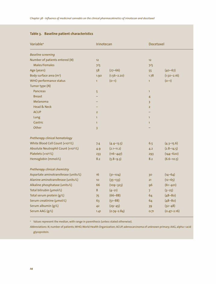
RESULTS Patient accrual. For both the irinotecan- and docetaxel treatment arm, 12 patients completed two
treatments, did not use co-medication and/or dietary supplements known to modulate CYP3A-function, took their medicinal cannabis as prescribed (based on cannabis screening, patient oral declaration and patient treatment-diaries) and were evaluable for irinotecan and docetaxel pharmacokinetic analyses, respectively. Table 3 lists a summary of the baseline characteristics of the 12 patients in both treatment
groups.
23
Chapter 3B - Influence of medicinal cannabis on the clinical pharmacokinetics of irinotecan and docetaxel Chapter 3B - Influence of medicinal cannabis on the clinical pharmacokinetics of irinotecan and docetaxel
a Irinotecan
N) 12 12
) 58 55 2) 1.90
1 1 N)
5 1 – 4
Melanoma – 3
Head & Neck – 2
2 –
Lung 1 1 Gastric 1 1 Other 3 –
(*109/L) 6.5 (*109/L)
*109/L) 233 293 Hemoglobin (mmol/L) 8.2 8.2
16 30 10 21 66 96 8 7 75 64 63 64 42 39
a
, , ;
Table 3. Baseline patient characteristics
Variable Docetaxel
Baseline screening
Number of patients entered (Males/Females 7/5 7/5
Age (years (27–66) (40–67) Body surface area (m (1.56–2.20) 1.78 (1.50–2.16) WHO performance status (0–1) (0–1) Tumor type (
Pancreas Breast
ACUP
Pretherapy clinical hematology
White Blood Cell Count 7.4 (4.4–13.5) (4.3–15.6) Absolute Neutrophil Count 4.9 (2.1–11.2) 4.2 (2.8–14.5) Platelets ( (116–447) (144–620)
(5.8–9.3) (6.6–10.5)
Pretherapy clinical chemistry
Aspartate aminotransferase (units/L) (31–104) (14–64) Alanine aminotransferase (units/L) (35–133) (12–65) Alkaline phosphatase (units/L) (109–323) (61–401) Total bilirubin (µmol/L) (4–21) (3–25) Total serum protein (g/L) (66–88) (48–80) Serum creatinine (µmol/L) (51–88) (48–80) Serum albumin (g/L) (29–45) (32–48) Serum AAG (g/L) 1.41 (0.74–2.84) 0.71 (0.47–2.16)
Values represent the median, with range in parenthesis (unless stated otherwise).
Abbreviations: N, number of patients; WHO World Health Organization; ACUP adenocarcinoma of unknown primary AAG, alpha-1 acid
glycoprotein.
24
Chapter 3B - Influence of medicinal cannabis on the clinical pharmacokinetics of irinotecan and docetaxel Chapter 3B - Influence of medicinal cannabis on the clinical pharmacokinetics of irinotecan and docetaxel
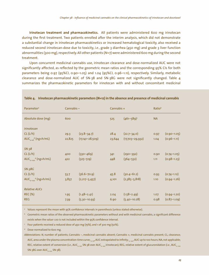
Irinotecan treatment and pharmacokinetics. All patients were administered 600 mg irinotecan during the first treatment. Two patients enrolled after the interim analysis, which did not demonstrate a substantial change in irinotecan pharmacokinetics or increased hematological toxicity, also received a reduced second irinotecan-dose due to toxicity, i.e., grade 3 diarrhea (450 mg) and grade 3 liver function abnormalities (300 mg), respectively. All other patients (N=7) were administered 600 mg during the second treatment.
Upon concurrent medicinal cannabis use, irinotecan clearance and dose-normalized AUC were not
significantly affected, as reflected by the geometric mean ratios and the corresponding 95% CIs for both parameters being 0.97 (95%CI, 0.90–1.05) and 1.04 (95%CI, 0.96–1.11), respectively. Similarly, metabolic clearance and dose-normalized AUC of SN-38 and SN-38G were not significantly changed. Table 4 summarizes the pharmacokinetic parameters for irinotecan with and without concomitant medicinal
l Iri i i (N ) i ici l is
a b
Absolute dose (mg) 600 525 c NA
Irinotecan
29.3 d (ng* 22,825 (17, (17,
400 d (ng* 422 448 1.11
d (ng* , 4,101 1.10
1.95 1.07 6.90
a
b
c
d
,
, ; ,
( , ( ,
Tab e 4. notecan pharmacok net c parameters =12 n the absence and presence of med na cannab
Parameter Cannabis – Cannabis + Ratio
(461–589)
CL (L/h) (23.8–34.7) 28.4 (22.7–34.0) 0.97 (0.90–1.05) AUC0-inf h/mL) 141–28,509) 23,644 703–29,932) 1.04 (0.96–1.11)
SN-38
CL (L/h) (330–469) 341 (290–392) 0.90 (0.74–1.05) AUC0–100
h/mL) (325–519) (364–532) (0.98–1.23)
SN-38G CL (L/h) 53.7 (36.6–70.9) 45.8 (30.4–61.2) 0.93 (0.74–1.12) AUC0–100
h/mL) 3,837 (2,217–5 457) (2,385–5,818) (0.94–1.26)
Relative AUCs
REC (%) (1.48–2.41) 2.04 (1.58–2.49) (0.94–1.20) REG 7.39 (5.30–10.93) (5.40–10.28) 0.98 (0.87–1.09)
Values represent the mean with 95% confidence intervals in parenthesis (unless stated otherwise).
Geometric mean ratios of the observed pharmacokinetic parameters without and with medicinal cannabis, a significant difference
exists when the value 1.00 is not included within the 95% confidence interval.
Four patients received a reduced dose of 450 mg (75%), and 1 of 300 mg (50%).
Dose-normalized to 600 mg.
Abbreviations: N number of patients; Cannabis –, medicinal cannabis absent; Cannabis +, medicinal cannabis present; CL, clearance;
AUC, area under the plasma concentration-time curve; 0–inf AUC extrapolated to infinity 0–100 AUC up to 100 hours; NA, not applicable;
REC, relative extent of conversion i.e. AUC0–100 SN-38 over AUC0–100 irinotecan); REG, relative extent of glucuronidation i.e. AUC0–100
SN-38G over AUC0–100 SN-38).
25
Chapter 3B - Influence of medicinal cannabis on the clinical pharmacokinetics of irinotecan and docetaxel Chapter 3B - Influence of medicinal cannabis on the clinical pharmacokinetics of irinotecan and docetaxel
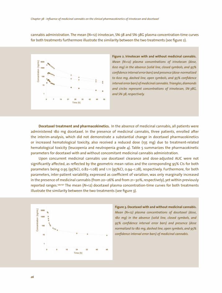
cannabis administration. The mean (N=12) irinotecan, SN-38 and SN-38G plasma concentration-time curves
for both treatments furthermore illustrate the similarity between the two treatments (see figure 2).
(dose, ,
) (, ,
) , diamonds
Figure 2. Irinotecan with and without medicinal cannabis. Mean (N=12) plasma concentrations of irinotecan 600 mg) in the absence (solid line, closed symbols and 95% confidence interval error bars and presence dose-normalized to 600 mg dashed line, open symbols and 95% confidence interval error bars of medicinal cannabis.Trianglesand circles represent concentrations of irinotecan, SN-38G, and SN-38, respectively.
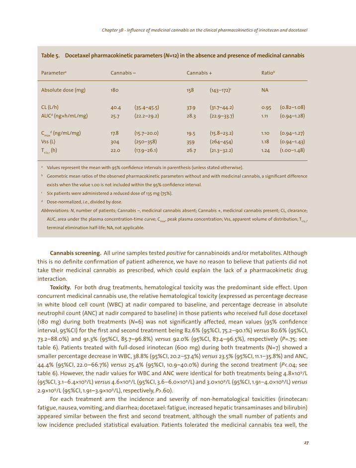
Docetaxel treatment and pharmacokinetics. In the absence of medicinal cannabis, all patients were administered 180 mg docetaxel. In the presence of medicinal cannabis, three patients, enrolled after the interim-analysis, which did not demonstrate a substantial change in docetaxel pharmacokinetics or increased hematological toxicity, also received a reduced dose (135 mg) due to treatment-related hematological toxicity (leucopenia and neutropenia grade 4). Table 5 summarizes the pharmacokinetic parameters for docetaxel with and without concomitant medicinal cannabis administration.
Upon concurrent medicinal cannabis use docetaxel clearance and dose-adjusted AUC were not
significantly affected, as reflected by the geometric mean ratios and the corresponding 95% CIs for both parameters being 0.95 (95%CI, 0.82–1.08) and 1.11 (95%CI, 0.94–1.28), respectively. Furthermore, for both parameters, inter-patient variability, expressed as coefficient of variation, was only marginally increased in the presence of medicinal cannabis (from 20–26% and from 21–30%, respectively), yet within previously reported ranges.149,150 The mean (N=12) docetaxel plasma concentration-time curves for both treatments illustrate the similarity between the two treatments (see figure 3).
(dose, , and
) (dose , ,
Figure 3. Docetaxel with and without medicinal cannabis. Mean (N=12) plasma concentrations of docetaxel 180 mg) in the absence (solid line, closed symbols95% confidence interval error bars and presence normalized to 180 mg dashed line, open symbols and 95% confidence interval error bars) of medicinal cannabis.
26
Chapter 3B - Influence of medicinal cannabis on the clinical pharmacokinetics of irinotecan and docetaxel Chapter 3B - Influence of medicinal cannabis on the clinical pharmacokinetics of irinotecan and docetaxel
l l i i (N ) i ici l is
a b
Absolute dose (mg) 180 158 c NA
d (ng* /mg) 28.3 1.11
Cmax d /mg) 19.5 1.10
1.18 T
γ (h) 22.0 1.24
a
b
c
d
,
max , 1/2,γ,
Tab e 5. Docetaxe pharmacok net c parameters =12 n the absence and presence of med na cannab
Parameter Cannabis – Cannabis + Ratio
(143–172)
CL (L/h) 40.4 (35.4–45.5) 37.9 (31.7–44.2) 0.95 (0.82–1.08) AUC h/mL 25.7 (22.2–29.2) (22.9–33.7) (0.94–1.28)
(ng/mL 17.8 (15.7–20.0) (15.8–23.2) (0.94–1.27) Vss (L) 304 (250–358) 359 (264–454) (0.94–1.43)
1/2, (17.9–26.1) 26.7 (21.3–32.2) (1.00–1.48)
Values represent the mean with 95% confidence intervals in parenthesis (unless stated otherwise).
Geometric mean ratios of the observed pharmacokinetic parameters without and with medicinal cannabis, a significant difference
exists when the value 1.00 is not included within the 95% confidence interval.
Six patients were administered a reduced dose of 135 mg (75%).
Dose-normalized, i.e., divided by dose.
Abbreviations: N number of patients; Cannabis –, medicinal cannabis absent; Cannabis +, medicinal cannabis present; CL, clearance;
AUC, area under the plasma concentration-time curve; C , peak plasma concentration; Vss apparent volume of distribution; T
terminal elimination half-life; NA, not applicable.
Cannabis screening. All urine samples tested positive for cannabinoids and/or metabolites. Although this is no definite confirmation of patient adherence, we have no reason to believe that patients did not
take their medicinal cannabis as prescribed, which could explain the lack of a pharmacokinetic drug interaction.
Toxicity. For both drug treatments, hematological toxicity was the predominant side effect. Upon concurrent medicinal cannabis use, the relative hematological toxicity (expressed as percentage decrease in white blood cell count (WBC) at nadir compared to baseline, and percentage decrease in absolute neutrophil count (ANC) at nadir compared to baseline) in those patients who received full dose docetaxel (180 mg) during both treatments (N=6) was not significantly affected, mean values (95% confidence interval, 95%CI) for the first and second treatment being 82.6% (95%CI, 75.2–90.1%) versus 80.6% (95%CI, 73.2–88.0%) and 91.3% (95%CI, 85.7–96.8%) versus 92.0% (95%CI, 87.4–96.5%), respectively (P=.75; see table 6). Patients treated with full-dosed irinotecan (600 mg) during both treatments (N=7) showed a smaller percentage decrease in WBC, 38.8% (95%CI, 20.2–57.4%) versus 23.5% (95%CI, 11.1–35.8%) and ANC, 44.4% (95%CI, 22.0–66.7%) versus 25.4% (95%CI, 10.9–40.0%) during the second treatment (P<.04; see table 6). However, the nadir values for WBC and ANC were identical for both treatments being 4.8*109/L (95%CI, 3.1–6.4*109/L) versus 4.6*109/L (95%CI, 3.6–6.0*109/L) and 3.0*109/L (95%CI, 1.91–4.0*109/L) versus
2.9*109/L (95%CI, 1.91–3.9*109/L), respectively, P>.60). For each treatment arm the incidence and severity of non-hematological toxicities (irinotecan:
fatigue, nausea, vomiting, and diarrhea; docetaxel: fatigue, increased hepatic transaminases and bilirubin) appeared similar between the first and second treatment, although the small number of patients and low incidence precluded statistical evaluation. Patients tolerated the medicinal cannabis tea well, the
27
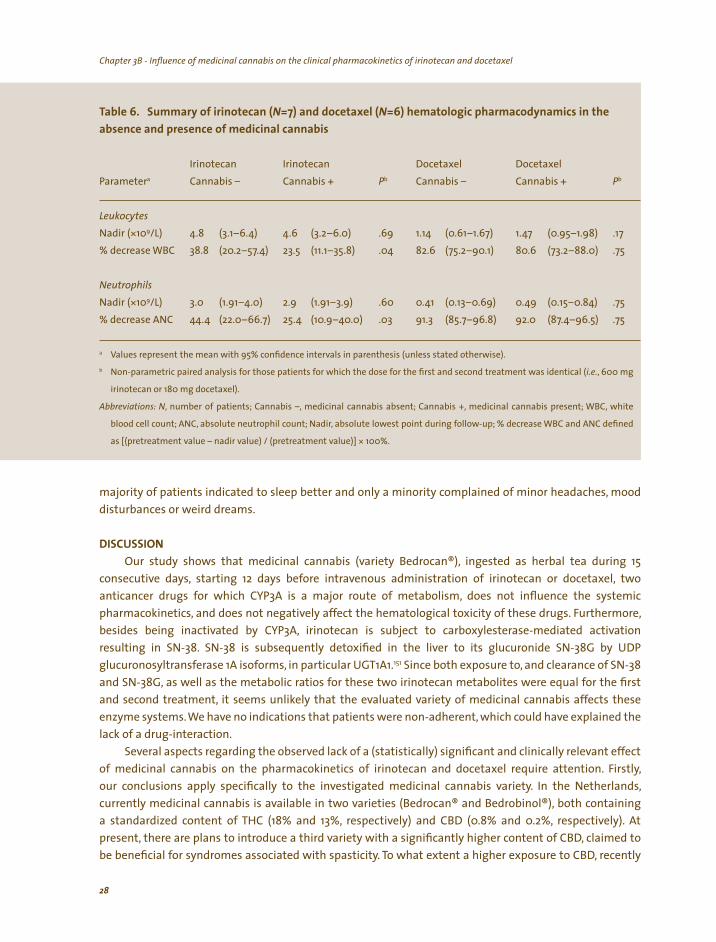
Chapter 3B - Influence of medicinal cannabis on the clinical pharmacokinetics of irinotecan and docetaxel Chapter 3B - Influence of medicinal cannabis on the clinical pharmacokinetics of irinotecan and docetaxel
N N
Irinotecan Irinotecan a Pb Pb
Nadir (*109/L) .69 1.14 .17
38.8 23.5 82.6
Nadir (*109/L) 3.0 2.9 .60 .03 91.3 92.0
a
b ( ,
,
,
* 100%.
Table 6. Summary of irinotecan ( =7) and docetaxel ( =6) hematologic pharmacodynamics in the
absence and presence of medicinal cannabis
Docetaxel Docetaxel Parameter Cannabis – Cannabis + Cannabis – Cannabis +
Leukocytes 4.8 (3.1–6.4) 4.6 (3.2–6.0) (0.61–1.67) 1.47 (0.95–1.98)
% decrease WBC (20.2–57.4) (11.1–35.8) .04 (75.2–90.1) 80.6 (73.2–88.0) .75
Neutrophils
(1.91–4.0) (1.91–3.9) 0.41 (0.13–0.69) 0.49 (0.15–0.84) .75
% decrease ANC 44.4 (22.0–66.7) 25.4 (10.9–40.0) (85.7–96.8) (87.4–96.5) .75
Values represent the mean with 95% confidence intervals in parenthesis (unless stated otherwise).
Non-parametric paired analysis for those patients for which the dose for the first and second treatment was identical i.e. 600 mg
irinotecan or 180 mg docetaxel).
Abbreviations: N number of patients; Cannabis –, medicinal cannabis absent; Cannabis +, medicinal cannabis present; WBC, white
blood cell count; ANC, absolute neutrophil count; Nadir absolute lowest point during follow-up; % decrease WBC and ANC defined
as [(pretreatment value – nadir value) / (pretreatment value)]
majority of patients indicated to sleep better and only a minority complained of minor headaches, mood disturbances or weird dreams.
DISCUSSION
Our study shows that medicinal cannabis (variety Bedrocan®), ingested as herbal tea during 15
consecutive days, starting 12 days before intravenous administration of irinotecan or docetaxel, two
anticancer drugs for which CYP3A is a major route of metabolism, does not influence the systemic
pharmacokinetics, and does not negatively affect the hematological toxicity of these drugs. Furthermore, besides being inactivated by CYP3A, irinotecan is subject to carboxylesterase-mediated activation
resulting in SN-38. SN-38 is subsequently detoxified in the liver to its glucuronide SN-38G by UDP
glucuronosyltransferase 1A isoforms, in particular UGT1A1.151 Since both exposure to, and clearance of SN-38
and SN-38G, as well as the metabolic ratios for these two irinotecan metabolites were equal for the first
and second treatment, it seems unlikely that the evaluated variety of medicinal cannabis affects these
enzyme systems. We have no indications that patients were non-adherent, which could have explained the
lack of a drug-interaction. Several aspects regarding the observed lack of a (statistically) significant and clinically relevant effect
of medicinal cannabis on the pharmacokinetics of irinotecan and docetaxel require attention. Firstly, our conclusions apply specifically to the investigated medicinal cannabis variety. In the Netherlands, currently medicinal cannabis is available in two varieties (Bedrocan® and Bedrobinol®), both containing a standardized content of THC (18% and 13%, respectively) and CBD (0.8% and 0.2%, respectively). At
present, there are plans to introduce a third variety with a significantly higher content of CBD, claimed to be beneficial for syndromes associated with spasticity. To what extent a higher exposure to CBD, recently
28
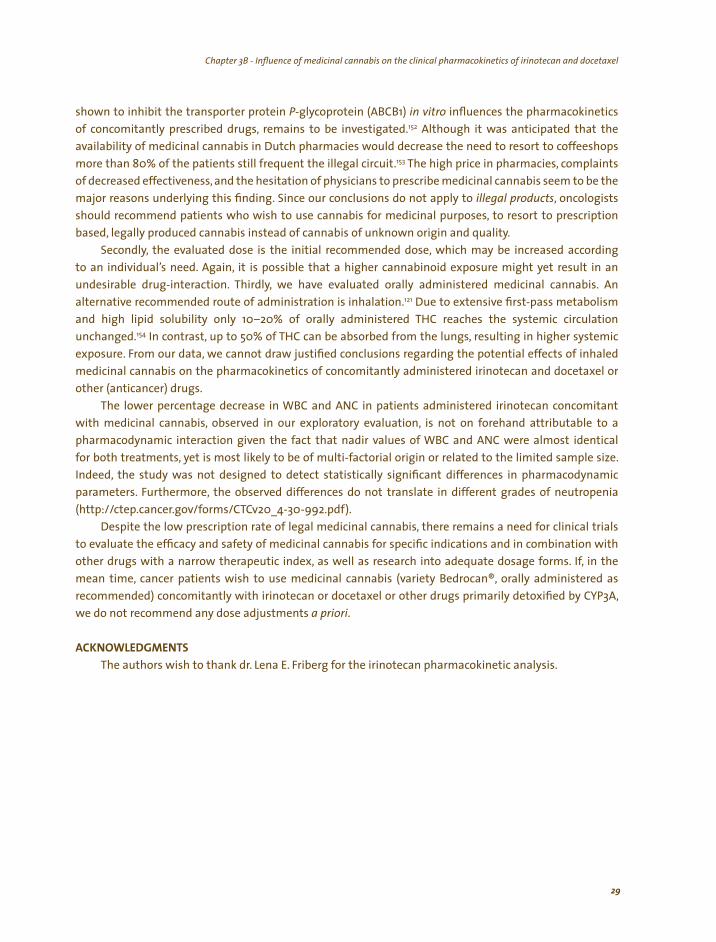
Chapter 3B - Influence of medicinal cannabis on the clinical pharmacokinetics of irinotecan and docetaxel Chapter 3B - Influence of medicinal cannabis on the clinical pharmacokinetics of irinotecan and docetaxel
shown to inhibit the transporter protein P-glycoprotein (ABCB1) in vitro influences the pharmacokinetics of concomitantly prescribed drugs, remains to be investigated.152 Although it was anticipated that the availability of medicinal cannabis in Dutch pharmacies would decrease the need to resort to coffeeshops more than 80% of the patients still frequent the illegal circuit.153 The high price in pharmacies, complaints of decreased effectiveness, and the hesitation of physicians to prescribe medicinal cannabis seem to be the major reasons underlying this finding. Since our conclusions do not apply to illegal products, oncologists should recommend patients who wish to use cannabis for medicinal purposes, to resort to prescription based, legally produced cannabis instead of cannabis of unknown origin and quality.
Secondly, the evaluated dose is the initial recommended dose, which may be increased according to an individual’s need. Again, it is possible that a higher cannabinoid exposure might yet result in an undesirable drug-interaction. Thirdly, we have evaluated orally administered medicinal cannabis. An alternative recommended route of administration is inhalation.121 Due to extensive first-pass metabolism and high lipid solubility only 10–20% of orally administered THC reaches the systemic circulation unchanged.154 In contrast, up to 50% of THC can be absorbed from the lungs, resulting in higher systemic exposure. From our data, we cannot draw justified conclusions regarding the potential effects of inhaled medicinal cannabis on the pharmacokinetics of concomitantly administered irinotecan and docetaxel or other (anticancer) drugs.
The lower percentage decrease in WBC and ANC in patients administered irinotecan concomitant
with medicinal cannabis, observed in our exploratory evaluation, is not on forehand attributable to a pharmacodynamic interaction given the fact that nadir values of WBC and ANC were almost identical for both treatments, yet is most likely to be of multi-factorial origin or related to the limited sample size. Indeed, the study was not designed to detect statistically significant differences in pharmacodynamic parameters. Furthermore, the observed differences do not translate in different grades of neutropenia (http://ctep.cancer.gov/forms/CTCv20_4-30-992.pdf).
Despite the low prescription rate of legal medicinal cannabis, there remains a need for clinical trials to evaluate the efficacy and safety of medicinal cannabis for specific indications and in combination with other drugs with a narrow therapeutic index, as well as research into adequate dosage forms. If, in the mean time, cancer patients wish to use medicinal cannabis (variety Bedrocan®, orally administered as recommended) concomitantly with irinotecan or docetaxel or other drugs primarily detoxified by CYP3A, we do not recommend any dose adjustments a priori.
ACKNOWLEDGMENTS
The authors wish to thank dr. Lena E. Friberg for the irinotecan pharmacokinetic analysis.
29

30
31
Chapter 4
Role of pharmacogenetics in irinotecan therapy
FA de Jong, MJ de Jonge, J Verweij, RH Mathijssen
Cancer Letters 234(1):90–106, 2006
Chapter 4 - Role of pharmacogenetics in irinotecan therapy
32
Chapter 4 - Role of pharmacogenetics in irinotecan therapy
ABSTRACT
In the treatment of advanced colorectal cancer, irinotecan has become one of the most important drugs, despite its sometimes unpredictable adverse effects. To understand why some patients experience severe adverse effects (diarrhea and neutropenia), while others do not, the metabolic pathways of this drug have to be unraveled in detail. Individual variation in expression of several phase I and phase II metabolizing enzymes and ABC-transporters involved in irinotecan metabolism and excretion, at least partly explains the observed pharmacokinetic interpatient variability. Although the differences in expression level of these proteins to a certain amount is explained by physiologic and environmental factors, the presence of specific genetic determinants also does influence their expression and function. In this review, the role of genetic polymorphisms in the main enzyme-systems (carboxylesterase, cytochrome P450 3A, and uridine diphosphate-glucuronosyltransferase) and ABC-transporters (ABCB1, ABCC2, and ABCG2) involved in irinotecan metabolism, are discussed. Since at this moment
the field of pharmacogenetics and pharmacogenomics is rapidly expanding and simultaneously more rapid and cost
effective screening methods are emerging, a wealth of future data is expected to enrich our knowledge of the genetic basis of irinotecan metabolism. Eventually, this may help to truly individualize the dosing of this (and other) anticancer agent(s), using a personal genetic profile of the most relevant enzymes for every patient.

Chapter 4 - Role of pharmacogenetics in irinotecan therapy Chapter 4 - Role of pharmacogenetics in irinotecan therapy
INTRODUCTION
Since its introduction, irinotecan (Camptosar®; Campto®) has become a valuable drug in the treatment of nonresectable and metastatic colorectal cancer, especially in combination with 5-fluorouracil (5-FU) and oxaliplatin.9,10,13,155 Partly as a result of the development of this agent, life expectancy of patients suffering from incurable colorectal cancer has doubled during the last decade.156 Responses to irinotecan are also seen in various other types of cancer, including lung and gastric malignancies.15-21 Like other camptothecins, the antineoplastic agent irinotecan (7-ethyl-10-[4-(1-piperidino)-1-piperidino]carbonyl-oxycamptothecin; CPT-11) and in particular its active metabolite SN-38 (7-ethyl-10-hydroxycamptothecin) stabilize the DNA-topoisomerase I complex by binding to it, preventing the resealing of single strand breaks.7 Irinotecan prevents the replication fork to proceed which results in double strand breaks and ultimately in its antitumor effect and its characteristic adverse effects on rapidly dividing tissues, such as bone marrow and intestinal mucosa. The main dose-limiting toxicities of irinotecan therapy are therefore myelosuppression and delayed-type diarrhea.29,157-160 Unfortunately, as a result of its complicated and highly variable expressed metabolic pathways, irinotecan and its metabolites are subject to extensive interindividual pharmacokinetic and pharmacodynamic variability.29,161,162
Many attempts have been made to understand the complex pharmacokinetic profile of irinotecan. However, up to now this has not resulted in a better prediction of occurrence and severity of adverse effects in clinical practice. As it has earlier clearly been demonstrated that body surface area (BSA) based dosing strategies do not significantly reduce interindividual variability in irinotecan pharmacokinetics and occurrence of adverse effects,50,51,139 other ways of dosing irinotecan are needed to predict the pharmacologic profile in individual patients. The metabolic pathways of irinotecan involve various metabolizing enzymes and drug transporters (see figure 4). As variations in their encoding genes may lead to an altered expression and/or function and may contribute to the variability in pharmacologic profiles between patients, research into variations in these genes may be important. Most variation in human DNA is limited to so-called single nucleotide polymorphisms (SNPs).163 Some of these SNPs may have functional consequences when resulting in total loss or diminished expression of the encoding gene, or when amino acid substitution leads to functional alterations. Other genetic variants in the human genome are, among others, two-base pair insertions (TA) in the TATA-box in the promoter-region of genes, leading to a lowered transcription of
Fi Iri li Iri i i l-( ) , i l i lls, i
iri ( i i ive
) ll i i i, i llul iri i l j i i ,
l i ( ), in
i i i i , i l ( )
i i iri i ly i ili i i , i i i l i i idi
di l l ily ( ). Iri i li ll l
i i i i ( ) , wi ly
l i i iti i
gure 4. notecan metabo sm. notecan s act vated by carboxyesterases CES type 1 and 2 found n p asma and n ce nto SN-38. Both notecan and SN-38 can be transported by d ffus on and acttransport across the ce membrane. Bes des be ng converted nto
SN-38 ntrace ar notecan s a so sub ect to an ox dat ve pathwaycata yzed by cytochrome P450 soforms 3A4 and 3A5 CYP3Awh ch the nact ve substances APC and NPC are formed. On ts turnNPC can be converted nto SN-38 by the same carboxy esterases CESact vat ng notecan d rect nto SN-38. To fac tate ts excret on the
act ve compound SN-38 s detox fied by g ucuron dat on by ur ne
phosphate-g ucuronosy transferases from the 1A subfam UGT1Anotecan and ts metabo tes are transported out of the ce s by severa
adenos ne tr phosphate b nd ng cassette ABC transporters th partover app ng spec fic es for the d fferent compounds.
33
Chapter 4 - Role of pharmacogenetics in irinotecan therapyChapter 4 - Role of pharmacogenetics in irinotecan therapy
the protein. Here, we review the role of polymorphisms in the genes involved in the metabolic pathway of irinotecan on the variation in pharmacokinetics and pharmacodynamics between patients, and discuss their potential role in future dosing strategies, aiming to lower interpatient variability in exposure to irinotecan and its metabolites and therapy-induced adverse effects.
METABOLIC PATHWAYS OF IRINOTECAN
have been demonstrated.
In humans, irinotecan is hydrolyzed into its active metabolite SN-38 by carboxylesterases (CES; see
figure 4), present in serum, intestines, tumor tissue, and in high content in the liver.23,164-166 Recently, the
opinion is emerging that intratumoral activation of irinotecan into SN-38 by CES might be even more
important than systemic circulating SN-38 levels, formed by hepatic CES.25,26,167-169 Although plasma levels
of SN-38 are relatively low, relations between SN-38 and myelosuppression and/or delayed-type diarrhea 29,44,157,158,160,170-172 Uridine diphosphate-glucuronosyltransferase 1A (UGT1A) mediated
glucuronidation of SN-38, forming a β-glucuronic acid conjugate (SN-38G; 10-O-glucuronyl-SN-38), is the
main pathway ofdetoxification forSN-38.Besidesbeinghydrolyzed,irinotecanis also sensitive to cytochrome
P450 3A (CYP3A) mediated oxidative pathways, resulting in the formation of inactive metabolites, including
APC (7-ethyl-10-[4-N-(5-aminopentanoic acid)-1-piperidino]-carbonyloxycamptothecin) and NPC (7-ethyl-10-(4-amino-1-piperidino)carbonyloxycamptothecin).29,32 In turn, NPC may be further metabolized into
.33,173 SN-38 by CES as wellBoth irinotecan, SN-38, and their metabolites are excreted by drug-transporting proteins from the
adenosine-triphosphate binding cassette (ABC) transporter superfamily via a hepatobiliary pathway into the feces, and to a lesser extent (ratio about 2:1) into urine.174 Besides influencing the renal and hepatobiliary secretion, these drug-pumps can affect intestinal re-absorption, and can also play a role in acquired (multi)drug resistance.29,175-177 In the intestines, hepatobiliary excreted irinotecan and NPC may be converted into SN-38 by local CES,173 and SN-38G can be deglucuronidated into SN-38 by endogenous bacterial β-glucuronidases,137 leading to local toxic effects of SN-38 on the intestinal mucosa. These processes are thought to play a role in delayed-type diarrhea, seen around 5–7 days after infusion of irinotecan in about 40% of patients.41 Both SN-38 and irinotecan are thought to undergo re-uptake from the intestines with resulting re-entrance in the systemic circulation (enterohepatic recirculation).178,179
ACTIVATION OF IRINOTECAN BY CARBOXYLESTERASES
Historically, systemic circulation of SN-38, the metabolite formed by hepatic carboxylesterases (CES) and known to be 100–1,000 times more cytotoxic than the parent compound, is believed to be of major importance for the therapeutic effects of irinotecan therapy. Despite the fact that CES in plasma are capable
of activating irinotecan as well,180 systemic levels of SN-38 after a single dose of irinotecan are relatively low.29
This may be explained by the ineffective conversion of irinotecan by CES,181 competition between the CES
mediated pathway and the CYP3A-mediated pathway,182 and the fact that the majority of (hepatic) formed
SN-38, both as SN-38 and as its glucuronide SN-38G, is excreted through the bile into the feces. In contrast to
systemic toxicity, to the best of our knowledge, therapeutic effectivity has not clearly been linked to exposure
to SN-38. As stated before, the opinion is emerging that intratumoral activation of irinotecan by CES might be
more important than its hepatic and systemic activation.25,26,167-169 In vitro studies have shown that activation
of irinotecan in the tumor is related to the expression of CES.25 Indeed, most colorectal tumors are found to
intrinsically express CES, albeit probably to a lower extent than healthy colorectal tissue.125,183 As mutations in 125 the genomic material of the CES genes in tumor tissue were found to be identical to those in healthy tissue,
other unrevealed factors may influence their local expression.
34
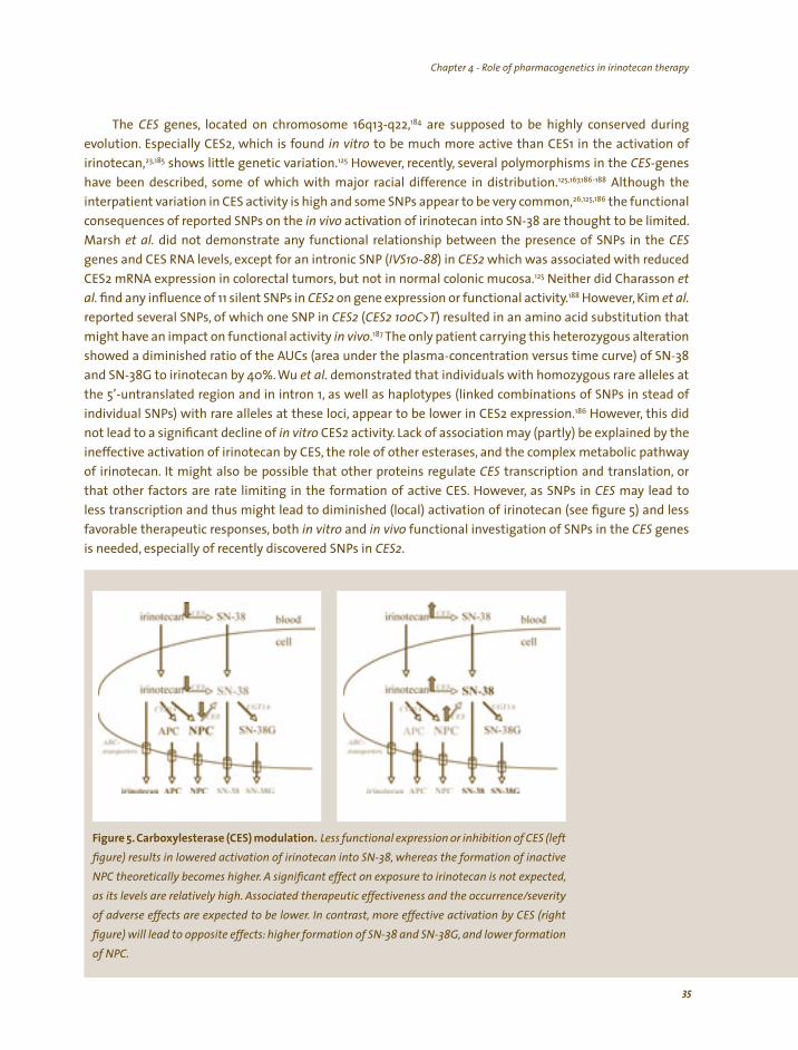
Chapter 4 - Role of pharmacogenetics in irinotecan therapy Chapter 4 - Role of pharmacogenetics in irinotecan therapy
The CES genes, located on chromosome 16q13-q22,184 are supposed to be highly conserved during
evolution. Especially CES2, which is found in vitro to be much more active than CES1 in the activation of irinotecan,23,185 shows little genetic variation.125 However, recently, several polymorphisms in the CES-genes
have been described, some of which with major racial difference in distribution.125,167,186-188 Although the
interpatient variation in CES activity is high and some SNPs appear to be very common,26,125,186 the functional consequences of reported SNPs on the in vivo activation of irinotecan into SN-38 are thought to be limited. Marsh et al. did not demonstrate any functional relationship between the presence of SNPs in the CES
genes and CES RNA levels, except for an intronic SNP (IVS10-88) in CES2 which was associated with reduced
CES2 mRNA expression in colorectal tumors, but not in normal colonic mucosa.125 Neither did Charasson et al. find any influence of 11 silent SNPs in CES2 on gene expression or functional activity.188 However, Kim et al. reported several SNPs, of which one SNP in CES2 (CES2 100C>T) resulted in an amino acid substitution that
might have an impact on functional activity in vivo.187 The only patient carrying this heterozygous alteration
showed a diminished ratio of the AUCs (area under the plasma-concentration versus time curve) of SN-38
and SN-38G to irinotecan by 40%. Wu et al. demonstrated that individuals with homozygous rare alleles at
the 5’-untranslated region and in intron 1, as well as haplotypes (linked combinations of SNPs in stead of individual SNPs) with rare alleles at these loci, appear to be lower in CES2 expression.186 However, this did
not lead to a significant decline of in vitro CES2 activity. Lack of association may (partly) be explained by the
ineffective activation of irinotecan by CES, the role of other esterases, and the complex metabolic pathway
of irinotecan. It might also be possible that other proteins regulate CES transcription and translation, or that other factors are rate limiting in the formation of active CES. However, as SNPs in CES may lead to
less transcription and thus might lead to diminished (local) activation of irinotecan (see figure 5) and less
favorable therapeutic responses, both in vitro and in vivo functional investigation of SNPs in the CES genes
is needed, especially of recently discovered SNPs in CES2.
(
/
Figure 5. Carboxylesterase CES) modulation. Less functional expression or inhibition of CES (left figure) results in lowered activation of irinotecan into SN-38, whereas the formation of inactive NPC theoretically becomes higher. A significant effect on exposure to irinotecan is not expected, as its levels are relatively high. Associated therapeutic effectiveness and the occurrence severity of adverse effects are expected to be lower. In contrast, more effective activation by CES (right figure) will lead to opposite effects: higher formation of SN-38 and SN-38G, and lower formation of NPC.
35
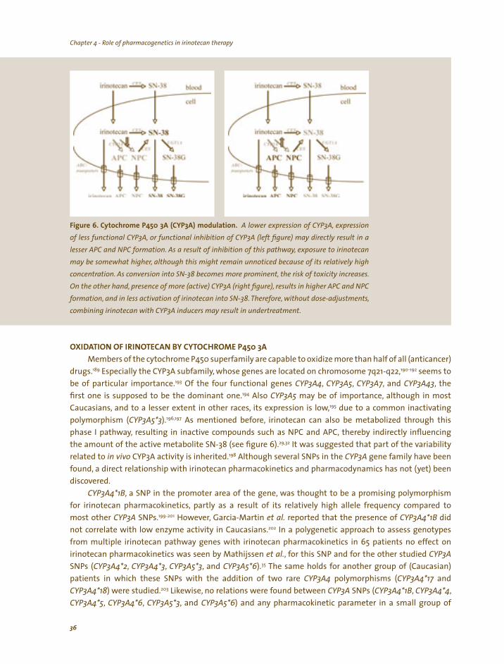
Chapter 4 - Role of pharmacogenetics in irinotecan therapyChapter 4 - Role of pharmacogenetics in irinotecan therapy
(
, ,
(,
Figure 6. Cytochrome P450 3A CYP3A) modulation. A lower expression of CYP3A, expression of less functional CYP3A, or functional inhibition of CYP3A (left figure) may directly result in a lesser APC and NPC formation. As a result of inhibition of this pathway exposure to irinotecan may be somewhat higher although this might remain unnoticed because of its relatively high concentration. As conversion into SN-38 becomes more prominent, the risk of toxicity increases. On the other hand, presence of more active) CYP3A (right figure), results in higher APC and NPC formation, and in less activation of irinotecan into SN-38. Therefore, without dose-adjustmentscombining irinotecan with CYP3A inducers may result in undertreatment.
OXIDATION OF IRINOTECAN BY CYTOCHROME P450 3A
Members of the cytochrome P450 superfamily are capable to oxidize more than half of all (anticancer) drugs.189 Especially the CYP3A subfamily, whose genes are located on chromosome 7q21-q22,190-192 seems to be of particular importance.193 Of the four functional genes CYP3A4, CYP3A5, CYP3A7, and CYP3A43, the first one is supposed to be the dominant one.194 Also CYP3A5 may be of importance, although in most
Caucasians, and to a lesser extent in other races, its expression is low,195 due to a common inactivating polymorphism (CYP3A5*3).196,197 As mentioned before, irinotecan can also be metabolized through this phase I pathway, resulting in inactive compounds such as NPC and APC, thereby indirectly influencing the amount of the active metabolite SN-38 (see figure 6).29,32 It was suggested that part of the variability related to in vivo CYP3A activity is inherited.198 Although several SNPs in the CYP3A gene family have been found, a direct relationship with irinotecan pharmacokinetics and pharmacodynamics has not (yet) been discovered.
CYP3A4*1B, a SNP in the promoter area of the gene, was thought to be a promising polymorphism for irinotecan pharmacokinetics, partly as a result of its relatively high allele frequency compared to most other CYP3A SNPs.199-201 However, Garcia-Martin et al. reported that the presence of CYP3A4*1B did not correlate with low enzyme activity in Caucasians.202 In a polygenetic approach to assess genotypes from multiple irinotecan pathway genes with irinotecan pharmacokinetics in 65 patients no effect on irinotecan pharmacokinetics was seen by Mathijssen et al., for this SNP and for the other studied CYP3A
SNPs (CYP3A4*2, CYP3A4*3, CYP3A5*3, and CYP3A5*6).35 The same holds for another group of (Caucasian) patients in which these SNPs with the addition of two rare CYP3A4 polymorphisms (CYP3A4*17 and CYP3A4*18) were studied.203 Likewise, no relations were found between CYP3A SNPs (CYP3A4*1B, CYP3A4*4, CYP3A4*5, CYP3A4*6, CYP3A5*3, and CYP3A5*6) and any pharmacokinetic parameter in a small group of
36

Chapter 4 - Role of pharmacogenetics in irinotecan therapy Chapter 4 - Role of pharmacogenetics in irinotecan therapy
Chinese patients receiving irinotecan.204 These results should be confirmed in larger groups of patients and with patients of different ethnicity, as the allele frequency of CYP3A4*1B for instance is almost absent in Asians, while it is five times higher in Africans as compared to Caucasians.199,205,206 On the other hand, the CYP3A4*18 variant is more common among Asians than in other studied races.206 This implicates that, for the time being, the impact of genetic variants in CYP3A on irinotecan metabolism remains undetermined and needs to be further clarified.
However, since variation in gene expression and function is seen, this is more likely caused by environmental (including co-medication and food-substances) and/or physiologic (such as altered liver function and performance status) factors. After stimulation by exogenous drugs, the pregnane X-receptor (PXR; NR1I2) is involved in an increased expression of CYP3A4 (and also P-glycoprotein, ABCB1) mRNA.207-209
Such exogenous drugs are for instance St. John’s wort and Kava, with clear effects on irinotecan pharmacokinetics, as illustrated in figure 6.182,210 Inhibition of CYP3A may also be seen as a result of coadministered drugs, such as ketoconazole, indirectly leading to increased levels of the active compound SN-38 since more irinotecan remains available to be activated through the CES pathway.124 As recently has been demonstrated, such environmental and physiologic factors are probably more important for CYP3A activity in the irinotecan metabolism than inherited changes.203 Therefore, the variation in the functional presence of CYP3A should be taken into account when studying irinotecan interpatient pharmacokinetic variability, rather than single genetic polymorphisms. In the last paragraph of this review, we focus more closely on ways to predict this variation using phenotypic measures.
DETOXIFICATION OF SN-38 BY UGT1A
populations.
In vertebrates, the glucuronidation of a range of endogenous and exogenous (lipophilic) compounds is catalyzed by the microsomal uridine diphosphate-glucuronosyltransferase (UGT) enzymes.211 The formation of hydrophilic glucuronides facilitates the excretion of these molecules.212 The human UGT superfamily has been classified into the UGT1 and UGT2 families, further classified into three subfamilies (UGT1A, UGT2A, and UGT2B).211,213 All nine functional members of the UGT1A subfamily are encoded by a single gene locus, the UGT1A locus on chromosome 2q37.214,215 Especially the UGT isoforms 1A1, 1A7 and 1A9 are involved in the phase II conjugation of SN-38 to the inactive metabolite SN-38G.27,28,216-220 As UGT1A7 is only expressed in extra hepatic tissues (esophagus, stomach, and lung),213,221-223 its role in the conversion of systemic SN-38 remains unclear.221,224 On the other hand, UGT1A1 and UGT1A9 are highly expressed in the gastrointestinal tract and the liver; the primary organ involved in the detoxification of irinotecan.218,225 In addition, UGT1A1 is the primary enzyme responsible for forming bilirubin glucuronides. Since observations of patients with physiological hyperbilirubinemia have led to interest in bilirubin metabolism, UGT1A1 has become an extensively studied UGT isoform. UGT1A1 contains many genetic variants influencing its expression and functional properties. Polymorphisms, resulting in absent or very low UGT1A1 activity, have been associated with three heritable unconjugated hyperbilirubinemia syndromes: Crigler-Najjar syndrome type 1 and 2, and Gilbert’s syndrome.226-229 Gilbert’s syndrome is common among Caucasians and is associated with the presence of an extra, seventh, dinucleotide (TA) insertion (UGT1A1*28) in the (TA)6TAA-box of the UGT1A1 promoter region,228,229 leading to a considerable reduced enzyme expression of about 30–80%.228,230,231 The UGT1A1 activity appears to be inversely related to the number of TA-repeats, varying from 5–8.230 The frequency of the additional TA-repeats has been reported to vary among ethnic
35,49,203,229,232-237 For instance, the incidence of TA7-homozygosity is much lower in Asians compared to Caucasians and Africans. In contrast, other UGT1A1 polymorphisms (especially UGT1A1*6) have also been causally related to Gilbert’s syndrome, and are much more common in Asians.238-240
37

Chapter 4 - Role of pharmacogenetics in irinotecan therapyChapter 4 - Role of pharmacogenetics in irinotecan therapy
Fi idi i l l ( ) l ii i lti in l ( l i l) i (l ) l
l i i i i iici , i i i i (ri ), l i
gl i i , ill i , ill l , i lt in l l i
gure 7. Ur ne d phosphate-g ucuronysy transferase UGT1A modu at on. The presence of genet c var ants resu ng ower or ess funct ona express on of UGT1A eft figure ead to
a reduced g ucuron dat on capac ty and therefore to h gher exposure to SN-38 and assoc ated
tox ty. On the other hand nduct on of ts express on ght figure may ead to ncreased
ucuron dat on of SN-38. As a consequence exposure to SN-38G w be h gher whereas exposure
to SN-38 w be ower wh ch may resu ess favorab e therapeut c outcomes.
7%.
Studies have shown that the homozygous UGT1A1*28 genotype was associated with an increased risk of developing leucopenia and severe delayed-type diarrhea after treatment with irinotecan. Ando et al. were the first to retrospectively analyze the association between UGT1A1 variants and irinotecan toxicity, revealing in a multivariate analysis that presence of UGT1A1*28 allele was a risk factor for severe toxicity.45
These data have been confirmed by other groups (see table 7).46-48,203 Based on this knowledge and the finding that demonstrated a good concordance between the UGT1A1*28 genotype and less effective SN-38 glucuronidation,27,28,241 prospective studies were initiated.46,49 A significant relation was observed between the AUC of SN-38 and the number of TA-alleles (see figure 7 and table 8). Also, the relative extent of glucuronidation of SN-38 to SN-38G (REG; the ratio of SN-38G AUC to SN-38 AUC), was inversely related to the number of TA-repeats,46,49,242 although Balram et al. could not confirm these findings in the earlier mentioned Chinese study-group, which might be explained by the relative low variant allele frequency of
204 In addition, two other promoter variants (common SNPs in the so-called phenobarbital-responsive enhancer module; i.e., UGT1A1 -3279G>T (also referred as UGT1A1 -3263T>G due to different reference sequence) and UGT1A1 -3156G>A) have been identified. These variants are in strong linkage disequilibrium with the UGT1A1*28 polymorphism in Caucasians, while this link is less apparent in African-Americans and Asians, suggesting a different haplotype structure among various races.204,243 This might partly explain racial phenotypic variability. Ando et al. found a strong a relation for presence of the UGT1A1 -3263T>G SNP and the severity of irinotecan-induced toxicity, although in a multivariate analysis including UGT1A1*28 as well, this effect was mainly attributed to this latter polymorphism.244 However, patients carrying both variants had significantly higher risk (six times) on severe adverse effects. Although present with an allele frequency of 36%, a relation between the UGT1A1 -3263T>G variation and irinotecan pharmacokinetic parameters could not be established in Chinese patients.204 In a study by Innocenti et al., it was suggested that the UGT1A1 -3156G>A SNP might be a better predictor of the patient’s functional UGT1A1 status than UGT1A1*28.49 Last year, Sai et al. published a comprehensive haplotype analysis of the entire UGT1A1 gene
38
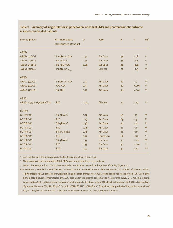
Chapter 4 - Role of pharmacogenetics in irinotecan therapy Chapter 4 - Role of pharmacogenetics in irinotecan therapy
qa N P
w .038 35
w .031 35
w 30 203
w Irinotecan Cmax –b Chinese 29
w 64 .02 264
w 64 <.001 264
w c <.001 264
w Chinese 29 .019
w 65 .03 49
w 65 .03 49
w 20 .001 w 20 .001 w 20 .001 w 86 .022 242
w 30 .006 203
w 30 <.001 203
w 30 .010 203
a ≥.01 or ≤.99. b
c 7/ 7
N ,
P , ;
max
,
Table 7. Summary of single relationships between individual SNPs and pharmacokinetic outcome in irinotecan-treated patients
Polymorphism Pharmacokinetic Race Ref consequence of variant
ABCB1 ABCB1 1236C>T Irinotecan AUC 0.34 Eur Cauc 46 ABCB1 1236C>T SN-38 AUC 0.34 Eur Cauc 46 ABCB1 1236C>T SN-38G AUC 0.48 Eur Cauc .042 ABCB1 3435C>T .047 204
ABCC2
ABCC2 3972C>T Irinotecan AUC 0.35 Am Cauc ABCC2 3972C>T APC AUC 0.35 Am Cauc ABCC2 3972C>T SN-38G 0.35 Am Cauc 54
ABCG2
ABCG2 -19572-19569delCTCA REC 0.04 204
UGT1A1 UGT1A1*28 SN-38 AUC 0.29 Am Cauc UGT1A1*28 REG 0.29 Am Cauc UGT1A1*28 SN-38 AUC 0.38 Am Cauc 46
UGT1A1*28 REG 0.38 Am Cauc 46
UGT1A1*28 Biliary index 0.38 Am Cauc 46
UGT1A1*28 REG 0.27 Caucasian UGT1A1*28 SN-38 AUC 0.35 Eur Cauc UGT1A1*28 REC 0.35 Eur Cauc UGT1A1*28 REG 0.35 Eur Cauc
Only mentioned if the observed variant allele frequency (q) was
Allele frequencies of three studied ABCB1 SNPs were reported between 0.25 and 0.39.
Patients homozygous for UGT1A1*28 were excluded to minimize the confounding effect of the TA TA repeat.
Abbreviations: q, standard Hardy-Weinberg nomenclature for observed variant allele frequencies; , number of patients; ABCB1
-glycoprotein; ABCC2 canalicular multispecific organic anion transporter ABCG2, breast cancer resistance protein; UGT1A1, uridine
diphosphate-glucuronosyltransferase 1A1; AUC, area under the plasma concentration versus time curve; C , maximal plasma
concentration; REC, relative extent of conversion of irinotecan to SN-38, i.e. ratio of SN-38 AUC to irinotecan AUC; REG, relative extent
of glucuronidation of SN-38 to SN-38G, i.e., ratio of SN-38G AUC to SN-38 AUC; Biliary index, the product of the relative area ratio of
SN-38 to SN-38G and the AUC CPT-11; Am Cauc, American Caucasian; Eur Cauc, European Caucasian.
39
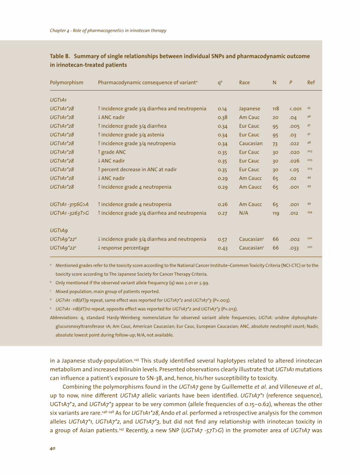
Chapter 4 - Role of pharmacogenetics in irinotecan therapyChapter 4 - Role of pharmacogenetics in irinotecan therapy
a qb N P
w / Japanese 118 <.001 45
w ANC nadir 20 w /4 diarrhea 95 .005 w /4 astenia 95 .03 w / 73 .022 48
w 30 .020 203
w ANC nadir 30 .026 203
w 30 <.05 203
w ANC nadir 65 .02 49
w 65 .001 49
w 65 .001 49
w / 119 .012 244
d w / c 66 .002 220
e w c 66 .033 220
a
b ≥.01 or ≤.99. c
d and (P=.003). e and (P=.013).
Table 8. Summary of single relationships between individual SNPs and pharmacodynamic outcome in irinotecan-treated patients
Polymorphism Pharmacodynamic consequence of variant Race Ref
UGT1A1 UGT1A1*28 incidence grade 3 4 diarrhea and neutropenia 0.14 UGT1A1*28 0.38 Am Cauc .04 46
UGT1A1*28 incidence grade 3 0.34 Eur Cauc 47
UGT1A1*28 incidence grade 3 0.34 Eur Cauc 47
UGT1A1*28 incidence grade 3 4 neutropenia 0.34 Caucasian UGT1A1*28 grade ANC 0.35 Eur Cauc UGT1A1*28 0.35 Eur Cauc UGT1A1*28 percent decrease in ANC at nadir 0.35 Eur Cauc UGT1A1*28 0.29 Am Caucc UGT1A1*28 incidence grade 4 neutropenia 0.29 Am Caucc
UGT1A1 -3156G>A incidence grade 4 neutropenia 0.26 Am Caucc UGT1A1 -3263T>G incidence grade 3 4 diarrhea and neutropenia 0.27 N/A
UGT1A9
UGT1A9*22 incidence grade 3 4 diarrhea and neutropenia 0.57 CaucasianUGT1A9*22 response percentage 0.43 Caucasian
Mentioned grades refer to the toxicity score according to the National Cancer Institute–Common Toxicity Criteria (NCI-CTC) or to the
toxicity score according to The Japanese Society for Cancer Therapy Criteria.
Only mentioned if the observed variant allele frequency (q) was
Mixed population, main group of patients reported.
UGT1A1 -118(dT)9 repeat, same effect was reported for UGT1A7*2 UGT1A7*3
UGT1A1 -118(dT)10 repeat, opposite effect was reported for UGT1A7*2 UGT1A7*3
Abbreviations: q, standard Hardy-Weinberg nomenclature for observed variant allele frequencies; UGT1A: uridine diphosphate-
glucuronosyltransferase 1A; Am Cauc, American Caucasian; Eur Cauc, European Caucasian; ANC, absolute neutrophil count; Nadir,
absolute lowest point during follow-up; N/A, not available.
in a Japanese study-population.245 This study identified several haplotypes related to altered irinotecan metabolism and increased bilirubin levels. Presented observations clearly illustrate that UGT1A1 mutations can influence a patient’s exposure to SN-38, and, hence, his/her susceptibility to toxicity.
Combining the polymorphisms found in the UGT1A7 gene by Guillemette et al. and Villeneuve et al., up to now, nine different UGT1A7 allelic variants have been identified. UGT1A7*1 (reference sequence), UGT1A7*2, and UGT1A7*3 appear to be very common (allele frequencies of 0.15–0.62), whereas the other six variants are rare.246-248 As for UGT1A1*28, Ando et al. performed a retrospective analysis for the common alleles UGT1A7*1, UGT1A7*2, and UGT1A7*3, but did not find any relationship with irinotecan toxicity in a group of Asian patients.247 Recently, a new SNP (UGT1A7 -57T>G) in the promoter area of UGT1A7 was
40
Chapter 4 - Role of pharmacogenetics in irinotecan therapy Chapter 4 - Role of pharmacogenetics in irinotecan therapy
found.249 However, a relation between this common variant and systemic irinotecan pharmacokinetics has not yet been determined. As linkage disequilibrium between allelic variants of UGT1A1, UGT1A6, and UGT1A7 exists, homozygous UGT1A1*28 patients were found to carry this UGT1A7 polymorphism in almost
all cases. The existence of such complex relationships in combination with the extra hepatic expression of UGT1A7 makes it extremely difficult to detect relationships in vivo, and may obscure the clinical value of the mutation.235,249 However, in a retrospective study by Carlini et al. in 66 patients treated with the combination of irinotecan and capecitabine the genetic variations UGT1A7*2 and UGT1A7*3, which both result in lower enzyme activity and less transcriptional activity, were associated with lower (gastrointestinal) irinotecan-induced toxicity and even higher antitumor response in this study population, which mainly consisted of Caucasian patients (see table 7).220
In addition to UGT1A1, hepatically expressed UGT1A9 may have a clinically relevant role in the biotransformation of SN-38.217 Up to now, a few SNPs in exon 1 have shown functional consequences in vitro. Both the UGT1A9 766G>A variant, also known as UGT1A9*5,248 and the UGT1A9 98T>C variant, known as UGT1A9*3,224 result in an amino acid substitution, and in vitro these variants showed <5% of SN-38 glucuronidation efficacy compared to the wild-type protein. About 4% of a group of French-Canadians and Caucasian-Americans, were found to be heterozygous for the UGT1A9*5 allele, whereas none of the small group of Afro-American subjects tested carried the variant allele.248 Based on a retrospective pharmacogenetic study in 94 Caucasian cancer patients treated with irinotecan,Paoluzzi et al. conclude that
genetic variation in UGT1A9 is probably clinically insignificant in irinotecan pharmacokinetics as functional variants are rare.242 Patients carrying the UGT1A9*5 genotype were not identified, whereas only one patient
was found to be heterozygous for UGT1A9*3. In this particular patient no altered glucuronidation of SN-38 could be established.
In the earlier mentioned retrospective study by Carlini et al. in patients receiving irinotecan and capecitabine, the common UGT1A9*22 variant (i.e., the presence of an extra, tenth, thymidine in a promoter region of the UGT1A9 gene at position -118) was also investigated.220 In line with in vitro research showing higher activity for this polymorphism,250 presence of this UGT1A9*22 polymorphism was associated with reduced therapy efficacy. Absence of the extra thymidine was reported to be related with less serious (gastro-intestinal) adverse effects. In contrast, Marcuello et al. reported a trend between presence of the UGT1A1*28 polymorphism (which leads to less glucuronidation capacity), and less overall survival in an univariate analysis in 95 cancer patients (P=.07).47 This may be explained by the finding that patients heterozygous or homozygous for UGT1A1*28 were more often dose-reduced as they experienced more often irinotecan-induced adverse events. A less favorable physical condition of patients carrying this mutation, due to irinotecan-induced adverse effects, may also be part of the explanation.
EXCRETION OF IRINOTECAN AND METABOLITES BY DRUG TRANSPORTERS
The adenosine-triphosphate (ATP) binding cassette (ABC) transporters are the largest family of transmembrane proteins that use ATP-derived energy to transport various substances over cell membranes.176,251 Based on the arrangement of the nucleotide binding domain and the topology of its transmembrane domains, human ABC-transporters are classified into seven distinct families (ABC-A
to ABC-G). Their localization pattern suggests that they have an important role in the prevention of absorption and the excretion of potentially toxic metabolites and xenobiotics, including irinotecan and its metabolites. P-glycoprotein (MDR1; ABCB1), the canalicular multispecific organic anion transporter (c-MOAT; MRP2; ABCC2), and the breast cancer resistance protein (BCRP; ABCG2) are thought to be the most important ones involved in irinotecan metabolism.29 Recent studies have revealed a number of allelic
41
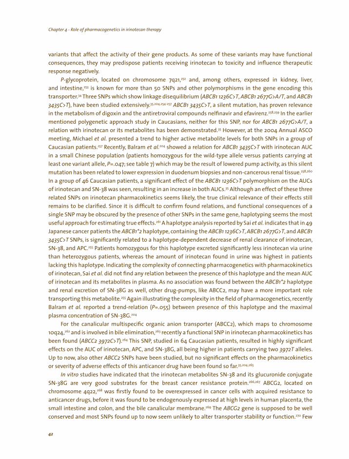
Chapter 4 - Role of pharmacogenetics in irinotecan therapyChapter 4 - Role of pharmacogenetics in irinotecan therapy
variants that affect the activity of their gene products. As some of these variants may have functional consequences, they may predispose patients receiving irinotecan to toxicity and influence therapeutic response negatively.
P-glycoprotein, located on chromosome 7q21,252 and, among others, expressed in kidney, liver, and intestine,253 is known for more than 50 SNPs and other polymorphisms in the gene encoding this transporter.34 Three SNPs which show linkage disequilibrium (ABCB1 1236C>T, ABCB1 2677G>A/T, and ABCB1 3435C>T), have been studied extensively.35,204,254-257 ABCB1 3435C>T, a silent mutation, has proven relevance in the metabolism of digoxin and the antiretroviral compounds nelfinavir and efavirenz.258,259 In the earlier mentioned polygenetic approach study in Caucasians, neither for this SNP, nor for ABCB1 2677G>A/T, a relation with irinotecan or its metabolites has been demonstrated.35 However, at the 2004 Annual ASCO meeting, Michael et al. presented a trend to higher active metabolite levels for both SNPs in a group of Caucasian patients.257 Recently, Balram et al.204 showed a relation for ABCB1 3435C>T with irinotecan AUC in a small Chinese population (patients homozygous for the wild-type allele versus patients carrying at
least one variant allele, P=.047; see table 7) which may be the result of lowered pump activity, as this silent
mutation has been related to lower expression in duodenum biopsies and non-cancerous renal tissue.258,260
In a group of 46 Caucasian patients, a significant effect of the ABCB1 1236C>T polymorphism on the AUCs of irinotecan and SN-38 was seen, resulting in an increase in both AUCs.35 Although an effect of these three related SNPs on irinotecan pharmacokinetics seems likely, the true clinical relevance of their effects still remains to be clarified. Since it is difficult to confirm found relations, and functional consequences of a single SNP may be obscured by the presence of other SNPs in the same gene, haplotyping seems the most
useful approach for estimating true effects.261 A haplotype analysis reported by Sai et al. indicates that in 49 Japanese cancer patients the ABCB1*2 haplotype, containing the ABCB1 1236C>T, ABCB1 2677G>T, and ABCB1 3435C>T SNPs, is significantly related to a haplotype-dependent decrease of renal clearance of irinotecan, SN-38, and APC.255 Patients homozygous for this haplotype excreted significantly less irinotecan via urine than heterozygous patients, whereas the amount of irinotecan found in urine was highest in patients lacking this haplotype. Indicating the complexity of connecting pharmacogenetics with pharmacokinetics of irinotecan, Sai et al. did not find any relation between the presence of this haplotype and the mean AUC of irinotecan and its metabolites in plasma. As no association was found between the ABCB1*2 haplotype and renal excretion of SN-38G as well, other drug-pumps, like ABCC2, may have a more important role transporting this metabolite.255 Again illustrating the complexity in the field of pharmacogenetics, recently Balram et al. reported a trend-relation (P=.055) between presence of this haplotype and the maximal plasma concentration of SN-38G.204
For the canalicular multispecific organic anion transporter (ABCC2), which maps to chromosome 10q24,262 and is involved in bile elimination,263 recently a functional SNP in irinotecan pharmacokinetics has been found (ABCC2 3972C>T).264 This SNP, studied in 64 Caucasian patients, resulted in highly significant
effects on the AUC of irinotecan, APC, and SN-38G, all being higher in patients carrying two 3972T alleles. Up to now, also other ABCC2 SNPs have been studied, but no significant effects on the pharmacokinetics or severity of adverse effects of this anticancer drug have been found so far.35,204,265
In vitro studies have indicated that the irinotecan metabolites SN-38 and its glucuronide conjugate SN-38G are very good substrates for the breast cancer resistance protein.266,267 ABCG2, located on
chromosome 4q22,268 was firstly found to be overexpressed in cancer cells with acquired resistance to
anticancer drugs, before it was found to be endogenously expressed at high levels in human placenta, the
small intestine and colon, and the bile canalicular membrane.269 The ABCG2 gene is supposed to be well conserved and most SNPs found up to now seem unlikely to alter transporter stability or function.270 Few
42

Chapter 4 - Role of pharmacogenetics in irinotecan therapy Chapter 4 - Role of pharmacogenetics in irinotecan therapy
SNPs with presumed clinical consequence have been studied in relation to irinotecan pharmacokinetics; in
particular, a single-nucleotide polymorphism in exon 5 has been described. This ABCG2 421C>A transversion 271-273 and results in an amino acid change of glutamine to lysine at codon 141, leads to altered substrate
specificity and function of the mutant protein.271,274 With an allele frequency of about 35%, this SNP
appears to be very common in Asian populations, whereas in African and Afro-American populations it
is rarely found.275 In Caucasians the allele frequency varies between 10–15%. Functional consequences of this SNP were clearly demonstrated in Caucasian cancer patients treated with the structurally related
camptothecins diflomotecan and topotecan.276,277 Patients carrying at least one defective ABCG2 421A allele
were found to have higher drug levels. However, in a large group of Caucasian patients pharmacokinetic
parameters of irinotecan and SN-38 were not significantly altered.275 Other ABCG2-polymorphisms have
been studied as well,but up to now no significant associations with irinotecan pharmacokinetics have been
demonstrated, except for a CTCA-deletion in the 5’-untranslated region (ABCG2 -19572-19569 delCTCA) in
the earlier discussed group of 29 Chinese cancer patients.204 Patients heterozygous or homozygous for this
deletion had significantly lower values for the relative extent of conversion (REC) of irinotecan into SN-38
compared with patients harboring the wild-type ABCG2 CTCA genotype. As may expected from these data, although not significantly, this deletion was also found associated with lower exposure (AUC) to SN-38. However, this finding is difficult to interpret without knowing the functional consequences of this deletion.
Given the complexity of irinotecan metabolism, relevance to genetic polymorphisms in the drug transporters is disputable, however, it is very likely that the contribution of a single SNP to changes in drug disposition may be obscured by functional roles of other SNPs and other polymorphic proteins involved. In particular, concerning the transporters of the ABC-superfamily with a partial substrate overlap between members, it is hard to assess the influence of one single polymorphism, as the effect of absenteeism of a specific functional drug-pump may be corrected for by the presence, or even by a compensatory overexpression, of others. In the near future, also here haplotype approaches will probably prove to be useful in explaining and predicting irinotecan pharmacokinetics.
FUTURE PERSPECTIVES OF CLINICAL PHARMACOGENETICS IN IRINOTECAN THERAPY
Lately, the field of pharmacogenetics is evolving from an approach in which interpatient variability in pharmacokinetic and pharmacodynamic parameters is established before its genotypic basis is determined, to an approach, in which the genotypic variability is observed before its phenotypic consequences are determined,278 although in the field of irinogenetics this step still has to be taken.279 Without a doubt, pharmacogenetics of anticancer agents may enable us to assess the toxicity and therapeutic outcome in an individual patient more closely in the (near) future. Especially for drugs with a small therapeutic index, like irinotecan, this may be of clinical importance. As shown in this paper, several SNPs and haplotypes with clinical relevance for irinotecan treated patients have been determined and more relationships will probably follow soon. At this moment, we have to decide what information is needed more to incorporate this knowledge in clinical decision-making, and in what way. With the development of modern techniques, it should become possible to gather pharmacogenetic knowledge of all patients before treatment within an acceptable time course and for reasonable costs.48,280-282
Despite the complex pharmacological behavior of irinotecan, pre-therapeutic screening helps us
to identify patients with a higher chance of therapy-induced toxicities. Eventually, this will safe costs on
treating adverse events. For the moment, in particular genetic testing for UGT1A1*28 is supposed to be helpful in identifying patients less capable to detoxify SN-38 well and therefore more prone to suffer excessive
hematological and gastroenterological toxicity. By identifying patients with UGT1A1*28 homozygous
43

Chapter 4 - Role of pharmacogenetics in irinotecan therapyChapter 4 - Role of pharmacogenetics in irinotecan therapy
genotype, who have a nine-times higher relative risk developing grade 4 neutropenia after treatment with
regularly dosed irinotecan, approximately half of all cases of grade 4 neutropenia may be prevented.49
Although one may assume that, being glucuronidated by UGT1A1, bilirubin levels are predictive as well, this
remains question of dispute. Innocenti et al. found strong relations between UGT1A1*28 status, pretreatment
total bilirubin levels, and therapy-induced grade 4 neutropenia,49 whereas Meyerhardt et al. showed that
baseline serum bilirubin does not reliably predict overall irinotecan-related toxicity or efficacy.283 In line
with this finding, Rouits et al. reported that UGT1A1*28 status was found to be strongly related to severity
of therapy-induced neutropenia in 73 patients, although in none of the seven patients homozygous for the
UGT1A1*28 allele hyperbilirubinemia was demonstrated.48 Besides identifying patients with higher risk on
therapy-induced adverse effects, the implementation of pharmacogenetic-based dosing should also result in
the intensification of dosing regimens for fast metabolizers,enlarging their chance of a favorable therapeutic
effect. Until recently UGT1A1 DNA testing was only available in a research setting making its use in clinical decision-making not yet possible,284 although Rouits et al. recently described a high-throughput sequencing
method and Hasegawa et al. reported a rapid and accurate detection of UGT1A1 polymorphisms using the
Invader assay technology which both may become applicable in routine clinical practice.48,282 In August 2005, the Food and Drug Administration (FDA) cleared the Invader Molecular Assay for irinotecan dosing.
As we saw for CYP3A, and to a lesser extent for other involved enzymes and drug transporters as well, it is impossible to explain (all) variation with help of pharmacogenetics. This is recently stressed by Baker et al. who performed a study in which factors affecting CYP3A activity in cancer patients were studied.285
Genetic alterations did not explain found variation on CYP3A activity, whereas patient and disease related factors did have influence. As boundaries of genotyping change the scope, other ways to diminish interpatient variability should be studied as well. One of the ways to do this is by studying interactions due to co-medication and food substances. As especially cytochrome P450 has been shown to be very susceptible to inhibition or induction by environmental factors, we have to find ways to study negative effects of such interactions on irinotecan effectiveness and safety. On the other hand, we also can use such drug interactions in favor of its therapeutic index as Innocenti et al. recently described.169 Irinotecan was combined with phenobarbital (a known inducer of both UGT1A and CYP3A) and cyclosporin (an inhibitor of both ABCC2 and ABCB1), to deliver more irinotecan to the tumor while simultaneously circumventing the serious toxic boundaries. This combination made it possible to deliver higher levels irinotecan to the patient, and irinotecan plasma levels were found higher.
Last decade, bypassing the limitations of genotyping, so called phenotypic measures have been
developed. An innocent probe-drug, which is metabolized the same way as the studied anticancer drug, is given to the patient before treatment to estimate the total functional activity of the enzyme in that
particular patient at that very moment. The total functional activity is the sum of the combination of genotype, influences of disease, patient characteristics, environmental influences, and co-medication. For docetaxel and irinotecan, probe-drugs like cortisol, dexamethasone, erythromycin and midazolam have
shown to be of predictive value for their pharmacokinetics.150,203,286,287 As results are encouraging, knowledge
from these studies may also be incorporated in future dosing regimens. Currently, studies are conducted
to test whether such dosing strategies are capable of reducing interpatient variation in exposure to
irinotecan compared to dosing based on BSA. Already for docetaxel,Yamamoto et al. successfully decreased
pharmacokinetic variability compared to a BSA-based regimen with 46% as a result of individualized
dosing based on parameters including cortisol metabolism.288 As stressed in the accompanying editorial, we should ask ourselves how to develop probe-based tests that are easy to translate into the average
outpatient oncology clinical setting.289
44

Chapter 4 - Role of pharmacogenetics in irinotecan therapy Chapter 4 - Role of pharmacogenetics in irinotecan therapy
In conclusion, right now, the role of pharmacogenetic testing for CES, CYP3A, and ABC drug-pumps, seems limited in explaining interpatient variation of irinotecan metabolism. Probably, patients do not
benefit from irinotecan therapy individualization strategies based on their genetic constellation of these genes. Since CYP3A-phenotyping strategies seem better predictors of its pharmacokinetics, such strategies may be used to individualize irinotecan dosing in the future. In contrast, the UGT1A-gene is a promising candidate for pretreatment genetic screening and implementation in routine clinical practice of irinotecan treatment. Ultimately, dosing regimens combining phenotyping for CYP3A and genotyping for UGT1A1 may lead to a better prediction of irinotecan pharmacokinetics and in this way to a higher therapeutic index. For the time being, advantages of such strategies may be limited to patients treated in academic settings. To truly individualize irinotecan treatment, efforts to develop and implement feasible phenotyping and genotyping strategies on a routine basis in general hospital settings should be strongly encouraged.
45

46
47
Chapter 5
ABCG2 pharmacogenetics: ethnic differences in allele frequency
and assessment of influence on
irinotecan disposition
FA de Jong, S Marsh, RH Mathijssen, C King, J Verweij, A Sparreboom, HL McLeod
Clinical Cancer Research 10(17):5889–5894, 2004
Chapter 5 - ABCG2 pharmacogenetics: ethnic differences in allele frequency and assessment of influence on irinotecan disposition
48
Chapter 5 - ABCG2 pharmacogenetics: ethnic differences in allele frequency and assessment of influence on irinotecan disposition
ABSTRACT
Objective: The ATP-binding cassette transporter ABCG2 (breast cancer resistance protein, BCRP) is an efflux protein that plays a role in host detoxification of various xenobiotic substrates, including the irinotecan metabolite 7-ethyl-10-hydroxycamptothecin (SN-38).The ABCG2 421C>A polymorphism has been associated with reduced protein expression and altered function in vitro. The aim of this study was to evaluate the ethnic distribution and potential functional consequence of the ABCG2 421C>A genotype in cancer patients treated with irinotecan.
Patients and methods: ABCG2 genotyping was performed using Pyrosequencing on DNA from 88 American Caucasians, 94 African Americans, 938 Africans, and 95 Han Chinese, as well as in 84 European Caucasian patients treated with irinotecan undergoing additional blood sampling for pharmacokinetic studies.
Results: Significant differences in allele frequencies were observed between the given world populations (P<.001), the variant allele being most common in the Han Chinese population with a frequency as high as 34%. The mean area under the curve of irinotecan and SN-38 were 19,851 and 639 ng*h/mL, respectively. The frequency of the variant allele (10.7%) was in line with results in American Caucasians. No significant changes in irinotecan pharmacokinetics were observed in relation to the ABCG2 421C>A genotype, although one of two homozygous variant allele carriers showed extensive accumulation of SN-38 and SN-38 glucuronide.
Conclusion: The ABCG2 421C>A polymorphism appears to play a limited role in the disposition of irinotecan in European Caucasians. It is likely that the contribution of this genetic variant is obscured by a functional role of other polymorphic proteins.
Chapter 5 - ABCG2 pharmacogenetics: ethnic differences in allele frequency and assessment of influence on irinotecan disposition Chapter 5 - ABCG2 pharmacogenetics: ethnic differences in allele frequency and assessment of influence on irinotecan disposition
INTRODUCTION
The ATP-binding cassette (ABC) transporters represent the largest family of transmembrane proteins that bind ATP and use the energy to drive the transport of various molecules across cell membranes.290
On the basis of the arrangement of molecular structural components, i.e., the nucleotide binding domain and the topology of transmembrane domains, human proteins are classified into seven distinct families (ABC-A to ABC-G).290 The ABCG subfamily consists of several half transporters that are generally thought
to form homo- or heterodimers to create the active transporter.291,292 Very recently, it was reported that
human ABCG2 probably exists as a homotetramer with a possibility of a higher form of oligomerization.292
The human ABCG2 gene is located on chromosome 4q22 and encodes a 655 amino acid polypeptide.268,293
In vitro studies have indicated that, apart from topotecan,294,295 the irinotecan metabolites 7-ethyl-10-hydroxycamptothecin (SN-38) and its glucuronide conjugate SN-38G266,267 are very good substrates for ABCG2. Furthermore, overexpression of ABCG2 reportedly confers cancer cell resistance to various anticancer drugs, including topotecan and SN-38.296,297 ABCG2 is endogenously expressed at high levels in human placenta, the small intestine and colon, and the bile canalicular membrane.269 This localization suggests that the physiologic role of ABCG2 may be to regulate intestinal absorption and biliary secretion of potentially toxic xenobiotics by active transport mechanisms.294,298
Recent resequencing of the ABCG2 transporter has revealed a number of allelic variants that affect
activity of the gene product in vivo. Some of these genetic variants may potentially modulate the ABCG2 phenotype in patients and therefore affect their predisposition to toxicity and response to substrate drug treatment. In particular, a single nucleotide polymorphism in exon 5 of the ABCG2 gene has been described in which a 421C>A transversion results in an amino acid change of glutamine to lysine at codon 141.271-273
Although a detailed analysis of the potential functional consequences of this ABCG2 variant has not yet
been evaluated, in vitro studies indicated altered substrate specificity and function of the mutant protein relative to the wild-type protein.271,274 In the present study, we evaluated the allele frequency of the ABCG2 421C>A polymorphism in a large number of individuals from different ethnic populations as well as its effect on the pharmacokinetics of irinotecan.
MATERIALS AND METHODS
Patient and population samples. A total of 84 patients (43 women and 41 men) diagnosed with a
histologically confirmed malignant solid tumor was assessed for irinotecan pharmacokinetics (see table 9).The
most common tumor types in this population were of gastrointestinal or pulmonary origin. All patients were
of Caucasian descent, were between 34 and 75 years old (median, 54 years), and were treated between January
1997 and June 2003 at the Erasmus University Medical Center–Daniel den Hoed Cancer Center (Rotterdam, the Netherlands). The inclusion and exclusion criteria, premedication schedules (i.e., 8 mg of the 5HT3-receptor antagonist ondansetron,administered intravenously,combined with 10 mg of dexamethasone),and protocols
for treatment of drug-induced side effects were documented previously.53 Irinotecan (Aventis Pharma, Hoevelaken,the Netherlands) was given once every three weeks as a 90-min intravenous infusion.The median
dose was 600 mg (range, 260–875 mg). None of the patients received any other concurrent chemotherapy or other drugs (http://medicine.iupui.edu/flockhart/table.htm), food supplements, and/or herbal preparations
known to interfere with the pharmacokinetics of irinotecan. Eighty-one patients received irinotecan as single
agent, whereas in three patients, irinotecan was combined with cisplatin, which has been demonstrated to
have no influence on the pharmacokinetics of irinotecan.299 The clinical protocols,35,53 including blood sampling
for the purpose of pharmacokinetic and pharmacogenetic analyses, were approved by the Erasmus University
Medical Center–Ethics Board, and all patients provided written informed consent.
49

Chapter 5 - ABCG2 pharmacogenetics: ethnic differences in allele frequency and assessment of influence on irinotecan dispositionChapter 5 - ABCG2 pharmacogenetics: ethnic differences in allele frequency and assessment of influence on irinotecan disposition
a
N) 84
/43
)
72.0 2) 1.85
1
28 20 8
77
(*109/L) (*109/L)
*109/L) Hemoglobin (mmol/L)
a
,
Table 9. Patient demographics
Variable Value
Baseline screening
Number of patients entered (Males/Females 41
Age (years 54 (34–75) Length (m) 1.70 (1.51-1.92) Weight (kg) (38.6-113.5) Body surface area (m (1.29–2.36) WHO performance status (0–1)
Pretherapy clinical chemistry
Aspartate aminotransferase (units/L) (6–185) Alanine aminotransferase (units/L) (4–225) Total Bilirubin (µmol/L) (3–22) Serum Creatinine (µmol/L) 78 (45–132) Serum Albumin (g/L) 41 (30–51) Total serum protein (g/L) (62–88)
Pretherapy hematology
White Blood Cell Count 7.80 (2.80–27.0) Absolute Neutrophil Count 5.38 (1.54–24.0) Platelets ( 304 (132–966)
7.55 (5.20–79.0) Hematocrit (L/L) 0.37 (0.27–0.48)
Values represent the median with range in parenthesis (unless stated otherwise).
Abbreviations: N, number of patients; WHO World Health Organization.
Apart from these 84 European Caucasian cancer patients, genomic DNA was extracted from whole blood of 88 Caucasian Americans, 94 African Americans, and 938 sub-Sahara African healthy volunteers as described previously.300,301 Genomic DNA from 95 healthy unrelated Asian individuals was obtained from the Coriell Institutes (http://coriell.umdnj.edu/ccr/ccrsumm.html).
Pharmacokinetic analysis. Pharmacokinetic studies were performed during the first administration of irinotecan in the 84 cancer patients.Blood samples of ~5 mL each were collected at serial time points up to 48 or 500 h after drug administration and were centrifuged to obtain plasma. Pharmacokinetic data obtained in a subset of 65 patients were described previously.35 Concentrations of irinotecan, SN-38, and SN-38G were determined using a validated method based on liquid chromatography with fluorescence detection, as described elsewhere.54 Previously developed population models were used to predict the pharmacokinetic parameters of irinotecan, SN-38, and SN-38G.55 Because the linear relationship between irinotecan dose and area under the plasma concentration-time curve (AUC),29 the AUC normalized to the recommended single
50
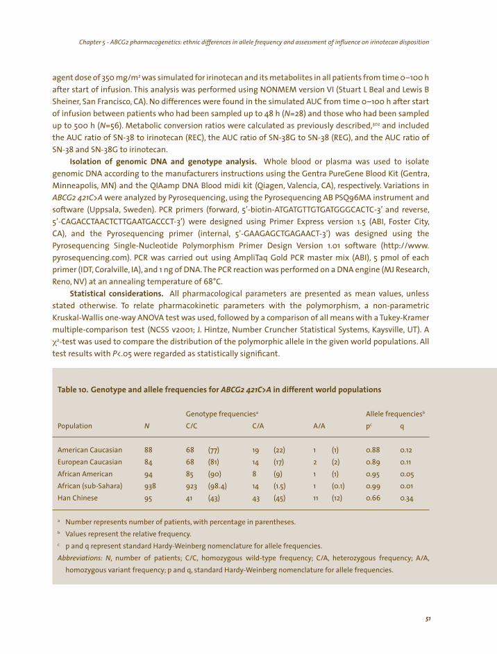
Chapter 5 - ABCG2 pharmacogenetics: ethnic differences in allele frequency and assessment of influence on irinotecan disposition Chapter 5 - ABCG2 pharmacogenetics: ethnic differences in allele frequency and assessment of influence on irinotecan disposition
agent dose of 350 mg/m2 was simulated for irinotecan and its metabolites in all patients from time 0–100 h
after start of infusion. This analysis was performed using NONMEM version VI (Stuart L Beal and Lewis B Sheiner, San Francisco, CA). No differences were found in the simulated AUC from time 0–100 h after start
of infusion between patients who had been sampled up to 48 h (N=28) and those who had been sampled up to 500 h (N=56). Metabolic conversion ratios were calculated as previously described,302 and included the AUC ratio of SN-38 to irinotecan (REC), the AUC ratio of SN-38G to SN-38 (REG), and the AUC ratio of SN-38 and SN-38G to irinotecan.
Isolation of genomic DNA and genotype analysis. Whole blood or plasma was used to isolate genomic DNA according to the manufacturers instructions using the Gentra PureGene Blood Kit (Gentra, Minneapolis, MN) and the QIAamp DNA Blood midi kit (Qiagen, Valencia, CA), respectively. Variations in ABCG2 421C>A were analyzed by Pyrosequencing, using the Pyrosequencing AB PSQ96MA instrument and software (Uppsala, Sweden). PCR primers (forward, 5’-biotin-ATGATGTTGTGATGGGCACTC-3’ and reverse, 5’-CAGACCTAACTCTTGAATGACCCT-3’) were designed using Primer Express version 1.5 (ABI, Foster City, CA), and the Pyrosequencing primer (internal, 5’-GAAGAGCTGAGAACT-3’) was designed using the Pyrosequencing Single-Nucleotide Polymorphism Primer Design Version 1.01 software (http://www. pyrosequencing.com). PCR was carried out using AmpliTaq Gold PCR master mix (ABI), 5 pmol of each primer (IDT, Coralville, IA), and 1 ng of DNA. The PCR reaction was performed on a DNA engine (MJ Research, Reno, NV) at an annealing temperature of 68°C.
Statistical considerations. All pharmacological parameters are presented as mean values, unless stated otherwise. To relate pharmacokinetic parameters with the polymorphism, a non-parametric Kruskal-Wallis one-way ANOVA test was used, followed by a comparison of all means with a Tukey-Kramer multiple-comparison test (NCSS v2001; J. Hintze, Number Cruncher Statistical Systems, Kaysville, UT). A χ2-test was used to compare the distribution of the polymorphic allele in the given world populations. All test results with P<.05 were regarded as statistically significant.
a b
N A/A pc q
88 68 (77) 19 (22) 1 (1) 84 68 (81) 14 (17) 2 (2) 94 85 (90) 8 (9) 1 (1) 938 923 14 (1.5) 1
Han Chinese 95 (43) 43 (45) 11 (12)
a
b
c
; ; A/A, ;
Table 10. Genotype and allele frequencies for ABCG2 421C>A in different world populations
Genotype frequencies Allele frequenciesPopulation C/C C/A
American Caucasian 0.88 0.12
European Caucasian 0.89 0.11 African American 0.95 0.05
African (sub-Sahara) (98.4) (0.1) 0.99 0.01 41 0.66 0.34
Number represents number of patients, with percentage in parentheses. Values represent the relative frequency. p and q represent standard Hardy-Weinberg nomenclature for allele frequencies.
Abbreviations: N, number of patients; C/C, homozygous wild-type frequency C/A, heterozygous frequencyhomozygous variant frequency p and q, standard Hardy-Weinberg nomenclature for allele frequencies.
51

Chapter 5 - ABCG2 pharmacogenetics: ethnic differences in allele frequency and assessment of influence on irinotecan dispositionChapter 5 - ABCG2 pharmacogenetics: ethnic differences in allele frequency and assessment of influence on irinotecan disposition
Median Mean
Dose (mg) 600 602 1.51
* 19,851 * 639
* 2,911 4,105 * ,
3.01 3.24
a Wt (N=68) Het/ N=16) Pb
* 19,305 * 692 .669
* .555
* ,751) 3.17 3.52
8.16 .803
a
b P
( /
( /A); N,
Table 11. Summary of pharmacokinetic parameters
Parameter Range
260–875
Infusion duration (h) 1.50 0.75–2.50 AUC irinotecan (ng h/mL) 18,350 10,350–46,079
AUC SN-38 (ng h/mL) 566 216–2,605
AUC SN-38G (ng h/mL) 962–29,961 AUC SN-38 + SN-38G (ng h/mL) 3,671 3,722 1,346–30 761 REC (%) 0.79–8.80 (AUC SN-38 + SN-38G) / AUC irinotecan 0.199 0.241 0.067–2.04
REG 5.97 7.25 1.70–37.5
Abbreviations: REC, relative extent of conversion (AUC ratio of SN-38 to irinotecan); REG, relative extent of glucuronidation (AUC ratio
of SN-38G to SN-38).
Table 12. Pharmacokinetic parameters as a function of ABCG2 421C>A genotype
Parameter Var (
AUC irinotecan (ng h/mL) 19,979 (2,280–37,678) (7,239–31,392) .715
AUC SN-38 (ng h/mL) 627 (0–2,029) (0–1,776) AUC SN-38G (ng h/mL) 3,836 (0–9,979) 5,083 (0–18,777) AUC SN-38 + SN-38G (ng h/mL) 4,382 (0–10 5,775 (0–19,687) .485
REC (%) (0.02–6.11) (0.02–6.84) .466
(AUC SN-38 + SN-38G) / AUC irinotecan 0.218 (0–0.436) 0.325 (0–1.255) .559
REG 7.00 (0–16.1) (0–25.6)
Values represent the mean with the 95% confidence interval in parentheses.
-values from a non-parametric Kruskal-Wallis test.
Abbreviations: Wt, homozygous wild-type patients C/C); Het Var, combined data of heterozygous and homozygous variant patients
C/A or A number of patients studied; REC, relative extent of conversion (AUC ratio of SN-38 to irinotecan); REG, relative extent
of glucuronidation (AUC ratio of SN-38G to SN-38).
RESULTS
ABCG2 genotyping. Significant differences in allele frequencies were observed between the given world populations (P<.001; see table 10). In the Han Chinese population, the variant allele appears to be very common with a found frequency of 34%. Fifty-seven percent of the studied Han Chinese were carrying at least one variant allele. In contrast, in the sub-Sahara African population, the variant allele appeared to be very rare with a frequency less than 1%. About 5% of studied African-American alleles were found
52
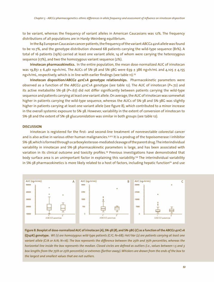
Chapter 5 - ABCG2 pharmacogenetics: ethnic differences in allele frequency and assessment of influence on irinotecan disposition Chapter 5 - ABCG2 pharmacogenetics: ethnic differences in allele frequency and assessment of influence on irinotecan disposition
to be variant, whereas the frequency of variant alleles in American Caucasians was 12%. The frequency distributions of all populations are in Hardy-Weinberg equilibrium.
In the 84 European Caucasian cancer patients, the frequency of the variant ABCG2 421A allele was found to be 10.7%, and the genotype distribution showed 68 patients carrying the wild-type sequence (81%). A total of 16 patients (19%) carried at least one variant allele, 14 of whom were carrying the heterozygous sequence (17%), and two the homozygous variant sequence (2%).
Irinotecan pharmacokinetics. In the entire population, the mean dose-normalized AUC of irinotecan was 19,851 ± 6,461 ng*h/mL. The AUCs of SN-38 and SN-38G were 639 ± 386 ng*h/mL and 4,105 ± 4,155 ng*h/mL, respectively, which is in line with earlier findings (see table 11).29
Irinotecan disposition/ABCG2 421C>A genotype relationships. Pharmacokinetic parameters were observed as a function of the ABCG2 421C>A genotype (see table 12). The AUC of irinotecan (P=.72) and its active metabolite SN-38 (P=.67) did not differ significantly between patients carrying the wild-type sequence and patients carrying at least one variant allele. On average, the AUC of irinotecan was somewhat
higher in patients carrying the wild-type sequence, whereas the AUCs of SN-38 and SN-38G was slightly higher in patients carrying at least one variant allele (see figure 8), which contributed to a minor increase in the overall systemic exposure to SN-38. However, variability in the extent of conversion of irinotecan to SN-38 and the extent of SN-38 glucuronidation was similar in both groups (see table 12).
DISCUSSION
Irinotecan is registered for the first- and second-line treatment of nonresectable colorectal cancer and is also active in various other human malignancies.9,303 It is a prodrug of the topoisomerase I inhibitor SN-38,whichis formed through a carboxylesterase-mediated cleavage of the parent drug.Theinterindividual variability in irinotecan and SN-38 pharmacokinetic parameters is large, and has been associated with
variation in its clinical outcome and toxicity profiles.29 Previous investigations have demonstrated that
body surface area is an unimportant factor in explaining this variability.139 The interindividual variability
in SN-38 pharmacokinetics is more likely related to a host of factors, including hepatic function62 and use
(A (B (C) ( ( /
( / ,
* * *A B C
Figure 8. Boxplot of dose-normalized AUC of irinotecan ), SN-38 ), and SN-38G as a function of the ABCG2 421C>A Q141K) genotype. Wt (1) are homozygous wild-type patients C/C; N=68); Het Var (2) are patients carrying at least one
variant allele C/A or A A; N=16). The box represents the difference between the 25th and 75th percentiles whereas the horizontal line inside the box represents the median. Closed circles are defined as outliers (i.e., values between 1.5 and 3 box lengths from the 75th or 25th percentile) or extremes (farther away). Whiskers are drawn from the ends of the box to the largest and smallest values that are not outliers.
AUC (ng h/mL) AUC (ng h/mL) AUC (ng h/mL)
53
Chapter 5 - ABCG2 pharmacogenetics: ethnic differences in allele frequency and assessment of influence on irinotecan dispositionChapter 5 - ABCG2 pharmacogenetics: ethnic differences in allele frequency and assessment of influence on irinotecan disposition
of concomitant medication124 in addition to multiple polymorphic pathways involved in the excretion and
biotransformation of irinotecan. The latter include esterases regulating cleavage of irinotecan, cytochrome
P450 3A4 and 3A5-mediated oxidations of the parent drug,32 and glucuronic acid conjugation of SN-38
by members of the UGT1A family.217 It has been suggested previously that the metabolism of SN-38 is
substantially influenced by a promoter polymorphism in the TATA-box sequence of the UGT1A1 gene
(i.e., UGT1A1*28).46 Indeed, an extra (seventh) TA-repeat in both alleles results in a substantial reduction
in transcriptional and functional UGT1A1 activity compared with patients carrying the heterozygous or 266,267,296,297 wild-type sequence.27,49 Because SN-38 is also a known substrate for the ABC transporter ABCG2,
we here evaluated the potential functional significance of the ABCG2 421C>A polymorphism in cancer patients receiving irinotecan to identify additional mechanisms underlying interindividual variation in
SN-38 pharmacokinetics. The allele frequency of ABCG2 421A varies highly around the diverse populations. The presented
findings in the four world populations are in line with earlier findings. The ABCG2 421A allele appears to be very common in Japanese and Chinese populations, with reported allele frequencies between 26–35%.271,273
To the best of our knowledge, no data have been reported previously on sub-Sahara African populations. However, the low frequency of ABCG2 421A (<1%) in this population is in line with earlier observations in Africans north of the Sahara and in African Americans,273 and with the relatively low variant allele frequency in the currently studied African American population. The observed 10.7% allele frequency of ABCG2 421A in the 84 European Caucasian cancer patients is similar to that found in the Caucasian American individuals (12%) and those described previously in American Caucasian (frequency, 14%; N=85)
10%; N=60).272,273 and Swedish populations (frequency,In our patients, pharmacokinetic parameters of irinotecan and SN-38 were not significantly different
between patients wild-type for ABCG2 and patients carrying at least one defective ABCG2 421C>A allele (see table 12). At first sight, this finding is inconsistent with the previous demonstration of reduced protein expression being associated with the variant allele,271 and with altered plasma pharmacokinetics of another camptothecin analogue, diflomotecan (BN80915), when given intravenously.276 However, given the complexity of irinotecan metabolism,303 it is possible that the contribution of ABCG2 421C>A
to changes in drug disposition is obscured by a functional role of other polymorphic proteins involved in irinotecan elimination. In addition, because of a partial substrate overlap between ABCB1 (P-glycoprotein) and ABCG2,271 the effect of absenteeism of functional ABCG2 may be corrected for by the presence, or even by a compensatory overexpression, of functional ABCB1.
Despite the lack of a significant association between ABCG2 421C>A genotype and irinotecan
disposition, it is worth mentioning that one of two patients homozygous for the variant allele showed
very extensive accumulation of SN-38 and SN-38G, with a value for the combined AUC of SN-38 and
SN-38G of 30,761 ng*h/mL and with a value for the combined ratio of SN-38 and SN-38G to irinotecan
that was >8-fold higher than the mean of all patients (2.041 versus 0.241). The patient developed
very severe side effects, including a grade 4 leucocytopenia, a grade 4 neutropenia, and a grade 4
diarrhea, and eventually died within nine days after the administration of irinotecan. This patient was
previously shown to be heterozygous for the UGT1A1*28 polymorphism,35 ruling out a causative link
between this Phase II glucuronidation pathway and the observed phenotype. The only other patient
homozygous for this allele did not show aberrant SN-38 pharmacokinetics. However, it cannot be
excluded that patients with this rare genotype comprising two variant alleles for ABCG2 421C>A show
markedly impaired ability to eliminate SN-38 through active ABCG2-mediated hepatobiliary and or intestinal secretion and demonstrate excessive toxicity. Clearly, additional investigation is required
54

Chapter 5 - ABCG2 pharmacogenetics: ethnic differences in allele frequency and assessment of influence on irinotecan disposition Chapter 5 - ABCG2 pharmacogenetics: ethnic differences in allele frequency and assessment of influence on irinotecan disposition
to unambiguously assess the relationship between ABCG2 421C>A homozygous variant genotype and
irinotecan disposition. In conclusion, the ABCG2 421C>A polymorphism does not seem to play a major role in vivo in the
disposition of irinotecan in European Caucasian cancer patients, although it cannot be ruled out that
patients with two variant alleles for ABCG2 421C>A may have impaired ability to eliminate SN-38. The ABCG2 421C>A polymorphism was found to vary highly in different ethnic groups, with <1% variant alleles found in Africans, and up to 34% in Chinese subjects. A prospective trial to corroborate the usefulness of gene diagnosis of this ABCG2 polymorphism, in combination with other polymorphisms of putative relevance in irinotecan metabolism and disposition such as UGT1A1*28 or the recently described variant
UGT1A1 -3156G>A,49 before irinotecan chemotherapy in different ethnic populations seems warranted.
ACKNOWLEDGEMENTS
This work was funded, in part, by the NIH Pharmacogenetics Research Network grant U01 GM63340 (St. Louis).
55

56

57
Chapter 6
Irinotecan-induced diarrhea: functional significance of the polymorphic ABCC2 transporter protein
FA de Jong, T Scott-Horton, DL Kroetz, HL McLeod, LE Friberg, RH Mathijssen, J Verweij, S Marsh, A Sparreboom
Submitted

Chapter 6 - Irinotecan-induced diarrhea: functional significance of the polymorphic ABCC2 transporter protein
58
Chapter 6 - Irinotecan-induced diarrhea: functional significance of the polymorphic ABCC2 transporter protein
ABSTRACT
Objective: Interindividual pharmacokinetic variability of the anticancer agent irinotecan is high. Life-threatening diarrhea is observed in up to 25% of patients receiving irinotecan and has been related with irinotecan pharmacokinetics and UGT1A1 genotype status. Here, we explore the association of ABCC2
polymorphisms and haplotypes with irinotecan disposition and diarrhea.
Methods: A cohort of 167 Caucasian cancer patients who were previously assessed for irinotecan pharmacokinetics (90-min infusion given every 21 days), toxicity and UGT1A1*28
genotype, were genotyped for polymorphisms in ABCC2 using Pyrosequencing.
Results: Fifteen ABCC2 haplotypes were identified in the studied patients. The reference haplotype ABCC2*2 was associated with lower irinotecan clearance (28.3 versus 31.6 L/h; P=.026). In patients that did not carry a UGT1A1*28 allele, a significant
reduction of severe diarrhea was noted in patients with the ABCC2*2 haplotype (10% versus 44%; P<.01). This effect was not
observed in patients with at least one UGT1A1*28 allele (32% versus 20%; P=.35).
Conclusions: This study suggests that the presence of the ABCC2*2 haplotype is associated with less irinotecan-related diarrhea, maybe as a consequence of reduced hepatobiliary secretion of irinotecan. As the association was seen in patients not genetically predisposed at risk for diarrhea due to UGT1A1*28, confirmatory studies of the relationships of ABCC2 genotypes and irinotecan disposition and toxicity are warranted.
Chapter 6 - Irinotecan-induced diarrhea: functional significance of the polymorphic ABCC2 transporter protein Chapter 6 - Irinotecan-induced diarrhea: functional significance of the polymorphic ABCC2 transporter protein
INTRODUCTION
Irinotecan is a widely used anticancer drug that has been approved for the treatment of advanced
colorectal cancer.11 The mechanism of action of irinotecan is associated with topoisomerase I inhibition by
the active metabolite 7-ethyl-10-hydroxycamptothecin (SN-38), which results in cytotoxic effects on rapidly
dividing cells. The main dose-limiting toxicities of irinotecan include severe (grade 3–4) myelosuppression
with an incidence of about 15–20%, and delayed-type severe diarrhea, which is characteristically seen in
20–25% of patients about five days after start of therapy.304 The occurrence of diarrhea in particular has
significant clinical ramifications, as it affects the dose that can be safely administered and occasionally is
associated with life-threatening events. Although the mechanism by which irinotecan induces delayed-type
diarrhea has not yet been elucidated, prior investigations have suggested that this side effect is associated
with interindividual variability in both systemic and intestinal exposure to SN-38.305 Hence, identification of the environmental and genetic factors affecting the pharmacokinetic profile of SN-38 following irinotecan
treatment could aid in predicting or adapting appropriate, individualized doses of this drug. The primary pathway of elimination of SN-38 is a Phase II glucuronic acid conjugation reaction that
results in the formation of SN-38-glucuronide (SN-38G),and that is mediated by UDP glucuronosyltransferase isoforms –in decreasing order of importance– UGT1A1, UGT1A6, UGT1A7, UGT1A9, and UGT1A10.216 The presence of an additional TA-repeat in the promoter region of UGT1A1 (UGT1A1*28) has been associated with decreased activity and SN-38 glucuronidation in humans,49 and its presence is also a well-known risk factor for the occurrence of severe toxicity.151 However, the presence of one or two UGT1A1*28 alleles explains only less than half of all cases of severe diarrhea following treatment with irinotecan.306 In addition to metabolism, SN-38 and irinotecan are also sensitive to direct hepatobiliary secretion mediated by a number of highly polymorphic members of the ATP-binding cassette transporters, including ABCC2 (cMOAT; MRP2),307 and to a lesser extent, ABCC1 (MRP1),308 ABCC4 (MRP4),309 ABCB1 (P-glycoprotein),310 and ABCG2 (ABCP; MXR; BCRP).297 In the current study, we performed an exploratory analysis to evaluate the association of single nucleotide polymorphisms (SNPs) in the ABCC2 gene with the pharmacokinetics and toxicity of irinotecan therapy.
METHODS
Patient treatment. Patients with histological evidence of a solid tumor that was potentially sensitive to treatment with single-agent irinotecan were considered eligible for this study. The inclusion and exclusion criteria, pre-medication schedules, and protocols for treatment of drug-induced side effects have been summarized before.35 Irinotecan was given once every three weeks as a 90-min continuous intravenous infusion. Other concurrent chemotherapy or other drugs, food supplements, and/or herbal preparations known or suspected to interfere with the pharmacokinetics of irinotecan were not allowed. Clinical protocols, including blood sampling for the purpose of pharmacokinetic and pharmacogenetic analyses, were approved by the local ethics board, and all patients provided written informed consent.
Pharmacokinetic analysis. Blood samples of 5–7 mL each for pharmacokinetic studies were collected at serial time points up to 500 h after the first drug administration and were centrifuged to obtain plasma. Concentrations of irinotecan, SN-38, and SN-38G were determined using a validated method based on liquid chromatography with fluorescence detection, as described elsewhere.54 Previously developed population models for irinotecan and its metabolites formed the basis for the pharmacokinetic modeling, however here a 3-compartment model for SN-38 was supported by the data.55,203 The analysis was performed using the software package NONMEM version V (GloboMax, Hanover, MD). Individual parameters were derived as Empirical Bayes estimates using the POSTHOC option. Metabolic conversion ratios, including the AUC
59
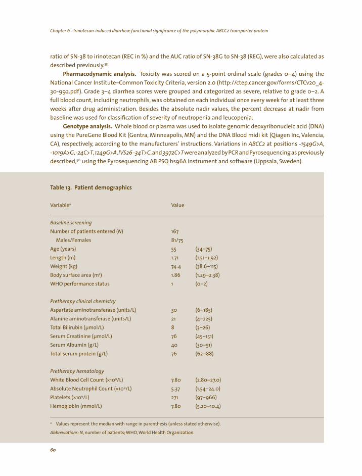
Chapter 6 - Irinotecan-induced diarrhea: functional significance of the polymorphic ABCC2 transporter protein Chapter 6 - Irinotecan-induced diarrhea: functional significance of the polymorphic ABCC2 transporter protein
ratio of SN-38 to irinotecan (REC in %) and the AUC ratio of SN-38G to SN-38 (REG), were also calculated as described previously.35
Pharmacodynamic analysis. Toxicity was scored on a 5-point ordinal scale (grades 0–4) using the National Cancer Institute–Common Toxicity Criteria, version 2.0 (http://ctep.cancer.gov/forms/CTCv20_4-30-992.pdf). Grade 3–4 diarrhea scores were grouped and categorized as severe, relative to grade 0–2. A full blood count, including neutrophils, was obtained on each individual once every week for at least three weeks after drug administration. Besides the absolute nadir values, the percent decrease at nadir from baseline was used for classification of severity of neutropenia and leucopenia.
Genotype analysis. Whole blood or plasma was used to isolate genomic deoxyribonucleic acid (DNA) using the PureGene Blood Kit (Gentra, Minneapolis, MN) and the DNA Blood midi kit (Qiagen Inc, Valencia, CA), respectively, according to the manufacturers’ instructions. Variations in ABCC2 at positions -1549G>A, -1019A>G,-24C>T,1249G>A,IVS26 -34T>C,and 3972C>T were analyzed by PCR and Pyrosequencing as previously
described,311 using the Pyrosequencing AB PSQ hs96A instrument and software (Uppsala, Sweden).
a
N)
) 55
2) 1.86 1
30 21 8
40
(*109/L) (*109/L)
*109/L) Hemoglobin (mmol/L)
a
,
Table 13. Patient demographics
Variable Value
Baseline screening
Number of patients entered ( 167
Males/Females 81/75 Age (years (34–75) Length (m) 1.71 (1.51–1.92) Weight (kg) 74.4 (38.6–115) Body surface area (m (1.29–2.38) WHO performance status (0–2)
Pretherapy clinical chemistry
Aspartate aminotransferase (units/L) (6–185) Alanine aminotransferase (units/L) (4–225) Total Bilirubin (µmol/L) (3–26) Serum Creatinine (µmol/L) 76 (45–151) Serum Albumin (g/L) (30–51) Total serum protein (g/L) 76 (62–88)
Pretherapy hematology
White Blood Cell Count 7.80 (2.80–27.0) Absolute Neutrophil Count 5.37 (1.54–24.0) Platelets ( 271 (97–966)
7.80 (5.20–10.4)
Values represent the median with range in parenthesis (unless stated otherwise).
Abbreviations: N, number of patients; WHO World Health Organization.
60
Chapter 6 - Irinotecan-induced diarrhea: functional significance of the polymorphic ABCC2 transporter protein Chapter 6 - Irinotecan-induced diarrhea: functional significance of the polymorphic ABCC2 transporter protein
The number of TA-repeats in the TATA-box of the promoter region of the UGT1A1 gene was determined
by sizing of PCR-products obtained with UGT1A1 specific primers as described previously.312 Genotypes
were assigned as *1/*1, *1/*28, and *28/*28, where *1 represents the reference allele containing six TA-repeats
(UGT1A1*1), whereas *28 represents the variant allele containing seven TA-repeats (UGT1A1*28), respectively. The genotype was called variant if it differed from the Refseq consensus sequence for the SNP position
(http://www.ncbi.nlm.nih.gov/LocusLink/refseq.html). Genotype-frequency analysis of Hardy-Weinberg
equilibrium was carried out using hwsim (http://krunch.med.yale.edu/hwsim/). Haplotypes were
determined using the software package PHASE version 0.9.313
Statistical considerations. All data are presented as mean values (± standard deviation), unless indicated otherwise. The association of the variant genotypes and haplotypes with the pertinent
pharmacokinetic and pharmacodynamic parameters was evaluated using a non-parametric Kruskal-Wallis one-way analysis of variance on ranks. A χ2-test was used to detect associations between dichotomous variables. Because this study was mainly exploratory in intent, no adjustments were performed to evaluate the significance of the multiple comparisons. Two-tailed P values of less than .05 were considered to be statistically significant. All statistical calculations were performed in the software package SPSS version 10.0.7 (SPSS Inc, Chicago, IL).
RESULTS
General observations. A total of 167 cancer patients (see table 13) diagnosed with a histologically-confirmed malignant solid tumor were treated with irinotecan. The most common tumor types were of gastrointestinal and pulmonary origin. All patients (81 male, 75 female) were of European Caucasian descent, were between 34 and 75 years old (median, 55 years), and were treated between January 1997 and August 2004.
Irinotecan pharmacokinetics-pharmacodynamics. The typical clearance of irinotecan in the 150
patients who were sampled for pharmacokinetic purposes was estimated to 29.3 L/h with an interindividual variability (CV) of 32%. The metabolic clearances (uncorrected for fraction metabolized) of SN-38 and
ngs.55,203 SN-38G were 465 L/h (49% CV) and 31.9 L/h (62% CV), respectively, which is in line with earlier findiThe means, medians and, ranges of the individual clearance values and the metabolic conversion ratios
are presented in table 14. Side effects of irinotecan therapy were recorded in the subset of 136 patients
receiving the recommended dose (350 mg/m2 or 600 mg). Grade 3–4 diarrhea was observed in 26 of 136
patients (19.1%), and grade 3–4 neutropenia was seen in 40 patients (28.8%).
parable.
Population frequency of variant ABCC2 genotypes. Six variants in the ABCC2 gene were studied in the current population (see table 15). Depending on the specific variant, these SNPs were successfully analyzed in 154 to 163 patients. The observed frequency of the variant alleles ranged between 0.04 and 0.56, which is in line with relative frequencies reported in Caucasians and Japanese subjects previously. In particular, relative frequencies of the variant alleles of ABCC2 -24C>T, ABCC2 1249G>A, and ABCC 23972C>T were com
264,265,314,315 No patients homozygous for the ABCC2 -24C>T and ABCC2 IVS26 -34T>C polymorphisms were identified. The frequency distributions of all SNPs are in Hardy-Weinberg equilibrium. A total of 15 haplotypes were constructed in the 139 patients who had been successfully genotyped for all six variants, although only six haplotypes had a frequency greater than 0.02 (see table 15). Frequencies of the most prevalent haplotypes were comparable to those estimated earlier in a group of Caucasian cancer patients.315
Population frequency of variant UGT1A1 genotypes. The number of TA-repeats in the TATA-box of the promoter region of the UGT1A1 gene was successfully determined in 147 patients. The frequency of the
61

Chapter 6 - Irinotecan-induced diarrhea: functional significance of the polymorphic ABCC2 transporter protein Chapter 6 - Irinotecan-induced diarrhea: functional significance of the polymorphic ABCC2 transporter protein
Median Mean
Dose (mg) 600 620
a 29.5 a 329
a
2.31 2.51 6.30
a
(
( ,
Table 14. Summary of pharmacokinetic data
Parameter Range
260–875
Infusion duration (h) 1.50 1.54 0.75–2.50
CL irinotecan (L/h) 29.7 11.7–50.4
CLM SN-38 (L/h) 359 69–1,683
CLM SN-38G (L/h) 54.2 65.9 2.2–214
REC (%) 0.72–5.41 REG 7.75 2.00–52.58
Values represent the Empirical Bayes estimates.
Abbreviations: CL, clearance i.e., dose divided by area under the plasma concentration versus time curve); CLM, metabolic clearance
i.e. dose divided by area under the plasma concentration versus time curve); REC, relative extent of conversion (AUC0–100 ratio of
SN-38 to irinotecan in %); REG, relative extent of glucuronidation (AUC0–100 ratio of SN-38G to SN-38).
UGT1A1*28 allele was found to be 28%. The genotype distribution showed 74 patients (50%) with the *1/*1 genotype, 63 patients (43%) with the *1/*28 genotype, and 10 (7%) with the *28/*28 genotype.
Genotype-pharmacokinetic relationships. None of the investigated ABCC2 SNPs was found to be significantly associated with the investigated irinotecan pharmacokinetic parameters. However, the ABCC2*2 haplotype was associated with lower irinotecan clearance (28.3 versus 31.6 L/h; P=.026), but
no effect on the apparent clearance of SN-38 or SN-38G or any other parameter estimates could be demonstrated. As predicted previously,49 patients carrying the UGT1A1 *1/*1 genotype had significantly higher clearance of SN-38 compared with patients with the *1/*28 or *28/*28 genotypes (461 versus 333 versus 235 L/h, respectively). Likewise, the AUC ratio of SN-38 and irinotecan was affected by UGT1A1*28 genotype status, with mean values of 2.24%, 2.67%, and 3.14% (P<.001) in the three groups, respectively.
haplotypea
Diarrhea
9 35 7 4
20 15 (68.2%) 5 7 (31.8%)
a ,th
( ).
Table 16. Functional significance of the ABCC2*2
ABCC2*2 ABCC2*2
absent present
UGT1A1*28 grade 0–2 (56.3%) (89.7%) absent grade 3–4 (43.7%) (10.3%)
UGT1A1*28 grade 0–2 (80.0%) present grade 3–4 (20.0%)
Values represents number of patients with percentage in parentheses.
Abbreviations: UGT1A1: uridine diphosphate-glucuronosyltransferase 1A1, the presence of an additional, 7 TA-repeat in the promoter
region of UGT1A1 is defined as variant UGT1A1*28), compared to the presence of six TA-repeats (wild-type; UGT1A1*1
62
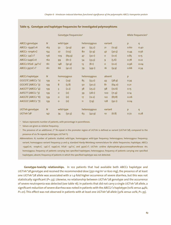
Chapter 6 - Irinotecan-induced diarrhea: functional significance of the polymorphic ABCC2 transporter protein Chapter 6 - Irinotecan-induced diarrhea: functional significance of the polymorphic ABCC2 transporter protein
Table 15. Genotype and haplotype frequencies for investigated polymorphisms
Genotype frequenciesa Allele frequenciesb
ABCC2 genotype N wild-type heterozygous variant p q
ABCC2 -1549G>A 163 52 (31.9) 90 (55.2) 21 (12.9) 0.60 0.40
ABCC2 -1019A>G 154 27 (17.5) 80 (51.9) 47 (30.5) 0.44 0.56
ABCC2 -24C>T 156 109 (69.9) 47 (30.1) 0 (0.0) 0.85 0.15
ABCC2 1249G>A 162 99 (61.1) 54 (33.3) 9 (5.6) 0.78 0.22
ABCC2 IVS26 -34T>C 161 148 (91.9) 13 (8.1) 0 (0.0) 0.96 0.04
ABCC2 3972C>T 161 66 (41.0) 79 (49.1) 16 (9.9) 0.66 0.34
ABCC2 haplotype N homozygous GGCGTC (ABCC2 *2) 139 11 (7.9) GGCATC (ABCC2 *3) 139 8 (5.8) AACGTT (ABCC2 *4) 139 3 (2.2) AATGTT (ABCC2 *5) 139 0 (0) AACGTC (ABCC2 *6) 139 0 (0) AACGCC (ABCC2 *7) 139 0 (0)
heterozygous absent p
85 (53.2) 43 (38.9) 0.34 50 (30.2) 81 (64.0) 0.21 38 (25.2) 98 (72.6) 0.15 39 (28.1) 100 (71.9) 0.14 17 (12.2) 122 (87.8) 0.06 11 (7.9) 128 (92.1) 0.04
UGT1A1 genotype N wild-type heterozygous variant p q
UGT1A1*28c 147 74 (50.3) 63 (42.9) 10 (6.8) 0.72 0.28
a Values represents number of patients, with percentage in parentheses.b Values are given as relative frequency.c The presence of an additional, 7th TA-repeat in the promoter region of UGT1A1 is defined as variant (UGT1A1*28), compared to the
presence of six TA-repeats (wild-type; UGT1A1*1).
Abbreviations: N, number of patients studied; wild-type, homozygous wild-type frequency; heterozygous, heterozygous frequency;
variant, homozygous variant frequency; p and q, standard Hardy-Weinberg nomenclature for allele frequencies; haplotpye, ABCC2
-1549G>A, -1019A>G, -24C>T, 1249G>A, IVS26 –34T>C, and 3972C>T; UGT1A1: uridine diphosphate-glucuronosyltransferase 1A1;
homozygous, frequency of patients carrying two specified haplotypes; heterozygous, frequency of patients carrying one specified
haplotypes; absent, frequency of patients in which the specified haplotype was not detected.
Genotype-toxicity relationships. In 102 patients that had available both ABCC2 haplotype and
UGT1A1*28 genotype and received the recommended dose (350 mg/m2 or 600 mg), the presence of at least
one UGT1A1*28 allele was associated with a 1.4-fold higher occurrence of severe diarrhea, but this was not
statistically significant (P=.33). Likewise, no relationship between UGT1A1*28 genotype and the occurrence
of severe neutropenia was detected (see table 16). In patients that did not carry a single UGT1A1*28 allele, a
significant reduction of severe diarrhea was noted in patients with the ABCC2*2 haplotype (10% versus 44%; P<.01). This effect was not observed in patients with at least one UGT1A1*28 allele (32% versus 20%; P=.35).
63

Chapter 6 - Irinotecan-induced diarrhea: functional significance of the polymorphic ABCC2 transporter protein Chapter 6 - Irinotecan-induced diarrhea: functional significance of the polymorphic ABCC2 transporter protein
DISCUSSION
This study provides preliminary evidence for a genetic predisposition to the pharmacokinetic profile of the anticancer drug irinotecan, and suggests that patients with impaired ABCC2 activity due to an inherited genetic defect are at an increased risk for irinotecan-induced diarrhea. The data complement
previous knowledge on the clinical pharmacology of irinotecan and may have important practical implications for its optimal use.
Numerous polymorphic proteins are involved in irinotecan elimination. However,altered functionality caused by inherited variability in a single gene is frequently obscured by (compensatory) activity of other enzymes and transporters and environmental factors.35,275 In the past, genotyping efforts in the context
of irinotecan chemotherapy have mainly focused on the functional significance of Phase II conjugating pathways involved in the detoxification of the active irinotecan metabolite, SN-38. For example, numerous investigations have determined that the UGT1A1*28 polymorphism affects irinotecan metabolism and toxicity in Caucasians, and the FDA has recently incorporated this information in the package label insert. However, guidelines for clinical investigators and practicing oncologists on how to screen patients for this polymorphism are lacking, nor is it clear how dosages should be adjusted for patients carrying this polymorphism. Most importantly, a priori determination of this particular polymorphism has rather poor predictive ability. In fact, it has been estimated that screening for UGT1A1*28 identifies only about half of individuals that will eventually experience (severe) diarrhea following treatment with irinotecan.306
The incentive for the current investigation was based on previous data indicating that ABCC2 appears to be the principal transporter involved in hepatobiliary secretion of irinotecan, SN-38 as well as SN-38G,307
and that multiple functional polymorphic variants of ABCC2 have been described.316 ABCC2 is a member of the ABC transmembrane proteins that bind ATP and use its energy to drive the transport of various substrates across cell membranes. The ABCC2 gene is located on chromosome 10q24 and encodes a 1545 amino-acid polypeptide, and, like several other transporters of the ABC-superfamily, is found on the apical membrane of polarized cells in the liver, the kidneys and the intestines and is endogenously expressed at highest levels in the canalicular membrane of the hepatocyte.317 Significant variability of the relative expression of ABCC2 has been demonstrated.318 It is conceivable that decreased transport of irinotecan into the bile by ABCC2 leads to increased hepatic metabolism, increased transport of irinotecan into the bile by ABCB1 or ABCG2, or increased transport back into the circulation by ABCC1.
Recently, substitution in the ABCC2 gene has been associated with altered expression of the ABCC2
gene in the liver.318 In particular, compared to haplotypes containing the variant ABCC2 1249A allele, haplotypes containing the wild-type allele (ABCC2 1249G), like the ABCC2*2 haplotype, had significant
lower mRNA levels. Assuming that the ABCC2*2 haplotype encodes a decreased function ABCC2, our finding that the presence of the ABCC2*2 haplotype results in significantly increased systemic irinotecan exposure suggests that other transporters did not compensate completely for the loss of apical transport
capacity, and that overall hepatobiliary secretion is impaired. Reduced hepatobiliary secretion of irinotecan may explain the observed association in this study
between the ABCC2*2 haplotype and the lower frequency of irinotecan-induced diarrhea. Previous work has demonstrated that carboxylesterases are highly expressed in enterocytes, and mediate local activation of irinotecan into SN-38.319 In this scenario, less biliary excretion of irinotecan and hence less local activation within intestinal enterocytes may play a crucial role in the pathogenesis of delayed type diarrhea observed with irinotecan chemotherapy. It is theoretically plausible that the presence of a variant UGT1A1*28 allele may indirectly override the protective effect of the ABCC2*2 haplotype on diarrhea, because of reduced glucuronidation capacity to detoxify enterically formed SN-38 that results in
64
Chapter 6 - Irinotecan-induced diarrhea: functional significance of the polymorphic ABCC2 transporter protein Chapter 6 - Irinotecan-induced diarrhea: functional significance of the polymorphic ABCC2 transporter protein
increased exposure to this toxic metabolite in the enterocytes. All of these scenarios assume that carriers of the ABCC2*2 haplotype have decreased biliary transport function. Alternatively, if the ABCC2*2 reference haplotype represents normal transport function, then a high glucuronidation capacity (UGT1A1*1) coupled with a high ABCC2 transport activity would be consistent with biliary elimination of SN-38G and decreased enterocyte exposure to the toxic SN-38. Understanding the functional significance of the ABCC2 variants and haplotypes will help decipher this complex multigene pathway that mediates irinotecan toxicity.
The hypothesis that decreased local SN-38 detoxification in patients carrying the UGT1A1*28 allele has a key role in the pathogenesis of diarrhea, as opposed to increased hepatobiliary SN-38 secretion, may explain why prophylactic interventions aimed at reducing intraluminal SN-38 exposure are relatively ineffective in reducing the risk for severe diarrhea.306,320 This is perhaps with the exception of the use of active charcoal administered orally, which might be effective for all patients through the adsorption of both intraluminal irinotecan and SN-38.41 This supposition explains as well why patients excreting more SN-38G, as observed in patients not carrying the UGT1A1*28 allele, and which metabolite is almost
completely deglucuronidated by microbial glucuronidases to form SN-38,137 are in general subject to experience less severe diarrhea.44 Since delayed-type diarrhea remains a severe side effect of irinotecan therapy that occurs rather frequently, additional research to define predictive measures of its occurrence in the individual patient is warranted.
In conclusion, this study suggests that the polymorphic ABCC2 transporter may have a crucial role in the occurrence of irinotecan-related diarrhea,particularly in individuals that are not genetically predisposed because of impaired UGT1A1-mediated glucuronidation of the active metabolite, SN-38. Confirmation of the current observation is warranted.
ACKNOWLEDGEMENTS
We acknowledge the seminal information provided on the web by Dr. Mark J. Ratain (University of Chicago, Chicago, IL; http://www.pharmgkb.org/) to the effect that ABCC2 polymorphisms may play a role in the pharmacokinetics of irinotecan.
65

66

67
Chapter 7
Prophylactic treatment of irinotecan-induced diarrhea with neomycin and potential role for UGT1A1*28 genotype screening: a randomized, placebo-controlled, double-blind study
FA de Jong, DF Kehrer, RH Mathijssen, G-J Creemers, P de Bruijn, RH van Schaik, AS Planting, A van der Gaast, FA Eskens, JT Janssen, JB Ruit, J Verweij, A Sparreboom, MJ de Jonge
The Oncologist 11: in press, 2006

Chapter 7 - Prophylactic treatment of irinotecan-induced diarrhea with neomycin and potential role for UGT1A1*28 genotype screening
68
Chapter 7 - Prophylactic treatment of irinotecan-induced diarrhea with neomycin and potential role for UGT1A1*28 genotype screening
ABSTRACT
Objective: Delayed-type diarrhea is a common side effect
of irinotecan and is associated with a bacterial-mediated formation of the active irinotecan metabolite SN-38 from its glucuronide conjugate in the intestine. Based on a pilot study, we hypothesized that concomitant administration of the antibiotic neomycin would diminish exposure of the gut to SN-38 and ameliorate the incidence and severity of diarrhea.
Patients and methods: Patients were treated with irinotecan in a multicenter, double-blind, randomized, placebo-controlled trial. Eligible patients received irinotecan (350 mg/m2 once every three weeks) combined with neomycin (660 mg three times daily for three consecutive days, starting two days before chemotherapy) or combined with placebo. Blood samples were obtained for additional pharmacokinetic and pharmacogenetic analyses.
Results: Sixty-two patients were evaluable for the toxicity analysis. Baseline patient characteristics, systemic SN-38 exposure, and UGT1A1*28 genotype status (i.e., an additional TA-repeat in the promoter region of uridine diphosphate-glucuronosyltransferase isoform 1A1 were similar in both arms. Although distribution, severity, and duration of delayed-type diarrhea did not differ significantly between both arms, grade 3 diarrhea tended to be less frequent in the neomycin arm. The presence of at least one UGT1A1*28 allele was strongly related to the incidence of grade 2–3 diarrhea. In the neomycin arm, grade 2 nausea was significantly more common.
Conclusion: Our results do not suggest a major role for neomycin as prophylaxis for irinotecan-induced delayed-type diarrhea. It
is suggested that the UGT1A1*28 genotype status could be used as a screening tool for a priori prevention of irinotecan-induced delayed-type diarrhea.
Chapter 7 - Prophylactic treatment of irinotecan-induced diarrhea with neomycin and potential role for UGT1A1*28 genotype screening Chapter 7 - Prophylactic treatment of irinotecan-induced diarrhea with neomycin and potential role for UGT1A1*28 genotype screening
INTRODUCTION
(SN-38G).
Irinotecan (Campto®, Camptosar®; Pfizer Pharmaceuticals, New York) is frequently used in first- and second-line treatment of metastatic colorectal cancer.8-10,13 Clinical activity has also been demonstrated in lung, gastric, and pancreatic cancer, amongst others.15,21,22 Regardless of its schedule of administration, myelosuppression and delayed-type diarrhea are the most common side effects seen in patients treated with this topoisomerase I inhibitor.7,8,29,40 Irinotecan is a prodrug that needs to be metabolized through hydroxylation to form its active metabolite SN-38.23 Subsequently, SN-38 is detoxified in the liver by uridine diphosphate-glucuronosyltransferases, especially UGT1A1, to form an inactive glucuronide metabolite
28,151,241 Both SN-38 and SN-38G are excreted via the urine and bile.27,29
Delayed-type diarrhea, defined as diarrhea occurring more than 24 hours after administration of irinotecan,36 is probably directly mediated by high concentrations of intraluminal SN-38, which is formed partly out of SN-38G by bacterial β-glucuronidases.30,321 Severe delayed-type diarrhea has been reported to occur in up to 30–40% of patients and necessitates hospitalization for intravenous rehydration in about
4310% of patients.13,36-42 Apart from morbidity, this type of diarrhea results in expanded health-care costs,and even mild diarrhea may influence continuation of treatment.
in humans.In recent years,several attempts have been made to prevent irinotecan-induced delayed-type diarrhea
41,137,322-335 Most strategies have focused on intervening in its metabolic pathway to reduce SN-38 concentrations in the gut. However, most studies that have suggested protective effects were not
randomized, did not evaluate SN-38 pharmacokinetics,and/or were not placebo controlled.Therefore, there is still no generally accepted prophylactic treatment for irinotecan-induced delayed-type diarrhea.320,336 In a previously performed pilot study, we reported that the aminoglycoside antibiotic neomycin prevented the recurrence of grade ≥2 diarrhea in six of seven patients,which was attributed to inhibition of β-glucuronidase producing intestinal bacteria.137 Because no changes in systemic pharmacokinetic parameters of SN-38 with or without neomycin were observed, cotreatment with neomycin was considered safe. Moreover, fecal cultures taken during and after neomycin treatment did not reveal neomycin-resistant microorganisms, bacterial overgrowth, or toxins. Two subsequent small studies have been published since then, confirming the potential role of neomycin in preventing irinotecan-induced delayed-type diarrhea.328,333 Here, we present the results of a multicenter, double-blind, randomized, placebo-controlled study of irinotecan with or without neomycin, aimed to validate the previously demonstrated prophylactic effects of neomycin on irinotecan-induced delayed-type diarrhea.137
PATIENTS AND METHODS
Patient eligibility. Patients were recruited from the departments of medical oncology and internal medicine of the hospitals mentioned below. The treating physicians were medical oncologists who were all familiar with the study protocols. Patients, 18 years of age or older, were required to have a confirmed malignant solid tumor for which irinotecan was considered the best available treatment option. Before randomization, eligibility was confirmed by use of a protocol-specific checklist, which included adequate hematological (absolute neutrophil count [ANC] ≥2.0*109/L) and hepatic (bilirubin ≥1.25*the upper limit
of normal (ULN), alanine aminotransferase (ALT) and aspartate aminotransferase (AST) ≥2.5*ULN, in case of liver metastasis ≥5*ULN, and alkaline phosphatase ≥5*ULN) functions. Patients who had a World Health Organization (WHO) performance status score >2, were known to be hypersensitive to neomycin, had been treated with irinotecan before, or had had anticancer therapy or major surgery within four weeks prior to study entry were not eligible. Other exclusion criteria were previous abdominal radiotherapy, unresolved bowel obstruction, and chronic colic disease. Patients were not allowed to use herbal medicines/tea,
69

Chapter 7 - Prophylactic treatment of irinotecan-induced diarrhea with neomycin and potential role for UGT1A1*28 genotype screeningChapter 7 - Prophylactic treatment of irinotecan-induced diarrhea with neomycin and potential role for UGT1A1*28 genotype screening
dietary supplements, and grapefruit( juice) from 4 weeks before and during the study period. Use of drugs known to modulate cytochrome P450 3A (CYP3A) function or expression to a clinically significant extent
was prohibited, except for necessary prophylactic antiemetics. This study was performed in accordance with the Declaration of Helsinki (Hong Kong amendment).The
institutional medical ethical board of the Erasmus University Medical Center Rotterdam, the Netherlands, and the local institutional review boards of all participating local hospitals in its referral area (Catharina Hospital in Eindhoven, the Franciscus Hospital in Roosendaal, the IJsselland Hospital in Capelle aan den IJssel, the Vlietland Hospital in Schiedam/Vlaardingen, the Van Weel Bethesda Hospital in Dirksland, and the Lievensberg Hospital in Bergen op Zoom) had approved the study protocol, which involved sampling for pharmacokinetic and pharmacogenetic purposes as well. Prior to study entry, written informed consent
was obtained from all patients. Sampling for pharmacokinetic and pharmacogenetic analysis was only done if the patient had given separate and specific written consent for these procedures.
Study design and treatment. Once eligibility was confirmed, the patient was given an identification number according to the hospital where the patient was entered and his/her entry order in the study. Randomization was performed according to a randomized block design. Block length varied at random using a mixture of block lengths stratified for each separate institution. Patients were randomized between arm A, consisting of irinotecan given at a dose of 350 mg/m2 combined with oral neomycin (660 mg three times daily) for three consecutive days starting two days before chemotherapy, and arm B, consisting of the same irinotecan regimen but during the first course given with placebo. Prior to the study, a numbered randomization list with neomycin versus placebo was produced by an independent statistician which was directly provided to the involved trial pharmacist (Department of Hospital Pharmacy, Erasmus University Medical Center Rotterdam) only. Capsules containing neomycin or placebo were produced by the hospital pharmacy. The medication strips, re-numbered according to the randomization list, were distributed to the participating centers. After registration, the registration officer provided the number of the strip to be used. The clinical study coordinator, the registration officer, the treating physician, and the patient were therefore not informed to which arm of treatment the patient had been randomized.
Irinotecan hydrochloride trihydrate (Aventis Pharma, Hoevelaken, the Netherlands; Pfizer BV, Capelle aan den IJssel, the Netherlands) was administered over 90 min as a continuous intravenous infusion, repeated every three weeks. All patients received prophylactic antiemetics, including dexamethasone and 5HT3-receptor antagonists.The neomycin-dosing regimen was based on a small study in healthy volunteers in which it was shown to adequately block fecal β-glucuronidase activity for a period of at least three days after the last dose (unpublished data). As soon as the first liquid stool occurred, the patient had to start
loperamide treatment (4 mg loperamide for the first dose, then 2 mg every two hours until 12 hours after the last liquid stool). If diarrhea persisted for more than 48 hours, oral antibiotic therapy with ciprofloxacin (500 mg twice a day) was prescribed.
Patients were deemed evaluable if they had taken three days of neomycin/placebo completely and without vomiting within 4 hours after intake (according to anamnesis), irinotecan had been adequately administered, and complete data on diarrhea toxicity were available. A full blood count, including neutrophils, was obtained once every week. Besides nadir values and percent decrease at nadir from baseline, the National Cancer Institute–Common Toxicity Criteria (NCI-CTC, version 2.0; http://ctep.cancer. gov/forms/CTCv20_4-30-992.pdf) were used for classification of severity of neutropenia and leucopenia, as well as for diarrhea, nausea, and vomiting, up to three weeks following administration of irinotecan. The duration of diarrhea (in days) was scored as well.
70
Chapter 7 - Prophylactic treatment of irinotecan-induced diarrhea with neomycin and potential role for UGT1A1*28 genotype screening Chapter 7 - Prophylactic treatment of irinotecan-induced diarrhea with neomycin and potential role for UGT1A1*28 genotype screening
At regular intervals, the case record forms were checked against source documents and, if necessary, completed by the data manager. After closing the study, the clinical study coordinator (re-)evaluated eligibility, (recorded) toxicity, and evaluability according to the above-mentioned criteria of all patients. To assure unbiased interpretation, this was done before unblinding the study intervention, i.e., the use of neomycin or placebo.
Isolation of genomic DNA and genotype analysis. Genomic DNA was isolated from whole blood or plasma using the MagNA Pure LC System (Roche Molecular Biochemicals, Mannheim, Germany), after which amplification was performed using polymerase chain reaction (PCR)-based techniques. The number of TA-repeats in the TATA-box of the promoter region of the UGT1A1 gene was determined by sizing of the PCR-products obtained with UGT1A1-specific primers, as described in detail elsewhere.203 Genotypes were assigned as 6/6, 6/7, and 7/7 for patients homozygous for six repeats (wild-type), heterozygous patients, and patients homozygous for seven repeats (UGT1A1*28), respectively. The analysis was validated using control DNA from individuals with known 6/6, 6/7, and 7/7 genotypes.
Blood sampling and pharmacokinetic analysis. Blood samples for pharmacokinetic analysis were collected at three time points: immediately prior to infusion, and at 1 and 48 h after the end of infusion, according to a previously developed limited-sampling model to predict the area under the plasma concentration-time curve (AUC) of the active metabolite SN-38.172,337 After patient approval, blood was taken from the arm that was not used for irinotecan infusion. Samples were collected in tubes containing lithium heparin and were centrifuged for 10 min at 3,000 g to separate blood cells from plasma, as described in detail elsewhere.338 Plasma was stored at –70°C until the day of analysis. Concentrations of SN-38 in plasma were determined by reversed-phase high-performance liquid chromatography with fluorescence detection.143,339 The AUC of SN-38 was calculated as: AUCSN-38
(ng*h/mL) = 6.588 * C1h + 146.4 * C48h
+ 15.53, where C1h and C48h are the plasma concentrations of SN-38 at 1 and 48 hours after the end of infusion.172
Statistical considerations. Initially, grade ≥2 irinotecan-induced delayed-type diarrhea was estimated to occur in half of the regularly dosed patients. Protection against this type of diarrhea by prophylactic neomycin coadministration was considered to be of clinical importance if the reduction to grade <2 diarrhea would be at least 50%. In order to detect this reduction, with a power of 80% and a significance level of 5%, it was calculated that a population of 60 patients had to be studied in a randomized, placebo-controlled design. To compare normally distributed continuous variables between two groups of patients, the Student’s t-test was used; otherwise the non-parametric Mann-Whitney U-test was chosen. In cases of two dichotomous variables, the Pearson χ2-test was performed, whereas Spearman’s correlation coefficient
was used to relate two continuous variables. A non-parametric trend test was used if a trend over several groups of patients was expected. Statistical tests were performed using SPSS version 10.0.7 (SPSS Inc, Chicago, IL) and Stata version 9.0 (Stata, College Station, TX). Test results with a P<.05 were considered statistically significant.
RESULTS
Patient population and treatment. Of the 75 patients enrolled on this study between December 2001 and October 2004, 62 were evaluable for analysis of the effects of neomycin on irinotecan-induced delayed-type diarrhea (see table 17). Thirteen patients did not fulfill all criteria: one never started, five did not complete their neomycin/placebo or irinotecan therapy, two died during follow-up, one received concurrent radiotherapy, two had a stoma, one suffered from pre-existing diarrhea, and one used lactulose. About half of the patients were included in the Erasmus University Medical Center Rotterdam, whereas the remaining patients were treated in the six local hospitals in its referral area. Patients were treated with
71
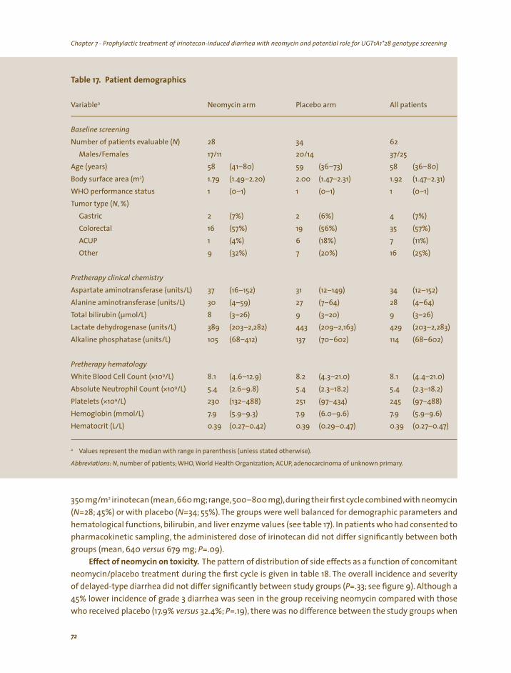
Chapter 7 - Prophylactic treatment of irinotecan-induced diarrhea with neomycin and potential role for UGT1A1*28 genotype screeningChapter 7 - Prophylactic treatment of irinotecan-induced diarrhea with neomycin and potential role for UGT1A1*28 genotype screening
a
N) 28 34 62
) 58 58 2) 2.00 1.92
1 1 1 N, %)
Gastric 2 2 (6%) 4 16 19 35 1 (4%) 6 (18%) 7 (11%)
Other 9 (32%) 7 (20%) 16 (25%)
31 34 30 28 8 9 9 389 443 429 105 114
(*109/L) 8.1 8.2 8.1 (*109/L)
*109/L) 230 251 245 Hemoglobin (mmol/L)
a
, ,
Table 17. Patient demographics
Variable Neomycin arm Placebo arm All patients
Baseline screening
Number of patients evaluable (Males/Females 17/11 20/14 37/25
Age (years (41–80) 59 (36–73) (36–80) Body surface area (m 1.79 (1.49–2.20) (1.47–2.31) (1.47–2.31) WHO performance status (0–1) (0–1) (0–1) Tumor type (
(7%) (7%) Colorectal (57%) (56%) (57%) ACUP
Pretherapy clinical chemistry Aspartate aminotransferase (units/L) 37 (16–152) (12–149) (12–152) Alanine aminotransferase (units/L) (4–59) 27 (7–64) (4–64) Total bilirubin (µmol/L) (3–26) (3–20) (3–26) Lactate dehydrogenase (units/L) (203–2,282) (209–2,163) (203–2,283) Alkaline phosphatase (units/L) (68–412) 137 (70–602) (68–602)
Pretherapy hematology
White Blood Cell Count (4.6–12.9) (4.3–21.0) (4.4–21.0) Absolute Neutrophil Count 5.4 (2.6–9.8) 5.4 (2.3–18.2) 5.4 (2.3–18.2) Platelets ( (132–488) (97–434) (97–488)
7.9 (5.9–9.3) 7.9 (6.0–9.6) 7.9 (5.9–9.6) Hematocrit (L/L) 0.39 (0.27–0.42) 0.39 (0.29–0.47) 0.39 (0.27–0.47)
Values represent the median with range in parenthesis (unless stated otherwise).
Abbreviations: N, number of patients; WHO World Health Organization; ACUP adenocarcinoma of unknown primary.
350 mg/m2 irinotecan (mean,660 mg;range,500–800 mg),during their first cycle combined with neomycin (N=28; 45%) or with placebo (N=34; 55%). The groups were well balanced for demographic parameters and hematological functions, bilirubin, and liver enzyme values (see table 17). In patients who had consented to pharmacokinetic sampling, the administered dose of irinotecan did not differ significantly between both groups (mean, 640 versus 679 mg; P=.09).
Effect of neomycin on toxicity. The pattern of distribution of side effects as a function of concomitant
neomycin/placebo treatment during the first cycle is given in table 18. The overall incidence and severity of delayed-type diarrhea did not differ significantly between study groups (P=.33; see figure 9). Although a 45% lower incidence of grade 3 diarrhea was seen in the group receiving neomycin compared with those who received placebo (17.9% versus 32.4%; P=.19), there was no difference between the study groups when
72
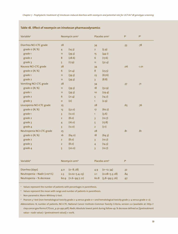
Chapter 7 - Prophylactic treatment of irinotecan-induced diarrhea with neomycin and potential role for UGT1A1*28 genotype screening Chapter 7 - Prophylactic treatment of irinotecan-induced diarrhea with neomycin and potential role for UGT1A1*28 genotype screening
a a a Pc Pd
28 34 .33 (N, %) 4 2
11 (39.3) 15 8 (28.6) 6 5 11 28 34 .06 <.01
(N, %) 6 8 (23.5) 11 (39.3) 23 11 (39.3) 3 (8.8) 28 34
(N, %) 11 (39.3) 18 (52.9) 11 (39.3) 10 6 5 0 (0) 1 (2.9) 25 28 .65
(N, %) 13 (52.0) 17 3 (12.0) 1 (3.6) 2 (8.0) 3 4 (16.0) 5 3 (12.0) 2 25 28 .81 .81
(N, %) 16 18 2 (8.0) 3 2 (8.0) 4 5 3
b b b Pc
) .32
*109/L) 2.3 2.1 .84
a
b
c
d χ2
, (
,
* 100%.
Table 18. Effect of neomycin on irinotecan pharmacodynamics
Variable Neomycin arm Placebo arm
Diarrhea NCI-CTC grade .78
grade 0 (14.3) (5.9) grade 1 (44.1) grade 2 (17.6) grade 3 (17.9) (32.4)
Nausea NCI-CTC grade grade 0 (21.4) grade 1 (67.6) grade 2
Vomiting NCI-CTC grade .37 .71 grade 0 grade 1 (29.4) grade 2 (21.4) (14.7) grade 3
Leucopenia NCI-CTC grade .76
grade 0 (60.7) grade 1 grade 2 (10.7) grade 3 (17.8) grade 4 (7.1)
Neutropenia NCI-CTC grade grade 0 (64.0) (64.3) grade 2 (10.7) grade 3 (14.3) grade 4 (20.0) (10.7)
Variable Neomycin arm Placebo arm
Diarrhea (days 4.0 (0–8; 28) 4.9 (0–12; 34) Neutropenia – Nadir ( (0.02–5.4; 23) (0.08–5.3; 28) Neutropenia – % decrease 60.9 (11.6–99.7; 21) 60.8 (3.8–99.5; 26) .97
Values represent the number of patients with percentages in parenthesis.
Values represent the mean with range and number of patients in parenthesis.
Non-parametric Mann-Whitney U-test.
Pearson -test (non-hematological toxicity grade 2–4 versus grade 0–1 and hematological toxicity grade 3–4 versus grade 0–2).
Abbreviations: N number of patients; NCI-CTC, National Cancer Institute–Common Toxicity Criteria, version 2.0 available at: http://
ctep.cancer.gov/forms/CTCv20_4-30-992.pdf); Nadir Absolute lowest point during follow-up; % decrease defined as [(pretreatment
value – nadir value) / (pretreatment value)]
73

Chapter 7 - Prophylactic treatment of irinotecan-induced diarrhea with neomycin and potential role for UGT1A1*28 genotype screeningChapter 7 - Prophylactic treatment of irinotecan-induced diarrhea with neomycin and potential role for UGT1A1*28 genotype screening
) or (
2.0,
Figure 9. The effect of concomitant neomycin treatment
on irinotecan-induced delayed-type diarrhea. Histogram showing the distribution of patients (percentages receiving neomycin (light) placebo dark) by diarrhea (National Cancer Institute–Common Toxicity Criteria (NCI-CTC), version
available at: http://ctep.cancer.gov/forms/CTCv20_4-30-992.pdf). No significant effect of neomycin prophylaxis on the incidence and severity of irinotecan-induced delayed-type diarrhea was found, although the risk of grade 3 diarrhea was 45% lower in patients receiving neomycin (P=.19).
grade 2 and 3 diarrhea were taken together (46.4% versus 50.0%; P=.78). Treatment with neomycin did not
result in a significantly shorter duration of diarrhea (4.0 versus 4.9 days; P=.32). In both arms,approximately 75% of patients experienced any grade of nausea despite the use of prophylactic antiemetics. Patients receiving neomycin had a 4.5-fold higher risk for grade 2 nausea than those receiving placebo (39.3% versus 8.8%; P<.01).
UGT1A1*28 pharmacogenetics. Of the evaluable patients, 52 patients (84%) consented to pharmacogenetic blood sampling. Twenty-three patients (44.2%) were heterozygous for UGT1A1*28, three (5.8%) were homozygous variant, and 26 (50.0%) were homozygous wild-type. The UGT1A1*28 allele was found to be equally distributed between patients receiving neomycin and those receiving placebo (P=.58). The presence of at least one UGT1A1*28 allele was highly related to the distribution pattern of delayed-type diarrhea (P=.03; see table 19). Specifically, the incidence of grade 2–3 diarrhea was doubled in patients carrying at least one UGT1A1*28 allele (69.2% versus 34.6%; P=.01), whereas patients carrying the wild-type sequence were more likely to experience only grade 0–1 diarrhea (30.8% versus 65.4%), regardless of prophylactic neomycin coadministration. In neither genetic group the incidence and severity of diarrhea was related to the use of neomycin or placebo (P>.66).
6, 7
Figure 10. UGT1A1*28 genotype and the metabolic clearance of SN-38. Boxplot showing that presence of the variant UGT1A1*28 allele is significantly related to less efficient metabolic clearance of SN-38 (P<.01). The wild-type allele of UGT1A1 is designated TA and the variant allele is TA (UGT1A1*28).
74
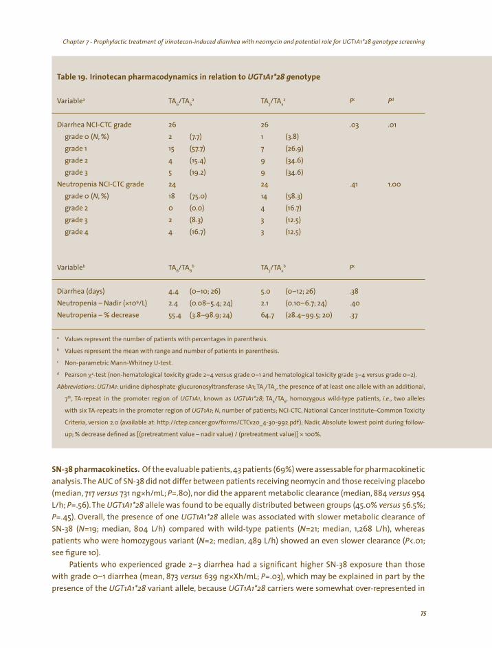
Chapter 7 - Prophylactic treatment of irinotecan-induced diarrhea with neomycin and potential role for UGT1A1*28 genotype screening Chapter 7 - Prophylactic treatment of irinotecan-induced diarrhea with neomycin and potential role for UGT1A1*28 genotype screening
enotype
a 6/ 6
a 7/ x
a Pc Pd
26 26 .03 .01 (N, %) 2 1 (3.8)
15 7 (26.9) 4 9 5 (19.2) 9 24 24 1.00
(N, %) 18 14 (58.3) 0 4 2 (8.3) 3 (12.5) 4 3 (12.5)
b 6/ 6
b 7/ x
b Pc
) .38
*109/L) 2.1
a
b
c
d χ2
7/ x,
7th , ; 6/ 6 ,
; N
( ,
* 100%.
Table 19. Irinotecan pharmacodynamics in relation to UGT1A1*28 g
Variable TA TA TA TA
Diarrhea NCI-CTC grade grade 0 (7.7) grade 1 (57.7) grade 2 (15.4) (34.6) grade 3 (34.6)
Neutropenia NCI-CTC grade .41 grade 0 (75.0) grade 2 (0.0) (16.7) grade 3 grade 4 (16.7)
Variable TA TA TA TA
Diarrhea (days 4.4 (0–10; 26) 5.0 (0–12; 26) Neutropenia – Nadir ( 2.4 (0.08–5.4; 24) (0.10–6.7; 24) .40
Neutropenia – % decrease 55.4 (3.8–98.9; 24) 64.7 (28.4–99.5; 20) .37
Values represent the number of patients with percentages in parenthesis.
Values represent the mean with range and number of patients in parenthesis.
Non-parametric Mann-Whitney U-test.
Pearson -test (non-hematological toxicity grade 2–4 versus grade 0–1 and hematological toxicity grade 3–4 versus grade 0–2).
Abbreviations: UGT1A1: uridine diphosphate-glucuronosyltransferase 1A1; TA TA the presence of at least one allele with an additional,
, TA-repeat in the promoter region of UGT1A1 known as UGT1A1*28 TA TA , homozygous wild-type patients, i.e. two alleles
with six TA-repeats in the promoter region of UGT1A1 , number of patients; NCI-CTC, National Cancer Institute–Common Toxicity
Criteria, version 2.0 available at: http://ctep.cancer.gov/forms/CTCv20_4-30-992.pdf); Nadir Absolute lowest point during follow-
up; % decrease defined as [(pretreatment value – nadir value) / (pretreatment value)]
SN-38 pharmacokinetics. Of the evaluable patients, 43 patients (69%) were assessable for pharmacokinetic analysis. The AUC of SN-38 did not differ between patients receiving neomycin and those receiving placebo (median, 717 versus 731 ng*h/mL; P=.80), nor did the apparent metabolic clearance (median, 884 versus 954 L/h; P=.56). The UGT1A1*28 allele was found to be equally distributed between groups (45.0% versus 56.5%; P=.45). Overall, the presence of one UGT1A1*28 allele was associated with slower metabolic clearance of SN-38 (N=19; median, 804 L/h) compared with wild-type patients (N=21; median, 1,268 L/h), whereas patients who were homozygous variant (N=2; median, 489 L/h) showed an even slower clearance (P<.01; see figure 10).
Patients who experienced grade 2–3 diarrhea had a significant higher SN-38 exposure than those with grade 0–1 diarrhea (mean, 873 versus 639 ng*Xh/mL; P=.03), which may be explained in part by the presence of the UGT1A1*28 variant allele, because UGT1A1*28 carriers were somewhat over-represented in
75
Chapter 7 - Prophylactic treatment of irinotecan-induced diarrhea with neomycin and potential role for UGT1A1*28 genotype screeningChapter 7 - Prophylactic treatment of irinotecan-induced diarrhea with neomycin and potential role for UGT1A1*28 genotype screening
the patients experiencing grade 2–3 diarrhea (65.2% versus 35%; P=.05). Patients (N=11) who experienced grade 3–4 neutropenia had a significantly higher systemic exposure to SN-38 than those (N=31) who experienced grade 0–2 neutropenia (median, 1,023 versus 623 ng*h/mL; P<.01). Both the ANC nadir and the percentage decrease at nadir from baseline were related to the AUC of SN-38 (rs=–0.54 and rs=0.62, respectively; P<.01).
DISCUSSION
In this double-blind, randomized, placebo-controlled trial, we found no significant effect of neomycin on incidence, severity, and duration of irinotecan-induced delayed-type diarrhea. Although the incidence of grade 3 diarrhea was diminished from 32% to 18% and the average duration was shortened by almost
one day, this was accompanied by a shift to a substantially higher incidence of grade 2 diarrhea. This finding is in contrast to the hypothesis derived from the earlier performed pilot study.137 Our results clearly demonstrate that the combination of neomycin with irinotecan is safe and that neomycin does not affect
systemic exposure to SN-38, but patients receiving neomycin had a significantly greater risk for more severe nausea, probably at least partly induced by neomycin.
Despite the fact that a high-dose loperamide regimen renders irinotecan-induced diarrhea more manageable once it has occurred,42,159 it still remains one of its serious toxicities, occurring rather frequently and unpredictably. It was originally suggested from animal models that β-glucuronidases produced by common intestinal microflora might play a major role in the development of delayed-type diarrhea by mediating deglucuronidation of hepatobiliary excreted SN-38G into SN-38.321 Indeed, data obtained in rats have indicated that antibiotics inhibited the β-glucuronidase activity from the intestinal microflora, thereby decreasing the luminal SN-38 concentration and subsequently reducing cecal damage and ameliorating diarrhea.30,31 Additionally, in our pilot study mentioned before,137 we observed a reduced incidence of delayed-type diarrhea after cotreatment with neomycin in patients who had experienced grade 2 or more severe irinotecan-induced diarrhea in their first course.
The discrepancy between the current data and those presented earlier on the neomycin-irinotecan combination might be caused by a change in study design. In the pilot study, patients were treated with neomycin starting five days before irinotecan infusion and ending 2 days after infusion. Prior to embarking on the current study, the question arose whether a shorter period of treatment with neomycin would be sufficient to reduce β-glucuronidase activity. Likewise, it was not known how soon β-glucuronidase activity would reappear after discontinuation of the neomycin treatment.These questions were addressed in a small study in healthy volunteers (unpublished data). The obtained data revealed that three days of treatment with three times daily 660 mg neomycin adequately blocked fecal β-glucuronidase activity for at least three days after the last dose, which led to the neomycin dosing regimen as prescribed in this study.
As mentioned before, in our earlier study, patients who had experienced grade ≥2 delayed-type diarrhea were selected to receive concomitant neomycin during their second chemotherapy course.137 Six of seven patients likely benefited from neomycin prophylaxis during their second course. Although no significant prophylactic effect on the incidence and severity of irinotecan-induced delayed-type diarrhea during the first course of treatment could be demonstrated in this study, a protective effect during subsequent courses once patients have experienced grade ≥2 delayed-type diarrhea and are at higher risk of recurrence of diarrhea cannot be ruled out based on the findings presented here. However, because the natural course of diarrhea during the second administration of irinotecan was not investigated in the pilot study, an overestimation of the prophylactic neomycin effect cannot be excluded. Additionally,
76

Chapter 7 - Prophylactic treatment of irinotecan-induced diarrhea with neomycin and potential role for UGT1A1*28 genotype screening Chapter 7 - Prophylactic treatment of irinotecan-induced diarrhea with neomycin and potential role for UGT1A1*28 genotype screening
a synergistic effect of neomycin on the severity of delayed-type diarrhea in patients who would have encountered only mild diarrhea without neomycin prophylaxis cannot be excluded.
Although the current study might be somewhat underpowered to detect small therapeutic benefits, the general lack of a substantial clinical benefit of neomycin on irinotecan-induced delayed-type diarrhea is concordant with previously reported comparative trials of comparable size that evaluated other agents. For example, Karthaus et al.335 performed a study in 56 patients showing that budesonide (a synthetic corticosteroid) had no significant prophylactic effect on irinotecan-induced delayed-type diarrhea. Similarly, no effect of tiorfan (racecadotril, an antidiarrhea drug that inhibits enkephalinase) was noted in a trial involving 68 patients.322 In contrast, in a smaller, non-randomized study, a significant effect of active charcoal was seen on the incidence of irinotecan-induced delayed-type diarrhea.41 Specifically, the incidence of grade 3–4 diarrhea was reduced in 28 patients from 25% to 7% when combined with active charcoal. However,no pharmacokinetic analysis was performed to confirm a lack of effect of active charcoal on systemic SN-38 concentrations. Hence, partly because of its nature and the many factors involved in its pathogenesis, specific recommendations for the prophylactic treatment of irinotecan-induced delayed-type diarrhea currently cannot be provided.
As long as there are no generally accepted and undisputedly proven interventions to prevent the occurrence of irinotecan-induced delayed-type diarrhea, we have to focus on other ways dealing with this debilitating side effect. Because it has been shown that body surface area-based dosing does not
reduce interindividual pharmacokinetic and pharmacodynamic variability of irinotecan,50,139 one approach may be to optimize dosing strategies.203 Several pharmacokinetic and pharmacogenetic analyses have been performed in order to predict the severity and incidence of irinotecan-induced side effects, with conflicting results.29,151,303,340 Some studies have reported a correlation between delayed-type diarrhea and biliary secretion of SN-38.161,341,342 In addition, the homozygous presence of the UGT1A1*28 polymorphism, leading to less efficient glucuronidation of SN-38,228,229 has been identified as a potential risk factor for the occurrence of delayed-type diarrhea and grade 3–4 neutropenia.27,45,49,284,343
In the current study, occurrence of grade 2–3 delayed-type diarrhea was found to be strongly related to systemic SN-38 exposure, with a 37% higher AUC for those patients who experienced grade 2–3 diarrhea, which is in line with earlier findings.44 Because pharmacokinetic information is always limited to a posteriori correlates, in clinical practice relations with exposure to SN-38 are of little value, and we should search for a priori predictors. In this study, a strong correlation between the presence of the UGT1A1*28 polymorphism and incidence and severity of irinotecan-induced delayed-type diarrhea was noted. Most studies have reported that only patients carrying two variant alleles were at risk of serious toxicity.151,284,340,343 In contrast, we found that patients carrying at least one variant allele had a doubled risk of grade 2–3 delayed-type diarrhea, making the presence of at least one UGT1A1*28 allele a strong predictive factor with potential clinical relevance. This is of particular interest given the availability of blood tests, such as the recently U.S. Food and Drug Administration-approved Invader UGT1A1 Molecular Assay (Third Wave Technologies, Madison, WI), which can rather easily detect this polymorphism at low costs before treatment.151
CONCLUSION
Based on the findings presented here, the best way to deal with irinotecan-induced delayed-type diarrhea still remains a strict loperamide regimen once it has occurred, in combination with antibiotics and rehydration if necessary. Although neomycin did not prove useful as prophylaxis, this study suggests that we can potentially reduce the a priori risk of grade ≥2 irinotecan-induced delayed-type diarrhea using genotyping strategies. Specifically, our data provide a strong argument to administer irinotecan only to
77
Chapter 7 - Prophylactic treatment of irinotecan-induced diarrhea with neomycin and potential role for UGT1A1*28 genotype screening
patients who are most likely to tolerate it relatively well, such as individuals that are not carrying the UGT1A1*28 allele. Alternatively, one should opt for other proven effective chemotherapeutic agents in first
instance for patients carrying this genetic aberration, until better dosing strategies and/or prophylactic interventions aimed to lower the risk for irinotecan-induced delayed-type diarrhea have undisputedly proven their clinical value.
ACKNOWLEDGEMENTS
We would like to thank all participating investigators and researchers at the following Dutch hospitals: the Erasmus University Medical Center Rotterdam (Center Location and Daniel den Hoed Cancer Center, Rotterdam), the Catharina Hospital (Eindhoven), the Franciscus Hospital (Roosendaal), the IJsselland Hospital (Capelle aan den IJssel), the Vlietland Hospital (Schiedam/Vlaardingen), the Van Weel Bethesda Hospital (Dirksland), and the Lievensberg Hospital (Bergen op Zoom).
This study was generously supported by a grant from Aventis Pharma (Hoevelaken, the Netherlands). Additionally, we thank Pfizer BV (Capelle aan den IJssel, the Netherlands) for its contribution to support
the finalization of this study.
78
Chapter 7 - Prophylactic treatment of irinotecan-induced diarrhea with neomycin and potential role for UGT1A1*28 genotype screening
79
Chapter 8
Prediction of irinotecan
pharmacokinetics by use of cytochrome P450 3A4 phenotyping probes
RH Mathijssen, FA de Jong, RH van Schaik, ER Lepper, LE Friberg, T Rietveld, P de Bruijn, WJ Graveland, WD Figg, J Verweij, A Sparreboom
Journal of the National Cancer Institute 96(21):1585–1592, 2004

Chapter 8 - Prediction of irinotecan pharmacokinetics by use of cytochrome P450 3A4 phenotyping probes
80
Chapter 8 - Prediction of irinotecan pharmacokinetics by use of cytochrome P450 3A4 phenotyping probes
ABSTRACT
Background: Irinotecan is a topoisomerase I inhibitor that has been approved for use as a first- and second-line treatment for colorectal cancer. The response to irinotecan is variable, possibly because of interindividual variation in the expression of the enzymes that metabolize irinotecan, including cytochrome P450 3A4 (CYP3A4) and uridine diphosphate glucuronosyltransferase 1A1 (UGT1A1). We prospectively explored the relationships between CYP3A phenotype, as assessed by erythromycin metabolism and midazolam clearance, and the metabolism of irinotecan and its active metabolite SN-38
Methods: Of the thirty white cancer patients, 27 received at least two treatments with irinotecan administered as a 90-min infusion (dose, 600 mg) with three weeks between treatments, and three received only one treatment. Before the first and second treatments, patients underwent an erythromycin breath test and a midazolam clearance test as phenotyping probes for CYP3A4. Erythromycin metabolism was assessed as the area under the curve for the flux of radioactivity in exhaled CO2
within 40 min after administration of [N-methyl-14C]erythromycin. Midazolam and irinotecan were measured by high-performance liquid chromatography. Genomic DNA was isolated from blood and screened for genetic variants in CYP3A4 and UGT1A1. All statistical tests were two-sided.
Results: CYP3A4 activity varied sevenfold (range, 0.223%–1.53% of dose) among patients, whereas midazolam clearance varied fourfold (range, 262–1,012 mL/min), although intraindividual variation was small. Erythromycin metabolism was not statistically significantly associated with irinotecan clearance (P=.090), whereas midazolam clearance was highly correlated with irinotecan clearance (r=0.745; P<.001). In addition, the presence of a UGT1A1 variant with a (TA)7
repeat in the promoter (UGT1A1*28) was associated with increased exposure to SN-38 [435 ng*h/
mL (95% confidence interval [95%CI], 339–531 ng*h/mL), in patients who are homozygous for wild-type UGT1A1; 631 ng*h/mL (95%CI, 499–762 ng*h/mL) in heterozygous patients; and 1,343 ng*h/mL (95%CI, 0–4,181 ng*h/mL) in patients who are homozygous for UGT1A1*28; P=.006)].
Conclusion: CYP3A4 phenotype, as assessed by midazolam clearance, is statistically significantly associated with irinotecan pharmacokinetics. Evaluation of midazolam clearance combined with UGT1A1*28 genotyping, may assist with optimization of irinotecan chemotherapy.

Chapter 8 - Prediction of irinotecan pharmacokinetics by use of cytochrome P450 3A4 phenotyping probes Chapter 8 - Prediction of irinotecan pharmacokinetics by use of cytochrome P450 3A4 phenotyping probes
INTRODUCTION
The topoisomerase I inhibitor irinotecan has been approved in the United States as a second-line
treatment for advanced colorectal cancer that is refractory to fluorouracil (5-FU) and as first-line treatment
in combination with fluorouracil-leucovorin for metastatic colorectal cancer.155 Apart from antitumor activity
in colorectal cancer, single-agent irinotecan is also moderately active in several other solid malignancies, including breast cancer,20 relapsed or refractory non-Hodgkin lymphoma,18 and lung cancer.19 Irinotecan is
unique among camptothecin analogues in that it must first be converted by a carboxylesterase-converting
enzyme to the active metabolite 7-ethyl-10-hydroxycamptothecin (SN-38).29 Despite much research into the
complex pharmacokinetic profile and pharmacodynamic effects of irinotecan, unpredictable and severe side
effects are still commonly observed.9,13
Irinotecan pharmacokinetic variability between individuals is large. Pathways that eliminate irinotecan
contain the cytochrome P450 3A subfamily members CYP3A4 and CYP3A5 (collectively referred to as CYP3A); the uridine diphosphate-glucuronosyltransferase 1A subfamily members UGT1A1, UGT1A3, UGT1A7, and
UGT1A9 (collectively referred to as UGT1A); and drug transporting proteins, including ABCB1 (P-glyco-protein; see figure 11).29 Recent investigations have also indicated that genetic polymorphisms,344 herbal supplements,122 and concomitantly administered allopathic drugs345 can alter the activity and/or expression
levels of these proteins and change the rate of irinotecan elimination. Thus far, only limited attempts
have been made to incorporate this knowledge into clinical practice. Because dosing strategies that are
based on body surface area do not reduce interindividual variability in irinotecan pharmacokinetics,50,51,139
other measures to predict the pharmacologic profile of irinotecan in individual patients are needed. One
possibility is to assess the phenotype of CYP3A because CYP3A is involved in the metabolism of about half of all prescribed drugs346 and plays a principal role in the metabolism of irinotecan.29 In vivo probe drugs such
as cortisol, dextromethorphan, erythromycin, and midazolam are widely used for evaluating CYP3A activity
in humans,347-349 and such probes accurately predict the activity of CYP3A350-352 and the clearance of docetaxel, another anticancer drug that is a CYP3A substrate.150,286,287,353 Consequently, we prospectively explored the
relationship between CYP3A phenotype, as assessed with the probe drugs erythromycin and midazolam, and the metabolism of irinotecan and its active metabolite SN-38 by white patients with cancer.
Fi i i li ii lizi i i
i l i limi i irii li , iri ; , l
in; , l i ; , l-[ i i idi ] l in; ,
l [ ( i i id) i idi ] l-in; /2, l i
2; /5, i ; /7/9, l l i , , ; ,
l in; , lti i i in
2 ( ); , i i Iri is
elimi i ll (bl , , bile) i li
gure 11. Schemat c d agram of a human ver show ng the
ma n drug-metabo ng enzymes and ATP-b nd ng cassette
drug transporters nvo ved n e nat on routes of notecan
and ts metabo tes. CPT-11 notecan SN-38 7-ethy -10-hydroxycamptothec SN-38G SN-38 g ucuron de NPC 7-ethy10- 4-am no-1-p per no -carbony oxycamptothec APC7-ethy -10- 4-N- 5-am nopentano c-ac -1-p per no carbonyoxy-camptothec hCE1 human carboxy esterase soforms 1 and
CYP3A4 cytochrome P450 soforms 3A4 and 3A5 UGT1A1UDP g ucuronosy transferase soforms 1A1 1A7 and 1A9 ABCB1P-g ycoprote ABCC2 mu drug-res stance-assoc ated prote
cMOAT ABCG2 breast cancer-res stance prote n. notecan
nated n a those compartments ood hepatocyte and
where the ment oned metabo tes are formed by use of enzymes and excreted by ABC transporters.
81
Chapter 8 - Prediction of irinotecan pharmacokinetics by use of cytochrome P450 3A4 phenotyping probes Chapter 8 - Prediction of irinotecan pharmacokinetics by use of cytochrome P450 3A4 phenotyping probes
PATIENTS AND METHODS
Treatment of patients. Eligible patients had a solid tumor for which irinotecan was considered the treatment of choice or for which no standard treatment option was available. Patients were treated with irinotecan (Aventis,Hoevelaken, the Netherlands) once every three weeks as a 90-min intravenous infusion at a fixed dose of 600 mg. If excessive toxicity (e.g., diarrhea and neutropenia) was detected, the next
course was postponed for a week and/or the dose was reduced by 25%. Thirty cancer patients received irinotecan chemotherapy; all but three received two treatments. Eligibility and exclusion criteria (including acceptable liver functions and a World Health Organization performance score of ≤1), pre-medication, and protocols for treatment of drug-induced side effects (e.g., diarrhea with the use of loperamide and/or antibiotics) were identical to those used earlier.53 No patient was allowed to use drugs, food supplements, and/or herbal preparations that were known to interfere with the function or expression of proteins involved in irinotecan disposition. The clinical protocol was approved by the Erasmus University Medical Center-Ethics Board, and all patients provided written informed consent before study entry.
Erythromycin breath test. Five microcuries (µCi; 0.07 mg or 0.1 µmol) of [N-methyl-14C]erythromycin
(American Radiolabeled Chemicals, St. Louis, MO) in 4 mL of a solution of 2.5% glucose and 0.45% sodium
chloride (final erythromycin concentration, 17.5 µg/mL or 1.19 µCi/g) was injected intravenously through an
infusion set in less than 30 seconds, 4–8 days before the first treatment with irinotecan. In one half of the
patients, this test was repeated before the second course; in the other half, a second midazolam clearance
test was given. The metabolism of erythromycin in the liver involves the CYP3A4-catalyzed cleavage of the N-methyl group from erythromycin and, after a series of non-rate-limiting steps, the formation of CO2
from the cleaved formaldehyde.352 Inclusion of 14C-labeled N-methyl moieties in erythromycin results
in the production of 14CO2.354 Patients exhaled 14CO2 through a drinking straw into a solution of 2.00 mL
of 1 M hyamine hydroxide in methanol (Packard Instrument, Meriden, CT) and 2 mL of thymolphthalein
(60 mg/L in ethanol). 14CO2 and CO2
in the breath sample were absorbed by the hyamine hydroxide, and
the indicator thymolphthalein (60 mg/L in ethanol) changed color from blue to clear when the hyamine
hydroxide was saturated with CO2. Aspiration of liquid as the patient breathed was prevented by safety
valves. After combining the 4-mL breath sample solution with 5 mL of Insta-Gel Plus liquid scintillation
fluid (Packard), we determined the amount of 14CO2 in a breath sample by liquid scintillation counting
on a Packard TRI-CARB Liquid Scintillation Analyzer 1900 TR, as described elsewhere.355 The amount of radioactivity was expressed as disintegrations per minute (dpm). Eight breath samples were taken over a 40-minute period (i.e., immediately before treatment and 5, 10, 15, 20, 25, 30, and 40 min after infusion). The flux of radioactivity (14C) in exhaled CO2
at each time tx (CERtx), expressed as a percentage of the
erythromycin dose of radioactivity per minute, was calculated as follows:
dpmtx – dpmt0 1 100 CERtx = * * * 5 mmol CO2/min * BSA ,
2.222*106 dpm/µpm 2.00mL * 0.972M CO2 1.19µCi/g * ∆weight
where the first two terms, (dpmtx – dpmt0)/(2.222*106 dpm/µpm) and 1/(2.00mL * 0.972M CO2), refer to the
measured dose in microcuries (first term) per captured millimole (second term) of CO2 at time t , and in the x
third term (1.19µCi/g * ∆weight) is the administered dose in microcuries, and 100 corrects for percentage. In the equation, dpmtx
is the amount of radioactivity (expressed in disintegrations per minute) for each breath sample obtained, and weight is the mass (expressed in grams) of solution injected. The product
of the first three terms is the percentage of administered dose per exhaled millimole of CO2 at time t .x
82
Chapter 8 - Prediction of irinotecan pharmacokinetics by use of cytochrome P450 3A4 phenotyping probes Chapter 8 - Prediction of irinotecan pharmacokinetics by use of cytochrome P450 3A4 phenotyping probes
Multiplying this product by the fourth term (5 mmol CO2/min * BSA) corrects for CO2 output of individuals
on the basis of 5 mmol of CO2 exhaled per minute per square meter of body surface area (BSA), to give the
flux of exhaled 14C expressed as percentage of the dose per minute.356 The most commonly used parameter from the erythromycin breath test is CER0 – 40, the area under the curve for the flux of radioactivity (14C) in exhaled CO2
from 0–40 min,351 and it served as the primary parameter to predict total body clearance of irinotecan.
Midazolam clearance test. Midazolam (0.025 mg/kg of body weight, Roche Laboratories, Mijdrecht, the Netherlands) was injected intravenously within a 30-second period, 4–8 days before the first treatment
with irinotecan. In patients not undergoing a second erythromycin breath test, the midazolam clearance test was repeated before the second treatment with irinotecan. Blood samples of 7 mL were collected immediately before infusion and 5 and 30 min and 1, 2, 4, 5, and 6 h after the infusion ended. These blood samples were centrifuged immediately after collection for 10 min at 2,000 g (4°C), and plasma supernatants were stored at –20°C on the day of collection and then at –80°C until the day of analysis. After the addition of 25 µL of the internal standard (a solution of lorazepam at 4 µg/mL of methanol), 600 µL plasma was extracted in one step with ethyl acetate. Midazolam and lorazepam were separated by high-performance liquid chromatography on a column (150*4.6 mm, internal diameter) with a matrix of 5-µm Zorbax Eclipse XDB-C8 and with a mobile phase composed of methanol and 10 mM aqueous ammonium acetate (60:40, vol/vol). Column effluents were analyzed by mass spectrometry with an atmospheric pressure chemical ionization interface.357 Calibration curves for midazolam were linear from 1.00–200 ng/mL. The accuracy and precision of measurements ranged from 92.8–112% and from 0.056–13.4%, respectively, for four concentrations of quality control samples analyzed in triplicate on eight separate occasions.
Irinotecan pharmacokinetics. Blood samples of 5-mL were collected in heparin-containing tubes during the first and second irinotecan treatments at the following times: immediately before infusion; 30 min after the start of infusion; immediately before the end of infusion; and 10, 20, and 30 min and 1, 1.5, 2, 3.5, 5, 6.5, 23, 31, 47, and 55 h after the end of infusion. In addition, patients were asked to provide a blood sample during their weekly outpatient visit on days 7, 14, and 20 after infusion. Blood samples were handled as described previously,338 and concentrations of irinotecan, SN-38, and SN-38 glucuronide (SN-38G) were determined by reversed-phase high-performance liquid chromatography with fluorescence detection as described previously.143,339
Genotyping procedures. DNA was isolated from 0.2 mL of whole blood or plasma with a Total Nucleic Acid Extraction kit on a MagNA Pure LC (Roche Molecular Biochemicals, Mannheim, Germany) and amplified by polymerase chain reaction. Restriction fragment length polymorphism analysis was used to identify specific variations in the genes ABCB1 (i.e., ABCB1 1236C>T [ABCB1*8], ABCB1 2677G>A/T [ABCB1*7], and ABCB1 3435C>T [ABCB1*6]), CYP3A4 (i.e., CYP3A4*1B, CYP3A4*2, CYP3A4*3, CYP3A4*17, and CYP3A4*18), and CYP3A5 (i.e., CYP3A5*3 and CYP3A5*6).199,358 The number of TA-repeats in the promoter of the UGT1A1 gene was determined by sizing of products from the polymerase chain reaction obtained with the UGT1A1-specific primers 5’-6-carboxyfluorescein-AAGTGAACTCCCTGCTACCT-3’ and 5’-AAAGTGAACTCCCTGCTACC-3’, followed by fragment analysis carried out with the automated capillary electrophoresis instrument ABI310 (Applied BioSystems, Foster City, CA).
Pharmacokinetic analysis. For pharmacokinetic modeling, we used a previously developed population model to estimate individual pharmacokinetic parameters of irinotecan,SN-38, and SN-38G, which included the accumulated area under the plasma concentration versus time curve (AUC) and clearance.44 The AUC was determined for irinotecan and its metabolites in all patients from 0–100 h after start of infusion for a dose corrected to 600 mg. In this analysis, interoccasion variability in the parameters was also considered.
83
Chapter 8 - Prediction of irinotecan pharmacokinetics by use of cytochrome P450 3A4 phenotyping probes Chapter 8 - Prediction of irinotecan pharmacokinetics by use of cytochrome P450 3A4 phenotyping probes
The analysis was performed with NONMEM version VI (S. L. Beal and L. B. Sheiner, San Francisco, CA). The relative extent of conversion (irinotecan to SN-38) was calculated as the ratio of the AUC of SN-38 and the AUC of irinotecan, and the relative extent of glucuronidation (SN-38 to SN-38G) was calculated as the ratio of the AUC of SN-38G and the AUC of SN-38. WinNonlin version 4.0 (Pharsight, Mountain View, CA) was used to calculate pharmacokinetics parameters for the erythromycin breath test and midazolam clearance test data. Uniform weighted percentages of the administered dose per minute (CER ) as input for a one-tx
compartment model yielded the following parameters for the erythromycin breath test: the maximal CER (CERmax), tmax, and its reciprocal 1/tmax. The area under the CER curve from 0–40 min (CER0 –40) was calculated by use of non-compartmental analysis, and the percentage of the administered dose per minute in the sample obtained at the 20-minute point was noted. The clearance was calculated as the ratio of dose and AUC extrapolated to infinity obtained from a linear one-compartment model. For the midazolam clearance test, the clearance and the midazolam concentration obtained at the 4-hour sampling point (t4) were evaluated as potential predictors of irinotecan pharmacokinetics.359 The midazolam concentration at
the 4-hour sampling point is a commonly used parameter that has been extensively evaluated in limited sampling schemes to determine the reproducibility of estimating midazolam AUC by use of only one time point.360
Pharmacodynamic evaluation. Complete blood cell counts and blood chemistry data were obtained for each patient before study entry and before each chemotherapy course, and these tests were repeated once a week during the patients’ outpatient visits. If severe hematological toxicity was detected, blood cell counts were measured daily or as clinically indicated. Diarrhea was scored by use of the National Cancer Institute–Common Toxicity Criteria (NCI-CTC), version 2.0 (available at: http://ctep.cancer.gov/forms/
CTCv20_4-30-992.pdf). Statistical considerations. Pharmacokinetic data are presented as mean values and 95% confidence
intervals (CIs), unless stated otherwise. Before genotype and phenotype analysis, AUC values were
logarithmically transformed. Associations between irinotecan pharmacokinetics obtained during the first
irinotecan treatment and the CYP3A phenotype as determined by the erythromycin breath test or the
midazolam clearance test were evaluated by use of Pearson’s correlation coefficient. The influence of the
various genetic variants on irinotecan pharmacokinetics and pharmacodynamics during the first irinotecan
treatment was assessed by use of a Kruskal-Wallis one-way analysis of variance or a non-parametric
trend analysis. Although this study was mainly exploratory in intent, a Hochberg adjustment was used to
evaluate the statistical significance of the multiple comparisons.361 All statistical tests were two-sided.With
both the Kruskal-Wallis and the trend analysis tests, P values of less than .01 were regarded as statistically
significant, and those less than .05 were considered a non-statistically significant trend (i.e., a P<.05 and
≥.01 correspondents with some evidence of difference, but the evidence is not strong enough to declare
it to be statistically significant). These levels were chosen to reduce the risk of finding purely coincidental associations in view of the number of parameters analyzed. Statistical calculations were performed with
SPSS version 10.1 (Paris, France) or Stata version 8.2 (Stata, College Station, TX).
RESULTS
Patients and treatment. A total of 30 eligible adult white patients with cancer (16 males and 14 females) with a median age of 55 years (range, 38–73 years) were recruited to this study between January 1, 2002, and July 31, 2003 (see table 20). All but three of them received at least two courses of chemotherapy. The most frequent primary tumor types were lung cancer (N=10) and colorectal cancer (N=12). All patients received the planned fixed irinotecan dose of 600 mg during the first course, but four of them received
84
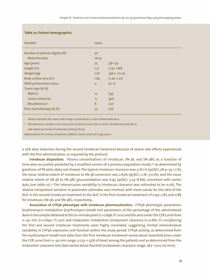
Chapter 8 - Prediction of irinotecan pharmacokinetics by use of cytochrome P450 3A4 phenotyping probes Chapter 8 - Prediction of irinotecan pharmacokinetics by use of cytochrome P450 3A4 phenotyping probes
a
N) 30
) 55
71.8 2) 1.84
0 N
10 (33) 12 (40)
b 8 N, %) 23 (77)
a ,b N N N=1),
N=4).
,
Table 20. Patient demographics
Variable Value
Number of patients eligible (Males/Females 16/14
Age (years (38–73) Length (m) 1.72 (1.55–1.86) Weight (kg) (48.0–101.0) Body surface area (m (1.46–2.27) WHO performance status (0–1) Tumor type ( , %)
(N)SCLC Gastro-intestinal Miscellaneous (27)
Prior chemotherapy (
Values represent the mean with range in parenthesis unless stated otherwise.
Miscellaneous include cervix cancer ( =2), breast cancer ( =1), cancer of head and neck (
and adenocarcinoma of unknown primary (
Abbreviations: N number of patients; (N)SCLC, (non) small cell lung cancer.
a 25% dose reduction during the second irinotecan treatment because of severe side effects experienced with the first administration, as required by the protocol.
Irinotecan disposition. Plasma concentrations of irinotecan, SN-38, and SN-38G as a function of time were accurately predicted by a modified version of a previous population model,44 as determined by goodness-of-fit plots (data not shown). The typical irinotecan clearance was 31.8 L/h (95%CI, 28.4–35.1 L/h), the mean relative extent of irinotecan to SN-38 conversion was 2.63% (95%CI, 2.18–3.07%), and the mean relative extent of SN-38 to SN-38G glucuronidation was 6.95 (95%CI, 5.23–8.66), consistent with earlier data (see table 21).44 The interoccasion variability in irinotecan clearance was estimated to be 11.0%. The relative intrapatient variation in parameter estimates was minimal, with mean values for the ratio of the AUC in the second irinotecan treatment to the AUC in the first irinotecan treatment of 0.90, 0.87, and 0.86 for irinotecan, SN-38, and SN-38G, respectively.
Association of CYP3A phenotype with irinotecan pharmacokinetics. CYP3A phenotypic parameters [erythromycin metabolism (erythromycin breath test parameters of the percentage of the administered dose in the sample obtained at the 20-minute point (r=0.846; P<.001) and the area under the CER curve from 0–40 min (r=0.840; P<.001) and midazolam metabolism (midazolam clearance (r=0.661; P=.010)]during the first and second irinotecan treatments were highly correlated, suggesting limited intraindividual variability in CYP3A expression and function within the study period. CYP3A activity, as determined from the erythromycin breath test data from the first irinotecan treatment varied about sevenfold (area under the CER curve from 0–40 min range, 0.223–1.53% of dose) among the patients and as determined from the midazolam clearance test data varied about fourfold (midazolam clearance range, 262–1,012 mL/min).
85
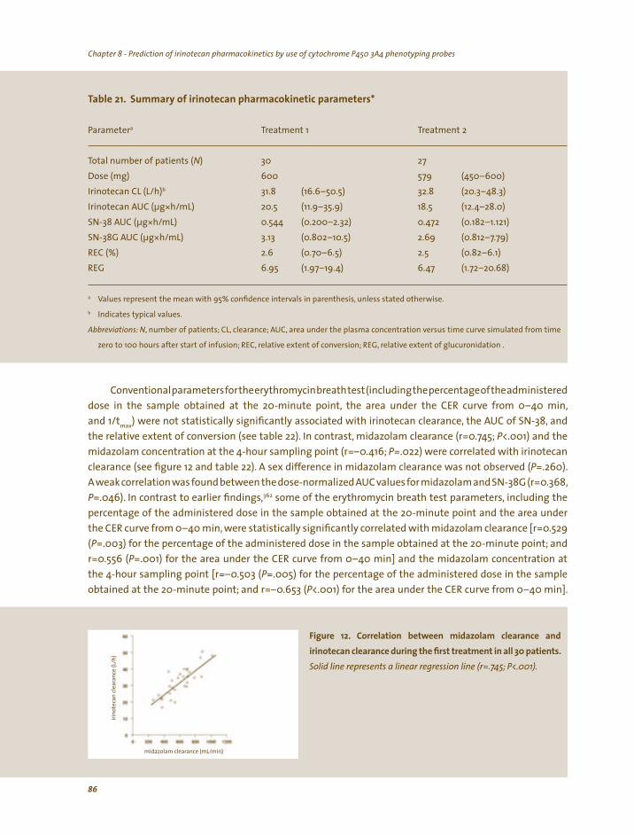
Chapter 8 - Prediction of irinotecan pharmacokinetics by use of cytochrome P450 3A4 phenotyping probes Chapter 8 - Prediction of irinotecan pharmacokinetics by use of cytochrome P450 3A4 phenotyping probes
a
N) 30 Dose (mg) 600
b 31.8 32.8 * 18.5
** 3.13 2.69
2.6 2.5 6.95
a ,b
,
Table 21. Summary of irinotecan pharmacokinetic parameters*
Parameter Treatment 1 Treatment 2
Total number of patients ( 27
579 (450–600) Irinotecan CL (L/h) (16.6–50.5) (20.3–48.3) Irinotecan AUC (µg h/mL) 20.5 (11.9–35.9) (12.4–28.0) SN-38 AUC (µg h/mL) 0.544 (0.200–2.32) 0.472 (0.182–1.121) SN-38G AUC (µg h/mL) (0.802–10.5) (0.812–7.79) REC (%) (0.70–6.5) (0.82–6.1) REG (1.97–19.4) 6.47 (1.72–20.68)
Values represent the mean with 95% confidence intervals in parenthesis unless stated otherwise.
Indicates typical values.
Abbreviations: N number of patients; CL, clearance; AUC, area under the plasma concentration versus time curve simulated from time
zero to 100 hours after start of infusion; REC, relative extent of conversion; REG, relative extent of glucuronidation .
Conventionalparametersfortheerythromycinbreathtest(includingthepercentageoftheadministered
dose in the sample obtained at the 20-minute point, the area under the CER curve from 0–40 min, and 1/t ) were not statistically significantly associated with irinotecan clearance, the AUC of SN-38, and max
the relative extent of conversion (see table 22). In contrast, midazolam clearance (r=0.745; P<.001) and the
midazolam concentration at the 4-hour sampling point (r=–0.416; P=.022) were correlated with irinotecan
clearance (see figure 12 and table 22). A sex difference in midazolam clearance was not observed (P=.260). A weak correlation was found between the dose-normalized AUC values for midazolam and SN-38G(r=0.368, P=.046). In contrast to earlier findings,362 some of the erythromycin breath test parameters, including the
percentage of the administered dose in the sample obtained at the 20-minute point and the area under the CER curve from 0–40 min, were statistically significantly correlated with midazolam clearance [r=0.529
(P=.003) for the percentage of the administered dose in the sample obtained at the 20-minute point; and
r=0.556 (P=.001) for the area under the CER curve from 0–40 min] and the midazolam concentration at
the 4-hour sampling point [r=–0.503 (P=.005) for the percentage of the administered dose in the sample
obtained at the 20-minute point; and r=–0.653 (P<.001) for the area under the CER curve from 0–40 min].
Fi l i mi l cliri l i i ll i
lid li li i li ( ; ).
/min)
gure 12. Corre at on between dazo am earance and
notecan c earance dur ng the first treatment n a 30 pat ents. So ne represents a near regress on ne r=.745 P<.001
midazolam clearance (mL
irino
teca
n cl
eara
nce
(L/h
)
86

Chapter 8 - Prediction of irinotecan pharmacokinetics by use of cytochrome P450 3A4 phenotyping probes Chapter 8 - Prediction of irinotecan pharmacokinetics by use of cytochrome P450 3A4 phenotyping probes
ra P
20 .136
20 -.007
20 .266 .155
.315 .090
.836
.243 .196
max .115
max -.132
max .012
<.001 -.103 .225 .231
t4 .022
t4 -.094 .622
t4 -.322 .083
a
2014
2 ,
142
max,
4
Table 22. Correlations between CYP3A phenotype and irinotecan parameters
Parameter 1 Parameter 2
Erythromycin breath test CER Irinotecan CL .279 CER SN-38 AUC .971 CER REC Area under CER0–40 curve Irinotecan CL Areas under CER0–40 curve SN-38 AUC -.040 Area under CER0–40 curve REC 1/t Irinotecan CL .546
1/t SN-38 AUC .488
1/t REC .950
Midazolam clearance test CL midazolam Irinotecan CL .745 CL midazolam SN-38 AUC .590
CL midazolam REC Irinotecan CL -.416 SN-38 AUC REC
A Pearson’s correlation coefficient was used to evaluate associations between irinotecan pharmacokinetics and the erythromycin
breath test or midazolam clearance test, respectively. All statistical tests were two-sided.
Abbreviations: N, number of patients; CYP3A, cytochrome P450 3A; CER , flux of exhaled CO at t=20 min; CL, clearance; CER0–40 flux of
exhaled CO from 0 to 40 minutes; AUC, area under the plasma concentration versus time curve simulated from time zero to 100
hours after start of infusion; REC, relative extent of conversion; REG, relative extent of glucuronidation; 1/t the reciprocal of time
to peak concentration; t , midazolam concentration in the 4-hour sample.
Influence of enzyme and transporter genotypes on irinotecan pharmacokinetics. Among the five genetic variants CYP3A4*2, CYP3A4*3, CYP3A4*17, CYP3A4*18, and CYP3A5*6, only the wild-type sequence was found, indicating that for these variants none of the patients in the studied cohort carried a mutant
allele. The absence of these variants is consistent with previously published data obtained in the general European white population.200,201 For the other six variants (ABCB1*8, ABCB1*7, ABCB1*6, CYP3A4*1B, CYP3A5*3, and UGT1A1*28) studied, the frequency of the variant allele (q) was highly variable, with only four patients carrying at least one variant allele for CYP3A4*1B (allele frequency,0.09) and as many as 29 patients carrying at least one variant allele for CYP3A5*3 (allele frequency, 0.87; see table 23).
The presence of two copies of the UGT1A1*28 allele was associated with statistically significantly altered pharmacokinetics of irinotecan. The UGT1A1*28 allele carries a seven-copy TA-repeat (TA7) in the promoter region instead of a six-copy repeat (TA6), as in the wild-type UGT1A1 allele. The AUC of SN-38 was 435 ng*h/mL (95%CI, 339–531 ng*h/mL) in wild-type patients (TA6/TA6; N=12), 631 ng*h/mL (95%CI, 499–762 ng*h/mL) in heterozygous patients (TA6/TA7; N=15), and 1343 ng*h/mL (95%CI, 0–4,181 ng*h/
87

Chapter 8 - Prediction of irinotecan pharmacokinetics by use of cytochrome P450 3A4 phenotyping probes Chapter 8 - Prediction of irinotecan pharmacokinetics by use of cytochrome P450 3A4 phenotyping probes
mL) in homozygous variant patients (TA7/TA7; N=3) (P=.006). Likewise, a UGT1A1*28 genotype-dependent
relative extent of conversion was observed, with a value of 0.018 (95%CI, 0.013–0.022) in wild-type patients, 0.030 (95%CI, 0.025–0.034) in heterozygous patients, and 0.042 (95%CI, 0–0.092) in homozygous variant
patients (P<.001). Furthermore, the relative extent of glucuronidation was 9.27 (95%CI, 6.00–12.53) in wild-type patients, 5.79 (95%CI, 3.74–7.83) in heterozygous patients, and 3.48 (95%CI, 0–9.47) in homozygous variant patients (P=.010) (see figure 13). National Cancer Institute–Common Toxicity Criteria scores (http://
ctep.cancer.gov/forms/CTCv20_4-30-992.pdf) of neutropenia (P=.020), the observed nadir value of the absolute neutrophil count (P=.026), and the percentage decrease in absolute neutrophil count at nadir (P=.024) were also associated with the UGT1A1*28 genotype. Statistically significant associations between irinotecan pharmacokinetics and the other variant genotypes studied were not observed, although the ABCB1*8 genotype was associated with a non-statistically significant trend in a decrease in the AUC of SN-38G (P=.042; Kruskal-Wallis test).
6, 7 , ,
Figure 13. UGT1A1*28 genotype and the metabolism of irinotecan to 7-ethyl-10-hydroxycamptothecin (SN-38). A) Area under the plasma concentration versus time curve (AUC) of SN-38. B) AUC of SN-38 divided by the AUC of irinotecan (REC). C) AUC of SN-38 glucuronide (SN-38G) divided by the AUC of SN-38 (REG). The wild-type allele of UGT1A1 is designated as TA and the variant is TA . Solid horizontal lines mean values; dashed horizontal lines interquartile range (25 percentile values to 75 percentile values).
DISCUSSION
In this study, we tested the hypothesis that the pharmacokinetics of irinotecan can be predicted by
the putative CYP3A phenotyping probes erythromycin and midazolam. We based the rationale for this
investigation on the observations that irinotecan is a substrate for CYP3A4 and CYP3A532 (see figure 11) and
that its pharmacokinetic profile is influenced by potent inhibitors124 or inducers182 of these isozymes. We
hypothesized that by administering a probe drug substrate for the enzymes in question, enzymatic activity
could be evaluated by comparing (metabolic) clearance and metabolic ratios of parent compounds and/or
88
Chapter 8 - Prediction of irinotecan pharmacokinetics by use of cytochrome P450 3A4 phenotyping probes Chapter 8 - Prediction of irinotecan pharmacokinetics by use of cytochrome P450 3A4 phenotyping probes
of their metabolites. Like irinotecan, erythromycin is a substrate for both CYP3A4 and ABCB1, but it is not
metabolized by CYP3A5.362 In contrast, midazolam is metabolized by both CYP3A4 and CYP3A5, but is a very
poor substrate for ABCB1.363 Because irinotecan is also partially metabolized by CYP3A5,32 we expected the
pharmacokinetic correlations of irinotecan with erythromycin and midazolam to differ.Because the metabolic
pathways of several other commonly used probes,such as the CYP3A phenotyping probes dextromethorphan
and cortisol,348 differ substantially from that of irinotecan, we did not evaluate them in this study. We did not observe statistically significant correlations between common parameters (CER20, area
under the CER0–40, and 1/t ) for the erythromycin breath test and irinotecan pharmacokinetics, but we didmax
obtain a highly statistically significant correlation between midazolam clearance and irinotecan clearance. From this correlation analysis, we predict that, in the patient cohort on our study, 56% of total interindividual variability in irinotecan clearance can be explained by variation in CYP3A function. The discrepant findings
between the erythromycin breath test and the midazolam clearance test may be related to the relatively
slow and inefficient metabolism of midazolam, which more closely resembles the CYP3A-mediated
n.348 We metabolism of irinotecan than the fast and extensive CYP3A-mediated metabolism of erythromycialso cannot exclude the possibility that ABCB1 plays a minor role in clearance of irinotecan and a larger role in the clearance of erythromycin and that this difference confounds pharmacokinetic interrelationships. We also observed a trend for a relationship between the AUC for midazolam and exposure to SN-38G. This
finding was unexpected because SN-38G is formed from SN-38 through UGT1A-mediated conjugation that
is independent of CYP3A activity. A possible explanation may be found in the indirect formation of SN-38 and
SN-38G from 7-ethyl-10-[4-amino-1-piperidino]-carbonyloxycamptothecin (NPC), which results from a ring 364 opening oxidation of the terminal piperidine ring of irinotecan that is mediated by CYP3A4 (see figure 11).
Although irinotecan is considered a prodrug with little inherent antitumor activity, the ability to accurately predict its clearance may be clinically relevant. First, the etiology of irinotecan-mediated side effects is still not completely understood, and circulating concentrations of the parent drug may predict
hematological toxicity associated with irinotecan treatment.299 Furthermore, the ratio of concentration of total unbound irinotecan to the concentration of total unbound SN-38 is similar to their potency ratio in vitro, and so both irinotecan and SN-38 are likely to be effective in vivo.29 Second, SN-38 may be formed both peripherally in the intestines, liver, and blood, as well as in the tumor from the conversion of irinotecan by carboxylesterase 2 (hCE2), which may be more important than previously thought and may contribute to the variable response to irinotecan chemotherapy for solid tumors.25,365 Consequently, knowledge of phenotypic CYP3A activity, as a predictor of irinotecan clearance in individual patients, may help to reduce the pharmacokinetic and subsequent pharmacodynamic variability associated with irinotecan treatment.
We found that certain genetic variations in polymorphic proteins are associated with differences in the elimination of irinotecan and its metabolites,as predicted previously.35We also confirmed earlier preliminary observations that the UGT1A1*28 genotype is independently associated with the pharmacokinetic profile of irinotecan46,49,245 and that ABCB1*8 has a smaller association.255 Genotyping for CYP3A4 and CYP3A5 did not result in statistically significant correlations with irinotecan pharmacokinetics, perhaps because of the low allelic frequency of most CYP3A variant genotypes (e.g., CYP3A4*17, CYP3A4*18, and CYP3A5*1) in the white population200,201 or because of the absence of a clinically important effect on enzyme activity in vivo (e.g., CYP3A4*1B).366,367 Because CYP3A is a very complex enzyme system that is easily influenced by environmental (i.e., co-medication, herbal preparations, and/or food substances) and physiologic (i.e., aging, disease state, and altered liver and renal function) factors,29 the role of CYP3A genotyping in the chemotherapeutic treatment of cancer remains uncertain.
89
Chapter 8 - Prediction of irinotecan pharmacokinetics by use of cytochrome P450 3A4 phenotyping probes
In conclusion, CYP3A phenotype (as determined by midazolam clearance) and UGT1A1*28 genotype appear to be statistically significant predictors of irinotecan and SN-38 pharmacokinetics, respectively. A prospective study to validate the usefulness of these phenotyping and genotyping strategies to optimize chemotherapeutic treatment with irinotecan for individual patients is currently under way. In addition, limited sampling strategies are being developed by use of data obtained in a larger cohort of patients to further optimize the clinical applicability of the midazolam clearance test.
ACKNOWLEDGEMENTS
We thank Dr. Wim van den Berg, Inge van den Bos, Dirk Buijs, Dr. Sjaak Burgers, Jan Francke, Ilse van der Heiden, Hans van der Meulen, Tatjana Pronk, Martin van Vliet, and Marloes van der Werf (all from Rotterdam) and Dr. Nicola F. Smith (from Bethesda, MD) for their contribution to this work.
Supported in part by the Cornelis Vrolijk Development Fund (IJmuiden, the Netherlands).
90

Chapter 8 - Prediction of irinotecan pharmacokinetics by use of cytochrome P450 3A4 phenotyping probes
91
Chapter 9
Summary, conclusions, and future perspectives

Chapter 9 - Summary, conclusions, and future perspectives , ,Chapter 9 - Summary conclusions and future perspectives
Chapter 9 - Summary, conclusions, and future perspectives Chapter 9 - Summary, conclusions, and future perspectives
Ever since its introduction to the drug-market in the late eighties, early nineties of the last century, irinotecan is fighting its image. Particularly, the unpredictable occurrence and severity of delayed-type
diarrhea, its main dose-limiting adverse effect, remains a serious concern for medical oncologists. In this perspective, reliable, predictable a priori knowledge of the toxicity profile of irinotecan therapy in the individual patient is of utmost importance, since it would guide physician’s choice and enable adaptive drug administration. In this thesis, different pharmacological aspects of irinotecan therapy have been investigated. Using pharmacokinetic, pharmacogenetic, and phenotyping strategies, as well as pharmacological prophylactic interventions, we have investigated ways to improve irinotecan tolerability: indirectly by predicting, controlling and reducing the interindividual variability in irinotecan pharmacokinetic parameters and directly by modifying the risk and severity of delayed-type diarrhea.
The traditional method of individualizing irinotecan dosing is through body surface area (BSA), using a formula derived from weight and height alone. The usefulness of normalizing irinotecan doses to BSA in adults has been questioned, since irinotecan pharmacokinetic parameters appear to be unrelated to BSA which suggests that the use of BSA-based dosing of irinotecan results in the administration of a standard dose multiplied by a random number, i.e., the ratio of the patient’s BSA to an average BSA. In chapter 2, we prospectively evaluated the feasibility of administering a flat-fixed irinotecan dose (600 mg) to cancer patients, regardless of BSA, compared with BSA-based dosing. The interindividual variability in irinotecan clearance, relative extent of conversion of irinotecan to SN-38, and relative extent of SN-38 glucuronidation were not significantly different between the two groups. Variance differences in irinotecan-mediated hematological adverse effects were also similar between both groups suggesting that flat-fixed dosing of irinotecan is feasible, does not result in increased pharmacokinetic variability and/or its subsequently associated pharmacodynamic variability, and could be safely used to substitute current dosing strategies based on BSA. It can be anticipated that implementation of this dosing concept in routine clinical practice would have significant economic and safety implications, since only a unit dose has to be manufactured, and obviously less errors can be made in calculating and preparing the right dose, both a significant source of error in attempting to obtain precise dosing.
Since September 2003, under the responsibility of the Dutch government (the Office of Medicinal Cannabis), the production and distribution of a legal, standardized cannabis product that meets pharmaceutical quality standards has become a reality. Until then, patients were forced to frequent illegal coffeeshops for their supply of medicinal cannabis, or even to produce cannabis themselves at home. In chapter 3A, we discussed the position of (medicinal) cannabis in oncology practice, viewed from the Dutch experience. Cancer patients may use cannabis or single (synthetically derived) cannabinoids to cope with disease- and chemotherapy-induced nausea and emesis, disease-related anorexia, and disease-related pain. Additionally, euphoria, relaxation, a better quality of sleep, a better mood, and a better quality of life are reported. To date, it remains to be determined if medicinal cannabis has an additive value in oncology practice as compared with the currently available conventional drugs and/or to isolated synthetic cannabinoids. Well-designed clinical trials that undisputedly prove the advantages of medicinal cannabis are lacking, and it is far from clear for what indications medicinal cannabis may be a justified treatment
option. Furthermore, issues related to administration route, dosing, and safety need to be resolved. At this time, medicinal cannabis as regular drug is still in its infancy and has a long way to go; regular prescription of medicinal cannabis in routine oncology practice is currently a bridge too far.
93

Chapter 9 - Summary, conclusions, and future perspectives Chapter 9 - Summary, conclusions, and future perspectives
Undesirable interactions between concomitantly administered drugs and/or herbal products and cytotoxic chemotherapeutic drugs metabolized by cytochrome P450-isozymes, particularly CYP3A, are a major risk in oncology. The potential inducing or inhibitory effects of medicinal cannabis with regard to CYPs are poorly documented, and therefore we undertook a formal drug-interaction study to evaluate the effect of concomitant medicinal cannabis (1 g/L Cannabis sativa L. Flos, variety Bedrocan®; orally administered as tea, 200 mL per day) on the pharmacokinetics of irinotecan and docetaxel (Taxotere®), both dependent on CYP3A-mediated metabolism. Both agents are anticancer drugs with an extremely narrow therapeutic window, meaning that a pharmacokinetic interaction may easily result in unwanted adverse effects (particularly delayed-type diarrhea and neutropenia for irinotecan) or suboptimal therapeutic effectiveness. In chapter 3B we conclude that medicinal cannabis tea can be safely concurrently administered with both agents without dose adjustments since medicinal cannabis does not significantly influence the plasma pharmacokinetics and the hematological toxicity of irinotecan.
Unfortunately, pharmacokinetic information is always limited to a posteriori correlates. In clinical practice such relations are of limited value, and we should search for a priori predictors, responsible for the interpatient variation in irinotecan pharmacokinetic parameters and its exposure-related adverse effects. To understand why some patients experience severe adverse effects, while others do not, the metabolic pathways of irinotecan have been unraveled in detail (see figure 1). Although the differences in the expression-level of the proteins and transporters involved in irinotecan disposition are explained to a certain extent by physiologic and environmental factors, the presence of specific genetic determinants also does influence their expression and function.
In chapter 4,the role of pharmacogenetics in explaining the pharmacokinetics and pharmacodynamics of irinotecan therapy is reviewed. Genetic variation in phase I (CYPs) and phase II (UGTs) enzymes and phase III transporters (ABC transporters) are discussed in detail. Particularly, the UGT1A-gene is a promising candidate for pretreatment genetic screening and implementation in routine clinical practice of irinotecan treatment,since the homozygous presence of an additional,seventh,dinucleotide (TA) insertion (UGT1A1*28) in the (TA)6TAA-box of the UGT1A1 promoter has frequently been associated with higher systemic levels of SN-38 and an increased risk of leucopenia and severe irinotecan-induced delayed-type diarrhea, as shown throughout this thesis as well. For example, in chapter 7 a strong correlation between the presence of the UGT1A1*28 polymorphism and systemic exposure to SN-38 and incidence and severity of diarrhea was noted in Caucasian cancer patients. Whereas most studies have reported that only patients carrying two variant alleles were at risk of serious toxicity, we found that patients carrying at least one variant
allele had a doubled risk of grade 2–3 delayed-type diarrhea (70% versus 35%), yielding the presence of at least one UGT1A1*28 allele a strong predictive factor with potential clinical relevance. In chapter 8 an association between UGT1A1*28 genotype and systemic exposure to SN-38 and neutropenia was found. Since at this very moment the field of pharmacogenetics and pharmacogenomics is rapidly expanding and simultaneously more rapid and cost-effective screening methods are emerging, a wealth of future data is expected to enrich our knowledge of the genetic basis of irinotecan metabolism. Eventually, this may help to truly individualize irinotecan dosing, using an individual genetic profile of the most relevant
enzymes for every patient. The breast cancer resistance protein (ABCG2) is an efflux ABC transporter that plays a role in host
detoxification of various xenobiotic substrates, including SN-38. The ABCG2 421C>A polymorphism has been associated with reduced protein expression and altered function in vitro. In chapter 5 the ethnic distribution and potential functional consequence of this single nucleotide polymorphism was evaluated.
94
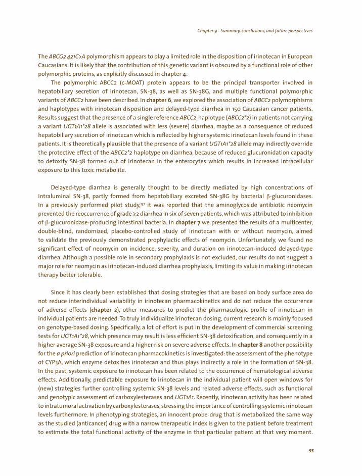
Chapter 9 - Summary, conclusions, and future perspectives Chapter 9 - Summary, conclusions, and future perspectives
The ABCG2 421C>A polymorphism appears to play a limited role in the disposition of irinotecan in European Caucasians. It is likely that the contribution of this genetic variant is obscured by a functional role of other polymorphic proteins, as explicitly discussed in chapter 4.
The polymorphic ABCC2 (c-MOAT) protein appears to be the principal transporter involved in hepatobiliary secretion of irinotecan, SN-38, as well as SN-38G, and multiple functional polymorphic variants of ABCC2 have been described. In chapter 6, we explored the association of ABCC2 polymorphisms and haplotypes with irinotecan disposition and delayed-type diarrhea in 150 Caucasian cancer patients. Results suggest that the presence of a single reference ABCC2-haplotype (ABCC2*2) in patients not carrying a variant UGT1A1*28 allele is associated with less (severe) diarrhea, maybe as a consequence of reduced hepatobiliary secretion of irinotecan which is reflected by higher systemic irinotecan levels found in these patients. It is theoretically plausible that the presence of a variant UGT1A1*28 allele may indirectly override the protective effect of the ABCC2*2 haplotype on diarrhea, because of reduced glucuronidation capacity to detoxify SN-38 formed out of irinotecan in the enterocytes which results in increased intracellular exposure to this toxic metabolite.
Delayed-type diarrhea is generally thought to be directly mediated by high concentrations of intraluminal SN-38, partly formed from hepatobiliary excreted SN-38G by bacterial β-glucuronidases. In a previously performed pilot study,137 it was reported that the aminoglycoside antibiotic neomycin prevented the reoccurrence of grade ≥2 diarrhea in six of seven patients, which was attributed to inhibition of β-glucuronidase-producing intestinal bacteria. In chapter 7 we presented the results of a multicenter, double-blind, randomized, placebo-controlled study of irinotecan with or without neomycin, aimed to validate the previously demonstrated prophylactic effects of neomycin. Unfortunately, we found no significant effect of neomycin on incidence, severity, and duration on irinotecan-induced delayed-type diarrhea. Although a possible role in secondary prophylaxis is not excluded, our results do not suggest a major role for neomycin as irinotecan-induced diarrhea prophylaxis, limiting its value in making irinotecan therapy better tolerable.
Since it has clearly been established that dosing strategies that are based on body surface area do not reduce interindividual variability in irinotecan pharmacokinetics and do not reduce the occurrence of adverse effects (chapter 2), other measures to predict the pharmacologic profile of irinotecan in individual patients are needed. To truly individualize irinotecan dosing, current research is mainly focused on genotype-based dosing. Specifically, a lot of effort is put in the development of commercial screening tests for UGT1A1*28, which presence may result is less efficient SN-38 detoxification, and consequently in a higher average SN-38 exposure and a higher risk on severe adverse effects. In chapter 8 another possibility for the a priori prediction of irinotecan pharmacokinetics is investigated: the assessment of the phenotype of CYP3A, which enzyme detoxifies irinotecan and thus plays indirectly a role in the formation of SN-38. In the past, systemic exposure to irinotecan has been related to the occurrence of hematological adverse effects. Additionally, predictable exposure to irinotecan in the individual patient will open windows for (new) strategies further controlling systemic SN-38 levels and related adverse effects, such as functional and genotypic assessment of carboxylesterases and UGT1A1. Recently, irinotecan activity has been related to intratumoral activation by carboxylesterases,stressing the importance of controlling systemic irinotecan levels furthermore. In phenotyping strategies, an innocent probe-drug that is metabolized the same way as the studied (anticancer) drug with a narrow therapeutic index is given to the patient before treatment
to estimate the total functional activity of the enzyme in that particular patient at that very moment.
95
Chapter 9 - Summary, conclusions, and future perspectives Chapter 9 - Summary, conclusions, and future perspectives
Fi i ibl . i
, i i i( i ) wi a
i ii i i
i ic
, i ivi li
l l
gure 14. Phenotyp ng: the pat ent
as ack box Us ng a safe probe
drug the pharmacok net c behav or of an ant cancer agent th
narrow therapeut c w ndow can be
pred cted before the drug s g ven. Based on the pharmacok netparameters of the probe drug an
nd dua zed dose of the treatment drug can be ca cu ated.
The patient is thought to be a kind of black box, meaning that the total functional activity is the sum of the combination of genotype, influences of disease, patient characteristics, environmental influences, and co-medication (see figure 14). In the exploratory study described in chapter 8, we found that in Caucasian cancer patients the CYP3A phenotype, as assessed by midazolam clearance, was statistically significantly associated with irinotecan pharmacokinetics, indicating that this strategy taking the patient as whole system may be used to individualize irinotecan dosing in the future and to reduce the risk of adverse effects.
As shown in this thesis, many factors are involved in irinotecan metabolism and in the pathogenesis of delayed-type diarrhea, for medical oncologists one of the most concerning adverse effects. In the future, dose individualization of irinotecan therapy ultimately aimed to make irinotecan therapy better tolerable, should focus on strategies that combine diverse factors. Currently, an individualized dosing strategy based on a linear regression model is prospectively validated in a randomized, multicenter study. Half of included patients undergo the midazolam clearance test after which they receive an irinotecan dose calculated multiplying a target exposure and the expected irinotecan clearance based on midazolam clearance, γ-GT, and length. The remaining patients are conventionally dosed, i.e., based on body surface area (BSA). Interpatient variation in irinotecan exposure will be compared between both groups, and when interindividual variation proves to decline, further studies are warranted to find the maximum tolerated target exposure and to further lower interpatient variation in irinotecan pharmacokinetics and –given a target exposure– its exposure related adverse effects.
Dosing regimens combining phenotyping for CYP3A, genotyping for UGT1A1 and others (i.e., the assessment of the functional activity of carboxylesterases) may lead to a better prediction of irinotecan pharmacokinetics and in this way to a higher therapeutic index. For the time being, advantages of such strategies may be limited to patients treated in academic settings. To truly individualize irinotecan treatment, efforts to develop and implement patient-friendly,simple,and feasible limited sampling models and phenotyping and genotyping strategies on a routine basis in university and general hospitals should
96

Chapter 9 - Summary, conclusions, and future perspectives Chapter 9 - Summary, conclusions, and future perspectives
be strongly encouraged. Additionally, (causal) correlations between genotype (particularly UGT1A1*28) and the pharmacokinetics, adverse effects, and/or therapeutic outcome of irinotecan therapy should be confirmed in large cohorts of patients. As a result of which evidence-based dosing recommendations should be formulated. Ultimately, lowering the severity and incidence of delayed-type diarrhea will make irinotecan therapy better tolerable for the individual patient and may give this classical topoisomerase I inhibitor the image it deserves in the rapidly changing field of anticancer agents.
97

98

99
Chapter 10
References

Chapter 10 - References
100
Chapter 10 - References
Chapter 10 - References Chapter 10 - References
REFERENCES
1. Wall ME,Wani MC, Cook CE, et al. Plant antitumor agents. I. The isolation and structure of camptothecine, a novel alkaloidal leukemia and
tumor inhibitor from Camptotheca Acuminata. J Am Chem Soc 88:3888-3890, 1966
2. Muggia FM, Creaven PJ, Hansen HH, et al. Phase I clinical trial of weekly and daily treatment with camptothecin (NSC-100880): correlation
with preclinical studies. Cancer Chemother Rep 56:515-521, 1972
3. Moertel CG,Schutt AJ,Reitemeier RJ,et al. Phase II study of camptothecin (NSC-100880) in the treatment of gastrointestinal cancer. Cancer Chemother Rep 56:95-101, 1972
4. Schaeppi U, Fleischman RW, Cooney DA. Toxicity of camptothecin (NSC-100880). Cancer Chemother Rep 3 5:25-36, 1974
5. Hsiang YH, Hertzberg R, Hecht S, et al. Camptothecin induces protein-linked DNA breaks via mammalian DNA topoisomerase I. J Biol Chem
260:14873-14878, 1985
6. Hsiang YH,Liu LF. Identification of mammalian DNA topoisomerase I as an intracellular target of the anticancer drug camptothecin. Cancer Res 48:1722-1726, 1988
7. Hsiang YH, Liu LF, Wall ME, et al. DNA topoisomerase I-mediated DNA cleavage and cytotoxicity of camptothecin analogues. Cancer Res
49:4385-4389, 1989
8. Saltz LB. Irinotecan in the first-line treatment of colorectal cancer. Oncology (Huntingt) 12:54-58, 1998
9. Vanhoefer U, Harstrick A, Achterrath W, et al. Irinotecan in the treatment of colorectal cancer: clinical overview. J Clin Oncol 19:1501-1518, 2001
10. Douillard JY,Sobrero A,CarnaghiC,et al.Metastatic colorectalcancer:integratingirinotecaninto combination and sequentialchemotherapy. Ann Oncol 14 Suppl 2:ii7-12, 2003
11. Chau I, Cunningham D. Adjuvant therapy in colon cancer--what, when and how? Ann Oncol,[Epub ahead of print,]2006
12. Andre T, Louvet C, Maindrault-Goebel F, et al. CPT-11 (irinotecan) addition to bimonthly, high-dose leucovorin and bolus and continuous-infusion 5-fluorouracil (FOLFIRI) for pretreated metastatic colorectal cancer. GERCOR. Eur J Cancer 35:1343-1347, 1999
13. Saltz LB, Cox JV, Blanke C, et al. Irinotecan plus fluorouracil and leucovorin for metastatic colorectal cancer. Irinotecan Study Group. N Engl J Med 343:905-914, 2000
14. Cunningham D, Humblet Y, Siena S, et al. Cetuximab monotherapy and cetuximab plus irinotecan in irinotecan-refractory metastatic
colorectal cancer. N Engl J Med 351:337-345, 2004
15. Wagener DJ, Verdonk HE, Dirix LY, et al. Phase II trial of CPT-11 in patients with advanced pancreatic cancer, an EORTC early clinical trials
group study. Ann Oncol 6:129-132, 1995
16. Verschraegen CF. Irinotecan for the treatment of cervical cancer. Oncology (Huntingt) 16:32-34, 2002
17. Sugiyama T,Yakushiji M, Ochiai K, et al. Japanese ovarian trials: focus on irinotecan. Oncology (Huntingt) 17:29-33, 2003
18. Ribrag V, Koscielny S, Vantelon JM, et al. Phase II trial of irinotecan (CPT-11) in relapsed or refractory non-Hodgkin’s lymphomas. Leuk
Lymphoma 44:1529-1533, 2003
19. Langer CJ. The global role of irinotecan in the treatment of lung cancer: 2003 update. Oncology (Huntingt) 17:30-40, 2003
20. Perez EA, Hillman DW, Mailliard JA, et al. Randomized phase II study of two irinotecan schedules for patients with metastatic breast cancer refractory to an anthracycline, a taxane, or both. J Clin Oncol 22:2849-2855, 2004
21. Bouche O, Raoul JL, Bonnetain F, et al. Randomized multicenter phase II trial of a biweekly regimen of fluorouracil and leucovorin (LV5FU2), LV5FU2 plus cisplatin, or LV5FU2 plus irinotecan in patients with previously untreated metastatic gastric cancer: a Federation Francophone
de Cancerologie Digestive Group Study--FFCD 9803. J Clin Oncol 22:4319-4328, 2004
22. Langer CJ. Irinotecan in advanced lung cancer: focus on North American trials. Oncology (Huntingt) 18:17-28, 2004
23. Humerickhouse R, Lohrbach K, Li L, et al. Characterization of CPT-11 hydrolysis by human liver carboxylesterase isoforms hCE-1 and hCE-2. Cancer Res 60:1189-1192, 2000
24. Khanna R, Morton CL, Danks MK, et al. Proficient metabolism of irinotecan by a human intestinal carboxylesterase. Cancer Res 60:4725-4728, 2000
25. Xu G, Zhang W, Ma MK, et al. Human carboxylesterase 2 is commonly expressed in tumor tissue and is correlated with activation of irinotecan. Clin Cancer Res 8:2605-2611, 2002
26. Sanghani SP, Quinney SK,Fredenburg TB, et al. Carboxylesterases expressed in human colon tumor tissue and their role in CPT-11 hydrolysis. Clin Cancer Res 9:4983-4991, 2003
27. Iyer L, King CD, Whitington PF, et al. Genetic predisposition to the metabolism of irinotecan (CPT-11). Role of uridine diphosphate
glucuronosyltransferase isoform 1A1 in the glucuronidation of its active metabolite (SN-38) in human liver microsomes. J Clin Invest
101:847-854, 1998
28. Ando Y, Saka H, Asai G, et al. UGT1A1 genotypes and glucuronidation of SN-38, the active metabolite of irinotecan. Ann Oncol 9:845-847, 1998
29. Mathijssen RH, van Alphen RJ,Verweij J, et al. Clinical pharmacokinetics and metabolism of irinotecan (CPT-11). Clin Cancer Res 7:2182-2194, 2001
30. Takasuna K, Hagiwara T, Hirohashi M, et al. Involvement of beta-glucuronidase in intestinal microflora in the intestinal toxicity of the
antitumor camptothecin derivative irinotecan hydrochloride (CPT-11) in rats. Cancer Res 56:3752-3757, 1996
101
Chapter 10 - References Chapter 10 - References
31. Takasuna K, Hagiwara T, Hirohashi M, et al. Inhibition of intestinal microflora beta-glucuronidase modifies the distribution of the active
metabolite of the antitumor agent, irinotecan hydrochloride (CPT-11) in rats. Cancer Chemother Pharmacol 42:280-286, 1998
32. Santos A, Zanetta S, Cresteil T, et al. Metabolism of irinotecan (CPT-11) by CYP3A4 and CYP3A5 in humans. Clin Cancer Res 6:2012-2020, 2000
33. Sanghani SP, Quinney SK, Fredenburg TB, et al. Hydrolysis of irinotecan and its oxidative metabolites, 7-ethyl-10-[4-N-(5-aminopentanoic
acid)-1-piperidino] carbonyloxycamptothecin and 7-ethyl-10-[4-(1-piperidino)-1-amino]-carbonyloxycamptothecin, by human
carboxylesterases CES1A1, CES2, and a newly expressed carboxylesterase isoenzyme, CES3. Drug Metab Dispos 32:505-511, 2004
34. Sparreboom A, Danesi R, Ando Y, et al. Pharmacogenomics of ABC transporters and its role in cancer chemotherapy. Drug Resist Updat
6:71-84, 2003
35. Mathijssen RH, Marsh S, Karlsson MO, et al. Irinotecan pathway genotype analysis to predict pharmacokinetics. Clin Cancer Res 9:3246-3253, 2003
36. Rothenberg ML,Eckardt JR,Kuhn JG,et al. Phase II trial of irinotecan in patients with progressive or rapidly recurrent colorectal cancer. J Clin
Oncol 14:1128-1135, 1996
37. Rougier P, Bugat R, Douillard JY, et al. Phase II study of irinotecan in the treatment of advanced colorectal cancer in chemotherapy-naive
patients and patients pretreated with fluorouracil-based chemotherapy. J Clin Oncol 15:251-260, 1997
38. Rougier P,Van Cutsem E, Bajetta E, et al. Randomised trial of irinotecan versus fluorouracil by continuous infusion after fluorouracil failure
in patients with metastatic colorectal cancer. Lancet 352:1407-1412, 1998
39. Hecht JR. Gastrointestinal toxicity or irinotecan. Oncology (Huntingt) 12:72-78, 1998
40. Cunningham D, Pyrhonen S, James RD, et al. Randomised trial of irinotecan plus supportive care versus supportive care alone after fluorouracil failure for patients with metastatic colorectal cancer. Lancet 352:1413-1418, 1998
41. Michael M, Brittain M, Nagai J, et al. Phase II study of activated charcoal to prevent irinotecan-induced diarrhea. J Clin Oncol 22:4410-4417, 2004
42. Benson 3rd AB, Ajani JA, Catalano RB, et al. Recommended guidelines for the treatment of cancer treatment-induced diarrhea. J Clin Oncol 22:2918-2926, 2004
43. Dranitsaris G, Maroun J, Shah A. Severe chemotherapy-induced diarrhea in patients with colorectal cancer: a cost of illness analysis. Support Care Cancer 13:318-324, 2005
44. Xie R, Mathijssen RH, Sparreboom A, et al. Clinical pharmacokinetics of irinotecan and its metabolites in relation with diarrhea. Clin
Pharmacol Ther 72:265-275, 2002
45. Ando Y, Saka H, Ando M, et al. Polymorphisms of UDP-glucuronosyltransferase gene and irinotecan toxicity: a pharmacogenetic analysis. Cancer Res 60:6921-6926, 2000
46. Iyer L, Das S, Janisch L, et al. UGT1A1*28 polymorphism as a determinant of irinotecan disposition and toxicity. Pharmacogenomics J 2:43-47, 2002
47. Marcuello E, Altes A, Menoyo A, et al. UGT1A1 gene variations and irinotecan treatment in patients with metastatic colorectal cancer. Br J Cancer 91:678-682, 2004
48. Rouits E, Boisdron-Celle M, Dumont A, et al. Relevance of different UGT1A1 polymorphisms in irinotecan-induced toxicity: a molecular and
clinical study of 75 patients. Clin Cancer Res 10:5151-5159, 2004
49. Innocenti F, Undevia SD, Iyer L, et al. Genetic variants in the UDP-glucuronosyltransferase 1A1 gene predict the risk of severe neutropenia of irinotecan. J Clin Oncol 22:1382-1388, 2004
50. Mathijssen RH,Verweij J, de Jonge MJ, et al. Impact of body-size measures on irinotecan clearance: alternative dosing recommendations. J Clin Oncol 20:81-87, 2002
51. Baker SD, Verweij J, Rowinsky EK, et al. Role of body surface area in dosing of investigational anticancer agents in adults, 1991-2001. J Natl Cancer Inst 94:1883-1888, 2002
52. Ratain MJ. Irinotecan dosing: does the CPT in CPT-11 stand for Can’t Predict Toxicity? J Clin Oncol 20:7-8, 2002
53. de Jonge MJ, Sparreboom A, Planting AS, et al. Phase I study of 3-week schedule of irinotecan combined with cisplatin in patients with
advanced solid tumors. J Clin Oncol 18:187-194, 2000
54. Sparreboom A, de Bruijn P, de Jonge MJ, et al. Liquid chromatographic determination of irinotecan and three major metabolites in human
plasma, urine and feces. J Chromatogr B Biomed Sci Appl 712:225-235, 1998
55. Xie R, Mathijssen RH, Sparreboom A, et al. Clinical pharmacokinetics of irinotecan and its metabolites: a population analysis. J Clin Oncol 20:3293-3301, 2002
56. Gurney HP, Ackland S, Gebski V, et al. Factors affecting epirubicin pharmacokinetics and toxicity: evidence against using body-surface area
for dose calculation. J Clin Oncol 16:2299-2304, 1998
57. Miller AA, Rosner GL, Egorin MJ, et al. Prospective evaluation of body surface area as a determinant of paclitaxel pharmacokinetics and
pharmacodynamics in women with solid tumors: Cancer and Leukemia Group B Study 9763. Clin Cancer Res 10:8325-8331, 2004. 58. Egorin MJ. Overview of recent topics in clinical pharmacology of anticancer agents. Cancer Chemother Pharmacol 42 Suppl:S22-S30, 1998
59. Gurney H. How to calculate the dose of chemotherapy. Br J Cancer 86:1297-1302, 2002
60. Taxis K, Barber N. Ethnographic study of incidence and severity of intravenous drug errors. BMJ 326:684-687, 2003
61. Anderson BJ, Ellis JF. Common errors of drug administration in infants: causes and avoidance. Paediatr Drugs 1:93-107, 1999
102
Chapter 10 - References Chapter 10 - References
62. Raymond E,BoigeV,Faivre S,et al.Dosage adjustment and pharmacokinetic profile of irinotecan in cancer patients with hepatic dysfunction. J Clin Oncol 20:4303-4312, 2002
63. Office of Medicinal Cannabis. [Medicinal cannabis: information for medical doctors and pharmacists]; http://www.cannabisbureau.nl/
pdf/MedicinalCannabis_Nov3.pdf. The Hague, the Netherlands: Dutch Ministry of Health,Welfare and Sports, 2003
64. Grotenhermen F. Pharmacokinetics and pharmacodynamics of cannabinoids. Clin Pharmacokinet 42:327-360, 2003
65. Marinol (Dronabinol) Capsules: Product prescribing information [package insert]. Marietta, GA, Solvay Pharmaceuticals Inc, 2003. 66. Hall W, Solowij N. Adverse effects of cannabis. Lancet 352:1611-1616, 1998
67. Walsh D, Nelson KA, Mahmoud FA. Established and potential therapeutic applications of cannabinoids in oncology. Support Care Cancer 11:137-143, 2003
68. Tramer MR, Carroll D, Campbell FA, et al. Cannabinoids for control of chemotherapy induced nausea and vomiting: quantitative systematic
review. BMJ 323:16-21, 2001 69. Lane M,Vogel CL,Ferguson J,et al. Dronabinol and prochlorperazine in combination for treatment of cancer chemotherapy-induced nausea
and vomiting. J Pain Symptom Manage 6:352-359, 1991 70. Darmani NA, Johnson JC. Central and peripheral mechanisms contribute to the antiemetic actions of delta-9-tetrahydrocannabinol
against 5-hydroxytryptophan-induced emesis. Eur J Pharmacol 488:201-212, 2004
71. Roila F,Donati D,Tamberi S,et al. Delayed emesis: incidence,pattern,prognostic factors and optimal treatment. Support Care Cancer 10:88-95, 2002
72. Bartlett N, Koczwara B. Control of nausea and vomiting after chemotherapy: what is the evidence? Intern Med J 32:401-407, 2002
73. Radbruch L, Nauck F. [A review of side effects and complications with cannabinoid treatment]. Schmerz 17:274-279, 2003
74. Ashton CH. Pharmacology and effects of cannabis: a brief review. Br J Psychiatry 178:101-106, 2001 75. Haney M, Ward AS, Comer SD, et al. Abstinence symptoms following oral THC administration to humans. Psychopharmacology (Berl)
141:385-394, 1999
76. Beal JE, Olson R, Laubenstein L, et al. Dronabinol as a treatment for anorexia associated with weight loss in patients with AIDS. J Pain
Symptom Manage 10:89-97, 1995
77. Timpone JG, Wright DJ, Li N, et al. The safety and pharmacokinetics of single-agent and combination therapy with megestrol acetate and
dronabinol for the treatment of HIV wasting syndrome. The DATRI 004 Study Group. Division of AIDS Treatment Research Initiative. AIDS
Res Hum Retroviruses 13:305-315, 1997
78. Jatoi A, Windschitl HE, Loprinzi CL, et al. Dronabinol versus megestrol acetate versus combination therapy for cancer-associated anorexia: a North Central Cancer Treatment Group study. J Clin Oncol 20:567-573, 2002
79. Davis MP, Dreicer R,Walsh D, et al. Appetite and cancer-associated anorexia: a review. J Clin Oncol 22:1510-1517, 2004
80. Killestein J, Uitdehaag BM, Polman CH. Cannabinoids in multiple sclerosis: do they have a therapeutic role? Drugs 64:1-11, 2004
81. Zajicek J, Fox P, Sanders H, et al. Cannabinoids for treatment of spasticity and other symptoms related to multiple sclerosis (CAMS study): multicentre randomised placebo-controlled trial. Lancet 362:1517-1526, 2003
82. Jochimsen PR,Lawton RL,VerSteeg K,et al. Effect of benzopyranoperidine,a delta-9-THC congener,on pain. Clin Pharmacol Ther 24:223-227, 1978
83. Noyes R Jr, Brunk SF, Baram DA, et al. Analgesic effect of delta-9-tetrahydrocannabinol. J Clin Pharmacol 15:139-143, 1975
84. Campbell FA,Tramer MR,Carroll D,et al. Are cannabinoids an effective and safe treatment option in the management of pain? A qualitative
systematic review. BMJ 323:13-16, 2001 85. Cichewicz DL. Synergistic interactions between cannabinoid and opioid analgesics. Life Sci 74:1317-1324, 2004
86. Notcutt W, Price M, Miller R, et al. Initial experiences with medicinal extracts of cannabis for chronic pain: results from 34 N of 1 studies. Anaesthesia 59:440-452, 2004
87. Hollister LE. Health aspects of cannabis: revisited. Int J Neuropsychopharmcol 1:71-80, 1998
88. Bachs L, Morland H. Acute cardiovascular fatalities following cannabis use. Forensic Sci Int 124:200-203, 2001 89. Gupta BD, Jani CB, Shah PH. Fatal Bhang poisoning. Med Sci Law 41:349-352, 2001 90. Jones RT. Cardiovascular system effects of marijuana. J Clin Pharmacol 42:58S-63S, 2002
91. Benowitz NL, Jones RT. Cardiovascular effects of prolonged delta-9-tetrahydrocannabinol ingestion. Clin Pharmacol Ther 18:287-297, 1975
92. Mittleman MA, Lewis RA, Maclure M, et al. Triggering myocardial infarction by marijuana. Circulation 103:2805-2809, 2001 93. Gable RS. Toward a comparative overview of dependence potential and acute toxicity of psychoactive substances used nonmedically. Am
J Drug Alcohol Abuse 19:263-281, 1993
94. Roth MD, Marques-Magallanes JA, Yuan M, et al. Induction and regulation of the carcinogen-metabolizing enzyme CYP1A1 by marijuana
smoke and delta (9)-tetrahydrocannabinol. Am J Respir Cell Mol Biol 24:339-344, 2001 95. Roth MD, Arora A, Barsky SH, et al. Airway inflammation in young marijuana and tobacco smokers. Am J Respir Crit Care Med 157:928-937,
1998
96. Barsky SH,Roth MD,Kleerup EC,et al. Histopathologic and molecular alterations in bronchial epithelium in habitual smokers of marijuana, cocaine, and/or tobacco. J Natl Cancer Inst 90:1198-1205, 1998
97. Sidney S, Quesenberry CP Jr, Friedman GD, et al. Marijuana use and cancer incidence (California, United States). Cancer Causes Control 8:722-728, 1997
103
Chapter 10 - References Chapter 10 - References
98. Zhang ZF, Morgenstern H, Spitz MR, et al. Marijuana use and increased risk of squamous cell carcinoma of the head and neck. Cancer Epidemiol Biomarkers Prev 8:1071-1078, 1999
99. Llewellyn CD, Linklater K, Bell J, et al. An analysis of risk factors for oral cancer in young people: a case-control study. Oral Oncol 40:304-313, 2004
100. Rosenblatt KA, Daling JR, Chen C, et al. Marijuana use and risk of oral squamous cell carcinoma. Cancer Res 64:4049-4054, 2004
101. Efird JT, Friedman GD, Sidney S, et al. The risk for malignant primary adult-onset glioma in a large, multiethnic, managed-care cohort: cigarette smoking and other lifestyle behaviors. J Neurooncol 68:57-69, 2004
102. Guzman M. Cannabinoids: potential anticancer agents. Nat Rev Cancer 3:745-755, 2003
103. Massi P,Vaccani A,Ceruti S,et al. Antitumor effects of cannabidiol,a nonpsychoactive cannabinoid,on human glioma cell lines. J Pharmacol Exp Ther 308:838-845, 2004
104. Hart S, Fischer OM, Ullrich A. Cannabinoids induce cancer cell proliferation via tumor necrosis factor alpha-converting enzyme (TACE/
ADAM17)-mediated transactivation of the epidermal growth factor receptor. Cancer Res 64:1943-1950, 2004
105. Benowitz NL, Jones RT. Effects of delta-9-tetrahydrocannabinol on drug distribution and metabolism. Antipyrine, pentobarbital, and
ethanol. Clin Pharmacol Ther 22:259-268, 1977
106. Jusko WJ, Schentag JJ, Clark JH, et al. Enhanced biotransformation of theophylline in marihuana and tobacco smokers. Clin Pharmacol Ther 24:405-410, 1978
107. McLeod AL,McKenna CJ,Northridge DB. Myocardial infarction following the combined recreational use of Viagra and cannabis. Clin Cardiol 25:133-134, 2002
108. Van Grinsven J,Westerik H. [Medicinal use of cannabis: a report]. Utrecht, the Netherlands: Duo Market Research, 2003. 109. Hall W, Degenhardt L. Medical marijuana initiatives: are they justified? How successful are they likely to be? CNS Drugs 17:689-697, 2003
110. Niesink R, Rigter S, Hoek J. [THC concentrations in weed, nederweed, and hashish in Dutch coffeeshops (2003-2004)]. Utrecht, the
Netherlands:Trimbos Institute – Netherlands Institute of Mental Health and Addiction, 2004. 111. Janse AF, Breekveldt-Postma NS, Erkens JA, et al. [Medicinal Cannabis in the Netherlands]. Utrecht, the Netherlands: PHARMO Institute for
Drug Outcomes Research, 2004. 112. Abel EL. Marijuana: the first twelve thousand years. New York: Plenum Press, 1980
113. Meiri E,Jhangiani H,Vredenburgh J,et al. Dronabinol treatment of delayed chemotherapy-induced nausea and vomiting,J Clin Oncol,2005
Annual Meeting Proceedings (Post-Meeting Edition) 24:8018a, 2005
114. de Jong FA, Engels FK, Mathijssen RH, et al. Medicinal cannabis in oncology practice: still a bridge too far? J Clin Oncol 23:2886-2891, 2005
115. Corey S. Recent developments in the therapeutic potential of cannabinoids. P R Health Sci J 24:19-26, 2005
116. Elsohly MA, Slade D. Chemical constituents of marijuana: the complex mixture of natural cannabinoids. Life Sci 78:539-548, 2005
117. Wilkinson JD,Whalley BJ,Baker D,et al. Medicinal cannabis: is delta9-tetrahydrocannabinol necessary for all its effects? J Pharm Pharmacol 55:1687-1694, 2003
118. Williamson EM. Synergy and other interactions in phytomedicines. Phytomedicine 8:401-409, 2001 119. Ware MA, Adams H, Guy GW.The medicinal use of cannabis in the UK: results of a nationwide survey. Int J Clin Pract 59:291-295, 2005
120. Scholten WK. Dutch measures to control medical grade marijuana: facilitating clinical trials. Drug Inf J 35:481-484, 2001 121. Office of Medicinal Cannabis; Medicinal Cannabis, Information for Health Care Professionals (version 15 march 2004); http://www.
cannabisbureau.nl/pdf/basic%20text%20cannabis%20EN%20vs%2015%20Mar%2004%20.pdf. The Hague, the Netherlands: Dutch
Ministry of Health,Welfare and Sports, 2003
122. Sparreboom A, Cox MC, Acharya MR, et al. Herbal remedies in the United States: potential adverse interactions with anticancer agents. J Clin Oncol 22:2489-2503, 2004
123. Tascilar M, de Jong FA, Verweij J, et al. Complementary and alternative medicine during cancer treatment: beyond innocence. Oncologist
11:732-741, 2006
124. Kehrer DF, Mathijssen RH, Verweij J, et al. Modulation of irinotecan metabolism by ketoconazole. J Clin Oncol 20:3122-3129, 2002
125. Mathijssen RH,Verweij J, de Bruijn P, et al. Effects of St. John’s wort on irinotecan metabolism. J Natl Cancer Inst 94:1247-1249, 2002
126. Engels FK,Ten Tije AJ, Baker SD, et al. Effect of cytochrome P450 3A4 inhibition on the pharmacokinetics of docetaxel. Clin Pharmacol Ther 75:448-454, 2004
127. Lepper ER, Baker SD, Permenter M, et al. Effect of common CYP3A4 and CYP3A5 variants on the pharmacokinetics of the cytochrome P450
3A phenotyping probe midazolam in cancer patients. Clin Cancer Res 11:7398-7404, 2005
128. Bornheim LM, Correia MA. Selective inactivation of mouse liver cytochrome P-450IIIA by cannabidiol. Mol Pharmacol 38:319-326, 1990
129. Yamamoto I,Watanabe K, Narimatsu S, et al. Recent advances in the metabolism of cannabinoids. Int J Biochem Cell Biol 27:741-746, 1995
130. Bornheim LM, Everhart ET, Li J, et al. Characterization of cannabidiol-mediated cytochrome P450 inactivation. Biochem Pharmacol 45:1323-1331, 1993
131. Jaeger W, Benet LZ, Bornheim LM. Inhibition of cyclosporine and tetrahydrocannabinol metabolism by cannabidiol in mouse and human
microsomes. Xenobiotica 26:275-284, 1996
132. Bornheim LM, Everhart ET, Li J, et al. Induction and genetic regulation of mouse hepatic cytochrome P450 by cannabidiol. Biochem
Pharmacol 48:161-171, 1994
104
Chapter 10 - References Chapter 10 - References
133. Kosel BW, Aweeka FT, Benowitz NL, et al. The effects of cannabinoids on the pharmacokinetics of indinavir and nelfinavir. Aids 16:543-550, 2002
134. Kalant H. Medicinal use of cannabis: history and current status. Pain Res Manag 6:80-91, 2001 135. Riggs CE Jr, Egorin MJ, Fuks JZ, et al. Initial observations on the effects of delta 9-tetrahydrocannabinol on the plasma pharmacokinetics of
cyclophosphamide and doxorubicin. J Clin Pharmacol 21:90S-98S, 1981 136. Shou M, Martinet M, Korzekwa KR, et al. Role of human cytochrome P450 3A4 and 3A5 in the metabolism of taxotere and its derivatives:
enzyme specificity, interindividual distribution and metabolic contribution in human liver. Pharmacogenetics 8:391-401, 1998
137. Kehrer DF,Sparreboom A,Verweij J,et al. Modulation of irinotecan-induced diarrhea by cotreatment with neomycin in cancer patients. Clin
Cancer Res 7:1136-1141, 2001 138. Rudek MA,Sparreboom A,Garrett-Mayer ES,et al.Factors affecting pharmacokinetic variability following doxorubicin and docetaxel-based
therapy. Eur J Cancer 40:1170-1178, 2004
139. de Jong FA, Mathijssen RH, Xie R, et al. Flat-fixed dosing of irinotecan: influence on pharmacokinetic and pharmacodynamic variability. Clin
Cancer Res 10:4068-4071, 2004
140. Bruno R, Hille D, Riva A, et al. Population pharmacokinetics/pharmacodynamics of docetaxel in phase II studies in patients with cancer. J Clin Oncol 16:187-196, 1998
141. Poujol S, Bressolle F, Duffour J, et al. Pharmacokinetics and pharmacodynamics of irinotecan and its metabolites from plasma and saliva
data in patients with metastatic digestive cancer receiving FOLFIRI regimen. Cancer Chemother Pharmacol:58:292-305, 2006
142. Engels FK, Mathot RA, Loos WJ, et al. Influence of High-Dose Ketoconazole on the Pharmacokinetics of Docetaxel. Cancer Biol Ther 5:833-839, 2006
143. de Bruijn P, Verweij J, Loos WJ, et al. Determination of irinotecan (CPT-11) and its active metabolite SN-38 in human plasma by reversed-phase high-performance liquid chromatography with fluorescence detection. J Chromatogr B Biomed Sci Appl 698:277-285, 1997
144. de Bruijn P, Willems EW, Loos WJ, et al. Indirect determination of the irinotecan metabolite 7-ethyl-10-O-glucuronyl-camptothecin in
human samples. Anal Biochem 328:84-86, 2004
145. Beal SL, Sheiner LB: NONMEM users guide. San Francisco, A: Division of Pharmacology, University of California, 1992
146. Yamaoka K, Nakagawa T, Uno T. Application of Akaike’s information criterion (AIC) in the evaluation of linear pharmacokinetic equations. J Pharmacokinet Biopharm 6:165-175, 1978
147. Verstraete AG. Detection times of drugs of abuse in blood, urine, and oral fluid. Ther Drug Monit 26:200-205, 2004
148. Schuirmann DJ. A comparison of the two one-sided tests procedure and the power approach for assessing the equivalence of average
bioavailability. J Pharmacokinet Biopharm 15:657-680, 1987
149. Rosing H, Lustig V, van Warmerdam LJ, et al. Pharmacokinetics and metabolism of docetaxel administered as a 1-h intravenous infusion. Cancer Chemother Pharmacol 45:213-218, 2000
150. Goh BC, Lee SC, Wang LZ, et al. Explaining interindividual variability of docetaxel pharmacokinetics and pharmacodynamics in Asians
through phenotyping and genotyping strategies. J Clin Oncol 20:3683-3690, 2002
151. de Jong FA, de Jonge MJ,Verweij J, et al. Role of pharmacogenetics in irinotecan therapy. Cancer Lett 234:90-106, 2006
152. Zhu HJ,Wang JS, Markowitz JS, et al. Characterization of P-glycoprotein inhibition by major cannabinoids from marijuana. J Pharmacol Exp
Ther 317:850-857, 2006
153. Erkens JA, Janse AF, Herings RM. Limited use of medicinal cannabis but for labeled indications after legalization. Pharmacoepidemiol Drug
Saf 14:821-822, 2005
154. Sharpe P, Smith G. Cannabis: time for scientific evaluation of this ancient remedy? Anesth Analg 90:237-240, 2000
155. Grothey A,Sargent D,Goldberg RM,et al. Survival of patients with advanced colorectal cancer improves with the availability of fluorouracil-leucovorin, irinotecan, and oxaliplatin in the course of treatment. J Clin Oncol 22:1209-1214, 2004
156. Meyerhardt JA, Mayer RJ. Systemic therapy for colorectal cancer. N Engl J Med 352:476-487, 2005
157. Rothenberg ML, Kuhn JG, Burris HA, 3rd, et al. Phase I and pharmacokinetic trial of weekly CPT-11. J Clin Oncol 11:2194-2204, 1993
158. Rowinsky EK, Grochow LB, Ettinger DS, et al. Phase I and pharmacological study of the novel topoisomerase I inhibitor 7-ethyl-10-[4-(1-piperidino)-1-piperidino]carbonyloxycamptothecin (CPT-11) administered as a ninety-minute infusion every 3 weeks. Cancer Res 54:427-436, 1994
159. Abigerges D,Armand JP,Chabot GG,et al. Irinotecan (CPT-11) high-dose escalation using intensive high-dose loperamide to control diarrhea. J Natl Cancer Inst 86:446-449, 1994
160. Canal P, Gay C, Dezeuze A, et al. Pharmacokinetics and pharmacodynamics of irinotecan during a phase II clinical trial in colorectal cancer. Pharmacology and Molecular Mechanisms Group of the European Organization for Research and Treatment of Cancer. J Clin Oncol 14:2688-2695, 1996
161. Gupta E, Mick R, Ramirez J, et al. Pharmacokinetic and pharmacodynamic evaluation of the topoisomerase inhibitor irinotecan in cancer patients. J Clin Oncol 15:1502-1510, 1997
162. Catimel G, Chabot GG, Guastalla JP, et al. Phase I and pharmacokinetic study of irinotecan (CPT-11) administered daily for three consecutive
days every three weeks in patients with advanced solid tumors. Ann Oncol 6:133-140, 1995
163. Marsh S, McLeod HL. Cancer pharmacogenetics. Br J Cancer 90:8-11, 2004
105
Chapter 10 - References Chapter 10 - References
164. van Ark-Otte J,Kedde MA,van der Vijgh WJ,et al. Determinants of CPT-11 and SN-38 activities in human lung cancer cells. Br J Cancer 77:2171-2176, 1998
165. Guichard S,Terret C, Hennebelle I, et al. CPT-11 converting carboxylesterase and topoisomerase activities in tumour and normal colon and
liver tissues. Br J Cancer 80:364-370, 1999
166. Morton CL,Wadkins RM,Danks MK,et al.The anticancer prodrug CPT-11 is a potent inhibitor of acetylcholinesterase but is rapidly catalyzed
to SN-38 by butyrylcholinesterase. Cancer Res 59:1458-1463, 1999
167. Wu MH, Yan B, Humerickhouse R, et al. Irinotecan activation by human carboxylesterases in colorectal adenocarcinoma cells. Clin Cancer Res 8:2696-2700, 2002
168. Ohtsuka K,Inoue S,Kameyama M,et al. Intracellular conversion of irinotecan to its active form,SN-38,by native carboxylesterase in human
non-small cell lung cancer. Lung Cancer 41:187-198, 2003
169. Innocenti F, Undevia SD, Ramirez J, et al. A phase I trial of pharmacologic modulation of irinotecan with cyclosporine and phenobarbital. Clin Pharmacol Ther 76:490-502, 2004
170. Sasaki Y, Hakusui H, Mizuno S, et al. A pharmacokinetic and pharmacodynamic analysis of CPT-11 and its active metabolite SN-38. Jpn J Cancer Res 86:101-110, 1995
171. Abigerges D, Chabot GG, Armand JP, et al. Phase I and pharmacologic studies of the camptothecin analog irinotecan administered very 3
weeks in cancer patients. J Clin Oncol 13:210-221, 1995
172. Mathijssen RH, Verweij J, Loos WJ, et al. Irinotecan pharmacokinetics-pharmacodynamics: the clinical relevance of prolonged exposure to
SN-38. Br J Cancer 87:144-150, 2002
173. Ahmed F, Vyas V, Cornfield A, et al. In vitro activation of irinotecan to SN-38 by human liver and intestine. Anticancer Res 19:2067-2071, 1999
174. Slatter JG,Schaaf LJ, Sams JP,et al. Pharmacokinetics, metabolism,and excretion of irinotecan (CPT-11) following I.V. infusion of [(14)C]CPT-11 in cancer patients. Drug Metab Dispos 28:423-433, 2000
175. Litman T, Druley TE, Stein WD, et al. From MDR to MXR: new understanding of multidrug resistance systems, their properties and clinical significance. Cell Mol Life Sci 58:931-959, 2001
176. Gottesman MM, Fojo T, Bates SE. Multidrug resistance in cancer: role of ATP-dependent transporters. Nat Rev Cancer 2:48-58, 2002
177. Candeil L, Gourdier I, Peyron D, et al. ABCG2 overexpression in colon cancer cells resistant to SN38 and in irinotecan-treated metastases. Int
J Cancer 109:848-854, 2004
178. Atsumi R, Suzuki W, Hakusui H. Identification of the metabolites of irinotecan, a new derivative of camptothecin, in rat bile and its biliary
excretion. Xenobiotica 21:1159-1169, 1991 179. Chabot GG,Abigerges D,Catimel G,et al.Population pharmacokinetics and pharmacodynamics of irinotecan (CPT-11) and active metabolite
SN-38 during phase I trials. Ann Oncol 6:141-151, 1995
180. Shingyoji M, Takiguchi Y, Watanabe-Uruma R, et al. In vitro conversion of irinotecan to SN-38 in human plasma. Cancer Sci 95:537-540, 2004
181. Rivory LP, Bowles MR, Robert J, et al. Conversion of irinotecan (CPT-11) to its active metabolite, 7-ethyl-10-hydroxycamptothecin (SN-38), by
human liver carboxylesterase. Biochem Pharmacol 52:1103-1111, 1996
182. Marsh S, Xiao M,Yu J, et al. Pharmacogenomic assessment of carboxylesterases 1 and 2. Genomics 84:661-668, 2004
183. Yu J,Shannon WD,Watson MA,et al. Gene expression profiling of the irinotecan pathway in colorectal cancer. Clin Cancer Res 11:2053-2062, 2005
184. Langmann T, Becker A, Aslanidis C, et al. Structural organization and characterization of the promoter region of a human carboxylesterase
gene. Biochim Biophys Acta 1350:65-74, 1997
185. Senter PD,Beam KS,Mixan B,et al. Identification and activities of human carboxylesterases for the activation of CPT-11,a clinically approved
anticancer drug. Bioconjug Chem 12:1074-1080, 2001 186. Wu MH, Chen P, Wu X, et al. Determination and analysis of single nucleotide polymorphisms and haplotype structure of the human
carboxylesterase 2 gene. Pharmacogenetics 14:595-605, 2004
187. Kim SR, Nakamura T, Saito Y, et al. Twelve novel single nucleotide polymorphisms in the CES2 gene encoding human carboxylesterase 2
(hCE-2). Drug Metab Pharmacokinet 18:327-332, 2004
188. Charasson V,Bellott R,Meynard D,et al. Pharmacogenetics of human carboxylesterase 2,an enzyme involved in the activation of irinotecan
into SN-38. Clin Pharmacol Ther 76:528-535, 2004
189. Eiselt R, Domanski TL, Zibat A, et al. Identification and functional characterization of eight CYP3A4 protein variants. Pharmacogenetics
11:447-458, 2001 190. Brooks BA, McBride OW, Dolphin CT, et al. The gene CYP3 encoding P450pcn1 (nifedipine oxidase) is tightly linked to the gene COL1A2
encoding collagen type 1 alpha on 7q21-q22.1. Am J Hum Genet 43:280-284, 1988
191. Gonzalez FJ, Schmid BJ, Umeno M, et al. Human P450PCN1: sequence, chromosome localization, and direct evidence through cDNA
expression that P450PCN1 is nifedipine oxidase. DNA 7:79-86, 1988
192. Spurr NK,Gough AC,Stevenson K,et al.The human cytochrome P450 CYP3 locus:assignment to chromosome 7q22-qter. Hum Genet 81:171-174, 1989
106
Chapter 10 - References Chapter 10 - References
193. Murray GI, McFadyen MC, Mitchell RT, et al. Cytochrome P450 CYP3A in human renal cell cancer. Br J Cancer 79:1836-1842, 1999
194. Finta C, Zaphiropoulos PG. The human cytochrome P450 3A locus. Gene evolution by capture of downstream exons. Gene 260:13-23, 2000
195. LinYS,Dowling AL,Quigley SD,et al.Co-regulation of CYP3A4 and CYP3A5 and contribution to hepatic and intestinal midazolam metabolism. Mol Pharmacol 62:162-172, 2002
196. Kuehl P, Zhang J, Lin Y, et al. Sequence diversity in CYP3A promoters and characterization of the genetic basis of polymorphic CYP3A5
expression. Nat Genet 27:383-391, 2001 197. Hustert E, Haberl M, Burk O, et al. The genetic determinants of the CYP3A5 polymorphism. Pharmacogenetics 11:773-779, 2001 198. Ozdemir V, Kalowa W,Tang BK, et al. Evaluation of the genetic component of variability in CYP3A4 activity: a repeated drug administration
method. Pharmacogenetics 10:373-388, 2000
199. Dai D, Tang J, Rose R, et al. Identification of variants of CYP3A4 and characterization of their abilities to metabolize testosterone and
chlorpyrifos. J Pharmacol Exp Ther 299:825-831, 2001 200. van Schaik RH, de Wildt SN, Brosens R, et al. The CYP3A4*3 allele: is it really rare? Clin Chem 47:1104-1106, 2001 201. van Schaik RH, van der Heiden IP, van den Anker JN, et al. CYP3A5 variant allele frequencies in Dutch Caucasians. Clin Chem 48:1668-1671,
2002
202. Garcia-Martin E, Martinez C, Pizarro RM, et al. CYP3A4 variant alleles in white individuals with low CYP3A4 enzyme activity. Clin Pharmacol Ther 71:196-204, 2002
203. Mathijssen RH, de Jong FA, van Schaik RH, et al. Prediction of irinotecan pharmacokinetics by use of cytochrome P450 3A4 phenotyping
probes. J Natl Cancer Inst 96:1585-1592, 2004
204. Zhou Q, Sparreboom A, Tan EH, et al. Pharmacogenetic profiling across the irinotecan pathway in Asian patients with cancer. Br J Clin
Pharmacol 59:415-424, 2005
205. Lamba JK, Lin YS, Schuetz EG, et al. Genetic contribution to variable human CYP3A-mediated metabolism. Adv Drug Deliv Rev 54:1271-1294, 2002
206. van Schaik RH. Implications of cytochrome P450 genetic polymorphisms on the toxicity of antitumor agents.Ther Drug Monit 26:236-240, 2004
207. Lehmann JM, McKee DD,Watson MA, et al.The human orphan nuclear receptor PXR is activated by compounds that regulate CYP3A4 gene
expression and cause drug interactions. J Clin Invest 102:1016-1023, 1998
208. Bertilsson G, Heidrich J, Svensson K, et al. Identification of a human nuclear receptor defines a new signaling pathway for CYP3A induction. Proc Natl Acad Sci USA 95:12208-12213, 1998
209. Wentworth JM, Agostini M, Love J, et al. St John’s wort, a herbal antidepressant, activates the steroid X receptor. J Endocrinol 166:R11-16, 2000
210. Raucy JL. Regulation of CYP3A4 expression in human hepatocytes by pharmaceuticals and natural products. Drug Metab Dispos 31:533-539, 2003
211. Kiang TK, Ensom MH, Chang TK. UDP-glucuronosyltransferases and clinical drug-drug interactions. Pharmacol Ther 106:97-132, 2005
212. Ouzzine M, Barre L, Netter P,et al.The human UDP-glucuronosyltransferases: structural aspects and drug glucuronidation. Drug Metab Rev
35:287-303, 2003
213. Guillemette C. Pharmacogenomics of human UDP-glucuronosyltransferase enzymes. Pharmacogenomics J 3:136-158, 2003
214. van Es HH,Bout A,Liu J,et al.Assignment of the human UDP-glucuronosyltransferase gene (UGT1A1) to chromosome region 2q37.Cytogenet
Cell Genet 63:114-116, 1993
215. Gong QH, Cho JW, Huang T, et al. Thirteen UDP glucuronosyltransferase genes are encoded at the human UGT1 gene complex locus. Pharmacogenetics 11:357-368, 2001
216. Ciotti M,Basu N,Brangi M, et al. Glucuronidation of 7-ethyl-10-hydroxycamptothecin (SN-38) by the human UDP-glucuronosyltransferases
encoded at the UGT1 locus. Biochem Biophys Res Commun 260:199-202, 1999
217. Hanioka N, Ozawa S, Jinno H, et al. Human liver UDP-glucuronosyltransferase isoforms involved in the glucuronidation of 7-ethyl-10-hydroxycamptothecin. Xenobiotica 31:687-699, 2001
218. Gagne JF,MontminyV,Belanger P,et al.Common human UGT1A polymorphisms and the altered metabolism of irinotecan active metabolite
7-ethyl-10-hydroxycamptothecin (SN-38). Mol Pharmacol 62:608-617, 2002
219. Oguri T, Takahashi T, Miyazaki M, et al. UGT1A10 is responsible for SN-38 glucuronidation and its expression in human lung cancers. Anticancer Res 24:2893-2896, 2004
220. Carlini LE, Meropol NJ, Bever J, et al. UGT1A7 and UGT1A9 polymorphisms predict response and toxicity in colorectal cancer patients treated
with capecitabine/irinotecan. Clin Cancer Res 11:1226-1236, 2005
221. Strassburg CP,Oldhafer K,Manns MP,et al.Differential expression of the UGT1A locus in human liver,biliary,and gastric tissue:identification
of UGT1A7 and UGT1A10 transcripts in extrahepatic tissue. Mol Pharmacol 52:212-220, 1997
222. Zheng Z, Park JY, Guillemette C, et al. Tobacco carcinogen-detoxifying enzyme UGT1A7 and its association with orolaryngeal cancer risk. J Natl Cancer Inst 93:1411-1418, 2001
223. Vogel A, Kneip S, Barut A, et al. Genetic link of hepatocellular carcinoma with polymorphisms of the UDP-glucuronosyltransferase UGT1A7
gene. Gastroenterology 121:1136-1144, 2001
107

Chapter 10 - References Chapter 10 - References
224. Jinno H, Saeki M, Saito Y, et al. Functional characterization of human UDP-glucuronosyltransferase 1A9 variant, D256N, found in Japanese
cancer patients. J Pharmacol Exp Ther 306:688-693, 2003
225. Barbier O, Girard H, Inoue Y, et al. Hepatic expression of the UGT1A9 gene is governed by hepatocyte nuclear factor 4alpha. Mol Pharmacol 67:241-249, 2005
226. Bosma PJ,ChowdhuryNR,GoldhoornBG,et al.Sequenceof exonsand the flanking regionsofhumanbilirubin-UDP-glucuronosyltransferase
gene complex and identification of a genetic mutation in a patient with Crigler-Najjar syndrome, type I. Hepatology 15:941-947, 1992
227. Aono S, Yamada Y, Keino H, et al. Identification of defect in the genes for bilirubin UDP-glucuronosyl-transferase in a patient with Crigler-Najjar syndrome type II. Biochem Biophys Res Commun 197:1239-1244, 1993
228. Bosma PJ, Chowdhury JR,Bakker C,et al.The genetic basis of the reduced expression of bilirubin UDP-glucuronosyltransferase 1 in Gilbert’s
syndrome. N Engl J Med 333:1171-1175, 1995
229. Monaghan G, Ryan M, Seddon R, et al. Genetic variation in bilirubin UPD-glucuronosyltransferase gene promoter and Gilbert’s syndrome. Lancet 347:578-581, 1996
230. Beutler E, Gelbart T, Demina A. Racial variability in the UDP-glucuronosyltransferase 1 (UGT1A1) promoter: a balanced polymorphism for regulation of bilirubin metabolism? Proc Natl Acad Sci USA 95:8170-8174, 1998
231. Desai AA, Innocenti F, Ratain MJ. Pharmacogenomics: road to anticancer therapeutics nirvana? Oncogene 22:6621-6628, 2003
232. Monaghan G, Foster B, Jurima-Romet M, et al. UGT1*1 genotyping in a Canadian Inuit population. Pharmacogenetics 7:153-156, 1997
233. Ando Y, Chida M, Nakayama K, et al. The UGT1A1*28 allele is relatively rare in a Japanese population. Pharmacogenetics 8:357-360, 1998
234. Burchell B, Hume R. Molecular genetic basis of Gilbert’s syndrome. J Gastroenterol Hepatol 14:960-966, 1999
235. Lampe JW, Bigler J, Horner NK, et al. UDP-glucuronosyltransferase (UGT1A1*28 and UGT1A6*2) polymorphisms in Caucasians and Asians: relationships to serum bilirubin concentrations. Pharmacogenetics 9:341-349, 1999
236. Balram C, Sabapathy K, Fei G, et al. Genetic polymorphisms of UDP-glucuronosyltransferase in Asians: UGT1A1*28 is a common allele in
Indians. Pharmacogenetics 12:81-83, 2002
237. Ando Y, Ueoka H, Sugiyama T, et al. Polymorphisms of UDP-glucuronosyltransferase and pharmacokinetics of irinotecan. Ther Drug Monit
24:111-116, 2002
238. Sato H, Adachi Y, Koiwai O. The genetic basis of Gilbert’s syndrome. Lancet 347:557-558, 1996
239. Akaba K,Kimura T,Sasaki A,et al. Neonatal hyperbilirubinemia and mutation of the bilirubin uridine diphosphate-glucuronosyltransferase
gene: a common missense mutation among Japanese, Koreans and Chinese. Biochem Mol Biol Int 46:21-26, 1998
240. Huang CS, Luo GA, Huang ML, et al. Variations of the bilirubin uridine-diphosphoglucuronosyl transferase 1A1 gene in healthy Taiwanese. Pharmacogenetics 10:539-544, 2000
241. Iyer L, Hall D, Das S, et al. Phenotype-genotype correlation of in vitro SN-38 (active metabolite of irinotecan) and bilirubin glucuronidation
in human liver tissue with UGT1A1 promoter polymorphism. Clin Pharmacol Ther 65:576-582, 1999
242. Paoluzzi L, Singh AS, Price DK, et al. Influence of genetic variants in UGT1A1 and UGT1A9 on the in vivo glucuronidation of SN-38. J Clin
Pharmacol 44:854-860, 2004
243. Innocenti F, Grimsley C, Das S, et al. Haplotype structure of the UDP-glucuronosyltransferase 1A1 promoter in different ethnic groups. Pharmacogenetics 12:725-733, 2002
244. Kitagawa C, Ando M, Ando Y, et al. Genetic polymorphisms in the phenobarbital-responsive enhancer module of the UDP-glucuronosyltransferase 1A1 gene and irinotecan toxicity. Pharmacogenet Genomics 15:35-41, 2005
245. Sai K, Saeki M, Saito Y, et al. UGT1A1 haplotypes associated with reduced glucuronidation and increased serum bilirubin in irinotecan-administered Japanese patients with cancer. Clin Pharmacol Ther 75:501-515, 2004
246. Guillemette C, Ritter JK, Auyeung DJ, et al. Structural heterogeneity at the UDP-glucuronosyltransferase 1 locus: functional consequences
of three novel missense mutations in the human UGT1A7 gene. Pharmacogenetics 10:629-644, 2000
247. Ando M, Ando Y, Sekido Y, et al. Genetic polymorphisms of the UDP-glucuronosyltransferase 1A7 gene and irinotecan toxicity in Japanese
cancer patients. Jpn J Cancer Res 93:591-597, 2002
248. Villeneuve L, Girard H, Fortier LC, et al. Novel functional polymorphisms in the UGT1A7 and UGT1A9 glucuronidating enzymes in Caucasian
and African-American subjects and their impact on the metabolism of 7-ethyl-10-hydroxycamptothecin and flavopiridol anticancer drugs. J Pharmacol Exp Ther 307:117-128, 2003
249. Lankisch TO, Vogel A, Eilermann S, et al. Identification and characterization of a functional TATA box polymorphism of the UDP
Glucuronosyltransferase 1A7 gene. Mol Pharmacol 67:1732-1739, 2005
250. Yamanaka H, Nakajima M, Katoh M, et al. A novel polymorphism in the promoter region of human UGT1A9 gene (UGT1A9*22) and its
effects on the transcriptional activity. Pharmacogenetics 14:329-332, 2004
251. Borst P, Evers R, Kool M, et al. A family of drug transporters: the multidrug resistance-associated proteins. J Natl Cancer Inst 92:1295-1302, 2000
252. Callen DF, Baker E, Simmers RN, et al. Localization of the human multiple drug resistance gene, MDR1, to 7q21.1. Hum Genet 77:142-144, 1987
253. Trent JM, Witkowski CM. Clarification of the chromosomal assignment of the human P-glycoprotein/MDR1 gene: possible coincidence
with the cystic fibrosis and c-met oncogene. Cancer Genet Cytogenet 26:187-190, 1987
108
Chapter 10 - References Chapter 10 - References
254. Tang K, Ngoi SM, Gwee PC, et al. Distinct haplotype profiles and strong linkage disequilibrium at the MDR1 multidrug transporter gene
locus in three ethnic Asian populations. Pharmacogenetics 12:437-450, 2002
255. Sai K, Kaniwa N, Itoda M, et al. Haplotype analysis of ABCB1/MDR1 blocks in a Japanese population reveals genotype-dependent renal clearance of irinotecan. Pharmacogenetics 13:741-757, 2003
256. Kroetz DL, Pauli-Magnus C, Hodges LM, et al. Sequence diversity and haplotype structure in the human ABCB1 (MDR1, multidrug resistance
transporter) gene. Pharmacogenetics 13:481-494, 2003
257. Michael M, Mitchell P, Hicks R, et al. The utility of in-vivo functional and genetic parameters of drug elimination for the estimation of irinotecan (Ir) clearance (CL). J Clin Oncol, 2004 Annual Meeting Proceedings (Post-Meeting Edition) 23:abstract 2034, 2004
258. Hoffmeyer S,Burk O,von Richter O,et al. Functional polymorphisms of the human multidrug-resistance gene:multiple sequence variations
and correlation of one allele with P-glycoprotein expression and activity in vivo. Proc Natl Acad Sci USA 97:3473-3478, 2000
259. Fellay J, Marzolini C, Meaden ER, et al. Response to antiretroviral treatment in HIV-1-infected individuals with allelic variants of the
multidrug resistance transporter 1: a pharmacogenetics study. Lancet 359:30-36, 2002
260. Siegsmund M, Brinkmann U, Schaffeler E, et al. Association of the P-glycoprotein transporter MDR1(C3435T) polymorphism with the
susceptibility to renal epithelial tumors. J Am Soc Nephrol 13:1847-1854, 2002
261. Kim RB. MDR1 single nucleotide polymorphisms: multiplicity of haplotypes and functional consequences. Pharmacogenetics 12:425-427, 2002
262. Taniguchi K, Wada M, Kohno K, et al. A human canalicular multispecific organic anion transporter (cMOAT) gene is overexpressed in
cisplatin-resistant human cancer cell lines with decreased drug accumulation. Cancer Res 56:4124-4129, 1996
263. Kool M, de Haas M, Scheffer GL, et al. Analysis of expression of cMOAT (MRP2), MRP3, MRP4, and MRP5, homologues of the multidrug
resistance-associated protein gene (MRP1), in human cancer cell lines. Cancer Res 57:3537-3547, 1997
264. Innocenti F, Undevia SD, Chen PX, et al. Pharmacogenetic analysis of interindividual irinotecan (CPT-11) pharmacokinetic (PK) variability: evidence for a functional variant of ABCC2. J Clin Oncol, 2004 Annual Meeting Proceedings (Post-Meeting Edition) 23:abstract 2010, 2004
265. Kitagawa C, Ando M, Ando Y, et al. Genetic polymorphisms of the multidrug resistance-associated protein 2 gene (ABCC2) and irinotecan
toxicity. J Clin Oncol, 2004 Annual Meeting Proceedings (Post-Meeting Edition) 23:abstract 2009, 2004
266. Schellens JH, Maliepaard M, Scheper RJ, et al. Transport of topoisomerase I inhibitors by the breast cancer resistance protein. Potential clinical implications. Ann NY Acad Sci 922:188-194, 2000
267. Nakatomi K,Yoshikawa M, Oka M, et al.Transport of 7-ethyl-10-hydroxycamptothecin (SN-38) by breast cancer resistance protein ABCG2 in
human lung cancer cells. Biochem Biophys Res Commun 288:827-832, 2001 268. Allikmets R, Schriml LM, Hutchinson A, et al. A human placenta-specific ATP-binding cassette gene (ABCP) on chromosome 4q22 that is
involved in multidrug resistance. Cancer Res 58:5337-5339, 1998
269. Maliepaard M, Scheffer GL, Faneyte IF, et al. Subcellular localization and distribution of the breast cancer resistance protein transporter in
normal human tissues. Cancer Res 61:3458-3464, 2001 270. Honjo Y, Morisaki K, Huff LM, et al. Single-nucleotide polymorphism (SNP) analysis in the ABC half-transporter ABCG2 (MXR/BCRP/ABCP1).
Cancer Biol Ther 1:696-702, 2002
271. Imai Y,Nakane M,Kage K, et al. C421A polymorphism in the human breast cancer resistance protein gene is associated with low expression
of Q141K protein and low-level drug resistance. Mol Cancer Ther 1:611-616, 2002
272. Backstrom G, Taipalensuu J, Melhus H, et al. Genetic variation in the ATP-binding cassette transporter gene ABCG2 (BCRP) in a Swedish
population. Eur J Pharm Sci 18:359-364, 2003
273. Zamber CP, Lamba JK, Yasuda K, et al. Natural allelic variants of breast cancer resistance protein (BCRP) and their relationship to BCRP
expression in human intestine. Pharmacogenetics 13:19-28, 2003
274. Morisaki K,Robey RW,Ozvegy-Laczka C,et al.Single nucleotide polymorphisms modify the transporter activity of ABCG2.Cancer Chemother Pharmacol 56:161-172, 2005
275. de Jong FA, Marsh S, Mathijssen RH, et al. ABCG2 pharmacogenetics: ethnic differences in allele frequency and assessment of influence on
irinotecan disposition. Clin Cancer Res 10:5889-5894, 2004
276. Sparreboom A, Gelderblom H, Marsh S, et al. Diflomotecan pharmacokinetics in relation to ABCG2 421C>A genotype. Clin Pharmacol Ther 76:38-44, 2004
277. Sparreboom A, Loos WJ, Burger H, et al. Effect of ABCG2 genotype on the oral bioavilability of topotecan. Cancer Biol Ther 4:650-658, 2005
278. Relling MV, Dervieux T. Pharmacogenetics and cancer therapy. Nat Rev Cancer 1:99-108, 2001 279. Innocenti F, Ratain MJ. Irinogenetics and UGT1A: from genotypes to haplotypes. Clin Pharmacol Ther 75:495-500, 2004
280. Kwok PY, Chen X. Detection of single nucleotide polymorphisms. Curr Issues Mol Biol 5:43-60, 2003
281. Kwok PY, Xiao M. Single-molecule analysis for molecular haplotyping. Hum Mutat 23:442-446, 2004
282. Hasegawa Y, Sarashina T, Ando M, et al. Rapid detection of UGT1A1 gene polymorphisms by newly developed Invader assay. Clin Chem
50:1479-1480, 2004
283. Meyerhardt JA, Kwok A, Ratain MJ, et al. Relationship of baseline serum bilirubin to efficacy and toxicity of single-agent irinotecan in
patients with metastatic colorectal cancer. J Clin Oncol 22:1439-1446, 2004
284. McLeod HL,Watters JW. Irinotecan pharmacogenetics: is it time to intervene? J Clin Oncol 22:1356-1359, 2004
109
Chapter 10 - References Chapter 10 - References
285. Baker SD, van Schaik RH, Rivory LP, et al. Factors affecting cytochrome P-450 3A activity in cancer patients. Clin Cancer Res 10:8341-8350, 2004
286. Yamamoto N, Tamura T, Kamiya Y, et al. Correlation between docetaxel clearance and estimated cytochrome P450 activity by urinary
metabolite of exogenous cortisol. J Clin Oncol 18:2301-2308, 2000
287. Puisset F, Chatelut E, Dalenc F, et al. Dexamethasone as a probe for docetaxel clearance. Cancer Chemother Pharmacol 54:265-272, 2004
288. Yamamoto N, Tamura T, Murakami H, et al. Randomized pharmacokinetic and pharmacodynamic study of docetaxel: dosing based on
body-surface area compared with individualized dosing based on cytochrome P450 activity estimated using a urinary metabolite of exogenous cortisol. J Clin Oncol 23:1061-1069, 2005
289. Dees EC,Watkins PB. Role of cytochrome p450 phenotyping in cancer treatment. J Clin Oncol 23:1053-1055, 2005
290. Dean M. The human ATP-binding cassette (ABC) transporter superfamily. Bethesda, MD: National Library of Medicine, 2002
291. Ejendal KF, Hrycyna CA. Multidrug resistance and cancer: the role of the human ABC transporter ABCG2. Curr Protein Pept Sci 3:503-511, 2002
292. Xu, J., Liu, Y., Yang, Y., Bates, S., & Zhang, J. T. (2004). Characterization of oligomeric human half-ABC transporter ATP-binding cassette G2. J Biol Chem 279:19781-19789, 2004
293. Doyle LA, Yang W, Abruzzo LV, et al. A multidrug resistance transporter from human MCF-7 breast cancer cells. Proc Natl Acad Sci USA
95:15665-15670, 1998
294. Jonker JW, Smit JW, Brinkhuis RF, et al. Role of breast cancer resistance protein in the bioavailability and fetal penetration of topotecan. J Natl Cancer Inst 92:1651-1656, 2000
295. Kruijtzer CM, Beijnen JH, Rosing H, et al. Increased oral bioavailability of topotecan in combination with the breast cancer resistance
protein and P-glycoprotein inhibitor GF120918. J Clin Oncol 20:2943-2950, 2002
296. Maliepaard M,van Gastelen MA,de Jong LA,et al. Overexpression of the BCRP/MXR/ABCP gene in a topotecan-selected ovarian tumor cell line. Cancer Res 59:4559-4563, 1999
297. Kawabata S, Oka M, Shiozawa K, et al. Breast cancer resistance protein directly confers SN-38 resistance of lung cancer cells. Biochem
Biophys Res Commun 280:1216-1223, 2001 298. Jonker JW, Buitelaar M, Wagenaar E, et al. The breast cancer resistance protein protects against a major chlorophyll-derived dietary
phototoxin and protoporphyria. Proc Natl Acad Sci USA 99:15649-15654, 2002
299. de Jonge MJ,Verweij J,de Bruijn P,et al.Pharmacokinetic,metabolic,and pharmacodynamic profiles in a dose-escalating study of irinotecan
and cisplatin. J Clin Oncol 18:195-203, 2000
300. McLeod HL, Syvanen AC, Githang’a J, et al. Ethnic differences in catechol O-methyltransferase pharmacogenetics: frequency of the codon
108/158 low activity allele is lower in Kenyan than Caucasian or South-West Asian individuals. Pharmacogenetics 8:195-199, 1998
301. Ameyaw MM, Collie-Duguid ES, Powrie RH, et al. Thiopurine methyltransferase alleles in British and Ghanaian populations. Hum Mol Genet 8:367-370, 1999
302. Rivory LP, Haaz MC, Canal P, et al. Pharmacokinetic interrelationships of irinotecan (CPT-11) and its three major plasma metabolites in
patients enrolled in phase I/II trials. Clin Cancer Res 3:1261-1266, 1997
303. Ma MK, McLeod HL. Lessons learned from the irinotecan metabolic pathway. Curr Med Chem 10:41-49, 2003
304. Kawahara M. Irinotecan in the treatment of small cell lung cancer: a review of patient safety considerations. Expert Opin Drug Saf 5:303-312, 2006
305. Araki E, Ishikawa M, Iigo M, et al. Relationship between development of diarrhea and the concentration of SN-38, an active metabolite of CPT-11, in the intestine and the blood plasma of athymic mice following intraperitoneal administration of CPT-11. Jpn J Cancer Res 84:697-702, 1993
306. de Jong FA, Kehrer DF, Mathijssen RH, et al. Prophylaxis of irinotecan-induced diarrhea with neomycin and potential role for UGT1A1*28
genotype screening: a double-blind, randomized, placebo-controlled study. Oncologist 11:in press, 2006
307. Sugiyama Y, Kato Y, Chu X. Multiplicity of biliary excretion mechanisms for the camptothecin derivative irinotecan (CPT-11), its metabolite
SN-38, and its glucuronide: role of canalicular multispecific organic anion transporter and P-glycoprotein. Cancer Chemother Pharmacol 42 Suppl:S44-S49, 1998
308. Luo FR, Paranjpe PV, Guo A,et al. Intestinal transport of irinotecan in Caco-2 cells and MDCK II cells overexpressing efflux transporters P-gp, cMOAT, and MRP1. Drug Metab Dispos 30:763-770, 2002
309. Norris MD, Smith J,Tanabe K, et al. Expression of multidrug transporter MRP4/ABCC4 is a marker of poor prognosis in neuroblastoma and
confers resistance to irinotecan in vitro. Mol Cancer Ther 4:547-553, 2005
310. Jansen WJ, Hulscher TM, van Ark-Otte J, et al. CPT-11 sensitivity in relation to the expression of P-glycoprotein and multidrug resistance-associated protein. Br J Cancer 77:359-365, 1998
311. Marsh S, King CR, Garsa AA, et al. Pyrosequencing of clinically relevant polymorphisms. Methods Mol Biol 311:97-114, 2005
312. Saeki M, Saito Y, Jinno H, et al. Comprehensive UGT1A1 genotyping in a Japanese population by Pyrosequencing. Clin Chem 49:1182-1185, 2003
313. Schaid DJ, McDonnell SK, Wang L, et al. Caution on pedigree haplotype inference with software that assumes linkage equilibrium. Am J Hum Genet 71:992-995, 2002
110
Chapter 10 - References Chapter 10 - References
314. Colombo S,Soranzo N,Rotger M,et al. Influence of ABCB1,ABCC1,ABCC2,and ABCG2 haplotypes on the cellular exposure of nelfinavir in vivo. Pharmacogenet Genomics 15:599-608, 2005
315. Innocenti F, Undevia SD, Rosner GL, et al. Irinotecan (CPT-11) pharmacokinetics (PK) and neutropenia: interaction among UGT1A1 and
transporter genes. J Clin Oncol, 2005 Annual Meeting Proceedings (Post-Meeting Edition) 24:abstract 2006, 2005
316. Wada M. Single nucleotide polymorphisms in ABCC2 and ABCB1 genes and their clinical impact in physiology and drug response. Cancer Lett 234:40-50, 2006
317. Ito K, Suzuki H, Horie T, et al. Apical/basolateral surface expression of drug transporters and its role in vectorial drug transport. Pharm Res
22:1559-1577, 2005
318. Kroetz DL,Liu W,Nguyen TD,et al. 1249G>A polymorphism of ABCC2 (MRP2) is associated with altered gene expression in human liver. J Clin
Oncol, 2006 Annual Meeting Proceedings (Post-Meeting Edition) 25(18S):13072, 2006
319. Wadkins RM, Hyatt JL, Yoon KJ, et al. Discovery of novel selective inhibitors of human intestinal carboxylesterase for the amelioration of irinotecan-induced diarrhea: synthesis,quantitative structure-activity relationship analysis,and biological activity. Mol Pharmacol 65:1336-1343, 2004
320. Sharma R, Tobin P, Clarke SJ. Management of chemotherapy-induced nausea, vomiting, oral mucositis, and diarrhoea. Lancet Oncol 6:93-102, 2005
321. Takasuna K, Kasai Y, Kitano Y, et al. Protective effects of kampo medicines and baicalin against intestinal toxicity of a new anticancer camptothecin derivative, irinotecan hydrochloride (CPT-11), in rats. Jpn J Cancer Res 86:978-984, 1995
322. Ychou M,Douillard JY,Rougier P,et al. Randomized comparison of prophylactic antidiarrheal treatment versus no prophylactic antidiarrheal treatment in patients receiving CPT-11 (irinotecan) for advanced 5-FU-resistant colorectal cancer: an open-label multicenter phase II study. Am J Clin Oncol 23:143-148, 2000
323. Govindarajan R, Heaton KM, Broadwater R, et al. Effect of thalidomide on gastrointestinal toxic effects of irinotecan. Lancet 356:566-567, 2000
324. Savarese D, Al-Zoubi A, Boucher J. Glutamine for irinotecan diarrhea. J Clin Oncol 18:450-451, 2000
325. Takeda Y, Kobayashi K, Akiyama Y, et al. Prevention of irinotecan (CPT-11)-induced diarrhea by oral alkalization combined with control of defecation in cancer patients. Int J Cancer 92:269-275, 2001
326. Duffour J, Gourgou S, Seitz JF, et al. Efficacy of prophylactic anti-diarrhoeal treatment in patients receiving Campto for advanced colorectal cancer. Anticancer Res 22:3727-3731, 2002
327. Takeda Y, Kobayashi K, Akiyama Y, et al. [A case-control study of prevention of irinotecan-induced diarrhea: the reducing side effects of irinotecan by oral alkalization combined with control of defecation]. Gan To Kagaku Ryoho 29:1171-1177, 2002
328. Alimonti A, Satta F, Pavese I, et al. Prevention of irinotecan plus 5-fluorouracil/leucovorin-induced diarrhoea by oral administration of neomycin plus bacitracin in first-line treatment of advanced colorectal cancer. Ann Oncol 14:805-806, 2003
329. Mori K, Kondo T, Kamiyama Y, et al. Preventive effect of Kampo medicine (Hangeshashin-to) against irinotecan-induced diarrhea in
advanced non-small-cell lung cancer. Cancer Chemother Pharmacol 51:403-406, 2003
330. Chester JD, Joel SP, Cheeseman SL, et al. Phase I and pharmacokinetic study of intravenous irinotecan plus oral ciclosporin in patients with
fuorouracil-refractory metastatic colon cancer. J Clin Oncol 21:1125-1132, 2003
331. Maeda Y, Ohune T, Nakamura M, et al. Prevention of irinotecan-induced diarrhoea by oral carbonaceous adsorbent (Kremezin) in cancer patients. Oncol Rep 12:581-585, 2004
332. Tamura T, Yasutake K, Nishisaki H, et al. Prevention of irinotecan-induced diarrhea by oral sodium bicarbonate and influence on
pharmacokinetics. Oncology 67:327-337, 2004
333. Schmittel A, Jahnke K,Thiel E, et al. Neomycin as secondary prophylaxis for irinotecan-induced diarrhea. Ann Oncol 15:1296, 2004
334. Tobin PJ,Beale P,Noney L,et al. A pilot study on the safety of combining chrysin,a non-absorbable inducer of UGT1A1,and irinotecan (CPT-11) to treat metastatic colorectal cancer. Cancer Chemother Pharmacol 57:309-316, 2006
335. Karthaus M, Ballo H, Abenhardt W, et al. Prospective, double-blind, placebo-controlled, multicenter, randomized phase III study with orally
administered budesonide for prevention of irinotecan (CPT-11)-induced diarrhea in patients with advanced colorectal cancer. Oncology
68:326-332, 2005
336. Alimonti A, Gelibter A, Pavese I, et al. New approaches to prevent intestinal toxicity of irinotecan-based regimens. Cancer Treat Rev 30:555-562, 2004
337. de Jong FA, Mathijssen RH, de Bruijn P, et al. Prospective validation of a limited sampling model predicting SN-38 pharmacokinetics. Proc
Am Assoc Cancer Res 44:abstract 4738, 2004
338. Sparreboom A, de Jonge MJ,de Bruijn P,et al. Irinotecan (CPT-11) metabolism and disposition in cancer patients. Clin Cancer Res 4:2747-2754, 1998
339. de Bruijn P, de Jonge MJ, Verweij J, et al. Femtomole quantitation of 7-ethyl-10-hydroxycamptothecine (SN-38) in plasma samples by
reversed-phase high-performance liquid chromatography. Anal Biochem 269:174-178, 1999
340. Marsh S, McLeod HL. Pharmacogenetics of irinotecan toxicity. Pharmacogenomics 5:835-843, 2004
341. Gupta E,LestingiTM,Mick R,et al. Metabolic fate of irinotecan in humans: correlation of glucuronidation with diarrhea. Cancer Res 54:3723-3725, 1994
111
Chapter 10 - References
342. Castellanos C, Aldaz A, Zufia L, et al. Biliary index accurately predict the severity of irinotecan (CPT-11) induced delayed diarrhea in colo-rectal cancer patients. J Clin Oncol, 2002 Annual Meeting Proceedings (Post-Meeting Edition) 22:162, 2003
343. McLeod HL,King CR,Marsh S.Applicationof pharmacogenomicsin theindividualization of chemotherapy for gastrointestinalmalignancies. Clin Colorectal Cancer 4 Suppl 1:S43-S47, 2004
344. Evans WE, Relling MV. Moving towards individualized medicine with pharmacogenomics. Nature 429:464-468, 2004
345. Rivory LP. Drug interactions. In: Figg WD, McLeod HL, editors. Handbook of anticancer pharmacokinetics and pharmacodynamics. Totawa, NJ: Humana Press.245-266, 2004
346. Guengerich FP. Cytochrome P-450 3A4: regulation and role in drug metabolism. Annu Rev Pharmacol Toxicol 39:1-17, 1999
347. Agundez JA. Cytochrome P450 gene polymorphism and cancer. Curr Drug Metab 5:211-224, 2004
348. Kivisto KT, Kroemer HK. Use of probe drugs as predictors of drug metabolism in humans. J Clin Pharmacol 37:40S-48S, 1997
349. Tanaka E, Kurata N, Yasuhara H. How useful is the cocktail approach for evaluating human hepatic drug metabolizing capacity using
cytochrome P450 phenotyping probes in vivo? J Clin Pharm Ther 28:157-165, 2003
350. Streetman DS, Bertino JS Jr, Nafziger AN. Phenotyping of drug-metabolizing enzymes in adults: a review of in-vivo cytochrome P450
phenotyping probes. Pharmacogenetics 10:187-216, 2000
351. Rivory LP,Watkins PB. Erythromycin breath test. Clin Pharmacol Ther 70:395-399, 2001 352. Rivory LP, Slaviero KA, Hoskins JM, et al. The erythromycin breath test for the prediction of drug clearance. Clin Pharmacokinet 40:151-158,
2001 353. Hirth J,Watkins PB, Strawderman M, et al.The effect of an individual’s cytochrome CYP3A4 activity on docetaxel clearance. Clin Cancer Res
6:1255-1258, 2000
354. Lane EA, Parashos I. Drug pharmacokinetics and the carbon dioxide breath test. J Pharmacokinet Biopharm 14:29-49, 1986
355. Ghoos YF, Maes BD, Geypens BJ, et al. Measurement of gastric emptying rate of solids by means of a carbon-labeled octanoic acid breath
test. Gastroenterology 104:1640-1647, 1993
356. Watkins PB, Murray SA, Winkelman LG, et al. Erythromycin breath test as an assay of glucocorticoid-inducible liver cytochromes P-450. Studies in rats and patients. J Clin Invest 83:688-697, 1989
357. Lepper ER, Hicks JK, Verweij J, et al. Determination of midazolam in human plasma by liquid chromatography with mass-spectrometric
detection. J Chromatogr B Analyt Technol Biomed Life Sci 806:305-310, 2004
358. Hesselink DA,van Schaik RH,van der Heiden IP,et al.Genetic polymorphisms of the CYP3A4,CYP3A5,and MDR-1 genes and pharmacokinetics
of the calcineurin inhibitors cyclosporine and tacrolimus. Clin Pharmacol Ther 74:245-254, 2003
359. Kanto JH. Midazolam: the first water-soluble benzodiazepine. Pharmacology, pharmacokinetics and efficacy in insomnia and anesthesia. Pharmacotherapy 5:138-155, 1985
360. Kim JS, Nafziger AN, Tsunoda SM, et al. Limited sampling strategy to predict AUC of the CYP3A phenotyping probe midazolam in adults: application to various assay techniques. J Clin Pharmacol 42:376-382, 2002
361. Hochberg Y. A sharper Bonferroni procedure for multiple tests of significance. Biometrika 75:800-802, 1988
362. Kinirons MT, O’Shea D, Kim RB, et al. Failure of erythromycin breath test to correlate with midazolam clearance as a probe of cytochrome
P4503A. Clin Pharmacol Ther 66:224-231, 1999
363. Kim RB, Wandel C, Leake B, et al. Interrelationship between substrates and inhibitors of human CYP3A and P-glycoprotein. Pharm Res
16:408-414, 1999
364. Haaz MC, Riche C, Rivory LP, et al. Biosynthesis of an aminopiperidino metabolite of irinotecan [7-ethyl-10-[4-(1-piperidino)-1-piperidino]ca
rbonyloxycamptothecine] by human hepatic microsomes. Drug Metab Dispos 26:769-774, 1998
365. Zhang W, Xu G, McLeod HL. Comprehensive evaluation of carboxylesterase-2 expression in normal human tissues using tissue array
analysis. Appl Immunohistochem Mol Morphol 10:374-380, 2002
366. Xie HG,Wood AJ, Kim RB, et al. Genetic variability in CYP3A5 and its possible consequences. Pharmacogenomics 5:243-272, 2004
367. Floyd MD, Gervasini G, Masica AL, et al. Genotype-phenotype associations for common CYP3A4 and CYP3A5 variants in the basal and
induced metabolism of midazolam in European- and African-American men and women. Pharmacogenetics 13:595-606, 2003
112

Chapter 10 - References
113
,
Curriculum vitae
Appendices
Samenvatting discussie en toekomstperspectief Dankwoord
List of publications

114
Samenvatting, discussie en toekomstperspectief
Samenvatting, discussie en toekomstperspectief
Al sinds zijn introductie op de medicijnmarkt eind jaren tachtig, begin jaren negentig vecht irinotecan
tegen zijn imago. Vooral de onvoorspelbaarheid en de ernst van late diarree blijft een zorg voor oncologen. Diarree is de voornaamste dosisbeperkende bijwerking van irinotecan en wordt gedefinieerd als diarree die
meer dan 24 uur na de toediening van irinotecan ontstaat. Ernstige, late diarree wordt gerapporteerd bij 30 tot 40% van de patiënten en maakt bij ongeveer 10% van de patiënten een opname in het ziekenhuis
noodzakelijk voor intraveneuze toediening van vocht. Behalve tot morbiditeit, leidt diarree ook tot een
toename van de kosten in de gezondheidszorg. Zelfs matige diarree kan de continuatie van een behandeling
met irinotecan bedreigen. Vanuit dit perspectief is betrouwbare, voorspelbare a priori-kennis van het
bijwerkingenprofiel van irinotecan in de individuele patiënt van bijzonder belang omdat het de keuze van de
dokter kan sturen en adaptieve medicijntoediening mogelijk kan maken. In dit proefschrift zijn verschillende
farmacologische aspecten van irinotecan-therapie bekeken. Zowel met behulp van farmacokinetische, farmacogenetische en fenotyperingsstrategieën, als met farmacologische profylactische interventies
hebben we manieren onderzocht om de verdraagzaamheid van irinotecan te verbeteren; indirect door het
voorspellen, controleren en verminderen van de variabiliteit van irinotecan farmacokinetische parameters
tussen patiënten en direct door het verkleinen van de kans op en de ernst van late diarree.
De traditionele manier van het individualiseren van irinotecan dosering gebeurt op basis van lichaamsoppervlakte (BSA). Hierbij wordt gebruik gemaakt van een formule die afgeleid is van alleen gewicht en lengte. Het nut van het normaliseren van irinotecan dosering op basis van lichaamsoppervlakte bij volwassenen wordt betwijfeld, omdat de farmacokinetische parameters van irinotecan niet gerelateerd lijken te zijn aan lichaamsoppervlakte, wat suggereert dat het gebruik van een dosering van irinotecan op basis van lichaamsoppervlakte niets anders is dan het toedienen van een standaarddosis vermenigvuldigd met een willekeurig getal, namelijk de verhouding van het BSA van een individuele patiënt met een gemiddelde BSA. In hoofdstuk 2, hebben we prospectief de haalbaarheid geëvalueerd van het toedienen van een vaste dosis irinotecan (600 mg) aan kankerpatiënten, onafhankelijk van hun lichaamsoppervlakte, in vergelijking met een op lichaamsoppervlakte gebaseerde dosering. De interindividuele variabiliteit in irinotecan klaring, de relatieve mate van omzetting van irinotecan in zijn actieve metaboliet SN-38 en de relatieve mate van glucuronidering van SN-38 in het inactieve SN-38G (zie ook figuur 15NL) waren niet
significant verschillend tussen beide doseringsgroepen.Variatie in irinotecan gemedieerde hematologische bijwerkingen was eveneens gelijk in beide groepen,wat suggereert dat flat-fixed doseren haalbaar is en niet
leidt tot toegenomen farmacokinetische variabiliteit en/of daarmee geassocieerde farmacodynamische variabiliteit, en dus veilig gebruikt kan worden ter vervanging van de huidige doseringsstrategieën die gebaseerd zijn op lichaamsoppervlakte. Implementatie van dit doseringsconcept in de dagelijkse klinische praktijk kan van betekenis zijn, zowel vanuit economisch oogpunt als vanuit veiligheidsoverwegingen: er behoeft slechts een eenheidsdosering geproduceerd te worden en er zullen tevens minder fouten gemaakt worden bij het berekenen en bereiden van de juiste dosis, die beide een belangrijke bron van fouten opleveren bij het proberen een nauwkeurige dosering te verkrijgen.
115
Samenvatting, discussie en toekomstperspectiefSamenvatting, discussie en toekomstperspectief
Sinds september 2003 is onder verantwoordelijkheid van de Nederlandse overheid (het Bureau Medicinale Cannabis) de productie en distributie van een legaal, gestandaardiseerd cannabisproduct dat
voldoet aan farmaceutische kwaliteitseisen, een realiteit geworden. Tot dat moment waren patiënten voor hun voorziening van medicinale cannabis gedwongen om illegale coffeeshops te bezoeken of om zelf cannabis te kweken. In hoofdstuk 3A wordt de positie van (medicinale) cannabis in de oncologische setting bediscussieerd, gezien vanuit de Nederlandse ervaring. Kankerpatiënten kunnen cannabis of enkelvoudige (synthetisch afgeleide) cannabinoïden gebruiken om verschillende redenen: Misselijkheid en braken vanwege hun ziekte of (chemo)therapie, ziektegerelateerde anorexie en ziektegerelateerde pijn. Verder worden euforie, ontspanning en een betere kwaliteit van slaap gerapporteerd. Tot op dit
moment moet nog vastgesteld worden of medicinale cannabis in vergelijking met de huidig beschikbare conventionele medicamenten en/of met de geïsoleerde synthetische cannabinoïden, daadwerkelijk een toegevoegde waarde heeft in de oncologische praktijk. Het is verre van duidelijk voor welke indicaties medicinale cannabis een gerechtvaardigde behandeloptie is: goed ontworpen klinische studies die duidelijk de voordelen van medicinale cannabis bewijzen, ontbreken. Verder moeten issues aangaande toedieningsroute, dosering en veiligheid opgelost worden. Op dit moment staat medicinale cannabis als regulier medicijn nog in de kinderschoenen en heeft het nog een lange weg te gaan; vooralsnog is het routinematig, regulier voorschrijven van medicinale cannabis, hoewel toegestaan, in de oncologische praktijk een brug te ver.
Onwenselijke interacties tussen tegelijkertijd toegediende medicijnen en/of kruidenproducten en cytotoxische chemotherapeutica die afgebroken worden door cytochroom P450-iso-enzymen, in het
bijzonder CYP3A, zijn een belangrijk risico in de oncologie. De potentiële inducerende of remmende effecten van medicinale cannabis met betrekking tot CYPs zijn slecht gedocumenteerd. Daarom hebben wij een formele interactiestudie opgezet om de effecten van gelijktijdig gebruik van medicinale cannabis (Cannabis sativa L. Flos, variëteit Bedrocan®; 1 g/L, oraal toegediend als 200 mL thee per dag) op de farmacokinetiek van irinotecan en docetaxel (Taxotere®), die beide afhankelijk zijn van CYP3A-gemedieerde afbraak. Deze twee anti-kankermiddelen hebben, net als veel andere klassieke chemotherapeutica, een extreem smalle therapeutische breedte, wat betekent dat een farmacokinetische interactie makkelijk kan resulteren in ongewenste bijwerkingen (in het bijzonder late diarree en neutropenie in het geval van irinotecan) of suboptimale effectiviteit. In hoofdstuk 3B concluderen we dat medicinale cannabisthee veilig tegelijkertijd toegediend kan worden met beide middelen, zonder dosisaanpassingen. Medicinale cannabis beïnvloedt
de plasma farmacokinetiek en de hematologische toxiciteit van irinotecan niet (significant).
Ongelukkigerwijs is farmacokinetische informatie altijd beperkt tot achteraf-relaties. In de klinische praktijk zijn zulke relaties van beperkte waarde en moeten we zoeken naar a-priori-voorspellers, die de variatie tussen patiënten in irinotecan farmacokinetische parameters en daaraan gekoppelde bijwerkingen verklaren. Om te begrijpen waarom sommige patiënten ernstige bijwerkingen ervaren en andere juist niet zijn de afbraakroutes van irinotecan in het (recente) verleden in detail ontrafeld (zie figuur 15NL). Irinotecan wordt in SN-38 geactiveerd door carboxylesterases, aanwezig in het plasma, het
maagdarmkanaal, in bepaalde tumoren, en overvloedig in de lever. Behalve omgezet in SN-38 wordt
irinotecan, met name in de lever, ook geoxideerd door cytochroom P450 3A4 en 3A5 (CYP3A) waarbij de inactieve metabolieten APC en NPC gevormd worden. CYP3A wordt eveneens elders in het lichaam, zij het in beperktere mate, tot expressie gebracht, zoals in de darmwand en in bepaalde tumoren. Op zijn beurt kan NPC weer omgezet worden in SN-38 door dezelfde carboxylesterases die irinotecan activeren. Om zijn uitscheiding te vergemakkelijken wordt de actieve metaboliet SN-38 in de lever ontgift (beter
116
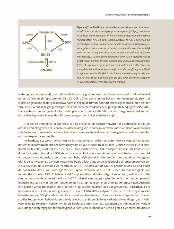
Samenvatting, discussie en toekomstperspectief Samenvatting, discussie en toekomstperspectief
Fi i i li iri Irii ( ),
i l l i li , i i ili l ( ), ie
ig i l , i , i l i l i , zij lijk
i iri i ij i l ili lij ij
i idi i l l( ) i ijn i l , i i l
l, lij i ii ij l i I l l
i l ui i iël(β l i ) i
guur 15NL. Act vat e en metabo sme van notecan. notecan
wordt door cytochroom P450 3A so-enzymen CYP3A met name
n de ever maar ook e ders n het chaam omgezet n de nact eve
metabo eten APC en NPC. Carboxy esterases CES enzymen doverdad n de ever maar ook n de darmmucosa n tumorweefseen n p asma tot express e gebracht worden n verantwoordevoor de omzett ng van notecan n z n du zendmaa act evere
metabo etSN-38.NPCenmoge kerw sookAPCkunnenookdoorCES
geact veerd worden. Ur ne d phosphate-g ucuronosy transferases
UGT 1A sovormen z n de ever maar ook n het ep thee van het maagdarmkanaa verantwoorde k voor de ontg ft ng van SN-38
n z n g ucuron de SN-38G. n het umen van het maagdarmkanaakan het v a de ga tgesche den SN-38G door bacter e enzymen
-g ucuron dases weer geact veerd worden.
wateroplosbaar gemaakt) door uridine diphosphate-glucuronosyltransferases van de 1A subfamilie, met
name UGT1A1, in zijn glucuronide SN-38G. Oók UGT1A wordt in het lichaam op meerdere plaatsen tot
expressie gebracht, zoals in de darmwand en in bepaalde tumoren. Irinotecan en zijn metabolieten worden vanuit de lever naar de gal getransporteerd door meerdere adenosine triphosphate binding cassette (ABC) transporteiwitten met gedeeltelijk overlappende substraatspecifiteiten. In het maagdarmkanaal kunnen bacteriële β-glucuronidases SN-38G weer terugvormen in het toxische SN-38.
Hoewel de verschillen in expressie van de enzymen en transporteiwitten die betrokken zijn bij de afbraak, verdeling over het lichaam en uitscheiding van irinotecan in zekere mate verklaard worden door fysiologische en omgevingsfactoren,beïnvloedt de aanwezigheid van specifieke genetische determinanten ook hun expressie en functie.
In hoofdstuk 4 wordt de rol van de farmacogenetica in het verklaren van de variabiliteit tussen patiënten in farmacokinetiek en farmacodynamiek van irinotecan besproken. Genetische variatie in fase I (CYPs) en fase II (UGTs) enzymen en fase III transporteiwitten (ABC transporters) is in dit hoofdstuk in detail besproken. Vooral het UGT1A1 gen is een veelbelovende kandidaat voor genetische screening, dat
wil zeggen voordat gestart wordt met een behandeling met irinotecan. De homozygote aanwezigheid (dat is de aanwezigheid van een mutatie op beide allelen, dus op beide identieke chromosomen) van een extra, zevende dinucleotide (TA) insertie in de (TA)6TAA-box van de UGT1A1 promoter, ook bekend onder de naam UGT1A1*28, kan namelijk tot een lagere expressie van UGT1A1 leiden. De aanwezigheid van minder (functioneel) UGT1A1 betekent dat SN-38 minder makkelijk ontgift kan worden, wat de associatie van de homozygote aanwezigheid van UGT1A1*28 met een hogere systemische (dat wil zeggen plasma) blootstelling aan SN-38 en een toegenomen risico op leukopenie en ernstige irinotecan geïnduceerde late diarree verklaart, zoals in dit proefschrift op diverse plaatsen ook aangetoond is. In hoofdstuk 7 is bijvoorbeeld een sterke relatie gevonden tussen het UGT1A1*28 polymorfisme en zowel de systemische blootstelling aan SN-38 als de incidentie en ernst van late diarree in Caucasische kankerpatiënten. Hoewel studies tot op heden hebben laten zien dat slechts patiënten die twee variante allelen dragen, at risk zijn voor ernstige toxiciteit, hebben wij in dit hoofdstuk laten zien dat patiënten die minimaal één variant
allel dragen (heterozygoot of homozygoot variant) een verdubbeld risico op graad 2 of meer late diarree
117
Samenvatting, discussie en toekomstperspectiefSamenvatting, discussie en toekomstperspectief
hebben (70% tegen 35%). Dit maakt de aanwezigheid van ten minste één UGT1A1*28 allel tot een sterke voorspellende factor met klinische potentie. In hoofdstuk 8 is een relatie gelegd tussen de aanwezigheid van het UGT1A1*28 genotype en systemische expositie en neutropenie. Aangezien op dit moment het veld van farmacogenetics en farmacogenomics zich snel uitbreidt en tegelijkertijd snellere en kosteneffectievere screeningsmethodes ontwikkeld worden, wordt in de nabije toekomst een grote hoeveelheid aan data verwacht die onze kennis wat betreft de genetische basis van irinotecan metabolisme zal verrijken. Een individueel genetisch profiel van de meeste relevante enzymen bij iedere patiënt kan uiteindelijk behulpzaam zijn bij het daadwerkelijk individualiseren van de behandeling met irinotecan.
Het zogenaamde breast cancer resistance protein (ABCG2) is een ABC transport eiwit dat een rol speelt bij de uitscheiding (efflux) van verscheidene lichaamsvreemde stoffen uit cellen, waaronder SN-38. In vitro onderzoek heeft laten zien dat het ABCG2 421C>A polymorfisme (een enkelvoudige nucleotide wisseling op plaats 421 in het gen dat codeert voor ABCG2) geassocieerd is met een verminderde eiwit
expressie en een gewijzigde functie. In hoofdstuk 5 zijn de etnische distributie en de potentiële functionele consequenties van dit enkelvoudige nucleotide polymorfisme (SNP) onderzocht. ABCG2 421C>A lijkt een beperkte rol te spelen in de dispositie van irinotecan in Europese Caucasiërs.Waarschijnlijk valt de bijdrage van deze genetische variant niet op door de functionele rol van andere polymorfe transporteiwitten, zoals besproken in hoofdstuk 4.
Het polymorfe eiwit ABCC2 (c-MOAT) lijkt het belangrijkste transporteiwit te zijn dat betrokken is bij de hepatobiliaire uitscheiding van irinotecan, SN-38 en SN-38G. Vele functionele polymorfe varianten van ABCC2 zijn tot op heden beschreven. In hoofdstuk 6 hebben we de associatie tussen ABCC2 polymorfismen en haplotypes enerzijds en irinotecan dispositie en late diarree anderzijds onderzocht in 150 Caucasische kankerpatiënten. De resultaten suggereren dat de aanwezigheid van een specifiek ABCC2-haplotype (ABCC2*2) in patiënten die geen variant UGT1A1*28 allel dragen geassocieerd is met het ontstaan van minder ernstige diarree, mogelijkerwijs als gevolg van een verminderde hepatobiliaire uitscheiding van irinotecan naar het maagdarmkanaal. Gevonden hogere systemische blootstelling aan irinotecan in deze patiënten ondersteunt deze hypothese. Het is theoretisch verklaarbaar dat de aanwezigheid van een variant UGT1A1*28 allel indirect het beschermende effect van ABCC2*2 op late diarree te niet doet vanwege de verminderde glucuronidatie-capaciteit in de darmwand. Patiënten die drager zijn van een UGT1A1*28
allel zijn immers minder goed in staat om lokaal in de darmmucosa uit irinotecan gevormd SN-38 te ontgiften, wat leidt tot een hogere expositie aan deze toxische metaboliet en dus een grote kans op diarree.
Late diarree wordt meestal verondersteld direct veroorzaakt te worden door hoge concentraties SN-38 in het maagdarmkanaal, deels gevormd uit hepatobiliair uitgescheiden SN-38G door bacteriële β-glucuronidases. In een eerder uitgevoerde pilot-studie137 is gerapporteerd dat het aminoglycoside antibioticum neomycine het opnieuw optreden van graad 2 of meer diarree voorkwam in zes van zeven patiënten. Dit werd toegeschreven aan de inhibitie van β-glucuronidase producerende bacteriën in de darm. In hoofdstuk 7 zijn de resultaten gepresenteerd van een multi-center, dubbel-geblindeerde, gerandomiseerde en placebo-gecontroleerde studie met irinotecan met of zonder neomycine, bedoeld om het eerder aangetoonde profylactische effect van neomycine te valideren.Helaas kon geen significant effect
op de incidentie, ernst en duur van irinotecan geïnduceerde diarree aangetoond worden. Mogelijk speelt
de lokale SN-38-ontgifting(scapaciteit) in de darmwand zelf een belangrijkere rol dan luminaal aanwezig en/of gevormd SN-38. In een exploratieve analyse werd namelijk het UGT1A1*28 polymorfisme, dat leidt tot
een lagere expressie van UGT1A1 in de lever en de darmwand en dus tot een lagere ontgiftingscapaciteit,
118
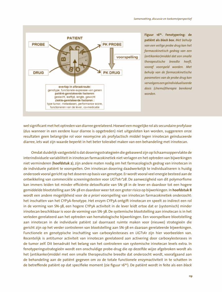
Samenvatting, discussie en toekomstperspectief Samenvatting, discussie en toekomstperspectief
Fi . ii ls bl . lp
ilii i
( i )mi l lle
i , ld
l i i
l ï ivi liis ( ) ie
guur 16NL Fenotyper ng: de
pat ënt a ack box Met behuvan een ve ge probe drug kan het farmacok net sch gedrag van een
ant kanker dde dat een smatherapeut sche breedte heeftvooraf voorspe worden. Met behu p van de farmacok net sche
parameters van de probe drug kan
vervo gens een ge nd dua seerde
dos chemo therap berekend
worden.
wel significant met het optreden van diarree gerelateerd. Hoewel een mogelijke rol als secundaire profylaxe (dus wanneer in een eerdere kuur diarree is opgetreden) niet uitgesloten kan worden, suggereren onze resultaten geen belangrijke rol voor neomycine als profylactisch middel tegen irinotecan geïnduceerde diarree, iets wat zijn waarde beperkt in het beter tolerabel maken van een behandeling met irinotecan.
Omdat duidelijk vastgesteld is dat doseringsstrategieën die gebaseerd zijn op lichaamsoppervlakte de interindividuele variabiliteit in irinotecan farmacokinetiek niet verlagen en het optreden van bijwerkingen niet verminderen (hoofdstuk 2), zijn andere maten nodig om het farmacologisch gedrag van irinotecan in de individuele patiënt te voorspellen. Om irinotecan dosering daadwerkelijk te individualiseren is huidig onderzoek vooral gericht op het doseren op basis van genotype. Er wordt vooral veel energie besteed aan de ontwikkeling van commerciële screeningstesten voor UGT1A1*28. De aanwezigheid van dit polymorfisme kan immers leiden tot minder efficiënte detoxificatie van SN-38 in de lever en daardoor tot een hogere gemiddelde blootstelling aan SN-38 en daardoor weer tot een groter risico op bijwerkingen. In hoofdstuk 8
wordt een andere mogelijkheid voor de a priori voorspelling van irinotecan farmacokinetiek onderzocht: het inschatten van het CYP3A-fenotype. Het enzym CYP3A ontgift irinotecan en speelt zo indirect een rol in de vorming van SN-38, een hogere CYP3A activiteit in de lever leidt ertoe dat er (systemisch) minder irinotecan beschikbaar is voor de vorming van SN-38. De systemische blootstelling aan irinotecan is in het
verleden gerelateerd aan het optreden van hematologische bijwerkingen. Een voorspelbare blootstelling aan irinotecan in de individuele patiënt zal daarnaast ruimte maken voor (nieuwe) strategieën die gericht zijn op het verder controleren van blootstelling aan SN-38 en daaraan gerelateerde bijwerkingen. Functionele en genotypische inschatting van carboxylesterases en UGT1A1 zijn hier voorbeelden van. Recentelijk is antitumor activiteit van irinotecan gerelateerd aan activering door carboxylesterases in de tumor zelf. Dit benadrukt het belang van het controleren van systemische irinotecan levels extra. In fenotyperingsstrategieën wordt een onschuldige probe-drug die op dezelfde wijze afgebroken wordt als het (antikanker)middel met een smalle therapeutische breedte dat onderzocht wordt, voorafgaand aan de behandeling aan de patiënt gegeven om zo de totale functionele enzymactiviteit in te schatten in de betreffende patiënt op dat specifieke moment (zie figuur 16NL). De patiënt wordt in feite als een black
119

DankwoordSamenvatting, discussie en toekomstperspectief
box gezien, wat betekent dat de totale functionele activiteit de som is van de combinatie van genotype, ziektegerelateerde invloeden,patiëntkarakteristieken,omgevingsinvloeden,voeding en co-medicatie. In de exploratieve studie, zoals beschreven in hoofdstuk 8, bleek dat in kankerpatiënten van Caucasische komaf het CYP3A-fenotype, zoals ingeschat door de midazolam-klaringstest, statistisch significant geassocieerd was met de klaring van irinotecan. Dit wijst erop dat deze strategie, waarin de patiënt als geheel genomen wordt, in de toekomst gebruikt kan worden om de dosering van irinotecan te individualiseren en zo de kans op bijwerkingen te verminderen.
In dit proefschrift is aangetoond dat veel factoren betrokken zijn bij het metabolisme van irinotecan en bij de pathogenese van late diarree, dat voor veel oncologen nog steeds een van de meest verontrustende bijwerkingen van irinotecan is. In de toekomst moet het individualiseren van de dosering van irinotecan, hetgeen uiteindelijk de bedoeling moet hebben een behandeling met irinotecan beter te verdragen en tegelijkertijd zo effectief als mogelijk te maken, zich richten op strategieën die verschillende factoren combineren. Op dit moment wordt een geïndividualiseerde doseringsstrategie die gebaseerd is op een lineair regressiemodel op basis van CYP3A-fenotypering, zoals beschreven in hoofdstuk 8, prospectief gevalideerd in een gerandomiseerde, multicenter studie. De helft van alle geïncludeerde patiënten ondergaat een midazolam-klaringstest, waarna zij een dosis irinotecan ontvangen die berekend is door een doel(target)-blootstelling te vermenigvuldigen met een verwachte irinotecan klaring (gebaseerd op midazolamklaring, γ-GT en lengte). De overige patiënten worden conventioneel gedoseerd, dat wil zeggen op basis van lichaamsoppervlakte. Variatie tussen patiënten in blootstelling aan irinotecan zal vergeleken worden tussen beide groepen, en wanneer deze interindividuele variatie inderdaad verminderd is, zijn verdere studies noodzakelijk om de maximum tolereerbare target-blootstelling te vinden en de variatie tussen patiënten wat betreft de farmacokinetiek van irinotecan en –gegeven een target-blootstelling– blootstellinggerelateerde bijwerkingen verder te verlagen.
Doseringsstrategieën die CYP3A-fenotypering combineren met UGT1A1-genotypering en andere strategieën (zoals de inschatting van de functionele carboxylesterase-activiteit), kunnen leiden tot een betere voorspelling van irinotecan farmacokinetiek en op deze manier tot een betere therapeutische index voor irinotecan. Vooralsnog zijn voordelen van zulke strategieën beperkt tot patiënten die behandeld worden in academische centra. Om irinotecan behandeling daadwerkelijk te individualiseren moeten inspanningen die gericht zijn op het ontwikkelen en routinematig implementeren in specialistische en algemene ziekenhuizen van patiëntvriendelijke, eenvoudige en haalbare limited-sampling-modellen en fenotyperings- en genotyperingsstrategieën, worden aangemoedigd. Gevonden (causale) correlaties tussen genotype (in het bijzonder UGT1A1*28) en de farmacokinetiek, de bijwerkingen en/of het
therapeutisch resultaat van een behandeling met irinotecan moeten bevestigd worden in grote groepen patiënten. Als resultaat hiervan moeten evidence-based aanbevelingen geformuleerd worden op basis waarvan irinotecan gedoseerd kan worden. Voor de individuele patiënt zal het verminderen van de ernst
en incidentie van late diarree irinotecan beter te verdragen maken. Uiteindelijk zal dit deze klassieke topoisomerase I remmer het imago moeten geven dat het daadwerkelijk verdient in het snel veranderende veld van anti-kankermiddelen.
120
Samenvatting, discussie en toekomstperspectief Dankwoord
Dankwoord
Ongetwijfeld verwacht men van mij dat ik alle mensen die een meer of minder zinvolle bijdrage hebben geleverd aan de totstandkoming van dit proefschrift, hier met naam en toenaam zal noemen. Terecht! Het dankwoord moet in orde zijn; er schijnen zelfs mensen te bestaan die eerst deze pagina’s lezen voordat zij zich aan de rest van het proefschrift wagen (toch Diana?). Aan hen het verzoek toch maar eerst de (Nederlandse) samenvatting te lezen, zodat, mocht ik vergeten zijn hen te noemen, in elk geval de essentie van de boodschap geconsumeerd is. Aan de rest van dit proefschrift heb ik immers, op een Leids kwartiertje na, vandaag precies vier jaar en een week gewerkt, en daar gaat het om! Dat zonder inzet en hulp van velen dit boekje er heel anders uitgezien zou hebben, moge duidelijk zijn!!
Zonder promotiecommissie geen promotie. Vanzelfsprekend gaat mijn dank uit naar mijn promotor Prof.dr. Jaap Verweij. Zijn kennis over de (experimentele) behandeling van kanker is fenomenaal, evenals zijn ervaring in het bedrijven van wetenschap. Zijn (internationale) netwerk is bijzonder: ‘from the group of Verweij from the Netherlands’ opent deuren. Beste Jaap, je hebt in mijn leven de afgelopen vier jaar een belangrijke rol gespeeld. Al was het alleen maar omdat jij (of ikzelf) de deadline weer eens zo scherp gezet
had die onhaalbaar (b)leek. Gelukkig was je altijd bereid de horde centimeter bij centimeter te verzetten. Je slaagde er steeds weer in me te blijven prikkelen. Hoewel je altijd over mijn schouder meekeek of ik wel gefocused bleef, heb je mij veel ruimte gelaten voor het uitwerken van mijn ideeën; het zonder vooroverleg aan je aangeboden cannabis-commentaar, dat verrassend gepubliceerd werd in het Journal of Clinical Oncology, het bedenken en opzetten van de interactiestudie met medicinale cannabis en de prospectieve validatie van de doseringsformule (de De Jong-formule, zoals jij pleegt te zeggen), zijn voorbeelden daarvan. Ik voel me bijzonder bevoorrecht onder jouw bezielende leiding onderzoek te hebben mogen doen en hoop in de toekomst nog wat irinotecan-onderzoek met je te mogen doen.
De secretaris van de promotiecommissie, Prof.dr. Gerrit Stoter, dank ik voor de geboden mogelijkheden tot het verrichten van wetenschappelijk onderzoek in het Erasmus Medisch Centrum–Daniel den Hoed. Prof.dr. J.H. Beijnen en mw. Prof.dr. E.G.E. de Vries, ben ik eveneens dankbaar voor de snelle beoordeling van het manuscript.
Ik ben alle leden van de promotiecommissie erkentelijk voor hun aanwezigheid, en in het bijzonder Dr. Alex Sparreboom (St. Jude Children’s Research Hospital, Memphis, TX, USA). Beste Alex, jij bent zonder twijfel mijn grote inspirator geweest. Wat een ideeëngenerator ben jij. De snelheid waarin jij een draft om kan zetten in een manuscript van hoge kwaliteit is absoluut ongekend. Wanneer een draft weer eens te lang was en niet snel genoeg tot de kern kwam, schrapte jij rigoureus mijn fraai gebrouwde zinnen. Ik ben je zeer erkentelijk voor al je hulp. Jouw energie is niet te beschrijven en jouw enthousiasme werkte iedere keer dat we elkaar spraken of zagen nog weken door. Het was geweldig met jou te kunnen samenwerken.
Zonder patiënten geen begin. Op papier zijn zij een studievolgnummer met drie initialen, de bijna 300 naamloze patiënten die in de studies die beschreven zijn in dit proefschrift en in de nog lopende irinotecan-studies, geparticipeerd hebben of nog (gaan) participeren. Beste patiënten, dankzij of ondanks uw ziekte was u bereid belangeloos mee te werken en uw kostbare tijd te spenderen aan extra ziekenhuisbezoeken en extra testen. Voor een deel van u was dit extra boven op uw reguliere behandeling, voor een grote groep was medewerking echter voorwaardelijk voor een behandeling met irinotecan. Allen nam u deel uit vrije
121
Dankwoord Dankwoord
wil, met uitlopende, individueel moverende redenen: voor de wetenschap, als laatste strohalm, of om te laten zien er alles aangedaan te hebben. Soms met moeite, maar altijd met een soort van plezier heb ik u in de studies geïncludeerd. Standaard liepen deze eerste kennismakingen uit. We wisten allebei waar het
om ging en wat er speelde. Al ging het soms snel slechter, ik heb u met veel plezier begeleid bij de kuren in studieverband. We hebben over alles en nog veel meer gesproken. Ik kan u, die alle doorproevingen wilde doorstaan, slechts bedanken. Sommigen van u kwam ik kuren later nog tegen, en dan vertelde u vol trots nog steeds irinotecan (of inmiddels een ander middel) te krijgen. Dat betekende dat het relatief goed met
u ging. Beiden de relativiteit beseffende, deed u dat goed en dat deed mij dan goed. Weliswaar nam ik u nooit mee naar huis, maar uw naam, uw gezicht en uw verhaal staan in mijn geheugen gegrift. Binnen de grenzen van uw ziekte was het contact met u bijzonder en plezierig.
Zonder dokters geen patiënten. Alle stafleden van de afdeling Interne Oncologie op de centrumlocatie en in de Daniel den Hoed, in het bijzonder Dr. Maja de Jonge, Dr. André Planting, Dr. Ferry Eskens en Dr. Stefan Sleijfer, ben ik erkentelijk voor het steeds weer aanmelden van patiënten voor de irinotecan studies en voor al hun suggesties en oplossingen. Beste Maja, dank voor jouw hulp bij het tot een goed einde brengen van een aantal van de in dit proefschrift beschreven studies. Dr. Lia van Zuylen wil ik bedanken voor het steeds weer mogen meekijken op haar poli. Beste Lia, wat heb ik daar veel van je geleerd! Het was erg plezierig samen met jou twee artikelen over medicinale cannabis in Nederland in elkaar te draaien. Dr. Geert-Jan Creemers (en ook Coby en Monique) ben ik bijzonder erkentelijk voor het
feit dat hij bereid is in het Catharina Ziekenhuis te Eindhoven patiënten te includeren in de 04-290, de prospectieve validatiestudie van de nieuwe doseringsformule.
Alle Daniel-juniors door jaren heen, ook jullie hebben met veel zorg patiënten geïncludeerd. Jullie hadden tussen alle poli’s en opnames door altijd wel een korter of langer moment tijd. Dr. Jos Kitzen was altijd bereikbaar om op het Behandelcentrum weer eens een dosis midazolam toe te dienen. Met Dr. Otto Soepenberg, Dr. Metin Tasçilar, Dr. Jos Kitzen en Dr. Paul Hamberg was het plezierig samenschrijven aan de verschillende manuscripten.
Zonder mensen geen zorg. Alle verpleegkundigen, secretaresses en doktersassistenten van de afdelingen B0, B0-zuid, B1 en de poliklinieken wil ik bedanken voor de geboden hulp in patiëntenzorg en onderzoek.Bimla en Barbara mag ik natuurlijk ook niet vergeten.Loes en Marian van de salarisadministratie, de meiden van het CMA (op moment van schrijven staan er meen ik zo’n 200 statussen op mijn code uitgeboekt), de mannen van de inmiddels opgeheven postkamer, en ons secretariaat, in het bijzonder Linda en Aline, moeten ook genoemd worden. Van jullie praktische en persoonlijke ondersteuning heb ik veel geleerd. Jullie klant- en patiëntvriendelijkheid mag een voorbeeld zijn voor velen.
Natuurlijk ook een bijzondere plaats voor de mensen van het Behandelcentrum, en niet alleen omdat
dat moet van Diana en niet alleen omdat er eigenlijk altijd ruimte was voor een midazolamtest en jullie bijna altijd op het juiste moment de kinetiek afnamen. Maar, Joop en Kathinka, Liesbeth, Elly, Elly, Jan, Ina, Evert, en Celesia, ook omdat het altijd prettig was even bij jullie in de zitkamer neer te kunnen ploffen. Zo was de droppot (bijna) altijd gevuld.
En natuurlijk de researchverpleegkundigen (eerst Hans van der Meulen en Tatjana Pronk, later Conny, Monique, Diane en Leny) en researchassistenten (Annet, Hester, Monica en Suzanne) van de afdeling ben ik bijzonder dankbaar voor de logistieke begeleiding van de studies, het geduldig veranderen van studiedocumenten, last-minute VSO’s, het afdraaien en afleveren van samples en de zorg voor alle patiënten. Maar zeker ook voor de gezellige jaren: oliebollen, hertengeweien en ijsblokken...
122
Dankwoord Dankwoord
Zonder lab geen kinetiek. Alle collega’s van het lab translationele farmacologie onder leiding van Dr. Erik Wiemer. Dr. Walter Loos, Ing. Peter de Bruin, Desirée van Boven-van Zomeren, Mei Lam en ook Robert Oostrum, dank voor jullie bijdrage aan de nauwkeurige analyses van midazolam, irinotecan, SN-38, SN-38G, APC en NPC. Limited sampling zal jullie echt een deugd doen! De discussies over wat-dan-ook waren altijd motiverend. De Voedsel en Waren Autoriteit heeft aangegeven dat we GLP-compliant zijn. De regeldrift en bureaucratische rompslomp van GLP blijft wat mij betreft een noodzakelijk en nuttig kwaad. Ik mis overigens nog steeds een gestandaardiseerde werkwijze koffiezetten. Want zelfs al was de koffie altijd veel te sterk, in zijn sterkte was de inter-individuele variatie fenomenaal. Een fase I-studie met jullie bekers bocht, te beginnen met één slok per dag, zal verrassende resultaten laten zien, al was het maar wat
betreft bijwerkingen. Dr. Frederike Engels, samen stonden wij ergens tussen lab en kliniek in. Soms best lastig: we
verzuchtten geregeld dat niets van zelf leek te gaan. Jessica van der Bol, jouw enthousiasme is aanstekelijk. Het was prettig je te begeleiden in het laatste deel van je basis-opleiding. Vanaf nu weet je waarom en hoe je later een status moet invullen. Hoewel je inmiddels bijna alle valkuilen in het promotiepad in de Daniel den Hoed kent, zal ook jij er vast in vele vallen. Land zacht. Dr. Herman Burger en Dr. Kees Nooter van de overkant, het was prettig met jullie te mogen werken aan de diverse imatinib-studievoorstellen en studies.
Uiteraard wil ik op deze plaats ook Dr. Ron Mathijssen, mijn copromotor, nadrukkelijk bedanken. Beste Ron, Jaap gunde jou deze eer, en ik denk terecht. Zonder jou was ik niet in de Daniel den Hoed terechtgekomen. In de irinotecan-lijn ben jij mij onder leiding van Jaap en Alex voorgegaan. Het was me een genoegen met je samen te werken aan de vervolgstukken. Altijd wist je jouw agenda duidelijk te maken. Het was prettig avonden en weekenden lang samen te analyseren, te schrijven en over de toekomst
te praten. Waar jouw fenotyperingsstudie je boekje niet haalde, haalt mijn doseringsstudie die daarop voortbouwt, het niet. Voor de komende jaren hebben we nog werk genoeg!
Zonder hulp geen data. Een absoluut gegeven: zonder betrouwbare data geen proefschrift. Daarom speciale dank aan onderstaande personen voor alle analyses en hulp. Steeds weer leverden zij topprestaties!
Dr. Lena Friberg, Dr. Rujia Xie, and Prof.dr. Mats Karlsson from the Uppsala University, Uppsala, Sweden. Thank you for all the pharmacokinetic modeling of irinotecan and its metabolites. Dear Lena, for sure, we will visit you in Uppsala! Dr. Erin Gardner (Lepper), Dr. Nicola Smith, and Dr. William Figg from the National Cancer Institute, Bethesda, MD, USA, thank you for measuring midazolam by HLPC. Dirk Buijs, Dr. Wim van den Berg, Trinet Rietveld en Jan Francke, dank voor jullie hulp bij het opzetten en analyseren van de erythromycine ademtest. Trinet, wat hebben we zitten zwoegen op het foutje in de door Laurent Rivory gepubliceerde formule.
Dr. Ron van Schaik, Dr. Paul Schenk, Martin van Vliet, Marloes van der Werf, Ilse van der Heiden en Marianne van Fessem van het Pharmacogenetics Core Lab van het Klinisch Chemisch Laboratorium op de centrumlokatie, dank voor het steeds weer genotyperen voor onder andere UGT1A1, CYP3A4, CYP3A5 en ABCB1. Dr. Sharon Marsh,Tiffany Scott-Horton, Cristi King,and Prof.dr. Howard McLeod from the Washington University School of Medicine, St Louis, MO, USA, thank you for performing the ABCG2, ABCC2, and UGT1A1 genotyping and haplotyping. It was nice working with you! Prof.dr. Deanna Kroetz from the University of California, San Francisco, CA, USA thank you for your input on the ABCC2-diarrhea manuscript.
Dr. Arno Hazekamp van de Universiteit Leiden en Johan Bender van Farmalyse BV uit Zaandam ben ik erkentelijk voor hun hulp bij het in vitro cannabis werk. Het manuscript ronden we dit jaar echt af.
123
Curriculum vitaeDankwoord
Datamanagers van G1, dank voor het steeds weer uitvragen van alle inclusie- en exclusiecriteria. De inclusie liep steeds soepel, en afwijkingen van de geplande procedure waren in overleg eenvoudig te regelen. Datamanagers van G4, ook jullie ondersteuning was nuttig, nodig en prettig. De verzameling van diarrhee-scores en de opbouw van het neomycine-databestand was bijvoorbeeld nooit goed gekomen zonder jullie hulp en de medewerking van Dr. Diederik Kehrer. Ook het fase I-verslag zou er wat betreft de irinotecan-studies zonder jullie hulp heel anders uit gezien hebben. Dr. Joke Boonstra, Joke Paardekoper en de andere mensen van het Klinisch Chemisch Laboratorium in de Daniel den Hoed ben ik dankbaar voor al de klinisch-chemische bepalingen, de opslag van de code geno buisjes en al het advies.
Drs. Wilfried Graveland, voorheen werkzaam bij de afdeling biostatistiek in de Daniel den Hoed. Dank voor Stata en voor alle controles van de statistische experimenten. Kerstin en Marijke van de Daniel den Hoed bibliotheek. Wanneer een artikel niet op internet te vinden was, kregen jullie het steeds weer voor elkaar een reprint te regelen. Hans Kneefel, dank voor alle posters en figuren.
Zonder vangnet geen promotie. Mijn vader en moeder ben ik dankbaar voor de basis van mijn vorming, die zij gelegd hebben. Zij hebben me altijd gestimuleerd tot kennisverwerving. Lieve papa en mama, onvoorwaardelijk waren en zijn jullie er altijd. Een nieuwe rol ligt voor jullie klaar: oppas-opa en oma voor Saskia. Laura (en Maarten natuurlijk), lieve zus, fijn dat je er altijd was. Schrijven we nog eens een manuscript samen?
Lieve vrienden, jullie zijn allemaal zo belangrijk, jullie rol is niet in woorden te vatten. Annet en John (en natuurlijk Tim, Joep, Daan, Fleur, Meike, Jonathan, Ruben en Naomi), fijn jullie in mijn leven te hebben. Bijna Vino Novello, als surrogaat dank voor alles. Dank voor de mooie vorm. Samen met de kaartjes van dit jaar een mooi en dankbaar geheel! Koos en Astrid (en natuurlijk Nienke en Roel), door jullie ben ik ooit
over het doen van wetenschappelijk onderzoek gaan nadenken. Ruben en Iris, Maarten, Bas en Josien, Remco en Femke, Daniëlle, al wapperen we steeds verder uit elkaar, we laten niet los! Keizer Hans (en Dymphy) en Prinses Pippi (en Richard), ik moet jullie natuurlijk ook hier noemen. Net als Desirée en André, Ron en Daniëlle, Marieke en Robbert, Ugo and Monica. Gabe, Iny, Bianca en Carla. Pieter, Nely, Klaas Bart, Anne Stijn en Martje. En natuurlijk ook schoonouders en Cor, Dorine en Manouk. PvdA’ers uit Zoetermeer, speciaal Gemma, Edo, Frans, Wim, André, Yvonne, Rob, Werner en John, wat heb ik me afgelopen jaren met
jullie vermaakt. Alle Energie’rs van de AAD-groep, de HOMErunners met moederkloek Marja, en natuurlijk de ROPArunners van het Daniel-team, wat een genot met jullie te lopen. Er wordt aan jullie allemaal gedacht.
De laatste woorden zijn voor de twee vrouwen die alles-bepalend in mijn leven zijn. Lieve Diana (‘als je me ergens in het dankwoord zet, ga ik niet naar die stomme promotie’) wat ben jij heerlijk lief. En Saskia, wat genieten we van jouw tevreden glimlach en geschreeuw.
124
Dankwoord Curriculum vitae
Curriculum vitae
Ik, Floris Aart de Jong, ben op 4 oktober 1973 geboren in Rotterdam. In 1993 kreeg ik op het Marnix Gymnasium in Rotterdam mijn gymnasiumdiploma uitgereikt. Na uitloting voor de studie geneeskunde begon ik dat jaar aan de (Rijks)Universiteit Leiden met de studie natuurkunde. Het volgende academisch jaar haalde ik aan diezelfde universiteit met lof mijn propedeutisch examen psychologie. Hoewel ik deze studie met bijzonder veel plezier volgde, besloot ik toen ik in 1996 bij de vierde poging eindelijk ingeloot
werd, in navolging van mijn zus, alsnog geneeskunde te gaan studeren aan de Erasmus Universiteit
Rotterdam (het latere Erasmus Medisch Centrum). In juli 1997 haalde ik daar met lof mijn propedeutisch examen en in januari 2004 rondde ik mijn doctoraal geneeskunde af. Tijdens mijn studie deed ik bij de afdeling voor Maag-, Darm- en Leverziekten van het voormalig Dijkzigt-ziekenhuis (tegenwoordig Erasmus Medisch Centrum–centrumlocatie) onderzoek naar de rol van genetische predispositie bij inflammatoire darmziekten. Vanaf het begin van mijn geneeskundestudie in 1996 ben ik in diverse hoedanigheden werkzaam bij T&IC in Delft, een bureau voor tekst en vorm van interne communicatie. Tevens was ik van juli 2000 tot december 2004 als fractiemedewerker verbonden aan de PvdA in Zoetermeer.
Gedreven vanuit een basale behoefte wetenschappelijk onderzoek binnen (en niet vanuit) de klinische praktijk te verrichten, kwam ik in november 2002 in dienst van de afdeling Interne Oncologie, Erasmus Medisch Centrum–locatie Daniel den Hoed (Prof.dr. G. Stoter). Daar verrichtte ik onder leiding van Prof.dr. J.Verweij klinisch onderzoek met als doel het daadwerkelijk individualiseren van de dosering van het
klassieke chemotherapeuticum Irinotecan®, wat geleid heeft tot dit proefschrift. Zowel farmacokinetische studie-designs in een fase I-populatie werden ontworpen en geïmplementeerd, als bestaande gegevens werden geanalyseerd. In 2004 ontving ik tijdens het 7e SITILO congres (Florence, Italië) de Maurizio Vaglini Award.
In 2005 ben ik gestart met een postdoctorale opleiding tot geregistreerd klinisch farmacoloog (opleider Prof.dr. J. Schellens, Antoni van Leeuwenhoek/NKI, Amsterdam). Daarnaast ben ik sinds april 2006 in de uren die resteren buiten bovengenoemde aanstellingen (bij de afdeling Interne Oncologie van het Erasmus Medisch Centrum–Daniel den Hoed; en bij T&IC, Delft), het huiselijke en sociale leven en de noodzakelijke hardlooptrainingen (in willekeurige volgorde), deelgemeenteraadslid voor de PvdA in Rotterdam-Feijenoord. Ik ben getrouwd met Diana Bakker en in de afrondende fase van dit proefschrift
werd onze prachtige dochter, Saskia, geboren.
125
List of publicationsList of publications
List of publications
PUBLICATIONS
1. de Jong FA, Mathijssen RH, de Bruijn P, Loos WJ, Verweij J, Sparreboom A. Determination of irinotecan
(CPT-11) and SN-38 in human whole blood and red blood cells by liquid chromatography with fluorescence
detection. J Chrom B 795(5):383-388, 2003
2. de Jong FA, Verweij J. Role of Imatinib Mesylate (Gleevec/Glivec) in gastrointestinal stromal tumors. Expert Rev Anticancer Ther 3(6):757-766, 2003
3. de Jong FA, Mathijssen RH, Xie R, Verweij J, Sparreboom A. Flat-fixed dosing of irinotecan: influence on
pharmacokinetic and pharmacodynamic variability. Clin Cancer Res 10(12):4068-4071, 2004
4. de Jong FA,Marsh S,Mathijssen RH,King C,Verweij J,Sparreboom A,McLeod HL.ABCG2 pharmacogenetics:ethnic differences in allele frequency, and assessment of influence on irinotecan disposition. Clin CancerRes 10(17):5889-5894, 2004
5. Mathijssen RH, de Jong FA, van Schaik RH, Lepper ER, Friberg LE, Rietveld T, de Bruijn P, Graveland WJ,Figg WD,Verweij J, Sparreboom A. Prediction of irinotecan pharmacokinetics by use of cytochrome P450
3A4 phenotyping probes. J Natl Cancer Inst 96(21):1585-1592, 2004
6. de Jong FA, Mathijssen RH, Verweij J. Limited potential of hepatic arterial infusion of irinotecan. J Chemother 16(S5):48-50, 2004
7. de Jong FA, de Jong L, van der Meulen H, Van Zuylen C. Beperkte plaats voor medicinale cannabis in de
palliatieve zorg van kankerpatiënten. Ned Tijdsch Palliatieve Zorg (3):81-86, 2004
8. Soepenberg O,Dumez H,Verweij J,de Jong FA,de Jonge MJ,Thomas J,Eskens FA,van Schaik RH,Sellenlach
J, ter Steeg J, Assadourian S,Sanderink G,Sparreboom A,van Oosterom AT. Phase I,pharmacokinetic, food
effect, and pharmacogenetic study on oral irinotecan given as semi-solid matrix capsules in patients
with solid tumors. Clin Cancer Res 11(4):1504-1511, 2005
9. de Jong FA,Engels FK,Mathijssen RH, van Zuylen L,Verweij J,Peters RP,Sparreboom A. Medicinal cannabis
in oncology practice: still a bridge too far? J Clin Oncol 23(13):2886-2891, 2005
10. de Jong FA, Engels FK, Mathijssen RH, van Zuylen L,Verweij J, Peters RP, Sparreboom A. In Reply. Medicinalcannabis in oncology practice: still a bridge too far? J Clin Oncol 23(30):7756, 2005
11. de Jong FA, Mathijssen RH, Verweij J. Irinotecan-chemotherapie: een nieuwe doseringsformule om de
variatie in blootstelling tussen patiënten te verlagen. Ned Tijdsch Oncol 3(1):33-36, 2006
12. de Jong FA,de Jonge MJ,Verweij J, Mathijssen RH. Role of pharmacogenetics in irinotecan therapy. CancerLett 234(1):90-106, 2006
13. Hamberg P, de Jong FA, Boonstra JG, van Doorn J, Verweij J, Sleijfer S. Non-islet-cell tumour induced
hypoglycaemia in patients with advanced gastrointestinal stromal tumour, possibly worsened by
imatinib. J Clin Oncol 24(18):e30-e31, 2006
14. Tascilar M1, de Jong FA1, Verweij J, Mathijssen RH. Complementary and alternative medicine during
cancer treatment: beyond Innocence. Oncologist 11(7):732-741, 2006
15. Gardner ER, Burger H, van Schaik RH, van Oosterom AT, de Bruijn EA, Guetens G, Prenen H, de Jong FA, Baker SD, Bates SE, Figg WD, Verweij J, Sparreboom A, Nooter K. Association of enzyme and transporter genotypes with the pharmacokinetics of imatinib in patients with gastrointestinal stromal tumors. Clin Pharmacol Ther 80(2): 192-201, 2006
16. de Jong FA, Kehrer DF, Mathijssen RH, Creemers GJ, de Bruijn P, van Schaik RH, Planting AS, van der Gaast A, Eskens FA, Janssen JT, Ruit JB, Verweij J, Sparreboom A, de Jonge MJ. Prophylaxis of irinotecan-induced diarrhea with neomycin and potential role for UGT1A1*28 genotype screening: a
126
List of publications List of publications
double-blind, randomized, placebo-controlled study. Oncologist 11: in press, 2006
17. de Jong FA, Kitzen JJ, de Bruijn P, Verweij J, Loos WJ. Hepatic transport, metabolism and biliary excretion
of irinotecan in a cancer patient with an external bile drain. Cancer Biol Ther: in press, 2006
18. de Jong FA,Scott-Horton T,Kroetz DL,McLeod HL,Friberg LE,Mathijssen RH,Verweij J,Marsh S,Sparreboom
A. Irinotecan-induced diarrhea: functional significance of the polymorphic ABCC2 transporter protein. Submitted, 2006
19. Engels FK1, de Jong FA1, Sparreboom A, Mathot R.A, Loos WJ, Kitzen JJ, de Bruijn P,Verweij J, Mathijssen RH. Influence of medicinal cannabis on the clinical pharmacokinetics of irinotecan and docetaxel. Submitted, 2006
PROCEEDINGS
1. Sparreboom A, Mathijssen RH, de Jong FA, de Jonge MJ, de Bruijn P, Rietveld T, Verweij J. Flat-fixed dosing
of irinotecan: feasibility and effects of cytochrome P450 3A4 phenotype. Proc Am Soc Clin Oncol (ASCO) Annual Meeting 2003;#504
2. Verweij J, de Jong FA, van Schaik RH, Friberg LE, Karlsson MO, Danesi R, Figg WD, Rietveld T, Sparreboom
A, Mathijssen RH. Is phenotype/genotype based dosing for irinotecan a feasible way of dose-individualization? Proc 8th Int. Symposium on Cancer Chemotherapy, Tokyo 2003
3. Mathijssen RH, Marsh S, de Jong FA, Karlsson MO, King C, Verweij J, Sparreboom A, McLeod HL. Effect of the common 421 C>A polymorphism in ABCG2 (BCRP) on irinotecan pharmacokinetics in Caucasians. Proc Am Assoc Cancer Res (AACR) Annual Meeting 2004;#4738
4. de Jong FA, Mathijssen RH, de Bruijn P, Willems EW, Verweij J, Sparreboom A. Prospective validation of a limited sampling model predicting SN-38 pharmacokinetics. Proc Am Assoc Cancer Res (AACR) Annual Meeting 2004;#4740
5. de Jong FA, Mathijssen RH, Lepper ER, van Schaik RH, Friberg LE, de Bruijn P, Graveland WJ, Figg WD, Verweij J, Sparreboom A. Role of CYP3A phenotyping by midazolam clearance in predicting irinotecan
pharmacokinetics. Proc Am Soc Clin Oncol (ASCO) Annual Meeting 2004;#2794
6. de Jong FA, Engels FK, A Sparreboom, Loos WJ, de Bruijn P, Friberg LE, Mathôt RA, Verweij J, Mathijssen
RH. Influence of medicinal cannabis on the pharmacokinetics of docetaxel and irinotecan. Proc Am Assoc
Cancer Res (AACR) Annual Meeting 2005;#3985
7. de Jong FA, van Boven-van Zomeren DM, Hazekamp A, Oostrum R, Bender J,Wiemer EA, Verweij J. In vitro
cytotoxicity of four cannabinoids (THC,THC-acid, CBD, and CBN) in a panel of six cancer cell lines. Proc Int Assoc for Cannabis as Medicine (IACM) 3rd Conference on Cannabinoids in Medicine Cannabis 2005;#15
8. de Jong FA, Engels FK,Verweij J, Mathijssen RH. Influence of medicinal cannabis on the pharmacokinetics
of docetaxel and irinotecan.Proc Int Assoc for Cannabis as Medicine (IACM) 3rd Conference on Cannabinoids
in Medicine Cannabis 2005;#34
9. de Jong FA, Scott-Horton T, Kroetz DL, McLeod HL, Friberg LE, Mathijssen RH, Verweij J, Sparreboom A, Marsh S. Irinotecan related diarrhea: functional significance of the polymorphic ABCC2 transporter protein. Proc Am Soc Clin Pharmacol Ther (ASCPT) Annual Meeting 2006;#PII-20
10. de Jong FA, Kitzen JJ, Verweij J, de Bruijn P, Loos WJ. Disposition of irinotecan and its main metabolites in
plasma,urine,bile and feces of a cancer patient with an exteriorized bile duct. Proc Am Soc Clin Pharmacol Ther (ASCPT) Annual Meeting 2006;# PIII-75
11. Mathijssen RH, de Jong FA, Kehrer DF, van Schaik RH, Verweij J, Sparreboom A, de Jonge MJ. Prophylactic
treatment of irinotecan-Induced diarrhea with neomycin (a randomized, placebo controlled, double-blind study). Proc Am Soc Clin Oncol (ASCO) Annual Meeting 2006;#3626
127

128


![VIII - ctz.zweefportaal.nl · [w] (2.2) Wordt het vermogen (in W ) vermenigvuldigd met de tijd (in s ) gedurende welke het vermogen werkzaam is, dan wordt weer de energiehoeveelheid](https://static.fdocuments.nl/doc/165x107/5d0b22a188c99398538ba025/viii-ctz-w-22-wordt-het-vermogen-in-w-vermenigvuldigd-met-de-tijd.jpg)