6-7 translist
8
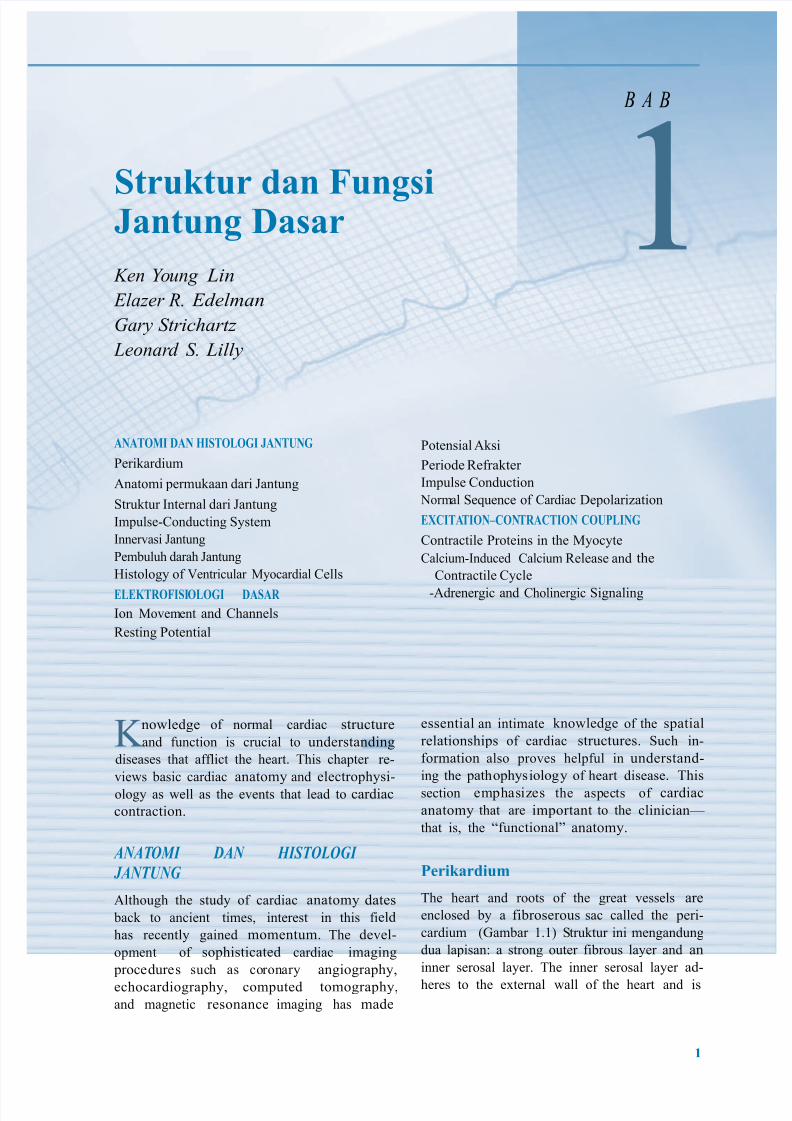
Struktur dan Fungsi Jantung Dasar Ken Y oung Lin Elazer R. Edelman Gary Strichartz Leonar d S. Lilly B A B 1 ANATOMI DAN HISTOLOGI JANTUNG Perikardium Anatomi permukaan dari Jantung Struktur Internal dari Jantung Impulse-Conducting System Innervasi Jantung Pembuluh darah Jantung Histology of entricular !yocardial Cells ELEKTROFISI OLOGI DASAR Ion !ovem ent and Channels "esting Potential no#ledge of normal cardiac structure and function is crucial to understanding diseases that afflict the heart$ %his chapter re- vie#s basic cardiac anatomy and electrophysi- ology as #ell as the events that lead to cardiac contraction$ ANAT OMI DAN HISTOLOGI JANTUNG Although the study of cardiac anatomy dates back to ancient times& interest in this field has recently gained momentum$ %he devel- opment of sophisticated cardiac imaging proce dures such as co ronary angiography& echocardiography& computed tomography& and magnetic resonance imaging has made Potensial Aksi Periode "efrakter Impulse Conduction 'orm al Se(uence of Cardiac )epolari*ation EXCITA TION–CONTRACTION COULING Contractile Proteins in the !yocyte Calcium-Induced Calcium "elease and the Contractile Cycle -Adrenergic and Cholinergic Signaling essential an intimate kno#ledge of the spatial relationships of cardiac structures$ Such in- formation also proves helpful in understand- ing the pathophys iolog y of heart disease$ %his section emphasi*es the aspects of cardiac anatomy that are important to the clinician+ that is& the ,functional anatomy$ !rikardiu" %he heart and roots of the great vessels are enclosed by a fibroserous sac called the peri- cardium ./ambar 1$10 Struktur ini mengandung dua lapisan a strong outer fibrous layer and an inner serosal layer$ %he inner serosal layer ad- heres to the e2ternal #all of the heart and is # 3
-
Upload
thiefeezae -
Category
Documents
-
view
213 -
download
0
Transcript of 6-7 translist
7/24/2019 6-7 translist
http://slidepdf.com/reader/full/6-7-translist 1/8
Struktur dan FungsiJantung Dasar
Ken Young Lin
Elazer R. Edelman
Gary Strichartz
Leonard S. Lilly
B A B
1ANATOMI DAN HISTOLOGI JANTUNG
Perikardium
Anatomi permukaan dari Jantung
Struktur Internal dari Jantung
Impulse-Conducting System
Innervasi Jantung
Pembuluh darah Jantung
Histology of entricular !yocardial Cells
ELEKTROFISIOLOGI DASAR
Ion !ovement and Channels
"esting Potential
no#ledge of normal cardiac structure
and function is crucial to understanding
diseases that afflict the heart$ %his chapter re-
vie#s basic cardiac anatomy and electrophysi-
ology as #ell as the events that lead to cardiac
contraction$
ANATOMI DAN HISTOLOGI
JANTUNG
Although the study of cardiac anatomy dates
back to ancient times& interest in this field
has recently gained momentum$ %he devel-
opment of sophisticated cardiac imaging
procedures such as coronary angiography&
echocardiography& computed tomography&and magnetic resonance imaging has made
Potensial Aksi
Periode "efrakter
Impulse Conduction
'ormal Se(uence of Cardiac )epolari*ation
EXCITATION–CONTRACTION COULING
Contractile Proteins in the !yocyte
Calcium-Induced Calcium "elease and the
Contractile Cycle
-Adrenergic and Cholinergic Signaling
essential an intimate kno#ledge of the spatial
relationships of cardiac structures$ Such in-
formation also proves helpful in understand-
ing the pathophysiology of heart disease$ %his
section emphasi*es the aspects of cardiac
anatomy that are important to the clinician+
that is& the ,functional anatomy$
!rikardiu"
%he heart and roots of the great vessels are
enclosed by a fibroserous sac called the peri-
cardium ./ambar 1$10 Struktur ini mengandung
dua lapisan a strong outer fibrous layer and an
inner serosal layer$ %he inner serosal layer ad-
heres to the e2ternal #all of the heart and is
#
3
7/24/2019 6-7 translist
http://slidepdf.com/reader/full/6-7-translist 2/8
4ungsi dan Struktur Jantung dasar
$
Vena
cava
superior
Vena
cava
inferior
Aorta
Arteri
Pulmonal
Jantungdengan
pericardium
Diafragma
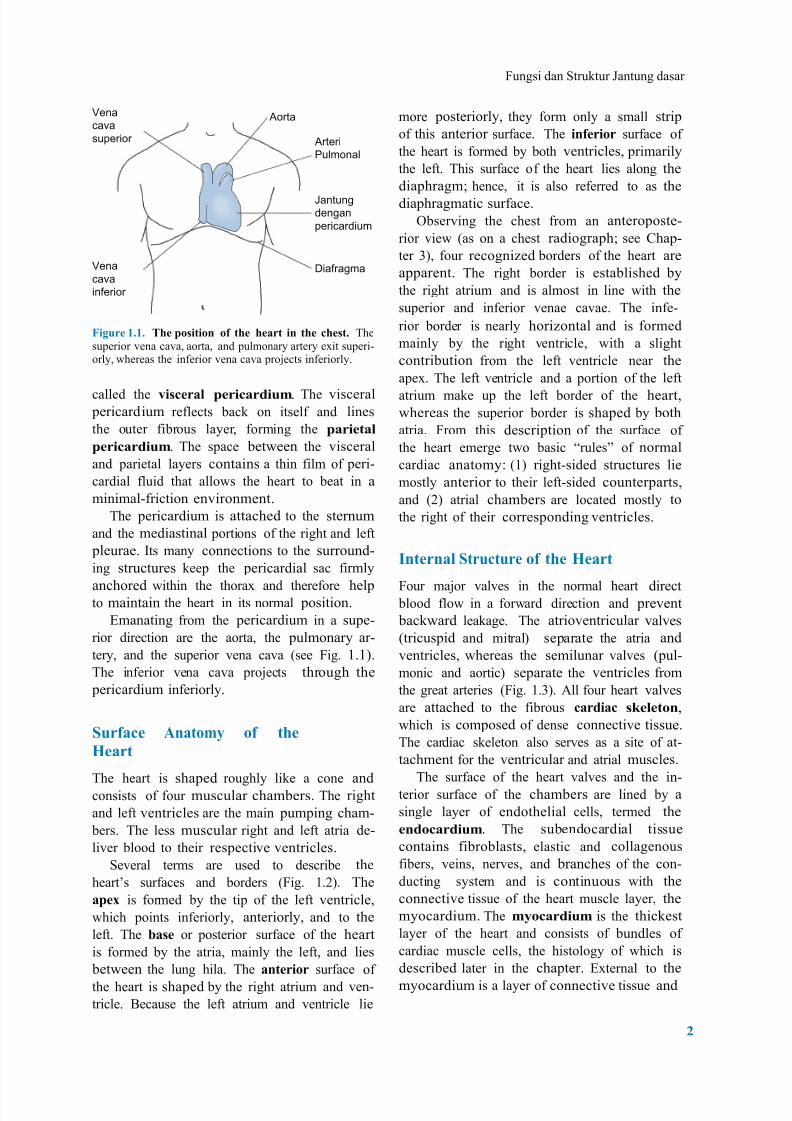
more posteriorly& they form only a small strip
of this anterior surface$ %he in%!ri&r surface of
the heart is formed by both ventricles& primarily
the left$ %his surface of the heart lies along the
diaphragm5 hence& it is also referred to as the
diaphragmatic surface$
6bserving the chest from an anteroposte-
rior vie# .as on a chest radiograph5 see Chap-
ter 70& four recogni*ed borders of the heart are
apparent$ %he right border is established by
the right atrium and is almost in line #ith the
superior and inferior venae cavae$ %he infe-
Figur! #'#' T(! )&siti&n &% t(! (!art in t(! *(!st' %hesuperior vena cava& aorta& and pulmonary artery e2it superi-orly& #hereas the inferior vena cava pro8ects inferiorly$
called the +is*!ra, )!ri*ardiu"$ %he visceral pericardium reflects back on itself and lines
the outer fibrous layer& forming the )ari!ta,
)!ri*ardiu"$ %he space bet#een the visceral
and parietal layers contains a thin film of peri-
cardial fluid that allo#s the heart to beat in a
minimal-friction environment$
%he pericardium is attached to the sternum
and the mediastinal portions of the right and left
pleurae$ Its many connections to the surround-
ing structures keep the pericardial sac firmly
anchored #ithin the thora2 and therefore helpto maintain the heart in its normal position$
9manating from the pericardium in a supe-
rior direction are the aorta& the pulmonary ar-
tery& and the superior vena cava .see 4ig$ 1$10$
%he inferior vena cava pro8ects through the
pericardium inferiorly$
Sur%a*! Anat&"- &% t(!
H!art
%he heart is shaped roughly like a cone and
consists of four muscular chambers$ %he right
and left ventricles are the main pumping cham-
bers$ %he less muscular right and left atria de-
liver blood to their respective ventricles$
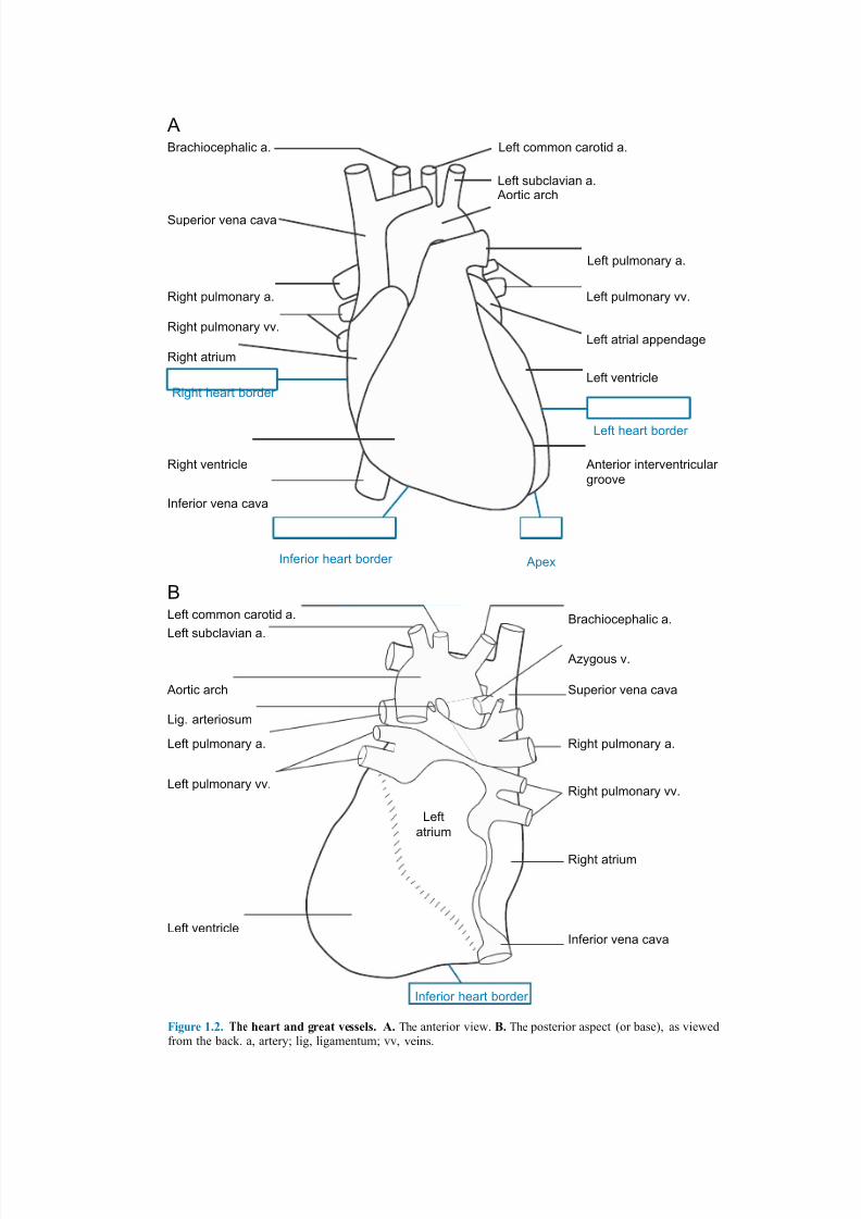
Several terms are used to describe the
heart:s surfaces and borders .4ig$ 1$;0$ %he
a)!. is formed by the tip of the left ventricle&
#hich points inferiorly& anteriorly& and to the
left$ %he /as! or posterior surface of the heart
is formed by the atria& mainly the left& and lies
bet#een the lung hila$ %he ant!ri&r surface of
the heart is shaped by the right atrium and ven-
tricle$ <ecause the left atrium and ventricle lie
rior border is nearly hori*ontal and is formed
mainly by the right ventricle& #ith a slight
contribution from the left ventricle near the
ape2$ %he left ventricle and a portion of the left
atrium make up the left border of the heart&#hereas the superior border is shaped by both
atria$ 4rom this description of the surface of
the heart emerge t#o basic ,rules of normal
cardiac anatomy .10 right-sided structures lie
mostly anterior to their left-sided counterparts&
and .;0 atrial chambers are located mostly to
the right of their corresponding ventricles$
Int!rna, Stru*tur! &% t(! H!art
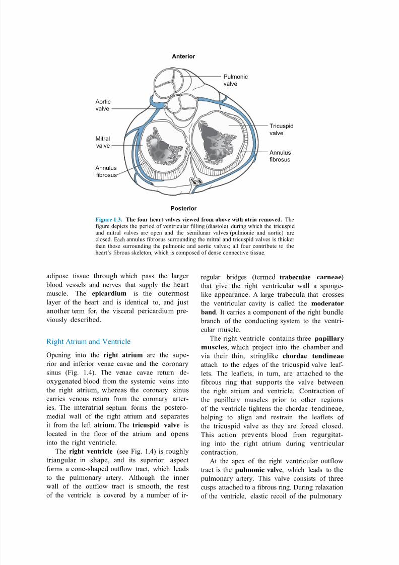
4our ma8or valves in the normal heart direct blood flo# in a for#ard direction and prevent
back#ard leakage$ %he atrioventricular valves
.tricuspid and mitral0 separate the atria and
ventricles& #hereas the semilunar valves .pul-
monic and aortic0 separate the ventricles from
the great arteries .4ig$ 1$70$ All four heart valves
are attached to the fibrous *ardia* sk!,!t&n&
#hich is composed of dense connective tissue$
%he cardiac skeleton also serves as a site of at-
tachment for the ventricular and atrial muscles$
%he surface of the heart valves and the in-
terior surface of the chambers are lined by a
single layer of endothelial cells& termed the
!nd&*ardiu"$ %he subendocardial tissue
contains fibroblasts& elastic and collagenous
fibers& veins& nerves& and branches of the con-
ducting system and is continuous #ith the
connective tissue of the heart muscle layer& the
myocardium$ %he "-&*ardiu" is the thickest
layer of the heart and consists of bundles of
cardiac muscle cells& the histology of #hich is
described later in the chapter$ 92ternal to the
myocardium is a layer of connective tissue and
7/24/2019 6-7 translist
http://slidepdf.com/reader/full/6-7-translist 3/8
ABrachiocephalic a. Left common carotid a.
Left subclavian a.
Superior vena cava
Aortic arch
Left pulmonary a.
Right pulmonary a.
Right pulmonary vv.
Right atrium
Right heart border
Left pulmonary vv.
Left atrial appendage
Left ventricle
Left heart border
Right ventricle
nferior vena cava
Anterior interventricular
groove
nferior heart border
B
Ape!
Left common carotid a.
Left subclavian a.Brachiocephalic a.
A"ygous v.
Aortic arch
Lig. arteriosum
Left pulmonary a.
Left pulmonary vv.
Left ventricle
Left
atrium
Superior vena cava
Right pulmonary a.
Right pulmonary vv.
Right atrium
nferior vena cava
nferior heart border
Figur! #'$' T(! (!art and gr!at +!ss!,s' A' %he anterior vie#$ 0' %he posterior aspect .or base0& as vie#edfrom the back$ a& artery5 lig& ligamentum5 vv& veins$
7/24/2019 6-7 translist
http://slidepdf.com/reader/full/6-7-translist 4/8
Anterior
Pulmonic
valve
Aortic
valve
#itral
valve
Annulus
fibrosus
$ricuspid
valve
Annulus
fibrosus
Posterior
Figur! #'1' T(! %&ur (!art +a,+!s +i!2!d %r&" a/&+! 2it( atria r!"&+!d' %hefigure depicts the period of ventricular filling .diastole0 during #hich the tricuspidand mitral valves are open and the semilunar valves .pulmonic and aortic0 areclosed$ 9ach annulus fibrosus surrounding the mitral and tricuspid valves is thicker than those surrounding the pulmonic and aortic valves5 all four contribute to theheart:s fibrous skeleton& #hich is composed of dense connective tissue$
adipose tissue through #hich pass the larger
blood vessels and nerves that supply the heart
muscle$ %he !)i*ardiu" is the outermostlayer of the heart and is identical to& and 8ust
another term for& the visceral pericardium pre-
viously described$
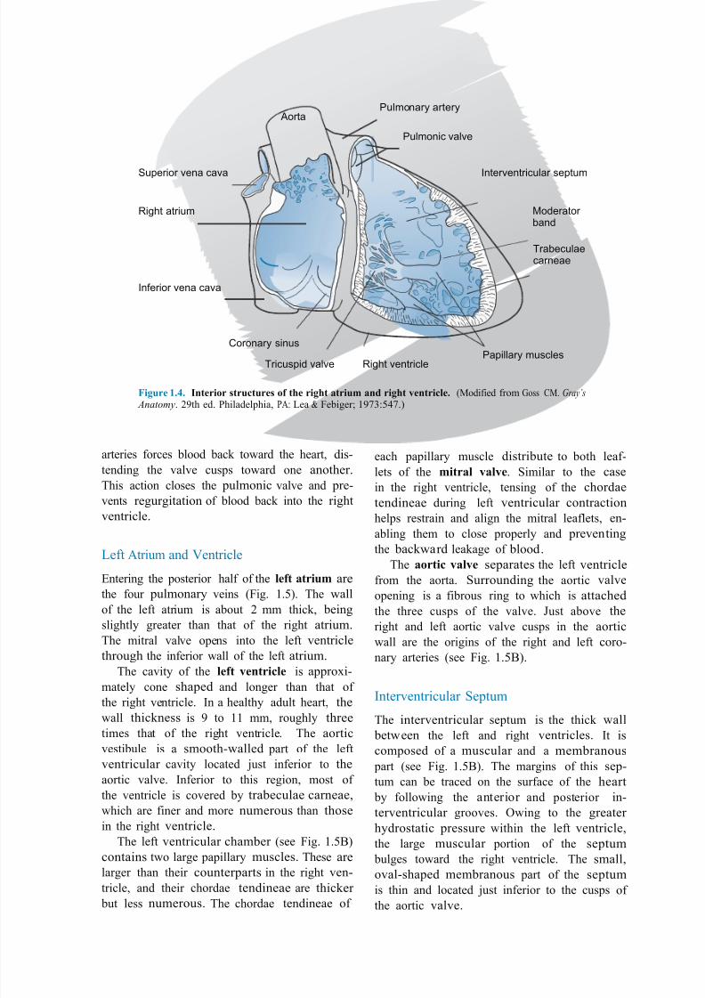
"ight Atrium and entricle
6pening into the rig(t atriu" are the supe-
rior and inferior venae cavae and the coronary
sinus .4ig$ 1$=0$ %he venae cavae return de-
o2ygenated blood from the systemic veins into
the right atrium& #hereas the coronary sinus
carries venous return from the coronary arter-
ies$ %he interatrial septum forms the postero-
medial #all of the right atrium and separates
it from the left atrium$ %he tri*us)id +a,+! is
located in the floor of the atrium and opens
into the right ventricle$
%he rig(t +!ntri*,! .see 4ig$ 1$=0 is roughly
triangular in shape& and its superior aspect
forms a cone-shaped outflo# tract& #hich leads
to the pulmonary artery$ Although the inner
#all of the outflo# tract is smooth& the rest
of the ventricle is covered by a number of ir-
regular bridges .termed tra/!*u,a! *arn!a!0
that give the right ventricular #all a sponge-
like appearance$ A large trabecula that crossesthe ventricular cavity is called the "&d!rat&r
/and$ It carries a component of the right bundle
branch of the conducting system to the ventri-
cular muscle$
%he right ventricle contains three )a)i,,ar-
"us*,!s& #hich pro8ect into the chamber and
via their thin& stringlike *(&rda! t!ndin!a!
attach to the edges of the tricuspid valve leaf-
lets$ %he leaflets& in turn& are attached to the
fibrous ring that supports the valve bet#een
the right atrium and ventricle$ Contraction of
the papillary muscles prior to other regions
of the ventricle tightens the chordae tendineae&
helping to align and restrain the leaflets of
the tricuspid valve as they are forced closed$
%his action prevents blood from regurgitat-
ing into the right atrium during ventricular
contraction$
At the ape2 of the right ventricular outflo#
tract is the )u,"&ni* +a,+!& #hich leads to the
pulmonary artery$ %his valve consists of three
cusps attached to a fibrous ring$ )uring rela2ation
of the ventricle& elastic recoil of the pulmonary
7/24/2019 6-7 translist
http://slidepdf.com/reader/full/6-7-translist 5/8
AortaPulmonary artery
Pulmonic valve
Superior vena cava
Right atrium
nferior vena cava
nterventricular septum
#oderator band
$rabeculaecarneae
%oronary sinus
$ricuspid valve Right ventriclePapillary muscles
Figur! #'3' Int!ri&r stru*tur!s &% t(! rig(t atriu" and rig(t +!ntri*,!' .!odified from /oss C!$ Gray’s Anatomy$ ;>th ed$ Philadelphia& PA1 ?ea @ 4ebiger5 1>A7B=A$0
arteries forces blood back to#ard the heart& dis-
tending the valve cusps to#ard one another$
%his action closes the pulmonic valve and pre-
vents regurgitation of blood back into the right
ventricle$
?eft Atrium and entricle
9ntering the posterior half of the ,!%t atriu" are
the four pulmonary veins .4ig$ 1$B0$ %he #all
of the left atrium is about ; mm thick& being
slightly greater than that of the right atrium$
%he mitral valve opens into the left ventricle
through the inferior #all of the left atrium$
%he cavity of the ,!%t +!ntri*,! is appro2i-
mately cone shaped and longer than that of
the right ventricle$ In a healthy adult heart& the#all thickness is > to 11 mm& roughly three
times that of the right ventricle$ %he aortic
vestibule is a smooth-#alled part of the left
ventricular cavity located 8ust inferior to the
aortic valve$ Inferior to this region& most of
the ventricle is covered by trabeculae carneae&
#hich are finer and more numerous than those
in the right ventricle$
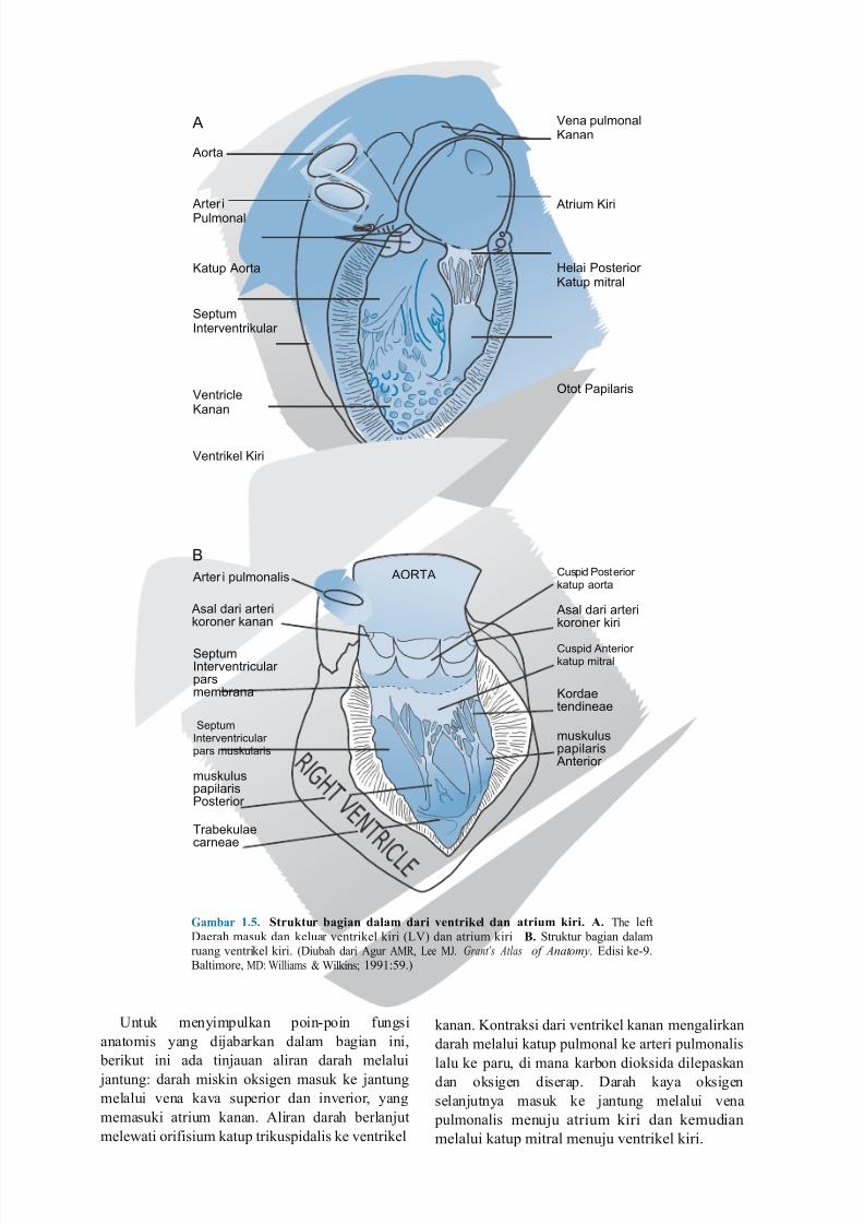
%he left ventricular chamber .see 4ig$ 1$B<0
contains t#o large papillary muscles$ %hese are
larger than their counterparts in the right ven-tricle& and their chordae tendineae are thicker
but less numerous$ %he chordae tendineae of
each papillary muscle distribute to both leaf-
lets of the "itra, +a,+!$ Similar to the case
in the right ventricle& tensing of the chordae
tendineae during left ventricular contraction
helps restrain and align the mitral leaflets& en-abling them to close properly and preventing
the back#ard leakage of blood$
%he a&rti* +a,+! separates the left ventricle
from the aorta$ Surrounding the aortic valve
opening is a fibrous ring to #hich is attached
the three cusps of the valve$ Just above the
right and left aortic valve cusps in the aortic
#all are the origins of the right and left coro-
nary arteries .see 4ig$ 1$B<0$
Interventricular Septum%he interventricular septum is the thick #all
bet#een the left and right ventricles$ It is
composed of a muscular and a membranous
part .see 4ig$ 1$B<0$ %he margins of this sep-
tum can be traced on the surface of the heart
by follo#ing the anterior and posterior in-
terventricular grooves$ 6#ing to the greater
hydrostatic pressure #ithin the left ventricle&
the large muscular portion of the septum
bulges to#ard the right ventricle$ %he small&
oval-shaped membranous part of the septumis thin and located 8ust inferior to the cusps of
the aortic valve$
7/24/2019 6-7 translist
http://slidepdf.com/reader/full/6-7-translist 6/8
A
Aorta
Vena pulmonal&anan
Arter i
Pulmonal
Atrium &iri
&atup Aorta
Septumnterventri'ular
(elai Posterior&atup mitral
Ventricle&anan
)tot Papilaris
Ventri'el &iri
B
Arter i pulmonalis
Asal dari arteri'oroner 'anan
Septumnterventricular parsmembrana
Septumnterventricularpars mus'ularis
mus'uluspapilarisPosterior
$rabe'ulaecarneae
A)R$A %uspid Posterior'atup aorta
Asal dari arteri'oroner 'iri
%uspid Anterior'atup mitral
&ordaetendineae
mus'uluspapilaris
Anterior
Ga"/ar #'4' Struktur /agian da,a" dari +!ntrik!, dan atriu" kiri' A' %he left)aerah masuk dan keluar ventrikel kiri .?0 dan atrium kiri 0' Struktur bagian dalamruang ventrikel kiri$ .)iubah dari Agur A!"& ?ee !J$ Grant’s Atlas of Anatomy$ 9disi ke->$<altimore& !)1 illiams @ ilkins5 1>>1B>$0
Dntuk menyimpulkan poin-poin fungsi
anatomis yang di8abarkan dalam bagian ini&
berikut ini ada tin8auan aliran darah melalui
8antung darah miskin oksigen masuk ke 8antung
melalui vena kava superior dan inverior& yang
memasuki atrium kanan$ Aliran darah berlan8ut
mele#ati orifisium katup trikuspidalis ke ventrikel
kanan$ 3ontraksi dari ventrikel kanan mengalirkan
darah melalui katup pulmonal ke arteri pulmonalis
lalu ke paru& di mana karbon dioksida dilepaskan
dan oksigen diserap$ )arah kaya oksigen
selan8utnya masuk ke 8antung melalui vena
pulmonalis menu8u atrium kiri dan kemudian
melalui katup mitral menu8u ventrikel kiri$
7/24/2019 6-7 translist
http://slidepdf.com/reader/full/6-7-translist 7/8
kontraksi ventrikel kiri memompa darah kaya
oksigen mele#ati katup aorta menu8u aorta& yang
kemudian didistribusikan ke seluruh 8aringan
tubuh$
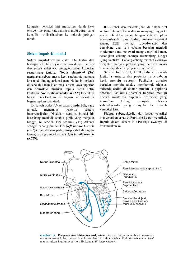
Sist!" I")u,s5K&nduksi
Sistem impuls-konduksi ./br$ 1$E0 terdiri dari
berbagai sel khusus yang memicu denyut 8antung
dan secara kelistrikan mengkoordinasi kontraksi
ruang-ruang 8antung$ N&dus sin&atria, 6SA7
merupakan sebuah massa kecil serabut otot 8antung
khusus di dinding atrium kanan$ 'odus ini terletak
di sebelah kanan 8alan masuk vena kava superior
dan normalnya memicu impuls listrik untuk
kontraksi$ N&dus atri&+!ntriku,ar 6A87 terletak di ba#ah endokardium di bagian inferoposteror
bagian septum interatrial$
)i ba#ah nodus A terdapat /und!, His& yang
terletak menembus posterior septum
interventrikular$ )i dalam septum& bundel his
bercabang men8adi serabut pipih yang men8alar
hingga ke sebelah kiri septum& yang dikenal
sebagai cabang bundel kiri .left bundle branch
(LBB)0$ dan struktur padat mirip kabel di bagian
kanan& cabang bundel kanan .right bundle branch
(BB)0$
"<< tebal dan terletak 8auh di dalam otot
septum interventikular dan meman8ang hingga ke
apeks$ )i dekat persambungan antara septum
interventrikular dan dinding anterior ventrikel
kanan& "<< men8adi subendokartial dan
bercabang dua$ satu cabang ber8alan men8adi
moderator band mele#ati ruang ventrikel kanan&
sedangkan cabang satunya meman8ang hingga
u8ung ventrikel$ Cabang-cabang tersebut akhirnya
men8alar men8adi pleksus yang beranastomosis
dengan rapi di sepan8ang ventrikel kanan$
Secara fungsional& ?<< terbagi men8adi
fasikulus anterior dan posterior serta cabang
kecil menu8u septum$ 4asikulus anterior
ber8alan menu8u apeks& membentuk pleksus
subendokardial di daerah muskulus papilaris
anterior$ 4asikulus posterior ber8alan menu8udaerah muskulus papilaris posterior5 yang
kemudian terbagi men8adi pleksus
subendokardial yang menyebar ke seluruh
ventrikel kiri$
Pleksus subendokardial dari kedua ventrikel
menyebarkan s!ra/ut urkin9! ke otot ventrikel$
Impuls dalam sistem His-Purkin8e a#alnya di
transmisikan ke
*odus Sinoatrial
Sinus %oronaria
*odus Atrioventri'ular
Bundel (is
Right bundle branch
#oderator band
&atup #itral
Pars #embranosa septum 'e V
Bifur'asiobundle (is
Pars #us'ularis
Septum 'e V
Left bundle branch
Serabut Pur'in+e diba,ah endo'ardiummus'ulus papilaris
Ga"/ar #':' K&")&n!n uta"a sist!" k&nduksi 9antung' Sistem ini yaitu nodus sino-atrial&nodus atrioventrikular& bundel His kanan dan kiri& d an serabut Purkin8e$ Moderator and menyalurkan bagian be sar bun dle kanan $ I& interventrikular$

7/24/2019 6-7 translist
http://slidepdf.com/reader/full/6-7-translist 8/8