Thierry Thomas, UnitéINSERM 1059, CHU Saint-Etienne
30
Os & inflammation Os & inflammation Thierry Thomas, UnitéINSERM 1059, CHU Saint-Etienne
Transcript of Thierry Thomas, UnitéINSERM 1059, CHU Saint-Etienne

Os & inflammationOs & inflammation
Thierry Thomas,
Unité INSERM 1059, CHU Saint-Etienne

Conflits d’intérêts
� Interventions ponctuelles
Honoraires en tant quHonoraires en tant qu’’expert ou orateur deexpert ou orateur de
Amgen, GenAmgen, Genéévrier, GSK, Lilly, Merck, Novartis, Serviervrier, GSK, Lilly, Merck, Novartis, Servier
� Intérêts indirects
Soutien financier pour des programmes de rechercheSoutien financier pour des programmes de recherche
ou investigateur de Amgen, Chugaou investigateur de Amgen, Chugaïï, Merck, Novartis, , Merck, Novartis,
Pfizer, Roche, Servier, UCB, WarnerPfizer, Roche, Servier, UCB, Warner--ChilcottChilcott
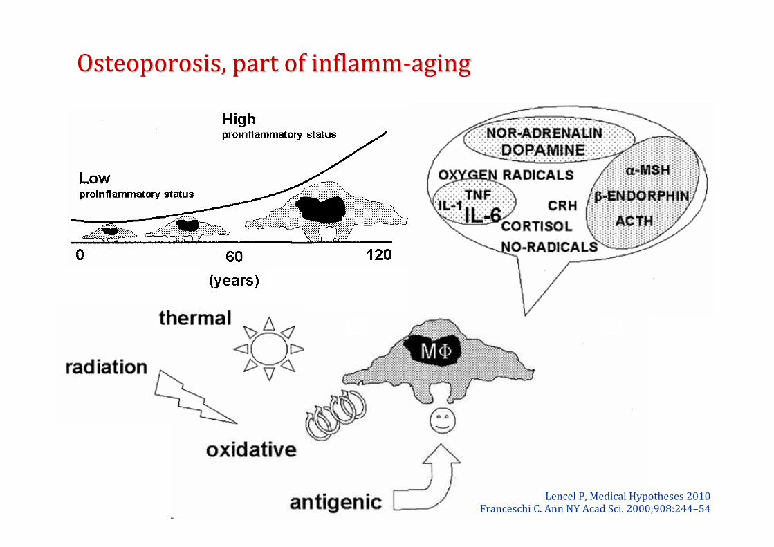
Lencel P, Medical Hypotheses 2010Franceschi C. Ann NY Acad Sci. 2000;908:244–54
Osteoporosis, part of inflammOsteoporosis, part of inflamm--agingaging

D’après Pacifici R. Cellular Immunol 2008
IncreasedIncreasedIncreasedIncreasedFSHFSHFSHFSH
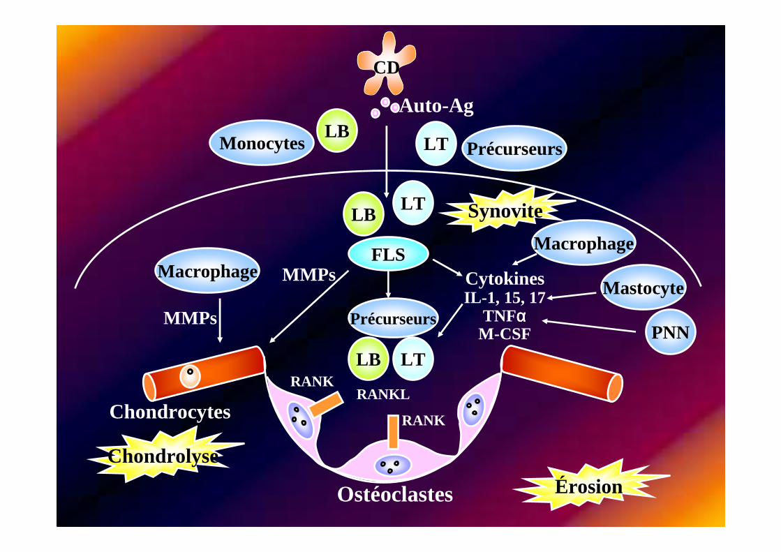
LT
FLS
CD
LB
Ostéoclastes Érosion
Chondrolyse
Synovite
Chondrocytes
LTLB
Macrophage
Macrophage
LTLBRANK
RANK
Auto-Ag
Mastocyte
PNN
CytokinesIL-1, 15, 17
TNFααααM-CSF
MMPs
Monocytes Précurseurs
MMPs Précurseurs
RANKL
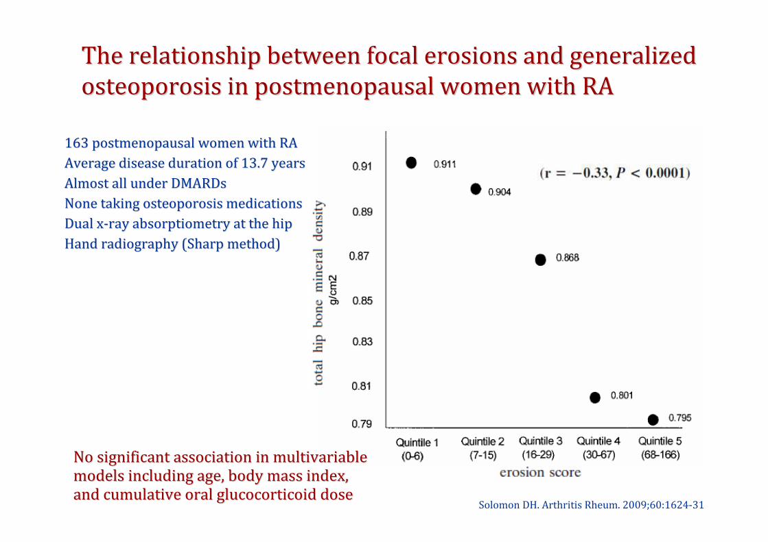
The relationship between focal erosions and generalized The relationship between focal erosions and generalized
osteoporosis in postmenopausal women with RAosteoporosis in postmenopausal women with RA
Solomon DH. Arthritis Rheum. 2009;60:1624-31
163 postmenopausal women with RA163 postmenopausal women with RA
Average disease duration of 13.7 years Average disease duration of 13.7 years
Almost all under DMARDsAlmost all under DMARDs
None taking osteoporosis medications None taking osteoporosis medications
Dual xDual x--ray absorptiometry at the hipray absorptiometry at the hip
Hand radiography (Sharp method)Hand radiography (Sharp method)
No significant association in multivariable No significant association in multivariable
models including age, body mass index,models including age, body mass index,
and cumulative oral glucocorticoid doseand cumulative oral glucocorticoid dose
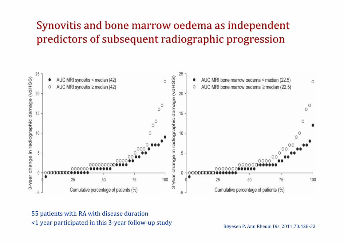
Synovitis and bone marrow oedema as independent Synovitis and bone marrow oedema as independent
predictors of subsequent radiographic progressionpredictors of subsequent radiographic progression
Bøyesen P. Ann Rheum Dis. 2011;70:428-33
55 patients with RA with disease duration55 patients with RA with disease duration
<1 year participated in this 3<1 year participated in this 3--year followyear follow--up studyup study
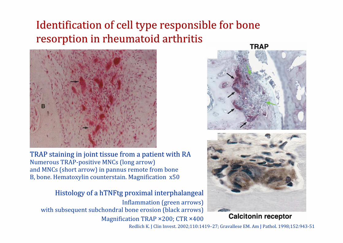
Identification of cell type responsible for boneIdentification of cell type responsible for bone
resorption in rheumatoid arthritisresorption in rheumatoid arthritis
Redlich K. J Clin Invest. 2002;110:1419–27; Gravallese EM. Am J Pathol. 1998;152:943-51
TRAP staining in joint tissue from a patient with RATRAP staining in joint tissue from a patient with RANumerous TRAP-positive MNCs (long arrow)and MNCs (short arrow) in pannus remote from boneB, bone. Hematoxylin counterstain. Magnification x50
Histology of a hTNFtg proximal interphalangealHistology of a hTNFtg proximal interphalangeal
Inflammation (green arrows)with subsequent subchondral bone erosion (black arrows)
Magnification TRAP ×200; CTR ×400
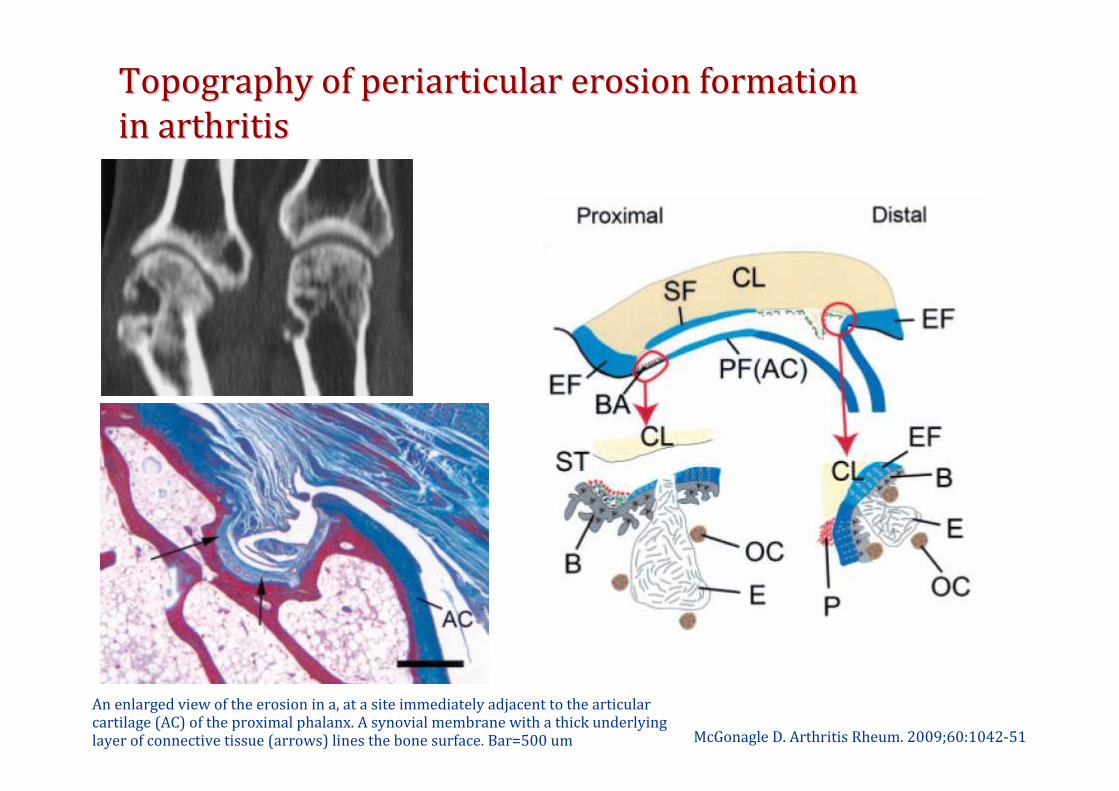
Topography of periarticular erosion formationTopography of periarticular erosion formation
in arthritisin arthritis
McGonagle D. Arthritis Rheum. 2009;60:1042-51
An enlarged view of the erosion in a, at a site immediately adjacent to the articular cartilage (AC) of the proximal phalanx. A synovial membrane with a thick underlying layer of connective tissue (arrows) lines the bone surface. Bar=500 um
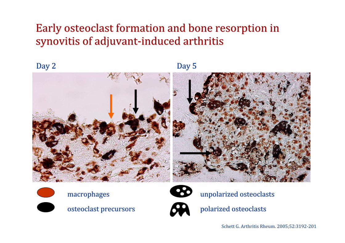
macrophagesmacrophages
osteoclast precursorsosteoclast precursors
Day 2Day 2 Day 5Day 5
unpolarized osteoclastsunpolarized osteoclasts
polarized osteoclastspolarized osteoclasts
Early osteoclast formation and bone resorption in Early osteoclast formation and bone resorption in
synovitis of adjuvantsynovitis of adjuvant--induced arthritisinduced arthritis
Schett G. Arthritis Rheum. 2005;52:3192-201
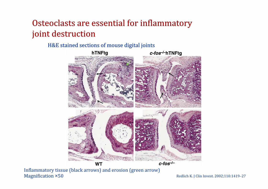
Osteoclasts are essential for inflammatoryOsteoclasts are essential for inflammatory
joint destructionjoint destruction
Redlich K. J Clin Invest. 2002;110:1419–27
Inflammatory tissue (black arrows) and erosion (green arrow)Magnification ×50
H&E stained sections of mouse digital jointsH&E stained sections of mouse digital joints
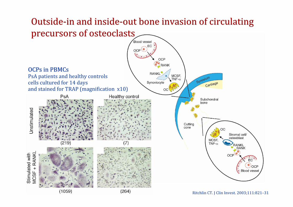
OutsideOutside--in and insidein and inside--out bone invasion of circulating out bone invasion of circulating
precursors of osteoclastsprecursors of osteoclasts
Ritchlin CT. J Clin Invest. 2003;111:821–31
OCPs in PBMCsOCPs in PBMCsPsA patients and healthy controlscells cultured for 14 days and stained for TRAP (magnification x10)
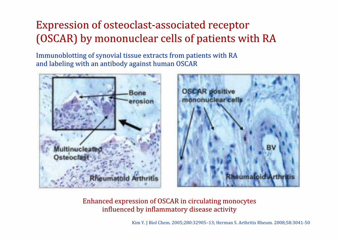
Expression of osteoclastExpression of osteoclast--associated receptor associated receptor
(OSCAR) by mononuclear cells of patients with RA(OSCAR) by mononuclear cells of patients with RA
Kim Y. J Biol Chem. 2005;280:32905–13; Herman S. Arthritis Rheum. 2008;58:3041-50
Immunoblotting of synovial tissue extracts from patients with RAImmunoblotting of synovial tissue extracts from patients with RA
and labeling with an antibody against human OSCARand labeling with an antibody against human OSCAR
Enhanced expression of OSCAR in circulating monocytesEnhanced expression of OSCAR in circulating monocytes
influenced by inflammatory disease activityinfluenced by inflammatory disease activity
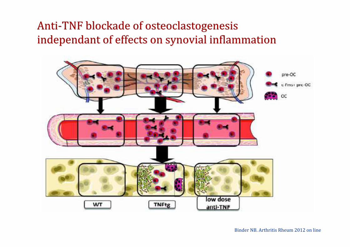
AntiAnti--TNF blockade of osteoclastogenesis TNF blockade of osteoclastogenesis
independant of effects on synovial inflammationindependant of effects on synovial inflammation
Binder NB. Arthritis Rheum 2012 on line

Denosumab treatment effects on structural Denosumab treatment effects on structural
damage in rheumatoid arthritisdamage in rheumatoid arthritis
Cohen SB. Arthritis Rheum. 2008;58:1299-1309
Modified Sharp erosion scoreModified Sharp erosion score Joint space narrowing scoreJoint space narrowing score
DoubleDouble--blind randomized study in RA patients receiving subcutaneous blind randomized study in RA patients receiving subcutaneous
placebo (n=75), denosumab 60 mg (n=71) or denosumab 180 mg (n=72placebo (n=75), denosumab 60 mg (n=71) or denosumab 180 mg (n=72) )
injections every 6 months for 12 monthsinjections every 6 months for 12 months
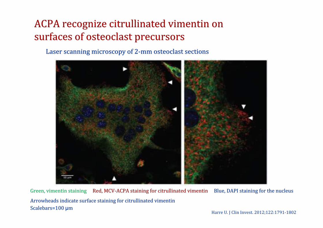
ACPA recognize citrullinated vimentin on ACPA recognize citrullinated vimentin on
surfaces of osteoclast precursorssurfaces of osteoclast precursors
Harre U. J Clin Invest. 2012;122:1791-1802
Arrowheads indicate surface staining for citrullinated vimentinArrowheads indicate surface staining for citrullinated vimentin
Scalebars=100 Scalebars=100 µµmm
Laser scanning microscopy of 2Laser scanning microscopy of 2--mm osteoclast sectionsmm osteoclast sections
Green, vimentin staining Green, vimentin staining Red, MCVRed, MCV--ACPA staining for citrullinated vimentin ACPA staining for citrullinated vimentin Blue, DAPI staining for the nucleusBlue, DAPI staining for the nucleus
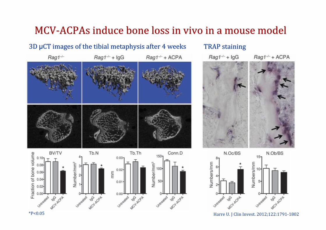
MCVMCV--ACPAs induce bone loss in vivo in a mouse modelACPAs induce bone loss in vivo in a mouse model
Harre U. J Clin Invest. 2012;122:1791-1802*P<0.05
3D 3D µµCT images of the tibial metaphysis after 4 weeksCT images of the tibial metaphysis after 4 weeks TRAP stainingTRAP staining
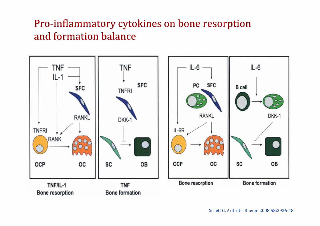
ProPro--inflammatory cytokines on bone resorptioninflammatory cytokines on bone resorption
and formation balanceand formation balance
Schett G. Arthritis Rheum 2008;58:2936-48

Effects of proEffects of pro-- and antiand anti--inflammatory cytokinesinflammatory cytokines
on bone resorptionon bone resorption
Schett G. Eur J Clin Invest. 2011;41:1361-6
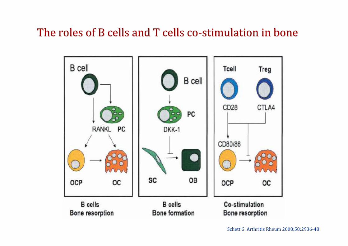
The roles of B cells and T cells coThe roles of B cells and T cells co--stimulation in bonestimulation in bone
Schett G. Arthritis Rheum 2008;58:2936-48
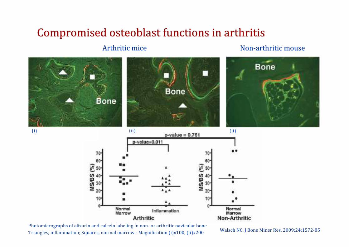
Compromised osteoblast functions in arthritis Compromised osteoblast functions in arthritis
Walsch NC. J Bone Miner Res. 2009;24:1572-85
Arthritic miceArthritic mice NonNon--arthritic mousearthritic mouse
Photomicrographs of alizarin and calcein labeling in non- or arthritic navicular bone
Triangles, inflammation; Squares, normal marrow - Magnification (i)x100, (ii)x200
(i) (ii) (ii)
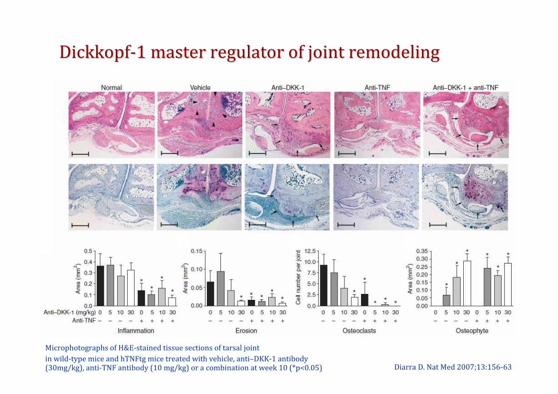
DickkopfDickkopf--1 master regulator of joint remodeling1 master regulator of joint remodeling
Diarra D. Nat Med 2007;13:156-63
Microphotographs of H&E-stained tissue sections of tarsal joint
in wild-type mice and hTNFtg mice treated with vehicle, anti–DKK-1 antibody (30mg/kg), anti-TNF antibody (10 mg/kg) or a combination at week 10 (*p<0.05)

DKKDKK--1 critical for joint bone balance1 critical for joint bone balance
Diarra D. Nat Med 2007;13:156-63
Physiological statePhysiological state ArthritisArthritisArthritis under Arthritis under
DKKDKK--1 blockade1 blockade
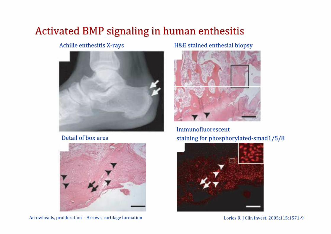
Activated BMP signaling in human enthesitisActivated BMP signaling in human enthesitis
Lories R. J Clin Invest. 2005;115:1571-9
Achille enthesitis XAchille enthesitis X--raysrays H&E stained enthesial biopsyH&E stained enthesial biopsy
Detail of box areaDetail of box area
ImmunofluorescentImmunofluorescent
staining for phosphorylatedstaining for phosphorylated--smad1/5/8 smad1/5/8
Arrowheads, proliferation - Arrows, cartilage formation
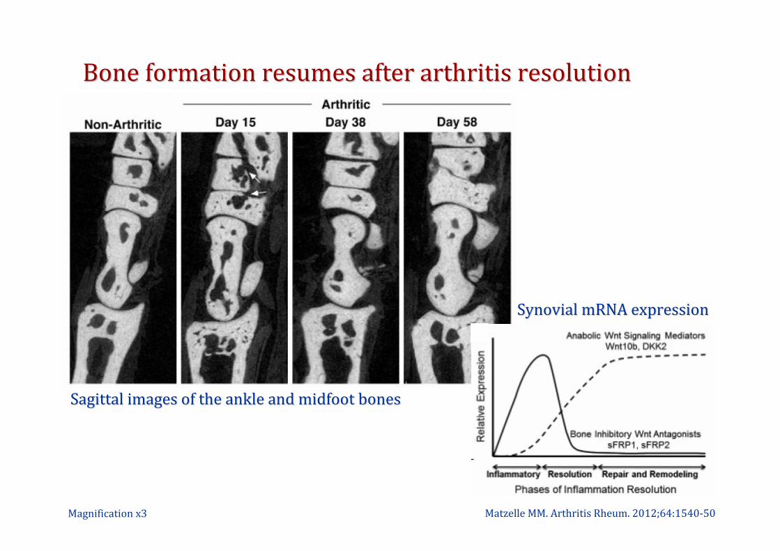
Bone formation resumes after arthritis resolutionBone formation resumes after arthritis resolution
Matzelle MM. Arthritis Rheum. 2012;64:1540-50Magnification x3
Sagittal images of the ankle and midfoot bonesSagittal images of the ankle and midfoot bones
Synovial mRNA expressionSynovial mRNA expression

Local and systemic bone events in chronic inflammation Local and systemic bone events in chronic inflammation
and therapeutic optionsand therapeutic options
Redlich K & Smolen JS. Nat Rev Drug Discov. 2012;11:234-50

Back upBack up
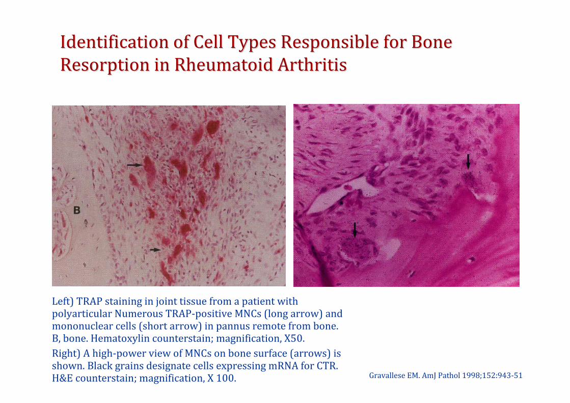
Identification of Cell Types Responsible for BoneIdentification of Cell Types Responsible for Bone
Resorption in Rheumatoid ArthritisResorption in Rheumatoid Arthritis
Gravallese EM. AmJ Pathol 1998;152:943-51
Left) TRAP staining in joint tissue from a patient with polyarticular Numerous TRAP-positive MNCs (long arrow) and mononuclear cells (short arrow) in pannus remote from bone. B, bone. Hematoxylin counterstain; magnification, X50.
Right) A high-power view of MNCs on bone surface (arrows) is shown. Black grains designate cells expressing mRNA for CTR. H&E counterstain; magnification, X 100.
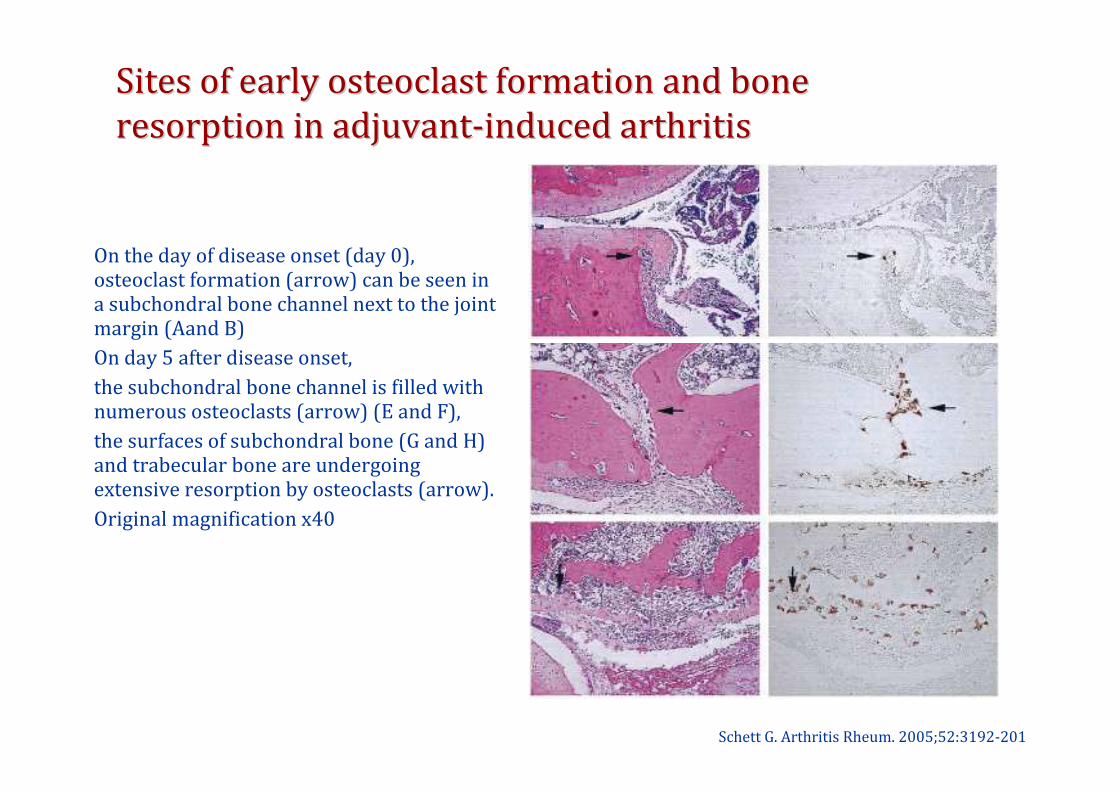
Sites of early osteoclast formation and bone Sites of early osteoclast formation and bone
resorption in adjuvantresorption in adjuvant--induced arthritisinduced arthritis
Schett G. Arthritis Rheum. 2005;52:3192-201
On the day of disease onset (day 0), osteoclast formation (arrow) can be seen in a subchondral bone channel next to the joint margin (Aand B)
On day 5 after disease onset,
the subchondral bone channel is filled with numerous osteoclasts (arrow) (E and F),
the surfaces of subchondral bone (G and H) and trabecular bone are undergoing extensive resorption by osteoclasts (arrow).
Original magnification x40
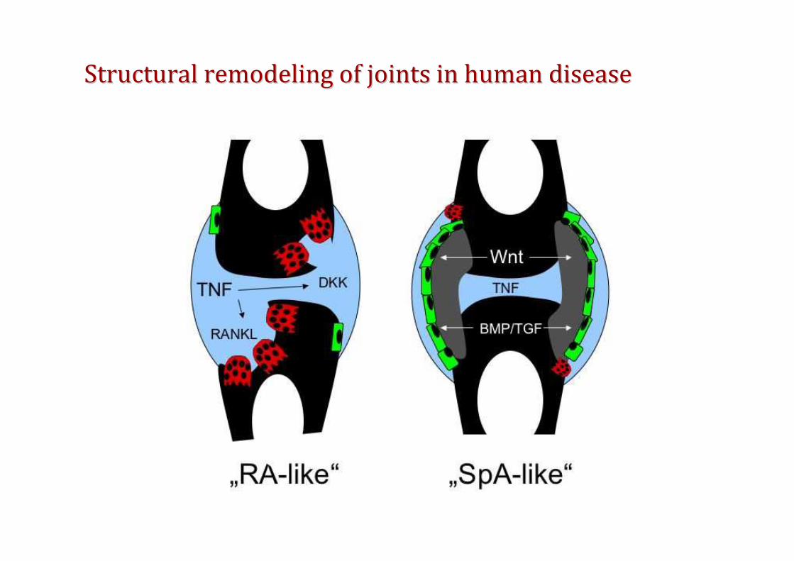
Structural remodeling of joints in human diseaseStructural remodeling of joints in human disease