
STATE RELATED QUANTITATIVE · PDF fileSamyn, Inge Schietecatte, Justina Sidlauskaite, ... Sara...
268
STATE-RELATED QUANTITATIVE ELECTROENCEPHALOGRAPHY IN ATTENTION-DEFICIT/HYPERACTIVITY DISORDER Inez Buyck
Transcript of STATE RELATED QUANTITATIVE · PDF fileSamyn, Inge Schietecatte, Justina Sidlauskaite, ... Sara...
STATE-RELATED QUANTITATIVE ELECTROENCEPHALOGRAPHY
IN ATTENTION-DEFICIT/HYPERACTIVITY DISORDER
Inez Buyck

State-related quantitative electroencephalography in attention-deficit/hyperactivity disorder
Inez Buyck
Promotor: Prof. dr. Jan R. Wiersema
Proefschrift ingediend tot het behalen van de academische graad
van Doctor in de Psychologie
2014

DANKWOORD/ACKNOWLEDGEMENTS
Samen staan we sterk
Niemand kan alleen
Je bouwt een kathedraal
Met velen om je heen
Stef Bos
Bouwen aan een proefschrift is onmogelijk zonder de hulp en steun van een heleboel
mensen. Graag wil ik een woord van dank richten aan allen die me de afgelopen jaren
hebben bijgestaan om de plannen te tekenen, de fundamenten te leggen, de correcte
metseltechnieken te gebruiken, de bouwstenen stevig aan elkaar te voegen, en de
zonnigste weersomstandigheden te creëren.
In de eerste plaats wil ik mijn promotor prof. dr. Roeljan Wiersema bedanken, die me de
ruimte en vrijheid heeft gegeven om mijn eigen weg te zoeken en me daarbij steeds met
raad en daad bijstond. Hij heeft me met zijn EEG- en onderzoeksexpertise veel
bijgebracht en zijn inspirerende stimulansen en kritische geest hebben mijn werk naar
een hoger niveau getild.
Prof. dr. Herbert Roeyers wil ik van harte danken voor de kans die hij me geboden heeft
om deze uitdaging aan te gaan. Ik heb zijn betrokkenheid en advies bij het
doctoraatsproject erg gewaardeerd en als een flinke ruggensteun aangevoeld.
Tevens wil ik mijn dank betuigen aan prof. dr. Marina Danckaerts en prof. dr. Gilles
Pourtois. Hun deskundige opmerkingen en suggesties tijdens de jaarlijkse bijeenkomsten
van de begeleidingscommissie hebben me er steeds toe aangezet om de kwaliteit van
mijn werk aan te scherpen.

Sylvie Verté, Helen Tibboel en Baris Metin waren een rots in de branding bij het
programmeren van de computertaken die tijdens de studies werden afgenomen.
Monica Dhar, Valentina Rossi, Filipa Viola, Markus Plank, Durk Talsma en Stijn Massar
ben ik erg dankbaar voor hun waardevolle ondersteuning bij het verwerken en
analyseren van de EEG-data. Voor advies over statistische analyses kon ik altijd
aankloppen bij Jan Lammertyn. Martijn Arns wil ik graag danken om me wegwijs te
maken in de neurofeedbacktherapie.
Ik had de voorbije jaren het geluk om deel uit te maken van een onderzoeksteam waarin
een goede collegiale samenwerking hoog in het vaandel gedragen wordt. Hiervoor wil ik
Jannes Baert, Chloè Bontinck, Annelies Ceulemans, Ellen Demurie, Mie Dereu, Leen De
Schuymer, Annemie Desoete, Nele Dewaele, Frauke De Weerdt, Isabel Gabriëls, Roos
Gasthuys, Elke Godefroid, Tinneke Hellinckx, Mieke Meirsschaut, Baris Metin, Tinneke
Moyson, Annabel Nijhof, Stefanie Pieters, Ruth Raymaekers, Lieselot Ruysschaert, Vicky
Samyn, Inge Schietecatte, Justina Sidlauskaite, Martine Thys, Daisy Titeca, Sara Van der
Paelt, Valerie Van Cauwenberge, Severine Van De Voorde, Liedewij Verhaeghe, Julie
Vermeirsch en Petra Warreyn van harte danken.
Bij de realisatie van dit doctoraatsproject waren ook heel wat studenten betrokken. Een
welgemeende dank aan Elke Baten, Hanna Bil, Eefke Cailliau, Sara Debruyne, Kaat
Defreyne, Tessa Devriese, Lore Goudesone, Sien Hoorens, Karolien Kennis, Java Labeau,
Nathalie Patty - van Doorn, Isabelle Tack, Rebecca Van Caekenberghe, Eline
Vandendriessche en Sara Woods voor de hulp bij het rekruteren van
onderzoeksparticipanten en de praktische ondersteuning bij het onderzoek.
Gedurende de afgelopen jaren hebben verschillende personen inspanningen geleverd
om allerlei praktische en administratieve zaken in goede banen te leiden. Dank hiervoor
aan Annick Van der Cruyssen, Sylvie Van Overmeeren, Wouter Bosmans, Antoine
Tavernier, Pascal Mestdagh, Kurt Killemaes, Ellen Van Glabeke en Marc Covents.

In het bijzonder wil ik ook prof. dr. Geert Crombez bedanken voor het vakkundig leiden
van de vakgroep, en het bieden van de mogelijkheden om dit doctoraatsproject uit te
werken en de onderzoeksresultaten te presenteren op verschillende congressen. In dit
verband gaat mijn dank ook uit naar de decaan prof. dr. Geert De Soete voor de
bijkomende financiële ondersteuning van mijn congresdeelnames.
Dit proefschrift zou nooit tot stand kunnen gekomen zijn zonder de vele kinderen,
ouders en volwassenen die belangeloos meegewerkt hebben aan de studies. De dienst
kinderneurologie van het Universitair Ziekenhuis van Gent, Sig vzw, centrum Zit Stil, vzw
Aandacht en de verschillende scholen en centra voor ambulante revalidatie die
betrokken waren bij het werven van onderzoeksparticipanten verdienen in dit kader ook
een speciaal woord van dank.
Tot slot zijn er heel wat mensen die me van op de zijlijn door dik en dun gesteund
hebben. Zij staan al jaren altijd voor me klaar en hebben een heel speciaal plekje in mijn
hart. In de eerste plaats denk ik hierbij aan mijn ouders John Buyck en Bea Decuypere,
mijn zus Birgit Buyck, mijn partner Danny Sandra, en mijn oom en tante Jean-Marie
Decuypere en Heidi van Lier. Verder hebben mijn vriend(inn)en Joke Langmans,
Christine Buyse, Jan Declercq, Laurence Anne Van de Goor, Evita Clauwaert, Sofie
Deman en Bert Vanrijckeghem veel voor mij betekend tijdens deze reis.
Inez Buyck
april 2014

TABLE OF CONTENTS
Chapter 1 General introduction 1
Chapter 2 Resting EEG in ADHD: developmental course and diagnostic value 43
Chapter 3 EEG activity before and after cognitive effort in children with ADHD 69
Chapter 4 State-related EEG deviances in ADHD 103
Chapter 5 Differential effects of pink noise on the EEG of adults with ADHD 131
Chapter 6 Task-related EEG deviances in adults with ADHD 159
Chapter 7 General discussion 195
Nederlandstalige samenvatting 239

1
GENERAL INTRODUCTION
Attention‐deficit/hyperactivity disorder (ADHD) is a common neurodevelopmental
disorder that often persists across the lifespan and has a severe impact on social,
academic and occupational functioning. In the past decades, quantitative
electroencephalography (EEG) has frequently been used to investigate the neural
correlates of ADHD. The goal of this dissertation is to gain more insights into the nature
and significance of EEG deviances in ADHD. While a myriad of studies have
demonstrated that behavioural and cognitive deficits in ADHD largely depend on
contextual and state factors, this has hardly been addressed in EEG research. Therefore,
a particular focus of this dissertation is on state‐related EEG in ADHD. In this chapter,
first some information is given on the characteristics of ADHD, the aetiological factors
that may be involved in the disorder and the neuropsychological theories explaining
ADHD deficits. Then, an overview of EEG findings in ADHD is provided and the EEG‐
based models that have been developed to interpret EEG deviances in ADHD are shortly
reviewed. Next, the clinical implications of EEG findings in ADHD are addressed.
Subsequently, some possible explanations of inconsistent EEG findings between studies
are discussed. Finally, the research objectives are presented and an overview of the
chapters included in this dissertation is given.
CHAPTER
CHAPTER 1
2
ATTENTION-DEFICIT/HYPERACTIVITY DISORDER
Definition
Attention-deficit/hyperactivity disorder (ADHD) is a neurodevelopmental
disorder that is characterized by a developmentally inappropriate pattern of inattention
and/or hyperactivity-impulsivity. Based on criteria established by the Diagnostic and
Statistical Manual of Mental Disorders- fourth edition, text revision (DSM-IV-TR,
American Psychiatric Association (APA), 2000), three subtypes of ADHD are specified: (a)
the predominantly inattentive type (b) the predominantly hyperactive/impulsive type,
and (c) the combined type. The symptoms have to be present for at least six months
and some of them must have been expressed before the age of seven. In addition, the
symptoms are required to be observed in two or more settings and to cause significant
impairment in social, academic or occupational functioning. The manifestation of the
disorder typically fluctuates depending on contextual factors within a given setting. That
is, individuals usually express less (or even no) problems when they are frequently
getting rewarded for appropriate behaviour, receive consistent external stimulation,
undertake interesting activities, are in a novel setting, are closely supervised, and during
one-on-one interactions (APA, 2000).
All participants with ADHD in our studies received a diagnosis according to the
DSM-IV-TR criteria. Meanwhile, a new edition of the DSM has been published (APA,
2013). While in the DSM-IV-TR, ADHD is categorized as a “disorder usually first
diagnosed in infancy, childhood, or adolescence”, in the DSM-5, it is classified as a
“neurodevelopmental disorder”. The diagnostic criteria for ADHD remained largely the
same in the new publication, except for the substitution of subtypes by presentation
specifiers that actually are highly equivalent to the prior subtypes. Further, the age
criterion has been raised to having several symptoms present before the age of 12 and
diagnostic criteria for adults have been adapted in the DSM-5 (APA, 2013). Research has
GENERAL INTRODUCTION
3
documented that a substantial number of children continue to meet DSM criteria for
ADHD in adolescence (e.g., Barkley, Fischer, Edelbrock, & Smallish, 1990; Bussing,
Mason, Bell, Porter, & Garvan, 2010) and adulthood (e.g., Klein et al., 2012; Lara et al.,
2009). Yet, the prominent behavioural manifestations of hyperactivity in childhood
often shift to a more subtle expression of symptoms in adulthood (e.g., Faraone,
Biederman, & Mick, 2006; Kooij et al., 2010; Spencer, Biederman, & Mick, 2007).
Prevalence
The worldwide prevalence rates of ADHD across the lifespan are estimated
between 5 to 7% (Willcutt, 2012). Within general population samples, the inattentive
type is the most prevalent, while the combined subtype is more often represented in
clinical samples (Willcutt, 2012). A higher prevalence is detected in boys than in girls,
with a male-to-female ratio of 3:1 in community samples and 9:1 in clinic-referred
samples (Biederman et al., 2002).
Comorbidity with other disorders
Through the lifespan, comorbidity with other psychiatric disorders and learning
disabilities is high. Disorders that commonly have been found to co-occur with ADHD
include oppositional defiant disorder (ODD), conduct disorder (CD), mood disorders,
anxiety disorders, Tourette syndrome, autism spectrum disorder, motor incoordination,
language disorders, dyslexia and dyscalculia (Biederman & Faraone, 2005; Daley, 2006;
Spencer et al., 2007; Voeller,2004).
CHAPTER 1
4
AETIOLOGY OF ADHD
The aetiology of ADHD is considered to be complex and multifactorial, with
several genetic, environmental and neurobiological factors contributing to the disorder.
Genetic factors
Genetic studies estimate the heritability of ADHD to be approximately 76% (for a
review, see Faraone et al., 2005). Candidate gene studies revealed that the genes with
the most consistent evidence to be associated with ADHD are the dopamine D4 and D5
receptors, the dopamine active transporter 1, the serotonin transporter 5HTT, the
serotonin 1B receptor and the synaptosomal-associated protein 25 (for reviews, see
Gizer, Ficks, & Waldman, 2009 and Thapar, Cooper, Eyre, & Langley, 2013). In addition,
the latrophilin 3 and cadherin 13 genes have been related to adults with persistent
ADHD (for a review, see Franke et al., 2012). Although support has been found for
(variants of) these genes to be involved in ADHD, so far no significant genome-wide
associations with ADHD could be identified (for a review, see Ashmore & Cheng, 2013;
for a meta-analysis, see Neale et al., 2010). This suggests that the impact of individual
genes on the aetiology of ADHD is small or that rare types of genetic variants explain the
heritability of the disorder (Ashmore & Cheng, 2013; Neale et al., 2010).
Environmental factors
Several environmental factors have been linked to the aetiology of ADHD. The
most frequently reported risk factors are maternal smoking, alcohol and substance
abuse during pregnancy, prematurity and low birth weight, childhood illnesses, exposure
to environmental toxins, low family income, low parental education, disturbances in
family functioning, disrupted parent-child relations and exposure to maternal
GENERAL INTRODUCTION
5
psychopathology (e.g., Biederman et al., 1995; Biederman & Faraone, 2005; Cortese,
2012; Nigg, Nikolas, Knottnerus, Cavanagh, & Friderici, 2010; Thapar, Cooper, Jefferies,
& Stergiakouli, 2012; Thapar et al., 2013). However, none of these factors have been
proven to play a definite causal role in ADHD (Cortese, Faraone, & Sergeant, 2012).
Moreover, the gene-environment interaction also seems to be important in the
aetiology of ADHD (Nigg, 2012; Pennington et al., 2009).
Neurobiological factors
Neuroimaging studies exploring the neurobiological profile of ADHD have
identified altered structure and function in several brain regions, including fronto-
striatal, fronto-parietal, fronto-cerebellar neural networks, as well as lateral
orbitofrontal-ventromedial networks (e.g., Arnsten & Rubia, 2012; Cubillo, Halari, Smith,
Taylor, & Rubia, 2012). Functional abnormalities are not only detected in isolated brain
regions, also the functional interconnectivity between regions appears to be impaired in
ADHD (e.g., Purper-Ouakil, Ramoz, Lepagnol-Bestel, Gorwood, & Simonneau, 2011;
Rubia, Cubillo, Woolley, Brammer, & Smith, 2010).
NEUROPSYCHOLOGICAL THEORIES ON THE AETIOLOGY OF ADHD
Several theoretical models have been developed to provide a comprehensive
account of the behavioural and cognitive deficits associated with ADHD. The most
influential and well-investigated accounts are outlined below.
Executive dysfunction account
In the executive dysfunction theory (Barkley, 1997), a deficiency in executive
functioning is proposed to underpin ADHD. Executive functions refer to a set of top-
down cognitive processes that are coordinated in order to achieve self-control and goal-
CHAPTER 1
6
directed behaviour (Castellanos, Sonuga-Barke, Milham, & Tannock, 2006). According to
the model, deficient inhibitory control is the primary deficit in ADHD, in turn
contaminating other executive functions (i.e., working memory, self-regulation of affect,
motivation and arousal, internalization of speech, and reconstitution) (Barkley, 1997).
Although a few meta-analyses have confirmed that individuals with ADHD show
impairments in several domains of executive functioning, effect sizes were moderate
and executive dysfunction was not found to be a characteristic of all individuals with
ADHD (Boonstra, Oosterlaan, Sergeant, & Buitelaar, 2005; Willcutt, Doyle, Nigg, Faraone,
& Pennington, 2005). Moreover, while in this theory, executive dysfunction tends to be
regarded as a fixed deficit in ADHD, this appears to disaccord with research
demonstrating executive functions themselves to be dynamic processes and
performance on executive tasks in ADHD to vary across states and settings (e.g., Nigg, &
Casey, 2005; Shiels et al., 2008).
In line with findings demonstrating the dynamic nature of impairment in ADHD,
alternative theoretical accounts have been developed, emphasizing the role of
contextual and state factors in defining deficits (Sonuga-Barke, Wiersema, van der
Meere, & Roeyers, 2010a).
Delay aversion account
In the delay aversion theory (Sonuga-Barke, Taylor, Sembi, & Smith, 1992), the
context dependent nature of ADHD is defined from a motivational perspective. In
specific, the model postulates that an impaired signalling of future rewards underlies
deficits in ADHD, resulting in a drive for immediate rewards (Sonuga-Barke et al., 2010a).
This is particularly expressed in a preference for instant over delayed rewards (Sonuga-
Barke et al., 1992) and a smaller investment of time and effort in long and boring tasks
to avoid or escape from delay (Sonuga-Barke, De Houwer, De Ruiter, Azensten, &
Holland, 2004).
GENERAL INTRODUCTION
7
Research findings support the notion that delay aversion is implicated in ADHD
but, as is the case for weaknesses in executive functioning, it is neither a necessary nor a
sufficient cause of ADHD in all individuals (Sonuga-Barke, Sergeant, Nigg, & Willcutt,
2008).
State regulation account
The state regulation account (Sergeant, 2005; van der Meere, 2005) found its
origin in the cognitive-energetic model of Sanders (1983), which postulates that human
processing capacity is determined by the interplay between processing stages (i.e.,
encoding, feature extraction, response selection and response execution) and energetic
state factors (i.e., arousal, activation and effort). The model accentuates that context
factors largely account for the variable expression of deficits in ADHD and that
impairments in ADHD result from a deficiency to regulate energetic state in challenging
suboptimal conditions (Sonuga-Barke et al., 2010a).
Research into this theoretical construct has mainly focussed on evaluating task
performance while manipulating presentation rate of stimuli, as this is believed to affect
activation level (Johnson, Wiersema, & Kuntsi, 2009). In accordance with the model,
numerous studies using a large variety of tasks have demonstrated worse performance
in children with ADHD in tasks with slow and fast event rates compared to moderate
event rates (e.g., Brown & Vickers, 2004; Leung, Leung, & Tang, 2000; for a meta-
analysis, see Metin, Roeyers, Wiersema, van der Meere, & Sonuga-Barke, 2012; Rubia,
Taylor, Taylor, & Sergeant, 1999; Scheres, Oosterlaan, & Sergeant, 2001; Wiersema, van
der Meere, Antrop, & Roeyers, 2006a; Wiersema, van der Meere, Roeyers, Van Coster, &
Baeyens, 2006b). Further support for the notion of state regulation deficits in slow
event rate conditions can be derived from cardiac response and electrophysiological
studies, which have described difficulties with the regulation of effort allocation in ADHD
(Börger & van der Meere, 2000; Wiersema et al., 2006a,b).
CHAPTER 1
8
From single core deficit to multiple pathways
Although the aforementioned single core deficit models each provide a valuable
contribution to explaining some of the problems related to ADHD, none of the
theoretical constructs are successful in accounting for the entire spectrum of deficits in
ADHD (e.g., Sonuga-Barke, 2002). Consequently, it was recognised that ADHD is a very
heterogeneous disorder with multiple causal and developmental pathways (e.g., Nigg,
2005a; Sonuga-Barke, 2002; Sonuga-Barke, Bitsakou, & Thompson, 2010b).
ELECTROENCEPHALOGRAPHY
Electroencephalographic measurements were among the first to be used to
investigate the neural correlates of ADHD (Loo & Barkley, 2005) and have been used as
such for over 40 years.
Electroencephalography (EEG) is a non-invasive imaging technique to monitor
brain electrical activity (Teplan, 2002). EEG measures voltage fluctuations on the scalp,
resulting from synchronous activity of large collections of neurons in the brain (Başar &
Güntekin, 2008). The procedure offers excellent temporal but rather poor spatial
resolution because electrical activity recorded from the scalp is not always directly
mapped to a specific underlying brain structure and may be contaminated by various
electrical artifacts (Loo & Barkley, 2005).
EEG activity consists of oscillations at different frequencies (i.e., number of
oscillations within a given time period), which are generally categorised in different
frequency ranges: delta (<4 Hz), theta (4-7 Hz), alpha (8-12 Hz), beta (13-25 Hz), and
gamma (25-50 Hz) (Loo & Makeig, 2012). In ADHD research, the most commonly
studied frequency bands are the theta, alpha and beta band. In addition, the theta/beta
ratio (TBR), which reflects the proportion of EEG activity in the theta to that in the beta
frequency band, is well-investigated in ADHD.
GENERAL INTRODUCTION
9
EEG FINDINGS IN ADHD
Most EEG studies in ADHD have evaluated brain wave activity under resting state
conditions with either eyes closed or eyes open. The majority of them focussed on
children, while only a few studies examined EEG activity in adolescents. As for adults,
there has been a growing number of publications in recent years.
Across the lifespan, the most consistent findings in both eyes closed and eyes
open resting conditions refer to increased theta activity and/or an enhanced TBR in
ADHD (see Barry & Clarke, 2009 and Barry, Clarke, & Johnstone, 2003 for reviews; Clarke
et al., 2011; Dupuy, Clarke, Barry, McCarthy, & Selikowitz, 2011; Dupuy, Barry, Clarke,
McCarthy, & Selikowitz, 2013; González-Castro, Rodríguez, López, Cueli, & Álvarez, 2013;
Koehler et al., 2009; Lansbergen, Arns, van Dongen-Boomsma, Spronk, & Buitelaar,
2011; Loo et al., 2010; Shi et al., 2012; Woltering, Jung, Liu, & Tannock, 2012). Yet,
several studies were not able to invariably replicate these findings (Coolidge, Starkey, &
Cahill, 2007; Liechti et al., 2013; Loo et al., 2009, 2013; Nazari, Wallois, Aarabi, &
Berquin, 2011; Ogrim, Kropotov, & Hestad, 2012; Poil et al., 2014; Swartwood,
Swartwood, Lubar, & Timmermann, 2003; van Dongen-Boomsma et al., 2010), which
questions the robustness of these deviances characterizing individuals with ADHD.
Additionally, some recent studies established that enhanced theta or TBR is only
apparent in about 20 to 60% of children with ADHD (Arns, Drinkenburg, & Kenemans,
2012; Clarke et al., 2011; Ogrim et al., 2012). Moreover, theta power (Clarke et al.,
2011) and TBR (Loo et al., 2013) can even be reduced in some individuals with ADHD.
EEG findings regarding brain wave activity in the faster frequency bands appear to be
even more mixed. That is, while some studies have documented decreased power in the
beta band in ADHD (Clarke, Barry, McCarthy, & Selikowitz, 1998; Clarke, Barry,
McCarthy, & Selikowitz, 2002; Clarke et al., 2003a, 2008; Dupuy et al., 2011; Hermens et
al., 2004; Shi et al., 2012), others were not able to distinguish groups based on beta
power (Bresnahan & Barry, 2002; Hobbs, Clarke, Barry, McCarthy, & Selikowitz, 2007;

CHAPTER 1
10
Koehler et al., 2009; Liechti et al., 2013; Loo et al., 2010, van Dongen-Boomsma et al.,
2010). In addition, a few studies have even reported increased beta power in (a
subgroup of) individuals with ADHD (Chabot & Serfontein, 1996; Clarke et al., 1998;
Clarke, Barry, McCarthy, & Selikowitz, 2001a; Clarke et al., 2008; Loo et al., 2010; Poil et
al., 2014). Likewise, alpha power has often been documented to be reduced in
individuals with ADHD (Barry, Clarke, Johnstone, McCarthy, & Selikowitz, 2009; Clarke et
al., 1998; Clarke, Barry, McCarthy, & Selikowitz, 2002a; Clarke et al., 2003a; Dupuy et al.,
2011; Loo et al., 2009; Woltering et al., 2012), but has also been found to be increased
(Chabot & Serfontein, 1996; Clarke et al., 2011; Koehler et al., 2009), or of an equal
magnitude to that of typically developing individuals (Hobbs et al., 2007; Loo et al.,
2010; Shi et al., 2012; van Dongen-Boomsma et al., 2010).
Fewer studies have investigated EEG activity during task performance in ADHD.
Most of them are conducted in children, while only a few have addressed an adult
sample. Whereas often similar theta or TBR deviances as in resting conditions were
revealed in children with ADHD during a variety of tasks (El-Sayed, Larsson, Persson, &
Rydelius, 2002; Janzen, Graap, Stephanson, Marshall, & Fitzsimmons, 1995; Mann,
Lubar, Zimmerman, Miller, & Muenchen, 1992; Monastra et al., 1999; Monastra, Lubar,
& Linden, 2001), this was not confirmed by all studies (Nazari et al., 2011; Swartwood et
al., 2003). Moreover, deviances in alpha power have also been described in children
with ADHD during execution of various tasks (Swartwood et al., 2003). The literature on
task-related EEG activity in adults is even more inconsistent. Monastra and colleagues
(1999) reported elevated TBR in adults with ADHD in several tasks. Further, one study
documented elevated theta power during a continuous performance task (CPT), be it
only at the end of the task (Rasey, Craig, & Lubar, 1999). Other studies could not detect
this and reported either an elevation (White, Hutchens, & Lubar, 2005) or a reduction
(Loo et al., 2009) in alpha power during CPT in adults with ADHD. Moreover, deviances
in alpha power in ADHD appeared to be task-dependent (White et al., 2005). Further,
GENERAL INTRODUCTION
11
alteration in task-related alpha (Loo et al., 2009) or beta power (Rasey et al., 1999) in
ADHD has been related to time-on-task effects.
EEG-BASED MODELS OF ADHD
In an attempt to explain the most common EEG deficits (i.e., aberrant theta and
TBR) associated with ADHD, several theoretical models have been developed.
Remarkably, EEG studies have hardly referred to well-established existing
neuropsychological models of ADHD to explain EEG deviances, although several EEG-
based models appear to rely on concepts that have also been accentuated in those
theoretical constructs.
Maturational lag
The maturational lag model suggests that the typically found elevated theta
power and TBR, as well as the often found reduced beta power in ADHD might reflect a
lag or delay in cortical development (Clarke et al., 1998; Lazzaro et al., 1998; Mann et al.,
1992; Satterfield, Schell, Backs, & Hidaka, 1984). In the model, it is proposed that
individuals with ADHD are underdeveloped for their age and act in a way that would be
considered normal for a younger age group (Kinsbourne, 1973). For this account to be
accurate, EEG activity of individuals with ADHD should be similar to that of typically
developing younger age groups (Clarke et al., 2008). Indeed, as for EEG maturation, a
massive body of literature has indicated that power in the slower frequency bands (e.g.,
theta) reduces with age, whereas power in the faster frequency bands (e.g., beta)
increases (see Barrigo-Paulino, Flores, & Gomez, 2011). Also, a decrease in TBR has
been documented with maturation (Monastra et al., 1999). However, several EEG
studies in adults with ADHD have established EEG deviances similar to those in children
with ADHD (Bresnahan & Barry, 2002; Clarke et al., 2008; Woltering et al., 2012), which
CHAPTER 1
12
argues against the maturational lag model as conceptually, it is not likely to have a
maturational lag that persists into adulthood (Barry et al., 2003).
Underarousal
According to the underarousal model, theta, beta and TBR deviances in ADHD are
accounted for by cortical underarousal (Lubar, 1991; Mann et al., 1992). The model was
founded by Lubar (1991), who linked findings of reduced skin conductance levels
(representing central nervous system (CNS) underarousal) in ADHD (Satterfield &
Dawson, 1971) with findings of EEG power shifts from dominant theta and alpha to
dominant beta power when going from resting to active states (Jasper, Soloman &
Bradley, 1938). In the model, it is hypothesized that children with ADHD are chronically
underaroused, which is expressed in increased theta power with a reciprocal reduction
of beta power (Lubar, 1991). However, a problem with this model explaining EEG
deviances refers to the fact that the association between low skin conductance level
(SCL) and enhanced TBR was based on a theoretical rationale, which was not specifically
validated (Barry et al., 2009). Recent studies have demonstrated that while children
with ADHD exhibited both reduced SCL and enhanced TBR, no correlations were found
between those measures (Barry et al., 2009; Clarke et al., 2013), which challenges the
role of theta and beta activity as markers of arousal. Instead, it was denoted that power
in the alpha frequency band is primarily related to arousal (Barry, Clarke, Johnstone,
Magee, & Rushby, 2007; Barry et al., 2009). Consequently, while researchers currently
are more cautious in attributing theta and TBR deviances to deficiencies in arousal in
ADHD, the concept of arousal is nowadays often used to explain aberrant alpha power
(e.g., Fonseca, Tedrus, Bianchini, & Silva, 2013; Loo et al., 2009; van Dongen-Boomsma
et al., 2010).
The lack of finding an association between TBR and SCL has led to a reframing of
TBR in terms of reflecting impaired activation and a processing deficit in ADHD (Barry et
GENERAL INTRODUCTION
13
al., 2009, Clarke et al., 2013). Accordingly, a few studies have reported that
performance measures during task execution are related to theta (Hermens et al., 2005;
Swartwood et al., 2003) or beta activity (Loo, Hopfer, Teale, & Reite, 2004) or TBR (Loo
et al., 2013; van Dongen-Boomsma et al., 2010) but see Ogrim et al., 2012. Yet, it should
be noted that the specific nature of the performance measures (i.e., reaction time,
errors, ...) that are associated with the EEG parameters differs across studies.
Noteworthy, over the past few decades, the concepts of arousal and/or
activation have been used by several theoretical constructs to explain the behavioural
and cognitive deficits associated with ADHD. According to the optimal stimulation
theory (Zentall & Zentall, 1983), the engagement in ADHD behaviours is a homeostatic
mechanism that functions to compensate for a state of underarousal. Further, as
aforementioned, the concepts of arousal and activation are key components of the state
regulation account of ADHD (Sergeant, 2005; van der Meere, 2005). Yet in that model,
the dynamic nature of deficits in ADHD is emphasized, a notion which so far appears to
be less incorporated in EEG research.
Reduced top-down control
As slow EEG frequencies are suggested to arise from subcortical structures, while
fast frequencies originate from thalamo-cortical and cortico-cortical circuits (Schutter &
Knyazev, 2012), it has been suggested that increased slow to fast wave ratios (i.e., TBR)
reflect reduced cortical control over subcortical structures (Schutter, Leitner, Kenemans,
& van Honk, 2006; Knyazev, 2007). In this framework it is postulated that slow waves
are associated with lower motivational systems, which bias the system to response
activation, and that fast waves are linked to higher cognitive systems, which promote
inhibition (Schutter et al., 2006). Hence, TBR is thought to represent reduced control
over approach drive, which translates into a deficiency in inhibition (Putman, van Peer,
Maimari, & van der Werff, 2010). The relation between TBR and poor inhibition is
CHAPTER 1
14
consistent with findings of associations between TBR and risky decision-taking (Massar,
Kenemans, & Schutter, 2013; Schutter & van Honk, 2005), and decreased self-reported
levels of attentional control as well as decreased expression-specific response bias to
faces in an emotional go/no-go task in typically developing adults (Putman et al., 2010).
In an adult ADHD group, the notion of TBR reflecting weaknesses in inhibition has been
confirmed by the finding of a positive correlation between TBR and reaction time during
a stop signal task in ADHD (van Dongen-Boomsma et al., 2010), in which reaction time
was interpreted as a measure of impulsivity as error rates during the task were high (but
see Loo et al., 2013 for counter case).
Interestingly, again a parallel can be drawn with a dominant explanatory model
of ADHD, namely the above mentioned executive dysfunction theory of ADHD, which
proposes that ADHD deficits originate from impairments in executive functions (i.e., top-
down processes), especially in response inhibition (Barkley, 1997).
CLINICAL IMPLICATIONS OF EEG FINDINGS IN ADHD
Diagnostic use of EEG
As up to date, diagnosis of ADHD is based on the subjective assessment of
behavioural symptoms and impairment, objective biomarkers would provide a valuable
adds-on (Liechti et al., 2013). In concordance with the frequently found abnormalities in
TBR and theta power across a number of resting EEG studies, the potential of these EEG
parameters as diagnostic biomarkers has been proposed (Monastra et al., 2001; Snyder
et al., 2008). In order to be considered as reliable indicators of the disorder, EEG
deviances should be meaningfully associated with and specific for ADHD (Boksa, 2013).
Several studies have investigated the accuracy of EEG parameters in classifying
individuals according to their diagnostic status. While earlier studies have documented
GENERAL INTRODUCTION
15
high sensitivity (90% and 87%) and specificity (both 94%) rates for TBR (Monastra et al.,
2001; Snyder et al., 2008), the most recent studies have established an insufficient
overall accuracy for TBR (40.3% and 58%) as well as for theta power (46.8% and 63%) in
distinguishing individuals with ADHD from a control group (Liechti et al., 2013; Ogrim et
al., 2012). Moreover, increased theta appears to be pathognomonic to a number of
other disorders such as epilepsy, bipolar disorder, substance abuse, dementia,
alcoholism and schizophrenia (Coutin-Churchman et al., 2003). Besides, given that the
behavioural and cognitive deficits in ADHD are well-known to fluctuate with respect to
environmental context (e.g., Antrop et al., 2006; Metin et al., 2012), it is not unlikely that
EEG deviances in ADHD are context or state dependent as well, which would raise an
additional difficulty in the potential use of quantitative EEG as a diagnostic tool.
Altogether, these findings suggest that traditional quantitative EEG measures are
not feasible to be used for diagnostic purposes. However, recently the U.S. Food and
Drug Administration approved the marketing of a medical device that registers TBR in
order to assist in assessing ADHD in children and adolescents
(http://www.fda.gov/NewsEvents/Newsroom/PressAnnouncements/ucm360811.htm).
Prognostic use of EEG
Nowadays, there is an increasing interest in using quantitative EEG as a
prognostic instrument to differentiate subgroups in their response to various treatments
(Arns et al., 2012). Whereas the traditional diagnostic use of EEG assumes homogeneity
among individuals with ADHD, in the prognostic approach, heterogeneity is stressed. A
few studies have illustrated that an EEG pattern characterized by excessive (frontal)
theta is a good indicator for clinically significant treatment response to stimulant
medication (Arns, Gunkelman, Breteler, & Spronk, 2008; Ogrim et al., 2014; Satterfield,
Dennis, Cantwell, Lesser, & Posodin, 1972; Suffin & Emory, 1995), whereas individuals
with slow alpha frequency (Arns et al., 2008) or excessive alpha power (Suffin & Emory,
CHAPTER 1
16
1995) are less responsive to stimulants. Although these findings suggest that EEG may
be of value for prognostic purposes, more studies are needed to confirm this.
Treatment use of EEG
The repeatedly observed increased theta, decreased beta and enhanced TBR in
ADHD have been used as a rationale for EEG biofeedback treatment, also known as
neurofeedback (Loo & Makeig, 2012). This EEG based treatment relies on the principles
of operant conditioning and aims to train EEG activity in the desired direction (i.e.,
reduction of theta and enhancement of beta power) in order to diminish ADHD
symptoms (Heinrich, Gevensleben, & Strehl, 2007). While neurofeedback seems to be a
promising approach in the treatment of ADHD (for a meta-analysis, see Arns, de Ridder,
Strehl, Breteler, & Coenen, 2009; for a review, see Gevensleben, Rothenberger, Moll, &
Heinrich, 2012), insights into the core mechanisms of action, moderators and mediators
are limited (Arns, Heinrich, & Strehl, 2014; Gevensleben et al., 2014). Also, it is
important to note that, although some recent studies addressed some of these
shortcomings, a lot of neurofeedback studies suffer from methodological issues such as
small sample sizes, and lack of an adequate control group or randomization (Loo &
Makeig, 2012), which hampers formulating firm conclusions regarding the efficacy of the
therapy. Besides, lately the justification of the rationale for neurofeedback has been
challenged, since the consistency of the typically observed deviances characterizing the
EEG profile of ADHD has come into question, as several studies failed to find these
abnormalities (Coolidge et al., 2007; Liechti et al., 2013; Loo et al., 2009, 2013; Nazari et
al., 2011; Ogrim et al., 2012; Poil et al., 2014; Swartwood et al., 2003; van Dongen-
Boomsma et al., 2010). With respect to this, it has been hypothesized that
neurofeedback training is not a matter of fixing EEG deficits, yet rather implicates
training compensatory mechanisms (Arns et al., 2014; Gevensleben et al., 2012).
GENERAL INTRODUCTION
17
In all, there are still a lot of open questions regarding neurofeedback and the
hypothesized underlying EEG abnormalities that are addressed by the training.
INCONSISTENCIES IN EEG FINDINGS ACROSS STUDIES
Though a large body of literature refers to deviances in theta power and TBR in
ADHD, also an increasing amount of studies fail to find these abnormalities. Various
factors may have contributed to these apparent discrepant findings between studies.
EEG instrumentation, recording and quantification procedures
First, variation in EEG instrumentation across studies can impede comparison
between datasets (Loo & Makeig, 2012). Furthermore, studies differ in EEG data
processing and methodology used for analyses (Arns, Conners, & Kraemer, 2013; van
Dongen-Boomsma et al., 2010). For example, studies vary in filtering techniques,
deartifacting methods, duration of epochs that are averaged, number and combination
of electrodes used to analyse EEG activity, definition of the borders of the frequency
ranges that are investigated, statistical procedures to conduct analyses and so on.
Sample characteristics
Control samples. It is not always clear which cut-off score on ADHD related
questionnaires (e.g., below clinical or below subclinical scores) was used to incorporate
individuals in the control groups of studies. Also, EEG studies in ADHD have
uncommonly explicitly documented whether children with psychiatric or learning
disorders were included in their control groups.
ADHD samples. ADHD comprises a heterogeneous clinical expression with different
behavioural subtypes (i.e., inattentive subtype, hyperactive/impulsive subtype,

CHAPTER 1
18
combined subtype) and often comorbid disorders (i.e., oppositional defiant disorder,
conduct disorder, antisocial disorders, mood disorders, anxiety disorders, learning
disorders, substance abuse) are rather a rule than an exception (Biederman, 2005).
These sample characteristics may vary across EEG studies and may possibly mediate EEG
deviances in ADHD.
Regarding the differentiation in EEG activity between ADHD subtypes, disparate
findings have been reported. While in most studies individuals with ADHD inattentive
subtype (ADHD-I) have been documented to demonstrate an in-between pattern of
abnormalities between ADHD combined subtype (ADHD-C) and control subjects (Clarke
et al., 1998; Clarke et al., 2001b; Dupuy et al., 2011), in others no differences between
subtypes (Monastra et al., 2001; Hermens et al., 2004; Loo et al., 2010) or greater
deviances in ADHD-I compared to ADHD-C (Loo et al., 2010) have been described.
Very few studies have been devoted to the impact of comorbid disorders on EEG
deviances in ADHD. As for comorbid oppositional defiant disorder (ODD) and conduct
disorder (CD), one study has documented that comorbidity with ODD/CD possibly has a
strengthening effect on TBR deviance in children with ADHD (Loo et al., 2013), while
another study did not detect this and instead reported more pronounced focal theta
abnormalities in an ADHD only group than in an ADHD group with comorbid ODD/CD
(Clarke, Barry, McCarthy, & Selikowitz, 2002b). Regarding comorbid depressive disorder,
this was indicated to lower deviance in TBR in ADHD (Loo et al., 2013).
Further, there is a large variety in gender proportion across EEG studies. The
majority of studies included both males and females in their samples, though with
various male to female ratios (e.g., Ogrim et al., 2012; Loo et al., 2009), or incorporated
only males (e.g., Clarke et al., 2008), while very few studies focussed on females only
(e.g., Dupuy et al., 2011). Research suggests differences in EEG deviances between
males and females with ADHD (for a review, see Dupuy et al., 2013). Even so, increased
slow wave activity and/or an elevated TBR have been detected in both studies
GENERAL INTRODUCTION
19
evaluating only males (e.g., Clarke et al., 2008) and only females (e.g., Dupuy et al.,
2011), which seems to confirm that the typically found EEG deviances in ADHD are
exhibited in both sexes.
Across studies, medication use of the participants with ADHD in daily life also
differs, with some studies investigating medication naïve patients (e.g., Clarke et al.,
2008), while others examining both individuals who are on and off medication in daily
life (e.g., Shi et al., 2012). The use of stimulants has been shown to decrease theta
power (Loo, Teale, & Reite, 1999; Clarke et al., 2003b; Clarke, Barry, McCarthy,
Selikowitz, & Johnstone, 2007) and TBR (Clarke et al., 2002b). While most studies
properly control for medication effects, little is known about the long-term effects of
medication use on EEG activity.
Differences in experimental design
Another factor of variance between studies refers to the condition in which EEG
was recorded. So far, most EEG studies in ADHD have evaluated resting EEG with either
eyes closed or eyes open, whereas only a few of them have included both conditions
(Fonseca et al., 2013; Lansbergen et al., 2011; Liechti et al., 2013; Loo et al., 2009, 2010,
2013; van Dongen-Boomsma et al., 2010; Woltering et al., 2012). Interestingly, some
studies addressing both resting conditions have reported deviances in TBR (Lansbergen
et al., 2011; Loo et al., 2013) in ADHD in the eyes closed but not in the eyes open
condition. Also, studies have demonstrated reduced alpha suppression to opening of
the eyes (Fonseca et al., 2013; Loo et al., 2010; Woltering et al., 2012) or greater theta
power increase to closing of the eyes (Liechti et al., 2013) in ADHD. Opening of the eyes
is a well-known procedure to generate an increase in arousal (Barry et al., 2007; Hüfner
et al., 2009). Hence, these findings appear to indicate the implication of state-related
factors in EEG abnormalities in ADHD.
CHAPTER 1
20
This notion is further supported by studies reporting deficiencies in the transition
between a resting and an active state in ADHD. That is, increased alpha power (Nazari et
al., 2011) and reduced attenuation of beta power (Loo et al., 2009) have been
documented in ADHD when making the transition from an eyes open resting condition
to task performance.
Further evidence for the dynamic nature of EEG divergence in ADHD comes from
a few studies addressing several tasks when evaluating on-task EEG activity. There, it is
striking that the expression of EEG differences between groups largely depends on the
tasks that are being performed. In particular, alpha power was found to be decreased in
children with ADHD in a reading task, while increased in a coding task and normal in
drawing and listening tasks as well as in the Raven’s Progressive Matrices task
(Swartwood et al., 2003). Likewise, alpha power was observed to be enhanced in adults
with ADHD during CPT performance but not during the execution of the Wisconsin Card
Sorting Task and the Paced Auditory Serial Addition Task (White et al., 2005).
In all, studies including different conditions when evaluating EEG activity suggest
that EEG deviances in ADHD may fluctuate across states and settings. Remarkably, so far
this conception has hardly been infiltrated in the EEG literature, although it appears to
be a crucial fact to better understand EEG deviances and their significance in ADHD.
Besides, it should be noted that multiple studies have already disclosed dynamic,
context-dependent variations in behavioural and performance deficits in ADHD. More
specifically, deficits in individuals with ADHD have been found to be more pronounced in
situations with low stimulation - as mostly investigated by including slow presentation
rate of stimuli during tasks - (for a meta-analysis, see Metin et al., 2012; Wiersema et al.,
2006a,b), long task duration (Toplak & Tannock, 2005), delay (Antrop et al., 2006),
absence of supervision (Power, 1992) and a lack of structure (Zentall & Leib, 1985) and
reinforcement (for a review, see Luman, Oosterlaan, & Sergeant, 2005). Altogether, the
behaviour of individuals with ADHD aggravates and their performance deteriorates in
GENERAL INTRODUCTION
21
situations in which high demands are placed on self-regulatory, motivational and
information processing skills (Imeraj et al., 2013; Nigg, 2005b). Consistent with this, as
aforementioned, a number of theoretical accounts of ADHD have emphasized the role of
contextual and state dependent factors in ADHD deficits (i.e., delay aversion model,
state regulation model). From all this it results that translating the insights regarding the
context and state dependent fluctuations of deviances in ADHD from behavioural and
performance studies into EEG literature would be a valuable approach to increase
knowledge on (the meaning of) EEG deviances in ADHD.
RESEARCH OBJECTIVES OF THE DOCTORAL DISSERTATION
While numerous studies have described enhanced theta and increased TBR in
ADHD, these EEG deviances appear to be not as consistent as assumed, since several
recent studies were not able to detect these abnormalities. Moreover, findings in the
alpha and beta frequencies seem to be even more ambiguous. Various factors may
underlie these inconsistent findings across studies, including contextual and state-
related factors. Whereas it is well-established that the behavioural and cognitive deficits
associated with ADHD fluctuate in response to a diversity of contextual and state
factors, this dynamic feature of deficits has hardly been addressed in quantitative EEG
studies. Yet, findings of studies that compare EEG activity between several conditions
suggest possible context/state dependent variations in EEG deviances in ADHD. This
calls for further research, especially in view of the increasing interest to integrate EEG
into clinical practice for diagnostic and treatment purposes.
Therefore, the first aim of this doctoral dissertation is to gain more insights into
the stability of EEG deviances in ADHD across conditions and the influence of dynamic
(i.e., contextual, state) factors on EEG in ADHD. This is directly related to the second
goal of this dissertation, which is to improve knowledge on the functional significance of
CHAPTER 1
22
aberrant EEG activity in ADHD. EEG-based models have explained deviances in theta
power and TBR in ADHD in terms of either maturational delay, cortical underarousal,
deficiencies in activation or reduced top-down control. These hypotheses will be
investigated and integrated with insights from several well-established
neuropsychological accounts of ADHD.
Not only theta power and TBR, which show the strongest evidence to distinguish
individuals with ADHD from typically developing individuals, will be investigated, also
alpha and beta power will be addressed. Alpha power will be examined because activity
in the alpha band has been postulated to reflect arousal and deficits in (regulation of)
arousal have often been associated with ADHD. Regarding beta activity, this has
frequently been related to activation or compensatory effort, which have also been
proposed to deviate in ADHD.
OVERVIEW OF THE CHAPTERS
In the following chapters, five empirical studies are reported that were based on
three different samples, including typically developing children and adults as well as
children and adults with ADHD.
In chapter 2, a cross sectional study in children and adults with and without
ADHD is described in which three minutes eyes closed resting EEG was compared across
diagnostic status and across age groups. The first aim of the study was to determine
whether similar possible EEG deviances in ADHD were present across age groups,
thereby addressing the suggestion of a maturational delay underlying EEG deviances in
ADHD. The second goal of the study was to investigate whether it would be possible to
accurately classify the diagnostic status of individuals by means of EEG measures.
In chapter 3, three minutes eyes closed resting EEG before and after cognitive
effort were compared between children with ADHD and typically developing children.
GENERAL INTRODUCTION
23
So far, EEG studies in ADHD focussed on evaluating EEG either at the start or at the end
of a laboratory visit, respectively before and after cognitive effort. A few studies in
typically developing individuals denoted that resting EEG activity may change after task
execution (Smit, Eling, & Coenen, 2004; Smit, Eling, Hopman, & Coenen, 2005; Tanaka et
al., 2012), hence demonstrating that resting EEG may be affected by contextual factors.
Accordingly, the registration of EEG activity both before and after task execution
enabled us to examine the stability of possible EEG divergence between groups across
conditions and addressed the state-dependent nature of deviances in ADHD related to
intervening cognitive effort.
Chapter 4 describes a study that was designed to compare three minutes resting
EEG activity with eyes closed and eyes open between children with and without ADHD.
As opening of the eyes is associated with a change in cortical arousal state (Barry et al.,
2007), we addressed the hypothesis of deficiencies in cortical arousal in ADHD in this
chapter. In particular, the study informed us whether possible deviances in ADHD were
stable across different arousal states and to what extent they depended on an arousal
modification.
In chapter 5 we elaborated on the notion of possible arousal dysfunction in
ADHD, which has been implicated in several theoretical models of ADHD. In the
Moderate Brain Arousal model it is predicted that presenting external environmental
white or pink noise modulates neuronal noise and accordingly increases arousal level
(Söderlund, Sikström, & Smart, 2007). While a few studies already documented on the
positive impact of noise on cognitive performance in children with attention problems
(Söderlund et al., 2007; Söderlund, Sikström, Loftesnes, & Sonuga-Barke, 2010), so far
the neural correlates of noise exposure have not been investigated. In this study, three
minutes eyes closed resting EEG was compared with three minutes eyes closed resting
EEG with exposure to pink noise in adults with ADHD and typically developing adults. It
CHAPTER 1
24
was investigated whether the administration of noise would produce different changes
in EEG activity in adults with ADHD compared to adults without ADHD.
In chapter 6, task-related deviances in quantitative EEG activity were investigated
in adults with ADHD compared to typically developing adults. It was examined whether
EEG abnormalities in ADHD might be related to a suboptimal energetic state or
impairment in executive control, hence addressing the hypotheses of aberrant EEG
activity representing respectively impaired activation state (Barry et al., 2009) and top-
down control (Schutter et al., 2006). Therefore, EEG was evaluated during performance
on a task inducing a low activation level and a task tapping top-down executive control.
Finally, chapter 7 comprises an integrated overview and general discussion of the
main findings of this dissertation. Then, theoretical and clinical implications of the
findings are discussed. Further, attention is drawn to the limitations of the studies.
Also, some recommendations for future research are outlined.
Important to note is that this dissertation consists of several research papers,
which have been published, are currently under editorial review or have been submitted
for publication. Therefore, each chapter is self-contained and there can be considerable
overlap between the chapters.
GENERAL INTRODUCTION
25
REFERENCES
American Psychiatric Association (2000). Diagnostic and statistical manual of mental disorders
(DSM-IV-TR). Washington, DC: American Psychiatric Association.
American Psychiatric Association (2013). Diagnostic and statistical manual of mental disorders
(5th ed.). Arlington, VA: American Psychiatric Publishing.
Antrop, I., Stock, P., Verte, S., Wiersema, J.R., Baeyens, D., & Roeyers, H. (2006). ADHD and delay
aversion: the influence of non-temporal stimulation on choice for delayed rewards.
Journal of Child Psychology and Psychiatry, 47(11), 1152-1158. doi: 10.1111/j.1469-
7610.2006.01619.x
Arns, M., Conners, C.K., & Kraemer, H.C. (2013). A decade of EEG theta/beta ratio research in
ADHD: a meta-analysis. Journal of Attention Disorders, 17, 374-383. doi:
10.1177/1087054712460087
Arns, M., de Ridder, S., Strehl, U., Breteler, M., & Coenen, A. (2009). Efficacy of neurofeedback
treatment in ADHD: the effects on inattention, impulsivity and hyperactivity: a meta-
analysis. Clinical EEG and Neuroscience, 40, 180-189. doi: 10.1177/155005940904000311
Arns, M., Drinkenburg, W., & Kenemans, J.L. (2012). The Effects of QEEG-Informed
Neurofeedback in ADHD: An Open-Label Pilot Study. Applied Psychophysiology and
Biofeedback, 37(3), 171-180. doi: 10.1007/s10484-012-9191-4
Arns, M., Gunkelman, J., Breteler, M., & Spronk, D. (2008). EEG phenotypes predict treatment
outcome to stimulants in children with ADHD. Journal of Integrative Neuroscience, 7,
421-438. doi: 10.1142/S0219635208001897
Arns, M., Heinrich, H., & Strehl, U. (2014). Evaluation of neurofeedback in ADHD: The long and
winding road. Biological Psychology, 95, 108-115. doi: 10.1016/j.biopsycho.2013.11.013
Arnsten, A.F., & Rubia, K. (2012). Neurobiological circuits regulating attention, cognitive control,
motivation, and emotion: disruptions in neurodevelopmental psychiatric disorders.
Journal of the American Academy of Child and Adolescent Psychiatry, 51(4), 356-367. doi:
10.1016/j.jaac.2012.01.008
CHAPTER 1
26
Ashmore, K., & Cheng, F. (2013). Genome-wide association studies on attention deficit
hyperactivity disorder. Clinical and Experimental Pharmacology, 3, 119. doi:
10.4172/2161-1459.1000119
Barriga-Paulino, C.I., Flores, A.B., & Gomez, C.M. (2011). Developmental changes in the EEG
rhythms of children and young adults. Journal of Psychophysiology, 25(3), 143-158. doi:
10.1027/0269-8803/a000052
Barkley, R.A. (1997). Behavioral inhibition, sustained attention, and executive functions:
Constructing a unifying theory of ADHD. Psychological Bulletin, 121, 65-94. doi:
10.1037//0033-2909.121.1.65
Barkley, R.A., Fischer, M., Edelbrock, C.S., & Smallish, L. (1990). The adolescent outcome of
hyperactive children diagnosed by research criteria: I. An 8-year prospective follow-up
study. Journal of the American Academy of Child and Adolescent Psychiatry, 29(4), 546-
557. doi: 10.1097/00004583-199007000-00007
Barry, R.J., & Clarke, A.R. (2009). Spontaneous EEG oscillations in children, adolescents, and
adults: Typical development, and pathological aspects in relation to AD/HD. Journal of
Psychophysiology, 23(4), 157-173. doi: 10.1027/0269-8803.23.4.157
Barry, R.J., Clarke, A.R., & Johnstone, S.J. (2003). A review of electrophysiology in attention-
deficit/hyperactivity disorder. I. Qualitative and quantitative electroencephalography.
Clinical Neurophysiology, 114, 171-183. doi: 10.1016/S1388-2457(02)00362-0
Barry, R.J., Clarke, A.R., Johnstone, S.J., Magee, C.A., & Rushby, J.A. (2007). EEG differences
between eyes-closed and eyes-open resting conditions. Clinical Neurophysiology, 118,
2765-2773. doi: 10.1016/j.clinph.2007.07.028
Barry, R.J., Clarke, A.R., Johnstone, S.J., McCarthy, R., & Selikowitz, M. (2009).
Electroencephalogram theta/beta ratio and arousal in attention-deficit/hyperactivity
disorder: evidence of independent processes. Biological Psychiatry, 66, 398-401. doi:
10.1016/j.biopsych.2009.04.027
Başar, E., & Güntekin, B. (2008). A review of brain oscillations in cognitive disorders and the role
of neurotransmitters. Brain Research, 1235, 172-193. doi: 10.1016/
j.brainres.2008.06.103
GENERAL INTRODUCTION
27
Biederman, J., & Faraone, S.V. (2005). Attention‐deficit hyperactivity disorder. Lancet,
366(9481), 237‐248. doi: 10.1016/S0140‐6736(05)66915‐2
Biederman, J., Mick, E., Faraone, S.V., Braaten, E., Doyle, A., Spencer, T., … & Johnson, M.A.
(2002). Influence of gender on attention deficit hyperactivity disorder in children
referred to a psychiatric clinic. American Journal of Psychiatry, 159, 36-42. doi:
10.1176/appi.ajp.159.1.36
Biederman, J., Milberger, S., Faraone, S.V., Kiely, K., Guite, J., Mick, E., … & Davis, S.G. (1995).
Impact of adversity on functioning and comorbidity in children with attention-deficit
hyperactivity disorder. Journal of the American Academy of Child and Adolescent
Psychiatry, 34, 1495-1503. doi:10.1097/00004583-199511000-00017
Boksa, P. (2013). A way forward for research on biomarkers for psychiatric disorders. Journal of
Psychiatry and Neuroscience, 38(2), 75-77. doi: 10.1503/jpn.130018
Boonstra, A.M., Oosterlaan, J., Sergeant, J.A., & Buitelaar, J.K. (2005). Executive functioning in
adult ADHD: a meta-analytic review. Psychological Medicine, 35(8), 1097-1108. doi:
10.1017/S003329170500499X
Börger, N.A., & van der Meere, J.J. (2000). Motor control and state regulation in children with
ADHD: a cardiac response study. Biological Psychology, 51, 247-267. doi: 10.1016/S0301-
0511(99)00040-X
Bresnahan, S.M., & Barry, R.J. (2002). Specificity of quantitative EEG analysis in adults with
attention deficit hyperactivity disorder. Psychiatry Research, 112(2), 133-144. doi:
10.1016/S0165-1781(02)00190-7
Brown, L.N., & Vickers, J.N. (2004). Temporal judgments, hemispheric equivalence, and
interhemispheric transfer in adolescents with attention deficit hyperactivity disorder.
Experimental Brain Research, 154(1), 76-84. doi: 10.1007/s00221-003-1641-z
Bussing, R., Mason, D.M., Bell, L., Porter, P., & Garvan, C. (2010). Adolescent outcomes of
childhood attention-deficit/hyperactivity disorder in a diverse community sample.
Journal of the American Academy of Child and Adolescent Psychiatry, 49(6), 595-605. doi:
10.1016/j.jaac.2010.03.006
CHAPTER 1
28
Castellanos, F.X., Sonuga-Barke, E.J.S., Milham, M.P., & Tannock, R. (2006). Characterizing
cognition in ADHD: Beyond executive dysfunction. Trends in Cognitive Sciences, 10, 117-
123. doi: 10.1016/j.tics.2006.01.011
Chabot, R.J., & Serfontein, G. (1996). Quantitative electroencephalographic profiles of children
with attention deficit disorder. Biological Psychiatry, 40, 951-963. doi: 10.1016/0006-
3223(95)00576-5
Clarke, A.R., Barry, R.J., Dupuy, F.E., Heckel, L.D., McCarthy, R., Selikowitz, M., & Johnstone, S.J.
(2011). Behavioural differences between EEG-defined subgroups of children with
Attention-Deficit/Hyperactivity Disorder. Clinical Neurophysiology, 122, 1333-1341.
doi: 10.1016/j.clinph.2010.12.038
Clarke, A.R., Barry, R.J., Dupuy, F.E., McCarthy, R., Selikowitz, M., & Johnstone, S.J. (2013). Excess
beta activity in the EEG of children with attention-deficit/hyperactivity disorder: A
disorder of arousal? International Journal of Psychophysiology, 89, 314-319. doi:
10.1016/j.ijpsycho.2013.04.009
Clarke, A.R., Barry, R.J., Heaven, P.C.L., McCarthy, R., Selikowitz, M., & Byrne, M.K. (2008). EEG in
adults with Attention-Deficit/Hyperactivity Disorder. International Journal of
Psychophysiology, 70(3), 176-183. doi: 10.1016/j.ijpsycho.2008.07.001
Clarke, A., Barry, R., McCarthy, R., & Selikowitz, M. (1998). EEG analysis in attention-
deficit/hyperactivity disorder: a comparative study of two subtypes. Psychiatry Research,
81, 19-29. doi: 10.1016/S0165-1781(98)00072-9
Clarke, A.R., Barry, R.J., McCarthy, R., & Selikowitz, M. (2001a). Excess beta in children with
attention-deficit/hyperactivity disorder: An atypical electrophysiological group.
Psychiatry Research, 103, 205-218. doi: 10.1016/S0165-1781(01)00277-3
Clarke, A.R., Barry, R.J., McCarthy, R., & Selikowitz, M. (2001b). Age and sex effects in the EEG:
Differences in two subtypes of attention-deficit/hyperactivity disorder. Clinical
Neurophysiology, 112(5), 815-826. doi: 10.1016/S1388-2457(01)00487-4
Clarke, A.R., Barry, R.J., McCarthy, R., & Selikowitz, M. (2002a). Children with attention-
deficit/hyperactivity disorder and comorbid oppositional defiant disorder: an EEG
analysis. Psychiatry Research, 111(2-3), 181-190. doi: 10.1016/S0165-1781(02)00137-3
GENERAL INTRODUCTION
29
Clarke, A.R., Barry, R.J., McCarthy, R., & Selikowitz, M. (2002b). EEG differences between good
and poor responders to methylphenidate and dexamphetamine in children with
attention-deficit/hyperactivity disorder. Clinical Neurophysiology, 113(2), 194-205. doi:
10.1016/S1388-2457(01)00736-2
Clarke, A.R., Barry, R.J., McCarthy, R., Selikowitz, M., Brown, C.R., & Croft, R.J. (2003b). Effects of
stimulant medications on the EEG of children with Attention-Deficit/Hyperactivity
Disorder predominantly inattentive type. International Journal of Psychophysiology,
47(2), 129-137. doi: 10.1016/S0167-8760(02)00119-8
Clarke, A.R., Barry, R.J., McCarthy, R., Selikowitz, M., Clarke, D., & Croft, R. (2003a). EEG in girls
with attention-deficit/hyperactivity disorder. Clinical Neurophysiology, 114, 319-328.
doi: 10.1016/S1388-2457(02)00364-4
Clarke, A.R., Barry, R.J., McCarthy, R., Selikowitz, M., & Johnstone, S.J. (2007). Effects of
stimulant medications on the EEG of girls with Attention-Deficit/Hyperactivity. Clinical
Neurophysiology, 118(12), 2700-2708. doi: 10.1016/j.clinph.2007.08.020
Coolidge, F.L., Starkey, M.T., & Cahill, B.S. (2007). Comparison of a Parent-Rated DSM-IV
Measure of Attention-Deficit/Hyperactivity Disorder and Quantitative EEG Parameters in
an Outpatient Sample of Children. Journal of Clinical Neurophysiology, 24(4), 348-351.
doi: 10.1097/WNP.0b013e318067bcfc
Cortese, S. (2012). The neurobiology and genetics of Attention-Deficit/Hyperactivity Disorder
(ADHD): What every clinician should know. European Journal of Paediatric Neurology,
16, 422-433. doi: 10.1016/j.ejpn.2012.01.009
Cortese, S., Faraone, S.V., & Sergeant, J.A. (2012). Misunderstandings of the genetics and
neurobiology of ADHD: Moving beyond anachronisms. American Journal of Medical
Genetics Part B, 156, 513-516. doi: 10.1002/ajmg.b.31207
Coutin-Churchman, P., Anez, Y., Uzcategui, M., Alvarez, L., Vergara, F., Mendez, L., & Fleitas, R.
(2003). Quantitative spectral analysis of EEG in psychiatry revisited: drawing signs out of
numbers in a clinical setting. Clinical Neurophysiology, 114, 2294-2306. doi:
10.1016/S1388-2457(03)00228-1
CHAPTER 1
30
Cubillo, A., Halari, R., Smith, A., Taylor, E., & Rubia, K. (2012). A review of fronto-striatal and
fronto-cortical brain abnormalities in children and adults with attention deficit
hyperactivity disorder (ADHD) and new evidence for dysfunction in adults with ADHD
during motivation and attention. Cortex, 48, 194-215. doi: 10.1016/j.cortex.2011.04.007
Daley, D. (2006). Attention deficit hyperactivity disorder: A review on the essential facts. Child
Care Health and Development, 32(2), 193‐204. doi: 10.1111/j.1365‐2214.2006.00572.x
Dupuy, F.E., Barry, R.J., Clarke, A.R., McCarthy, R., & Selikowitz, M. (2013). Sex differences
between the combined and inattentive types of attention-deficit/hyperactivity disorder:
An EEG perspective. International Journal of Psychophysiology, 89(3), 320-327. doi:
10.1016/j.ijpsycho.2013.04.004
Dupuy, F.E., Clarke, A.R., Barry, R.J., McCarthy, R., & Selikowitz, M. (2011). Girls with attention-
deficit/hyperactivity disorder: EEG differences between DSM-IV types. Clinical EEG and
Neuroscience, 42(1), 1-5. doi: 10.1177/155005941104200104
El-Sayed, E., Larsson, J.O., Persson, H.E., & Rydelius, P.A. (2002). Altered cortical activity in
children with attention-deficit/hyperactivity disorder during attentional load task.
Journal of the American Academy of Child & Adolescent Psychiatry, 41, 811-819. doi:
10.1097/00004583-200207000-00013
Faraone, S.V., Biederman, J., & Mick, E. (2006). The age-dependent decline of attention deficit
hyperactivity disorder: a meta-analysis of follow-up studies. Psychological Medicine, 36,
159-165. doi: 10.1017/S003329170500471X
Faraone, S.V., Perlis, R.H., Doyle, A.E., Smoller, J.W., Goralnick, J.J., Holmgren, M.A, & Sklar, P.
(2005). Molecular genetics of attention-deficit/hyperactivity disorder. Biological
Psychiatry, 57, 1313-1323. doi: 10.1016/j.biopsych.2004.11.024
Fonseca, L.C., Tedrus, G.M.A.S., Bianchini, M.C., & Silva, T.F. (2013). Electroencephalographic
alpha reactivity on opening the eyes in children with attention-deficit hyperactivity
disorder. Clinical EEG and Neuroscience, 44(1), 53-57. doi: 10.1177/1550059412445659
Franke, B., Faraone, S.V., Asherson, P., Buitelaar, J., Bau, C.H., Ramos-Quiroga, J.A., ... & Reif, A.;
International Multicentre persistent ADHD Collaboration (2012). The genetics of
GENERAL INTRODUCTION
31
attention deficit/hyperactivity disorder in adults, a review. Molecular Psychiatry, 17(10),
960-987. doi: 10.1038/mp.2011.138
Gevensleben, H., Kleemeyer, M., Rothenberger, L.G., Studer, P., Flaig-Röhr, A., Moll, G.H., … &
Heinrich, H. (2014). Neurofeedback in ADHD: further pieces of the puzzle. Brain
Topography, 27, 20-32. doi: 10.1007/s10548-013-0285-y
Gevensleben, H., Rothenberger, A., Moll, G.H., & Heinrich, H. (2012). Neurofeedback in children
with ADHD: validation and challenges. Expert Review of Neurotherapeutics, 12, 447-460.
doi: 10.1586/ern.12.22
Gizer, I.R., Ficks, C., & Waldman, I.D. (2009). Candidate gene studies of ADHD: a meta-analytic
review. Human Genetics, 126, 51-90. doi: 10.1007/s00439-009-0694-x
González-Castro, P., Rodríguez, C., López, A., Cueli, M., & Álvarez, L. (2013). Attention Deficit
Hyperactivity Disorder, differential diagnosis with blood oxygenation, beta/theta ratio,
and attention measures. International Journal of Clinical and Health Psychology, 13(2),
101-109. doi: 10.1016/S1697-2600(13)70013-9
Heinrich, H., Gevensleben, H., & Strehl, U. (2007). Annotation: Neurofeedback – train your brain
to train behaviour. Journal of Child Psychology and Psychiatry, 48, 3-16. doi:
10.1111/j.1469-7610.2006.01665.x
Hermens, D.F., Soei, E.X., Clarke, S.D., Kohn, M.R., Gordon, E., & Williams, L.M. (2005). Resting
EEG theta activity predicts cognitive performance in attention-deficit hyperactivity
disorder. Pediatric Neurology, 32(4), 248-256. doi: 10.1016/j.pediatrneurol.2004.11.009
Hermens, D.F., Williams, L.M., Lazzaro, I., Whitmont, S., Melkonian, D., & Gordon, E. (2004). Sex
differences in adult ADHD: a double dissociation in brain activity and autonomic arousal.
Biological Psychology, 66, 221-233. doi: 10.1016/j.biopsycho.2003.10.006
Hobbs, M.J., Clarke, A.R., Barry, R.J., McCarthy, R., & Selikowitz, M. (2007). EEG abnormalities in
adolescent males with AD/HD. Clinical Neurophysiology, 118(2), 363-371. doi:
10.1016/j.clinph.2006.10.013
Hüfner, K., Stephan, T., Flanagin, V.L., Deutschlander, A., Stein, A., Kalla, R., … & Brandt, T.
(2009). Differential effects of eyes open or closed in darkness on brain activation
CHAPTER 1
32
patterns in blind subjects. Neuroscience Letters, 466, 30-34. doi:
10.1016/j.neulet.2009.09.010
Imeraj, L., Antrop, I., Sonuga-Barke, E., Deboutte, D., Deschepper, E., Bal, S., & Roeyers, H.
(2013). The impact of instructional context on classroom on-task behavior: A matched
comparison of children with ADHD and non-ADHD classmates. Journal of School
Psychology, 51, 4, 487-498. doi: 10.1016/j.jsp.2013.05.004
Janzen, T., Graap, K., Stephanson, S., Marshall, W., & Fitzsimmons, G. (1995). Differences in
baseline EEG measures for ADD and normally achieving preadolescent males.
Biofeedback and Self- Regulation, 20(1), 65-82. doi: 10.1007/BF01712767
Jasper, H., Solomon, P., & Bradley, C. (1938). Electroencephalographic analyses of behaviour
problem children. The American Journal of Psychiatry, 95, 641-658.
Johnson, K.A., Wiersema J.R., & Kuntsi, J. (2009). What would Karl Popper say? Are current
psychological theories of ADHD falsifiable? Behavioral and Brain Functions, 5, 15. doi:
10.1186/1744-9081-5-15
Kinsbourne, M. (1973). Minimal brain dysfunction as a neurodevelopmental lag. Annals of the
New York Academy of Sciences, 205, 268-273. doi: 10.1111/j.1749-6632.1973.tb43184.x
Klein, R.G., Mannuzza, S., Ramos Olazagasti, M.A., Belsky, E.R., Hutchison, J.A., Lashua-Shriftman,
E.L., & Castellanos, F.X. (2012). Clinical and Functional Outcome of Childhood ADHD 33
Years Later. Archives of General Psychiatry, 69(12), 1295-1303. doi:
10.1001/archgenpsychiatry.2012.271
Knyazev, G.G. (2007). Motivation, emotion, and their inhibitory control mirrored in brain
oscillations. Neuroscience & Biobehavioral Reviews, 31(3), 377-395. doi:
10.1016/j.neubiorev.2006.10.004
Koehler, S., Lauer, P., Schreppel, T., Jacob, C., Heine, M., Boreatti-Hümmer, A., … & Herrmann,
M. (2009). Increased EEG power density in alpha and theta bands in adult ADHD
patients. Journal of Neural Transmission, 116, 97-104. doi: 10.1007/s00702-008-0157-x
Kooij, S.J.J., Bejerot, S., Blackwell, A., Caci, H., Casas-Brugué, M.C., Carpentier, P.J., … & Asherson,
P. (2010). European consensus statement on diagnosis and treatment of adult ADHD:
GENERAL INTRODUCTION
33
The European Network Adult ADHD. BMC Psychiatry, 10, 1-24. doi: 10.1186/1471-244X-
10-67
Lansbergen, M., Arns, M., van Dongen-Boomsma, M., Spronk, D., & Buitelaar, J. (2011). The
increase in theta/beta ratio on resting state EEG in boys with attention-
deficit/hyperactivity disorder is mediated by slow alpha peak frequency. Progress in
Neuropsychopharmacology & Biological Psychiatry, 35, 47-52. doi: 10.1016/
j.pnpbp.2010.08.004
Lara, C., Fayyad, J., de Graaf, R., Kessler, R.C., Aguilar-Gaxiola, S., Angermeyer, M., ... & Sampson,
N. (2009). Childhood predictors of adult attention-deficit/hyperactivity disorder: results
from the World Health Organization World Mental Health Survey Initiative. Biological
Psychiatry, 65(1), 46-54. doi: 10.1016/j.biopsych.2008.10.005
Lazzaro, I., Gordon, E., Whitmont, S., Plahn, M., Li, W., Clarke, S., Dosen, A., & Meares, R. (1998).
Quantified EEG activity in adolescent attention deficit hyperactivity disorder. Clinical EEG
and Neuroscience, 29(1), 37-42. doi: 10.1177/155005949802900111
Leung, J.P., Leung, P.W.L., & Tang, C.S.K. (2000). A Vigilance Study of ADHD and Control Children:
Event Rate and Extra-Task Stimulation. Journal of Developmental and Physical
Disabilities, 12(3), 187-201. doi: 10.1023/A:1009409720485
Liechti, M.D., Valko, L., Müller, U.C., Döhnert, M., Drechsler, R., Steinhausen, H.C., & Brandeis, D.
(2013). Diagnostic value of resting electroencephalogram in attention-
deficit/hyperactivity disorder across the lifespan. Brain Topography, 26, 135-151. doi:
10.1007/s10548-012-0258-6
Loo, S.K., & Barkley, R.A. (2005). Clinical utility of EEG in attention deficit hyperactivity disorder.
Applied Neuropsychology, 12(2), 64-76. doi: 10.1207/s15324826an1202_2
Loo, S.K., Cho, A., Hale, T.S., McGough, J., McCracken, J., & Smalley, S.L. (2013). Characterization
of the Theta to Beta Ratio in ADHD: Identifying Potential Sources of Heterogeneity.
Journal of Attention Disorders, 17(5), 384-392. doi: 10.1177/1087054712468050
Loo, S.K., Hale, T.S., Hanada, G., Macion, J., Shrestha, A., McGough, J.J., ... & Smalley, S.L. (2010).
Familial clustering and DRD4 effects on electroencephalogram measures in multiplex
CHAPTER 1
34
families with attention deficit/hyperactivity disorder. Journal of the American Academy
of Child and Adolescent Psychiatry, 49(4), 368-377. doi: 10.1016/j.jaac.2010.01.002
Loo, S.K., Hale, T.S., Macion, J., Hanada, G., McGough, J.J., McCracken, J.T., & Smalley, S.L.
(2009). Cortical activity patterns in ADHD during arousal, activation and sustained
attention. Neuropsychologia, 47, 1033-1040. doi: 10.1016/
j.neuropsychologia.2009.04.013
Loo, S.K., Hopfer, C., Teale, P.D., & Reite, M.L. (2004). EEG correlates of methylphenidate
response in ADHD: Association with cognitive and behavioral measures. Journal of
Clinical Neurophysiology, 21(6), 457-464. doi: 10.1097/01.WNP.0000150890.14421.9A
Loo, S.K., & Makeig, S. (2012). Clinical utility of EEG in attention-deficit/hyperactivity disorder: a
research update. Neurotherapeutics, 9, 569-587. doi: 10.1007/s13311-012-0131-z
Loo, S.K., Teale, P.D., & Reite, M.L. (1999). EEG correlates of methylphenidate response among
children with ADHD: a preliminary report. Biological Psychiatry, 45(12), 1657-1660. doi:
10.1016/S0006-3223(98)00250-9
Lubar, J.F. (1991). Discourse on the development of EEG diagnostics and biofeedback for
attention-deficit/hyperactivity disorders. Biofeedback and Self-Regulation, 16(3), 201-
225. doi: 10.1007/BF01000016
Luman, M., Oosterlaan, J., & Sergeant, J. A. (2005). The impact of reinforcement contingencies
on AD/HD: A review and theoretical appraisal. Clinical Psychology Review, 25(2), 183-
213. doi: 10.1016/j.cpr.2004.11.001
Mann, C.A., Lubar, J.F., Zimmerman, A.W., Miller, C.A., & Muenchen, R.A. (1992). Quantitative
analysis of EEG in boys with attention deficit hyperactivity disorder: controlled study
with clinical implications. Pediatric Neurology, 8, 30-36. doi: 10.1016/0887-
8994(92)90049-5
Massar, S.A.A., Kenemans, J.L. & Schutter, D.J.L.G. (2013). Resting-state EEG theta activity and
risk learning: sensitivity to reward or punishment? International Journal of
Psychophysiology, 91(3), 172-177. doi: 10.1016/j.ijpsycho.2013.10.013
Metin, B., Roeyers, H., Wiersema, J.R., van der Meere, J., & Sonuga-Barke, E. (2012). A Meta-
Analytic Study of Event Rate Effects on Go/No-Go Performance in Attention-
GENERAL INTRODUCTION
35
Deficit/Hyperactivity Disorder. Biological Psychiatry, 72, 990-996. doi:
10.1016/j.biopsych.2012.08.023
Monastra, V.J., Lubar, J.F., & Linden, M. (2001). The development of a quantitative
electroencephalographic scanning process for attention deficit-hyperactivity disorder:
reliability and validity studies. Neuropsychology, 15, 136-144. doi: 10.1037/0894-
4105.15.1.136
Monastra, V.J., Lubar, J.F., Linden, M., Van Deusen, P., Green, G., Wing, W., … & Fenger, T.N.
(1999). Assessing attention deficit hyperactivity disorder via quantitative
electroencephalography: An initial validation study. Neuropsychology, 13, 424-433. doi:
10.1037/0894-4105.13.3.424
Nazari, M.A., Wallois, F., Aarabi, A., & Berquin, P. (2011). Dynamic changes in quantitative
electroencephalogram during continuous performance test in children with attention-
deficit/hyperactivity disorder. International Journal of Psychophysiology, 81, 230-236.
doi: 10.1016/j.ijpsycho.2011.06.016
Neale, B.M., Medland, S.E., Ripke, S., Asherson, P., Franke, B., Lesch., K.P., ... & Nelson, S.;
Psychiatric GWAS Consortium: ADHD Subgroup (2010). Meta-analysis of genome-wide
association studies of attention-deficit/hyperactivity disorder. Journal of the American
Academy of Child and Adolescent Psychiatry, 49(9), 884-897. doi:
10.1016/j.jaac.2010.06.008
Nigg, J.T. (2005a). Neuropsychologic theory and findings in attention-deficit/hyperactivity
disorder: The state of the field and salient challenges for the coming decade. Biological
Psychiatry, 57, 1424-1435. doi: 10.1016/j.biopsych.2004.11.011
Nigg, J.T. (2005b). Attention, task difficulty, and ADHD. The British Journal of Developmental
Psychology, 23, 513-516. doi: 10.1348/026151005X55848
Nigg, J.T. (2012). Future directions in ADHD etiology research. Journal of Clinical Child and
Adolescent Psychology, 41(4), 524-533. doi: 10.1080/15374416.2012.686870
Nigg, J.T., & Casey, B.J. (2005). An integrative theory of attention-deficit/hyperactivity disorder
based on the cognitive and affective neurosciences. Development and Psychopathology,
17, 785-806. doi: 10.1017/S0954579405050376
CHAPTER 1
36
Nigg, J.T., Nikolas, M., Knottnerus, G. M., Cavanagh, K., & Friderici, K. (2010). Confirmation and
extension of association of blood lead with attention-deficit/hyperactivity disorder
(ADHD) and ADHD symptom domains at population-typical exposure levels. Journal of
Child Psychology and Psychiatry, 51, 58-65. doi: 10.1111/j.1469-7610.2009.02135.x
Ogrim, G., Kropotov, J., Brunner, J.F., Candrian, G., Sandvik, L., & Hestad, K.A. (2014). Predicting
the clinical outcome of stimulant medication in pediatric attention-deficit/hyperactivity
disorder: data from quantitative electroencephalography, event-related potentials, and
a go/no-go test. Neuropsychiatric Disease and Treatment, 10, 231-242. doi:
10.2147/NDT.S56600
Ogrim, G., Kropotov, J., & Hestad, K. (2012). The QEEG TBR in ADHD and normal controls:
Sensitivity, specificity and behavioral correlates. Psychiatry Research, 198(3), 482-488.
doi: 10.1016/j.psychres.2011.12.041
Pennington, B.F., McGrath, L.M., Rosenberg, J., Barnard, H., Smith, S.D., Willcutt, E.G., ... &
Olson, R.K. (2009). Gene X Environment Interactions in Reading Disability and Attention-
Deficit/Hyperactivity Disorder. Developmental Psychology, 45(1), 77-89. doi:
10.1037/a0014549
Poil, S.S., Bollmann, S., Ghisleni, C., O'Gorman, R.L., Klaver, P., Ball, J., Eich-Höchli, D., ... &
Michels, L. (in press). Age dependent electroencephalographic changes in attention-
deficit/hyperactivity disorder (ADHD). Clinical Neurophysiology. Advance online
publication. doi: 10.1016/j.clinph.2013.12.118
Power, T.J. (1992). Contextual factors in vigilance testing of children with ADHD. Journal of
Abnormal Child Psychology, 20(6), 579-593. doi: 10.1007/BF00911242
Purper-Ouakil, D., Ramoz, N., Lepagnol-Bestel, A. M., Gorwood, P., & Simonneau, M. (2011).
Neurobiology of attention deficit/hyperactivity disorder. Pediatric Research, 69, 69R-
76R. doi: 10.1203/PDR.0b013e318212b40f
Putman, P., van Peer, J.M., Maimari I., & van der Werff, S. (2010). EEG theta/beta ratio in
relation to fear-modulated response-inhibition, attentional control, and affective traits.
Biological Psychology, 83, 73-78. doi: 10.1016/j.biopsycho.2009.10.008
GENERAL INTRODUCTION
37
Rasey, H.W., Craig, F.W., & Lubar, J.F. (1999). EEG differences between adults with and without
attention deficit hyperactivity disorder during the administration of a continuous
performance test. Applied Psychophysiology and Biofeedback, 24, 135-135. doi:
10.1023/A:1022274311231
Rubia, K., Cubillo, A., Woolley, J., Brammer, M.J., & Smith, A. (2011). Disorder-specific
dysfunctions in patients with attention-deficit/hyperactivity disorder compared to
patients with obsessive-compulsive disorder during interference inhibition and attention
allocation. Human Brain Mapping, 32, 601-611. doi: 10.1002/hbm.21048
Rubia, K., Taylor, A., Taylor, E., & Sergeant, J.A. (1999). Synchronization, anticipation, and
consistency in motor timing of children with dimensionality defined attention deficit
hyperactivity behaviour. Perceptual and Motor Skills, 89(3), 1237-1258. doi:
10.2466/Pms.1999.89.3f.1237
Sanders, A. F. (1983). Towards a model of stress and human performance. Acta Psychologica, 53,
61-97. doi:10.1016/0001-6918(83)90016-1
Satterfield, J.H., Cantwell, D.P., Lesser, L.I., & Podosin, R.L. (1972). Physiological Studies of the
Hyperkinetic Child: I. American Journal of Psychiatry, 128, 1418-1424.
Satterfield, J.H., & Dawson, M.E. (1971). Electrodermal correlates of hyperactivity in children.
Psychophysiology, 8, 191-197. doi: 10.1111/j.1469-8986.1971.tb00450.x
Satterfield, J.H., Schell, A.M., Backs, R.W., & Hidaka, K.C. (1984). A cross-sectional and
longitudinal study of age effects of electrophysiological measures in hyperactive and
normal children. Biological Psychiatry, 19(7), 973-990.
Shiels, K., Hawk, L.W., Lysczek, C.L., Tannock, R., Pelham, W.E., Spencer, S.V., ... & Waschbusch,
D.A. (2008). The effects of incentives on visual-spatial working memory in children with
attention-deficit/hyperactivity disorder. Journal of Abnormal Child Psychology,36(6),
903-913. doi: 10.1007/s10802-008-9221-0
Scheres, A., Oosterlaan, J., & Sergeant, J.A. (2001). Response Execution and Inhibition in Children
with AD/HD and other Disruptive Disorders: The Role of Behavioural Activations. Journal
of Child Psychology and Psychiatry and Allied Disciplines, 42(3), 347-357. doi:
10.1111/1469-7610.00728
CHAPTER 1
38
Schutter, D.J.L.G., & Knyazev, G.G. (2012). Cross-frequency coupling of brain oscillations in
studying motivation and emotion. Motivation and Emotion, 36(1), 46-54. doi:
10.1007/s11031-011-9237-6
Schutter, D.J., Leitner, C, Kenemans, J.L., & van Honk J. (2006). Electrophysiological correlates of
cortico-subcortical interaction: a cross-frequency spectral EEG analysis. Clinical
Neurophysiology, 117(2),381-387. doi: 10.1016/j.clinph.2005.09.021
Schutter, D.J., & van Honk, J. (2005). Electrophysiological ratio markers for the balance between
reward and punishment. Cognitive Brain Research, 24(3), 685-690. doi:
10.1016/j.cogbrainres.2005.04.002
Sergeant, J. (2005). Modeling attention-deficit/hyperactivity disorder: a critical appraisal of the
cognitive-energetic model. Biological Psychiatry, 57, 1248-1255. doi:
10.1016/j.biopsych.2004.09.010
Shi, T., Li, X., Song, J., Zhao, N., Sun, C., Xia, W., … & Tomoda, A. (2012). EEG characteristics and
visual cognitive function of children with attention deficit hyperactivity disorder (ADHD).
Brain and Development, 34(10), 806-811. doi: 10.1016/j.braindev.2012.02.013
Smit, A.S., Eling, P.A., & Coenen, A.M. (2004). Mental effort affects vigilance enduringly: after-
effects in EEG and behavior. International Journal of Psychophysiology, 53, 239-243. doi:
10.1016/j.ijpsycho.2004.04.005
Smit, A.S., Eling, P.A., Hopman, M.T., & Coenen, A.M. (2005). Mental and physical effort affect
vigilance differently. International Journal of Psychophysiology, 57, 211-217. doi:
10.1016/j.ijpsycho.2005.02.001
Snyder, S.M., Quintana, H., Sexson, S.B., Knott, P., Haque, A.F., & Reynolds, D.A. (2008). Blinded,
multi-center validation of EEG and rating scales in identifying ADHD within a clinical
sample. Psychiatry Research, 159(3), 346-358. doi: 10.1016/j.psychres.2007.05.00
Söderlund, G., Sikström, S., & Smart, A. (2007). Listen to the noise: noise is beneficial for
cognitive performance in ADHD. Journal of Child Psychology and Psychiatry, 48, 840-847.
doi: 10.1111/j.1469-7610.2007.01749.x
GENERAL INTRODUCTION
39
Söderlund, G.B.W., Sikström, S., Loftesnes, J.M., & Sonuga-Barke, E.J. (2010). The effects of
background white noise on memory performance in inattentive school children.
Behavioral and Brain Functions, 6, 55. doi: 10.1186/1744-9081-6-55
Sonuga-Barke, E.J.S. (2002). Psychological heterogeneity in AD/HD - A dual pathway model of
behaviour and cognition. Behavioural Brain Research, 130, 29-36. doi: 10.1016/S0166-
4328(01)00432-6
Sonuga-Barke, E.J.S., Bitsakou, P., & Thompson, M. (2010b). Beyond the dual pathway model:
Evidence for the dissociation of timing, inhibitory, and delay-related impairments in
attention-deficit/hyperactivity disorder. Journal of the American Academy of Child and
Adolescent Psychiatry, 49, 345-355. doi:10.1016/j.jaac.2009.12.018
Sonuga-Barke, E.J.S., De Houwer, J., De Ruiter, K., Azensten, M., & Holland, S. (2004). ADHD and
the capture of attention by briefly exposed delay-related cues. Evidence from a
conditioning paradigm. Journal of Child Psychology and Psychiatry, 45, 274-283. doi:
10.1111/j.1469-7610.2004.00219.x
Sonuga-Barke, E.J., Sergeant, J.A., Nigg, J., & Willcutt, E. (2008). Executive dysfunction and delay
aversion in attention deficit hyperactivity disorder: nosologic and diagnostic
implications. Child and Adolescent Psychiatric Clinics of North America, 17(2), 367-384.
doi: 10.1016/j.chc.2007.11.008
Sonuga-Barke, E.J.S., Taylor, E., Sembi, S., & Smith, J. (1992). Hyperactivity and delay aversion: I.
The effect of delay on choice. Journal of Child Psychology and Psychiatry and Allied
Disciplines, 33(2), 387-398. doi: 10.1111/j.1469-7610.1992.tb00874.x
Sonuga-Barke, E., Wiersema, J.R., van der Meere J., & Roeyers, H. (2010a). Context-dependent
Dynamic Processes in Attention Deficit/Hyperactivity Disorder: Differentiating Common
and Unique Effects of State Regulation Deficits and Delay Aversion. Neuropsychological
Review, 20, 86-102. doi: 10.1007/s11065-009-9115-0
Spencer, T.J., Biederman, J., & Mick, E. (2007). Attention-Deficit/Hyperactivity disorder:
Diagnosis, lifespan, comorbidities, and neurobiology. Journal of Pediatric Psychology, 32,
631-642. doi: 10.1093/jpepsy/jsm005
CHAPTER 1
40
Suffin, S.C., & Emory, W.H. (1995). Neurometric subgroups in attentional and affective-disorders
and their association with pharmacotherapeutic outcome. Clinical
Electroencephalography, 26(2), 76-83. doi: 10.1177/155005949502600204
Swartwood, J.N., Swartwood, M.O., Lubar, J.F., & Timmermann, D.L. (2003). EEG differences in
ADHD-combined type during baseline and cognitive tasks. Pediatric Neurology, 28, 199-
204. doi: 10.1016/S0887-8994(02)00514-3
Tanaka, M., Shigihara, Y., Ishii, A., Funakura, M., Kanai, E., & Watanabe, Y. (2012). Effect of
mental fatigue on the central nervous system: an electroencephalography study.
Behavioral and Brain Functions, 8, 48. doi: 10.1186/1744-9081-8-48
Thapar, A., Cooper, M., Eyre, O., & Langley, K. (2013). Practitioner Review: What have we learnt
about the causes of ADHD? The Journal of Child Psychology and Psychiatry, 54(1), 3-16.
doi: 10.1111/j.1469-7610.2012.02611.x
Thapar, A., Cooper, M., Jefferies, R., & Stergiakouli, E. (2012). What causes attention deficit
hyperactivity disorder? Archives of Disease in Childhood, 97, 260-265. doi:
10.1136/archdischild-2011-300482
Teplan, M. (2002). Fundamentals of EEG measurement. Measurement Science Review, 2, 1-11.
Retrieved from http://www.measurement.sk/2002/S2/Teplan.pdf
Toplak, M., & Tannock, R. (2005). Time perception: Modality and duration effects in Attention-
Deficit/Hyperactivity disorder (ADHD). Journal of Abnormal Child Psychology, 33(5), 639-
654. doi: 10.1007/s10802-005-6743-6
van der Meere, J.J. (2005). State regulation and ADHD. In D. Gozal & D.L. Molfese (Eds.),
Attention deficit hyperactivity disorder: From genes to animal models to patients (pp.
413-433). Totowa, NJ: Humana.
van Dongen-Boomsma, M., Lansbergen, M.M., Bekker, E.M., Kooij, S.J.J., van der Molen, M.,
Kenemans, J.L., & Buitelaar, J.K. (2010). Relation between resting EEG to cognitive
performance and clinical symptoms in adults with attention-deficit/hyperactivity
disorder. Neuroscience Letters, 469, 102-106. doi: 10.1016/j.neulet.2009.11.053
Voeller, K.K.S. (2004). Attention‐deficit hyperactivity disorder. Journal of Child Neurology, 19(10),
798‐814. doi: 10.1177/08830738040190100901
GENERAL INTRODUCTION
41
White, J.N., Hutchens, T.A., & Lubar, J.F. (2005). Quantitative EEG assessment during
neuropsychological task performance in adults with attention deficit hyperactivity
disorder. Journal of Adult Development, 12, 113-121. doi: 10.1007/s10804-005-7027-7
Wiersema, J.R., van der Meere, J.J., Antrop, I., & Roeyers, H. (2006a). State regulation in adult
ADHD: an event-related potential study. Journal of Clinical and Experimental
Neuropsychology, 28, 1113-1126. doi: 10.1080/13803390500212896
Wiersema, J.R., van der Meere, J.J., Roeyers, H., Van Coster, R., & Baeyens, D. (2006b). Event
rate and event-related potentials in ADHD. Journal of Child Psychology and Psychiatry,
47, 560-567. doi: 10.1111/j.1469-7610.2005.01592.x
Willcutt, E.G. (2012). The prevalence of DSM-IV attention-deficit/hyperactivity disorder: a meta-
analytic review. Neurotherapeutics, 9(3), 490-499. doi: 10.1007/s13311-012-0135-8
Willcutt, E.G., Doyle, A.E., Nigg, J.T., Faraone, S.V., & Pennington, B.F. (2005). Validity of the
executive function theory of attention-deficit/hyperactivity disorder: a meta analytic
review. Biological Psychiatry, 57, 1336-1346. doi: 10.1016/j.biopsych.2005.02.006
Woltering, S., Jung, J., Liu, Z., & Tannock, R. (2012). Resting state EEG oscillatory power
differences in ADHD college students and their peers. Behavioral and Brain Functions, 8,
60. doi: 10.1186/1744-9081-8-60
Zentall, S.S., & Leib, S.L. (1985). Structured Tasks - Effects on Activity and Performance of
Hyperactive and Comparison Children. Journal of Educational Research, 79(2), 91-95.
Zentall, S.S., & Zentall, T.R. (1983). Optimal stimulation: a model of disordered activity and
performance in normal and deviant children. Psychological Bulletin, 94, 446-447. doi:
10.1037/0033-2909.94.3.446

43
RESTING EEG IN ADHD:
DEVELOPMENTAL COURSE AND
DIAGNOSTIC VALUE1
ABSTRACT
Objective: This study investigated electroencephalographic (EEG) activity and its
developmental course in attention-deficit/hyperactivity disorder (ADHD) throughout the
lifespan, as well as the accuracy of EEG parameters in distinguishing ADHD patients from
typically developing individuals. Methods: Three minutes eyes closed resting EEG was
compared between 62 individuals with ADHD (36 children, 26 adults) and 55 typically
developing individuals (30 children, 25 adults). Results: EEG activity and maturation did
not differ between individuals with ADHD and typically developing individuals. However,
despite comparable developmental course between clinical groups, persistent elevated
theta/beta ratio and reduced relative beta power were observed in the ADHD
inattentive subtype compared to the ADHD combined subtype and controls across the
lifespan. Therefore, a maturational deviation rather than a maturational delay may
underlie a subgroup of ADHD. EEG based classification failed for ADHD but proved
successful for age. Conclusions: These findings emphasize heterogeneity in ADHD
throughout the lifespan and question clinical utility of conventional EEG approaches for
diagnostic purposes in ADHD.
1 Based on Buyck, I., & Wiersema, J.R. (2014). Resting electroencephalogram in attention deficit
hyperactivity disorder: Developmental course and diagnostic value. Psychiatry Research. Advance online
publication. doi: 10.1016/j.psychres.2013.12.055
2 CHAPTER
CHAPTER 2
44
INTRODUCTION
Attention deficit hyperactivity disorder (ADHD) is a common neurobehavioural
disorder with a childhood onset that often persists into adulthood (for a review, see
Spencer, Biederman, & Mick, 2007) and is characterized by developmentally
inappropriate symptoms of inattention and/or hyperactivity/impulsivity, resulting in
impairment in multiple life domains (APA, 2000). The worldwide prevalence of ADHD is
estimated between 5% and 7% (Willcutt, 2012).
Much effort has been devoted to laying out clinical recommendations for
diagnosing ADHD, which relies on the assessment of behavioural symptoms and
functional impairment (Seixas, Weiss, & Muller, 2012). Still, more objective diagnostic
procedures would provide a valuable supplement. To this end, brain-based approaches
seem promising, since neuroimaging studies suggest that disturbances in brain structure
and function are related to overt behavioural manifestations of ADHD (Williams, Tsang,
Clarke, & Kohn, 2010).
In the last decades, resting state electroencephalographic (EEG) measures have
been widely used to document underlying neurophysiological dysfunction in ADHD.
Although most EEG studies focus on children, there is a growing interest in adults with
ADHD. The most robust findings in individuals with ADHD across the lifespan in eyes
closed as well as eyes open resting state conditions are increased theta activity and/or
an elevated proportion of slower to faster frequencies in the brain, as reflected in
theta/beta ratio (TBR), particularly apparent at frontocentral sites (for a review, see
Barry, Clarke, & Johnstone, 2003; e.g., Clarke et al., 2011b; Shi et al., 2012). This has also
been confirmed by meta-analyses, reporting effect sizes between 0.58 and 1.31 for
theta power and between 0.62 and 3.08 for TBR (Arns, Conners, & Kraemer, 2013;
Boutros, Fraenkel, & Feingold, 2005; Snyder & Hall, 2006). However, a reducing
discrepancy in TBR between youngsters with and without ADHD was found across
RESTING EEG IN ADHD: DEVELOPMENTAL COURSE AND DIAGNOSTIC VALUE
45
publication years of studies (Arns et al., 2013). This corresponds with recent studies that
could not invariably replicate theta or TBR discrepancies between ADHD and control
groups, neither in children, nor in adults (Loo et al., 2009; Ogrim, Kropotov, & Hestad,
2012; van Dongen-Boomsma et al., 2010). Further, in a recent study, theta activity was
observed to be enhanced in 60% and reduced in 40% of children with ADHD, highlighting
the heterogeneity of spectral EEG in ADHD (Clarke et al., 2011a). Findings regarding
beta activity appear even more mixed, with some studies reporting reduced beta power
(Lazzaro et al., 1998; Loo et al., 2009) and others documenting no deviances (Bresnahan,
Anderson, & Barry, 1999; Clarke, Barry, McCarthy, & Selikowitz, 2001b) or even a beta
excess (Chabot & Serfontein, 1996; Clarke, Barry, McCarthy, & Selikowitz, 2001a) in
ADHD.
Regarding the differentiation in EEG activity between ADHD predominantly
inattentive type (ADHD-I) and ADHD combined type (ADHD-C), disparate findings have
been reported. While in some studies individuals with ADHD-I have been documented
to demonstrate an in-between pattern of abnormalities between ADHD-C and control
subjects (Clarke, Barry, McCarthy, & Selikowitz, 1998; Clarke et al., 2001b; Dupuy, Barry,
McCarthy, & Selikowitz, 2011), in others no differences between subtypes (Hermens et
al., 2004; Loo et al., 2010; Monastra, Lubar, & Linden, 2001) or greater deviances in
ADHD-I compared to ADHD-C (Loo et al., 2010) have been described.
Although theta and TBR are frequently reported to be aberrant in ADHD, their
functional significance is not fully understood (Loo & Makeig, 2012). Traditionally,
deviating resting state EEG in ADHD (e.g., theta and TBR) has been hypothesized as
reflecting hypoarousal in the central nervous system (e.g., Mann, Lubar, Zimmerman,
Miller, & Muenchen, 1992; Monastra et al., 1999). However, this account has been
challenged since recent studies failed to find an association between a well established
measure of arousal (i.e., skin conductance level) and theta or TBR (Barry, Clarke,
Johnstone, McCarthy, & Selikowitz, 2009; Clarke et al., 2013). Other researchers have
CHAPTER 2
46
suggested that the deviances may indicate a maturational delay (e.g., Lazzaro et al.,
1998) or developmental deviation (e.g., Hermens et al., 2004) in ADHD as with age EEG
power is found to decrease for slow frequencies (e.g., theta) and to increase for faster
frequency ranges (e.g., beta) (see Barriga-Paulino, Flores, & Gomez, 2011).
Still, research into the developmental course of EEG in ADHD is scarce. Some
evidence has been found for a slower maturation of absolute beta (Satterfield, Schell,
Backs, & Hidaka, 1984) and theta (Satterfield et al., 1984; Clarke et al., 2001b) activity in
children with ADHD compared to typically developing children. Comparing children with
adolescents, a tendency of a smaller decrease in absolute theta power in ADHD relative
to a control group has also been reported while no developmental differences were
detected when those age groups as a whole were compared with adults (Liechti et al.,
2013). Furthermore, along with maturation of EEG in the expected direction, a study
documented absolute as well as relative theta activity and TBR to be continuously
elevated in ADHD throughout the lifespan, while the amount of reduced relative beta
activity in ADHD compared to typically developing individuals decreased with age
(Bresnahan et al., 1999). Additionally, a recent longitudinal study revealed that different
abnormalities in EEG in childhood preceded persistence and remission of ADHD in
adulthood (Clarke et al., 2011b).
Corresponding to the frequently found abnormalities in TBR and theta power
across a number of resting EEG studies, the potential of these EEG parameters as
diagnostic markers has been proposed, yet remains equivocal. Across studies, sensitivity
numbers of TBR in discriminating individuals with and without ADHD vary between
43.8% and 90%, while specificity percentages between 36.7% and 94% are reported
(Liechti et al., 2013; Monastra et al., 2001; Snyder et al., 2008). The most recent studies
documented an insufficient overall accuracy between 40.3% and 58% for TBR and
between 46.8% and 63% for theta power in distinguishing children with ADHD from a
control group (Liechti et al., 2013; Ogrim et al., 2012). Besides, increased theta appears
RESTING EEG IN ADHD: DEVELOPMENTAL COURSE AND DIAGNOSTIC VALUE
47
to be a nonspecific phenomenon common to other disorders such as epilepsy, bipolar
disorder, substance abuse, dementia, alcoholism and schizophrenia (Coutin-Churchman
et al., 2003).
In the current study, eyes closed resting EEG was analyzed in children and adults
with and without ADHD to gain further insights into the developmental course and
diagnostic potential of resting EEG in ADHD (subtypes) throughout the lifespan. As most
of the normative databases used in clinical practice to compare EEG activity are based
on eyes closed resting EEG data, it is important to investigate eyes closed resting
conditions in clinical populations (Clarke et al., 2008). If theta power and TBR are
significant markers for ADHD across the lifespan, these EEG parameters were expected
to be elevated in children as well as in adults with ADHD and consequently successful in
distinguishing individuals with ADHD from typically developing individuals. Previous
research addressing differences between inattentive and combined subtype or
divergence in maturational changes between the ADHD and control group is scarce and
revealed inconsistent results, which makes it difficult to formulate specific hypotheses.
METHOD
Participants
Sixty-six children (30 without ADHD, 36 with ADHD) aged 7 to 14 years and 51
adults (25 without ADHD, 26 with ADHD) aged 18 to 55 years participated in the study
(see Table 1). Participants were recruited through staff members, schools,
advertisements, neurologists and self support groups for ADHD. Individuals with ADHD
were previously diagnosed in a clinical setting according to DSM-IV criteria. Diagnosis
was ascertained by a semi-structured clinical interview (behavioural module of
Diagnostic Interview Schedule for Children IV for children, Schaffer, Fisher, Lusac,
CHAPTER 2
48
Dulcan, & Schwab-Stone, 2000; Diagnostisch Interview Voor ADHD bij Volwassenen 2.0
for adults, Kooij & Francken, 2010). The Disruptive Behavior Disorder Rating Scale (DBD,
Pelham, Gnagy, Greenslade, & Milich, 1992) was administered to furthermore evaluate
ADHD symptoms in children. Presence of childhood ADHD in adults was retrospectively
assessed with the Wender Utah Rating Scale (Ward, Wender, & Reimherr, 1993),
whereas ADHD manifestation through the lifespan was evaluated with the ADHD Rating
Scale-IV (ADHD-RS, DuPaul, Power, Anastopoulos, & Reid, 1998). Following published
diagnostic guidelines (Kooij et al., 2010), adults were required to exhibit at least 4
symptoms in the inattentive and/or the hyperactive/impulsive domain to meet criteria
for ADHD during adulthood. Individuals with ADHD using stimulants were asked to
refrain from medication 48 hours before participation in the experiment. Exclusion
criteria for all participants were history of brain related illness, neurological disorder,
suspicion of autism spectrum disorder and estimated IQ below 80. Intelligence
functioning was evaluated by an abbreviated Wechsler Intelligence Scale for Children-III
(Wechsler, 1991; Grégoire, 2000) or Wechsler Adult Intelligence Scale-III (Wechsler,
1997; Ryan & Ward, 1999). Individuals reaching clinical scores on ADHD rating scales
and on the Child Behavior Checklist (for children) or Adult Self Report (for adults)
(Achenbach & Rescorla, 2001, 2003) were excluded from the control groups.
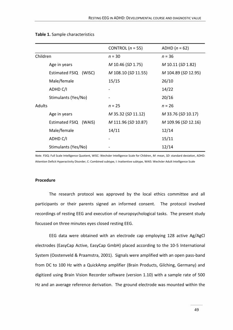
RESTING EEG IN ADHD: DEVELOPMENTAL COURSE AND DIAGNOSTIC VALUE
49
Table 1. Sample characteristics
CONTROL (n = 55) ADHD (n = 62)
Children n = 30 n = 36
Age in years M 10.46 (SD 1.75) M 10.11 (SD 1.82)
Estimated FSIQ (WISC) M 108.10 (SD 11.55) M 104.89 (SD 12.95)
Male/female 15/15 26/10
ADHD C/I - 14/22
Stimulants (Yes/No) - 20/16
Adults n = 25 n = 26
Age in years M 35.32 (SD 11.12) M 33.76 (SD 10.17)
Estimated FSIQ (WAIS) M 111.96 (SD 10.87) M 109.96 (SD 12.16)
Male/female 14/11 12/14
ADHD C/I - 15/11
Stimulants (Yes/No) - 12/14
Note. FSIQ: Full Scale Intelligence Quotient, WISC: Wechsler Intelligence Scale for Children, M: mean, SD: standard deviation, ADHD:
Attention Deficit Hyperactivity Disorder, C: Combined subtype, I: Inattentive subtype, WAIS: Wechsler Adult Intelligence Scale
Procedure
The research protocol was approved by the local ethics committee and all
participants or their parents signed an informed consent. The protocol involved
recordings of resting EEG and execution of neuropsychological tasks. The present study
focussed on three minutes eyes closed resting EEG.
EEG data were obtained with an electrode cap employing 128 active Ag/AgCl
electrodes (EasyCap Active, EasyCap GmbH) placed according to the 10-5 International
System (Oostenveld & Praamstra, 2001). Signals were amplified with an open pass-band
from DC to 100 Hz with a QuickAmp amplifier (Brain Products, Gilching, Germany) and
digitized using Brain Vision Recorder software (version 1.10) with a sample rate of 500
Hz and an average reference derivation. The ground electrode was mounted within the
CHAPTER 2
50
cap at Fpz. Electro-oculogram was recorded with electrodes enclosed in the cap near the
eyes and for adults, an additional electrode was placed below the right eye.
Analyses of EEG data were performed using Brain Vision Analyzer software
(version 2.0.1). EEG data were filtered with a high pass filter of 0.5 Hz, a low pass filter
of 50 Hz and a notch filter of 50 Hz. Data were segmented in 2 s epochs with 1 s overlap.
Eye movement correction was conducted according to the Gratton & Coles algorithm
(Gratton, Coles, & Donchin, 1983). Segments were rejected from further analyses if
amplitudes exceeded ±100 µV for children and if absolute difference in amplitudes
exceeded 120 µV for adults. Subsequently, spectral power was calculated using Fast
Fourier Transform and the transformed data were averaged. For replication purposes
(e.g., Liechti et al., 2013; Ogrim et al., 2012), absolute and relative power estimates were
derived for theta (3.5-7.5 Hz) and beta (12.5-25 Hz) frequencies at frontal (Fz), central
(Cz) and parietal (Pz) midline. Relative power was computed by dividing the average
power in a frequency band by the total average power in the 1.5-25 Hz spectrum,
multiplied by 100 (see also Dupuy et al., 2011). TBR was calculated by dividing power in
the theta band by power in the beta band. A natural logarithmic transform was used to
approach normal spreading of the data.
Statistical analyses
EEG activity across the lifespan in the ADHD and control group was compared by
separate analyses of variance with repeated measures for absolute as well as relative
theta and beta power and for TBR. Midline electrode position (Fz, Cz, Pz) was the
within-subject factor and clinical group (ADHD, non-ADHD) and age group (child, adult)
were between-subject factors. Effect sizes were determined by partial eta squared (η2p)
and, according to guidelines (Cohen, 1988), are interpreted as small (< .06), medium (.06
- .14), or large (> .14).
RESTING EEG IN ADHD: DEVELOPMENTAL COURSE AND DIAGNOSTIC VALUE
51
Receiver operation characteristic (ROC) curves were computed for determining
the accuracy of logistic regression models of absolute as well as relative theta and beta
power and TBR at the vertex in predicting clinical and age group membership. Cz was
examined to warrant comparability with other publications, since most studies
documenting on diagnostic validity of EEG parameters in ADHD have investigated EEG
activity at the vertex (Liechti et al., 2013; Monastra et al., 1999, 2001; Ogrim et al., 2012;
Snyder et al., 2008). First, binary logistic regression was performed to predict
probabilities for each EEG parameter separately. Then, ROC curves were computed to
determine the efficacy of the predicted probabilities in the respective binary outcomes
of interest (i.e., age group and clinical diagnosis group). Accuracy is indicated by the
area under the ROC curve, in which the reference line represents the null hypothesis
(Ogrim et al., 2012).
All analyses were performed using the Statistical Package for the Social Sciences
(SPSS) volume 20.
RESULTS
EEG
Concerning clinical group effects, none of the EEG measures reached significance
(see Table 2). Incorporation of sex as a covariate did not change the results significantly,
neither did repeating analyses including only individuals expressing at least 6 symptoms
in one or both domains of symptomatology in the adult ADHD group (i.e., 22 adults).
Also, effect sizes were fairly similar in these additional analyses.
Regarding age group effects, children demonstrated higher absolute theta (F(1, 111)
= 281.51, p < .001) and beta (F(1, 111) = 19.85, p < .001) power than adults. Regarding
relative power, higher theta (F(1, 111) = 97.73, p < .001) as well as lower beta (F(1, 111)
CHAPTER 2
52
= 62.67, p < .001) activity were found in children compared to adults. Furthermore, a
higher TBR (F(1, 111) = 169.44, p < .001) was observed in childhood relative to
adulthood. Considering the scope of the present study, divergence between children
and adults associated with electrode positions will not be elaborated on.
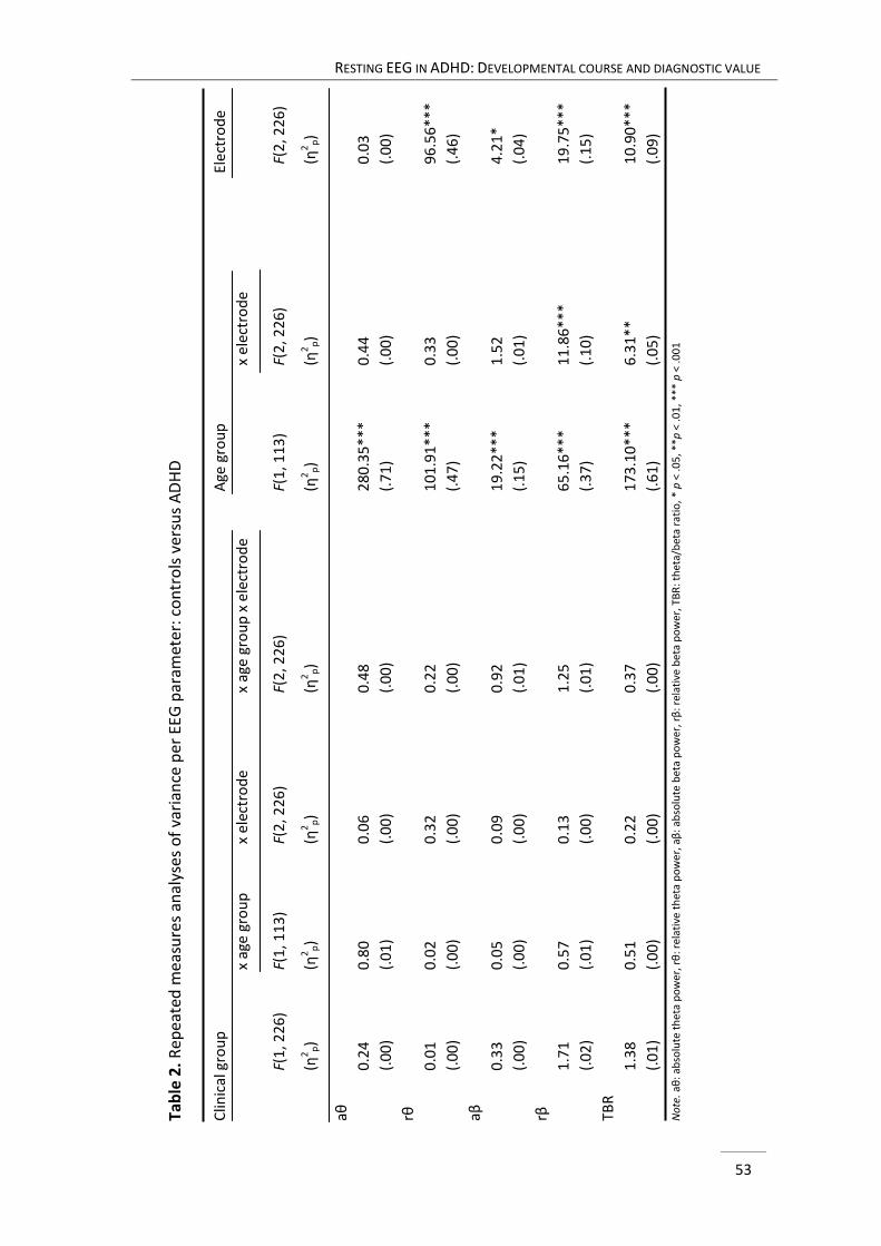
RESTING EEG IN ADHD: DEVELOPMENTAL COURSE AND DIAGNOSTIC VALUE
53
Tab
le 2
. R
epea
ted
mea
sure
s an
alys
es o
f va
rian
ce p
er E
EG p
aram
eter
: co
ntr
ols
ver
sus
AD
HD
C
linic
al g
rou
p
A
ge g
rou
p
E
lect
rod
e
x ag
e gr
ou
p
x
elec
tro
de
x
age
gro
up
x e
lect
rod
e
x
elec
tro
de
F(1
, 226
)
F(1
, 113
)
F(
2, 2
26)
F(
2, 2
26
)
F(1
, 11
3)
F(2
, 22
6)
F(
2, 2
26
)
(η2 p
)
(η2
p)
(η
2p)
(η2 p
)
(η
2 p)
(η
2 p)
(η2
p)
aθ
0.2
4
0.8
0
0
.06
0
.48
2
80
.35
***
0.4
4
0.0
3
(.0
0)
(.0
1)
(.
00
)
(.
00
)
(.
71
)
(.0
0)
(.0
0)
rθ
0.0
1
0.0
2
0
.32
0
.22
1
01
.91
***
0.3
3
96
.56
***
(.0
0)
(.0
0)
(.
00
)
(.
00
)
(.
47
)
(.0
0)
(.4
6)
aβ
0.3
3
0.0
5
0
.09
0
.92
1
9.2
2**
*
1
.52
4
.21
*
(.0
0)
(.0
0)
(.
00
)
(.
01
)
(.
15
)
(.0
1)
(.0
4)
rβ
1.7
1
0.5
7
0
.13
1
.25
6
5.1
6**
*
1
1.8
6**
*
19
.75
***
(.0
2)
(.0
1)
(.
00
)
(.
01
)
(.
37
)
(.1
0)
(.1
5)
TBR
1.3
8
0.5
1
0
.22
0
.37
1
73
.10
***
6.3
1**
10
.90
***
(.0
1)
(.0
0)
(.
00
)
(.
00
)
(.
61
)
(.0
5)
(.0
9)
No
te. a
θ: a
bso
lute
th
eta
po
wer
, rθ
: rel
ativ
e th
eta
po
we
r, a
β: a
bso
lute
bet
a p
ow
er, r
β: r
elat
ive
be
ta p
ow
er, T
BR
: th
eta/
bet
a ra
tio
, *
p <
.05
, **p
< .0
1, *
** p
< .0
01
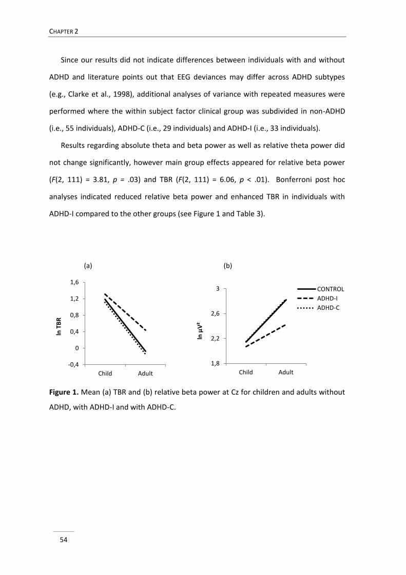
CHAPTER 2
54
1,8
2,2
2,6
3
Child Adult
ln μ
V²
CONTROL
ADHD-I
ADHD-C
Since our results did not indicate differences between individuals with and without
ADHD and literature points out that EEG deviances may differ across ADHD subtypes
(e.g., Clarke et al., 1998), additional analyses of variance with repeated measures were
performed where the within subject factor clinical group was subdivided in non-ADHD
(i.e., 55 individuals), ADHD-C (i.e., 29 individuals) and ADHD-I (i.e., 33 individuals).
Results regarding absolute theta and beta power as well as relative theta power did
not change significantly, however main group effects appeared for relative beta power
(F(2, 111) = 3.81, p = .03) and TBR (F(2, 111) = 6.06, p < .01). Bonferroni post hoc
analyses indicated reduced relative beta power and enhanced TBR in individuals with
ADHD-I compared to the other groups (see Figure 1 and Table 3).
(a) (b)
Figure 1. Mean (a) TBR and (b) relative beta power at Cz for children and adults without
ADHD, with ADHD-I and with ADHD-C.
-0,4
0
0,4
0,8
1,2
1,6
Child Adult
ln T
BR
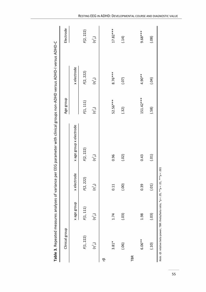
RESTING EEG IN ADHD: DEVELOPMENTAL COURSE AND DIAGNOSTIC VALUE
55
Tab
le 3
. R
epea
ted
mea
sure
s an
alys
es o
f va
rian
ce p
er E
EG p
aram
eter
wit
h c
linic
al g
rou
ps
no
n-A
DH
D v
ersu
s A
DH
D-I
ver
sus
AD
HD
-C
Clin
ical
gro
up
Age
gro
up
Elec
tro
de
x
age
gro
up
x el
ectr
od
e
x
age
gro
up
x e
lect
rod
e
x
elec
tro
de
F(1
, 222
)
F(
1, 1
11)
F(2
, 222
)
F(
2, 2
22
)
F(1
, 11
1)
F(2
, 22
2)
F(2
, 22
2)
(η2
p)
(η
2p)
(η
2p)
(η
2 p)
(η2 p
)
(η2
p)
(η
2 p)
rβ
3.8
1*
1
.74
0.1
1
0
.96
5
2.5
6**
*
8
.76
**
*
1
7.8
2**
*
(.0
6)
(.
03
)
(.0
0)
(.
02
)
(.
32
)
(.0
7)
(.
14
)
TBR
6.0
6**
1
.98
0.3
9
0
.43
15
1.4
2**
*
4
.90
**
9.6
8*
**
(.1
0)
(.
03
)
(.0
1)
(.
01
)
(.
58
)
(.0
4)
(.
08
)
No
te. r
β: r
elat
ive
bet
a p
ow
er, T
BR
: th
eta/
bet
a ra
tio
, *p
< .0
5, *
*p <
.01
, **
*p <
.00
1
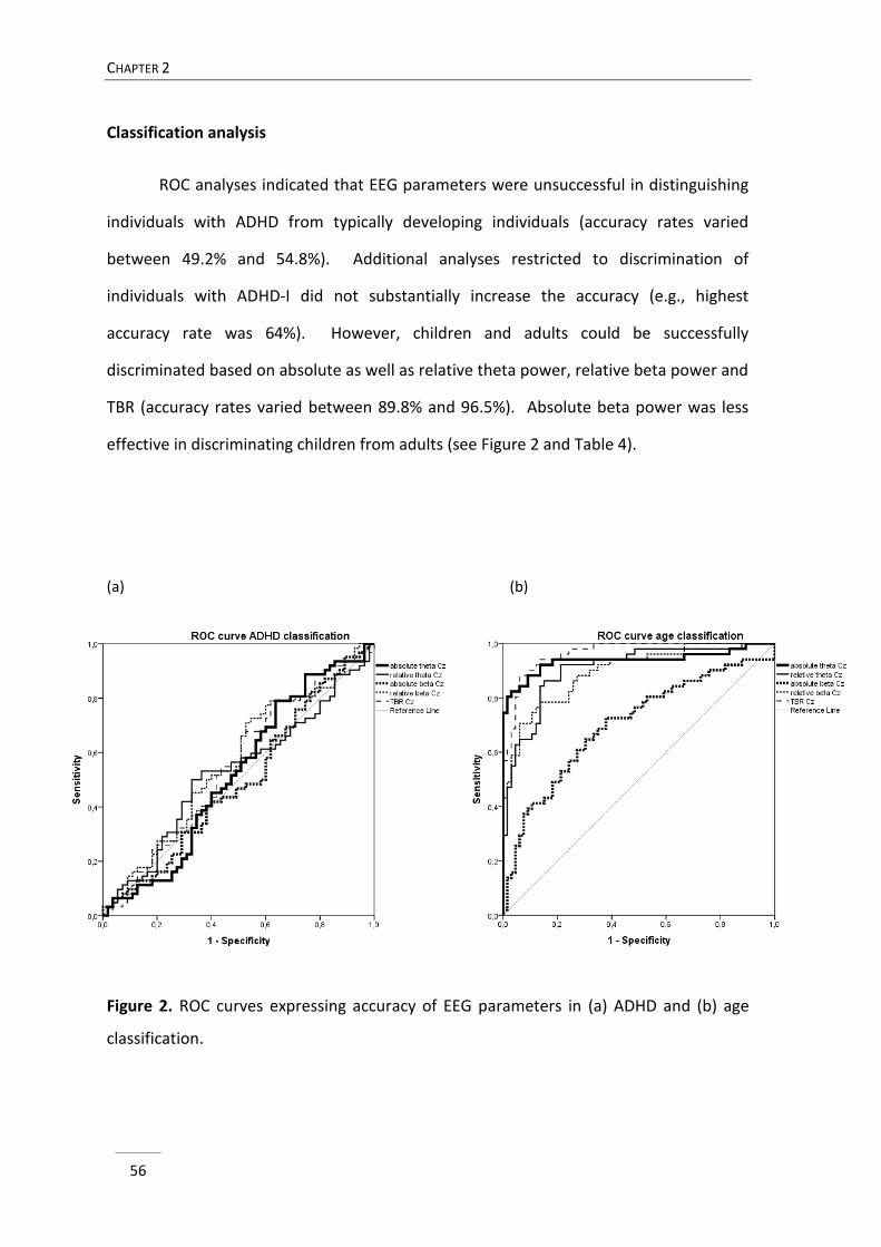
CHAPTER 2
56
Classification analysis
ROC analyses indicated that EEG parameters were unsuccessful in distinguishing
individuals with ADHD from typically developing individuals (accuracy rates varied
between 49.2% and 54.8%). Additional analyses restricted to discrimination of
individuals with ADHD-I did not substantially increase the accuracy (e.g., highest
accuracy rate was 64%). However, children and adults could be successfully
discriminated based on absolute as well as relative theta power, relative beta power and
TBR (accuracy rates varied between 89.8% and 96.5%). Absolute beta power was less
effective in discriminating children from adults (see Figure 2 and Table 4).
(a) (b)
Figure 2. ROC curves expressing accuracy of EEG parameters in (a) ADHD and (b) age
classification.
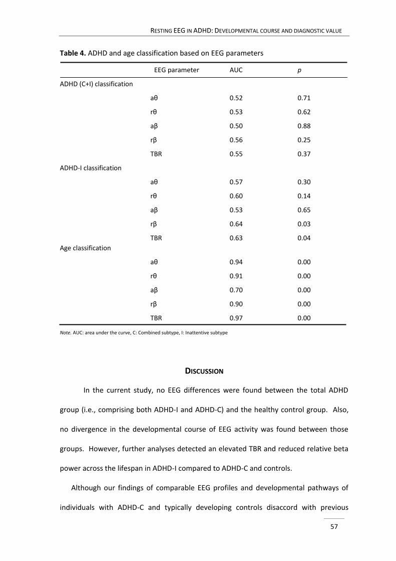
RESTING EEG IN ADHD: DEVELOPMENTAL COURSE AND DIAGNOSTIC VALUE
57
Table 4. ADHD and age classification based on EEG parameters
EEG parameter AUC p
ADHD (C+I) classification
aθ 0.52 0.71
rθ 0.53 0.62
aβ 0.50 0.88
rβ 0.56 0.25
TBR 0.55 0.37
ADHD-I classification
aθ 0.57 0.30
rθ 0.60 0.14
aβ 0.53 0.65
rβ 0.64 0.03
TBR 0.63 0.04
Age classification
aθ 0.94 0.00
rθ 0.91 0.00
aβ 0.70 0.00
rβ 0.90 0.00
TBR 0.97 0.00
Note. AUC: area under the curve, C: Combined subtype, I: Inattentive subtype
DISCUSSION
In the current study, no EEG differences were found between the total ADHD
group (i.e., comprising both ADHD-I and ADHD-C) and the healthy control group. Also,
no divergence in the developmental course of EEG activity was found between those
groups. However, further analyses detected an elevated TBR and reduced relative beta
power across the lifespan in ADHD-I compared to ADHD-C and controls.
Although our findings of comparable EEG profiles and developmental pathways of
individuals with ADHD-C and typically developing controls disaccord with previous
CHAPTER 2
58
reports on theta and TBR deviances in ADHD (for a review, see Barry et al., 2003), they
correspond with a few recent studies that also failed to replicate theta and TBR
abnormalities in ADHD relative to typically developing individuals (e.g., van Dongen-
Boomsma et al., 2010; Liechti et al., 2013). Furthermore, our results are in line with
findings of a smaller difference in TBR between ADHD and normal controls in more
recent publications compared to earlier studies (Arns et al., 2013). It is plausible that
differences in inclusion and exclusion criteria for control subjects as well as ADHD
patients have contributed to different findings across studies (Arns et al., 2013; Liechti et
al., 2013). As for the current study, it seems unlikely that the predominance of boys in
the child ADHD group contributed to the failure of finding EEG abnormalities in children
with ADHD, since supplementary analyses controlling for sex did not change the results
significantly. Besides, given that some studies documented more deviances in ADHD
boys compared to girls (e.g., Clarke et al., 2001b), this would rather have resulted in
detecting instead of overlooking abnormalities in our sample. Lowering the cut-off of
symptomatology for incorporating adults with ADHD in our study did also not impede
finding EEG differences between individuals with ADHD and typically developing
individuals since additional analyses using a more restrictive symptom threshold for
including adults did not affect the significance of findings. Moreover, this cannot explain
the absence of differences between children with and without ADHD. The current ADHD
sample comprised both individuals diagnosed with the ADHD combined as well as the
inattentive subtype. This differs from studies that incorporated only individuals with
ADHD combined subtype (e.g., Liechti et al., 2013; van Dongen-Boomsma et al., 2010)
but accords with others that included individuals with different ADHD subtypes (e.g., Loo
et al., 2010; Ogrim et al., 2012). Only a few studies directly compared the combined and
RESTING EEG IN ADHD: DEVELOPMENTAL COURSE AND DIAGNOSTIC VALUE
59
inattentive subtype and presented mixed results, making comparability to existing
literature difficult. Furthermore, exclusion and inclusion criteria regarding comorbid
disorders in individuals with ADHD are different across studies and the influence of
comorbidity on resting EEG findings in ADHD has not been investigated systematically.
Also in the current study, comorbid problems were present in individuals with ADHD as
indicated by elevated symptoms of anxiety/depression, learning disorders and disruptive
behaviour measured through interviews and questionnaires. Future studies with larger
sample sizes are needed to evaluate the impact of comorbidity on resting EEG findings in
ADHD. These factors, as well as the relatively high intelligence profile of our ADHD
sample compared to other studies, may confound the comparability of our study to
others and therefore impede generalisability of the results.
Few EEG studies have addressed the comparison of ADHD-I and ADHD-C, and
yielded disparate findings. In some studies, both ADHD subtypes similarly deviated from
a control group, while in others, more pronounced quantitative EEG differences in one
subtype relative to the other and/or the control group were documented (Clarke et al.,
1998, 2001b; Dupuy et al., 2011; Hermens et al., 2004; Loo et al., 2010). In this study,
the observed larger EEG deviances in ADHD-I were not accounted for by a greater
severity of inattention problems in the ADHD-I group compared to the ADHD-C group, as
inattention scores as derived from the questionnaires did not differ between ADHD-I
and ADHD-C. Also, ADHD subtypes did not differ on severity of comorbid problems as
screened by the subscales of Achenbach questionnaires. Our findings may suggest that
distinct types of inattention characterize differences between ADHD-I and ADHD-C,
involving different neural circuits, rather than variations in symptom severity (Diamond,
2005; Milich, Balentine, & Lynam, 2001). However, this statement cannot unequivocally
CHAPTER 2
60
be confirmed by our data. Moreover, this interpretation discords with a recent meta-
analysis of 546 studies, that found minimal support for defining ADHD-I and ADHD-C as
distinct disorders (Willcutt et al., 2012). In view of the literature that defined EEG based
subgroups in ADHD through cluster analyses (Clarke et al., 2011a), our findings may be
better explained as emphasizing heterogeneity of EEG in ADHD, confirming deviances in
a subgroup of patients rather than in the whole ADHD group.
Since EEG deviances were found to persist throughout the lifespan, it seems
questionable that a maturational delay may account for EEG discrepancies in a subgroup
of individuals with ADHD. A developmental deviance may be a better interpretation of
these results. However, findings should be seen as tentative since direct individual
measurements of developmental changes are not provided in this cross sectional study.
Ideally, longitudinal designs are applied in gaining more insights in the nature of the
developmental course of EEG deviances in ADHD(-I).
Given the lack of consistent EEG abnormalities in the ADHD group, the accuracy
rates reflecting failure in discriminating this group from the control group based on
these parameters was not surprising. Our overall accuracy rates below 57% align with
recent documents, that also demonstrated inadequacy of EEG based classification
(Liechti et al., 2013; Ogrim et al., 2012) but contrast with earlier findings of accuracy
rates up to 90% (Monastra et al., 2001; Snyder et al., 2008). Alternatively, accuracy in
distinguishing children from adults attained 96.5% despite using parameters selected for
deviances in ADHD. Since our findings regarding maturation of EEG activity are
consistent with literature (e.g., Barriga-Paulino et al., 2011), we believe our data would
have allowed reliable detection of a maturational lag, as has been suggested for ADHD.
RESTING EEG IN ADHD: DEVELOPMENTAL COURSE AND DIAGNOSTIC VALUE
61
In sum, our EEG findings confirm the view that ADHD is a heterogeneous
disorder, which is also established in multiple pathway models of ADHD (e.g., Sonuga-
Barke, Bitsakou, & Thompson, 2010). Our results suggest that it is unlikely that ADHD is
characterized by generalised EEG abnormalities and that the use of conventional EEG
practices as a diagnostic adds-on in ADHD has to be regarded as insufficient. However,
since reduced relative beta power and elevated TBR were found in a subgroup of
patients, it seems plausible that EEG measures may be of interest in guiding prognostic
practices. That is, they may possibly be applied to distinguish subgroups that respond
differently to various treatments. This has already been illustrated in research
investigating the relationship between EEG parameters and treatment response to
stimulant medication (Arns, Gunkelman, Breteler, & Spronk, 2008) and neurofeedback
treatment (Arns, Drinkenburg, & Kenemans, 2012).
CHAPTER 2
62
REFERENCES
Achenbach, T.M., & Rescorla, L.A. (2001). Manual for the ASEBA School-Age Forms and Profiles.
Burlington, Vermont: University of Vermont, Research Center for Children, Youth, and
Families.
Achenbach, T.M., & Rescorla, L.A. (2003). Manual for the ASEBA Adult Forms & Profiles.
Burlington, Vermont: University of Vermont, Research Center for Children, Youth, and
Families.
American Psychiatric Association (APA) (2000). Diagnostic and Statistical Manual of mental
disorders, 4th edition, Text Revision (DSM-IV-TR). Washington, DC: American Psychiatric
Association.
Arns, M., Conners, C.K., & Kraemer, H.C. (2013). A decade of EEG theta/beta ratio research in
ADHD: a meta-analysis. Journal of Attention Disorders, 17, 374-383. doi:
10.1177/1087054712460087
Arns, M., Drinkenburg, W.H.I.M., & Kenemans, J.L. (2012). The effects of QEEG-informed
neurofeedback in ADHD: An open label pilot study. Applied Psychophysiology and
Biofeedback, 37, 171-180. doi: 10.1007/s10484-012-9191-4
Arns, M., Gunkelman, J., Breteler, M., & Spronk, D. (2008). EEG phenotypes predict treatment
outcome to stimulants in children with ADHD. Journal of Integrative Neuroscience, 7,
421-438. doi: 10.1142/S0219635208001897
Barriga-Paulino, C.I., Flores, A.B., & Gomez, C.M. (2011). Developmental changes in the EEG
rhythms of children and young adults. Journal of Psychophysiology, 25(3), 143-158. doi:
10.1027/0269-8803/a000052
Barry, R.J., Clarke, A.R. & Johnstone, S.J. (2003). A review of electrophysiology in attention-
deficit/hyperactivity disorder: I. Qualitative and quantitative electroencephalography.
Clinical Neurophysiology, 114, 171-183. doi: 10.1016/S1388-2457(02)00362-0
Barry, R.J., Clarke, A.R., Johnstone, S.J., McCarthy, R., & Selikowitz, M. (2009).
Electroencephalogram theta/beta ratio and arousal in attention-deficit/hyperactivity
disorder: evidence of independent processes. Biological Psychiatry, 66, 398-401. doi:
10.1016/j.biopsych.2009.04.027
RESTING EEG IN ADHD: DEVELOPMENTAL COURSE AND DIAGNOSTIC VALUE
63
Boutros, N., Fraenkel, L., & Feingold, A. (2005). A four-step approach for developing diagnostic
tests in psychiatry: EEG in ADHD as a test case. Journal of Neuropsychiatry and Clinical
Neurosciences, 17, 455-464. doi: 10.1176/appi.neuropsych.17.4.455
Bresnahan, S.M., Anderson, J.W., & Barry, R.J. (1999). Age-related changes in quantitative EEG in
attention-deficit/hyperactivity disorder. Biological Psychiatry, 46, 1690-1697. doi:
10.1016/S0006-3223(99)00042-6
Chabot, R.J., & Serfontein, G. (1996). Quantitative electroencephalographic profiles of children
with attention deficit disorder. Biological Psychiatry, 40, 951-963. doi: 10.1016/0006-
3223(95)00576-5
Clarke, A.R., Barry, R.J., Dupuy, F.E., Heckel, L.D., McCarthy, R., Selikowitz, M., & Johnstone, S.J.
(2011a). Behavioural differences between EEG-defined subgroups of children with
attention-deficit/hyperactivity disorder. Clinical Neurophysiology, 122, 1333-1341. doi:
10.1016/j.clinph.2010.12.038
Clarke, A.R., Barry, R.J., Dupuy, F.E., McCarthy, R., Selikowitz, M., & Heaven, P.C. (2011b).
Childhood EEG as a predictor of adult attention-deficit/hyperactivity disorder. Clinical
Neurophysiology, 122(1), 73-80. doi: 10.1016/j.clinph.2010.05.032
Clarke, A.R., Barry, R.J., Dupuy, F.E., McCarthy, R., Selikowitz, M., & Johnstone, S.J. (2013). Excess
beta activity in the EEG of children with attention-deficit/hyperactivity disorder: A
disorder of arousal? International Journal of Psychophysiology, 89(3), 314-319. doi:
10.1016/j.ijpsycho.2013.04.009
Clarke, A.R., Barry, R.J., Heaven, P.C.L., McCarthy, R., Selikowitz, M., & Byrne, M.K. (2008). EEG in
adults with Attention-Deficit/Hyperactivity Disorder. International Journal of
Psychophysiology, 70, 176-183. doi: 10.1016/j.ijpsycho.2008.07.001
Clarke, A., Barry, R., McCarthy, R., & Selikowitz, M. (1998). EEG analysis in attention
deficit/hyperactivity disorder: a comparative study of two subtypes. Psychiatry Research
81, 19-29. doi: 10.1016/S0165-1781(98)00072-9
Clarke, A.R., Barry, R.J., McCarthy, R., & Selikowitz, M. (2001a). Excess beta activity in children
with attention-deficit/hyperactivity disorder: an atypical electrophysiological group.
Psychiatry Research, 103(2-3), 205-218. doi: 10.1016/S0165-1781(01)00277-3
CHAPTER 2
64
Clarke, A.R., Barry, R.J., McCarthy, R., & Selikowitz, M. (2001b). Age and sex effects in the EEG:
differences in two subtypes of attention-deficit/hyperactivity disorder. Clinical
Neurophysiology, 112(5), 815-826. doi: 10.1016/S1388-2457(01)00487-4
Cohen, J. (1988). Statistical Power Analysis for the Behavioral Sciences, 2 ed. Laurence Erlbaum,
Hillsdale, N.J.
Coutin-Churchman, P., Anez, Y., Uzcategui, M., Alvarez, L., Vergara, F., Mendez, L., & Fleitas
(2003). Quantitative spectral analysis of EEG in psychiatry revisited: drawing signs out of
numbers in a clinical setting. Clinical Neurophysiology, 114, 2294-2306. doi:
10.1016/S1388-2457(03)00228-1
Diamond, A. (2005). Attention-deficit disorder (attention-deficit/hyperactivity disorder without
hyperactivity): A neurobiologically and behaviorally distinct disorder from attention-
deficit/hyperactivity disorder (with hyperactivity). Development and Psychopathology
17(3), 807-825. doi: 10.1017/S0954579405050388
DuPaul, G.J., Power T.J., Anastopoulos A.D., & Reid R. (1998). ADHD Rating Scale-IV: Checklists,
Norms, and Clinical Interpretation. New York, The Guilford Press.
Dupuy, F.E., Clarke, A.R., Barry, R.J., McCarthy, R., & Selikowitz, M. (2011). Girls with attention-
deficit/hyperactivity disorder: EEG differences between DSM-IV types. Clinical EEG and
Neuroscience, 42(1), 1-5. doi: 10.1177/155005941104200104
Gratton, G., Coles, M.G., & Donchin, E. (1983). A new method for off-line removal of ocular
artifact. Electroencephalography and Clinical Neurophysiology, 55(4), 468-484. doi:
10.1016/0013-4694(83)90135-9
Grégoire, J. (2000). L’évaluation clinique de l’intelligence de l’enfant. Liège: Mardaga.
Hermens, D. F., Williams, L. M., Lazzaro, I., Whitmont, S., Melkonian, D., & Gordon, E. (2004). Sex
differences in adult ADHD: a double dissociation in brain activity and autonomic arousal.
Biological Psychology, 66(3), 221-233. doi: 10.1016/j.biopsycho.2003.10.006
Kooij, S.J.J., Bejerot, S., Blackwell, A., Caci, H., Casas-Brugué, M.C., Carpentier, P.J., … & Asherson,
P. (2010). European consensus statement on diagnosis and treatment of adult ADHD:
The European Network Adult ADHD. BMC Psychiatry, 10(67), 1-24. doi: 10.1186/1471-
244X-10-67
Kooij, S.J.J., & Francken, M.H. (2010). Diagnostisch Interview Voor ADHD bij Volwassenen. DIVA
Foundation, Den Haag.
RESTING EEG IN ADHD: DEVELOPMENTAL COURSE AND DIAGNOSTIC VALUE
65
Lazzaro, I., Gordon E., Whitmont, S., Plahn, M., Li, W., Clarke, S., … & Meares, R. (1998).
Quantitative EEG activity in adolescent attention deficit hyperactivity disorder. Clinical
Electroencephalography, 29, 37-42. doi: 10.1177/155005949802900111
Liechti, M.D., Valko, L., Müller, U.C., Döhnert, M., Drechsler, R., Steinhausen, H.C., & Brandeis, D.
(2013). Diagnostic value of resting electroencephalogram in attention-
deficit/hyperactivity disorder across the lifespan. Brain Topography, 26(1), 135-151. doi:
10.1007/s10548-012-0258-6
Loo, S.K., Hale, T.S., Hanada, G., Macion, J., Shrestha, A., McGough, J.J., ... & Smalley, S.L. (2010).
Familial clustering and DRD4 effects on electroencephalogram measures in multiplex
families with attention deficit/hyperactivity disorder. Journal of the American Academy
of Child and Adolescent Psychiatry, 49(4), 368-377. doi: 10.1016/j.jaac.2010.01.002
Loo, S.K., Hale, T.S., Macion, J., Hanada, G., McGough, J.J., McCracken, J.T., & Smalley, S.L.
(2009). Cortical activity patterns in ADHD during arousal, activation and sustained
attention. Neuropsychologia, 47(10), 1033-1040. doi: 10.1016/j.neuropsychologia.
2009.04.013
Loo, S.K., & Makeig, S. (2012). Clinical utility of EEG in attention-deficit/hyperactivity disorder: a
research update. Neurotherapeutics, 9, 569-587. doi: 10.1007/s13311-012-0131-z
Mann, C.A., Lubar, J.F., Zimmerman, A.W., Miller, C.A., & Muenchen, R.A. (1992). Quantitative
analysis of EEG in boys with attention deficit hyperactivity disorder: controlled study
with clinical implications. Pediatric Neurology, 8, 30-36. doi: 10.1016/0887-
8994(92)90049-5
Milich, R., Balentine, A.C., & Lynam, D.R. (2001). ADHD combined subtype and ADHD
predominantly inattentive type are distinct and unrelated disorders. Clinical Psychology:
Science and Practice, 8, 463-488. doi: 10.1093/clipsy.8.4.463
Monastra, V.J., Lubar, J.F., & Linden, M. (2001). The development of a quantitative
electroencephalographic scanning process for attention deficit-hyperactivity disorder:
Reliability and validity studies. Neuropsychology, 15, 136-144. doi: 10.1037/0894-
4105.15.1.136
Monastra, V.J., Lubar, J.F., Linden, M., Van Deusen, P., Green, G., Wing, W., … & Fenger, T.N.
(1999). Assessing attention deficit hyperactivity disorder via quantitative
CHAPTER 2
66
electroencephalography: An initial validation study. Neuropsychology, 13, 424-433. doi:
10.1037/0894-4105.13.3.424
Ogrim, G., Kropotov, J., & Hestad, K. (2012). The QEEG TBR in ADHD and normal controls:
Sensitivity, specificity and behavioral correlates. Psychiatry Research, 198(3), 482-488.
doi: 10.1089/cap.2012.0090
Oostenveld, R., & Praamstra, P. (2001). The five percent electrode system for high resolution
EEG and ERP measurements. Clinical Neurophysiology, 112, 713-719. doi:
10.1016/S1388-2457(00)00527-7
Pelham W., Gnagy E.M., Greenslade K.E., & Milich R. (1992). Teacher ratings of DSM-III-R
symptoms for the disruptive behavior disorders. Journal of the American Academy of
Child and Adolescent Psychiatry, 31, 210-218. doi: 10.1097/00004583-199203000-00006
Ryan, J.J., & Ward, L.C. (1999). Validity, reliability, and standard errors of measurement for two
seven-subtest short forms of the Wechsler Adult Intelligence Scale-III. Psychological
Assessment, 11, 207-211. doi: 10.1037/1040-3590.11.2.207
Satterfield, J.H., Schell, A.M., Backs, R.W., & Hidaka, K.C. (1984). A cross-sectional and
longitudinal study of age effects of electrophysiological measures in hyperactive and
normal children. Biological Psychiatry, 19 (7), 973-990.
Schaffer, D., Fisher, P., Lusac, C.P., Dulcan, M.K., & Schwab-Stone, M.E. (2000). NIMH Diagnostic
Interview Schedule for Children version IV (NIMH DISC-IV): Description, differences from
previous versions and reliability of some common diagnoses. Journal of the American
Academy of Child and Adolescent Psychiatry, 39, 28-38. doi: 10.1097/00004583-
200001000-00014
Seixas, M., Weiss, M., & Muller, U. (2012). Systematic review of national and international
guidelines on attention-deficit hyperactivity disorder. Journal of Psychopharmacology
26(6), 753-765. doi: 10.1177/0269881111412095
Shi, T., Li, X., Song, J., Zhao, N., Sun, C., Xia, W., … & Tomoda, A. (2012). EEG characteristics and
visual cognitive function of children with attention deficit hyperactivity disorder (ADHD).
Brain and Development, 34(10), 806-811. doi: 10.1016/j.braindev.2012.02.013
Snyder, S.M., & Hall, J.R. (2006). A meta-analysis of quantitative EEG power associated with
attention-deficit/hyperactivity disorder. Journal of Clinical Neurophysiology, 23, 440-455.
doi: 10.1097/01.wnp.0000221363.12503.78
RESTING EEG IN ADHD: DEVELOPMENTAL COURSE AND DIAGNOSTIC VALUE
67
Snyder, S.M., Quintana, H., Sexson, S.B., Knott, P., Haque, A.F., & Reynolds, D.A. (2008). Blinded,
multi-center validation of EEG and rating scales in identifying ADHD within a clinical
sample. Psychiatry Research, 159(3), 346-358. doi: 10.1016/j.psychres.2007.05.006
Sonuga-Barke, E., Bitsakou, P., & Thompson, M. (2010). Beyond the dual pathway model:
evidence for the dissociation of timing, inhibitory and delay-related impairments in
attention-deficit/hyperactivity disorder. Journal of American Child and Adolescent
Psychiatry, 49(4), 345-355. doi: 10.1016/j.jaac.2009.12.018
Spencer, T.J., Biederman, J., & Mick, E. (2007). Attention-deficit/hyperactivity disorder:
diagnosis, lifespan, comorbidities and neurobiology. Journal of Pediatric Psychology,
32(6), 631-642. doi: 10.1016/j.ambp.2006.07.006
van Dongen-Boomsma, M., Lansbergen, M.M., Bekker, E.M., Kooij, S.J.J., van der Molen, M.,
Kenemans, J.L., & Buitelaar, J.K. (2010). Relation between resting EEG to cognitive
performance and clinical symptoms in adults with attention-deficit/hyperactivity
disorder. Neuroscience Letters, 469, 102-106. doi: 10.1016/j.neulet.2009.11.053
Ward, M.F., Wender, P.H., & Reimherr, F.H. (1993). The Wender Utah Rating Scale: An aid in the
retrospective diagnosis of childhood attention deficit hyperactivity disorder. American
Journal of Psychiatry, 150, 885-890
Wechsler, D. (1991). The Wechsler intelligence scale for children-third edition. San Antonio, TX:
The Psychological Corporation.
Wechsler, D. (1997). WAIS-III: Wechsler Adult Intelligence Scale- third edition administration and
scoring manual. San Antonio, TX: The Psychological Corporation.
Willcutt, E.G. (2012). The prevalence of DSM-IV attention-deficit/hyperactivity disorder: a meta-
analytic review. Neurotherapeutics, 9(3), 490-499. doi: 10.1007/s13311-012-0135-8
Willcutt, E.G., Nigg, J.T., Pennington, B.F., Solanto, M.V., Rohde, L.A., Tannock, R., … & Lahey,
B.B. (2012). Validity of DSM-IV attention-hyperactivity disorder symptom dimensions
and subtypes. Journal of Abnormal Psychology, 121(4), 991-1010. doi:
10.1037/a0027347. doi: 10.1037/a0027347
Williams, L.M., Tsang, T.W., Clarke, S., & Kohn, M. (2010). An ‘integrative neuroscience’
perspective on ADHD: linking cognition, emotion, brain and genetic measures with
implications for clinical support. Expert Review of Neurotherapeutics, 10(10), 1607-1621.
doi: 10.1586/ern.10.140

69
EEG ACTIVITY BEFORE AND AFTER
COGNITIVE EFFORT IN CHILDREN WITH ADHD1
ABSTRACT
Objective: This study investigated whether children with attention-deficit/hyperactivity
disorder (ADHD) show a deviant electroencephalographic (EEG) pattern and whether
this is stable across resting state conditions before and after cognitive effort. Methods:
Three minutes eyes closed resting EEG before and after cognitive effort were compared
between 21 children with ADHD and 22 typically developing children. Results: Groups
did not differ in any of the EEG measures. However, the progressive topographic
expansion in alpha power from anterior to posterior sites was lower in children with
ADHD across conditions. Posterior absolute delta, theta and beta power, central
absolute beta power, and relative theta power attenuated, while central relative delta
power increased after task execution in both groups. No group differences were
detected in EEG changes following cognitive effort. Conclusions: Despite the lack of
finding group differences in theta or theta/beta ratio, topographic organization of alpha
power was detected to differ between groups, independent of condition. Although EEG
activity changed after cognitive effort in both groups, the findings did not indicate
altered changes after task execution in ADHD.
1 Based on Buyck, I., & Wiersema, J.R. (2014). Electroencephalographic activity before and after cognitive
effort in children with attention deficit hyperactivity disorder. Manuscript submitted for publication.
3 CHAPTER
CHAPTER 3
70
INTRODUCTION
Attention-deficit/hyperactivity disorder (ADHD) is a neurobehavioural disorder
with a childhood onset that is characterized by developmentally incompatible symptoms
of inattention and/or hyperactivity/impulsivity, leading to impairment in multiple life
domains (American Psychiatric Association, 2013). The worldwide prevalence rates of
ADHD are estimated between 5 and 7% (Willcutt, 2012). In the past decades,
electroencephalographic (EEG) measures, particularly during resting conditions, have
been frequently used to determine whether neurophysiological abnormalities may be
associated with ADHD.
Resting EEG studies in ADHD have mainly focussed on children, in which
differences in EEG patterns have been observed compared to typically developing
children. That is, children with ADHD commonly show increased absolute or relative
theta activity and/or an elevated theta/beta ratio (TBR) (for a review, see Barry, Clarke,
& Johnstone, 2003; Buyck & Wiersema, 2014; Clarke, Barry, McCarthy, Selikowitz, &
Johnstone, 2007; Clarke et al., 2011; Dupuy, Clarke, Barry, McCarthy, & Selikowitz, 2011;
González-Castro, Rodríguez, López, Cueli, & Álvarez, 2013; Lansbergen, Arns, van
Dongen-Boomsma, Spronk, & Buitelaar, 2011; Loo et al., 2010; Shi et al., 2012).
Although not systematically addressed in studies, increased absolute or relative delta
activity has been observed as well in children with ADHD (Clarke, Barry, McCarthy, &
Selikowitz, 1998; Clarke, Barry, McCarthy, & Selikowitz, 2001a; Clarke et al., 2007). Yet,
lately the robustness of EEG slowing characterizing the total ADHD group has been
challenged, since several recent studies could not invariably replicate the typically
increased theta power or TBR in children with ADHD relative to typically developing
children found in earlier studies (Coolidge, Starkey, & Cahill, 2007; Liechti et al., 2013;
Loo et al., 2013; Nazari, Wallois, Aarabi, & Berquin, 2011; Ogrim, Kropotov, & Hestad,
2012; Swartwood, Swartwood, Lubar, & Timmermann, 2003). This was also confirmed
by a recent meta-analysis that revealed a reducing discrepancy in TBR between
EEG BEFORE AND AFTER COGNITIVE EFFORT IN CHILDREN WITH ADHD
71
youngsters with and without ADHD across publication years of studies in the last decade
(Arns, Conners, & Kraemer, 2013). Additionally, recent studies denoted that enhanced
theta or TBR was only apparent in about 20 to 60% of children (Arns, Drinkenburg, &
Kenemans, 2012; Clarke et al., 2011; Ogrim et al., 2012), and that theta can even be
reduced in a part of children with ADHD (Clarke et al., 2011). Concerning activity in the
faster frequency bands, findings are even more ambiguous. Regarding alpha activity,
most studies refer to a reduced amount of absolute or relative alpha (Clarke et al., 1998,
2001a; Clarke, Barry, McCarthy, & Selikowitz, 2002a; Clarke et al., 2003; Barry, Clarke,
Johnstone, McCarthy, & Selikowitz, 2009) but increased activity in the alpha band has
also been reported in a subgroup of ADHD (Chabot & Serfontein, 1996; Clarke et al.,
2011). Even so, some studies failed to detect deviance in alpha power in ADHD (Loo et
al., 2010; Shi et al., 2012). Likewise, reduced absolute or relative beta activity has often
been detected in children with ADHD (Clarke et al., 1998, 2002a, 2003; Shi et al., 2012)
but in a small group, excess beta activity has been observed instead (Chabot &
Serfontein, 1996; Clarke et al., 1998; Clarke, Barry, McCarthy, & Selikowitz, 2001b), while
in other studies no significant group distinction in beta power was found (Liechti et al.,
2013; Loo et al., 2010).
Interestingly, in ADHD it is well-established that behavioural symptoms (Antrop,
Buysse, Roeyers, & Van Oost, 2005; Zentall & Zental, 1983), performance (Luman,
Oosterlaan, & Sergeant, 2005; Power, 1992; Sonuga-Barke, Williams, Hall, & Saxton,
1996; Toplak & Tannock, 2005; Wiersema, van der Meere, Antrop & Roeyers, 2006a;
Wiersema, van der Meere, Roeyers, Van Coster, & Baeyens, 2006b) as well as
physiological parameters (Benikos & Johnstone, 2009; Börger & van der Meere, 2000;
Imeraj et al., 2011, 2012; Kooistra et al., 2010; Wiersema et al., 2006a,b) fluctuate over
time and in response to changing environmental contexts. In this respect, there has
been mounting evidence that contextual factors play a crucial role in determining to
which extent performance is deficient in ADHD (Sonuga-Barke, Wiersema, van der
CHAPTER 3
72
Meere, & Roeyers, 2010). For instance, it has repeatedly been found that performance
of children with ADHD heavily depends on energetic state related factors, with
impairments especially apparent in conditions that induce suboptimal arousal/activation
state, such as slow presentation rate of stimuli (for a meta-analysis, see Metin, Roeyers,
Wiersema, van der Meere, & Sonuga-Barke, 2012; Scheres, Oosterlaan, & Sergeant,
2001; Wiersema et al., 2006a,b). Based on these and other findings, the important role
of contextual factors in the explanation of the deficits associated with ADHD has been
acknowledged by several theoretical frameworks. That is, the state regulation deficits
model (Sergeant, 2005; van der Meere, 2005) and the delay aversion model (Sonuga-
Barke, Taylor, Sembi, & Smith, 1992) emphasize respectively dynamic underlying failures
of energetic or motivational state factors in ADHD (Sonuga-Barke et al., 2010).
As abnormalities in ADHD have been found to be dependent on contextual and
state factors, the question can be raised whether the apparent discrepant findings
between resting EEG studies -among others (i.e., sample characteristics, EEG recording
and quantification procedures, …)- can partially be explained by differences in state-
related factors between studies. Although research has found support for intra-
individual stability of EEG in typically developing children over time (e.g., Fein, Galin,
Yingling, Johnstone, & Nelson, 1984; Gasser, Bacher, & Steinberg, 1985), contextual and
state influences are also well known to affect EEG (e.g., Antonenko, Paas, Grabner, &
van Gog, 2010; Barry, Clarke, Johnstone, Magee, & Rushby, 2007; Kim, Kim, Oh, & Kim,
2013; Oken, Salinsky, & Elsas, 2006). An illustration of a widely recognized state factor
that influences EEG activity refers to the opening of the eyes (e.g., Barry et al., 2007).
Regarding this, most studies in ADHD have addressed EEG in either an eyes closed or an
eyes opened resting condition, with only a few evaluating both resting conditions
(Fonseca, Tedrus, Bianchini, & Silva, 2013; Lansbergen et al., 2011; Liechti et al., 2013;
Loo et al., 2010, 2013). Interestingly, some of these latter studies have detected that
conditions with closed eyes generated greater discrepancies between groups in absolute
EEG BEFORE AND AFTER COGNITIVE EFFORT IN CHILDREN WITH ADHD
73
theta power (Liechti et al., 2013) and TBR (Lansbergen et al., 2011) than resting
conditions with open eyes. Further, a greater theta power increase to eye closing
(Liechti et al., 2013) and a reduced alpha suppression to eye opening (Buyck &
Wiersema, submitted; Fonseca et al., 2013) have been found in children with ADHD.
These findings suggest that contextual and state factors are important to take into
account when evaluating EEG deviances in ADHD. Keeping up with this, less is known
about the impact of timing of EEG measurement during experiments in EEG divergence
between groups, since so far research focussed on either starting (e.g., Snyder et al.,
2008) or ending (e.g., Dupuy et al., 2011) laboratory visits with EEG registration without
including both conditions. Studies suggest that both conditions are able to unfold EEG
deviances in ADHD, but the lack of literature addressing both EEG registration before
and after cognitive effort in one and the same study impedes drawing conclusions on
possible divergence and transition, and thus context dependent changes, between
measurements. To our knowledge only one study evaluated EEG before and after
cognitive effort in ADHD, be it in adults (Koehler et al., 2009). In this study, beta power
increased after task execution in controls, but not in adults with ADHD (Koehler et al.,
2009). Yet, although research addressing the subsequent influences of cognitive effort
on resting conditions in a normal population is scarce as well and also limited to adults,
several studies point to changes in resting EEG after task execution (Smit, Eling, &
Coenen, 2004; Smit, Eling, Hopman, & Coenen, 2005; Tanaka et al., 2012), revealing the
impact of this contextual factor on EEG activity. In typically developing adults, eyes
closed absolute (Smit et al., 2004) and relative theta (Smit et al., 2005) as well as
absolute beta power (Smit et al., 2004) have been documented to increase following
demanding cognitive tasks. In a more recent study, it was established that EEG activity
was not found to be modulated after completing an easy 0-back task, whereas
decreased posterior alpha and beta power and increased theta activity emerged after a
more demanding 2-back task (Tanaka et al., 2012). Although it is clear that task
CHAPTER 3
74
execution affects subsequent resting EEG activity, the inconsistency in existent findings
suggest that also task-related features contribute to the nature of EEG changes after
cognitive effort.
Taken together the above-mentioned notions, the question rises whether
context or state dependent fluctuations may not only be reflected in behaviour,
performance and certain physiological indices, but also in quantitative EEG in ADHD. As
outlined above, some support can be found for this hypothesis through studies
evaluating both eyes closed and eyes open resting conditions. Given the paucity of
literature, the present study aims to expand insights on context-related EEG divergence
in ADHD by investigating whether possible EEG deviances in ADHD are stable before and
after task execution and to what extent intervening task execution interferes with
characterizing resting EEG deviances in ADHD. It is imperative to shed light on those
dynamic changes in EEG in ADHD, since nowadays an increasing number of clinicians use
EEG for assessment and treatment guidance in which quantitative EEG of individuals
with ADHD is compared to normative databases. If there are indications that dynamic
factors affect EEG abnormalities in ADHD, this may have important consequences in
dealing with these databases for clinical purposes, where insufficient knowledge may
result in ineffective practices and even cause iatrogenic harm (Hammond, 2010). As
most of these databases are based on eyes closed resting EEG data, it is important to
investigate eyes closed resting conditions in clinical populations (Clarke et al., 2008).
Therefore, in the present study resting EEG activity is evaluated in children with and
without ADHD in eyes closed resting conditions before and after task execution.
If theta power and TBR are solid markers for ADHD that may be regarded as a
stable characteristic, then these EEG parameters are expected to be enhanced in
children with ADHD in both resting conditions. If however an interaction is detected
between group divergence and timing of EEG registration, this would indicate that state-
related factors may be associated with EEG abnormalities in ADHD.
EEG BEFORE AND AFTER COGNITIVE EFFORT IN CHILDREN WITH ADHD
75
METHOD
Participants
Twenty-one children diagnosed with ADHD and 22 typically developing children
aged 7 to 14 years participated in the study (Table 1). Groups were matched for sex and
age (F(1, 41) = 0.00, p = .99) and did not differ in IQ (F(1, 41) = 2.83, p = .10). Participants
were recruited through advertisements, schools, staff members and, specifically for
ADHD, child neurologists. Children with ADHD were previously diagnosed in a clinical
setting according to DSM-IV criteria. The diagnosis was confirmed using the parent
version of the behavioural module of the Diagnostic Interview Schedule for Children IV
for children (DISC-IV) (Schaffer, Fisher, Lusac, Dulcan, & Schwab-Stone, 2000).
Furthermore, the Disruptive Behavior Disorder (DBD) Rating Scale (Pelham, Gnagy,
Greenslade, & Milich, 1992; Dutch translation: Oosterlaan, Scheres, Antrop, Roeyers, &
Sergeant, 2000) was used to evaluate ADHD symptoms, while the Child Behavior
Checklist (Achenbach & Rescorla, 2001) was administered to screen for psychiatric
disorders. IQ was evaluated by an abbreviated Wechsler Intelligence Scale for Children-
III, including the subtests Vocabulary, Similarities, Block design and Picture Arrangement
(Wechsler, 1991; Grégoire, 2000). Exclusion criteria for all participants were history of
brain related illness, neurological disorder, learning disorder and estimated IQ below 80.
Children reaching clinical scores on the DBD Rating Scale and on the Child Behavior
Checklist were excluded from the group of typically developing children. To control for
medication effects, children with ADHD using stimulants respected a wash-out period of
48 hours before participating in the experiment.
The participants in the present study largely overlap with the children that have
been incorporated in the study of Buyck and Wiersema (2014). Also, the current data
reported in the eyes closed condition before task execution, partially overlap with data
from the aforementioned study.
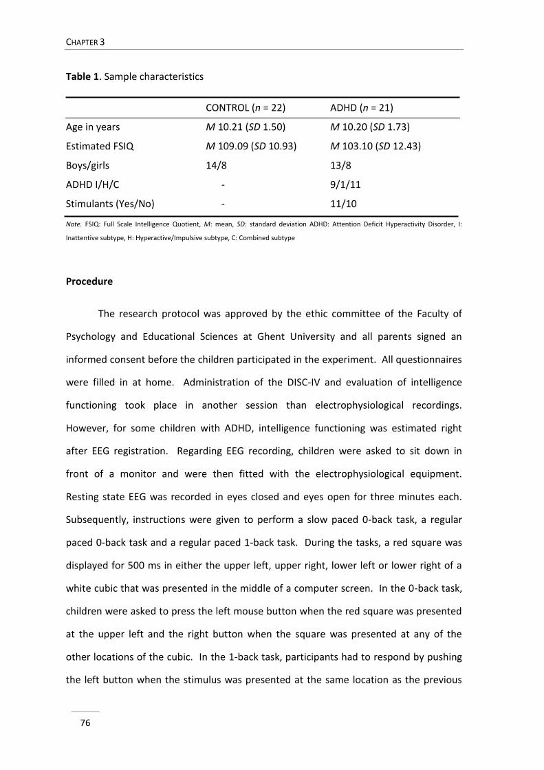
CHAPTER 3
76
Table 1. Sample characteristics
CONTROL (n = 22) ADHD (n = 21)
Age in years M 10.21 (SD 1.50) M 10.20 (SD 1.73)
Estimated FSIQ M 109.09 (SD 10.93) M 103.10 (SD 12.43)
Boys/girls 14/8 13/8
ADHD I/H/C - 9/1/11
Stimulants (Yes/No) - 11/10
Note. FSIQ: Full Scale Intelligence Quotient, M: mean, SD: standard deviation ADHD: Attention Deficit Hyperactivity Disorder, I:
Inattentive subtype, H: Hyperactive/Impulsive subtype, C: Combined subtype
Procedure
The research protocol was approved by the ethic committee of the Faculty of
Psychology and Educational Sciences at Ghent University and all parents signed an
informed consent before the children participated in the experiment. All questionnaires
were filled in at home. Administration of the DISC-IV and evaluation of intelligence
functioning took place in another session than electrophysiological recordings.
However, for some children with ADHD, intelligence functioning was estimated right
after EEG registration. Regarding EEG recording, children were asked to sit down in
front of a monitor and were then fitted with the electrophysiological equipment.
Resting state EEG was recorded in eyes closed and eyes open for three minutes each.
Subsequently, instructions were given to perform a slow paced 0-back task, a regular
paced 0-back task and a regular paced 1-back task. During the tasks, a red square was
displayed for 500 ms in either the upper left, upper right, lower left or lower right of a
white cubic that was presented in the middle of a computer screen. In the 0-back task,
children were asked to press the left mouse button when the red square was presented
at the upper left and the right button when the square was presented at any of the
other locations of the cubic. In the 1-back task, participants had to respond by pushing
the left button when the stimulus was presented at the same location as the previous
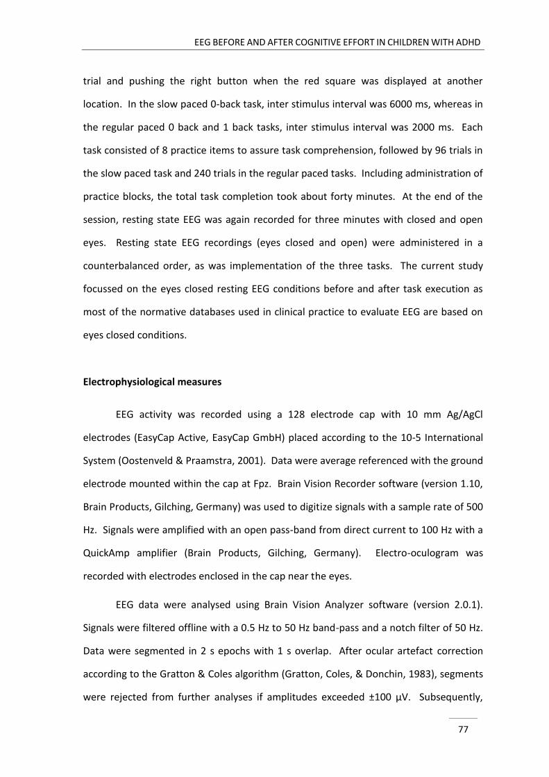
EEG BEFORE AND AFTER COGNITIVE EFFORT IN CHILDREN WITH ADHD
77
trial and pushing the right button when the red square was displayed at another
location. In the slow paced 0-back task, inter stimulus interval was 6000 ms, whereas in
the regular paced 0 back and 1 back tasks, inter stimulus interval was 2000 ms. Each
task consisted of 8 practice items to assure task comprehension, followed by 96 trials in
the slow paced task and 240 trials in the regular paced tasks. Including administration of
practice blocks, the total task completion took about forty minutes. At the end of the
session, resting state EEG was again recorded for three minutes with closed and open
eyes. Resting state EEG recordings (eyes closed and open) were administered in a
counterbalanced order, as was implementation of the three tasks. The current study
focussed on the eyes closed resting EEG conditions before and after task execution as
most of the normative databases used in clinical practice to evaluate EEG are based on
eyes closed conditions.
Electrophysiological measures
EEG activity was recorded using a 128 electrode cap with 10 mm Ag/AgCl
electrodes (EasyCap Active, EasyCap GmbH) placed according to the 10-5 International
System (Oostenveld & Praamstra, 2001). Data were average referenced with the ground
electrode mounted within the cap at Fpz. Brain Vision Recorder software (version 1.10,
Brain Products, Gilching, Germany) was used to digitize signals with a sample rate of 500
Hz. Signals were amplified with an open pass-band from direct current to 100 Hz with a
QuickAmp amplifier (Brain Products, Gilching, Germany). Electro-oculogram was
recorded with electrodes enclosed in the cap near the eyes.
EEG data were analysed using Brain Vision Analyzer software (version 2.0.1).
Signals were filtered offline with a 0.5 Hz to 50 Hz band-pass and a notch filter of 50 Hz.
Data were segmented in 2 s epochs with 1 s overlap. After ocular artefact correction
according to the Gratton & Coles algorithm (Gratton, Coles, & Donchin, 1983), segments
were rejected from further analyses if amplitudes exceeded ±100 µV. Subsequently,
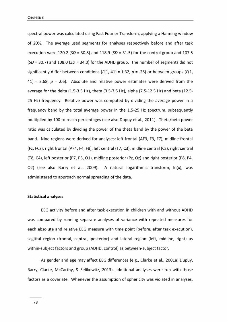
CHAPTER 3
78
spectral power was calculated using Fast Fourier Transform, applying a Hanning window
of 20%. The average used segments for analyses respectively before and after task
execution were 120.2 (SD = 30.8) and 118.9 (SD = 31.5) for the control group and 107.5
(SD = 30.7) and 108.0 (SD = 34.0) for the ADHD group. The number of segments did not
significantly differ between conditions (F(1, 41) = 1.32, p = .26) or between groups (F(1,
41) = 3.68, p = .06). Absolute and relative power estimates were derived from the
average for the delta (1.5-3.5 Hz), theta (3.5-7.5 Hz), alpha (7.5-12.5 Hz) and beta (12.5-
25 Hz) frequency. Relative power was computed by dividing the average power in a
frequency band by the total average power in the 1.5-25 Hz spectrum, subsequently
multiplied by 100 to reach percentages (see also Dupuy et al., 2011). Theta/beta power
ratio was calculated by dividing the power of the theta band by the power of the beta
band. Nine regions were derived for analyses: left frontal (AF3, F3, F7), midline frontal
(Fz, FCz), right frontal (AF4, F4, F8), left central (T7, C3), midline central (Cz), right central
(T8, C4), left posterior (P7, P3, O1), midline posterior (Pz, Oz) and right posterior (P8, P4,
O2) (see also Barry et al., 2009). A natural logarithmic transform, ln(x), was
administered to approach normal spreading of the data.
Statistical analyses
EEG activity before and after task execution in children with and without ADHD
was compared by running separate analyses of variance with repeated measures for
each absolute and relative EEG measure with time point (before, after task execution),
sagittal region (frontal, central, posterior) and lateral region (left, midline, right) as
within-subject factors and group (ADHD, control) as between-subject factor.
As gender and age may affect EEG differences (e.g., Clarke et al., 2001a; Dupuy,
Barry, Clarke, McCarthy, & Selikowitz, 2013), additional analyses were run with those
factors as a covariate. Whenever the assumption of sphericity was violated in analyses,
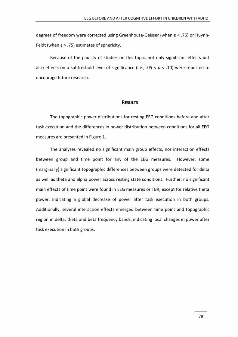
EEG BEFORE AND AFTER COGNITIVE EFFORT IN CHILDREN WITH ADHD
79
degrees of freedom were corrected using Greenhouse-Geisser (when ε < .75) or Huynh-
Feldt (when ε > .75) estimates of sphericity.
Because of the paucity of studies on this topic, not only significant effects but
also effects on a subtreshold level of significance (i.e., .05 < p < .10) were reported to
encourage future research.
RESULTS
The topographic power distributions for resting EEG conditions before and after
task execution and the differences in power distribution between conditions for all EEG
measures are presented in Figure 1.
The analyses revealed no significant main group effects, nor interaction effects
between group and time point for any of the EEG measures. However, some
(marginally) significant topographic differences between groups were detected for delta
as well as theta and alpha power across resting state conditions. Further, no significant
main effects of time point were found in EEG measures or TBR, except for relative theta
power, indicating a global decrease of power after task execution in both groups.
Additionally, several interaction effects emerged between time point and topographic
region in delta, theta and beta frequency bands, indicating local changes in power after
task execution in both groups.
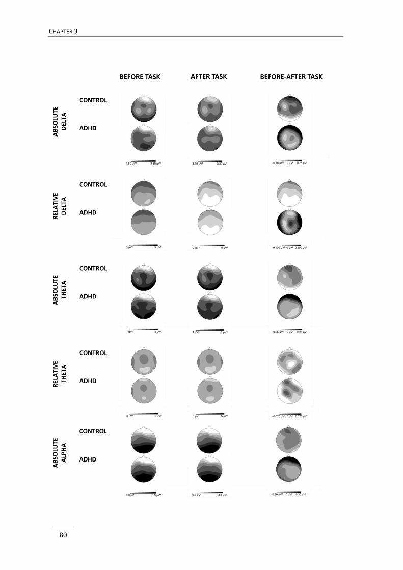
CHAPTER 3
80
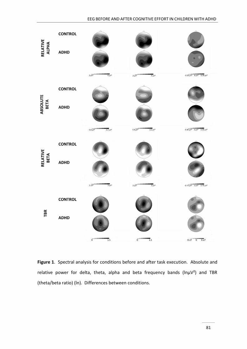
EEG BEFORE AND AFTER COGNITIVE EFFORT IN CHILDREN WITH ADHD
81
Figure 1. Spectral analysis for conditions before and after task execution. Absolute and
relative power for delta, theta, alpha and beta frequency bands (lnµV²) and TBR
(theta/beta ratio) (ln). Differences between conditions.
CHAPTER 3
82
Delta power
A marginally significant interaction between group and lateral region was found
for absolute delta power (F(2, 82) = 2.52, p = .09, η2p = .06). Group divergence did not
reach significance at either midline or lateral regions, as indicated by post hoc analyses
in the separate lateral regions. Yet, analyses in the separate groups revealed that power
across resting state conditions was significantly higher at midline than at lateral sites for
typically developing children (p < .01 for left and p < .05 for right region) but not for
children with ADHD (p = 1.00 for left and p = .76 for right region). For relative delta
power, a trend to significant effect was detected between group and sagittal region
(F(1.66, 68.02) = 2.82, p = .08, η2p = .07). Post hoc analyses yielded no significant group
differences in either frontal, central or posterior region. Within-subjects contrasts
demonstrated that the topographic progressive attenuation of delta power from
anterior to posterior sites was larger in control children than in children with ADHD, be it
only on a marginally significant level (F(1, 41) = 3.67, p = .06, η2p = .08).
Further, an interaction between time point and sagittal position emerged in both
absolute (F(2, 82) = 3.20, p < .05, η2p = .07) and relative delta (F(2, 82) = 3.40, p < .05, η2
p
= .08). Post hoc analyses revealed a posterior decrease in absolute power (F(1, 41) =
4.64, p = .04, η2p = .10) and a central increase in relative delta power after task execution
(F(1, 41) = 5.54, p < .05, η2p = .12).
Theta power
The topographic group effects that emerged in theta power were analogous to
the effects in delta power. That is, the marginally significant interaction between group
and lateral region (F(2, 82) = 2.57, p = .08, η2p = .06) for absolute theta power indicated
that theta was significantly higher for controls (p < .001 for both lateral sites) but not for
ADHD at midline than at lateral sites (p = 1.00 for left and p = .23 for right region) across
EEG BEFORE AND AFTER COGNITIVE EFFORT IN CHILDREN WITH ADHD
83
resting state conditions. Also, the interaction effect between group and sagittal region
for relative theta power (F(1.43, 58.44) = 3.30, p = .06, η2p = .08) demonstrated that the
power diminishment from anterior to posterior sites tended to be larger in control
children than in children with ADHD (F(1, 41) = 3.56, p = .07, η2p = .08).
In addition, an interaction between time point and sagittal position emerged in
absolute theta (F(1, 41) = 4.57, p = .01, η2p = .10), indicating a significant posterior (F(1,
41) = 7.84, p < .01, η2p = .16) and a marginally significant central reduction (F(1, 41) =
3.02, p = .09, η2p = .07) of absolute theta power after task execution. Further, these focal
reductions in absolute theta power led to a trend for a global decrease in absolute theta
power after cognitive effort (F(1, 41) = 3.92, p = .05, η2p = .09). As for relative measures,
a significant global reduction was found for theta power after task execution (F(1, 41) =
5.89, p = .02, η2p = .13).
Alpha power
For absolute alpha power, a marginally significant interaction between group and
sagittal region emerged (F(1.83, 75.08) = 3.07, p = .06, η2p = .07), an effect which reached
significance in relative alpha activity (F(2, 82) = 4.67, p = .01, η2p = .10). Post hoc tests
revealed no significant group differences in either sagittal region. However, within-
subjects contrasts demonstrated a larger progressive expansion from anterior to
posterior sites in absolute (F(1, 41) = 4.03, p = .05, η2p = .09) as well as in relative (F(1, 41)
= 6.89, p = .01, η2p = .14) alpha power in the control group relative to the ADHD group.
No significant effects involving time point were detected in absolute as well as
relative alpha power.
CHAPTER 3
84
Beta power
Topographic distribution of both absolute and relative beta power did not
significantly differ between groups.
The significant interaction between time point and sagittal region in absolute
beta (F(2, 82) = 6.18, p < .01, η2p = .13) power indicated a posterior (F(1, 41) = 5.86, p =
.02, η2p = .13) and central reduction (F(1, 41) = 5.07, p = .03, η2
p = .11) of absolute beta
power after task execution. As for relative beta power, a significant interaction emerged
between time point and sagittal region (F(2, 82) = 3.34, p < .05, η2p = .08). Post hoc
analyses revealed that relative central beta power tended to reduce (F(1, 41) = 3.13, p =
.08, η2p = .07) after cognitive effort.
TBR
TBR did not differentiate between groups and time points.
Additional analyses with age and sex as covariate
Separate additional analyses with sex and age as a covariate, marginally changed
group related results in alpha power. That is, the level of significance of the interaction
between group and sagittal region for absolute alpha slightly increased with sex or age
as a covariate (p-levels going from p = .06 to p = .04 for both covariates; effect sizes
going from η2p = .07 to η2
p = .08). When age was taken into account, the group x sagittal
region effect in relative theta power slightly increased (p-levels going from p = .06 to p <
.05, effect sizes remained η2p = .08). However, given the small groups, and therefore
reduced power, it might be more appropriate to evaluate the changes in the analyses
with covariates according to effect sizes rather than to p-levels. Hence, additional
analyses with these covariates showed that the effect sizes remained categorised as
medium effects.
EEG BEFORE AND AFTER COGNITIVE EFFORT IN CHILDREN WITH ADHD
85
DISCUSSION
The present study was set out to investigate whether plausible EEG deviances,
particularly enhanced theta power and increased TBR, are a stable characteristic of
children with ADHD across eyes closed resting conditions before and after cognitive
effort or whether discrepancies may be context or state dependent.
Across conditions, no higher theta power or elevated TBR was observed in
children with ADHD relative to typically developing children. Also, groups were not
found to diverge in terms of higher or lower power values in delta, alpha and beta
frequencies. Yet, across resting conditions, a significant larger topographic expansion
from anterior to posterior sites was detected in absolute and relative alpha power in
typically developing children compared to children with ADHD. Also, the control group
demonstrated a larger sagittal diminution from anterior to posterior sites in relative
delta and theta power, although this effect did not reach an acceptable significance
level. Further, the midline distribution in absolute theta and delta power tended to be
higher in typically developing children compared to children with ADHD. With respect to
state-related effects following task execution, a global reduction of relative theta power
and an attenuation in absolute delta, absolute theta and absolute beta power at
posterior sites were detected in both groups, as well as a decrease in central beta power
and an increase in central relative delta power. Yet, no evidence was found for altered
state-related changes in EEG after task execution in ADHD.
The findings of the present study disaccord with frequently reported elevated
theta power or increased TBR in other studies (for a review, see Barry et al., 2003; Clarke
et al., 2007; Clarke et al., 2011; Dupuy et al., 2011; Lansbergen et al., 2011; Loo et al.,
2010; Shi et al., 2012). However, our results are in line with some recent studies
(Coolidge et al., 2007; Liechti et al., 2013; Loo et al., 2013; Nazari et al., 2011; Ogrim et
al., 2012; Swartwood et al., 2003) that also failed to replicate these abnormalities.
CHAPTER 3
86
Findings regarding delta, alpha and beta power are not surprising since deviances in
those EEG rhythms in ADHD have not consistently been established across publications
(see introduction). As for the absence of finding reduced beta power or elevated TBR in
the ADHD group, one may argue that these deviances were not detected because our
sample comprised a subgroup of children with ADHD showing excessive beta power
(Clarke, Barry, McCarthy, & Selikowitz, 2001c). However, additional analyses in this
respect revealed that this was not the case. In comparison to other studies (for an
overview, see Arns et al., 2013; Liechti et al., 2013), non-logaritmic transformed TBRs at
the vertex in the present study are in the range of published TBRs for both groups. Yet,
they approach the maximum for the controls, and the minimum published values for the
ADHD groups compared to other publications. These findings align with the
identification of a decreasing difference in TBR in children with ADHD relative to control
groups across studies in the last decade (Arns et al., 2013). Poor distinction between
groups on a behavioural level is most probably also not a confounder in the present
study, since severe inclusion criteria were used in both groups, resulting in highly
distinguishable groups in terms of the presence of ADHD symptoms as determined in the
DBD Rating Scale. Nevertheless, it is plausible that differences in the make-up of the
sample may have contributed to divergent findings across studies (Arns et al., 2013;
Liechti et al., 2013). Our ADHD sample comprised children with ADHD inattentive
subtype (i.e., 9) as well as combined (i.e., 11) and hyperactive-impulsive (i.e., 1) subtype,
which may have played a role. Regarding EEG differences between ADHD subtypes,
some studies were not able to detect EEG divergence between ADHD subtypes, while
others reported more aberrant EEG activity in one subtype relative to the other and/or
the control group (Buyck & Wiersema, 2014; Clarke et al., 1998, 2001b; Dupuy et al.,
2011; Hermens et al., 2004; Loo et al., 2010). Unfortunately, reliable examination of EEG
divergence according to ADHD subtypes is hampered by the size of our sample, yet this
calls for further research. In addition, the presence of comorbid disorders in the ADHD
EEG BEFORE AND AFTER COGNITIVE EFFORT IN CHILDREN WITH ADHD
87
groups, which is actually rather a rule than an exception, may also serve as an explaining
factor for diverse results between studies. Although the influence of comorbidity on
resting EEG findings in ADHD has not been investigated systematically, a recent study
points to possible mediating effects of oppositional defiant disorder (ODD)/conduct
disorder (CD) and depressive disorder in deviance in TBR in ADHD (Loo et al., 2013). As
for the present study, 5 children with ADHD were diagnosed with comorbid ODD, one of
them also received a diagnosis of CD. More studies with larger sample sizes are needed
to evaluate the impact of comorbidity on resting EEG findings in ADHD. Another factor
that might be relevant in explaining the absence of aberrant theta power and TBR is the
relatively high number of girls (i.e., male to female ratio approximately 2:1) adopted in
the current study. Some evidence has been found for smaller EEG differences between
girls relative to boys with ADHD and typically developing children (for a review, see
Dupuy et al., 2013), yet it has also been documented that enhanced TBR in ADHD is not
affected by sex (Monastra et al., 1999). Moreover, some studies evaluating only females
have reported increased slow wave activity and/or an elevated TBR (Clarke et al., 2003;
Dupuy et al., 2011) in girls with ADHD. In addition, controlling for sex in our analyses
appeared not to contribute largely to changing group results. Hence, the absence of EEG
abnormalities in children with ADHD is most likely not due to a gender-related factor,
but future studies are warranted that systematically investigate this issue. Finally, in the
current study, both children with ADHD who take or do not take stimulants in daily life
have been incorporated. This factor also differs between studies, with some
investigating only medication naïve patients (e.g., Clarke et al., 2008), while most others
examining both children who are on and off medication in daily life (e.g., Shi et al.,
2012). Studies exploring the effects of stimulant medication on brain wave activity have
indicated that the intake of stimulants results in a decrease in theta power (Loo, Teale, &
Reite, 1999; Clarke et al., 2003; Clarke, Barry, McCarthy, Selikowitz, & Johnstone, 2007)
and TBR (Clarke, Barry, McCarthy, Selikowitz, & Croft, 2002b). However, in the present
CHAPTER 3
88
study medication use was controlled for by requiring a wash-out period of 48 hours for
children who used medication in daily life. Consequently, we doubt that medication use
in a part of our ADHD sample may have been a confounding factor, although future
studies investigating long-term medication effects on resting EEG in ADHD are
recommended.
Although the findings of the present study do not confirm the typically observed
aberrant theta power and TBR in ADHD in other studies, divergence in topographic
distribution of EEG activity on the sagittal plane was found for alpha power with a
smaller expansion from anterior to posterior sites in children with ADHD. Importantly,
this cannot be explained by the children with ADHD incidentally opening the eyes during
the recordings, while an experimenter monitored the children during the experiment.
We are not aware of any study that examined linear within subjects contrasts in
deviation between groups, which impedes comparison of our observation of smaller
changes in EEG distribution from anterior to posterior sites in alpha to other studies.
Also, to our knowledge, no publications are available regarding the significance of the
topographic pattern of EEG activity between different regions. Even so, it is well known
that during resting state, the brain attempts to preserve a physiological and functional
balance. In order to do so, different local and long-range cortical neural activities co-
occur and may interact with each other (Woltering, Jung, Liu, & Tannock, 2012).
Variations in topographic distribution in ADHD may therefore reflect deficient neural
communication or the use of different underlying sources of activity, which may be
related to ADHD symptomatology. While the results of the present study are suggestive
of possible different topographic distribution between ADHD and controls, before
drawing further conclusions on this, studies involving a larger number of participants will
be needed to confirm generalizability of the results. Despite this limitation, other
studies investigating a similar widespread topographic distribution in ADHD also point to
deviance between groups depending on examined region (e.g., Clarke et al., 1998;
EEG BEFORE AND AFTER COGNITIVE EFFORT IN CHILDREN WITH ADHD
89
2001), which underlines the importance of investigating widespread cortical activity in
ADHD and not limiting EEG comparison between groups to a small number of electrode
derivations.
As for EEG changes after cognitive effort, the present findings confirm that
intervening task execution results in modified resting EEG patterns after effort, be it
similarly in both groups. The most consistent finding in studies that have examined EEG
before and after task execution has been an increase in theta rhythm following cognitive
effort (Smit et al., 2004, 2005; Tanaka et al., 2012), which has traditionally been
accounted for by mental fatigue. A theta increase after cognitive effort was not
replicated in the present study, in which instead a decrease in theta power was
detected. It may be argued that performing n-back tasks for only 40 minutes may not
have been sufficient to induce mental fatigue and therefore an increase in theta power
has not been generated. Previous research has documented that task difficulty may also
interfere with the appearance of EEG changes after task execution, suggesting that less
demanding tasks elucidate smaller EEG changes (Tanaka et al., 2012). The 0-back tasks
used in the present study were not highly demanding in terms of workload, which could
therefore also explain the failure to find a theta increase after task performance. This
however does not explain the decrease in theta power after task execution as noticed in
our study. Yet, modulation in the theta frequency band has not only been associated
with a possible reflection of fatigue or task difficulty but also with attentional
processing. Although tentative, in accord with research on the effects of meditation on
EEG activity relating higher theta power to enhanced attention to internal events (for
reviews, see Chiesa & Serretti, 2010; Rubia, 2009), the present finding of a reduction in
theta power after cognitive effort may perhaps be the result of participants being more
in a state of external focussed attention after task execution than during the baseline
resting condition. As for the decrease in beta power after task execution, this is in line
with findings of the study by Tanaka and colleagues (2012). Since beta waves are
CHAPTER 3
90
generally associated with increased attentional arousal (Loo & Makeig, 2012), decreases
in beta power after task execution may reflect reduced alertness and attentional
processing. Noteworthy is that the EEG changes after task execution were most
pronounced at posterior sites. Remarkably, several positron emission tomography and
functional magnetic resonance imaging studies have elucidated involvement of the
parietal cortex in the performance of spatial tasks (Corbetta, Miezin, Shulman, &
Petersen, 1993; Coul & Nobre, 1998; Nobre et al., 1997, 2004; Nobre, 2001). The
observed EEG changes after task execution in the current study may therefore possibly
reflect altered activity in the parietal cortex, after this brain area has been activated
during task performance. Yet, given the relatively poor spatial sensitivity in EEG,
carefulness is warranted in this interpretation. However, this hypothesis may also
explain why EEG modulation after cognitive effort has been found to differ across
studies. That is, since involvement of different brain areas is required during a variety of
tasks, this possibly will be reflected in varying topographic distribution of EEG changes
after cognitive effort. This may also imply that, although not detected in the present
study, there is still potential to elucidate group divergence in EEG modulation after task
execution when different tasks, tapping other brain related processes that possibly more
discriminate between patients and controls, are used between EEG measurements.
Although not controlled for in the present study, other factors may also
contribute to changes in EEG activity after cognitive effort, such as feeling more at ease
at the end of the session compared to at the start of the experiment. Both theta and
beta (Knyazev, Savostyanov, & Levin, 2005), as well as delta power (Knyazev, 2012),
have been associated with feelings of anxiety and uncertainty. Consequently, it is
recommended that future studies incorporate additional measures to assess the
significance of the induced alteration in state after cognitive effort, since the
interpretation regarding the significance of the EEG changes after cognitive effort
remains difficult. For instance, questionnaires can be used to detect fatigue or changes
EEG BEFORE AND AFTER COGNITIVE EFFORT IN CHILDREN WITH ADHD
91
in emotion/motivation after cognitive effort and a well-established measure of
autonomic arousal (i.e., skin conductance level, Barry et al., 2009) could assist in
detecting possible arousal differences before and after task execution. Furthermore, it
could also be of value to control for possible circadian influences in future studies, since
these have been demonstrated to affect EEG (Sannita, 2006). Unfortunately, we were
not able to do so in the current study as we were dependent on the schedule of the
children and their parents to conduct the experiment. That is, children were assessed
after school hours or during weekends, taking into account the feasibility of refraining
from medication.
Our hypothesis regarding possible state-related factors associated with EEG
deviance in ADHD has not been confirmed by the current study design. However,
leaving open that task duration or requirement of mental effort were not sufficient to
induce altered state-related EEG changes in ADHD, we believe prudence is called for
interpretation of these results. There is ample evidence for state related deficits in
ADHD on a behavioural, performance, and psychophysiological level (e.g., Metin et al.,
2012; Sonuga-Barke et al., 2010; Wiersema et al., 2006a,b), and findings from studies on
eyes open versus eyes closed EEG suggest that this may also be the case for quantitative
EEG measures (e.g., Fonseca et al., 2013; Liechti et al., 2013). Hence, more research is
definitely needed on the influence of state and contextual factors on resting EEG in
ADHD.
To conclude, the findings of the present study do not confirm the validity of
deviances in theta and TBR characterizing the whole ADHD group, which is in line with a
growing number of studies. However, divergences in topographic organization of EEG
activity between groups were detected, and therefore, examining widespread cortical
activity is recommended in evaluating EEG in ADHD. Finally, the results suggest that
time point of EEG measurement did not influence group differences, arguing against
CHAPTER 3
92
state-related EEG deviances in ADHD, though more research studying context
dependency of quantitative EEG measures in ADHD is recommended.
EEG BEFORE AND AFTER COGNITIVE EFFORT IN CHILDREN WITH ADHD
93
REFERENCES
Achenbach, T.M., & Rescorla, L.A. (2001). Manual for the ASEBA School-Age Forms and Profiles.
Burlington, Vermont: University of Vermont, Research Center for Children, Youth, and
Families.
American Psychiatric Association (2013). Diagnostic and statistical manual of mental disorders
(5th Ed.). Arlington, VA: American Psychiatric Publishing.
Antonenko, P., Paas, F., Grabner, R., & van Gog, T. (2010). Using Electroencephalography to
Measure Cognitive Load. Educational Psychology Review, 22, 425-438. doi:
10.1007/s10648-010-9130-y
Antrop, I., Buysse, A., Roeyers, H., & Van Oost P. (2005). Activity in children with ADHD during
waiting situations in the classroom: a pilot study. British Journal of Educational
Psychology, 75(Pt 1), 51-69. doi: 10.1348/000709905X36100
Arns, M., Conners, C.K., & Kraemer, H.C. (2013). A decade of EEG theta/beta ratio research in
ADHD: a meta-analysis. Journal of Attention Disorders, 17, 374-383. doi:
10.1177/1087054712460087
Arns, M., Drinkenburg, W.H.I.M., & Kenemans, J.L. (2012). The effects of QEEG-informed
neurofeedback in ADHD: An open label pilot study. Applied Psychophysiology and
Biofeedback, 37, 171-180. doi: 10.1007/s10484-012-9191-4
Barry, R.J., Clarke, A.R. & Johnstone, S.J. (2003). A review of electrophysiology in attention-
deficit/hyperactivity disorder: I. Qualitative and quantitative electroencephalography.
Clinical Neurophysiology, 114, 171-183. doi: 10.1016/S1388-2457(02)00362-0
Barry, R.J., Clarke, A.R., Johnstone, S.J., Magee, C.A., & Rushby, J.A. (2007). EEG differences
between eyes-closed and eyes-open resting conditions. Clinical Neurophysiology, 118,
2765-2773. doi: 10.1016/j.clinph.2007.07.028
Barry, R.J., Clarke, A.R., Johnstone, S.J., McCarthy, R., & Selikowitz, M. (2009).
Electroencephalogram theta/beta ratio and arousal in attention-deficit/hyperactivity
disorder: evidence of independent processes. Biological Psychiatry, 66, 398–401. doi:
10.1016/j.biopsych.2009.04.027
CHAPTER 3
94
Benikos, N., & Johnstone, S.J. (2009). Arousal-state modulation in children with AD/HD. Clinical
Neurophysiology, 120, 30-40. doi: 10.1016/j.clinph.2008.09.026
Börger, N.A., & van der Meere, J.J. (2000). Motor control and state regulation in children with
ADHD: a cardiac response study. Biological Psychology, 51, 247-267. doi: 10.1016/S0301-
0511(99)00040-X
Buyck, I., & Wiersema, J.R. (2014). Resting electroencephalogram in attention deficit
hyperactivity disorder: developmental course and diagnostic value. Psychiatry Research.
Advance online publication. doi: 10.1016/j.psychres.2013.12.055
Chabot, R.J., & Serfontein, G. (1996). Quantitative electroencephalographic profiles of children
with attention deficit disorder. Biological Psychiatry, 40, 951-963. doi: 10.1016/0006-
3223(95)00576-5
Chiesa, A., & Serretti, A. (2010). A systematic review of neurobiological and clinical features of
mindfulness meditations. Psychological Medicine, 40, 1239-1252. doi:
10.1017/S0033291709991747
Clarke, A.R., Barry, R.J., Dupuy, F.E., McCarthy, R., Selikowitz, M., & Heaven, P.C. (2011).
Childhood EEG as a predictor of adult attention-deficit/hyperactivity disorder. Clinical
Neurophysiology, 122(1), 73-80. doi: 10.1016/j.clinph.2010.05.032
Clarke, A.R., Barry, R.J., Heaven, P.C.L., McCarthy, R., Selikowitz, M., & Byrne, M.K. (2008). EEG in
adults with Attention-Deficit/Hyperactivity Disorder. International Journal of
Psychophysiology, 70, 176-183. doi: 10.1016/j.ijpsycho.2008.07.001
Clarke, A., Barry, R., McCarthy, R., & Selikowitz, M. (1998). EEG analysis in attention
deficit/hyperactivity disorder: a comparative study of two subtypes. Psychiatry Research
81, 19-29. doi: 10.1016/S0165-1781(98)00072-9
Clarke, A.R., Barry, R.J., McCarthy, R., & Selikowitz, M. (2001a). Age and sex effects in the EEG:
differences in two subtypes of attention-deficit/hyperactivity disorder. Clinical
Neurophysiology, 112(5), 815-826. doi: 10.1016/S1388-2457(01)00487-4
Clarke, A.R., Barry, R.J., McCarthy, R., & Selikowitz, M. (2001b). EEG-defined subtypes of children
with attention-deficit/hyperactivity disorder. Clinical Neurophysiology, 112, 2098-2105.
doi: 10.1016/S1388-2457(01)00668-X
EEG BEFORE AND AFTER COGNITIVE EFFORT IN CHILDREN WITH ADHD
95
Clarke, A.R., Barry, R.J., McCarthy, R., & Selikowitz, M. (2001c). Excess beta in children with
attention-deficit/hyperactivity disorder: An atypical electrophysiological group.
Psychiatry Research, 103, 205-218. doi: 10.1016/S0165-1781(01)00277-3
Clarke, A., Barry, R., McCarthy, R., & Selikowitz, M. (2002a). EEG analysis of children with
attention-deficit/hyperactivity disorder and comorbid reading disabilities. Journal of
Learning Disabilities, 35, 276-285. doi: 10.1177/002221940203500309
Clarke, A.R., Barry, R.J., McCarthy, R., Selikowitz, M., & Croft, R.J. (2002b). EEG differences
between good and poor responders to methylphenidate in boys with the inattentive
type of attention-deficit/hyperactivity disorder. Clinical Neurophysiology, 113, 1191–
1198. doi: 10.1016/S1388-2457(02)00147-5
Clarke, A.R., Barry, R.J., McCarthy, R., Selikowitz, M., Brown, C.R., & Croft, R.J. (2003). Effects of
stimulant medications on the EEG of children with Attention-Deficit Hyperactivity
Disorder Predominantly Inattentive type. International Journal of Psychophysiology, 47,
129-137. doi: 10.1016/S0167-8760(02)00119-8
Clarke, A.R., Barry, R.J., McCarthy, R., Selikowitz, M., & Johnstone, S.J. (2007). Effects of
stimulant medications on the EEG of girls with Attention-Deficit/Hyperactivity. Clinical
Neurophysiology, 118(12), 2700-2708. doi: 10.1016/j.clinph.2007.08.020
Coolidge, F.L., Starkey, M.T., & Cahill, B.S. (2007). Comparison of a Parent-Rated DSM-IV
Measure of Attention-Deficit/Hyperactivity Disorder and Quantitative EEG Parameters in
an Outpatient Sample of Children. Journal of Clinical Neurophysiology, 24(4), 348-351.
doi: 10.1097/WNP.0b013e318067bcfc
Corbetta, M., Miezin, F.M., Shulman, G.L., & Petersen, S.E. (1993). A PET study of visuospatial
attention. The Journal of Neuroscience, 13, 1202-1226. Retrieved from
http://www.jneurosci.org/content/13/3/1202.full.pdf+html
Coull, J.T., & Nobre, A.C. (1998). Where and when to pay attention: the neural systems for
directing attention to spatial locations and to time intervals as revealed by both PET and
fMRI. The Journal of Neuroscience, 18, 7426-7435. Retrieved from
http://www.jneurosci.org/content/18/18/7426.full.pdf
CHAPTER 3
96
Dupuy, F.E., Barry, R.J., Clarke, A.R., McCarthy, R., & Selikowitz, M. (2013). Sex differences
between the combined and inattentive types of attention-deficit/hyperactivity disorder:
An EEG perspective. International Journal of Psychophysiology, 89, 320-327. doi:
10.1016/j.ijpsycho.2013.04.004
Dupuy, F.E., Clarke, A.R., Barry, R.J., McCarthy, R., & Selikowitz, M. (2011). Girls with attention-
deficit/hyperactivity disorder: EEG differences between DSM-IV types. Clinical EEG and
Neuroscience, 42, 1-5. doi: 10.1177/155005941104200104
Fein, G., Galin, D., Yingling, C.D., Johnstone, J., & Nelson, M .A. (1984). EEG spectra in 9-13-year-
old boys are stable over 1-3 years. Electroencephalography and Clinical Neurophysiology,
58, 517-518. doi: 10.1016/0013-4694(84)90041-5
Fonseca, L.C., Tedrus, G.M.A.S., Bianchini, M.C., & Silva, T.F. (2013). Electroencephalographic
alpha reactivity on opening the eyes in children with attention-deficit hyperactivity
disorder. Clinical EEG and Neuroscience, 44(1), 53-57. doi: 10.1177/1550059412445659
Gasser, T., Bacher, P., & Steinberg, H. (1985). Test-retest reliability of spectral parameters of the
EEG. Electroencephalography and Clinical Neurophysiology, 60, 312-319. doi:
10.1016/0013-4694(85)90005-7
González-Castro, P., Rodríguez, C., López, A., Cueli, M., & Álvarez, L. (2013). Attention Deficit
Hyperactivity Disorder, differential diagnosis with blood oxygenation, beta/theta ratio,
and attention measures. International Journal of Clinical and Health Psychology, 13(2),
101-109. doi: 10.1016/S1697-2600(13)70013-9
Gratton, G., Coles, M.G.H., & Donchin, E. (1983). A new method for off-line removal of ocular
artifact. Electroencephalography and Clinical Neurophysiology, 55, 468-484. doi:
10.1016/0013-4694(83)90135-9
Grégoire, J. (2000). L’évaluation clinique de l’intelligence de l’enfant. Liège: Mardaga.
Hammond, D.C. (2010). The need for individualization in neurofeedback: heterogeneity in QEEG
patterns associated with diagnoses and symptoms. Applied Psychophysiology and
Biofeedback, 35, 31-36. doi: 10.1007/s10484-009-9106-1
EEG BEFORE AND AFTER COGNITIVE EFFORT IN CHILDREN WITH ADHD
97
Hermens, D.F., Williams, L.M., Lazzaro, I., Whitmont, S., Melkonian, D., & Gordon, E. (2004). Sex
differences in adult ADHD: a double dissociation in brain activity and autonomic arousal.
Biological Psychology, 66, 221-233. doi: 10.1016/j.biopsycho.2003.10.006
Imeraj, L., Antrop, I., Roeyers, H., Deschepper, E., Bal, S., & Deboutte, D. (2011). Diurnal
variations in arousal: a naturalistic heart rate study in children with ADHD. European
Child & Adolescent Psychiatry, 20, 381-392. doi: 10.1007/s00787-011-0188-y
Imeraj, L., Antrop, I., Roeyers, H., Swanson, J., Deschepper, E., Bal, S., & Deboutte, D. (2012).
Time-of-day effects in arousal: Disrupted diurnal cortisol profiles in children with ADHD.
Journal of Child Psychology and Psychiatry, 53, 782-789. doi: 10.1111/j.1469-
7610.2012.02526.x
Kim, M.K., Kim, M., Oh, E., & Kim, S.P. (2013). A review on the computational methods for
emotional state estimation from the human EEG. Computational and Mathematical
Methods in Medicine, 2013, 573734. doi: 10.1155/2013/573734
Knyazev, G.G. (2012). EEG delta oscillations as a correlate of basic homeostatic and motivational
processes. Neuroscience & Biobehavioral Reviews, 36, 677-695. doi: 10.1016/
j.neubiorev.2011.10.002
Knyazev, G.G., Savostyanov, A.N., & Levin, E.A. (2005). Uncertainty, anxiety and brain
oscillations. Neuroscience Letters, 387, 121-125. doi: 10.1016/j.neulet.2005.06.016
Koehler, S., Lauer, P., Schreppel, T., Jacob, C., Heine, M., Boreatti-Hümmer, A., … & Herrmann,
M. (2009). Increased EEG power density in alpha and theta bands in adult ADHD
patients. Journal of Neural Transmission, 116, 97-104. doi: 10.1007/s00702-008-0157-x
Kooistra, L., van der Meere, J., Edwards, J., Kaplan, B.J., Crawford, S., & Goodyear, B.G. (2010).
Preliminary fMRI findings on the effects of event rate in adults with ADHD. Journal of
Neural Transmission, 117, 655-662. doi: 10.1007/s00702-010-0374-y
Lansbergen, M., Arns, M., van Dongen-Boomsma, M., Spronk, D., & Buitelaar, J. (2011). The
increase in theta/beta ratio on resting state EEG in boys with attention-
deficit/hyperactivity disorder is mediated by slow alpha peak frequency. Progress in
Neuropsychopharmacology & Biological Psychiatry, 35(1), 47-52. doi:
10.1016/j.pnpbp.2010.08.004
CHAPTER 3
98
Liechti, M.D., Valko, L., Müller, U.C., Döhnert, M., Drechsler, R., Steinhausen, H.C., & Brandeis, D.
(2013). Diagnostic value of resting electroencephalogram in attention-
deficit/hyperactivity disorder across the lifespan. Brain Topography, 26(1), 135-151. doi:
10.1007/s10548-012-0258-6
Loo, S.K., Cho, A., Hale, T.S., McGough, J., McCracken, J., & Smalley, S.L. (2013). Characterization
of the Theta to Beta Ratio in ADHD: Identifying Potential Sources of Heterogeneity.
Journal of Attention Disorders, 17(5), 384-392. doi: 10.1177/1087054712468050
Loo, S.K., Hale, T.S., Hanada, G., Macion, J., Shrestha, A., McGough, J.J., ... & Smalley, S.L. (2010).
Familial clustering and DRD4 effects on electroencephalogram measures in multiplex
families with attention deficit/hyperactivity disorder. Journal of the American Academy
of Child and Adolescent Psychiatry, 49(4), 368-377. doi: 10.1016/j.jaac.2010.01.002
Loo, S.K., & Makeig, S. (2012). Clinical utility of EEG in attention-deficit/hyperactivity disorder: a
research update. Neurotherapeutics, 9, 569-587. doi: 10.1007/s13311-012-0131-z
Loo, S.K., Teale, P.D., & Reite, M.L. (1999). EEG correlates of methylphenidate response among
children with ADHD: A preliminary report. Biological Psychiatry, 45(12), 1657-1660. doi:
10.1016/S0006-3223(98)00250-9
Luman, M., Oosterlaan, J., & Sergeant, J.A. (2005). The impact of reinforcement contingencies on
AD/HD: a review and theoretical appraisal. Clinical Psychology Review, 25, 183-213. doi:
10.1016/j.cpr.2004.11.001
Metin, B., Roeyers, H., Wiersema, J.R., van der Meere, J., & Sonuga-Barke, E. (2012). A Meta-
Analytic Study of Event Rate Effects on Go/No-Go Performance in Attention-
Deficit/Hyperactivity Disorder. Biological Psychiatry, 72, 990-996. doi:
10.1016/j.biopsych.2012.08.023
Monastra, V.J., Lubar, J.F., Linden, M., Van Deusen, P., Green, G., Wing, W., … & Fenger, T.N.
(1999). Assessing attention deficit hyperactivity disorder via quantitative
electroencephalography: An initial validation study. Neuropsychology, 13, 424-433. doi:
10.1037/0894-4105.13.3.424
Nazari, M.A., Wallois, F., Aarabi, A., & Berquin, P. (2011). Dynamic changes in quantitative
electroencephalogram during continuous performance test in children with attention-
EEG BEFORE AND AFTER COGNITIVE EFFORT IN CHILDREN WITH ADHD
99
deficit/hyperactivity disorder. International Journal of Psychophysiology, 81(3), 230-236.
doi: 10.1016/j.ijpsycho.2011.06.016
Nobre, A.C. (2001). The attentive homunculus: now you see it, now you don't. Neuroscience &
Biobehavioral Reviews, 25, 477-496. doi: 10.1016/S0149-7634(01)00028-8
Nobre, A.C., Coull, J.T., Maquet, P., Frith, C.D., Vandenberghe, R., & Mesulam, M.M. (2004).
Orienting attention to locations in perceptual versus mental representations. Journal of
Cognitive Neuroscience, 16, 363-373. doi: 10.1162/089892904322926700
Nobre, A.C., Sebestyen, G.N., Gitelman, D.R., Mesulam, M.M., Frackowiak, R.S., & Frith, C.D.
(1997). Functional localization of the system for visuospatial attention using positron
emission tomography. Brain, 120 (Pt 3), 515-533. doi: 10.1093/brain/120.3.515
Ogrim, G., Kropotov, J., & Hestad, K. (2012). The QEEG TBR in ADHD and normal controls:
Sensitivity, specificity and behavioral correlates. Psychiatry Research, 198(3), 482-488.
doi: 10.1016/j.psychres.2011.12.041
Oken, B.S., Salinsky, M.C., & Elsas, S.M. (2006). Vigilance, alertness, or sustained attention:
physiological basis and measurement. Clinical Neurophysiology, 117, 1885-1901. doi:
10.1016/j.clinph.2006.01.017
Oostenveld, R., & Praamstra, P. (2001). The five percent electrode system for high resolution
EEG and ERP measurements. Clinical Neurophysiology, 112, 713-719. doi: 10.1016/S1388-
2457(00)00527-7
Oosterlaan, J., Scheres, A., Antrop, I., Roeyers, H., & Sergeant, J.A. (2000). Vragenlijst voor
Gedragsproblemen bij Kinderen (VvGK). Nederlandse bewerking van de Disruptive
Behavior Disorders Rating Scale [Dutch translation of the Disruptive Behavior Disorders
Rating Scale]. Lisse, The Netherlands: Swets & Zeitlinger.
Pelham, W., Gnagy, E.M., Greenslade, K.E., & Milich, R. (1992). Teacher ratings of DSM-III-R
symptoms for the disruptive behavior disorders. Journal of the American Academy of
Child and Adolescent Psychiatry, 31, 210-218. doi: 10.1097/00004583-199203000-00006
Power, T.J. (1992). Contextual factors in vigilance testing of children with ADHD. Journal of
Abnormal Child Psychology, 20, 579-593. doi: 10.1007/BF00911242
CHAPTER 3
100
Putman, P., van Peer, J.M., Maimari I., & van der Werff, S. (2010). EEG theta/beta ratio in
relation to fear-modulated response-inhibition, attentional control, and affective traits.
Biological Psychology, 83, 73-78. doi: 10.1016/j.biopsycho.2009.10.008
Rubia, K. (2009). The neurobiology of Meditation and its clinical effectiveness in psychiatric
disorders. Biological Psychology, 82(1), 1-11. doi: 10.1016/j.biopsycho.2009.04.003
Sannita, W.G. (2006). Individual variability, end-point effects and possible biases in
electrophysiological research. Clinical Neurophysiology, 117, 2569-2583. doi:
10.1016/j.clinph.2006.04.026
Schaffer, D., Fisher, P., Lusac, C.P., Dulcan, M.K., & Schwab-Stone, M.E. (2000). NIMH Diagnostic
Interview Schedule for Children version IV (NIMH DISC-IV): Description, differences from
previous versions and reliability of some common diagnoses. Journal of the American
Academy of Child and Adolescent Psychiatry, 39, 28-38. doi: 10.1097/00004583-
200001000-00014
Scheres, A., Oosterlaan, J.,& Sergeant, J.A. (2001). Response Execution and Inhibition in Children
with AD/HD and other Disruptive Disorders: The Role of Behavioural Activations. Journal
of Child Psychology and Psychiatry and Allied Disciplines, 42(3), 347-357. doi:
10.1111/1469-7610.00728
Sergeant, J. (2005). Modeling attention-deficit/hyperactivity disorder: a critical appraisal of the
cognitive-energetic model. Biological Psychiatry, 57, 1248-1255. doi:
10.1016/j.biopsych.2004.09.010
Shi, T., Li, X., Song, J., Zhao, N., Sun, C., Xia, W., … & Tomoda, A. (2012). EEG characteristics and
visual cognitive function of children with attention deficit hyperactivity disorder (ADHD).
Brain and Development, 34 (10), 806-811. doi: 10.1016/j.braindev.2012.02.013
Smit, A.S., Eling, P.A., & Coenen, A.M. (2004). Mental effort affects vigilance enduringly: after-
effects in EEG and behavior. International Journal of Psychophysiology, 53, 239-243. doi:
10.1016/j.ijpsycho.2004.04.005
Smit, A.S., Eling, P.A., Hopman, M.T., & Coenen, A.M. (2005). Mental and physical effort affect
vigilance differently. International Journal of Psychophysiology, 57, 211-217. doi:
10.1016/j.ijpsycho.2005.02.001
EEG BEFORE AND AFTER COGNITIVE EFFORT IN CHILDREN WITH ADHD
101
Snyder, S.M., Quintana, H., Sexson, S.B., Knott, P., Haque, A.F., & Reynolds, D.A. (2008). Blinded,
multi-center validation of EEG and rating scales in identifying ADHD within a clinical
sample. Psychiatry Research, 159(3), 346-358. doi: 10.1016/j.psychres.2007.05.006
Sonuga-Barke, E.J.S., Taylor, E., Sembi, S., & Smith, J. (1992). Hyperactivity and delay aversion: I.
The effect of delay on choice. Journal of Child Psychology and Psychiatry and Allied
Disciplines, 33(2), 387-398. doi: 10.1111/j.1469-7610.1992.tb00874.x
Sonuga-Barke, E., Wiersema, J.R., van der Meere J., & Roeyers, H. (2010). Context-dependent
Dynamic Processes in Attention Deficit/Hyperactivity Disorder: Differentiating Common
and Unique Effects of State Regulation Deficits and Delay Aversion. Neuropsychological
Review, 20, 86-102. doi: 10.1007/s11065-009-9115-0
Sonuga-Barke, E.J.S., Williams, E., Hall, M., & Saxton, T. (1996). Hyperactivity and delay aversion
III. The effect on cognitive style of imposing delay after errors. The Journal of Child
Psychology and Psychiatry, 37, 189-194. doi: 10.1111/j.1469-7610.1996.tb01390.x
Swartwood, J.N., Swartwood, M.O., Lubar, J.F., & Timmermann, D.L. (2003). EEG differences in
ADHD-combined type during baseline and cognitive tasks. Pediatric Neurology, 28, 199-
204. doi: 10.1016/S0887-8994(02)00514-3
Tanaka, M., Shigihara, Y., Ishii, A., Funakura, M., Kanai, E., & Watanabe, Y. (2012). Effect of
mental fatigue on the central nervous system: an electroencephalography study.
Behavioral and Brain Functions, 8, 48. doi: 10.1186/1744-9081-8-48
Toplak, M., & Tannock, R. (2005). Time perception: modality and duration effects in attention-
deficit/hyperactivity disorder (ADHD). Journal of Abnormal Child Psychology, 33, 639-
654. doi: 10.1007/s10802-005-6743-6
van der Meere, J.J. (2005). State regulation and ADHD. In D. Gozal & D.L. Molfese (Eds.),
Attention deficit hyperactivity disorder: From genes to animal models to patients (pp.
413-433). Totowa, NJ: Humana.
Wechsler, D. (1991). The Wechsler intelligence scale for children-third edition. San Antonio, TX:
The Psychological Corporation.
CHAPTER 3
102
Wiersema, J.R., van der Meere, J.J., Antrop, I., Roeyers, H. (2006a). State regulation in adult
ADHD: an event-related potential study. Journal of Clinical and Experimental
Neuropsychology, 28, 1113-1126. doi: 10.1080/13803390500212896
Wiersema, J.R., van der Meere, J.J., Roeyers, H., Van Coster, R., & Baeyens, D. (2006b). Event
rate and event-related potentials in ADHD. Journal of Child Psychology and Psychiatry,
47, 560-567. doi: 10.1111/j.1469-7610.2005.01592.x
Willcutt, E.G. (2012). The prevalence of DSM-IV attention-deficit/hyperactivity disorder: a meta-
analytic review. Neurotherapeutics, 9(3), 490-499. doi: 10.1007/s13311-012-0135-8
Woltering, S., Jung, J., Liu, Z., & Tannock, R. (2012). Resting state EEG oscillatory power
differences in ADHD college students and their peers. Behavioral and Brain Functions, 8,
60. doi: 10.1186/1744-9081-8-60
Zentall, S.S., & Zentall, T.R. (1983). Optimal stimulation: a model of disordered activity and
performance in normal and deviant children. Psychological Bulletin, 94, 446-447. doi:
10.1037/0033-2909.94.3.446
103
STATE-RELATED EEG DEVIANCES IN ADHD1
ABSTRACT
Objective: This study investigated the stability and state-related characteristics of EEG
deviances in ADHD. Methods: Three minutes resting EEG with eyes closed and eyes
open were compared between 21 children with ADHD and 29 typically developing
children. Results: Across resting conditions, children with ADHD exhibited divergent
topographic distribution for theta, alpha and beta power compared to typically
developing children. In addition, less alpha and theta suppression to eye opening was
found in children with ADHD, but only in those without comorbid ODD/CD. Conclusions:
Findings of the present study refer to a consistent divergence in topographic distribution
in ADHD across resting state conditions, yet demonstrate that state-related factors and
comorbidity may also contribute to resting EEG deviances in ADHD. The state-related
findings are in accord with several theoretical accounts emphasizing the role of
contextual and state factors defining deficits in ADHD.
1 Based on Buyck, I., & Wiersema, J.R. (2014). State-related electroencephalographic deviances in attention
deficit hyperactivity disorder. Manuscript submitted for publication.
4 CHAPTER
CHAPTER 4
104
INTRODUCTION
Attention deficit hyperactivity disorder (ADHD) is a neurodevelopmental
disorder, affecting an estimated 5 to 7 % of the worldwide population (Willcutt, 2012),
and is characterized by varying age-inappropriate levels of inattention and/or
hyperactivity/impulsivity, leading to impairment in multiple life domains (American
Psychiatric Association, 2013). Although the mechanisms are not fully understood, it has
often been claimed that ADHD is associated with a dysfunction in the central nervous
system (CNS), which has frequently been investigated by means of
electroencephalography (EEG).
Resting EEG studies with either eyes closed or open typically have reported
increased theta power or an elevated proportion of slow to fast frequency power,
theta/beta ratio (TBR), in children with ADHD compared to typically developing children
(e.g., Barry, Clarke, & Johnstone, 2003; Dupuy, Clarke, Barry, McCarthy, & Selikowitz,
2011; González-Castro, Rodríguez, López, Cueli, Álvarez, 2013; Lansbergen, Arns, van
Dongen-Boomsma, Spronk, & Buitelaar, 2011; Loo et al., 2010; Snyder et al., 2008; Shi et
al., 2012). However, lately the robustness of these EEG deviances characterizing the
whole ADHD population has become the focus of a debate, as a number of studies,
mainly addressing eyes open resting EEG, could not invariably distinguish children with
ADHD from a typically developing group based on theta power or TBR (Coolidge,
Starkey, & Cahill, 2007; Liechti et al., 2013; Loo et al., 2013; Nazari, Wallois, Aarabi, &
Berquin, 2011; Ogrim, Kropotov, & Hestad, 2012; Swartwood, Swartwood, Lubar, &
Timmermann, 2003). This also appears to find support by a recent meta-analysis,
revealing less discrepancy in eyes open TBR between children with and without ADHD in
recent studies compared to earlier studies (Arns, Conners, & Kraemer, 2013). In
addition, in a recent study, eyes closed theta activity was observed to be enhanced in
60%, yet reduced in 40% of children with ADHD (Clarke et al., 2011). EEG findings
regarding brain wave activity in the faster frequency bands appear to be even more
EYE OPENING IN CHILDREN WITH ADHD
105
mixed. That is, although some studies have documented decreased beta (Clarke, Barry,
McCarthy, & Selikowitz, 1998; Clarke, Barry, McCarthy, & Selikowitz, 2002a; Clarke et al.,
2003; Dupuy et al., 2011; Shi et al., 2012), others have reported increased beta power in
a subgroup of children with ADHD (Chabot & Serfontein, 1996; Clarke et al., 1998;
Clarke, Barry, McCarthy, & Selikowitz, 2001a) or failed to find group differences in beta
power (Liechti et al., 2013; Loo et al., 2010). Likewise, alpha power has often been
reported to be reduced in children with ADHD (Barry, Clarke, Johnstone, McCarthy, &
Selikowitz, 2009; Clarke et al., 1998; Clarke et al., 2001a, 2003; Dupuy et al., 2011), but
has also been found to be enhanced (Chabot & Serfontein, 1996; Clarke et al., 2011), or
equivalent to that of typically developing children (Loo et al., 2010; Shi et al., 2012).
Various factors may have contributed to the apparent discrepant findings between
studies, including sample characteristics and resting state condition in which EEG was
recorded (i.e., eyes open or closed). ADHD presents a heterogeneous clinical expression
with different subtypes, often comorbid with other conditions such as oppositional
defiant disorder (ODD) or conduct disorder (CD). These sample characteristics may vary
across studies and although not often taken into account, available research indicates
that these may mediate EEG deviances in ADHD (e.g., Buyck & Wiersema, 2014; Clarke,
Barry, McCarthy, & Selikowitz, 2002b; Loo et al., 2013). Studies also differ in the resting
state condition in which EEG was recorded. Crucially, in order to consider EEG deviances
as a trait-like hallmark of ADHD, there should be stability in these abnormalities across
different states. Although several studies have demonstrated intra-individual stability of
EEG in typically developing children over time (e.g., Fein, Galin, Yingling, Johnstone, &
Nelson, 1984; Gasser, Bacher, & Steinberg, 1985), brain oscillations also have been
found to be affected by contextual and state factors, such as opening of the eyes (e.g.,
Barry, Clarke, Johnstone, Magee, & Rushby, 2007; Karhu, Könönen, Herrgård, &
Partanen, 1996; Ristanovic, Martinovic, & Jovanovic, 1999; Samson-Dollfus, & Goldberg,
1979). Opening of the eyes has been related to an increase in arousal (Barry et al., 2007;
CHAPTER 4
106
Hüfner et al., 2009). Interestingly, although some recent studies challenge this account
(Barry et al., 2009; Clarke et al., 2013), one of the dominant theories explaining the most
consistent EEG deviances in ADHD (i.e., aberrant theta and TBR) have interpreted the
abnormalities as hypoarousal of the CNS (for reviews, see Barry et al., 2003; Barry &
Clarke, 2009). This raises the question whether a change in arousal state with eye
opening is an important factor that should be taken into account in characterizing EEG
deviances in ADHD.
So far, most EEG studies on ADHD have focussed on evaluating EEG in either an
eyes closed or an eyes opened resting condition, with only a few addressing both resting
conditions (Fonseca, Tedrus, Bianchini, & Silva, 2013; Lansbergen et al., 2011; Liechti et
al., 2013; Loo et al., 2009, 2010, 2013; van Dongen-Boomsma et al., 2010; Woltering,
Jung, Liu, & Tannock, 2012). Interestingly, some of these latter studies reported that
deviances in TBR in children (Lansbergen et al., 2011) or adults (Loo et al., 2013) with
ADHD were detected in the eyes closed but not in the eyes open condition. Hence,
these findings suggest that, whether or not the EEG profile in ADHD is aberrant, it may
depend on arousal state. The notion that state factors may play a role when
investigating resting EEG in ADHD is further supported by findings of group differences
in EEG reactivity to opening or closing the eyes. That is, studies have demonstrated
reduced frontal and/or posterior alpha suppression to opening the eyes in children as
well as adults with ADHD (Fonseca et al., 2013; Loo et al., 2010; Woltering et al., 2012),
but see Loo et al., 2009, van Dongen-Boomsma et al., 2010. Also, a greater theta power
increase to closing the eyes has been observed in children with ADHD (Liechti et al.,
2013). Although studies that investigated resting EEG in ADHD during different arousal
states are scarce, the findings seem to highlight a state-related factor in EEG deviances
in ADHD. Interestingly, these results are in line with several theoretical frameworks that
emphasize the role of contextual and state factors instead of fixed factors in defining
deficits in ADHD, such as the state regulation deficit model (Sergeant, 2005; van der
EYE OPENING IN CHILDREN WITH ADHD
107
Meere, 2005) and the delay aversion model (Sonuga-Barke, Taylor, Sembi, & Smith,
1992), which emphasize respectively dynamic underlying failures of energetic state or
motivational factors in ADHD (Sonuga-Barke, Wiersema, van der Meere, & Roeyers,
2010).
To gain further insight into the role of state-related factors (i.e., arousal) in
determining EEG deviances in ADHD, in the current study, EEG activity in an eyes closed
and eyes open resting condition will be compared between children with ADHD and
typically developing children. Although most studies on ADHD address sagittal
topographical differences in EEG reactivity following eye opening, to our knowledge EEG
differences between groups on the lateral plane (i.e., left hemisphere, midline and right
hemisphere) have hardly been investigated. Yet, this may be important, since a few
studies documented hemispherical divergence in EEG activity in ADHD (Hale et al., 2009,
2010a, 2010b; Keune et al., 2011). Therefore, in the present study, sagittal as well as
lateral scalp regions were included, enabling a thorough investigation of possible
topographical differences between groups and conditions. Furthermore, as only a few
studies on EEG reactivity in ADHD have systematically addressed a wide range of EEG
frequencies and TBR, brain wave activity from theta to beta frequency bands and TBR
related to eye opening will be evaluated.
If elevated theta power and TBR are trait-like markers of ADHD, then increased
theta power and TBR are expected in both resting state conditions. An interaction effect
between group and resting state condition would indicate that state factors are
associated with EEG abnormalities in ADHD. In line with literature findings, the largest
differences are expected in the eyes closed condition. In addition, based on previous
reactivity studies, children with ADHD are hypothesized to show less alpha suppression
when opening the eyes.
CHAPTER 4
108
METHOD
Participants
Participants were 22 children with ADHD and 29 typically developing children
between 7 and 14 years old with an estimated full scale IQ of 80 or higher (Table 1),
which were recruited through advertisements, schools, and staff members. Children
with ADHD were furthermore referred by child neurologists. Participants with ADHD
were previously diagnosed with ADHD in a clinical setting according to criteria as
specified in DSM-IV. ADHD-diagnosis was confirmed by administering the parent version
of the behavioral module of the Diagnostic Interview Schedule for Children IV (DISC-IV)
(Schaffer, Fisher, Lusac, Dulcan, & Schwab-Stone, 2000). Disruptive Behavior Disorder
Rating Scale (DBD-RS) (Pelham, Gnagy, Greenslade, & Milich, 1992) and Child Behavior
Checklist (Achenbach & Rescorla, 2001) were used to evaluate respectively ADHD
symptoms and possible presence of psychiatric problems in both groups. Intelligence
functioning was assessed by an abbreviated Wechsler Intelligence Scale for Children-III
(Wechsler, 1991; Grégoire, 2000). Exclusion criteria for participation consisted of a
history of brain related illness, neurological disorder, serious medical condition, learning
disorder, and autism spectrum disorder. Children from the control group were required
to reach scores below clinical cut off on DBD-RS and Child Behavior Checklist (CBCL)
(Achenbach & Rescorla, 2001). One child with ADHD had to be excluded from the
analyses as it received a diagnosis of autism spectrum disorder during the study. This
resulted in an ADHD group comprising 9 children of the predominantly inattentive type,
1 child of the predominantly hyperactive/impulsive type and 11 children of the
combined type. 4 children with ADHD also met criteria for ODD, 1 child met criteria for
both ODD and CD, as determined by the DISC-IV. Children with ADHD using stimulant
medication for ADHD symptoms (i.e., 11 children) were asked to refrain from medication
for at least 48 hours before participation in the experiment.
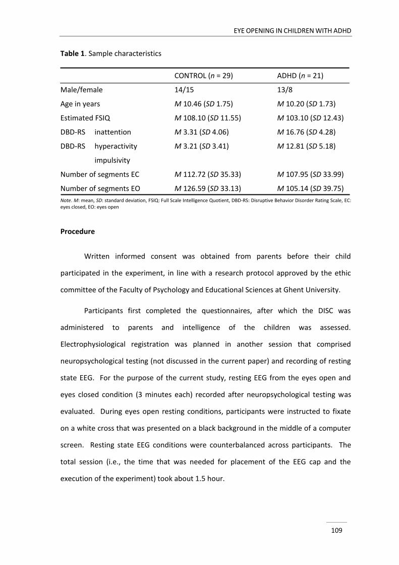
EYE OPENING IN CHILDREN WITH ADHD
109
Table 1. Sample characteristics
CONTROL (n = 29) ADHD (n = 21)
Male/female 14/15 13/8
Age in years M 10.46 (SD 1.75) M 10.20 (SD 1.73)
Estimated FSIQ M 108.10 (SD 11.55) M 103.10 (SD 12.43)
DBD-RS inattention M 3.31 (SD 4.06) M 16.76 (SD 4.28)
DBD-RS hyperactivity M 3.21 (SD 3.41) M 12.81 (SD 5.18)
impulsivity
Number of segments EC M 112.72 (SD 35.33) M 107.95 (SD 33.99)
Number of segments EO M 126.59 (SD 33.13) M 105.14 (SD 39.75)
Note. M: mean, SD: standard deviation, FSIQ: Full Scale Intelligence Quotient, DBD-RS: Disruptive Behavior Disorder Rating Scale, EC: eyes closed, EO: eyes open
Procedure
Written informed consent was obtained from parents before their child
participated in the experiment, in line with a research protocol approved by the ethic
committee of the Faculty of Psychology and Educational Sciences at Ghent University.
Participants first completed the questionnaires, after which the DISC was
administered to parents and intelligence of the children was assessed.
Electrophysiological registration was planned in another session that comprised
neuropsychological testing (not discussed in the current paper) and recording of resting
state EEG. For the purpose of the current study, resting EEG from the eyes open and
eyes closed condition (3 minutes each) recorded after neuropsychological testing was
evaluated. During eyes open resting conditions, participants were instructed to fixate
on a white cross that was presented on a black background in the middle of a computer
screen. Resting state EEG conditions were counterbalanced across participants. The
total session (i.e., the time that was needed for placement of the EEG cap and the
execution of the experiment) took about 1.5 hour.
CHAPTER 4
110
Electrophysiological procedure
Participants were fitted with a 128 electrode cap with 10 mm Ag/AgCl electrodes
(EasyCap Active, EasyCap GmbH) placed according to the 10-5 International System
(Oostenveld & Praamstra, 2001). Data were collected with a sample rate of 500 Hz and
average referenced with the ground electrode mounted within the cap at Fpz. Electro-
oculogram was registered through electrodes enclosed in the cap near the eyes. A
QuickAmp amplifier (Brain Products, Gilching, Germany) was employed to amplify
signals with an open pass-band from direct current to 100 Hz. Brain Vision Analyzer
software (version 2.0.1) was used to filter (0.5 Hz to 50 Hz band-pass, notch filter of 50
Hz) and segment (2 s epochs with 1 s overlap) data offline. Ocular artefact correction
was conducted according to the Gratton and Coles algorithm (Gratton, Coles & Donchin,
1983). Segments with amplitudes exceeding ±100 µV were removed from further
analyses. The remaining number of segments for analyses did not differ between
conditions (F(1, 48) = 1.79, p = .19) and groups (F(1, 48) = 2.00, p = .16) (Table 1).
Spectral power was calculated using Fast Fourier Transform with a 20% Hanning
window. For comparison purposes (Barry et al., 2009), absolute power estimates were
derived for the theta (3.5-7.5 Hz), alpha (7.5-12.5 Hz) and beta (12.5-25 Hz) band and
divided in nine regions: left frontal (AF3, F3, F7), midline frontal (Fz, FCz), right frontal
(AF4, F4, F8), left central (T7, C3), midline central (Cz), right central (T8, C4), left
posterior (P7, P3, O1), midline posterior (Pz, Oz) and right posterior (P8, P4, O2).
Theta/beta power ratio was obtained by dividing the power of the theta band by the
power of the beta band. A natural logarithmic transform, ln(x), was applied to approach
normal spreading of the data.
Statistical analyses
Eyes closed and eyes open EEG activity in children with and without ADHD were
compared by running separate analyses of variance with repeated measures for each
EYE OPENING IN CHILDREN WITH ADHD
111
EEG measure with condition (eyes closed, eyes open), sagittal region (frontal, central,
posterior) and lateral region (left, midline, right) as within-subject factors and group
(ADHD, controls) as between-subject factor. When the assumption of sphericity was
violated, degrees of freedom were corrected using Greenhouse-Geisser (when ε < .75)
or Huynh-Feldt (when ε > .75) estimates of sphericity. Effect sizes were determined by
partial eta squared (η2p) and are interpreted as small (< .06), medium (.06 - .14), or large
(> .14) (Cohen, 1988).
Since comorbid ODD/CD has been documented to possibly play a mediating role
in detecting EEG deviances in ADHD (Clarke et al., 2002b; Loo et al., 2013), a
supplementary analysis was performed excluding children with comorbid ODD/CD (i.e.,
5/21) (see also Liechti et al., 2013). As age and gender have been demonstrated to
affect EEG (e.g., Clarke Barry, McCarthy, Selikowitz, 2001b; Dupuy, Barry, Clarke,
McCarthy, & Selikowitz, 2013), separate additional analyses were run with these factors
as covariates.
In view of the scope of the present study, only effects involving diagnostic status
are discussed. Because of the relative paucity of studies on this topic, not only
significant effects but also effects on a subtreshold level of significance (i.e., .05 < p <
.10) are reported to encourage future research (see also Barry et al., 2004).
RESULTS
Group characteristics
The DBD-RS confirmed that children with ADHD experienced more problems with
inattention (F(1, 48) = 127.76, p < .001) as well as hyperactivity/impulsivity (F(1, 48) =
62.54, p < .001) than typically developing children. The groups did not significantly differ
CHAPTER 4
112
in terms of age (F(1, 48) = 0.26, p = .62) and estimated full scale IQ (F(1, 48) = 2.15, p =
.15) (Table 1).
EEG analyses
The topographic power distributions for eyes closed and eyes open resting
conditions and the differences in power distribution between conditions in each
frequency band and TBR are presented in Figure 1.
Theta power. As for theta power, a group-by-lateral region interaction was
found (F(2, 96) = 4.40, p = .02, η2p = .08), indicating theta power across resting state
conditions was higher for controls than for ADHD at midline than at lateral electrode
sites. Yet, group differences did not reach significance at either midline or lateral region.
Alpha power. Likewise, a significant interaction between group and lateral
region was detected for alpha power (F(2, 96) = 3.77, p = .03, η2p = .07). Although groups
did not significantly differ in either region, higher alpha power at midline than at lateral
sites was detected in the control group but not in the ADHD group.
Beta power. Beta power also differed between groups on the lateral plane (F(2,
96) = 3.52, p = .03, η2p = .07). Further testing revealed a greater reduction in power at
midline compared to lateral sites in children with ADHD than in typically developing
children. Yet, again, no significant differences between groups were detected at either
midline or lateral region.
TBR. No significant interaction effects involving group were detected for TBR.
Supplementary analyses with age and sex as a covariate did not change the group
related effects for all frequency bands and TBR.
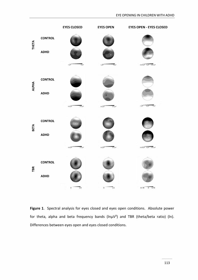
EYE OPENING IN CHILDREN WITH ADHD
113
Figure 1. Spectral analysis for eyes closed and eyes open conditions. Absolute power
for theta, alpha and beta frequency bands (lnµV²) and TBR (theta/beta ratio) (ln).
Differences between eyes open and eyes closed conditions.
CHAPTER 4
114
Supplementary analyses excluding children with comorbid ODD/CD. Regarding
topographic differences between groups, the p-levels in alpha and beta power slightly
reduced but the effect sizes remained in the same range (respectively p = .06, η2p = .07
and p = .08, η2p = .07). Since the interpretations of the effect sizes between the analyses
with and without children with comorbid ODD/CD were comparable, the slight
decreases in p-levels probably resulted from the reduction in power following the
exclusion of 5 children from the analyses. The significant effect in theta power remained
when only children without comorbid ODD/CD were included in the analyses.
Further, excluding children with comorbid ODD/CD from the analyses revealed
that topographic distribution on the sagittal plane for alpha power differed between
groups (F(2, 86) = 4.87, p = .01, η2p = .10), indicating a steeper increase from anterior to
posterior sites in typically developing children than in children with ADHD (F(2, 86) =
8.98, p < .01, η2p = .17). In addition, a group-by-condition effect was detected (F(1, 43) =
5.12, p = .03, η2p = .11) in alpha power, denoting alpha suppression with eye opening
was lower in the ADHD group than in the control group. Yet, group differences did not
reach significance in either condition. Also, a marginally significant group x condition x
sagittal region effect appeared (F(1.74, 74.83) = 2.75, p = .08, η2p = .06), meaning that
the smaller alpha attenuation with eye opening in ADHD was most apparent in central
(F(1, 43) = 8.05, p < .01, η2p = .16) and posterior (F(1, 43) = 4.56, p = .04, η2
p = .10) region
(Figure 2).
Furthermore, EEG response to eye opening tended to differ between groups in
theta power (F(1, 43) = 3.43, p = .07, η2p = .07), resulting from a slight theta decrease in
the control group (F(1, 28) = 2.97, p < .10, η2p = .10) which was not apparent in the ADHD
group. No group differences in theta power were detected in either condition. Further
testing regarding the marginally significant group x condition x sagittal region effect in
theta power (F(1.66, 71.25) = 2.88, p = .07, η2p = .06), revealed that the group difference
in EEG reactivity to eye opening was significant in posterior region (F(1, 43) = 4.59, p =
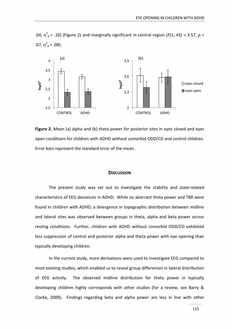
EYE OPENING IN CHILDREN WITH ADHD
115
2
2,3
2,6
2,9
CONTROL ADHD
lnμ
V²
eyes closed
eyes open
1,5
2
2,5
3
3,5
4
CONTROL ADHD
lnμ
V²
.04, η2p = .10) (Figure 2) and marginally significant in central region (F(1, 43) = 3.57, p =
.07, η2p = .08).
Figure 2. Mean (a) alpha and (b) theta power for posterior sites in eyes closed and eyes
open conditions for children with ADHD without comorbid ODD/CD and control children.
Error bars represent the standard error of the mean.
DISCUSSION
The present study was set out to investigate the stability and state-related
characteristics of EEG deviances in ADHD. While no aberrant theta power and TBR were
found in children with ADHD, a divergence in topographic distribution between midline
and lateral sites was observed between groups in theta, alpha and beta power across
resting conditions. Further, children with ADHD without comorbid ODD/CD exhibited
less suppression of central and posterior alpha and theta power with eye opening than
typically developing children.
In the current study, more derivations were used to investigate EEG compared to
most existing studies, which enabled us to reveal group differences in lateral distribution
of EEG activity. The observed midline distribution for theta power in typically
developing children highly corresponds with other studies (for a review, see Barry &
Clarke, 2009). Findings regarding beta and alpha power are less in line with other
(a) (b)
CHAPTER 4
116
studies, in which a midline distribution for beta power and a more equivalent
distribution for alpha power across the lateral plane has been documented in control
groups (for a review, see Barry & Clarke, 2009). Although other studies did not report
the more equivalent distribution of EEG activity in ADHD that we observed in the
present study, they also point to more deviance between groups at midline than lateral
regions (e.g., Clarke et al., 1998; Clarke et al., 2001b), which underlines the importance
of investigating widespread cortical activity in ADHD. Therefore, it is recommended that
future studies investigating EEG deviances in ADHD incorporate a sufficient number of
electrodes. The paucity in the literature concerning power distribution across a lateral
dimension including the midline, impedes drawing firm conclusions regarding the brain
mechanisms underlying the deviances between children with ADHD and typically
developing children. Nevertheless, during resting state, preservation of a physiological
and functional balance in the brain relies on different local cortical neural activities and
long-range corticocortical/cortico-subcortical neural activities that coexist and may
interact with each other (Woltering et al., 2012). Variations in topographical distribution
in ADHD may therefore reflect deficient neural communication or dysfunction in
distributed network organization (for a review, see Konrad & Eickhoff, 2010), which may
be related to ADHD symptomatology. Before drawing further conclusions on this, the
robustness of the current findings should be replicated in future research.
Still, our findings regarding comparable theta and TBR between groups are in
contrast with other studies (Barry et al., 2003; Clarke et al., 2007; Clarke et al., 2011;
Dupuy et al., 2011; González-Castro et al., 2013; Lansbergen et al., 2011; Loo et al.,
2010; Snyder et al., 2008; Shi et al., 2012), but correspond with some recent studies that
also failed to replicate increased theta or TBR in ADHD (Coolidge et al., 2007; Liechti et
al., 2013; Loo et al., 2013; Nazari et al., 2011; Ogrim et al., 2012; Swartwood et al.,
2003). As for alpha and beta power, our results are not unexpected since deviances in
EYE OPENING IN CHILDREN WITH ADHD
117
those frequency ranges in ADHD have not been consistently observed across studies
(see introduction).
Among others, it is plausible that differences in sample characteristics for both
children with and without ADHD may have contributed to divergent findings across
studies (Arns et al., 2013; Liechti et al., 2013). Regarding sex, it has been suggested that
boys have different EEG abnormalities than girls with ADHD (for a review, see Dupuy et
al., 2013). In the current study, more girls were included in the control group than in the
ADHD group. Furthermore, the proportion of boys to girls in our ADHD sample was
somewhat lower than in most studies (e.g., Shi et al., 2012). However, higher theta or
TBR in ADHD has been detected in samples with even a smaller boy to girl ratio than in
the current study (e.g., Clarke et al., 2001b; Loo et al., 2010). Moreover, controlling for
sex in our analyses did not change the results. Therefore, we cautiously conclude that
sex did not act as a confounding factor. Our study included children with ADHD with
different subtypes and it may be that enhanced theta or TBR is only related to one of
these subtypes. However not much research has been done on EEG differences
between ADHD subtypes and so far disparate findings have been reported, with some
studies documenting comparability in EEG activity between ADHD subtypes, and others
observing more pronounced quantitative EEG differences in one subtype relative to the
other and/or the control group (Buyck & Wiersema, 2014; Clarke et al., 1998; Clarke et
al., 2001b; Loo et al., 2010; Dupuy et al., 2011). Unfortunately, the size of our sample
impedes reliable investigation of divergence according to ADHD subtypes, yet this could
be further explored in future work. Furthermore, in the current study, 11 children with
ADHD were on stimulant medication in daily life. Studies addressing the impact of
stimulant medication on brain wave activity have indicated a power decrease in the
theta band (Loo, Teale, & Reite, 1999; Clarke, Barry, McCarthy, Selikowitz, & Johnstone,
2007) and a reduction in TBR (Clarke, Barry, Bond, McCarthy, & Selikowitz, 2002c)
following medication. Although we are not aware of studies investigating the specific
CHAPTER 4
118
duration of medication effects on EEG activity, a medication wash-out period of 48 hours
was respected, which is even more restrictive than regular research procedures across
EEG studies (e.g., Loo et al., 2010) in ADHD. Consequently, we do not regard daily
medication use in a part of our ADHD sample as a confounding factor for our results, yet
further research may be warranted investigating (long-term) effects of daily life use of
stimulants on EEG in ADHD.
Another source of heterogeneity in ADHD samples across studies refers to the
presence of comorbid disorders. It is well established that a large proportion of children
with ADHD has at least one comorbid psychiatric diagnosis, with ODD occurring the most
frequently (Biederman, 2005). Yet, often studies have not controlled for comorbid
ODD/CD and research on how this comorbidity influences EEG in ADHD is scarce. One
study has documented that comorbidity with ODD/CD possibly enlarges TBR deviance in
children with ADHD (Loo et al., 2013), while another study did not detect this effect and
instead reported that focal theta abnormalities were more pronounced in an ADHD only
group than in an ADHD group with ODD/CD comorbidity (Clarke et al., 2002b). However,
both studies focussed on an eyes closed resting condition only and were therefore not
able to address the influence of comorbid ODD/CD on EEG response to eye opening. In
the present study, although no divergence was found in the separate resting state
conditions, group differences in alpha and theta reactivity on eye opening emerged
when excluding children with comorbid ODD/CD, alluding more deviance in children
with pure ADHD (i.e., not suffering from comorbid disruptive behavior disorder).
Currently, the debate is still ongoing whether comorbid oppositional symptoms in ADHD
should be considered as a component of ADHD or whether ADHD+ODD/CD represents a
distinct pathological entity (Connor, Steeber, & McBurnett, 2010), as also defined in the
International Classification of Diseases and Related Health Problems classification
system (ICD-10, World Health Organisation, 2008). Our results appear to be more in line
with the latter interpretation, although this conclusion is putative since it is not derived
EYE OPENING IN CHILDREN WITH ADHD
119
from a direct comparison between ADHD groups with and without comorbidity, as the
sample size of the comorbid ADHD group (i.e., n = 5) was not sufficient to perform these
analyses. Future studies incorporating more children with ADHD with comorbid
disruptive behavior disorder are warranted to shed more light on the possible mediating
effects of comorbid disorders in EEG deviances in ADHD.
As for less alpha attenuation on eye opening in ADHD, this has previously been
observed in another study focussing on EEG response to eye opening in children with
ADHD (Fonseca et al., 2013) and has been confirmed in two studies in adults with ADHD
(Loo et al., 2010; Woltering et al., 2012). The finding of less theta attenuation in ADHD
following eye opening has to the best of our knowledge not been reported before.
Modulation of EEG activity between eyes closed and eyes open conditions has been
proposed to be an indicator of function of thalamocortical networks (Boord et al., 2008)
resulting from cortical processing of visual input (Barry et al., 2007). This suggests that
in the ADHD group, neuronal networks are impaired to adjust to the changes in input
between eyes closed and eyes open states. Furthermore, recent studies indicate that
alpha power can be regarded as a marker of arousal, since an inverse relationship has
repeatedly been established between alpha power and skin conductance level (SCL)
(Barry et al., 2004; Barry et al., 2009; Clarke et al., 2013), which is considered to be a
reliable measure of autonomic arousal (for a review, see Critchley, 2002). For a long
time, deviances in the theta frequency band have also been associated with the concept
of arousal, yet recent studies failed to find an association between theta power and SCL
(Barry et al., 2004, 2009). Consequently, in specific, less alpha suppression in children
with ADHD may possibly be regarded as reflecting an arousal dysfunction in ADHD.
Important to notice is that the ADHD group exhibited no aberrant alpha power in the
conditions separately, yet expressed a difference in modulation of EEG activity between
conditions. This may imply that deviances in ADHD are not characterized by a simple
arousal impairment, but rather reflect a deficiency in arousal regulation. Despite the
CHAPTER 4
120
lack of a well established autonomic measure of arousal (i.e., SCL) in our study design to
strengthen this hypothesis, interpretations regarding deficient arousal modulation are in
line with theoretical accounts of ADHD. According to the optimal stimulation theory,
ADHD is related to a deficiency in arousal regulation with the symptomatology reflecting
a behavioural strategy to compensate for a state of underarousal (Zentall & Zentall,
1983). In support of this theory, research documented that in low stimulation
environments, children with ADHD showed increased stimulation seeking activity
whereas in high stimulation environments, behavioural responses in children with ADHD
did not differentiate from typically developing children (Antrop, Buysse, Roeyers, & Van
Oost, 2005; for a review, see Zentall & Zentall, 1983). Also the state regulation account
(Sergeant, 2005; van der Meere, 2005), an influential explanatory account of ADHD,
which is derived from the cognitive-energetic model (Sanders, 1983), states that
individuals with ADHD have difficulties in maintaining and regulating an optimal
energetic arousal/activation state. Research in this respect revealed convincing
behavioural and psychophysiological support for disrupted energetic state regulation in
ADHD during task execution (Börger, & van der Meere, 2000; for a meta-analysis, see
Metin, Roeyers, Wiersema, van der Meere, & Sonuga-Barke, 2012; Sergeant, 2005;
Sonuga-Barke et al., 2010; Wiersema, van der Meere, Roeyers, Van Coster, & Baeyens,
2006).
In conclusion, although no aberrant theta and TBR were found in children with
ADHD in the present study, the findings refer to a consistent variation in topographic
distribution in ADHD across resting state conditions compared to typically developing
children, indicating the usefulness of inclusion of electrodes beyond the midline. The
finding of less central alpha and theta attenuation with eye opening in children with
ADHD who do not suffer from comorbid ODD/CD, suggest that EEG deviances in ADHD
may reflect arousal regulation difficulties and stress the importance to take into account
comorbidity with ODD/CD. Furthermore, the findings indicate that it is unlikely that
EYE OPENING IN CHILDREN WITH ADHD
121
stable, univariate EEG abnormalities are implicated in all children with ADHD and that it
is important to take into account state-dependent characteristics when evaluating EEG
in ADHD. From a clinical perspective, this implies that cautiousness is warranted in using
simple EEG measures as a supplementary diagnostic tool, as has been proposed by some
researchers (Monastra, Lubar, & Linden, 2001; Snyder et al., 2008).
CHAPTER 4
122
REFERENCES
Achenbach, T.M., & Rescorla, L.A. (2001). Manual for the ASEBA School-Age Forms and Profiles.
Burlington, Vermont: University of Vermont, Research Center for Children, Youth, and
Families.
American Psychiatric Association (2013). Diagnostic and statistical manual of mental disorders
(5th Ed.). Arlington, VA: American Psychiatric Publishing.
Antrop, I., Buysse, A., Roeyers, H., & Van Oost P. (2005). Activity in children with ADHD during
waiting situations in the classroom: a pilot study. British Journal of Educational
Psychology, 75(Pt 1), 51-69. doi: 10.1348/000709905X36100
Arns, M., Conners, C.K., & Kraemer, H.C. (2013). A decade of EEG theta/beta ratio research in
ADHD: a meta-analysis. Journal of Attention Disorders, 17, 374-383. doi:
10.1177/1087054712460087
Barry, R.J., Clarke, A.R. (2009). Spontaneous EEG oscillations in children, adolescents, and adults:
Typical development, and pathological aspects in relation to AD/HD. Journal of
Psychophysiology, 23(4), 157-173. doi: 10.1027/0269-8803.23.4.157
Barry, R.J., Clarke, A.R., & Johnstone, S.J. (2003). A review of electrophysiology in attention-
deficit/hyperactivity disorder. I. Qualitative and quantitative electroencephalography.
Clinical Neurophysiology, 114, 171-183. doi: 10.1016/S1388-2457(02)00362-0
Barry, R.J., Clarke, A.R., Johnstone, S.J., Magee, C.A., & Rushby, J.A. (2007). EEG differences
between eyes-closed and eyes-open resting conditions. Clinical Neurophysiology, 118,
2765-2773. doi: 10.1016/j.clinph.2007.07.028
Barry, R.J., Clarke, A.R., Johnstone, S.J., McCarthy, R., & Selikowitz, M. (2009).
Electroencephalogram theta/beta ratio and arousal in attention-deficit/hyperactivity
disorder: evidence of independent processes. Biological Psychiatry, 66, 398-401. doi:
10.1016/j.biopsych.2009.04.027
Barry, R.J., Clarke, A.R., McCarthy, R., Selikowitz, M., Rushby, J.A., & Ploskova, E. (2004). EEG
differences in children as a function of resting-state arousal level. Clinical
Neurophysiology, 115, 402-408. doi: 10.1016/S1388-2457(03)00343-2
Biederman (2005). Attention-deficit/hyperactivity disorder: a selective overview. Biological
Psychiatry, 57(11). doi: 1215-1220. 10.1016/j.biopsych.2004.10.020
EYE OPENING IN CHILDREN WITH ADHD
123
Boord, P., Siddall, P.J., Tran, Y., Herbert, D., Middleton, J., & Craig, A. (2008).
Electroencephalographic slowing and reduced reactivity in neuropathic pain following
spinal cord injury. Spinal Cord, 46, 118-123. doi: 10.1038/sj.sc.3102077
Börger, N.A., & van der Meere, J.J. (2000). Motor control and state regulation in children with
ADHD: a cardiac response study. Biological Psychology, 51, 247-267. doi: 10.1016/S0301-
0511(99)00040-X
Buyck, I., & Wiersema, J.R. (2014). Resting electroencephalogram in attention deficit
hyperactivity disorder: developmental course and diagnostic value. Psychiatry Research.
Advance online publication. doi: 10.1016/j.psychres.2013.12.055
Chabot, R.J., & Serfontein, G. (1996). Quantitative electroencephalographic profiles of children
with attention deficit disorder. Biological Psychiatry, 40, 951-963. doi: 10.1016/0006-
3223(95)00576-5
Clarke, A.R., Barry, R.J., Bond, D., McCarthy, R., & Selikowitz, M. (2002c). Effects of stimulant
medications on the EEG of children with attention-deficit/hyperactivity disorder.
Psychopharmacology, 164(3), 277-284. doi: 10.1007/s00213-002-1205-0
Clarke, A.R., Barry, R.J., Dupuy, F.E., Heckel, L.D., McCarthy, R., Selikowitz, M., & Johnstone, S.J.
(2011). Behavioural differences between EEG-defined subgroups of children with
Attention-Deficit/Hyperactivity Disorder. Clinical Neurophysiology, 122, 1333-1341. doi:
10.1016/j.clinph.2010.12.038
Clarke, A.R., Barry, R.J., Dupuy, F.E., McCarthy, R., Selikowitz, M., Johnstone, S.J. (2013). Excess
beta activity in the EEG of children with attention-deficit/hyperactivity disorder: A
disorder of arousal? International Journal of Psychophysiology, 89, 314-319. doi:
10.1016/j.ijpsycho.2013.04.009
Clarke, A., Barry, R., McCarthy, R., & Selikowitz, M. (1998). EEG analysis in attention-
deficit/hyperactivity disorder: a comparative study of two subtypes. Psychiatry Research,
81, 19-29. doi: 10.1016/S0165-1781(98)00072-9
Clarke, A.R., Barry, R.J., McCarthy, R., & Selikowitz, M. (2001a). Excess beta in children with
attention-deficit/hyperactivity disorder: An atypical electrophysiological group.
Psychiatry Research, 103, 205-218. doi: 10.1016/S0165-1781(01)00277-3
CHAPTER 4
124
Clarke, A., Barry, R., McCarthy, R., & Selikowitz, M. (2001b). Age and sex effects in the EEG:
differences in two subtypes of attention-deficit/hyperactivity disorder. Clinical
Neurophysiology, 112, 806-814. doi: 10.1016/S1388-2457(01)00487-4
Clarke, A., Barry, R., McCarthy, R., & Selikowitz, M. (2002a). EEG analysis of children with
attention-deficit/hyperactivity disorder and comorbid reading disabilities. Journal of
Learning Disabilities, 35, 276-285. doi: 10.1177/002221940203500309
Clarke, A.R., Barry, R.J., McCarthy, R., & Selikowitz, M. (2002b). Children with attention-
deficit/hyperactivity disorder and comorbid oppositional defiant disorder: an EEG
analysis. Psychiatry Research, 111(2-3), 181-190. doi: 10.1016/S0165-1781(02)00137-3
Clarke, A.R., Barry, R.J., McCarthy, R., Selikowitz, M., Clarke, D., & Croft, R. (2003). EEG in girls
with attention-deficit/hyperactivity disorder. Clinical Neurophysiology, 114, 319-328.
doi: 10.1016/S1388-2457(02)00364-4
Clarke, A.R., Barry, R.J., McCarthy, R., Selikowitz, M., Johnstone, S.J. (2007). Effects of stimulant
medications on the EEG of girls with Attention-Deficit/Hyperactivity. Clinical
Neurophysiology, 118(12), 2700-2708. doi: 10.1016/j.clinph.2007.08.020
Cohen, J. (1988). Statistical Power Analysis for the Behavioral Sciences, 2 ed. Laurence Erlbaum,
Hillsdale, N.J.
Connor, D.F., Steeber, J., & McBurnett, K. (2010). A review of attention-deficit/hyperactivity
disorder complicated by symptoms of oppositional defiant disorder or conduct disorder.
Journal of Developmental and Behavioral Pediatrics, 31(5), 427-440. doi:
10.1097/dbp.0b013e3181e121bd
Coolidge, F.L., Starkey, M.T., & Cahill, B.S. (2007). Comparison of a Parent-Rated DSM-IV
Measure of Attention-Deficit/Hyperactivity Disorder and Quantitative EEG Parameters in
an Outpatient Sample of Children. Journal of Clinical Neurophysiology, 24(4), 348-351.
doi: 10.1097/WNP.0b013e318067bcfc
Critchley, H.D. (2002). Book Review: Electrodermal Responses: What Happens in the Brain. The
Neuroscientist, 8, 132. doi: 10.1177/107385840200800209
Dupuy, F.E., Barry, R.J., Clarke, A.R., McCarthy, R., & Selikowitz, M. (2013). Sex differences
between the combined and inattentive types of attention-deficit/hyperactivity disorder:
An EEG perspective. International Journal of Psychophysiology, 89(3), 320-327. doi:
10.1016/j.ijpsycho.2013.04.004
EYE OPENING IN CHILDREN WITH ADHD
125
Dupuy, F.E., Clarke, A.R., Barry, R.J., McCarthy, R., & Selikowitz, M. (2011). Girls with attention-
deficit/hyperactivity disorder: EEG differences between DSM-IV types. Clinical EEG and
Neuroscience, 42(1), 1-5. doi: 10.1177/155005941104200104
Fein, G., Galin, D., Yingling, C.D., Johnstone, J., & Nelson, M.A.(1984). EEG spectra in 9-13-year-
old boys are stable over 1-3 years. Electroencephalography and Clinical Neurophysiology,
58(6), 517-518. doi: 10.1016/0013-4694(84)90041-5
Fonseca, L.C., Tedrus, G.M.A.S., Bianchini, M.C., & Silva, T.F. (2013). Electroencephalographic
alpha reactivity on opening the eyes in children with attention-deficit hyperactivity
disorder. Clinical EEG and Neuroscience, 44(1), 53-57. doi: 10.1177/1550059412445659
Gasser, T., Bacher, P., & Steinberg, H. (1985). Test-retest reliability of spectral parameters of the
EEG. Electroencephalography and Clinical Neurophysiology, 60(4), 312-319. doi:
10.1016/0013-4694(85)90005-7
González-Castro, P., Rodríguez, C., López, A., Cueli, M., & Álvarez, L. (2013). Attention Deficit
Hyperactivity Disorder, differential diagnosis with blood oxygenation, beta/theta ratio,
and attention measures. International Journal of Clinical and Health Psychology, 13(2),
101-109. doi: 10.1016/S1697-2600(13)70013-9
Grégoire, J. (2000). L’évaluation clinique de l’intelligence de l’enfant. Liège: Mardaga.
Gratton, G., Coles, M.G.H., & Donchin, E. (1983). A new method for off-line removal of ocular
artifact. Electroencephalography and Clinical Neurophysiology, 55, 468-484. doi:
10.1016/0013-4694(83)90135-9
Hale, T.S., Smalley, S.L., Hanada, G., Macion, J., McCracken, J.T., McGough, J.J., & Loo, S.K.
(2009). Atypical alpha asymmetry in adults with ADHD. Neuropsychologia, 47(10), 2082-
2088. doi: 10.1016/j.neuropsychologia.2009.03.021
Hale, T.S., Smalley, S.L., Dang, J., Hanada, G., Macion, J., McCracken, J.T., ... & Loo, S.K. (2010a).
ADHD familial loading and abnormal EEG alpha asymmetry in children with ADHD.
Journal of Psychiatric Research, 44(9), 605-615. doi: 10.1016/j.jpsychires.2009.11.012
Hale, T.S., Smalley, S.L., Walshaw, P.D., Hanada, G., Macion, J., McCracken, J.T., ... & Loo, S.K.
(2010b). Atypical EEG beta asymmetry in adults with ADHD. Neuropsychologia, 48(12),
3532-3539. doi: 10.1016/j.neuropsychologia.2010.08.002
Hüfner, K., Stephan, T., Flanagin, V.L., Deutschlander, A., Stein, A., Kalla, R., ... & Brandt, T.
(2009). Differential effects of eyes open or closed in darkness on brain activation

CHAPTER 4
126
patterns in blind subjects. Neuroscience Letters, 466, 30-34. doi:
10.1016/j.neulet.2009.09.010
Karhu, J., Könönen, M., Herrgård, E., & Partanen, J. (1996). Spectra and reactivity of EEG in
visual, sensori-motor and auditory areas in children. Electroencephalography and Clinical
Neurophysiology, 99(4), 300-301. doi: 10.1016/0013-4694(96)88224-1
Keune, P.M., Schoenenberg, M., Wyckoff, S., Mayer, K., Riemann, S., Hautzinger, M., & Strehl, U.
(2011). Frontal alpha-asymmetry in adults with attention deficit hyperactivity disorder:
Replication and specification. Biological Psychology, 87(2), 306-310. doi:
10.1016/j.biopsycho.2011.02.023
Konrad, K., & Eickhoff, S.B. (2010). Is the ADHD Brain Wired Differently? A Review on Structural
and Functional Connectivity in Attention Deficit Hyperactivity Disorder. Human Brain
Mapping, 31(6), 904-916. doi: 10.1002/hbm.21058
Lansbergen, M., Arns, M., van Dongen-Boomsma, M., Spronk, D., & Buitelaar, J. (2011). The
increase in theta/beta ratio on resting state EEG in boys with attention-
deficit/hyperactivity disorder is mediated by slow alpha peak frequency. Progress in
Neuropsychopharmacology & Biological Psychiatry, 35(1), 47-52. doi:
10.1016/j.pnpbp.2010.08.004
Liechti, M.D., Valko, L., Müller, U.C., Döhnert, M., Drechsler, R., Steinhausen, H.C., & Brandeis, D.
(2013). Diagnostic value of resting electroencephalogram in attention-
deficit/hyperactivity disorder across the lifespan. Brain Topography, 26(1), 135-151. doi:
10.1007/s10548-012-0258-6
Loo, S.K., Cho, A., Hale, T.S., McGough, J., McCracken, J., & Smalley, S.L. (2013). Characterization
of the Theta to Beta Ratio in ADHD: Identifying Potential Sources of Heterogeneity.
Journal of Attention Disorders, 17(5), 384-392. doi: 10.1177/1087054712468050
Loo, S.K., Hale, T.S., Macion, J., Hanada, G., McGough, J.J., McCracken, J.T., & Smalley, S.L.
(2009). Cortical activity patterns in ADHD during arousal, activation and sustained
attention. Neuropsychologia, 47, 2114-2119. doi: 10.1016/
j.neuropsychologia.2009.04.013
Loo, S.K., Hale, T.S., Hanada, G., Macion, J., Shrestha, A., McGough, J.J., ... & Smalley, S.L. (2010).
Familial clustering and DRD4 effects on electroencephalogram measures in multiplex
EYE OPENING IN CHILDREN WITH ADHD
127
families with attention deficit/hyperactivity disorder. Journal of the American Academy
of Child and Adolescent Psychiatry, 49(4), 368-377. doi: 10.1016/j.jaac.2010.01.002
Loo, S.K., & Makeig, S. (2012). Clinical utility of EEG in attention-deficit/hyperactivity disorder: a
research update. Neurotherapeutics, 9, 569-587. doi: 10.1007/s13311-012-0131-z
Loo, S.K., Teale, P.D., & Reite, M.L. (1999). EEG correlates of methylphenidate response among
children with ADHD: A preliminary report. Biological Psychiatry, 45(12), 1657-1660. doi:
10.1016/S0006-3223(98)00250-9
Metin, B., Roeyers, H., Wiersema, J.R., van der Meere, J., & Sonuga-Barke, E. (2012). A Meta-
Analytic Study of Event Rate Effects on Go/No-Go Performance in Attention-
Deficit/Hyperactivity Disorder. Biological Psychiatry, 72, 990-996. doi:
10.1016/j.biopsych.2012.08.023
Monastra, V.J., Lubar, J.F., & Linden, M. (2001). The development of a quantitative
electroencephalographic scanning process for attention deficit-hyperactivity disorder:
Reliability and validity studies. Neuropsychology, 15, 136-144. doi: 10.1037/0894-
4105.15.1.136
Nazari, M.A., Wallois, F., Aarabi, A., & Berquin, P. (2011). Dynamic changes in quantitative
electroencephalogram during continuous performance test in children with attention-
deficit/hyperactivity disorder. International Journal of Psychophysiology, 81(3), 230-236.
doi: 10.1016/j.ijpsycho.2011.06.016
Ogrim, G., Kropotov, J., & Hestad, K. (2012). The QEEG TBR in ADHD and normal controls:
Sensitivity, specificity and behavioral correlates. Psychiatry Research, 198(3), 482-488.
doi: 10.1016/j.psychres.2011.12.041
Oostenveld, R., & Praamstra, P. (2001). The five percent electrode system for high resolution
EEG and ERP measurements. Clinical Neurophysiology, 112, 713-719. doi:
10.1016/S1388-2457(00)00527-7
Pelham, W., Gnagy, E.M., Greenslade, K.E., & Milich, R. (1992). Teacher ratings of DSM-III-R
symptoms for the disruptive behavior disorders. Journal of the American Academy of
Child and Adolescent Psychiatry, 31, 210-218. doi: 10.1097/00004583-199203000-00006
Ristanovic, D., Martinovic, Z.J., & Jovanovic, V. (1999). Topography of visual EEG reactivity in
school-age children. Brain and Development, 21, 236-243. doi: 10.1016/S0387-
7604(99)00015-7
CHAPTER 4
128
Samson-Dollfus, D., & Goldberg, P. (1979). Electroencephalographic quantification by time
domain analysis in normal 7-15-Year-Old Children. Electroencephalography and Clinical
Neurophysiology, 46, 147-154. doi: 10.1016/0013-4694(79)90064-6
Sanders, A. F. (1983). Towards a model of stress and human performance. Acta Psychologica, 53,
61-97. doi: 10.1016/0001-6918(83)90016-1
Schaffer, D., Fisher, P., Lusac, C.P., Dulcan, M. K., & Schwab-Stone, M.E. (2000). NIMH Diagnostic
Interview Schedule for Children version IV (NIMH DISC-IV): Description, differences from
previous versions and reliability of some common diagnoses. Journal of the American
Academy of Child and Adolescent Psychiatry, 39, 28-38. doi: 10.1097/00004583-
200001000-00014
Sergeant, J. (2005). Modeling attention-deficit/hyperactivity disorder: a critical appraisal of the
cognitive-energetic model. Biological Psychiatry, 57, 1248-1255. doi:
10.1016/j.biopsych.2004.09.010
Shi, T., Li, X., Song, J., Zhao, N., Sun, C., Xia, W., … & Tomoda, A. (2012). EEG characteristics and
visual cognitive function of children with attention deficit hyperactivity disorder (ADHD).
Brain and Development, 34(10), 806-811. doi: 10.1016/j.braindev.2012.02.013
Snyder, S.M., Quintana, H., Sexson, S.B., Knott, P., Haque, A.F., & Reynolds, D.A. (2008). Blinded,
multi-center validation of EEG and rating scales in identifying ADHD within a clinical
sample. Psychiatry Research, 159(3), 346-358. doi: 10.1016/j.psychres.2007.05.006
Sonuga-Barke, E.J.S., Taylor, E., Sembi, S., & Smith, J. (1992). Hyperactivity and delay aversion: I.
The effect of delay on choice. Journal of Child Psychology and Psychiatry and Allied
Disciplines, 33(2), 387-398. doi: 10.1111/j.1469-7610.1992.tb00874.x
Sonuga-Barke, E., Wiersema, J.R., van der Meere J., & Roeyers, H. (2010). Context-dependent
Dynamic Processes in Attention Deficit/Hyperactivity Disorder: Differentiating Common
and Unique Effects of State Regulation Deficits and Delay Aversion, Neuropsychological
Review, 20, 86-102. doi: 10.1007/s11065-009-9115-0
Swartwood, J.N., Swartwood, M.O., Lubar, J.F., & Timmermann, D.L. (2003). EEG differences in
ADHD-combined type during baseline and cognitive tasks. Pediatric Neurology, 28(3),
199-204. doi: 10.1016/S0887-8994(02)00514-3
EYE OPENING IN CHILDREN WITH ADHD
129
van der Meere, J.J. (2005). State regulation and ADHD. In D. Gozal & D.L. Molfese (Eds.),
Attention deficit hyperactivity disorder: From genes to animal models to patients (pp.
413-433). Totowa, NJ: Humana.
van Dongen-Boomsma, M., Lansbergen, M. M., Bekker, E.M., Kooij, S.J.J., van der Molen, M.,
Kenemans, J.L., & Buitelaar, J.K. (2010). Relation between resting EEG to cognitive
performance and clinical symptoms in adults with attention-deficit/hyperactivity
disorder. Neuroscience Letters, 469, 102-106. doi: 10.1016/j.neulet.2009.11.053
Wechsler, D. (1991). The Wechsler intelligence scale for children-third edition. San Antonio, TX:
The Psychological Corporation.
Wiersema, J.R., van der Meere, J.J., Roeyers, H., Van Coster, R., Baeyens, D. (2006). Event rate
and event-related potentials in ADHD. Journal of Child Psychology and Psychiatry, 47,
560–567. doi: 10.1111/j.1469-7610.2005.01592.x
Willcutt, E.G. (2012). The prevalence of DSM-IV attention-deficit/hyperactivity disorder: a meta-
analytic review. Neurotherapeutics, 9(3), 490-499. doi: 10.1007/s13311-012-0135-8
Woltering, S., Jung, J., Liu, Z., & Tannock, R. (2012). Resting state EEG oscillatory power
differences in ADHD college students and their peers. Behavioral and Brain Functions, 8,
60. doi: 10.1186/1744-9081-8-60
World Health Organization (2008). ICD-10: International statistical classification of diseases and
related health problems (10th Rev. ed.). New York, NY: World Health Organization.
Zentall, S.S., & Zentall, T.R. (1983). Optimal stimulation: a model of disordered activity and
performance in normal and deviant children. Psychological Bulletin, 94(3),446-447. doi:
10.1037//0033-2909.94.3.446

131
DIFFERENTIAL EFFECTS OF PINK NOISE ON THE
EEG OF ADULTS WITH AND WITHOUT ADHD1
ABSTRACT
Objective: Previous research has shown that noise may be beneficial for cognitive
performance in attention-deficit/hyperactivity disorder (ADHD). The underlying
mechanisms of action of noise are however not well known. Therefore, in the current
study we evaluated the impact of external auditory pink noise on
electroencephalographic (EEG) activity in adults with and without ADHD. Methods:
Three minutes eyes closed resting EEG with and without the presence of noise were
compared between 40 adults with ADHD and 40 typically developing adults. The order of
conditions (baseline or noise) was counterbalanced. Results: Alpha power attenuated in
both groups with noise exposure after baseline EEG. Midline theta power also
decreased from baseline to noise condition, however this was only the case for controls
and was not observed in adults with ADHD. No EEG changes between conditions were
detected when the noise condition preceded the condition without noise. Conclusions:
The findings indicate that external auditory pink noise has energetic state or arousal
enhancing properties. No evidence was found for a deviant brain state or underarousal
during baseline resting EEG in adults with ADHD. However, adults with ADHD appear to
show decreased neural adaptability in theta oscillations to noise exposure.
1 Based on Buyck, I., & Wiersema, J.R. (2014). Differential effects of pink noise on the
electroencephalogram of adults with and without attention deficit hyperactivity disorder. Manuscript
submitted for publication.
5 CHAPTER
CHAPTER 5
132
INTRODUCTION
Attention-deficit/hyperactivity disorder (ADHD) is a common neurobehavioural
disorder with worldwide prevalence rates varying between 5 to 7% (Willcutt, 2012).
The disorder has a childhood onset and involves a persistent pattern of developmentally
inappropriate inattention and/or hyperactivity and impulsivity, leading to significant
impairment in daily functioning (American Psychiatric Association, 2013). Research has
documented that ADHD persists throughout the lifespan in a substantial number of
individuals, although the symptoms of hyperactivity and impulsivity are often less
pronounced in adulthood (Faraone, Biederman, & Mick, 2006; Kooij et al., 2010;
Spencer, Biederman, & Mick, 2007). Although the specific neurobiological mechanisms
underlying the disorder are still unclear, converging evidence suggests disruption of the
brain dopamine system to be implicated (e.g., Sagvolden, Johansen, Aase, & Russell,
2005; Volkow et al., 2007).
For a long time, the dominant view on ADHD was that it is related to deficits in
executive functioning, with poor response inhibition being proposed to be the central
deficit (Barkley, 1997). However a fixed core deficit in executive functioning is not
universal, as not all individuals with ADHD show impaired executive functioning (for
meta-analyses, see Boonstra, Oosterlaan, Sergeant, & Buitelaar, 2005 and Willcutt,
Doyle, Nigg, Faraone, & Pennington, 2005) and research has indicated that performance
of individuals with ADHD is largely state and context dependent (Sergeant, 2005;
Sonuga-Barke, Taylor, Sembi, & Smith, 1992; van der Meere, 2005), emphasizing the
dynamic rather than the fixed nature of ADHD (Sonuga-Barke, Wiersema, van der
Meere, & Roeyers, 2010).
The notion that ADHD symptoms and performance are context or state
dependent is not new. In the optimal stimulation theory, Zentall and Zentall (1983)
already proposed that the expression of ADHD symptoms can be interpreted as a self-
EFFECTS OF NOISE ON EEG OF ADULTS WITH ADHD
133
regulated compensation for a state of underarousal, thus serving as an attempt to
maintain an optimal arousal level. In support of the model, research documented that
in low but not in high stimulation environments, children with ADHD show increased
stimulation seeking activity (Antrop, Roeyers, Van Oost, & Buysse, 2000; Antrop, Buysse,
Roeyers, & Van Oost, 2002, 2005; Zentall & Zentall, 1983). The state regulation account
(Sergeant, 2005; van der Meere, 2005) elaborated on the optimal stimulation theory by
implementing the cognitive-energetic model of Sanders (1983), stating that individuals
with ADHD have difficulties in maintaining and regulating an optimal arousal/activation
state. As compensatory effort is needed to counteract a suboptimal energetic state, this
may indicate impaired effort allocation in ADHD (Johnson, Wiersema, & Kuntsi, 2009).
Most support for this account has come from studies that included an event rate
manipulation, based on the notion that presentation rate of stimuli influences an
organism’s activation state (Sanders, 1983, 1998). That is, deteriorated task
performance in ADHD is especially prominent in conditions that provoke suboptimal
activation state by means of very fast or slow event rates, with strongest evidence of
impairment in slow conditions (for a meta-analysis, see Metin, Roeyers, Wiersema, van
der Meere, & Sonuga-Barke, 2012), being accompanied by decreased effort allocation
(Börger & van der Meere, 2000; Wiersema, van der Meere, Antrop, & Roeyers, 2006a;
Wiersema, van der Meere, Roeyers, Van Coster, & Baeyens, 2006b).
Recently, a new model has been introduced into the ADHD field, which also
stresses the context dependent nature of ADHD, namely the moderate brain arousal
(MBA) model (Sikström & Söderlund, 2007). This model postulates that insufficient
neuronal noise is generated in ADHD because of low extracellular levels of dopamine in
the brain, leading to impaired dopaminergic signal transmission, which may explain
worse performance in ADHD in certain conditions. In the MBA model it is predicted that
presenting external environmental auditory random interference (white or pink noise,
generally called “noise”) in ADHD can modulate neuronal noise and accordingly restore
CHAPTER 5
134
neural activity and normalize performance (Sikström & Söderlund, 2007). These effects
are accounted for by stochastic resonance, a mechanism whereby weak signals in the
brain are amplified by adding environmental noise, hence passing the threshold for
signal transmission in the neurotransmitter system and improving cognitive
performance. The stochastic resonance curve follows an inverted U-shape, indicating
that only an optimal amount of added external noise results in the maximum output (for
a review, see Moss, Ward, & Sannita, 2004; McDonnell, & Abbot, 2009). Consequently,
neurocomputational simulations in the MBA model show that more noise is needed to
achieve stochastic resonance and hence higher cognitive performance in
hypodopaminergic systems compared with normal functioning dopamine systems
(Sikström & Söderlund, 2007). So far, a few studies have denoted the beneficial impact
of noise on cognitive performance in ADHD and related symptomatology. In specific,
memory performance of children with ADHD was found to improve during
administration of external auditory white noise, whereas performance in typically
developing children deteriorated (Söderlund, Sikström, & Smart, 2007). Moreover, in a
non-clinical group, differential effects of white noise on performance were observed in
accordance with attentional abilities of the participants. That is, while adding noise was
detrimental for memory performance in attentive children, performance of inattentive
children was enhanced and episodic memory discrepancies between attentive and
inattentive children disappeared (Söderlund, Sikstrom, Loftesnes, & Sonuga-Barke,
2010).
Despite these promising results and potentially important clinical implications,
knowledge about the underlying neurobiological mechanisms of action is limited, which
calls for further research (Söderlund et al., 2007). In other words, the mentioned studies
seem to be supportive of the beneficial effect of noise in individuals with low attentional
abilities or ADHD, but they do not give insight into why noise was beneficial for
performance. As Söderlund and colleagues (2010) argue themselves, another
EFFECTS OF NOISE ON EEG OF ADULTS WITH ADHD
135
theoretical interpretation of the data would be that noise in a general way increases
arousal, making participants more alert. In order to provide more insight into the
underlying mechanism of noise effects in ADHD, studies are needed that include
measures that exceed the behavioural level. Therefore in the current study, we will
evaluate the impact of external noise on resting electroencephalogram (EEG) in typically
developing individuals and individuals with ADHD. EEG activity reflects the summation
of the synchronous activity of many neurons and consists of oscillations at a variety of
frequencies, which relate to different brain states (Loo & Makeig, 2012).
A large body of literature has established enhanced theta power and increased
theta/beta ratio (TBR) in ADHD, but also deviant alpha power has been reported (for
reviews, see Barry, Clarke, & Johnstone, 2003 and Barry & Clarke, 2009). Interestingly,
these EEG deviances in ADHD have often been interpreted as reflecting altered cortical
state (e.g., Mann, Lubar, Zimmerman, Miller, & Muenchen, 1992; Monastra et al., 1999;
van Dongen-Boomsma et al., 2010). While higher theta or TBR in ADHD has long been
argued to indicate underarousal (e.g., Lubar, 1991; Mann et al., 1992), more recent work
could not validate this, as theta and TBR were not found to be correlated with skin
conductance level (Barry et al., 2004; Barry, Clarke, Johnstone, McCarthy, & Selikowitz,
2009; Dupuy, Clarke, Barry, Selikowitz, & McCarthy, 2014), a well-known indicator of
autonomic arousal (Critchley, 2002), which led to a reinterpretation of TBR as indexing
“activation” (Barry et al., 2009). Instead, alpha power was found to be inversely related
to skin conductance level and seems therefore closely related to arousal (Barry et al.,
2004, 2009; Dupuy et al., 2014). Evaluating EEG with noise exposure may thus not only
enhance insights in the underlying working mechanisms of noise but may also add to the
understanding of the significance of EEG deviances in ADHD.
In the present study, for the first time, resting EEG will be evaluated in conditions
without and with the administration of pink noise in adults with ADHD and typically
developing individuals. Although findings regarding deviant theta or TBR in ADHD are
CHAPTER 5
136
not fully consistent across studies, if deviant theta power and TBR are reliable markers
for ADHD, the ADHD group is expected to show enhanced theta power and TBR in the
baseline resting condition. If noise normalizes neural functioning in ADHD, a normalized
EEG pattern is expected in the noise condition. If noise merely acts through generally
increasing arousal, alpha power is expected to decrease in the presence of noise.
Interactions between diagnostic status and type of resting condition might appear for
alpha, as EEG studies on eyes opening have documented abnormal alpha reactivity in
ADHD (Fonseca, Tedrus, Bianchini, & Silva, 2013; van Dongen-Boomsma et al., 2010;
Woltering, Jung, Liu, & Tannock, 2012).
METHOD
Participants
40 adults with ADHD and 40 typically developing adults between the ages of 18 and
55 years participated in the study (Table 1). Groups did not differ in age (F(1, 79) < 0.01,
p = .94), IQ (F(1, 79) = 0.89, p = .35) and sex. Both adults with and without ADHD were
recruited through advertisements in local magazines and acquaintances of the
researchers. Furthermore, the ADHD group consisted of adults who previously
participated in an experiment and adults who attended ADHD self support groups.
Individuals with ADHD were required to meet DSM-IV criteria for the diagnosis of ADHD
across the entire lifespan, ascertained by a semi-structured clinical interview
(Diagnostisch Interview voor ADHD bij Volwassenen (DIVA 2.0), Kooij & Francken, 2010).
The presence of childhood ADHD was furthermore retrospectively assessed with a
translated version of the 61-item Wender Utah Rating Scale (WURS) (Ward, Wender, &
Reimherr, 1993), whereas ADHD manifestation through the life course was evaluated
with a translated version of the 46-item DSM-IV ADHD rating scale (Du Paul, Power,
Anastopoulos, & Reid, 1998). Following published diagnostic guidelines, thresholds
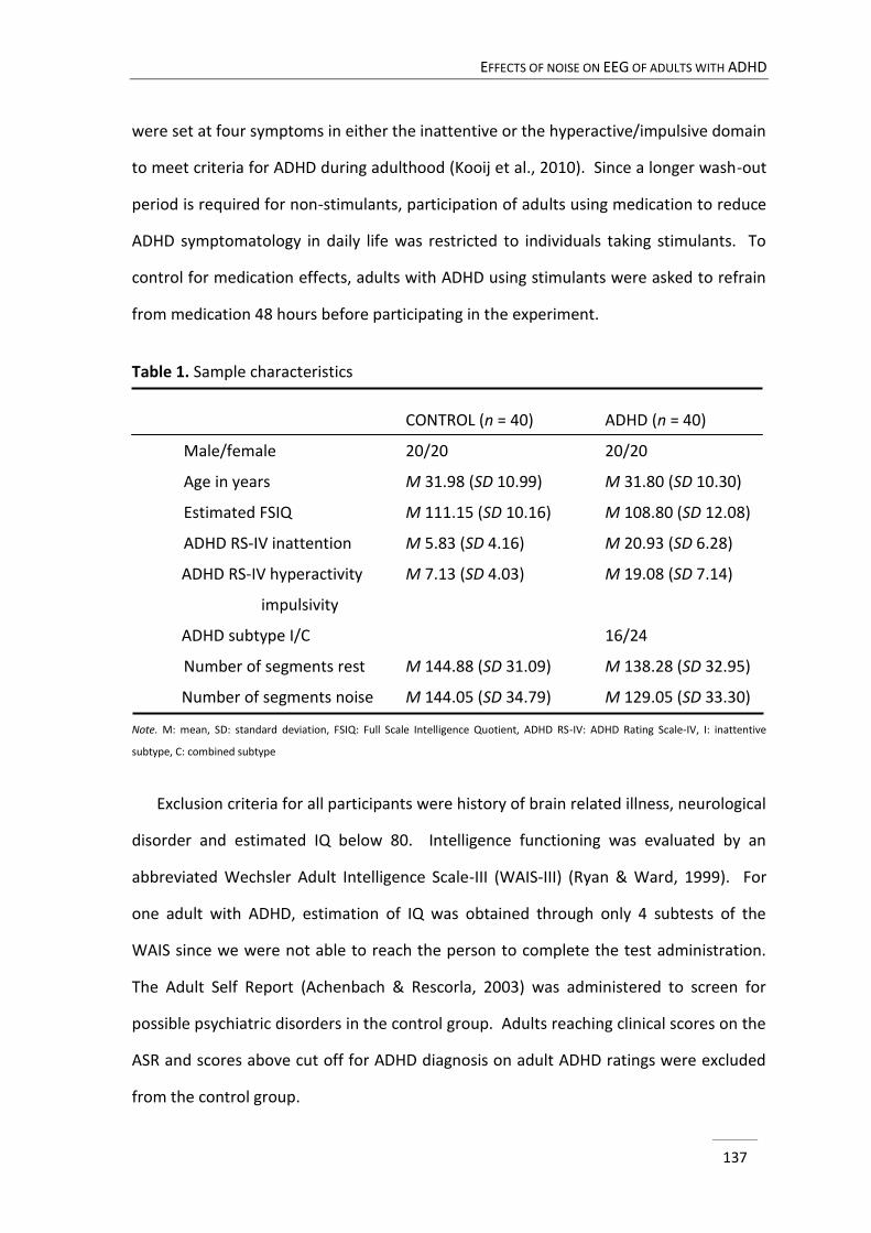
EFFECTS OF NOISE ON EEG OF ADULTS WITH ADHD
137
were set at four symptoms in either the inattentive or the hyperactive/impulsive domain
to meet criteria for ADHD during adulthood (Kooij et al., 2010). Since a longer wash-out
period is required for non-stimulants, participation of adults using medication to reduce
ADHD symptomatology in daily life was restricted to individuals taking stimulants. To
control for medication effects, adults with ADHD using stimulants were asked to refrain
from medication 48 hours before participating in the experiment.
Table 1. Sample characteristics
CONTROL (n = 40) ADHD (n = 40)
Male/female 20/20 20/20
Age in years M 31.98 (SD 10.99) M 31.80 (SD 10.30)
Estimated FSIQ M 111.15 (SD 10.16) M 108.80 (SD 12.08)
ADHD RS-IV inattention M 5.83 (SD 4.16) M 20.93 (SD 6.28)
ADHD RS-IV hyperactivity M 7.13 (SD 4.03) M 19.08 (SD 7.14)
impulsivity
ADHD subtype I/C 16/24
Number of segments rest M 144.88 (SD 31.09) M 138.28 (SD 32.95)
Number of segments noise M 144.05 (SD 34.79) M 129.05 (SD 33.30)
Note. M: mean, SD: standard deviation, FSIQ: Full Scale Intelligence Quotient, ADHD RS-IV: ADHD Rating Scale-IV, I: inattentive
subtype, C: combined subtype
Exclusion criteria for all participants were history of brain related illness, neurological
disorder and estimated IQ below 80. Intelligence functioning was evaluated by an
abbreviated Wechsler Adult Intelligence Scale-III (WAIS-III) (Ryan & Ward, 1999). For
one adult with ADHD, estimation of IQ was obtained through only 4 subtests of the
WAIS since we were not able to reach the person to complete the test administration.
The Adult Self Report (Achenbach & Rescorla, 2003) was administered to screen for
possible psychiatric disorders in the control group. Adults reaching clinical scores on the
ASR and scores above cut off for ADHD diagnosis on adult ADHD ratings were excluded
from the control group.
CHAPTER 5
138
Procedure
The research protocol was approved by the ethic committee of the Faculty of
Psychology and Educational Sciences at Ghent University. Following the explication of
procedures, participants signed an informed consent. Subjects were asked to sit down
in front of a monitor and were then fitted with the electrophysiological equipment.
During the study, the experimenter was present in the room but sat out of sight of the
participants. After the participants were instructed to close their eyes, two resting EEGs
varying in noise level (with and without presentation of pink noise) were recorded for
three minutes in a counterbalanced order (see also Söderlund et al., 2007, 2010) across
participants. Pink noise was used, because white noise can be perceived as aversive due
to much power in the high frequencies. Pink noise, also referred to as 1/f noise, is a
random signal whose power spectral density decreases as its frequency increases. Noise
was presented with a sound level of 80 dB via two external loudspeakers, placed on the
left and right side of a computer screen. Subsequently, several tasks were administered,
followed by another resting EEG session. In the current paper, the focus is on the
resting EEG conditions with and without exposure of noise before task execution.
Electrophysiological procedure
EEG data were obtained with an electrode cap employing 128 active 10 mm
Ag/AgCl electrodes placed according to the 10-5 International System (Oostenveld &
Praamstra, 2001) and amplified with a QuickAmp amplifier (Brain Products). The ground
electrode was enclosed within the cap at Fpz and an average reference derivation was
used. Signals were monitored using Brain Vision Recorder software (version 1.10) with a
sample rate of 500 Hz and a low pass filter of 50 Hz. Vertical eye movement was
recorded by an electrode enclosed in the cap above the right eye (Fp2) and an electrode
placed in line with the pupil below the right eye, whereas horizontal eye movement was
recorded from electrodes enclosed in the cap next to the eyes (F9 and F10).
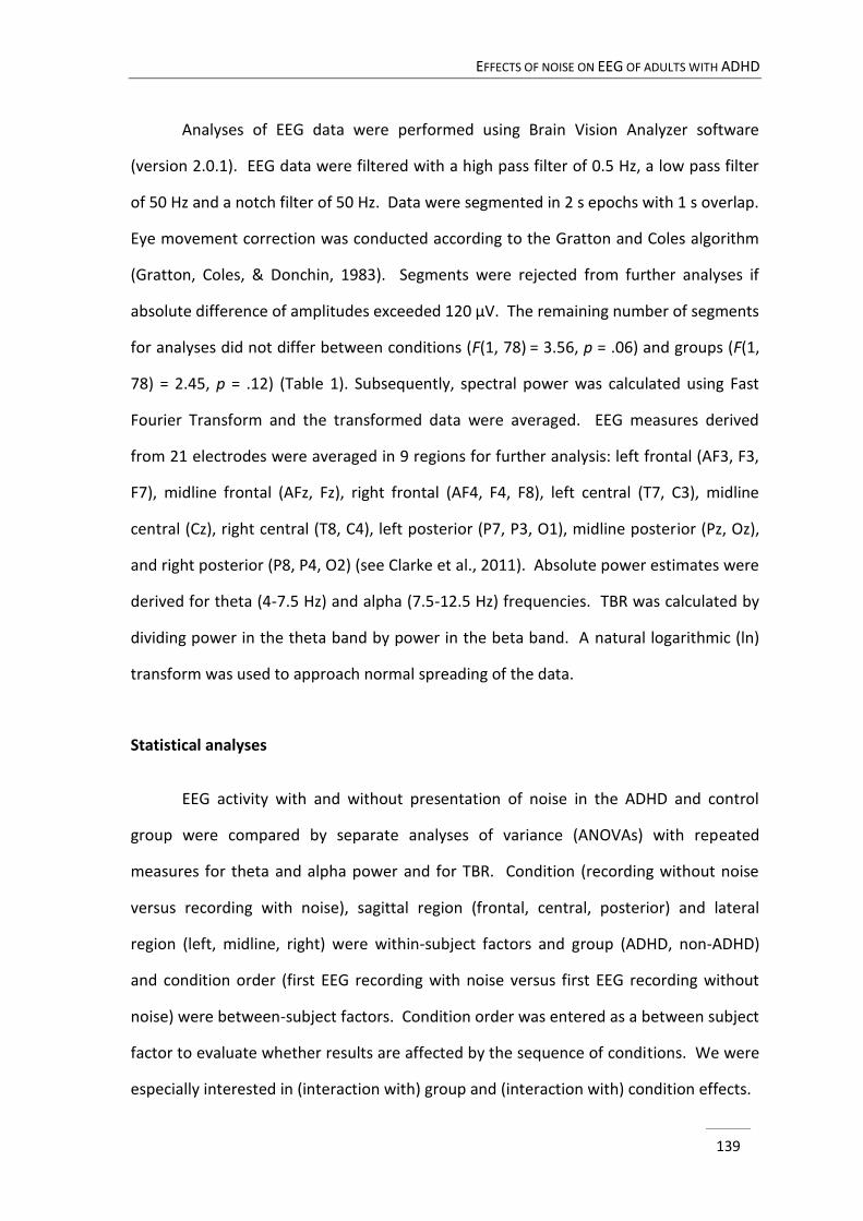
EFFECTS OF NOISE ON EEG OF ADULTS WITH ADHD
139
Analyses of EEG data were performed using Brain Vision Analyzer software
(version 2.0.1). EEG data were filtered with a high pass filter of 0.5 Hz, a low pass filter
of 50 Hz and a notch filter of 50 Hz. Data were segmented in 2 s epochs with 1 s overlap.
Eye movement correction was conducted according to the Gratton and Coles algorithm
(Gratton, Coles, & Donchin, 1983). Segments were rejected from further analyses if
absolute difference of amplitudes exceeded 120 µV. The remaining number of segments
for analyses did not differ between conditions (F(1, 78) = 3.56, p = .06) and groups (F(1,
78) = 2.45, p = .12) (Table 1). Subsequently, spectral power was calculated using Fast
Fourier Transform and the transformed data were averaged. EEG measures derived
from 21 electrodes were averaged in 9 regions for further analysis: left frontal (AF3, F3,
F7), midline frontal (AFz, Fz), right frontal (AF4, F4, F8), left central (T7, C3), midline
central (Cz), right central (T8, C4), left posterior (P7, P3, O1), midline posterior (Pz, Oz),
and right posterior (P8, P4, O2) (see Clarke et al., 2011). Absolute power estimates were
derived for theta (4-7.5 Hz) and alpha (7.5-12.5 Hz) frequencies. TBR was calculated by
dividing power in the theta band by power in the beta band. A natural logarithmic (ln)
transform was used to approach normal spreading of the data.
Statistical analyses
EEG activity with and without presentation of noise in the ADHD and control
group were compared by separate analyses of variance (ANOVAs) with repeated
measures for theta and alpha power and for TBR. Condition (recording without noise
versus recording with noise), sagittal region (frontal, central, posterior) and lateral
region (left, midline, right) were within-subject factors and group (ADHD, non-ADHD)
and condition order (first EEG recording with noise versus first EEG recording without
noise) were between-subject factors. Condition order was entered as a between subject
factor to evaluate whether results are affected by the sequence of conditions. We were
especially interested in (interaction with) group and (interaction with) condition effects.
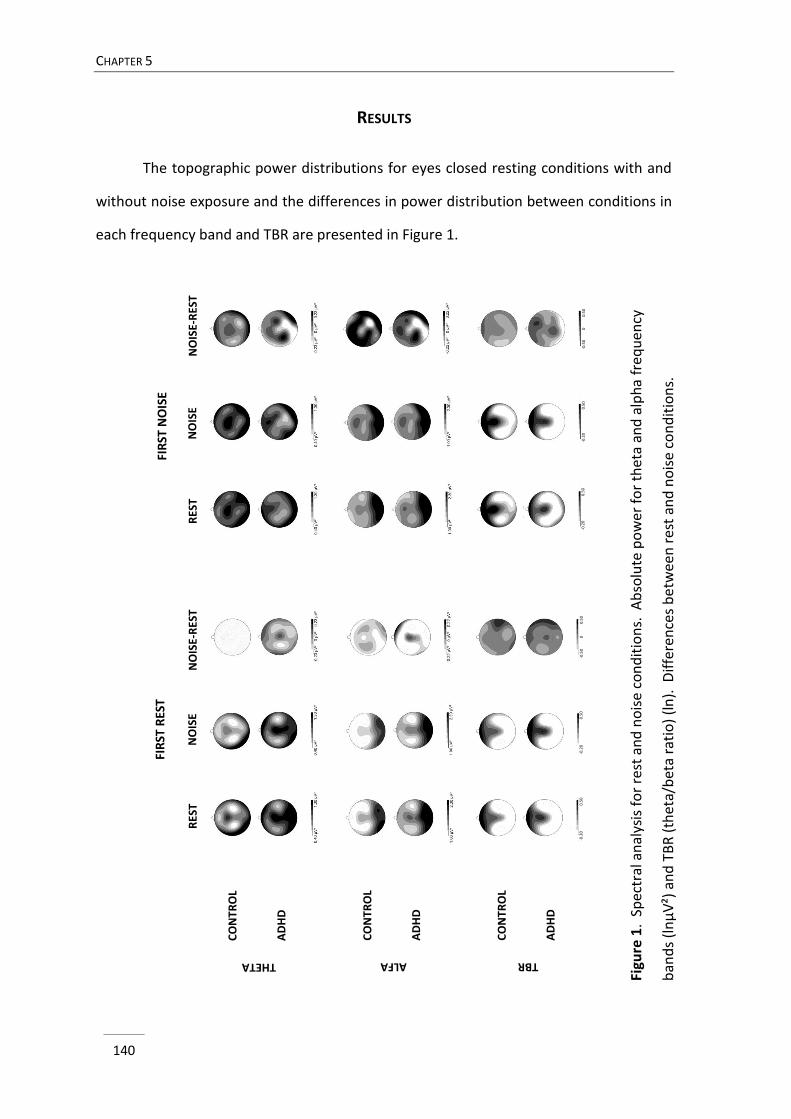
CHAPTER 5
140
RES
TN
OIS
E-R
EST
NO
ISE
CO
NT
RO
L
AD
HD
THETA
RES
TN
OIS
E-R
EST
NO
ISE
FIR
ST R
EST
FIR
ST N
OIS
E
CO
NT
RO
L
AD
HD
ALFA
CO
NT
RO
L
AD
HD
TBR
-0.2
2 µ
V²
0 µ
V²
0.2
2 µ
V²
0 m
s -
51
ms
-0.2
0 µ
V²0 µ
V²
0.3
0 µ
V²
0 m
s -
51
ms
-0.2
0
0
.50
-0.2
0
0
.50
-0.2
0
0
.50
-0.2
0
0
.50
-0.5
0
0
0
.50
-0.5
0
0
0.5
0
RESULTS
The topographic power distributions for eyes closed resting conditions with and
without noise exposure and the differences in power distribution between conditions in
each frequency band and TBR are presented in Figure 1.
Figu
re 1
. Sp
ectr
al a
nal
ysis
fo
r re
st a
nd
no
ise
con
dit
ion
s. A
bso
lute
po
wer
fo
r th
eta
and
alp
ha
fre
qu
ency
ban
ds
(ln
µV
²) a
nd
TB
R (
thet
a/b
eta
rati
o)
(ln
). D
iffe
ren
ces
be
twee
n r
est
and
no
ise
con
dit
ion
s.
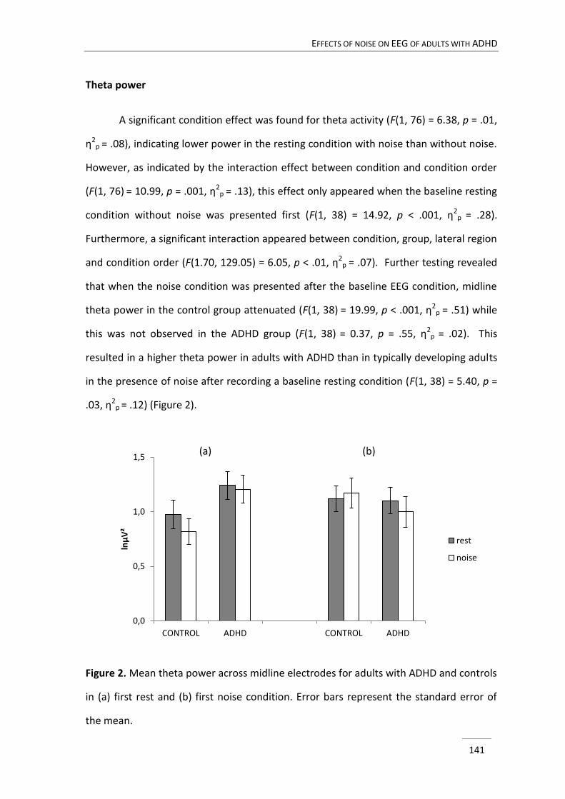
EFFECTS OF NOISE ON EEG OF ADULTS WITH ADHD
141
Theta power
A significant condition effect was found for theta activity (F(1, 76) = 6.38, p = .01,
η2p = .08), indicating lower power in the resting condition with noise than without noise.
However, as indicated by the interaction effect between condition and condition order
(F(1, 76) = 10.99, p = .001, η2p = .13), this effect only appeared when the baseline resting
condition without noise was presented first (F(1, 38) = 14.92, p < .001, η2p = .28).
Furthermore, a significant interaction appeared between condition, group, lateral region
and condition order (F(1.70, 129.05) = 6.05, p < .01, η2p = .07). Further testing revealed
that when the noise condition was presented after the baseline EEG condition, midline
theta power in the control group attenuated (F(1, 38) = 19.99, p < .001, η2p = .51) while
this was not observed in the ADHD group (F(1, 38) = 0.37, p = .55, η2p = .02). This
resulted in a higher theta power in adults with ADHD than in typically developing adults
in the presence of noise after recording a baseline resting condition (F(1, 38) = 5.40, p =
.03, η2p = .12) (Figure 2).
Figure 2. Mean theta power across midline electrodes for adults with ADHD and controls
in (a) first rest and (b) first noise condition. Error bars represent the standard error of
the mean.
0,0
0,5
1,0
1,5
CONTROL ADHD CONTROL ADHD
lnµ
V²
rest
noise
(a) (b)
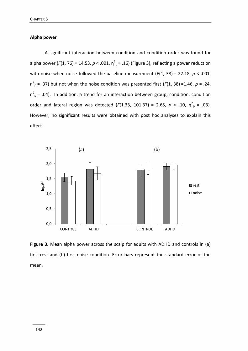
CHAPTER 5
142
Alpha power
A significant interaction between condition and condition order was found for
alpha power (F(1, 76) = 14.53, p < .001, η2p = .16) (Figure 3), reflecting a power reduction
with noise when noise followed the baseline measurement (F(1, 38) = 22.18, p < .001,
η2p = .37) but not when the noise condition was presented first (F(1, 38) =1.46, p = .24,
η2p = .04). In addition, a trend for an interaction between group, condition, condition
order and lateral region was detected (F(1.33, 101.37) = 2.65, p < .10, η2p = .03).
However, no significant results were obtained with post hoc analyses to explain this
effect.
Figure 3. Mean alpha power across the scalp for adults with ADHD and controls in (a)
first rest and (b) first noise condition. Error bars represent the standard error of the
mean.
0,0
0,5
1,0
1,5
2,0
2,5
CONTROL ADHD CONTROL ADHD
lnμ
V²
rest
noise
(a) (b)
EFFECTS OF NOISE ON EEG OF ADULTS WITH ADHD
143
TBR
For TBR, a significant interaction emerged between group, condition, lateral
region and condition order (F(2, 152) = 3.67, p = .03, η2p = .05). Post hoc analyses to
further explore this effect, revealed no significant results.
DISCUSSION
This is the first study that investigated the impact of noise on resting EEG activity
in an ADHD population. While studies have denoted a beneficial effect of noise on
cognitive performance in ADHD, little is known about the underlying working
mechanisms of noise on a brain functioning level. The MBA model explains the
beneficial effects of noise on ADHD by the restoration of impaired dopamine
transmission, resulting from the mechanism of stochastic resonance (Sikström &
Söderlund, 2007). Alternatively, a general arousal enhancing effect might explain the
effects, as ADHD has often been considered to be related to deviances in energetic
arousal state (Sonuga-Barke et al., 2010).
Groups did not differ in theta, TBR, or alpha across conditions, except for higher
midline theta power in adults with ADHD when noise was presented after baseline
resting EEG, due to absent attenuation of theta in the ADHD group with noise. The
comparable alpha power between groups is not fully unexpected, since inconsistent
findings in this frequency band have been reported across resting EEG studies
(Bresnahan & Barry, 2002; Bresnahan, Barry, Clarke, & Johnstone, 2006; Hermens et al.,
2004; Koehler et al., 2009; Liechti et al., 2013; Loo et al., 2009; van Dongen-Boomsma et
al., 2010; Woltering et al., 2012) and suggest that arousal was not decreased in adults
with ADHD. As for theta power and TBR, many studies have documented these
parameters to be elevated in ADHD (for reviews, see Barry et al., 2003 and Barry &
Clarke, 2009), however our findings are in accord with more recent studies that also
CHAPTER 5
144
could not replicate enhanced theta or TBR in adult ADHD (Liechti et al., 2013; Loo et al.,
2013; Poil et al., 2014; van Dongen-Boomsma et al., 2010).
As for the effects of noise on the EEG parameters, both groups showed a
significant reduction of alpha power with noise exposure after recording a baseline
resting EEG, while midline theta power only reduced in the control group and TBR was
not found to modulate with noise. Research addressing the effects of noise on brain
wave activity is scarce, limited to healthy controls and so far did not examine theta
power and TBR. Yet, our findings regarding alpha attenuation with noise exposure after
a baseline EEG are in line with other studies (Cho, Hwang, & Choi, 2011; Davies, 1968;
Mohammad, Saidatul, & Tamjis, 2009), indicating a general arousal enhancing effect of
pink noise. Reduction in alpha power has also been noticed in studies addressing other
manipulations of arousal, such as opening of the eyes (e.g., Barry, Clarke, Johnstone,
Magee, & Rushby, 2007; Boytsova & Danko, 2010) or administration of caffeine (e.g.,
Barry, Clarke, & Johnstone, 2011; Siepmann & Kierch, 2002). As mentioned in the
introduction, theta differs from alpha in that it does not seem to be directly related to
autonomic arousal as it was not found to be correlated with skin conductance level
(Dupuy et al., 2014), however enhanced theta is considered as a well-known EEG
signature for fatigue or drowsiness (e.g., Barwick, Arnett, & Slobounov, 2012; Jödicke et
al., 2013; Lal & Craig, 2001; Oken, Salinsky, & Elsas, 2006) and as with alpha several
studies have reported a reduction in theta with eye opening (e.g., Barry et al., 2007,
2009; Boytsova & Danko, 2010) or caffeine intake (Gilbert, Dibb, Plath, & Hiyane, 2000).
All together, this seems to support the notion that exposure to auditory external pink
noise enhances arousal or energetic state.
According to the MBA model, noise has the potential to induce beneficial effects
in ADHD by means of stochastic resonance through modulating properties of dopamine-
related gain and neural noise in determining neural responsivity (Sikström & Söderlund,
2007). Do the current findings provide support for the MBA model? First, the MBA
EFFECTS OF NOISE ON EEG OF ADULTS WITH ADHD
145
model suggests that individuals with ADHD or low attentive individuals in general have a
low gain parameter owing to low levels of baseline dopamine neuron firing. However,
as mentioned, no EEG differences were observed between groups during the baseline
EEG recording, providing no support for a deviant brain state in ADHD. Moreover,
adding noise decreased theta power in controls, but not in adults with ADHD, resulting
in higher midline theta power in ADHD when noise was presented. From an MBA model
perspective, this might indicate that the level of noise presented in the current study
was not sufficient to obtain the same effects on theta power in the ADHD group
compared to the control group. That is, according to the predictions of the model, more
noise may be needed to achieve stochastic resonance in dopamine deprived systems
and hence neural responsivity (Sikström & Söderlund, 2007; Söderlund et al., 2010).
However, the finding that none of the EEG measures, including theta power, could
distinguish the groups during baseline measurement argues against this explanation. In
addition, a similar noise level was used (80 dB) to that in studies where noise was found
to be effective on performance (Söderlund et al., 2007, 2010). Moreover, the adults
with ADHD included in the current study had significantly higher inattention symptoms
than the controls. Nevertheless, further research could elaborate on this by including
different levels of noise in the study design in order to investigate potential differential
effects related to noise level in ADHD. Perhaps, investigating EEG during conditions
where individuals with ADHD show normalized performance will provide more insight
into the underlying mechanism of the earlier observed beneficial effect of noise in
ADHD. Although the current study does not allow to fully test the MBA model as no
neurochemical responses to noise were evaluated, recently, Pålsson and colleagues
(2011) studied the effect of noise on the startle response in a rat model of ADHD and
showed that both control and ADHD strains (i.e., spontaneous hyperactive rats)
benefited from noise, but the noise effects were also found in rats with dopamine
CHAPTER 5
146
lesion, indicating that noise effects do not necessarily act through dopamine-related
gain (Pålsson, Söderlund, Klamer, & Bergquist, 2011).
The finding of different modulation of EEG activity with noise in ADHD may
suggest a deficiency in brain state regulation or adaptation rather than the presence of a
fixed state of underarousal, which would generally be in line with the state regulation
account of ADHD, implying impaired regulation of arousal/activation levels (Sergeant,
2005; Sonuga-Barke et al., 2010; van der Meere, 2005). The state regulation account
however relates ADHD to impaired top-down modulation of arousal systems, i.e.
difficulty with allocating extra effort in order to counteract suboptimal energetic state
(Börger & van der Meere, 2000; Wiersema et al., 2006a,b), while noise has been argued
to act through bottom-up modulation of arousal systems (Sanders, 1983). Hence, the
current finding may extend the state regulation account, by suggesting that bottom-up
regulation of energetic state in ADHD is deviant as well. Although we are aware that this
may seem a premature conclusion and more research is needed to further test this
hypothesis, some additional support for this proposition may be found in EEG studies on
ADHD addressing resting conditions that differ in arousal level (i.e., eyes open versus
eyes closed), in which also altered responsivity of theta in ADHD was noticed to opening
of the eyes (Buyck & Wiersema, submitted; Liechti et al., 2013), suggesting deficiencies
in adaptability in theta power in ADHD. This may also possibly partly explain some
inconsistent findings regarding deviating theta power in ADHD across studies, since this
implies plausible divergence in theta power in individuals with ADHD depending on
contextual/state features. Although speculative, the current findings may possibly also
clarify some effects of neurofeedback treatment in ADHD, in which traditionally theta
power is learned to be modulated to improve ADHD symptomatology. Perhaps
beneficial effects of this training can be related to acquiring a greater adaptability in
theta power rather than fixing a static deficit (i.e., higher theta or TBR (Barry & Clarke,
EFFECTS OF NOISE ON EEG OF ADULTS WITH ADHD
147
2009)). However, future studies are warranted to confirm the robustness and validity of
our results and to elaborate on this hypothesis.
The fact that EEG power did not change between conditions when noise was
presented first, may be the result from an after effect of noise on the following resting
condition. In animal research, it is well-established that arousal upon brief sensory
stimuli generally lasts for a while in order to facilitate goal-directed behaviour that
promotes survival and well-being (for reviews, see Jing, Gillette, & Weiss, 2009 and Pfaff,
Martin, & Faber, 2012). As an after effect of internal noise has repeatedly been
demonstrated on sensory nerves in humans (Chiou-Tan et al., 1996, 1997; Chuang et al.,
1999; Chuang & Chiou-Tan, 2000; Tran, Moss, Robinson, & Chiou-Tan, 2000), the
question can be raised whether similar mechanisms may act on brain oscillations.
However, our study design is not feasible to verify this hypothesis, since this requires
registration of baseline resting EEG before recording EEG with noise, followed by
another EEG registration without noise for all participants. Also, it should be taken into
account that the participants in the different condition orders may have experienced a
different level of arousal prior to the exposure to noise as sitting quietly for three
minutes in a silent room may elicit a different (i.e., lower) basic arousal level (baseline
first) than getting fitted with the test material (noise first). However, the experimental
design does not enable a within-subjects comparison of the condition orders, and a
between-subjects comparison of condition orders is hampered because spectral
parameters are known to express a considerable inter-individual variability (Poil et al.,
2014; van Dongen-Boomsma et al., 2010). Further research addressing order effects of
noise is imperative to elaborate on the impact of noise on resting EEG, since this may
have potentially important implications for the possible use of noise for clinical
purposes.
Since research already demonstrated associations between measures of central
and autonomous nervous system functioning (Olbrich et al., 2011), incorporating for
CHAPTER 5
148
instance electrodermal measures, that have been shown to be a reliable marker of
arousal (for a review, see Critchley, 2002), could provide more insights into the relation
between the concept of arousal and the effect of noise presentation in ADHD.
To conclude, this first study on the effects of noise on brain wave activity in an
ADHD population revealed some interesting findings on the nature of EEG discrepancies
in ADHD and the significance of noise on brain functioning in adults with as well as
without ADHD. First, the current findings seem to validate that noise exposure may
serve as an energetic state or arousal enhancing procedure. Second, although our
results did not confirm discrepancies in alpha power nor in theta or TBR in adults with
ADHD compared to typically developing adults, some evidence was found for dynamic,
state dependent deficiencies in theta power in ADHD. That is, while midline theta
activity attenuated in typically developing adults with noise after recording of baseline
EEG, this was not observed in the ADHD group. This may reflect aberrant adaptability in
this frequency band in adults with ADHD.
EFFECTS OF NOISE ON EEG OF ADULTS WITH ADHD
149
REFERENCES
Achenbach, T.M., & Rescorla, L.A. (2003). Manual for the ASEBA Adult Forms & Profiles.
Burlington, Vermont: University of Vermont, Research Center for Children, Youth, and
Families.
American Psychiatric Association (2013). Diagnostic and statistical manual of mental disorders
(5th Ed.). Arlington, VA: American Psychiatric Publishing.
Antrop, I., Buysse, A., Roeyers, H., & Van Oost, P. (2002). Stimulation seeking and hyperactive
behavior in children with ADHD: A re-analysis. Perceptual and Motor Skills, 95(1), 71-90.
Antrop, I., Buysse, A., Roeyers, H., & Van Oost, P. (2005). Activity in children with ADHD during
waiting situations in the classroom: A pilot study. British Journal of Educational
Psychology, 75, 51-69. doi: 10.1348/000709905X36100
Antrop, I., Roeyers, H., Van Oost, P., & Buysse, A. (2000). Stimulation seeking and hyperactivity in
children with ADHD. Journal of Child Psychology and Psychiatry and Allied Disciplines,
41(2), 225-231.
Barkley, R.A. (1997). Behavioral inhibition, sustained attention, and executive functions:
Constructing a unifying theory of ADHD. Psychological Bulletin, 121, 65-94. doi:
10.1037//0033-2909.121.1.65
Barry, R.J., & Clarke, A.R. (2009). Spontaneous EEG Oscillations in Children, Adolescents, and
Adults. Typical Development, and Pathological Aspects in Relation to AD/HD. Journal of
Psychophysiology, 23, 157-173. doi: 10.1027/0269-8803.23.4.157
Barry, R.J., Clarke, A.R., & Johnstone, S.J. (2003). A review of electrophysiology in attention-
deficit/hyperactivity disorder. I. Qualitative and quantitative electroencephalography.
Clinical Neurophysiology, 114, 171-183. doi: 10.1016/S1388-2457(02)00362-0
Barry, R.J., Clarke, A.R., & Johnstone, S.J. (2011). Caffeine and opening the eyes have additive
effects on resting arousal measures. Clinical Neurophysiology, 122(10), 2010-2015. doi:
10.1016/j.clinph.2011.02.036
CHAPTER 5
150
Barry, R.J., Clarke, A.R., Johnstone, S.J., Magee, C.A., & Rushby, J.A. (2007). EEG differences
between eyes-closed and eyes-open resting conditions. Clinical Neurophysiology, 118,
2765-2773. doi: 10.1016/j.clinph.2007.07.028
Barry, R.J., Clarke, A.R., Johnstone, S.J., McCarthy, R., & Selikowitz, M. (2009).
Electroencephalogram theta/beta Ratio and Arousal in Attention-Deficit/Hyperactivity
Disorder: Evidence of Independent Processes. Biological Psychiatry, 66, 398-401. doi:
10.1016/j.biopsych.2009.04.027
Barry, R.J., Clarke, A.R., McCarthy, R., Selikowitz, M., Rushby, J.A., & Ploskova, E. (2004). EEG
differences in children as a function of resting-state arousal level. Clinical
Neurophysiology, 115, 402-408. doi: 10.1016/S1388-2457(03)00343-2
Barwick, F., Arnett, P., & Slobounov, S. (2012). EEG correlates of fatigue during administration of
a neuropsychological test battery. Clinical Neurophysiology, 123, 278-284. doi:
10.1016/j.clinph.2011.06.027
Boonstra, A.M., Oosterlaan, J., Sergeant, J.A., & Buitelaar, J.K. (2005). Executive functioning in
adult ADHD: a meta-analytic review. Psychological Medicine, 35(8), 1097-1108. doi:
10.1017/S003329170500499X
Börger, N.A., & van der Meere, J.J. (2000). Motor control and state regulation in children with
ADHD: a cardiac response study. Biological Psychology, 51, 247-267. doi: 10.1016/S0301-
0511(99)00040-X
Boytsova, Y.A., & Danko, S.G. (2010). EEG differences between resting states with eyes open and
eyes closed in darkness. Human Physiology, 36(3), 367-369. doi:
10.1134/S0362119710030199
Bresnahan, S.M., & Barry, R.J. (2002). Specificity of quantitative EEG analysis in adults with
attention deficit hyperactivity disorder. Psychiatry Research, 112, 133-144. doi:
10.1016/S0165-1781(02)00190-7
Bresnahan, S.M., Barry, R.J., Clarke, A.R., & Johnstone, S.J. (2006). Quantitative EEG analysis in
dexamphetamine-responsive adults with attention-deficit/hyperactivity disorder.
Psychiatry Research, 112, 133-144. doi: 10.1016/j.psychres.2005.09.002
EFFECTS OF NOISE ON EEG OF ADULTS WITH ADHD
151
Buyck, I., & Wiersema, J.R. (2014). State-related electroencephalographic deviances in attention
deficit hyperactivity disorder. Manuscript submitted for publication.
Clarke, A.R., Barry, R.J., Dupuy, F.E., McCarthy, R., Selikowitz, M., & Heaven, P.C. (2011).
Childhood EEG as a predictor of adult attention-deficit/hyperactivity disorder. Clinical
Neurophysiology, 122(1), 73-80. doi: 10.1016/j.clinph.2010.05.032
ChiouTan, F.Y., Magee, K.N., Robinson, L.R., Nelson, M.R., Tuel, S.S., Krouskop, T.A., & Moss, F.
(1996). Enhancement of subthreshold sensory nerve action potentials during muscle
tension mediated noise. International Journal of Bifurcation and Chaos, 6, 1389-1396.
doi: 10.1142/S0218127496000813
ChiouTan, F.Y., Magee, K.N., Tuel, S.M., Robinson, L.R., Krouskop, T.A., Nelson, M.R., & Moss, F.
(1997). Augmented sensory nerve action potentials during distant muscle contraction.
American Journal of Physical Medicine & Rehabilitation, 76, 14-18. doi:
10.1097/00002060-199701000-00004
Cho, W., Hwang, S.H., & Choi, H. (2011). An investigation of the influences of noise on EEG
power bands and visual cognitive responses for human-oriented product design. Journal
of Mechanical Science and Technology, 25, 821-826. doi: 10.1007/s12206-011-0128-2
Chuang, T.Y., & ChiouTan, F.Y. (2000). Temporal effects of isometric contraction maneuvers on
threshold sural amplitude. American Journal of Physical Medicine & Rehabilitation, 79,
40-43. doi: 10.1097/00002060-200001000-00009
Chuang, T.Y., Robinson, L.R., Nelson, M.R., Moss, F., & ChiouTan, F.Y. (1999). Effect of isometric
contraction on threshold somatosensory evoked potentials. American Journal of Physical
Medicine & Rehabilitation, 78, 2-6. doi: 10.1097/00002060-199901000-00002
Critchley, H.D. (2002). Electrodermal responses: What happens in the brain. The Neuroscientist,
8, 132-142. doi: 10.1177/107385840200800209
Davies, D.R. (1968). Physiological and psychological effects of exposure to high intensity noise.
Applied Acoustics, 1, 215-233. doi: 10.1016/0003-682X(68)90024-8
DuPaul, G.J., Power, T.J., Anastopoulos, A.D., & Reid, R. (1998). ADHD Rating Scale-IV: Checklists,
norms, and clinical interpretation. New York, NY, US: Guilford Press.
CHAPTER 5
152
Dupuy, F.E., Clarke, A.R., Barry, R.J., Selikowitz, M., & McCarthy, R. (2014). EEG and
electrodermal activity in girls with Attention-Deficit/Hyperactivity Disorder. Clinical
Neurophysiology, 125(3), 491-499. doi: 10.1016/j.clinph.2013.09.007
Faraone, S.V., Biederman, J., & Mick, E. (2006). The age-dependent decline of attention deficit
hyperactivity disorder: a meta-analysis of follow-up studies. Psychological Medicine, 36,
159-165. doi: 10.1017/S003329170500471X
Fonseca, L.C., Tedrus, G.M.A.S., Bianchini, M.C., & Silva, T.F. (2013). Electroencephalographic
Alpha Reactivity on Opening the Eyes in Children With Attention-Deficit Hyperactivity
Disorder. Clinical EEG and Neuroscience, 44, 53-57. doi: 10.1177/1550059412445659
Gilbert, D.G., Dibb, W.D., Plath, L.C., & Hiyane, S.G. (2000). Effects of nicotine and caffeine,
separately and in combination, on EEG topography, mood, heart rate, cortisol, and
vigilance. Psychophysiology, 37(5), 583-595. doi: 10.1111/1469-8986.3750583
Gratton, G., Coles, M.G., & Donchin, E. (1983). A new method for off-line removal of ocular
artifact. Electroencephalography and Clinical Neurophysiology, 55, 468-484. doi:
10.1016/0013-4694(83)90135-9
Hermens, D.F., Williams, L.M., Lazzaro, I., Whitmont, S., Melkonian, D., & Gordon, E. (2004). Sex
differences in adult ADHD: a double dissociation in brain activity and autonomic arousal.
Biological Psychology, 66, 221-233. doi: 10.1016/j.biopsycho.2003.10.006
Jing, J., Gillette, R., & Weiss, K.R. (2009). Evolving Concepts of Arousal: Insights from Simple
Model Systems. Reviews in the Neurosciences, 20, 405-427. doi:
10.1515/REVNEURO.2009.20.5-6.405
Johnson, K.A., Wiersema J.R., & Kuntsi, J. (2009). What would Karl Popper say? Are current
psychological theories of ADHD falsifiable? Behavioral and Brain Functions, 5, 15. doi:
10.1186/1744-9081-5-15
Jödicke, J., Olbrich, S., Sander, C., Minkwitz, J., Chittka, T., Himmerich, H., & Hegerl, U. (2013).
Separation of Low-Voltage EEG-Activity During Mental Activation from that During
Transition to Drowsiness. Brain Topography, 26(4), 538-546. doi: 10.1007/s10548-013-
0287-9
EFFECTS OF NOISE ON EEG OF ADULTS WITH ADHD
153
Koehler, S., Lauer, P., Schreppel, T., Jacob, C., Heine, L., Boreatti-Hummer A., … & Herrmann, M.J.
(2009). Increased EEG power density in alpha and theta bands in adult ADHD patients.
Journal of Neural Transmission, 116, 97-104. doi: 10.1007/s00702-008-0157-x
Kooij, S.J.J., Bejerot, S., Blackwell, A., Caci, H., Casas-Brugue, M., Carpentier, P.J., ... & Asherson,
P. (2010). European consensus statement on diagnosis and treatment of adult ADHD:
The European Network Adult ADHD. BMC Psychiatry, 10, 67. doi: 10.1186/1471-244X-10-
67
Kooij, S.J.J., & Francken, M.H. (2010). DIVA 2.0. Diagnostisch Interview Voor ADHD bij
volwassenen. Den Haag, DIVA Foundation.
Lal, S.K., & Craig, A. (2001). A critical review of the psychophysiology of driver fatigue. Biological
Psychology, 55(3), 173-194. doi: 10.1016/S0301-0511(00)00085-5
Liechti, M.D., Valko, L., Müller, U.C., Döhnert, M., Drechsler, R., Steinhausen, H.C., & Brandeis, D.
(2013). Diagnostic value of resting electroencephalogram in attention-
deficit/hyperactivity disorder across the lifespan. Brain Topography, 26, 135-151. doi:
10.1007/s10548-012-0258-6
Loo, S.K., Cho, A., Hale, T.S., McGough, J., McCracken, J., & Smalley, S.L. (2013). Characterization
of the Theta to Beta Ratio in ADHD: Identifying Potential Sources of Heterogeneity.
Journal of Attention Disorders, 17, 384-392. doi: 10.1177/1087054712468050
Loo, S.K., Hale, T.S., Macion, J., Hanada, G., McGough, J.J., McCracken, J.T., & Smalley, S.L.
(2009). Cortical activity patterns in ADHD during arousal, activation and sustained
attention. Neuropsychologia, 47, 1033-1040. doi: 10.1016/
j.neuropsychologia.2009.04.013
Loo, S.K., & Makeig, S. (2012). Clinical utility of EEG in attention-deficit/hyperactivity disorder: a
research update. Neurotherapeutics, 9, 569-587. doi: 10.1007/s13311-012-0131-z
Lubar, J.F. (1991). Discourse on the development of EEG diagnostics and biofeedback for
attention-deficit/hyperactivity disorders. Biofeedback and Self-Regulation, 16(3), 201-
225. doi: 10.1007/BF01000016
Mann, C.A., Lubar, J.F., Zimmerman, A.W., Miller, C.A., & Muenchen, R.A. (1992). Quantitative
analysis of EEG in boys with attention deficit hyperactivity disorder: controlled study
CHAPTER 5
154
with clinical implications. Pediatric Neurology, 8, 30-36. doi: 10.1016/0887-
8994(92)90049-5
McDonnell, M.D., & Abbott, D. (2009). What Is Stochastic Resonance? Definitions,
Misconceptions, Debates, and Its Relevance to Biology. Plos Computational Biology, 5,
e1000348. doi: 10.1371/journal.pcbi.1000348
Metin, B., Roeyers, H., Wiersema, J.R., van der Meere, J., & Sonuga-Barke, E. (2012). A Meta-
Analytic Study of Event Rate Effects on Go/No-Go Performance in Attention-
Deficit/Hyperactivity Disorder. Biological Psychiatry, 72, 990-996. doi:
10.1016/j.biopsych.2012.08.023
Mohammad, N.F., Saidatul, A., & Tamjis, M.R. (2009). A Study of Noise Effect on the Fast Brain
Activity, Proceedings of the International Conference on Man-Machine Systems
(ICoMMS). Batu Ferringhi, Penang, Malaysia. Retrieved from
http://dspace.unimap.edu.my/dspace/bitstream/123456789/7274/1/A%20Study%20of
%20Noise%20Effect.pdf
Monastra, V.J., Lubar, J.F., Linden, M., Van Deusen, P., Green, G., Wing, W., … & Fenger, T.N.
(1999). Assessing attention deficit hyperactivity disorder via quantitative
electroencephalography: An initial validation study. Neuropsychology, 13, 424-433. doi:
10.1037/0894-4105.13.3.424
Moss, F., Ward, L.M., & Sannita, W.G. (2004). Stochastic resonance and sensory information
processing: a tutorial and review of application. Clinical Neurophysiology, 115, 267-281.
doi: 10.1016/j.clinph.2003.09.014
Oken, B.S., Salinsky, M.C., & Elsas, S.M. (2006). Vigilance, alertness, or sustained attention:
physiological basis and measurement. Clinical Neurophysiology, 117, 1885-1901. doi:
10.1016/j.clinph.2006.01.017
Olbrich, S., Sander, C., Matschinger, H., Mergl, R., Trenner, M., Schonknecht, P., & Hegerl, U.
(2011). Associations Between EEG-Vigilance and the Autonomous Nervous System
Activity During Rest. Journal of Psychophysiology, 25, 190-200. doi: 10.1027/0269-
8803/a000061
EFFECTS OF NOISE ON EEG OF ADULTS WITH ADHD
155
Oostenveld, R., & Praamstra, P. (2001). The five percent electrode system for high resolution
EEG and ERP measurements. Clinical Neurophysiology, 112, 713-719. doi: 10.1016/S1388-
2457(00)00527-7
Pålsson, E., Söderlund, G., Klamer, D., & Bergquist, F. (2011). Noise benefit in prepulse inhibition
of the acoustic startle reflex. Psychopharmacology, 214(3), 675-685. doi:
10.1007/s00213-010-2074-6
Pfaff, D.W., Martin, E.M., & Faber, D. (2012). Origins of arousal: roles for medullary reticular
neurons. Trends in Neurosciences, 35(8), 468-476. doi: 10.1016/j.tins.2012.04.008
Poil, S.S., Bollmann, S., Ghisleni, C., O'Gorman, R.L., Klaver, P., Ball, J., Eich-Höchli, D., ... &
Michels, L. (in press). Age dependent electroencephalographic changes in attention-
deficit/hyperactivity disorder (ADHD). Clinical Neurophysiology. Advance online
publication. doi: 10.1016/j.clinph.2013.12.118
Ryan, J.J., & Ward, L.C. (1999). Validity, reliability, and standard errors of measurement for two
seven-subtest short forms of the Wechsler Adult Intelligence Scale-III. Psychological
Assessment, 11, 207-211. doi: 10.1037/1040-3590.11.2.207
Sagvolden, T., Johansen, E.B., Aase, H., & Russell, V.A. (2005). A dynamic developmental theory
of attention-deficit/hyperactivity disorder (ADHD) predominantly hyperactive/impulsive
and combined subtypes. Behavioral and Brain Sciences, 28, 397-468. doi:
10.1017/S0140525X05000075
Sanders, A.F. (1983). Towards a model of stress and human performance. Acta Psychologica, 53,
61-97. doi: 10.1016/0001-6918(83)90016-1
Sanders, A.F. (1998). Elements of human performance: reaction processes and attention in
human skill, Mahwah, NJ: Lawrence Erlbaum.
Sergeant, J. (2005). Modeling attention-deficit/hyperactivity disorder: a critical appraisal of the
cognitive-energetic model. Biological Psychiatry, 57, 1248-1255. doi:
10.1016/j.bps.2004.09.010
Siepmann, M., & Kirch, W. (2002). Effects of caffeine on topographic quantitative EEG.
Neuropsychobiology, 45(3), 161-166. doi: 10.1159/000054958
CHAPTER 5
156
Sikström, S., & Söderlund, G. (2007). Stimulus-dependent dopamine release in attention-
deficit/hyperactivity disorder. Psychological Review, 114, 1047-1075. doi: 10.1037/0033-
295X.114.4.1047
Söderlund, G.B.W., Sikström, S., Loftesnes, J.M., & Sonuga-Barke, E.J. (2010). The effects of
background white noise on memory performance in inattentive school children.
Behavioral and Brain Functions, 6, 55. doi: 10.1186/1744-9081-6-55
Söderlund, G., Sikström, S., & Smart, A. (2007). Listen to the noise: noise is beneficial for
cognitive performance in ADHD. Journal of Child Psychology and Psychiatry, 48, 840-847.
doi: 10.1111/j.1469-7610.2007.01749.x
Sonuga-Barke, E.J.S., Taylor, E., Sembi, S., & Smith, J. (1992). Hyperactivity and delay aversion: I.
The effect of delay on choice. Journal of Child Psychology and Psychiatry and Allied
Disciplines, 33(2), 387-398. doi: 10.1111/j.1469-7610.1992.tb00874.x
Sonuga-Barke, E., Wiersema J.R., van der Meere J., & Roeyers, H. (2010). Context-dependent
Dynamic Processes in Attention Deficit/Hyperactivity Disorder: Differentiating Common
and Unique Effects of State Regulation Deficits and Delay Aversion. Neuropsychological
Review, 20, 86-102. doi: 10.1007/s11065-009-9115-0
Spencer, T.J., Biederman, J., & Mick, E. (2007). Attention-Deficit/Hyperactivity disorder:
Diagnosis, lifespan, comorbidities, and neurobiology. Journal of Pediatric Psychology, 32,
631-642. doi: 10.1093/jpepsy/jsm005
Tran, T.M.P., Moss, F., Robinson, L.R., & Chiou-Tan, F.Y. (2000). Increased threshold sural
amplitude after upper limb isometric contraction in complete paraplegics. American
Journal of Physical Medicine & Rehabilitation, 79, 542-546. doi: 10.1097/00002060-
200011000-00011
van der Meere, J. J. (2005). State regulation and ADHD. In D. Gozal & D.L. Molfese (Eds.),
Attention deficit hyperactivity disorder: From genes to animal models to patients (pp.
413-433). Totowa, NJ: Humana.
van Dongen-Boomsma, M., Lansbergen, M.M., Bekker, E.M., Kooij, S.J.J., van der Molen, M.,
Kenemans, J.L., & Buitelaar, J.K. (2010). Relation between resting EEG to cognitive
EFFECTS OF NOISE ON EEG OF ADULTS WITH ADHD
157
performance and clinical symptoms in adults with attention-deficit/hyperactivity
disorder. Neuroscience Letters, 469, 102-106. doi: 10.1016/j.neulet.2009.11.053
Volkow, N.D., Wang, G.J., Newcorn, J., Telang, F., Solanto, M.V., Fowler, J.S., ... & Swanson, J.M.
(2007). Depressed dopamine activity in caudate and preliminary evidence of limbic
involvement in adults with attention-deficit/hyperactivity disorder. Archives of General
Psychiatry, 64(8), 932-940. doi: 10.1001/archpsyc.64.8.932
Ward, M.F., Wender, P.H., & Reimherr, F.W. (1993). The Wender Utah Rating Scale: an aid in the
retrospective diagnosis of attention deficit hyperactivity disorder. American Journal of
Psychiatry, 150, 885-890
Wiersema, J.R., van der Meere, J.J., Antrop, I., & Roeyers, H. (2006a). State regulation in adult
ADHD: an event-related potential study. Journal of Clinical and Experimental
Neuropsychology, 28, 1113–1126. doi: 10.1080/13803390500212896
Wiersema, R., van der Meere, J., Roeyers, H., Van Coster, R., & Baeyens, D. (2006b). Event rate
and event-related potentials in ADHD. Journal of Child Psychology and Psychiatry, 47,
560-567. doi: 10.1111/j.1469-7610.2006.01592.x
Willcutt, E.G. (2012). The prevalence of DSM-IV attention-deficit/hyperactivity disorder: a meta-
analytic review. Neurotherapeutics, 9, 490-499. doi: 10.1007/s13311-012-0135-8
Willcutt, E.G., Doyle, A.E., Nigg, J.T., Faraone, S.V., & Pennington, B.F. (2005). Validity of the
executive function theory of attention-deficit/hyperactivity disorder: a meta analytic
review. Biological Psychiatry, 57, 1336-1346. doi: 10.1016/j.biopsych.2005.02.006
Woltering, S., Jung, J., Liu, Z., & Tannock, R. (2012). Resting state EEG oscillatory power
differences in ADHD college students and their peers. Behavioral and Brain Functions, 8,
60. doi: 10.1186/1744-9081-8-60
Zentall, S.S., & Zentall, T.R. (1983). Optimal stimulation: a model of disordered activity and
performance in normal and deviant children. Psychological Bulletin, 94, 446-447. doi:
10.1037/0033-2909.94.3.446

159
TASK-RELATED EEG DEVIANCES IN
ADULTS WITH ADHD1
ABSTRACT
Objective: The aim of this study was to provide more insights into the functional
significance of EEG deviances in ADHD by evaluating quantitative EEG during
performance on a task loading on activation level and a task tapping top-down executive
control. Methods: Behavioural performance and EEG were compared between 24
adults with ADHD and 20 typically developing adults during execution of a simple slow
paced 2-choice reaction time (2-CRT) task and a regular paced flanker task. Results:
During the slow 2-CRT task, adults with ADHD responded slower, more variably, and
tended to make more errors of commission. Although being slower, adults with ADHD
showed equally large congruency effects during flanker task performance, indicating
intact interference inhibition. In the slow 2-CRT task, (midline) theta and beta power
were higher in the ADHD group than in the control group, whereas no significant EEG
group differences were observed in the flanker task. A moderate positive correlation
between theta power and errors of commission was found in the 2-CRT task for adults
with ADHD. Conclusions: Adults with ADHD performed worse on a task inducing a low
activation level but showed intact interference inhibition. The EEG findings are in accord
with this, showing abnormal EEG patterns in ADHD only when activation level was taxed,
not when top-down executive control was required. Time-on-task effects could not
explain deviances between groups. The findings indicate that EEG deviances in ADHD
are task dependent and may be related to a suboptimal energetic state, rather than
impaired top-down executive control.
1 Based on Buyck, I., & Wiersema, J.R. (2014). Task-related electroencephalographic deviances in adults
with attention deficit hyperactivity disorder. Manuscript submitted for publication.
6 CHAPTER
CHAPTER 6
160
INTRODUCTION
Attention-deficit/hyperactivity disorder (ADHD) is a common
neurodevelopmental disorder with a childhood onset, affecting approximately 5 to 7 %
of the worldwide population (Willcut, 2012). The disorder comprises a pattern of
developmentally inappropriate symptoms of inattention and/or hyperactivity and
impulsivity, resulting in significant impairments in social, academic or occupational
functioning (American Psychiatric Association, 2013). ADHD-related symptomatology
has been documented to persist throughout the lifespan in about two thirds of
individuals, although often with a more subtle expression of symptoms of hyperactivity
and impulsivity in adulthood (Faraone, Biederman, & Mick, 2006; Kooij et al., 2010;
Spencer, Biederman, & Mick, 2007).
While its etiology is not fully understood, over the past decades the neural
correlates of ADHD have been widely investigated by means of electroencephalography
(EEG) (Loo & Makeig, 2012). Most EEG studies in ADHD have evaluated brain wave
activity during resting conditions with either eyes closed or eyes open, often revealing
an elevated theta activity and/or an enhanced theta/beta ratio (TBR) in ADHD across the
lifespan (Barry, Clarke, & Johnstone, 2003, for a review; Buyck & Wiersema, 2014;
Clarke, Barry, McCarthy, Selikowitz, & Johnstone, 2007; Clarke et al., 2011; Dupuy,
Clarke, Barry, McCarthy, & Selikowitz, 2011; Dupuy, Barry, Clarke, McCarthy, &
Selikowitz, 2013; González-Castro, Rodríguez, López, Cueli, & Álvarez, 2013; Hermens et
al., 2004; Koehler et al., 2009; Lansbergen, van Dongen-Boomsma, Spronk, & Buitelaar,
2011; Loo et al., 2010; Shi et al., 2012; Woltering, Jung, Liu, & Tannock, 2012). Yet, lately
the consistency of these deviances characterizing the EEG profile of the total ADHD
group is debated, since some studies could not invariably replicate these findings (Buyck
& Wiersema, submitted; Coolidge, Starkey, & Cahill, 2007; Liechti et al., 2013; Loo et al.,
2009, 2013; Nazari, Wallois, Aarabi, & Berquin, 2011; Ogrim, Kropotov, & Hestad, 2012;
TASK-RELATED EEG DEVIANCES IN ADULTS WITH ADHD
161
Poil et al., 2014; Swartwood, Swartwood, Lubar, & Timmermann, 2003; van Dongen-
Boomsma et al., 2010).
Less often, quantitative EEG in ADHD has been studied during task performance.
As different tasks may tap diverse cognitive(-energetic) processes, which are not easily
discerned during resting state conditions, applying this approach may increase the
understanding of EEG deviations in ADHD. Most studies regarding task-related EEG
activity in ADHD are conducted in children. Some of these report similar theta or TBR
deviances as observed in resting conditions during a variety of tasks (El-Sayed, Larsson,
Persson, & Rydelius, 2002; Janzen, Graap, Stephanson, Marshall, & Fitzsimmons, 1995;
Mann, Lubar, Zimmerman, Miller, & Muenchen, 1992; Monastra et al., 1999; Monastra,
Lubar, & Linden, 2001), however other studies could not confirm these findings (Nazari
et al., 2011; Swartwood et al., 2003). Fewer studies have addressed task-related EEG
activity in adult ADHD, also documenting inconsistent results. That is, Monastra and
colleagues (1999) reported that adults with ADHD showed increased TBR during
performance on several tasks. Yet, in another study, theta and TBR were found to be
normal in adults with ADHD during a Wisconsin Card Sorting Test, a Paced Auditory
Serial Addition Test, and a Continuous Performance Test (CPT), while alpha activity was
elevated during the CPT (White, Hutchens, & Lubar, 2005). In contrast, alpha
attenuation has also been reported in adults with ADHD while performing the CPT (Loo
et al., 2009). Interestingly, some studies also refer to time-on-task effects in differences
between groups during the CPT. In specific, one study found reduced frontal beta power
in ADHD in the second part of a visual CPT, while beta was not divergent during the first
part, and a similar observation was made for theta power, being enhanced in ADHD only
at the end of the task in an auditory CPT (Rasey, Craig, & Lubar, 1999). Another study
observed an alpha increase with time-on-task in the control group but not in the ADHD
group (Loo et al., 2009). Although not concordant, these results nevertheless suggest
CHAPTER 6
162
that it may be important to take into account time-on-task effects when investigating
task-related EEG activity in ADHD.
While aberrant theta and TBR have frequently been reported in ADHD, the
significance of these deviances is still not fully understood. Concerning this, evaluating
quantitative EEG during task performance may be helpful, because one can include
experimental manipulations that enable to test existing hypotheses. In this respect, one
of the most influential explanations regarding enhanced theta and TBR is that they
represent cortical hypoarousal (e.g., Mann et al., 1992; Monastra et al., 1999).
However, this account has been challenged since recent studies failed to find an
association between a well established autonomic measure of arousal (i.e., skin
conductance level) and theta or TBR (Barry, Clarke, Johnstone, McCarthy, & Selikowitz,
2009; Clarke et al., 2013). These findings led to a reframing of TBR in terms of impaired
functionality of cortical activation (Barry et al., 2009). Noteworthy, the concepts of
arousal and activation (although perhaps conceptually not completely overlapping due
to evolving from different areas of research) are also key concepts of the state
regulation account, which is a prominent neuropsychological model of ADHD (Sergeant,
2000; van der Meere, 2005). This account is based on the cognitive-energetic model of
Sanders (1983, 1998) and states that impairments in ADHD result from a deficiency in
maintaining and regulating an optimal energetic state in challenging suboptimal
situations (Sonuga-Barke, Wiersema, van der Meere, & Roeyers 2010). Important to
note is that the cognitive-energetic model, together with other influential accounts,
abandons the uni-dimensional concept of arousal, postulating an effortful top-down
mechanism (compensatory effort) needed to counteract performance decrements due
to suboptimal arousal or activation levels (Hockey, 1997; Mulder, 1986; Pribram &
McGuinness, 1975; Sanders, 1983, 1998). Within the cognitive-energetic framework,
‘arousal’ is quantified as the phasic physiological response to stimulus processing,
whereas ‘activation’ is defined as a tonic vigilant physiological readiness for action
TASK-RELATED EEG DEVIANCES IN ADULTS WITH ADHD
163
(Pribram & McGuinness, 1975; Sonuga-Barke et al., 2010). In order to avoid confusion,
the current study will use ‘activation’ as conceptualized by the state regulation account
(Sonuga-Barke et al., 2010). According to the cognitive-energetic model of Sanders
(1983), a slow presentation rate of stimuli induces a state of underactivation. Indeed,
numerous studies addressing a large variety of tasks have demonstrated that individuals
with ADHD perform worse when stimuli are presented at a slow pace (e.g., 8 s), but
normal when presented at a moderate pace (e.g., Brown & Vickers, 2004; Leung, Leung,
& Tang, 2000; Metin, Roeyers, Wiersema, van der Meere, & Sonuga-Barke, 2012 for a
meta-analysis; Rubia, Taylor, Taylor, & Sergeant, 1999; Scheres, Oosterlaan, & Sergeant,
2001; Wiersema, van der Meere, Antrop, & Roeyers, 2006a; Wiersema,van der Meere,
Roeyers, Van Coster, & Baeyens, 2006b). Hence, investigating EEG activity during a task
with a slow event rate might be a valuable approach to provide us with information
about the potential relationship between enhanced theta or TBR in ADHD and a
suboptimal activation state.
Alternatively, increased slow to fast wave ratios have been explained as
reflecting reduced cortical control over subcortical structures (Schutter, Leitner,
Kenemans, & van Honk, 2006; Knyazev, 2007), as slow waves are suggested to arise from
subcortical structures, while fast waves originate from thalamo-cortical and cortico-
cortical circuits (Schutter & Knyazev, 2012). It is then assumed that a balance to slow
EEG frequencies (e.g., theta) results in response activation, while a balance to fast
frequencies (e.g., beta) generates inhibition (Schutter et al., 2006). Interestingly, again
this parallels a well-known explanatory model of ADHD, namely the executive
dysfunction theory of ADHD, which postulates impairments in executive functioning (i.e.,
top down executive control), particularly in response inhibition, to be the core problem
of ADHD (Barkley, 1997). Indeed, ADHD has frequently been related to executive
dysfunction, including response inhibition (for meta-analyses, see Boonstra, Oosterlaan,
Sergeant, & Buitelaar, 2005 and Willcutt, Doyle, Nigg, Faraone, & Pennington, 2005),
CHAPTER 6
164
although it has been stressed that executive dysfunction is neither necessary nor
sufficient to cause all cases of ADHD (Willcutt et al., 2005). Evaluating EEG activity in
ADHD during a task that taps top-down executive control may be helpful in gaining more
insights into the possible association between aberrant theta or TBR in ADHD and
impairment in executive functioning.
So far, the literature regarding task-related EEG activity in adulthood ADHD is
scarce and research rarely used a theory-driven approach with respect to selection of
tasks. Therefore, in the current study we chose our tasks to test two existing
hypotheses (reduced activation; decreased executive top-down control) that relate to
well-known explanatory accounts of ADHD (state regulation account; executive
dysfunction theory), which may help to gain further insights in the significance of
aberrant EEG activity in ADHD. In specific, EEG will be evaluated during execution of a
slow paced 2 choice reaction time (2-CRT) task and a flanker task. The 2-CRT task has a
very slow event rate (8 s inter stimulus interval), which induces a low activation state
during task execution (Metin et al., 2012; van der Meere, 2005). Little executive or
inhibitory control is required as only a simple decision has to be made (press left or
right), when a letter appears on the screen, indicating the required response (Hogan,
Vargha-Khadem, Kirkham, & Baldeweg, 2005; Bitsakou, Psychogiou, Thompson, &
Sonuga-Barke, 2008). In contrast, in the flanker task stimuli are presented at a
moderate pace to induce moderate levels of activation, but the task loads high on top-
down executive control. The difference between RT and error rate on incongruent trials
(target arrow flanked by arrows pointing in the opposite direction) and congruent trials
(target arrow flanked by arrows pointing in the same direction) reflects interference
inhibition and a larger score indicates less top-down executive control. Since previous
research has indicated divergent time-on-task effects on EEG in ADHD (Loo et al., 2009;
Rasey et al., 1999), which may hamper correct interpretation of results when not taken
TASK-RELATED EEG DEVIANCES IN ADULTS WITH ADHD
165
into account, analyses will comprise a first, middle and third part of each task. Finally,
links between EEG indices and performance will be examined by means of correlations.
From a state regulation account perspective, impaired performance on the slow
2-CRT task is expected in the ADHD group, reflected in increased reaction time (RT),
variability of RT, and/or accuracy, due to underactivation. From a disinhibition
perspective, larger congruency effects are expected in adults with ADHD in the flanker
task, indicating worse interference inhibition. If theta power and TBR are related to
activation deficits in ADHD, they are expected to deviate in the 2-CRT task. If aberrant
theta and TBR are associated with impaired top-down executive control, deviance
between groups is expected in the flanker task. The paucity and inconsistency of data in
the literature hampers formulating strong hypotheses regarding the associations
between EEG parameters and task performance measures, although existent literature
suggests a difference in association patterns between groups (Loo et al., 2009).
METHOD
Participants
Participants were twenty-four adults with ADHD and twenty control adults, aged
18 to 55 years, who were recruited through staff members and local advertisements.
Furthermore, the ADHD group comprised former participants of an experiment and
adults who attended ADHD self support groups. Before participation, adults with ADHD
had received a diagnosis in a clinical setting. ADHD diagnosis was ascertained by a semi-
structured clinical interview (Diagnostisch Interview Voor ADHD bij Volwassenen, 2.0,
Kooij, & Francken, 2010) in which, following international diagnostic guidelines (Kooij et
al., 2010), symptom threshold was set at 4 to reach a diagnosis. Four adults with ADHD
experienced between 4 and 6 symptoms, whereas 20 adults experienced 6 or more
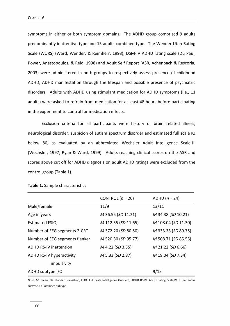
CHAPTER 6
166
symptoms in either or both symptom domains. The ADHD group comprised 9 adults
predominantly inattentive type and 15 adults combined type. The Wender Utah Rating
Scale (WURS) (Ward, Wender, & Reimherr, 1993), DSM-IV ADHD rating scale (Du Paul,
Power, Anastopoulos, & Reid, 1998) and Adult Self Report (ASR, Achenbach & Rescorla,
2003) were administered in both groups to respectively assess presence of childhood
ADHD, ADHD manifestation through the lifespan and possible presence of psychiatric
disorders. Adults with ADHD using stimulant medication for ADHD symptoms (i.e., 11
adults) were asked to refrain from medication for at least 48 hours before participating
in the experiment to control for medication effects.
Exclusion criteria for all participants were history of brain related illness,
neurological disorder, suspicion of autism spectrum disorder and estimated full scale IQ
below 80, as evaluated by an abbreviated Wechsler Adult Intelligence Scale-III
(Wechsler, 1997; Ryan & Ward, 1999). Adults reaching clinical scores on the ASR and
scores above cut off for ADHD diagnosis on adult ADHD ratings were excluded from the
control group (Table 1).
Table 1. Sample characteristics
CONTROL (n = 20) ADHD (n = 24)
Male/female 11/9 13/11
Age in years M 36.55 (SD 11.21) M 34.38 (SD 10.21)
Estimated FSIQ M 112.55 (SD 11.65) M 108.04 (SD 11.30)
Number of EEG segments 2-CRT M 372.20 (SD 80.50) M 333.33 (SD 89.75)
Number of EEG segments flanker M 520.30 (SD 95.77) M 508.71 (SD 85.55)
ADHD RS-IV inattention M 4.22 (SD 3.35) M 21.22 (SD 6.66)
ADHD RS-IV hyperactivity M 5.33 (SD 2.87) M 19.04 (SD 7.34)
impulsivity
ADHD subtype I/C 9/15
Note. M: mean, SD: standard deviation, FSIQ: Full Scale Intelligence Quotient, ADHD RS-IV: ADHD Rating Scale-IV, I: Inattentive
subtype, C: Combined subtype
TASK-RELATED EEG DEVIANCES IN ADULTS WITH ADHD
167
Procedure
The research protocol was approved by the local ethics committee and written
informed consent was obtained from the participants. The protocol involved recordings
of resting EEG and execution of a flanker task, a slow paced 2-choice time reaction task,
an attention network task and a Simon task, administered in a counterbalanced order.
For the purpose of the current study, EEG and performance measures during the 2-CRT
and the flanker task were evaluated.
Tasks
2-CRT task. In this task, first a fixation cross was presented with a variable duration
(3000 - 4600 ms). Then, the letters X and O were randomly presented for a maximum
duration of 1800 ms in the middle of a computer screen, which disappeared from the
screen when participants pressed a key. Participants had to press a left key when an X
was presented and a right key when an O was displayed. To control for laterality effects,
left and right press following X and O were reversed among participants in a
counterbalanced order. Finally, a fixation cross was displayed again for 4000 ms. 60
stimuli were presented, half requiring a left and half requiring a right button press.
Flanker task. Executive attention was measured using a modified Eriksen flanker task
(Eriksen & Eriksen, 1974; Debener et al., 2005). Participants were asked to evaluate
whether a middle arrow of five horizontally presented arrows was directed to the left or
right by pressing one of two possible keys in the keyboard. The efficiency of executive
attention was measured by evaluating the impact of flankers on reaction time and
accuracy. Congruent flankers pointed in the same direction as the target, while
incongruent flankers pointed in the opposite direction. At the beginning of each trial, a
fixation cross was displayed for 200 ms at the centre of the screen. Subsequently, four
flanker arrows were presented for 80 ms, accompanied by the target arrow in the
CHAPTER 6
168
middle of the flankers 20 ms after the onset of the flankers. This was done to increase
the load on interference inhibition. The target arrow was presented for 60 ms, after
which a fixation cross was presented with a variable duration (1100 - 1500 ms). The task
comprised 480 trials in which half of the trials were congruent, whereas the other half
were incongruent trials. Three blocks of 160 trials were administered, in between adults
were permitted to take a short break.
EEG recording and processing
During task execution, EEG data were obtained with an electrode cap employing
128 active Ag/AgCl electrodes with average reference, placed according to the 10-5
International System (Oostenveld & Praamstra, 2001). The ground electrode was
located in the cap at FPz. Signals were amplified with an open pass-band from DC to 100
Hz with a QuickAmp amplifier (Brain Products, Gilching, Germany) and digitized using
Brain Vision Recorder software (version 1.10) with a 500 Hz sampling rate. Analyses of
EEG data were performed using Brain Vision Analyzer software (version 2.0.1). EEG data
were filtered (band-pass 0.5-50 Hz, notch filter 50 Hz) and segmented (2 s epochs with 1
s overlap). Eye movement correction was conducted according to the Gratton and Coles
algorithm (Gratton, Coles, & Donchin, 1983). Segments were rejected from further
analyses if absolute difference in amplitudes exceeded 120 µV. The remaining number
of segments for analyses did not differ between groups in the 2-CRT task (F(1, 42) = 2.24,
p = .14), nor in the flanker task (F(1, 42) = 0.18, p = .67) (Table 1). Subsequently, spectral
power was calculated using Fast Fourier Transform with a 20% Hanning window and the
transformed data were averaged. Absolute power estimates were derived for the theta
(3.5-7.5 Hz), alpha (7.5-12.5 Hz) and beta (12.5-25 Hz) band and divided in nine regions:
left frontal (AF3, F3, F7), midline frontal (Fz, FCz), right frontal (AF4, F4, F8), left central
(T7, C3), midline central (Cz), right central (T8, C4), left posterior (P7, P3, O1), midline
posterior (Pz, Oz) and right posterior (P8, P4, O2) (see also Barry et al., 2009). TBR was
TASK-RELATED EEG DEVIANCES IN ADULTS WITH ADHD
169
calculated by dividing power in the theta band by power in the beta band. A natural
logarithmic transform, ln(x), was used to approach normal spreading of the data.
Data analyses
Behavioural performance. For the 2-CRT task, mean reaction time (RT), variability in RT
(SD-RT), and percentage errors of commission (%EOC) were compared between groups
by separate analyses of variance (ANOVAs) with repeated measures with time-on-task
(first, second, third part of testing) as within-subject and group (ADHD, control) as
between-subject factor.
For the flanker task, the same analyses were performed for RT and %EOC, with
congruency (congruent, incongruent trial) added as an extra within subject factor.
EEG activity. EEG activity in each task was evaluated by separate repeated measures
ANOVAs for each frequency band and for TBR with time-on-task (first, second and third
part of the respective task), sagittal region (frontal, central, posterior) and lateral region
(left, midline, right) as within-subject and group as between-subject factors. When the
assumption of sphericity was violated, degrees of freedom were corrected using
Greenhouse-Geisser (when ε < .75) or Huynh-Feldt (when ε > .75) estimates of
sphericity. Effect sizes were determined by partial eta squared (η2p) and interpreted as
small (< .06), moderate (.06 - .14), or large (> 0.14) (Cohen, 1988).
Correlation analyses. Pearson correlations were used to evaluate the relationship
between EEG parameters and performance measures. In order to reduce the number of
tests, only the EEG variables that were found to differ between groups were used in the
correlation analyses. According to guidelines, the strength of the correlations was
interpreted as small (< .30), moderate (.30 < .50) or large (> .50) (Cohen, 1988).
Following van Dongen-Boomsma and colleagues (2010), Bonferroni corrections were not
conducted, because the test statistics were assumed to be highly dependent.
CHAPTER 6
170
RESULTS
Group characteristics
The ADHD-RS IV confirmed that adults with ADHD experienced more problems
with inattention (F(1, 42) = 97.47, p < .001) and hyperactivity/impulsivity (F(1, 42) =
55.87, p < .001) than typically developing adults. Groups did not significantly differ in
terms of estimated full scale IQ (F(1, 42) = 1.69, p = .20) (Table 1).
Performance data
An overview of the performance data on both tasks is presented in table 2.
2-CRT task. As expected, RT was slower (F(1, 42) = 6.77, p = .01, η2p = .14) and more
variable (F(1, 42) = 5.70, p = .02, η2p = .12) in adults with ADHD compared to adults
without ADHD. Furthermore, the ADHD group tended to commit more EOC than the
control group (F(1, 42) = 3.96, p = .05, η2p = .09). Further, no main time-on-task or group
by time-on-task effects were observed.
Flanker task. RT was slower in adults with ADHD than controls (F(1, 42) = 4.14, p < .05,
η2p = .09) and on incongruent than congruent trials (F(1, 42) = 1197.83, p < .001, η2
p =
.97). Groups did not significantly differ in terms of %EOC, yet across participants more
EOC were made on incongruent than congruent trials (F(1, 42) = 94.90, p < .001, η2p =
.69). The group by congruency effects for RT and %EOC were not significant,
respectively (F(1, 42) = 0.81, p = .37, η2p = .02) and (F(1, 42) = 0.10, p = .75, η2
p = .00),
indicating intact interference inhibition in the ADHD group. Across groups, a time-on-
task effect emerged for %EOC (F(2, 82) = 4.72, p = .01, η2p = .10), with more errors made
in the second than in the first part of the task (p = .03). An interaction between time-on-
task and congruency was found as well for %EOC (F(2, 82) = 3.25, p = .04, η2p = .07). Post
hoc analyses revealed a difference in number of errors with time on incongruent trials
TASK-RELATED EEG DEVIANCES IN ADULTS WITH ADHD
171
(F(2, 84) = 4.16, p = .02, η2p = .09) but not on congruent trials (F(1.86, 78.04) = 2.34, p =
.10, η2p = .05). In specific, more errors were committed on incongruent trials in the
second relative to the first part of the testing (p = .04), indicating worse interference
inhibition with time-on-task. This effect was however the same for both groups, as
indicated by the absence of a group x time-on-task x congruency effect (F(2, 84) = 1.58, p
= .21, η2p = .04).
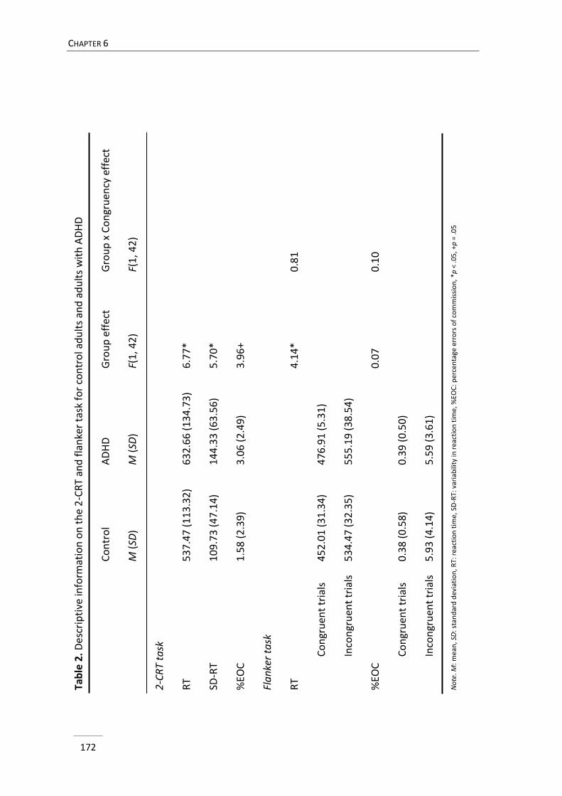
CHAPTER 6
172
Tab
le 2
. D
escr
ipti
ve in
form
atio
n o
n t
he
2-C
RT
and
fla
nke
r ta
sk f
or
con
tro
l ad
ult
s an
d a
du
lts
wit
h A
DH
D
Co
ntr
ol
A
DH
D
G
rou
p e
ffe
ct
Gro
up
x C
on
gru
ency
eff
ect
M (
SD)
M
(SD
)
F(1
, 42
)
F(
1, 4
2)
2-C
RT
task
RT
5
37
.47
(11
3.3
2)
6
32
.66
(13
4.7
3)
6
.77
*
SD-R
T
1
09
.73
(47
.14
)
14
4.3
3 (6
3.5
6)
5
.70
*
%EO
C
1
.58
(2
.39
)
3
.06
(2
.49
)
3
.96
+
Fla
nke
r ta
sk
RT
4
.14
*
0
.81
Co
ngr
uen
t tr
ials
4
52
.01
(31
.34
)
47
6.9
1 (5
.31
)
Inco
ngr
uen
t tr
ials
5
34
.47
(32
.35
)
55
5.1
9 (3
8.5
4)
%EO
C
0
.07
0
.10
Co
ngr
uen
t tr
ials
0
.38
(0
.58
)
0
.39
(0
.50
)
Inco
ngr
uen
t tr
ials
5
.93
(4
.14
)
5
.59
(3
.61
)
No
te. M
: mea
n, S
D: s
tan
dar
d d
evia
tio
n, R
T: r
eact
ion
tim
e, S
D-R
T: v
aria
bili
ty in
rea
ctio
n t
ime,
%EO
C: p
erce
nta
ge e
rro
rs o
f co
mm
issi
on
, *p
< .0
5, +
p =
.05
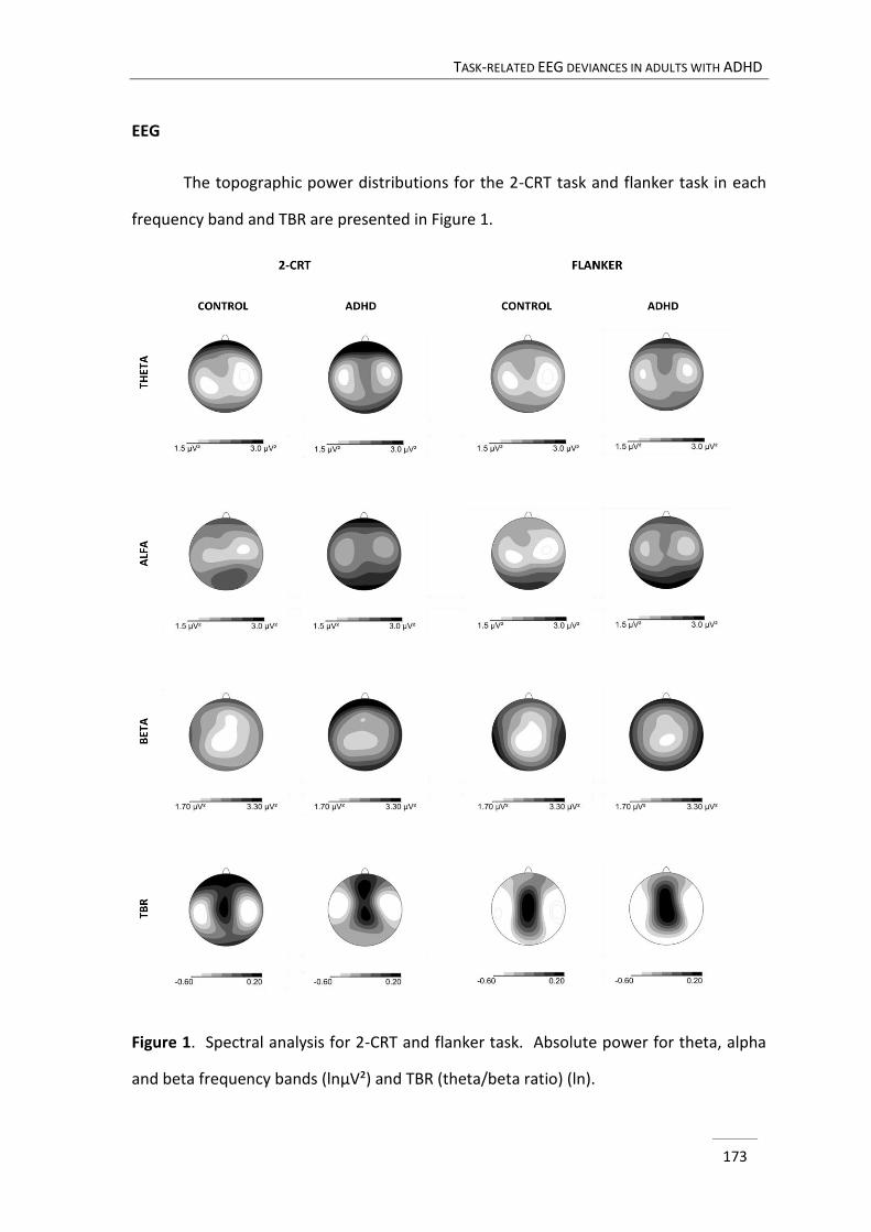
TASK-RELATED EEG DEVIANCES IN ADULTS WITH ADHD
173
EEG
The topographic power distributions for the 2-CRT task and flanker task in each
frequency band and TBR are presented in Figure 1.
Figure 1. Spectral analysis for 2-CRT and flanker task. Absolute power for theta, alpha
and beta frequency bands (lnµV²) and TBR (theta/beta ratio) (ln).
CHAPTER 6
174
2-CRT task.
Theta power. The main group effect was marginally significant, with higher theta
power in the ADHD group (F(1, 42) = 3.05, p = .09, η2p = .07). A significant group x lateral
region effect (F(1, 42) = 4.12, p = .02, η2p = .09) indicated that theta power at the midline
was higher in the ADHD group compared to the control group (F(1, 42) = 5.96, p = .02,
η2p = .12). Time-on-task effects were not detected.
Alpha power. As for alpha power, a trend for an interaction between group and
lateral region was found (F(1, 42) = 2.59, p = .08, η2p = .06). However, post hoc analyses
per laterality revealed no significant group effects. Further, a time-on-task effect
occurred (F(1, 42) = 9.03, p < .001, η2p = .18), with an increase in alpha power from the
first to the second (p < .01) and third (p < .01) part of the testing. The time-on-task
effect also tended to interact with sagittal region (F(3.00, 125.95) = 2.15, p < .10, η2p =
.05). Follow-up analyses per sagittal region revealed that alpha power increased in each
region during testing with the strongest effect at frontal sites.
Beta power. Beta power was higher in the ADHD group than in the control group
(F(1, 42) = 7.57, p < .01, η2p = .15). Time-on-task effects were dependent on sagittal
region (F(3.66, 153.69) = 6.04, p < .001, η2p = .13). Post hoc analyses per sagittal region
revealed time effects at frontal derivations (F(2, 84) = 4.61, p = .01, η2p = .10), indicating
higher power at the end compared to the beginning of the task (p = .03). Also, a trend
for a time-on-task effect appeared at central sites (F(2, 84) = 2.85, p = .06, η2p = .06).
However, follow-up analyses on this effect yielded no further significant results.
TBR. Neither effects involving group were nor a main time-on-task effect was
detected for TBR. An interaction was found between time-on-task and sagittal region
for TBR (F(3.46, 145.38) = 2.88, p = .03, η2p = .06). However, time-on-task effects per
region did not reach significance.
TASK-RELATED EEG DEVIANCES IN ADULTS WITH ADHD
175
Flanker task.
Theta power. No effects involving group were detected. Time-on-task effects also
did not reach significance for theta power.
Alpha power. Also for alpha power, neither group related effects, nor time-on-
task effects were found.
Beta power. As for beta power, an interaction was found between time-on-task
and sagittal region (F(4, 168) = 5.49, p < .001, η2p = .12). Further testing per sagittal area
revealed a time-on-task effect at frontal leads (F(2, 84) = 3.30, p = .04, η2p = .07),
indicating a trend for more power at the end compared to the start of the task (p = .09).
Also, an interaction between time-on-task, sagittal region and lateral region emerged
(F(5.01, 210.50) = 3.74, p < .001, η2p = .08). Follow-up analyses revealed a time-on-task
effect at right central region (F(1.76, 73.79) = 3.95, p = .03, η2p = .09), yet further testing
yielded no significant differences between parts of the task.
TBR. A marginally significant interaction between group, sagittal region and
lateral region emerged for TBR (F(4, 168) = 2.36, p = .06, η2p =.05). However, further
testing per region revealed no significant group effects. Further, time-on-task effects in
TBR differed according to sagittal position (F(3.34, 140.25) = 3.03, p = .03, η2p = .07).
Post hoc analyses did not reveal significant time-on-task effects per region.
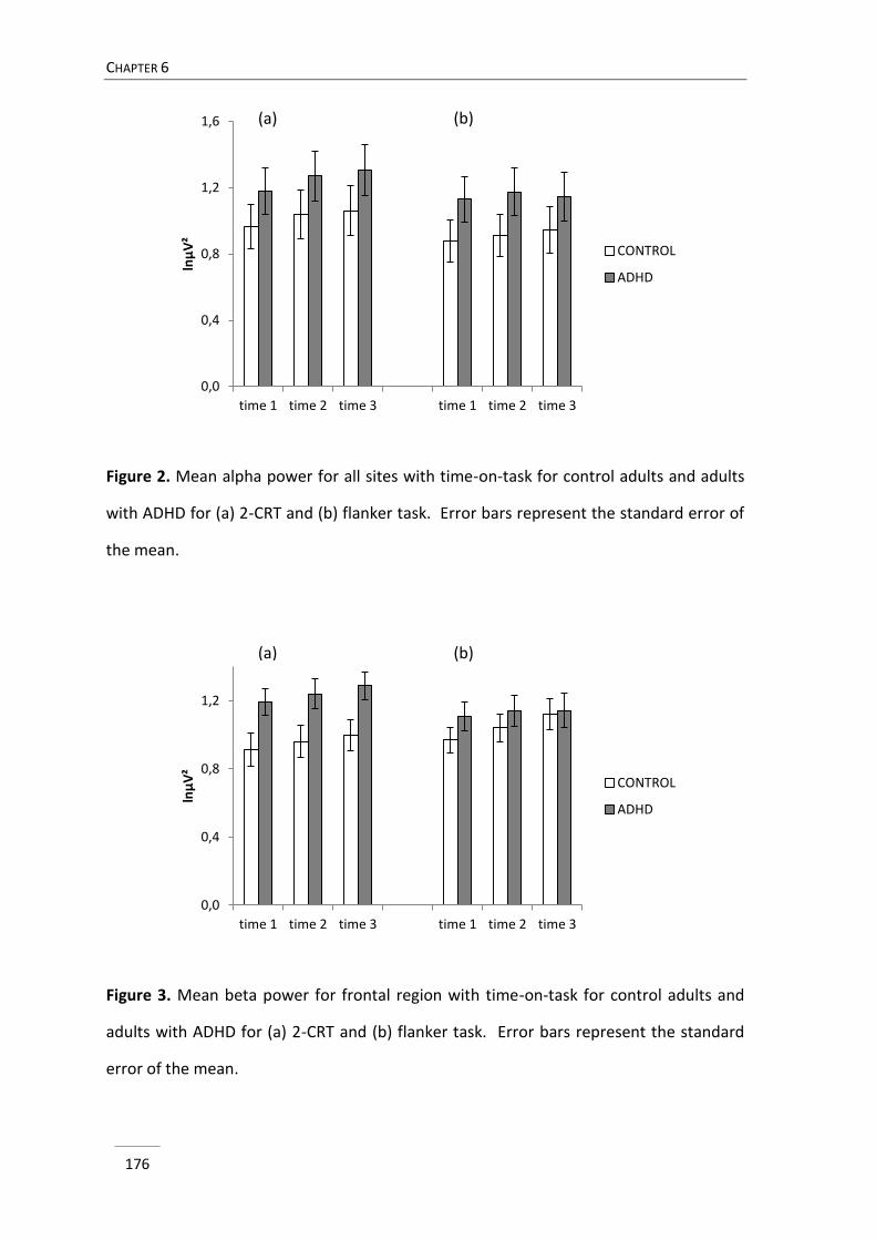
CHAPTER 6
176
Figure 2. Mean alpha power for all sites with time-on-task for control adults and adults
with ADHD for (a) 2-CRT and (b) flanker task. Error bars represent the standard error of
the mean.
Figure 3. Mean beta power for frontal region with time-on-task for control adults and
adults with ADHD for (a) 2-CRT and (b) flanker task. Error bars represent the standard
error of the mean.
0,0
0,4
0,8
1,2
1,6
time 1 time 2 time 3 time 1 time 2 time 3
lnµ
V²
CONTROL
ADHD
0,0
0,4
0,8
1,2
time 1 time 2 time 3 time 1 time 2 time 3
lnµ
V²
CONTROL
ADHD
(a) (b)
(a) (b)
TASK-RELATED EEG DEVIANCES IN ADULTS WITH ADHD
177
Additional analyses with sex as a covariate.
Since gender has been documented to affect EEG deviances in ADHD (e.g., Clarke
et al., 2003; Dupuy et al., 2013), supplementary analyses were performed with sex as a
covariate. Group related findings did not change.
Correlation analyses
Based on the significant interaction effects involving diagnostic status for midline
theta and overall beta power in the 2-CRT task, the average power values for
respectively midline and all regions were calculated for theta and beta power.
Subsequently, those variables were used to correlate with RT, SD-RT and %EOC in the 2-
CRT task. In the ADHD group, a significant moderate positive correlation was detected
between midline theta power and %EOC (r = .41, p = .04). Although pointing to the
same direction, this correlation was not significant and of a small magnitude in the
control group (r = .17, p = .47). In addition, all other correlations were small and did not
reach statistical significance in either group.
DISCUSSION
The present study examined EEG in adults with ADHD compared to controls
during performance of a slow paced 2-CRT task, which required low executive attention
and induced a low level of activation, and a moderate paced flanker task, which tapped
top-down executive control and induced a moderate activation level. These tasks were
chosen to gain more insights into the significance attached to the common observed
aberrant theta and TBR in ADHD. One hypothesis is that higher theta or TBR in ADHD is
related to impaired activation state in ADHD, as suggested in several publications (e.g.,
Barry et al., 2009; Clarke et al., 2013) and which would be in accord with the state
regulation account of ADHD (Sergeant, 2000; van der Meere, 2005). On the other hand,
CHAPTER 6
178
high TBR has been proposed to represent impairments in top-down executive control,
particularly inhibition (e.g., Schutter et al., 2006; Knyazev, 2007), which parallels the
executive dysfunction theory of ADHD (Barkley, 1997).
As for performance measures, with respect to the 2-CRT task, adults with ADHD
were found to be slower and more variable in their responding and tended to make
more errors than control participants. This is in line with a large number of studies using
a variety of tasks, showing worse performance in ADHD when stimuli were presented at
a slow rate (e.g., Brown & Vickers, 2004; Leung et al., 2000; Metin et al., 2012, for a
meta-analysis; Rubia et al., 1999; Scheres et al., 2001; Wiersema et al., 2006a,b). The
finding that performance in ADHD is highly sensitive to presentation rate of stimuli has
been explained in terms of state regulation difficulties (van der Meere, 2005). In
specific, as slow event rates induce a state of underactivation (Sanders, 1983), it has
been suggested that individuals with ADHD are easily under-activated and have difficulty
in adjusting their under-activated state because of insufficient effort allocation (Johnson,
Wiersema, & Kuntsi, 2010; Wiersema et al., 2006a,b).
Regarding the flanker task, in accord with existing research (for a review, see
Mullane, Corkum, Klein, & McLaughlin, 2009; Chang, Davies, & Gavin, 2009; McLoughlin
et al., 2009), slower responding and decreased accuracy were found in this task when
targets were surrounded by conflicting flankers, which indicates that additional
attentional processing was required to filter out interfering information (Mullane et al.,
2009), and hence that the task efficiently tapped executive control. Yet, importantly,
groups did not differ in congruency effects, which corresponds with literature in adults
with ADHD (Chang et al., 2009; McLoughlin et al., 2009) and suggests intact interference
inhibition in the ADHD group (Johnstone & Galetta, 2013). Instead adults with ADHD
were found to be slower, independent of congruency condition, as found before (Chang
et al., 2009; McLoughlin et al., 2009). Performance deteriorated during task execution,
especially on incongruent trials, indicating worsening of interference inhibition with task
TASK-RELATED EEG DEVIANCES IN ADULTS WITH ADHD
179
duration. However importantly, groups did not differ in this respect, suggesting no
sustained attention deficit in our sample of adults with ADHD, which is in line with other
research (Schachar, Logan, Wachsmuth, & Chajczyk, 1988; Stins et al., 2005; Tucha et al.,
2010).
Broadly in accord with the findings on the behavioural level, EEG deviances in
ADHD were found during performance on the slow paced 2-CRT task, but not the flanker
task. More specific, adults with ADHD showed increased theta and beta power when
performing the task that induced a low activation level and required minimal executive
functioning (i.e., 2-CRT), whereas no deviances were observed in the task with a high
load on executive control and a regular activation level (i.e., the flanker task). This
indicates that EEG deviances in ADHD may be task-dependent and reflect impairment in
activation state rather than executive control. This appears to be confirmed by the
finding of a moderate positive correlation between theta power and errors of
commission in the 2-CRT task in adults with ADHD. Yet, this association was small in the
control group, suggesting a possible difference in neural organization in adults with
ADHD relative to controls (Loo et al., 2009).
Elevated theta during task performance in ADHD has been reported before (El-
Sayed et al., 2002; Janzen et al., 1995; Mann et al., 1992; Rasey et al., 1999). As theta
power has repeatedly been found to increase during tasks with a higher cognitive load,
enhanced theta power during task execution has often been explained in terms of an
increase in mental activity to perform a difficult task (e.g., Gevins, Smith, McEvoy, & Yu,
1997; Onton, Delorme, & Makeig, 2005; Sammer et al., 2007; Sauseng, Hoppe, Klimesch,
Gerloff, & Hummel, 2007). This modulation of theta has been noticed to appear at the
frontal midline region of the scalp and has been associated with activity in the anterior
cingulate cortex (Peterson & Posner, 2012). In light of the present data, it seems
however unlikely that the elevated theta power in ADHD in the 2-CRT task reflects
increased mental activity related to task difficulty, as actually the cognitive load in the 2-
CHAPTER 6
180
CRT task was rather low, especially when compared with the flanker task. Moreover,
the increased theta power in our ADHD group was not limited to frontal midline theta
power. With regard to this topographical factor, a more generalized increase in theta
activity is known to be a marker of drowsiness (e.g., Barwick, Arnett, & Slobounov, 2012;
Jödicke et al., 2013; Lal & Craig, 2001; Oken, Salinsky, & Elsas, 2006). Hence, the higher
theta in adults with ADHD during performance on the slow 2-CRT task may rather reflect
an under-activated state (decreased readiness to respond), which may have resulted in
inadequate processing of the stimuli, consequently leading to a higher rate of
commission errors.
In addition to elevated theta power, enhanced beta power was found in the
ADHD group during 2-CRT task performance. Up to date, the functional significance of
power in the beta frequency range seems to be the least well understood (Engel & Fries,
2010). Nevertheless, beta power has frequently been reported to reflect cortical activity
related to higher order processing (Barry, Clarke, Johnstone, Magee, & Rushby, 2007;
Kropotov, 2009; Loo et al., 2009). Furthermore, elevated power in the beta band has
been interpreted as representing compensatory effort to counteract a low vigilance level
(Corsi-Cabrera, Sánchez, del-Río-Portilla, Villanueva, & Pérez-Garci, 2003; Fischer,
Langner, Birbaumer, & Brocke, 2008; Kiroy, Warasawskaya, & Volyov, 1996; Smit, Eling,
Droogleever Fortuyn, & Coenen, 2005). From that, the finding of increased beta power
may reflect increased effort allocation in adults with ADHD in order to counteract their
under-activated state (Hockey, 1997; Mulder, 1986; Sanders, 1983, 1998). Other studies
also indicated difficulty with effort allocation in ADHD during slow presentation rates,
however there less effort allocation in ADHD was noticed (Börger & van der Meere,
2000; Wiersema et al., 2006a,b). Yet, it should be noted that in those studies time-on-
task effects were not investigated. Hence, it may be possible that failed attempts to
upregulate activation level led participants with ADHD to discontinue investing extra
effort, while in the current study, adults with ADHD kept allocating additional
TASK-RELATED EEG DEVIANCES IN ADULTS WITH ADHD
181
compensatory effort. Also, a direct comparison between studies is hampered by the use
of different measures (i.e., beta, heart rate variability, P3 event related potential
component) to evaluate effort allocation.
While in some studies different time-on-task effects on EEG activity have been
reported in ADHD compared to control adults (Loo et al., 2009; Rasey et al., 1999), this
was not the case in the present study, which is in line with our findings on the
performance level. Alpha power increased during the course of the 2-CRT task in both
groups, which is in accord with existing literature (Barwick et al., 2012; Gevins et al.,
1997; Loo et al., 2009; Smith, McEvoy, & Gevins, 1999), and may indicate a decrease in
alertness (e.g., Barwick et al., 2012; Oken, Salinsky, & Elsas, 2006) or attentional
processing (e.g., Laufs et al., 2003; Willeford, Ciuffreda, Yadav, Ludlam, 2013) with time.
As vigilance or attention levels may drop earlier in boring tasks with long inter stimulus
intervals, this may explain why an alpha increase with time was only observed in the 2-
CRT task. Alternatively, the inclusion of breaks in the flanker task may have contributed
to this. In both tasks and in both groups, an increase of frontal beta with task duration
was noticed, which might reflect increased effort allocation to maintain meeting task
demands. Crucially and most important however, is the fact that time-on-task effects
could not differentiate adults with ADHD from controls.
Some considerations have to be made with respect to the interpretation of the
results. First, assigning specific functional roles to different frequency bands remains
difficult because throughout literature, they have been associated with a variety of
states and cognitive phenomena (Loo & Makeig, 2012). Including additional measures
such as subjective ratings of alertness or task-related effort as well as other physiological
measures (i.e., skin conductance level to capture autonomic arousal, heart rate
variability) in future studies, may help clarifying the functional significance of EEG
deviances in ADHD. Next, although a head to head comparison was made between two
possible hypotheses regarding deviant EEG, especially theta and TBR, in ADHD, the use

CHAPTER 6
182
of an orthogonal research design would have been more appropriate to formulate firm
conclusions. Finally, in the literature the concepts of arousal and activation have been
defined and interpreted in different ways, which may hamper interpreting results.
‘Activation’ as used by Barry and colleagues (2009) and as formulated in the state
regulation account may not fully overlap and future studies are warranted to investigate
this issue. However, a common factor in each conceptualization of ‘activation’ is the
reference that is made to an energetic state. Therefore, we argue that it is safe to say
that the EEG deviances in adults with ADHD in the current study were related to
energetic state factors.
Despite these limitations, the current findings indicate that EEG deviances in
ADHD are task-dependent, since aberrant EEG activity was not consistently found across
both tasks. This may indicate that EEG deviances in ADHD are not to be seen as a fixed
hallmark but rather as a dynamic characteristic that fluctuates across states and in
response to environmental contexts. This corresponds with models that emphasize the
dynamic, rather than the fixed nature of ADHD and especially the role of contextual and
state factors in determining deficits (Sonuga-Barke et al., 2010). Future studies are
needed to confirm the findings of the present study and to shed more light on the
dynamic nature and significance of EEG deviances in ADHD.
TASK-RELATED EEG DEVIANCES IN ADULTS WITH ADHD
183
REFERENCES
Achenbach, T.M., & Rescorla, L.A. (2003). Manual for the ASEBA Adult Forms & Profiles.
Burlington, Vermont: University of Vermont, Research Center for Children, Youth, and
Families.
American Psychiatric Association (2013). Diagnostic and statistical manual of mental disorders
(5th Ed.). Arlington, VA: American Psychiatric Publishing.
Barkley, R.A. (1997). Behavioral inhibition, sustained attention, and executive functions:
Constructing a unifying theory of ADHD. Psychological Bulletin, 121, 65-94. doi:
10.1037//0033-2909.121.1.65
Barry, R.J., Clarke, A.R., & Johnstone, S.J. (2003). A review of electrophysiology in attention-
deficit/hyperactivity disorder. I. Qualitative and quantitative electroencephalography.
Clinical Neurophysiology, 114, 171-183. doi: 10.1016/S1388-2457(02)00362-0
Barry, R.J., Clarke, A.R., Johnstone, S.J., Magee, C.A., & Rushby, J.A. (2007). EEG differences
between eyes-closed and eyes-open resting conditions. Clinical Neurophysiology, 118,
2765-2773. doi: 10.1016/j.clinph.2007.07.028
Barry, R.J., Clarke, A.R., Johnstone, S.J., McCarthy, R., & Selikowitz, M. (2009).
Electroencephalogram theta/beta ratio and arousal in attention-deficit/hyperactivity
disorder: evidence of independent processes. Biological Psychiatry, 66, 398-401. doi:
10.1016/j.biopsych.2009.04.027
Barwick, F., Arnett, P., & Slobounov, S. (2012). EEG correlates of fatigue during administration of
a neuropsychological test battery. Clinical Neurophysiology, 123, 278-284. doi:
10.1016/j.clinph.2011.06.027
Bitsakou, P., Psychogiou, L., Thompson, M., & Sonuga-Barke, E.J. (2008). Inhibitory deficits in
attention-deficit/hyperactivity disorder are independent of basic processing efficiency
and IQ. Journal of Neural Transmission, 115, 261-268. doi: 10.1007/s00702-007-0828-z
Boonstra, A.M., Oosterlaan, J., Sergeant, J.A., & Buitelaar, J.K. (2005). Executive functioning in
adult ADHD: a meta-analytic review. Psychological Medicine, 35(8), 1097-1108. doi:
10.1017/S003329170500499X
CHAPTER 6
184
Börger, N.A., & van der Meere, J.J. (2000). Motor control and state regulation in children with
ADHD: a cardiac response study. Biological Psychology, 51, 247-267. doi: 10.1016/S0301-
0511(99)00040-X
Brown, L.N., & Vickers, J.N. (2004). Temporal judgments, hemispheric equivalence, and
interhemispheric transfer in adolescents with attention deficit hyperactivity disorder.
Experimental Brain Research, 154(1), 76-84. doi: 10.1007/s00221-003-1641-z
Buyck, I., & Wiersema, J.R. (in press). Resting electroencephalogram in attention deficit
hyperactivity disorder: developmental course and diagnostic value. Psychiatry Research.
Advance online publication. doi: 10.1016/j.psychres.2013.12.055
Chang, W.P., Davies, P.L., & Gavin, W.J. (2009). Error Monitoring in College Students with
Attention-Deficit/Hyperactivity Disorder. Journal of Psychophysiology, 23(3), 113-125.
doi: 10.1027/0269-8803.23.3.113
Clarke, A.R., Barry, R.J., Dupuy, F.E., Heckel, L.D., McCarthy, R., Selikowitz, M., & Johnstone, S.J.
(2011). Behavioural differences between EEG-defined subgroups of children with
Attention-Deficit/Hyperactivity Disorder. Clinical Neurophysiology, 122, 1333–1341. doi:
10.1016/j.clinph.2010.12.038
Clarke, A.R., Barry, R.J., Dupuy, F.E., McCarthy, R., Selikowitz, M., & Johnstone, S.J. (2013). Excess
beta activity in the EEG of children with attention-deficit/hyperactivity disorder: A
disorder of arousal? International Journal of Psychophysiology, 89, 314-319. doi:
10.1016/j.ijpsycho.2013.04.009
Clarke, A.R., Barry, R.J., McCarthy, R., Selikowitz, M., Clarke, D., & Croft, R. (2003). EEG in girls
with attention-deficit/hyperactivity disorder. Clinical Neurophysiology, 114, 319-328.
doi: 10.1016/S1388-2457(02)00364-4
Clarke, A.R., Barry, R.J., McCarthy, R., Selikowitz, M., & Johnstone, S.J. (2007). Effects of
stimulant medications on the EEG of girls with Attention-Deficit/Hyperactivity. Clinical
Neurophysiology, 118(12), 2700-2708. doi: 10.1016/j.clinph.2007.08.020
Cohen, J. (1988). Statistical Power Analysis for the Behavioral Sciences, 2 ed. Laurence Erlbaum,
Hillsdale, N.J.
TASK-RELATED EEG DEVIANCES IN ADULTS WITH ADHD
185
Coolidge, F.L., Starkey, M.T., & Cahill, B.S. (2007). Comparison of a Parent-Rated DSM-IV
Measure of Attention-Deficit/Hyperactivity Disorder and Quantitative EEG Parameters in
an Outpatient Sample of Children. Journal of Clinical Neurophysiology, 24(4), 348-351.
doi: 10.1097/WNP.0b013e318067bcfc
Corsi-Cabrera, M., Sánchez, A., del-Río-Portilla, Y., Villanueva, Y., & Pérez-Garci, E. (2003). Effect
of 38 h of total sleep deprivation on the waking EEG in women: sex differences.
International Journal of Psychophysiology, 50(3), 213-224. doi: 10.1016/S0167-
8760(03)00168-5
Debener, S., Ullsperger, M., Siegel, M., Fiehler, K., von Cramon, D.Y., & Engel, A.K. (2005). Trial-
by-trial coupling of concurrent electroencephalogram and functional magnetic
resonance imaging identifies the dynamics of performance monitoring. Journal of
Neuroscience, 25, 11730-11737. doi: 10.1523/JNEUROSCI.3286-05.2005
DuPaul, G.J., Power, T.J., Anastopoulos, A.D., & Reid, R. (1998). ADHD Rating Scale-IV: Checklists,
norms, and clinical interpretation. New York, NY, US: Guilford Press.
Dupuy, F.E., Barry, R.J., Clarke, A.R., McCarthy, R., & Selikowitz, M. (2013). Sex differences
between the combined and inattentive types of attention-deficit/hyperactivity disorder:
An EEG perspective. International Journal of Psychophysiology, 89(3), 320-327. doi:
10.1016/j.ijpsycho.2013.04.004
Dupuy, F.E., Clarke, A.R., Barry, R.J., McCarthy, R., & Selikowitz, M. (2011). Girls with attention-
deficit/hyperactivity disorder: EEG differences between DSM-IV types. Clinical EEG and
Neuroscience, 42(1), 1-5. doi: 10.1177/155005941104200104
El-Sayed, E., Larsson, J.O., Persson, H.E., & Rydelius, P.A. (2002). Altered cortical activity in
children with attention-deficit/hyperactivity disorder during attentional load task.
Journal of the American Academy of Child & Adolescent Psychiatry, 41, 811-819. doi:
10.1097/00004583-200207000-00013
Engel, A.K., & Fries, P. (2010). Beta-band oscillations - signalling the status quo? Current Opinion
in Neurobiology, 20(2), 156-165. doi: 10.1016/j.conb.2010.02.015
CHAPTER 6
186
Eriksen, B.A., & Eriksen, C.W. (1974). Effects of noise letters upon the identification of a target
letter in a non search task. Perception & Psychophysics, 16, 143-149. doi:
10.3758/BF03203267
Faraone, S.V., Biederman, J., & Mick, E. (2006). The age-dependent decline of attention deficit
hyperactivity disorder: a meta-analysis of follow-up studies. Psychological Medicine, 36,
159-165. doi: 10.1017/S003329170500471X
Fischer, T., Langner, R, Birbaumer, N, & Brocke, B. (2008). Arousal and attention: self-chosen
stimulation optimizes cortical excitability and minimizes compensatory effort. Journal of
Cognitive Neuroscience, 20(8), 1443-1453. doi: 10.1162/jocn.2008.20101
Gevins, A., Smith, M.E., McEvoy, L. & Yu, D. (1997). High-resolution EEG mapping of cortical
activation related to working memory: Effects of task difficulty, type of processing, and
practice. Cerebral Cortex, 7(4), 374-385. doi: 10.1093/cercor/7.4.374
González-Castro, P., Rodríguez, C., López, A., Cueli, M., & Álvarez, L. (2013). Attention Deficit
Hyperactivity Disorder, differential diagnosis with blood oxygenation, beta/theta ratio,
and attention measures. International Journal of Clinical and Health Psychology, 13(2),
101-109. doi: 10.1016/S1697-2600(13)70013-9
Gratton, G., Coles, M.G., & Donchin, E. (1983). A new method for off-line removal of ocular
artifact. Electroencephalography and Clinical Neurophysiology, 55, 468-484. doi:
10.1016/0013-4694(83)90135-9
Hermens, D.F., Williams, L.M., Lazzaro, I., Whitmont, S., Melkonian, D., & Gordon, E. (2004). Sex
differences in adult ADHD: a double dissociation in brain activity and autonomic arousal.
Biological Psychology, 66, 221-233. doi: 10.1016/j.biopsycho.2003.10.006
Hockey, G.R.J. (1997). Compensatory control in the regulation of human performance under
stress and high workload: A cognitive-energetical framework. Biological Psychology, 45,
73-93. doi: 10.1016/S0301-0511(96)05223-4
Hogan, A.M., Vargha-Khadem, F., Kirkham, F.J., & Baldeweg, T. (2005). Maturation of action
monitoring from adolescents to adulthood: an ERP study. Developmental Science, 8, 525-
534. doi: 10.1111/j.1467-7687.2005.00444.x
TASK-RELATED EEG DEVIANCES IN ADULTS WITH ADHD
187
Janzen, T., Graap, K., Stephanson, S., Marshall, W., & Fitzsimmons, G. (1995). Differences in
baseline EEG measures for ADD and normally achieving preadolescent males.
Biofeedback and Self-Regulation, 20, 65-82. doi: 10.1007/BF01712767
Jödicke, J., Olbrich, S., Sander, C., Minkwitz, J., Chittka, T., Himmerich, H., & Hegerl, U. (2013).
Separation of Low-Voltage EEG-Activity During Mental Activation from that During
Transition to Drowsiness. Brain Topography, 26(4), 538-546. doi: 10.1007/s10548-013-
0287-9
Johnson, K.A., Wiersema J.R., & Kuntsi, J. (2009). What would Karl Popper say? Are current
psychological theories of ADHD falsifiable? Behavioral and Brain Functions, 5, 15. doi:
10.1186/1744-9081-5-15
Johnstone, S.J., & Galetta, D. (2013). Event-rate effects in the flanker task: ERPs and task
performance in children with and without AD/HD. International Journal of
Psychophysiology, 87, 340-348. doi: 10.1016/j.ijpsycho.2012.07.170
Kiroy, V.N., Warsawskaya, L.V., & Voynov, V.B. (1996). EEG after prolonged mental activity.
International Journal of Neuroscience, 85, 31-43. doi: 10.3109/00207459608986349
Knyazev, G.G. (2007). Motivation, emotion, and their inhibitory control mirrored in brain
oscillations. Neuroscience & Biobehavioural Reviews, 31(3), 377-395. doi:
10.1016/j.neubiorev.2006.10.004
Koehler, S., Lauer, P., Schreppel, T., Jacob, C., Heine, L., Boreatti-Hummer A., … & Herrmann, M.J.
(2009). Increased EEG power density in alpha and theta bands in adult ADHD patients.
Journal of Neural Transmission, 116, 97-104. doi: 10.1007/s00702-008-0157-x
Kooij, S.J.J., Bejerot, S., Blackwell, A., Caci, H., Casas-Brugué, M.C., Carpentier, P.J., … & Asherson,
P. (2010). European consensus statement on diagnosis and treatment of adult ADHD:
The European Network Adult ADHD. BMC Psychiatry, 10, 1-24. doi: 10.1186/1471-244X-
10-67
Kooij, S.J.J., & Francken, M.H. (2010). Diagnostisch Interview Voor ADHD bij Volwassenen. DIVA
Foundation, Den Haag.
Kropotov, J.D. (2009). Quantitative EEG, event-related potentials and neurotherapy. Amsterdam:
Elsevier.
CHAPTER 6
188
Lal, S.K., & Craig, A. (2001). A critical review of the psychophysiology of driver fatigue. Biological
Psychology, 55(3), 173-194. doi: 10.1016/S0301-0511(00)00085-5
Lansbergen, M., Arns, M., van Dongen-Boomsma, M., Spronk, D., & Buitelaar, J. (2011). The
increase in theta/beta ratio on resting state EEG in boys with attention-
deficit/hyperactivity disorder is mediated by slow alpha peak frequency. Progress in
Neuropsychopharmacology & Biological Psychiatry, 35(1), 47-52. doi:
10.1016/j.pnpbp.2010.08.004
Laufs, H., Kleinschmidt, A., Beyerle, A., Egera, E., Salek-Haddadi, A., Preibisch, C., & Krakowa, K.
(2003). EEG-correlated fMRI of human alpha activity. NeuroImage, 19(4), 1463-1476. doi:
10.1016/S1053-8119(03)00286-6
Leung, J.P., Leung, P.W.L., & Tang, C.S.K. (2000). A Vigilance Study of ADHD and Control Children:
Event Rate and Extra-Task Stimulation. Journal of Developmental and Physical
Disabilities, 12(3), 187-201. doi: 10.1023/A:1009409720485
Liechti, M.D., Valko, L., Müller, U.C., Döhnert, M., Drechsler, R., Steinhausen, H.C., & Brandeis, D.
(2013). Diagnostic value of resting electroencephalogram in attention-
deficit/hyperactivity disorder across the lifespan. Brain Topography, 26, 135-151. doi:
10.1007/s10548-012-0258-6
Loo, S.K., Cho, A., Hale, T.S., McGough, J., McCracken, J., & Smalley, S.L. (2013). Characterization
of the Theta to Beta Ratio in ADHD: Identifying Potential Sources of Heterogeneity.
Journal of Attention Disorders, 17(5), 384-392. doi: 10.1177/1087054712468050
Loo, S.K., Hale, T.S., Hanada, G., Macion, J., Shrestha, A., McGough, J.J., ... & Smalley, S.L. (2010).
Familial clustering and DRD4 effects on electroencephalogram measures in multiplex
families with attention deficit/hyperactivity disorder. Journal of the American Academy
of Child and Adolescent Psychiatry, 49(4), 368-377. doi: 10.1016/j.jaac.2010.01.002
Loo, S.K., Hale, T.S., Macion, J., Hanada, G., McGough, J.J., McCracken, J.T., & Smalley, S.L.
(2009). Cortical activity patterns in ADHD during arousal, activation and sustained
attention. Neuropsychologia, 47, 1033-1040. doi: 10.1016/
j.neuropsychologia.2009.04.013
TASK-RELATED EEG DEVIANCES IN ADULTS WITH ADHD
189
Loo, S.K., & Makeig, S. (2012). Clinical utility of EEG in attention-deficit/hyperactivity disorder: a
research update. Neurotherapeutics, 9, 569-587. doi: 10.1007/s13311-012-0131-z
Mann, C.A., Lubar, J.F., Zimmerman, A.W., Miller, C.A., & Muenchen, R.A. (1992). Quantitative
analysis of EEG in boys with attention deficit hyperactivity disorder: controlled study
with clinical implications. Pediatric Neurology, 8, 30-36. doi: 10.1016/0887-
8994(92)90049-5
McLoughlin, G., Albrecht, B., Banaschewski, T., Rothenberger, A., Brandeis, D., Asherson, P., &
Kuntsia, J. (2009). Performance monitoring is altered in adult ADHD: A familial event-
related potential investigation. Neuropsychologia, 47, 3134-3142. doi:
10.1016/j.neuropsychologia.2009.07.013
Metin, B., Roeyers, H., Wiersema, J.R., van der Meere, J., & Sonuga-Barke, E. (2012). A Meta-
Analytic Study of Event Rate Effects on Go/No-Go Performance in Attention-
Deficit/Hyperactivity Disorder. Biological Psychiatry, 72, 990-996. doi:
10.1016/j.biopsych.2012.08.023
Monastra, V.J., Lubar, J.F., & Linden, M. (2001). The development of a quantitative
electroencephalographic scanning process for attention deficit-hyperactivity disorder:
reliability and validity studies. Neuropsychology, 15, 136-144. doi: 10.1037/0894-
4105.15.1.136
Monastra, V.J., Lubar, J.F., Linden, M., Van Deusen, P., Green, G., Wing, W., … & Fenger, T.N.
(1999). Assessing attention deficit hyperactivity disorder via quantitative
electroencephalography: An initial validation study. Neuropsychology, 13, 424-433. doi:
10.1037/0894-4105.13.3.424
Mulder, G. (1986). The concept and measurement of mental effort. In G. R. J. Hockey, A. W. K.
Gaillard & M. G. H. Coles (Eds.), Energetical issues in research on human information
processing (pp. 175-198). Dordrecht, The Netherlands: Martinus Nijhoff.
Mullane, J.C., Corkum, P.V., Klein, R.M., & McLaughlin, E. (2009). Interference control in children
with and without ADHD: a systematic review of flanker and simon task performance.
Child Neuropsychology, 15, 321-342. doi: 10.1080/09297040802348028
CHAPTER 6
190
Nazari, M.A., Wallois, F., Aarabi, A., & Berquin, P.(2011). Dynamic changes in quantitative
electroencephalogram during continuous performance test in children with attention-
deficit/hyperactivity disorder. International Journal of Psychophysiology, 81, 230-236.
doi: 10.1016/j.ijpsycho.2011.06.016
Ogrim, G., Kropotov, J., & Hestad, K. (2012). The QEEG TBR in ADHD and normal controls:
Sensitivity, specificity and behavioral correlates. Psychiatry Research, 198(3), 482-488.
doi: 10.1016/j.psychres.2011.12.041
Oken, B.S., Salinsky, M.C., & Elsas, S.M. (2006). Vigilance, alertness, or sustained attention:
physiological basis and measurement. Clinical Neurophysiology, 117, 1885-1901. doi:
10.1016/j.clinph.2006.01.017
Onton, J., Delorme, A., & Makeig, S. (2005). Frontal midline EEG dynamics during working
memory. NeuroImage, 27, 341-356. doi: 10.1016/j.neuroimage.2005.04.014
Oostenveld, R., & Praamstra, P. (2001). The five percent electrode system for high resolution
EEG and ERP measurements. Clinical Neurophysiology, 112, 713-719. doi:
10.1016/S1388-2457(00)00527-7
Petersen, S.E., & Posner, M.I. (2012). The Attention System of the Human Brain: 20 Years After.
Annual Review of Neuroscience, 35, 73-89. doi: 10.1146/annurev-neuro-062111-150525
Poil, S.S., Bollmann, S., Ghisleni, C., O'Gorman, R.L., Klaver, P., Ball, J., Eich-Höchli, D., ... &
Michels, L. (in press). Age dependent electroencephalographic changes in attention-
deficit/hyperactivity disorder (ADHD). Clinical Neurophysiology. Advance online
publication. doi: 10.1016/j.clinph.2013.12.118
Pribram, K.H., & McGuinness, D. (1975). Arousal, activation, and effort in the control of
attention. Psychological Review, 82(2), 116-149. doi: 10.1037/h0076780
Rasey, H.W., Craig, F.W., & Lubar, J.F. (1999). EEG differences between adults with and without
attention deficit hyperactivity disorder during the administration of a continuous
performance test. Applied Psychophysiology and Biofeedback, 24, 135-135. doi:
10.1023/A:1022274311231
Rubia, K., Taylor, A., Taylor, E., & Sergeant, J.A. (1999). Synchronization, anticipation, and
consistency in motor timing of children with dimensionality defined attention deficit
TASK-RELATED EEG DEVIANCES IN ADULTS WITH ADHD
191
hyperactivity behaviour. Perceptual And Motor Skills, 89(3), 1237-1258. doi:
10.2466/Pms.1999.89.3f.1237
Ryan, J.J., &Ward, L.C. (1999). Validity, reliability, and standard errors of measurement for two
seven-subtest short forms of the Wechsler Adult Intelligence Scale-III. Psychological
Assessment, 11, 207-211. doi: 10.1037/1040-3590.11.2.207
Sammer, G., Blecker, C., Gebhardt, H., Bischoff, M., Stark, R., Morgen, K., & Vaitl, D. (2007).
Relationship between regional hemodynamic activity and simultaneously recorded EEG-
theta associated with mental arithmetic-induced workload. Human Brain Mapping,
28(8), 793-803. doi: 10.1002/hbm.20309
Sanders, A.F. (1983). Towards a model of stress and performance. Acta Psychologica, 53, 61-97.
doi: 10.1016/0001-6918(83)90016-1
Sanders, A.F. (1998). Elements of human performance: reaction processes and attention in
human skill, Mahwah, NJ: Lawrence Erlbaum.
Sauseng, P., Hoppe, J., Klimesch, W., Gerloff, C., & Hummel, F.C. (2007). Dissociation of sustained
attention from central executive functions: local activity and interregional connectivity in
the theta range. European Journal of Neuroscience, 25, 587-593. doi: 10.1111/j.1460-
9568.2006.05286.x
Schachar, R., Logan, G., Wachsmuth, R., & Chajczyk, D. (1988). Attaining and maintaining
preparation: a comparison of attention in hyperactive, normal, and disturbed control
children. Journal of Abnormal Child Psychology, 16(4), 361-378. doi:
10.1007/BF00914169
Scheres, A., Oosterlaan, J.,& Sergeant, J.A. (2001). Response Execution and Inhibition in Children
with AD/HD and other Disruptive Disorders: The Role of Behavioural Activations. Journal
of Child Psychology and Psychiatry and Allied Disciplines, 42(3), 347-357. doi:
10.1111/1469-7610.00728
Schutter, D.J.L.G., & Knyazev, G.G. (2012). Cross-frequency coupling of brain oscillations in
studying motivation and emotion. Motivation and Emotion, 36(1), 46-54. doi:
10.1007/s11031-011-9237-6
CHAPTER 6
192
Schutter, D.J., Leitner, C., Kenemans, J.L., & van Honk J. (2006). Electrophysiological correlates of
cortico-subcortical interaction: a cross-frequency spectral EEG analysis. Clinical
Neurophysiology, 117(2), 381-387. doi: 10.1016/j.clinph.2005.09.021
Sergeant, J.A. (2000). The cognitive-energetic mode: An empirical approach to attention-deficit
hyperactivity disorder. Neuroscience and Biobehavioral Reviews, 24, 7-12. doi:
10.1016/S0149-7634(99)00060-3
Shi, T., Li, X., Song, J., Zhao, N., Sun, C., Xia, W., … & Tomoda, A. (2012). EEG characteristics and
visual cognitive function of children with attention deficit hyperactivity disorder (ADHD).
Brain and Development, 34(10), 806-811. doi: 10.1016/j.braindev.2012.02.013
Smit, A.S., Droogleever Fortuyn, H.A., Eling, P.A.T.M., & Coenen, A.M.L. (2005). Diurnal spectral
EEG fluctuations in narcoleptic patients during rest and reaction time tasks. Journal of
Sleep Research, 14(4), 455-461. doi: 10.1111/j.1365-2869.2005.00483.x
Smith, M.E., McEvoy, L.K., & Gevins, A. (1999). Neurophysiological indices of strategy
development and skill acquisition. Cognitive Brain Research, 7(3), 389-404. doi:
10.1016/S0926-6410(98)00043-3
Sonuga-Barke, E., Wiersema, J.R., van der Meere J., & Roeyers, H. (2010). Context-dependent
Dynamic Processes in Attention Deficit/Hyperactivity Disorder: Differentiating Common
and Unique Effects of State Regulation Deficits and Delay Aversion. Neuropsychological
Review, 20, 86-102. doi: 10.1007/s11065-009-9115-0
Spencer, T.J., Biederman, J., & Mick, E. (2007). Attention-Deficit/Hyperactivity disorder:
Diagnosis, lifespan, comorbidities, and neurobiology. Journal of Pediatric Psychology, 32,
631-642. doi: 10.1093/jpepsy/jsm005
Stins, J.F., Tollenaar, M.S., Slaats-Willemse, D.I., Buitelaar, J.K., Swaab-Barneveld, H., Verhulst,
F.C., … & Boomsma, D.I. (2005). Sustained attention and executive functioning
performance in attention-deficit/hyperactivity disorder. Child Neuropsychology, 11(3),
285-294. doi: 10.1080/09297040490916938
Swartwood, J.N., Swartwood, M.O., Lubar, J.F., & Timmermann, D.L. (2003). EEG differences in
ADHD-combined type during baseline and cognitive tasks. Pediatric Neurology, 28, 199-
204. doi: 10.1016/S0887-8994(02)00514-3
TASK-RELATED EEG DEVIANCES IN ADULTS WITH ADHD
193
Tucha, L., Tucha, O., Walitza, S., Sontag, T.A., Laufkotter, R., Linder, M., & Lange, K.W. (2009).
Vigilance and Sustained Attention in Children and Adults With ADHD. Journal of
Attention Disorders, 12(5), 410-421. doi: 10.1177/1087054708315065
van der Meere, J.J. (2005). State regulation and ADHD. In D. Gozal & D.L. Molfese (Eds.),
Attention deficit hyperactivity disorder: From genes to animal models to patients (pp.
413-433). Totowa, NJ: Humana.
van Dongen-Boomsma, M., Lansbergen, M.M., Bekker, E. M., Kooij, S.J.J., van der Molen, M.,
Kenemans, J. L., & Buitelaar, J.K.. (2010). Relation between resting EEG to cognitive
performance and clinical symptoms in adults with attention-deficit/hyperactivity
disorder. Neuroscience Letters, 469, 102-106. doi: 10.1016/j.neulet.2009.11.053
Ward, M.F., Wender, P.H., & Reimherr, F.W. (1993). The Wender Utah Rating Scale: an aid in the
retrospective diagnosis of attention deficit hyperactivity disorder. American Journal of
Psychiatry, 150, 885-890
Wechsler, D. (1997). WAIS-III: Wechsler Adult Intelligence Scale- third edition administration and
scoring manual. San Antonio, TX: The Psychological Corporation.
White, J.N., Hutchens, T.A., & Lubar, J.F. (2005). Quantitative EEG assessment during
neuropsychological task performance in adults with attention deficit hyperactivity
disorder. Journal of Adult Development, 12, 113-121. doi: 10.1007/s10804-005-7027-7
Wiersema, J.R., van der Meere, J.J., Antrop, I., & Roeyers, H. (2006a). State regulation in adult
ADHD: an event-related potential study. Journal of Clinical and Experimental
Neuropsychology, 28, 1113–1126. doi: 10.1080/13803390500212896
Wiersema, J.R., van der Meere, J.J., Roeyers, H., Van Coster, R., & Baeyens, D. (2006b). Event
rate and event-related potentials in ADHD. Journal of Child Psychology and Psychiatry,
47, 560-567. doi: 10.1111/j.1469-7610.2005.01592.x
Willcutt, E.G. (2012). The prevalence of DSM-IV attention-deficit/hyperactivity disorder: a meta-
analytic review. Neurotherapeutics, 9(3), 490-499. doi: 10.1007/s13311-012-0135-8
Willcutt, E.G., Doyle, A.E., Nigg, J.T., Faraone, S.V., & Pennington, B.F. (2005). Validity of the
executive function theory of attention-deficit/hyperactivity disorder: a meta analytic
review. Biological Psychiatry, 57, 1336-1346. doi: 10.1016/j.biopsych.2005.02.006
CHAPTER 6
194
Willeford, K.T., Ciuffreda, K.J., Yadav, N.K., & Ludlam, D.P. (2013). Objective assessment of the
human visual attentional state. Documenta Ophthalmologica, 126(1), 29-44. doi:
10.1007/s10633-012-9357-7
Woltering, S., Jung, J., Liu, Z., & Tannock, R. (2012). Resting state EEG oscillatory power
differences in ADHD college students and their peers. Behavioral and Brain Functions, 8,
60. doi: 10.1186/1744-9081-8-60
195
GENERAL DISCUSSION
The aim of the present dissertation was to gain more insights into the nature and
functional significance of quantitative electroencephalographic (EEG) deviances in
attention‐deficit/hyperactivity disorder (ADHD). To this end, several empirical studies
were conducted, in which EEG activity was compared between individuals with ADHD
and typically developing individuals. In this final chapter, the goals of the dissertation
will be briefly recapitulated and the most important findings will be summarized and
discussed. In addition, scientific and clinical implications of the findings will be outlined.
Finally, some limitations of the conducted studies and suggestions for future research
will be formulated.
CHAPTER
CHAPTER 7
196
RECAPITULATION OF THE RESEARCH GOALS
In recent years, there has been an increasing interest in the use of quantitative
EEG for clinical purposes in ADHD, particularly with respect to the diagnosis and
treatment of the disorder. From a translational perspective, it is imperative that clinical
practice is grounded in and informed by knowledge on the psychopathophysiology of
the disorder (Sonuga-Barke & Halperin, 2010). This emphasizes the importance of
gaining more insights into the nature, stability and significance of quantitative EEG
deviances in ADHD, which was aimed for in the current dissertation. As the behavioural
and cognitive problems that are associated with ADHD are well-known to fluctuate
across states and settings (Sonuga-Barke, Wiersema, van der Meere, & Roeyers, 2010), a
special focus of this dissertation was to investigate whether this state and context
dependent nature of the disorder would also be mirrored in EEG activity. Therefore,
EEG in children as well as adults was recorded in a variety of conditions, which differed
in state and context-related features. This leads to the second goal of this dissertation,
which was to increase knowledge on the significance that is attached to the possible EEG
deviances in ADHD. Several EEG-based models have been developed to explain those
deviances, in which the typical expressed EEG abnormalities in ADHD (i.e., increased
theta and elevated theta to beta ratio (TBR) (for a review, see Barry & Clarke, 2009)) are
related to either maturational delay (e.g., Satterfield, Schell, Backs, & Hidaka, 1984),
underarousal (e.g., Lubar, 1991), impaired activation (e.g., Barry, Clarke, Johnstone,
McCarthy, & Selikowitz, 2009), or reduced cortical control over subcortical substructures
(e.g., Schutter, Leitner, Kenemans, & van Honk, 2006). These theories have been
examined across the studies presented in this dissertation. Moreover, an attempt was
made to integrate insights from the EEG-based models with knowledge from several
well-investigated neuropsychological models of ADHD, as they appear to overlap at
some points.
GENERAL DISCUSSION
197
INTEGRATION OF THE MAIN FINDINGS
Stability of EEG deviances in ADHD
Theta power and theta to beta ratio as trait-like markers for ADHD? The most
common findings in EEG studies among children, adolescents and adults are elevated
theta power and an increased TBR in ADHD (see Barry & Clarke, 2009; Barry, Clarke, &
Johnstone, 2003; Dupuy, Barry, Clarke, McCarthy, & Selikowitz, 2013 for reviews). The
robustness of these findings, however, has to be questioned since several studies could
not detect significant abnormalities in theta and/or TBR in ADHD, neither in resting
conditions (Coolidge, Starkey, & Cahill, 2007; Liechti et al., 2013; Loo et al., 2013; Nazari,
Wallois, Aarabi, & Berquin, 2011; Ogrim, Kropotov, & Hestad, 2012; Poil et al., 2014;
Swartwood, Swartwood, Lubar, & Timmermann, 2003; van Dongen-Boomsma et al.,
2010) nor during task performance (Loo et al., 2009; Nazari et al., 2011; Swartwood et
al., 2003; White, Hutchens, & Lubar, 2005). This inconsistency is confirmed by the
findings in the present dissertation. While elevated theta power was apparent in certain
contexts, it is not to be regarded as a robust characteristic of individuals with ADHD. As
for TBR, in general no deviances were detected in the whole ADHD group, though in one
study elevated TBR was found in the ADHD inattentive subtype.
Deviances in alpha and beta power. Regarding deviances in the alpha and beta
frequency bands, so far mixed results have been published.
As for alpha power, most resting EEG studies refer to decreased alpha (Barry et
al., 2009; Clarke, Barry, McCarthy, & Selikowitz, 1998; Clarke, Barry, McCarthy, &
Selikowitz, 2002a; Clarke et al., 2003a; Dupuy, Clarke, McCarthy, & Selikowitz, 2011) but
increased alpha (Chabot & Serfontein, 1996; Clarke et al., 2011b; Poil et al., 2014) and
equivalent alpha power to control groups (Loo et al., 2010; Shi et al., 2012) have also
been observed. Likewise, alpha was found to be reduced (Loo et al., 2009), enhanced
(White et al., 2005) or normal (Nazari et al., 2011; White et al., 2005) in ADHD during
CHAPTER 7
198
task execution. Accordingly, the present findings do not indicate that individuals with
ADHD are characterised by stable deviances in alpha power. Deviation in this frequency
band was only found in terms of less alpha suppression to eye opening in children with
ADHD without comorbid oppositional defiant disorder (ODD) or conduct disorder (CD).
Likewise, beta power was often found to be reduced in ADHD in resting
conditions (Clarke et al., 1998, 2002a, 2003a; Dupuy et al., 2011; Shi et al., 2012),
although several studies failed to find group differences (Liechti et al., 2013; Loo et al.,
2010) or even detected higher beta power in (a subgroup of) ADHD (Chabot &
Serfontein, 1996; Clarke et al., 1998; Clarke, Barry, McCarthy, & Selikowitz, 2001a;
Clarke et al., 2013; Poil et al., 2014). One study reported reduced beta power (Rasey,
Craig, & Lubar, 1999), whereas others did not demonstrate deviances in ADHD during
task execution (Loo et al., 2009; Nazari et al., 2011). These inconsistencies were also
reflected in our findings. Depending on the condition, reduced, normal as well as
enhanced beta power was found in ADHD(-inattentive subtype).
Conclusion regarding the stability of EEG deviances in ADHD. In all, the findings
suggest that EEG deviances in ADHD are not to be regarded as fixed, stable
characteristics, rather are to be interpreted in a dynamic, state- and context-related
framework. Moreover, the heterogeneity in ADHD, as reflected in the clinical
presentation of symptomatology (i.e., ADHD subtypes) and the presence of comorbid
ODD/CD, appears to contribute to the manifestation of EEG deviances in ADHD. This will
be further discussed in the following sections.
Interpretations of the (disparity in) EEG deviances in ADHD
Maturational factors affecting EEG deviances in ADHD. In chapter 2, it was
investigated whether a delay in EEG maturation, which is considered to be a dynamic
factor related to development, may account for EEG deviances in ADHD. In one of the
earliest accounts of EEG deficits in ADHD, the commonly elevated theta and TBR and
GENERAL DISCUSSION
199
reduced beta power in ADHD are interpreted as reflecting a lag or delay in cortical
development (Clarke et al., 1998; Lazzaro et al., 1998; Satterfield et al., 1984). An
inherent implication of the maturational lag model is that EEG abnormalities in ADHD
are expected to taper off with age (Barry & Clarke, 2009). It should be noted that this
account was basically developed to explain EEG deviances in children with ADHD and
that it originated when little information was available on EEG activity in adult ADHD.
With the increment of EEG studies in adults with ADHD in the last decade, the
maturational delay hypothesis has become less influential in the literature, as several
studies established EEG abnormalities in adults similar to those in children, which argues
against the model.
In the present dissertation, elevated TBR and reduced relative beta power were
observed in ADHD across the lifespan, without an interaction between age and
diagnostic status. Yet, those deviances were only detected in individuals with the
inattentive subtype of ADHD. These findings are in line with another study, also failing
to detect age-dependent EEG abnormalities in ADHD (Liechti et al., 2013), although it
should be mentioned that in that study, no EEG divergence was found at all in ADHD. In
contrast, an earlier study found theta power to be elevated across age groups, while the
difference in beta power in ADHD compared to controls decreased with increasing age
(Bresnahan, Anderson, & Barry, 1999).
Our findings do not support the hypothesis of a maturational lag that is caught
up in adulthood to explain EEG deviances in individuals with ADHD, as a persistent
aberrant EEG picture was found across the entire lifespan. Further, our results seem to
confirm the notion of a stable pattern of EEG deviances across the lifespan, as the same
spectral measures were found to be aberrant in ADHD across age groups. However, this
is not in line with a recent study, in which activity in different frequency bands was
found to be distorted in children compared to adults with ADHD (Poil et al., 2014). More
CHAPTER 7
200
research is needed to clarify this, as a comparison between studies is being hampered
because of differences in the methodologies that are used to evaluate EEG activity.
The cross-sectional design of the study warrants cautiousness in interpretations,
as it does not allow to directly evaluate developmental changes of individuals. In this
respect, a possible confounding factor may be that the EEG profile of children with
persistent ADHD differs from that of children who remit in adulthood (Clarke et al.,
2011b). Ideally, longitudinal studies should be conducted, as these are more feasible to
delineate the developmental course of EEG deviances in ADHD. While no clear evidence
could be found for a transient maturational lag underlying EEG deviances in ADHD, it is
still possible that EEG activity in ADHD is characterised by an atypical developmental
pattern (Poil et al., 2014), which could be addressed by evaluating EEG activity at more
than two time points that are more closely spaced (Doehnert, Brandeis, Imhof,
Drechsler, & Steinhausen, 2010).
Contextual and state factors affecting EEG deviances in ADHD.
The influence of intervening cognitive effort on resting EEG. Several studies in
typically developing adults have demonstrated that resting EEG patterns change after
task execution, indicating that intervening cognitive effort may provoke state changes
(Smit, Eling, & Coenen, 2004; Smit, Eling, Hopman, & Coenen, 2005; Tanaka et al., 2012).
In light of the dynamic contextual nature of deficits in ADHD, this raised the question
whether possible EEG deviances in ADHD would be stable across conditions before and
after cognitive effort and to what extent altered state-related changes in EEG activity
would be detected in ADHD after task execution. So far, to our knowledge, only one
study addressed this, be it in an adult sample, and found increased beta power in
controls but not in individuals with ADHD after cognitive effort (Koehler et al., 2009). In
chapter 3, we evaluated EEG activity in children with and without ADHD before and after
execution of three n-back tasks, which were presented in a counterbalanced order. No
evidence was found for abnormal EEG activity in ADHD across conditions. After
GENERAL DISCUSSION
201
cognitive effort, EEG changes were observed in both groups, providing evidence for
state-related changes after task execution. However, children with ADHD did not differ
from typically developing children in this respect.
The influence of arousal. One of the most prominent accounts of elevated theta
and TBR claims that these abnormalities represent a state of underarousal (e.g., Lubar,
1991; Mann, Lubar, Zimmerman, Miller, & Muenchen, 1992), although this has recently
been debated because those parameters were not found to correlate with a well-known
measure of autonomic arousal (i.e., skin conductance level). Instead, alpha power was
established to be a good indicator of arousal (Dupuy, Clarke, Barry, Selikowitz, &
McCarthy, 2014). As several broad theoretical constructs of ADHD emphasize arousal
problems to be a basic mechanism in ADHD (i.e., optimal stimulation theory (Zentall &
Zentall, 1983), state regulation theory (Sergeant, 2005; van der Meere, 2005), moderate
brain arousal model (Sikström & Söderlund, 2007)), a few studies in this dissertation
focussed on arousal states in ADHD.
In chapter 4, EEG activity in children with and without ADHD was compared
between an eyes closed and an eyes open condition, which represent two different
states of arousal. Opening of the eyes is namely a procedure that is known to result in
an arousal increment (Barry, Clarke, Johnstone, Magee, & Rushby, 2007; Hüfner et al.,
2009). Some recent studies investigating both eyes closed and eyes open resting EEG in
children as well as adults indicated that deviances in ADHD are more pronounced in the
eyes closed condition (Lansbergen, Arns, van Dongen-Boomsma, Spronk, & Buitelaar,
2011; Loo et al., 2013; Woltering, Jung, Liu, & Tannock, 2012) and that EEG modulation,
especially alpha attenuation, between the conditions is disturbed in ADHD (Fonseca,
Tedrus, Bianchini, & Silva, 2013; Loo et al., 2010; van Dongen-Boomsma et al., 2010;
Woltering et al., 2012). The latter was confirmed in our study, denoting not only a
smaller alpha but also a slighter theta attenuation from eyes closed to eyes open
condition in children with ADHD without comorbid ODD/CD. It should be noted that the
CHAPTER 7
202
effects were not accounted for by a group divergence in EEG activity in either the eyes
closed or eyes open condition, but resulted from an impaired transition between the
conditions.
In chapter 5, resting EEG without and with exposure to pink noise were
compared between adults with ADHD and controls, hereby elaborating on the notion of
possible arousal dysfunction in ADHD. That is, according to the moderate brain arousal
model, individuals with ADHD are characterised by a state of underarousal, which may
possibly be restored by presenting external environmental noise (Söderlund, Sikström, &
Smart, 2007). No EEG differences were detected between groups at baseline condition,
yet less theta suppression with noise exposure was observed in adults with ADHD,
resulting in an elevated midline theta power in ADHD in the noise condition. Both
groups showed a decrease in alpha power with noise. Remarkably, no EEG changes
between conditions were detected when the noise condition preceded the condition
without noise, which was speculated to be caused by an after effect of the noise on
subsequent resting EEG.
The influence of activation. More recently, elevated TBR in ADHD has been
reconceptualized in terms of impaired functionality of cortical activation (Barry et al.,
2009). Compatible with the state regulation account of ADHD, which postulates that
individuals with ADHD have difficulties in regulating their activation state in suboptimal
conditions (Sonuga-Barke et al., 2010), we addressed this hypothesis by evaluating on-
task EEG activity in adults with and without ADHD during a cognitive low demanding 2-
choice reaction time task with a slow event rate, inducing a low activation level in the
participants. On a behavioural level, adults with ADHD responded slower and more
variable than controls and tended to commit more errors, which is in accord with
literature showing worse performance during conditions with slow event rates in ADHD
(e.g., Brown & Vickers, 2004; Leung, Leung, & Tang, 2000; Metin, Roeyers, Wiersema,
van der Meere, & Sonuga-Barke, 2012, for a meta-analysis; Rubia, Taylor, Taylor., &
GENERAL DISCUSSION
203
Sergeant, 1999; Scheres, Oosterlaan, & Sergeant, 2001; Wiersema, van der Meere,
Antrop, & Roeyers, 2006a; Wiersema, van der Meere, Roeyers, Van Coster, & Baeyens,
2006b). On an EEG level, adults with ADHD expressed elevated theta power, especially
at midline derivations, which was found to correlate positively with commission errors.
In addition, enhanced beta power was detected in the ADHD group.
Reduced top-down control. Another theoretical approach explains increased TBR
as reflecting reduced cortical control over subcortical structures (Schutter et al., 2006;
Knyazev, 2007), hence suggesting higher TBR in ADHD to reflect a tendency to prefer
activation over inhibition. This explanation parallels the executive dysfunction theory of
ADHD, which relates the core problem of ADHD to impairments in executive functioning
(i.e., top down executive control), especially in response inhibition (Barkley, 1997). To
investigate this account, EEG was recorded in adults with and without ADHD during
performance of a flanker task, which required attentional top-down control. Although
adults with ADHD had slower reaction times than typically developing adults, they did
not deviate from controls in terms of congruency effects. In fact, in both groups strong
congruency effects were detected, suggesting that additional attention was required to
deal with conflicting information during task performance, as hypothesized (Mullane,
Corkum, Klein, & McLaughlin, 2009). The absence of finding group differences for the
conflict-effect is in agreement with literature in adults with ADHD (Chang, Davies, &
Gavin, 2009; McLoughlin et al., 2009) and is not particularly surprising. Research has
shown that performance on executive tasks in ADHD may vary depending on several
factors, such as task characteristics (i.e., event rate rate of the stimuli (Wiersema et al.,
2006a,b)) and contextual factors (e.g., Sonuga-Barke et al., 2010). Regarding on-task
EEG activity, also no group differences were detected. Noteworthy, in the same sample
of adults, adults with ADHD expressed a differential EEG pattern in a task inducing low
activation, as mentioned before.
CHAPTER 7
204
Conclusions regarding the contextual and state factors affecting EEG deviances
in ADHD. Altogether, the findings support the notion that the expression of EEG
deviances in ADHD is related to state and contextual factors. In broad outline,
individuals with ADHD could be best distinguished from typically developing individuals
in situations in which arousal and activation states were manipulated.
Theta power. Throughout the studies that addressed modulations in energetic
state, mainly the activity in the theta band was found to be distorted. In specific, we
observed elevated theta power in a low activation condition (i.e., slow paced 2-choice
reaction time task) and reduced theta reactivity to arousal enhancing procedures (i.e.,
eye opening and noise exposure). The association between distorted theta power
during a state of low activation appears to fit with the EEG-based model postulating
impaired activation to be associated with this deviance (Barry et al., 2009). The model is
however extended with the notion of an anomalous activation mechanism in ADHD.
This is highly compatible with the state regulation account of ADHD (Sergeant, 2005; van
der Meere, 2005), which is grounded in the cognitive-energetic model of Sanders (1983)
and proposes that difficulties in ADHD are context dependent and arise in situations that
induce suboptimal states. In this respect, it is possible that the moderate event rate in
the flanker task did not produce a suboptimal activation state in adults with ADHD,
leading to the lack of finding EEG group differences in that condition. In addition,
regarding the absence of finding altered EEG changes in ADHD after cognitive effort, the
question is raised whether different intervening tasks, for instance tasks that evoke low
activation levels, may provoke abnormal EEG modifications in ADHD.
As for the reduced theta modulation in arousal enhancing conditions, some
studies recommend cautiousness in interpreting theta power as an index of arousal,
given its inconsistent association with skin conductance level (Dupuy et al., 2014; Lim et
al., 1996), which is considered to be a reliable indicator of autonomic arousal (Critchley,
2002). However, recently, the validation of electrodermal activity to be the most
GENERAL DISCUSSION
205
reliable indicator of arousal has been discussed, as it only mirrors parasympathic
activity, and ideally both parasympathic and sympathic activity should be mapped to get
a complete picture of autonomic arousal (Olbrich et al., 2011). Even so, an alternative
explanation for the reduced attenuation of theta power in the arousal enhancing
conditions may be that this effect reflects altered processing of the respectively visual
and auditory stimulation with eye opening and noise exposure. With respect to the
latter, maybe individuals with ADHD require more stimulation to modulate EEG activity
than was offered. However, the same level of noise was used as in experiments
establishing beneficial effects of noise on task performance in ADHD. Regarding this, it
might be possible that task characteristics interfere with the level of noise required to
modulate EEG activity in ADHD. This should be explored in further work.
Anyhow, from a more broad perspective, throughout the literature, enhanced
activity in the theta band has traditionally been associated with a state of reduced
alertness (e.g., Barwick, Arnett, & Slobounov, 2012; Jödicke et al., 2013; Lal & Craig,
2001; Oken, Salinsky, & Elsas, 2006). Considering this, the present findings may
generally indicate that individuals with ADHD have more difficulties in changing from a
less alert to a more alert state (chapter 4 and chapter 5), and are more easily brought
into a state of drowsiness (chapter 6) when not enough activation is present. In all, the
findings appear to suggest less adaptation or regulation in the theta frequency band in
ADHD.
Alpha power. The notion of an altered state modulation in ADHD was also
reflected in the alpha frequency band. The suppressed alpha attenuation with eye
opening may possibly be interpreted as reflecting a deficiency in arousal modulation in
ADHD as alpha power has been suggested to be an indicator of arousal (e.g., Dupuy et
al., 2014).
Beta power. In the low activation condition, besides elevated theta power, also
enhanced beta power was detected in ADHD. Several studies have associated beta
CHAPTER 7
206
power with compensatory effort to counteract a low vigilance level (Corsi-Cabrera,
Sánchez, del-Río-Portilla, Villanueva, & Pérez-Garci, 2003; Fischer, Langner, Birbaumer,
& Brocke, 2008; Kiroy, Warasawskaya, & Volyov, 1996; Smit, Droogleever Fortuyn, Eling,
& Coenen, 2005). This leads to the suggestion that adults with ADHD tried to
compensate for a state of underactivation by producing more global beta activity,
although this approach appeared not to be successful. Altered effort allocation in ADHD
during tasks with slow event rates has been described before, yet in terms of reduced
investment of effort (Börger & van der Meere, 2000; Wiersema et al., 2006a,b).
However, a direct comparison with other studies is being hampered because different
physiological measures were used to evaluate effort (i.e., beta, heart rate variability, P3
event related potential component). That is, while beta power is a tonic measure, P3
and heart rate variability are both phasic indicators of effort. In this respect, it might still
be possible that, although adults with ADHD on the whole invested more effort in the
slow paced task than control adults, the specific event-related allocation of extra effort
was impaired in our ADHD group.
Underarousal as a core characteristic of ADHD? Our findings indicate that
individuals with ADHD are not characterised by a baseline state of underarousal because
in neither “low arousal” baseline resting condition of the experiments, aberrant EEG
activity was found in ADHD. EEG abnormalities were only found when an increase in
arousal level was induced, showing less EEG responsivity to the arousal enhancing
procedure in ADHD. This appears to run counter the optimal stimulation theory (Zentall
& Zentall, 1983) and the moderate brain arousal model (Sikström & Söderlund, 2007),
which would predict deviation in the condition with the lowest level of arousal, being
corrected in the arousal enhancing condition. In this respect, it may be suggested that
the duration of the EEG recordings (i.e., 3 minutes) was not sufficient to elicit a state of
underarousal in individuals with ADHD. In some other studies that do find EEG
abnormalities during baseline resting conditions, traditionally a comparable number of
GENERAL DISCUSSION
207
segments was used to analyze EEG activity, however, the epochs were selected out of up
to 20 minutes of recording (Clarke et al., 2002a), which might be more provocative for
suboptimal energetic states. This may partly explain some inconsistent findings
between studies.
Potential sources of EEG heterogeneity in ADHD.
The findings of the present dissertation indicate that not only state and
contextual factors affect EEG deviances, but also that hetereogeneity in ADHD may have
mediating effects on the expression of aberrant EEG activity in ADHD.
Subtypes. So far, disparate findings have been reported regarding the
differentiation of EEG profiles between ADHD subtypes. Moreover, research that
focusses on subtypes has been limited to a comparison between ADHD-inattentive
subtype (ADHD-I) and ADHD-combined subtype (ADHD-C). While most studies have
documented EEG deviances in both subtypes compared to control groups, inconsistent
findings have been reported on EEG divergence between the subtypes. In specific,
whereas some studies have documented that EEG profiles are equivalent in ADHD-I and
ADHD-C (Hermens et al., 2004; Loo et al., 2010; Monastra, Lubar, & Linden, 2001),
others reported that individuals with ADHD-I express a pattern of abnormalities
between ADHD-C and control subjects (Clarke et al., 1998; Dupuy et al., 2011). Once,
the ADHD-I group has been established to deviate more from a control group compared
to the ADHD-C group (Loo et al., 2010), which is in line with the findings of this
dissertation. Unfortunately, the divergence between ADHD subtypes was only examined
in the study with the combined child and adult group, in which only an eyes closed
condition was compared between ADHD and non-ADHD groups, as the sample sizes
were insufficient to perform these analyses in the studies in the separate age groups.
EEG divergence in our study could not be explained by variations in symptom severity of
attention problems between the subtypes (Diamond, 2005; Milich, Balentine, & Lynam,
2001), which led to the suggestion that different neural circuits underlie problems in
CHAPTER 7
208
ADHD-C and ADHD-I. However, besides the fact that our data could not unequivocally
support this, a recent meta-analysis found minimal support for classifying ADHD-I and
ADHD-C as distinct pathologic entities (Willcutt et al., 2012). A possible confounder in
investigating ADHD subtypes is the fact that the classification of subtypes is based on a
categorical decision, using the number of symptoms in the separate symptom
dimensions to assign subtypes. This means that, especially in the group of participants
with ADHD-I, there is a possible large inter-individual variability with some individuals
with ADHD-I showing very few symptoms of hyperactivity/impulsivity, while others
expressing a subclinical picture of ADHD-C. Hence, the make-up of the sample of
individuals with ADHD may highly differ between studies, which impedes a proper
comparison between datasets.
Comorbid ODD/CD. As for possible mediating effects of comorbid oppositional
defiant disorder (ODD) and conduct disorder (CD) on EEG deviances in ADHD, it has to be
noted that among the few studies that investigated this, only one study has documented
on a broad range of EEG parameters (i.e., Clarke et al., 2002a). While one study
established that comorbidity with ODD/CD has an enlarging effect on TBR deviance in
children with ADHD (Loo et al., 2013), another study did not detect this effect and
instead reported that focal theta abnormalities were more pronounced in an ADHD only
group than in an ADHD group with ODD/CD comorbidity (Clarke et al., 2002a). Yet
another study suggested no impact of comorbid ODD/CD on EEG deviances in ADHD
(Liechti et al., 2013).
The inconsistent results among studies is also represented in the current
dissertation, in which mixed findings on this topic emerged. In chapter 4, a divergent
alpha and theta response to eye opening was only detected in a group of children with
ADHD without comorbid ODD/CD. Although not reported in chapter 3, the influence of
comorbid ODD/CD on EEG changes after cognitive effort was also explored. Yet, there,
comorbid disorder did not appear to have a significant impact on the results, as all the
GENERAL DISCUSSION
209
effect sizes remained in the same range when analysing the data with exclusion of the
children with comorbid ODD/CD. This raises the question whether the influence of
comorbidity on the detection of EEG differences in ADHD depends on state-related
factors. In specific, whereas opening of the eyes is associated with a modulation of
arousal, a change in arousal state might not have been elicited by the execution of tasks.
As for arousal differences between children with ADHD only and children with comorbid
ODD, some studies have addressed differences in skin conductance level between those
groups. Lower skin conductance has been demonstrated in both groups compared to a
control group, implying arousal deficiencies in ADHD. Yet, differences in electrodermal
activity between the ADHD groups with and without comorbidity with ODD have not
been validated, suggesting comparable arousal deficiencies in ADHD only and ADHD
with comorbid ODD (Snoek, Van Goozen, Matthys, Buitelaar, & Van Engeland, 2004; van
Lang et al., 2007). Currently, there is still disagreement about whether comorbid
oppositional symptoms in ADHD should be considered as a component of ADHD or
whether ADHD+ODD/CD represents a distinct disorder (Connor, Steeber, & McBurnett,
2010), as also defined in the International Classification of Diseases and Related Health
Problems classification system (ICD-10, World Health Organisation, 2008). However, a
direct comparison of the ADHD only and the comorbid group was hampered by the small
number of children showing comorbid ODD/CD, which makes it difficult to draw reliable
conclusions on this topic in this dissertation. We encourage future work on this.
Gender. There is a large variety in gender proportion across EEG studies. The
majority of studies included both males and females, though with various male to
female ratios (e.g., Loo et al., 2009; Ogrim et al., 2012), or only males (e.g., Clarke et al.,
2008), while few studies focussed on females only (e.g., Dupuy et al., 2011). Research
suggests sex differences in EEG deviances between males and females with ADHD (for a
review, see Dupuy et al., 2013). Even so, studies evaluating only males (Clarke et al.,
2008) and only females (Dupuy et al., 2011) have confirmed increased slow wave activity
CHAPTER 7
210
and/or an elevated TBR in both males and females with ADHD compared to control
groups, suggesting that the typically found EEG deviances in ADHD are exhibited in both
sexes. As for the current dissertation, the number of females compared to males was
somewhat larger than in most other studies. However, this did not impede finding EEG
deviances in ADHD when arousal/activation states were manipulated. Moreover, when
gender was controlled for in our studies in which no EEG deviances were found between
ADHD and typically developing individuals, the group related results did not change.
Therefore, we suspect gender not to have played a confounding role in our studies.
Medication effects. Our sample of children and adults with ADHD comprised
both individuals who were on and off medication in daily life. Studies examining the
acute effects of stimulant medication on EEG activity have described a decrease in theta
power (Bresnahan, Barry, Clarke, & Johnstone, 2006; Loo, Teale, & Reite, 1999; Clarke et
al., 2003b, 2007) and TBR (Clarke, Barry, Bond, McCarthy, & Selikowitz, 2002b) with
medication, resulting in a normalisation of the EEG pattern. Although we are not aware
of studies investigating the specific duration of medication effects on EEG activity,
throughout our studies, a medication wash-out period of 48 hours was respected in the
individuals who used medication in daily life, which is quite restrictive compared to
some other studies (e.g., Loo et al., 2010). So far, both studies that incorporate only
medication naive patients (e.g., Clarke et al., 2008) and a mixture of individuals who take
and do not take medication in daily life (e.g., Shi et al., 2012) have been able to unfold
EEG deviances in ADHD. Consequently, we doubt that daily medication use may have
confounded our findings. Nevertheless, the role of history of stimulant medication
deserves further research, since it has been suggested that the long-term use of
stimulants may have a normalizing effect on brain structure in ADHD (Castellanos et al.,
2002; Pliszka, Lancaster, Liotti, & Semrud-Clikeman, 2006; Shaw et al., 2009), and hence
raises the question if there are possible analogous long-term effects associated with
medication use on brain functioning in terms of quantitative EEG as well.
GENERAL DISCUSSION
211
EEG defined subgroups? Although group comparisons show that individuals with
ADHD significantly differ from controls on a behavioural and neuropsychological level,
there is also a substantial interindividual variability in ADHD (Purper-Ouakil, Ramoz,
Lepagnol-Bestel, Gorwood, & Simonnea, 2011). This may probably also apply to EEG
activity. Indeed, a few studies have identified several distinct EEG profiles within the
disorder (Arns, Gunkelman, Breteler, & Spronk, 2008; Chabot, Orgill, Crawford, Harris, &
Serfontein, 1999; Chabot & Serfontein, 1996; Clarke et al., 2011a). In broad outline,
EEG-defined groups in ADHD that have been reported throughout the literature are
characterised by (a) elevated theta activity (Arns et al., 2008; Chabot et al., 1999) with
deficiencies of alpha and beta power (Clarke et al., 2011a), (b) excess beta activity (Arns
et al., 2008; Chabot & Serfontein, 1996; Clarke et al., 2011a), (c) elevated alpha activity
(Arns et al., 2008; Clarke et al., 2011a), (d) elevated slow wave with less fast wave
activity (Clarke et al., 2011a) and (d) low voltage EEG (Arns et al., 2008). While it is
possible that EEG heterogeneity characterised our ADHD samples and confined finding
significant group differences in certain conditions, the limited sample sizes in the current
dissertation did not allow us to investigate the stratification of EEG subgroups.
Differences in topographic distribution of EEG activity in ADHD
In children, yet not in adults, indications were found for a different topographical
distribution of EEG activity in ADHD. Consistent differential topographical organisation
appeared across conditions before and after cognitive effort (chapter 3) and across
conditions with eyes closed and eyes open (chapter 4). In chapter 3, theta and alpha
power at the midline were higher than at the lateral sites in the control group, whereas
this effect was not detected in the ADHD group. Further, a steeper decrease in midline
beta power compared to lateral sites was detected in children with ADHD but not in
control children. As for chapter 4, higher midline than lateral power was detected in the
delta and theta frequency bands in the control group, yet not in the ADHD group. Also, a
CHAPTER 7
212
steeper increase from anterior to posterior sites was observed in alpha power across
conditions in the control children compared to the children with ADHD. Importantly,
this effect was did not result from the children with ADHD incidentally opening the eyes
during the recordings, while an experimenter monitored the children during the
experiment. As can be seen, the group differences in topographical distribution of EEG
activity were not the same in chapter 3 and chapter 4, except for the clearer midline
distribution of theta power in the control group compared to the ADHD group. As the
participants of those studies largely overlapped, different samples may not serve as an
explanation for this inconsistency. Altogether, this also questions the stability of
deviances related to topographical organisation of EEG activity in ADHD.
As for the observed midline distribution in delta, theta and alpha power in
typically developing children, this is in accord with existing literature (Clarke, Barry,
McCarthy, & Selikowitz, 2001b). Whereas a more equivalent distribution of EEG activity
across the lateral plane in ADHD has not been demonstrated in other studies, still there
are some indications for more deviance between groups at midline than lateral regions
in delta and theta power (e.g., Clarke et al., 1998). Regarding alpha power, our findings
are in line with findings of greater differences between groups in posterior compared to
frontal sites, although other studies in this respect additionally point out that alpha
power in ADHD is lower than in controls (see Barry & Clarke, 2009, for a review).
While the exact significance of variations in topographical distribution remains
unclear, divergence in ADHD may reflect different underlying neural communication or
dysfunction in distributed network organization (for a review, see Konrad & Eickhoff,
2010). As group differences regarding distribution of EEG activity were only found in the
child groups we investigated, the question can be raised whether developmental aspects
contribute to these findings.
GENERAL DISCUSSION
213
EEG-based classification of ADHD
In chapter 2, it was established that simple spectral EEG parameters were not
sufficient to determine the diagnostic status of children and adults. Our overall accuracy
rates below 57% align with recent documents, that also demonstrated inadequacy of
EEG based classification (Liechti et al., 2013; Ogrim et al., 2012) but contrast with earlier
findings of accuracy rates up to 90% (Monastra et al., 2001; Snyder et al., 2008). Given
that the ADHD group was not found to differ in EEG activity from the control group, the
failure to successfully classify individuals was actually not surprising. However, when
analyses were repeated in ADHD-I only, in which deviances in TBR and relative beta
power were found, accuracy rates did not enlarge sufficiently.
For the purpose of this discussion, some additional classification analyses were
performed in the conditions in which a clear distinction was found between groups,
namely during noise exposure and performance of a 2-choice reaction time task in the
adult samples. Only the EEG parameters that were found to differ between groups (i.e.,
midline theta, beta power) were analyzed, resulting in a group classification accuracy of
approximately 70% for all measures. These values are still far below the accuracy rates
of the abovementioned studies of Monastra and Snyder and are insufficient for clinical
diagnostic purposes as they point out that still 3 individuals out of 10 would receive a
misclassification, which is unacceptable in clinical practice.
Relationship between EEG parameters and behavioural symptoms of ADHD
Although there are numerous studies investigating EEG activity in ADHD,
remarkably few of them have assessed relationships between EEG measures and
behavioural symptoms of ADHD. As for theta power, this parameter has sometimes
been reported to correlate positively with inattention (Koehler et al., 2009; Ogrim et al.,
2012) and once a negative association of this parameter with hyperactivity/impulsivity
CHAPTER 7
214
was found (Ogrim et al., 2012). Regarding beta power, a positive correlation with
inattention has been documented (Ogrim et al., 2012). Further, an inverse relation
between TBR and ADHD (van Dongen-Boomsma et al., 2010) and inattention (Loo et al.,
2013) has been reported, but otherwise, TBR has also been found to correlate positively
with symptoms of inattention (Loo et al., 2013). However, not all studies could identify
significant correlations between EEG and ADHD symptomatology after correcting for
multiple testing (Swartwood et al., 1998).
Although not reported in the separate chapters of this dissertation, we also
performed correlation analyses between EEG parameters and behavioural symptoms of
ADHD in our child as well as adult samples. These analyses yielded no consistent
correlation patterns between EEG and ADHD symptoms, neither in children, nor in
adults.
In all, these findings put into question a consistent interrelationship between
simple quantitative EEG parameters and ADHD symptoms.
IMPLICATIONS OF THE RESEARCH FINDINGS
Theoretical and methodological implications
The findings of the present dissertation do not support a transient maturational
lag underlying EEG abnormalities, as similar deviances in TBR and beta power were
detected in children and adults with ADHD-I. Also, no EEG deviances were found in
ADHD when high demands were placed on top-down control, arguing against the model
that puts forward reduced cortical control over subcortical structures in ADHD.
Together with the recurrent findings of altered EEG responses to manipulations of
energetic state, our findings suggest that EEG deviances in ADHD are most likely to be
associated with deficiencies in arousal/activation mechanisms. Hence, the results most
GENERAL DISCUSSION
215
closely fit with the EEG-based accounts that emphasize impaired arousal or activation in
ADHD. Yet, they add to these models by the notion that EEG deviances are not related
to fixed states of underarousal or underactivation in ADHD but are to be regarded from
a more dynamic perspective.
Our findings of aberrant EEG activity in ADHD during slow event rates are in
agreement with the state regulation account of ADHD, which postulates that individuals
with ADHD have difficulties with regulating their energetic state in suboptimal
conditions, possibly resulting from impaired extra effort allocation (Sergeant, 2005; van
der Meere, 2005). Yet, our findings also appear to suggest deficiencies in modulating
energetic state in conditions which act through bottom-up modulation of arousal
systems (i.e., eye opening, noise exposure). Although tentative, this may lead to the
suggestion that not only top-down but also bottom-up modulation of energetic state in
ADHD may be deviant, which would extent the state regulation account.
Our results regarding the effects of noise on EEG activity appear to challenge the
optimal stimulation theory (Zentall & Zentall, 1983) and the moderate brain arousal
model (Sikström & Söderlund, 2007) of ADHD. Both theories postulate that individuals
with ADHD are underaroused, which can be overcome by environmental stimulation.
However, in contrast to the predictions of the models, altered EEG activity was not
found in the baseline measurement but in the EEG evaluation with stimulation.
From a methodological perspective, the data of this dissertation show that it is of
importance to incorporate a large number of electrodes when investigating EEG activity
in ADHD. This offers the opportunity to compare widespread cortical activity and thus
map differences in topographical distribution in ADHD.
CHAPTER 7
216
Clinical implications
Diagnostic use of EEG. In July 2013, the U.S. Food and Drug Administration
approved the marketing of the Neuropsychiatric EEG-Based Assessment Aid System,
which is a medical device that registers TBR in order to assist in assessing ADHD in
children and adolescents. It was argued that, when using the device in combination with
a complete medical and psychological examination, the accuracy in diagnosing ADHD
would largely improve1. In light of the findings of the present dissertation, this
development is quite worrying. In order to consider TBR as a reliable biomarker for
ADHD, this deviance should be a hallmark for all patients and should be stable across
different conditions. This was not confirmed by our findings. As a matter of fact, in
general little evidence was found for an increased TBR characterising the whole group of
ADHD patients, not even when state-related factors were taken into account. Despite
the fact that our results confirmed that an elevated TBR might be characteristic for a
subgroup of patients, using this parameter to classify those individuals as persons with
ADHD proved insufficient, as denoted in the second chapter. Likewise, findings in the
separate theta and beta frequency bands did not indicate that those EEG parameters
were able to reliably categorize individuals according to their diagnostic status. Hence,
EEG-based classification of ADHD is not only challenged by the state-dependent nature
of EEG deviances but also by the heterogeneity in ADHD. Moreover, the practice of
evaluating EEG activity at only one single place on the scalp (i.e., the vertex), as is done
by the Neuropsychiatric EEG-Based Assessment Aid System, runs counter a lot of
evidence, including the data presented in this dissertation, which denotes the
importance of addressing widespread EEG activity to distinguish individuals with ADHD
from typically developing individuals. Also, so far little is known on the specificity of EEG
1 http://www.fda.gov/NewsEvents/Newsroom/PressAnnouncements/ucm360811.htm
GENERAL DISCUSSION
217
deviances for ADHD. Hence, the utility of simple quantitative EEG measures as a(n
additional) diagnostic tool in ADHD should be questioned.
Prognostic use of EEG. Recently, the prognostic use of EEG has gained increasing
attention because of its potential in guiding treatment allocation (Arns, Conners, &
Kraemer, 2013). The gain of this approach compared to the diagnostic use of EEG is that
it takes into account the heterogeneity in ADHD. However, it is still an open question
how this approach can be integrated with findings of the state-related nature of EEG
deviances in ADHD. Perhaps not restricting EEG recording to one single moment and
hence recording EEG in several conditions may be of help to overcome this challenge.
Treatment use of EEG. It is widely recognized that intervention in ADHD should
be based on a multimodal approach (Graham et al., 2011), in which stimulant
medication and behaviour therapy are considered to be the most effective treatment
options (Wolraich et al., 2011). However, limitations of these treatments, such as no
(sufficient) response to the interventions, absence of long-term effects, side effects and
resistance to treatment options (Sonuga-Barke et al., 2013), validate the search for
alternative or additional intervention strategies (van Dongen-Boomsa, Vollebregt, Slaats-
Willemse, & Buitelaar, 2013). In this respect, EEG-based neurofeedback therapy has
been put into focus as a promising treatment option for ADHD (Gevensleben et al.,
2014). In neurofeedback training, individuals learn to self-regulate EEG-activity in a
desired direction through the technique of operant conditioning (Heinrich, Gevensleben,
& Strehl, 2007). Validated by recurrent findings of enhanced theta and reduced beta
activity in earlier studies (for a review, see Barry & Clarke, 2009), neurofeedback
treatment traditionally focusses on decreasing activity in the theta band and
concurrently increasing activity in the beta band or increasing activity of the
sensorimotor rhythm (Gevensleben et al., 2014). Yet, recent studies, including the
studies described in this dissertation, question the presence of a stable deviant EEG
pattern in ADHD, which challenges the rationale of TBR neurofeedback therapy.
CHAPTER 7
218
Whether or not neurofeedback training is an effective treatment for ADHD falls beyond
the scope of the present dissertation. However, from a translational perspective, the
findings of the present dissertation may suggest that it might be beneficial for
individuals with ADHD to act upon deficiencies in EEG modulation. Perhaps this can be
addressed by using a bidirectional approach in neurofeedback frequency training, in
which participants learn to modulate EEG activity on demand in both directions (i.e.,
increase and decrease) in order to target difficulties in state regulation (Liechti et al.,
2012, 2013).
Given that neurofeedback treatment is still subject to controversies (Loo &
Makeig, 2012), carefulness is warranted in providing specific recommendations
regarding this intervention. Yet, as our EEG findings in general illustrate that difficulties
in ADHD are state and context dependent, perhaps this can also be of guidance for
interventions on a behavioural level. In this respect, the findings stress the importance
of providing information and specific intervention strategies about the state dependent
nature of ADHD in psychoeducational interventions for parents and teachers.
LIMITATION OF THE RESEARCH
Several specific limitations of the studies incorporated in this dissertation were
already outlined in each of the preceding chapters or were integrated in the
abovementioned considerations. In this section, some additional limitations referring to
the dissertation in its totality are discussed.
First, the ADHD groups were community based samples, which may differ from
clinical samples, potentially being characterised by more severe impairment (Brassett-
Harknett & Butler, 2007). In addition, the individuals of the ADHD groups were relatively
well functioning with mean IQs in the upper average range. Also, a large heterogeneity
was reflected in the ADHD samples, comprising all three subtypes of ADHD and a variety
GENERAL DISCUSSION
219
of comorbid disorders, which may have played mediating roles on EEG deviances (Loo et
al., 2013). Further, although comparable to other studies in the field (e.g., Liechti et al.,
2013; Woltering et al., 2012), the sample sizes were small, which may not have afforded
sufficient power to detect significant differences between groups in certain conditions
or to disclose modest associations between EEG parameters and behavioural symptoms.
As we detected divergence in EEG deviances between ADHD subtypes in the combined
sample of children and adults, it would have been interesting to investigate this in the
other studies as well but this was not possible due to the undersized samples in the
separate groups. Furthermore, the moderate sample sizes did not allow to address EEG
heterogeneity, which may be implicated in ADHD (e.g., Arns et al., 2008) and may
particularly be of importance to investigate for its potential clinical implications. Yet, in
this respect, although not always explicitly mentioned in the separate chapters, when no
divergence was found between groups in beta or TBR, it was investigated whether this
could be accounted for by some participants with ADHD displaying excess beta power,
which was not the case.
There was a large variety in timing of EEG measurement across participants
throughout the experiments, which may be a confounding factor as EEG is known to be
affected by circadian influences (Sannita, 2006). Moreover, fluctuations in
manifestation of behavioural symptoms related to impaired arousal regulation are
known to be characterised by time-of-day effects, as confirmed by several studies
addressing physiological measures of arousal (i.e., heart rate, cortisol level) (Imeraj et
al., 2011, 2012). Unfortunately, we were not able to properly control for circadian
effects because we were highly dependent on the schedule of the participating children
and adults, requiring EEG evaluation outside school or working hours and feasibility of
refraining from medication.
While the findings of this dissertation contribute to the understanding of the
significance of EEG deviances in ADHD, this remains a difficult issue. Additional
CHAPTER 7
220
measures tapping hypothesized factors to be associated with EEG parameters (i.e.,
arousal, activation, fatigue, vigilance, emotion, motivation) would have been helpful in
gaining more insights. In this respect, questionnaires and other physiological measures
(i.e., skin conductance level, heart rate variability) would have been an assist.
Another factor impeding the interpretation of the results, refers to the large
inconsistency in the use of concepts related to energetic state throughout the literature.
Across studies, the terms of arousal, activation, alertness and vigilance have been
interpreted in different ways and sometimes appear to be used interchangeably (Barry
et al., 2009; Oken et al., 2006; Söderlund et al., 2007), which makes it extremely difficult
to assign specific terms to the observed effects.
FUTURE DIRECTIONS
The research findings presented in this dissertation revealed new insights into
(the significance of) EEG deviances in ADHD and may serve as a guide for further
research, which is discussed in this section.
First, our findings indicate that more research is needed on the sources of
heterogeneity in ADHD that have potential mediating and/or opposing effects on EEG
deviances. The contributing role of subtypes (or presentation specifiers, as defined in
the new DSM publication), comorbid disorders such as ODD/CD, and gender to the
manifestation of EEG deviances (and their dynamics) should be further explored in
studies with sufficient sample sizes. With respect to comorbidities, it is recommended
that learning disabilities (i.e., dyslexia, dyscalculia) are also taken into account, as these
disorders are highly prevalent in ADHD samples (Biederman, 2005). The use of larger
sample sizes would enable to investigate EEG-based subgroups in ADHD.
Regarding the developmental course of EEG deviances in ADHD, longitudinal
studies investigating several (closely spaced) time points may enable to delineate
GENERAL DISCUSSION
221
possible non-linear or differential maturation of EEG in ADHD (Doehnert et al., 2010). As
it has been argued that remission of ADHD symptoms is underpinned by structural and
functional normalization of the cortex (Halperin, & Schulz, 2006; Shaw, Gogtay, &
Rapoport, 2010), it would be interesting to investigate whether adults in remission show
a convergence towards typical EEG patterns and whether adults with persistent ADHD
establish more non-progressive deficits. A cohort that definitely deserves more
attention, is the group of adolescents as so far few studies have addressed EEG activity
in these youngsters. In this respect, it is highly recommended that future studies
investigate widespread cortical activity, given that EEG has been demonstrated to
mature differently depending on scalp region (Clarke et al., 2001b).
Using a large amount of electrodes is not only of importance in maturational
studies, it should also be considered as a general practice in ADHD research as this
enables to detect potential aberrant topographic distribution of EEG activity, which may
reflect an impaired organisation of or interaction within brain networks (Woltering et al.,
2012). Regarding this, an additional valuable approach would be to evaluate functional
connectivity in brain networks, which can be achieved by EEG coherence measurements.
Several studies have already denoted aberrant EEG coherence in individuals with ADHD
(e.g., Barry et al., 2011; González et al., 2013; Murias, Swanson, & Srinivasan, 2007) and
in a recent study, it was claimed that EEG coherence measures are useful as a diagnostic
tool in ADHD (Gonzalez et al., 2013).
As the findings of the present dissertation point to deficiencies in regulation or
adaptability of arousal/activation in ADHD, it would be interesting to extent the duration
of resting EEG recordings as this may offer opportunities to detect declines in
arousal/activation over time. In this respect, it is also recommended that future studies
take into account circadian influences in EEG deviances, as previous research has shown
that EEG (Sanitta, 2006) as well as arousal states in ADHD (Imeraj et al., 2011, 2012) are
sensitive to time-of-day effects.
CHAPTER 7
222
Regarding task-related EEG activity, an interesting approach that may add to the
understanding of EEG deviances in ADHD would be to investigate event-related
quantitative EEG. Also, it would be valuable to expand analyses with event related
potential component measures. For example, reduced P3 amplitudes are generally
explained as reflecting a deficiency in effort allocation (e.g., Banaschewski et al., 2005;
Johnstone, Watt, & Dimoska, 2010), while decreased lateralized readiness potential
(LRP) amplitudes are considered to reflect reduced preparation of motor activity (i.e.,
activation) (Bozorgpour, Klorman, & Gift, 2013).
Another promising area for future research is to extend work on the effects of
noise on EEG by evaluating this manipulation during task execution, as this may provide
further insights into the underlying mechanisms accounting for possible beneficial
effects of noise during cognitive effort. With respect to noise, future studies
incorporating several levels of noise could test the hypothesis of an inverted U
relationship between noise intensity and performance (Söderlund et al., 2007). It is
possible that a different intensity level of noise produce different effects on EEG and/or
behaviour and that this interferes with diagnostic status, as predicted by the moderate
brain arousal model (Sikström & Söderlund, 2007).
To increase knowledge on the significance of EEG deviances in ADHD, it is
recommended that future studies incorporate additional measures. For instance,
questionnaires and other physiological measures such as skin conductance level or heart
rate variability could assist.
FINAL CONCLUSION
The aim of the present dissertation was to increase knowledge on (the functional
significance of) EEG deviances in ADHD. The findings provide support for aberrant EEG
activity in ADHD, however, they point out that EEG deviances are not to be considered
GENERAL DISCUSSION
223
as core, fixed characteristics but are to be regarded from a contextual and state
dependent perspective. This adds to a myriad of studies on behavioural symptoms and
cognitive performance, also showing that contextual and state features are implicated in
the manifestation of deficits in ADHD.
Our findings indicate that EEG deviances in ADHD are most pronounced in
situations in which arousal and activation, which both refer to energetic states of an
individual, are manipulated. It was denoted that individuals with ADHD show
suppressed theta attenuation in arousal enhancing procedures (i.e., eyes opening,
exposure to noise) and exhibit elevated theta power in situations that evoke low
activation (i.e., slow event rate). Less reduction of alpha power was also observed in
ADHD, be it only with eye opening. Further, enhanced beta power was found in
individuals with ADHD during low activation, which was interpreted as an increase in
effort allocation to compensate for the underactivation.
Altogether, the findings may indicate a deficiency in brain state regulation or
adaptation in ADHD associated with energetic state. In general, this is in accord with the
state regulation account of ADHD, putting forward impaired regulation of energetic
state related to deficient top-down modulation of energetic systems. Yet, the current
findings may extend this account, by suggesting that bottom-up regulation of energetic
state in ADHD may be deviant as well.
Besides the state and context dependent nature of EEG deviances in ADHD, it
should also be noted that heterogeneity in ADHD plays a role in EEG abnormalities. In
specific, subtypes and comorbid disorders contribute to EEG deviances in ADHD.
The results have important clinical implications. First, they indicate that it is not
feasible to use simple EEG measures as a (supplementary) diagnostic tool. Second, they
may help in creating better interventions or neurofeedback methods for ADHD.
CHAPTER 7
224
REFERENCES
Arns, M., Conners, C.K., & Kraemer, H.C. (2013). A decade of EEG theta/beta ratio research in
ADHD: a meta-analysis. Journal of Attention Disorders, 17, 374-383. doi:
10.1177/1087054712460087
Arns, M., Gunkelman, J., Breteler, M., & Spronk, D. (2008). EEG phenotypes predict treatment
outcome to stimulants in children with ADHD. Journal of Integrative Neuroscience, 7,
421–438. doi: 10.1142/S0219635208001897
Banaschewski, T., Hollis, C., Oosterlaan, J., Roeyers, H., Rubia, K., Willcutt, E., & Taylor, E. (2005).
Towards an understanding of unique and shared pathways in the psychpathophysiology
of ADHD. Developmental Science, 8, 132-140. doi: 10.1111/j.1467-7687.2005.00400.x
Barkley, R.A. (1997). Behavioral inhibition, sustained attention, and executive functions:
Constructing a unifying theory of ADHD. Psychological Bulletin, 121, 65-94.
doi:10.1037//0033-2909.121.1.65
Barry, R.J., & Clarke, A.R. (2009). Spontaneous EEG oscillations in children, adolescents, and
adults: Typical development, and pathological aspects in relation to AD/HD. Journal of
Psychophysiology, 23(4), 157-173. doi: 10.1027/0269-8803.23.4.157
Barry, R.B., Clarke, A.R., Hajos, M., Dupuy, F.E., McCarthy, R., & Selikowitz, M. (2011). EEG
coherence and symptom profiles of children with attention-deficit/hyperactivity
disorder. Clinical Neurophysiology, 122, 1327-13332. doi: 10.1016/j.clinph.2011.01.007
Barry, R.J., Clarke, A.R., & Johnstone, S.J. (2003). A review of electrophysiology in attention-
deficit/hyperactivity disorder. I. Qualitative and quantitative electroencephalography.
Clinical Neurophysiology, 114, 171-183. doi: 10.1016/S1388-2457(02)00362-0
Barry, R.J., Clarke, A.R., Johnstone, S.J., Magee, C.A., & Rushby, J.A. (2007). EEG differences
between eyes-closed and eyes-open resting conditions. Clinical Neurophysiology, 118,
2765-2773. doi: 10.1016/j.clinph.2007.07.028
Barry, R.J., Clarke, A.R., Johnstone, S.J., McCarthy, R., & Selikowitz, M. (2009).
Electroencephalogram theta/beta ratio and arousal in attention-deficit/hyperactivity
GENERAL DISCUSSION
225
disorder: evidence of independent processes. Biological Psychiatry, 66, 398-401. doi:
10.1016/j.biopsych.2009.04.027
Barwick, F., Arnett, P., & Slobounov, S. (2012). EEG correlates of fatigue during administration of
a neuropsychological test battery. Clinical Neurophysiology, 123, 278-284. doi:
10.1016/j.clinph.2011.06.027
Biederman (2005). Attention-deficit/hyperactivity disorder: a selective overview. Biological
Psychiatry, 57(11). doi: 1215-1220. 10.1016/j.biopsych.2004.10.020
Bozorgpour, E.B.G., Klorman, R. & Gift, T.E. (2013). Effects of Subtype of Attention-
Deficit/Hyperactivity Disorder in Adults on Lateralized Readiness Potentials During a
Go/No-Go Choice Reaction Time Task. Journal of Abnormal Psychology, 122(3), 868-878.
doi: 10.1037/a0033992
Börger, N.A., & van der Meere, J.J. (2000). Motor control and state regulation in children with
ADHD: a cardiac response study. Biological Psychology, 51, 247-267. doi: 10.1016/S0301-
0511(99)00040-X
Brassett-Harknett, A., & Butler, N. (2007). Attention-deficit/hyperactivity disorder: An overview
of the etiology and a review of the literature relating to the correlates and lifecourse
outcomes for men and women. Clinical Psychology Review, 27(2), 188-210. doi:
10.1016/j.cpr.2005.06.001
Bresnahan, S.M., Anderson, J.W., & Barry, R.J. (1999). Age-related changes in quantitative EEG in
attention-deficit/hyperactivity disorder. Biological Psychiatry, 46, 1690-1697. doi:
10.1016/S0006-3223(99)00042-6
Bresnahan, S.M., Barry, R.J., Clarke, A.R., & Johnstone, S.J. (2006). Quantitative EEG analysis in
dexamphetamine-responsive adults with attention-deficit/hyperactivity disorder.
Psychiatry Research, 141(2), 151-159. doi: 10.1016/j.psychres.2005.09.002
Brown, L.N., & Vickers, J.N. (2004). Temporal judgments, hemispheric equivalence, and
interhemispheric transfer in adolescents with attention deficit hyperactivity disorder.
Experimental Brain Research, 154(1), 76-84. doi: 10.1007/s00221-003-1641-z
Castellanos, F.X., Lee, P.P., Sharp, W., Jeffries, N.O., Greenstein, D.K., Clasen, L.S., … & Rapoport,
J. (2002). Developmental trajectories of brain volume abnormalities in children and
CHAPTER 7
226
adolescents with attention-deficit/hyperactivity disorder. Journal of the American
Medical Association, 288, 1740-1748. doi: 10.1001/jama.288.14.1740.
Chabot, R.J., Orgill, A.A., Crawford, G., Harris, M.J., & Serfontein, G. (1999). Behavioral and
electrophysiologic predictors of treatment response to stimulants in children with
attention disorders. Journal of Child Neurology, 14, 343. doi:
10.1177/088307389901400601
Chabot, R.J., & Serfontein, G. (1996). Quantitative electroencephalographic profiles of children
with attention deficit disorder. Biological Psychiatry, 40, 951-963. doi: 10.1016/0006-
3223(95)00576-5
Chang, W.P., Davies, P.L., & Gavin, W.J. (2009). Error Monitoring in College Students with
Attention-Deficit/Hyperactivity Disorder. Journal of Psychophysiology, 23(3), 113-125.
doi: 10.1027/0269-8803.23.3.113
Clarke, A.R., Barry, R.J., Bond, D., McCarthy, R., & Selikowitz, M. (2002b). Effects of stimulant
medications on the EEG of children with attention-deficit/hyperactivity disorder.
Psychopharmacology, 164(3), 277-284. doi: 10.1007/s00213-002-1205-0
Clarke, A.R., Barry, R.J., Dupuy, F.E., Heckel, L.D., McCarthy, R., Selikowitz, M., & Johnstone, S.J.
(2011a). Behavioural differences between EEG-defined subgroups of children with
Attention-Deficit/Hyperactivity Disorder. Clinical Neurophysiology, 122, 1333-1341. doi:
10.1016/j.clinph.2010.12.038
Clarke, A.R., Barry, R.J., Dupuy, F.E., McCarthy, R., Selikowitz, M., & Heaven, P.C. (2011b).
Childhood EEG as a predictor of adult attention-deficit/hyperactivity disorder. Clinical
Neurophysiology, 122(1), 73-80. doi: 10.1016/j.clinph.2010.05.032
Clarke, A.R., Barry, R.J., Dupuy, F.E., McCarthy, R., Selikowitz, M., & Johnstone, S.J. (2013). Excess
beta activity in the EEG of children with attention-deficit/hyperactivity disorder: A
disorder of arousal? International Journal of Psychophysiology, 89, 314-319. doi:
10.1016/j.ijpsycho.2013.04.009
Clarke, A.R., Barry, R.J., Heaven, P.C.L., McCarthy, R., Selikowitz, M., & Byrne, M.K. (2008). EEG in
adults with Attention-Deficit/Hyperactivity Disorder. International Journal of
Psychophysiology, 70(3), 176-183. doi: 10.1016/j.ijpsycho.2008.07.001
GENERAL DISCUSSION
227
Clarke, A., Barry, R., McCarthy, R., & Selikowitz, M. (1998). EEG analysis in attention-
deficit/hyperactivity disorder: a comparative study of two subtypes. Psychiatry Research,
81, 19-29. doi: 10.1016/S0165-1781(98)00072-9
Clarke, A.R., Barry, R.J., McCarthy, R., & Selikowitz, M. (2001a). Excess beta in children with
attention-deficit/hyperactivity disorder: An atypical electrophysiological group.
Psychiatry Research, 103, 205-218. doi: 10.1016/S0165-1781(01)00277-3
Clarke, A.R., Barry, R.J., McCarthy, R., & Selikowitz, M. (2001b). Age and sex effects in the EEG:
development of the normal child. Clinical Neurophysiology, 112, 806-814. doi:
10.1016/S1388-2457(01)00487-4
Clarke, A.R., Barry, R.J., McCarthy, R., & Selikowitz, M. (2002a). Children with attention-
deficit/hyperactivity disorder and comorbid oppositional defiant disorder: an EEG
analysis. Psychiatry Research, 111(2-3), 181-190. doi: 10.1016/S0165-1781(02)00137-3
Clarke, A.R., Barry, R.J., McCarthy, R., Selikowitz, M., Brown, C.R., & Croft, R.J. (2003b). Effects of
stimulant medications on the EEG of children with Attention-Deficit Hyperactivity
Disorder Predominantly Inattentive type. International Journal of Psychophysiology, 47,
129-137. doi: 10.1016/S0167-8760(02)00119-8
Clarke, A.R., Barry, R.J., McCarthy, R., Selikowitz, M., Clarke, D., & Croft, R. (2003a). EEG in girls
with attention-deficit/hyperactivity disorder. Clinical Neurophysiology, 114, 319-328.
doi: 10.1016/S1388-2457(02)00364-4
Clarke, A.R., Barry, R.J., McCarthy, R., Selikowitz, M., & Johnstone, S.J. (2007). Effects of
stimulant medications on the EEG of girls with Attention-Deficit/Hyperactivity. Clinical
Neurophysiology, 118(12), 2700-2708. doi: 10.1016/j.clinph.2007.08.020
Connor, D.F., Steeber, J., & McBurnett, K. (2010). A review of attention-deficit/hyperactivity
disorder complicated by symptoms of oppositional defiant disorder or conduct disorder.
Journal of Developmental and Behavioral Pediatrics, 31(5), 427-440.
doi:10.1097/dbp.0b013e3181e121bd
Coolidge, F.L., Starkey, M.T., & Cahill, B.S. (2007). Comparison of a Parent-Rated DSM-IV
Measure of Attention-Deficit/Hyperactivity Disorder and Quantitative EEG Parameters in
CHAPTER 7
228
an Outpatient Sample of Children. Journal of Clinical Neurophysiology, 24(4), 348-351.
doi: 10.1097/WNP.0b013e318067bcfc
Corsi-Cabrera, M., Sánchez, A., del-Río-Portilla, Y., Villanueva, Y., & Pérez-Garci, E. (2003). Effect
of 38 h of total sleep deprivation on the waking EEG in women: sex differences.
International Journal of Psychophysiology, 50(3), 213-224. doi: 10.1016/S0167-
8760(03)00168-5
Critchley, H.D. (2002). Book Review: Electrodermal Responses: What Happens in the Brain. The
Neuroscientist, 8, 132. doi: 10.1177/107385840200800209
Diamond, A. (2005). Attention-deficit disorder (attention-deficit/hyperactivity disorder without
hyperactivity): A neurobiologically and behaviorally distinct disorder from attention-
deficit/hyperactivity disorder (with hyperactivity). Development and Psychopathology
17(3), 807-825. doi: 10.1017/S0954579405050388
Doehnert, M., Brandeis, D., Imhof, K., Drechsler, R., & Steinhausen, H.C. (2010). Mapping
Attention-Deficit/Hyperactivity Disorder from Childhood to Adolescence-No
Neurophysiologic Evidence for a Developmental Lag of Attention but Some for Inhibition.
Biological Psychiatry, 67(7), 608-616. doi: 10.1016/j.biopsych.2009.07.038
Dupuy, F.E., Barry, R.J., Clarke, A.R., McCarthy, R., & Selikowitz, M. (2013). Sex differences
between the combined and inattentive types of attention-deficit/hyperactivity disorder:
An EEG perspective. International Journal of Psychophysiology, 89(3), 320-327. doi:
10.1016/j.ijpsycho.2013.04.004
Dupuy, F. E., Clarke, A. R., Barry, R. J., McCarthy, R., & Selikowitz, M. (2011). Girls with attention-
deficit/hyperactivity disorder: EEG differences between DSM-IV types. Clinical EEG and
Neuroscience, 42(1), 1-5. doi: 10.1177/155005941104200104
Dupuy, F.E., Clarke, A.R., Barry, R.J., Selikowitz, M., & McCarthy, R. (2014). EEG and
electrodermal activity in girls with Attention-Deficit/Hyperactivity Disorder. Clinical
Neurophysiology, 125(3), 491-499. doi: 10.1016/j.clinph.2013.09.007
Fischer, T., Langner, R, Birbaumer, N, & Brocke, B. (2008). Arousal and attention: self-chosen
stimulation optimizes cortical excitability and minimizes compensatory effort. Journal of
Cognitive Neuroscience, 20(8), 1443-1453. doi: 10.1162/jocn.2008.20101
GENERAL DISCUSSION
229
Fonseca, L.C., Tedrus, G.M.A.S., Bianchini, M.C., & Silva, T.F. (2013). Electroencephalographic
alpha reactivity on opening the eyes in children with attention-deficit hyperactivity
disorder. Clinical EEG and Neuroscience, 44(1), 53-57. doi: 10.1177/1550059412445659
Gevensleben, H., Kleemeyer, M., Rothenberger, L.G., Studer, P., Flaig-Röhr, A., Moll, G.H., … &
Heinrich, H. (2014). Neurofeedback in ADHD: further pieces of the puzzle. Brain
Topography, 27, 20-32. doi: 10.1007/s10548-013-0285-y
González, J.J., Méndez, L.D., Mañas, S., Duque, M.D., Pereda, E., & De Vera, L. (2013).
Performance analysis of univariate and multivariate EEG measurements in the diagnosis
of ADHD. Clinical Neurophysiology, 124, 1139-1150. doi: 10.1016/j.clinph.2012.12.006
Graham, J., Banaschewski, T., Buitelaar, J., Coghill, D., Danckaerts, M., Dittmann, R.W., … &
Taylor, E. (2011). European guidelines on managing adverse effects of medication for
ADHD. European Child & Adolescent Psychiatry, 20, 17-37. doi: 10.1007/s00787-010-
0140-6
Halperin, J.M., & Schulz, K.P. (2006). Revisiting the role of the prefrontal cortex in the
pathophysiology of attention-deficit/hyperactivity disorder. Psychological Bulletin,
132(4), 560-581. doi: 10.1037/0033-2909.132.4.560
Heinrich, H., Gevensleben, H., & Strehl, U. (2007). Annotation: Neurofeedback - train your brain
to train behaviour. Journal of Child Psychology and Psychiatry, 48, 3-16. doi:
10.1111/j.1469-7610.2006.01665.x
Hermens, D.F., Williams, L.M., Lazzaro, I., Whitmont, S., Melkonian, D., Gordon, E. (2004). Sex
differences in adult ADHD: a double dissociation in brain activity and autonomic arousal.
Biological Psychology, 66, 221-233. doi: 10.1016/j.biopsycho.2003.10.006
Hüfner, K., Stephan, T., Flanagin, V.L., Deutschlander, A., Stein, A., Kalla, R., … & Brandt, T.
(2009). Differential effects of eyes open or closed in darkness on brain activation
patterns in blind subjects. Neuroscience Letters, 466, 30-34.
doi:10.1016/j.neulet.2009.09.010
Imeraj, L., Antrop, I., Roeyers, H., Deschepper, E., Bal, S., & Deboutte, D. (2011). Diurnal
variations in arousal: a naturalistic heart rate study in children with ADHD. European
Child & Adolescent Psychiatry, 20(8), 381-392. doi: 10.1007/s00787-011-0188-y
CHAPTER 7
230
Imeraj, L., Antrop, I., Roeyers, H., Swanson, J., Deschepper, E., Bal, S., & Deboutte, D. (2012).
Time-of-day effects in arousal: disrupted diurnal cortisol profiles in children with ADHD.
Journal of Child Psychology and Psychiatry, 53(7), 782-789. doi: 10.1111/j.1469-
7610.2012.02526.x
Johnstone, S.J., Watt, A.J., & Dimoska, A. (2010). Varying required effort during interference
control in children with AD/HD: task performance and ERPs. International Journal of
Psychophysiology, 76, 174-185. doi: 10.1016/j.ijosycho.2010.03.010
Jödicke, J., Olbrich, S., Sander, C., Minkwitz, J., Chittka, T., Himmerich, H., & Hegerl, U. (2013).
Separation of Low-Voltage EEG-Activity During Mental Activation from that During
Transition to Drowsiness. Brain Topography, 26(4), 538-546. doi: 10.1007/s10548-013-
0287-9
Kiroy, V.N., Warsawskaya, L.V., & Voynov, V.B. (1996). EEG after prolonged mental activity.
International Journal of Neuroscience, 85, 31-43. doi: 10.3109/00207459608986349
Knyazev, G.G. (2007). Motivation, emotion, and their inhibitory control mirrored in brain
oscillations. Neuroscience & Biobehavioural Reviews, 31(3), 377-395. doi:
10.1016/j.neubiorev.2006.10.004
Koehler, S., Lauer, P., Schreppel, T., Jacob, C., Heine, M., Boreatti-Hümmer, A., … & Herrmann,
M. (2009). Increased EEG power density in alpha and theta bands in adult ADHD
patients. Journal of Neural Transmission, 116, 97-104. doi: 10.1007/s00702-008-0157-x
Konrad, K., & Eickhoff, S.B. (2010). Is the ADHD Brain Wired Differently? A Review on Structural
and Functional Connectivity in Attention Deficit Hyperactivity Disorder. Human Brain
Mapping, 31(6), 904-916. doi: 10.1002/hbm.21058
Lal, S.K., & Craig, A. (2001). A critical review of the psychophysiology of driver fatigue. Biological
Psychology, 55(3), 173-194. doi: 10.1016/S0301-0511(00)00085-5
Lansbergen, M., Arns, M., van Dongen-Boomsma, M., Spronk, D., & Buitelaar, J. (2011). The
increase in theta/beta ratio on resting state EEG in boys with attention-
deficit/hyperactivity disorder is mediated by slow alpha peak frequency. Progress in
Neuropsychopharmacology & Biological Psychiatry, 35(1), 47-52. doi:
10.1016/j.pnpbp.2010.08.004.
GENERAL DISCUSSION
231
Lazzaro, I., Gordon, E., Whitmont, S., Plahn, M., Li, W., Clarke, S., Dosen, A., & Meares, R. (1998).
Quantified EEG activity in adolescent attention deficit hyperactivity disorder. Clinical EEG
and Neuroscience, 29(1), 37-42. doi: 10.1177/155005949802900111
Leung, J.P., Leung, P.W.L., & Tang, C.S.K. (2000). A Vigilance Study of ADHD and Control Children:
Event Rate and Extra-Task Stimulation. Journal of Developmental and Physical
Disabilities, 12(3), 187-201. doi: 10.1023/A:1009409720485
Liechti, M.D., Maurizio, S., Heinrich, H., Jäncke, L., Meier, L., Steinhausen, H.C., Walitza, S.,
Drechsler, R., & Brandeis, D. (2012). First clinical trial of tomographic neurofeedback in
attention-deficit/hyperactivity disorder: Evaluation of voluntary cortical control. Clinical
Neurophysiology, 123(10), 1989-2005. doi: 10.1016/j.clinph.2012.03.016
Liechti, M.D., Valko, L., Müller, U.C., Döhnert, M., Drechsler, R., Steinhausen, H.C., & Brandeis, D.
(2013). Diagnostic value of resting electroencephalogram in attention-
deficit/hyperactivity disorder across the lifespan. Brain Topography, 26, 135-151. doi:
10.1007/s10548-012-0258-6
Lim, C.L., Barry, R.J., Gordon, E., Sawant, A., Rennie, C., & Yiannikas, C. (1996). The relationship
between quantified EEG and skin conductance level. International Journal of
Psychophysiology, 21, 151-162. doi: 10.1016/0167-8760(95)00049-6
Loo, S.K., Cho, A., Hale, T.S., McGough, J., McCracken, J., & Smalley, S.L. (2013). Characterization
of the Theta to Beta Ratio in ADHD: Identifying Potential Sources of Heterogeneity.
Journal of Attention Disorders, 17(5), 384-392. doi: 10.1177/1087054712468050
Loo, S.K., Hale, T.S., Hanada, G., Macion, J., Shrestha, A., McGough, J.J., ... & Smalley, S.L. (2010).
Familial clustering and DRD4 effects on electroencephalogram measures in multiplex
families with attention deficit/hyperactivity disorder. Journal of the American Academy
of Child and Adolescent Psychiatry, 49(4), 368-377. doi: 10.1016/j.jaac.2010.01.002
Loo, S.K., Hale, T.S., Macion, J., Hanada, G., McGough, J.J., McCracken, J.T., & Smalley, S.L.
(2009). Cortical activity patterns in ADHD during arousal, activation and sustained
attention. Neuropsychologia, 47, 1033-1040. doi: 10.1016/
j.neuropsychologia.2009.04.013
CHAPTER 7
232
Loo, S.K., & Makeig, S. (2012). Clinical utility of EEG in attention-deficit/hyperactivity disorder: a
research update. Neurotherapeutics, 9, 569-587. doi: 10.1007/s13311-012-0131-z
Loo, S.K., Teale, P.D., & Reite, M.L. (1999). EEG correlates of methylphenidate response among
children with ADHD: a preliminary report. Biological Psychiatry, 45(12), 1657-1660. doi:
10.1016/S0006-3223(98)00250-9
Lubar, J.F. (1991). Discourse on the development of EEG diagnostics and biofeedback for
attention-deficit/hyperactivity disorders. Biofeedback and Self-Regulation, 16(3), 201-
225. doi: 10.1007/BF01000016
Mann, C.A., Lubar, J.F., Zimmerman, A.W., Miller, C.A., & Muenchen, R.A. (1992). Quantitative
analysis of EEG in boys with attention deficit hyperactivity disorder: controlled study
with clinical implications. Pediatric Neurology, 8, 30-36. doi: 10.1016/0887-
8994(92)90049-5
McLoughlin, G., Albrecht, B., Banaschewski, T., Rothenberger, A., Brandeis, D., Asherson, P., &
Kuntsia, J. (2009). Performance monitoring is altered in adult ADHD: A familial event-
related potential investigation. Neuropsychologia, 47, 3134-3142. doi:
10.1016/j.neuropsychologia.2009.07.013
Metin, B., Roeyers, H., Wiersema, J.R., van der Meere, J., & Sonuga-Barke, E. (2012). A Meta-
Analytic Study of Event Rate Effects on Go/No-Go Performance in Attention-
Deficit/Hyperactivity Disorder. Biological Psychiatry, 72, 990-996. doi:
10.1016/j.biopsych.2012.08.023
Milich, R., Balentine, A.C., & Lynam, D.R. (2001). ADHD combined subtype and ADHD
predominantly inattentive type are distinct and unrelated disorders. Clinical Psychology:
Science and Practice, 8, 463-488. doi: 10.1093/clipsy.8.4.463
Monastra, V.J., Lubar, J.F., & Linden, M. (2001). The development of a quantitative
electroencephalographic scanning process for attention deficit-hyperactivity disorder:
reliability and validity studies. Neuropsychology, 15, 136-144. doi: 10.1037/0894-
4105.15.1.136
Mullane, J.C., Corkum, P.V., Klein, R.M., & McLaughlin, E. (2009). Interference control in children
with and without ADHD: a systematic review of flanker and simon task performance.
Child Neuropsychology, 15, 321-342. doi: 10.1080/09297040802348028
GENERAL DISCUSSION
233
Murias, M., Swanson, J.M., & Srinivasan, R. (2007). Functional connectivity of frontal cortex in
healthy and ADHD children reflected in EEG coherence. Cerebral Cortex, 17(8), 1788-
1799. doi: 10.1093/cercor/bhl089
Nazari, M.A., Wallois, F., Aarabi, A., & Berquin, P. (2011). Dynamic changes in quantitative
electroencephalogram during continuous performance test in children with attention-
deficit/hyperactivity disorder. International Journal of Psychophysiology, 81, 230-236.
doi: 10.1016/j.ijpsycho.2011.06.016
Ogrim, G., Kropotov, J., & Hestad, K. (2012). The QEEG TBR in ADHD and normal controls:
Sensitivity, specificity and behavioral correlates. Psychiatry Research, 198(3), 482-488.
doi: 10.1016/j.psychres.2011.12.041
Oken, B.S., Salinsky, M.C., & Elsas, S.M. (2006). Vigilance, alertness, or sustained attention:
physiological basis and measurement. Clinical Neurophysiology, 117, 1885-1901. doi:
10.1016/j.clinph.2006.01.017
Olbrich, S., Sander, C., Matschinger, H., Mergl, R., Trenner, M., Schonknecht, P., & Hegerl, U.
(2011). Associations Between EEG-Vigilance and the Autonomous Nervous System
Activity During Rest. Journal of Psychophysiology, 25, 190-200. doi: 10.1027/0269-
8803/a000061
Pliszka, S.R., Lancaster, J., Liotti, M., & Semrud-Clikeman, M. (2006). Volumetric MRI differences
in treatment-naive versus chronically treated children with ADHD. Neurology, 67(6),
1023-1027. doi: 10.1212/01.wnl.0000237385.84037.3c.
Poil, S.S., Bollmann, S., Ghisleni, C., O'Gorman, R.L., Klaver, P., Ball, J., Eich-Höchli, D., ... &
Michels, L. (in press). Age dependent electroencephalographic changes in attention-
deficit/hyperactivity disorder (ADHD). Clinical Neurophysiology. Advance online
publication. doi: 10.1016/j.clinph.2013.12.118
Purper-Ouakil, D., Ramoz, N., Lepagnol-Bestel, A. M., Gorwood, P., Simonneau, M. (2011).
Neurobiology of attention deficit/hyperactivity disorder. Pediatric Research, 69, 69R-
76R. doi: 10.1203/PDR.0b013e318212b40f
Rasey, H.W., Craig, F.W., & Lubar, J.F. (1999). EEG differences between adults with and without
attention deficit hyperactivity disorder during the administration of a continuous
CHAPTER 7
234
performance test. Applied Psychophysiology and Biofeedback, 24, 135-135. doi:
10.1023/A:1022274311231
Rubia, K., Taylor, A., Taylor, E., & Sergeant, J.A. (1999). Synchronization, anticipation, and
consistency in motor timing of children with dimensionality defined attention deficit
hyperactivity behaviour. Perceptual and Motor Skills, 89(3), 1237-1258. doi:
10.2466/Pms.1999.89.3f.1237
Sanders, A.F. (1983). Towards a model of stress and performance. Acta Psychologica, 53, 61-97.
doi: 10.1016/0001-6918(83)90016-1
Sannita, W.G. (2006). Individual variability, end-point effects and possible biases in
electrophysiological research. Clinical Neurophysiology, 117, 2569-2583. doi:
10.1016/j.clinph.2006.04.026
Satterfield, J.H., Schell, A.M., Backs, R.W., & Hidaka, K.C. (1984). A cross-sectional and
longitudinal study of age effects of electrophysiological measures in hyperactive and
normal children. Biological Psychiatry, 19 (7), 973-990.
Scheres, A., Oosterlaan, J., & Sergeant, J.A. (2001). Response Execution and Inhibition in Children
with AD/HD and other Disruptive Disorders: The Role of Behavioural Activations. Journal
of Child Psychology and Psychiatry and Allied Disciplines, 42(3), 347-357. doi:
10.1111/1469-7610.00728
Schutter, D.J., Leitner, C., Kenemans, J.L., & van Honk J. (2006). Electrophysiological correlates of
cortico-subcortical interaction: a cross-frequency spectral EEG analysis. Clinical
Neurophysiology, 117(2),381-387. doi: 10.1016/j.clinph.2005.09.021
Sergeant, J. (2005). Modeling attention-deficit/hyperactivity disorder: a critical appraisal of the
cognitive-energetic model. Biological Psychiatry, 57, 1248-1255. doi:
10.1016/j.biopsych.2004.09.010
Shaw, P., Gogtay, N., & Rapoport, J. (2010). Childhood psychiatric disorders as anomalies in
neurodevelopmental trajectories. Human Brain Mapping, 31(6), 917-925. doi:
10.1002/hbm.21028.
Shaw, P., Sharp, W.S., Morrison, M., Eckstrand, K., Greenstein, D.K., Clasen, L.S., … & Rapoport,
J.L. (2009). Psychostimulant treatment and the developing cortex in attention deficit
GENERAL DISCUSSION
235
hyperactivity disorder. American Journal of Psychiatry, 166, 58–63. doi:
10.1176/appi.ajp.2008.08050781.
Shi, T., Li, X., Song, J., Zhao, N., Sun, C., Xia, W., … & Tomoda, A. (2012). EEG characteristics and
visual cognitive function of children with attention deficit hyperactivity disorder (ADHD).
Brain and Development, 34 (10), 806-811. doi: 10.1016/j.braindev.2012.02.013
Sikström, S., & Söderlund, G. (2007). Stimulus-dependent dopamine release in attention-
deficit/hyperactivity disorder. Psychological Review, 114, 1047-1075. doi: 10.1037/0033-
295X.114.4.1047
Smit, A.S., Droogleever Fortuyn, H.A., Eling, P.A.T.M., & Coenen, A.M.L. (2005). Diurnal spectral
EEG fluctuations in narcoleptic patients during rest and reaction time tasks. Journal of
Sleep Research, 14(4), 455-461. doi: 10.1111/j.1365-2869.2005.00483.x
Smit, A.S., Eling, P.A., & Coenen, A.M. (2004). Mental effort affects vigilance enduringly: after-
effects in EEG and behavior. International Journal of Psychophysiology, 53, 239-243. doi:
10.1016/j.ijpsycho.2004.04.005
Smit, A.S., Eling, P.A., Hopman, M.T., & Coenen, A.M. (2005). Mental and physical effort affect
vigilance differently. International Journal of Psychophysiology, 57, 211-217. doi:
10.1016/j.ijpsycho.2005.02.001
Snoek, H., Van Goozen, S.H.M., Matthys, W., Buitelaar, J.K., & Van Engeland, H. (2004). Stress
responsivity in children with externalizing behavior disorders. Development and
Psychopathology, 16(2), 389-406. doi: 10.1017/S0954579404044578
Snyder, S.M., Quintana, H., Sexson, S.B., Knott, P., Haque, A.F., & Reynolds, D.A. (2008). Blinded,
multi-center validation of EEG and rating scales in identifying ADHD within a clinical
sample. Psychiatry Research, 159(3), 346-358. doi: 10.1016/j.psychres.2007.05.00
Sonuga-Barke, E.J.S., Brandeis, D., Cortese, S., Daley, D., Ferrin, M., Holtmann, M., ... & Sergeant,
J. (2013). Nonpharmacological Interventions for ADHD: Systematic Review and Meta-
Analyses of Randomized Controlled Trials of Dietary and Psychological Treatments.
American Journal of Psychiatry, 170(3), 275-289. doi: 10.1176/appi.ajp.2012.12070991
Sonuga‐Barke, E.J.S., & Halperin, J.M. (2010). Developmental phenotypes and causal pathways in
CHAPTER 7
236
attention deficit/hyperactivity disorder: Potential targets for early intervention? Journal
of Child Psychology and Psychiatry, 51(4), 368‐389. doi:
10.1111/j.1469‐7610.2009.02195.x
Sonuga-Barke, E., Wiersema, J.R., van der Meere J., & Roeyers, H. (2010). Context-dependent
Dynamic Processes in Attention Deficit/Hyperactivity Disorder: Differentiating Common
and Unique Effects of State Regulation Deficits and Delay Aversion. Neuropsychological
Review, 20, 86-102. doi: 10.1007/s11065-009-9115-0
Söderlund, G.B.W., Sikström, S., Loftesnes, J.M., & Sonuga-Barke, E.J. (2010). The effects of
background white noise on memory performance in inattentive school children.
Behavioral and Brain Functions, 6, 55. doi: 10.1186/1744-9081-6-55
Söderlund, G., Sikström, S., & Smart, A. (2007). Listen to the noise: noise is beneficial for
cognitive performance in ADHD. Journal of Child Psychology and Psychiatry, 48, 840-847.
doi: 10.1111/j.1469-7610.2007.01749.x
Swartwood, J.N., Swartwood, M.O., Lubar, J.F., & Timmermann, D.L. (2003). EEG differences in
ADHD-combined type during baseline and cognitive tasks. Pediatric Neurology, 28, 199-
204. doi: 10.1016/S0887-8994(02)00514-3
Swartwood, M.O., Swartwood, J.N., Lubar, J.F., Timmermann, D.L., Zimmerman, A.W., &
Muenchen, R.A. (1998). Methylphenidate effects on EEG, behavior, and performance in
boys with ADHD. Pediatric Neurology, 18(3), 244-250. doi: 10.1016/S0887-
8994(97)00205-1
Tanaka, M., Shigihara, Y., Ishii, A., Funakura, M., Kanai, E., & Watanabe, Y. (2012). Effect of
mental fatigue on the central nervous system: an electroencephalography study.
Behavioral and Brain Functions, 8, 48. doi: 10.1186/1744-9081-8-48
van der Meere, J.J. (2005). State regulation and ADHD. In D. Gozal & D.L. Molfese (Eds.),
Attention deficit hyperactivity disorder: From genes to animal models to patients (pp.
413-433). Totowa, NJ: Humana.
van Dongen-Boomsma, M., Lansbergen, M.M., Bekker, E. M., Kooij, S.J.J., van der Molen, M.,
Kenemans, J.L., & Buitelaar, J.K. (2010). Relation between resting EEG to cognitive
performance and clinical symptoms in adults with attention-deficit/hyperactivity
disorder. Neuroscience Letters, 469, 102-106. doi: 10.1016/j.neulet.2009.11.053
GENERAL DISCUSSION
237
van Dongen-Boomsma, M., Vollebregt, M.A., Slaats-Willemse, D., & Buitelaar, J.K. (2013). A
Randomized Placebo-Controlled Trial of Electroencephalographic (EEG) Neurofeedback
in Children With Attention-Deficit/Hyperactivity Disorder. Journal of Clinical Psychiatry,
74(8), 821-827. doi: 10.4088/JCP.12m08321
van Lang, N.D.J., Tulen, J.H.M., Kallen, V.L., Rosbergen, B., Dieleman, G., & Ferdinand, R.F.
(2007). Autonomic reactivity in clinically referred children attention-deficit/hyperactivity
disorder versus anxiety disorder. European Child & Adolescent Psychiatry, 16(2), 71-78.
doi: 10.1007/s00787-006-0575-y
White, J.N., Hutchens, T.A., & Lubar, J.F. (2005). Quantitative EEG assessment during
neuropsychological task performance in adults with attention deficit hyperactivity
disorder. Journal of Adult Development, 12, 113-121. doi: 10.1007/s10804-005-7027-7
Wiersema, J.R., van der Meere, J.J., Antrop, I., & Roeyers, H. (2006a). State regulation in adult
ADHD: an event-related potential study. Journal of Clinical and Experimental
Neuropsychology, 28, 1113–1126. doi:10.1080/13803390500212896
Wiersema, J.R., van der Meere, J.J., Roeyers, H., Van Coster, R., & Baeyens, D. (2006b). Event
rate and event-related potentials in ADHD. Journal of Child Psychology and Psychiatry,
47, 560-567. doi: 10.1111/j.1469-7610.2005.01592.x
Willcutt, E.G., Nigg, J.T., Pennington, B.F., Solanto, M.V., Rohde, L.A., Tannock, R., … & Lahey,
B.B. (2012). Validity of DSM-IV attention-hyperactivity disorder symptom dimensions
and subtypes. Journal of Abnormal Psychology, 121(4), 991-1010. doi:
10.1037/a0027347
Wolraich, M., Brown, L., Brown, R.T., DuPaul, G., Earls, M., Feldman, H.M., ... & Visser, S. (2011).
ADHD: Clinical Practice Guideline for the Diagnosis, Evaluation, and Treatment of
Attention-Deficit/Hyperactivity Disorder in Children and Adolescents. Pediatrics, 128(5),
1007-1022. doi: 10.1542/peds.2011-2654
Woltering, S., Jung, J., Liu, Z., & Tannock, R. (2012). Resting state EEG oscillatory power
differences in ADHD college students and their peers. Behavioral and Brain Functions, 8,
60. doi: 10.1186/1744-9081-8-60
CHAPTER 7
238
World Health Organization (2008). ICD-10: International statistical classification of diseases and
related health problems (10th Rev. ed.). New York, NY: World Health Organization.
Zentall, S.S., & Zentall, T.R. (1983). Optimal stimulation: a model of disordered activity and
performance in normal and deviant children. Psychological Bulletin, 94, 446-447. doi:
10.1037/0033-2909.94.3.446
239
INLEIDING
Aandachtstekortstoornis met hyperactiviteit
Aandachtstekortstoornis met hyperactiviteit (attention-deficit/hyperactivity
disorder, ADHD) is een ontwikkelingsstoornis die wereldwijd ongeveer 5 tot 7% van de
bevolking treft (Willcutt, 2012). ADHD begint in de kindertijd en houdt vaak levenslang
aan, hoewel de typerende symptomen een leeftijdsspecifiek karakter vertonen. De
stoornis wordt gekenmerkt door aandachtsmoeilijkheden en/of hyperactieve/impulsieve
gedragingen die ongepast zijn voor het ontwikkelingsniveau en een negatieve impact
hebben op verschillende levensdomeinen. Het klinisch beeld van ADHD kan sterk
verschillen tussen personen: sommigen hebben vooral aandachtsmoeilijkheden, terwijl
anderen voornamelijk hyperactief/impulsief gedrag vertonen of te kampen hebben met
een combinatie van beide probleemdimensies (American Psychiatric Association, 2013).
De symptomen zijn vaak meer uitgesproken in situaties waarin hoge eisen worden
gesteld aan zelfregulatie, motivatie en informatieverwerking (Imeraj et al., 2013). Een
aanzienlijk aantal personen met ADHD heeft één of meerdere bijkomende stoornissen,
zoals een oppositioneel opstandige gedragsstoornis, antisociale gedragsstoornis,
stemmingsstoornis, angststoornis of leerstoornis (Biederman, 2005).
Etiologie van ADHD
Tot op heden is nog niet volledig duidelijk wat ADHD precies veroorzaakt en in
stand houdt. Onderzoek wijst op een belangrijke genetische component maar ook
omgevingsfactoren kunnen een risicofactor vormen. Daarnaast zijn verschillende
NEDERLANDSTALIGE SAMENVATTING
NEDERLANDSTALIGE SAMENVATTING
240
afwijkingen gevonden in de hersenstructuur en -werking van personen met ADHD
(Cortese, 2012).
In de voorbije decennia werden verschillende neuropsychologische theorieën
ontwikkeld om de symptomen van ADHD te verklaren. In de “executieve disfunctie
theorie” (Barkley, 1997) wordt verondersteld dat ADHD symptomen het gevolg zijn van
een primair inhibitieprobleem, wat op zijn beurt andere domeinen van het executief
functioneren verstoort. Andere theorieën benadrukken meer het dynamische en
contextuele karakter van ADHD. Zo stelt de “aversie voor uitstel theorie” dat een
intolerantie voor uitstel en wachtperiodes aan de basis ligt van ADHD symptomen
(Sonuga-Barke, Taylor, Sembi, & Smith, 1992), terwijl het “toestandsregulatie model”
aanvoert dat personen met ADHD moeilijkheden hebben om hun interne energetische
toestand aan te passen aan veranderende omgevingseisen (Sergeant, 2005; van der
Meere, 2005). In tegenstelling tot de voorgaande theorieën, waar de klemtoon telkens
ligt op één onderliggend deficit, gaat men er de laatste jaren steeds meer van uit dat een
combinatie van factoren ten grondslag ligt aan ADHD (Nigg, 2005; Sonuga‐Barke,
Bitsakou, & Thompson, 2010).
Elektro-encefalografische afwijkingen bij ADHD
Om het de hersenwerking van personen met ADHD in kaart te brengen, wordt
vaak gebruik gemaakt van elektro-encefalografie (EEG). EEG meet de elektrische
activiteit van de hersenen en verschaft informatie over de toestand waarin een persoon
zich bevindt. De meest consistente bevindingen bij personen met ADHD tijdens
rustcondities met gesloten en open ogen, zijn een verhoogde activiteit in de theta band
en een toegenomen theta/beta ratio (TBR) (Barry & Clarke, 2009; Barry, Clarke, &
Johnstone, 2003). Verschillende studies konden deze afwijkingen echter niet
terugvinden, wat de robuustheid ervan in vraag stelt (Liechti et al., 2013; Poil et al.,
2014). Bovendien hebben een aantal recente studies uitgewezen dat deze
NEDERLANDSTALIGE SAMENVATTING
241
elektrofysiologische vertraging bij slechts 20 tot 60% van de personen met ADHD
voorkomt (Arns, Drinkenburg, & Kenemans, 2012; Clarke et al., 2011; Ogrim, Kropotov,
& Hestad, 2012) en werd bij sommigen zelfs een verlaagde theta power (Clarke et al.,
2011) of TBR (Loo et al., 2013) vastgesteld. Wat betreft de andere frequentiebanden
lopen de onderzoeksresultaten nog meer uiteen. Voor zowel alfa als beta power
werden tot op heden zowel afwijkingen in beide richtingen als een normaal
activiteitspatroon gerapporteerd bij personen met ADHD (Dupuy, Clarke, Barry,
McCarthy, & Selikowitz, 2011; Koehler et al., 2009; Liechti et al., 2013; Loo et al., 2010;
Shi et al., 2012).
Tijdens het uitvoeren van taken worden vaak gelijkaardige afwijkingen in theta
en TBR vastgesteld als tijdens rustsituaties (El-Sayed, Larsson, Persson, & Rydelius, 2002;
Monastra et al., 1999), hoewel niet alle studies dit bevestigen (Nazari, Wallois, Aarabi, &
Berquin, 2011; Swartwood, Swartwood, Lubar, & Timmermann, 2003). Afwijkende alfa
activiteit wordt ook regelmatig teruggevonden bij personen met ADHD tijdens
taakuitvoering (Loo et al., 2009; Swartwood et al., 2003; White, Hutchens, & Lubar,
2005).
Verklaringen voor de EEG-afwijkingen bij ADHD
Hoewel verschillende studies verhoogde theta power en/of TBR rapporteren bij
personen met ADHD, blijft er onduidelijkheid bestaan over de betekenis van deze
afwijkingen. Doorheen de literatuur worden hier een aantal mogelijke interpretaties
aan gegeven.
Een eerste theorie interpreteert de afwijkingen als een vertraging in de corticale
ontwikkeling, gezien een normaal ontwikkelingsverloop gekenmerkt wordt door een
afname van theta power en TBR (Mann, Lubar, Zimmerman, Miller, & Muenchen, 1992;
Satterfield, Schell, Backs, & Hidaka, 1984). De onderarousal theorie stelt dat verhoogde
theta en TBR veroorzaakt worden door een gebrek aan corticale arousal (Lubar, 1991).
NEDERLANDSTALIGE SAMENVATTING
242
Meer recent schrijft men de vaak gevonden EEG-afwijkingen toe aan een
activatieprobleem (Barry, Clarke, Johnstone, McCarthy, & Selikowitz, 2009; Clarke et al.,
2013). Tot slot wordt een verhoogde TBR ook gerelateerd aan verminderde corticale
controle over subcorticale structuren (Schutter, Leitner, Kenemans, & van Honk, 2006)
en wordt de afwijking bijgevolg geïnterpreteerd als de weerspiegeling van een
inhibitieprobleem (Putman, van Peer, Maimari, & van der Werff, 2010).
Mogelijke verklaringen voor inconsistente EEG-bevindingen bij ADHD
Niettegenstaande een verhoogde theta power en TBR vaak gerapporteerd
worden bij personen met ADHD, neemt het aantal studies waarin deze afwijkingen niet
worden teruggevonden stelselmatig toe.
Dit zou vooreerst kunnen te wijten zijn aan het heterogene klinische beeld dat
personen met ADHD vertonen. In dit verband rapporteren sommige onderzoekers EEG-
verschillen tussen personen met het inattentieve subtype van ADHD en gecombineerde
subtype van ADHD (Dupuy et al., 2011; Loo et al., 2010). Er is ook een mogelijk
interfererend effect van bijkomende oppositioneel opstandige (ODD) en antisociale (CD)
gedragsstoornissen op het EEG van personen met ADHD, ofschoon onderzoek daarover
tot nu toe geen eensluidende resultaten opleverde (Clarke, Barry, McCarthy, &
Selikowitz, 2002; Loo et al., 2013). Verder wordt gesuggereerd dat EEG-afwijkingen
verschillen tussen mannen en vrouwen met ADHD (Dupuy, Barry, Clarke, McCarthy, &
Selikowitz, 2013), hoewel de typisch verhoogde theta en TBR doorgaans bij beide
geslachten teruggevonden wordt.
Niettegenstaande hier nog geen systematisch onderzoek naar gevoerd werd, lijkt
het er ook op dat het optreden van EEG-afwijkingen bij personen met ADHD context- of
toestandsafhankelijk is. Zo werd bijvoorbeeld meermaals gevonden dat personen met
ADHD meer afwijkingen vertonen in een rustconditie met gesloten ogen dan in een
rustconditie met open ogen (Lansbergen, Arns, van Dongen-Boomsma, Spronk, &
NEDERLANDSTALIGE SAMENVATTING
243
Buitelaar, 2011; Loo et al., 2013). Ook de overgang tussen het openen en sluiten van de
ogen blijkt anders te verlopen bij personen met ADHD (Fonseca, Tedrus, Bianchini, &
Silva, 2013; Liechti et al., 2013; Loo et al., 2010; Woltering, Jung, Liu, & Tannock, 2012),
evenals de transitie tussen rust- en taakcondities (Loo et al., 2009; Nazari et al., 2011).
Verder zijn EEG-afwijkingen bij personen met ADHD tijdens het uitvoeren van taken
sterk taakafhankelijk (Swartwoord et al., 2003; White et al., 2005).
DOELSTELLINGEN VAN HET DOCTORAATSONDERZOEK
Het hoofddoel van dit proefschrift was om de stabiliteit van EEG-afwijkingen en
de invloed van dynamische (contextuele, toestandsgerelateerde) factoren op het EEG-
profiel van personen met ADHD in kaart te brengen. Een tweede doel was om meer
inzicht te krijgen in de functionele betekenis van afwijkende EEG-activiteit bij personen
met ADHD. EEG-gebaseerde modellen hebben verhoogde theta power en TBR bij ADHD
geïnterpreteerd als een vertraagde corticale ontwikkeling, een gebrek aan corticale
arousal, activatieproblemen, of een verminderde executieve top-down controle. Deze
hypotheses werden onderzocht en geïntegreerd met inzichten uit diverse invloedrijke
neuropsychologische modellen van ADHD.
OVERZICHT EN BESPREKING VAN DE BELANGRIJKSTE ONDERZOEKSRESULTATEN
Stabiliteit van EEG-afwijkingen bij ADHD
Doorheen het proefschrift werden bij zowel kinderen als volwassenen met ADHD
het vaakst EEG-afwijkingen vastgesteld in de theta frequentieband. Niettemin was dit
geen robuust gegeven en werden in bepaalde condities ook afwijkingen in alfa en beta
activiteit waargenomen. In één studie werd een verhoogde TBR opgemerkt bij personen
NEDERLANDSTALIGE SAMENVATTING
244
met ADHD, maar dan enkel bij de onderzoeksparticipanten met het inattentieve
subtype.
Verder werden verschillen waargenomen in de topografische distributie van EEG-
activiteit tussen kinderen met ADHD en controlekinderen. Ook dit was echter geen
consistent gegeven over de studies heen.
Dynamische invloeden op EEG-afwijkingen bij ADHD
Corticale ontwikkelingsvertraging. In hoofdstuk 2 werd onderzocht of mogelijke
EEG-afwijkingen bij personen met ADHD verklaard kunnen worden door een corticale
ontwikkelingsvertraging (Satterfield et al., 1984). Hiertoe werd de EEG-activiteit van
kinderen en volwassenen met en zonder ADHD geanalyseerd tijdens een rustconditie
met gesloten ogen. Over beide leeftijdsgroepen heen werden een verhoogde TBR en
verminderde relatieve beta power waargenomen bij personen met ADHD, zij het enkel
bij de onderzoeksparticipanten met het inattentieve subtype. Deze resultaten
suggereren dat EEG-afwijkingen stabiel zijn over de gehele levensloop en vormen
bijgevolg geen ondersteuning voor de hypothese van een ontwikkelingsvertraging bij
personen met ADHD die wordt ingehaald in de volwassenheid. Enige voorzichtigheid is
echter geboden bij de interpretatie van de resultaten door de cross-sectionele aard van
het onderzoek.
De invloed van cognitieve inspanning op rust-EEG. In hoofdstuk 3 werd de
impact van cognitieve inspanning op een rust-EEG met gesloten ogen nagegaan door de
EEG-activiteit van kinderen met en zonder ADHD te evalueren vóór en na het uitvoeren
van cognitieve taken. Hoewel er een duidelijke invloed van het uitvoeren van de taken
merkbaar was op het EEG-patroon, was deze van dezelfde aard bij de kinderen met en
zonder ADHD en werd geen abnormale EEG-(re)activiteit vastgesteld bij kinderen met
NEDERLANDSTALIGE SAMENVATTING
245
ADHD. De intermediaire cognitieve inspanning op zich bleek dus geen versterkende of
afzwakkende factor te zijn voor afwijkende EEG-activiteit in rust bij kinderen met ADHD.
De invloed van arousal. In het vierde en vijfde hoofdstuk werd dieper ingegaan op
de hypothese van een verminderde arousal als verklaring voor EEG-afwijkingen bij
personen met ADHD (Lubar, 1991).
In hoofdstuk 4 werd de EEG-activiteit van kinderen met en zonder ADHD
vergeleken tijdens rustcondities met gesloten en open ogen. Gezien het openen van de
ogen gepaard gaat met een arousalverhoging (Barry, Clarke, Johnstone, Magee, &
Rushby, 2007; Hüfner et al., 2009), representeren deze onderzoekscondities twee
verschillende toestanden van arousal. In geen enkele conditie werd abnormale EEG-
activiteit vastgesteld bij kinderen met ADHD. Het openen van de ogen resulteerde bij de
ADHD groep (zonder bijkomende ODD/CD) echter wel in een kleinere daling van
posterieure alfa en theta activiteit dan bij de controlegroep.
In hoofdstuk 5 werd de mogelijkheid van een verlaagd arousalniveau verder
onderzocht en werd het effect van externe achtergrondruis op het EEG van volwassenen
met ADHD nagegaan (Sikström & Söderlund, 2007). In deze studie werd de EEG-
activiteit van een groep personen met ADHD en groep typisch ontwikkelende personen
vergeleken tijdens rustcondities met gesloten ogen zonder en met achtergrondruis. Het
EEG-profiel van volwassenen met ADHD verschilde niet van dat van typisch
ontwikkelende volwassenen in de baseline rustconditie. Niettemin werd een kleinere
afname in theta activiteit opgemerkt bij volwassenen met ADHD bij het aanbieden van
ruis na een baselinemeting. Dit suggereert een andere adaptatie aan ruis bij de ADHD
groep dan bij de controlegroep.
De invloed van activatie. In hoofdstuk 6 werd onderzocht of een
activatieprobleem aan de basis kan liggen van EEG-afwijkingen bij ADHD (Barry et al.,
2009). Hiertoe werd de EEG-activiteit van volwassenen geëvalueerd tijdens het
NEDERLANDSTALIGE SAMENVATTING
246
uitvoeren van een trage eenvoudige perceptuele beslissingstaak, die een toestand van
lage activatie induceerde. Volwassenen met ADHD reageerden trager en meer variabel
en hadden de neiging om meer fouten te maken dan controlevolwassenen, wat in
overeenstemming is met literatuur die slechtere prestaties bij ADHD tijdens trage
taakcondities rapporteert (Metin, Roeyers, Wiersema, van der Meere, & Sonuga-Barke,
2012, voor een meta-analyse). Deze groepsverschillen werden ook op EEG-niveau
gereflecteerd, waarbij de volwassenen met ADHD een verhoogde theta en beta activiteit
vertoonden.
Verminderde top-down controle. Om de hypothese van een onderliggend
inhibitieprobleem te onderzoeken (Putman et al., 2010), werd het EEG bij volwassenen
geanalyseerd tijdens de uitvoering van een flanker taak, die aandachtsgerichte top-down
controle vereist. Er werd een tragere reactietijd waargenomen bij volwassenen met
ADHD maar de ADHD groep vertoonde een gelijkaardige executieve controle als de
controlegroep. Dit is een niet geheel onverwacht resultaat, gezien onderzoek heeft
aangetoond dat problemen in het executief functioneren niet bij alle volwassenen met
ADHD voorkomen (Boonstra, Oosterlaan, Sergeant, & Buitelaar, 2005) en prestaties op
taken die executieve functies nagaan sterk kunnen variëren tussen personen met ADHD
(Nigg et al., 2005). Ook op vlak van EEG-activiteit werden geen groepsverschillen
waargenomen tijdens het uitvoeren van deze taak, hoewel er -zoals hierboven
beschreven- bij dezelfde volwassenen wel EEG-groepsverschillen werden vastgesteld
tijdens het uitvoeren van de trage perceptuele beslissingstaak. Dit alles biedt weinig
ondersteuning voor de hypothese dat EEG-afwijkingen een weerspiegeling vormen van
een inhibitieprobleem bij personen met ADHD.
Conclusies met betrekking tot de dynamische invloeden op EEG-afwijkingen bij
ADHD. Samenvattend kan geconcludeerd worden dat het EEG-profiel van personen met
ADHD verschilde van dit van typisch ontwikkelende personen in situaties waarin arousal-
en activatieniveaus werden gemanipuleerd.
NEDERLANDSTALIGE SAMENVATTING
247
Niettegenstaande personen met ADHD geen afwijkende EEG-activiteit lieten zien
tijdens condities waarin een laag niveau van arousal verondersteld werd, vertoonden ze
wel een verminderde theta (en in mindere mate alfa) reactiviteit tijdens
arousalverhogende procedures. Hoewel een aantal studies heeft aangetoond dat theta
power wel verandert in arousal opwekkende situaties zoals het openen van de ogen
(Barry et al., 2007; Boytsova & Danko, 2010) of het toedienen van cafeïne (Gilbert, Dibb,
Plath, & Hiyane, 2000), is toch enige voorzichtigheid geboden met het interpreteren van
theta activiteit als een maat voor arousal. Theta power blijkt namelijk, in tegenstelling
tot alfa power, geen goede samenhang te vertonen met huidgeleidingsactiviteit (Dupuy,
Clarke, Barry, Selikowitz, & McCarthy, 2014), die beschouwd wordt als een betrouwbare
indicator voor autonome arousal (Critchley, 2002). De observatie van een verhoogde
theta power bij personen met ADHD tijdens een toestand van lage activatie sluit aan bij
het theoretische model dat stelt dat een activatieprobleem aan deze afwijking ten
grondslag ligt (Barry et al., 2009). De onderzoeksresultaten suggereren echter dat dit
model kan uitgebreid worden met de idee dat verminderde activatie geen basiskenmerk
is van personen met ADHD en afhankelijk is van omgevingsfactoren. Globaal genomen
wordt verhoogde activiteit in de theta band van oudsher geassocieerd met een toestand
van verminderde alertheid (Barwick, Arnett, & Slobounov, 2012; Oken, Salinsky, & Elsas,
2006). In dit opzicht kunnen de huidige bevindingen mogelijk aangeven dat personen
met ADHD moeilijkheden hebben met de overgang van een minder alerte naar een meer
alerte toestand (hoofdstuk 4 en hoofdstuk 5), en sneller in een toestand van
verminderde alertheid verkeren wanneer de omgeving weinig stimulansen biedt
(hoofdstuk 6). Niettemin lijken personen met ADHD wel te proberen om te
compenseren voor een laag activatieniveau door extra effort allocatie (beta activiteit)
(Fischer, Langner, Birbaumer, & Brocke, 2008; Smit, Droogleever Fortuyn, Eling, &
Coenen, 2005).
NEDERLANDSTALIGE SAMENVATTING
248
Vanuit dit interpretatiekader is het mogelijk dat er tijdens het uitvoeren van de
flanker taak geen EEG-afwijkingen bij personen met ADHD werden waargenomen
doordat de aanbiedingssnelheid van de stimuli geen daling in het activatieniveau van de
deelnemers teweegbracht. Dit doet ook de vraag rijzen of er na het uitvoeren van taken
die een laag activatieniveau opwekken bij kinderen, andere EEG-resultaten zouden
gevonden zijn als na de cognitieve taken die afgenomen werden in hoofdstuk 3.
Heterogeniteit bij personen met ADHD
Naast het feit dat EEG-afwijkingen bij ADHD context- en toestandsafhankelijk
blijken te zijn, laten de onderzoeksresultaten zien dat deze ook worden beïnvloed door
verschillende mediërende factoren die samenhangen met ADHD, zoals ADHD subtypes
en bijkomende stoornissen. In hoofdstuk 2 werd namelijk een meer afwijkend EEG-
patroon waargenomen bij personen met het inattentieve subtype dan bij personen met
het gecombineerde subtype. Dit was niet toe te schrijven aan de ernst van de
aandachtsproblemen. Wegens de kleine steekproeven was het niet mogelijk om de
invloed van ADHD subtypes te bestuderen in de andere hoofdstukken. De
onderzoeksbevindingen wijzen er ook op dat bijkomende ODD/CD een invloed kan
hebben op EEG-afwijkingen bij kinderen met ADHD. In hoofdstuk 4 werd bij het openen
van de ogen enkel een abnormale EEG-reactiviteit gevonden wanneer de kinderen met
een bijkomende gedragsstoornis uitgesloten werden van de analyses. Niettemin werd
geen invloed van deze comorbiditeit vastgesteld op het rust-EEG voor en na cognitieve
inspanning (hoofdstuk 5). In aanmerking genomen dat slechts een aantal kinderen een
bijkomende gedragsstoornis hadden en er geen rechtstreekse vergelijking kon gemaakt
worden tussen kinderen met ADHD en kinderen met ADHD en een bijkomende ODD/CD,
zijn conclusies hieromtrent ietwat voorbarig. Meer onderzoek is nodig om de rol van
subtypes en bijkomende gedragsstoornissen bij het optreden van EEG-afwijkingen
verder te bestuderen.
NEDERLANDSTALIGE SAMENVATTING
249
ADHD-classificatie op basis van het EEG
In hoofdstuk 2 werd de bruikbaarheid van het EEG nagegaan bij het stellen van
een ADHD diagnose. Geen enkele van de bestudeerde EEG-parameters bleek de
diagnostische status van een persoon met voldoende accuraatheid te kunnen aangeven.
Gezien de globale resultaten van dit proefschrift aantonen dat EEG-afwijkingen bij ADHD
een dynamisch karakter vertonen en tevens kunnen worden beïnvloed door factoren die
samenhangen met de heterogeniteit van de stoornis, is dit geen onverwacht gegeven.
IMPLICATIES VAN HET ONDERZOEK
Theoretische implicaties
Wanneer de onderzoeksresultaten bekeken worden in het licht van bestaande
neuropsychologische modellen van ADHD die problemen toeschrijven aan de
energetische toestand van personen, lijken deze niet in lijn te liggen met de
voorspellingen van de optimale stimulatie theorie (Zentall & Zentall, 1983) en het
moderate brain arousal model (Sikström & Söderlund, 2007). Deze gaan namelijk uit van
een gebrek aan arousal bij personen met ADHD, dat mogelijk kan gecorrigeerd worden
met omgevingsstimulatie. Er werden in het huidige proefschrift geen EEG-afwijkingen
gevonden bij personen met ADHD in condities met een laag arousalniveau, maar de
bevindingen wezen wel in de richting van een verminderde adaptatie aan
arousalverhogende condities. Samen met de bevindingen dat het EEG bij personen met
ADHD afwijkt in een situatie waarin een laag activatieniveau teweeggebracht werd,
lijken deze resultaten het nauwst aan te sluiten bij de toestandsregulatie theorie van
ADHD (Sergeant, 2005; van der Meere, 2005), die stelt dat personen met ADHD
moeilijkheden hebben om hun interne energetische toestand aan te passen aan
veranderende omgevingseisen. Daar waar het toestandsregulatie model vooral een
NEDERLANDSTALIGE SAMENVATTING
250
probleem in de top-down regulatie van arousal/activatie benadrukt, lijken de
onderzoeksresultaten te indiceren dat er bij personen met ADHD mogelijk ook een
probleem is met bottom-up regulatie van de energetische toestand.
Klinische implicaties
De laatste jaren is er een toenemende belangstelling voor het gebruik van
kwantitatief EEG bij de diagnosestelling en behandeling van ADHD (Loo & Makeig, 2012).
De onderzoeksresultaten van dit proefschrift laten zien dat het niet opportuun is
om simpele kwantitatieve EEG-maten in te zetten als diagnostisch hulpmiddel bij ADHD.
Een voorwaarde om EEG-afwijkingen als goede biomarkers voor de stoornis te
beschouwen, is namelijk dat deze stabiel en universeel zijn (Thome et al., 2012), wat niet
ondersteund wordt door de huidige bevindingen. Bovendien is nog weinig gekend over
de specificiteit van de EEG-afwijkingen bij de stoornis, wat ook een bepalende factor is
voor de inzetbaarheid van het EEG voor diagnostische doeleinden (Loo & Makeig, 2012).
De onderzoeksbevindingen dagen bijkomend de oorspronkelijke rationale van
TBR neurofeedbacktraining bij ADHD uit. Vanuit de regelmatig terugkerende verhoogde
theta en verminderde beta activiteit bij personen met ADHD in tal van studies (voor een
overzicht, zie Barry & Clarke, 2009), wordt bij deze behandeling via het principe van
operante conditionering aangeleerd om activiteit in de theta band te reduceren en
tegelijkertijd activiteit in de beta band te verhogen (Gevensleben et al., 2014). De
bevindingen van dit proefschrift wijzen echter eerder op een tekortkoming in het
adapteren of reguleren van EEG-activiteit dan op de aanwezigheid van een stabiel
afwijkend EEG-patroon bij personen met ADHD. Van daaruit is een bidirectionele
training van EEG-activiteit, waarin de deelnemers leren om deze activiteit op verzoek
zowel te reduceren als te verhogen, misschien meer aangewezen om de problemen met
toestandsregulatie op te vangen (Liechti et al., 2012, 2013). Verder onderzoek is nodig
om hier meer duidelijkheid in te brengen.
NEDERLANDSTALIGE SAMENVATTING
251
CONCLUSIE
De voornaamste doelstelling van dit proefschrift was om de stabiliteit van en
mogelijke dynamische invloeden op afwijkende EEG-activiteit bij personen met ADHD in
kaart te brengen. Een tweede doel was om meer inzicht te krijgen in de functionele
betekenis van EEG-afwijkingen bij personen met ADHD.
De onderzoeksresultaten bevestigen dat personen met ADHD afwijkende EEG-
activiteit vertonen, maar wijzen op het toestands- en contextafhankelijke karakter
hiervan. Deze bevindingen vormen een aanvulling op een groot aantal studies die
aantonen dat ook de mate waarin de symptomen en cognitieve moeilijkheden die tot
uiting komen bij personen met ADHD beïnvloed worden door toestandsgerelateerde en
contextuele factoren. De EEG-afwijkingen bij personen met ADHD zijn het meest
uitgesproken in situaties waarin arousal- en activatieniveaus, die beide betrekking
hebben op de energetische toestand van een individu, worden gemanipuleerd.
Naast het contextafhankelijke karakter van EEG-afwijkingen bij personen met
ADHD, blijkt het optreden van deze afwijkingen ook beïnvloed te worden door
verschillende factoren die samenhangen met ADHD, zoals de klinische presentatie van
de stoornis in subtypes en bijkomende ODD/CD.
De onderzoeksresultaten hebben een aantal belangrijke klinische implicaties.
Vooreerst geven ze aan dat het niet aangewezen is om EEG in te zetten als aanvullend
diagnostisch hulpmiddel. Ten tweede kunnen de onderzoeksbevindingen een aanzet
geven tot de verbetering van bestaande of het ontwikkelen van nieuwe evidence based
neurofeedbackmethodes voor ADHD.
NEDERLANDSTALIGE SAMENVATTING
252
REFERENTIES
American Psychiatric Association (2013). Diagnostic and statistical manual of mental disorders
(5th ed.). Arlington, VA: American Psychiatric Publishing.
Arns, M., Drinkenburg, W., & Kenemans, J.L. (2012). The effects of QEEG-informed
neurofeedback in ADHD: An open-label pilot study. Applied Psychophysiology and
Biofeedback, 37(3), 171-180. doi: 10.1007/s10484-012-9191-4
Barkley, R.A. (1997). Behavioral inhibition, sustained attention, and executive functions:
Constructing a unifying theory of ADHD. Psychological Bulletin, 121, 65-94. doi:
10.1037//0033-2909.121.1.65
Barry, R.J., & Clarke, A.R. (2009). Spontaneous EEG oscillations in children, adolescents, and
adults: Typical development, and pathological aspects in relation to AD/HD. Journal of
Psychophysiology, 23(4), 157-173. doi: 10.1027/0269-8803.23.4.157
Barry, R.J., Clarke, A.R., & Johnstone, S.J. (2003). A review of electrophysiology in attention-
deficit/hyperactivity disorder. I. Qualitative and quantitative electroencephalography.
Clinical Neurophysiology, 114, 171-183. doi: 10.1016/S1388-2457(02)00362-0
Barry, R.J., Clarke, A.R., Johnstone, S.J., Magee, C.A., & Rushby, J.A. (2007). EEG differences
between eyes-closed and eyes-open resting conditions. Clinical Neurophysiology, 118,
2765-2773. doi: 10.1016/j.clinph.2007.07.028
Barry, R.J., Clarke, A.R., Johnstone, S.J., McCarthy, R., & Selikowitz, M. (2009).
Electroencephalogram theta/beta ratio and arousal in attention-deficit/hyperactivity
disorder: evidence of independent processes. Biological Psychiatry, 66, 398-401. doi:
10.1016/j.biopsych.2009.04.027
Barwick, F., Arnett, P., & Slobounov, S. (2012). EEG correlates of fatigue during administration of
a neuropsychological test battery. Clinical Neurophysiology, 123, 278-284. doi:
10.1016/j.clinph.2011.06.027
Biederman, J. (2005). Attention-deficit/hyperactivity disorder: a selective overview. Biological
Psychiatry, 57(11), 1215-1220. doi: 10.1016/j.biopsych.2004.10.020
Boonstra, A.M., Oosterlaan, J., Sergeant, J.A., & Buitelaar, J.K. (2005). Executive functioning in
adult ADHD: a meta-analytic review. Psychological Medicine, 35(8), 1097-1108. doi:
10.1017/S003329170500499X
NEDERLANDSTALIGE SAMENVATTING
253
Boytsova, Y.A., & Danko, S.G. (2010). EEG differences between resting states with eyes open and
eyes closed in darkness. Human Physiology, 36(3), 367-369. doi:
10.1134/S0362119710030199
Clarke, A.R., Barry, R.J., Dupuy, F.E., Heckel, L.D., McCarthy, R., Selikowitz, M., & Johnstone, S.J.
(2011). Behavioural differences between EEG-defined subgroups of children with
Attention-Deficit/Hyperactivity Disorder. Clinical Neurophysiology, 122(7), 1333-1341.
doi: 10.1016/j.clinph.2010.12.038
Clarke, A.R., Barry, R.J., Dupuy, F.E., McCarthy, R., Selikowitz, M., & Johnstone, S.J. (2013). Excess
beta activity in the EEG of children with attention-deficit/hyperactivity disorder: A
disorder of arousal? International Journal of Psychophysiology, 89, 314-319. doi:
10.1016/j.ijpsycho.2013.04.009
Clarke, A.R., Barry, R.J., McCarthy, R., & Selikowitz, M. (2002). Children with attention-
deficit/hyperactivity disorder and comorbid oppositional defiant disorder: an EEG
analysis. Psychiatry Research, 111(2-3), 181-190. doi: 10.1016/S0165-1781(02)00137-3
Cortese, S. (2012). The neurobiology and genetics of Attention-Deficit/Hyperactivity Disorder
(ADHD): what every clinician should know. European Journal of Paediatric Neurology, 16,
422-433. doi: 10.1016/j.ejpn.2012.01.009
Critchley, H.D. (2002). Electrodermal responses: What happens in the brain. The Neuroscientist,
8, 132-142. doi: 10.1177/107385840200800209
Dupuy, F.E., Barry, R.J., Clarke, A.R., McCarthy, R., & Selikowitz, M. (2013). Sex differences
between the combined and inattentive types of attention-deficit/hyperactivity disorder:
An EEG perspective. International Journal of Psychophysiology, 89(3), 320-327. doi:
10.1016/j.ijpsycho.2013.04.004
Dupuy, F.E., Clarke, A.R., Barry, R.J., McCarthy, R., & Selikowitz, M. (2011). Girls with attention-
deficit/hyperactivity disorder: EEG differences between DSM-IV types. Clinical EEG and
Neuroscience, 42(1), 1-5. doi: 10.1177/155005941104200104
Dupuy, F.E., Clarke, A.R., Barry, R.J., Selikowitz, M., & McCarthy, R. (2014). EEG and
electrodermal activity in girls with Attention-Deficit/Hyperactivity Disorder. Clinical
Neurophysiology, 125(3), 491-499. doi: 10.1016/j.clinph.2013.09.007
El-Sayed, E., Larsson, J.O., Persson, H.E., & Rydelius, P.A. (2002). Altered cortical activity in
children with attention-deficit/hyperactivity disorder during attentional load task.
NEDERLANDSTALIGE SAMENVATTING
254
Journal of the American Academy of Child and Adolescent Psychiatry, 41(7), 811-819. doi:
10.1097/00004583-200207000-00013
Fischer, T., Langner, R, Birbaumer, N, & Brocke, B. (2008). Arousal and attention: self-chosen
stimulation optimizes cortical excitability and minimizes compensatory effort. Journal of
Cognitive Neuroscience, 20(8), 1443-1453. doi: 10.1162/jocn.2008.20101
Fonseca, L.C., Tedrus, G.M.A.S., Bianchini, M.C., & Silva, T.F. (2013). Electroencephalographic
alpha reactivity on opening the eyes in children with attention-deficit hyperactivity
disorder. Clinical EEG and Neuroscience, 44(1), 53-57. doi: 10.1177/1550059412445659
Gevensleben, H., Kleemeyer, M., Rothenberger, L.G., Studer, P., Flaig-Röhr, A., Moll, G.H., … &
Heinrich, H. (2014). Neurofeedback in ADHD: further pieces of the puzzle. Brain
Topography, 27, 20-32. doi: 10.1007/s10548-013-0285-y
Gilbert, D.G., Dibb, W.D., Plath, L.C., & Hiyane, S.G. (2000). Effects of nicotine and caffeine,
separately and in combination, on EEG topography, mood, heart rate, cortisol, and
vigilance. Psychophysiology, 37(5), 583-595. doi: 10.1111/1469-8986.3750583
Hüfner, K., Stephan, T., Flanagin, V.L., Deutschlander, A., Stein, A., Kalla, R., … & Brandt, T.
(2009). Differential effects of eyes open or closed in darkness on brain activation
patterns in blind subjects. Neuroscience Letters, 466, 30-34. doi:
10.1016/j.neulet.2009.09.010
Imeraj, L., Antrop, I., Sonuga-Barke, E., Deboutte, D., Deschepper, E., Bal, S., & Roeyers, H.
(2013). The impact of instructional context on classroom on-task behavior: a matched
comparison of children with ADHD and non-ADHD classmates. Journal of School
Psychology, 51(4), 487-498. doi: 10.1016/j.jsp.2013.05.004
Koehler, S., Lauer, P., Schreppel, T., Jacob, C., Heine, M., Boreatti-Hümmer, A., … & Herrmann,
M. (2009). Increased EEG power density in alpha and theta bands in adult ADHD
patients. Journal of Neural Transmission, 116, 97-104. doi: 10.1007/s00702-008-0157-x
Lansbergen, M., Arns, M., van Dongen-Boomsma, M., Spronk, D., & Buitelaar, J. (2011). The
increase in theta/beta ratio on resting state EEG in boys with attention-
deficit/hyperactivity disorder is mediated by slow alpha peak frequency. Progress in
Neuropsychopharmacology & Biological Psychiatry, 35(1), 47-52. doi:
10.1016/j.pnpbp.2010.08.004.
NEDERLANDSTALIGE SAMENVATTING
255
Liechti, M.D., Maurizio, S., Heinrich, H., Jäncke, L., Meier, L., Steinhausen, H.C., Walitza, S.,
Drechsler, R., & Brandeis, D. (2012). First clinical trial of tomographic neurofeedback in
attention-deficit/hyperactivity disorder: Evaluation of voluntary cortical control. Clinical
Neurophysiology, 123(10), 1989-2005. doi: 10.1016/j.clinph.2012.03.016
Liechti, M.D., Valko, L., Müller, U. C., Döhnert, M., Drechsler, R., Steinhausen, H.-C., & Brandeis,
D. (2013). Diagnostic value of resting electroencephalogram in attention-
deficit/hyperactivity disorder across the lifespan. Brain Topography, 26(1), 135-151. doi:
10.1007/s10548-012-0258-6
Loo, S.K., Cho, A., Hale, T.S., McGough, J., McCracken, J., & Smalley, S.L. (2013). Characterization
of the theta to beta ratio in ADHD: identifying potential sources of heterogeneity.
Journal of Attention Disorders, 17(5), 384-392. doi: 10.1177/1087054712468050
Loo, S.K., Hale, T.S., Hanada, G., Macion, J., Shrestha, A., McGough, J.J., … & Smalley, S.L. (2010).
Familial Clustering and DRD4 Effects on Electroencephalogram Measures in Multiplex
Families With Attention Deficit/Hyperactivity Disorder. Journal of the American Academy
of Child & Adolescent Psychiatry, 49(4), 368-377. doi: 10.1016/j.jaac.2010.01.002
Loo, S.K., Hale, T.S., Macion, J., Hanada, G., McGough, J.J., McCracken, J.T., & Smalley, S.L.
(2009). Cortical activity patterns in ADHD during arousal, activation and sustained
attention. Neuropsychologia, 47(10), 2114-2119. doi: 10.1016/
j.neuropsychologia.2009.04.013
Loo, S.K., & Makeig, S. (2012). Clinical utility of EEG in attention-deficit/hyperactivity disorder: a
research update. Neurotherapeutics, 9, 569-587. doi: 10.1007/s13311-012-0131-z
Lubar, J.F. (1991). Discourse on the development of EEG diagnostics and biofeedback for
attention-deficit/hyperactivity disorders. Biofeedback and Self-Regulation, 16(3), 201-
225. doi: 10.1007/BF01000016
Mann, C.A., Lubar, J.F., Zimmerman, A.W., Miller, C.A., & Muenchen, R.A. (1992). Quantitative
analysis of EEG in boys with attention deficit hyperactivity disorder: controlled study
with clinical implications. Pediatric Neurology, 8, 30-36. doi: 10.1016/0887-
8994(92)90049-5
Metin, B., Roeyers, H., Wiersema, J.R., van der Meere, J., & Sonuga-Barke, E. (2012). A Meta-
Analytic Study of Event Rate Effects on Go/No-Go Performance in Attention-
NEDERLANDSTALIGE SAMENVATTING
256
Deficit/Hyperactivity Disorder. Biological Psychiatry, 72, 990-996. doi:
10.1016/j.biopsych.2012.08.023
Monastra, V.J., Lubar, J.F., Linden, M., Van Deusen, P., Green, G., Wing, W., … & Fenger, T.N.
(1999). Assessing attention deficit hyperactivity disorder via quantitative
electroencephalography: An initial validation study. Neuropsychology, 13, 424-433. doi:
10.1037/0894-4105.13.3.424
Nazari, M.A., Wallois, F., Aarabi, A., & Berquin, P. (2011). Dynamic changes in quantitative
electroencephalogram during continuous performance test in children with attention-
deficit/hyperactivity disorder. International Journal of Psychophysiology, 81(3), 230-236.
doi: 10.1016/j.ijpsycho.2011.06.016
Nigg, J.T. (2005). Neuropsychologic theory and findings in attention-deficit/hyperactivity
disorder: The state of the field and salient challenges for the coming decade. Biological
Psychiatry, 57, 1424-1435. doi: 10.1016/j.biopsych.2004.11.011
Nigg, J.T., Stavro, G., Ettenhofer, M., Hambrick, D.Z., Miller, T., & Henderson, J.M. (2005).
Executive functions and ADHD in adults: evidence for selective effects on ADHD
symptom domains. Journal of Abnormal Psychology, 114, 706-717. doi: 10.1037/0021-
843X.114.3.706
Ogrim, G., Kropotov, J., & Hestad, K. (2012). The quantitative EEG theta/beta ratio in attention
deficit/hyperactivity disorder and normal controls: sensitivity, specificity, and behavioral
correlates. Psychiatry Research, 198(3), 482-488. doi: 10.1016/j.psychres.2011.12.041
Oken, B.S., Salinsky, M.C., & Elsas, S.M. (2006). Vigilance, alertness, or sustained attention:
physiological basis and measurement. Clinical Neurophysiology, 117, 1885-1901. doi:
10.1016/j.clinph.2006.01.017
Poil, S.S., Bollmann, S., Ghisleni, C., O'Gorman, R.L., Klaver, P., Ball, J., Eich-Höchli, D., ... &
Michels, L. (in press). Age dependent electroencephalographic changes in attention-
deficit/hyperactivity disorder (ADHD). Clinical Neurophysiology. Advance online
publication. doi: 10.1016/j.clinph.2013.12.118
Putman, P., van Peer, J.M., Maimari I., & van der Werff, S. (2010). EEG theta/beta ratio in
relation to fear-modulated response-inhibition, attentional control, and affective traits.
Biological Psychology, 83, 73-78. doi: 10.1016/j.biopsycho.2009.10.008
NEDERLANDSTALIGE SAMENVATTING
257
Satterfield, J.H., Schell, A.M., Backs, R.W., & Hidaka, K.C. (1984). A cross-sectional and
longitudinal study of age effects of electrophysiological measures in hyperactive and
normal children. Biological Psychiatry, 19 (7), 973-990.
Schutter, D.J., Leitner, C., Kenemans, J.L., & van Honk J. (2006). Electrophysiological correlates of
cortico-subcortical interaction: a cross-frequency spectral EEG analysis. Clinical
Neurophysiology, 117(2),381-387. doi: 10.1016/j.clinph.2005.09.021
Sergeant, J. (2005). Modeling attention-deficit/hyperactivity disorder: a critical appraisal of the
cognitive-energetic model. Biological Psychiatry, 57, 1248-1255. doi:
10.1016/j.biopsych.2004.09.010
Shi, T., Li, X., Song, J., Zhao, N., Sun, C., Xia, W., … & Tomoda, A. (2012). EEG characteristics and
visual cognitive function of children with attention deficit hyperactivity disorder (ADHD).
Brain & Development, 34(10), 806-811. doi: 10.1016/j.braindev.2012.02.013
Sikström, S., & Söderlund, G. (2007). Stimulus-dependent dopamine release in attention-
deficit/hyperactivity disorder. Psychological Review, 114, 1047-1075. doi: 10.1037/0033-
295X.114.4.1047
Smit, A.S., Droogleever Fortuyn, H.A., Eling, P.A.T.M., & Coenen, A.M.L. (2005). Diurnal spectral
EEG fluctuations in narcoleptic patients during rest and reaction time tasks. Journal of
Sleep Research, 14(4), 455-461. doi: 10.1111/j.1365-2869.2005.00483.x
Sonuga-Barke, E.J.S., Bitsakou, P., & Thompson, M. (2010). Beyond the dual pathway model:
Evidence for the dissociation of timing, inhibitory, and delay-related impairments in
attention-deficit/hyperactivity disorder. Journal of the American Academy of Child and
Adolescent Psychiatry, 49, 345-355. doi: 10.1016/j.jaac.2009.12.018
Sonuga-Barke, E.J.S., Taylor, E., Sembi, S., & Smith, J. (1992). Hyperactivity and delay aversion: I.
The effect of delay on choice. Journal of Child Psychology and Psychiatry and Allied
Disciplines, 33(2), 387-398. doi: 10.1111/j.1469-7610.1992.tb00874.x
Swartwood, J.N., Swartwood, M.O., Lubar, J.F., & Timmermann, D.L. (2003). EEG differences in
ADHD-combined type during baseline and cognitive tasks. Pediatric Neurology, 28(3),
199-204. doi: 10.1016/S0887-8994(02)00514-3
Thome, J., Ehlis, A.C., Fallgatter, A.J., Krauel, K., Lange, K.W., Riederer, P., ... & Gerlach, M.
(2012). Biomarkers for attention-deficit/hyperactivity disorder (ADHD). A consensus
report of the WFSBP task force on biological markers and the World Federation of ADHD.
NEDERLANDSTALIGE SAMENVATTING
258
World Journal of Biological Psychiatry, 13(5), 379-400. doi:
10.3109/15622975.2012.690535
van der Meere, J.J. (2005). State regulation and ADHD. In D. Gozal & D.L. Molfese (Eds.),
Attention deficit hyperactivity disorder: From genes to animal models to patients (pp.
413-433). Totowa, NJ: Humana.
White, J.N., Hutchens, T.A., & Lubar, J.F. (2005). Quantitative EEG Assessment During
Neuropsychological Task Performance in Adults with Attention Deficit Hyperactivity
Disorder. Journal of Adult Development, 12(2-3), 113-121. doi: 10.1007/s10804-005-
7027-7
Willcutt, E.G. (2012). The prevalence of DSM-IV attention-deficit/hyperactivity disorder: a meta-
analytic review. Neurotherapeutics, 9(3), 490-499. doi: 10.1007/s13311-012-0135-8
Woltering, S., Jung, J., Liu, Z., & Tannock, R. (2012). Resting state EEG oscillatory power
differences in ADHD college students and their peers. Behavioral and Brain Functions, 8,
60. doi: 10.1186/1744-9081-8-60
Zentall, S.S., & Zentall, T.R. (1983). Optimal stimulation: a model of disordered activity and
performance in normal and deviant children. Psychological Bulletin, 94, 446-447. doi:
10.1037/0033-2909.94.3.446


![京都大学OCW - -þ Õ1f*G ³ n ý5 $Ï K3ø i î6í · 2013-05-13 · ¾4 ¬ Õ ¬ Õ7L 3¨ Õ%Â'*&Á 2] ¿*6(Î-þ Õ1f*G ³ n ý5 $Ï K3ø i î┞6í Quantitative Structure-Activity](https://static.fdocuments.nl/doc/165x107/5e8c1edd898d4f5c5c04fd19/efocw-1fg-n-5-k3-i-6-2013-05-13-4-.jpg)