Report in English with a Dutch summary (KCE reports 179A)
110
2012 www.kce.fgov.be KCE REPORT 179A UPDATE VAN DE PRAKTIJKRICHTLIJN VOOR SLOKDARM- EN MAAGKANKER
Transcript of Report in English with a Dutch summary (KCE reports 179A)
2012 www.kce.fgov.be
KCE REPORT 179A
UPDATE VAN DE PRAKTIJKRICHTLIJN VOOR SLOKDARM- EN MAAGKANKER
Het Federaal Kenniscentrum voor de Gezondheidszorg Het Federaal Kenniscentrum voor de Gezondheidszorg is een parastatale, opgericht door de
programmawet (1) van 24 december 2002 (artikelen 259 tot 281) die onder de bevoegdheid valt van de Minister van Volksgezondheid en Sociale Zaken. Het Centrum is belast met het realiseren van beleidsondersteunende studies binnen de sector van de gezondheidszorg en de ziekteverzekering.
Raad van Bestuur Effectieve Leden Plaatsvervangende Leden
Voorzitter Pierre Gillet Leidend ambtenaar RIZIV (vice-voorzitter) Jo De Cock Benoît Collin Voorzitter FOD Volksgezondheid (vice-voorzitter) Dirk Cuypers Chris Decoster Voorzitter FOD Sociale Zekerheid
(vice-voorzitter) Frank Van Massenhove Jan Bertels
Administrateur-generaal FAGG Xavier De Cuyper Greet Musch Vertegenwoordigers Minister van Volksgezondheid Bernard Lange François Perl Marco Schetgen Annick Poncé Vertegenwoordigers Minister van Sociale Zaken Olivier de Stexhe Karel Vermeyen Ri De Ridder Lambert Stamatakis Vertegenwoordigers Ministerraad Jean-Noël Godin Frédéric Lernoux Daniel Devos Bart Ooghe Intermutualistisch Agentschap Michiel Callens Frank De Smet Patrick Verertbruggen Yolande Husden Xavier Brenez Geert Messiaen Beroepsverenigingen van de artsen Marc Moens Roland Lemye Jean-Pierre Baeyens Rita Cuypers Beroepsverenigingen van de verpleegkundigen Michel Foulon Ludo Meyers Myriam Hubinon Olivier Thonon Ziekenhuisfederaties Johan Pauwels Katrien Kesteloot Jean-Claude Praet Pierre Smiets Sociale partners Rita Thys Leo Neels Paul Palsterman Celien Van Moerkerke Kamer van Volksvertegenwoordigers Lieve Wierinck
Controle Regeringscommissaris Yves Roger
Directie Algemeen Directeur Adjunct Algemeen Directeur
Raf Mertens Jean-Pierre Closon
Programmadirectie Christian Léonard Kristel De Gauquier
Contact Federaal Kenniscentrum voor de Gezondheidszorg (KCE) Doorbuilding (10e verdieping) Kruidtuinlaan 55 B-1000 Brussel Belgium T +32 [0]2 287 33 88 F +32 [0]2 287 33 85 [email protected] http://www.kce.fgov.be

2012 www.kce.fgov.be
KCE REPORT 179A GOOD CLINICAL PRACTICE
UPDATE VAN DE PRAKTIJKRICHTLIJN VOOR SLOKDARM- EN MAAGKANKER TONI LERUT, SABINE STORDEUR, LEEN VERLEYE, JOAN VLAYEN, TOM BOTERBERG, GERT DE HERTOGH, JOHAN DE MEY, PIERRE DEPREZ, PATRICK FLAMEN, PIET PATTYN, JEAN-LUC VAN LAETHEM, MARC PEETERS
COLOFON Titel: Update van de praktijkrichtlijn voor slokdarm- en maagkanker
Auteurs: Toni Lerut (UZ Leuven), Sabine Stordeur (KCE), Leen Verleye (KCE), Joan Vlayen (KCE), Tom Boterberg (UZ Gent), Gert De Hertogh (UZ Leuven), Johan De Mey (UZ Brussel), Pierre Deprez (Cliniques Universitaires St.-Luc), Patrick Flamen (Institut Jules Bordet), Piet Pattyn (UZ Gent), Jean-Luc Van Laethem (ULB), Marc Peeters (UA)
Externe experten: Michel Buset (Belgische Vereniging voor Endoscopie van het Spijsverteringsstelsel)1,2, Wim Ceelen (Belgische Vereniging voor Heelkundige Oncologie)2, Donald Claeys (Afdeling Bovenste Gastrointestinale Tractus - Koninklijk Belgisch Genootschap voor Heelkunde)1,2, Claude Cuvelier (Belgische Vereniging Anatomopathologie)1, Pieter Demetter (Belgische club voor digestieve pathologie)1,2, Karin Haustermans (Belgische Vereniging voor Radiotherapie-Oncologie)1,2, Ghislain Houbiers (Belgische Groep voor Digestieve Oncologie)1,2, Anne Jouret-Mourin (Belgische Vereniging Anatomopathologie, Belgische club voor digestieve pathologie)1, Philippe Martinive (Belgische Vereniging voor Radiotherapie-Oncologie)2, Hans Prenen (Belgische Vereniging voor Medische Oncologie)1,2, Eric Van Cutsem (Belgische Groep voor Digestieve Oncologie)2, Daniel Van Daele (Koninklijke Belgische Vereniging voor Gastroenterologie)1,2, Joseph Weerts (Belgische Vereniging voor Heelkundige Oncologie, Afdeling Bovenste Gastrointestinale Tractus - Koninklijk Belgisch Genootschap voor Heelkunde)2
1 Aanwezig tijdens de expert meeting; 2 Gaf geschreven commentaar en/of scores
Externe Validatoren: P.D. Siersema (UMC Utrecht, Nederland), Jean-Marie Collard (Cliniques universitaires Saint-Luc, Brussel), Marc De Man (OLV Ziekenhuis, Aalst)
Belangenconflict: Ghislain Houbiers heeft een vergoeding gekregen voor een wetenschappelijke voordracht.
Layout: Ine Verhulst
Disclaimer: • De externe experten werden geraadpleegd over een (preliminaire) versie van het wetenschappelijke rapport. Hun opmerkingen werden tijdens vergaderingen besproken. Zij zijn geen coauteur van het wetenschappelijke rapport en gingen niet noodzakelijk akkoord met de inhoud ervan.
• Vervolgens werd een (finale) versie aan de validatoren voorgelegd. De validatie van het rapport volgt uit een consensus of een meerderheidsstem tussen de validatoren. Zij zijn geen coauteur van het wetenschappelijke rapport en gingen niet noodzakelijk alle drie akkoord met de inhoud ervan.
• Tot slot werd dit rapport met meerderheid van stemmen goedgekeurd door de Raad van Bestuur. • Alleen het KCE is verantwoordelijk voor de eventuele resterende vergissingen of onvolledigheden
alsook voor de aanbevelingen aan de overheid.
Publicatiedatum: 5 juni 2012
Domein: Good Clinical Practice (GCP)
MeSH: Esophageal Neoplasms; Stomach Neoplasms; Practice guidelines
NLM classificatie: WI 149
Taal: Nederlands, Engels
Formaat: Adobe® PDF™ (A4)
Wettelijk depot: D/2012/10273/32
Copyright: De KCE-rapporten worden gepubliceerd onder de Licentie Creative Commons « by/nc/nd » http://kce.fgov.be/nl/content/de-copyrights-van-de-kce-rapporten.
Hoe refereren naar dit document? Lerut T, Stordeur S, Verleye L, Vlayen J, Boterberg T, De Hertogh G, De Mey J, Deprez P, Flamen P, Pattyn P, Van Laethem J-L, Peeters M. Update van de praktijkrichtlijn voor slokdarm- en maagkanker. Good Clinical Practice (GCP). Brussel: Federaal Kenniscentrum voor de Gezondheidscentrum (KCE). 2012. KCE Report 179A. D/2012/10.273/32.
Dit document is beschikbaar op de website van het Federaal Kenniscentrum voor de Gezondheidszorg.

KCE Report 179A Praktijkrichtlijnen slokdarm- en maagkanker - update i
VOORWOORD
Wanneer men wetenschappelijke adviezen op papier zet, weet men met quasi zekerheid dat ze de dag van hun publicatie al niet meer 100% actueel zullen zijn. En het is niet anders met praktijkrichtlijnen. Het wetenschappelijk onderzoek staat niet stil, en er worden elke dag letterlijk duizenden nieuwe resultaten gepubliceerd. Het gaat niet altijd om belangrijke doorbraken, en de arts op het terrein kan sowieso deze omvangrijke literatuur niet van dag tot dag opvolgen. En dus behouden praktijkrichtlijnen absoluut hun nut. Maar dit betekent ook dat een praktijkrichtlijn geregeld zal moeten geactualiseerd worden, wil ze betrouwbaar blijven. Dit betekent een enorme uitdaging, ook voor het KCE, maar we hebben geen keuze. Zeker in een domein als kanker, waar het toch om levensreddende zorg kan gaan, heeft de patiënt hier recht op. Maar omdat de middelen nu eenmaal beperkt zijn, moeten we prioriteit geven aan de meest frequente kankers. Slokdarm- en maagkanker behoren hiertoe: samen vormen zij de vijfde meest frequente groep kankers in ons land, na borst-, prostaat-, dikdarm- en longkanker. Er zijn belangrijke evoluties in de aanpak. Zo zijn er nu sterkere bewijzen voor het nut van neo-adjuvante behandeling: een chemo- en/of radiotherapie voorafgaand aan de heelkundige ingreep. En dus was het tijd om de bestaande KCE-richtlijn van 2008 bij te werken. Dit ook met het doel om in een tweede tijd een aantal kwaliteitsindicatoren op punt te zetten. Dit laatste is zeker geen nutteloze oefening. Vooral in het geval van de slokdarm gaat het om hooggespecialiseerde zorg, die een ervaren hand vraagt, maar ook een goede opvolging van de kwaliteit van de processen en uitkomsten. Ook deze update kwam tot stand in nauwe samenwerking met het College voor Oncologie, en wij ontvingen ook veel constructieve input van de experten op het terrein. Wij danken hen hiervoor, ook in naam van de toekomstige patiënten, die dank zij deze gebundelde inspanning zullen kunnen rekenen op een meer optimale zorg.
Jean-Pierre CLOSON Adjunct algemeen directeur
Raf MERTENS Algemeen directeur
ii Praktijkrichtlijnen slokdarm- en maagkanker - update KCE Report 179A
SAMENVATTING EN TOELICHTINGEN
INLEIDING In 2008 publiceerden het KCE en het College voor Oncologie nationale richtlijnen voor slokdarm- en maagkanker. Momenteel is een KCE project bezig met het identificeren en uitwerken van kwaliteitsindicatoren voor beide kankertypes. Indien men de richtlijnen wil gebruiken als basis voor de ontwikkeling van deze kwaliteitsindicatoren moeten ze up-to-date zijn. Daarom werd besloten om een pragmatische update te doen met het belangrijkste bewijsmateriaal gepubliceerd sinds het vorige literatuuronderzoek, zijnde augustus 2007. Deze update heeft betrekking op stadiëring, behandeling en follow-up van patiënten met bevestigde invasieve slokdarm- of maagkanker. De richtlijn is bedoeld voor alle zorgverleners die betrokken zijn bij de zorg voor deze patiënten. Belangrijk is ook dat de volgende onderwerpen, die deel uitmaakten van de vorige versie, niet in de update zullen worden opgenomen, gezien ze buiten de scope vallen van het kwaliteitsindicatoren project: • Aanpak van pre-invasieve letsels, i.e. Barrettslokdarm en
dysplastische letsels, waaronder hooggradige dysplasie; • Behandeling van maaglymfoom; • Behandeling van gastro-intestinale stromale tumoren (GIST).
KCE Report 179A Praktijkrichtlijnen slokdarm- en maagkanker - update iii
METHODOLOGIE De volgende klinische vragen worden behandeld in deze update: 1. Welke stadiëringstechnieken moeten worden gebruikt voor slokdarm-
en maagkanker? 2. Wat zijn de beste behandelopties voor mucosale slokdarm- en
maagkanker? 3. Wat zijn de beste behandelopties voor slokdarm- en maagkanker
voorbij de mucosa? a. Neoadjuvante behandeling b. Chirurgische behandeling c. Adjuvante behandeling d. Niet-chirurgische behandeling met curatief opzet
4. Wat zijn de beste palliatieve behandelopties voor gemetastaseerde slokdarm- en maagkanker?
5. Wat zijn de beste follow-up strategieën voor slokdarm- en maagkanker?
Voor therapeutische klinische vragen richtte het literatuuronderzoek zich op nieuwe systematische reviews en gerandomiseerde gecontroleerde studies (RCT's). Voor diagnostische klinische vragen werden daarnaast ook diagnostische accuratesse studies gezocht. Systematische reviews en meta-analyses werden gezocht in OVID Medline en PreMedline, EMBASE, Cochrane Database of Systematic Reviews, Database of Abstracts of Reviews of Effects (DARE) en Health Technology Assessment (HTA) database (opzoekingsdatum november 2011). RCT's werden gezocht in OVID Medline, PreMedline, EMBASE en CENTRAL (opzoekingsdatum november 2011), terwijl diagnostische accuratesse studies werden gezocht in OVID Medline, PreMedline en EMBASE (opzoekingsdatum januari 2012).
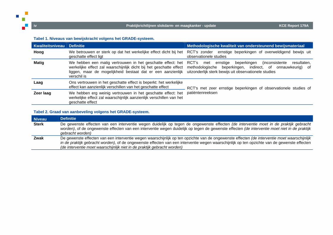
Op basis van het wetenschappelijk bewijsmateriaal gevonden door de KCE experts werden aanbevelingen opgesteld door een multidisciplinaire richtlijnontwikkelingsgroep (d.w.z. de auteurs van deze richtlijn). Een nazicht van deze aanbevelingen werd uitgevoerd door externe experts door middel van een formele procedure. Belangenconflicten werden genoteerd. Een niveau van bewijskracht en graad van aanbeveling werden aan elke aanbeveling toegewezen door middel van het GRADE systeem (Tabel 1 en 2).
iv Praktijkrichtlijnen slokdarm- en maagkanker - update KCE Report 179A
Tabel 1. Niveaus van bewijskracht volgens het GRADE-systeem.
Kwaliteitsniveau Definitie Methodologische kwaliteit van ondersteunend bewijsmateriaal Hoog We betrouwen er sterk op dat het werkelijke effect dicht bij het
geschatte effect ligt RCT’s zonder ernstige beperkingen of overweldigend bewijs uit observationele studies
Matig We hebben een matig vertrouwen in het geschatte effect: het werkelijke effect zal waarschijnlijk dicht bij het geschatte effect liggen, maar de mogelijkheid bestaat dat er een aanzienlijk verschil is
RCT’s met ernstige beperkingen (inconsistente resultaten, methodologische beperkingen, indirect, of onnauwkeurig) of uitzonderlijk sterk bewijs uit observationele studies
Laag Ons vertrouwen in het geschatte effect is beperkt: het werkelijke effect kan aanzienlijk verschillen van het geschatte effect
RCT's met zeer ernstige beperkingen of observationele studies of patiëntenreeksen
Zeer laag We hebben erg weinig vertrouwen in het geschatte effect: het
werkelijke effect zal waarschijnlijk aanzienlijk verschillen van het geschatte effect
Tabel 2. Graad van aanbeveling volgens het GRADE-systeem.
Niveau Definitie Sterk De gewenste effecten van een interventie wegen duidelijk op tegen de ongewenste effecten (de interventie moet in de praktijk gebracht
worden), of de ongewenste effecten van een interventie wegen duidelijk op tegen de gewenste effecten (de interventie moet niet in de praktijk gebracht worden)
Zwak De gewenste effecten van een interventie wegen waarschijnlijk op ten opzichte van de ongewenste effecten (de interventie moet waarschijnlijk in de praktijk gebracht worden), of de ongewenste effecten van een interventie wegen waarschijnlijk op ten opzichte van de gewenste effecten (de interventie moet waarschijnlijk niet in de praktijk gebracht worden)
KCE Report 179A Praktijkrichtlijnen slokdarm- en maagkanker - update v
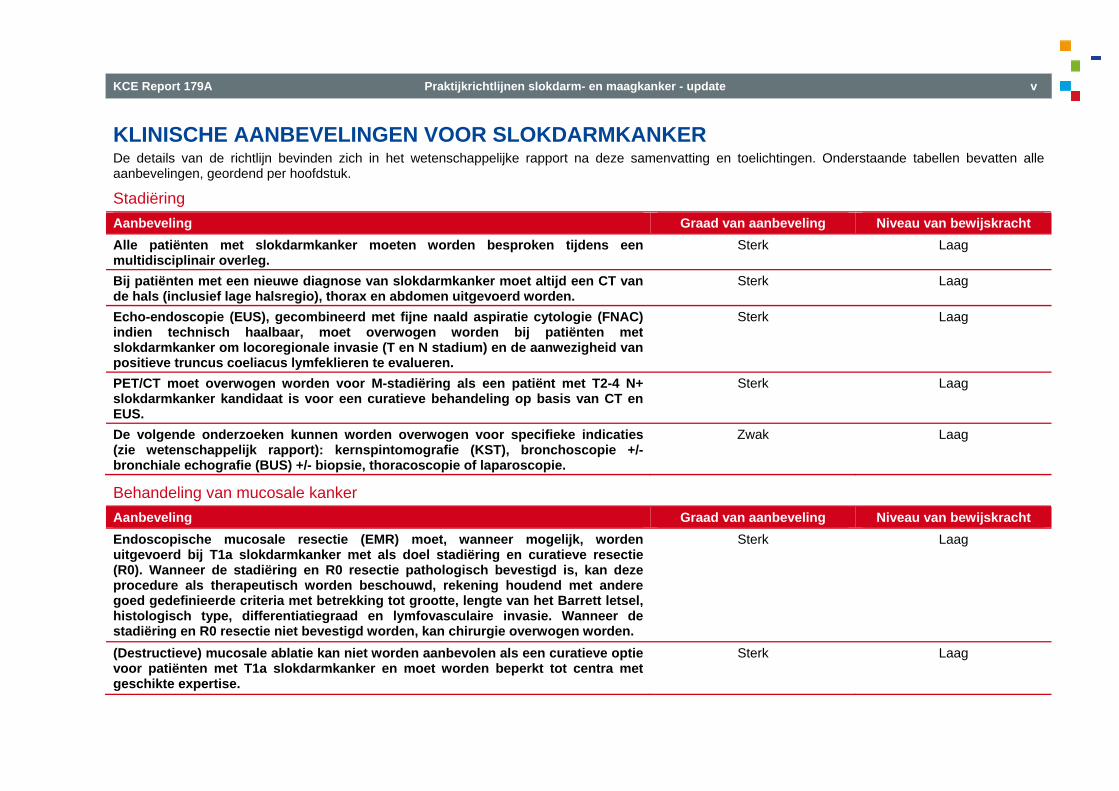
KLINISCHE AANBEVELINGEN VOOR SLOKDARMKANKER De details van de richtlijn bevinden zich in het wetenschappelijke rapport na deze samenvatting en toelichtingen. Onderstaande tabellen bevatten alle aanbevelingen, geordend per hoofdstuk.
Stadiëring Aanbeveling Graad van aanbeveling Niveau van bewijskracht Alle patiënten met slokdarmkanker moeten worden besproken tijdens een multidisciplinair overleg.
Sterk Laag
Bij patiënten met een nieuwe diagnose van slokdarmkanker moet altijd een CT van de hals (inclusief lage halsregio), thorax en abdomen uitgevoerd worden.
Sterk Laag
Echo-endoscopie (EUS), gecombineerd met fijne naald aspiratie cytologie (FNAC) indien technisch haalbaar, moet overwogen worden bij patiënten met slokdarmkanker om locoregionale invasie (T en N stadium) en de aanwezigheid van positieve truncus coeliacus lymfeklieren te evalueren.
Sterk Laag
PET/CT moet overwogen worden voor M-stadiëring als een patiënt met T2-4 N+ slokdarmkanker kandidaat is voor een curatieve behandeling op basis van CT en EUS.
Sterk Laag
De volgende onderzoeken kunnen worden overwogen voor specifieke indicaties (zie wetenschappelijk rapport): kernspintomografie (KST), bronchoscopie +/- bronchiale echografie (BUS) +/- biopsie, thoracoscopie of laparoscopie.
Zwak Laag
Behandeling van mucosale kanker Aanbeveling Graad van aanbeveling Niveau van bewijskracht Endoscopische mucosale resectie (EMR) moet, wanneer mogelijk, worden uitgevoerd bij T1a slokdarmkanker met als doel stadiëring en curatieve resectie (R0). Wanneer de stadiëring en R0 resectie pathologisch bevestigd is, kan deze procedure als therapeutisch worden beschouwd, rekening houdend met andere goed gedefinieerde criteria met betrekking tot grootte, lengte van het Barrett letsel, histologisch type, differentiatiegraad en lymfovasculaire invasie. Wanneer de stadiëring en R0 resectie niet bevestigd worden, kan chirurgie overwogen worden.
Sterk Laag
(Destructieve) mucosale ablatie kan niet worden aanbevolen als een curatieve optie voor patiënten met T1a slokdarmkanker en moet worden beperkt tot centra met geschikte expertise.
Sterk Laag
vi Praktijkrichtlijnen slokdarm- en maagkanker - update KCE Report 179A
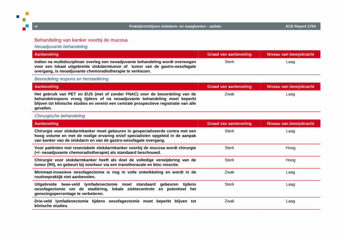
Behandeling van kanker voorbij de mucosa Neoadjuvante behandeling
Aanbeveling Graad van aanbeveling Niveau van bewijskracht
Indien na multidisciplinair overleg een neoadjuvante behandeling wordt overwogen voor een lokaal uitgebreide slokdarmtumor of tumor van de gastro-oesofagale overgang, is neoadjuvante chemoradiotherapie te verkiezen.
Sterk Laag
Beoordeling respons en herstadiëring
Aanbeveling Graad van aanbeveling Niveau van bewijskracht
Het gebruik van PET en EUS (met of zonder FNAC) voor de beoordeling van de behandelrespons vroeg tijdens of na neoadjuvante behandeling moet beperkt blijven tot klinische studies en vereist een centrale prospectieve registratie van alle gevallen.
Zwak Laag
Chirurgische behandeling
Aanbeveling Graad van aanbeveling Niveau van bewijskracht
Chirurgie voor slokdarmkanker moet gebeuren in gespecialiseerde centra met een hoog volume en met de nodige ervaring en/of specialisten opgeleid in de aanpak van kanker van de slokdarm en van de gastro-oesofagale overgang.
Sterk Laag
Voor patiënten met resectabele slokdarmkanker voorbij de mucosa wordt chirurgie (+/- neoadjuvante chemoradiotherapie) als standaard beschouwd.
Sterk Hoog
Chirurgie voor slokdarmkanker heeft als doel de volledige verwijdering van de tumor (R0), en gebeurt bij voorkeur via een transthoracale en bloc resectie.
Sterk Hoog
Minimaal-invasieve oesofagectomie is nog in volle ontwikkeling en wordt in de routinepraktijk niet aanbevolen.
Zwak Laag
Uitgebreide twee-veld lymfadenectomie moet standaard gebeuren tijdens oesofagectomie om de stadiëring, lokale ziektecontrole en potentieel het genezingspercentage te verbeteren.
Sterk Laag
Drie-veld lymfadenectomie tijdens oesofagectomie moet beperkt blijven tot klinische studies.
Zwak Laag
KCE Report 179A Praktijkrichtlijnen slokdarm- en maagkanker - update vii
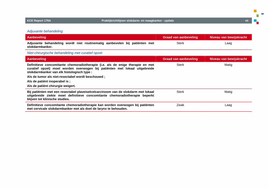
Adjuvante behandeling
Aanbeveling Graad van aanbeveling Niveau van bewijskracht
Adjuvante behandeling wordt niet routinematig aanbevolen bij patiënten met slokdarmkanker.
Sterk Laag
Niet-chirurgische behandeling met curatief opzet
Aanbeveling Graad van aanbeveling Niveau van bewijskracht
Definitieve concomitante chemoradiotherapie (i.e. als de enige therapie en met curatief opzet) moet worden overwogen bij patiënten met lokaal uitgebreide slokdarmkanker van elk histologisch type : Als de tumor als niet-resectabel wordt beschouwd ; Als de patiënt inoperabel is ; Als de patiënt chirurgie weigert.
Sterk Matig
Bij patiënten met een resectabel plaveiselcelcarcinoom van de slokdarm met lokaal uitgebreide ziekte moet definitieve concomitante chemoradiotherapie beperkt blijven tot klinische studies.
Sterk Matig
Definitieve concomitante chemoradiotherapie kan worden overwogen bij patiënten met cervicale slokdarmkanker met als doel de larynx te behouden.
Zwak Laag
viii Praktijkrichtlijnen slokdarm- en maagkanker - update KCE Report 179A
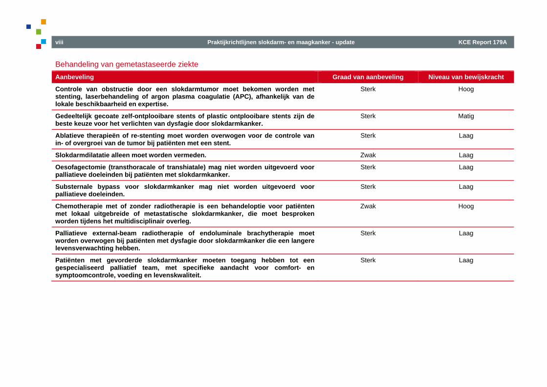
Behandeling van gemetastaseerde ziekte Aanbeveling Graad van aanbeveling Niveau van bewijskracht
Controle van obstructie door een slokdarmtumor moet bekomen worden met stenting, laserbehandeling of argon plasma coagulatie (APC), afhankelijk van de lokale beschikbaarheid en expertise.
Sterk Hoog
Gedeeltelijk gecoate zelf-ontplooibare stents of plastic ontplooibare stents zijn de beste keuze voor het verlichten van dysfagie door slokdarmkanker.
Sterk Matig
Ablatieve therapieën of re-stenting moet worden overwogen voor de controle van in- of overgroei van de tumor bij patiënten met een stent.
Sterk Laag
Slokdarmdilatatie alleen moet worden vermeden. Zwak Laag
Oesofagectomie (transthoracale of transhiatale) mag niet worden uitgevoerd voor palliatieve doeleinden bij patiënten met slokdarmkanker.
Sterk Laag
Substernale bypass voor slokdarmkanker mag niet worden uitgevoerd voor palliatieve doeleinden.
Sterk Laag
Chemotherapie met of zonder radiotherapie is een behandeloptie voor patiënten met lokaal uitgebreide of metastatische slokdarmkanker, die moet besproken worden tijdens het multidisciplinair overleg.
Zwak Hoog
Palliatieve external-beam radiotherapie of endoluminale brachytherapie moet worden overwogen bij patiënten met dysfagie door slokdarmkanker die een langere levensverwachting hebben.
Sterk Laag
Patiënten met gevorderde slokdarmkanker moeten toegang hebben tot een gespecialiseerd palliatief team, met specifieke aandacht voor comfort- en symptoomcontrole, voeding en levenskwaliteit.
Sterk Laag
KCE Report 179A Praktijkrichtlijnen slokdarm- en maagkanker - update ix
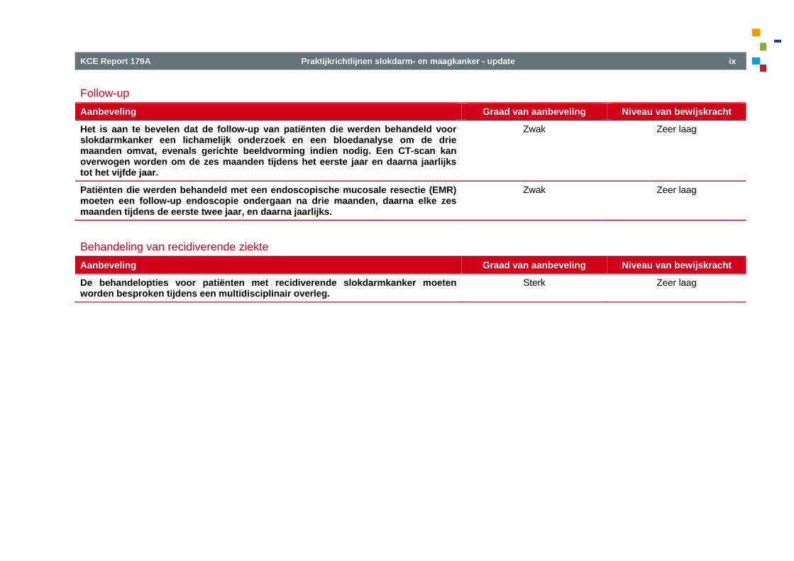
Follow-up Aanbeveling Graad van aanbeveling Niveau van bewijskracht
Het is aan te bevelen dat de follow-up van patiënten die werden behandeld voor slokdarmkanker een lichamelijk onderzoek en een bloedanalyse om de drie maanden omvat, evenals gerichte beeldvorming indien nodig. Een CT-scan kan overwogen worden om de zes maanden tijdens het eerste jaar en daarna jaarlijks tot het vijfde jaar.
Zwak Zeer laag
Patiënten die werden behandeld met een endoscopische mucosale resectie (EMR) moeten een follow-up endoscopie ondergaan na drie maanden, daarna elke zes maanden tijdens de eerste twee jaar, en daarna jaarlijks.
Zwak Zeer laag
Behandeling van recidiverende ziekte Aanbeveling Graad van aanbeveling Niveau van bewijskracht
De behandelopties voor patiënten met recidiverende slokdarmkanker moeten worden besproken tijdens een multidisciplinair overleg.
Sterk Zeer laag
x Praktijkrichtlijnen slokdarm- en maagkanker - update KCE Report 179A
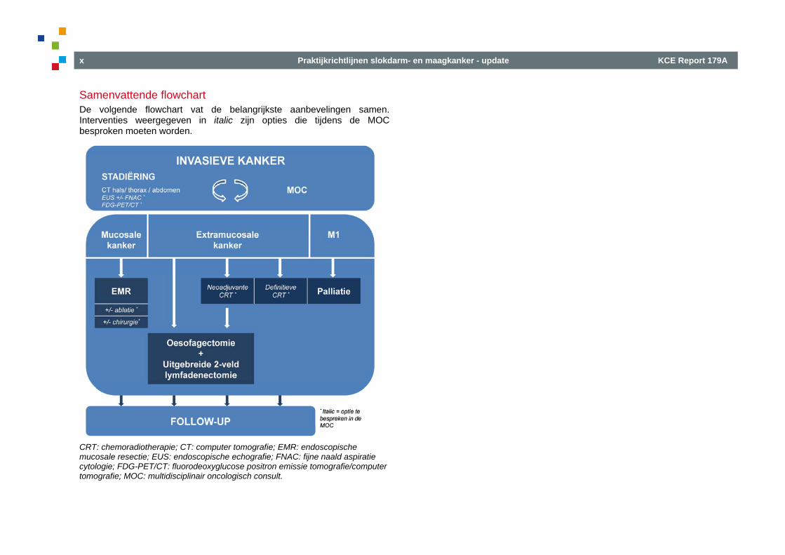
Samenvattende flowchart De volgende flowchart vat de belangrijkste aanbevelingen samen. Interventies weergegeven in italic zijn opties die tijdens de MOC besproken moeten worden.
CRT: chemoradiotherapie; CT: computer tomografie; EMR: endoscopische mucosale resectie; EUS: endoscopische echografie; FNAC: fijne naald aspiratie cytologie; FDG-PET/CT: fluorodeoxyglucose positron emissie tomografie/computer tomografie; MOC: multidisciplinair oncologisch consult.
KCE Report 179A Praktijkrichtlijnen slokdarm- en maagkanker - update xi
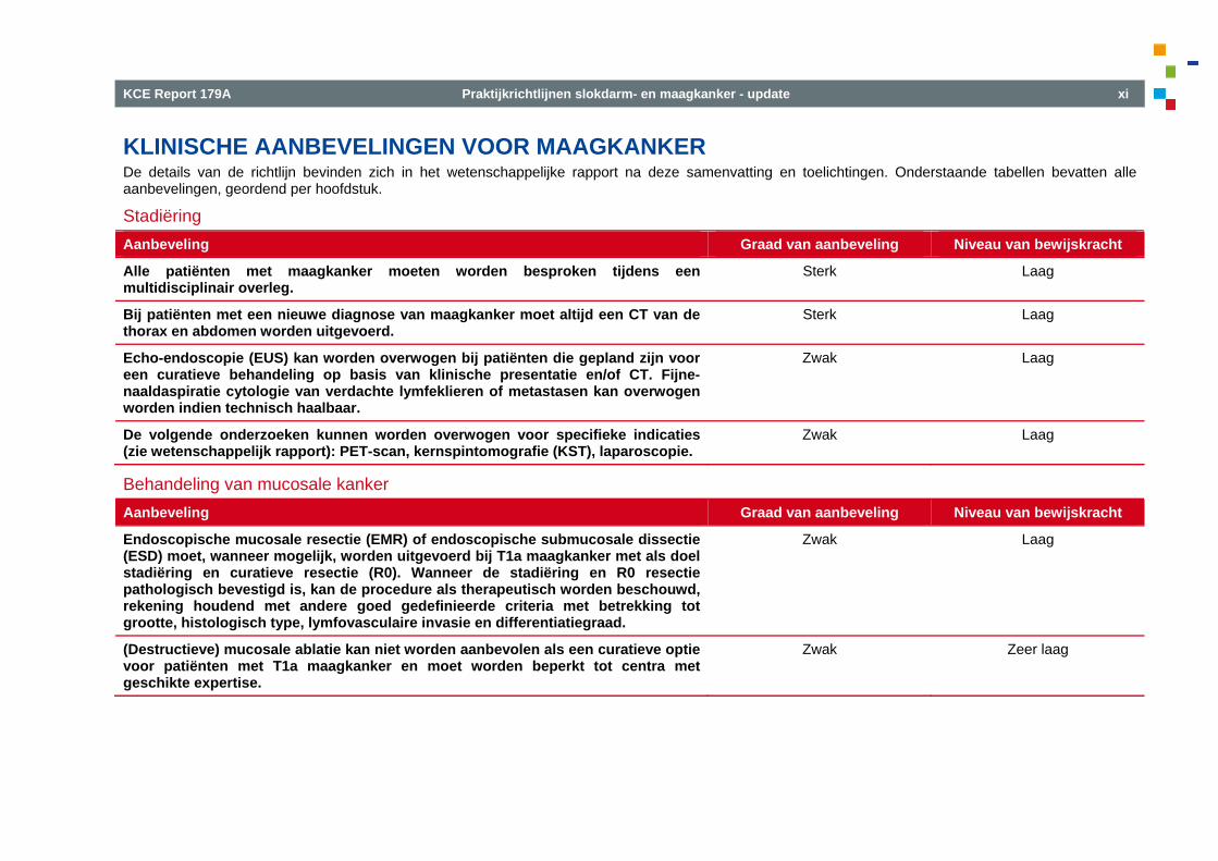
KLINISCHE AANBEVELINGEN VOOR MAAGKANKER De details van de richtlijn bevinden zich in het wetenschappelijke rapport na deze samenvatting en toelichtingen. Onderstaande tabellen bevatten alle aanbevelingen, geordend per hoofdstuk.
Stadiëring Aanbeveling Graad van aanbeveling Niveau van bewijskracht
Alle patiënten met maagkanker moeten worden besproken tijdens een multidisciplinair overleg.
Sterk Laag
Bij patiënten met een nieuwe diagnose van maagkanker moet altijd een CT van de thorax en abdomen worden uitgevoerd.
Sterk Laag
Echo-endoscopie (EUS) kan worden overwogen bij patiënten die gepland zijn voor een curatieve behandeling op basis van klinische presentatie en/of CT. Fijne-naaldaspiratie cytologie van verdachte lymfeklieren of metastasen kan overwogen worden indien technisch haalbaar.
Zwak Laag
De volgende onderzoeken kunnen worden overwogen voor specifieke indicaties (zie wetenschappelijk rapport): PET-scan, kernspintomografie (KST), laparoscopie.
Zwak Laag
Behandeling van mucosale kanker Aanbeveling Graad van aanbeveling Niveau van bewijskracht
Endoscopische mucosale resectie (EMR) of endoscopische submucosale dissectie (ESD) moet, wanneer mogelijk, worden uitgevoerd bij T1a maagkanker met als doel stadiëring en curatieve resectie (R0). Wanneer de stadiëring en R0 resectie pathologisch bevestigd is, kan de procedure als therapeutisch worden beschouwd, rekening houdend met andere goed gedefinieerde criteria met betrekking tot grootte, histologisch type, lymfovasculaire invasie en differentiatiegraad.
Zwak Laag
(Destructieve) mucosale ablatie kan niet worden aanbevolen als een curatieve optie voor patiënten met T1a maagkanker en moet worden beperkt tot centra met geschikte expertise.
Zwak Zeer laag
xii Praktijkrichtlijnen slokdarm- en maagkanker - update KCE Report 179A
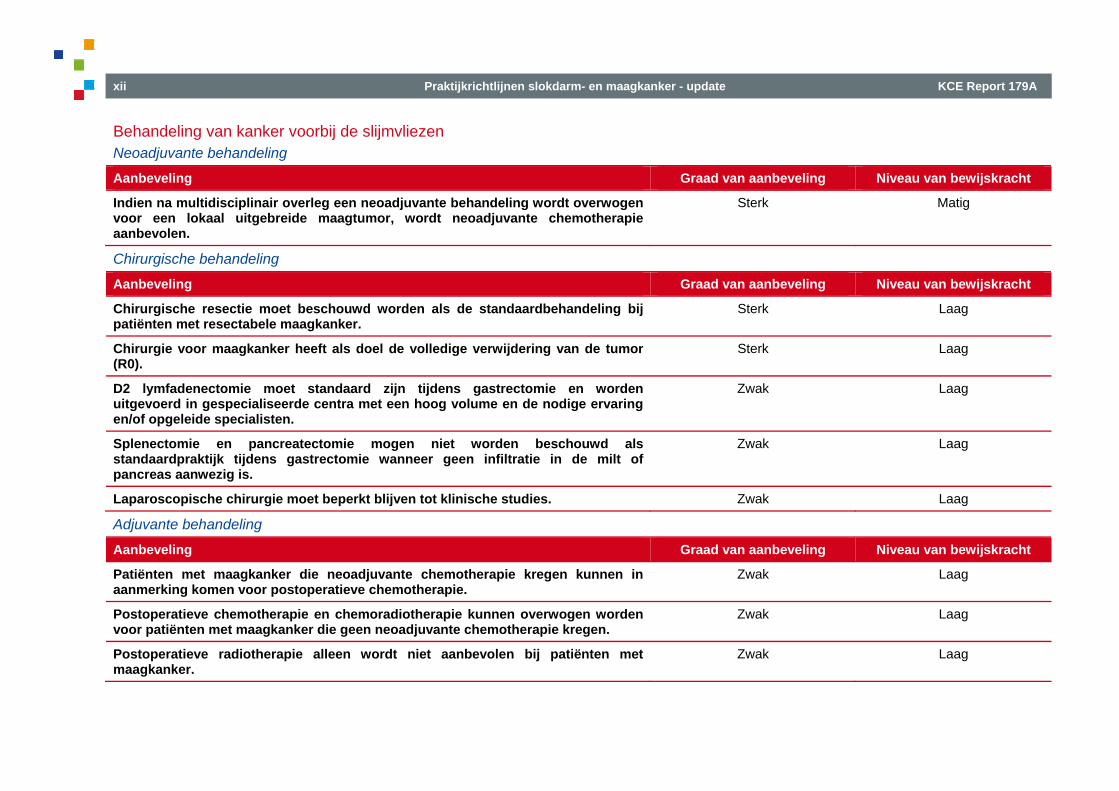
Behandeling van kanker voorbij de slijmvliezen Neoadjuvante behandeling
Aanbeveling Graad van aanbeveling Niveau van bewijskracht
Indien na multidisciplinair overleg een neoadjuvante behandeling wordt overwogen voor een lokaal uitgebreide maagtumor, wordt neoadjuvante chemotherapie aanbevolen.
Sterk Matig
Chirurgische behandeling
Aanbeveling Graad van aanbeveling Niveau van bewijskracht
Chirurgische resectie moet beschouwd worden als de standaardbehandeling bij patiënten met resectabele maagkanker.
Sterk Laag
Chirurgie voor maagkanker heeft als doel de volledige verwijdering van de tumor (R0).
Sterk Laag
D2 lymfadenectomie moet standaard zijn tijdens gastrectomie en worden uitgevoerd in gespecialiseerde centra met een hoog volume en de nodige ervaring en/of opgeleide specialisten.
Zwak Laag
Splenectomie en pancreatectomie mogen niet worden beschouwd als standaardpraktijk tijdens gastrectomie wanneer geen infiltratie in de milt of pancreas aanwezig is.
Zwak Laag
Laparoscopische chirurgie moet beperkt blijven tot klinische studies. Zwak Laag
Adjuvante behandeling
Aanbeveling Graad van aanbeveling Niveau van bewijskracht
Patiënten met maagkanker die neoadjuvante chemotherapie kregen kunnen in aanmerking komen voor postoperatieve chemotherapie.
Zwak Laag
Postoperatieve chemotherapie en chemoradiotherapie kunnen overwogen worden voor patiënten met maagkanker die geen neoadjuvante chemotherapie kregen.
Zwak Laag
Postoperatieve radiotherapie alleen wordt niet aanbevolen bij patiënten met maagkanker.
Zwak Laag
KCE Report 179A Praktijkrichtlijnen slokdarm- en maagkanker - update xiii
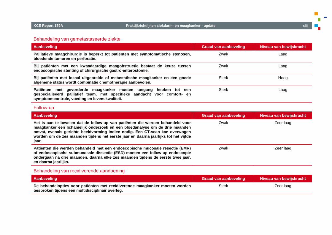
Behandeling van gemetastaseerde ziekte Aanbeveling Graad van aanbeveling Niveau van bewijskracht
Palliatieve maagchirurgie is beperkt tot patiënten met symptomatische stenosen, bloedende tumoren en perforatie.
Zwak Laag
Bij patiënten met een kwaadaardige maagobstructie bestaat de keuze tussen endoscopische stenting of chirurgische gastro-enterostomie.
Zwak Laag
Bij patiënten met lokaal uitgebreide of metastatische maagkanker en een goede algemene status wordt combinatie chemotherapie aanbevolen.
Sterk Hoog
Patiënten met gevorderde maagkanker moeten toegang hebben tot een gespecialiseerd palliatief team, met specifieke aandacht voor comfort- en symptoomcontrole, voeding en levenskwaliteit.
Sterk Laag
Follow-up Aanbeveling Graad van aanbeveling Niveau van bewijskracht
Het is aan te bevelen dat de follow-up van patiënten die werden behandeld voor maagkanker een lichamelijk onderzoek en een bloedanalyse om de drie maanden omvat, evenals gerichte beeldvorming indien nodig. Een CT-scan kan overwogen worden om de zes maanden tijdens het eerste jaar en daarna jaarlijks tot het vijfde jaar.
Zwak Zeer laag
Patiënten die werden behandeld met een endoscopische mucosale resectie (EMR) of endoscopische submucosale dissectie (ESD) moeten een follow-up endoscopie ondergaan na drie maanden, daarna elke zes maanden tijdens de eerste twee jaar, en daarna jaarlijks.
Zwak Zeer laag
Behandeling van recidiverende aandoening Aanbeveling Graad van aanbeveling Niveau van bewijskracht
De behandelopties voor patiënten met recidiverende maagkanker moeten worden besproken tijdens een multidisciplinair overleg.
Sterk Zeer laag
xiv Praktijkrichtlijnen slokdarm- en maagkanker - update KCE Report 179A
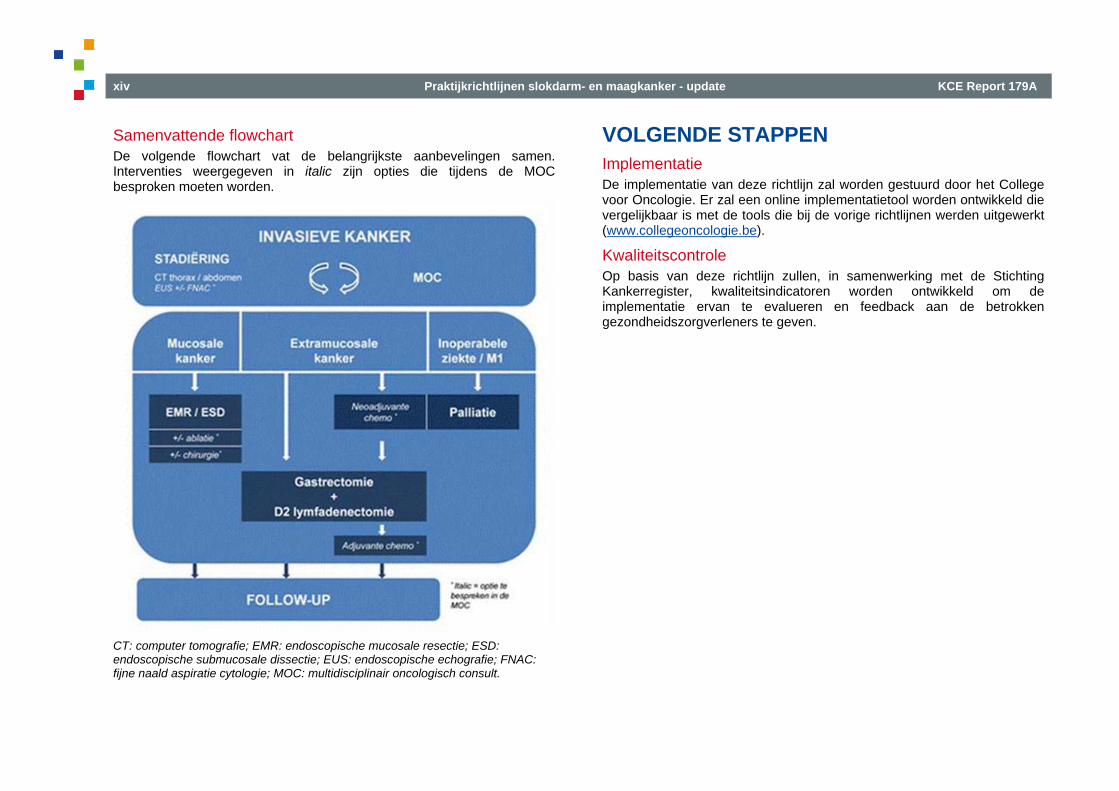
Samenvattende flowchart De volgende flowchart vat de belangrijkste aanbevelingen samen. Interventies weergegeven in italic zijn opties die tijdens de MOC besproken moeten worden.
CT: computer tomografie; EMR: endoscopische mucosale resectie; ESD: endoscopische submucosale dissectie; EUS: endoscopische echografie; FNAC: fijne naald aspiratie cytologie; MOC: multidisciplinair oncologisch consult.
VOLGENDE STAPPEN Implementatie De implementatie van deze richtlijn zal worden gestuurd door het College voor Oncologie. Er zal een online implementatietool worden ontwikkeld die vergelijkbaar is met de tools die bij de vorige richtlijnen werden uitgewerkt (www.collegeoncologie.be).
Kwaliteitscontrole Op basis van deze richtlijn zullen, in samenwerking met de Stichting Kankerregister, kwaliteitsindicatoren worden ontwikkeld om de implementatie ervan te evalueren en feedback aan de betrokken gezondheidszorgverleners te geven.
KCE Report 179A Praktijkrichtlijnen slokdarm- en maagkanker - update xv
BELEIDSAANBEVELINGENa
Ter attentie van de verantwoordelijken van het Health Research Systemb • Gelet op het veranderend bewijsmateriaal en op basis van een pre-evaluatie
van de literatuur zou deze richtlijn volledig geüpdatet moeten zijn binnen 5 jaar. Ondertussen zal op de website van het College van Geneesheren voor Oncologie vermeld worden wanneer er belangrijk bewijsmateriaal beschikbaar wordt (http://www.collegeoncologie.be).
Ter attentie van de Nationale Raad voor Kwaliteitspromotie en het College van Geneesheren voor Oncologie • De kwaliteitsindicatoren die ontwikkeld zullen worden, zullen in een
integratief kwaliteitssysteem moeten worden ingebed, zoals in het KCE rapport 152 wordt aanbevolen.
Ter attentie van de Minister, na advies van de bevoegde organen (Nationale Raad voor Ziekenhuisvoorzieningen, Geneeskundige Technische Raad, College van Geneesheren voor Oncologie) • Hoewel de literatuur over de volume-uitkomst relatie van kanker van de
bovenste gastrointestinale tractus hoofdzakelijk beperkt is tot chirurgie, moet de volledige behandeling gecentraliseerd worden in centra met specialisten opgeleid voor, en met hoog-volume ervaring in de aanpak van kanker van de bovenste gastrointestinale tractus.
• Analoog aan het PROCARE project moet geschikte opleiding en peer review georganiseerd worden om een behandeling van hoge kwaliteit te verzekeren voor patiënten met kanker van de bovenste gastrointestinale tractus.
a Alleen het KCE is verantwoordelijk voor deze aanbevelingen b Beschreven door het Rekenhof in zijn audit van januari 2010 “Wetenschappelijke ondersteuning van het federale gezondheidsbeleid”


KCE Report 179 Clinical Practice Guidelines Upper Gastrointestinal Cancer - update 1
TABLE OF CONTENTS
1. INTRODUCTION ................................................................................................................................... 7 1.1. SCOPE .................................................................................................................................................. 7 1.2. EPIDEMIOLOGY ................................................................................................................................... 7
1.2.1. Oesophageal cancer ............................................................................................................... 7 1.2.2. Gastric cancer ......................................................................................................................... 9
2. METHODOLOGY ................................................................................................................................ 10 2.1. GENERAL APPROACH ...................................................................................................................... 10 2.2. CLINICAL QUESTIONS ...................................................................................................................... 10 2.3. LITERATURE SEARCH AND SELECTION CRITERIA ...................................................................... 10 2.4. QUALITY APPRAISAL ........................................................................................................................ 10 2.5. DATA EXTRACTION AND EVIDENCE SUMMARY ........................................................................... 10 2.6. FORMULATION OF RECOMMENDATIONS ...................................................................................... 12 3. DEFINITIONS ...................................................................................................................................... 13 3.1. TOPOGRAPHIC DEFINITIONS .......................................................................................................... 13 3.2. EARLY LESIONS ................................................................................................................................ 14
3.2.1. Histology of the normal oesophagus ..................................................................................... 14 3.2.2. Barrett’s oesophagus ............................................................................................................ 15 3.2.3. Dysplasia in squamous epithelium ........................................................................................ 16 3.2.4. Dysplasia in columnar epithelium .......................................................................................... 16
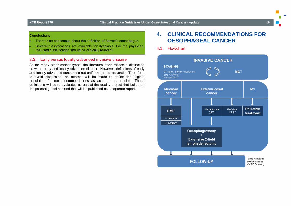
3.3. EARLY VERSUS LOCALLY-ADVANCED INVASIVE DISEASE ........................................................ 19 4. CLINICAL RECOMMENDATIONS FOR OESOPHAGEAL CANCER ............................................... 19 4.1. FLOWCHART ...................................................................................................................................... 19 4.2. STAGING ............................................................................................................................................. 20
4.2.1. Computed tomography .......................................................................................................... 20 4.2.2. Endoscopic ultrasonography ................................................................................................. 21 4.2.3. Positron-emission tomography .............................................................................................. 22

2 Clinical Practice Guidelines Upper Gastrointestinal Cancer - update KCE Report 179
4.2.4. Magnetic resonance imaging ................................................................................................ 23 4.2.5. Bronchoscopy and bronchoscopic ultrasound ...................................................................... 23 4.2.6. Thoracoscopy and laparoscopy ............................................................................................ 23
4.3. TREATMENT OF MUCOSAL CANCER .............................................................................................. 25 4.4. TREATMENT OF CANCER BEYOND THE MUCOSA ....................................................................... 26
4.4.1. Neoadjuvant treatment .......................................................................................................... 26 4.4.2. Response assessment and restaging ................................................................................... 30 4.4.3. Surgical treatment ................................................................................................................. 32 4.4.4. Adjuvant treatment ................................................................................................................ 36 4.4.5. Non-surgical treatment with curative intent ........................................................................... 37
4.5. TREATMENT OF METASTATIC DISEASE ........................................................................................ 39 4.5.1. Endoscopic ablation .............................................................................................................. 40 4.5.2. Stents .................................................................................................................................... 40 4.5.3. Palliative surgery ................................................................................................................... 41 4.5.4. Systemic treatment ................................................................................................................ 41 4.5.5. Radiotherapy ......................................................................................................................... 41
4.6. SUPPORTIVE CARE........................................................................................................................... 43 4.7. FOLLOW-UP ....................................................................................................................................... 43 4.8. TREATMENT OF RECURRENT DISEASE ........................................................................................ 44 5. CLINICAL RECOMMENDATIONS FOR GASTRIC CANCER ........................................................... 45 5.1. FLOWCHART ...................................................................................................................................... 45 5.2. STAGING ............................................................................................................................................. 46
5.2.1. Computed tomography .......................................................................................................... 46 5.2.2. Endoscopic ultrasonography ................................................................................................. 48 5.2.3. Positron-emission tomography .............................................................................................. 49 5.2.4. Magnetic resonance imaging ................................................................................................ 50 5.2.5. Laparoscopy .......................................................................................................................... 50 5.2.6. Sentinel lymph node mapping ............................................................................................... 51
5.3. TREATMENT OF MUCOSAL CANCER .............................................................................................. 52
KCE Report 179 Clinical Practice Guidelines Upper Gastrointestinal Cancer - update 3
5.4. TREATMENT OF CANCER BEYOND THE MUCOSA ....................................................................... 53 5.4.1. Neoadjuvant treatment .......................................................................................................... 53 5.4.2. Surgical treatment ................................................................................................................. 55 5.4.3. Adjuvant treatment ................................................................................................................ 59
5.5. TREATMENT OF METASTATIC DISEASE ........................................................................................ 61 5.5.1. Palliative surgery ................................................................................................................... 61 5.5.2. Stents .................................................................................................................................... 61 5.5.3. Chemotherapy and targeted treatment ................................................................................. 62 5.5.4. Supportive care ..................................................................................................................... 63
5.6. FOLLOW-UP ....................................................................................................................................... 64 5.7. TREATMENT OF RECURRENT DISEASE ........................................................................................ 65 6. REFERENCES .................................................................................................................................... 66
4 Clinical Practice Guidelines Upper Gastrointestinal Cancer - update KCE Report 179
LIST OF ABBREVIATIONS
ABBREVIATION DEFINITION 18-FDG PET 18F-fluoro-2-deoxy-D-glucose (FDG) positron emission tomography (PET)
5-FU 5-Fluorouracil
95%CI 95% confidence interval
ACC Adenocarcinoma
AETMIS Agence d’Evaluation des Technologies et des Modes d’Intervention en Santé
APC Argon plasma coagulation
CBO Dutch Institute for Healthcare Improvement
CPG Clinical Practice Guideline
CT Computed tomography
CTRT Chemoradiotherapy
DFS Disease-free survival
DPIC Delayed postoperative intraperitoneal chemotherapy
EBRT External Beam Radiotherapy
ECOG Eastern Cooperative Oncology Group
EIPL Extensive intraoperative peritoneal lavage
EMR Endoscopic mucosal resection
EPIC Early postoperative intraperitoneal chemotherapy
ESD Endoscopic submucosal dissection
EUS Endoscopic ultrasonography
FNAC Fine needle aspiration cytology
FNCLCC Fédération Nationale des centres de Lutte Contre le Cancer (France)
FU Follow-up
GOJ Gastro-oesophageal junction
KCE Report 179 Clinical Practice Guidelines Upper Gastrointestinal Cancer - update 5
GRADE Grading of Recommendations Assessment, Development and Evaluation
HDR High dose rate
HIIC Hyperthermic Intra-operative intra-peritoneal chemotherapy
HR Hazard ratio
IP Intra-peritoneal
ITT Intention-to-treat
IV Intra-venous
LN Lymph nodes
MA Meta-analysis
MDT Multidisciplinary Team
MGC Metastatic gastric cancer
MIE Minimally invasive oesophagectomy
MRI Magnetic Resonance Imaging
NACRT Neoadjuvant chemoradiotherapy
NACT Neoadjuvant chemotherapy
NICC Normothermic intra-operative intra-peritoneal chemotherapy
NNT Number of Needed to treat
NPV Negative predictive value
OR Odds ratio
OS Overall survival
PALN Para-aortic lymph nodes
PDT Photodynamic therapy
PFS Progression-free survival
PPV Positive predictive value
6 Clinical Practice Guidelines Upper Gastrointestinal Cancer - update KCE Report 179
PS Performance status
QoL Quality of life
QUADAS Quality Assessment of Diagnostic Accuracy Studies
RCT Randomized controlled trial
RECIST Response Evaluation Criteria in Solid Tumours
SCC Squamous cell carcinoma
Se Sensitivity
SIGN Scottish Intercollegiate Guidelines Network
SLNB Sentinel lymph node biopsy
Sp Specificity
SR Systematic review
SUV Standard Uptake Value
TNM Tumour - Node - Metastasis
KCE Report 179 Clinical Practice Guidelines Upper Gastrointestinal Cancer - update 7
SCIENTIFIC REPORT 1. INTRODUCTION 1.1. Scope In 2008, the KCE and the College of Oncology published national guidelines for oesophageal and gastric cancer1. Currently, an ongoing KCE project aims to identify and elaborate quality indicators for both cancer types. To allow the guidelines to serve as a basis for the development of these quality indicators, they should be up-to-date. It was therefore decided to do a pragmatic update focusing on key evidence published since the previous literature search, being August 2007. The update will cover the staging, treatment and follow-up of patients with confirmed invasive oesophageal or gastric cancer. It is intended to be used by all care providers involved in the care for these patients. Importantly, the following topics that were part of the previous version will not be included in the update, because they are outside the scope of the quality indicators project: • Work-up of pre-invasive lesions, i.e. Barrett’s oesophagus and
dysplastic lesions, including high-grade dysplasia; • Treatment of gastric lymphoma; • Treatment of gastrointestinal stromal tumours (GIST).
1.2. Epidemiology 1.2.1. Oesophageal cancer Oesophageal cancer is the eighth most common cancer in the world (about 481 000 new cases in 2008 worldwide) and one of the most lethal (6th most common cause of death from cancer worldwide)2. Incidence rates of oesophageal cancer show well-known regional disparities, with the highest incidence rates in Southern Africa (Age Standardised Rate [ASR] 22.3 per 100 000 men and 11.7 per 100 000 women in 2008) and the lowest rates in Western Africa (ASR 1.4 per 100 000 men in 2008). In Europe, crude incidence rates for all types of oesophageal cancer ranged from 0.7 cases per 100 000 in Cyprus to 13.3 cases per 100 000 in the UK in 20082.
8 Clinical Practice Guidelines Upper Gastrointestinal Cancer - update KCE Report 179
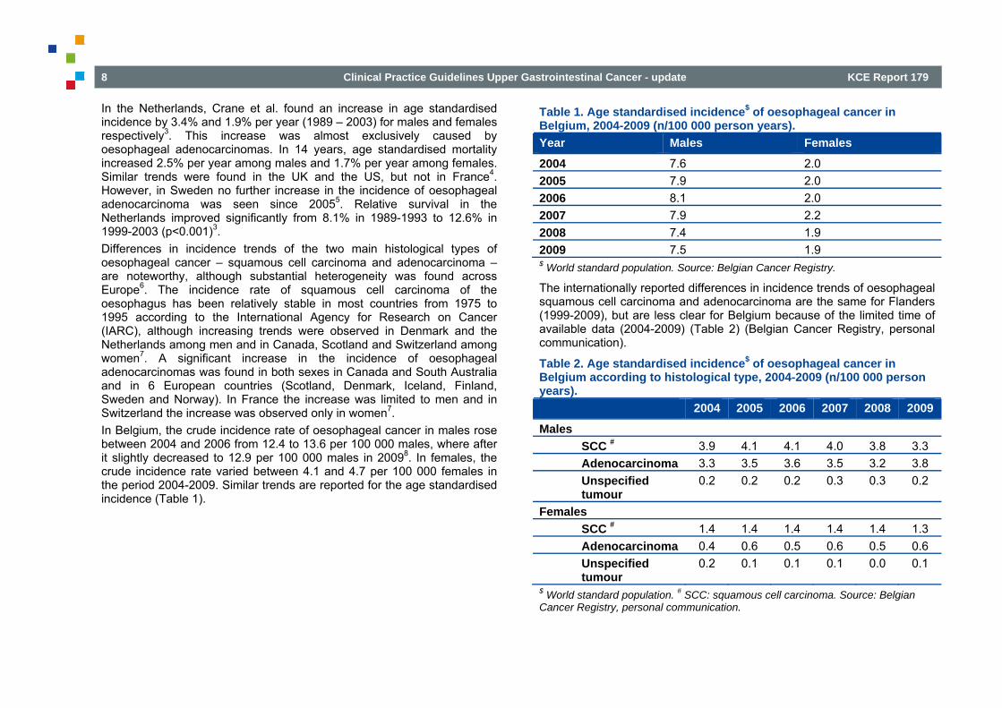
In the Netherlands, Crane et al. found an increase in age standardised incidence by 3.4% and 1.9% per year (1989 – 2003) for males and females respectively3. This increase was almost exclusively caused by oesophageal adenocarcinomas. In 14 years, age standardised mortality increased 2.5% per year among males and 1.7% per year among females. Similar trends were found in the UK and the US, but not in France4. However, in Sweden no further increase in the incidence of oesophageal adenocarcinoma was seen since 20055. Relative survival in the Netherlands improved significantly from 8.1% in 1989-1993 to 12.6% in 1999-2003 (p<0.001)3. Differences in incidence trends of the two main histological types of oesophageal cancer – squamous cell carcinoma and adenocarcinoma – are noteworthy, although substantial heterogeneity was found across Europe6. The incidence rate of squamous cell carcinoma of the oesophagus has been relatively stable in most countries from 1975 to 1995 according to the International Agency for Research on Cancer (IARC), although increasing trends were observed in Denmark and the Netherlands among men and in Canada, Scotland and Switzerland among women7. A significant increase in the incidence of oesophageal adenocarcinomas was found in both sexes in Canada and South Australia and in 6 European countries (Scotland, Denmark, Iceland, Finland, Sweden and Norway). In France the increase was limited to men and in Switzerland the increase was observed only in women7. In Belgium, the crude incidence rate of oesophageal cancer in males rose between 2004 and 2006 from 12.4 to 13.6 per 100 000 males, where after it slightly decreased to 12.9 per 100 000 males in 20098. In females, the crude incidence rate varied between 4.1 and 4.7 per 100 000 females in the period 2004-2009. Similar trends are reported for the age standardised incidence (Table 1).
Table 1. Age standardised incidence$ of oesophageal cancer in Belgium, 2004-2009 (n/100 000 person years). Year Males Females
2004 7.6 2.0 2005 7.9 2.0 2006 8.1 2.0 2007 7.9 2.2 2008 7.4 1.9 2009 7.5 1.9 $ World standard population. Source: Belgian Cancer Registry.
The internationally reported differences in incidence trends of oesophageal squamous cell carcinoma and adenocarcinoma are the same for Flanders (1999-2009), but are less clear for Belgium because of the limited time of available data (2004-2009) (Table 2) (Belgian Cancer Registry, personal communication).
Table 2. Age standardised incidence$ of oesophageal cancer in Belgium according to histological type, 2004-2009 (n/100 000 person years). 2004 2005 2006 2007 2008 2009
Males SCC # 3.9 4.1 4.1 4.0 3.8 3.3 Adenocarcinoma 3.3 3.5 3.6 3.5 3.2 3.8 Unspecified tumour
0.2 0.2 0.2 0.3 0.3 0.2
Females SCC # 1.4 1.4 1.4 1.4 1.4 1.3 Adenocarcinoma 0.4 0.6 0.5 0.6 0.5 0.6 Unspecified tumour
0.2 0.1 0.1 0.1 0.0 0.1
$ World standard population. # SCC: squamous cell carcinoma. Source: Belgian Cancer Registry, personal communication.

KCE Report 179 Clinical Practice Guidelines Upper Gastrointestinal Cancer - update 9
1.2.2. Gastric cancer With an estimated 988 000 new cases in 2008 worldwide (7.8% of all new cancer cases), gastric cancer is in fourth place behind cancers of the lung, breast, and colon and rectum, with more than 70% of the cases occurring in developing countries2. It is the second most common cause of death from cancer. Gastric cancer incidence rates vary by up to ten-fold throughout the world. Japan and Korea have the highest gastric cancer incidence rates in the world. High-incidence areas for non-cardia gastric adenocarcinoma include East Asia, Eastern Europe, and Central and South America. Low incidence rates are found in South Asia, North and East Africa, North America, Australia, and New Zealand2. Survival is moderately good only in Japan (52%), where mass screening by photofluoroscopy has been practiced since the 1960s. Survival is also relatively high in North America (approximately 21%), possibly due to early diagnosis following a higher number of endoscopic examinations performed for gastric disorders. Estimated survival is 27% in Western Europe9. In the Netherlands, age standardised incidence of gastric cancer declined from 24 to 12 per 100 000 person years in males and from 10 to 6 per 100 000 person years in females between 1990 and 200710. The age standardised mortality rates decreased from 20.7 to 12.8 per 100 000 person years in males and from 8.2 to 4.2 per 100 000 person years in females between 1978 and 199711. In Belgium, the crude incidence rate of gastric cancer declined from 17.4 per 100.000 males in 2004 to 15.4 per 100 000 males in 20098. In females, the crude incidence rate remained quite stable between 2004 and 2009 (9.4/100 000 females in 2009). Similar trends are reported for the age standardised incidence (Table 3). While the incidence rates of these GOJ tumours recently increased, the incidence rates of ‘real’ gastric tumours declined12.
Table 3. Age standardised incidence$ of gastric cancer in Belgium, 2004-2009 (n/100 000 person years). Year Males Females
2004 9.4 3.9 2005 9.4 4.0 2006 8.5 4.0 2007 8.9 3.6 2008 7.9 3.8 2009 8.1 3.8 $ World standard population. Source: Belgian Cancer Registry.
10 Clinical Practice Guidelines Upper Gastrointestinal Cancer - update KCE Report 179
2. METHODOLOGY 2.1. General approach A pragmatic approach was chosen. For therapeutic clinical questions the literature search focused on new systematic reviews and randomized controlled trials (RCTs). For diagnostic clinical questions, diagnostic accuracy studies were searched in addition. Other observational and prognostic studies were not considered.
2.2. Clinical questions The following clinical questions were addressed in this update: 1. What staging techniques should be used for oesophageal and gastric
cancer? 2. What are the best treatment options for mucosal oesophageal and
gastric cancer? 3. What are the best treatment options for oesophageal and gastric
cancer beyond the mucosa? a. neoadjuvant treatment b. surgical treatment c. adjuvant treatment d. non-surgical treatment with curative intent
4. What are the best palliative treatment options for metastatic oesophageal and gastric cancer?
5. What are the best follow-up strategies for oesophageal and gastric cancer?
2.3. Literature search and selection criteria Systematic reviews and meta-analyses were searched in the following databases: • OVID Medline and PreMedline • EMBASE • Cochrane Database of Systematic Reviews • Database of Abstracts of Reviews of Effects (DARE)
• Health Technology Assessment (HTA) database RCTs were searched in OVID Medline, PreMedline, EMBASE and CENTRAL, while diagnostic accuracy studies were searched in OVID Medline, PreMedline and EMBASE. A generic search strategy was used for all research questions. The search terms and their combinations can be found in appendix 1. A date limit was set from August 2007 (i.e. the search date of the previous version) until 2011. The evidence published before August 2007 was not searched again. However, to allow a correct application of the GRADE methodology (see below), some primary studies from before August 2007 were retrieved for additional quality appraisal and data extraction.
2.4. Quality appraisal The quality of the retrieved systematic reviews and RCTs was assessed using the checklists of the Dutch Cochrane Centre (www.cochrane.nl). Evidence-based guidelines were treated as systematic reviews. For the appraisal of diagnostic accuracy studies, the updated version of the QUADAS instrument was used13. All articles were appraised by one reviewer. In case of doubt, a second reviewer was consulted.
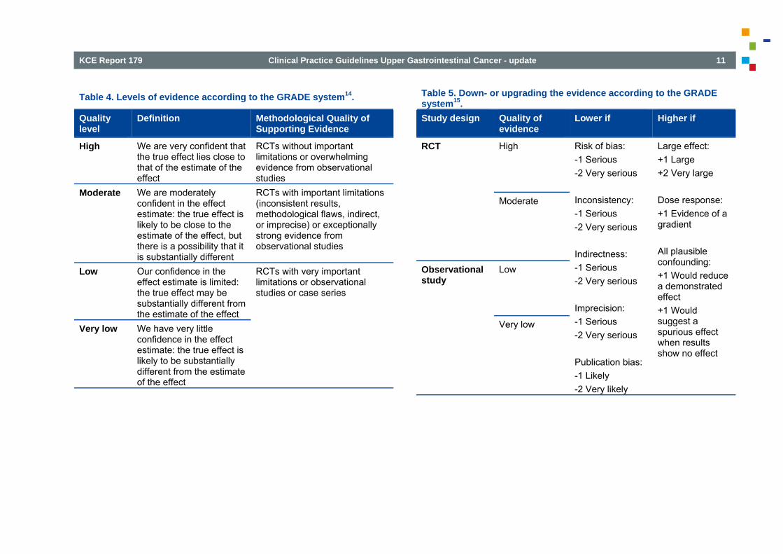
2.5. Data extraction and evidence summary Data extraction was done by one reviewer using the standard KCE template for evidence tables (see appendix 4 and 5). The evidence tables of the previous version were not copied in this version. RCTs included in eligible systematic reviews were not extracted anymore. The evidence was summarized using the previous version of the guideline as a starting point. Some parts of the previous text were adapted based on additional elements from the quality appraisal and data extraction of studies published before August 2007. For each clinical question, conclusions were formulated at the level of individual treatment outcomes. A level of evidence was assigned to each conclusion using the GRADE system14 (Table 4). The quality of evidence was down- or upgraded based on predefined criteria (Table 5).
KCE Report 179 Clinical Practice Guidelines Upper Gastrointestinal Cancer - update 11
Table 4. Levels of evidence according to the GRADE system14.
Quality level
Definition Methodological Quality of Supporting Evidence
High We are very confident that the true effect lies close to that of the estimate of the effect
RCTs without important limitations or overwhelming evidence from observational studies
Moderate We are moderately confident in the effect estimate: the true effect is likely to be close to the estimate of the effect, but there is a possibility that it is substantially different
RCTs with important limitations (inconsistent results, methodological flaws, indirect, or imprecise) or exceptionally strong evidence from observational studies
Low Our confidence in the effect estimate is limited: the true effect may be substantially different from the estimate of the effect
RCTs with very important limitations or observational studies or case series
Very low We have very little confidence in the effect estimate: the true effect is likely to be substantially different from the estimate of the effect
Table 5. Down- or upgrading the evidence according to the GRADE system15. Study design Quality of
evidence Lower if Higher if
RCT High Risk of bias: -1 Serious -2 Very serious
Inconsistency: -1 Serious -2 Very serious
Indirectness: -1 Serious -2 Very serious
Imprecision: -1 Serious -2 Very serious
Publication bias: -1 Likely -2 Very likely
Large effect: +1 Large +2 Very large
Dose response: +1 Evidence of a gradient
All plausible confounding: +1 Would reduce a demonstrated effect +1 Would suggest a spurious effect when results show no effect
Moderate
Observational study
Low
Very low
12 Clinical Practice Guidelines Upper Gastrointestinal Cancer - update KCE Report 179
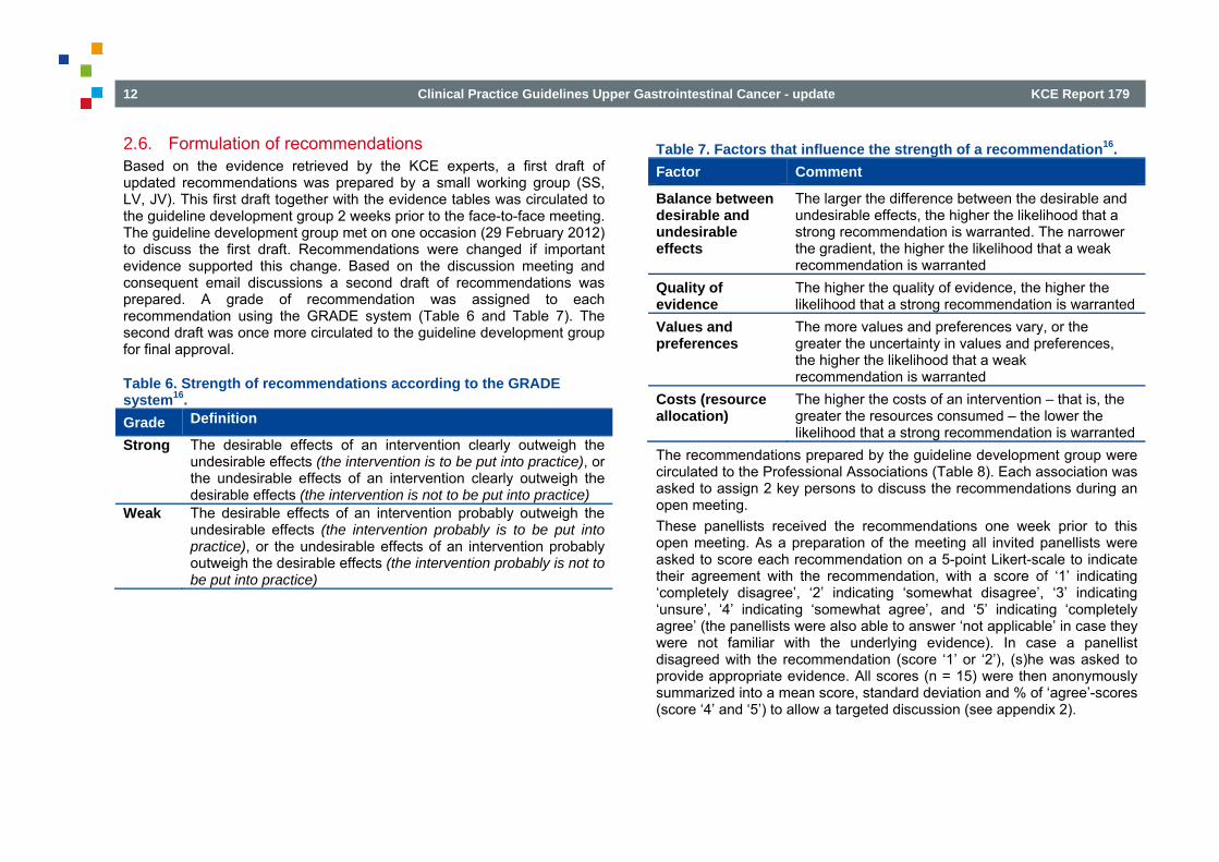
2.6. Formulation of recommendations Based on the evidence retrieved by the KCE experts, a first draft of updated recommendations was prepared by a small working group (SS, LV, JV). This first draft together with the evidence tables was circulated to the guideline development group 2 weeks prior to the face-to-face meeting. The guideline development group met on one occasion (29 February 2012) to discuss the first draft. Recommendations were changed if important evidence supported this change. Based on the discussion meeting and consequent email discussions a second draft of recommendations was prepared. A grade of recommendation was assigned to each recommendation using the GRADE system (Table 6 and Table 7). The second draft was once more circulated to the guideline development group for final approval.
Table 6. Strength of recommendations according to the GRADE system16. Grade Definition
Strong The desirable effects of an intervention clearly outweigh the undesirable effects (the intervention is to be put into practice), or the undesirable effects of an intervention clearly outweigh the desirable effects (the intervention is not to be put into practice)
Weak The desirable effects of an intervention probably outweigh the undesirable effects (the intervention probably is to be put into practice), or the undesirable effects of an intervention probably outweigh the desirable effects (the intervention probably is not to be put into practice)
Table 7. Factors that influence the strength of a recommendation16. Factor Comment
Balance between desirable and undesirable effects
The larger the difference between the desirable and undesirable effects, the higher the likelihood that a strong recommendation is warranted. The narrower the gradient, the higher the likelihood that a weak recommendation is warranted
Quality of evidence
The higher the quality of evidence, the higher the likelihood that a strong recommendation is warranted
Values and preferences
The more values and preferences vary, or the greater the uncertainty in values and preferences, the higher the likelihood that a weak recommendation is warranted
Costs (resource allocation)
The higher the costs of an intervention – that is, the greater the resources consumed – the lower the likelihood that a strong recommendation is warranted
The recommendations prepared by the guideline development group were circulated to the Professional Associations (Table 8). Each association was asked to assign 2 key persons to discuss the recommendations during an open meeting. These panellists received the recommendations one week prior to this open meeting. As a preparation of the meeting all invited panellists were asked to score each recommendation on a 5-point Likert-scale to indicate their agreement with the recommendation, with a score of ‘1’ indicating ‘completely disagree’, ‘2’ indicating ‘somewhat disagree’, ‘3’ indicating ‘unsure’, ‘4’ indicating ‘somewhat agree’, and ‘5’ indicating ‘completely agree’ (the panellists were also able to answer ‘not applicable’ in case they were not familiar with the underlying evidence). In case a panellist disagreed with the recommendation (score ‘1’ or ‘2’), (s)he was asked to provide appropriate evidence. All scores (n = 15) were then anonymously summarized into a mean score, standard deviation and % of ‘agree’-scores (score ‘4’ and ‘5’) to allow a targeted discussion (see appendix 2).
KCE Report 179 Clinical Practice Guidelines Upper Gastrointestinal Cancer - update 13
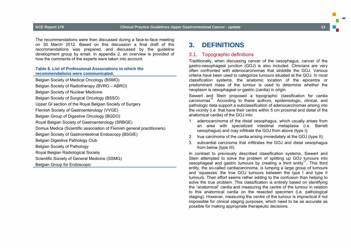
The recommendations were then discussed during a face-to-face meeting on 30 March 2012. Based on this discussion a final draft of the recommendations was prepared, and discussed by the guideline development group by email. In appendix 2, an overview is provided of how the comments of the experts were taken into account.
Table 8. List of Professional Associations to which the recommendations were communicated. Belgian Society of Medical Oncology (BSMO) Belgian Society of Radiotherapy (BVRO – ABRO) Belgian Society of Nuclear Medicine Belgian Society of Surgical Oncology (BSSO) Upper GI section of the Royal Belgian Society of Surgery Flemish Society of Gastroenterology (VVGE) Belgian Group of Digestive Oncology (BGDO) Royal Belgian Society of Gastroenterology (SRBGE) Domus Medica (Scientific association of Flemish general practitioners) Belgian Society of Gastrointestinal Endoscopy (BSGIE) Belgian Digestive Pathology Club Belgian Society of Pathology Royal Belgian Radiological Society Scientific Society of General Medicine (SSMG) Belgian Group for Endoscopic Surgery (BGES)
3. DEFINITIONS 3.1. Topographic definitions Traditionally, when discussing cancer of the oesophagus, cancer of the gastro-oesophageal junction (GOJ) is also included. Clinicians are very often confronted with adenocarcinomas that straddle the GOJ. Various criteria have been used to categorize tumours situated at the GOJ. In most classification systems, the anatomic location of the epicentre or predominant mass of the tumour is used to determine whether the neoplasm is oesophageal or gastric (cardia) in origin. Siewert and Stein proposed a topographic classification for cardia carcinomas17. According to these authors, epidemiologic, clinical, and pathologic data support a subclassification of adenocarcinomas arising into the vicinity (i.e. that have their centre within 5 cm proximal and distal of the anatomical cardia) of the GOJ into: 1. adenocarcinoma of the distal oesophagus, which usually arises from
an area with specialized intestinal metaplasia (i.e. Barrett oesophagus) and may infiltrate the GOJ from above (type I);
2. true carcinoma of the cardia arising immediately at the GOJ (type II); 3. subcardial carcinoma that infiltrates the GOJ and distal oesophagus
from below (type III). In contrast to previously described classification systems, Siewert and Stein attempted to solve the problem of splitting up GOJ tumours into oesophageal and gastric tumours by creating a third entity17. This third entity, the so-called cardiacarcinoma, is lumping a large group of tumours and ‘squeezes’ the true GOJ tumours between the type I and type II tumours. Their effort seems rather adding to the confusion than helping to solve the true problem. This classification is entirely based on identifying the “anatomical” cardia and measuring the centre of the tumour in relation to this anatomical cardia on the resected specimen (i.e. pathological staging). However, measuring the centre of the tumour is impractical if not impossible for clinical staging purposes, which need to be as accurate as possible for making appropriate therapeutic decisions.
14 Clinical Practice Guidelines Upper Gastrointestinal Cancer - update KCE Report 179
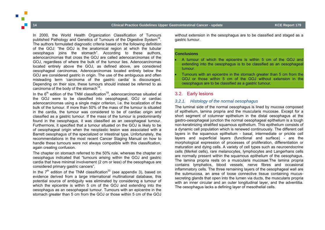
In 2000, the World Health Organization Classification of Tumours published Pathology and Genetics of Tumours of the Digestive System18. The authors formulated diagnostic criteria based on the following definition of the GOJ: “the GOJ is the anatomical region at which the tubular oesophagus joins the stomach”. According to these authors, adenocarcinomas that cross the GOJ are called adenocarcinomas of the GOJ, regardless of where the bulk of the tumour lies. Adenocarcinomas located entirely above the GOJ, as defined above, are considered oesophageal carcinomas. Adenocarcinomas located entirely below the GOJ are considered gastric in origin. The use of the ambiguous and often misleading term ‘carcinoma of the gastric cardia’ is discouraged. Depending on their size, these tumours should instead be referred to as carcinoma of the body of the stomach18. In the 6th edition of the TNM classification19, adenocarcinomas situated at the GOJ were to be classified into oesophageal, GOJ or cardiac adenocarcinomas using a single major criterion, i.e. the localization of the bulk of the tumour. If more than 50% of the mass of the tumour is situated in the cardia, the tumour was considered to be of cardiac origin and classified as a gastric tumour. If the mass of the tumour is predominantly found in the oesophagus, it was classified as an oesophageal tumour. Furthermore, it specified that a tumour situated on the GOJ is likely to be of oesophageal origin when the neoplastic lesion was associated with a Barrett oesophagus of the specialized or intestinal type. Unfortunately, the recommendations in the most recent Cancer Staging Manual on how to handle these tumours were not always compatible with this classification, again creating confusion. The chapter on stomach referred to the 50% rule, whereas the chapter on oesophagus indicated that “tumours arising within the GOJ and gastric cardia that have minimal involvement (2 cm or less) of the oesophagus are considered primary gastric cancers”. In the 7th edition of the TNM classification20 (see appendix 3), based on evidence derived from a large international multinational database, this potential source of ambiguity was eliminated by considering a tumour of which the epicentre is within 5 cm of the GOJ and extending into the oesophagus as an oesophageal tumour. Tumours with an epicentre in the stomach greater than 5 cm from the GOJ or those within 5 cm of the GOJ
without extension in the oesophagus are to be classified and staged as a gastric tumour.
Conclusions • A tumour of which the epicentre is within 5 cm of the GOJ and
extending into the oesophagus is to be classified as an oesophageal tumour.
• Tumours with an epicentre in the stomach greater than 5 cm from the GOJ or those within 5 cm of the GOJ without extension in the oesophagus are to be classified as a gastric tumour.
3.2. Early lesions 3.2.1. Histology of the normal oesophagus The luminal side of the normal oesophagus is lined by mucosa composed of epithelium, lamina propria and the muscularis mucosae. Except for a short segment of columnar epithelium in the distal oesophagus at the gastro-oesophageal junction the normal oesophageal epithelium is a tough non-keratinizing stratified squamous epithelium. This epithelium consists of a dynamic cell population which is renewed continuously. The different cell layers in the squamous epithelium - basal, intermediate or prickle cell layers and superficial layers (functional and surface) - are the morphological expression of processes of proliferation, differentiation or maturation and dying cells. A variety of cell types such as neuroendocrine cells (Merkel cells), rare melanocytes, lymphocytes and Langerhans cells are normally present within the squamous epithelium of the oesophagus. The lamina propria rests on a muscularis mucosae.The lamina propria contains lymphatics, blood vessels, nerve fibres and occasional inflammatory cells. The three remaining layers of the oesophageal wall are the submucosa, an area of loose connective tissue containing mucus-secreting glands that open into the lumen via ducts, the muscularis propria with an inner circular and an outer longitudinal layer, and the adventitia. The oesophagus lacks a defining layer of mesothelial cells.
KCE Report 179 Clinical Practice Guidelines Upper Gastrointestinal Cancer - update 15
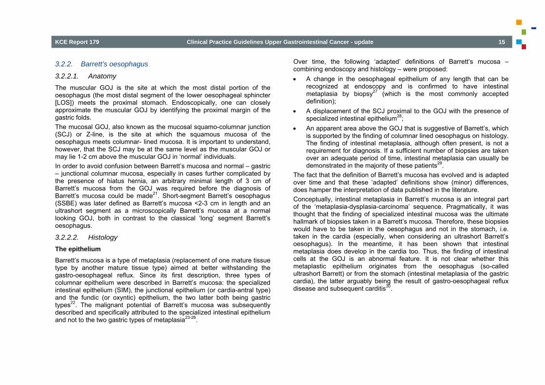
3.2.2. Barrett’s oesophagus
3.2.2.1. Anatomy The muscular GOJ is the site at which the most distal portion of the oesophagus (the most distal segment of the lower oesophageal sphincter [LOS]) meets the proximal stomach. Endoscopically, one can closely approximate the muscular GOJ by identifying the proximal margin of the gastric folds. The mucosal GOJ, also known as the mucosal squamo-columnar junction (SCJ) or Z-line, is the site at which the squamous mucosa of the oesophagus meets columnar- lined mucosa. It is important to understand, however, that the SCJ may be at the same level as the muscular GOJ or may lie 1-2 cm above the muscular GOJ in ‘normal’ individuals. In order to avoid confusion between Barrett’s mucosa and normal – gastric – junctional columnar mucosa, especially in cases further complicated by the presence of hiatus hernia, an arbitrary minimal length of 3 cm of Barrett’s mucosa from the GOJ was required before the diagnosis of Barrett’s mucosa could be made21. Short-segment Barrett’s oesophagus (SSBE) was later defined as Barrett’s mucosa <2-3 cm in length and an ultrashort segment as a microscopically Barrett’s mucosa at a normal looking GOJ, both in contrast to the classical ‘long’ segment Barrett’s oesophagus.
3.2.2.2. Histology The epithelium Barrett’s mucosa is a type of metaplasia (replacement of one mature tissue type by another mature tissue type) aimed at better withstanding the gastro-oesophageal reflux. Since its first description, three types of columnar epithelium were described in Barrett’s mucosa: the specialized intestinal epithelium (SIM), the junctional epithelium (or cardia-antral type) and the fundic (or oxyntic) epithelium, the two latter both being gastric types22. The malignant potential of Barrett’s mucosa was subsequently described and specifically attributed to the specialized intestinal epithelium and not to the two gastric types of metaplasia23-26.
Over time, the following ‘adapted’ definitions of Barrett’s mucosa – combining endoscopy and histology – were proposed: • A change in the oesophageal epithelium of any length that can be
recognized at endoscopy and is confirmed to have intestinal metaplasia by biopsy27 (which is the most commonly accepted definition);
• A displacement of the SCJ proximal to the GOJ with the presence of specialized intestinal epithelium28;
• An apparent area above the GOJ that is suggestive of Barrett’s, which is supported by the finding of columnar lined oesophagus on histology. The finding of intestinal metaplasia, although often present, is not a requirement for diagnosis. If a sufficient number of biopsies are taken over an adequate period of time, intestinal metaplasia can usually be demonstrated in the majority of these patients29.
The fact that the definition of Barrett’s mucosa has evolved and is adapted over time and that these ‘adapted’ definitions show (minor) differences, does hamper the interpretation of data published in the literature. Conceptually, intestinal metaplasia in Barrett’s mucosa is an integral part of the ‘metaplasia-dysplasia-carcinoma’ sequence. Pragmatically, it was thought that the finding of specialized intestinal mucosa was the ultimate hallmark of biopsies taken in a Barrett’s mucosa. Therefore, these biopsies would have to be taken in the oesophagus and not in the stomach, i.e. taken in the cardia (especially, when considering an ultrashort Barrett’s oesophagus). In the meantime, it has been shown that intestinal metaplasia does develop in the cardia too. Thus, the finding of intestinal cells at the GOJ is an abnormal feature. It is not clear whether this metaplastic epithelium originates from the oesophagus (so-called ultrashort Barrett) or from the stomach (intestinal metaplasia of the gastric cardia), the latter arguably being the result of gastro-oesophageal reflux disease and subsequent carditis30.
16 Clinical Practice Guidelines Upper Gastrointestinal Cancer - update KCE Report 179
The muscularis mucosae Patients with Barrett’s oesophagus often develop a new (superficial), more luminally situated, layer of muscularis mucosae31. Important to know is that endoscopic biopsies may contain some limited fragments of muscle. These may originate from a newly formed muscularis mucosae or from the original muscularis mucosae. However, light microscopic distinction towards origin is impossible. The ‘double’ muscularis mucosae can only be visualized in (good quality) endoscopic mucosal resection (EMR) and operation specimens. Only invasion through the original, deeper muscularis mucosae is defined as “submucosal” invasion32,33.
3.2.3. Dysplasia in squamous epithelium In squamous epithelium dysplasia is classified as mild, moderate, or severe. With increasing grades of dysplasia there is a progressive increase in dysplastic cells from the basal layer onwards until the entire thickness of the epithelium is replaced. The latter state is described as carcinoma in situ34. When dysplastic cells reach the luminal aspect of the epithelium without cytoplasmic maturation the term carcinoma in situ can be used. Some authors use a simplified classification for non-invasive squamous neoplastic lesions: low-grade intraepithelial neoplasia, which includes mild and moderate dysplasia, and high-grade intraepithelial neoplasia, which includes severe dysplasia and carcinoma in situ. By definition, the basement membrane is intact in dysplasia. However, the junction of the epithelium with the underlying stroma may become irregular35.
3.2.4. Dysplasia in columnar epithelium From a biological point of view, the progression of precursors and precursor lesions into carcinomas is driven by the evolution and proliferation of clones of cells with accumulated genetic errors related to control of cell proliferation, intercellular adhesion, tumour suppression, etc., resulting in genomic instability. Dysplasia is defined as ‘an unequivocal neoplastic epithelium confined within its basement membrane36. Furthermore, dysplasia has the potential to progress into invasive malignancy. Although morphological detection in mucosal biopsy specimens is still the best method of detecting patients at risk of developing cancer, it has its limitations:
Intra-observer and inter-observer variability Numerous articles have been published on this topic demonstrating specific areas of discordance at the lower and higher end of the spectrum of early lesions. There is a significant degree of intra-observer and inter-observer variability in the diagnosis of dysplasia (unequivocal neoplastic atypia) versus reactive atypia even among experienced gastrointestinal pathologists37-39. Similarly, it has become apparent, especially from comparative studies between Western and Japanese pathologists, that the differential diagnosis between high-grade dysplasia, carcinoma in situ and intramucosal carcinoma is prone to intra-observer and inter-observer variability40-42.
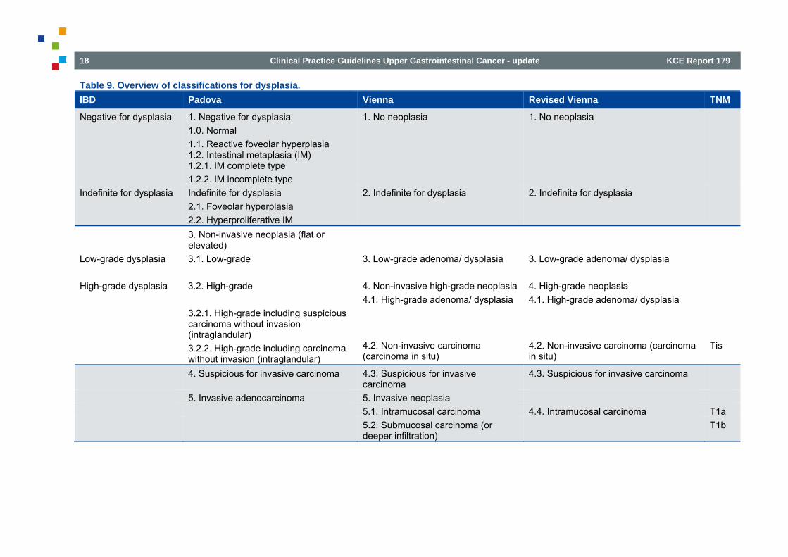
Classifications Diagnosis of dysplasia is based on the detection of morphological changes. According to the severity of histological changes, dysplasia has been graded using either a three tier system or a two tier system (Table 9). The changes were initially described as mild, moderate and severe dysplasia (morphological classification, 3-tier). In 1983, the Inflammatory Bowel Disease (IBD) study group classified dysplasia as negative, indefinite or positive, i.e. low and high-grade (clinical classification, 2-tier: low-grade = mild and moderate dysplasia; high-grade = severe dysplasia) (Table 9)36.
KCE Report 179 Clinical Practice Guidelines Upper Gastrointestinal Cancer - update 17
Grading in a two tier system would seem to be easier and more reproducible, and moreover to correlate with clinical implications. In the US and Western Europe, there was a general agreement that this clinically based classification was applicable to neoplastic changes in Barrett’s mucosa too39,43,44. In 2000, three new classifications for gastrointestinal dysplasia have been proposed: the Padova classification (gastric)45, the Vienna classification46 and the revised Vienna classification47 (Table 9). These new classifications aimed at: 1) changing the terminology used, i.e. replacing dysplasia by (intra)epithelial neoplasia; 2) including not only dysplasia but also (invasive) carcinomas; and 3) distinguishing ‘mucosal high-grade neoplasia’ into high-grade dysplasia, suspicion for non-invasive or invasive carcinoma, non-invasive carcinoma, intramucosal carcinoma and carcinoma invading submucosa or beyond. The major differences amongst these classifications reside in the subcategories included/grouped and the figures attributed. Comparative studies using the new classifications have shown an improvement of the inter-observer variability especially between the Western and Japanese pathologists40-42,48. Finally, a revision of the WHO classification of tumours of the digestive system was published at the end of 200049. The latter introduced ‘high-grade intraepithelial neoplasia’ (including severe dysplasia and carcinoma in situ), but did not recommend one or another of the previously mentioned classifications. Up till now, these new classifications have not yet gained widespread acceptance29,50. Moreover, the authors of the Vienna classification stressed that the subdivisions related to ‘mucosal high-grade neoplasia’ (high-grade dysplasia, suspicion for invasive carcinoma, non-invasive carcinoma and intramucosal carcinoma) may be important for research purposes and may not be needed for clinical purposes46. Moreover, for resection specimens, only specific histological diagnoses should be given. Group classifications such as ‘mucosal high-grade neoplasia’ should not be used48.
In conclusion, especially concerning the higher end of the spectrum of early lesions and in view of the importance of a multidisciplinary approach, it is important for a pathologist to have a clear understanding of the particular treatment regimens available and to be applied under particular circumstances. Classification is a chosen arrangement of elements in relation to a purpose. For the physician, a classification should be clinically relevant. Currently, the main clinical options are no follow-up, follow-up, local treatment by endoscopy, minimally invasive (laparoscopic) surgery and extensive surgery including lymph node dissection (see below). Many studies have evaluated the potential utility of immunohistochemical or molecular markers as additional techniques in detecting dysplasia, however with limited success. Additional confirmation by an expert pathologist (second opinion) is advocated, especially when therapeutic intervention is considered29. Today, the new classifications should be taken for what they are, i.e. attempts at an international level to reach consensus on histological diagnosis of dysplasia in ‘chosen arrangements of elements’ which may eventually generate guidelines for the development of diagnostic and management strategies.
18 Clinical Practice Guidelines Upper Gastrointestinal Cancer - update KCE Report 179
Table 9. Overview of classifications for dysplasia. IBD Padova Vienna Revised Vienna TNM
Negative for dysplasia 1. Negative for dysplasia 1.0. Normal 1.1. Reactive foveolar hyperplasia 1.2. Intestinal metaplasia (IM) 1.2.1. IM complete type 1.2.2. IM incomplete type
1. No neoplasia 1. No neoplasia
Indefinite for dysplasia Indefinite for dysplasia 2.1. Foveolar hyperplasia 2.2. Hyperproliferative IM
2. Indefinite for dysplasia 2. Indefinite for dysplasia
3. Non-invasive neoplasia (flat or elevated)
Low-grade dysplasia 3.1. Low-grade 3. Low-grade adenoma/ dysplasia 3. Low-grade adenoma/ dysplasia
High-grade dysplasia 3.2. High-grade 4. Non-invasive high-grade neoplasia 4. High-grade neoplasia 4.1. High-grade adenoma/ dysplasia 4.1. High-grade adenoma/ dysplasia 3.2.1. High-grade including suspicious
carcinoma without invasion (intraglandular)
3.2.2. High-grade including carcinoma without invasion (intraglandular)
4.2. Non-invasive carcinoma (carcinoma in situ)
4.2. Non-invasive carcinoma (carcinoma in situ)
Tis
4. Suspicious for invasive carcinoma 4.3. Suspicious for invasive carcinoma
4.3. Suspicious for invasive carcinoma
5. Invasive adenocarcinoma 5. Invasive neoplasia 5.1. Intramucosal carcinoma 4.4. Intramucosal carcinoma T1a 5.2. Submucosal carcinoma (or
deeper infiltration) T1b
KCE Report 179 Clinical Practice Guidelines Upper Gastrointestinal Cancer - update 19
Conclusions • There is no consensus about the definition of Barrett’s oesophagus. • Several classifications are available for dysplasia. For the physician,
the used classification should be clinically relevant.
3.3. Early versus locally-advanced invasive disease As for many other cancer types, the literature often makes a distinction between early and locally-advanced disease. However, definitions of early and locally-advanced cancer are not uniform and controversial. Therefore, to avoid discussion, an attempt will be made to define the eligible population for our recommendations as accurate as possible. These definitions will be re-evaluated as part of the quality project that builds on the present guidelines and that will be published as a separate report.
4. CLINICAL RECOMMENDATIONS FOR OESOPHAGEAL CANCER
4.1. Flowchart
20 Clinical Practice Guidelines Upper Gastrointestinal Cancer - update KCE Report 179
4.2. Staging 4.2.1. Computed tomography The main contribution of CT scan to the staging of oesophageal cancer is the detection of distant metastases and gross invasion of adjacent structures/organs51-53. If metastatic disease is detected with computed tomography (CT), curative treatment is excluded and additional staging with endoscopic ultrasonography (EUS) and/or positron-emission tomography (PET) is unnecessary. Lowe et al. found a sensitivity and specificity of 81% and 82% respectively for the detection of distant metastasis in 69 patients with newly diagnosed oesophageal cancer53. Differences with EUS (Se 73%, Sp 86%) and PET (Se 81%, Sp 91%) were not statistically significant (p>0.25). CT scan has also an acceptable diagnostic accuracy for locoregional staging (T and N), although EUS is clearly more sensitive (see below)51,52. In 3 prospective studies, the sensitivity for nodal staging ranged from 79-84%, while the specificity ranged from 58-86%53-55. Two of these studies had partial verification, and blinded evaluation was not applied54,55. CT scan of the chest and abdomen should be performed routinely with intravenous contrast and gastric distension with oral contrast or water. The liver should at least be imaged in the arterial and portal venous phase52. Slice thickness should be 5 mm or less51. Given the high incidence of cervical lymph node metastasis56, CT scan should also include the cervical region. Neck imaging with ultrasonography or CT enables the detection of metastatic lymph nodes that are clinically not palpable52 or can be used to guide fine needle aspiration of suspicious lymph nodes with very high accuracy and specificity51,57.
Update In a systematic review, Van Vliet et al.58 compared the diagnostic performance of EUS, CT and PET for the staging of oesophageal cancer. Seventeen articles on CT for regional lymph node metastases (n=943 patients), 5 papers on other abdominal lymph node metastases (n=254 patients) and 7 articles on distant metastases (n=437 patients) were included. Pooled sensitivity and specificity of CT for the detection of regional lymph node metastases were 50% (95%CI 41–60%) and 83% (95%CI 77–89%), respectively. For the detection of abdominal lymph node
metastases, sensitivity and specificity were 42% (95%CI 29–54%) and 93% (95%CI 86–100%), respectively. For the detection of distant metastases, sensitivity and specificity were 52% (95%CI 33%–71%) and 91% (95%CI 86%–96%), respectively. The Canadian Governmental Agency AETMIS59 conducted a similar systematic review (with an important overlap in included studies) on the performance of the diagnostic methods used for staging oesophageal cancer, reporting results in similar ranges. Most of the studies included in the SR used abdominal and thoracic CT. The rate of correctly staged tumours (stage T) was fairly low with spiral CT (median 61.8%). Spiral CT did not perform very well in detecting positive lymph nodes (Se 50%; 95%CI 41-60%), because lymph node assessment relied only on lymph node size, and the malignancy threshold was generally a short-axis diameter of 10 mm. However, its specificity was moderate (Sp 83%; 95%CI 77-89%). Its sensitivity for diagnosing distant lymph node metastases and distant organ metastases was also low (median 49.2%), but its specificity was moderate (median 87.2%).The low sensitivity can be explained because the selected studies did not separately evaluate metastases in distant organs and distant lymph nodes, and several of the studies likely had a selection bias in favour of cases at earlier stages. Moreover, most of the studies dealt with single-slice spiral CT or multi-detector spiral CT technology, whereas the latest generation of scanners (64- and 256-slice) was not evaluated. Kato et al.60 tested the performance of CT for the evaluation of initial lymph node staging in 117 patients with thoracic oesophageal squamous cell carcinoma obtaining similar results (Se 48.3%, Sp 73.7%). Two recent observational studies were conducted to test the accuracy of multidetector CT (MD-CT)61,62. One study61 only focused on T staging in 131 patients, obtaining a high sensitivity (≥95%) and a high positive predictive value (97%) but a low specificity (≤ 50%) and a low negative predictive value (<45%). Schreurs et al.62 tested different staging modalities to detect cervical metastases in 125 patients with cancer of the oesophagus and gastro-oesophageal junction. MD-CT had a low sensitivity (71%), but a high specificity (100%). However, this study carried a high risk of bias (partial verification and interpretation bias).
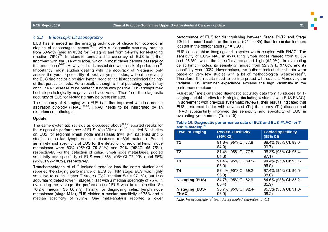
KCE Report 179 Clinical Practice Guidelines Upper Gastrointestinal Cancer - update 21
4.2.2. Endoscopic ultrasonography EUS has emerged as the imaging technique of choice for locoregional staging of oesophageal cancer51,52, with a diagnostic accuracy ranging from 53-94% (median 83%) for T-staging and from 54-94% for N-staging (median 76%)63. In stenotic tumours, the accuracy of EUS is further improved with the use of dilation, which in most cases permits passage of the endoscope64,65. However, this is associated with a risk of perforation66. Importantly, most studies dealing with the accuracy of N-staging only assess the yes-no possibility of positive lymph nodes, without correlating the EUS findings of a positive lymph node to the histopathological findings of that particular node. As a result, although a final pathology report may conclude N1 disease to be present, a node with positive EUS findings may be histopathologically negative and vice versa. Therefore, the diagnostic accuracy of EUS for N-staging may be overestimated. The accuracy of N staging with EUS is further improved with fine needle aspiration cytology (FNAC)51,52. FNAC needs to be interpreted by an experienced pathologist.
Update The same systematic reviews as discussed above58,59 reported results for the diagnostic performance of EUS. Van Vliet et al.58 included 31 studies on EUS for regional lymph node metastases (n=1 841 patients) and 5 studies on celiac lymph nodes metastases (n=339 patients). Pooled sensitivity and specificity of EUS for the detection of regional lymph node metastases were 80% (95%CI 75–84%) and 70% (95%CI 65–75%), respectively. For the detection of celiac lymph node metastases, pooled sensitivity and specificity of EUS were 85% (95%CI 72–99%) and 96% (95%CI 92–100%), respectively. Tranchemontagne et al.59 included more or less the same studies and reported the staging performance of EUS by TNM stage. EUS was highly sensitive to detect higher T stages (T≥2; median Se = 97.1%), but less accurate to detect lower T stages (T≤1) with a median specificity of 75%. In evaluating the N-stage, the performance of EUS was limited (median Se 76.2%; median Sp 66.7%). Finally, for diagnosing celiac lymph node metastases (stage M1a), EUS yielded a median sensitivity of 75% and a median specificity of 93.7%. One meta-analysis reported a lower
performance of EUS for distinguishing between Stage T1/T2 and Stage T3/T4 tumours located in the cardia (Q* = 0.85) than for similar tumours located in the oesophagus (Q* = 0.90). EUS can combine imaging and biopsies when coupled with FNAC. The sensitivity of EUS-FNAC in evaluating lymph nodes ranged from 83.3% and 93.3%, while the specificity remained high (92.9%). In evaluating celiac lymph nodes, its sensitivity ranged from 92.9% to 97.8%, and its specificity was 100%. Nevertheless, the authors indicated that data were based on very few studies with a lot of methodological weaknesses59. Therefore, the results need to be interpreted with caution. Moreover, the variable EUS examiner experience explains the high variability in the performance outcomes. Puli et al.67 meta-analyzed diagnostic accuracy data from 43 studies for T-staging and 44 studies for N-staging (including 4 studies with EUS-FNAC). In agreement with previous systematic reviews, their results indicated that EUS performed better with advanced (T4) than early (T1) disease and FNAC substantially improved the sensitivity and specificity of EUS in evaluating lymph nodes (Table 10).
Table 10. Diagnostic performance data of EUS and EUS-FNAC for T- and N-staging 67. Level of staging Pooled sensitivity
(95% CI) Pooled specificity (95% CI)
T1 81.6% (95% CI: 77.8-84.9)
99.4% (95% CI: 99.0-99.7)
T2 81.4% (95% CI: 77.5-84.8)
96.3% (95% CI: 95.4-97.1)
T3 91.4% (95% CI: 89.5-93.0)
94.4% (95% CI: 93.1-95.5)
T4 92.4% (95% CI: 89.2-95.0)
97.4% (95% CI: 96.6-98.0)
N staging (EUS) 84.7% (95% CI: 82.9-86.4)
84.6% (95% CI: 83.2-85.9)
N staging (EUS-FNAC)
96.7% (95% CI: 92.4-98.9)
95.5% (95% CI: 91.0-98.2)
Note. Heterogeneity (χ2 test ) for all pooled estimates: p>0.1

22 Clinical Practice Guidelines Upper Gastrointestinal Cancer - update KCE Report 179
A recent meta-analysis68 tested the diagnostic accuracy of EUS in differentiating mucosal versus submucosal invasion of superficial oesophageal cancers by pooling results from 19 international studies involving 1 019 patients with superficial oesophageal cancer. The pooled sensitivity and specificity of EUS for T1a staging were 85% (95%CI 82-88%) and 87% (95%CI 84-90%), respectively. For T1b staging, these results were 86% (95%CI 82-89%) and 86% (95%CI 83-89%), respectively. The area under the curve was at least 0.93 for both mucosal and submucosal lesions. The authors reported that the high heterogeneity between studies could be explained by several factors including operator experience and volume, EUS technology, cancer type, and location of lesion. Recent observational studies were conducted to test the performance of EUS compared or not with other imaging modalities (PET or PET/CT). Yen et al.69 obtained a higher sensitivity (>92%) in staging T3 cancers than lower T stages at the cost of a very low specificity. In assessing the N-stage, the sensitivity of EUS remained high (82-100%) with a low specificity (29-45%). This retrospective study included 118 consecutive patients, but the analysis by stage considerably reduced the sample size in each group. Similarly, Smith et al.70 reported a higher sensitivity for T3 patients (88%) than for other T stages (54-63%) and a moderate sensitivity but low specificity for N-staging (Se 84%, Sp 67%). Choi et al. 71 reported a lower sensitivity for N-staging (Se 42%), but this study only enrolled surgically resectable patients, leading to a possible underestimation of both sensitivity and accuracy in the detection of metastasis to lymph nodes. Mennigen et al.72 compared the staging performance of conventional probe EUS (in nonstenotic tumours) with miniprobe EUS (in stenotic tumours) and found comparable accuracies for T- (75.4% vs. 70.0%, p=0.64) and N-staging (68.4% vs. 80%, p=0.25). Dilatation therapy to allow the passage of a conventional EUS probe, carrying a significant risk of oesophageal perforation, should therefore be avoided. However, the EUS miniprobe with a much smaller diameter and higher frequency than the conventional EUS probe is technically limited by its range and might be unable to fully image large tumours or regional lymph node involvement.
4.2.3. Positron-emission tomography In the absence of clear metastatic disease on conventional imaging (CT, EUS, cervical US), PET(/CT) may provide valuable additional information, in particular when considering multimodality treatment with curative option. In such cases, baseline PET(/CT) allows better response assessment as compared to conventional CT/EUS/cervical US. FNCLCC made specific recommendations on the use of PET scan in the diagnosis and staging of oesophageal cancer73. In addition, in 2005, the KCE published an HTA report on the use of PET scan74. Since this HTA report, numerous observational studies of variable quality have been published75-82. For initial staging of patients without apparent metastases on CT scan, especially in locally advanced disease (cT>2 or cN1 or cN doubtful), PET(/CT) is useful for detecting positive lymph nodes and distant metastatic disease74. However, in early-stage disease (cT1-2N0) the probability that PET(/CT) will significantly upstage disease is low.
Update No more recent study was included in van Vliet et al.58 nor in Tranchemontagne et al.59. In the systematic review of van Vliet et al.58, the diagnostic performance of PET was tested (1) for regional lymph nodes metastases (10 studies; n=424 patients) with a low pooled sensitivity (57%; 95%CI 43%-70%) but a moderate pooled specificity (85%; 95%CI 76%-95%); and (2) for distant metastases (9 studies; n=475 patients) with a low pooled sensitivity (71%; 95%CI 62%-79%) and a high pooled specificity (93%; 95%CI 89%-97%). Tranchemontagne et al.59 separately analyzed PET from PET/CT. Data on PET were derived from systematic reviews and meta-analyses. The sensitivity of PET for N-stage was 57% (95%CI 43-70%), while the specificity reached 85% (95%CI 76-95%). However, PET performed better in evaluating distant lymph node and organ metastases. Sensitivity was 71% (95%CI 62-79%) and specificity 93% (95%CI 89-97%). Data on PET/CT for initial staging of oesophageal cancer were only based on two primary studies, among which one study included patients who had received neoadjuvant therapy. For assessing locoregional lymph nodes and Stage M1a, PET/CT had a sensitivity ranging between 83.3% and 93.9%, while specificity was as high as 92.1%.

KCE Report 179 Clinical Practice Guidelines Upper Gastrointestinal Cancer - update 23
PET/CT was very sensitive (100%) in evaluating distant metastases, but there were only four patients presenting with Stage M1 in the analyzed studies. Noble et al.83 reviewed the impact of PET/CT on 191 patients thought to be candidates for curative treatment based on CT/EUS. The performance of PET/CT was good (Se 91%, Sp 94%). PET/CT suggested distant metastases in 16% of patients and these metastases were confirmed in 9%. Kato et al.60 compared the performance of PET/CT (50 patients) with PET alone (117 patients) for initial lymph node staging. The sensitivity and accuracy of PET/CT were significantly higher (75.9% and 78%) than that of PET (55% and 70.1%). Hsu et al.84 reported a lower sensitivity to detect both regional (Se 57.1%) and non-regional nodal involvement (Se 36.4%), but a moderate specificity (83.3% and 84%). This study had a smaller sample size (45 patients) and carried risk of interpretation bias. Choi et al. 71 also tested the performance of PET/CT for N- and M-staging in 109 patients, reporting low sensitivities (49% and 40% respectively), but moderate to high specificities (87% and 99% respectively). As this study only enrolled surgically resectable patients, the underestimation of the sensitivity in the detection of lymph node metastasis is a potential bias. The choice of an adequate diagnostic criteria (SUVmax≥2.5) seems to be crucial to interpret results from PET/CT85.
Other considerations FNAC can induce false positive results on PET scan.
4.2.4. Magnetic resonance imaging Magnetic resonance imaging (MRI) was not found to be superior to conventional imaging techniques such as CT scan 51,52. Therefore, MRI should be reserved for those patients who cannot undergo CT or used for additional investigation following CT and/or EUS.
Update No more recent studies were identified to evaluate the performance of MRI to stage oesophageal cancer.
4.2.5. Bronchoscopy and bronchoscopic ultrasound In patients with clinical or imaging features suspicious of tracheobronchial invasion (e.g. cough aggravated by swallowing in case of a fistula) or extrinsic compression, bronchoscopy combined with bronchoscopic ultrasound (BUS) and/or biopsy can be useful52,86,87. The underlying evidence, however, is weak to very weak. Observational data also provide weak evidence for the occurrence of synchronous neoplasms of the bronchial tree in patients with squamous cell carcinoma of the oropharynx and the oesophagus, which is an indication for bronchoscopy88 or even a pan-endoscopy of the entire aerodigestive tract (including the ear-nose-throat area). For the latter intervention, no evidence is available.
Update No more recent studies were identified to evaluate the performance of bronchoscopy and BUS to stage oesophageal cancer.
4.2.6. Thoracoscopy and laparoscopy Few studies are available to support the use of invasive staging with thoracoscopy and laparoscopy (+/- peritoneal cytology and/or laparoscopic ultrasound)51,52,89. Thoracoscopy can be useful for the detection of positive mediastinal lymph nodes, while laparoscopy can be considered in patients with oesophageal tumours with a gastric component52. Laparoscopy has a higher specificity for M staging in comparison with CT scan, but obviously carries a higher risk of morbidity51.
Update No more recent studies were identified to evaluate the performance of thoracoscopy and laparoscopy to stage oesophageal cancer.

24 Clinical Practice Guidelines Upper Gastrointestinal Cancer - update KCE Report 179
Conclusions • The rate of correctly staged tumours (T-stage) is higher with EUS than
with CT, but the latest generation of scanners (64- and 256-slice) were poorly evaluated (low level of evidence; Tranchemontagne 2009).
• For the detection of regional lymph node metastases, EUS is most sensitive, whereas CT and PET are more specific tests. The examiner experience explains the high variability in the performance outcomes obtained with EUS. FNA substantially improves the sensitivity and specificity of EUS in evaluating the N-stage (low level of evidence; Tranchemontagne 2009).
• For the evaluation of distant metastases, PET has probably a higher sensitivity than CT. Combining these two modalities could be of clinical value, with PET detecting possible metastases and CT confirming or excluding their presence and precisely determining the location(s). The high specificity of PET/CT is useful to exclude positive non-peritumoural lymph nodes and metastasis (low level of evidence; Tranchemontagne 2009).
• Laparoscopy has a higher specificity for M-staging in comparison with CT, but carries a higher risk of morbidity (low level of evidence; CBO 2005). The available evidence does not allow to conclude about the clinical value of thoracoscopy.
• Few studies are available on the diagnostic accuracy of MRI for the staging of oesophageal cancer (low level of evidence; SIGN 2006, CBO 2005).
Other considerations Multidisciplinary team meetings (MDT) have been implemented in many countries as the predominant model of cancer care to ensure that all patients receive timely diagnosis and treatment, that patient management is evidence-based, and that there is continuity of care90. The positive impact of multidisciplinary team care in the management of oesophageal cancer was reported at least in two publications from UK91,92. Stephens reported that multidisciplinary team management resulted in improved staging, lower operative mortality, and improved 5-year survival when
compared to a group of patients undergoing R0 resection by surgeons who were working independently. Davies concluded that MDT significantly improved staging accuracy for gastro-oesophageal cancer and ensured that correct management decisions were made for the majority of patients. Moreover, multidisciplinary care tend to enable the construction of clinical pathways and to develop formal programs with a unified vision for therapy and palliation93. Such MDT have to be encouraged and generalized in the management of patients with oesophageal cancer.
Recommendations • All patients diagnosed with oesophageal cancer should be
discussed at a multidisciplinary meeting (strong recommendation, low level of evidence).
• In patients with newly diagnosed oesophageal cancer, CT scan of the neck (including lower neck region), thorax and abdomen should always be performed (strong recommendation, low level of evidence).
• Endoscopic ultrasonography (EUS), combined with fine needle aspiration cytology (FNAC) if technically feasible, should be considered to evaluate locoregional invasion (T and N stage) and presence of positive celiac lymph nodes in patients with oesophageal cancer (strong recommendation, low level of evidence).
• PET/CT should be considered for M staging if a patient with T2-4 N+ oesophageal cancer is a candidate for a curative treatment after CT and EUS (strong recommendation, low level of evidence).
• The following examinations can be considered for specific indications: MRI, bronchoscopy +/- bronchial ultrasonography (BUS) +/- biopsy, thoracoscopy, or laparoscopy (weak recommendation, low level of evidence).
KCE Report 179 Clinical Practice Guidelines Upper Gastrointestinal Cancer - update 25
Good clinical practice • Multi-detector, multi-planar reformatted CT scan should be
performed with IV contrast. The liver should at least be imaged in the arterial and portal venous phase.
4.3. Treatment of mucosal cancer The principles of anatomopathological evaluation of mucosal cancer are similar to those of high-grade dysplasia, including validation of the diagnosis by a second pathologist, additional biopsies or diagnostic EMR in case of uncertainty, and discussion of treatment options at the MDT52. Only observational studies are available comparing endoscopic mucosal resection (EMR) to surgery for the treatment of superficial oesophageal cancer51. Both treatments were found to be equally effective, but EMR was associated with less complications. Therefore, according to SIGN and CBO, superficial oesophageal cancer limited to the mucosa should be treated with EMR, taking into account the stage, size, length of Barrett, histological type, differentiation grade, and lymphovascular invasion51,52. For example, in case of mucosal cancer in a long Barrett’s segment, complete resection of the Barrett’s area with EMR is unlikely. The same is true for patients with multiple early squamous cancers. In these cases, surgery (e.g. gastric pull-up or vagal sparing oesophagectomy with colon interposition) may remain the treatment of choice. En-bloc resection – allowing better pathology staging of deep and lateral margins – should be aimed at with the appropriate technique (EMR for lesions less than 12 mm)94,95. Mucosal ablative techniques, such as argon plasma coagulation (APC), photodynamic therapy (PDT) or laser, are investigational51,52. Only small observational studies showed the benefits of these techniques, and they should therefore be limited to units with appropriate expertise. Moreover, APC was associated with buried neoplastic glands and should not be considered for curative treatment. Importantly, PDT is associated with a risk of stricture formation in up to 30% of patients96. Above this, mucosal ablative techniques make an anatomopathological control of the staging results impossible.
Update A systematic review of different endoscopic and non-endoscopic approaches was published by McCann et al. in 201197. Treatment approaches that were evaluated included ablative approaches (PDT, radiofrequency ablation [RFA], APC and cryotherapy) and non-ablative approaches (chemotherapy, RT, CRT, EMR, endoscopic submucosal dissection [ESD] and oesophagectomy). Initially, this review was performed for patients with early oesophageal cancer (SCC/ACC, stages 0-IIA without lymph nodes involvement). However, the majority of the 75 included studies on ablative techniques included patients ineligible for surgery, only 20% of included studies were comparative and half of all studies included less than 20 patients. Some patients also received additional treatments following treatment failure with endoscopic approach (PDT or EMR). Meta-analysis of results could not be performed, since multiple studies of the same treatment were unavailable. While some studies presented results according to disease stage, many others presented only summary values, grouping all patients whatever their cancer stage together. Safety and efficacy of all techniques were evaluated. Adverse events reported across studies of endoscopic techniques were similar and less significant compared to those in the studies of non-endoscopic techniques. These were mainly stricture (13%) and chest pain, nausea and vomiting (50%) after PDT, according to the photosensitizing agent used, and bleeding (10%) and stenosis (6%) in patients who underwent EMR. Complete response rates were slightly lower for PDT (54% in ACC and 71% in SCC) relative to EMR (98% in ACC and 88% in SCC), possibly due to differences in patient populations across studies. No studies compared overall or cause-specific survival in patients who received endoscopic treatments vs. those who received non-endoscopic treatments.
26 Clinical Practice Guidelines Upper Gastrointestinal Cancer - update KCE Report 179
Conclusions • Despite low quality evidence, endoscopic treatments seem to reduce
the morbidity and mortality associated with oesophagectomy. However, there is no evidence demonstrating the superiority of one particular endoscopic treatment (low level of evidence; McCann 2011).
• In patients with superficial oesophageal cancer (T1a), treatment with endoscopic mucosal resection is equally effective as surgery, but associated with less complications (low level of evidence; SIGN 2006, CBO 2005).
• The clinical effectiveness of mucosal ablative techniques in patients with superficial oesophageal cancer is insufficiently proven (low level of evidence; SIGN 2006, CBO 2005).
Recommendations • Endoscopic mucosal resection (EMR) should be performed
whenever possible for a T1a oesophageal cancer, aiming at staging and curative resection. If the staging and R0 resection is pathologically confirmed, this procedure can be considered therapeutic, taking into account other well-defined criteria relating to size, length of Barrett, histological type, differentiation grade and lymphovascular invasion. In case the staging and R0 resection is not confirmed, surgery can be considered (strong recommendation, low level of evidence).
• (Destructive) mucosal ablative techniques cannot be recommended as a curative option for patients with T1a oesophageal cancer and should be limited to centres with appropriate expertise (strong recommendation, low level of evidence).
Good clinical practice • The diagnosis of T1a oesophageal cancer should be validated by
an experienced pathologist.
4.4. Treatment of cancer beyond the mucosa 4.4.1. Neoadjuvant treatment In 2006, Cancer Care Ontario (CCO) published a high-quality CPG (including meta-analyses) on the use of neoadjuvant treatment for oesophageal cancer98. Three additional meta-analyses99-101 and one systematic review with a narrative presentation of the evidence102 have been published since then. Only Gebski et al. found new evidence in addition to that presented by CCO100.
4.4.1.1. Neoadjuvant radiotherapy CCO identified 6 RCTs comparing preoperative radiotherapy and surgery vs. surgery alone98. No significant difference was detected in the risk of death at one year (RR 1.01; 95%CI 0.88–1.16; p=0.90). These results are in line with those of Arnott et al., who found a hazard ratio (HR) of 0.89 (95%CI 0.78–1.01), suggesting a small but non-significant absolute survival benefit of 4% (from 30% to 34%; 95%CI 0-9%) at 2 years and 3% (from 15% to 18%; 95%CI 0-8%) at 5 years in favour of preoperative radiotherapy. This result is not statistically significant (p=0.062). No clear differences in the size of the effect were reported by sex, age or tumour location99. Outcomes other than survival were not reported by these reviews. Authors recognized that trials or meta-analyses of around 2 000 patients are needed to detect an overall benefit of 5% (from 10% to 15%; 90% power; 5% significance). Moreover, trials included in this MA recruited patients in the period 1973-1988, and used outdated staging techniques and RT schemes.
Update In 2010, Arnott et al.103 published an update of their 2005 meta-analysis. The search strategy was run again in the same databases three times until September 2008. No new relevant trials were identified on any of these occasions.
KCE Report 179 Clinical Practice Guidelines Upper Gastrointestinal Cancer - update 27
Conclusions • Preoperative radiotherapy is not associated with an improved survival
compared to surgery alone in patients with operable oesophageal cancer (moderate level of evidence; Malthaner 2005, Arnott 2010).
4.4.1.2. Neoadjuvant chemotherapy A Neoadjuvant chemotherapy versus surgery alone Nine RCTs were identified by CCO comparing preoperative chemotherapy and surgery vs. surgery alone98. All trials were also included in a Cochrane review of the same authors101. Meta-analysis showed a pooled RR of 1.00 (95%CI 0.83–1.19; p=0.97), detecting no difference in survival at one year98. These results are generally in line with the Cochrane review, that also found no survival benefit with neoadjuvant chemotherapy (HR 0.88; 95%CI 0.75–1.04)101. In the Cochrane review, other outcomes were also evaluated101. No difference was found in the rate of complete resections (RR 1.05, 95%CI 0.97-1.15) or local and distant recurrence (RR 1.03, 95%CI 0.80-1.32). Complications (RR 0.90, 95%CI 0.76-1.06) and postoperative deaths (RR 0.91, 95%CI 0.65-1.28) did not differ significantly either.
B Neoadjuvant and adjuvant chemotherapy versus surgery alone Two RCTs were identified by CCO comparing preoperative and postoperative chemotherapy and surgery vs. surgery alone 98. No survival difference was found.
C Perioperative chemotherapy versus surgery alone Our search identified 1 RCT comparing perioperative chemotherapy and surgery to surgery alone104. Cunningham et al. randomized 503 patients with adenocarcinoma of the stomach, lower oesophagus or oesophagogastric junction to perioperative chemotherapy (N=250) or surgery alone (N=253). The HR for death and progression were 0.75 (95%CI 0.60-0.93, p=0.009) and 0.66 (95%CI 0.53-0.81, p<0.001) respectively, both in favour of perioperative chemotherapy. Five-year survival was 36.3% in the perioperative chemotherapy group vs. 23.0% in the surgery only group. Although no separate data were provided by
tumour site, subanalysis showed no evidence of heterogeneity of treatment effect according to the tumour site. Among patients treated with radical surgery, resection was considered curative by the operating surgeon in 79.3% in the perioperative chemotherapy group compared with 70.3% in the surgery only group (p=0.03). The incidence of postoperative complications was similar in the two groups (45.7% vs. 45.3%), as were the number of deaths within 30 days (5.6% vs. 5.9%).
D Neoadjuvant chemotherapy versus neoadjuvant radiotherapy Two RCTs were identified by CCO comparing preoperative chemotherapy with preoperative radiotherapy98. One study found no survival difference105, while the other study found a survival benefit with preoperative radiotherapy (3-year survival: 21% vs. 3%, p=0.01)106. However, as mentioned above, no survival benefit was found compared to surgery alone.
Update Several systematic reviews with or without meta-analyses were published between 2008 and 2011107-109. All SR included the same core of primary RCTs published between 1992 and 2007. Sjoquist et al.109 and Kranzfelder et al.108 added more recent publications (2009-2011). Whereas Boughrassa et al. critically appraised all included studies and highlighted the discordant results obtained in previous meta-analysis, Sjoquist et al.109 and Kranzfelder et al.108 considered all primary RCTs as adequately powered and well-conducted to be included in a meta-analysis. Unfortunately, many studies had small sample sizes and were characterised by methodological weaknesses (analyses not done on an ITT basis, randomisation process unclear, low Jadad score). The studies were also heterogeneous in terms of their surgical techniques, chemotherapy protocols, and clinical characteristics of the patients and tumours, which were not always described. These limitations have to be taken into account when interpreting the outcomes.
28 Clinical Practice Guidelines Upper Gastrointestinal Cancer - update KCE Report 179
Consequently, conclusions differed between these systematic reviews: • Boughrassa et al.107 reported that only one large trial of fair quality
(MRC 2002), which had a large number of adenocarcinomas, showed a significant improvement in the 5-year survival rate in patients treated with two cycles of cisplatin and 5-fluorouracil, especially in those who presented with resectable oesophageal adenocarcinomas. They also reported an improvement in disease-free survival. Pooling the results of that study and of those that obtained negative results showed similar overall survival rates in the two treatment groups. The authors concluded that the efficacy of neoadjuvant chemotherapy for improving the survival of patients with squamous cell carcinoma has not been demonstrated.
• Sjoquist et al.109 updated the meta-analysis of Gebski et al. This update included 9 randomised comparisons of neoadjuvant chemotherapy versus surgery alone (n=2 062 patients). The HR for all-cause mortality for neoadjuvant chemotherapy was 0.87 (95%CI 0.79–0.96; p=0.005); the absolute survival benefit at 2 years was 5.1% (NNT=19). The HR for squamous-cell carcinoma only was 0.92 (95%CI 0.81–1.04; p=0.18) and for adenocarcinomas only was 0.83 (95%CI 0.71–0.95; p=0.01). Considering oesophageal and oesophagogastric junction tumours, the HR was 0.63 (95% CI 0.45–0.89).
• Using the same 9 RCTs, Kranzfelder et al.108 published a meta-analysis with additional outcomes. The likelihood of R0 resection was significantly higher after neoadjuvant chemotherapy (HR 1.16, 95%CI 1.05-1.30, p=0.006). Morbidity (HR 1.03, 95%CI 0.90-1.19, p=0.638) and 30 day-mortality (HR 1.04, 95%CI 0.76- 1.43, p=0.810) rates after neoadjuvant chemotherapy and surgery did not differ from those after surgery alone.
A Japanese research team conducted a multicenter (low quality) RCT to evaluate the optimal perioperative timing for providing chemotherapy (before or after surgery) in 330 patients with locally advanced oesophageal squamous cell carcinoma (stages II or III)110. After a median follow-up of 62 months, no difference in 5-year overall progression-free survival was reported. A greater benefit was reported for 5-year overall survival in the neoadjuvant group (55%, 95%CI 46.7–62.5%) compared to the adjuvant
chemotherapy group (43%, 95%CI 34.6–50.5) (p=0.04). No difference was reported for postoperative complications nor for in-hospital mortality111.
Conclusions • There is no strong evidence for a survival benefit of neoadjuvant
chemotherapy over surgery alone in patients with oesophageal carcinoma (low level of evidence; Boughrassa 2009).
• Preoperative chemotherapy is associated with a higher likelihood of R0 resection, without increasing postoperative morbidity or 30-day mortality (low level of evidence; Kranzfelder 2011)
• Preoperative chemotherapy increases neither complications nor postoperative mortality (low level of evidence; Boughrassa 2009, Ando 2011)
4.4.1.3. Neoadjuvant chemoradiotherapy Gebski et al. identified 10 RCTs (of which one was an unpublished thesis, and two were published as an abstract) comparing preoperative CRT and surgery vs. surgery alone100. A HR for all-cause mortality of 0.81 (95%CI 0.70-0.93; p=0.002) in favour of preoperative CRT was found, corresponding to a 13% absolute difference in survival at 2 years. Patients with adenocarcinoma seemed to obtain the highest benefit (HR 0.75, 95%CI 0.59-0.95, p=0.02), although conflicting evidence is available. These results are not in line with those presented by CCO, who found no difference in one-year survival between the 2 treatment arms (RR 0.91; 95%CI 0.79-1.06; p=0.21)98. In a systematic review of 6 RCTs by Graham et al., a RR of 0.81 (95%CI 0.64-1.02) compared to surgery alone was found. Since the literature searches of CCO and Gebski et al., two additional small RCTs became available, comparing preoperative CRT (cisplatin + 5-FU) to surgery alone112,113. Natsugoe et al. randomized 45 patients with squamous cell carcinoma of the oesophagus to preoperative CRT (N=22) or surgery alone (N=23)112. Five-year survival was 57% in the CRT group vs. 41% in the surgery only group (p=0.58). The other RCT was published as an abstract, and involved 91 patients with oesophageal cancer113.
KCE Report 179 Clinical Practice Guidelines Upper Gastrointestinal Cancer - update 29
No significant difference in survival was found (4-year survival 29% after neoadjuvant CRT vs. 23% after surgery alone). CCO also identified 1 RCT comparing preoperative CRT (bleomycin + radiotherapy) to preoperative radiotherapy alone. No significant difference in survival between the two treatment groups was found98.
Update Several systematic reviews with or without meta-analyses were published between 2008 and 2011107-109,114. A critical review of meta-analyses was also published by Wijnhoven et al.115. All papers included the same core of primary RCTs published between 1992 and 2007. Sjoquist et al.109 and Kranzfelder et al.108 added more recent publications (2009-2011). Whereas Boughrassa et al.107 critically appraised all included studies and highlighted the discordant results obtained in previous meta-analysis, Jin et al.114, Sjoquist et al.109 and Kranzfelder et al108 considered all primary RCTs as adequately powered and well-conducted to be included in a meta-analysis. Unfortunately, trial design issues were not systematically applied in all RCTs (effect size justification, statistical power, sample size, study duration, randomisation process, blinding). The quality of these trials was poor to moderate. In some trials, intervention and control groups were not comparable on important characteristics (e.g. there were more stage III cancers in the control group and lower cancer stages in the intervention groups 116) and no sub-group analysis by cancer stage was done. In other studies, adenocarcinomas and squamous cell carcinomas were pooled together. Besides these quality weaknesses, there was also a considerable heterogeneity both in the RT and in the CT protocols among studies. Total doses of RT (from 35 Gy to 50.4 Gy) and daily doses (1.75 Gy to 3.7 Gy) varied between studies as did the number of fractions (between 10 and 30). Number and types of CT agents (more often cisplatinum combined with fluorouracil, but also bleomycin and paclitaxel) also varied as did the doses and scheduling of the drugs. Moreover, surgical techniques were not uniform across the studies. Finally, while Boughrassa et al.107 only included published studies, Sjoquist et al. 109 also included publications in abstract form (Marriette 2010 and van der Gaast 2010).
Consequently, conclusions differed between these systematic reviews: • Boughrassa et al.107 concluded that the efficacy of neoadjuvant
chemoradiotherapy for improving the overall survival of patients with resectable oesophageal cancer has not been demonstrated, although two RCTs showed that it improved disease-free survival in patients with squamous cell carcinoma. Moreover, the results of the studies do not help to establish whether neoadjuvant CRT has any effect on postoperative mortality or whether it leads to additional adverse effects.
• Sjoquist et al.109 included three more recent studies (Lv 2010, low quality; Marriette 2010 and van der Gaast 2010, abstracts). The HR for all-cause mortality for neoadjuvant chemoradiotherapy vs. surgery alone was 0.78 (95%CI 0.70–0.88; p<0.0001), the absolute survival benefit at 2 years was 8.7% (NNT=11). The HR for neoadjuvant chemoradiotherapy vs. surgery alone for squamous-cell carcinoma only was 0.80 (95%CI 0.68–0.93; p=0.004) and for adenocarcinoma only was 0.75 (95%CI 0.59–0.95; p=0.02).
• Kranzfelder et al.108 also published a meta-analysis focusing on other outcomes. The likelihood of R0 resection was significantly higher after neoadjuvant CRT (HR 1.15, 95%CI 1.00-1.32, p=0.043). Morbidity rates were not increased after neoadjuvant CRT (HR 0.94, 95%CI 0.82-1.07, p=0.363) and 30-day mortality was non-significantly higher with combined treatment (HR=1.46, 95%CI 0.91-2.33).
• Jin et al.114, including 7 trials until 2008, compared results about recurrence after neoadjuvant CRT or surgery alone. Patients treated with preoperative chemoradiotherapy had a lower incidence of local recurrence (OR 0.64, 95%CI 0.41-0.99, p=0.04), but the two groups had no significant difference in distant recurrence (OR 0.94, 95%CI 0.68-1.31, p=0.73).
Sjoquist et al.109 also identified 2 RCTs that compared neoadjuvant chemoradiotherapy with neoadjuvant chemotherapy (n=194) in patients with resectable oesophageal carcinoma (Stahl 2009 and Burmeister 2005). The HR for all-cause mortality for neoadjuvant chemoradiotherapy versus neoadjuvant chemotherapy was 0.77 (95%CI 0.53–1.12). Both trials were closed prematurely and were consequently underpowered to detect a significant survival advantage.
30 Clinical Practice Guidelines Upper Gastrointestinal Cancer - update KCE Report 179
Conclusions • There is evidence for a survival benefit of neoadjuvant
chemoradiotherapy over surgery alone in patients with oesophageal carcinoma, irrespective of the histological type (low level of evidence; Sjoquist 2011). The complete histological response rates observed after this treatment suggest that it could contribute to improving disease-free survival (low level of evidence; Boughrassa 2009). The highest potential benefit was only observed in a minority of patients with a complete response.
• Preoperative chemoradiotherapy is associated with a higher likelihood of R0 resection, without increasing postoperative morbidity or 30-day mortality (low level of evidence; Kranzfelder 2011)
• A clear advantage of neoadjuvant chemoradiotherapy over neoadjuvant chemotherapy has not been established (low level of evidence; Sjoquist 2011).
Other considerations Most of the referenced studies included patients with a good performance status, and the above conclusions are therefore mainly applicable to these patients. In patients with a less well performance status, neoadjuvant chemotherapy can still be considered.
Recommendations • If, after multidisciplinary discussion, neoadjuvant treatment is
considered for a locally-advanced oesophageal or junction tumour, neoadjuvant chemoradiotherapy is preferred (strong recommendation, low level of evidence).
4.4.2. Response assessment and restaging
4.4.2.1. Assessing response to neoadjuvant treatment Patients who respond to induction chemotherapy have significantly improved survival compared to nonresponding patients. Identifying nonresponding patients early in the course of therapy can avoid toxic, expensive, and ineffective treatment. However, no standardized measures for evaluating response have been established so far. Three meta-analyses were identified that assessed the diagnostic value of PET for the evaluation of neoadjuvant therapy response in patients with oesophageal cancer117-119. In all studies, PET was used to predict early tumour response to neoadjuvant therapy allowing nonresponders to accept surgery early and avoid adverse events from inefficient chemotherapy with or without radiotherapy. Some studies assessed the early response to neoadjuvant treatment (e.g. after completion of 2 cycles of chemotherapy or 14 days after starting neoadjuvant therapy) while others used PET to evaluate the global response after the completion of the neoadjuvant therapy (e.g. until 5–7 weeks after therapy). Chen et al.117 included 13 studies conducted between 2005 and 2009. Neoadjuvant chemotherapy (NACT) was administered in 5 studies while chemoradiotherapy (NACRT) was given in the remaining 8 studies. Five studies used PET only, 5 other studies used PET/CT, and the 3 last studies used both modalities. Sensitivity and specificity of PET ranged from 45.7 to 90.9% and from 21.9 to 93.3%, respectively. The pooled sensitivity and specificity were 70.3% (95%CI 64.4–75.8%) and 70.1% (95%CI 65.1–74.8%) based on the random-effects model. There was heterogeneity with respect to the sensitivity (χ2=37.04, p=0.0002) and specificity (χ2 value=85.60, p=0.0000). The area under the symmetric sROC curve was 0.8244. There was no significant difference between chemotherapy alone and chemoradiotherapy with respect to diagnostic specificity. According to this meta-analysis, a 50% reduction in standardized uptake value (SUV) between pretherapy and posttherapy PET scans performed in the first 2 weeks after the initiation of neoadjuvant therapy is the optimal condition for predicting a response to neoadjuvant therapy in patients with oesophageal cancer.
KCE Report 179 Clinical Practice Guidelines Upper Gastrointestinal Cancer - update 31
Kwee et al.118 included 20 studies assessing the diagnostic value of PET, for a total of 849 patients with oesophageal cancer having received NACT (4 studies) or NACRT (16 studies). Concordant results were obtained with sensitivity and specificity ranging from 33% to 100% and from 30% to 100%, respectively, and with pooled estimates of 67% (95%CI 62-72%) and 68% (95%CI 64-73%), respectively. The area under the sROC curve was 0.7815. There was significant heterogeneity in both the sensitivity and specificity of the included studies (p<0.0001). Studies performed outside the United States and studies of higher methodological quality yielded significantly higher overall accuracy. Finally, the meta-analysis of Ngamruengphong et al.119 included 7 studies on EUS and 15 studies on PET. For EUS evaluations, 3 studies used restaging by a change in tumour stage as a parameter for assessment of neoadjuvant therapy response, 2 studies used change in volume measurements of the maximum tumour cross sectional dimensions and 2 other studies used reduction in tumour thickness as a method for assessing neoadjuvant response. In one study, EUS-guided FNA of the suspicious lymph nodes or metastatic disease was also performed for all patients. In 15 studies with PET, 11 evaluated response to neoadjuvant therapy after completion, six studies evaluated response during course of neoadjuvant therapy, and two studies evaluated both during course and after completion of neoadjuvant therapy. In all except one study, tumour uptake of FDG in terms of SUV measurement were used. The sensitivity of EUS and PET ranged from 20 to 100% and 42 to 100% respectively, while the specificity ranged from 36 to 100% and 27 to 100%, respectively. The areas under the curve were 0.86 (95% CI: 0.77–0.96) for EUS and 0.80 (95% CI: 0.72–0.89) for PET (p=0.37). The maximum joint sensitivity and specificity (Q* index) values for EUS and PET were 0.79 (95% CI 0.70–0.88) and 0.74 (95% CI 0.66–0.81), respectively (p=0.38). There was no difference in accuracy between early PET and PET after completion of neoadjuvant therapy. However, a late assessment does not allow the therapy to be modified for patients not responding to it. As part of a phase III trial, Van Heijl et al.120 tested the performance of PET in assessing treatment response 2 weeks after the start of NACRT in 100 patients, using different SUV cut-offs. According to the different cut-offs applied (0% to 30% decrease as SUV cut-off), NPV varied between 45 and 75%. A lot of patients who do not show response according to PET would
then erroneously discontinue potentially effective chemoradiotherapy. The authors considered the accuracy, and especially the NPV, insufficient to apply PET for response assessment early in the course of neoadjuvant chemoradiotherapy. They reported however that 31% of the enrolled patients were unable to complete the study protocol. The applicability of PET as early response assessment modality might be further hampered by this relatively high number of dropouts. Another imaging test, 3D-CT, was also tested in a small study (39 patients), without convincing results121. Change in tumour volume as measured by 3D-CT volumetry was not associated with histopathological tumour response.
4.4.2.2. Restaging after neoadjuvant treatment In a retrospective study, Yen et al.69 investigated the efficacy of EUS and PET/CT for the restaging of oesophageal cancer with or without NACRT. In 90 patients receiving NACRT, EUS yielded unsatisfactory results for restaging after NACRT (Se 5% and Sp 38%). PET/CT obtained higher values (Se 32% and Sp 90%). However, overstaging by EUS or PET/CT cannot be avoided because the post-NACRT related fibrosis and inflammation are indistinguishable from residual tumours. Eloubeidi et al.122 assessed the true negative rate of EUS-FNA in 107 patients predicted to be N0 (NPV), obtaining moderate specificity and NPV values (88 and 78% respectively).
32 Clinical Practice Guidelines Upper Gastrointestinal Cancer - update KCE Report 179
Conclusions • For the evaluation of treatment response, EUS combined with FNA
has only a moderate diagnostic value. However, this method is harmful and, in some cases, cannot be performed because of the lesion location (low level of evidence, Ngamruengphong 2010).
• PET performed early in the course or after the neoadjuvant therapy may be able to predict response with low to moderate diagnostic accuracy (low level of evidence; Chen 2011, Kwee 2010, Ngamruengphong 2010)
• CT has insufficient value for early response assessment in patients with potentially curable oesophageal cancer who are treated with neoadjuvant chemoradiotherapy (low level of evidence; Van Heijl 2011).
• There is insufficient evidence to draw conclusions about the diagnostic accuracy of EUS, PET/CT and CT for restaging of patients with oesophageal cancer after neoadjuvant treatment.
Recommendations • The use of PET and EUS (with or without FNAC) for the
assessment of treatment response early in the course of, or after neoadjuvant treatment should be restricted to clinical studies and requires a central prospective registration of all cases (weak recommendation, low level of evidence).
4.4.3. Surgical treatment
4.4.3.1. Surgery as standard treatment According to most guidelines, surgical resection remains the standard treatment for patients with resectable oesophageal cancer 51,52,98,102, if nutritional status permits (eventually after improvement via e.g. feeding jejunostomy). Patients are considered to have unresectable disease in case of metastatic disease, positive lymph nodes outside the region of surgery and/or radiotherapy (i.e. the cervical, thoracic and upper abdominal compartment), advanced locoregional disease (e.g. tracheo-oesophageal fistula), or severe comorbidity. Until now, only few studies compared surgery to other treatment modalities. Three RCTs compared primary CRT with surgery123-125. In 2 of these studies, surgery was preceded by neoadjuvant CRT123,125. Stahl et al. randomized 172 patients with locally advanced squamous cell carcinoma of the oesophagus to induction chemotherapy followed by CRT followed or not by surgery125. The overall survival was equivalent between the two treatment groups (2-years: 39.9% after surgery vs. 35.4% after CRT), but the local progression-free survival was better in the surgery group (HR 2.1, 95%CI 1.3-3.5, p=0.03). Treatment-related mortality was significantly higher in the surgery group (12.8% vs. 3.5%, p=0.03). Bedenne et al. randomized 259 patients with operable T3 N0-1 M0 thoracic oesophageal cancer (89% squamous cell carcinoma) and with response to CRT (2 cycles of 5FU/cisplatin and either conventional [46 Gy] or split-course [15 Gy] concomitant CRT) to continuation of CRT (3 cycles of 5FU/cisplatin and either conventional [20 Gy] or split-course [15 Gy] radiotherapy) or surgery123. They also found no difference in overall survival at 2 years (HR 0.90 for CRT vs. surgery, p=0.44). The 3-month mortality was significantly higher after surgery (9.3% vs. 0.8%, p=0.002). No differences in quality of life were found. Finally, Chiu et al. found no difference in survival at 2 years (RR 0.89, 95%CI 0.37-2.17) between primary CRT (and salvage surgery) and primary surgery in patients with potentially resectable oesophageal squamous cell cancer124. Six out of 36 patients (17%) assigned to primary CRT were treated with salvage oesophagectomy.
KCE Report 179 Clinical Practice Guidelines Upper Gastrointestinal Cancer - update 33
The results of these RCTs need to be interpreted with caution, since some had methodological limitations (e.g. no accurate information on randomisation procedure). Above this, as in many other trials, surgery was not standardized.
Update Kranzfelder et al.108 also compared primary CRT with surgery, but did not include more recent RCTs than those previously reported123-125. None of the RCTs reporting outcome after CRT demonstrated a significant survival benefit, but treatment-related mortality rates were lower (HR 7.60, 95%CI 1.76-32.88, p=0.007) than with neoadjuvant treatment followed by surgery or surgery alone. The authors concluded that primary (or definitive) CRT did not demonstrate any survival benefit over other curative strategies.
Conclusions • Overall survival after primary CRT or standard multimodality treatment
for patients with operable locally advanced oesophageal squamous cell cancer is equivalent (moderate level of evidence; Bedenne 2007, Stahl 2005, Chiu 2005).
• There are indications that surgery preceded by CRT for patients with locally advanced oesophageal squamous cell cancer is associated with a better local progression-free survival than primary CRT (low level of evidence; Stahl 2005).
• Standard multimodality treatment is associated with a higher treatment-related mortality than primary CRT (moderate level of evidence; Bedenne 2007, Stahl 2005).
4.4.3.2. Extent of surgery According to SIGN, surgery for oesophageal cancer should be aimed at achieving an R0 resection, and should be considered preferentially through an en bloc resection (i.e. with systematic abdominal and mediastinal lymph node dissection, the so-called two field lymphadenectomy)51,52. This recommendation is mainly based on the results of 1 RCT that compared extended transthoracic resection with two-field lymphadenectomy to limited transhiatal resection without two-field lymphadenectomy in 220 patients
with adenocarcinoma of the mid-to-distal oesophagus or gastric cardia126. A trend towards improved 5-year survival was found in favour of the extended thoracic group (5-year DFS: 39% vs. 27%; 5-year OS: 39% vs. 29%). The goal of lymphadenectomy is to optimize staging and to improve long-term outcomes by decreasing the risk for local recurrence and increasing the probability of an R0 resection51,52. Two-field lymphadenectomy involves the clearance of mediastinal (1st field) and abdominal (2nd field) lymph nodes, while three-field lymphadenectomy also involves cervical lymph node clearance (3rd field). The extent of two-field lymphadenectomy is specified in relation to the extent of the mediastinal part of the lymphadenectomy. Standard two-field lymphadenectomy involves the clearance of lymph nodes from the carina down to the diaphragm. In extended two-field lymphadenectomy, clearance is expanded to the right paratracheal and recurrent nerve, while in total two-field lymphadenectomy clearance is further extended to the left recurrent nerve127. Three-field lymphadenectomy is considered strictly investigational, especially in distal third adenocarcinoma56. Indeed, data from Western studies with three-field lymphadenectomy are scarce and are based on relatively small sample sizes56,128.
Update Boughrassa et al.129 conducted a systematic review analysing HTA reports, clinical guidelines, systematic reviews with or without meta-analyses, randomized controlled trials and non-randomized controlled studies of the surgical treatment of oesophageal cancer, published until December 2009. Regular updates were performed until date of final publication. The included studies were selected to assess the comparative efficacy of: (1) invasive transthoracic vs. transhiatal surgical techniques; (2) minimally invasive vs. invasive surgical techniques; and (3) two-field vs. three-field lymph node dissection. Primary endpoints were postoperative mortality, overall and disease-free survival, and adverse effects associated with the surgical procedure. Studies were of poor or average methodological quality (lack of standardisation of surgical technique, heterogeneity of tumour and patients characteristics) and their results must be interpreted with caution.
34 Clinical Practice Guidelines Upper Gastrointestinal Cancer - update KCE Report 179
Invasive transthoracic vs. transhiatal surgical techniques Variable but similar postoperative (including 30 days) mortality rates among patients with oesophageal cancer who underwent transthoracic or transhiatal oesophagectomies were reported. A gain in overall median or 5-year survival and disease-free survival was reported favouring the en-bloc transthoracic procedure in patients with adenocarcinoma of the oesophagus who had one to eight involved lymph nodes in the resection specimen (39% vs. 19%, p=0.05130). No survival benefit for either surgical approach in patients with type I tumours (adenocarcinoma of the distal oesophagus, p=0.33) or type II tumours (adenocarcinoma of the gastric cardia substantially involving the distal oesophagus, p=0.81) was reported 130. With respect to squamous cell carcinoma, complete resection, no lymph node involvement (N0) and dissection of more than 16 involved lymph nodes, rather than the type of surgical procedure, were factors associated with better short-term and long-term (5-year) survival 131. The en-bloc transthoracic technique allows removing a greater number of lymph nodes at a higher risk of pulmonary complications and chylothorax. Transhiatal oesophagectomy increases the risk of recurrent laryngeal nerve lesions. Available results on complete resection rates and tumour recurrence rates are not sufficient to conclude on the superiority of one technique over the other. Minimally invasive vs. invasive surgical techniques For this comparison, Sgourakis et al.132 reported that invasive and minimally invasive oesophagectomy were equivalent in terms of postoperative morbidity and mortality and overall 5-year survival. Endoscopic treatments appear to be able to avoid the morbidity and mortality associated with oesophagectomy. However, minimally invasive oesophagectomy (MIE) remains under development. There is a need of sufficiently-powered randomized, controlled trials focusing on the differences between MIE and open oesophagectomy. Two-field vs. three-field lymph node dissection Three-field lymph node dissection led to significantly lower rates in postoperative mortality (Kato et al. 1991) and anastomotic leaks (Nishihara 1998). The procedure is also longer (Kato et al. 1991 and Nishihira et al. 1998) and implies a greater number of dissected lymph nodes (N0) (Kato et al. 1991 and Nishihira et al. 1998).
Conclusions • There is no significant overall survival benefit with transthoracic
oesophagectomy compared to transhiatal oesophagectomy. However, extended transthoracic oesophagectomy for type I oesophageal adenocarcinoma shows a trend towards better 5-year survival. Moreover, an extended transthoracic oesophagectomy is more beneficial for patients with a limited number of positive lymph nodes (< 8) in the resection specimen (moderate level of evidence; Omloo 2007).
• Transthoracic and transhiatal techniques led to similar results in terms of postoperative mortality (regardless of histological tumour type) and in terms of cardiac or infectious complications (low level of evidence; Boughrassa 2011).
• The weakness of the available evidence on the efficacy of the different invasive and minimally invasive techniques hampers to conclude on the superiority of minimally invasive oesophagectomy in terms of short-term and oncological outcomes. Minimally invasive oesophagectomy remains under development (low level of evidence; Boughrassa 2011, Sgourakis 2010).
• Available data are insufficient to conclude on the clinical benefit of extending lymph node dissection to the cervical region (low level of evidence; Boughrassa 2011).
Other considerations Observational studies provide information on the optimal number of lymph nodes to be removed during surgery. Peyre et al. found that the number of lymph nodes removed was an independent predictor of survival, with the optimal threshold being a minimum of 23 nodes133. More recently, Rizk et al. evaluated the relationship between the number of removed lymph nodes and survival by T-stage134. In pN0M0 cancers, the optimal number was 10 to 12 removed lymph nodes for pT1, 15 to 22 for pT2, and 31 to 42 for pT3/T4. In pN+M0 cancers and 1 to 6 positive lymph nodes, the optimal number was 10 for pT1, 15 for pT2, and 29 to 50 for pT3/T4.
KCE Report 179 Clinical Practice Guidelines Upper Gastrointestinal Cancer - update 35
4.4.3.3. Volume-outcome relationship Many studies have shown a relationship between patient outcomes (e.g. 30-day mortality) and surgeon or hospital volume51,52,135,136. Recently, a meta-analysis of 13 studies calculated that the experience of 20 oesophagectomies per year is needed to significantly reduce postoperative mortality137. Therefore, according to SIGN, oesophageal cancer surgery should be carried out in high volume specialist surgical units by surgeons with experience in oesophageal and GOJ cancer52. Recently, it was shown that specialty training in thoracic surgery has an independent association with lower mortality after oesophageal resection (adjusted mortality 12.4% vs. 16.5%; p=0.01), although specialty training appeared to be less important than hospital and surgeon volume138. Of course, specialty training and high volume are closely related.
Update The meta-analysis done by Wouters et al.139, applying strict criteria for methodological quality of included studies, reported that hospital volume had a strong inverse relation with postoperative mortality, and that patients operated on in high-volume centres had better survival (HR 1.17; 95%CI 1.05-1.31). This relation was much stronger than that between surgeon volume and outcome of oesophageal cancer resections. These results suggest a higher impact of experience in oesophageal surgery at the hospital level than at the individual surgeon level. However, the choice of volume categories was extremely diverse among all included studies and no evidence for a specific volume cut off was reported in the literature.
Conclusions • Centralization of oesophageal cancer surgery in dedicated high-
volume centres, which also combine other favourable characteristics (infrastructure, specialization of medical professionals, outcome measures), could lead to better outcomes in this patient group (low level of evidence; Wouters 2011).
• Identifying best practices in patient selection, treatment strategies, technical procedures, and perioperative care are also important to improve the quality of care in patients with oesophageal cancer (low level of evidence; Wouters 2011).
Recommendations • Oesophageal cancer surgery should be carried out in high-
volume specialist centres with experience and/or specialist training in oesophageal and gastro-oesophageal junction cancer (strong recommendation, low level of evidence).
• For patients with resectable oesophageal cancer beyond the mucosa, surgery (+/- neoadjuvant chemoradiotherapy) is considered standard (strong recommendation, high level of evidence).
• Surgery for oesophageal cancer should be aimed at achieving an R0 resection, and should be considered preferentially through a transthoracic en bloc resection (strong recommendation, high level of evidence).
• Minimally invasive oesophagectomy is under development and is not recommended in routine practice (weak recommendation, low level of evidence).
• Extensive two-field lymphadenectomy should be standard during oesophagectomy to improve staging, local disease control and potentially cure rate (strong recommendation, low level of evidence).
• Three-field lymphadenectomy during oesophagectomy should be restricted to clinical studies (weak recommendation, low level of evidence).
36 Clinical Practice Guidelines Upper Gastrointestinal Cancer - update KCE Report 179
Other considerations Recent RCTs were conducted to evaluate the effect of anastomotic techniques on surgical adverse events (anastomosis stenosis, leakage or stricture), post-operative mortality, in-hospital stay, and quality of life outcomes (reflux symptoms, sleep disturbances due to reflux or dysphagia). These RCTs compared hand-sewn end-to-end to end-to-side anastomosis140, reinforcement of the anastomosis with pedicle omental flap141 or fundoplication anastomosis (Wrap)142. Anterior or posterior route of conduit transposition after transhiatal oesophagectomy was tested in one small RCT143, as was gastric tube reconstruction144. Thoracic duct mass ligation was also tested to prevent chylothorax145. These RCTs were generally of low quality level, measured other outcomes than oncological ones and were not yet replicated by other authors, hampering to draw definitive conclusions regarding the effectiveness of all these techniques.
4.4.4. Adjuvant treatment
4.4.4.1. Chemotherapy CCO provided an excellent overview of the literature concerning adjuvant treatment for resectable oesophageal cancer98. Three RCTs of postoperative chemotherapy (all cisplatin-based regimens) and surgery vs. surgery alone were identified. Pooled results of 2 RCTs146,147 showed no significant difference in the risk of death at 3 years (RR 0.94; 95%CI 0.74-1.18; p=0.60)98. A third RCT also found no survival benefit for postoperative chemotherapy, but 3-year survival was not reported148.
Update No additional studies found.
Conclusions • Postoperative chemotherapy is not associated with a survival benefit
compared to surgery alone in patients with oesophageal cancer (low level of evidence; Malthaner 2005).
4.4.4.2. Radiotherapy CCO identified 5 RCTs of postoperative radiotherapy and surgery vs. surgery alone98. Meta-analysis of these trials showed no significant difference in the risk of death with postoperative radiotherapy and surgery at one year compared with surgery alone (RR 1.23; 95%CI 0.95-1.59; p=0.11). The rate of local recurrence with radiotherapy was lower in two RCTs, but this benefit was achieved at the expense of increased morbidity149,150. The most recent RCT found significantly improved 5-year survival rates with adjuvant radiotherapy for stage III patients (35% vs. 13%; p=0.0027)151. However, this trial was hampered by some important methodological drawbacks (no informed consent, no allocation concealment, no intention-to-treat analysis). Postoperative radiotherapy was compared with postoperative chemotherapy in one trial98. No survival difference was found (3-year survival: 52% for chemotherapy vs. 51% for radiotherapy, p=0.81). Postoperative chemotherapy was associated with significantly more grade 3-4 leukopenia. In another trial, postoperative radiotherapy was compared with preoperative and postoperative radiotherapy98. Median survival was significantly higher in the postoperative radiotherapy group (648 vs. 394 days, p=0.0069). Finally, one RCT compared preoperative with postoperative radiotherapy98. No survival difference was found (median survival 11 months in both groups), but preoperative radiotherapy was associated with increased morbidity.
Update No additional studies found.
Conclusions • Postoperative radiotherapy is not associated with a survival benefit
compared to surgery alone in patients with oesophageal cancer (low level of evidence; Malthaner 2005).

KCE Report 179 Clinical Practice Guidelines Upper Gastrointestinal Cancer - update 37
4.4.4.3. Chemoradiotherapy No RCTs on postoperative chemoradiotherapy vs. surgery alone were identified by CCO98. A search for RCTs published since the CCO report identified one eligible study. McDonald et al. randomized 556 patients with resected adenocarcinoma of the stomach or GOJ to surgery plus postoperative chemoradiotherapy (N=281) or surgery alone (N=275)152. Approximately 20% of the patients had a tumour located in the cardia. The HR for death was 1.35 (95%CI 1.09-1.66, p=0.005) in favour of postoperative chemoradiotherapy. However, no separate results were presented for cardia tumours. Furthermore, the surgery performed was often not up to the desired standards. One other small RCT compared postoperative CRT with postoperative CT98. No differences in survival were found (5-year survival: 50% after CRT vs. 38% after chemotherapy).
Update No additional studies found.
Conclusions • No direct evidence from randomized trials is available comparing
postoperative chemoradiotherapy with surgery alone in patients with oesophageal cancer.
• Postoperative chemoradiotherapy is not associated with a survival benefit compared to postoperative chemotherapy in patients with oesophageal cancer (low level of evidence; Malthaner 2005).
Other considerations No evidence is available on whether adjuvant therapy is of real benefit in R1 or R2 resection.
Recommendations • Adjuvant treatment is not routinely recommended for patients
with oesophageal cancer (strong recommendation, low level of evidence).
4.4.5. Non-surgical treatment with curative intent
4.4.5.1. Primary CRT versus surgery According to CCO and SIGN, definitive CRT should be considered in patients with oesophageal cancer who have locally advanced disease and are unfit for surgery, or in patients who decline surgery 52,153. Three RCTs compared primary CRT with surgery123-125. In 2 of these studies, surgery was preceded by neoadjuvant CRT123,125. Stahl et al. randomized 172 patients with locally advanced squamous cell carcinoma of the oesophagus to induction chemotherapy followed by CRT followed or not by surgery125. The overall survival was equivalent between the two treatment groups (2-years: 39.9% [95%CI 29.4%-50.4%] after surgery vs. 35.4% [95%CI 25.2%-45.6%] after CRT), but the local progression-free survival was better in the surgery group (HR 2.1, 95%CI 1.3-3.5, p=0.03). Treatment-related mortality was significantly higher in the surgery group (12.8% vs. 3.5%, p=0.03). Bedenne et al. randomized 259 patients with operable T3 N0-1 M0 thoracic oesophageal cancer (89% squamous cell carcinoma) and with response to CRT (2 cycles of 5FU/cisplatin and either conventional [46 Gy] or split-course [15 Gy] concomitant CRT) to continuation of CRT (3 cycles of 5FU/cisplatin and either conventional [20 Gy] or split-course [15 Gy] radiotherapy) or surgery123. Non-responders were excluded, however. They also found no difference in overall survival at 2 years (HR 0.90 for CRT vs. surgery, p=0.44). The 3-month mortality was significantly higher after surgery (9.3% vs. 0.8%, p=0.002). No differences in quality of life were found. Finally, Chiu et al. found no difference in survival at 2 years (RR 0.89, 95%CI 0.37-2.17) between primary CRT (and salvage surgery) and primary surgery in patients with potentially resectable oesophageal squamous cell cancer124. No RCTs were found comparing surgery to primary CRT for patients with cervical oesophageal cancer. However, according to CBO, definite CRT should be considered for these patients, for whom it offers the benefit of preserving the larynx51.
38 Clinical Practice Guidelines Upper Gastrointestinal Cancer - update KCE Report 179
Update For primary CRT, Crehange et al.154 compared the outcomes obtained after each CRT scheme proposed in the Bedenne’s study. Whereas response rate to the induction CRT as well as the 2-year overall survival did not differ according to the CRT scheme, patients benefited from a higher local relapse-free survival rate after 2 years (76.7% vs. 56.8%, p=0.0002) with the conventional radiotherapy. The authors concluded that with the same local failure rates, a conventional radiation regimen delivering 50 Gy, at 2 Gy per fraction, combined with two concomitant courses of cisplatin and fluorouracil remained the standard scheme.
Conclusions • Overall survival after primary CRT or standard multimodality treatment
for patients with operable locally advanced oesophageal squamous cell cancer is equivalent (moderate level of evidence; Bedenne 2007, Stahl 2005, Chiu 2005).
• There are indications that surgery preceded by CRT for patients with locally advanced oesophageal squamous cell cancer is associated with a better local progression-free survival than primary CRT (low level of evidence; Stahl 2005).
• Standard multimodality treatment is associated with a higher treatment-related mortality than primary CRT (moderate level of evidence; Bedenne 2007, Stahl 2005).
Other considerations
• An adequate method to predict treatment response to CRT is currently lacking, see above.
• In patients with locally advanced infracarinal oesophageal tumours that do not respond to primary CRT, curative salvage surgery remains an option provided the patient is fit for surgery155. Piessen et al. found a R0 resection rate of 62.2% in these patients. Overall survival was significantly higher in the R0 resection group compared with the incomplete resection group (18.4 vs. 8.6 months, p<0.001). Tumours < 5 cm and with < 90° aortic contact had the highest chance of obtaining an R0 resection.
4.4.5.2. Concomitant CRT versus radiotherapy alone According to CCO, concomitant CRT is recommended over radiotherapy alone. In a Cochrane review written by the authors of the CCO guideline156, 11 RCTs were included comparing concomitant CRT vs. radiotherapy alone. Pooled analysis showed an OR for mortality of 0.73 (95%CI 0.64-0.84; p<0.00001) in favour of concomitant CRT. When only concomitant CRT trials using cisplatin-based chemotherapy regimens were included, a statistically significant survival benefit was detected at one year (OR 0.54; 95%CI 0.36-0.82; p=0.003)153,157. Concomitant CRT was also associated with a lower risk for local recurrence (OR 0.55, 95%CI 0.41-0.76). However, treatment with concomitant CRT led to an increased risk of acute toxicity (OR 5.16, 95%CI 2.83-9.38)156. Zhao et al. published 5-year results of a trial comparing late course accelerated hyperfractionation (LCAF) radiotherapy combined with chemotherapy (N=54) vs. LCAF radiotherapy alone (N=57)158. A trend toward better survival was found in patients who received combination therapy (5-year overall survival 40% vs. 28%; p=0.31). Combination therapy was associated with more grade 3 and 4 acute toxicity (46% vs. 25%) and grade 5 toxicity (6% vs. 0%), although no p-values were provided.
KCE Report 179 Clinical Practice Guidelines Upper Gastrointestinal Cancer - update 39
Update Liu et al.159 published a meta-analysis including 21 original RCTs comparing late course accelerated hyperfractionation (LCAF) radiotherapy combined with chemotherapy (N=1 024) vs. LCAF radiotherapy alone (N=1 066) in patients with squamous cell carcinoma. All studies included were Chinese RCTs in which a high dose of 49-70 Gy (with the accelerated fraction dose from 1.3 to 1.5 Gy) was used. This dosage is commonly used in China, whereas clinical trials in Europe and America more commonly use 50.4 Gy as radiation dose. The survival rates favoured the combination therapy after 1 year (79.6% vs. 67.4%), 2 years (65% vs. 48.4%), 3 years (50.3% vs. 34.9%) and 5 years (40.5% vs. 26.9%; 2 RCTs). The local control rates also favoured the combination therapy after 1 year (79.9% vs. 70.4%), 2 years (72% vs. 58.4%) and 3 years (63.9% vs. 48.7%). However, the acute toxicity rates (radiation bronchitis and oesophagitis, myelosuppression and gastrointestinal disturbances) were increased.
Conclusions • Primary concomitant chemoradiotherapy is associated with a survival
benefit compared to radiotherapy alone in patients with locally advanced oesophageal cancer (moderate level of evidence; Wong 2006, Zhao 2005, Liu 2010).
• Treatment with primary concomitant chemoradiotherapy is associated with important toxicity compared to radiotherapy alone (moderate level of evidence; Wong 2006, Zhao 2005, Liu 2010).
4.4.5.3. Sequential CRT versus radiotherapy alone According to CCO, primary sequential radiotherapy and chemotherapy is not recommended as standard practice for patients with locally advanced oesophageal cancer153. CCO identified 5 RCTs of sequential radiotherapy and chemotherapy vs. radiotherapy alone, but the same authors identified 8 RCTs for their Cochrane review156. Pooled analysis of 5 RCTs revealed no significant difference in survival between the treatment groups (OR 0.87, 95%CI 0.74-1.02). Local recurrence rates did not differ significantly either (OR 0.88, 95%CI 0.60-1.27).
Update No additional studies found
Conclusions • Primary sequential chemoradiotherapy is not associated with a survival
benefit compared to radiotherapy alone in patients with locally advanced oesophageal cancer (moderate level of evidence; Wong 2006).
Recommendations • Definitive concomitant chemoradiotherapy should be considered
in patients with locally advanced oesophageal cancer of any histological type (strong recommendation, moderate level of evidence):
o If the tumour is considered unresectable; o If the patient is unfit for surgery; o If the patient declines surgery. • In patients with resectable squamous cell carcinoma of the
oesophagus who have locally advanced disease, definitive concomitant chemoradiotherapy should be restricted to clinical studies (strong recommendation, moderate level of evidence).
• Definitive concomitant chemoradiotherapy can be considered for patients with cervical oesophageal cancer in order to preserve the larynx (weak recommendation, low level of evidence).
40 Clinical Practice Guidelines Upper Gastrointestinal Cancer - update KCE Report 179
4.5. Treatment of metastatic disease Endoscopic therapy has an important role in the control of obstruction caused by oesophageal cancer. Different treatment options are available, such as stent placement, laser or argon plasma coagulation (APC) therapy. Other palliative treatment options include surgery, systemic treatment or radiotherapy. Treatment must be individualized and should depend on the local availability and expertise52.
4.5.1. Endoscopic ablation According to SIGN, laser or photodynamic therapy should be used for initial control of obstructive symptoms caused by exophytic tumours in the oesophagus (including tumours near the upper oesophageal sphincter)52. SIGN referred to 4 RCTs comparing thermal ablative therapy to expandable metal stents160, photodynamic therapy to laser161,162, and laser to ethanol injection163. Dallal et al. found a significantly longer median survival after thermal ablative therapy (125 vs. 68 days after stenting) and a significant worsening of the quality of life after stenting160. Carazzone et al. found a similar median survival (6 months) after laser treatment and ethanol injection, and similar improvements in the dysphagia score and quality of life163. Two RCTs found similar improvements in dysphagia scores after photodynamic treatment and laser treatment, but better tumour response rates after photodynamic treatment 161,162. However, photodynamic treatment was associated with (temporary) photosensitivity.
Update Rupinski et al.164 conducted a 3-arms RCT in 93 patients with malignant dysphagia to evaluate the best strategy to lengthen the dysphagia-free period. Three regimens of oesophageal re-canalization were tested: (1) APC combined with high dose rate (HDR) brachytherapy, (2) APC combined with photodynamic therapy (PDT), and (3) APC alone. Their results supported the use of combination treatment of dysphagia (APC+HDR or APC+PDT) instead of APC alone. This combination was more efficient to increase the dysphagia-free period after treatment and was safe and well tolerated. No deaths, perforations, hemorrhages, or fistula formations were attributed to treatment. The only major complication was fever, occurring in three PDT patients.
4.5.2. Stents Partially covered self-expanding metal stents (SEMS) or plastic expandable stents are good options for palliation of dysphagia caused by oesophageal cancer, especially in patients with a short life expectancy52,165,166. Both types have a highly successful insertion rate, but SEMS are associated with fewer procedure-related complications and a trend toward better quality of life. On the other hand, plastic stents are associated with lower costs. In case of tumour ingrowth or overgrowth in stented patients, laser therapy, APC therapy or re-stenting should be considered according to SIGN52. However, the use of APC therapy is supported by observational studies only. Also according to SIGN, the use of oesophageal dilation alone should be avoided52. Dilation can be used as an adjunct to other treatment modalities, such as by facilitating placement of an oesophageal stent.
Update A systematic review and meta-analysis was published by Sgourakis et al.167, including 16 studies (n=1 027 patients) to evaluate the impact of self-expanding stents versus locoregional treatment modalities (laser therapy, thermotherapy ablation (TTA) or brachytherapy) in patients with metastatic oesophageal cancer or who were unsuitable for surgery or curative chemoradiotherapy. The number of patients requiring reinterventions was significantly higher in the group treated with locoregional modality treatments (5 studies, n=509 patients; OR: 6.31 (95%CI 1.47-27.0)), whereas the 1-year survival was lower in the group treated with stents (4 studies, n=497; risk difference: 0.06 (95%CI 0.01-0.11). Guo et al.168 compared the effectiveness of a self-expandable oesophageal stent loaded with 125I seeds for intraluminal brachytherapy (irradiation stent group) and conventional covered stents (control group) in relieving dysphagia and increasing survival. This RCT enrolled 53 patients who had unresectable tumours due to extensive lesions, metastatic disease, or poor medical condition. The median survival in the intervention group was 7 months (95% CI 5.0-10.0) and 4 months (95% CI 2.0-4.0) in the control group. No severe procedure-related complications were reported in any group.
KCE Report 179 Clinical Practice Guidelines Upper Gastrointestinal Cancer - update 41
4.5.3. Palliative surgery No RCTs are available to support the use of palliative surgery in patients with oesophageal cancer52. Observational studies showed associated high morbidity and decreased quality of life in selected patients. Therefore, oesophagectomy (transthoracic or transhiatal) should not be performed routinely with palliative intent in patients with oesophageal cancer52. In carefully selected patients that are fit for surgery and having recurrent upper GI bleeding or a covered perforation that induces severe back pain that is unlikely to be relieved by any other therapy, oesophagectomy can be considered. Since substernal bypass is also associated with high morbidity and mortality, it should not be performed with palliative intent in patients with oesophageal cancer52.
Update No additional studies found.
4.5.4. Systemic treatment In a recent Cochrane review, 7 RCTs on chemotherapy for patients with metastatic oesophageal cancer were identified169. Due to the variability in study population and the variance in chemotherapy regimens used, no meta-analysis was performed. In 2 RCTs comparing chemotherapy with best supportive care in a total of 38 patients, no survival benefit was reported in the chemotherapy group (median survival 6 months vs. 3.9 months). In the five RCTs comparing different chemotherapy regimens, no consistent benefit of any specific regimen was found according to median survival. However, combination regimens tended to increase response rates169. Whereas no RCTs or systematic reviews were identified evaluating the use of CRT in the palliative setting of oesophageal cancer, SIGN recommended to consider combination chemotherapy including cisplatin and infusional 5FU (such as ECF or MCF) in patients with locally advanced or metastatic cancer of the oesophagus with good performance status. This recommendation was based on survival benefit obtained in patients with gastric cancer52.
Update A large phase III RCT170 evaluated capecitabine and oxaliplatin as alternatives to infused fluorouracil and cisplatin, respectively, for untreated advanced oesophagogastric cancer. In this study, 1 002 patients with locally advanced (inoperable) or metastatic adenocarcinoma, squamous-cell carcinoma, or undifferentiated carcinoma of the oesophagus, gastro-oesophageal junction, or stomach were randomly assigned to receive triplet therapy with epirubicin and cisplatin plus either fluorouracil (ECF) or capecitabine (ECX) or triplet therapy with epirubicin and oxaliplatin plus either fluorouracil (EOF) or capecitabine (EOX). For the capecitabine–fluorouracil comparison, the HR for death in the capecitabine group was 0.86 (95%CI 0.80-0.99); for the oxaliplatin–cisplatin comparison, the HR for the oxaliplatin group was 0.92 (95%CI 0.80-1.10). Median survival in the ECF, ECX, EOF, and EOX groups were 9.9 months, 9.9 months, 9.3 months, and 11.2 months, respectively. Survival rates at 1 year were 37.7%, 40.8%, 40.4%, and 46.8%, respectively. Progression-free survival and response rates did not differ significantly among the regimens. Toxic effects of capecitabine and fluorouracil were similar. As compared with cisplatin, oxaliplatin was associated with lower incidences of grade 3 or 4 neutropenia, alopecia, renal toxicity, and thromboembolism, but with slightly higher incidences of grade 3 or 4 diarrhoea and neuropathy.
4.5.5. Radiotherapy The use of external-beam radiotherapy for the palliation of dysphagia and pain is supported by observational studies only52. In 1 RCT, endoluminal brachytherapy resulted in better dysphagia control than stent placement in patients who lived longer than 140 days52. Stent placement had more complications than brachytherapy (33% vs. 21%; p=0.02), which was mainly due to an increased incidence of late haemorrhage (13% vs. 5%; p=0.05). Groups did not differ for persistent or recurrent dysphagia (p=0.81), or for median survival (145 vs. 155 days; p=0.23). Quality-of-life scores were in favour of brachytherapy compared with stent placement171. These results were confirmed by another RCT, which did not find a difference in mean survival (149 days after stenting vs. 157 days after brachytherapy)172. In this study, stenting offered a more instant relief of dysphagia, although the quality of life was more stable after brachytherapy.
42 Clinical Practice Guidelines Upper Gastrointestinal Cancer - update KCE Report 179
Update In a RCT of moderate quality, Rosenblatt et al.173 compared the combination of high dose-rate brachytherapy (HDRBT) and External Beam Radiation Therapy (EBRT) to HDRBT alone in 219 patients. Results indicated that the addition of EBRT to HDRBT improved dysphagia-relief experience (DRE). The average benefit was an absolute +18% improvement in DRE which was sustained between 50 to 350 days of follow-up. The combination was well tolerated and relatively safe. Weight, toxicities and overall survival were not different between study arms. Similarly, Javed et al.174 reported a beneficial effect of post-stenting EBRT, that effectively prolonged duration of dysphagia relief and improved overall survival in inoperable oesophageal cancer, without increasing the incidence of complications. Although quality of life improved significantly after stenting, QoL parameters deteriorated immediately after radiotherapy, which may be attributed to its side effects. This study had a high risk of bias.
Conclusions • Argon plasma coagulation (APC) therapy combined with photodynamic
therapy (PDT) or with high dose rate brachytherapy is efficient to increase the dysphagia-free period after treatment and is safe and well tolerated (moderate level of evidence; Rupinski 2011).
• Partially covered self-expanding metal stents (SEMS) or plastic expandable stents reduce the number of reinterventions, but are associated with a lower overall survival than other treatment modalities (laser therapy, thermotherapy ablation (TTA) or brachytherapy) (moderate level of evidence; Sgourakis 2010).
• The use of palliative surgery in patients with oesophageal cancer is associated with high morbidity and decreased quality of life (low level of evidence; Scottish Intercollegiate Guidelines Network 2006).
• Capecitabine and oxaliplatin are as effective as fluorouracil and cisplatin with respect to overall survival. Toxic effects are not negligible and can reduce the quality of life (high level of evidence; Cunningham 2008).
• The use of external-beam radiotherapy or high dose rate (HDR) brachytherapy for the palliation of dysphagia and pain is supported by RCTs and observational studies. The addition of EBRT to HDRBT improved dysphagia-relief experience in 1 RCT (moderate level of evidence; Rosenblatt 2010).

KCE Report 179 Clinical Practice Guidelines Upper Gastrointestinal Cancer - update 43
Recommendations • Control of obstruction caused by oesophageal cancer should be
obtained with stent placement, or laser therapy or argon plasma coagulation (APC) therapy, depending on the local availability and expertise (strong recommendation, high level of evidence).
• Partially covered self-expanding metal stents or plastic expandable stents are the best options for palliation of dysphagia caused by oesophageal cancer (strong recommendation, moderate level of evidence).
• Ablative therapies or restenting should be considered for control of tumour ingrowth or overgrowth in stented patients (strong recommendation, low level of evidence).
• The use of oesophageal dilation alone should be avoided (weak recommendation, low level of evidence).
• Oesophagectomy (transthoracic or transhiatal) should not be performed with palliative intent in patients with oesophageal cancer (strong recommendation, low level of evidence).
• Substernal bypass for oesophageal cancer should not be performed with palliative intent (strong recommendation, low level of evidence).
• In patients with locally advanced or metastatic cancer of the oesophagus, chemotherapy or chemoradiotherapy are treatment options that should be discussed in the multidisciplinary team (weak recommendation, high level of evidence).
• Palliative external-beam radiotherapy or endoluminal brachytherapy should be considered in patients with dysphagia from oesophageal cancer and with a longer life expectancy (strong recommendation, low level of evidence).
• Patients with advanced oesophageal cancer should have access to a specialist palliative care team, in particular in relation to comfort and symptom control, nutrition and quality of life (strong recommendation, low level of evidence).
4.6. Supportive care Patients with oesophageal cancer should have access to a specialist (outpatient and/or inpatient) palliative care team, in particular in relation to comfort and symptom control, and quality of life52. This team clearly should involve the general practitioner, who should have a coordinating role in the organisation of the palliative home care. Evidence specific to control of symptoms, such as pain, anorexia and bleeding, in these cancers is limited. Some evidence exists for the use of celiac axis plexus block for the palliation of pain and for the use of corticosteroids in patients with oesophageal cancer who are anorexic or who have symptoms of bowel obstruction52. Nutritional support in the palliative setting should take into account the comfort of the patient, in that quality of life is much more important than the long-term effects51. Limited evidence (coming from a secondary analysis of 1 RCT) is also available for psychotherapeutic support during hospital stay175.
Update The KCE is currently developing a guideline on the supportive care of cancer patients. Specific recommendations on the treatment of chemotherapy- and radiotherapy-related adverse events, exercise treatment, psychosocial support and pain treatment will be available.
4.7. Follow-up In patients that underwent treatment for oesophageal cancer, follow-up is needed to detect recurrent disease (in case of curative treatment), progressive disease, symptoms that warrant treatment, or nutritional problems. However, evidence about the duration, frequency and type of follow-up is unavailable 51. Therefore, published guidelines on the follow-up of patients with oesophageal cancer are always consensus-based.
44 Clinical Practice Guidelines Upper Gastrointestinal Cancer - update KCE Report 179
A physical examination is recommended every three months, and should give special attention to nutritional status, weight loss, fatigue, etc. Vitamin B12 evaluation can be done, but deficiency after oesophagectomy occurs less frequently than after total gastrectomy. A CT scan of the chest and abdomen is recommended every six months in the first year and afterwards annually until the fifth year. However, it should be stressed that no evidence is available to support regular imaging in the follow-up of these patients52. After endoscopic treatment of dysplastic lesions or mucosal cancer, endoscopic follow-up is similar to that of patients with Barrett’s oesophagus51: three months after endoscopic treatment, then every six months in the first two years, and then annually.
Update A Dutch RCT176 compared the impact of a standard follow-up by surgeons at the outpatient clinic (standard follow-up; n=55) or by regular home visits of a specialist nurse with more than 10 years experience in oncological care (nurse-led follow-up; n=54) on patients’ quality of life and satisfaction and on general costs. Scheduled follow-up visits for both follow-up groups were 6 weeks, and 3, 6, 9 and 12 months after randomisation. The authors reported largely similar quality-of-life scores in the two follow-up groups over time, until 12 months. A trend to higher satisfaction of patients and their spouses was reported due to the higher opportunity to ask questions and to obtain advices. No differences were found in most medical outcomes, except for body weight of patients that deteriorated slightly in the standard follow-up group (p=0.04). Medical costs were lower in the nurse-led follow-up group (€2 600 vs. €3 800), without reaching statistical significance due to the large variation between patients (p=0.11).
PET/CT was assessed for its ability to detect a cancer recurrence (locoregional, lymph nodes and distant metastases) in 47 patients in the follow-up after surgery177. The median follow-up was 25 months (range 10-39 months), and during this period, 27 of the 47 patients were found to have recurrent disease, whereas 20 patients were recurrence free. PET/CT yielded moderate results (Se 89%, Sp 75%, PPV 83% and NPV 83%).
Conclusions • Frequency, type and duration of follow-up is not supported by strong
evidence and is consensus-based (very low level of evidence; CBO 2005, SIGN 2006).
• Nurse-led follow-up at home does not adversely affect quality of life or satisfaction of patients compared with standard follow-up by clinicians at the outpatient clinic (low level of evidence; Verschuur 2009).
• Based on one diagnostic accuracy study, PET/CT seems to have a good sensitivity to detect recurrence, at the cost of a high rate of false positive findings (low level of evidence; Roedl 2008).
Recommendations • It is recommended that the follow-up of patients treated for
oesophageal cancer includes a physical examination and blood analysis every three months, with targeted imaging if needed. A CT scan can be considered every six months in the first year and then annually until the fifth year (weak recommendation; very low level of evidence).
• Patients treated with endoscopic mucosal resection (EMR) should have a follow-up endoscopy after three months, then every six months in the first two years, and then annually (weak recommendation; very low level of evidence).
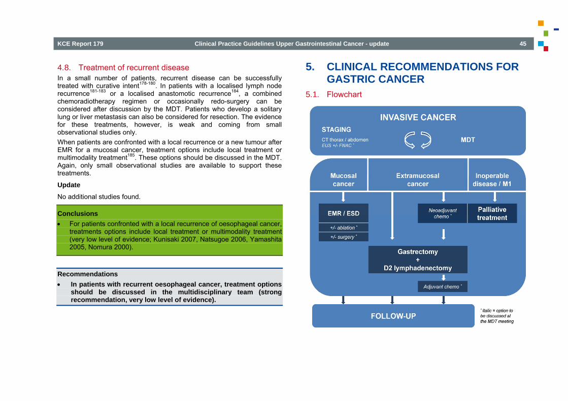
KCE Report 179 Clinical Practice Guidelines Upper Gastrointestinal Cancer - update 45
4.8. Treatment of recurrent disease In a small number of patients, recurrent disease can be successfully treated with curative intent178-180. In patients with a localised lymph node recurrence181-183 or a localised anastomotic recurrence184, a combined chemoradiotherapy regimen or occasionally redo-surgery can be considered after discussion by the MDT. Patients who develop a solitary lung or liver metastasis can also be considered for resection. The evidence for these treatments, however, is weak and coming from small observational studies only. When patients are confronted with a local recurrence or a new tumour after EMR for a mucosal cancer, treatment options include local treatment or multimodality treatment185. These options should be discussed in the MDT. Again, only small observational studies are available to support these treatments.
Update No additional studies found.
Conclusions • For patients confronted with a local recurrence of oesophageal cancer,
treatments options include local treatment or multimodality treatment (very low level of evidence; Kunisaki 2007, Natsugoe 2006, Yamashita 2005, Nomura 2000).
Recommendations • In patients with recurrent oesophageal cancer, treatment options
should be discussed in the multidisciplinary team (strong recommendation, very low level of evidence).
5. CLINICAL RECOMMENDATIONS FOR GASTRIC CANCER
5.1. Flowchart
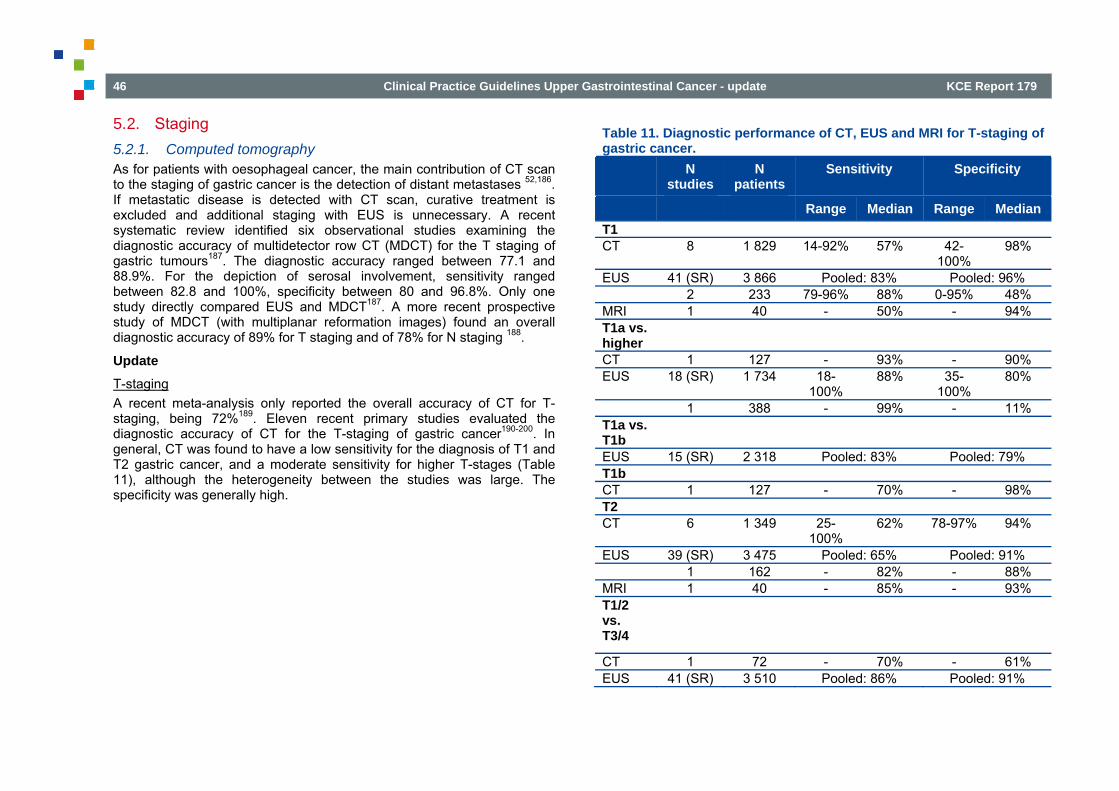
46 Clinical Practice Guidelines Upper Gastrointestinal Cancer - update KCE Report 179
5.2. Staging 5.2.1. Computed tomography As for patients with oesophageal cancer, the main contribution of CT scan to the staging of gastric cancer is the detection of distant metastases 52,186. If metastatic disease is detected with CT scan, curative treatment is excluded and additional staging with EUS is unnecessary. A recent systematic review identified six observational studies examining the diagnostic accuracy of multidetector row CT (MDCT) for the T staging of gastric tumours187. The diagnostic accuracy ranged between 77.1 and 88.9%. For the depiction of serosal involvement, sensitivity ranged between 82.8 and 100%, specificity between 80 and 96.8%. Only one study directly compared EUS and MDCT187. A more recent prospective study of MDCT (with multiplanar reformation images) found an overall diagnostic accuracy of 89% for T staging and of 78% for N staging 188.
Update T-staging A recent meta-analysis only reported the overall accuracy of CT for T-staging, being 72%189. Eleven recent primary studies evaluated the diagnostic accuracy of CT for the T-staging of gastric cancer190-200. In general, CT was found to have a low sensitivity for the diagnosis of T1 and T2 gastric cancer, and a moderate sensitivity for higher T-stages (Table 11), although the heterogeneity between the studies was large. The specificity was generally high.
Table 11. Diagnostic performance of CT, EUS and MRI for T-staging of gastric cancer.
N studies
N patients
Sensitivity Specificity
Range Median Range Median T1 CT 8 1 829 14-92% 57% 42-
100% 98%
EUS 41 (SR) 3 866 Pooled: 83% Pooled: 96% 2 233 79-96% 88% 0-95% 48% MRI 1 40 - 50% - 94% T1a vs. higher
CT 1 127 - 93% - 90% EUS 18 (SR) 1 734 18-
100% 88% 35-
100% 80%
1 388 - 99% - 11% T1a vs. T1b
EUS 15 (SR) 2 318 Pooled: 83% Pooled: 79% T1b CT 1 127 - 70% - 98% T2 CT 6 1 349 25-
100% 62% 78-97% 94%
EUS 39 (SR) 3 475 Pooled: 65% Pooled: 91% 1 162 - 82% - 88% MRI 1 40 - 85% - 93% T1/2 vs. T3/4
CT 1 72 - 70% - 61% EUS 41 (SR) 3 510 Pooled: 86% Pooled: 91%
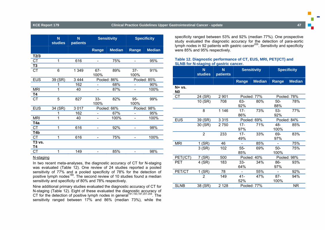
KCE Report 179 Clinical Practice Guidelines Upper Gastrointestinal Cancer - update 47
N studies
N patients
Sensitivity Specificity
Range Median Range Median T2/3 CT 1 616 - 75% - 95% T3 CT 6 1 349 67-
100% 89% 37-
100% 91%
EUS 39 (SR) 3 444 Pooled: 86% Pooled: 85% 1 162 - 68% - 90% MRI 1 40 - 87% - 100% T4 CT 5 827 33-
100% 82% 95-
100% 99%
EUS 34 (SR) 3 017 Pooled: 66% Pooled: 98% 1 162 - 67% - 95% MRI 1 40 - 100% - 100% T4a CT 1 616 - 92% - 98% T4b CT 1 616 - 75% - 100% T3 vs. T4
CT 1 149 - 85% - 98% N-staging In two recent meta-analyses, the diagnostic accuracy of CT for N-staging was evaluated (Table 12). One review of 24 studies reported a pooled sensitivity of 77% and a pooled specificity of 78% for the detection of positive lymph nodes189. The second review of 10 studies found a median sensitivity and specificity of 80% and 78% respectively. Nine additional primary studies evaluated the diagnostic accuracy of CT for N-staging (Table 12). Eight of these evaluated the diagnostic accuracy of CT for the detection of positive lymph nodes in general191,193,197,201-204. The sensitivity ranged between 17% and 86% (median 73%), while the
specificity ranged between 53% and 92% (median 77%). One prospective study evaluated the diagnostic accuracy for the detection of para-aortic lymph nodes in 92 patients with gastric cancer205. Sensitivity and specificity were 85% and 95% respectively.
Table 12. Diagnostic performance of CT, EUS, MRI, PET(/CT) and SLNB for N-staging of gastric cancer. N
studies N
patients Sensitivity Specificity
Range Median Range Median N+ vs. N0
CT 24 (SR) 2 901 Pooled: 77% Pooled: 78% 10 (SR) 708 63-
92% 80% 50-
88% 78%
8 1 146 17-86%
73% 53-92%
77%
EUS 39 (SR) 3 315 Pooled: 69% Pooled: 84% 30 (SR) 2 750 17-
97% 71% 48-
100% 85%
2 233 17-49%
33% 69-97%
83%
MRI 1 (SR) 46 - 85% - 75% 3 (SR) 102 55-
85% 69% 50-
100% 75%
PET(/CT) 7 (SR) 500 Pooled: 40% Pooled: 98% PET 4 (SR) 183 33-
64% 34% 86-
97% 93%
PET/CT 1 (SR) 78 - 55% - 92% 2 149 41-
52% 47% 87-
100% 94%
SLNB 38 (SR) 2 128 Pooled: 77% NR
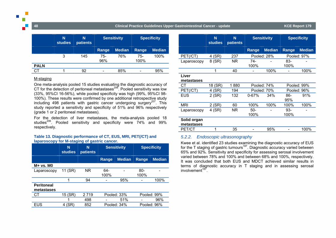
48 Clinical Practice Guidelines Upper Gastrointestinal Cancer - update KCE Report 179
N studies
N patients
Sensitivity Specificity
Range Median Range Median 3 145 75-
96% 76% 75-
100% 100%
PALN CT 1 92 - 85% - 95%
M-staging One meta-analysis pooled 15 studies evaluating the diagnostic accuracy of CT for the detection of peritoneal metastases206. Pooled sensitivity was low (33%, 95%CI 16-56%), while pooled specificity was high (99%, 95%CI 98-100%). These results were confirmed by one additional retrospective study including 498 patients with gastric cancer undergoing surgery207. This study reported a sensitivity and specificity of 51% and 96% respectively (grade 1 or 2 peritoneal metastases). For the detection of liver metastases, the meta-analysis pooled 18 studies206. Pooled sensitivity and specificity were 74% and 99% respectively.
Table 13. Diagnostic performance of CT, EUS, MRI, PET(/CT) and laparoscopy for M-staging of gastric cancer. N
studies N
patients Sensitivity Specificity
Range Median Range Median M+ vs. M0 Laparoscopy 11 (SR) NR 64-
100% - 80-
100% -
1 94 - 95% - 100% Peritoneal metastases
CT 15 (SR) 2 719 Pooled: 33% Pooled: 99% 1 498 - 51% - 96% EUS 4 (SR) 852 Pooled: 34% Pooled: 96%
N studies
N patients
Sensitivity Specificity
Range Median Range Median PET(/CT) 4 (SR) 237 Pooled: 28% Pooled: 97% Laparoscopy 8 (SR) NR 74-
100% - 83-
100% -
1 40 - 100% - 100% Liver metastases
CT 18 (SR) 1 880 Pooled: 74% Pooled: 99% PET(/CT) 4 (SR) 194 Pooled: 70% Pooled: 96% EUS 2 (SR) 132 0-67% 34% 86-
95% 91%
MRI 2 (SR) 60 100% 100% 100% 100% Laparoscopy 4 (SR) NR 50-
100% - 93-
100% -
Solid organ metastases
PET/CT 1 35 - 95% - 100%
5.2.2. Endoscopic ultrasonography Kwee et al. identified 23 studies examining the diagnostic accuracy of EUS for the T staging of gastric tumours187. Diagnostic accuracy varied between 65% and 92%. Sensitivity and specificity for assessing serosal involvement varied between 78% and 100% and between 68% and 100%, respectively. It was concluded that both EUS and MDCT achieved similar results in terms of diagnostic accuracy in T staging and in assessing serosal involvement187.
KCE Report 179 Clinical Practice Guidelines Upper Gastrointestinal Cancer - update 49
Update Two recent meta-analyses evaluated the diagnostic accuracy of EUS for T-staging. Mocellin et al. reported a pooled sensitivity and specificity of 86% (95%CI 81-90%) and 91% (95%CI 89-93%) respectively to discriminate between T1-2 and T3-4 tumours208. To differentiate between T1m (T1 confined to the mucosa) and T1sm tumours (T1 infiltrating the submucosa) the pooled sensitivity and specificity were 83% (95%CI 76-89%) and 79% respectively (95%CI 65-88%). Finally, for differentiation between lymph node positive and negative patients, the pooled sensitivity and specificity were 69% (95%CI 63-74%) and 84% (95%CI 81-88%) respectively (31 studies). No information was available on the added value of FNAC. An important between-study heterogeneity was found for all outcomes. Kwee et al. reported a median sensitivity of 87.8% and a median specificity of 80.2% for the differentiation between T1m and non-T1m tumours209. If only patients with endoscopic suspicion of early gastric cancer were included, the sensitivity to detect T1m increased to 91% (95%CI 85-94%). In a third recent meta-analysis, Wang et al. evaluated the diagnostic accuracy of EUS for the detection of peritoneal metastases206. Pooled sensitivity and specificity were 34% and 96% respectively (5 studies). Finally, Kwee et al. identified 30 studies evaluating the diagnostic accuracy of EUS for N-staging210. Median sensitivity was 70.8%, while median specificity was 84.6%. No information was available on the added value of FNAC. Three additional primary studies were found: • Choi et al. retrospectively analysed the diagnostic accuracy of EUS to
differentiate between T1m and non-T1m tumours in 388 patients with gastric adenocarcinoma suspected to be early gastric cancer by conventional endoscopy211. Sensitivity was found to be 99% and specificity 11%.
• Hye et al. evaluated the diagnostic accuracy of EUS (without FNAC) to differentiate between early (T1) and advanced gastric cancer (T2-4) in 71 patients preoperatively diagnosed as early gastric cancer using endoscopy or CT and undergoing curative gastrectomy193. Sensitivity was found to be 96% and specificity 0%. For differentiation between lymph node positive and negative patients, sensitivity and specificity were 17% and 97% respectively.
• Zheng et al. retrospectively analysed the diagnostic accuracy of EUS (without FNAC) for T- and N-staging in 162 patients treated surgically for gastric cancer212. For T-staging, sensitivity was low to moderate (T1: 79%; T2: 82%; T3: 68%; T4: 67%), while specificity was high (T1: 95%; T2: 88%; T3: 90%; T4: 95%). For differentiation between lymph node positive and negative patients, sensitivity and specificity were 49% and 69% respectively.
5.2.3. Positron-emission tomography No diagnostic accuracy studies were found evaluating PET(/CT).
Update Three recent meta-analyses evaluated the diagnostic accuracy of PET and/or PET/CT for the staging of gastric cancer. Seevaratnam et al. identified 9 studies, but did not distinguish between PET and PET/CT189. Pooled sensitivity and specificity for the detection of positive lymph nodes were 40.3% (significantly lower than CT and MRI) and 97.7% (significantly higher than CT and MRI) respectively. For M-staging, PET had an overall accuracy of 88.2%. Wang et al. identified 5 studies evaluating the diagnostic accuracy of PET and/or PET/CT for the detection of metastases206. They also did not distinguish between PET and PET/CT. Pooled sensitivity and specificity for the detection of liver metastases were 70% (95%CI 36-90%) and 96% (95%CI 81-99%) respectively. Pooled sensitivity and specificity for the detection of peritoneal metastases were 28% (95%CI 17-44%) and 97% (95%CI 83-100%) respectively.
50 Clinical Practice Guidelines Upper Gastrointestinal Cancer - update KCE Report 179
Finally, Kwee et al. identified 4 studies on PET and 1 study on PET/CT, evaluating their diagnostic accuracy for the detection of lymph node metastases210. Median sensitivity and specificity for PET were 34.3% and 93.2% respectively. The one study on PET/CT found a sensitivity of 54.7% and a specificity of 92.2%. Three additional primary studies on PET/CT were found. Two retrospective studies compared the diagnostic accuracy of PET/CT with CT for the detection of lymph node metastases in 71203 and 78 patients201 respectively. Both studies found a low sensitivity (41% and 52% respectively) and a moderate to high specificity (100% and 87% respectively), confirming the results of the meta-analyses. In one study, sensitivity was significantly higher for CT (70% vs. 52%, p=0.035) and specificity was significantly higher for PET/CT (69% vs. 87%, p=0.029)201. In the other study, no significant differences were found203. In a third study, the diagnostic accuracy of PET/CT for the detection of solid organ metastases was evaluated in 35 patients with gastric adenocarcinoma and distant metastases validated by histologic confirmation or by contrast-enhanced CT and serial follow-up213. Sensitivity was 95% and specificity 100%.
5.2.4. Magnetic resonance imaging Kwee et al. identified 3 observational studies examining the diagnostic accuracy of MRI for the T staging of gastric tumours187. Diagnostic accuracy varied between 71% and 83%. Sensitivity and specificity for assessing serosal involvement varied between 90% and 93% and between 94% and 100%, respectively.
Update Three recent meta-analyses evaluated the diagnostic accuracy of MRI for the staging of gastric cancer. Two of these evaluated the diagnostic accuracy of MRI for the detection of lymph node metastases, both including 3 studies (but differing between the 2 meta-analyses)189,210. Seevaratnam et al. found a sensitivity and specificity of 85.3% (significantly better than PET) and 75.0% respectively (1 study)189. Kwee et al. found a median sensitivity and specificity of 68.8% and 75% respectively 210. Overall accuracy for T-staging was found to be 82.9%189. A third systematic review identified two small Chinese studies and did not perform a meta-analysis206. Both studies reported a sensitivity and specificity of
100% for the detection of liver metastases. However, both studies had methodological limitations (poor description of verification and blinding). One additional prospective study was found, evaluating the diagnostic accuracy of MRI for T-staging in 40 patients with an endoscopic diagnosis of gastric carcinoma190. Overall, MRI had a high specificity for all T-stages (T1: 94%; T2: 93%; T3: 100%; T4: 100%), but the sensitivity was low for T1 tumours (T1: 50%; T2: 85%; T3: 87%; T4: 100%).
5.2.5. Laparoscopy According to SIGN, laparoscopy can be considered in patients with gastric tumours being candidates for surgery where full thickness gastric wall involvement is suspected52. There are insufficient data to confirm benefit for laparoscopic ultrasound52,89.
Update One recent systematic review214 summarizes the possible benefits of diagnostic staging laparoscopy before laparotomy for patients with assumed resectable gastric cancer. Sensitivity to detect (peritoneal) metastases varied between 64.3% and 100% with a specificity of 80-100%. Laparoscopy changed management in 8.5% to 59.6% of patients, especially patients with a T3 or T4 tumour. In 8.5% to 43.8% of patients laparotomy could be avoided based on diagnostic laparoscopy. These results are based on prospective and retrospective observational studies with substantial variation in characteristics of included patients, pre-operative imaging and laparoscopic techniques used. Two additional prospective primary studies were found. One study evaluated the diagnostic accuracy of laparoscopy for the detection of peritoneal metastases in 40 patients with gastric carcinoma215. Patients with obvious unresectable disease (e.g. liver metastasis, ascites) on CT scan were excluded. A sensitivity and specificity of 100% was found. In another study, the diagnostic accuracy of laparoscopy for the detection of metastases in general was evaluated in 94 patients with adenocarcinoma of the stomach or gastro-oesophageal junction216. A sensitivity and specificity of 95% and 100% was found respectively. However, it is unclear if patients with a negative laparoscopy received verification with a reference standard.
KCE Report 179 Clinical Practice Guidelines Upper Gastrointestinal Cancer - update 51
5.2.6. Sentinel lymph node mapping Sentinel lymph node mapping has been the subject of several observational studies217-221. Overall sensitivity ranged from 71 to 89%218-220. In patients with a pT1 tumour, sensitivity ranged from 88 to 100%218,220.
Update A recent meta-analysis of 38 studies showed a sentinel lymph node detection rate of 93.7% (95%CI 91.1-95.6%)222. The pooled sensitivity was found to be 76.9% (95%CI 71.6-81.4%) and the NPV 90.3% (95%CI 86.9-92.9%). Three additional primary studies were found223-225. Two of these were prospective 223,225. Two studies only included patients with T1 or T2 tumours 223,224. These studies reported a sensitivity of 75% and 76% and a specificity of 75% and 100%. In the third study, about half of the patients had a T3 tumour225. Sensitivity was 96% while specificity was 100%.
Conclusions • For T-staging, CT seems to have a low sensitivity for the diagnosis of
T1-2 tumours and a moderate sensitivity for higher T-stages (low level of evidence; Anzidei 2009, Cidon 2009, Hwang 2010, Hye 2009, Kim 2011, Kim 2009, Lee 2009, Lee 2009, Makino 2011, Moschetta 2010, Pan 2010). EUS seems to have a better diagnostic accuracy for the detection of T1 tumours, and has a moderate sensitivity and specificity for the distinction between T1a and T1b tumours (low level of evidence; Mocellin 2011, Kwee 2008).
• For N-staging, CT has a moderate sensitivity and specificity (low level of evidence; Seevaratnam 2011, Kwee 2009). EUS and MRI do not seem to have a better diagnostic accuracy (low level of evidence; Mocellin 2011, Seevaratnam 2011, Kwee 2009). PET and PET/CT have a lower sensitivity but a higher specificity (low level of evidence; Seevaratnam 2011, Kwee 2009). Sentinel lymph node biopsy in patients with T1-2 tumours has a sensitivity similar to that of CT, but a higher specificity (low level of evidence, Wang 2011).
• For the detection of liver metastases, CT and PET(/CT) have a similar low sensitivity and high specificity (low level of evidence, Wang 2011). The diagnostic accuracy of MRI seems to be better (low level of evidence, Wang 2011).
• For the detection of peritoneal metastases, CT, EUS and PET(/CT) have a low sensitivity and a high specificity (low level of evidence, Wang 2011). Laparoscopy seems to have a better diagnostic accuracy (low level of evidence, Leake 2011).
Other considerations
• PET(/CT) can be considered in locally advanced mass-forming tumours (intestinal type) in a curative setting226. Gastric carcinomas can be classified according to different classification systems (Lauren227, Goseki228, WHO, etc.) based on different criteria. The Lauren classification combines etiopathological, clinical and pathological (macro- and microscopic) features227. The most common type, the intestinal type, generates a tumoural mass and has convincingly been demonstrated to arise through a Helicobacter pylori infection, gastritis, intestinal metaplasia, dysplasia, carcinoma sequence. The less common type, the signet ring cell carcinoma, often presents with a characteristic macroscopic appearance called “linitis plastica” and will often be negative on PET.
• The concept of the sentinel lymph node, referring to the first lymph node that receives drainage from the primary tumour, is well accepted for breast cancer and melanoma, but is not evident in gastric cancer. Drainage of the stomach is multidirectional and much more complicated, resulting in more than one node which should be considered as a sentinel lymph node.
• In most studies evaluating sentinel lymph node biopsy, the following definitions are used: a true-negative sentinel lymph node (SLN) is a negative SLN and a negative non-SLN, a false-negative SLN is a negative SLN with a positive non-SLN, and a true-positive is a positive SLN with or without a positive non-SLN. Consequently, no false-positive SLN are detected, resulting in a specificity of 100%.
52 Clinical Practice Guidelines Upper Gastrointestinal Cancer - update KCE Report 179
• Multidisciplinary team meetings (MDT) have been implemented in many countries as the predominant model of cancer care to ensure that all patients receive timely diagnosis and treatment, that patient management is evidence-based, and that there is continuity of care 90. The positive impact of multidisciplinary team care in the management of gastro-oesophageal cancer was reported at least in two publications from UK91,92. Stephens reported that multidisciplinary team management resulted in improved staging, lower operative mortality, and improved 5-year survival when compared to a group of patients undergoing R0 resection by surgeons who were working independently. Davies concluded that MDT significantly improved staging accuracy for gastro-oesophageal cancer and ensured that correct management decisions were made for the majority of patients. Moreover, multidisciplinary care tend to enable the construction of clinical pathways and to develop formal programs with a unified vision for therapy and palliation93. Such MDT have to be encouraged and generalized in the management of patients with gastric cancer.
•
Recommendations • All patients diagnosed with gastric cancer should be discussed
at a multidisciplinary team meeting (strong recommendation, low level of evidence).
• In patients with newly diagnosed gastric cancer, CT scan of the chest and abdomen should always be performed (strong recommendation, low level of evidence).
• Endoscopic ultrasonography (EUS) can be considered in patients planned for curative treatment on the basis of clinical presentation and/or CT (weak recommendation, low level of evidence). Fine-needle aspiration cytology of suspicious lymph nodes or metastases can be considered if technically feasible.
• The following examinations can be considered for specific indications: PET scan, magnetic resonance imaging, laparoscopy (weak recommendation, low level of evidence).
Good clinical practice • Multi-detector, multi-planar reformatted CT scan should be
performed with IV contrast and gastric distension with oral contrast or water. The liver should at least be imaged in the arterial and portal venous phase.
5.3. Treatment of mucosal cancer According to SIGN, superficial gastric cancer limited to the mucosa can be treated with endoscopic mucosal resection (EMR), taking into account the stage, size, histological type and differentiation grade (Table 14)52. Large Japanese series of hundreds of patients treated endoscopically with EMR or ESD showed the advantages and success rates of endoscopic management for mucosal cancer229-232. However, a recent Cochrane review failed to identify RCTs of early gastric cancer patients involving a treatment arm of EMR and a comparison arm of gastrectomy233. En-bloc resection – allowing better pathology staging of deep and lateral margins – should be aimed at during mucosectomy with the appropriate technique (EMR for lesions less than 12 mm and ESD for larger lesions)234. Mucosal ablative techniques, such as photodynamic therapy (PDT), laser or argon plasma coagulation (APC), are insufficiently studied.
Table 14. Criteria associated with low risk of lymph node metastasis in patients with mucosal gastric cancer 52,235 (based on Japanese Classification of Gastric Carcinoma 236).
• Lesion < 2 cm in size in elevated types • Lesion < 1 cm in size in depressed types • Well or moderately differentiated histology • No macroscopic ulceration • Invasion limited to mucosa and certainly no deeper than the
superficial submucosa • No residual invasive disease after EMR
KCE Report 179 Clinical Practice Guidelines Upper Gastrointestinal Cancer - update 53
Update Park et al.237 recently published a meta-analysis comparing EMR with ESD for early gastric cancer. The meta-analysis, mainly based on retrospective studies, showed a higher en-bloc resection rate and curative resection rate after ESD versus EMR (OR 8.43, 95%CI 5.2-13.67) at the cost of a higher perforation rate (RR 3.58, 95%CI 1.95-6.55). Local recurrence rate was significantly lower after ESD (RR 0.13, 95%CI 0.04-0.41), but no difference in survival was detected (RR 0.65, 95%CI 0.08-5.38). Importantly, in most of these observational studies, treatment groups were not well balanced, with larger and more difficultly located tumours more frequently included in the ESD group. This could have led to an underestimation of the treatment effect of ESD. The Cochrane review by Bennett et al. was recently updated and still failed to identify RCTs235. Based on non-randomized trials, they reported a local cure rate of 98% after endoscopic treatment of gastric cancer if the standard indications are applied (Table 14). Five-year and ten-year disease-specific survival rates of 99% can be achieved. No new data on mucosal ablative techniques were identified in the literature.
Conclusions • Based on observational studies, excellent cure rates and survival can
be achieved with endoscopic treatment of early gastric cancer (T1) if standard indications are applied (low level of evidence; Bennett 2009).
• Based on observational studies, endoscopic submucosal dissection seems to be associated with a higher curative resection rate, a lower local recurrence rate, but a higher perforation rate than endoscopic mucosal resection. Both interventions are equally effective in terms of survival (low level of evidence; Park 2011).
• Insufficient evidence is available to draw conclusions on the effectiveness of photodynamic therapy, laser or argon plasma coagulation for the treatment of early gastric cancer.
Recommendations • Endoscopic mucosal resection (EMR) or endoscopic submucosal
dissection (ESD) should be performed whenever possible for a T1a gastric cancer aiming at staging and curative resection. If the staging and R0 resection is pathologically confirmed, the procedure can be considered therapeutic, taking into account other well-defined criteria relating to size, histological type, lymphovascular invasion and differentiation grade (weak recommendation, low level of evidence).
• (Destructive) mucosal ablative techniques cannot be recommended as a curative option for patients with T1a gastric cancer and should be limited to centres with appropriate expertise (weak recommendation, very low level of evidence).
Good clinical practice • Resection specimens of EMR and ESD should be reviewed by an
experienced pathologist in this area and discussed at a multidisciplinary meeting with access to the clinical information.
5.4. Treatment of cancer beyond the mucosa 5.4.1. Neoadjuvant treatment In a Cochrane review, 4 RCTs were identified comparing neoadjuvant chemotherapy and surgery to surgery alone, including 2 Japanese trials using oral fluoropyrimidines238. Meta-analysis of these 4 trials showed no statistically significant difference in mortality between the two treatment groups (OR 1.05; 95%CI 0.73–1.50; p=0.29). The MAGIC trial, which was excluded from the Cochrane review because it also included postoperative chemotherapy, found a higher likelihood of overall survival in patients receiving perioperative chemotherapy (ECF) as compared to surgery alone (HR 0.75; 95%CI 0.60–0.93; p=0.009)104. However, the trial received some criticism because of a lack of standardization of surgery, the absence of separate data for gastric cancer, and some methodological shortcomings (e.g. lack of staging accuracy, no blinding).
54 Clinical Practice Guidelines Upper Gastrointestinal Cancer - update KCE Report 179
One abstract was found presenting the results of an RCT comparing preoperative chemotherapy (5-FU/cisplatin) to surgery alone in adenocarcinoma of stomach and lower oesophagus239. HR of death was 0.69 (95%CI 0.50–0.95; p=0.02) with 3 and 5- year overall survival of 35% and 24% respectively in the surgery alone group vs. 48% and 38% respectively in the neoadjuvant chemotherapy group. CCO identified three RCTs comparing preoperative radiotherapy and surgery to surgery alone240. However, inconsistent results were found, making it difficult to draw unambiguous conclusions. Three additional RCTs were identified comparing neoadjuvant immunotherapy and surgery to surgery alone240. No significant survival benefit was found. A small trial of neoadjuvant immunotherapy with interleukin-2 found a trend towards better overall and disease-free survival in the neoadjuvant treatment group241.
Update One meta-analysis242 investigated the role of neoadjuvant chemotherapy in the treatment of gastric cancer. Fourteen controlled studies (including the trials identified in Wu 2007 and by CCO, and including Cunningham 2006) were included. All studies compared neoadjuvant chemotherapy with upfront surgery, but policy regarding postoperative chemotherapy differed between studies. In four studies, postoperative chemotherapy was also given in the control arm. The meta-analysis showed a significant difference in overall survival (OR 1.27, 95%CI 1.04-1.55) and 3-year progression-free survival (OR 1.85, 95%CI 1.39-2.46) in favour of neoadjuvant chemotherapy. Subgroup analysis suggested that neoadjuvant treatment is especially beneficial for T3-4 tumours, Western patients and if multi-agent chemotherapy is used. Two meta-analyses243,244 reported on the value of perioperative radiotherapy compared with surgery alone. The most recent meta-analysis 244 concluded that the addition of radiotherapy to surgery alone results in a significant survival benefit after five years but not after three years. Locoregional relapse was also significantly reduced with radiotherapy. Studies using preoperative, intra-operative and postoperative radiotherapy or chemoradiotherapy were all included in the meta-analysis and compared with surgery alone. Subgroup analysis suggested that preoperative radiotherapy (5-year survival: RR 1.39, 95%CI 1.13-1.73) is
preferable to postoperative radiotherapy (5-year survival: RR 1.53, 95%CI 0.19-12.15)244. One additional underpowered, industry sponsored study by Biffi et al.245 detected no significant differences in morbidity following neoadjuvant chemotherapy and surgery compared to upfront surgery followed by chemotherapy (morbidity rate 28.5% vs. 25.7% respectively).
Conclusions • Neoadjuvant chemotherapy is associated with a survival benefit
compared with surgery alone in patients with locally advanced gastric cancer eligible for potentially curative surgery (moderate level of evidence; Li 2010). The benefit seems to be larger in T3-4 tumours (low level of evidence; Li 2010).
• Perioperative chemotherapy seems to be associated with a survival benefit in patients with gastric cancer (low level of evidence; Cunningham 2006).
• Preoperative radiotherapy seems to improve local control and 5-year survival in patients with resectable gastric cancer compared to no adjuvant therapy (low level of evidence; Valentini 2009).
Recommendations • If after multidisciplinary discussion neoadjuvant treatment is
considered for a locally-advanced gastric tumour, neoadjuvant chemotherapy is recommended (strong recommendation, moderate level of evidence).
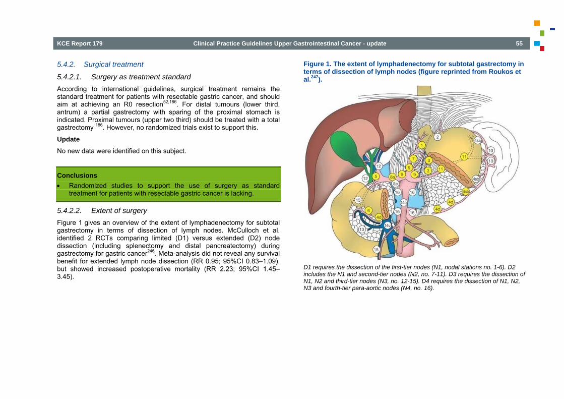
KCE Report 179 Clinical Practice Guidelines Upper Gastrointestinal Cancer - update 55
5.4.2. Surgical treatment
5.4.2.1. Surgery as treatment standard According to international guidelines, surgical treatment remains the standard treatment for patients with resectable gastric cancer, and should aim at achieving an R0 resection52,186. For distal tumours (lower third, antrum) a partial gastrectomy with sparing of the proximal stomach is indicated. Proximal tumours (upper two third) should be treated with a total gastrectomy 186. However, no randomized trials exist to support this.
Update No new data were identified on this subject.
Conclusions • Randomized studies to support the use of surgery as standard
treatment for patients with resectable gastric cancer is lacking.
5.4.2.2. Extent of surgery Figure 1 gives an overview of the extent of lymphadenectomy for subtotal gastrectomy in terms of dissection of lymph nodes. McCulloch et al. identified 2 RCTs comparing limited (D1) versus extended (D2) node dissection (including splenectomy and distal pancreatectomy) during gastrectomy for gastric cancer246. Meta-analysis did not reveal any survival benefit for extended lymph node dissection (RR 0.95; 95%CI 0.83–1.09), but showed increased postoperative mortality (RR 2.23; 95%CI 1.45–3.45).
Figure 1. The extent of lymphadenectomy for subtotal gastrectomy in terms of dissection of lymph nodes (figure reprinted from Roukos et al.247).
D1 requires the dissection of the first-tier nodes (N1, nodal stations no. 1-6). D2 includes the N1 and second-tier nodes (N2, no. 7-11). D3 requires the dissection of N1, N2 and third-tier nodes (N3, no. 12-15). D4 requires the dissection of N1, N2, N3 and fourth-tier para-aortic nodes (N4, no. 16).
56 Clinical Practice Guidelines Upper Gastrointestinal Cancer - update KCE Report 179
Since this Cochrane review, numerous trials have been published examining the extent of lymph node dissection. An interim analysis of the Italian Gastric Cancer Study Group (IGCSG) randomized surgical trial showed no statistically significant difference in postoperative morbidity and mortality between D1 and D2 gastrectomy248. In this trial, only a minority of patients received a splenectomy (in case of clinical T > 1 on the greater curvature of the proximal and middle thirds of the stomach) or a distal pancreatectomy (suspicion of tumour involvement). However, a subsequent multicentric phase II study by the same group evaluating pancreas-preserving D2 gastrectomy suggested a survival benefit with an overall 5-year survival of 55% and a postoperative in-hospital mortality of 3.1%249. An update of the Dutch gastric cancer group trial, included in the Cochrane review, was also published250. Postoperative morbidity (25% vs. 43%; p=0.001) and mortality (4% vs. 10%; p=0.004) were significantly higher in the D2 dissection group, but after 11 years no overall difference in survival was found (30% vs. 35%; p=0.53). As compared to Degiuli et al.248, the amount of splenectomies and distal pancreatectomies was higher in the Randomized Dutch Gastric Cancer Group Trial250. Three more recent trials compared standard D2 vs. extended D2 (D2+, i.e. D2 with para-aortic nodal dissection) lymph node dissection251-253. No statistically significant differences in postoperative morbidity and mortality were found in two trials251,252, while postoperative morbidity was higher in the extended D2 group in the third trial253. Finally, Wu et al.254 compared D1 vs. D3 (i.e. level 1, 2 and 3) lymph node dissection in 221 patients with gastric adenocarcinoma. Overall 5-year survival was significantly higher in patients assigned to D3 surgery than in those assigned to D1 surgery (59.5% [95%CI 50.3-68.7] vs. 53.6% [95%CI 44.2-63.0]; p=0.041). Patients who had R0 resection (N=215) had recurrence at 5 years of 50.6% (95%CI 41.1-60.2) for D1 surgery and 40.3% (95%CI 30.9-49.7) for D3 surgery (p=0.197).
Update Several recent meta-analyses have summarized the available evidence on the possible benefits of more extended lymphadenectomy in the surgical treatment of gastric cancer.
Three meta-analyses255-257 compared D1 with D2 lymphadenectomy. In none of the reviews, recent evidence published since 2007 was included. The meta-analyses all concluded that there is no benefit after D2 lymphadenectomy in terms of recurrence rate or overall survival, but a significant increase in morbidity. However, the RCTs comparing D1 and D2 lymphadenectomy in Western countries have received many criticism. The main limitation is related to the quality of the surgery performed in most of the trials. Surgeons participating in Western trials often did not have training in performing D2 lymphadenectomy or received training only during the trial and performed surgery in low-volume centers. Furthermore, a high number of patients also underwent splenectomy or pancreatectomy as part of the D2 lymphadenectomy, which appeared to be responsible for the majority of complications. Splenectomy and pancreatectomy are currently not routinely performed as part of extended lymphadenectomy. Furthermore, patient selection, contamination and non-compliance rates in the RCTs are substantial, obscuring a possible effect on survival. Five meta-analyses 256-260 compared D1 or D2 lymphadenectomy with or without para-aortic lymphadenectomy (also called D4 lymphadenectomy). None of the meta-analyses could identify a survival benefit of para-aortic lymphadenectomy. The most recent studies did not identify a difference in postoperative morbidity or mortality. Two meta-analyses261,262 assessed the value of splenectomy and pancreatectomy in the surgical treatment of gastric cancer. They both concluded that splenectomy or pancreatectomy have no influence on survival or postoperative mortality. Roberts et al.263 evaluated the use of pancreatectoduodenectomy in patients with gastric cancer infiltrating the pancreas or duodenum or with macroscopic lymph node involvement. Only retrospective studies are available on this topic and these showed mixed results regarding survival benefit. One recently published, Japanese study264 suggested a possible survival benefit of (prophylactic) bursectomy for advanced gastric cancer, although the trial was underpowered and designed as a non-inferiority trial.
KCE Report 179 Clinical Practice Guidelines Upper Gastrointestinal Cancer - update 57
Conclusions • In patients with resectable gastric cancer, D2 lymphadenectomy is
associated with similar overall survival and recurrence rates as D1 lymphadenectomy (high level of evidence; Memon 2011), but with a significantly increased morbidity (low level of evidence; Memon 2011). This morbidity seems to be related to the splenectomy and/or pancreatectomy performed as part of the D2 lymphadenectomy (low level of evidence; Roberts 2011).
• In patients with resectable gastric cancer, para-aortic lymph node dissection is not associated with a survival benefit (moderate level of evidence; Zheng 2011, Lustosa 2008).
Other considerations
• As noted by McCulloch et al.265, cohort studies of D2 lymphadenectomy from high-volume centers report a high survival and low mortality rates in comparison to randomized and non-randomized comparisons, showing that D2 lymphadenectomy can be considered a safe procedure if performed by specialized surgeons. In Italy, trials with extensive pre-trial surgical training achieved low morbidity and mortality rates after D2 lymphadenectomy with conservation of the spleen and pancreas266. In the Netherlands, an improved relative survival was seen during and after the surgical trial period, for which surgical training and better quality of surgery seems the most plausible explanation. No changes in pre- or postoperative treatment with radiotherapy or chemotherapy were noted267.
• Whether D2 lymphadenectomy without pancreaticosplenectomy, performed by specialized, experienced surgeons in high-volume hospitals results in better locoregional control and survival is still a matter of debate. As discussed above, due to contamination, non-compliance and high postoperative mortality, the results of Western RCTs are not necessarily applicable in this situation. D2 lymphadenectomy is considered standard of care in Japan and other Asian countries, based on large Asian observational trials255. Furthermore, 15-year follow-up data of the Dutch D1-D2 trial shows a lower locoregional recurrence and gastric-cancer-related death rates
after D2 versus D1 in patients who were treated with curative intent. Results have to be interpreted with caution though, as there was no intention-to-treat analysis of all randomized patients, there was no blinding of assessors and no overall survival difference could be detected. Also, the high survival rates in an Italian phase II study with trained surgeons, higher than achieved in RCTs, suggest a possible benefit of D2 lymphadenectomy249.
5.4.2.3. Reconstruction after surgery Reconstruction after gastrectomy can be with or without pouch formation and with (Billroth I and II) or without (Roux-en-Y, jejunal interposition) maintenance of duodenal passage52. Evidence suggests that pouch procedures may be associated with a higher earlier weight gain 268,269 and some improvement in long-term quality of life270.
Update One recent meta-analysis by Gertler et al.271 confirms the above findings, especially after a longer follow-up period of 6, 12 or even 24 months. Symptoms of dumping syndrome and heartburn, and food intake are significantly better after 12 months if a pouch was constructed. Also, quality of life in R0 patients was better at 12 and 24 months if a reconstruction with pouch was performed. Morbidity and mortality are not influenced by type of surgery (pouch versus no pouch). A small underpowered randomized trial of low quality272 also found better food intake and body weight in patients with jejunal pouch interposition after proximal gastrectomy compared to reconstruction with a double-tract method. Other aspects of surgical techniques aiming at decreasing postoperative complications and improving postoperative recovery such as stapled versus handsewn gastroduodenostomy273, compression anastomosis clip274, the use of postoperative drains275, bioresorbable membranes276 and alternative reconstruction techniques277-279 are not further discussed in this report.
58 Clinical Practice Guidelines Upper Gastrointestinal Cancer - update KCE Report 179
Conclusions • Pouch reconstruction seems to be associated with better food intake
and improved quality of life (high level of evidence; Gertler 2009).
5.4.2.4. Laparoscopic surgery Several articles have compared laparoscopic surgery to conventional surgery for gastric cancer280-283. One SR identified 4 RCTs comparing laparoscopy-assisted distal gastrectomy (LADG) to conventional open distal gastrectomy (CODG)281. LADG was found to be a longer procedure, but was associated with a lower postoperative morbidity. No difference was found in postoperative mortality. These results were confirmed by an RCT published subsequently283. Hayashi et al. also found a shorter operation time with CODG, but no difference in postoperative morbidity and mortality280. Huscher et al. found no difference in duration of surgery and postoperative morbidity and mortality between laparoscopic-assisted and open radical subtotal gastrectomy282. Overall, duration of analgesic administration was shorter after laparoscopic surgery280,281,283.
Update Four meta-analyses284-287 compared laparoscopic with open (distal) gastrectomy. Earlier findings were confirmed: laparoscopic gastrectomy is associated with fewer postoperative complications at the cost of a longer operation time and a reduced number of lymph nodes harvested. Oncological outcomes and mortality do not appear to be affected.
Conclusions • In patients with resectable gastric cancer, laparoscopic gastrectomy is
associated with fewer postoperative complications compared with open gastrectomy, but a longer operation time and a reduced number of lymph nodes harvested (low level of evidence; Zorcolo 2011, Chen 2009).
5.4.2.5. Volume-outcome relationship As for oesophageal cancer, several studies have shown a relationship between patient outcomes (e.g. 30-day mortality) and surgeon or hospital volume for gastric cancer surgery52,135,136. However, this relationship is less profound than for oesophageal cancer surgery135. Subspecialty training is also associated with lower postoperative mortality, although this relationship is not statistically significant288.
Update No systematic reviews published since 2007 were identified in the literature.
Conclusions • Postoperative mortality after gastrectomy for gastric cancer seems to
be associated with the surgeon and hospital volume (low level of evidence; Halm 2002, Killeen 2005).
Recommendations • Surgical resection should be considered standard treatment for
patients with resectable gastric cancer (strong recommendation, low level of evidence).
• Surgery for gastric cancer should aim at achieving an R0 resection (strong recommendation, low level of evidence).
• D2 lymphadenectomy should be standard during gastrectomy and performed in high-volume, specialized centres with experience and/or specialist training (weak recommendation, low level of evidence).
• Splenectomy and pancreatectomy should not be considered standard practice during gastrectomy if no disease infiltration in the spleen or pancreas is present (weak recommendation, low level of evidence).
• Laparoscopic surgery should be restricted to clinical studies (weak recommendation, low level of evidence).
KCE Report 179 Clinical Practice Guidelines Upper Gastrointestinal Cancer - update 59
5.4.3. Adjuvant treatment
5.4.3.1. Chemotherapy Numerous RCTs and systematic reviews have been published comparing postoperative adjuvant chemotherapy to surgery alone52,240. Most meta-analyses found small statistically significant differences in survival favouring adjuvant chemotherapy, although only a minority of the included RCTs were of high quality. Subgroup analyses in one meta-analysis showed a trend towards a larger magnitude of the effect for trials in which at least two thirds of the patients had node-positive disease (OR 0.74; 95%CI 0.59–0.95)240. Therefore, CCO concluded that adjuvant chemotherapy alone may be of benefit in particular for patients with lymph node metastases. However, this is not in line with SIGN, that did not recommend postoperative chemotherapy outside a clinical trial52. One meta-analysis of 4 Japanese RCTs comparing adjuvant oral fluorinated pyrimidines to surgery alone found a survival benefit in favour of adjuvant chemotherapy (HR 0.73; 95%CI 0.60–0.89; p=0.002)289. Other trials failed to demonstrate a benefit in favour of adjuvant chemotherapy290-
293. As stated above, the MAGIC trial found a higher likelihood of overall survival in patients receiving perioperative chemotherapy (ECF) as compared to surgery alone (HR 0.75; 95%CI 0.60–0.93; p=0.009)104. However, the trial received some criticism because of a lack of standardization of surgery, the absence of separate data for gastric cancer, and some methodological shortcomings (e.g. lack of staging accuracy, no blinding).
Update Three more recent meta-analyses294-296 compared surgery followed by adjuvant chemotherapy with surgery alone for resectable gastric cancer. Methodological quality of the included primary studies was variable and the meta-analysis based on individual patient data by the GASTRIC group294 can be criticized for the incompleteness of data and the lack of critical appraisal of included studies. Nevertheless, all three meta-analyses concluded that adjuvant chemotherapy for resectable gastric cancer results in a significant survival benefit with a hazard ratio around 0.80. That
would correspond to an absolute survival benefit of 6% after 5 years294 and a NNT of 14295. In a subanalysis of Sun et al., adjuvant oral fluorinated pyrimidines were associated with a clear survival benefit compared to surgery alone (3 studies, HR 0.63, 95%CI 0.52-0.78)296. Two additional small RCTs297,298, both with methodological limitations, could not identify a survival benefit after adjuvant chemotherapy. Another low quality RCT299 suggested a clinical benefit of adding oxaliplatin to 5-fluorouracil and leucovorin in the adjuvant treatment of gastric cancer. One small RCT of very low quality300 compared FOLFOX adjuvant chemotherapy with FOLFOX combined with arsenic trioxide. There was no significant difference in disease-free survival between the two groups.
Conclusions • Postoperative adjuvant chemotherapy significantly improves overall
survival in patients with resectable gastric cancer compared to no adjuvant therapy (moderate level of evidence; GASTRIC 2010, Sun 2009, Liu 2008).
• There are indications that perioperative chemotherapy improves the overall survival of patients with gastric cancer (low level of evidence; Cunningham 2006).
• Adjuvant oral fluorinated pyrimidines are associated with a survival benefit compared to surgery alone (high level of evidence; Sun 2009).
5.4.3.2. (Chemo)radiotherapy CCO identified two RCTs comparing adjuvant radiotherapy to surgery alone 240. No differences in survival were found. However, addition of postoperative chemotherapy to radiotherapy may result in better outcomes. Three RCTs were identified by the CCO, of which one found no statistically significant difference in survival. The two other trials detected improved survival with postoperative CRT, but received considerable criticism240. For example, the surgery performed in the RCT of McDonald et al. was often not up to the desired standards 152.
60 Clinical Practice Guidelines Upper Gastrointestinal Cancer - update KCE Report 179
Update Two meta-analyses243,244 reported on the value of perioperative radiotherapy compared with surgery alone. The most recent meta-analysis244 concluded that the addition of radiotherapy to surgery alone results in a significant survival benefit after five years but not after three years. Locoregional relapse was also significantly reduced with radiotherapy. Studies using preoperative, intra-operative and postoperative radiotherapy or chemoradiotherapy were included and compared with surgery alone. Subgroup analysis suggested that preoperative radiotherapy (5-year survival: RR 1.39, 95%CI 1.13-1.73) is preferable to postoperative radiotherapy (5-year survival: RR 1.53, 95%CI 0.19-12.15)244. An underpowered (early closure due to slow accrual) multicenter Greek RCT investigated the addition of radiotherapy to adjuvant chemotherapy for R0 resected gastric cancer with serosal involvement or lymph node metastases301. No change in progression-free or overall survival was noted, but the discontinuation rate due to toxicity was significantly higher in the radiotherapy arm. Another underpowered Korean RCT of low methodological quality302 could not show a benefit in progression-free survival with the addition of radiotherapy to adjuvant chemotherapy.
Conclusions • Postoperative radiotherapy is not associated with a survival benefit in
patients with gastric cancer (low level of evidence; Valentine 2009). • No definite conclusions can be drawn on the effectiveness of
postoperative chemoradiation (low level of evidence; CCO 2003).
5.4.3.3. Intraperitoneal chemotherapy Several RCTs and meta-analyses compared adjuvant intraperitoneal chemotherapy to surgery alone240,303,304. A recent meta-analysis found a significant survival benefit in favour of hyperthermic intraoperative intraperitoneal chemotherapy with or without early postoperative intraperitoneal chemotherapy304. However, in general survival results have been conflicting, and some trials even reported harm from intraperitoneal therapy240.
Update One meta-analysis304 investigated adjuvant chemotherapy administered through the intraperitoneal route. Included trials were considered of fair quality and the majority had surgery alone as control arm. A significant survival benefit was noted for hyperthermic intraperitoneal chemotherapy, but only a trend towards better survival for normothermic intraperitoneal chemotherapy. One additional Japanese RCT305 could not detect clinical benefit of adjuvant IP and IV chemotherapy for completely resected gastric cancer with macroscopically involvement of the serosa (T3-T4).
Conclusions • Postoperative hyperthermic intraperitoneal chemotherapy can improve
overall survival in patients with resectable gastric cancer compared to no adjuvant therapy (moderate level of evidence; Yan 2007).
5.4.3.4. Immunochemotherapy CCO identified 9 RCTs comparing adjuvant immunochemotherapy to surgery alone240; one meta-analysis on immunochemotherapy with polysaccharide K306 and one RCT307 were published subsequently. Overall, inconsistent results were found, making it difficult to draw definite conclusions.
Update One Korean RCT308 suggested a survival benefit when polyadenylic-polyuridylic acid is added to 5-fluorouracil and adriamycin adjuvant chemotherapy.
Conclusions • The benefits of immunochemotherapy are currently unclear (low level
of evidence; CCO 2003).
KCE Report 179 Clinical Practice Guidelines Upper Gastrointestinal Cancer - update 61
Recommendations • Patients with gastric cancer who received neoadjuvant
chemotherapy can be considered for postoperative chemotherapy (weak recommendation; low level of evidence).
• Postoperative chemotherapy and chemoradiotherapy can be considered for patients with gastric cancer who did not receive neoadjuvant chemotherapy (weak recommendation; low level of evidence).
• Postoperative radiotherapy alone is not recommended for patients with gastric cancer (weak recommendation; low level of evidence).
5.5. Treatment of metastatic disease 5.5.1. Palliative surgery According to international guidelines, palliative gastric surgery is limited to symptomatic stenoses, bleeding tumours and perforation186. It should be avoided in patients who have disseminated peritoneal disease52,186. In comparison to palliative gastrectomy, gastric bypass has a higher mortality52. However, reported mortality after laparoscopic gastrojejunostomy is lower than for open bypass.
Update Mahar et al.309 performed a systematic review of quality of life after palliative surgery for non-curative gastric cancer. No studies using a validated instrument to assess quality of life could be identified in the literature. Some studies suggested a shorter time to oral intake and shorter hospital stay after laparoscopic gastrojejunostomy compared to surgical bypass. In recent years, the interest in cytoreductive surgery, often followed by hyperthermic intraperitoneal chemotherapy, has increased for advanced gastric cancer with peritoneal carcinomatosis. Gill et al.310 performed a meta-analysis on the combined procedures. The included studies and the meta-analysis itself both have methodological flaws and no direct comparison with other treatment options, e.g. systemic chemotherapy without surgery, was done. The authors reported a median overall survival
of 7.9 (range 6.1-9.2) months for all patients and of 15 (range 9.5-43.4) months for patients in whom all macroscopic tumour could be removed. A 1-year survival of 43% (95%CI 22-68%) could be achieved at the cost of significant postoperative morbidity in 21.5% of patients. One RCT 311 demonstrated an improvement in disease-specific survival after cytoreductive surgery followed by hyperthermic intraperitoneal chemotherapy compared with cytoreductive surgery alone for gastric cancer patients with peritoneal spread but without distant metastases. The study had methodological limitations, mainly the absence of clear allocation concealment and lack of blinding.
Conclusions • Palliative gastrectomy seems to be associated with a lower mortality
than gastric bypass for patients with unresectable gastric cancer (low level of evidence; SIGN 2006).
• Laparoscopic gastrojejunostomy seems to be associated with a shorter time to oral intake and a shorter hospital stay than open gastric bypass for patients with unresectable gastric cancer (low level of evidence; Mahar 2011).
• There is weak evidence that adding HIPEC to cytoreductive surgery in patients with peritoneal metastases is associated with a survival benefit (low level of evidence; Yang 2011).
5.5.2. Stents For patients with malignant gastric outlet obstruction, treatment options include endoscopic stenting or surgical gastroenterostomy312,313. Two systematic reviews, that mainly comprised observational studies, found a higher initial clinical success after endoscopic stenting. However, inconsistent results were found for associated morbidity312,313. No significant survival benefit was found. Endoscopic stenting was associated with a shorter hospital stay.
62 Clinical Practice Guidelines Upper Gastrointestinal Cancer - update KCE Report 179
Update Two meta-analyses314,315 compared surgical treatment of malignant gastric outlet syndrome with placement of a stent. Based on the limited evidence available, they concluded that stenting results in a quicker start of oral intake, shorter hospital stay and fewer complications without shortening survival. One small Dutch RCT mainly including other cancer types with malignant gastric outlet syndrome316 showed a shorter hospital stay and a more rapid food intake, but more re-interventions, major complications and recurrent obstructive symptoms after endoscopic stent placement compared to surgical gastrojejunostomy.
Conclusions • In patients with malignant gastric outlet obstruction, endoscopic
stenting and surgical gastroenterostomy have similar survival rates (low level of evidence; Zheng 2011, Ly 2010). Inconsistent evidence is available on the associated risks.
5.5.3. Chemotherapy and targeted treatment Several RCTs compared palliative chemotherapy to best supportive care for patients with advanced or metastatic gastric cancer317,318. A clear survival benefit was found in favour of palliative chemotherapy. Above this, palliative chemotherapy was associated with a better quality of life317. Wagner et al. also found a significant survival benefit in favour of combination chemotherapy as compared to single agent chemotherapy (HR 0.83; 95%CI 0.74–0.93)318.
Update Combination chemotherapy The meta-analysis by Wagner et al. was updated in 2010319. An improvement in progression-free and overall survival was confirmed for chemotherapy added to best supportive care. Median overall survival was 11 months in the chemotherapy group versus 4.3 months in the supportive care only group. Furthermore, combination chemotherapy resulted in a longer overall survival than single agent chemotherapy (median survival 8.3 versus 6.7 months). In this review, the value of several chemotherapeutic agents was tested with a meta-analysis of the available data. Adding anthracyclines to 5-FU and cisplatin resulted in a 2 month prolongation of overall survival. Cisplatin added to 5-FU and an anthracyclin improved survival with approximately 1 month. The use of docetaxel or irinotecan appears to have no significant influence on progression-free or overall survival. Replacing cisplatin by oxaliplatin did not show a survival benefit in the review by Wagner et al. 319. However, with new evidence included in the meta-analysis of Montagnani et al. 320, replacing cisplatin by oxaliplatin seems to have a beneficial effect on the risk of death and risk of progression at the cost of increased diarrhea and neurotoxicity. The RCT published by Narahara et al.321 identified an increased response rate after irinotecan and S-1 compared to S-1 alone (41.5% vs. 26.9%, p=0.035), but no significant change in median survival time (12.8 vs. 10.5 months, p=0.23). Lee et al.322 compared heptaplatin with cisplatin, both combined with 5-FU, in advanced gastric cancer. No difference in overall survival or time to progression was noted. Toxicity profile, especially nausea and vomiting, appeared more beneficial for cisplatin.
KCE Report 179 Clinical Practice Guidelines Upper Gastrointestinal Cancer - update 63
Oral 5-FU The use of oral 5-FU analogues (capecitabine, S-1) results in similar survival rates as the use of intravenous 5-FU according to the meta-analysis of Wagner et al.319. Huang et al.323 published a more recent review on S-1 for advanced gastric cancer, showing better overall survival (HR 0.87, 95%CI 0.79-0.96) and reduced incidence of neutropenia compared to 5-FU. A meta-analysis of Ma et al.324 concluded that the use of capecitabine instead of 5-FU improves overall survival (10.7 vs. 9.5 months, p=0.03) and reduces side-effects such as leukopenia, stomatitis, nausea and vomiting. The incidence of hand-foot syndrome increased with capecitabine. Targeted treatment Zagouri et al.325 performed a systematic review on targeted therapies in gastric cancer, with methodological limitations (search in Medline only, no critical appraisal of included studies). Only for bevacizumab and trastuzumab, RCTs have been published so far. Bevacizumab has no significant therapeutic effect in terms of survival in advanced, unresectable gastric cancer326. Median progression-free survival significantly increased from 5.3 to 6.7 months. The TOGA study evaluated the use of trastuzumab in HER2-positive gastric cancer327. This open-label study showed a significantly improved progression-free (HR 0.71, 95%CI 0.59-0.85) and overall survival (HR 0.74, 95%CI 0.60-0.91) when trastuzumab was added to chemotherapy (cisplatin + 5FU or capecitabine). These results are based on an interim analysis after 75% of events. Intraperitoneal chemotherapy One small Japanese RCT328 randomized 88 patients with R0 resection but positive peritoneal cytology to one of three groups: surgery alone, surgery followed by IP chemotherapy and surgery followed by extensive intraoperative peritoneal lavage (EIPL) and IP chemotherapy. The patients who underwent EIPL had a significantly improved 5-year survival (43.8% vs. 4.6% after IP chemotherapy and 0% after surgery alone) and a lower rate of peritoneal recurrence compared to the other two groups.
Conclusions • In patients with advanced or metastatic gastric cancer, combination
chemotherapy has a survival benefit compared to single agent chemotherapy (high level of evidence; Wagner 2010).
• Oral 5-FU analogues seem to be associated with a survival benefit compared with intravenous 5-FU (moderate level of evidence; Huang 2010, Ma 2011).
• In HER2-positive gastric cancer, adding trastuzumab to standard chemotherapy seems to be associated with a survival benefit (low level of evidence; Bang 2010).
Other considerations
• Since 1 October 2010, trastuzumab in combination with capecitabine or with 5-FU and cisplatin is reimbursed in Belgium for patients with metastatic gastric cancer or cancer of the GOJ if amplification of the HER2-gen is demonstrated by a positive in situ hybridisation test. The decision for reimbursement was based on the results of the TOGA trial.
5.5.4. Supportive care Patients with gastric cancer should have access to a specialist (outpatient and/or inpatient) palliative care team, in particular in relation to comfort and symptom control, and quality of life 52. This team clearly should involve the general practitioner, who should have a coordinating role in the organisation of the palliative home care. However, as for oesophageal cancer, evidence specific to control of symptoms, such as pain, anorexia and bleeding, in these cancers is limited. Some evidence exists for the use of celiac axis plexus block for the palliation of pain and for the use of corticosteroids in patients with gastric cancer who are anorexic or who have symptoms of bowel obstruction52. Nutritional support in the palliative setting should take into account the comfort of the patient, in that quality of life is much more important than the long-term effects. Limited evidence (coming from a secondary analysis of 1 RCT) is also available for psychotherapeutic support during hospital stay175.
64 Clinical Practice Guidelines Upper Gastrointestinal Cancer - update KCE Report 179
The KCE is currently preparing a generic guideline on supportive care for cancer patients.
Recommendations • Palliative gastric surgery is limited to symptomatic stenoses,
bleeding tumours and perforation (weak recommendation, low level of evidence).
• For patients with malignant gastric outlet obstruction, treatment options include endoscopic stenting or surgical gastroenterostomy (weak recommendation, low level of evidence).
• In patients with locally advanced or metastatic cancer of the stomach with good performance status combination chemotherapy is recommended (strong recommendation, high level of evidence).
• Patients with advanced gastric cancer should have access to a specialist palliative care team, in particular in relation to comfort and symptom control, nutrition and quality of life (strong recommendation, low level of evidence).
5.6. Follow-up In patients that underwent treatment for gastric cancer, follow-up is needed to detect recurrent disease (in case of curative treatment), progressive disease, symptoms that warrant treatment, or nutritional problems resulting from total gastrectomy (e.g. iron and vitamin deficiency, maldigestion with steathorroea, etc). However, evidence about the duration, frequency and type of follow-up is lacking329. Also, no evidence is available to support regular imaging in the follow-up of these patients52. For patients that underwent partial gastrectomy, lifelong endoscopic surveillance is indicated because of the high risk of developing gastric stump carcinoma330. Patients treated with endoscopic mucosal resection (EMR) should have strict endoscopic surveillance233 with a follow-up endoscopy after three months, then every six months in the first two years, and then annually.
Update Five primary studies evaluated the diagnostic accuracy of PET/CT to detect recurrence in patients treated for gastric cancer331-335. One of these studies compared PET/CT with CT331. In three studies, PET/CT was used routinely in the follow-up of postoperative gastric cancer patients331-333. A total of 241 patients were included in these studies. All studies suffered from selection bias and differential verification. Sensitivity ranged from 54% to 90% (median 68%), specificity from 71% to 86% (median 85%). In the study of Kim et al., the difference with CT was not statistically significant (Se: CT 64%, PET/CT 54%; Sp: CT 86%, PET/CT 85%)331. Two other studies evaluated PET/CT in patients with suspected recurrence based on clinical grounds, endoscopy or imaging334,335. Using an SUVmax of 2.3 as a cut-off, Bilici et al. found a sensitivity and specificity of 96% and 100% respectively334. The other study reported a sensitivity and specificity of 75% and 77% respectively.
Conclusions • In routine follow-up, CT seems to have a low sensitivity and moderate
specificity to detect recurrence after curative gastrectomy for gastric cancer (low level of evidence; Kim 2009). The diagnostic accuracy of PET/CT is not clearly better (low level of evidence; Kim 2009, Graziosi 2011, Sim 2009).
• Conflicting evidence is available on the diagnostic accuracy of PET/CT in patients with a suspected recurrence of gastric cancer (low level of evidence; Bilici 2011, Park 2009).
KCE Report 179 Clinical Practice Guidelines Upper Gastrointestinal Cancer - update 65
Other considerations Since no good evidence is available on the optimal follow-up schedule of patients treated for gastric cancer, these recommendations are made in line with those for oesophageal cancer. A physical examination is recommended every three months, and should give special attention to nutritional status, weight loss, fatigue, etc. Above this, a blood analysis (routine chemistry and haematology, completed with serum ferritine and vitamin B12 dosing in case of anaemia) is recommended. Vitamin B12 deficiency is a frequent phenomenon after total gastrectomy, and should be adequately treated. CT scan of the abdomen is recommended every six months in the first year and afterwards annually until the fifth year.
Recommendations • It is recommended that the follow-up of patients treated for
gastric cancer includes a physical examination and blood analysis every three months, with targeted imaging if needed. A CT scan can be considered every six months in the first year and then annually until the fifth year (weak recommendation; very low level of evidence).
• Patients treated with endoscopic mucosal resection (EMR) or endoscopic submucosal dissection (ESD) should have a follow-up endoscopy after three months, then every six months in the first two years, and then annually (weak recommendation; very low level of evidence).
5.7. Treatment of recurrent disease In a small number of patients, recurrent disease can be successfully treated with curative intent. In patients with cancer of the gastric stump after distal gastrectomy, R0 resection can be achieved in a relatively high number of patients336,337. Patients who develop a solitary lung or liver metastasis can also be considered for resection338,339. When patients are confronted with a local recurrence after EMR for a mucosal cancer, local treatment can be reconsidered340,341. The evidence for these treatments, however, is weak and coming from small observational studies only.
Update Apart from one RCT342, no new evidence specifically on the second line treatment of gastric cancer was found. Often, patients with recurrent cancer of the stomach are included in chemotherapy studies designed mainly for advanced, metastatic gastric cancer (see above). The sponsored study published by Thuss-Patience et al.342 compared irinotecan as second-lone chemotherapy versus best supportive care in gastric cancer. The trial had to be closed prematurely due to slow accrual. The hazard ratio for death was reduced to 0.48 (95% CI 0.25-0.92). Median survival for patients who received irinotecan was 4 months (95% CI 3.6-7.5 months) compared to 2 months (95%CI 1.7-4.9 months) for patients who received best supportive care.
Conclusions • No good evidence is available about the optimal treatment for patients
with recurrent gastric cancer.
Recommendations • In patients with recurrent gastric cancer, treatment options
should be discussed in the multidisciplinary team (strong recommendation; very low level of evidence).
66 Clinical Practice Guidelines Upper Gastrointestinal Cancer - update KCE Report 179
6. REFERENCES 1. Peeters M, Lerut T, Vlayen J, Mambourg F, Ectors N, Deprez P, et
al. Guideline pour la prise en charge du cancer oesophagien et gastrique: éléments scientifiques à destination du Collège d’Oncologie. Bruxelles: Centre fédéral d'expertise des soins de santé (KCE); 2008. KCE Reports 75B (D/2008/10.273/17)
2. International Agency for Research on Cancer (IARC) GLOBOCAN 2008. Cancer incidence, mortality and prevalence worldwide in 2008 [2008 [cited March 1st 2012]. Available from: http://globocan.iarc.fr/
3. Crane LM, Schaapveld M, Visser O, Louwman MW, Plukker JT, van Dam GM. Oesophageal cancer in The Netherlands: increasing incidence and mortality but improving survival. Eur J Cancer. 2007;43(9):1445-51.
4. Qiu D, Kaneko S. Comparison of esophageal cancer mortality in five countries: France, Italy, Japan, UK and USA from the WHO mortality database (1960-2000). Jpn J Clin Oncol. 2005;35(9):564-7.
5. Lagergren J, Mattsson F. No further increase in the incidence of esophageal adenocarcinoma in Sweden. Int J Cancer. 2011;129(2):513-6.
6. Steevens J, Botterweck AA, Dirx MJ, van den Brandt PA, Schouten LJ. Trends in incidence of oesophageal and stomach cancer subtypes in Europe. Eur J Gastroenterol Hepatol. 2010;22(6):669-78.
7. Vizcaino AP, Moreno V, Lambert R, Parkin DM. Time trends incidence of both major histologic types of esophageal carcinomas in selected countries, 1973-1995. Int J Cancer. 2002;99(6):860-8.
8. Belgian Cancer Registry (BCR) [Tables on an anual basis] [Brussels;2012 [cited March 1st 2012]. Available from: http://www.kankerregister.org/Statistieken_tabellen_jaarbasis
9. Parkin DM, Bray F, Ferlay J, Pisani P. Global cancer statistics, 2002. CA Cancer J Clin. 2005;55(2):74-108.
10. Dassen AE, Lemmens VE, van de Poll-Franse LV, Creemers GJ, Brenninkmeijer SJ, Lips DJ, et al. Trends in incidence, treatment and survival of gastric adenocarcinoma between 1990 and 2007: a population-based study in the Netherlands. Eur J Cancer. 2010;46(6):1101-10.
11. Wijnhoven BP, Louwman MW, Tilanus HW, Coebergh JW. Increased incidence of adenocarcinomas at the gastro-oesophageal junction in Dutch males since the 1990s. Eur J Gastroenterol Hepatol. 2002;14(2):115-22.
12. Crane SJ, Richard Locke G, 3rd, Harmsen WS, Diehl NN, Zinsmeister AR, Joseph Melton L, 3rd, et al. The changing incidence of oesophageal and gastric adenocarcinoma by anatomic sub-site. Aliment Pharmacol Ther. 2007;25(4):447-53.
13. Whiting PF, Rutjes AW, Westwood ME, Mallett S, Deeks JJ, Reitsma JB, et al. QUADAS-2: a revised tool for the quality assessment of diagnostic accuracy studies. Ann Intern Med. 2011;155(8):529-36.
14. Balshem H, Helfand M, Schunemann HJ, Oxman AD, Kunz R, Brozek J, et al. GRADE guidelines: 3. Rating the quality of evidence. J Clin Epidemiol. 2011;64(4):401-6.
15. Guyatt G, Oxman AD, Akl EA, Kunz R, Vist G, Brozek J, et al. GRADE guidelines: 1. Introduction-GRADE evidence profiles and summary of findings tables. J Clin Epidemiol. 2011;64(4):383-94.
16. Guyatt GH, Oxman AD, Kunz R, Falck-Ytter Y, Vist GE, Liberati A, et al. Going from evidence to recommendations. BMJ. 2008;336(7652):1049-51.
17. Siewert JR, Stein HJ. Classification of adenocarcinoma of the oesophagogastric junction. Br J Surg. 1998;85(11):1457-9.
18. Spechler SJ, Dixon MF, Genta R, Hainaut P, Lambert R, Siewert R. Adenocarcinoma of the esophago-gastric junction. In: Hamilton SR, Aaltonen LA, editors. Pathology and Genetics of Tumours of the Digestive System. Lyon, France: IARC Press; 2000. p. 31-6.
19. (UICC) IUAC. TNM classification of malignant tumours. 6th ed. ed. Berlin: Springer-Verlag; 2002.

KCE Report 179 Clinical Practice Guidelines Upper Gastrointestinal Cancer - update 67
20. (UICC) IUAC. TNM classification of malignant tumours. 7th ed. ed. Sobin LH, Gospodarowicz MK, Wittekind C, editor.: Wiley-Blackwell; 2009.
21. Skinner DB, Walther BC, Riddell RH, Schmidt H, Iascone C, DeMeester TR. Barrett's esophagus. Comparison of benign and malignant cases. Ann Surg. 1983;198(4):554-65.
22. Paull A, Trier JS, Dalton MD, Camp RC, Loeb P, Goyal RK. The histologic spectrum of Barrett's esophagus. N Engl J Med. 1976;295(9):476-80.
23. Naef AP, Savary M, Ozzello L. Columnar-lined lower esophagus: an acquired lesion with malignant predisposition. Report on 140 cases of Barrett's esophagus with 12 adenocarcinomas. J Thorac Cardiovasc Surg. 1975;70(5):826-35.
24. Haggitt RC, Tryzelaar J, Ellis FH, Colcher H. Adenocarcinoma complicating columnar epithelium-lined (Barrett's) esophagus. Am J Clin Pathol. 1978;70(1):1-5.
25. Reid BJ, Haggitt RC, Rubin CE, Rabinovitch PS. Barrett's esophagus. Correlation between flow cytometry and histology in detection of patients at risk for adenocarcinoma. Gastroenterology. 1987;93(1):1-11.
26. Spechler SJ, Goyal RK. The columnar-lined esophagus, intestinal metaplasia, and Norman Barrett. Gastroenterology. 1996;110(2):614-21.
27. Sampliner RE. Practice guidelines on the diagnosis, surveillance, and therapy of Barrett's esophagus. The Practice Parameters Committee of the American College of Gastroenterology. Am J Gastroenterol. 1998;93(7):1028-32.
28. Sharma P, McQuaid K, Dent J, Fennerty MB, Sampliner R, Spechler S, et al. A critical review of the diagnosis and management of Barrett's esophagus: the AGA Chicago Workshop. Gastroenterology. 2004;127(1):310-30.
29. British Society of Gastroenterology. Guidelines for the diagnosis and management of Barrett’s columnar-lined oesophagus. London: British Society of Gastroenterology; 2005 August 2005.
30. Flejou JF, Svrcek M. Barrett's oesophagus--a pathologist's view. Histopathology. 2007;50(1):3-14.
31. Takubo K, Sasajima K, Yamashita K, Tanaka Y, Fujita K. Double muscularis mucosae in Barrett's esophagus. Hum Pathol. 1991;22(11):1158-61.
32. Nigro JJ, Hagen JA, DeMeester TR, DeMeester SR, Peters JH, Oberg S, et al. Prevalence and location of nodal metastases in distal esophageal adenocarcinoma confined to the wall: implications for therapy. J Thorac Cardiovasc Surg. 1999;117(1):16-23; discussion -5.
33. Westerterp M, Koppert LB, Buskens CJ, Tilanus HW, ten Kate FJ, Bergman JJ, et al. Outcome of surgical treatment for early adenocarcinoma of the esophagus or gastro-esophageal junction. Virchows Arch. 2005;446(5):497-504.
34. Watanabe H, Jass JR, Sobin LH. Histological typing of oesophageal and gastric tumours. 2nd ed. ed. Berlin: Springer-Verlag; 1990.
35. Rubio CA, Liu FS, Zhao HZ. Histological classification of intraepithelial neoplasias and microinvasive squamous carcinoma of the esophagus. Am J Surg Pathol. 1989;13(8):685-90.
36. Riddell RH, Goldman H, Ransohoff DF, Appelman HD, Fenoglio CM, Haggitt RC, et al. Dysplasia in inflammatory bowel disease: standardized classification with provisional clinical applications. Hum Pathol. 1983;14(11):931-68.
37. Montgomery E. Is there a way for pathologists to decrease interobserver variability in the diagnosis of dysplasia? Arch Pathol Lab Med. 2005;129(2):174-6.
38. Montgomery E, Bronner MP, Goldblum JR, Greenson JK, Haber MM, Hart J, et al. Reproducibility of the diagnosis of dysplasia in Barrett esophagus: a reaffirmation. Hum Pathol. 2001;32(4):368-78.
39. Reid BJ, Haggitt RC, Rubin CE, Roth G, Surawicz CM, Van Belle G, et al. Observer variation in the diagnosis of dysplasia in Barrett's esophagus. Hum Pathol. 1988;19(2):166-78.
68 Clinical Practice Guidelines Upper Gastrointestinal Cancer - update KCE Report 179
40. Schlemper RJ, Borchard F, Dixon MF, Koike M, Mueller J, Stolte M, et al. International comparability of the pathological diagnosis for early cancer of the digestive tract: Munich meeting. J Gastroenterol. 2000;35 Suppl 12:102-10.
41. Schlemper RJ, Dawsey SM, Itabashi M, Iwashita A, Kato Y, Koike M, et al. Differences in diagnostic criteria for esophageal squamous cell carcinoma between Japanese and Western pathologists. Cancer. 2000;88(5):996-1006.
42. Schlemper RJ, Itabashi M, Kato Y, Lewin KJ, Riddell RH, Shimoda T, et al. Differences in diagnostic criteria for gastric carcinoma between Japanese and western pathologists. Lancet. 1997;349(9067):1725-9.
43. Antonioli DA, Wang HH. Morphology of Barrett's esophagus and Barrett's-associated dysplasia and adenocarcinoma. Gastroenterol Clin North Am. 1997;26(3):495-506.
44. Haggitt RC. Barrett's esophagus, dysplasia, and adenocarcinoma. Hum Pathol. 1994;25(10):982-93.
45. Rugge M, Correa P, Dixon MF, Hattori T, Leandro G, Lewin K, et al. Gastric dysplasia: the Padova international classification. Am J Surg Pathol. 2000;24(2):167-76.
46. Schlemper RJ, Riddell RH, Kato Y, Borchard F, Cooper HS, Dawsey SM, et al. The Vienna classification of gastrointestinal epithelial neoplasia. Gut. 2000;47(2):251-5.
47. Schlemper RJ, Kato Y, Stolte M. Diagnostic criteria for gastrointestinal carcinomas in Japan and Western countries: proposal for a new classification system of gastrointestinal epithelial neoplasia. J Gastroenterol Hepatol. 2000;15 Suppl:G49-57.
48. Schlemper RJ, Kato Y, Stolte M. Review of histological classifications of gastrointestinal epithelial neoplasia: differences in diagnosis of early carcinomas between Japanese and Western pathologists. J Gastroenterol. 2001;36(7):445-56.
49. Hamilton SR, Aaltonen LA. Pathology and genetics of tumours of the digestive system. Hamilton SR, Aaltonen LA, editor. Lyon, France: IARC Press; 2000.
50. Odze RD. Diagnosis and grading of dysplasia in Barrett's oesophagus. J Clin Pathol. 2006;59(10):1029-38.
51. Kwaliteitsinstituut voor de Gezondheidszorg (CBO). Diagnostiek en behandeling oesofaguscarcinoom. Alphen aan den Rijn: CBO; 2005 2005.
52. Scottish Intercollegiate Guidelines Network. Management of oesophageal and gastric cancer. A national clinical guideline. Edinburgh: SIGN; 2006 June 2006.
53. Lowe VJ, Booya F, Fletcher JG, Nathan M, Jensen E, Mullan B, et al. Comparison of positron emission tomography, computed tomography, and endoscopic ultrasound in the initial staging of patients with esophageal cancer. Molecular Imaging & Biology. 2005;7(6):422-30.
54. Panebianco V, Grazhdani H, Iafrate F, Petroni M, Anzidei M, Laghi A, et al. 3D CT protocol in the assessment of the esophageal neoplastic lesions: can it improve TNM staging? European Radiology. 2006;16(2):414-21.
55. Onbas O, Eroglu A, Kantarci M, Polat P, Alper F, Karaoglanoglu N, et al. Preoperative staging of esophageal carcinoma with multidetector CT and virtual endoscopy. Eur J Radiol. 2006;57(1):90-5.
56. Lerut T, Nafteux P, Moons J, Coosemans W, Decker G, De Leyn P, et al. Three-field lymphadenectomy for carcinoma of the esophagus and gastroesophageal junction in 174 R0 resections: impact on staging, disease-free survival, and outcome: a plea for adaptation of TNM classification in upper-half esophageal carcinoma. Ann Surg. 2004;240(6):962-72; discussion 72-4.
57. Doldi SB, Lattuada E, Zappa MA, Cioffi U, Pieri G, Massari M, et al. Ultrasonographic evaluation of the cervical lymph nodes in preoperative staging of esophageal neoplasms. Abdom Imaging. 1998;23(3):275-7.
58. van Vliet EP, Heijenbrok-Kal MH, Hunink MG, Kuipers EJ, Siersema PD. Staging investigations for oesophageal cancer: a meta-analysis. Br J Cancer. 2008;98(3):547-57.
KCE Report 179 Clinical Practice Guidelines Upper Gastrointestinal Cancer - update 69
59. Tranchemontagne J. Stadification initiale du cancer de l'oesophage: revue systématique sur la performance des méthodes diagnostiques. Québec: Agence d'évaluation des technologies et des modes d'intervention en santé; 2009. 5 Available from: www.aetmis.gouv.qc.ca.
60. Kato H, Kimura H, Nakajima M, Sakai M, Sano A, Tanaka N, et al. The additional value of integrated PET/CT over PET in initial lymph node staging of esophageal cancer. Oncol Rep. 2008;20(4):857-62.
61. Ba-Ssalamah A, Matzek W, Baroud S, Bastati N, Zacherl J, Schoppmann SF, et al. Accuracy of hydro-multidetector row CT in the local T staging of oesophageal cancer compared to postoperative histopathological results. Eur Radiol. 2011;21(11):2326-35.
62. Schreurs LM, Verhoef CC, van der Jagt EJ, van Dam GM, Groen H, Plukker JT. Current relevance of cervical ultrasonography in staging cancer of the esophagus and gastroesophageal junction. Eur J Radiol. 2008;67(1):105-11.
63. van Vliet EPM, Eijkemans MJC, Kuipers EJ, Poley JW, Steyerberg EW, Siersema PD. Publication bias does not play a role in the reporting of the results of endoscopic ultrasound staging of upper gastrointestinal cancers. Endoscopy. 2007;39(4):325-32.
64. Pfau PR, Ginsberg GG, Lew RJ, Faigel DO, Smith DB, Kochman ML. Esophageal dilation for endosonographic evaluation of malignant esophageal strictures is safe and effective. Am J Gastroenterol. 2000;95(10):2813-5.
65. Shimpi RA, George J, Jowell P, Gress FG. Staging of esophageal cancer by EUS: staging accuracy revisited. Gastrointest Endosc. 2007;66(3):475-82.
66. Egan JV, Baron TH, Adler DG, Davila R, Faigel DO, Gan SL, et al. Esophageal dilation. Gastrointest Endosc. 2006;63(6):755-60.
67. Puli SR, Reddy JB, Bechtold ML, Antillon D, Ibdah JA, Antillon MR. Staging accuracy of esophageal cancer by endoscopic ultrasound: a meta-analysis and systematic review. World J Gastroenterol. 2008;14(10):1479-90.
68. Thosani N, Singh H, Kapadia A, Ochi N, Lee JH, Ajani J, et al. Diagnostic accuracy of EUS in differentiating mucosal versus submucosal invasion of superficial esophageal cancers: a systematic review and meta-analysis. Gastrointest Endosc. 2012;75(2):242-53.
69. Yen TJ, Chung CS, Wu YW, Yen RF, Cheng MF, Lee JM, et al. Comparative study between endoscopic ultrasonography and positron emission tomography-computed tomography in staging patients with esophageal squamous cell carcinoma. Dis Esophagus. 2012;25(1):40-7.
70. Smith BR, Chang KJ, Lee JG, Nguyen NT. Staging accuracy of endoscopic ultrasound based on pathologic analysis after minimally invasive esophagectomy. Am Surg. 2010;76(11):1228-31.
71. Choi J, Kim SG, Kim JS, Jung HC, Song IS. Comparison of endoscopic ultrasonography (EUS), positron emission tomography (PET), and computed tomography (CT) in the preoperative locoregional staging of resectable esophageal cancer. Surg Endosc. 2010;24(6):1380-6.
72. Mennigen R, Tuebergen D, Koehler G, Sauerland C, Senninger N, Bruewer M. Endoscopic ultrasound with conventional probe and miniprobe in preoperative staging of esophageal cancer. J Gastrointest Surg. 2008;12(2):256-62.
73. Fédération Nationale des Centres de Lutte Contre le Cancer (FNCLCC). Recommandations pour la pratique clinique: Standards, Options et Recommandations 2003 pour l’utilisation de la tomographie par émission de positons au [18F]-FDG (TEP-FDG) en cancérologie (rapport intégral). Paris: FNCLCC; 2003 Novembre 2003. Standards, Options et Recommendations.
74. Cleemput I, Dargent G, Poelmans J, Camberlin C, Van den Bruel A, Ramaekers D. HTA Positron Emission Tomography Imaging in Belgium. Report. Brussels: Belgian Health Care Knowledge Centre (KCE); 2005. KCE reports 22 (D2005/10.273/31) Available from: https://kce.fgov.be/publication/report/positron-emission-tomography-in-belgium.
70 Clinical Practice Guidelines Upper Gastrointestinal Cancer - update KCE Report 179
75. Choi JY, Jang H-J, Shim YM, Kim K, Lee KS, Lee K-H, et al. 18F-FDG PET in patients with esophageal squamous cell carcinoma undergoing curative surgery: prognostic implications. Journal of Nuclear Medicine. 2004;45(11):1843-50.
76. Kneist W, Schreckenberger M, Bartenstein P, Menzel C, Oberholzer K, Junginger T. Prospective evaluation of positron emission tomography in the preoperative staging of esophageal carcinoma. Archives of Surgery. 2004;139(10):1043-9.
77. Leong T, Everitt C, Yuen K, Condron S, Hui A, Ngan SYK, et al. A prospective study to evaluate the impact of FDG-PET on CT-based radiotherapy treatment planning for oesophageal cancer.[see comment]. Radiotherapy & Oncology. 2006;78(3):254-61.
78. Moureau-Zabotto L, Touboul E, Lerouge D, Deniaud-Alexandre E, Grahek D, Foulquier J-N, et al. Impact of CT and 18F-deoxyglucose positron emission tomography image fusion for conformal radiotherapy in esophageal carcinoma. International Journal of Radiation Oncology, Biology, Physics. 2005;63(2):340-5.
79. Ott K, Weber WA, Lordick F, Becker K, Busch R, Herrmann K, et al. Metabolic imaging predicts response, survival, and recurrence in adenocarcinomas of the esophagogastric junction. Journal of Clinical Oncology. 2006;24(29):4692-8.
80. Sihvo EIT, Rasanen JV, Knuuti MJ, Minn HRI, Luostarinen MES, Viljanen T, et al. Adenocarcinoma of the esophagus and the esophagogastric junction: positron emission tomography improves staging and prediction of survival in distant but not in locoregional disease. Journal of Gastrointestinal Surgery. 2004;8(8):988-96.
81. Song SY, Kim JH, Ryu JS, Lee GH, Kim SB, Park SI, et al. FDG-PET in the prediction of pathologic response after neoadjuvant chemoradiotherapy in locally advanced, resectable esophageal cancer. International Journal of Radiation Oncology, Biology, Physics. 2005;63(4):1053-9.
82. Yuan S, Yu Y, Chao KSC, Fu Z, Yin Y, Liu T, et al. Additional value of PET/CT over PET in assessment of locoregional lymph
nodes in thoracic esophageal squamous cell cancer. Journal of Nuclear Medicine. 2006;47(8):1255-9.
83. Noble F, Bailey D, Panel SUGT, Tung K, Byrne JP. Impact of integrated PET/CT in the staging of oesophageal cancer: a UK population-based cohort study. Clin Radiol. 2009;64(7):699-705.
84. Hsu WH, Hsu PK, Wang SJ, Lin KH, Huang CS, Hsieh CC, et al. Positron emission tomography-computed tomography in predicting locoregional invasion in esophageal squamous cell carcinoma. Ann Thorac Surg. 2009;87(5):1564-8.
85. Shum WY, Hsieh TC, Yeh JJ, Chen JH, Su CC, Liang JA, et al. Clinical usefulness of dual-time FDG PET-CT in assessment of esophageal squamous cell carcinoma. Eur. J. Radiol. 2011.
86. Osugi H, Nishimura Y, Takemura M, Higashino M, Takada N, Kishida T, et al. Bronchoscopic ultrasonography for staging supracarinal esophageal squamous cell carcinoma: impact on outcome. World Journal of Surgery. 2003;27(5):590-4.
87. Wakamatsu T, Tsushima K, Yasuo M, Yamazaki Y, Yoshikawa S, Koide N, et al. Usefulness of preoperative endobronchial ultrasound for airway invasion around the trachea: esophageal cancer and thyroid cancer.[see comment]. Respiration. 2006;73(5):651-7.
88. Imadahl A, Kirchner R, Swoboda L, Ruf G. [Is bronchoscopy a useful additional preoperative examination in esophageal carcinoma?]. Langenbecks Arch Chir. 1990;375(6):326-9.
89. Mortensen MB, Fristrup CW, Ainsworth AP, Pless T, Nielsen HO, Hovendal C. Combined preoperative endoscopic and laparoscopic ultrasonography for prediction of R0 resection in upper gastrointestinal tract cancer. British Journal of Surgery. 2006;93(6):720-5.
90. Patkar V, Acosta D, Davidson T, Jones A, Fox J, Keshtgar M. Cancer multidisciplinary team meetings: evidence, challenges, and the role of clinical decision support technology. Int J Breast Cancer. 2011;2011:831605.
91. Davies AR, Deans DAC, Penman I, Plevris JN, Fletcher J, Wall L, et al. The multidisciplinary team meeting improves staging
KCE Report 179 Clinical Practice Guidelines Upper Gastrointestinal Cancer - update 71
accuracy and treatment selection for gastro-esophageal cancer.[see comment]. Diseases of the Esophagus. 2006;19(6):496-503.
92. Stephens MR, Lewis WG, Brewster AE, Lord I, Blackshaw GR, Hodzovic I, et al. Multidisciplinary team management is associated with improved outcomes after surgery for esophageal cancer. Dis Esophagus. 2006;19(3):164-71.
93. Jobe BA, Enestvedt CK, Thomas CR, Jr. Disease-specific multidisciplinary care: a natural progression in the management of esophageal cancer. Dis Esophagus. 2006;19(6):417-8.
94. Esaki M, Matsumoto T, Hirakawa K, Nakamura S, Umeno J, Koga H, et al. Risk factors for local recurrence of superficial esophageal cancer after treatment by endoscopic mucosal resection. Endoscopy. 2007;39(1):41-5.
95. Katada C, Muto M, Manabe T, Ohtsu A, Yoshida S. Local recurrence of squamous-cell carcinoma of the esophagus after EMR. Gastrointest Endosc. 2005;61(2):219-25.
96. Overholt BF, Lightdale CJ, Wang KK, Canto MI, Burdick S, Haggitt RC, et al. Photodynamic therapy with porfimer sodium for ablation of high-grade dysplasia in Barrett's esophagus: international, partially blinded, randomized phase III trial.[see comment][erratum appears in Gastrointest Endosc. 2006 Feb;63(2):359]. Gastrointestinal Endoscopy. 2005;62(4):488-98.
97. McCann P, Stafinski T, Wong C, Menon D. The safety and effectiveness of endoscopic and non-endoscopic approaches to the management of early esophageal cancer: a systematic review. Cancer Treat Rev. 2011;37(1):11-62.
98. Malthaner RA, Wong RK, Rumble RB, Zuraw L, and members of the Gastrointestinal Cancer Disease Site Group of Cancer Care Ontario’s Program in Evidence-based Care. Neoadjuvant or Adjuvant Therapy for Resectable Esophageal Cancer. Practice Guideline Report #2-11. Ottawa: CCO; 2005.
99. Arnott SJ, Duncan W, Gignoux M, Hansen HS, Launois B, Nygaard K, et al. Preoperative radiotherapy for esophageal carcinoma.[update of Cochrane Database Syst Rev.
2000;(4):CD001799; PMID: 11034728]. Cochrane Database of Systematic Reviews. 2005(4):CD001799.
100. Gebski V, Burmeister B, Smithers BM, Foo K, Zalcberg J, Simes J, et al. Survival benefits from neoadjuvant chemoradiotherapy or chemotherapy in oesophageal carcinoma: a meta-analysis.[see comment]. Lancet Oncology. 2007;8(3):226-34.
101. Malthaner RA, Collin S, Fenlon D. Preoperative chemotherapy for resectable thoracic esophageal cancer.[update of Cochrane Database Syst Rev. 2003;(4):CD001556; PMID: 14583936]. Cochrane Database of Systematic Reviews. 2006;3:CD001556.
102. Graham AJ, Shrive FM, Ghali WA, Manns BJ, Grondin SC, Finley RJ, et al. Defining the optimal treatment of locally advanced esophageal cancer: a systematic review and decision analysis.[see comment]. Annals of Thoracic Surgery. 2007;83(4):1257-64.
103. Arnott SJ, Duncan W, Gignoux M, Girling D, Hansen H, Launois B, et al. Preoperative radiotherapy for esophageal carcinoma. Cochrane Database of Systematic Reviews 2005, Issue 4. Art. No.: CD001799. DOI: 10.1002/14651858.CD001799.pub2.; 2010.
104. Cunningham D, Allum WH, Stenning SP, Thompson JN, Van de Velde CJH, Nicolson M, et al. Perioperative chemotherapy versus surgery alone for resectable gastroesophageal cancer.[see comment]. New England Journal of Medicine. 2006;355(1):11-20.
105. Kelsen DP, Ginsberg R, Pajak TF, Sheahan DG, Gunderson L, Mortimer J, et al. Chemotherapy followed by surgery compared with surgery alone for localized esophageal cancer. N Engl J Med. 1998;339(27):1979-84.
106. Nygaard K, Hagen S, Hansen HS, Hatlevoll R, Hultborn R, Jakobsen A, et al. Pre-operative radiotherapy prolongs survival in operable esophageal carcinoma: a randomized, multicenter study of pre-operative radiotherapy and chemotherapy. The second Scandinavian trial in esophageal cancer. World J Surg. 1992;16(6):1104-9; discussion 10.
107. Boughrassa F. Traitement curatif du cancer de l’oesophage: revue systématique sur la thérapie néoadjuvante et la
72 Clinical Practice Guidelines Upper Gastrointestinal Cancer - update KCE Report 179
chimioradiothérapie seule. Québec: Agence d'évaluation des technologies et des modes d'intervention en santé; 2009. 5 Available from: www.aetmis.gouv.qc.ca
108. Kranzfelder M, Schuster T, Geinitz H, Friess H, Buchler P. Meta-analysis of neoadjuvant treatment modalities and definitive non-surgical therapy for oesophageal squamous cell cancer. Br J Surg. 2011;98(6):768-83.
109. Sjoquist KM, Burmeister BH, Smithers BM, Zalcberg JR, Simes RJ, Barbour A, et al. Survival after neoadjuvant chemotherapy or chemoradiotherapy for resectable oesophageal carcinoma: an updated meta-analysis. Lancet Oncol. 2011;12(7):681-92.
110. Ando N, Kato H, Igaki H, Shinoda M, Ozawa S, Shimizu H, et al. A Randomized Trial Comparing Postoperative Adjuvant Chemotherapy with Cisplatin and 5-Fluorouracil Versus Preoperative Chemotherapy for Localized Advanced Squamous Cell Carcinoma of the Thoracic Esophagus (JCOG9907). Ann Surg Oncol. 2011.
111. Hirao M, Ando N, Tsujinaka T, Udagawa H, Yano M, Yamana H, et al. Influence of preoperative chemotherapy for advanced thoracic oesophageal squamous cell carcinoma on perioperative complications. Br J Surg. 2011;98(12):1735-41.
112. Natsugoe S, Okumura H, Matsumoto M, Uchikado Y, Setoyama T, Yokomakura N, et al. Randomized controlled study on preoperative chemoradiotherapy followed by surgery versus surgery alone for esophageal squamous cell cancer in a single institution. Diseases of the Esophagus. 2006;19(6):468-72.
113. Carstens H, Albertsson M, Friesland S, Adell G, Frykholm G, Wagenius G, et al. A randomized trial of chemoradiotherapy versus surgery alone in patients with resectable esophageal cancer. J Clin Oncol (Meeting Abstracts). 2007;25(18_suppl):4530.
114. Jin HL, Zhu H, Ling TS, Zhang HJ, Shi RH. Neoadjuvant chemoradiotherapy for resectable esophageal carcinoma: a meta-analysis. World J Gastroenterol. 2009;15(47):5983-91.
115. Wijnhoven BP, van Lanschot JJ, Tilanus HW, Steyerberg EW, van der Gaast A. Neoadjuvant chemoradiotherapy for esophageal
cancer: a review of meta-analyses. World J Surg. 2009;33(12):2606-14.
116. Lv J, Cao XF, Zhu B, Ji L, Tao L, Wang DD. Long-term efficacy of perioperative chemoradiotherapy on esophageal squamous cell carcinoma. World J Gastroenterol. 2010;16(13):1649-54.
117. Chen YM, Pan XF, Tong LJ, Shi YP, Chen T. Can 18F-fluorodeoxyglucose positron emission tomography predict responses to neoadjuvant therapy in oesophageal cancer patients? A meta-analysis. Nucl Med Commun. 2011;32(11):1005-10.
118. Kwee RM. Prediction of tumor response to neoadjuvant therapy in patients with esophageal cancer with use of 18F FDG PET: a systematic review. Radiology. 2010;254(3):707-17.
119. Ngamruengphong S, Sharma VK, Nguyen B, Das A. Assessment of response to neoadjuvant therapy in esophageal cancer: an updated systematic review of diagnostic accuracy of endoscopic ultrasonography and fluorodeoxyglucose positron emission tomography. Dis Esophagus. 2010;23(3):216-31.
120. van Heijl M, Omloo JM, van Berge Henegouwen MI, Hoekstra OS, Boellaard R, Bossuyt PM, et al. Fluorodeoxyglucose positron emission tomography for evaluating early response during neoadjuvant chemoradiotherapy in patients with potentially curable esophageal cancer. Ann Surg. 2011;253(1):56-63.
121. van Heijl M, Phoa SS, van Berge Henegouwen MI, Omloo JM, Mearadji BM, Sloof GW, et al. Accuracy and reproducibility of 3D-CT measurements for early response assessment of chemoradiotherapy in patients with oesophageal cancer. Eur J Surg Oncol. 2011;37(12):1064-71.
122. Eloubeidi MA, Cerfolio RJ, Bryant AS, Varadarajulu S. Efficacy of endoscopic ultrasound in patients with esophageal cancer predicted to have N0 disease. Eur J Cardiothorac Surg. 2011;40(3):636-41.
123. Bedenne L, Michel P, Bouche O, Milan C, Mariette C, Conroy T, et al. Chemoradiation followed by surgery compared with chemoradiation alone in squamous cancer of the esophagus:
KCE Report 179 Clinical Practice Guidelines Upper Gastrointestinal Cancer - update 73
FFCD 9102.[see comment]. Journal of Clinical Oncology. 2007;25(10):1160-8.
124. Chiu PWY, Chan ACW, Leung SF, Leong HT, Kwong KH, Li MKW, et al. Multicenter prospective randomized trial comparing standard esophagectomy with chemoradiotherapy for treatment of squamous esophageal cancer: early results from the Chinese University Research Group for Esophageal Cancer (CURE). Journal of Gastrointestinal Surgery. 2005;9(6):794-802.
125. Stahl M, Stuschke M, Lehmann N, Meyer H-J, Walz MK, Seeber S, et al. Chemoradiation with and without surgery in patients with locally advanced squamous cell carcinoma of the esophagus.[see comment][erratum appears in J Clin Oncol. 2006 Jan 20;24(3):531]. Journal of Clinical Oncology. 2005;23(10):2310-7.
126. Hulscher JB, van Sandick JW, de Boer AG, Wijnhoven BP, Tijssen JG, Fockens P, et al. Extended transthoracic resection compared with limited transhiatal resection for adenocarcinoma of the esophagus. N Engl J Med. 2002;347(21):1662-9.
127. Fumagalli U, Akiyama H, DeMeester TR. Resective surgery for cancer of the thoracic esophagus: results of a consensus conference held at the VIth World congress of the international society for diseases of the esophagus. Dis Esophagus. 1996;9:30-8.
128. Altorki N, Kent M, Ferrara C, Port J. Three-field lymph node dissection for squamous cell and adenocarcinoma of the esophagus. Ann Surg. 2002;236(2):177-83.
129. Boughrassa F. Traitement du cancer de l’oesophage: revue systématique sur les techniques chirurgicales. Québec: Agence d'évaluation des technologies et des modes d’intervention en santé; 2011. 7 Available from: www.aetmis.gouv.qc.ca
130. Omloo JM, Lagarde SM, Hulscher JB, Reitsma JB, Fockens P, van Dekken H, et al. Extended transthoracic resection compared with limited transhiatal resection for adenocarcinoma of the mid/distal esophagus: five-year survival of a randomized clinical trial. Ann Surg. 2007;246(6):992-1000; discussion -1.
131. Junginger T, Gockel I, Heckhoff S. A comparison of transhiatal and transthoracic resections on the prognosis in patients with squamous cell carcinoma of the esophagus. Eur J Surg Oncol. 2006;32(7):749-55.
132. Sgourakis G, Gockel I, Radtke A, Musholt TJ, Timm S, Rink A, et al. Minimally invasive versus open esophagectomy: meta-analysis of outcomes. Dig Dis Sci. 2010;55(11):3031-40.
133. Peyre CG, Hagen JA, DeMeester SR, Altorki NK, Ancona E, Griffin SM, et al. The number of lymph nodes removed predicts survival in esophageal cancer: an international study on the impact of extent of surgical resection. Ann Surg. 2008;248(4):549-56.
134. Rizk NP, Ishwaran H, Rice TW, Chen LQ, Schipper PH, Kesler KA, et al. Optimum lymphadenectomy for esophageal cancer. Ann Surg. 2010;251(1):46-50.
135. Halm EA, Lee C, Chassin MR. Is volume related to outcome in health care? A systematic review and methodologic critique of the literature. Ann Intern Med. 2002;137(6):511-20.
136. Killeen SD, O'Sullivan MJ, Coffey JC, Kirwan WO, Redmond HP. Provider volume and outcomes for oncological procedures. Br J Surg. 2005;92(4):389-402.
137. Metzger R, Bollschweiler E, Vallbohmer D, Maish M, DeMeester TR, Holscher AH. High volume centers for esophagectomy: what is the number needed to achieve low postoperative mortality? Diseases of the Esophagus. 2004;17(4):310-4.
138. Dimick JB, Goodney PP, Orringer MB, Birkmeyer JD. Specialty training and mortality after esophageal cancer resection. Ann Thorac Surg. 2005;80(1):282-6.
139. Wouters MWJM, Gooiker GA, Van Sandick JW, Tollenaar RAEM. The volume-outcome relation in the surgical treatment of esophageal cancer: A systematic review and meta-analysis. Cancer. 2011.
140. Nederlof N, Tilanus HW, Tran TC, Hop WC, Wijnhoven BP, de Jonge J. End-to-end versus end-to-side esophagogastrostomy after esophageal cancer resection: a prospective randomized study. Ann Surg. 2011;254(2):226-33.
74 Clinical Practice Guidelines Upper Gastrointestinal Cancer - update KCE Report 179
141. Dai JG, Zhang ZY, Min JX, Huang XB, Wang JS. Wrapping of the omental pedicle flap around esophagogastric anastomosis after esophagectomy for esophageal cancer. Surgery. 2011;149(3):404-10.
142. Aly A, Jamieson GG, Watson DI, Devitt PG, Ackroyd R, Stoddard CJ. An antireflux anastomosis following esophagectomy: a randomized controlled trial. J Gastrointest Surg. 2010;14(3):470-5.
143. Khiria LS, Pal S, Peush S, Chattopadhyay TK, Deval M. Impact on outcome of the route of conduit transposition after transhiatal oesophagectomy: A randomized controlled trial. Dig Liver Dis. 2009;41(10):711-6.
144. Zhang C, Wu QC, Hou PY, Zhang M, Li Q, Jiang YJ, et al. Impact of the method of reconstruction after oncologic oesophagectomy on quality of life--a prospective, randomised study. Eur J Cardiothorac Surg. 2011;39(1):109-14.
145. Lai FC, Chen L, Tu YR, Lin M, Li X. Prevention of chylothorax complicating extensive esophageal resection by mass ligation of thoracic duct: a random control study. Ann Thorac Surg. 2011;91(6):1770-4.
146. Pouliquen X, Levard H, Hay JM, McGee K, Fingerhut A, Langlois-Zantin O. 5-Fluorouracil and cisplatin therapy after palliative surgical resection of squamous cell carcinoma of the esophagus. A multicenter randomized trial. French Associations for Surgical Research. Ann Surg. 1996;223(2):127-33.
147. Ando N, Iizuka T, Kakegawa T, Isono K, Watanabe H, Ide H, et al. A randomized trial of surgery with and without chemotherapy for localized squamous carcinoma of the thoracic esophagus: the Japan Clinical Oncology Group Study. J Thorac Cardiovasc Surg. 1997;114(2):205-9.
148. Ando N, Iizuka T, Ide H, Ishida K, Shinoda M, Nishimaki T, et al. Surgery plus chemotherapy compared with surgery alone for localized squamous cell carcinoma of the thoracic esophagus: a Japan Clinical Oncology Group Study--JCOG9204. Journal of Clinical Oncology. 2003;21(24):4592-6.
149. Fok M, Sham JS, Choy D, Cheng SW, Wong J. Postoperative radiotherapy for carcinoma of the esophagus: a prospective, randomized controlled study. Surgery. 1993;113(2):138-47.
150. Teniere P, Hay JM, Fingerhut A, Fagniez PL. Postoperative radiation therapy does not increase survival after curative resection for squamous cell carcinoma of the middle and lower esophagus as shown by a multicenter controlled trial. French University Association for Surgical Research. Surg Gynecol Obstet. 1991;173(2):123-30.
151. Xiao ZF, Yang ZY, Liang J, Miao YJ, Wang M, Yin WB, et al. Value of radiotherapy after radical surgery for esophageal carcinoma: a report of 495 patients.[see comment]. Annals of Thoracic Surgery. 2003;75(2):331-6.
152. Macdonald JS, Smalley SR, Benedetti J, Hundahl SA, Estes NC, Stemmermann GN, et al. Chemoradiotherapy after surgery compared with surgery alone for adenocarcinoma of the stomach or gastroesophageal junction. N Engl J Med. 2001;345(10):725-30.
153. Wong RK, Malthaner RA, Zuraw L, Rumble RB, and members of Cancer Care Ontario’s Program in Evidence-based Care’s Gastrointestinal Cancer Disease Site Group. Combined Modality Radiotherapy and Chemotherapy in the Non-surgical Management of Localized Carcinoma of the Esophagus. Practice Guideline Report #2-12. Ottawa: CCO; 2005.
154. Crehange G, Maingon P, Peignaux K, N'Guyen T D, Mirabel X, Marchal C, et al. Phase III trial of protracted compared with split-course chemoradiation for esophageal carcinoma: Federation Francophone de Cancerologie Digestive 9102. J Clin Oncol. 2007;25(31):4895-901.
155. Piessen G, Briez N, Triboulet JP, Mariette C. Patients with locally advanced esophageal carcinoma nonresponder to radiochemotherapy: who will benefit from surgery? Ann Surg Oncol. 2007;14(7):2036-44.
156. Wong R, Malthaner R. Combined chemotherapy and radiotherapy (without surgery) compared with radiotherapy alone in localized carcinoma of the esophagus.[update of Cochrane Database Syst
KCE Report 179 Clinical Practice Guidelines Upper Gastrointestinal Cancer - update 75
Rev. 2003;(1):CD002092; PMID: 12535428]. Cochrane Database of Systematic Reviews. 2006(1):CD002092.
157. Hao D, Ritter MA, Oliver T, Browman GP. Platinum-based concurrent chemoradiotherapy for tumors of the head and neck and the esophagus. Seminars in Radiation Oncology. 2006;16(1):10-9.
158. Zhao K-l, Shi X-h, Jiang G-l, Yao W-q, Guo X-m, Wu G-d, et al. Late course accelerated hyperfractionated radiotherapy plus concurrent chemotherapy for squamous cell carcinoma of the esophagus: a phase III randomized study. International Journal of Radiation Oncology, Biology, Physics. 2005;62(4):1014-20.
159. Liu CX, Li XY, Gao XS. Meta-analysis of late course accelerated hyperfractionated radiotherapy combined with FP chemotherapy for esophageal carcinoma. Chin. 2010;29(10):889-99.
160. Dallal HJ, Smith GD, Grieve DC, Ghosh S, Penman ID, Palmer KR. A randomized trial of thermal ablative therapy versus expandable metal stents in the palliative treatment of patients with esophageal carcinoma. Gastrointest Endosc. 2001;54(5):549-57.
161. Lightdale CJ, Heier SK, Marcon NE, McCaughan JS, Jr., Gerdes H, Overholt BF, et al. Photodynamic therapy with porfimer sodium versus thermal ablation therapy with Nd:YAG laser for palliation of esophageal cancer: a multicenter randomized trial. Gastrointest Endosc. 1995;42(6):507-12.
162. Heier SK, Rothman KA, Heier LM, Rosenthal WS. Photodynamic therapy for obstructing esophageal cancer: light dosimetry and randomized comparison with Nd:YAG laser therapy. Gastroenterology. 1995;109(1):63-72.
163. Carazzone A, Bonavina L, Segalin A, Ceriani C, Peracchia A. Endoscopic palliation of oesophageal cancer: results of a prospective comparison of Nd:YAG laser and ethanol injection. Eur J Surg. 1999;165(4):351-6.
164. Rupinski M, Zagorowicz E, Regula J, Fijuth J, Kraszewska E, Polkowski M, et al. Randomized comparison of three palliative regimens including brachytherapy, photodynamic therapy, and
APC in patients with malignant dysphagia (CONSORT 1a) (Revised II). Am J Gastroenterol. 2011;106(9):1612-20.
165. Wenger U, Johnsson E, Bergquist H, Nyman J, Ejnell H, Lagergren J, et al. Health economic evaluation of stent or endoluminal brachytherapy as a palliative strategy in patients with incurable cancer of the oesophagus or gastro-oesophageal junction: results of a randomized clinical trial. European Journal of Gastroenterology & Hepatology. 2005;17(12):1369-77.
166. Polinder S, Homs MYV, Siersema PD, Steyerberg EW, Dutch SSG. Cost study of metal stent placement vs single-dose brachytherapy in the palliative treatment of oesophageal cancer. British Journal of Cancer. 2004;90(11):2067-72.
167. Sgourakis G, Gockel I, Radtke A, Dedemadi G, Goumas K, Mylona S, et al. The use of self-expanding stents in esophageal and gastroesophageal junction cancer palliation: a meta-analysis and meta-regression analysis of outcomes. Dig Dis Sci. 2010;55(11):3018-30.
168. Guo JH, Teng GJ, Zhu GY, He SC, Fang W, Deng G, et al. Self-expandable esophageal stent loaded with 125I seeds: initial experience in patients with advanced esophageal cancer. Radiology. 2008;247(2):574-81.
169. Homs MYV, v d Gaast A, Siersema PD, Steyerberg EW, Kuipers EJ. Chemotherapy for metastatic carcinoma of the esophagus and gastro-esophageal junction. Cochrane Database of Systematic Reviews. 2006(4):CD004063.
170. Cunningham D, Starling N, Rao S, Iveson T, Nicolson M, Coxon F, et al. Capecitabine and oxaliplatin for advanced esophagogastric cancer. N Engl J Med. 2008;358(1):36-46.
171. Homs MYV, Steyerberg EW, Eijkenboom WMH, Tilanus HW, Stalpers LJA, Bartelsman JFWM, et al. Single-dose brachytherapy versus metal stent placement for the palliation of dysphagia from oesophageal cancer: multicentre randomised trial. Lancet. 2004;364(9444):1497-504.
172. Bergquist H, Wenger U, Johnsson E, Nyman J, Ejnell H, Hammerlid E, et al. Stent insertion or endoluminal brachytherapy
76 Clinical Practice Guidelines Upper Gastrointestinal Cancer - update KCE Report 179
as palliation of patients with advanced cancer of the esophagus and gastroesophageal junction. Results of a randomized, controlled clinical trial. Diseases of the Esophagus. 2005;18(3):131-9.
173. Rosenblatt E, Jones G, Sur RK, Donde B, Salvajoli JV, Ghosh-Laskar S, et al. Adding external beam to intra-luminal brachytherapy improves palliation in obstructive squamous cell oesophageal cancer: a prospective multi-centre randomized trial of the International Atomic Energy Agency. Radiother Oncol. 2010;97(3):488-94.
174. Javed A, Pal S, Dash NR, Ahuja V, Mohanti BK, Vishnubhatla S, et al. Palliative Stenting With or Without Radiotherapy for Inoperable Esophageal Carcinoma: A Randomized Trial. J. Gastrointest. Cancer. 2010:1-7.
175. Kuchler T, Bestmann B, Rappat S, Henne-Bruns D, Wood-Dauphinee S. Impact of psychotherapeutic support for patients with gastrointestinal cancer undergoing surgery: 10-year survival results of a randomized trial.[see comment]. Journal of Clinical Oncology. 2007;25(19):2702-8.
176. Verschuur EM, Steyerberg EW, Tilanus HW, Polinder S, Essink-Bot ML, Tran KT, et al. Nurse-led follow-up of patients after oesophageal or gastric cardia cancer surgery: a randomised trial. Br J Cancer. 2009;100(1):70-6.
177. Roedl JB, Harisinghani MG, Colen RR, Fischman AJ, Blake MA, Mathisen DJ, et al. Assessment of treatment response and recurrence in esophageal carcinoma based on tumor length and standardized uptake value on positron emission tomography-computed tomography. Ann Thorac Surg. 2008;86(4):1131-8.
178. Kunisaki C, Makino H, Takagawa R, Yamamoto N, Nagano Y, Fujii S, et al. Surgical Outcomes in Esophageal Cancer Patients with Tumor Recurrence After Curative Esophagectomy. J Gastrointest Surg. 2007.
179. Natsugoe S, Okumura H, Matsumoto M, Uchikado Y, Setoyama T, Uenosono Y, et al. The role of salvage surgery for recurrence of esophageal squamous cell cancer. Eur J Surg Oncol. 2006;32(5):544-7.
180. Yamashita H, Nakagawa K, Tago M, Nakamura N, Shiraishi K, Ohtomo K. Salvage radiotherapy for postoperative loco-regional recurrence of esophageal cancer. Dis Esophagus. 2005;18(4):215-20.
181. Yano M, Takachi K, Doki Y, Miyashiro I, Kishi K, Noura S, et al. Prognosis of patients who develop cervical lymph node recurrence following curative resection for thoracic esophageal cancer. Dis Esophagus. 2006;19(2):73-7.
182. Komatsu S, Shioaki Y, Ichikawa D, Hamashima T, Kan K, Ueshima Y, et al. Survival and clinical evaluation of salvage operation for cervical lymph node recurrence in esophageal cancer. Hepatogastroenterology. 2005;52(63):796-9.
183. Nishimura Y, Koike R, Nakamatsu K, Kanamori S, Suzuki M, Shigeoka H, et al. Concurrent chemoradiotherapy with protracted infusion of 5-FU and cisplatin for postoperative recurrent or residual esophageal cancer. Jpn J Clin Oncol. 2003;33(7):341-5.
184. Schipper PH, Cassivi SD, Deschamps C, Rice DC, Nichols FC, 3rd, Allen MS, et al. Locally recurrent esophageal carcinoma: when is re-resection indicated? Ann Thorac Surg. 2005;80(3):1001-5; discussion 5-6.
185. Nomura T, Boku N, Ohtsu A, Muto M, Matsumoto S, Tajiri H, et al. Recurrence after endoscopic mucosal resection for superficial esophageal cancer. Endoscopy. 2000;32(4):277-80.
186. Fédération Nationale des Centres de Lutte Contre le Cancer (FNCLCC). Recommandations pour la pratique clinique: Standards, Options et Recommandations 2003 pour la prise en charge des patients atteints d’adénocarcinomes de l’estomac (cancers du cardia, autres types histologiques exclus) (rapport intégral). Paris: FNCLCC; 2004 January 2004. Standards, Options et Recommandations
187. Kwee RM, Kwee TC. Imaging in local staging of gastric cancer: a systematic review. Journal of Clinical Oncology. 2007;25(15):2107-16.
188. Chen C-Y, Hsu J-S, Wu D-C, Kang W-Y, Hsieh J-S, Jaw T-S, et al. Gastric cancer: preoperative local staging with 3D multi-detector
KCE Report 179 Clinical Practice Guidelines Upper Gastrointestinal Cancer - update 77
row CT--correlation with surgical and histopathologic results. Radiology. 2007;242(2):472-82.
189. Seevaratnam R, Cardoso R, McGregor C, Lourenco L, Mahar A, Sutradhar R, et al. How useful is preoperative imaging for tumor, node, metastasis (TNM) staging of gastric cancer? A meta-analysis. 2011:1-16.
190. Anzidei M, Napoli A, Zaccagna F, Di Paolo P, Zini C, Cavallo Marincola B, et al. Diagnostic performance of 64-MDCT and 1.5-T MRI with high-resolution sequences in the T staging of gastric cancer: a comparative analysis with histopathology. Radiol Med (Torino). 2009;114(7):1065-79.
191. Cidon EU, Cuenca IJ. Gastric Adenocarcinoma: Is Computed Tomography (CT) Useful in Preoperative Staging? Clinical Medicine Oncology. 2009;3:91-7.
192. Hwang SW, Lee DH, Lee SH, Park YS, Hwang JH, Kim JW, et al. Preoperative staging of gastric cancer by endoscopic ultrasonography and multidetector-row computed tomography. J Gastroenterol Hepatol. 2010;25(3):512-8.
193. Hye SA, Lee HJ, Yoo MW, Sang GK, Jong PI, Se HK, et al. Diagnostic accuracy of T and N stages with endoscopy, stomach protocol CT, and endoscopic ultrasonography in early gastric cancer. J. Surg. Oncol. 2009;99(1):20-7.
194. Kim JW, Shin SS, Heo SH, Choi YD, Lim HS, Park YK, et al. Diagnostic performance of 64-section CT using CT gastrography in preoperative T staging of gastric cancer according to 7th edition of AJCC cancer staging manual. Eur. Radiol. 2011:1-9.
195. Kim YH, Lee KH, Park SH, Kim HH, Hahn S, Park do J, et al. Staging of T3 and T4 gastric carcinoma with multidetector CT: added value of multiplanar reformations for prediction of adjacent organ invasion. Radiology. 2009;250(3):767-75.
196. Lee ES, Kim SH, Lee JY, Kim SJ, Kim MA, Lee JM, et al. Radiologist performance in differentiating polypoid early from advanced gastric cancer using specific CT criteria: Emphasis on dimpling sign. Am. J. Roentgenol. 2009;193(6):1546-55.
197. Lee SM, Kim SH, Lee JM, Im SA, Bang YJ, Kim WH, et al. Usefulness of CT volumetry for primary gastric lesions in predicting pathologic response to neoadjuvant chemotherapy in advanced gastric cancer. Abdom. Imaging. 2009;34(4):430-40.
198. Makino T, Fujiwara Y, Takiguchi S, Tsuboyama T, Kim T, Nushijima Y, et al. Preoperative T staging of gastric cancer by multi-detector row computed tomography. Surgery (USA). 2011;149(5):672-9.
199. Moschetta M, Stabile Ianora AA, Anglani A, Marzullo A, Scardapane A, Angelelli G. Preoperative T staging of gastric carcinoma obtained by MDCT vessel probe reconstructions and correlations with histological findings. Eur. Radiol. 2010;20(1):138-45.
200. Pan Z, Zhang H, Yan C, Du L, Ding B, Song Q, et al. Determining gastric cancer resectability by dynamic MDCT. Eur. Radiol. 2010;20(3):613-20.
201. Ha TK, Choi YY, Song SY, Kwon SJ. F18-fluorodeoxyglucose-positron emission tomography and computed tomography is not accurate in preoperative staging of gastric cancer. J Korean Surg Soc. 2011;81(2):104-10.
202. Kawaguchi T, Ichikawa D, Komatsu S, Okamoto K, Murayama Y, Shiozaki A, et al. Clinical evaluation of JCGC and TNM staging on multidetector-row computed tomography in preoperative nodal staging of gastric cancer. Hepato-Gastroenterology. 2011;58(107-108):838-41.
203. Kim EY, Lee WJ, Choi D, Lee SJ, Choi JY, Kim BT, et al. The value of PET/CT for preoperative staging of advanced gastric cancer: Comparison with contrast-enhanced CT. Eur. J. Radiol. 2011;79(2):183-8.
204. Yan C, Zhu ZG, Yan M, Zhang H, Pan ZL, Chen J, et al. Size of the largest lymph node visualized on Multi-Detector-Row Computed Tomography (MDCT) is useful in predicting metastatic lymph node status of gastric cancer. J. Int. Med. Res. 2010;38(1):22-33.
78 Clinical Practice Guidelines Upper Gastrointestinal Cancer - update KCE Report 179
205. Marrelli D, Mazzei MA, Pedrazzani C, Di Martino M, Vindigni C, Corso G, et al. High accuracy of multislices computed tomography (MSCT) for para-aortic lymph node metastases from gastric cancer: A prospective single-center study. Ann. Surg. Oncol. 2011;18(8):2265-72.
206. Wang Z, Chen JQ. Imaging in assessing hepatic and peritoneal metastases of gastric cancer: a systematic review. BMC Gastroenterology. 2011;11(19).
207. Kim SJ, Kim HH, Kim YH, Hwang SH, Lee HS, Park do J, et al. Peritoneal metastasis: detection with 16- or 64-detector row CT in patients undergoing surgery for gastric cancer. Radiology. 2009;253(2):407-15.
208. Mocellin S, Marchet A, Nitti D. EUS for the staging of gastric cancer: a meta-analysis. Gastrointest Endosc. 2011;73(6):1122-34.
209. Kwee RM, Kwee TC. The accuracy of endoscopic ultrasonography in differentiating mucosal from deeper gastric cancer. Am J Gastroenterol. 2008;103(7):1801-9.
210. Kwee RM, Kwee TC. Imaging in assessing lymph node status in gastric cancer. Gastric Cancer. 2009;12(1):6-22.
211. Choi J, Kim SG, Im JP, Kim JS, Jung HC, Song IS. Is endoscopic ultrasonography indispensable in patients with early gastric cancer prior to endoscopic resection? Surg. Endosc. Interv. Tech. 2010;24(12):3177-85.
212. Zheng Z, Yu Y, Lu M, Sun W, Wang F, Li P, et al. Double contrast-enhanced ultrasonography for the preoperative evaluation of gastric cancer: a comparison to endoscopic ultrasonography with respect to histopathology. Am J Surg. 2011;202(5):605-11.
213. Chung HW, Lee EJ, Cho YH, Yoon SY, So Y, Kim SY, et al. High FDG uptake in PET/CT predicts worse prognosis in patients with metastatic gastric adenocarcinoma. J. Cancer Res. Clin. Oncol. 2010;136(12):1929-35.
214. Leake PA, Cardoso R, Seevaratnam R, Lourenco L, Helyer L, Mahar A, et al. A systematic review of the accuracy and indications
for diagnostic laparoscopy prior to curative-intent resection of gastric cancer. 2011:1-10.
215. Mahadevan D, Sudirman A, Kandasami P, Ramesh G. Laparoscopic staging in gastric cancer: An essential step in its management. J. 2010;6(4):111-3.
216. Power DG, Schattner MA, Gerdes H, Brenner B, Markowitz AJ, Capanu M, et al. Endoscopic Ultrasound Can Improve the Selection for Laparoscopy in Patients with Localized Gastric Cancer. J. Am. Coll. Surg. 2009;208(2):173-8.
217. Gretschel S, Bembenek A, Ulmer C, Hunerbein M, Markwardt J, Schneider U, et al. Prediction of gastric cancer lymph node status by sentinel lymph node biopsy and the Maruyama computer model. European Journal of Surgical Oncology. 2005;31(4):393-400.
218. Lee JH, Ryu KW, Kim CG, Kim S-K, Lee J-S, Kook M-C, et al. Sentinel node biopsy using dye and isotope double tracers in early gastric cancer. Annals of Surgical Oncology. 2006;13(9):1168-74.
219. Miwa K, Kinami S, Taniguchi K, Fushida S, Fujimura T, Nonomura A. Mapping sentinel nodes in patients with early-stage gastric carcinoma. British Journal of Surgery. 2003;90(2):178-82.
220. Mochiki E, Kuwano H, Kamiyama Y, Aihara R, Nakabayashi T, Katoh H, et al. Sentinel lymph node mapping with technetium-99m colloidal rhenium sulfide in patients with gastric carcinoma. American Journal of Surgery. 2006;191(4):465-9.
221. Simsa J, Hoch J, Leffler J, Schwarz J, Pospisil R, Vajtrova R. Sentinel node biopsy in gastric cancer: preliminary results. Acta Chirurgica Belgica. 2003;103(3):270-3.
222. Wang Z, Dong ZY, Chen JQ, Liu JL. Diagnostic Value of Sentinel Lymph Node Biopsy in Gastric Cancer: A Meta-Analysis. Ann. Surg. Oncol. 2011:1-10.
223. Cozzaglio L, Bottura R, Di Rocco M, Gennari L, Doci R. Sentinel lymph node biopsy in gastric cancer: Possible applications and limits. Eur. J. Surg. Oncol. 2011;37(1):55-9.
224. Tajima Y, Murakami M, Yamazaki K, Masuda Y, Kato M, Sato A, et al. Sentinel node mapping guided by indocyanine green
KCE Report 179 Clinical Practice Guidelines Upper Gastrointestinal Cancer - update 79
fluorescence imaging during laparoscopic surgery in gastric cancer. Ann. Surg. Oncol. 2010;17(7):1787-93.
225. Toth D, Kincses Z, Plosz J, Torok M, Kovacs I, Kiss C, et al. Value of sentinel lymph node mapping using a blue dye-only method in gastric cancer: a single-center experience from North-East Hungary. 2011:1-5.
226. Mukai K, Ishida Y, Okajima K, Isozaki H, Morimoto T, Nishiyama S. Usefulness of preoperative FDG-PET for detection of gastric cancer. Gastric Cancer. 2006;9(3):192-6.
227. Lauren P. The Two Histological Main Types of Gastric Carcinoma: Diffuse and So-Called Intestinal-Type Carcinoma. an Attempt at a Histo-Clinical Classification. Acta Pathol Microbiol Scand. 1965;64:31-49.
228. Goseki N, Takizawa T, Koike M. Differences in the mode of the extension of gastric cancer classified by histological type: new histological classification of gastric carcinoma. Gut. 1992;33(5):606-12.
229. Ono H, Kondo H, Gotoda T, Shirao K, Yamaguchi H, Saito D, et al. Endoscopic mucosal resection for treatment of early gastric cancer. Gut. 2001;48(2):225-9.
230. Tajima Y, Nakanishi Y, Ochiai A, Tachimori Y, Kato H, Watanabe H, et al. Histopathologic findings predicting lymph node metastasis and prognosis of patients with superficial esophageal carcinoma: analysis of 240 surgically resected tumors. Cancer. 2000;88(6):1285-93.
231. Gotoda T, Yanagisawa A, Sasako M, Ono H, Nakanishi Y, Shimoda T, et al. Incidence of lymph node metastasis from early gastric cancer: estimation with a large number of cases at two large centers. Gastric Cancer. 2000;3(4):219-25.
232. Kitajima K, Fujimori T, Fujii S, Takeda J, Ohkura Y, Kawamata H, et al. Correlations between lymph node metastasis and depth of submucosal invasion in submucosal invasive colorectal carcinoma: a Japanese collaborative study. J Gastroenterol. 2004;39(6):534-43.
233. Wang YP, Bennett C, Pan T. Endoscopic mucosal resection for early gastric cancer. Cochrane Database of Systematic Reviews. 2006(1):CD004276.
234. Oka S, Tanaka S, Kaneko I, Mouri R, Hirata M, Kawamura T, et al. Advantage of endoscopic submucosal dissection compared with EMR for early gastric cancer. Gastrointest Endosc. 2006;64(6):877-83.
235. Bennett C, Wang Y, Pan T. Endoscopic mucosal resection for early gastric cancer. Cochrane Database Syst Rev. 2009(4):CD004276.
236. Japanese Gastric Cancer A. Japanese Classification of Gastric Carcinoma - 2nd English Edition. Gastric Cancer. 1998;1(1):10-24.
237. Park YM, Cho E, Kang HY, Kim JM. The effectiveness and safety of endoscopic submucosal dissection compared with endoscopic mucosal resection for early gastric cancer: a systematic review and metaanalysis. Surg Endosc. 2011;25(8):2666-77.
238. Wu AW, Xu GW, Wang HY, Ji JF, Tang JL. Neoadjuvant chemotherapy versus none for resectable gastric cancer. Cochrane Database of Systematic Reviews. 2007(2):CD005047.
239. Boige V, Pignon J, Saint-Aubert B, Lasser P, Conroy T, Bouche O, et al. Final results of a randomized trial comparing preoperative 5-fluorouracil (F)/cisplatin (P) to surgery alone in adenocarcinoma of stomach and lower esophagus (ASLE): FNLCC ACCORD07-FFCD 9703 trial. J Clin Oncol (Meeting Abstracts). 2007;25(18_suppl):4510-.
240. Earle CC, Maroun J, Zuraw L, and members of the Gastrointestinal Cancer Disease Site Group. Neoadjuvant or Adjuvant Therapy for Resectable Gastric Cancer. Practice Guideline Report #2-14. Ottawa: CCO; 2003.
241. Romano F, Piacentini MG, Franciosi C, Caprotti R, De Fina S, Cesana G, et al. Phase-II randomized study of preoperative IL-2 administration in radically operable gastric cancer patients. Hepato-Gastroenterology. 2004;51(60):1872-6.
80 Clinical Practice Guidelines Upper Gastrointestinal Cancer - update KCE Report 179
242. Li W, Qin J, Sun YH, Liu TS. Neoadjuvant chemotherapy for advanced gastric cancer: a meta-analysis. World J Gastroenterol. 2010;16(44):5621-8.
243. Fiorica F, Cartei F, Enea M, Licata A, Cabibbo G, Carau B, et al. The impact of radiotherapy on survival in resectable gastric carcinoma: a meta-analysis of literature data. Cancer Treat Rev. 2007;33(8):729-40.
244. Valentini V, Cellini F, Minsky BD, Mattiucci GC, Balducci M, D'Agostino G, et al. Survival after radiotherapy in gastric cancer: systematic review and meta-analysis. Radiother Oncol. 2009;92(2):176-83.
245. Biffi R, Fazio N, Luca F, Chiappa A, Andreoni B, Zampino MG, et al. Surgical outcome after docetaxel-based neoadjuvant chemotherapy in locally-advanced gastric cancer. World J Gastroenterol. 2010;16(7):868-74.
246. McCulloch P, Nita ME, Kazi H, Gama-Rodrigues J. Extended versus limited lymph nodes dissection technique for adenocarcinoma of the stomach.[update of Cochrane Database Syst Rev. 2003;(4):CD001964; PMID: 14583942]. Cochrane Database of Systematic Reviews. 2004(4):CD001964.
247. Roukos DH, Kappas AM. Perspectives in the treatment of gastric cancer. Nat Clin Pract Oncol. 2005;2(2):98-107.
248. Degiuli M, Sasako M, Calgaro M, Garino M, Rebecchi F, Mineccia M, et al. Morbidity and mortality after D1 and D2 gastrectomy for cancer: interim analysis of the Italian Gastric Cancer Study Group (IGCSG) randomised surgical trial. European Journal of Surgical Oncology. 2004;30(3):303-8.
249. Degiuli M, Sasako M, Ponti A, Calvo F. Survival results of a multicentre phase II study to evaluate D2 gastrectomy for gastric cancer. Br J Cancer. 2004;90(9):1727-32.
250. Hartgrink HH, van de Velde CJH, Putter H, Bonenkamp JJ, Klein Kranenbarg E, Songun I, et al. Extended lymph node dissection for gastric cancer: who may benefit? Final results of the randomized Dutch gastric cancer group trial.[see comment]. Journal of Clinical Oncology. 2004;22(11):2069-77.
251. Kulig J, Popiela T, Kolodziejczyk P, Sierzega M, Szczepanik A, Polish Gastric Cancer Study G. Standard D2 versus extended D2 (D2+) lymphadenectomy for gastric cancer: an interim safety analysis of a multicenter, randomized, clinical trial. American Journal of Surgery. 2007;193(1):10-5.
252. Sano T, Sasako M, Yamamoto S, Nashimoto A, Kurita A, Hiratsuka M, et al. Gastric cancer surgery: morbidity and mortality results from a prospective randomized controlled trial comparing D2 and extended para-aortic lymphadenectomy--Japan Clinical Oncology Group study 9501.[see comment]. Journal of Clinical Oncology. 2004;22(14):2767-73.
253. Yonemura Y, Wu CC, Fukushima N, Honda I, Bandou E, Kawamura T, et al. Operative morbidity and mortality after D2 and D4 extended dissection for advanced gastric cancer: a prospective randomized trial conducted by Asian surgeons. Hepato-Gastroenterology. 2006;53(69):389-94.
254. Wu C-W, Hsiung CA, Lo S-S, Hsieh M-C, Chen J-H, Li AF-Y, et al. Nodal dissection for patients with gastric cancer: a randomised controlled trial.[see comment]. Lancet Oncology. 2006;7(4):309-15.
255. Memon MA, Subramanya MS, Khan S, Hossain MB, Osland E, Memon B. Meta-analysis of D1 versus D2 gastrectomy for gastric adenocarcinoma. Ann Surg. 2011;253(5):900-11.
256. Yang SH, Zhang YC, Yang KH, Li YP, He XD, Tian JH, et al. An evidence-based medicine review of lymphadenectomy extent for gastric cancer. Am J Surg. 2009;197(2):246-51.
257. Lustosa SA, Saconato H, Atallah AN, Lopes Filho Gde J, Matos D. Impact of extended lymphadenectomy on morbidity, mortality, recurrence and 5-year survival after gastrectomy for cancer. Meta-analysis of randomized clinical trials. Acta cir. 2008;23(6):520-30.
258. Wang Z, Chen JQ, Cao YF. Systematic review of D2 lymphadenectomy versus D2 with para-aortic nodal dissection for advanced gastric cancer. World J Gastroenterol. 2010;16(9):1138-49.
259. Zheng B, Ma B, Yang K, Mi D. Meta-analysis of randomized controlled trials comparing D2 and D4 lymphadenectomy for gastric cancer. Eur. Surg. Acta Chir. Austriaca. 2011.
KCE Report 179 Clinical Practice Guidelines Upper Gastrointestinal Cancer - update 81
260. Chen XZ, Hu JK, Zhou ZG, Rui YY, Yang K, Wang L, et al. Meta-analysis of effectiveness and safety of D2 plus para-aortic lymphadenectomy for resectable gastric cancer. J Am Coll Surg. 2010;210(1):100-5.
261. Yang K, Chen XZ, Hu JK, Zhang B, Chen ZX, Chen JP. Effectiveness and safety of splenectomy for gastric carcinoma: a meta-analysis. World J Gastroenterol. 2009;15(42):5352-9.
262. Brar SS, Seevaratnam R, Cardoso R, Law C, Helyer L, Coburn N. A systematic review of spleen and pancreas preservation in extended lymphadenectomy for gastric cancer. 2011:1-11.
263. Roberts P, Seevaratnam R, Cardoso R, Law C, Helyer L, Coburn N. Systematic review of pancreaticoduodenectomy for locally advanced gastric cancer. 2011:1-8.
264. Fujita J, Kurokawa Y, Sugimoto T, Miyashiro I, Iijima S, Kimura Y, et al. Survival benefit of bursectomy in patients with resectable gastric cancer: interim analysis results of a randomized controlled trial. 2011:1-7.
265. McCulloch P, Nita ME, Kazi H, Gama-Rodrigues J. Extended versus limited lymph nodes dissection technique for adenocarcinoma of the stomach.[update in Cochrane Database Syst Rev. 2004;(4):CD001964; PMID: 15495024]. Cochrane Database of Systematic Reviews. 2003(4):CD001964.
266. Degiuli M, Sasako M, Ponti A, Italian Gastric Cancer Study G. Morbidity and mortality in the Italian Gastric Cancer Study Group randomized clinical trial of D1 versus D2 resection for gastric cancer. Br J Surg. 2010;97(5):643-9.
267. Krijnen P, den Dulk M, Meershoek-Klein Kranenbarg E, Jansen-Landheer ML, van de Velde CJ. Improved survival after resectable non-cardia gastric cancer in The Netherlands: the importance of surgical training and quality control. Eur J Surg Oncol. 2009;35(7):715-20.
268. Iwata T, Kurita N, Ikemoto T, Nishioka M, Andoh T, Shimada M. Evaluation of reconstruction after proximal gastrectomy: prospective comparative study of jejunal interposition and jejunal pouch interposition. Hepato-Gastroenterology. 2006;53(68):301-3.
269. Yoo CH, Sohn BH, Han WK, Pae WK. Proximal gastrectomy reconstructed by jejunal pouch interposition for upper third gastric cancer: prospective randomized study. World Journal of Surgery. 2005;29(12):1592-9.
270. Kono K, Iizuka H, Sekikawa T, Sugai H, Takahashi A, Fujii H, et al. Improved quality of life with jejunal pouch reconstruction after total gastrectomy. American Journal of Surgery. 2003;185(2):150-4.
271. Gertler R, Rosenberg R, Feith M, Schuster T, Friess H. Pouch vs. no pouch following total gastrectomy: meta-analysis and systematic review. Am J Gastroenterol. 2009;104(11):2838-51.
272. Ikeguchi M, Kuroda H, Matsunaga T, Saito H, Tatebe S. Prospective randomized trial to evaluate the quality of life after proximal gastrectomy for gastric cancer: Jejunal pouch interposition versus double-tract reconstruction. Chirurgia (Italy). 2010;23(5):155-9.
273. Kim T, Yu W, Chung H. Handsewn versus stapled gastroduodenostomy in patients with gastric cancer: long-term follow-up of a randomized clinical trial. World J Surg. 2011;35(5):1026-9.
274. Liu PC, Jiang ZW, Zhu XL, Wang ZM, Diao YQ, Li N, et al. Compression anastomosis clip for gastrointestinal anastomosis. World J. Gastroenterol. 2008;14(31):4938-42.
275. Liu HP, Zhang YC, Zhang YL, Yin LN, Wang J. Drain versus no-drain after gastrectomy for patients with advanced gastric cancer: systematic review and meta-analysis. Dig Surg. 2011;28(3):178-89.
276. Hayashi S, Takayama T, Masuda H, Kochi M, Ishii Y, Matsuda M, et al. Bioresorbable membrane to reduce postoperative small bowel obstruction in patients with gastric cancer: a randomized clinical trial. Ann Surg. 2008;247(5):766-70.
277. Iwahashi M, Nakamori M, Nakamura M, Naka T, Ojima T, Iida T, et al. Evaluation of double tract reconstruction after total gastrectomy in patients with gastric cancer: prospective randomized controlled trial. World J Surg. 2009;33(9):1882-8.
82 Clinical Practice Guidelines Upper Gastrointestinal Cancer - update KCE Report 179
278. Hirao M, Kurokawa Y, Fujitani K, Tsujinaka T. Randomized controlled trial of Roux-en-Y versus rho-shaped-Roux-en-Y reconstruction after distal gastrectomy for gastric cancer. World J Surg. 2009;33(2):290-5.
279. Ishigami S, Natsugoe S, Hokita S, Aoki T, Kashiwagi H, Hirakawa K, et al. Postoperative long-term evaluation of interposition reconstruction compared with Roux-en-Y after total gastrectomy in gastric cancer: prospective randomized controlled trial. Am J Surg. 2011;202(3):247-53.
280. Hayashi H, Ochiai T, Shimada H, Gunji Y. Prospective randomized study of open versus laparoscopy-assisted distal gastrectomy with extraperigastric lymph node dissection for early gastric cancer. Surgical Endoscopy. 2005;19(9):1172-6.
281. Hosono S, Arimoto Y, Ohtani H, Kanamiya Y. Meta-analysis of short-term outcomes after laparoscopy-assisted distal gastrectomy. World Journal of Gastroenterology. 2006;12(47):7676-83.
282. Huscher CGS, Mingoli A, Sgarzini G, Sansonetti A, Di Paola M, Recher A, et al. Laparoscopic versus open subtotal gastrectomy for distal gastric cancer: five-year results of a randomized prospective trial. Annals of Surgery. 2005;241(2):232-7.
283. Lee JH, Han HS, Lee JH. A prospective randomized study comparing open vs laparoscopy-assisted distal gastrectomy in early gastric cancer: early results. Surgical Endoscopy. 2005;19(2):168-73.
284. Zorcolo L, Rosman AS, Pisano M, Marcon F, Restivo A, Nigri GR, et al. A meta-analysis of prospective randomized trials comparing minimally invasive and open distal gastrectomy for cancer. J Surg Oncol. 2011;104(5):544-51.
285. Yakoub D, Athanasiou T, Tekkis P, Hanna GB. Laparoscopic assisted distal gastrectomy for early gastric cancer: is it an alternative to the open approach? Surg Oncol. 2009;18(4):322-33.
286. Chen XZ, Hu JK, Yang K, Wang L, Lu QC. Short-term evaluation of laparoscopy-assisted distal gastrectomy for predictive early gastric cancer: a meta-analysis of randomized controlled trials. Surg Laparosc Endosc Percutan Tech. 2009;19(4):277-84.
287. Martinez-Ramos D, Miralles-Tena JM, Cuesta MA, Escrig-Sos J, Van der Peet D, Hoashi JS, et al. Laparoscopy versus open surgery for advanced and resectable gastric cancer: a meta-analysis. Rev Esp Enferm Dig. 2011;103(3):133-41.
288. Callahan MA, Christos PJ, Gold HT, Mushlin AI, Daly JM. Influence of surgical subspecialty training on in-hospital mortality for gastrectomy and colectomy patients. Ann Surg. 2003;238(4):629-36; discussion 36-9.
289. Oba K, Morita S, Tsuburaya A, Kodera Y, Kobayashi M, Sakamoto J. Efficacy of adjuvant chemotherapy using oral fluorinated pyrimidines for curatively resected gastric cancer: a meta-analysis of centrally randomized controlled clinical trials in Japan. Journal of Chemotherapy. 2006;18(3):311-7.
290. Bouche O, Ychou M, Burtin P, Bedenne L, Ducreux M, Lebreton G, et al. Adjuvant chemotherapy with 5-fluorouracil and cisplatin compared with surgery alone for gastric cancer: 7-year results of the FFCD randomized phase III trial (8801). Annals of Oncology. 2005;16(9):1488-97.
291. Chipponi J, Huguier M, Pezet D, Basso N, Hay J-M, Quandalle P, et al. Randomized trial of adjuvant chemotherapy after curative resection for gastric cancer. American Journal of Surgery. 2004;187(3):440-5.
292. Nashimoto A, Nakajima T, Furukawa H, Kitamura M, Kinoshita T, Yamamura Y, et al. Randomized trial of adjuvant chemotherapy with mitomycin, Fluorouracil, and Cytosine arabinoside followed by oral Fluorouracil in serosa-negative gastric cancer: Japan Clinical Oncology Group 9206-1.[see comment]. Journal of Clinical Oncology. 2003;21(12):2282-7.
293. Nitti D, Wils J, Dos Santos JG, Fountzilas G, Conte PF, Sava C, et al. Randomized phase III trials of adjuvant FAMTX or FEMTX compared with surgery alone in resected gastric cancer. A combined analysis of the EORTC GI Group and the ICCG. Annals of Oncology. 2006;17(2):262-9.
294. Group G, Paoletti X, Oba K, Burzykowski T, Michiels S, Ohashi Y, et al. Benefit of adjuvant chemotherapy for resectable gastric cancer: a meta-analysis. JAMA. 2010;303(17):1729-37.
KCE Report 179 Clinical Practice Guidelines Upper Gastrointestinal Cancer - update 83
295. Liu TS, Wang Y, Chen SY, Sun YH. An updated meta-analysis of adjuvant chemotherapy after curative resection for gastric cancer. Eur J Surg Oncol. 2008;34(11):1208-16.
296. Sun P, Xiang JB, Chen ZY. Meta-analysis of adjuvant chemotherapy after radical surgery for advanced gastric cancer. Br J Surg. 2009;96(1):26-33.
297. Di Costanzo F, Gasperoni S, Manzione L, Bisagni G, Labianca R, Bravi S, et al. Adjuvant chemotherapy in completely resected gastric cancer: a randomized phase III trial conducted by GOIRC. J Natl Cancer Inst. 2008;100(6):388-98.
298. Kulig J, Kolodziejczyk P, Sierzega M, Bobrzynski L, Jedrys J, Popiela T, et al. Adjuvant chemotherapy with etoposide, adriamycin and cisplatin compared with surgery alone in the treatment of gastric cancer: a phase III randomized, multicenter, clinical trial. Oncology. 2010;78(1):54-61.
299. Zhang XL, Shi HJ, Cui SZ, Tang YQ, Ba MC. Prospective, randomized trial comparing 5-FU/LV with or without oxaliplatin as adjuvant treatment following curative resection of gastric adenocarcinoma. Eur J Surg Oncol. 2011;37(6):466-72.
300. Sui H, Bai Y, Han Y, Wang K. Arsenic trioxide enhances the therapeutic efficacy of adjuvant post-operative chemotherapy of gastric carcinoma while protecting bone marrow. Chin. Ger. J. Clin. Oncol. 2009;8(7):406-10.
301. Bamias A, Karina M, Papakostas P, Kostopoulos I, Bobos M, Vourli G, et al. A randomized phase III study of adjuvant platinum/docetaxel chemotherapy with or without radiation therapy in patients with gastric cancer. Cancer Chemother Pharmacol. 2010;65(6):1009-21.
302. Kwon HC, Kim MC, Kim KH, Jang JS, Oh SY, Kim SH, et al. Adjuvant chemoradiation versus chemotherapy in completely resected advanced gastric cancer with D2 nodal dissection. Asia Pac J Clin Oncol. 2010;6(4):278-85.
303. Xu D-Z, Zhan Y-Q, Sun X-W, Cao S-M, Geng Q-R. Meta-analysis of intraperitoneal chemotherapy for gastric cancer. World Journal of Gastroenterology. 2004;10(18):2727-30.
304. Yan TD, Black D, Sugarbaker PH, Zhu J, Yonemura Y, Petrou G, et al. A systematic review and meta-analysis of the randomized controlled trials on adjuvant intraperitoneal chemotherapy for resectable gastric cancer. Ann Surg Oncol. 2007;14(10):2702-13.
305. Miyashiro I, Furukawa H, Sasako M, Yamamoto S, Nashimoto A, Nakajima T, et al. Randomized clinical trial of adjuvant chemotherapy with intraperitoneal and intravenous cisplatin followed by oral fluorouracil (UFT) in serosa-positive gastric cancer versus curative resection alone: final results of the Japan Clinical Oncology Group trial JCOG9206-2. Gastric Cancer. 2011;14(3):212-8.
306. Oba K, Teramukai S, Kobayashi M, Matsui T, Kodera Y, Sakamoto J. Efficacy of adjuvant immunochemotherapy with polysaccharide K for patients with curative resections of gastric cancer. Cancer Immunology, Immunotherapy. 2007;56(6):905-11.
307. Popiela T, Kulig J, Czupryna A, Szczepanik AM, Zembala M. Efficiency of adjuvant immunochemotherapy following curative resection in patients with locally advanced gastric cancer. Gastric Cancer. 2004;7(4):240-5.
308. Jeung HC, Moon YW, Rha SY, Yoo NC, Roh JK, Noh SH, et al. Phase III trial of adjuvant 5-fluorouracil and adriamycin versus 5-fluorouracil, adriamycin, and polyadenylic-polyuridylic acid (poly A:U) for locally advanced gastric cancer after curative surgery: final results of 15-year follow-up. Ann Oncol. 2008;19(3):520-6.
309. Mahar AL, Coburn NG, Karanicolas PJ, Viola R, Helyer LK. Effective palliation and quality of life outcomes in studies of surgery for advanced, non-curative gastric cancer: a systematic review. 2011:1-8.
310. Gill RS, Al-Adra DP, Nagendran J, Campbell S, Shi X, Haase E, et al. Treatment of gastric cancer with peritoneal carcinomatosis by cytoreductive surgery and HIPEC: a systematic review of survival, mortality, and morbidity. J Surg Oncol. 2011;104(6):692-8.
311. Yang XJ, Huang CQ, Suo T, Mei LJ, Yang GL, Cheng FL, et al. Cytoreductive surgery and hyperthermic intraperitoneal chemotherapy improves survival of patients with peritoneal
84 Clinical Practice Guidelines Upper Gastrointestinal Cancer - update KCE Report 179
carcinomatosis from gastric cancer: final results of a phase III randomized clinical trial. Ann Surg Oncol. 2011;18(6):1575-81.
312. Hosono S, Ohtani H, Arimoto Y, Kanamiya Y. Endoscopic stenting versus surgical gastroenterostomy for palliation of malignant gastroduodenal obstruction: a meta-analysis. Journal of Gastroenterology. 2007;42(4):283-90.
313. Jeurnink SM, van Eijck CHJ, Steyerberg EW, Kuipers EJ, Siersema PD. Stent versus gastrojejunostomy for the palliation of gastric outlet obstruction: a systematic review. BMC Gastroenterology. 2007;7:18.
314. Zheng B, Wang X, Ma B, Tian J, Jiang L, Yang K. Endoscopic stenting versus gastrojejunostomy for palliation of malignant gastric outlet obstruction. Dig. Endosc. 2011.
315. Ly J, O'Grady G, Mittal A, Plank L, Windsor JA. A systematic review of methods to palliate malignant gastric outlet obstruction. Surg Endosc. 2010;24(2):290-7.
316. Jeurnink SM, Steyerberg EW, van Hooft JE, van Eijck CH, Schwartz MP, Vleggaar FP, et al. Surgical gastrojejunostomy or endoscopic stent placement for the palliation of malignant gastric outlet obstruction (SUSTENT study): a multicenter randomized trial. Gastrointest Endosc. 2010;71(3):490-9.
317. Casaretto L, Sousa PLR, Mari JJ. Chemotherapy versus support cancer treatment in advanced gastric cancer: a meta-analysis. Brazilian Journal of Medical & Biological Research. 2006;39(4):431-40.
318. Wagner AD, Grothe W, Haerting J, Kleber G, Grothey A, Fleig WE. Chemotherapy in advanced gastric cancer: a systematic review and meta-analysis based on aggregate data. Journal of Clinical Oncology. 2006;24(18):2903-9.
319. Wagner AD, Unverzagt S, Grothe W, Kleber G, Grothey A, Haerting J, et al. Chemotherapy for advanced gastric cancer. Cochrane Database Syst Rev. 2010(3):CD004064.
320. Montagnani F, Turrisi G, Marinozzi C, Aliberti C, Fiorentini G. Effectiveness and safety of oxaliplatin compared to cisplatin for
advanced, unresectable gastric cancer: a systematic review and meta-analysis. Gastric Cancer. 2011;14(1):50-5.
321. Narahara H, Iishi H, Imamura H, Tsuburaya A, Chin K, Imamoto H, et al. Randomized phase III study comparing the efficacy and safety of irinotecan plus S-1 with S-1 alone as first-line treatment for advanced gastric cancer (study GC0301/TOP-002). Gastric Cancer. 2011;14(1):72-80.
322. Lee KH, Hyun MS, Kim HK, Jin HM, Yang J, Song HS, et al. Randomized, multicenter, phase III trial of heptaplatin 1-hour infusion and 5-fluorouracil combination chemotherapy comparing with cisplatin and 5-fluorouracil combination chemotherapy in patients with advanced gastric cancer. Cancer Res. 2009;41(1):12-8.
323. Huang J, Cao Y, Wu L, Liao C, He Y, Gao F. S-1-based therapy versus 5-FU-based therapy in advanced gastric cancer: a meta-analysis. Med. Oncol. 2010:1-8.
324. Ma Y, Tang L, Wang HX, Xu YC, Zhang FC. Capecitabine for the treatment for advanced gastric cancer: Efficacy, safety and ethnicity. J. Clin. Pharm. Ther. 2011.
325. Zagouri F, Papadimitriou CA, Dimopoulos MA, Pectasides D. Molecularly targeted therapies in unresectable-metastatic gastric cancer. A systematic review. Cancer Treat. Rev. 2011;37(8):599-610.
326. Ohtsu A, Shah MA, Van Cutsem E, Rha SY, Sawaki A, Park SR, et al. Bevacizumab in Combination With Chemotherapy As First-Line Therapy in Advanced Gastric Cancer: A Randomized, Double-Blind, Placebo-Controlled Phase III Study. J Clin Oncol. 2011;29(30):3968-76.
327. Bang YJ, Van Cutsem E, Feyereislova A, Chung HC, Shen L, Sawaki A, et al. Trastuzumab in combination with chemotherapy versus chemotherapy alone for treatment of HER2-positive advanced gastric or gastro-oesophageal junction cancer (ToGA): a phase 3, open-label, randomised controlled trial.[Erratum appears in Lancet. 2010 Oct 16;376(9749):1302]. Lancet. 2010;376(9742):687-97.
KCE Report 179 Clinical Practice Guidelines Upper Gastrointestinal Cancer - update 85
328. Kuramoto M, Shimada S, Ikeshima S, Matsuo A, Yagi Y, Matsuda M, et al. Extensive intraoperative peritoneal lavage as a standard prophylactic strategy for peritoneal recurrence in patients with gastric carcinoma. Ann Surg. 2009;250(2):242-6.
329. Whiting J, Sano T, Saka M, Fukagawa T, Katai H, Sasako M. Follow-up of gastric cancer: a review. Gastric Cancer. 2006;9(2):74-81.
330. Sinning C, Schaefer N, Standop J, Hirner A, Wolff M. Gastric stump carcinoma - epidemiology and current concepts in pathogenesis and treatment. Eur J Surg Oncol. 2007;33(2):133-9.
331. Kim DW, Park SA, Kim CG. Detecting the recurrence of gastric cancer after curative resection: comparison of FDG PET/CT and contrast-enhanced abdominal CT. J Korean Med Sci. 2011;26(7):875-80.
332. Graziosi L, Bugiantella W, Cavazzoni E, Cantarella F, Porcari M, Baffa N, et al. Role of FDG-PET/CT in follow-up of patients treated with resective gastric surgery for tumour. Ann Ital Chir. 2011;82(2):125-9.
333. Sim SH, Kim YJ, Oh DY, Lee SH, Kim DW, Kang WJ, et al. The role of PET/CT in detection of gastric cancer recurrence. BMC Cancer. 2009;9.
334. Bilici A, Ustaalioglu BB, Seker M, Kefeli U, Canpolat N, Tekinsoy B, et al. The role of 18F-FDG PET/CT in the assessment of suspected recurrent gastric cancer after initial surgical resection: can the results of FDG PET/CT influence patients' treatment decision making? Eur J Nucl Med Mol Imaging. 2011;38(1):64-73.
335. Park MJ, Lee WJ, Lim HK, Park KW, Choi JY, Kim BT. Detecting recurrence of gastric cancer: The value of FDG PET/CT. Abdom. Imaging. 2009;34(4):441-7.
336. Ohashi M, Katai H, Fukagawa T, Gotoda T, Sano T, Sasako M. Cancer of the gastric stump following distal gastrectomy for cancer. Br J Surg. 2007;94(1):92-5.
337. Chen L, Tian H, Chen J, He ZG, Tao SF, Lokesh G, et al. Surgical management of gastric stump cancer: a report of 37 cases. J Zhejiang Univ Sci B. 2005;6(1):38-42.
338. Hirai I, Kimura W, Fuse A, Isobe H, Hachiya O, Moriya T, et al. Surgical management for metastatic liver tumors. Hepatogastroenterology. 2006;53(71):757-63.
339. Lehnert T, Rudek B, Buhl K, Golling M. Surgical therapy for loco-regional recurrence and distant metastasis of gastric cancer. Eur J Surg Oncol. 2002;28(4):455-61.
340. Oka S, Tanaka S, Kaneko I, Mouri R, Hirata M, Kanao H, et al. Endoscopic submucosal dissection for residual/local recurrence of early gastric cancer after endoscopic mucosal resection. Endoscopy. 2006;38(10):996-1000.
341. Yokoi C, Gotoda T, Hamanaka H, Oda I. Endoscopic submucosal dissection allows curative resection of locally recurrent early gastric cancer after prior endoscopic mucosal resection. Gastrointest Endosc. 2006;64(2):212-8.
342. Thuss-Patience PC, Kretzschmar A, Bichev D, Deist T, Hinke A, Breithaupt K, et al. A randomised phase III study of the Arbeitsgemeinschaft Internistische Onkologie (AIO). Eur. J. Cancer. 2011;47(15):2306-14.