KCE reports 142A
104
Wetenschappelijke ondersteuning van het College voor Oncologie: een update van de nationale richtlijn voor testiskanker KCE reports 142A Federaal Kenniscentrum voor de Gezondheidszorg Centre fédéral d’expertise des soins de santé 2010
Transcript of KCE reports 142A
Wetenschappelijke ondersteuning van het College voor Oncologie: een
update van de nationale richtlijn voor testiskanker
KCE reports 142A
Federaal Kenniscentrum voor de Gezondheidszorg Centre fédéral d’expertise des soins de santé
2010
Het Federaal Kenniscentrum voor de Gezondheidszorg
Voorstelling: Het Federaal Kenniscentrum voor de Gezondheidszorg is een parastatale, opgericht door de programma-wet van 24 december 2002 (artikelen 262 tot 266) die onder de bevoegdheid valt van de Minister van Volksgezondheid en Sociale Zaken. Het Centrum is belast met het realiseren van beleidsondersteunende studies binnen de sector van de gezondheidszorg en de ziekteverzekering.
Raad van Bestuur
Effectieve leden: Pierre Gillet (Voorzitter), Dirk Cuypers (Ondervoorzitter), Jo De Cock (Ondervoorzitter), Frank Van Massenhove (Ondervoorzitter), Yolande Avondtroodt, Jean-Pierre Baeyens, Ri de Ridder, Olivier De Stexhe, Johan Pauwels, Daniel Devos, Jean-Noël Godin, Floris Goyens, Jef Maes, Pascal Mertens, Marc Moens, Marco Schetgen, Patrick Verertbruggen, Michel Foulon, Myriam Hubinon, Michael Callens, Bernard Lange, Jean-Claude Praet.
Plaatsvervangers: Rita Cuypers, Christiaan De Coster, Benoît Collin, Lambert Stamatakis, Karel Vermeyen, Katrien Kesteloot, Bart Ooghe, Frederic Lernoux, Anne Vanderstappen, Paul Palsterman, Geert Messiaen, Anne Remacle, Roland Lemeye, Annick Poncé, Pierre Smiets, Jan Bertels, Catherine Lucet, Ludo Meyers, Olivier Thonon, François Perl.
Regeringscommissaris: Yves Roger
Directie
Algemeen Directeur: Raf Mertens
Adjunct Algemeen Directeur: Jean-Pierre Closon
Contact
Federaal Kenniscentrum voor de Gezondheidszorg (KCE) Administratief Centrum Kruidtuin, Doorbuilding (10e verdieping) Kruidtuinlaan 55 B-1000 Brussel Belgium
Tel: +32 [0]2 287 33 88 Fax: +32 [0]2 287 33 85
Email: [email protected] Web: http://www.kce.fgov.be
Wetenschappelijke ondersteuning van het College voor Oncologie:
een update van de nationale richtlijn voor testiskanker
KCE reports 142A
BERTRAND TOMBAL, JOAN VLAYEN, SABINE STORDEUR, GERT DE MEERLEER, THIERRY. GIL, LAURETTE RENARD, SANDRINE RORIVE, SYLVIE ROTTEY,
ISABELLE SALMON, DIRK SCHRIJVERS, GEERT VILLEIRS
Federaal Kenniscentrum voor de Gezondheidszorg Centre fédéral d’expertise des soins de santé
2010
KCE reports 142A
Titel: Wetenschappelijke ondersteuning van het College voor Oncologie: een update van de nationale richtlijn voor testiskanker
Auteurs: B. Tombal, J. Vlayen, S. Stordeur, G. De Meerleer, T. Gil, L. Renard, S. Rorive, S. Rottey, I. Salmon, D. Schrijvers, G. Villeirs
Externe experten: H. Dumez (medische oncologie, UZ Leuven), G. Soete (radiotherapie, UZ Brussel), T. Puttemans (radiologie, Clinique Saint-Pierre), J. Kerger (medische oncologie, Cliniques Universitaires UCL de Mont-Godinne)
Externe validatoren: Dr. M. Piccart (medische oncologie, Institut Jules Bordet, Brussel), Prof. H. Wildiers (medische oncologie, UZ Leuven), Prof. B. Aertgeerts (Academic Center for General Practice, KULeuven; Belgian Centre For Evidence-Based Medicine)
Conflict of interest: Geen gemeld
Disclaimer : De externe experten werden geraadpleegd over een (preliminaire) versie van het wetenschappelijke rapport. Nadien werd een (finale) versie aan de validatoren voorgelegd. De validatie van het rapport volgt uit een consensus of een meerderheidsstem tussen de validatoren. Dit rapport werd unaniem goedgekeurd door de Raad van Bestuur. Alleen het KCE is verantwoordelijk voor de eventuele resterende vergissingen of onvolledigheden alsook voor de aanbevelingen aan de overheid.
Layout: Ine Verhulst
Brussel, 9 november 2010
Studie nr 2008-52
Domein: Good Clinical Practice (GCP)
MeSH: Testicular Neoplasms ; Neoplasms, Germ Cell and Embryonal ; Practice Guideline [Publication Type]
NLM classificatie : WJ 858
Taal : Nederlands, engels
Format : Adobe® PDF™ (A4)
Wettelijk depot : D/2010/10.273/72
Dit document is beschikbaar op de website van het Federaal Kenniscentrum voor de gezondheidszorg.
De KCE-rapporten worden gepubliceerd onder de Licentie Creative Commons « by/nc/nd » (http://kce.fgov.be/index_nl.aspx?SGREF=5261&CREF=15977).
Hoe refereren naar dit document?
B. Tombal, J. Vlayen, S. Stordeur, G. De Meerleer, T. Gil, L. Renard, S. Rorive, S. Rottey, I. Salmon, D. Schrijvers, G. Villeirs. Wetenschappelijke ondersteuning van het College voor Oncologie: een update van de nationale richtlijn voor testiskanker. Good Clinical Practice (GCP). Brussel: Federaal Kenniscentrum voor de Gezondheidszorg (KCE). KCE Reports 142A. D/2010/10.273/72
KCE Reports 142A Richtlijnen testiskanker i
VOORWOORD Het nationale kankerplan heeft als één van zijn doelstellingen om voor alle vormen van kanker klinische praktijkrichtlijnen uit te werken en up-to-date te houden. Het operationaliseren van deze doelstelling werd toevertrouwd aan het KCE, in overleg met het College voor Oncologie. De werkwijze is welbekend, het verzamelen en kritisch evalueren van de wetenschappelijke gegevens, deze daarna aan een multidisciplinaire discussie onderwerpen om tenslotte te komen tot wetenschappelijk gefundeerde aanbevelingen met een gedegen draagvlak.
Logischerwijze richt het KCE in eerste instantie de aandacht op de meest frequente kankers (zoals borstkanker) of de kankers met een belangrijke sterfte (zoals pancreaskanker). Toch verdienen daarnaast ook de meer zeldzame kankers, waarvoor ook minder wetenschappelijk informatie voorhanden is, onze aandacht. Een voorbeeld hiervan is testiskanker, waarover reeds in 2006 een KCE-rapport werd gepubliceerd. Wat vandaag voorligt is dus een actualisering van deze richtlijn.
Tegen eind 2010 zal ter aanvulling van deze richtlijn een tweede rapport verschijnen. Hierin zal men een aantal indicatoren terugvinden aan de hand waarvan men kan meten in welke mate deze richtlijn ook effectief wordt opgevolgd en of ze daadwerkelijk bijdraagt tot het uiteindelijke doel: het verbeteren van de kwaliteit van de zorg. Des te zeldzamer een aandoening, en dus onvermijdelijk ook de ervaring ermee, des te groter het belang van een goede praktijkrichtlijn.
Ten slotte wensen wij alle experten en clinici te bedanken die hun kennis en ervaring hebben gedeeld en zo hebben bijgedragen tot het realiseren van deze richtlijn.
Jean Pierre CLOSON Raf MERTENS
Adjunct algemeen directeur Algemeen directeur
ii Richtlijnen testiskanker KCE Reports 142A
Samenvatting
INLEIDING Dit document bevat een update van de klinische praktijkrichtlijn over teelbalkanker, gepubliceerd in 2006. De richtlijn behandelt een ruim gamma van onderwerpen, gaande van de diagnose tot de opvolging. De richtlijn heeft voornamelijk betrekking op mannen met testiculaire kiemceltumoren. Mannen met primaire extragonadale kiemceltumoren of testiculaire niet-kiemcel-tumoren maken geen deel uit van deze richtlijn. Alle aanbevelingen zijn gebaseerd op overwegingen van klinische doeltreffendheid; er werd geen kosteneffectiviteitsanalyse uitgevoerd. De richtlijn richt zich op alle zorgverleners die bij de zorg voor deze patiënten betrokken zijn.
METHODOLOGIE Er werd gebruik gemaakt van de ADAPTE-methodologie waarbij (inter)nationale richtlijnen aan de Belgische context werden aangepast. Bestaande (inter)nationale richtlijnen werden gezocht in Medline, de National Guideline Clearinghouse en websites van richtlijnorganisaties en oncologische organisaties. De 20 gevonden richtlijnen werden op hun kwaliteit beoordeeld met het AGREE-instrument door twee onafhankelijke onderzoekers en in- of uitgesloten op basis van hun algemene kwaliteit. Daarna werden de 5 geselecteerde richtlijnen voor elke klinische vraag bijgewerkt met aanvullend bewijsmateriaal in Medline en in de Cochrane Database of Systematic Reviews. Een niveau van bewijskracht werd toegekend aan elke oorspronkelijke aanbeveling en aanvullende studie met behulp van het GRADE-systeem.
Op basis van het gevonden bewijsmateriaal stelde een multidisciplinaire richtlijngroep de aanbevelingen op. Een review van deze aanbevelingen werd uitgevoerd door externe experts door middel van een formele procedure. Belangenconflicten werden genoteerd.
KCE Reports 142A Richtlijnen testiskanker iii
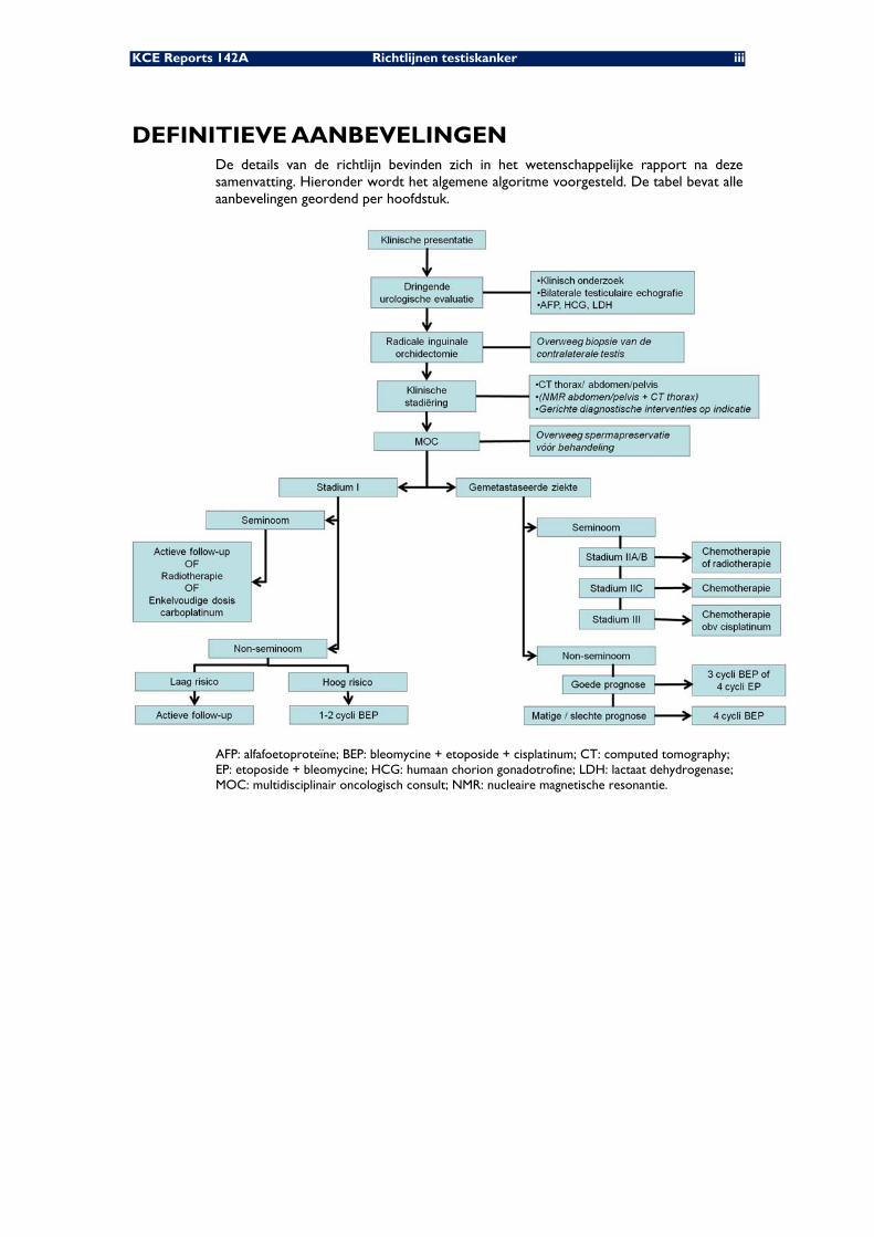
DEFINITIEVE AANBEVELINGEN De details van de richtlijn bevinden zich in het wetenschappelijke rapport na deze samenvatting. Hieronder wordt het algemene algoritme voorgesteld. De tabel bevat alle aanbevelingen geordend per hoofdstuk.
AFP: alfafoetoproteïne; BEP: bleomycine + etoposide + cisplatinum; CT: computed tomography; EP: etoposide + bleomycine; HCG: humaan chorion gonadotrofine; LDH: lactaat dehydrogenase; MOC: multidisciplinair oncologisch consult; NMR: nucleaire magnetische resonantie.
iv Richtlijnen testiskanker KCE Reports 142A
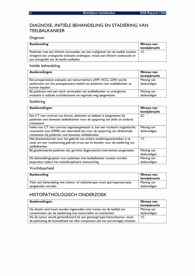
DIAGNOSE, INITIËLE BEHANDELING EN STADIËRING VAN TEELBALKANKER
Diagnose
Aanbeveling Niveau van bewijskracht
Patiënten met een klinisch vermoeden van een maligniteit van de teelbal moeten dringend een urologische evaluatie ondergaan, incluis een klinisch onderzoek en een echografie van de beide teelballen
1C
Initiële behandeling
Aanbevelingen Niveau van bewijskracht
Een preoperatieve evaluatie van tumormerkers (AFP, HCG, LDH) wordt aanbevolen om het postoperatieve beleid van patiënten met teelbalkanker te kunnen bepalen
Mening van deskundigen
Bij patiënten met een sterk vermoeden van teelbalkanker na urologische evaluatie is radicale orchidectomie via inguïnale weg aangewezen
Mening van deskundigen
Stadiëring
Aanbevelingen Niveau van bewijskracht
Een CT met contrast van thorax, abdomen en bekken is aangewezen bij patiënten met bewezen teelbalkanker voor de opsporing van (klier en andere) metastasen
2C
Indien een CT met contrast tegenaangewezen is, kan een nucleaire magnetische resonantie scan (NMR) een alternatief zijn voor de opsporing van abdominale metastasen bij patiënten met bewezen teelbalkanker
Mening van deskundigen
Het bewijsmateriaal voor het gebruik van andere stadiëringstechnieken is te zwak om een routinematig gebruik ervan aan te bevelen voor de stadiëring van teelbalkanker
1C
Bij geselecteerde patiënten zijn gerichte diagnostische interventies aangewezen Mening van deskundigen
De behandelingsopties voor patiënten met teelbalkanker moeten worden besproken tijdens het multidisciplinaire teamoverleg
Mening van deskundigen
Vruchtbaarheid
Aanbeveling Niveau van bewijskracht
Vóór een behandeling met chemo- of radiotherapie moet spermapreservatie aangeboden worden
Mening van deskundigen
HISTOPATHOLOGISCH ONDERZOEK Aanbevelingen Niveau van
bewijskracht De distale rand moet worden ingesneden vóór incisie van de teelbal om contaminatie van de zaadstreng met tumorcellen te voorkomen
Mening van deskundigen
Als de tumor wordt geclassificeerd als een gemengd type kiemceltumor, moet de patholoog de hoeveelheid van elke component (als een percentage) schatten
1C
KCE Reports 142A Richtlijnen testiskanker v
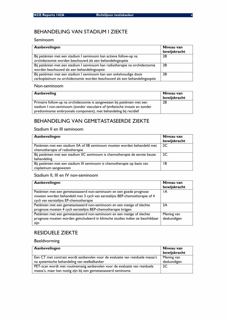
BEHANDELING VAN STADIUM I ZIEKTE
Seminoom
Aanbevelingen Niveau van bewijskracht
Bij patiënten met een stadium I seminoom kan actieve follow-up na orchidectomie worden beschouwd als een behandelingsoptie
2B
Bij patiënten met een stadium I seminoom kan radiotherapie na orchidectomie worden beschouwd als een behandelingsoptie
2B
Bij patiënten met een stadium I seminoom kan een enkelvoudige dosis carboplatinum na orchidectomie worden beschouwd als een behandelingsoptie
2B
Non-seminoom
Aanbeveling Niveau van bewijskracht
Primaire follow-up na orchidectomie is aangewezen bij patiënten met een stadium I non-seminoom (zonder vasculaire of lymfatische invasie en zonder predominante embryonale component), met behandeling bij recidief
2B
BEHANDELING VAN GEMETASTASEERDE ZIEKTE
Stadium II en III seminoom
Aanbevelingen Niveau van bewijskracht
Patiënten met een stadium IIA of IIB seminoom moeten worden behandeld met chemotherapie of radiotherapie
2C
Bij patiënten met een stadium IIC seminoom is chemotherapie de eerste keuze behandeling
2C
Bij patiënten met een stadium III seminoom is chemotherapie op basis van cisplatinum aangewezen
1B
Stadium II, III en IV non-seminoom
Aanbevelingen Niveau van bewijskracht
Patiënten met een gemetastaseerd non-seminoom en een goede prognose moeten worden behandeld met 3 cycli van eerstelijns BEP-chemotherapie of 4 cycli van eerstelijns EP-chemotherapie
1A
Patiënten met een gemetastaseerd non-seminoom en een matige of slechte prognose moeten 4 cycli eerstelijns BEP-chemotherapie krijgen
2A
Patiënten met een gemetastaseerd non-seminoom en een matige of slechte prognose moeten worden geïncludeerd in klinische studies indien ze beschikbaar zijn
Mening van deskundigen
RESIDUELE ZIEKTE
Beeldvorming
Aanbevelingen Niveau van bewijskracht
Een CT met contrast wordt aanbevolen voor de evaluatie van residuele massa’s na systemische behandeling van teelbalkanker
Mening van deskundigen
PET-scan wordt niet routinematig aanbevolen voor de evaluatie van residuele massa’s, maar kan nuttig zijn bij een gemetastaseerd seminoma
2C
vi Richtlijnen testiskanker KCE Reports 142A
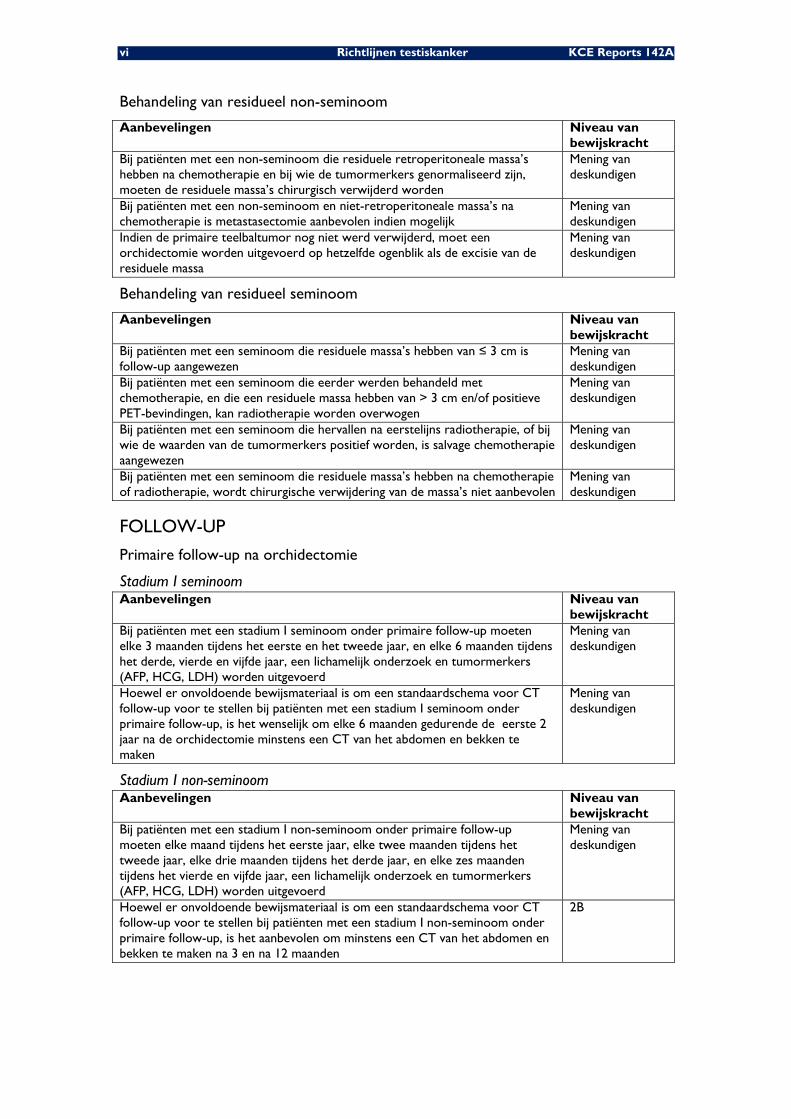
Behandeling van residueel non-seminoom
Aanbevelingen Niveau van bewijskracht
Bij patiënten met een non-seminoom die residuele retroperitoneale massa’s hebben na chemotherapie en bij wie de tumormerkers genormaliseerd zijn, moeten de residuele massa’s chirurgisch verwijderd worden
Mening van deskundigen
Bij patiënten met een non-seminoom en niet-retroperitoneale massa’s na chemotherapie is metastasectomie aanbevolen indien mogelijk
Mening van deskundigen
Indien de primaire teelbaltumor nog niet werd verwijderd, moet een orchidectomie worden uitgevoerd op hetzelfde ogenblik als de excisie van de residuele massa
Mening van deskundigen
Behandeling van residueel seminoom
Aanbevelingen Niveau van bewijskracht
Bij patiënten met een seminoom die residuele massa’s hebben van ≤ 3 cm is follow-up aangewezen
Mening van deskundigen
Bij patiënten met een seminoom die eerder werden behandeld met chemotherapie, en die een residuele massa hebben van > 3 cm en/of positieve PET-bevindingen, kan radiotherapie worden overwogen
Mening van deskundigen
Bij patiënten met een seminoom die hervallen na eerstelijns radiotherapie, of bij wie de waarden van de tumormerkers positief worden, is salvage chemotherapie aangewezen
Mening van deskundigen
Bij patiënten met een seminoom die residuele massa’s hebben na chemotherapie of radiotherapie, wordt chirurgische verwijdering van de massa’s niet aanbevolen
Mening van deskundigen
FOLLOW-UP
Primaire follow-up na orchidectomie
Stadium I seminoom Aanbevelingen Niveau van
bewijskracht Bij patiënten met een stadium I seminoom onder primaire follow-up moeten elke 3 maanden tijdens het eerste en het tweede jaar, en elke 6 maanden tijdens het derde, vierde en vijfde jaar, een lichamelijk onderzoek en tumormerkers (AFP, HCG, LDH) worden uitgevoerd
Mening van deskundigen
Hoewel er onvoldoende bewijsmateriaal is om een standaardschema voor CT follow-up voor te stellen bij patiënten met een stadium I seminoom onder primaire follow-up, is het wenselijk om elke 6 maanden gedurende de eerste 2 jaar na de orchidectomie minstens een CT van het abdomen en bekken te maken
Mening van deskundigen
Stadium I non-seminoom Aanbevelingen Niveau van
bewijskracht Bij patiënten met een stadium I non-seminoom onder primaire follow-up moeten elke maand tijdens het eerste jaar, elke twee maanden tijdens het tweede jaar, elke drie maanden tijdens het derde jaar, en elke zes maanden tijdens het vierde en vijfde jaar, een lichamelijk onderzoek en tumormerkers (AFP, HCG, LDH) worden uitgevoerd
Mening van deskundigen
Hoewel er onvoldoende bewijsmateriaal is om een standaardschema voor CT follow-up voor te stellen bij patiënten met een stadium I non-seminoom onder primaire follow-up, is het aanbevolen om minstens een CT van het abdomen en bekken te maken na 3 en na 12 maanden
2B
KCE Reports 142A Richtlijnen testiskanker vii
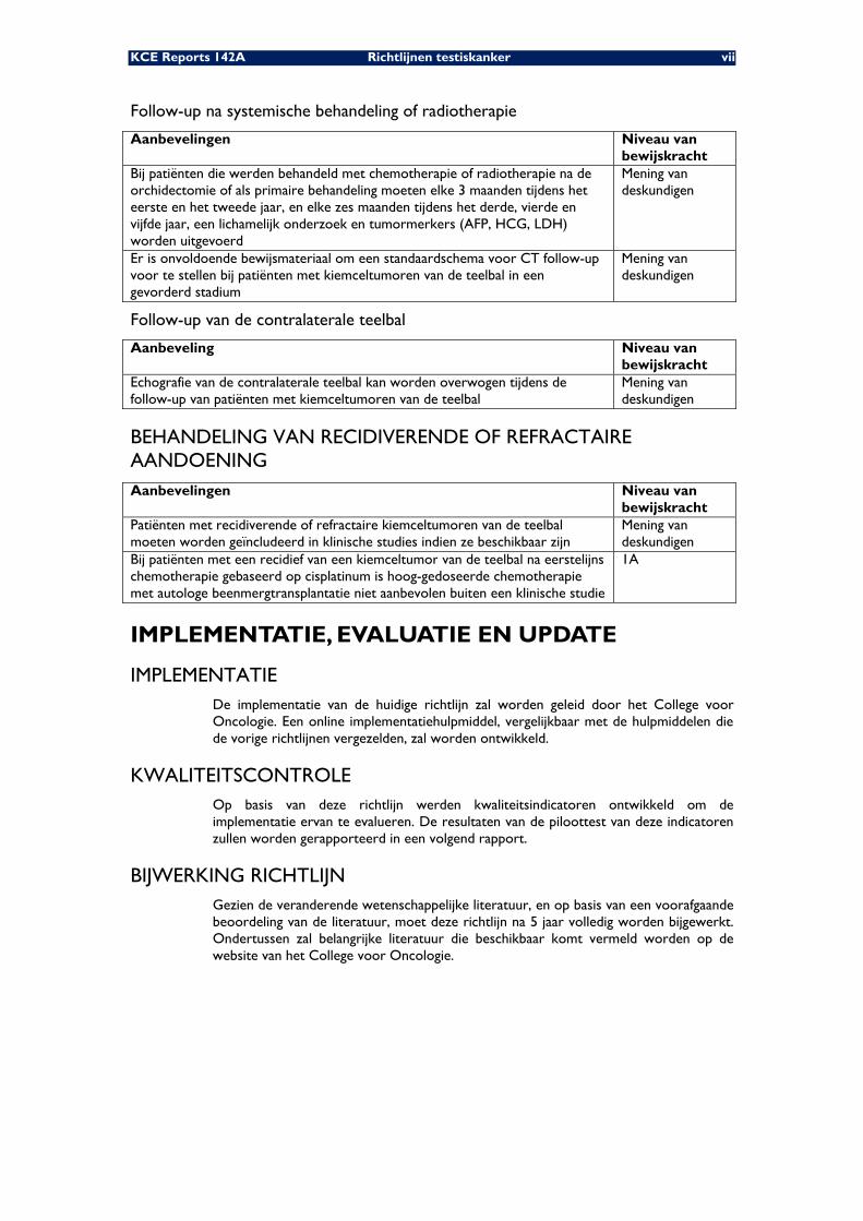
Follow-up na systemische behandeling of radiotherapie
Aanbevelingen Niveau van bewijskracht
Bij patiënten die werden behandeld met chemotherapie of radiotherapie na de orchidectomie of als primaire behandeling moeten elke 3 maanden tijdens het eerste en het tweede jaar, en elke zes maanden tijdens het derde, vierde en vijfde jaar, een lichamelijk onderzoek en tumormerkers (AFP, HCG, LDH) worden uitgevoerd
Mening van deskundigen
Er is onvoldoende bewijsmateriaal om een standaardschema voor CT follow-up voor te stellen bij patiënten met kiemceltumoren van de teelbal in een gevorderd stadium
Mening van deskundigen
Follow-up van de contralaterale teelbal
Aanbeveling Niveau van bewijskracht
Echografie van de contralaterale teelbal kan worden overwogen tijdens de follow-up van patiënten met kiemceltumoren van de teelbal
Mening van deskundigen
BEHANDELING VAN RECIDIVERENDE OF REFRACTAIRE AANDOENING Aanbevelingen Niveau van
bewijskracht Patiënten met recidiverende of refractaire kiemceltumoren van de teelbal moeten worden geïncludeerd in klinische studies indien ze beschikbaar zijn
Mening van deskundigen
Bij patiënten met een recidief van een kiemceltumor van de teelbal na eerstelijns chemotherapie gebaseerd op cisplatinum is hoog-gedoseerde chemotherapie met autologe beenmergtransplantatie niet aanbevolen buiten een klinische studie
1A
IMPLEMENTATIE, EVALUATIE EN UPDATE
IMPLEMENTATIE De implementatie van de huidige richtlijn zal worden geleid door het College voor Oncologie. Een online implementatiehulpmiddel, vergelijkbaar met de hulpmiddelen die de vorige richtlijnen vergezelden, zal worden ontwikkeld.
KWALITEITSCONTROLE Op basis van deze richtlijn werden kwaliteitsindicatoren ontwikkeld om de implementatie ervan te evalueren. De resultaten van de piloottest van deze indicatoren zullen worden gerapporteerd in een volgend rapport.
BIJWERKING RICHTLIJN Gezien de veranderende wetenschappelijke literatuur, en op basis van een voorafgaande beoordeling van de literatuur, moet deze richtlijn na 5 jaar volledig worden bijgewerkt. Ondertussen zal belangrijke literatuur die beschikbaar komt vermeld worden op de website van het College voor Oncologie.

KCE Reports 142 Guideline testicular cancer 1
Scientific summary Table of content
ABBREVIATIONS ...................................................................................................................... 3 1 INTRODUCTION ............................................................................................................ 5 1.1 SCOPE ............................................................................................................................................................. 5 1.2 EPIDEMIOLOGY ........................................................................................................................................... 5 2 METHODOLOGY ............................................................................................................ 6 2.1 GENERAL APPROACH .............................................................................................................................. 6 2.2 INTERNATIONAL COLLABORATION ................................................................................................ 6 2.3 CLINICAL QUESTIONS ............................................................................................................................. 6 2.4 LITERATURE SEARCHES ........................................................................................................................... 7
2.4.1 Search strategy .................................................................................................................................. 7 2.4.2 Quality appraisal ............................................................................................................................... 8
2.5 DATA EXTRACTION AND SUMMARY ................................................................................................ 8 2.6 FORMULATION OF RECOMMENDATIONS ...................................................................................... 8 2.7 EXTERNAL EXPERT MEETING ................................................................................................................ 9 2.8 DEFINITIONS ................................................................................................................................................ 9 3 DIAGNOSIS, PRIMARY MANAGEMENT AND STAGING OF TESTICULAR
CANCER ......................................................................................................................... 10 3.1 ALGORITHM ............................................................................................................................................... 10 3.2 DIAGNOSIS ................................................................................................................................................. 10 3.3 PRIMARY MANAGEMENT ...................................................................................................................... 11 3.4 CONTRALATERAL TESTIS ..................................................................................................................... 12 3.5 STAGING ..................................................................................................................................................... 12 3.6 FERTILITY ISSUES....................................................................................................................................... 13 4 HISTOPATHOLOGIC EXAMINATION .................................................................... 14 4.1 CLASSIFICATION ...................................................................................................................................... 14 4.2 MACROSCOPIC EXAMINATION ......................................................................................................... 14
4.2.1 Description ...................................................................................................................................... 14 4.2.2 Sampling of the resection specimen ........................................................................................... 15
4.3 MICROSCOPIC EXAMINATION .......................................................................................................... 15 4.4 IMMUNOHISTOCHEMISTRY ................................................................................................................. 15
4.4.1 Diagnosis of IGCN ......................................................................................................................... 15 4.4.2 Distinction between seminoma and non-seminoma ............................................................... 15
5 TREATMENT OF STAGE I DISEASE ......................................................................... 17 5.1 ALGORITHM ............................................................................................................................................... 17 5.2 STAGE I SEMINOMA................................................................................................................................. 17 5.3 STAGE I NON-SEMINOMA .................................................................................................................... 18 6 TREATMENT OF METASTATIC DISEASE ............................................................... 19 6.1 ALGORITHM ............................................................................................................................................... 19 6.2 STAGE II AND III SEMINOMA ................................................................................................................ 19 6.3 STAGE II, III AND IV NON-SEMINOMA ............................................................................................. 20 7 RESIDUAL DISEASE ..................................................................................................... 22 7.1 IMAGING ..................................................................................................................................................... 22 7.2 TREATMENT OF RESIDUAL NSGCT .................................................................................................. 22 7.3 TREATMENT OF RESIDUAL SGCT ...................................................................................................... 23 8 FOLLOW-UP ................................................................................................................. 24 8.1 PRIMARY SURVEILLANCE POST-ORCHIDECTOMY ..................................................................... 24

2 Guideline testicular cancer KCE Reports 142
8.2 FOLLOW-UP AFTER SYSTEMIC TREATMENT OR RADIOTHERAPY ....................................... 25 8.3 FOLLOW-UP OF THE CONTRALATERAL TESTIS .......................................................................... 25 8.4 FOLLOW-UP FOR LATE TOXICITY .................................................................................................... 26 9 TREATMENT OF RELAPSING OR REFRACTORY DISEASE ................................ 27 10 IMPLEMENTATION AND UPDATE OF THE GUIDELINE .................................... 28 10.1 IMPLEMENTATION ................................................................................................................................... 28 10.2 QUALITY CONTROL ............................................................................................................................... 28 10.3 GUIDELINE UPDATE ................................................................................................................................ 28 11 APPENDICES ................................................................................................................. 29 12 REFERENCES ................................................................................................................. 75
KCE Reports 142 Guideline testicular cancer 3
ABBREVIATIONS 95%CI 95 percent confidence interval
ACCC Association of Comprehensive Cancer Centres
ACR American College of Radiology
ACS American College of Surgeons
ADASP Association of Directors of Anatomic and Surgical Pathology
AFP Alphafoetoprotein
AFU Association Française d’Urologie
AHFMR Alberta Heritage Foundation for Medical Research
ASCO American Society of Clinical Oncology
BCR Belgian Cancer Registry
BEP Bleomycin, etoposide, cisplatin
CCO Cancer Care Ontario
CE-CT Contrast-enhanced CT
CoCanCPG Coordination of Cancer Clinical Practice Guidelines in Europe
CPG Clinical practice guideline
CT Computerized tomography
EAU European Association of Urology
EGCCCG European Germ Cell Cancer Consensus Group
EP Etoposide, cisplatin
ESMO European Society for Medical Oncology
FDG Fluorodeoxy-glucose
FNAC Fine-needle aspiration cytology
FNCLCC Fédération Nationale des Centres de Lutte Contre le Cancer
GCT Germ cell tumour
GIN Guidelines International Network
GP General Practitioner
Gy Gray
HAS Haute Authorité de Santé
HCG Human chorionic gonadotrophin
HDCT High-dose chemotherapy
HR Hazard ratio
HTA Health technology assessment
ICSI Institute for Clinical Systems Improvement
IGCCC International Germ Cell Consensus Classification
IGCN Intratubular germ cell neoplasia
IGCNU IGCN of the unclassified type
IGG Italian Germ cell cancer Group
KCE Belgian Healthcare Knowledge Centre
4 Guideline testicular cancer KCE Reports 142
LDH Lactate dehydrogenase
MDT Multidisciplinary team
MRI Magnetic resonance imaging
NACB National Academy of Clinical Biochemistry
NCCN National Comprehensive Cancer Network
NHMRC National Health and Medical Research Council
NICE National Institute for Health and Clinical Excellence
NPV Negative predictive value
NS Not significant
NSGCT Non-seminoma germ cell tumour
NZGG New Zealand Guidelines Group
OR Odds ratio
PET Positron emission tomography
PLAP Placental Alkaline Phosphatase
PPV Positive predictive value
QUADAS Quality Assessment of Diagnostic Accuracy Studies
RCT Randomized controlled trial
RPLND Retroperitoneal lymph node dissection
RT Radiotherapy
Se Sensitivity
SGCT Seminoma germ cell tumour
SIGN Scottish Intercollegiate Guidelines Network
SIR Standardized Incidence Rate
SMR Standardized Mortality Rate
Sp Specificity
UICC Union Against Cancer Classification
US Ultrasonography
US United States
USPSTF US Preventive Services Task Force
WHO World Health Organization
KCE Reports 142 Guideline testicular cancer 5
1 INTRODUCTION 1.1 SCOPE
In the present report, the clinical practice guideline (CPG) on testicular cancer, published in 2006, is updated 1. The previous guideline was mainly based on an adaptation of published guidelines with an additional search for systematic reviews. However, some of the included CPGs were of low quality. Above this, in a domain such as testicular cancer with few systematic reviews available, a search for recent primary studies seems necessary to dispose of the entire evidence base. In order to have a full evidence-based CPG, it was decided to perform a complete update of the previous version, with a search for guidelines, systematic reviews and primary studies.
This guideline is the result of a collaboration between the College of Oncology and the KCE. The CPG will cover a broad range of topics: diagnosis, staging, treatment and follow-up. It is restricted to men presenting with testicular germ cell tumours and does not address primary extragonadal germ cell cancer or non-germ cell testicular cancers (e.g. Leydig cell tumours, lymphoma, sarcoma, metastatic disease). The CPG is intended to be used by all care providers involved in the care for these men.
1.2 EPIDEMIOLOGY In Belgium, 269 new testicular cancers were diagnosed in 2006, with a crude incidence rate of 5.2/100 000 person years (source: Belgian Cancer Registry). Since 2003, the crude incidence rate slightly increased (4.7/100 000 person years), although it should be noted that the coverage of the cancer registration markedly improved since then. Testicular cancer typically is a cancer of young men, with a peak age-standardised incidence rate of 20.9/100 000 person years in the age category 25-30 years in 2006. In males aged 15-44 years, testicular cancer was the most frequent cancer in the period 2004-2005.
The Belgian crude incidence rate is comparable to that in the US (age-adjusted incidence rate 5.1/100 000 person years in 2004) 2, but lower than that in Germany (crude incidence rate 10.6/100 000 person years in 2005-2006) 3 and Luxembourg (age-adjusted incidence rate 7.7/100 000 person years in 2000-2004) 4.
No published mortality or survival data specifically for testicular cancer are available for Belgium. However, in the period 2000-2001, the relative 5-year survival for testicular cancer was 95% in Flanders 5. These data are in line with those reported in the literature for other countries and regions. For example, in England and Wales, the relative 5-year survival rose from 91% between 1986-1990 to 97% between 1996-19996. In the southern of the Netherlands, the relative 5-year survival was 99% and 96% for patients with seminoma and non-seminoma germ cell cancer respectively 7.
6 Guideline testicular cancer KCE Reports 142
2 METHODOLOGY 2.1 GENERAL APPROACH
As for the previous CPGs developed within the collaboration between the College and the KCE, the present CPG was developed by adapting (inter)national CPGs to the Belgian context (www.kce.fgov.be). This approach was recently structured in a formal methodology by the ADAPTE group, an international group of guideline developers and researchers 8. The ADAPTE methodology generally consists of three major phases (www.adapte.org):
1. Set-up Phase: Outlines the necessary tasks to be completed prior to beginning the adaptation process (e.g., identifying necessary skills and resources).
2. Adaptation Phase: Assists guideline developers in moving from selection of a topic to identification of specific clinical questions; searching for and retrieving guidelines; assessing the consistency of the evidence therein, their quality, currency, content and applicability; decision making around adaptation; and preparing the draft adapted guideline.
3. Finalization Phase: Guides guideline developers through getting feedback on the document from stakeholders who will be impacted by the guideline, consulting with the source developers of guidelines used in the adaptation process, establishing a process for review and updating of the adapted guideline and the process of creating a final document.
2.2 INTERNATIONAL COLLABORATION The KCE is involved in a European cancer network, CoCanCPG (Coordination of Cancer Clinical Practice Guidelines in Europe), aiming to develop a sustainable international collaboration for the joint management of mutually relevant priorities in CPG development and research to reduce the existing duplication and fragmentation of efforts, skills and information between programmes (www.cocancpg.eu).
Within this network, annual programmes are being exchanged to allow the identification of common projects and to highlight opportunities for collaboration. Since both the Scottish Intercollegiate Guidelines Network (SIGN) and the KCE planned the update of their CPG on testicular cancer, it was decided to use this guideline for a preliminary collaboration. One KCE expert took part of the guideline development group for the SIGN guideline, while two Belgian urologists of the KCE guideline development group served as external reviewer of the SIGN guideline. On the other hand, the KCE was able to use the preliminary documents of SIGN to feed the content of the KCE guideline. It is important to mention that, at the time of this writing, the SIGN guideline still is in the process of external review and consultation.
2.3 CLINICAL QUESTIONS The CPG addresses the following clinical questions:
1. What diagnostic tests are the most effective to confirm the diagnosis of testicular cancer?
2. What diagnostic tests are necessary to investigate the extent of testicular cancer? What is the place of tumour markers in the diagnosis, staging and prognosis?
3. Which treatments are the most effective for primary management of testicular cancer?
4. What is the work-up for the contralateral testis in case of testicular cancer?
5. What histopathological tests are needed to assess the extent and prognosis of testicular cancer? What parameters need to be reported in the histopathological report?
KCE Reports 142 Guideline testicular cancer 7
6. What is the most effective treatment for stage I testicular cancer?
7. What is the most effective treatment for metastatic testicular cancer?
8. What diagnostic tests are needed to evaluate residual disease? What is the most effective treatment of residual testicular cancer?
9. What is the most effective follow-up strategy after treatment for testicular cancer?
10. What is the most effective treatment of relapsing or refractory testicular cancer?
2.4 LITERATURE SEARCHES
2.4.1 Search strategy
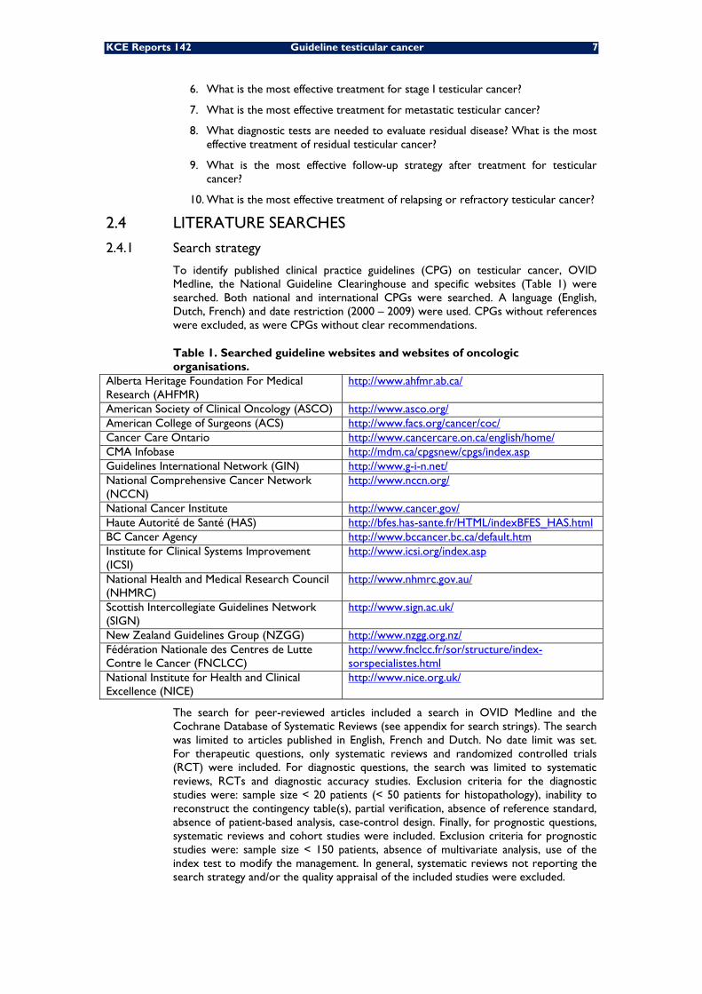
To identify published clinical practice guidelines (CPG) on testicular cancer, OVID Medline, the National Guideline Clearinghouse and specific websites (Table 1) were searched. Both national and international CPGs were searched. A language (English, Dutch, French) and date restriction (2000 – 2009) were used. CPGs without references were excluded, as were CPGs without clear recommendations.
Table 1. Searched guideline websites and websites of oncologic organisations.
Alberta Heritage Foundation For Medical Research (AHFMR)
http://www.ahfmr.ab.ca/
American Society of Clinical Oncology (ASCO) http://www.asco.org/ American College of Surgeons (ACS) http://www.facs.org/cancer/coc/ Cancer Care Ontario http://www.cancercare.on.ca/english/home/ CMA Infobase http://mdm.ca/cpgsnew/cpgs/index.asp Guidelines International Network (GIN) http://www.g-i-n.net/ National Comprehensive Cancer Network (NCCN)
http://www.nccn.org/
National Cancer Institute http://www.cancer.gov/ Haute Autorité de Santé (HAS) http://bfes.has-sante.fr/HTML/indexBFES_HAS.html BC Cancer Agency http://www.bccancer.bc.ca/default.htm Institute for Clinical Systems Improvement (ICSI)
http://www.icsi.org/index.asp
National Health and Medical Research Council (NHMRC)
http://www.nhmrc.gov.au/
Scottish Intercollegiate Guidelines Network (SIGN)
http://www.sign.ac.uk/
New Zealand Guidelines Group (NZGG) http://www.nzgg.org.nz/ Fédération Nationale des Centres de Lutte Contre le Cancer (FNCLCC)
http://www.fnclcc.fr/sor/structure/index-sorspecialistes.html
National Institute for Health and Clinical Excellence (NICE)
http://www.nice.org.uk/
The search for peer-reviewed articles included a search in OVID Medline and the Cochrane Database of Systematic Reviews (see appendix for search strings). The search was limited to articles published in English, French and Dutch. No date limit was set. For therapeutic questions, only systematic reviews and randomized controlled trials (RCT) were included. For diagnostic questions, the search was limited to systematic reviews, RCTs and diagnostic accuracy studies. Exclusion criteria for the diagnostic studies were: sample size < 20 patients (< 50 patients for histopathology), inability to reconstruct the contingency table(s), partial verification, absence of reference standard, absence of patient-based analysis, case-control design. Finally, for prognostic questions, systematic reviews and cohort studies were included. Exclusion criteria for prognostic studies were: sample size < 150 patients, absence of multivariate analysis, use of the index test to modify the management. In general, systematic reviews not reporting the search strategy and/or the quality appraisal of the included studies were excluded.
8 Guideline testicular cancer KCE Reports 142
All searches were run between January and December 2009, and updated in January 2010.
The identified studies were selected based on title and abstract. For all eligible studies, the full-text was retrieved. In case no full-text was available, the study was not taken into account for the final recommendations.
2.4.2 Quality appraisal
2.4.2.1 Clinical practice guidelines
The AGREE instrument 9 was used to evaluate the methodological quality of the identified CPGs. Each of the 20 identified CPGs was scored by two independent researchers (JV and SS) and discussed in case of disagreement (see appendix for an overview of the scores). Based on an overall assessment – taking into account the AGREE scores – 5 high-quality CPGs were finally selected. In general, CPGs with an aggregated domain score of 66% or less on the domain ‘Rigour of development’ were not included (see appendix 3).
2.4.2.2 Peer-reviewed articles
The quality of the retrieved systematic reviews, RCTs and prognostic studies was assessed using the checklists of the Dutch Cochrane Centre (www.cochrane.nl). The methodological quality of the diagnostic accuracy studies was assessed with the Quality Assessment of Diagnostic Accuracy Studies (QUADAS) checklist 10. All critical appraisals were done by a single KCE expert.
2.5 DATA EXTRACTION AND SUMMARY For each included CPG the following data were extracted: search date & publication year, searched databases, availability of evidence tables, recommendations and referenced evidence.
For each systematic review, the search date, publication year, included studies and main results were extracted. For primary studies, the following data were extracted: publication year, study population, study intervention, and outcomes.
For each clinical question, the recommendations from the identified CPGs and the additional evidence were summarized in evidence tables. A level of evidence was assigned to each recommendation and additional study using the GRADE system (see appendix 2).
2.6 FORMULATION OF RECOMMENDATIONS Based on the retrieved evidence, a first draft of recommendations was prepared by a KCE expert (JV). This draft together with the evidence tables were circulated to the guideline development group (Table 2) prior to each face-to-face meeting. The guideline development group met on two occasions (September 16th 2009 and January 26th 2010) to discuss the first draft. Recommendations were changed if important evidence supported this change. Based on the discussion meetings a second draft of recommendations was prepared.
A grade of recommendation was assigned to each recommendation using the GRADE system (see appendix 2). The second draft was once more circulated to the guideline development group for final approval.
KCE Reports 142 Guideline testicular cancer 9
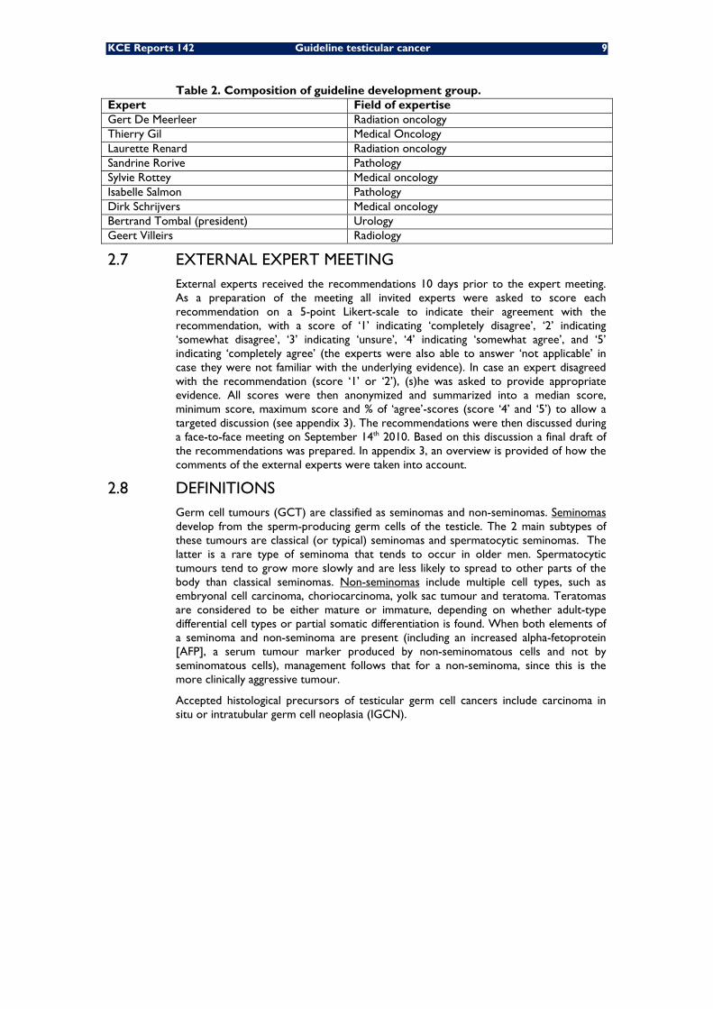
Table 2. Composition of guideline development group. Expert Field of expertise Gert De Meerleer Radiation oncology Thierry Gil Medical Oncology Laurette Renard Radiation oncology Sandrine Rorive Pathology Sylvie Rottey Medical oncology Isabelle Salmon Pathology Dirk Schrijvers Medical oncology Bertrand Tombal (president) Urology Geert Villeirs Radiology
2.7 EXTERNAL EXPERT MEETING External experts received the recommendations 10 days prior to the expert meeting. As a preparation of the meeting all invited experts were asked to score each recommendation on a 5-point Likert-scale to indicate their agreement with the recommendation, with a score of ‘1’ indicating ‘completely disagree’, ‘2’ indicating ‘somewhat disagree’, ‘3’ indicating ‘unsure’, ‘4’ indicating ‘somewhat agree’, and ‘5’ indicating ‘completely agree’ (the experts were also able to answer ‘not applicable’ in case they were not familiar with the underlying evidence). In case an expert disagreed with the recommendation (score ‘1’ or ‘2’), (s)he was asked to provide appropriate evidence. All scores were then anonymized and summarized into a median score, minimum score, maximum score and % of ‘agree’-scores (score ‘4’ and ‘5’) to allow a targeted discussion (see appendix 3). The recommendations were then discussed during a face-to-face meeting on September 14th 2010. Based on this discussion a final draft of the recommendations was prepared. In appendix 3, an overview is provided of how the comments of the external experts were taken into account.
2.8 DEFINITIONS Germ cell tumours (GCT) are classified as seminomas and non-seminomas. Seminomas develop from the sperm-producing germ cells of the testicle. The 2 main subtypes of these tumours are classical (or typical) seminomas and spermatocytic seminomas. The latter is a rare type of seminoma that tends to occur in older men. Spermatocytic tumours tend to grow more slowly and are less likely to spread to other parts of the body than classical seminomas. Non-seminomas include multiple cell types, such as embryonal cell carcinoma, choriocarcinoma, yolk sac tumour and teratoma. Teratomas are considered to be either mature or immature, depending on whether adult-type differential cell types or partial somatic differentiation is found. When both elements of a seminoma and non-seminoma are present (including an increased alpha-fetoprotein [AFP], a serum tumour marker produced by non-seminomatous cells and not by seminomatous cells), management follows that for a non-seminoma, since this is the more clinically aggressive tumour.
Accepted histological precursors of testicular germ cell cancers include carcinoma in situ or intratubular germ cell neoplasia (IGCN).
10 Guideline testicular cancer KCE Reports 142
3 DIAGNOSIS, PRIMARY MANAGEMENT AND STAGING OF TESTICULAR CANCER
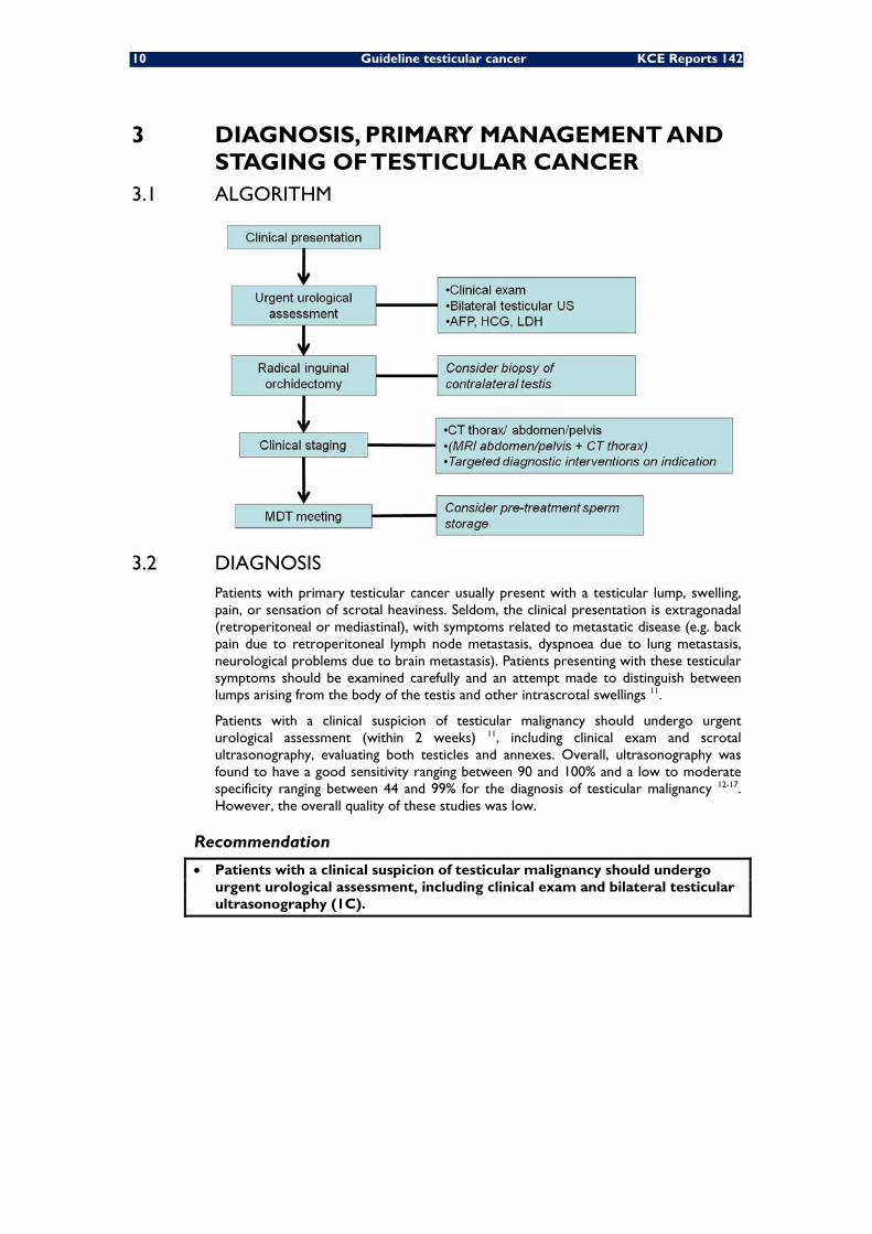
3.1 ALGORITHM
3.2 DIAGNOSIS Patients with primary testicular cancer usually present with a testicular lump, swelling, pain, or sensation of scrotal heaviness. Seldom, the clinical presentation is extragonadal (retroperitoneal or mediastinal), with symptoms related to metastatic disease (e.g. back pain due to retroperitoneal lymph node metastasis, dyspnoea due to lung metastasis, neurological problems due to brain metastasis). Patients presenting with these testicular symptoms should be examined carefully and an attempt made to distinguish between lumps arising from the body of the testis and other intrascrotal swellings 11.
Patients with a clinical suspicion of testicular malignancy should undergo urgent urological assessment (within 2 weeks) 11, including clinical exam and scrotal ultrasonography, evaluating both testicles and annexes. Overall, ultrasonography was found to have a good sensitivity ranging between 90 and 100% and a low to moderate specificity ranging between 44 and 99% for the diagnosis of testicular malignancy 12-17. However, the overall quality of these studies was low.
Recommendation
• Patients with a clinical suspicion of testicular malignancy should undergo urgent urological assessment, including clinical exam and bilateral testicular ultrasonography (1C).
KCE Reports 142 Guideline testicular cancer 11
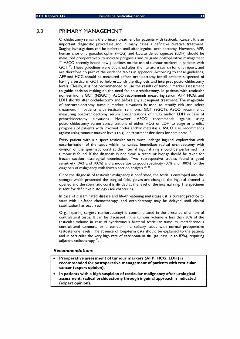
3.3 PRIMARY MANAGEMENT Orchidectomy remains the primary treatment for patients with testicular cancer. It is an important diagnostic procedure and in many cases a definitive curative treatment. Staging investigations can be deferred until after inguinal orchidectomy. However, AFP, human chorionic gonadotrophin (HCG) and lactate dehydrogenase (LDH) should be measured preoperatively to indicate prognosis and to guide postoperative management 18. ASCO recently issued new guidelines on the use of tumour markers in patients with GCT 19. These guidelines were published after the literature search for this report, and are therefore no part of the evidence tables in appendix. According to these guidelines, AFP and HCG should be measured before orchidectomy for all patients suspected of having a testicular GCT to help establish the diagnosis and interpret postorchidectomy levels. Clearly, it is not recommended to use the results of tumour marker assessment to guide decision making on the need for an orchidectomy. In patients with testicular non-seminoma GCT (NSGCT), ASCO recommends measuring serum AFP, HCG, and LDH shortly after orchidectomy and before any subsequent treatment. The magnitude of postorchidectomy tumour marker elevations is used to stratify risk and select treatment. In patients with testicular seminoma GCT (SGCT), ASCO recommends measuring postorchidectomy serum concentrations of HCG and/or LDH in case of preorchidectomy elevations. However, ASCO recommends against using postorchidectomy serum concentrations of either HCG or LDH to stage or predict prognosis of patients with involved nodes and/or metastasis. ASCO also recommends against using tumour marker levels to guide treatment decisions for seminoma 19.
Every patient with a suspect testicular mass must undergo inguinal exploration with exteriorization of the testis within its tunics. Immediate radical orchidectomy with division of the spermatic cord at the internal inguinal ring should be performed if a tumour is found. If the diagnosis is not clear, a testicular biopsy should be taken for frozen section histological examination. Two retrospective studies found a good sensitivity (94% and 100%) and a moderate to good specificity (89% and 100%) for the diagnosis of malignancy with frozen section analysis 20, 21.
Once the diagnosis of testicular malignancy is confirmed, the testis is enveloped into the sponges which protected the surgical field, gloves are changed, the inguinal channel is opened and the spermatic cord is divided at the level of the internal ring. The specimen is sent for definitive histology (see chapter 4).
In case of disseminated disease and life-threatening metastases, it is current practice to start with up-front chemotherapy, and orchidectomy may be delayed until clinical stabilisation has occurred.
Organ-sparing surgery (tumorectomy) is contraindicated in the presence of a normal contralateral testis. It can be discussed if the tumour volume is less than 30% of the testicular volume in case of synchronous bilateral testicular tumours, metachronous contralateral tumours, or a tumour in a solitary testis with normal preoperative testosterone levels. The absence of long-term data should be explained to the patient, and in particular the very high rate of carcinoma in situ (at least up to 82%), requiring adjuvant radiotherapy 22.
Recommendations
• Preoperative assessment of tumour markers (AFP, HCG, LDH) is recommended for postoperative management of patients with testicular cancer (expert opinion).
• In patients with a high suspicion of testicular malignancy after urological assessment, radical orchidectomy through inguinal approach is indicated (expert opinion).
12 Guideline testicular cancer KCE Reports 142
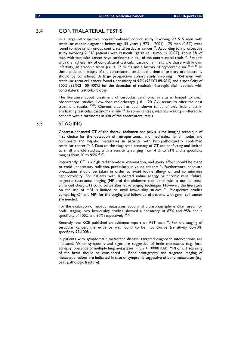
3.4 CONTRALATERAL TESTIS In a large retrospective population-based cohort study involving 29 515 men with testicular cancer diagnosed before age 55 years (1973 – 2001), 175 men (0.6%) were found to have synchronous contralateral testicular cancer 23. According to a prospective study involving 2 318 patients with testicular germ cell tumours (GCT), about 5% of men with testicular cancer have carcinoma in situ of the contralateral testis 24. Patients with the highest risk of contralateral testicular carcinoma in situ are those with known infertility, an atrophic testis (i.e. < 12 ml 25) and a history of cryptorchidism 24, 26-29. In these patients, a biopsy of the contralateral testis at the time of primary orchidectomy should be considered. A large prospective cohort study involving 1 954 men with testicular germ cell cancer found a sensitivity of 95% (95%CI 89-98%) and a specificity of 100% (95%CI 100-100%) for the detection of testicular intraepithelial neoplasia with contralateral testicular biopsy.
The literature about treatment of testicular carcinoma in situ is limited to small observational studies. Low-dose radiotherapy (18 – 20 Gy) seems to offer the best treatment results 30-33. Chemotherapy has been shown to be of only little effect in eradicating testicular carcinoma in situ 34. In some centres, watchful waiting is offered to patients with a carcinoma in situ of the contralateral testis.
3.5 STAGING Contrast-enhanced CT of the thorax, abdomen and pelvis is the imaging technique of first choice for the detection of retroperitoneal and mediastinal lymph nodes and pulmonary and hepatic metastases in patients with histopathologically confirmed testicular cancer 11, 18. Data on the diagnostic accuracy of CT are conflicting and limited to small and old studies, with a sensitivity ranging from 41% to 91% and a specificity ranging from 50 to 95% 35-39.
Importantly, CT is a high radiation-dose examination, and every effort should be made to avoid unnecessary radiation, particularly in young patients 40. Furthermore, adequate precautions should be taken in order to avoid iodine allergy or and to minimize nephrotoxicity. For patients with suspected iodine allergy or chronic renal failure, magnetic resonance imaging (MRI) of the abdomen (combined with a non-contrast-enhanced chest CT) could be an alternative staging technique. However, the literature on the use of MRI is limited to small low-quality studies 41. Prospective studies comparing CT and MRI for the staging and follow-up of patients with germ cell cancer are needed.
For the evaluation of hepatic metastases, abdominal ultrasonography is often used. For nodal staging, two low-quality studies showed a sensitivity of 87% and 95% and a specificity of 100% and 50% respectively 37, 42.
Recently, the KCE published an evidence report on PET scan 43. For the staging of testicular cancer, the evidence was found to be inconclusive (sensitivity 66-70%, specificity 97-100%).
In patients with symptomatic metastatic disease, targeted diagnostic interventions are indicated. When symptoms and signs are suggestive of brain metastases (e.g. focal epilepsy, presence of multiple lung metastases, HCG > 10000 IU/l), MRI or CT scanning of the brain should be considered 11. Bone scintigraphy and targeted imaging of metastatic lesions are indicated in case of symptoms suggestive of bone metastases (e.g. pain, pathologic fracture).
KCE Reports 142 Guideline testicular cancer 13
Recommendations
• Contrast-enhanced CT of the thorax, abdomen and pelvis is recommended in patients with confirmed testicular cancer for the detection of (nodal and extranodal) metastatic disease (2C).
• In patients with confirmed testicular cancer, magnetic resonance imaging is an alternative for the detection of abdominal metastatic disease if contrast-enhanced CT is contraindicated (expert opinion).
• The evidence supporting other staging techniques is too weak to recommend their routine use for the staging of testicular cancer (1C).
• In selected patients, targeted diagnostic interventions are indicated (expert opinion).
• Treatment options for patients with testicular cancer should be discussed at the multidisciplinary team meeting (expert opinion)
3.6 FERTILITY ISSUES Due to recent advances in in-vitro fertilisation technology and sperm banking procedures, even men with extremely reduced sperm count and motility are candidates for sperm cryopreservation 44. It is strongly recommended that sperm be collected before initiation of cancer therapy, because the quality of the sample and sperm DNA integrity may be compromised even after a single treatment session. In addition, in patients with testicular cancer, sperm quality may be poor even in patients who have not yet started treatment 11. Many patients have to start chemotherapy immediately or soon after diagnosis, limiting the potential number of ejaculates to one or two samples. Even in these instances, it is reasonable to make every effort to bank sperm, since recent progress in andrology laboratories and in the use of assisted reproductive techniques, particularly the technique of intracytoplasmic sperm injection, allows the successful freezing and future use of a very limited amount of sperm.
Recommendation
• Pre-treatment sperm storage should be offered to men who may require chemotherapy or radiotherapy (expert opinion).
14 Guideline testicular cancer KCE Reports 142
4 HISTOPATHOLOGIC EXAMINATION 4.1 CLASSIFICATION
The recommended histological classification of testicular tumours is that of the World Health Organization (WHO) Classification of Tumours 45.
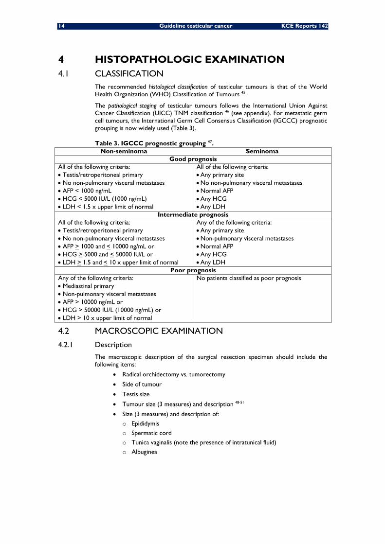
The pathological staging of testicular tumours follows the International Union Against Cancer Classification (UICC) TNM classification 46 (see appendix). For metastatic germ cell tumours, the International Germ Cell Consensus Classification (IGCCC) prognostic grouping is now widely used (Table 3).
Table 3. IGCCC prognostic grouping 47. Non-seminoma Seminoma
Good prognosis All of the following criteria: • Testis/retroperitoneal primary • No non-pulmonary visceral metastases • AFP < 1000 ng/mL • HCG < 5000 IU/L (1000 ng/mL) • LDH < 1.5 x upper limit of normal
All of the following criteria: • Any primary site • No non-pulmonary visceral metastases • Normal AFP • Any HCG • Any LDH
Intermediate prognosis All of the following criteria: • Testis/retroperitoneal primary • No non-pulmonary visceral metastases • AFP > 1000 and < 10000 ng/mL or • HCG > 5000 and < 50000 IU/L or • LDH > 1.5 and < 10 x upper limit of normal
Any of the following criteria: • Any primary site • Non-pulmonary visceral metastases • Normal AFP • Any HCG • Any LDH
Poor prognosis Any of the following criteria: • Mediastinal primary • Non-pulmonary visceral metastases • AFP > 10000 ng/mL or • HCG > 50000 IU/L (10000 ng/mL) or • LDH > 10 x upper limit of normal
No patients classified as poor prognosis
4.2 MACROSCOPIC EXAMINATION
4.2.1 Description
The macroscopic description of the surgical resection specimen should include the following items:
• Radical orchidectomy vs. tumorectomy
• Side of tumour
• Testis size
• Tumour size (3 measures) and description 48-51
• Size (3 measures) and description of:
o Epididymis
o Spermatic cord
o Tunica vaginalis (note the presence of intratunical fluid)
o Albuginea
KCE Reports 142 Guideline testicular cancer 15
4.2.2 Sampling of the resection specimen
A sample of the following structures needs to be taken:
• Tumour: 1 cm2 section for each cm of maximum tumour diameter;
• Normal macroscopic testis tissue: scar area if present;
• Albuginea nearby the tumour;
• Epididymis;
• Proximal and distal (surgical margin) sections of spermatic cord. The distal margin has to be cut prior to incision of the testis 52;
• If any suspected area is found, extensive sampling has to be done.
4.3 MICROSCOPIC EXAMINATION If the tumour is classified as a mixed type germ cell tumour, the pathologist has to estimate the amount of each component (as a percentage) 23, 48, 53-57.
The presence or absence of IGCN in non-tumoural parenchyma needs to be described.
The pathological TNM Staging needs to be done with specific attention to:
• Presence or absence of vascular and/or lymphatic invasion 48, 50, 53, 54;
• Presence or absence of invasion or extension through tunica albuginea, tunica vaginalis, rete testis 51, epididymis or spermatic cord invasion.
4.4 IMMUNOHISTOCHEMISTRY
4.4.1 Diagnosis of IGCN
The evidence on the use of immunohistochemical staining for the diagnosis of IGCN is limited to low-quality observational studies. Tavolini et al. reported a very low sensitivity of 0% and a moderate specificity of 89% for the detection of non-invasive testicular cancer with bilateral testicular FNAC and immunohistochemical staining for Placental Alkaline Phosphatase (PLAP) in cryptorchid patients who underwent orchidopexy 58. Nevertheless, PLAP can be demonstrated in a high percentage of IGCN of the unclassified type (IGCNU) 45.
CD-117 (or c-kit proto-oncogene) is also widely expressed in IGCN 59. Above this, nuclear reactivity of OCT3/4 can be useful to identify early forms of IGCNU 60. However, good diagnostic accuracy studies or prognostic studies investigating their diagnostic potential are lacking.
4.4.2 Distinction between seminoma and non-seminoma
PLAP 61, 62 and CD-117 59, 63 are widely expressed in classical seminoma. However, in a low-quality retrospective study, Heidenreich et al. found a moderate sensitivity of 79% and a low specificity of 45% for the distinction of seminoma from non-seminoma with PLAP immunohistochemical staining 61.
In case of differential diagnosis between seminoma and embryonal carcinoma, CD-117, CD-30 and pancytokeratins may be helpful 62, 63. Again, good diagnostic accuracy studies or prognostic studies investigating their diagnostic potential are lacking.
In case of differential diagnosis between seminoma and Yolk sac tumour, teratoma or choriocarcinoma, OCT4, AFP and beta-HCG may be helpful 61, 64-66. Sensitivity of immunohistochemical staining with AFP for the distinction between non-seminoma and seminoma was found to be low in 2 retrospective studies (20-67%) 61, 64. Specificity was high (100%) in both studies. Bosman et al. found a sensitivity of 74% and a specificity of 93% for the distinction between non-seminoma and seminoma with beta-HCG immunohistochemical staining 64. In the study of Heidenreich et al. no positive HCG staining was found 61. Importantly, both studies potentially suffered from selection bias. Two other retrospective studies, also potentially suffering from selection bias, found a high sensitivity (100%) and a low specificity (63-66%) for the distinction between seminoma and non-seminoma with OCT4 immunohistochemical staining 65, 66.

16 Guideline testicular cancer KCE Reports 142
Recommendations
• The distal margin has to be cut prior to incision of the testis to avoid tumour cell contamination of the spermatic cord (expert opinion).
• If the tumour is classified as a mixed type germ cell tumour, the pathologist has to estimate the amount of each component (as a percentage) (1C).
KCE Reports 142 Guideline testicular cancer 17
5 TREATMENT OF STAGE I DISEASE 5.1 ALGORITHM
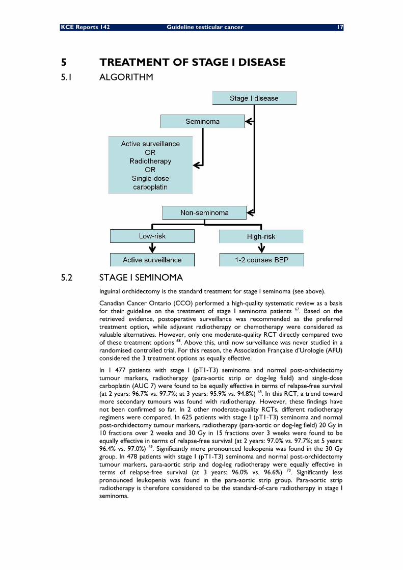
5.2 STAGE I SEMINOMA Inguinal orchidectomy is the standard treatment for stage I seminoma (see above).
Canadian Cancer Ontario (CCO) performed a high-quality systematic review as a basis for their guideline on the treatment of stage I seminoma patients 67. Based on the retrieved evidence, postoperative surveillance was recommended as the preferred treatment option, while adjuvant radiotherapy or chemotherapy were considered as valuable alternatives. However, only one moderate-quality RCT directly compared two of these treatment options 68. Above this, until now surveillance was never studied in a randomised controlled trial. For this reason, the Association Française d’Urologie (AFU) considered the 3 treatment options as equally effective.
In 1 477 patients with stage I (pT1-T3) seminoma and normal post-orchidectomy tumour markers, radiotherapy (para-aortic strip or dog-leg field) and single-dose carboplatin (AUC 7) were found to be equally effective in terms of relapse-free survival (at 2 years: 96.7% vs. 97.7%; at 3 years: 95.9% vs. 94.8%) 68. In this RCT, a trend toward more secondary tumours was found with radiotherapy. However, these findings have not been confirmed so far. In 2 other moderate-quality RCTs, different radiotherapy regimens were compared. In 625 patients with stage I (pT1-T3) seminoma and normal post-orchidectomy tumour markers, radiotherapy (para-aortic or dog-leg field) 20 Gy in 10 fractions over 2 weeks and 30 Gy in 15 fractions over 3 weeks were found to be equally effective in terms of relapse-free survival (at 2 years: 97.0% vs. 97.7%; at 5 years: 96.4% vs. 97.0%) 69. Significantly more pronounced leukopenia was found in the 30 Gy group. In 478 patients with stage I (pT1-T3) seminoma and normal post-orchidectomy tumour markers, para-aortic strip and dog-leg radiotherapy were equally effective in terms of relapse-free survival (at 3 years: 96.0% vs. 96.6%) 70. Significantly less pronounced leukopenia was found in the para-aortic strip group. Para-aortic strip radiotherapy is therefore considered to be the standard-of-care radiotherapy in stage I seminoma.
18 Guideline testicular cancer KCE Reports 142
Recently, Groll et al. pooled the results of 14 observational studies involving 2 060 patients 71. Cause-specific survival was found to be 99.7%. Overall, 356 relapses (17%) were found, of which 23 (2%) were late relapses.
Recommendations
• In patients with stage I seminoma post-orchidectomy, active surveillance can be considered as a management option (2B).
• In patients with stage I seminoma post-orchidectomy, radiotherapy can be considered as a management option (2B).
• In patients with stage I seminoma post-orchidectomy, single-dose carboplatin can be considered as a management option (2B).
5.3 STAGE I NON-SEMINOMA Inguinal orchidectomy is the standard treatment for stage I non-seminoma (see above).
Based on a systematic review of the literature, CCO recommended primary surveillance after inguinal orchidectomy for all patients with clinical stage I non-seminoma, with treatment at relapse 72. On the contrary, the AFU recommended to take into account the relapse risk for determining the optimal treatment strategy 18. For patients with low risk (defined by the AFU as no vascular or lymphatic invasion), the AFU offers the choice between surveillance and retroperitoneal lymph node dissection (RPLND). For high-risk patients (defined by the AFU as vascular or lymphatic invasion), the choice is offered between surveillance and chemotherapy 18.
One RCT compared surveillance to adjuvant radiotherapy in 156 patients with clinical stage I non-seminoma 73. In the first year after orchidectomy, no significant difference in relapse rate was found (radiotherapy 23% vs. surveillance 14%). However, radiotherapy completely prevented retroperitoneal relapse. Importantly, all relapsing patients in the surveillance group were without evidence of disease with a median observation time of 67 months after salvage chemotherapy.
A more recent RCT compared one cycle of BEP (bleomycin + etoposide + cisplatin) to RPLND with 2 cycles of BEP in case of retroperitoneal metastases in 382 patients with clinical stage I non-seminoma 74. Less recurrences were observed in the BEP group compared to the RPLND group (2 vs. 15, p=0.0011). Recurrence-free survival was significantly better in the BEP group (at 2 years: 99.5% vs. 91.9%; HR 7.937, 95%CI 1.808-34.48).
Groll et al. pooled the results of 31 studies (including the RCT of Rorth et al.) 71. The cause-specific survival was found to be 98.6%. Overall, 1 025 relapses (28%) were identified, of which 55 (2%) were late relapses. The following predictors of relapse were identified: vascular invasion of the primary tumour, predominantly embryonal carcinoma histology (i.e. >50%), T stage and absence of yolk sac tumour in the primary specimen 71. In stage I non-seminoma patients with these risk factors, treatment with 1 or 2 courses of BEP should be considered 18, 75.
Recommendations
• Primary surveillance is recommended for patients with stage I non-seminoma (without vascular or lymphatic invasion and without predominant embryonal component) post-orchidectomy, with treatment at relapse (2B).
KCE Reports 142 Guideline testicular cancer 19
6 TREATMENT OF METASTATIC DISEASE 6.1 ALGORITHM
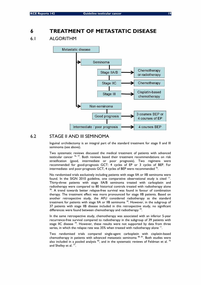
6.2 STAGE II AND III SEMINOMA Inguinal orchidectomy is an integral part of the standard treatment for stage II and III seminoma (see above).
Two systematic reviews discussed the medical treatment of patients with advanced testicular cancer 76, 77. Both reviews based their treatment recommendations on risk stratification (good, intermediate or poor prognosis). Two regimens were recommended for good-prognosis GCT: 4 cycles of EP or 3 cycles of BEP. For intermediate- and poor-prognosis GCT, 4 cycles of BEP were recommended 76.
No randomised trials exclusively including patients with stage IIA or IIB seminoma were found. In the SIGN 2010 guideline, one comparative observational study is cited 11. Thirty-three patients with stage IIA/B seminoma treated with carboplatin and radiotherapy were compared to 80 historical controls treated with radiotherapy alone 78. A trend towards better relapse-free survival was found in favour of combination therapy. The treatment effect was more pronounced for stage IIB patients. Based on another retrospective study, the AFU considered radiotherapy as the standard treatment for patients with stage IIA or IIB seminoma 18. However, in the subgroup of 37 patients with stage IIB disease included in this retrospective study, no significant differences were found between chemotherapy and radiotherapy 79.
In the same retrospective study, chemotherapy was associated with an inferior 5-year recurrence-free survival compared to radiotherapy in the subgroup of 39 patients with stage IIC disease 79. However, these results were not supported by data from three series, in which the relapse rate was 35% when treated with radiotherapy alone 11.
Two randomised trials compared single-agent carboplatin with cisplatin-based chemotherapy in patients with advanced metastatic seminoma 80, 81. Both studies were also included in a pooled analysis 82, and in the systematic reviews of Feldman et al. 76 and Shelley et al. 77.
20 Guideline testicular cancer KCE Reports 142
Horwich et al. randomised 130 patients with relapsing (after radiotherapy) or newly diagnosed advanced seminoma (stage IIC, III or IV) to single agent carboplatin or cisplatin and etoposide 81. The majority of the included patients had testicular seminoma (85%). No significant differences were found in terms of overall survival (HR 0.85; 95%CI 0.35-2.10), progression-free survival (HR 0.64; 95%CI 0.32-1.28) and failure-free survival (HR 0.56; 95%CI 0.28-1.09) at three years. In the second study, which is only available as an abstract, 280 patients with advanced metastatic seminoma were randomised to single agent carboplatin or cisplatin, etoposide and ifosfamide 80. Again, no significant difference was found in terms of overall survival (87% vs. 95%, no p-value provided). However, relapse-free survival was significantly better with cisplatin-based chemotherapy (74% vs. 95%, p<0.01). Cisplatin-based chemotherapy was associated with more grade 3/4 leuko- and thrombopenia (8% vs. 72%). Individual patient data (n=361) from these two trials were pooled 82. Patients treated with single agent carboplatin had an inferior 5-year overall (89 vs. 94%; p=0.09) and progression-free survival rate (72 vs. 92%; p<0.0001) compared with patients receiving cisplatin-based combinations. Based on these observations, the AFU recommended cisplatin-based chemotherapy (3 courses of BEP or 4 courses of EP) as standard treatment for patients with stage IIC or III seminoma 18.
Recommendations
• Patients with stage IIA or IIB seminoma should be treated with chemotherapy or radiotherapy (2C).
• In patients with stage IIC seminoma chemotherapy is the treatment of choice (2C).
• In patients with stage III seminoma cisplatin-based chemotherapy is recommended (1B).
6.3 STAGE II, III AND IV NON-SEMINOMA The systematic review of Feldman et al. included 11 RCTs of different first-line chemotherapy regimens in patients with good-prognosis metastatic NSGCT 76. Four of these studies exclusively included patients with NSGCT 83-86. Two studies compared 3 vs. 4 cycles of BEP and found equal efficacy, but less toxicity with 3 cycles 87, 88. In another trial, 3 cycles of BEP were found to be more effective than 3 cycles of EP (overall survival: 95% vs. 86%, p=0.01; failure-free survival: 86% vs. 69%, p=0.01) 89. However, in a more recent trial, 4 cycles of EP were found to be equivalent to 3 cycles of BEP based on equal efficacy (4-year overall survival: HR 0.42, 95%CI 0.15-1.20) and balanced toxicity 83. Based on this evidence, the authors recommended two regimens for patients with good-prognosis GCT (both seminoma and non-seminoma): 3 cycles of BEP or 4 cycles of EP (e.g. in case of contraindications for bleomycin). These conclusions are in line with the recommendations of the AFU 18.
Feldman et al. included 8 RCTs of first-line chemotherapy regimens in patients with intermediate- or poor-prognosis metastatic NSGCT 76. Four of these studies exclusively included patients with NSGCT 90-93. In all trials that compared 4 cycles of BEP with another regimen, BEP was found to be equally or more effective, but less toxic 91-97. Based on this evidence, the authors recommended 4 cycles of BEP for intermediate- and poor-prognosis GCT. These conclusions are again in line with the recommendations of the AFU 18.
One more recent RCT compared 4-6 cycles of CISCA/VB to 4 cycles of first-line BEP in 188 patients with intermediate- or poor-prognosis metastatic NSGCT 98. Five-year event-free (HR 0.76, 95%CI 0.52-1.11) and overall survival (HR 0.73, 95%CI 0.46-1.18) did not differ significantly between the two treatment groups. However, CISCA/VB was associated with more significant haematological and mucous toxicities.
Finally, a recent RCT, presented as an abstract, demonstrated no improved outcomes with high-dose chemotherapy plus autologous stem cell support given as part of first-line therapy in patients with poor-prognosis GCT 99.

KCE Reports 142 Guideline testicular cancer 21
Recommendations
• Patients with good prognosis metastatic NSGCT should be treated with 3 cycles of first-line BEP chemotherapy or 4 cycles of first-line EP chemotherapy (1A).
• Patients with intermediate prognosis metastatic NSGCT should receive first-line BEP chemotherapy in 4 cycles (2A).
• Patients with poor prognosis metastatic NSGCT should be treated with first-line BEP chemotherapy in 4 cycles (2A).
• Patients with intermediate and poor prognosis metastatic NSGCT should be enrolled in clinical trials when available (expert opinion).
22 Guideline testicular cancer KCE Reports 142
7 RESIDUAL DISEASE 7.1 IMAGING
Very few diagnostic accuracy studies evaluated the use of different diagnostic tests for the assessment of residual masses. Pfannenberg et al. compared the diagnostic accuracy of FDG-PET and CT/MRI for the assessment of the viability of 60 residual masses in 28 patients with metastatic GCT treated with high-dose chemotherapy 100. In this study, MRI was used in patients with iodine allergy, and thus no comparison was made between CT and MRI. No significant differences were found for sensitivity (PET: 62%; CT/MRI: 62%) or specificity (PET: 83%; CT/MRI: 72%). In another study, the diagnostic accuracy of FDG-PET and CT was compared in 54 patients with metastatic seminoma and a CT-documented mass after chemotherapy 101. For the diagnosis of viability of residual masses, PET scan was found to have a significantly better sensitivity (80% vs. 73%) and specificity (100% vs. 73%) than CT. However, both studies suffered from methodological weaknesses (small sample size, per-lesion-analysis, differential verification, possible incorporation bias).
Finally, Oechsle et al. prospectively evaluated the diagnostic accuracy of FDG-PET in 121 patients with NSGCT and a residual mass on conventional imaging (i.e. CT) after cisplatin-based chemotherapy 102. Reference standard was histology in all patients. For the evaluation of viability of the residual mass, sensitivity was 70% and specificity 39%.
Despite this poor evidence, CT is the most commonly used diagnostic test for the assessment of residual masses. The evidence discussed above is insufficient to alter current practice. Therefore, CE-CT scan remains recommended for the imaging of residual masses after systemic treatment of testicular cancer. In case of contra-indications (iodine allergy, renal function impairment), MRI can be an alternative diagnostic technique. In view of the inconsistent results for FDG-PET, it cannot be routinely recommended for the evaluation of residual masses, although it may be useful in metastatic seminoma (residual lesions > 3 cm).
Recommendations
• CE-CT scan is recommended for the imaging of residual masses after systemic treatment of testicular cancer (expert opinion).
• PET scan is not routinely recommended for the evaluation of residual masses, but may be useful in metastatic seminoma (2C).
7.2 TREATMENT OF RESIDUAL NSGCT The evidence on the treatment of residual NSGCT is limited to observational studies. Therefore, the optimal management of a residual mass following treatment for NSGCT is a subject of ongoing debate. Nevertheless, there is considerable consensus concerning the need for surgical resection in patients with NSGCT who have residual retroperitoneal masses (i.e. > 1 cm) after chemotherapy and whose markers have normalised 103. The decision on the extent of resection should take into account the postoperative morbidity and risk for residual disease or relapse.
In patients with NSGCT and non-peritoneal masses (e.g. pulmonary or hepatic masses) after chemotherapy, metastasectomy can be safely performed 104-106.
If the primary testicular tumour has not already been removed, an orchidectomy should be performed at the same time as excision of the residual mass 107.
KCE Reports 142 Guideline testicular cancer 23
Recommendations
• In patients with NSGCT who have residual retroperitoneal masses after chemotherapy and whose markers have normalised, the residual masses should be removed (expert opinion).
• In patients with NSGCT and non-retroperitoneal masses after chemotherapy, metastatectomy is recommended if feasible (expert opinion).
• If the primary testicular tumour has not already been removed, an orchidectomy should be performed at the same time as excision of the residual mass (expert opinion).
7.3 TREATMENT OF RESIDUAL SGCT In patients with seminoma who have residual masses following chemotherapy or radiotherapy, surgery is not routinely indicated. Indeed, surgical resection of residual seminomatous elements is technically challenging owing to the severe desmoplastic reaction between the regressing mass and the adjacent vascular and visceral structures 103.
According to the consensus-based guidelines of the EGCCCG 108 and the EAU 75, FDG-PET should direct treatment in patients with residual masses of > 3 cm in size after treatment for seminoma. This recommendation is based on the results of the SEMPET trial, in which the sensitivity and specificity of FDG-PET for the prediction of viable residual tumour in patients with residual masses > 3 cm were 80% (95%CI 44-95%) and 100% (95%CI 92-100%) respectively 101. However, these findings are insufficient to recommend a routine use of PET scan to guide treatment decisions in these patients.
No resection or any other treatment modality is necessary in patients with a mass ≤ 3 cm, and surveillance is recommended in these cases. No clinical data are available to support active treatment in these cases.
In patients with seminoma previously treated with chemotherapy, and who have a residual mass > 3 cm and/or positive PET findings, radiotherapy is recommended. Although this is not studied prospectively, one retrospective series supports this recommendation 109. Patients who relapse after first-line radiotherapy have a cure rate of > 90% and should be treated with cisplatin-based chemotherapy 108. In patients with seminoma whose tumour markers become positive, salvage chemotherapy is also indicated.
Recommendations
• In patients with seminoma who have residual masses ≤ 3 cm, surveillance is recommended (expert opinion).
• In patients with seminoma previously treated with chemotherapy, and who have a residual mass > 3 cm and/or positive PET findings, radiotherapy can be considered (expert opinion).
• In patients with seminoma relapsing after first-line radiotherapy or whose tumour markers become positive, salvage chemotherapy is indicated (expert opinion).
• In patients with seminoma who have residual masses following chemotherapy or radiotherapy, extirpative surgery is not recommended (expert opinion).
24 Guideline testicular cancer KCE Reports 142
8 FOLLOW-UP In general, follow-up is intended to 11:
• Detect relapse at a stage where therapy has the best chance of being effective;
• Monitor and treat treatment-related toxicity;
• Detect metachronous cancers, in particular contralateral testicular cancers.
8.1 PRIMARY SURVEILLANCE POST-ORCHIDECTOMY Few studies evaluated the use of diagnostic tests for follow-up and the frequency of this follow-up in patients with stage I GCT under primary surveillance after orchidectomy.
One RCT compared surveillance with two CT scans over 1 year (at 3 and 12 months post-orchidectomy) to surveillance with five CT scans over 2 years (at 3, 6, 9, 12 and 24 months post-orchidectomy) in 414 patients with stage I NSGCT 110. Relapse rate was comparable in both groups (15% vs. 20%), no patients had poor prognosis at relapse. Based on observational data, two published guidelines of sufficient quality included recommendations on the follow-up of stage I NSGCT patients 18, 111. Furthermore, the new SIGN guideline also contains valuable information on this topic 11.
Most guidelines agree on the frequency of follow-up visits that include a clinical examination, blood serum marker tests (AFP, HCG and LDH) and a chest X-ray. These follow-up visits should be conducted every month in the first year, every two months in the second year, every three months in the third year, and every six months in the fourth and fifth years.
On the frequency of post-orchidectomy thoraco-abdomino-pelvic CT scans there is less agreement. Both CCO and AFU recommended a CT scan every three months in the first year, every four to six months in the second year and every six months in the third year 18, 111. In the fourth and fifth year, the AFU recommended a CT every 6 months 18, while the CCO recommended an annual CT 111. On the contrary, the SIGN guideline took into account the results of Rustin et al. (2007) 110, and recommended a post-orchidectomy CT scan at 3 and 12 months 11.
On the follow-up of stage I seminomas there is less information available. The AFU recommended blood serum marker tests and a CT scan every 6 months in the first five years post-orchidectomy 18. SIGN proposed a 3-monthly follow-up visit in the first two years and a 6-monthly visit in the third and fourth year 11. A CT is recommended every 6 months during the first two years and annually during the third and fourth year.
In view of the absence of a consensus on the frequency of CT examinations during primary surveillance, no standard scheme can be proposed. However, at least an abdomino-pelvic CT every 6 months during the 2 first years post-orchidectomy is desirable for stage I seminomas. For patients with stage I non-seminoma under primary surveilance, at least an abdomino-pelvic CT at 3 and 12 months is recommended.
As mentioned above, CT is a high radiation-dose examination, and every effort should be made to avoid unnecessary radiation, particularly in young patients 40. Furthermore, adequate precautions should be taken in order to avoid iodine allergy or nephrotoxicity. For these patients, MRI could be an alternative follow-up technique.
KCE Reports 142 Guideline testicular cancer 25
Recommendations
• In patients with stage I seminoma under primary surveillance, physical examination and blood serum marker tests (AFP, HCG, LDH) should be conducted every 3 months in the first and second years, and every six months in the third, fourth and fifth years (expert opinion).
• Although the evidence is insufficient to propose a standard scheme for CT follow-up in patients with stage I seminoma under primary surveilance, at least an abdomino-pelvic CT every 6 months during the 2 first years post-orchidectomy is desirable (expert opinion).
• In patients with stage I non-seminoma under primary surveillance, physical examination and blood serum marker tests (AFP, HCG, LDH) should be conducted every month in the first year, every two months in the second year, every three months in the third year, and every six months in the fourth and fifth years (expert opinion).
• Although the evidence is insufficient to propose a standard scheme for CT follow-up in patients with stage I non-seminoma under primary surveilance, at least an abdomino-pelvic CT at 3 and 12 months is recommended (2B).
8.2 FOLLOW-UP AFTER SYSTEMIC TREATMENT OR RADIOTHERAPY Useful information on the follow-up of patients with GCT treated with systemic treatment or radiotherapy is scarce. SIGN recommended a follow-up visit every three months during the first two years and every 6 months in the third, fourth and fifth year 11. For patients with NSGCT, the recommended frequency of follow-up visits is higher during the first year. For stage I seminomas not treated with primary surveillance, SIGN recommended an annual CT during the first three years. For all other patients treated with systemic treatment or radiotherapy, SIGN recommended to stop routine follow-up CT scans if the post-treatment CT is normal 11.
However, in view of the absence of a consensus on the frequency of CT examinations during primary surveillance, no standard scheme can be proposed.
Recommendations
• In patients treated with chemotherapy or radiotherapy post-orchidectomy or as primary treatment, physical examination and blood serum marker tests (AFP, HCG, LDH) should be conducted every 3 months in the first and second years, and every six months in the third, fourth and fifth years (expert opinion).
• There is insufficient evidence to define a standard scheme for CT follow-up in patients with advanced stage testicular germ cell cancer (expert opinion).
8.3 FOLLOW-UP OF THE CONTRALATERAL TESTIS Observational studies suggest a role of US for the follow-up of the contralateral testis in patients with testicular germ cell cancer. In a recent small retrospective study (n = 16), testicular tumours detected through US were significantly smaller than those diagnosed by palpation only 112. Above this, the rate of organ preservation was higher in tumours detected through US. Therefore, US of the contralateral testis can be considered during the follow-up of patients with testicular germ cell cancer.
Recommendation
• Ultrasonography of the contralateral testis can be considered during the follow-up of patients with testicular germ cell cancer (expert opinion).
26 Guideline testicular cancer KCE Reports 142
8.4 FOLLOW-UP FOR LATE TOXICITY For the present guideline, no specific systematic search was done addressing late toxicity after treatment for testicular cancer. Therefore, no separate recommendations were formulated addressing this issue. Nevertheless, the 2010 SIGN guideline specifically addressed this topic and was therefore consulted to write the paragraph below 11.
Besides the well-known short, medium and long-term toxicities, such as neurotoxicity, nephrotoxicity, pulmonary toxicity and androgen deficiency, SIGN identified two specific types of very late toxicity associated with testicular cancer therapy with an increasing body of evidence: cardiovascular events and second cancers.
SIGN identified several studies showing an increased standardized mortality rate (SMR) and standardized incidence rate (SIR) for cardiovascular disease in patients treated for germ cell cancer. Inconsistent evidence was found for an increased frequency of cardiovascular risk factors in survivors of testicular cancer. SIGN recommended that oncologists should advise survivors of testicular cancer and their general practitioners (GPs) of the increased risk of cardiovascular disease and its risk factors at the time of discharge from the hospital or at around five years after diagnosis if maintained on long term follow up 11.
In addition, observational studies showed an increased SMR and SIR for second non-germ cell cancer 11. Elevated SIRs are reported for a wide range of solid and haematological cacners. Based on this evidence, SIGN recommended that oncologists should advise patients and their GPs of the increased risk of non-germ cell second malignancies (at the time of initial diagnosis, following initial therapy and at discharge from follow up by oncology clinics). The risks are greatest for those treated before the age of 30 years. Increased risks continue beyond 15 years following treatment. SIGN also stated that there should be increased awareness of the risk of haematological malignancies especially after chemotherapy and solid malignancies in or near the fields of radiotherapy. There should be a low threshold for further investigation and appropriate referral to secondary care if any alert symptoms are reported 11.
KCE Reports 142 Guideline testicular cancer 27
9 TREATMENT OF RELAPSING OR REFRACTORY DISEASE The evidence on the treatment of relapsing or refractory disease is mainly limited to observational studies. Two RCTs evaluated the efficacy of high-dose chemotherapy in patients with relapsed or refractory GCT after cisplatin-based chemotherapy. Pico et al. compared 4 cycles of PEI (ifosfamide, cisplatin and etoposide) or VeIP (ifosfamide, cisplatin and vinblastine) with 3 cycles of these regimens followed by CarboPEC (high-dose carboplatin, etoposide and cyclophosphamide) and haematopoietic stem cell support 113. Three-year event-free survival did not differ significantly between the two treatment arms (35% vs. 42%, p=0.16), while the 3-year disease-free survival in patients achieving complete response was better in the group treated with high-dose chemotherapy (55% vs. 75%, p=0.04). However, high-dose chemotherapy was associated with significantly more adverse events. Lorch et al. compared two regimens of high-dose chemotherapy (1 cycle of VIP and 3 cycles of high-dose carboplatin and etoposide [VIP/CE] vs. 3 cycles of VIP and 1 cycle of high-dose carboplatin, etoposide and cyclophosphamide [VIP/CEC]) 114. Both regimens were found to be equally effective, but were associated with different toxicity patterns. The trial was prematurely stopped as a result of excess treatment-related mortality after VIP/CEC. Based on this evidence, SIGN did not routinely recommend high-dose chemotherapy as a salvage therapy for patients with GCT who relapse after standard cisplatin-based chemotherapy 11.
No randomised trials were found comparing different regimens of standard-dose salvage chemotherapy. Therefore, patients with relapsing or refractory GCT should be enrolled in clinical trials.
Recommendations
• Patients with relapsing or refractory GCT should be enrolled in clinical trials when available (expert opinion).
• In patients with testicular GCT relapsing after cisplatin-based first-line chemotherapy, high-dose chemotherapy with autologous bone marrow support is not recommended outside a clinical trial (1A).
28 Guideline testicular cancer KCE Reports 142
10 IMPLEMENTATION AND UPDATE OF THE GUIDELINE
10.1 IMPLEMENTATION The implementation of the present guideline will be led by the College of Oncology. An online implementation tool – similar to the tools accompanying previous guidelines (https://portal.health.fgov.be/portal/page?_pageid=56,10338450&_dad=portal&_schema=PORTAL) – will be developed. The tool will be based on the algorithms of this guideline.
10.2 QUALITY CONTROL Based on this guideline, quality indicators were developed to evaluate its implementation. The results of the pilot test of these indicators will be reported in a subsequent report.
10.3 GUIDELINE UPDATE In view of the changing evidence, and based on a pre-assessment of the literature, this guideline should be fully updated in 5 years. In the meanwhile, when important evidence becomes available, this will be mentioned on the website of the College of Oncology.
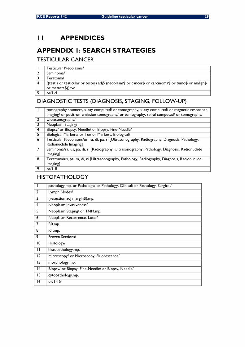
KCE Reports 142 Guideline testicular cancer 29
11 APPENDICES
APPENDIX 1: SEARCH STRATEGIES TESTICULAR CANCER 1 Testicular Neoplasms/ 2 Seminoma/ 3 Teratoma/ 4 ((testis or testicular or testes) adj5 (neoplasm$ or cancer$ or carcinoma$ or tumo$ or malign$
or metasta$)).tw. 5 or/1-4
DIAGNOSTIC TESTS (DIAGNOSIS, STAGING, FOLLOW-UP) 1 tomography scanners, x-ray computed/ or tomography, x-ray computed/ or magnetic resonance
imaging/ or positron-emission tomography/ or tomography, spiral computed/ or tomography/ 2 Ultrasonography/ 3 Neoplasm Staging/ 4 Biopsy/ or Biopsy, Needle/ or Biopsy, Fine-Needle/ 5 Biological Markers/ or Tumor Markers, Biological/ 6 Testicular Neoplasms/us, ra, di, pa, ri [Ultrasonography, Radiography, Diagnosis, Pathology,
Radionuclide Imaging] 7 Seminoma/ra, us, pa, di, ri [Radiography, Ultrasonography, Pathology, Diagnosis, Radionuclide
Imaging] 8 Teratoma/us, pa, ra, di, ri [Ultrasonography, Pathology, Radiography, Diagnosis, Radionuclide
Imaging] 9 or/1-8
HISTOPATHOLOGY 1 pathology.mp. or Pathology/ or Pathology, Clinical/ or Pathology, Surgical/
2 Lymph Nodes/
3 (resection adj margin$).mp.
4 Neoplasm Invasiveness/
5 Neoplasm Staging/ or TNM.mp.
6 Neoplasm Recurrence, Local/
7 R0.mp.
8 R1.mp.
9 Frozen Sections/
10 Histology/
11 histopathology.mp.
12 Microscopy/ or Microscopy, Fluorescence/
13 morphology.mp.
14 Biopsy/ or Biopsy, Fine-Needle/ or Biopsy, Needle/
15 cytopathology.mp.
16 or/1-15
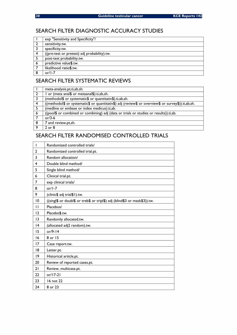
30 Guideline testicular cancer KCE Reports 142
SEARCH FILTER DIAGNOSTIC ACCURACY STUDIES 1 exp "Sensitivity and Specificity"/ 2 sensitivity.tw. 3 specificity.tw. 4 ((pre-test or pretest) adj probability).tw. 5 post-test probability.tw. 6 predictive value$.tw. 7 likelihood ratio$.tw. 8 or/1-7
SEARCH FILTER SYSTEMATIC REVIEWS 1 meta-analysis.pt,ti,ab,sh. 2 1 or (meta anal$ or metaanal$).ti,ab,sh. 3 (methodol$ or systematic$ or quantitativ$).ti,ab,sh. 4 ((methodol$ or systematic$ or quantitativ$) adj (review$ or overview$ or survey$)).ti,ab,sh. 5 (medline or embase or index medicus).ti,ab. 6 ((pool$ or combined or combining) adj (data or trials or studies or results)).ti,ab. 7 or/3-6 8 7 and review.pt,sh. 9 2 or 8
SEARCH FILTER RANDOMISED CONTROLLED TRIALS
1 Randomized controlled trials/
2 Randomized controlled trial.pt.
3 Random allocation/
4 Double blind method/
5 Single blind method/
6 Clinical trial.pt.
7 exp clinical trials/
8 or/1-7
9 (clinic$ adj trial$1).tw.
10 ((singl$ or doubl$ or treb$ or tripl$) adj (blind$3 or mask$3)).tw.
11 Placebos/
12 Placebo$.tw.
13 Randomly allocated.tw.
14 (allocated adj2 random).tw.
15 or/9-14
16 8 or 15
17 Case report.tw.
18 Letter.pt.
19 Historical article.pt.
20 Review of reported cases.pt.
21 Review, multicase.pt.
22 or/17-21
23 16 not 22
24 8 or 23

KCE Reports 142 Guideline testicular cancer 31
SEARCH FILTER PROGNOSTIC STUDIES
1 "prognos*".ti,ab.
2 first.ti,ab.
3 episode.ti,ab.
4 3 and 2
5 cohort.ti,ab.
6 5 or 1 or 4
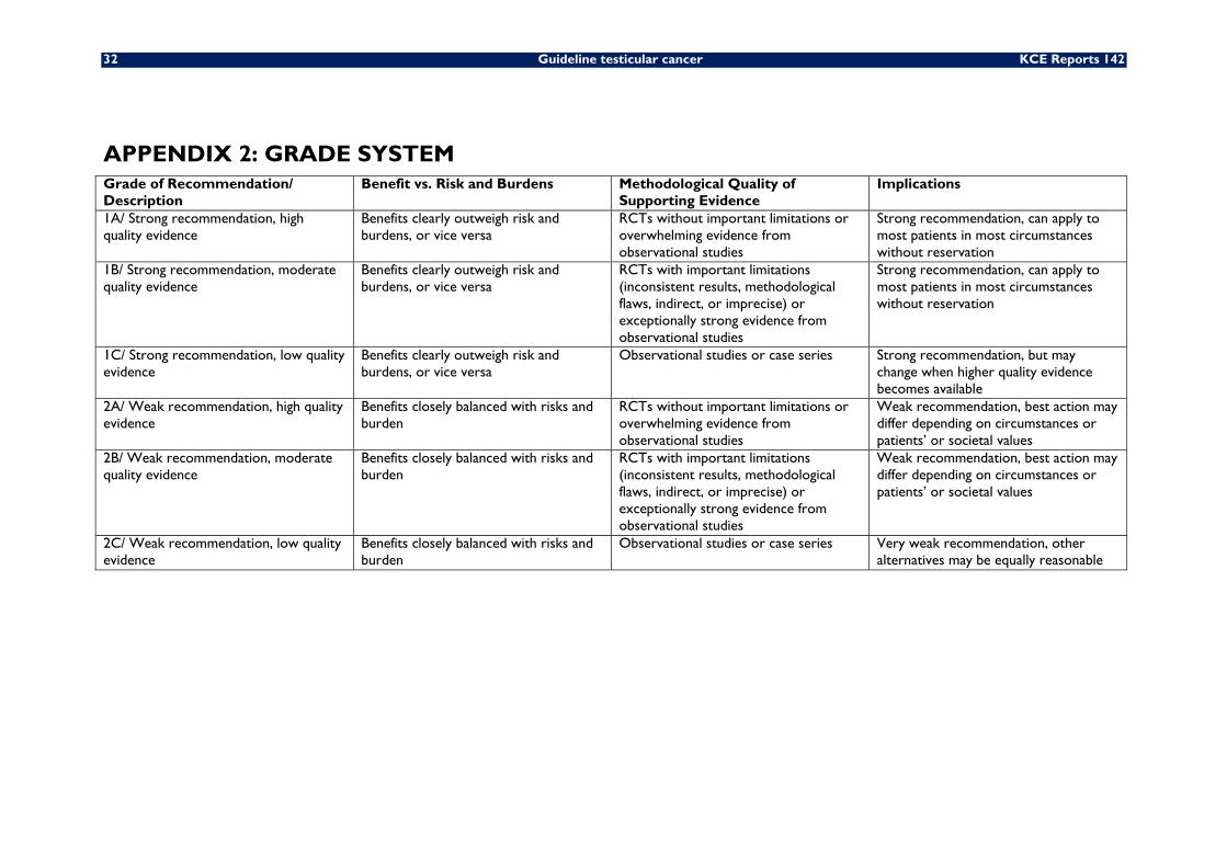
32 Guideline testicular cancer KCE Reports 142
APPENDIX 2: GRADE SYSTEM Grade of Recommendation/ Description
Benefit vs. Risk and Burdens Methodological Quality of Supporting Evidence
Implications
1A/ Strong recommendation, high quality evidence
Benefits clearly outweigh risk and burdens, or vice versa
RCTs without important limitations or overwhelming evidence from observational studies
Strong recommendation, can apply to most patients in most circumstances without reservation
1B/ Strong recommendation, moderate quality evidence
Benefits clearly outweigh risk and burdens, or vice versa
RCTs with important limitations (inconsistent results, methodological flaws, indirect, or imprecise) or exceptionally strong evidence from observational studies
Strong recommendation, can apply to most patients in most circumstances without reservation
1C/ Strong recommendation, low quality evidence
Benefits clearly outweigh risk and burdens, or vice versa
Observational studies or case series Strong recommendation, but may change when higher quality evidence becomes available
2A/ Weak recommendation, high quality evidence
Benefits closely balanced with risks and burden
RCTs without important limitations or overwhelming evidence from observational studies
Weak recommendation, best action may differ depending on circumstances or patients’ or societal values
2B/ Weak recommendation, moderate quality evidence
Benefits closely balanced with risks and burden
RCTs with important limitations (inconsistent results, methodological flaws, indirect, or imprecise) or exceptionally strong evidence from observational studies
Weak recommendation, best action may differ depending on circumstances or patients’ or societal values
2C/ Weak recommendation, low quality evidence
Benefits closely balanced with risks and burden
Observational studies or case series Very weak recommendation, other alternatives may be equally reasonable
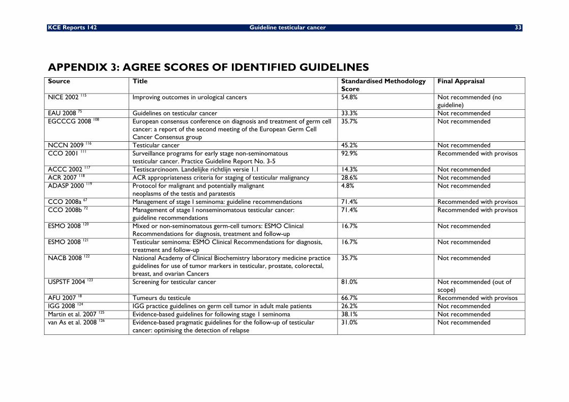
KCE Reports 142 Guideline testicular cancer 33
APPENDIX 3: AGREE SCORES OF IDENTIFIED GUIDELINES Source Title Standardised Methodology
Score Final Appraisal
NICE 2002 115 Improving outcomes in urological cancers 54.8% Not recommended (no guideline)
EAU 2008 75 Guidelines on testicular cancer 33.3% Not recommended EGCCCG 2008 108 European consensus conference on diagnosis and treatment of germ cell
cancer: a report of the second meeting of the European Germ Cell Cancer Consensus group
35.7% Not recommended
NCCN 2009 116 Testicular cancer 45.2% Not recommended CCO 2001 111 Surveillance programs for early stage non-seminomatous
testicular cancer. Practice Guideline Report No. 3-5 92.9% Recommended with provisos
ACCC 2002 117 Testiscarcinoom. Landelijke richtlijn versie 1.1 14.3% Not recommended ACR 2007 118 ACR appropriateness criteria for staging of testicular malignancy 28.6% Not recommended ADASP 2000 119 Protocol for malignant and potentially malignant
neoplasms of the testis and paratestis 4.8% Not recommended
CCO 2008a 67 Management of stage I seminoma: guideline recommendations 71.4% Recommended with provisos CCO 2008b 72 Management of stage I nonseminomatous testicular cancer:
guideline recommendations 71.4% Recommended with provisos
ESMO 2008 120 Mixed or non-seminomatous germ-cell tumors: ESMO Clinical Recommendations for diagnosis, treatment and follow-up
16.7% Not recommended
ESMO 2008 121 Testicular seminoma: ESMO Clinical Recommendations for diagnosis, treatment and follow-up
16.7% Not recommended
NACB 2008 122 National Academy of Clinical Biochemistry laboratory medicine practice guidelines for use of tumor markers in testicular, prostate, colorectal, breast, and ovarian Cancers
35.7% Not recommended
USPSTF 2004 123 Screening for testicular cancer 81.0% Not recommended (out of scope)
AFU 2007 18 Tumeurs du testicule 66.7% Recommended with provisos IGG 2008 124 IGG practice guidelines on germ cell tumor in adult male patients 26.2% Not recommended Martin et al. 2007 125 Evidence-based guidelines for following stage 1 seminoma 38.1% Not recommended van As et al. 2008 126 Evidence-based pragmatic guidelines for the follow-up of testicular
cancer: optimising the detection of relapse 31.0% Not recommended
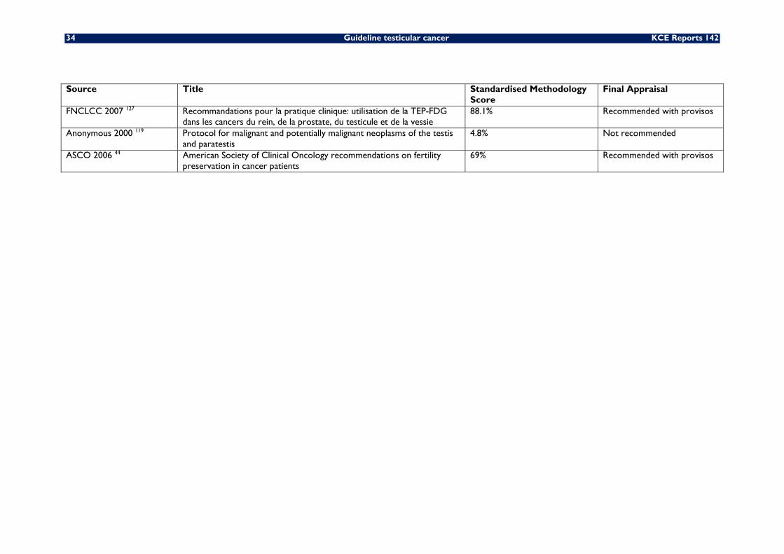
34 Guideline testicular cancer KCE Reports 142
Source Title Standardised Methodology Score
Final Appraisal
FNCLCC 2007 127 Recommandations pour la pratique clinique: utilisation de la TEP-FDG dans les cancers du rein, de la prostate, du testicule et de la vessie
88.1% Recommended with provisos
Anonymous 2000 119 Protocol for malignant and potentially malignant neoplasms of the testis and paratestis
4.8% Not recommended
ASCO 2006 44 American Society of Clinical Oncology recommendations on fertility preservation in cancer patients
69% Recommended with provisos
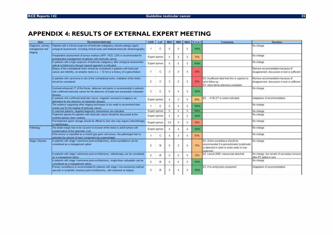
KCE Reports 142 Guideline testicular cancer 35
APPENDIX 4: RESULTS OF EXTERNAL EXPERT MEETING
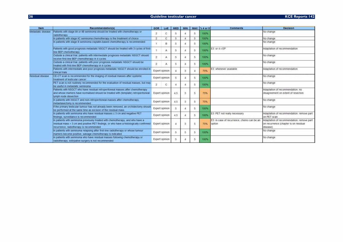
36 Guideline testicular cancer KCE Reports 142
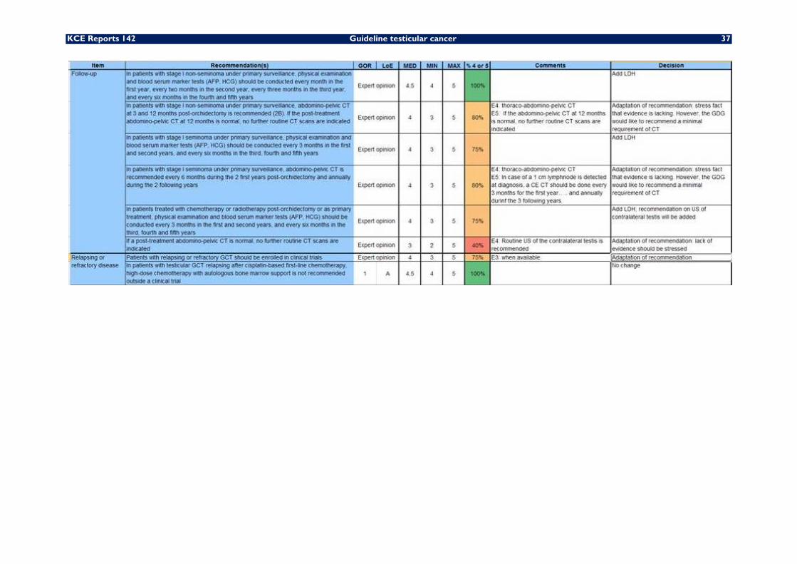
KCE Reports 142 Guideline testicular cancer 37
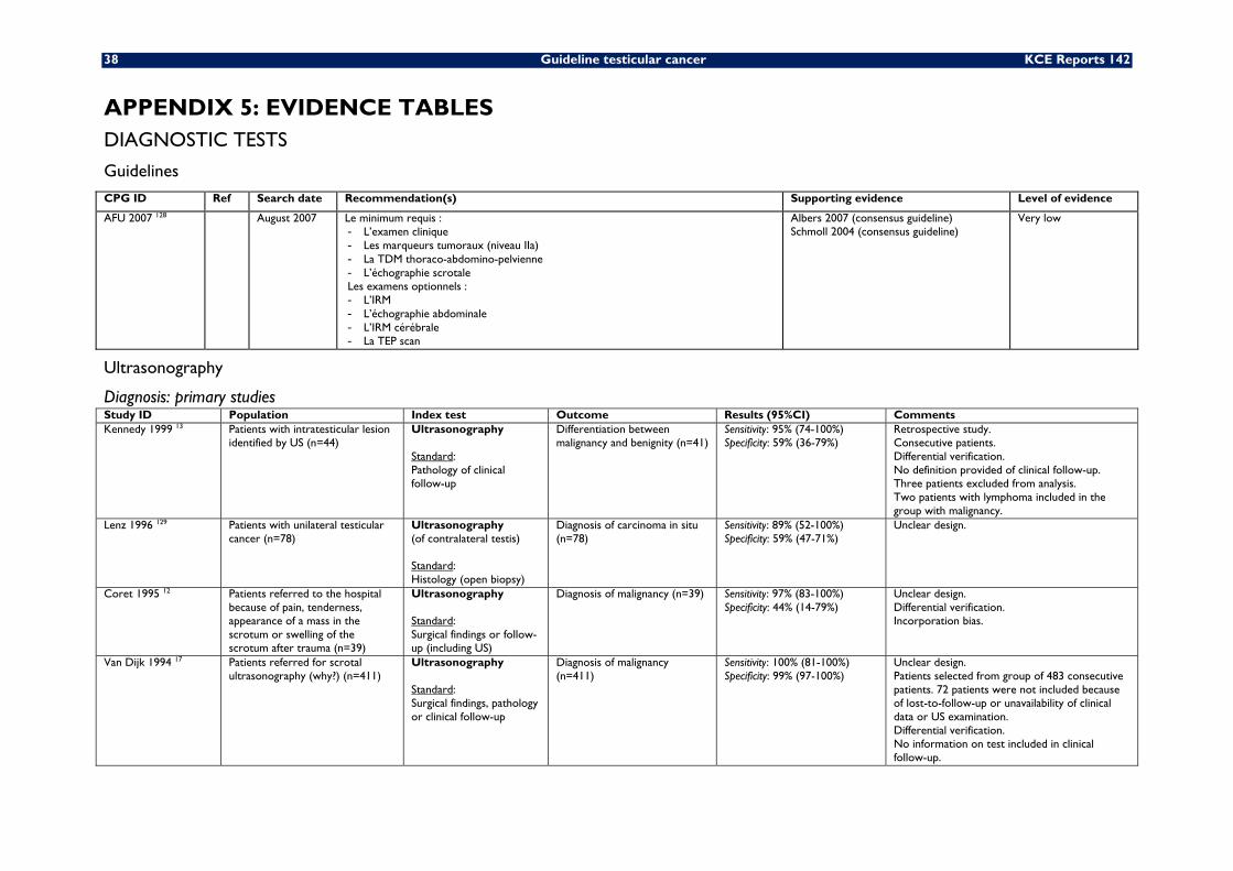
38 Guideline testicular cancer KCE Reports 142
APPENDIX 5: EVIDENCE TABLES DIAGNOSTIC TESTS
Guidelines
CPG ID Ref Search date Recommendation(s) Supporting evidence Level of evidence
AFU 2007 128 August 2007 Le minimum requis : - L’examen clinique - Les marqueurs tumoraux (niveau IIa) - La TDM thoraco-abdomino-pelvienne - L’échographie scrotale Les examens optionnels : - L’IRM - L’échographie abdominale - L’IRM cérébrale - La TEP scan
Albers 2007 (consensus guideline) Schmoll 2004 (consensus guideline)
Very low
Ultrasonography
Diagnosis: primary studies Study ID Population Index test Outcome Results (95%CI) Comments Kennedy 1999 13 Patients with intratesticular lesion
identified by US (n=44) Ultrasonography Standard: Pathology of clinical follow-up
Differentiation between malignancy and benignity (n=41)
Sensitivity: 95% (74-100%) Specificity: 59% (36-79%)
Retrospective study. Consecutive patients. Differential verification. No definition provided of clinical follow-up. Three patients excluded from analysis. Two patients with lymphoma included in the group with malignancy.
Lenz 1996 129 Patients with unilateral testicular cancer (n=78)
Ultrasonography (of contralateral testis) Standard: Histology (open biopsy)
Diagnosis of carcinoma in situ (n=78)
Sensitivity: 89% (52-100%) Specificity: 59% (47-71%)
Unclear design.
Coret 1995 12 Patients referred to the hospital because of pain, tenderness, appearance of a mass in the scrotum or swelling of the scrotum after trauma (n=39)
Ultrasonography Standard: Surgical findings or follow-up (including US)
Diagnosis of malignancy (n=39) Sensitivity: 97% (83-100%) Specificity: 44% (14-79%)
Unclear design. Differential verification. Incorporation bias.
Van Dijk 1994 17 Patients referred for scrotal ultrasonography (why?) (n=411)
Ultrasonography Standard: Surgical findings, pathology or clinical follow-up
Diagnosis of malignancy (n=411)
Sensitivity: 100% (81-100%) Specificity: 99% (97-100%)
Unclear design. Patients selected from group of 483 consecutive patients. 72 patients were not included because of lost-to-follow-up or unavailability of clinical data or US examination. Differential verification. No information on test included in clinical follow-up.
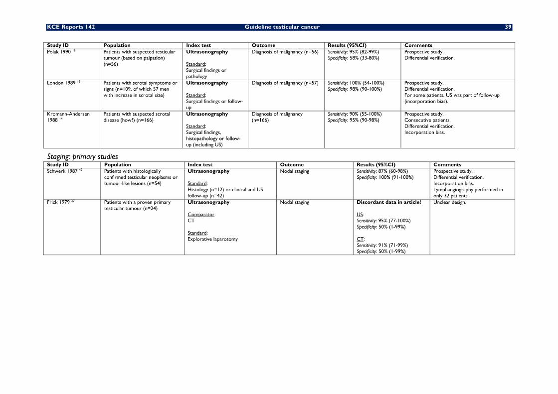
KCE Reports 142 Guideline testicular cancer 39
Study ID Population Index test Outcome Results (95%CI) Comments Polak 1990 16 Patients with suspected testicular
tumour (based on palpation) (n=56)
Ultrasonography Standard: Surgical findings or pathology
Diagnosis of malignancy (n=56) Sensitivity: 95% (82-99%) Specificity: 58% (33-80%)
Prospective study. Differential verification.
London 1989 15 Patients with scrotal symptoms or signs (n=109, of which 57 men with increase in scrotal size)
Ultrasonography Standard: Surgical findings or follow-up
Diagnosis of malignancy (n=57) Sensitivity: 100% (54-100%) Specificity: 98% (90-100%)
Prospective study. Differential verification. For some patients, US was part of follow-up (incorporation bias).
Kromann-Andersen 1988 14
Patients with suspected scrotal disease (how?) (n=166)
Ultrasonography Standard: Surgical findings, histopathology or follow-up (including US)
Diagnosis of malignancy (n=166)
Sensitivity: 90% (55-100%) Specificity: 95% (90-98%)
Prospective study. Consecutive patients. Differential verification. Incorporation bias.
Staging: primary studies Study ID Population Index test Outcome Results (95%CI) Comments Schwerk 1987 42 Patients with histologically
confirmed testicular neoplasms or tumour-like lesions (n=54)
Ultrasonography Standard: Histology (n=12) or clinical and US follow-up (n=42)
Nodal staging Sensitivity: 87% (60-98%) Specificity: 100% (91-100%)
Prospective study. Differential verification. Incorporation bias. Lymphangiography performed in only 32 patients.
Frick 1979 37 Patients with a proven primary testicular tumour (n=24)
Ultrasonography Comparator: CT Standard: Explorative laparotomy
Nodal staging Discordant data in article! US: Sensitivity: 95% (77-100%) Specificity: 50% (1-99%) CT: Sensitivity: 91% (71-99%) Specificity: 50% (1-99%)
Unclear design.
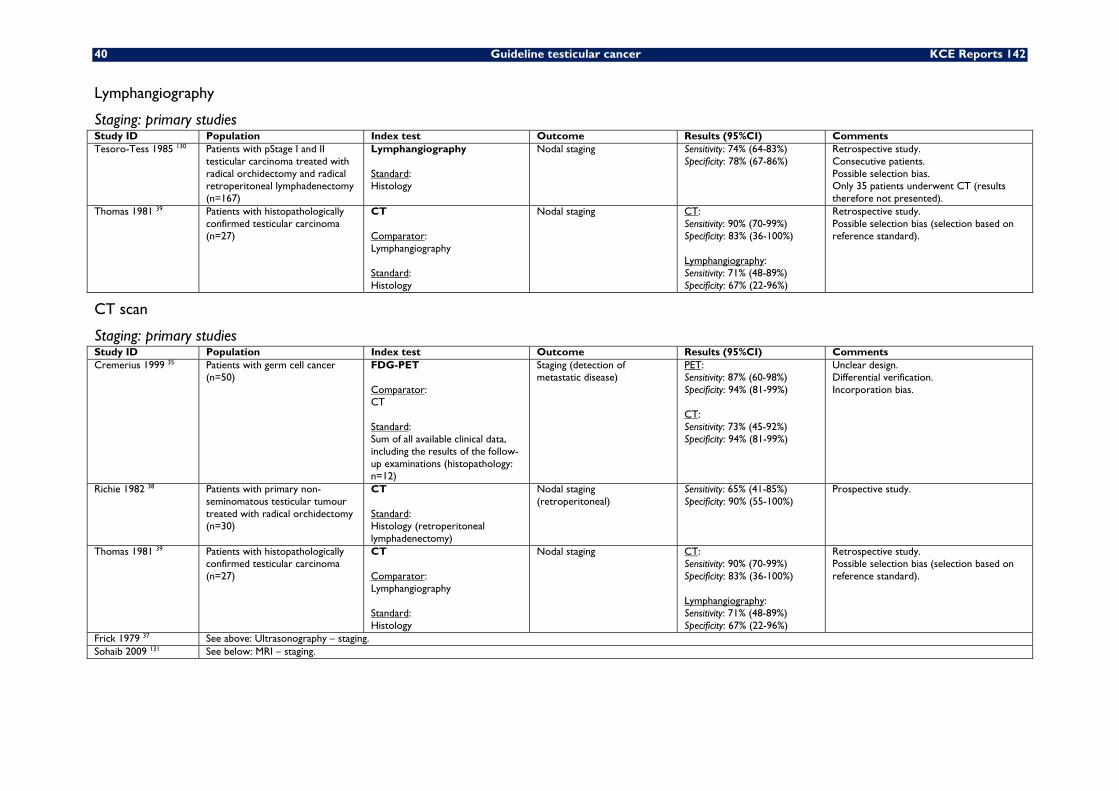
40 Guideline testicular cancer KCE Reports 142
Lymphangiography
Staging: primary studies Study ID Population Index test Outcome Results (95%CI) Comments Tesoro-Tess 1985 130 Patients with pStage I and II
testicular carcinoma treated with radical orchidectomy and radical retroperitoneal lymphadenectomy (n=167)
Lymphangiography Standard: Histology
Nodal staging Sensitivity: 74% (64-83%) Specificity: 78% (67-86%)
Retrospective study. Consecutive patients. Possible selection bias. Only 35 patients underwent CT (results therefore not presented).
Thomas 1981 39 Patients with histopathologically confirmed testicular carcinoma (n=27)
CT Comparator: Lymphangiography Standard: Histology
Nodal staging CT: Sensitivity: 90% (70-99%) Specificity: 83% (36-100%) Lymphangiography: Sensitivity: 71% (48-89%) Specificity: 67% (22-96%)
Retrospective study. Possible selection bias (selection based on reference standard).
CT scan
Staging: primary studies Study ID Population Index test Outcome Results (95%CI) Comments Cremerius 1999 35 Patients with germ cell cancer
(n=50) FDG-PET Comparator: CT Standard: Sum of all available clinical data, including the results of the follow-up examinations (histopathology: n=12)
Staging (detection of metastatic disease)
PET: Sensitivity: 87% (60-98%) Specificity: 94% (81-99%) CT: Sensitivity: 73% (45-92%) Specificity: 94% (81-99%)
Unclear design. Differential verification. Incorporation bias.
Richie 1982 38 Patients with primary non-seminomatous testicular tumour treated with radical orchidectomy (n=30)
CT Standard: Histology (retroperitoneal lymphadenectomy)
Nodal staging (retroperitoneal)
Sensitivity: 65% (41-85%) Specificity: 90% (55-100%)
Prospective study.
Thomas 1981 39 Patients with histopathologically confirmed testicular carcinoma (n=27)
CT Comparator: Lymphangiography Standard: Histology
Nodal staging CT: Sensitivity: 90% (70-99%) Specificity: 83% (36-100%) Lymphangiography: Sensitivity: 71% (48-89%) Specificity: 67% (22-96%)
Retrospective study. Possible selection bias (selection based on reference standard).
Frick 1979 37 See above: Ultrasonography – staging. Sohaib 2009 131 See below: MRI – staging.
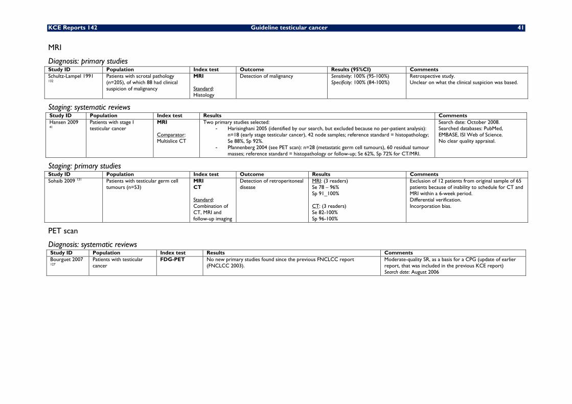
KCE Reports 142 Guideline testicular cancer 41
MRI
Diagnosis: primary studies Study ID Population Index test Outcome Results (95%CI) Comments Schultz-Lampel 1991 132
Patients with scrotal pathology (n=205), of which 88 had clinical suspicion of malignancy
MRI Standard: Histology
Detection of malignancy Sensitivity: 100% (95-100%) Specificity: 100% (84-100%)
Retrospective study. Unclear on what the clinical suspicion was based.
Staging: systematic reviews Study ID Population Index test Results Comments Hansen 2009 41
Patients with stage I testicular cancer
MRI Comparator: Multislice CT
Two primary studies selected: - Harisinghani 2005 (identified by our search, but excluded because no per-patient analysis):
n=18 (early stage testicular cancer), 42 node samples; reference standard = histopathology; Se 88%, Sp 92%.
- Pfannenberg 2004 (see PET scan): n=28 (metastatic germ cell tumours), 60 residual tumour masses; reference standard = histopathology or follow-up; Se 62%, Sp 72% for CT/MRI.
Search date: October 2008. Searched databases: PubMed, EMBASE, ISI Web of Science. No clear quality appraisal.
Staging: primary studies Study ID Population Index test Outcome Results Comments Sohaib 2009 131 Patients with testicular germ cell
tumours (n=53) MRI CT Standard: Combination of CT, MRI and follow-up imaging
Detection of retroperitoneal disease
MRI: (3 readers) Se 78 – 96% Sp 91_100% CT: (3 readers) Se 82-100% Sp 96-100%
Exclusion of 12 patients from original sample of 65 patients because of inability to schedule for CT and MRI within a 6-week period. Differential verification. Incorporation bias.
PET scan
Diagnosis: systematic reviews Study ID Population Index test Results Comments Bourguet 2007 127
Patients with testicular cancer
FDG-PET No new primary studies found since the previous FNCLCC report (FNCLCC 2003).
Moderate-quality SR, as a basis for a CPG (update of earlier report, that was included in the previous KCE report) Search date: August 2006
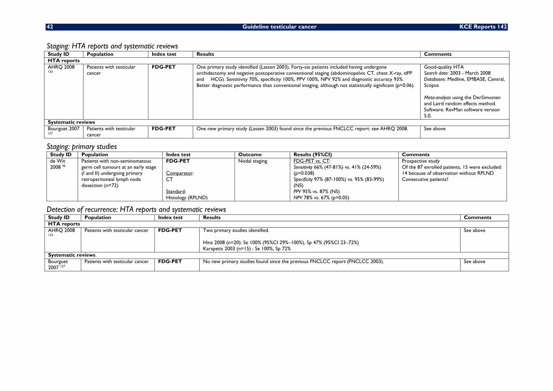
42 Guideline testicular cancer KCE Reports 142
Staging: HTA reports and systematic reviews Study ID Population Index test Results Comments HTA reports AHRQ 2008 133
Patients with testicular cancer
FDG-PET One primary study identified (Lassen 2003). Forty-six patients included having undergone orchidectomy and negative postoperative conventional staging (abdominopelvic CT, chest X-ray, αFP and �HCG). Sensitivity 70%, specificity 100%, PPV 100%, NPV 92% and diagnostic accuracy 93%. Better diagnostic performance than conventional imaging, although not statistically significant (p<0.06).
Good-quality HTA Search date: 2003 - March 2008 Databases: Medline, EMBASE, Central, Scopus Meta-analysis using the DerSimonian and Laird random effects method. Software: RevMan software version 5.0.
Systematic reviews Bourguet 2007 127
Patients with testicular cancer
FDG-PET One new primary study (Lassen 2003) found since the previous FNCLCC report: see AHRQ 2008. See above
Staging: primary studies Study ID Population Index test Outcome Results (95%CI) Comments de Wit 2008 36
Patients with non-seminomatous germ cell tumours at an early stage (I and II) undergoing primary retroperitoneal lymph node dissection (n=72)
FDG-PET Comparator: CT Standard: Histology (RPLND)
Nodal staging FDG-PET vs. CT: Sensitivity 66% (47-81%) vs. 41% (24-59%) (p=0.038) Specificity 97% (87-100%) vs. 95% (83-99%) (NS) PPV 95% vs. 87% (NS) NPV 78% vs. 67% (p=0.05)
Prospective study Of the 87 enrolled patients, 15 were excluded: 14 because of observation without RPLND Consecutive patients?
Detection of recurrence: HTA reports and systematic reviews Study ID Population Index test Results Comments HTA reports AHRQ 2008 133
Patients with testicular cancer FDG-PET Two primary studies identified. Hinz 2008 (n=20): Se 100% (95%CI 29%–100%), Sp 47% (95%CI 23–72%) Karapetis 2003 (n=15) : Se 100%, Sp 72%
See above
Systematic reviews Bourguet 2007 127
Patients with testicular cancer FDG-PET No new primary studies found since the previous FNCLCC report (FNCLCC 2003). See above
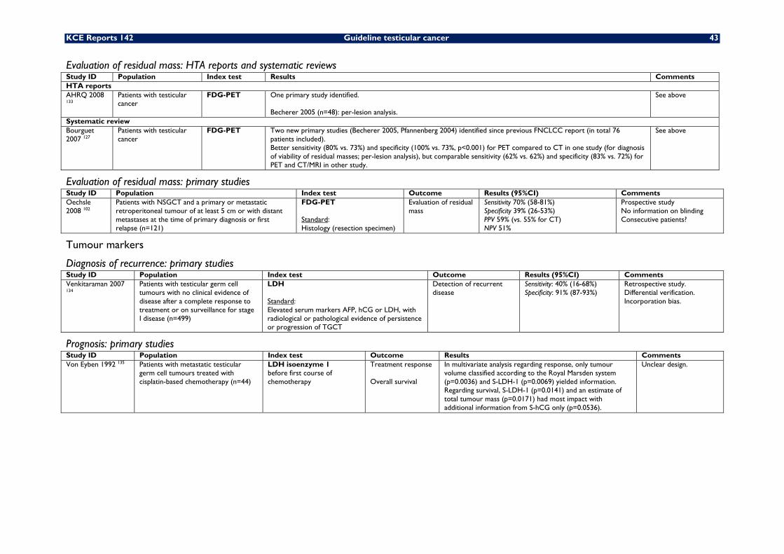
KCE Reports 142 Guideline testicular cancer 43
Evaluation of residual mass: HTA reports and systematic reviews Study ID Population Index test Results Comments HTA reports AHRQ 2008 133
Patients with testicular cancer
FDG-PET One primary study identified. Becherer 2005 (n=48): per-lesion analysis.
See above
Systematic review Bourguet 2007 127
Patients with testicular cancer
FDG-PET Two new primary studies (Becherer 2005, Pfannenberg 2004) identified since previous FNCLCC report (in total 76 patients included). Better sensitivity (80% vs. 73%) and specificity (100% vs. 73%, p<0.001) for PET compared to CT in one study (for diagnosis of viability of residual masses; per-lesion analysis), but comparable sensitivity (62% vs. 62%) and specificity (83% vs. 72%) for PET and CT/MRI in other study.
See above
Evaluation of residual mass: primary studies Study ID Population Index test Outcome Results (95%CI) Comments Oechsle 2008 102
Patients with NSGCT and a primary or metastatic retroperitoneal tumour of at least 5 cm or with distant metastases at the time of primary diagnosis or first relapse (n=121)
FDG-PET Standard: Histology (resection specimen)
Evaluation of residual mass
Sensitivity 70% (58-81%) Specificity 39% (26-53%) PPV 59% (vs. 55% for CT) NPV 51%
Prospective study No information on blinding Consecutive patients?
Tumour markers
Diagnosis of recurrence: primary studies Study ID Population Index test Outcome Results (95%CI) Comments Venkitaraman 2007 134
Patients with testicular germ cell tumours with no clinical evidence of disease after a complete response to treatment or on surveillance for stage I disease (n=499)
LDH Standard: Elevated serum markers AFP, hCG or LDH, with radiological or pathological evidence of persistence or progression of TGCT
Detection of recurrent disease
Sensitivity: 40% (16-68%) Specificity: 91% (87-93%)
Retrospective study. Differential verification. Incorporation bias.
Prognosis: primary studies Study ID Population Index test Outcome Results Comments Von Eyben 1992 135 Patients with metastatic testicular
germ cell tumours treated with cisplatin-based chemotherapy (n=44)
LDH isoenzyme 1 before first course of chemotherapy
Treatment response Overall survival
In multivariate analysis regarding response, only tumour volume classified according to the Royal Marsden system (p=0.0036) and S-LDH-1 (p=0.0069) yielded information. Regarding survival, S-LDH-1 (p=0.0141) and an estimate of total tumour mass (p=0.0171) had most impact with additional information from S-hCG only (p=0.0536).
Unclear design.

44 Guideline testicular cancer KCE Reports 142
Frozen section analysis
Primary studies Study ID Population Index test Outcome Results (95%CI) Comments Connolly 2006 20 Patients with a painless testicular
mass of unclear etiology (n=80) Frozen section analysis Standard: Histology and/or follow-up
Detection of malignancy Sensitivity: 94% (84-99%) Specificity: 89% (72-98%)
Retrospective study. Differential verification.
Elert 2002 21 Patients with testicular tumour (n=354)
Frozen section analysis Standard: Histology
Detection of malignancy Sensitivity: 100% (99-100%) Specificity: 100% (91-100%)
Retrospective study.
Herr 1997 136 Patients with non-seminomatous germ cell tumour of the testis treated with chemotherapy (n=62)
Frozen section analysis Standard: Histology
Evaluation of resection margins (positive if viable germ cell tumour or teratoma)
Sensitivity: 86% (68-96%) Specificity: 100% (89-100%)
Prospective study. Consecutive patients.
Biopsy
Primary studies Study ID Population Index test Outcome Results (95%CI) Comments Dieckmann 1999 137 Patients with testicular germ cell
cancer (n=1954) Contralateral testicular biopsy Standard: Histology or follow-up
Detection of testicular intraepithelial neoplasia
Sensitivity: 95% (89-98%) Specificity: 100% (100-100%)
Prospective study. Consecutive patients.
PATHOLOGY
Diagnostic accuracy studies Study ID Study Description Patient characteristics Intervention(s) Results Comments Level of
evidence Bosman FT 1980 64 Retrospective cohort
study N=73
Patients with testicular germ cell tumours (GCT) Cases were orchidectomy specimens with available embedded tissue blocks 46 seminomas (S), 27 non-seminomas (NS)
Index tests: Immunohistochemical staining for AFP and HCG Reference standard: Histology
Differentiation NS from S: AFP Se 67% (18/27) (95%CI 46-83%) Sp 100% (46/46) (92-100%) HCG Se 74% (20/27) (54-89%) Sp 93% (43/46) (82-99%)
Possible selection bias. Independent evaluation of immunohistochemical staining.
Low
Dimov ND 2007 138 Retrospective cohort study N=109
Patients with primary testicular GCTs. 57 seminomas, 52 mixed GCTs. In total, 181 GCT foci examined.
Index tests: Immunohistochemical staining for Topoisomerase II alpha Reference standard:
Distinction between teratomas and other GCT foci: Se 91% (85-95%) Sp 64% (45-80%)
Possible selection bias. No information on blinding. Positive cases = positive + focally positive
Very low
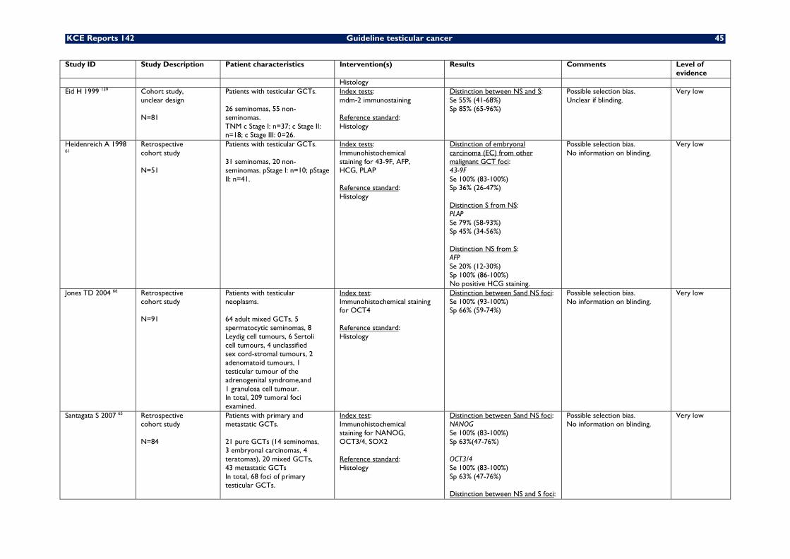
KCE Reports 142 Guideline testicular cancer 45
Study ID Study Description Patient characteristics Intervention(s) Results Comments Level of evidence
Histology Eid H 1999 139 Cohort study,
unclear design N=81
Patients with testicular GCTs. 26 seminomas, 55 non- seminomas. TNM c Stage I: n=37; c Stage II: n=18; c Stage III: 0=26.
Index tests: mdm-2 immunostaining Reference standard: Histology
Distinction between NS and S: Se 55% (41-68%) Sp 85% (65-96%)
Possible selection bias. Unclear if blinding.
Very low
Heidenreich A 1998 61
Retrospective cohort study N=51
Patients with testicular GCTs. 31 seminomas, 20 non- seminomas. pStage I: n=10; pStage II: n=41.
Index tests: Immunohistochemical staining for 43-9F, AFP, HCG, PLAP Reference standard: Histology
Distinction of embryonal carcinoma (EC) from other malignant GCT foci: 43-9F Se 100% (83-100%) Sp 36% (26-47%) Distinction S from NS: PLAP Se 79% (58-93%) Sp 45% (34-56%) Distinction NS from S: AFP Se 20% (12-30%) Sp 100% (86-100%) No positive HCG staining.
Possible selection bias. No information on blinding.
Very low
Jones TD 2004 66 Retrospective cohort study N=91
Patients with testicular neoplasms. 64 adult mixed GCTs, 5 spermatocytic seminomas, 8 Leydig cell tumours, 6 Sertoli cell tumours, 4 unclassified sex cord-stromal tumours, 2 adenomatoid tumours, 1 testicular tumour of the adrenogenital syndrome,and 1 granulosa cell tumour. In total, 209 tumoral foci examined.
Index test: Immunohistochemical staining for OCT4 Reference standard: Histology
Distinction between Sand NS foci: Se 100% (93-100%) Sp 66% (59-74%)
Possible selection bias. No information on blinding.
Very low
Santagata S 2007 65 Retrospective cohort study N=84
Patients with primary and metastatic GCTs. 21 pure GCTs (14 seminomas, 3 embryonal carcinomas, 4 teratomas), 20 mixed GCTs, 43 metastatic GCTs In total, 68 foci of primary testicular GCTs.
Index test: Immunohistochemical staining for NANOG, OCT3/4, SOX2 Reference standard: Histology
Distinction between Sand NS foci: NANOG Se 100% (83-100%) Sp 63%(47-76%) OCT3/4 Se 100% (83-100%) Sp 63% (47-76%) Distinction between NS and S foci:
Possible selection bias. No information on blinding.
Very low
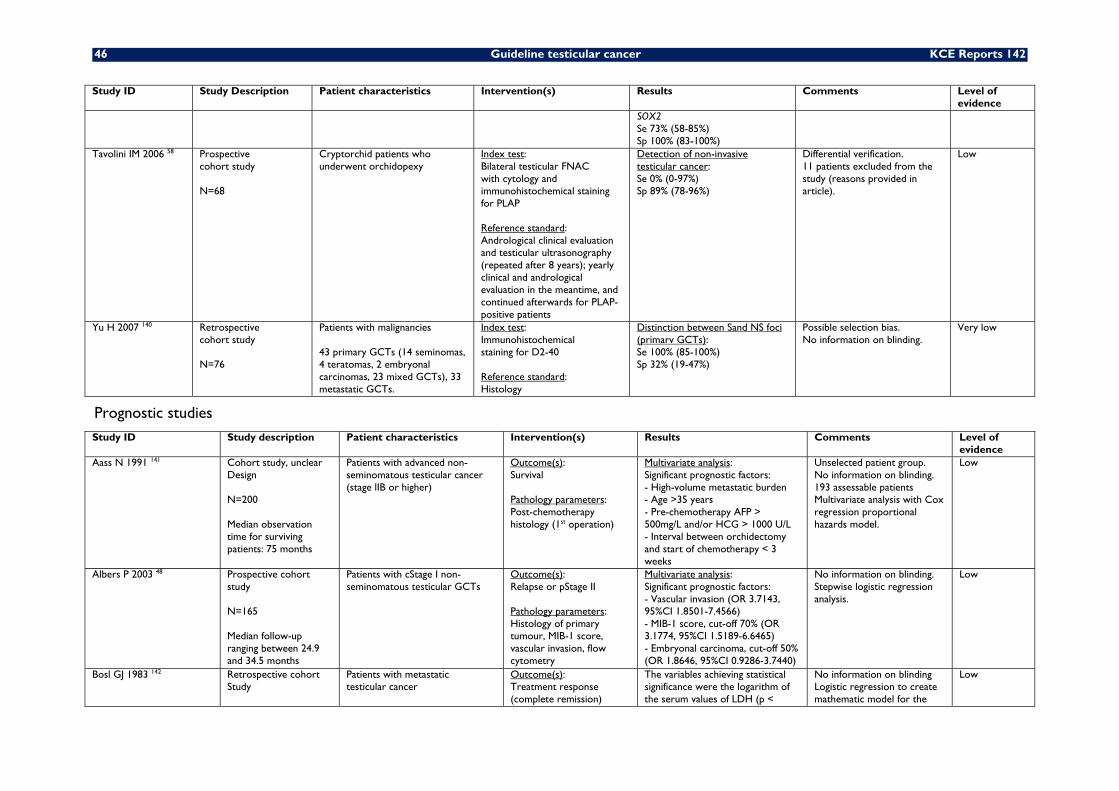
46 Guideline testicular cancer KCE Reports 142
Study ID Study Description Patient characteristics Intervention(s) Results Comments Level of evidence
SOX2 Se 73% (58-85%) Sp 100% (83-100%)
Tavolini IM 2006 58 Prospective cohort study N=68
Cryptorchid patients who underwent orchidopexy
Index test: Bilateral testicular FNAC with cytology and immunohistochemical staining for PLAP Reference standard: Andrological clinical evaluation and testicular ultrasonography (repeated after 8 years); yearly clinical and andrological evaluation in the meantime, and continued afterwards for PLAP-positive patients
Detection of non-invasive testicular cancer: Se 0% (0-97%) Sp 89% (78-96%)
Differential verification. 11 patients excluded from the study (reasons provided in article).
Low
Yu H 2007 140 Retrospective cohort study N=76
Patients with malignancies 43 primary GCTs (14 seminomas, 4 teratomas, 2 embryonal carcinomas, 23 mixed GCTs), 33 metastatic GCTs.
Index test: Immunohistochemical staining for D2-40 Reference standard: Histology
Distinction between Sand NS foci (primarv GCTs): Se 100% (85-100%) Sp 32% (19-47%)
Possible selection bias. No information on blinding.
Very low
Prognostic studies Study ID Study description Patient characteristics Intervention(s) Results Comments Level of
evidence Aass N 1991 141 Cohort study, unclear
Design N=200 Median observation time for surviving patients: 75 months
Patients with advanced non-seminomatous testicular cancer (stage lIB or higher)
Outcome(s): Survival Pathology parameters: Post-chemotherapy histology (1st operation)
Multivariate analysis: Significant prognostic factors: - High-volume metastatic burden - Age >35 years - Pre-chemotherapy AFP > 500mg/L and/or HCG > 1000 U/L - Interval between orchidectomy and start of chemotherapy < 3 weeks
Unselected patient group. No information on blinding. 193 assessable patients Multivariate analysis with Cox regression proportional hazards model.
Low
Albers P 2003 48 Prospective cohort study N=165 Median follow-up ranging between 24.9 and 34.5 months
Patients with cStage I non- seminomatous testicular GCTs
Outcome(s): Relapse or pStage II Pathology parameters: Histology of primary tumour, MIB-1 score, vascular invasion, flow cytometry
Multivariate analysis: Significant prognostic factors: - Vascular invasion (OR 3.7143, 95%CI 1.8501-7.4566) - MIB-1 score, cut-off 70% (OR 3.1774, 95%CI 1.5189-6.6465) - Embryonal carcinoma, cut-off 50% (OR 1.8646, 95%CI 0.9286-3.7440)
No information on blinding. Stepwise logistic regression analysis.
Low
Bosl GJ 1983 142 Retrospective cohort Study
Patients with metastatic testicular cancer
Outcome(s): Treatment response (complete remission)
The variables achieving statistical significance were the logarithm of the serum values of LDH (p <
No information on blinding Logistic regression to create mathematic model for the
Low
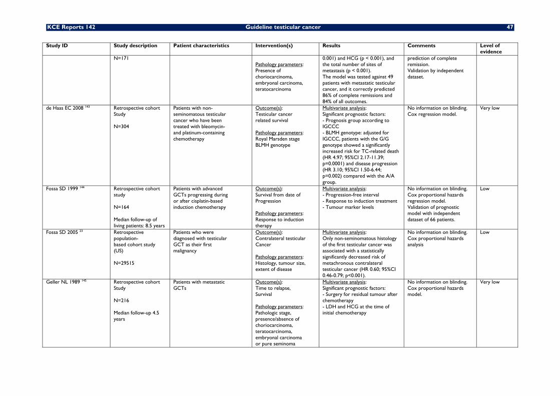
KCE Reports 142 Guideline testicular cancer 47
Study ID Study description Patient characteristics Intervention(s) Results Comments Level of evidence
N=171 Pathology parameters: Presence of choriocarcinoma, embryonal carcinoma, teratocarcinoma
0.001) and HCG (p < 0.001), and the total number of sites of metastasis (p < 0.001). The model was tested against 49 patients with metastatic testicular cancer, and it correctly predicted 86% of complete remissions and 84% of all outcomes.
prediction of complete remission. Validation by independent dataset.
de Haas EC 2008 143 Retrospective cohort Study N=304
Patients with non- seminomatous testicular cancer who have been treated with bleomycin- and platinum-containing chemotherapy
Outcome(s): Testicular cancer related survival Pathology parameters: Royal Marsden stage BLMH genotype
Multivariate analysis: Significant prognostic factors: - Prognosis group according to IGCCC - BLMH genotype: adjusted for IGCCC, patients with the G/G genotype showed a significantly increased risk for TC-related death (HR 4.97; 95%CI 2.17-11.39; p=0.0001) and disease progression (HR 3.10; 95%CI 1.50-6.44; p=0.002) compared with the A/A group.
No information on blinding. Cox regression model.
Very low
Fossa SD 1999 144 Retrospective cohort study N=164 Median follow-up of living patients: 8.5 years
Patients with advanced GCTs progressing during or after cisplatin-based induction chemotherapy
Outcome(s): Survival from date of Progression Pathology parameters: Response to induction therapy
Multivariate analysis: - Progression-free interval - Response to induction treatment - Tumour marker levels
No information on blinding. Cox proportional hazards regression model. Validation of prognostic model with independent dataset of 66 patients.
Low
Fossa SD 2005 23 Retrospective population- based cohort study (US) N=29515
Patients who were diagnosed with testicular GCT as their first malignancy
Outcome(s): Contralateral testicular Cancer Pathology parameters: Histology, tumour size, extent of disease
Multivariate analysis: Only non-seminomatous histology of the first testicular cancer was associated with a statistically significantly decreased risk of metachronous contralateral testicular cancer (HR 0.60; 95%CI 0.46-0.79; p<0.001).
No information on blinding. Cox proportional hazards analysis
Low
Geller NL 1989 145 Retrospective cohort Study N=216 Median follow-up 4.5 years
Patients with metastatic GCTs
Outcome(s): Time to relapse, Survival Pathology parameters: Pathologic stage, presence/absence of choriocarcinoma, teratocarcinoma, embryonal carcinoma or pure seminoma
Multivariate analysis: Significant prognostic factors: - Surgery for residual tumour after chemotherapy - LDH and HCG at the time of initial chemotherapy
No information on blinding. Cox proportional hazards model.
Very low
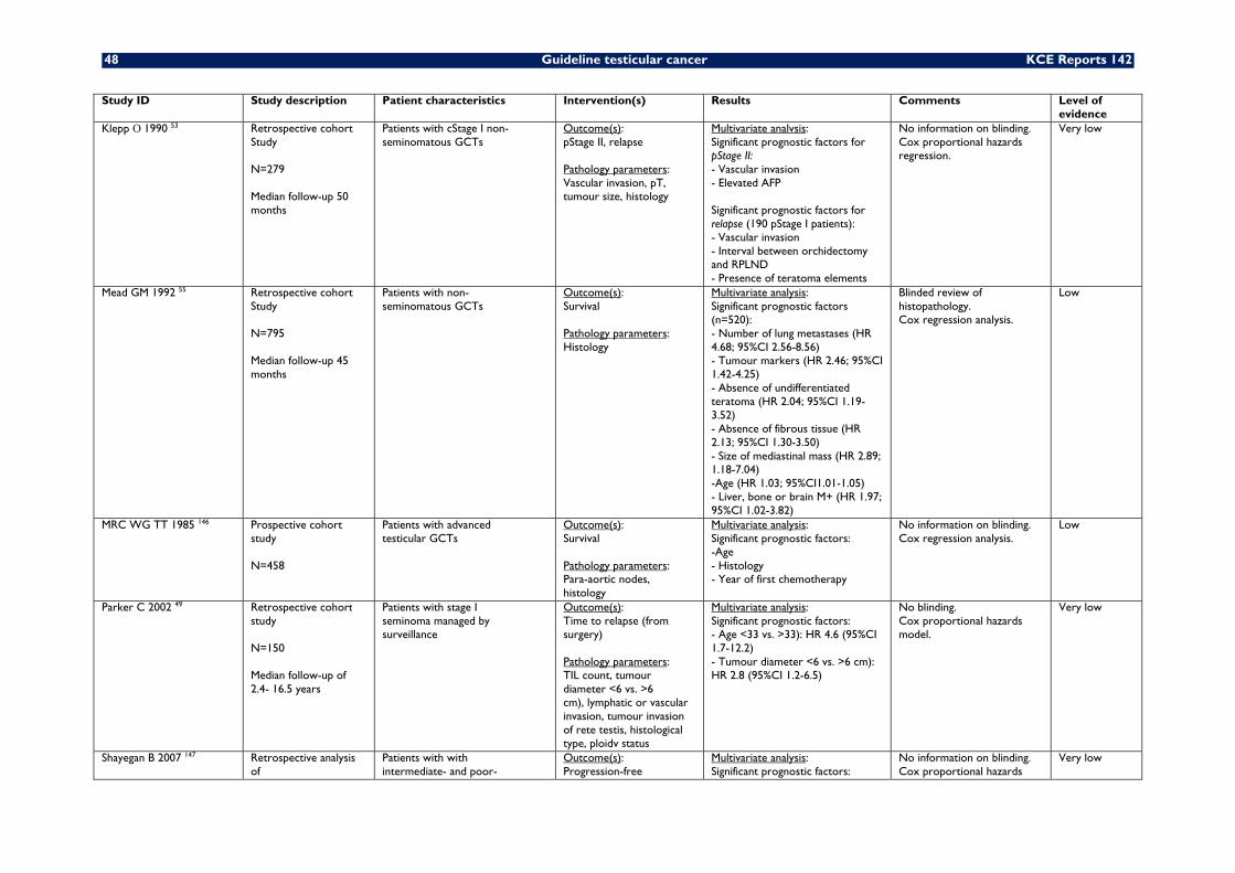
48 Guideline testicular cancer KCE Reports 142
Study ID Study description Patient characteristics Intervention(s) Results Comments Level of evidence
Klepp O 1990 53 Retrospective cohort Study N=279 Median follow-up 50 months
Patients with cStage I non- seminomatous GCTs
Outcome(s): pStage II, relapse Pathology parameters: Vascular invasion, pT, tumour size, histology
Multivariate analvsis: Significant prognostic factors for pStage II: - Vascular invasion - Elevated AFP Significant prognostic factors for relapse (190 pStage I patients): - Vascular invasion - Interval between orchidectomy and RPLND - Presence of teratoma elements
No information on blinding. Cox proportional hazards regression.
Very low
Mead GM 1992 55 Retrospective cohort Study N=795 Median follow-up 45 months
Patients with non- seminomatous GCTs
Outcome(s): Survival Pathology parameters: Histology
Multivariate analysis: Significant prognostic factors (n=520): - Number of lung metastases (HR 4.68; 95%CI 2.56-8.56) - Tumour markers (HR 2.46; 95%CI 1.42-4.25) - Absence of undifferentiated teratoma (HR 2.04; 95%CI 1.19-3.52) - Absence of fibrous tissue (HR 2.13; 95%CI 1.30-3.50) - Size of mediastinal mass (HR 2.89; 1.18-7.04) -Age (HR 1.03; 95%CI1.01-1.05) - Liver, bone or brain M+ (HR 1.97; 95%CI 1.02-3.82)
Blinded review of histopathology. Cox regression analysis.
Low
MRC WG TT 1985 146 Prospective cohort study N=458
Patients with advanced testicular GCTs
Outcome(s): Survival Pathology parameters: Para-aortic nodes, histology
Multivariate analysis: Significant prognostic factors: -Age - Histology - Year of first chemotherapy
No information on blinding. Cox regression analysis.
Low
Parker C 2002 49 Retrospective cohort study N=150 Median follow-up of 2.4- 16.5 years
Patients with stage I seminoma managed by surveillance
Outcome(s): Time to relapse (from surgery) Pathology parameters: TIL count, tumour diameter <6 vs. >6 cm), lymphatic or vascular invasion, tumour invasion of rete testis, histological type, ploidv status
Multivariate analysis: Significant prognostic factors: - Age <33 vs. >33): HR 4.6 (95%CI 1.7-12.2) - Tumour diameter <6 vs. >6 cm): HR 2.8 (95%CI 1.2-6.5)
No blinding. Cox proportional hazards model.
Very low
Shayegan B 2007 147 Retrospective analysis of
Patients with with intermediate- and poor-
Outcome(s): Progression-free
Multivariate analysis: Significant prognostic factors:
No information on blinding. Cox proportional hazards
Very low

KCE Reports 142 Guideline testicular cancer 49
Study ID Study description Patient characteristics Intervention(s) Results Comments Level of evidence
prospective database N=157 Median follow-up 36 months
risk non-seminomatous GCT
probability, disease- specific survival Pathology parameters: R0 vs. R1 resection, Retroperitoneal histology
- Retroperitoneal histology: teratoma vs. fibrosis (HR 3.34; 95%CI1.43-7.79), viable GCT vs. fibrosis (HR 7.26; 95%CI2.81-18.80) - Resection (incomplete vs. complete): HR 10.42 (95%CI 3.89-27.91) - Mass size after chemotherapy: HR 1.14 (95%CI1.03-1.25)
regression.
Steyerberg EW 1995 56 Steyerberg EW 2001 148 Vergouwe Y 2001 149 Vergouwe Y 2007 150
Retrospective cohort Study N=556 (Steyerberg 1995)
Patients with metastatic GCT
Outcome(s): Histology after chemotherapy Pathology parameters: Primary tumour histology (teratoma or not), pre-chemotherapy and post-chemotherapy mass size
Multivariate analysis: Significant prognostic factors necrosis vs. other (n=544): - Primary tumour histology (teratoma + or -): OR 2.46 (95%CI1.6-3.7) - AFP (normal vs. elevated): OR 2.49 (95%CI 1.6-3.9) - HCG (normal vs. elevated): OR 2.22 (95%CI 1.4-3.5) - LDH In: OR 2.76 (95%CI 1.8 4.2) - Post-chemotherapy mass size: OR 0.744 (95%CI 0.63-0.87) - Tumour shrinkage: OR 1.17 (95%CI 1.1-1.3) Significant prognostic factors cancer vs. teratoma (n=299): - LDH In: OR 1.58 (95%CI 0.93-2.7) - Post-chemotherapy mass size: OR 1.17 (95%CI 0.99-1.4) -Tumour shrinkage: OR 1.06 (95%CI 0.95-1.2)
No information on blinding. Logistic regression analysis. Validation and refinement of model in 3 articles: - Steyerberg 2001: independent dataset of 172 patients - Vergouwe 2001: 276 cases - Vergouwe 2007: 1094 cases (including the 544 original cases)
Low
Stoter G 1987 151 Stoter G 1988 152
Prospective cohort study N=163
Patients with disseminated testicular GCTs
Outcome(s): Response (complete response rate) Pathology parameters: Histology of primary tumour, trophoblastic elements in tumour, seminoma elements in tumour, size and number of several M+ sites
Multivariate analysis: Significant prognostic factors: - Trophoblastic elements in tumour - AFP - Lung metastases - Size and number of lung metastases
No information on blinding. Linear logistic regression model.
Low
Stoter G 1993 153 Prospective cohort study
Patients with non- seminomatous GCTs
Outcome(s): Complete response
Multivariate analysis: Significant prognostic factors for
No information on blinding. Cox proportional hazards
Low
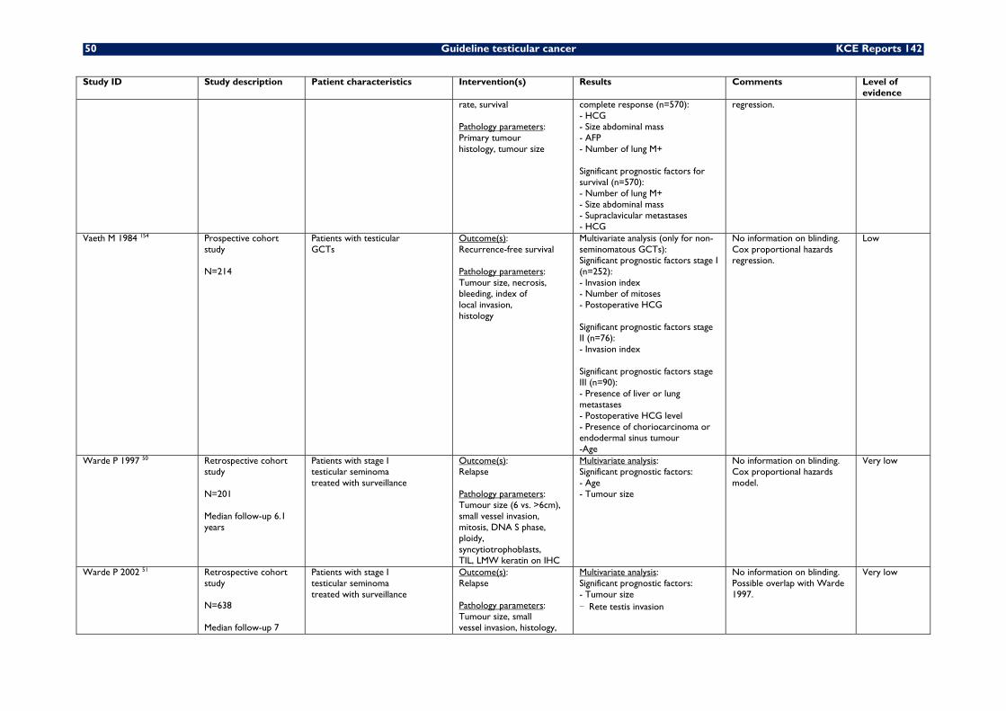
50 Guideline testicular cancer KCE Reports 142
Study ID Study description Patient characteristics Intervention(s) Results Comments Level of evidence
rate, survival Pathology parameters: Primary tumour histology, tumour size
complete response (n=570): - HCG - Size abdominal mass - AFP - Number of lung M+ Significant prognostic factors for survival (n=570): - Number of lung M+ - Size abdominal mass - Supraclavicular metastases - HCG
regression.
Vaeth M 1984 154 Prospective cohort study N=214
Patients with testicular GCTs
Outcome(s): Recurrence-free survival Pathology parameters: Tumour size, necrosis, bleeding, index of local invasion, histology
Multivariate analysis (only for non- seminomatous GCTs): Significant prognostic factors stage I (n=252): - Invasion index - Number of mitoses - Postoperative HCG Significant prognostic factors stage II (n=76): - Invasion index Significant prognostic factors stage III (n=90): - Presence of liver or lung metastases - Postoperative HCG level - Presence of choriocarcinoma or endodermal sinus tumour -Age
No information on blinding. Cox proportional hazards regression.
Low
Warde P 1997 50 Retrospective cohort study N=201 Median follow-up 6.1 years
Patients with stage I testicular seminoma treated with surveillance
Outcome(s): Relapse Pathology parameters: Tumour size (6 vs. >6cm), small vessel invasion, mitosis, DNA S phase, ploidy, syncytiotrophoblasts, TIL, LMW keratin on IHC
Multivariate analysis: Significant prognostic factors: - Age - Tumour size
No information on blinding. Cox proportional hazards model.
Very low
Warde P 2002 51 Retrospective cohort study N=638 Median follow-up 7
Patients with stage I testicular seminoma treated with surveillance
Outcome(s): Relapse Pathology parameters: Tumour size, small vessel invasion, histology,
Multivariate analysis: Significant prognostic factors: - Tumour size - Rete testis invasion
No information on blinding. Possible overlap with Warde 1997.
Very low
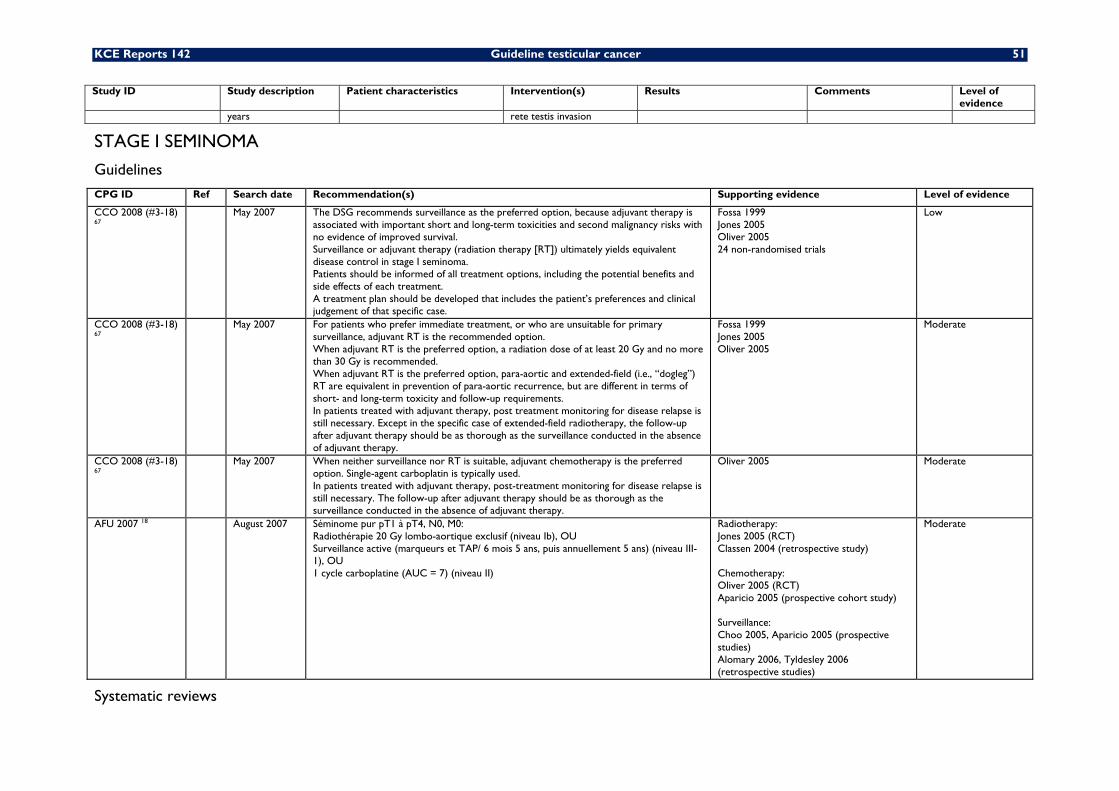
KCE Reports 142 Guideline testicular cancer 51
Study ID Study description Patient characteristics Intervention(s) Results Comments Level of evidence
years rete testis invasion
STAGE I SEMINOMA
Guidelines
CPG ID Ref Search date Recommendation(s) Supporting evidence Level of evidence
CCO 2008 (#3-18) 67
May 2007 The DSG recommends surveillance as the preferred option, because adjuvant therapy is associated with important short and long-term toxicities and second malignancy risks with no evidence of improved survival. Surveillance or adjuvant therapy (radiation therapy [RT]) ultimately yields equivalent disease control in stage I seminoma. Patients should be informed of all treatment options, including the potential benefits and side effects of each treatment. A treatment plan should be developed that includes the patient’s preferences and clinical judgement of that specific case.
Fossa 1999 Jones 2005 Oliver 2005 24 non-randomised trials
Low
CCO 2008 (#3-18) 67
May 2007 For patients who prefer immediate treatment, or who are unsuitable for primary surveillance, adjuvant RT is the recommended option. When adjuvant RT is the preferred option, a radiation dose of at least 20 Gy and no more than 30 Gy is recommended. When adjuvant RT is the preferred option, para-aortic and extended-field (i.e., “dogleg”) RT are equivalent in prevention of para-aortic recurrence, but are different in terms of short- and long-term toxicity and follow-up requirements. In patients treated with adjuvant therapy, post treatment monitoring for disease relapse is still necessary. Except in the specific case of extended-field radiotherapy, the follow-up after adjuvant therapy should be as thorough as the surveillance conducted in the absence of adjuvant therapy.
Fossa 1999 Jones 2005 Oliver 2005
Moderate
CCO 2008 (#3-18) 67
May 2007 When neither surveillance nor RT is suitable, adjuvant chemotherapy is the preferred option. Single-agent carboplatin is typically used. In patients treated with adjuvant therapy, post-treatment monitoring for disease relapse is still necessary. The follow-up after adjuvant therapy should be as thorough as the surveillance conducted in the absence of adjuvant therapy.
Oliver 2005 Moderate
AFU 2007 18 August 2007 Séminome pur pT1 à pT4, N0, M0: Radiothérapie 20 Gy lombo-aortique exclusif (niveau Ib), OU Surveillance active (marqueurs et TAP/ 6 mois 5 ans, puis annuellement 5 ans) (niveau III-1), OU 1 cycle carboplatine (AUC = 7) (niveau II)
Radiotherapy: Jones 2005 (RCT) Classen 2004 (retrospective study) Chemotherapy: Oliver 2005 (RCT) Aparicio 2005 (prospective cohort study) Surveillance: Choo 2005, Aparicio 2005 (prospective studies) Alomary 2006, Tyldesley 2006 (retrospective studies)
Moderate
Systematic reviews
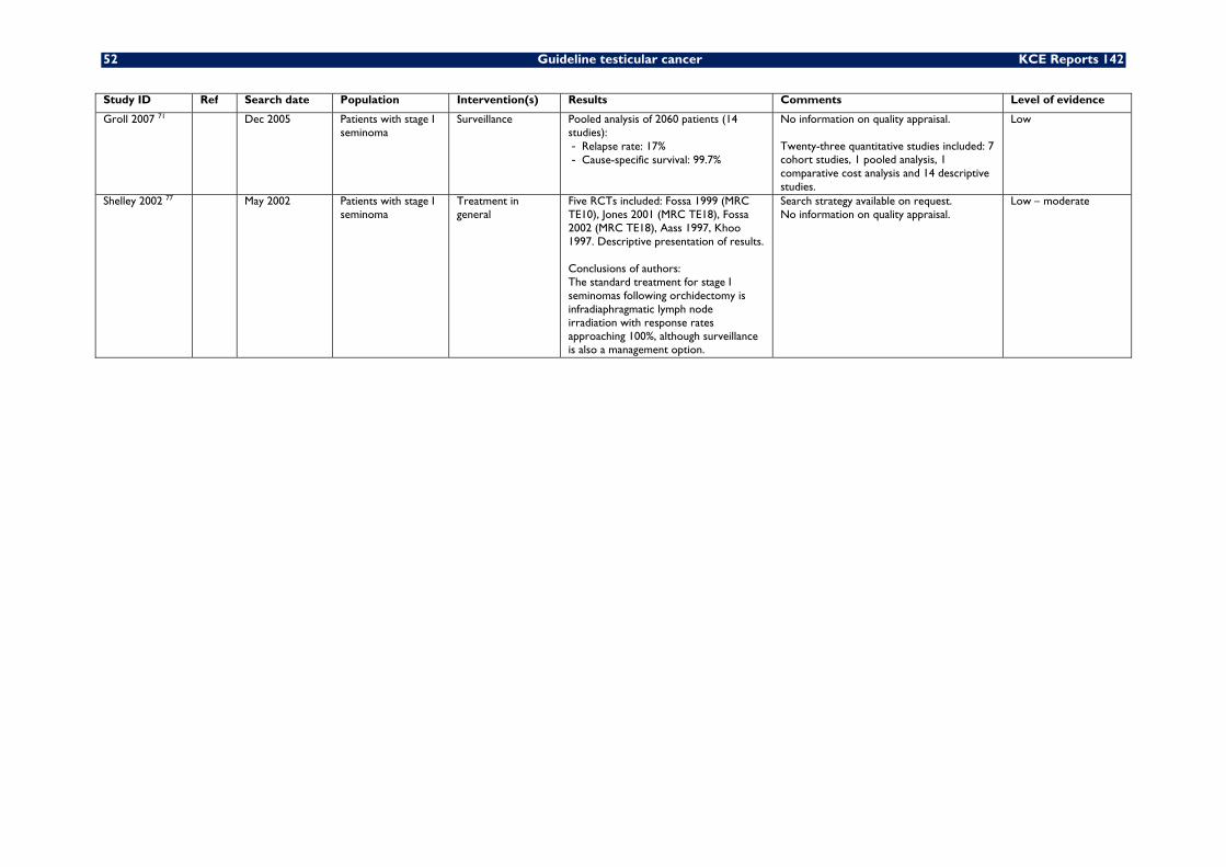
52 Guideline testicular cancer KCE Reports 142
Study ID Ref Search date Population Intervention(s) Results Comments Level of evidence
Groll 2007 71 Dec 2005 Patients with stage I seminoma
Surveillance Pooled analysis of 2060 patients (14 studies): - Relapse rate: 17% - Cause-specific survival: 99.7%
No information on quality appraisal. Twenty-three quantitative studies included: 7 cohort studies, 1 pooled analysis, 1 comparative cost analysis and 14 descriptive studies.
Low
Shelley 2002 77 May 2002 Patients with stage I seminoma
Treatment in general
Five RCTs included: Fossa 1999 (MRC TE10), Jones 2001 (MRC TE18), Fossa 2002 (MRC TE18), Aass 1997, Khoo 1997. Descriptive presentation of results. Conclusions of authors: The standard treatment for stage I seminomas following orchidectomy is infradiaphragmatic lymph node irradiation with response rates approaching 100%, although surveillance is also a management option.
Search strategy available on request. No information on quality appraisal.
Low – moderate
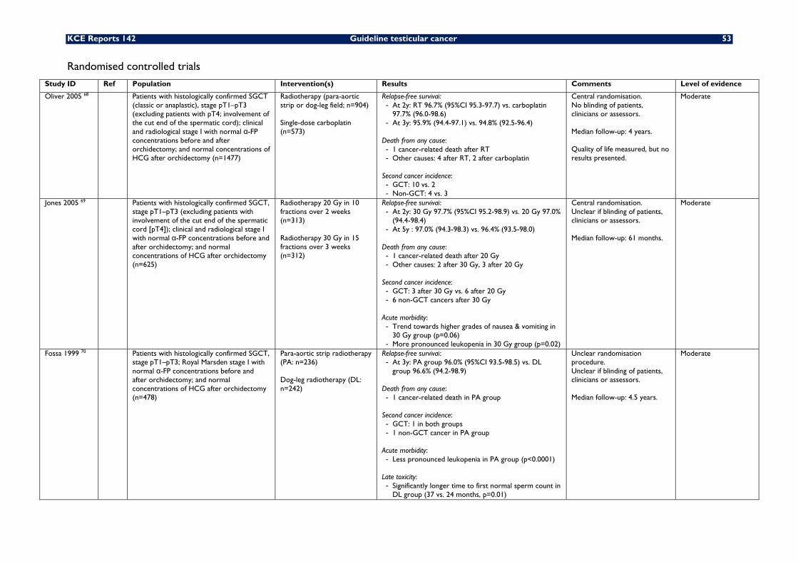
KCE Reports 142 Guideline testicular cancer 53
Randomised controlled trials Study ID Ref Population Intervention(s) Results Comments Level of evidence
Oliver 2005 68 Patients with histologically confirmed SGCT (classic or anaplastic), stage pT1–pT3 (excluding patients with pT4; involvement of the cut end of the spermatic cord); clinical and radiological stage I with normal α-FP concentrations before and after orchidectomy; and normal concentrations of HCG after orchidectomy (n=1477)
Radiotherapy (para-aortic strip or dog-leg field; n=904) Single-dose carboplatin (n=573)
Relapse-free survival: - At 2y: RT 96.7% (95%CI 95.3-97.7) vs. carboplatin
97.7% (96.0-98.6) - At 3y: 95.9% (94.4-97.1) vs. 94.8% (92.5-96.4)
Death from any cause: - 1 cancer-related death after RT - Other causes: 4 after RT, 2 after carboplatin
Second cancer incidence: - GCT: 10 vs. 2 - Non-GCT: 4 vs. 3
Central randomisation. No blinding of patients, clinicians or assessors. Median follow-up: 4 years. Quality of life measured, but no results presented.
Moderate
Jones 2005 69 Patients with histologically confirmed SGCT, stage pT1–pT3 (excluding patients with involvement of the cut end of the spermatic cord [pT4]); clinical and radiological stage I with normal α-FP concentrations before and after orchidectomy; and normal concentrations of HCG after orchidectomy (n=625)
Radiotherapy 20 Gy in 10 fractions over 2 weeks (n=313) Radiotherapy 30 Gy in 15 fractions over 3 weeks (n=312)
Relapse-free survival: - At 2y: 30 Gy 97.7% (95%CI 95.2-98.9) vs. 20 Gy 97.0%
(94.4-98.4) - At 5y : 97.0% (94.3-98.3) vs. 96.4% (93.5-98.0)
Death from any cause: - 1 cancer-related death after 20 Gy - Other causes: 2 after 30 Gy, 3 after 20 Gy
Second cancer incidence: - GCT: 3 after 30 Gy vs. 6 after 20 Gy - 6 non-GCT cancers after 30 Gy
Acute morbidity: - Trend towards higher grades of nausea & vomiting in
30 Gy group (p=0.06) - More pronounced leukopenia in 30 Gy group (p=0.02)
Central randomisation. Unclear if blinding of patients, clinicians or assessors. Median follow-up: 61 months.
Moderate
Fossa 1999 70 Patients with histologically confirmed SGCT, stage pT1–pT3; Royal Marsden stage I with normal α-FP concentrations before and after orchidectomy; and normal concentrations of HCG after orchidectomy (n=478)
Para-aortic strip radiotherapy (PA: n=236) Dog-leg radiotherapy (DL: n=242)
Relapse-free survival: - At 3y: PA group 96.0% (95%CI 93.5-98.5) vs. DL
group 96.6% (94.2-98.9) Death from any cause: - 1 cancer-related death in PA group
Second cancer incidence: - GCT: 1 in both groups - 1 non-GCT cancer in PA group
Acute morbidity: - Less pronounced leukopenia in PA group (p<0.0001)
Late toxicity: - Significantly longer time to first normal sperm count in
DL group (37 vs. 24 months, p=0.01)
Unclear randomisation procedure. Unclear if blinding of patients, clinicians or assessors. Median follow-up: 4.5 years.
Moderate
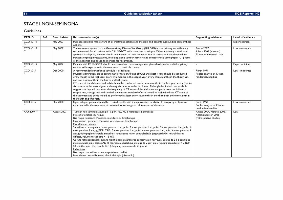
54 Guideline testicular cancer KCE Reports 142
STAGE I NON-SEMINOMA
Guidelines
CPG ID Ref Search date Recommendation(s) Supporting evidence Level of evidence
CCO #3-19 72
May 2007 Patients should be made aware of all treatment options and the risks and benefits surrounding each of these options.
- Expert opinion
CCO #3-19 72
May 2007 The consensus opinion of the Genitourinary Disease Site Group (GU DSG) is that primary surveillance is recommended for all patients with CS I NSGCT, with treatment at relapse. When a primary surveillance approach is adopted, patients should be informed of their estimated risk of recurrence and the need for frequent ongoing investigations, including blood tumour markers and computerised tomography (CT) scans of the abdomen and pelvis, to monitor for recurrence.
Rustin 2007 Albers 2006 (abstract) 21 non-randomised trials
Low - moderate
CCO #3-19 72
May 2007 Patients with CS I NSGCT should be assessed and have management plans developed at multidisciplinary centres with experience in the treatment of testicular cancer.
- Expert opinion
CCO #3-5 111
Dec 2000 A recommended surveillance schedule is as follows: Physical examination, blood serum marker tests (AFP and bHCG) and chest x-rays should be conducted every month in the first year, every two months in the second year, every three months in the third year, and every six months in the fourth and fifth years; CT scans of the abdomen and pelvis should be conducted every three months in the first year, every four to six months in the second year and every six months in the third year. Although the limited data available suggest that beyond two years the frequency of CT scans of the abdomen and pelvis does not influence relapse rate, salvage rate and survival, the current standard of care should be maintained and CT scans of the abdomen and pelvis should be performed at least every six months in the third year and once a year in the fourth and fifth year.
Rorth 1991 Pooled analysis of 13 non-randomised studies
Low - moderate
CCO #3-5 111
Dec 2000 Upon relapse, patients should be treated rapidly with the appropriate modality of therapy by a physician experienced in the treatment of non-seminomatous germ cell tumours of the testis.
Rorth 1991 Pooled analysis of 13 non-randomised studies
Low - moderate
AFU 2007 18 August 2007 Tumeur non séminomateuse pT1 à pT4, N0, M0 à marqueurs normalisés: Stratégie fonction du risque: Bas risque : absence d’invasion vasculaire ou lymphatique Haut risque : présence d’invasion vasculaire ou lymphatique Modalités techniques : Surveillance : marqueurs / mois pendant 1 an, puis / 2 mois pendant 1 an, puis / 3 mois pendant 1 an, puis / 6 mois pendant 2 ans, et TDM TAP / 3 mois pendant 1 an, puis / 4 mois pendant 1 an, puis / 6 mois pendant 3 ans et échographie scrotale annuelle si haut risque lésion controlatérale (cryptorchidie, microlithiases diffuses, volume testiculaire < 12 ml)) Curage rétropéritonéal : curage modifié homolatéral avec conservation nerveuse. Si plus de 3 à 6 ganglions métastatiques ou si stade pN2 (1 ganglion métastatique de plus de 2 cm) ou si rupture capsulaire : + 2 BEP Chimiothérapie : 2 cycles de BEP (chaque cycle espacé de 21 jours) Indications : Bas risque : surveillance ou curage (niveau IIa-IIb) Haut risque : surveillance ou chimiothérapie (niveau IIb)
Amato 2004, Maroto 2005, Kildahlandersen 2005 (retrospective studies)
Low
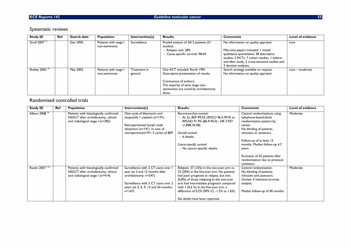
KCE Reports 142 Guideline testicular cancer 55
Systematic reviews Study ID Ref Search date Population Intervention(s) Results Comments Level of evidence
Groll 2007 71 Dec 2005 Patients with stage I non-seminoma
Surveillance Pooled analysis of 3613 patients (31 studies): - Relapse rate: 28% - Cause-specific survival: 98.6%
No information on quality appraisal. Fifty-nine papers included: 1 mixed qualitative-quantitative, 38 descriptive studies, 2 RCTs, 7 cohort studies, 1 before-and-after study, 2 cross-sectional studies and 3 decision analyses.
Low
Shelley 2002 77 May 2002 Patients with stage I non-seminoma
Treatment in general
One RCT included: Rorth 1991. Descriptive presentation of results. Conclusions of authors: The majority of early stage non-seminomas are cured by orchidectomy alone.
Search strategy available on request. No information on quality appraisal.
Low – moderate
Randomised controlled trials Study ID Ref Population Intervention(s) Results Comments Level of evidence
Albers 2008 74 Patients with histologically confirmed NSGCT after orchidectomy, clinical and radiological stage I (n=382)
One cycle of bleomycin and etoposide + cisplatin (n=191) Retroperitoneal lymph node dissection (n=191). In case of retroperitoneal M+: 2 cycles of BEP
Recurrence-free survival: - At 2y: BEP 99.5% (95%CI 96.2-99.9) vs.
RPLND 91.9% (86.9-95.0) ; HR 7.937 (1.808-34.48)
Overall survival: - 6 deaths
Cancer-specific survival: - No cancer-specific deaths
Central randomisations using telephone-based block randomisation pattern by center. No blinding of patients, clinicians or assessors. Follow-up of at least 15 months. Median follow-up 4.7 years. Exclusion of 35 patients after randomisation due to protocol violations.
Moderate
Rustin 2007 110 Patients with histologically confirmed NSGCT after orchidectomy, clinical and radiological stage I (n=414)
Surveillance with 2 CT scans over 1 year (at 3 and 12 months after orchidectomy; n=247) Surveillance with 5 CT scans over 2 years (at 3, 6, 9, 12 and 24 months; n=167)
Relapses: 37 (15%) in the two-scan arm vs. 33 (20%) in the five-scan arm. No patients had poor prognosis at relapse, but two (0.8%) of those relapsing In the two-scan arm had intermediate prognosis compared with 1 (0.6 %) in the five-scan arm, a difference of 0.2% (90% CI, -1.2% to 1.6%). No deaths have been reported.
Central randomisation. No blinding of patients, clinicians and assessors. Unclear if intention-to-treat analysis. Median follow-up of 40 months.
Moderate
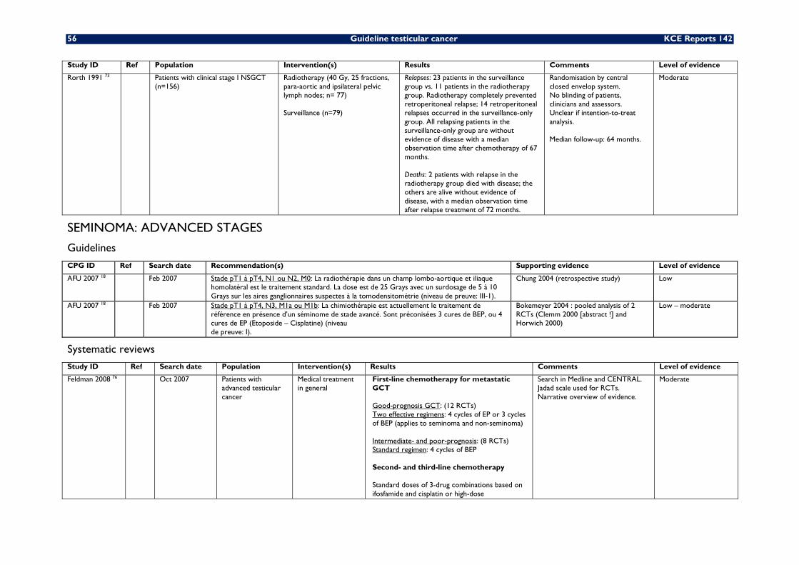
56 Guideline testicular cancer KCE Reports 142
Study ID Ref Population Intervention(s) Results Comments Level of evidence
Rorth 1991 73 Patients with clinical stage I NSGCT (n=156)
Radiotherapy (40 Gy, 25 fractions, para-aortic and ipsilateral pelvic lymph nodes; n= 77) Surveillance (n=79)
Relapses: 23 patients in the surveillance group vs. 11 patients in the radiotherapy group. Radiotherapy completely prevented retroperitoneal relapse; 14 retroperitoneal relapses occurred in the surveillance-only group. All relapsing patients in the surveillance-only group are without evidence of disease with a median observation time after chemotherapy of 67 months. Deaths: 2 patients with relapse in the radiotherapy group died with disease; the others are alive without evidence of disease, with a median observation time after relapse treatment of 72 months.
Randomisation by central closed envelop system. No blinding of patients, clinicians and assessors. Unclear if intention-to-treat analysis. Median follow-up: 64 months.
Moderate
SEMINOMA: ADVANCED STAGES
Guidelines
CPG ID Ref Search date Recommendation(s) Supporting evidence Level of evidence
AFU 2007 18 Feb 2007 Stade pT1 à pT4, N1 ou N2, M0: La radiothérapie dans un champ lombo-aortique et iliaque homolatéral est le traitement standard. La dose est de 25 Grays avec un surdosage de 5 à 10 Grays sur les aires ganglionnaires suspectes à la tomodensitométrie (niveau de preuve: III-1).
Chung 2004 (retrospective study)
Low
AFU 2007 18 Feb 2007 Stade pT1 à pT4, N3, M1a ou M1b: La chimiothérapie est actuellement le traitement de référence en présence d’un séminome de stade avancé. Sont préconisées 3 cures de BEP, ou 4 cures de EP (Etoposide – Cisplatine) (niveau de preuve: I).
Bokemeyer 2004 : pooled analysis of 2 RCTs (Clemm 2000 [abstract !] and Horwich 2000)
Low – moderate
Systematic reviews Study ID Ref Search date Population Intervention(s) Results Comments Level of evidence
Feldman 2008 76 Oct 2007 Patients with advanced testicular cancer
Medical treatment in general
First-line chemotherapy for metastatic GCT Good-prognosis GCT: (12 RCTs) Two effective regimens: 4 cycles of EP or 3 cycles of BEP (applies to seminoma and non-seminoma) Intermediate- and poor-prognosis: (8 RCTs) Standard regimen: 4 cycles of BEP Second- and third-line chemotherapy Standard doses of 3-drug combinations based on ifosfamide and cisplatin or high-dose
Search in Medline and CENTRAL. Jadad scale used for RCTs. Narrative overview of evidence.
Moderate
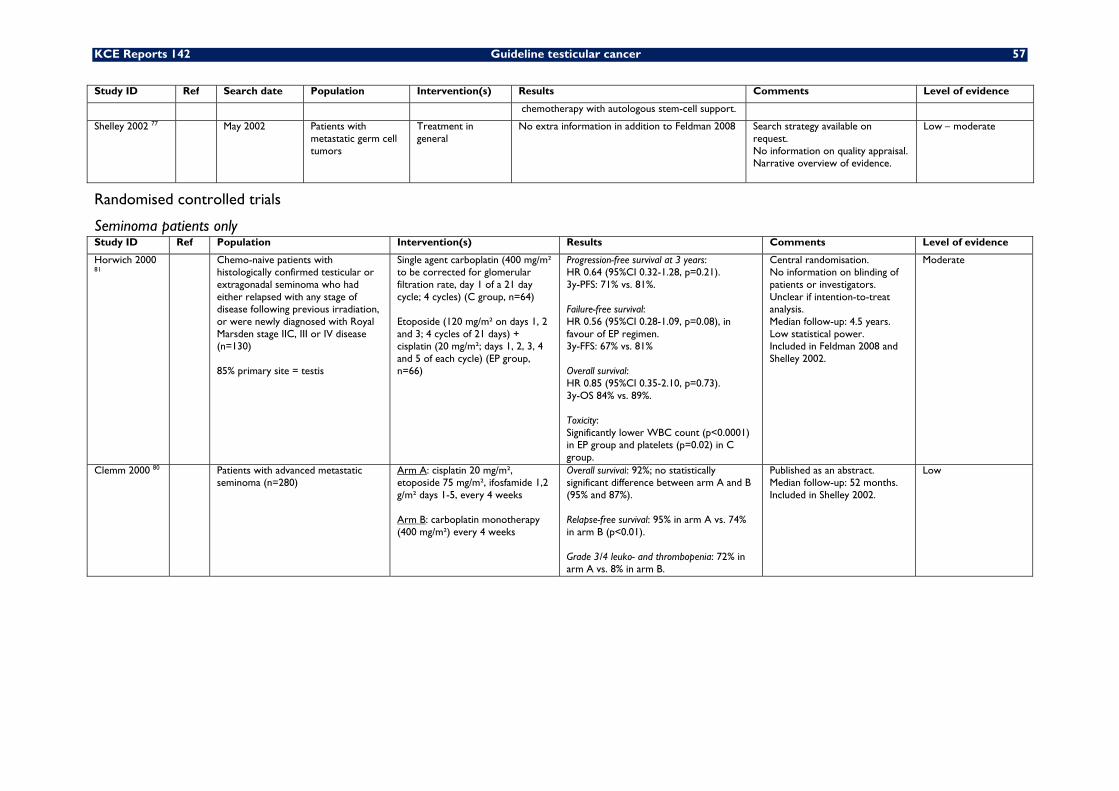
KCE Reports 142 Guideline testicular cancer 57
Study ID Ref Search date Population Intervention(s) Results Comments Level of evidence
chemotherapy with autologous stem-cell support.
Shelley 2002 77 May 2002 Patients with metastatic germ cell tumors
Treatment in general
No extra information in addition to Feldman 2008 Search strategy available on request. No information on quality appraisal. Narrative overview of evidence.
Low – moderate
Randomised controlled trials
Seminoma patients only Study ID Ref Population Intervention(s) Results Comments Level of evidence
Horwich 2000 81
Chemo-naive patients with histologically confirmed testicular or extragonadal seminoma who had either relapsed with any stage of disease following previous irradiation, or were newly diagnosed with Royal Marsden stage IIC, III or IV disease (n=130) 85% primary site = testis
Single agent carboplatin (400 mg/m² to be corrected for glomerular filtration rate, day 1 of a 21 day cycle; 4 cycles) (C group, n=64) Etoposide (120 mg/m² on days 1, 2 and 3; 4 cycles of 21 days) + cisplatin (20 mg/m²; days 1, 2, 3, 4 and 5 of each cycle) (EP group, n=66)
Progression-free survival at 3 years: HR 0.64 (95%CI 0.32-1.28, p=0.21). 3y-PFS: 71% vs. 81%. Failure-free survival: HR 0.56 (95%CI 0.28-1.09, p=0.08), in favour of EP regimen. 3y-FFS: 67% vs. 81% Overall survival: HR 0.85 (95%CI 0.35-2.10, p=0.73). 3y-OS 84% vs. 89%. Toxicity: Significantly lower WBC count (p<0.0001) in EP group and platelets (p=0.02) in C group.
Central randomisation. No information on blinding of patients or investigators. Unclear if intention-to-treat analysis. Median follow-up: 4.5 years. Low statistical power. Included in Feldman 2008 and Shelley 2002.
Moderate
Clemm 2000 80 Patients with advanced metastatic seminoma (n=280)
Arm A: cisplatin 20 mg/m², etoposide 75 mg/m², ifosfamide 1,2 g/m² days 1-5, every 4 weeks Arm B: carboplatin monotherapy (400 mg/m²) every 4 weeks
Overall survival: 92%; no statistically significant difference between arm A and B (95% and 87%). Relapse-free survival: 95% in arm A vs. 74% in arm B (p<0.01). Grade 3/4 leuko- and thrombopenia: 72% in arm A vs. 8% in arm B.
Published as an abstract. Median follow-up: 52 months. Included in Shelley 2002.
Low
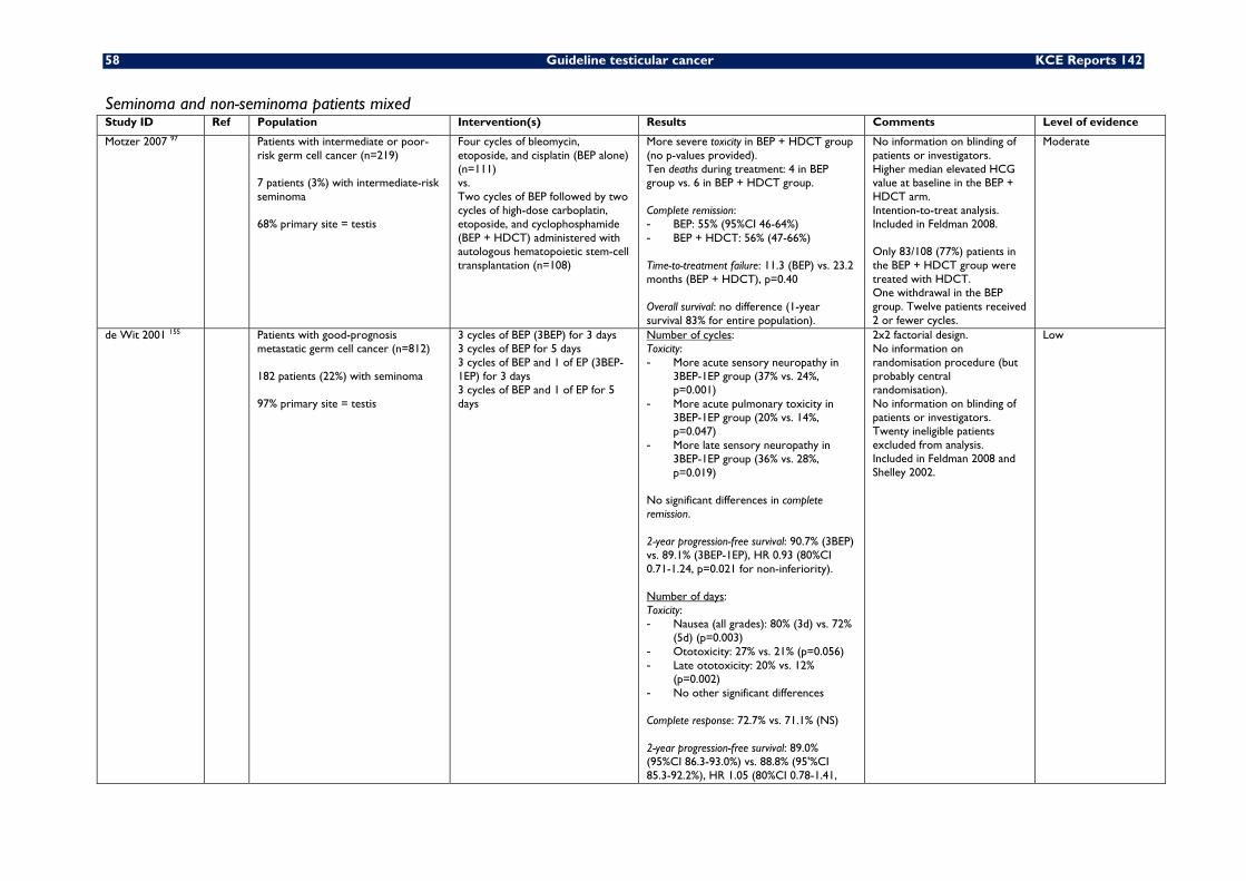
58 Guideline testicular cancer KCE Reports 142
Seminoma and non-seminoma patients mixed Study ID Ref Population Intervention(s) Results Comments Level of evidence
Motzer 2007 97 Patients with intermediate or poor-risk germ cell cancer (n=219) 7 patients (3%) with intermediate-risk seminoma 68% primary site = testis
Four cycles of bleomycin, etoposide, and cisplatin (BEP alone) (n=111) vs. Two cycles of BEP followed by two cycles of high-dose carboplatin, etoposide, and cyclophosphamide (BEP + HDCT) administered with autologous hematopoietic stem-cell transplantation (n=108)
More severe toxicity in BEP + HDCT group (no p-values provided). Ten deaths during treatment: 4 in BEP group vs. 6 in BEP + HDCT group. Complete remission: - BEP: 55% (95%CI 46-64%) - BEP + HDCT: 56% (47-66%) Time-to-treatment failure: 11.3 (BEP) vs. 23.2 months (BEP + HDCT), p=0.40 Overall survival: no difference (1-year survival 83% for entire population).
No information on blinding of patients or investigators. Higher median elevated HCG value at baseline in the BEP + HDCT arm. Intention-to-treat analysis. Included in Feldman 2008. Only 83/108 (77%) patients in the BEP + HDCT group were treated with HDCT. One withdrawal in the BEP group. Twelve patients received 2 or fewer cycles.
Moderate
de Wit 2001 155 Patients with good-prognosis metastatic germ cell cancer (n=812) 182 patients (22%) with seminoma 97% primary site = testis
3 cycles of BEP (3BEP) for 3 days 3 cycles of BEP for 5 days 3 cycles of BEP and 1 of EP (3BEP-1EP) for 3 days 3 cycles of BEP and 1 of EP for 5 days
Number of cycles: Toxicity: - More acute sensory neuropathy in
3BEP-1EP group (37% vs. 24%, p=0.001)
- More acute pulmonary toxicity in 3BEP-1EP group (20% vs. 14%, p=0.047)
- More late sensory neuropathy in 3BEP-1EP group (36% vs. 28%, p=0.019)
No significant differences in complete remission. 2-year progression-free survival: 90.7% (3BEP) vs. 89.1% (3BEP-1EP), HR 0.93 (80%CI 0.71-1.24, p=0.021 for non-inferiority). Number of days: Toxicity: - Nausea (all grades): 80% (3d) vs. 72%
(5d) (p=0.003) - Ototoxicity: 27% vs. 21% (p=0.056) - Late ototoxicity: 20% vs. 12%
(p=0.002) - No other significant differences Complete response: 72.7% vs. 71.1% (NS) 2-year progression-free survival: 89.0% (95%CI 86.3-93.0%) vs. 88.8% (95'%CI 85.3-92.2%), HR 1.05 (80%CI 0.78-1.41,
2x2 factorial design. No information on randomisation procedure (but probably central randomisation). No information on blinding of patients or investigators. Twenty ineligible patients excluded from analysis. Included in Feldman 2008 and Shelley 2002.
Low
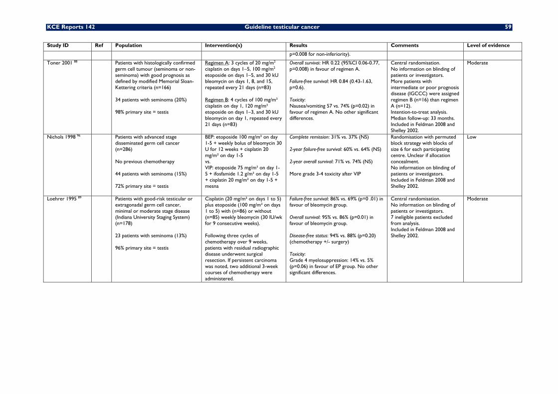
KCE Reports 142 Guideline testicular cancer 59
Study ID Ref Population Intervention(s) Results Comments Level of evidence
p=0.008 for non-inferiority).
Toner 2001 88 Patients with histologically confirmed germ cell tumour (seminoma or non-seminoma) with good prognosis as defined by modified Memorial Sloan-Kettering criteria (n=166) 34 patients with seminoma (20%) 98% primary site = testis
Regimen A: 3 cycles of 20 mg/m2 cisplatin on days 1–5, 100 mg/m2 etoposide on days 1–5, and 30 kU bleomycin on days 1, 8, and 15, repeated every 21 days (n=83) Regimen B: 4 cycles of 100 mg/m² cisplatin on day 1, 120 mg/m² etoposide on days 1–3, and 30 kU bleomycin on day 1, repeated every 21 days (n=83)
Overall survival: HR 0.22 (95%CI 0.06-0.77, p=0.008) in favour of regimen A. Failure-free survival: HR 0.84 (0.43-1.63, p=0.6). Toxicity: Nausea/vomiting 57 vs. 74% (p=0.02) in favour of regimen A. No other significant differences.
Central randomisation. No information on blinding of patients or investigators. More patients with intermediate or poor prognosis disease (IGCCC) were assigned regimen B (n=16) than regimen A (n=12). Intention-to-treat analysis. Median follow-up: 33 months. Included in Feldman 2008 and Shelley 2002.
Moderate
Nichols 1998 96 Patients with advanced stage disseminated germ cell cancer (n=286) No previous chemotherapy 44 patients with seminoma (15%) 72% primary site = testis
BEP: etoposide 100 mg/m² on day 1-5 + weekly bolus of bleomycin 30 U for 12 weeks + cisplatin 20 mg/m² on day 1-5 vs. VIP: etoposide 75 mg/m² on day 1-5 + ifosfamide 1.2 g/m² on day 1-5 + cisplatin 20 mg/m² on day 1-5 + mesna
Complete remission: 31% vs. 37% (NS) 2-year failure-free survival: 60% vs. 64% (NS) 2-year overall survival: 71% vs. 74% (NS) More grade 3-4 toxicity after VIP
Randomisation with permuted block strategy with blocks of size 6 for each participating centre. Unclear if allocation concealment. No information on blinding of patients or investigators. Included in Feldman 2008 and Shelley 2002.
Low
Loehrer 1995 89 Patients with good-risk testicular or extragonadal germ cell cancer, minimal or moderate stage disease (Indiana University Staging System) (n=178) 23 patients with seminoma (13%) 96% primary site = testis
Cisplatin (20 mg/m² on days 1 to 5) plus etoposide (100 mg/m² on days 1 to 5) with (n=86) or without (n=85) weekly bleomycin (30 IU/wk for 9 consecutive weeks). Following three cycles of chemotherapy over 9 weeks, patients with residual radiographic disease underwent surgical resection. If persistent carcinoma was noted, two additional 3-week courses of chemotherapy were administered.
Failure-free survival: 86% vs. 69% (p=0 .01) in favour of bleomycin group. Overall survival: 95% vs. 86% (p=0.01) in favour of bleomycin group. Disease-free status: 94% vs. 88% (p=0.20) (chemotherapy +/- surgery) Toxicity: Grade 4 myelosuppression: 14% vs. 5% (p=0.06) in favour of EP group. No other significant differences.
Central randomisation. No information on blinding of patients or investigators. 7 ineligible patients excluded from analysis. Included in Feldman 2008 and Shelley 2002.
Moderate
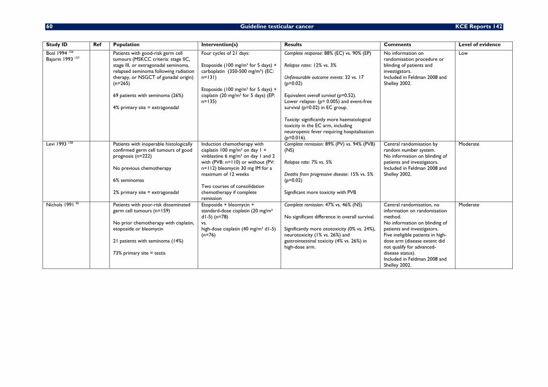
60 Guideline testicular cancer KCE Reports 142
Study ID Ref Population Intervention(s) Results Comments Level of evidence
Bosl 1994 156 Bajorin 1993 157
Patients with good-risk germ cell tumours (MSKCC criteria: stage IIC, stage III, or extragonadal seminoma, relapsed seminoma following radiation therapy, or NSGCT of gonadal origin) (n=265) 69 patients with seminoma (26%) 4% primary site = extragonadal
Four cycles of 21 days: Etoposide (100 mg/m² for 5 days) + carboplatin (350-500 mg/m²) (EC: n=131) Etoposide (100 mg/m² for 5 days) + cisplatin (20 mg/m² for 5 days) (EP: n=135)
Complete response: 88% (EC) vs. 90% (EP) Relapse rates: 12% vs. 3% Unfavourable outcome events: 32 vs. 17 (p=0.02) Equivalent overall survival (p=0.52). Lower relapse- (p= 0.005) and event-free survival (p=0.02) in EC group. Toxicity: significantly more haematological toxicity in the EC arm, including neutropenic fever requiring hospitalisation (p=0.016).
No information on randomisation procedure or blinding of patients and investigators. Included in Feldman 2008 and Shelley 2002.
Low
Levi 1993 158 Patients with inoperable histologically confirmed germ cell tumours of good prognosis (n=222) No previous chemotherapy 6% seminomas 2% primary site = extragonadal
Induction chemotherapy with cisplatin 100 mg/m² on day 1 + vinblastine 6 mg/m² on day 1 and 2 with (PVB: n=110) or without (PV: n=112) bleomycin 30 mg IM for a maximum of 12 weeks Two courses of consolidation chemotherapy if complete remission
Complete remission: 89% (PV) vs. 94% (PVB) (NS) Relapse rate: 7% vs. 5% Deaths from progressive disease: 15% vs. 5% (p=0.02) Significant more toxicity with PVB
Central randomisation by random number system. No information on blinding of patients and investigators. Included in Feldman 2008 and Shelley 2002.
Moderate
Nichols 1991 95 Patients with poor-risk disseminated germ cell tumours (n=159) No prior chemotherapy with cisplatin, etoposide or bleomycin 21 patients with seminoma (14%) 73% primary site = testis
Etoposide + bleomycin + standard-dose cisplatin (20 mg/m² d1-5) (n=78) vs. high-dose cisplatin (40 mg/m² d1-5) (n=76)
Complete remission: 47% vs. 46% (NS) No significant difference in overall survival. Significantly more ototoxicity (0% vs. 24%), neurotoxicity (1% vs. 26%) and gastrointestinal toxicity (4% vs. 26%) in high-dose arm.
Central randomisation, no information on randomisation method. No information on blinding of patients and investigators. Five ineligible patients in high-dose arm (disease extent did not qualify for advanced- disease status). Included in Feldman 2008 and Shelley 2002.
Moderate
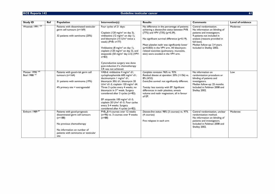
KCE Reports 142 Guideline testicular cancer 61
Study ID Ref Population Intervention(s) Results Comments Level of evidence
Wozniak 1991 159 Patients with disseminated testicular germ cell tumours (n=169) 32 patients with seminoma (20%)
Four cycles of 21 days: Cisplatin (120 mg/m² on day 3), vinblastine (12 mg/m² on day 1), and bleomycin (15 U/m² twice a week) (PVB: n=77) Vinblastine (8 mg/m² on day 1), cisplatin (120 mg/m² on day 3), and etoposide (50 mg/m² day 2-5) (VPV: n=83) Cytoreductive surgery was done post-induction if a chemotherapy CR was not achieved.
No difference in the percentage of patients achieving a disease-free status between PVB (77%) and VPV (73%) (p=0.39). No significant survival difference (p=0.19). Mean platelet nadir was significantly lower (p=0.003) in the VPV arm. All bleomycin-related toxicities (pulmonary, mucositis, skin) were avoided in the VPV arm.
Central randomisation. No information on blinding of patients and investigators. 9 patients not included in analysis (reasons provided in full-text). Median follow-up: 3.4 years. Included in Shelley 2002.
Moderate
Motzer 1990 160 Bosl 1988 161
Patients with good-risk germ cell tumours (n=164) 31 patients with seminoma (19%) 4% primary site = extragonadal
VAB-6: vinblastine 4 mg/m² d1; cyclophosphamide 600 mg/m² d1; doctinomycin 1 mg/m² d1; bleomycin 30U d1; bleomycin 20 U/m² d1-3; cisplatin 120 mg/m² d4. Three 3 cycles every 4 weeks, no bleomycin in 3rd week. Surgery considered after 3 cycles (n=82). EP: etoposide 100 mg/m² d1-5; cisplatin 20 U/m² d1-5. Four cycles every 3-4 weeks. Surgery considered after 4 cycles (n=82).
Complete remission: 96% vs. 93% Residual disease at operation: 20% (11/56) vs. 8% (4/52) Event-free survival: not significantly different. Toxicity: less toxicity with EP. Significant differences in nadir platelets, emesis volume and nadir magnesium, all in favour of EP.
No information on randomisation procedure or blinding of patients and investigators. Median follow-up: 25 months. Included in Feldman 2008 and Shelley 2002.
Low
Einhorn 1989 87 Patients with good-prognosis disseminated germ cell tumours (n=188) No previous chemotherapy No information on number of patients with seminoma or testicular site
PVB16B 4 courses over 12 weeks (n=96) vs. 3 courses over 9 weeks (n=88)
Disease-free status: 98% (3 courses) vs. 97% (4 courses) Five relapses in each arm
Central randomisation, unclear randomisation method. No information on blinding of patients and investigators. Included in Feldman 2008 and Shelley 2002.
Moderate
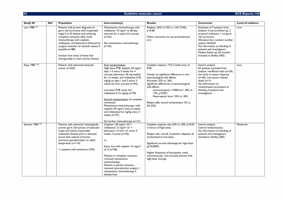
62 Guideline testicular cancer KCE Reports 142
Study ID Ref Population Intervention(s) Results Comments Level of evidence
Levi 1988 162 Patients with proven diagnosis of germ cell carcinoma with inoperable stage II or III disease and achieving complete remission after initial chemotherapy with cisplatin, vinblastine, and bleomycin followed by surgical resection of residual masses if possible (n=88) Unclear how many of these had extragonadal or even ovarian disease
Maintenance chemotherapy with vinblastine 10 mg/m² at 28-day intervals for a total of 6 courses (n=43). No maintenance chemotherapy (n=45).
Relapses: 26% (11/43) vs. 16% (7/45), p=0.08. Other outcomes are not presented per arm.
Exclusion of 7 patients from analysis: 4 lost-to-follow-up, 2 protocol violations, 1 no germ cell carcinoma. Allocation by a random number system: blinded? No information on blinding of patients and investigators. Median follow-up: 64 months. Included in Shelley 2002.
Low
Kaye 1985 163 Patients with advanced testicular cancer (n=203)
First randomisation: High-dose PVB: cisplatin 20 mg/m² days 1-5 every 3 weeks for 4 courses, bleomycin 30 mg weekly for 12 weeks, and vinblastine 0.20 mg/kg on days 1 and 2 every 3 weeks for four courses (n=64). Low-dose PVB: same, but vinblastine 0.15 mg/kg (n=70). Second randomisation (if complete remission): Maintenance chemotherapy: with cisplatin 50 mg/m² every 6 weeks and vinblastine 0.2 mg/kg every 3 weeks (n=37). No further chemotherapy (n=31).
Complete response: 71% in both arms of PVB. Toxicity: no significant difference in non-haematological side effects. Mucositis: 55% vs. 36%. Significant differences in haematological side effects: - Leucocytopenia <1000/mm²: 30% vs.
13%, p<0.001 - Neutropenic fever: 55% vs. 30% Relapse after second randomisation: 3% vs. 6% (NS)
Interim analysis. 69 patients excluded from analysis: insufficient data (n=24), too early to assess response (n=44), non-cancer-related death (n=1). No information on randomisation procedure or blinding of patients and investigators.
Low
Samson 1984 164 Patients with advanced, histologically proven germ cell tumour of testicular origin and clearly measurable metastatic disease and/ or elevated serum beta subunit of human chorionic gonadotrophin or alpha-fetoprotein (n=114) 11 patients with seminoma (10%)
Cisplatin 120 mg/m² d3 + vinblastine 12 mg/m² d1 + bleomycin 15 U/m² d1 every 4 weeks, 4 cycles (n=56). vs. Same, but with cisplatin 15 mg/m² d1-5 (n=58). Patients in complete remission received maintenance chemotherapy. Patients in partial remission received cytoreductive surgery + maintenance chemotherapy if disease-free.
Complete response rate: 63% vs. 43%, p=0.03 in favour of high-dose. Relapse rate: overall, 4 patients relapsed, all randomised to low-dose. Significant survival advantage for high-dose (p=0.0009). Higher frequency of leucopenia, renal, neuromuscular, and mucosal toxicity with high-dose therapy.
Interim analysis. Central randomisation. No information on blinding of patients and investigators. Included in Shelley 2002.
Moderate
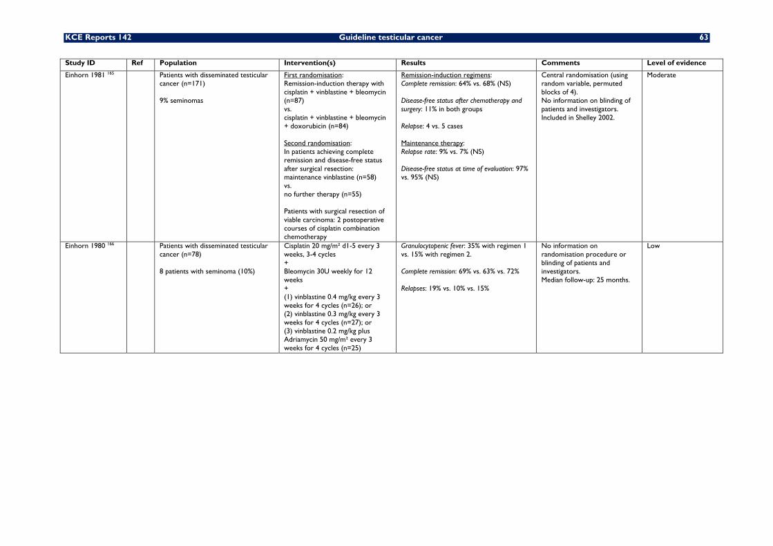
KCE Reports 142 Guideline testicular cancer 63
Study ID Ref Population Intervention(s) Results Comments Level of evidence
Einhorn 1981 165 Patients with disseminated testicular cancer (n=171) 9% seminomas
First randomisation: Remission-induction therapy with cisplatin + vinblastine + bleomycin (n=87) vs. cisplatin + vinblastine + bleomycin + doxorubicin (n=84) Second randomisation: In patients achieving complete remission and disease-free status after surgical resection: maintenance vinblastine (n=58) vs. no further therapy (n=55) Patients with surgical resection of viable carcinoma: 2 postoperative courses of cisplatin combination chemotherapy
Remission-induction regimens: Complete remission: 64% vs. 68% (NS) Disease-free status after chemotherapy and surgery: 11% in both groups Relapse: 4 vs. 5 cases Maintenance therapy: Relapse rate: 9% vs. 7% (NS) Disease-free status at time of evaluation: 97% vs. 95% (NS)
Central randomisation (using random variable, permuted blocks of 4). No information on blinding of patients and investigators. Included in Shelley 2002.
Moderate
Einhorn 1980 166 Patients with disseminated testicular cancer (n=78) 8 patients with seminoma (10%)
Cisplatin 20 mg/m² d1-5 every 3 weeks, 3-4 cycles + Bleomycin 30U weekly for 12 weeks + (1) vinblastine 0.4 mg/kg every 3 weeks for 4 cycles (n=26); or (2) vinblastine 0.3 mg/kg every 3 weeks for 4 cycles (n=27); or (3) vinblastine 0.2 mg/kg plus Adriamycin 50 mg/m² every 3 weeks for 4 cycles (n=25)
Granulocytopenic fever: 35% with regimen 1 vs. 15% with regimen 2. Complete remission: 69% vs. 63% vs. 72% Relapses: 19% vs. 10% vs. 15%
No information on randomisation procedure or blinding of patients and investigators. Median follow-up: 25 months.
Low
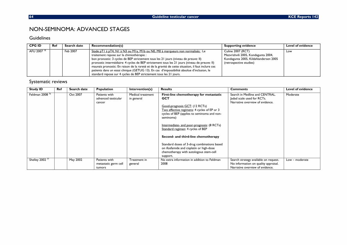
64 Guideline testicular cancer KCE Reports 142
NON-SEMINOMA: ADVANCED STAGES
Guidelines
CPG ID Ref Search date Recommendation(s) Supporting evidence Level of evidence
AFU 2007 18 Feb 2007 Stade pT1 à pT4, N1 à N3 ou M1a, M1b ou N0, M0 à marqueurs non normalisés : Le traitement repose sur la chimiothérapie : bon pronostic: 3 cycles de BEP strictement tous les 21 jours (niveau de preuve: II) pronostic intermédiaire: 4 cycles de BEP strictement tous les 21 jours (niveau de preuve: II) mauvais pronostic: En raison de la rareté et de la gravité de cette situation, il faut inclure ces patients dans un essai clinique (GETUG 13). En cas d’impossibilité absolue d’inclusion, le standard repose sur 4 cycles de BEP strictement tous les 21 jours.
Culine 2007 (RCT) Mezvrishvili 2005, Kondagunta 2004, Kondagunta 2005, Kildahlandersen 2005 (retrospective studies)
Low
Systematic reviews Study ID Ref Search date Population Intervention(s) Results Comments Level of evidence
Feldman 2008 76 Oct 2007 Patients with advanced testicular cancer
Medical treatment in general
First-line chemotherapy for metastatic GCT Good-prognosis GCT: (12 RCTs) Two effective regimens: 4 cycles of EP or 3 cycles of BEP (applies to seminoma and non-seminoma) Intermediate- and poor-prognosis: (8 RCTs) Standard regimen: 4 cycles of BEP Second- and third-line chemotherapy Standard doses of 3-drug combinations based on ifosfamide and cisplatin or high-dose chemotherapy with autologous stem-cell support.
Search in Medline and CENTRAL. Jadad scale used for RCTs. Narrative overview of evidence.
Moderate
Shelley 2002 77 May 2002 Patients with metastatic germ cell tumors
Treatment in general
No extra information in addition to Feldman 2008
Search strategy available on request. No information on quality appraisal. Narrative overview of evidence.
Low – moderate
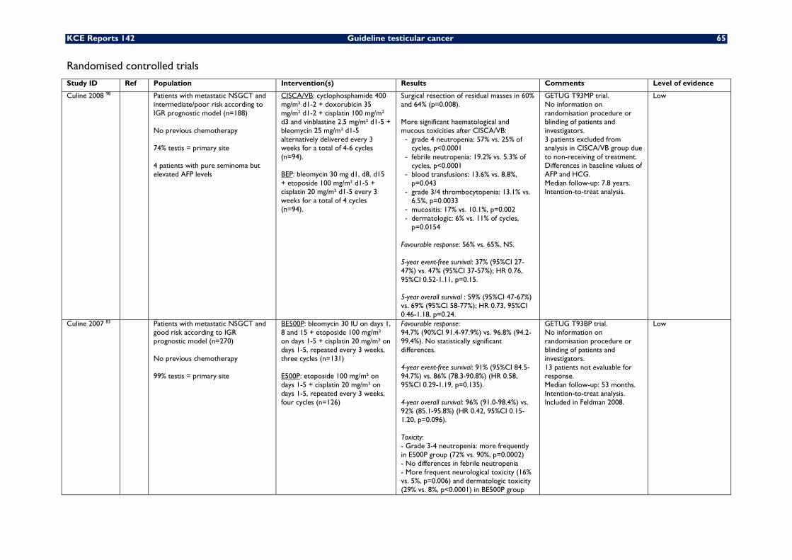
KCE Reports 142 Guideline testicular cancer 65
Randomised controlled trials Study ID Ref Population Intervention(s) Results Comments Level of evidence
Culine 2008 98 Patients with metastatic NSGCT and intermediate/poor risk according to IGR prognostic model (n=188) No previous chemotherapy 74% testis = primary site 4 patients with pure seminoma but elevated AFP levels
CISCA/VB: cyclophosphamide 400 mg/m² d1-2 + doxorubicin 35 mg/m² d1-2 + cisplatin 100 mg/m² d3 and vinblastine 2.5 mg/m² d1-5 + bleomycin 25 mg/m² d1-5 alternatively delivered every 3 weeks for a total of 4-6 cycles (n=94). BEP: bleomycin 30 mg d1, d8, d15 + etoposide 100 mg/m² d1-5 + cisplatin 20 mg/m² d1-5 every 3 weeks for a total of 4 cycles (n=94).
Surgical resection of residual masses in 60% and 64% (p=0.008). More significant haematological and mucous toxicities after CISCA/VB: - grade 4 neutropenia: 57% vs. 25% of
cycles, p<0.0001 - febrile neutropenia: 19.2% vs. 5.3% of
cycles, p<0.0001 - blood transfusions: 13.6% vs. 8.8%,
p=0.043 - grade 3/4 thrombocytopenia: 13.1% vs.
6.5%, p=0.0033 - mucositis: 17% vs. 10.1%, p=0.002 - dermatologic: 6% vs. 11% of cycles,
p=0.0154
Favourable response: 56% vs. 65%, NS. 5-year event-free survival: 37% (95%CI 27-47%) vs. 47% (95%CI 37-57%); HR 0.76, 95%CI 0.52-1.11, p=0.15. 5-year overall survival : 59% (95%CI 47-67%) vs. 69% (95%CI 58-77%); HR 0.73, 95%CI 0.46-1.18, p=0.24.
GETUG T93MP trial. No information on randomisation procedure or blinding of patients and investigators. 3 patients excluded from analysis in CISCA/VB group due to non-receiving of treatment. Differences in baseline values of AFP and HCG. Median follow-up: 7.8 years. Intention-to-treat analysis.
Low
Culine 2007 83 Patients with metastatic NSGCT and good risk according to IGR prognostic model (n=270) No previous chemotherapy 99% testis = primary site
BE500P: bleomycin 30 IU on days 1, 8 and 15 + etoposide 100 mg/m² on days 1-5 + cisplatin 20 mg/m² on days 1-5, repeated every 3 weeks, three cycles (n=131) E500P: etoposide 100 mg/m² on days 1-5 + cisplatin 20 mg/m² on days 1-5, repeated every 3 weeks, four cycles (n=126)
Favourable response: 94.7% (90%CI 91.4-97.9%) vs. 96.8% (94.2-99.4%). No statistically significant differences. 4-year event-free survival: 91% (95%CI 84.5-94.7%) vs. 86% (78.3-90.8%) (HR 0.58, 95%CI 0.29-1.19, p=0.135). 4-year overall survival: 96% (91.0-98.4%) vs. 92% (85.1-95.8%) (HR 0.42, 95%CI 0.15-1.20, p=0.096). Toxicity: - Grade 3-4 neutropenia: more frequently in E500P group (72% vs. 90%, p=0.0002) - No differences in febrile neutropenia - More frequent neurological toxicity (16% vs. 5%, p=0.006) and dermatologic toxicity (29% vs. 8%, p<0.0001) in BE500P group
GETUG T93BP trial. No information on randomisation procedure or blinding of patients and investigators. 13 patients not evaluable for response. Median follow-up: 53 months. Intention-to-treat analysis. Included in Feldman 2008.
Low
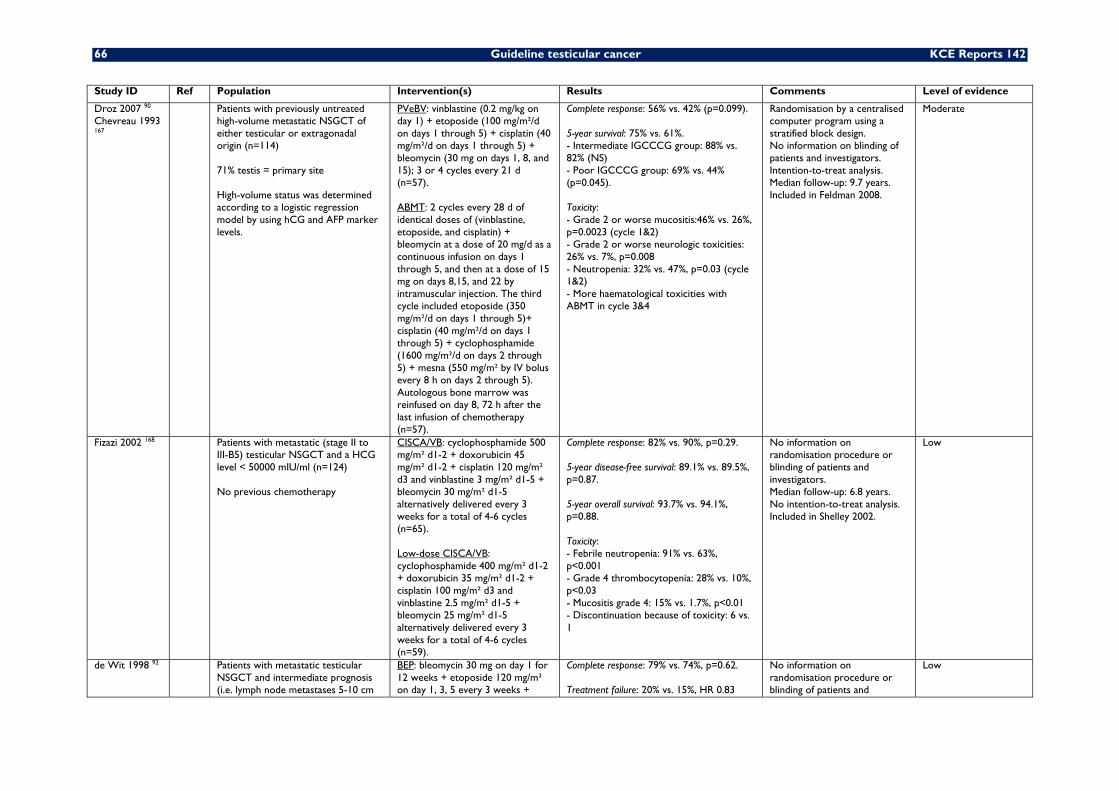
66 Guideline testicular cancer KCE Reports 142
Study ID Ref Population Intervention(s) Results Comments Level of evidence
Droz 2007 90 Chevreau 1993 167
Patients with previously untreated high-volume metastatic NSGCT of either testicular or extragonadal origin (n=114) 71% testis = primary site High-volume status was determined according to a logistic regression model by using hCG and AFP marker levels.
PVeBV: vinblastine (0.2 mg/kg on day 1) + etoposide (100 mg/m²/d on days 1 through 5) + cisplatin (40 mg/m²/d on days 1 through 5) + bleomycin (30 mg on days 1, 8, and 15); 3 or 4 cycles every 21 d (n=57). ABMT: 2 cycles every 28 d of identical doses of (vinblastine, etoposide, and cisplatin) + bleomycin at a dose of 20 mg/d as a continuous infusion on days 1 through 5, and then at a dose of 15 mg on days 8,15, and 22 by intramuscular injection. The third cycle included etoposide (350 mg/m²/d on days 1 through 5)+ cisplatin (40 mg/m²/d on days 1 through 5) + cyclophosphamide (1600 mg/m²/d on days 2 through 5) + mesna (550 mg/m² by IV bolus every 8 h on days 2 through 5). Autologous bone marrow was reinfused on day 8, 72 h after the last infusion of chemotherapy (n=57).
Complete response: 56% vs. 42% (p=0.099). 5-year survival: 75% vs. 61%. - Intermediate IGCCCG group: 88% vs. 82% (NS) - Poor IGCCCG group: 69% vs. 44% (p=0.045). Toxicity: - Grade 2 or worse mucositis:46% vs. 26%, p=0.0023 (cycle 1&2) - Grade 2 or worse neurologic toxicities: 26% vs. 7%, p=0.008 - Neutropenia: 32% vs. 47%, p=0.03 (cycle 1&2) - More haematological toxicities with ABMT in cycle 3&4
Randomisation by a centralised computer program using a stratified block design. No information on blinding of patients and investigators. Intention-to-treat analysis. Median follow-up: 9.7 years. Included in Feldman 2008.
Moderate
Fizazi 2002 168 Patients with metastatic (stage II to III-B5) testicular NSGCT and a HCG level < 50000 mIU/ml (n=124) No previous chemotherapy
CISCA/VB: cyclophosphamide 500 mg/m² d1-2 + doxorubicin 45 mg/m² d1-2 + cisplatin 120 mg/m² d3 and vinblastine 3 mg/m² d1-5 + bleomycin 30 mg/m² d1-5 alternatively delivered every 3 weeks for a total of 4-6 cycles (n=65). Low-dose CISCA/VB: cyclophosphamide 400 mg/m² d1-2 + doxorubicin 35 mg/m² d1-2 + cisplatin 100 mg/m² d3 and vinblastine 2.5 mg/m² d1-5 + bleomycin 25 mg/m² d1-5 alternatively delivered every 3 weeks for a total of 4-6 cycles (n=59).
Complete response: 82% vs. 90%, p=0.29. 5-year disease-free survival: 89.1% vs. 89.5%, p=0.87. 5-year overall survival: 93.7% vs. 94.1%, p=0.88. Toxicity: - Febrile neutropenia: 91% vs. 63%, p<0.001 - Grade 4 thrombocytopenia: 28% vs. 10%, p<0.03 - Mucositis grade 4: 15% vs. 1.7%, p<0.01 - Discontinuation because of toxicity: 6 vs. 1
No information on randomisation procedure or blinding of patients and investigators. Median follow-up: 6.8 years. No intention-to-treat analysis. Included in Shelley 2002.
Low
de Wit 1998 92 Patients with metastatic testicular NSGCT and intermediate prognosis (i.e. lymph node metastases 5-10 cm
BEP: bleomycin 30 mg on day 1 for 12 weeks + etoposide 120 mg/m² on day 1, 3, 5 every 3 weeks +
Complete response: 79% vs. 74%, p=0.62. Treatment failure: 20% vs. 15%, HR 0.83
No information on randomisation procedure or blinding of patients and
Low
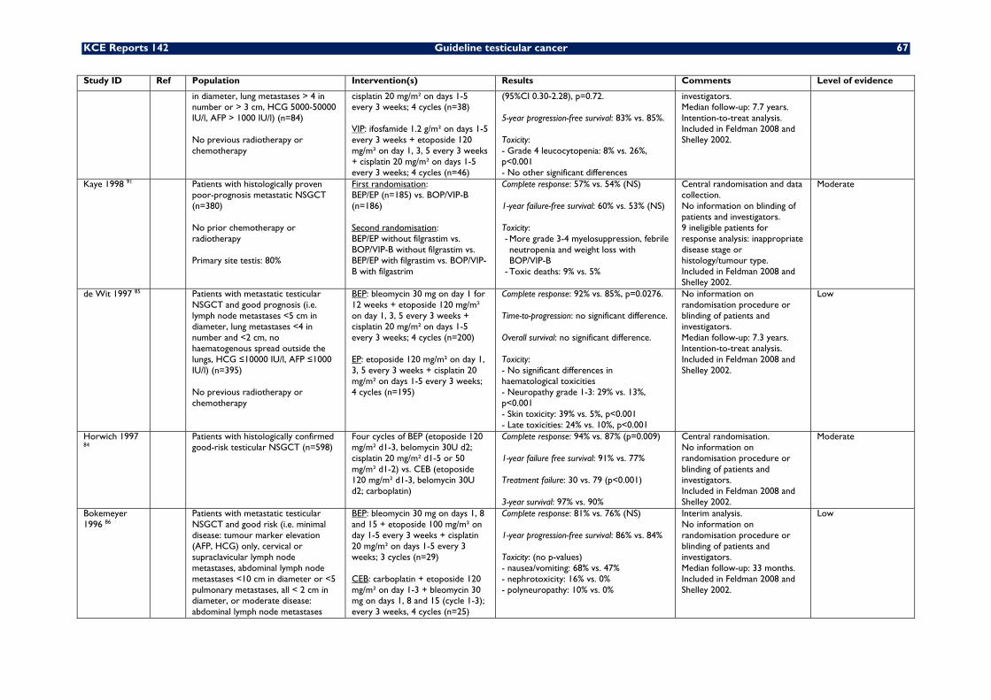
KCE Reports 142 Guideline testicular cancer 67
Study ID Ref Population Intervention(s) Results Comments Level of evidence
in diameter, lung metastases > 4 in number or > 3 cm, HCG 5000-50000 IU/l, AFP > 1000 IU/l) (n=84) No previous radiotherapy or chemotherapy
cisplatin 20 mg/m² on days 1-5 every 3 weeks; 4 cycles (n=38) VIP: ifosfamide 1.2 g/m² on days 1-5 every 3 weeks + etoposide 120 mg/m² on day 1, 3, 5 every 3 weeks + cisplatin 20 mg/m² on days 1-5 every 3 weeks; 4 cycles (n=46)
(95%CI 0.30-2.28), p=0.72. 5-year progression-free survival: 83% vs. 85%. Toxicity: - Grade 4 leucocytopenia: 8% vs. 26%, p<0.001 - No other significant differences
investigators. Median follow-up: 7.7 years. Intention-to-treat analysis. Included in Feldman 2008 and Shelley 2002.
Kaye 1998 91 Patients with histologically proven poor-prognosis metastatic NSGCT (n=380) No prior chemotherapy or radiotherapy Primary site testis: 80%
First randomisation: BEP/EP (n=185) vs. BOP/VIP-B (n=186) Second randomisation: BEP/EP without filgrastim vs. BOP/VIP-B without filgrastim vs. BEP/EP with filgrastim vs. BOP/VIP-B with filgastrim
Complete response: 57% vs. 54% (NS) 1-year failure-free survival: 60% vs. 53% (NS) Toxicity: - More grade 3-4 myelosuppression, febrile neutropenia and weight loss with BOP/VIP-B
- Toxic deaths: 9% vs. 5%
Central randomisation and data collection. No information on blinding of patients and investigators. 9 ineligible patients for response analysis: inappropriate disease stage or histology/tumour type. Included in Feldman 2008 and Shelley 2002.
Moderate
de Wit 1997 85 Patients with metastatic testicular NSGCT and good prognosis (i.e. lymph node metastases <5 cm in diameter, lung metastases <4 in number and <2 cm, no haematogenous spread outside the lungs, HCG ≤10000 IU/l, AFP ≤1000 IU/l) (n=395) No previous radiotherapy or chemotherapy
BEP: bleomycin 30 mg on day 1 for 12 weeks + etoposide 120 mg/m² on day 1, 3, 5 every 3 weeks + cisplatin 20 mg/m² on days 1-5 every 3 weeks; 4 cycles (n=200) EP: etoposide 120 mg/m² on day 1, 3, 5 every 3 weeks + cisplatin 20 mg/m² on days 1-5 every 3 weeks; 4 cycles (n=195)
Complete response: 92% vs. 85%, p=0.0276. Time-to-progression: no significant difference. Overall survival: no significant difference. Toxicity: - No significant differences in haematological toxicities - Neuropathy grade 1-3: 29% vs. 13%, p<0.001 - Skin toxicity: 39% vs. 5%, p<0.001 - Late toxicities: 24% vs. 10%, p<0.001
No information on randomisation procedure or blinding of patients and investigators. Median follow-up: 7.3 years. Intention-to-treat analysis. Included in Feldman 2008 and Shelley 2002.
Low
Horwich 1997 84
Patients with histologically confirmed good-risk testicular NSGCT (n=598)
Four cycles of BEP (etoposide 120 mg/m² d1-3, belomycin 30U d2; cisplatin 20 mg/m² d1-5 or 50 mg/m² d1-2) vs. CEB (etoposide 120 mg/m² d1-3, belomycin 30U d2; carboplatin)
Complete response: 94% vs. 87% (p=0.009) 1-year failure free survival: 91% vs. 77% Treatment failure: 30 vs. 79 (p<0.001) 3-year survival: 97% vs. 90%
Central randomisation. No information on randomisation procedure or blinding of patients and investigators. Included in Feldman 2008 and Shelley 2002.
Moderate
Bokemeyer 1996 86
Patients with metastatic testicular NSGCT and good risk (i.e. minimal disease: tumour marker elevation (AFP, HCG) only, cervical or supraclavicular lymph node metastases, abdominal lymph node metastases <10 cm in diameter or <5 pulmonary metastases, all < 2 cm in diameter, or moderate disease: abdominal lymph node metastases
BEP: bleomycin 30 mg on days 1, 8 and 15 + etoposide 100 mg/m² on day 1-5 every 3 weeks + cisplatin 20 mg/m² on days 1-5 every 3 weeks; 3 cycles (n=29) CEB: carboplatin + etoposide 120 mg/m² on day 1-3 + bleomycin 30 mg on days 1, 8 and 15 (cycle 1-3); every 3 weeks, 4 cycles (n=25)
Complete response: 81% vs. 76% (NS) 1-year progression-free survival: 86% vs. 84% Toxicity: (no p-values) - nausea/vomiting: 68% vs. 47% - nephrotoxicity: 16% vs. 0% - polyneuropathy: 10% vs. 0%
Interim analysis. No information on randomisation procedure or blinding of patients and investigators. Median follow-up: 33 months. Included in Feldman 2008 and Shelley 2002.
Low
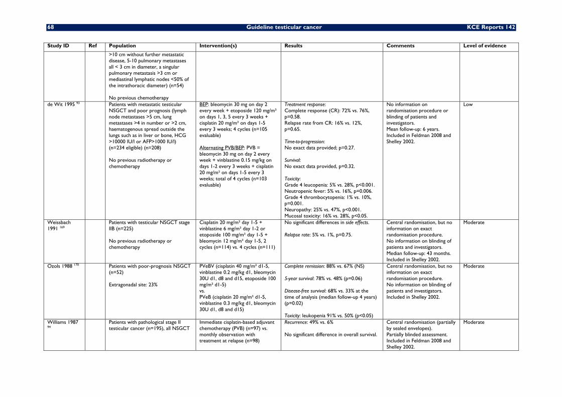
68 Guideline testicular cancer KCE Reports 142
Study ID Ref Population Intervention(s) Results Comments Level of evidence
>10 cm without further metastatic disease, 5-10 pulmonary metastases all < 3 cm in diameter, a singular pulmonary metastasis >3 cm or mediastinal lymphatic nodes <50% of the intrathoracic diameter) (n=54) No previous chemotherapy
de Wit 1995 93 Patients with metastatic testicular NSGCT and poor prognosis (lymph node metastases >5 cm, lung metastases >4 in number or >2 cm, haematogenous spread outside the lungs such as in liver or bone, HCG >10000 IU/l or AFP>1000 IU/l) (n=234 eligible) (n=208) No previous radiotherapy or chemotherapy
BEP: bleomycin 30 mg on day 2 every week + etoposide 120 mg/m² on days 1, 3, 5 every 3 weeks + cisplatin 20 mg/m² on days 1-5 every 3 weeks; 4 cycles (n=105 evaluable) Alternating PVB/BEP: PVB = bleomycin 30 mg on day 2 every week + vinblastine 0.15 mg/kg on days 1-2 every 3 weeks + cisplatin 20 mg/m² on days 1-5 every 3 weeks; total of 4 cycles (n=103 evaluable)
Treatment response: Complete response (CR): 72% vs. 76%, p=0.58. Relapse rate from CR: 16% vs. 12%, p=0.65. Time-to-progression: No exact data provided; p=0.27. Survival: No exact data provided, p=0.32. Toxicity: Grade 4 leucopenia: 5% vs. 28%, p<0.001. Neutropenic fever: 5% vs. 16%, p=0.006. Grade 4 thrombocytopenia: 1% vs. 10%, p=0.001. Neuropathy: 25% vs. 47%, p<0.001. Mucosal toxicity: 16% vs. 28%, p<0.05.
No information on randomisation procedure or blinding of patients and investigators. Mean follow-up: 6 years. Included in Feldman 2008 and Shelley 2002.
Low
Weissbach 1991 169
Patients with testicular NSGCT stage IIB (n=225) No previous radiotherapy or chemotherapy
Cisplatin 20 mg/m² day 1-5 + vinblastine 6 mg/m² day 1-2 or etoposide 100 mg/m² day 1-5 + bleomycin 12 mg/m² day 1-5, 2 cycles (n=114) vs. 4 cycles (n=111)
No significant differences in side effects. Relapse rate: 5% vs. 1%, p=0.75.
Central randomisation, but no information on exact randomisation procedure. No information on blinding of patients and investigators. Median follow-up: 43 months. Included in Shelley 2002.
Moderate
Ozols 1988 170 Patients with poor-prognosis NSGCT (n=52) Extragonadal site: 23%
PVeBV (cisplatin 40 mg/m² d1-5, vinblastine 0.2 mg/kg d1, bleomycin 30U d1, d8 and d15, etoposide 100 mg/m² d1-5) vs. PVeB (cisplatin 20 mg/m² d1-5, vinblastine 0.3 mg/kg d1, bleomycin 30U d1, d8 and d15)
Complete remission: 88% vs. 67% (NS) 5-year survival: 78% vs. 48% (p=0.06) Disease-free survival: 68% vs. 33% at the time of analysis (median follow-up 4 years) (p=0.02) Toxicity: leukopenia 91% vs. 50% (p<0.05)
Central randomisation, but no information on exact randomisation procedure. No information on blinding of patients and investigators. Included in Shelley 2002.
Moderate
Williams 1987 94
Patients with pathological stage II testicular cancer (n=195), all NSGCT
Immediate cisplatin-based adjuvant chemotherapy (PVB) (n=97) vs. monthly observation with treatment at relapse (n=98)
Recurrence: 49% vs. 6% No significant difference in overall survival.
Central randomisation (partially by sealed envelopes). Partially blinded assessment. Included in Feldman 2008 and Shelley 2002.
Moderate
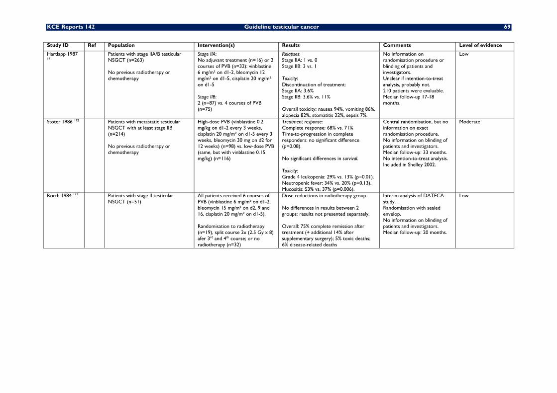
KCE Reports 142 Guideline testicular cancer 69
Study ID Ref Population Intervention(s) Results Comments Level of evidence
Hartlapp 1987 171
Patients with stage IIA/B testicular NSGCT (n=263) No previous radiotherapy or chemotherapy
Stage IIA: No adjuvant treatment (n=16) or 2 courses of PVB (n=32): vinblastine 6 mg/m² on d1-2, bleomycin 12 mg/m² on d1-5, cisplatin 20 mg/m² on d1-5 Stage IIB: 2 (n=87) vs. 4 courses of PVB (n=75)
Relapses: Stage IIA: 1 vs. 0 Stage IIB: 3 vs. 1 Toxicity: Discontinuation of treatment: Stage IIA: 3.6% Stage IIB: 3.6% vs. 11% Overall toxicity: nausea 94%, vomiting 86%, alopecia 82%, stomatitis 22%, sepsis 7%.
No information on randomisation procedure or blinding of patients and investigators. Unclear if intention-to-treat analysis, probably not. 210 patients were evaluable. Median follow-up 17-18 months.
Low
Stoter 1986 172 Patients with metastatic testicular NSGCT with at least stage IIB (n=214) No previous radiotherapy or chemotherapy
High-dose PVB (vinblastine 0.2 mg/kg on d1-2 every 3 weeks, cisplatin 20 mg/m² on d1-5 every 3 weeks, bleomycin 30 mg on d2 for 12 weeks) (n=98) vs. low-dose PVB (same, but with vinblastine 0.15 mg/kg) (n=116)
Treatment response: Complete response: 68% vs. 71% Time-to-progression in complete responders: no significant difference (p=0.08). No significant differences in survival. Toxicity: Grade 4 leukopenia: 29% vs. 13% (p=0.01). Neutropenic fever: 34% vs. 20% (p=0.13). Mucositis: 53% vs. 37% (p=0.006).
Central randomisation, but no information on exact randomisation procedure. No information on blinding of patients and investigators. Median follow-up: 33 months. No intention-to-treat analysis. Included in Shelley 2002.
Moderate
Rorth 1984 173 Patients with stage II testicular NSGCT (n=51)
All patients received 6 courses of PVB (vinblastine 6 mg/m² on d1-2, bleomycin 15 mg/m² on d2, 9 and 16, cisplatin 20 mg/m² on d1-5). Randomisation to radiotherapy (n=19), split course 2x (2.5 Gy x 8) afer 3rd and 4th course; or no radiotherapy (n=32)
Dose reductions in radiotherapy group. No differences in results between 2 groups: results not presented separately. Overall: 75% complete remission after treatment (+ additional 14% after supplementary surgery); 5% toxic deaths; 6% disease-related deaths
Interim analysis of DATECA study. Randomisation with sealed envelop. No information on blinding of patients and investigators. Median follow-up: 20 months.
Low
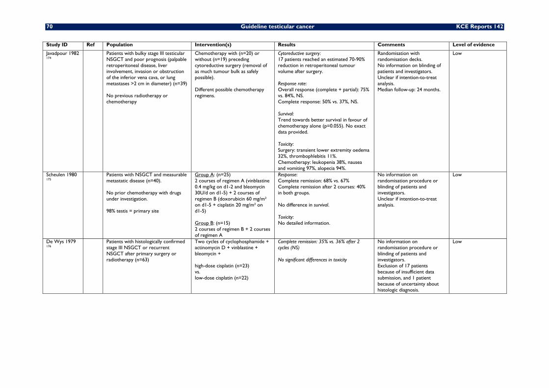
70 Guideline testicular cancer KCE Reports 142
Study ID Ref Population Intervention(s) Results Comments Level of evidence
Javadpour 1982 174
Patients with bulky stage III testicular NSGCT and poor prognosis (palpable retroperitoneal disease, liver involvement, invasion or obstruction of the inferior vena cava, or lung metastases >2 cm in diameter) (n=39) No previous radiotherapy or chemotherapy
Chemotherapy with (n=20) or without (n=19) preceding cytoreductive surgery (removal of as much tumour bulk as safely possible). Different possible chemotherapy regimens.
Cytoreductive surgery: 17 patients reached an estimated 70-90% reduction in retroperitoneal tumour volume after surgery. Response rate: Overall response (complete + partial): 75% vs. 84%, NS. Complete response: 50% vs. 37%, NS. Survival: Trend towards better survival in favour of chemotherapy alone (p=0.055). No exact data provided. Toxicity: Surgery: transient lower extremity oedema 32%, thrombophlebitis 11%. Chemotherapy: leukopenia 38%, nausea and vomiting 97%, alopecia 94%.
Randomisation with randomisation decks. No information on blinding of patients and investigators. Unclear if intention-to-treat analysis. Median follow-up: 24 months.
Low
Scheulen 1980 175
Patients with NSGCT and measurable metastatic disease (n=40). No prior chemotherapy with drugs under investigation. 98% testis = primary site
Group A: (n=25) 2 courses of regimen A (vinblastine 0.4 mg/kg on d1-2 and bleomycin 30U/d on d1-5) + 2 courses of regimen B (doxorubicin 60 mg/m² on d1-5 + cisplatin 20 mg/m² on d1-5) Group B: (n=15) 2 courses of regimen B + 2 courses of regimen A
Response: Complete remission: 68% vs. 67% Complete remission after 2 courses: 40% in both groups. No difference in survival. Toxicity: No detailed information.
No information on randomisation procedure or blinding of patients and investigators. Unclear if intention-to-treat analysis.
Low
De Wys 1979 176
Patients with histologically confirmed stage III NSGCT or recurrent NSGCT after primary surgery or radiotherapy (n=63)
Two cycles of cyclophosphamide + actinomycin D + vinblastine + bleomycin + high-dose cisplatin (n=23) vs. low-dose cisplatin (n=22)
Complete remission: 35% vs. 36% after 2 cycles (NS) No significant differences in toxicity
No information on randomisation procedure or blinding of patients and investigators. Exclusion of 17 patients because of insufficient data submission, and 1 patient because of uncertainty about histologic diagnosis.
Low
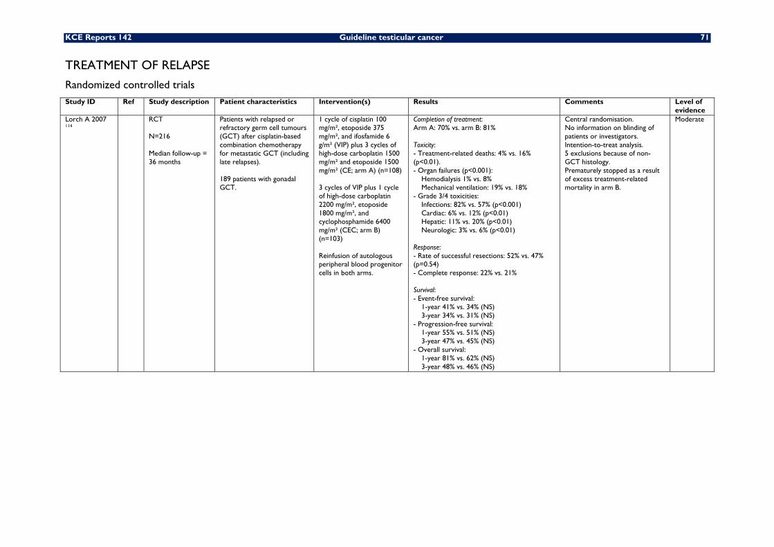
KCE Reports 142 Guideline testicular cancer 71
TREATMENT OF RELAPSE
Randomized controlled trials Study ID Ref Study description Patient characteristics Intervention(s) Results Comments Level of
evidence Lorch A 2007 114
RCT N=216 Median follow-up = 36 months
Patients with relapsed or refractory germ cell tumours (GCT) after cisplatin-based combination chemotherapy for metastatic GCT (including late relapses). 189 patients with gonadal GCT.
1 cycle of cisplatin 100 mg/m², etoposide 375 mg/m², and ifosfamide 6 g/m² (VIP) plus 3 cycles of high-dose carboplatin 1500 mg/m² and etoposide 1500 mg/m² (CE; arm A) (n=108) 3 cycles of VIP plus 1 cycle of high-dose carboplatin 2200 mg/m², etoposide 1800 mg/m², and cyclophosphamide 6400 mg/m² (CEC; arm B) (n=103) Reinfusion of autologous peripheral blood progenitor cells in both arms.
Completion of treatment: Arm A: 70% vs. arm B: 81% Toxicity: - Treatment-related deaths: 4% vs. 16% (p<0.01). - Organ failures (p<0.001):
Hemodialysis 1% vs. 8% Mechanical ventilation: 19% vs. 18%
- Grade 3/4 toxicities: Infections: 82% vs. 57% (p<0.001) Cardiac: 6% vs. 12% (p<0.01) Hepatic: 11% vs. 20% (p<0.01) Neurologic: 3% vs. 6% (p<0.01)
Response: - Rate of successful resections: 52% vs. 47% (p=0.54) - Complete response: 22% vs. 21% Survival: - Event-free survival:
1-year 41% vs. 34% (NS) 3-year 34% vs. 31% (NS)
- Progression-free survival: 1-year 55% vs. 51% (NS) 3-year 47% vs. 45% (NS)
- Overall survival: 1-year 81% vs. 62% (NS) 3-year 48% vs. 46% (NS)
Central randomisation. No information on blinding of patients or investigators. Intention-to-treat analysis. 5 exclusions because of non-GCT histology. Prematurely stopped as a result of excess treatment-related mortality in arm B.
Moderate
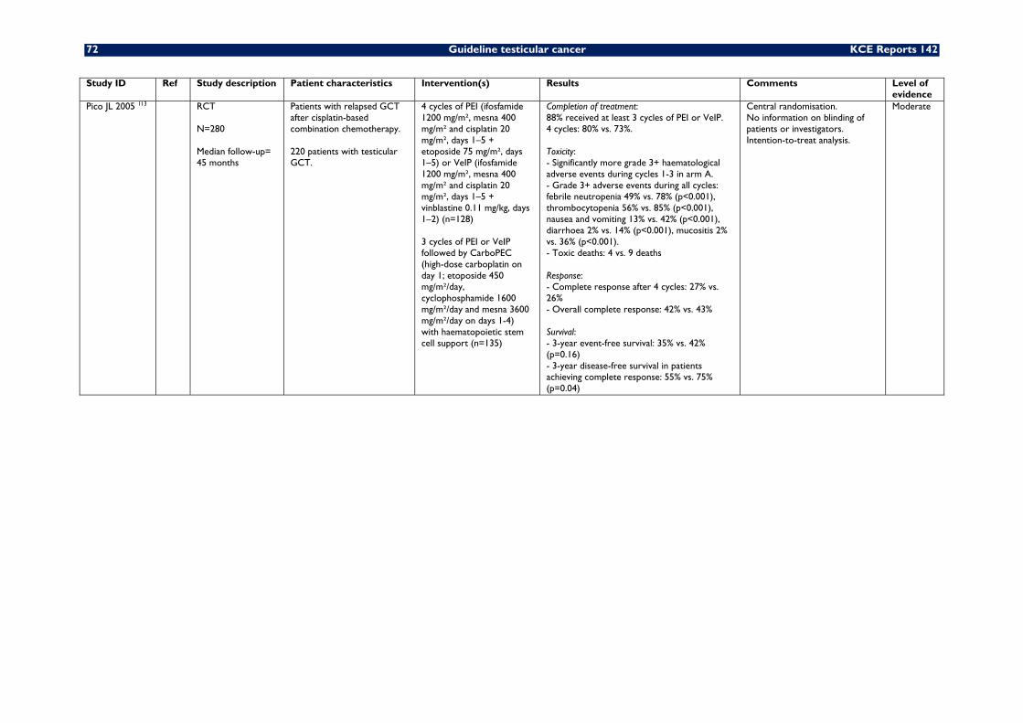
72 Guideline testicular cancer KCE Reports 142
Study ID Ref Study description Patient characteristics Intervention(s) Results Comments Level of evidence
Pico JL 2005 113 RCT N=280 Median follow-up= 45 months
Patients with relapsed GCT after cisplatin-based combination chemotherapy. 220 patients with testicular GCT.
4 cycles of PEI (ifosfamide 1200 mg/m², mesna 400 mg/m² and cisplatin 20 mg/m², days 1–5 + etoposide 75 mg/m², days 1–5) or VeIP (ifosfamide 1200 mg/m², mesna 400 mg/m² and cisplatin 20 mg/m², days 1–5 + vinblastine 0.11 mg/kg, days 1–2) (n=128) 3 cycles of PEI or VeIP followed by CarboPEC (high-dose carboplatin on day 1; etoposide 450 mg/m²/day, cyclophosphamide 1600 mg/m²/day and mesna 3600 mg/m²/day on days 1-4) with haematopoietic stem cell support (n=135)
Completion of treatment: 88% received at least 3 cycles of PEI or VeIP. 4 cycles: 80% vs. 73%. Toxicity: - Significantly more grade 3+ haematological adverse events during cycles 1-3 in arm A. - Grade 3+ adverse events during all cycles: febrile neutropenia 49% vs. 78% (p<0.001), thrombocytopenia 56% vs. 85% (p<0.001), nausea and vomiting 13% vs. 42% (p<0.001), diarrhoea 2% vs. 14% (p<0.001), mucositis 2% vs. 36% (p<0.001). - Toxic deaths: 4 vs. 9 deaths Response: - Complete response after 4 cycles: 27% vs. 26% - Overall complete response: 42% vs. 43% Survival: - 3-year event-free survival: 35% vs. 42% (p=0.16) - 3-year disease-free survival in patients achieving complete response: 55% vs. 75% (p=0.04)
Central randomisation. No information on blinding of patients or investigators. Intention-to-treat analysis.
Moderate
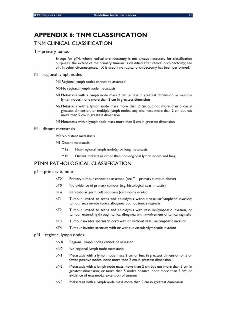
KCE Reports 142 Guideline testicular cancer 73
APPENDIX 6: TNM CLASSIFICATION TNM CLINICAL CLASSIFICATION
T – primary tumour
Except for pT4, where radical orchidectomy is not always necessary for classification purposes, the extent of the primary tumour is classified after radical orchidectomy; see pT. In other circumstances, TX is used if no radical orchidectomy has been performed.
N – regional lymph nodes
NX Regional lymph nodes cannot be assessed
N0 No regional lymph node metastasis
N1 Metastasis with a lymph node mass 2 cm or less in greatest dimension or multiple lymph nodes, none more than 2 cm in greatest dimension
N2 Metastasis with a lymph node mass more than 2 cm but not more than 5 cm in greatest dimension, or multiple lymph nodes, any one mass more than 2 cm but not more than 5 cm in greatest dimension
N3 Metastasis with a lymph node mass more than 5 cm in greatest dimension
M – distant metastasis
M0 No distant metastasis
M1 Distant metastasis
M1a Non-regional lymph node(s) or lung metastasis
M1b Distant metastasis other than non-regional lymph nodes and lung
PTNM PATHOLOGICAL CLASSIFICATION
pT – primary tumour
pTX Primary tumour cannot be assessed (see T – primary tumour, above)
pT0 No evidence of primary tumour (e.g. histological scar in testis)
pTis Intratubular germ cell neoplasia (carcinoma in situ)
pT1 Tumour limited to testis and epididymis without vascular/lymphatic invasion; tumour may invade tunica albuginea but not tunica vaginalis
pT2 Tumour limited to testis and epididymis with vascular/lymphatic invasion, or tumour extending through tunica albuginea with involvement of tunica vaginalis
pT3 Tumour invades spermatic cord with or without vascular/lymphatic invasion
pT4 Tumour invades scrotum with or without vascular/lymphatic invasion
pN – regional lymph nodes
pNX Regional lymph nodes cannot be assessed
pN0 No regional lymph node metastasis
pN1 Metastasis with a lymph node mass 2 cm or less in greatest dimension or 5 or fewer positive nodes, none more than 2 cm in greatest dimension
pN2 Metastasis with a lymph node mass more than 2 cm but not more than 5 cm in greatest dimension; or more than 5 nodes positive, none more than 5 cm; or evidence of extranodal extension of tumour
pN3 Metastasis with a lymph node mass more than 5 cm in greatest dimension
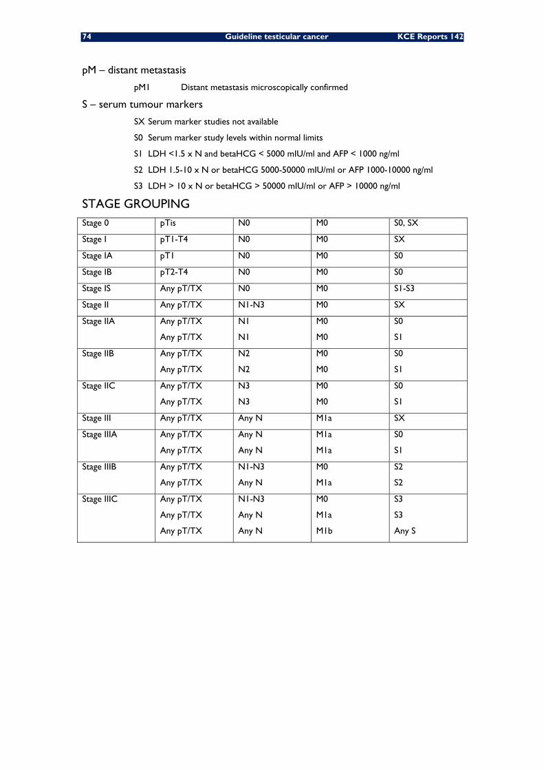
74 Guideline testicular cancer KCE Reports 142
pM – distant metastasis
pM1 Distant metastasis microscopically confirmed
S – serum tumour markers
SX Serum marker studies not available
S0 Serum marker study levels within normal limits
S1 LDH <1.5 x N and betaHCG < 5000 mIU/ml and AFP < 1000 ng/ml
S2 LDH 1.5-10 x N or betaHCG 5000-50000 mIU/ml or AFP 1000-10000 ng/ml
S3 LDH > 10 x N or betaHCG > 50000 mIU/ml or AFP > 10000 ng/ml
STAGE GROUPING Stage 0 pTis N0 M0 S0, SX
Stage I pT1-T4 N0 M0 SX
Stage IA pT1 N0 M0 S0
Stage IB pT2-T4 N0 M0 S0
Stage IS Any pT/TX N0 M0 S1-S3
Stage II Any pT/TX N1-N3 M0 SX
Stage IIA Any pT/TX
Any pT/TX
N1
N1
M0
M0
S0
S1
Stage IIB Any pT/TX
Any pT/TX
N2
N2
M0
M0
S0
S1
Stage IIC Any pT/TX
Any pT/TX
N3
N3
M0
M0
S0
S1
Stage III Any pT/TX Any N M1a SX
Stage IIIA Any pT/TX
Any pT/TX
Any N
Any N
M1a
M1a
S0
S1
Stage IIIB Any pT/TX
Any pT/TX
N1-N3
Any N
M0
M1a
S2
S2
Stage IIIC Any pT/TX
Any pT/TX
Any pT/TX
N1-N3
Any N
Any N
M0
M1a
M1b
S3
S3
Any S
KCE Reports 142 Guideline testicular cancer 75
12 REFERENCES 1. Peeters M, Zlotta A, Roucoux F, De Greve J, Van Belle S, Haelterman M, et al. National
Clinical Practice Guidelines of the College of Oncology : clinical practice guideline for testicular cancer. Good Clinical Practice (GCP). Brussels: Belgian Health Care Knowledge Centre (KCE); 2006. KCE reports 29Suppl. 2 (D/2006/10.273/56)
2. Holmes L, Jr., Escalante C, Garrison O, Foldi BX, Ogungbade GO, Essien EJ, et al. Testicular cancer incidence trends in the USA (1975-2004): plateau or shifting racial paradigm? Public Health. 2008;122(9):862-72.
3. Stang A, Katalinic A, Dieckmann K-P, Pritzkuleit R, Stabenow R, Network of German Cancer R. A novel approach to estimate the German-wide incidence of testicular cancer. Cancer Epidemiol. 2010;34(1):13-9.
4. Scheiden R, Hein T, Wagener C, Kieffer N, Lamy S, Capesius C. Testicular cancer in Luxembourg: incidence and outcome in relation to the different histo-pathological types (1980-2004). Bull Soc Sci Med Grand Duche Luxemb. 2008(4):521-39.
5. Van Eycken E, De Wever N. Cancer Incidence and Survival in Flanders, 2000-2001. Brussels: Flemish Cancer Registry Network, VLK; 2006.
6. Nur U, Rachet B, Mitry E, Cooper N, Coleman MP. Survival from testicular cancer in England and Wales up to 2001. Br J Cancer. 2008;99 Suppl 1:S80-2.
7. Verhoeven RHA, Coebergh JWW, Kiemeney LALM, Koldewijn EL, Houterman S. Testicular cancer: trends in mortality are well explained by changes in treatment and survival in the southern Netherlands since 1970. Eur J Cancer. 2007;43(17):2553-8.
8. Fervers B, Burgers JS, Haugh MC, Latreille J, Mlika-Cabanne N, Paquet L, et al. Adaptation of clinical guidelines: literature review and proposition for a framework and procedure. Int J Qual Health Care. 2006;18(3):167-76.
9. Collaboration A. Development and validation of an international appraisal instrument for assessing the quality of clinical practice guidelines: the AGREE project. Qual Saf Health Care. 2003;12(1):18-23.
10. Whiting P, Rutjes AWS, Reitsma JB, Bossuyt PMM, Kleijnen J. The development of QUADAS: a tool for the quality assessment of studies of diagnostic accuracy included in systematic reviews. BMC Med Res Methodol. 2003;3:25.
11. Scottish Intercollegiate Guidelines Network. Management of Adult Testicular Germ Cell Tumours - Selective Update (in development). Edinburgh: SIGN; 2010.
12. Coret A, Leibovitch I, Heyman Z, Goldwasser B, Itzchak Y. Ultrasonographic evaluation and clinical correlation of intratesticular lesions: a series of 39 cases. British Journal of Urology. 1995;76(2):216-9.
13. Kennedy PT, Elliott JM, Rice PF, Kelly BE. Ultrasonography of intratesticular lesions: its role in clinical management. Ulster Medical Journal. 1999;68(2):54-8.
14. Kromann-Andersen B, Hansen LB, Larsen PN, Lawetz K, Lynge P, Lysen D, et al. Clinical versus ultrasonographic evaluation of scrotal disorders. British Journal of Urology. 1988;61(4):350-3.
15. London NJ, Smart JG, Kinder RB, Watkin EM, Rees Y, Haley P. Prospective study of routine scrotal ultrasonography in urological practice. British Journal of Urology. 1989;63(4):416-9.
16. Polak V, Hornak M. The value of scrotal ultrasound in patients with suspected testicular tumour. International Urology & Nephrology. 1990;22(5):467-73.
17. van Dijk R, Doesburg WH, Verbeek AL, van der Schouw YT, Debruyne FM, Rosenbusch G. Ultrasonography versus clinical examination in evaluation of testicular tumors. Journal of Clinical Ultrasound. 1994;22(3):179-82.
18. Mottet N, Culine S, Iborra F, Avances C, Bastide C, Lesourd A, et al. [Testicular tumors]. Prog Urol. 2007;17(6):1035-45.
19. Gilligan TD, Seidenfeld J, Basch EM, Einhorn LH, Fancher T, Smith DC, et al. American Society of Clinical Oncology Clinical Practice Guideline on uses of serum tumor markers in adult males with germ cell tumors. J Clin Oncol. 2010;28(20):3388-404.
76 Guideline testicular cancer KCE Reports 142
20. Connolly SS, D'Arcy FT, Bredin HC, Callaghan J, Corcoran MO. Value of frozen section analysis with suspected testicular malignancy. Urology. 2006;67(1):162-5.
21. Elert A, Olbert P, Hegele A, Barth P, Hofmann R, Heidenreich A. Accuracy of frozen section examination of testicular tumors of uncertain origin. Eur Urol. 2002;41(3):290-3.
22. Heidenreich A, Weissbach L, Holtl W, Albers P, Kliesch S, Kohrmann KU, et al. Organ sparing surgery for malignant germ cell tumor of the testis. J Urol. 2001;166(6):2161-5.
23. Fossa SD, Chen J, Schonfeld SJ, McGlynn KA, McMaster ML, Gail MH, et al. Risk of contralateral testicular cancer: a population-based study of 29,515 U.S. men. J Natl Cancer Inst. 2005;97(14):1056-66.
24. Dieckmann K-P, Kulejewski M, Pichlmeier U, Loy V. Diagnosis of contralateral testicular intraepithelial neoplasia (TIN) in patients with testicular germ cell cancer: systematic two-site biopsies are more sensitive than a single random biopsy. European Urology. 2007;51(1):175-83; discussion 83-5.
25. Harland SJ, Cook PA, Fossa SD, Horwich A, Mead GM, Parkinson MC, et al. Intratubular germ cell neoplasia of the contralateral testis in testicular cancer: defining a high risk group. J Urol. 1998;160(4):1353-7.
26. Akre O, Pettersson A, Richiardi L. Risk of contralateral testicular cancer among men with unilaterally undescended testis: a meta analysis. Int J Cancer. 2009;124(3):687-9.
27. Peng X, Zeng X, Peng S, Deng D, Zhang J. The association risk of male subfertility and testicular cancer: a systematic review. PLoS ONE [Electronic Resource]. 2009;4(5):e5591.
28. Walsh TJ, Dall'Era MA, Croughan MS, Carroll PR, Turek PJ. Prepubertal orchiopexy for cryptorchidism may be associated with lower risk of testicular cancer. J Urol. 2007;178(4 Pt 1):1440-6; discussion 6.
29. Wood HM, Elder JS. Cryptorchidism and testicular cancer: separating fact from fiction. J Urol. 2009;181(2):452-61.
30. Dieckmann K-P, Classen J, Loy V. Diagnosis and management of testicular intraepithelial neoplasia (carcinoma in situ)--surgical aspects. APMIS. 2003;111(1):64-8; discussion 8-9.
31. Classen J, Dieckmann K, Bamberg M, Souchon R, Kliesch S, Kuehn M, et al. Radiotherapy with 16 Gy may fail to eradicate testicular intraepithelial neoplasia: preliminary communication of a dose-reduction trial of the German Testicular Cancer Study Group. Br J Cancer. 2003;88(6):828-31.
32. Petersen PM, Giwercman A, Daugaard G, Rorth M, Petersen JH, Skakkeaek NE, et al. Effect of graded testicular doses of radiotherapy in patients treated for carcinoma-in-situ in the testis. J Clin Oncol. 2002;20(6):1537-43.
33. Sedlmayer F, Holtl W, Kozak W, Hawliczek R, Gebhart F, Gerber E, et al. Radiotherapy of testicular intraepithelial neoplasia (TIN): a novel treatment regimen for a rare disease. Int J Radiat Oncol Biol Phys. 2001;50(4):909-13.
34. Kleinschmidt K, Dieckmann K-P, Georgiew A, Loy V, Weissbach L. Chemotherapy is of limited efficacy in the control of contralateral testicular intraepithelial neoplasia in patients with testicular germ cell cancer. Oncology. 2009;77(1):33-9.
35. Cremerius U, Wildberger JE, Borchers H, Zimny M, Jakse G, Gunther RW, et al. Does positron emission tomography using 18-fluoro-2-deoxyglucose improve clinical staging of testicular cancer?--Results of a study in 50 patients. Urology. 1999;54(5):900-4.
36. de Wit M, Brenner W, Hartmann M, Kotzerke J, Hellwig D, Lehmann J, et al. [18F]-FDG-PET in clinical stage I/II non-seminomatous germ cell tumours: results of the German multicentre trial. Annals of Oncology. 2008;19(9):1619-23.
37. Frick MP, Feinberg SE, Knight LC. Evaluation of retroperitoneum with computerized tomography and ultrasonography in patients with testicular tumors. Computerized Tomography. 1979;3(3):181-4.
38. Richie JP, Garnick MB, Finberg H. Computerized tomography: how accurate for abdominal staging of testis tumors? Journal of Urology. 1982;127(4):715-7.
39. Thomas JL, Bernardino ME, Bracken RB. Staging of testicular carcinoma: comparison of CT and lymphangiography. AJR. 1981;American Journal of Roentgenology. 137(5):991-6.
KCE Reports 142 Guideline testicular cancer 77
40. Tarin TV, Sonn G, Shinghal R. Estimating the risk of cancer associated with imaging related radiation during surveillance for stage I testicular cancer using computerized tomography. J Urol. 2009;181(2):627-32; discussion 32-3.
41. Hansen J, Jurik AG. Diagnostic value of multislice computed tomography and magnetic resonance imaging in the diagnosis of retroperitoneal spread of testicular cancer: a literature review. Acta Radiol. 2009;50(9):1064-70.
42. Schwerk WB, Schwerk WN, Rodeck G. Testicular tumors: prospective analysis of real-time US patterns and abdominal staging. Radiology. 1987;164(2):369-74.
43. Vlayen J, Stordeur S, Van den Bruel A, Mambourg F, Eyssen M. Positron Emission Tomography (PET) in Belgium: an update. Health Technology Assessment (HTA). Brussels: Belgian Health Care Knowledge Centre (KCE); 2009. KCE reports 110 (D/2009/10.273/24)
44. Lee SJ, Schover LR, Partridge AH, Patrizio P, Wallace WH, Hagerty K, et al. American Society of Clinical Oncology recommendations on fertility preservation in cancer patients. J Clin Oncol. 2006;24(18):2917-31.
45. World Health Organisation. WHO Classification of Tumours: Pathology and Genetics of Tumours of the Urinary System and Male Genital Organs. Third ed. Eble JN, Sauter G, Epstein JL, Sesterhenn IA, editor.: WHO/IARC; 2008.
46. UICC International Union Against Cancer. TNM Classification of Malignant Tumours. Seventh ed. Sobin L, Gospodarowicz M, Wittekind C, editor. New York: Wiley- Blackwell; 2009.
47. International Germ Cell Consensus Classification: a prognostic factor-based staging system for metastatic germ cell cancers. International Germ Cell Cancer Collaborative Group. J Clin Oncol. 1997;15(2):594-603.
48. Albers P, Siener R, Kliesch S, Weissbach L, Krege S, Sparwasser C, et al. Risk factors for relapse in clinical stage I nonseminomatous testicular germ cell tumors: results of the German Testicular Cancer Study Group Trial. J Clin Oncol. 2003;21(8):1505-12.
49. Parker C, Milosevic M, Panzarella T, Banerjee D, Jewett M, Catton C, et al. The prognostic significance of the tumour infiltrating lymphocyte count in stage I testicular seminoma managed by surveillance. Eur J Cancer. 2002;38(15):2014-9.
50. Warde P, Gospodarowicz MK, Banerjee D, Panzarella T, Sugar L, Catton CN, et al. Prognostic factors for relapse in stage I testicular seminoma treated with surveillance. J Urol. 1997;157(5):1705-9; discussion 9-10.
51. Warde P, Specht L, Horwich A, Oliver T, Panzarella T, Gospodarowicz M, et al. Prognostic factors for relapse in stage I seminoma managed by surveillance: a pooled analysis.[see comment]. J Clin Oncol. 2002;20(22):4448-52.
52. Nazeer T, Ro JY, Kee KH, Ayala AG. Spermatic cord contamination in testicular cancer. Mod Pathol. 1996;9(7):762-6.
53. Klepp O, Olsson AM, Henrikson H, Aass N, Dahl O, Stenwig AE, et al. Prognostic factors in clinical stage I nonseminomatous germ cell tumors of the testis: multivariate analysis of a prospective multicenter study. Swedish-Norwegian Testicular Cancer Group. J Clin Oncol. 1990;8(3):509-18.
54. Moul JW, McCarthy WF, Fernandez EB, Sesterhenn IA. Percentage of embryonal carcinoma and of vascular invasion predicts pathological stage in clinical stage I nonseminomatous testicular cancer. Cancer Res. 1994;54(2):362-4.
55. Mead GM, Stenning SP, Parkinson MC, Horwich A, Fossa SD, Wilkinson PM, et al. The Second Medical Research Council study of prognostic factors in nonseminomatous germ cell tumors. Medical Research Council Testicular Tumour Working Party.[erratum appears in J Clin Oncol 1992 May;10(5):867]. J Clin Oncol. 1992;10(1):85-94.
56. Steyerberg EW, Keizer HJ, Fossa SD, Sleijfer DT, Toner GC, Schraffordt Koops H, et al. Prediction of residual retroperitoneal mass histology after chemotherapy for metastatic nonseminomatous germ cell tumor: multivariate analysis of individual patient data from six study groups. Journal of Clinical Oncology. 1995;13(5):1177-87.
57. Freedman LS, Parkinson MC, Jones WG, Oliver RT, Peckham MJ, Read G, et al. Histopathology in the prediction of relapse of patients with stage I testicular teratoma treated by orchidectomy alone. Lancet. 1987;2(8554):294-8.
78 Guideline testicular cancer KCE Reports 142
58. Tavolini IM, Bettella A, Boscolo Berto R, Bassi PF, Longo R, Menegazzo M, et al. Immunostaining for placental alkaline phosphatase on fine-needle aspiration specimens to detect noninvasive testicular cancer: a prospective evaluation in cryptorchid men.[see comment]. BJU International. 2006;97(5):950-4.
59. Izquierdo MA, Van der Valk P, Van Ark-Otte J, Rubio G, Germa-Lluch JR, Ueda R, et al. Differential expression of the c-kit proto-oncogene in germ cell tumours. J Pathol. 1995;177(3):253-8.
60. de Jong J, Stoop H, Dohle GR, Bangma CH, Kliffen M, van Esser JWJ, et al. Diagnostic value of OCT3/4 for pre-invasive and invasive testicular germ cell tumours. J Pathol. 2005;206(2):242-9.
61. Heidenreich A, Sesterhenn IA, Mostofi FK, Moul JW. Immunohistochemical expression of monoclonal antibody 43-9F in testicular germ cell tumours. Int J Androl. 1998;21(5):283-8.
62. Cheville JC, Rao S, Iczkowski KA, Lohse CM, Pankratz VS. Cytokeratin expression in seminoma of the human testis. Am J Clin Pathol. 2000;113(4):583-8.
63. Leroy X, Augusto D, Leteurtre E, Gosselin B. CD30 and CD117 (c-kit) used in combination are useful for distinguishing embryonal carcinoma from seminoma. J Histochem Cytochem. 2002;50(2):283-5.
64. Bosman FT, Giard RW, Nieuwenhuijen Kruseman AC, Knijnenburg G, Spaander PJ. Human chorionic gonadotrophin and alpha-fetoprotein in testicular germ cell tumours: a retrospective immunohistochemical study. Histopathology. 1980;4(6):673-84.
65. Santagata S, Ligon KL, Hornick JL. Embryonic stem cell transcription factor signatures in the diagnosis of primary and metastatic germ cell tumors. Am J Surg Pathol. 2007;31(6):836-45.
66. Jones TD, Ulbright TM, Eble JN, Baldridge LA, Cheng L. OCT4 staining in testicular tumors: a sensitive and specific marker for seminoma and embryonal carcinoma.[see comment]. American Journal of Surgical Pathology. 2004;28(7):935-40.
67. Chung P, Mayhew LA, Warde P, Winquist E, Lukka H, and Members of the Genitourinary Cancer Disease Site Group. Management of Stage I Seminoma: Guideline Recommendations. Toronto: CCO; 2008. (Evidence-Based Series #3-18)
68. Oliver RTD, Mason MD, Mead GM, von der Maase H, Rustin GJS, Joffe JK, et al. Radiotherapy versus single-dose carboplatin in adjuvant treatment of stage I seminoma: a randomised trial.[see comment]. Lancet. 2005;366(9482):293-300.
69. Jones WG, Fossa SD, Mead GM, Roberts JT, Sokal M, Horwich A, et al. Randomized trial of 30 versus 20 Gy in the adjuvant treatment of stage I Testicular Seminoma: a report on Medical Research Council Trial TE18, European Organisation for the Research and Treatment of Cancer Trial 30942 (ISRCTN18525328).[see comment]. J Clin Oncol. 2005;23(6):1200-8.
70. Fossa SD, Horwich A, Russell JM, Roberts JT, Cullen MH, Hodson NJ, et al. Optimal planning target volume for stage I testicular seminoma: A Medical Research Council randomized trial. Medical Research Council Testicular Tumor Working Group.[see comment]. J Clin Oncol. 1999;17(4):1146.
71. Groll RJ, Warde P, Jewett MAS. A comprehensive systematic review of testicular germ cell tumor surveillance. Critical Reviews in Oncology-Hematology. 2007;64(3):182-97.
72. Hotte S, Mayhew LA, Jewett M, Chin J, Winquist E, and Members of the Genitourinary Cancer Disease Site Group. Management of Stage I Nonseminomatous Testicular Cancer: Guideline Recommendations. Toronto: CCO; 2008. (Evidence-Based Series #3-19)
73. Rorth M, Jacobsen GK, von der Maase H, Madsen EL, Nielsen OS, Pedersen M, et al. Surveillance alone versus radiotherapy after orchiectomy for clinical stage I nonseminomatous testicular cancer. Danish Testicular Cancer Study Group. J Clin Oncol. 1991;9(9):1543-8.
74. Albers P, Siener R, Krege S, Schmelz H-U, Dieckmann K-P, Heidenreich A, et al. Randomized phase III trial comparing retroperitoneal lymph node dissection with one course of bleomycin and etoposide plus cisplatin chemotherapy in the adjuvant treatment of clinical stage I Nonseminomatous testicular germ cell tumors: AUO trial AH 01/94 by the German Testicular Cancer Study Group.[see comment]. J Clin Oncol. 2008;26(18):2966-72.
75. Albers P, Albrecht W, Algaba F, Bokemeyer C, Cohn-Cedermark G, Horwich A, et al. Guidelines on Testicular Cancer. European Association of Urology; 2008.
76. Feldman DR, Bosl GJ, Sheinfeld J, Motzer RJ. Medical treatment of advanced testicular cancer. JAMA. 2008;299(6):672-84.
KCE Reports 142 Guideline testicular cancer 79
77. Shelley MD, Burgon K, Mason MD. Treatment of testicular germ-cell cancer: a cochrane evidence-based systematic review. Cancer Treatment Reviews. 2002;28(5):237-53.
78. Patterson H, Norman AR, Mitra SS, Nicholls J, Fisher C, Dearnaley DP, et al. Combination carboplatin and radiotherapy in the management of stage II testicular seminoma: comparison with radiotherapy treatment alone. Radiother Oncol. 2001;59(1):5-11.
79. Chung PWM, Gospodarowicz MK, Panzarella T, Jewett MAS, Sturgeon JFG, Tew-George B, et al. Stage II testicular seminoma: patterns of recurrence and outcome of treatment. Eur Urol. 2004;45(6):754-59; discussion 9-60.
80. Clemm C, Bokemeyer C, Gerl A, Holzel D, Weissbach L, Mamberg M, et al. Randomized Trial Comparing Cisplatin/Etoposide/ Ifosfamide with Carboplatin Monochemotherapy in Patients with Advanced Metastatic Seminoma. Proc Am Soc Clin Oncol. 2000;19 (abstr 1283).
81. Horwich A, Oliver RT, Wilkinson PM, Mead GM, Harland SJ, Cullen MH, et al. A medical research council randomized trial of single agent carboplatin versus etoposide and cisplatin for advanced metastatic seminoma. MRC Testicular Tumour Working Party. Br J Cancer. 2000;83(12):1623-9.
82. Bokemeyer C, Kollmannsberger C, Stenning S, Hartmann JT, Horwich A, Clemm C, et al. Metastatic seminoma treated with either single agent carboplatin or cisplatin-based combination chemotherapy: a pooled analysis of two randomised trials. Br J Cancer. 2004;91(4):683-7.
83. Culine S, Kerbrat P, Kramar A, Theodore C, Chevreau C, Geoffrois L, et al. Refining the optimal chemotherapy regimen for good-risk metastatic nonseminomatous germ-cell tumors: a randomized trial of the Genito-Urinary Group of the French Federation of Cancer Centers (GETUG T93BP). Ann Oncol. 2007;18(5):917-24.
84. Horwich A, Sleijfer DT, Fossa SD, Kaye SB, Oliver RT, Cullen MH, et al. Randomized trial of bleomycin, etoposide, and cisplatin compared with bleomycin, etoposide, and carboplatin in good-prognosis metastatic nonseminomatous germ cell cancer: a Multiinstitutional Medical Research Council/European Organization for Research and Treatment of Cancer Trial. J Clin Oncol. 1997;15(5):1844-52.
85. de Wit R, Stoter G, Kaye SB, Sleijfer DT, Jones WG, ten Bokkel Huinink WW, et al. Importance of bleomycin in combination chemotherapy for good-prognosis testicular nonseminoma: a randomized study of the European Organization for Research and Treatment of Cancer Genitourinary Tract Cancer Cooperative Group.[see comment]. J Clin Oncol. 1997;15(5):1837-43.
86. Bokemeyer C, Kohrmann O, Tischler J, Weissbach L, Rath U, Haupt A, et al. A randomized trial of cisplatin, etoposide and bleomycin (PEB) versus carboplatin, etoposide and bleomycin (CEB) for patients with 'good-risk' metastatic non-seminomatous germ cell tumors. Ann Oncol. 1996;7(10):1015-21.
87. Einhorn LH, Williams SD, Loehrer PJ, Birch R, Drasga R, Omura G, et al. Evaluation of optimal duration of chemotherapy in favorable-prognosis disseminated germ cell tumors: a Southeastern Cancer Study Group protocol. J Clin Oncol. 1989;7(3):387-91.
88. Toner GC, Stockler MR, Boyer MJ, Jones M, Thomson DB, Harvey VJ, et al. Comparison of two standard chemotherapy regimens for good-prognosis germ-cell tumours: a randomised trial. Australian and New Zealand Germ Cell Trial Group. Lancet. 2001;357(9258):739-45.
89. Loehrer PJ, Sr., Johnson D, Elson P, Einhorn LH, Trump D. Importance of bleomycin in favorable-prognosis disseminated germ cell tumors: an Eastern Cooperative Oncology Group trial. J Clin Oncol. 1995;13(2):470-6.
90. Droz J-P, Kramar A, Biron P, Pico J-L, Kerbrat P, Peny J, et al. Failure of high-dose cyclophosphamide and etoposide combined with double-dose cisplatin and bone marrow support in patients with high-volume metastatic nonseminomatous germ-cell tumours: mature results of a randomised trial. Eur Urol. 2007;51(3):739-46; discussion 47-8.
91. Kaye SB, Mead GM, Fossa S, Cullen M, deWit R, Bodrogi I, et al. Intensive induction-sequential chemotherapy with BOP/VIP-B compared with treatment with BEP/EP for poor-prognosis metastatic nonseminomatous germ cell tumor: a Randomized Medical Research Council/European Organization for Research and Treatment of Cancer study. J Clin Oncol. 1998;16(2):692-701.
80 Guideline testicular cancer KCE Reports 142
92. de Wit R, Stoter G, Sleijfer DT, Neijt JP, ten Bokkel Huinink WW, de Prijck L, et al. Four cycles of BEP vs four cycles of VIP in patients with intermediate-prognosis metastatic testicular non-seminoma: a randomized study of the EORTC Genitourinary Tract Cancer Cooperative Group. European Organization for Research and Treatment of Cancer. Br J Cancer. 1998;78(6):828-32.
93. de Wit R, Stoter G, Sleijfer DT, Kaye SB, de Mulder PH, ten Bokkel Huinink WW, et al. Four cycles of BEP versus an alternating regime of PVB and BEP in patients with poor-prognosis metastatic testicular non-seminoma; a randomised study of the EORTC Genitourinary Tract Cancer Cooperative Group. Br J Cancer. 1995;71(6):1311-4.
94. Williams SD, Stablein DM, Einhorn LH, Muggia FM, Weiss RB, Donohue JP, et al. Immediate adjuvant chemotherapy versus observation with treatment at relapse in pathological stage II testicular cancer. N Engl J Med. 1987;317(23):1433-8.
95. Nichols CR, Williams SD, Loehrer PJ, Greco FA, Crawford ED, Weetlaufer J, et al. Randomized study of cisplatin dose intensity in poor-risk germ cell tumors: a Southeastern Cancer Study Group and Southwest Oncology Group protocol. J Clin Oncol. 1991;9(7):1163-72.
96. Nichols CR, Catalano PJ, Crawford ED, Vogelzang NJ, Einhorn LH, Loehrer PJ. Randomized comparison of cisplatin and etoposide and either bleomycin or ifosfamide in treatment of advanced disseminated germ cell tumors: an Eastern Cooperative Oncology Group, Southwest Oncology Group, and Cancer and Leukemia Group B Study. J Clin Oncol. 1998;16(4):1287-93.
97. Motzer RJ, Nichols CJ, Margolin KA, Bacik J, Richardson PG, Vogelzang NJ, et al. Phase III randomized trial of conventional-dose chemotherapy with or without high-dose chemotherapy and autologous hematopoietic stem-cell rescue as first-line treatment for patients with poor-prognosis metastatic germ cell tumors. J Clin Oncol. 2007;25(3):247-56.
98. Culine S, Kramar A, Theodore C, Geoffrois L, Chevreau C, Biron P, et al. Randomized trial comparing bleomycin/etoposide/cisplatin with alternating cisplatin/cyclophosphamide/doxorubicin and vinblastine/bleomycin regimens of chemotherapy for patients with intermediate- and poor-risk metastatic nonseminomatous germ cell tumors: Genito-Urinary Group of the French Federation of Cancer Centers Trial T93MP. J Clin Oncol. 2008;26(3):421-7.
99. Daugaard G, Skoneczna IA, Aass N, De Wit R, De Santis M, Dumez H, et al. A randomized phase III study comparing standard dose BEP with sequential high-dose cisplatin, etoposide, ifosfamide (VIP) plus stem cell support in males with poor prognosis germ cell cancer (GCC): An intergroup study of EORTC, GTCSG, and Grupo Germinal (EORTC 30974). J Clin Oncol (Meeting Abstracts). 2010;28(15_suppl):4512.
100. Pfannenberg AC, Oechsle K, Kollmannsberger C, Dohmen BM, Bokemeyer C, Bares R, et al. Early prediction of treatment response to high-dose chemotherapy in patients with relapsed germ cell tumors using [18F]FDG-PET, CT or MRI, and tumor marker. ROFO Fortschr Geb Rontgenstr Nuklearmed. 2004;176(1):76-84.
101. De Santis M, Becherer A, Bokemeyer C, Stoiber F, Oechsle K, Sellner F, et al. 2-18fluoro-deoxy-D-glucose positron emission tomography is a reliable predictor for viable tumor in postchemotherapy seminoma: an update of the prospective multicentric SEMPET trial. Journal of Clinical Oncology. 2004;22(6):1034-9.
102. Oechsle K, Hartmann M, Brenner W, Venz S, Weissbach L, Franzius C, et al. [18F]Fluorodeoxyglucose positron emission tomography in nonseminomatous germ cell tumors after chemotherapy: the German multicenter positron emission tomography study group. J Clin Oncol. 2008;26(36):5930-5.
103. Heidenreich A, Thuer D, Polyakov S. Postchemotherapy retroperitoneal lymph node dissection in advanced germ cell tumours of the testis. Eur Urol. 2008;53(2):260-72.
104. Liu D, Abolhoda A, Burt ME, Martini N, Bains MS, Downey RJ, et al. Pulmonary metastasectomy for testicular germ cell tumors: a 28-year experience. Ann Thorac Surg. 1998;66(5):1709-14.
105. You YN, Leibovitch BC, Que FG. Hepatic metastasectomy for testicular germ cell tumors: is it worth it? J Gastrointest Surg. 2009;13(4):595-601.
KCE Reports 142 Guideline testicular cancer 81
106. Pfannschmidt J, Zabeck H, Muley T, Dienemann H, Hoffmann H. Pulmonary metastasectomy following chemotherapy in patients with testicular tumors: experience in 52 patients. Thorac Cardiovasc Surg. 2006;54(7):484-8.
107. Ondrus D, Hornak M, Breza J, Mat'oska J, Schnorrer M, Belan V, et al. Delayed orchiectomy after chemotherapy in patients with advanced testicular cancer. Int Urol Nephrol. 2001;32(4):665-7.
108. Krege S, Beyer J, Souchon R, Albers P, Albrecht W, Algaba F, et al. European consensus conference on diagnosis and treatment of germ cell cancer: a report of the second meeting of the European Germ Cell Cancer Consensus group (EGCCCG): part I. European Urology. 2008;53(3):478-96.
109. Puc HS, Heelan R, Mazumdar M, Herr H, Scheinfeld J, Vlamis V, et al. Management of residual mass in advanced seminoma: results and recommendations from the Memorial Sloan-Kettering Cancer Center. J Clin Oncol. 1996;14(2):454-60.
110. Rustin GJ, Mead GM, Stenning SP, Vasey PA, Aass N, Huddart RA, et al. Randomized trial of two or five computed tomography scans in the surveillance of patients with stage I nonseminomatous germ cell tumors of the testis: Medical Research Council Trial TE08, ISRCTN56475197--the National Cancer Research Institute Testis Cancer Clinical Studies Group.[see comment]. J Clin Oncol. 2007;25(11):1310-5.
111. Segal R, Lukka H, Klotz L, Eady A, Bestic N, Johnston M, et al. Surveillance Programs for Early Stage Non-Seminomatous Testicular Cancer. Toronto: CCO; 2001. (Practice Guideline Report No. 3-5)
112. Stoehr B, Zangerl F, Steiner E, Leonhartsberger N, Fritzer A, Bartsch G, et al. Routine scrotal ultrasonography during the follow-up of patients with testicular cancer leads to earlier detection of asynchronous tumours and a high rate of organ preservation. BJU Int. 2009.
113. Pico JL, Rosti G, Kramar A, Wandt H, Koza V, Salvioni R, et al. A randomised trial of high-dose chemotherapy in the salvage treatment of patients failing first-line platinum chemotherapy for advanced germ cell tumours. Ann Oncol. 2005;16(7):1152-9.
114. Lorch A, Kollmannsberger C, Hartmann JT, Metzner B, Schmidt-Wolf IGH, Berdel WE, et al. Single versus sequential high-dose chemotherapy in patients with relapsed or refractory germ cell tumors: a prospective randomized multicenter trial of the German Testicular Cancer Study Group. J Clin Oncol. 2007;25(19):2778-84.
115. National Institute for Health and Clinical Excellence. Guidance for Commissioning Cancer Srvices. Improving Outcomes in Urological Cancers. London: NICE; 2002.
116. National Comprehensive Cancer Network. Testicular Cancer. NCCN; 2009. (V.I.2009)
117. Dutch Association of Comprehensive Cancer Centres. Testiscarcinoom. Utrecht: ACCC; 2007. (Versie 1.1)
118. American College of Radiology. Staging of Testicular Malignancy. ACR; 2007. ACR Appropriateness Criteria
119. Protocol for malignant and potentially malignant neoplasms of the testis and paratestis. Association of Directors of Anatomic and Surgical Pathology. Am J Clin Pathol. 2000;114(3):339-42.
120. Huddart R, Kataja V, Group EGW. Mixed or non-seminomatous germ-cell tumors: ESMO clinical recommendations for diagnosis, treatment and follow-up. Annals of Oncology. 2008;19(2).
121. Huddart R, Kataja V, Group EGW. Testicular seminoma: ESMO clinical recommendations for diagnosis, treatment and follow-up. Annals of Oncology. 2008;19(2).
122. Sturgeon CM, Duffy MJ, Stenman U-H, Lilja H, Brunner N, Chan DW, et al. National Academy of Clinical Biochemistry laboratory medicine practice guidelines for use of tumor markers in testicular, prostate, colorectal, breast, and ovarian cancers. Clin Chem. 2008;54(12):e11-79.
123. Calonge N, U. S. Preventive Services Task Force Agency for Healthcare Research and Quality. Screening for testicular cancer: recommendation statement. Am Fam Physician. 2005;72(10):2069-70.
124. De Giorgi U, Nicolai N, Tana S, Tavolini IM, Palazzi S, Bracarda S, et al. IGG practice guidelines on germ cell tumor in adult male patients. Tumori. 2008;94(1):96-109.
82 Guideline testicular cancer KCE Reports 142
125. Martin JM, Panzarella T, Zwahlen DR, Chung P, Warde P. Evidence-based guidelines for following stage 1 seminoma. Cancer. 2007;109(11):2248-56.
126. van As NJ, Gilbert DC, Money-Kyrle J, Bloomfield D, Beesley S, Dearnaley DP, et al. Evidence-based pragmatic guidelines for the follow-up of testicular cancer: optimising the detection of relapse. Br J Cancer. 2008;98(12):1894-902.
127. Bourguet P, Planchamp F, Montravers F, Vincendeau S, Association francaise du, Federation nationale des centres de lutte contre le c. 2006 technology monitoring report: clinical practice guideline: use of FDG-PET in kidney, prostate, testis and bladder cancers. Prog Urol. 2007;17(2):172-5.
128. Mottet N, Berger N, Droz J-P. Diagnosis and staging of germ-cell tumours of the testis. Rev Prat. 2007;57(4):365-78.
129. Lenz S, Skakkebaek NE, Hertel NT. Abnormal ultrasonic pattern in contralateral testes in patients with unilateral testicular cancer. World Journal of Urology. 1996;14 Suppl 1:S55-8.
130. Tesoro-Tess JD, Pizzocaro G, Zanoni F, Musumeci R. Lymphangiography and computerized tomography in testicular carcinoma: how accurate in early stage disease? Journal of Urology. 1985;133(6):967-70.
131. Sohaib SA, Koh DM, Barbachano Y, Parikh J, Husband JES, Dearnaley DP, et al. Prospective assessment of MRI for imaging retroperitoneal metastases from testicular germ cell tumours. Clin Radiol. 2009;64(4):362-7.
132. Schultz-Lampel D, Bogaert G, Thuroff JW, Schlegel E, Cramer B. MRI for evaluation of scrotal pathology. Urological Research. 1991;19(5):289-92.
133. Agency for Healthcare Research and Quality. Positron Emission Tomography for Nine Cancers (Bladder, Brain, Cervical, Kidney, Ovarian, Pancreatic, Prostate, Small Cell Lung, Testicular). Rockville: AHRQ; 2008.
134. Venkitaraman R, Johnson B, Huddart RA, Parker CC, Horwich A, Dearnaley DP. The utility of lactate dehydrogenase in the follow-up of testicular germ cell tumours. BJU Int. 2007;100(1):30-2.
135. von Eyben FE, Blaabjerg O, Madsen EL, Petersen PH, Smith-Sivertsen C, Gullberg B. Serum lactate dehydrogenase isoenzyme 1 and tumour volume are indicators of response to treatment and predictors of prognosis in metastatic testicular germ cell tumours. European Journal of Cancer. 1992;28(2-3):410-5.
136. Herr HW. Does necrosis on frozen-section analysis of a mass after chemotherapy justify a limited retroperitoneal resection in patients with advanced testis cancer? Br J Urol. 1997;80(4):653-7.
137. Dieckmann KP, Souchon R, Hahn E, Loy V. False-negative biopsies for testicular intraepithelial neoplasia. Journal of Urology. 1999;162(2):364-8.
138. Dimov ND, Zynger DL, Luan C, Kozlowski JM, Yang XJ. Topoisomerase II alpha expression in testicular germ cell tumors. Urology. 2007;69(5):955-61.
139. Eid H, Institoris E, Geczi L, Bodrogi I, Bak M. mdm-2 expression in human testicular germ-cell tumors and its clinical value. Anticancer Res. 1999;19(4C):3485-90.
140. Yu H, Pinkus GS, Hornick JL. Diffuse membranous immunoreactivity for podoplanin (D2-40) distinguishes primary and metastatic seminomas from other germ cell tumors and metastatic neoplasms. American Journal of Clinical Pathology. 2007;128(5):767-75.
141. Aass N, Klepp O, Cavallin-Stahl E, Dahl O, Wicklund H, Unsgaard B, et al. Prognostic factors in unselected patients with nonseminomatous metastatic testicular cancer: a multicenter experience. J Clin Oncol. 1991;9(5):818-26.
142. Bosl GJ, Geller N, Cirrincione C, Hajdu SI, Whitmore W, Jr., Nisselbaum J, et al. Interrelationships of histopathology and other clinical variables in patients with germ cell tumors of the testis. Cancer. 1983;51(11):2121-5.
143. de Haas EC, Zwart N, Meijer C, Nuver J, Boezen HM, Suurmeijer AJH, et al. Variation in bleomycin hydrolase gene is associated with reduced survival after chemotherapy for testicular germ cell cancer. J Clin Oncol. 2008;26(11):1817-23.
144. Fossa SD, Stenning SP, Gerl A, Horwich A, Clark PI, Wilkinson PM, et al. Prognostic factors in patients progressing after cisplatin-based chemotherapy for malignant non-seminomatous germ cell tumours. Br J Cancer. 1999;80(9):1392-9.
KCE Reports 142 Guideline testicular cancer 83
145. Geller NL, Bosl GJ, Chan EY. Prognostic factors for relapse after complete response in patients with metastatic germ cell tumors. Cancer. 1989;63(3):440-5.
146. Prognostic factors in advanced non-seminomatous germ-cell testicular tumours: results of a multicentre study. Report from the Medical Research Council Working Party on Testicular Tumours. Lancet. 1985;1(8419):8-11.
147. Shayegan B, Carver BS, Stasi J, Motzer RJ, Bosl GJ, Sheinfeld J. Clinical outcome following post-chemotherapy retroperitoneal lymph node dissection in men with intermediate- and poor-risk nonseminomatous germ cell tumour. BJU Int. 2007;99(5):993-7.
148. Steyerberg EW, Vergouwe Y, Keizer HJ, Habbema JD, ReHi TSG. Residual mass histology in testicular cancer: development and validation of a clinical prediction rule. Stat Med. 2001;20(24):3847-59.
149. Vergouwe Y, Steyerberg EW, Foster RS, Habbema JD, Donohue JP. Validation of a prediction model and its predictors for the histology of residual masses in nonseminomatous testicular cancer. J Urol. 2001;165(1):84-8; discussion 8.
150. Vergouwe Y, Steyerberg EW, Foster RS, Sleijfer DT, Fossa SD, Gerl A, et al. Predicting retroperitoneal histology in postchemotherapy testicular germ cell cancer: a model update and multicentre validation with more than 1000 patients. Eur Urol. 2007;51(2):424-32.
151. Stoter G, Sylvester R, Sleijfer DT, ten Bokkel Huinink WW, Kaye SB, Jones WG, et al. Multivariate analysis of prognostic factors in patients with disseminated nonseminomatous testicular cancer: results from a European Organization for Research on Treatment of Cancer Multiinstitutional Phase III Study. Cancer Res. 1987;47(10):2714-8.
152. Stoter G, Sylvester R, Sleijfer DT, ten Bokkel Huinink WW, Kaye SB, Jones WG, et al. A multivariate analysis of prognostic factors in disseminated non-seminomatous testicular cancer. Prog Clin Biol Res. 1988;269:381-93.
153. Stoter G, Sleijfer D, Kaye SB, Jones WG, ten Bokkel Huinink WW, de Pauw M, et al. Prognostic factors in metastatic non-seminomatous germ cell tumours: an interim analysis of the EORTC GU-Group experience. Eur Urol. 1993;23(1):202-6.
154. Vaeth M, Schultz HP, Von Der Maase H, Engelholm SA, Krag Jacobsen G, Norgaard-Pedersen B. Prognostic factors in testicular germ cell tumours. Experiences from 1058 consecutive cases. Acta Radiologica Oncology. 1984;23(4):271-85.
155. de Wit R, Roberts JT, Wilkinson PM, de Mulder PH, Mead GM, Fossa SD, et al. Equivalence of three or four cycles of bleomycin, etoposide, and cisplatin chemotherapy and of a 3- or 5-day schedule in good-prognosis germ cell cancer: a randomized study of the European Organization for Research and Treatment of Cancer Genitourinary Tract Cancer Cooperative Group and the Medical Research Council. J Clin Oncol. 2001;19(6):1629-40.
156. Bosl GJ, Bajorin DF. Etoposide plus carboplatin or cisplatin in good-risk patients with germ cell tumors: a randomized comparison. Semin Oncol. 1994;21(5 Suppl 12):61-4.
157. Bajorin DF, Sarosdy MF, Pfister DG, Mazumdar M, Motzer RJ, Scher HI, et al. Randomized trial of etoposide and cisplatin versus etoposide and carboplatin in patients with good-risk germ cell tumors: a multiinstitutional study.[see comment]. J Clin Oncol. 1993;11(4):598-606.
158. Levi JA, Raghavan D, Harvey V, Thompson D, Sandeman T, Gill G, et al. The importance of bleomycin in combination chemotherapy for good-prognosis germ cell carcinoma. Australasian Germ Cell Trial Group. J Clin Oncol. 1993;11(7):1300-5.
159. Wozniak AJ, Samson MK, Shah NT, Crawford ED, Ford CD, Altman SJ, et al. A randomized trial of cisplatin, vinblastine, and bleomycin versus vinblastine, cisplatin, and etoposide in the treatment of advanced germ cell tumors of the testis: a Southwest Oncology Group study.[see comment]. J Clin Oncol. 1991;9(1):70-6.
160. Motzer RJ, Geller NL, Bosl GJ. The effect of a 7-day delay in chemotherapy cycles on complete response and event-free survival in good-risk disseminated germ cell tumor patients. Cancer. 1990;66(5):857-61.
161. Bosl GJ, Geller NL, Bajorin D, Leitner SP, Yagoda A, Golbey RB, et al. A randomized trial of etoposide + cisplatin versus vinblastine + bleomycin + cisplatin + cyclophosphamide + dactinomycin in patients with good-prognosis germ cell tumors. J Clin Oncol. 1988;6(8):1231-8.
84 Guideline testicular cancer KCE Reports 142
162. Levi JA, Thomson D, Sandeman T, Tattersall M, Raghavan D, Byrne M, et al. A prospective study of cisplatin-based combination chemotherapy in advanced germ cell malignancy: role of maintenance and long-term follow-up. J Clin Oncol. 1988;6(7):1154-60.
163. Kaye SB, Bokkel-Huinink WW, van Oosterom AT, Jones WG, Sleijfer DT, Vendrik CP, et al. EORTC Genito-Urinary Group studies in advanced testicular cancer--past and future. Aust N Z J Surg. 1985;55(3):239-41.
164. Samson MK, Rivkin SE, Jones SE, Costanzi JJ, LoBuglio AF, Stephens RL, et al. Dose-response and dose-survival advantage for high versus low-dose cisplatin combined with vinblastine and bleomycin in disseminated testicular cancer. A Southwest Oncology Group study. Cancer. 1984;53(5):1029-35.
165. Einhorn LH, Williams SD, Troner M, Birch R, Greco FA. The role of maintenance therapy in disseminated testicular cancer. N Engl J Med. 1981;305(13):727-31.
166. Einhorn LH, Williams SD. Chemotherapy of disseminated testicular cancer. A random prospective study. Cancer. 1980;46(6):1339-44.
167. Chevreau C, Droz JP, Pico JL, Biron P, Kerbrat P, Cure H, et al. Early intensified chemotherapy with autologous bone marrow transplantation in first line treatment of poor risk non-seminomatous germ cell tumours. Preliminary results of a French randomized trial. Eur Urol. 1993;23(1):213-7; discussion 8.
168. Fizazi K, Do KA, Wang X, Finn L, Logothetis CJ, Amato RJ. A 20% dose reduction of the original CISCA/VB regimen allows better tolerance and similar survival rate in disseminated testicular non-seminomatous germ-cell tumors: final results of a phase III randomized trial. Ann Oncol. 2002;13(1):125-34.
169. Weissbach L, Hartlapp JH. Adjuvant chemotherapy of metastatic stage II nonseminomatous testis tumor. J Urol. 1991;146(5):1295-8.
170. Ozols RF, Ihde DC, Linehan WM, Jacob J, Ostchega Y, Young RC. A randomized trial of standard chemotherapy v a high-dose chemotherapy regimen in the treatment of poor prognosis nonseminomatous germ-cell tumors. J Clin Oncol. 1988;6(6):1031-40.
171. Hartlapp JH, Weissbach L, Bussar-Maatz R. Adjuvant chemotherapy in nonseminomatous testicular tumour stage II. Int J Androl. 1987;10(1):277-84.
172. Stoter G, Sleyfer DT, ten Bokkel Huinink WW, Kaye SB, Jones WG, van Oosterom AT, et al. High-dose versus low-dose vinblastine in cisplatin-vinblastine-bleomycin combination chemotherapy of non-seminomatous testicular cancer: a randomized study of the EORTC Genitourinary Tract Cancer Cooperative Group. J Clin Oncol. 1986;4(8):1199-206.
173. Rorth M, von der Maase H, Sandberg Nielsen E, Schultz HP, Svennekjaer IL. Non-seminomatous testicular germ cell tumours. Preliminary analysis of ongoing trials in the DATECA Study. Acta Radiologica Oncology. 1984;23(4):295-304.
174. Javadpour N, Ozols RF, Anderson T, Barlock AB, Wesley R, Young RC. A randomized trial of cytoreductive surgery followed by chemotherapy versus chemotherapy alone in bulky stage testicular cancer with poor prognostic features. Cancer. 1982;50(10):2004-10.
175. Scheulen ME, Seeber S, Schilcher RB, Meier CR, Schmidt CG. Sequential combination chemotherapy with vinblastine-bleomycin and doxorubicin-cis-dichlorodiammineplatinum(II) in disseminated nonseminomatous testicular cancer. Cancer Treat Rep. 1980;64(4-5):599-609.
176. DeWys WD, Begg C, Slayton R, Hahn RG, Brodsky I. Chemotherapy for advanced germinal cell neoplasms: preliminary report of an Eastern Cooperative Oncology Group Study. Cancer Treat Rep. 1979;63(9-10):1675-80.

This page is left intentionally blank.

Wettelijk depot : D/2010/10.273/72
KCE reports
1. Effectiviteit en kosten-effectiviteit van behandelingen voor rookstop. D/2004/10.273/1. 2. Studie naar de mogelijke kosten van een eventuele wijziging van de rechtsregels inzake
medische aansprakelijkheid (fase 1). D/2004/10.273/2. 3. Antibioticagebruik in ziekenhuizen bij acute pyelonefritis. D/2004/10.273/5. 4. Leukoreductie. Een mogelijke maatregel in het kader van een nationaal beleid voor
bloedtransfusieveiligheid. D/2004/10.273/7. 5. Het preoperatief onderzoek. D/2004/10.273/9. 6. Nationale richtlijn prenatale zorg. Een basis voor een klinisch pad voor de opvolging van
zwangerschappen. D/2004/10.273/13. 7. Validatie van het rapport van de Onderzoekscommissie over de onderfinanciering van
de ziekenhuizen. D/2004/10.273/11. 8. Financieringssystemen van ziekenhuisgeneesmiddelen: een beschrijvende studie van een
aantal Europese landen en Canada. D/2004/10.273/15. 9. Feedback: onderzoek naar de impact en barrières bij implementatie –
Onderzoeksrapport: deel 1. D/2005/10.273/01. 10. De kost van tandprothesen. D/2005/10.273/03. 11. Borstkankerscreening. D/2005/10.273/05. 12. Studie naar een alternatieve financiering van bloed en labiele bloedderivaten in de
ziekenhuizen. D/2005/10.273/07. 13. Endovasculaire behandeling van Carotisstenose. D/2005/10.273/09. 14. Variaties in de ziekenhuispraktijk bij acuut myocardinfarct in België. D/2005/10.273/11. 15. Evolutie van de uitgaven voor gezondheidszorg. D/2005/10.273/13. 16. Studie naar de mogelijke kosten van een eventuele wijziging van de rechtsregels inzake
medische aansprakelijkheid. Fase II : ontwikkeling van een actuarieel model en eerste schattingen. D/2005/10.273/15.
17. Evaluatie van de referentiebedragen. D/2005/10.273/17. 18. Prospectief bepalen van de honoraria van ziekenhuisartsen op basis van klinische paden
en guidelines: makkelijker gezegd dan gedaan.. D/2005/10.273/19. 19. Evaluatie van forfaitaire persoonlijk bijdrage op het gebruik van spoedgevallendienst.
D/2005/10.273/21. 20. HTA Moleculaire Diagnostiek in België. D/2005/10.273/23, D/2005/10.273/25. 21. HTA Stomamateriaal in België. D/2005/10.273/27. 22. HTA Positronen Emissie Tomografie in België. D/2005/10.273/29. 23. HTA De electieve endovasculaire behandeling van het abdominale aorta aneurysma
(AAA). D/2005/10.273/32. 24. Het gebruik van natriuretische peptides in de diagnostische aanpak van patiënten met
vermoeden van hartfalen. D/2005/10.273/34. 25. Capsule endoscopie. D/2006/10.273/01. 26. Medico–legale aspecten van klinische praktijkrichtlijnen. D2006/10.273/05. 27. De kwaliteit en de organisatie van type 2 diabeteszorg. D2006/10.273/07. 28. Voorlopige richtlijnen voor farmaco-economisch onderzoek in België. D2006/10.273/10. 29. Nationale Richtlijnen College voor Oncologie: A. algemeen kader oncologisch
kwaliteitshandboek B. wetenschappelijke basis voor klinische paden voor diagnose en behandeling colorectale kanker en testiskanker. D2006/10.273/12.
30. Inventaris van databanken gezondheidszorg. D2006/10.273/14. 31. Health Technology Assessment prostate-specific-antigen (PSA) voor
prostaatkankerscreening. D2006/10.273/17. 32. Feedback : onderzoek naar de impact en barrières bij implementatie –
Onderzoeksrapport : deel II. D/2006/10.273/19. 33. Effecten en kosten van de vaccinatie van Belgische kinderen met geconjugeerd
pneumokokkenvaccin. D/2006/10.273/21. 34. Trastuzumab bij vroegtijdige stadia van borstkanker. D/2006/10.273/23. 35. Studie naar de mogelijke kosten van een eventuele wijziging van de rechtsregels inzake
medische aansprakelijkheid (fase III)- precisering van de kostenraming. D/2006/10.273/26.
36. Farmacologische en chirurgische behandeling van obesitas. Residentiële zorg voor ernstig obese kinderen in België. D/2006/10.273/28.
37. HTA Magnetische Resonantie Beeldvorming. D/2006/10.273/32. 38. Baarmoederhalskankerscreening en testen op Human Papillomavirus (HPV).
D/2006/10.273/35 39. Rapid assessment van nieuwe wervelzuil technologieën : totale discusprothese en
vertebro/ballon kyfoplastie. D/2006/10.273/38. 40. Functioneel bilan van de patiënt als mogelijke basis voor nomenclatuur van
kinesitherapie in België? D/2006/10.273/40. 41. Klinische kwaliteitsindicatoren. D/2006/10.273/43. 42. Studie naar praktijkverschillen bij electieve chirurgische ingrepen in België.
D/2006/10.273/45. 43. Herziening bestaande praktijkrichtlijnen. D/2006/10.273/48. 44. Een procedure voor de beoordeling van nieuwe medische hulpmiddelen.
D/2006/10.273/50. 45. HTA Colorectale Kankerscreening: wetenschappelijke stand van zaken en budgetimpact
voor België. D/2006/10.273/53. 46. Health Technology Assessment. Polysomnografie en thuismonitoring van zuigelingen
voor de preventie van wiegendood. D/2006/10.273/59. 47. Geneesmiddelengebruik in de belgische rusthuizen en rust- en verzorgingstehuizen.
D/2006/10.273/61 48. Chronische lage rugpijn. D/2006/10.273/63. 49. Antivirale middelen bij seizoensgriep en grieppandemie. Literatuurstudie en ontwikkeling
van praktijkrichtlijnen. D/2006/10.273/65. 50. Eigen betalingen in de Belgische gezondheidszorg. De impact van supplementen.
D/2006/10.273/68. 51. Chronische zorgbehoeften bij personen met een niet- aangeboren hersenletsel (NAH)
tussen 18 en 65 jaar. D/2007/10.273/01. 52. Rapid Assessment: Cardiovasculaire Primaire Preventie in de Belgische Huisartspraktijk.
D/2007/10.273/03. 53. Financiering van verpleegkundige zorg in ziekenhuizen. D/2007/10 273/06 54. Kosten-effectiviteitsanalyse van rotavirus vaccinatie van zuigelingen in België 55. Evidence-based inhoud van geschreven informatie vanuit de farmaceutische industrie aan
huisartsen. D/2007/10.273/12. 56. Orthopedisch Materiaal in België: Health Technology Assessment. D/2007/10.273/14. 57. Organisatie en Financiering van Musculoskeletale en Neurologische Revalidatie in België.
D/2007/10.273/18. 58. De Implanteerbare Defibrillator: een Health Technology Assessment. D/2007/10.273/21. 59. Laboratoriumtesten in de huisartsgeneeskunde. D2007/10.273/24. 60. Longfunctie testen bij volwassenen. D/2007/10.273/27. 61. Vacuümgeassisteerde Wondbehandeling: een Rapid Assessment. D/2007/10.273/30 62. Intensiteitsgemoduleerde Radiotherapie (IMRT). D/2007/10.273/32. 63. Wetenschappelijke ondersteuning van het College voor Oncologie: een nationale
praktijkrichtlijn voor de aanpak van borstkanker. D/2007/10.273/35. 64. HPV Vaccinatie ter Preventie van Baarmoederhalskanker in België: Health Technology
Assessment. D/2007/10.273/41. 65. Organisatie en financiering van genetische diagnostiek in België. D/2007/10.273/44. 66. Health Technology Assessment: Drug-Eluting Stents in België. D/2007/10.273/47 67. Hadrontherapie. D/2007/10.273/50. 68. Vergoeding van schade als gevolg van gezondheidszorg – Fase IV : Verdeelsleutel tussen
het Fonds en de verzekeraars. D/2007/10.273/52. 69. Kwaliteit van rectale kankerzorg – Fase 1: een praktijkrichtlijn voor rectale kanker
D/2007/10.273/54. 70. Vergelijkende studie van ziekenhuisaccrediterings-programma’s in Europa
D/2008/10.273/57. 71. Aanbevelingen voor het gebruik van vijf oftalmologische testen in de klinische
praktijk .D/2008/10.273/04 72. Het aanbod van artsen in België. Huidige toestand en toekomstige uitdagingen.
D/2008/10.273/07
73. Financiering van het zorgprogramma voor de geriatrische patiënt in algemene ziekenhuizen: definitie en evaluatie van een geriatrische patiënt, definitie van de interne liaisongeriatrie en evaluatie van de middelen voor een goede financiering. D/2008/10.273/11
74. Hyperbare Zuurstoftherapie: Rapid Assessment. D/2008/10.273/13. 75. Wetenschappelijke ondersteuning van het College voor Oncologie: een nationale
praktijkrichtlijn voor de aanpak van slokdarm- en maagkanker. D/2008/10.273/16. 76. Kwaliteitsbevordering in de huisartsenpraktijk in België: status quo of quo vadis?
D/2008/10.273/18. 77. Orthodontie bij kinderen en adolescenten. D/2008/10.273/20. 78. Richtlijnen voor farmaco-economische evaluaties in België. D/2008/10.273/23. 79. Terugbetaling van radioisotopen in België. D/2008/10.273/26 80. Evaluatie van de effecten van de maximumfactuur op de consumptie en financiële
toegankelijkheid van gezondheidszorg. D/2008/10.273/35. 81. Kwaliteit van rectale kankerzorg – phase 2: ontwikkeling en test van een set van
kwaliteitsindicatoren. D/2008/10.273/38 82. 64-Slice computertomografie van de kransslagaders bij patiënten met vermoeden van
coronaire hartziekte. D/2008/10.273/40 83. Internationale vergelijking van terugbetalingsregels en juridische aspecten van plastische
heelkunde. D/200810.273/43 84. Langverblijvende psychiatrische patiënten in T-bedden. D/2008/10.273/46 85. Vergelijking van twee financieringssystemen voor de eerstelijnszorg in België.
D/2008/10.273/49. 86. Functiedifferentiatie in de verpleegkundige zorg: mogelijkheden en beperkingen.
D/2008/10.273/52. 87. Het gebruik van kinesitherapie en van fysische geneeskunde en revalidatie in België.
D/2008/10.273/54. 88. Chronisch Vermoeidheidssyndroom: diagnose, behandeling en zorgorganisatie.
D/2008/10.273/58. 89. Rapid assessment van enkele nieuwe behandelingen voor prostaatkanker en goedaardige
prostaathypertrofie. D/2008/10.273/61 90. Huisartsgeneeskunde: aantrekkingskracht en beroepstrouw bevorderen.
D/2008/10.273/63 91. Hoorapparaten in België: health technology assessment. D/2008/10.273/67 92. Nosocomiale infecties in België, deel 1: nationale prevalentiestudie. D/2008/10.273/70. 93. Detectie van adverse events in administratieve databanken. D/2008/10.273/73. 94. Intensieve maternele verzorging (Maternal Intensive Care) in België. D/2008/10.273/77 95. Percutane hartklep implantatie bij congenitale en degeneratieve klepletsels: A rapid
Health Technology Assessment. D/2008/10.273/79 96. Het opstellen van een medische index voor private ziekteverzekerings-overeenkomsten.
D/2008/10.273/82 97. NOK/PSY revalidatiecentra: doelgroepen, wetenschappelijke evidentie en
zorgorganisatie. D/2009/10.273/84 98. Evaluatie van universele en doelgroep hepatitis A vaccinatie opties in België.
D/2008/10.273/88 99. Financiering van het geriatrisch dagziekenhuis. D/2008/10.273/90 100. Drempelwaarden voor kosteneffectiviteit in de gezondheidszorg. D/2008/10.273/94 101. Videoregistratie van endoscopische chirurgische interventies: rapid assessment.
D/2008/10.273/97 102. Nosocomiale Infecties in België: Deel II, Impact op Mortaliteit en Kosten.
D/2009/10.273/99 103. Hervormingen in de geestelijke gezondheidszorg: evaluatieonderzoek ‘therapeutische
projecten’ - eerste tussentijds rapport. D/2009/10.273/04. 104. Robotgeassisteerde chirurgie: health technology assessment. D/2009/10.273/07 105. Wetenschappelijke ondersteuning van het College voor Oncologie: een nationale
praktijkrichtlijn voor de aanpak van pancreaskanker. D/2009/10.273/10 106. Magnetische Resonantie Beeldvorming: kostenstudie. D/2009/10.273/14 107. Vergoeding van schade ten gevolge van gezondheidszorg – Fase V: Budgettaire impact
van de omzetting van het Franse systxxeem in België. D/2009/10.273/16
108. Tiotropium in de behandeling van Chronisch Obstructief Longlijden (COPD): Health Technology Assessment. D/2009/10.273/18
109. De waarde van EEG en geëvokeerde potentialen in de klinische praktijk.3 D/2009/10.273/21
110. Positron Emissie Tomografie: een update. D/2009/10.273/24 111. Medicamenteuze en niet-medicamenteuze interventies voor de ziekte van Alzheimer,
een rapid assessment. D/2009/10.273/27 112. Beleid voor weesziekten en weesgeneesmiddelen. D/2009/10.273/30. 113. Het volume van chirurgische ingrepen en de impact ervan op de uitkomst:
haalbaarheidsstudie op basis van Belgische gegevens. D/2009/10.273/33. 114. Endobronchiale kleppen bij de behandeling van ernstig longemfyseem Een “rapid” Health
Technology Assessment. D/2009/10.273/37. 115. Organisatie van palliatieve zorg in België. D/2009/10.273/40 116. Rapid assessment van interspinale implantaten en pedikelschroeven voor dynamische
stabilisatie van de lumbale wervelkolom. D/2009/10.273/44 117. Gebruik van point-of care systemen bij patiënten met orale anticoagulatie: een Health
Technology Assesment. D/2009/10.273/47 118. Voordelen, nadelen en haalbaarheid van het invoeren van ‘Pay for Quality’ programma’s
in België. D/2009/10.273/50. 119. Aspecifieke nekpijn: diagnose en behandeling. D/2009/10.273/54. 120. Hoe zelfvoorziening in stabiele plasmaderivaten voor België verzekeren?
D/2009/10.273/57. 121. Haalbaarheidsstudie voor de invoering van een “all-in” pathologiefinanciering voor
Belgische ziekenhuizen.D/2010/10.273/01 122. Financiering van de thuisverpleging in België. D/2010/10.273/05 123. Hervormingen in de geestelijke gezondheidszorg: evaluatieonderzoek ‘therapeutische
projecten’ - tweede tussentijds rapport. D/2010/10.273/08 124. Organisatie en financiering van chronische dialyse in België. D/2010/10.273/11 125. Invloed van onafhankelijke artsenbezoekers op de praktijk van artsen in de eerste lijn.
D/2010/10.273/14 126. Het referentieprijssysteem en socio-economische verschillen bij het gebruik van
goedkopere geneesmiddelen. D/2010/10273/18 127. Kosteneffectiviteit van antivirale behandeling voor chronische hepatitis B in België. Deel
1: Literatuuroverzicht en resultaten van een nationale studie.. D/2010/10.273/22. 128. Een eerste stap naar het meten van de performantie van het Belgische
gezondheidszorgsysteem. D/2010/10.273/25. 129. Opsporing van borstkanker tussen 40 en 49 jaar. D/2010/10.273/28. 130. Kwaliteitscriteria voor stageplaatsen van kandidaat-huisartsen en kandidaat-specialisten.
D/2010/10.273/33. 131. Continuïteit van de medicamenteuze behandeling tussen ziekenhuis en thuis.
D/2010/10.273/37. 132. Is Neonatale Screening op Mucoviscidose aangewezen in België? D/2010/10.273/41. 133. Optimalisatie van de werkingsprocessen van het Bijzonder Solidariteitsfonds.
D/2010/10.273/44 134. De vergoeding van slachtoffers besmet met het hepatitis C-virus of het HIV-virus door
bloedtransfusie. D/2010/10.273/47. 135. Spoedeisende psychiatrische hulp voor kinderen en adolescenten. D/2010/10.273/49. 136. Bewaking op afstand van patiënten met geïmplanteerde defibrillatoren. Evaluatie van de
technologie en breder regelgevend kader. D/2010/10.273/53. 137. Pacemakertherapie voor bradycardie in België. D/2010/10.273/56. 138. Het Belgische Gezondheidssysteem in 2010. D/2010/10.273/59. 139. Richtlijnen voor goede klinische praktijk bij laag risico bevallingen. D/2010/10.273/62 140. Cardiale revalidatie: klinische doeltreffendheid en gebruik in België. D/2010/10.273/65. 141. Statines in België: evolutie in het gebruik en invloed van het
terugbetalingsbeleid.D/2010/10.273/69. 142. Wetenschappelijke ondersteuning van het College voor Oncologie: een update van de
nationale richtlijn voor testiskanker. D/2010/10.273/72.