QUASI-MORPHINE ABSTINENCE BEHAVIOUR GABA-ERGIC ...
144
QUASI-MORPHINE ABSTINENCE BEHAVIOUR GABA-ERGIC liiiECHANISliiiS AND THEIR LOCALIZATION PROEFSCHRIFT TER VERKRIJGJNG VAN DE GRAAD VAN DOCTOR IN DE GENEESKUNDE AAN DE ERASMUS UNIVERSITEIT ROTTERDAM OP GEZAG VAN DE RECTOR MAGNIHCUS PROF. DR.]. SPERNA WEILAND EN VOLGENS BESLUIT VAN HET COLLEGE VAN DEKANEN. DE OPENBARE VERDEDIGJNG ZAL PLAATSV!NDEN OP WOENSDAG 23 SEPTEMBER 1981 DES NAMIDDAGS TE3.45 UUR DOOR Jan Willem van der Laan geboren te Groningen krips repro meppel
Transcript of QUASI-MORPHINE ABSTINENCE BEHAVIOUR GABA-ERGIC ...
QUASI-MORPHINE ABSTINENCE BEHAVIOUR GABA-ERGIC liiiECHANISliiiS AND THEIR LOCALIZATION
PROEFSCHRIFT
TER VERKRIJGJNG VAN DE GRAAD VAN DOCTOR IN DE GENEESKUNDE
AAN DE ERASMUS UNIVERSITEIT ROTTERDAM OP GEZAG VAN DE RECTOR MAGNIHCUS
PROF. DR.]. SPERNA WEILAND EN VOLGENS BESLUIT VAN HET COLLEGE VAN DEKANEN.
DE OPENBARE VERDEDIGJNG ZAL PLAATSV!NDEN OP WOENSDAG 23 SEPTEMBER 1981 DES NAMIDDAGS
TE3.45 UUR
DOOR
Jan Willem van der Laan
geboren te Groningen
~ krips repro meppel
PROMOTOR: Prof. Dr. J. Bruinvels
CO-REFERENTEN: Prof. Dr. J.F. Koster
Prof. Dr. J. Moll
Het onderzoek dat leidde tot dit proefschrift werd verricht op de
afdeling Farmacologie van de Erasmus Universiteit Rotterdam (hoofd:
Prof. Dr. I.L. Bonta).
Het werd gesteund door de Stichting voor Medisch Wetenschappelijk
Onderzoek FUNGO (project 13-47-11), die wordt gesubsidieerd door de
Nederlandse Organisatie voor Zuiver-Wetenschappelijk Onderzoek (Z.W.O.)
Omslag-ontwerp: Studio Myosotis, Baarn.

Voo~ Nieh~e
Voo~ Eto, R~ en Janne~e
Voo~ m~jn ouden~
Met dank aan de Here, die mij de mogelijkheden gaf dit werk te volbrengen.

Bij het gereedkomen van dit proefschrift wil ik graag iedereen bedanken
die op enigerlei wijze geholpen heeft bij de uitvoering van het onderzoek
en bij het tot stand komen van dit boekje.
Om te beginnen wil ik mijn promotor, Dr. J. Bruinvels, bedanken.
Jacques, door jouw positieve benadering in het onderzoek heb je mij erg
gestimuleerd en van jouw meestal niet malse kritiek, vooral bij het
schrijven, heb ik erg veel geleerd.
De directie van het Rijks Instituut voor de Volksgezondheid dank ik voor
de mogelijkheid om een deel van mijn tijd vrij te maken voor het schrijven
van mijn proefschrift.
Mike Parnham wil ik bedanken voor het corrigeren van mijn moeizame
Engels, iets wat vooral op het laatst in no-time moest gebeuren. Els Hillen
bedank ik voor de uitstekende uitvoering van het tikwerk om dit proef
schrift persklaar te maken.
Daarnaast bedank ik Kees Aalbers, Aalt Bast, Peter Bragt, Thijs de Boer,
Magda Busscher-Lauw~ Lex Cools, Wistaria Cairo-Rawlins, Diny Franchimont,
Feddo Hillen, Fans Jacobs, Ton van de Kuil, Jos Lamers, Peter Moleman,
Jan Noordhoek, Willem Oudejans, Loeky van de Poel-Heisterkamp, dhr.
Schoelitz, Yvonne Tjoa-Lie, Kees van Valkenburg, Marijke Verhulst, Gradus
Weick, Paul van Wel, Amarentia Wijling, Freek Zijlstra, dhr. Schuhmacher
en zijn medewerkers van de CRW, John Bovenlander van de ASW en de medewerkers
van de AVO voor de ondervonden medewerking en verder oak alle niet met name
genoemde (ex)medewerkers van de afdeling Farmacologie voor het onderhouden
van een prettige werksfeer.
Tenslotte, Nieske, jou wil ik bedanken voor de moeite die jij je
hebt getroost om mij te laten zien dat de wereld uit meer dan Farmacologie
bestaat en voor het geduld wat je vooral de laatste jaren hebt betoond.
Contents
Abbreviations
Chapter:
I GABA-ergic mechanisms and morphine abstinence behaviour:
a survey of the literature
II Di-n-propylacetate and GABA degradation. Preferential inhibition
of succinic semialdehyde dehydrogenase and indirect inhibition
of GABA-transaminase
III Effects of branched-chain fatty acids on GABA degradation and
behaviour: further evidence for a role of GABA in
quasi-morphine abstinence behaviour
IV A dual role for GABA in quasi-morphine abstinence behaviour
induced by di-n-propylacetate involving both initiation
and termination
v Dipropylacetate-induced quasi-morphine abstinence behaviour
in the rat: Involvement of amygdaloid and thalamic structures
VI Dipropylacetate-induced quasi-morphine abstinence behaviour
in the rat: Participation of the locus coeruleus system
VII Dipropylacetate-induced quasi-morphine abstinence behaviour:
Mechanisms and site of induction. General discussion
Summary
Samenvatting
6
7
48
71
85
101
114
125
135
137
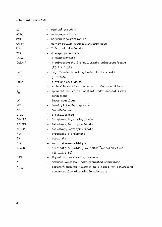
Abbreviations used:
Ac
AOAA
BMI Cm-Pf
DMV
DPA
GABA GABA-T
GAD
G1u
5HTP
K
K m
LC
MEC
NA 2-0G
30HDPA
40HDPA
50HDPA
PLP
SA
SSA
SSA-DH
TRH v v max
6
central amygdala
aminooxyacetic acid
bicuculline-methiodide
centre median-parafascicularis area
2,2-dimethylvalerate
di-n-propylacetate
4-aminobutyrate
4-aminobutyrate:2-oxoglutarate aminotransferase
(EC 2.6.1.19)
L-glutamate 1-carboxylyase (EC 4.l.Ll5)
glutamate
5-hydroxytryptophan
Michaelis constant under saturated conditions
apparent Michaelis constant under non-saturated
conditions
locus coeruleus
2-methyl,2-ethylcaproate
noradrenaline
2-oxoglutarate
3-hydroxy,2-propylvalerate
4-hydroxy,2-propylvalerate
5-hydroxy,Z-propylvalerate
pyridoxal-5 1 phosphate
succinate
succinate-semialdehyde
succinate-semialdehyde: NAD(P)+oxidoreductase
(EC 1.2.1.16)
thyrotropin-releasing hormone
maximum velocity under saturated conditions
apparent maximum velocity at a fixed non-saturating
concentration of a single substrate
CHAPTER I
GABA-ERGIC MECHANISMS A\D MORPHINE ABSTINENCE BEHAVIOUR:
A SURVEY OF THE LITERATURE
l.l. INTRODUCTION
1.2. GABA-ERGIC MECHANISMS
1.2.1. M~bo~m and diotn~bution
1.2.2. Shunt enzqme4
1.2.3. Slf~ptJe eve~
1.2.4. Meta.boUe eompM:tmen.ta.tion
1.2.5. GABA and eonvut6~on6
1.2.6. V~-n-p~opqfaee~e
1.2.1. GABA and behav~o~
1.3. MORPHINE ABSTINENCE BEHAVIOUR
1.3.1. MMpMne ab<>tJnenee behav~o~, deM~tJon6 and de4M~ptJon
1.3.2. QUM~-mMpMne ab;.,tJnenee behav~o~
1 . 3. 3. Loea«zatJon o£ ;.,~te4 ~nvoR.ved ~n ab;.,tJnenee behav~o~
1.3.4. Mo~ph~ne aetJon6 on the GABA _;q;.,tem
1.4. DPA-INDUCED QUASI-MORPHINE ABSTINENCE BEHAVIOUR
7. 4. 1. Method<>
1.4.2. The ~o.te ofi GABA
1.4.3. A~ ofi thio the4io
7
l.l. INTRODUCTIO~
Since the discovery of the non-protein amino acid, gamma-aminobutyric acid
(GABA), in mammalian brain (Roberts and Frankel, 1950; Udenfriend~ 1950;
Awapara et al., 1950), evidence has been gathered indicating that this compound
has a function as an inhibitory neurotransmitter (for a review see Krnjevic,
1970). Subsequent to its discovery in the brain GABA has also been identified
in other tissues, although in most cases only traces could be found (Zachman
et al., 1966; Whelan et al., 1969; Gerber and Hare, 1980).
Recently, it has been suggested that GABA may fulfil a role in morphine
abstinence behaviour. A syndrome resembling morphine abstinence behaviour can
be induced in rats using di-n-propylacetate (DPA), a compound presumably
acting through GABA. The role for GABA in this behaviour was suggested by its
suppression by the GABA antagonists picrotoxin and bicuculline (De Boer et al.,
1977, 1980). The results presented in this thesis provide further evidence
for a role of GABA in this behaviour induced by DPA. In addition, the
involvement of different brain areas in DPA-induced behaviour has been studied.
The implications of these findings for the involvement of GABA in morphine
abstinence behaviour will be discussed at the end of the thesis. In this
first chapter, therefore, a short review will be given on GABA, morphine
abstinence behaviour and the interactions between GABA and morphine.
In the first part of this chapter the recent literature concerning GABA
will be reviewed with the emphasis on mechanisms regulating GABA-ergic
activity. The metabolism of GABA, the enzymes involved in this metabolism and
the synaptic events related to GABA will be discussed. Furthermore, a short
description will be given of the metabolic compartmentation of GABA and related
substances. Since DPA is an anticonvulsant drug, the role of GABA in con
vulsions and the hitherto assumed mechanism of action of DPA will be
discussed. In order to compare the behavioural effects of DPA with the effects
of other GABA-ergic compounds an overview will be given of the effects
of these compounds on the spontaneous behaviour of rats and mice.
In the second part of the chapter the following subjects will be discussed:
the terms involved in the study of morphine abstinence behaviour; quasi-morphine
abstinence behaviour and its implications for true morphine abstinence; the
brain areas responsible for morphine abstinence; the interaction between
morphine and GABA.
Finally, a description is given of what was known about DPA-induced
behaviour before the research described in this thesis was initiated,
8
together with an explanation of the way in which we have extended this
research by the present experiments.
1.2. GABA-ERGIC MECHANISMS
7.2.7. Metabot<hm and dio~~bution
GABA is synthesized in the brain mainly by decarboxylation of L-glutamate
by the enzyme glutamate decarboxylase (GAD; L-glutamate. 1-carboxy-lyase;
EC.4.1.1.15). The major pathway of GABA-degradation is its transamination by
GABA-transaminase (GABA-T; 4-aminobutyrate: 2-oxoglutarate aminotransferase;
EC.2.6.1.19) and subsequent oxidation to succinate by succinic semialdehyde
dehydrogenase (SSA-DH; succinic semi-aldehyde: NAD+ oxido-reductase; EC.
1.2.1.16).
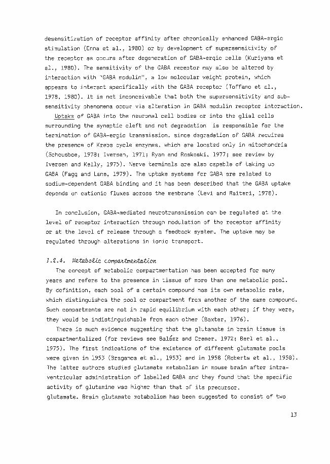
Since glutamate can be formed from 2-oxoglutarate, an intermediate of the
Krebs cycle, the conversion of 2-oxoglutarate via glutamate and GABA to
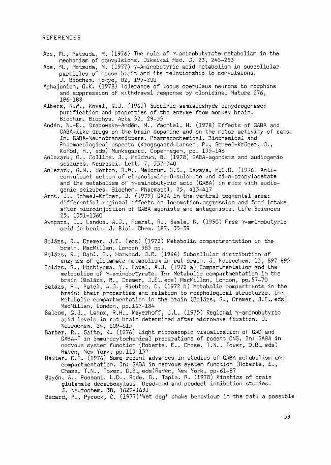
succinate is referred to as a bypass of the Krebs cycle and is called the
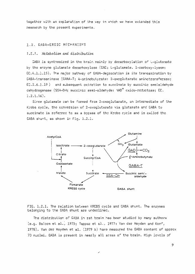
GABA shunt, as shown in Fig. 1.2.1.
Glutamine AcetyiCoA
!socitrate - 2-oxoglutarate
CitLe l ~~ate
GAo}--co2
't-Aminobutyrate Succiny!CoA
l Oxalacetate
l Malate Succinate
~ / Fumdrate
KREBS cycle
GABA-T
-=-:--=:- Succinic semi-SSA- DH aldehyde
GABA shunt
FIG. 1.2.1. The relation between KREBS cycle and GABA shunt. The enzymes belonging to the GABA shunt are underlined.
The distribution of GABA in rat brain has been studied by many authors
(e.g. Balcom et al.~ 1975; Tappaz et al., 1977; Vander Heyden and Korf,
1978). Vander Heyden et al. (1979 b) have measured the GABA content of approx
70 nuclei. GABA is present in nearly all areas of the brain. High levels of
9
GABA are present in the hypothalamus, substantia nigra and globus pallidus.
The GABA concentration in nerve terminals has been estimated to be 60-140
mM in the cat (Fonnum and Walberg, 1973 a,b; Fonnum et al., 1974) and
10-20 mM in the rat (Storm-Mathisen, 1976). Whether or not this GABA is stored
in synaptic vesicles, like the storage of catecholamines (Douglas, 1968), is
still a matter of dispute. The association of GABA with vesicular structures,
a quantity accounting for only 10-20% of total brain GABA (Kuriyama et al.,
1968 a,b; Kuriyama, 1976; Zisappel and Zurgil, 1978), may be loose and it
probably represents unstable compartments (Mangan and Whittaker, 1966).
In conclusion, GABA is present in nearly all areas of the brain, with
high concentrations in the hypothalamus, substantia nigra and globus pallidus.
The concentration of GABA in nerve endings may be in the order of 10-50 mM.
J.Z.Z. Shunt enzqme4
Each enzyme involved in the synthesis and degradation of GABA has its own
cofactor requirements and kinetic profile, the modification of which can lead
to changes in the ultimate concentration of GABA.
The enzyme GAD, responsible for the synthesis of GABA, requires pyridoxal-
5'phosphate (PLP) as a cofactor. The enzyme has been purified from mouse, rat
and human brain (Wu et al., 1973; Maitre et al., 1978 b; Blindermann et al.,
1978) and is located mainly in nerve endings (Salganicoff and DeRobertis,
1963, 1965; Balasz et al., 1966; Wood et al., 1976).
GAD activity can be inhibited by some Krebs cycle intermediates and GABA
(Gerig and Kwock, 1979; Wu and Roberts, 1974;Blindermann et al., 1978). The
latter effect may be important for feedback regulation of the GABA level in
nerve terminals (Sayan et al., 1978; Nitsch, 1980).
The activity of GAD can be influenced too by changing the degree of
saturation of the apoenzyme for the coenzyme PLP, since the ~n v~vo
saturation of GAD by PLP is only 24-47% (Miller et al., 1977; Nitsch, 1980).
Pharmacological manipulation of the synthesis of GABA in vivo, based on
competition with PLP, results in convulsions oi in a decrease in the con
vulsive threshold (see for a review: Perez de la Mora et al., 1973).
GABA-T, the first enzyme in the degradation of GABA, has been purified
from mouse, rat, rabbit, pig, bovine and human brain (Schousboe et al.,
1973; Sytinski and Vasilijev, 1969; Waksman and Roberts, 1965; Maitre et al.,
1975; John and Fowler, 1976; Bloch-Tardy et al., 1974; Kobayashi et al.,
1977; Maitre et a1., 1978 a). Waksman and Bloch (1968) have purified and
10
separated several isozymes of GABA-T, which seem to be located in different
subcellular organelles. GABA-T from non-synaptosomal mitochondria has a much
higher activity and a higher affinity for GABA (Km for GABA is 4-6.5 mM)
than the GABA-T located in synaptosomal mitochondria (Km for GABA is 30-50
mM) (Ngo and Tunnicliff, 1978). The weaker affinity of synaptosomal GABA-T
for GABA and the lower activity of this enzyme favour a role of GABA-T in
the regulation of the high presynaptic GABA level (see section 1.2.1).
It has been suggested that the low activity of GABA-T in synaptosomes can
be attributed to contamination of this fraction with free cytoplasmic
mitochondria (Bal8sz et al., 1966; Van den Berget al., 1975; Hertz, 1979).
However, electronmicroscopic studies and measurements of marker enzymes
indicate that the low GABA-T activity detected in these fractions cannot be
due to such a contamination (Salganicoff and DeRobertis, 1965; Van Kempen
et al., 1965; Waksman et al., 1968; Walsh and Clark, 1976). Recently, the
localization of GABA-T in nerve endings has been unequivocally established
using immunohistochemical methods (Barber and Saito, 1976; Chan-Palay et al.,
1979).
A high ionic strength has been shown to decrease the activity of GABA-T
(De Boer and Bruinvels, 1977 a). In addition, the purified enzyme is
susceptible to inhibition by a large number of metal ions, glutamate and
some other naturally occurring substances (Schousboe et al., 1974). Inter
ference from carbohydrate metabolism, e.g. hypoglycemia, will also affect
GABA-T activity, through alterations in the level of 2-oxoglutarate (Otsuki
et al., 1968; Dravid and Jilek, 1965).
SSA-DH, the second enzyme of the GABA degradation, appears to be more
specific for the GABA shunt than GABA-T (Embree and Albers, 1964; Albers and
Koval, 1961). The enzyme has been purified from monkey, human and rat brain
(Albers and Koval, 1961; Embree and Albers, 1964; Kammeraat and Veldstra,
1967; Cash et al., 1975, 1977).
Just like GABA-T, SSA-CH activity seems to be present in mitochondria of
both synaptosomal and non-synaptosomal origin (Salganicoff and DeRobertis,
1965; Sims and Davis, 1973). The distribution of the enzyme in the brain
parallels the distribution of GABA-T too (Miller and Pitts, 1967; Sheridan
et al., 1967). The activity of SSA-DH is always higher (1.5-5 times) than
that of GABA-T (Miller and Pitts, 1967; Cash et al., 1975; De Boer and
Bruinvels, 1977 a). This higher activity of SSA-DH (as compared to GABA-T),
together with its high affinity for SSA, indicate that GABA-T and not SSA-DH
is rate-limiting in the degradation of GABA .. However, changes in the
ll
concentration of cations (De Boer and Bruinvels, 1977 b; De Boer et al.,
1979) or in the availability of the cofactor NAD+, e.g. during hypoxia (Gubler
et al., 1974), may diminish the activity of SSA-DH in such a manner that it
may become rate limiting.
In conclusion, the GABA synthesizing enzyme GAD is located almost ex
clusively in nerve terminals and may be regulated by PLP availability and
by the intraterminal GABA concentration. GABA degradation occurs mainly, but
not exclusively, in the non-nerve terminal compartment. It is suggested that
different isozymes of GABA-T are located in nerve terminals and in glial
cells. Cation concentration as well as ionic strength may regulate the
activity of the enzymes involved in the GABA degradation.
J • 2. 3. Syrta.ptiQ even-U )[e)'a;ted cto GABA
In accordance with its role as a putative neurotransmitter, GABA is
released into the synaptic cleft, interacts with specific receptors and is
inactivated by uptake into the neurons or the glial cells surrounding the
synaptic cleft.
Release of GABA has been demonstrated to occur after electrical
stimulation or by depolarizing potassium concentrations both in vitko
(Katz et al., 1969; Srinivasan et al., 1969) and ~n v~vo (Obata and Takeda,
1969; Vander Heyden et al., 1979 a). It has been postulated by Tapia et al.
(1975) that GABA is continuously released from the synaptosomal cytoplasm
of inhibitory nerve endings with a rate depending on the activity of GAD.
According to these authors it is still possible that GABA may also be re
leased from a storage site such as synaptic vesicles, when the GABA neuron
is depolarized by a stimulus. The existence of a feedback control of the
release of GABA via presynaptic autoreceptors has been indicated by both
in vitko and ~n v~vo experiments (Mitchell and Martin, 1978; Van der Heyden
et al., 1980; Brennan et al., 1981).
Binding of GABA to subcellular brain particles has been shown to be
either sodium-dependent or -independent (Sana and Roberts, 1961; Zukin et al.,
1974; Enna and Snyder, 1975). The sodium-independent binding of GABA re
presents binding to postsynaptic receptor sites since it could be prevented
by bicucu1line-methiodide (BMI) (for a review see De Feudis, 1978). The
existence of more than one single type of GABA receptor has been suggested
(Nistri and Constanti, 1979).
Regulation of the responsiveness of GABA receptors is possible by
12
desensitization of receptor affinity after chronically enhanced GABA-ergic
stimulation (Enna et al., 1980) or by development of supersensitivity of
the receptor as occurs after degeneration of GABA-ergic cells (Kuriyama et
al., 1980). The sensitivity of the GABA receptor may also be altered by
interaction with "GABA modulin 11, a low molecular weight protein. which
appears to interact specifically with the GABA receptor (Toffano et al.,
1978, 1980). It is not inconceivable that both the supersensitivity and sub
sensitivity phenomena occur via alteration in GABA modulin receptor interaction.
Uptake of GABA into the neuronal cell bodies or into the glial cells
surrounding the synaptic cleft and not degradation is responsible for the
termination of GABA-ergic transmission, since degradation of GABA requires
the presence of Krebs cycle enzymes, which are located only in mitochondria
(Schousboe, 1978; Iversen, 1971; Ryan and Roskoski, 1977; see review by
Iversen and Kelly, 1975). \'erve terminals are also capable of taking up
GABA (Fagg and Lane, 1979). The uptake systems for GABA are related to
sodium-dependent GABA binding and it has been described that the GABA uptake
depends on cationic fluxes across the membrane (Levi and Raiteri, 1978).
In conclusion, GABA-mediated neurotransmission can be regulated at the
level of receptor interaction through modulation of the receptor affinity
or at the level of release through a feedback system. The uptake may be
regulated through alterations in ionic transport.
1.2.4. Metabotcc comp~tmentxtion The concept of metabolic compartmentation has been accepted for many
years and refers to the presence in tissue of more than one metabolic pool.
By definition, each pool of a certain compound has its own metabolic rate,
which distinguishes the pool or compartment from another of the same compound.
Such compartments are not in rapi_d equilibrium with each other; if they were,
they would be indistinguishable from each other (Baxter, 1976).
There is much evidence suggesting that the glutamate in brain tissue is
compartmentalized (for reviews see Bal3sz and Cremer, 1972; Berl et al.,
1975). The first indications of the existence of different glutamate pools
were given in 1953 (Braganca et al., 1953) and in 1958 (Roberts et al., 1958).
The latter authors studied glutamate metabolism in mouse brain after intra
ventricular administration of labelled GABA and they found that the specific
activity of glutamine was higher than that of its precursor.
glutamate. Brain glutamate metabolism has been suggested to consist of two
l3
pools (Garfinkel, 1966; Van den Berg and Garfinkel, 1971). Other authors have
proposed the existence of three compartments consisting of a small glutamate
compartment in glial cells, a second small compartment composed of nerve
terminals and a large compartment located in nerve cell bodies and dendrites
(Bal~sz et al., 1972 b). Several independent tricarboxylic acid cycles have
also been identified in brain tissue and have been related to the different
glutamate pools (Garfinkel, 1966). Both GABA synthesis and degradation are
each divided over more than one compartment (Bal~sz et al., 1972 a; Van den
Berget al., 1975). One has to bear in mind, however, that different metabolic
compartments are not necessarily equivalent to morphologically different cell
types. It is not inconceivable that, superimposed upon these morphological
defined cell types, anatomically distinct brain divisions exist which
represent separate metabolic compartments, independent of the number of
cell types present.
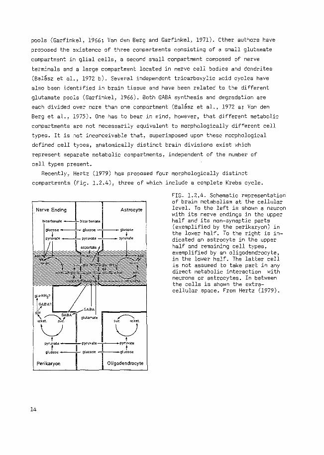
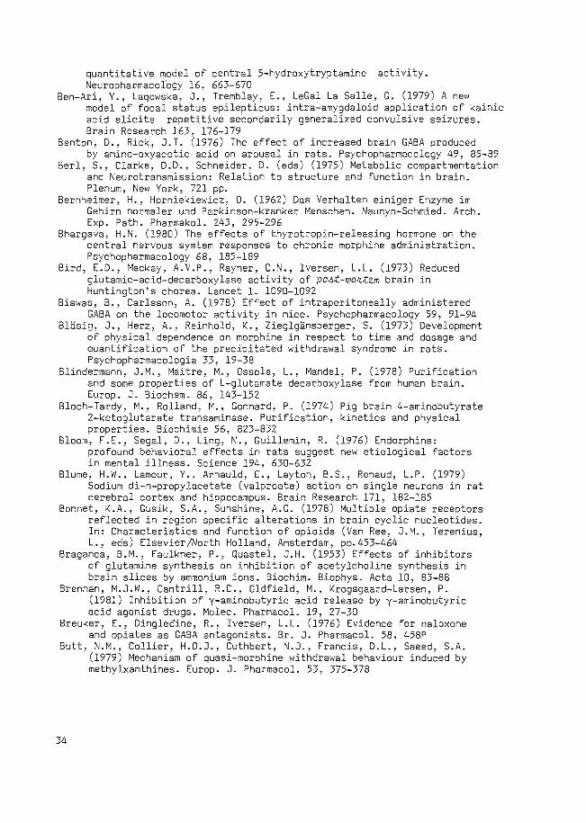
Recently, Hertz (1979) has proposed four morphologically distinct
compartments (Fig. 1.2.4), three of which include a complete Krebs cycle.
Nerve Ending
Perikaryon
14
Astrocyte
Oligodendrocyte
FIG. 1.2.4. Schematic representation of brain metabolism at the cellular level. To the left is shown a neuron with its nerve endings in the upper half and its non-synaptic parts (exemplified by the perikaryon) in the lower half. To the right is indicated an astrocyte in the upper half and remaining cell types, exemplified by an oligodendrocyte, in the lower half. The latter cell is not assumed to take part in any direct metabolic interaction with neurons or astrocytes. In between the cells is shown the extracellular space. From Hertz (1979).
The author suggested that GABA synthesis is located only in nerve endings,
while GABA degradation takes place mainly in astroglia and neuronal perikarya.
Therefore, a flux of GABA from nerve terminals to glial cells has to be
present. Glutamine is transported in the opposite direction and may then act
as the precursor of GABA in nerve endings (Tapia and Gonzalez, 1978; Reubi
et al., 1978). Although the model fails in that degradation of GABA in nerve
endings is not included (see section 1.2.2), it clearly demonstrates the
complexity of GABA metabolism in the brain and the importance of the concept
of metabolic compartmentation.
In conclusion, glutamate- and GABA metabolism is divided over several
metabolic compartments. When considering the pharmacology of GABA, this
compartmentation must be taken into account.
1.2.5. GABA and Qonvu£6/ono GABA has been suggested to be implicated in many neuropathological and
psychopathological states, e.g. schizophrenia (Roberts, 1972; Van Kammen,
1977), Huntington's chorea (Perry et al., 1973; Bird et al., 1973), Parkin
son's disease (Bernheimer and Horniekiewicz, 1962; McGreer et al., 1971) and
tardive dyskinesia (Gibson, 1977).
Since some work presented in this thesis deals with the mechanisms of
action of the anticonvulsant drug di-n-propylacetate (DPA, DepakineR), the
possible relationship between GABA and convulsions will be discussed.
Before a physiological role for GABA as an inhibitory neurotransmitter was
established, it had already been shown that GABA has anticonvulsive properties
(Hayashi, 1959). A decreased availability of PLP, probably leading to a lower
activity of GAD (see section 1.2.2), was suggested to be the cause of seizures
occurring in children with a dietary vitamin 86 -deficiency (Holtz and Palm,
1964). This relationship between GABA and convulsions can be summarized as
follows: a decrease in the level of.GABA-ergic transmission parallels a
decrease in the convulsive threshold and makes the animals more susceptible
to convulsant agents. An increase in GABA-ergic transmission is associated
with an increase in the convulsive threshold and protects the animals against
convulsion-evoking treatments. Reviews on this topic have frequently been
published (Meldrum, 1975; Tower, 1976; Meldrum, 1978; Roberts, 1980). An anti-convulsive effect through an increase in GABA-ergic transmission
can be achieved by administration of a GABA agonist (muscimol; Anlezark
et al., 1978), by inhibition of GABA reuptake (L-2,4-diaminobutyric
15
acid; Taberner.and Roberts~ 1978; Horton et al., 1979), or by inhibition
of GABA-transaminase (ethanolamine-0-sulphate; Anlezark et al., 1976;
aminooxyacetic acid; Wallach, 1961; Kuriyama et al., 1966). Recently,
reviews have been published on the inhibitors of GABA-T and their anti
convulsive action (Metcalf, 1979; Seiler and Sarhan,l980).
Although these reports suggest a simple relationship between GABA
concentration in brain and the susceptibility to seizures, the relation
appears to be more complex. Some compounds,e.g. hydrazides, which enhance
GABA levels by inhibition of GABA-T, can, despite of enhancing GABA
concentrations, induce convulsions. This is related to concomitant inhibition
of GAD, and Wood and Peesker (1973) have postulated a mathematical relation
ship in which the excitability of the brain is determined not only by the
GABA concentration but also by the GAD activity (see also Wood and Peesker,
1974, 1975; Wood, 1975). Other authors have also found that the level of
synaptosomal GABA, which decreases primarily after GAD inhibition, is likely
to be more important in determining excitability than total brain GABA
(Abe and Matsuda, 1976, 1977; Matsuda et al., 1978, 1979; Wood et al., 1979,
1980). The specific involvement of synaptosomal GABA in the determination of
brain excitability is further indicated by the finding that the onset and
the peak of the anticonvulsive activity of the GABA-T inhibitor gamma-vinyl
GABA (GVG) parallelled the time course for the (limited) increase in GABA
in nerve endings induced by this compound and not that of the (massive)
increase of GABA in the other compartments (Gale and Iadarola, 1980).
In conclusion, the increase in GABA concentration in the brain is probably
involved in the anticonvulsive action of various compounds. The increased
level of GABA in nerve terminals rather than in glial cells and other
organelles seems to be important for this anticonvulsive action.
1.2.6. V~-n-p~opyfacetate
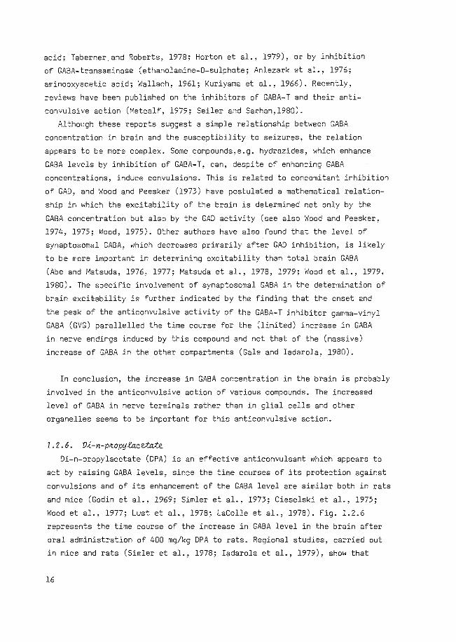
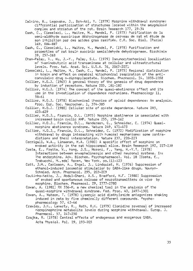
Di-n-propylacetate (DPA) is an effective anticonvulsant which appears to
act by raising GABA levels, since the time courses of its protection against
convulsions and of its enhancement of the GABA level are similar both in rats
and mice (Godin et al., 1969; Simler et al., 1973; Cieselski et al., 1975;
Wood et a1 .• 1977; Lust et a1 .• 1978; LaCo11e et a1., 1978). Fig. 1.2.6
represents the time course of the increase in GABA level in the brain after
oral administration of 400 mg/kg DPA to rats. Regional studies, carried out
in mice and rats (Simler et al., 1978; Iadarola et al., 1979), show that
16
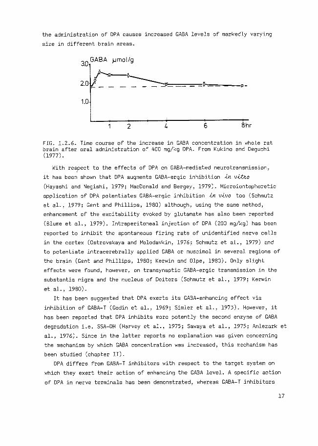
the administration of DPA causes increased GABA levels of markedly varying
size in different brain areas.
3.0 GABA ).lmol/g
2.0 ,_......., -9-=-=------==-=-= 2-,~ =--o-
1.0
2 6 Bhr
FIG. 1.2.6. Time course of the increase in GABA concentration in whole rat brain after oral administration of 400 mg/kg DPA. From Kukino and Deguchi (1977).
With re~pect to the effects of DPA on GABA-mediated neurotransmission,
it has been shown that DPA augments GABA-ergic inhibition ~n v~o
(Hayashi and Negishi, 1979; MacDonald and Bergey, 1979). Microiontophoretic
application of DPA potentiates GABA-ergic inhibition .<.n v.<.vo too (Schmutz
et al., 1979; Gent and Phillips, 1980) although, using the same method,
enhancement of the excitability evoked by glutamate has also been reported
(Blume et al., 1979). Intraperitoneal injection of DPA (200 mg/kg) has been
reported to inhibit the spontaneous firing rate of unidentified nerve cells
in the cortex (Ostrovskaya and Molodavkin, 1976; Schmutz et al., 1979) and
to potentiate intracerebrally applied GABA or muscimol in several regions of
the brain (Gent and Phillips, 1980; Kerwin and Olpe, 1980). Only slight
effects were found, however, on transynaptic GABA-ergic transmission in the
substantia nigra and the nucleus of Deiters (Schmutz et al., 1979; Kerwin
et al., 1980).
It has been sugges~ed that DPA exerts its GABA-enhancing effect via
inhibition of GABA-T (Godin et al., 1969; Simler et al., 1973). However, it
has been reported that DPA inhibits more potently the second enzyme of GABA
degradation i.e. SSA-DH (Harvey et al., 1975; Sawaya et al., 1975; Anlezark et
al., 1976). Since in the latter reports no explanation was given concerning
the mechanism by which GABA concentration was increased, this mechanism has
been studied (chapter II).
DPA differs from GABA-T inhibitors with respect to the target system on
which they exert their action of enhancing the GABA level. A specific action
of OPA in nerve terminals has been demonstrated, whereas GABA-T inhibitors
l7
are active in both nerve terminals and other organelles (Iadarola and Gale,
1979; Sarhan and Seiler, 1979).
In conclusion, DPA exerts its anticonvulsive action probably via inhibition
of one of the enzymes involved in GABA degradation leading to an increase in
the concentration of GABA in nerve terminals. However, the exact enzymatic
mechanism by which the GABA concentration can increase after this inhibition
of the degradation, remains to be elucidated.
1.Z.?. GABA and bthav{oUk Systemic injections of GABA, a GABA-agonist or a GABA-T inhibitor into
rats have been reported to cause a decrease of spontaneous locomotor activity
(Biswas and Carlsson, 1978; Benton and Rich, 1976; Cott et al., 1976;
Matsui and Deguchi, 1977; Schechter et al., 1977, 1979; Worms and Lloyd,
1980; Scheel-KrUger et al., 1978 b). In addition, SCotti de Carolis et al.
(1978) described a biphasic action after subcutaneous administration of
muscimol, viz. a short lasting excitation immediately after injection, followed
by muscle relaxation and loss of righting reflex.
Furthermore, intracisternal or intraventricular injection of GABA or the
GABA-T inhibitor ethanolamine-0-sulphate (EOS) inhibited spontaneous loco
motor activity (Freed and Michaelis, 1976; Czajka, 1978; File, 1977). Thus,
either systemic or intracranial injection of GABA-ergic compounds results
in sedation of the animals.
However, effects of GABA-ergic compounds on spontaneous behaviour observed
after intracerebral injection, depend on the site of injection. Injection
of muscimol into the nucleus of the raphe dorsalis, an area containing
serotonergic cell bodies, stimulates locomotor activity (Przewlocka et al.,
1979). Similarly, injection of GABA agonists into areas containing dopaminergic
cell bodies has been found to be excitatory. Tanner (1979) injected GABA
into the ventral tegmental area (A 10) -a dopaminergic structure- and found
an increase in locomotor activity, which could be antagonized by picrotoxin.
However, Mogensen et al. (1979) found an increase in locomotor activity
after injection of the GABA antagonist picrotoxin into the ventral teqme~tal
area. This contradiction may be explained by differences in GABA function
within the caudal and rostral parts of the ventral tegmental area. Injection
of muscimol into the caudal part evokes hypermotility while after such an
injection into the rostral part sedation can be observed (Arnt and Scheel
KrOger, 1979).
18
Injection of muscimol or GABA-T inhibitors into the substantia nigra of
rats has been found to evoke stereotype behaviour, including sniffing, biting
and gnawing (Scheel-KrUger et al., 1978 a; Matsui and Kamioka, 1978 a, b;
Koob et al., 1978). On the other hand, injection of GABA-antagonists into
this area elicited catatonia (DiChiara et al., 1978).
Catalepsy and sedation were found after injection of muscimol or the
GABA-T inhibitor gabaculine into the nucleus accumbens or into the globus
pallidus (Scheel-KrUger et al., 1977, 1978 b; Matsui and Kamioka, 1978,
1979; Wachtel and Anden, 1978; Anden et al., 1978). Catalepsy has also been
observed after injection of muscimol into some nuclei of the thalamus
(DiChiara et a1 .• 1979).
Thus, in most reports concerning the behavioural effects induced by
alteration of central GABA-ergic activity, the relationship with dopaminergic
systems has been studied. It may be inferred that injection of GABA-ergic
compounds into areas containing dopaminergic cell bodies may cause a
stimulation of behavioural activity. Injection of these compounds into
areas containing dopaminergic nerve terminals induces sedation and catalepsy.
In conclusion, the enhancement of GABA-ergic transmission ~n v~vo
has been generally reported to cause sedation. In some reports, excitatory
effects were observed. Behavioural effects obtained by modifying GABA-ergic
activity in discrete areas of the brain are not fully predictable since
both sedative and excitatory effects have been reported.
1.3. MORPHINE ABSTINENCE BEHAVIOUR
1.3.1. Mokp~ne abh~nence beh~v~oUk, defi~~~onh ~ dehek~p~on
Before describing the symptoms of morphine abstinence behaviour, some
terms should be defined. Tolerance is known to develop towards the effects
elicited by morphine-like compounds, when these drugs are given repeatedly,
while, concomitantly, the animals become physically dependent. Tolerance
may be said to have developed when, after repeated administration, a given
dose of a drug -in this case morphine- produces a decreased effect.
Conversely, tolerance exists when increasingly larger doses must be
administered to obtain the effects observed with the original dose. Physical
dependence refers to an altered physiological state produced by the
repeated administration of a drug, which necessitates the continued
administration of the drug to prevent the appearance of a stereotypical
19
syndrome, the withdrawal or abstinence syndrome characteristic for the
particular drug (Jaffe, 1980). The development of tolerance and physical
dependence with repeated use is a characteristic feature of all opioid drugs.
The degree of tolerance and physical dependence will depend on the particular
drug, on the frequency and period of administration and on the quantity
administered.
To study morphine abstinence behaviour, morphine is administered
chronically to animals, using sustained release preparations, e.g. sub
cutaneously implanted pellets (Way et al., 1969; Wei et al., 1973 a; Blasig
et al., 1973) or subcutaneously injected emulsions (Collier et al., 1972),
multiple injection schedules (Martinet al., 1963) or addition of morphine
to the drinking water (Gellert and Holtzmann, 1978). After a sufficiently
long period of administration the withdrawal syndrome can be obtained by
abrupt cessation of morphine administration or by precipitation,using
narcotic antagonists like naloxone, levallorphan or nalorphine.
The symptoms observed in antagonist-precipitated abstinence behaviour
in rats have been described by Bl3sig et al. (1973). They arranged the
symptoms into two classes: counted signs and checked signs. The counted
signs were: exploration, jumps, flights, 11 wet dog shakes 01 (body shakes),
episodes of teeth chattering and writhing. The checked signs were:scream
on touch, hostility (irritation) to handling, ptosis, eye twitching,
rhinorrhea, lacrimation, diarrhea and penile erection. Other authors have
used a slightly modified list, but in principle the abstinence behaviour is
described by these symptoms. Wei et al. (1973 a) called jumps and flights 01 escape attempts 11 and this notion 11 escape11 can be found also in the "escape
diggingn, observed in sawdust-lined cages used by Frederickson and Smits
(1973) and by De Boer et al. (1977) (see also section 1.4.1 and the chapters
III, IV and V). The ranking of the relative importance of the symptoms
varies from author to author (Wei et al., 1973 a; Collier et al., 1972).
However, from the study of Bl3sig et al. (1973) it appeared that the relative
importance of a symptom depends on the degree of dependency. With a high
dependence some signs, e.g. jumps, are dominant, while with a lower degree
of dependence more recessive signs, e.g. body shakes, are expressed.
In conclusion, morphine abstinence behaviour consists of various symptoms
and the incidence of each particular symptom differs from another and the
relative importance of a symptom depends on the degree of dependency.
20
J • 3. 2. Quabi -mOJtpiUne a.bsUnenee beha.v.<ouJl
In an attempt to unravel the neurochemical and neuropharmacological
mechanisms underlying the expression of abstinence symptoms in morphine
dependent animals, these animals are often treated with drugs either during
the phase of development of dependence or during the phase of withdrawal. The
effects of these drugs on the intensity of the withdrawal symptoms is then
measured.
However, the mechanism of the induction of morphine abstinence symptoms
can also be studied in naive animals which have never received morphine. The
advantage of using naive animals is that the experiments can be performed in
a short period of time and no interference from a change of morphine level or
from the injection of antagonists occurs. Compounds of different categories
(Table 1.3.2) can induce in naive animals behavioural symptoms resembling
morphine abstinence behaviour. This kind of behaviour has been defined by
Collier (1974) as quasi-morphine abstinence behaviour: ''An effect resembling
one elicited by withdrawal of a drug on which an animal has been made
dependent, but produced by another treatment in a naive animal never exposed
to drug nor to a like-acting congener that induces such dependence''.
For the quasi-abstinence behaviour to be a model for true morphine
abstinence behaviour several criteria should be fulfilled. Firstly~ the
behaviour induced should closely resemble true morphine abstinence behaviour.
In several cases,e.g. using phosphodiesterase inhibitors, histamine, DPA,
TRH and EGTA, three or more symptoms of the morphine abstinence syndrome have
been observed. In other cases, e.g. 5-HTP and kainic acid, also other symptoms
occur, i.e. gnawing and biting (5-HTP; Sloviter et al., 1978), and convulsions
(kainic acid; Ben-Ari et al., 1979). Therefore, the behaviour induced by the
latter two compounds cannot be considered as a specific quasi-morphine
abstinence syndrome.
A second criterion is that the effects of morphine on quasi-morphine
abstinence behaviour and on true morphine abstinence behaviour should be
parallel. One would expect that the quasi-morphine abstinence behaviour can
be suppressed by a low dose of morphine. Only the behaviour induced by DPA,
by TRH or by the phosphodiesterase inhibitors can be suppressed by morphine,
while the effect of morphine has not been studied on responses to EGTA and the
opioid compounds. In order to suppress the behaviour induced by 5-HT, ACTH,
Ag 3-5 and the,Sgd compounds, much higher doses of morphine have been used
and in some cases even at these high doses only partial suppression has been
found.
21
The implications of these reports of the effects of phosphodiesterase
inhibitors, TRH and EGTA will be discussed briefly. The behaviour induced
by DPA is the topic of this thesis and will receive attention in section
1.4.2 and in other chapters.
Phosphodiesterase inhibitors. It has been reported that administration of
phosphodiesterase inhibitors or of cyclic AMP during the induction of morphine
dependence, accelerates the development of dependence resulting in an intensi
fied abstinence syndrome (Francis et al., 1976; Collier and Francis, 1975;
Shahid Salles et al., 1979). These data support the hypothesis by Collier
(1978) that cyclic AMP plays a role in morphine abstinence behaviour (Francis
et al., 1978; for a recent review see Collier, 1980). The implication is
that disinhibition of an adenylate cyclase system, which has become tolerant
to inhibition by morphine, leads to an overshoot in adenylate cyclase activity
during the abstinence phase.
However, the data relating to the role of cyclic AMP in true morphine
abstinence behaviour are contradictory. The rise in cyclic AMP in brain, which
would be expected to occur according to the afore-mentioned hypothesis, has
been found by some authors (e.g. Collier and Francis, 1975), but not by
others (VonVoigtlander and Losey, 1977). The increase in cyclic AMP may
occur in certain brain areas only, which may explain the conflicting results
(Bonnet et al., 1978; for a review see Rosenfeld et al., 1979).
Thyrotropin Releasing Hormone (TRH). The areas of the brain from which
body shakes can be elicited by the injection of TRH, namely the medial
thalamus, the periaquaductal grey and the locus coeruleus (Wei et al.,
1975 a; Kalivas and Horita, 1980), are the same as those from which naloxon
injection can precipitate abstinence in morphine pretreated rats (section
1.3.3). Repeated subcutaneous administration of TRH during the induction of
morphine dependence in mice has been reported to prevent the development of
withdrawal hypothermia, but it failed to modify the jumping response to
naloxone (Bhargava, 1980). Administration of TRH prior to precipitation
of abstinence by injection of naloxone in morphine dependent rats, exacerbated
shaking behaviour while other components of the withdrawal syndrome remain
unchanged (Martinet al., 1977). Since TRH is widely distributed in areas of
the brain other than the hypothalamus, this endogenous compound may have a
direct role in the manifestation of these morphine abstinence symptoms which
are related to the thermoregulatory system (Jackson and Reichlin, 1974;
Winoku and Utiger, 1974).
22
EGTA. Since EGTA is pre-eminently a chelator of calcium, the reports
cited indicate an important role for calcium in morphine abstinence behaviour.
This role of calcium in morphine abstinence behaviour appears to be intimately
related to neurotransmitter release since the concentration of calcium in
synaptic vesicles has been found to be particularly affected by morphine
(for reviews see Way, 1978; Ross and Cardenas, 1979). Administration of
morphine acutely lowers vesicular calcium, while, during the development of
tolerance and dependence, an accumulation of vesicular calcium occurs. The
sudden removal of morphine leads to increased release of neurotransmitters
resulting in the hyperactivity of several neurotransmitter systems, which
characterizes morphine withdrawal behaviour (Way, 1978).
Although the syndromes induced by 5-HTP or by amphetamine and L-DOPA do
not seem to be as closely related as the afore-mentioned syndromes to quasi
morphine abstinence behaviour, the data do suggest that an overshoot of
serotonergic and catecholaminergic activity may form a part of the underlying
mechanism in the morphine abstinence behaviour. Such an enhanced neuronal
activity may become apparent in various ways. From enhancement of the release
of the transmitter, as has been suggested in relation to the response to EGTA
or from an enhanced sensitivity of the receptors or from a decrease in reuptake
or extra neuronal degradation of the transmitters. An altered receptor
sensitivity has already been suggested as the mechanism underlying abstinence
behaviour (Collier, 1965; Jaffe and Sharpless, 1968). In recent years,
receptor binding studies have confirmed this theory, e.g. for S-adrenergic
receptors (Llorens et al., 1978). No data exist to support the involvement
of decreased uptake or degradation. In contrast, an enhanced turnover of
noradrenaline appears to be present during morphine abstinence (Laverty and
Roth, 1980; Crawley et al., 1979).
It is not within the scope of this chapter to discuss all the implicationo
of the data summarized in Table 1.3.2. However, it is clear that when
studying quasi-morphine abstinence behaviour, data are yielded which support
the research on true morphine abstinence behaviour.
In conclusion, it has been shown that quasi-morphine abstinence behaviour
can be induced by interference at several levels of brain function, i.e. at
the level of transmitter release or receptor stimulation, or at the level
of the second mesSenger cyclic AMP.
23
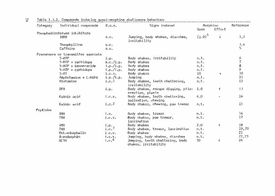
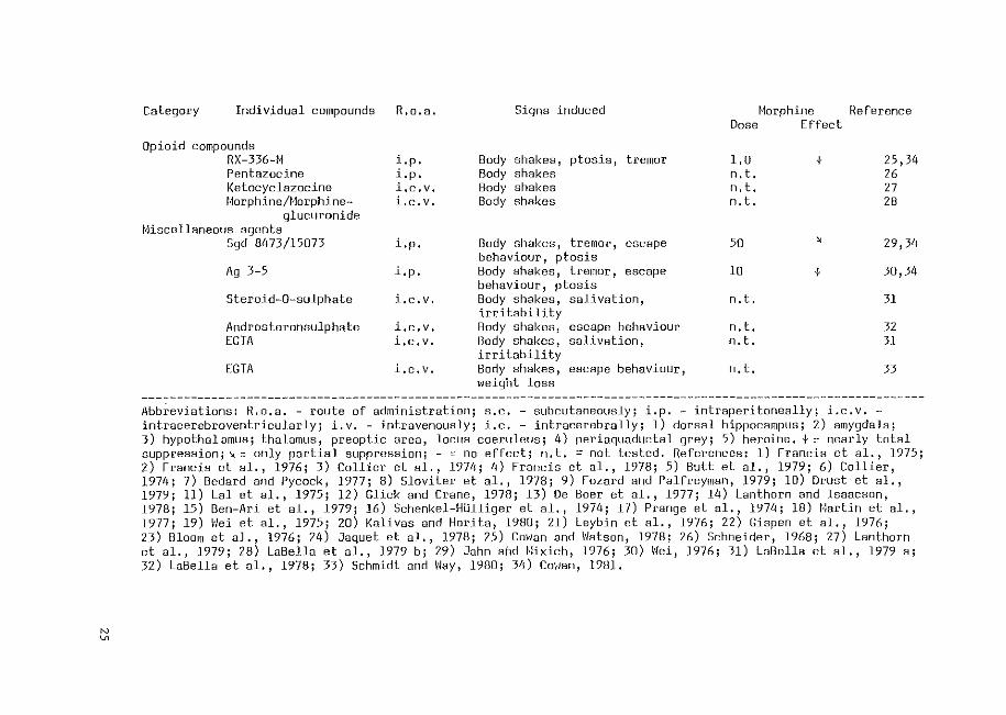
~ I~~~~-!~~~~~-~~~e~~~~~-!~~~~!~g_g~~~!=~~~eb!~~-~~~~!~~~~~-~~b~~!~~~---------------------------------------------category Individual compounds R. o. a. Signs induced 1·1orphine Reference
Phosphodiesterase inhibitors IBHX
Theophylline Caffeine
Precursors or transmitter agonints 5-HTP
Pep tides
5-HTP + carbidopa 5-HTP + benserazide 5-HTP + carbidopa 5-HT Amphetamine + L-DOPA Histamine
DPA
Kainic acid
Kainic acid
TRH TRH
TRH TRH t·let-enkephalin a-endorphin ACTH
s.c.
s.c. s.c.
i.p. s. c. /i. p. i.p./i.p. i.p./i.p. i.c.v. i.p./i.p. i.c.l
i.p.
i.c.v.
i.e. 2
i. v. i.c.v.
i.p. . 3 l.c. i.c.v. i.c.v. . 4 l,C,
Jumping, body shakes, diarrhea, irritability
Body shakes, irritability Body shakes Body shakes Body shakes Body shakes Jumping Body shakes, teeth chattering, irritability Body shakes, escape digginq, piloerection, ptosis Body shakes, teeth chattering~ salivation, chewing Body shakes, chewing, pal·/ tremor
Body shakes, tremor Body nhakes, paw tremor, lacrimation Body shakes Body shakes, tremor, lacrimation Body shakes Jumping, body shakes, diarrhea Jumping, teeth chattering, body shakes, irritability
Dose Effect
(1.0)5
n.t. n.t. n.t. n.t. 10 n.t. n. t.
1.0
4.0
n.t.
n. t. n.t.
2.0 n. t. n.t. n. t. 30
+
'
+
+
+
1,2
3,4 5
6 7 8 9 10 11 12
13
14
15
16 17
18 19,20 21 22,23 24
N
"'
Category Individual compounds
Opioid compounds RX-336-f\ Pentazocine Ketocyclazocinef,1orphine/Horphine-
glucuronide f,liscellaneous agents
Sgd 8473/15073
Ag 3-5
Steroid-0-sulphate
Androsteronsulphate EGTA
EGTA
R.o.a.
i.p. i.p. i,c,v. i.c.v.
i.p.
i.p.
i.c.v.
i,c,v. i,c,v.
i,c,v,
Signs induced
Body shakes, ptosis, tremor Body shakes Body shakes Body shakes
Body shakes, tremor, escape behaviour, ptosis Body shakes, tremor, escape behaviour, ptosis Body shakes, salivation, irritability Body shakes, escape behaviour Aody shakes, salivation, irritability Body shakes, escape behaviour, weight loss
Horphine Reference Dose Effect
l.D n.t. n.t. n.t.
50
10
n.t.
n.t. n.t.
n.t.
t
' t
25,34 26 27 28
29,34
30,34
31
32 31
33
Abb~eviations: R.o.a. - route of administration; s.c. - subcutaneously; i.p. - intraperitoneally; i.c.v. -intracerebroventricularly; i.v. - intravenously; i.e. - intracerebrally; 1) dorsal hippocampus; 2) amygdala; 3) hypothalamus; thalamus, preoptic area, locus coeruleus; 4) periaquaductal grey; 5) heroine. +::: nearly total suppression;~::: only partial suppression; - ::: no effect; n.t. =not tested, References: 1) Francis et al., 1975; 2) Francis et al., 1976; 3) Collier et al., 1974; 4) Francis et al., 1978; 5) Butt et al., 1979; 6) Collier, 1974; 7) Bedard and Pycock, 1977; 8) Sloviter et al., 1978; 9) Fozard and Palfreyman, 1979; 10) Drust·et al., 1979; 11) Lal et al., 1975; 12) Glick and Crane, 1978; 13) De Boer et al., 1977; 14) Lanthorn and Isaacson, 1978; 15) Ben-Ari et al., 1979; 16) Schenkel-HUlliger et al., 1974; 17) Prange et al., 1974; 18) f4artin et al., 1977; 19) \'lei et al., 1975; 20) Kalivas and Horita, 1980; 21) Leybin et al., 1976; 22) Gispen et al., 1976; 23) Bloom et al., 1976; 24) Jaquet et al., 1978; 25) Cm1an and \'Iatson, 1978; 26) Schneider, 1968; 27) Lanthorn et al., 1979; 28) LaBella et al., 1979 b; 29) Jahn arid fHxich, 1976; 30) \'lei, 1976; 31) LnBella et al., 1979 a; 32) LaBella et al., 1978; 33) Schmidt and \'lay, 1980; 34) Covmn, 1981.
1.3.3. Loeat<zat<on on ~~t'4 ~nvofved in ab~~nenee beh~vio~ Localization of the areas involved in morphine abstinence behaviour is
performed by many authors by lesioning the brain or by injection of opiate
antagonists into particular sites of the brain in morphine dependent animals.
The lesion technique, however, has several drawbacks since it is not possible
to decide whether a lesion was truly made at the primary site responsible
for withdrawal signs. The lesioned brain structure may be merely inter
connected with the primary site. Therefore, lesion studies should always be
accompanied by injection studies using injections of different substances
into the same structure. Taking these precautions into consideration, no
clear conclusions have been drawn from lesion studies in morphine withdrawal
(see Pert. 1978).
Results of the studies using the antagonist microinjection technique in rats
are summarized in Table 1.3.3. The studies of Laschka et al. (1976) and
Laschka and Herz (1977) were carried out using labelled levallorphan or
naloxone and the distribution of the label was studied autoradiographically.
Aghajanian (1978) reported that the locus coeruleus, probably the most
important structure in the anterior part of the floor of the 4th ventricle
with respect to the induction of abstinence signs (Laschka et al., 1976),
exhibits a hyperactive firing rate after precipitation of abstinence, a
finding which is in agreement with the enhanced noradrenaline turnover
mentioned in the preceding section. The involvement of the amygdala, as
suggested by Lagowska et al. (1978) and Calvina et al. (1979), is
supported by the finding that B-endorphin-induced body shakes occurred
immediately after high frequency bursts recorded from the amygdala, bursts
which were not observed in the hippocampus or the cortex (Henriksen et al.,
1978).
The approach opposite to that of the antagonist microinjection method
has been used by Wei et al. (1975c), who studied the sites at which morphine
suppresses body shakes, evoked by immersion of anaesthetized rats into cold
water. Three sites, i.e. the locus coeruleus, the medial preoptic area and
the periaquaductal grey, appeared to be highly sensitive to morphine, whereas
no effect could be found on injecting morphine into the amygdala. These
areas, apart from the amygdala, are similar to those which were found to
be sensitive to naloxone in morphine-dependent rats (see Table 1.3.3).
Thus, the occurrence of morphine abstinence symptoms is related to
several brain areas, i.e. locus coeruleus, periaquaductal grey, medial
preoptic area, medial thalamus and amygdala. However, apart from the locus
26
coeruleus no specific transmitter system has yet been implicated in any of
these brain structures.
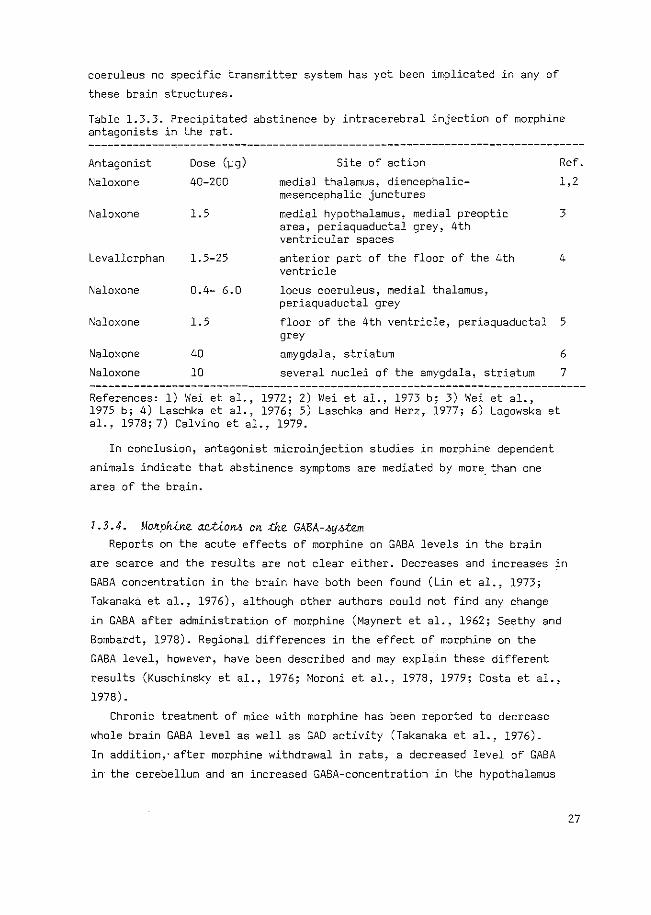
Table 1.3.3. Precipitated abstinence by intracerebral injection of morphine antagonists in the rat.
Antagonist
Naloxone
Dose (~g)
40-200
Site of action Ref.
medial thalamus, diencephalicmesencephalic junctures
1,2
Naloxone 1.5
Levallorphan 1.5-25
Naloxone 0.4- 6.0
medial hypothalamus, medial preoptic area, periaquaductal grey, 4th ventricular spaces
anterior part of the floor of the 4th ventricle
locus coeruleus, medial thalamus, periaquaductal grey
3
4
Naloxone 1.5 floor of the 4th ventricle, periaquaductal 5 grey
Naloxone
Naloxone
40
10
amygdala, striatum 6
several nuclei of the amygdala, striatum 7
References: 1) Wei et al., 1972; 2) Wei et al., 1973 b; 3) Wei et al., 1975 b; 4) Laschka et al., 1976; 5) Laschka and Herz, 1977; 6) Lagowska et al., 1978; 7) Calvina et al., 1979.
In conclusion, antagonist microinjection studies in morphine dependent
animals indicate that abstinence symptoms are mediated by more_ than one
area of the brain.
1.3.4. Mo~ph~ne actiono on the GABA-~y~em Reports on the acute effects of morphine on GABA levels in the brain
are scarce and the results are not clear either. Decreases and increases in
GABA concentration in the brain have both been found (Lin et al., 1973;
Takanaka et al., 1976), although other authors could not find any change
in GABA after administration of morphine (Maynert et al., 1962; Seethy and
Bombardt, 1978). Regional differences in the effect of morphine on the
GABA level, however, have been described and may explain these different
results (Kuschinsky et al., 1976; Moroni et al., 1978, 1979; Costa et al.,
1978).
Chronic treatment of mice with morphine has been reported to decrease
whole brain GABA level as well as GAD activity (Takanaka et al., 1976).
In addition,- after morphine withdrawal in rats, a decreased level of GABA
in the cerebellum and an increased GABA-concentration in the hypothalamus
27
has been observed (Lin et a1., 1973; Tzeng and Ho, 1978; Volicer et al.,
1977).
With respect to the GABA shunt enzymes, only minor effects of morphine
on GABA-T have been reported, whereas a decrease in the activity of GAD is
found after chronic morphine administration (Tzeng and Ho, 1978), possibly
due to a decreased affinity of this enzyme for PLP. This decrease can be
reversed by withdrawal of morphine or by precipitating abstinence with
naloxone (Ho and Gilliland, 1979).
The interaction between morphine and GABA has also been studied at the
cellular level. Morphine has no influence on the inhibitory effect of
exogenously applied GABA in the hippocampus (Segal, 1977): However, morphine
was reported to decrease the inhibitory effects of putative GABA-ergic
pathways in this area, presumably via a presynaptic mechanism (Corrigall and
Linseman, 1980). The presence of a presynaptic effect of morphine on GAGA
neurons is supported by the fact that morphine inhibits the potassium-evoked
release of GABA in vivo and in v~o (Iwatsubo and Kondo. 1978; Van der
Heyden et al., 1980; Coutinho-Netto et al., 1980).
At the receptor level there also seems to be a relationship between
GABA and opiates, since GABA-antagonistic features have been ascribed to
naloxone, morphine, levorphanol and dextrorphan (Breuker et al., 1976;
Dingledine et al., 1977). These data are relevant only when high doses of
these compounds will be used (see Gruol et al., 1980).
Thus chronic administration of morphine appears to result in a decreased
GABA-ergic activity either by a decrease of the activity of GAD or by
inhibition of the release. Administration of naloxone or withdrawal of merphine
will then result in a relative increase in GABA-ergic activity.
In conclusion, the interaction of morphine with the GABA-ergic system
in vivo is complex, possibly due to regional differences. In most cases
morphine decreases GABA-ergic activity.
1.4. DPA-INDUCED QUASI-MORPHINE ABSTINENCE BEHAVIOUR
1.4.1. Methodh All experiments were performed on male albino Wistar rats (TNO, Zeist,
The Netherlands). At the start of the experiment rats were allowed to
habituate for at least 30 min in a transparant Macrolon cage (47x27xl5 em)
with sawdust bedding. Thereafter an intraperitoneal injection of DPA was
28
given (in most cases 300 mg/kg at a concentration of 100 mg/ml deionized
water). Each rat was continuously observed for the following 15 min, sub
divided into three periods of 5 min, and its locomotor activity monitored
using a Varimex activity meter, equipped for alternating measurement of
horizontal and vertical activity every 10 sec. The following signs were
checked as absent, mild or marked: salivation, rhinorrhea, lacrimation,
urination, diarrhea, penile erection and ejaculation, ptosis, teeth
chattering, swallowing, tremor, hunchback posture, piloerection, irritability
to handling and reaction on poking. Other signs were counted as quantitative
events: escape digging, body shakes, head shakes, foreleg shakes and yawning.
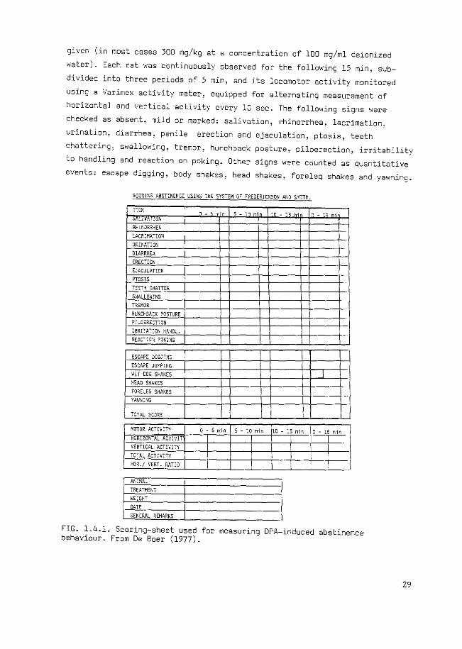
SCORIXG ABST:NENCE USING THE SYSTEM OF FREDERICKSON AND SM:TH.
lTEX
' RHINORRKEA
'-ACR:MAT:ON
URINATION
DIARRKEA
ERECTION
E~ACULATION
PTOSIS
TEETH CHATTER
SWALLOWING
TREMOR
HL:NCHBACK POSTURE
P:LOERECT:ON
IRRITATION HANOL.
REACTIOX POW!G
ESCAPE OIGGTNG
ESCAPE JUMPING
\JET DOG SHAKES
HEAD SHAKES
FORELEG S,~AKES
YAWNING
707AL SCORE
MOTOR ACTIV:TY
HORIZONTAL ACTIVIT
VERTICAL ACTIVITY
TOTAL ACT:VXTY
HOR./ VERT. RATIO
AN!XAL
TREATME~T
WEIGHT
DATE
GENERAL REMARKS
' - ' ., ' - ; ' ·n - 1 ~ - ; -
.1
0 • 5 min 5-lOmin 10-15min !o-lSmin
FIG. 1.4.1. Scoring-sheet used for measuring DPA-induced abstinence behaviour. From De Boer (1977).
29
From this checklist (Fig. 1.4.1) of behavioural symptoms described by
Frederickson and Smits (1973), an abstinence score was calculated,
according to these authors, in which the checked signs received 0, 2 or 4
when they were absent, mild or marked, respectively, and the counted signs
received 0, 2, 4, 6, 8, or 10 when they occurred 2-5, 6-10, 11-20, 21-40
or more than 40 times per 15 min period, respectively. Table 1.4.1 gives a
characterization of the DPA-induced behaviour. Not all signs of morphine
abstinence behaviour occur after injection of DPA, but the signs present
after this treatment can be summarized as 11 central signsn (Frederickson,l975).
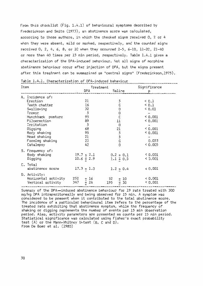
Table 1.4.1. Characterization of DPA-induced behaviour -------------------------------------------------------------------------------Item Treatment Significance
--------------------------------~~-----------~~l~~=------------£---~---------A. Incidence of:
Erection 21 5 < 0.1 Teeth chatter 16 0 < 0.1 Swallowing 32 0 < 0.01 Tremor 5 0 Hunchback posture 95 0 < 0.001 Piloerection 89 11 < 0. 001 Irritation 5 0 Digging 68 21 < 0.001 Body shaking 95 5 < 0.001 Head shaking 21 5 Foreleg shaking 21 0 0.053 Catalepsy 42 0 < 0. 005
B. Frequency of: Body shaking 19.7 + 2.1 0.2 + 0.1 < 0.001 Digging 10.6 :; 2.9 1.1 + 0.5 < 0.001
c. Total abstinence score 17.3 + 1.0 1.2+ 0.4 < 0. 001
D. Activity: Horizontal activity 252 :': 16 52 + 10 < 0.001 Vertical activity 347 :': 24 195 + 30 < 0. 001
------------------------------------------------------------------------------Summary of the DPA--induced abstinence behaviour for 19 rats treated with 300 mg/kg DPA intraperitoneally and being observed for 15 min. A symptom was considered to be present when it contributed to the total abstinence score. The incidence of a particular behavioural item refers to the percentage of the treated rats exhibiting that abstinence symptom, while the frequency of shaking or digging represents the number of events per 15 min observation period. Also, activity parameters are presented as counts per 15 min period. Statistical significance was calculated using Fisher's exact probability test (A) or the Mann-Whitney U-test (B, C and D). From De Boer et al. (1980)
30
1.4.2. The ko£e ofi GABA In this section the behavioural syndrome induced by DPA will be described
with respect to the pharmacological aspects, summarizing the results of
De Boer (1977).
A role for GABA in this behaviour is suggested by the action of DPA to
increase the concentration of GABA in the brain (see section 1.2.6) and by
the suppression of the behaviour by treatment with subconvulsive doses of
the GABA antagonists bicuculline (De Boer et al., 1977) and picrotoxin
(De Boer et al .. 1980). The GABA synthesis inhibitor, 3-mercaptopropionic
acid, has also been found to antagonize DPA-induced behaviour, while
strychnine, a convulsant glycine antagonist, has no effect. These data
indicate that the DPA-induced behaviour may be a correlate of increased
GABA-ergic activity (De Boer et al., 1980).
This behavioural syndrome has also been compared with behavioural effects
obtained after injection of the GASA-T inhibitor aminooxyacetic acid (AOAA).
Administration of AOAA does not induce abstinence behaviour, although this
treatment is known to enhance the GABA concentration in the brain (Wallach,
1961). Moreover, DPA-induced abstinence behaviour is suppressed in AOAA
pretreated rats, indicating that GABA-T inhibition counteracts rather than
potentiates DPA-induced abstinence behaviour.
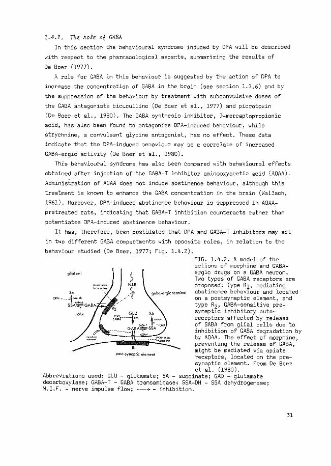
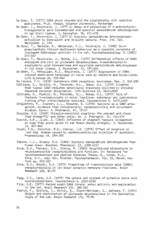
It has, therefore, been postUlated that DPA and GABA-T inhibitors may act
in twa different GABA compartments with opposite roles, in relation to the
behaviour studied (De Boer, 1977; Fig. 1.4.2).
glial cell ) N.I.F.
___ ), , , '0' gcbc-ergic terminal
,, pest-synoptic element
FIG. 1.4.2. A model of the actions of morphine and GABAergic drugs on a GABA neuron. Two types of GABA receptors are proposed: Type R1, mediating abstinence behaviour and located on a postsynaptic element, and type R2, GAGA-sensitive presynaptlc inhibitory autoreceptors affected by release of GABA from glial cells due to inhibition of GABA degradation by by AOAA. The effect of morphine, preventing the release of GABA, might be mediated via opiate receptors, located on the pre-synaptic element. From De Boer et al. (1980).
Abbreviations used: GLU - glutamate; SA - succinate; GAD - glutamate decarboxylase; GABA-T- GABA transami~ase; SSA-DH - SSA dehydrogenase; N.I.F. -nerve impulse flow; ---4- inhibition.
31
Accordingly, DPA increased GABA concentration only in nerve endings,
initiating quasi-abStinence behaviour, while GABA-T inhibitors will mainly
prevent GABA degradation in glial cells (see section 1.2.6). Consequently,
GABA, leaking or released from glial cells, may act on presynaptic GABA
receptors. This effect on presynaptic GABA receptors will cause a decreased
GABA-ergic activity impairing quasi-abstinence behaviour. The specificity
of DPA-induced quasi-morphine abstinence behaviour as a model for true
morphine abstinence behaviour has been discussed in section 1.3.2.
In conclusion, DPA-induced behaviour can be considered as a possible
correlate of GABA-ergic activity and it is a specific quasi-morphine
abstinence syndrome.
l . 4 • 3 • Ai.m o fi cth,W cth ru.,,W
In the previous section it was suggested that DPA induces quasi-morphine
abstinence behaviour via an increased stimulation of GABA receptors. It is
probable that this increased GABA-ergic stimulation occurs via inhibition of
the degradation of GABA (section 1.2.6). Since the mechanism by which DPA
increases the GABA concentration had not been clearly characterized, it was
our aim to study this mechanism and to examine its possible contribution to
explain the induction of quasi-morphine abstinence behaviour.
The behaviour observed after administration of DPA may throw some light
on the relation between GABA and morphine-abstinence behaviour. It enabled us
to study the involvement of particular GABA-ergic systems in the brain in the
induction of abstinence behaviour.
In brief, the aim of our research was to characterize the mechanism
responsible for the behaviour evoked by DPA, to locate its site of action, and
to obtain, in this way, a deeper understanding of possible mechanisms in
volved in morphine abstinence behaviour.
In this thesis experiments are described to specify the role of the GABA
ergic system in the mechanism of action of DPA (chapter II). Using analogues
of DPA we then studied the involvement of this mechanism of action in the
quasi-abstinence behaviour (chapter III). The discrepancy between the time
course of the behavioural effects of DPA and of its effect on the level of
GABA in brain, known from the literature, was studied (chapter IV). Finally,
the involvement of several GABA-containing brain structures in the DPA
induced behaviour was studied in order to characterize the site of action
of DPA in evoking this behaviour (chapters V and VI).
32
REFERENCES
Abe, M., Matsuda, M. (1976) The role of Y-aminobutyrate metabolism in the mechanism of convulsions. Jikeikai Med. J. 23, 245-253
Abe, M., Matsuda, M. (1977) y-Aminobutyric acid metabolism in subcellular particles of mouse brain and its relationship to convulsions. J. Biochem. Tokyo, 82, 195-200
Aghajanian, G.K. (1978) Tolerance of locus coeruleus neurons to morphine and suppression of withdrawal response by clonidine. Nature 276, 186-188
Albers, R.W., Koval, G.J. (1961) Succinic semialdehyde dehydrogenase: purification and properties of the enzyme from monkey brain. Biochim. Biophys. Acta 52, 29-35
Anden, N.-E., Grabowska-Anden, M., Wachtel, H. (1978) Effects of GABA and GABA-like drugs on the brain dopamine and on the motor activity of rats. In: GABA-Neurotransmitters. Pharmacochemical. Biochemical and Pharmacological aspects (Krogsgaard-Larsen, P., Scheel-KrUger, J., Kofod, H., eds) Munksgaard, Copenhagen, pp. 135-146
Anlezark, G., Collins, J., Meldrum, B. (1978) GABA-agonists and audiogenic seizures. Neurosci. Lett. 7. 337-340
Anlezark, G.M., Horton, R.W., Meidrum, B.S., Sawaya, M.C.B. (1976) Anticonvulsant action of ethanolamine-0-sulphate and di-n-propylacetate and the metabolism of y-aminobutyric acid (GABA) in mice with audiogenic seizures. Biochem. Pharmacal. 25, 413-417
Arnt, J., Scheel-KrUger, J. (1979) GABA in the ventral tegmental area: differential regional effects on locomotion,aggression and food intake after microinjection of GABA agonists and antagonists. Life Sciences 25, 1351-1360
Awapara, J., Landua, A.J., Fuerst, R., Seale, B. (1950) Free Y-aminobutyric acid in brain. J. Sial. Chern. 187, 35-39
Balazs, R., Cremer, J.E. (eds) (1972) Metabolic compartmentation in the brain. MacMillan, London 383 pp.
Balazs, R., Dahl, D., Harwood, J.R. (1966) Subcellular distribution of enzymes of glutamate metabolism in rat brain. J. Neurochem. 13, 897-895
Balazs, R., Machiyama, Y., Patel, A.J. (1972 a) Compartmentation and the metabolism of y-aminobutyrate. In: Metabolic compartmentation in the brain (Balazs, R., Cremer, J.E·., eds) MacMillan, London, pp. 57-70
Balazs, R., Patel, A.J., Richter, D. (1972 b) Metabolic compartments in the brain: their properties and relation to morphological structures. In: Metabolic compartmentation in the brain (Balazs, R., Cremer, J.E., eds) MacMillan, London~ pp.l67-184
Balcom, G.J., Lenox, R.H., Meyerhoff, J.L (1975) Regional y-aminobutyric acid levels in rat brain determined after microwave fixation. J. Neurochem. 24, 609-613
Barber, R., Saito, K. (1976) Light microscopic visualization of GAD and GABA-T in immunocytochemical preparations of rodent CNS. In: GABA in nervous system function (Roberts, E. , Chase, T. N. , Tower, D. B., eds) Raven, New York, pp.ll3-132
Baxter, C.F. (1976) Some recent advances in studies of GABA metabolism and compartmentation. In: GABA in nervous system function (Roberts, E., Chase, T.N., Tower, D.B., eds)Raven, New York, pp-61-87
Bay6n, A., Possani, L.D., Rode, G., Tapia, R. (1978) Kinetics of brain glutamate decarboxylase. Dead-end and product inhibition studies. J. Neurochem. 30, 1629-1631
Bedard, P., Pycock, C. (l977) 1Wet dog 1 shake behaviour in the rat: a possible
33
quantitative model of central 5-hydroxytryptamine activity. Neuropharmacology 16, 663-670
Ben-Ari, Y., Lagowska, J., Tremblay, E., LeGal La Salle, G. (1979) A new model of focal status epilepticus: intra-amygdaloid application of kainic acid elicits repetitive secondarily generalized convulsive seizures. Brain Research 163. 176-179
Benton, D., Rick, J.T. (1976) The effect of increased brain GABA produced by amino-oxyacetic acid on arousal in rats. Psychopharmacology 49, 85-89
Berl, S., Clarke, D.O., Schneider, D. (eds) (1975) Metabolic compartmentation and Neurotransmission: Relation to structure and function in brain. Plenum, New York, 721 pp.
Bernheimer, H., Horniekiewicz, 0. (1962) Das Verhalten einiger Enzyme im Gehirn normaler und Parkinson-kranker Menschen. ,"Jaunyn-Schmied. Arch. Exp. Path. Pharmakol. 243. 295-296
Bhargava, H.N. (1980) The effects of thyrotropin-releasing hormone on the central nervous system responses to chronic morphine administration. Psychopharmacology 68, 185-189
Bird, E.D., Mackay, A.V.P., Rayner, C.,~., Iversen, L.L. (1973) Reduced glutamic-acid-decarboxylase activity of po~~-mok~em brain in Huntington's chorea. Lancet 1, 1090-1092
Biswas, B., Carlsson, A. (1978) Effect of intraperitoneally administered GABA on the locomotor activity in mice. Psychopharmacology 59, 91-94
818sig, J., Herz, A., Reinhold, K., Zieglg8nsberger, S. (1973) Development of physical dependence on morphine in respect to time and dosage and quantification of the precipitated withdrawal syndrome in rats. Psychopharmacologia 33, 19-38
Blindermann, J.M., Maitre, M., Ossola, L., Mandel, P. (1978) Purification and some properties of L-glutamate decarboxylase from human brain. Europ. J. Biochem. 86, 143-152
Bloch-Tardy, M., Rolland, M., Gonnard, P. (1974) Pig brain 4-aminobutyrate 2-ketoglutarate transaminase. Purification, kinetics and physical properties. Biochimie 56, 823-832
Bloom, F.E., Segal, D., Ling, N., Guillemin, R. (1976) Endorphins: profound behavioral effects in rats suggest new etiological factors in mental illness. Science 194, 630-632
Blume, H.W., Lamour, Y., Arnauld, E., Layton, B.S., Renaud, L.P. (1979) Sodium di-n-propylacetate (valproate) action on single neurons in rat cerebral cortex and hippocampus. Brain Research 171, 182-185
Bonnet, K.A., Gusik, S.A., Sunshine, A.G. (1978) Multiple opiate receptors reflected in region specific alterations in brain cyclic nucleotides. In: Characteristics and function of opioids (Van Ree, J.M., Terenius, L., eds) Elsevier/North Holland, Amsterdam, pp.453-464
Braganca, S.M., Faulkner, P., Quastel, J.H. (1953) Effects of inhibitors of glutamine synthesis on inhibition of acetylcholine synthesis in brain slices by ammonium ions. Biochim. Biophys. Acta 10, 83-88
Brennan, M.J.W., Cantrill, R.C., Oldfield, M., Krogsgaard-Larsen, P. (1981) Inhibition of y-aminobutyric acid release by y-aminobutyric acid agonist drugs. Malec. Pharmacal. 19, 27-30
Breuker, E., Dingledine, R., Iversen, L.L. (1976) Evidence for naloxone and opiates as GABA antagonists. Br. J. Pharmacal. 58, 458P
Butt, ~.M., Collier, H.O.J., Cuthbert, N.J., Francis, D.L., Saeed, S.A. (1979) Mechanism of quasi-morphine withdrawal behaviour induced by methylxanthines. Europ. J. Pharmacal. 53, 375-378
34
Calvina, B., Lagowska, J., Ben-Ari, Y. (1979) Morphine withdrawal syndrome: differential participation of structures located within the amygdaloid complex and striatum of the rat. Brain Research 177, 19-34
Cash, C., Cieselski, L., Maitre, M., Mandel, P. (1975) Purification de la semi-aldehyde succinique deshydrogenase de cerveau de rat et etude de son inhibition par des acides gras ramifies. C.R. Soc. Biol. (Paris) 169, 884-887
Cash, C., Cieselski, L., Maitre, M., Mandel, P. (1978) Purification and properties of rat brain succinic semialdehyde dehydrogenase. Biochimie 59, 257-268
Chan-Palay, V., Wu, J.-Y., Palay, S.L. (1979) Immunocytochemical localization of Y-aminobutyric acid transaminase at cellular and ultrastructural levels. Proc. Nat. Acad. Sci. U.S.A. 76, 2067-2071
Cieselski, L., Maitre, M., Cash, C., Mandel, P. (1975) Regional distribution in brain and effect on cerebral mitochondra1 respiration of the anticonvulsive drug n-dipropy1acetate. Biochem. Pharmacal. 24, 1055-1058
Collier, H.O.J. (1965) A general theory of the genesis of drug dependence by induction of receptors. Nature 205, 181-182
Collier, H.O.J. (1974) The concept of the quasi-abstinence effect and its use in the investigation of dependence mechanisms. Pharmacology 11, 58-61
Collier, H.O.J. (1978) Biochemical theories of opioid dependence: An analysis. Proc. Eur. Soc. ~eurochem. 1, 374-385
Collier, H.O.J. (1980) Cellular site of opiate dependence. ~ature 283, 625-629
Collier, H.O.J., Francis, D.L. (1975) Morphine abstinence is associated with increased brain cyclic AMP. Nature 255, 159-162
Collier, H.O.J., Francis, D.L., Henderson, G., Schneider, C. (1974) Quasimorphine abstinence syndrome. Nature 249, 471-473
Collier, H.O.J., Francis, D.L., Schneider, C. (1972) Modification of morphine withdrawal by drugs interacting with humoral mechanisms: some contradictions and their interpretation. Nature 237, 220-223
Corrigall, W.A., Linseman, M.A. (1980) A specific effect of morphine on evoked activity in the rat hippocampal slice. Brain Research 192, 227-238
Costa, E., Fratta, W., Hong, J.S., Moroni, F., Yang, H.-Y.T. (1978) Interactions between enkephalinergic and other neuronal systems. In: The endorphins. Adv. Biochem. Psychopharmacol. Vol. 18 (Costa, E., Trabucchi, M.,eds) Raven, New York, pp.lll-123
Cott, J.M., Carlsson, A., Engel, J., Lindquist, M. (1976) Suppression of ethanol-induced locomotor stimulation by GABA-like drugs. NaunynSchmied. Arch. Pharmacal. 295, 203-209
Coutinho-Netto, J., Abdul-Ghani, A.S., Bradford, H.F. (1980) Suppression of evoked and spontaneous release of neurotransmitters ~n v~vo by morphine. Biocheffi. Pharmacal. 29, 2777-2780
Cowan, A. (1981) RX 336-M, a new chemical tool in the analysis of the quasi-morphine withdrawal syndrome. Fed. Proc. 40, 1497-1501
Cowan, A., Watson, T. (1978) Lysergic acid diethylamide antagonizes shaking induced in rats by five chemically different compounds. Psychopharmacology 57, 43-46
Crawley, J.N., Laverty, R., Roth, R.H. (1979) Clonidine reversal of increased norepinephrine metabolite levels during morphine withdrawal. Europ. J. Pharmacal. 57, 247-250
Czajka, R. (1978) Central effects of endogenous and exogenous GABA. Acta Physiol. Pol. 29, 193-205
35
De Boer, T. (1977) ·GABA shunt enzymes and the relationship with morphine abstinence. Ph.D. Thesis, Erasmus University, Rotterdam
De Boer, T., Bruinvels, J. (1977 a) Assay and properties of 4-aminobutyric-2-oxoglutaric acid transaminase and succinic semialdehyde dehydrogenase in rat brain tissue. J. ~eurochem. 28, 471-478
De Boer, T., Bruinvels, J. (1977 b) Succinic semialdehyde dehydrogenase: activation by monovalent and divalent cations. Proc. Int. Soc. Neurochem. 6. 524
De Boer, T., Bartels, K., Metselaar, H.J., Bruinvels, J. (1980) Di-npropylacetate induced abstinence behaviour as a possible correlate of increased GABA-ergic activity in the rat. Psychopharmacology 71 (1980) 257-267
De Boer, T., Bruinvels, J., Bonta, I.L. (1979) Differential effects of GABA analogues and zinc on glutamate decarboxylase, 4-aminobutyric-2-oxoglutaric acid transaminase and succinate semialdehyde dehydrogenase in rat brain tissue. J. Neurochem. 33, 597-601
De Boer, T., Metselaar, H.J., Bruinvels, J. (1977) Suppression of GABAinduced abstinence behaviour in naive rats by morphine and bicuculline. Life Sciences 20. 933-942
De Feudis, F.V. (1978) Vertebrate GABA receptors. Neurochem. Res. 3, 263-280 DiChiara, G., Morelli, M., Porceddu, M.L., Gessa, G.L (1978) Evidence
that nigral GABA mediates behavioural responses elicited by striatal dopamine receptor stimulation. Life Sciences 23, 2045-2052
DiChiara, G., Morelli, M., Porceddu, M.L., Gessa, G.L. (1979) Role of thalamic Y-aminobutyrate in motor functions: catalepsy and ipsiversive turning after intra-thalamic muscimol. Neuroscience 4, 1453-1465
Dingledine, R., Iversen, L.L., Breuker, E. (1978) Naloxone as a GABA antagonist: evidence from iontophoretic, receptorbinding and convulsant studies. Europ. J. Pharmacal. 47, 19-27
Douglas, W.W. (1968) Stimulus-secretion coupling: the concept and clues from chromaffin and other cells. Sr. J. Pharmacal. 34. 451-474
Dravid, A.R., Jilek, J. (1965) Influence of stagnant hypoxia (oligaemia) on some free amino acids in rat brain during ontogeny. J. ~eurochem. 12, 837-843
Drust, E.G., Sloviter, R.S., Connor, J.D. (1979) Effect of morphine on 'wet dog' shakes caused by cerebroventricular injection of serotonin. Pharmacology 18, 299-305
Embree, L.J., Albers, R.W. (1964) Succinic semialdehyde dehydrogenase from human brain. Biochem. Pharmacal. 13, 1209-1217
Enna, S.J., Ferkany, J.W., Strong, R. (1980) Drug-induced alterations in neurotransmitter receptorbinding and function. In: Receptors for neurotransmitters and peptide hormones (Pepeu, G., Kuhar, M.J., Enna, S.J., eds) Adv. Biochem. Psychopharmacol. Vol. 21, Raven, New York pp. 253-263
Enna, S.J., Snyder, S.H. (1975) Properties of y-aminobutyric acid (GABA) receptor-binding in rat brain synaptic membrane fractions. Brain Research 100, 81-97
Fagg, G.E., Lane, J.D. (1979) The uptake and release of putative amino acid neurotransmitters. Neuroscience 4, 1015-1036
File, S.E. (1977) Raised brain GABA levels, motor activity and exploration in the rat. Brain Research 131, 180-183
Fonnum, F., Grofova, I., Rinvik, E., Storm-Mathisen, J., Walberg, F. (1974) Origin and distribution of glutamate decarboxylase in the substantia nigra of the cat. Brain Research 171, 77-92
36
Fonnum, F., Walberg, F. (1973 a) An estimation of the concentration of Yaminobutyric acid and glutamate decarboxylase in the inhibitory Purkinje axon terminals in the cat. Brain Research 54, 115-127
Fonnum, F., Walberg, F. (1973 b) The concentration of GABA within inhibitory nerve terminals. Brain Research 62. 577-579
Fozard, J.R., Palfreyman, M.G. (1979) Metoclopramide antagonism of 5-hydroxytryptophan-induced ~'wet dog" shake behaviour in the rat. ~'aunyn-Schmied. Arch. Pharmacal. 307, 135-142
Francis, D.L., Cuthbert, N.J., Dinneen, L.C., Schneider, C., Collier, H.O.J. (1976) Methylxanthine-accelerated opiate dependence in the rat. In: Opiates and endogenous opioid peptides. (Kosterlitz, H.W. ed) Elsevier/ North Holland, Amsterdam pp.l77-184
Francis, D.L., Cuthbert, ~.J., Saeed, S.A., Butt, N.M., Collier, H.O.J. (1978) Meaning of quasi-withdrawal phenomena for the cellular mechanism of abstinence. In: Characteristics and function of opioids. (Van Ree, J.M., Terenius, L., eds) Elsevier/~orth Holland, Amsterdam pp. 37-49
Francis, D.L., Roy, A.C., Collier, H.O.J. (1975) Morphine abstinence and quasi-abstinence effects after phosphodiesterase inhibitors and naloxone. Life Sciences 16. 1901-1906
Frederickson, R.C.A. (1975) Morphine withdrawal response and central cholinergic activity. Nature 257, 131-132
Frederickson, R.C.A., Smits, S.E. (1973) Time course of dependence and tolerance development in rats treated with 'slow release' morphine suspensions. Res. Commun. Chern. Path. Pharmacal. 5, 867-870
Freed, W.J .. Michaelis, E.K. (1976) Effects of intracisternal GABA or glutamic acid upon behavioural activity in the rat. Pharmacal. Biochem. Behav. 5, ll-14
Gale, K., Iadarola, M.J. (1980) Seizure protection and increased nerveterminal GABA: delayed effects of GABA-transaminase inhibition. Science 208. 288-291
Garfinkel, D. (1966) A simulation study of the metabolism and compartmentation in brain of glutamate, aspartate, the Krebs cycle and related metabolites. J. Biol. Chern. 241, 3918-3929
Gellert, V.F., Holtzman, S.G. (1978) Development and maintenance of morphine tolerance and dependence in the rat by scheduled access to morphine drinking solutions. J. Pharmacal. Exp. Ther. 205, 536-546
Gent, J.P., Phillips, N.J. (1980) Sodium di-n-propylacetate (valproate) potentiates responses to GABA and muscimol on single central neurons. Brain Research 197, 275-278
Gerber, J.C., Hare, T.A. (1980) GABA in peripheral tissues: presence and actions in endocrine pancreatic function. In: GABA-Neurotransmission (Lal, H. ed) Brain Res. Bull.S. Suppl. 2 pp. 341-346
Gerig, J.T., Kwock, L. (1979) Inhibition of bacterial glutamate decarboxylase by tricarboxylic acid cycle intermediates. FEBS Lett. 105, 155-157
Gibson, A.C. (1977) Sodium valproate and tardive dyskinesia. Br. J. Psychiat. 133, 82.
Gispen, W.H., Wiegant, V.M., Bradbury, A.F., Hulme, E.C., Smyth, D.G., Snell, C.R., De Wied, D. (1976) Induction of excessive grooming in the rat by fragments of lipotropin. Nature 276, 794-795
Glick, S.D., Crane, L.A. (1978) Opiate-like and abstinence-like effects of intracerebral histamine administration in rats. Nature 273, 547-549
Godin, Y., Heiner, L., Mark, J., Mandel, P. (1969) Effects of di-npropylacetate, an anticonvulsive compound, on GABA metabolism. J. ~eurochem. 16, 869-873
Gruol, D.L., Barker, J.L., Smith, T.G. (1980) Naloxone antagonism of GABA-
37
evoked membrane polarizations in cultured mouse spinal cord neurons. Brain Research 198, 323-332
Gubler~ C.J., Adams, B.L., Hammond, B., Yuan, E.C., Guo, S.M., Bennion, M. (1974) Effect of thiamine deprivation and thiamine antagonists on the level of y-aminobutyric acid and on 2-oxoglutarate metabolism in rat brain. J. Neurochem. 22, 831-836
Harvey, P.K.P., Bradford, H.F., Davison, A.N. (1975) The inhibitory of sodium n-dipropylacetate on the degradative enzymes of the GABA shunt. FEBS Lett. 52, 251-254
Hayashi, T. (1959) The inhibitory action of S-hydroxy-Y-aminobutyric acid upon the seizure following stimulation of the motor cortex of the dog. J. Physiol. (London) 145, 570-578
Hayashi, T., Negishi, K. (1979) Suppression of retinal spike discharge by dipropylacetate (Depakene): a possible involvement of GABA. Brain Research 175, 271-278
Henriksen, S.J., Bloom, F.E., McCoy, F., Ling, N., Guillemin, R. (1978) S-Endorphin induces non-convulsive lumbic seizures. Proc. Nat. Acad. Sci. U.S.A. 75, 5221-5225
Hertz, L. (1979) Functional interactions between neurons and astrocytes. I. Turnover and metabolism of putative amino acid transmitters. Prog. i\'eurobiol. 13, 277-323
Ho, I.K., Gilliland.T.(l979) Effects of acute and continuous morphine administration. on the affinity of glutamic acid decarboxylase for pyridoxal 5 1 -phosphate. Biochem. Pharmacal. 28, 355-360
Holtz, P., Palm, D. (1964) Pharmacological aspects of Vitamin 86. Pharmacal. Reviews 16, 113-178
Horton, R.W., Collins, J.F., Anlezark, G.M., Meldrum, B.S. (1979) Convulsant and anticonvulsant actions in DBA/2 mice of compounds blocking the reuptake of GABA. Europ. J. Pharmacal. 59, 75-83
Iadarola, M.J., Gale, K. (1979) Dissociation between drug-induced increases in nerve terminal and non-nerve terminal pools of GABA ~n v~vo. Europ. J. Pharmacal. 59, 125-129
Iadarola, M.J., Raines, A., Gale, K. (1979) Differential effects of di-npropylacetate and amino-oxyacetic acid on Y-aminobutyric acid levels in discrete areas of rat brain. J. Neurochem. 33, 1119-1123 ·
Iversen, L.L. (1971) Role of transmitter uptake mechanisms in synaptic neurotransmission. Sr. J. Pharmacal. 41, 571-591
Iversen, L.L., Kelly, J.S. (1975) Uptake and metabolism of Y-aminobutyric acid by neurones and glial cells. Biochem. Pharmacal. 24, 933-938
Iwatsubo, K., Kondo, Y. (1978) Inhibitory effect of morphine on the release of preloaded 3H-GABA from rat substantia nigra in response to the stimulation of caudate nucleus and globus pallidus. In: Characteristics and function of opioids (Van Ree, J.M., Terenius, L., eds) Elsevier/ North Holland, Amsterdam pp. 357-358
Jackson, I.M.D., Reichlin, S. (1974) Thyrotropin-releasing hormone (TRH): distribution in hypothalamic and extrahypothalamic brain tissues of mammalian and submammalian chordates. Endocrinology, 95, 854-862
Jacquet, Y.F. (1978) Opiate effects after adrenocorticotropin or S-endorphin injection in the periaqueductal grey matter of rats. Science 201, 1032-1034
Jaffe, J.H. (1980) Drug addiction and drug abuse. In: The pharmacological basis of therapeutics. (Gilman, A.G., Goodman, L.S., Gilman, A., eds) MacMillan, New York pp. 535-584
38
Jaffe, J.H., Sharpless, S.K. (1968) Pharmacological denervation supersensitivity in the central nervous system: A theory of physical dependence. In: The addictive states. (Wikler, A. ed) Williams and Wilkins Camp., Baltimore, pp. 226-246
Jahn, U., Mixich, G. (1976) Wet dog shake behaviour in normal rats, elicited by benzylideneaminooxycarboxic acid derivatives. Psychopharmacologia 46, 191-196
John, R.A., Fowler, L.J. (1976) Kinetic and spectral properties of rabbit brain 4-aminobutyrate aminotransferase. Biochem. J. 155, 645-651
Kalivas, P.W., Horita, A. (1980) Thyrotropin-releasing hormone: neurogenesis of actions in the pentobarbital narcotized rat. J. Pharmacal. Exp. Ther. 212, 203-210
Kammeraat, C., Veldstra, H. (1967) Characterization of succinate semialdehyde dehydrogenase from rat brain. Biochim. Biophys. Acta 151, 1-10
Katz, R.I., Chase, T.N., Kopin, I.J. (1969) Effects of ions on stimulus induced release of amino acids from mammalian brain slices. J. Neurochem. 16, 961-967
Kerwin, R.W., Olpe, H.-R. (1980) The effect of sodium valproate on single unit activity in the rat brain. Br. J. Pharmacal. 70, 76 P.
Kerwin, R.W., Olpe, H.-R., Schmutz, M. (1980) The effect of sodium di-npropylacetate on Y-aminobutyric acid-dependent inhibition in the rat cortex and substantia nigra in relation to its anticonvulsant activity. Br. J. Pharmacal. 71, 545-551
Kobayashi, K., Miyazawa, S., Endo, A. (1977) Isolation and inhibitory activity of gabaculine, a new potent inhibitor of Y-aminobutyrate aminotransferase produced by a streptomyces. FEBS Lett. 76, 207-210
Koob, G.F., Del Fiacco, M., Iversen, S.D. (1978) Spontaneous and amphetamineinduced behavior after bilateral injection of ethanol-0-sulphate into the substantia nigra. Brain Research 146, 313-323
Krnjevic, K. (1970) Glutamate and Y-aminobutyric acid in brain. N?ture 228, 119-224
Kukino, K., Deguchi, T. (1977) Effects of sodium dipropylacetate on y-aminobutyric acid and biogenic amines in rat brain. Chern. Pharmac. Bull. 25, 2257-2262
Kuriyama, K. (1976) Subcellular localization of the GABA system in brain. In: GABA in nervous system function. (Roberts, E., Chase, T.N., Tower, D.B., eds) Raven, New York pp. 187-196
Kuriyama, K., Roberts, E., Kakefuda, T. (1968 a) Association of theY-aminobutyric acid system with a synaptic vesicle fraction from mouse brain. Brain .Research 8, 132-152
Kuriyama, K., Roberts, E., Rubinstein, M.K. (1966) Elevation of y-aminobutyric acid in brain with amino-oxyacetic acid and susceptibility to convulsive seizures in mice: a quantitative re-evaluation. J. Neurochem. 15, 221-236
Kuriyama, K., Roberts, E., Vas, J. (1968 b) Some characteristics of binding of y-aminobutyric acid and acetylcholine to a synaptic vesicle fraction from mouse brain. Brain Research 9, 231-252
Kuschinsky, K., Bosse, A., NOring, A., Ulmar, G. (1976) GABA and dopamine metabolism in the nigra-striatal pathways of rats. Alterations after single administrations of narcotics and during withdrawal. In: Opiates and endogenous opioid peptides. (Kosterlitz, H.W. ed) Elsevier/North Holland, Amsterdam pp. 419-422
39
LaBella, F. S:, Havlicek, V .. Pinsky, C. (1979 a) Opiate-like excitatory effects of steroid sulfates and calcium-complexing agents given cerebroventricularly. Brain Research 160, 295-305
LaBella, F.S., Havlicek, V., Pinsky, C., Leybin, L. (1978) Opiate-like. naloxone reversible effects of androsterone sulfate in rats. Can. J. Physiol. Pharmacal. 56, 940-944
LaBella, F.S., Pinsky, C., Havlicek, V. (1976 b) Morphine-derivatives with diminished opiate receptor potency show enhanced central excitatory activity. Brain Research 174, 263-271
Lacelle, J.Y., Ferrandes, B., Eymard, P. (1978) Profile of anticonvulsant activity of sodium valproate. Role of GABA. In: Advances in Epileptology-1977. (Meinardi, H., Rowan, A.J., eds) Swets and Zeitlinger, Amsterdam pp. 162-167
Lagowska, J., Calvina, B., Ben-Ari, Y. (1978) Intra-amygdaloid application of naloxone elicits severe withdrawal signs in morphine-dependent rats. Neurosci. Lett. 8, 241-245
Lal, H., Colpaert, F.C., Laduron, P.(l975) Narcotic withdrawal-like mousejumping produced by amphetamine and L-DDPA. Europ. J. Pharmacal. 30, 113-116
Lanthorn, T., Isaacson, R.L. (1978) studies on kainate induced wet-dog shakes in the rat. Life Sciences 22, 171-178
Lanthorn, T., Smith, M.A., Isaacson, R.L. (1979) Wet-dog shaking in the rat: possible involvement of a kappa opiate receptor. Neuropharmacology 18, 743-745
Laschka, E., Herz, A. (1977) Sites of action of morphine involved in the development of physical dependence in rats III. Autoradiographic studies. Psychopharmacology 53, 33-37
Laschka, E., Teschemacher, Hj., Mehraein, P., Herz, A. (1976) Sites of action of morphine involved in the development of physical dependence in rats II. Morphine withdrawal precipitated by application of morphine antagonists into restricted parts of the ventricular system and by microinjection into various brain areas.Psychopharmacologia 46, 141-147
Laverty, R., Roth, R.H. (1980) Clonidine reverses the increased norepinephrine turnover during morphine withdrawal in rats. Brain Research 182, 482-485
Levi, G., Raiteri, M. (1978) Modulation of Y-aminobutyric acid transport in nerve endings: role of extracellular Y-aminobutyric acid and of cationic fluxes. Proc. Nat. Acad. Sci. U.S.A. 75. 2981-2985
Leybin, L., Pinsky, C.~ L3Bella, F.S., Havlicek, V., Rezek, M. (1976) Intraventricular Met -enkephalin causes unexpected lowering of pain threshold and narcotic withdrawal signs in rats. Nature 264, 458-459
Lin, S.C., Sutherland, V.C., Way, E.L. (1973) Brain amino acids in morphine tolerant and non-tolerant rats. Proc. West. Pharmacal. Soc. 16, 8-13
Llorens, C., Martres, M.P., Baudry, M., Schwartz, J.C. (1978) Hypersensitivity to noradrenaline in cortex after chronic morphine: relevance to tolerance and dependence. Nature 274, 603-605
Lust, W.O., Kupferberg, H.J., Yonekawa, W.D., Penry, J.K., Passoneau, J.V., Wheaton, A.B. (1978) Changes in brain metabolites induced by convulsants or electroshock: effects of anticonvulsant agents. Malec. Pharmacal. 14, 347-356
MacDonald, R.L., Bergey, G.K. (1979) Valproic acid augments GABA-mediated postsynaptic inhibition in cultured mammalian neurons. Brain Research 170, 558-562
Maitre, M., Cieselski, L., Cash, C., Mandel, P. (1975) Purification and studies on some properties of the 4-aminobutyrate:2-oxoglutarate transaminase from rat brain. Europ. J. Biochem. 52, 157-169
40
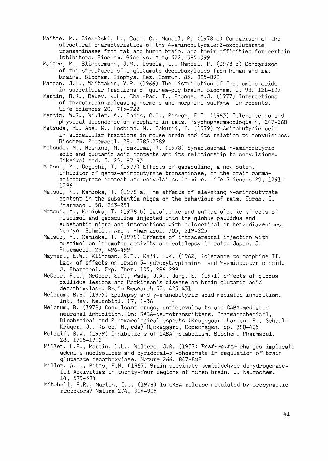
Maitre, M., Cieselski, L., Cash, C., Mandel, P. (1978 a) Comparison of the structural characteristics of the 4-aminobutyrate:2-oxoglutarate transaminases from rat and human brain, and their affinities for certain inhibitors. Biochem. Biophys. Acta 522, 385-399
Maitre, M., Blindermann, J.M., Ossola, L., Mandel, P. (1978 b) Comparison of the structures of L-glutamate decarboxylases from human and rat brains. Biochem. Biophys. Res. Commun. 85, 885-890
Mangan, J.L., Whittaker, V.P. (1966) The distribution of free amino acids in subcellular fractions of guinea-pig brain. Biochem. J. 98, 128-137
Martin, B.R., Dewey, W.L., Chau-Pam, T., Prange, A.J. (1977) Interactions of thyrotropin-releasing hormone and morphine sulfate in rodents. Life Sciences 20. 715-722
Martin, W.R., Wikler,, A., Eades, C.G., Pescor, F.T. (1963) Tolerance to and physical dependence on morphine in rats. Psychopharmacologia 4, 247-260
Matsuda, M., Abe, M., Hoshino, M., Sakurai, T. (1979) y-Aminobutyric acid in subcellular fractions in mouse brain and its relation to convulsions. Biochem. Pharmacal. 28. 2785-2789
Matsuda, M., Hoshino, M., S~kurai, T. (1978) Synaptosomal Y-aminobutyric acid and glutamic acid contents and its relationship to convulsions. Jikeikai Med. J. 25. 87-93
Matsui, Y., Deguchi, T. (1977) Effects of gabaculine, a new potent inhibitor of gamma-aminobutyrate transaminase, on the brain gammaaminqbutyrate content and convulsions in mice. Life Sciences 20, 1291-1296
Matsui, Y., Kamioka, T. (1978 a) The effects of elevating y-aminobutyrate content in the substantia nigra on the behaviour of rats. Europ. J. Pharmacal. 50, 243-251
Matsui, Y., Kamioka, T. (1978 b) Cataleptic and anticataleptic effects of muscimol and gabaculine injected into the globus pallidus and substantia nigra and interactions with haloperidol or benzodiazepines. Naunyn- Schmied. Arch. Pharmacal. 305, 219-225
Matsui, Y., Kamioka, T. (1979) Effects of intracerebral injection with muscimol on locomotor activity and catalepsy in rats. Japan. J. Pharmacal. 29, 496-499
Maynert, E.W., Klingman, G.I., Kaji, H.K. (1962) Tolerance to morphine II. Lack of effects on brain 5-hydroxytryptamine and y-aminobutyric acid. J. Pharmacal. Exp. Ther. 135. 296-299
McGeer, P.L., McGeer, E.G., Wada, J.A., Jung, E. (1971) Effects of globus pallidus lesions and Parkinson's disease on brain glutamic acid decarboxylase. Brain Research 32, 425-431
Meldrum, B.S. (1975) Epilepsy and y-aminobutyric acid mediated inhibition. Int. Rev. Neurobiol. 17, 1-36
Meldrum, B. (1978) Convulsant drugs, anticonvulsants and GABA-mediated neuronal inhibition. In: GABA-Neurotransmitters. Pharmacochemical, Biochemical and Pharmacological -aspects (Krogsgaard-Larsen, P., ScheelKri..iger, J., Kofod, H., eds) Munksgaard, Copenhagen, pp. 390-405
Metcalf, B.W. (1979) Inhibitions of GABA.metabolism. Biochem. Pharmacal. 28. 1705-1712
Miller, ·L.P., Martin, D.L., Walters, J.R. (1977) Po~t-mo~tem changes implicate adenine nucleotides and pyridoxal-5'-phosphate in regulation of brain glutamate decarboxylase. Nature 266, 847-848
Miller, A.L., Pitts, F.N. (1967) Brain succinate semialdehyde dehydrogenaseIII Activities in twenty-four regions of human brain. J. Neurochem. 14, 5797584
Mitchell, P.R., Martin, I.L. (1978) Is GABA release modulated by presynaptic receptors? Nature 274, 904-905
41
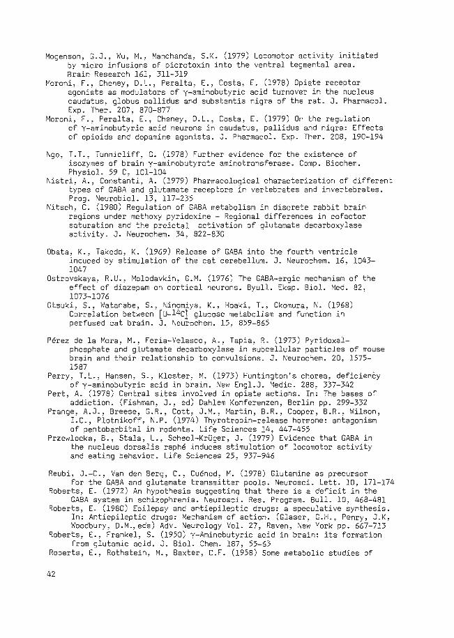
Mogensen, G.J., Wu, M.~ Manchanda, S.K. (1979) by micro infusions of picrotoxin into the Brain Research 161, 311-319
Moroni, F., Cheney, D.L., Peralta, E., Costa, agonists as modulators of y-aminobutyric caudatus, globus pallidus and substantia Exp. Ther. 207, 870-877
Locomotor activity initiated ventral tegmental area.
E. (1978) Opiate receptor acid turnover in the nucleus nigra of the rat. J. Pharmacal.
Moroni, F., Peralta, E., Cheney, D.L., of Y-aminobutyric acid neurons in of opioids and dopamine agonists.
Costa, E. (1979) On the regulation caudatus, pallidus and nigra: Effects J. Pharmacal. Exp. Ther. 208, 190-194
\1go, T. T., Tunnicliff, G. (1978) Further evidence for the existence of
isozymes of brain y-aminobutyrate aminotransferase. Camp. Biochem. Physiol. 59 C, 101-104
Nistri, A., Constanti, A. (1979) Pharmacological characterization of different types of GABA and glutamate receptors in vertebrates and invertebrates. Prog. Neurobiol. 13, 117-235
Nitsch, C. (1980) Regulation of GABA metabolism in discrete rabbit brain regions under methoxy pyridoxine - Regional differences in cofactor saturation and the preictal activation of glutamate decarboxylase activity. J. ~eurochem. 34, 822-830
Dbata, K., Takeda, K. (1969) Release of GABA into the fourth ventricle induced by stimulation of the cat cerebellum. J. Neurochem. 16, 1043-1047
Ostrovskaya, R.U., Molodavkin, G.M. (1976) The GABA-ergic mechanism of the effect of diazepam on cortical neurons. Byull. Eksp. Sial. Med. 82, 1073-1076
Otsuki, 5., Watanabe, S., Ninomiya, K., Hoaki, T., Okamura, N. (1968) Correlation between [u-14c] glucose metabolism and function in perfused cat brain. J. Neurochem. 15, 859-865
Perez de la Mora, M., Feria-Velasco, A., Tapia, R. (1973) Pyridoxalphosphate and glutamate decarboxylase in subcellular particles of mouse brain and their relationship to convulsions. J. Neurochem. 20, 1575-1587
Perry, T.L., Hansen, S., Kloster~ M. (1973) Huntington's chorea, deficienCy of y-aminobutyric acid in brain. New Engl.J. Medic. 288, 337-342
Pert, A. (1978) Central sites involved in opiate actions. In: The bases of addiction. (Fishman, J., ed) Dahlem Konferenzen, Berlin pp. 299-332
Prange, A.J., Breese, G.R., Cott, J.M., Martin, B.R., Cooper, B.R., Wilson, I.e., Plotnikoff, N.P. (1974) Thyrotropin-release hormone: antagonism of pentobarbital in rodents. Life Sciences 14, 447-455
Przewlocka, 8., Stala, L., Scheel-KrUger, J. (1979) Evidence that GABA in the nucleus dorsalis raphe induces stimulation of locomotor activity and eating behavior. Life Sciences 25, 937-946
Reubi, J.-C., Van den Berg, C., Cuenod, M. (1978) Glutamine as precursor for the GABA and glutamate transmitter pools. Neurosci. Lett. 10, 171-174
Roberts, E. (1972) An hypothesis suggesting that there is a deficit in the GABA system in schizophrenia. Neurosci. Res. Program. Bull. 10, 468-481
Roberts, E. (1980) Epilepsy and antiepileptic drugs: a speculative synthesis. In: Antiepileptic drugs: Mechanism of action. (Glaser, G.H., Penry, J.K, Woodbury, D.M., eds) Adv. Neurology Vol. 27, Raven, ,\Jew York pp. 667-713
Roberts, E., Frankel, S. (1950) y-Aminobutyric acid in brain: its formation from glutamic acid. J. Sial. Chern. 187, 55-63
Roberts, E., Rothstein, M., Baxter, C.F. (1958) Some metabolic studies of
42
y-aminobutyric acid. Proc. Soc. Exp. Biol. Med. 97, 796-802 Rosenfeld, G.C., Strada, S.J., Robison, G.A. (1979) Cyclic nucleotides
and the central effects of opiates: An overview of current research. In:,\leurochemical mechanisms of opiates and endorphins. Adv. Biochem. Psychopharmacol. Vol. 20 (Loh, H.H., Ross, D.H. eds) Raven, New York pp. 261-280
Ross, D.H., Cardenas, H.L. (1979) Nerve cell calcium as a messenger for opiate and endorphin actions. In: Neurochemical mechanisms of opiates and endorphins. Adv. Biochem. Psychopharmacol. Vol. 20 (Loh, H.H., Ross, D.H., eds) Raven, New York pp. 301-336
Ryan, L.D., Roskoski, R. (1977) Net uptake of y-aminobutyric acid by a high affinity synaptosomal transport system. J. Pharmacal. Exp. Ther. 200, 285-291
Salganicoff, L., DeRobertis, E. (1963) Subcellular distribution of glutamic decarboxylase and gamma-aminobutyric alpha-ketoglutaric transaminase. Life Sciences 2, 85-91
Salganicoff, L., DeRobertis, E. (1965) Subcellular distribution of the enzymes of the glutamic acid, glutamine and y-aminobutyric acid cycles in rat brain. J. Neurochem. 12, 287-309
Sana, K., Roberts, E. (1961) Binding of y-aminobutyric acid by brain preparations. Biochem. Biophys. Res. Commun. 4, 358-361
Sarhan, S., Seiler, ~- (1979) Metabolic inhibitors and subcellular distribution of GABA. J. Neurosci. Res. 4, 399-421
Sawaya, M.C.B., Horton, R.W., Meldrum, B.S. (1975) Effects of anticonvulsant drugs on the cerebral enzymes metabolizing GABA. Epilepsia 16, 649-655
Schechter, P.J., Tranier, Y., Grove, J. (1979) Gabaculine and isogabaculine: ~n v~vo biochemistry and pharmacology in mice. Life Sciences 24, 1173-1182
Schechter, P.J., Tranier, Y., Jung, M.J., BOhlen, P. (1977) Audiogenic seizure protection by elevated brain GABA concentration in mice: effects of Yacetylenic GABA and Y-vinyl GABA, two irreversible GABA- T. inhibitors. Europ. J. Pharmacal. 45, 319-328
Scheel-KrUger, J., Arnt, J., Breastrup, C., Christensen, A.V., Cools, A.R., Magelund, G. (1978 a) GABA-dopamine interaction in substantia nigra and nucleus accumbens - relevance to behavioral stimulation and stereotyped behavior. In: Dopamine (Roberts, P.J., Woodruff, G.N., Iversen, L.L. ,eds) Adv. Biochem. Psychopharmacol. Vol. 19, Raven, New York pp. 343-346
Scheel-KrUger, J., Christensen, A.V., Arnt, J. (1978 b) Muscimol differentially facilitates stereotypy but antagonizes motility induced by dopaminergic drugs: a complex GABA-dopamine interaction. Life Sciences 22, 75-84
Scheel-KrUger, J., Cools, A.R., Van Wel, P. (1977) Muscimol, a GABA-agonist injected into the nucleus accumbens increases apomorphine stereotypy and decreases the motility. Life Sciences 21, 1697-1702
Schenkel-Hulliger, L., Koella, W.P., Hartmann, A., Maitre, L. (1974) Tremorogenic effect of thyrotropin-releasing hormone in rats. Experienta 30, 4-6
Schmidt, W.K., Way, E.L. (1980) Effect of a calcium chelator on morphine tolerance development. Europ. J. Pharmacal. 63, 243-250
Schmutz, M., Olpe, H.-R., Koella, W.P. (1979) Central actions of valproate sodium. J. Pharm. Pharmacal. 31, 413-414
Schneider, C: (1968) Behavioural effects of some morphine antagonists and hallucinogens in the rat. Nature 220, 586-587
Schousboe, A. (1978) Significance of the glial transport system for the inactivation of GABA and effect of structural analogues on the uptake.
43
In: GABA-Neurotransmitters. Pharmacochemical, Biochemical and Pharmacological aspects. (Krogsgaard-Larsen. P., Scheel-KrUger, J., Kofod, H., eds) Munksgaard, Copenhagen pp. 263-280
Schousboe, A., Wu, J .-Y., Roberts, E. (1973) Purification and characterization of the 4-aminobutyrate-2-ketoglutarate transaminase from mouse brain. Biochemistry, Easton 12, 2868-2873
Schousboe, A., Wu, J.-Y., Roberts, E. (1974) Subunit structure and kinetic properties of 4-aminobutyrate-2-ketoglutarate transaminase purified from mouse brain. J. Neurochem. 23. 1189-1195
Scotti de Carolis, A., Massotti, M~ (1978) Electroencephalographic and behavioral investigations on 01 GABA-ergic 01 drugs: muscimol, baclofen and sodium y-hydroxybutyrate. Prog. Neuro-Psychopharmacol. 2, 431-442
Segal, M. (1977) Morphine and enkephalin interactions with putative neurotransmitters in rat hippocampus. Neuropharmacology 16, 587-592
Seiler, N., Sarhan, S. (1980) Drugs affecting GABA. In: Neurochemistry and Clinical ,\Jeurology, in press.
Sethy, V.H., Bombardt, P.A. (1978) Is GABA involved in anaigesia? Res. Commun. Chern. Path. Pharmac. 19. 365-368
Shahid Salles. K.S .• Colasanti. B.K .• Craig. C.R. (1979) Cyclic AMP-induced changes in the electroencephalogram and in behavior during morphine dependence and abstinence in the rat. Sleep 1, 247-257
Sheridan, J.J., Sims, K.L., Pitts, F.~. (1967) Brain y-aminobutyrate-~oxoglutarate transaminase - II. Activities in twenty-four regions of human brain. J. Neurochem. 14, 571-578
Simler, S., Cieselski, L., Maitre, M., Randrianarisoa, H., Mandel, P. (1973) Effect of sodium n-dipropylacetate on audiogenic seizures and brain Y -aminobutyric acid level. Biochem. Pharmacal. 22, 1701-1708
Simler, S., Gensburger, C .. Cieselski, L., Mandel, P. (1978) Time course of the increase in GABA level in different mice brain regions following n-dipropylacetate treatment. Commun. Psychopharmacol. 2, 123-130
Sims, K.L., Davis, G.A. (1973) Subcellular distribution of succinate semialdehyde dehydrogenase in rat brain. Europ. J. Biochem. 35, 450-453
Sloviter, R.S., Drust, E.G., Connor, J.D. (1978) Specificity of a rat behavioral model for serotonin receptor activation. J. Pharmacal. Exp. Ther. 206. 339-347
Srinivasan, v.; Neal, M.J., Mitchell, J.F. (1969) The effect of electrical stimulation and high potassium concentrations on the efflux of 3H Y-aminobutyric acid from brain slices. J. Neurochem. 16, 1235-1244
Storm-Mathisen, J. (1976) Distribution of the components of the GABA system in neuronal tissue: Cerebellum and hippocampus - effects of axotomy. In: GABA in nervous system function. (Roberts, E., Chase, T.N., Tower, D.B., eds) Raven, New York pp. 149-168
Sytinski, I.A., Vasilijev, V.Y. (1969) Some catalytic properties of purified Y-aminobutyrate-a-oxoglutarate transaminase from the rat brain. Enzymologia 39, 1-11
Taberner, P.V., Roberts, F. (1978) The anticonvulsant action of L-2,4-diaminobutyric acid. Europ. J. Pharmacal. 52, 281-286
Takanaka, A., Tzeng, S.F., Ho, I.K. (1976) Acute and chronic effects of morphine on glutamic acid decarboxylase (GAD) activity and y-aminobutyric acid (GABA) level in discrete brain areas of the mouse. Neurosci. Abstr. 2, 617
Tanner, T. (1979) GABA-induced locomotor activity in the rat after bilateral injection into the ventral tegmental area. ~europharmacology 18, 441-446
Tapia, R., Gonzalez, R.M. (1978) Glutamine and glutamate as precursors of the releasable pool of GABA in brain cortex slices. 1\'eurosci. Lett. 10, 165-169
44
Tapia, R., Sandoval, M.E., Contreras, P. (1975) Evidence for a role of glutamate decarboxylase activity as a regulatory mechanism of cerebral excitability. J. Neurochem. 24, 1283-1285
Tappaz, M.L., Brownstein, M.J., Kopin, I.J. (1977) Glutamate decarboxylase (GAD) and Y-aminobutyric acid (GABA) in discrete nuclei of hypothalamus and substantia nigra. Brain Research 125, 109-121
Toffano, G., Guidotti, A., Costa, E. (1978) Purification of an endogenous protein inhibitor of the high affinity binding of gamma-aminobutyric acid to synaptic membranes of rat brain. Proc. \!at. Acad. Sci. U.S.A. 75, 4024-4028
Toffano, G., Leon, A., Massotti, M., Guidotti, A., Costa, E. (1980) GABAmodulin: a regulatory protein for GABA receptors. In: Receptors for neurotransmitters and peptide hormones. (Pepeu, G., Kuhar, M.J., Enna, S.J., eds) Adv. Biochem. Psychopharmacol. Vol. 21 Raven, New York pp. 133-142
Tower, D.B. (1976) GABA and seizures: clinical correlates in man. In: GABA in nervous sytem function. (Roberts, E., Chase, T .I\., Tower, D.B., eds) Raven, \'ew York pp. 461-478
Tzeng, S.F., Ho, I.K. (1978) Acute and continuous morphine administration on the Y-aminobutyric acid system in the mouse. Prog. NeuroPsychopharmacol. 2, 55-64
Udenfriend, S. (1950) Identification of Y-aminobutyric acid in brain by the isotope derivative method. J. Biol. Chern. 187, 65-69
Van den Berg, C.J., Garfinkel, D. (1971) A simulation study of brain compartments. Biochem. J. 123, 211-218
Van den Berg, C.J., Matheson, D.F., Ronda, G., Reynierse, G.L.A., Blokhuis, G.G.D., Kroon, M.C., Clarke, D.O., Garfinkel, D. (1975) A model of glutamate metabolism in brain: A biochemical analysis of heterogenous structure. In: Metabolic compartmentation and Neurotransmission. (Berl, S., Clarke, D.O., Schneider, D., eds) Plenum, New York pp. 515-543
Vander Heyden, J.A.M., De Kloet, E.R., Korf, J., Versteeg, D.H.G. (1979 b) GABA content of discrete brain nuclei and spinal cord of the rat. J. Neurochem. 33, 857-861
Vander Heyden, J.A.M., Korf, J. (1978) Regional levels of GABA in the brain: rapid semiautomated assay and prevention of po~t moktem increase by 3-mercaptopropionic acid. J. Neurochem. 31, 197-203
Vander Heyden, J.A.M., Venema, K., Korf, J. (1979 a) In v~vo release of endogenous GABA from rat substantia nigra measured by a novel method. J. Neurochem. 32, 469-476
Vander Heyden, J.A.M., Venema, K., Korf, J. (1980) In v~vo release of endogenous GABA from rat striat~m: effects of muscimol1 oxotremorine and morphine. J. Neurochem. 34, 1648-1653
Van Kammen, D.P. (1977) Y-Aminobutyric acid (GABA) and the dopamine hypothesis of schizophrenia. Am. J. Psychiat. 134, 138-143
Van Kempen, G.M.J., Van den Berg, C.J., Vander Helm, H.J., Veldstra, H. (1965) Intracellular localization of glutamate decarboxylase, y-aminobutyrate transaminase and some other enzymes in brain tissue. J. Neurochem. 12, 581-588
Volicer, L., Puri, S.K., Choma, P. (1977) Cyclic GMP and GABA levels in rat striatum and cerebellum during morphine withdrawal: effect of apomorphine. Neuropharmacology 16, 791-794
Von Voigtlander, P.F., Losey, E.G. (1977) Prostaglandin E2, cyclic adenosine monophosphate and morphine analgesia. Brain Research 128, 275-283
45
Wachtel, H., Anden~ N.-E. (1978) Motor activity of rats following intracerebral injectiOns of drugs influencing GABA mechanisms. NaunynSchmied. Arch. Pharmacal. 302. 133-139
Waksman, A., Bloch, M. (1968) Identification of multiple forms of y-aminobutyrate transaminase in mouse and rat brain: subcellular localization. J. i\eurochem. 15. 99-105
Waksman, A., Roberts, -E (1965) Purification and some properties of mouse brain y-aminobutyrate-a-ketoglutaric acid transaminase. Biochemistry, Easton 4, 2132-2139
Waksman, A., Rubinstein, M.K., Kuriyama, K., Roberts, E. (1968) Localization of y-aminobutyric-a-oxoglutaric acid transaminase in mouse brain. J. \!eurochem. 15. 351-357
Wallach, D.P. (196i) Studies on the GABA pathway. I: The inhibition of Yaminobutyric acid-a-ketoglutaric acid transaminase ~n v~tko and {n v{vo by U-7524 (amino-oxyacetic acid).Biochem. Pharmacal. 5, 323-331
Walsh, J.M., Clark, J.B. (1976) Studies on the control of 4-aminobutyrate metabolism in 11 synaptosomal'' and free rat brain mitochondria. Biochem. J. 160, 147-157
Way, E.L. (1978) Common and selective mechanisms in drug dependence. In: The bases of addiction. (Fishman, J., ed) Dahlem Konferenzen, Berlin pp. 333-352
Way, E.L., Loh, H.H., Shen, F.H. (1969) Simultaneous quantitative assessment of morphine tolerance and physical dependence. J. Pharmacal. Exp. Ther. 167. l-8
Wei, E. T~ (1976) Chemical stimulation of shaking behaviour. J. Pharm. Pharmacal. 28. 722-724
Wei, E. T., Loh, H.H. ~ Way, E.l. (1972) Neuroanatomical correlates of morphine dependence. Science 177, 616-617
Wei, E., Loh, H.H., Way, E.L. (1973 a) Quantitative aspects of precipitated abstinence in morphine-dependent rats. J. Pharmacal. Exp. Ther. 184, 398-403
Wei, E., Loh, H.H., Way, E.L. (1973 b) Brain sites of precipitated abstinence in morphine-dependent rats. J. Pharmacal. Exp. Ther. 185, 108-115
Wei, E., Sigel, S., Loh, H.H., Way, E.L. (1975 a) Thyrotropin-releasing hormone and shaking behaviour in rat. Nature 253, 739-740
Wei, E., Sigel, S.S.R., Loh, H.H., Way, E.L. (1975 c) Central sites of naloxon-precipitated shaking in the anesthetized morphine-dependent rat. J. Pharmacal. Exp. Ther. 195, 480-487
Wei, E., Sigel, S., Way, E.L. (1975 b) Regional sensitivity of the rat brain to the inhibitory effects on morphine on wet shake behavior. J. Pharmacal. Exp. Ther. 193, 56-63
Whelan, D. T., Scriver, C.R., Mohyuddin, F. (1969) Glutamic acid decarboxylase and gamma-aminobutyric acid in mammalian kidney. Nature 224, 916-917
Winokur, A., Utiger, R.D. (1974) Thyrotropin-releasing hormone: regional distribution in rat brain. Science 185, 265-267
Wood, J.D. (1975) The role of gamma-aminobutyric acid in the mechanism of seizures. Prog. Neurobiol. 5, 77-95
Wood, J.D., Durham, J.S., Peesker, S.J. (1977) Effect of di-n-propylacetate and y-acetylenic GABA on hyperbaric oxygen-induced seizures and GABA metabolism. Neurochem. Res. 2, 707-715
Wood, J.G., Mclaughlin, B.J., Vaughn, J.E. (1976) Immunocytochemical localization of GAD in electronmicroscopic preparations of rodent CNS. In: GABA in nervous system function. (Roberts, E., Chase, T.N., Tower, D.B., eds) Raven, New York pp. 133-148
Wood, J.D., Peesker, S.J. (1973) The role of GABA metabolism on the convulsant and anticonvulsant actions of aminooxyacetic acid.
46
J. Neurochem. 20, 379-387 Wood, J.D., Peesker, S.J. (1974) Development of an expression which relates
the excitable state of the brain to the level of GAD activity and GABA content, with particular reference to the action of hydrazine and its derivatives. J. Neurochem. 23, 703-712
Wood, J.D., Peesker, S.J. (1975) The anticonvulsant action of GABA-elevating agents: a re-evaluation. J. Neurochem. 25, 277-282
Wood, J.D., Russell, M.P., Kurylo, E. (1980) The Y-aminobutyrate content of nerve endings (synaptosomes) in mice after the intramuscular injection of Y-aminobutyrate-elevating agents: a possible role in anticonvulsant activity. J. Neurochem. 35, 125-130
Wood, J.D., Russell, M.P., Kurylo, E., Newstead, J.D. (1979) Stability of synaptosomal GABA levels and their use in determining the ~n v~vo effects of drugs: convulsant agents. J. Neurochem. 33, 61-68
Worms, P., Lloyd, K.G. (1980) Biphasic effects of direct, but not indirect, GABA mimetics and antagonists on haloperidol-induced catalepsy. NaunynSchmied. Arch. Pharmacal. 311, 179-184
Wu, J.-Y., Matsuda, T., Roberts, E. (1973) Purification and characterization of glutamate decarboxylase from mouse brain. J. Sial. Chern. 248, 3029-3034
Wu, J.-Y., Roberts, E. (1974) Properties of brain L-glutamate decarboxylase: inhibition studies. J. Neurochem. 23, 759-767
Zachmann, M., Tocci, P., Nyhan, W.L. (1966) The occurence of Y-aminobutyric acid in human tissues other than brain. J. Sial. Chem. 241. 1355-1358
Zisappel, N., Zurgi1, ~- (1978) The content of GABA and other amino acids in bovine cerebral cortex synaptic vesicles. Life Sciences 23, 231-236
Zukin, S.R., Young, A.B., Snyder, S.H. (1974) Gamma-aminobutyric acid binding to receptor sites in the rat central nervous system. Proc. Nat. Acad. Sci. U.S.A. 71. 4802-4807
47
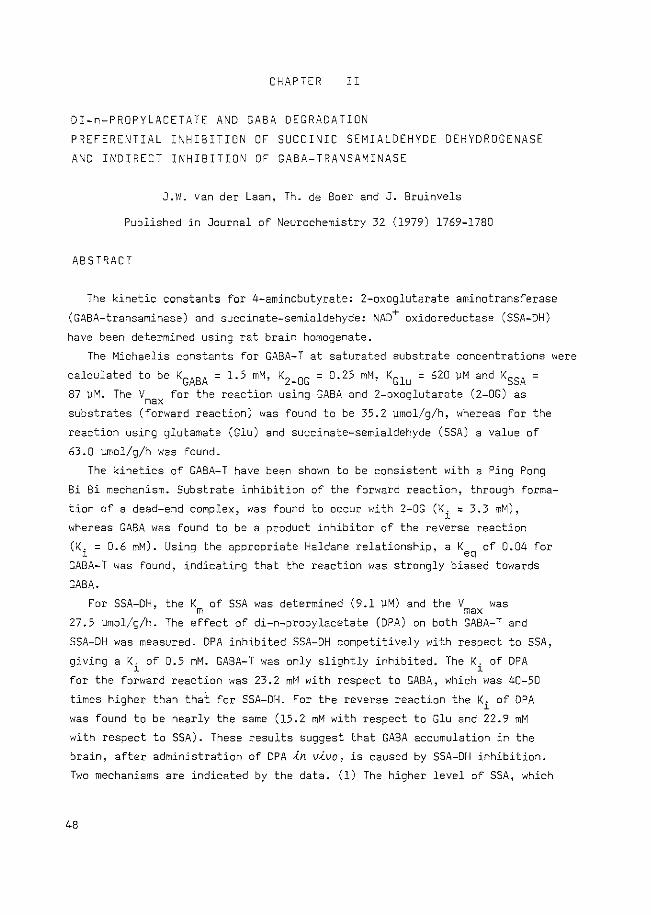
CHAPTER II
DI-n-PROPYLACETATE AND GABA DEGRADATION
PREFERE~TIAL I~HIBITION OF SUCCINIC SEMIALDEHYDE DEHYDROGENASE
AND INDIRECT INHIBITION OF GABA-TRANSAMINASE
J.W. van der Laan, Th. de Boer and J. Bruinvels
Published in Journal of Neurochemistry 32 (1979) 1769-1780
ABSTRACT
The kinetic constants for 4-aminobutyrate: 2-oxoglutarate aminotransferase
(GABA-transaminase) and succinate-semialdehyde: \JAD+ oxidoreductase (SSA-DH)
have been determined using rat brain homogenate.
The Michaelis constants for GABA-T at saturated substrate concentrations were
calculated to be KGABA = 1.5 mM, K2_0G = 0.25 mM, KGlu = 620 VM and KSSA = 87 ~M. The V for the reaction using GABA and 2-oxoglutarate (2-0G) as max substrates (forward reaction) was found to be 35.2 ~mol/g/h, whereas for the
reaction using glutamate (Glu) and succinate-semialdehyde (SSA) a value of
63.0 emol/g/h was found.
The kinetics of GABA-T have been shown to be consistent with a Ping Pong
Bi Bi mechanism. Substrate inhibition of the forward reaction, through forma
tion of a dead-end complex, was found to occur with 2-0G (K. = 3.3 mM), l
whereas GABA was found to be a product inhibitor of the reverse reaction
(K. = 0.6 mM). Using the appropriate Haldane relationship, a K of 0.04 for l ~
GABA-T was found, indicating that the reaction was strongly biased towards
GABA.
For SSA-DH,
27.5 \lillOl/g/h.
the Km of SSA was determined (9.1 ~M) and
The effect of di-n-propylacetate (DPA) on
the V was max both GABA-T and
SSA-DH was measured. DPA inhibited SSA-DH competitively with respect to SSA,
giving a K. of 0.5 mM. GASA-T was only slightly inhibited. The K. of DPA l l
for the forward reaction was 23.2 mM with respect to GABA, which was 40-50
times higher than that for SSA-DH. For the reverse reaction the Ki of DPA
was found to be nearly the same (15.2 mM with respect to Glu and 22.9 mM
with respect to SSA). These results suggest that GABA accumulation in the
brain, after administration of DPA ~n v~vo, is caused by SSA-DH inhibition.
Two mechanisms are indicated by the data. (1) The higher level of SSA, which
48
results from inhibition of SSA-DH, initiates the reverse reaction of GABA-T,
thus increasing the level of GABA via conversion of SSA. (2) The degradation
of GABA is inhibited by SSA~ since SSA has a strong inhibitory effect on the
forward reaction, as calculated from the present data.
InRDDUCTIO~
In mammalian brain gamma-aminobutyric acid (GABA) is thought to be an
inhibitory neurotransmitter (KrnjeviC, 1970). A decrease in the concentration
of GABA has been implicated in the onset of convulsions (Tower, 1976).
Accordingly, it has been reported by many authors that several anticonvulsant
drugs increase GABA levels in the brain through inhibition of its degradation
(see Tapia, 1975).
Elevation of GABA levels has been extensively studied by administration of
drugs which affect the activity of 4-aminobutyrate: 2-oxoglutarate amino
transferase (EC 2.6.1.19; GABA-T). Such drugs include aminooxyacetic acid
(AOAA; Baxter & Roberts, 1961), ethanolamine-0-sulphate (EOS; Fowler & John,
1972). y-acetylene-GABA and Y-vinyl-GABA (Schechter et al., 1977; Jung et al., . R 1977). Similarly, di-n-propylacetate (Depakine , Valproate, DPA) also increases
the concentration of GABA in the brain (Anlezark et al., 1976; Godin et al.,
1969; Simler et al., 1973) and its action has been ascribed to an inhibition
of GABA-T (Godin et al., 1969; Simler et al., 1973; Fowler et al.~ 1975).
However, some authors reported a preferential action of DPA on SSA-DH (Harvey
et al., 1975; Anlezark et al., 1976). Two specific reactions are involved in
the degradation of GABA; firstly, the reversible conversion of GABA and 2-
oxoglutarate (2-0G) into succinate-semialdehyde (SSA) and glutamate (Glu),
respectively, which is catalysed by GABA-T, and secondly, the subsequent
oxidation of SSA to form succinate by succinate-semialdehyde: NAD+ oxido
reductase (EC 1.2.1.16; SSA-DH) using NAD as a cofactor, which reaction is
almost irreversible. Since the equilibrium of the GABA-T catalysed reaction
is biased towards GABA (Duffy et al., 1972), inhibition of SSA-DH may also
result in elevation of the GABA concentration.However, this possibility has
so far been neglected, because SSA-DH was considered to be very efficient
in preventing accumulation of SSA. Recent studies have suggested that the
activity of SSA-DH can be affected by mono- and bivalent cations like sodium,
potassium and calcium (De Boer & Bruinvels, 1977b). Therefore, it is possible
that SSA-DH may play a regulating role in GABA degradation ~n v~vo and that
inhibition of this enzyme might incr.ease GABA-concentrations through
49
activating the reverse reaction of GABA-T.
The aim of the present paper has. therefore, been to study this regulatory
role of SSA-DH in relation to the action of DPA. The affinity of SSA for both
GABA-T and SSA-DH and its alteration by DPA have been determined as well as
the effect of DPA on the conversion of GABA into SSA.
MATERIALS AND METHODS
GABA was obtained from Calbiochem, Los Angeles, CA. Triton X-100, 2-oxo
glutaric acid, di-sodium succinate and 2-mercaptoethanol were obtained from
BDH Chemicals Ltd. Pyridoxamine-di-HCl and SSA were purchased from Sigma
Chemical Company. NAD and NADH were obtained from Boehringer-Mannheim.
[u14c] GABA (specific radioactivity 4.6 mCi/mmol) was obtained from the Radio
chemical Centre, Amersham, UK. All other chemicals were purchased from Merck,
Darmstadt. DPA was a gift of Labaz B. V., Maassluis, The \'ether lands.
T~¢ue pkepaAation
Randomly selected male Wistar rats (125-175 g) were killed by decapitation
and their brains were rapidly removed and weighed. A 10% (w/v) brain homogenate
was prepared in an ice-cold solution containing 0.32 M-sucrose and 4.5 mM-2-
mercaptoethanol, using a Teflon-glass Potter-Elvehjem homogenizer. One volume
of the homogenate was added to 3 val of ice-cold Triton medium (0.67% w/v
Triton X-100, 50 mM-Tris-HCl (pH 8.5) and 4.5 mM-2-mercaptoethanol) and kept
on ice-water for 1 h before use.
GABA-T a4¢aij ~~ng GABA a4 ¢ub¢~~e lnokwaAd keac~on)
For the performance of the assay of the GABA-T forward reaction a radio
chemical method was used as previously described (De Boer & Bruinvels, 1977a)
with the following modifications. NAD was omitted from the incubation mixture,
unless otherwise specified, to exclude interference with SSA-DH, since OPA
might affect this enzyme too. A control was carried throughout the whole
procedure in the presence of NAD but without DPA. Tissue concentration was
4.44 mg/ml (wet wt.). After preincubation at 22°C for 30 min the reaction was
started by the addition of homogenate and the mixture was incubated for a
further 30 min at the same bath temperature. All experiments were carried out
in duplicate.
GABA-T a4~ay ~~ng SSA a4 ¢Ub¢~~e (keveHe keac~on)
The reaction was carried out under similar conditions to the forward
reaction. The incubation mixture contained 50 mM-Tris-HCl (pH 8.5), 20 mM
50
2-mercaptoethanol and 0.12 ml Triton-treated homogenate in a total volume of
0.615 ml. The final tissue concentration was 4.44 mg/ml. The reaction mixture
was preincubated at 22°C for 1 h in the presence of glutamate (0.5-5.0 mM)
and in the absence of SSA. The reaction was started by addition of 60 ~1 SSA
solution, resulting in final concentrations of 50-600 ~M-SSA. For the inhibition
studies DPA was added simultaneously with SSA.
Since the sodium salt of DPA was used throughout this study, a solution in
which DPA was replaced by sodiumisethionate, equimolar to the highest DPA
concentration, was used as control.
After an incubation period of 15 or 20 min, the reaction was terminated by
adding an equal volume of ice-cold 5% TCA solution. Subsequently, the tubes
were placed in ice-water. After centrifugation (10 min x 1200 g) the concen
tration of 2-0G in 0.5 ml of the supernatant was determined in duplicate.
Vet~minaXJon o£ Z-oxog~aXe The fluorometric determination of a-oxo acids as described by Takeda et al.
(1976) was used. To neutralize 0.5 ml of the 2.5% TCA solution (sample
solution), 0.2 ml of a solution containing 2 val 1 M-Tris-HCl (pH 8.3) and 1 val
1 M-\aOH was added. Thereafter, 5 ml of a solution containing 2% (v/v) pyridine
in methanol, 100 mg/1 pyridoxamine-di-HCl and 2.0 g/1 zinc acetate was added.
The reaction of pyridoxamine, 2-oxoglutarate and Zn(II)ion was found to yield
a Zn(II) chelate of the aldimine (Matsushima & Martell, 1967). After incubation
for 30 min at 37°C the reaction mixture was cooled in ice-water for 3-5 min.
The fluorescence intensity was measured within 2 h at room temperature using
a Baird-Atomic spectrophotofluorometer with excitation at 395 nm and emission
at 475 nm. Standards and blanks were carried throughout the entire assay
procedure. Blanks contained the complete incubation mixture, except SSA. The
standards were prepared in the same way as blanks and 2-0G was added to final
concentrations of 20-80 ~M. The change in the glutamate concentration used
in the GABA-T assay resulted in blanks which varied to a maximum of 20% and
this variation was proportional to the glutamate concentration. Therefore, the
assay was always performed with standards of all glutamate concentrations used.
Incubation mixtures, to which the TCA solution was added immediately after
starting the reaction by addition of SSA, were used as additional controls.
When 2-0G was incubated under the conditions of the assay in the absence of
SSA it was not significantly metabolized, as measured after 1 h (recovery
98.4! 1.3%; n = 13).
51
SSA-VH GMIUJ. Measurement of the SSA-DH activity was performed as described previously
(De Boer & Bruinvels, 1977a), using a tissue concentration of 0.44 mg/ml
(wet wt.).
Data. ana£y .;.W
The apparent kinetic parameters of the forward reaction of GABA-T were
calculated from Lineweaver-Burk plots.
Since in both the assay of the reverse reaction of GABA-T and in the assay
of SSA-DH more than 5~0 of the initial concentration of SSA was converted,
the apparent kinetic constants were calculated using the integrated form of
the Henri-Michaelis-Menten equation (Segel, 1975).
To obtain limiting Michaelis constants for the substrates of GABA-T from
the apparent constants the equations for a Ping Pong Bi Bi mechanism (Cleland,
1963) were applied, since it is generally accepted that transaminations occur
via such a mechanism (Meister, 1955; Scott & Jakoby, 1959; Henson & Cleland,
1964).
The type of inhibition by DPA of GABA-T was determined graphically. The
Ki's were calculated from a replot of the ratio of the apparent Km and Vmax
vs concentration of inhibitor as suggested by Segel (1975) for mixed-type
inhibition.
All constants were calculated from the plots using the least squares method.
Variance analysis for regression lines was used to test the significance of
differences in K and V m max
RESULTS
K~netio.; on GABA-T
Rewction w.i.:th GABA cv., .;ub.;:ttta;te I u oJcWaJtd Jte=tion)
The kinetic constants of the GABA-T forward reaction were determined using
concentrations of 0.5-5.0 mM-GABA and 0.05-6.0 mM-2-0G in the absence of
NAD. Lineweaver-Burk plots are given in Fig. 1.
Since substrate inhibition was observed with 2-0G (Fig.lA). data obtained
with concentrations of 2-0G exceeding 0.5 mM were omitted for calculation of
the kinetic constants. The dependency of the apparent kinetic constants for
2-0G on the concentration of GABA, and vice versa, is shown in the plots of
Figs. lA and D. The data were fitted to equation (1)
52
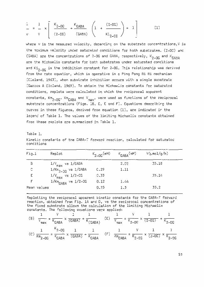
= X KG ABA
+--- + (2-0G) ) l - + lj
Ki2-0G
(1) 1 1
v v (2-0G) (GABA)
where vis the measured velocity, depending on the substrate concentrations!V is
the maximum velocity under saturated conditions for both substrates. (2-0G) and
(GABA) are the concentrations of 2-0G and GABA, respectively, K2_0G and KGABA
are the Michaelis constants for both substrates under saturated conditions
and Ki2_0G is the inhibition constant for 2-0G. This relationship was derived
from the rate equation, which is operative in a Ping Pong Bi Bi mechanism
(Cleland, 1963), when substrate inhibition occurs with a single substrate
(Garces & Cleland, 1969). To obtain the Michaelis constants for saturated
conditions, replots were calculated in which the reciprocal apparent
constants, Km 2_0G' KmGABA and Vmax' were used as functions of the reciprocal
substrate concentrations (Figs. 18, C, E and F). Equations describing the
curves in these figures, derived from equation (1), are indicated in the
legend of Table 1. The values at the limiting Michaelis constants obtained
from these replots are summarized in Table 1.
Table l.
Kinetic constants of the GABA-T forward reaction, calculated for saturated conditions
Fig.l Replot
8 1/V VS 1/GABA max C 1/KmZ-OG vs 1/GABA E 1/V vs 1/2-0G max F 1/KmGABA vs 1/2-0G
Mean values
0.29
0.33
0.12
0.25
KGABA(mM) V()lmol/g/h)
2.02 35.18
1.11
35.14
1.44
1.5 35.2
Replotting the reciprocal apparent kinetic constants for the GABA-T forward reaction, obtained from Fig. lA and D, vs the reciprocal concentrations of the fixed substrate allows the calculation of the limiting Michaelis constants. The following equations were applied:
1 v 1 1 1 v 1 1 (B)
Vmax = KGABA x (GABA) + K(GABA)
1 1 1 (C) --- - --· X --- + -
Km2-0G - KGABA (GASA) KGABA
(E)
(F)
-v m-a-x = -K2---D-G x 7("2 _...,o"'G")- + -K2---0-G
1 v 1 1 --- = --- X --- X ---KmGABA K2-0G (2-0G) K2-0G
53
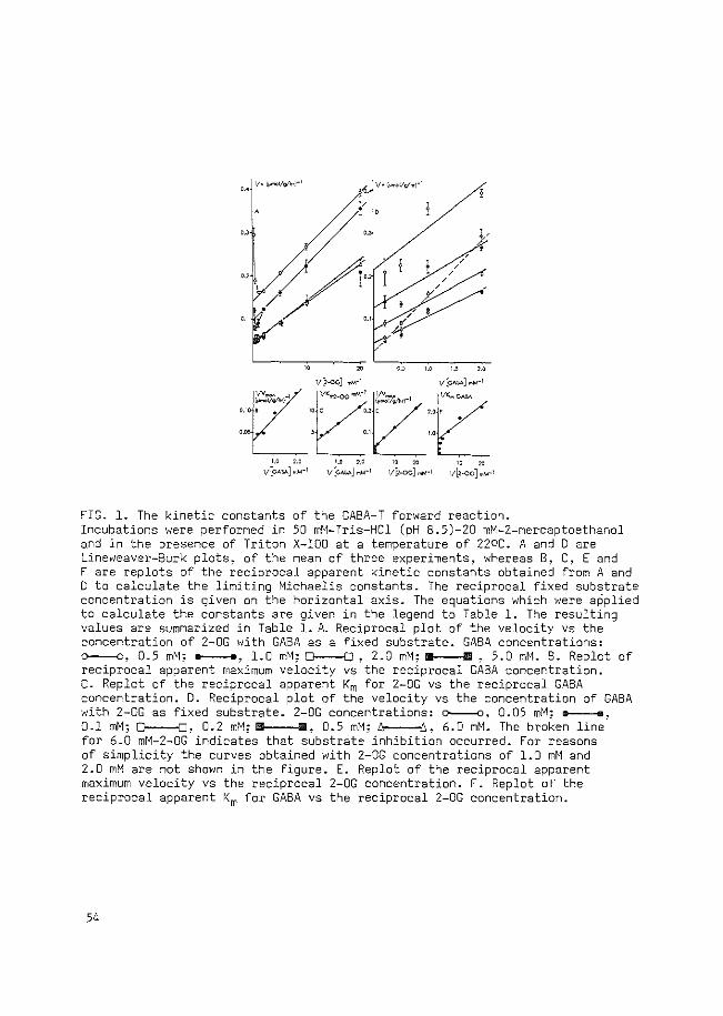
j
v.'"-v"'.-' "
'
~~:.:v~.r, 0.10 B
0.~ 1.0 ).5 2.0
1/[2-0C] mM"1 1/[GAM] ..M"1
TO .2 C 2.01f •
~ .I 1.0
j"'~~c·"-' ~ (?: '----,,,-:_0-0:-.0- 1.0 2.0 10 :10 IO :10
1/~GAM] mM"1 1/c.o.!IA] ...M"1 v[;o-oc;J mM·l 11~-oc] mM-1
FIG. 1. The kinetic constants of the GABA-T forward reaction. Incubations were performed in 50 mM-Tris-HCl (pH 8.5)-20 mM-2-mercaptoethanol and in the presence of Triton X-100 at a temperature of 22DC. A and 0 are Lineweaver-Burk plots, of the mean of three experiments, whereas B, C, E and F are replots of the reciprocal apparent kinetic constants obtained from A and D to calculate the limiting Michaelis constants. The reciprocal fixed substrate concentration is given on the horizontal axis. The equations which were applied to calculate the constants are given in the legend to Table 1. The resulting values are summarized in Table 1. A. Reciprocal plot of the velocity vs the concentration of 2-0G with GABA as a fixed substrate. GABA concentrations: o---o, 0.5 mM; a----a, 1.0 mM; D---0, 2.0 mM; II 1111 , 5.0 mM. B. Replot of reciprocal apparent maximum velocity vs the reciprocal GABA concentration. C. Replot of the reciprocal apparent Km for 2-0G vs the reciprocal GABA concentration. D. Reciprocal plot of the velocity vs the concentration of GABA with 2-0G as fixed substrate. 2-0G concentrations: o----o, 0.05 mM; e----e, 0.1 mM; D---O, 0.2 mM; IIIII 111, 0.5 mM; f::t-----.lj., 6.0 mM. The broken line for 6.0 mM-2-0G indicates that substrate inhibition occurred. For reasons of simplicity the curves obtained with 2-0G concentrations of 1.0 mM and 2.0 mM are not shown in the figure. E. Replot of the reciprocal apparent maximum velocity vs the reciprocal 2-0G concentration. F. Replot of the reciprocal apparent Km for GABA vs the reciprocal 2-0G concentration.
54
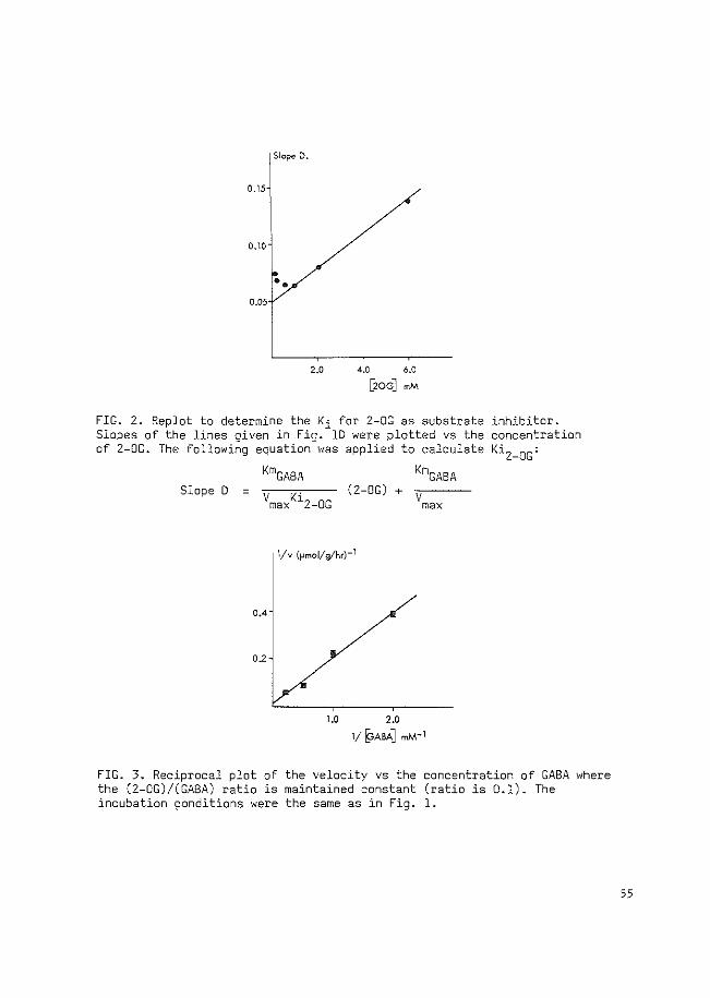
Slope D.
0.15
0.10
•• 0.05
2.0 4.0 6.0
[20G] mM
FIG. 2. Replot to determine the Ki for 2-0G as substrate inhibitor. Slopes of the lines given in Fig. lD were plotted vs the concentration of 2-0G. The following equation was applied to calculate Ki2_0G:
Slope 0 =
0.4
02
KmGABA (2-DG) + "v __ _
max
1/v (pmol/g/hr)-1
1.0 2.0
1/ ~ABA] mM- 1
FIG. 3. Reciprocal plot of the velocity vs the concentration of GABA where the (2-0G)/(GABA) ratio is maintained constant (ratio is 0.1). The incubation ~onditions were the same as in Fig. 1.
55
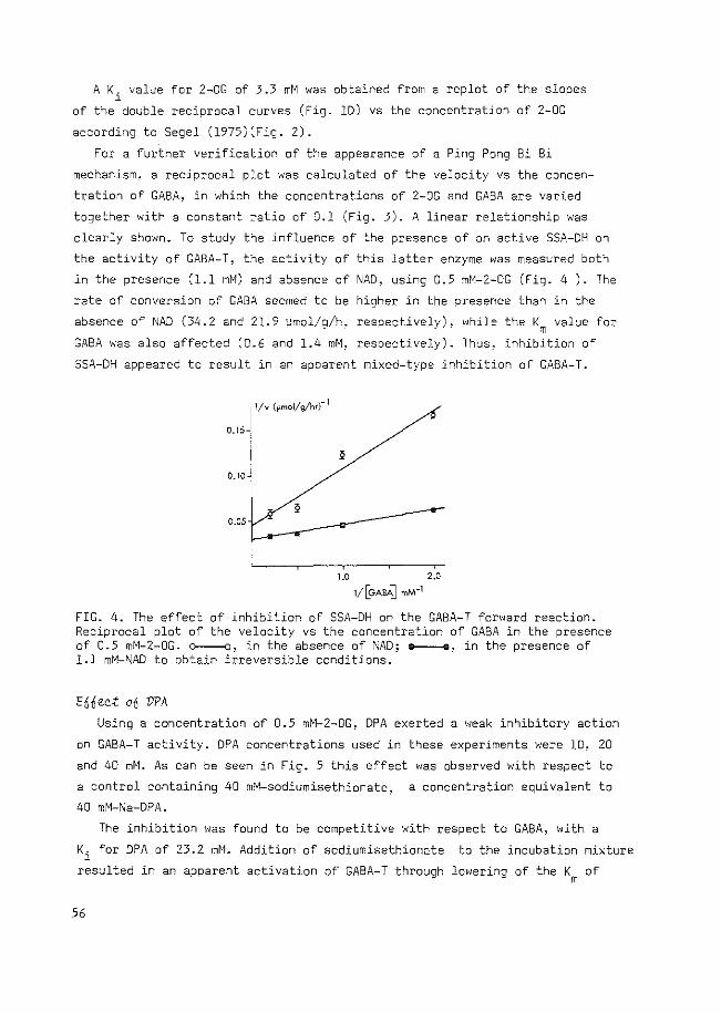
A K. value for 2-0G of 3.3 mM was obtained from a replot of the slopes l
of the double reciprocal curves (Fig. 10) vs the concentration of 2-0G
according to Segel (l975)(Fig. 2).
For a further verification of the appearance of a Ping Pong Bi Bi
mechanism. a reciprocal plot was calculated of the velocity vs the concen
tration of GABA, in which the concentrations of 2-0G and GABA are varied
together with a constant ratio of 0.1 (Fig. 3). A linear relationship was
clearly shown. To study the influence of the presence of an active SSA-DH on
the activity of GABA-T, the activity of this latter enzyme was measured both
in the presence (1.1 mM) and absence of NAD, using 0.5 mM-2-0G (Fig. 4 ). The
rate of conversion of GABA seemed to be higher in the presence than in the
absence of NAD (34.2 and 21.9 ~mol/g/h, respectively), while the Km value for
GABA was also affected (0.6 and 1.4 mM, respectively). Thus~ inhibition of
SSA-DH appeared to result in an apparent mixed-type inhibition of GABA-T.
0.15
0.10
0.05
1.0 2.0
1/[GABA] mM·1
FIG. 4. The effect of inhibition of SSA-DH on the GABA-T forward reaction. Reciprocal plot of the velocity vs the concentration of GABA in the presence of 0.5 mM-2-0G. o----o, in the absence of ~AD; e----o. in the presence of 1.1 mM-1\1AD to obtain irreversible conditions.
Ennee;t on DPA Using a concentration of 0.5 mM-2-0G, DPA exerted a weak inhibitory action
on GABA-T activity. DPA concentrations used in these experiments were 10, 20
and 40 mM. As can be seen in Fig. 5 this effect was observed with respect to
a control containing 40 mM-sodiumisethionate, a concentration equivalent to
40 mM-Na-DPA.
The inhibition was found to be competitive with respect to GABA, with a
Ki for DPA of 23.2 mM. Addition of sodiumisethionate to the incubation mixture
resulted in an apparent activation of GABA-T through lowering of the Km of
56
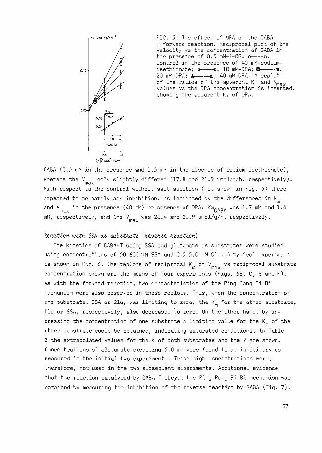
0.05
'·"1~/ O,OL 0 20 40
mMDPA
0.5 1.0
1f[9ABA] mM·l
FIG. 5. The effect of DPA on the GABA-T forward reaction. Reciprocal plot of the velocity vs the concentration of GABA in the presence of 0.5 mM-2-0G. o----o~
Control in the presence of 40 mM-sodiumisethionate; a----e. 10 mM-DPA; mD IIIII, 20 mM-DPA; A A, 40 mM-DPA. A replot of the ratios of the apparent Km and Vmax values vs the DPA concentration is inserted~ showing the apparent Ki of DPA.
GABA (0.5 mM in the presence and 1.5 mM in the absence of sodium-isethionate),
whereas the Vmax only slightly differed (17.8 and 21.9 vrnol/g/h~ respectively).
With respect to the control without salt addition (not shown in Fig. 5) there
appeared to be hardly any inhibition, as indicated by the differences in Km
and Vmax in the presence (40 mM) or absence of DPA: KmGABA was 1.7 mM and 1.4
mM, respectively, and the V was 20.4 and 21.9 ~mol/g/h, respectively. max
Reaetion w~ SSA ~ ~ub~~ate lkevek4e keaQtionl
The kinetics of GABA-T using SSA and glutamate as substrates were studied
using concentrations of 50-600 ~M-SSA and 0.5-5.0 mM-Glu. A typical experiment
is shown in Fig. 6. The replots of reciprocal Km or Vmax vs reciprocal substrate
concentration shown are the means of four experiments (Figs. 68, C, E and F).
As -with the forward reaction, two characteristics of the Ping Pong Bi Bi
mechanism were also observed in these replots. Thus, when the concentration of
one substrate 1 SSA or Glu, was limiting to zero, the
Glu or SSA, respectively, also decreased to zero. On
K for the other substrate, m
the other hand, by in-
creasing the concentration of one substrate a limiting value for the Km of the
other substrate could be obta_ined, indicating saturated conditions. In Table
2 the extrapolated values for the K of both substrates and the V are shown.
Concentrations of glutamate exceeding 5.0 mM were found to be inhibitory as
measured in the initial two experiments. These high concentrations were,
therefore, not uSed in the two subsequent experiments. Additional evidence
that the reaction catalysed by GABA-T obeyed the Ping Pong Bi Bi mechanism was
obtained by measuring the inhibition of the reverse reaction by GABA (Fig. 7).
57
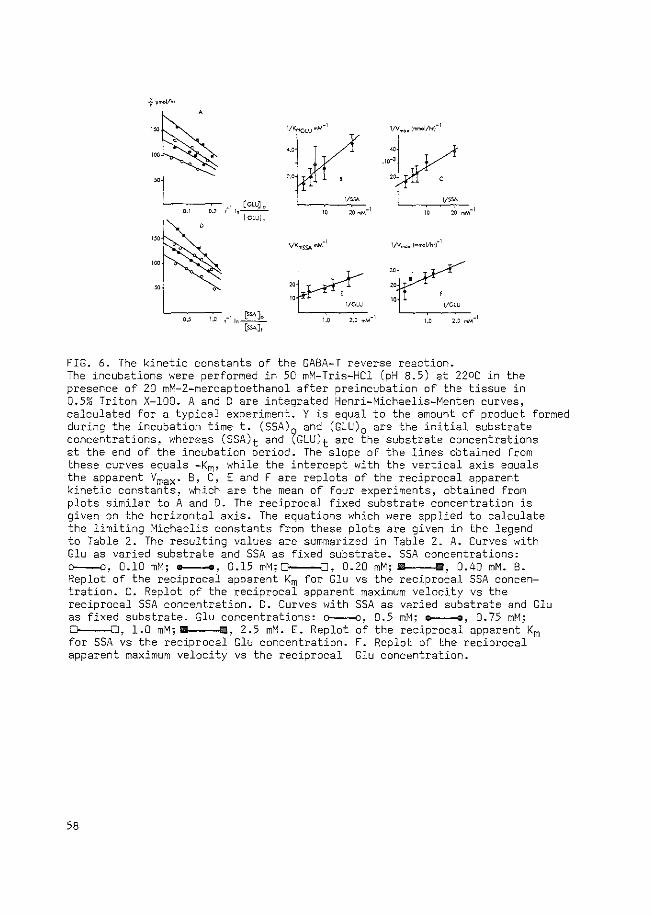
'--~,.-, --00-,-
110_[c_c_c]_. lcwi,
'---,oc----c,.c-0 ,-llo (:iSA)o [SSA),
J:Vr ·~k: s V>~ 10 :IOwM-T TO 20 mM-I
"~ " ' 1/CLU
1.0 2.0 tnM-1
::J~ lG~
1.0 2.0 mM-\
FIG. 6. The kinetic constants of the GABA-T reverse reaction. The incubations were performed in 50 mM-Tris-HCl (pH 8.5) at 22DC in the presence of 20 mM-2-mercaptoethanol after preincubation of the tissue in 0.5% Triton X-100. A and D are integrated Henri-Michaelis-Menten curves, calculated for a typical experiment. Y is equal to the amount of product formed during the incubation time t. (SSA) and (GLU) 0 are the initial substrate concentrations, whereas (SSA)t and ?GLU)t are the substrate concentrations at the end of the incubation period. The slope of the lines obtained from these curves equals -Km, while the intercept with the vertical axis equals the apparent Vmax· B, C, E and F are replots of the reciprocal apparent kinetic constants, which are the mean of four experiments, obtained from plots similar to A and D. The reciprocal fixed substrate concentration is given on the horizontal axis. The equations which were applied to calculate the limiting Michaelis constants from these plots are given in the legend to Table 2. The resulting values are summarized in Table 2. A. Curves with Glu as varied substrate and SSA as fixed substrate. SSA concentrations: o---o, 0.10 mM; -, 0.15 mM; D----{], 0.20 mM; Ill 1111, 0.40 mM. B. Replot of the reciprocal apparent Km for Glu vs the reciprocal SSA concentration. C. Replot of the reciprocal apparent maximum velocity vs the reciprocal SSA concentration. D. Curves with SSA as varied substrate and Glu as fixed substrate. Glu concentrations: o----o, 0.5 mM; e----e, 0.75 mM; 0---0, 1.0 mM; II B, 2.5 mM. E. Replot of the reciprocal apparent Km for SSA vs the reciprocal Glu concentration. F. Replot of the reciprocal apparent maximum velocity vs the reciprocal Glu concentration.
58
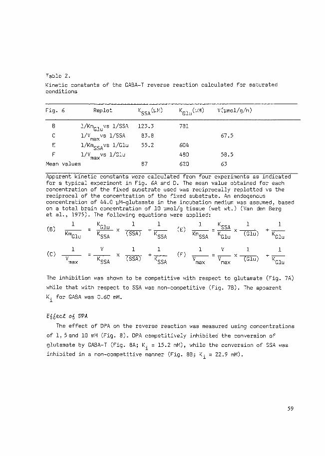
Table 2.
Kinetic constants of the GABA-T reverse reaction calculated for saturated conditions
Fig. 6 Replot KSSA (~M) KGlu(~M) V(~mol/g/h)
B 1/KmGluvs 1/SSA 123.3 781
c 1/V VS 1/SSA max 83.8 67.5
E 1/KmSSAvs l/Glu 55.2 604
F 1/V vs 1/Glu 480 58.5 max Mean values 87 620 63
Apparent kinetic constants were calculated from four experiments as indicated for a typical experiment in Fig. 6A and D. The mean value obtained for each concentration of the fixed substrate used was reciprocally replotted vs the reciprocal of the concentration of the fixed substrate. An endogenous concentration of 44.0 ~M-glutamate in the incubation medium was assumed, based on a total brain concentration of 10 ~mol/g tissue (wet wt.) (Van den Berg et al., 1975). The following equations were applied:
l KGlu l l l (B) = x -- +-- (E)
KmGlu KSSA (SSA) KSSA KmSSA
KSSA l ---X---· - KGlu (Glu)
(C) l
-v-max
v - KSSA
l l
x (SSA) +--KSSA
l v l (F) -v-- --v-
max max X (Glu)
l +--
KGlu
l +--
KGlu
The inhibition was shown to be competitive with respect to glutamate (Fig. 7A)
while that with respect to SSA was non-competitive (Fig. 78). The apparent
Ki for GABA was 0.60 mM.
En£ec:t on VPA The effect of DPA on the reverse reaction was measured using concentrations
of 1, 5 and 10 mM (Fig. 8). DPA competitively inhibited the conversion of
glutamate by GABA-T (Fig. SA; Ki = 15.2 mM), while the conversion of SSA was
inhibited in a non-competitive manner (Fig. 88; Ki = 22.9 mM).
59
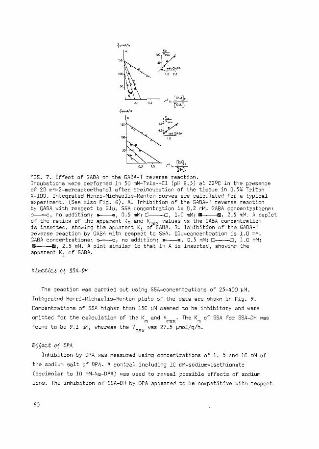
'~~· "' . mMCAliA
1.0 Z..5
t 1 In @SA] 0 ["?SA J r
FIG. 7. Effect of GABA on the GABA-T reverse reaction. Incubations were performed in 50 mM-Tris-HCl (pH 8.5) at 22°C in the presence of 20 mM-2-mercaptoethanol after preincubation of the tissue in 0.5~0 Triton X-100. Integrated Henri-Michaelis-Menten curves are calculated for a typical experiment. (See also Fig. 6). A. Inhibition of the GABA-T reverse reaction by GABA with respect to Glu. SSA concentration is 0.2 mM. GABA concentrations: o----o~ no addition; e----e, 0.5 mM; ~' 1.0 mM; m a, 2.5 mM. A replot of the ratios of the apparent Km and Ymax values vs the GABA concentration is inserted, showing the apparent Ki of GABA. B. Inhibition of the GABA-T reverse reaction by GABA with respect to SSA. Glu-concentration is 1.0 mM. GABA concentrations: o----o~ no addition; ------~ 0.5 mM; ~' 1.0 mM; a a, 2.5 mM. A plot similar to that in A is inserted, showing the apparent Ki of GABA.
K~net;04 o0 SSA-VH
The reaction was carried out using SSA-concentrations of 25-400 ~M.
Integrated Henri-Michaelis-Menten plots of the data are shown in Fig. 9.
Concentrations of SSA higher than 150 ~M seemed to be inhibitory and were
omitted for the calculation of the K and V . The K of SSA for SSA-DH was m max m found to be 9.1 ~M, whereas the V was 27.5 ~mol/g/h. max
Efi£eQ;t o£ VPA
Inhibition by DPA was measured using concentrations of 1, 5 and 10 mM of
the sodium salt of DPA. A control including 10 mM-sodium-isethionate
(equimolar to 10 mM-,\1a-DPA) was used to reveal possible effects of sodium
ions. The inhibition of SSA-DH by DPA appeared to be competitive with respect
60
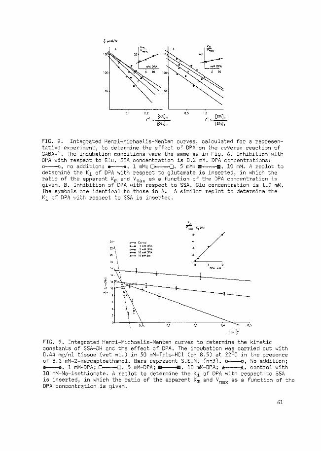
f ~mol/ht A
,, ·I [CLUJ., ''•---
,_, _1 [ss.o..]c ' '•--[cLUJ, (SSAJ t
FIG. 8. Integrated Henri-Michaelis-Men·ten curves, calculated for a representative experiment, to determine the effect of OPA on the reverse reaction of GABA-T. The incubation conditions were the same as in Fig. 6. Inhibition with DPA with respect to Glu, SSA concentration is 0.2 mM. DPA concentrations: o----o, no addition; .----., 1 mM; ~,- 5 mM: m E, 10 mM. A replot to determine the Ki of DPA with respect to glutamate is inserted, in which the ratio of the apparent Km and Vmax as a function of the DPA concentration is given. B. Inhibition of DPA with respect to SSA. Glu concentration is 1.0 mM. The symbols are identical to those in A. A similar replot to determine the Ki of DPA with respect to SSA is inserted.
:~ <>--<> '""!Tol - l mMDPA ~ 5-.M DPA - 10""' DPA
20' \ - IOmMI,.
'« ;f " l " +
\! \
'·' "
FIG. 9. Integrated Henri-Michaelis-Menten curves to determine the kinetic constants of SSA-DH and the effect of DPA. The incubation was carried out with 0.44 mg/ml tissue (wet wt.) in 50 mM-Tris-HCl (pH 8.5) at 22°C in the presence of 8.2 mM-2-mercaptoethanol. Bars represent S.E.M. (n::::3). t?---O, i'\o addition; e----e, 1 mM-DPA; ~' 5 mM-DPA; R B, 10 mM-DPA; A A, control with 10 mM-Na-isethionate. A replot to determine the Ki of OPA with respect to SSA is inserted, in which the ratio of the apparent Km and Vmax as a function of the DPA concentration is given.
61
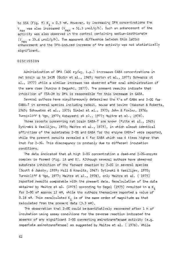
to SSA (Fig. 9) Ki.= 0.5 mM. However, by increasing DPA concentrations the
V was also increased (V = 51.5 ~mol/g/h). Such an enhancement of the max max
activity was also observed in the control containing sodium-isethionate
(V = 35.6 vmol/g/h). The apparent difference between this latter max enhancement and the DPA-induced increase of the activity was not statistically
significant.
DISCUSSION
Administration of DPA (400 mg/kg, i.p.) increases GABA concentrations in
rat brain up to 140% (Godin et al., 1969; Horton et al., 1977; Schwarcz et
al., 1977) while a similar increase was observed after oral administration of
the same dose (Kukino & Deguchi, 1877). The present results indicate that
inhibition of SSA-DH by DPA is responsible for this increase in GABA.
Several authors have sim_ultaneously determined the. K 's of GABA and 2-0G for
GABA-T in several species including rabbit, mouse and bovine (Waksman & Roberts,
1965; Schousboe et al., 1973; Simler et al., 1973; John & Fowler, 1976;
Tunnicliff & Ngo, 1977; Kobayashi et al., 1977; Maitre et al., 1978).
Three reports concerning rat brain GABA-T are known (Pitts et al., 1965;
Sytinski & Vasilijev, 1970; Maitre et al., 1975), in which almost identical
affinities of the substrates 2-0G 3nd GABA for the enzyme GABA-T were reported,
while the present results revealed a K for GABA which was 6 times higher than
that for 2-0G. This discrepancy is probably due to different incubation
conditions.
The data indicated that at high 2-0G concentration a dead-end 2-0G-enzyme
complex is formed (Fig. lA and D). Although several authors have observed
substrate inhibition of the forward reaction by 2-0G in several species
(Scott & Jakoby, 1959; Hall & Kravitz, 1967; Sytinski & Vasilijev, 1970;
Tunnicliff & Ngo, 1977; Maitre et al., 1978), only Maitre et al. ( 1975)
reported results comparable with the present data. Recalculation of the data
obtained by Maitre et al. (1975) according to Segel (1975) resulted in a Ki
for 2-0G of approx 12 mM, while the authors themselves reported a value of
0.18 mM. This recalculated Ki is of the same order of magnitude as that
calculated from the present data (3.3 mM).
The observation that 2-0G could bequantitatively recovered after 1 h of
incubation using assay conditions for the reverse reaction indicated the
absence of any significant 2-0G converting aminotransferase activity (e.g.
aspartate aminotransferase) as suggested by Maitre et al. ( 1976). While
62
GABA and 2-0G both exhibited higher affinity for GABA-T during the forward
reaction, as discussed above, the affinities of SSA and Glu in the reverse
reaction were also higher than those reported by others (Harvey et al., 1975;
Maitre et al., 1975).
The K. of GABA (0.6 mM) must be viewed as an apparent value measured at a l
fixed concentration of Glu (1.0 mM) or SSA (0.2 mM). Nevertheless, it might
be a good estimate of the real value since the concentrations of both
substrates used exceeded their K values for GABA-T.
It seems remarkable that the Ki of GABA for the reverse reaction, which
represents the extent of binding of GABA to the aldimine form of GABA-T
without subsequent conversion in competition with respect to Glu, is lower
than the K of this substance for the forward reaction, which represents the
extent of GABA converted into SSA by the enzyme. This suggests that in the
overall conversion of GABA into SSA and the concomitant conversion of the
aldimine form of the enzyme into the ketimine form, the binding of GABA to
the enzyme does not represent the rate-limiting step.
This interpretation is consistent with the results of John & Fowler (1976),
who reported that the conversion of the GABA-enzyme complex from the
aldimine into the ketimine form is relatively slow as compared with identical
steps in other transaminase catalysed reactions.
The enzyme kinetics of GABA-T presented are consistent with a Ping Pong
Bi Bi mechanism, which is generally accepted to be the underlying mechanism
for transamination reactions. According to Segel (1975) the family of parallel
lines (Fig. lA), as well as the linear reciprocal plot when the concentrations
of 2-0G and GASA are varied together with a constant ratio (Fig. 3) are
sufficient for the identification of a Ping Pong Bi Bi mechanism.
An ordered Bi Bi mechanism, which may yield similar results under certain
conditions, can be rejected, since the inhibition
reverse reaction does not fit in with this latter
pattern of GABA in the
system.
relating the equilibrium constant and the various kinetic
Four equations,
constants (Haldane
relationships) are operative in the Ping Pong Bi Bi mechanism. The following
equation provides an indication of the occurrence of the reverse reaction in
this system.
KsslGlu
KGABl2-0G = 0.04 (2)
Vf and Vr are the velocities of the forward reaction and reverse reaction
respectively. Two values for the K of GABA-T from different species have eq
63
been reported: for mouse brain GABA-T, Keq = 0.29 (Duffy et al., 1972), and
for P¢eudomonah ofUo~~een6 GABA-T, Keq = 0.10 (Scott & Jakoby, 1959). The
latter two K 1s have been determined by measuring the concentrations of the eq
substrates when the reaction was allowed to come into equilibrium, applying
the following equation:
(GABA)(2-0G) K = eq (SSA) (GLU)
(3)
Scott & Jakoby (1959) have reported the effect of different initial
substrate concentrations on
equation (2) is independent
the resulting K . S1nce the K • calculated with eq eq· of the substrate concentrations, this value seems
to be more reliable. Besides
the discrepancy in these K eq
a species difference, it might be possible that
values and the present value is due to a
different way of calculating the K eq The present observation that omission of NAD from the incubation mixture
resulted in an impaired GABA-T activity during the assay of the forward
reaction (Fig. 4) is in agreement with the findings of Hall & Kravitz (1967)
using lobster GABA-T, and can be taken as indirect evidence for an equilibrium
biased towards GABA.
Although the value of the K strongly suggests that the equilibrium is eq biased towards GABA, it seems doubtful whether this mechanism will contribute
to the ~n v~vo synthesis of GABA, since the substrate for the reverse reaction,
SSA, is continuously removed by a
via SSA-DH (Pitts & Quick, 1965).
subsequent almost irreversible reaction
Although the present value of
SSA-DH is not very high, it is not directly comparable with the
V of GABA-T, since somewhat different conditions were used.
the V max
value of
of
the
Recent studies have indicated that monovalent and bivalent cations can
affect the activity of SSA-DH (De Boer & Bruinvels, 1977b). Therefore,it is
conceivable that, in vivo, these ions exert a regulating role in the meta
bolism of SSA and will therefore indirectly affect the formation of GABA via
the reverse reaction of GABA-T.
E££eet o£ VPA
The preferential action of DPA on SSA-DH, as shown by its higher affinity
for this enzyme compared to that for GABA-T, is in agreement with the findings
of Harvey et a1. (1975) and of Anlezark et a1. (1976). Only a slight inhibitory
effect of DPA on the GABA-T forward reaction was found using conditions which
excluded interference of DPA with SSA-DH. The K. of DPA (23.2 mM) for this l
64
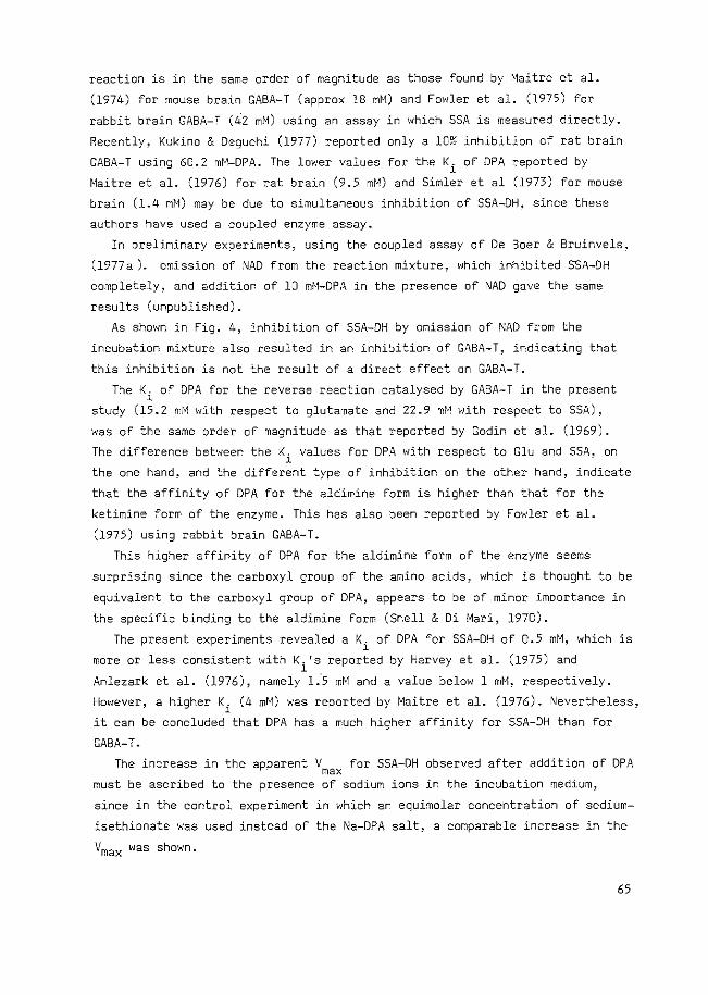
reaction is in the same order of magnitude as those found by Maitre et al.
(1974) for mouse brain GABA-T (approx lB mM) and Fowler et al. (1975) for
rabbit brain GABA-T (42 mM) using an assay in which SSA is measured directly.
Recently, Kukino & Deguchi (1977) reported only a 10% inhibition of rat brain
GABA-T using 60.2 mM-DPA. The lower values for the Ki of DPA reported by
Maitre et al. (1976) for rat brain (9.5 mM) and Simler et al (1973) for mouse
brain (1.4 mM) may be due to simultaneous inhibition of SSA-DH, since these
authors have used a coupled enzyme assay.
In preliminary experiments, using the coupled assay of De Boer & Bruinvels,
(1977a ). omission of NAD from the reaction mixture, which inhibited SSA-DH
completely, and addition of 10 mM-DPA in the presence of NAD gave the same
results (unpublished).
As shown in Fig. 4, inhibition of SSA-DH by omission of ~AD from the
incubation mixture also resulted in an inhibition of GABA-T, indicating that
this inhibition is not the result of a direct effect on GABA-T.
The Ki of DPA for the reverse reaction catalysed by GABA-T in the present
study (15.2 mM with respect to glutamate and 22.9 mM with respect to SSA),
was of the same order of magnitude as that reported by Godin et al. (1969).
The difference between the K. values for DPA with respect to Glu and SSA, on l
the one hand, and the different type of inhibition on the other hand, indicate
that the affinity of DPA for the aldimine form is higher than that for the
ketimine form of the enzyme. This has also been reported by Fowler et al.
(1975) using rabbit brain GABA-T.
This higher affinity of DPA for the aldimine form of the enzyme seems
surprising since the carboxyl group of the amino acids, which is thought to be
equivalent to the carboxyl group of DPA, appears to be of minor importance in
the specific binding to the aldimine form (Snell & Di Mari, 1970).
The present experiments revealed a Ki of DPA for SSA-DH of 0.5 mM, which is
more or less consistent with K. 's reported by Harvey et al. (1975) and 1
Anlezark et al. (1976), namely 1.·5 mM and a value below 1 mM, respectively.
However, a higher Ki (4 mM) was reported by Maitre et al. (1976). Nevertheless,
it can be concluded that DPA has a much higher affinity for SSA-DH than for
GABA-T.
The increase in the apparent Vmax for SSA-DH observed after addition of DPA
must be ascribed to the presence of sodium ions in the incubation medium,
since in the control experiment in which an equimolar concentration of sodium
isethionate was used instead of the Na-DPA salt, a comparable increase in the
Ymax was shown.
65
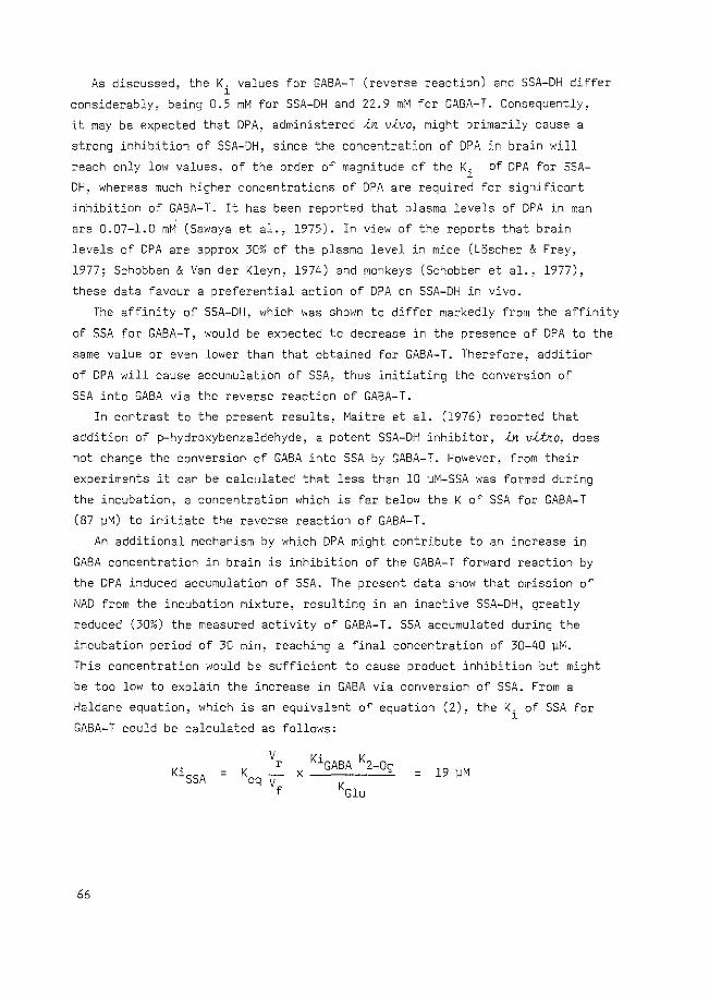
As discussed. the K. values for GABA-T (reverse reaction) and SSA-DH differ . 1
considerably, being 0.5 mM for SSA-DH and 22.9 mM for GABA-T. Consequently~
it may be expected that DPA, administered ~n v~vo, might primarily cause a
strong inhibition of SSA-DH, since the concentration of DPA in brain will
reach only low values, of the order of magnitude of the Ki of OPA for SSA
DH, whereas much higher concentrations of DPA are required for significant
inhibition of GABA-T. It has been reported that plasma levels of DPA in man
are 0.07-1.0 mM (Sawaya et al., 1975). In view of the reports that brain
levels of DPA are approx 30% of the plasma level in mice (LOscher & Frey,
1977; Schobben & Van der Kleyn, 1974) and monkeys (Schobben et aL, 1977),
these data favour a preferential action of DPA on SSA-DH in vivo.
The affinity of SSA-DH, which was shown to differ markedly from the affinity
of SSA for GABA-T, would be expected to decrease in the presence of DPA to the
same value or even lower than that obtained for GABA-T. Therefore, addition
of DPA will cause accumulation of SSA, thus initiating the conversion of
SSA into GABA via the reverse reaction of GABA-T.
In contrast to the present results. Maitre et al. (1976) reported that
addition of p-hydroxybenzaldehyde, a potent SSA-DH inhibitor, {n v{tno, does
not change the conversion of GABA into SSA by GABA-T. However, from their
experiments it can be calculated that less than 10 vM-SSA was formed during
the incubation, a concentration which is far below the K of SSA for GABA-T
(87 vM) to initiate the reverse reaction of GABA-T.
An additional mechanism by which DPA might contribute to an increase in
GABA concentration in brain is inhibition of the GABA-T forward reaction by
the DPA induced accumulation of SSA. The present data show that omission of
I\1AD from the incubation mixture, resulting in an inactive SSA-DH, greatly
reduced (30%) the measured activity of GABA-T. SSA accumulated during the
incubation period of 30 min, reaching a final concentration of 30-40 vM.
This concentration would be sufficient to cause product inhibition but might
be too low to explain the increase in GABA via conversion of SSA. From a
Haldane equation, which is an equivalent of equation (2), the K. of SSA for 1
GABA-T could be calculated as follows:
= = 19 ~M
66
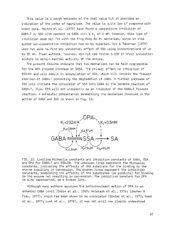
This value is a rough estimate of the real value but it provides an
indication of the order of magnitude. The value is quite low if compared with
known data. Maitre et al. (1975) have found a competitive inhibition of
GABA-T by SSA with respect to GABA with a Ki of 4 mM. However, this type of
inhibition does not fit with the Ping Pong Bi Bi mechanism, since in this
system non-competitive inhibition has to be expected. Dye & Taberner (1975)
were not able to find any inhibitory effect of SSA using concentrations of up
to 20 mM. These authors, however. did not use Triton X-100 in their incubation
mixture to obtain maximal activity of the enzyme.
The present results indicate that two mechanisms can be held responsible
for the DPA induced increase in GABA. The primary effect is inhibition of
SSA-DH and will result in accumulation of SSA, which will inhibit the forward
reaction of GABA-T preventing the degradation of GABA. A further increase of
SSA will initiate the conversion of SSA into GABA by the reverse reaction of
GABA-T: Thus, DPA will act indirectly as an inhibitor of the GABA-T forward
reaction. A schematic presentation summarizing the mechanism involved in the
action of GABA and SSA is shown in Fig. 10.
SA
FIG. 10. Limiting Michaelis constants and inhibition constants of GABA, SSA and DPA for GABA-T and SSA-DH. The unbroken lines represent the Michaelis constants, indicating the affinity of the substrate for the binding to the enzyme resulting in conversion. The broken lines represent the inhibition constants, indicating the affinity of the substrates (or products) for binding to the enzyme not resulting in conversion. The inhibition constant for DPA is also represented, as a broken line.
Although many authors ascribed the anticonvulsant action of DPA to an
enhanced GABA level (Godin et al., 1969; Anlezark et al., 1976; LOscher & Frey, 1977), which has been shown to be correlated (Simler et al., 1973; Wood
et al., 1977; Lust et al., 1978), it was not until now clearly understood
67
how DPA exerts its action, because of its weak inhibitory effect on GABA-T
(Kukino & Deguchi, 1977; Fowler et al., 1975). The action of DPA on SSA-DH
and the effects of the resulting accumulated SSA might explain the increase
in GABA levels in a more satisfactory way. It is interesting to note that
inhibitors of GABA-T like ADAA exert their action via trapping of pyridoxal
phosphate and therefore the action of GAD, which also needs this cofactor,
will be impaired too. Inhibition of SSA-DH might, therefore, be a better
choice to increase GABA levels .<.n v.<.vo.
REFERENCES
Anlezark,G., Horton,R.W., Meldrum,B.S., Sawaya,M.C.B. (1976) Anticonvulsant action of ethanolamine-0-sulphate and di-n-propylacetate and the metabolism of ~-aminobutyric acid (GABA) in mice with audiogenic seizures. Biochem. Pharmac. 25. 413-417
Baxter, C.F., Roberts, E~(l961) Elevation of y-aminobutyric acid in brain: selective inhibition of ~-aminobutyric-~-ketoglutaric acid transaminase. J. Bio1. Chem. 236. 3287-3294
Cleland, W.W. (1963) The kinetics of enzyme-catalyzed reactions with two or more substrates or products 1. Nomenclature and rate equations. Biochim. Biophys. Acta 67, 104-137
De Boer, Th., Bruinvels,J. (1977a) Assay and properties of 4-aminobutyric-2-oxoglutaric acid transaminase and succinic semialdehyde dehydrogenase in rat brain tissue J. Neurochem. 28. 471-478
De Boer, Th., Bruinvels, J. (1977b) S~ccinic semialdehyde dehydrogenase: activation by monovalent and bivalent cations. Proc. Int. Soc. Neurochem. 6, 524
Duffy, T.E., Nelson, S.R., Lowry, O.H. (1972) Cerebral carbohydrate metabolsim during acute hypoxia and recovery. J. ~eurochem. 19, 959-977
Dye, D.J., Taberner, P.V. (1975) The effect of some newer anaesthetics on the .<.n v.<.tko activity of glutamate decarboxylase and GABA transaminase in crude brain extracts and on the levels of amino acids .<.n v.<.vo. J. i\'eurochem. 24, 997-1001
Fowler, L.J., John, R.A. (1972) Active-site-directed irreversible inhibition of rat brain 4-aminobutyrate amino-transferase by ethanolamine-asulphate .<.n v.<.tko and-.<.n v.<.vo. Biochem. J. 130, 569-573
Fowler, L.J., Beckford, J., John, R.A. (1975) An analysis of the inhibition of rabbit brain ~-aminobutyrate aminotransferase by sodium n-dipropylacetate and some other simple carboxylic acids. Biochem. Pharmac. 24, 1267-1270
Garces, E., Cleland, W.W. (1969) Kinetic studies of yeast nucleotide diphosphate kinase. Biochemistry, Easton 8, 633-640
Godin, Y., Heiner, L., Mark., J., Mandel, P.· (1969) Effects of di-n-propylacetate, an anticonvulsive compound, on GABA metabolsim. J. Neurochem. 16, 869-873
Hall, Z.W., Kravitz, E.A. (1967) The metabolism of ~-aminobutyric acid (GABA) in the lobster nervous system-1. GABA-glutamate transaminase. J. Neurochem. 14, 45-54
Harvey, P.K.P., Bradford, H.F., Davison, A.N. (1975) The inhibitory effect of sodium n-dipropylacetate on the degradative enzymes of the GABA shunt. FEBS Lett. 52, 251-254
68
Henson, C.P., Cleland, W.W. (1964) Kinetic studies of glutamic oxaloacetic transaminase isozymes. Biochemistry, Easton 3, 338-345
Horton, R.W., Anlezark, G.M., Sawaya, M.C.B., Meldrum, B.S. (1977) Monoamine and GABA metabolism and the anticonvulsant action of di-n-propylacetate and ethanolamine-0-sulphate. Eur. J. Pharmac. 41, 387-397
John, R.A., Fowler, L.J. (1976) Kinetic and spectral properties of rabbit brain 4-aminobutyrate aminotransferase. Biochem. J. 155, 645-651
Jung, M.J., Lippert, B., Metcalf, B.W., Schechter, P.J., BOhlen, P., Sjoerdsma, A. (1977) The effect of 4-amino-hex-5-ynoic acid (y-acetylenic GABA, Y-ethynyl GABA) a catalytic inhibitor of GABA metabolism ~n v~vo. J. Neurochem. 28. 717-723
Kobayashi, K., Miyaz~wa, S., Endo, A. (1977) Isolation and inhibitory activity of gabaculine, a new potent inhibitor of Y-aminobutyrate aminotransferase produced by a streptomyces. FEBS Lett. 76, 207-210
KrnjeviC, K. (1970) Glutamate and y-aminobutyric acid in brain. Nature, Land. 228, 119-124
Kukino, K., Deguchi, T. (1977) Effects of sodium dipropylacetate on Y-aminobutyric acid and biogenic amines in rat brain. Chern. Pharm. Bull., Tokyo 25, 2257-2262
LOscher, W., Frey, H.H. (1977) On the mechanism of action of valproic acid. Arzneim. Forsch./Drug Res. 27, 1081-1082
Lust, W.O., Kupferberg, H.J., Yonekawa, W.O., Penry, J.K., Passonneau, J.V., Wheaton, A.B. (1978) Changes in brain metabolites induced by convulsants or electroshock: effects of anticonvulsant agents. Malec. Pharmac. 14, 347-356
Maitre, M., Cieselski, L., Mandel, P. (1974) Effect of 2-methyl-2-ethylcaproic and 2-2-dimethyl valerie acid on audiogenic seizures and brain gamma-aminobutyric acid. Biochem. Pharmac. 23, 2363-2368
Maitre, M., Cieselski, L., Cash, C.,Mandel, P. (1975) Purification and studies on some properties of the 4-aminobutyrate: 2-oxoglutarate transaminase from rat brain. Eur. J. Biochem. 52. 157-169
Maitre, M., Ossola, L., Mandel, P. (1976) In v~tko studies into the effect of inhibition of rat brain succinic semialdehyde dehydrogenase on GABAsynthesis and degradation. FEBS Lett. 72, 53-57
Maitre, M., Cieselski, L., Cash, C., Mandel, P. (1978) Comparison of the structural characteristics of the 4-aminobutyrate: 2-oxoglutarate transaminases from rat and human brain, and of their affinities for certain inhibitors. Biochim. Biophys. Acta 522, 385-399
Matsushima, Y., Martell, A.E. (1967) Pyridoxal analogues. X. Zinc (II)chelate catalysis of transamination in methanol solution. J. Am. Chern. Soc. 89, 1331-1335
Meister, A.· (1955) Transamination, in Advances in Enzymology (Nord F.F. ed) Vol. 16, pp.l85-246. Interscience, New York
Pitts, F.N., Quick, C. (1965) Brain succinate semialdehyde dehydrogenase-1, assay and distribution. J. Neurochem. 12, 893-900
Pitts, F.N., Quick, C., Robins, E. (1965) The enzymatic measurement of yaminobutyric-u-oxoglutaric transaminase. J. Neurochem. 12, 93-101
Sawaya, M.C.B., Horton, R.W., Meldrum, B.S. (1975) Effects of anticonvulsant drugs on the cerebral enzymes metabolizing GABA. Epilepsia 16, 649-655
Schechter, P.J., Tranier, Y., Jung, M.J., BOhlen, P. (1977) Audiogenic seizure protection by elevated brain GABA concentration in mice: effects of Yacetylenic-GABA and y-vinyl-GABA, two irreversible GABA-T inhibitors. Eur. J. Pharmac. 45, 319-328
Schobben, F., Vander Kleyn, E. (1974) Pharmacokinetics of distribution and elimination of sodium di-n-propylacetate in mouse and dog. Pharm. Weekb1ad 109, 33.42
69
Schobben, F., Van der Kleyn, E., Vree, T.B., Guelen, P.J.M. (1977) Pharmacokinetics of dipropylacetate (2-n-propyl pentanoate) in man and laboratory animals, in Antiepileptic Drug Monitoring. (Gardner-Thorpe, C., Janz, D., Meinardi, H., Pippenger, C. eds.) pp. 147-169 Pitman, Kent
Schousboe, A., Wu, J.-Y., Roberts, E. (1973) Purification and characterization of the 4-aminobutyrate-2-ketoglutarate transaminase from mouse brain. Biochemistry, Easton 12, 2868-2873
Schwarcz, R., Bennett, J.P., Coyle, J.T. (1977) Inhibitors of GABA metabolism: implications for Hunting ton 1 s disease. Ann. Neural. 2, 299-303
Scott, E.M., Jakoby, W.B. (1959) Soluble y-aminobutyric-glutamic transaminase from Pseudomonas fluorescens. J. Biol. Chern. 234. 932-936
Segel, I.H. (1975) Enzyme Kinetics. ~Iiley, New York . Simler, S., Cieselski, L., Maitre, M., Randrianarisoa, H., Mandel, P. (1973)
Effect of sodium n-dipropylacetate on audiogenic seizures and brain Yaminobutyric acid level. Biochem. Pharmac. 22, 1701-1708
Snell, E.E., Di Mari, S.J. (1970) Schiff base intermediates in enzyme catalyses, in The Enzymes (Boyer, P.O. ed.), 3rd Ed., Vol. 2, pp. 335-370. Academic Press, New York
Sytinski, I.A., Vasilijev, V.Y. (1970) Some catalytic properties of purified Y-aminobutyrate-a-oxoglutarate transaminase from the rat brain. Enzymologia 39, 1-11
Takeda, M., Kinoshita, T., Tsuji, A. (1976) A fluorophotometric method for the determination of a-oxoacids by using pyridoxamine and Zinc (II)ion. Ana1yt. Biochem. 72. 184-190
Tapia, R. (1975) Biochemical pharmacology of GABA in C\JS, in Handbook of Psychopharmacology (Iversen, L.L., Iversen, S.D., Snyder, S.H., eds.) Vol. 4, pp. 1-58. Plenum Press, New York
Tower. D.B. (1976) GABA and seizures: clinical correlates in man. in GABA in Nervous System Function. (Roberts, E., Chase, T.N .. Tower; D.B. eds.) pp. 461-478. Raven Press, New York
Tunnicliff, G., Ngo, T. T. (1977) The mode of action of homocysteine on mouse brain glutamic decarboxylase and y-aminobutyrate aminotransferase. Can. J. Biochem. 55, 1013-1018
Van den Berg, C.J., Matheson, D.F., Ronda, G., Reynierse, G.L.A., Blokhuis, G.C.D., Kroon, M.C., Clarke, D.O., Garfinkel, D. (1975) A model of glutamate metabolism in brain: a biochemical analysis of a heterogenous structure, in Metabolic Compartmentation and Neurotransmission. NATO Advanced Study Institute Series, Vol. 6 (Berl., S., Clarke, D.O., Schneider, D., eds.) pp. 515-543. Plenum Press, New York
Waksman, A., Roberts, E. (1965) Purification and some properties of mouse brain Y-aminobutyric-~-ketoglutaric acid transaminase. Biochemistry, Easton 4. 2132-2138
Wood, J.D., Durham, J.S., Peesker, S.J. (1977) Effect of di-n-propylacetate and Y-acetylenic GABA on hyperbaric oxygen-induced seizures and GABA metabolism. Neurochem. Res. 2, 707-715
70
CHAPTER III
EFFECTS OF BRANCHED-CHAIN-FATTY ACIDS ON GABA-DEGRADATION AND
BEHAVIOUR: FURTHER EVIDE~CE FOR A ROLE OF GABA I~ QUASI-MORPHINE ABSTI~ENCE
BEHAVIOUR
J.W. van der Laan, A.W.C.M. Jacobs and J. Bruinvels
Published in Pharmacology Biochemistry & Behavior 13 (1980)843-849
ABSTRACT
An increase in GABA-ergic activity has been implicated in the initiation
of quasi-morphine abstinence behaviour by di-n-propylacetate (DPA). Two
structural analogues of DPA, namely the branched-chain fatty acids 2-methyl
2-ethylcaproic acid and 2,2-dimethylvaleric acid have now been used to study
this relationship between behavioural and biochemical effects. A correlation
appeared to exist between the Ki of these compounds for succinic semi
aldehyde dehydrogenase, the second enzyme in the degradation of GABA, and
the doses exerting a maximum effect on behaviour. On the other hand, concurrent
inhibition of GABA-transaminase seemed to suppress the behavioural effects
of the fatty acids. This apparent paradox can possibly be explain~d by
supposing a different action of the fatty acids in distinct compartments of
the brain, suggesting an important role for increased GABA-ergic activity in
the neuronal compartment in the initiation of the quasi-morphine abstinence
behaviour.
INTRODUCTION
Elevation of GABA levels by di-n-propylacetate has been shown to occur via
inhibition of succinic semialdehyde dehydrogenase (SSA-DH), the second enzyme
in the degradation of GABA (Harvey et al., 1975; VanderLaan et al., 1979).
The resulting accumulation of succinic semialdehyde (SSA) probably increases
GABA concentration by product inhibition of GABA-transaminase (GABA-T). In
addition, backward formation of GABA from SSA may occur since the reactions
catalysed by GABA-T are strongly biased towards GABA (VanderLaan et al.,
1979).
71
In order to find more evidence for the involvement of this mechanism in
DPA-induced "quasi-morphine abstinence'' behaviour ( De Boer et al., 1977), we
have studied the effects of some analogues of DPA both on behaviour and on the
enzymes involved in GABA-degradation.
MATERIALS AND METHODS
Beha.v-i.oUJta£ Stu.d-i.u
Anhna£6
Male albino rats (100-200 g), randomly selected from an inbred Wistar
strain (TNO, Zeist, The Netherlands), were used for all experiments. The
animals were housed four or five to a plastic cage with food and water ad lib.
Lights were kept on from 8.30 a.m. till 20.30 p.m. Behavioural observations
were performed between 10.00 a.m. and 16.30 p.m. in a room with constant back
ground noise and a constant temperature of 22 ~ 1°C.
Behav-Lowr.
Observation of behaviour was carried out essentially as described by De
Boer et al. (1977). Briefly, the scoring system of Frederickson and Smits
(1973) was used, in which scores of 0 (absent), 2 (mild) or 4 (marked) were
given to penile erection, penis licking, ptosis, teeth chattering, swallowing,
tremor, hunchback posture and piloerection. Escape digging, body shaking, head
shaking and foreleg shaking were counted and assigned scores of 2, 4, 6, 8 or
10 if they occurred 2-5, 6-10. 11-20, 21-40 or more than 40 times, respectively,
during the observation period of 15 min. Horizontal activity was measured
using a Varimex activity meter.
V~ug p~epana£-i.on and adm-i.n-i.o~a£-i.on
Sodium di-n-propylacetate (Sodium Valproas, Albic BV, Maassluis, The
Netherlands) was dissolved in deionized water (0.6 mmol/ml). 2-Methyl, 2-
ethylcaproic acid (MEC) and 2,2-dimethylvaleric acid (DMV) were a gift from
Prof. Dr. A Lespagnol (Lille, France) and were, like caproic acid (Merck,
Darmstadt, GFR) dissolved in deionized water, together with an equimolar
quantity of sodium hydroxide to obtain a similar solution as for DPA. For
intraperitoneal injections, the following concentrations were used: MEC, 0.3
mmol/ml; DMV, 0.4 mmol/ml; and caproate, 1.2 mmol/ml.
72
Apart from the drugs mentioned above, the following compounds were used:
GABA was obtained from Calbiochem; Triton X-100 and 2-mercaptoethanol were
obtained from SOH Chemicals Ltd. SSA was purchased from Sigma Chemical
Company. NAD and NADH were obtained from Boehringer Mannheim. (U-14c)GABA
(specific radioactivity 224 mCi/mmol) was obtained from the Radiochemical
Centre, Amersham. All other chemicals were purchased from Merck, Darmstadt.
T~ue p.tr.epaAation
Randomly selected male Wistar rats (125-175 g) were killed by decapitation
and their brains were rapidly removed and weighed. A 10% (w/v) brain homogenate
was prepared in an ice-cold solution containing 0.32 M-sucrose and 4.5 mM-
2-mercaptoethanol, using a Teflon-glass Potter-Elvehjem homogenizer. One
volume of the homogenate was added to 3 val of ice-cold Triton medium (0.67%
w/v Triton X-100, 50 mM-Tris-HCl (pH 8.5) and 4.5 mM-2-mercaptoethanol) and
kept in ice water for 1 h before use.
GABA-T M-1>111J
For the assay of GABA-T a radiochemical method was used, as previously
described (De Boer and Bruinvels, 1977), with the following modifications.
NAD was omitted from the incubation mixture to exclude interference with SSA
DH. Tissue concentration was 1.11 mg/ml(wet wt.). Drugs, as mentioned above,
were used as their sodium salts and, when necessary, sodium isethionate was
added to obtain a standard sodium concentration of 20 mM in all incubation
media. After preincubation at 22°C for 30 min, the reaction was started by
the addition of homogenate and subsequently the mixture was incubated for a
further 60 min at the same bath temperature. All experiments were carried out
in duplicate.
SSA-VH M-'>111J
Measurement of SSA-DH activity was performed as previously described (De
Boer and Bruinvels, 1977) using a tissue concentration of 0.44 mg/ml (wet
wt.). In all incubations the sodium concentration used was 10 mM. A1
incubations were performed in quadruplicate.
Da.:ta. "-'lLLtlf~.>.U
!he kinetic parameters of GABA- T were ec.lculated from Lineweaver-l::3urke
plots. Since in the assay of SSA-DH more than 5% of the initial concentration
of SSA was converted, "':.he apparent kinetic constants were calculated using
the integratsd form of the Henri-Michaelis-Ment8n equation (Segel, 1975).
73
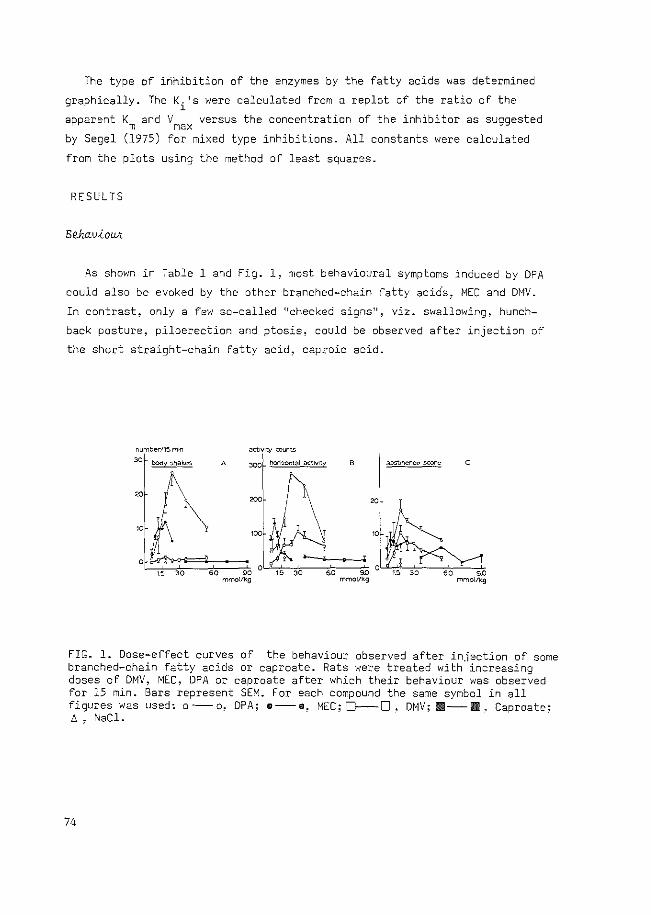
The type of inhibition of the enzymes by the fatty acids was determined
graphically. The Ki's were calculated from a replot of the ratio of the
apparent Km and Vmax versus the concentration of the inhibitor as suggested
by Segel (1975) for mixed type inhibitions. All constants were calculated
from the plots using the method of least squares.
RESULTS
Beha.v-£owr.
As shown in Table 1 and Fig. 1, most behavioural symptoms induced by DPA
could also be evoked by the other branched-chain fatty acids~ MEC and DMV.
In contrast, only a few so-called ''checked signs", viz. swallowing, hunch
back posture, piloerection and ptosis, could be observed after injection of
the short straight-chain fatty acid, caproic acid.
I "'""""" ~oco ,l
c
i
FIG. l. Dose-effect curves of the behaviour observed after injection of some branched-chain fatty acids or caproate. Rats were treated with increasing doses of DMV, MEC, DPA or caproate after which their behaviour was observed for 15 min. Bars represent SEM. For each compound the same symbol in all figures was used: o--o, DPA; m--a, MEC; D-0, DMV; Ill--Ill, Caproate; !J. , NaCl.
74
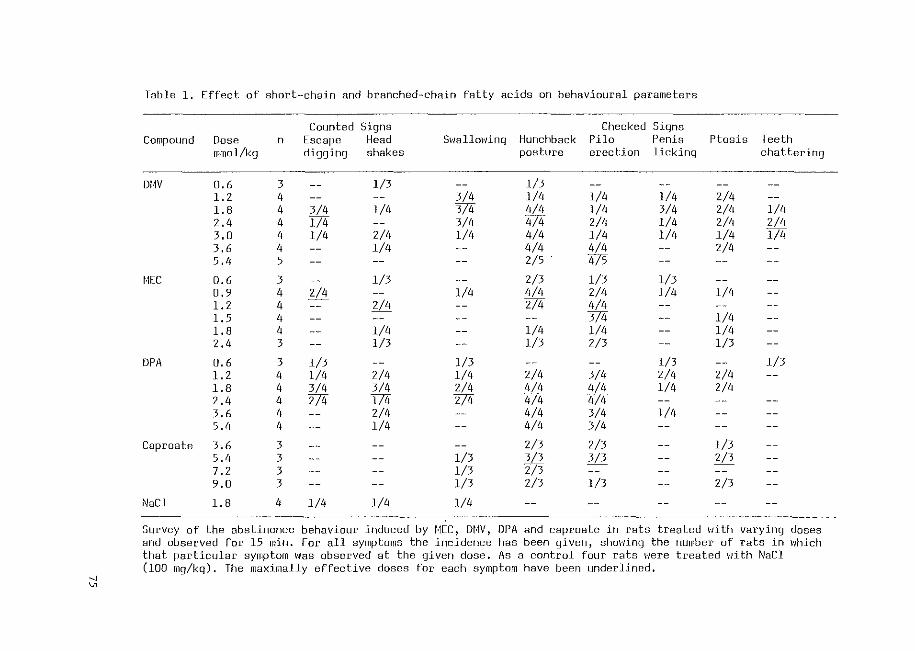
Table 1. Effect of short-chain and branched-chain fatty acids on behavioural parameters ---
Counted Signs Checked Signs Compound Dose n Escape Head Swallowing Hunchback Pilo Penis Ptosis Teeth
mmol/kg digging shakes posture erection licking chattering
---------DIW 0,6 3 -- l/3 -- l/3
1.2 4 -- -- 3/4 l/4 l/4 l/4 2/4 1.8 4 3/4 l/4 3/4 4/4 l/4 3/4 2/4 l/4 2.4 4 l/4 -- 3/4 4/4 2/4 l/4 2/4 2/4 3.0 4 l/4 2/4 l/4 4/4 l/4 l/4 l/4 l/4 3.6 4 -- l/4 -- 4/4 4/4 -- 2/4 5.4 5 -- -- -- 2/5 475
f.IEC 0.6 3 -- l/3 -- 2/3 l/3 l/3 0,9 4 2/4 -- l/4 4/4 2/4 1/4 l/4 1.2 4 -- 2/4 -- 2/4 4/4 -1.5 4 -- -- -- -- 374 -- l/4 1.8 4 -- l/4 -- l/4 l/4 -- l/4 2.4 3 -- l/3 -- 1/3 2/3 -- l/3
OPA 0,6 3 l/3 -- l/3 -- -- l/3 -- l/3 1.2 4 l/4 2/4 l/4 2/4 3/4 2/4 2/4 1.8 4 3/4 3/4 2/4 4/4 4/4 l/4 2/4 2.4 4 274 I74 274 474 474 3.6 4 -- 2/4 -- 4/4 3/4 l/4 5.4 4 -- l/4 -- 4/4 3/4
Caproate 3.6 3 -- -- -- 2/3 2/3 -- l/3 5.4 3 -- -- l/3 3/3 3/3 -- 2/3
3 l/3 2/3 - -
7.2 -- -- -- -- --9.0 3 -- -- l/3 2/3 l/3 -- 2/3
NaCl 1.8 4 l/4 l/4 l/4
Survey of the abstinence behaviour induced by f.JEC, Dt·1V, DPA and caproate in rats treated 1·1ith varying doses and observed for 15 min. For all symptoms the incidence has been given, showing the number of rats in which that particular symptom was observed at the given dose. As a control four rats 1·1ere treated 1·/ith NaCl (lOO mg/kg). The maximally effective doses for each symptom have been underlined.
--J
"'
As can be seen in Fig. 1, the dose which induced the maximal number of
body shakes was, for MEC, 1.2 mmol/kg, whereas with doses higher than 1.8 mmol/
kg a decrease was observed. With DMV, a maximal number of body shakes was
obtained using 1.8 mmol/kg, while for DPA this was the case with 2.4 mmol/kg
(Fig. lA).
Maximum activity after injection of MEC was found using a dose of 0.9 mmol/
kg, while maximal locomotor activity after DPA was obtained using 2.4 mmol/kg
and after DMV, when using 3.0 mmol/kg. Caproate did not alter the activity,
as compared with NaCl treatment, up to doses of 9.0 mmol/kg. The decrease in
motor activity obtained after administration of high doses of the branched
chain fatty acids was caused by a loss of righting reflex occurring within the
observation period (Fig. 18).
As was found for body shakes and locomotor activity, so with regard to the
total abstinence score (Fig. !C), MEC was shown to be the most potent compound,
eliciting a maximum score after administration of 0.9 mmol/kg. For DMV and
DPA the highest abstinence score was evoked using a dose of 1.8 mmol/kg.
Comparing the magnitude of the maximal activity of the various compounds,
DPA was found to be the most potent compound in evoking body shakes, hori
zontal activity and abstinence behaviour (Fiq.l) . Using MEC, a higher number
of body shakes and activity counts was obtained when compared to DMV, whereas
the DMV-induced maximal abstinence score was somewhat higher than found for
MEC. With regard to the checked signs (Table l), no great differences were
apparent between all compounds tested. Therefore, the differences in the
total abstinence score can only be ascribed to differences in counted signs,
such as body shakes and escape digging. As already mentioned, counted signs
were not observed after injection of caproate.
The kinetics of GABA-T were studied using concentrations of 0.5-5.0 mM
GABA and 0.5 mM 2-oxoglutarate. The concentrations of MEC, DMV and caproate
were 5, 10 and 20 mM for each compound. Lineweaver-Burke plots are presented
in Fig. 2. From these plots the Ki's were calculated using a replot of the
ratio of the Kmand Vmax vs the concentration of the inhibitor and the
calculated Ki's are presented in Table 2. MEC was found to be the most potent
inhibitor, DMV was slightly less potent, whereas no inhibition could be found
76
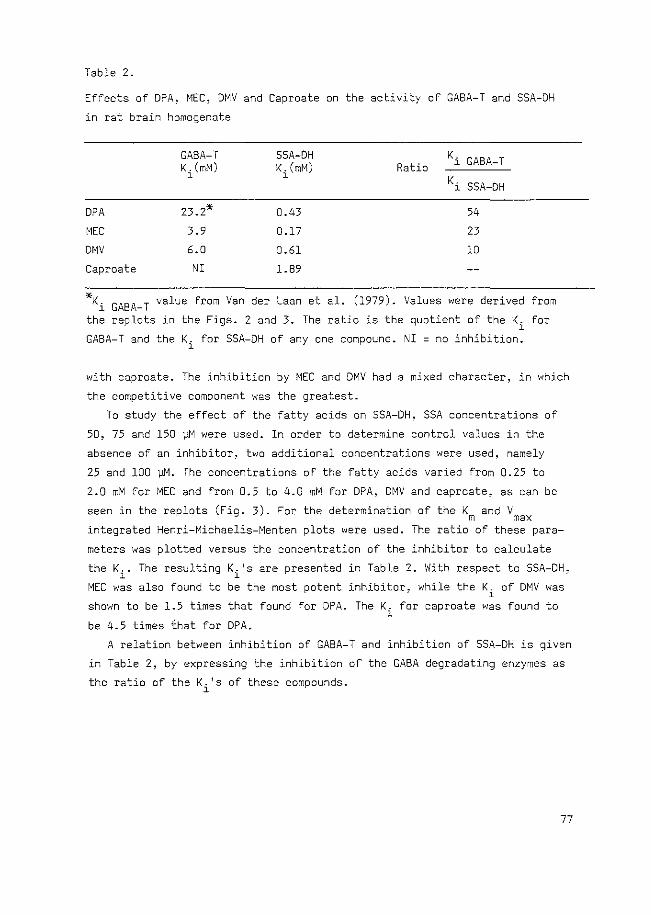
Table 2.
Effects of DPA, MEC, DMV and Caproate on the activity of GABA-T and SSA-DH
in rat brain homogenate
GABA-T SSA-DH K. GABA-T K. (mM) K. (mM) Ratio l
l l
Ki SSA-DH
DPA 23.2* 0.43 54
MEC 3.9 0.17 23
DMV 6.0 0.61 10
Caproate NI 1.89
*Ki GABA-T value from VanderLaan et al. (1979). Values were derived from
the replots in the Figs. 2 and 3. The ratio is the quotient of the Ki for
GABA-T and the Ki for SSA-DH of any one compound. NI =no inhibition.
with caproate. The inhibition by MEC and DMV had a mixed character, in which
the competitive component was the greatest.
To study the effect of the fatty acids on SSA-DH, SSA concentrations of
50, 75 and 150 )JM were used. In order to determine control values in the
absence of an inhibitor, two additional concentrations were used, namely
25 and 100 ~M. The concentrations of the fatty acids varied from 0.25 to
2.0 mM for MEC and from 0.5 to 4.0 mM for DPA, OMV and caproate, as can be
seen in the replots (Fig. 3). For the determination of the K m
and V max integrated Henri-Michaelis-Menten plots were used. The ratio of these para-
meters was plotted versus the concentration of the inhibitor to calculate
the Ki. The resulting Ki 1 s are presented in Table 2. With respect to SSA-DH,
MEC was also found to be the most potent inhibitor, while the Ki of DMV was
shown to be 1.5 times that found for OPA. The K. for caproate was found to l
be 4.5 times fhat for DPA.
A relation between inhibition of GABA-T and inhibition of SSA-OH is given
in Table 2, by expressing the inhibition of the GABA degradating enzymes as
the ratio of the Ki 1 s of these compounds.
77
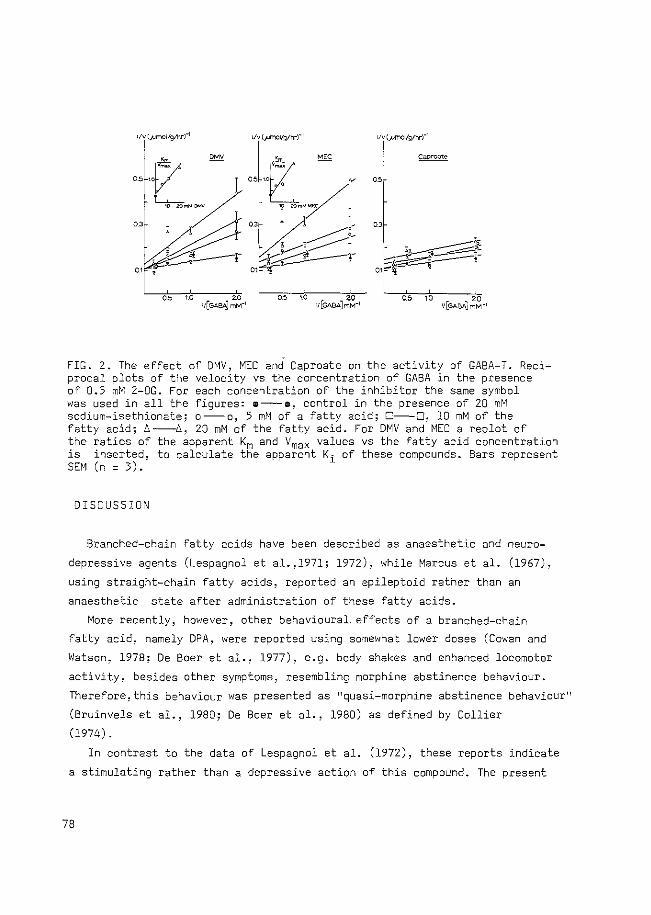
rtv (..l.lmoVglhrr' r!v(_pmol/glhrr1
!
OS
''
,, ,, '' ,, 10 20
11[GA8A]mM"1
FIG. 2. The effect of DMV~ MEC and Caproate on the activity of GABA-T. Reciprocal plots of the velocity vs the concentration of GABA in the presence of 0.5 mM 2-0G. For each concentration of the inhibitor the same symbol was used in all the figures: •--•, control in the presence of 20 mM sodium-isethionate; o--o, 5 mM of a fatty acid; C--D, 10 mM of the fatty acid; n----6~ 20 mM of the fatty acid. For DMV and MEC a replot of the ratios of the apparent Km and Vmax values vs the fatty acid concentration is inserted. to calculate the apparent Ki of these compounds. Bars represent SEM (n = 3) ..
DISCUSSION
Branched-chain fatty acids have been described as anaesthetic and neuro
depressive agents (Lespagnol et al.,l971; 1972), while Marcus et al. (1967),
using straight-chain fatty acids, reported an epileptoid rather than an
anaesthetic state after administration of these fatty acids.
More recently, however, other behavioural, effects of a branched-chain
fatty acid, namely DPA, were reported using somewhat lower doses (Cowan and
Watson, 1978; De Boer et al., 1977), e.g. body shakes and enhanced locomotor
activity, besides other symptoms, resembling morphine abstinence behaviour.
Therefore, this behaviour was presented as 11 quasi-morphine abstinence behaviour 11
(Bruinvels et al., 1980; De Boer et al., 1980) as defined by Collier
(1974).
In contrast to the data of Lespagnol et al. (1972), these reports indicate
a stimulating rather than a depressive action of this compound. The present
78
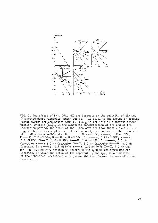
20.0
1~-0 \ 12.0
'' .. ,
FIG. 3. The effect of DMV, DPA, MEC and Caproate on the activity of SSA-DH. Integrated Henri-Michaelis-Menten curves. Y is equal to the amount of product formed during the incubation timet. [SSA] 0 is the initial substrate concentration, whereas [SSA]t is the substrate concentration at the end of the incubation period. The slope of the lines obtained from these curves equals -Km, while the intercept equals the apparent Vm. A: control in the presence of 10 mM sodium-isethionate. B: o--o, 0.5 mM DPA; e--e, 1.0 mM DPA; 0-- 0, 2.0 mM DPA; !ill- IIIIi, 4.0-mM DPA. C: o--o, 0.25 mM MEC; e--o, 0.5 mM MEC; 0-0; 1:0 mM MEC; 111-1111, 2.0 mM MEC. o, o-o, 0.5 mM Caproate; e--e,l.O mM Caproate;--0-C, 2.0 mM Caproate; !D--11111, 4.0 mM Caproate. E: o--.o, 0.5 mM DMV; e--e, 1.0 mM DMV: D-C, 2.0 mM DMV; 1111--111, 4.0 mM DMV. Replots to determine the Ki's of the compounds are inserted, in which the ratio of the apparent Km and Vmax as a function of the inhibitor concentration is given. The results are the mean of three experiments.
79
results show that these behavioural effects could also be evoked after
administration of two other branched-chain fatty acids, MEC and DMV.
Comparison of the doses of the compounds used for maximal induction of
the behavioural syndrome revealed that MEC was more potent than DPA or DMV.
The dose of MEC inducing the maximal score of body shakes~ locomotor
activity or total abstinence (1.0 mmol/kg-the mean of 1.2 for body shakes,
0.9 for horizontal activity and 0.9 for the total abstinence score) was found
to be lower than those found for DPA or DMV (both 2.2 mmol/kg). On the other
hand, the number of body shakes, the locomotor activity and the abstinence
score were higher after injection of DPA at its maximally effective dose
than after the maximally effective doses of DMV or MEC. After injection of
DMV only a few body shakes could be shown, even at the maximally effective
dose, whereas the scores for horizontal activity and abstinence were the same
as for MEC.
The biochemical data obtained for MEC and DMV with regard to GABA-T are
in agreement with those reported by Cash et al. (1975) and Maitre et al.
(1978). However. other studies revealed a Ki for these compounds ten-fold
lower than that obtained in the present study (Cunningham et al., 1980;
Maitre et al., 1974). Possibly, this discrepancy may be due to the use of
different methods. Although the Ki's for SSA-DH of the branched-chain fatty
acids in the present experiments were eight-fold lower than those reported
by Cash et al. (1975), the potency ranking was found to be the same. The K. 1
for DPA was shown to be the same as that reported previously (Van der Laan
et al., 1979).
When considering the inhibition pattern of GABA-T by these compounds, it
appeared that the compound with an intrinsic butyric acid chain, MEC, was the
most potent inhibitor of GABA-T when compared to the compound with a pro
pionic acid chain (DMV) or with a valerie acid chain (DPA); a conclusion
which is in agreement with other reports (Fowler et al., 1975). When
considering inhibition of SSA-DH, the length of the longest chain in the fatty
acids studied seems to be more important, as has been reported previously
for the straight-chain fatty acids (Bruinvels et al., 1980).
The present study clearly shows a great similarity in behavioural
symptoms evoked by the branched-chain fatty acids, while the short straight
chain fatty acid, caproic acid, was inactive in this respect. Furthermore,
the present results show that the three branched-chain fatty acids were
acting as strong inhibitors of the degradation of GABA, especially by
inhibition of SSA-DH.
80
Combining behavioural and biochemical data, a correlation appeared to exist
between the maximally effective doses, evoking the strongest behavioural
effects, and the Ki for SSA-DH of the various compounds studied. Thus, MEC,
which elicits the behavioural syndrome at the low dose of 1.0 mmol/kg, has
also been shown to be the strongest inhibitor of SSA-DH with a K. of 0.17 l
mM. DPA and DMV, both exhibiting mean dose values evoking maximal behavioural
effects higher than that found for MEC, namely 2.2 mmol/kg, are also less
potent with regard to inhibition of SSA-DH with Ki values of 0.43 and 0.61
mM, respectively. The much higher Ki for SSA-DH or caproate (1.89 mM) may,
therefore, indicate why no similar behavioural effects were observed with
this fatty acid.
On the other hand, the number of body shakes, the increase in locomotor
activity and the abstinence score appear to be dependent on the ratio of the
K. 's for GABA-T and SSA-DH (Table 2). The high ratio (54) found for DPA seems l
to correlate with the highest behavioural score obtained with this compound,
while the moderate ratio found for MEC (Sarhan and Seiler, 1979) and the
low ratio found for DMV (Fowler et al., 1975) are in agreement with the
moderate and low behavioural scores, respectively obtained with these
compounds, especially with respect to body shakes.
These results may indicate that inhibition of SSA-DH by these branched
chain fatty acids, thus enhancing GABA levels, is responsible for the
initiation of the quasi-morphine abstinence behaviour, a conclusion which is
in agreement with the findings that the behaviour induced by DPA is antagonized
by bicuculline, picrotoxin or 3-mercaptopropionic acid, but not by
strychnine (De Boer et al., 1977, 1980). However, concurrent inhibition of
GABA-T by these fatty acids -indicated by a lower ratio of the K. 1 S for both l
enzymes- results in a suppression of this abstinence behaviour. Intact GABA-T
activity seems to be a requirement for full expression of the abstinence
behaviour since pre-treatment of rats with the GABA-T inhibitor aminooxyacetic
acid (AOAA) prevents DPA-induced behaviour (De Boer et al., 1980).
Since inhibition of either SSA-DH or GABA-T will both result in an increase
in the GABA concentration, the results can be interpreted in favour of a
compartmentalized action of DPA, suggesting a preferential inhibition of
GABA-degradation, via SSA-DH, in the neuronal compartment as the action
responsible for evoking quasi-abstinence behaviour, whereas an overflow of
GABA to presynaptic areas, possibly GABA-ergic presynaptic receptors, might
be responsible for suppression of this behaviour (Bruinvels et al., 1980;
De Boer, 1977; De Boer et al., 1980).
Bl
A possible presynaptic action of GABA in inhibiting its own release has been
suggested by several authors (Mitchell et al., 1978; Vander Heyden et al.,
1980). Recently, Sarhan and Seiler (1979) presented evidence for a
compartmentalized action of DPA in mouse brain, indicating an increase in
synaptosomal GABA during the first 10 min and thereafter, an increase in non
synaptosomal material. This time course for the increase in synaptosomal
GABA after DPA is in agreement with the time course of the quasi-abstinence
behaviour induced by DPA.Iadarola and Gale (1979) have also provided
evidence that the GABA-increase produced by DPA is associated mainly with
GABA-ergic nerve terminals, while AOAA primarily elevates GABA in non-nerve
terminal compartments.
Using the neuronal model as described above, MEC and DMV can be thought
to inhibit the quasi-abstinence behaviour by enhancement of the concentration
of non-synaptosomal GABA (probably located in glial cells and neuronal cell
bodies). Release of GABA from the glial compartment will promote an action
of GABA on presynaptic GABA-ergic autoreceptors resulting in inhibition of
neuronal GABA-release. This explanation is supported by the recent findings
of Cunningham et al. (1980), showing that MEC and DMV are capable of
inhibiting GABA-dependent oxygen-uptake in non-synaptosomal mitochondria,
while the very weak GABA-T inhibitor DPA was without effect in this system.
Thus, DMV and MEC, but not DPA, inhibit GABA-metabolism in the non
synaptosomal compartment.
Although the role for GABA in true morphine-abstinence behaviour is still
not clear, Ho et al. (1973, 1976) have shown that administration of AOAA or
bicuculline can affect morphine analgesia, the development of tolerance and
the degree of physical dependence.
Studies on the activity of glutamate decarboxylase during morphine
dependency and abstinence (Ho and Gilliland, 1979; Tzeng and Ho, 1978) also
indicate an enhanced synthesis of GABA (which is localized in the nerve
endings) during the abstinence phase when compared to the dependency phase.
In conclusion, the present findings support a role for GABA in quasi
morphine abstinence behaviour evoked by branched-chain fatty acids. Inhibition
of SSA-DH, resulting in an accumulation of GABA in the synaptosomal compart
ment, seems to be responsible for evoking quasi-morphine abstinence behaviour
whereas inhibition of GABA-T, resulting in an increase in GABA-concentration
in the non-synaptosomal compartment, will be responsible for suppression of
this behaviou~ The latter may occur via leakage or release of GABA from this
compartment into the extraneuronal space, promoting an action of GABA on
presynaptically located autoreceptors.
82
REFERENCES
Bruinvels, J., Van der Laan, J.W., De Boer, Th. (1980) GABA-degradation and its possible relationship to the ''morphine abstinence 11 syndrome. Brain Res. Bull. 5, suppl. 2, 797-804
Cash, C., Cieselski, L., Maitre, M., Mandel, P. (1975) Purification de la semialdehyde succinique d8shydrog8nase de cerveau de rat et etude de son inhibition par des acides gras ramifies. C.r. seanc. Soc. Biol. 169. 884-887
Collier, ·H.O.J. (1974) The concept of the quasi-abstinence effect and its use in the investigation of dependence mechanisms. Pharmacology 11, 58-61
Cowan, A., Watson, T. (1978) Lysergic diethylamide antagonizes shaking induced in rats by five chemically different compounds. Psychopharmacology 57, 43-46
Cunningham, J., Clarke, D.O., i\1icklas, W.J. (1980) Oxidative metabolism of 4-aminobutyrate by rat brain mitochondria: inhibition by branched-chain fatty acids. J. Neurochem. 34, 197-202
De Boer, Th.(l977)GABA shunt enzymes and the relationship with morphine abstinence. Thesis, Erasmus University, Rotterdam
De Boer, Th., Bruinvels, J. (1977) Assay and properties of 4-aminobutyric-2-oxoglutaric acid transaminase and succinic semialdehyde dehydrogenase in rat brain tissue. J. Neurochem. 28, 471-478
De Boer, Th, Metselaar, H.J., Bruinvels, J~ (1977) Suppression of GABAinduced abstinence behavior in naive rats by morphine and bicuculline. Life Sci. 20, 933-942
De Boer, Th., Bartels, K., Metselaar, H.J., Bruinvels, J. (1980) Di-npropylacetate induced abstinence behavior as a possible correlate of increased GABA-ergic activity in the rat. Psychopharmacology,71,257-267
Fowler, L.J., Beckford, J., John, R.A. (1975) An analysis of the kinetics of the inhibition of rabbit brain y-aminobutyrate aminotransferase by sodium n-dipropylacetate and some other simple carboxylic acids. Biochem. Pharmac. 24, 1267-1270
Frederickson, R.C.A., Smits, S.E. (1973) Time course of dependence and tolerance development in rats treated with "slow release 11 morphine suspensions. Res. communs. chem.pathol. Pharmac. 5, 867-870
Harvey, P.K.P., Bradford, H.F., Davison, A.N. (1975) The inhibitory effect of sodium n-dipropy1acetate on the degradative enzymes of the GABAshunt. FEBS lett. 52, 251-254
Ho, I.K., Gilliland, T. (1979) Effects of acute and continuous morphine administration on the affinity of glutamic acid decarboxylase for pyridoxal 5~phosphate. Biochem. Pharmac. 28, 355-360
Ho, I.K., Loh, H.H., ~lay, E.L. (1973) Influence of GABA on morphine analgesia, tolerance and physical dependence. Proc. West. Pharmac. Soc. 16, 4-7
Ho, I.K., Loh, H.H., Way, E.L. (1976) Pharmacological manipulation of Yaminobutyric acid (GABA) in morphine analgesia, tolerance and physical dependence. Life Sci. 18, 1111-1123
Iadarola, M.J., Gale, K. (1979) Dissociation between drug-induced increases in nerve terminal and non-nerve terminal pools of GABA ~n v~vo. Eur. J. Pharmac. 59, 125-129
83
Lespagnol, A., Mercier, J., Erb-Debruyne, F., Dessaigne, S. (1971) Surles proprietes pharmacodynamiques du methyl-ethyl-caproate de sodium et en particu1ier son action anesthetique generale de courte duree apres injection intraveineuse. Therapie 26, 969-981
Lespagnol, A., Mercier, J., Erb-Debruyne, F., Dessaigne, S. (1972) Surles proprietes neurodepressives et antiepileptiques d 1 acides organiques 8 chaine ramifiee. Annls. pharm. fr. 30, 193-198
Marcus, R.J., Winters, W.D., Mori, K., Spooner, C.E. (1967) EEG and behavioral comparison of the effects of gamma-hydroxybutyrate, gamma-butyro1actone and short chain fatty acids in the rat. Int. J. ,\!europharm. 6, 175-185
Maitre, M., Cieselski, L., Cash, C., Mandel, P. (1978) Comparison of the structural characteristics of the 4-amino-butyrate: 2-oxoglutarate transaminases from the rat and human brain, and their affinities for certain inhibitors. Biochim. Biophys. Acta 522, 385-399
Maitre, M., Cieselski, L., Mandel, P. (1974) Effect of 2-methyl, 2-ethy1 caproic acid and 2,2-dimethyl valerie acid on audiogenic seizures and brain gamma aminobutyric acid. Biochem. Pharmac. 23, 2363-2368
Mitchell, P.R., Martin, l.L. (1976) Is GABA release modulated by presynaptic receptors? Nature 274, 904-906
Sarhan, 5., Seiler, :\!. (1979) Metabolic inhibitors and subcellular distribution of GABA. J. 1\'eurosci. Res. 4. 399-422
Segel, I.H. (1975) Enzyme Kinetics, John WileY, New York Tzeng, S.F., Ho, I.K. (1978) Acute and continuous morphine administration
on the Y-aminobutyric acid system in the mouse. Prog. ~'euro-Psych opharmac. 2, 55-64
Van der Heyden, J.A.M., Venema, K., Korf, J. (1980) In v~vo release of endogenous GABA from rat striatum: effects of muscimol, oxotremorine and morphine. J. Neurochem. 34, 1648-1653
Van der Laan, J.W., De Boer, Th., Bruinvels, J. (1979) Di-n-propylacetate and GABA-degradation. Preferential inhibition of succinic semialdehyde dehydrogenase and indirect inhibition of GABA-transaminase. J. Neurochem. 32, 1769-1780
84
CHAPTER IV
A DUAL ROLE FOR GABA I~ QUASI-MORPHINE ABSTINENCE BEHAVIOUR
INDUCED BY DI-N-PROPYLACETATE INVOLVING BOTH I~ITIATION AND
TERMINATION
J.W. van der Laan and J. Bruinvels
Psychopharmacology, in press
ABSTRACT
Di-n-propylacetate (OPA) induces in rats a behavioural syndrome resembling
morphine abstinence behaviour. The inhibitory action of DPA on GABA
degradation, resulting in an enhanced release of GABA, is probably responsible
for this behavioural effect since GABA antagonists like bicuculline and
picrotoxin have been shown to suppress this behaviour. However, the time
course of the DPA induced behaviour is much shorter than that of the DPA
induced enhancement of the GABA concentration. Therefore, we have studied
the influence of enhanced GABA levels caused by a first injection of DPA and
the behaviour evoked by a second injection of DPA at different time intervals
after the first injection. The results indicate that GABA fulfils a role both
in the initiation and termination of DPA induced behaviour. The mechanism
responsible for this dual action of GABA is ascribed to a differential
sensitivity to DPA of the nerve terminal and glial metabolic compartments
of GABA in the brain. The increase of GABA in the nerve terminal caused by
DPA is probably responsible for the initiation of the quasi-abstinence
behaviour, whereas the overflow of GABA into the synaptic cleft may be
responsible for the suppression of this behaviour via stimulation of pre
synaptic autoreceptors.
Another mechanism responsible for the rapid termination of the DPA
evoked behaviour can be the formation of metabolites of DPA which may anta
gonize this behaviour. From experiments using some primary metabolites of
DPA a role for these metabolites in the termination of the DPA induced
behaviour seemed unlikely.
85
INTRODUCTION
It has been shown that administration of sodium-di-n-propylacetate (300
mg/kg i.p.; sodium valproate, DepakineR, DPA) to naive rats induces a
behavioural syndrome, including body shakes, resembling morphine abstinence
behaviour (De Boer et al., 1977). This behaviour is antagonized by the GABA
antagonists bicuculline and picrotoxin, but not by strychnine, a glycine
antagonist, and a role for GABA in the expression of abstinence signs has
been suggested (De Boer et al., 1977, 1980). In addition, it has been shown
that DPA inhibits the degradation of GABA via inhibition of succinic semi
aldehyde dehydrogenase (Van der Laan et al., 1979). The behaviour induced
by DPA in rats has, therefore, been suggested to represent a model for
increased GABA-ergic activity (De Boer et al., 1980).
However, the time course of the increase in GABA concentration in rat
brain after injection of DPA differs from the time course of the behaviour
evoked by DPA. The latter reaches a peak value after 5 to 6 min and
disappears after about 15 min (De Boer et al., 1977), while the maximum
increase in GABA levels occurs 45 min after DPA administration and after
4 h has almost returned to normal (Kukino and Deguchi, 1977).
Thus, although the behaviour evoked by DPA appears to be initiated by an
enhanced GABA-ergic activity, no correlation can be found between the
time courses of behaviour and increased GABA. On the contrary, while the
GABA concentration in brain is still increasing, the behaviour evoked by
DPA has disappeared before the maximum GABA concentration has been achieved.
To explain this paradox one can postulate that the behaviour is initiated
by an action of DPA on the GABA-ergic nerve terminals -where DPA probably
exerts its main action (Sarhan and Seiler, 1979; Iadarola and Gale, 1979)
by increasing the concentration of GABA and thus promoting the release of
greater quantities of GABA. An overflow of GABA to presynaptic areas would
subsequently stimulate the putative GABA-ergic autoreceptors, which may
suppress the release of GABA and would thus terminate the DPA-evoked
behaviour.
In the present study, directed towards the possible dual role of GABA in
DPA-induced behaviour, namely in both initiation and termination of this
behaviour, we have tested whether a second injection of DPA during the
increase or during the decrease in the GABA concentration,caused by the first
injection of DPA,would evoke the behaviour to the full extent.
86
Furthermore, the occurrence of tolerance to the behavioural effect was
studied after repeated injections of DPA using longer time intervals, namely
daily and weekly intervals. A possible development of tolerance over these
time intervals is probably not related to enhanced GABA-levels, since 4 h
and certainly 48 h after the DPA injection the GABA concentration has returned
to normal (Kukino and Deguchi, 1977; Grimm et al., 1975).
Another possible mechanism responsible for the termination of the behaviour
could be the rapid formation of one or more metabolites which do not
evoke this behaviour themselves but may antagonize the behaviour induced by
DPA. Therefore, three primary metabolites of DPA (Schafer and Luhrs, 1978)
were used to study this possibility.
The results obtained suggest that GABA indeed fulfils a dual role in the
behaviour induced by DPA. No effect of the metabolites of DPA could be shown
on the behaviour.
MATERIALS AND METHODS
AnimaL\
Male albino rats (100-200 g) randomly selected from an inbred Wistar
strain (TNO, Zeist, the i\'etherlands) were used in all experiments. The
animals were housed four or five to a plastic cage with food and wat~r ad
lib. Lights were kept on from 8.30 a.m. till 8.30 p.m. The experiments were
performed between 9.00 a.m. and 4.30 p.m. in a room with white noise and at
a constant temperature of 22 + 1°C.
VJtug-6
Sodium di-n-propylacetate (Sodium valproate, Albic BV, Maassluis, the
Netherlands) was dissolved in deionized water (100 mg/ml) and administered
intraperitone3lly in a volume of 3 ml/kg .(total dose 300 mg/kg), with the
exception of experiment 2, where higher doses of DPA were used.
Controls received an equimolar NaCl solution (33 mg/ml). The hydroxy
metabolites of DPA, sodium 3-hydroxy, 2-propylvalerate (3-0HDPA), sodium
5-hydroxy, 2-propylvalerate (5-0HDPA) and 4-hydroxy, 2-propylvalerolactone
(4-0HDPA) were a gift from Dr. H. Schafer (Desitin Werke Carl Klinke GmbH,
Hamburg, FRG). The sodium salts of the metabolites were provided as a
10% solution in distilled water and were diluted to the appropriate volume
with deionized water, whereas the lactone was dissolved in an equimolar
quantity of sodium hydroxide to hydrolyse the lactone.
87
Behav-LoWL Observation of the behaviour was carried out essentially as described
by De Boer et al. (1977). Briefly, rats were allowed to adjust to a cage with
sawdust on the bottom for at least 30 min, whereafter the drug or salt
solution was administered intraperitoneally. Subsequently, the behaviour
was observed for 15 min. Scores of 0 (absent), 2 (mild) or 4 (marked) were
given to penile erection, penis licking, teeth chattering, swallowing, tremor,
hunchback posture and piloerection. Escape digging, body shakes and foreleg
shakes were counted and assigned scores of 2, 4, 6, 8 or 10 if they
occurred 2-5, 6-10, 11-20, 21-40 or more than 40 times, respectively, during
the observation period of 15 min. Other signs characteristic of the morphine
abstinence syndrome, as indicated by Frederickson and Smits (1973) were not
seen in DPA-treated animals.
A~u v"-:ty meMwwnent.o
Horizontal and vertical activity was measured using a Varimex activity
meter as described by De Boer et al. (1977).
s:to.J:Mu~~
The Mann-Whitney U-test, the Wilcoxon paired signed rank test and Fisher's
exact probability test (Siegel, 1956) were used for evaluating the results
as indicated in the legends to the tables and figures. Differences with a
probability equal to or lower than 5% were considered to be statistically
significant.
PROCEDURES
ExpvrJment: 1
Rats were treated with DPA (300 mg/kg i.p.) and their behaviour was
observed. After 20 min, 2 h or 4 h the rats received a second injection of
DPA (300 mg/kg i.p.), whereafter behaviour was scored again for 15 min.
Controls received as a first or a second treatment an injection of NaCl
(100 mg/kg i.p.).
Expvr."-ment: 2
Naive rats received graded doses of DPA, i.e. 300, 400, 600 or 900 mg/kg
i.p., whereafter their behaviour was scored for 15 min. The observation
period was divided into three subperiods of five minutes.
88
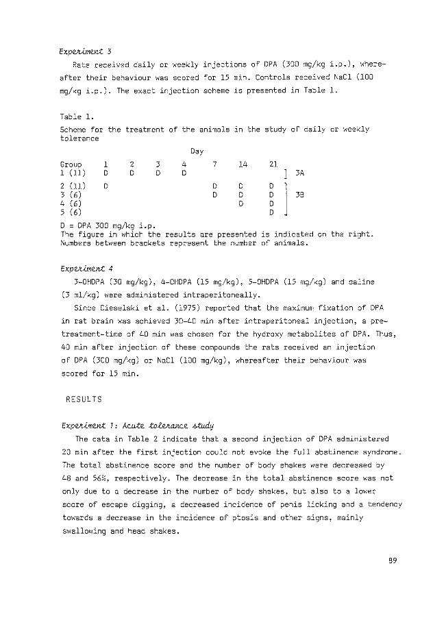
ExpVt-tme.n;t 3
Rats received daily or weekly injections of DPA (300 mg/kg i.p.), where
after their behaviour was scored for 15 min. Controls received NaCl (100
mg/kg i.p.). The exact injection scheme is presented in Table 1.
Table 1.
Scheme for the treatment of the animals in the study of daily or weekly tolerance
Day
Group 1 2 3 4 7 14 21 1 (11) D D D D 3A
2 (11) D D D D 1 ' 3 (6) D D D j 36
4 (6) D D 5 (6) D
D = DPA 300 mg/kg i.p. The figure in which the results are presented is indicated on the right. i\'umbers between brackets represent the number of animals.
ExpVl.iment 4
3-0HDPA (30 mg/kg), 4-0HDPA (15 mg/kg), 5-0HDPA (15 mg/kg) and saline
(3 ml/kg) were administered intraperitoneally.
Since Cieselski et al. (1975) reported that the maximum fixation of DPA
in rat brain was achieved 30-40 min after intraperitoneal injection, a pre
treatment-time of 40 min was chosen for the hydroxy metabolites of DPA. Thus,
40 min after injection of these compounds the rats received an injection
of DPA (300 mg/kg) or NaCl (100 mg/kg), whereafter their behaviour was
scored for 15 min.
RESULTS
ExpVt.iment 1 , Mu-te :totVtcmce >.>:tv.dy
The data in Table 2 indicate that a second injection of DPA administered
20 min after the first injection could not evoke the full abstinence syndrome.
The total abstinence score and the number of body shakes were decreased by
48 and 56%, respectively. The decrease in the total abstinence score was not
only due to a decrease in the number of body shakes, but also to a lower
score of escape digging, a decreased incidence of penis licking and a tendency
towards a decrease in the incidence of ptosis and other signs, mainly
swallowing and head shakes.
89
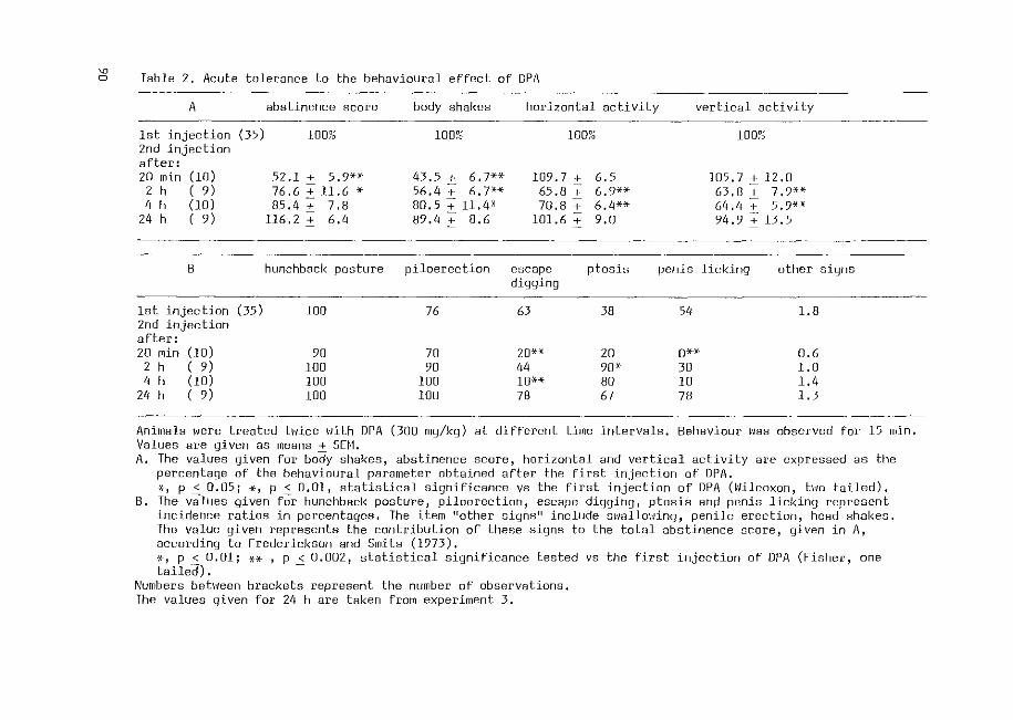
~ Table 2. Acute tolerance to the behavioural effect of DPA
A abstinence score body shakes horizontal activity vertical activity
lst injection (35) 1007~ 100?0 100?~ 100~0 2nd injection after: 20 min (10) 52.1 2:_ 5,9*><· 43.5 + 6.7** 109.7 + 6.5 105.7 :+: 12.0
2 h ( 9) 76.6 :+: 11.6 * 56.4 + 6, 7-H 65.8 + 6.9** 63,8+ 7,9H 4 h (lo) 85.4 + 7.8 80.5 + 11.4* 70.8 + 6.4** 64,4 + 5,9-)(--X
211 h ( 9) 116.2 ± 6.4 89.4 + 8.6 101.6 + 9.0 94.9 + 13.5
8 hunchback posture piloerection escape ptosis penis licking other signs digging
1st injection (35) 100 76 63 38 54 l.B 2nd injection after: 20 min (10) 90 70 20*-X 20 O*' 0.6
2 h ( 9) 100 90 44 90* 30 l.O 4 h (10) 100 100 lO·H 80 10 1.4
24 h ( 9) 100 100 78 67 7B 1.3
Animals \·1ere treated hlice Nith DPA (300 mg/kg) at different time intervals. Behaviour \'las observed for 15 min. Values are given as means + SEH. A. The values given for body shakes, abstinence score, horizontal and vertical activity are expressed as the
percentaqe of the behavioural parameter obtained after the first injection of DPA. x, p < 0.05; *' p < 0.01, statistical significance vs the first injection of DPA (\'/ilcoxon, hm tailed).
B. The v8lues given fOr hunchback posture, piloerection, escape diqging, ptosis and penis licking represent incidence ratios in percentages, The item 11other siqns'' include 8\<18llm1inq, penile erection, head shakes, The value given represents the contribution of these signs to the total abstinence score, given in A, according to Frederickson and Smits (1973). *' p < 0.01; ** , p < 0.002, statistical significance tested vs the first injection of DPA (Fisher, one tailed). -
Numbers bet\'/een brackets represent the number of observations, The values given for 24 h are taken from experiment 3.
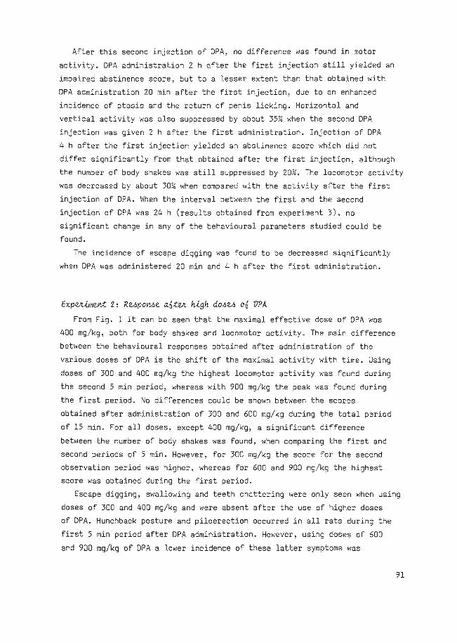
After this second injection of DPA, no difference was found in motor
activity. DPA administration 2 h after the first injection still yielded an
impaired abstinence score, but to a lesser extent than that obtained with
DPA administration 20 min after the first injection, due to an enhanced
incidence of ptosis and the return of penis licking. Horizontal and
vertical activity was also suppressed by about 35% when the second DPA
injection was given 2 h after the first administration. Injection of DPA
4 h after the first injection yielded an abstinence score which did not
differ significantly from that obtained after the first injection, although
the number of body shakes was still suppressed by 20%. The locomotor activity
was decreased by about 30% when compared with the activity after the first
injection of DPA. When the interval between the first and the second
injection of DPA was 24 h (results obtained from experiment 3), no
significant change in any of the behavioural parameters studied could be
found.
The incidence of escape digging was found to be decreased significantly
when DPA was administered 20 min and 4 h after the first administration.
ExpVt-UnenJ: Z' RupoMe a.fi:tVt rugh dMU ot] VPA
From Fig. 1 it can be seen that the maximal effective dose of DPA was
400 mg/kg, both for body shakes and locomotor activity. The main difference
between the behavioural responses obtained after administration of the
various doses of DPA is the shift of the maximal activity with time. Using
doses of 300 and 400 mg/kg the highest locomotor activity was found during
the second 5 min period, whereas with 900 mg/kg the peak was found during
the first period. No differences could be shown between the scores
obtained after administration of 300 and 600 mg/kg during the total period
of 15 min. For all doses~ except 400 mg/kg, a significant difference
between the number of body shakes was found, when comparing the first and
second periods of 5 min. However, for 300 mg/kg the score for the second
observation period was higher, whereas for 600 and 900 mg/kg the highest
score was obtained during the first period.
Escape digging, swallowing and teeth chattering were only seen when using
doses of 300 and 400 mg/kg and were absent after the use of higher doses
of DPA. Hunchback posture and piloerection occurred in all rats during the
first 5 min period after DPA administration. However, using doses of 600
and 900 mg/kg of DPA a lower incidence of these latter symptoms was
91
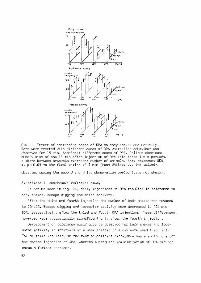
Body shakes l>Ody SM.:li«-~1~ min.
Horizontal activity
Vertical activity
'" "' "'" ,., 5 mrn.
mglkg
FIG. 1. Effect of increasing doses of DPA on body shakes and activity. Rats were treated with different doses of DPA whereafter behaviour was observed for 15 min. Abscissa: different doses of DPA. Oblique abscissa: subdivision of the 15 min after injection of DPA into three 5 min periods. ,'\!umbers between brackets represent number of animals. Bars represent SEM, *' p < 0.05 vs the first period of 5 min (Mann Whitney-U., two tailed).
observed during the second and third observation period (data not shown).
ExpVt-imen-t 3, _;u6chlto>Uc :totVt.mce <~:tudy
As can be seen in Fig. 2A, daily injections of DPA resulted in tolerance to
body shakes, escape digging and motor activity.
After the third and fourth injection the number of body shakes was reduced
to 50-60%. Escape digging and locomotor activity were decreased to 40% and
80%, respectively, after the third and fourth DPA injection. These differences,
however, were statistically significant only after the fourth injection.
Development of tolerance could also be observed for body shakes and loco
motor activity if intervals of a week instead of a day were used (Fig. 28).
The decrease resulting in the most significant difference was also found after
the second injection of DPA, whereas subsequent administration of DPA did not
cause a further decrease.
92
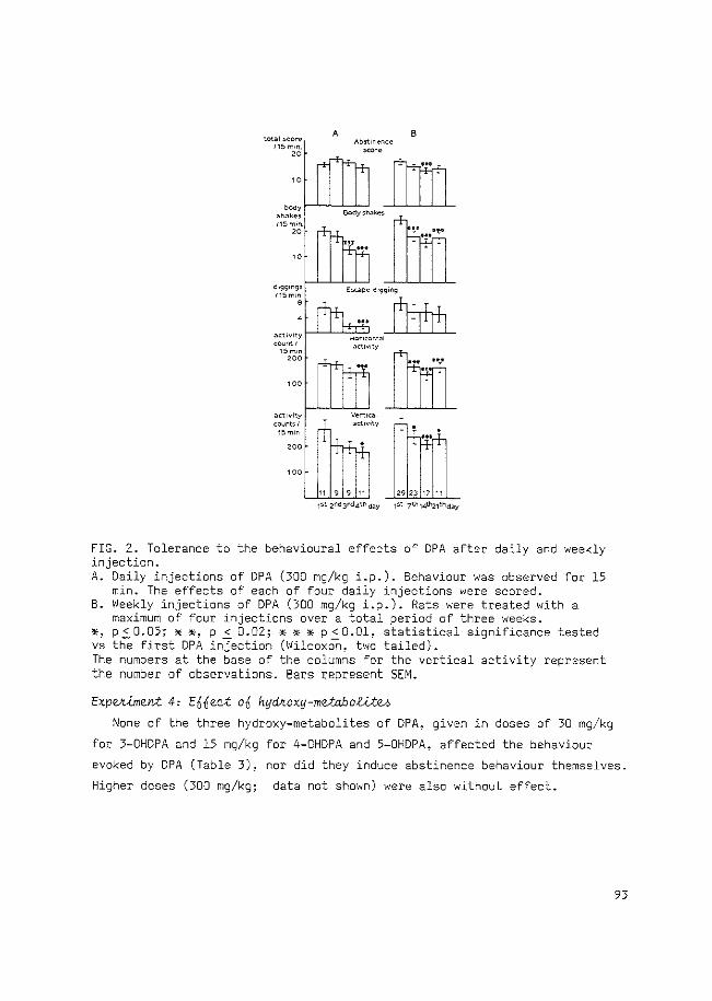
total !>COr<' 115 m•n
"
.lCtl\li~y
count:> I 15mJn
A 8
..:ore
119911 29231711
1:>t 2nd3rd4t~day 1st 7t!11.#121thday
FIG. 2. Tolerance to the behavioural effects of DPA after daily and weekly injection. A. Daily injections of DPA (300 mg/kg i.p.). Behaviour was observed for 15
min. The effects of each of four daily injections were scored. B. Weekly injections of DPA (300 mg/kg i.p.). Rats were treated with a
maximum of four injections over a total period of three weeks. ~, p < 0.05; * *~ p < 0.02; * * * p < 0.01, statistical significance tested vs the first DPA in}ection (Wilcoxon, two tailed). The numbers at the base of the columns for the vertical activity represent the number of observations. Bars represent SEM.
Expekjment 4, E66eQt o6 hydkoxy-metabo~eh None of the three hydroxy-metabolites of DPA, given in doses of 30 mg/kg
for 3-0HDPA and 15 mg/kg for 4-0HDPA and 5-0HDPA, affected the behaviour
evoked by DPA (Table 3), nor did they induce abstinence behaviour themselves.
Higher doses (300 mg/kg; data not shown) were also without effect.
93
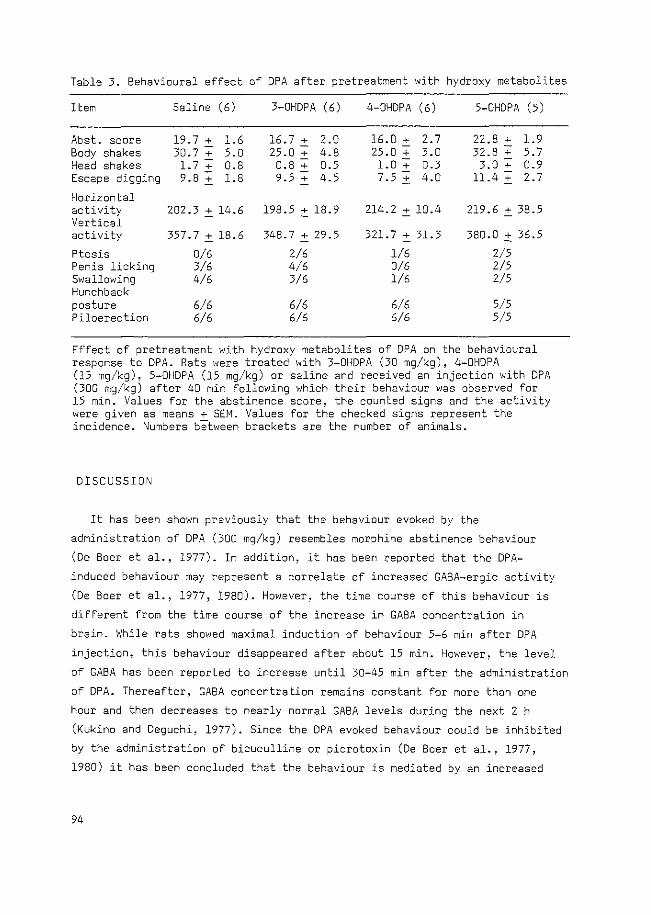
Table 3. Behavioural effect of DPA after pretreatment with hydroxy metabolites
Item Saline (6) 3-0HDPA (6) 4-0HDPA (6) 5-0HDPA (5)
Abst. score 19.7 + 1.6 16.7 + 2.0 16.0 + 2.7 22.8 + 1.9 Body shakes 30.7 + 5.0 25.0 + 4.8 25.0 + 3.0 32.8 + 5.7 Head shakes 1.7+ 0.8 0.8 + 0.5 1.0+ 0.3 3.0 + 0.9 Escape digging 9.8 + 1.8 9.5 + 4.5 7.5 + 4.0 11.4 + 2.7
Horizontal activity 202.3 + 14.6 198.5 + 18.9 214.2 + 10.4 219.6 + 38.5 Vertical activity 357.7 + 18.6 348.7 + 29.5 321.7 + 31.3 380.0 :': 36.5
Ptosis 0/6 2/6 1/6 2/5 Penis licking 3/6 4/6 0/6 2/5 Swallowing 4/6 3/6 l/6 2/5 Hunchback posture 6/6 6/6 6/6 5/5 Piloerection 6/6 6/6 6/6 5/5
Effect of pretreatment with hydroxy metabolites of DPA on the behavioural response to DPA. Rats were treated with 3-0HDPA (30 mg/kg), 4-0HDPA (15 mg/kg), 5-0HDPA (15 mg/kg) or saline and received an injection with DPA (300 mg/kg) after 40 min following which their behaviour was observed for 15 min. Values for the abstinence score, the counted signs and the activity were given as means + SEM. Values for the checked signs represent the incidence. Numbers between brackets are the number of animals.
DISCUSSION
It has been shown previously that the behaviour evoked by the
administration of DPA (300 mg/kg) resembles morphine abstinence behaviour
(De Boer et al., 1977). In addition, it has been reported that the DPA
induced behaviour may represent a correlate of increased GABA-ergic activity
(De Boer et al., 1977, 1980). However, the time course of this behaviour is
different from the time course of the increase in GABA concentration in
brain. While rats showed maximal induction of behaviour 5-6 min after DPA
injection, this behaviour disappeared after about 15 min. However, the level
of GABA has been reported to increase until 30-45 min after the administration
of DPA. Thereafter, GABA concentration remains constant for more than one
hour and then decreases to nearly normal GABA levels during the next 2 h
(Kukino and Deguchi, 1977). Since the DPA evoked behaviour could be inhibited
by the administration of bicuculline or picrotoxin (De Boer et al., 1977,
1980) it has been concluded that the behaviour is mediated by an increased
94
GABA-ergic stimulation. However, the short duration of the behaviour as
compared to the increase in brain GABA level does not seem to favour a
correlation between those two phenomena. In the present paper the effect of
pretreatment with OPA itself, which will cause an increase in brain GABA con-
centration, has been studied on the OPA induced behaviour evoked by a second
injection of OPA.
When rats, pretreated with OPA, received a second injection of DPA shortly
after the first injection. the latter could not evoke the behaviour to the
same extent as that obtained after the first OPA administration. The
strongest suppression of body shakes and total abstinence score could be
observed if a second injection of OPA was given 20 min or 2 h after the first
DPA administration. However, if DPA was administered 4 h after the first
injection, the behavioural scores obtained were still somewhat impaired,
but only the number of body shakes differed significantly from that obtained
after the first DPA injection. The reason that this lower number of body
shakes is not reflected in a lower abstinence score lies with the method
of calculation of the abstinence score, i.e. there is a certain range in
the number of body shakes to which the same score will be given for
calculation of the abstinence score (see under Materials and Methods).
When the time course of the behavioural suppression is compared with
the known time course of the increase in GABA concentration after a single
injection of DPA (Kukino and Oeguchi, 1977; v-Lde. -6upJr.a.), it rather suggests
that suppression of the DPA evoked behaviour and not the behaviour itself
was induced by an enhanced GABA level. This suppression is already maximal
during the increase in the brain GABA concentration and is still present
2 h after the OPA injection, when the brain GABA level has achieved maximum.
When, however, the level of GABA is decreased at 4 h after administration
of DPA, the abstinence behaviour can be evoked by a second injection of DPA
to nearly the full extent. Therefore, the results strongly suggest a
relationship between inhibition of DPA-evoked behaviour and GABA concentration
in the brain. In contrast, the decrease in locomotor activity, which was
found to have a different time course when compared with the body shakes
(Table 2), is probably caused by a mechanism secondary to the enhanced
GABA leveL
That an increase in the GABA concentration will suppress DPA-induced
behaviour is supported by the finding that pretreatment with low doses of
the GABA-T inhibitor amino-oxyacetic acid (AOAA) prevents the induction of
this behaviour (De Boer et al., 1980).
95
The question arises as to the mechanism of the DPA-induced enhancement
of GABA, since on the one hand it seems obvious that enhancement of GABA
ergic stimulation is involved in the DPA-evoked behaviour, while, on the
other hand GABA may play a role in suppression of this behaviour.
It is well-known that the metabolism of GABA is compartmentalized
(Balazs et al., 1972). It may be that these compartments are associated
with certain functionally distinguishable structures in the brain, such as
nerve endings, glial cells and neuronal cell bodies (for a review see
Hertz, 1979). To explain the fact that pretreatment of rats with ADAA pre
vents the induction of abstinence behaviour by DPA, it has been suggested
that accumulation of GABA by AOAA will result in leaking of GABA from
glial cells reaching presynaptic autoreceptors, thus inhibiting the
enhanced release of GABA induced by DPA (De Boer, 1977; De Boer et al.,
1980). Recently, a different localization for the GABA-enhancing action of
DPA and AOAA has been reported (Iadarola and Gale, 1979), indicating that
DPA only exerts its action in the neuronal compartment, while AOAA acts in
both the glial and the neuronal compartment.
The present findings, therefore, support the postulated model in which
the DPA-induced increase in nerve terminal GABA results in an increased
release of GABA into the synaptic cleft and an activation of a post
synaptically localized receptor. Subsequently, a further increase in the
GABA concentration may activate the GABA-ergic autoreceptors resulting in an
inhibition of the enhanced GABA release. Regulation of GABA release via
stimulation of presynaptic autoreceptors has been reported (Mitchell and
Martin, 1978; Vander Heyden et al., 1980). This mechanism, involving an
overflow of GABA has been supported recently by Sarhan and Seiler (1979)
who described an initial rise of GABA induced by DPA during the first 10 min
after its administration in synaptosomal fractions only, whereas after this
period an increase in non-synaptosomal fractions is observed.
It has been reported that the DPA-induced enhancement of the GABA
concentration is dose-dependent (Simler et al., 1978), but that the time
to achieve the maximal increase appears to be the same at all doses. This
indicates that a higher dose of DPA will result in a more rapid increase in
GABA levels. Therefore, different doses of DPA were used to study the effect
of this more rapid enhancement on the time course of the behavioural
syndrome.
The results obtained are in agreement with the dual role of GABA in the
behaviour as discussed above. On the one hand, a more rapid increase in GABA
96
will result in a shorter time of onset of the behaviour and a higher
initial score, while on the other hand, due to the faster increase in GABA
concentration, the overflow of GABA to GABA-ergic autoreceptors will occur
earlier after the injection of DPA.
Alternatively, the present data might be explained assuming different
functions for GABA-ergic systems, which are localized in different areas of
the brain viz. an inhibitory function e.g. in the nucleus accumbens
(Scheel-KrUger et al., 1978) and a disinhibitory one e.g. in the ventral
tegmental area or in the substantia nigra (Arnt and Scheel-KrUger, 1979;
Scheel-KrUger, 1978). In this hypothesis DPA would enhance GABA-ergic trans
mission at first in excitatory areas, while secondary an increase in the
GABA concentration in other areas will result in inhibition. However, the
finding that treatment of rats with a low dose of AOAA did not result in a
similar syndrome as that observed after DPA and even suppressed the latter
(De Boer et al .. 1980) would favour an interpretation involving autorecep
tors as discussed above.
The results presented in Fig. 2 indicate the induction of tolerance after
both daily and weekly injections of DPA. The degree of tolerance development
is much lower, especially after weekly injections, than has been observed
when a second injection of DPA was given within 2 h after the first DPA
injection (Table 2). This indicates that probably different systems are
responsible for the observed tolerance using this different schedule. In
addition, Grimm et al. (1975) have shown that the concentration of GABA
has been returned to normal levels 48 h after administration of DPA.
Although there are strong indications for a role of GABA in the termination
of the DPA-induced behavioural syndrome, other mechanisms cannot be ruled
out. Another possible mechanism might be the rapid impairment of DPA-
induced behaviour by one or more metabolites of DPA which themselves do not
induce such behaviour but suppress the behaviour induced by DPA. The
tolerance evoked by daily or weekly administration of DPA might be explained
then by an enhanced formation of metabolites, e.g. by enzyme induction.
To test whether inhibition of the behaviour by metabolites of DPA could
occur, we used the initial metabolites of DPA, namely 3-0HDPA which is mainly
formed in rats (Jakobs and LOscher, 1978). In view of the studies on the
levels of DPA metabolites found in rat urine after 24 h (Kukino et al.,
1972; Kochen et al., 1977; Ferrandes and Eymard, 1977), doses of 30 mg/kg
3-0HDPA, 15 mg/kg 4-0HDPA and 5-0HDPA were used to test this possibility
as stated above.
97
Since no inhibitory effect of these metabolites was observed (Table 3),
it seems unlikely that the metabolites of DPA play a role in the termination
of the behaviour or in the occurrence of acute tolerance. However, a role
for a metabolite of DPA which was not tested (e.g. 2-propyl-glutaric acid;
Kuhara and Matsumoto, 1974) cannot be excluded in the present experiments.
The DPA-induced behaviour can also be considered as a model for morphine
abstinence behaviour (Bruinvels et al., 1980) and might be important in view
of the short time course of the behaviour. This short time course is in
agreement with that observed after precipitation of abstinence behaviour
with naloxone in opiate-dependent animals. In this case the maximal behavioural
score was found within 10 min after naloxone injection and the behaviour
returned to normal after 20-30 min (818sig et al., 1973). This similarity
in time course between naloxone-precipitated abstinence and DPA-induced
quasi-abstinence behaviour may suggest that similar processes, i.e. a dual
role for GABA both in initiation and termination, may also be involved in
naloxone-precipitated abstinence. More direct evidence for the involvement
of opiate sensitive systems in DPA-induced behaviour can be derived from the
observed cross-tolerance between DPA and RX 336-M, a codeine derivative,
for the induction of body shakes (Cowan and Watson, 1978).
In conclusion, the present results suggest that GABA may play a role
not only in the initiation of the DPA-induced behaviour but also in its
termination. An explanation for the dual role of this neurotransmitter is
provided by the differential action of DPA on the distinct metabolic compart
ments of GABA. According to this mechanism, DPA preferentially acts on the
neuronal system, thus initiating abstinence behaviour, whereas the overflow
of GABA to presynaptic areas may activate presynaptic autoreceptors thus via
inhibition of the presynaptic release of GABA terminating the GABA-evoked
abstinence behaviour.
REFERENCES
Arnt, J., Scheel-KrUger, J. (1979) GABA in the ventral tegmental area: differential regional effects on locomotion, aggression and food intake after micro-injection of GABA agonists and antagonists. Life Sci. 25, 1351-1360
Balazs, R., Machiyama, Y., Patel, A.J. (1972) Compartmentation and the metabolism of y-aminobutyrate. In: Metabolic compartmentation in the brain. R. Balazs and J.E. Cremer, eds. Macmillan, London pp. 57-70
Bl8sig, J., Herz, A., Reinhold, K., Zieglg8nsberger, S. (1973) Development
98
of physical dependence on morphine in respect to time and dosage and quantification of the precipitated withdrawal syndrome in rats. Psychopharmacol. 33! 19-38
Bruinvels, J., VanderLaan, J.W., De Boer, Th. (1980) GABA degradation and its possible relationship to the 11 morphine abstinence'' syndrome. Brain Res. Bull. 5, suppl. 2, 797-804
Cieselski, L., Maitre, M., Cash, C., Mandel, P. (1975) Regional distribution in brain and effect on cerebral mitochondrial respiration of the anticonvulsive drug n-dipropylacetate. Biochem. Pharmac. 24, 1055-1058
Cowan, A., Watson, T. (1978) Lysergic acid di-ethylamide antagonizes shaking induced in rats by five chemically different compounds. Psychopharmacal. 57, 43-46
De Boer, Th. (1977) GABA shunt enzymes and the relationship with morphine abstinence. Ph.D. Thesis, Erasmus University, Rotterdam
De Boer, Th., Metselaar, H.J., Bruinvels, J. (1977) Suppression of GABAinduced abstinence behaviour in naive rats by morphine and bicuculline. Life Sci. 20, 933-942
De Boer, Th., Bartels, K., Metselaar, H.J., Bruinvels, J. (1980) Di-npropylacetate induced abstinence behaviour as a possible correlate of increased GABA-ergic activity in the rat. Psychopharmacol. 71, 257-267
Ferrandes, 8~, Eymard, P. (1977) Metabolism of Valproate sodium in rabbit, rat, dog and man. Epilepsia 18, 169-182
Frederickson, R.C.A., Smits, S.E. (1973) Time course of dependence and tolerance development in rats treated with "slow release" morphine suspensions. Res. Commun. Chern. Pathol. Pharmacal. 5, 867-870
Grimm, V., Gottesfeld, z., Wasserman, I., Samuel, D. (1975) The level of GABA in the brain and locomotor behaviour. Pharmac. Biochem. Behav. 3, 573-578
Hertz, L. (1979) Functional interactions between neurons and astrocytes I. Turnover and metabolism of putative amino-acid transmitters. Prog. Neurobiol. 13, 277-323
Iadarola, M.J., Gale, K. (1979) Dissociation between drug-induced increases in nerve terminals and non-nerve terminal pools of GABA ~n v~vo. Eur. J. Pharmacal. 59, 125-129
Jakobs, C., LOscher, W. (1978) Identification of metabolites of valproic acid in serum of humans, dog, rat en mouse. Epilepsia 19, 591-602
Kochen, W., Imbeck, H., Jakobs, C. (1977) Studies in the urinary excretion of metabolites of valproic acid (dipropylacetic acid) in rats and humans. Arzneim.-Forsch./Drug Res. 27, 1090-1099
Kukino, K., Deguchi, T. (1977) Effects of sodium dipropylacetate on yamino-butyric acid and biogenic amines in rat brain. Chern. Pharm. Bull. 25. 2257-2262
Kukino, · K., Mineura, K'., Deguchi, T., Ishii, A., Takahira, H. (1972) Studies on a new anticonvulsant drug, sodium dipropylacetate. Assay for metabolites and metabolic pathway. J. Pharm. Soc. Jap. 92, 896-900
Kuhara, T., Matsumoto, I. (1974) Metabolism of branched medium chain length fatty acid I. w-Oxidation of sodium dipropylacetate in rats. Biomed. Mass Spectrom. 1, 291-294
Mitchell, P.R., Martin, I.L. (1978) Is GABA release modulated by presynaptic receptors? Nature 274, 904-905
Sarhan, S., Seiler, N. (1979) Metabolic inhibitors and subcellular distribution of GABA. J. Neuroscience Res. 4, 399-422
Sch8fer, H., Luhrs, R. (1978) Metabolite pattern of valproic acid. Part 1: Gaschromatographic determination of the valproic acid metabolite
99
artefacts, hepta~one-3, 4- and 5-hydroxyvalproic acid lactone. Arzneim. Forsch./Drug Res. 28, 657-662
Scheel-KrUger, J., Arnt, J., Braestrup, C., Christensen, A.V., Cools, A.R., Magelund, G. (1978) GABA-dopamine interaction in the substantia nigra and nucleus accumbens -relevance to behavioural stimulation and stereotyped behaviour. In: Dopamine, P.J. Roberts, G .. \. Woodruff, L.L. Iversen, eds. Advances in Biochemical Psychopharmacology, Vol. 19, Raven, New York pp. 343-346
Siegel, S. (1956) Non-parametric statistics for the behavioural sciences. McGraw Hill, Kogakusha, Tokyo
Simler, S., Gensburger, C., Cieselski, L. Mandel, P. (1978) Time course of increase in GABA level in different mice brain regions following n-dipropylacetate treatment. Commun. Psychopharmacol. 2, 123-130
Vander Heyden, J.A.M., Venema, K., Korf, J. (1980) In vivo release of endogenous GABA from rat striatum: effects of muscimol, oxotremorine and morphine. J. Neurochem. 34, 1648-1653
Van der Laan, J.W., De Boer, Th., Bruinvels, J. (1979) Di-n-propyl-
100
acetate and GABA degradation. Preferential inhibition of succinic semialdehyde dehydrogenase and indirect inhibition of GABA-transaminase. J. Neurochem. 32, 1769-1780
CHAPTER V
DIPROPYLACETATE-INDUCED QUASI-MORPHINE ABSTINENCE BEHAVIOUR I~
THE RAT' INVOLVEMENT OF AMYGDALOID AND THALAMIC STRUCTURES
J.W. van der Laan and J. Bruinvels
Brain Research, accepted for publication
ABSTRACT
Quasi-morphine abstinence behaviour induced by di-n-propylacetate (DPA)
in rats is thought to be caused by an increased GABA-ergic activity in the C~S.
Behavioural responses after intracerebral injections of DPA were studied
to gain insight into the brain sites involved in this quasi-abstinence
syndrome. Injection of DPA into the centre median-parafascicularis (Cm-Pf)
resulted in a large number of body shakes and greater locomotor activity when
compared to other brain areas. Injection of DPA into the central amygdala (Ac)
resulted in an enhanced number of chewing episodes.
Administration of bicuculline methiodide (BMI) into the Cm-Pf, 5 min after
intraperitoneal administration of DPA, suppressed body shakes but had not only
minor effects on horizontal activity, whereas injection of morphine into the
same structure suppressed both behavioural symptoms. It is concluded that GABA
ergic and opioid mechanisms in the Cm-Pf are involved in the DPA-induced
behaviour.
Injection of BMI into the central amygdala shortly after i.p. injection of
DPA resulted in an increase in the number of body shakes, whereas no effect was
observed on activity.· Morphine applied to this structure potentiated slightly
the locomotor activity, but had no effect on the body shakes induced by DPA.
The present results suggest a facilitatory role for a GABA-ergic system in
the Cm-Pf on body shakes, while in the central amygdala a GABA-ergic system
exerts an inhibitory influence on this symptom of abstinence.
INTRODUCTION
Di-n-propylacetate (DPA, DepakineR) induces a specific syndrome resembling
morphine absti~ence behaviour in rats who have never been exposed to opiates
(De Boer et al., 1977). The syndrome mainly involves body shakes, escape digging,
hunchback posture, piloerection and enhanced locomotor activity. Non-sedative
101
doses of morphine suppress this behaviour, indicating a quasi-abstinence type
of behaviour (De Boer et al., 1980). Since the DPA-induced abstinence
behaviour can be antagonized by bicuculline or picrotoxin, but not by
strychnine, increased GABA-ergic activity is the mechanism held responsible
for the syndrome (De Boer et al., 1977, 1980). DPA increases GABA-ergic activity
via inhibition of the second enzyme involved in GABA degradation, i.e. succinic
semialdehyde dehydrogenase (SSA-DH) (VanderLaan et al., 1979) and not via
a direct effect on the first enzyme, GABA-transaminase (GABA-T), as suggested
by others (Godin et al., 1969; Simler et al., 1973). Inhibition of SSA-DH
results in accumulation of SSA, which inhibits GABA-T, thus increasing the
concentration of GABA (VanderLaan et al., 1979). ReceDt studies with
branched-chain fatty acid analogues of DPA support this hypothesis, revealing
a correlation between inhibitory effects on SSA-DH and the behavioural
effects of these compounds (VanderLaan et al., 1980).
The present experiments were designed to investigate the role of brain
structures involved in the expression of these quasi-morphine abstinence
signs evoked by DPA and also to gain a better insight into the neuro
anatomical pathways involved in the morphine withdrawal syndrome. The latter
pathways have been analyzed thus far by intracerebral application of naloxone
in opiate-dependent animals (Calvina et al., 1979; Lagowska et al., 1978;
Laschka and Herz, 1977; Wei et al., 1972, 1973, 1975), or by lesioning,
electrolytically or chemically, of brain pathways, whereafter abstinence
behaviour was precipitated by systemic administration of naloxone (Calvina et
et al., 1979; Elchisak and Rosecrans, 1979; Kerr and Pozuelo, 1971).
Intracerebral injection of DPA might reveal a relationship between spe8ific
GABA-ergic systems and abstinence behaviour.
In the present experiments DPA was injected into several structures
which are known to contain GABA-ergic receptors, e.g. the caudate nucleus,
the substantia nigra, hippocampus and amygdala and quasi-morphine abstinence
behaviour was observed. Only behavioural responses elicited in the central
amygdala (Ac) and the centre median-parafascicularis area (Cm-Pf) correspond
to the quasi-abstinence syndrome and were further characterized. Both the
Ac and the Cm-Pf are known to contain a moderate to high density of opiate
receptors (Atweh and Kuhar, 1977 a,b).
102
MATERIALS AND METHODS
:\inety-four male albino rats (180-220 g, at the time of surgery), randomly
selected from an inbred Wistar strain (TNO, Zeist, The Netherlands) were used.
The animals were housed in groups of four or five in Macrolon cages (47x27x
15 em) and received food and water ad lib. Lights were kept on from 8.30 to
20.30 h.
Animals (n=66) were anaesthetized with chloralhydrate (400 mg/kg i.p.) for
stereotaxic surgery and double barrelled stainless steel cannulae were
implanted bilaterally. The coordinates, according to the Atlas of KOnig and
Klippel (1963), are presented in Table 1. For the centre median, the cannulae
were placed at an angle of 6°. The cannulae were inserted 6.0 mm below the
skull surface for the amygdala and 4.0 mm for the centre median. After placement
the cannulae were fixed to the skull with acrylic dental cement (PaladurR)_
After a recovery period of one week, during which the animals were housed
individually, the animals received a bilateral injection of 10 ~g DPA in 0.5
~1 artificial CSF (Palaic et al., 1967), using a 10 ~1 Hamilton syringe
(31 gauge needle).
Rats were allowed to habituate to a cage with sawdust bedding for at least
30 min. Directly after the intracerebral injection, behaviour was observed
for 15 min. The following signs were scored: hunchback posture_, piloerection,
ptosis and penile erection. Other signs were recorded as quanta! events:
rearing, foreleg shakes, head shakes, body shakes~ escape digging and
episodes of chewing, swallowing and teeth chattering. Locomotor activity was
monitored over the 15 min. observation period using a Varimex activity meter.
Behaviour was observed between 10.00 and 16.30 h in a room with white noise
and a constant temperature of 22! 1°C.
Four·to seven days after the intracerebral injection of DPA, the animals
received an intraperitoneal injection of DPA (300 mg/kg) after which their
behaviour was observed, as described above. After five minutes bicuculline
methiodide (BMI), strychnine or vehicle was injected intracerebrally.
Seven days later, these animals were similarly treated with DPA in a final
experiment in which, instead of BMI, morphine was used.
In a second group, rats (n=28) were lesioned (1 rnA, 45 sec.) in the Ac and
the Cm-Pf on the same coordinates as those given above, with a platinum
electrode (anode), varnished except for the tip of 0.25 mm. The stereotaxic
frame served as the cathode. In control animals, the electrode was driven to
1 mm above the coordinates used without applying current. The animals were
103
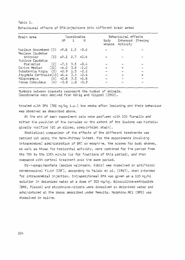
Table l.
Behavioural effects of DPA-injections into different brain areas
Brain area Coordinates Behavioural effects AP L H Body Enhanced Chewing
shakes Activity
Nucleus Accumbens (2) +9.8 1.2 -0.6 Nucleus Caudatus
Anterior (2) +9.2 2.7 +0.4 ,'\!ucleus Caudatus
Posterior (2) +7.5 3.5 +0.4 Centre Median (12) +4.0 0.8 -1.0 Substantia Nigra (2) +2.9 1.5 -2.2 Amygdala Centralis(ll) +4.4 3.5 -2.4 Hippocampus (4) +2.8 3.0 +1.8 Locus Coeruleus (4) -0.8 l.O -0.5
+
Numbers between brackets represent the number of animals. Coordinates were derived from KOnig and Klippel (1963).
+
+
treated with DPA (300 mg/kg i.p.) two weeks after lesioning and their behaviour
was observed as described above.
At the end of each experiment rats were perfused with 10~ formalin and
either the position of the cannulae or the extent of the lesions was histolo
gically verified (60 ~m slices, cresylviolet stain).
Statistical comparison of the effects of the different treatments was
carried out using the Mann-Whitney U-test. For the experiments involving
intracerebral administration of BMI or morphine! the scores for body shakes,
as well as those for horizontal activity, were combined for the period from
the 7th to the 15th minute (or for fractions of this period), and then
compared with control treatment over the same period.
Di-n-propylacetate (sodium valproate, Albic) was dissolved in artificial
cerebrospinal fluid (CSF)! according to Palaic et al. (1967)! when intended
for intracerebral injection. Intraperitoneal DPA was given as a 100 mg/ml
solution in deionized water at a dose of 300 mg/kg. Bicuculline-methiodide
(BMI, Pierce) and strychnine-nitrate were dissolved in deionized water and
administered at the doses described under Results. Morphine.HCl (OPG) was
dissolved in saline.
104
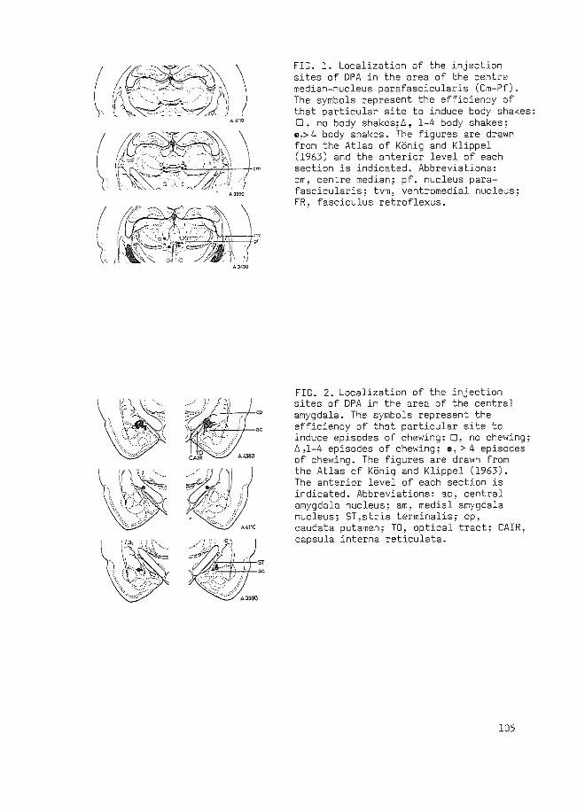
F.IG. 1. Localization of the injection sites of DPA in the area of the centre median-nucleus parafascicularis (Cm-Pf). The symbols represent the efficiency of that particular site to induce body shakes: :J, no body shakes;Ll, 1-4 body shakes; •~> 4 body shakes. The figures are drawn from the Atlas of KOnig and Klippel (1963) and the anterior level of each section is indicated. Abbreviations: em, centre median; pf. nucleus parafascicularis; tvm, ventromedial nucleus; FR, fasciculus retroflexus.
FIG. 2. Localization of the injection sites of DPA in the area of the central amygdala. The symbols represent the efficiency of that particular site to induce episodes of chewing: 0, no chewing; 1:. ,1-4 episodes of chewing; a, > 4 episodes of chewing. The figures are drawn from the Atlas of Konig and Klippel (1963). The anterior level of each section is indicated. Abbreviations: ac, central amygdala nucleus; am, medial amygdala nucleus; ST,stria terminalis; cp, caudata putamen; TO, optical tract; CAIR, capsula interna reticulata.
105
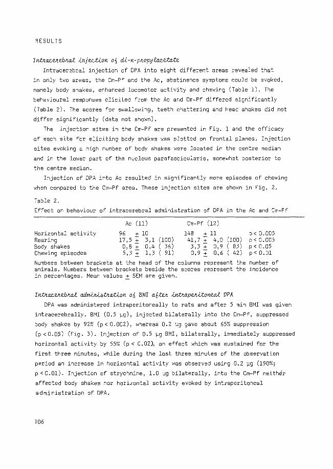
RESULTS
Inlkaeekebk~ ~njection ofi d~-n-pkopyLxcetaXe Intracerebral injection of DPA into eight different areas revealed that
in only two areas, the Cm-Pf and the Ac, abstinence symptoms could be evoked,
namely body shakes, enhanced locomotor activity and chewing (Table 1). The
behavioural responses elicited from the Ac and Cm-Pf differed significantly
(Table 2). The scores for swallowing, teeth chattering and head shakes did not
differ significantly (data not shown).
The injection sites in the Cm-Pf are presented in Fig. 1 and the efficacy
of each site for eliciting body shakes was plotted on frontal planes. Injection
sites evoking a high number of body shakes were located in the centre median
and in the lower part of the nucleus parafascicularis, somewhat posterior to
the centre median.
Injection of DPA into Ac resulted in significantly more episodes of chewing
when compared to the Cm-Pf area. These injection sites are shown in Fig. 2.
Table 2.
Effect on behaviour of intracerebral administration of DPA in the Ac and Cm-Pf
Ac (ll) Cm-Pf (12)
Horizontal activity 96 + 10 148 +ll p<0.005 Rearing 17.5 + 3,1 (100) 41,7 + 4,0 (100) p< 0.005 Body shakes o; 8 :t 0,4 ( 36) 3,3 :t 0,9 ( 83) p < 0. 05 Chewing episodes 5,3 ~ 1,3 ( 91) 0,9 ~ 0,6 ( 42) p < 0. Ol
Numbers between brackets at the head of the columns represent the number of animals. Numbers between brackets beside the scores represent the incidence in percentages. Mean values ~ SEM -are given.
Inlkaeekebk~ adm~n-WV<ation o£ BMI o.6:tek ~nt<o.pek~:tone~ VPA
DPA was administered intraperitoneally to rats and after 5 min BMI was given
intracerebrally. BMI (0.5 ~g), injected bilaterally into the Cm-Pf, suppressed
body shakes by 92~0 (p< 0.002), whereas 0.2 Vg gave about 65~0 suppression
(p < 0.05) (Fig. 3). Injection of 0.5 Vg BMI, bilaterally, immediately suppressed
horizontal activity by 55~0 (p < 0.02), an effect which was sustained for the
first three minutes, while during the last three minutes of the observation
period an increase in horizontal activity was observed using 0.2 vg (190%;
p < 0.01). Injection of strychnine, 1.0 f.lg bilaterally, into the Cm-Pf neither
affected body shakes nor horizontal activity evoked by intraperitoneal
administration of DPA.
106
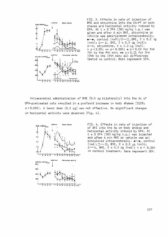
1 2 3 4 !i 7 6 S 10 11 12 13 14 1!1 m•n
, 2 3 4 !i 6 S 101112 •,3 141!im•n
FIG. 3. Effects in rats of injection of BMI and strychnine into the Cm-Pf on body shakes and horizontal activity induced by DPA. At t = 0 DPA (300 mg/kg i.p.) was given and after 6 min BMI, strychnine or vehicle was administered intracerebrally . .--., control (n:::9); 0-0_, BMI, 2 x 0.2 f.l9 (n=4); ~- ~. BMI, 2 x 0.5 \l9 (n=5); o--o, strychnine, 2 x 1.0 ~g (n=5). + p <0.05; ++ p< 0.002; * p< 0.02 for the 7th to the 9th min; ** p < 0. 01 for the 13th to the 15th min; all differences tested vs control. Bars represent SEM.
Intracerebral administration of BMI (0.5 f.lg bilaterally) into the Ac of
DPA-pretreated rats resulted in a profound increase in body shakes (210%;
p < 0.005). A lower dose (0.2 1-lg) was not effective. No significant changes
in horizontal activity were observed (Fig. 4).
1 234!1 769WnQ13M1!i~n
FIG. 4. Effects in rats of injection of of BMI into the Ac on body shakes and horizontal activity induced by DPA. At t = 0 DPA (300 mg/kg i.p.) was injected and after 6 min BMI or vehicle was administered intracerebrally. a---a, control (n=6); 0-0, BMI, 2 x 0.2 119 (n=5); ~-~. BMI. 2 x 0.5 \lg (n=6); + p < 0.005 vs control treatment. Bars represent SEM.
107
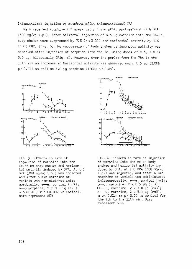
Inttac~eb~~ 1njection o£ mo~ph1ne a£t~ 1nttap~1tone~ VPA
Rats received morphine intracerebrally 5 min after pretreatment with DPA
(300 mg/kg i.p.). After bilateral injection of 0.5 ~g morphine into the Cm-Pf,
body shakes were suppressed by 72% (p< 0.01) and horizontal activity by 37~6
(p < 0.002) (Fig. 5). No suppression of body shakes or locomotor activity was
observed after injection of morphine into the Ac, using doses of 0.5, 1.0 or
5.0 ~g, bilaterally (Fig. 6). However, over the period from the 7th to the
11th min an increase in horizontal activity was observed
p < 0.01) as well as 5.0 1,.:9 morphine (lao~~; p < 0.05).
using 0. 5 119 (23m&;
lr'IJ<'Ctlon
f/'~~ 12 3 4 ~ 7 8 9101112131415mln
12345 789101112t314t:lm•n
FIG. 5. Effects in rats _of injection of morphine into the Cm-Pf on body shakes and horizontal activity induced by DPA. At t=O DPA (300 mg/kg i.p.) was injected and after 6 min morphine or vehicle was administered intracerebrally . .--., control (n=7); o--o morphine, 2 x 0.5 ~g (n=B). + p < 0.01; '* p < 0.002 vs control. Bars represent SEM.
108
r , I '
FIG. 6. Effects in rats of injection of morphine into the Ac on body shakes and horizontal activity induced by DPA. At t=D DPA (300 mg/kg i.p.) was injected, and after 6 min morphine or vehicle was administered intracerebrally. o--e, control (n=B); o--o, morphine, 2 x 0.5 119 (n=3); D-0, morphine, 2 x 1. 0 119 ( n=3) ; L-!:,, morphine, 2 x 5.0 \!9 (n=5). * p< 0.01; ** p< 0.05 vs control for the 7th to the 11th min. Bars represent SEM.
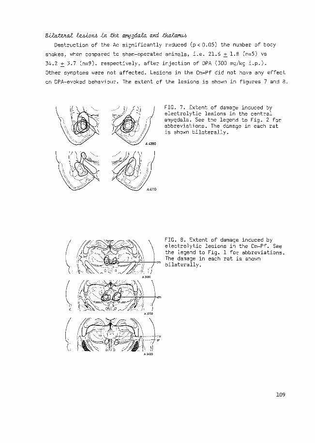
B.i.Wvw£ teA.i.OM .i.n :the amygda.fu and :thcU'am<M
Destruction of the Ac significantly reduced (p < 0.05) the number of body
shakes, when compared to sham-operated animals, i.e. 21.6 ~ 1.8 (n=5) vs
34.2 ~ 3.7 (n=9), respectively, after injection of DPA (300 mg/kg i.p.).
Other symptoms were not affected. Lesions in the Cm-Pf did not have any effect
on DPA-evoked behaviour. The extent of the lesions is shown in figures 7 and 8.
A3990
A3750
A3~30
FIG. 7. Extent of damage induced by electrolytic lesions in the central amygdala. See the legend to Fig. 2 for abbreviations. The damage in each rat is shown bilaterally.
FIG. 8. Extent of damage induced by electrolytic lesions in the Cm-Pf. See the legend to Fig. 1 for abbreviations. The damage in each rat is shown
em bilaterally.
109
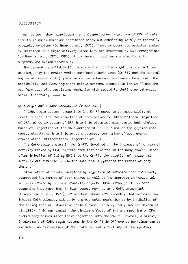
DISCUSSION
As has been shown previously, an intraperitoneal injection of DPA in rats
results in quasi-morphine abstinence behaviour consisting mainly of centrally
regulated symptoms (De Boer et al., 1977). These symptoms are probably evoked
by increased GABA-ergic activity since they are prevented by GABA-antagonists
(De Boer et al., 1977, 1980). A low dose of morphine was also found to
suppress DPA-evoked behaviour.
The present data (Table 1). indicate that, of the eight brain structures
studied, only the centre median-parafascicularis area (Cm-Pf) and the central
amygdaloid nucleus (Ac) are involved in DPA-evoked abstinence behaviour. The
possibility that GABA-ergic and opiate systems, present in the Cm-Pf and the
Ac, form part of a regulating mechanism with regard to abstinence behaviour,
seems 1 therefore, feasible.
GABA-e<gle and opiate meehanl6mo ln the Cm-Pn A GABA-ergic system present in the Cm-Pf seems to be responsible, at
least in part, for the induction of body shakes by intraperitoneal injection
of DPA, since injection of DPA into this structure also evoked body shakes.
Moreover, injection of the GABA-antagonist BMI, but not of the glycine anta
gonist strychnine into this area, suppressed the number of body shakes
evoked after intraperitoneal injection of DPA.
The GABA-ergic system in the Cm-Pf, involved in the increase of horizontal
activity evoked by DPA, differs from that involved in the body shakes, since,
after injection of 0.2 ~g BMI into the Cm-Pf, the duration of horizontal
activity was extended, while the same dose suppressed the number of body
shakes.
Stimulation of opiate receptors by injection of morphine into the Cm-Pf,
suppressed the number of body shakes as well as the increase in horizontal
activity evoked by intraperitoneally injected DPA. Although it has been
suggested that morphine, in high doses, may act as a GABA-antagonist
(Dingledine et al., 1977), it has been shown more recently that morphine may
inhibit GABA-release, either by a presynaptic mechanism or by inhibition of
the firing rate of GABA-ergic cells (Nicollet al., 1980; Vander Heyden et
al.,l980). This may explain the similar effects of BMI and morphine on DPA
evoked body shakes after their injection into the Cm-Pf. However, a primary
involvement of GABA-ergic systems in the Cm-Pf in DPA-evoked behaviour can be
excluded , as destruction of the Cm-Pf did not affect any of the symptoms.
110
Thus, GABA-ergic neurons present in the Cm-Pf seem to be facilitatory in
the expression of DPA-induced body shakes, while their action can be
counteracted by opiate mechanisms in the same area.
GABA-ekgiQ and opiate mechaniom4 in the AQ Although injection of DPA into the Ac resulted in enhanced chewing, no
further evidence could be obtained for the mediation of this effect by GABA
ergic neurons, since BMI injected into the Ac had no effect on chewing evoked
by intraperitoneal DPA-administration (data not shown).
Injection of BMI into the Ac increased the number of body shakes after
intraperitoneal administration of DPA. suggesting the presence of an in
hibitory GABA-ergic system in the Ac, which affects body shaking.
It has been reported that the Ac contains a high density of opiate
receptors (Atweh and Kuhar, 1977b),probably connected to receiver cells of
an enkephalinergic system (Gras et al., 1978; Simantov et al., 1977).
Nevertheless, morphine injected into this brain area was without effect on
DPA-evoked body shakes. Thus, the Ac contains GABA-ergic mechanisms which
inhibit the expression of body shakes and which are unaffected by morphine.
The present results, therefore, suggest that, as far as body shakes are
concerned, a GABA-ergic system in the Cm-Pf plays a facilitatory role, while
a GABA-ergic system in the Ac exerts an inhibitory effect. Systemic injection
of DPA may, thus, evoke abstinence behaviour by activating the GABA-ergic
system in the Cm-Pf which may stimulate an unknown neuronal pathway
responsible for the expression of body shakes, probably via disinhibition
of this pathway. The GABA-ergic cells in the Cm-Pf might be under control
of an opioid system.
The inhibitory action of the GABA-ergic system in the Ac on DPA-evoked
behaviour and the fact that destruction of this structure decreases the
number of body shakes, suggests that the Ac contains excitatory neurons
which might be primarily involved in evOking body shakes. The GABA-ergic
neurons present in this structure may inhibit these neurons directly.
The present data, obtained with rats in which quasi-morphine abstinence
behaviour was induced by DPA, might have implications for the localization
of the brain area responsible for the true morphine withdrawal syndrome.
Several structures have been reported to be involved in the expression of
morphine abstinence behaviour, e.g. areas located on the floor of the 4th
ventricle (Laschka and Herz, 1977), the periaqueductal grey (Laschka and
Herz, 1977; Wei et al., 1975) and the hypothalamus (Kerr and Pozuelo, 1971;
lll
Wei et al., 1975). In none of these studies were the data related to a
specific transmitter system. Amyqdaloid structures have also been implicated
in the morphine withdrawal syndrome (Calvina et al., 1979; Lagowska et al.,
1978).
Furthermore, it has been reported that morphine alters the synthesis
and distribution of GABA in the thalamus, although, after chronic administra
tion of morphine, no differences were observed (Kuriyama and Yoneda, 1978;
Yoneda et al., 1977). Further experiments are necessary to elucidate the
role of GABA-ergic mechanisms in the Ac and the Cm-Pf in the morphine
withdrawal syndrome.
In conclusion, the present study suggests that GABA-ergic systems,
especially those in the Cm-Pf and the Ac, are involved in the expression
of body shakes and horizontal activity evoked by DPA. An opioid input
present in the Cm-Pf may regulate this system. It is possible that the
projection of the Pf into the central amygdala, which has been described
recently (Ottersen and Ben-Ari, 1979), is implicated in this system.
REFERENCES
Atweh, S.F., Kuhar, M.J.(l977a) Autoradiographic localization of opiate receptors in rat brain. II. The Brain Stem. Brain Res. 129, 1-12
Atweh, S.F., Kuhar, M.J.(l977b) Autoradiographic localization of opiate receptors in rat brain. III. The Telencephalon. Brain Res. 134, 393-405
Calvina, B., Lagowska, J., Ben-Ari, Y. (1979) Morphine withdrawal syndrome: differential participation of structures located within the amygdaloid complex and striatum of the rat. Brain Res. 177, 19-34
De Boer, Th., Bartels, K., Metselaar, H.J., Gruinvels, J. (1980) Di-npropylacetate-induced abstinence behaviour as a possible correlate of increased GAGA-ergic activity in the rat. Psychopharmacol. 71, 257-267
De Boer, Th., Metselaar, H.J., Gruinvels, J. (1977) Suppression of GAGAinduced abstinence behaviour in naive rats by morphine and bicuculline. Life Sci. 20, 933-942
Dingledine, R., Iversen, L.L., Greuker, E. (1977) Naloxone as a GAGAantagonist: evidence from iontophoretic, receptor binding and convulsant studies. Eur. J. Pharmacal. 47. 19-27
Elchisak, M.A., Rosecrans, J.A. (1979) Development of morphine tolerance and physical dependence in rats depleted of brain catecholamines by 6-hydroxydopamine. :\Jeuropharmacol. 18, 175-182
Godin, Y., Heiner, L., Mark, J., Mandel, P. (1969) Effects of di-n-propylacetate, an anticonvulsive compound, on GAGA metabolism. J. Neurochem. 16, 869-873
Gras, C., Pradelles, P., Humbert, J., Dray, F., LeGal LaSalle, G., Gen-Ari, Y. (1978) Regional distribution of met-enkephalin within the amygdaloid complex and bed nucleus of the stria terminalis. Neuroscience Lett., 10, 193-196
Kerr, F.W.L., Pozuelo, J. (1971) Suppression of physical dependence and
112
induction of hypersensitivity to morphine by stereotaxic hypothalamic lesions in addicted rats. Mayo Clin. Proc. 46! 653-665
KOnig, J.F.R., Klippel, R.A. (1963) The Rat Brain: A stereotaxic atlas of the forebrain and lower parts of the brain stem. The Williams and Wilkins Camp., Baltimore
Kuriyama, K., Yoneda, Y. (1978) Morphine-induced alterations of Y-aminobutyric acid and taurine contents and L-glutamate decarboxylase activity in the spinal cord and thalamus: possible correlates with analgesic action of morphine. Brain Res. 148, 163-179
Lagowska, J., Calvina! B., Ben-Ari, Y. (1978) Intra-amygdaloid applications of naloxone elicits severe withdrawal signs in morphine-dependent rats. Neuroscience Lett., 8, 241-245
Laschka, E., Herz, A. (1977) Sites of action of morphine involved in the development of physical dependence in rats.Psychopharmacol. 53, 33-37
Nicoll, R.A., Alger, B.E., Jahr, C.E. (1980) Enkephalin blocks inhibitory pathways in the vertebrate CNS. Nature (London), 287, 22-25
Ottersen, D.P., Ben-Ari, Y. (1979) Afferent connections to the amygdaloid complex of the rat and cat. I. Projections from the thalamus. J. Camp. Neural. 187, 401-424
Palaic. D., Page, I.H., Khairallah, P.A. (1967) Uptake and metabolism of 14.c serotonin in rat brain. J. ~eurochem. 14, 63-69
Pert, A. (1978) Central sites involved in opiate actions. In: J. Fishman (ed.) The bases of addiction, Dahlem Konferenzen, Berlin, pp. 299-322
Simantov, R., Kuhar, M.J., Uhl, G.R., Snyder, S.H. (1977) Opioid peptide enkephalin: Immunohistochemical mapping in rat central nervous system. Prac. Nat. Acad. Sci. U.S.A. (Wash.) 74, 2167-2171
Simler, S., Cieselski, L., Maitre, M., Randrianarisoa, H., Mandel, P. (1973) Effect of sodium n-dipropylacetate on audiogenic seizures and brain Y-aminobutyric acid level. Biochem. Pharmacal. 22, 1701-1708
Vander Heyden, J.A.M., Venema, K., Korf, J. (1980) Tn v~vo release of endogenous GABA from rat striatum: effects of muscimol, oxotremorine and morphine. J. Neurochem. 34, 1648-1653
VanderLaan, J.W., De Boer, Th., Bruinvels, J. (1979) Di-n-propylacetate and GABA-degradation. Preferential inhibition of succinic semialdehyde dehydrogenase and indirect inhibition of GABA-transaminase. J. Neurochem. 32, 1769-1780
VanderLaan, J.W., Jacobs, A.W.C.M., Bruinvels, J. (1980) Effects of branched-chain fatty acids on GABA-degradation and behavior: Further evidence for a role of GABA in quasi-morphine abstinence behavior. Pharmacal. Biochem. Behav. 13, 843-849
Wei, E., Loh, H.H., Way, E.L. (1972) Neuroanatomical correlates of morphine dependence. Science, .177, 616-617
Wei, E., Loh, H.H., Way, E.L. (1973) Brain sites of precipitated abstinence in morphine-dependent rats. J. Pharmacal. Exp. Ther. 185, 108-115
Wei, E., Sigel, S.S.R., Loh, H.H., Way, E.L. (1975) Central sites of naloxone-precipitated shaking in the anaesthetized morphine-dependent rat. J. Pharmacal. Exp. Ther. 195, 480-487
Yoneda, Y., Kuriyama, K., Kurihara, E. (1977) Morphine alters distribution of GABA in thalamus. Brain Res. 124, 373-378
113
CHAPTER VI
DIPROPYLACETATE-INDUCED QUASI-MORPHINE ABSTINENCE BEHAVIOUR
IN THE RAT: PARTICIPATION OF THE LOCUS COERULEUS SYSTEM
J.W. van der Laan, J. Bruinvels and A.R. Cools
Brain Research, submitted for publication
ABSTRACT
The role of the noradrenergic system in quasi-morphine abstinence
behaviour induced by di-n-propy1acetate (DPA) in rats has been studied.
Depletion of noradrenaline (NA), by treatment with FLA-63, decreased the
number of body shakes and the extent of horizontal activity evoked by DPA.
Almost total suppression of these symptoms was obtained by injection of 20
ng morphine bilaterally into the locus coeruleus (LC). Destruction of the
LC system by electrolytic lesion of the LC or by injection of 6-0Hdopamine
into the dorsal bundle revealed that degeneration of the NA system by at
least am& appears to be necessary to decrease th~ number of body shakes.
It is concluded that the noradrenergic LC system fulfils a modulatory
role in quasi-morphine abstinence behaviour induced by DPA.
INTRODUCTION
Systemic administration of di-n-propylacetate (DPA) induces a behavioural
syndrome in rats consisting of body shakes, escape digging, hunchback
posture, enhanced locomotor activity and various other symptoms (Bruinvels
et a1 .. 19BO; De Boer et a1., 1977, 1980). Increased GABA-ergic activity is held responsible for the induction of
this behaviour since it can be suppressed by the GABA-antagonists bicuculline
and picrotoxin, but not by the glycine antagonist strychnine (De Boer et al.,
1977~ 1980). DPA is thought to enhance GABA-ergic activity by inhibition of
succinic-semialdehyde dehydrogenase, the second enzyme in the degradation
of GABA (VanderLaan et a1., 1979).
Since the DPA-evoked behaviour resembles that seen during morphine
abstinence (De Boer et al., 1977) it is referred to as quasi-morphine
abstinence behaviour as defined by Collier (1974).
114
We have now used the DPA-induced behavioural syndrome as a model to
study the role of the noradrenergic locus coeruleus (LC) system in morphine
withdrawal behaviour. Participation of brain noradrenaline (NA) in morphine
abstinence behaviour is suggested by several lines of evidence as reviewed
recently (Iwamoto and Way, 1979). Contradictory results, however, have been
an obstacle to the elucidation of the exact role of this transmitter (see
Iwamoto and Way, 1979). These contradictions may, in part, be accounted
for by differences in the species studied, in opiate treatment schedules,
in scoring systems for the degree of abstinence behaviour and in the
indicator of NA functionality. For instance, some investigators have
studied steady state concentration of NA in parts of or in the whole brain,
while others have studied turnover of ~A by determination of its rate of
synthesis or of degradation.
The role of the noradrenergic system in morphine abstinence behaviour
seems to be important in view of recent reports on the use of clonidine
in the detoxification of opiate addicts (Gold et al., 1978, 1979, 1980;
Washton et al., 1980). Evidence has been presented that clonidine counteracts
morphine abstinence behaviour by stimulation of ~adrenoceptors located in
the LC (Aghajanian, 1978). Therefore, we decided to restrict ourselves, in
the present study, mainly to the noradrenergic system originating in the
LC, which projects via the dorsal bundle into the forebrain. To allow
comparison with treatments that affect the whole brain NA content, a study
on the effects of systemically administered FLA-63, a dopamine-S-hydroxylase
inhibitor, was included.
In the present paper, the effect of changes in LC activity on DPA-induced
behaviour has been examined using three different approaches: a) local
injection of morphine into the LC; b) electrolytic lesion of the LC; c)
chemical lesion of the LC forebrain projection.
MATERIALS AND METHODS
Sixty-eight male Wistar rats (150-225 g; T~O, Zeist, the Netherlands)
were used for all experiments. In the first experiment rats (n=l5) were
treated with FLA-63 (20 mg/kg i.p.) three times at twa hourly intervals.
Two hours after the last injection they received DPA (300 mg/kg i.p.) and
their behaviour was observed as follows. Rats were placed in a macrolon
cage (47 x 27 x 15 em) with sawdust bedding and were allowed to habituate
for at least 30 min, whereafter DPA was administered intraperitaneally.
115
Behavioural observ~tion was started directly after the injection and lasted
for 15 min. The following signs were scored as absent, mild or marked:
hunchback posture, piloerection, ptosis, penis licking and erection. Other
signs were counted as quanta! events: body shakes, head shakes, escape
digging and foreleg shakes. Horizontal activity was measured simultaneously
using an Animex activity meter.
A second group of animals (n=28) was anaesthetized with pentobarbital
(60 mg/kg i.p.), placed in a stereotaxic frame and stainless steel
cannulae were implanted bilaterally, as previously described by Pijnenburg
et al. (1976). The following coordinates were chosen: AP -0.8, L 1.2, H 3.1,
according to the coordinate system of KOnig and Klippel (1963). The
cannulae were inclined at an angle of 15° from the posterior side and 5°
from the lateral side to avoid bleeding. After a recovery period of one
week, during which the animals were housed individually, behavioural
observations were carried out after placing the rats in a wooden cage
(50 x 30 x 30 em) with a single transparent perspex wall, on sawdust bedding.
Behavioural scoring after injection of DPA (300 mg/kg i.p.) was carried out
as described above, except for the horizontal activity, which was scored as
the number of full cage lenghts (50 em) the rats had covered. After five
minutes morphine or saline was injected intracerebrally in a volume of 0.5
~1, the latter serving as a control. To check the correct placement of the
cannulae, histological examination was carried out as described previously
(VanderLaan and Bruinvels, in press).
In a third experiment, rats (n=l2) were anaesthetized with chloral
hydrate (400 mg/kg i.p.), placed in a stereotaxic frame and brain lesions
were performed centered on the LC, AP -0.5, L 0.9, H 2.6, according to the
coordinate system of KOnig and Klippel (1963), with a platinum electrode
(anode) varnished except for the tip of 0.25 mm. The stereotaxic frame
served as the cathode. The electrode was inclined at an angle of 16° from
the posterior side. The intensity of the current was 1 rnA and the duration
45 sec. In control animals the electrode was driven to 2 mm above the LC
without applying current. Two weeks after lesioning, the animals were
treated with DPA (300 mg/kg) and their behaviour was observed for 15 min,
as described above. In this experiment, horizontal activity was measured
using a Varimex activity meter.
In a fourth group of rats (n=l3), under chloral hydrate anaesthesia,
6-0Hdopamine was injected bilaterally into the dorsal bundle, AP 1.8,
L 1.0, H 0.5 (KOnig and Klippel, 1963). 6-0Hdopamine was administered in a
116
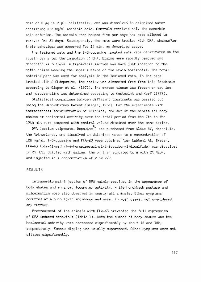
dose of 8 ~g in 2 ~1, bilaterally, and was dissolved in deionized water
containing 0.2 mg/ml ascorbic acid. Controls received only the ascorbic
acid solution. The animals were housed five per cage and were allowed to
recover for 21 days. Subsequently, the rats were treated with DPA, whereafter
their behaviour was observed for 15 min, as described above.
The lesioned rats and the 6-0Hdopamine treated rats were decapitated on the
fourth day after the injection of DPA. Brains were rapidly removed and
dissected as follows. A transverse section was made just anterior to the
optic chiasm keeping the upper surface of the brain horizontal. The total
anterior part was used for analysis in the lesioned rats. In the rats
treated with 6-0Hdopamine, the cortex was dissected free from this forebrain
according to Gispen et al. (1972). The cortex tissue was frozen on dry ice
and noradrenaline was determined according to Westerink and Korf (1977).
Statistical comparison between different treatments was carried out
using the Mann-Whitney U-test (Siegel, 1956). For the experiments with
intracerebral administration of morphine, the sum of the scores for body
shakes or horizontal activity over the total period from the 7th to the
15th min were compared with control values obtained over the same period.
DPA (sodium valproate~ DepakineR) was purchased from Albic BV, Maassluis,
the Netherlands, and dissolved in deionized water to a concentration of
100 mg/ml. 6-0Hdopamine and FLA-63 were obtained from Labkemi AB, ~weden.
FLA-63 (bis-(1-methyl-4-homopiperazinyl-thiocarbonyl)disulfide) was dissolved
in 2N HCl, diluted with saline, the pH then adjusted to 6 with 2~ NaOH,
and injected at a concentration of 2.5% w/v.
RESULTS
Intraperitoneal injection of DPA mainly resulted in the appearance of
body shakes and enhanced locomotor activity, while hunchback posture and
piloerection were also observed in nearly all animals. Other symptoms
occurred at a much lower incidence and were, in most cases, not considered
any further.
Pretreatment of the animals with FLA-63 prevented the full expression
of DPA-induced behaviour (Table 1). Both the number of body shakes and the
horizontal activity were decreased significantly by about 58 and 38%,
respectively. Escape digging was totally suppressed. Other symptoms were not
altered significantly.
117
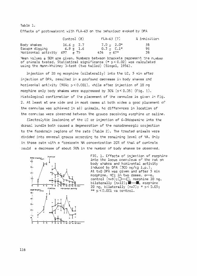
Table 1.
Effects of pretreatment with FLA-63 on the behaviour evoked by DPA
Control (8) FLA-63 (7) " inhibition ,,
Body shakes 16.6 :': 2.7 7.0 ± 2.Q';<· 58 Escape digging 6.9 ± 1.6 0.3 ± o.l* 96 Horizontal activity 697 ± 75 434 ± 67* 38
Mean values ± SEM are given. Numbers between brackets represent the number of animals tested. Statistical significance (* p< 0.02) was calculated using the Mann-Whitney U-test (two tailed) (Siegel, 1956).
Injection of 20 ng morphine (bilaterally) into the LC, 5 min after
injection of DPA, resulted in a profound decrease in body shakes and
horizontal activity (9m6; p < 0.001), while after injection of 10 ng
morphine only body shakes were suppressed by 30~; (p < 0.05) (Fig. 1).
Histological confirmation of the placement of the cannulae is given in Fig.
2. At least at one side and in most cases at both sides a good placement of
the cannulae was achieved in all animals. No differences in location of
the cannulae were observed between the groups receiving morphine or saline.
Electrolytic lesionin~ of the LC or injection of 6-0Hdopamine into the
dorsal bundle both caused a degeneration of the noradrenergic projection
to the forebrain regions of the rats (Table 2). The treated animals were
divided into several groups according to the remaining level of NA. Only
in those rats with a forebrain NA concentration 20?~ of that of controls
could a decrease of about 50% in the number of body shakes be observed.
r·r ·~ f-%!~ 0 45 789101112131415mon
ln)...:tlon HorlzoMal ~ctlvlty
0 45 789101112131415mln
118
FIG. 1. Effects of injection of morphine into the locus coeruleus of the rat on body shakes and horizontal activity induced by DPA (300 mg/kg i.p.). At t=O DPA was given and after 5 min morphine. HCl in two doses. o--o, control ( n=9) ; 0-D, morphine 10 ng, bilaterally (n=l2);~, morphine 20 ng, bilaterally (n=7); ·> p< 0.05; ** p < 0. 001 vs control.
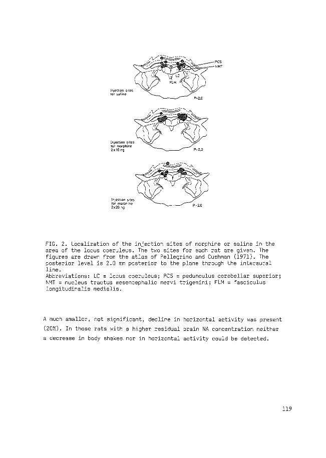
lr.jt'C!•cr. s•tes lor salir.e
lr.Jed:ICr"\ SiteS fer mcrph1ne 2~10n9
lnJect•cn saes tor morphine 2~20 ng
P-2.0
FIG. 2. Localization of the injection sites of morphine or saline in the area of the locus coeruleus. The two sites for each rat are given. The figures are drawn from the atlas of Pellegrino and Cushman (1971). The posterior level is 2.0 mm posterior to the plane through the interaural line. Abbreviations: LC = locus coeruleus; PCS = pedunculus cerebellar superior; NMT = nucleus tractus mesencephalic nervi trigemini; FLM = fasciculus longitudinalis medialis.
A much smaller, not significant, decline in horizontal activity was present
(20%). In those rats with a higher residual brain NA concentration neither
a decrease in body shakes nor in horizontal activity could be detected.
119
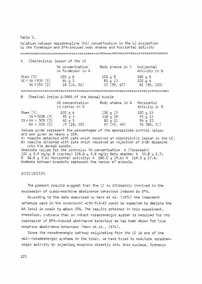
Table 2.
Relation between noradrenaline (NA) concentration in the LC projection to the forebrain and DPA-induced body shakes and horizontal activity
~=~~=======================================================================
A Electrolytic lesion of the LC
NA concentration in forebrain in %
Sham (5) 50 < NA < 901o ( 5)
NA < 251o (2)
100 ± 8 64 + 5 18 (14, 21)
Body shakes in ~~ Horizontal Activity in ?0
100 + 8 82 ± 10 53 (39, 67)
100 ± 8 102 ± 6 Bl (59, 103)
===================================================================~=======
B Chemical lesion 6-0HDA of the dorsal bundle
:'\!A concentration Body shakes in ,, Horizontal ,, in cortex in ?6 Activity in ~;;
Sham (5) 100 ± 4 100 ± 13 100 ± 15 NA ::::: lOO% (3) 95 ± 3 118 ± 19 95 ± l3
25 < NA < 5mo (3) 42 + 5 82 +ll 84 + 20 NA < 25~0 (2) 19 (rs, 20) 47 (45, 49) 76 (so, 71)
Values given represent the percentages of the appropriate control values and are given as means ± SEM. A: results obtained with rats which received an electrolytic lesion in the LC. 8: results obtained with rats which received an injection of 6-0H dopamine
into the dorsal bundle. Absolute values for the controls: NA concentration A (forebrain) 121 ± 9.9 ng/g; 8 (cortex) 158.0 ± 5.8 ng/g; Body shakes: A 35.8 ± 2.7; B 26.8 ± 3.6; Horizontal activity: A 260.0 ± 19.6; B 119.5 ± 17.4. Numbers between brackets represent the number of animals.
DISCUSSION
The present results suggest that the LC is intimately involved in the
expression of quasi-morphine abstinence behaviour induced by DPA.
According to the data described by Herz et al. (1974) the treatment
schedule used in the experiment with FLA-63 would be expected to deplete the
NA level in brain to about 25%. The results obtained in this experiment,
therefore, indicate that an intact noradrenergic system is required for the
expression of DPA-induced abstinence behaviour as has been shown for true
morphine abstinence behaviour (Herz et al., 1974).
Since the noradrenergic pathway originating from the LC is one of the
main noradrenergic systems in the brain, we have tried to modulate noradren
ergic activity by injecting morphine directly into this nucleus. Systemic
120
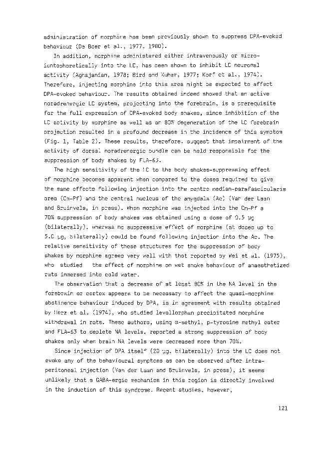
administration of morphine has been previously shown to suppress DPA-evoked
behaviour (De Boer et al., 1977, 1980).
In addition, morphine administered either intravenously or micro
iontophoretically into the LC, has been shown to inhibit LC neuronal
activity (Aghajanian, 1978; Bird and Kuhar, 1977; Korf et al., 1974).
Therefore, injecting morphine into this area might be expected to affect
DPA-evoked behaviour. The results obtained indeed showed that an active
noradrenergic LC system, projecting into the forebrain, is a prerequisite
for the full expression of DPA-evoked body shakes, since inhibition of the
LC activity by morphine as well as an 80~~ degeneration of the LC forebrain
projection resulted in a profound decrease in the incidence of this symptom
(Fig. 1, Table 2). These results, therefore, suggest that impairment of the
activity of dorsal noradrenergic bundle can be held responsible for the
suppression of body shakes by FLA-63.
The high sensitivity of the LC to the body shakes-suppressing effect
of morphine becomes apparent when compared to the doses required to give
the same effects following injection into the centre median-parafascicularis
area (Cm-Pf) and the central nucleus of the amygdala (Ac) (Van der Laan
and Bruinvels, in press). When morphine was injected into the Cm-Pf a
70% suppression of body shakes was obtained using a dose of 0.5 '\.lg
(bilaterally), whereas no suppressive effect of morphine (at doses up to
5.0 )..lg, bilaterally) could be found following injection into the Ac. The
relative sensitivity of these structures for the suppression of body
shakes by morphine agrees very well with that reported by Wei et al. (1975),
who studied the effect of morphine on wet shake behaviour of anaesthetized
rats immersed into cold water.
The observation that a decrease of at least SmG in the NA level in the
foiebrain or cortex appears to be necessary to affect the quasi-morphine
abstinence behaviour induced by DPA, is in agreement with results obtained
by Herz et al. (1974), who studied levallorphan precipitated morphine
withdrawal in rats. These authors, using a-methyl, p-tyrosine methyl ester
and FLA-63 to deplete NA levels, reported a strong suppression of body
shakes only when brain NA levels were decreased more than 70%.
Since injection of DPA itself (20 )..lg, bilaterally) into the LC does not
evoke any of the behavioural symptoms as can be observed after intra
peritoneal injection (VanderLaan and Bruinvels, in press), it seems
unlikely that a GABA-ergic mechanism in this region is directly involved
in the induction of this syndrome. Recent studies, however,
121
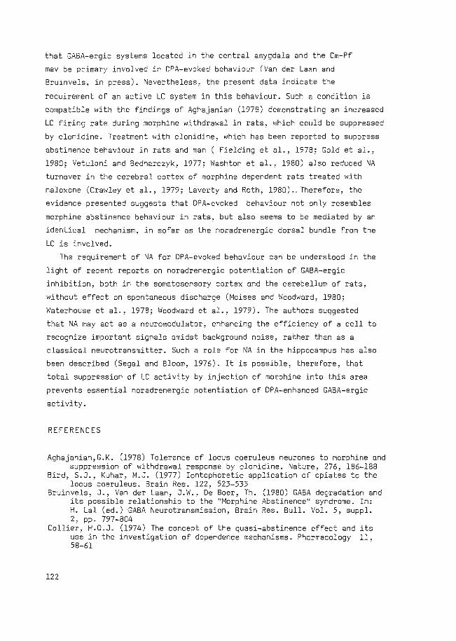
that GABA-ergic systems located in the central amygdala and the Cm-Pf
mav be primary involved in OPA-evoked behaviour (Van der Laan and
Bru1nvels, in press). Nevertheless, the present data indicate the
requirement of an active LC system in this behaviour. Such a condition is
compatible with the findings of Aghajanian (1978) demonstrating an increased
LC firing rate during morphine withdrawal in rats, which could be suppressed
by clonidine. Treatment with clonidine, which has been reported to suppress
abstinence behaviour in rats and man ( Fielding et al., 1978; Gold et al.,
1980; Vetulani and Bednarczyk, 1977; Washton et al., 1980) also reduced NA
turnover in the cerebral cortex of morphine dependent rats treated with
naloxone (Crawley et al., 1979; Laverty and Roth, 1980) .. Therefore, the
evidence presented suggests that DPA-evoked behaviour not only resembles
morphine abstinence behaviour in rats, but also seems to be mediated by an
identical mechanism, in sofar as the noradrenergic dorsal bundle from the
LC is involved.
The requirement of NA for DPA-evoked behaviour can be understood in the
light of recent reports on noradrenergic potentiation of GABA-ergic
inhibition, both in the somatosensory cortex and the cerebellum of rats,
without effect on spontaneous discharge (Moises and Woodward, 1980;
Waterhouse et al., 1978; Woodward et al., 1979). The authors suggested
that NA may act as a neurom8dulator, enhancing the efficiency of a cell to
recognize important signals amidst background noise, rather than as a
classical neurotransmitter. Such a role for NA in the hippocampus has also
been described (Segal and Bloom, 1976). It is possible, therefore, that
total suppression of LC activity by injection of morphine into this area
prevents essential noradrenergic potentiation of DPA-enhanced GABA-ergic
activity.
REFERENCES
Aghajanian,G.K. (1978) Tolerance of locus coeruleus neurones to morphine and suppression of withdrawal response by clonidine. Nature, 276, 186-188
Bird, S.J., Kuhar, M.J. (1977) Iontophoretic application of opiates to the locus coeruleus. Brain Res. 122, 523-533
Bruinvels, J., Van der Laan, J.W., oe Boer, Th. (1980) GABA degradation and its possible relationship to the "Morphine Abstinence" syndrome. In: H. Lal (ed.) -GA8A Neurotransmission, Brain Res. Bull. Vol. 5, suppl. 2, pp. 797-804
Collier, H.O.J. (1974) The concept of the quasi-abstinence effect and its use in the investigation of dependence mechanisms. Pharmacology 11, 58-61
122
Crawley, J .\!., Laverty~ R., Roth. R. H. (1979) Clonidine reversal of increased norepinephrine metabolite levels during morphine withdrawal. Eur. J. Pharmac. 57, 247-250
De Boer, Th., Bartels, K., Metselaar, H.J., Bruinvels, J. (1980) Di-npropylacetate-induced abstinence behaviour as a possible correlate of increased GABA-ergic activity in the rat. Psychopharmacol. 71, 257-267
De Boer, Th., Metselaar, H.J., Bruinvels, J. (1977) Suppression of GABA_ induced abstinence behaviour in naive rats by morphine and bicuculline. Life Sci. 20, 933-942
Fielding, S., Wilker. J., Hynes, M., Szewczak, M., Novick Jr., W.J., Lal, H. (1978) A comparison of clonidine with morphine for antinociceptive and antiwithdrawal actions. J. Pharmacal. Exp. Ther. 207, 899-905
Gispen, W.H., Schotman .. P.~ De Kloet. E.R. (1972) Brain Rr\A and hypophysectomie; a topographical study. Neuroendocrinology 9. 285-296
Gold, M.S., Redmond Jr., D.E., Kleber, H.D. (1978) Clonidine blocks acute opiate withdrawal symptoms. The Lancet II, 599-602
Gold, M.S., Redmond Jr., D. E., Kleber, H .D. (1979) Noradrenergic hyperactivity in opiate withdrawal supported by clonidine reversal of opiate withdrawal. Am. J. Psychiatry 136, 100-102
Gold, M.S., Pottash, A.L.C., Extein, I., Kleber, H.D. (1980) Clonidine and opiate withdrawal. The Lancet II 1078-1079
Herz~ A., Bl3sig, J., Papeschi, R. (1974) Role of catecholaminergic mechanisms in the expression of the morphine abstinence syndrome in rats. Psychopharmacol. 39, 121-143
Iwamoto, E.T., Way, E.L. (1979) Opiate actions and catecholamines. In:Loh. H.H. Ross, D.H. (eds.) Neurochemlcal mechanlsms of opiates and endorphins,· Advances in Biochem.Psychopharmac.Vol. 20 Raven Press,New York,pp.357-407
KOnig, J.F.R., Klippel, R.A. (1963) The Rat Brain: A stereotaxic atlas of the forebrain and lower parts of the brain stem. The Williams and Wilkins Camp. Baltimore
Korf, J., Bunney, B.S., Aghajanian, G.K. (1974) Noradrenergic neurons: morphine inhibition of spontaneous activity. Eur. J. Pharmac. 25, 165-169
Laverty, R., Roth, R.H. (1980) Clonidine reverses the increased norepinephrine turnover during morphine withdrawal in rats. Brain Res. 182, 482-485
Moises, H.C., Woodward, D.J. (1980) Potentiation of GABA inhibitory action in cerebellum by locus coeruleus stimulation. Brain Res. 182, 327-344
Pellegrino, L.J., Cushman, A.J. (1971) A Stereotaxic Atlas of the rat brain, Meredith, New York
Pijnenburg, ·A.J.J., Honig, W.M.M., Struyker Boudier, H.A.J., Cools, A.R., Van der Heyden, J.A.M., Van Rossum, J.M. (1976) Further investigations on the effect of ergometrine and other ergot derivatives following injection into the nucleus accumbens of rats. Arch. Int. Pharmacodyn. 222, 103-115
Segal, M., Bloom, F.E. (1976) The action of norepinephrine in the hippocampus IV. The effects of locus coeruleus stimulation on evoked hippocampal unit activity. Brain Res. 107, 513-525
Siegel, S. (1956) Nonparametric statistics for the behavioral sciences. McGraw-Hill Kogakusha, Tokyo
VanderLaan, J.W., Bruinvels, J. Dipropylacetate-induced quasi-morphine abstinence behaviour in the rat: Involvement of amygdaloid and thalamic structures. Brain Res. in press.
Van der Laan, J.W., De Boer, Th., Bruinvels, J. (1979) Di-n-propylacetate and GABA degradation. Preferential inhibition of succinic semialdehyde
123
dehydrogenase and indirect inhibition of GABA-transaminase. J. Neurochem. 32. 1769-1780
Vetulani, J.,. Bednarczyk, B. (1977) Depre.ssion of shaking behaviour elicited by nalorphine in morphine dependent rats. J. Pharm. Pharmac. 29, 567-569
Washton, A.M., Resnick, R.B.~ Rawson, R.A. (1980) Clonidine for outpatient opiate detoxification. The Lancet I 1078-1079
Waterhouse, B.D., Moises, H.C., Woodward, D.J. (1978) Noradrenergic modulation of somatosensory cortical neuronal responses to iontophoretically applied putative neurotransmitters. Soc. Neurosci. Abst. 4, 286
Wei, E., Si9el, S., Way, E.L. (1975) Regional sensitivity of the rat brain to the inhibitory effects of morphine on wet shake behavior. J. Pharmacal. Exp. Ther. 193, 56-63
Westerink, B.H.C., Korf, J. (1977) Rapid concurrent automated fl~orimetric assay of noradrenaline, dopamine, 3,4-dihydroxyphenylacetic acid, homovanillic acid and 3-methoxytyramine in milligram a~ounts of nervous tissue after isolation on Sephadex G-10. J. Neurochem. 29, 697-706
Woodward, O.J., Moises, H.C., Waterhouse, B.D., Hoffer, B.J., Freedman, R. (1979) Modulatory actions of norepinephrine in the central nervous system. Fed. Proc. 38, 2109-2116
124
CHAPTER VII
DIPROPYLACETATE-INDUCED QUASI-MORPHINE ABSTINENCE BEHAVIOUR•
MECHANISMS AND SITE OF INDUCTION
GENERAL DISCUSSION
Mech~m o£ ruo~on o£ VPA
Whole brain GABA is increased in rats after DPA administration and the time
course of this change matches that of the anticonvulsant effect of DPA (see
section 1.2.6). Mandel and co-workers have reported that DPA inhibits GABA-T
(Godin et al., 1969; Simler et al., 1973) and they have assumed that DPA
exerts its GABA-enhancing effect through inhibition of this enzyme. However,
the Ki of DPA for this enzyme was found to be rather high, 18 mM, a value
of the same order as that found in our experiments (23.2 mM; chapter II).
The present results indicate that DPA more potently inhibits SSA-DH (K.= 1
0.5 mM), in agreement with the data reported by other authors (Harvey et al.,
1975; Sawaya et al., 1975; Anlezark et al., 1976). Calculation of the kinetic
constants for GABA-T led to the conclusion that the equilibrium of the
reactions catalyzed by this enzyme is strongly biased towards GABA, confirming
the data of Duffy et al. (1972), and that product inhibition by SSA is to
be expected.
Therefore, the following mechanisms of action can be put forward.
Administration of DPA will result in inhibition of SSA-DH, causing an
accumulation of its substrate SSA. The accumulated SSA will prevent further
degradation of GABA by inhibition of the GABA-T forward reaction or initiation
of the GABA-T reversed reaction, thereby enhancing the GABA concentration
in brain.
Previous reports had shown that the brain and plasma levels of DPA,which
lead to a rise in GABA levels in the rat brain, are of the order of 0.1 -
1.0 mM (Sawaya et al., 1975). Since DPA has a much weaker inhibitory effect
on GABA-T, it can now be clearly understood in which way DPA exerts its GABA
increasing action. The action of DPA on SSA-DH (K.=0.5 mM) and the effects of 1
the resulting accumulation of SSA explain the increase in GABA levels in a more
satisfactory way. The question now arises as to whether this mechanism of
action is also involved in DPA-evoked quasi-morphine abstinence behaviour.
125
Me.c..ha.YU.omo -i.Jtvo.tve.d .<.n. qu.a..6.f.. -moJtph.<.n.e. a.b~n.en.c.e behav.f..ouJr..
There is considerable evidence that DPA induces quasi-morphine
abstinence behaviour via stimulation of GABA-ergic activity, since the
behaviour can be suppressed by the GASA antagonists bicuculline and picrotoxin
(see section 1.4.2). Therefore, we have studied the possibility that the
mechanism of action of DPA, stated in the preceding section, is responsible
for the induction of this behaviour. We have compared the biochemical and
behavioural effects of analogues of DPA, namely 2,2-dimethylvaleric acid
(DMV), 2-methyl,2-ethylcaproic acid (MEC) and caproic acid. It appeared that
the two branched-chain fatty acids, but not caproic acid, are capable of
inducing a similar behaviour pattern to that found with DPA. A correlation
was found between the Ki's of these compounds for SSA-DH and the doses
exerting a maximum effect on behaviour. However, MEC and DMV could not evoke
the behaviour to the same extent as OPA, probably due to their concurrent
inhibition of GABA-T. The maximum scores for body shakes and horizontal
activity were found to be related to the ratio of the Ki's of these compounds
for GABA-T over SSA-DH. Thus, concomitant inhibition of GABA-T by these
compounds suppressed the behaviour, which was induced by inhibition of SSA-DH.
These results fit the hypothetical model, proposed by De Boer (1977),
concerning the compartmentalized action of DPA, in inducing quasi-abstinence
behaviour (see section 1.4.2; Fig. 1.4.2). According to this model, DPA
increases GABA only in nerve endings, while the GABA-T inhibitor, amino
oxyacetic acid (AOAA), is mainly active in glial cells. Supporting evidence
for this hypothesis has been obtained recently by Sarhan and Seiler (1979),
who measured the GABA concentration in different subcellular organelles of
mouse brain after administration of OPA. The data suggest that the DPA-induced
increase in GABA is exclusively localized in the synaptosomal fraction,
leaving non-synaptosomal GABA unchanged. In addition, Iadarola and Gale
(1979) compared the effects of ADAA and DPA on GABA content in the substantia
nigra of rats in which the striatonigral GABA-ergic projections had been
unilaterally destroyed and provided evidence that the GABA increase produced
by DPA is associated mainly with GABA-ergic nerve terminals.
The present data can be explained in a similar way, supposing a
different action of the fatty acids in two distinct metabolic compartments
of the brain. The SSA-DH located in nerve endings (see Fig. 1) may be more
susceptible to inhibition by the fatty acids in comparison to its counterpart
126
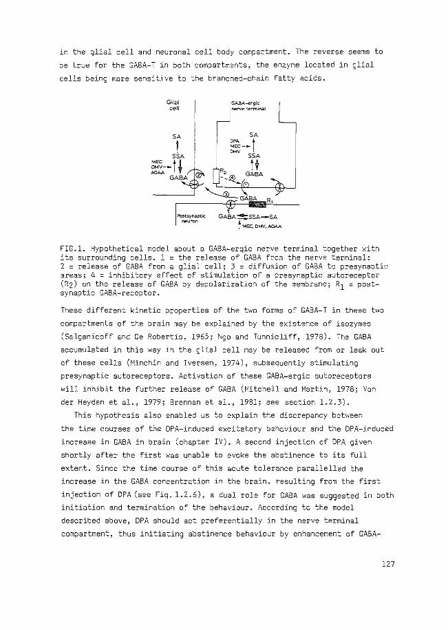
in the glial cell and neuronal cell body compartment. The reverse seems to
be true for the GABA-T in both compartments, the enzyme located in glial
cells being more sensitive to the branched-chain fatty acids.
Glial cell
SA
I SSA
MEC t ~ OMV--.OAA
GABA
~tsynaptic
neuron
I GABA-•cgi• ~ nerv" terminal
I I
GABA~SSA-SA
t MEC. OMV, AOAA
FIG.l. Hypothetical model about a GABA-ergic nerve terminal together with its surrounding cells. 1 = the release of GABA from the nerve terminal; 2 = release of GABA from a glial cell; 3 = diffusion of GABA to presynaptic areas; 4 = inhibitory effect of stimulation of a presynaptic autoreceptor (R2) on the release of GABA by depolarization of the membrane; R1 = postsynaptic GABA-receptor.
These different kinetic properties of the two forms of GABA-T in these two
compartments of the brain may be explained by the existence of isozymes
(Salganicoff and De Robertis~ 1965; i\'go and Tunnicliff, 1978). The GABA
accumulated in this way in the glial cell may be released from or leak out
of these cells (Minchin and Iversen, 1974), subsequently stimulating
presynaptic autoreceptors. Activation of these GABA-ergic autoreceptors
will inhibit the further release of GABA (Mitchell and Martin, 1978; Van
der Heyden et al., 1979; Brennan et al., 1981; see section 1.2.3).
This hypothesis also enabled us to explain the discrepancy between
the time courses of the DPA-induced excitatory behaviour and the DPA-induced
increase in GABA in brain (chapter IV). A second injection of DPA given
shortly after the first was unable to evoke the abstinence to its full
extent. Since the time course of this acute tolerance parallelled the
increase in the GABA concentration in the brain, resulting from the first
injection of DPA (see Fig. 1.2.6), a dual role for GABA was suggested in both
initiation and termination of the behaviour. According to the model
described above, DPA should act preferentially in the nerve terminal
compartment, thus initiating abstinence behaviour by enhancement of GABA-
127
ergic transmission. On the other hand, the overflow of GABA to presynaptic
areas ( arrow 3 in Fig. 1) may activate presynaptic autoreceptors, thus
terminating the abstinence behaviour through inhibition of the enhanced
release of GABA. Recently, Sarhan and Seiler (1979) presented evidence for
an overflow of GABA, indicating an increase in synaptosomal GABA during the
first 10 min and thereafter, an increase in non-synaptosomal material.
This time course for the increase in synaptosomal GABA after DPA is in
agreement with the time course of the quasi-abstinence behaviour induced
by DPA.
The finding that pretreatment with the dopamine-S-hydroxylase inhibitor
FLA-63 prevented the full expression of the DPA-induced behaviour suggests
that the presence of a normal level of noradrenaline (:-..JA) is required for
the induction of this behaviour (chapter VI). Since pretreatment of morphine
dependent rats with FLA-63 prevents also induction of body shakes by
levallorphan, these data support the specificity of the DPA-induced behaviour
as a model for the true abstinence behaviour and indicate that identical
mechanisms may be involved. In recent years it has been established that NA
may act as a neuromodulator enhancing the signal efficiency of GABA and
probably also that of other neurotransmitters (Woodward et al., 1979;
Moises and Woodward, 1980). As discussed in the next section, the nor
adrenergic bundle originating in the locus coeruleus appears to be
particularly involved in the DPA-induced behaviour.
The data discussed so far, which indicate an important role of GABA-
ergic activity in DPA-evoked abstinence behaviour, give rise to the question
as to which mechanisms are involved in true morphine withdrawal. With respect
to the involvement of the nerve terminal GABA compartment, two findings are
important, namely the presynaptic inhibition of GABA-release by morphine
(Vander Heyden et al., 1980; Corrigall and Linsemann, 1980) and the
decreased activity of GAD after development of physical dependence (Tzeng and
Ho, 1978; see section 1.3.4). Long-term inhibition of GABA-release by morphine
may result in the development of supersensitivity of postsynaptic GABA-
receptors (see section 1.2.3), as has been described recently (Ticku and
Huffman, 1980). It may be suggested that after withdrawal of morphine an
enhanced stimulation of these receptors will occur.
As far as GAD is concerned, the decreased activity found during the
development of physical dependence is restored after withdrawal of morphine
or precipitation of abstinence by naloxone, thus resulting in a relatively
enhanced GAD activity when compared with the preceding phase (Tzeng and Ho,
128
1978). This relatively enhanced activity of GAD implies an enhanced GABA
ergic activity, which, combined with the supersensitivity of GABA
receptors, offers a GABA-ergic mechanism which may initiate abstinence
behaviour.
No indications were found that a decrease in SSA-DH activity, the
mechanism involved in the induction of quasi-abstinence by DPA, is also
implicated in the expression in true morphine withdrawal behaviour. Repeated
administration of increasingly high doses of morphine (50-200 mg/kg daily,
during 14 days) did not result in a decreased SSA-DH activity or affinity
for SSA, measured 48 h after the last injection of morphine (Van der Laan,
Jacobs and Bruinvels, unpublished). However, it is not unlikely that a
possible decreased activity of SSA-DH occurring only in the neuronal
compartment, will be masked by the bulk of unchanged SSA-DH in the remaining
cells.
We have tried to show that the present results concerning DPA-induced
behaviour provided further insight into the mechanism which may be involved
in true morphine abstinence behaviour.
Considering the present data in relation to those obtained using other
quasi-abstinence models, e.g. phosphodiesterase inhibitors, TRH or EGTA (see
section 1.3.2), it can be concluded that they are complementary, yielding
insight into the complex mechanisms in morphine abstinence behaviour.
The study on DPA and TRH may provide data concerning the involvement of
intercellular mechanisms, on the level of neurotransmission or neuro
modulation, whereas the behaviour induced by phosphodiesterase inhibitors
and EGTA may throw some light on the involvement of intracellular
mechanisms in morphine abstinence behaviour.
&<Un aJtecw -i.nvo.tved -i.n qua.-6-i.-mMp!Une absunenee behav-i.oWt
Within the scope of this thesis, the involvement of several brain
structures in the expression of DPA-induced quasi-morphine abstinence
behaviour was investigated. Only three of them, namely the central amygdala
(Ac), the centre median-parafascicularis area (Cm-Pf) and the locus
coeruleus (LC) appeared to participate, each in a different way. The other
areas, where DPA was without effect, were nucleus caudatus, nucleus
accumbens, hippocampus and substantia nigra (chapters V and VI).
A GABA-ergic system in the Cm-Pf appears to have a facilitatory influence
on body shakes and locomotor activity, since injection of DPA into this
brain structure resulted in a large number of body shakes and greater
129
locomotor activity when compared with other areas. Administration of bicu
culline methiodide (BMI) into the Cm-Pf suppressed body shakes evoked by
intraperitoneal DPA, but had only minor effects on horizontal activity,
whereas injection of morphine into the same structure suppressed both
behavioural symptoms. A high amount of GAD appears to be present in the
nucleus parafascicularis (Massari et al., 1976), which is activated after
acute administration of morphine (Kuriyama and Yonenda, 1978). After chronic
administration of morphine,tolerance to this effect occurs. Whether an
enhanced or a decreased synthesis of GABA becomes apparent, remains to be
investigated.
Furthermore, it may be suggested that an inhibitory influence on body
shakes is provided in a GABA-ergic system in the Ac, since injection of
SMI into the Ac shortly after intraperitoneal injection of DPA resulted in
an increase in the number of body shakes. Morphine injected into this
structure potentiated locomotor activity slightly, but had no effect on the
body shakes evoked by DPA.
The Ac contains cell bodies of a GABA-ergic pathway projecting to the
bed nucleus of the stria terminalis (SST). In addition, a recurrent GABA
ergic pathway exists with cell bodies in the SST and terminals in the Ac
(LeGal La Salle et al., 1978). Body shakes can also be induced by injection
of kainic acid, a rigid analogue of glutamate, into this area (Ben-Ari
et al.,l978; see Table 1.3.2).It can, therefore, be suggested that the
neurons in the Ac, responsible for the induction of body shakes, can be
excited physiologically by glutamate, and are under the inhibitory control
of GABA (possibly originating in the SST). It is remarkable that DPA, while
also increasing the level of GABA in the amygdala, can induce body shakes.
It is not inconceivable that the GASA-projection from the SST to the Ac
is, in turn, under GABA-ergic control in the SST (Ben-Ari et al., 1976).
Administration of DPA to a rat, then, will cause an enhanced inhibition of
the activity of the GABA-ergic pathway to the Ac, resulting in disinhibition
of the neurons in the Ac. Subsequent injection of BMI into the Ac will cause
a further disinhibition leading to a higher number of body shakes.
Thus, GABA-ergic systems in the Cm-Pf and in the Ac may have opposite
roles in the initiation of DPA-induced quasi-morphine abstinence symptoms.
Since injection of DPA into the LC did not evoke quasi-morphine
abstinence symptoms, the GABA-ergic system present in the LC (Guyenet and
Aghajanian, 1978) does not appear to be directly involved in the induction
of this syndrome. Nevertheless, the present data indicate that an active
130
LC system is a prerequisite for this behaviour. In the preceding section
it was stated that depletion of NA with FLA-63 decreased the full expression
of DPA-evoked behaviour. Therefore, impairment of the activity of the dorsal
noradrenergic bundle may be held responsible for the suppression of body
shakes by FLA-63. Injection of morphine into the LC, thus decreasing the
LC firing rate (Bird and Kuhar, 1977) also suppressed DPA-induced behaviour
almost totally. These data are in agreement with the findings of Aghajanian
(1978), demonstrating an increased LC firing rate during morphine withdrawal
in rats, which could be suppressed by clonidine. Clonidine has been reported
to suppress abstinence behaviour in rats too (Fielding et al., 1978).
It has been demonstrated that the majority of the noradrenergic vari
cosities do not form synaptic contacts, but are located in gaps between
glial and nerve cells, and the released NA probably affects a large number
of neurons in the vicinity (Descarries et al., 1977). Furthermore, NA seems
to act more as a neuromodulator than as a transmitter in the classical
sense, i.e. both NA and amino acids can hyperpolarize their target neuronal
membrane, but the action of NA is not accompanied by an increase in ionic
membrane conductance, as is the case for the amino acids GABA and glycine.
The NA-induced inhibition of nerve cell activity in some brain areas mainly
concerns neuronal maintained activity, while responses to electrical or
sensory stimulation are either enhanced or left unaffected, at least in
comparison to the maintained activity (Woodward et al., 1979; Moises and
Woodward, 1980). It is attractive to suppose that such an action of NA on
GABA-ergic transmission is also involved in controlling DPA-induced behaviour.
Such an hypothesis explains why an active LC system is required for full
expression of the DPA-evoked behaviour. The two areas in which GABA-ergic
systems appear to be primarily involved in the induction of quasi-morphine
abstinence behaviour, namely the Ac and the Cm-Pf, have been shown to
receive projections·from the LC (Ungerstedt, 1971; Emson et al., 1979;
McGuiness and Krauthamer, 1980).
In the present experiments four structures were studied which are also
involved in true morphine abstinence behaviour, as revealed with the
technique of microinjection of antagonists (see section 1.3.3) namely, the
Ac as a part of the amygdala, the Cm-Pf as a part of the medial thalamus,
the nucleus caudatus as a part of the striatum and the LC as a part of the
floor of the 4th ventricle. The data suggest that only in the Cm-Pf a
GABA-ergic system may play a direct role in the expression of morphine
withdrawal symptoms. In the amygdala and LC other neurotransmitter systems
131
are probably involved.
ConQ£U4~on
Administration of DPA to a rat results in quasi-morphine abstinence
behaviour. In this thesis the mechanisms underlying this behaviour have
been investigated further and attempts have been made to locate the site of
action of DPA in initiating this behaviour. DPA acts via specific inhibition
of the second enzyme of the GABA degradation pathway, SSA-DH, leading to
an accumulation of SSA. This SSA probably enhances the level of GABA by
prevention of further GABA-T catalysed conversion of GABA, or through
backward formation of GABA. This enhanced GABA concentration, located in
the nerve endings, raises GABA-ergic transmission. thus initiating quasi
abstinence behaviour.
Concomitantly increased GABA concentration in glial cells will suppress
the behaviour through stimulation of presynaptic autoreceptors. Stimulation
of autoreceptors following overflow of GABA to presynaptic areas may be
responsible for the termination of quasi-morphine abstinence behaviour.
The increased GABA-ergic nerve cell activity probably initiates body
shakes directly in the Cm-Pf or via disinhibition in the Ac. Stimulation
of locomotor activity by DPA probably also involves a GABA-ergic system in
the Cm-Pf. All events mentioned thus far will occur in the presence of a
normally active LC. Taking into account the neuromodulatory action of NA,
it may be suggested that, in this case, the neurons in the Ac and in the
Cm-Pf are capable of differentiating between signal and noise, considering
the enhanced GABA-ergic stimulation as such a signal. However, when the
activity of the LC is decreased either through suppression by morphine or
through degeneration of the projections to the Ac and the Cm-Pf, the cells
in both nuclei might be less capable of differentiating signal from noise.
As a result their capacity to evoke the signals required to initiate body
shakes and enhanced locomotor activity would be decreased.
REFERENCES
Aghajanian, G.K. (1978) Tolerance of locus coeruleus neuronS to morphine and suppression of withdrawal respons by clonidine. Nature 276, 186-188
Anlezark, G., Horton, R.W., Meldrum, B.S., Sawaya, M.C.B. (1976) Anticonvulsant action of ethanolamine-0-sulphate and di-n-propylacetate and the metabolism of y-aminobutyric acid (GABA) in mice with audiogenic seizures. Biochem. Pharmacal. 25, 413-417
Ben-Ari, Y., Kanazawa, I., Zigmond, R.E. (1976) Regional distribution of
132
glutamate decarboxylase and GABA within the amygdaloid complex andstria terminalis system of the rat.J. Neurochem. 26, 1279-1283
Ben-Ari~ Y., Lagowska, J., Tremblay, E., LeGal La Salle, G. (1979) A new model of focal status epilepticus: intra-amygdaloid application of kainic acid elicits secondarily generalized convulsive seizures. Brain Res. 163, 176-179
Bird, S.J., Kuhar, M.J. (1977) Iontophorectic application of opiates to the locus coeruleus. Brain Res. 122, 523-533
Brennan, M.J.W.~ Cantril!, R.C., Oldfield, M., Krogsgaard-Larsen, P. (1981) Inhibition of Y-aminobutyric acid release by Y-aminobutyric acid-agonist drugs. Malec. Pharmacal. 19, 27-30
Corrigall, W.A., Linseman, M.A. (1980) A specific effect of morphine on evoked activity in the rat hippocampal slice. Brain Res. 192, 227-238
De Boer, Th. (1977) GABA shunt enzymes and the relationship with morphine abstinence. Academic Thesis, Erasmus University, Rotterdam, The Netherlands
Descarries, L., Watkins, K.C., Lapierre, Y. (1977) Noradrenergic axon terminals in the cerebral cortex of rat. III. Topometric ultrastructural analysis. Brain Res. 133, 197-222
Duffy, T.E., Nelson, S.R., Lowry, O.H. (1972) Cerebral carbohydrate metabolism during acute hypoxia and recovery. J. Neurochem. 19, 959-977
Emson, P.C., BjOrklund, A., Lindvall, 0., Paxinos, G. (1979) Contributions of different afferent pathways to the catecholamine and 5-hydroxytryptamine innervation of the amygdala: a neurochemical and histochemical study. Neuroscience 4, 1347-1357
Fielding, S., Wilker, J .. Hynes, M., Szewack, M., Novick Jr., W.J., Lal, H. (1978) A comparison of clonidine for antinociceptive and antiwithdrawal actions. J. Pharmacal. Exp. Ther. 207, 899-905
Godin, Y., Heiner, L., Mark, J., Mandel, P. (1969) Effects of di-npropylacetate, an anticonvulsive compound, on GABA metabolism. J. Neurochem. 16. 869-873
Guyenet, P.G., Aghaja~ian, G.K. (1979) Ach, Substance P and Met-enkephalin in the locus coeruleus: pharmacological evidence for independent sites of action. Europ. J. Pharmacal. 53, 319-328
Harvey, P.K.P., Bradford, H.F., Davison, A.N. (1975) The inhibitory effect of sodium n-dipropylacetate on the degradative enzymes of the GABAshunt. FEBS Lett. 52. 251-254
Herz, A., Blasig, J., Pap~schi, R. (1974) Role of catecholaminergic mechanisms in the expression of the morphine abstinence syndrome in rats. Psychopharmacologia 39, 121-143
Iadarola, .M.J., Gale, K. (1979) Dissociation between drug-induced increases in nerve terminal and non-nerve terminal pools of GABA ~ vivo. Europ. J. Pharmac. 59, 125-129
Kuriyama, K., Yoneda, Y. (1978) Morphine-induced alterations of Yaminobutyric acid and taurine contents and L-glutamate decarboxylase activity in rat spinal cord and thalamus: possible correlates with analgesic action of morphine. Brain Res. 148, 163-179
LeGal La Salle, G., Paxinos, G., Emson, P., Ben-Ari, Y. (1978) Neurochemical mapping of GABA-ergic systems in the amygdaloid complex and bed nucleus of the stria terminalis. Brain Res. 155, 397-403
Massari, V.J., Gottesfeld, Z., Jacobowitz, D.M. (1976) Distribution of glutamic acid decarboxylase in certain rhombencephalic and thalamic QUclei of the rat. Brain Res. 118, 147-151
McGuiness, C.M., Krauthamer, G.M. (1980) The afferent projections to the centrum medianum of the cat as demonstrated by retrograde transport of horseradish peroxidase. Brain Res. 184, 255-269
133
Minchin, M.C.W., Iversen, L.L. (1974) Release of (3H) gamma-aminobutyric acid from glial cells in rat dorsal root ganglia. J. Neurochemistry 23, 533-540
Mitchell, P.R., Martin, I.L. (1978) Is GABA release modulated by presynaptic receptors? Nature 274, 904-905
Moises, H.C., Woodward, D.J. (1980) Potentiation of GABA inhibitory action in cerebellum by locus coeruleus stimulation. Brain Res. 182, 327-344
Salganicoff, L., De Robertis, E. (1965) Subcellular distribution of the enzymes of the glutamic acid, glutamine and y-aminobutyric acid cycles in rat brain. J. Neurochemistry 12, 287-309
Sarhan, S., Seiler, N. (1979) Metabolic inhibitors and subcellular distribution of GABA. J. Neurosci. Res. 4, 399-422
Sawaya, M.C.B., Horton, R.W., Meldrum, B.S. (1975) Effects of anticonvulsant drugs on the cerebral enzymes metabolizing GABA. Epilepsia 16, 649-655
Simler, S., Cieselski, L., Maitre, M., Randrianarisoa, H., Mandel, P. (1973) Effect of sodium n-dipropylacetate on audiogenic seizures and brain Y-aminobutyric acid level. Biochem. Pharmacal. 22, 1701-1708
Ticku, M.K., Huffman, R.D. (1980) The effects of acute and chronic morphine administration on GABA receptor binding. Europ. J. Pharmacal. 68, 97-106
Tzeng, S.F., Ho, I.K. (1978) Acute and continuous morphine administration on the Y-aminobutyric acid system in the mouse. Prog. Neuro-Psychopharmac. 2, 55-64
Ungerstedt, U. (1971) Stereotaxic mapping of the monoamine pathways in the rat brain. Acta Physiol. Scand. Suppl. 367, 1-48
Vander Heyden, J.A.M., Venema, K., Korf, J. (1980) In v~vo release of endogenous GABA from rat striatum: effects of muscimol, oxotremorine and morphine. J. Neurochem. 34, 1648-1653
Woodward, D.J., Moises, H.C., Waterhouse, B.D., Hoffer, B.J., Freedman, R. (1979) Modulatory actions of norepinephrine in the central nervous system. Fed. Proc. 38, 2109-2116.
134
SUMMARY
Di-n-propylacetate (DPA), generally known to be an anti-epileptic drug,
induces a behavioural syndrome in rats resembling morphine abstinence behaviour,
which is called, therefore, quasi-morphine abstinence beh~viour. An increase
in GABA-ergic activity is probably responsible for this behavioural effect of
DPA, since it has been shown to be suppressed by GABA-antagonists like
bicuculline and picrotoxin.
The experiments presented in this thesis were designed to investigate the
central mechanisms involved in this quasi-morphine abstinence behaviour.
In chapter I a survey is given of the biochemistry and .physiology of GABA,
and possible levels for regulation are indicated. In addition, the morphine
withdrawal syndrome is described briefly. A lot of compounds have been
reported to induce quasi-abstinence behaviour, similar to a greater or lesser
extent to that induced by DPA. The implication of these data, with respect to
the mechanisms involved in morphine abstinence behaviour, are discussed.
Finally, the interactions between GABA and opioids are reviewed. The data
suggest that such interactions exist at the cellular level.
In chapter II studies on the mechanism of action of DPA are described,
particularly ~he effect of DPA on the enzymes involved in the degradation of
GABA. It appears that DPA increases GABA-ergic activity in the brain via
inhibition of the second enzyme involved in GABA-degradation, viz. succinic
semialdehyde dehydrogenase (SSA-DH). DPA does not exert a direct effect on the
first enzyme GABA-transaminase (GABA-T), as suggested by other authors.
Inhibition of SSA-DH will result in accumulation of SSA, which probably inhibits
GABA-T, thus increasing the concentration of GABA.
In the experiments described in chapter III, two branched-chain fatty
acid analogues of DPA and a short-chain fatty acid were used to study the
relationship between their biochemical and behavioural effects. A correlation
appeared to exist between the K. of these compounds for SSA-DH and the doses 1 .
exerting a maximum effect on behaviour. On the other hand, concurrent
inhibition of GABA-T seemed to suppress the behavioural effects of the fatty
acids. This apparent paradox can possibly be explained by supposing a
different action of the fatty acids in distinct compartments of the brain,
suggesting that the increased GABA-ergic activity in the neuronal compartment
is evoked by SSA-DH inhibition and initiates quasi-morphine abstinence
behaviour. A concomitant increase in the GABA concentration in glial cells,
caused by inhibition of GABA-T, is held responsible for the reduced
135
capability of the fatty acids ·to induce the behavioural syndrome when
compared with DPA.
The time course of the DPA-induced behaviour is much shorter than that
of the DPA-induced enhancement of the GABA-concentration in brain.
Therefore, as described in chapter IV, we have studied the influence of
enhanced GABA-levels, caused by a first injection of DPA, on the behaviour
evoked by a second injection of DPA at different time intervals after the first
injection. The results indicate that GABA fulfils a role both in the initiation
and termination of DPA-induced behaviour. The mechanism responsible for this
dual action of GABA is ascribed to a different sensitivity to DPA of the nerve
terminal and glial metabolic compartments of GABA in the brain, as suggested
above. The increase in GABA in the nerve terminal caused by DPA is probably
responsible for the initiation of the quasi-abstinence behaviour, whereas the
overflow of GABA from the synaptic cleft to presynaptic areas may be responsible
for the suppression of this behaviour via stimulation of autoreceptors.
Behavioural responses after intracerebral injections of DPA were studied
to obtain information on the brain areas involved in the quasi-abstinence
syndrome (Chapters V and VI). The results suggest a facilitatory role for a
GABA-ergic system in the centre median-parafascicularis area on body shakes,
while in the central amygdala a GABA-ergic system appears to exert an inhibitory
influence on this symptom of abstinence. The GABA-ergic terminals present in
the locus coeruleus do not seem to play a role in this behaviour. On the other
hand, stimulation of opiate receptors in the latter nucleus by low doses of
morphine, thus decreasing the activity of the noradrenergic cells, was found
to suppress this behaviour almost totally. The centre median-parafas~icularis
area is much less sensitive to this suppressive effect of morphine, while
no suppression by morphine could be found when injected into the central
amygdala. A decrease in locus coeruleus activity by destruction of the cells
or by degeneration of the projections to the forebrain by about 80% prevented
the full expression of the quasi-abstinence behaviour.
Thus, an active locus coeruleus is required for the induction of DPA-induced
abstinence behaviour. The role of the locus coeruleus can possibly be understood
in the light of a neuromodulatory function of noradrenaline on GABA-ergic
inhibition.
It is concluded that quasi-morphine abstinence behaviour is evoked by DPA
through enhancement of GABA-ergic transmission in certain areas of the brain,
such as the centre median-parafascicularis area, or via disinhibition of neurons
lying in the central amygdala. An active noradrenergic system is a prerequisite
for an efficient transmission of the GABA-ergic signals in these nuclei. 136
SAME~VATTING
KWASI-MORFINE ONTHOUDINGSGEDRAG OPGEWEKT DOOR DIPROPYLACETAAT
IN DE RAT: MECHANISMEN EN AANGRIJPINGSPUNTEN
De opmerkelijke waarneming dat dipropylacetaat (DepakineR) bij ratten een
gedrag opwekt dat veel overeenkomsten vertoont met het patroon van gedrags
symptomen dat optreedt na het onthouden van morfine aan verslaafde ratten, is
aanleiding geweest voor ons onderzoek. We duiden dit gedrag oak wel aan als
kwasi-morfine onthoudingsgedrag. Nader onderzoek van dit gedrag zou infor
matie kunnen leveren over de processen die optreden bij het echte morfine
onthoudingsgedrag. De belangrijkste symptomen van het door dipropylacetaat
(DPA) geinduceerde gedrag bij de rat zijn: het schudden van het hele lichaam
zoals door een natte hand, het graven in het zaagsel van de observatie-
kooi, een hoge rug en rechtopstaande haren en een sterk verhoogde bewegings
aktiviteit.
Het was bekend uit de literatuur dat DPA de hoeveelheid gamma-amino
boterzuur (GABA) in de hersenen verhoogt. GABA verzorgt in de hersenen een
functie als overdrachtsstof (transmitter) tussen zenuwcellen (neuronen) en
komt voor in nagenoeg alle delen van de hersenen en het ruggemerg. Uit het
onderzoek van De Boer en Bruinvels, dat aan het in dit proefschrift beschreven
onderzoek voorafging, is dan oak gebleken dat de werking van de neuronen
die GABA bevatten, gestimuleerd wordt tijdens het door DPA opgewekte gedrag.
In dit proefschrift is beschreven hoe DPA deze verhoogde stimulatie door
GABA veroorzaakt en oak waar DPA in de hersenen aangrijpt.
In de inleiding (hoofdstuk I) is aangegeven dat de aktiviteit van het
GABA-erge systeem op verschillende niveaus beinvloed kan worden, nl. op het
niveau. van de aanmaak en afbraak van GABA, van de mate van GABA-afgifte door
de GABA-erge zenuwuiteinden en van de gevoeligheid voor GABA van de ontvanger
cellen. Omdat DPA veel wordt gebruikt als geneesmiddel bij epilepsie, is oak
kart ingegaan op de mechanismen die aan deze anti-epileptische werking van
DPA ten grondslag kunnen liggen. Met betrekking tot het morfine onthoudings
gedrag is een overzicht gegeven van wat er tot dusver is bestudeerd aan kwasi
onthoudingsgedrag. Behalve door DPA kan dit gedrag namelijk nag door veel
andere stoffen worden opgewekt. Hoewel er duidelijke verschillen zijn aan te
geven tuss~n de syndromen die door verschillende klassen van stoffen opgewekt
worden, is gebleken dat de informatie, verkregen uit zulk onderzoek, een
werkelijke bijdrage levert tot de ontrafeling van de mechanismen die een rol
137

spelen bij morfine-onthoudingsgedrag. In het laatste gedeelte van de in
leiding is nagegaan of de literatuur-gegevens over de interacties tussen
morfine en GABA in overeenstemming zijn met de rol van GABA die wordt
gesuggereerd door de gegevens omtrent het door DPA opgewekte onthoudings
gedrag.
Ons eerste onderzoek (hoofdstuk II) betrof het mechanisme waardoor DPA
de hoeveelheid GABA in de hersenen verhoogt. Deze verhoging komt tot stand
door remming van de afbraak van GABA. Twee specifieke enzymen zijn bij deze
afbraak betrokken, achtereenvolgens GABA-transaminase (GABA-T) en succinaat
semi-aldehyde dehydrogenase (SSA-DH) (zie Fig. 1).
~DPA .. /
K; =23.2mM/ \K; =0.5mM I I I I
: I
GABA t SSA -'--.sA GABA-T SSA-DH
FIG. l.Effect van DPA op de afbraak van GABA. SSA = succinaat semi-aldehyde; SA = succinaat. De Ki is een maat voor het remmend effect van een stof. Een lagere Ki wil zeggen een sterkere remming.
In hoofdstuk II is beschreven dat DPA het tweede enzym 30-40 maal sterker
remt dan het eerste, waardoor ophoping plaatsvindt van SSA. Omdat GABA-T
geremd wordt door het tussenprodukt SSA, is het uiteindelijk resultaat van
de werking van DPA ophoping van GABA.
Ligt dit mechanisme nu ten grondslag aan het kwasi-morfine onthoudings
gedrag? Voor de beantwoording van deze vraag hebben we gebruik gemaakt van
op DPA gelijkende stoffen. Door van al deze stoffen te vergelijken wat hun
effect was op het gedrag en op de afbraak van GABA, konden we dit inderdaad
aannemelijk maken. Het bleek dat de dosis van deze stoffen waarmee het
maximale effect op het gedrag bereikt kan worden, lager is naarmate SSA-DH
sterker geremd wordt. De mate waarin het gedrag wordt opgewekt is echter
gering voor die stoffen die tegelijk een sterk remmend effect hebben op
GABA-T. We hebben dit verklaard door aan te nemen dat de verschillende
cellen waarin de afbraak van GABA plaatsvindt, nl. de zenuwceluiteinden en
de gliacellen (die een opbouw- en onderhoudsfunctie in de hersenen vervullen),
enzymen bevatten die een verschillende gevoeligheid hebben voor deze stoffen.
Gegevens uit de literatuur ondersteunen deze aanname. Remming van GABA-T
138
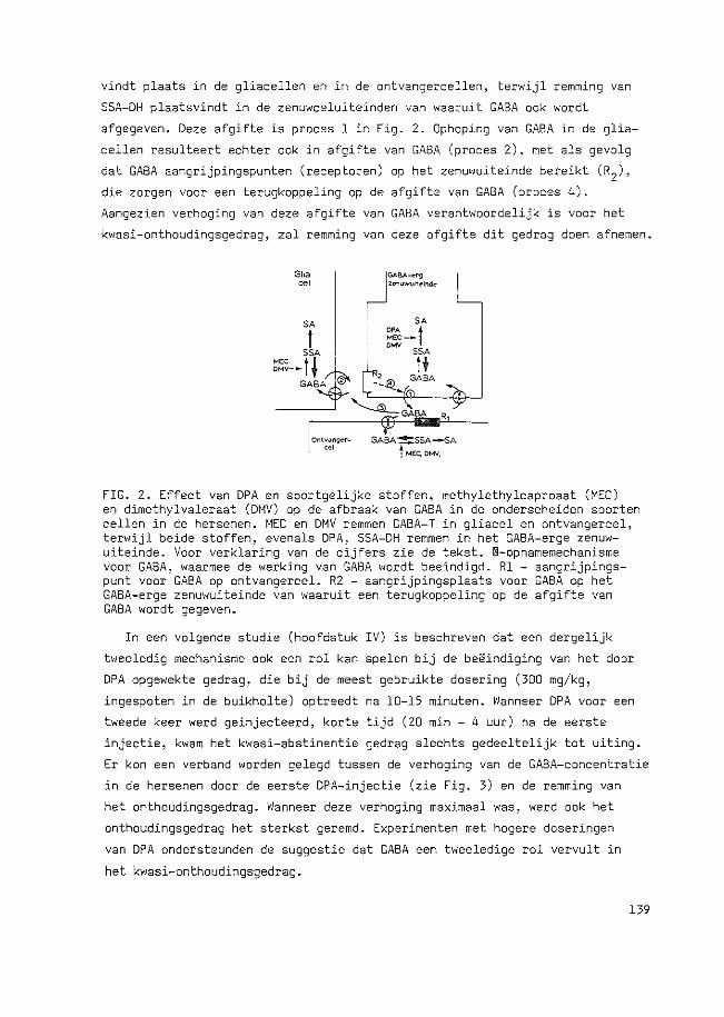
vindt plaats in de gliacellen en in de ontvangerce11en, terwijl remming van
SSA-DH plaatsvindt in de zenuwce1uiteinden van waaruit GABA oak wordt
afgegeven. Deze afgifte is proces 1 in Fig. 2. Ophoping van GABA in de glia
ce11en resulteert echter oak in afgifte van GABA (proces 2), met a1s gevo1g
dat GABA aangrijpingspunten (receptoren) op het zenuwuiteinde bereikt (R2),
die zorgen voor een terugkoppe1ing op de afgifte van GABA (proces 4).
Aangezien verhoging van deze afgifte van GABA verantwoordelijk is voor het
kwasi-onthoudingsgedrag, zal remming van deze afgifte dit gedrag doen afnemen.
FIG. 2. Effect van DPA en soortgelijke stoffen, methylethylcaproaat (MEC) en dimethylvaleraat (DMV) op de afbraak van GABA in de onderscheiden soorten ce1len in de hersenen. MEC en DMV remmen GABA-T in gliacel en ontvangercel, terwijl beide stoffen, evenals DPA, SSA-DH remmen in het GABA-erge zenuwuiteinde. Voor verk1aring van de cijfers zie de tekst. ®-opnamemechanisme voor GABA, waarmee de werking van GABA wordt beeindigd. R1 - aangrijpingspunt voor GABA op ontvangercel. R2 - aangrijpingsplaats voor GABA op het GABA-erge zenuwuiteinde van waaruit een terugkoppeling op de afgifte van GABA wordt gegeven.
In een volgende studie (hoofdstuk IV) is beschreven dat een dergelijk
tweeledig mechanisme oak een rol kan spelen bij de beeindiging van het door
DPA opgewekte gedrag, die bij de meest gebruikte dosering (300 mg/kg,
ingespoten in de buikholte) optreedt na 10-15 minuten. Wanneer DPA voor een
tweede keer werd geinjecteerd, korte tijd (20 min - 4 uur) na de eerste
injectie, kwam het kwasi-abstinentie gedrag slechts gedeeltelijk tot uiting.
Er kon een verband worden gelegd tussen de verhoging van de GABA-concentratie
in de hersenen door de eerste DPA-injectie (zie Fig. 3) en de remming van
het onthoudingsgedrag. Wanneer deze verhoging maximaal was, werd oak het
onthoudingsgedrag het sterkst geremd. Experimenten met hogere doseringen
van DPA ondetsteunden de suggestie dat GABA een tweeledige rol vervult in
het kwasi-onthoudingsgedrag.
139
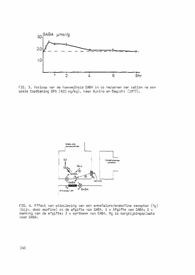
3D GABA )Jmol/g
1.0
2 4 6 8hr
FIG. 3. Verloop van de hoeveelheid GABA in de hersenen van ratten na een orale toediening DPA (400 mg/kg). Naar Kukino en Deguchi (1977).
FIG. 4. Effect van stimulering van een enkefaline/endorfine receptor (RE) (bijv. door morfine) op de afgifte van GABA. 1 = Afgifte van GABA; 2 = remming van de afgifte; 3 = synthese van GABA. Rc is aangrijpingsplaats voor GABA.
140
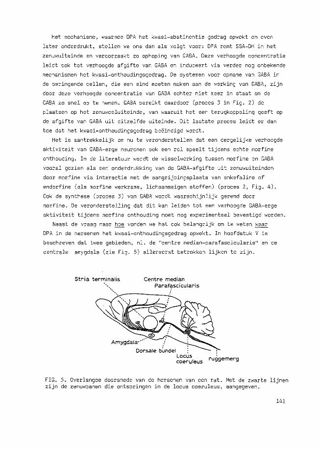
Het mechanisme, waarmee DPA het kwasi-abstinentie gedrag opwekt en even
later onderdrukt, stellen we ons dan als volgt voor: DPA remt SSA-DH in het
zenuwuiteinde en veroorzaakt zo ophoping van GABA. Deze verhoogde concentratie
leidt oak tot verhoogde afgifte van GABA en induceert via verder nag onbekende
mechanismen het kwasi-onthoudingsgedrag. De systemen voor opname van GABA in
de omringende cellen, die een eind moeten maken aan de werking van GABA, zijn
door deze verhoogde concentratie van GABA echter niet meer in staat om de
GABA zo snel op te nemen. GABA bereikt daardoor (proces 3 in Fig. 2) de
plaatsen op het zenuwceluiteinde, van waaruit het een terugkoppeling geeft op
de afgifte van GABA uit ditzelfde uiteinde. Dit laatste proces leidt er dan
toe dat het kwasi-onthoudingsgedrag be§indigd wordt.
Het is aantrekkelijk om nu te veronderstellen dat een dergelijke verhoogde
aktiviteit van GABA-erge neuronen oak een rol speelt tijdens echte morfine
onthouding. In de literatuur wordt de wisselwerking tussen morfine en GABA
vooral gezien als een onderdrukking van de GABA-afgifte uit zenuwuiteinden
door morfine via interactie met de aangrijpingsplaats van enkefaline of
endorfine (als morfine werkzame, lichaamseigen stoffen) (proces 2, Fig. 4).
Oak de synthese (proces 3) van GABA wordt waarschijnlijk geremd door
morfine. De veronderstelling dat dit kan leiden tot een verhoogde GABA-erge
aktiviteit tijdens morfine onthouding moet nag experimenteel bevestigd worden.
Naast de vraag naar hoe vonden we het oak belangrijk om te weten ~
DPA in de hersenen het kwasi-onthoudingsgedrag opwekt. In hoofdstuk V is
beschreven dat twee gebieden, nl. de "centre median-parafascicularis 11 en de
centrale amygdala (zie Fig. 5) allereerst betrokken lijken te zijn.
Stria terminatis Centre median Paraffscicularis
,.,.-----!..._
Amygdala····
Dorsale bUndel . Locus coerureus ruggemerg
FIG. 5. Overlangse doorsnede van de hersenen van een rat. Met de zwarte lijnen zijn de zenuwbanen die ontspringen in de locus coeruleus, aangegeven.
141
Het GABA-erge systeem in de centre median-parafascicularis lijkt een aan
grijpingspunt voor DPA te zijn~ omdat injectie van DPA ter plaatse resulteerde
in het natte hand syndroom. GABA-tegenwerkende stoffen alsook morfine
onderdrukken dit gedrag wanneer ze in de centre median-parafascicularis worden
geinjecteerd 5 minuten nadat DPA door injectie in de buik is toegediend.
Aan de andere kant bleek het GABA-erge systeem in de centrale amygdala juist
een remmende invloed uit te oefenen op het door DPA geinduceerde gedrag.
Desondanks resulteerde beschadiging van dit laatste gebied in vermindering van
het natte hand syndroom. De centrale amygdala lijkt dus niet een direkt
aangrijpingspunt voor DPA te zijn, maar vervult wel een belangrijke rol in het
kwasi-onthoudingsgedrag.
De studie beschreven in hoofdstuk VI bracht aan het licht dat nag een
derde hersenstruktuur, nl. de locus coeruleus, onmisbaar is voor de gedrags
effecten van DPA. Van de locus coeruleus is bekend dat het wijd vertakte
uitlopers heeft naar bijna alle delen van de hersenen (zie Fig. 5). Het gebied
bleek met name heel gevoelig te zijn voor morfine, dat de aktiviteit van de
zenuwcellen in de locus coeruleus onderdrukt. Een 25 keer lagere dosering van
morfine dan gebruikt was voor de centre median-parafascicularis was in staat,
na injectie in de locus coeruleus, het kwasi-onthoudingsgedrag nagenoeg
volledig te onderdrukken. Oak uitschakeling van deze zenuwcellen op andere
manieren verminderde het door DPA geinduceerde gedrag aanzienlijk. Een
mogelijke verklaring voor de noodzakelijke aanwezigheid van de locus coeruleus
is dat de overdrachtsstof, waarvan zich de cellen van deze kern bedienen,
de werking van GABA potentieert, waardoor de informatieoverdracht van GABA
erge cellen op ontvangercellen efficienter verloopt. Blijkbaar is deze
potentiering zo zeer noodzakelijk dat zonder de aktiviteit van de locus
coeruleus de stimulering van GABA door DPA in bijvoorbeeld de centre median
parafascicuJAris niet kan resulteren in kwasj-morfine onthoudingsqedrag.
Samengevat luiden de conclusies van de in dit proefschrift beschreven
studies als volgt:
1. Hoe wekt DPA kwasi-onthoudingsgedrag op? DPA remt de afbraak van GABA
door remming van het tweede enzym in de afbraak, SSA-DH.
2. Waar ligt het aangrijpingspunt van DPA? Voor het natte hand syndroom
waarschijnlijk in de centre median-parafascicularis, terwijl een intakte
centrale amygdala en locus coeruleus onmisbaar lijken te zijn.
142

Curriculum vitae
Op 21 december 1952 werd ik geboren te Groningen en na de lagere school
en het Gereformeerd Lyceum in deze plaats door1open te hebben, behaalde ik
in 1970 mijn diploma HBS-8. Daarna werd gestart met de studie scheikunde
aan de Rijksuniversiteit van Groningen. Na in mei 1973 geslaagd te zijn
voor het kandidaatsexamen, koos ik als specia1isatie de biochemie. Het
hoofdvakonderzoek over bacteriele membranen werd verricht onder leiding
van Prof. Dr. B. Witholt. Dr. J. Korf was begeleider bij het bijvak
onderzoek in de biologische psychiatrie. Na het doctoraal examen in april
1977 trad ik in dienst van de Nederlandse Stichting voor Zuiver-Wetenschappelijk
Onderzoek ten behoeve van de stichting FUNGO. Het onderzoek vond plaats op
de afdeling Farmacologie van de Erasmus Universiteit te Rotterdam onder
supervisie van Prof. Dr. J. Bruinvels, en leidde tot dit proefschrift.
In dit onderzoek werd oak samengewerkt met Dr. A.R. Cools van het Instituut
voor Farmacologie van de Katholieke Universiteit te Nijmegen. Vanaf begin
1980 ben ik in dienst van het Rijks Instituut voor de Volksgezondheid te
Bi1thoven en werkzaam op het Laboratorium voor Farmacologie (afdeling
Psychofarmacologie) te Utrecht.
143