Pavel Š , Yvonne N , Jaromír P , Nikolay I. BogdaNov Christina ock … · 2016-12-06 · Pavel...
10
Fottea, Olomouc, 14(1): 53–62, 2014 53 Planktochlorella nurekis gen. et sp. nov. (Trebouxiophyceae, Chlorophyta), a novel coccoid green alga carrying significant biotechnological potential Pavel ŠKALOUD 1* , Yvonne Němcová 1 , Jaromír PYTELA 2 , Nikolay I. BogdaNov 3 , Christina Bock 4 & Sam H. PickiNPaugh 2 1 Department of Botany, Charles University in Prague, Benátská 2, Praha 2, 12801 Czech Republic; *Corresponding author e–mail: [email protected] 2 Key Industry Engineering s.r.o., Mojmírova 1710/15, Praha 4, 14000, Czech Republic 3 Penza Agricultural Research Institute, Penza oblast, 442731, Russia 4 Faculty of Biology, University of Essen, Essen, 45141, Germany Abstract: Phylogenetic position, morphology and ultrastructure were investigated for biotechnologically remarkable strain Chlorella vulgaris IFR C–111, utilized in various commercial applications. Molecular phylogenetic analyses based on the SSU and ITS rDNA data revealed that the strain IFR C–111 forms a distinct lineage within the Parachlorella–clade in Chlorellaceae. We describe this organism as a new genus and species, Planktochlorella nurekis. Vegetative cells of this newly recognized species are spherical, possessing a single pot–shaped chloroplast with starch–covered pyrenoid. Asexually it reproduces by the formation of 2–16(–32) autospores. Cell wall is composed of two layers, the outer layer containing extended microfibrillar material. The fuzzy cell wall structure improves the buoyancy resulting in low sedimentation rate of P. nurekis. To resolve the phylogenetic position of Planktochlorella and its relationship to the closely related genera, nucleotide saturation present in the ITS rDNA data was reduced by four different approaches. The resulting topologies pointed to the poor phylogenetic signal in generally utilized SSU and ITS rDNA data and the need of sequencing other molecular markers. Key words: biotechnology, Chlorella, green algae, nucleotide saturation, phylogeny, taxonomy, Trebouxiophyceae, ultrastructure INTRODUCTION Microalgae are increasingly being assessed for their potential in various industrial biotechnology platforms. They have several applications from human and animal nutrition to cosmetics, production of high value molecules (e.g. polysaccharides, fatty acids, pigments) and biodiesel production. Due to their high growth rate and easy cultivation, green algae traditionally included in the genus Chlorella BeijeriNck are among the most extensively used microorganisms in industry. Chlorella is commercially produced by more than 70 companies, with the world annual sales extending 38 billon USD (Yamaguchi 1997). Various Chlorella strains are most importantly exploited for their various health–promoting effects (e.g. for anaemia treatment, anti–tumour effects, immunostimulation, prevention against atherosclerosis and hypercholesterolemia; merchaNt & aNdre 2001), but they are also used as food additives, nutrition in aquacultures, and in cosmetic industry (SPOLAORE et al. 2006). However, the taxonomy of Chlorella–like species is complicated. The application of molecular data uncovered that simple morphology of these organisms hides extensive diversity and distant relation of traditionally defined Chlorella species (huss et al. 1999). According to the recent molecular phylogenetic investigations, species having typical Chlorella morphology (i.e., spherical cells with single, parietal chloroplast including a single pyrenoid with a distinct starch envelope) belong to the Trebouxiophycean family Chlorellaceae, which is divided into two lineages, the Chlorella–clade and the Parachlorella–clade (KRIENITZ et al. 2004; LUO et al. 2010). Moreover, several studies pointed to the close relation of Chlorella morphotypes with various elongated or needle–shaped green algal genera e.g. Dicloster, Closteriopsis (hegewald & haNagata 2000; ustiNova et al. 2001; wolf et al. 2002; KRIENITZ et al. 2004) and colonial species traditionally determined as Dictyosphaerium NÄGELI and Micractinium fresiNius (LUO et al. 2006, 2010; Bock et al. 2010; KRIENITZ et al. 2010). Consequently, high levels of cryptic diversity within the Chlorella morphotype as well as the polyphyletic nature of both Chlorella and Dictyosphaerium resulted in fundamental taxonomic revision of these organisms, including the
Transcript of Pavel Š , Yvonne N , Jaromír P , Nikolay I. BogdaNov Christina ock … · 2016-12-06 · Pavel...
Fottea, Olomouc, 14(1): 53–62, 2014 53
Planktochlorella nurekis gen. et sp. nov. (Trebouxiophyceae, Chlorophyta), a novel coccoid green alga carrying significant biotechnological potential
Pavel Škaloud1*, Yvonne Němcová1, Jaromír Pytela2, Nikolay I. BogdaNov3, Christina Bock4 & Sam H. PickiNPaugh2
1 Department of Botany, Charles University in Prague, Benátská 2, Praha 2, 12801 Czech Republic; *Corresponding author e–mail: [email protected] Key Industry Engineering s.r.o., Mojmírova 1710/15, Praha 4, 14000, Czech Republic3 Penza Agricultural Research Institute, Penza oblast, 442731, Russia4 Faculty of Biology, University of Essen, Essen, 45141, Germany
Abstract: Phylogenetic position, morphology and ultrastructure were investigated for biotechnologically remarkable strain Chlorella vulgaris IFR C–111, utilized in various commercial applications. Molecular phylogenetic analyses based on the SSU and ITS rDNA data revealed that the strain IFR C–111 forms a distinct lineage within the Parachlorella–clade in Chlorellaceae. We describe this organism as a new genus and species, Planktochlorella nurekis. Vegetative cells of this newly recognized species are spherical, possessing a single pot–shaped chloroplast with starch–covered pyrenoid. Asexually it reproduces by the formation of 2–16(–32) autospores. Cell wall is composed of two layers, the outer layer containing extended microfibrillar material. The fuzzy cell wall structure improves the buoyancy resulting in low sedimentation rate of P. nurekis. To resolve the phylogenetic position of Planktochlorella and its relationship to the closely related genera, nucleotide saturation present in the ITS rDNA data was reduced by four different approaches. The resulting topologies pointed to the poor phylogenetic signal in generally utilized SSU and ITS rDNA data and the need of sequencing other molecular markers.
Key words: biotechnology, Chlorella, green algae, nucleotide saturation, phylogeny, taxonomy, Trebouxiophyceae, ultrastructure
IntroductIon
Microalgae are increasingly being assessed for their potential in various industrial biotechnology platforms. They have several applications from human and animal nutrition to cosmetics, production of high value molecules (e.g. polysaccharides, fatty acids, pigments) and biodiesel production. Due to their high growth rate and easy cultivation, green algae traditionally included in the genus Chlorella BeijeriNck are among the most extensively used microorganisms in industry. Chlorella is commercially produced by more than 70 companies, with the world annual sales extending 38 billon USD (Yamaguchi 1997). Various Chlorella strains are most importantly exploited for their various health–promoting effects (e.g. for anaemia treatment, anti–tumour effects, immunostimulation, prevention against atherosclerosis and hypercholesterolemia; merchaNt & aNdre 2001), but they are also used as food additives, nutrition in aquacultures, and in cosmetic industry (SPolaore et al. 2006).
However, the taxonomy of Chlorella–like species is complicated. The application of molecular
data uncovered that simple morphology of these organisms hides extensive diversity and distant relation of traditionally defined Chlorella species (huss et al. 1999). According to the recent molecular phylogenetic investigations, species having typical Chlorella morphology (i.e., spherical cells with single, parietal chloroplast including a single pyrenoid with a distinct starch envelope) belong to the Trebouxiophycean family Chlorellaceae, which is divided into two lineages, the Chlorella–clade and the Parachlorella–clade (krienitz et al. 2004; luo et al. 2010). Moreover, several studies pointed to the close relation of Chlorella morphotypes with various elongated or needle–shaped green algal genera e.g. Dicloster, Closteriopsis (hegewald & haNagata 2000; ustiNova et al. 2001; wolf et al. 2002; krienitz et al. 2004) and colonial species traditionally determined as Dictyosphaerium nägeli and Micractinium fresiNius (luo et al. 2006, 2010; Bock et al. 2010; krienitz et al. 2010). Consequently, high levels of cryptic diversity within the Chlorella morphotype as well as the polyphyletic nature of both Chlorella and Dictyosphaerium resulted in fundamental taxonomic revision of these organisms, including the
54 Škaloud et al.: Planktochlorella nurekis gen. et sp. nov.
description of many new species and genera (krienitz et al. 2004, 2012; Bock et al. 2010, 2011a,b). At present, the Chlorella– and Parachlorella–clades comprise seven (Actinastrum lagerheim, Chlorella, Didymogenes schmidle, Heynigia Bock, Pröschold et krienitz, Hindakia Bock, Pröschold et krieNitz, Meyerella fawleY et fawleY, Micractinium) and nine (Closteriopsis lemmermaNN, Compactochlorella krieNitz, Bock, kotut et Pröschold, Dicloster jao, wei et hu, Dictyosphaerium, Kalenjinia krieNitz, Bock, kotut et Pröschold, Marasphaerium krieNitz, Bock, kotut et Pröschold, Masaia krieNitz, Bock, kotut et Pröschold, Mucidosphaerium Bock, Pröschold et krienitz, Parachlorella krieNitz, hegewald, hePPerle, huss, rohr et wolf) genera, respectively (krienitz et al. 2012).
In this study, we focused on biotechnologically remarkable strain Chlorella vulgaris KIEG 1904, morphologically fitting the traditional circumscription of the genus Chlorella. This organism was originally isolated from a plankton sample of the Nurek reservoir (Tajikistan) in 1977, and labelled as IFR C–111. Soon, the strain was recognized as a valuable organism for various commercial applications, including remediation of wastewaters and polluted water bodies (kruzhiliN & BogdaNov 2009), feeding of domesticated animals (BogdaNov 2007), and even extermination of cyanobacteria, bacteria and fungi from aquatic environments (BogdaNov 2008). To improve growth potential, two new strains (BIN and KIEG 1904) were raised on the basis of IFR C–111 strain, having broader temperature growth optima and less nutritional demand. All three strains are involved in several patents issued by the Russian Federation (e.g., RU2192459 C1, RU2176667 C1, RU2197438 C1, RU2370458 C2), USA (US20120225036), Czech Republic (CZ20100157 A3) or China (CN102770019 A). The strain KIEG 1914 has been deposited in the Culture Collection of Algae of Charles University in Prague (CAUP) as CAUP H 8701.
To characterize and taxonomically determine this biotechnologically valuable organism, we investigated the morphology of both light and electron microscopy and conducted molecular phylogenetic analyses based on the 18S and ITS rDNA sequences. Molecular investigations revealed a distinct position of the alga within the Parachlorella–clade, warranting its description as a new genus and species, Planktochlorella nurekis.
MaterIal and Methods
Light microscopic observations were performed on two strains. Strain CAUP H 8701 (=KIEG 1904) was acquired from the personal algal collection of Nikolay I. Bogdanov. The strain CCAP 222/25 was obtained from the Culture Collection of Algae and Protozoa, Oban, Scotland (originally isolated from Kazinga–Channel, Uganda). Both strains
were grown on modified BBM agarized or liquid medium (anderSen et al. 2005) at 23 °C under an illumination of 5–15 µmol.m–2.s–1 provided by 18–W cool fluorescent tubes (Philips TLD 18W/33). The algae were investigated using an Olympus BX51 light microscope with differential interference contrast. Microphotographs were taken with an Olympus Z5060 digital camera.
Determination of sedimentation rate was performed on three strains, Planktochlorella nurekis CAUP H 8701, Chlorella vulgaris CAUP H 1955, and Parachlorella kessleri CAUP H 1901. The strains were obtained from the Culture Collection of Algae of Charles University in Prague. The strains were cultivated in flasks to the late logarithmic phase. Each strain was represented by 5 parallel cultures of 200 ml. At the end of cultivation, cultures were thoroughly mixed and allowed to sediment for 4 days. Samples were collected in 24h intervals by pipetting from the surface layer of sedimenting cultures. After adaptation to the dark for 10 minutes, samples were measured on Aquapen–C AP–C 100 (PSI, Czech Republic) according to manufacturer instructions. To calculate the amount of dry cell biomass, the area below the fluorescence curve, termed as fixed area, was multiplied by the coefficient 2 × 10⁻⁶, which was determined by calibration. To minimize the variation of fixed area values, each measurement of the culture was performed 8 times and the median value of 8 consecutive measurements was calculated and taken as the valid fixed area value. In the end, the percentual decrease of biomass during sedimentation was calculated from both initial and actual value of dry cell biomass in the culture. Whisker plots were calculated using the program STATISTICA 8.0 (StatSoft, Inc., Tulsa, OK, USA).
For transmission electron microscopy (TEM), the strain CAUP H 8701 was cultivated in liquid BBM under the same regime as described above. Samples were fixed for 2 hours at 5 °C in 2% solution of glutaraldehyde in 0.05 M phosphate buffer and postfixed for 2 hours at 5 °C in 1% osmium tetroxide in 0.05 M phosphate buffer and overnight at 5 °C in 1% uranyl acetate in methanol. After dehydration through an ethanol series, the strains were embedded in Spurr (SPurr 1969) medium via isobutanol. Ultrathin sections, cut with a diamond knife on an Ultracut E (Reichert–Jung), were post–stained with lead citrate and examined using a JEOL 1011 TEM at 80 kV.
For DNA isolation, cells grown on agarized BBM medium were scrapped of into the 2 ml tube, and centrifuged at 10 000 rpm for 2 min. 150 ml of InstaGene matrix (Bio–Rad Laboratories) was then added to the pellet. The cells were mechanically disrupted by shaking for 5 min in the presence of glass beads (3 mm diameter; Sigma–Aldrich) in Mixer Mill MM 400 (Retsch, Haan, Germany). Subsequently, the solution was incubated at 56 °C for 30 min, vortex mixed for 10 s, and heated at 99 °C for 8 min. After vortex mixing a second time, the tubes were centrifuged at 12 000 rpm for 2 min, and the supernatant was directly used as a PCR template. The sequences of the 18S rRNA gene and the ITS region were obtained by PCR amplification using an XP thermal cycler (Bioer, Tokyo, Japan). The PCR reaction in a total volume of 20 µl contained 13.1 µl sterile Milli–Q water, 2 µl AmpliTaq Gold® 360 buffer 109 (Applied Biosystems, Life technologies, Carlsbad, CA, USA), 2.2 µl MgCl2 (25 mM), 0.4 µl dNTP mix (10 mM), 0.25 µl of each primer (25 nM), 0.6 µl 360 GC enhancer, 0.2 µl AmpliTaq Gold®
360 DNA polymerase, and 1 µl DNA (10 ng.µ.l–1). The SSU rDNA gene was amplified using the primers 18S–F (5′–AAC
CTG GTT GAT CCT GCC AGT–3′) and 18S–R (5′–TGA TCC TTC TGC AGG TTC ACC TAC G–3′; katana et al. 2001). The ITS rDNA region was amplified using the primers ITS1 (5′–TCC GTA GGT GAA CCT GCG G–3′) and ITS4 (5′–TCC TCC GCT TAT TGA TAT GC–3′; white et al. 1990). The amplification of the SSU rDNA and ITS markers started with an initial denaturation at 94°C for 4 min, followed by 35 cycles of denaturing at 94 °C for 1 min, annealing at 52/50 °C for 1 min, and elongation at 72 °C for 2/1.5 min, with a final extension at 72 °C for 10 min, respectively. The PCR products were stained with bromophenol blue loading dye, quantified on 1% agarose gel, stained with ethidium bromide, and cleaned with the JETQUICK PCR Purification Kit (Genomed, Löhne, Germany). The purified amplification products were sequenced using an Applied Biosystems (Seoul, Korea) automated sequencer (ABI 3730xl) at Macrogen Corp. in Seoul, Korea. Sequencing reads were assembled and edited using the SeqAssem programme (hePPerle 2004). SSU and ITS rDNA sequence of the strain CAUP H 8701 is available in the EMBL Nucleotide Sequence Database under accession number HF677200.
Four different alignments were constructed for the phylogenetic analyses. Initially, 37 SSU + ITS rDNA sequences were selected to encompass all known lineages in Chlorellaceae, and aligned using the MAFFT, ver. 6 software (katoh et al. 2005), under the Q–INS–i strategy. Since the ITS rDNA sequences were very divergent and their alignment was ambiguous even with the aim of the ITS1+2 secondary structures, we eliminated poorly aligned positions by using two different methods. First, we compared the ClustalW alignments produced under different gap opening/extension penalties using SOAP v. 1.2 alpha 4 (löYtYNoja & miliNkovitch 2001). Gap penalties were incrementally adjusted from 7 to 17 by steps of 2, and extension penalties were adjusted from 4 to 9 by steps of 1. Regions of instability were deleted by computing to either 70% or 90% consensus among the 36 different alignments. These alignments were concatenated with SSU rDNA dataset, leaving alignments comprising of 2183 (70% consensus) and 2128 (90% consensus) positions, respectively. Second, ambiguously aligned regions in the concatenated SSU + ITS rDNA alignment were determined and eliminated by the program Gblocks v. 0.91b (castresaNa 2000). Two alignments were produced, differing by allowing less strict flanking regions. Resulted alignments comprised 2202 (less strict flanking regions allowed), and 2015 (less strict flanking regions unallowed) positions, respectively. The amount of phylogenetic signal vs. noise in SSU and ITS rDNA alignments was assessed by plotting the uncorrected p–distance against the corrected GTR+G+I distance using PAUP, version 4.0b10 (swofford 2002).
The most appropriate substitution models were estimated using the Akaike Information Criterion (AIC) with PAUP/MrModeltest 1.0b (nylander 2004). The phylogenetic trees were inferred with Bayesian inference (BI) by using MrBayes version 3.1 (roNquist & huelseNBeck 2003). 2 parallel Markov chain Monte Carlo (MCMC) runs were carried out for 4 million generations each with 1 cold and 3 heated chains. Trees and parameters were sampled for every 100 generations. Convergence of the 2 cold chains was checked and ‘burn–in’ was determined using the ‘sump’ command. Bootstrap analyses were performed by maximum likelihood (ML) and weighted parsimony (wMP) criteria using GARLI, version 0.951 (zwickl 2006) and PAUP*, version 4.0b10, respectively. ML analyses consisted of rapid heuristic searches (100 pseudo–replicates) using automatic
termination (genthreshfortopoterm command set to 100 000). The wMP bootstrapping (1000 replications) was performed using heuristic searches with 100 random sequence addition replicates, tree bisection reconnection swapping, random addition of sequences (the number limited to 10 000 for each replicate), and gap characters treated as a fifth character state. The weight to the characters was assigned using the rescaled consistency index on a scale of 0 to 1 000. New weights were based on the mean of the fit values for each character over all of the trees in memory.
results
Taxonomy
Planktochlorella Škaloud et Němcová gen. nov.
Description: Vegetative cells uninuclear, spherical, planktonic. Single pot–shaped chloroplast with starch–covered pyrenoid, penetrated by a bunch of thylakoids. Asexual reproduction by autosporulation, sexual reproduction not observed. Cell wall composed of two layers, the outer layer containing extended microfibrillar material. Genus differs from other genera of the family by the order of the nucleotides in SSU and ITS rRNA gene sequences.Type species: Planktochlorella nurekis, Škaloud et Němcová sp. nov. Etymology: The genus is named according to its planktonic nature, associated with the low sedimentation rate.
Planktochlorella nurekis Škaloud et Němcová, sp. nov.
Description: See generic diagnosis for the general description. Vegetative cells up to 9.5(–11) μm in diameter. Chloroplast pot–shaped, often divided into two lobes, containing a conspicuous pyrenoid. Nucleus peripherally positioned, lying in the broad chloroplast infolding. Asexual reproduction by 2–16(–32) autospores, slightly ellipsoidal or irregular in shape. Cell wall composed of two layers, the outer layer containing extended microfibrillar material, giving the wall fuzzily appearance.Holotype: Material of the authentic strain CAUP H 8701 is cryopreserved in metabolic inactive state at the Culture Collection of Algae of Charles University in Prague (CAUP).Type locality: Nurek reservoir, Tajikistan (38° 22′ 18″ N, 69° 20′ 53″ E).Etymology: The species is named after its type locality, Nurek reservoir in Tajikistan.Authentic strain: CAUP H 8701.Iconotype: Fig. 7.
Molecular phylogenyIn order to assume the phylogenetic position of P. nurekis
Fottea, Olomouc, 14(1): 53–62, 2014 55
we determined the ITS rDNA sequences of the strain CAUP H 8701. According to the BLAST search, the best hit represented the sequence of Dictyosphaerium sp. CCAP 222/25 (accession No. GQ176862), showing the high level of sequence similarity (99.4% of identical positions). Additional BLAST hits revealed that the sequenced strain is related to Dictyosphaerium ehrenbergianum nägeli, and thus could be attributed to the Parachlorella–clade (Trebouxiophyceae, Chlorophyta). In addition, we also determined a partial sequence of the 18S rRNA gene in CAUP H 8701. The sequenced part of the gene comprises 1746 bp, and was entirely identical with the 18S rDNA sequence of Dictyosphaerium sp. CCAP 222/25.
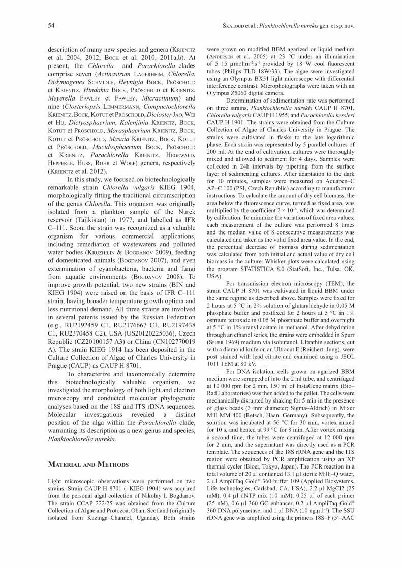
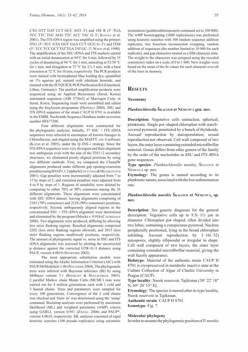
Analysis of saturation of the SSU and ITS rDNA sequences showed the significant differences between these two datasets. Saturation plot of the SSU rDNA (Fig. 1) showed a near–linear correlation. However, the saturation plot of ITS rDNA (Fig. 2) was found to level off with increasing genetic distance, indicating the presence of nucleotide saturation. To eliminate deleterious effects of substitution saturation on the resulted topology, four different 18S + ITS rDNA phylogenetic analyses were conducted, varying by different approaches to eliminate poorly aligned regions (Figs 3–6). All analyses supported monophyly of the Parachlorella–clade with the highest statistical support. Similarly, the analyses consistently revealed the close relation of Planktochlorella nurekis CAUP H 8701 and CCAP 222/25 strains, which formed a distinct lineage within the Parachlorella–clade. However, none of the analyses revealed significant relationship between Planktochlorella and any of allied genera. Moreover, the genera Dictyosphaerium, Mucidosphaerium and Compactochlorella were in some phylograms recovered to be either polyphyletic or paraphyletic (Figs 3–5). Only single phylogenetic analysis, based on the alignment treated by the Gblocks program, recovered all genera monophyletic (Fig. 6).
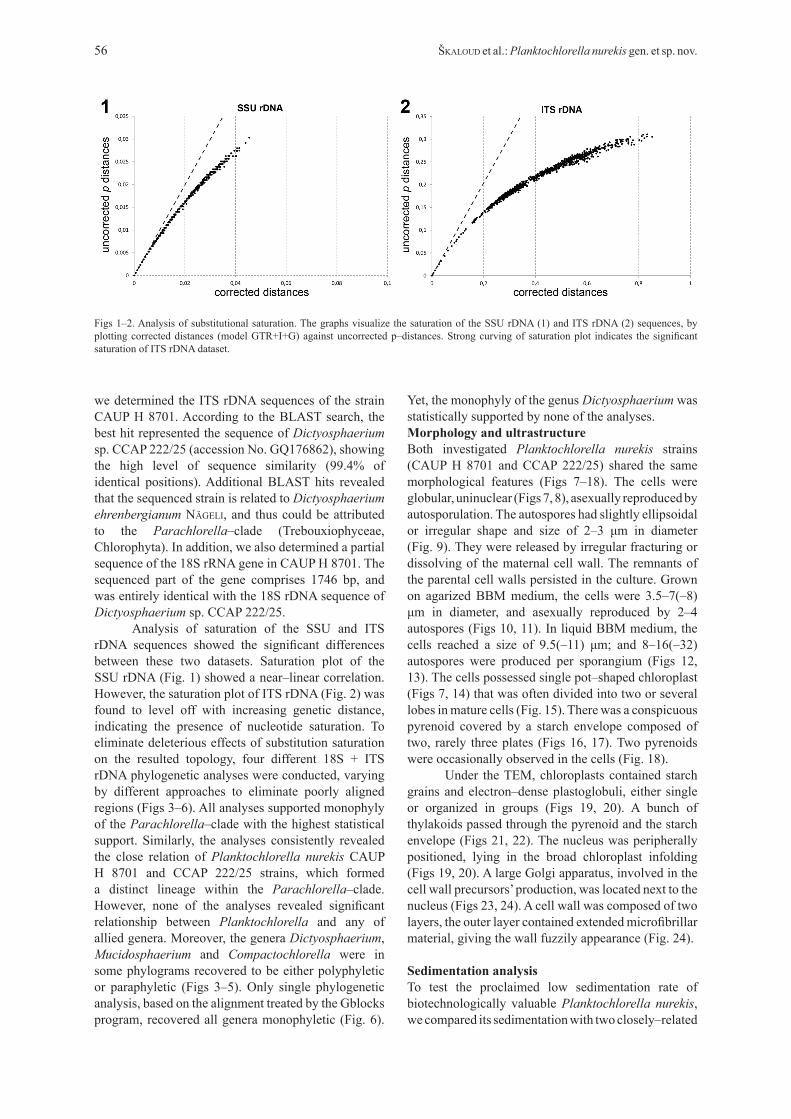
Yet, the monophyly of the genus Dictyosphaerium was statistically supported by none of the analyses.Morphology and ultrastructureBoth investigated Planktochlorella nurekis strains (CAUP H 8701 and CCAP 222/25) shared the same morphological features (Figs 7–18). The cells were globular, uninuclear (Figs 7, 8), asexually reproduced by autosporulation. The autospores had slightly ellipsoidal or irregular shape and size of 2–3 μm in diameter (Fig. 9). They were released by irregular fracturing or dissolving of the maternal cell wall. The remnants of the parental cell walls persisted in the culture. Grown on agarized BBM medium, the cells were 3.5–7(–8) μm in diameter, and asexually reproduced by 2–4 autospores (Figs 10, 11). In liquid BBM medium, the cells reached a size of 9.5(–11) μm; and 8–16(–32) autospores were produced per sporangium (Figs 12, 13). The cells possessed single pot–shaped chloroplast (Figs 7, 14) that was often divided into two or several lobes in mature cells (Fig. 15). There was a conspicuous pyrenoid covered by a starch envelope composed of two, rarely three plates (Figs 16, 17). Two pyrenoids were occasionally observed in the cells (Fig. 18).
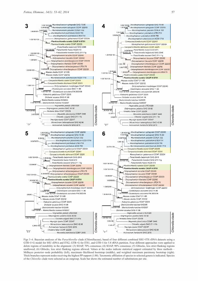
Under the TEM, chloroplasts contained starch grains and electron–dense plastoglobuli, either single or organized in groups (Figs 19, 20). A bunch of thylakoids passed through the pyrenoid and the starch envelope (Figs 21, 22). The nucleus was peripherally positioned, lying in the broad chloroplast infolding (Figs 19, 20). A large Golgi apparatus, involved in the cell wall precursors’ production, was located next to the nucleus (Figs 23, 24). A cell wall was composed of two layers, the outer layer contained extended microfibrillar material, giving the wall fuzzily appearance (Fig. 24).
Sedimentation analysisTo test the proclaimed low sedimentation rate of biotechnologically valuable Planktochlorella nurekis, we compared its sedimentation with two closely–related
Figs 1–2. Analysis of substitutional saturation. The graphs visualize the saturation of the SSU rDNA (1) and ITS rDNA (2) sequences, by plotting corrected distances (model GTR+I+G) against uncorrected p–distances. Strong curving of saturation plot indicates the significant saturation of ITS rDNA dataset.
56 Škaloud et al.: Planktochlorella nurekis gen. et sp. nov.
Figs 3–6. Bayesian analyses of the Parachlorella–clade (Chlorellaceae), based of four different combined SSU+ITS rDNA datasets using a GTR+I+G model for SSU rDNA and ITS2, GTR+G for ITS1, and GTR+I for 5.8 rRNA partition. Four different approaches were applied to delete regions of instability in the alignment: (3) SOAP, 70% consensus; (4) SOAP, 90% consensus; (5) Gblocks, less strict flanking regions unallowed; (6) Gblocks, less strict flanking regions allowed. Values at the nodes indicate statistical support estimated by three methods – MrBayes posterior–node probability (left), maximum–likelihood bootstrap (middle), and weighted maximum parsimony bootstrap (right). Thick branches represent nodes receiving the highest PP support (1.00). Taxonomic affiliation of species to selected genera is indicated. Species of the Chlorella–clade were selected as an outgroup. Scale bar shows the estimated number of substitutions per site.
Fottea, Olomouc, 14(1): 53–62, 2014 57

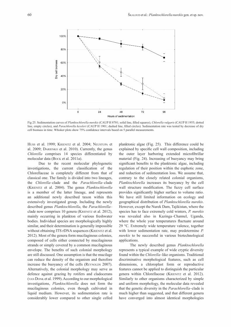
coccoid algae, Chlorella vulgaris (CAUP H 1955) and Parachlorella kessleri (CAUP H 1901). Analysis of the dry cell biomass decrease showed significant difference in sedimentation of P. nurekis and two compared strains (Fig. 25). After 96 hours of sedimentation, the density of C. vulgaris and P. kessleri cultures decreased to 4% and 7% of the original values, respectively. By contrast, density of the P. nurekis culture decreased to 68% only, and the green turbidity was still clearly visible by eye.
dIscussIon
The green algal family Chlorellaceae could serve as a good example of a taxonomic revolution, reflecting the application of modern molecular phylogenetic techniques. The family was described by BruNNthaler (1913) to encompass morphologically simple, autosporine algae, often forming mucilaginous
colonies or producing spines. komárek & fott (1983) classified these morphologically simple organisms into the sub–family Chlorelloideae. In total, they recognized 14 genera differentiated by the cell shape, number and morphology of chloroplasts, cell wall structure, and the ability to form colonies. However, molecular phylogenetic data did not support the relation of these genera, clearly indicating that the relatively simple morphology of coccoid autosporine algae is generally a poor indicator of phylogenetic relationships. Indeed, none of the genera classified by komárek & fott (1983) into the sub–family Chlorelloideae was revealed to be genetically related to the genus Chlorella (haNagata 1998; huss et al. 1999). Moreover, the genus Chlorella itself has been proved to be polyphyletic (huss et al. 1999). Only the species having a glucosamine cell wall were retained in the genus Chlorella, whereas the remaining ones were transferred into the existing or newly described genera within Chlorophyceae and Trebouxiophyceae (kaliNa & PuNčochářová 1987;
Figs 7–18. Morphology of Planktochlorella nurekis; CAUP H 8701 (Figs 7–13, 15, 17–18) and CCAP 222/25 (Figs 14, 16): (7) vegetative cell with a pot–shaped chloroplast and a conspicuous pyrenoid; (8) vegetative cell – note the pyrenoid lying in the chloroplast infolding (arrowhead); (9) autospores; (10) young autosporangium with two autospores; (11) mature autospores; (12) a young autosporangium containing 16 autospores; (13) releasing of autospores – note the remnants of the dissolving maternal cell wall; (14) a vegetative cell with a pot–shaped chloroplast; (15) a mature cell with the chloroplast divided into several lobes; (16) a pyrenoid envelope composed by two plates of starch; (17) a pyrenoid covered by a starch envelope composed of three plates; (18) a mature cell – note two pyrenoids within the chloroplast (marked by asterisks). Scale bars 5 μm.
58 Škaloud et al.: Planktochlorella nurekis gen. et sp. nov.
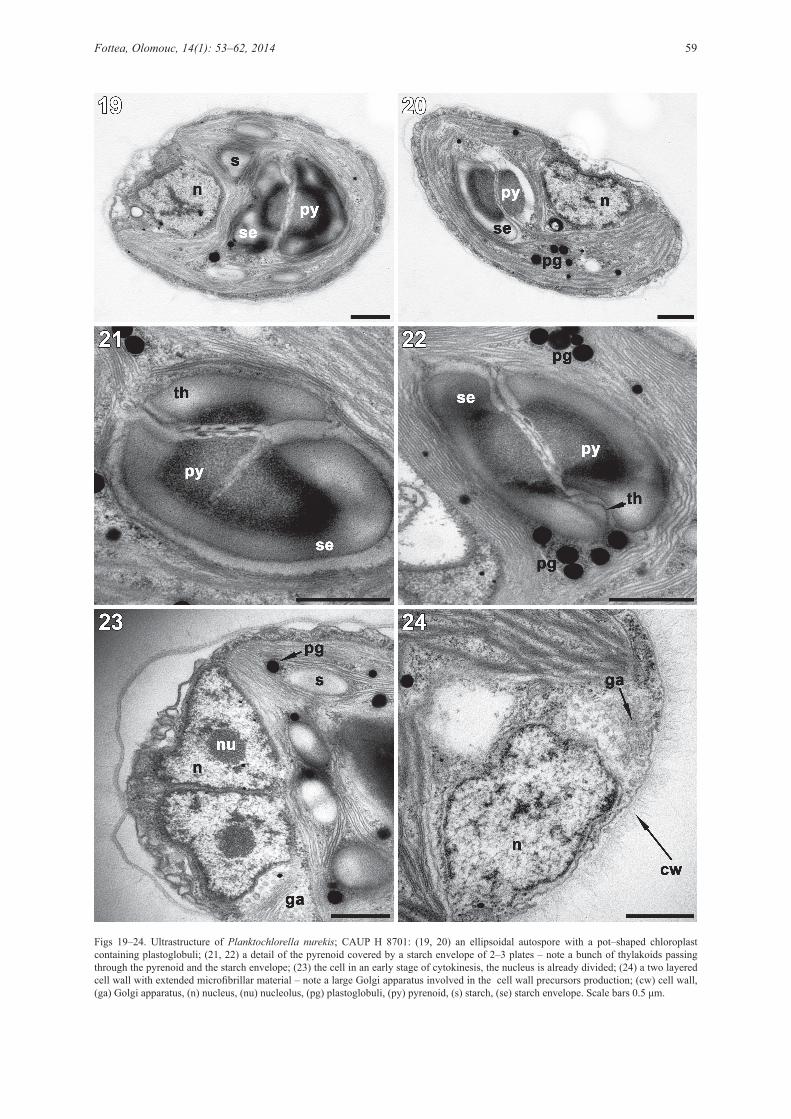
Figs 19–24. Ultrastructure of Planktochlorella nurekis; CAUP H 8701: (19, 20) an ellipsoidal autospore with a pot–shaped chloroplast containing plastoglobuli; (21, 22) a detail of the pyrenoid covered by a starch envelope of 2–3 plates – note a bunch of thylakoids passing through the pyrenoid and the starch envelope; (23) the cell in an early stage of cytokinesis, the nucleus is already divided; (24) a two layered cell wall with extended microfibrillar material – note a large Golgi apparatus involved in the cell wall precursors production; (cw) cell wall, (ga) Golgi apparatus, (n) nucleus, (nu) nucleolus, (pg) plastoglobuli, (py) pyrenoid, (s) starch, (se) starch envelope. Scale bars 0.5 μm.
Fottea, Olomouc, 14(1): 53–62, 2014 59
huss et al. 1999; krienitz et al. 2004; neuStuPa et al. 2009; darienko et al. 2010). Currently, the genus Chlorella comprises 14 species differentiated by molecular data (Bock et al. 2011a).
Due to the recent molecular phylogenetic investigations, the current classification of the Chlorellaceae is completely different from that of classical one. The family is divided into two lineages, the Chlorella–clade and the Parachlorella–clade (krienitz et al. 2004). The genus Planktochlorella is a member of the latter lineage, and represents an additional newly described taxon within this extensively investigated group. Including the newly described genus Planktochlorella, the Parachlorella–clade now comprises 10 genera (krienitz et al. 2012), mainly occurring in plankton of various freshwater bodies. Individual species are morphologically highly similar, and their determination is generally impossible without obtaining ITS rDNA sequences (krienitz et al. 2012). Most of the genera form mucilaginous colonies, composed of cells either connected by mucilaginous strands or simply covered by a common mucilaginous envelope. The benefits of such colonial morphology are still discussed. One assumption is that the mucilage can reduce the density of the organism and therefore increase the buoyancy of the cells (reynoldS 2007). Alternatively, the colonial morphology may serve as defence against grazing by rotifers and cladocerans (vaN doNk et al. 1999). According to our morphological investigations, Planktochlorella does not form the mucilaginous colonies, even though cultivated in liquid medium. However, its sedimentation rate is considerably lower compared to other single celled
planktonic algae (Fig. 25). This difference could be explained by specific cell wall composition, including the outer layer harboring extended microfibrillar material (Fig. 24). Increasing of buoyancy may bring significant benefits to the planktonic algae, including regulation of their position within the euphotic zone, and reduction of sedimentation loss. We assume that, contrary to the closely related colonial organisms, Planktochlorella increases its buoyancy by the cell wall structure modification. The fuzzy cell surface provides significantly higher surface to volume ratio. We have still limited information on ecology and geographical distribution of Planktochlorella nurekis. However, except the Nurek Dam, Tajikistan, where the species has to face extremely cold winters, P. nurekis was revealed also in Kazinga–Channel, Uganda, where the whole year temperatures fluctuate around 29 °C. Extremely wide temperature valence, together with lower sedimentation rate, may predetermine P. nurekis to be successful in various biotechnological applications.
The newly described genus Planktochlorella represents a typical example of wide cryptic diversity found within the Chlorella–like organisms. Traditional discriminative morphological features, such as cell dimensions, a chloroplast form or reproductive features cannot be applied to distinguish the particular genera within Chlorellaceae (krienitz et al. 2012). Similarly to other organisms characterized by simple and uniform morphology, the molecular data revealed that the genetic diversity in the Parachlorella–clade is much higher than suggested, and that different genera have converged into almost identical morphologies
Fig 25. Sedimentation curves of Planktochlorella nurekis (CAUP H 8701; solid line, filled squares), Chlorella vulgaris (CAUP H 1955; dotted line, empty circles), and Parachlorella kessleri (CAUP H 1901; dashed line, filled circles). Sedimentation rate was tested by decrease of dry cell biomass in time. Whisker plots show 75% confidence intervals based on 5 parallel measurements.
60 Škaloud et al.: Planktochlorella nurekis gen. et sp. nov.

(rindi et al. 2010). Our current knowledge of the diversity within
Chlorellaceae fully relies on sequencing of SSU and ITS rDNA, and subsequent analyses on the concatenated dataset (krienitz et al. 2004, 2010; luo et al. 2006, 2010; Bock et al. 2011a, b). However, whereas the nuclear ribosomal small subunit has poor species–level resolution, ITS rDNA suffers from its high variability even among phylogenetically very closely related species. A high number of indels negatively affects the alignment accuracy. In addition, such alignments are typically very saturated, which negatively affect the phylogenetic reconstructions (moreira & PhiliPPe 2000). Mutational saturation occurs when multiple mutations at a given site lead to a randomization of the phylogenetic signal with the number of observed differences being lower than the expected number of differences. Although this may lead to an underestimation of observed divergence times (arbogaSt et al. 2002), the effect is frequently neglected. As expected from their high variability, ITS rDNA sequences within the Parachlorella–clade were found to be significantly saturated (Fig. 2). Therefore, we applied four different approaches to reduce the effect of nucleotide saturation, resulting in four topologically different phylogenetic reconstructions (Fig. 3–6). Interestingly, the relationships among the genera, and even the monophyly of some of them, differed significantly across these reconstructions. In view of that, the evolution of the Chlorella–like organisms within the Chlorellaceae is still unclear. To resolve it better, other molecular markers should be sequenced, preferably those having sufficient nucleotide diversity, low saturation, and simple alignment process. The rbcL gene offers all above–mentioned features, as well as good discriminating power among the species and cryptic lineages of various green algae (hall et al. 2010; fučíková et al. 2012; Škaloud & riNdi 2013).
Finally, our study demonstrates the importance of detailed molecular investigations focused on the cryptic diversity within morphologically similar organisms. It is evident that plenty of distinct lineages, with potential biotechnological exploitation, are still not known to science.
acknowledgeMents
This work has been supported by the Institutional Funds of the
Charles University in Prague.
references
aNderseN, r. a.; Berges, j. a.; harrisoN, P. j. & wataNaBe, m. m. (2005): Recipes for freshwater and seawater media. – In: aNderseN, r. a. (ed.): Algal culturing techniques. – pp. 429–538, Elsevier Academic Press.
arBogast, B.; edwards, s. v.; wakeleY, j.; Beerli, P. &
slowiNski, j. B. (2002): Estimating divergence time from molecular data on phylogenetic and population genetic timescales. – Annu. Rev. Ecol. Syst. 33: 707–740.
Bock, c.; Pröschold, t. & krieNitz, l. (2010): Two new Dictyosphaerium–morphotype lineages of the Chlorellaceae (Trebouxiophyceae): Heynigia gen. nov. and Hindakia gen. nov. – Europ. J. Phycol. 45: 267–277.
Bock, c.; krieNitz, l. & Pröschold, t. (2011a): Taxonomic reassessment of the genus Chlorella (Trebouxiophyceae) using molecular signatures (barcodes), including description of seven new species. – Fottea 11: 293–312.
Bock, c.; Pröschold, t. & krieNitz, l. (2011b): Updating the genus Dictyosphaerium and description of Mucidosphaerium gen. nov. (Trebouxiophyceae) based on morphological and molecular data. – J. Phycol. 47: 638–652.
BogdaNov, N. i. (2007): Biological Basis of Preventing “Bloom” of the Penza Reservoir by Blue–Green Algae. – Penza, RIO PGSChA, 75 pp. (in Russian)
BogdaNov, N. i. (2008): Biological rehabilitation of waters. – 125 pp., Penza, RIO PGSChaA, Available from http://www.chlorella–v.narod.ru/Sea.pdf (in Russian)
BruNNthaler, j. (1913): Die systematische Gliederung der Protococcales (Chlorophyceae). – Verh. K. K. Zool. – Bot. Ges. Wien 63:76–91.
castresaNa, j. (2000): Selection of conserved blocks from multiple alignments for their use in phylogenetic analysis. – Mol. Biol. Evol. 17: 540–552.
darieNko, t.; gustavs, l.; mudimu, o.; meNeNdez, c. r.; schumaNN, r.; karsteN, u.; friedl, t. & Pröschold, t. (2010): Chloroidium, a common terrestrial coccoid green alga previously assigned to Chlorella (Trebouxiophyceae, Chlorophyta). – Eur. J. Phycol. 45: 79–95.
fučíková, k.; flechtNer, v. r. & lewis, l. a. (2012): Revision of the genus Bracteacoccus Tereg (Chlorophyceae, Chlorophyta) based on a phylogenetic approach. – Nova Hedwigia 96: 15–59.
hall, j. d.; fučíková, k.; lo, c.; lewis, l. l. & karol, k. g. (2010): An assessment of proposed DNA barcodes in freshwater green algae. – Cryptogam. Algol. 31: 529–555.
haNagata, N. (1998): Phylogeny of the subfamily Scotiellocystoideae (Chlorophyceae, Chlorophyta) and related taxa inferred from 18S ribosomal RNA gene sequence data. – J. Phycol. 34: 1049–1054.
hegewald, e. & haNagata, N. (2000): Phylogenetic studies on Scenedesmaceae (Chlorophyta). – Algological Studies 100: 29–49.
hePPerle, d. (2004): SeqAssem©. A sequence analysis tool, contig assembler and trace data visualization tool for molecular sequences. (http://www.sequentix.de).
huss, v. a. r.; fraNk, c.; hartmaNN, e. c.; hirmer, m.; kloBoucek, a.; seidel, B. m.; weNzeler, P. & kessler, e. (1999): Biochemical taxonomy and molecular phylogeny of the genus Chlorella sensu lato (Chlorophyta). – J. Phycol. 35: 587–598.
kaliNa, t. & PuNčochářová, m. (1987): Taxonomy of the subfamily Scotiellocystoideae Fott 1976 (Chlorellaceae, Chlorophyceae). – Arch. Hydrobiol. Suppl. / Algological Studies 45: 473–521.
kataNa, a.; kwiatowski, j.; sPalik, k.; zakrYs, B.;
Fottea, Olomouc, 14(1): 53–62, 2014 61
szalacha, e. & szYmaNska, h. (2001): Phylogenetic position of Koliella (Chlorophyta) as inferred from nuclear and chloroplast small subunit rDNA. – J. Phycol. 37: 443–451.
katoh, k.; kuma, k.; toh, h. & miYata, t. (2005): MAFFT version 5: improvement in accuracy of multiple sequence alignment. – Nucleic Acids Res. 33: 511–518.
komárek, j. & fott, B. (1983): Chlorophyceae (Grünalgen) Ordnung: Chlorococcales. Das Phytoplankton des Süsswassers. – In: huBer–Pestalozzi, g. (ed.): Die Binnengewässer XVI., 7/1. – 1044 pp., Schweizerbart’sche, Stuttgart.
krieNitz, l.; Bock, c.; kotut, k. & Pröschold, t. (2012): Genotypic diversity of Dictyosphaerium–morphospecies (Chlorellaceae, Trebouxiophyceae) in African inland waters, including the description of four new genera. – Fottea 12: 231–253.
krieNitz, l.; Bock, c.; luo, w. & Pröschold, t. (2010): Polyphyletic origin of the Dictyosphaerium–morphotype within Chlorellaceae (Trebouxiophyceae). – J. Phycol. 46: 559–563.
krieNitz, l.; hegewald, e. h.; hePPerle, d.; huss, v. a. r.; rohr, t. & wolf, m. (2004): Phylogenetic relationship of Chlorella and Parachlorella gen. nov. (Chlorophyta, Trebouxiophyceae). – Phycologia 43: 529–542.
kruzhiliN, i. P. & BogdaNov, N. i. (2009): Use of Chlorella vulgaris strain BIN for biological rehabilitation of wastewaters and polluted water bodies. – Russ. Agric. Sci. 35: 405–407.
löYtYNoja, a.; miliNkovitch, m. c. (2001): SOAP, cleaning multiple alignments from unstable blocks. – Bioinformatics 17: 573–574.
luo, w.; Pflugmacher, s.; Pröschold, t.; walz, N. & krieNitz, l. (2006): Genotype versus phenotype variability in Chlorella and Micractinium (Chlorophyta, Trebouxiophyceae). – Protist 157: 315–333
luo, w.; Pröschold, t.; Bock, c. & krieNitz, l. (2010): Generic concept in Chlorella–related coccoid green algae (Chlorophyta, Trebouxiophyceae). – Plant Biol. 12: 545–553.
merchaNt, r. e. & aNdre, c. a. (2001): A review of recent clinical trials of the nutritional supplement Chlorella pyrenoidosa in the treatment of fibromyalgia, hypertension, and ulcerative colitis. – Altern. Therap. Health Med. 7: 79–91.
moreira, d. & PhiliPPe, h. (2000): Molecular phylogeny: pitfalls and progress. – Internatl. Microbiol. 3: 9–16.
NeustuPa, j.; Němcová, Y.; eliáŠ, m. & Škaloud, P. (2009): Kalinella bambusicola gen. et sp. nov. (Trebouxiophyceae, Chlorophyta), a novel coccoid Chlorella–like subaerial alga from Southeast Asia. – Phyc. Res. 57: 159–169.
NYlaNder, j. a. a. (2004): MrModeltest v2. Program distributed by the author. Evolutionary Biology Centre, Uppsala University. Available from http://www.abc.se/~nylander/.
reYNolds, c. s. (2007): Variability in the provision and function of mucilage in phytoplankton: facultative responses to the environment. – Hydrobiologia 578: 37–45.
riNdi, f.; allali, h. a.; lam, d. w. & loPez–Bautista, j. m. (2010): An overview of the biodiversity
and biogeography of terrestrial green algae. – In: rescigNo, v. & maletta, s. (eds): Biodiversity Hotspots. – pp. 105–122, Nova Science Publishers, Hauppauge, New York.
roNquist, f. & huelseNBeck, j. P. (2003): MrBayes 3: Bayesian phylogenetic inference under mixed models. – Bioinformatics 19: 1572–1574.
Škaloud, P. & riNdi, f. (2013): Ecological differentiation of cryptic species within an asexual protist morphospecies: A case study of filamentous green alga Klebsormidium (Streptophyta). – J. Euk. Microbiol., in press.
sPolaore, P.; joaNNis–cassaN, c.; duraN, e.; isamBert, a. (2006): Commercial Applications of Microalgae. – J. Biosci. Bioeng. 101: 87–96.
sPurr, a. r. (1969): A low viscosity epoxy resin embedding medium for electron microscopy. – J. Ultrastruct. Res. 26: 31–43.
swofford, d. l. (2002): PAUP*. Phylogenetic Analysis Using Parsimony (*and Other Methods). Version 4, Sinauer Associates, Sunderland, Massachusetts.
ustiNova, i.; krieNitz, l. & huss, v. a. r. (2001): Closteriopsis acicularis (G. M. Smith) Belcher et Swale is a fusiform alga closely related to Chlorella kessleri Fott et Nováková (Chlorophyta, Trebouxiophyceae). – Eur. J. Phycol. 36: 341–351.
vaN doNk, e.; lürliNg, m. & lamPert, w. (1999): Consumer–induced changes in phytoplankton: Inducibility, costs, benefits and impacts on grazers. – In: tollriaN, r. & harvell, c. d. (eds): The Ecology and Evolution of Inducible Defenses. – pp. 89–103, Princeton Univ. Press, Priceton, New Jersey.
white, t. j.; BruNs, t.; lee, s. & taYlor, j. (1990): Amplification and direct sequencing of fungal ribosomal RNA genes for phylogenetics. – In: iNNis, m. a.; gelfaNd, d. h.; sNiNskY, j. j. & white, t. j. (eds): PCR Protocols: a guide to methods and applications. – pp. 315–322, Academic Press, New York.
wolf, m.; krieNitz, l.; hePPerle, d. (2002): Phylogenetic position of Actinastrum hantzschii Lagerheim 1882 (Chlorophyta, Trebouxiophyceae). – Arch. Hydrobiol. Suppl. / Algological Studies 104: 59–67.
Yamaguchi, k. (1997): Recent advances in microalgal bioscience in Japan, with special reference to utilization of biomass and metabolites: a review. – J. Appl. Phycol. 8: 487–502.
zwickl, d. j. (2006): Genetic algorithm approaches for the phylogenetic analysis of large biological sequence datasets under the maximum likelihood criterion. World–wide electronic publication, The University of Texas, Austin. Available from http://www.bio.utexas.edu/faculty/antisense/garli/Garli.html.
62 Škaloud et al.: Planktochlorella nurekis gen. et sp. nov.
© Czech Phycological Society (2014)Received May 1, 2013Accepted August 26, 2013