ON THE SUBCEllUlAR DISTRIBUTION OF OESTRADIOL RECEPTORS … · ON THE SUBCEllUlAR DISTRIBUTION OF...
148
ON THE SUBCEllUlAR DISTRIBUTION OF OESTRADIOL RECEPTORS IN RAT TESTIS AND UTERUS Proefschrift ter verkrijging van de graad van doctor in de geneeskunde aan de Erasmus Universiteit te Rotterdam op gezag van de rector magnificus Prof.Dr. B. Leijnse en volgens besluit van het College van Dekanen. De openbare verdediging zal plaats vinden op vrijdag 10 juni 1977 des namiddags te 3.00 uur. door Willem de Boer geboren te Joure Drukkerij de Vries-Rotterdam
Transcript of ON THE SUBCEllUlAR DISTRIBUTION OF OESTRADIOL RECEPTORS … · ON THE SUBCEllUlAR DISTRIBUTION OF...
ON THE SUBCEllUlAR DISTRIBUTION
OF OESTRADIOL RECEPTORS
IN RAT TESTIS AND UTERUS
Proefschrift
ter verkrijging van de graad van doctor in de
geneeskunde aan de Erasmus Universiteit te Rotterdam op gezag van de rector magnificus
Prof.Dr. B. Leijnse en volgens besluit van het College van Dekanen.
De openbare verdediging zal plaats vinden op vrijdag 10 juni 1977 des namiddags te 3.00 uur.
door
Willem de Boer
geboren te Joure
Drukkerij de Vries-Rotterdam
Promotor Prof.Dr. H.J. van der Molen
Co-referenten: Prof. Dr. M. Gruber
Prof.Dr. W.C. HUismann
Dit proefschrift werd bewerkt in het instituut Biochemie II
(Chemische Endocrinologie) van de Faculteit der Geneeskunde ,
Erasmus Universiteit te Rotterdam.
Het onderzoek werd mede mogelijk gemaakt door steun van
de stichting voor Medisch Wetenschappelijk Onderzoek FUNGO.
Foar Heit en Mem


Contents
Introduction and scope of the thesis
1.1 Steroid receptors and steroid hormone action
1.2 Scope of this thesis
2 Summary of the literature on steroid interactions with
target cells
2.1 Interaction of steroid hormones with target cells
2.2 Entry of steroid hormones into a target cell
2.3 Nature of the cytoplasmic receptor and the inter
action of the steroid with the receptor
2.4 Steroid induced changes in the cytoplasmic
receptor
2.5 The translocation of the steroid-receptor
complex into the nucleus
2.6 Interaction of steroid receptors with nuclear
components
2.7 Dissociation of steroids from receptor sites and
intracellular recycling of receptors
2.8 Regulation of the amount of steroid hormone
receptors
3
3. 1
3. 2
3. 3
3.4
3. 5
Methods used for studying steroid receptor interactions
Measurement of specific steroid binding sites
Sephadex gel chromatography
Sucrose density gradient centrifugation
Agar-gel electrophoresis
Hydroxylapatite chromatography
3.6 Determination of nuclear receptor sites in the
presence of endogenous steroids
7
11
11
12
15
15
16
17
19
21
23
27
28
33
33
34
35
36
37
38
4 Introduction and discussion of experimental work
4.1 Introduction
4.2 Effects of oestradiol, hypophysectomy and age on
cytoplasmic oestradiol receptor levels
4.3 Effects of oestradiol, hypophysectomy and
choriogonadotropin on nuclear receptor sites
4.4 Kinetics of in vitro binding of oestradiol in
subcellular fractions of testicular and uterine
tissue
4.5 Conclusions
5 Distribution of oestradiol-receptor complexes in subnuclear
fractions of uterine tissue after administration of
oestradiol in vivo and in vitro
5.1 Introduction
5.2 Materials and methods
5.3 Results
5.4 Discussion
6 General discussion and conclusions
6.1 Regulation of cytoplasmic and nuclear receptor
sites for oestradiol in testicular tissue
6.2 Characterization and retention of nuclear
receptor complexes
6.3 Subnuclear distribution of oestradiol-receptor
complexes in uterus
References
Summary
Samenvatting
Abbreviations
8
39
39
41
42
42
44
47
47
48
54
65
71
71
76
79
81
95
99
103
Nawoord
Curriculum vitae
Appendix papers
Paper I
Effects of oestradiol, hypophysectomy and age on cytoplasmic
oestradiol receptor sites in rat testis interstitial tissue
Willem de Boer, Eppo Mulder and Henk J. van der Molen
J. Endocr. 70 (1976) 397-407.
Paper II
Comparative study of nuclear binding sites for oestradiol in
rat testicular and uterine tissue. Determination of low
amounts of specific binding sites by an 3H-oestradiol
exchange method
Willem de Boer, Joan de Vries, Eppo Mulder and
Henk J. van der Molen
Biochern. J. 162 (1977) 331-339.
Paper Ill
Kinetics of in vitro binding of oestradiol in subcellular
fractions of testicular and uterine tissue. Characterization
of oestradiol binding in testicular nuclei
Willem de Boer, Joan de Vries, Eppo Mulder and
Henk J. van der Molen
J. Steroid Biochern. (1977) in press.
9
105
106


Introduction and scope of the thesis
1.1 Steroid receptors and steroid hormone action
There is good evidence that the concentration of nuclear
steroid-receptor complexes shows a direct relationship with
the effect of steroids on cells. Anderson et al. (l) showed
that uterotrophic responses correlated well with nuclear
oestradiol receptor levels. Correlations between concentra
tions of nuclear receptors and cell specific effects have
also been observed for glucocorticoids in eukaryotic cells
(2), oestrogens in chicken liver {3,4) and for oestradiol
and progesterone in chick oviduct (5-10).
In the search for effects of steroid hormones on cellular
metabolism it has been observed that oestradiol can affect
several important parameters that may influence gene expres
sion, such as synthesis of histone and non-histone proteins
in uterus {ll-14), the rate of peptide elongation of uterine
ribosomes (15) and the rate of methylation of ribosomal and
tRNA's (16,17)
The involvement of RNA synthesis during the early effects
of steroids has also been well established for several tis
sues. Within 0.5-4 h after administration of oestradiol to
mature or ovariectomized rats, uterine nuclear RNA-polymerase
activity is stimulated (18,19,20), predominantly at nucleolar
sites (21,22). The oestrogen induced stimulation of RNA syn
thesis could be prevented if puromycine or cycloheximide was
administered prior to injection of the hormone {18,23), in
dicating the necessity of a continuous protein synthesis for
the expression of the oestrogen effect. In fact, a group of
proteins, including one acidic protein, called IP {induced
protein) , is formed in response to oestrogen administration
(24,25,26). The synthesis of this protein, which appears
within 15 min after oestradiol injection, can be blocked by
actinomycin D (27 1 28) and cordycepin (29) .However, the biolo
gical significance of this IP 1 which recently has been puri
fied and characterized as a polypeptide of molecular weight
45,000 (30), and its possible relation to the stimulation of
11

nucleolar RNA-polymerase remains to be elucidated.
In more recent studies the effect of steroid hormones on
template activity of nuclear chromatin of target tissues has
nucleolar (I) and been investigated.
nucleoplasmic (II)
It was found that both
RNA-polymerase were stimulated but at
different times after oestradiol administration (31,32,33,
34) •
More detailed studies concerning the possible role of
hormone-receptor comple~es were carried out by O'Malley and
coworkers for the effects of progesterone and oestradiol in
the chick oviduct (35,36). They could demonstrate that the
accumulation of steroid-receptor complexes in nuclei or on
the chromatin caused an increase in the number of initiation
sites for RNA-polymerase molecules prior to the increase in
ovalbumin synthesis, which is the cell's response to hormone
administration (10,37,38).
Based on the kind of information described above, it is
now generally believed, that effects of steroid hormones in
target cells are mediated through the binding of steroids to
specific receptor molecules and the subsequent interaction
of steroid-receptor complexes with the chromatin. The quan
tity of nuclear receptor molecules appears to be important
for the magnitude of a cell's response. The interaction
between the steroid-receptor complexes and the genome causes
activation or derepression of transcription or post-trans
criptional regulation of RNA synthesis. The products of mRNA
and rRNA dictate the synthesis of specific proteins, which
ultimately determine the morphogenetic and physiological
responses to the hormone.
1.2 Scope of this thesis
It has been shown previously by Brinkmann et al. (39),
that the interstitial compartment of the rat testis contains
a limited number of specific oestradiol receptor sites with
a high affinity for oestrogens (Ka for oestradiol is
10 10M- 1 ). This receptor, which in the presence of oestradiol
12
will be translocated into the nuclear fraction (40), shows
a steroid specificity comparable to that of the uterine
receptor for oestradiol (41).
The physiological significance of the uptake of oestra
diol and its subsequent binding to the oestradiol receptor
in the testis, is not yet understood. In studies of de Jong
et al. (42,43) it was found that oestradiol concentrations
in rat testis interstitial tissue (0.5-lxl0- 9M) are higher
than those in seminiferous tubules.
Actions of oestradiol in the testis on DNA, RNA and pro
tein synthesis have been reported for Balb/c mice (44). It
has also been suggested that after long-term treatment with
oestrogens, the observed decrease of testosterone levels in
rat plasma and testicular tissue would occur without inter
ference of the LH secretion (45,46,47). Other studies (48),
however, indicated that the observed oestradiol effect could
be fully explained through a feedback action of administered
oestrogens on pituitary LH secretion. The lack of a distinct
and well-defined effect of oestradiol in the testis made it
important to investigate whether oestradiol and oestradiol
binding sites in both the cytoplasmic and nuclear compart
ments of the testicular tissue would show the same behaviour
as in an established oestradiol target tissue. In some of our
studies therefore the nuclear translocation of the oestradiol
receptor in uterine tissue was used for comparison, because
this tissue contains an oestradiol receptor which is known
to be related to the effects of oestradiol on the uterus.
The results of experiments on the regulation of the
cytoplasmic oestradiol receptor and the nuclear oestradiol
receptor are discussed in chapter 6 and appendix papers I
and II. Also in chapter 6 and appendix paper III the processes
of translocation and nuclear binding of oestradiol-receptor
complexes in testicular and uterine tissue are compared.
It is now well accepted that steroid hormones exert their
actions in target tissues via the binding of steroid-receptor
complexes to chromatin constituents in the nuclear fraction.
There is hardly any information, however, about the nature
13
and the localization of the so-called 'acceptor sites' on
the chromatin, which bind the steroid-receptor complexes.
we have studied this aspect of steroid hormone action in
uterine nuclei and preliminary results are discussed in
chapter 5.
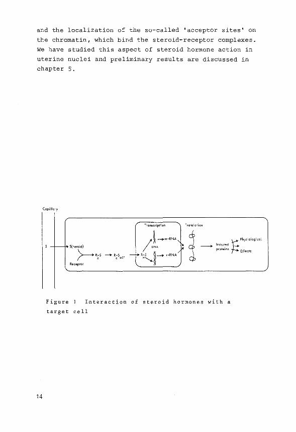
Ccpillcry
Transcription Tran•lction
I l:-om·RNA' '~ t: Phy'''''''''' -,-- rt" S(terojd) - Induced
proteins Effects >---- R-S ---+ R-S - f'-' R-S ~- r-RNA c c act ""'--. Receptor
Figure Interaction of steroid hormones with a
target cell
14
Summary of the literature on steroid interactions with
target cells
2.1 Interaction of steroid hormones with target cells
It is generally accepted that receptors play an important
role in the expression of steroid hormone effects.in target
tissues (35,49-53) and binding of steroids to specific
receptors appears to be related to and precedes the physio
logical effects of steroids. However, the precise sequence
of events which occur between the entry of the steroid in
the cell and the expression of the hormone effect is still
unknown. Present knowledge about the fate of steroid hor
mones in a target cell can be summarized as depicted in
Fig. 1. The steroid hormone enters a cell, binds to specific
receptor proteins located in the cytoplasm, followed by
translocation of the hormone-receptor complex into the
nucleus where a presumed specific interaction of the complex
with chromatin constituents ultimately leads to specific
changes in the cell metabolism ascribed to that specific
steroid hormone.
A brief summary about the following aspects, as indicated
in Fig. 1 will be given in the following paragraphs:
a) entry of steroid hormones into the target cell.
b) nature of cytoplasmic receptors and the inter
action of steroids with the receptor.
c) steroid induced changes of the cytoplasmic
receptor.
d) translocation of the steroid-receptor complex
from the cytoplasm into the nucleus.
e) interaction of steroid-receptor complexes with
nuclear components.
f) dissociation of steroids from receptor and/or
acceptor sites.
g) regulation of the amount of steroid hormone
receptors.
h) receptors and actions of steroid hormones.
15
This discussion will mainly concern receptors for oestrogens
and progestins.
2.2 Entry of steroid hormones into a target cell
Steroid hormones outside target cells are generally bound
to (plasma) proteins which, in comparison to the intracellu
lar receptor proteins, show a moderate binding specificity
and a rather low affinity for steroid hormones (Kassociation
=10 5-10 BM- 1 ). For example serum albumin, the most abundant
protein constituent of plasma, binds oestradiol, progeste
rone, testosterone and oestrone with a Ka in the order of 5 6 10 -10 {54) .Although the more specific steroid binding
globulins {CBG, SBG, DBG, PBG, EBG). are present in smaller
amounts, they still will strongly bind the larger part of
steroids in the blood because of their higher affinity (Ka=
10 7 -10 9M- 1 ) even if total blood steroid levels are low{SS-59)_.
The biological function of the plasma steroid binding
proteins is not clear. They may protect steroids from degra
dation in the liver, but there is no strong evidence that
blood proteins can indeed act as a reservoir from which
steroids can be fed gradually to target organs. In fact,
there are some indications that the biological activity of
steroids is probably related to the unbound fraction of
steroids in plasma (60,61), which is only a small percentage
of the total amount.
Before the free steroid can interact with receptor pro
teins inside the cell, it has to pass the cell membrane. It
has been generally assumed that the cell membrane provides
little or no barrier to the diffusion of steroids into cells,
because of their lipophilic properties. It has been shown by
Jensen and Jacobson (62) that oestradiol and oestrone are
taken up rapidly by most tissues of the rat, suggesting a
simple but rapid diffusion process. A protein mediated pro
cess was proposed for the entry of oestradiol in uterine
cells by Milgram et al. (63), who found that the rate of
entry of oestradiol was reaching a maximum in the range of
16

physiological hormone concentrations and that it could be
inhibited by SH-blocking agents. This saturable protein is
probably not the oestrogen receptor since the uptake of
diethylstilboestrol, which also binds to the receptor, could
not be blocked significantly. However using N-ethylmaleirnide
as a SH-blocking agent Peck et al (641 found no support for
a saturable transport mechanism for oestradiol in uterine
tissue.Sirnilar results obtained with thymus cells indicated
that the membrane of thymus cells is also freely permeable
for corticoids. It can be concluded therefore that steroids
can enter the cell by simple diffusion and there is little
evidence to support facilitated transport mechanisms for
steroid hormones.
2.3 Nature of the cytoplasmic receptor and the interaction of the steroid
with the receptor
After entering a cell steroid hormones can be bound to
many proteins. In most cases this binding is 'nonspecific',
but the binding of the steroid to the receptor is the excep
tion. This steroid receptor interaction is characterized by
a high affinity (Ka=2xl0 10-lxl0 9M- 1 ), a limited number of
receptor molecules per cell {3000-40,000 receptors/cell)
and ~ompetition with small amounts of compounds which are
chemically similar to the specifically bound hormones. Little
is known about the physicochemical nature of the strong
interaction between the steroid and the receptor molecule.
It has been suggested that the bulkiness and the flatness of
the steroid plays a more important role in receptor binding
than the detailed electronic structure of the steroid nucleus
(65). This would imply that the site of interaction is loca
ted inside the receptor molecule rather than on the surface
of the protein. A localization of the steroid binding sites
inside the receptor proteins could also be responsible for
the very high affinity constant for receptor binding of
steroids, the extremely slow rates of association and dis
sociation of steroids at low temperatures, the acceleration
17

of rates of exchange of unbound steroids with bound steroids ' by freezing and thawing and the inability of ethanol (30%)
and detergents {Triton-X-100 or deoxycholate) to dissociate
steroids from receptors at low temperatures (66). It has
also been suggested {67) that the binding sites could be
located in a hydrophobic pocket in the receptor protein and
that it is necessary for the receptor to 'envelop 1 the
steroid molecule.
Treatment of steroid hormone receptors with proteolytic
enzymes generally destroys the steroid binding capacity.
Other enzymes like DNAse and RNAse do not effect the binding
properties indicating that nucleic acids do not play a sig
nificant role in the interaction between receptors and
steroids.
The state of receptor molecules under physiological con
ditions is still unknown. On sucrose gradients sedimentation
values between 3S and 125 have been observed depending on
the protein concentration and the ionic strength of the
medium (68). In hypotonic media receptors mostly appear as
an 85 sedimenting entity. Increasing salt concentrations
reduce the size of the molecule to a 45 sedimenting mole
cule at 0.4 M KCl. In 0.15 M KCl (isotonic media) receptor
molecules with sedimentation values of 45 or 65 have been
observed (69,70,71). After homogenization of uterine tissue
without adding buffer only a 65 form of the oestradiol re
ceptor could be detected in the cytosol (72). These obser
vations indicate, but do not prove, the possibility that
the 65 form is the predominant receptor form in the target
cell cytoplasm. During the past years several attempts have
been made to estimate the molecular weight of steroid hor
mone receptor molecules as they are present in the cell.
For the uterine oestradiol receptor with a sedimentation
value of 4S, Notides and Nielsen estimated a molecular
weight of about 80,000 (73). Puca et al. consider the 8.65
sedimenting molecule of the oestradiol receptor to be a
dimer of the 5.35 form, which may have a molecular weight
of about 118,000 (74,75).
18
The progesterone receptor in chick oviduct is probably
considerably larger than the uterine oestradiol receptor.
The 8S form is thought to be a tetramer of the 4S form,
which has a molecular weight of 90,000 (76). More recent
studies showed that the 6S form of the progesterone recep
tor, probably the native form, consisted of two 4.2S sub
units, with molecular weights of 110,000 and 117,000 res
pectively. The subunits differ in binding characteristics
as will be discussed later (77).
It can be concluded that steroid receptors are proteins
and show a very specific high affinity interaction with
their respective ligand. The nature of the interaction
between the steroid and the receptor molecule is not yet
understood. Steroid hormone receptors in cell fractions have
thusfar been detected only after binding of a radioactive
steroid. Therefore these studies may not give reliable in
formation about the receptor as it exists in the cytoplasm
of a cell prior to the interaction with the steroid.
2.4 Steroid induced changes in the cytoplasmic receptor
Steroid-receptor complexes are found predominantly in the
nuclear fraction, while free receptors remain in the cyto
plasm. The nature of the changes in the structure of the
receptor protein during or after binding of the steroid is
still unknown. It has been shown (78,79,80) that free oestra
diol receptors are more susceptible to changes in temperature
than the oestrogen-receptor complex. Other studies (81,82,83)
have shown that the presence of the steroid is essential for
induction of specific changes in the structure of the recep
tor protein at a temperature of 20°C or higher, although
opposite results have also been reported (83). After inter
action of the steroid with its receptor site in vivo or in
vitro a change in the sedimentation value of the complex has
been observed: the 45 form of the uterine oestradiol receptor
is converted to aSS form (84). This change in receptor con
formation probably occurs in the cytoplasmic compartment of
the cell, because no oestradiol binding proteins with a se-
19
dimentation value of 58 can be observed if nuclei are incu
bated with oestradiol in the absence of cytoplasm (82,85).
Also, if uterine nuclei are incubated with isolated oestra
diol-receptor complexes at 25°C or at 0°C with cytosol which
has been preincubated at 25°C, a considerable accumulation
of nuclear receptor can be observed {85). The 4S--+ 5S recep
tor transformation takes place only slowly in the cold, pro
ceeds rapidly at 25°C to 37°C and is accelerated with in-
creasing pH over
EDTA, Ca2+, Mg2+ the range 6.5-8.5. The presence of salt,
2+ and Mn retards the tran_sformation{S2).
A somewhat different view has been presented by Puca and
coworkers {75,86), who believe that oestradiol binds to a
5.3S cytosol receptor and that this complex is cleaved by a
proteolytic factor (the •receptor transforming factor') to
a 4.58 complex which is retained by the nuclei.
On basis of these results it could be proposed that the
cytoplasmic receptor binds the steroid, thereby transforming
the steroid-receptor complex to a 5S form which then trans
locates into the nucleus. Some data, however, do not support
this. 8iiteri et al. (87) reported the presence of 4S oestra
diol binding macromolecules in carefully washed nuclei, and
they suggested that the 4S form- could have been converted
into a 5S form inside the nucleus. In addition observations
of Yamamoto (88) indicate that the presence of DNA could in
crease the conversion rate of the 4S form into the 58 form.
Some yet unknown steps may therefore be involved in the
transformation of cytoplasmic receptor molecules and the sub
sequent transfer of the complexes into the nucleus.
In contrast to the transformation theory of steroid recep
tors, it has been suggested by Notides and Nielsen (73), that
the 48 and 5S sedimenting entities are chemically different
molecules. They suggested that the 48 protein, with a mol.
weight of 80,000 forms a complex with a second cytoplasmic
protein, thus creating the 5S receptor molecule with a mol.
weight of 130,000. This view is supported by studies of
Yamamoto (88) which indicated in addition, that the unknown
factor was present in both nontarget and target cells.
In summary,therefore,it can be concluded that steroid hormo-
20
nes interact with and bind to receptor proteins, in vivo as
well as in vitro, but almost nothing is known about the
nature of the free receptor in vivo, the exact subcellular
localization of the receptor transformation process and the
mechanisms which ultimately result in a receptor protein
suitable for the nuclear translocation. Future studies with
purified receptors (89,90,91) might provide the information
necessary for a better understanding of the processes of
steroid receptor interaction and receptor transformation.
2.5 The translocation of the steroid-receptor complex into the nucleus
Early observations from Jensen {92,93) indicated that
after incubation of uterine tissue in vitro with radioactive
oestradiol the major part of the hormone was located in the
nuclear fraction. Subsequently Jensen and Gorski (92,94,95,
96) found that the nuclear accumulation of uterine hormone
receptor complexes was accompanied by a concomitant decrease
of the steroid bound in the cytoplasmic cell fraction. Simi
lar observations were made using in vivo studies (97,98).
The pLocess of translocation in uterus is not under the con
trol of protein synthesis and RNA synthesis (95) and cannot
be influenced by inhibitors which either affect the energy
utilization of a cell (95) or interfere with rnicrotubules
or rnicrofilaments, such as cytochalasin or vinblastin (99).
However recent observations on chick liver indicate that a
polypeptide with a high turnover might be involved in the
interaction of steroid receptor molecules with the acceptor
site (100). This could be one of the subunits of the recep
tor itself or a polypeptide involved in binding the hormone
receptor complex to the chromatin. Addition of SH-blocking
agents (iodoacetarnide or p-chloromercuribenzoate) gave a
considerable inhibition of the process, probably via an in
teraction of the agents with SH-groups on the receptor sur
face (83,95).
In contrast to the results obtained for chicken liver it
has been reported that the rate of translocation of uterine
21
receptor molecules was directly proportional to the concen
tration of the oestrogen-receptor complex in the cytoplasm
at the start of the experiment (101). Therefore it can be
assumed that translocation in uterus occurs without any in
terference of other cellular functions and appears to be
the consequence of the inherent properties of the receptor
as modulated by the binding of the steroid. The movement of
the steroid-receptor complexes might take place via a simple
diffusion process. For oocyte cytoplasm it has been shown
that various materials could be transported from the cyto
plasm into the nuclei, via the nuclear pores (102,103), and
that proteins with a molecular weight of about 70,000 enter
nuclei only at a very slow rate. It remains to be proven
whether receptor proteins with. _even larger molecular weights
(80,000-130,000) can simply diffuse through the nuclear po
res of a cell. From the calculation of the axial ratio of
the receptor proteins (ratio 7-14) it was concluded that re
ceptors are rod shaped (84,90,104) and this might be in fa
vour of the transport of receptors into the nuclei.
Only after interaction of the receptor with its steroid
hormone the hormone-receptor complex can translocate into
the nucleus. Free receptor molecules are located in the cy
toplasm. There are two possible explanations for this pheno
menon: 1) the free receptor is bound to some cytoplasmic
constituents and 2) free receptor molecules cannot pass the
nuclear membrane. The observations of Little et al. (105),
Robel et al. (106) and Hirsch et al. (107) are in favour of
the first explanation. These groups did observe microsomal
and lysosomal forms of oestradiol and androgen receptors
respectively. Observations on the glucocorticoid recep-
tor are in favour of the second explanation. Munck et al.
(67) and Bell and Munck (108) showed that prewarmed (25°C)
cytosol receptor could translocate into nuclei at a tempera
ture of 3°C. This observation could reflect that in the pre
sence of steroid at elevated temperatures an inactive form
of the steroid receptor is transformed into an active form,
which can pass the nuclear membrane. In all translocation
22
studies of steroid-receptor complexes very little attention
has been paid to a possible role of the nuclear chromatin,
the substance which ultimately binds the complexes. It has
been observed, however, that progesterone treatment (3 days)
of ovariectomized rabbits did increase the number of nuclear
acceptor sites and the binding affinity for the retention of
oestradiol-receptor complexes in uterine tissue (1-09).
From the summarized studies it is evident that the trans
location of steroid receptor molecules is preceded by a still
unknown change in the steroid-receptor complex itself. Stu
dies presented in the literature make it very likely that
this change is mainly due to the interaction of a second pro
tein with the steroid-receptor complex (73,88,110). Binding
of the transformed steroid-receptor complexes to high affi
nity nuclear acceptor sites will result in a shift of the
equilibrium, which is supposed to exist between the number
of binding sites in the cytoplasmic and nuclear fraction,
towards the nuclei.
2.6 Interaction of steroid receptors with nuclear components
A 'translocation model, for steroid-receptor complexes has
been derived from earlier studies about the lac-operon in
E.coli. In this prokaryote, it was observed that the repres
sor of the lac-operon, an allosteric protein, binds to speci
fic DNA regions and that this binding can be modulated by
small molecular weight substances. After the initial observa
tion that steroid-receptor complexes could be translocated
into the nuclear fraction (92,94) an intensive search was
started for a so-called nuclear acceptor, a component which
had to account for both the accumulation of receptor in the
nucleus and the nuclear response to the hormone.
Several studies have shown, that steroid receptors could
associate with crude chromatin (111-116), as well as with
DNA (either from eukaryotes or prokaryotes) (88, 117-120),
specific acidic (116,121) or basic proteins (122,123,124),
ribonucleoprotein particles (125,126) and the nuclear mem
brane (127). Recently it was shown by MUller et al. (128)
23
that the oestradiol receptor in quail oviduct could associa
te with RNA-polymerase I 1 a finding which is consistent with
the stimulation of RNA-polymerase activities observed in
uterus after oestradiol administration. Thus RNA-polymerase
may be another nuclear site of oestrogen receptor action and
binding.
Steroid-receptor complexes bound to target cell nuclei
are commonly demonstrated using extraction of nuclei with
high salt, a method which releases part of the nuclear bound
receptors. With in vitro cell-free studies it has been pos
sible to demonstrate a saturable, tissue specific interaction
between receptor-hormone complexes and nuclei (115,129,130 1
131). However in these studies variations in the ionic
strength of the incubation medium caused an overestimation of
the concentration of specific acceptor sites (132). Recent
studies have raised questions concerning the use of a cell
free system as a model for translocation studies (133,134,
135), because it was demonstrated that nuclear binding could
be inhibited by a cytoplasmic factor, different from the re
ceptor. In addition nuclei preloaded with hormone-receptor
complexes and nuclei without receptors showed the same kine
tics for uptake of steroid-receptor complexes. Williams and
Gorski (101) observed that a fixed percentage of the total
number of receptors that bound oestradiol, were translocated
into the nucleus at any oestradiol concentration and they
concluded that an equilibrium exists between the oestrogen
receptors in the cytoplasm and in the nuclei. This observa
tion supports the view that the translocation is independent
on high affinity binding sites in the nucleus. Based on
these and other experiments (88,120) Yamamoto and Alberts
have proposed that in the nucleus a very large number of low
affinity binding sites in combination with only a very few
high affinity binding sites are responsible for known hor
mone effects. In their studies they found that the oestrogen
receptor complex binds to DNA with a low affinity (K =l0 4M-1 ) a
but due to the larger number of these binding sites, high
affinity binding sites might be masked (136). This model of
24
Yamamoto and Alberts is similar to the model explaining the
control of the lac-operon in prokaryotes (137), which invol
ves regulatory proteins interacting with nonspecific binding
sites on the DNA but which in addition have a very high
affinity for some specific regions of the DNA. Thus an equi
librium develops between binding to a large number of non
specific low affinity sites and to the limited number of
specific higher affinity sites. In this respect the oestro
gen receptor can be thought of as being in an equilibrium
between three sites:
cytoplasm~ DNA (low affinity) ~ DNA (high affinity)
Results obtained for a variety of tissues are in favour
of this model. Alberga et al. (138) have presented evidence
that uterine nuclei contain an oestradiol binding protein
with a very high affinity (K =10 14M- 1 ). In addition Anderson a
et al. (139) have suggested that the continuing presence of
oestrogen-receptor complexes in the nuclei of uterus is
required for the responses to oestrogen. It was concluded
that particularly the late responses are dependent on a pool
of oestrogen that shows delayed disappearance from the tar
get cell and which is defined as the nuclear fraction which
resists extraction from nuclei with 0.4 M KCl {140). The de
monstration of similar non-KCl extractable nuclear receptors
in other tissues (31,141-144) might indicate that this par
ticular type of nuclear receptor is indeed important in the
mechanism of action of steroid hormones.
The interaction of a steroid hormone-receptor complex
with possible nuclear acceptor sites has been most extensi
vely studied by O'Malley et al. for the progesterone recep
tor in the chick oviduct. The progesterone-receptor complex
binds to chromatin isolated from the oviduct to a greater
extent than to chromatin isolated from nontarget tissues
(116). With chromatin reconstituted from dehistonized chro
matin and nonhistone proteins, it was shown that the binding
specificity resides in the acidic protein fraction. Thus if
nonhistone proteins from a nontarget tissue such as spleen,
were used, binding of the progesterone-receptor complex did
25

not occur (116). Fractionation of the acidic proteins reveal
ed one group of the nonhistone proteins, called AP 3 , as the
most effective in the observed binding on the chromatin. In
addition they could demonstrate that steroid-receptor complex
accumulation in nuclei or on the chromatin did increase the
number of initiation sites for RNA-polymerase molecules prior
to the increase in ovalbumin synthesis which is the cell
response to hormone administration (10,37,38,145). Only the
complete set_of subunits of the progesterone receptor (sub
unit A and B) could increase the number of initiation sites,
whereas the B subunit, which binds to chromatin but not to
DNA (146), did not have this effect. The A subunit, which
only binds to DNA (146), could only enhance the number of
initiation sites on the chromatin if present in a tenfold
higher concentration than needed for the intact 6S dimer
(147). On basis of their studies the following model was
proposed: The 6S cytoplasmic steroid-receptor complex enters
the nucleus and binds to chromatin acceptor sites with mode
rate affinity through the presence of the B subunit. As a
result of this association the A subunit is released and
searches along the adjacent genome for specific effector
sites with a high affinity constant for the subunit. The
ultimate binding of this A subunit to such an effector site
would then promote a destabilization of the DNA duplex and
create new binding sites for RNA-polymerase molecules and
initiation sites for RNA synthesis (35,36). Most of the des
cribed studies were carried out in vitro with isolated chro
matin and purified E-coli RNA-polymerase. Therefore the
question remains whether endogenous RNA-polymerase molecules
act on similar initiation sites as has been observed for the
prokaryotic enzyme and whether the initiation sites used in
vitro are identical to the actual sites in vivo.
In conclusion it appears, that interaction of steroid
receptor complexes with still poorly defined constituents of
the chromatin leads to an activation of specific genes.
About the steps between the initial interaction of the hor
mone-receptor complex with the chromatin and the transcrip-
26
tion of specific gene sequences only speculations can be
made (148). It seems very likely that as a result of the
interaction of steroid-receptor complexes with the chromatin
ultimately new initiation sites for the transcription machi
nery are created.
2.7 Dissociation of steroids from receptor sites and intracellular recycling
of receptors
One of the main questions, still unanswered, concerns the
disappearance of the steroid-receptor complex from the nu
cleus. It was shown after in vivo administration of radio
active oestradiol that larger doses of the hormone dis-
appeared more rapidly from uterine tissue than low doses.
This difference in release could be explained through the
presence of larger amounts of nonspecific low affinity bin-
ding sites at high doses of oestrogen (149). In other studies
it was shown that,after injection of oestradiol,receptor sites
could not immediately reassociate with oestradiol in vitro after
releasing the ligand. Only 16 h after the injection of the hor
mone the amount of cytosol receptor had returned to control
levels and this process of replenishment could be inhibited by
cycloheximide and actinomycin D, indicating that probably
both RNA and protein synthesis were required (98). There are
also indications that nuclear RNP-particles may be involved
in steroid receptor recycling in target cells and that such
a recycling may be functionally related to gene expression
(150). RNA sequences, transcribed after the entry of steroid
receptor complexes in the nucleus might combine with the
steroid-receptor complexes to form RNP-particles. After the
maturation, RNP-particles leave the nucleus and may alter the
protein synthesizing capacity in the target cell cytoplasm.
For glucocorticoid receptors in cultured thymus cells a
correlation between ATP levels and the magnitude of specific
cortisol binding has been observed (151). It has been sug
gested that ATP is necessary for the activation of the recep
tor for binding of the steroid (108). In other studies it has
been shown that the release of glucocorticoid receptor from
fibroblast nuclei and its generation to an active form was
regulated by an energy dependent step (152,153). For the pro-
27

gesterone receptor and the androgen receptor a specific
interaction with mononucleotides has been observed (154,155),
indicating again that other factors in addition to the steroid
are involved in the mechanism of action of steroid hormones.
F~om these studies it is clear that more investigations
are required to solve the remaining questions concerning
the dissociation of the steroid-receptor complexes in the
nucleus and the fate of the free receptor. A steroid recep
tor antibody technique could be very useful in this respect.
2.8 Regulation of the amount of steroid hormone receptors
Several studies have reported on the regulation of cyto
plasmic and nuclear steroid receptor concentrations. In the
rat uterus, a tissue in which most cells appear to be a tar
get for oestrogens, oestradiol receptors have been detected
immediately after birth of the animal (156,157,158). Are
ceptor for oestradiol could already be demonstrated in the
MUllerian duct cells (159,160). In the immature animal the
receptor concentration in the cytoplasm first rises to a
maximum of 20,000-60,000 sites/cell at the age of about
10 days, declining subsequently to a level of 15,000-20,000
sites/cell at 20-22 days. After puberty the values vary be
tween a minimum just after oestrus (2000 sites/cell) and a
maximum at pro-oestrus (20,000 sites/cell) (98,161,162,163)
In pregnancy the maximum cytosol receptor concentrations
coincide with the implantation of embryos and reaches
40,000 sites/cell in the endometrium {163). In studies con
cerning the measurement of cytoplasmic receptor levels it is
uncertain whether the data obtained are a true reflection of
the total available or of the number of non-occupied receptor
sites.
The nuclear levels of the oestrogen receptor as measured
by an 3H-oestradiol exchange assay (164) vary also during
the oestrus cycle, closely following the variation in oes
trogen secretion (165,166). The maximum at pro-oestrus was
about 5000 sites/cell, the minimum value at metoestrus
28
900 sites/cell. The total available number of receptor sites,
measured after administration of a saturating dose of oestra
diol, was similar for rats either in metoestrus or in pro
oestrus. This indicates that the cyclic fluctuation in nu
clear receptor sites during the oestrus cycle reflects the
change in the distribution of oestradiol receptors between
the cytoplasm and the nuclei. From these results and the ob
servation that cytoplasmic receptor levels decrease after
castration (161,167), it is clear that oestrogens do influence
the receptor concentration. However, it is not known whether
or not the development of receptor from birth to puberty is
influenced by oestrogen production.
Administration of oestradiol to immature rats (20-23 days)
caused a 50% increase of the initial receptor content in
uteri 24 h after injection of the animal (98). The process of
replenishment of cytoplasmic receptor molecules, which
occurred between 4 and 24 h after oestradiol administration
was inhibited by cycloheximide, indicating the involvement of
protein synthesis (98,144,168). However in other studies part
of the replenishment process could not be inhibited by cyclo
heximide {92,144). Also in some studies it was observed that
the decrease in cytosol receptor after administration of
oestradiol in vivo could not be accounted for by the complexes
measured in the nuclei (92,169). Only 50% of the cytoplasmic
receptor molecules could be recovered in the nuclear frac
tion. A process of receptor inactivation was postulated to
explain this phenomenon. For uterus it has been demonstrated
that progesterone could influence the cytoplasmic and nuclear
oestradiol receptor levels (170), an effect which may be im
portant in the regulation of oestradiol receptor sites in
female rats (171). Observations that hypophysectomy did not
affect the receptor levels in the uterus indicate that pitui
tary hormones are not necessary for the maintenance of cyto
plasmic receptor levels (168).
The highest amount of the progesterone receptor in the
29

Methods used for studying steroid receptor interactions
3.1 Measurement of specific steroid binding sites
If intact tissue or isolated cell fractions containing
oestradiol receptors are incubated with tracer amounts of
radioactive oestradiol (range l0-10-lo- 8M), the steroid ' becomes bound by both nonspecific binding sites of low affi-
nity (Ka=l0 6-Io 8M-l) and specific binding sites of high
affinity (K =10 9-lOlOM-l). a
One can distinguish between steroid bound to nonspecific
sites and specific binding sites, by taking advantage of the
fact, that, in addition to their differences in affinity,
the specific binding sites are present only in very limited
amounts. Thus if samples are incubated either with radio
active oestradiol or with radioac·tive oestradiol in the pre
sence of a 100-200 fold excess of non-radioactive oestradiol,
the difference between the bound radioactivity recovered in
each sample represents the amount of specifically bound
oestradiol. This procedure can only be used if the nonspeci
fic binding component behaves as a linear function of the
steroid concentration with the employed concentration range.
All commonly used methods for measuring steroid binding
sites are based on the separation of macromolecular bound
steroid and free steroid at temperatures between 0-4°C. At
these temperatures no appreciable dissociation of steroid
occurs from the formed steroid-receptor complexes. The num
ber of specific binding sites can be measured as described
above. In this thesis four different methods have been used:
1. Sephadex gel chromatography
2. Sucrose density gradient centrifugation
3. Agar-gel electrophoresis
4. Hydroxylapatite column chromatography
33

3.2 Sephadex gel chromatography
This method originally developed by Williams and Gorski
{180) has been used in the present studies only for cyto
sols. Small portions of cytosol (50 ]ll) after incubation
with radioactive steroid were layered on Sephadex G-25
columns {8x0.5 em). Macromolecular bound steroid (recovered
in the void volume) and free steroid were separated by elu
ting the column with buffer as illustrated in Figure 1. The
shaded area indicates the number of specific oestradiol bin
ding sites which was obtained as described under 3.1.
m\ effluent
Figure 1
labelled
Sephadex gel chromatography
with 3H-oestradiol.
of testicular cytosol
. -0 -
34
• cytosol incubated with
o cytosol incubated with
3 . H-oestradl.ol
3H-oestradiol
excess non-radioactive oestradiol.
plus a 200-fold
A
7.6 s Q
10 20 lop 10 20 lop
frodion number
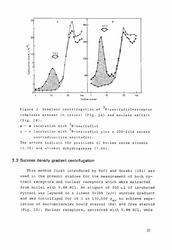
Figure 2 Gradient centrifugation of 3H-oestradiol-receptor
complexes present in cytosol (Fig. zA) and nuclear extract
(Fig. 2B). . - • incubation with 3H-oestradiol
0 - o incubation with 3H-oestradiol plus a 200-fold excess
non-radioactive oestradiol.
The arrows indicate the positions of bovine serum albumin
(4.35) and alcohol dehydrogenase (7.65).
3.3 Sucrose density gradient centrifugation
This method first introduced by Toft and Gorski {181) was
used in the present studies for the measurement of both cy
tosol receptors and nuclear receptors which were extracted
from nuclei with 0.4M KCl. An aliquot of 200 ~1 of incubated
cytosol was layered on a linear 5-20% (w/v) sucrose gradient
and was centrifuged for 16 h at 150,000 gav to achieve sepa
ration of macromolecular bound steroid (8S) and free steroid
(Fig. 2A). Nuclear receptors, extracted with 0.4M KCl, were
35
separated on linear 5-20% (w/v) sucrose gradients, containing
0.4M KCl. Gradients were centrifuged for 18 h at 260,000 g , av
which gave a clear separation of macromolecular bound steroid
(58) and free steroid (Fig. 2B). The shaded areas represent
the amount of specifically bound oestradiol measured as des
cribed under 3.1.
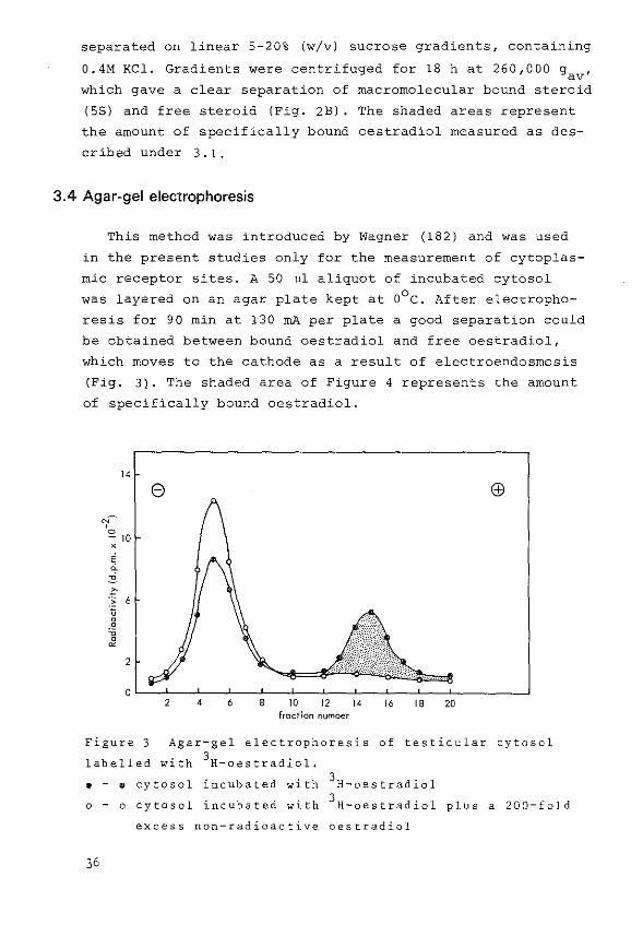
3.4 Agar-gel electrophoresis
This method was introduced by Wagner (182) and was used
in the present studies only for the measurement of cytoplas
mic receptor sites. A 50 ul aliquot of incubated cytosol
was layered on an agar plate kept at o0 c. After electropho
resis for 90 min at 130 rnA per plate a good separation could
be obtained between bound oestradiol and free oestradiol,
which moves to the cathode as a result of electroendosmosis
(Fig. 3). The shaded area of Figure 4 represents the amount
of specifically bound oestradiol.
14
"'~ 0 - 10 X
;
" :i 10 ·:; 6 .'5 .!? ~
0 ~
2
8
16 18 20 fraction number
Figure 3 Agar-gel electrophoresis of testicular cytosol
labelled with 3H-oestradiol. . - • cytosol incubated with 3H-oestradiol
o - o cytosol incubated with 3H-oestradiol plus a 200-fold
excess non-radioactive oestradiol
36
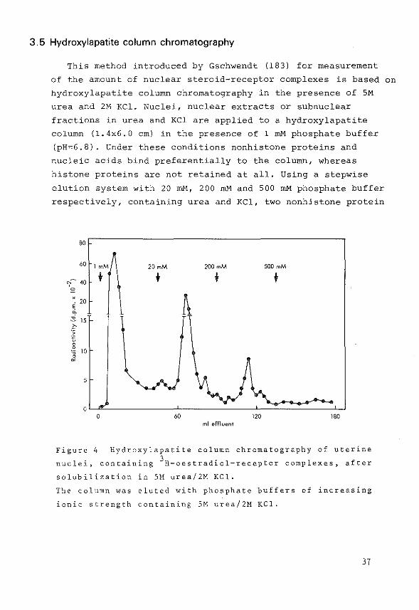
3.5 Hydroxylapatite column chromatography
This method introduced by Gschwendt (183) for measurement
of the amount of nuclear steroid-receptor complexes is based on
hydroxylapatite column chromatography in the presence of SM
urea and 2M KCl. Nuclei, nuclear extracts or subnuclear
fractions in urea and KCl are applied to a hydroxylapatite
column (1.4x6.0 em) in the presence of l mM phosphate buffer
(pH=6.8). Under these conditions nonhistone proteins and
nucleic acids bind preferentially to the column, whereas
histone proteins are not retained at all. Using a stepwise
elution system with 20 rnM, 200 mM and 500 mM phosphate buffer
respectively, containing urea and KCl, two nonhistone protein
80
60 1mM 20mM 200 mM 500 mM
'I~ 40 + " X
20 ~ ~ A ,_
15
" :~ 0 0 0
'0 10 0 ~
5
0 0 60 120 180
ml effluent
Figure 4 Hydroxylapatite column chromatography of uterine
nuclei, containing 3H-oestradiol-receptor complexes, after
solubilization in 5M urea/2M KCl.
The column was eluted with phosphate buffers of increasing
ionic strength containing SM urea/2M KCl.
37
fractions and a fraction containing DNA can be obtained as
described by McGillivray (184). The elution pattern in Figure 4
was obtained from uterine nuclei, loaded with 3H-oestradiol.
This technique was used for the present investigations
concerning the localization of subnuclear oestradiol-
receptor complexes in uterus.
3.6 Determination of nuclear receptor sites in the presence of endogenous
steroids
In tissues, which contain rather high amounts of steroids,
either from endogenous production or through exogenous ad
ministration, a certain fraction of the receptor population
moves into the nuclear fraction. For a quantitative measure
ment of the number of nuclear receptor sites it is necessary
to use a method that can distinguish between the total avai
lable amount of receptor sites and the number of receptor
sites occupied by steroid. In the exchange assay, developed
by Anderson et al. (164) isolated nuclei are incubated with
radioactive oestradiol at elevated temperatures in order to
achieve an exchange of non-radioactive oestradiol for radio
active oestradiol. After removal of excess free steroid by
charcoal treatment, the amount of radioactive &eroid bound
to receptor sites was measured directly or after extraction
of nuclei with 0.4M KCl.
Introduction and discussion of experimental work
4.1 Introduction
Receptors for steroid hormones in target cells play an
important role in the mechanism of action of steroid hor
mones (49-53), but it is not (yet) clear whether the reverse
is also true, i.e. whether the presence of a steroid recep
tor does reflect that a tissue can be considered as a target
tissue for the steroid.
The interstitial tissue is a steroid producing tissue
which is influenced by luteinizing hormone (LH) and secretes
testosterone into the blood stream (43). Processes involved
in spermatogenesis are located in the other tissue compart
ment of the testis, the seminiferous tubules. Spermatogenesis
requires both testosterone and follicle stimulating hormone
(FSH) (185,186). Recent experiments have shown that Sertoli
cells in the seminiferous tubules may be considered as tar
get cells for FSH (187,188) and that these cells can produce
oestradiol from testosterone under the influence of FSH
(189). Considering these results one might speculate about a
mutual interaction between the two testicular compartments.
Oestradiol produced in the Sertoli cell might influence the
metabolism of steroids in the Leydig cell via the hormone
receptor interaction and the subsequent translocation of the
oestradiol-receptor complexes into the nuclei. The Leydig
cell in turn might influence the Sertoli cell via changes
in the testosterone production. If oestradiol affects the
Leydig cell via receptor steroid interactions, changes in
the amount and in the subcellular distribution of the recep
tor might provide useful information concerning the function
of oestradiol and its receptor. Therefore we have attempted
to investigate the regulation of the number of cytoplasmic
receptor sites for oestradiol as a reflection of the total
available number of receptor sites. Changes in the number
of total available receptor sites might be directly correla
ted with the number of receptor sites which can ultimately
39
be bound in the nuclear fraction. It is most likely that the
ultimate effect of steroid hormones will occur via an inter
action of hormone-receptor complexes and the chromatin. For
other tissues it has been shown that hormone effects are
directly related to the number of oestradiol-receptor com
plexes found in the nucleus {1-10) and prolonged effects of
steroid hormone effects appear to be dependent on the reten
tion of steroid-receptor complexes in the nuclear fraction.
This process could be correlated with the depletion and re
plenishment of cytoplasmic receptor sites (98,143,168).
Steroidogenesis in testicular Leydig cells is dependent
on the presence of LH. Until now it is not known whether
other Leydig cell parameters, which are not directly invol
ved in the steroidogenesis, are also under the control of
gonadotrophins. Therefore it was decided to investigate the
effect of hypophysectomy on testicular oestradiol receptor
concentrations. During the development of an organism a
variety of biochemical changes occurs in different cell
types; these changes ultimately result in the formation of
fully differenti~ted cells. For some steroid hormone recep
tors it has been shown that they are present in their target
cells immediately after birth of the animal or even in the
foetus (159,160,190,191). However most of such data have
been expressed in a qualitative way. One might speculate
about the ontogeny of steroid hormone receptors. It is not
clear whether these receptors are synthesized during the
very early stages of the embryonic development or whether
they are induced in the foetus or in the immature animal by
some unknown factor. If the receptors are induced only at
later stages during development, one might suppose that
changes either in the number of receptor molecules or in the
binding properties of the protein are related to steroid hor
mone effects, which are only needed at certain stages of
development.
We have attempted to study the following aspects of the
regulation of cytoplasmic receptor sites in testicular tissue:
40

1) Effect of in vivo administration of oestradiol on the
depletion and replenishment of cytoplasmic receptor sites.
2) Effect of hypophysectomy on cytoplasmic receptor levels.
3) Ontogeny of cytoplasmic receptor sites.
4.2 Effects of oestradiol, hypophysectomy and age on cytoplasmic receptor levels
The results in appendix paper I show that in both mature
{3 months old) and immature (23 days old) rats administration
of oestradiol resulted in a rapid depletion (within 1 h) of
cytoplasmic receptor sites. However 5 h after the hormone
administration control levels were restored and remained
constant for at least an additional period of 20 h.
In mature rats, 10-15 days after hypophysectomy, no dra
matic effects could be observed on cytoplasmic receptor
levels, suggesting that neither steroid hormones nor gona
dotrophins, which both disappear from testicular tissue as
a result of hypophysectomy (42,192), are important in main
taining cytoplasmic receptor levels.
In studies concerning the ontogeny of cytoplasmic recep
tor sites for oestradiol, a plasma protein in the testicular
cytosol preparations, which binds oestradiol with a rather
high affinity (Ka=lOBM-l) and which is called a-foetoprotein,
interfered with accurate measurements of cytoplasmic recep
tors. Therefore in the testicular tissue of rats younger than
14 days of age cytoplasmic receptor sites could not be detec
ted. It was shown for rats from 4 days of age onwards,
however, that receptors accumulated in the nuclear fraction,
after incubation of whole testicular tissue with oestradiol.
These results on the regulation of the oestradiol receptor
in testicular tissue showed that oestradiol translocates
cytoplasmic receptor sites very effectively, but the hormone
itself was not important in maintaining a constant level of
total receptor sites. But if hormones exert their effects
via a hormone-receptor complex in the nuclei, information on
41
the regulation of nuclear receptor complexes appears to be
essential. Results on this aspect of receptor regulation
are described for both in vivo and in vitro studies in
appendix papers II and III respectively.
4.3 Effects of oestradiol, hypophysectomy and choriogonadotropin on nuclear receptor sites
For a quantitative measurement of nuclear receptor sites
in intact rats it was necessary to use a method that can
distinguish between free receptor sites and receptor sites
that have been occupied in vivo with endogenous hormone.
Therefore a 3H-oestradiol exchange method was developed for
testicular tissue of immature rats. Using this method it was
possible to obtain quantitative information on the number of
oestradiol receptor sites in the KCl extractable nuclear
fraction. In appendix paper II it has been demonstrated that
hypophysectomy of immature rats did result in a considerable
decrease of both the total number of receptor sites and the
number of receptor sites occupied in vivo with endogenous
oestradiol. In intact rats 20% of the total number of avai
lable receptor sites was present in the nuclear fraction,
suggesting that the presence of this small but significant
amount may be important in maintaining some unknown Leydig
cell function.
In immature rats treated with human choriogonadotropin
for 5 days it was observed that the number of available
receptor sites per testis increased threefold, while 73% of
this amount was present in its nuclear form.
4.4 Kinetics of in vitro binding of oestradiol in subcellular fractions of
testicular and uterine tissue
Like other tissues the testicular tissue contains two
types of nuclear receptor sites 1 which differ in their ex
tractibility with 0.4M KCl. For uterine tissue it has been
described that the number of nuclear receptor sites, which
resist KCl extraction 1 shows a good correlation with the
42
tissue's response to oestradiOl (140). With the 3H-oestradiol
exchange method described in appendix paper II only the class
of receptor sites which can be extracted from nuclei with KCl
could be measured quantitatively. Therefore it was decided to
use an in vitro system in order to study the translocation
and the subsequent binding of oestradiol-receptor complexes
in the KCl extractable and in the non-KCl extractable nuclear
fraction in testicular and uterine tissue under similar expe
rimental conditions. In appendix paper III the following as
pects have been investigated:
1) Mechanism and rate of translocation of cytoplasmic recep
tor molecules into the nuclear fractions of testis and
uterus.
2) The nature of the nuclear acceptor sites in the testicu
lar tissue.
3) The effect of energy deprivation (following addition of
KCN) on the subnuclear distribution of oestradiol recep
tor sites in testicular and uterine tissue.
The latter aspect concerning the role of energy in the
subnuclear distribution of receptor sites was studied because
it has been reported that cellular ATP might be involved in
the action of g1ucocorticoids, progesterone and dihydrotes
tosterone (151,153,154,155).
The results obtained on the comparison between receptors
in testis and uterus in appendix paper III showed that the
interaction of oestradiol-receptor complexes with nuclear
acceptor sites in testicular and uterine nuclei followed
different kinetics. In both tissues the number of binding
sites in the KCl extractable and non-KCl extractable frac
tion of the nuclei showed an increase observed during the
first 30 min of incubation. Thereafter the concentration of
oestradiol receptor sites in the nuclear extract of uterine
tissue showed a decrease, but reached a steady state level
(SO% of the maximum value) 60 min after the start of the
incubation. During this same period no changes could be
43
observed for the number of oestradiol receptor sites in the
KCl extract of testicular nuclei. The difference between
both tissues in the time dependent uptake of binding sites
in the non-KCl extractable nuclear fraction was even more
striking. In uterine tissue the concentration of binding
sites decreased to levels 50% of the maximum value and this
level was maintained for at least an additional period of
60 min. In testicular nUclei a similar decrease was observed,
but 60 min after the start of the incubation almost all
binding sites had dissapeared from the non-KCl extractable
fraction. This suggests that a difference exists between the
dissociation rates of the steroid-receptor complexes from the
acceptor sites in testicular and uterine nuclei.
The effect of energy deprivation, as a result of the
addition of KCN, on the number of binding sites was similar
for testicular and uterine tissue. A significant increase
was observed for the number of binding sites in the non-KCl
extractable nuclear fractions, whereas the number of KCl
extractable nuclear receptor sites remained unaffected.
For testicular tissue some attempts have been made to
solubilize the non-KCl extractable receptor sites. However,
of all the methods used only mild trypsin treatment could
release a limited number of receptor sites in a soluble form.
After trypsin treatment the additional receptor sites had a
sedimentation value of 4S on sucrose gradients, instead of a
sedimentation value of 55. Attempts with DNAse and deoxycho
late were unsuccessful: only an increase in the amount of
unbound oestradiol could be obtained.
4.5 Conclusions
In summary it can be concluded that the distribution
of testicular oestradiol receptor molecules between the
cytoplasmic and the nuclear fractions is influenced by the
oestradiol concentration in testicular tissue. After being
translocated into the nucleus,oestradiol-receptor complexes
44
are bound by at least two different types of chromatin
acceptor sites which differ in affinity for the oestradiol
receptor complex as determined by KCl extraction. In con
trast to the oestradiol receptor in uterus testicular recep
tors are retained by the high affinity acceptor sites inside
the nucleus only for short periods. This might reflect that,
although both tissues contain oestradiol receptors in compa
rable amounts, different mechanisms are involved in the bin
ding of oestradiol-receptor complexes to and the subse
quent release from the acceptor sites inside the nucleus.
The experiments described thusfar were performed in order
to obtain more information about the regulation and subcel
lular localization of testicular oestradiol-receptor com
plexes. It appeared that the kinetics of the interaction of
the oestradiol-receptor complex with nuclear constituents
from testis and uterus were different. However these results
do not permit definitive conclusions concerning the mole
cular mechanisms underlying these differences. This would
require more information about the nature of the interaction
between the oestradiol-receptor complexes and the chromatin.
In this respect results of experiments on the interaction of
the oestradiol-receptor complex and uterine chromatin are
presented in chapter s. Uterine tissue was chosen for this
purpose because in this tissue a possible interaction of
steroid-receptor complexes with a specific nuclear fraction
i.e. DNA or chromatin proteins, might be correlated with
the tissue response to oestradiol. A comparison of these
mechanisms in a well-known responsive tissue, like the uterus
and in a tissue with a less defined response like testis,
might indicate whether the presence of steroid receptors
defines a tissue as a 'hormone target tissue 1, even if dis
tinct actions of the steroids are not (yet) known.
45

Distribution of oestradiol-receptor complexes in subnuclear
fractions of uterine tissue after administration of oestradiol
in vivo and in vitro
5.1 Introduction
An important step in the response of a tissue to a
steroid hormone is the interaction of the steroid with its
receptor in the cytosol of the target tissue. The cornolex
formed between hormone and receptor migrates into the
nucleus and binds to acceptor sites on the chromatin and
this binding ultimately causes a response of the tissue to
the hormone via changes in RNA and protein synthesis (193-
196) .
The location of the oestradiol binding protein or the
newly synthesized RNA in chromatin is not known. The present
study was started in an attempt to obtain information on the
subnuclear localization of the steroid-receptor complex.
There is considerable morphological (197,198,199) and
physical (200,201) evidence that chromatin consists of a
biological 'active' form (euchromatin) and a transcrip
tionally repressed form (heterochromatin). In the literature
some methods have appeared on the fractionation of chromatin
in an active and an inactive form (202-207), but these
methods have the disadvantage that the chromatin is fraction
ated after its isolation from purified cell nuclei. During
the isolation procedure RNA-polymerase molecules and other
chromatin proteins {histones and nonhistones) might be re
leased or become inactivated. In addition it seems very
likely that cleavage of the chromatin, by DNAse digestion or
by shearing of the chromatin, produces either artificial
initiation sites or damaged template DNA in the fragments.
Tata and Baker (208) and Chesterton et al. (209) have des
cribed a method for fractionation of isolated eukaryotic
nuclei in 8 different subnuclear fractions in a single step.
In this method, introduced by Frenster et al. (210) the
nuclei are gently sonicated in an isotonic buffer and sepa-
47
rated in nuclear sap, euchromatin, heterochromatin, nucleoli
and nuclear membranes by virtue of their different sedimen
tation rates in a discontinuous sucrose gradient. Individual
fractions are considerably enriched in either one type of
the chromatin, one type of the RNA-polymerases or enriched
in poly A-rich Hn-RNA (211,212).
In the present study it was tried to apply this method
to the subnuclear distribution of oestradiol receptors in
nuclei, isolated from uterus after treatment of either
ovariectomized rats or isolated uterine tissue with oestra
diol.
5.2 Materials and methods
Animal and tissue treatment
Mature female rats of the Wistar strain, ovariectomized
for 3-5 days, were used. Two different methods were used
for the preparation of nuclear 3H-oestradiol-receptor com
plexes:
1) Isolated uterine tissue was incubated for 30 min at 37°C
in KRBG-buffer (pH 7.4) either with 10-8M radioactive
oestradiol or with 10-8M radioactive oestradiol plus a
1000-fold excess of non-radioactive oestradiol. There
after the tissue was rinsed with buffer and nuclei were
isolated.
2) Rats were injected subcutaneously with 5 vg of oestradiol
and 60 min later uterine nuclei were isolated.
The nuclear suspensions thus obtained were incubated for
60 min at 20°c with 10-BM 3H-oestradiol in order to obtain
complete exchange between receptor bound oestradiol and
added 3H-oestradiol(J64).
48
Preparation of nuclei
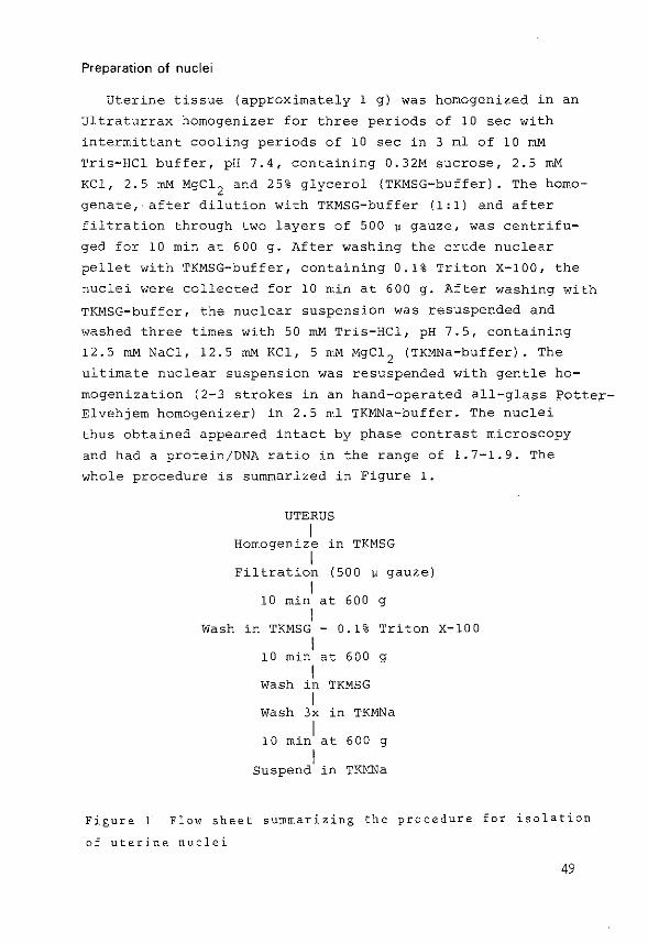
Uterine tissue (approximately 1 g) was homogenized in an
Ultraturrax homogenizer for three periods of 10 sec with
intermittant cooling periods of 10 sec in 3 ml of 10 rnM
Tris-HCl buffer, pH 7.4, containing 0.32M sucrose, 2.5 mM
KCl, 2.5 mM MgCl2
and 25% glycerol (TKMSG-buffer). The homo
genate,·after dilution with TKMSG-buffer (1:1) and after
filtration through two layers of 500 ~ gauze, was centrifu-
ged for 10 min at 600 g. After washing the crude nuclear
pellet with TKMSG-buffer, containing 0.1% Triton X-100, the
nuclei were collected for 10 min at 600 g. After washing with
TKMSG-buffer, the nuclear suspension was resuspended and
washed three times with 50 mM Tris-HCl, pH 7.5, containing
12.5 mM NaCl, 12.5 mM KCl, 5 mM MgC12
(TKMNa-buffer). The
ultimate nuclear suspension was resuspended with gentle ho
mogenization (2-3 strokes in an hand-operated all-glass ~otte~
Elvehjern homogenizer) in 2.5 ml TKMNa-buffer. The nuclei
thus obtained appeared intact by phase contrast microscopy
and had a protein/DNA ratio in the range of 1.7-1.9. The
whole procedure is summarized in Figure 1.
UTERUS I
Homogenize in TKMSG I
Filtration (500 ~ gauze) I
10 min at 600 g I
Wash in TKMSG- 0.1% Triton X-100 I
10 min at 600 g I
Wash in TKMSG I
wash 3x in TKMNa
10 mini at 600 g
I Suspend in TKMNa
Figure 1 Flow sheet summarizing the procedure for isolation
of uterine nuclei
49
Fractionation of nuclei
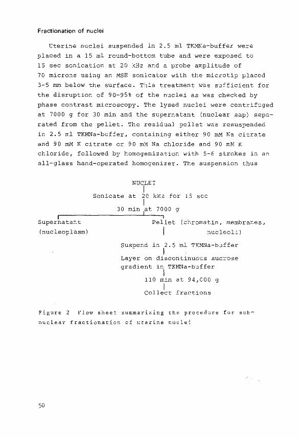
Uterine nuclei suspended in 2.5 rnl TKMNa-buffer were
placed in a 15 ml round-bottom tube and were exposed to
15 sec sonication at 20 kHz and a probe amplitude of
70 microns using an MSE sonicator with the microtip placed
3-5 mm below the surface. This treatment was sufficient for
the Qisruption of 90-95% of the nuclei as was checked by
phase contrast microscopy. The lysed nuclei were centrifuged
at 7000 g for 30 min and the supernatant (nuclear sap) sepa
rated from the pellet. The residual pellet was resuspended
in 2.5 ml TKMNa-buffer, containing either 90 mM Na citrate
and 90 mM K citrate or 90 roM Na chloride and 90 mM K
chloride, followed by homogenization with 5-6 strokes in an
all-glass hand-operated homogenizer. The suspension thus
Supernatant
(nucleoplasm)
NUCLEI I
Sonicate at 20 kHz for 15 sec I
30 min at 7000 g
Pellet (chromatin, membranes,
I nucleoli)
suspend in 2.5 ml TKMNa-buffer I
Layer on discontinuous sucrose gradient in TKMNa-buffer
I 110 min at 94,000 g
I Collect fractions
Figure 2 Flow sheet summarizing the procedure for sub
nuclear fractionation of uterine nuclei
50
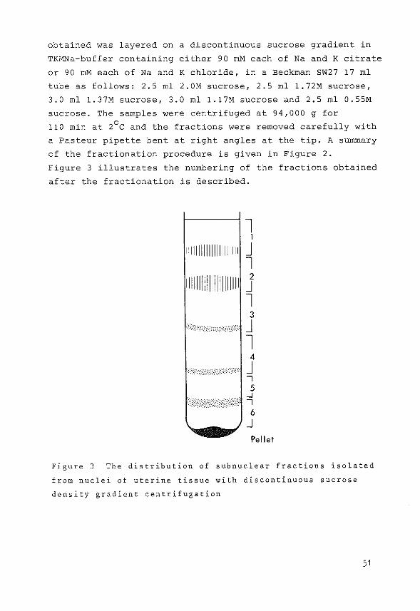
obtained was layered on a discontinuous sucrose gradient in
TKMNa-buffer containing either 90 mM each of Na and K citrate
or 90 roM each of Na and K chloride, in a Beckman SW27 17 ml
tube as follows: 2.5 ml 2.0M sucrose, 2.5 ml l.72M sucrose,
3.0 ml 1.37M sucrose, 3.0 ml l.l7M sucrose and 2.5 ml 0.55M
sucrose. The samples were centrifuged at 94,000 g for
110 min at 2°C and the fractions were removed carefully with
a Pasteur pipette bent at right angles at the tip. A summary
of the fractionation procedure is given in Figure 2.
Figure 3 illustrates the numbering of the fractions obtained
after the fractionation is described.
l 1
111111111111111111111 J l
111111111111111111111 j l 3
J l
4
J -, 5
f'i//)/0' q =j 6
J Pellet
Figure 3 The distribution of subnuclear fractions isolated
from nuclei of uterine tissue with discontinuous sucrose
density gradient centrifugation
51
Labelling of RNA in vitro
One uterus, stripped of adhering fat and mesentery, was
incubated in 2.0 ml of Krebs Ringer bicarbonate buffer,
pH 7.4, containing 0.2% glucose and 50 ~Ci/rnl s- 3H-uridine
(sp. act. 20 Ci/rnmol). Incubation was carried out for
30 min at 37°C in an atmosphere of 95% o2
: 5% co2
. Nuclei
were prepared from uterine tissue and fractionated by the
described procedure in buffers containing K and Na chloride.
Nucleic acids in each subnuclear fraction were precipitated
with O.SM HC104
at 0°C and were washed three times with
O.SM HC10 4 . RNA in the pellet was dissolved in 1M KOH at
37°C for 60 min and samples, taken after the addition of
HClo 4 to precipitate the DNA, were used for the measurement
of incorporated radioactivity and RNA
was treated with HClo4
at 70°C for 15
content. The pellet
min
DNA and samples were taken to measure the
to hydrolyse the
DNA content (see
under Measurement of DNA, RNA and protein).
Measurement of specific binding of 3
H-oestradiol
Different methods were used for the estimation of 3H
oestradiol binding by receptor proteins:
1) After the isolation of the·subnuclear fractions samples
were taken for the measurement of total radioactivity
present in each fraction. Specific 'binding' in each
fraction was determined as previously described by sub
tracting the nonspecific binding (obtained after incu
bation of tissue in the presence of excess non-radioactive
oestradiol) from the total binding of 3H-oestradiol
(144). Binding was expressed as dprn/~g DNA present in
each fraction.
2) In some experiments samples of various subnuclear frac
tions were run on a linear sucrose density gradient
(5-20%) in the presence or in the absence of high salt
concentrations as described previously (144,213).
3) Binding of 3H-oestradiol by receptor molecules was also
demonstrated by hydroxylapatite cOlumn chromatography.
52

The method used was essentially the procedure described
by Gschwendt (183), originally developed by McGillivray
et al. (184). Hydroxylapatite was washed three times in
5M urea/2M KCl in 1 mM potassiumphosphate buffer pH 6.8
by suspension and decantation. Columns were then packed
(1x6 em), equilibrated and run at 4°C. Subnuclear frac
tions containing chromatin were diluted with 5M urea/2M
KCl in phosphate buffer (1:1), applied to the column and
eluted with 1 mM, 20 mM, 200 mM and 500 mM potassium
phosphate pH 6.8 in 5M urea/2M KCl. The flow rate was
maintained at 30 ml/h and 3 ml fractions were collected.
In later experiments the columns were prepared in
1 mM potassiumphosphate pH 6.8 containing 0.4M KCl. Sub
nuclear fractions diluted (1:1) with 0.8M KCl were applied
to the column and were eluted with 1 mM, 20 mM, 200 mM
and 500 mM potassiumphosphate buffer pH 6.8 respectively,
containing 0.4M KCl.
Measurement of DNA, RNA and protein
After accurately measuring the volume of each subnuclear
fraction the fractions were diluted 2-3 fold with water.
HCl0 4 was added at 0°C to a final concentration of 0.5M and
the samples were washed three times with 0.5M HC104 . The
pellets were dissolved in lM KOH at 37°C for 60 min. DNA and
protein were precipitated after the addition of HClo4
at
0°C for 30 min. The supernatant containing the products of
RNA hydrolysis was used for RNA estimation. The pellet was
treated with HCl0 4 at 70°C for 15 min to hydrolyze the DNA.
The residual pellet, containing protein, was dissolved in lM
KOH at 70°C for 60 min. DNA in the fractions was measured
using the method of Giles and Myer (214), RNA was measured
according to Fleck and Munro (215) and the protein content
of each fraction was measured by the method of Lowry et al.
(216). In some experiments the amounts of DNA in the subnu
clear fractions were very small (less than 10 ~g/fraction)
and in those cases the fluorescence method of Robertson and
Tait was used (217).
53

5.3 Results
Isolation and chemical characterization of subnuclear fractions
In initial experiments it was tried to establish the con
ditions for fractionation of uterine nuclei. With light
microscopy as a parameter it could be observed that sonica
tion for a period of 15 sec was sufficient to disrupt about
90-95% of the uterine nuclei. The pattern of bands obtained
after the final ultracentrifugation step in TKMNa-buffer
containing K{Na) citrate was similar to that observed for
liver nuclei by Tata and Baker (208). After the collection
of the various fractions the recoveries of protein, RNA and
DNA were measured. Generally approximately 45-50% of the
total nuclear DNA was recovered in the pellet fraction. The
pellet also contained about 40-50% of the total amount of
protein recovered. The very small amount of DNA (in the
order of 1% of the total amount) and the relatively large
amount of protein in the 7000 g supernatant of the nuclear
sonicate were considered to reflect the nucleoplasm in this
fraction (Table I).
The results in Table I are representative for the data
obtained as percentages of each constituent and as RNA/DNA
and protein/DNA ratios. From these data it is evident that
the individual fractions differ considerably in composition.
However these data cannot be used for the determination of
the functional properties of the constituents present in
each fraction. Tata and Baker (208,211) showed that sub
nuclear fractions isolated from rat liver contained different
types of chromatin, different types of RNA-polymerase and
differed also in the rate of labelling of RNA in vivo. They
concluded that the top fractions of the sucrose gradient
(fraction 1 and 2) contained the euchromatin, whereas the
pellet and fraction 6 contained the heterochromatin. A mix
ture of both types of chromatin was located in fractions 3,
4 and 5.
54
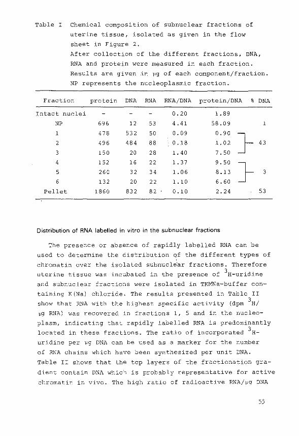
Table I Chemical composition of subnuclear fractions of
uterine tissue, isolated as given in the flow
sheet in Figure 2.
After collection of the different fractions, DNA,
RNA and protein were measured in each fraction.
Results are given in vg of each component/fraction.
NP represents the nucleoplasmic fraction.
Fraction protein DNA RNA RNA/DNA protein/DNA % DNA
Intact nuclei
NP
1
2
3
4
5
6
Pellet
696
478
496
15 0
152
260
132
1860
12
53 2
48 4
20
16
32
20
83 2
53
50
88
28
22
34
22
82
0.20
4.41
0. 09
0.18
l. 40
l. 37
l. 06
l. 10
0.10
l. 89
58.0 9
0.90
l. 02
7. 50
9.50
8.13
6.60
2. 24
Distribution of RNA labelled in vitro in the subnuclear fractions
1
53
The presence or absence of rapidly labelled RNA can be
used to determine the distribution of the different types of
chromatin over the isolated subnucl~'·ar fractions. Therefore
uterine tissue was incubated in the presence of 3H-uridine
and subnuclear fractions were isolated in TKMNa-buffer con
taining K(Na) chloride. The results presented in Table II
show that RNA with the highest specific activity (dpm 3
H/
vg RNA) was recovered in fractions 1, 5 and in the nucleo
plasm, indicating that rapidly labelled RNA is predominantly
located in these fractions. The ratio of incorporated 3 H
uridine per vg DNA can be used as a marker for the number
of RNA chains which have been synthesized per unit DNA.
Table II shows that the top layers of the fractionation gra
dient contain DNA which is probably representative for active
chromatin in vivo. The high ratio of radioactive RNA/vg DNA
55
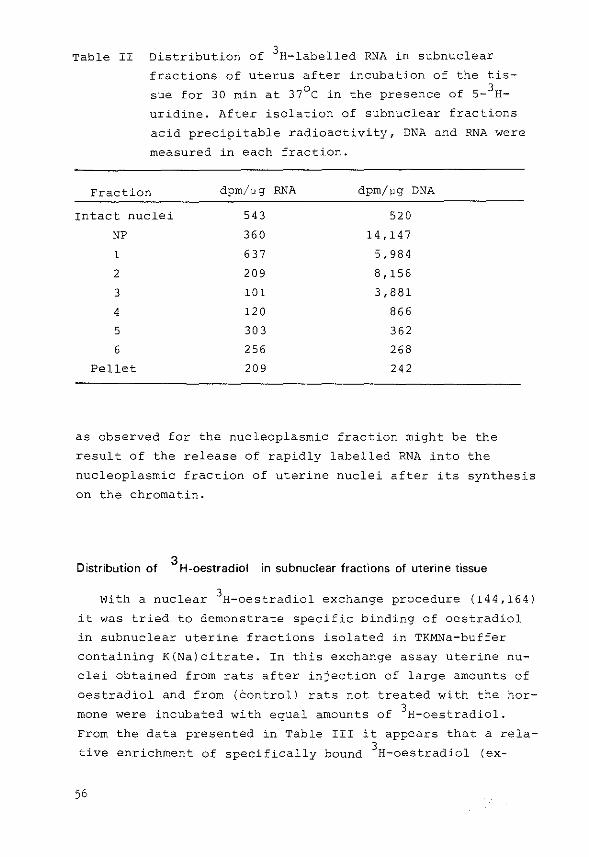
Table II Distribution of 3H-labelled RNA in subnuclear
fractions of uterus after incubation of the tis
sue for 30 min at 37°C in the presence of s- 3H
uridine. After isolation of subnuclear fractions
acid precipitable radioactivity, DNA and RNA were
measured in each fraction.
Fraction dpm/"g RNA dpm/"g DNA
Intact nuclei 543 520
NP 360 14,147
1 637 5,984
2 209 8,156
3 101 3,881
4 120 866
5 30 3 362
6 256 268
Pellet 209 242
as observed for the nucleoplasmic fraction might be the
result of the release of rapidly labelled RNA into the
nucleoplasmic fraction of uterine nuclei after its synthesis
on the chromatin.
Distribution of 3
H-oestradiol in subnuclear fractions of uterine tissue
With a nuclear 3H-oestradiol exchange procedure (144,164)
it was tried to demonstrate specific binding of oestradiol
in subnuclear uterine fractions isolated in TKMNa-buffer
containing K(Na)citrate. In this exchange assay uterine nu
clei obtained from rats after injection of large amounts of
oestradiol and from (Control) rats not treated with the hor
mone were incubated with equal amounts of 3H-oestradiol.
From the data presented in Table III it appears that a rela
tive enrichment of specifically bound 3H-oestradiol (ex-
56
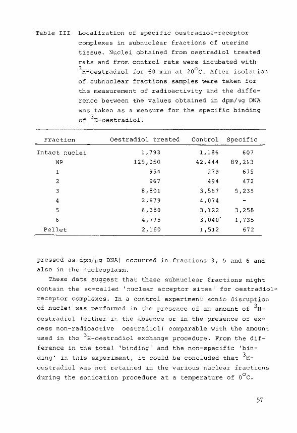
Table III Localization of specific oestradiol-receptor
complexes in subnuclear fractions of uterine
tissue. Nuclei obtained from oestradiol treated
rats and from control rats were incubated with 3H-oestradiol for 60 min at 20°C. After isolation
of subnuclear fractions samples were taken for
the measurement of radioactivity and the diffe
rence between the values obtained in dpm/vg DNA
was taken as a measure for the specific binding
of 3H-oestradiol.
Fraction Oestradiol treated Control Specific
Intact nuclei 1,793 1,186 607
NP 129,050 42,444 89,213
1 954 279 675
2 967 494 472
3 8,801 3,567 5,235
4 2,679 4,074
5 6,380 3,122 3,258
6 4,775 3,040' 1,735
Pellet 2,160 1,512 672
pressed as dpm/~g DNA) occurred in fractions 3, 5 and 6 and
also in the nucleoplasm.
These data suggest that these subnuclear fractions might
contain the so-called 'nuclear acceptor sites' for oestradiol
receptor complexes. In a control experiment sonic disruption
of nuclei was performed in the presence of an amount of 3H
oestradiol (either in the absence or in the presence of ex
cess non-radioactive oestradiol) comparable with the amount
used in the 3H-oestradiol exchange procedure. From the dif-
ference in the total 'binding' and
ding' in this experiment, it could
oestradiol was not retained in the
during the sonication procedure at
the non-specific 'bin
be concluded that 3H-
various nuclear fractions 0
a temperature of 0 c.
57
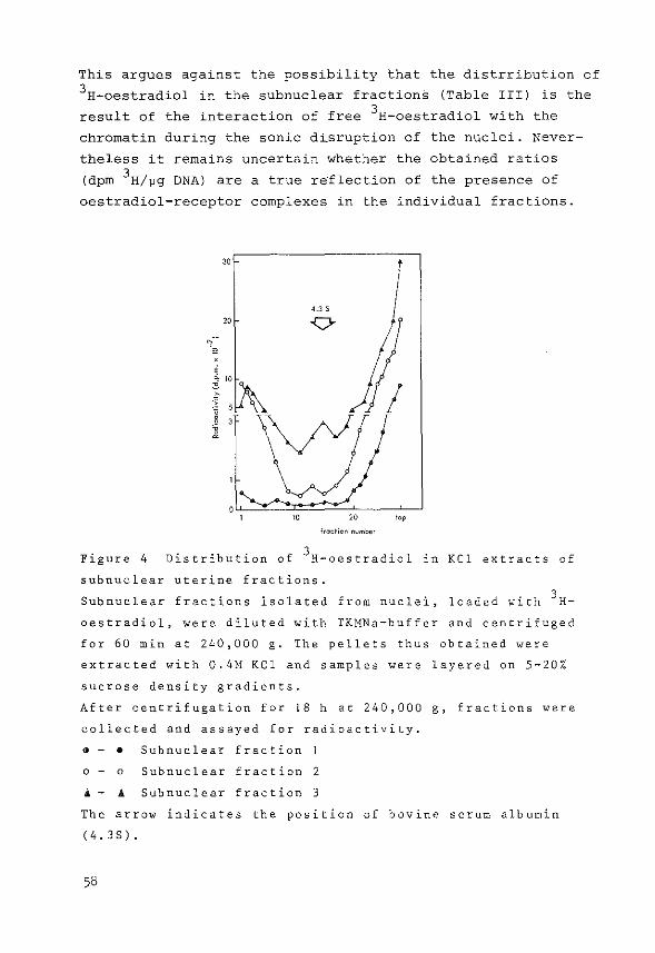
This argues against the possibility that the distrribution of 3H-oestradiol in the subnuclear fractions (Table III) is the
result of the interaction of free 3H-oestradiol with the
chromatin during the sonic dLsruption of the nuclei. Never
theless it remains uncertain whether the obtained ratios
(dprn 3H/Jlg DNA) are a true re·flection of the presence of
oestradiol-receptor complexes in the individual fractions.
30
4.3 s 20
' i 10
~ 5 g 3
i
lrac~;on number
Figure 4 Distribution of 3H-oestradiol in KCl extracts of
subnuclear uterine fractions.
Subnuclear fractions isolated from nuclei, loaded with 3 H
oestradiol, were diluted with TKMNa-buffer and centrifuged
for 60 min at 240,000 g. The pellets thus obtained were
extracted with 0.4M KCl and samples were layered on 5-20%
sucrose density gradients.
After centrifugation for 18 h at 240,000 g, fractions were
collected and assayed for radioactivity.
•- • Subnuclear fraction
o - o Subnuclear fraction 2
A- .A Subnuclear fraction 3
The arrow indicates the position of bovine serum albumin
(4,3S).
58
In an attempt to demonstrate the binding of oestradiol to
macromolecules, subnuclear fractions, after dilution with
TKMNa-buffer, were centrifuged for 60 min at 240,000 g. The
pellets thus obtained were extracted with 0.4M KCl for
60 min at Q°C and the KCl extracts were used for analysis
on high salt sucrose gradients. The distribution of 3H
oestradiol in these extracts is shown for subnuclear frac
tions 1,2 and 3 in Figure 4. A comparabl~ heterogeneous
distribution of radioactivity was also obtained for the
other subnuclear fractions including the pellet, and this
observation made the sucrose density gradient centrifugation
technique less suitable for the detection of nuclear recep
tor sites. Moreover it cannot be excluded that receptor
sites were lost during the centrifugation step in diluted
buffer. Another disadvantage is that KCl extraction is sui
table only for the detection of KCl extractable receptor
sites. Considering this, together with the low capacity and
the low resolving power of sucrose gradients, it was tried
to concentrate the various subnuclear fractions. All methods
used, including dialysis and concentration by hollow fibers
resulted, however, in considerable losses (about 50%) of both
radioactivity and protein and no binding could be observed in
the resulting preparations.
Demonstration of nuclear oestradiol receptor sites in uterine nuclei by
hydroxylapatite column chromatography in the presence of urea and KCI
It has been demonstrated by Gschwendt (183) for the oes
tradiol receptor in nuclei from chicken liver that chroma
tography on hydroxylapatite columns for separation of chro
matin proteins as originally developed by McGillivray (184),
could be used for the measurement of nuclear oestradiol
receptors. This method is based on retention of proteins by
the column and the subsequent release by increasing phosphate
concentrations. It has also been demonstrated by Gschwend·t:,
that the method could distinguish between KCl extractable
59
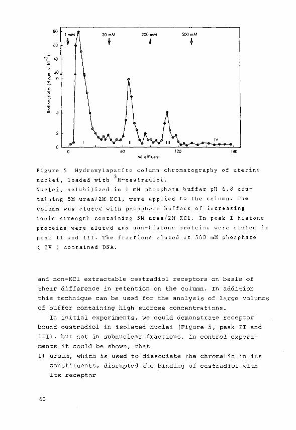
80 lmM 20 mM 200 mM 500mM
60 + t t t 'I~ 40
" X
; 20 q. 10
"'-1:-:~ u ' ' '0 ' ~ 5
2 IV
0 0 180
ml effluent
Figure 5 Hydroxylapatite column chromatography of uterine
nuclei, loaded with 3H-oestradiol.
Nuclei, solubilized in 1 mM phosphate buffer pH 6.8 con
taining SM urea/2M KCl, were applied to the column. The
column was eluted with phosphate buffers of increasing
ionic strength containing SM urea/2M KCl. In peak I histone
proteins were eluted and non-histone proteins were eluted lll
peak II and III. The fractions eluted at 500 mM phosphate
( IV ) contained DNA.
and non-KCl extractable oestradiol receptors on basis of
their difference in retention on the column. In addition
this technique can be used for the analysis of large volumes
of buffer containing high sucrose concentrations.
In initial experiments, we could demonstrate receptor
bound oestradiol in isolated nuclei (Figure 5, peak II and
III), but not in subnuclear fractions. In control experi
ments it could be shown, that
1) ureurn, which is used to dissociate the chromatin in its
constituents, disrupted the binding of oestradiol with
its receptor
60

2) citrate, which is present in the fractionation gradient,
extracted considerable amounts of oestradiol-receptor
complexes from isolated nuclei and also interfered with
the elution of oestradiol-receptor complexes from the
hydroxylapatite column.
On basis of these observations it was decided to omit
ureum from the elution buffer and to substitute citrate ions
for chloride ions in the fractionation gradient. Subnuclear
fractions, after addition of O.BM KCl (1:1), were applied to
columns prepared in 1 mM phosphate buffer containing 0.4M
KCl. Fractions were collected after elution with 20 mM,
200 rnM and 500 rnM phosphate buffer, respectively, containing
0.4M KCl. This alternative method appeared to be reliable,
because in the order of 80% of the number of receptor sites
present in a KCl extract of uterine nuclei could be recover
ed in fraction II and III after elution of a hydroxylapatite
column.
Demonstration of nuclear oestradiol receptor sites in subnuclear fractions of
uterine nuclei by hydroxylapatite column chromatography
Subnuclear fractions were isolated in TKMNa-buffer con
taining KCl and NaCl after incubation of uterine tissue for
60 min at 37°C in the presence of 10-BM 3H-oest·radiol. The
isolated subnuclear fractions were diluted (1:1) with 0.8M
KCl and chromatographed on hydroxylapatite columns. Table IV
gives a summary of the results. It was possible to demon
strate binding of 3H-oestradiol in subnuclear fractions 1,2
and 3, in the nucleoplasm (NP) but not in fractions 4,5 and
6. In order to obtain information about the presence of
receptor sites in the pellet fraction of the fractionation
gradient it was tried to solubilize receptor sites by adding
0.4M KCl for 60 min at 0°C. In a further attempt to demon
strate receptor sites in the pellet fraction the residual
(non-KCl extractable) fraction was sonicated for 15 sec in
buffer containing 0.4M KCl. The KCl extracts thus obtained
61
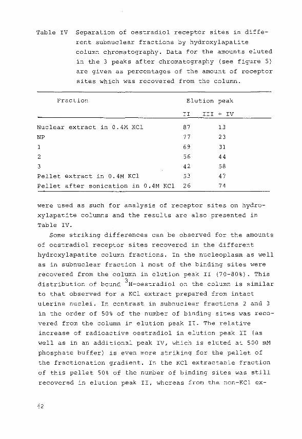
Table IV Separation of oestradiol receptor sites in diffe
rent subnuclear fractions by hydroxylapatite
column chromatography. Data for the amounts eluted
in the 3 peaks after chromatography (see figure 5)
are given as percentages of the amount of receptor
sites which was recovered from the column.
Frdclion Elution peak
II III + IV
Nuclear extract in 0.4M KCl 87 13
NP 77 23
1 69 31
2 56 44
3 42 58
Pellet extract in 0.4M KCl 53 47
Pellet after sonication in 0.4M KCl 26 74
were used as such for analysis of receptor sites on hydro
xylapatite columns and the results are also presented in
Table IV.
Some striking differences can be observed for the amounts
of oestradiol receptor sites recovered in the different
hydroxylapatite column fractions. In the nucleoplasm as well
as in subnuclear fraction 1 most of the binding sites were
recovered trom the column in elution peak II (70-80%). This
distribution of bound 3H-oestradiol on the column is similar
to that observed for a KCl extract prepared from intact
uterine nuclei. In contrast in subnuclear fractions 2 and 3
in the order of 50% of the number of binding sites was reco
vered from the column in elution peak II. The relative
increase of radioactive oestradiol in elution peak II (as
well as in an additional peak IV, which is eluted at 500 mM
phosphate buffer) is even more striking for the pellet of
the fractionation gradient. In the KCl extractable fraction
of this pellet 50% of the number of binding sites was still
recovered in elution peak II, whereas from the non-KCl ex-

tractable fraction, applied to the hydroxylapatite column
after sonication in 0.4M KCl, only 20-35% was eluted in this
peak. A similar treatment, including KCl extraction of nuclei
and sonication of the nuclear residual pellet in 0.4M KCl,
also resulted in the recovery of non-KCl extractable receptor
sites predominantly in elution peaks III and IV.
In summary therefore it can be concluded that non-KCl
extractable receptor sites appear to sediment in the heavier
sucrose gradient fractions and in the pellet of the fraction
ation gradient. KCl extractable receptors were recovered
predominantly in the nucleoplasm and in the lighter sucrose
gradient fractions.
Recovery of oestradiol-receptor complexes in subnuclear fractions of uterus
Considering the quantitative distribution of oestradiol
receptor complexes over the various subnuclear fractions it
appears that in the order of 40% of receptor sites was re
covered in the top layers of the fractionation gradient
(30%) and in the nucleoplasm (10%). The remaining 60% was
found in the pellet fraction obtained during the isolation
of the subnuclear fractions. From the total amount of nuclear
receptor sites present in uterine nuclei before sonication
and fractionation, approximately 40% could be recovered. This
loss of nuclear receptor sites was not caused by the sonica
tion procedure because after KCl extraction of sonically dis
rupted nuclei more than 80% of the number of receptor sites
present in intact nuclei was recovered as measured by sucrose
gradient centrifugation. During the fractionation of uterine
nuclei, the sonicated nuclei are considerably diluted during
the centrifugation through the discontinuous gradient.
This step, which might induce a considerable dissociation of
oestradiol-receptor complexes could explain, at least in
part, the low recovery of nuclear receptor sites.
63
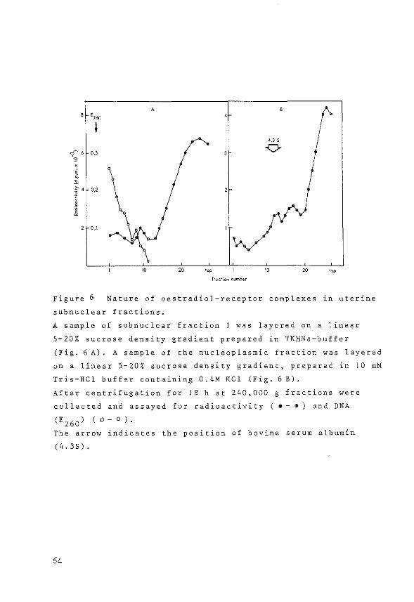
' 1 4 0,2
' " ~ 2 O,l
Figure 6
A
4.3 s
0
20 20
fraction number
Nature of oestradiol-receptor complexes in uterine
subnuclear fractions.
A sample of subnuclear fraction 1 was layered on a linear
5-20% sucrose density gradient prepared in TKMNa-buffer
(Fig. 6 A). A sample of the nucleoplasmic fraction was layered
on a linear 5-20% sucrose density gradient, prepared in 10 mM
Tris-HCl buffer containing 0, 4M KCl (Fig. 6 B).
After centrifugation for 18 h at 240,000 g fractions were
collected and assayed for radioactivity ( •- •) and DNA
(E260) ( o- o ).
The arrow indicates the position of bovine serum albumin
(4.3S).
64
Nature of oestradiol-receptor complexes in subnuclear fractions
An attempt was made to obtain further information about
the state of the oestradiol-receptor complexes in the nu
cleoplasmic fraction and in subnuclear fraction number 1. A
sample of the nucleoplasm was layered on a sucrose density
gradient prepared in 0.4M KCl in buffer. After centrifuga
tion and collection of the individual fractions, the amount
of radioactivity was determined in each fraction. Figure 6B
shows that oestradiol bound to macromolecules in the nucleo
plasm sediments heterogeneously in the 3-8S region of the
gradient. A sample of subnuclear fraction 1 was layered on a
sucrose gradient prepared in TKMNa-buffer in the absence of
KCl. Figure 6A shows that subnuclear fraction 1, in addition
to oestradiol bound to macromolecules, also contains a
heterogeneous fraction of DNA, which was determined by mea
suring E260 in each fraction. This might reflect a possible
interaction between the two components, i.e. the oestradiol
receptor complex and the DNA, in vivo.
5.4 Discussion
Chromatin in vivo appears to occur in two forms, hetero
chromatin, which is inactive in transcription and euchroma
tin, which includes genes participating in transcription
(199). In vitro chromatin can be separated in fractions which
have different template activities in incubations with added
exogenous RNA-polymerase and different amounts of nascent
RNA after labelling in vivo or in vitro (206,209,210,211,
218,219,220,221). Also differences in histone and nonhistone
protein content could be observed (221,222,223,224). Although
the molecular composition of these chromatin fractions is not
known, they may be separated from each other as a consequence
of their differences in:
-hydrodynamic properties, which are reflected in shearing or
sonication and sedimentation through glycerol or sucrose
gradients {208, 209,210,219,221).
65

-electrostatic properties after sonication, as reflected
during separation by ECHTAM-cellulose column chromatography
(223).
-behaviour during DNAse digestion of chromatin (203,225}.
In some of these methods chromatin was isolated from
nuclei and fractionated by either of the treatments as des
cribed above in order to obtain chromatin fractions. A major
disadvantage of this procedure is the fact that isolated
chromatin may be different from chromatin in vivo with res
pect to the location of histones and nonhistones on the
chromatin (226). It is also possible that as a result of
the isolation and fractionation methods the bulk of RNA
polymerase would be released from its template or become
inactivated. Furthermore most procedures used for prepara
tion of chromatin and RNA-polymerases could destroy the
nuclear envelope, while, at the same time, methods used for
isolating nuclear membranes would not allow a meaningful
study of the role of nuclear structures in the regulation of
transcription of DNA.
In order to correlate a possible interaction of oestra
diol-receptor complexes with their acceptor sites and the
ultimate effect of oestradiol inside the nucleus i.e. changes
in RNA-polymerase activity and synthesis of new mRNA species,
it was tried to disrupt isolated uterine nuclei by sonication
and prepare subnuclear fractions on sucrose gradients.
This relatively mild method which was originally introdu
ced by Frenster et al. (210} was applied successfully by
Chesterton (209} and Tata and Baker (208) to liver nuclei.
However this sonication method had thusfar not been used to
study the subnuclear localization of steroid-receptor com
plexes.
From the present results it appears that administration
of oestradiol in vivo resulted in a relative enrichment of
oestradiol (expressed as dpm/~g DNA) in the subnuclear frac
tions of uterus designed fractions 3,5 and 6 and also in the
nucleoplasm (Table III). Although it could be demonstrated
by a 3H-oestradiol exchange procedure that this relative
66
enrichment was specific and not due to the sonication proce
dure at 0°C, it was tried to elucidate the nature of the
oestradiol present in each fraction. Sucrose density gradient
centrifugation appeared to be less suitable for the analysis
of oestradiol-receptor complexes in each fraction. Hydroxyl
apatite column chromatography was expected to be more
suitable for the localization of receptor bound oestradiol,
because it had previously been used by Gschwendt (183) for
separation of the oestradiol receptor population in chicken
liver in KCl extractable and non-KCl extractable receptor
sites due to the differences in interaction with the exchange
resin. In this procedure the isolated chromatin was first
dissociated in histones, nonhistones and DNA by adding SM
urea and 2M KCl.
In the present study it was observed that, in contrast to
the oestradiol receptor in chicken liver, steroids are dis
sociated from the uterine receptor in the presence of urea.
In addition it became apparent, that citrate ions in the
buffe~, which are used for the isolation of subnuclear frac
tions according to the method of Tata and Baker (208),
interfered with both the interaction of oestradiol receptor
molecules with the hydroxylapatite column and the interac
tion of receptor molecules with DNA in intact nuclei. There
fore, in later experiments urea was omitted and chloride ions
rather than citrate ions were used in the subnuclear fractio
nation procedure and in the chromatography of the isolated
subnuclear fractions. Oestradiol receptors present in the individual subnuclear
fractions of uterus could be eluted from the hydroxylapatite
column at different ionic strengths. KCl extractable recep
tor sites (eluted predominantly at 20 rnM phosphate) were
found in the upper region of the fractionation gradient and
in the nucleoplasm, whereas non-KCl extractable receptor
sites (eluted at 200 rnM and 500 mM phosphate) were measured
in the pellet fraction. These results indicate that a sepa
ration of receptor molecules with different affinities for
the chromatin could be achieved. The results might reflect
that low affinity acceptor sites were recovered in the light
67
sucrose fractions and in the nucleoplasm, whereas high affi
nity acceptor sites were found in the pellet fraction. It
has been suggested by others (140), that receptor molecules
which interact with high affinity acceptor sites could be
important for the response of the uterus to oestradiol. The
present observation that these complexes are mainly located
in the pellet fraction of the fractionation gradient suggests
that this nuclear fraction might be important in the regula
tion of the activity of steroid hormones.
For the progesterone receptor in chick oviduct it has also
been demonstrated that steroid receptor molecules were loca
ted in a heavy sucrose gradient fraction, containing the
heterochromatin {2061. In contrast to these results it has
been demonstrated that, using a different technique for
fractionation, progesterone receptors (218) and oestradiol
receptors (227,228) were located in the lighter chromatin
(euchromatin) fraction.
The recovery of receptor sites after the fractionation
procedure of uterine nuclei was far from complete (in the
order of 40%). A possible explanation may be found in the
dilution of the nuclear sonicate during the separation of
the subnuclear fractions on the sucrose gradient, which
might result in a considerable dissociation of oestradiol
receptor complexes. This explanation is sustained by the
observation that a considerable amount of the 3H-oestradiol,
which is present in the individual subnuclear fractions, is
not bound to macromolecules, whereas in intact uterine
nuclei unbound oestradiol was almost absent. In addition it
was found that the binding sites were almost equally dis
tributed between the upper two gradient fractions, including
the nucleoplasm, and the pellet fraction of the gradient.
No conclusive data were obtained concerning the possible
interaction of oestradiol receptor molecules and DNA present
in the subnuclear fractions. On low salt sucrose density
gradients oestradiol-receptor complexes, present in frac
tion 1, were shown to sediment heterogeneously in a region
where small-size DNA species were also located (Figure 6A).
68
From the detailed studies of Yamamoto (120) it appears that
such an interaction, if present, could represent a low
affinity interaction without
Tata
physiological meaning.
and Baker (208,211) and It has been shown by
Chesterton et al. (209) that sonication and sucrose gradient
centrifugation of rat liver nuclei could achieve separation
of euchromatin and heterochromatin fractions. This has been
supported by results of transcription on DNA stretches in
vitro, the localization of nascent RNA molecules and the
precipitation of heterochromatin in the presence of 5 mM
MgC1 2 . Due to the presence of Mg2+ ions in the fractionation
buffer this simple method could not be applied to the isola
ted uterine subnuclear fractions. In the present study it
could be demonstrated that rapidly labelled RNA, after in
vitro incubation of uterus, was predominantly located in
the upper layers of the fractionation gradient (Table II),
suggesting that euchromatin is p~esent in these fractions.
It cannot be excluded, however, that as a result of the
isolation and fractionation of nuclei RNA chains become de
tached from their respective templates and interact with
other DNA stretches. Nevertheless a random redistribution
of labelled RNA can be excluded on basis of the results
presented in Table II.
From the results presented in this chapter it is evident
that the localization of oestradiol-receptor complexes in
subnuclear fractions of uterus requires still further inves
tigations. In this respect the influence of the different
fractionation techniques on the subnuclear localization of
steroid receptor molecules creates a challenging problem.
Another difficulty is caused by the different ionic condi
tions which are required for the isolation of the 'physio
logical' form of those components which are supposed to be
obligatory for the ultimate effect of steroid hormones, i.e.
steroid hormone receptor associated with chromatin, RNA
polymerases and chromatin. Furthermore the question remains
whether the isolated chromatin fractions are really repre
sentative for regions which are active and inactive in
69
transcription in vivo. In two studies (229,230) it has been
reported that chromatin could be separated in euchromatin and
heterochromatin, which differed in protein composition as well
in their activity in transcription with RNA-polymerase in
vitro. However DNA-DNA reassociation experiments performed
with radioactive eDNA, obtained after transcription of RNA
molecules with reverse transcriptase, did not provide any
evidence for an unequal distribution of the RNA genes over
both chromatin fractions. However the usefulness of endoge
nous genomes as probes for the structure of chromatin has
been demonstrated in these studies. Perhaps the use of such
experiments will assist in future attempts to relate in
vitro definitions of fractionated chromatin to activities
in vivo.
In conclusion the results in this chapter indicate that:
1. Oestradiol-receptor complexes with different affinities
for the chromatin were present in subnuclear fractions
isolated from uterine tissue.
2. Oestradiol-receptor complexes were located in both the
light and heavy chromatin fractions, which might be
representative for euchromatin and heterochromatin res
pectively.
3. Oestradiol-receptor complexes loosely bound to the chro
matin were located in the light chromatin fractions,
whereas tightly bound oestradiol-receptor complexes were
located predominantly in the heavy chromatin fractions.
70

General discussion and conclusions
The work presented in the previous chapters concerning
the regulation of the concentration and the subcellular
localization of steroid receptors in target cells was
started because almost nothing is known about the mechanisms
regulating the amount and localization of steroid receptors.
These receptors, which are supposed to be obligatory for
steroid hormone effects, are located in the cytoplasmic
fraction of a target cell, but are translocated into the
nucleus after having formed a steroid-receptor complex with
their specific steroid. Therefore c·hanges in the number of
cytoplasmic receptor sites might have a direct effect on
the number of receptor sites, which can be translocated and
this could ultimately result in changes of physiological
effects i.e. the response of a tissue to the hormone.
6.1 Regulation of cytoplasmic and nuclear receptor sites for oestradiol in
testicular tissue
It was attempted to investigate possible control mecha
nisms which might be important in controlling the number of
oestradiol receptor sites in cytoplasmic and nuclear frac
tions of Leydig cells from rat testis.
Effect of oestradiol
From the results in appendix paper I it appears that
oestradiol administration results in a maximal depletion of
the oestradiol receptor from the cytoplasmic fraction iso
lated from both mature and immature rat testis within 1 h.
For mature rats it has also been demonstrated that this de
pletion is accompanied by a rapid increase and a subsequent
decrease of plasma oestradiol levels, suggesting that oes
tradiol from the rat plasma penetrates into the Leydig cell
and induces a shift in the subcellular distribution of
receptor molecules towards the nucleus. Replenishment
of cytoplasmic receptor sites was shown to occur between 3
and 5 h after the administration of oestradiol.
71

For uterine and hypothalamic tissue it has been demon
strated that the replenishment of receptor sites involves
both synthesis of new receptor molecules and recycling of
receptors from the nucleus to the cytoplasm (98,143,168).
These two processes resulted in a restoration of the cyto
plasmic receptor levels to values similar to control levels
between 11 and 15 h after the hormone administration.
The results obtained in the present studies for testicu
lar tissue appeared different from those described for
uterine and hypothalamic tissue. From the data obtained 3
and 5 h after the administration of oestradiol it is evident
that the replenishment of cytoplasmic receptor sites in
testis occurs at a faster rate. Similar differences in the
rate of restoration of cytoplasmic receptor levels for oes
tradiol have been observed between pituitary tissue of male
and female rats (231). It has also been observed that the
replenishment of receptors in the pituitary of neonatally
androgenized female rats follows the same pattern as was
observed for the pituitary of either normal or androgenized
male rats (231). Considering these results, it is tempting
to speculate about a sex related difference between the
replenishment of receptor molecules in male and female rats
which might be caused by an interaction of androgens with
the process. This might result either in differences in the
de novo synthesis of receptor sites or in differences in
the rate of transfer of nuclear receptor sites into the
cytoplasm. The finding that the nuclear retention of oestra
diol receptor sites in testis is relatively short when com
pared to that of the uterus (appendix paper III) is in favour
of the latter possibility.
Effect of hypophysectomy
In order to study the influence of oestradiol on the
amount of receptor sites for oestradiol, the number of cyto
plasmic receptor sites in interstitial tissue from mature
rat testis and the number of total available receptor sites
72

in immature rat testis were measured at different times after
hypophysectomy. In appendix paper I it was shown that no
changes in the oestradiol receptor concentration (expressed
as fmol/mg interstitial cytosol protein) could be observed
in adult rats between 1 and 10 days after hypophysectomy.
During this period the weight of the testis does not change
considerably. In studies of de Jong (42) it was shown that
hypophysectomy for periods of 6-12 days drastically decreased
the testicular oestradiol concentrations.
The observation that oestradiol receptor levels remain
unaffected, while oestradiol concentrations decrease leads
to the conclusion that oestradiol itself is not necessary
for the maintainance of a constant number of cytoplasmic
receptor sites per Leydig cell. It can, however, not be ex
cluded that small but significant amounts of oestradiol
produced extratesticularly play a role in maintaining con
stant receptor levels.
From studies performed with immature rats 5 days after
hypophysectomy (Table 2, appendix paper II) it became clear
that the total number of oestradiol receptor sites per testis
was influenced by a factor which is under the control of the
pituitary-hypothalamic system. This factor might be oestra
diol, LH or some other steroid or trophic hormone. Because
no data are available concerning the number of Leydig cells
in testes of hypophysectomized immature rats it remains un
certain whether the observed changes reflect also a change
in the number of receptor sites per Leydig cell.
Effect of choriogonadotropin
The decrease in the total availablE number of receptor
sites per immature rat testis after hypophysectomy might be
the result of a decrease in the number of Leydig cells. This
view is supported by the observation that LH administration
to hypophysectomized rats could prevent or even reverse the
atrophy of the interstitial tissue (232). This view also
fits the data obtained after injection of immature rats
73
with HCG, presented in appendix paper II. Administration of
HCG to immature rats has a stimulatory effect on the number
of Leydig cells per testis (233) . It appears from this ob
servation and the data presented in appendix paper II that
HCG can stimulate the number of mitosis in Leydig cells
which results in an increase of the number of Leydig cells
also containing oestradiol receptors. Possibly as a result of
the increase in production of oestradiol after HCG administra
tion, as was shown for mature rats (42), the fraction of the
,receptor population which could be recovered from the
nuclear fraction was increased. This suggests that the distri
bution of oestradiol receptor molecules over the cytoplasmic
and the nuclear compartment of the Leydig cell is under the
control of testicular oestradiol concentrations. The number
of available receptor sites may also be influenced by the
hormonal environment. Further studies are needed to eluci
date whether LH and oestradiol are the only two hormones
involved in the regulation of receptor sites for oestradiol
and whether this regulation has any physiological signifi
cance in the development of the rat testis.
Om~e~
In order to investigate the ontogeny of the oestradiol
receptor in the testis, testicular tissue from immature rats
was used. As a result of the presence of a second oestradiol
binding protein (called a-foetoprotein) it was only possible
to perform accurate quantitative measurements of cytoplasmic
receptors in testicular tissue of rats older than 14 days. A
rather constant amount of receptor sites (9 fmoljmg cytosol
protein) appeared to be present in the cytoplasm of rats be
tween 14 and 35 days of age. In mature rat testis an amount of
approximately 18 fmol/mg cytosol protein has been measured.
The increase in the receptor amount which appears from these
data is in agreement with the results of Pahnke et al. (234).
They observed a steep increase in the number of Leydig cells
per testis between 30 and 60 days of age concomitant with a
74
parallel increase in testis weight.
A nuclear form of the oestradiol receptor could be demon
strated in testicular tissue of 4 days old rats, because
contaminations originating from the plasma or the cytoplasm
were effectively removed during the isolation of nuclei.
Because no accurate data are available about the change of
the number of Leydig cells during early development of the
testis it is difficult to interpret these data per Leydig
cell. Moreover, quantitative data would only be reliable if
a method had been used, which estimates the number of recep
tor sites indepently from the amounts of endogenous oestra
diol present in the testis from rats of different ages. Such
a method has been developed for testicular tissue of immature
rats and is described in appendix paper II.
Data on the amount of a-foetoprotein in the plasma of
immature male rats are presented in appendix paper I. The
change in the number of binding sites in plasma with age was
compared with the amount measured in female rat plasma (235).
It appears from the data in Table 1 of appendix paper I that
the amount of a-foetoprotein in plasma of male rats is about
100 times lower, but the disappearance of the plasma protein
occurs with a half life of about 5.5 days, which is in good
agreement with the half life of 4 days observed in plasma of
female rats.
Demonstration of occupied nuclear receptor sites
With the exchange method described in appendix paper II
it could be shown that in nuclei isolated from intact imma
ture rats (25 days of age), a considerable fraction of the
total receptor population was present in the nuclear frac
tion isolated from Leydig cells. This observation suggests
that sufficient oestradiol is present in the interstitial
tissue compartment in order to keep the oestradiol-receptor
complexes in the nuclear fraction. Nothing is known however
about the origin of the oestradiol. It could have been pro
duced from intratesticular as well as from peripheral
75
sources. Also nothing is known about its physiological
role. If one assumes that under the influence of endogenous
oestradiol also a significant fraction of the non-KCl
extractable fraction of receptors is located inside the
nucleus it is tempting to relate some Leydig cell function
with it, because this fraction of receptors was shown to
correlate with an oestradiol effect in the uterus (140).
6.2 Characterization and retention of nuclear receptor complexes
In appendix paper III studies concerning the nuclear
translocation and nuclear binding of oestradiol receptor
sites in testis and uterus are presented. This comparative
study was performed in order to relate these processes in a
cell with a poorly defined response, like the immature Leydig
cell (236), with those in a cell, with a well-known response,
the uterine cell.
Characterization of nuclear receptor sites in testis and uterus
From the results presented in appendix paper II it became
apparent that certain differences concerning the retention
of nuclear oestradiol-receptor complexes did exist between
testicular and uterine tissue. In both tissues oestradiol
is retained by two different nuclear receptor sites which
could be distinguished according to their differences in
extraction from nuclei with 0.4M KCl.
In order to obtain further information about the charac
teristics of the non-KCl extractable oestradiol receptor in
testicular tissue, it was tried to solubilize the oestra
diol-receptor complexes with either DNAse, trypsin or deoxy
cholate. All three different treatments did not result in
a considerable release of oestradiol-receptor complexes.
Only treatment with trypsin gave some information about the
interaction of receptor complexes with the chromatin. From
the altered sedimentation value of the complex after trypsin
treatment, 4S instead of SS, it was concluded that the enzyme
76
cuts off one part of the receptor protein containing the
chromatin interaction site, while leaving the oestradiol
binding site intact. In this respect the testicular oestra
diol receptor does not differ from the oestradiol receptors
found in liver and uterus (142,237).
The results presented in appendix paper II show that
incubation of testicular nuclei, loaded with oestradiol
receptor complexes, for 60 min at 20°C in the presence of 3H-oestradiol was sufficient for a complete exchange of the
radioactive steroid with the non-radioactive steroid. It was
also demonstrated that only 40% of the total receptor popu
lation (the fraction recovered in the KCl extract of the
nuclei) could be measured by this method. In this respect
two differences between testicular and uterine nuclei were
observed. The oestradiol receptor in uterine nuclei can be
readily extracted with 0.4M KCl (60 to 80% has been reco
vered in the extractable fraction) and in the exchange pro
cedure routinely used for uterine nuclei by others (143,
164), a shift of receptor sites from the extractable frac
tion into the so-called nuclear residual fraction was obser
ved. Whether this change is merely artificial or has any
physiological significance is not clear.
Effect of energy and the release of nuclear bound receptor sites
It is generally accepted that nuclear oestradiol-receptor
complexes, after being bound by the chromatin, can return
into the cytoplasm via a recycling process (143,168). This
suggests that after the interaction with the chromatin,
enzymes must be involved in the release of nuclear receptor
complexes. From the data in Figure 3 and Figure 4 in appen
dix paper III it can be concluded that different mechanisms
might be involved in the retention of hormone-receptor com
plexes in nuclei of testicular and uterine tissue which
could be related to differences in enzyme activities, neces
sary for the detachment of receptor sites.
77

From the studies performed with KCN it seems very likely
that a continuous supply of energy is also important for the
release of receptor complexes from the nuclei. In the non
KCl extractable fraction of both uterus and testis, addition
of KCN drastically increased the number of oestradiol bin
ding sites. This might be due to inhibitory action of KCN,
mediated via ATP, on those enzymes which probably are neces
sary for the release of receptor sites associated tightly
with the chromatin. In the literature other data are avail
able which are in favour of this explanation concerning the
release of chromatin bound steroid-receptor complexes. In
studies performed on erythrocyte chromatin it has been
demonstrated that the presence of Mg 2+-ions resulted in a
complete contraction of the chromatin (238). Equal molar z~
amounts of Mg ' and ATP were found to inhibit this contrac-
tion. Therefore a decrease in the cellular (nuclear) amount
of ATP as a result of the addition of KCN might result in a
relative increase of the free Mg 2+ concentration. The sub
sequent change in conformation of the chromatin might make
the steroid receptor acceptor sites less susceptible for
enzymes involved in the release of receptor molecules.
Retention of nuclear receptor sites in testis and uterus
The exact nature of the interaction of oestradiol
receptor complexes with the chromatin and the mechanism of
release with KCl is still unclear. Therefore one can only
speculate about the factors which determine the observed
differences in the retention of nuclear receptor sites,
shown in appendix paper III. One possible explanation might
be found in differences in the affinity of the oestradiol
receptor complexes for acceptor sites on the chromatin.
This could be due to variations in the nonhistone chromatin
protein fraction among various tissues, because this group
of proteins is thought to contain the so-called acceptor
protein (121,124).
For uterine and prostatic tissue it has been suggested

that the retention of steroids by nuclear residual receptor
sites (probably receptor sites with a very high affinity for
the chromatin) are obligatory for hormone effects (140).
This type of nuclear retention was shown to be absent in
testicular tissue during longer incubation periods and may
be due to the presence of acceptor sites with a relatively
low affinity for the steroid-receptor complexes. This makes
it very unlikely that an oestradiol effect in the testicular
Leydig cell, if present, is mediated by an interaction of
oestradiol-receptor complexes with acceptor sites in the
non-KCl extractable nuclear fraction.
6. 3 Su bnuclear distribution of oestradiol-receptor complexes in uterus
From the results obtained in appendix paper III it was
concluded that due to the lack of prolonged retention of
oestradiol-receptor complexes in the non-KCl extractable
fraction of testicular nuclei, the uptake and the subsequent
binding of receptor complexes in testis and uterus might
follow different pathways. One way to obtain further infor
mation about the mechanism of action of steroid hormones via
their receptors in any cell type is to unravel the molecular
mechanisms inside the nucleus 1 which are involved in the
expression of known steroid effects. Therefore one has to
characterize and localize the locus on the chromatin which
interacts with receptor molecules. A possible correlation
of the binding of receptor complexes with this locus and a
specific hormone effect inside the nucleus 1 i.e. RNA-poly
merase activity increase or synthesis of specific mRNA
species, might provide the evidence necessary for the elu
cidation of the mechanism of action of steroid hormones.
Preliminary results concerning the interaction of oestradiol
receptor complexes with subnuclear fractions isolated from
uterine tissue are presented in chapter 5.
It became apparent that the subnuclear fractionation
method by Tata and Baker (208) as such could not be applied·
79
to uterine tissue in order to obtain information about the
subnuclear distribution of oestradiol receptor sites. It was
also found that a classical method for the analysis of
nuclear receptor sites i.e. sucrose density gradient centri
fugation in high salt, was not suitable for the demonstration
of receptor sites in the different subnuclear fractions. To
circumvent these difficulties we made appropriate changes in
the fractionation buffer (citrate was replaced by chloride)
and decided to use hydroxylapatite column chromatography in
0.4M KCl as a method for separation of nuclear receptor
sites. This method has the advantage that tt is suitable for
the separation of KCl extractable and non-KCl extractable
receptor sites as was previously demonstrated by Gschwendt
for the oestradiol receptor in rat liver nuclei (183).
Application of the described method to nuclei obtained
after in vitro incubation of uterus with oestradiol gave
indications for the presence of steroid receptor molecules
in both the euchromatin and heterochromatin fraction. The
relatively small amount (in the order of 10%) of receptor
sites which could be demonstrated in the nucleoplasm is
probably released from nuclei during mild sonication and
might represent a fraction of steroid-hormone complexes
present in the nucleoplasm in vivo.
From the data presented in chapter 5 it is evident that
the method used for the subcellular fractionation of uterine
nuclei in order to correlate uterine oestradiol effects
within the nucleus with the presence of oestradiol-receptor
complexes, still requires improvement. It may be worthwhile
to compare the different techniques which are available for
the fractionation of intact nuclei in order to obtain an
unambigous characterization of the isolated fractions.
Furthermore the use of more sensitive methods for the detec
tion of steroid-hormone complexes, like the antibody assay
technique developed by Castaneda and Liao (239) or a tech
nique which uses fluorescence labelled receptors, might im
prove our knowledge about the role of receptors in the mecha
nism of action of steroid hormones.
80
References
l. Anderson, J.N., Peck, E.J., Jr. and Clark, J.H. (1973)
Endocrinology~' 1488.
2. Tomkins, G.M., Martin, D.W.,Jr., Stellwagen, R.H.,
Baxter, J.D., Mamont, P. and Levinson, B.B. (1970) in Cold
Spring Harbor Symposia on Quantitative Biology~, 635.
3. Berg, J.A. van der, Kooistra, T., AB, G and Gruber, M.
(1974) Biochem. Biophys. Res. Commun. ~~ 367.
4. Lazier, C. (1975) Steroids~, 281.
5. Tsai, S.Y., Tsai, M.J., Schwartz, R.J., Kalimi, M.,
Clark, J.H. and O'Malley, B.W. {1975) Proc. Nat. Acad.
Sci. USA 22• 4228.
6. Kalirni, M., Tsai, S.Y., Tsai, M.J., Clark, J.H. and
O'Malley, B.W. (1976) J. Biol. Chern. 251, 516.
7. Harris, S.E., Rosen, J.M., Means, A.R. and O'Malley, B.W.
(1975) Biochemistry !±• 2072.
8. Palmiter, R.D. (1973) J. Biol. Chern. 248, 8260.
9. Chan, L., Means, A.R. and O'Malley, B.W. (1973) Proc.
Nat. Acad. Sci. USA 2Q, 1870.
10. Palmiter, R.D. (1973) J. Biol. Chern. 247, 6450.
11. Barker, K.L. (1971) Biochemistry !Q, 284.
12. Cohen, M.E. and Hamilton, T.H. (1975) Proc. Nat. Acad.
Sci. USA J.l:, 4346.
13. King, R.J.B., S¢mjen, D., Kaye, A.M. and Lindner, H.R.
( 1974) Mol. Cell. Endocrin. .!o.o 21.
14. Libby, P.R. ( 1972) Biochem. J. 130, 663.
15. Whelly, S.M. and Barker, K.L. ( 1974) Biochemistry g, 341.
16. Luck, D.N. and Hamilton, T.H. (1975) Biochim. Biophys.
Acta 383, 23.
17. Baliga, B.S. and Borek, E. (1974) Endocrinology~, 815.
18. Mueller, G.C., Gorski, J. and Aizawa, Y. (1961) Proc.
Nat. Acad. Sci. USA !2, 164.
19. Gorski, J. and Nicolette, J.A. (1963) Arch. Biochem.
Biophys. 103, 418.
81

20. Hamilton, T.H. (1964} Proc. Nat. Acad. Sci. USA 2!• 83.
21. Hamilton, T.H., Widnell, C.C. and Tata, J.R. (1968)
J. Biol. Chern. 243, 408.
22. Billing, R.J., Barbiroli, B. and smellie, R.M.S. (1969)
Biochern. J. 112, 563.
23. Nicolette, J.A. and Mueller, G.C. (1966) Endocrinology
2'!_, 1162.
24. Notides, A.C. and Gorski, J. {1966) Proc, Nat. Acad.
Sci. USA 2£, 230.
25. Barnea, A. and Gorski, J. (1970) Biochemistry~' 1899.
26. Mayol, R.F. and Thayer, S.A. (1970) Biochemistry ~,2484.
27. DeAngelo, A.B. and Gorski, J. (1970) Proc. Nat. Acad.
Sci. USA ~' 693.
28. Baulieu 1 E.E., Wira, C.R., Milgram, E. and
Raynaud-Jarnrnet, C. (1972) Acta Endocr., Suppl. 168, 396.
29. Borgna, J.L., Bonnafous, J.C.' !'-1ousseron-canet, M.'
Mani, J.C. and Cazaubon, c. (1976} Biochirnie ~I 443.
30. Iacobelli, s.' Paparatti, L. and Bompiani, A. ( 1973}
FEBS Lett. 11_, 199.
31. Janne, 0., Bullock, L.P., Bardin, C.W. and Jacob, S.T.
(1976} Biochim. Biophys. Acta 418, 330.
32. Glasser, S.R., Chytil, F. and Spelsberg, T.C. (1972)
Biochern. J. 130, 947.
33. Borthwick, N.M. and Srnellie, R.M.S. (1975) Biochern. J.
147, 91.
34. Hardin, J.W., Clark, J.H., Glasser, S.R. and
Peck, E.J., Jr. (1976) Biochemistry _!2., 1370.
35. Buller, R.E. and O'Malley, B.W. {1976) Biochem. Pharm .
.e_, 1.
36. Rosen, J.M. and O'Malley, B.W. (1975) in Biochemical
Actions of Hormones III (Litwack, G., ed.), Academic
Press, New York, p. 271.
37. Schwartz, R.J. I Tsai, M.J. I Tsai, S.Y. and
O'Malley, B.W. ( 1975} J. Biol. Chern. 250, 5175.
38. Schwartz, R.J., Kuhn, R.W. I Buller, R.E. I Schrader,
and O'Malley, B.W. ( 1976} J. Biol. Chern. 251' 5166.
82
W.T.
39. Brinkmann, A.O., Mulder, E., Lamers-Stahlhofen, G.J.M.,
Mechielsen, M.J. and van der Molen, H.J. (1972)
FEBS Lett. ~' 301.
40. Mulder, E., Brinkmann, A.O., Lamers-Stahlhofen, G.J.M.
and van der Molen, H.J. (1973) FEBS Lett. l!_, 131.
41. van Beurden-Lamers, W.M.O., Brinkmann, A.O., Mulder, E.
and van der Molen, H.J. (1974) Biochern. J. 140, 495.
42. de Jong, F.H. (1974) Ph.D. Thesis, Erasmus University
Rotterdam.
43. de Jong, F.H., Hey, A.H. and van der Molen, H.J. (1974)
J. Endocr. fQ, 409.
44. Samuels, L.T., Uchikawa, T. and Huseby, R.A. (1967)
Call. Endocrinol. ~' 211.
45. Danutra, V., Harper, M.E., Boyns, A.R., Cole, E.N.,
Brownsey, B.G. and Griffiths, K. (1973) J. Endocr. ~,
207.
46. Chowdhury, M., Tcholakian, R. and Steinberger, E. (1974)
J. Endocr. ~, 375.
47. Mallarnpati, R.S. and Johnson, D.C. {1973)
Neuroendocrinology l!, 46.
48. van Beurden, W.M.O., Mulder, E., de Jong, F.H.,
Uilenbroek, J.Th.J. and van der Molen, H.J. (1977)
Endocrinology (in press).
49. Edelman, I.S. (1975) J. Steroid Biochem. §_, 147.
50. Rousseau, G.G. (1975) J. Steroid Biochem. §_, 75.
51. Mainwaring, W.I.P. {1975) J. Reprod. Fert. ±±, 377.
52. Yamamoto, K.R., Gehring, U., Stampfer, M.R. and
Sibley, C.H. (1976) in Recent Progress in Hormone
Research (Greep, R.O., ed.), Academic Press, New York,
11_, 3.
53. Jensen, E.V., Mohla, S., Gorell, T.A. and DeSombre, E.R.
(1974) Vit. Horm. }2, 89.
54. Westphal, U. (1970) in Biochemical Actions of Hormones
(Litwack, G., ed.}, Vol. 1, Academic Press, New York,
p. 209.
83
55. Chader, G.J. and Westphal, U. {1968) Biochemistry z, 4272 0
56. Mercier-Bedard, C., Alfsen, A. and Baulieu, E.E. (1970)
Karolinska Symp. Res. Methods, Reprod. Endocrinol.,
2nd Symp., Steroid Assay Protein Binding, p. 204.
57. Mahoudeau, J.A. and Corvol, P. {1973) Endocrinology~'
1113 0
58. Milgram, E., Allouch, P., Atger, M. and Baulieu, E.E.
(1973) J. Biol. Chern. 248, 1106.
,59. Raynaud, J.P., Mercier-Bedard, C. and Baulieu, E.E.
(1971) Steroids ll_, 767.
60. Sandberg, A.A., Rosenthal, H., Schneider, S.L. and
Slaunwhite, W.R., Jr. {1966) in Steroid Dynamics
{Pincus, G., Nakao, T. and Tait, J.F., eds.), Academic
Press, New York, p. 1.
61. Gala, R.R. and Westphal, U. (1967) Acta Endocr. ~' 47.
62. Jensen, E.V. and Jacobson, H.I. (1962) Recent Prog.
Harm. Res. ~' 387.
63. Milgram, E., Atger, M. and Baulieu, E.E. (1973)
Biochim. Biophys. Acta 320, 267.
64. Peck, E.J., Jr., Burgner, J. and Clark, J.H. (1973)
Biochemistry~, 4596.
65. Liao, S., Liang, T., Shao, T.C. and Tymoczko, J.L. (1973)
Adv. Exp. Med. Biol. ~, 232.
66. Liao, S. (1975) Int. Rev. Cytol. i_l, 87.
67. Munck, A., Wira, c., Young, D.A. 1 Mosher, K.M.,
Hallahan c. and Bell, P.A. (1972) J. Steroid Biochem.
1_, 567 0
68. Stancel, G.M., Leung, K.M.T. and Gorski, J. (1973)
Biochemistry !l' 2130.
69. Giannopoulos, G. and Gorski, J. (1971) J. Biol. Chern.
246' 2530 0
70. Chamness, G.C. and McGuire, W.L. (1972) Biochemistry ..!.2:._,
2466.
71. Yamamoto, K.R. and Alberts, B.M. (1972) Proc. Nat. Acad.
Sci. USA .§2_, 2105.
84
72. Reti, I. and Erdos, T. (1971) Biochirnie ~, 435.
73. Notides, A.C. and Nielsen, s. (1975) J. Steroid Biochern.
§_, 483.
74. Puca, G.A., Nola, E., Sica, v. and Bresciani, F. (1971)
Biochemistry !Q, 3769.
75. Puca, G.A., Nola, E., Sica, V. and Bresciani 1 F. (1972)
Biochemistry!!, 4157.
76. O'Malley, B.W., Sherman, M.R., Toft, D.O., Spelsberg, T.C.,
Schrader, W.T. and Steggles, A.W. (1971) Adv. Biosci. ]_,
213.
77. Schrader, W.T., Heuer, s.s. and O'Malley, B.W. (1975)
Biol. Reprod. ~, 134.
78. Baulieu, E.E., Alberga, A.A., Jung, I., Lebeau, M.,
Mercier-Bedard, C., Milgram, E., Raynaud, J.P., Raynaud
Jarnmet, C., Rochefort, H., Truong, H. and Robel, P.
(1971) Recent Prog. Horrn. Res; ~, 351.
79. Katzenellenbogen, J.A., Johnson, H.J., Jr. and
Carlson, K.E. (1973) Bioche~istry g, 4092.
80. Peck, E.J., Jr., DeLibero, J., Richards, R. and
Clark, J.H. (1973) Biochemistry llr 4603.
81. Gschwendt, M. and Hamilton, T.H. (1972) Biochern. J. 128,
611.
82. Jensen, E.V., Nurnata, M., Brecher, P.r. and DeSornbre, E.R.
(1971) in The Biochemistry of Steroid Hormone Action
(Srnellie, R.M.S., ed.), Academic Press, New York, p. 133.
83. Buchi, K. and Villee, C.A. (1976) J. Steroid Biochem. ]_,
539.
84. Notides, A.C. and Nielsen, s. (1974) J. Biol. Chern. 249,
1866.
85. Jensen, E.V., Mohla, s., Gorell, T., Tanaka, s. and
DeSornbre, E.R. (1972) J. Steroid Biochern. lr 445.
86. Bresciani, F., Nola, E., Sica, V. and Puca, G.A. (1973)
Fed. Proc., Fed. Amer. Soc. Exp. Biol. g, 2126.
87. Siiteri, P.K., Schwartz, R.E., Moriyama, I., Ashby, R. 1
Linkie, D. and MacDonald, P.C. (1973) in Receptors for
Reproductive Hormones, Plenum Press, New York, Adv. Exp.
Med. Biol. ~' 97.
85
88. Yamamoto, K.R. (1974) J. Biol. Chern. 249, 7068.
89. Atger, M. and Milgram, E. (1976) Biochemistry~' 4298.
90. Schrader, W.T., Kuhn, R.W. and 0 1 Malley, B.W. (1977)
J. Biol. Chern. 252, 299.
91. Jensen, E.V. 1 Mohla, S., Gorell, T.A. and DeSornbre, E.R.
(1977) in Physical and Chemical Basis of Biological
Information Transfer (Vassileva-Popova, J.G., ed.),
Plenum Publishing Corp., New York (in press).
92. Jensen, E.V., Suzuki, T., Kawashima, T., Stumpf, W.E.
Jungblut, P.W. and DeSombre 1 E.R. (1968) Proc. Nat.
Acad, Sci. USA~~ 632.
93. Jensen, E.V., DeSornbre, E.R., Jungblut, P.W.,
Stumpf, W.E. and Roth, L.J. (1969) in Autoradiography
of Diffusible Substances (Roth, L.J. and Stumpf, W.E.,
eds.), Academic Press, New York, p. 81.
94. Gorski, J., Toft, D.O., Shyamala, G., Smith, D. and
Notides, A. (1968) in Recent Progr. Horm. Res. £!, 45
Astwood, E.B., ed.), Academic Press, New York.
95. Shyamala, G. and Gorski, J. (1969) J. Biol. Chern. 244,
1097.
96. Giannopoulos, G. and Gorski, J. (1971) J. Biol. Chern.
246, 2524.
97~ Jensen, E.V., Nurnata, M., Smith, s., Suzuki, T.,
Brecher, P.I. and DeSombre, E.R. (1969) Develop. Biol.
]., 151.
98. Sarff, M. and Gorski, J. (1971) Biochemistry ..!_Q, 2557.
99. Gorski, J. and Raker, B. (1973) Endocrinology 2l_, 1212.
100. Joss, u., Bassand, C. and Dierks-Ventling, c. (1976)
FEBS Lett. ~' 293.
101. Williams, D. and Gorski, J. (1972) Proc. Nat. Acad.
Sci. USA _§1, 3464.
102. Paine, P.L. and Feldherr, C.M. (1972) Exp. Cell Res.
]i, 81.
103. Gurdon, J.B. (1970) Proc. Roy. Soc. London 176, 303.
104. Sherman, M.R., Corvol, P.L. and O'Malley, B.W. (1970)
J. Biol. Chern. 245, 6085.
86
105. Little, M., Szendro, P., Teran, c., Hughes, A. and
Jungblut, P.W. (1975) J. Steroid Biochern. !• 493.
106. Robel, P., Blondeau, J.P. and Baulieu, E.E. (1974)
Biochim. Biophys. Acta 373, 1.
107. Hirsch, P.C. and Szego, C.M. (1974) J. Steroid Biochem.
2· 533.
108. Bell, P.A. and Munck, A. (1973) Biochem. J. 136, 97.
109. Chatkoff, M.L. and Julian, J.A. (1973) Biochern. Biophys.
Res. Comrnun .. ~, 1015.
110. Thrower, S., Hall, c., Lim, L. and Davison, A.N. (1976)
Biochern. J. 160, 271.
111. Maurer, H.R. and Chalkley 1 G.R. (1967) J. Mol. Biol.
'fl._, 431.
112. Teng, C.S. and Hamilton 1 T.H. (1968) Proc. Nat. Acad.
Sci. USA~, 1410.
113. King, R.J.B., Gordon, J., Cowan, D.M. and Inman, D.R.
(1966) J. Endocr. l!• 139.
114. Mainwaring, W.I.P. and Peterken, B.M. (1971) Biochern. J.
125, 285.
115. Steggles, A.W., Spelsberg, T.C., Glasser, S.R. and
O'Malley, B.W. (1971) Proc. Nat. Acad. Sci. USA g, 1479.
116. Spelsberg, T.C., Steggles, A.W. and O'Malley, B.W.
(1971) J. Biol. Chern. 246, 4188.
117. Harris, G.S. (1971) Nature New Biol. 231, 246.
118. King, R.J.B. and Gordon 1 J. (1972) Nature New Biol.
240, 185.
119. Toft, D.O. (1972) J. Steroid Biochern. lr 515.
120. Yamamoto, K.R. and Alberts, B. (1974) J. Biol. Chern.
249, 7076.
121. Klyzsejko-Stefanowicz, L., Chiu, J.F., Tsai, Y. and
Hnilica, L.S. (1976) Proc. Nat. Acad. Sci. USA 21..r 1954.
122. Puca, G.A., Sica, V. and Nola, E. (1974) Proc. Nat.
Acad. Sci. USA llr 979.
123. Mainwaring, W.I.P., Symes, E.K. and Higgins, S.J. (1976)
Biochem. J. 156, 129.
124. Puca, G.A., Nola, E., Hibner, U., Cicala, G. and
Sica, V. (1975) J. Biol. Chern. 250, 6452.
87
125. Liang, T. and Liao, s. (1974) J. Biol. Chern. 249, 4671.
126. Liao, s., Liang, T. and Tymoczko, J.L. (1973) Nature
New Biol. 241, 211.
127. Jackson, V. and Chalkley, R. (1974) J. Biol. Chern.
249, 1615.
128. Mliller, W.E.G., Totsuka, A. and Zahn, R.K. (1974)
Biochim. Biophys. Acta 366, 224.
129. Gschwendt, M. and Hamilton, T.H. (1972) Biochern. J.
128' 611.
130. Musliner, T.A., Chader, G.J. and Villee, C.A. (1970)
Biochemistry ~, 4448.
131. Liao, s., Tymoczko, J.L., Liang, T., Anderson, K.M. and
Fang, s. (1971) Adv. Biosci. 2, 155.
132. Spelsberg, T.C., Pikler, G.M. and Webster, R.A. (1976)
Science 194, 197.
133. Chamness, G. C., Jennings, A.W. and McGuire, W.L. (1974)
Biochemistry~, 327.
134. Higgins, S.J., Rousseau, G.G., Baxter, J.D. and
Tomkins, G.M. (1973) Proc. Nat. Acad. Sci. USA 7...2_, 3415.
135. Milgram, E. and Atger, M. (1975) J. Steroid Biochern. ~'
487.
136. Yamamoto, K.R. and Alberts, B. (1975) Cell_±, 301.
137. Von Hippel, P.H., Revzin, A., Gross, C.A. and Wang, A.C.
(1974) Proc. Nat. Acad. Sci. USA ll• 4808.
138. Alberga, A., Massol, N., Raynaud, J.P. and Baulieu, E.E.
(1971) Biochemistry !Q, 3835.
139. Anderson, J.N., Peck, E.J., Jr. and Clark, J.H. (1975)
Endocrinology~, 160.
140. Clark, J.H. and Peck, E.J., Jr. (1976) Nature 260, 635.
141. Best-Belpomme, M., ~~ester, J,, Weintraub, H. and
Baulieu, E.E. (1975) Eur. J. Biochem. 2!_, 537.
142. Lebeau, M.C., Massol, N. and Baulieu, E.E. (1974)
FEBS Lett. il• 107.
143. Mester, J. and Baulieu, E.E. (1975) Biochern. J. 146, 617.
144. Boer, w.de, de Vries, J,, Mulder, E. and
van der Molen, H.J. (1977) Biochem. J. 162, 331.
88
145. Palmiter, R.D. (1974) J. Biol. Chern. 249, 6779.
146. O'Malley, B.W., Schrader, W.T. and Spelsberg 1 T.C.
(1973) in Receptors for Reproductive Hormones,
Adv. Exp. Med. Biol. 36, 174.
147. Buller, R.E., Schwartz, R.J., Schrader, W.T. and
O'Malley, B.W. (1976) J. Biol. Chern. 251, 5178.
148. Palmiter, R.D., Moore, P.B., Mulvihill, E.R. and
Emtage, s. (1976) Cell !l_, 557.
149. Jensen, E.V. and Jacobson, H.I. (1960) in Biological
Activities of Steroids in Relation to Cancer
(Pincus, G. and Volmmer, E.P., eds.), Academic Press,
New York, p. 161.
150. Liao, S., Liang, T., Shao, T.C. and Tyrnoczko, J.L.
(1973) in Receptors for Reproductive Hormones
(O'Malley, B.W. and Means, A.R., eds.), Plenum Press,
New York, ~' p. 232.
151. Munck, A. and Brinck-Johnsen, T. (1968) J. Biol. Chern.
243' 5556.
152. Ishii, D.N., Pratt, W.B. and Aronow 1 L. {1972)
Biochemistry !! 1 3896.
153. Middlebrook, J.L., Wong, M.D., Ishii, D.N. and Aronow, L.
(1975) Biochemistry li• 180.
154. Mougdil, V.K. and Toft 1 D.O. (1975) Proc. Nat. Acad. Sci.
USA 71_, 901.
155. Liao, s., Tymoczko, J.L., Castaneda, E. and Liang, T.
{1975) in Vitamins and Hormones }l, 297 (Munson, P.L.
et al. , eds.) .
156. Clark, J.H. and Gorski, J. (1970) Science 169, 76.
157. Michel, G. 1 Jung, I., Baulieu, E.E., Aussel 1 C. and
Uriel, J. (1974) Steroids~' 437.
158. S¢mjen, G.J., Kaye, A.M. and Lindner, H.R. (1974)
Mol. Cell. Endocrin. l, 341.
159. Teng, c.s. and Teng, C.T. (1975) Biochem. J. 150, 191.
160. S¢mjen, G.J., Kaye 1 A.M. and Lindner, H.R. (1976)
Biochim. Biophys. Acta 428, 787.
161. Feherty, P., Robertson, D.M., Waynforth 1 H.B. and
Kellie, A.E. (1970) Biochem. J. 120, 837.
162. Gorski, J., Sarff, M. and Clark, J.H. (1971) Advan.
Biosc. ]_, 5.
163. Mester, J., Martel, D., Psychoyos, A. and Baulieu, E.E.
(1974) Nature 250, 776.
164. Anderson, J.N., Clark, J.H. and Peck, E.J., Jr. (1972)
Biochem. J. 126, 561.
165. Iacobelli, S. (1973) Nature New Biol. 245, 154.
166. Clark, J.H., Anderson, J.N. and Peck, E.J., Jr. (1973)
in Receptors for Reproductive Hormones (O'Malley, B.W.
and Means, A.R., eds.), Plenum Press, New York, p. 15.
167. Steggles, A.W. and King, R.J.B. (1970) Biochem. J. 118,
695.
168. Cidlowski, J.A. and Muldoon, T.G. (1974) Endocrinology
~· 1621. 169. zava, D.T., Harrington, N.Y. and McGuire, W.L. (1976)
Biochemistry~~ 4292.
170. Hsueh, A.J.W., Peck, E.J., Jr. and Clark, J.H. (1976)
Endocrinology~, 438.
171. Bayard, F., Damilano, s., Robel, P. and Baulieu, E.E.
(1975) C.R. Acad. Sci. Paris 281, 1341.
172. Leavitt, W.W., Toft, D.O., Strott, C.A. and
O'Malley, B.W. (1974) Endocrinology 2i• 1041.
173. Milgram, E., Perrot, M., Atger, M. and Baulieu, E.E.
(1972) Endocrinology 2Q, 1064.
174. Milgram, E., Thi, M.L. and Baulieu, E.E. (1973) Acta
Endocr., Suppl. 180, 380.
175. Baulieu, E.E. and Jung, I. (1970) Biochem. Biophys.
Res. Cornrnun. ~' 599.
176. Mainwaring, W.I.P. and Mangan, F.R. (1973) J. Endocr.
~, 121.
177. Sullivan, J.N. and Strott, C.A. (1973) J. Biol. Chern.
248, 3202.
178. Blondeau, J.P., Corp§chot, c., Le Goascogne, C.,
Baulieu, E.E. and Robel, P. (1975) in Vitamins and
Hormones 1], 319 (Munson, P.L. et al., eds.), Academic
Press, New York.
179. Bruchovsky, N. and Craven, s. (1975) Biochem. Biophys.
Res. Commun. ~, 837.
180. Williams, D. and Gorski, J. (1973) Biochemistry g, 297.
181. Toft, D. and Gorski, J. (1966) Proc. Nat. Acad. Sci. USA
22· 1574. 182. Wagner, R.K. (1972) Hoppe-Seyler's Z. Physiol. Chern. 353,
1235.
183. Gschwendt, M. (1976) Eur. J. Biochern. ~, 411.
184. MacGillivray, A.J., Cameron, A., Krauze, R.J.,
Rickwood, D. and Paul, J. (1972) Biochirn. Biophys. Acta
277, 384.
185. Steinberger, E., Chowdhury, A.K., Tcholakian, R.K. and
Roll, H. (1975) Endocrinology 2..§_, 1319.
186. Steinberger 1 E. (1971) Physiol. Rev. ~, 1.
187. Dorrington 1 J.H., Roller, N.F. and Fritz, I.E. (1974) in
Hormone Binding and Target Cell Activation from Rat
Testis (Dufau, M. and Means, A.R., eds.), Vol. I,
Plenum Press, New York, p. 237.
188. Means, A.R. (1974) Life Sci. .!2_, 371.
189. Darrington, J.H. and Armstrong, D.T. (1975} Proc. Nat.
Acad. Sci. USA 22• 2677.
190. Feldman, D. (1974) Endocrinology~, 1219.
191. sOmjen, D., sOmjen, G., King, R.J.B., Kaye, A.M. and
Lindner, H.R. (1973) Biochern. J. 136, 25.
192. Gay, V.L. and Sheth, N.A. (1972) Endocrinology ~' 158.
193. Hamilton, T.H. (1968) Science 161, 649.
194. Jensen, E.V. and DeSornbre, E.R. {1972) Ann. Rev.
Biochern. !!· 203.
195. King, R.J.B. and Mainwaring, W.I.P. {1974) Steroid-Cell
Interactions, University Park Press, Baltimore.
196. O'Malley, B.W. and Means, A.R. {1974) Science 183, 610.
197. Heitz, E. (1928) Jahrb. Wiss. Bot. ~' 762.
198. Hsu, T.C. (1962) Exp. Cell. Res. rJ..-' 332.
199. Littau, V.C., Allfrey, V.G., Frenster, J.H. and
Mirsky, A.E. {1964) Proc. Nat. Acad. Sci. USA~, 93.
200. Hanlon, S., Johnson, R.S. and Chan, A. (1974)
Biochemistry !l, 3972.
201. Wilhelm, F.X., De Murcia, G.M., Champagne, M.H. and
Daune, M.P. {1974) Eur. J. Biochern. ~, 431.
91
202. Marushige, K. and Bonner, J. (1971) Proc. Nat. Acad.
Sci. USA g, 2941.
203. Gottesfeld, J.M., Garrard, W.T., Bagi, G., Wilson, R.F.
and Bonner, J. (1974) Proc. Nat. Acad. Sci. USA ll' 2193.
204. Simpson, R.T. (1974) Proc. Nat. Acad. Sci. USA 2!, 2740.
205. Str::itling, W.H., Van, N.T. and O'Malley, B.W. (1976)
Eur. J. Biochern. ~' 423.
206. Socher, S.H., Krall, J.F., Jaffe, R.C. and O'Malley, B.W.
(1976) Endocrinology 21• 891.
207. Henuninki, K. (1976) Nucleic Acid Res. }_, 1499.
208. Tata, J.R. and Baker, B. (1974) Exp. Cell Res. ~' 111.
209. Chesterton, C.J., Coupar, B.E.H. and Butterworth, P.H.W.
( 1974) Biochem. J. 14 3, 73.
210. Frenster, J .H., Allfrey, V.G. and Mirskey, A.E. ( 19 63)
Proc. Nat. Acad. Sci. USA ~~ 1026.
211. Tata, J.R. and Baker, B. ( 19 7 4) Exp. Cell Res. g, 125.
212. Tata, J.R. and Baker, B. ( 1975) Exp. Cell Res. 2l_t 191.
213. Boer, w. de, Mulder, E. and van der Molen, H.J. ( 1976)
J. Endocr. 2Q 1 397.
214. Giles, K.W. and Myers, A. (1965) Nature 206, 93.
215. Fleck, A. and Munro, H.N. (1968) Biochirn. Biophys. Acta
2.?_, 571.
216. Lowry, O.H., Rosebrough, N.H., Farr, A.L. and
Randall, R.J. (1951) J. Biol. Chern. 193, 265.
217. Robertson, F.W. and Tait, K. (1971) Anal. Biochem . .!!_,
477.
218. Stri:itling, W.H. and O'Malley, B.W. (1976) Eur. J.
Biochem. ~, 435.
219. Chalkley, R. and Jensen, R.H. (1968) Biochemistry z, 4380.
220. Reeck, G.R., Simpson 1 R.T. and Sober, H.A. (1972)
Proc. Nat. Acad. Sci. USA~' 2317.
221. Murphy, E.C., Hall, S.H., Shepherd, J.H. and
Weiser, R.S. (1973) Biochemistry~, 3843.
222. Polacow, I. and Simpson, R.T. (1973) Biochern. Biophys.
Res. Conunun. 22, 202.
92

223. Simpson, R.J. and Reeck, G.R. (1973) Biochemistry g, 3853.
224. Stein, G.S., Spelsberg, T.C. and Kleinsmith, L.J.
(1974) Science 183, 817.
225. Billing, R.J. and Bonner, J. (1972) Biochim. Biophys.
Acta 281, 453.
226. Varshavsky, A.J., Ilyin, Y.V. and Georgiev, G.P.
(1974) Nature 250, 602.
227. Pooley, A.S. and King, R.J.B. (1971) J. Endocr. ~' 231.
228. Hemrninki, K. (1977) Acta Endocr. ~' 215.
229. Paul, J. and Malcolm, S. (1976) Biochemistry~' 3510.
230. Howk, R.S., Anisowicz, A., Silverman, A.Y., Parks, W.P.
and Scolnick, E.M. (1975) Cell .i 1 321.
231. Cidlowski, J.A. and Muldoon, T.G. (1976} Endocrinology
~. 833.
232. Woods, M.C. and Simpson, M.E. (1961) Endocrinology 69,
91.
233. Chemes, H.E., Rivarola, M.A. and Bergada, C. (1976)
J. Reprod. Fert. 46, 279.
234. Pahnke, V.G., Leidenberger, F.A. and KUnzig, H.J. (1975)
Acta Endocr. 21, 610.
235. Raynaud, J.P. (1973} Steroids~' 249.
236. Beurden, W.M.O. van, Roodnat, B., de Jong, F.H.,
Mulder, E. and van der Molen, H.J. (1976) Steroids ~'
8 4 7.
237. Vallet-Strouve, C., Rat, L. and Sala-Trepat, J.M.
(1976) Eur. J. Biochern. ~' 327.
238. Leake, R.E., Trench, M.E. and Barry, J.M. (1972)
Exp. Cell Res . .2...!.-' 17.
239. Castaneda, E. and Liao, s. (1975) J. Biol. Chern. 250,
883.
93

Summary
Effects of steroid hormones in target cells are thought
to be mediated via the interaction of steroids with specific
receptor molecules. The complex formed between the receptor
and the steroid is translocated into the nucleus and ini
tiates a cascade of events, which ultimately results in the
response ascribed to a specific hormone.
In chapter 2 the successive steps between the initial
interaction of the steroid with its receptor and the final
tissue response are discussed.
In chapter 3 a summary of methods used for the determina
tion of specific steroid binding sites is given.
In order to obtain information about a possible function
of the oestradiol receptor in the testicular Leydig cell we
have compared several characteristics obtained for the tes
ticular and uterine oestradiol ~eceptor. In chapter 4 and
appendix papers I and II the experiments on the regulation
of the number of cytoplasmic and nuclear receptors are in
troduced and discussed.
It was found that after administration of oestradiol to
intact mature and immature rats a decrease in the testicular
concentration of cytoplasmic receptor sites was observed
within 1 h. The binding capacity was replenished starting
about 3 h after oestradiol administration and after 5 h the
receptor levels were completely restored (appendix paper I).
In mature animals which were hypophysectomized for
periods up to 10 days a constant level of cytoplasmic recep
tor sites was measured in total testicular tissue and dis
sected interstitial tissue (appendix paper I). From these
results it is concluded that neither gonadotrophins nor
steroid hormones are necessary for maintaining constant
oestradiol receptor levels in the cytoplasm.
The testicular receptor for oestradiol was demonstrated
in its nuclear form in rats from 4 days of age onwards. No
accurate figures could be obtained for the number of receptor
sites during the development of the rat (appendix paper I).
95
By means of an exchange method, in which receptor bound
oestradiol is substituted for 3H-oestradiol, it was demon
strated that testicular tissue of intact immature rats
(25 days old) contained a considerable amount of nuclear
receptors, occupied with endogenous oestradiol (20%)
(appendix paper II) . This amount was considerably increased
in rats which had been treated with human choriogonadotropin
for five successive days. Concomitant with this shift in
favour of the nuclear fraction it was observed that the
total amount of receptor sites per testis, was increased
threefold. In contrast, after hypophysectomy the total
amount of receptor sites per testis was only 50% of the
amount measured in intact rats. In these animals no nuclear
receptor sites occupied with endogenous oestradiol could be
observed (appendix paper II) . These observations suggest
that gonadotrophins and oestradiol may be important in the
in vivo regulation of the amount and the subcellular distri
bution of receptor sites in immature rat testicular tissue.
In chapter 4 and appendix paper III the results obtained
in a comparative study with the uterus are described. It was
observed that inside rat testicular nuclei oestradiol is
specifically bound by two different classes of receptor
sites, which differ in extractability with KCl. The amount
of 'extractable' nuclear binding sites could be increased by
~ild trypsin treatment. The receptor sites, after being
released with trypsin, showed a sedimentation value of 4S on
sucrose density gradients, in contrast to the value of SS
which was observed for KCl extracted receptors.
The number of non-KCl extractable receptor sites in both
testis and uterus was increased significantly after incuba
tion of the tissues in the presence of KCN. This observation
suggests that energy might be involved in either the binding
or the subsequent release of nuclear oestradiol-receptor
complexes.
In vitro incubation of- testicular and uterine tissue was
used as a technique for studying the rate of translocation
and retention of nuclear oestradiol-receptor complexes in
96
both tissues (appendix paper III). The number of KCl
extractable receptor sites in testis remained constant
after 30 min of incubation; the number of non-KCl extract
able receptor sites decreased continuously after incubation
periods longer than 30 min. In contrast the number of both
types of binding sites in the uterine nuclei after an
initial increase, decreased to 50% of the maximum value
between 30 and 60 min of incubation. On basis of these
results it seems very likely that oestradiol-receptor com
plexes in testis and uterus interact with different acceptor
sites on the chromatin.
In chapter 5 the experiments on the subnuclear distribu
tion of oestradiol-receptor complexes in uterine tissue are
presented. It was observed that oestradiol-receptor com
plexes were localized in a transcriptionally active chroma
tin fraction (euchromatin) and a chromatin which is repressed
in transcription (heterochromatin). Also in the nucleoplasmic
fraction oestradiol-receptor complexes could be demonstrated.
In chapter 6 of this thesis, the resultS obtained in the
appendix papers and in chapter 5 are discussed in more detail
and are compared with results presented in the literature.
91

Samenvatting
De interaktie van steroidhormonen met specifieke recep
tor eiwitten speelt waarschijnlijk een rol in het tot stand
komen van steroidhorrnoon effekten. Het steroid-receptor
komplex dat gevorrnd wordt in het cytoplasma van een eel,
verhuist naar de kern en heeft een reeks van gebeurtenissen
tot gevolg, die resulteren in de veranderingen, die toege
schreven worden aan een bepaald horrnoon.
In hoofdstuk 2 is een overzicht gegeven van de gebeurte
nissen die plaats vinden tussen de interaktie van het
steroid met de receptor en de uiteindelijke veranderingen
in de eel.
In hoofdstuk 3 worden de methoden beschreven, die voor
de bepaling van steroidreceptoren gebruikt zijn.
De Leydig eel in de testis van de rat bevat een receptor
eiwit, dat specifiek oestradiol bindt. Om voor dit eiwit
een mogelijke funktie op te sporen zijn in dit proefschrift
enkele eigenschappen van deze receptor vergeleken met die
van de oestradiol receptor in uterus weefsel. In hoofdstuk 4
worden de experimenten beschreven die de regulatie van cyto
plasmatische receptoren en kernreceptoren bestuderen. Na
toediening van oestradiol aan intakte volwassen of jonge
ratten neemt de hoeveelheid cytoplasmatische receptor in de
testis binnen 1 uur af. Reeds na 3 uur neemt de hoeveelheid
receptor weer toe en na 5 uur worden weer kontrole waarden
gemeten (appendix publikatie I).
Na hypofysektomie van volwassen ratten blijft de hoeveel
heid receptor gedurende 10 dagen konstant zowel in totaal
testisweefsel als in interstitieel weefsel {appendix publi
katie I). Op grond van deze resultaten is het onwaarschijn
lijk dat gonadotrofines en steroidhormonen nodig zijn voor
de handhaving van receptor gehaltes in het cytoplasma.
De oestradiol receptor kon al aangetoond worden in kern
frakties gelsoleerd uit de testis van 4 dagen oude ratten.
Het was niet mogelijk kwantitatieve gegevens te verkrijgen
voor de hoeveelheid receptor gedurende de ontwikkeling van
99
de rat (appendix publikatie I).
In appendix publikatie II wordt een methode beschreven,
waarmee een hoeveelheid kernreceptor gemeten kan worden in
aanwezigheid van grate hoeveelheden niet-radioaktief oestra
diol. Met behulp van deze methode, waarbij niet-radioaktief
steroid uitgewisseld wordt tegen radioaktief steroid, is
aangetoond, dat in de testis van 25 dagen oude ratten 20%
van de totale hoeveelheid receptor zich in de kernfraktie
bevindt (appendix publikatie II). Na toediening van HCG nam
zowel de totale hoeveelheid receptor als het percentage re
ceptor dat zich in de kern bevindt aanzienlijk toe. Door
hypofysektomie verminderde de totale hoeveelheid receptor
rnolekulen per testis tot 50% van de kontrole waarde. Deze
receptoren werden alleen in het cytoplasrna en niet in de
kern aangetroffen (appendix publikatie II). Op grand van
deze resultaten is het waarschijnlijk dat gonadotrofines en
oestradiol een rol spelen in de regulering van de hoeveel
heid en de subcellulaire verdeling van de receptoren tussen
cytoplasrna en kern van de testis van jonge ratten.
In hoofdstuk 4 en appendix publikatie III zijn de eigen
schappen van de oestradiol receptor in testis en uterus met
elkaar vergeleken. In de kernfraktie van de testis bindt
oestradiol zich aan twee verschillende receptor eiwitten.
Deze eiwitten onderscheiden zich van elkaar op grand van hun
extraheerbaarheid met KCl. Trypsine behandeling van kernen
heeft een toename van de hoeveelheid KCl extraheerbare recep
tor tot gevolg. Op sucrose dichtheidsgradienten vertonen de
oestradiol-receptor komplexen na trypsine behandeling een
sedimentatiewaarde van 4S. Dit in tegenstelling met de SS
waarde, die gemeten wordt voor de trypsine behandeling.
Wanneer testis- en uterusweefsel met oestradiol gelnku
beerd werden in aanwezigheid van KCN resulteerde dit in een
toename van de hoeveelheid kernreceptoren, die bestand zijn
tegen KCl extraktie. Deze waarneming rnaakt het waarschijnlijk
dat energie een rol speelt in de binding van receptoren in de
kern of de daaropvolgende verwijdering van receptoren uit de
kern.
100
De snelheid van verplaatsing naar en de verblijftijd van
receptoren in de kernfraktie van testis en uterus zijn be
studeerd met behulp van weefselinkubatie (appendix publika
tie III). De hoeveelheid KCl extraheerbare receptor nam toe
tot een maximum na 30 min., waarna deze waarde konstant
bleef. De hoeveelheid receptor die niet met KCl geextraheerd
wordt bereikte eveneens na 30 min. een maximum, maar nam bij
langere inkubatieduur sterk af. Tijdens inkubatie van de
uterus werd voor peide typen kernreceptoren na 30 min. een
maximumhoeveelheid receptor gemeten. In dit geval had een
langere inkubatieduur een afname tot 50% van de maximale
waarden tot gevolg. De verschillen in kinetiek van de kern
binding rnaken het aannernelijk dat oestradiol-receptor
komplexen in de kernfrakties van testis en uterus op ver
schillende wijze worden gebonden.
In hoofdstuk 5 wordt de verdeling van oestradiol-receptor
komplexen in verschillende kernfrakties van de uterus be
schreven. De komplexen werden zowel aangetoond in een chro
matine fraktie die aktief is in transkriptie (euchromatine)
als in een fraktie, welke inaktief is (heterochromatine).
In het nucleoplasma werden eveneens oestradiol-receptor
komplexen aangetoond.
In hoofdstuk 6 van dit proefschrift zijn de verkregen
resultaten besproken in het kader van in de literatuur ver
schenen waarnemingen.
101

Abbreviations
ATP
CBG
DBG
DNA
DNAse
dpm
EBG
ECHTAM
- adenosine 5'-triphosphate
- corticoid-binding globulin
- dihydrotestosterone-binding globulin
- deoxyribonucleic acid
- deoxyribonuclease
- disintegrations per minute
- estrogen-binding globulin
- epichlorohydrin-tris(hydroxymethyl)-
aminornethane
E.coli - Escherichia coli
E260 - absorbance at 260 nrn; 1 ern light-path
FSH - follicle stimulating hormone
g - relative centrifugal force
h - hour
HCG - human choriogonadotropin
Hn-RNA - heterogeneous nuclear RNA
IP - induced protein
Ka - association constant
KRBG - Krebs Ringer bicarbonate glucose
lac-operon - lactose operon
LH - luteinizing hormone
M-l - litres per mole
(m)M - (rnilli)rnoles per litre
rnA - milliampere
mRNA - messenger RNA
PEG - progesterone-binding globulin
poly-A - polyadenylic acid
RNA - ribonucleic acid
RNAse - ribonuclease
RNP-particles - ribonucleoprotein particles
rRNA - ribosomal RNA
S - Svedberg unit
SBG - sex steroid-binding globulin
sec - seconds
sp.act. - specific activity
103
tl;
Tris
tRNA
104
- hc3.lf life time
- Tris(hydroxymethyl)aminomethane
- transfer RNA

Nawoord
Vele mensen en ratten hebben aan het tot stand kornen van
dit proefschrift hun bijdrage geleverd. Enkelen wil ik in
het bijzonder bedanken.
Mijn promotor, Henk van der Molen, voor de kritische bege
leiding van alle in dit proefschrift beschreven experirnenten
en voor de vele korrekties, die nodig waren voor publikaties
en het proefschrift.
Eppo Mulder, die in zijn rol als advokaat van de duivel, vele
beantwoorde en onbeantwoorde vragen kon oproepen.
Joan de Vries, die vrijwel alle experirnenten tot een goed
einde bracht, danzij haar grate dosis nauwkeurigheid, val
harding en inzicht.
Wilma van Beurden, voor de stirnulerende sarnenwerking en het
inzicht in het verrichte onderzoek.
Marjan Peters en Bep Roodnat, voor de vele niet-biochemische
intermezzo's voor, tijdens of na thee- en koffiepauzes.
Pirn Clotscher, die altijd weer de ultracentrifuges aan de
gang wist te houden.
De beide coreferenten, Professor M. Gruber en Professor
w.c. HUlsmann, voor het kritisch doorlezen van het rnanuskript
en de geleverde kornrnentaren.
Marja Decae, die in staat was zelfs twee proefschriften ge
lijktijdig in een definitieve vorrn te typen.
Rien Blankenstein, voor de allerlaatste noodzakelijke kor
rekties en
Nico Woudstra, voor de artistieke bijdrage aan dit proef
schrift.
105
Curriculum vitae
Op 21 maart 1948 ben ik te Joure geboren.In 1965 behaalde
ik het getuigschrift HBS-B aan het Openbaar Lyceum te
Heerenveen en in hetzelfde jaar began ik de studie chemie
aan de Rijksuniversiteit te Groningen.In 1972 heb ik het
doctoraal exarnen afgelegd met als hoofdvak biochernie en
als bijvak klinische chernie.Sinds januari 1973 ben ik
werkzaam als wetenschappelijk rnedewerker bij de afdeling
Biochemie II (Chernische Endocrinologie). van de Erasmus
Universiteit te Rotterdam.


Appendix papers

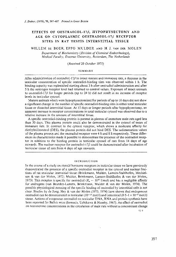
J. Endocr. (1976), 70, 397-407 Printed in Great Britain
EFFECTS OF OESTRADIOL-17P, HYPOPHYSECTOMY AND
AGE ON CYTOPLASMIC OESTRADIOL-17p RECEPTOR
SITES IN RAT TESTIS INTERSTITIAL TISSUE
WILLEM DE BOER, EPPO MULDER AND H. J. VAN DER MOLEN
Department of Biochemistry (Division of Chemical Endocrinology), Medical Faculty, Erasmus University, Rotterdam, The Netherlands
(Received 20 October 1975)
SUMMARY
After administration of oestradiol-!? j3 to intact mature and immature rats, a decrease in the testicular concentration of specific oestradiol-binding sites was observed within 1 h. The binding capacity was replenished starting about 3 h after oestradiol administration and after 5 h the oestrogen receptor level had returned to control values. Exposure of intact animals to oestradiol-17 j3 for longer periods (up to 24 h) did not result in an increase of receptor levels in testicular cytosol.
Mature animals which were hypophysectomized for periods of up to 10 days did not show a significant change in the number of specific oestradiol-binding sites in either total testicular tissue or dissected interstitial tissue. At 15 days or longer periods after hypophysectomy, an apparent increase in receptor concentrations in total testicular cytosol was observed due to a relative increase in the amount of interstitial tissue.
A specific oestradiol-binding protein is present in plasma of immature male rats aged less than 30 days. This plasma protein could also be demonstrated in the cytosol of testes of immature rats. In contrast to the cytosol receptor, which shows a moderate affinity for diethylstilboestrol (DES), the plasma protein did not bind DES. The sedimentation values of the plasma protein and the oestradiol receptor were 4 Sand 8 S respectively. These differences in characteristics made it possible to demonstrate the presence of the oestradiol receptor in addition to the binding protein in testicular cytosol of rats from 14 days of age onwards. The nuclear receptor for oestradiol-17 j3 could be demonstrated after incubation of testicular tissue of rats from 4 days of age onwards.
INTRODUCTION
In the course of a study on steroid hormone receptors in testicular tissue we have previously demonstrated the presence of a specific oestradiol receptor in the cytosol and nuclear fractions of rat testicular interstitial tissue (Brinkmann, Mulder, Lamers-Stahlhofen, Mechielsen & van der Molen, 1972; Mulder, Brinkmann, Lamers-Stahlhofen & van der Molen, 1973). This receptor is specific for oestradiol (Ka = 1010 1/mol) and has a negligible affinity for androgens (van Beurden-Lamers, Brinkmann, Mulder & van der Molen, 1974). The possible physiological meaning of the specific binding of oestradiol by interstitial cells is not clear. Studies by de Jong, Hey & van der Molen (1973, 1974) have shown that endogenous oestradiol can be demonstrated in testicular (l0- 10 mol /I) and interstitial (0·5-1 x IQ- 9 mol/!) tissue. Actions of exogenous oestradiol on testicular DNA, RNA and protein synthesis have been reported for Balb/c mice (Samuels, Uchikawa & Huseby, 1967). An effect of oestradiol on testosterone concentrations in the circulation of male rats without a concomitant change
397

398
W. DE BOER, E. MULDER AND H. J. VAN DER MOLEN
in luteinizing hormone (LH) levels, which might imply a direct effect of oestradiol on steroidogenesis in testicular tissue, has been observed. (Danutra, Harper, Boyns, Cole, Brownsey & Griffiths, 1973; Danutra, Harper & Griffiths, 1973; Chowdhury, Tcholakian & Steinberger, 1974; Tcholakian, Chowdhury & Steinberger, 1974).
It is now widely accepted that steroid hormones exert their effects through an interaction with subcellular receptors. After binding of the hormone to cytoplasmic receptors the receptor~hormone complex is translocated into the nuclear fraction and becomes bound to the chromatin. This interaction between the receptor-hormone complex and the chromatin might further regulate gene expression. Thus the hormonal control of target cells could depend both on variations in hormone concentrations and on changes in the amount of receptor proteins.
The purpose of the present study was to investigate the testicular concentration of receptor sites for oestradiol-I7;J under various conditions. This included the influence of oestradiol-17 ;J administration on the concentration of cytoplasmic receptor sites, the effect of gonadotrophins in the regulation of receptor concentrations and the ontogeny of the testicular oestradiol receptor in immature rats.
MATERIALS AND METHODS
Materials
[2, 4, 6, 7- 3H)Oestradiol-l7;J (sp.act. 105 Ci/mmol) was obtained from the Radiochemical Centre, Amersham. The radiochemical purity was verified by)hin-layer chromatography. Diethy!stilboestrol and oestradiol-17;1 were obtained from Steraloids Inc, Pawling, N.Y., U.S.A.
Preparation of subcellular fractions and incubation procedures
Mature (3 months old) and immature rats of the R-Amsterdam strain were used. The animals were injected with 500 ng (mature animals) or 100 ng (immature animals) oestradiol-17;J dissolved in 0·15 M-saline containing 2% ethanol where indicated. For studies in vitro decapsulated testicular tissue of immature rats was incubated in 2·0 ml Krebs-Ringer-bicarbonate buffer (pH 7·4), containing 0·2% glucose and 2x J0-8 M-(3H]oestradiol-17;J. Incubations were carried out at 3ZO C for 60 min in an atmosphere of 95% 0 2 :5% CO~. Interstitial tissue was obtained by wet dissection of decapsulated whole testicular tissue at 0 oc .(Christensen & Mason, 1965). The isolated tissue was homogenized in 10 mM-Tris-HCI buffer (pH 7·4), (I ml/g tissue) containing 1·5 mM~EDTA and 0·02% NaN3, with three strokes of a Potter-Elvehjem homogenizer at 1100 rev./min. The: homogenate was centrifuged at 105000 g for 60 min at 0 "C. The 105000 g supernatant (cytosol) was incubated with steroids for 16-24 hat 0 °C to reach saturation. For the preparation of nuclei, total testicular tissue was homogenized in ice-cold Tris~HCl buffer (pH 7·4), containing 1·5 m\fEDTA and 0·02% NaN3 with six strokes of a Potter-Elvehjem homogenizer at 1100 rev./ min. The homogenate was centrifuged at 500 g for 10 min at 0 cc. The 500 g pellet was washed once with homogenization buffer containing 0·2% Triton X-100 and twice with homogenization buffer. A nuclear extract was prepared by extraction of the nuclear fraction with 0·4 M-KCl in 10 MM Tris-HCl (pH 8·5), containing 1·5 mM-EDTA and 0·02% NaN3,
for 60 min at 0 oc, followed by centrifugation at 105000 g for 30 min at 0 "C.
Measurement of steroid binding
When cytosol was incubated with oestradiol-17;1 the steroid was bound to specific and nonspecific binding sites. In preliminary experiments it was shown that 2 x 10⁻⁸ M-oestradio! saturates all specific binding sites. The cytosol was incubated with either [3H]oestradiol-17;J
Oestradiol receptor in rat testicular tissue
to determine total binding or [3H]oestradiol-17 jJ plus a 200-fold excess of unlabelled oestradiol-17/J to determine non-specific binding (Williams & Gorski, 1973). The amount of specifically bound steroid was calculated by subtracting the value for (SH]oestradiol-17/J bound in the presence of unlabelled oestradiol-17ft (non-specifically bound oestradiol-17/J) from the value for total [3H]oestradiol-17jJ binding.
The bound and unbound steroid fractions after incubation of cytosol with steroids were separated by one of the following techniques.
Sephadex chromatography
Sephadex chromatography was performed as described by Williams & Gorski (1973). A 50 ,ul portion of incubated cytosol was layered on a wlumn (8 x 0·5 em) of Sephadex G-25 Superfine grade. The column was eluted with homogenization buffer and the excluded volume (bound radioactivity) was collected in a vial and the radioactivity was measured.
Gradient centrifugation
After incubation of the cytosol with steroid, 200 ,ul were layered on 5 ml of a 5-20 %sucrose density gradient prepared in homogenization buffer. After centrifugation in a Beckman L2-65B centrifuge at 0 "C for 16 hat 150000 g,v in a SW65 rotor, the bottom of the tube was pierced and 30 fractions were collected. Radioactivity was measured in each fraction. Nuclear extracts were layered on 5 ml of a 5-20% sucrose density gradient prepared in extraction buffer and run for 18 hat 260000 gav· Bovine serum albumin (BSA) and alcohol dehydrogenase (ADH) were used as markers with sedimentation values of 4·3 S and 7·6 S respectively.
Agar-gel electrophoresis
Agar-gel electrophoresis was performed essentially as described by Wagner (1972). A 50 ttl portion of incubated cytosol was applied on an agar plate (100 x 85 x 5 mm thick) kept at 0 oc (agar Noble; Difco, Detroit, Michigan, U.S.A.). After electrophoresis for 90 min at 130 rnA per plate (200-250 V) at 0 oc, the plate was cut into ten strips, each containing one sample, and each strip was divided into 20 fractions of 4 mm. To count the radioactivity, steroid from the individual agar fractions was dissolved by shaking for 12 hat room temperature in 10 ml Triton/scintillation fluid mixture.
Pretreatment with charcoal
Excess unbound steroid was in some experiments removed by adding 0·5 mg dextran-coated charcoal to 200 pl incubated cytosol. After mixing, the suspensions were incubated for 15 min at 0 oc and charcoal was removed by centrifugation for 10 min at 1200 g.
Protein determination
The protein content of the isolated cytosols was determined by the method of Lowry, Rosebrough, Farr & Randall (1951) with bovine serum albumin as standard. Generally the cytosol of mature rat testis contained 20-25 mg/ml and immature rat testicular cytosol contained 5-15 mg/mL
Estimation of oestradio!-17 P Oestradio\-17 jJ was measured by a radioimmunoassay technique as described by de Jong eta/. (1974).
Measurement of radioactivity
Radioactivity was measured in a Packard model3375 liquid scintillation spectrometer. The scintillation fluid consisted of a mixture of Triton X-100 (Rohm and Haas, Philadelphia, U.S.A.) and toluene (1 :2, v/v) containing 0·1 g POPOP (1,4-bis-(5-phenyloxazo\-2-y\)
399

400
W. DE BOER, E. MULDER AND H. J. VANDER MOLEN
benzene)/! and 4·8 g PPO (2,5~diphenyloxazole)/l (Packard Instrument S.A. Benelux, Brussels, Belgium).
RESULTS
Effects of exogenous oestradiol~ 17 fJ on cytoplasmic receptor levels
Receptor concentrations were estimated by Sephadex chromatography and sucrose gradient centrifugation. With both these techniques specific steroid binding is estimated by subtract~ ing the radioactivity bound in the presence of the competing non~radioactive oestradiol from the radioactivity retained in the absence of the competing steroid. It has previously been shown that these two methods give similar results (van Beurden-Lamers eta/. 1974) which are also in good agreement with the results estimated using a Scatchard-type plot obtained by charcoal assay (Brinkmann eta!. 1972).
Mature rats received a single injection of 500 ng oestradiol-17,8. One and 3 h after injection, the values obtained were significantly lower than the control levels, but levels 5 and 24 h after injection of oestradiol- I? ft were similar to those of the control (Fig. 1 a).
(a) (h)
120
~ 100 8 'o ~ ~ rf-r} "' 80
"' ·u
~ 60
~ ~ 40 :0 w ~
r r rf r
+~ ~ ·u
20 ~ ~
0 0 3 5 24 5 24
Time (h)
Fig. 1. Effect of oestradiol-17# on cytoplasmic receptor concentration. Rats were exposed to a single dose of oestradiol-17/l for various periods. Testicular cytosol was incubated for 24 hat 0 °C with 2 x IO-' M-["H]oestradiol-17(1 or with 2 x IQ-• M-["H]oestradiol-17(1 in the presence of 4 x IQ-1
M·oestradiol-17(1. After incubation, binding was determined by Sephadex G-25 chromatcgraphy and sucrose density gradient centrifugation. Specific binding is expressed as a percentage of control values ±s.n. for rats not injected with oestradiol-17(1 (n = 4). (a) Injection of mature rats with 500 ng oestradiol-17(1. (b) Injection of immature rats with 100 ng oestradiol·17(l.
Immature rats (23 days) were injected with 100 ng oestradiol-17fi'. Within 1 h after oestradiol administration, receptor levels decreased significantly; levels similar to those of the control were observed again at 5 and 24 h after hormone injection (Fig. I b).
Binding of [3H]oestradiol to cytoplasmic receptor molecules was measured after incubation at 0 oc. At this temperature only free receptor sites are estimated. Receptor sites which were already occupied by unlabelled oestradiol remain unaffected due to the slow dissociation of the receptor-hormone complex at 0 °C. From other data (van Beurden·Lamers eta!.
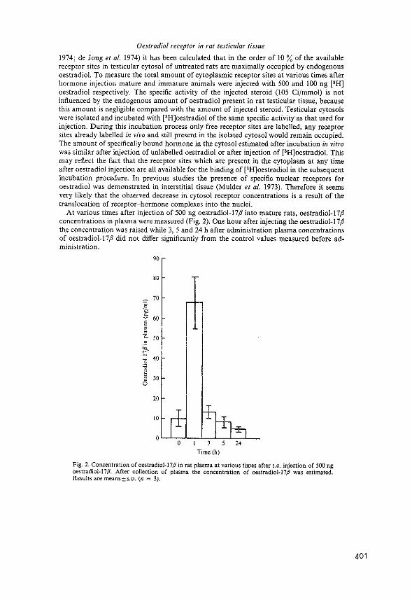
Oestradiol receptor in rat testicular tissue
1974; de Jong eta/. 1974) it has been calculated that in the order of 10% of the available receptor sites in testicular cytosol of untreated rats are maximally occupied by endogenous oestradiol. To measure the total amount of cytoplasmic receptor sites at various times after hormone injection mature and immature animals were injected with 500 and 100 ng [3H] oestradiol respectively. The specific activity of the injected steroid (105 Ci/mmol) is not influenced by the endogenous amount of oestradiol present in rat testicular tissue, because this amount is negligible compared with the amount of injected steroid. Testicular cytosols were isolated and incubated with [3H]oestradiol of the same specific activity as that used for injection. During this incubation process only free receptor sites are labelled, any receptor sites already labelled in vil•o and still present in the isolated cytosol would remain occupied. The amount of specifically bound hormone in the cytosol estimated after incubation in vitro was similar after injection of unlabelled oestradiol or after injection of [3H]oestradiol. This may reflect the fact that the receptor sites which are present in the cytoplasm at any time after oestradiol injection are all available for the binding of [3H]oestradiol in the subsequent incubation procedure. In previous studies the presence of specific nuclear receptors for oestradiol was demonstrated in interstitial tissue (Mulder eta!. 1973). Therefore it seems very likely that the observed decrease in cytosol receptor concentrations is a result of the translocation of receptor~hormone complexes into the nuclei.
At various times after injection of 500 ng oestradiol-17,8 into mature rats, oestradiol-17,8 concentrations in plasma were measured (Fig. 2). One hour after injecting the oestradiol-17,8 the concentration was raised while 3, 5 and 24 h after administration plasma concentrations of oestradiol-17,8 did not differ significantly from the control values measured before administration.
90
80
70 E
" -= 60 0 0 ~ 0
Q_ 50 c
~
0 40 '3 • 8 30
20
10
0 0 ,.
Time(h)
Fig. 2. Concentration of oestradiol-17P in rat plasma at various times after s.c. injection of 500 ng oestradiol-17P. After collection of plasma the concentration of oestradiol·17P was estimated. Results are means±s.o. (n = 3).
401

402
W. DE BOER, E. MULDER AND H.J. VANDER MOLEN
Effect of hypophysectomy on cytoplasmic receptor levels
At various times after hypophysectomy receptor concentrations in total testicular tissue and interstitial tissue were determined. Up to 15 days after hypophysectomy no change could be observed in oestradiol receptor concentration in total testicular tissue (Fig. 3). Similar results were obtained for cytosol receptor concentrations in dissected interstitial tissue isolated from rats l-10 days after hypophysectomy. For periods longer than 15 days after hypophysectomy an increase in receptor concentrations in total testicular tissue was ob~ served (Fig. 3.).
40
0 10 20 30 40 Time (days)
Fig. 3. Effect of duration of hypophysectomy on oestradiol-binding capacity in rat testicular cyto~ sol. Testicular cytosol was incubated for 16 h at 0 °C with either 2 x 10-• M-(lH]oestradiol-17,8 or 2 X IQ-• M-[ 3H]oestradiol-17,8 in the presence of 4 X lQ-7 M-oestradiol-17,8. After incubation binding was determined by Sephadex G-25 chromatography. Each point represents the mean ( ± s.o.) of four different animals. Value at day 38 is the mean of two estimations.
Oestradiol binding sites in plasma and testicular cytosol of immature rats
Plasma of immature rats 4--35 days old was incubated with either pH]oestradiol-17,8 or PH]oestradiol-17,8 in the presence of a 200-fold excess of non-radioactive oestradiol-17,8. Specific binding was determined using agar-gel electrophoresis (Table I) or sucrose density gradient centrifugation (Fig. 4a). High levels of a specific oestradiol-binding protein with a sedimentation value of 4S were demonstrated immediately after birth while decreasing levels were measured during the onset of pubescence. No specific binding could be demonstrated in plasma of 30-day-old rats.
When testicular cytoplasm of 20-day-old rats was incubated with [3H)oestradiol-17 fJ and analysed for binding using sucrose density gradients at low ionic strength two binding proteins with different sedimentation values of 8 and 4S respectively could be demonstrated (Fig. 4b). Binding of [3H]oestradiol-17 ,8 to the 8 S component was blocked by DES and by oestradiol-17,8 whereas DES did not affect the 4S binding (Fig. 4b). Thus it is possible to distinguish between the cytoplasmic oestradiol receptor and the plasma binding protein using the differences in sedimentation value and in binding affinity for DES. After incubation
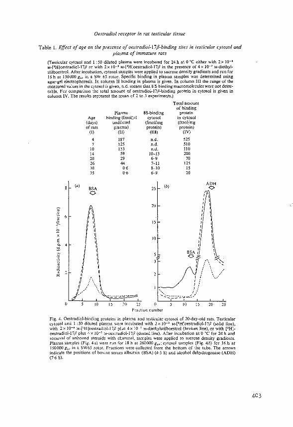
Oestradiol receptor in rat testicular tissue
Table I. Effect of age on the presence of oestradiol-17jl-binding sites in testicular cytosol and plasma of immature rats
(Testicular cytosol and I :50 diluted plasma were incubated for 24 hat 0 °C either with 2x IQ- 9
M-["H]oestradiol-17,8 or with 2 x 10-• M-['H]oestradiol-17,8 in the presence of 4 x IO-' M-diethylstilboestrol. After incubation, cytosol samples were applied to sucrose density gradients and run for 16 hat 150000 g.v in a SW 65 rotor. Specific binding in plasma samples was determined using agar-gel electrophoresis. In column II binding in plasma is given. In column Ill the range of the measured values in the cytosol is given, n.d. means that 8 S binding macromolecules were not detectable. For comparison the total amount of oestradiol-17,8-binding protein in cytosol is given in column IV. The results represent the mean of 2 to 3 experiments.)
0
' E " 4 ::! " :~ 6 i3 ;;.
0
' I I
' I , __
Age (days) of rats
({)
4 7
10 14 20 26 30 )5
BSA 0
r, 'I I 1
' ' ' ' '
10
Plasma binding (fmol/.ul
undiluted plasma)
(Il)
187 125 133
59 29 44 0·6 0·6
15 20 25
8S-binding cytosol
(fmol/mg protein)
(III)
n.d. n.d. n.d.
10-13 6-9 7-11 8-10 6-9
25
20
15
10
(b)
Total amount of binding
protein in cytosol (fmol/mg protein)
(IV)
525 510 110 200
70 125
15 20
ADH 0
r I I
' I ' I
' ' ' ' I 1 I
' ' ' I ' I ' I ' I ' I ' '
BSA t \
J{/\) I'
0
• I ,' '..::.""::::::.:.·..:;..,_,_:.;/
10 15 20 25 Frac·tion number
Fig. 4. Oestradiol-binding proteins in plasma and testicular cytosol of 20-day-old rats. Testicular cytosol and l :50 diluted plasma were incubated with 2x to-• M-PH]oestradiol-17,8 (solid line), with 2 x 10-• M-["H]oestradiol-l7ji plus 4 x 10-' M-diethylstilboestrol (broken line), or with ["H]oestradiol-17;3 plus 4 x JO·' M-oestradiol-17,8 (dotted line). After incubation at 0 °C for 24 h and removal of unbound steroids with charcoal, samples were applied 10 sucrose density gradients. Plasma samples (Fig. 4a) were run for 18 hat 260000 g,.,.; cytosol samples {Fig. 4b) for 16 hat 150000 g",. in a SW65 rotor. Fractions were collected from the bottom of the tube. The arrows indicate the positions of bovine serum albumin (BSA) (4·3 S) and alcohol dehydrogenase (ADH) (7·6 S).
403
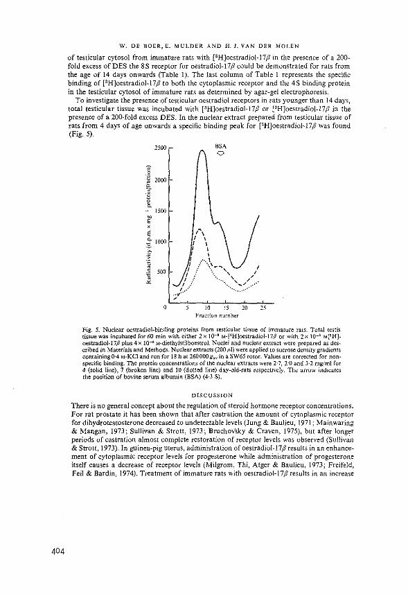
404
W. DE BOER, E. MULDER AND H. J. VANDER MOLEN
of testicular cytosol from immature rats with (3H]oestradiol-17P in the presence of a 200-fold excess of DES the 8 S receptor for oestradiol-17 P could be demonstrated for rats from the age of 14 days onwards (Table I). The last column of Table l represents the specific binding of f3H]oestradiol-17 P to both the cytoplasmic receptor and the 4S binding protein in the testicular cytosol of immature rats as determined by agar-gel electrophoresis.
To investigate the presence of testicular oestradiol receptors in rats younger than 14 days, total testicular tissue was incubated with (3H]oestradiol-17P or (3HJoestradiol-17;J' in the presence of a 200-fold excess DES. In the nuclear extract prepared from testicular tissue of rats from 4 days of age onwards a specific binding peak for [3H]oestradiol-17 P was found (Fig. 5).
2500
0 0
·~ 2000
" ? .• i ; 1500 ~
E
' E ~ 1000
"' ~ :~ 1l 0 500 .;;
'" 0 10
BSA 0
15 Fraction numher
10 25
Fig. 5. Nuclear oestradiol-binding proteins from testicular tissue of immature rats. Total testis tissue was incubated for 60 min with either 2 x 10-~ M-["H]oestradioi-17/J or with 2x 10-s M[~H]oestradiol-17/1 plus 4 x w-• M-diethylstilboestrol. Nuclei and nuclear extract were prepared as described in Materials and Methods. Nuclear elttracts (200;d) were applied to sucrose density gradients containing0·4 M-KCl and run for 18 hat 260000gav in a SW65 rotor. Values are corrected for nonspecific binding. The protein concentrations of the nuclear extracts were 2·7, 2·0 and 3·2 mg/ml for 4 (solid line), 7 (broken line) and 10 (dotted line) day-old-rats respectively. The arrow indicates the position of bovine serum albumin (BSA) (4·3 S).
DISCUSSION
There is no general concept about the regulation of steroid hormone receptor concentrations. For rat prostate it has been shown that after castration the amount of cytoplasmic receptor for dihydrotestosterone decreased to undetectable levels (Jung & Baulieu, 1971; Mainwaring & Mangan, 1973; Sullivan & Strott, 1973; Bruchov'sky & Craven, 1975), but after longer periods of castration almost complete restoration of receptor levels was observed (Sulliv<in & Strott, 1973). In guinea-pig uterus, administration of oestradiol-17;1 results in an enhancement of cytoplasmic receptor levels for progesterone while administration of progesterone itself causes a decrease of receptor levels (Milgram, Thi, Atger & Baulieu, 1973; Freifeld, Feil & Bardin, 1974). Treatment of immature rats with oestradiol-17 p results in an increase
Oestradiol receptor in rat testicular tissue
in the level of uterine receptors for oestradiol-17jJ (Sarff & Gorski, 1971). The present data indicate that administration of lOO and 500 ng unlabelled oestradiol-l7jJ to immature and mature rats respectively caused a rapid decrease of oestradiol-17jJ receptor sites available in testicular cytoplasm (Fig. 1). The time-course of the disappearance and reappearance of cytoplasmic receptor sites after oestradiol-l7jJ administration closely parallels the entry of oestradiol into and its removal from the blood circulation. Injection of [3H]oestradiol-17jJ and the subsequent incubation of cytosol with [3H]oestradiol-17jJ at ooc resulted in a similar decrease of receptor levels as measured after injection of unlabelled oestradiol-17jJ. Therefore the observed decrease in the amount of specifically bound radioactivity must be the result of the disappearance of receptor molecules from the cytoplasm. Previously the existence of a nuclear form of oestradiol-17 jJ receptor in interstitial tissue has been demonstrated (Mulder et a!. 1973). For uterine tissue Williams & Gorski (1972) observed an equilibrium between the concentration of receptor-bound steroid in the cytosol and receptor-bound steroid in the nuclei. Therefore the observed changes in testicular receptor concentration after hormone injection may also reflect the translocation process of hormone molecules into the nuclei. A comparison of the processes of nuclear translocation and cytoplasmic restoration of receptor molecules between testicular tissue and other oestrogensensitive tissues shows several differences. In uterine tissue of immature rats the lowest cytoplasmic receptor levels have been measured 3~6 h after administration of I OOng oestradiol-17jJ (Sarff & Gorski, 1971; Cidlowski & Muldoon, 1974). Control levels were reached only after 12 h (Sarff & Gorski, 1971). Administration of I pg oestradiol-17jJ to adult female rats resulted in a maximal depletion of cytoplasmic receptor molecules in pituitary, hypotha· lam us and uterus within 1 h. The reappearance of receptor molecules in uterine tissue and pituitary occurred at 15 h after injection reaching levels slightly below those of the control. In the hypothalamus, however, a plateau (60% of the control value) was reached 5 h after injection. In addition an enhancement of cytoplasmic receptor concentrations, as observed in uterine tissue 24 h after oestradiol administration, was not observed in testicular tissue. Thus testicular tissue belongs to the group of tissues which in response to oestradiol administration shows a depletion and subsequent reappearance of cytosol receptor molecules. The magnitude and the rate of both processes seems to vary among various tissues.
Six to 12 days after hypophysectomy in rats, plasma levels of gonadotrophins and testicular levels of oestradiol-17 jJ and testosterone are decreased (Gay & Sheth, 1972; de Jong, 1974). In the present experiments cytoplasmic levels of oestradiol-17jJ receptor were not affected after hypophysectomy. The initial decrease in oestradiol concentration in testicular tissue after hypophysectomy might have resulted in a proportional increase in unoccupied and therefore measurable receptor sites. A constant amount of measurable receptor sites might therefore accompany a decrease in the total receptor concentration (unoccupied and occupied sites) during the first days after hypophysectomy. It has been calculated, however, that only in the order of 10% of the total receptor sites in testicular cytosol of intact rats can be maximally occupied by endogenous oestradiol-l7jJ (van Beurden-Lamers et a!. 1974; de Jong eta!. 1974). Therefore it appears very unlikely that gonadotrophins, oestradiol-17 jJ and testosterone are important for the maintenance of the concentration of oestradiol-17.8 receptors in testicular cytoplasm. The increase in receptor sites which is found at 15 days or longer periods after hypophysectomy is an apparent increase, reflecting the relative increase in the amount of interstitial tissue.
Testicular cytosol of immature rats older than 14 days contained two binding proteins for oestradiol-17jJ with sedimentation values of 4S and 8S respectively (Fig. 4). This is similar to the observations for oestradiol-17jJ binding by uterine cytosols of immature rats (Michel, Jung & Baulieu, 1974; Somjen, Kaye & Lindner, 1974). The binding capacity of plasma of immature male rats for oestradiol-17jJ showed a decrease of more than two orders of
405

406
W. DE BOER, E. MULDER AND H. J. VANDER MOLEN
magnitude between birth and puberty, which paralleled the decrease in 4 S oestradiol~l7 ;J~ binding protein in testicular cytoplasm. It is therefore very likely that the oestradiol-17 jJbinding plasma protein is_responsible for (part of) the 4 S oestradiol-17;1 binding found in the testicular cytoplasm of immature rats. This suggestion is supported by the similar behaviour of both the plasma component and the 4 S testicular cytosol component towards diethylstilboestrol.
The very large concentration of 4 S oestradiol-17 ;J binding protein in testicular tissue made the quantitative measurement of receptor concentrations less accurate. Therefore small amounts of oestradiol-17 ;J receptor molecules, even if present in the testicular cytosol of rats younger than 14 days, might have escaped detection. However, the nuclear form of the testicular oestradiol-! 7fl receptor could be demonstrated in rats from 4 days of age onwards. Similar observations have been made for other receptors both for gonadal and adrenal steroids. The hepatic receptor for glucocorticoids could be demonstrated in liver cytosol of foetuses and immature rats (Feldman, 1974). The epididymal androgen receptor has been detected in 20-day-old rats (Calandra, Podesta, Rivarola & Blaquier, 1974) and the presence of oestradiol-17 fl-binding proteins with a sedimentation value of 8 S has been demonstrated in uteri of 5-day-old rats (Michel eta!. 1974).
The physiological meaning of the testicular oestradiol-17 fl receptor is not yet clear. It has been reported that administration of oestradiol benzoate (50 f.l8) to adult male rats results in a rapid decrease in testicular testosterone levels within 2 h without changes in LH levels in the circulation (Tcholakian eta!. 1974). The present results showed that injection of oestradiol-17ft into mature and immature rats caused a rapid decrease in testicular receptor levels in the cytoplasm. Therefore a possible direct inhibiting effect of oestradiol-17 ,8 on testosterone synthesis could be mediated by the binding of oestradiol-17 ;J to the cytoplasmic receptor and the subsequent binding of the receptor-hormone complex to the chromatin. Whether the endogenous concentration of oestradiol-17fl in mature rats is high enough to exert the same regulatory effect on testosterone synthesis is unknown. The presence of very large amounts of oestradiol-17 fl-binding protein in immature rat plasma which decreased during the onset of pubescence might be sufficiently high to trap all the endogenous oestrogen. This would indicate that the suggested regulatory role of oestrogens on steroid biosynthesis in testicular tissue can only be important if the oestradiol-17ft binding in testicular cytosol exceeds the binding in plasma.
This work was supported (in part) by the Foundation for Medical Research FUNGO, which is subsidized by the Netherlands Organization for the Advancement of Pure Research (Z.W.O.).
REFERENCES
van Beurden-Lamers, W. M. 0., Brinkmann, A. 0., Mulder, E. & van der Molen, H. J. (1974). Highaffinity binding of oestradiol-17 fJ by cytosols from testis interstitial tissue, pituitary, adrenal, liver and accessory sex glands of the male rat. Biochemical Journal140, 495-502.
Brinkmann, A. 0., Mulder, E., Lamers-Stahlhofen, G.J. M., Mechie!sen, M. J. & van der Molen, H. J. (1972). An oestradiol receptor in rat testis interstitial tissue. FEBS Letters 26, 301-305.
Bruchovsky, N. & Craven, S. (1975). Prostatic involution: effect on androgen receptors and intracellular androgen transport. Biochemical and Biophysical Research Communications 62, 837-E43.
Calandra, R. S., Podesta, E. J., Rivarola, M.A. & Blaquier, J. A. (1974). Tissue androgens and androphilic proteins in rat epididymis during sexual development. Steroids 24, 507-518.
Chowdhury, M., Tcholakian, R. & Steinberger, E. (1974). An unexpeCted effect of oestradio!-17/J on luteinizing hormone and testosterone. Journal of Endocrinology 60, 375-376.
Christensen, A. K. & Mason, N. R. (1965). Comparative ability of seminiferous tubules and interstitial tissue of rat testes to synthesize androgens from progesterone-4- 14C in vitro. Endocrinology 76, 646-656.
Cidlowski, J. A. & Muldoon, T. G. (1974). Estrogenic regulation of cytoplasmic receptor populations in estrogen-responsive tissues of the rat. Endocrinology 95, 1621-1627.
Oestradiol receptor in rat tesricular tissue
Danutra, V., Harper, M. E., Boyns, A. R., Cole, E. N., Brownsey, B. G. & Griffiths, K. (1973). The effect of certain stilboestrol analogues on plasma prolactin and testosterone in the rat. Journal of Endocrinology 57,207-215.
Danutra, V .• Harper, M. E. & Griffiths, K. (1973). The effect of stilboestrol analogues on the metabolism of steroids by the testis and prostate of the rat in vitro. Journal of Endocrinology 59, 539-544.
Feldman, D. (1974). Ontogeny of rat hepatic glucocorticoid receptors. Endocrinology 95, 1219-1227. Freifeld, M. L., Feil, P. D. & Bardin, C. W. (1974). The in vivo regulation of the progesterone receptor in
guinea-pig uterus: dependence on estrogen and progesterone. Steroids 23, 93-102. Gay, V. L. & Sheth, N. A. (1972). Evidence for a periodic release of LH in castrated male and female rats.
Endocrinology 90, 158-162. de Jong, F. H. (1974). Testicular oestradiol-17P, p. 52. Ph.D. Thesis, Erasmus University Rotterdam. de Jong, F. H., Hey, A. H. & van der Molen, H. J. (1973). Oestradiol in rat testis tissue: stimulation and
localization. Acta Endocrinologica Suppl. 177, 345. de Jong, F. H., Hey, A. H. & van der Molen, H. J. (1974). Oestradiol-17,8 and testosterone in rat testis
tissue: effect of gonadotrophins, localization and production in vitro. Journal of Endocrinology 60, 409-419.
Jung, I. & Baulieu, E. E. (1971). Nee-nuclear androgen receptor in rat ventral prostate. Biochimie 53, 807-817.
Lowry, 0. H., Rosebrough, N.H., Farr, A. L & Randall, R. J. (1951). Protein measurement with the Falin phenol reagent. Journal of Biological Chemistry 193, 265-275.
Mainwaring, W. I. P. & Mangan, F. R. {1973). A study of the androgen receptors in a variety of androgensensitive tissues. Journal of Endocrinology 59, 121-139.
Michel, G., Jung, I. & Baulieu, E. E. (1974). Two high affinity binding proteins of different specificity in the immature rat uterus cytosol. Sterc,ids 24, 437-449.
Milgram, E., Thi, L., Atger, M. & Bau1ieu, E. E. (1973). Mechanisms regulating the concentration and the conformation of progesterone receptor(s) in the uterus. Journal of Biological Chemistry 248, 6366-6374.
Mulder, E., Brinkmann, A. 0., Lamers-Stahlhofen, G. J. M. & van der Molen, H. J. (1973). Binding of oestradiol-17ft by the nuclear fraction of rat testis interstitial tissue. FEES Letters 31, 131-136.
Samuels, L. T., Uchikawa, T. & Huseby, R. A. (1967). Direct and indirect effects of oestrogens on the enzymes of the testis. In Ciba Foundation Colloquia on Endocrinology, vol.I6, pp. 211-232. EdsG. E. W. Wolstenholme & M. O'Connor. London: J. & A. Churchill, Ltd.
Sarff, M. & Gorski, J. (1971). Control of estrogen binding protein concentration under basal conditions and after estrogen administration. Biochemistry 10, 2557-2563.
SOmjen, G. J., Kaye, A.M. & Lindner, H. R. (1974). Oestradio\-17,8 binding proteins in the rat uterus: changes during postnatal development. Molecular and Cellufar Endocrinology l, 341-353.
Sullivan, J. N. & Strott, C. A. (1973). Evidence for an androgen-independent mechanism regulating the levels of receptor in target tissue. Journal of Biological Chemistry 258, 3202-3208.
Tcholakian, R. K., Chowdhury, M. & Steinberger, E. (1974). Time of action of oestradiol-17,8 on luteinizing hormone and testosterone. Journal of Endocrinology 63, 411-412.
Wagner, R. K. (1972). Characterization and assay of steroid hormone receptors and steroid-binding serum proteins by agargel electrophoresis at low temperatures. Hoppe-Seyler's Zeitschnft fiir physiologische Chemie 353, 1235-1245.
Williams, D. & Gorski, J. (1972). Kinetic and equilibrium analysis of estradiol in uterus: a model of binding sites distribution in uterine cells. Proceedings of the National Academy of Sciences of the U.S.A. 69, 3464--3468.
Williams, D. & Gorski, J. (1973). Preparation and characterization of free cell suspensions from the immature rat uterus. Biochemistry 12, 297-306.
407

Biochem. J. (1977) 162, 331-339 Printed in Great Britain
Comparative Study of Nuclear Binding Sites for Oestradiol in Rat Testicular and Uterine Tissue
DETERMINATION OF LOW AMOUNTS OF SPECIFIC BINDING SITES BY AN [3H]OESTRADlOL-EXCHANGE METHOD
By WILLEM DE BOER, JOAN DE VRIES, EPPO MULDER and HENK J. VANDER MOLEN
Department of Biochemistry (Division of Chemical Endocrilwlogy), Medical Faculty, Erasmus University R~zterdam, Rotterdam, The Netherlands
(Received 4 August 1976)
1. An [3H]oestradiol-exchange method was developed for the determination of oestradiolreceptor complexes in the nuclear fraction of immature rat testicular tissue. This method permits the determination of nuclear oestradiol-receptor sites in the presence of a relatively large amount of non-specific oestradiol binding present in testicular nuclei. After incubation of nuclei for 60min at 20°C in the presence of [3H]oestradiol with or without a 1000-fold excess of non-radioactive diethylstilboestrol, specific binding can be determined quantitatively in the KCl-extractable fraction, which contains 40% of the total receptor population. 2. The amount of receptor-bound steroid present in the 0.4M-KCI extract of testicular nuclei remained constant during incubation at 20"C. For uterine nuclei incubated with [3H]oestradiol at 37°C a shift of specifically bound [3H]oestradiol occurred from the KCI-soluble fraction to the KCI-insoluble fraction. 3. In intact rat testis, about 20% of the total receptor concentration was present in its nuclear form. Hypophysectomy 5 days before measurement resulted in a twofold decrease in the amount of receptor, which was present mainly in the cytosol. After injection of choriogonadotropin to intact animals, the total receptor concentration increased threefold. 4. This nuclear exchange method might be useful for determination of occupied specific receptor sites in tissues with relatively low contents of specific receptors.
Investigations of a biochemicai explanation for the mechanism of action cfsteroids have revealed the presence of specific n:ceptors for steroid hormones in target tissues. For uterine tissue it is well documented that oestradiol-receptor complexes migrate into the nucleus, bind to receptor sites on the chromatin and initiate a sequence of events which result in the hormone effect (Clark et al., 1973; O'Malley & Means, 1975). The in~erstitial tissue of the testis from rats of 4 days of age onwards contains a specific receptor for oestradiol (Brinkmann et al., 1972; Mulder et at., 1973; de Boer eta!., 1976). Oestradiol is endogenously produced in the testis and it appears that oestradiol concentrations in rat testis interstitial tissue (0.5-1 nM) are higher than those in seminiferous tubules (de Jong eta!., 1974), but it is still uncertain whether oestradiol has a physiological function in testicular tissue. The well-known decrease in testicular testosterone production after administration of oestradiol could be fully explained by a negative feedback of oestrogens on Jutropin (luteinizing hormone) secretion (W. M. 0. van Beurden-Lamers, unpub!ished
work), although it has been suggested that oestradiol might have a local intratesticulareffect on testosterone production.
Vol. 162
Possible direct effects of oestradiol mediated by the oestradiol receptor on Leydig-cell functions have not been studied in detail. On the assumption that an effect of steroid hormones is preceded by binding of the steroid-receptor complex in the nucleus, it was the purpose of the present study to investigate a possible translocation of the oestradiol receptor into the nucleus of testicular tissue of intact animals. In addition, the effects of oestradiol and administration of choriogonadotropin on the concentration and localization of the receptor were studied.
For a quantitative determination of nuclear receptor sites in intact animals it is necessary to use a method that can distinguish between the total available amount of receptor sites and the number of receptor sites that is occupied by endogenous oestradiol. Anderson et at. (1972) have developed an [3H]oestradiol-exchange method for determination of the number of nuclear oestradi0J-receptor sites in the
331

presence of endogenous oestradiol. Because a considerable amount of non-specific oestradiol-binding sites was observed in testicular tissue, it was necessary to modify this nuclear exchange method. We have therefore compared this modified hormoneexchange assay for determination of nuclear oestradiol receptors in testicular tissue with the assay described by Anderson eta!. (1972) for uterine tissue.
Materials and Methods
Preparations of animals and materials Immature male and female rats (21~3S-days-old) of
the R-Amsterdam strain were used in this study. In some experiments animals were hypophysectomized S days before the experiments. Male rats were injected subcutaneously with a solution of either SOOng of oestradiol-17ft or SOOng of [3H]oestradiol-17P in 0.2ml ofO.lSM-NaCl containing 2.S% (v/v) ethanol. The rats were decapitated lh after the injection. In experiments designed to examine the effect of human choriogonadotropin, a daily dose of SOi.u. of the hormone (WHO, 197S), dissolved in 0.1 m! of O.lSM-NaCI, was subcutaneously injected for 5 successive days. Testicular tissue was removed after decapitation of the animals and placed on a Petri dish on ice before incubation or isolation of nuclei.
[2,4,6,7-3HJ0estradiol-17 P (sp. radioactivity 85Ci/ mmol) was obtained from The Radiochemical Centre, Amersham, Bucks., U.K., and was examined for purity by t.l.c. Diethylstilboestrol and oestradiol-17 P were obtained from Steraloids Inc., Pawling, NY, U.S.A.
Incubation of whole tissues in vitro
One decapsulated testis or one uterus, stripped of adhering fat and mesentery, from immature rats was incubated with lOnM-oestradiol in 2.0ml of Krebs~ Ringer bicarbonate buffer, pH7.4, containing 0.2% glucose (Umbreit et al., 1964). Incubations were carried out in an atmosphere of Ol+C02 (95:5) for 60min at 32"C for testicular tissue and at 37"C for uterine tissue. In control studies tissues were incubated with either t0nM-(3H]oestradiol or 10nM[3H]oestradiol plus lO.uM-diethylstilboestrol.
Uterine and testicular tissue of immature rats con~ tains, in addition to the receptor, a plasma binding protein (oc-fetoprotein), which has a rather high affinity for oestrogens (Raynaud et a!., 1971; Aussel eta!., 1974; de Boer eta!., 1976). However, oestradiol bound to the plasma protein cannot be displaced by an excess of diethylstilboestrol. Therefore excess of diethylstilboestrol rather than oestradiol was used in incubations where the non-specific binding was estimated. The specific oestradiol binding to receptor molecules was then defined as the difference between the total binding and the non-specific binding and is expressed as fmol/mg of protein or as fmol/two testes
332
W. DE BOER AND OTHERS
or fmol/uterus. Previously it has been shown that the affinity of diethylstilboestrol for the testicular oestradiol receptor is one-quarter of the affinity of oestradiol (van Beurden-Lamers et a!., 1974). The amount of non-specific binding, obtained after incubation of mature rat testis and uterus in vitro, tissues that do not contain a-fetoprotein or other specific oestradiol-binding plasma proteins, did not differ if a 100-foldexcess of oestradiol or a 1000-foldexcess of diethylstilboestrol was used.
Preparations of nuclei
After isolation and incubation the testicular tissue was homogenized in 10 vo!. of 10 mM-Tris/HCl buffer, pH7.4, containing 1.5mM-EDTA and 0.02% NaN3
(TEN buffer) with six strokes of a Porter~Eivehjem homogenizer at 1 JOOrev.jmin. The homogenate was rehomogenized in an all-glass Potter~Elvehjem homogenizer and centrifuged at 500g for IOmin at O"C. The 500g pellet was washed once with 3 ml ofTEN buffer, twice with 3ml of TEN buffer containing 0.2% Triton X-100 and another time with 3m! of TEN buffer. Uterine tissue after incubation was suspended in 3m! ofTEN buffer and homogenized by hand with two strokes in an all-glass Kontes homogenizer. The homogenate was centrifuged at 800g for lOmin at 0°C and the pellet was washed three times with 3m! of TEN buffer. The resulting nuclear pellets were used either for the hormone-exchange assay of the nuclear oestradiol-binding sites or for immediate extraction with 0.4M-KCL
Hormone-binding assay
(a) Determination of nuclear binding, after incubation or injection with [3H]oestradiol. Isolated nuclei were extracted with 0.4M-KCl in TEN buffer, pH 8.5, during 60min at O?C. The extract was centrifuged at O"C for lOmin at 1500g, and a KC!-extractab!e and a residual nuclear fraction were obtained as the supernatant and pellet fractions. The bound radioactivity in the KCJ-extractable fraction was measured by sucrose-density-gradient centrifugation. The residual nuclear fraction was dissolved in 1 ml of I M-NaOH, and 100 .ul was counted for radioactivity after addition of 50.ul of 3 M-HCIO~.
(b) Hormone-exchange assay followed by KC! extraction of nuclei. Isolated nuclei were incubated in TEN buffer containing25% (v/v) glycerol for periods up to 120min at different temperatures with either I0nM-[3Hjoestradiol or 10nM-[3H]oestradiol plus lO.uM-diethylstilbo{-strol. After the incubation, nuclei were extracted as described under (a). To remove the excess of unbound steroid, the nuclear suspension in 0.4M-KC! was treated for lOmin with dextran-coated charcoal (final concentrations: 0.5% charcoal and 0.05% dextran T 300) at ooc and was subsequently centrifuged for 30minat l05000g,v. in a SW 65 rotor to remove the charcoaL Specific binding in the super~
1977
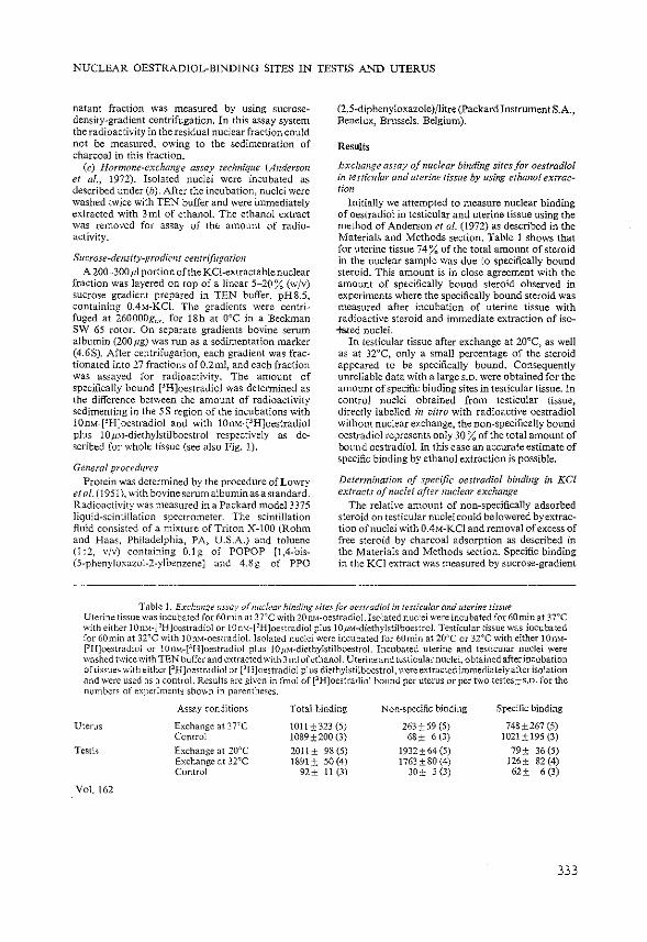
NUCLEAR OESTRADIOL-BINDING SITES IN TESTIS AND UTERUS
natant fraction was measured by using sucrosedensity-gradient centrifugation. In this assay system the radioactivity in the residual nuclear fraction could not be measured, owing to the sedimentation of charcoal in this fraction.
(c) Hormone-exchange assay technique (Anderson et af., 1972). Isolated nuclei were incubated as described under (h). After the incubation, nuclei were washed twice with TEN buffer and were immediately extracted with 3m! of ethanol. The ethanol extract was removed for assay of the amount of radioactivity.
Sucrose-density-gradient centrifugation A 200~300 .ul portion of the KCl-extractable nuclear
fraction was layered on top of a linear 5-20% (w/v) sucrose gradient prepared in TEN buffer, pH8.5, containing 0.4M-KCI. The gradients were centrifuged at 260000g~v. for ISh at 0°C in a Beckman SW 65 rotor. On separate gradients bovine serum albumin (200,ug) was run as a sedimentation marker (4.6S). After centrifugation, each gradient was fractionated into 27 fractions of0.2ml, and each fraction was assayed for radioactivity. The amount of specifically bound [3H]oestradiol was determined as the difference between the amount of radioactivity sedimenting in the 5S region of the incubations with 10n~-[3H]oestradiol and with 10nM-[3H]oestradiol plus lOpM-diethylstilboestrol respectively as described for whole tissue (see also Fig. 1).
General procedures Protein was determined by the procedure of Lowry
eta!. (1951), with bovineserumalbuminasastandard. Radioactivity was measured in a Packard model3375 liquid-scintillation spectrometer. The scintillation fluid consisted of a mixture of Triton X-100 (Rohm and Haas, Philadelphia, PA, U.S.A.) and toluene (l :2, v/v) containing 0.1 g of PO POP [1 ,4-bis(5-phenyloxazol-2-ylbenzene] and 4.Sg of PPO
(2,5-diphenyloxazole )/litre (Packard Instrument S.A., Benelux, Brussels, Belgium).
Results
Exchange assay of nuclear binding sites for oestradiol li1 testiwlar and uterine tissue by using ethanol extrac· lion
Initially we attempted to measure nuclear binding of oestradiol in testicular and uterine tissue using the method of Anderson et al. (1972) as described in the Materials and Methods section. Table 1 shows that for uterine tissue 74% of the total amount of steroid in the nuclear sample was due to specifically bound steroid. This amount is in close agreement with the amount of specifically bound steroid observed in experiments where the specifically bound steroid was measured after incubation of uterine tissue with radioactive steroid and immediate extraction of iso+&ed nuclei.
In testicular tissue after exchange at 20°C, as well as at 32°C, only a small percentage of the steroid appeared to be specifically bound. Consequently unreliable data with a large s.D. were obtained for the amount of specific binding sites in testicular tissue. In control nuclei obtained from testicular tissue, directly labelled in vitro with radioactive oestradiol without nuclear exchange, the non-specifically bound oestradiol represents only 30% of the total amount of bound oestradiol. In this case an accurate estimate of specific binding by ethanol extraction is possible.
Determination of specific oestradiol binding in KCl extracts of nuclei after nuclear exchange
The relative amount of non-specifically adsorbed steroid on testicular nuclei could be lowered by extraction of nuclei with 0.4M-KCI and removal of excess of free steroid by charcoal adsorption as described in the Materials and Methods section. Specific binding in the KCI extract was measured by sucrose-gradient
Table 1. Exr:hange assay of nuclear binding sites/or oes/radiol in testicular and uterine tissue Uterine tissue was incubated for 60min at 3TC with 20nM-oestradiol. Isolated nuclei were incubated for 60minat3TC with either lOnM·['H]ocstradiol or 10nM-[3H]oestradiol plus 10ttM-diethylstilboestrol. Testicular tissue was incubated for 60min at 32"C with JOnM-oestradiol.lsolated nuclei were incubated for 60min at 20"C or 32oC with either J0m1-[3H]oestradiol or 10n"1-[3 H]oestradiol plus 10tt).1·diethylstilboestrol. Incubated uterine and testicular nuclei were washed twice with TEN buffer and extracted with 3 ml of ethanol. Uterine and testicular nuclei, obtained after incubation of tissues with either [3H]oestradiol or [3H]oestradiol plus diethylstilboestrol, were extracted immediately after isolation and were used as a control. Results are given in fmol of [3 H]oestradiol bound per uterus or per two testes±s.o. for the nl1mbers of experiments shown in parentheses.
Assay conditions Total binding Non-specific binding Specific binding
Uterus Exchange at 37'C 1011 ± 323 (5) 263±59 (5) 748±267 (5) Control 1089±200(3) 68± 6 (3) 1021±195{3)
Testis Exchange at 20"C 2011± 98 (5) 1932± 64 (5) 79± 36 {5) Exchange at 32"C 1891± 50(4) 1763±80(4) 126± 82 (4) Control 92± 11 (3) 30± 5(3) 62± 6 (3)
Vol. \62
333

400! 0
'2 J
0
JJ l E ~
3
"' I :~ 1l J .2 v • "' ~~~ ,L __ ~--
I 10 20 Top
Fraction number
Fig. l. Binding of [3 H]oestradiol in rhe KC!-exrractab{e fraction of testicular nuclei
Testicular tissue was incubated for 60min at 32'C with either lOnM-eHJoestradiol (0) or lOnM-[31-J]oestradiol plus lOpM-diethylstilboestrol (o). Nuclei were isolated and extracted as described in the Materials and Methods section. A 200111 portion of the nuclear extracts was centrifuged on sucrose gradients for ISh at 260000g,,._. Specific binding of oestradiol in the KCI extract was estimated by calculating the difference in radioactive steroid sedimcnting in the 55 region of the gradient in the presence and in the absence of the excess of competing steroid. The arrow indicates the position of bovine serum albumin (4.65) run in a separate gradient.
centrifugation (Fig. 1). The amount of specifically bouod steroid recovered in the KCI-extractable frJ.ction represents 39±9% (s.D.) (11 =G) from the total amount of specific nuclear binding as determined by ethanol extraction (Table 1, line 5). To determine the amount of binding sites in the KCl extract, an almost complete exchange should be obtained at a temperature where no appreciable decomposition of binding sites occurs. The effect of different incubation temperatures on the stability of nuclear oestmdiol binding is shown in Fig. 2. In the absence of excess of [~H]oestradiol in the incubation medium, specific binding in both the KC1-extractable and residual nuclear fraction decreases considerably during 1 h of incubation (Fig. 2a). In the presence of oestradiol, dissociation of binding sites in the KCI-extractable
334
W. DE BOER AND OTHERS
fraction at 20°C was less than 5% (Fig. 2b). Dissociation of the binding sites in the residual nuclear fraction in the presence of added 3H-labelled steroid could not be measured, owing to the large amount of free and non-specifically bound oestradiol present in this fraction. For uterine tissue both the KCIextractable and the non-KCJ-extractable nuclear receptors (pellet) could be measured, because in contrast with testicular nuclei, it was possible to remove excess of unbound steroid by washing the uterine nuclei. In the presence of added [3H]oestradiol, a loss of binding sites in the KCI-extractable fraction was accompanied by an increase in the amount of non-KCI-extractable binding sites (Fig. 3b). As a consequence the total binding of oestradiol remained nearly constant. In the absence of [3 H]oestradiol the amount of both KCI-extractable and non-KClextractable binding sites in uterine nuclei were con· siderably decreased (Fig. 3a).
The time-course of the p H]oestradiol exchange by KCJ-extractable nuclear receptor sites in testicular tissue was studied after labelling of the tissue in vivo by injection of oestradiol. Fig. 4 shows that the hormone exchange in the KCI-extractable fraction is completed within I5min. The amount of specific binding measured after the exchange procedure [21.9fmol±2.7 (s.D.), n=I3] did not differ signifi· cantly from control values [22.4fmol±2.5 (S.D.), 11 = 3], which were obtained after injection of (3HJoestradiol in vivo. Therefore the hormoneexchange procedure in ~;itra during 60min of incubation of nuclei with [.JH]oestradiol at 20"C gives reliable results for oestradiol-receptor concentrations.
Determination of occupied nuclear oestradiol receptors in testicular tissue of intact immature rats and the effects of hypophysectomy on the amount of n11clear receptor sires
Fig. S(a) shows that the KCI-extractable fraction of testicular nuclei isolated from intact immature rats and subjected to the hormone-exchange procedure contains oestradiol-binding sites in the 5S region of the gradient. In contrast, in testicular tissue of 5-day hypophysectomized rats little or no specifically bound oestradiol could be demonstrated in the 5S region or sucrose gradients after application of the cxch:mge technique (Fig. 5b). Ta.ble 2 shows the number of nuclear receptor sites per two testes of intact and hypophysectomized animals and the total available amounts of nuclear rcceptot· sites measured after injection of rats with 500ng of non-radioactive oestradiol. As a result of hypophysectomy the total available number of receptor sites is decreased by a factor of two.
Effect of choriogonadotropill lreatmc'll/ on the mnotmt of specific nuclear receptor silt•s
Administration of 50i.u. of choriogonadotropin/
1977

NUCLEAR OESTRADIOL-BINDING SITES IN TESTIS AND UTERUS
JOO-
~ '00
(o) ' (b) l \ 2
8 \ " 0
= 'J 0
0 L ~ I 'o ~ C) 50~ 0 .10
]~\\ C) ~ .5
~ ~
" :ca '6 " iii
" JO 40
Temperature CC) Temperature CC)
Fig. 2. Effect of temperature on the stability of testicular nuclear oestradiol receptors Testicular tissue was incubated for 60min at 32"C with either lOnM-PH]ocstradiol or lOnM-r'H]oestradiol plus lOpM-diethylstilboestrol. Nuclei were isolated and nuclear suspensions were kept for 60min at different temperatures either without [3H]oestradioi (a) or with !Om.1-(3H]oestradiol (b) in the incubation medium. Thereafter nuclei were washed twice with TEN buffer and extracted with 0.4M-KC1 as described in the Materials and Methods section. In (a) specific binding in the KCl-extractab!e (A) and residual nuclear fraction (e) was calculated as a percentage oft he total nuclear binding (o) obtained after incubation of nuclei at ooc [61.3 ±4.9(4)fmolftwo testes]. In (b) specific binding in the KCl-extractable fraction is given as the percentage of the specific binding obtained after incubation of nuclei at 0°C [23.0±3.4(3)fmoljtwo testes]. Each value is the mean±s.n. of two to four determinations.
,00 100
~ (')
~ ~ " I I----~ 8 '· 0
0 ~ 0
0 so C) 50
~ T ±. ~ r-" 0
'6 1' '6 0 0
iii ~--......._ ....... ~---.....
iii
I ~ . ...... 0 ' ____ __l_ ... __ [
' ,L__c_ ___ ___l_ __ ~
" " ]0 •10 0 " 10 30 " Temperature ("C) Temperature ("C)
Fig. 3. E.f{ec/ of/emperature on the stabifity ofuterine nuclear oestradiol receptors Uterine tissue was incubated for 60min at 37"C with either 20nM-[3H]oestradiol or 20nM-["H]oestradiol plus 4pMdiethylstilbocstrol. Nuclei were isolated and nuclear suspensions were kept for 60rnin at different temperatures either withOIJt ['H]oestradiol (a) or with lOnM-['H]oestradiol (b) in the incubation medium. Thereafter nuclei were washed twice with TEN buffer and extracted \\·ith 0.4M-KC1 as described in the Materials ;1nd Methods section. Specific binding in the KC!-extractablc (.A) and re~idual nuclear fraction ( •) was c:llculatcd as the percentage of the Iota] nuclear binding (0) obtained after incubation of nuclei at O"C [985±231 (5)fmoljuterus]. Each value is the mean±s.D. of two to five detern1inations.
Vol. 162
335
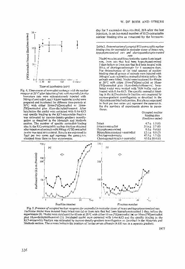
]' 30 r
,1 r1-1- i !l 0 I ~
" l g ~
J/ 0 , 0 ~
0 'i3 ' ~ If 0
~ ' oU~
0 15 30 " '" Ccncrcl
Time of incubation (min) Fig. 4. Time-course of oestradiol exchange with the nuclear receptor at 20aC after injection of rats with oestradiol in vivo
Immature rats were subcutaneously injected with 500ng of oestradiol, and 1 hlater testicular nuclei were prepared and incubated for different time-periods at 20°C with either 10IL\.f-[3H]oestradiol or lOnMPH]oestradiol plus IOp:.l-diethylstilboestrol. After incubation the nuclei were extracted with 0.4M-KCI and specific binding in the KCl-extractable fraction was estimated by sucrose-density-gradient centrifugation as described in the Materials and Methods section. The number of specific oestradiol-binding sites in the KCJ-extractable nuclear fraction obtained after injection ofanimals with 500 ng of [3H]oestradiol in vivo was used as a control. Results are expressed in fmol per two testes and represent the mean±s.o. obtained from three to four experiments.
100 r
!l J (o)
0 v ·B • 200 - - " ~ [ g ~ 3 .f IOO~N ·.g
• •• .,. "'
Fraction number
W. DE BOER AND OTHERS
day for 5 successive days resulted, 20h after the last injection, in an increased number of KCI-extractable nuclear binding sites as measured by the hormone-
Table 2. Determination of occupied KCI-extractable nuclear bindb1g sites for oestradiol in testicular tissue of intact rats, hypophysectomized rats and choriogonadotropin-treated
rats Nuclei were isolated from testicular tissue from intact rats, from rats that had been hypophysectomized 5 days before or from rats that had been treated with 50i.u. of choriogonadotropin for 5 successive days. For determination of the total amount of nuclear binding sites all groups of animals were injected with SOOng of non-radioactive oestradiol 60min before the animals were killed. Nuclei were incubated for 60min at 20°C with either 10nM-[3HJoestradiol or lOOM:· [3H]oestradiol plus lOpM-diethylstilboestrol. Incubated nuclei were washed with TEN buffer and extracted with 0.4M-KCJ. The specific oestradiol binding in the KCl-extractable fraction was measured by sucrose-gradient centrifugation as described in the Materials and Methods section. Results are expressed in fmol per two testes and represent the mean±s.D. for the numbers of experiments shown in paren~ theses.
Intact Intact+oestradiol Hypophysectomized Hypophysectomized+oestradiol Choriogonadotropin Choriogonadotropin+oestradiol
200
Occupied nuclear binding sites
(fmol/two testes)
4.7± 1.5(6) 21.8± 2.7 (8)
0.5 ± 0.6 (4) 12.4± 0.8 (3) 47.5± 1.7 (3) 65.0± 10.4 (4)
100 /(
I
I \oyr.;\f .. (:/ V\!~~
ol, - .. .1._·---·.-··· __ , ;
I 10 20 l"up
Fraction number Fig. 5. Presence of occupied nuclear receptors for oestradiol in testicular tissue of intact and hypophysectomized rats
Testicular nuclei were isolated from intact rats (a) or from rats that had been hypophysectomized 5 days before the experiments (b). Nuclei were incubated for60min at 20oC with either IOn).f-PH]oestradiol ($)or 10nM-[3HJoestradiol plus lOJtM~diethylstilboestrol (0). Incubated nuclei were extracted with 0.4M-KCI and the specific binding in the KCI-extractable fraction was estimated by sucrose-density~gradient centrifugation as described in .the Materials and Methods section. The arrows indicate the position of bovine serum albumin (4.6S) run in a separate gradient.
1977
336

NUCLEAR OESTRADIOL-BINDING SITES IN TESTIS AND UTERUS
exchange assay (Table 2). To measure the total number of receptor sites, testicular nuclei, isolated from choriogonadotropin-treated animals injected with SOOng of oestradiol 60min before death, were subjected to the exchange procedure. In choriogonadotropin-treated rats the testicular receptor concentration was three times that in intact animals (see Table 2).
Discussion
The accumulation of specific receptor-steroid complexes in target-cell nuclei appears to precede the observed effects of steroids on synthesis of new RNA and protein (van der Berget al., 1974; Lazier, 1975; Tsai eta/., 1975; Janne eta!., 1976; Hardin eta!., 1976). This has stimulated the interest in quantitative evaluations of nuclear steroid receptors as a possible indicator of physiological actions of steroids, even if in some cases the actual physiological importance of a steroid is not yet known. Such a situation exists for oestradiol in the rat testis. Oestradiol is produced and present in the testis, and there is now ample proof of the occurrence of specific cytoplasmic and nuclear receptors in rat testis interstitial tissue (Brinkmann eta!., 1972; Mulder et al., 1973; de Boer et al., 1976). However, there is no certainty about the possible action of oestradiol or the significance of the oestradiol receptors in testicular tissue. Therefore in the present study we attempted to investigate the biochemical behaviour of oestradiol in nuclei of testis interstitial tissue as a possible indicator of a physiological effect.
For our studies we required a reliable method for the quantitative determination of nuclear receptoroestradiol complexes. In order to obtain information about the amount of receptor that was already occupied by endogenous unlabelled steroid, we were interested in a method that could distinguish between the total amount (occupied and unoccupied) of oestradiol receptor and the amount occupied. Methods that have been used for labelling receptors in vitro with negligible amounts of radioactive ligand (followed by separation of unbound and receptorbound radioactive ligand) only give an indication of the number of unoccupied receptor sites.
Anderson eta!. (1972) were the first to introduce the so-called nuclear exchange method, which uses the exchange of 3H-labelled steroid with the endogenous receptor-bound steroid in the nuclear fraction for quantitative determination of the number of occupied nuclear receptor sites. In this method 3 HJabelled steroid, accumulated in the nucleus after the exchange procedure, is extracted with ethanol (total radioactivity T).~ In control experiments exchange is performed in the presence of 3H-labelled steroid and an excess of non-radioactive steroid. It is assumed that in this case the amount of 3 H-labelled
Vol.l62
steroid extracted with ethanol represents the nonspecifically bound steroid (N). Specific binding of [3 H]oestradiol to nuclear receptor molecules is defined
as the difference between total and non-specific binding (T-N). The practicability and reliability of this approach for measuring nuclear steroid-hormone receptors in several tissues has been reported (Anderson eta!., 1973; Hsueh et al., 1974; Sanborn et al., 1975; Teng & Teng, 1976). An indication of the total amount of receptor present should be obtained if, before the exchange labelling, tissues and animals are treated with excess of unlabelled steroid, so that all cytoplasmic receptor molecules are transferred to the nuclear fraction (Anderson eta!., 1972).
In initial experiments with testicular tissue, when we tried to use the conditions described by Anderson et al. (1972) for uterine tissue, it became evident that in testis nuclei, almost ali [3H]oestradiol (96%) was retained by non-specific binding sites (Table 1). Therefore measurements of occupied nuclear receptor sites in testicular tissue became inaccurate. For uterine tissue, which was used for comparison, 26% of the total amount of radioactive oestradiol in nuclei was retained by non-specific binding sites (Table 1). The amount of non-specifically bound steroid in the nuclear extract could be decreased if, after the exchange procedure, both the amount of non-specific binding sites and the amount of free steroid were decreased. This could be achieved if, after incubation with steroids, nuclei were extracted with 0.4M-KCI and subsequently treated with charcoal to remove excess of free steroid. Others have observed that the number of cytoplasmic oestradiol-binding sites measured by a charcoal-adsorption method in the presence of 0.4M-KCl was underestimated as a result of a partial dissociation of the hormone from the oestradiol-receptor complex (Peck & Clark, 1974). In our studies we measured the number of receptor sites in nuclei obtained after injection of animals with ['H]oestradiol. Under these conditions the measured number of receptor sites did not differ significantly from the number of sites obtained after injection of animals with non-radioactive oestradiol and by the exchange procedure (Fig. 4). Therefore it seems unlikely that, in the exchange procedure using charcoal adsorption, a considerable underestimation of the number of oestradiol-binding sites occurs. The modified method did result in much lower numbers of non-specific binding sites (1 0-20% of the total binding). This procedure has, however, the disadvantage that only in the KCI-extractable nuclear fraction can specific binding be measured. Because free steroid cannot be removed from the residual nuclear fraction, the difference between the total binding and the non-specific binding is too small to obtain reliable steroid-receptor measurements.
Of the total amount of receptor present, about 40% could be extracted from testicular nuclei with
337
0.4M-KC1; 60% was recovered in the residual nuclear fraction. Comparable results have been obtained after extraction of nuclei from kidney and hen oviduct (Best-Belpomme eta!., 1975; Janne et al., 1976), but the extraction efficiencies for chick liver nuclei and immature rat uterine nuclei were sUghtly lower (Lebeau et al., 1974; Mester & Baulieu, 1975). Even if only part of the total nuclear receptor population can be measured quantitatively in this way, information indicating the number of occupied receptor sites present in testicular nuclei of immature rats under different physiological conditions might be obtained. The KCl solution appears to extract a nuclear nonhistone fraction (Kostraba et al., 1975), and the KClextractable radioactivity may reflect the steroidreceptor complexes associated with non-histone proteins.
For testicular nuclei it was necessary to use an exchange temperature of 20°C. It was shown that the exchange of [3H]oestradio1 at this temperature was completed within 5min (Fig. 4). At higher temperatures a considerable Joss ofKCl-extractable receptor sites was observed during incubations of intact nuclei (Fig. 2). Also for other steroid-hom10ne receptors, degradation has been observed during the honnoneexchangeassay(Hsuehetal., 1974; Mester &Baulieu, 1975). A good correlation was observed, however, between the depletion of cytoplasmic receptor sites and the increase in nuclear receptor sites measured by hormone exchange in Miillerian-duct cells after oestradiol administration (Teng & Teng, 1976). A possible difference in proteolytic enzyme activity in the isolated nuclei might offer an explanation for these contrasting findings.
In the present study, occupied oestradiol receptors could be demonstrated in the KCJ-extractable fraction from nuclei of 25-day-old rats (4.7fmol/two testes). After administration of oestradiol the amount of receptor in the KCI extract increased to 21.8fmol/two testes (fable 2). As a consequence endogenous oestradiol in testicular tissue of intact immature rats could translocate 22% of the total receptor population into the nuclear fraction. For uterine tissue of intact immature rats, about 16% of the total oestradiol-receptor concentration was present in the nuclear fraction (Anderson et al., 1972). In our studies, hypophysectomy of inunature rats resulted, after 5 days, in a decrease in the number of KCl-extractable binding sites occupied with endogenous oestradiol (0.5 fmol/two testes). The total amount of KCl-extractable binding sites in hypophysectomized animals measured after oestradiol administration also decreased (12.4fmoljtwo testes). Therefore the receptor population in hypophysectomized rats is decreased to about 50% of the value in intact rats. The small amount of receptor sites occupied by endogenous oestradiol does reflect the fall of the testicular oestradiol concentration after hypo-
338
W. DE BOER AND OTHERS
physectomy. The decrease in testicular oestradiolreceptor content after hypophysectomy might be explained by a degeneration of Leydig cells or by a decrease in the number of these ~lis due to the disappearance oflutropin (Woods & Simpson, 1961; Gay &Sheth, 1972; Odell &Swerdloff, 1975). Similar observations have been made after hypophysectomy of mature female rats, which also results in a dramatic decrease in the number of available cytoplasmic receptor sites for oestradiol in the liver (Chamness ct al., 1975). The effect of choriogonadotropin was studied in order to investigate a possible role of gonadotropins on the number of oestradiol-receptor sites in testicular tissue. In intact immature rats, choriogonadotropin caused an increase in both the number of occupied receptor sites (47.5fmoljtwo testes) and the total amount of receptor sites (65.0fmol(two testes) in the KCl-extractable fraction. From these values it can be concluded that 73% of the total receptor population is present in the nuclear fraction and that choriogonadotropin administration results in a threefold increase in the receptor-site population per testis if compared with that in intact rats. The increase correlates very well with the observed increase in the number of Leydig cells in 21-day-old rats after choriogonadotropin treatment (15i.u.(day for 5 successive days) owing to mitosis and cellular differentiation (Chemes et al., 1976). It appears likely therefore that the number of oestradiol-receptor sites per Leydig cell does not change after choriogonadotropin treatment.
It is concluded that the number of total available and occupied oestradiol-receptor sites in the KCIextractable fraction of testicular nuclei is under the potential control of the hormonal environment. Whether the observed changes in the number of receptor sites are correlated with similar changes in the activities of the RNA and protein-synthesizing processes still requires investigation.
References Anderson, J., Clark, J. H. & Peck, E.J., Jr. (1972) Bio
chem. J. 126, 561-567 Anderson, J., Peck, E.J., Jr. & Clark, J. H. (1973) Endo
crinology 93, 7Il-717 Aussel, C., Uriel, J., Michel, G. & Baulieu, E. E. (1974)
Biochimie 56, 567-570 Best-Belpomme, M ., Mester,J., Weintraub, H. &Baulieu,
E. E. (1975) Eur. J. Bioc!u:m. 57, 537-547 Brinkmann, A. 0., Mulder, E., Lamers-Stahlhofen.
G. J. M., Mechielsen, M. J. & van der Molen, H. J. (1972) FEBS Lett. 26, 301-305
Chamness, G. C., Costlow, M. E. & McGuire, W. L. (1975) Steroids 26,363-371
Chemes, H. E., Rivarola, M. A. & Bergada, C. (1976) J. Reprod. Ferri!. 46, 279-282
Clark, J. H., Anderson, J. N. & Peck, E. J., Jr. (1973) in Receptors for Reproductive Hormones (O'Malley, B. W. & Means, A. R., eds.), pp. 15-59, Plenum Publishing Corp., New York
1977

NUCLEAR OESTRADIOL-BINDING SITES IN TESTIS AND UTERUS
de Boer, W., Mulder, E. & van der Molen, H. J. (1976) J. Endocrinol. 70, 397-407
de Jong, F. H., Hey, A. H. & van der Molen, H. J. (1974) J. Endocrinol. 60, 409-419
Gay, V. L. & Sheth, N. A. (1972) Endocrinology 90, 158-162
Hardin,J. W., Clark,J. H., Glasser, S. R. & Peck, E.J., Jr. (1976) Biochemistry 15, 1370-1374
Hsueh, A. J. W., Peck, E. J., Jr. & Clark, J. H. (1974) Steroids 24, 599-611
Janne, 0., Bullock, L. P., Bardin, C. W. & Jacob, S. T. (1976) Biochim. Biophys. Acta 418, 330-343
Kostraba, N.C., !vlontagna, R. A. & Wang, T. Y. (1975) J. Bioi. Chern. 250, 1548-1555
Lazier, C. (1975) Steroids 26, 281-298 Lebeau, M. C., Masso!, N. & Baulieu, E. E. (1974) FEES
Lett. 43, 107-111 Lowry, 0. H., Rosebrough, N.J., Farr, A. L. & Randall,
R. J. (1951) J. Bioi. Chern. 193, 265-275 Mester, J. & Baulieu, E. E. (1975) Biocltem.J.146, 617-623 Mulder, E., Brinkmann, A. 0., Lamers-Stahlhofen,
G. J. M. & van der Molen, H. J. (1973) FEBS Lett. 31, 131-136
Vol. 162
Odell, W. D. &Swerdloff, R. S. (1975)1. SteroidBiochem. 6, 853-857
O'Malley, B. W. & Means, A. R. (1975) Science 183, 6H420
Peck, E. J., Jr. & Clark, J. H. (1974) J. Steroid Biochem. 5, 327-328
Raynaud, J. P., Mercier-Bedard, C. & Baulieu, E. E. (1971) Steroids 18, 767-788
Sanborn, B. M., Steinberger, A. Meistrich, M. L. & Steinberger, E. (1975) J. Steroid Biochem. 6, 1459-1465
Teng, C. S. & Teng, C. T. (1976) Biochem. 1.154, 1-9 Tsai, S. Y., Tsai, M. J., Schwartz, R., Kalimi, M., Clark,
J. H. & O'Malley, B. W. (1975) Proc. Nat{. Acad. Sci. U.S.A. 72, 4228-4332
Umbreit, W. W., Burris, R. H. & Stauffer, J. F. (1964) in Marwmetric Techniques and Tissue Metabolism, 4th edn., p. 132, Burgess Publishing Co., Minneapolis
van Beurden-Lamers, W. M. 0., Brinkmann, A. 0., Mulder, E. & van der Molen, H. J. (1974) Biochem. J. 140, 495-502
van der Berg, J. A., Kooistra, T., Ab, G. & Gruber, M. (1974) Biochem. Biophys. Res. Commun. 61, 367-374
Woods, M. C. & Simpson, M. E. (1961) Endocrinology69, 91-125
339

Journal of Steroid Biochemistry 1977 (accepted for
publication)
Kinetics of in vitro binding of oestradiol in subcellular fractions of testicular and uterine tissue
characterization of oestradiol binding in testicular nuclei
Willern de Boer, Joan de Vries, Eppo Mulder and
H.J. van der Molen
Department of Biochemistry (Division of Chemical
Endocrinology), Medical Faculty, Erasmus University
Rotterdam, Rotterdam, The Netherlands
(Received 7 December 1976)
Summary
1. Oestradiol in rat testicular and uterine tissue is spe
cifically bound to nuclear receptor sites, which can be
separated in KCl-extractable nuclear and nuclear residual
(a nuclear fraction which resists KCl extraction) recep
tor sites.
2. The amount of 'extractable' nuclear binding sites for
oestradiol in testis could be increased by mild trypsin
treatment. Treatment of testicular nuclei with deoxycho
late or DNAse resulted in a decrease of residual receptor
sites and a concomitant increase of unbound oestradiol in
the •extractable• nuclear fraction.
3. The presence of KCN in vitro resulted in a relative in
crease in the number of oestradiol binding sites in the
nuclear residual fraction in both uterine and testicular
tissue; the number of binding sites in the KCl-extractable
fraction was not affected by KCN.
4. During in vitro incubations of testicular tissue the num
ber of oestradiol binding sites in the KCl-extractable
nuclear fraction reached a maximum and remained constant
after 30 min of incubation; the number of binding sites
in the nuclear residual fraction decreased after incuba
tion periods longer than 30 min.
5. During in vitro incubations of uterine tissue the number
of oestradiol binding sites in the KCl-extractable nu
clear fraction and the nuclear residual fraction after an
initial increase decreased to 50% of the maximal value
between 30 and 60 min of incubation.
6. It is concluded, that the testicular oestradiol receptor
shows certain characteristics comparable with those of
the uterine receptor. However, regarding the differences
in retention time of steroids in the nucleus, it seems
very unlikely that the oestradiol effect in uterus and
the oestradiol effect in testis, if present are mediated
by identical receptor mechanisms.
Introduction
It is now well accepted that the cytoplasm of target
cells for steroid hormones contains a specific binding pro
tein called a receptor. The complete sequence of events in
the response of a tissue to a steroid hormone is still un
known, but a postulated primary step is the interaction of
the steroid with its receptor in the cytoplasm of the tar
get tissue. The formed steroid-receptor complex migrates
into the nucleus and binds to acceptor sites on the chroma
tin which ultimately results in a response of the tissue to
the steroid via changes in RNA and protein synthesis
[1' 2' 3' .u . The rat testicular Leydig cell contains a cytoplasm re
ceptor for oestradiol which can be transported into the .nu
cleus and which binds to the chromatin under the influence
of endogenously produced oestradiol [5,6,7,~. Whether the
2
binding of oestradiol-receptor complexes to nuclear acceptor
sites results in a physiological effect in the Leydig cell
is still unclear [9].
In order to gain further insight in a possible function
of oestradiol receptors in the Leydig cell the processes of
translocation and nuclear binding of receptor-oestradiol
complexes in vitro in testicular tissue with similar pro
cesses in uterine tissue, a tissue which responds well to
oestrogen administration, were compared [10,1~. In the present study we have also investigated the ef
fects of trypsin, deoxycholate and DNAse treatment on the
nature of nuclear oestradiol binding sites in testicular
tissue. For uterine tissue it has been postulated that the
number of nuclear residual oestradiol receptor sites (the
fraction which resists KCl-extraction) determines the tissue
response to oestradiol [12]. Therefore the distribution of
KCl-extractable and nuclear residual binding sites was in
vestigated after incubation of uterine and testicular tissue
with oestradiol.
It has been reported that energy might be required for
the action of glucocorticoids and progesterone [13,14,15-,
16]. In this respect we have also studied the effect of
energy deprivation on the distribution of oestradiol binding
sites in KCl-extractable and nuclear residual fractions of
uterine and testicular tissue.
Experimental
Materials. Unlabelled oestradiol and diethylstilboestrol
(DES) were purchased from Steraloids Inc. Pawling, New York,
U.S.A. 3H-oestradiol (sp.act. 96 Ci/mmol) was purchased
from Radiochemical· Centre, Aroersham, U.K. The purity of the
steroids was determined by thin-layer chromatography. Tryp
sine (analytical grade) was obtained from Boehringer
Mannheim, West Germany, sodium deoxycholate from Merck,
Darmstadt, West Germany and DNAse from Sigma, St. Louis,
U.S.A.
3

Source of tissues and treatments. Immature (25-30 day
old) and mature (3 months old) rats of the R-Arnsterdam strain
were used in this study. Interstitial tissue of mature rat
testis was obtained by wet dissection after incubation in
vitro [17]. One decapsulated testis or one uterus stripped
of adhering fat and mesentery, from immature rats, was in
cubated in 2.0 ml Krebs Ringer bicarbonate buffer, pH 7.4,
containing 0.2% glucose. For mature rat testis 4.0 ml of
incubation medium was used per testis. Incubations were car
ried out for different time periods in an atmosphere of 95%
o2 : 5% co2 . In studies where the effect of KCN was inves
tigated tissues were incubated in Krebs Ringer bicarbonate
buffer, pH 7.4, containing 5 x 10- 4M KCN. Testicular tissue
was incubated at 32°C; uterine tissue at 37°c.
Tissues were incubated either with 2 x 10-SM 3H-oestra
diol (total binding) or with 2 x 10-SM 3H-oestradiol in the
presence of 4 x 10- 6M DES (non-specific binding). Specific
binding of 3H-oestradiol is defined as the difference be
tween the total binding and the non-specific binding.
Preparation of subcellular fractions. After incubation
total testicular tissue and interstitial tissue were homo
genized in 10 vol 10 mM Tris-HCl buffer, pH 7.4, containing
1.5 mM EDTA and 0.02% NaN 3 (TEN-buffer) with 6 strokes of
Potter-Elvehjem homogenizer at 1100 rev./min. The homoge
nate was rehomogenized in an all-glass Potter-Elvehjem ho
mogenizer and was subsequently centrifuged at 500 g for
10 min at 0°C. The 500 g pellet was washed once with TEN
buffer, twice with TEN-buffer containing 0.2% Triton X-100
and another time with TEN-buffer. Uterine tissue of imma
ture rats was homogenized in an Ultraturrax homogenizer for
10 sec, rehomogenized in a Potter-Elvehjem homogenizer and
centrifuged at 800 g for 10 min at 0°C. The 800 g pellet
was further washed and centrifuged at 800 g as described
for testicular tissue. The nuclear preparations thus obtai
ned appeared pure by phase contrast microscopy and had a
protein/DNA ratio in the range of 1.7-1.9. The isolated
4

pellets were used immediately after isolation for estimation
of nuclear binding. Where indicated the 500 g and 800 g su
pernatants of the testicular and uterine tissue respectively
were centrifuged at 105,000 g for 60 min to obtain the cyto
sol fraction.
Incubation of nuclei. In experiments concerning the na
ture of the oestradiol binding sites in testicular nuclei
the following procedure was used. After incubation of testis
tissue from mature rats in the presence of steroids for
60 min at 32°C interstitial tissue nuclei were isolated.
Nuclei obtained from the interstitial tissue of one testis
were suspended in 10 mM Tris buffer, pH 7.4 and incubated
with DNAse (150 "g/ml; 30 min at 20°C), trypsin (150 "g/ml;
30 min at 10°C} or deoxycholate (1% DOC, 30 min at 0°C) in
a volume of 1.0 ml. After incubation the nuclear suspensions
were centrifuged for 30 min at 105,000 g and specific bin
ding of 3H-oestradiol was measured in the supernatant
('soluble') and pellet ('nuclear residual') fraction as des
cribed below. A KCl-extractable nuclear fraction and a nu
clear residual fraction obtained after extraction of untrea
ted nuclei with 0.4M KCl served as a control.
Estimation of specific 3H-oestradiol binding. It was at
tempted to extract the 3H-oestradiol in the nuclear pellet
with 0.4M KCl in TEN-buffer, pH 8.5. The nuclear pellet was
mixed with an equal volume of 0.8M KCl in TEN-buffer, pH 8.5.
The final volume of the extraction buffer was adjusted to
800 ~1 with 0.4M KCl in TEN-buffer, pH 8.5. After incubation
for 60 min at 0°C the extract was centrifuged for 30 min at
105,000 g and an aliquot of the supernatant fraction was
analyzed on sucrose density gradients as described below
(KCl-extractable nuclear binding). The residual nuclear pel
let, from which no further 3H-oestradiol could be extracted
with KCl-TEN-buffer, was solubilized in 1 N NaOH and an ali
quot was counted for radioactivity after addition of an
equal volume of 3 N perchloric acid (residual nuclear bin-
5
ding). Specific binding in both fractions was estimated by
substracting the non-specific binding from the total binding
of 3H-oestradiol and was expressed as fmol/mg protein, pre
sent in each fraction. In the 105,000 g supernatants speci
fic cytosol binding was estimated by sucrose density gradient
centrifugation as described below.
Sucrose gradient centrifugation. A 200 ~l portion of the
KCl-extractable nuclear fraction was layered on top of a
linear 5-20% (w/v) sucrose density gradient prepared in TEN
buffer, pH 8.5, containing 0.4M KCl. Gradients were centri
fuged at 260,000 g for 18 h at 0°C in a Beckman SW65 rotor. av
Cytosol fractions (200-400 ~1) were layered on top of linear
5-20% (w/v) sucrose gradients prepared in TEN-buffer and gra
dients were centrifuged at 150,000 g for 16 h at 0°c. On av
separate gradients 200 ~g of bovine serum albumin (BSA) or
alcohol dehydrogenase (ADH) were run as sedimentation mar
kers (4.6S and 7.6S respectively). After centrifugation each
gradient was fractionated in 27 fractions of 0,2 ml each and
each fraction was assayed for radioactivity.
General procedures. Protein was determined by the proce
dure of Lowry et al. [1~ with bovine serum albumin as a
standard. Radioactivity was measured in a Packard model 3375
liquid scintillation spectrometer. The scintillation fluid
consisted of a mixture of Triton X-100 (Rohm and Haas,
Philadelphia, U.S.A.) and toluene (1:2 v/v) containing
0.1 g POPOP (1,4-bis-(5-pheny1oxazo1-2-yl) benzene)/1 and
4.8 g PPO (2,5-diphenyloxazol)/1 (Packard Instrument S.A.,
Benelux, Brussels, Belgium).
6

Results
Determination of two different types of specific oestra
diol binding sites in testicular nuclei. Immature rat tes
ticular tissue was incubated either with 10-SM 3H-oestradiol
or with 10-8M 3H-oestradiol in the presence of increasing
amounts non-radioactive DES (Fig. 1). Nuclei were isolated,
"' c -o c
-" ~
"' E c ~ u • "-
100
50
0
' ' ' ' ' .. ' '~ 0
' ' '
10
' ' .. ' '
nM DES 100 1000
Figure Determination of two different nuclear binding
sites for oestradiol.
Testicular tissue from immature rats was incubated for
60 min at 32°C either with 10-SM 3H-oestradiol or with 10-SM 3 . . . . H-oestrad~ol ~n the presence of LncreasLng amounts of non-
radioactive diethylstilboestrol. Nuclei were isolated and
extracted with 0.4M KCl. Binding of oestradiol in the KCl
extractable fraction and the nuclear residual fraction was
estimated as described in the methods section. The binding
obtained in these two nuclear fractions in the absence of
non-radioactive competitor is defined as the 100% value.
• - e nuclear extract; x --- x residual nuclear fraction.
7
extracted with 0.4M KCl and oestradiol binding sites present
in the KCl-extractable fraction and the nuclear residual
fraction (the fraction which resists extraction) was esti
mated.
In Fig. 1 it is shown that increasing amounts of non
radioactive DES decreased the binding of 3H-oestradiol in
both the KCl-extractable and the nuclear residual fraction.
A two and three fold excess non-radioactive DES reduced the
initial amount of binding by a factor two in the KCl
extractable fraction and nuclear residual fraction respec
tively.
In the order of 40% of the specifically bound steroid in
testicular nuclei was generally recovered in the KC1
extractable fraction; 60% resisted KCl extraction. The dis
tribution of specific binding sites in the two nuclear frac
tions did not change if nuclei were extracted several times
with 0.4M KCl. At O.SM and higher salt concentrations the
nuclear material became quite viscous and could not be se
dimented by centrifugation.
Nature of the oestradiol binding sites in testicular
nuclei. In order to investigate the nature of the oestradiol
binding sites in testicular nuclei, testes from mature rats
were incubated with oestradiol and the obtained nuclear sus
pension from interstitial tissue nuclei was incubated,
either with trypsin, DNAse or deoxycholate.
The results in Fig. 2 show that treatment of nuclei with
trypsin resulted in an increased amount of specifically
bound 3H-oestradiol in the soluble fraction. Further analy
sis of this soluble fraction on sucrose density gradients
revealed that 3H-oestradiol was specifically bound to a 4S
sedimenting macromolecule. If nuclei were treated with DNAse
or deoxycholate an oestradiol binding macromolecules with a
sedimentation value of SS was observed in sucrose density
gradients. This value is similar to the value obtained after
extraction of nuclei with 0.4M KCl. Treatment of nuclei with
either DNAse or deoxycholate did not result in a further
8
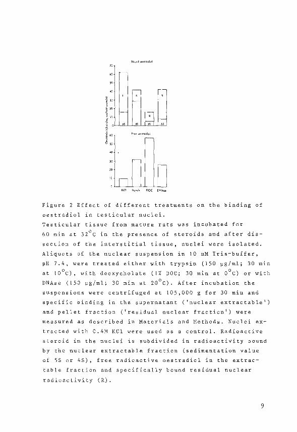
9oo"d O<!trodiol
" 00
~
• ? ., '" t " ' l w
1 ! " ~ 00 Fcoo ""'"~diol
j r-~
• '" r-
" "n
KCI ''YP"" DOC DNA,.,
Figure 2 Effect of different treatments on the binding of
oestradiol in testicular nuclei.
Testicular tissue from mature rats was incubated for
60 min at 32°C in the presence of steroids and after dis
section of the interstitial tissue, nuclei were isolated.
Aliquots of the nuclear suspension in 10 nM Tris-buffer,
pH 7.4, were treated either with trypsin (150 ~g/ml; 30 min
at !0°C), with deoxycholate ( 1% DOC; 30 min at 0°C) or with
DNAse (!50 1-1g/ml; 30 min at 20°C). After incubation the
suspensions were centrifuged at 105,000 g for 30 min and
specific binding in the supernatant ('nuclear extractable')
and pellet fraction ('residual nuclear fraction') were
measured as described in Materials and Methods. Nuclei ex
tracted with 0.4M KCl were used as a control. Radioactive
steroid in the nuclei is subdivided in radioactivity bound
by the nuclear extractable fraction (sedimentation value
of 55 or 4S), free radioactive oestradiol in the extrac
table fraction and specifically bound residual nuclear
radioactivity (R).
9

Uterus. Uterine tissue was incubated at 37°C for diffe
rent time periods after an initial incubation of 60 min at
0°C in the presence of steroids. Nuclei were isolated and
extracted with 0.4M KCl in order to obtain a KCl-extrac
table and nuclear residual fraction. Specifically bound
oestradiol in both nuclear fractions increased during the
time of incubation reaching a maximum after 30 min (Fig. 4).
After continued incubation the specific binding of oestra
diol in the KCl-extractable and nuclear residual fraction
declined reaching a plateau after 60 min. During the first
period of incubation the initial increase in nuclear oes
tradiol binding was accompanied by a decrease in the amount
of specifically bound oestradiol in the cytosol fraction.
After 60 min specific binding sites for oestradiol in the
cytosol fraction became undetectable.
Effect of energy deprivation on the distribution of spe
cifically bound 3H-oestradiol in testicular and uterine nu
clei. To determine the effect of energy deprivation on the
distribution of oestradiol binding sites, testicular and
uterine tissue were incubated in the presence of steroid
and 5xl0- 4M KCN. Tissues incubated in the presence of ste
roids but with the omission of KCN from the incubation me
dium served as control tissues. One hour after the addition
of KCN the number of specific oestradiol binding sites in
the nuclear residual fractions of both testicular (Fig. SA)
and uterine (Fig. SB) tissue were enhanced significantly
compared to controls. For testicular tissue a twofold in
crease was measured, for uterine tissue the increase was
slightly lower (1.6 fold). The addition of KCN had no spe
cific effect on the specific binding of oestradiol in the
KCl-extractable nuclear fraction of both tissues.
12
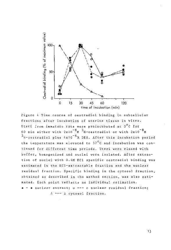
• ~ c > E 100 \ ~ E I ., I \
c \ / I 0 E '1/ I ~ I 0 \
t I "b. I
I "" ..
0> I
\ I
--F4 ·" 50 I ~ ................. " I \ c I .. ~~
.!l
' ..
0 0
'0 01
~ ~ ~ .... '--t>. 0 0
0 15 30 45 60 120 time of incubation (min)
Figure 4 Time course of oestradiol binding in subcellular
fractions after incubation of uterine tissue in vitro.
Uteri from immature rats were preincubated at 0°C for
60 min either with 2xl0-SM 3H-oestradiol or with 2xl0- 8M 3H-oestradiol plus 4xl0- 6M DES. After this incubation period
the temperature was elevated to 37°C and incubation was con
tinued for different time periods. Uteri were rinsed with
buffer, homogenized and nuclei were isolated. After extrac
tion of nuclei with 0.4M KCl specific oestradiol binding was
estimated in the KCl-extractable fraction and the nuclear
residual fraction. Specific binding in the cytosol fraction,
obtained as described in the method section, was also esti
mated. Each point reflects an individual estimation.
• - • nuclear extract; o --- o nuclear residual fraction;
~ --- ~ cytosol fraction.
13
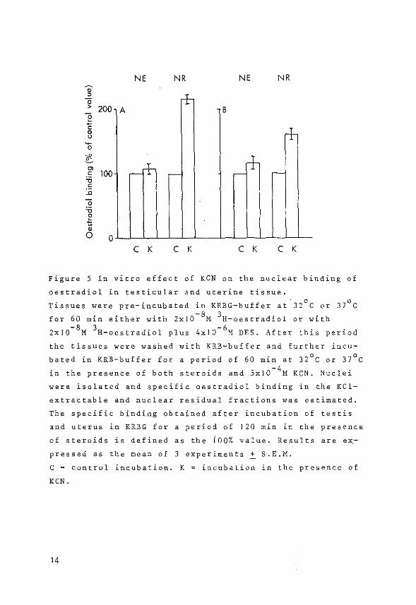
NE NR NE NR ~ , a + > 200 e A B
c 0 u + ~
0
~ ~ m c 100 -~ -- -~ r-~
.~ ~
.~ ~
~ ~
0 0 C K C K C K C K
Figure 5 In vitro effect of KCN on the nuclear binding of
oestradiol in testicular and uterine tissue.
Tissues were pre-incubated in KRBG-buffer at 32°C or 37°C
for 60 min either with 2xl0-SM 3H-oestradiol or with -8 3 -6
Zx!O M H-oestradiol plus 4xl0 M DES. After this period
the tissues were washed with KRB-buffer and further incu
bated in KRB-buffer for a period of 60 min at 32°C oi 37°C
in the presence of both steroids and 5x!0- 4M KCN. Nuclei
were isolated and specific oestradiol binding in the KCl
extractable and nuclear residual fractions was estimated.
The specific binding obtained after incubation of testis
and uterus in KRBG for a period of 120 min in the presence
of steroids is defined as the iOO% value. Results are ex
pressed as the mean of 3 experiments ~ S.E.M.
C = control incubation. K = incubation in the presence of
KCN.
14

Discussion
The testicular Leydig cell contains a receptor protein
which is specific for oestradiol [7,19]. This oestradiol
receptor has also been demonstrated in total testicular
tissue from immature rats [20] and in dissected interstitial
tissue of mature rats [5,20]. From the present results it
appears that two different fractions of specifically bound
oestradiol can be demonstrated iri testicular nuclei: one
fraction (40%) is extractable with 0.4M KCl, the other
fraction resists KCl-extraction. The number of specific
binding sites in the KCl-extractable fraction did not show
any increase if nuclei were extracted repeatedly or if nu
clei were extracted with concentrations of KCl above 0.4M.
The present observations are compatible with those for
steroid-hormone receptors in other tissues where nuclear
binding could also be distinguished in a KCl-extractable
and a nuclear residual form [12,13,21,22,24 1 25,26].
The role of different types of nuclear binding sites for
steroid hormones is not yet clear. Evidence has been pre
sented, however, that the residual nuclear binding proteins
for oestradiol and testosterone might be important in the
mechanism of action of steroid hormones [12,27 1 28]. The de
monstration of two different nuclear binding sites for oes
tradiol in Leydig cells raises the possibility that actions
of oestradiol in these cells can also be mediated via a si
milar receptor mechanism.
The in vitro incubations of testicular tissue with 3H
oestradiol showed that both types of nuclear oestradiol bin
ding were similarly affected by increasing amounts of non
radioactive DES (Fig. 1). The addition of a 2-3 fold excess 3
of non-radioactive DES caused a 50% reduction of the H-
oestradiol binding in both nuclear fractions. This observa
tion is in good agreement with those obtained with the cy
toplasmic oestradiol receptor [ 19].
Different approaches were used to characterize the nature
of the nuclear oestradiol binding sites in testicular tis-
15

sue. Mild trypsin treatment of nuclei, which contained oes
tradiol-receptor complexes, caused a release of binding si
tes for oestradiol with a sedimentation value of 4S. This
value is different from the sedimentation value of SS which
is normally observed for KCl-extractable nuclear oestradiol
receptors. Receptor complexes with a sedimentation value of
4S were also solubilized after treatment of liver and ute
rine nuclei with trypsin [22-,23].
It has been reported previously that trypsin treatment
of the oestradiol receptor from cytosols of calve uteri
resulted in a loss of binding affinity of the receptor for
DNA and it has been suggested [29] that trypsin treatment
releases part of the receptor molecule, which contains the
chromatin-binding unit, whereas the oestrogen binding unit
remains unaffected and shows a sedimentation value of 4S on
sucrose density gradients.
Extraction of isolated liver nuclei with NaCl solutions
in a concentration range of 0.14M to 2M causes removal of
DNA-histones and associated proteins; the residual protein
fraction contains acidic proteins[3o]. For other tissues it
has been reported that nuclear steroid hormone receptors
are associated with acidic nuclear proteins [27, 31] . It has
been reported by Wang [3o], that deoxycholate (DOC) could
solubilize 95% of the nuclear residual proteins from rat
liver, including the acidic protein fraction. In the pre
sent experiments deoxycholate treatment of testicular nuclei,
containing oestradiol-receptor complexes, caused the release
of a considerable amount of total nuclear radioactive oes
tradiol, but only a minor fraction was bound to macromole
cules with a sedimentation value of SS. Addition of DOC to
cytoplasmic oestradiol-receptor complexes did not result in
a dissociation of the hormone from the receptor molecule.
Therefore the observed release of unbound radioactivity from
deoxycholate-treated nuclei might reflect a lowered affinity
of the nuclear receptor molecule for the hormone subsequent
to its binding by the chromatin.
DNAse was used in another attempt to characterize the na-
16
ture of the nuclear steroid binding. After treatment with
DNAse of testicular nuclei, containing accumulated oestra
diol-receptor complexes, a considerable increase of radio
active oestradiol in the soluble nuclear fraction was ob
tained, but again only a small part of the released oestra
diol was bound to macromolecules.
From the observed effects of DNAse in the present expe
riments as well as in previous experiments by others [13,
32 1 33 1 34 1 35,36,37,38,39] it is no·t possible to derive un
ambiguous information about the nature of the nuclear accep
tor sites which bind hormone-receptor complexes. From the
available evidence [4,27,31,40,41,42] it appears most likely
that the nuclear acceptor sites are composed of both DNA and
nonhistone proteins.
The observed increase in the amount of nuclear binding
sites in the KCl-extractable and nuclear residual fraction
of testicular tissue during the first 30 min of incubation
(Fig. 3) might reflect translocation of cytoplasmic receptor
molecules.
Whereas the level of nuclear binding sites in the KCl
extractable fraction remained rather constant, the number of
oestradiol binding sites in the nuclear residual fraction
decreased, which might reflect either an inactivation of
binding sites in this fraction or a redistribution of bin
ding sites over nuclear and cytosol fractions. A possible
return of nuclear binding sites into the cytoplasmic frac
tion could not be measured due to very high levels of non
specific binding sites in this fraction.
Different results were obtained for uterine tissue
(Fig. 4). After an initial increase, the specific binding
to KCl-extractable and residual nuclear fractions declined
to levels about 50% of the maximum value, which was observed
at 30 min after the start of the incubation. This pattern is
in close agreement with observations of Mester et al. for
uterine tissue after in vivo injection of immature rats with
oestradiol [24]. The measured amoUnt of specific oestradiol
binding sites in the nuclear residual fraction 60 min after
17

incubation (160 £mol/uterus) is comparable with the amount
estimated by Clark et al. 1 and 6 h after in vivo injection
of oestradiol [12]. These authors suggested that this rather
constant amount of accumulated nuclear residual binding
sites might be the important fraction for the response of
the uterus to oeStradiol [10,1~. In testicular tissue the
amount of nuclear residual binding sites, after a maximum
at t = 30 min, dropped gradually, without reaching a pla
teau.
From the results presented in Fig. 3 and Fig. 4 it can be
concluded that the in vitro translocation of oestradiol
receptor complexes into the nuclear fractions of uterus and
testis follow similar time courses. However, differences
were observed for the retention times of the oestradiol
receptor complexes in the nuclear fractions of both tissues.
The incubation temperatures used in the experiments were
37°C and 32°C for uterus and testis respectively. Therefore
it cannot be excluded that the observed differences in the
nuclear retention reflect a difference in the stability of
the binding of hormone-receptor complexes to uterine and
testicular chromatin under physiological conditions.
If hormone effects in cells are mediated via a receptor
mechanism in the nuclear fraction, then the short retention
time of tightly bound nuclear oestradiol receptor molecules
(the binding sites in the residual nuclear fraction) could
offer an explanation for the difficulties in the search for
an oestrogen effect in the testis. In the literature some
evidence is available that cellular ATP is involved in the
mechanism of action of steroid hormones. It has been obser
ved that the progesterone receptor in oviduct binds ATP
[16] and that KCN could inhibit the glucocorticoid receptor
release from fibroblast nuclei [13]. In our studies the pre
sence of KCN increased the amounts of oestradiol binding in
the residual nuclear fraction in testicular (2-fold in
crease) and uterine (1.6 fold) tissue, while the oestradiol
binding in the KCl-extractable fraction remained unaffected.
The increased residual nuclear binding in the presence of
18
KCN might indicate that a continuous production of ATP is
necessary for the release of receptor molecules, possibly
in an inactive form [43], into the cytosol. Further inves
tigations are needed to elucidate the energy requirements
in the mechanism of action of steroid hormones.
From the present results it can be concluded that the
testicular Leydig cell shares the large group of cells that
contain receptors for steroid hormones. Under the influence
of added oestradiol the cytoplasmic receptor was transloca
ted into the nuclei where it could be recovered from two
different nuclear binding sites which differed in extracti
bility with KCl. The retention times of bound oestradiol in
the nuclear fraction which resisted KCl extraction were
different for uterine and testicular tissue. Therefore it
seems very unlikely that the oestradiol effect in uterine
tissue and in testicular tissue, if present, are mediated
by identical nuclear receptor mechanisms.
19

References
1. Hamilton, T.H., Science 161 (1968) 649-661.
2. Jensen, E.V. and De Sombre, E.R., Ann. Rev. Biochem. 41
(1972) 203-230.
3. King, R.J.B. and Mainwaring, W.I.P., Steroid-Cell
Interactions (1974) University Park Press 1 Baltimore.
4. 0 1 Malley, B.W. and Means, A.R., Science 183 (1974)
610-620.
5. Mulder, E., Brinkmann, A.O., Lamers-Stahlhofen, G.J.M.
and van der Molen, H.J. 1 FEBS Letters 31 (1973)
131-136.
6. de Jong, F.H., Hey, A.H. and van der Molen, H.J.
J. Endocr. §_Q_ (1974) 409-419.
7. Mulder, E., Peters, M.J., van Beurden, W.M.O.,
Galdieri, M., Rornrnerts, F.F.G., Janszen, F.H.A. and
van der Molen, H.J., J. Endocr. 2Q (1976) 331-332.
8. de Boer,- W., de Vries, J., Mulder, E. and
van der Molen, H.J.,Biochern. J, 162 (1977) 331-339.
9. van Beurden-Lamers, W.M.O., Mulder, E., de Jong, F.H.
and van der Molen, H.J., Endocrinology (in press).
10. Anderson, J.N. 1 Clark, J.H. and Peck, E.J. Jr., Biochern.
Biophys. Res. Cornmun. ±§_ (1972) 1460-1468.
11. Anderson, J.N. 1 Peck, E.J. Jr. and Clark, J.H.,
Endocrinology 2_2 (1973) 1488-1495.
12. Clark 1 J.H. and Peck, E.J. Jr., Nature 260 (1976)
635-637.
13. Middlebrook, J.L., Wang, M.D. 1 Ishii, D.N. and
Aronow 1 L., Biochemistry 14 (1975) 180-186.
14. Chader, G.J., J. Neurochern.- 21 (1973) 1525-1532.
15. Sloman, J.C. and Bell, P.A. 1 Biochim. Biophys. Acta
428 (1976) 403-413.
16. Mougdil, V.K. and Toft, D.O., Proc. Nat. Acad. Sci. USA
?.2 (1975) 901-905.
17. Christensen, A.K. and Mason, R., Endocrinology 76 (1965)
646-656.
20
18. Lowry, O.H., Rosebrough, N.H., Farr, A.L. and
Randall, R.J., J. Biol. Chern. 193 (1951) 265-275.
19. van Beurden-Lamers, w.M.O., Brinkmann, A.O., Mulder, E.
and van der Molen, H.J., Biochem. J. 140 (1974)
495-502.
20. de Boer, W., Mulder, E. and van der Molen, H.J.,
J. Endocr. 2.Q (1976) 397-407.
21. De Hertogh, R., Ekka, E., Vanderheyden, J. and
Hoet, J.J., J. Steroid Biochem.! (1973) 313-320.
22. Lebeau, M.C., Massol, N. and Baulieu, E.E., FEES Letters
Q (1974) 107-111.
23. Vallet-Strouve, C., Rat, L. and Salat-Trepat, J.M.,
Eur. J. Biochern. ~ (1976) 327-337.
24. Mester, J. and Baulieu, E.E., Biochem. J. 146 (1975)
617-623.
25. Best-Belpomme, M., Mester, J., Weintraub, H. and
Baulieu, E.E., Eur. J. Biochem. ~ (1975) 537-547.
26. Janne, 0., Bullock, L.P., Bardin, C.W. and Jacob, S.T.,
Biochirn. Biophys. Acta 418 (1976) 330-343.
27. Nyberg, L.M. and Wang, T.Y., J. Steroid Biochem. 7
(1976) 267-273.
28. Gschwendt, M., Eur. J. Biochem. ~ (1976) 411-419.
29. Andre, J. and Rochefort, H., FEES Letters 32 (1973)
330-334.
30. Wang, R.Y., J. Biol. Chern. 241 (1966) 2913-2917.
31. Webster, R.A., Pikler, G.M. and Spelsberg, T.C.,
Biochem. J. 156 (1976) 409-418.
32. Baxter, J.D., Rousseau, G.G., Benson, M.C., Garcea, R.L.,
Ito, J. and Tomkins, G.M., Proc. Nat. Acad. Sci. USA
.§.'J. (1972) 1892-1896.
33. Lebeau, M.C., Massol, N. and Baulieu, E.E., Eur. J.
Biochem. l§_ (1973) 294-300.
34. Harris, G.S., Nature New Biol. 231 (1971) 246-248.
35. Marver, D., Goodman, D. and Edelman, I.S., Kidney Int. l
(1972) 210-223.
36. Sarkar, P.K. and Moscona, A.A., Biochem. Biophys. Res.
Comrnun. 57 (1974) 980-986.
21
37. King, R.J.B. and Gordon, J., Nature New Biol. 240
(1972) 185-187.
38. Higgins, S.J., Rousseau, G.G., Baxter, J.D. and
Tomkins, G.M., J. Biol. Chern. 248 (1973) 5873-5879.
39. Buller, R.E., Toft, D.O., Schrader, W.T. and
O'Malley, B.W., J. Biol. Chern. 250 (1974) 801-808.
40. Puca, G.A., Nola, E., Hibner, K., Cicala, G. and
Sica, V., J. Biol. Chern. 250 (1975) 6452-6459.
41. Mainwaring, W.I.P., Symes, E.K. and Higgins, S.J.,
Biochern. J. 156 (1976) 129-141.
42. Klyzsejko-Stefanowicz, L., Chiu, J.F., Tsai, Y.H. and
Hnilica, L.S., Proc. Nat. Acad. Sci. USA 73 (1976)
1954-1958.
43. Munck, A., Wira, C., Young, D.A., Mosher, K.M.,
Hallahan, C. and Bell, P.A., J. Steroid Biochern. 3
(1972) 567-578.
22