Muhammad Imran*, and Ahmad Irfan Digera muricata L. Mart ...
9
Research Article Naheed Ashraf, Sajjad H. Sumrra, Mohammed A. Assiri, Muhammad Usman, Riaz Hussain, Farooq Aziz, Ajaz Hussain*, Muhammad Abuzar Ghaffari, Muhammad Naeem Qaisar, Muhammad Imran*, and Ahmad Irfan Digera muricata (L.) Mart. mediated synthesis of antimicrobial and enzymatic inhibitory zinc oxide bionanoparticles https://doi.org/10.1515/gps-2021-0044 received February 17, 2021; accepted June 20, 2021 Abstract: Herein, we report a simple and ecofriendly synthesis of ZnO nanoparticles (ZnO NPs) employing Digera muricata along with bioassay studies of synthe- sized NPs. The ZnO NPs obtained were indicated by a colour change from yellow to almost faint yellow giving whitish tinge and supported by the appearance of UV-Vis band at 373 nm and were characterized by using Fourier transform infrared (FTIR) spectroscopy, energy-dispersive X-ray (EDX), and scanning electron microscopy (SEM). The FT-IR spectrum confirmed the presence of biomolecules fabricated on ZnO NPs as indicated by the absorption bands at 1,378 for C–O cm -1 , and ZnO NPs were also evi- dent from the absorption bands at 440 and 670 cm -1 , the former being the result of symmetric vibration of hexa- gonal ZnO and the latter belonged to a very weak vibration of ZnO. Its surface morphology was confirmed by SEM, and the zinc and oxygen bonds were confirmed by EDX ana- lysis giving sharp signals for Zn and oxygen with At% of 17.58 and 30.49, respectively. The antimicrobial activity of ZnO nanoparticles was determined by the agar well diffusion method against pathogenic bacterial and fungal strains using imipenem and miconazole as standards. The results reflected that ZnO NPs enhanced the activity of plant extracts against all employed algal (E. coli, S. fae- calis, P. aeruginosa, K. pneumonia, S. aureus, and B. sub- tilis) and fungal (T. mentogrophytes, E. floccosum, A. niger, M. canis, and F. culmorum) strains. The antibacterial and antifungal activities of extracts were enhanced by the for- mation of ZnO NPs. The results indicated that Digera mur- icata extract contains effective reducing agents for green synthesis of Digera muricata fabricated ZnO NPs, which are more potent antimicrobial than the plant extract and showed almost similar inhibition against lipoxygenase, i.e., the IC 50 value of 83.82 ± 1.15, comparable to the standard. Keywords: Digera muricata (L.) Mart., bionanoparticles, ZnO nanoparticles, antimicrobial activity, lipoxygenase and α-glucosidase inhibition 1 Introduction Nanoparticles have diverse applications in catalysis, purification of water, biological and chemical sensors, wireless electronic equipment, and memory schemes [1–3]. Among the metals, zinc is present in all types of enzymes, i.e., transferases, oxidoreductases, ligases, lyases, hydrolases, and isomerases. Moreover, zinc oxide Naheed Ashraf: Institute of Chemical Sciences, Bahauddin Zakariya University, Multan, 60800, Pakistan Sajjad H. Sumrra: Department of Chemistry, University of Gujrat, Gujrat, 50700, Pakistan Mohammed A. Assiri: Department of Chemistry, Faculty of Science, King Khalid University, P.O. Box 9004, Abha 61413, Saudi Arabia Muhammad Usman: Department of Chemistry, Government College University, Faisalabad, 38000, Pakistan Riaz Hussain: Department of Chemistry, University of Education, Lahore, Campus Dera Ghazi Khan, 32200, Punjab, Pakistan Farooq Aziz: Department of Physics, University of Sahiwal, Sahiwal, Pakistan * Corresponding author: Ajaz Hussain, Institute of Chemical Sciences, Bahauddin Zakariya University, Multan, 60800, Pakistan, e-mail: [email protected], [email protected] Muhammad Abuzar Ghaffari: Department of Pharmaecutical Chemistry, Bahauddin Zakariya University, Multan, 60800, Pakistan Muhammad Naeem Qaisar: Faculty of Pharmacy, University of Sargodha, Sargodha, Punjab, Pakistan * Corresponding author: Muhammad Imran, Department of Chemistry, Faculty of Science, King Khalid University, P.O. Box 9004, Abha 61413, Saudi Arabia, e-mail: [email protected] Ahmad Irfan: Department of Chemistry, Faculty of Science, King Khalid University, P.O. Box 9004, Abha 61413, Saudi Arabia; Research Center for Advanced Materials Science, King Khalid University, P.O. Box 9004, Abha 61413, Saudi Arabia Green Processing and Synthesis 2021; 10: 476–484 Open Access. © 2021 Naheed Ashraf et al., published by De Gruyter. This work is licensed under the Creative Commons Attribution 4.0 International License.
Transcript of Muhammad Imran*, and Ahmad Irfan Digera muricata L. Mart ...
Research Article
Naheed Ashraf, Sajjad H. Sumrra, Mohammed A. Assiri, Muhammad Usman, Riaz Hussain,Farooq Aziz, Ajaz Hussain*, Muhammad Abuzar Ghaffari, Muhammad Naeem Qaisar,Muhammad Imran*, and Ahmad Irfan
Digera muricata (L.) Mart. mediated synthesisof antimicrobial and enzymatic inhibitory zincoxide bionanoparticles
https://doi.org/10.1515/gps-2021-0044received February 17, 2021; accepted June 20, 2021
Abstract: Herein, we report a simple and ecofriendlysynthesis of ZnO nanoparticles (ZnO NPs) employingDigera muricata along with bioassay studies of synthe-sized NPs. The ZnO NPs obtained were indicated by acolour change from yellow to almost faint yellow givingwhitish tinge and supported by the appearance of UV-Visband at 373 nm and were characterized by using Fouriertransform infrared (FTIR) spectroscopy, energy-dispersiveX-ray (EDX), and scanning electronmicroscopy (SEM). TheFT-IR spectrum confirmed the presence of biomoleculesfabricated on ZnO NPs as indicated by the absorption
bands at 1,378 for C–O cm−1, and ZnO NPs were also evi-dent from the absorption bands at 440 and 670 cm−1, theformer being the result of symmetric vibration of hexa-gonal ZnO and the latter belonged to a very weak vibrationof ZnO. Its surfacemorphologywas confirmed by SEM, andthe zinc and oxygen bonds were confirmed by EDX ana-lysis giving sharp signals for Zn and oxygen with At% of17.58 and 30.49, respectively. The antimicrobial activityof ZnO nanoparticles was determined by the agar welldiffusion method against pathogenic bacterial and fungalstrains using imipenem and miconazole as standards. Theresults reflected that ZnO NPs enhanced the activity ofplant extracts against all employed algal (E. coli, S. fae-calis, P. aeruginosa, K. pneumonia, S. aureus, and B. sub-tilis) and fungal (T. mentogrophytes, E. floccosum, A. niger,M. canis, and F. culmorum) strains. The antibacterial andantifungal activities of extracts were enhanced by the for-mation of ZnO NPs. The results indicated that Digera mur-icata extract contains effective reducing agents for greensynthesis of Digera muricata fabricated ZnO NPs, whichare more potent antimicrobial than the plant extract andshowed almost similar inhibition against lipoxygenase,i.e., the IC50 value of 83.82 ± 1.15, comparable to thestandard.
Keywords: Digera muricata (L.) Mart., bionanoparticles,ZnO nanoparticles, antimicrobial activity, lipoxygenaseand α-glucosidase inhibition
1 Introduction
Nanoparticles have diverse applications in catalysis,purification of water, biological and chemical sensors,wireless electronic equipment, and memory schemes[1–3]. Among the metals, zinc is present in all types ofenzymes, i.e., transferases, oxidoreductases, ligases,lyases, hydrolases, and isomerases. Moreover, zinc oxide
Naheed Ashraf: Institute of Chemical Sciences, Bahauddin ZakariyaUniversity, Multan, 60800, PakistanSajjad H. Sumrra: Department of Chemistry, University of Gujrat,Gujrat, 50700, PakistanMohammed A. Assiri: Department of Chemistry, Faculty of Science,King Khalid University, P.O. Box 9004, Abha 61413, Saudi ArabiaMuhammad Usman: Department of Chemistry, Government CollegeUniversity, Faisalabad, 38000, PakistanRiaz Hussain: Department of Chemistry, University of Education,Lahore, Campus Dera Ghazi Khan, 32200, Punjab, PakistanFarooq Aziz: Department of Physics, University of Sahiwal, Sahiwal,Pakistan
* Corresponding author: Ajaz Hussain, Institute of ChemicalSciences, Bahauddin Zakariya University, Multan, 60800, Pakistan,e-mail: [email protected], [email protected]
Muhammad Abuzar Ghaffari: Department of PharmaecuticalChemistry, Bahauddin Zakariya University, Multan, 60800, PakistanMuhammad Naeem Qaisar: Faculty of Pharmacy, University ofSargodha, Sargodha, Punjab, Pakistan
* Corresponding author: Muhammad Imran, Department ofChemistry, Faculty of Science, King Khalid University, P.O. Box9004, Abha 61413, Saudi Arabia, e-mail: [email protected]
Ahmad Irfan: Department of Chemistry, Faculty of Science, KingKhalid University, P.O. Box 9004, Abha 61413, Saudi Arabia;Research Center for Advanced Materials Science, King KhalidUniversity, P.O. Box 9004, Abha 61413, Saudi Arabia
Green Processing and Synthesis 2021; 10: 476–484
Open Access. © 2021 Naheed Ashraf et al., published by De Gruyter. This work is licensed under the Creative Commons Attribution 4.0International License.
nanoparticles (ZnO NPs) exhibit properties like pyroelec-tric, semiconducting, and piezoelectric materials [4]. ZnONPs are employed as preservatives towards the protectionof wood and food items from pathogenic agents [5,6].These act as photocatalysts, which are being used forthe removal of organic pollutants [7]. NanostructuredZnO show extraordinary uses in the area of agriculture,antimicrobial effect, and medicine, and environmentalremediation [8]. ZnO NPs also have many uses in gas sen-sors, cosmetics, optoelectronics, pharmaceuticals, and solarcells [9]. The antimicrobial action of nanoparticles dependson different factors like size, bacterial strain, type of phyto-chemicals, conditions, shapes, and medium [10]. Theenzyme inhibition studies by employing ZnO NPs havebeen extensively reported in the literature.
Among many other methods available for the synth-esis of nanoparticles, plants are employed for the synth-esis of nanoparticles, and such a method, called a greenapproach, is considered more significant than the others[11,12]. The phytochemicals present in plants containfunctional groups capable of reducing metal ions andsuch methods are environmentally friendly and an inex-pensive way for the formation of metal nanoparticles.Keeping in view the potential of phytochemicals, plant-based methods of synthesizing nanoparticles has gainedmomentum at present [13]. The first plant used for thesynthesis of nanoparticles was Medicago sativa (alfalfa)[14]. The proposed mechanism for plant-mediated synth-esis is the electrostatic interaction between functionalgroups of phytochemicals and metal salts [15]. In thisperspective, Digera muricata (False Amaranth) is an impor-tant plant, which is spread worldwide and belongs to thefamily Amaranthaceae [16,17]. It is a wild herb plant, usedas a laxative, edible, and usually known as tandla or tan-dula. Different primary metabolites (protein, lipids, phe-nols, and amino acids) and secondary metabolites (flavo-noids, saponins, coumarins, tannins, terpenoids, alkaloids,anthraquinones, and cardiac glycosides) have been iso-lated from Digera muricata [18]. Its ethanolic extract is adiuretic and is widely used in the herbal treatment forvarious diseases like constipation, diabetes, and to curekidney stones in the urinary tract [16,19]. In the presentwork, Digera muricata has been employed for the synthesisand biofabrication of ZnO NPs. The major benefit ofemploying Digera muricata for nanoparticles synthesis isthe ease of availability of the plantmaterial [20]with its broadrange of primary and secondary metabolites along withits medicinal impact. The synthesized metal nanoparti-cles were characterized by UV-Vis spectroscopy, Fourier-transform infrared spectroscopy (FTIR), scanning electronmicroscopy (SEM), and energy-dispersive X-ray analysis
(EDX). In addition to the synthesis of ZnO NPs, the phy-tochemical analysis of the plant has been accomplishedto investigate the type of secondary metabolites associatedwith this plant. Moreover, the plant extract and the synthe-sized ZnO NPs were screened against various bacterial andfungal strains to check their antimicrobial potential alongwith enzymatic inhibition potential. The comparative ana-lysis revealed that ZnO NPs have more antimicrobial andenzyme inhibition potential than the plant extract.
2 Materials and methods
2.1 General
All analytical grade chemicals, reagents, enzymes, andactivity standards were purchased from Sigma-Aldrichand were used without any purification. Double-distilledwater was used in all experiments. All glassware waswashed with deionized water and dried before use.
2.2 Collection of plant material
The plant specimen (2 kg) including flowers, stems, leaves,and roots was collected from rural areas of Dunyapur inOctober 2017 and identified by Dr. Zafar Ullah Zafar,Associate Professor of Botany, Institute of Pure and AppliedBiology, Bahauddin Zakariya University, Multan. A voucherspecimen was deposited in the herbarium of BahauddinZakariya University, Multan (R. R. Stewart F. W. Pak. 232 (1)).
2.3 Preparation of the plant extract
The plant material was washed, crushed dried, and soakedin a conical flask containing methanol for 15 days. Afterthat, the supernatant was concentrated on a rotary eva-porator. Moreover, the same process was repeated threetimes and the extract thus obtained was dried. The dryweight of plant extract was 40 g. This plant extract wasused for different chemical tests.
2.4 Phytochemical screening
Phytochemical tests were used for the identification ofvarious classes of naturally occurring compounds by
Digera muricata antimicrobial and enzymatic inhibitory zinc oxide bionanoparticles 477
employing standard procedures. The tests for alkaloids,tannins, flavones and flavonols, flavonoids, glycosides,triterpenoids, saponins, and carbohydrates were con-ducted, and the results indicated the presence of mostof the above-mentioned compounds in the extract ofDigera muricata. For alkaloids, in a 3 mL sample solutionin water, a few drops of Wagner’s reagent were added.and the formation of a brownish precipitate indicated thepresence of alkaloids. For tannins, a few drops of 1% leadacetate solution was added and the appearance of ayellow-coloured precipitate indicated its presence. Forflavones, to the plant extract was added concentratedH2SO4 dropwise; a deep yellow colour appeared that indi-cated the presence of flavones and flavonols. The Shinodatest showed the presence of flavonoid, Liebermann’s testshowed the presence of glycosides, and the Salkowskitest indicated the presence of triterpenoids. The Molischtest indicated the absence of carbohydrates, and thefoam test was used to test for saponins, indicating theirpresence in Digera muricata.
2.5 Synthesis of ZnO nanoparticles
The method reported by Fakhari et al., with slight modi-fications, was used for the preparation of ZnO NPs [21].Zinc acetate (0.25 g) was dissolved in 50mL of distilledwater to which was added dropwise to the plant extractsolution (5mL). This mixture was then stirred for 10minby using a magnetic stirrer and NaOH (0.02 M)was addeddropwise into the solution with stirring to adjust the pHto 12. Zinc oxide crystals (off-white) were formed, whichwere washed with distilled water, filtered, and oven-dried to obtain ZnO nanoparticles.
2.6 UV-Vis and Fourier transform infrared(FTIR) spectroscopy
The formation of zinc oxide nanoparticles was confirmedby obtaining UV-Vis spectra of the solution in quartzcuvettes with an optical path length of 10mm and a reso-lution of 1 nm using a UV-Vis. spectrophotometer (1,800)at absorption wavelengths between 200 and 800 nm. TheFTIR spectra of ZnO nanoparticles were obtained usingan AT-FTIR spectrophotometer. Both samples were placedin the sample compartment of the instrument for analysis,and a spectrum in the range 4,000–500 cm−1 was plotted.
2.7 Scanning electron microscopy (SEM)analysis
The particle shape, morphology, and sizes of nanoparti-cles at micro and nanoscales are determined by SEM. Inthis technique, a high-energy electron beam was scannedover the nanoparticles sample, and then this electronbeam was scattered back to obtain the characteristic fea-tures [22]. The elemental analysis of single particles wascarried out using an energy dispersive X-ray spectroscope(EDX) supplemented with a Jeol 5800 LV scanning elec-tron microscope at the instrument conditions: acceler-ating voltage of 20 keV and counting time of 100 s.
2.8 Energy-dispersive X-ray (EDX)spectroscopy
When the electron beam is bombarded onto the sample, itejects electrons and creates vacant space, which is filledby electrons coming from a high energy shell; to balancethe energy gap between these two electrons the X-raysare emitted. The EDX counts the number of X-rays (char-acteristic of element) emitted against their energy. Aspectrum having energy on one axis and X-rays emittedon the other axis was obtained. This gives quantitativeand qualitative information about the elements found inthe sample.
2.9 In vitro antibacterial and antifungalactivity
The in vitro antibacterial activities of plant extract andZnO NPs was determined against bacterial strainsE. coli (ATCC 25922), S. faecalis (ATCC 700802), P. aeru-ginosa (ATCC 27853), K. pneumoniae (ATCC 700721),S. aureus (ATCC 25923), and B. subtilis (ATCC 6051) usingthe agar well diffusion method [23] and the results areshown in Table 3. For screening, the same concentrationsof the standard antibacterial agent (imipenem) and testsample solutions were used. The molten agar was cooedto 45°C, mixed with bacterial inoculum, and poured intothe sterile Petri dishes. Using a sterile metallic borer, thewells (6mm in diameter) were made in the agar media ata distance of almost 22–24 mm away from each other.Different cork borers were used for different bacterialstrains. Then, 100 µL of the test sample was introduced
478 Naheed Ashraf et al.
on the agar surface with the help of a sterile pipette. Afterthat, the Petri dishes were kept for incubation for 24 h at37°C. In the end, the antibacterial activity of the referencedrug and each sample was determined by measuring thediameter of inhibition zones in mm.
The antifungal activity was determined against thefungal strains; T. mentagrophytes (TIMM 2789), E. floc-cosum (NBRC 9045), A. niger (ATCC 1015), M. canis (NBRC9182), and F. culmorum (NRRL 25475) according to the pre-vious procedure [24]. The dextrose agar medium mixedwith fungal inoculum was evenly spread by means of asterile cotton swab on sterilized Petri dishes. Using steri-lized metallic borers, the wells (6mm in diameter) weredug in the agar media spaced at a distance of almost22–24mm. The same concentrations (100 µL) of the samplesolutions and reference antifungal agent (miconazole)werepoured into the Petri dishes. The Petri dishes were incu-bated at 37°C for 48 h. After incubation, confluent fungalgrowthwas obtained. Inhibition zones of the fungal growthwere measured in mm for all the test samples and theantifungal agent.
2.10 Enzymatic (lipoxygenase andα-glucosidase) assay
Inhibitions of the crude extract and ZnO nanoparticles bylipoxygenase were assayed according to the spectropho-tometric (absorbance at 234 nm) method established byTappel [25] by using quercetin (0.5 mM well−1) as a posi-tive control. For the lipoxygenase activity, a final volumeof 200 µL of the assay mixture was prepared, which con-sisted of 20 µL of lipoxygenase enzyme, 160 µL of sodiumphosphate buffer (pH 8.0, 100mM), and 10 µL of testsamples (in 100mM Tris buffer, pH 7.4). The assay mix-ture was preincubated at 25°C for 10min. Then, the reac-tion was initiated by adding 10 µL of linoleic acid solutionthat acted as substrate. The change in the absorbancereading was recorded at 234 nm after 6 min. The IC50values were calculated using EZ-Fit Enzyme Kinetics Soft-ware [26]. The inhibitory activity of the compounds wasdetermined against α-glucosidase according to the pre-vious method [27]. A 250 µL of acarbose was incubatedwith α-glucosidase solution (500 µL) in phosphate buffer(pH 6.9, 100 mM) for 20 min at 37°C. Then, p-NPG(p-nitrophenyl-β-D-glucopyranoside solution (1mM, 250 µL)and 250 µL of starch (1%) were added to the reaction mix-ture and incubated for 1 h at 37°C. After that, the absorbance
of the resulting mixture was recorded at 400 nm. A blanksolution was used for correcting the absorbance readings.1-Deoxynojirimycin acted as a positive control. The% enzymeinhibition was calculated as
=
−
×
% InhibitionAbsorbance of control Absorbance of test
Absorbance of control100
(1)
3 Results and discussion
Digera muricata contains a rich number of secondary meta-bolites and has attracted great interest due to its extraordinarymedicinal value, therapeutic uses, and edible properties.Therefore, phytochemical analysis of Digera muricata alongwith the synthesis of zinc oxide nanoparticles was performedfrom the methanolic extract of Digera muricata.
3.1 Phytochemical screening of the plantextract
For phytochemical analysis, standard tests for variousclasses of compounds like alkaloids, tannins, flavonoids,terpenoids, saponins, glycosides, carbohydrates, flavonesand flavonols, and triterpenoids were performed. Thesetests were positive for alkaloids, tannins, flavonoids, ter-penoids, glycosides, flavones and flavonols, triterpenoids,and saponins and negative for carbohydrates as summar-ized in Table 1. In previous reports, these phytochemicalshave been responsible for the reduction and then cappingof nanoparticles [28,29]. The presence of these phytochem-icals inDigera muricatawas therefore responsible for redu-cing zinc metal to zinc oxide nanoparticles, which werelater surrounded by the plant extract, also called cappingof nanoparticles.
Table 1: Phytochemicals investigated in the plant extract
Phytochemicals Plantsample
Phytochemicals Plantsample
Alkaloids + Saponins +Tannins + Carbohydrates −Flavonoids + Flavones +Glycosides + Triterpenoids +
+: present, −: absent.
Digera muricata antimicrobial and enzymatic inhibitory zinc oxide bionanoparticles 479
3.2 Biosynthesis of Digera muricate–ZnOnanoparticles
ZnO nanoparticles were obtained as light grey precipitatesThe precipitates were then thoroughly washedwith distilledwater to obtain white ZnO nanoparticles. The mechanismreported for the chemical synthesis of ZnO NPs is shown inEqs. 2 and 3. A similar colour of ZnO nanoparticles wasreported by Varghese et al. [28]:
( ) ( )( )+ →
+
Zn CH COO 2NaOH Zn CH COO OHNaOCOCH
3 2 3
3(2)
( )( ) + → +
+
Zn CH COO OH NaOH ZnO NaOCOCHH O
3 3
2(3)
Keeping in view the above mechanisms, an analo-gous mechanism for green synthesis has been proposed.The proposed mechanism consists of three steps. In thefirst step, sodium hydroxide abstracts a proton from thephenolic or flavonoid moiety containing OH groups onthe benzene ring. Then, this negatively charged phenolateion replaces one of the acetate ions by a simple replace-ment reaction. Later, the organometallic zinc compounddisintegrates in such a way to give ZnO along with a phe-nolic or flanoidal compound containing acetate groups.
O H NaOH
OZn(CH3COO)2 O Zn
OCOCH3ZnO
OCOCH3+
R RR R
3.3 Characterization of ZnO NPs:UV-Vis spectroscopy
The green, eco-friendly, low-cost synthesis of ZnO-NPswas performed using Digera muricata extract. The bio-chemicals like alkaloids, terpenoids, flavonoids, phenols,etc. present in the Digera muricata leaf extract reducedZn ions to ZnO nanoparticles, which was visible bythe colour change, a first indication of the ZnO-NPssynthesis. The change in the colour corresponding tothe single peak of absorption in the UV-Vis spectrumwas because of characteristic changes in the energylevels of electrons in the nanoparticles. The maximumabsorption peaks of ZnO were observed at 373 nm in the200–800 nm range.
3.4 FTIR analysis
The reducing and stabilizing or capping were predictableby comparing the different functional groups present in
FTIR spectra of the extract and ZnO nanoparticles. Metalnanoparticles were frequently centrifuged and redis-persed in deionized water in order to eliminate the pos-sibility of unbound residual organic moieties. The FTIRspectra of the plant extract showed a very close resem-blance to the FTIR spectra of Digera muricata–ZnO NPs,excluding some differences in a few peaks. This indicatedthat some metabolites of the Digera muricata extractremained on the surface of ZnONPs. The possible bio-molecules responsible for ZnO reduction and then itscapping via different bond vibrations were identified inthe spectrum. ZnO NPs showed different phytochemicalsas shown by the characteristic peaks at 609, 699, and894 cm−1 (attributed to vibrations of COO– carboxylgroup), 1,378 cm−1 (C–O stretching), 1,489 cm−1 (C–Hbending), 1,519, 1,557 cm−1 (C]C stretching), and broad-band at 3,300–3,400 cm−1 (O–H or N–H stretching) indi-cated the formation of ZnO nanoparticles. The FTIRresults showed that different metabolites like alkaloids,flavonoids, phenols, etc. are attached on the surface ofZnO NPs and remained bound regardless of washingrepeatedly, and these metabolites reduced zinc ions tonanoparticles. The peaks at 440–670 cm−1 were attrib-uted to ZnO stretching and deformation, respectively.
3.5 Scanning electron microscopy (SEM)analysis
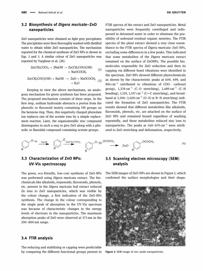
The SEM images of ZnO-NPs are shown in Figure 1, whichconfirmed the surface morphologies and their shape.
Figure 1: SEM image of zinc oxide nanoparticles.
(4)
480 Naheed Ashraf et al.
The SEM image revealed that zinc oxide nanoparticles arespherical, irregular, and spongy, and their individualparticle [28] size ranges from 55 to 80 nm with somedeviations. The metal oxides were uniformly distributedin the three-dimensional polymeric network structure; theaccumulation could be induced by densification resultingin the narrow space between particles, which may be dueto the flavonoids, alkaloids, tannins, saponins, and glyco-sides present in the Digera muricata and were responsiblefor the formation of ZnO NPs.
3.6 Energy-dispersive X-ray spectroscopy(EDX) analysis
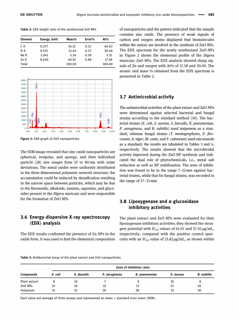
The EDX results confirmed the presence of Zn-NPs in theoxide form. It was used to find the elemental composition
of nanoparticles and the pattern indicated that the samplecontains zinc oxide. The presence of weak signals ofcarbon and oxygen atoms displayed that biomoleculeswithin the extract are involved in the synthesis of ZnO-NPs.The EDX spectrum for the newly synthesized ZnO-NPsin Figure 2 shows the elemental profile of the Digeramuricata–ZnO NPs. The EDX analysis showed sharp sig-nals of Zn and oxygen with At% of 17.58 and 30.49. Theatomic and mass % obtained from the EDX spectrum ispresented in Table 2.
3.7 Antimicrobial activity
The antimicrobial activities of the plant extract and ZnO-NPswere determined against selected bacterial and fungalstrains according to the standard method [30]. The bac-terial strains (E. coli, S. aureus, S. faecalis, K. pneumoniae,P. aeruginosa, and B. subtilis) used imipenem as a stan-dard, whereas fungal strains (T. mentagrophytes, E. floc-cosum,A. niger,M. canis, and F. culmorum) usedmiconazoleas a standard; the results are tabulated in Tables 3 and 4,respectively. The results showed that the microbicidalactivity improved during the ZnO-NP synthesis and indi-cated the dual role of phytochemicals, i.e., metal saltreduction as well as NP stabilization. The zone of inhibi-tion was found to be in the range 7–22mm against bac-terial strains, while that for fungal strains, was recorded inthe range of 17–33mm.
3.8 Lipoxygenase and α-glucosidaseinhibitory activities
The plant extract and ZnO-NPs were evaluated for theirlipoxygenase inhibitory activities; they showed the stron-gest potential with IC50 values of 61.07 and 57.01 µg/mL,respectively, compared with the positive control quer-cetin with an IC50 value of 23.82 µg/mL, as shown within
Figure 2: EDX graph of ZnO nanoparticles.
Table 2: EDX weight ratio of the synthesized ZnO NPs
Element Energy (keV) Mass% Error% At%
C K 0.277 24.15 0.22 46.63O K 0.525 21.04 0.22 30.49Na K 1.041 5.26 0.30 5.31Zn K 8.630 49.55 0.89 17.58Total 100.00 100.00
Table 3: Antibacterial assay of the plant extract and ZnO nanoparticles
Zone of inhibition (mm)
Compounds E. coli S. faecalis P. aeruginosa K. pneumoniae S. aureus B. subtilis
Plant extract 8 10 7 9 10 8ZnO NPs 21 19 15 17 22 20Imipenum 31 32 29 28 31 30
Each value are average of three assays and represented as mean ± standard error mean (SEM).
Digera muricata antimicrobial and enzymatic inhibitory zinc oxide bionanoparticles 481
Table 4. The plant extract and ZnO-NPs were also evalu-ated for α-glucosidase enzymatic inhibitory activities anddisplayed IC50 (μM) values of 106.5 and 98.1, respectively(Table 5). Deoxynojirimycin (IC50 = 301.4 μM)was used asa positive control for the α-glucosidase enzyme inhibitoryactivity. As these compounds showed moderate to signif-icant enzymatic inhibition, in the future, these nanopar-ticles and plant extract phytochemicals may be used forthe treatment of COVID-19 based main protease (Mpro)protein inhibition [31,32].
4 Conclusions
Phytochemical screening of the methanolic extract ofDigera muricata showed the presence of alkaloids, tan-nins, flavonoids, terpenoids, glycosides, flavones and fla-vonols, triterpenoids, and saponins. A fast, easy, and greensynthesis of zinc oxide nanoparticles using Digera muricataextracts was successfully completed by the reductionof zinc acetate solutions. The synthesized ZnO-NPs wereconfirmed by characterization using spectroscopic techni-ques like UV-Vis spectroscopy, FTIR spectroscopy, andmicroscopic techniques like SEM and EDX. The EDX ana-lysis showed sharp signals between 0 and 2 and Zn peaksbetween 8 and 10. In addition, the antimicrobial potentialof nanoparticles and their comparison with that of Digeramuricata extract indicated that ZnO-NPs were more
promising antimicrobial agents than that of plant extract.The extract and ZnO-NPs demonstrated lipoxygenase andα-glucosidase inhibitory activities using quercetin anddeoxynojirimycin as standards, respectively. It can beassumed that the plant extract and ZnO NPs contain potentenzymes (lipoxygenase and α-glucosidase) inhibiting phyto-chemicals that may be used for the treatment of inflamma-tion, COVID-19, asthma, and cancer.
Acknowledgements: M. Imran and A. Irfan extend theirappreciation to the Deanship of scientific research atKing Khalid University (KKU), Saudi Arabia, for fundingthrough research groups program under grant numberR.G.P. 1/297/42.
Funding information:M. A. A. express appreciation to theDeanship of Scientific Research at King Khalid UniversitySaudi Arabia through research groups program undergrant number R.G.P. 1/297/42.
Author contributions: Naheed Ashraf: writing – originaldraft, formal analysis; Sajjad H. Sumrra: writing – reviewand editing, revision; MohammedA. Assiri: writing– reviewand editing, resources; Muhammad Usman: writing – ori-ginal draft, visualization, project administration; RiazHussain: writing – original draft, formal analysis; FarooqAziz: methodology, formal analysis; Muhammad AbuzarGhaffari: writing – original draft, formal analysis; Ajaz
Table 4: Antifungal assay of the plant extract and ZnO-nanoparticles
Zone of inhibition (mm)
Compounds T. mentogrophytes E. floccosum A. niger M. canis F. culmorum
Plant extract 22 19 17 21 19ZnO NPs 28 33 31 27 29Miconazole 39 41 40 39 41
Each value are average of three assays and represented as mean ± standard error mean (SEM).
Table 5: Lipoxygenase and α-glucosidase inhibition of samples
Sample Lipoxygenase inhibition α-glucosidase
LOX (%) at 0.5 mM LOX (IC50) µmol IC50c with SEM
Plant extract 80.48 ± 0.94 61.07 ± 0.06 106.5 ± 1.4ZnO NPs 83.82 ± 1.15 57.01 ± 0.09 98.1 ± 2.2Quercetina 94.65 ± 1.06 23.82 ± 0.05 —Deoxynojirimycinb — — 301.4 ± 2.3
LOX = lipoxygenase; apositive control for lipoxygenase inhibition; bpositive control for α-glucosidase; cIC50 (μM) values are average of threeassays and represented as mean ± standard error mean (SEM).
482 Naheed Ashraf et al.
Hussain: writing – original draft, formal analysis, visualiza-tion, project administration; Muhammad Naeem Qaisar:writing – original draft, visualization; Muhammad Imran:writing– review and editing, visualization, project adminis-tration; Ahmad Irfan: visualization, project administration.
Conflict of interest: The authors state no conflict ofinterest.
Data availability statement: All the data generated oranalyzed during this study are included in this publishedarticle. The datasets are also available from the corre-sponding author on reasonable request.
References
[1] Sun Y, Xia Y. Shape-controlled synthesis of gold and silvernanoparticles. Science. 2002;298(5601):2176–9.
[2] Vijayan SR, Santhiyagu P, Ramasamy R, Arivalagan P,Kumar G, Ethiraj K, et al. Seaweeds: a resource formarine bionanotechnology. Enzyme Microb Technol.2016;95:45–57.
[3] Pugazhendhi S, Kirubha E, Palanisamy PK, Gopalakrishnan R.Synthesis and characterization of silver nanoparticles fromAlpinia calcarata by Green approach and its applications inbactericidal and nonlinear optics. Appl Surf Sci.2015;357:1801–8.
[4] Wahab R, Kim YS, Lee DS, Seo JM, Shin HS. Controlled synth-esis of zinc oxide nanoneedles and their transformation tomicroflowers. Sci Adv Mater. 2010;2(1):35–42.
[5] Schilling K, Bradford B, Castelli D, Dufour E, Nash JF,Pape W, et al. Human safety review of “nano” titaniumdioxide and zinc oxide. Photochem Photobiol Sci.2010;9(4):495–509.
[6] Gerloff K, Fenoglio I, Carella E, Kolling J, Albrecht C, Boots AW,et al. Distinctive toxicity of TiO2 rutile/anatase mixed phasenanoparticles on Caco-2 cells. Chem Res Toxicol.2012;25(3):646–55.
[7] Raja A, Ashokkumar S, Marthandam RP, Jayachandiran J,Khatiwada CP, Kaviyarasu K, et al. Eco-friendly preparation ofzinc oxide nanoparticles using Tabernaemontana divaricataand its photocatalytic and antimicrobial activity. J PhotochemPhotobiol B Biol. 2018;181:53–8.
[8] Kumar SS, Venkateswarlu P, Rao VR, Rao GN. Synthesis,characterization and optical properties of zinc oxide nano-particles. Int Nano Lett. 2013;3(1):30.
[9] Divband B, Khatamian M, Eslamian GK, Darbandi M. Synthesisof Ag/ZnO nanostructures by different methods and investi-gation of their photocatalytic efficiency for 4-nitrophenoldegradation. Appl Surf Sci. 2013;284:80–6.
[10] Misra SK, Dybowska A, Berhanu D, Luoma SN, Valsami-Jones E. The complexity of nanoparticle dissolution and its
importance in nanotoxicological studies. Sci Total Environ.2012;438:225–32.
[11] Ahmed S, Ahmad M, Swami BL, Ikram S. A review on plantsextract mediated synthesis of silver nanoparticles for antimicro-bial applications: a green expertise. J Adv Res. 2016;7(1):17–28.
[12] Kuppusamy P, Yusoff MM, Maniam GP, Govindan N.Biosynthesis of metallic nanoparticles using plant derivativesand their new avenues in pharmacological applications – anupdated report. Saudi Pharm J. 2016;24(4):473–84.
[13] Varshney R, Bhadauria S, Gaur MS. A review: biologicalsynthesis of silver and copper nanoparticles. Nano BiomedEng. 2012;4(2.
[14] Gardea-Torresdey JL, Gomez E, Peralta-Videa JR, Parsons JG,Troiani H, Jose-Yacaman M. Alfalfa sprouts: a natural sourcefor the synthesis of silver nanoparticles. Langmuir.2003;19(4):1357–61.
[15] Zhou Y, Lin W, Huang J, Wang W, Gao Y, Lin L, et al.Biosynthesis of gold nanoparticles by foliar broths: roles ofbiocompounds and other attributes of the extracts. NanoscaleRes Lett. 2010;5(8):1351.
[16] Sharma N, Vijayvergia R. A review on Digera muricata (L.)Mart – a great versatile medicinal plant. Int J Pharm Sci RevRes. 2013;20(1):114–9.
[17] El-Ghamery AA, Sadek AM, Abdelbar OH. Comparative anato-mical studies on some species of the genus Amaranthus(Family: Amaranthaceae) for the development of an identifi-cation guide. Ann Agric Sci. 2017;62(1):1–9.
[18] Kritikar K, Basu B. Indian medicinal plants. M/S Bishen Singh,Mahendra Pal Singh. International book distributors.Dehradune, India. Vol. 1; 1975. p. 672–5.
[19] Aggarwal S, Gupta V, Narayan R. Ecological study of wild med-icinal plants in a dry tropical peri-urban region of Uttar Pradeshin India. Int J Med Aromatic Plants. 2012;2:246–53.
[20] Chandirika JU, Annadurai G. Biosynthesis and characterizationof silver nanoparticles using leaf extract abutilon indicum.Glob J Biotechnol Biochem. 2018;13(1):7–11.
[21] Fakhari S, Jamzad M, Kabiri Fard H. Green synthesis of zincoxide nanoparticles: a comparison. Green Chem Lett Rev.2019;12(1):19–24.
[22] Buhr E, Senftleben N, Klein T, Bergmann D, Gnieser D,Frase CG, et al. Characterization of nanoparticles by scanningelectron microscopy in transmission mode. Meas Sci Technol.2009;20(8):084025.
[23] Choudhary MI, Thomsen WJ. Bioassay techniques for drugdevelopment. Singapore: CRC Press, Harwood AcademicPublishers; 2001.
[24] McLaughlin J, Chang C, Smith D. Bench-top” bioassays for thediscovery of natural products: an update in studies in naturalproducts chemistry. Amsterdam: Elsevier; 1991. p. 383–409.
[25] Tappel A. The mechanism of the oxidation of unsaturated fattyacids catalyzed by hematin compounds. Arch Biochem bio-physics. 1953;44(2):378–95.
[26] Palanisamy U, Manaharan T, Teng LL, Radhakrishnan AK,Subramaniam T, Masilamani T. Rambutan rind in the man-agement of hyperglycemia. Food Res Int. 2011;44(7):2278–82.
[27] Zechel DL, Boraston AB, Gloster T, Boraston CM,Macdonald JM, Tilbrook DMG, et al. Iminosugar glycosidase
Digera muricata antimicrobial and enzymatic inhibitory zinc oxide bionanoparticles 483
inhibitors: structural and thermodynamic dissection of thebinding of isofagomine and 1-deoxynojirimycin to β-glucosi-dases. J Am Chem Soc. 2003;125(47):14313–23.
[28] Varghese E, George M. Green synthesis of zinc oxide nano-particles. Int J Adv Res Sci Eng. 2015;4(1):307–14.
[29] Gunaydin K, Topcu G, Ion RM. 1, 5-Dihydroxyanthraquinonesand an anthrone from roots of Rumex crispus. Nat Product Lett.2002;16(1):65–70.
[30] Imran M, Irfan A, Assiri MA, Sumrra SH, Saleem M,Hussain R, et al. Coumaronochromone as antibacterialand carbonic anhydrase inhibitors from Aerva persica (Burm. f.)
Merr.: experimental and first-principles approaches. Zeitschriftfür Naturforschung C. 2021;76(1–2):71–8.
[31] Irfan A, Imran M, Khalid M, Ullah MS, Khalid N, Assiri MA, et al.Phenolic and flavonoid contents in Malva sylvestris andexploration of active drugs as antioxidant and anti-COVID19 byquantum chemical and molecular docking studies. J SaudiChemi Soc. 2021;25(8):101277.
[32] Khaerunnisa S, Kurniawan H, Awaluddin R, Suhartati S,Soetjipto S. Potential inhibitor of COVID-19 main protease(Mpro) from several medicinal plant compounds by moleculardocking study; 2020. p. 2020030226.
484 Naheed Ashraf et al.