FROM BARRETT'S ESOPHAGUS TO ADENOCARCINOMA - RePub
152
FROM BARRETT'S ESOPHAGUS TO ADENOCARCINOMA AND METASTASIS
Transcript of FROM BARRETT'S ESOPHAGUS TO ADENOCARCINOMA - RePub

FROM BARRETT'S ESOPHAGUS TO ADENOCARCINOMA AND METASTASIS

© 1997 K.K. Krishnadath
Publication of this thesis was generously supported by Byk Netherlands
1811111
Eburon Publishers Oude Delft 224 2611 HJ Delft The Netherlands 31-152131484
Behoudens uitzonderingen door de wet gesteld, mag zonder schriftelijke toestemming van de rechthebbende c.q. rechthebbende gemachtigd namens deze op te treden, niets uit deze uitgave worden vermenigvuldigd of anderzins openbaar gemaakt d.m.v. druk, fotokopie, microfilm of anderszins. No part of this book may be reproduced in any form, by print, photoprint, microfilm or any other means without written permisSion of the publisher.
ISBN 90-5166-579-2
FROM BARRETT'S ESOPHAGUS TO ADENOCARCINOMA AND METASTASIS
Van Barrett Oesofagus naar Adenocarcinoom en Metastase
PROEFSCHRIFT
ter Verkrijging van de Graad van Doctor aan de Erasmus Universiteit Rotterdam op Gezag
van de Rector Magnificus Prof. Dr P.W.C. Akkermans M.A.
en volgens Besluit van het College voor Promoties de openbare Verdediging zal plaatsvinden op
Donderdag 26 Juni 1997 om 16.00 uur door
Kausilia Krishnawatie Krishnadath Geboren te Paramaribo
Rotterdam 1997

PROMOTIECOMMISSIE
Promotores:
Co-Promotor:
Overige leden:
Prof. Dr F. T. Bosman Prof. Dr H. W. Tilanus
Dr H. van Dekken
Prof. Dr I.H.P. Wilson Drs. M. van Blankenstein Prof. Dr I. Ieekel Prof. Dr G.I.H. Offerhaus

Aall mijll ollders, aall Max

CONTENTS
Chapter 1
General Introduction
1.1 Historical perspective 1. 2 Present definitions 1.3 Epidemiology, cancer risk and clinical outcome
Chapter 2
Prognostic Factors in Barrett's Esophagus aud Esophageal Adenocarcinomas
2.1 Biological markers 2.2 Secretory factors 2.3 Proliferation 2.4 Growth factors 2.5 Oncogenes 2.6 P53 and other tumor suppressor genes 2.7 Numerical and structural aberrations 2.8 Aneuploidy 2.9 The E-Cadherin -Catenin complex 2.10 CD44 and CD44 splice variants 2.11 References
Chapter 3
Aim of the Study and Introduction to the Papers.
3 5 6
9
9
11 11 12 13 13 13 16 17 18 19 21
33
33
3.1 From Barrett's esophagus to adenocarcinoma and metastasis. 35 3.2 Aim of the study 35 3.3 Introduction to the papers 36
Chapter 4 39
Detection of Genetic Changes in Barrett's Adenocarci- 39
noma and Barrett's Esophagus by DNA In Situ Hybridization and Immunohistochemistry.
4.1 Abstract 4.2 Introduction 4.3 Materials and methods
4.3.1 Tissue preparation 4.3.2 Probe and probe labeling 4.3.3 In Situ Hybridization 4.3.4 Immunohistochemistry 4.3.5 DNA flow cytometry 4.3.6 Analysis
4.4 Results 4.5 Discussion 4.6 References
Accumulation of P53 Protein in Normal, Dysplastic
41 41 42 43 43 44 44 45 45 47 51 54
and Neoplastic Barrett's Esophagus. 57
5.1 Abstract 5.2 Introduction 5.3 Material and methods
5.3.1 Tissues 5.3.2 Histopathological criteria 5.3.3 Immunohistochemistry 5.3.4 Microscopical evaluation
5.4 Results 5.4.1 Nonnal cardia and esophageal mucosa 5.4.2 Barrett's esophagus 5.4.3 Barrett's esophagus adjacent to adenocarcinoma 5.4.4 Adenocarcinoma in Barrett's esophagus
5.5 Discussion 5.6 References
59 59 60 60 60 61 61 63 63 64 64 64 64 66
Chapter 6 69
Accumulation of Genetic Abnormalities during Neoplastic Progression in Barrett's Esophagus. 69
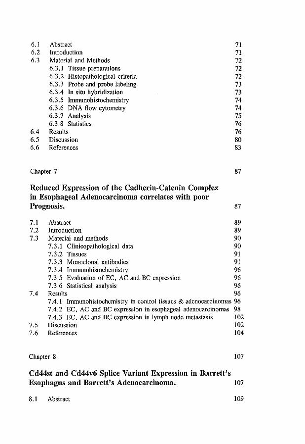
6.1 Abstract 71 6.2 Introduction 71 6.3 Material and Methods 72
6.3.1 Tissue preparations 72 6.3.2 Histopathological criteria 72 6.3.3 Probe and probe labeling 73 6.3.4 In situ hybridization 73 6.3.5 Immunohistochemistry 74 6.3.6 DNA flow cytometry 74 6.3.7 Analysis 75 6.3.8 Statistics 76
6.4 Results 76 6.5 Discussion 80 6.6 References 83
Chapter 7 87
Reduced Expression of the Cadherin-Catenin Complex in Esophageal Adenocarcinoma correlates with poor Prognosis. 87
7.1 Abstract 89 7.2 Introduction 89 7.3 Material and methods 90
7.3.1 Clinicopathological data 90 7.3.2 Tissues 91 7.3.3 Monoclonal antibodies 91 7.3.4 Immunohistochemistry 96 7.3.5 Evaluation of EC, AC and BC expression 96 7.3.6 Statistical analysis 96
7.4 Results 96 7.4.1 Immunohistochemistry in control tissues & adenocarcinomas 96 7.4.2 EC, AC and BC expression in esophageal adenocarcinomas 98 7.4.3 EC, AC and BC expression in lymph node metastasis 102
7.5 Discussion 102 7.6 References 104
Chapter 8 107
Cd44st and Cd44v6 Splice Variant Expression in Barrett's Esophagus and Barrett's Adenocarcinoma. 107
8.1 Abstract 109
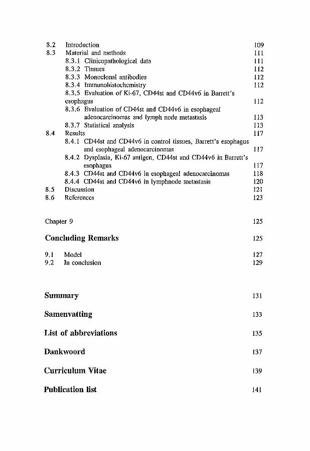
8.2 Introduction 109 8.3 Material and methods 111
8.3.1 Clinicopathological data 111 8.3.2 Tissues 112 8.3.3 Monoclonal antibodies 112 8.3.4 Immunohistochemistry 112 8.3.5 Evaluation of Ki-67, CD44st and CD44v6 in Barrett's esophagus 112 8.3.6 Evaluation of CD44st and CD44v6 in esophageal
adenocarcinomas and lymph node metastasis 113 8.3.7 Statistical analysis 113
8.4 Results 117 8.4.1 CD44st and CD44v6 in control tissues, Barrett's esophagus
and esophageal adenocarcinomas 117 8.4.2 Dysplasia, Ki-67 antigen, CD44st and CD44v6 in Barrett's
esophagus 117 8.4.3 CD44st and CD44v6 in esophageal adenocarcinomas 118 8.4.4 CD44st and CD44v6 in lymphnode metastasis 120
8.5 Discussion 121 8.6 References 123
Chapter 9 125
Concluding Remarks 125
9.1 Model 127 9.2 In conclusion 129
Summary 131
Samenvatting 133
List of abbreviations 135
Dankwoord 137
Curriculum Vitae 139
Publication list 141

Chapter 1
General Introdnction


1.1 Historical perspective
The first description of islets of ectopic gastric mucosa in the esophagus was by
Schmidt in 1805 (1). One century later, in 1906, Tileston described peptic
ulcerations in columnar epithelium lining the distal esophagus (2). In 1950
Norman Barrett gave a detailed description of the columnar lined esophagus. He
regarded the distal columnar lined esophagus a mediastinal extension of the
stomach, as a result of a congenitally short esophagus. Barrett based his theory
on the nature of the mucosa and mucosal secretions of the columnar lining,
whereas absence of the musculature and peritoneal covering of the normal stomach were ignored (1). In that period Lortat-Jacob described the same
condition, which he named endobrachyesophagus, a term still used in French literature (3). Barrett's observation was that of gastric mucosa, extending as a
continuous sheet into the mediastinum. He observed that the columnar mucosa could extend for a varying distance and could reach as far as the aortic arch.
Allison and Johnstone in 1953 showed that anatomically and functionally the
segment of digestive tract described by Barrett is part of the esophagus. These
authors suggested that the so-called Barrett's esophagus might be an acquired
rather than a congenital condition. This implies that as a consequence of gastro
esophageal reflux, oesophageal squamous epithelium is converted to columnar
epithelium through metaplasia (4). In several studies, authors noted "upward
migration" of the squamo-columnar junction during follow-up of patients with
gastro-esophageal reflux (5-7). Animal experiments proved that columnar
epithelium in the esophagus is generated in the presence of gastro-oesophageal reflux (8). Through these observations it had become apparent that Barrett's
esophagus is an acquired rather than a congenital condition. Around 1980 most authors appear to favor the view of the acquired origin of Barrett's esophagus
(9-14). However, congenital islands of ectopic gastric mucosa do occur. They are found in up to 10% of individuals undergoing endoscopy (15). These so called
"inlet patches" occur principally in the cervical esophagus and are mostly
surrounded by normal squamous epithelium (16).
Different mechanism have been proposed for the development of columnar epithelium in the esophagus. One suggestion is upward extension of gastric
epithelium, another is generation of columnar epithelium from superficial
esophageal glands (heterotopic gastric mucosa). The theory proposing metaplastic
change of squamous epithelium was already suggested by Allison in 1953. This
theory was clinically supported by evidence of photographed ulcerations of
3
squamous mucosa which, while healing, were replaced by metaplastic colunmar
epithelium (17). Further evidence in favor of metaplasia was derived from the
histology, mucin histochemistry and ultrastructural features of Barrett's
esophagus. These features of multidirectional differentiation of epithelial cells,
suggested that the columnar epithelium was derived from a mucosal stem cell
(18-25). More evidence sustaining the theory of metaplasia from a multi potent
stem cell was obtained from experiments in dogs. In these experiments, segments
of esophagus from which the mucosa had been excised were re-epithelialized by
columnar epithelium when reflux was induced. To prove that this re
epithelialization was not a process of upward migration of gastric mucosa, a strip
of normal squamous epithelium between the excised area and stomach was left
intact (26).
The epithelium which Barrett described in 1957, in the upper region contained
flat cells in shallow tubular glands with mucus secreting units but little, if any,
acid, pepsin or secretin secretion. In the lower esophagus a more typical gastric
mucous membrane was seen (27). Over time, additional types of aberrant
columnar epithelium with different secreting and non secreting cell types, such as
mucous, parietal, chief, neuro-endocrine and goblet cells, were observed (12,
18-20, 22, 24, 28-30). Paull et aI., distinguished three major types of columnar
epithelium: an atrophic gastric fundic type with parietal and chief cells; a
junctional type with cardiac mucous glands and a distinctive specialized columnar
epithelium with villiform surface, mucous glands and goblet cells (13, 31). These
authors noted that specialized columnar epithelium is the prevalent type of
epithelium in Barrett's esophagus. This specialized columnar type appeared to be
present most proximally in the esophagus, the gastric-fundic type being found
most distally and the junctional type being ioterposed between the specialized
columnar and gastric fundic type. This so called zonal expansion theory was
challenged by Thompson et aI., who described a mosaic pattern of different
columnar cell types in Barrett's esophagus (22). Other authors confirmed the
predominance of the specialized intestinal type. With respect to the two other
types of epithelium the need for adequate sampling in the esophagus was pointed
out. If sampling is performed blindly under manometric guidance, biopsies taken
from the cardia could be mistakenly taken for Barrett's esophagus, for instance in
cases of hiatal herniation of the cardia into the mediastinum (12, 32).
Endoscopically, the exact gastro-oesophageal junction is difficult to locate. For
instance, up to 2 cm of simple colunmar lining may be present in the distal
esophagus of healthy individuals. This led to the agreement that Barrett's
4
few studies reported regression of Barrett's epithelium after successful anti-reflux
surgery (35, 36). In contrast, in several papers long-term treatment with H2
receptor blockers (cimetidin) and antacids showed decreased esophagitis and
induced healing of Barrett's ulcers, but not regression of Barrett's esophagus
(37 -40). Nevertheless, more recent studies on treatment with proton pump
inhibitors showed partial regression of Barrett's epithelium (41, 42).
Barrett already associated columnar lined esophagus with adenocarcinoma in his
first description (I). Esophageal adenocarcinomas evidently associated with
Barrett's esophagus were reported a few years later (43). This study was followed
by numerous other reports, many of which also described dysplastic columnar
epithelium adjacent to the adenocarcinomas (10, 44-52). Based on these observations Barrett's esophagus came to be regarded as a premalignant condition.
It became clear that progression of Barrett's esophagus to adenocarcinoma is a
gradual process going through different stages of dysplasia. In analogy to stomach,
three stages of dysplasia (mild, moderate and severe dysplasia) in Barrett's
esophagus were defmed (53). Since esophageal adenocarcinomas are highly
malignant, it lVas recommended that Barrett's esophagus patients should be under
endoscopic surveillance. A consensus was reached that periodically biopsies taken from the mucosa should be screened for dysplastic changes or early carcinoma
(54). In case of early carcinoma, the patient could be cured by surgical
intervention.
1.2 Pl'esent definition
In Barrett's esophagus the normal stratified squamous mucosal lining of the
esophagus is replaced by metaplastic columnar epithelium. Three types of
metaplastic epithelium have been described (l3), but careful examinations proved
that fundic and gastric type of metaplasia are rarely seen in biopsies taken above
the lower esophageal sphincter (33). Typical Barrett's epithelium shows incomplete intestinal metaplasia with goblet and columnar cells and a flat or
villiform surface (55, 56).
Until recently, it was agreed that for diagnosing Barrett's esophagus, the columnar
epithelium should extend at least three centimeters from the gastro-esophageal
junction into the esophagus. This margin of three cm of columnar epithelium is
debatable since endoscopic measurements of the length of columnar lining do not
5
columnar epithelium should extend at least three centimeters from the gastroesophageal junction into the esophagus. This margin of three cm of columnar
epithelium is debatable since endoscopic measurements of the length of columnar lining do not appear to be reproducible (57). Furthermore, intestinal metaplasia
with dysplasia or adenocarcinoma may occur at the gastro-esophageal junction or in short segments of Barrett's esophagus (58, 59).
Regarding the three stages of dysplasia (53), there is limited inter- and intra
observer agreement, particularly when diagnosing dysplasia less than high grade
(60). This led to simplification of the dysplasia classification, in which "non dysplastic" lesions are distinguished from dysplastic and from unclassifiable
lesions (mostly regenerative changes), which are marked as "indefinite for
dysplasia" (61). In practice and for patient management indefinite for dysplasia
and low grade dysplasia are grouped together. When dysplasia occurs it may involve a considerable amount of the columnar
mucosa or it may be limited in extent (62-64). Therefore, a thorough, systematic
biopsy protocol is proposed to detect small areas of dysplasia and small
carcinomas. At present, the length of columnar epithelium extending in the esophagus may be
ignored, four quadrant biopsies at intervals of 2 cm or less throughout the length
of the Barrett's segment and biopsies of any suspect lesion should be taken. Only
intestinal metaplasia, which is the prevalent type of Barrett's esophagus, is regarded as premalignant. Dysplasia, if present, should be classified as
indefinite/low grade or high grade dysplasia.
1.3 Epidemiology, cancer risk and clinical ontcome
Although it is generally accepted that Barrett's esophagus predisposes for
esophageal adenocarcinoma, the prevalence of Barrett's esophagus and the
frequency with which adenocarcinoma occurs in Barrett's esophagus are unknown. There is a discrepancy between data derived from clinical and that
from population based autopsy series. For instance, in a population based study at the Mayo Clinic in Rochester USA, in the period of 1980-1986, a 21 fold higher
prevalence of Barrett's esophagus was reported in autopsy series compared with
the number of endoscopical diagnoses (65). This important discrepancy is mainly
caused by the fact that many Barrett's esophagus patients have few symptoms or
are asymptomatic. The prevalence of Barrett's esophagus is relatively low in
6
childhood and increases with age. A plateau is reached at the age of 60. Median
age of developing Barrett's esophagus is 40 (66). Interestingly, the prevalence of
Barrett's esophagus is highest in white middle aged males (67). It has become clear that the high incidence of adenocarcinoma in Barrett's
esophagus as published in the early studies is exaggerated (48, 68, 69). In
endoscopic series of Barrett's esophagus patients the prevalence of esophageal
adenocarcinoma is between 8 and 15%. Follow up series, however, shows that
one case per 46 to 441 patient years of follow up will progress to
adenocarcinoma. Thus the incidence of adenocarcinoma in Barrett's esophagus ranges between 0.2 and 2%. Compared to the general population, the risk for
developing esophageal adenocarcinoma in Barrett's esophagus is between 30 and
125 fold increased (70-76). For many years, Barrett's esophagus patients only
underwent major surgery in case of complete malignant transformation (carcinoma) had occurred. Decrease of postoperative mortality and morbidity by
improved esophageal surgery (77-80), and the frequent coexistence of carcinoma with severe dysplasia led to a new approach. In this approach it is suggested that
surgical intervention should be performed on all Barrett's esophagus patients with consistent high grade dysplasia (63, 81-83).
A striking fact is that in Western countries esophageal adenocarcinoma is the
most rapidly increasing type of gastrointestinal malignancy (84-87). In the USA
it shows probably the most rapidly rising incidence rate of all cancers (86). The increase in esophageal adenocarcinomas is most marked in higher social
economic classes, in whites opposed to blacks, and is seven fold higher for males than for females (84, 86, 87). At present the incidence of esophageal
adenocarcinoma lies between 0.6 and 0.8 per 100.000 per year (84-87). It is assumed that virtually all esophageal adenocarcinomas are associated with
Barrett's esophagus. Since the vast majority of Barrett's esophagus is
unrecognized (72), most esophageal adenocarcinoma associated with Barrett's
esophagus are discovered when the disease has reached an advanced stage and patients already have symptoms of dysphagia and weight loss (69, 72, 88, 89).
The only possible cure for these patients is esophagogastrectomy. Nevertheless,
curation by surgery is only feasible in case the disease is limited to the
esophageal wall and metastases are absent. Therefore, curative resection is only attempted after extensive preoperative staging of disease to exclude the presence
of distal metastases (90-95). For patients with extensive disease and lymph node
or organ metastases, palliative therapy is a more suitable option (96-104). In
practice, local or distant lymph node metastases are seen in at least half of the
7

resected specimens. The cumulative five year recurrence rate for metastases in
patients after esophagogastric resection is approximately 65% (105-107). Overall
post operative long-term survival of patients with esophageal adenocarcinoma
ranges between 15 and 60%. However, survival is strongly correlated with the
stage of the disease. Five years survival of advanced stages, i.e., carcinomas with deep infiltration of the esophageal wall and lymph node metastasis, is as low as
0%, whereas survival rises to 100% in cases where patients are operated with
disease limited to the mucosa (79, 108-111).
8

Chapter 2
Prognostic Factors in Barett's Esophagus and Esophageal Adenocarcinoma.

2.1 Biological markers
Biological markers in neoplastic disease are often factors involved in the
regulation of normal and abnormal differentiation and proliferation of cells. In
oncology, these markers can be used as parameters to diagnose at tissue level or
to clinically monitor disease. In Barrett's esophagus the development of an adenocarcinoma involves many factors interacting in a complex manner rather
than a simple transforming hit. Some factors can be detected early and others late during neoplastic progression, but an exact order of involvement of the various
factors in malignant transformation in Barrett's esophagus has not been determined. Generally, markers which may be used in Barrett's esophagus
include: Secretory factors, cell cycle regulating factors, onco- and tumor suppressor genes, cell surface proteins, genetic aberrations and ploidy status.
Most of these have been extensively correlated with histopathological changes in Barrett's esophagus, but few have been evaluated in prospective follow-up series,
in order to determine their prognostic significance.
Many markers evaluated in Barrett's esophagus have also been studied in
esophageal adenocarcinomas. Some have even been correlated with clinico pathological parameters, such as tumor stage, grade and patient survival.
2.2 Secretory factors
Barrett's esophagus consists of a mixed population of secretory and non secretory
cells, such as columnar, goblet, Paneth and endocrine cells. Most of the mucin
secreted is of the neutral type, but sulphomucins and sialomucins can be found as
well. The presence of sulphomucins, although previously reported to be a risk factor for adenocarcinoma (21,23, 112) appears to be common in the intestinal
type of Barrett's metaplasia and cannot be considered a marker for malignant transformation (55, 113). Decreased secretion of O-acetylated sialomucins was
found in all dysplastic but also in a number of non·dysplastic cases of Barrett's esophagus with incomplete intestinal type of metaplasia (114). Likewise, sucrase
isomaltase expression was found in incomplete intestinal type of metaplasia and in
esophageal adenocarcinomas (115). Glutathione and Glutathione-S transferase
enzyme activity is low in Barrett's esophagus. These enzymes protect tissues from genetic damage, for instance, caused by oxygen radicals. Decreased levels
11
have been correlated with an increased risk of malignant transformation (116).
Increasing levels of Ornithine decarboxylase (ODC) have been frequently found
in metaplastic, dysplastic and neoplastic Barrett's esophagus and this may be
regarded as a biochemical marker for malignant progression (117, 118). ODC
catalyzes polyamine synthesis and is an important enzyme for cell proliferation
and differentiation. Despite increased ODC levels in Barrett's esophagus,
chemopreventive intervention trials with ODC inhibitors, for instance alpha
diflouromethylornithine, were inconclusive as to whether or not changes in the
polyamine contents would be induced in Barrett's mucosa (119, 120). A complicating factor may be a defective ODC - polyantine pathway, which may
also explain the poor correlation between ODC activity and polyamine levels (121, 122).
2.3 Proliferation
Proliferation of tissues can be assessed by several methods. Through metabolic
labeling, tritiated thymidine or 5-Bromodeoxynridine (BrdU), an analogue of
thymidine, can be incorporated into the DNA during S-phase of the cell cycle,
and proliferating cells can be identified. Early studies using these
autoradiographic methods showed expansion of the proliferative compartment in
Barrett's esophagus compared to other columnar epithelia (123, 124).
Alternatively, proliferation can be assessed by immunohistochemistry using
antibodies recognizing antigens which are expressed in the nucleus during
proliferation. PCNA (Proliferating Cell Nuclear Antigen) and Ki-67 are such antigens. PCNA, a 36 kDa molecule, serves as a cofactor for DNA polymerase
delta in both S-phase and unscheduled DNA synthesis associated with DNA
repair. Since PCNA has a 20 hour half life, it can also be found in non cycling cells, i.e., GO phase. The Ki-67 antigen is expressed in the nuclei during late G 1,
S, G2, and M phases of the cell cycle, but not in GO phase and is therefore a
more precise marker for estimating proliferation. Assessment of these markers in
Barrett's esophagus showed that proliferation is increased in intestinal type of metaplasia even when dysplasia is absent. Interestingly, with increasing dysplasia
the fraction of proliferating cells and the proliferative compartment increases
further (125, 126). Eventually, in high grade dysplasia luminal epithelial cells
show proliferative activity. (123, 125, 127, 128). Reid et aI., used multi
parameter flow cytometry to simultaneously measure DNA content and cycling
12
cells, which allowed him to estimate the fractions of cells in GO, Gl, S and G2M
phase. In this study for instance an increased number of cells is found in Gl phase early on during the neoplastic progression (126).
2.4 Growth Factors
Growth factors are important for regulation of cell differentiation and
proliferation. Abnormal expression of growth factor receptors, such as EGF-R
(Epidermal Growth Factor) and ligands such as EGF (Epidermal Growth Factor) and TGF-Alpha (Transfonning Growth Factor-Alpha) are associated with
mitogenesis and carcinogenesis (129). In Barrett's esophagus expression of EGFRand TGF-Alpha but not EGF is increased in intestinal type metaplasia
compared to the other types of metaplasia. Both markers are also elevated in dysplasia and in esophageal adenocarcinomas (113, 130-134).
2.5 Oncogenes
Abnormal expression of (proto)oncogenes is involved in the transformation of
normal tissues into malignancy (135). Many oncogenes studied in Barrett's
esophagus did not yield specific data, while some need more extensive evaluation. For instance, in a small series of patients increased H-ras expression has been
observed in high grade dysplasia and adenocarcinoma (136). The proto-oncogene
product C-erbB2 is a glycoprotein which shows high homology with the
epidermal growth factor receptor, EGF-R. Like EGF-R, C-erbB2 can be activated
through ligands and in tum activate pathways in the cytoplasm involved in cell differentiation and proliferation (137, 138). Although, C-erbB2 expression has
been found to be correlate with survival of patients with esophageal
adenocarcinoma, the proportion of C-erbB2 expressing cases in the different studies varied between to to 73 % and hence this protein needs to be further evaluated (139-142).
2.6 P53 and other tumor suppressor geues
In contrast to an oncogene, the function of a tumor suppressor gene is only
13

disrupted in case both alleles are defective. This condition is usually met by loss
of one allele and functional inactivation of the other. Tumor suppressor gene
alterations can be studied by a variety of techniques. Gene mutations can be studied by PCR (Polymerase chain reaction) and sequence analysis or SSCP
(Single Strand Conformation Polymorphism) analysis. allelic loss by RFLP
(Restriction Fragment Length Polymorphism) analysis.
The p53 tumor suppressor protein was detected in 1979 by Lane and Crawford
(143). Though formerly regarded as an oncoprotein, the main function of p53 is
that of a tumor suppressor (144, 145). Although p53 is a tumor suppressor, it can
display oncogene-like behavior, because mutant p53 molecules may bind to and
inhibit wild type p53 function. Under normal circumstances Wild-type p53 plays a
limited role in cell cycle control. In cells with genomic damage, accumulation of
high levels of wild-type p53 in the cell nucleus results in a prolonged G 1 phase of
the cell cycle. The extended G 1 phase consequently delays replication in damaged
cells, which facilitates DNA repair (146, 147). Another unique property of this
protein is that mutant forms acquire conformational changes which prolong half life, resulting in accumulation of mutated p53 protein in cells (148, 149). Wild
type as well as mutated p53 protein are detectable by immunohistochemistry as
long as a sufficient amount of protein is retained (150). In Barrett's esophagus and esophageal adenocarcinoma p53 aberrations have been
extensively documented. Although the applied methodologies vary, the findings show a certain consistency. The prevalence of p53 protein accumulation as
determined by immunostaining in adenocarcinomas ranges between 53-87 % (151-157). In esophageal adenocarcinomas p53 (17p) allelic loss is found in 55%
to 100% of cases (152, 158-160), and p53 gene mutations in 8% to 89% of cases (159, 161-166). In esophageal adenocarcinomas no correlation was found between
p53 protein accumulation and clinicopathological and prognostic factors (167).
Nevertheless, in Barrett's esophagus the frequency of p53 alterations increases as
the epithelium becomes more dysplastic (151, 154-156, 168). The prevalence of
abnormal p53 reaches up to 89% in high grade dysplasia. Since aberrant p53 can
be found in non dysplastic Barrett's esophagus and cases with low grade dysplasia
(table 2), p53 mutations is considered one of the earliest genetic events during
malignant transformation of Barrett's esophagus. Several studies have shown that in resection specimens mutations in the adenocarcinoma correspond with those in
surrounding Barrett's epithelium (162, 164, 165). Interestingly, p53 assessment combined with DNA cell sorting showed that p53 mutations occur in diploid
cells, suggesting that these precede aneuploidy. Only a few follow up studies
14
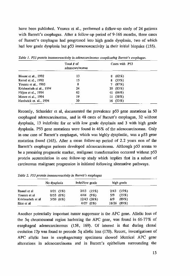
have been published. Younes et aI., performed a follow-up study of 24 patients with Barrett's esophagus. After a follow-up period of 9-166 months, three cases
of Barrett's esophagus had progressed into high grade dysplasia, two of which
had low grade dysplasia but p53 immunoreactivity in their initial biopsies (155).
Table 1. P53 proteill immllnoreactivity ill adenocarcinomas complicatillg Barrett's esophagus.
Total # of Cases with P53 adenocarcinomas
Blount et aL, 1992 13 8 (62%) Ramel et aI., 1992 15 8 (53%) Younes et al., 1993 8 7 (87%) Krishnadath et ai., 1994 24 20 (83%) Flljou et aI., 1994 62 41 (66%) Moore et aI., 1994 19 11 (58%) Hardwick et. al., 1994 30 16 (53%)
Recently, Schneider et al, documented the prevalence p53 gene mutations in 50
esophageal adenocarcinomas, and in 48 cases of Barrett's esophagus, 32 without
dysplasia, 13 indefinite for or with low grade dysplasia and 3 with high grade
dysplasia. P53 gene mutations were found in 46% of the adenocarcinomas. Only in one case of Barrett's esophagus, which was highly dysplastic, was a p53 gene
mutation found (165). After a mean follow-up period of 2.2 years non of the Barrett's esophagus patients developed adenocarcinoma. Although p53 seems to
be a promising prognostic marker, malignant transformation occurred without p53 protein accumulation in one follow-up study which implies that in a subset of
carcinomas malignant progression is initiated following alternative pathways.
Table 2. P53 protein immulloreactMty ill Barrell's esophagus
Ramel et at Younes et al Krishnadath et al Rice et at
No dysplasia
1121 (5%) 0/53 (0%) 3/50 (6%)
Indefllow grade
2/13 (15%)
4/44 (9%) 12/43 (28%) 0127 (0%)
high grade
2/13
519 8/9 18/26
(15%) (55%) (89%) (89%)
Another potentially important tumor suppressor is the APC gene. Allelic loss of
the 5q chromosomal region harboring the APC gene, was found in 66-77 % of
esophageal adenocarcinomas (158, 169). Of interest is that during clonal
evolution 17p was found to precede 5q allelic loss (170). Recent, investigations of
APC allelic loss in esophagectomy specimens showed identical APC gene alterations in adenocarcinoma and in Barrett's epithelium surrounding the
15

adenocarcinoma (171). Despite the high frequency of 5q allelic loss in the APC
region as observed several studies, APC gene mutations were only sporadically
found in esophageal adenocarcinomas (172). Possibly another gene or genes on
5q, distinct from APC, are involved.
Other allelic losses of familiar tumor suppressor genes have been found for the
mutated colon carcinoma (MCC) gene in 63%, for deleted in colon carcinoma
(DCC) gene in 24% and for the retinoblastoma (Rb) gene in 48% (158, 169).
2.7 Numerical and structural chromosomal aherrations
A variety of cytogenetic and molecular biological techniques is available for
detecting numerical and structural chromosomal abnormalities. The general idea
is that these methods will lead to the detection of chromosomal regions which
may harbor genes involved in cell cycle regulation and carcinogenesis. Such
genes may be onco- and tumor suppressor genes, cell cycle and cell death
(apoptosis) regulating genes, growth factors, genes coding for transcription and
signal transduction factors and cell adhesion genes. In esophageal
adenocarcinomas many aberrant chromosomes and chromosomal regions have
been identified, whereas the number of genetic aberrations in Barrett's esophagus
is limited. The most frequently documented numerical chromosomal aberration in Barrett's esophagus and esophageal adenocarcinomas is loss of the Y
Chromosome (173-179). In esophageal adenocarcinoma Y-chromosome loss was
found in 31 to 93 % of cases. In Barrett's esophagus, for instance Garewal et aI.,
noticed Y-Ioss in 7110 cases. In chapter 5 of this thesis the frequency of Ychromosome loss in Barrett's increased along with grade of dysplasia and all
cases of high grade dysplasia showed Y- chromosome loss. Although Barrett's esophagus and esophageal adenocarcinoma occur more frequently in men, no
specific onco- or tumor suppressor genes have been assigned to the Y
chromosome. Perhaps, as genetic instability increases during malignant
transformation of Barrett's esophagus, Y-chromosome loss randomly occurs. Nonetheless, the Y-chromosome may harbor genes involved in signal transduction
or cell cycle regulation, which in a more subtle fashion may influence cell
transformation.
Other frequent numerical aberrations in esophageal adenocarcinomas are over
representation of chromosomes 8, 14 and 20 and loss of chromosomes 4, 17, 18
16
and 21 (174, 175, 177, 179). Karyotyping revealed frequent structural
rearrangements in esophageal adenocarcinomas in the Ip, 3q, llpI3-15, and 22p
regions (174, 179). Loss of heterozygosity studies showed high frequencies of
17p (100%) and 5q (80%) allelic loss in adenocarcinomas (152, 160, 170). 17p
and 5q loss were also seen in Barrett's esophagus with high grade dysplasia.
Interestingly in Barrett's esophagus 17p preceded 5q loss (170). In approximately
60% of esophageal adenocarcinomas structural loss of 9p and in 40% of Ip, 13q, and 18q was observed (160).
2.8 Aneuploidy
DNA flow cytometry (DNA-FCM) is the method of choice for measuring DNA
cell content. In addition, DNA-FCM may serve to determine the fraction of cells
in different phases of the cell cycle, such as resting cells in GO and G 1, cells with
active DNA replication in S-phase and cells with doubled DNA content and in
mitosis in G2M phase. Karyotyping and DNA in situ hybridization may also provide information on cell ploidy. Aneuploidy is found in esophageal
adenocarcinomas with a frequency ranging from 79% to 100% (126, 141, 178,
180-184). In several studies aneuploidy of esophageal adenocarcinoma was found
to correlate with the presence of lymph node metastases and poor survival (141, 184).
Although data on aneuploidy in Barrett's esophagus have been inconsistent, aneuploidy and increased G2M/tetraploid populations seem to increase along with
increasing dysplasia (126, 178, 180-183, 185-189). Most discordance between
several studies can be explained by methodological differences. For instance,
interpretation of a DNA FCM histogram depends on parameters chosen by the investigator. Other confounding factors are contamination with non-epithelial
(diploid) cells and differences between fresh and paraffin embedded tissues. Convincing results have been published by Reid et al (126, 180, 182, 187).
Aneuploidy and increased tetraploidy were not found in esophagitis, but in a low percentage of cases with intestinal type of metaplasia without dysplasia or
indefinite for dysplasia, in a high percentage of cases with dysplasia and in all
cases of adenocarcinoma (180). These findings were confirmed by others by
DNA -flow or - image cytometry (181, 183), and DNA in situ hybridization with (peri-) centromeric DNA probes (178). Aneuploidy and increased tetraploidy have
also been correlated with ultrastructural abnonnalities (186). In a prospective
17
follow-up study most (70%) patients with aneuploidy or increased tetraploidy in their initial biopsy specimens progressed toward high grade dysplasia or
carcinoma. In contrast patients without DNA-FCM abnormalities did not develop high grade dysplasia or carcinoma (187).
2.9 The E-Cadherin-Catenin Complex.
The transmembrane 120 kDa glycoprotein E-Cadherin belongs to the family of
calcinm dependent adhesion molecules (190, 191). TIle E-Cadherin gene has been mapped on chromosome l6q22.l (192). Cadherins play an important role in
morphogenesis of tissues during embryogenesis (193). E-Cadherin is part of a
larger system, and in addition to Catenins also interacts with Plakoglobin and a tyrosine kinase substrate, p120cas (194-196). As E-Cadherin is responsible for
tight cellular connections in mature cells which arrange epithelial architecture,
expression of E-Cadherin between epithelial cells is concentrated at tight junctions, adherens junctions and desmosomal regions (197). It has become
apparent that homotypic E-Cadherin cell-cell mediated adhesion is structurally
organized as a zipper (198). In malignancies, dissolution of cell-cell adhesions is
the first step in invasion. There is substantial evidence that reduced expression or
inhibited function of E-Cadherin leads to detachment of cells and increased
invasiveness. For instance in vitro, monoclonal antibodies against E-Cadherin or blocking of E-Cadherin expression in canine kidney cells leads to transformation
into an invasive phenotype (199). In the other direction, induction of E-Cadherin
expression inhibits the invasive phenotype (195). In addition to its role in cellular
adhesion, E-Cadherin is also involved in signal transduction via connections with
Catenins and the actin cytoskeleton (200). Furthermore, aberrant Catenin
expression may impair normal E-Cadherin function (201). The Catenin anchorage complex, which interacts with the cytoplasmic domain of E-Cadherin, consists of
a heteromere of at least three proteins: Alpha-, Beta- and Gamma-Catenin. Alpha-Catenin a 102 kDa protein, homologous to Vinculin, is ubiquitously
expressed in cells and mediates E-Cadherin connection to actin via Beta-Catenin (200-202). At least two isoforrns of Alpha-Catenin have been cloned (Alpha
Neuro and Alpha- Epith-Catenin) (201). In turn, Beta-Catenin, a 92 kDa protein,
homologous to the Drosophila armadillo gene and human plakoglobin, is directly
linked to E-Cadherin (203, 204). The role and organization of Gamma-Catenin is not well understood, it appears that Gamma-Catenin has high homology to or
18
might be identical with plakoglobin (194, 205).
Normal squamous epithelial cells, except in the most superficial keratinizing
layer, intensely express E-Cadherin as well as Alpha- and Beta-Catenin on all
cell-cell boundaries (206-208). Likewise, strong membrane bound expression of
E-Cadherin and Catenins can be found at cell-cell boundaries of metaplastic columnar epithelia such as Barrett's esophagus (207-210).
In esophageal adenocarcinomas aberrant E-Cadherin expression has been
observed in 88% (45151) of the cases, 47% (24/51) of which showed
disorganized E-Cadherin, whereas 41 % (21151) showed reduced expression
(209). Aberrant expression correlated with poor differentiation and advanced
stages of disease. Interestingly, northern blot analysis revealed in terms of size
normal E-Cadherin and Alpha-Catenin mRNA expression in adenocarcinomas
with normal but also in those with disorganized E-Cadherin expression. These
findings imply that disorganized E-Cadherin expression with impaired E-Cadherin
function might be indirectly induced by factors which interact with E-Cadherin. Beta-Catenin is such a potential factor. However, no literature data is available
regarding Alpha- and Beta-Catenin expression in esophageal adenocarcinomas. In chapter 7, E-Cadherin, Alpha- and Beta-Catenin is investigated in 65 esophageal
adenocarcinomas. Reduced E-Cadherin, Alpha- and Beta-Catenin expression was observed in between 60 and 75 % of cases. Reduced expression of E-Cadherin
and Alpha-and Beta-Catenin correlated with unfavorable clinico-pathological parameters, such as poor differentiation grade, advanced stage disease and poor
patient survival.
2.10 CD44 and CD44 splice variants
CD44 is a family of cell surface molecules, involved in intercellular interactions
such as lymphocyte homing, lymphocyte activation, hemopoiesis, cell migration
and metastasis, but also in binding of cytokines, hyaluronate and collagen (211,
212). The CD44 gene has been mapped on chromosome IIp13 (213). The gene
consists of at least 20 exons, 10 of which (exons 1-5 and 16-20) encode for the standard form of CD44 (CDst). At least ten remaining exons are subjected to
alternative splicing. Differential expression of these exons give rise to several
CD44 isoforms (214-216). The structure of the CD44 glycoprotein is complex.
Its standard form (CDST), a 85-90 kDa glycoprotein, is involved in the functions mentioned above. So far, specific ligands which interact only with the variable
19
regions, have not been identified. In the first experiments which led to the
discovery of splice variants, monoclonal antibodies raised against membrane
epHopes of a metastasizing rat pancreatic cell line transiently inhibited the
metastatic potential of these cells (217, 218). The cDNA sequence encoding the
epHope recognized by one of these antibodies, appeared to be part of the exon
encoding the v6 variant (216). Transfection experiments with cDNA partly coding
for v6 conferred metastatic potential to a non metastatic rat pancreatic cancer cell line (219). TIle application of monoclonal antibodies raised against the transfected
epitope blocked metastatic potential of the transfected cell line (220). hI various
malignancies expression of several CD44 splice variants, such as 5v, 6v, 7v, 8v
and 9v, has been related with increased tumor invasiveness, metastatic potential and patient survival (221-228). Nonetheless, splice variants are not exclusively
expressed in metastatic cells. The epithelial CD44 version (CD44E) besides standard CD44 also includes the 8-9 and 10 variable exons (229-231). CD44
isoforms are typically expressed by cells that participate in dynamic processes, such as normal tissue development, regenerative processes, innammation and
wound healing. Thus normal expression of CD44 isoforms can be found in embryonic cells, activated T and B lymphocytes, and in proliferative zones of
both squamous and columnar epithelium (232). Although the exact mechanisms are not yet known, regulated CD44 isoform expression presumably plays a major
role in cell movement and cell-cell and cell-extra cellular matrix contacts (233).
In metastatic cells genetic events may initiate expression of CD44-isoforrus,
rendering cancer cells metastatic. Little literature data is available concerning CD44 and CD44 isoform expression
in Barrett's esophagus and esophageal adenocarcinomas. In chapter 8, a panel of tissues from Barrett's esophagus, esophageal adenocarcinomas and lymph node
metastases was investigated for CDST and CD44v6 expression. Expression of
CDST in Barrett's esophagus correlated with grade of dysplasia and proliferation,
whereas increased CD44V6 expression appeared to occur early during neoplastic progression. Interestingly, in the esophageal adenocarcinomas increased CD44V6
expression tended to be correlated with worse five year survival.
20
References: 1. Barrett NR. Chronic peptic ulcer of the oesophagus and oesophagitis. Br J Surg 38;1950: 175-182. 2. Tileston W. Peptic ulcer of the oesophagus. Am J Med Sci 132;1906:240-265. 3. Lortat-lacob JL. L'endo-brachy-oesophage, AI/Ii ChiT 11;1957:1247-1254.
4. Allison PR, Johnston AS. The oesophagus lined with gastric mucous membrane. 17lOrax 8;1953:87-101.
5. Mossberg SM. The columnar-lined esophagus (Barrett syndrome)--an acquired condition? Gastroelllerolog)' 50;1966:671-6.
6. Halvorsen JF, Semb BK. The "Barrett syndrome" (the columnar-lined lo\\'er oesophagus): an acquired condition secondary to reflux oesophagitis. A case report with discussion of pathogenesis. Acta Chir Scalld 141;1975:683-7.
7. Endo M. Kobayashi S, Kosu T. A case of Barrett's epithelialization followed up for five years. EI/doscopy 6;1974:48-51.
8. Bremner CG, Lynch VP, Ellis PHJ. Barrett's esophagus; Congenital or acquired? An experimental study of esophageal mucosal regeneration in the dog. Surgery 68;1970:209-216.
9. Adler RH. The lower oesophagus lined by columnar epithelium; its association with hiatel hernia. ulcer stricture and tumor. J 17lOracic Cardiovasc Surg 45: 1963.
to. Lortat-Jacob JL, Maillard IN, Richard CA, Fekete P, Huguler M, Conte-Marti J. Primary esophageal adenocarcinoma: report of 16 cases. Surgery 64;1968:535-43.
11. Jordan P Jr., Longhi EH. Diagnosis and treatment of an esophageal stricture (ring) in a patient with Barrett's epithelium. Ann Surg 169;1969:355-63.
12. Burgess JN, Payne WS, Andersen HA, Weiland LH, Carlson HC. Barrett esophagus: the columnar-epithelial-lined lower esophagus. Mayo Clill Proc 46;1971:728-34.
13. Paull A, Trier JS, Dalton MD. The histologic spectrum of Barrett's esophagus. N Engl J Med
295;1976:476-480. 14. Goldman H, AntonioU DA. Mucosal biopsy of the esophagus, stomach and proximal duodenum.
HUIll Pathol13; 1982:423-448.
15. Borhan-Manesh P, Farnum JB. Incidence of heterotopic gastric mucosa in the upper oesophagus. GlII 32;1991:968-72.
16. Van Asche C, Rahm A Jr., Goldner F, Crumbaker D. Colunmar mucosa in the proximal esophagus. Gaslro/lltest Endosc 34;1988:324-6.
17. Naef AP, Savary M. Conservative operations for peptic esophagitis with stenosis in columnar-lined lower esophagus. AI/II 17/Orac Surg 13;1972:543-51.
18. Trier JS. The surface coat of gastrointestinal epithelial cells. Gaslroemerology 56;1969:618-22.
19. Berenson MM, Herbst n, Freston JW. Enzyme and ultrastructural characteristics of esophageal columnar epithelium. Am J Dig Dis 19;1974:895-907.
20. Berenson MM, Herbst H, Freston nY. Esophageal columnar epithelial beta-galactosidase and beta-glucuronidase. Gaslroemero[ogy 68;1975:1417-20.
21. Jass JR. Mucin histochemistry of the columnar epithelium of the oesophagus: a retrospective study. J Clill PathoI34;1981:866-70.
22. Thompson n, Zinsser KR, Enterline HT. Barrett's metaplasia and adenocarcinoma of the esophagus and gastroesophageal junction. Hum PathoI14:1983:42-61.
23. Peuchmaur M, Potet F, Goldfain D. Mucin histochemistry of the columnar epithelium of the oesophagus (Barrett's oesophagus): a prospective biopsy study. J CUll Pallia! 37;1984:607-10.
24. Rindi G, Bishop AE, Daly MJ, Isaacs P, Lee PI, Polak JM. A mixed pattern of endocrine cells in metaplastic Barrett's oesophagus. Evidence that the epithelium derives from a pluripotential stem cell. Histochemistry 87:1987:377-83.
25. Levine OS, Rubin CE, Reid BJ, et al. Specialized metaplastic columnar epithelium in Barrett's
esophagus. A comparative transmission electron microscopy study. Lab IllvesI60;1983:418-432. 26. Gillen P, Keeling P, Byrne PJ, West AB, Hennessy TP. Experimental columnar metaplasia in the
canine oesophagus. Dr J SlIrg 75;1988:113-5.
27. Barrett NR. The lower esophagus lined by columnar epithelium. Surgery 41; 1957:881-894.
21
28. Mangla Je, Kim Y, Guarasci G, Schenk EA. Pepsinogens in epithelium of Barrett's esophagus. Gastroellferology 65;1973:949~55.
29. Ozzello L. Endobrachyesophagus: light and electron microscopic observations. }Jorl } Fr Olorhilloiaryllgoi Audiophollol Olir Ma-rilloJac 23;1974:145·6.
30. Schreiber OS, Epstein M, Hennos JA. Paneth celis in Barrett's esophagus. Gastroellterology 74; 1978: 1302-1304.
31. Stadelmann 0, Elster K, Kuhn HA. Columnar·lined oesophagus (Barrett's syndrome) - congenital or acquired? Endoscopy 13;1981: 140-7.
32. Mangla JC, Brown M. Diagnosis of Barrett's esophagus by pertechnetate radionuclide. Am } Dig Dis 21;1976:324-8.
33. Reid BI, Weinstein WM. Barrett's esophagus and adenocarcinoma. [Review]. AlUllt Rev Med 38; 1987:477-92.
34. Gottfried MR, McClave SA, Boyce HW. Incomplete intestinal metaplasia in the diagnosis of columnar lined esophagus (Barrett's esophagus). Am} Clill Pat/10192;1989:741-6.
35. Zamora JL, Donahue PE. Columnar epithelium in the esophagus. CUff Surg 37;1980:372~3. 36. Brand DL, Ylvisaker}T, Gelfand M, Pope CEo Regression of colunmar (Barrett's) epithelium after
antireflux surgery. N Engl} Med 302;1980:844-848. 37. Wesdorp IC, Bartelsman J, Schipper ME, Tytgat GN. Effect of long-term treatment with cimetidine
and antacids in Barrett's oesophagus. Gilt 22;1981:724-7. 38. Humphries TJ. Effects of long-tenn medical treatment with cimetidine and bethanechol in patients
with esophagitis and Barrett's esophagus.} Clill Gaslroellteroi9;1987:28~32. 39. Cooper BT, Barbezat GO. Treatment of Barrett's esophagus with H2 blockers.} Clill Gastroellterol
9;1987:139-41. 40. Sampliner RE, Garewal HS, Pelmerty MB, Aickin M. Lack of impact of therapy on extent of
Barrett's esophagus in 67 patients. Dig Dis Sci 35;1990:93-6. 41. Gore S, Healey CI, Sutton R, Eyre-Brook lA, Gear MW, Shepherd NA, Wilkinson SP. Regression
of columnar lined (Barrett's) oesophagus with continuous omeprazole therapy. A/imellt Pharmacol 17ler 7;1993:623-8.
42. Sampliner RE, Pass R. Partial regression of Barrett's esophagus--an inadequate endpoint. Am } GastroellleroI88;1993:2092-4.
43. Morson BC, Belcher JR. Adenocarcinoma of the oesophagus and ectopic gastric mucosa. Br } Callcer 6;1952:127-130.
44. Me Corkle RG, Blades B. Adenocarcinoma of the esophagus arising in aberrant gastric mucosa. Am SlIrg 21;1955:781-785.
45. Azzopardi JG, Menzies T. Primary esophageal adenocarcinoma; conformation of its existence by the rmding of mucous gland tumours. Br} SlIrg 49;1962:497-506.
46. Kalsbeek HL. Wouden Avd. Barrett-oesophagus with papillomatous tumour. Arch Olir Neerl 23;1971 :287-96.
47. Dorsch ER, Graham DY, Lie JT, Estrada RG. The columnar-cell-lined (Barrett's) esophagus: a premalignant condition? South Med J 70;1977:505-7.
48. Naef AP, Savary M, Ozello L, Pearson FG. Columnar lined lower esophagus. SlIrgery 70;1975:826-834.
49. Poleynard GD, Marty AT, Birnbaum WB, Nelson LE, O'Reilly RR. Adenocarcinoma in the columnar-lined (Barrett) esophagus. Case report and review of the literature. Arch SlIrg 112; 1977:997-1000.
50. Dees J, Van Blankenstein M, Frenkel M. Adenocarcinoma in Barrett's esophagus: a report of 13 cases. Gastroelllero!ogy 74; 1978: 1119.
51. Haggitt RC, Tryzelaar J, Ellis PG, Colcher H. Adenocarcinoma complicating columnar lined (Barrett's) esophagus. Am J Ciill Pathal 70; 1978:1-5.
52. Hall C, Hoie I, Langmark F. Adenocarcinoma in Barrett's oesophagus. Two case reports and discussion. Scalld J T1wrac Cardiovasc Surg 16;1982:107-9.
53. Morson BC, Sobin LH, Grundman E, et al. Precancerous conditions and epithelial dysplasia in the stomach. } CUll Pat"oi33;1980:711-721.
22
54. ASGE. Statements and guidelines developed by the standards of the training and practice of the ASGE. The role of endoscopy in the suveiIIance of premalignant conditions of the gastrointestinal tract. Gastroilltest Endosc 34;1988:18S.
55. Haggitt RC, Reid Bl, Rabinovitch PS, Rubin CEo Barrett's esophagus. Correlation between mucin histochemistry, flow cytometry, and histologic diagnosis for predicting increased cancer risk. Am J Pa,hoI131;1988:53-61.
56. Levine DS, Rubin CE, Reid Bl, Haggitt RC. Specialized metaplastic columnar epithelium in Barrett's esophagus. A comparative transmission electron microscopic study. lAb Invest 60;1989:418-32.
57. Kim SL, Waring JP, Spechler Sl, Sampliner RE, Doos WG, Krol WF, Williford WOo Diagnostic inconsistencies in Barrett's esophagus. Department of Veterans Affairs Gastroesophageal Reflux Study Group. Gastroellterology 107; 1994:945-9.
58. Clark GW, Smyrk TC, Burdiles P, Hoeft SF, Peters JH, Kiyabu M, Hinder RA, Bremner CG, DeMeester TR. Is Barrett's metaplasia the source of adenocarcinomas of the cardia? Arch Surg 129; 1994:609-14.
59. Schnell TG, Sontag Sl, Chejfec G. Adenocarcinomas arising in tongues or short segments of Barrett's esophagus. Dig Dis Sci 37;1992:137-43.
60. Reid BJ, Haggitt RC, Rubin CE, Roth G, Surawicz CM, Van Belle 0, Lewin K, Weinstein WM, Antonioli DA, Goldman H, et al. Observer variation in the diagnosis of dysplasia in Barrett's esophagus. Hum Palhol19: 1988: 166-78.
61. Riddell RH. Screening strategies in gastrointestinal cancer. [Review]. Scolld J Gasfroelllerol Suppl 175; 1990:177-84.
62. Reid BJ, Weinstein WM, Lewin KJ, Haggitt Re, VanDeventer G, DenBesten L, Rubin CEo Endoscopic biopsy can detect high-grade dysplasia or early adenocarcinoma in Barrett's esophagus without grossly recognizable neoplastic lesions. Gaslroelller%g), 94;1988:81-90.
63. McArdle lE, Lewin KI, Randall G, Weinstein W. Distribution of dysplasias and early invasive carcinoma in Barrett's esophagus. Hum PatltoI23;1992:479-82.
64. Levine DS, Haggitt RC, Blount PL, Rabinovitch PS, Rusch VW, Reid B1. An endoscopic biopsy protocol can differentiate high-grade dysplasia frolll early adenocarcinoma in Barrett's esophagus. Gaslroellferolog), 105:1993:40-50.
65. Cameron AJ, Zinsmeister AR, Ballard DJ, Carney JA. Prevalence of columnar-lined (Barrett's) esophagus. Comparison of population-based clinical and autopsy findings. Gastroellferolog)' 99;1990:918-22.
66. Cameron A1. Epidemiologic studies and the development of BarreU's esophagus. [Review]. Endoscopy 25;1993:635-6.
67. Muir CS, McKinney PA. Cancer of the esophagus: a view. Ellr J Qmcer Prev 1;1992:259-261. 68. Siewert R. Weiser HF, Lepsien G, Peiper HJ. [Endobrachyesophagus and adenocarcinoma of the
esophagus]. [German]. Chimrg 50;1979:675-80. 69. Sjogren R Jr., Johnson LF. Barrett's esophagus: a review. [Review]. Am J Med 74; 1983:313-21. 70. Atkinson M. Barrett's oesophagus--to SCreen or not to screen? [Review}. GUf 30; 1989:2-5. 71. Spechler SJ, Robbins AH, Rubins HB, Vincent ME, Heeren T, Doos WG, Colton T, Schimmel
EM. Adenocarcinoma and Barrett's esophagus. An overrated risk? Gaslroellferology 87;1984:927-33.
72. Cameron AJ, Ott DJ, Payne WS. The incidence of adenocarcinoma in columnar-lined (Barrett's) esophagus. N Engl J Med 313;1985:857-9.
73. Van der Veen AH, Dees J, Biankensteijn JD, Van Blankenstein M. Adenocarcinoma in Barrett's oesophagus: an overrated risk. Gfl/30:1989:14-8.
74. Robertson CS, Mayberry JF, Nicholson DA, James PD, Atkinson M. Value of endoscopic surveillance in the detection of neoplastic change in Barrett's oesophagus. Br J Sllrg 75;1988:760·3.
75. Hameeteman W, Tytgat ON, Houthoff HI, van den Tweel JO. Barren's esophagus: development of dysplasia and adenocarcinoma. Gaslroemerology 96:1989:1249-56.
76. Iftikhar SY, James PD, Steele RI, Hardcastle lD, Atkinson M. Length of Barrett's oesophagus: an important factor in the development of dysplasia and adenocarcinoma [see comments]. Gilt
23
33;1992:1155-8. 77. Goldfadcn D, Orringer MB, Appelman HD, Kalish R. Adenocarcinoma of the distal esophagus and
gastric cardia. Comparison of results of transhiatal esophagectomy and thoracoabdominal csophagogastreclomy, J 17wrac Cardiomsc Surg 91;1986:242-7.
78. Mathisen DI, Grillo He, Wilkins E Jr., Moncure AC, Hilgenberg AD. Transthoracic esophagectomy: a safe approach to carcinoma of the esophagus. AIlII 17lOrac Surg 45; 1988: 137-43.
79. Streitz J Jr., EUis F Jr.. Gibb SP, Balogh K, Watkins E Jr. Adenocarcinoma in Barrett's esophagus. A clinicopathologic study of 65 cases. AI/II Surg 213;1991: 122-5.
80. Orringer MB. Transhiatal esophagectomy without thoracotomy for carcinoma of the esophagus. [Review]. Adv Surg 19;1986:1-49.
81. Pefa M. Trastek VF, Carpenter HA, Allen MS, Deschamps C, Pairolero PC. Barrett's esophagus with high-grade dysplasia: an indication for esophagectomy? Ann I7/Orac Surg 54~1992:199-204.
82. Peters IH, Clark GW, Ireland AP, Chandrasoma p, Smyrk TC, DeMeester TR. Outcome of adenocarcinoma arising in Barrett's esophagus in endoscopically surveyed and nonsurveyed patients. J Thorae Cardiovase Surg 108;1994:813-21.
83. Hamilton SR, Smith RR. The relationship between columnar epithelial dysplasia and invasive adenocarcinoma arising in Barrett's esophagus. Am J Ciin PatII0187;1987:301-12.
84. Reed PI, Johnston BI. The changing incidence of oesophageal cancer. Endoscopy 25;1993:606-608. 85. Iohnston BI, Reed PI. Changing pattern of esophageal cancer. Ellr J Callcer Prev 1;1991:23-25. 86. Blot WI, Devesa SS, Kneller RW, Fraumeni I Ir. Rising incidence of adenocarcinoma of the
esophagus and gastric cardia. Jama 265;1991:1287-9. 87. Pera M, Cameron Al. Trastek VF, Carpenter HA, Zinsmeister AR. Increasing incidence of
adenocarcinoma of the esophagus and the esophagogastric junction. Gastroelllero!ogy 104; 1993:510-513.
88. Skinner DB, Walther BC, Riddell RH, Schmidt H. Iascone C, DeMeester TR. Barrett's esophagus. Comparison of benign and malignant cases. AIm Surg 198;1983:554-65.
89. Sarr MG. Hamilton SR, Marrone GC, Cameron IL. Barrett's esophagus: its prevalence and association with adenocarcinoma in patients with symptoms of gastroesophageal reflux. Am J Surg 149;1985: 187-93.
90. Siewert IR, Dittler HI. Esophageal carcinoma: impact of staging on treatment. [Review]. Endoscopy
25;1993:28-32. 91. Becker CD, Barbier P, Porcellin! B. CT evaluation of patients undergoing transhiatal
esophagectomy for cancer. J Compllt Assisl TOlllogr 10;1986:607-11. 92. van Overhagen H, Lameris IS, Berger MY, van der Voorde F, THanus HW. Klooswijk AI,
Zonderland HM, van Pel R. Supraclavicular lymph node metastases in carcinoma of the esophagus and gastroesophageal junction: assessment with CT, US, and US-guided fine-needle aspiration biopsy. Radiology 179;1991:155-8.
93. van Overhagen H, Lameris IS, Berger MY, Klooswijk AI, Tilanus HW, van Pel R. Schutte HE. CT assessment of resectability prior to transhiatal esophagectomy for esophageal/gastroesophageal junction carcinoma. J COli/pili Assist Tomogr 17;1993:367-73.
94. van Overhagen H, Lameris IS, Berger MY. van Pel R, THanus HW. Klooswijk AI, Schutte HE. Assessment of distant metastases with ultrasound-guided fine-needle aspiration biopsy and cytologic study in carcinoma of the esophagus and gastroesophageal junction. Gastroil/test Radial 17;1992:305-10.
95. Lehr L, Rupp N, Siewert IR. Assessment of resectability of esophageal cancer by computed tomography and magnetic resonance imaging. Surgery 103;1988:344-350.
96. Rowland CG, Pagliero KM. Intracavitary irradiation in palliation of carcinoma of oesophagus and cardia. Lancel 2; 1985:981-3.
97. Johnston JH, Fleisher D, Petrini J, Norel HI. Palliative bipolar electrocoagulation therapy of obstructing esophageal cancer. Gas/roifllest elldosc 33;1987:349-353.
98. Krasner N, Barr H, Skidmore C, Morris AL. Palliative lasertherapy for malignant dysplasia. Gill
28; 1987 :792-798. 99. Tytgat GNJ, Hartog Jager den FCN. Barlelsman IF\ThL Endoscopic prosthesis for advanced
24
esopahgeal cancer. Endoscopy 18;1986:32-39. 100. Naveau S, Chiesa A, Poynard T, Chaput JC. Endoscopic Nd-YAG laser therapy as palliative
treatment for esophageal and cardial cancer. Parameters affecting long-term outcome [see comments}. Dig Dis Sci 35;1990:294-301.
101. Fuchs KH, Freys SM, Schaube H, Eckstein AK, Selch A, Hamelmann H. Randomized comparison of endoscopic palliation of malignant esophageal stenoses. Surg EI/dose 5; 1991 :63-7.
102. Ellis F Jr., Gibb SP, Watkins E Jr. Limited esophagogastrectomy for carcinoma of the cardia. Indications, technique, and results. AJ/1/ Surg 208;1988:354-61.
103. DeMeester TR, Zaninotto G, Johansson KE. Selective therapeutic approach to cancer of the lower esophagus and cardia [see comments]. J Thorae Cardiomse Surg 95;1988:42-54.
104. Dz MC, Dz M. Esophagogastrectomy. Successful palliation for esophageal carcinoma. Chest 89;1986:527-9.
105. van Lanschot n, THanus HW, Voormolen MH, van Deelen RA. Recurrence pattern of oesophageal carcinoma after limited resection does not support wide local excision with extensive lymph node dissection. Br J Surg 81;1994:1320-3.
106. Becker CD, Barbier PA, Terrier F, Porcellini B. Patterns of recurrence of esophageal carcinoma after transhiatal esophagectomy and gastric interposition. Ajr Am J Roemgello/148;1987:273-7.
107. Clark GW, Peters JH, Ireland AP, Ehsan A, Hagen JA, Kiyabu MT, Bremner eG, DeMeester TR. Nodal metastasis and sites of recurrence after en bloc esophagectomy for adenocarcinoma. AIIII
Thorae SlIrg 58;1994:646-53. 108. Menke-Pluymers MB, Schoute NW, Mulder AH, Hop WC, van Blankenstein M, THanus HW.
Outcome of surgical treatment of adenocarcinoma in Barrett's oesophagus. Gut 33;1992:1454-8. 109. Lerut T, Coosemans W, Van Raemdonck D, Dillemans B, De Leyn p, Marnette JM, Geboes K.
Surgical treatment of Barrett's carcinoma. Correlations between morphologic fIndings and prognosis. J 17wflle Cardiovase Surg 107;1994:1059-65.
110. Lerut T, De Leyn P, Coosemans W, Van Raemdonck D, Scheys I, LeSaffre E. Surgical strategies in esophageal carcinoma with emphasis on radical lymphadenectomy {see comments]. Ann Surg 216;1992:583-90.
111. Oliver SE, Robertson CS, Logan RF. Oesophageal cancer: a population-based study of survival after treatment. Br J SlIrg 79;1992:1321-5.
112. Jauregui HO, Davessar K, Hale JH, Kessimian N, Cenoz C. Mucin histochemistry of intestinal metaplasia in Barrett's esophagus. Mod Patlw/l;1988:188-92.
113. Jaskiewicz K, Louw J, Anichkov N. Barrett's oesophagus: mucin composition, neuroendocrine cells, p53 protein, cellular proliferation and differentiation. Amicallcer Res 14;1994:1907-12.
114. Lapertosa G, Baracchini P, Fulcheri E. Mucin histochemical analysis in the interpretation of Barrett's esophagus. Results of a multicenter study. The Operative Group for the Study of Esophageal Precancer. Am J Ciin PatllO/98;1992:61-6.
115. Wu GD, Beer DG, Moore JH, Drringer MB, Appelman HD, Traber PG. Sucrase-isomaltase gene expression in Barrett's esophagus and adenocarcinoma. Gastroelllero[ogy 105;1993:837-44.
116. Peters WH, Roelofs HM. Hectors MP, Nagengast PM, Jansen JB. Glutathione and glutathione S-transferases in Barrett's epithelium. Br J Callcer 67;1993:1413-7.
117. Garewal HS, Gerner EW. Sampliner RE, Roe D. Ornithine decarboxylase and polyamine levels in columnar upper gastrointestinal mucosae in patients with Barrett's esophagus. Callcer Res 48; 1988:3288-91.
118. Garewal HS, Sampliner R, Alberts D, Steinbronn K. Increase in ornithine decarboxylase activity associated with development of dysplasia in Barrett's esophagus. Dig Dis Sci 34;1989:312-4.
119. Garewal HS, Sampliner RE, Fennerty MB. Chemopreventive studies in Barrett's esophagus: a model premalignant lesion for esophageal adenocarcinoma. [Review}. Monogr Nat! Callcer Inst 13;1992:51-4.
120. Gerner EW, Garewal HS, Emerson SS, Sampliner RE. Gastrointestinal tissue polyamine contents of patients with Barrett's esophagus treated with alpha-difluoromethylornithine. Callcer Epidemiol Biomarkers Pre~' 3;1994:325-30.
121. Gray MR, Wallace HM, Goulding H, Hoffman J, Kenyon WE, Kingsnorth AN. Mucosal
25
polyamine metabolism in the columnar lined oesophagus. Gur 34;1993:584-7. 122. Garewal HS, Gerner EW, Sampliner RE, Roe D. Ornithine decarboxylase and polyamine levels in
columnar upper gastrointestinal mucosae in patients with Barrett's esophagus. Cancer Res 48;1988:3288-91.
123. Herbst JJ, Berenson MM, McCloskey DW, Wiser WC. Cell proliferation in esophageal columnar epithelium (Barrett's esophagus). Gastroenterology 75;1978:683-7.
124. Pellish LI, Hermos JA, Eastwood GL, Gordon H. Cell proliferation in three types of Barrett's epithelium. Oncogenes. [Review1. Gut 21;1980:697-713.
125. Gray MR, Hall PA, Nash J, Ansari B, Lane DP, Kingsnorth AN. Epithelial proliferation in Barrett's esophagus by proliferating cell nuclear antigen immunolocalization. Gas/roeflterology 103; 1992: 1769-76.
126. Reid BJ, Sanchez CA, Blount PL, Levine DS. Barrett's esophagus: cell cycle abnormalities in advancing stages of neoplastic progression. Gastroelllerology 105;1993:119-29.
127. Jankowski J, McMenemin R, Yu C, Hopwood D, Womlsley KG. Proliferating cell nuclear antigen in oesophageal diseases; correlation with transforming growth factor alpha expression. Gut 33;1992:587-91.
128. Hong MK, Laskin WB, Herman BE, Johnston MH, Vargo JJ, Steinberg SM, Allegra CJ, Johnston PG. Expansion of the Ki-67 proliferative compartment correlates with degree of dysplasia in Barrett's esophagus. Cancer 75;1995:423-9.
129. Tahara E. Growth control factors in the intestinal tract. J Cancer Res Clill OllcoI45;1990:121-131. 130. Garewal H, Meltzer P, Trent J, Prabhala R, Sampliner R, Korc M. Epidermal growth factor
receptor overexpression and trisomy 7 in a case of Barrett's esophagus. Dig Dis Sci 35;1990:1115-20.
131. Jankowski J, Hopwood D, Wormsley KG. Flow-cytometric analysis of growth-regulatory peptides and their receptors in Barrett's oesophagus and oesophageal adenocarcinoma. Scalld J Gastroelllerol 27;1992:147-54.
132. Jankowski J, Coghill G, Tregaskis B, Hopwood D, Wormsley KG. Epidermal growth factor in the oesophagus. Gilt 33;1992:1448-53.
133. Jankowski J, Murphy S, Coghill G, Grant A, Wormsley KG, Sanders DS, Kerr M, Hopwood D. Epidermal growth factor receptors in the oesophagus. Gilt 33; 1992:439-43.
134. Brito MJ, Filipe MI, Linehan I, Jankowski 1. Association of transfonning growth factor alpha (TGFA) and its precursors with malignant change in Barrelt's epithelium: biological and clinical variables. Jill J Cnncer 60;1995:27-32.
135. Land H, Parada LF, Weinberg RA. Cellular oncogenes and multistep carcinogenesis. [Review]. Science 222;1983:771-8.
136. Abdilatif OMA, Chandler FW, Mills LR, McGuire BS, Pantazis CG, Darrett JM. Differential espression of c-myc and H-ras oncogenes in Barrett's epithelium. Arch PatllOl Lab Med 115; 1991 :880-885.
137. Yamamoto T, Ikawa S, Akiyama T, et al. Similarity of protein encoded by the human c-erbB2 gene to the epidermal growth factor receptor. Nature 319;1986:230-234.
138. Akiyama T, Sudo T, Ogawara H, Toyoshima K, Yamamoto T. The product of human c-erb-B2 gene: a 185-kilodaJton glycoprotein with tyrosine kinase activity. Science 232;1986:1644-1646.
139. Jankowski I, Coghill G, Hopwood D, WormsJey KG. Oncogenes and onco-suppressor gene in adenocarcinoma of the oesophagus. G/I/33;1992:1033-8.
140. Flejou JF, Paraf F, Muzeau F, Fekete F, Henin D, Jothy S, Potet F. Expression of c-erbB-2 oncogene product in Barrett's adenocarcinoma: pathological and prognostic correlations. J Clill Pat/10147;1994:23-6.
141. Nakamura T, Nekarda H, Hoelscher AH, Bollschweiler E, Harbeck N, Becker K, Siewert IR, Harbeck N. Prognostic value of DNA ploidy and c-erbB-2 oncoprotein overexpression in adenocarcinoma of Barrett's esophagus. Cancer 73;1994: 1785-94.
142. Hardwick RH, Shepherd NA, Moorghen M, Newcomb PV, Alderson D. c-erbB-2 overexpression in the dysplasia/carcinoma sequence of Barrett's oesophagus. J Clin PathoI48;1995:129-32.
143. Lane DP, Crawford LV. T antigen is bound to a host protein in SV40-transfonned cells. Nature
26
278;1979:261-3. 144. Finlay CA, Hinds PW, Levine N. The p53 proto-oncogene can act as a suppressor of
transformation. Cell 57;1989:1083-93. 145. Levine Al, Momand J. Finlay CA. The p53 tumour suppressor gene. [Review], Nafltre
351;1991:453-6. 146. Unger T, Nau MN. Segal S, Minna JD. P53 a transdominant regulator of transcription whose
function is ablated by mutations in human cancer. EMBO J 11; 1992:1383-1390. 147. Lane D. P53 guardian of the genome. Nature 356; 1992: 15-16. 148. Vogelstein B. Kinzler KW. P53 function and dysfunction. Cell 70;1992:523-526. 149. Finlay CA. Hinds PW, Tan TH, Eliyanhu D, Oren M, Levine N. Activating mutations for
transformation by p53 produce a gene product that forms an hsc70-p53 complex with an altered half life. Mol Cell BioI 8;1989:531-539.
150. Reihsaus E, Kohler M, Kraiss S, Oren M, Monternarh M. Regulation of the level of the oncoprotein p53 in non transformed and transfonned cells. Ol/cogene 5;1990:137-145.
151. Ramel S, Reid BI. Sanchez CA, Blount PL, Levine OS, Neshat K, Haggitt RC, Dean PI, Thor K, Rabinovitch PS. Evaluation of p53 protein expression in Barrett's esophagus by two-parameter flow cytometry. Gastroenterology 102;1992: 1220-8.
152. Blount PL. Ramel S, Raskind WH, Haggitt RC. Sanchez CA, Dean PJ, Rabinovitch PS, Reid BJ. 17p allelic deletions and p53 protein overexpression in Barrett's adenocarcinoma. Qmcer Res 51; 1991:5482-6.
153. Flejou IF, Potet F, Muzeau F. Le Pelletier F, Fekete F, Henin D. Overexpression of p53 protein in Barrett's syndrome with malignant transformation. J Clill PaIII0146;1993:330-3.
154. Krishnadath KK, Tilanus HW, van Blankenstein M, Bosman FT, Mulder AH. Accumulation of p53 protein in nonnal, dysplastic, and neoplastic Barrett's oesophagus. J Palllo/175;1995:175-80.
155. Younes M, Lebovitz RM, Lechago LV, Lechago 1. p53 protein accumulation in Barrett's metaplasia, dysplasia, and carcinoma: a follow-up study. Gastroelllerology 105; 1993:1637-42.
156. Symmans PJ, Linehan 1M. Brito MJ, Filipe MI. p53 expression in Barrett's oesophagus, dysplasia, and adenocarcinoma using antibody 00-7. J PatIIO/173;1994:221-6.
157. Hardwick RH, Shepherd NA, Moorghen M, Newcomb PV, Alderson D. Adenocarcinoma arising in Barrett's oesophagus: evidence for the participation of p53 dysfunction in the dysplasia/carcinoma sequence. Gut 35;1994:764-8.
158. Huang Y, Boynton RF, Blount PL, Silverstein ru, Yin 1, Tong Y, Me Daniel TK, Newkirk C, Resau IH. Sridham R, Reid BJ, Meltzer S1. Loss of heterozygosity involves numerous tumor suppressor genes in human esophageal cancers. Callcer Res 51;1992:644-651.
159. Gleeson CM, Sloan 1M, McGuigan lA, Ritchie AI, Russell SE. Base transitions at CpG dinucleotides in the p53 gene are common in esophageal adenocarcinoma. Callcer Res 55;1995:3406-11.
160. Barrett MT. Galipeau PC, Sanchez CA, Emond MI, Reid BJ. Determination of the frequency of loss of heterozygosity in esophageal adenocarcinoma by cell sorting, whole genome amplification and microsatellite polymorphisms. Ollcogene 12;1996:1873-1878.
161. Casson AG. Mukhopadhyay T, Cleary KR, Ro JY, Levin B. Roth JA. p53 gene mutations in Barrett's epithelium and esophageal cancer. Cancer Res 51;1991:4495-9.
162. Hamelin R, Flejou JF, Muzeau F, Potet F, Laurent-Puig P, Fekete P, Thomas G. TP53 gene mutations and p53 protein immunoreactivity in malignant and premalignant Barrett's esophagus. Gastroenterology 107; 1994: 1012-8.
163. Moore IH, Lesser El, Erdody DH, Natale RB. Orringer MD, Beer DG. Intestinal differentiation and p53 gene alterations in Barrett's esophagus and esophageal adenocarcinoma. 1111 J Callcer 56;1994:487-93.
164. Neshat K, Sanchez CA, Galipeau PC, Blount PL, Levine DS, Joslyn G, Reid DJ. p53 mutations in Barrett's adenocarcinoma and high-grade dysplasia. Gastroellterology 106;1994:1589-95.
165. Schneider PM. Casson AG, Levin B, Garewal HS, Hoelscher AH, Decker K, Dittler HJ, Cleary KR, Troster M, Siewert IR, Roth JA. Mutations of p53 in barrett's esophagus and barrett's cancer: a prospective study of ninety-eight cases. J T1lOrac Cardiomsc Sllrg 111;1996:323-331.
27

166. Audrezet MP, Robaszkiewicz M, Mercier B, Nousbaum JB, Hardy E, Bail JP, Volant A, Lozach P, Gouerou H, Perec C. Molecular analysis of the TP53 gene in barrett's adenocarcinoma. HUIll
Mutat7;1996:109-113. 167. Flejou JF, Paraf F, Potet F, Muzeau F, Fekete F, Henin D. p53 protein expression in Barrett's
adenocarcinoma: a frequent event with no prognostic significance. Histopathology 24;1994:487-9. 168. Polkowski W, van Lanschot n, Ten Kate FJ, Baak JP, Tytgat GN, Obertop H, Voom WJ,
Offerhaus GJ. The value of p53 and Ki67 as markers for tumour progression in the Barrett's dysplasia-carcinoma sequence. Surg OllcoI4;1995:163-71.
169. Boynton RF, Blount PL, Yin J, Brown VL, Huang Y, Tong Y, Mc Daniel T, Resau JH, Raskind WH, Haggitt RC, Reid BI, Meltzer S1. Loss of heterozygosity involving the APC and MCC genetic loci occurs in the majority of human esophageal cancers. Proc Nat! Acad Sci USA 89; 1992:3385-3388.
170. Blount PL, Meltzer SJ, Yin J, Huang Y, Krasna MJ, Reid BJ. Clonal ordering of 17p and 5q allelic losses in Barrett dysplasia and adenocarcinoma. Proc Natl Acad Sci USA 90;1993:3221-5.
171. Zhuang Z, Vortnteyer AO, Mark EJ, Odze R, Emmert-Buck MR, Merino MJ, Moon H, Liotta LA, Duray P. Barrett's esophagus: Metaplastic cells with loss of heterozygosity at the APC gene locus are clonal precursors to invasive adenocarcinoma. Cancer Res 56;1996:1961-1964.
172. Powell SM, Papadopoulos N, Kinzler KW, Smolinski KN, Meltzer MJ. APC Gene mutations in the mutation cluster region are rare in esophageal cancers. Gastroenterology 107;1994:1759-1763.
173. Garewal HS. Sampliner R, Liu Y. Trent JM. Chromosomal rearrangements in Barrett's esophagus. A premalignant lesion of esophageal adenocarcinoma. Cancer Genet Cytogenet 42;1989:281-6.
174. Rodriguez E, Rao PH. Ladanyi M, Altorki N. Albino AP, Kelsen DP, Jhanwar SC, Chaganti RSK. I1p13-15 is a specific region of chromosomal rearrangements in gastric and esophageal adenocarcinomas. Callcer Res 50;1990:6410-6416.
175. Raskind WH, Norwood T. Levine DS, Haggitt RC, Rabinovitch PS, Reid BJ. Persistent clonal areas and clonal expansion in Barrett's esophagus. Cancer Res 52;1992:2946-50.
176. Hunter S, Gramlich T, Abott K. Varma V. Y chromosome loss in esophageal carcinoma: An in situ hybridization study. Gelles, Chromosomes and Cancer 8;1993: 172-177.
177. Krishlladath KK, THanus HW, Alers JC. Mulder AH, van Dekken H. Detection of genetic changes in Barrett's adenocarcinoma and Barrett's esophagus by DNA in situ hybridization and immunohistochemistry. Cytomelry 15;1994:176-84.
178. Krishnadath KK, Tilanus HW, van Blankenstein M. Hop WC, Teijgeman R, Mulder AH, Bosman FT, van Dekken H. Accumulation of genetic abnormalities during neoplastic progression in Barrett's esophagus. Cancer Res 55;1995:1971-6.
179. Menke-pluymers MBE, Drunen vE, Vissers Kl, Mulder AH. Tilallus HW. Hagemeijer-Hausman AMMJ. Cytogenetic analysis of Barrett's mucosa and adenocarcinoma of the distal oesophagus and cardia. Cancer Gen Cytogell in press;1996.
180. Reid BJ. Haggitt RC, Rubin CE, Rabinovitch PS. Barrett's esophagus. Correlation between flow cytometry and histology in detection of patients at risk for adenocarcinoma. Gastroenterology 93;1987:1-1l.
181. McKinley MJ, Budman DR, Grueneberg D, Bronzo RL, Weissman GS, Kahn E. DNA content in Barrett's esophagus and esophageal malignancy. Am J Gastroelltero[ 82;1987:1012-5.
182. Rabinovitch PS, Reid BJ. Haggitt RC, Norwood TH, Rubin CEo Progression to cancer in Darrett's esophagus is associated with genomiC instability. Lab Invest 60;1989:65-71.
183. James PO, Atkinson M. Value of DNA image cytometry in the prediction of malignant change in Barrett's oesophagus. Gut 30; 1989:899-905.
184. Flejou JF, Doublet B, Potet F. Metayer J, Hemet J. {Dna ploidy in adenocarcinoma of Barrett's esophagus]. [French]. Arm PalhollO;1990:161-5.
185. Fennerty MB, Sampliner RE, Way D. Riddell R, Steinbronn K, Garewal HS. Discordance between flow cytometric abnormalities and dysplasia in Barrett's esophagus. GastroeJlterology 97;1989:815-20.
186. Levine DS, Reid BJ, Haggitt RC, Rubin CE, Rabinovitch PS. Correlation of ultrastructural aberrations with dysplasia and flow cytometric abnormalities in Barrett's epithelium.
28
Gasfroellferology 96:1989:355-67.
187. Reid BI, Blount PL, Rubin CE, Levine OS, Haggitt RC, Rabinovitch PS. Flow-cytometric and histological progression to malignancy in Barrett's esophagus: prospective endoscopic surveillance of a cohort. Gastroellterology 102;1992:1212-9.
188. Sciallero S, Giaretti W, Bonelli L, Geido E, Rapallo A, Couio M, Ravelli p. Lombardo L, BrigJia R, Lapertosa G, et at. DNA content analysis of Barrett's esophagus by flow cytometry. Endoscopy 25; 1993:648-51.
189. Menke-Pluymers MB, Mulder AH, Hop WC, van DIankenstein M, TUanus HW. Dysplasia and aneuploidy as markers of malignant degeneration in Barrett's oesophagus. The Rotterdam Oesophageal Tumour Study Group. Gut 35;1994:1348-51.
190. Takeichi M. The cadherin cell adhesion receptor family: roles in multicellular organization and neurogenesis. [Review]. Prog Clill BioI Res 390;1994:145-53.
191. RiImll DL, Morrow IS. Molecular cloning of human E-cadherin suggests a novel subdivision of the cadherin superfamily. Biochem Biophys Res COI1lI1l1ll1200;1994:1754-61.
192. Berx G, Staes K, van Hengel I, Molemans F, Bussemakers MJ, van Bokhoven A, van Roy F. Cloning and characterization of the human invasion suppressor gene E-cadherin (CDHl). Gellomics 26;1995:281-9.
193. Hyafil F, Morallo D, Babinet C, Jacob F. A cell surface molecule glycoprotein involved in compaction of embryonal carcinoma cells and cleavage stage embryos. Cell 21;1980:927-934.
194. Ozawa M, Baribault H, Kemler R. The cytoplasmic domain of the cell adhesion molecule uvomorulin associates with three independent proteins structurally related in different species. Embo J 8;1989:1711-7.
195. Frixen UH, Behrens J, Sachs M, Eberle G, Voss B. Warda A, Lochner D, Birchmeier W. E-cadherin-mediated cell-cell adhesion prevents invasiveness of human carcinoma cells. J Cell BioI 113;1991 :173-85.
196. Reynolds AB, Daniel J, McCrea PD, Wheelock MI, Wu J, Zhang Z. Identification of a new catenin: the tyrosine kinase substrate p120cas associates with E-cadherin complexes. Mol Cell Bioi 14; 1994:8333-42.
197. Boller K, Vestweber D, Kemler R. Cell-adhesion Uvomorulin is localized in the intennediate junctions of adult intestinal epithelial cells. J Cell Bioi 100;1985:327-332.
198. Shapiro L, Fannon AM, Kwong PD, Thompson A, Lehmann MS, Grubel G, Legrand IF, Als-Nielsen J, Colman DR, Hendrickson WA. Structural basis of cell-cell adhesion by cadherins [see conUllents). Nature 374; 1995:327-37.
199. Mareel MM, Behrens ], Birchmeier W, De Bruyne GK, Vleminckx K, Hoogewijs A, Fiers WC. Van Roy FM. Down-regulation of E~cadherin expression in Madin Darby canine kidney (MDCK) cells inside tumors of nude mice. lilt J Callcer 47;1991:922-8.
200. Nagafuchi A, Takeichi M, Tsukita S. The 102 kd cadherin-associated protein: similarity to vinculin and posttranscriptional regulation of expression. Cell 65;1991 :849-57.
201. Hirano S, Kimoto N, Shimoyama Y, Hirohashi S, Takeichi M. Identification of a neural alpha-catenin as a key regulator of cadherin function and multicellular organization. Cell 70; 1992:293-301.
202. Claverie 1M, Hardelin JP, Legouis R, Levilliers 1, Bougueleret L, Mattei MG. Petit C. Characterization and chromosomal assignment of a human cDNA encoding a protein related to the murine 102-kDa cadherin-associated protein (alpha-catenin). Gel/Dillies 15;1993:13-20.
203. McCrea PO, Gumbiner BM. Purification of a 92-kDa cytoplasmic protein tightly associated with the cell-cell adhesion molecule E~cadherin (uvomorulin). Characterization and extractability of the protein complex from the cell cytostructure. J BioI Cllem 266;1991:4514-20.
204. Ozawa M, Kemler R. Molecular organization of the uvomorulin-catenin complex. J Cell Bioi
116;1992:989-96. 205. Knudsen KA, Wheelock MI. Plakogiobin, or an 83-kD homologue distinct from beta-catenin,
interacts with E-cadherin and N-cadherin. J Cell BioI 118;1992:671-9. 206. Kadowaki T, Shiozaki H, Inoue M, Tamura S, Oka H, Doki Y, Iihara K, Matsui S, Iwazawa T,
Nagafuchi A, et at. E~cadherin and alpha-catenin expression in human esophageal cancer. Callcer
29

Res 54;1994:291-6. 207. Shiozaki H. Iihara K, Oka H, Kadowaki T, Matsui S, Gofuku I, Inoue M, Nagafuchi A, Tsukita S,
Mori T. Immunohistochemical detection of alpha-catcnin expression in human cancers. Am J PallIol 144;1994:667-74.
208. Takayama T, Shiozaki H, Shibamoto S, Oka H. Kimura Y, Tamura S. Inoue M, Monden T. Ito F. Monden M. Beta-Catenin expression in human cancers. Am J PatlloI148;1996:39-47.
209. Bongiorno PF, al-Kasspooles M, Lee SW. Rachwal WI, Moore JH, Whyte RI, Orringer MB, Beer DG. E-cadherin expression in primary and metastatic thoracic neoplasms and in Barrett's oesophagus. Br J Cancer 71;1995:166-72.
210. Swami S, Kumble S, Triadafilopoulos G. E-cadherin expression in gastroesophageal reflux disease, Barrett's esophagus, and esophageal adenocarcinoma: an immunohistochemical and inununoblot study. Am J GastroenteroI90;1995:1808-13.
211. Haynes BF, Telen MJ, Hale LP, Denning SM. CD44--a molecule involved in leukocyte adherence and T-cell activation. [Review). Immul/ol Today 10;1989:423-8.
212. Haynes BF, Liao HX, Patton KL. The transmembrane hyaluronate receptor (CD44): multiple functions, multiple forms. [Review]. Col/eer Cells 3;1991:347-50.
213. Forsberg VH, Jalkanen S, Schroder 1. Assignment of the human lymphocyte homing receptor gene to the short arm of chromosome 11. Immunogenetics 29;1989.
214. Screaton GR, Bell MV, Jackson DG, Comelis FB, Gerth V,. Bell II. Genomic structure of DNA encoding the lymphocyte boming receptor CD44 reveals at least 12 alternatively spliced exons. Proc NaIl Acad Sci USA 89;1992:12160-4.
215. Cooper DL, Dougherty G, Ham HJ, Jackson S, Baptist EW, Byers J, Datta A, Phillips G, Isola NR. The complex CD44 transcriptional unit; alternative splicing of three internal exons generates the epithelial fonn of CD44. Biochem Biophys Res Commull 182;1992:569-78.
216. Hofmann M, Rudy W, Zoller M, Tolg C, Ponta H, Herrlich P, Gunthert U. CD44 splice variants confer metastatic behavior in rats: homologous sequences are expressed in human tumor cell lines. Callcer Res 51;1991:5292-7.
217. Malzku S, Wenzel A, Liu S, Zoller M. Antigen differences between metastatic and non metastatic BSp73 rat tumor variants characterized by monoclonal antibodies. Callcer res 49;1989:1294-1299.
218. Reber S, Matzku S, Gunthert V, Ponta H, Herrlich P, Zoller M. Retardation of metastatic tumor growth after immunization with metastasis-specific monoclonal antibodies. lilt J Cancer 46;1990:919-27.
219. Gunthert V, Hofmallll M, Rudy W, Reber S, Zoller M, Haussmann I, Matzk'U S, Wenzel A, Ponta H, Herrlich P. A new variant of glycoprotein CD44 confers metastatic potential to rat carcinoma cells. Q1I65; 1991 :13-24.
220. Seiter S, Arch R, Reber S, Komitowski D, Hofmaml M, Ponta H, Herrlich P, Matzku S, Zoller M. Prevention of tumor metastasis formation by anti-variant CD44. J E'(p Med 177;1993:443-55.
221. Heider KH, Dammrich J, Skroch-Angel P, Muller-Hermelink HK, Vollmers HP, Herrlich p, Ponta H. Differential expression of CD44 splice variants in intestinal- and diffuse-type human gastric carcinomas and normal gastric mucosa. Cancer Res 53;1993:4197-203.
222. Dall P, Heider KH. Hekele A, von Minckwitz G, Kaufmann M, Ponta H. Herrlich P. Surface protein expression and messenger RNA-splicing analysis of CD44 in uterine cervical cancer and normal cervical epithelium. Cancer Res 54;1994:3337-41.
223. Penno MB, August JT, Daylin SB, Mabry M, Linnoila RI, Lee VS. Croteau D, Yang XL. Rosada C. Expression of CD44 in human lung tumors. Cancer Res 54;1994: 1381-7.
224. Mulder JW. Kruyt PM, Sewnath M, Oosting J, Seldenrijk CA, Weidema WF. Offerhaus GJ. Pals ST. Colorectal cancer prognosis and expression of exon-v6-containing CD44 proteins. Lancet 344;1994:1470-2.
225. Kaufmann M, Heider KH, Sinn HP, von Minckwitz G, Ponta H, Herrlich P. CD44 variant exon epitopes in primary breast cancer and length of survival. Lancet 345;1995:615-9.
226. Takeuchi K, Yamaguchi A, Vrano T, Goi T, Nakagawara G, Shiku H. Expression of CD44 variant exons 8-10 in colorectal cancer and its relationship to metastasis. Jpll J Cancer Res 86;1995:292-7.
227. Finn L, Dougherty G, Finley G, Meisler A, Becich M, Cooper DL. Alternative splicing of CD44
30
pre-mRNA in human colorectal tumors. Biochem Biophys Res Commull 200;1994:1015-22. 228. Gunthert U, Stauder R, Mayer B. Terpe HI, Finke L, Friedrichs K. Are CD44 variant isoforms
involved in human tumour progression? [Review1. Cancer Sllfl' 24;1995: 19-42. 229. Stamenkovic I, Amffo A. Amiot M, Seed B. The hematopoietic and epithelial forms of CD44 are
distinct polypeptides with different adhesion potentials for hyaluronate-bearing cells. Embo J 10;1991:343-8.
230. Brown TA, Bouchard T, St JT, Wayner E, Carter WG. Human keratinocytes express a new CD44 core protein (CD44E) as a heparan-sulfate intrinsic membrane proteoglycan with additional exons. J Cell Bioi 113;1991:207-21.
231. Dougherty GJ, Landorp PM, Cooper DL. Humphries RK. Molecular cloning of CD44Rl and CD44R2, two novel isoforms of the human CD44 lymphocyte "homing" receptor expressed by hemopoietic cells. J E'(p Med 174;1991:1-5.
232. Gunthert U. CD44: a multitude of isoforms with diverse functions. [Review]. Curr Top Microbiol 111I11Iullo/184;1993:47-63.
233. Lesley 1. Hyman R, Kincade PW. CD44 and its interaction with extracellular matrix. [Review]. Adv ImlluUloIS4;1993:271-335.
31


Chapter 3
Aim of the Study and Introduction to the Papers

3.1 From Barrett's esophagus to esophageal adenocarcinoma and metastasis.
A major difficulty concerning Barrett's esophagus is whether to screen or not to
screen (70). Only a fraction of Barrett's esophagus cases will progress to
malignancy. Consequently, screening of all cases of Barrett's esophagus' for
malignant degeneration is not efficient. However, patients with early stage disease
as discovered in screening programs have significantly better prognosis than
patients with advanced stage disease. Therefore, a screening approach, which can
predict in an early phase which Barrett's esophagus will progress to malignancy
is of paramount importance. At present biological parameters with this potential
has not been found. It should be realized that, malignant progression in Barrett's
esophagus may follow several pathways in which many different factors are
involved. Histopathological assessment of dysplasia, although to a certain degree
subjective, seems to give the best reflection of the level of progression in a
lesion. Any expectation to be able to replace histological assessment with a single
biological parameter is, given the complexity of the process involved, not
realistic. As important as identifying Barrett's esophagus patients at risk for
neoplastic transformation is improving treatment of patients with esophageal
adenocarcinoma. As the incidence of esophageal adenocarcinomas is rapidly
rising, optimal care and treatment of this group of patients is of increasing
importance. In general, patients with esophageal adenocarcinoma have a poor
prognosis. The extent and aggressiveness of disease are important parameters for
selecting treatment strategies. The tendency of esophageal adenocarcinomas to
metastasize in early phase of disease, sometimes with only limited invasion of the
esophageal wall, is poorly understood. Histopathologically, esophageal
adenocarcinomas are heterogeneous, varying from well differentiated to
undifferentiated phenotypes. Nevertheless, grade of differentiation seems not to
be a good parameter for behavior of these malignancies. Although stage indirectly
determines the aggressiveness of disease and accurately predicts long term
survival of patients, it does not yield specific information about the intrinsic
mechanisms which are involved in tumor behavior.
3.2 Aim of the study
The aim of this study was to assess the potential role of a number of events,
35
related to carcinogenesis in the evolution of Barrett's esophagus towards
esophageal adenocarcinoma and metastasis. The information would help us to
understand how this cancer develops and how metastases occur, and pointly at an
early stage to detect which benign lesions might be prone for malignant
progression, and which carcinomas tend to metastasize. Parameters were chosen
which are related to molecular genetic events, cellular proliferation, and
intercellular adhesion. Each of these factors plays a role in neoplastic
progression. As the progression of Barrett's esophagus towards adenocarcinoma
may take up to several decades, not a longitudinal but a trans sectional approach
was used. For such an approach archival material collected from endoscopically
surveyed Barrett's patients and surgical resection specimens of patients with
esophageal adenocarcinoma provides an ideal basis.
3.3 Introduction to the papers
Chapter 4:
"Detection of genetic changes in Barrett's adenocarcinoma and Barrett's
esophagus by DNA ill sifll hybridization and immunohistochemistry. " The main goal of this paper was to screen a pilot of archival resected esophageal
adenocarcinomas for common numerical chromosomal aberrations, aneuploidy,
proliferation and p53 protein accumulation. By immunohistochemistry and the
application of in situ hybridization with DNA probes on tissue sections, both the
adenocarcinoma as the adjacent Barrett's esophagus in the resection specimens
could be evaluated. The findings in this paper formed the basis for the next
studies.
Chapter 5:
"Accumulation of p53 protein in normal, dysplastic and neoplastic Barrett's esophagus. 1/
In this paper the intention was to determine the frequency of p53 protein
accumulation in esophageal adenocarcinoma and to investigate whether aberrant
p53 is a prognostic parameter for neoplastic progression in Barrett's esophagus.
By immunohistochemistry on paraffin sections, p53 protein accumulation was
studied in a large penal of Barrett's esophagus with varying degrees of dysplasia
and in a number of esophageal adenocarcinomas.
36
Chapter 6:
''Accumulation of genetic abnormalities during neoplastic progression in Barrett's
esophagus. " In an earlier study (chapter 4), Y-chromosome loss and aneuploidy were found in
a subset of esophageal adenocarcinomas and in Barrett's esophagus adjacent to
these carcinomas. In this study the meaning of these aberrations was further
investigated. In the adenocarcinomas the frequencies of Y-loss and aneuploidy
were detennined in archival resection specimens. In cases of Barrett's esophagus
not related to adenocarcinoma the prognostic value of Y-loss and aneuploidy was
studied. In situ hybridization with DNA probes on archival tissues allowed us to
evaluate these genetic aberrations with respect to grade of dysplasia in a series of
biopsy specimens of patients with Barrett's esophagus.
Chapter 7:
"E-Cadherill, Alpha- and Beta Catenin e~pression ill Barrett's adenocarcinoma."
The goal of this study was to investigate expression of several members of the E
Cadherin-Catenin complex in esophageal adenocarcinoma and to evaluate the
results against clinico-pathological criteria and patient survival.
Immunohistochemistry was applied to a series of archival resection specimens and
patient follow-up data was analyzed.
Chapter 8:
"CD44st and CD44v6 splice varia/II e.lpressioll ill Barrett's esophagus and
Barrett's adenocarcinoma. " In this paper upregulated expression of CD44st and CD44v6 was investigated in
esophageal adenocarcinomas to detennine whether these parameters are
prognostic for aggressive tumor behavior. In Barrett's esophagus the prognostic
value of upregulated CD44st and CD44v6 expression for malignant progression
was evaluated. Immunohistochemistry was applied to archival material, consisting
of esophageal resection specimens with adenocarcinomas and a series of archival
biopsied material of patients with Barrett's esophagus. Data was evaluated against
clinico-pathological parameters of the adenocarcinomas, and against dysplasia and
proliferation rate in Barrett's esophagus.
37

Chapter 4
Detection of Genetic Changes in Barrett's Adenocarcinoma and Barrett's Esophagus by DNA in Situ Hybridization
and Immunohistochemistry.
K.K. Krishnadath, H.W. THanus, J.C. Alers,
A.H. Mulder, and H. van Dekken.
Published in: Cytometry 1994;15: 176-184

4.1 Abstract
We have investigated the occurrence of chromosomal DNA and cell cycle related
protein cbanges in Barrett's epithelium and adenocarcinoma. The presence of numerical chromosomal aberrations was studied by applying non-isotopic in situ
hybridization (ISH) with (peri-)centromeric DNA probes, specific for
chromosomes 7,8, 17, and Y, to routinely processed tissue sections of five cases
(4 male , 1 female) of Barrett's adenocarcinoma and adjacent Barrett's
epithelium. Cell cycle related protein expression was studied by
immunohistochemistry (IHC) for p53 protein and the Ki-67 antigen (Mib-l) in subsequent sections. P53 protein overexpression was found in 3 of the 5 tumors.
Over representation of chromosome 8 and loss of chromosome 17 were found in two adenocarcinomas, both also negative for p53 protein overexpression. Y-loss,
mostly clonal, was detected in 3 of the 4 male adenocarcinomas and 2 cases of
adjacent Barrett's epithelium. One tumor had both areas of over representation
and loss of the Y chromosome. All Barrett's adenocarcinomas appeared to contain aneuploid cell populations. No relation was found between cell
proliferation characteristics and chromosomal aberrations. We conclude that ISH
with chromosome specific DNA probes can be applied for the assessment of
potentially important numerical chromosome changes in Barrett's esophagus.
Further, the combination of IHC and ISH is useful for evaluation of specific
genetic events.
4.2 Introduction
In Barrett's disease the stratified squamous epithelium of the distal part of the
esophagus is replaced by columnar epithelium, a process called metaplasia(l, 2). This condition is associated with a 30 to 40 times increased risk of developing
esophageal adenocarcinoma (3, 4). Even after surgical resection and! or radiotherapy and chemotherapy, Barrett's adenocarcinoma has a poor
prognosis(5). Early diagnosis of malignancy in Barrett's esophagus is essential for optimal treatment and better prospects for the patient. Therefore a better
understanding of the underlying mechanisms of carcinogenesis in Barrett's
epithelium is of great importance. The progression to malignancy in Barrett's epithelium is a multistep process:
Metaplasia progresses to dysplasia and eventually to adenocarcinoma. This
41
neoplastic transformation is related to major genetic changes ultimately progressing into malignancy (6). The correlation between aneuploidy and
histological diagnosis of dysplasia or carcinoma has been well demonstrated in prospective studies by flow cytometric techniques (7, 8). However, little is
known about the specific chromosomal aberrations. Karyotyping revealed frequent loss of the V-chromosome in Barrett's epithelium (9), while structural
rearrangements were found in the llp13-15 region in Barrett's adenocarcinoma
(10).
P53 gene mutations and protein overexpression has been demonstrated in
Barrett's adenocarcinomas, and in Barrett's esophagus (ll, 12). Genetic analysis
demonstrated a high incidence of loss of heterozygosity (LOH) of multiple tumor
suppressor genes, including p53, in Barrett's adenocarcinomas (13). Cytogenetic
studies of other gastrointestinal malignancies, i.e., gastric adenocarcinomas
revealed frequent loss of the Y -chromosome and over representation of chromosomes 7, 8, 9 and 12 (14, 15). In colon carcinomas a frequent loss of
chromosomes 18, 22, Y and gain of the chromosomes 7, 8, 12, 13, 20 was
shown (16). The cytogenetic study of solid tumors by karyotyping is often unprofitable and
strenuous (17). In contrast, analysis of numerical chromosomal aberrations by
non-isotopic in situ hybridization (ISH) to isolated nuclei of solid tumors has
proved to be efficient and successful (18-22). ISH on formalin-fixed paraffin
embedded tissue sections created the possibility of chromosomal analysis in
archival material (23-26). The latter ISH technique has the great advantage of preserved morphology leaving different cell types distinguishable and enabling
analysis of the tumorigenic cells only (27). In this study we analyzed the presence of numerical chromosomal aberrations by
applying non-isotopic in situ hybridization with (peri-)centromeric DNA probes, specific for chromosomes 7, 8, 17 and Y, to five archival cases of Barrett's
adenocarcinoma and adjacent Barrett's epithelium. In each case cardia tissue and
leucocytes were used as controls. Karyotyping of all five specimens had failed to
produce analyzable metaphases of Barrett's epithelium or adenocarcinoma cells.
Cell proliferation was estimated by immunolabeling of the Ki-67 antigen through
it's reactivity with Mib-I (28). Immunohistochemistry was also used to detect
overexpression of the p53 protein.
4.3 Materials and methods
42
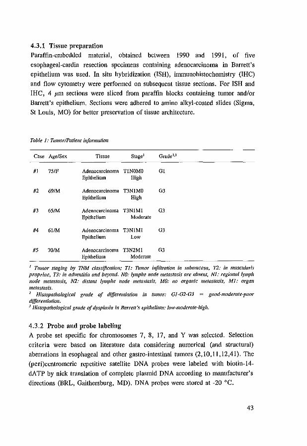
4.3.1 Tissue preparation
Paraffin-embedded material, obtained between 1990 and 1991, of five
esophageal-cardia resection specimens containing adenocarcinoma in Barrett's epithelium was used. In situ hybridization (ISH), immunohistochemistry (!HC)
and flow cytometry were performed on subsequent tissue sections. For ISH and IHC, 4 I'm sections were sliced from paraffin blocks containing tumor andlor
Barrett's epithelium. Sections were adhered to amino alkyl-coated slides (Sigma, St Louis, MO) for better preservation of tissue architecture.
Table 1.' Tumor/Pat/em ill/ormatioll
Case Age/Sex Tissue Stage l Grade1.3
#1 75/F Adenocarcinoma TlNOMO Gl Epithelium High
#2 69/M Adenocarcinoma T3N1MO G3 Epithelium High
#3 65IM Adenocarcinoma T3N1Ml G3 Epithelium Moderate
#4 6l1M Adenocarcinoma T3N1Ml G3 Epithelium Low
#5 70IM Adenocarcinoma T3N2MI G3 Epithelium Moderate
1 TUmor stagillg by TNM classification; Tl: TUmor il1ftltralioll ill submucosa. 12: ill muscularis propriae, T3: ill advemitia alld beyond. NO: lymp/le !lode metastasis are absellt, Nl,' regiollallympll /lode metastasis, N2: distallt lymp/te node metastasis, MO: I/O organic metastasis, MI: organ metastasis. 2 Histopathological grade of dijferemiatioll ill tumor: GJ-G2-G3 = good-moderate-poor dijferemialioll. J Histopathological grade of dysplasia ill Barrell's epithelium: low-moderate-higll.
4.3.2 Probe and probe labeling
A probe set specific for chromosomes 7, 8, 17, and Y was selected. Selection
criteria were based on literature data considering numerical (and structural) aberrations in esophageal and other gaslro-intestinal tumors (2,1O,11,12,41). The
(peri}centromeric repetitive satellite DNA probes were labeled with biotin-14-
dATP by nick translation of complete plasmid DNA according to manufacturer's
directions (BRL, Gaithersburg, MD). DNA probes were stored at -20°C.
43

4.3.3 In Sitn Hybridization
The in situ hybridization procedure was performed as described by van Dekken
et al. (27). Briefly, after deparaffinizing of the tissue sections in xylene, endogenous peroxidase activity was blocked with 0.3 % H,O, in methanol.
Enhancement of tissue permeability was achieved by proteolytic enzyme digestion (pepsin: Sigma, 4 mg/ml in 0.2 M HCI) for 15-20 minutes at 37 'C. Before
applying the probe set, the optimal digestion time for each tumor was determined by a pepsin time series (5, 10, 15 and 20 minutes).
Tissue DNA was heat denatured for 2 minutes in 70% formarnide in 2xSCC (pH
7.0), followed by dehydration in an ethanol series. Chromosome specific
repetitive DNA probes were denatured for 5 minutes at 70 'C in a hybridization
mixture, containing: 1-2 I'g/ml probe DNA, 500 I'g/ml sonicated herring sperm
DNA (Sigma), 0.1 % Tween-20, 10% dextran sulphate, and 60% formarnide in
2xSSC at pH 7. The probe mixture was then applied to the sections and
incubated overnight at 37 'C. Then the slides were washed in 60% formamide in
2xSSC (pH 7.0) at 42 'C.
Histochemical detection was perfornled by immunoperoxidase staining. Slides were subsequently incubated for 30 minutes at 37 'C with mouse-anti-biotin
(Dakopatts, Glostrup, Demnark), biotin-labeled horse-anti-mouse (Vector,
Burlingame, CAl and with avidin-biotin-complex (Vectastain Elite ABC Kit).
Visualisation was achieved with 0.05% H,O" 0.5 gIl DAB, 0.1 M imidazole in PBS (Phosphate Buffered Saline). The signal was amplified with 0.5% CuS04 in
0.9% NaCI. Slides were rinsed in distilled water and counterstained with
hematoxylin. Finally, slides were dehydrated in ethanol, cleared in xylene and
mounted in Malinol (Chroma-Geselschaft, K6ngen, Germany).
4.3.4 ImmunoWstochemistry
For immunostaining of p53, DO-7 (Dakopatts) was applied, diluted 1125 in PBS
for overnight incubation. This monoclonal antibody recognizes an epitope in the
N-terminus, residing between amino acids 35 and 45 of the human p53 protein. Primary labeling of the Ki-67 antigen was performed with a monoclonal
antibody, Mib-I (Immunotech, Marseille, France), diluted 1125 in PBS. The monoclonal Mib-I reacts with native Ki-67 antigen and recombinant fragments of
the Ki-67 molecule (28). When compared with staining of Ki-67 on fresh (frozen)
tissues, Mib-I shows corresponding staining of proliferating cells in formalin
fixed paraffin embedded tissues (29). For negative controls the primary
antibodies were replaced by PBS.
44

Before immunolabeling enhancement of p53 protein staining by DO-7 was
achieved by heating slides submerged in distilled water in a microwave (700 W)
at 95°C for 2x5 minutes with an interval of 1 minute. During this interval fluid
level was checked and restored. The same procedure was followed for Ki-67
staining by Mib-I, using citrate buffer (10 mM citric acid monohydrate, pH 6.0)
instead of distilled water. Immunohistochemistry was performed using the routine
ABC-immunoperoxidase method (Vector).
4.3.5 DNA flow cytometry
DNA content of the paraffin material was measured, basically as described by
Hedley et al. (30). Tumor, dysplastic and cardia regions were selectively cut out
of the paraffin blocks. Flow cytometry and analysis of the ethidium bromide
(Sigma) stained nuclei from these areas was carried out using a Facscan (Becton
Dickinson, Mountain View, Cal.
4.3.6 Analysis
Results of ISH with probes for chromosomes 7, 8, 17, and Y were evaluated on
consecutive 4 I'm sections in previously defined areas. This approach was applied
to reduce artefacts due to tumor heterogeneity and section thickness. Leucocytes
on the same sections served as controls for hybridization quality, cardia cells
were used as a tissue specific control. For each probe the number of solid DAB
spots was scored (0, 1, 2, 3, 4, >4 spots) of 100 distinct, non-overlapping,
single or side by side 4 I'm nuclear slices. Scoring was performed by two
independent investigators. The counted spot distributions were compared,
Figure 1: (A) Frequency distributions of tile hybridization spots, obtained by in situ hybridization (ISH) 011 4 11m tissue sectiol/s with (peri~)cel1lromeric DNA probes, speciftcfor chromosomes 7,8,17 alld Y, ill Barrett's adenocarcinoma of patiellt #4. For each DNA probe the Jlumber of hybridization spots (0,1,2,3.4, > 4) was determined ill 200 lIuclei alld expressed as a percelllage, FOT each of the DNA probes a different spot distribllfioll was fOlllld. 171e chromosome 7 profile was taken "modal", based 01/ the flow cytometric DNA index. I1len a gaill can be seen for chromosome 8, whereas loss is found of 17. (B) Chromosome 8 DNA probe distribllliOll ill Barrett's adenocarcinoma (care.), Barrett's epithelium (epitlt.), normal cardia tissue and leucoC)'tes (blood) of patient #4. Strong hyperdiploidy (gain) of chromosome 8 is seell ill the adenocarcinoma. (C) III lite same case the chromosome 17 DNA probe distributioll shows loss of chromosome 17 ill the adellocarcinoma. (D) Spot distribllfioll of DNA probes 7, 8, 17 and Y ill Barrett's epithelium of case #5 reveals 11011-complete loss of chromosome Y. (E) Spot pattem of the Y probe ill patielll #2 sholVs complete loss ill Barrett's epithelium, but 1I0t ill the tumor. Note the presence of nuclei with more thall olle copy of Y ill this tumor. (F) Spot distributiolls of the DNA probes III the adenocarcilloma oj patlem #3 shows loss of the Y chromosome.
45
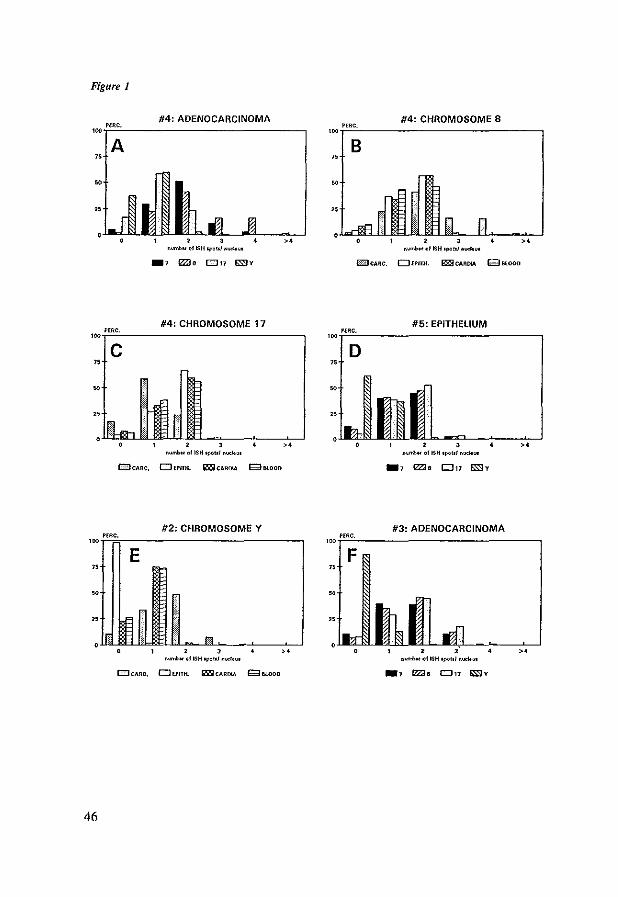
Figure 1
#4: ADENOCARCINOMA PEllC.
A
n~ 01 ISH Cpolol .,<><Itu<
#4: CHROMOSOME 17 #5~ EPITHELIUM '~T'em"'"-________________________________ -, 'ooi'O""'c·----______________________________ ,
c D "
"
EillCAAC. OfPfTlI. Il'liICAAOIA g8LOOD
46
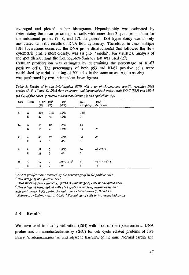
averaged and plotted in bar histograms. Hyperdiploidy was estimated by determining the mean percentage of cells with more than 2 spots per nucleus for the autosomal probes (7, 8, and 17). In general, ISH hyperploidy was closely associated with the results of DNA flow cytometry. Therefore, in case multiple ISH aberrations occurred, the DNA probe distribution(s) that followed the flow cytometric profile most closely, was assigned "modal". For statistical analysis of the spot distributions the Kolmogorov-Smimov test was used (27). Cellular proliferation was estimated by determining the percentage of Ki-67 positive cells. The percentages of both p53 and Ki-67 positive cells were established by serial counting of 200 cells in the same areas. Again scoring was performed by two independent investigators.
Table 2: Results of iii situ hybridizatioll (ISH) with a sel oj chromosome specific repetitive DNA probes (7, 8, 17 alld y), DNA flow c)'/ometry, and immullohistocliemislry with DO-7 (P53) and Mib-l
(Ki-67) a/five cases of Barrell's adenocarcinoma (A) alld epithelium (E).
c"-'" Tissue Ki-671 P531 01' ISH· ISH' (%) (%) (p2%) aneuploidy aberrations
#1 A 23% 78% 1.6151 18%
E 27 48 1.6151 7
If2 A 46 80 1.7/63 54
E 15 21 1.7/40 19 -y
#3 A 46 89 1.6116 I' -y
E 17 0 1.0/- 3
#4 A 55 0 1.9116 16 +8,-17,-Y
E 21 0 1.0/- 2
#5 A 40 0 2.0+2.7110" 17 +8,-I7,+Y/-Y
E 15 0 1.0t- 3 -y
I Kf-67: proliferation estimated by the percellfage of Ki-6? positive cells. 2 Percellfage of p53 positive cells. J DNA Index by flow cytomelfY. (P2%) is percentage oj cells ill aI/euploid peak. -I Percelltage of hyperdiploid cells (> 2 spots per IIIlcleus) measured by ISH willi cellfromeric DNA probes for autosomal chromosomes 7, 8 and 17. s Kolmogorov-Smimov lesl: p < 0.01.6 Percellfage of cells ill two aneuploid peaks.
4.4 Results
We have used in situ hybridization (ISH) with a set of (peri-)centromeric DNA probes and immunohistochemistry (IRC) for cell cycle related proteins of five
Barrett's adenocarcinomas and adjacent Barrett's epithelium. Normal cardia and
47
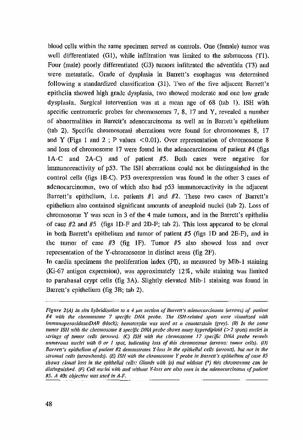
blood cells within the same specimen served as controls. One (female) tumor was well differentiated (GI), while infiltration was limited to the submucosa (TI).
Four (male) poorly differentiated (G3) tumors infiltrated the adventitia (T3) and
were metastatic. Grade of dysplasia in Barrett's esophagus was determined
following a standardized classification (31). Two of the five adjacent Barrett's
epithelia showed high grade dysplasia, two showed moderate and one low grade
dysplasia. Surgical intervention was at a mean age of 68 (tab 1). ISH with
specific centromeric probes for chromosomes 7, 8, 17 and Y, revealed a number
of abnormalities in Barrett's adenocarcinoma as well as in Barrett's epithelium (tab 2). Specific chromosomal aberrations were found for chromosomes 8, 17
and Y (Figs 1 and 2 ; P values <0.01). Over representation of chromosome 8 and loss of chromosome 17 were found in the adenocarcinoma of patient #4 (figs
IA-C and 2A-C) and of patient #5. Both cases were negative for
immunoreactivity of p53. The ISH aberrations could not be distinguished in the
control cells (figs IB-C). P53 overexpression was found in the other 3 cases of adenocarcinomas, two of which also had p53 immunoreactivity in the adjacent
Barrett's epithelium, i.e. patients #1 and #2. TIlese two cases of Barrett's
epithelium also contained significant amounts of aneuploid nuclei (tab 2). Loss of
chromosome Y was seen in 3 of the 4 male tumors, and in the Barrett's epithelia of case #2 and #5 (figs lD-F and 2D-F; tab 2). This loss appeared to be clonal
in both Barrett's epithelium and tumor of patient #5 (figs ID and 2E-F), and in
the tumor of case #3 (fig IF). Tumor #5 also showed loss and over
representation of the Y-chromosome in distinct areas (fig 2F).
In cardia specimens the proliferation index (PI), as measured by Mib-I staining
(Ki-67 antigen expression), was approximately 12%, while staining was limited to parabasal crypt cells (fig 3A). Slightly elevated Mib-I staining lVas found in
Barrett's epithelium (fig 3B; tab 2).
Figure 2(A) /1/ situ hybridization to a 41'.111 sectioll of Barrett's adenocarcinoma (arrows) of patiem #4 With Ihe chromosome 7 specific DNA probe. TIle ISH-related spots were visualized with immulIoperoxidaselDAB (black); hematoxylin was used as a COlillterstaill (grey). (8) In tile same fIlmor ISH with the chromosome 8 specific DNA probe shows mall)' hyperdiploid (> 2 SpOIS) nuclei ill strings of tumor celfs (arrows). (C) ISH wit" the chromosome 17 specific DNA probe reveals Ilumerous lIuclei wil" 0 or 1 spot. indicating loss of this chromosome (arrows: //lmor cells). (D) Barrett's epithelium oj patielll #2 demollstrates Y-Ioss ill the epithelial cells (arroll's), bur lIot in the stromal cells (arrowheads). (E) ISH with tile chromosome Y probe ill Barrell's epithelium oj case #5 shows clol/al loss ill tile epithelial cells: Glands wit" (0) and without (If) this chromosome call be distinguished. (F) Cell Jluclei with alld without Y-loss are also seen ill the adellocarcilloma oj patielll #5. A 40x objecti\'e was Ilsed in A-F.
48
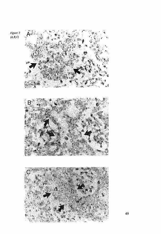
Figure 2
(A,B,C)
h,i ,if -.....::'
~:ii;j.",
•• "
49

Figure 2
(D,E,F)
50
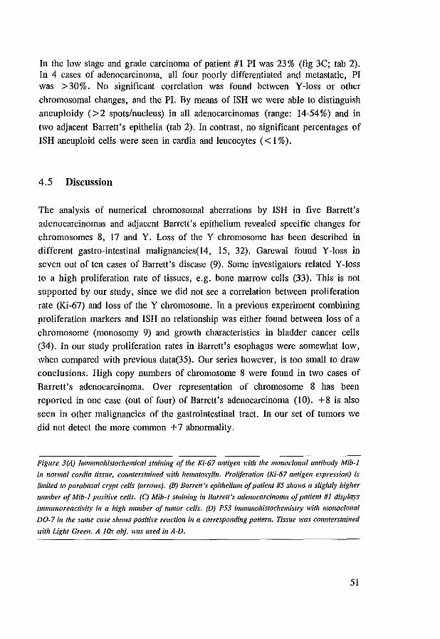
In the low stage and grade carcinoma of patient #1 PI was 23% (fig 3C; tab 2). In 4 cases of adenocarcinoma, all four poorly differentiated and metastatic, PI was > 30%. No significant correlation was found between Y-Ioss or other
chromosomal changes, and the PI. By means of ISH we were able to distinguish
aneuploidy (>2 spots/nucleus) in all adenocarcinomas (range: 14-54%) and in
two adjacent Barrett's epithelia (tab 2). In contrast, no significant percentages of
ISH aneuploid cells were seen in cardia and leucocytes « 1 %).
4.5 Discussion
The analysis of numerical chromosomal aberrations by ISH in five Barrett's
adenocarcinomas and adjacent Barrett's epithelium revealed specific changes for
chromosomes 8, 17 and Y. Loss of the Y chromosome has been described in
different gastro-intestinal malignancies(l4, 15, 32). Garewal found Y-Ioss in seven out of ten cases of Barrett's disease (9). Some investigators related Y-Ioss
to a high proliferation rate of tissues, e.g. bone marrow cells (33). This is not supported by our study, since we did not see a correlation between proliferation
rate (Ki-67) and loss of the Y chromosome. In a previous experiment combining proliferation markers and ISH no relationship was either found between loss of a
chromosome (monosomy 9) and growth characteristics in bladder cancer cells
(34). In our study proliferation rates in Barrett's esophagus were somewhat low,
when compared with previous data(35). Our series however, is too small to draw conclusions. High copy numbers of chromosome 8 were found in two cases of
Barrett's adenocarcinoma. Over representation of chromosome 8 has been
reported in one case (out of four) of Barrett's adenocarcinoma (10). +8 is also
seen in other malignancies of the gastrointestinal tract. In our set of tumors we
did not detect the more common +7 abnormality.
Figure 3(A) Immunohistochemical staining of the Ki-67 antigen with the mOl/oelonal olltihody Mib-l
ill normal cardia tisslle, cOlllllerstailled with hematoxylin. Proliferation (Ki-67 alltigell expression) is limited to parabasal crypt cells (arrows). (8) Barrett's epithelium ojpaliellt #5 shows a slightly higher
JlllIllber of Mib-J positi~'e ceUs. (C) Mib-l staillillg iii Barrett's adenocarcinoma oJpatielll #1 displays immulloreactivity ill a high number of (umor cells. (D) P53 illllllllllOllistochemistry with monoclollal DO-7 ill the same case shows positive reactioll ill a correspondillg pattern. Tisme was COllllterstailled
with light Green. A lOx obj. lI'as used ill A-D.
51
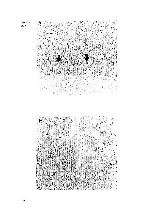
Figure 3
lA, B)
52
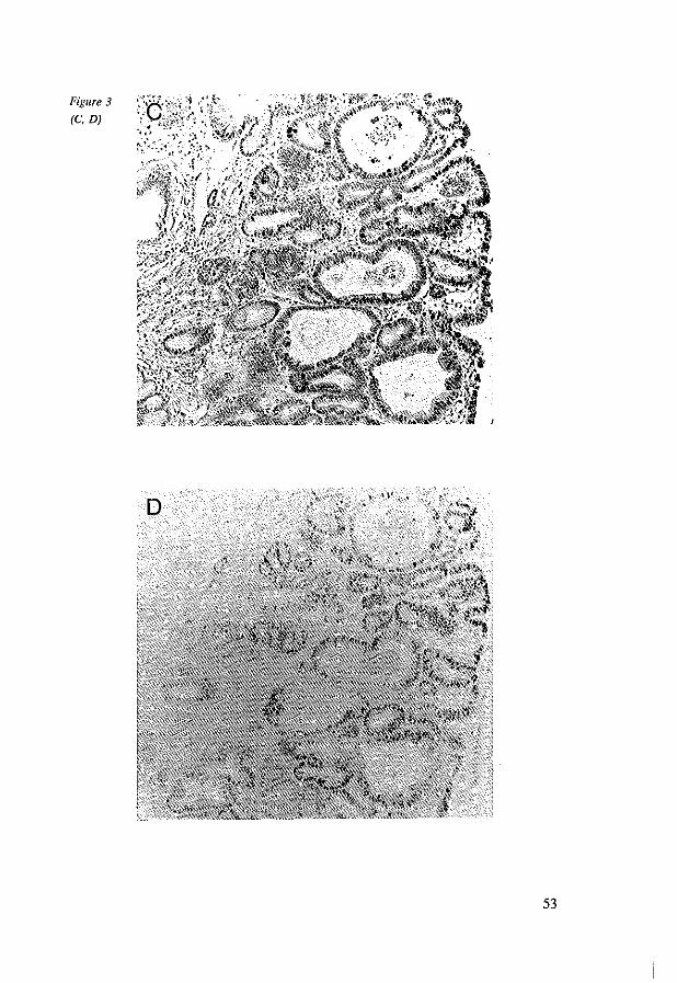
Figure 3
(e, D)
53
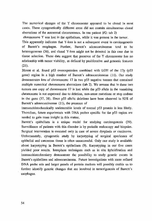
The numerical changes of the Y chromosome appeared to be clonal in most
cases. These cytogenetically different areas did not contain simultaneous clonal aberrations of the autosomal chromosomes. In one patient (#2: tab 2)
chromosome Y was lost in the epithelium, while it was present in the tumor.
This apparently indicates that Y-Ioss is not a subsequent event in carcinogenesis
of Barrett's esophagus. Further, Barrett's adenocarcinomas tend to be
heterogeneous (36), and clonal Y-Ioss might not be detected in this case due to
tissue selection. These data suggest that presence of the Y chromosome has no
relationship with tumor viability, as defined by proliferative and genomic features (21).
Blount et al. found p53 overexpression combined with LOH of the 17p (p53
gene) region in a high number of Barrett's adenocarcinomas (II). Our study
demonstrates loss of chromosome 17 in two p53 negative tumors that contained
multiple numerical chromosome aberrations (tab 2). We assume that in these two
tumors one copy of chromosome 17 is lost while the p53 allele in the remaining
chromosome is not expressed due to deletion, non-sense mutations or stop codons in the gene (37, 38). Since p53 allelic deletions have been observed in 92% of
Barrett's adenocarcinomas (I I), the presence of
immunohistochemically undetectable levels of normal p53 protein is less likely. Therefore, future experiments with DNA probes specific for the p53 region are
needed to gain more insight in this matter.
Barrett's epithelium is a unique model for studying carcinogenesis (39).
Surveillance of patients with this disorder is by periodic endoscopy and biopsies. Surgical intervention is executed only in case of severe dysplasia or carcinoma.
Unfortunately, cytogenetic study by karyotyping of surgical specimens of
epithelial and cancerous tissue is often unsuccessful. Only one study is available
about karyotyping in Barrett's epithelium (9). Karyotyping in our five cases yielded poor results. Interphase techniques such as in situ hybridization and
immunohistochemistry demonstrate the possibility to study genetic events in Barrett's epithelium and adenocarcinoma. Future investigations with more refined
DNA probe sets and larger panels of protein markers will possibly enable us to
further identify genetic changes that are involved in tumorigenesis of Barrett's
esophagus.
54
4.6 Fteferences
1. Barrett NR. Chronic peptic ulcer of the oesophagus and oesophagitis. Br J SlIrg 38;1950: 175-182. 2. Sjogren R Jr., Johnson LF. Barrett's esophagus: a review. [Review]. Am J Med 74;1983:313-21. 3. Spechlcr SJ, Robbins AH, Rubins HB, Vincent ME, Heeren T. DaDs WG, Colton T, Schimmel
EM. Adenocarcinoma and Barrett's esophagus. An overrated risk? Gastroelltero[ogy
87;1984:927·33. 4, Van def Veen AH, Dees It Blankensleijn JD, Van B1ankenstein M. Adenocarcinoma in Barrett's
oesophagus: an overrated risk. Gll/ 30;1989:14-8. 5. Herscovic A, Martz K, AI-Safaf M, Leichman L, Brindle I, Vaitkevicius V. Cooper J, Byhardt R,
Davis L, Emami B. Combined chemotherapy and radiotherapy compared with radiotherapy alone in cancer of the esophagus. N Eng/ J Med 326;1992:1593-1598.
6. Chejfec G, Schnell T, Sonlag S. Barrett's esophagus a preneoplastic disorder. Am J Clill Palllol 98;1992:5·7.
7. Spechler S1. Barrett's esophagus. Gastroelltero(ogy 8;1992:573-578. 8. Reid BJ, Blount PL, Rubin CE, Levine DS, Haggitt RC, Rabinovitch PS. Flow-cytometric and
histological progression to malignancy in Barrett's esophagus: prospective endoscopic surveillance of a cohort. Gastroenterology 102;1992: 1212-9.
9. Garewal HS, Sampliner R, Liu Y, Trent JM. Chromosomal rearrangements in Barrett's esophagus. A premalignant lesion of esophageal adenocarcinoma. Callcer Genet Cytogellet 42;1989:281-6.
10. Raskind WH, Norwood T. Levine DS, Haggitt RC. Rabinovitch PS, Reid BJ. Persistent clonal areas and clonal expansion in Barrett's esophagus. Concer Res 52;1992:2946-50.
11. Blount PL, Ramel S, Raskind WH, Haggitt RC. Sanchez CA, Dean PJ, Rabinovitch PS, Reid m. I7p allelic deletions and p53 protein overexpression in Barrett's adenocarcinoma. Callcer Res 51; 1991 :5482·6.
12. Ramel S, Reid BJ, Sanchez CA, Blount PL, Levine DS. Neshat K. Haggitt RC, Dean PJ. Thor K, Rabinovitch PS. Evaluation of p53 protein expression in Barrett's esophagus by two-parameter flow cytometry. Gastroelllerofogy 102; 1992: 1220-8.
13. Huang H. Auerbach R. Identification and characterization of hematopoietic stem cells from the yolk sac of the early mouse embryo. Proc Natl Acad Sci USA 90;1993: 10110-4.
14. Ochi H, Douglas JHO, Sandberg AA. Cytogenetic studies in primary gastric cancer. Concer Genet C)'togellet 22; 1986:295·307.
15. Ferti-Passantonopoulou AD, Panani AD, Vlachos JD, Raptis SA. Common cytogenetic fmdings in gastric cancer. Oll/cer Genet Cytogenet 24;1987:63-73.
16. Bardi G, Johansson B, Pandis N, Bak-Jensen E, Orndal C, Heim S, Mandahl N, Andren-Sandberg A, Mitelman F. Cytogenetic aberrations in colorectal adenocarcinomas and their correlation with clinicopathological features. Concer 71;1992:306-314.
17. Sandberg AA, Ture-Carel C. Gemmill RM:. Chromosomes in solid tumors and beyond. Concer Res 48;1988: 1049·1059.
18. Arnoldus EPJ, Noordermeer lA, Peters ACB, Raap AK. Van der Ploeg M. Interphase cytogenetics of brain nUIlors. Gelles Chrolllosom Cnllcer 3;1991:101-107.
19. Cremer T. Tesin D, Hopman AH, Manuelidis L. Rapid interphase and metaphase assessment of specific chromosomal changes in neuroectodennal tumor cells by in situ hybridization with chemically modified DNA probes. Exp Cell Res 176;1988:199-220.
20. Hopman AHN, Moesker 0, Smeets AWGB, Pauwels RPW, Vooijs GP, Ramaekers FCS. Numerical chromosome 1. 7. 9 and II aberrations in baldder cancer detected by in situ hybridization. COllcer Res 51;1991:644-651.
21. Van Dekken H, Pizzala JG, Kelsen DP, Melamed MR. Targeted cytogenetic analysis of gastric tumors by in situ hybridization with a set of chromosome specific DNA probes. Oll/cer 66;1990:491497.
22. Waldman PM, Carroll PR, Kerschmann R, Cohen MB, Field FG, Mayall BH. Centromeric copy number of chromosome 7 is strongly correlated with tumor grade and labeling index in human
55
bladder cancer. Cancer Res 51;1991:3807-3813. 23. Emmerich P, Jauch A, Hofmalm MC. Cremer T, Walt H. Interphase cytogenetics in paraffIn
embedded sections from human testicular germ cell tumor xenografts and in corresponding cultured cells. Lab Invest 61;1989:235-242.
24. Burns J, Redfern DR1vf. Esiri MM. McGee JO. Human and viral gene detection in routine paraffIn embedded tissue by in situ hybridization with biotinylated probes. J Clill PatllOl 39; 1986: 1066-1073.
25. Pringle JH, Homer CE, Warford A. Kendall CH, Lauder I. In situ hybridization: Alkaline phosfatase visualization of biotinylated probes in cryostat and parafflllSections. Histochem J 19;1987:488-496.
26. Van de Kaa CA. Nelson KAM. Ramaekers FCS, Vooijs PG. Hopman AHN. Interphase cytogenetics in paraffin sections of routinely processed hydatiform moles en hydropic abortions. J PatlwI165;1991:281-287.
27. Van Dekken H, Kerstens MHJ. Tersteeg TA. Verhofstad AAJ. Vooijs GP. Histological preservation after in situ hybridization to archival solid tumour sections allows discrimination of cells bearing numerical chromosome changes. J PathoI168;1992:317-324.
28. Gerdes 1, Becker MHG. Key G. Cattoretti G. Immunohistochemical detection of tumour growth factor (ki-67 antigen) in formalin-fixed and routinely processed tissues. J PatII0185;1992:85-87.
29. Cattoreui G. Becker MHG. Key G. Duchrow M, Schluter C. Galle J. Gerdes J. Monoclonal antibodies against recombinant parts of the Ki-67 antigen (Mibl and Mib3) detect proliferating cells in microwave-processed formalin-fixed paraffin sections. J PatlIOI168;1992:357-363.
30. Hedley DW, Friedlander ML, Taylor IW. Rugg CA, Musgrove EA. Method for analysis of cellular DNA content of paraffm embedded pathological mete rial using flow cytometry. J Histochem Cytocilem 31;1983:1333-1335.
31. Morson BC, Sobin LH. Grundman E, et al. Precancerous conditions and epithelial dysplasia in the stomach. J elill Patl10133; 1980:711-721.
32. Xiao S, Wei W. Leng X, Shi Y, Liu Q, Li P. Direct chromosome analysis of seven primary colorectal carcinomas. Cancer Genet Cytogellet 62; 1992:32-39.
33. Pierre RV, Hoagland HC. Age-associated anueploidy: Loss of Y- chromosome from human bone marrow cells with aging. Callcer 30;1970:889-894.
34. Van Dekken H, Shervish EW. Pizzolo JG, Fair WR. Melamed MR. Sinmltaneous detection of flourescent in situ hybridization and in vivo incorporated BrdU in a human bladder tumour. J PatIIO/164;1991:17-22.
35. Jankowski J, McMenemin R, Hopwood D. Penston J, Wormsley KG. Abnormal expression of growth regulatory factors in Barrett's oesophagus. Clill Sci 81;1991:663-8.
36. Rabinovitch PS, Reid BI. Haggitt RC, Norwood TH, Rubin CEo Progression to cancer in Barrett's esophagus is associated with genomic instability. Lab [lives/ 60;1989:65-71.
37. Levine AJ, Momand J, Finlay CA. The p53 tumour suppressor gene. [Review]. Nature 351;1991:453-6.
38. Vogelstein B, Kinzler KW. P53 function and dysfunction. Cell 70;1992:523-526. 39. Hartwell L. Defects in a cell cycle checkpoint may be responsible for genomic instability of cancer
cells. Cell 71;1992:543-546.
56
Chapter 5
Accumulation of P53 Protein in Normal, Dysplastic aud Neoplastic Banett's Esophagus.
K. K. Krishnadath, H. W. Tilrums, M. Vrul Blankenstein,
F. T. Bosmrul and A. H. Mulder.
Published in: J Pathol 1995;175:175-180

5.1 Abstract
Accumulation of p53 protein was determined by immunohistochemistry in
archival material of biopsy specimens from 102 patients with Barrett's esophagus
with different grades of dysplasia, in 24 esophageal adenocarcinomas associated
with Barrett's esophagus and in 23 cases of metaplastic epithelium adjacent to
these carcinomas. Immunostaining for the p53 protein was found in 23/102 (23%) cases of the Barrett's esophagus biopsies and in 12123 (52%) cases of
Barrett's esophagus adjacent to adenocarcinoma. Significant correlations were
found between grade of dysplasia and p53 immunoreactivity in both Barrett's
biopsies without adenocarcinoma (p<O.OOI) and Barrett esophagus adjacent to
adenocarcinoma (p<0.05). In the adenocarcinomas intense nuclear immunohistochemical staining for p53 was diffusely or focally present in 20/24
(83%) of the specimens. In Barrett's esophagus p53 is a progression marker with
high expression in high grade dysplasia (89%) and adenocarcinoma (83%).
5.2 Introduction
In Barrett's esophagus the stratified squamous epithelium of the distal esophagus is replaced by metaplastic columnar epithelium (1,2). Patients with Barrett's
esophagus have a 30·40 fold increased risk of developing esophageal adeno
carcinoma (3,4). Although adenocarcinoma in Barrett's esophagus is preceded by
epithelial dysplasia, time course of progression of simple metaplasia into dysplastic Barrett's esophagus and ultimately into infiltrating adenocarcinoma is
unknown. Furthermore, grading of dysplasia is to a certain extent subjective,
leading to significant interobserver variation. Therefore, objective prognostic
criteria predicting which cases of Barrett's esophagus will progress towards malignancy are of major importance. Previous immunohistochemical studies with
proliferation markers already showed higher proliferation rates in dysplastic
Barrett's esophagus (5) and abnormalities measured by DNA flow cytometry may
precede malignancy (6). During the past years interest in tumor suppressor genes and proteins in
(pre)malignant lesions has intensified. The appearance of the p53 tumor
suppressor protein has been reported in a wide variety of malignant en
premalignant conditions (7-14). P53 immunoreactivity has been reported in 62%-
87 % of esophageal adenocarcinomas, and in non dysplastic and dysplastic
59

Barrett's esophagus (15-18).
Aim of the present study was to determine whether immunohistochemically
detectable p53 protein accumulation occurs along with the development of dysplasia in Barrett's esophagus.
5.3 Materials and methods
5.3.1 Tissues P53 immunoreactivity was studied in archival biopsies taken between 1980 and
1992 from 102 patients with Barrett's esophagus with various degrees of
dysplasia but without adenocarcinoma. The number of biopsies per patient varied
from one to twelve. Furthermore, paraffin-embedded tissue blocks from 24 esophagus-cardia resection specimens with adenocarcinoma in Barrett's esophagus
encountered between 1990 and 1992 were investigated. All patients had undergone cardiac-esophageal resection with gastric tube interposition. Surgery was
performed at a mean age of 62 (range: 45-80). Non of the patients with adenocarcinoma had received chemo- or radiotherapy prior to surgery. One
paraffin block containing adenocarcinoma was selected in each case. To determine the efficacy of p53 immunostaining in paraffin embedded tissues,
immunohistochemistry for the detection of p53 protein was performed on
corresponding fresh frozen samples of 20 of the adenocarcinomas. Barrett's
esophagus adjacent to the tumor was found in 23 cases. In one case Barrett's esophagus was diagnosed in biopsy specimens prior to esophageal resection.
Normal gastric-cardiac mucosa was used for control purposes.
5.3.2 Histopathological criteria
The histological sections were reviewed by two pathologists (A.H.M. and
F.T.B.) Tumor staging was performed according to pTNM criteria. Barrett's esophagus was defined as columnar epithelial lining of the esophagus of at least
three em proximal to the gastroesophageal junction. All cases had specialized
columnar type. of metaplasia characterized by the presence of intestinal type of
absorptive cells e.g., goblet cells. Dysplasia was graded into three categories following criteria revised by Reid et al. (19), Le.; negative, indefinite or low
grade dysplasia and high grade dysplasia. Slides were coded and scoring of p53 immunoreactivity was performed independent from grading sections.
60
5.3.3 Immunohistochemistry
Cryostat sections (5 I'm) of fresh tissues were airdried aud fixed in acetone (-
20·C). After blocking of endogenous peroxidase activity aud rinsing in phosphate
buffered saline (PBS, pH 7.4), p53 protein labeling was performed by staudard
peroxidase-auti-peroxidase method (Dakopatts Z 259; Sigma P 2416, St Louis,
MO).
DO-7 (Dakopatts, Glostrup, Denmark), au Ig2b kappa monoclonal autibody
recognizing au epitope in the N-tenninus between amino acids 35 aud 45 of
human p53 protein, was used on formalin-fixed paraffin-embedded tissues.
For paraffin sections au autigen retrieval method was applied (20). After
blocking of endogenous peroxidase P53 labeling with DO-7 was performed by
the streptavidin-biotin-complex procedure. The sections were preincubated for 15
minutes at 37·C with 10% normal goat serum, diluted in PBS. DO-7, diluted
1125 in PBS, was added for overnight incubation at 4 ·C. The negative controls
were incubated with PBS only. The sections were subsequently incubated with
biotinylated goat-auti-mouse IgG (1/400 in PBS, Dakopatts), rinsed aud finally
incubated with a Streptavidin-biotinylated horseradish peroxidase complex
(Dakopaus). Sections were developed for seven minutes in 3.3' diaminobenzidine
tetrachloride.
5.3.4 Microscopical evaluation
Positive immunohistochemistry with the DO-7 p53 autibody was considered by moderate or intense brown nuclear staining. Cytoplasmic staining was not found. In a semi-quautitative scoring approach, two staining patterns were distinguished: A focal paUern, with > 10% of nuclei positive in clusters was mostly found in non dysplastic aud low grade dysplastic Barrett's esophagus. The focal staining was mostly confined to epithelial glauds (figure lA). A diffuse staining pattern with > 10% of positive nuclei scattered through the tissue was seen in most adenocarcinomas aud high grade dysplasia (figure lB).
Figure 1 A: Focal immullohistochemical stail/illg of Ihe p53 protein with the mOl/oclol/al a1llibody DO-7 ill a paraffin embedded sectioll of a Barrett's esophagus biopsy wilh low grade dysplasia. B: Moderately differelltiated esophageal adenocarcinoma showing a diffllse iml1lllllOslainilig pattem of tllmor cells. Both secliolls were coullterstailled by hematoxylin. A Jar objeclil'e was used.
61
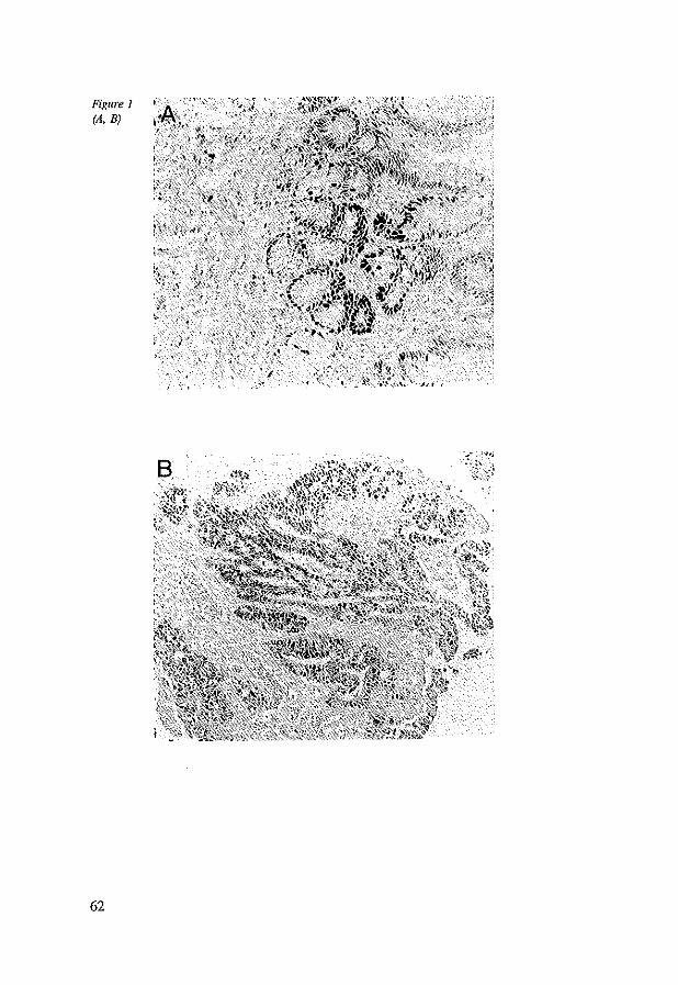
Figure I lA, B)
62
B .( :.:~'.
·-~r}
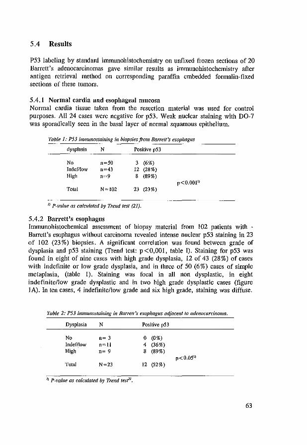
5.4 Results
P53 labeling by standard immunohistochemistry on unfixed frozen sections of 20 Barrett's adenocarcinomas gave similar results as immunohistochemistry after antigen retrieval method on corresponding paraffin embedded formalin-fixed sections of these tumors.
5.4.1 Normal cardia and esophageal mucosa Normal cardia tissue taken from the resection material was used for control purposes. All 24 cases were negative for p53. Weak nuclear staining with DO-7 was sporadically seen in the basal layer of normal squamous epithelium.
Table 1: P53 immullostaillillg ill biopsies from Barrett's esophagus
dysplasia N Positive p53
No n~50 3 (6%) Indefllow n~43 12 (28%) High 0=9 8 (89%)
p<O.OOI" Total N~102 23 (23%)
I) P-vo[ue as calculated by Trend test (21).
5.4.2 Barrett's esophagus Immunohistochemical assessment of biopsy material from 102 patients with -Barrett's esophagus without carcinoma revealed intense nuclear p53 staining in 23 of 102 (23%) biopsies. A significant correlation was found between grade of dysplasia and p53 staining (Trend test: p<O,OOI, table I). Staining for p53 was found in eight of nine cases with high grade dysplasia, 12 of 43 (28 %) of cases with indefinite or low grade dysplasia, and in three of 50 (6%) cases of simple metaplasia, (table 1). Staining was focal in all non dysplastic, in eight indefinitellow grade dysplastic and in two high grade dysplastic cases (figure lA). In ten cases, 4 indefinitellow grade and six high grade, staining was diffuse.
Table 2: P53 immullostaillillg ill Barrett's esophagus adjacent to adenocarcilloma.
Dysplasia N Positive p53
No n= 3 0 (0%) Indefllow n~ll 4 (36%) High n= 9 8 (89%)
p<O.OSI) Total N~23 12 (52%)
1) P-vafue as calculated by Trend res?l,
63

5.4.3 Barrett's esophagus adjacent to adenocarcinoma. Staining for p53 in Barrett's esophagus adjacent to the infiltrating tumor was performed in 23 cases. P53 staining correlated significantly with grade of dysplasia (Trend test p <0.05, table II). Eight of nine (89%) of the severely dysplastic cases showed diffuse staining. In four out of II (36%) cases with indefinite for or low grade dysplasia focal p53 staining was observed. P53 immunostaining was not observed in the three non dysplastic epithelia adjacent to tumor. In a total of II cases of adjacent Barrett's esophagus p53 immunoreactivity was not seen, four of which were adjacent to adenocarcinomas lacking p53 staining as well.
5.4.4 Adenocarcinoma in Barrett's esophagus The 24 cases of adenocarcinoma in Barrett's esophagus were distributed according to tumor stage as shown in table three (table III). In 20 of 24 cases (83 %) diffuse (19 cases) or focal (I case) nuclear immunohistochemical staining for p53 was found (figure lB). Staining was similar in the three stage groups, with a slight preponderance of negative cases in the advanced tumors. Also no significant correlation was found between p53 staining and grade of differentiation.
Table 3: P53 immtmostainillg ill adenocarcinomas ill Barrett's esophagus.
Tumor slagel) N Positive p53 2)
Tl n= 6 6 (100%) T2 n= 6 5 (83%) T3/4 n=12 9 (75%)
Total N~24 20 (83%)
1) Tumor stage accordillg to TNAI classification; Tl: filmor il1fi/fratioll ill submucosa, T2: ill muscularis propria, T3rr4: ill adl'elltitia and beyond. }) Focal or diffuse nuclear ill/munos/aluing.
5.5 Discussion
The study of different stages in carcinogenesis in a model which includes early stages of cancer development has important clinical implications. Barrett's esophagus might serve as a unique model in which early changes preceding full blown malignancies are encountered. Patients suffering from Barrett's changes are monitored by periodic endoscopies and biopsies, allowing longitudinal follow up studies. Although Barrett's esophagus may lead to malignancy, the course of the development of carcinoma in the individual can hardly be predicted by histopathology. Therefore tracing more objective parameters associated with dysplasia and reflecting the progression of malignancy is of paramount importance. P53 mutation, a common genetic event in many human malignancies
64
(7-14), might be such a parameter. Positive pS3 protein immunostaining is generally assumed to occur in case of gene mutations, which results in increased stability and consequently accumulation of mutated pS3 protein (22). Intense immunostaining with the DO-7 anti pS3 antibody has proved to be highly specific for mutated pS3 (23). In the present study nuclear pS3 protein staining was shown in 23 of 102 (23 %) biopsies from Barrett's esophagus with different grades of dysplasia and in 12 of 23 (S2%) Barrett's esophagus adjacent to tumor. In these specimens pS3 staining correlated significantly with grade of dysplasia (p < 0,00 1 and p < O,OS respectively), with a high prevalence (89%) in high grade dysplasia. The number of cases with nuclear pS3 immunostaining in Barrett's esophagus in our study is somewhat higher than previously reported (16,17,18). Differences in the applied monoclonal antibodies and/or in the histochemical techniques used, may account for the discrepancies. Positive pS3 immunostaining in Barrett's esophagus with no or low grade dysplasia (16,17,18), may indicate that pS3 protein accumulation is an early event during carcinogenesis in Barrett's esophagus. Focally staining areas in these Barrett's esophagus may be associated with pS3 gene mutations, moreover these clones may be precursors of progressive lesions with ultimately diffuse pS3 staining. PS3 gene mutations in non dysplastic and minimal dysplastic Barrett's esophagus were demonstrated by PCR and sequence analysis by Casson et al (24). Alternatively, increased amounts of wild type pS3 protein, resulting in positive immunostaining, were noticed in irradiated and highly proliferative tissues (2S,26). Since the DO-7 monoclonal also reacts with wild type pS3 protein and proliferation rates are high in Barrett's esophagus (S), we cannot rule out that in a subset of cases immunostaining was a result of immunoreaction of DO-7 with wild type pS3. Furthermore, using monoclonal antibodies, certain conditions may give false negative results, e.g., when the epitope which is recognized by the DO-7 monoclonal antibody is mutated or inaccessible as a consequence of complex formation of pS3 with other proteins (27). On the other hand complete absence of pS3 protein (due to loss of both pS3 alleles, non-sense mutations or stop codons in the gene) would not be distinguishable from normal levels of wild type pS3 by immunohistochemical techniques; in both conditions staining would be negative. In the present study pS3 immunostaining was observed in 20 out of 24 (83 %) adenocarcinomas in Barrett's esophagus. In esophageal adenocarcinomas associated with Barrett's esophagus, Younes et a!., reported pS3 immunostaining in 87% (7 of 8) of cases. Additionally, in a follow-up series of 24 Barrett's esophagus patients three cases progressed to high grade dysplasia, two of which had pS3 immunostaining and low grade dysplasia in initial biopsy specimens (16). In the study of Blount et al., pS3 immunostaining was found in 62% (8 of 13) of esophageal adenocarcinomas, whereas allelic loss of the pS3 gene (17p) was detected in 92% of cases (IS). Although we did not find a correlation between tumor grade and pS3 protein accumulation, a trend between increasing pS3
6S
expression with reduced tumor differentiation was recently seen in a larger series (18). The absence of p53 staining in four tumors and in two cases of highly dysplastic Barrett's esophagus may indicate that malignant progression is possible without involvement of the p53 gene (28). P53 staining in tumor and in adjacent Barrett's esophagus do not necessarily correspond. In seven out of 19 patients Barrett's esophagus adjacent to p53 positive tumor tissue showed no p53 immunostaining. None of these cases showed severe dysplasia. A similar observation was reported by Flejou et aI., who found that p53 immunostaining in Barrett's esophagus adjacent to p53 positive adenocarcinomas was only present in case the epithelium was severely dysplastic (29). Recently Hardwick et aI., found positive p53 immunostaining in 10 cases of dysplastic Barrett's esophagus, all adjoining p53 positive esophageal adenocarcinomas (30). In the present study four carcinomas did not show p53 immunostaining. In all these cases adjacent Barrett's esophagus, regardless of degree of dysplasia, were negative as well. In conclusion, we found that p53 protein accumulates in Barrett's adenocarcinoma and in premalignant lesions, correlating with the degree of dysplasia. P53 immunostaining might therefore constitute a useful diagnostic tool. However, the value of p53 immunostaining in biopsies without high grade dysplasia in predicting the chance of development of carcinoma should be determined in prospective follow-up studies.
5.6 References:
1. Barrett NR. Chronic peptic ulcer of the oesophagus and esophagitis. Bf J Surg 1950;38:175-182. 2. Spechlcr S1. Barrett's oesophagus. Gastroenterology 1992;8:573-578. 3. Spechler SJ, Robbins AH, Rubins HB, et a!. Adenocarcinoma and Barrett's oesophagus. An
overrated risk? Gastroenterology 1984;87:927-933. 4. Van def Veen AH, Decs J, BJankensteijn JD, et a!. Adenocarcinoma in Barrett's oesophagus: An
overrated risk. Gut 1989;30:14-18. 5. Reid Bl, Sanchez CA, Blount PL, Levine DS. Barrett's Oesophagus: Cell cycle abnonnalities, in
advancing stages of neoplastic progression. Gastroenterology 1993; 105: 119-129. 6. Fennerty MB, Sampliner RE, Garewal HS. Review article: Barrett's oesophagus-cancer risk,
biology and therapeutic management. Aliment Phannacol Ther 1993;7:339-345. 7. Porter PL, Gown AM, Kramp SG. et al. Widespread p53 overexpression in human malignant
tumors. An immunohistochemical study using methacarn-fixed. embedded tissue. Am J Pathol 1992;140: 145-153.
8. Nigro JM. Baker SJ. Preisinger AC. et a!. Mutations in the p53 gene occur in diverse hUman tumor types. Nature 1989;342:705-708.
9. Hollstein M. Sidransky D. Vogelstein B, et at. P53 mutations in human cancers. Science 1991;253:49-53.
10. Campo E. de la Calle-Martin 0, Miquet R. et al. Loss of heterozygosity of p53 gene and p53 protein expression in human colorectal carcinomas. Cancer Res 1991;51:4436-4442.
11. Caamano J, Ruggeri B. Momiki S, et al. Detection of p53 in primary lung tumors and nonsmall cell lung carcinoma cell lines. Am J Pathol 1991; 139:839-845.
12. Van den Berg FM, Tigges N. Schipper ME, et a1. Expression of the lluclear oncogene p53 in colon tumours. J PathoI1989;157:193-199.
66
13. Kaklamanis L, Gatter KC, Mortensen N, et al. P53 Expression in colorectal adenomas. Am 1 Pathol 1993:1:87-93.
14. 1M Kros, KK Krishnadath, IC) Godschalk. et al. Expression of p53 in oligodendrogliomas. 1 Pathol 1993:171:285-290.
15. Blount PL, Ramel S, Raskind WHo et al. 17p allelic deletions and p53 protein overexpression in Barrett's adenocarcinoma. Cancer Res 1991;51:5482-5486.
16. Younes M. Lebovitz RLM, Lechago LV, Lechago 1. P53 protein accumulation in Barrett's metaplasia, dysplasia and carcinoma: a follow-up study. Gastroenterology 1993;105:1637-1642.
17. Ramel S, Reid B1, Sanchez CA, et al. Evaluation ofp53 in Barrett's oesophagus by two-parameter flow cytometry. Gastroenterology 1992;102:1220-1228.
18. Symmans PI, Linehan 1M, Brito MI, Filipe MI. P53 expression in Barrett's oesophagus, dysplasia, and adenocarcinoma using antibody DO-7. 1 PathoI1994;173:
19. Reid B), Weinstein WM, Lewin 10, et a1. Endoscopic biopsy can detect high-grade dysplasia or early adenocarcinoma in Barrett's oesophagus without grossly recognizable neoplastic lesions. Gastroenterology 1988;94:81-90
20. Shi SR, Key ME, Kalra KL. Antigen retrieval in fonnaHn-ftxed. paraffm-embedded tissues: an enhancement method for inununohistochemical staining based on microwave oven heating of tissue sections. 1 Histochem Cytochem 1991;39:741-748.
21. N. Mantel. Chi square tests with one degree of freedom. 1ASA 1963;58:690-700. 22 Finlay CA, Hinds PW, Tan TH, et al. Activating mutations for transfonnation by p53 produce a
gene product that fonus and hsc700-p53 complex with an altered half-life. Moll Cell Bioi 1988:8:531-539.
23. Baas 10, Mulder IWR, Offerhaus GJA, Vogelstein B, Hamilton SR. An evaluation of six antibodies for inlnlUnohistochemistry of mutant p53 gene product in archival colorectal neoplasms. J Pathoi 1994:172:5-12.
24. Casson AG, Mukhopadyay T, Cleary KR, Ro IY, Levin B, Roth IA. P53 gene mutations in Barrett'S epithelium and oesophageal cancer. Cancer Res 1991;51:4495-4499.
25. Lane DP. P53 Guardian of the genome. Nanue 1992;358: 15-16. 26. Kuerbitz S1, Plunkett BS. Walsh WV, Kaslan MB. Wild-type p53 is a cell cycle checkpoint
determinant following irradiation. Proc Nat! Acad Sci USA 1992;89:7491-7495 27. Vogelstein B. Kinzler KW. P53 function and dysfunction. CeI11992;70:523-526_ 28. Levine Al, Momand 1, Finlay CA. The p53 tumour suppressor gene. Nature 1991;351:453-456. 29 Flejou IF, Potet P, Mureau F, et al. Overexpression of p53 protein in Barrett's syndrome with
malignant transfonnation. 1 Clin Pathol 1993;46:330-334. 30. Hardwick RH, Shepherd NA, Moorghen M, Newcomb PV. Alderson D. Adenocarcinoma arising in
Barrett's oesophagus. evidence for the participation of p53 dysfunction in the dysplasia/carcinoma sequence. Gut 1994;35:764-768.
67

Chapter 6
Accumulation of Genetic Abnormalities During Neoplastic Progression in Barrett's Esophagus.
K. K. Krishnadath, H. W. THanus, M. van Blankenstein,W. C. J. Hop, R. Teijgeman, A.H. Mulder, Fre T. Bosman
and Herman van Dekken.
Published in: Cancer Research 1995:55;1971-76

6.1 Abstract
Sex chromosome status, ploidy and proliferation rate were evaluated in archival material of 73 Barrett's esophagus patients (48 male, 25 female). Diagnosis in esophageal mucosa samples ranged from intestinal metaplasia with no dysplasia to invasive esophageal adenocarcinoma; also four lymph node metastases were studied. Chromosomal and ploidy aberrations were determined by in situ hybridization with repetitive DNA probes specific for chromosomes Y, X and 1. Proliferation index (Ki-67 protein expression) was assessed by immunohistochemistry. Proliferation rate was elevated in all stages of dysplasia and in the adenocarcinomas. Aneuploidy (hyperdiploidy) and loss of the Y chromosome correlated with the advancing stages towards neoplasia (p<O.OOI), and reached high prevalences (70-100%) in high grade dysplasia and adenocarcinoma. Abnormalities of the X chromosome were not seen. These data suggest that in Barrett's esophagus genetic perturbations may be generated in relation to a high proliferation rate.
6.2 Introduction
Barrett's esophagus is a condition in which the stratified squamous epithelium of the distal esophagus is replaced by columnar epithelium (metaplasia) as a result of chronic esophageal reflux (1-4). Barrett's esophagus is associated with dysplasia and, compared to the general population, with a 30 to 40 fold increased incidence of esophageal adenocarcinoma (5,6). Patients with esophageal adenocarcinoma have a poor prognosis, even after surgical intervention. However, when surgical treatment is performed at an early stage, five year survival rates improve up to 100% (7,8). Surveillance of Barrett's esophagus patients is conducted by periodic endoscopy and histopathological examination of biopsy specimens. Although high grade dysplasia is considered an indication for surgical treatment (9-12), Barrett's esophagus with no or low grade dysplasia generally does not progress, but in individual cases is unpredictable. Therefore surveillance programs are unprofitable (13). Clearly, dysplasia cannot be the only criterium to recognize Barrett's esophagus patients with an increased risk for acquiring esophageal adenocarcinoma. To identify this subgroup prognostic parameters which can reliably predict malignant progression in Barrett's esophagus are required. Along with the development of neoplasia in Barrett's esophagus, cell cycle abnormalities and a broad spectrum of genetic events, ranging from point mutations to gross numerical and structural chromosomal rearrangements, have been observed (14-27). Increased proliferation rate in Barrett's esophagus were found ill PCNA and Ki-67 protein immunolabeling studies (14-16). Aberrations of the p53 gene have been found along with the development of neoplasia in Barrett's esophagus (17-20), as were aneuploidy and increased tetraploid cell
71
populations identified by flow cytometry (28-32). The most consistent numerical chromosomal aberration found in karyotyping studies is loss of the Y chromosome (21-24). By non-isotopic in situ hybridization (ISH), our group found V-loss in gastric (25), and esophageal adenocarcinomas (26). In the latter study, this aberration was also observed in Barrett's esophagus adjacent to adenocarcinoma. Recently, Hunter et al., using a similar ISH technique, found V-loss in 92 % of esophageal adenocarcinomas (27). Objective of the present study was to investigate ploidy, sex chromosome status and proliferation rate in non-dysplastic, dysplastic and neoplastic Barrett's esophagus. Ploidy and sex chromosome abnormalities were detennined by nonisotopic in situ hybridization with (peri-) centromeric DNA probes specific for chromosomes I, X and Y. Ploidy in the esophageal adenocarcinomas was also measured by means of DNA flow cytometry. Accumulation of the Ki-67 antigen was assessed through immunohistochemistry.
6.3 Materials and methods
6.3.1 Tissue preparation Routinely formalin fixed paraffin-embedded tissues, obtained between 1987 and 1993 from patients at our hospital, were used. The material consisted of endoscopic biopsies of 50 patients (32 male, 18 female) with Barrett's esophagus, 23 esophagus-cardia resection specimens (16 male, 7 female) with adenocarcinoma in Barrett's esophagus and 4 lymph node metastases. All specimens were derived from different patients. In situ hybridization (ISH), immunohistochemistry (IHC) and flow cytometry were performed on consecutive tissue sections. For ISH and IHC, 4 I'm sections were cut from paraffin blocks containing tumor and/or Barrett's epithelium. Tissue sections were adhered to coated slides (!TK, Uithoom, The Netherlands) and stored overnight at 60°C.
6.3.2 Histopathological criteria Hematoxylin and eosin stained sections from all tissue blocks were reviewed by two pathologists (A.H.M. and F.T.B.). Barrett's esophagus was defined as columnar epithelial lining of the esophagus at least three centimeters proximal to the gastro-esophageal junction. All cases had specialized colunmar type of metaplasia characterized by the presence of intestinal type of absorptive cells, e.g., goblet cells. Grading of dysplasia was performed following the criteria modified by Reid et al. (33), which distinguish three categories, i.e., no dysplasia, indefmite/low grade dysplasia and high grade. dysplasia. Eighteen biopsy specimens (11 male, 7 female) with Barrett's esophagus had no dysplasia, 25 (16 male, 9 female) showed indefinite/low grade dysplasia, and seven (5 male, 2 female) showed high grade dysplasia. Tumor staging was performed according to pTNM criteria. In nine (7 male, 2 female) of the adenocarcinomas infiltration was limited to the submucosa (pTl), in three (2 male, I female) to the
72
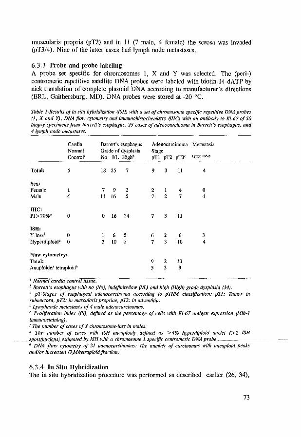
muscularis propria (pTI) and in 11 (7 male, 4 female) the serosa was invaded (pTI/4). Nine of the latter cases had lymph node metastases.
6.3.3 Probe and probe labeling A probe set specific for chromosomes I, X and Y was selected. The (peri-) centromeric repetitive satellite DNA probes were labeled with biotin-14-dATP by nick translation of complete plasmid DNA according to manufacturer's directions (BRL, Gaithersburg, MD). DNA probes were stored at -20°C.
Table 1 :Resu/ls of ill situ hybridlzatioll (ISH; with a set of chromosome specific repetitive DNA probes (1, X and Y). DNAjloll' C)'loll/elry alld imlllullollistodlemistry (IHC) with an amibody to Ki-67 oj 50 biopsy specimells from Barrett's esophagus, 23 cases of adenocarcilloma in Barrett's esophagus. alld 4 lymph node metastases.
Cardia Normal ControP
Total: 5
Sex: Female 1 Male 4
me: PI>20%e 0
ISH: Y lossf 0 Hyperdiploid8 0
Flow cytometry: Total: Anuploide/ tetraploidh
II Normal cardta control IfsslIe.
Barrett's esophagus Grade of dysplasia No ItL Highb
18 25 7
7 9 2 11 16 5
0 16 24
1 6 5 3 to 5
Adenocarcinoma Metastasis Stage pT 1 pT2 pT3c LJu¥! ro;J,:,j
9 3
2 1 7 2
7 3
6 2 7 3
9 2 5 2
11
4 7
11
6 to
to 9
4
o 4
3 4
b Barrett's esophagus with 110 (No), illdefillifellow (IlL) alld high (High) grade dysplasia (34). ( pT-Stages of esophageal adenocarcinoma according to pTNM classificatioll: pTJ.' TUmor ill submucosa, pT2: ill mliscularis propriae, pT3: ill advemitia. d LympJlIlode metastases of 4 male adellocarcinomas. e Proliferation index (PI), defilled as the percell/age of cells witli Ki-67 auagell expressioll (Mib-l immw/Ostaining). 'The number of cases of Y chromosome-loss ill males. 8 The lIumber of cases with ISH aI/euploidy defil/ed as > 4% hyperdiploid nuclei (> 2 ISH spots/nucleus) eS(;'lIated by ISH with a chromosome 1 specific ceil/TOll/eric DNA probe. h DNA flow cytometry of 21 adellocarcinomas: Vie number of carcinomas with aI/euploid peaks alld/or iI/creased G)Jltelraploidjraclioll.
6.3.4 In Situ Hybridization The in situ hybridization procedure was performed as described earlier (26, 34),
73

with some slight modifications. Briefly, after deparaffinizing of the tissue sections in xylene, endogenous peroxidase activity was blocked with 0.3% H,O, in methanol. Prior to enzymatic digestion slides were heated in a microwave oven (Biorad, UK), submerged in 2XSSC at 85°C for 25 min with an interval of I min. During this interval fluid level was checked and restored. Further enhancement of tissue permeability was achieved by proteolytic enzyme digestion (pepsin: Sigma, I mg/ml in 0.2 M HCI) for 5-10 min at 37°C. Before applying the probe set, the optimal digestion time for each tumor was determined by a pepsin time series (5-10 min). Tissue DNA was heat denatured for 2 min in 70% formamide in 2xSSC (pH 7.0), followed by dehydration in an ethanol series. Chromosome specific repetitive DNA probes were denatured for 7 min at 70°C in a hybridization mixture, containing 1-2 I'g/ml probe DNA, 500 I'g/ml sonicated herring sperm DNA (Sigma), 0.1 % Tween-20, 10% dextran sulphate, and 60 % formamide in 2xSSC at pH 7. The probe mixture was then applied to the sections and incubated overnight at 37°C. Then slides were rinsed in 60% formamide in 2xSSC (pH 7.0) at 42°C. Histochemical detection of probe binding was performed by immunoperoxidase staining. Slides were incubated for 30 min at 37°C with mouse-ant i-biotin (DakopaUs, Glostrup, Derunark), biotin-labeled horse-anti-mouse (Vector, Burlingame, CAl and with avidin-biotin-complex (Vectastain Elite ABC Kit). Visualization was achieved with 0.05% H,O" 0.5 gil DAB, 0.1 M imidazole in PBS (Phosphate Buffered Saline). The signal was amplified with 0.5% CuS04 in 0.9% NaCI. Slides were rinsed in distilled water and counterstained with hematoxylin. Finally, slides were dehydrated in ethanol, cleared in xylene and mounted in Malinol (Chroma-Geselschaft, Kongen, Germany).
6.3.5 ImmunoWstochemistry Before immunolabeling enhancement of Ki-67 antigen staining was achieved by heating slides submerged in citrate buffer (10 mM citric acid monohydrate, pH 6.0) in a microwave oven (700 W) at 95°C for 2x5 min with an interval of I min. During this interval fluid level was checked and restored. Labeling of the Ki-67 antigen was performed with a monoclonal antibody, Mib-I (Immunotech, Marseille, France), diluted 1/25 in PBS. The monoclonal Mib-I reacts with the Ki-67 molecule as detectable in late Gl, S and G2 phase, but not in GO phase of the cell cycle (35). For negative controls the primary antibodies were replaced by PBS. Immunohistochemistry was performed using the routine ABCimmunoperoxidase method (Vector).
6.3.6 DNA Flow Cytometry DNA content of the paraffin material of 21 carcinomas was measured, as described by Hedley et al. (36), using 50 I'm paraffin tissue sections. Flow cytometry was carried out using a Facscan (Becton Dickinson, Mountain View, Cal·
74
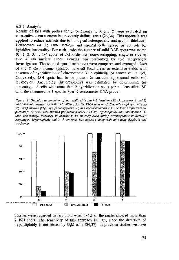
6.3.7 Analysis Results of ISH with probes for chromosomes I, X and Y were evaluated on consecutive 4 I'm sections in previously defined areas (26,34). This approach was applied to reduce artifacts due to biological heterogeneity and section thickness. Leukocytes on the same sections and stromal cells served as controls for hybridization quality. For each probe the number of solid DAB spots was scored (0, I, 2, 3, 4, >4 spots) of 2xlOO distinct, non-overlapping, single or side by side 4 I'm nuclear slices. Scoring was performed by two independent investigators. The counted spot distributions were compared and averaged. Loss of the Y chromosome appeared as small focal areas or extensive fields with absence of hybridization of chromosome Y in epithelial or cancer cell nuclei. Conversely, ISH spots had to be present in surrounding stromal cells and leukocytes. Aneuploidy (hyperdiploidy) was estimated by detennining the percentage of cells with more than 2 hybridization spots per nucleus afler ISH with the chromosome I specific (peri-) centromeric DNA probe.
Figure. 1. Graphic represellfatioll of the results of ill situ hybridization with chromosome 1 and y, alld immllllohistochemistry with alld antibody for the KJ-67 amigell of: Barrett's esophagus wilh 110 (N), indefinite/low (flL), high grade dysplasia (H) alld adenocarcinoma m. 'DIe Yaxis represellfs the percelltage oj cases with ele~'ated proliferation index (PI> 20), hyperdiploidy and chromosome Yloss, respecti~'e/y. Illcreased PI appears 10 be an early evellt during carcil/ogenesis ill Barrett's esophagus. Hyperdiploidy alld Y chromosome loss increase alollg with advancing dysplasia alld carcinoma.
100-
80-
60-
40-
20-
o--~ N ,,1. H T
D PI>20% Ill] Hyperdiploid • Y~loss
Tissues were regarded hyperdiploid when > 4 % of the nuclei showed more than 2 ISH spots. The sensitivity of this approach is high, since the detection of hyperdiploidy is not biased by G,M cells (34,37). In previous studies we have
75
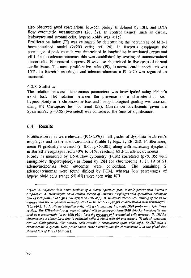
also observed good correlations between ploidy as defined by ISH, and DNA flow cytometric measurements (26, 37). In control tissues, such as cardia, leukocytes and stromal cells, hyperdiploidy was < I %. Proliferation index (PI) was estimated by determining the percentage of Mib-l immunostained nuclei (2x200 cells; ref. 26). In Barrett's esophagus the percentage of positive cells was determined in longitudinally sectioned crypts and viii i. In the adenocarcinomas this was established by scoring of immunostained cancer cells. For control purposes PI was also determined in five cases of normal cardia tissue. The mean proliferative index (PI), in normal cardia specimens was 15%. In Barrett's esophagus and adenocarcinomas a PI >20 was regarded as increased.
6.3.8 Statistics The relation between dichotomous parameters was investigated using Fisher's exact test. The relation between the presence of a characteristic, i.e., hyperdiploidy or Y chromosome loss and histopathological grading was assessed using the Chi-square test for trend (38). Correlation coefficients given are Spearman's; p=0.05 (two sided) was considered the limit of significance.
6.4 Results
Proliferation rates were elevated (PI>20%) in all grades of dysplasia in Barrett's esophagus and in the adenocarcinomas (Table I; Figs. I, 2B, 3B). Furtherruore, mean PI gradually increased (r=0.65, p <0.001) along with increasing dysplasia in Barrett's esophagus from 40% to 51 %, reaching 63 % in adenocarcinomas. Ploidy as measured by DNA flow cytometry (FCM) correlated (p<0.05) with aneuploidy (hyperdiploidy) as found by ISH for chromosome 1. In 19 of 21 adenocarcinomas both outcomes were concordant. The remaining 2 adenocarcinomas were found diploid by FCM, whereas low percentages of hyperdiploid cells (range 5%-6%) were seen with ISH.
Figure 2. Adjacel/t 4p.m tissue sectiolls of a biopsy specimen from a male patielll lVitlz Barrett's esophagus. A: Hemato.ryfill-Eosill stailled sectioll of Barrett's esophagus with specialized columnar type of metaplasia alld high grade dysplasia (2Ox obi). B: Immullohistochemical staillillg of the Ki-67 allfigell with tile monoclollal antibody Mib-J ill Barrett's esophagus cOll1lterslailled with hematoxylin. (20x obj.). C: III sim hybridizarion (ISH) with a chromosome 1 specific DNA probe 011 a 4p.m fiSSile
secfioll. TIle ISH-related spots were visualized with ;'l/IJl/l1loperoxidaseIDAB (black); hematoxylin was used as a counterstain (grey; IOOx obi). Note the presence oj liyperdiploid cells (arrows). D: ISH jor cliromosome Y shows focal loss in epithelial cells: A glalld with (0) al/d witholll (*) this chromosome can be distinguished. Also stromal cells comaill Y chromosome spots (4Ox obi). E: ISH with a chromosome X specific DNA probe shows clear hybridization for chromosome X ill the glalld Ihat showed loss oj Y ill D (4Ox obi).
76
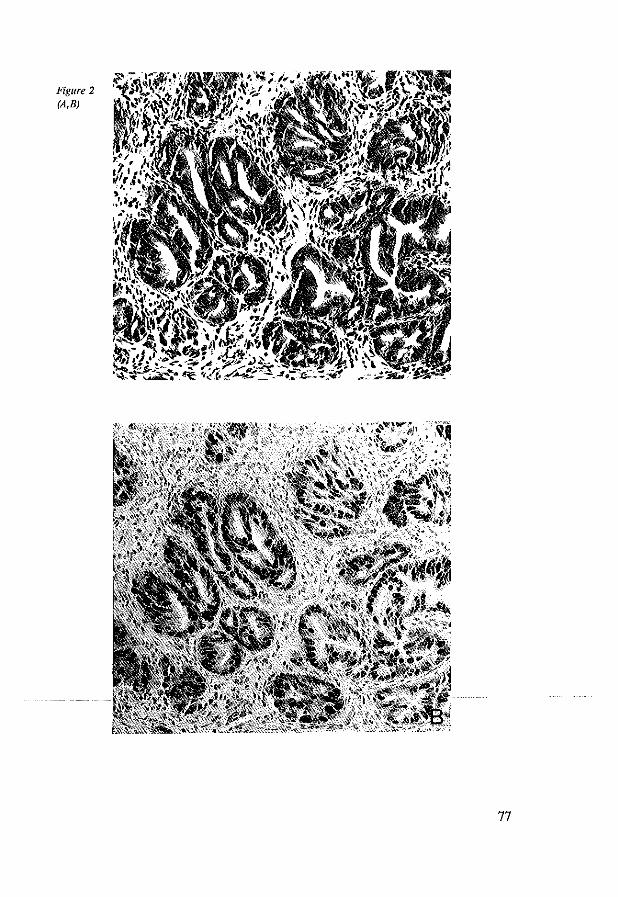
Figure 2 (A,B)
77
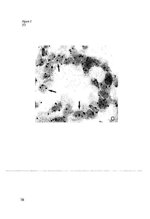
Figure 2 (C)
78

Figure 2 (D,E)
79
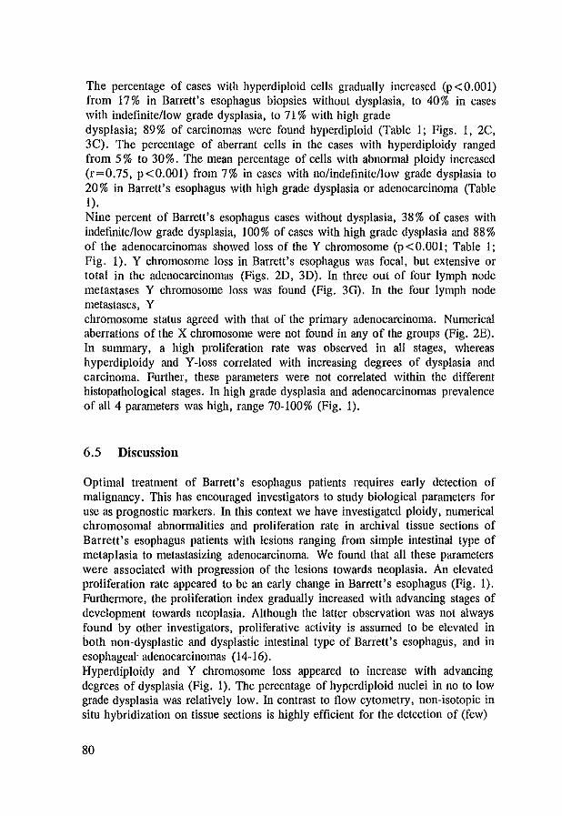
The percentage of cases with hyperdiploid cells gradually increased (p<O.OOI) from 17% in Barrett's esophagus biopsies without dysplasia, to 40% in cases with indefinite/low grade dysplasia, to 71 % with high grade dysplasia; 89% of carcinomas were found hyperdiploid (Table 1; Figs. 1, 2C, 3C). The percentage of aberrant cells in the cases with hyperdiploidy ranged from 5% to 30%. The mean percentage of cells with abnormal ploidy increased (r=0.75, p<O.OOl) from 7% in cases with no/indefinite/low grade dysplasia to 20% in Barrett's esophagus with high grade dysplasia or adenocarcinoma (Table I). Nine percent of Barrett's esophagus cases without dysplasia, 38% of cases with indefinite/low grade dysplasia, 100% of cases with high grade dysplasia and 88 % of the adenocarcinomas showed loss of the Y chromosome (p<O.OOI; Table 1; Fig. 1). Y chromosome loss in Barrett's esophagus was focal, but extensive or total in the adenocarcinomas (Figs. 2D, 3D). In three out of four lymph node metastases Y chromosome loss was found (Fig. 30). In the four lymph node metastases, Y chromosome status agreed with that of the primary adenocarcinoma. Numerical aberrations of the X chromosome were not found in any of the groups (Fig. 2E). In summary, a high proliferation rate was observed in all stages, whereas hyperdiploidy and Y-loss correlated with increasing degrees of dysplasia and carcinoma. Further, these parameters were not correlated within the different histopathological stages. In high grade dysplasia and adenocarcinomas prevalence of all 4 parameters was high, range 70-100% (Fig. 1).
6.5 Discussion
Optimal treatment of Barrett's esophagus patients requires early detection of malignancy. This has encouraged investigators to study biological parameters for use as prognostic markers. In this context we have investigated ploidy, numerical chromosomal abnormalities and proliferation rate in archival tissue sections of Barrett's esophagus patients with lesions ranging from simple intestinal type of metaplasia to metastasizing adenocarcinoma. We found that all these parameters were associated with progression of the lesions towards neoplasia. An elevated proliferation rate appeared to be an early change in Barrett's esophagus (Fig. I). Furthermore, the proliferation index gradually increased with advancing stages of development towards neoplasia. Although the latter observation was not always found by other investigators, proliferative activity is assumed to be elevated in both non-dysplastic and dysplastic intestinal type of Barrett's esophagus, and in esophageal" adenocarcinomas (14-16). Hyperdiploidy and Y chromosome loss appeared to increase with advancing degrees of dysplasia (Fig. 1). The percentage of hyperdiploid nuclei in no to low grade dysplasia was relatively low. In contrast to flow cytometry, non-isotopic in situ hybridization on tissue sections is highly efficient for the detection of (few)
80
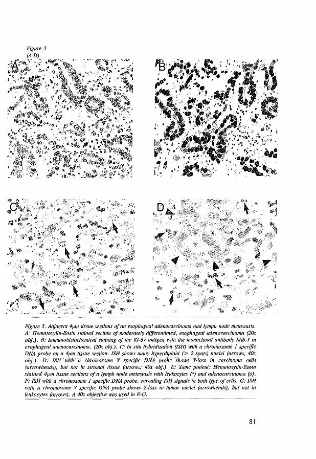
'" ~~'~ilff}~
"'t,;:
~
9 {4
i$i ,J, • .. ,-$ ;;,.
~~ I p
r'
1 .
Figure 3. Adjacem 4p.m tissue sectiolls of all esophageal adel/ocarcilloma alld lymph node metastasis. A: Hematoxylin-Eosin slailled sectiOll of moderately dijferellliated, esophageal adenocarcinoma (2Ox obj.). B: Immullohistochemical stail/illg of the Ki-67 am/gell with the mOl/oclol/al omibody Mib-l ill esophageal adenocarcinoma. (2Ox obj.). C: II/ situ hybridization (ISH) with a chromosome 1 specific DNA probe 011 a 4Jllll tissue sectioll, ISH shows mall)' hyperdiploid (> 2 spots) lIuclei (arrows; 40x obj.). D: ISH witll a chromosome Y specific DNA probe shows Y-Ioss ill carcilloma cells (arrowheads), bllt 1/01 ill stromal ,;ssue (arrows; 40x obj.). E: Same patient: Hematoxylin-Eosin stained 4p.m tissue sectiolls of a lymph node metastasis with leukocytes (*) and adenocarcinoma (0). F: ISH with a chromosome I specific DNA probe, re~'ealillg ISH signals ill both type of cells. G: ISH with a chromosome Y specific DNA probe shows Y-loss ill tumor nuclei (arrowheads), but not ill leukocytes (arrows). A 40;; objecti~'e was lIsed ill E-G.
81
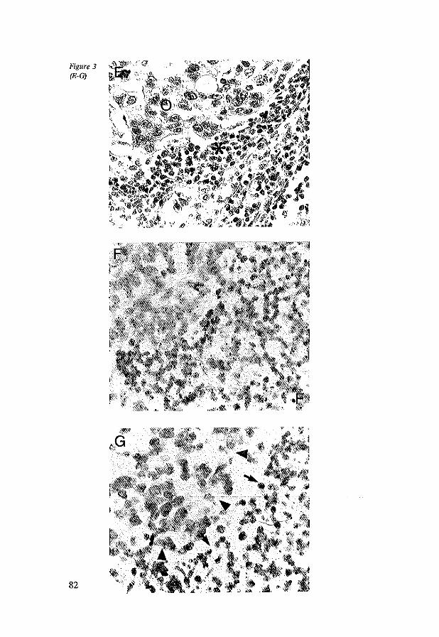
82
dispersed cells with abnormal ploidy status (37). In the present study this was demol1Btrated by discordance of ploidy measured by FCM and ISH in two (of 21) tumor samples. Both samples were diploid by FCM, but were found to contain a low number of hyperdiploid nuclei by ISH. Y-Ioss was rarely observed in nondysplastic Barrett's esophagus, yet increased significantly in most advanced lesions with a high prevalence in high grade dysplasia and adenocarcinoma (Fig. I). Loss of the Y chromosome appears to be the most consistent chromosomal abnormality in Barrett's esophagus and esophageal adenocarcinoma. It was found previously in several karyotyping and ISH studies (21, 23-27). However, its association with dysplastic changes in Barrett's esophagus had not been observed previously. The role of Y-Ioss in neoplastic transfOlmation in Barrett's esophagus is uncertain. Barrett's esophagus and esophageal adenocarcinoma have a three and seven times increased incidence in males (39, 40). Since as yet no genes involved in tumorigenesis have been linked to the Y chromosome, Y-Ioss may be the result of genomic instability in transformed cells. In contrast to Y chromosome aberrations, X chromosome changes were not observed. The X chromosome is therefore not an informative numerical marker in (female) Barrett's esophagus patients. Our results suggest that during the development of dysplasia and carcinoma ill Barrett's esophagus a high cell turnover is an initial event. The resulting high proliferative activity may induce genomic instability. The latter leads to cytogenetic alterations, and transformation of cells, which may further expand into cell clones with malignant behavior.
6.6 References
1. Barrett, N. R. Chronic peptic ulcer of the esophagus and esophagitis. Dr. J. Surg., 38:175· 185, 1950.
2. Sjogren, Jr. R. W .. Johnson, L. F. Barrett's esophagus: A review. Am. J. Med., 74:313-321, 1983.
3. Spechler, S. J. Barrett's esophagus: Current opinion. Gastroenterology, 8:573-578. 1992. 4. Fennerty. M. B. Review article: Darrett's oesophagus-cancer risk, biology and therapeuthic
management. Aliment. Pharmacal. Ther., 7:339-345, 1993. 5, Van dec Vecll, A. H. t Dees, I" Blankensteijn, J. D., van Blankenstein, M. Adenocarcinoma
in Barrett's oesophagus: An overrated risk. Gut, 30:14-18, 1989. 6. Spcehlcr, S. J., Robbins, A. H., Rubins, Vincent, M. E., H. B., Heeren, V. C., Doos, W.
G., Colton, T., Schimmel, E.M. Adenocarcinoma and Barrett's oesophagus: An overrated risk? Gastroenterology, 84: 927-933 1984.
7. Lerut, T., Cooseman, W., Raemdonck, Van, D., Dillemans, B., De Leyn, P., Marnette, J. M., Geboes, K. Surgical treaUnent of Barrett's carcinoma. J Thorae. Cardiovase. Surg., 107:1059-1066, 1994.
8. Menke-Pluymers, M. B. E., Schoute, N. W., Mulder, A. H., Hop, W. C. J., Van Blankenstein, M., Tilanus, H. W. Outcome of surgical treatment of adenocarcinoma in Barrett's oesophagus. Gut, 33:1454-1458, 1993.
9. Hamilton, S. R., Smith, R. R. L. The relation between columnar epithelial dysplasia and invasive adenocarcinoma arising in Barrett's esophagus. Am. J. Clin. Pathol., 87:301-312, 1987.
83
10. Pera, M. Trastek, V. P., Carpenter, H. A., Allen, M. S., Deschamps, C., Pairelero, P. C. Barrett's esophagus with high grade dysplasia: An indication for esophagectomy'? Ann. Thorac. Surg., 54:199-204. 1992.
11. Hameeteman. W., Tytgat, G. N. J .• Houthoff. H. J., Van Den Tweel, 1. G. Barrett's esophagus: Development in dysplasia and adenocarcinoma. Gastroenterology, 96:1249-1256, 1989.
12. Mc Ardle. J. E., Lewin. K. J., Randall, G. Weinstein, W. Distribution of dysplasia and early invasive carcinomas in Barrett's esophagus. Hum. Pathol., 23:479-482. 1992.
13. Achkar, E. and Carey. W. The cost of surveillance for adenocarcinoma complicating Barrett's oesophagus. Am J Gastroenterology. 83:291-294. 1988.
14. Reid, B. J .• Sanchez. C. A .• Blount, P. L.. Levine D. S. Barrett's esophagus: Cell cycle abnormalities in advancing stages of neoplastic progression. Gastroenterology, 105:119-129, 1993.
15. Gray, M. R .• Hall, P. A., Nash, J., Ansari. B .• Lane, D. P., Kingsnorth, A. N. Epithelial proliferation in Darrett's esophagus by Proliferating Cell Nuclear antigen immunolocalization. Gastroenterology, 103:1769-1776, 1992.
16. Casson, A. G., Mukhopadhyay, T., Cleary, K. R., Ro, J. Y., Levin, B., Roth, J. A. Oncogene activation in esophageal cancer. 1. Thorae. Cardiovasc. Surg .• 102:707-709, 1991.
17. Ramel, S., Reid, B. J., Sanchez, C. A., Blount, P. L" Levine D. S .• Neshat, K., Haggitt, R. C., Dean. P. J., Thor, K., Rabinovitch, P. S. Evaluation of p53 protein expression in Barrett's esophagus by two-parameter flow cytometry. Gastroenterology, 102:1220-1228, 1992.
18. Blount, P. L., Ramel, S. R., Raskind, W. H .• Haggiu R. C., Sanchez, C. A., Dean, P. J., Rabinovitch, P. S., Reid, D. 1. 17p Allelic deletions and p53 protein overexpression in Barrett's adenocarcinoma. Cancer Res .• 51:5482-5486,1991.
19. Blount, P. L., Meltzer, S. 1., Yin, 1., Huang, Y., Krasna, M. 1., Reid. B. J. Clonal ordering of 17p and 5q allelic losses in Barrett dysplasia and adenocarcinoma. Proc. Natl. Acad. Sci. USA, 90:3221-3225, 1993.
20. Krishnadath, K. K., Van Blankenstein, M., Tilanus, H. W., Prognostic value of p53 in Barrett's esophagus. Eur. 1. Gastroenterology and Hepatology 1994;7:81-84.
21. Garewal, H. S., Sampliner, R., Liu. Y., Trent. J. M. Chromosomal rearrangements in Barrett's esophagus. Cancer Genet. Cytogenet., 42:281-296, 1989.
22. Garewal. H., Meltzer, P., Trent, 1., Prabbala, R., Sampliner, R, Korc, M. Epidermal growth factor receptor overexpression and trisomy 7 in a case of Barrett's esophagus. Dig. Dis. Sci., 35:1115-1120, 1990.
23. Rodriguez, E., Rao. P. H., Ladanyi, M., Altorki, N., Albino, A. P., Kelsen, D. P., Jhanwar, S. C., Changanti, R. S. K. 11-p13-15 Is a specific region of chromosomal rearrangement in gastric and esophageal adenocarcinomas. Cancer Res., 50:6410-6416, 1990.
24. Raskind, W. H., Norwood. T., Levine, D. S .• Haggitt, R. C., Rabinovitch. P. S., Reid, B. 1. Persistent clonal areas and clonal expansion in Barrett esophagus. Cancer Res., 52:2946-2950, 1992.
25. Van Dekken, H .• Pizzolo, J. G., Kelsen. D. P., Melamed, M.R. Targeted cytogenetic analysis of gastric tumors by in situ hybridization with a set of chromosome-specific DNA probes. Cancer, 66:491-497, 1990.
26. Krislmadath. K. K., Tilanus, H. W., Mulder, A. H., Van Dekken, H. Detection of genetic changes in Barrett's adenocarcinoma and Barrett's esophagus by DNA in situ hybridization. Cytometry, 15:176-184,1994.
27. Hunter, S., Gramlich, T., Abbott, K., Varma, V. Y. Chromosome loss in esophageal carcinoma; An in situ hybridization study. Genes Chromo Cancer, 8:172-177, 1993.
28. Reid, B. J., Haggitt, R. C., Rubin, C. E .• Rabinovitch, P. S. Barrett's esophagus. Correlation between flow eytometry and histology in detection of patients at risk for adenocarcinoma. Gastroenterology, 93: I-II, 1987.
29. Rabinovitch, P. R., Reid, B. J .• Haggitt, R. C., Norwood, T. H., Rubin, C. E. Progression
84
to cancer in Barrett's esophagus is associated with genomic instability. Lab. Invest., 60:65-71, 1988.
30. Fennerty, M. B., Sampliner, R. E., Way, D., Riddell, R., Steinbronn, K., Garewal, H. S., Discordance between flow cytometric abnormalities and dysplasia in Barrett's esophagus. Gastroenterology, 97:815-820, 1989.
31. McKinley, M. J., Budman, D. R., Grueneberg, M. S., Bronzo, R. L., Weissman, G. S., Kahn, E. DNA Content in Barrett's esophagus and esophageal malignancy. Am. J. Gastroenterology, 82: 10 12-10 15, 1987.
32. Scialiero, S., Giaretti, W., Bonelli. L., Geido, E., Rapallo, A., Conio. M., Ravelli, P., Lombardo. L .• Lapertosa. G., Aste, H. DNA Content analysis of Barrett's esophagus by flow cytometry. Endoscopy. 25:648-651. 1993.
33. Reid. B. J., Weinstein, W. M., Lewin, K. 1., Haggitt, R. C., VanDeventer, G., DenBesten, L., Rubin. C. E. Endoscopic biopsy detect high-grade dysplasia or early adenocarcinoma in Barrett's esophagus without grossly recognizable neoplastic lesions. Gastroenterology, 94:81-90, 1988.
34. Van Dekken, H., Kerstens. H. M. J., Tersteeg. T. A., Verhofstad, A. A. 1.. Vooijs, G. P. Histological preservation after in situ hybridization to archival solid tumour sections allows discrimination of cells bearing numerical chromosome changes. 1. Patha!., 168:317-324, 1992.
35. Gerdes. J .• Becker. M. H. G., Key, G., Calloretti, G. Immunohistological detection of tumour growth fraction (Ki-67 antigen) in formalin-fixed and routinely processed tissues. J. Patho!., 168:85-87, 1992.
36. Hedley, D. W .• Friedlander. M. L., Taylor, I. W., Rugg. C. A., Musgrove. E. A. Method for analysis of cellular DNA content of paraffin-embedded pathological material using flow cytometry. 1. H6.6 istochem. Cytochem., 31:1333-1335, 1983.
37. Van Dekken, H .• Bosman, F. T., Teijgeman. R .• Vissers, C. 1., Tersteeg, T. A., Kerstens, H. M. J., Vooijs. G. P., Verhofslad, A. A. J. Identification of numerical chromosome aberrations in archival tumours by in situ hybridization to routine paraffin sections: Evaluation of 23 phaeochromocytomas. 1. Pathol., 171:161-171, 1993.
38. Mantel. N. Chi square tests with one degree of freedom. JASA, 58:690-700, 1963. 39. Blot, W. J., Devesa, S. S., Kneller. R. W., Fraumeni, jr., J. F .• Rising incidence of
adenocarcinoma of the oesophagus and gastric cardia. JAMA, 265:1287-1289 1991. 40. Reed. P. I., and Johnston, B. J. The changing incidence of oesophageal Cancer. Endoscopy.
25:606-608 1993.
85


Chapter 7
Reduced Expression of the Cadheriu-Catenin Complex in
Esophageal Adenocarcinoma Correlates with Poor Proguosis.
K. K. Krishnadath, H. W. Tilanus, M. van Blankenstein, W. C. J. Hop,
E. D. Kremers, W.N. M. Dinjens and F. T. Bosman.
In Press: J Pathol 1997

7.1 Abstract
TIle E-Cadherin-Catenin complex is important for cell-cell adhesion of epithelial
cells. Impairment of one or more components of this complex is associated with poor differentiation and increased invasiveness of carcinomas. Esophageal
adenocarcinomas cause early metastases, progress rapidly and consequently have
a poor prognosis. By means of immunohistochemistry, we studied the expression
of E-Cadherin, Alpha- and Beta-Catenin in 65 esophageal adenocarcinomas and
15 lymph node metastases. Expression of these proteins was evaluated with
respect to clinico-pathological parameters and patient survival. Expression of the proteins was strongly correlated. In carcinomas reduced
expression of E-Cadherin, Alpha-Catenin and Beta-Catenin were found in 74 %, 60%, and 72%, respectively. Expression of E-Cadherin and Alpha-Catenin
correlated signit1cantly with stage and grade of the carcinomas, whereas
expression of Beta-Catenin only correlated with grade of the carcinomas.
Reduced expression of all three proteins correlated with shorter patient survival.
In contrast to grade, E-Cadherin and Beta-Catenin were significant
prognosticators for survival, independent of stage of disease. We conclude that in
esophageal adenocarcinomas decreased expression of E-Cadherin, Alpha- and
Beta-Catenin are related events. Furthermore, expression of at least E-Cadherin and Beta-Catenin, is significantly correlated with poor patients' prognosis.
7.2 Introduction
For several decades the incidence of esophageal adenocarcinoma in Western
countries is rapidly increasing (1). The predisposing condition for developing
esophageal adenocarcinoma is Barrett's esophagus (2-3). Progression of Barrett's
esophagus to adenocarcinoma occurs through gradually increasing dysplasia (4-
5). The survival of patients with esophageal adenocarcinoma is poor, even after seemingly radical surgical intervention. Stage of disease is the best parameter for
predicting survival (6, 7). Patients with limited tumor invasion of the esophageal wall and without metastases at time of resection have significantly better survival
than those with lymph node metastases or organ infiltration (6, 7). Understanding
the mechanisms of metastatic behavior of carcinomas, and recognition of cases
running higher risk for developing metastases, might help to design new strategies for diagnosis and treatment.
89

E-Cadherin is a 120 KDa transmembrane glycoprotein, which belongs to a large
family of calcium dependent adhesion molecules (CAMs) (8, 9). By mostly
homotypic interactions (10), E-Cadherin plays an important role in
morphogenesis of tissues during embryogenesis and in mature epithelia E
Cadherin is involved in cell-cell adhesion (11, 12). In vitro and in vivo
experiments showed that absence of membranous E-Cadherin in carcinoma cells
goes along with increased invasiveness and metastatic ability (13-15). It has
become clear that besides cellular adhesion, E-Cadherin is also involved in signal
transduction via connections with Catenins and the actin cytoskeleton. The
Catenin anchorage complex, which interacts with the cytoplasmic domain of E
Cadherin, consists of a heteromere of at least three proteins: Alpha-, Beta- and
Gamma-Catenin. Alpha-Catenin, a 102 kDa protein ubiquitously expressed in
cells, binds to actin and to Beta-Catenin. In turn, Beta-Catenin, a 92 kDa protein,
is directly linked to E-Cadherin (16, 17). Both Beta- and Alpha-Catenin have
binding sites for several onco- and tumor suppressor proteins, suggesting that these might modulate the function of the E-Cadherin-Catenin adhesion complex
(18-20). In adenocarcinomas of the prostate, breast, esophagus, lung, stomach and colon, and in squamous cell carcinomas of the esophagus and of head and
neck, decreased E-Cadherin and/or Alpha-Catenin expression was found to
correlate with high tumor grade and poor patients' prognosis (21-29). Recently,
high frequencies of decreased Beta-Catenin expression were observed in several
gastro-intestinal cancers (30).
In the present report we describe the expression of E-Cadherin, (EC), Alpha(AC) and Beta-Catenin (BC) in 65 esophageal adenocarcinomas associated with
Barrett's esophagus and in 15 lymph node metastases. Expression of these
proteins, as assessed by immunohistochemistry with specific monoclonal
antibodies, is evaluated with respect to various pathological parameters and
patient survival.
7.3 Materials and methods
7.3.1 Clinico-pathological data Esophageal-cardia resection specimens of 65 patients with esophageal
adenocarcinoma associated with Barrett's esophagus, encountered between 1985
and 1992 at the Academic Hospital-Dijkzigt Rotterdam, were used in the study. Forty nine patients were male, 16 female, with a mean age of 67 (Range 34-80
90
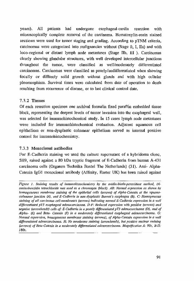
years). All patients had undergone esophageal·cardia resection with
microscopically complete removal of the carcinoma. Hematoxylin-eosin stained
sections were used for tumor staging and grading. According to pTNM criteria,
carcinomas were categorized into malignancies without (Stage 0, I, IIa) and with
loco-regional or distant lymph node metastases (Stage lIb, III ). Carcinomas
clearly showing glandular structures, with well developed intercellular junctions
throughout the tumor, were classified as well/moderately differentiated
carcinomas. Carcinomas were classified as poorly/undifferentiated when showing
focally or diffusely solid growth without glands and with high cellular
pleomorphism. Survival times were calculated from date of operation to death
resulting from recurrence of disease, or to last clinical control date.
7.3.2 Tissues
Of each resection specimen one archival formalin fixed paraffin embedded tissue
block, representing the deepest levels of tumor invasion into the esophageal wall,
was selected for immunohistochemical study. In 15 cases lymph node metastases
were included for immunohistochemical evaluation. Adjacent squamous cell
epithelium or non·dysplastic columnar epithelium served as internal positive
control for immunohistochemistry.
7.3.3 Monoclonal antibodies
For E-Cadherin staining we used the culture supernatant of a hybridoma clone,
5H9, raised against a 80 kDa tryptic fragment of E-Cadherin from human A-431
carcinoma cells (Organon Technika Boxtel The Netherlands) (31). Anti- Alpha
Catenin IgGl monoclonal antibody (Affinity, Exeter UK) has been raised against
Figure 1. Staillillg results of immullohistochemistry by tile (H'idill~biotin-peroxidase method, DiamilwbellZidill tetrachloride was used as a chromogen (black). AB: Normal expression as shown by homogeneous membrane stail/ing of the epithelial celfs (arrows) of Alpha-Carel/ill at the sqllamocolUmnar jUlIctioli (A), alld E-Cadllerill ill 1101l-dysplastic Barrett's esophagus (8). C: Homogeneous staining of all carcinoma ceU-membranes (arrows) indicatillg normal E-Cadllerill expression ill a well differemiated pT3 esophageal adenocarcinoma. D-F: Reduced expression with positive (arrows) and negative (arrowheads) cefts of: E-Cadherill in a poorly dijJerentiated pT3 adenocarcilloma (D), amI of Alpha- (E) and Beta- Catellill (F) ill a moderately dijJerellfiated esophageal adenocarcilloma. G: Normal expression, homogelleous membrane staillillg (arrows), of Alpha-Catellill expressioll ill a well dijJerellfiated adenocarcinoma. H: No membrane stainillg (arrowheads), but pos/ril'e nuclear staillillg (arrows) of Beta-Caten;1I ill a moderately dijJerellfiated adenocarcinoma. Magnification A: 9ar, B-H: 18Q(.
91
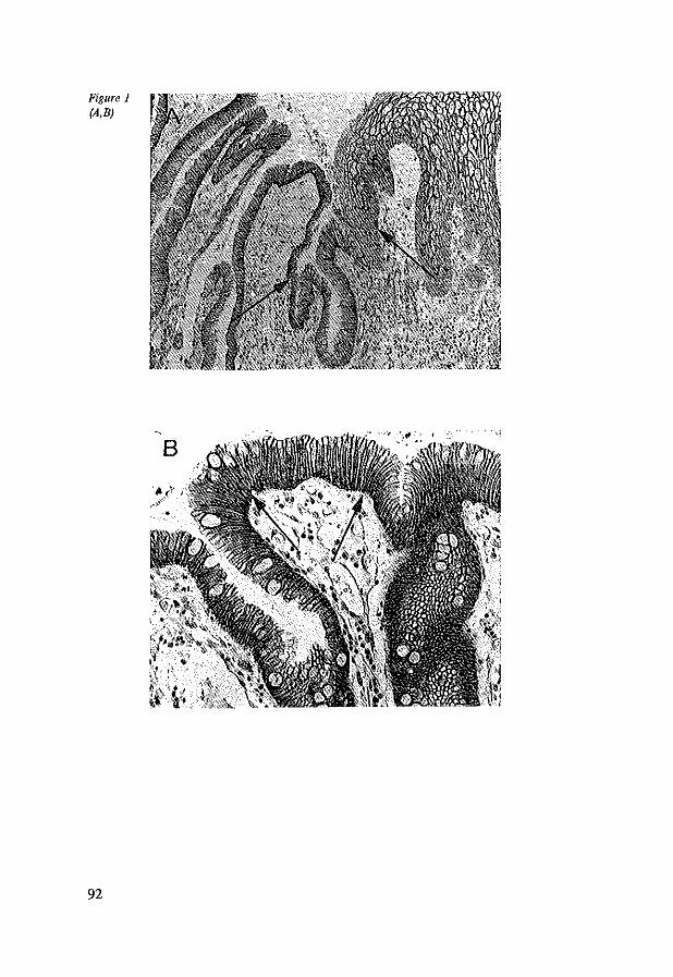
Figure 1 (A,B)
92
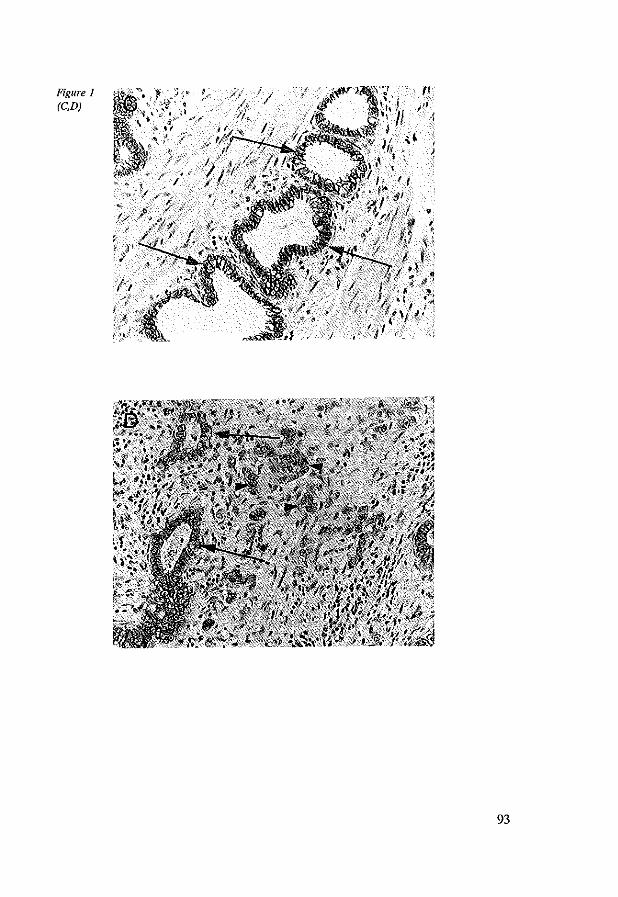
Figure 1 (C.D)
/
(I
if} j
93
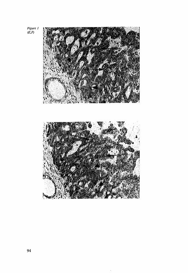
Figure 1 (E,F)
94
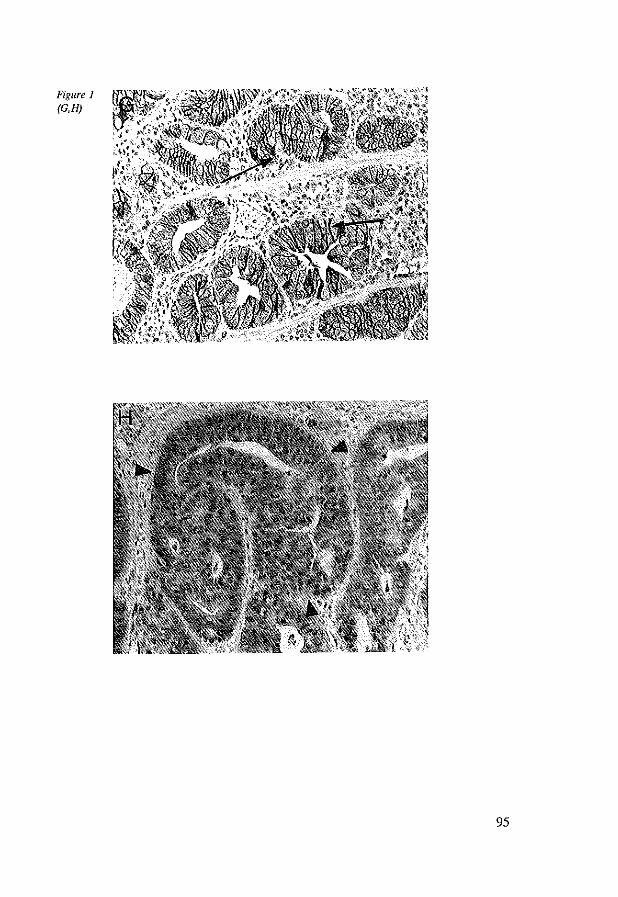
Figure J (G,H)
95
a 19.4 kDa C-terminal protein fragment corresponding to amino acids 729-906 of mouse Alpha-Catenin. This antibody was diluted I: 100 ill PBS. A monoclonal
IgGl Anti-Beta Catenin (Affinity), raised against 23 kDa protein fragment corresponding with amino acid residues 571-781 of mouse Beta-Catenin, was
diluted 1 :200 in PBS.
7.3.4 Immunohistochemistry
Five I'm paraffin sections were mounted on 3-amino-propyl-tri-ethoxy-silane
(Sigma, St. Louis, USA) coated slides and stored overnight at 60°C. Sections
were deparaffinized, endogenous peroxidase was blocked in 3 % H,O, in methanol
(20 min) and rinsed in PBS. As antigen retrieval procedure for E-Cadherin, tissues were digested in I % protease (Sigma) with 1 % Ca++ in PBS solution (10-
20 min at 37" C). Prior to Alpha- and Beta-Catenin staining the sections were
heated in a microwave oven (in 0.01 M Citrate buffer Ph 6.0) for two cycles of
five min. Primary antibodies were incubated overnight (at 4°C). As detection
system we used streptavidin-biotin-peroxidase (Dako, Glostrnp Denmark) with
diaminobenzidin tetrachloride (Fluka, Basel Switzerland) as chromogen. Tissues
were counterstained with hematoxylin.
7.3.5 Evaluation of E-Cadherin (EC), Alpha- (AC) and Beta-Catenin (BC) expression,
Immunohistochemical staining of EC, AC and BC in the subsequent tissue sections was scored semi-quantitatively by two independent investigators in two
areas per carcinoma and per lymph node metastasis(lOx objective).
7.3.6 Statistical analysis Correlations between E-Cadherin, Alpha-, Beta- Catenin, grade and stage were
calculated in cross-tables. Significance was tested by the Fisher's exact test. Survival probabilities were calculated by the Kaplan-Meier method and
probabilities were compared by logrank test. Multi-variate analysis to determine
independent prognostic value of the various parameters was performed by Cox
regression. P=O.05 (two sided) was considered the limit of significance.
7.4 Results
7.4.1 IlIllllunohistochemical staining patterns in control tissues and in
96

adenocarcinomas.
Homogeneous membranous immunostaining for E-Cadherin (EC), Alpha- (AC)
and Beta-Catenin (BC) was found in all cases of normal squamous epithelium,
normal cardia mucosa and non dysplastic Barrett's mucosa (figure IA,B). These
tissues adjacent to the adenocarcinomas served as internal control for the staining procedures. In squamous epithelium immunostaining was present homogeneously
Table 1: Cross tabulalioll oj immunohistochemical staining remits oj E-Cadherill, Alpha- and Beta-Calellill lI'ith respect to stage and grade ill 65 esophageal adenocarcinomas,
Pis E-Cadherin Alpha-Catenin Beta·Catenin normal reduced normal reduced normal reduced
Total 65 17 48 28 37 18 47
Stage": O,I,IIa (%) 33 15(88)' 18(38)' 21(75)' 12(32)' 12(67)' 21(45)' lIb,lII (%) 32 2(12) 30(63) 7(25) 25(68) 6(33) 26(55)
Grade: Well/moder (%) 33 17(100)' 16(33)' 20(71)' 13(35)' 14(78)' 19(40)' Poor/undiff (%) 32 0(0) 32(67) 8(29) 24(65) 4(22) 28(60)
E-Cadherin: normal (%) 17 17(61)' 0(0)' 11(61), 6(13)' reduced (%) 48 11(39) 37(100) 7(39) 41(85)
Alpha-Catenin: normal (%) 28 16(89Y 12(25Y reduced (%) 37 2(11) 35(75)
al Stages: O=pTisNOMO l=pT1NOMO; lla=pT213NOMO; llb=pT1I2N1MO; 11l=pT314Nl-3MO Column percellfages. Np < 0.001 clp =O.OOl dJp =0.17 tip < 0.001 /.~Ip <0.01 h-j!p < 0.001
on the intermediate cells and on the basal layer (but not at the basal membrane side). In non dysplastic Barrett's and cardia epithelia intercellular membrane
staining was seen between all columnar cells. Membrane staining for all three proteins was also found in the adenocarcinomas
and lymph node metastases. In the adenocarcinomas and lymph node metastases three main staining patterns were recognized: Homogeneous positive staining
with more than 90% positive cells (figure IC,G). Heterogeneously staining
carcinomas with mixed negative and positive cells or tumor areas (figure
ID,E,F), but with at least 30% negatively staining cells. And a group of
carcinomas with negative staining with no or less than 10% positive cells (figure
IH). For further analysis, carcinomas displaying a homogeneous positive pattern
97
b-jJ
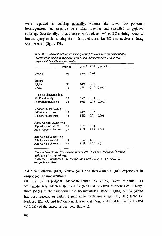
were regarded as staining normally, whereas the latter two patterns, heterogeneous and negative were taken together and classified as reduced
staining. Occasionally, in carcinomas with reduced AC or BC staining, weak to
intense cytoplasmic staining for both proteins and for BC also nuclear staining
was observed (figure lH).
Table 2; Esophageal adellocarcinoma specific five years survival probabilities, subsequemly stratified for stage, grade, and immul/oreactive E-Cadherill, Alpha-alld Beta-Catel/in expressiol/.
patients 5-yrsl) SDl) p-value11
Overall 65 32% 0.07
Stage4):
D,I,ITa 33 64% 0.10 IIb,llI 32 7% 0.10 0.0001
Grade of differentiation: Well/moderately 33 55% 0.10 Poor/undifferentiated 32 10% 0.10 0.0001
E-Cadherin expression: E-Cadherin normal 17 74% 0.12 E-Cadherin aberrant 43 14% 0.7 0.001
Alpha-Catenin expression: Alpha-Catenin normal 28 61 % 0.10 Alpha-CateoiD aberrant 37 11% 0.06 0.001
Bcta-Catenill expression: Bela-Catenill normal 18 64% 0.14 Beta-Cateoin aberrant 42 21 % 0,07 0.01
IJKapiall-Meier'sjil'e year sun'ivai probability. ])StalJdard del'latioll. 3)p-value calculated by Logrank test. f)Srages: 0= TisNOMO; l=pTINOMO; lla=pTlI3NOMO; lib=pT1I2NIMO;
Ill~pT3I4Nl-3MO.
7.4.2 E-Cadherin (EC), Alpha- (AC) and Beta-Catenin (BC) expression in
esophageal adenocarcinoma, Of the 65 esophageal adenocarcinomas 33 (51 %) were classified as
well/moderately differentiated and 32 (49%) as poorly/undifferentiated. Thirty
three (51 %) of the carcinomas had no metastases (stage O,I,lIa), but 32 (49%)
had loco-regional or distant lymph node metastases (stage lib, III ; table 1). Reduced EC, AC and BC imrnunostaining was found in 48 (74%), 37 (60%) and
47 (72%) of the cases, respectively (table 1).
98
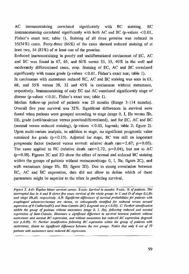
AC immunostaining correlated significantly with BC stammg. EC
immunostaining correlated significantly with both AC and BC (p-values <0.01,
Fisher's exact test; table I). Staining of all three proteins was reduced in
35(54%) cases. Forty-three (66%) of the cases showed reduced staining of at
least two, 54 (83%) of at least one of the proteins.
Reduced inununostaining in poorly and undifferentiated carcinomas of EC, AC
and BC was found in 67, 65, and 60% versus 33, 35, 40% in the well and
moderately differentiated cases, resp. Staining of EC, AC and BC correlated
significantly with tumor grade (p-values <0.01, Fisher's exact test; table I).
In carcinomas with metastases reduced EC, AC and BC staining was seen in 63,
68, and 55% versus 38, 32 and 45% in carcinomas without metastases, respectively. Immunostaining of only EC and AC correlated significantly stage of
disease (p-values <0.01, Fisher's exact test; table I).
Median follow-up period of patients was 23 months (Range 3-114 months).
Overall five year survival was 32 %. Significant differences in survival were found when patients were grouped according to stage (stage 0, I, I1a versus lIb,
Ill), grade (well/moderate versus poor/undifferentiated), and for EC, AC and BC
(normal versus reduced staining), (p-values <0.05, logrank; table 2; figure 2).
Upon multi-variate analysis, in addition to stage, no significant prognostic value
remained for grade (p=0.15). Adjusted for stage, BC was still an important
prognostic factor (reduced versus normal: relative death rate=2.67; p=0.03).
The same applied to EC (relative death rate=2.72, p=0.04), but not to AC
(p=0.09). Figures 2C and 2D show the effect of norrual and reduced BC staining within the groups of patients without metastases(stage 0, I, na; figure 2C), and
with metastases (stage lIb, 1II; figure 2D). Due to strong correlation between
EC, AC and BC expression, data did not allow to define which of these
parameters might be superior to the other in predicting survival.
Figure 2. A-D: Kaplan Meier slln'imi curves. X-axis: Survival ill mom"s; Y-axis: % oj patiellts. 17le interrupted Iille in A alld B shows jive years surviml of tile whole group,' ill C alld D of stage O,l,Jla and stage /lb,lIl, respectb'ely. A,B: Sigllijicallt differences of survival probabilities for patiems wilh esophageal adenocarcinomas are shown, as subsequently stratified for reduced verslls I/ormal expressioll of E-Cadherifl(EC) and Bela-Gaten/" (BC) (Lograllk lest p < 0.05). C; Further stratification within the group of patiellts without metastases (stage 0, I, lIa), fol/owillg reduced and normal expressioll of Beta-Catell/n, illustrates a significallt difference ill survival betweell patiellfs wilhout metastases and 1I0rmai BC expressioll, alld without metastases but reduced BC expressioll (lograllk test p,O.OS). D: Further stratification following BC expressioll within the group of patiellls with metastases, shows 110 sigllijicalU difference beMeell the two groups. Notice that ollly 6 out of 32 patiellls witl/metastases have reduced BC expression,
99

Figure 2 (A,B)
100
~ c W .~
0.
'"
1 00 ,---""'----, A
80
EC normal N"17
60
40
EC reduc. N .. 4a 20 Logrank p<O.01
o L-___ ~ ___ ~ ___ _L ___ _L ___ ~
o 12 24 36 48 60
survival in months
100
B
80 Be normal n~17
60 I
I
- I , " 40 1 total N~65
- - --I 1 _____ -
20 logrank p<O.01
oL--__ L-___ ~ ___ _L ___ _L ___ ~
o 12 24 36 48 60
survival in months

Figure 2 (C,D)
11 c ~ .~
a.
100 ~----,----,
C
80
60
~ 40
20 log rank p<O.05
o 12
100.-,-,
D
80
60
40
20 Logrank n.S,
24 36
survival in months
~ - I
I
~I
BC redue. n=21
48
BC normal n=6
'-----=--==-='1 ~t.l! b,lIl n =32
60
BC redue. n=26
oL---~----~----L---~====~ o 12 24 36 48 60
survival in months
101
7.4.3 E-Cadherin (EC), Alpha- (AC) and Beta-Catenin (BC) expression in
lymph node metastasis.
In the 15 investigated lymph node metastases, heterogeneous or homogeneous
positive membrane staining for EC and BC was found in 14 (93 %) and for AC in
13 (86%) cases. All these lymph node metastases corresponded with heterogeneously or normally expressed EC, AC and BC in the primary cancers.
All but one of the negatively staining (less than 10% positive cells) lymph node metastases arose from heterogeneously stained primary carcinomas. One
metastasis, which was negative for AC corresponded with a normally AC
expressing carcinoma.
7.5 Discussion
The E-Cadherin-Catenin complex is part of a larger system, including in addition to Alpha- and Beta-, also Garnma-Catenin (plakoglobin), which is linked to the
actin cytoskeleton (16, 17, 32). As E-Cadherin (EC) is responsible for tight
cellular connections, expression of EC between epithelial cells is concentrated at
tight junctions, adherens junctions and desmosomal regions (33). The Catenins,
which connect EC with the actin cytoskeleton, are essential for EC function since
deletion of the intracellular Catenin binding domain of EC results in loss of adhesive function, even when the extracellular portion of EC is intact (16, 17,
34-35). In carcinomas several mechanisms may impair the function of the E
Cadherin-Catenin complex, which might result in increased invasiveness and
metastatic ability of malignant cells. Gene mutations or deletions and decreased expression, and transcriptional and translational abnormalities have been found
for EC and Alpha-Catenin (AC) in several malignancies (36-38). Alternatively,
down-regulation of the protein complex appears to occur through phosphorylation
or competitive binding of Beta-Catenin (BC) to tumor suppressor proteins, such as APC, oncoproteins for instance C-erbB-2, and growth factors such as EGF-R
(18-20, 39-42). Consequently, overexpression of such onco proteins and growth
factors or mutated APC in carcinomas may influence tumor behavior through modulation or down regulation of the E-Cadherin-Catenin complex (43-45). In
esophageal adenocarcinomas overexpression of EGF-R and C-erbB-2 have been
frequently found. In several studies C-erbB-2 has been correlated with poor
patients' prognosis (43-45). Frequent 5q (APC) allelic loss has been documented
in esophageal adenocarcinomas (46-48). However, in these malignancies APC
102

gene mutations appears to be rare (49).
In various gastro-intestinal cancers a high percentage of tumors with reduced EC,
AC and BC expression have been observed. This phenomenon generally appears to be correlated with poor differentiation grade or increased tumor stage (24-28).
In squamous cell carcinoma of the esophagus and colon carcinoma, reduced EC,
AC and BC expression has been found in 70%-80% of cases (25, 26, 28, 30). In
gastric cancers reduced EC, AC and BC expression has been observed in 55 % (33/60),70% (42/60) and 47% (9/19) of the cases, respectively (25,27,30). E
Cadherin gene mutations were discovered in 50% of diftiIse type gastric
carcinomas(36). In scirrhous gastric adenocarcinomas, however, the frequency
(55 %) of reduced AC expression was much higher than that of reduced EC expression (18%)(50).
Recently Bongiorno et al., showed reduced expression of E-Cadherin in 86% (37/43) of esophageal adenocarcinomas, correlating with stage of disease.
However, positive EC staining was observed in 100% (12112) of the lymph node
metastases(24). In esophageal adenocarcinomas, to our knowledge, no literature
data is available concerning AC and BC expression in esophageal
adenocarcinomas. In the present study we observed reduced BC expression as
frequently as reduced EC and slightly more frequently than reduced AC expression in esophageal adenocarcinomas. EC and AC, but not BC were
significantly correlated with stage of disease (Table 1). Nevertheless, BC status
correlated significantly with survival, independent of stage of disease.
At the level, immunohistochemical findings were compared with clinico
pathological data. Reduced EC, AC and BC expression was found in between 60
and 75% of cases. Moreover, expression of the three proteins was strongly correlated. Reduced expression of EC and AC correlated with unfavorable
clinico-pathological parameters, such as poor differentiation grade, advanced stage and poor survival. Interestingly, reduced BC expression did not correlate
with stage of disease but strongly correlated with poor differentiation grade and
shorter survival. In our study almost all (90%) lymph node metastases expressed
all three proteins. The slightly higher number of cases with altered EC staining observed by Bongiorno et al., as compared to the present results, may be
explained by methodological differences and the higher number of advanced stage carcinomas in their study (24). The unexpected observation of homogeneous
positive expression of the proteins in lymph node metastases, even from
carcinomas with reduced expression, may be explained by transient down
regulation of the protein complex in cancer cells followed by (re)expression of
103
the complex in metastasized cells, re-establishing tissue architecture, which requires cell adhesion. Future investigations on the regulation of the expression
of these proteins may elucidate this enigma.
In conclusion EC, AC and BC expression in esophageal adenocarcinomas are
significantly correlated. Reduced expression of EC, AC and BC is correlated
with less favorable clinico-pathologicaI parameters. Moreover, EC and BC
expression are prognostic parameters, independent of stage, which might be used
to identify patients which run a higher risk for developing disease recurrence.
7.6 References
1. Reed PI, Johnston BJ. The changing incidence of oesophageal cancer. Elldoscopy 1993;25:606-608. 2. Cameron N, Ott BJ, Payne WS. The incidence of adenocarcinoma in columnar-lined (Darrett's)
esophagus. N E/lg/ J Med 1985;313:857-9. 3. Van der Veen AH. Dees J. Blankensteijn 10, Van Biankenslein M. Adenocarcinoma in Barrett's
oesophagus: an overrated risk. Gut 1989;30: 14-8. 4. Reid BI, Diount PL, Rubin CE, Levine OS, Haggiu Re. Rabinovitch PS. Flow-cytometric and
histological progression to malignancy in Barrett's esophagus: prospective endoscopic surveillance of a cohort, Gaslroellferofogy 1992;102:1212-9.
5. Haggitt Re. Barrett's esophagus, dysplasia, and adenocarcinoma. [Review]. Hum Pat/IO! 1994;25:982-93.
6. Menke-Pluymers MB, Schoute NW, Mulder AH, Hop WC, van Blankenstein M, Tilanus HW. Outcome of surgical treatment of adenocarcinoma in Barrett's oesophagus. Gilt 1992;33:1454-8.
7. Lernt T, De Leyn P, Coosemans W, Lesaffre E. Surgery for esophageal carcinoma. J Beige Radiol 1991 ;74:389-96.
8. Takeichi M. The cadherin cell adhesion receptor family: roles in multicellular organization and neurogenesis. [Review]. Prog Clill Bioi Res 1994;390:145-53.
9. Rimm DL, Morrow IS. Molecular cloning of human E-cadherin suggests a novel subdivision of the cadherin superfamily. Biochem Biophys Res Commtlll 1994;200:1754-61.
10. Shapiro L, Fannon AM, Kwong PD, et al. Structural basis of cell-cell adhesion by cadherins. Natllre 1995;374:327-37.
II. Damsky CH, Richa J, Solter D, Knudsen K, Buck CA. Identification and purification of a cell surface glycoprotein mediating intercellular adhesion in embryonic and adult tissue. Cell 1983;34:455-466.
12. Hyafil F, MoraUo D, Dabinet C, Jacob F. A cell surface molecule glycoprotein involved in compaction of embryonal carcinoma cells and cleavage stage embryos. Cell 1980;21:927-934.
13. Behrens I, Mareel MM, Van Roy F. Birchmeier W. Dissecting tumor cell invasion: Epithelial cells acquire invasive properties after the loss of uvomorulin-mediated cell-cell adhesion. J. Cell Bioi. 1989;108:2435-2447.
14. Vleminckx K, Vakaet L Jr., Mareel M, Fiers W, van Roy F. Genetic manipulation of E-cadherin expression by epithelial tumor cells reveals an invasion suppressor role. Cell 1991;66:107-19.
15. Behrens J. Cadherins as determinants of tissue morphology and suppressors of invasion. [Review]. Acta AllatI994;149:165-9.
16. Ozawa M, Baribault H, Kemler R. The cytoplasmic domain of the cell adhesion molecule uvomorulin associates with three independent proteins structurally related in different species. Embo J 1989;8:1711-7.
17. Ozawa M. Ringwald M, Kemler R. Uvomorulin-catenin complex formation is regulated by' 'a
104
specific domain in the cytoplasmic region of the cell adhesion molecule. Proc Nail Acad Sci USA 1990;87:4246-50.
18. Rubinfeld B, Souza B. Albert I, et al. Association of the APC gene product with beta-catenin. Sciellce 1993;262:17314.
19. Su LK, Vogeistein B. Kinzler KW. Association of the APC tumor suppressor protein with catenins. Science 1993;262:1734-7.
20. Kanai Y, Ochiai A, Shibata T, et al. c-erbB-2 gene product directly associates with beta-catenin and plakoglobin. Bioel/em Biopltys Res Coml111m 1995;208:1067-72.
21. Umbas R, Schalken lA, Aalders TW, et al. Expression of the cellular adhesion molecule E-cadherin is reduced or absent in high-grade prostate cancer. Cancer Res 1992;52:5104-9.
22. Morton RA, Ewing CM, Nagafuchi A, Tsukita S, Isaacs WB. Reduction of E-cadherin levels and deletion of the alpha-catenin gene in human prostate cancer cells. Cancer Res 1993;53:3585-90.
23. Rimm DL, Sinard JH, Morrow IS. Reduced alpha-catenin and E-cadherin expression in breast cancer. Lab Invest 1995;72:506-12.
24. Bongiorno PF, al-Kasspooles M, Lee SW, et al. E-cadherin expression in primary and metastatic thoracic neoplasms and in Barrett's oesophagus. Br J Cancer 1995;71:166-72.
25. Shiozaki H, Tahara H. Oka H, et at. Expression of immunoreactive E-cadherin adhesion molecules in human cancers. Am J PatII011991;139:17-23.
26. Shiozaki H, Iihara K, Oka H, et at. Immunohistochemical detection of alpha-catenin expression in human cancers. Am J PatltoI1994;144:667-74.
27. Matsui S, Shiozaki H, Inoue M, et al. Immunohistochemical evaluation of alpha-catenin expression in human gastric cancer. Virchows Arch 1994:424:375-81.
28. Kadowaki T, Shiozaki H, Inoue M, et aJ. E-cadherin and alpha-catenin expression in human esophageal cancer. Callcer Res 1994;54:291·6.
29. Schipper IH, Frixen UH, Behrens J, Unger A, Iahnke K, Birchmeier W. E-cadherin expression in squamous cell carcinomas of head and neck: inverse correlation with tumor dedifferentiation and lymph node metastasis. Concer Res 1991;51:6328-37.
30. Takayama T, Shiozaki H, Shibamoto S, et al. Beta-Catenin expression in human cancers. Am J Pallial 1996;148:3947.
31. Frixen UH. Behrens I, Sachs M, et al. E--cadherin-mediated cell-cell adhesion prevents invasiveness of human carcinoma cells. J Cell 8ioI1991;113: 173-85.
32. Piepenhagen PA, Nelson WJ. Defining E-cadherin-associated protein complexes in epithelial cells: plakoglobin, beta- and gamma-catenin are distinct components. J Cell Sci 1993;104:751-62.
33. Boller K. Vestweber D. Kemler R. Cell-adhesion Uvomorulin is localized in the intermediate junctions of adult intestinal epithelial cells. J Cell Bioi 1985;100:327-332.
34. Takeichi M. Cadherin cell adhesion receptors as a morphogenetic regulator. [Review]. Science 1991;251:1451-5.
35. Nagafuchi A, Takeichi M. Cell binding function of E-Cadherin is regulated by the cytoplasmic domain. EMBO J 1988;7:3679-3684.
36. Becker KF, Atkinson MI, Reich U, et al. E-cadherin gene mutations provide clues to diffuse type gastric carcinomas. Cal/cer Res 1994;54:3845-52.
37. Berx G, Cleton-jansen A, Nollet F. et al. E-Cadherin is a tumourlinvasion suppressor gene mutated in hUman lobular breast cancers. EMBO 1995;14:6107-6115.
38. Breen E, Clarke A, Steele G Ir., Mercurio AM. Poorly differentiated colon carcinoma cell lines defiCient in alpha-catenin expression express high levels of surface E-cadherin but lack Ca(2+)--dependent cell·celt adhesion. Cell Adhes CommllIl 1993;1:239-50.
39. Hulsken J, Birchmeier W, Behrens J. E·cadherin and APC compete for the interaction with beta-catenin and the cytoskeleton. J Cell Bioi 1994;127:2061-9.
40. Hoscheutzky H, Aberle H, Kemler R. Beta-Catenin mediates the interaction of the Cadherin-Catenin Complex with epidermal growth factor receptor. J Cell Bioi 1995;127:1375-1380.
41. Aghib OF, McCrea PD. The E-cadherin complex contains the src substrate p-120. Rtp Cell Res 1995;218:359-69.
42. Behrens I, Vakaet L, Friis R, et al. Loss of epithelial differentiation and gain of invasiveness
105
correlates with tyrosine phosphorylation of the E-cadherinlheta-catenin complex in cells transfonned with a temperature-sensitive v-SRC gene. J Cell BioI 1993;120:757-66.
43. Nakamura T, Nekarda H, Hoelscher AH, et al. Prognostic value of DNA ploidy and c-erhB-2 oncoprotein overexpression in adenocarcinoma of Barrett's esophagus. Cancer 1994;73: 1785-94.
44. Jankowski I, Coghill G, Hopwood D, Wormsley KG. Oncogenes and onco-suppressor gene in adenocarcinoma of the oesophagus. Gut 1992;33: 1033-8.
45. AI-Kaspooles M. Moore IH, Orringer MB. Amplification and overexpression of the EGFR and erbB-2 genes in human esophageal adenocarcinomas. 11/1 J Cal/cer 1993;54:213-219.
46. Bloullt PL, Meltzer SI, Yin I, Huang Y, Krasna 10.11, Reid BI. Clonal ordering of 17p and 5q allelic losses in Barrett dysplasia and adenocarcinoma. Proc Natl Acad Sci USA 1993;90:3221-5.
47. Barrett MT, Galipeau PC, Sanchez CA, Emond MJ, Reid BJ. Determination of the frequency of loss of heterozygosity in esophageal adenocarcinoma by cell sorting, whole genome amplification and microsatellite polymorphisms. Oncogene 1996;12:1873-1878.
48. Zhuang Z, Vortmeyer AO, Mark EJ, et al. Barrett's esophagus: Metaplastic cells with loss of heterozygosity at the APe gene locus are clonal precursors to invasive adenocarcinoma. Cancer Res 1996;56: 1961-1964.
49. Powell SM, Papadoupoulos N, Kinzler KW, Smolinkski KN, Meltzer MJ. APe Gene mutations in the mutation cluster region are rare in esophageal cancers. Gastroenterology 1994;107.
50. Ochiai A, Akimoto S, Shimoyama Y, Nagafuchi At Tsukita S, HirohasW S. Frequent loss of alpha catenin expression in scirrhous carcinomas with scattered cell growth. Jpll J Calfcer Res 1994;85:266-73.
106

Chapter 8
CD44st and CD44v6 Splice Variant Expression in Barrett's
Esophagus and Barrett's Adenocarcinoma.
K. K. Krishnadath, H.W. Tilanus, M. van Blankenstein,
W.C.J. Hop, E. Kremers and F.T. Bosman.
Submitted to Am J Surg Patho] (April 1997)

8.1 Abstract
CD44 is a family of glycoproteins involved in cell-cell and cell-matrix
interactions. The standard CD44 form (CD44st) of only 90 kDa is present on most hematopoietic and various epithelial cell types. Larger CD44 forms between
140-230 kDa exist as alternatively spliced exons are inserted in the extracellular
domain of the CD44 protein. Expression of CD44 with insertion of the variable
exon 6 product (CD44v6) in several malignancies is associated with increased
metastatic potential. To analyze the role of CD44 in adenocarcinoma of the
esophagus, we studied expression of CD44st and CD44v6 in 35 cases of Barrett's
esophagus, in 70 resected esophageal (Barrett'S) adenocarcinomas and in 22
lymph node metastases by immunohistochemistry. Expression of CD44st and CD44v6 in Barrett's esophagus was compared with grade of dysplasia and Ki-67
defined proliferation rate. In the adenocarcinomas expression of CD44st and CD44v6 was evaluated with respect to clinico-pathological parameters. In
Barrett's esophagus, CD44st expression increased along with the rate of
proliferation and dysplasia (p <0.01). In contrast, increased CD44v6 expression
was seen in an early stage of malignant transformation. In esophageal adenocarcinomas, increased CD44st and CD44v6 expression was observed in
50% (35170) and 83% (58170), respectively. No significant correlations were found between expression of CD44st and CD44v6 and grade or stage of the
carcinomas. A trend towards improved five years survival was found for the group without increased CD44v6 (24% versus 55%; p=0.06). In the lymph node
metastases CD44st and CD44v6 were positive in 45% (10122) and 64% (14122)
cases. In conclusion, in Barrett's esophagus CD44st expression increases along
with dysplasia and the proliferation rate. Increased CD44v6 occurs early during malignant transformation. Furthermore, esophageal adenocarcinoma patients with
increased CD44v6 expression tend to have poorer five years survival.
8.2 Introdnction
Barrett's esophagus is a disorder predisposing for esophageal adenocarcinoma.
Progression of Barrett's esophagus to adenocarcinoma occurs through gradually
increasing dysplasia, along with increased proliferation and accumulation of
genetic changes (1-9). Only a fraction of Barrett's esophagus will progress into
esophageal adenocarcinoma (10-13). Once this invasive malignancy occurs,
109

survival of patients is poor, even after seemingly radical surgical intervention
(14, 15). Therefore, it is important to lInd emcient screening paranleters, which
can identify Barrett's esophagus patients at high risk for malignant
transformation. An additional problem is the rapidly increasing incidence of
esophageal adenocarcinomas. Since the vast majority of Barrett's esophagus
patients have little or no symptoms and therefore is not surveyed, most of the
malignant cases are detected when dysphagia and weight loss occur and disease is
advanced. Despite surgical intervention, overall five years survival of patients
with esophageal adenocarcinoma is poor. The presence of metastases is one of
the most important factors detennining the prognosis of the patient (14, 15).
Little biological data is available concerning progression and metastatic behavior
of esophageal adenocarcinomas. Understanding these mechanisms and identifying
factors involved in these processes are of major concern and could improve
treatment protocols.
The CD44 protein is a multi functional cell surface molecule, involved in
processes as lymphocyte homing, lymphocyte activation and extracellular matrix
and intercellular adhesion (16). As a result of alternative splicing of 10 exons
(vl-vlO) more than 20 isoforms have been described. The standard hemopoietic
variant (CD44st), sized 90kD, lacks all 10 variable variants and is expressed on a
variety of tissues such as epithelium, fibroblasts and cells of hematopoietic origin
(17). Gunthert et. al. (18), detected the association between the expression of
certain CD44 splice variants and metastatic behavior of cells. Transfection
experiments proved that naturally non-metastasizing cells, when transfected with
the CD44-v6 splice variant, were transformed into metastatic cells (19). Although
the role of CD44 splice variants is not completely understood, the presence of
certain splice variants were shown to correlate with metastatic behavior of several
cancers, including gastrointestinal malignancies (20-24). In most malignancies the
CD44-v6 splice variant is overexpressed and then associated with unfavorable
clinico-pathological features. Alternatively, tissue specific overexpression of
splice variants has been documented. For instance, upregulation has been found
in cervical carcinoma of CD44v7-8, in diffuse type gastric carcinomas of
CD44v5, in gastric and renal carcinoma of CD44v9-1O (20, 22, 25, 26). In colon
carcinomas expression of CD44v8-1O has been associated with hematogenous
metastases, while in general alternatively spliced variants correlate with poor
prognosis (27, 28). Interestingly, in colon adenomas CD44v6 expression is
correlated with dysplastic features (29-31).
In the present study we evaluated, by immunohistochemistry, CD44st and
110

CD44v6 expression in archival material of 35 cases of Barrett's esophagus, 70 of Barrett's adenocarcinomas and 22 lymph node metastases of the esophageal
adenocarcinomas. In Barrett's esophagus CD44 immunostaining results were compared with dysplasia and proliferation rate as reflected in Ki-67 expression.
In adenocarcinomas staining results were evaluated with respect to histopathological parameters and patient survival.
8.3 Material and Methods
8.3.1 Clinico-pathological data Barrett's oesophagus was defined as colunlllar epithelial lining of the oesophagus
at least three em proximal to the gastroesophageal junction. Only cases with
specialized columnar type of metaplasia, characterized by the presence of
intestinal columnar cells and goblet cells, were included. Dysplasia was graded into three categories following criteria revised by Reid et al, i.e.; negative,
indefinite or low grade dysplasia, and high grade dysplasia (32). TIlirty five cases
of Barrett's esophagus, encountered between 1987 and 1992 at the Academic
Hospital-Dijkzigt Rotterdam, were included for immunohistochemical study, 26 (74%) male and 9 (26%) female. Mean age was 62 (range 28 to 81). Thirty two
cases of Barrett's esophagus were biopsy specimens of patients without esophageal adenocarcinoma; three Barrett's esophagus were esophageal resection
specimens with high grade dysplasia but without invasive carcinoma. Seventy cases of resected esophageal adenocarcinomas associated with Barrett's
oesophagus, encountered between 1985 and 1992, were included in this study. In
addition in 22 cases lymph node metastases were investigated as well. Fifty three
of these patients were male, 17 female, mean age was 67 (Range 34-80 years). All patients had undergone cardia-esophageal resection with microscopically
complete removal of the carcinoma. Hematoxylin-eosin stained sections were used for tumor staging and grading. According to pTNM criteria, carcinomas
were categorized in malignancies without (Stage I, lIa) or with loco-regional or distant lymph node metastasis (Stage lIb, III). Carcinomas which clearly showed
glandular structures, with well developed intercellular junctions throughout the
tumor, were classified as well/moderately differentiated carcinomas. Carcinomas
were classified as poorly/undifferentiated when focally or completely
characterized by lack of differentiation and high nuclear pleomorphism. Survival
times were calculated from the date of operation to death resulting from
111
recurrence of disease, or to last clinical control date.
8.3.2 Tissnes
Of each resection specimen the formalin fixed paraffin embedded tissue block
representing deepest level of tumor invasion in the esophageal wall, and in 22
cases also one paraffin block with lymph node metastases was selected for
immunohistochemical investigations. Of the Barrett's biopsy specimens, per case
only those biopsies representing the highest degree of dysplasia were used for
immunohistochemical evaluation. Adjacent squamous cell epithelium or non
dysplastic columnar (cardia) epithelium served as internal positive control.
8.3.3 Monoclonal antibodies
Monoclonal IgGl antibodies, Mab VFF-18 (diluted 1: 100 in PBS), which
specifically recognizes the epitope encoded by exon v6 of the variant portion, and
Mab SFF-2 (diluted 1:100 in PBS), which reacts with an epitope of the non
variable portion of the human CD44 molecule were used for CD44v6 and
CD44st staining respectively (Bender Med Systems, Vienna Austria). The Mib-I
IgG monoclonal antibody (diluted I :50 in PBS) which specifically recognizes the
Ki-67 antigen expressed in cycling cells, was used to determine proliferation in
Barrett's esophagus (Dako, Glostrup Denmark).
8.3.4 Immunohistochemistry
Five !"m paraffin sections were mounted to 3-arnino-propyl-triethoxy-silane
(Sigma, St. Louis, USA) coated slides and stored overnight at 60'C. Sections
were deparaffmized, endogenous peroxidase was blocked in 3 % H,O, in methanol
for 20 minutes and rinsed in phosphate buffered saline (PBS). For enhancement
of Ki-67, CD44st and CD44V6 staining, the sections were heated in a microwave
oven (submerged in 0.01 M Citrate buffer pH 6.0) for two cycles of five
minutes. Appropriately diluted monoclonal antibodies were applied to sections
and left for overnight incubation at 4'C. As a detection system we used
streptavidin-biotin-peroxidase (Dako, Glostrup Denmark) with diarninobenzidin
tetrachloride (Fluka, Basel Swiss) as chromogen. Tissues were counterstained
with hematoxylin.
8.3.5 Evaluation of proliferation (Ki·67 autigen staining), CD44st, and
CD44v6 expression in Barrett's esophagus
In Barrett's esophagus, Ki-67 staining and staining of CD44st fUld CD44v6 were
112
scored by two independent investigators. Three increasing levels of expression were defined in Barrett's esophagus. Compartment I (Cl) was defined as the
glandular or crypt portion of the mucosa; compartment 2 (C2) included staining in the neck area of the villus; compartment 3 (C3) also included the luminal side
of the villus (fig. IB).
8.3.6 Evaluation of CD44st and CD44v6 expression in esophageal
adenocarcinomas and lymph node metastases
ltillnunohistochemical staining of CD44st and CD44v6 in subsequent tissue sections was scored semi-quantitatively, in 10% intervals (0-10%, 10-20%, etc)
by two independent investigators in three previously selected areas per carcinoma (lOx objective). Percentages as scored by the two investigators were averaged per
tumor area. In order to compare inmmnohistochemical results with
clinicopathological data, eventually one average score per carcinoma was
calculated. Since CD44st and CD44v6 are normally expressed in the crypt zone of non dysplastic intestinal type (metaplastic) epithelium, carcinomas were
considered as having increased CD44st or CD44v6 expression when on average more than 20% of cells was positive.
8.3.7 Statistical analysis In Barrett's esophagus differences in expression of CD44st, CD44v6 and Ki-67
expression were calculated by Wilcoxon signed rank test. Correlations between these parameters and dysplasia in Barrett's esophagus, and the correlation
between the CD44st and CD44v6 in adenocarcinomas were evaluated by
calculating Spearman's rank correlation coefficient.
Figure 1. A (a, (3, i): immunohistochemical stainillg (lHe) showillg nuclear staillillg of Ki~67 (a), alld membranolls stail/ing of CD44st ({3) alld CD44v6 ("I) ill normal stratified squall/oils epithelium of the esophagus (arrows). Stainillg of the fhree epitopes is fOlllld ill the same area of the epithelium. Nolice 'hat staining of CD44\'6 is more illfellse Ihall of CD44st. B-D: subsequellf sections Of a Barrett's esophagus wit" loll' grade dysplasia, stained/or Ki-67, CD44st alld CD44~'6 respect/l'e/y. B: The defined areas for scoring IHC results are shown: Compartmellts Cl: glalld and crypt zone; C2: CI + /leck zone and C3: C2+ luminal side. Ki-67 positive are showll ill C2 (arrow heads). C, D: CD44st and CD44~'6 positb'e cells are foulld ill C3 (arrows). Notice that virtually all epithelial cells are stailled for CD44~'6. E, F: Subseqltellt sec/iolls of a moderately differentiated esophageal adenocarcinoma stailled for CD44s/ and CD44v6, respectj~'ely. E: Most carcilJoma cells lack CD44st stainillg (arrows), whereas stromal cells and leukocytes sholl' positive membrane stainillg (arrow heads). F: CD44l'6 stailling is presellt 011 almost alt carcinoma cells (arrows), whereas stromal cells are lJegative (arrow heads).
113
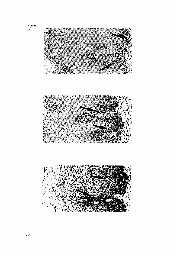
Figure 1 (A)
114
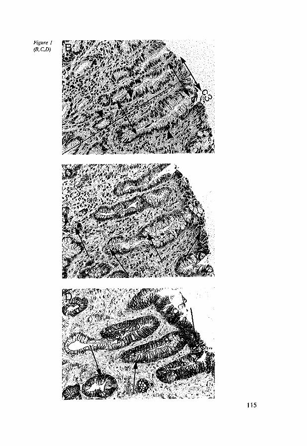
Figure 1 IB,C.D)
115
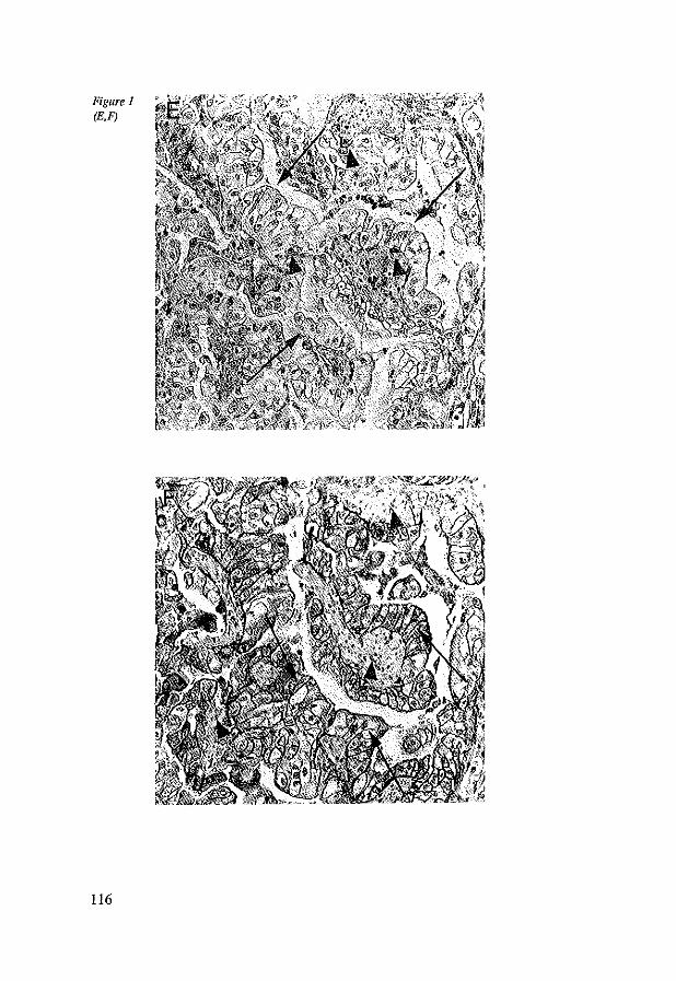
Figure 1 IE,F)
116
Cross tabulations were perfonued to assess correlations between CD44st and CD44-V6 expression, and grade and stage of the carcinomas. Significance was
tested by the Fisher's exact test (two tailed). Survival probabilities were calculated and compared by the Kaplan-Meier method and logrank test.
Multivariate analysis to determine independent prognostic value of the various
parameters was perfonued by Cox regression. For all tests, p=O.05 (two sided)
was considered the limit of significance.
8.4 Results
8.4.1 CD44st and CD44v6 in control tissues, Barrett's esophagus and
esophageal adenocarcinomas
Homogeneous membrane immunostaining for CD44st and CD44v6 was found in
all cases of normal squamous epithelium and in the crypts and glands of normal cardia mucosa and non dysplastic Barrett's epithelium (figure lA, B). These
tissues adjacent to the adenocarcinomas served as internal controls for the staining procedures. In squamous epithelium membrane immunostaining was
present homogeneously on the intenuediate cells and on the basal layer, but not on the basal membrane side of the basal cells (figure lA). In columnar cardia
epithelium, intercellular membrane staining was only seen in the gland and crypt
zone. In both squamous epithelium and columnar epithelium CD44v6 staining
was more often found and more intense than CD44st (figure lA). Membrane staining for CD44st, but not for CD44v6, was seen for a variety of other
different cell types, including lymphocytes and stromal cells.
8.4.2 Dysplasia, Ki-67 antigen (proliferation), CD44st and CD44v6 in
Barrett's esophagus
Thirty five cases of Barrett's esophagus were analyzed for dysplasia, Ki-67 antigen expression, and CD44st and CD44v6 immunoreactivity (figures lB-D).
Fourteen cases were non dysplastic, 16 showed indefinite or low grade dysplasia
and five cases were highly dysplastic (figure 2). In Barrett's esophagus without dysplasia proliferation was observed in Cl in
13/14 and in C2 in 1114. In all non dysplastic cases CD44st was seen in Cl only.
In contrast, CD44v6 was seen in Cl, C2 and C3 in 8/14, 5/14 and 1114 Barrett's
esophagus without dysplasia. In cases indefinite for dysplasia or with low grade dysplasia, Ki-67 antigen was
117
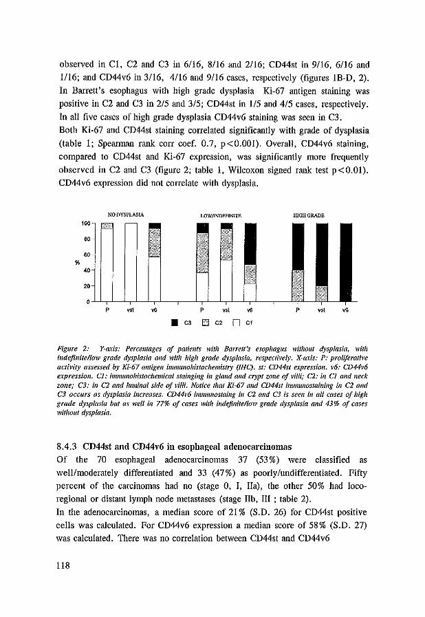
observed in CI, C2 and C3 in 6/16, 8116 and 2116; CD44st in 9/16, 6116 and
1/16; and CD44v6 in 3116, 4/16 and 9116 cases, respectively (figures IB-D, 2).
In Barrett's esophagus with high grade dysplasia Ki-67 antigen staining was
positive in C2 and C3 in 2/5 and 3/5; CD44st in 1/5 and 4/5 cases, respectively.
In all five cases of high grade dysplasia CD44v6 staining was seen in C3.
Both Ki-67 and CD44st staining correlated significantly with grade of dysplasia
(table 1; Spearman rank corr coef. 0.7, p<O.OOl). Overall, CD44v6 staining,
compared to CD44st and Ki-67 expression, was significantly more frequently
observed in C2 and C3 (figure 2; table I, Wilcoxon signed rank test p <0.01).
CD44v6 expression did not correlate with dysplasia.
NO DYSPLASrA LOW/lNDEFlN1TE llGHGRADE
100
%
p Y$I ... p
• C3 E3 C2 0 Cl
Figure 2: Y-axis: Percelllages of patiellfs willi Barrell's esophagus without dysplasia, witll indefinite/low grade dysplasia alld with high grade dysplasia, respectively. X-aris: P: proliferative activity assessed by Ki-67 antigell immunohistochemistry (IHC). sf: CD44s1 expression. v6: CD44v6 expressioll. Cl.' immunohistocliemical sln/lIgillg ill gland alld crypt ZOlle of villi; C2: ill Cl alld neck zone,' C3: ill C2 and lumillal side oj villi. Notice that Ki-67 alld CD44st inmlllllos/oilling ill C2 alld C3 OCCllrs as dysplasia iI/creases. CD44~'6 immflllosfaillg ill C2 and C3 is seen ill all cases of high grade dysplasia but as well ill 77% of cases with indefinite/lolV grade dysplasia alld 43% of cases without dysplasia.
8.4.3 CD44st and CD44v6 ill esophageal adenocarcinomas
Of the 70 esophageal adenocarcinomas 37 (53 %) were classified as
well/moderately differentiated and 33 (47%) as poorly/undifferentiated. Fifty
percent of the carcinomas had no (stage 0, I, lIa), the other 50% had loco
regional or distant lymph node metastases (stage lIb, III ; table 2).
In the adenocarcinomas, a median score of 21 % (S.D. 26) for CD44st positive
cells was calculated. For CD44v6 expression a median score of 58% (S.D. 27)
was calculated. There was no correlation between CD44st and CD44v6
118
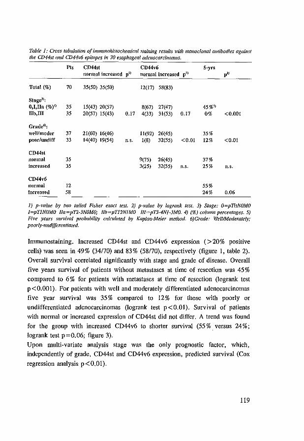
Table 1: Cross tabulation of immltlfoliistocliemical staining results witli monoclonal antibodies against the CD44st and CD44v6 epitopes i1l 70 esopliageal adenocarcinomas.
PIs CD44st CD44v6 S·yrs normal increased p" normal increased p" p"
Total (%) 70 35(50) 35(50) 12(17) 58(83)
Stage3):
O,I,lIa (%)4) 35 15(43) 20(57) 8(67) 27(47) 45%-S) IIb,lll 35 20(57) 15(43) 0.17 4(33) 31(53) 0.17 0% <0.001
Grade6>: weWmodcr 37 21(60) 16(46) 11(92) 26(45) 35% poor/undiff 33 14(40) 19(54) n.s. 1(8) 32(55) <0.01 12% <0.01
CD44st normal 35 9(75) 26(45) 37% increased 35 3(25) 32(55) n.s. 25% n.s.
CD44v6 normal 12 55% increased 58 24% 0.06
1) p-value by two tailed Fisher exact test. 2) p-mlue by /ogrank test. 3) Stage: O=pTisNOMO l=pTlNOMO lla=pT2-3NOMO; llb=pT12NIMO IlI=pT3-4Nl-3MO. 4) (%) column percel/lages. 5) Five years sltrvi\'ai probability calculated by Kaplan-Meier method. 6)Grade: WelllModerately; poorly-tIIldifferentiated.
Immunostaining. Increased CD44st and CD44v6 expression (>20% positive
cells) was seen in 49% (34170) and 83% (58170), respectively (figure I, table 2).
Overall survival correlated significantly with stage and grade of disease. Overall
five years survival of patients without metastases at time of resection was 45 %
compared to 6% for patients with metastases at time of resection (logrank test
p<O.OOI). For patients with well and moderately differentiated adenocarcinomas
five year survival was 35% compared to 12% for those with poorly or
undifferentiated adenocarcinomas (logrank test p<O.OI). Survival of patients
with normal or increased expression of CD44st did not differ. A trend was found
for the group with increased CD44v6 to shorter survival (55 % . versus 24 %;
10grank test p =0.06; figure 3).
Upon multi-variate analysis stage was the only prognostic factor, which,
independently of grade, CD44st and CD44v6 expression, predicted survival (Cox
regression analysis p<O.OI).
119
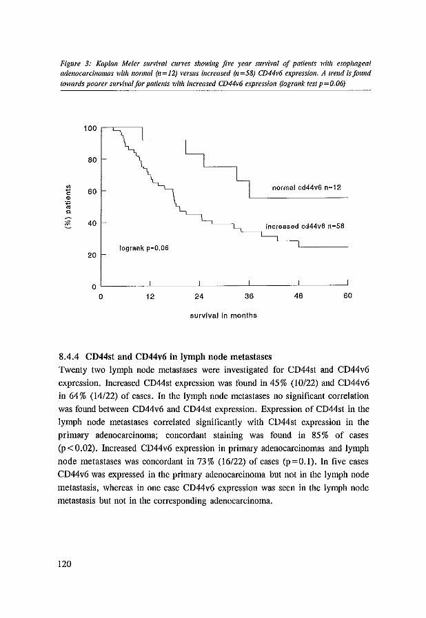
Figure 3: Kaplall Meier survival curves showillg five year survival of patients with esophageal adenocarcinomas wilh normal (11=12) verslls increased (11=58) CD44v6 expressioll. A trelld is/oulld towards poorer survival/or patiellls with increased CD44v6 expressioll (lograllk tesl p=O.06)
100 ~~-~
80
:1 co 60 normal cd44v6 n=12
'" 'c; "-
~ 40
log rank p=O.06 20
oL-----~------~------~----~~-----"
o 12 24 36 48 60
survival in months
8.4.4 CD44st and CD44v6 in lymph node metastases Twenty two lymph node metastases were investigated for CD44st and CD44v6
expression. Increased CD44st expression was found in 45% (10/22) and CD44v6 in 64% (14/22) of cases. In the lymph node metastases no significant correlation
was found between CD44v6 and CD44st expression. Expression of CD44st in the
lymph node metastases correlated significantly with CD44st expression in the primary adenocarcinoma; concordant staining was found in 85 % of cases
(p<0.02). Increased CD44v6 expression in primary adenocarcinomas and lymph
node metastases was concordant in 73% (16/22) of cases (p=O.I). In five cases
CD44v6 was expressed in the primary adenocarcinoma but not in the lymph node
metastasis, whereas in one case CD44v6 expression was seen in the lymph node metastasis but not in the corresponding adenocarcinoma.
120

8.5 Discussion
Development of esophageal adenocarcinomas in Barrett's esophagus occurs
through increasing dysplasia, concurrent with increased proliferation and
accumulation of genetic abnormalities (1-9). Overall, patients with esophageal
adenocarcinomas have poor prognosis. The presence of metastases is the most important factor predicting patient survival (14, 15).
Expression of CD44st and certain CD44 splice variants have proven to be predictive for behavior of several malignancies. The CD44v6 splice variant
expression has not only been associated with unfavorable clinicopathological characteristics in several carcinomas (20-24), but has also been observed in
premalignant lesions such as in colon adenomas (29-31).
In this study we present data on CD44st and CD44v6 expression in 35 cases of
Barrett's esophagus with different degrees of dysplasia, in 70 esophageal
adenocarcinomas associated with Barrett's esophagus, and also in 22 lymph node
metastases. Although in control epithelia a consistently higher number of cells
stained positive for CD44v6 than for CD44st, in these epithelia CD44st and
CD44v6 staining was clearly correlated.
In Barrett's esophagus we compared CD44st and CD44v6 expression with grade
of dysplasia and proliferation rate. CD44st expression increased along with increased dysplasia and proliferative activity, but CD44v6 did not. In literature a
similar correlation between CD44 and proliferation was noted for colonic epithelium and adenomas (33). Also in gastric intestinal metaplasia increased
CD44 expression was observed (20). CD44v6 expression in Barrett's esophagus did neither correlate with dysplasia nor with proliferation nor with CD44st
expression. Excessive CD44v6 expression was seen in all cases of Barrett's
esophagus with high grade dysplasia but was also observed in many cases with
low grade and some cases without dysplasia. This indicates that increased CD44v6 expression is an early event during neoplastic transformation in Barrett's
esophagus. In contrast, in colon adenomas increased CD44v6 expression has almost exclusively been observed in areas with high grade dysplasia (29-31).
In the esophageal adenocarcinomas, increased CD44st was observed in a lower number of cases than CD44v6 expression (50% versus 83 % respectively).
Gunther! et al. (34), observed that not only epitopes of the variable portion of the
CD44 molecule, but also the standard part of the CD44 molecule may be
differentially expressed. As we used a monoclonal antibody which recognizes
only one epitope of the CD44st domain, this may explain why a number of
121
carcinomas with increased CD44v6 immunostaining did not stain for CD44st. Alternatively, differences in epitope affinity of the anti CD44st and CD44v6
antibodies, resulting in different staining intensities, may have confounded the staining results.
The proportion of cases with increased CD44st and CD44v6 found in this study
for esophageal adenocarcinomas is comparable to that found for other gastro
intestinal carcinomas (20, 24). Interestingly, univariate analysis showed a trend
of increased CD44v6 expression to be correlated with poorer survival (p=0.06).
Also breast and colon carcinomas as well as in non-Hodgkin lymphomas CD44v6 expression is significantly correlated with survival (35).
Sixty four per cent (14122) of the lymph node metastases showed increased
CD44v6 staining. Interestingly in 5116 cases CD44v6 expression was not increased in the lymph node metastases. Since most carcinomas showed
heterogeneous CD44v6 expression, it is likely that metastatic competence is not
limited to the CD44v6 positive cells. Alternatively, progression of metastasized carcinoma cells may lead to loss of CD44v6 expression.
Expression of CD44v6 has been associated with increased malignant behavior, in spite of the fact that in many epithelia these variants are normally expressed (29,
36). To differentiate between regulated and abnormal CD44v6 expression in
Barrett's esophagus, investigation of the expression of splice variants expression
at mRNA level would be necessary(20). Unfortunately, the parafrm embedded material used in this study did not allow these analyses. Moreover, for clinical
practice RNA analyses-assays are too laborious, demanding special conditions for
tissue preservation in order to obtain liable results. Assessment of progression
specific expression of epitopes of the CD44 molecule which are specific tumor progression markers may be more applicable for clinical use. Recently, intron 9
of CD44 has been found to be expressed in 80% of colon adenocarcinomas and to be expressed in several cell-lines of gastro intestinal origin (37). As expression
of an intron sequence is an abnormal event and presumably the result of gene
derangement, immunohistochemical assessment of these epitopes may be more
tumor specific. However, expression of CD44st and CD44 splice variants is complicated and yet not fully understood. This caveat calls for careful interpretation immunohistochemical staining.
In conclusion, we have found in Barrett's esophagus expression of CD44v6 splice
variant early during development of neoplastic transformation, reaching a high incidence in esophageal adenocarcinomas. At present inununohistochemical
staining of CD44 epitopes do not have diagnostic value for evaluation of
122
Barrett's esophagus and esophageal adenocarcinomas.
8.6 References
1. Garewal HS, Sampliner R, Liu Y. Trent 1M. Chromosomal rearrangements in Barrctt's esophagus. A premalignant lesion of esophageal adenocarcinoma. Cimcer Gel/et Cytogellel 42;1989:281·6.
2. Gray MR, Hall PA, Nash I. Ansari B, Lane DP, Kingsnorth AN. Epithelial proliferation in Barrett's esophagus by proliferating cell nuclear antigen immunolocalization. Gastroellferology 103; 1992: 1769-76.
3, Hong MK, Laskin \VB, Hennan BE, Johnston MH. Vargo H, Steinberg SM, Allegra CIt Johnston PG. Expansion of the Ki-67 proliferative compartment correlates with degree of dysplasia in Barrett's esophagus. Cancer 75;1995:423-9.
4. Jankowski I, McMenemin R. Yu C, Hopwood D. Wormsley KG. Proliferating cell nuclear antigen in oesophageal diseases; correlation with transforming growth factor alpha expression. Gut 33;1992:587-91.
5. Krishnadath KK, THanus HW, van Blankenstein M, Hop we, Teijgeman R, Mulder AH, Bosman FT, van Dekken H. Accumulation of genetic abnormalities during neoplastic progression in Barrett's esophagus. Callcer Res 55;1995:1971-6.
6. Rabinovitch PS, Reid BI, Haggitt RC, Norwood TH. Rubin CEo Progression to cancer in Barrett's esophagus is associated with genomic instability. Lnb Ilivest 60;1989:65-71.
7. Reid BJ, Blount PL, Rubin CE, Levine DS, Haggitt RC, Rabinovitch PS. Flow-cytometric and histological progression to malignancy in Barrett's esophagus: prospective endoscopic surveillance of a cohort [see comments]. Gas/roellterology 102; 1992: 1212-9.
8. Raskind WH, Norwood T, Levine DS, Haggilt RC, Rabinovitch PS, Reid BJ. Persistent clonal areas and clonal expansion in Barrett's esophagus. Callcer Res 52;1992:2946-50.
9. Reid BJ, Weinstein WM. Barrett's esophagus and adenocarcinoma. [Review]. A1I11f1 Rev hied 38; 1987:477-92.
10. Cameron AJ, OU BJ, Payne WS. The incidence of adenocarcinoma in columnar-lined (Barrett's) esophagus. N Engi J Med 313;1985:857-9.
11. Spechler SJ, Robbins AH, Rubins HB, Vincent ME, Heeren T, Doos WG, Colton T, Schimmel EM. Adenocarcinoma and Barrett's esophagus. An overrated risk? Gaslroelllerology 87; 1984:927-33.
12. Van der Veen AH, Dees I, Blankensteijn ID, Van Blankenstein M. Adenocarcinoma in Barrett's oesophagus: an overrated risk. Gut 30;1989:14-8.
13. Achkar E, Carey \V. The cost of surveillance for adenocarcinoma complicating Barrett's esophagus. Am J Gaslroelltero( 83;1988:291-4.
14. Menke-Pluymers MD, Schoute NW. Mulder AH, Hop WC, van Blankenstein M, Tilanus HW. Outcome of surgical treatment of adenocarcinoma in Barrett's oesophagus. Gill 33;1992: 1454-8.
15. Lernt T, Coosemans W, Van Raellldonck D. Dillemans B, De Leyn P, Mamette 1M, Geboes K. Surgical treatment of Barrett's carcinoma. Correlations between morphologiC findings and prognosis. J Thorae Cardiomse Sllrg 107;1994:1059-65.
16. Haynes BF, Telen MI, Hale LP, Denning SM. CD44--a molecule involved in leukocyte adherence and T-cel! activation [published erratum appears in Immunol Today 1990 Mar;11(3):80]. [Review]. Immrmo/ Today 10;1989:423-8.
17. Pals ST, Koopman G, Heider KH, Griffioen A, Adolf GR, Van den Berg F t Ponta H, Herrlich P, Horst E. CD44 splice variants: expression during lymphocyte activation and tumor progression. [Review]. Behrillg Illst Milt 92;1993:273-7.
18. GUllthert U, Hofmann M, Rudy W, Reber S, Zoller M, Haussmann I. Matzku S, Wenzel A.
123
Ponta H, Herrlich P. A new variant of glycoprotein CD44 confers metastatic potential to rat carcinoma cells. Cell 65;1991:13-24.
19. Hofmann M, Rudy W, Zoller M, Tolg C, Ponta H, Herrlich P, Gunthert U. CD44 splice variants confer metastatic behavior in rats: homologous sequences are expressed in human tumor cell lines. Callcer Res 51;1991:5292-7.
20. Heider KH, Dammrich J, Skroch-Angel P, Muller-Hermelink HK, Vollmers HP, Herrlich P, Ponta H. Differential expression of CD44 splice variants in intestinal- and diffuse-type human gastric carcinomas and nonnal gastric mucosa. Callcer Res 53;1993:4197-203.
21. Kaufmann M, Heider KH, Sinn HP, von Minckwitz G. Ponta H, Herrlich P. CD44 variant exon epitopes in primary breast cancer and length of survival [see comments]. Lancet 345;1995:615-9.
22. Dall P. Heider KH, Hekele A, von Minckwitz G, Kaufmann M, Ponta H. Herrlich P. Surface protein expression and messenger RNA-splicing analysis of CD44 in uterine cervical cancer and nOffiml cervical epithelium. Cancer Res 54;1994:3337-41.
23. Penno MB, August JT, Baylin SB, Mabry M, Linnoila RI. Lee VS, Croteau D, Yang XL, Rosada C. Expression of CD44 in human lung tumors. Cancer Res 54;1994:1381-7.
24. Mulder JW. Kroyt PM, Sewnath M. Oosting J. Seldenrijk CA, Weidema WF, Offerhaus GJ, Pals ST. Colorectal cancer prognosis and expression of exon-v6-containing CD44 proteins. wl/eet 344;1994:1470-2.
25. Gunther! U, Stauder R, Mayer B, Terpe HJ, Finke L, Friedrichs K. Are CD44 variant isoforms involved in human tumour progression? [Review]. Callcer Sllrv 24; 1995: 19-42.
26. Kan M, Aki M, Akiyama K, Naruo S. Kanayama H. Kagawa S. High-level expression of the CD44 variant sharing exon vl0 in renal cancer. IplI I Callcer Res 86;1995:847-53.
27. Finn L. Dougherty G. Finley G, Meisler A, Becich M. Cooper DL. Alternative splicing of CD44 pre-mRNA in human colorectal tumors. Biochem Biop/lys Res Commlm 200;1994:1015-22.
28. Takeuchi K, Yamaguchi A, Urano T, Goi T. Nakagawara G, Shiku H. Expression of CD44 variant exons 8-10 in colorectal cancer and its relationship to metastasis. IplI I Caltcer Res 86;1995:292-7.
29. Heider KH, Hofmann M. HOTs E, van den Berg F, Ponta H. Herrlich p. Pals ST. A human homologue of the rat metastasis-associated variant of CD44 is expressed in colorectal carcinomas and adenomatous polyps. J Ce/f BioI 120;1993:227-33.
30. Wieienga VJ, Heider KH, Offerhaus GJ. Adolf GR, van den Berg PM, Ponta H, Herrlich P, Pals ST. Expression of CD44 variant proteins in human colorectal cancer is related to tumor progression. Caltcer Res 53;1993:4754-6.
31. Mulder JW, Wielenga VJ. Polak MM, van den Berg FM, Adolf GR, Herrlich P, Pals ST. Offerhaus OJ. Expression of mutant p53 protein and CD44 variant proteins in colorectal tumorigenesis. Gut 36; 1995:76-80.
32. Reid BJ, Haggitt RC, Rubin CEo Roth G, Surawicz CM. Van Belle G, Lewin K, Weinstein WM, Antonioli DA, Goldman H, et aJ. Observer variation in the diagnosis of dysplasia in Barrett's esophagus. Hum PathoI19;1988:166·78.
33. Abbasi AM, Chester KA. Talbot IC. Macpherson AS, Boxer G, Forbes A, Malcolm AD, Begent RH. CD44 is associated with proliferation in nonnal and neoplastic human colorectal epithelial cells. Eur I Callcer 14;1993:1995-2002.
34. Gunthert U. CD44: a multitude of isoforms with diverse functions. [Review]. Cu" Top MicrobiollmmulloI184;1993:47-63.
35. Herrlich p. Pals S, Ponta H. CD44 in colon cancer. [Review}. Eurl Callcer ;1995:1110-2. 36. Mackay CR, Terpe HJ. Stauder R. Marston WL, Stark H, Gunthert U. Expression and
modulation of CD44 variant isoforms in humans. J Cell Bioi 124; 1994:71-82. 37, Yoshida K, Dolodeoku J, Sugino T, Goodison S, Matsumura y, Warren BF, Toge T, Tahara
E, Tarin D. Abnormal retention of mtron 9 in CD44 gene transcripts in human gastrointestinal tumors. Qmcer Res 55;1995:4273-7.
124

Chapter 9
Concluding Remarks

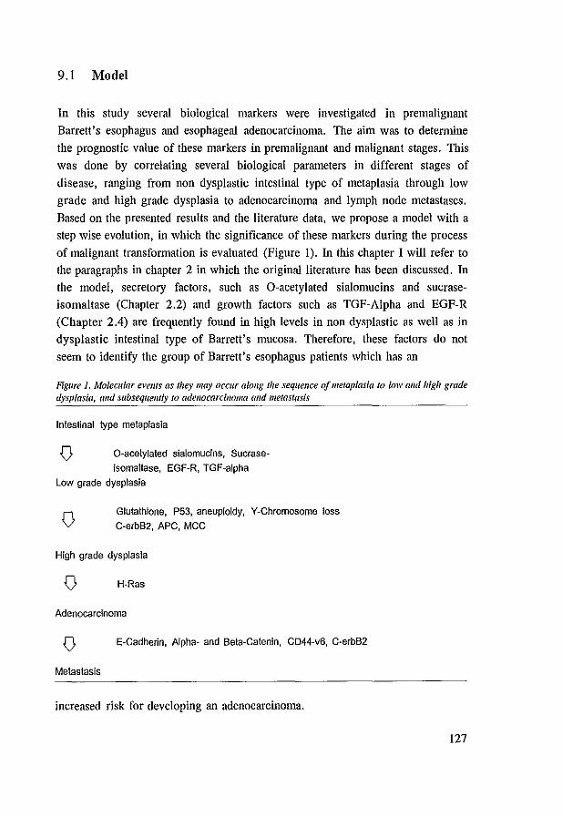
9.1 Model
In this study several biological markers were investigated in premalignant
Barrett's esophagus and esophageal adenocarcinoma. The aim was to detennine
the prognostic value of these markers in premalignant and malignant stages. This
was done by correlating several biological parameters in different stages of
disease, ranging from non dysplastic intestinal type of metaplasia through low
grade and high grade dysplasia to adenocarcinoma and lymph node metastases.
Based on the presented results and the literature data, we propose a model with a
step wise evolution, in which the significance of these markers during the process
of malignant transformation is evaluated (Figure 1). In this chapter I will refer to
the paragraphs in chapter 2 in which the original literature has been discussed. In
the model, secretory factors, such as O-acetylated sialomucins and sucrase
isomaltase (Chapter 2.2) and growth factors such as TGF-Alpha and EGF-R
(Chapter 2.4) are frequently found in high levels in non dysplastic as well as in
dysplastic intestinal type of Barrett's mucosa. Therefore, these factors do not
seem to identify the group of Barrett's esophagus patients which has an
Figure 1. Molecular evellts as they llIay occllr alollg the sequence oj metaplasia to [ow alld high grade dysplasia, alld subsequemly to adellocarcinoma alld metastasis
Intestinal type metaplasia
Q O~acetylated sialomucins, Sucrase~ isomaltase, EGF·R, TGF-alpha
Low grade dysplasia
Q Glutathione, P53, aneuploidy, V-Chromosome loss C·erbB2, APC, MCC
High grade dysplasia
Q H·Ras
Adenocarcinoma
Q E·Cadherin, Alpha· and Bela·Calenin, CD44-v6, C·erbB2
Metastasis
increased risk for developing an adenocarcinoma.
127
Parameters which do seem to be associated with neoplastic progression are decreased glutathione and glutathione-S transferase enzyme activity (Chapter 2.2)
increased proliferative activity (Chapter 2.3), aneuploidy (Chapter 2.8) aberrant p53 (Chapter 2.6) and Y-chromosome loss (Chapter 2.7). Typically, these
markers are observed at low frequency in non dysplastic Barrett's esophagus, they are increasingly observed along with increasing dysplasia and a high
frequency is reached in high grade dysplasia and in the adenocarcinomas. In a prospective follow-up study the majority of Barrett's esophagus patients with
aneuploidy or increased tetraploidy in their initial biopsy specimens progressed towards high grade dysplasia or carcinoma, whereas patients without these
abnormalities did not (Chapter 2.8). In chapters 5, 6 and 8, the correlation of increased proliferation rate, p53 protein accumulation, aneuploidy and Y
chromosome loss with dysplasia was studied. In chapter 5, we report increased
levels of proliferation in intestinal type of metaplasia with and without dysplasia.
However, detailed analysis of proliferation in Barrett's esophagus revealed that as the epithelium becomes dysplastic the zone of proliferating cells tends to expand
to the luminal surface of the mucosa. A similar expression pattern was observed
for CD44st (chapter 8). The evolution from low grade to high grade dysplasia is characterized by
increasing genetic instability. Proliferation rate and aneuploidy increase (Chapter
2.3, 2.8, 5, 8). In addition to p53 aberrations other tumor suppressor genes and oncogenes such as APC, and H-ras showed abnormalities (Chapter 2.5, 2.6). It
is not known in which order these genetic aberrations occur and neither whether the order is at all of importance. Nevertheless, there is evidence that p53 (l7p)
aberrations precede aneuploidy and APC (5q) gene loss (Chapter 2.7).
For dysplastic cells to become invasive cancer cells a crucial step is the moment
they acquire the ability to break through the basement membrane and invade surrounding tissues. In frankly invasive adenocarcinomas the accumulation of
numerous genetic aberrations have been seen (Chapter 2). However, little data is available about genetic events specifically involved in transition from high grade
dysplasia into adenocarcinoma. In a small series H-ras expression was observed
in high grade dysplasia and adenocarcinomas but not in low grade dysplasia, nor
in non dysplastic epithelium (Chapter 2.5). This suggests an important role of
H-ras in the transition from high grade dysplasia to adenocarcinoma.
Perhaps the most fascinating step in carcinogenesis is the formation of
metastases. Formation of metastases is a complex process in which many factors are involved. In this process, epithelial cells are liberated from their original
128
compartment, break through the basement membrane, invade lymph and blood
vessels, adhere to a new environment and proliferate. It has been observed that
esophageal adenocarcinomas with limited invasion of the esophageal wall already may have metastasized (Chapter 1.3). It seems that as soon as cells invade the
submucosa, metastatic ability is rapidly acquired. In a few studies aneuploidy was
found to correlate with the presence of metastases and poor survival (Chapter
2.8). Another study showed a correlation between C-erbB2 expression and poor survival (Chapter 2.5). Recently, cell surface molecules and their role in tumor
cell behavior gained interest. Cell surface molecules, such as those involved in the E-Cadherin-Catenin complex and CD44 with its splice variants, play an
important role in tumor invasiveness and metastatic ability (Chapter 2.9, 2. to). A
high frequency of decreased expression of E-Cadherin was found in esophageal
adenocarcinomas (Chapter 2.9). In our studies decreased expression of Alphaand Beta-Catenin was found to occur roughly in the same frequency as E
Cadherin expression (Chapter 7). Interestingly, in a subset of carcinomas without
metastases and sometimes with limited invasion of the esophageal wall, decreased
expression of these proteins correlated with poor survival (Chapter 7). Another interesting cell surface molecule studied in this thesis is CD44. It has become
apparent that the expression of the CD44v6 splice variant is associated with
increased tumor invasiveness and metastatic ability (Chapter 2. to). We detected
increased CD44v6 splice variant expression in a high percentage of esophageal adenocarcinomas (Chapter 8). A trend was found between increased CD44v6
expression and poor five year survival. Surprisingly, a high level of CD44v6
expression was found in Barrett's esophagus as well (Chapter 8).
9.2 In conclusion
In conclusion, in the progression of Barrett's esophagus via dysplasia to
carcinoma and the development of metastases formation numerous parameters are involved. In this study p53 protein accumulation, aneuploidy, Y-chromosome
loss, high expression of the CD44st molecule and increased proliferation rate
were found to go along with increasing dysplasia. These markers may identify
Barrett's esophagus at risk for neoplastic progression. Nevertheless, if these markers were to be applied for clinical decision making, their relevance should
be evaluated in long term follow up studies of patients with Barrett's esophagus. For most of these parameters long tenn follow up studies are lacking.
129
The most promising marker investigated is p53. During neoplastic progression
mutation of the p53 gene seems to be a key event which is followed by several
other genetic alterations. In addition to histological grading of dysplasia,
detection of p53 aberrations might become an important diagnostic tool.
A remarkable observation in this study was loss of the Y -chromosome, which
occurs as dysplasia increases. The meaning of Y -chromosome loss is unclear. At
present no specific genes involved in tumorigenesis have been located on the Y
chromosome. Therefore, Y-loss is considered a random effect, reflecting genetic
instability and as such is a parameter to detect neoplastic progression in male
Barrett's esophagus patients.
The transformation of high grade dysplasia into adenocarcinoma is poorly
understood. Possibly the same factors involved in tumor invasiveness and
metastatic behavior of esophageal adenocarcinomas are involved in this
transitional step. Future investigation of biological markers involved in tumor
behavior in high grade dysplasia and early carcinomas are of major importance.
With respect to tumor behavior and metastasis decreased expression of the cell
surface molecules E-Cadherin, Alpha- and Beta-Catenin and increased expression
of CD44v6 in esophageal adenocarcinomas were found to correlate with poor
survival. Although these results may seem useful for clinical applications, the
underlying mechanisms involved in up and down regulation of these factors are
poorly understood and should be explored in more detail. For instance, there is a
complicated interaction between the E-Cadherin-Catenin complex and several
intracellular factors such as the tumor suppressor APC and the C-erbB2
oncogene. Concerning CD44 splice variants, little is known about their
interactions and regulation of expression. Therefore, the use of the expression of
cell surface molecules for predicting survival at this moment is premature.
130

Summary
Barrett's esophagus is a condition in which, due to chronic gastro-esophageal reflux, the normal squamous epithelium is transformed into columnar epithelium.
Compared to the general population, patients with Barrett's esophagus have an
increased risk of 30 to 120 fold for developing esophageal adenocarcinomas.
Malignant transformation of Barrett's esophagus is a gradual process which is
reflected in histological changes, classified as low and high grade dysplasia.
Unfortunately, most patients with Barrett's esophagus have no or few symptoms (gastro-esophageal reflux), and esophageal adenocarcinoma is often detected in
advanced stage of disease. During the past decades there has been an alarming rise in the incidence of esophageal adenocarcinomas. In Western countries the
incidence of esophageal adenocarcinomas reaches almost 1 per 100.000 per year.
The prevalence is highest in white middle aged males of higher socio-economic
classes. Esophageal adenocarcinomas are frequently metastasizing tumors
associated with an average five year survival of less than 20%. However, in case
treated in an early stage of the disease, survival improves significantly.
In this thesis studies concerning neoplastic progression of Barrett's esophagus and
metastases formation in esophageal adenocarcinomas are reported. In our studies
we analyzed several biological parameters in non dysplastic and dysplastic
Barrett's esophagus, in esophageal adenocarcinomas and metastases. In a pilot study aneuploidy, p53 protein accumulation and numerical chromosomal
aberrations such as chromosome 17- and Y-loss in adenocarcinoma and Barrett's
esophagus were detected. P53 protein accumulation was evaluated in over a
hundred biopsy specimens of patients with Barrett's esophagus with varying degrees of dysplasia and in 24 esophageal adenocarcinomas. In approximately
80% of the adenocarcinomas p53 protein had accumulated. In Barrett's
esophagus a significant correlation was found between p53 protein accumulation
and increasing dysplasia. In a subsequent study aneuploidy and the status of X and Y -chromosome was investigated in a series of 50 biopsy specimens of patients with Barrett's esophagus and in 23 adenocarcinomas. Aneuploidy and Y
chromosome loss were found to correlate with dysplasia. The frequencies of both
parameters reached 70-100% in high grade dysplasia and adenocarcinomas. Other parameters investigated in Barrett's esophagus have been the expression of the
cell surface molecules CD44st and CD44v6 splice variant and proliferation rate.
Expression of these parameters has been investigated in different compartments
of the Barrett's esophagus mucosa. As dysplasia increased proliferation and
131
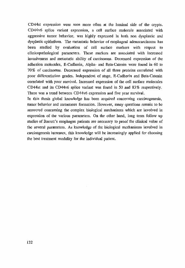
CD44st expression were seen more often at the luminal side of the crypts.
CD44v6 splice variant expression, a cell surface molecule associated with
aggressive tumor behavior, was highly expressed in both non dysplastic and
dysplastic epitilelium. The metastatic behavior of esophageal adenocarcinoma has
been studied by evaluation of cell surface markers with respect to
clinicopathological parameters. These markers are associated with increased
invasiveness and metastatic ability of carcinomas. Decreased expression of the
adhesion molecules, E-Cadherin, Alpha- and Beta-Catenin were found in 60 to
70% of carcinomas. Decreased expression of all three proteins correlated with
poor differentiation grades. Independent of stage, E-Cadherin and Beta-Catenin
correlated with poor survival. Increased expression of the cell surface molecules
CD44st and its CD44v6 splice variant was found in 50 and 83% respectively.
There was a trend between CD44v6 expression and five year survival.
In this thesis global knowledge has been acquired concerning carcinogenesis,
tumor behavior and metastases formation. However, many questions remain to be
answered concerning the complex biological mechanisIll5 which are involved in
expression of the various parameters. On the other hand, long term follow up
studies of Barrett's esophagus patients are necessary to proof the clinical value of
the several parameters. As knowledge of the biological mechanisIll5 involved in
carcinogenesis increases, this knowledge will be increasingly applied for choosing
the best treatment modality for the individual patient.
132

Samenvatting
Barrett oesofagus is een aandoening, waarbij het normale niet verhoomend meerlagig plaveisel epitheel van de oesofagus, door langdurige gastro-oesofageale
reflux, is veranderd in cilindrisch epithee!. Patienten met een Barrett oesofagus
hebben vergeleken met de normale bevolking een 30 tot 120 maal verhoogd
risico voor het krijgen van een oesofagus adenocarcinoom. Maligne transformatie
van Barrett epitheel is een geleidelijk proces, dat wordt gekenmerkt door
histologische veranderingen. Binnen deze histologische veranderingen wordt
onderscheid gemaakt tussen niet dysplastisch, licht en sterk dysplastisch epithee!.
Het merendeel van de patienten met een Barrett oesofagus heeft geen (reflux)
klachten, waardoor veel oesofagus adenocarcinomen in een laat stadium worden
ontdekt. Gedurende de afgelopen decennia is de incidentie van oesofagus
adenoarcinomen a1armerend toegenomen. In Westerse landen is deze incidentie
bijna 1 per 100.000. De belangrijkste risico groep voor het krijgen van een
oesofagus adenocarcinoom wordt gevormd door blanke mannen van middelbare leeftijd uit een hoog socio-economische milieu. Oesofagus carcinomen zijn
geassocieerd met vroege metastasering en een vijf jaars overleving van minder
dan 20 %. De overleving verbetert significant indien de ziekte in een vroeg stadium wordt ontdekt.
In dit proefschrift is de maligne progressie van Barrett oesofagus en het
progressieve gedrag van oesofagus adenocarcinomen onderzocht. Verschillende
biologische factoren werden bestudeerd in niet dysplastisch en dysplastisch Barrett epitheel, in oesofagus adenocarcinomen en in lymfklier metastasen. In een
'pilot' studie werden aneuploidie, p53 proteine accumulatie en numerieke
chromosomale afwijkingen, zoals verlies van chromosomen 17 en Y ontdekt.
Deze bevindingen werden in de daarop volgende studies verder geevalueerd. Aberrante accumulatie van het p53 eiwit werd in meer dan 100 biopten met
verschillende graden van dysplasie en in 24 oesofagus adenocarcinomen bekeken. Accumulatie van p53 werd in ongeveer 80% van de adenocarcinomen gezien. In
Barrett oesofagus correleerde p53 accumulatie significant met toenemende dysplasie. In de studie die hierop volgde werden aneuploidie en de status van het
X en Y -chromosoom bestudeerd in 50 biopten van patienten met Barrett oesofagus en in 23 oesofagus adenocarcinomen. Zowel aneuploidie en verlies van
het Y chromosoom correleerden significant met toenemende dysplasie. Beide parameters hadden een frequentie van 70-100% in biopten met sterke dysplasie
en in de adenocarcinomen. Andere parameters die in Barrett oesofagus werden
133
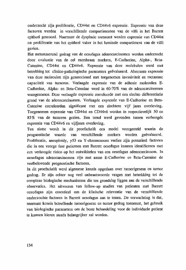
onderzocht zijn proliferatie, CD44st en CD44v6 expressie. Expressie van deze
factoren werden in verschillende compartimenten van de villi in het Barrett
epitheel gescoord. Naarrnate de dysplasie toeneemt werden expressie van CD44st
en proliferatie van het epitheel vaker in het luminale compartiment van de villi
gezien. Het metastaserend gedrag van de oesofagus adenocarcinomen werden onderzocht
door evaluatie van de cel membraan markers, E-Cadherine, Alpha-, Beta
Catenine, CD44st en CD44v6. Expressie van deze molekulen werd met
betrekking tot clinico-pathologische parameters geevalueerd. Aberrante expressie
van deze moleculen zijn geassocieerd met toegenomen invasiviteit en metastase
capaciteit van tumoren. Verlaagde expressie van de adhesie moleculen E
Cadherine, Alpha- en Beta-Catenine werd in 60-70% van de adenocarcinomen
waargenomen. Deze verlaagde expressie correleerde met een slechte differentiatie
graad van de adenocarcinomen. Verlaagde expressie van E-Cadherine en Beta
Catenine correleerden significant met een slechtere vijf jaars overleving.
Toegenomen expressie van CD44st en CD44v6 werden in respectievelijk 50 en
83 % van de tumoren gezien. Een trend werd gevonden tussen verhoogde
expressie van CD44v6 en vijfjaars overleving.
Ten slotte wordt in dit proefschrift een model voorgesteld waarin de prognostische waarde van verschillende markers worden geevalueerd.
Proliferatie, aneuploidy, p53 en Y-chromosoom verlies zijn potentieel factoren die in een vroege fase patienten met Barrett oesofagus kunnen identificeren met
een verhoogde risico op het ontwikkelen van een oesofagus adenocarcinoom. In oesofagus adenocarcinomen zijn met name E-Cadherine en Beta-Catenine de
veelbelovende prognostische factoren. In dit proefschrift werd algemene kennis opgedaan over tumorigenese en tumor
gedrag. Er zijn echter nog veel onbeantwoorde vragen met betrekking tot de
complexe biologische mechauismen die ten grondslag liggen aan de verschillende
observaties. Het uitvoeren van follow-up studies van patienten met Barrett oesofagus zijn essentieel om de klinische relevantie van de verschillende
onderzochte factoren in Barrett oesofagus aan te tonen. De verwachting is dat,
naarmate kennis betreffende tumorigenese en tumor gedrag toeneemt, het gebruik
van biologische parameters am de beste behandeling voor de individuele patient
te kunnen kiezen steeds belangrijker zal worden.
134
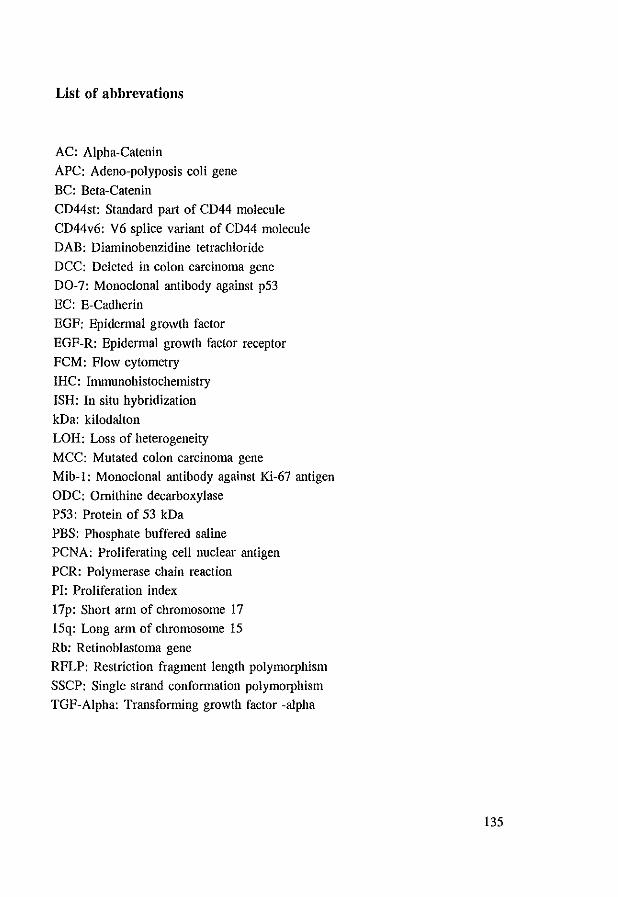
List of abbrevations
AC: Alpha-Catenin
APC: Adeno-polyposis coli gene
BC: Beta-Catenin
CD44st: Standard part of CD44 molecule
CD44v6: V6 splice variant of CD44 molecule
DAB: Diaminobenzidine tetrachloride
DCC: Deleted in colon carcinoma gene
DO-7: Monoclonal antibody against p53
EC: E-Cadherin
EGF: Epidermal growth factor
EGF-R: Epidermal growth factor receptor
FCM: Flow cytometry
IRC: Immunohistochemistry
ISH: In situ hybridization
kDa: kilodalton
LOH: Loss of heterogeneity
MCC: Mutated colon carcinoma gene
Mib-I: Monoclonal antibody against Ki-67 antigen
ODC: Ornithine decarboxylase
P53: Protein of 53 kDa
PBS: Phosphate buffered saline
PCNA: Proliferating cell nuclear antigen
PCR: Polymerase chain reaction
PI: Proliferation index
17p: Short arm of chromosome 17
15q: Long arm of chromosome 15
Rb: Retinoblastoma gene
RFLP: Restriction fragment length polymorphism
SSCP: Single strand conformation polymorphism
TGF-Alpha: Transforming growth factor -alpha
135


Dankwoord
Mijn promotoren dank ik voor het vertrouwen dat zij in mij hadden tijdens het
onderzoek. De co-promotor voor de dagelijkse begeleiding. Mijn dank gaat
verder uit naar de afdeling pathologie voor de gastvrijheid. In het bijzonder dank
ik mijn lab-genoten: Ianneke Alers, Ron Teijgemen, Pieter-Iaap Krijtenburg,
Anton Timmermans, Patrick Iansen en Kees Vissers voor de goede maar vooral
gezellige samenwerking. Frank van del' Panne wilde ik bedanken voor de service
en kwaliteit van het foto materiaal. Elizabeth Kremers en Cor van Vroonhoven bedank ik voor de analytische hulp. Mw. Bominar bedank ik voor de coordinatie
bij de voorbereiding van de promotie. Pavlov en Nicky voor hun gezelschap tijdens de uren achter de computer. Ten slotte bedank ik Max voor zijn morele
steun.
137


Curriculum Vitae
KausHia Krishnawatie Krishnadath werd op 7 januari 1966 geboren te
Paramaribo. Na het behalen van het VWO diploma aan de St. Stanislascollege te
Delft, is zij in 1984 de opleiding geneeskunde gaan volgen aan de Erasmus
Universiteit te Rotterdam. Het propedeuse exanlen werd in 1990 behaald. Ais
keuze stage werd er in 1989 gedurende 5 maanden onderzoek verricht in Zuid
India. Een Coassistentschap dermatologie werd in 1991 gevolgd te Paramaribo.
In 1992 studeerde zij Cum Laude af als basisarts. Hierna was zij werkzaam op
de afdeling pathologie aan de Erasmus Universiteit, waar zij is gestart met het
onderzoek naar prognostische factoren in Barrett oesofagus en oesofagus
adenocarcinomen. In 1993 werd zij AIO onder leiding van Prof. Dr P.T.
Bosman en Prof. Dr H.W. THanus. Het onderzoek heeft geresulteerd in dit
proefschrift, dat zij d.d. 26 juni 1997 zal verdedigen. Sinds 1996 is zij in
opleiding tot gastroenteroloog in het Dijkzigt ziekenhuis te Rotterdam.
139

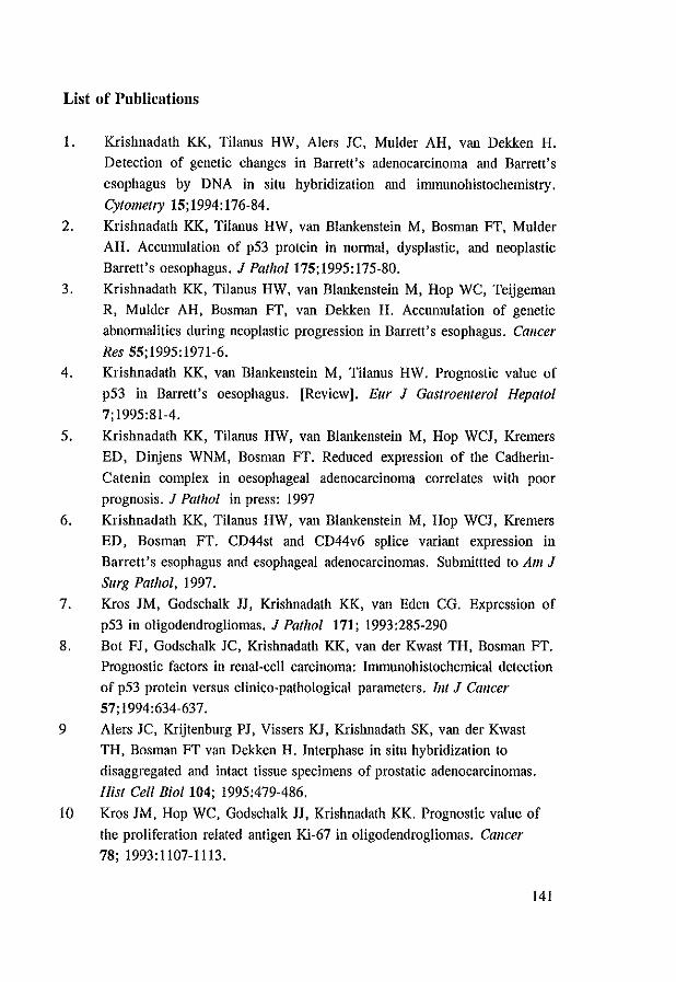
List of Publications
1. Krishnadath KK, Tilanus HW, Alers IC, Mulder AH, van Dekken H.
Detection of genetic changes in Barrett's adenocarcinoma and Barrett's
esophagus by DNA in situ hybridization and immunohistochemistry.
Cytomelly 15; 1994: 176-84.
2. Krishnadath KK, Tilanus HW, van Blankenstein M, Bosman FT, Mulder
AH. Accumulation of p53 protein in normal, dysplastic, and neoplastic
Barrett's oesophagus. J PatlIOI175;1995:l75-80.
3. Krishnadath KK, Tilanus HW, van Blankenstein M, Hop WC, Teijgeman
R, Mulder AH, Bosman FT, van Dekken H. Accumulation of genetic
abnorrualities during neoplastic progression in Barrett's esophagus. Callcer
Res 55;1995:1971-6.
4. Krishnadath KK, van Blankenstein M, Tilanus HW. Prognostic value of
p53 in Barrett's oesophagus. [Review]. Eur J Gastroellferol Hepatol
7;1995:81-4.
5. Krishnadath KK, Tilanus HW, van B1ankenstein M, Hop WCI, Kremers
ED, Dinjens WNM, Bosman FT. Reduced expression of the Cadherin
Catenin complex in oesophageal adenocarcinoma correlates with poor
prognosis. J Pat/wi in press: 1997
6. Krishnadath KK, Tilanus HW, van Blankenstein M, Hop WCI, Kremers
ED, Bosman FT. CD44st and CD44v6 splice variant expression in
Barrett's esophagus and esophageal adenocarcinomas. Submittted to Alii J
Surg PatilOl, 1997.
7. Kros 1M, Godschalk JJ, Krishnadath KK, van Eden CG. Expression of
p53 in oligodendrogliomas. J PatilOl 171; 1993:285-290
8. Bot FI, Godschalk IC, Krishnadath KK, van der Kwast TH, Bosman FT.
Prognostic factors in renal-cell carcinoma: Immunohistochemical detection
of p53 protein versus clinico-pathological parameters. lilt J Callcer
57; 1994:634-637.
9 Alers IC, Krijtenburg PI, Vissers KJ, Krishnadath SK, van der Kwast
TH, Bosman FT van Dekken H. Interphase in situ hybridization to
dis aggregated and intact tissue specimens of prostatic adenocarcinomas.
Hist Cell Bioi 104; 1995:479-486.
10 Kros 1M, Hop WC, Godschalk JJ, Krishnadath KK. Prognostic value of
the proliferation related antigen Ki-67 in oligodendrogliomas. Callcer
78; 1993: 1107-1113.
141