ENERGY-AND GLUTATHIONE METABOLISM IN SPERMATIDS AS … · ENERGY-AND GLUTATHIONE METABOLISM IN...
158
ENERGY- AND GLUTATHIONE METABOLISM IN SPERMATIDS AS POSSffiLE TARGETS FOR ANTISPERMATOGENIC AGENTS ENERGIE- EN GLUTATlllONMETABOLISME IN SPERMATID EN ALS MOGELIJKE DOELWITTEN VOOR ANTISPERMATOGENETISCHE STOFFEN PROEFSCHRIFI ter verkrijging van de graad van doctor aan de Erasmus Universiteit Rotterdam op gezag van de rector magnificus Prof. Dr. C.J. Rijnvos en volgens besluit van het college van dekanen. De openbare verdediging zal plaatsvinden op woensdag 24 januari 1990 om 15.45 uur door PIETER JOHANNES DEN BOER geboren te Dordrecht 1989 Gedrukt bij Offsetdrukkerij Kanters bv, Alblasserdam
Transcript of ENERGY-AND GLUTATHIONE METABOLISM IN SPERMATIDS AS … · ENERGY-AND GLUTATHIONE METABOLISM IN...
ENERGY- AND GLUTATHIONE METABOLISM IN SPERMATIDS AS POSSffiLE TARGETS
FOR ANTISPERMATOGENIC AGENTS
ENERGIE- EN GLUTATlllONMETABOLISME IN SPERMATID EN ALS MOGELIJKE DOELWITTEN VOOR ANTISPERMATOGENETISCHE STOFFEN
PROEFSCHRIFI
ter verkrijging van de graad van doctor aan de Erasmus Universiteit Rotterdam
op gezag van de rector magnificus Prof. Dr. C.J. Rijnvos
en volgens besluit van het college van dekanen. De openbare verdediging zal plaatsvinden op
woensdag 24 januari 1990 om 15.45 uur
door
PIETER JOHANNES DEN BOER
geboren te Dordrecht
1989
Gedrukt bij Offsetdrukkerij Kanters bv, Alblasserdam
Promotiecommissie
Promotor: Prof. dr. H.J. van der Molen
Overige leden: Prof. dr. j.F. Koster Prof. dr. J.F. Jongkind Prof. dr. j.J. van der Werff ten Bosch
Co-promotor Dr. j.A. Grootegoed
Dit proefschrift werd bewerkt binnen de afdeling Biochemie II (Chemische Endocrinologie) van de Faculteit der Genesskunde, Erasmus Universiteit Rotterdam.
Ignoramus et ignorabimus
voor Ardie en de kinderen
- s -
CONTENTS
Abbreviations and trivial names
Chapter 1 General introduction
1.1 Spermatogenesis
1.2 Biochemical properties of spermatids
1.3 Antispermatogenic agents
1.4 Aspects of testicular toxicity
1.5 Defence systems 1.5.1 - introduction 1.5.2 - detoxicating reactions
1.6 Glutathione in spermatogenic cells
1. 7 Model systems 1.7.1 - models 1.7.2 - parameters
1.8 Aim and scope of this thesis
References
page
8
9
10
15
15
18
19 19
21
23 24
26
27
Chapter 2 Differential effects of (+)- and (-)gossypol 35
Chapter 3
enantiomers on LDH-C 4 activity of hamster spermatogenic epithelium in vitro.
Mechanism of action of (-)gossypol on ATP 45 production in isolated hamster spermatids.
- 6 -
Chapter 4 The effects of glucose and adenosine on 55 the A TP content of hamster spermatids.
Chapter 5 Glutathione-dependent defence mechanisms 71 in isolated round spermatids from the rat
Chapter 6 Effect of glutathione depletion on the 91 cytotoxicity of xenobiotics and induction of single-strand DNA breaks by ionizing radiation in isolated hamster round spermatids.
Chapter 7 Glutathione metabolism in cultured 111 Sertoli cells and spermatogenic cells from hamsters
Summary and concluding remarks 122
Samenvatting 126
Papers related to this thesis 131
Curriculum vitae 132
Nawoord 133
Appendix paper 135 Energy metabolism of spermatids A review
- 7 -
Abbreviations and trivial names
ATP BSA BSO cAMP CDNB CHP OEM DNA DNase EDTA ENPP FSH GGT Gossypol
GSH GSSG Kd
adenosine 5'-triphosphate bovine serum albumin buthionine sulfoximine adenosine cyclic-3': 5'-monophosphate 1-chloro-2, 4-dini trobenzene cumene hydroperoxide diethyl maleate deoxyribonucleic acid deoxyribonuclease ethylenediamine tetraacetate 1, 2""epoxy-3 (p-ni trophenoxy) propane follicle-stimulating hormone y-glutamyl transpeptidase 1,1',6,6', 7, 7'-hexahydroxy-3,3'-dimethyl-5 ,5'-bis isopropyl [2,2'-bi-naphthaleneJ-8,8'-dicarboxaldehyde reduced glutathione; y-glutamylcysteinylglycine oxidized glutathione dissociation constant
Km Michaelis Menten constant LDH-C 4 lactate dehydrogenase isoenzyme C 4 LH luteinizing hormone MEM Eagle's minimum essential medium NAD(H) (reduced) nicotinamide adenine dinucleotide OPA o-phthaldialdehyde PBS Dulbecco's phosphate-buffered saline RFI relative fluorescence intensity Testosterone 17 ~-hydroxy-4-androsten-3-one TRIS 2-amino-2-hydroxy methy 1 propane-1, 3-dio 1 Vmax maximal initial velocity
- 8 -
Chapter 1
General Introduction
- 9 -
1.1 SPERMATOGENESIS
Spermatogenesis is a sequence of intra- and intercellular events resulting in the formation of spermatozoa from precursor cells. In mammalian species, this process takes place in the testis, and the further maturation of the spermatozoa occurs in the epididymis.
Within the testis two compartments can be distinguished. First, the seminiferous tubules, which consists of a complex epithelium composed of Sertoli cells and germ cells. In this compartment (surrounded by a basal lamina, peritubular myoid cells and extracellular matrix layers) spermatogenesis takes place. Second, the interstitium, the intertubular tissue with endothelial cell layers, blood vessels, lymphatics, nerve fibres, macrophages and Leydig cells. The Leydig cells in this interstitium are responsible for the production of androgens.
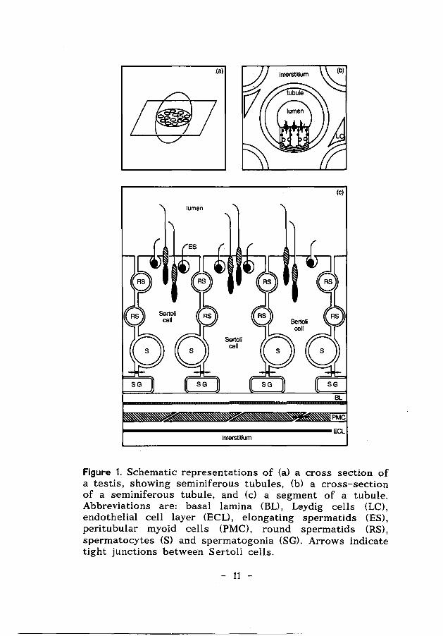
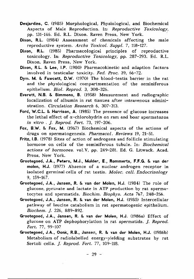
The two testicular compartments are separated structurally and physiologically, not only by the peri tubular myoid cells and extracellular matrix layers, but also by a Sertoli cell barrier, the so-called blood-testis barrier (see Fig. 1). This barrier is the result of tight or occluding junctions between adjacent Sertoli cells. These occluding junctions divide the seminiferous epithelium into a basal compartment, containing the early, mitotically active spermatogenic cells, and an adluminal compartment, containing the more advanced spermatogenic cells during their meiotic and post-meiotic development (Dym & Fawcett, 1970) (see Fig. 1). This barrier is formed in immature animals during the initiation of spermatogenesis and plays a role in the unique environment within the tubules. The milieu in the adluminal compartment is created by the Sertoli cells and is different from the milieu outside the tubules. The differences include the concentrations of proteins, amino acids, and ions (Setchell, 1967; Dym & Fawcett, 1970).
The development of spermatozoa starts with periodic mitotic divisions of undifferentiated spermatogonia. After several rounds of mitotic divisions, the first generation of differentiated type
- 10 -
_(a)
(c)
BL
PMC
~-----------------------------------ECL Interstitium
Figure 1. Schematic representations of (a) a cross section of a testis, showing seminiferous tubules, (b) a cross-section of a seminiferous tubule, and (c) a segment of a tubule. Abbreviations are: basal lamina (BL), Leydig cells (LC), endothelial cell layer (ECL), elongating spermatids (ES), peritubular myoid cells (PMC), round spermatids (RS), spermatocytes (S) and spermatogonia (SG). Arrows indicate tight junctions between Sertoli cells.
- 11 -
A spermatogonia appears, which then gives rise to successive generations of types A, intermediate and type B spermatogonia. Type B spermatogonia divide and develop to preleptotene spermatocytes (Clermont, 1972). These germ cells are at the start of the prophase of the meiotic divisions. The first meiotic division results in two haploid secondary spermatocytes, each containing a single set of chromosomes. These spermatocytes rapidly divide without DNA synthesis and yield haploid early round spermatids (see Fig. 1). The formation of the spermatids is followed by spermiogenesis~ which involves an extensive differentiation of these cells without further cell divisions. During this process, when compaction and genomic inactivation of the sperm nucleus takes place, the histones in the nucleus are replaced by a distinct group of sperm-specific DNA binding proteins, the protamines. These protamines are very rich not only in arginine but also in cysteine, and a unique feature of these highly basic proteins is their extensive SS-cross linking (Bellve, 1979).
The immature spermatozoa are released into the lumen of the seminiferous tubules by the so-called spermiation process, which involves an active role of the Sertoli cells (Russell, 1980; Sakai et al.,1988). After the release from the seminiferous epithelium, spermatozoa undergo major maturational changes during their transit through the epididymis, including the acquisition of a progressive motility pattern and the development of the capacity to fertilize an ovum. The maturation depends on a series of interactions between sperm cells, secreted proteins and other factors from the epididymal epithelium (Yanagimachi, 1988).
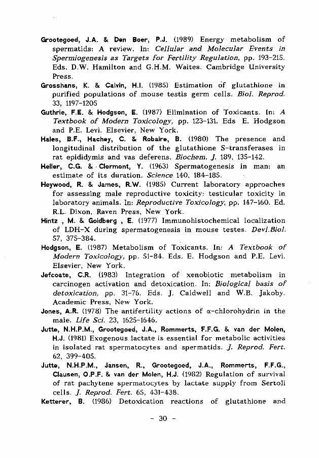
A remarkable morphological feature of the seminiferous epithelium is that various spermatogenic cell types are arranged in well-defined cell associations. These associations are called stages of the cycle of the seminiferous epithelium. The cycle of the seminiferous epithelium is defined as "a complete series of the successive cellular associations appearing in any one given area of the seminiferous epithelium" (Leblond & Clermont, 1952). A transverse section through a tubule shows different spermatogenic cell types at subsequent steps of development in the progression towards spermatozoa, each cell type representing one step of a separate cycle (Leblond & Clermont, 1952). For the rat and for the Golden (or Syrian) hamster 14 and 13 stages of the cycle have been defined, respectively. The hamster stages are
- 12 -
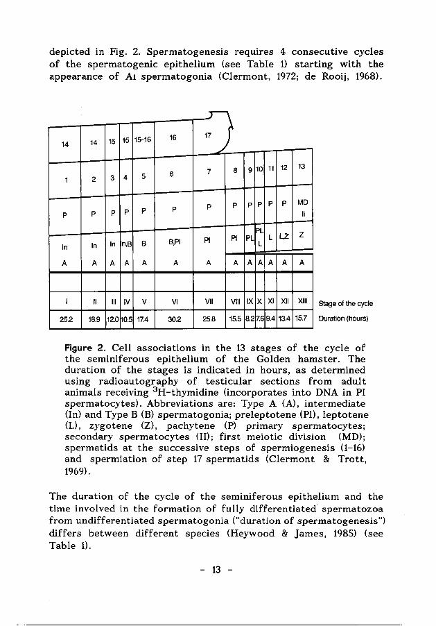
depicted in Fig. 2. Spermatogenesis requires 4 consecutive cycles of the spermatogenic epithelium (see Table 1) starting with the appearance of A1 spermatogonia (Clermont, 1972; de Rooij, 1968).
14 14 15 15 15-16 16
6 1 2 3 4 5
p p p p p p
In In In n,B 8 B,PI
A A A A A A
I II Ill IV v VI
25.2 18.9 12.0 0.5 17.4 30.2
·J 7 8 9 10 11
p p p p p
PL PI PI PL L
L
A A A A A
VII VIII IX X XI
25.8 15.5 8.2 17.6 9.4
12
p
L,Z
A
XII
13.4
13
MD II
z
A
XIII
15.7
Stage of the cycle
Duration (hours)
Figur-e 2. Cell associations in the 13 stages of the cycle of the seminiferous epithelium of the Golden hamster. The duration of the stages is indicated in hours, as determined using radioautography of testicular sections from adult animals receiving 3 H-thymidine (incorporates into DNA in Pl spermatocytes). Abbreviations are: Type A (A), intermediate (In) and Type B (B) spermatogonia; preleptotene (Pl), leptotene (L), zygotene (Z), pachytene (P) primary spermatocytes; secondary spermatocytes (II); first meiotic division (MD); spermatids at the successive steps of spermiogenesis (1-16) and spermiation of step 17 spermatids (Clermont & Trott, 1969).
The duration of the cycle of the seminiferous epithelium and the time involved in the formation of fully differentiated· spermatozoa from undifferentiated spermatogonia ("duration of spermatogenesis") differs between different species (Heywood & james, 1985) (see Table 1).
- 13 -
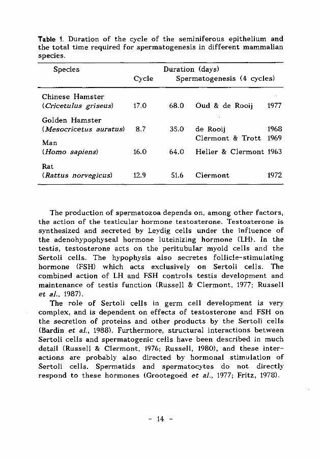
Table 1. Duration of the cycle of the seminiferous epithelium and the total time required for spermatogenesis in different mammalian species.
Species Duration (days) Cycle Spermatogenesis ( 4 cycles)
Chinese Hamster (Cricetulus griseus) 17.0 68.0 Oud & de Rooij 1977
Golden Hamster ( Mesocricetus auratus) 8.7 35.0 de Rooij 1968
Man Clermont & Trott 1969
(Homo sapiens) 16.0 64.0 Heller & Clermont 1963
Rat (Rattus norvegicus) 12.9 51.6 Clermont 1972
The production of spermatozoa depends on, among other factors, the action of the testicular hormone testosterone. Testosterone is synthesized and secreted by Leydig cells under the influence of the adenohypophyseal hormone luteinizing hormone (LH). In the testis, testosterone acts on the peritubular myoid cells and the Sertoli cells. The hypophysis also secretes follicle-stimulating hormone (FSH) which acts exclusively on Sertoli cells. The combined action of LH and FSH controls testis development and maintenance of testis function (Russell & Clermont, 1977; Russell et al., 1987).
The role of Sertoli cells in germ cell development is very complex, and is dependent on effects of testosterone and FSH on the secretion of proteins and other products by the Sertoli cells (Bardin et al., 1988). Furthermore, structural interactions between Sertoli cells and spermatogenic cells have been described in much detail (Russell & Clermont, 1976; Russell, 1980), and these interactions are probably also directed by hormonal stimulation of Sertoli cells. Spermatids and spermatocytes do not directly respond to these hormones (Grootegoed et al., 1977; Fritz, 1978).
- 14 -
1.2 BIOCHEMICAL PROPERTIES OF SPERMATIOS
Spermatozoa are highly specialized cells, and their function depends on highly specialized molecular and cellular processes that occur during development of the spermatogenic cells. A number of specific biochemical properties of spermatids are described in detail in the Appendix paper (Grootegoed & Den Boer, 1989). One of the most striking biochemical differences of spermatids when compared with somatic cells is the presence of spermspecific proteins which are synthesized during spermatogenesis. One of these proteins is the lactate dehydrogenase isoenzyme LDH-C4 , containing four C subunits which are unique to the male germ cells. Another aspect concerns the carbohydrate metabolism of spermatids. Isolated spermatids are dependent on lactate and pyruvate as energy-yielding substrates. Incubation of spermatids from rats (Grootegoed et al., 1986a; ·Nakamura et al., 1986) and hamsters (Chapter 4) with glucose causes a rapid fall of ATP and loss of cell viability. Sertoli cells, isolated from immature rats, show a net production of pyruvate and lactate from glucose (Robinson & Fritz, 1981; jutte et al., 1981, 1982; Grootegoed et al., 1986b), which makes it possible that the Sertoli · cells could support the spermatogenic cells by providing the proper energy-yielding substrates. The developing spermatids seem to have specialized to utilize exogenous lactate for ATP production and therefore may be dependent on a perfect performance of their mitochondria (Grootegoed et al., 1984). Agents that interfere with mitochondrial ATP production may cause degeneration of the germ cells in vitro but also in vivo.
1.3 ANTISPERMATOGENIC AGENTS
The spermatogenic epithelium is one of the most rapidly proliferating tissues in the body. Testes of adult animals produce many millions of spermatozoa per day. This does not only rely on complex molecular processes which occur within the spermatogenic cells, but is also dependent on proper extracellular conditions in the testicular tubules and in the epididymis. Introduction of exogenous chemicals in these physiological milieus may have deleterious effects on the developing germ cells and ultimately on reproduction.
- 15 -
A number of chemicals have been described which interfere with spermatogenesis and/or sperm function (Patanelli, 1975; Dixon, 1984; Neumann, 1984; Schrag & Dixon, 1985). These include therapeutic drugs, metals, insecticides, nematocides (1,2- dibromo-3-chloropropane), rodenticides (fluoroacetate) and investigational antispermatogenic drugs (5-thio-D-glucose, chlorinated sugars, cx-chlorohydrin and gossypol).
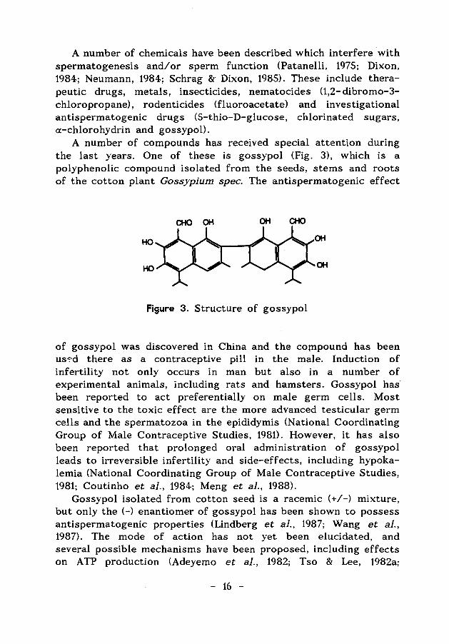
A number of compounds has received special attention during the last years. One of these is gossypol (Fig. 3), which is a polyphenolic compound isolated from the seeds, stems and roots of the cotton plant Gossypium spec. The antispermatogenic effect
CHO OH OH CHO
HO
HO
Figure 3. Structure of gossypol
of gossypol was discovered in China and the compound has been us-:-d there as a contraceptive pill in the male. Induction of infertility not only occurs in man but also in a number of experimental animals, including rats and hamsters. Gossypol has been reported to act preferentially on male germ cells. Most sensitive to the toxic effect are the more advanced testicular germ cells and the spermatozoa in the epididymis (National Coordinating Group of Male Contraceptive Studies, 1981). However, it has also been reported that prolonged oral administration of gossypol leads to irreversible infertility and side-effects, including hypokalemia (National Coordinating Group of Male Contraceptive Studies, 1981; Coutinho et al., 1984; Meng et al., 1988).
Gossypol isolated from cotton seed is a racemic (+/-) mixture, but only the (-) enantiomer of gossypol has been shown to possess antispermatogenic properties (Lindberg et al., 1987; Wang et al., 1987). The mode of action has not yet been elucidated, and several possible mechanisms have been proposed, including effects on ATP production (Adeyemo et al., 1982; Tso & Lee, 1982a;
- 16 -
Martinez et al., 1982; Nakamura et al., 1988) and inhibition of the testis-specific lactate dehydrogenase isoenzyme LDH-C 4 (Morris et al., 1986; Tso & Lee, 1982b).
It is uncertain through which transport mechanism gossypol reaches the testis and the spermatogenic cells. After oral administration of gossypol to rats, high levels appeared in the liver, lung, blood, spleen, heart and kidney, but the gossypol levels in the testes were rather low (Chen et al., 1987). It is known that gossypol can bind specifically to a high-affinity binding site on serum albumin (Royer & Vander jagt, 1983; Maliwal et al., 1985), and it can be suggested that the compound is transported in the blood as a gossypol-albumin complex, so that gossypol reaches the testis in this form. Subsequently, the gossypol-albumin complex may be transported into the interstitial compartment of the testis. Transport of serum albumin into the interstitium has been shown after intravenous injection of radio-labelled albumin. Interestingly, it was observed that the accumulation of 125I-albumin in the testes was much higher than in other organs (Everett & Simmons, 1958).
Transport of gossypol from the interstitium through the bloodtestis barrier into the seminiferous tubules may be based on the lipophilic character of gossypol (see 1.4). This would require dissociation of the albumin-gossypol complex, followed by binding of gossypol to other extra- or intracellular proteins with a high affinity for gossypol at the adluminal side of the barrier. Results from Wang et al. (1988, 1989) indicate that gossypol indeed enters the lumen of the spermatogenic epithelium through the blood-testis barrier and is then concentrated during the passage of the luminal fluid from the testis to the epididymis. This may explain that the first adverse effects of gossypol are observed on spermatozoa (National Coordinating Group of Male Contraceptive Studies, 1981; Wang et al., 1986; Radigue et al., 1988).
In this context, it is also of interest that after oral administration of 14C-labelled gossypol, the intracellular distribution in rat testes showed that the mitochondria had the highest incorporation rate among the subcellular fractions. This suggests that a possible action of gossypol on mitochondria may play an important role in impairing male fertility (National Coordinating Group of Male Contraceptive Studies, 1981), and points to a specific binding component in the mitochondria with an affinity for gossypol greater than the affinity of serum proteins for gossypol. In the
- 17 -
present thesis, the effect of gossypol on the mitochondrial function of spermatids was studied in an in vitro model.
The discovery of the antifertility effect of gossypol was a coincidence (Liu & Segal, 1985). Another way to identify antispermatogenic agents is to take advantage of possible vulnerable biochemical properties of spermatogenic cells. For this approach more knowledge is required not only of the biochemistry of the germ cells, but also of the defence systems which may diminish the toxic potential of such compounds. A number of factors and defence systems affecting the toxic potential of chemicals is described below.
1.4 ASPECTS OF TESTICULAR TOXICITY
Several factors determine the testicular toxicity of exogenous chemicals. First, the blood-testis barrier, which plays a major role in the maintenance of the specialized environment in which the spermatogenic cells develop. This barrier restricts the transfer of many compounds between the interstitium and the seminiferous tubules (Desjardins, 1985), and thereby may provide protection against foreign chemicals. The passage of chemicals across the barrier, from the blood to the lumen of the seminiferous tubules, depends on molecule size, lipid solubility, protein binding and other factors. A positive correlation exists between the lipid solubility of chemicals and their ability to enter the seminiferous tubules (Okumura et al., 1975; Dixon & Lee, 1980).
In addition to the selective permeability of the blood-testis barrier, the presence of different defence systems in testicular cells also influences the toxic and mutagenic potential of chemicals, by providing protection against chemicals which have passed the blood-testis barrier. Different defence systems are discussed in 1.5.
An important factor which limits harmful effects of environmental chemicals is an efficient DNA repair mechanism. DNA damage resulting from exposure to xenobiotics but also from irradiation, interferes with gene integrity, alters transcription, and affects cellular replication. Mutations in the DNA of spermatogenic cells, furthermore, are of greater concern than somatic cell alterations because they are passed on to future generations (Dixon, 1985).
- 18 -
1.5 DEFENCE SYSTEMS
1.5.1 Introduction
The elimination of toxicants is a complex process. The nature of the toxic molecule determines the mechanisms that the cell will use (Steinberger & Lloyd, 1985). Most xenobiotics that enter the body are lipophilic, a property that enables them to be transported by lipoproteins in the blood and penetrate lipid 'membranes.
Detoxication is defined by Hodgson (1987) as "a metabolic reaction or sequence of reactions that reduces the potential for adverse effects of xenobiotics. Such sequences normally involve an increase in water solubility that facilitates excretion and/ or the reaction of a reactive product with an endogenous substrate (conjugation), thereby not only increasing the water solubility but also reducing the possibility of interaction with cellular macromolecules".
1.5.2. Detoxicating reactions
Xenobiotic metabolism generally consists of two phases. In phase one, a polar reactive group is introduced into a toxicant molecule. In most cases, this is followed by conjugation (phase- two metabolism), which renders the molecule suitable for excretion (Guthrie & Hodgson, 1987). As a result of the phase-one reactions many exogenous compounds undergo metabolism to highly reactive, electrophilic intermediates. These products may interact, however, also with cellular constituents rather than enter phase- two reactions. This biotransformation of relatively inert chemicals to highly reactive intermediate metabolites is commonly referred to as "metabolic activation" or "bioactivation". Most activations are monooxygenations catalyzed by the cytochrome P- 450 dependent monooxygenase system or by the FAD-containing monooxygenase (Levi, 1987).
Once these reactive metabolites are formed, mechanisms within the cell may bring about their rapid inactivation (jefcoate, 1983). Factors affecting the toxicity of these activated metabolites are the levels of conjugating enzymes and cofactors or conjugating chemicals (Levi, 1987), which are necessary for further detoxication.
The phase-one products, but also other xenobiotics containing functional groups such as hydroxyl, amino, carboxyl, epoxide or
- 19 -
halogen can be metabolized by conjugation with endogenous substrates, including glutathione, sugars, sulfate groups, and amino acids. The produced conjugates, with only rare exceptions, are more polar, less toxic and more readily excreted than are their parent compounds.
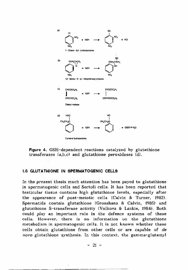
The three principal transferases involved in these phase-two reactions, glutathione S-transferases, UDP-glucuronyltransferases and sulfotransferases, require as coreactants glutathione (GSH), uridine diphosphate glucuronic acid (UDPGA) and 3'-phosphoadenosine 5'-phosphosulfate (PAPS), respectively. Although conjugation reactions occasionally result in bioactivation of a compound, the reaction of xenobiotics with the glutathione-, glucuronyl-, or sulfotransferases usually results in the formation of a non-toxic, water-soluble metabolite that is then easily excreted (Levi, 1987). In Figure 4, a number of glutathione S-transferse reactions is shown for compounds used in the experiments described in the Chapters 5, 6 and 7.
With respect to the actions of reactive electrophiles, glutathione provides the last line of defence by trapping the electrophiles and preventing their binding to proteins and enzymes. Glutathione also protects cells against the action of peroxides (Jefcoate, 1983; Meister & Anderson, 1983). Peroxides can be reduced by the enzyme glutathione peroxidase, and reduced glutathione (GSH) is concomitantly converted to oxidized glutathione (GSSG). This disulfide is subsequently reduced by the enzyme glutathione reductase at the expense of reducing equivalents from NADPH. In Fig. 4 the reduction of the model compound cumene hydroperoxide is shown.
Another aspect of the role of GSH is its function in the protection against damage during exposure to ionizing radiation. The effects of ionizing radiation on cells, in general, are initiated by the formation of free radicals, which can react with water molecules, or with macromolecules. The resulting active oxygen molecules (superoxide, hydroperoxide radical, hydroxyl radical and hydrogen peroxide) can cause damage to cellular macromolecules including DNA (Levi, 1987). Damage elicited by ionizing radiation, however, can be prevented or limited by a high cellular thiol (including glutathione) content (Bump et al., .1982; Meister & Anderson, 1983, van der Schans et al., 1986; Biaglow et al., 1989)
- 20 -
(a) SG Cl
ONO, 6N02
I + GSH - I + HCI
N02 N02
1 - Chloro - 2,4 - d initrobenzene
(b) Sf OCH2CHCH2 OCH2CHCH2 0 'ti + GSH - 0 bH
N02 N02
1.2 - Epoxy- 3 - (p - nitrophenoxy) propane
(c) CHCOOC,H, CHCOOC,H,
II + GSH -- I CHCOOC,H, GSCHCOOC,H,
Diethy! maleate
(d) HOD HO
I I CH,(CH,)C CH3(CH,)C
0 + GSH - 0 + GSSG+H20
Cumene hydroperaxide
Figure 4. GSH-dependent reactions catalyzed by glutathione transferases (a,b,c) and glutathione peroxidases (d).
1.6 GLUTATHIONE IN SPERMATOGENIC CELLS
In the present thesis much attention has been payed to glutathione in spermatogenic cells and Sertoli cells. It has been reported that testicular tissue contains high glutathione levels, especially after the appearance of post-meiotic cells (Calvin & Turner, 1982). Spermatids contain glutathione (Grosshans & Calvin, 1985) and glutathione S-transferase activity (Volkova & Lankin, 1984). Both could play an important role in the defence systems of these cells. However, there is no information on the glutathione metabolism in spermatogenic cells. It is not known whether these cells obtain glutathione from other cells or are capable of de novo glutathione synthesis. In this context, the gamma-glutamyl
- 21 -
cycle, which includes the activity of the enzyme gamma-glutamyl transpeptidase (GGT), could play an important role (Meister & Anderson, 1983). GGT has been detected in Sertoli cell preparations, whereas spermatids seem to have very low activity levels of this enzyme (Lu & Steinberger, 1977).
It has been observed that testicular glutathione levels can be decreased by injection of a combination of the glutathione $
transferase substrate diethyl maleate and the glutathione biosynthesis inhibitor buthionine sulphoximine (BSO) (Slott et al., 1989), but the possible effect of this treatment on the glutathione level of spermatids was not studied. In another study, reduction of testicular and epididymal GSH by BSO was found to potentiate the mutagenic effect of the alkylating agent ethyl methane sulfonate on male germ cells in late testicular stages and on caput epididymal spermatozoa, indicating that perturbation of GSH in the male reproductive tract enhances chemical-induced mutations in germ cells (Teaf et al., 1987).
Epididymal spermatozoa from a number of animal species also contain GSH (Li, 1975) and antioxidative enzymes, including glutathione peroxidase, glutathione reductase and superoxide dismutase (Volkova & Lankin, 1984; Alvarez & Storey, 1983, 1984; Alvarez et al., 1987). This indicates that spermatozoa contain protective mechanisms against oxidative damage by peroxides. The importance of these mechanisms is indicated by observations showing lipid peroxidation in spermatozoa following inactivation of glutathione peroxidase (Alvarez & Storey, 1989).
In addition the epididymal cells seem to play a role. The epididymis contains GSH and glutathione S-transferases (Hales et a]., 1980, Agrawal & Vanha-Perttula, 1988; Robaire & Hermo, 1988), which might protect the epididymal spermatozoa against electrophilic chemicals.
The glutathione content of developing spermatids or spermatozoa may be decreased as a result of exposure of the testis and epididymis to xenobiotic chemicals, and it appears attractive to consider the possibility that such a decrease could enhance the adverse action of electrophilic compounds and the viability of the cells. In the present thesis, glutathione was studied as a possible target for antispermatogenic and antifertility agents.
- 22 -
1.7 MODEL SYSTEMS
1. 7.1 Models
In the present experiments the Golden hamster (Mesocricetus auratus) and the rat (Rattus norvegicus) were used as experimental animals to evaluate effects of toxic compounds on spermatogenic cells. Hamsters were used mainly because hamsters have been used also in gossypol screening programmes to estimate the effects of gossypol and derivatives on spermatogenesis and fertility. Furthermore, hamsters are more sensitive to the annfertility effect of gossypol than rats (Chang et al., 1980). From hamster testes and epididymides, different germinal cell types can be isolated, including spermatocytes, spermatids, elongating spermatids and spermatozoa. The latter can be used, in future studies, to evaluate effects of xenobiotic compounds on sperm fertilizing capacity, using in vitro fertilization which is operational for hamsters (Bavister, 1981). Rats were used in ~he first instance, to study different aspects of the glutathione-dependent defence systems, since there is much information in the litterature on glutathione metabolism and the relevant enzymes in rat tissues. Results from the litterature show that the glutathione concentration in the rat testis increases during postnatal development when spermatogenesis is initiated (Calvin & Turner, 1982). It is also known that a high glutathione S-transferase activity is present in rat testicular tissue (Kraus & Kloft, 1980), including an isoenzyme found predominantly in the testis (Boyer & Kenney, 1985; Ketterer, 1986). Furthermore, the presence of glutathione S-transferase activity (Mukhtar et al., 1978) and glutathione peroxidase activity (Volkova & Lankin, 1984) in rat spermatozoa has been described. Subsequent to the introductory experiments on the characterization of glutathione- dependent defence systems in rat spermatogenic cells, hamster spermatogenic tissue and cells were used for further studies on glutathione metabolism and the possible function of glutathione in spermatids.
Toxins may affect different specific steps in germ cell development. A more or less specific action on one cell type has been observed, for example, using methoxy acetic acid (Bartlett et al., 1988; Ratnasooriya & Sharpe, 1989) and nitrofurans (Patanelli, 1975), which attack pachytene spermatocytes. Another example is cx-chlorohydrin, which acts preferentially on epididymal spermatozoa
- 23 -
(jones, 1978; Ford & Harrison, 1985). Gossypol, in contrast, shows a less specific toxicity, acting not only on spermatozoa but also on spermatids and spermatocytes (National Coordinating Group of Male Contraceptive Studies, 1981).
One approach to study effects of different toxic compounds on the spermatogenic epithelium is to culture small fragments of seminiferous tubules from immature animals. This may offer a fairly good model, containing Sertoli cells and spermatogenic cells at different stages of their development up to and including round spermatids (Grootegoed et al., 1985; Toebosch et al., 1989). The presence of Sertoli cells in this model is important, because Sertoli cells may not only protect the germ cells, but may also potentiate toxic effects of certain compounds.
Another approach is to study effects of xenobiotics or other compounds on isolated testicular cell types, including Sertoli cells, pachytene spermatocytes and round spermatids. Using hamster testes, like testes from many other mammalian species, these cell types can be isolated quite easily with reasonable quantities. The same isolation procedures were followed, with small modifications, as described for rat Sertoli cells (Oonk et al., 1985) and rat spermatogenic cells (Grootegoed et al., 1984). Spermatocytes and spermatids were separated by sedimentation at unit gravity using the STA-PUT method, and further purified by Percoll density centrifugation. The purity of the isolated pr pulations of spermatogenic cells were analyzed by microscopic analysis and according to their DNA content using a flow cytometer (spermatids contain a 1C amount of DNA; spermatocytes contain a 4 C amount of DNA). The purity of the spermatid preparations was approximately 96% (see also Fig. 5b). The experiments described in the present thesis have focussed on round spermatids.
1.7.2 Parameters
In order to evaluate the effects of toxic agents on spermatogenic epithelium and isolated testicular cells, a number of biochemical parameters were estimated. These include ATP, LDH-C 4 and GSH.
The ATP content of the cells was estimated since many toxic agents, including gossypol, interfere or are thought to interfere, with energy metabolism. Furthermore, as indicated in 1.2 and the Appendix paper, spermatids have a different energy metabolism as compared with somatic cells, which may render ATP synthesis in
- 24 -
the germ cells exceptionally vulnerable to toxic agents (Grootegoed et al., 1984).
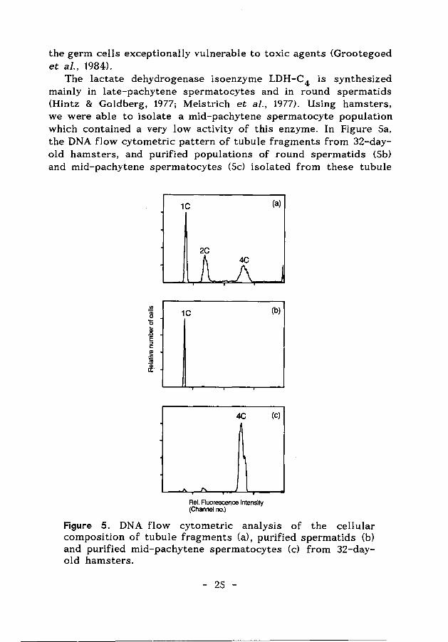
The lactate dehydrogenase isoenzyme LDH-C4 is synthesized mainly in late-pachytene spermatocytes and in round spermatids (Hintz & Goldberg, 1977; Meistrich et al., 1977). Using hamsters, we were able to isolate a mid-pachytene spermatocyte population which contained a very low activity of this enzyme. In Figure Sa, the DNA flow cytometric pattern of tubule fragments from 32-dayold hamsters, and purified populations of round spermatids (Sb) and mid-pachytene spermatocytes (Sc) isolated from these tubule
.!!l
~ 0
~ E :::0 c:
j ~
1C (a)
2C
1C (b)
4C (c)
Rei. Fluorescence Intensity (Channel no.)
Figure 5. DNA flow cytometric analysis of the cellular composition of tubule fragments (a), purified spermatids (b) and purified mid-pachytene spermatocytes (c) from 32-dayold hamsters.
- 25 -
fragments, illustrate the cellular composition of these preparations. The spermatids and the spermatocytes contained an LDH-C 4 activity of 66.6 ± 4.9 and 5.5 ± 0.1 mU/mg protein, respectively. A high LDH-C4 activity was observed in isolated late-pachytene spermatocytes (not shown). The LDH-C4 activity was used to evaluate effects of gossypol, since this compound has been reported to act specifically on the enzyme (Lee et al., 1982; Tso & Lee, 1982b; Morris et al., 1986; Whaley et al., 1986).
The cellular glutathione content was estimated for reasons described in 1.6.
1.8 AIM AND SCOPE OF THIS THESIS
It has been reported that a number of drugs and chemicals act preferentially on spermatogenesis (Fox & Fox, 1967; Patanelli, 1975). A specific action of a toxic compound on spermatogenesis suggests a specific target in the testicular tubules and may point to a unique. or at least rare property of spermatogenic cells (or possibly Sertoli cells) as compared with all (other) somatic cell types. As indicated in this Introduction, spermatogenic cells possess a number of particular features. In the present thesis it was studied whether specific biochemical processes in spermatids are possible targets for antispermatogenic agents.
Remarkable biochemical properties of advanced spermatogenic cells include the presence of the testis-specific lactate dehydrogenase isoenzyme LDH-C 4 (see 1.2) and the deviating energy metabolism (see 1.2 and Appendix paper). Drugs acting on mitochondria may preferentially kill spermatogenic cells, since these cells are highly dependent on mitochondrial function. In the experiments described in Chapter 2 and 3, effects of the antifertility agent gossypol on LDH-C 4 activity and ATP production in spermatids were studied. From the biochemical analysis of the preferential action of gossypol on spermatogenic cells, more can be learned about biochemical processes which are of particular importance in the spermatogenic cells. It was observed that gossypol may interfere with spermatogenesis through an effect on ATP production. In this context, ATP metabolism in spermatids was studied in more detail as described in Chapter 4.
Another possible target for antispermatogenic agents is glutathione metabolism in the testicular tubules (see 1.6). In the
- 26 -
experiments described in Chapter S, glutathione S-transferase substrates and oxidizing agents were applied to study the presence of GSH-dependent defence mechanisms in round spermatids. In Chapter 6, it is evaluated to what extent the GSH-dependent defence systems in spermatids can provide protection against xenobiotics and the induction of DNA damage by ionizing radiation. The possible role of Sertoli cells in the de novo glutathione biosynthesis in seminiferous tubules is indicated in Chapter 7. In the experiments described in this Chapter 7 the glutathione biosynthesis in isolated testicular cells and tubule fragments was studied, making use of the glutathione-depleting agent diethyl maleate and the glutathione biosynthesis inhibitor buthionine sulfoximine.
It is anticipated that improved understanding of the action of different compounds on biochemical processes in Sertoli cells and spermatogenic cells, may provide working hypothesises to test and identify antispermatogenic agents.
REFERENCES
Adeyemo, 0., Chang, C.Y., Segal, S.J. & Koide, S.S. (1982) Gossypol action on the production and utilization of ATP in sea urchin spermatozoa. Archs Androi. 9, 343-349.
Agrawal, Y.P. & Vanha-Perttula, T. (1988) Gamma-glutamyl transpeptidase, glutathione, and L-glutamic acid in the rat epididymis during postnatal development. Bioi. Reprod. 38, 996-1000.
Alvarez, J.G. & Storey, B.T. (1983) Role of superoxide dismutase in protecting rabbit spermatozoa from 0 2 toxicity due to lipid peroxidation. Bioi. Reprod. 28, 1129-1136.
Alvarez, J.G. & Storey, B.T. (1984) Lipid peroxidation and the reactions of superoxide and hydrogenperoxide in mouse spermatozoa. Bioi. Reprod. 30, 833-841.
Alvarez, J.G., Touchstone, J.C., Blasco, L. & Storey, B.T. (1987) Spontaneous lipid peroxidation and production of hydrogen peroxide and superoxide in human spermatozoa. Superoxide dismutase as major enzyme protectant against oxygen toxicity. ]. Androl. 8, 338-348.
Alvarez, J.G. & Storey, B.T. (1989) Role of glutathione peroxidase in protecting mammalian spermatozoa from loss of motility
- 27 -
caused by spontaneous lipid peroxidation. Gam Res. 23, 77-90 Bardin, C.W., Cheng, C.Y., Musto, N.A. Sot Gunsalus, G.L. (1988) The
Sertoli cell. In: The Physiology of Reproduction, pp. 933- 97 4. Eds E. Knobil and j. Neill. Raven Press, New York.
Bartlett, J.M.S., Kerr, J.B. Sot Sharpe, R.M. (1988) The selective removal of pachytene spermatocytes using methoxy acetic acid as an approach to the study in vivo of paracrine interactions in the testis. ]. Androl. 9, 31-40.
Bavister, B.D. (1981) Analysis of culture media for in vitro fertilization and criteria for succes. In: Fertilization and embryonic development in vitro, pp. 41-60. Eds L. Mastroianni and ].D. Biggers, Plenum Press, New York and London.
Bellve , A.R. (1979) The molecular biology of mammalian spermatogenesis. Oxford Reviews of Reproductive Biology 1, 159-261.
Biaglow, J.E., Varnes, M.E., Epp, E.R., Clark, E.P., Tuttle, S.W. & Held, K.O. (1989) Role of glutathione and other thiols in cellular response to radiation and drugs. Drug Metabolism Reviews 20, 1-12.
Boyer, T.O. Sot Kenney, W.C. (1985) Acidic glutathione S- transferases of rat testis. Biochem. ]. 230, 125-132.
Bump, E.A., Yu, N.Y. Sot Brown, J.M. (1982) Radiosensitization of hypoxic tumor cells by depletion of intracellular glutathione. Science 217, 544-545.
Calvin, H.l. Sot Turner, S.l. (1982) High levels of glutathione attained dl'ring postnatal development of rat testis. ]. exp. Zool. 219, 389-393.
Chang, M.C., Gu, Z. Sot Saksena, S.K. (1980) Effects of gossypol on the fertility of male rats, hamsters and rabbits. Contraception 21, 461-469.
Chen, Q.Q., Chen, H. Sot Lei, H.P. (1987) Comparative study on the metabolism of optical gossypol in rats. ]. Ethnopharmacol. 20, 31-37.
Clermont, Y. Sot Trott, M. (1969) Duration of the cycle of the seminiferous epithelium in the mouse and hamster determined by means of 3 H-thymidine and radioautography. Fertil. Steril. 20, 805-817.
Clermont, Y. (1972) Kinetics of spermatogenesis in mammals: seminiferous epithelium cycle and spermatogonial renewal. Physiol. Reviews 52, 198-236.
Coutinho, E.M., Melo, J.F., Barbosa, I. Sot Segal, S.J. (1984) Antispermatogenic action of gossypol in men. Fert. Steril. 42, 424-430.
- 28 -
Desjardins, C. (1985) Morphological, Physiological, and Biochemical Aspects of Male Reproduction. In: Reproductive Toxicology, pp. 131-146. Ed. R.L. Dixon. Raven Press, New York. -
Dixon, R.L. (1984) Assessment of chemicals affecting the male reproductive system. Archs Toxicol. Suppl. 7, 118-127.
Dixon, R.L. (1985) Pharmacological principles of reproductive toxicology. In: Reproductive Toxicology, pp. 287-293. Ed. R.L. Dixon, Raven Press, New York.
Dixon, R.L. & Lee, I.P. (1980) Pharmacokinetic and adaption factors involved in testicular toxicity. Fed. Proc. 39, 66-72.
Dym. M. & Fawcett, D.W. (1970) The blood-testis barrier in the rat and the physiological compartmentation of the seminiferous epithelium. Bioi. Reprod. 3, 308-326.
Everett, N.B. & Simmons, B. (1958) Measurement and radiographic localization of albumin in rat tissues after intravenous administration. Circulation Research 6, 307-313.
Ford, W.C.L. & Harrison, A. (1985) The presence of glucose increases the lethal effect of o:-chlorohydrin on ram and boar spermatozoa in vitro . ]. Reprod. Pert. 73, 197-206.
Fox, B.W. & Fox, M. (1967) Biochemical aspects of the actions of drugs on spermatogenesis. Pharmacal. Reviews 19, 21-51.
Fritz, 1.8. (1978) Sites of action of androgens and follicle stimulating hormone on cells of the seminiferous tubule. In: Biochemical actions of hormones. vol.V, pp. 249-281. Ed. G. Litwack. Acad. Press, New York.
Grootegoed, J.A., Peters, M.J., Mulder, E., Rommerts, F.F.G. & van der molen, H.J. (1977) Absence of a nuclear androgen receptor in isolated germinal cells of rat testis. Malec. cell. Endocrinology 9, 159-167.
Grootegoed, J.A., Jansen, R. & van der Molen, H.J. (1984) The role of glucose, pyruvate and lactate in ATP production by rat spermatocytes and spermatids. Biochim. Biophys. Acta 767, 248-256.
Grootegoed, J.A., Jansen, R. & van der Molen, H.J. (1985) Intercellular pathway of leucine catabolism in rat spermatogenic epithelium. Biochem. ]. 226, 889-892.
Grootegoed, J.A., Jansen, R. & van der Molen, H.J. (1986a) Effect of glucose on ATP dephophorylation in rat spermatids. ]. Reprod. Pert. 77, 99-107
Grootegoed, J.A., Oonk, R.B., Jansen, R. & van der Molen, H.J. (1986b) Metabolism of radiolabelled energy-yielding substrates by rat Sertoli cells. ]. Reprod. Fert. 77, 109-118.
- 29 -
Grootegoed, J.A. &. Den Boer, P.J. (1989) Energy metabolism of spermatids: A review. In: Cellular and Molecular Events in Spermiogenesis as Targets for Fertility Regulation, pp. 193-215. Eds. D.W. Hamilton and G.H.M. Waites. Cambridge University Press.
Grosshans, K. &. Calvin, H.l. (1985) Estimation of glutathione in purified populations of mouse testis germ cells. Bioi. Reprod. 33, 1197-1205
Guthrie, F.E. &. Hodgson, E. (1987) Elimination of Toxicants. In: A Textbook of Modern Toxicology, pp. 123-131. Eds E. Hodgson and P.E. Levi. Elsevier, New York.
Hales, B.F., Hachey, C. &. Robaire, B. (1980) The presence and longitudinal distribution of the glutathione S-transferases in rat epididymis and vas deferens. Biochem. ]. 189, 135-142.
Heller, C.G. &. Clermont, Y. (1963) Spermatogenesis in man: an estimate of its duration. Science 140, 184-185.
Heywood, R. &. James, R.W. (1985) Current laboratory approaches for assessing male reproductive toxicity: testicular toxicity in laboratory animals. In: Reproductive Toxicology, pp. 147-160. Ed. R.L. Dixon, Raven Press, New York.
Hintz , M. &. Goldberg , E. (1977) Immunohistochemical localization of LDH-X during spermatogenesis in mouse testes. Devl.Biol. 57, 375-384.
Hodgson, E. (1987) Metabolism of Toxicants. In: A Textbook of Modern Toxicology, pp. 51-84. Eds. E. Hodgson and P.E. Levi. Elsevier, New York.
Jefc:oate, C.R. (1983) Integration of xenobiotic metabolism in carcinogen activation and detoxication. In: Biological basis of detoxication, pp. 31-76. Eds. ]. Caldwell and W.B. jakoby. Academic Press, New York.
Jones, A.R. (1978) The antifertility actions of cx-chlorohydrin in the male. Life Sci. 23, 1625-1646.
Jutte, N.H.P.M., Grootegoed, J.A., Rommerts, F.F.G. &. van der Molen, H.J. (1981) Exogenous lactate is essential for metabolic activities in isolated rat spermatocytes and spermatids. ]. Reprod. Pert. 62, 399-405.
Jutte, N.H.P.M., Jansen, R., Grootegoed, J.A., Rommerts, F.F.G., Clausen, O.P.F. &. van der Molen, H.J. (1982) Regulation of survival of rat pachytene spermatocytes by lactate supply from Sertoli cells. ]. Reprod. Pert. 65, 431-438.
Ketterer, B. (1986) Detoxication reactions of glutathione and
- 30 -
glutathione S-transferases. Xenobiotica 16, 957-973. Kraus, P. & Kloft, H.D. (1980) The activity of glutathione-S
transferases in various organs of the rat. Enzyme 25, 158-160. Leblond, C.P. & Clermont, Y. (1952) Definition of the stages of the
cycle of the seminiferous epithelium of the rat. Ann. N.Y. Acad. Sci SS, 548-594.
Lee, C.-Y., Moon, Y.S., Yuan, J.H. & Chen, A.F. (1982) Enzyme inactivation and inhibition by gossypol. Molec. cell. Biochem. 47, 65-70.
Levi, P.E. (1987) Toxic action. In: A Textbook of Modern Toxicology, pp. 133-184. Eds. E. Hodgson and P.E. Levi. Elsevier, New York.
Li, T.K. (1975) The glutathione and thiol content of mammalian spermatozoa and seminal plasma. Bioi. Reprod. 12, 641-646.
Lindberg, M.C., Naqvi, R.H., Matlin, S.A., Zhou, R.H., Bialy, G. & Blye, R.P. (1987) Comparative antifertility effects of gossypol enantiomers in male hamsters. Int. ]. Androl. 10, 619-623.
Liu, G.Z. & Segal, S.J. (1985) Introduction and history of gossypol. In: Gossypol, a potential contraceptive for men, pp. 1-7. Eds. S.j. Segal. Plenum Press, New York and London.
Lu, C. & Steinberger, A. (1977) Gamma-glutamyl transpeptidase activity in the developing rat testis. Enzyme localization in isolated cell types. Bioi. Reprod. 17, 84-88.
Maliwal, B.P., Appu Rao, A.G. & Narasinga Rao, M.S. (1985) Spectroscopic study of the interaction of gossypol with bovine serum albumin. Int. j. Peptide Protein Res. 25, 382-388.
Martinez, F., Gamboa, S. and Diaz-Sanchez, V. (1988) Biochemical effects of gossypol in isolated mitochondria: monovalent cations and ATPase activity. Int. ]. Biochem. 20, 189-192.
Meister, A. & Anderson, M.E. (1983) Glutathione. Ann. Rev. Biochem. 52, 711-760.
Meistrich, M.L., Trostle, P.K., Frapart, M. & Erickson, R.P. (1977) Biosynthesis and localization of lactate dehydrogenase X in pachytene spermatocytes and spermatids from mouse testis. Devl. Bioi. 60, 428-441.
Meng, G.D., Zhu, J.C., Chen, Z.W., Wong, L.T., Zhang, G.Y., Hu, Y.Z., Ding, J.H. Wang, X.H., Qian, S.Z., Wang, C., Manchin, D., Pinol, A. & Waites, G.M.H. (1988) Recovery of sperm production following the cessation of gossypol treatment: a two-centre study in China. Int. ]. Androl. 11, 1-11.
Morris, 1.0., Higgins, C. & Matlin, S.A. (1986) Inhibition of testicular
- 31 -
LDH-X from laboratory animals and man by· gossypol and its isomers. ]. Reprod. Pert. 77, 607-612.
Mukhtar, H., Lee, I.P. & Bend, J.R. (1978) Glutathione S- transferase activities in rat and mouse sperm and human sperm. Biochem. Biophys. Res. Comm. 83, 1093-1098.
Nakamura, M., Okinaga, S. & Arai, K. (1986) Studies of metabolism of round spermatids: glucose as unfavourable substrate. Bioi. Reprod. 35, 927-935.
Nakamura, M., Ikeda, M., Suzuki, A., Okinaga, S. & Arai, K. (1988) Metabolism of round spermatids: gossypol induces uncoupling of respiratory chain and oxidative phosphorylation. Bioi. Reprod. 39, 771-778.
National Coordinating Group of Male Contraceptive Studies (1981) Studies on the antifertility effect of gossypol, a new contraceptive for males. In: Recent Advances in Fertility Regulation, pp. 122-146. Eds C.F. Chang & D. Griffin. S.A. Atar, Geneva.
Neumann (1984) Effects of drugs and chemicals on spermatogenesis. Archs Toxicol., suppl. 7, 109-117.
Oonk, R.B, Grootegoed, J.A. & van der Molen, H.J. (1985) Comparison of the effects of insulin and follitropin on glucose metabolism by Sertoli cells from immature rats. Molec. cell. Endocrinol. 42, 39-48.
Okumura, K., Lee, I.P. & Dixon, R.L. (i975) Permeability of selected drugs and chemicals across the blood-testis barrier of the rat. ]. _9 harmacol. Exp. Ther. 194, 89-95
Oud, J.L. & de Rooij, D.G. (1977) Spermatogenesis in the Chinese hamster. Anat. Rec., 187, 113-123.
Patanelli, D.J. (1975) Suppresion of fertility in the male. In: Handbook of Physiology, section 7: Endocrinology, vol V. Male reproductive systems, pp. 245-258. Eds. D.W. Hamilton and R.O. Greep. Waverly Press, Baltimore.
Radigue, C., Soufir, J.C., Couvillers, M.L., Dantec, M.C. & Folliot, R. (1980) Early effects of gossypol on the testis and epididymis in the rat. Reprod. Nutr. Develop. 28, 1329-1338.
Ratnasooriya, W.O. & Sharpe, R.M. (1989) Evaluation of the effect of selective germ cell depletion on subsequent spermatogenesis and fertility in the rat. Int. ]. Androl. 12, 44-57.
Robaire, B. & Hermo, L. (1988) Efferent ducts, epididymis, and vas deferens: structure, functions, and their regulation. In: The Physiology of Reproduction, pp. 999-1080. Eds. E. Knobil and j. Neill. Raven Press, New York.
- 32 -
Robinson, R. & Fritz, I.B. (1981) Metabolism of glucose by Sertoli cells in culture. Bioi. Reprod. 24, 1032-1041.
de Rooij, D.G. (1968) Stem cell renewal and duration of spermatogonial cycle in the goldhamster. Zeitsch. Zellforschung 89, 133-136.
Royer, R.E. & Vander Jagt, D.L. (1983) Gossypol binds to a highaffinity binding site on human serum albumin. FEBS Letters 157, 28-30.
Russell, L.D. & Clermont, Y. (1976) Anchoring device between Sertoli cells and late spermatids in rat seminiferous tubules. Anat. Rec. 185, 259-278.
Russell, L.D. & Clermont, Y. (1977) Degeneration of germ cells in normal, hypophysectomized and hormone treated hypophysectomized rats. Anat. Rec. 187, 347-366.
Russell, L.D. (1980) Sertoli-germ cell interrelations: a review. Gam. Res. 3, 179-202.
Russell, L.D., Alger, L.E. & Nequin, L.G. (1987) Hormonal control of pubertal spermatogenesis. Endocrinology 120, 1615-1632.
Sakai, Y, Nakamoto, T. & Yamashina (1988) Dynamic changes in Sertoli processes invading spermatid cytoplasm during mouse spermiogenesis. Anat Rec. 220, 51-57.
Schans, G.P. van der, Vos, 0., Roos-Verheij, W.S.D. & Lohman, P.H.M. (1986) The influence of oxygen on the induction of radiation damage in DNA in mammalian cells after sensitization by intracellular glutathione depletion. Int. ]. ·Radiat. Bioi. SO, 453-465.
Schrag, S.D. & Dixon, R.L. (1985) Reproductive effects of chemical agents. In: Reproductive Toxicology, pp. 301-319. Ed. R.L Dixon. Raven Press, New York.
Setchell, B.P. (1967) The blood-testicular fluid barrier in sheep. ]. Physiol. 189, 63P-6SP.
Slott, V.L., Linder, R.E., Strader, L.F. & Perreault, S.D. (1989) Unilateral depletion of testicular glutathione levels in the rat following intratesticular injections of diethylmaleate and buthionine sulfoximine. Toxicol. Appl. Pharmacal. 98, 369-373.
Steinberger, E. & Lloyd, J.A. (1985) Chemicals affecting the development of reproductive capacity. In: Reproductive Toxicology, pp. 1-20. Eds. R.L Dixon. Raven Press, New York.
Teaf, C.M., Bishop, J.B. & Harbison, R.D. (1987) Depression of glutathione in male reproductive tissues and potentiation of EMSinduced germ cell mutagenesis by L-buthionine sulfoximine.
- 33 -
Teratogenesis, Carcinogenesis, and Mutagenesis 7, 497-513. Toebosch, A.M.W., Brussee, R., Verkerk, A. & Grootegoed, J.A. (1989)
Quantitative evaluation of the maintenance and development of spermatocytes and round spermatids in cultured tubule fragments from immature rat testis. Int. ]. Androl. On the press).
Tso, W.-W. & Lee, C.-S. (1982a) Gossypol uncoupling of respiratory chain and oxidative phosphorylation in ejaculated boar spermatozoa. Contraception 25, 649-655.
Tso, W.-W. & Lee, C.-S. (1982b) Lactate dehydrogenase-X: an isozyme particularly sensitive to gossypol inhibition. Int. ]. Androl. 5, 205-209.
Volkova, N.P. & Lankin, V.Z. (1984) Changes in activity of antioxidative enzymes during spermatogenesis. Bull. Exp. Bioi. Med. 98, 1496-1498.
Wang, J.M., Gu, C.H., Tao, L., Wu, X.L. & Qui, J.P. (1986) Electrolyte composition of rete testis fluid and cauda epididymal plasma and spermatozoa from rats following gossypol treatment. Andrologia 18, 43-49.
Wang, N.G., Zhou, L.F., Guan, M.H. & Lei, H.P. (1987) Effect of (+)
and (-) gossypol on fertility in male rats. ]. Ethnopharmacol. 20, 21-24.
Wang, J., Qui, J., Wu, X., Zhang, Z., Shao, Y. & Cao, R. (1988) The correlation between the gossypol contents in blood plasma, rete testis fluid, and cauda epididymal fluid following chronic treatment with gossypol in rats. ]. Androl. 9, 397-402.
Wang, J.M., Wen, G.Y., Zhang, Z.R., Wu, X.L., Jiang, D.H., Tao, L., Cao, R.Q. and Zhou, Q. (1989) The entry of gossypol across the blood-testis barrier in rats. Contraception 39, 569-575.
Whaley, K.J., Stephens, D.T., Klimkow, N.M. & Hoskins, D.O. (1986) Monkey lactate dehydrogenase-C4 as a model for the interaction of enzymes with gossypol. Contraception 33, 605-616.
Yanagimachi, R. (1988) Mammalian Fertilization. In: The Physiology of Reproduction, pp. 135-185. Eds. E. Knobil and j. Neill. Raven Press, New York.
- 34 -
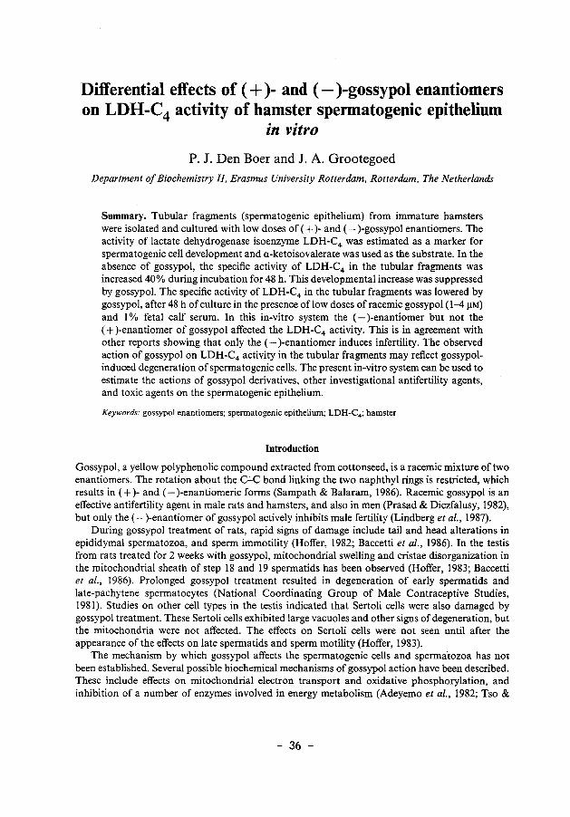
Chapter 2
Differential effects of ( +) - and (-) gossypol enantiomers on LDH-C4 activity of hamster
spermatogenic epithelium in vitro
P 1. Den Boer and J A. Grootegoed
Journal of Reproduction & Fertility (1988) 83:701-709
- 35 -
Differential effects of ( + )- and (-)-gossypol enantiomers on LDH-C4 activity of hamster spermatogenic epithelium
in vitro
P. J. Den Boer and J. A. Grootegoed Department of Biochemistry I/, Erasmus University Rotterdam, Rotterdam, The Netherlands
Summary. Tubular fragments (spermatogenic epithelium) from immature hamsters were isolated and cultured with low doses of ( + )- and (-)-gossypol enantiomers. The activity of lactate dehydrogenase isoenzyme LDH-C4 was estimated as a marker for spermatogenic cell development and a-ketoisovalerate was used as the substrate. In the absence of gossypol, the specific activity of LDH-C4 in the tubular fragments was increased 40% during incubation for 48 h. This developmental increase was suppressed by gossypol. The specific activity of LDH-C4 in the tubular fragments was lowered by gossypol, after 48 h of culture in the presence of low doses of racemic gossypol (1-4 !lM) and I% fetal calf serum. In this in-vitro system the (- )-enantiomer but not the ( + )-enantiomer of gossypol affected the LDH-C4 activity. This is in agreement with other reports showing that only the (- )-enantiomer induces infertility. The observed action of gossypol on LDH-C4 activity in the tubular fragments may reflect gossypolinduced degeneration of spermatogenic cells. The present in-vitro system can be used to estimate the actions of gossypol derivatives, other investigational antifertility agents, and toxic agents on the spermatogenic epithelium.
Keywords: gossypol enantiomers; spermatogenic epithelium; LDH-C4 ; hamster
Introduction
Gossypol, a yellow polyphenolic compound extracted from cottonseed, is a racemic mixture of two enantiomers. The rotation about the C-C bond linking the two naphthyl rings is restricted, which results in (+)-and (- )-enantiomeric forms (Sampath & Balaram, 1986). Racemic gossypol is an effective antifertility agent in male rats and hamsters, and also in men (Prasad & Diczfalusy, 1982), but only the (- )-enantiomer of gossypol actively inhibits male fertility (Lindberg eta!., 1987).
During gossypol treatment of rats, rapid signs of damage include tail and head alterations in epididymal spermatozoa, and sperm immotility (Hoffer, 1982; Baccetti et at., 1986). In the testis from rats treated for 2 weeks with gossypol, mitochondrial swelling and cristae disorganization in the mitochondrial sheath of step 18 and 19 spermatids has been observed (Hoffer, 1983; Baccetti et a!., 1986). Prolonged gossypol treatment resulted in degeneration of early spermatids and late-pachytene spermatocytes (National Coordinating Group of Male Contraceptive Studies, 1981). Studies on other cell types in the testis indicated that Sertoli cells were also damaged by gossypol treatment. These Sertoli cells exhibited large vacuoles and other signs of degeneration, but the mitochondria were not affected. The effects on Sertoli cells were not seen until after the appearance of the effects on late spermatids and sperm motility (Hoffer, 1983).
The mechanism by which gossypol affects the spermatogenic cells and spermatozoa has not been established. Several possible biochemical mechanisms of gossypol action have been described. These include effects on mitochondrial electron transport and oxidative phosphorylation, and inhibition of a number of enzymes involved in energy metabolism (Adeyemo et al., 1982; Tso &
- 36 -
Lee, 1982a; Stephens eta!., 1983; Kim & Waller, 1984). Much attention has been paid to possible inhibition of the lactate dehydrogenase isoenzyme LDH-C4 (Lee et at., 1982; Tso & Lee, 1982b; Morris eta!., 1986; Whaley eta!., 1986). LDH-C4 is composed of male germ cell-specific C subunits (Zinkham, 1968), and inhibition of this isoenzyme would specifically interfere with the metabolism of spermatogenic cells and spermatozoa. In-vivo administration of gossypol to rats can result in a decreased specific activity of testicular LDH-C4 (U/mg protein), but it is not clear whether this decrease reflects the elimination of spermatogenic cells or direct inhibition of the enzyme (Olgiati eta!., 1984a).
The aim of the present experiments was to develop an in-vitro system to study the effects of gossypol enantiomers and derivatives on the spermatogenic epithelium. Hamsters were used, mainly because hamsters have also been used in gossypol screening programmes to estimate the effects of gossypol and derivatives on spermatogenesis and fertility. Furthermore, hamsters are more sensitive to the antifertility effect of gossypol than are rats (Chang eta!., 1980).
Materials and Methods
Isolation and incubation of spermatogenic epithelium from hamsters. Immature 25-26-day-old hamsters (Mesocricetus auratus) were killed by cervical dislocation and the testes were removed. Tubular fragments were isolated by treatment of 6 decapsulated testes with 10 mg collagenase (CLS, Worthington Biochemical Corporation, Freehold, NJ, U.S.A.) in 20 ml phosphate-buffered saline (PBS), supplemented with 6 mM-L-lactate and 5·6 mM-glucose, for 30 min at 32°C in a shaking waterbath (120 cycles/min). The method used was the same, and yielded comparable results, as the method used to isolate tubular fragments from rat testes (Jutte eta/., 1982). The tubular fragments contained Sertoli cells and spermatogenic cells, and this preparation was also referred to as spermatogenic epithelium. In this preparation, the tubular wall had been largely removed, but a limited number of peritubular cells was still present.
The tubular fragments were incubated in Eagle's minimum essential medium (MEM) containing Earle's salts and 25 mM-Hepes (Gibco, Paisley, U.K.) supplemented with antibiotics (Grootegoed eta/., 1985), L-glutamine (292 mg/1) and I% fetal calf serum (FCS). The incubations were performed for 48 hat 32°C under an atmosphere of 5% C02 in air, in a final volume of 2·5 ml in plastic 12 well plates (Costar, Broadway, Cambridge, MA, U.S.A.; approximately 0·9 mg protein/well with a growth area of 3·8 cm2).
Gossypol. Gossypol acetic acid (World Health Organization standard preparation) was dissolved in ethanol (stock solution of I mM) and stored at - 20°C in the dark. The pure phenols ( + )- and (-)-gossypol were obtained from S. -\.Matlin (Department of Chemistry, The City University, London, U.K.), and were dissolved in ethanol shortly before use. A volume of 10 fll gossypol solution was added to 2·5 ml incubation medium. The maximum final concentration of ethanol in the incubations was 0·4% (v/v), and the same amount of ethanol was also added to the control incubations.
Estimation of LDH-C4 activity. Tubular fragments of 4 wells were pooled and centrifuged (5 min, 600 g). The medium was discarded and the tubular fragments were sonicated in 1·6 ml water for 5 sec at 7f.1m (MSE ISO Watt Ultrasonic disintegrator, 20kHz), and frozen. After thawing and centrifugation (15 min, 10 000 g), 100-1501-11 supernatant were used for estimation of the activity of the lactate dehydrogenase isoenzyme LDH-C4 , by measuring the oxidation ofNADH at 340 nm using a Gilford model2400 spectrophotometer (Blanco eta/., 1976). The enzyme assay mixture (final volume of I ml) contained 0·145 mM-NADH, 4·5 mM-EDTA, and different concentrations of the substrates, in 0·45 M-triethanolamine-HCI buffer (pH 7·6). Initial velocities were recorded for 2 min (LlE was 0·02-{)·03 per min). The results were expressed as international units (U) of enzyme activity per mg protein. LDH-C4 from different species is reported to act specifically on a number of2-oxo- and 2-hydroxy-acids (Blanco eta/., 1976; Coronel eta/., 1983). Different substrates for the enzymic reaction of LDH-C4 were tested to find the most suitable substrate for the estimation of hamster LDH-C4 • The substrates tested were 2-oxobutanoate (a-keto butyrate), 2-oxo-3-methylbutanoate (a-keto-isovalerate) and 2-oxo-4-methyl-pentanoate (a-ketoisocaproate) (Sigma Chemical Company, St Louis, MO, U.S.A.).
Estimation of cellular protein. The amount of cellular protein of the tubular fragments was estimated according to Lowry eta/. (1951), using bovine serum albumin as standard.
Flow cytometric analysis. The cellular composition of the tubular fragments was estimated by DNA flow cytometry. This method yields information on the distribution of cells according to their DNA contents (the haploid spermatids contain a IC amount of DNA; Sertoli cells, spermatogonia and secondary spermatocytes contain a 2C amount of DNA; primary spermatocytes contain a 4C amount of DNA). The DNA flow cytometric analysis was carried out essentially as described by Vindelav eta/. (1983a, b, c), as follows. Tubular fragments were centrifuged, the supernatant was discarded and the cells were resuspended in storage buffer (250 roM-sucrose, 40 mM-trisodiumcitrate.2H20 and 50 ml dimethylsulphoxide/1; pH 7·6). At this point, the preparations were frozen and stored at -8ooc. Subsequently, the nuclei were prepared by trypsin treatment (30 mg/1; Worthington) for 10 min at room
- 37 -
temperature, .in citrate-Tris buffer (3·4mM-trisodiumcitrate.2H20, 0·5 M-Tris, pH 7·6) containing Nonidet P-40 (0·1 %, vfv) as a detergent and spermine tetrachloride (1·5 mM) for stabilizing the nuclei. After this treatment, trypsin inhibitor (0·5 g/1; Sigma) and ribonuclease A (100 mg/1; Boehringer, Mannheim, F.R.G.), were added and the nuclei were incubated for another I 0 min at room temperature. The final preparation of nuclei was stained with propidium iodide (416 mg/1) in the citrate-Tris buffer containing 4·5 mM-spennine tetrahydrochloride. The nuclei were filtered through a 30 11m nylon mesh and analysed using a F ACS II flow cytometer (Becton Dickinson, Sunnyvale, CA, U.S.A.).
Results
Substrate specificity of hamster LDH-C4
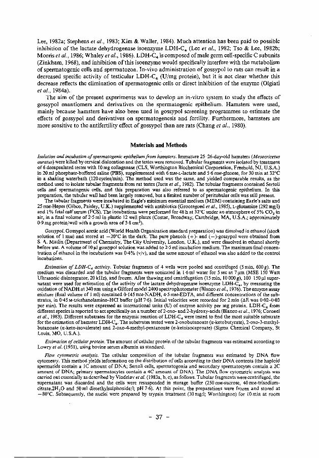
The reaction of the tubular homogenates with a-ketobutyrate showed the highest initial velocities, bUt the maximal initial velocity (V max) was not reached at substrate concentrations up to 6 mM (Fig. Ia). For evalu~tion of the enzyme kinetics, double-reciprocal (Lineweaver-Burk) plots were constructed. The Lineweaver-Burk plot for a-ketobutyrate did not yield a straight line (Fig. Ia), which indicates that this enzyme reaction deviates from Michaelis-Menten behaviour. A possible explanation is that more than one enzyme from the tubular fragments is reacting with this substrate. For this reason, a-ketobutyrate is not suitable as a substrate for the estimation of hamster LDH-C4 activity. a-Ketoisovalerate gave somewhat lower initial velocities than did a-ketobutyrate, but the reaction could be better described according to the Michaelis-Menten equation (Fig. 1 b), and a straight line was obtained in the Lineweaver-Burk plot for substrate concentrations up to 0·8 mM (r = 0·995) (Fig. lb). Using the substrate a-ketoisocaproate, the reaction was inhibited at high substrate concentrations (Fig. lc). The velocities obtained at low a-ketoisocaproate concentrations can be used to estimate Km and Vmax (Fig. lc), but the low initial velocities and the sharp decline of enzyme activity at higher substrate concentrations limits the suitability of this substrate. In the experiments described below a-ketoisovalerate (1·2 mM) was used as substrate for the estimation of LDH-C4 activities.
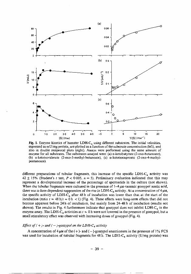
Correlation between LDH-C 4 activity and the percentage of haploid cells in tubular fragments from immature hamsters
The testes from immature hamsters undergo a spurt of growth, and it can be anticipated that the cellular composition of the testes used for the present experiments will show some variation. If the different preparations of tubular fragments contain different numbers of spermatids, different LDH-C4 activities will be found. To validate the comparison of the results within a series of experiments using different cell preparations, the correlation between the percentage of haploid cells and the LDH-C4 activity was estimated.
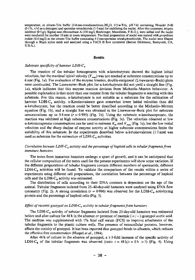
The distribution of cells according to their DNA contents is dependent on the age of the animal. Tubular fragments isolated from 25-40-day-old hamsters were analysed using DNA flow cytometry (Fig. 2). A strong correlation (r = 0·996) was observed for the LDH-C4 activityjmg protein and the percentage of haploid cells (Fig. 3).
Effect of racemic gossypol on LDH-C4 activity in tubular fragments from hamsters
The LDH-C4 activity of tubular fragments isolated from 25-day-old hamsters was estimated before and after culture for 48 h in the absence or presence of racemic ( + j- )-gossypol acetic acid. The medium was supplemented with 1% fetal calf serum (FCS) to improve attachment of the tubular fragments to the plastic culture wells. The presence of extracellular proteins, however, affects the toxicity of gossypol. It has been reported that gossypol binds to albumin, which reduces the effective free concentration (Haspel eta!., 1984).
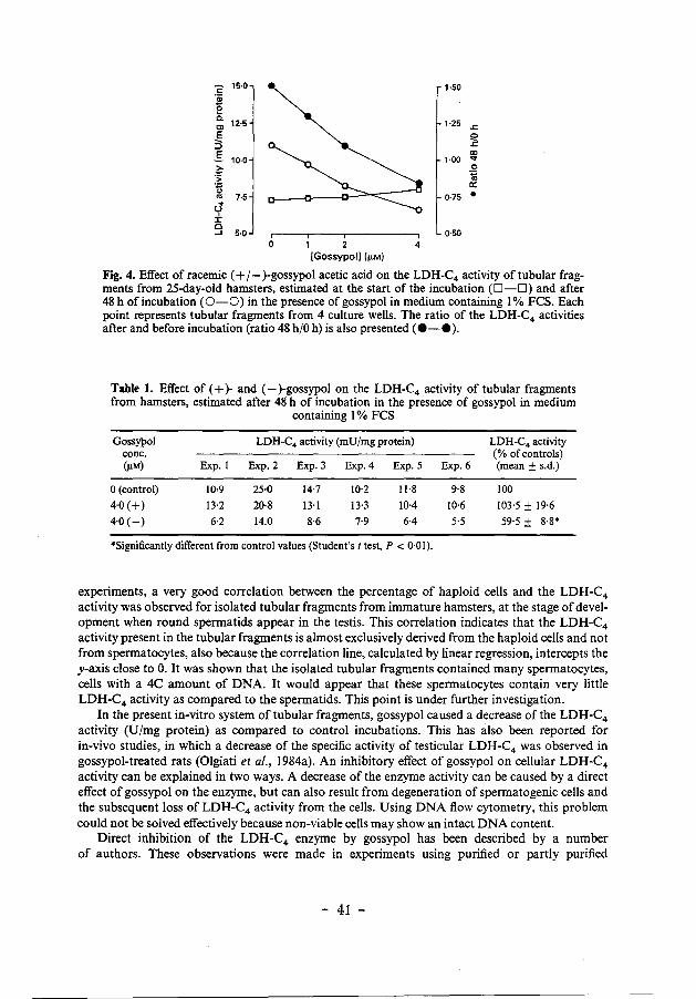
After 48 h of culture in the absence of gossypol, a 1·5-fold increase of the specific activity of LDH-C4 of the tubular fragments was observed (ratio t = 48 h/t = 0 h > 1) (Fig. 4). Using
- 38 -
80
60
40
20
15
s .s 10 ~
5
(a)
(b)
·~~--~-------- 'i ::::l .s ~
Ot----r---.----.---,----,---,
(c)
1·0 2·0 3·0 4·0 5·0 6·0 -5
[S](mM)
0·06
0·04
0·02
0·3
0·2
10 15 20
1/[S] (mM-1 )
Fig. 1. Enzyme kinetics of hamster LDH-C4 using different substrates. The initial velocities, expressed as m U /mg protein, are plotted as a function of the substrate concentration (left), and also in double reciprocal plots (right). Assays were performed using the same amount of enzyme for all substrates. The substrates assayed were: (a) a-keto butyrate (2-oxo-butanoate), (b) a-ketoisovalerate (2-oxo-3-methyl-butanoate), (c) a-ketoisocaproate (2-oxo-4-methylpentanoate).
different preparations of tubular fragments, this increase of the specific LDH-C4 activity was 42 ± 15% (Student's t test, P < 0·005, n = 5). Preliminary evaluation indicated that this may represent a developmental increase of the percentage of spermatids in the culture (not shown). When the tubular fragments were cultured in the presence of 1-4 JlM-racemic gossypol acetic acid, there was a dose-dependent suppression of the rise in LDH-C4 activity. At a concentration of 4 JlM, the specific activity of LDH-C4 after 48 h of incubation was lower than that at the start of the incubation (ratio t = 48 h/t = 0 h < 1) (Fig. 4). These effects were long-term effects that did not become apparent before 24 h of incubation, but mainly from 24-48 h of incubation (results not shown). The results in Fig. 4 furthermore indicate that gossypol does not inhibit LDH-C4 in the enzyme assay. The LDH-C4 activities at t = 0 h were not lowered in the presence of gossypol, but a small stimulatory effect was observed with increasing doses of gossypol (Fig. 4).
Effect of (+)-and (-)-gossypol on the LDH-C4 activity
A concentration of 4 JlM of the (+)-and (-)-gossypol enantiomers in the presence of 1% FCS was used for incubation of tubular fragments for 48 h. The LDH-C4 activity (U/mg protein) was
- 39 -
a; c: c: "' ~ " ]!! a; " 0 0 c: Q)
·~ "' a; a:
2C
(a) (b)
1C
4C
2C
Relative fluorescence intensity (channel no.)
Fig. 2. DNA-flow cytometric analysis of the cellular composition of tubular fragments (spermatogenic epithelium) from hamsters at (a) 25 days and (b) 40 days of age.
-;: 60 "iii
e so c. Cl E 40 3 ..s 30 ~
:~ 20 ~ . cJ 10 ± 0 ...J
0 10 20 30 40 50 60
Haploid cells(%)
Fig. 3. Correlation between LDH-C4 activities and the number of haploid cells in tubular fragments isolated from 25--40-day-old hamsters. For each point, tubular fragments were prepared from 4 animals of the same age. The resulting line was evaluated by linear regression analysis (r = 0·996).
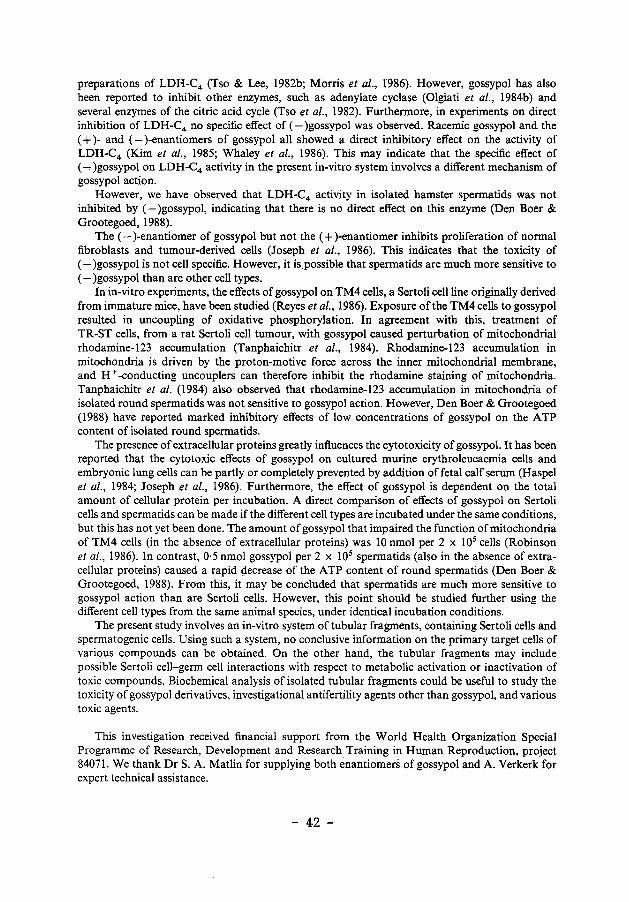
not affected by (+)gossypol, but (-)gossypol caused a marked inhibition of the LDH-C4 activity as compared to the control incubations (Table 1). The variation of the LDH-C4 activities in the different experiments (Table 1) is explained by the rapid testicular development in immature hamsters, as described above. Therefore, the results are also expressed as percentage of the control values (mean± s.d.; n = 6) (Table 1).
Discussion
The lactate dehydrogenase isoenzyme LDH-C4 is synthesized mainly in late-pachyt,~ne spermatocytes and in round spermatids (Hintz & Goldberg, 1977; Meistrich et a/., 1977), nd development of the spermatogenic epithelium in immature animals leads to a rapid increast of the specific and total enzyme activity in the testis (Goldberg & Hawtrey, 1967). In the present
- 40 -
""2 15·0 1·50 ·a; e a.
12·5 1·25 Cl .<:
.€ Q ;:) .<:
.S CXI 10·0 1·00 ...
~ 0 ... ·;; .. ''§ a: .. 7·5 0·75 • .. (,)
± c ....1 5·0 0·50
0 1 2 4 [Gossypol] (fLM)
Fig. 4. Effect of racemic (+I- )-gossypol acetic acid on the LDH-C4 activity of tubular fragments from 25-day-old hamsters, estimated at the start of the incubation (D-O) and after 48 h of incubation (0-0) in the presence of gossypol in medium containing I% FCS. Each point represents tubular fragments from 4 culture wells. The ratio of the LDH-C4 activities after and before incubation (ratio 48 h/0 h) is also presented ( •-• ).
Table 1. Effect of ( + )- and (-)-gossypol on the LDH-C4 activity of tubular fragments from hamsters, estimated after 48' h of incubation in the presence of gossypol in medium
containing I% FCS
Gossypol LDH-C4 activity (mU/mg protein) LDH-C4 activity cone. (% of controls) (!1M) Exp. I Exp.2 Exp.3 Exp.4 Exp.5 Exp.6 (mean ± s.d.)
0 (control) 10•9 25·0 14·7 10·2 Il·8 9·8 100
4·0(+) 13·2 20·8 13·1 13·3 10·4 10·6 103·5 ± 19-6
4·0 (-) 6·2 14.0 8·6 7-9 6·4 5·5 59·5 ± 8·8*
*Significantly different from control values (Student's t test, P < 0·01).
experiments, a very good correlation between the percentage of haploid cells and the LDH-C4
activity was observed for isolated tubular fragments from immature hamsters, at the stage of development when round spermatids appear in the testis. This correlation indicates that the LDH-C4
activity present in the tubular fragments is almost exclusively derived from the haploid cells and not from spermatocytes, also because the correlation line, calculated by linear regression, intercepts the y-axis close to 0. It was shown that the isolated tubular fragments contained many spermatocytes, cells with a 4C amount of DNA. It would appear that these spermatocytes contain very little LDH-C4 activity as compared to the spermatids. This point is under further investigation.
In the present in-vitro system of tubular fragments, gossypol caused a decrease of the LDH-C4
activity (U/mg protein) as compared to control incubations. This has also been reported for in-vivo studies, in which a decrease of the specific activity of testicular LDH-C4 was observed in gossypol-treated rats (Oigiati eta!., 1984a). An inhibitory effect of gossypol on cellular LDH-C4
activity can be explained in two ways. A decrease of the enzyme activity can be caused by a direct effect of gossypol on the enzyme, but can also result from degeneration of spermatogenic cells and the subsequent loss of LDH-C4 activity from the cells. Using DNA flow cytometry, this problem could not be solved effectively because non-viable cells may show an intact DNA content.
Direct inhibition of the LDH-C4 enzyme by gossypol has been described by a number of authors. These observations were made in experiments using purified or partly purified
- 41 -
preparations of LDH-C4 (Tso & Lee, 1982b; Morris et al., 1986). However, gossypol has also been reported to inhibit other enzymes, such as adenylate cyclase (Olgiati et al., 1984b) and several enzymes of the citric acid cycle (Tso et al., 1982). Furthermore, in experiments on direct inhibition of LDH-C4 no specific effect of (-)gossypol was observed. Racemic gossypol and the ( + )- and (- )-enantiomers of gossypol all showed a direct inhibitory effect on the activity of LDH-C4 (Kim et al., 1985; Whaley et al., 1986). This may indicate that the specific effect of (~)gossypol on LDH-C4 activity in the present in-vitro system involves a different mechanism of gossypol action.
However, we have observed that LDH-C4 activity in isolated hamster spermatids was not inhibited by (-)gossypol, indicating that there is no direct effect on this enzyme (Den Boer & Grootegoed, 1988).
The (-)-enantiomer of gossypol but not the ( + )-enantiomer inhibits proliferation of normal fibroblasts and tumour-derived cells (Joseph et al., 1986). This indicates that the toxicity of (-)gossypol is not cell specific. However, it is possible that spermatids are much more sensitive to (-)gossypol than are other cell types.
In in-vitro experiments, the effects of gossypol on TM4 cells, a Sertoli cell line originally derived from immature mice, have been studied (Reyes et al., 1986). Exposure of the TM4 cells to gossypol resulted in uncoupling of oxidative phosphorylation. In agreement with this, treatment of TR-ST cells, from a rat Sertoli cell tumour, with gossypol caused perturbation of mitochondrial rhodamine-123 accumulation (Tanphaichitr et al., 1984). Rhodamine-123 accumulation in mitochondria is driven by the proton-motive force across the inner mitochondrial membrane, and H+ -conducting uncouplers can therefore inhibit the rhodamine staining of mitochondria. Tanphaichitr et al. (1984) also observed that rhodamine-123 accumulation in mitochondria of isolated round spermatids was not sensitive to gossypol action. However, Den Boer & Grootegoed (1988) have reported marked inhibitory effects of low concentrations of gossypol on the ATP content of isolated round spermatids.
The presence of extracellular proteins greatly influences the cytotoxicity of gossypol. It has been reported that the cytotoxic effects of gossypol on cultured murine erythroleucaemia cells and embryonic lung cells can be partly or completely prevented by addition of fetal calf serum (Haspel et al., 1984; Joseph et al., 1986). Furthermore, the effect of gossypol is dependent on the total amount of cellular protein per incubation. A direct comparison of effects of gossypol on Sertoli cells and spermatids can be made if the different cell types are incubated under the same conditions, but this has not yet been done. The amount of gossypol that impaired the function of mitochondria of TM4 cells (in the absence of extracellular proteins) was 10 nmol per 2 x 10⁵ cells (Robinson eta!., 1986). In contrast, 0·5 nmol gossypol per 2 x 10⁵ spermatids (also in the absence of extracellular proteins) caused a rapid gecrease of the ATP content of round spermatids (Den Boer & Grootegoed, 1988). From this, it may be concluded that spermatids are much more sensitive to gossypol action than are Sertoli cells. However, this point should be studied further using the different cell types from the same animal species, under identical incubation conditions.
The present study involves an in-vitro system of tubular fragments, containing Sertoli cells and spermatogenic cells. Using such a system, no conclusive information on the primary target cells of various compounds can be obtained. On the other hand, the tubular fragments may include possible Sertoli cell-germ cell interactions with respect to metabolic activation or inactivation of toxic compounds. Biochemical analysis of isolated tubular fragments could be useful to study the toxicity of gossypol derivatives, investigational antifertility agents other than gossypol, and various toxic agents.
This investigation received financial support from the World Health Organization Special Programme of Research, Development and Research Training in Human Reproduction, project 84071. We thank DrS. A. Matlin for supplying both enantiomers of gossypol and A. Verkerk for expert technical assistance.
- 42 -
References
Adeyemo, 0., Chang, C.Y., Segal, S.J. & Koide, S.S. (1982) Gossypol action on the production and utilization of A TP in sea urchin spermatozoa. Archs Androl, 9, 343-349.
Baccetti, B., Bigliardi, E., Burrini, A.G., Renieri, T. & Sebni, G. (1986) The action of gossypol on rat germinal cells. Gamete Res: 13, 1-17.
Blanco, A., Burgos, C., Gerez de Burgos, N.M. & Montamat, E.E. (1976) Properties of the testicular lactate dehydrogenase isoenzyme. Biochem. J. 153, 165-172.
Chang, M.C., Gu, Z. & Saksena, S.K. (1980) Effects of gossypol on the fertility of male rats, hamsters and rabbits. Contraception 21, 461-469.
Coronel, C.E., Burgos, C., Gerez de Burgos, N.M., Rovai, L.E. & Blanco, A. (1983) Catalytic properties of the sperm-specific lactate dehydrogenase (LDH X or C4 )
from different species. J. exp. Zoo/. 225, 379-385. Den Boer, P.J. & Grootegoed, J.A. (1988) Mechanism of
action of (-)gossypol on ATP production in isolated hamster spermatids. J. Reprod. Fer/. 83, 693-700.
Goldberg, E. & Hawtrey, C. (1967) The ontogeny of sperm specific lactate dehydrogenase in mice. J. exp. Zoo/. 164, 309-3!6.
Grootegoed, J.A., Jansen, R. & van der Molen, H.J. (1985) Intercellular pathway of leucine catabolism in rat spermatogenic epithelium. Biochem. J. 226, 889-892.
Haspel, H.C., Reo, Y.-F., Watanabe, K.A., Sonenberg, M. & Corio, R.E. (1984) Cytocidal effect of gossypol on cultured murine erythroleukemia cells is prevented by serum protein. J. Pharmacol. exp. Ther. 229,218-225.
Hil'tz, M. & Goldberg, E. (1977) Immunohistochemical localization of LDH-X during spermatogenesis in mouse testes. Devl Bioi. 57, 375-384.
Hoffer, A.P. (1982) Ultrastructural studies of spermatozoa and the epithelial lining of the epididymis and vas deferens in rats treated with gossypol. Archs Androl. 8, 233-246.
Hoffer, A.P. (1983) Effects of gossypol on the seminiferous epithelium in the rat: a light and electron microscope study. Bioi. Reprod. 28, 1007-1020.
Joseph, A.E.A., Matlin, S.A. & Knox, P. (1986) Cytotoxicity of enantiomers of gossypol. Br. J. Cancer 54, 511-513.
Jutte, N.H.P.M., Jansen, R., Grootegoed, J.A., Rommerts, F.F.G., Clausen, O.P.F. & van der Molen, H.J. (1982) Regulation of survival of rat pachytene spermatocytes by lactate supply from Sertoli cells. J. Reprod. Fer/. 65,431-438.
Kim, I. & Waller, D.P. (1984) Specific inhibition of the testicular mitochondrial respiratory chain in-vicro by gossypol. J. Androl. 5, 424-430.
Kim, I.C., Waller, D.P. & Fong, H.H.S. (1985) Inhibition of LDH-X by gossypol optical isomers. J. Androl. 6, 344--347.
Lee, C.-Y., Moon, Y.S., Yuan, J.H. & Chen, A.F. (1982) Enzyme inactivation and inhibition by gossypol. Molec. cell. Biochem. 47, 65--70.
Lindberg, M.C., Naqvi, R.H., Matlin, S.A., Zhou, R.H., Bialy, G. & Blye, R.P. (1987) Comparative anti-
fertility effects of gossypol enantiomers in male hamsters. Int. J. Androl. 10,619-623.
Lowry, O.H., Rosebrough, N.J., Farr, A.L. & Randall, R.J. (1951) Protein measurement with the Folin phenol reagent. J. bioi. Chern. 193, 265-275.
Meistricb, M.L., Trostle, P.K., Frapart, M. & Erickson, R.P. (1977) Biosynthesis and localization of lactate dehydrogenase X in pachytene spermatocytes and spermatids from mouse testis. Devl Bioi. 60, 428-441.
Morris, J.D., Higgins, C. & Matlin, S.A. (1986) Inhibition of testicular LDH-X from laboratory animals and man by gossypol and its isomers. J. Reprod. Fer/. 77, 607-612.
National Coordinating Group of Male Contraceptive Studies (1981) Studies on the antifertility effect of gossypol, a new contraceptive for males. In Recent Advances in Fertility Regulation, pp. 122-146. Eds C. F. Chang & D. Griffin. S. A. A tar, Geneva.
Olgiati, K.L., Hoffer, A.P. & Toscano, W.A. (1984a) Gossypol modulation of nucleotide metabolizing enzymes in the reproductive tract of male rats. Bioi. Reprod. 31, 759-770.
Olgiati, K.L., Toscano, D.G., Atkins, W.M. & Toscano, W.A. (l984b) Gossypol inhibition of adenylate cyclase. Archs Biochem. Biophys. 231,411-415.
Prasad, M.R.N. & Diczfalusy, E. (1982) Gossypol. Int. J. Androl., Suppl. 5, 53-70.
Reyes, J., Borriero, L., Tanphaichitr, N., Bellve, A.R. & Benos, D.J. (1986) Energy metabolism of cultured TM4 cells and the action of gossypol. Bioi. Reprod. 34, 809-819.
Robinson, J.M., Tanphaichitr, N. & Bellve, A.R. (1986) Gossypol-induced damage to mitochondria of transformed Sertoli cells. Am. J. Pathol. 125,484-492.
Sampatb, D.S. & Balaram, P. (1986) Resolution of racemic gossypol and interaction of individual enantiomers with serum albumins and model peptides. Biochim. Biophys. Acta 882, 183-186.
Stephens, D.T., Critchlow, L.M. & Hoskins, D.D. (1983) Mechanism of inhibition by gossypol of glycolysis and motility of monkey spermatozoa in vitro. J. Reprod. Fer/. 69, 447-452.
Tanphaichitr, N., Chen, L.B. & Bellve, A.R. (1984) Direct effect of gossypol on TR-ST cells: pertu~bation of rhodamine 123 accumulation in mitochondria. Bioi. Reprod. 31, 1049-1060.
Tso, W.-W. & Lee, C.-S. (1982a) Gossypol uncoupling of respiratory chain and oxidative phosphorylation in ejaculated boar spermatozoa. Contraception 25, 649-655.
Tso, W.-W. & Lee, C.-S. (1982b) Lactate dehydrogenase-X: an isozyme particularly sensitive to gossypol inhibition. Int. J. Androl. 5, 205-209.
Tso, W.-W., Lee, C.-S. & Tso, M.Y.W. (1982) Sensitivity of various spermatozoal enzymes to gossypol inhibition. Archs Andro/. 9, 31-33.
Vindelsv, L.L., Christensen, I.J., Keiding, N., SpangTbomsen, M. & Nissen, N.I. (1983a) Long-term storage of samples for flow cytometric DNA analysis. Cytometry 3, 317-322.
- 43 -
Vindelev, L.L., Christensen, I.J. & Nissen, N.l. (1983b) A detergent-trypsin method for the preparation of nuclei for flow cytometric DNA analysis. Cytometry 3, 323-327.
Vindelov, L.L., Christensen, I.J. & Nissen, N.l. (1983c) Standarruzation of high-resolution flow cytometric DNA analysis by the simultaneous use of chicken and trout red blood cells as internal reference standards. Cytometry 3, 328-331.
Whaley, K.J., Stephens, D.T., Klimkow, N.M. & Hoskins, D.D. (1986) Monkey lactate dehydrogenase-C4 as a model for the interaction of enzymes with gossypol. Contraception 33, 605-{) 16.
Zinkham, W.H. (1968) Lactate dehydrogenase isozymes of testis and sperm: biological and biochemical properties and genetic control. Ann. N.Y. A cad. Sci. 151, 598-{)10.
Received 20 October 1987
- 44 -
Chapter 3
Mechanism of action of (-) gossypol on ATP production in isolated hamster spermatids
P J. Den Boer and J A. Grootegoed
J oumal of Reproduction & Fertility (1988) 83: 693-700
- 45 -
Mechanism of action of (-)gossypol on ATP production in isolated hamster spermatids
P. J. Den Boer and J. A. Grootegoed Department of Biochemistry II, Erasmus University Rotterdam, Rotterdam, The Netherlands
Summary. The A TP content of round spermatids isolated from hamsters was decreased 90% after 18 h of incubation in the presence of 4 JlM-(- )gossypol and 0·1 0% bovine serum albumin (BSA). The ( + )-enantiomer had no effect under these incubation conditions. The Michaelis-Menten constant Km and the maximal initial velocity V max of cellular LDH-C4 were not significantly altered after 18 h of incubation of the spermatids with (-)gossypol. Furthermore, there was no effect of (-)gossypol on the production of 14C02 from L-[U-14C]lactate. It is concluded that (-)gossypol does not inhibit ATP production in spermatids by an effect on the sperm-specific LDH-C4
enzyme or on the mitochondrial oxidation of pyruvate. Rather, (-)gossypol may have an effect on the coupling between electron transport and ATP synthesis in the mitochondria. This action of (-)gossypol may not involve the H+ -conducting activity of gossypol, but could be produced through binding of (-)gossypol to specific mitochondrial proteins.
Keywords: spennatids; gossypol; ATP; LDH-C4 ; mitochondria; enantiomers
Introduction
Racemic ( + j- )gossypol acetic acid, isolated from cottonseed, acts as an antispermatogenic agent in men and in a number of animal species, including hamsters (Prasad & Diczfalusy, 1982). The (- )-enantiomer of gossypol is the active compound in male hamsters, whereas the ( + )-enantiomer does not exert an effect on spermatogenic cells and spermatozoa (Lindberg eta!., 1987). A specific toxic effect of (-)gossypol has been shown for cultured tubular fragments from hamsters, using the activity of the lactate dehydrogenase isoenzyme LDH-C4 as a marker (Den Boer & Grootegoed, 1988).
Several biochemical effects of gossypol have been described, including uncoupling of the linkage between mitochondrial electron transport and ATP synthesis (Abou-Donia & Dieckert, 1974; Tso & Lee, 1982a; Reyes eta!., 1984), inhibition of the mitochondrial electron transport chain (Kim & Waller, 1984), and inhibition of the glycolytic pathway and the citric acid cycle (Stephens eta!., 1983; Wichmann eta!., 1983). The inhibition of the glycolytic pathway and the citric acid cycle could result from inhibition of distinct enzymes. More specifically, an effect of gossypol on the activity of testis- and sperm-specific LDH-C4 has been demonstrated (Tso & Lee, 1982b; Higgins & Morris, 1985; Kim eta!., 1985; Whaley eta!., 1986; Giridharan eta!., 1987).
Studies on the biochemical mechanism of action of gossypol cannot readily be carried out using the in-vitro model of tubular fragments, described by Den Boer & Grootegoed (1988). These fragments contain Sertoli cells and spermatogenic cells at different stages of their development, and it is difficult to determine the primary target cell. In the present experiments, isolated round spermatids from hamsters were used to study the mechanism of action of gossypol. The effects of ( + )- and (-)-gossypol on the ATP content, the metabolism of radioactively labelled lactate, and the LDH-C4 enzyme activity were estimated.
- 46 -
The incubation conditions for isolated round spermatids include the presence of serum albumin in the incubation medium (Grootegoed eta/., I 977). This introduces a complicating factor, because gossypol reacts strongly with cellular and extracellular proteins (Conkerton & Frampton, 1959; Damaty & Hudson, 1975). Gossypol binds to a high-affinity binding site on serum albumin (Royer & Vander Jagt, 1983), forming a complex in a molecular ratio 1:1 (Maliwal eta/., 1985). The binding of gossypol to albumin reduces the free effective concentration of gossypol (Haspel eta/., 1984). This action of extracellular proteins plays an important role in the present experiments.
Materials and Methods isolation of round spermatids from hamsters. Round spermatids from 32-34-day-old golden hamsters (Mesocricetus auratus) were isolated by velocity sedimentation at unit gravity (STA-PUT) and further purified by Percoll-gradient centrifugation, using the same methods as described for the isolation of rat spermatogenic cells (Grootegoed et a/., 1986). The only modification was that the Percoll gradients were formed by centrifugation of 33% (v/v) Percoll (Pharmacia, Uppsala, Sweden) instead of 30%. The isolated cells were incubated in Dulbecco's phosphate-buffered saline, supplemented with 6 mM-sodium L-lactate and 5·6 mM-glucose (PBS-GL), at 32°C in air. The PBS-GL also contained antibiotics (Grootegoed eta/., 1985). Bovine serum albumin (BSA, fraction V, Sigma Chemical Company, St Lollis, MO, U.S.A.) was added ~.s indicated for the different experiments. Approximately 0·3 x 106 cells were incubated in a volume of0·25 ml in 3-ml plastic tubes, unless indicated otherwise.
Gossypol treatment. Racemic (+/-)gossypol acetic acid and the pure phenols ( + )- and (-)-gossypol were obtained and dissolved in ethanol as described by Den Boer & Grootegoed (1988). The maximal final concentration of ethanol in the incubations was 0·5%, and this amount of ethanol was also added to the control incubations.
Estimation of cellular ATP content. The cellular ATP content of the spermatids was estimated using the bioluminescent firefly luciferin-luciferase reaction (Lumac) as described by Grootegoed et a/. (1984), and a model 6100 Pico-Lite Luminometer (Packard).
Estimation of LDH-C4 activity. To estimate the LDH-C4 activity of the isolated round spermatids, 6 incubations of0·3 x 106 cells in 0·25 mi were pooled. Of this pooled suspension, 0·25 ml was used for the estimation of the cellular ATP content and 1·25 ml was used to estimate LDH-C4 activity, as follows. The cells were centrifuged (5 min, 200g), the supernatant was discarded and the cell pellet suspended in 1 ml water. The lysed cells were sonicated for 5 sec at ?11m (MSE !50 Watt Ultrasonic disintegrator, 20kHz), and frozen. After thawing and centrifugation (15 min, 10 000 g), the supernatant was used for estimation of the activity of LDH-C4 as described by Den Boer & Grootegoed (1988). The results were expressed as international units (U) of enzyme activity per 106 cells. a-Ketoisovalerate (Sigma) was used as a substrate (Den Boer & Grootegoed, 1988).
Estimation of 14 C02 formation. To estimate the conversion ofL-[U-14C]lactate to 14C02 , the spermatids were incubated as described, in the presence of O·l11Ci L-[U-14C]lactate (sp.act. 161 Cifmol; Amersham International, Bucks, U.K.) and 3 mM-sodium L-iactate. After 18 h of incubation, the amount of 14C02 produced was estimated essentially as described by Grootegoed eta/. (1984).
Results
Effect of cell number per incubation
Different numbers of spermatids were incubated in 1 ml PBS-GL, for I h in the absence or presence of 0·5 nmo1 (0·5 J.LM) racemic gossypol acetic acid. In the absence of gossypol, the ATP content of the cells was not influenced by the number of cells per incubation (Fig. 1 ). An effect of gossypol on the ATP content was observed only at the lower cell numbers (Fig. 1). This effect of gossypol represents a short-term effect in the absence of extracellular proteins. Under these incubation conditions, the number of cells per given amount of gossypol is probably very important for the evaluation of the toxic effect of gossypol. If the cell number is increased, there is less gossypol available per cell. The presence of albumin in the incubation medium could prevent rapid accumulation of gossypol in the cells and quantitative binding of gossypol to cellular proteins (see below).
Effect of (+)-and (-)-gossypol in the presence of albumin
Isolated spermatids from hamsters were incubated for 18 h in PBS-GL, in the presence of different concentrations ofBSA. At a BSA concentration of0·003% (wjv), the ATP content of the
- 47 -
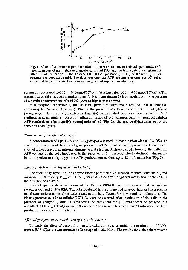
120 0 "#.0 ::II 80 c::-.,-_., c::., 0::~ u-a.."' 1->
40
<(
0·4 0·8 1·2 1·6 2·0 2·4
No. of cells (x 10-s)
Fig. 1. Effect of cell number per incubation on the ATP content of isolated spermatids. Different numbers of spermatids were incubated in 1 ml PBS, and the ATP content was estimated after lh of incubation in the absence (e-e) or presence (0-0) of 0·5nmol (0·5!1M) racemic gossypol acetic acid. The data represent the ATP content expressed per 106 cells, converted to% of the starting value (mean ± s.d. of triplicate incubations).
spermatids decreased to 0·12 ± 0·10 nmol/106 cells (starting value 1·80 ± 0·25 nmol/106 cells). The spermatids could effectively maintain their ATP content during 18 h of incubation in the presence of albumin concentrations of 0·01 0% (w jv) or higher (not shown).
In subsequent experiments, the isolated spermatids were incubated for 18 h in PBS-GL containing 0·02% or 0·10% (wjv) BSA, in the presence of different concentrations of ( + )- or (-)-gossypol. The results presented in Fig. 2(a) indicate that both enantiomers inhibit ATP synthesis in spermatids at [gossypol]/[albumin] ratios of > 1, whereas only (-)gossypol inhibits ATP synthesis at a [gossypol]/[albumin] ratio of< 1 (Fig. 2b; the [gossypol]j[albumin] ratios are shown in each figure).
Time-course of the effect of gossypol
A concentration of 4 !1M ( + )- and (-)-gossypol was used, in combination with 0·1 0% BSA, to study the time-course of the effect of gossypol on the ATP content of round spermatids. There was no effect of either gossypol enantiomer during the first 8 h of incubation (Fig. 3). However, thereafter the ATP content of the cells incubated in the presence of (-)gossypol slowly declined, whereas no inhibitory effect of (+)gossypol on ATP synthesis was evident up to 18 h of incubation (Fig. 3).
Effect of (+)-and (-)-gossypol on LDH-C4
The effect of gossypol on the enzyme kinetic parameters (Michaelis-Menten constant Km and maximal initial velocity Vmax) of LDH-C4 was estimated after long-term incubation of the cells in the presence of gossypol.
Isolated spermatids were incubated for 18 h in PBS-GL, in the presence of 4 !1M ( + )- or (-)-gossypol and 0·1 0% BSA. The cells incubated in the presence of gossypol had an intact plasma membrane (microscopic observation) and could be collected by low-speed centrifugation. The kinetic parameters of the cellular LDH-C4 were not altered after incubation of the cells in the presence of gossypol (Table 1). This result indicates that the (- )-enantiomer of gossypol did not affect LDH-C4 activity in incubation conditions in which a pronounced inhibition of ATP production was observed (Table 1).
Effect of gossypol on the metabolism of L-[V- 14C}lactate
To study the effect of gossypol on lactate oxidation by spermatids, the production of 14C02
from L-[U- 14C]lactate was estimated (Grootegoed et al., 1984). The results show that there was no
- 48 -
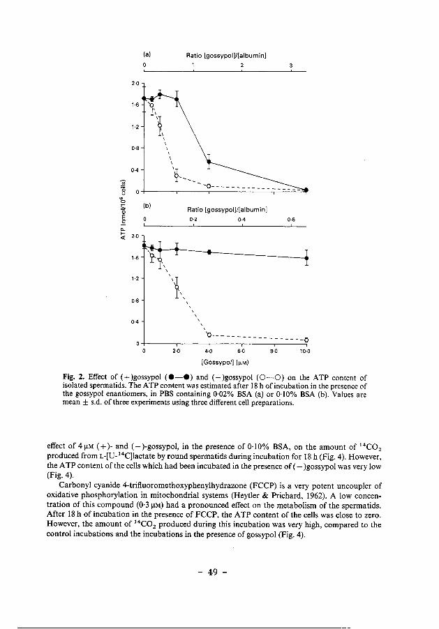
2·0
1·6
1·2
0·8
0·4
:2i o; (.)
"' 0
0 E ..s "-~ 2·0 <(
1·6
1·2
0·8
0·4
(a)
(b)
0
0
\
\ \ I I I
Ratio [gossypol)/[albumin)
l---Ratio [gossypol]/[albumin)
0·2
' \ \
\
\
I
0·4
3
0·6
'o--- -- ------ - - --- - --o 2·0 4·0 6·0 8·0 10·0
[Gossypol) (~M)
Fig. 2. Effect of (+)gossypol (e-e) and (-)gossypol (0-0) on the ATP content of isolated spermatids. The A TP content was estimated after 18 h of incubation in the presence of the gossypol enantiomers, in PBS containing 0·02% BSA (a) or 0·10% BSA (b). Values are mean ± s.d. of three experiments using three different cell preparations.
effect of 4 J.lM ( + )- and (-)-gossypol, in the presence of 0·1 0% BSA, on the amount of 14C02
produced from L-[U-14C]Iactate by round spermatids during incubation for 18 h (Fig. 4). However, the A TP content of the cells which had been incubated in the presence of (-)gossypol was very low (Fig. 4).
Carbonyl cyanide 4-trifluoromethoxyphenylhydrazone (FCCP) is a very potent uncoup1er of oxidative phosphorylation in mitochondrial systems (Heytler & Prichard, 1962). A low concentration of this compound (0·3 J.lM) had a pronounced effect on the metabolism of the spermatids. After 18 h of incubation in the presence of FCCP, the A TP content of the cells was close to zero. However, the amount of 14C02 produced during this incubation was very high, compared to the control incubations and the incubations in the presence of gossypol (Fig. 4).
- 49 -
1-6
~ Qi 1·2
" "' 0
0 0·8 E
.s c... f-<(
0·4
1 0
4 8 12 16
Incubation period (h)
Fig. 3. Time course of the effect of gossypol on the A TP content of isolated spermatids. The ATP content was estimated after different periods of incubation in the absence of gossypol (e-e), and in the presence of 4 J.lM·( +)gossypol (0-0) or 4 J.lM-(- )gossypol (6-6), in PBS containing 0·10% BSA. Values are mean ± s.d. of triplicate incubations.
Table 1. Effect of gossypol on the kinetics of cellular LDH-C4 activity and ATP content
Gossypol vmax Vat 1·2mM ATP cone. Km (mU/106 (mU/106 (nmol/106
(I!M) (mM) cells) cells) cells)
0 0·13 4-81 4·09 ± 0·10 1·70 ± 0·04 4(+) 0·13 5·03 4·11 ± 0·29 1·74 ± 0·11 4(-) 0·12 4·91 4·26 ± 0·17 0·51 ± 0·02
Isolated round spermatids from hamsters were incubated for 18 h in PBS containing 0·10% BSA, in the presence of 41-LM ( + )· or (-)·gossypol. Subsequently, the initial reaction velocities of cellular LDH-C4 were measured. Double-reciprocal plots of the enzyme kinetics showed a r ~ 0·99 by linear regression analysis. Data on the initial velocity at 1·2 mM a-keto-isovalerate and A TP content represent the mean ± s.d. of triplicate incubations.
Discussion
The present study concerns long-term effects of low doses of ( + )- and (-)-gossypol on isolated (ound spermatids from hamsters, and aimed to investigate the biochemical mechanism of gossypol ·action oii the spermatids. · When cells or tissues are incubated in the absence of extracellular proteins, racemic gossypol added to the incubation medium rapidly and quantitatively binds to cells (Haspel eta/., 1984). The presenrresults showed that, in short-term experiments in the absence of extracellular proteins, the gossypol-induced inhibition of ATP synthesis in isolated spermatids was abolished when the number of cells per incubation was increased. This indicates that ATP synthesis inhibition is
- so -
'"l (a)
~ 180
0 -0 E 80 .s ""C CD
" :J 60 ""C 0 c. 0 40
k' co 0 20 f-
0 c C (+) (-) FCCP
t= 0 L___ t= 18 h ____]
"' 0
2·0
~1-0 E .s I= 0·5 <(
0
(b)
c t= 0
c (+) ~ (-) = FCCP
L-t=18h__j
Fig. 4. Effect of 4 J.!M-( +)gossypol and 4 J.lM-(- )gossypol, and of 0·3 J.lM-FCCP on (a) the conversion of L-(U-14C]lactate to 14C02 by isolated spermatids and (b) the ATP content of the spermatids. The spermatids were incubated for 18 h in PBS containing the gossypol enantiomers and 0· 10% BSA.
dependent on the amount of gossypol per cell. This has also been observed for rat spermatozoa (Stephens eta!., 1983). Therefore, it is concluded that the number of cells and the total amount of gossypol in the incubations are much more relevant than the concentration of gossypol.
In the present incubation system, using isolated spermatids in medium containing albumin, gossypol can bind to extracellular and cellular proteins. In the experiments on the mechanism of action of gossypol, 4 J.lM ( + )- or (-)-gossypol were used in combination with 0· I 0% BSA. This resulted in a [gossypol]/[ albumin] ratio of <I, and a low concentration of free gossypol (approximately 0·02 J.lM, as calculated from the Kd; Sam path & Balaram, 1986). The present results indicate that (-)gossypol under these conditions can slowly but progressively accumulate in the cells. Concomitantly, gossypol may become bound to cellular components, possibly one or several proteins that bind gossypol stronger than albumin. The interaction of (-)gossypol with the cellular proteins might be stereospecific, resulting in inhibition of the biological activity of the proteins. There is apparently no such specific interaction of (+)gossypol with cellular components. It is not very likely that the preferential effect of (-)gossypol is explained by a relative weak binding of this enantiomer to albumin, because it has been observed that the ( + )- and (- )-enantiomers bind equally effective to BSA (Sampath & Balaram, 1986).
The described effect of (-)gossypol on the A TP content of the spermatids was evaluated in relation to possible effects of gossypol on LDH-C4. The lactate dehydrogenase isoenzyme LDHC4, which appears to be a key enzyme in the metabolism of the male germ cells, has been proposed as a potential site of action for the antifertility effect of gossypol (Tso & Lee, 1982b; Olgiati & Toscano, 1983; Kim et a!., 1985; Whaley et a!., 1986). In these studies, crude or purified preparations of LDH-C4 were treated with gossypol which resulted in inhibition of the enzyme. Higgins & Morris (1985) have observed an effect of gossypol on the Km of LDH-C4, after administration of gossypol to hamsters. However, these authors have used the substrate u-ketobutyrate to estimate the kinetic parameters of the LDH-C4 reaction, and it has been observed that the enzyme reaction with this substrate is not described by the Michaelis-Menten equation (Den Boer & Grootegoed, 1988). The possibility that the effect of gossypol on spermatogenesis could involve
- 51 -
an effect on LDH-C4 is contradicted by the present results. The kinetic parameters Km and Vm•• of the enzyme in the spermatids were not altered after 18 h of incubation in the presence of gossypol, whereas the ATP content was low. This indicates that LDH-C4 is not the primary target of gossypol action on cellular ATP production.
The next step in the present study was the estimation of the effects of (-)gossypol on the metabolism of lactate by round spermatids. The production of 14C02 from L-[U-14C]lactate involves the conversion of lactate to pyruvate, catalysed by LDH isoenzymes including LDH-C4 ,
and the subsequent mitochondrial oxidation of pyruvate via the pyruvate dehydrogenase complex and the citric acid cycle. C02 production was not affected by gossypol, indicating that the mitochondrial pyruvate oxidation was not inhibited by gossypol. Conversion of pyruvate to C02 is linked to oxidation ofNADH via the mitochondrial electron transport chain. Therefore the results indicate that the electron transport chain is not inhibited. Kim & Waller (1984) observed strong inhibition of the electron transport chain by gossypol, but this was an effect of (+)gossypol at very high concentrations. The work of Wichmann eta/. (1983) showed inhibition of the citric acid cycle by gossypol in spermatozoa. This is not in agreement with the present findings, but the discrepancy may be explained by the fact that the studies on spermatozoa were short-term experiments in which racemic gossypol was added to the cells in the absence of extracelh.ilar proteins. As described above, under such conditions a rapid accumulation of gossypol in the cells may occur. This may lead to interactions with many different proteins which are not specific for the two enantiomers.
A possible mechanism for the action of gossypol on ATP production involves an effect of gossypol on the coupling between mitochondrial electron transport and ATP synthesis (Abou-Donia & Dieckert, 1974; Tso & Lee, 1982a; Reyes et a/., 1984). In the presence of an H +-conducting uncoupling agent, NADH oxidation and electron transport are not inhibited, but ATP production is impaired because no proton gradient can be established across the inner mitochondrial membrane. Rather, the proton-motive force is dissipated, and there is a loss of respiratory control. Concomitantly, the rate of substrate oxidation can be increased. The present results on [1 4 C]lactate metabolism indicate that such effects were brought about by incubation of the spermatids in the presence of the H+ -conducting uncoupler FCCP. However, the effects of gossypol a .. d FCCP were different, in that gossypol did not cause an increased rate of lactate oxidation. A possible explanation is that, under the present incubation conditions, gossypol does not act as an uncoupler by dissipating proton gradients across membranes. It has not been shown that the H +-conducting activity of gossypol is different for the (+)-and (- )-enantiomers (Reyes et at., 1984). The present results seem to indicate that (-)gossypol inactivates mitochondrial proteins which are directly involved in A TP synthesis. The inactivation could involve high affinity binding of (-)gossypol.
There are no indications that there is a germ cell-specific interaction of (-)gossypol with mitochondrial proteins. The cytotoxic effect of (-)gossypol is much more pronounced than that of the ( + )-enantiomer for other cell types, such as hepatoma cells and skin fibroblasts (Joseph eta/., 1986). The seemingly specific effect of (-)gossypol on spermatogenic cells and spermatozoa in vivo may be explained by the very high sensitivity of these cell types to inhibition of mitochondrial function (Grootegoed eta/., 1984).
This investigation received financial support from the World Health Organization Special Programme of Research, Development and Research Training in Human Reproduction, project 84071. We thank Dr S. A. Matlin (Dept. of Chemistry, The City University, London, U.K.) for supplying the ( + )- and (- )-enantiomers of gossypol.
References
Abou-Donia, M.B. & Dieckert, J.W. (1974) Gossypol: uncoupling of respiratory chain and oxidative phosphorylation. Life Sci. 14, 1955-1963.
Conkerton, E.J. & Frampton, V.L. (1959) Reaction of gossypol with free E-arnino groups of lysine in proteins. Archs Biochem. Biophys. 81, Bo-134.
- 52 -
Damaty, S. & Hudson, B.J.F. (1975) Interaction of gossypol with cottonseed protein: potentiometric studies. J. Sci. Fd Agric. 26, 1667-1672.
Den Boer, P.J. & Grootegoed, J.A. (1988) Differential effects of ( + )· and (-)·gossypol enantiomers on LDH·C4 activity of hamster spermatogenic epithelium in vitro. J. Reprod. Fert. 83, 701-709.
Giridharan, N., Bamji, M.S. & Madhyastha, M.N. (1987) In vitro effects of gossypol on testicular lactic dehydrogenase-X and other dehydrogenases. J. Biosci. 11,465-471.
Grootegoed, J.A., GroUe-Hey, A.H., Rommerts, F.F.G. & van der Molen, H.J. (1977) Ribonucleic acid synthesis in vitro in primary spermatocytes isolated from rat testis. Biochem. J. 168, 23-31.
Grootegoed, J.A., Jansen, R. & van der Molen, H.J. (1984) The role of glucose, pyruvate and lactate in A TP production by rat spermatocytes and spermatids. Biochim. Biophys. Acta 767, 248-256.
Grootegoed, J.A., Jansen, R. & van der Molen, H.J. (1985) Intercellular pathway of leucine catabolism in rat spermatogenic epithelium. Biochem. J. 226, 889-892.
Grootegoed, J.A., Jansen, R. & van der Molen, H.J. (1986) Effect of glucose on ATP dephosphorylation in rat spermatids. J. Reprod. Fert. 77, 99-107.
Haspel, H.C., Reo, Y.-F., Watanabe, K.A., Sonenberg, M. & Corio, R.E. (1984) Cytocidal effect of gossypol on cultured murine erythroleukemia cells is pre· vented by serum protein. J. Pharmacal .. exp. Ther. 229,218-225.
Heytler, P.G. & Prichard, W.W. (1962) A new class of uncoupling agents-{;arbonyl cyanide phenylhydra· zones. Biochem. Biophys. Res. Commun. 7, 272-275.
Higgins, C. & Morris, I.D. (1985) Fertility and LDH·X activity in male hamsters treated with the male con· traceptive gossypol.IRCS Med. Sci. 13, 1064-1065.
Joseph, A.E.A., Matlin, S.A. & Knox, P. (1986) Cyto· toxicity of enantiomers of gossypol. Br. J. Cancer 54, 511-513.
Kim, I. & WaDer, D.P. (1984) Specific inhibition of the testicular mitochondrial respiratory chain in vitro by gossypol. J. Androl. 5, 424-430.
Kim, I.C., Waller, D.P. & Fong, H.H.S. (1985) Inhibition of LDH·X by gossypol optical isomers. J. Androl. 6, 344-347.
Lindberg, M.C., Naqvi, R.H., Matlin, S.A., Zhou, R.H., Bialy, G. & Blye, R.P. (1987) Comparative antiferti· lity effects of gossypol enantiomers in male hamsters. Int. J. Androl. 10, 619-623.
Maliwal, B.P., Appu Rao, A.G. & Narasinga Rao, M.S. (1985) Spectroscopic study of the interaction of gossypol with bovine serum albumin. Int. J. Peptide Protein Res. 25, 382-388.
Olgiati, K.L. & Toscano, W.A. (1983) Kinetics of gossypol inhibition of bovine lactate dehydrogenase X. Biochem. Biophys. Res. Commun. 115, 180-185.
Prasad, M.R.N. & Diczfalusy, E. (1982) Gossypol. Int. J. Androl., Suppl. 5, 53-70.
Reyes, J., Allen, J., Tanphaichitr, N., Bellve, A.R. & Benos, D.J. (1984) Molecular mechanism of gossypol action on lipid membranes. J. bioi. Chern. 259, 9607-9615.
Royer, R.E. & Vander Jagt, D.L. (1983) Gossypol binds to a high-affinity binding site on human serum albumin. FEBS Letters 157,28-30.
Sampath, D.S. & Balaram, P. (1986) Resolution of racemic gossypol and interaction of individual enan· tiomers with serum albumins and model peptides. Biochim. Biophys. Acta 882, 183-186.
Stephens, D.T., Critchlow, L.M. & Hoskins, D.D. (1983) Mechanism of inhibition by gossypol of glycolysis and motility of monkey spermatozoa in vitro. J. Reprod. Fert. 69, 447-452.
Tso, W.-W. & Lee, C.-S. (1982a) Gossypol uncoupling of respiratory chain and oxidative phosphorylation in ejaculated boar spermatozoa. Contraception 25, 649-655.
Tso, W.-W. & Lee, C.-S. (1982b) Lactate dehydrogenase-X: an isozyme particularly sensitive to gossypol inhibition. Int. J. Androl. 5, 205-209.
Whaley, K.J., Stephens, D.T., Klimkow, N.M. & Hoskins, D.D. ( 1986) Monkey lactate dehydrogenase· C4 as a model of the interaction of enzymes with gossypol. Contraception 33, 605-616.
Wichmann, K., Kiipyaho, K., Sinervirta, R. & Jinne, J. (1983) Effect of gossypol on the motility and metabolism of human spermatozoa. J. Reprod. Fert. 69, 259-264.
Received 20 October 1987
- 53 -

Chapter 4
The effects of glucose and adenosine on the ATP content of hamster spermatids
P. Mackenbach, P J. Den Boer, J.W. de Jong and J.A. Grootegoed
submitted to: Reproduction, Fertility & Development
- 55 -
The effects of glucose and adenosine on the ATP content of hamster spermatids
Abstract. Effects of glucose and adenosine on ATP metabolism were studied using isolated round spermatids from hamsters. The ATP content of the spermatids was strongly reduced after 1 h of incubation of the cells in the presence of 0.1 mM D-glucose. There was no effect of glucose up to a 1 mM concentration, during 18 h of incubation in the presence of 12 mM sodium DL-lactate. However, 10 mM glucose caused a virtually complete loss of cellular A TP also in the presence of lactate. The effect of adenosine was estimated in the absence of glucose, in cells utilizing exogenous lactate as energy-yielding substrate. The cellular A TP content was approximately 4 and 8 nmol/106 cells, after 18 h of incubation in the absence and presence of 0.1 mM adenosine, respectively. This two-fold increase was prevented by inhibitors of adenosine uptake and phosphorylation, and was slowly reversed after removal of the exogenous adenosine. Treatment of the cells with adenosine had no effect on the energy charge, which was higher than 0.90, and also did not alter the cellular cyclic AMP content. It is discussed that the physiological ATP content of the round spermatids is at least 4 nmol/106 cells and is stable.
INTRODUCTION
ATP synthesis in round spermatids from rats involves a particular pattern of utilization of the energy-yielding metabolic pathways (reviewed by Grootegoed & Den Boer, 1989). Spermatids contain all the enzyme activities which constitute the glycolytic pathway. Furthermore, the isolated cells convert glucose to pyruvate and lactate. It is very clear, however, that this glucose metabolism does not result in maintenance of the cellular ATP content. Rather, exposure of isolated spermatids to glucose results in ATP depletion (Grootegoed et al., 1986a; Nakamura et' al., 1986a). Many authors agree that lactate is the very best energy-yielding substrate for isolated round spermatids from rats (see for a review Grootegoed & Den Boer, 1989). The spermatids convert added lactate to pyruvate and produce ATP via mitochondrial oxidation
- 56 -
of pyruvate. In rat spermatids, the ATP content is not lowered by glucose
during incubation of the cells in the presence of exogenous lactate (Grootegoed et al., 1986a). This can be explained by lactate-induced inhibition of the glycolytic pathway. The enzyme phosphofructokinase can be effectively inhibited by ATP and other factors, in cells with a high energy charge <Bosca et al., 1985). In spermatids, a high energy charge can probably be reached when the cells oxidize exogenous lactate under optimal conditions.
The present study concerns the ATP content of round spermatids isolated from Golden hamsters. We have reported that these hamster cells contained a relatively low amount of ATP (less than 2 nmol/106 cells) <Den Boer & Grootegoed, 1988), as compared with the ATP content of spermatids from rats (6-8 nmol/106
cells) (Grootegoed et al., 1986a). This difference between rat and hamster spermatids may not reflect a species-specific size of the total adenine nucleotide pool, but rather may point to a loss of adenine nucleotides during isolation and incubation of hamster spermatids. Such a loss can occur when the energy charge ([ATPJ + 112 [ADPJ) I UATPJ + [ADPJ + [AMPJ) which is normally maintained within narrow limits near 0.9, would be endangered due to unfavourable conditions. Recovery of the adenine nucleotide pool would rely on de novo synthesis of the nucleotides, and/or uptake and metabolism of free purine bases and nucleosides which involves direct phosphorylation and the purine salvage pathway. The purine nucleoside adenosine exerts complex effects on cells (Fox & Kelley, 1987), but can be used to increase the concentration of total cellular adenine nucleotides under experimental conditions (Lund et al., 1975). In the present series of experiments, the effects of exogenous glucose and adenosine on the ATP content of hamster spermatids was studied.
MATERIALS AND METHODS
Isolation of round spermatids from hamsters. Round spermatids from 32-34-day-old hamsters (Mesocricetus auratus) were isolated by velocity sedimentation at unit gravity (STA-PUT) and further purified by Percoll-gradient centrifugation as described by Den Boer & Grootegoed (1988). The isolation medium was Dulbecco·s phosphate-buffered saline <Dulbecco & Vogt, 1954) supplemented
- 57 -
with 12 mM sodium DL-lactate, but did not contain glucose. The isolated cells were incubated in Dulbecco's phosphate-buffered saline supplemented with 12 mM sodium DL-lactate, penicillin (105 units/1), streptomycin (100 mg/1), and 0.1% (w/v) bovine serum albumin (fraction V; Sigma Chemical Company, St. Louis, MO, USA), also referred to as PBS-L. Where indicated, this incubation medium did not contain lactate (PBS). Approximately 0.3 x 10⁶ cells were incubated in 0.25 ml PBS or PBS-L in polystyrene tubes (diameter 11.5 mm), at 32 °C in air.
Estimation of ATP, energy charge and cyclic AMP (cAMP). The cellular ATP content of the spermatids was estimated with the bioluminescent firefly luciferin-luciferase reaction as described by Grootegoed et al. (1984), using Lumit PM (Lumac, Meise, Belgium) and a model 6100 Pico-Lite luminometer (Packard Instrument Company, Downers Grove, Il., USA).
To determine the cellular energy charge, AMP, ADP and ATP were assayed in cell extracts, using the h.p.l.c. method described by Achterberg et al. (1986).
Cyclic AMP was estimated using a cyclic AMP (3 H) radioassay kit (Amersham International plc., Little Chalfont, Buckinghamshire, UK).
All estimations were performed using cell ex~racts which were prepared after termination of the incubations with perchloric acid, as described by Groo-tegoed et al. (1984).
Measurement of enzyme activities. Adenosine kinase activity in freshly isolated spermatids was measured at 32 °C as described by Dejong (1977). Hexokinase activity was estimated using the method described by Chou & Wilson (1975), but using a fluorometer (SFM 25; Kontron instruments, Zurich, Switzerland) with the sample compartment maintained at 37°C.
Chemicals. Adenosine was purchased from Boehringer Mannheim, Mannhein, F.R.G. Dipyridamole was from Sigma and 5-Iodo-tubercidin was obtained from Research Biochemicals Inc., Natick, MA, USA. Other chemicals used were also from commercial sources and were of high purity.
- 58 -
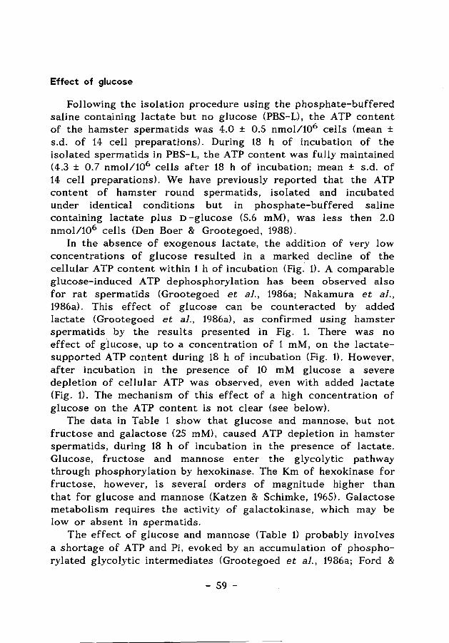
Effect of glucose
Following the isolation procedure using the phosphate-buffered saline containing lactate but no glucose (PBS-L), the ATP content of the hamster spermatids was 4.0 ± 0.5 nmol/106 cells (mean ± s.d. of 14 cell preparations). During 18 h of incubation of the isolated spermatids in PBS-L, the ATP content was fully maintained (4.3 ± 0. 7 nmol/106 cells after 18 h of incubation; mean ± s.d. of 14 cell preparations). We have previously reported that the ATP content of hamster round spermatids, isolated and incubated under identical conditions but in phosphate-buffered saline containing lactate plus D -glucose (5.6 mM), was less then 2.0 nmol/106 cells <Den Boer & Grootegoed, 1988).
In the absence of exogenous lactate, the addition of very low concentrations of glucose resulted in a marked decline of the cellular ATP content within 1 h of incubation (Fig. 1). A comparable glucose-induced ATP dephosphorylation has been observed also for rat spermatids (Grootegoed et al., 1986a; Nakamura et al., 1986a). This effect of glucose can be counteracted by added lactate (Grootegoed et al., 1986a), as confirmed using hamster spermatids by the results presented in Fig. 1. There was no effect of glucose, up to a concentration of 1 mM, on the lactatesupported ATP content during 18 h of incubation (Fig. 1). However, after incubation in the presence of 10 mM glucose a severe depletion of cellular ATP was observed, even with added lactate (Fig. 1). The mechanism of this effect of a high concentration of glucose on the ATP content is not clear (see below).
The data in Table 1 show that glucose and mannose, but not fructose and galactose (25 mM), caused ATP depletion in hamster spermatids, during 18 h of incubation in the presence of lactate. Glucose, fructose and mannose enter the glycolytic pathway through phosphorylation by hexokinase. The Km of hexokinase for fructose, however, is several orders of magnitude higher than that for glucose and mannose (Katzen & Schimke, 1965). Galactose metabolism requires the activity of galactokinase, which may be low or absent in spermatids.
The effect of glucose and mannose (Table 1) probably involves a shortage of ATP and Pi, evoked by an accumulation of phosphorylated glycolytic intermediates (Grootegoed et al., 1986a; Ford &
- 59 -
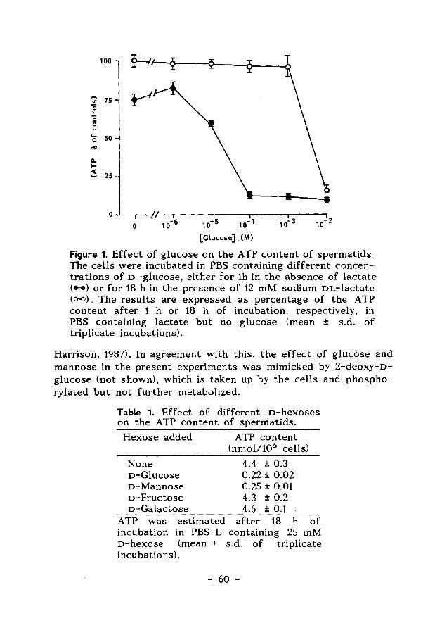
100 ~
,.... 75 r-' "' 0 ... ....
c: 0 u .... 50 0 dP
ll. 1-< 25 ....
~~~---.-----.----~---. 10-s 10-4 10- 3 10- 2
0
0
[Glucose] (M)
Figure 1. Effect of glucose on the ATP content of spermatids. The cells were incubated in PBS containing different concentrations of D -glucose, either for 1h in the absence of lactate ( .. ) or for 18 h in the presence of 12 mM sodium DL-lactate (o-o). The results are expressed as percentage of the ATP content after 1 h or 18 h of incubation, respectively, in PBS containing lactate but no glucose (mean ± s.d. of triplicate incubations).
Harrison, 1987). In agreement with this, the effect of glucose and mannose in the present experiments was mimicked by 2-deoxy-Dglucose (not shown), which is taken up by the cells and phosphorylated but not further metabolized.
Table 1. Effect of different o-hexoses on the ATP content of spermatids.
Hexose added ATP content (nmol/106 cells)
None 4.4 ± 0.3 o-Glucose 0.22 ± 0.02 o-Mannose 0.25 ± 0.01 o-Fructose 4.3 ± 0.2 o-Galactose 4.6 ± 0.1
ATP was estimated after 18 h of incubation in PBS-L containing 25 mM o-hexose (mean ± s.d. of triplicate incubations).
- 60 -
The enzyme kinetic properties of hexokinase in the hamster spermatids were studied, to exclude the possibility that the effect of 10 mM glucose on the lactate-supported ATP content was related to the presence of a high Km hexokinase isoenzyme. However, the Km observed was 0.56 mM (Fig. 2), which indicates the presence of a low Km hexokinase isoenzyme (Katzen & Schimke, 1965; Katzen et al., 1968).
1.0
0.8
• I 0.6 :::1 E
> -- 0.4 • • >
4 6 10 [s] (mMl
-2 0 2 4 6 8 10
Figure 2. Enzyme kinetic properties of hexokinase in hamster spermatids. The Km was 0.56 mM.
Effect of adenosine
Incubation of hamster round spermatids for 18 h in the presence of the nucleoside adenosine (up to 0.2 mM) resulted in a two-fold increase of the cellular ATP content (Fig. 3). This was a slow effect, because the change in the ATP content of the cells was not detected within 2-4 h of incubation (not shown). The energy charge of the spermatids was 0.95 ± 0.02 and 0.97 ± 0.01 (mean ± s.d. of triplicate incubations) after 18 h of incubation in the absence and presence of adenosine, respectively. Furthermore, adenosine did not markedly affect the cAMP content of the
- 61 -
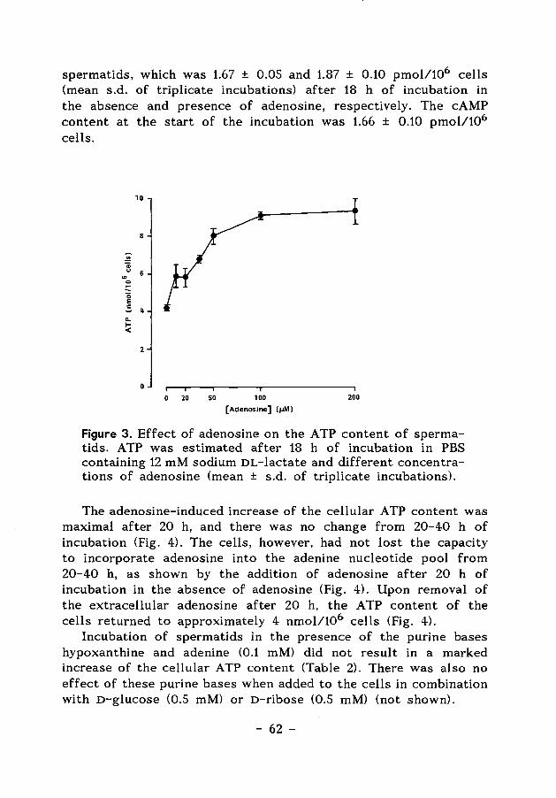
spermatids, which was 1.67 ± 0.05 and 1.87 ± 0.10 pmol/106 cells (mean s.d. of triplicate incubations) after 18 h of incubation in the absence and presence of adenosine, respectively. The cAMP content at the start of the incubation was 1.66 ± 0.10 pmol/106
cells.
... 0
0 E
10
8
.s 4 ... 1-<
2
0
0 20 50 100 200
(Adenosine] !IJMJ
Figure 3. Effect of adenosine on the ATP content of spermatids. ATP was estimated after 18 h of incubation in PBS containing 12 mM sodium DL-lactate and different concentrations of adenosine (mean ± s.d. of triplicate incubations).
The adenosine-induced increase of the cellular ATP content was maximal after 20 h, and there was no change from 20-40 h of incubation (Fig. 4). The cells, however, had not lost the capacity to incorporate adenosine into the adenine nucleotide pool from 20-40 h, as shown by the addition of adenosine after 20 h of incubation in the absence of adenosine (Fig. 4). Upon removal of the extracellular adenosine after 20 h, the A TP content of the cells returned to approximately 4 nmol/106 cells (Fig. 4).
Incubation of spermatids in the presence of the purine bases hypoxanthine and adenine (0.1 mM) did not result in a marked increase of the cellular ATP content (Table 2). There was also no effect of these purine bases when added to the cells in combination with D-glucose (0.5 mM) or D-ribose (0.5 mM) (not shown).
- 62 -
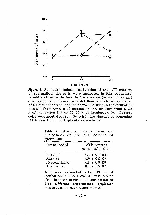
10
8
..!!! QJ u 6
IQ
0 -.._ 0 e 4 !: -------------2 c.. 1-< 2
0
0 20 40
Time (hours)
Figure 4. Adenosine-induced modulation of the ATP content of spermatids. The cells were incubated in PBS containing 12 mM sodium DL-lactate, in the absence (broken lines and open symbols) or presence (solid lines and closed symbols) of 0.1 mM adenosine. Adenosine was included in the incubation medium from 0-40 h of incubation (•), or only from 0-20 h of incubation ('ii') or 20-40 h of incubation (T). Control cells were incubated from 0-40 h in the absence of adenosine ( o) (mean ± s.d. of triplicate incubations).
Table 2. Effect of purine bases and nucleosides on the A TP content of spermatids.
Purine added
None Adenine Hypoxanthine Adenosine
ATP content (nmol/106 cells)
4.3 ± 0.7 (14) 4.9 ± 0.5 (3) 4.6 ± 0.9 (5) 8.4 ± 1.5 (13)
A TP was estimated after 18 h of incubation in PBS-L and 0.1 mM purine (free base or nucleoside) (mean± s.d. of 3-14 different experiments; triplicate incubations in each experimemt).
- 63 -
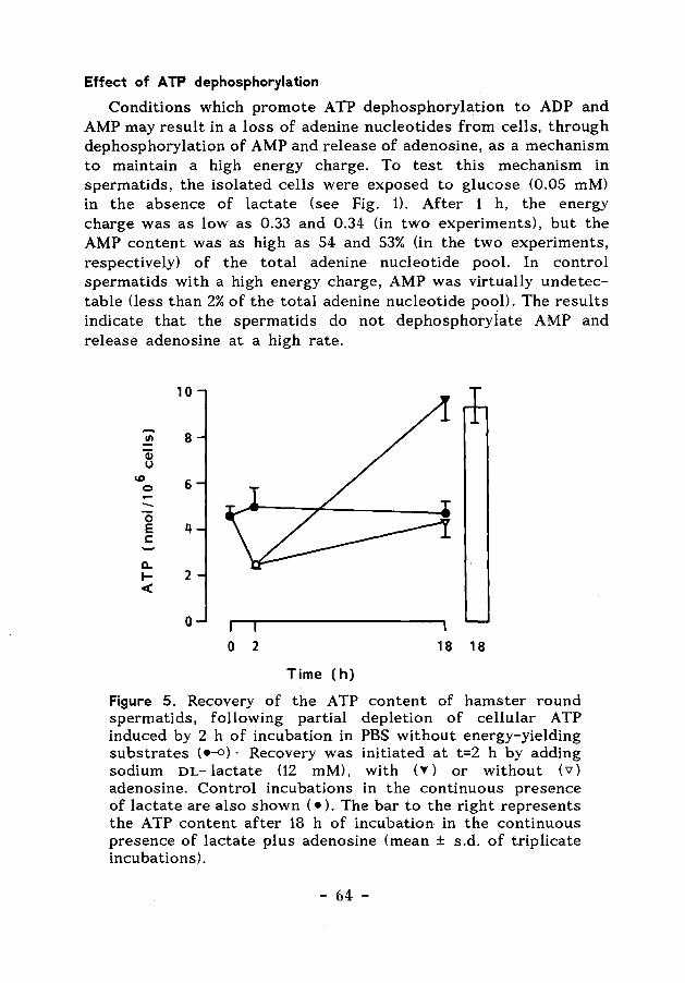
Effect of ATP dephosphorylation
Conditions which promote A TP dephosphorylation to ADP and AMP may result in a loss of adenine nucleotides from cells, through dephosphorylation of AMP and release of adenosine, as a mechanism to maintain a high energy charge. To test this mechanism in spermatids, the isolated cells were exposed to glucose (0.05 mM) in the absence of lactate (see Fig. 1). After 1 h, the energy charge was as low as 0.33 and 0.34 (in two experiments), but the AMP content was as high as 54 and 53% On the two experiments, respectively) of the total adenine nucleotide pool. In control spermatids with a high energy charge, AMP was virtually undetectable (less than 2% of the total adenine nucleotide pool). The results indicate that the spermatids do not dephosphorylate AMP and release adenosine at a high rate.
10
-Cll 8 Q) u
\D 6 0
0 E q c
ll. ..... 2 <
0
0 2 18 18
Time (h)
Figure 5. Recovery of the ATP content of hamster round spermatids, following partial depletion of cellular ATP induced by 2 h of incubation in PBS without energy-yielding substrates (e-o)· Recovery was initiated at t=2 h by adding sodium DL-lactate (12 mM), with ('Y) or without ('V) adenosine. Control incubations in the continuous presence of lactate are also shown ( • ). The bar to the right represents the ATP content after 18 h of incubation in the continuous presence of lactate plus adenosine (mean ± s.d. of triplicate incubations).
- 64 -
A moderate ATP dephosphorylation was induced by incubation of the spermatids in the absence of extracellular energy-yielding substrates for 2 h, which resulted in a SO% reduction of the cellular ATP content (Fig. 5). After addition of lactate, full recovery of the cellular ATP content was observed. The ATP content returned to approximately 4 and 8 nmol/106 cells in the absence and presence of adenosine, respectively (Fig. 5).
Effect of inhibitors
Adenosine is transported into cells by facilitated diffusion (Fox & Kelley, 1987), and dipyridamole has been frequently used as a selective inhibitor of nucleoside transport (Meghji et al., 1985). In the present experiments, the stimulatory effect of adenosine on the ATP content of the isolated spermatids was inhibited by dipyridamole (Table 3).
Table 3. Inhibition of the effect of adenosine on the ATP content of spermatids by dipyridamole and S- iodotubercidin.
Inhibitor (S l1 M)
None Dipyridamole 5-Iodotubercidin
ATP (nmol/106 cells) - Adenosine
4.3 ± 0.3 4.3 ± 0.2 4.7 ± 0.1
+ Adenosine
8.4 ± 0.3 5.0 ± 0.4 5.6 ± 0.1
A TP was estimated after 18 h of incubation in PBS-L, with or without 0.1 mM adenosine, in the presence of an inhibitor of purine transport (dipyridamole) or adenosine kinase (5-iodotubercidin), as indicated in the Table (mean± s.d. of triplicate incubations).
In cells, the utilization of adenosine is dependent upon the activity of adenosine kinase. It was observed that the isolated spermatids contained 0.04 U/mg protein of adenosine kinase activity, estimated as described in Materials and Methods. Furthermore, the adenosine kinase inhibitor 5-iodotubercidin (Achterberg et al., 1986) counteracted the adenosine-induced elevation of the cellular A TP content (Table 3). There was no effect '
- 65 -
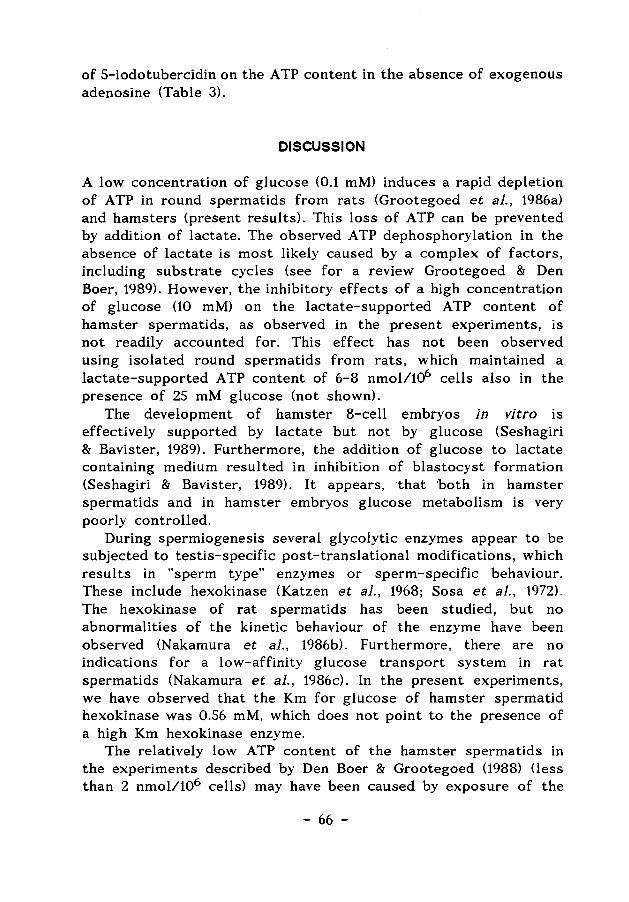
of 5-iodotubercidin on the A TP content in the absence of exogenous adenosine (Table 3).
DISCUSSION
A low concentration of glucose (0.1 mM) induces a rapid depletion of ATP in round spermatids from rats (Grootegoed et al., 1986a) and hamsters (present results). This loss of ATP can be prevented by addition of lactate. The observed ATP dephosphorylation in the absence of lactate is most likely caused by a complex of factors, including substrate cycles (see for a review Grootegoed & Den Boer, 1989). However, the inhibitory effects of a high concentration of glucose (10 mM) on the lactate-supported A TP content of hamster spermatids, as observed in the present experiments, is not readily accounted for. This effect has not been observed using isolated round spermatids from rats, which maintained a lactate-supported A TP content of 6-8 nmoll1<f> cells also in the presence of 25 mM glucose (not shown).
The development of hamster 8-cell embryos in vitro is effectively supported by lactate but not by glucose (Seshagiri & Bavister, 1989). Furthermore, the addition of glucose to lactate containing medium resulted in inhibition of blastocyst formation (Seshagiri & Bavister, 1989). It appears, that ·both in hamster spermatids and in hamster embryos glucose metabolism is very poorly controlled.
During spermiogenesis several glycolytic enzymes appear to be subjected to testis-specific post-translational modifications, which results in "sperm type" enzymes or sperm-specific behaviour. These include hexokinase (Katzen et al., 1968; Sosa et al., 1972). The hexokinase of rat spermatids has been studied, but no abnormalities of the kinetic behaviour of the enzyme have been observed (Nakamura et al., 1986b). Furthermore, there are no indications for a low-affinity glucose transport system in rat spermatids (Nakamura et al., 1986c). In the present experiments, we have observed that the Km for glucose of hamster spermatid hexokinase was 0.56 mM, which does not point to the presence of a high Km hexokinase enzyme.
The relatively low ATP content of the hamster spermatids in the experiments described by Den Boer & Grootegoed (1988) Oess than 2 nmol/106 cells) may have been caused by exposure of the
- 66 -
cells in those experiments to 5.6 mM o-glucose. The glucose concentration in the spermatogenic environment is likely to be very low (Setchell et al., 1969), so that the value of 4 nmol/106
cells, observed in the present experiments may reflect the A TP content of the spermatids in situ.
The present results indicate that uptake and phosphorylation of adenosine are involved in the effect of exogenous adenosine on the A TP content of the spermatids. Furthermore, this effect of a?enosine was observed at relatively high concentrations of the nucleoside. It appears, therefore, that the present action of adenosine is not connected with the interaction of adenosine with adenosine receptors (Fain & Malbon, 1979). An association of adenosine receptors with spermatocytes has been reported (Murphy et al., 1983), but other authors have not detected expression of adenosine receptors on spermatocytes and spermatids (Monaco & Conti, 1986).
Adenosine is probably used by the hamster spermatids as a substrate for adenosine kinase, which leads to an enlargement of the adenine nucleotide pool. No effect of the purine bases hypoxanthine and adenine on the ATP content of the isolated hamster spermatids was observed. Presumably this is not caused by a deficiency of the purine salvage reactions in spermatids, since incorporation of radiolabeled hypoxanthine and adenine into RNA in isolated rat spermatids has been reported (Grootegoed et al., 1986b).
Adenosine kinase is an important enzyme in the maintenance of intracellular ATP levels, and inhibition of this enzyme leads to a decrease of the intracellular A TP content in different cell types (Fox & Kelley, 1987). In hamster spermatids, inhibition of adenosine kinase by 5-iodotubercidin did not result in ATP depletion, during 18 h of incubation in the absence of exogenous adenosine. This indicates that in isolated spermatids AMP is not broken down to adenosine, or only at a very low rate, by the activity of 5'-nucleotidase or non-specific phosphatases. During glucose-induced ATP depletion, the AMP content of the cells was very high, which also indicates a low rate of conversion of AMP to adenosine. From this, it can be suggested that the total adenine nucleotide pool of spermatids is not subject to pronounced fluctuations. In agreement with this, it was observed that a moderate energy crisis (50% ATP dephosphorylation during 2 h of incubation in the absence of extracellular energy-yielding substrates) was followed
- 67 -
by a full recovery of the cellular ATP content. This recovery occurred within 2 h (not shown) and was possibly effected mainly through phosphorylation of endogenous AMP and ADP, rather than through de novo purine synthesis, phosphorylation of adenosine, and/or the purine salvage pathway. It is suggested that at a high energy charge as maintained in the absence of glucose, the ATP content of the spermatids and the size of the total cellular adenine nucleotide pool are stable.
With respect to the effect of adenosine, the possible role of Sertoli cells is uncertain. It has been shown that the uptake of hypoxanthine and adenosine by cumulus cell-enclosed mouse oocytes is far better than the uptake by denuded oocytes (Downs et al., 1986). Furthermore, purine metabolism may play a critical role in the maintenance of mouse oocytes in meiotic arrest (Downs et al., 1985; Downs & Eppig, 1987). There is no motive to suggest comparable regulatory actions of Sertoli cells through modulation of purine metabolism in spermatids. It seems possible, however, that adenosine from Sertoli cells supports a stable level of adenin~ nucleotides in spermatids.
Acknowledgement
This investigation received financial support from the World Health Organization Special Programme of Research, Development and Research Training in Human Reproduction (project 87079). We thank K. van Loon, T. Tumkaya, A.S. Nieukoop and E. Keijzer for their contribution to this work.
REFERENCES
Achterberg, P.W., Stroeve, R.J., & de Jong, J.W. (1986). Myocardial adenosine cyclic rates during normoxia and under conditions of stimulated purine release. Biochem. ]. 235, 13-17.
Bosca, L., Aragon, J.J., & Sols, A. (1985). Modulation of muscle phosphofructokinase at physiological concentrations of enzyme. ]. Bioi. Chern. 260, 2100-2107.
Chou, A.C., & Wilson, J.E. (1975). Hexokinase in rat brain. Methods in Enzymology, Vol. 42 part c, pp. 20-25.
De Jong, J.W. (1977). Partial purification and properties of ratheart adenosine kinase. Archs Int. Physiol. Biochim. 85, 557-569.
- 68 -
Den Boer, P.J., & Grootegoed, J.A. (1988). Mechanism of action of (-)gossypol on ATP production in isolated hamster spermatids. ]. Reprod. Pert. 83, 693-700.
Downs, S.M., Coleman, D.L., Ward-Bailey, P.F., & Eppig, J.J. (1985). Hypoxanthine is the principal inhibitor of murine oocyte maturation in a low molecular weight fraction of porcine follicular fluid. Proc. Natl. Acad. Sci. USA. 82, 454- 458.
Downs, S.M., Coleman, D.L., & Eppig, J.J. (1986). Maintenance of murine oocyte meiotic arrest: uptake and metabolism of hypoxanthine and adenosine by cumulus cell-enclosed and denuded oocytes. Devl. Bioi. 117, 174-183.
Downs, S.M., & Eppig, J.J. (1987). Induction of mouse oocyte maturation in vivo by perturbants of purine metabolism. Bioi. Reprod. 36:431-437.
Dulbecco, R., & Vogt, M. (1954). Plaque formation and isolation of pure lines with poliomyelitis viruses. ]. Exp. Medicine 99, 167-182.
Fain, J.N., & Malbon, C.C. (1979). Regulation of adenylate cyclase by adenosine. Mol. Cell. Biochem. 25, 143-169.
Ford, W.C.L., & Harrison, A. (1987). Futile substrate cycles in the glycolytic pathway of boar and rat spermatozoa and the effect of a:- chlorohydrin. ]. Reprod. Pert. 79, 21-32.
Fox, I.H., & Kelley, W.N. (1987). The role of adenosine and 2"deoxyadenosine in mammalian cells. Ann. Rev. Biochem. 47, 655-686.
Grootegoed, J.A., Jansen, R., & van der Molen, H.J. (1984). The role of glucose, pyruvate and lactate in ATP production by rat spermatocytes and spermatids. Biochim. Biophys. Acta 767, 248-256.
Grootegoed, J.A., Jansen, R., & van der Molen, H.J. (1986a). Effect of glucose on ATP dephosphorylation in rat spermatids. ]. Reprod. Pert. 77, 99-107.
Grootegoed, J.A., Oonk, R.B., Jansen, R., & van der Molen, H.J. (1986b). Extracellular factors that contribute to the development of spermatogenic cells. In: Molecular and Cellular Endocrinology of the Testis. pp. 215-225. Eds. M. Stefanini, M. Conti, R. Geremia and E. Ziparo. Elsevier Science Publishers: Amsterdam.
Grootegoed, J.A., & Den Boer, P.J. (1989). Energy metabolism in spermatids: A review. In: Cellular and Molecular Events in Spermiogenesis as Targets for Fertility Regulation. Eds G.M.H. Waites and D.W. Hamilton. Cambridge University Press, in the
- 69 -
press. Katzen, H.M., & Schimke, R.T. (1965). Multiple forms of hexokinase
in the rat: tissue distribution, age dependency, and properties. Proc. Natl. Acad. Sci. 54, 1218-1225.
Katzen, H.M., Soderman, D.O., & Cirillo, V.J. (1968). Tissue distribution and physiological significance of multiple forms of hexokinase. Ann. N. Y. Acad. Sci. 151, 351-358.
Lund, P., Cornell, N.W., & Krebs, H.A. (1975). Effect of adenosine on the adenine nucleotide content and metabolism of hepatocytes. Biochem. ]. 152, 593-599.
Meghji, P., Holmquist, C.A., & Newby, A.C. (1985). Adenosine formation and release from neonatal heart cells in culture. Biochem. ]. 229, 799-805.
Monaco, L., & Conti, M. (1986). Localization of adenosine receptors in rat testicular cells. Bioi. Reprod. 35, 258-266.
Murphy, K.M.M., Goodman, R.R., & Snyder, S.H. (1983). Adenosine receptor localization in rat testes: biochemical and autoradiographic evidence for association with spermatocytes. Endocrinology 113, 1299-1305.
Nakamura, M., Okinaga, S., & Arai, K. (1986a). Studies of metabolism of round spermatids: glucose as unfavourable substrate. Bioi. Reprod. 35, 927-935.
Nakamura, M., Okinaga, S., & Arai, K. (1986b). Kinetic properties of hexokinase of germ cells in rat testis. Andrologia 18, 635-638.
Nakamura, M., Yamaguchi, K., Suzuki, A., Okinaga, S., & Arai, K. (1986c). Metabolism of round spermatids: a possible regulation of hexose transport. Develop. Growth Differ. 28, 499-504.
Setchell. B.P., Scott, T.W., Voglmayer, J.K., & Waites, G.H.M. (1969). Characteristics of testicular spermatozoa and the fluid which transports them into the epididymis. Bioi. Reprod. 1, 40-66.
Seshagiri, P.B., & Bavister, B.D. (1989). Glucose inhibits development of hamster 8-cell embryos in vitro. Bioi. Reprod. 40, 590-606.
Sosa, A., Altamirano, E., Hernandez, P., & Rosado, A. (1972). Developmental pattern of rat testis hexokinase. Life Sci. 11, 499-510.
- 70 -
Chapter 5
Glutathione-dependent defence mechanisms in isolated round spermatids from the rat
PJ. Den Boer, M. Poot, A. Verkerk, R. Jansen, P. Mackenbach and J.A. Grootegoed
International Journal of Andrology (1989), in the press
- 71 -
Glutathione-dependent defence mechanisms in isolated round spermatids from the rat
Summary. The different mechanisms for glutathione-dependent inactivation of a number of oxidizing compounds and other xenobiotics were studied using isolated round spermatids from rats. For the estimation of cellular GSH a flow cytometric assay was used. The cells were exposed to the oxidizing compounds cumene hydroperoxide and diamide, to study the activity of the GSH redox cycle. Incubation of the isolated cells with these compounds showed that the cells had a limited capacity to withstand the oxidative stress associated with their inactivation. The GSH level of the spermatids was maintained during 18 h of incubation in the presence of low concentrations cumene hydroperoxide and diamide, whereas spermatids exposed to higher concentrations showed a loss of both GSH and ATP. No partial loss of GSH from individual cells was observed. Diethyl maleate and 1,2-epoxy-p-(nitrophenoxy)propane (ENPP) were used to study the effect of glutathione $-transferase catalyzed GSH ·conjugation on the GSH content of the spermatids. Exposure of the cells to low concentrations of diethyl maleate and ENPP resulted in a decrease of the GSH content. The flow cytometric analysis showed that this was a partial loss of GSH from all cells, rather than GSH depletion in a part of the cell population. This diminution of the cellular GSH pool, however, did not affect the ATP content and viability of the cells. The present results indicate that spermatids can perform GSH-dependent defence mechanisms against a number of model compounds.
Key words: xenobiotics; peroxides; glutathione; spermatids; Sertoli cells.
INTRODUCTION
The tripeptide glutathione (GSH) is the most abundant non-protein thiol in mammalian cells. The GSH content of various organs and tissues represents at least 90% of the total non-protein low molecular weight thiols (Reed & Farris, 1984). High GSH concencentrations are present in testicular tissue, especially after the onset
- 72 -
of spermatogenesis (Calvin & Turner, 1982). In agreement with this, a high GSH content has been observed in isolated spermatogenic cells from mice (Grosshans & Calvin, 1985) and hamsters (Den Boer et al., 1989), whereas a relatively low GSH content was found in Sertoli cells from immature hamsters (Den Boer et al., 1989).
GSH is involved in many biological processes, including protection of cells through various GSH-dependent mechanisms (Meister & Anderson, 1983). Important aspects of this protective function of GSH, are the cellular defence against oxidative stress and the detoxication of xenobiotics via conjugation (Meister & Anderson, 1983).
The aim of the present study, was to analyse the capacity of GSH-supported defence mechanisms in isolated spermatogenic cells. Isolated round spermatids were exposed to the oxidizing agents cumene hydroperoxide and diamide, while in related studies the effect of GSH conjugating xenobiotics on the isolated germ cells was evaluated.
Antioxidant defence mechanisms are particularly important in oxygen consuming cells, which continuously generate partially reduced oxygen species in the mitochondria or by the action of cytoplasmic oxidases. These oxygen species include hydrogen peroxide and other organic peroxides, which can be detoxicated by reduction with GSH. Concomitantly, GSH is converted to its oxidized form, GSSG, by the selenium-dependent enzyme glutathione peroxidase. The GSSG thus formed is subsequently reduced by the enzyme glutathione reductase, using reducing equivalents from NADPH (GSH redox cycle), or is released from the cells (Meister & Anderson, 1983). Under physiological conditions, nearly all cellular glutathione is present in its reduced form and less than 5 % of the total is present as the disulphide GSSG (Reed & Farris, 1984).
Cumene hydroperoxide can be transformed to the corresponding alcohol, a reaction that is catalyzed by glutathione peroxidase and which activates the GSH redox cycle. Diamide causes nonenzymic oxidation of cellular GSH to GSSG (Plummer et al., 1981). These redox active compounds have been shown to be suitable model compounds to study the GSH redox cycle in different cell types, including cultured human fibroblasts (Poot et al., 1986; 1987).
Detoxification of electrophilic xenobiotics can occur by way of glutathione $-transferase (GST) activity. These transferases are a group of dimeric isoenzymes that catalyse the reaction of GSH
- 73 -
with a variety of substrates (Ketterer, 1986). Testicular tissue contains a high glutathione $-transferase activity, and the major GST isoenzyme present in rat testis is GST 6-6. The cellular localization of this isoenzyme in the testis, however, has not been described. Its activity has been estimated using various compounds. 1-Chloro-2,4-dinitrobenzene appears to be a very good substrate for this transferase reaction (Ketterer, 1986). GST activity is also present in rat spermatogenic cells (Volkova & Lankin, 1984) and spermatozoa (Mukhtar, Lee & Bend, 1978).
In the GST catalysed reaction, GSH is conjugated to xenobiotics that are GST substrates. The conjugates formed are metabolized further to mercapturic acids, S-alkylated derivatives of N-acetylcysteine, that are water soluble and which are released from the cells (Habig et al., 1974). This type of detoxification results in a loss of GSH from cells. In the present experiments the compounds diethyl maleate and 1,2-epoxy-p-(nitrophenoxy)propane (ENPP), were used to lower GSH levels in spermatids. Diethyl maleate reacts with GSH by direct conjugation and also by interaction with the glutathione $-transferase system <Bannai, 1984) and has been used in isolated cells, including human fibroblasts <Bannai, 1984; Meredith & Dodson, 1987), to lower cellular GSH. ENPP is a substrate for glutathione S-transferases in enzyme assays (Ketterer, 1986; Kraus & Kloft, 1980; Habig et al., 197 4), but has also been reported to react with GSH in human fibroblasts (Bannai, 1984).
In the present experiments, the mechanisms used by the isolated spermatids to inactivate the compounds described above, were evaluated using a flow cytometric analysis of the cellular GSH content (Poot et al., 1987), in addition to biochemical assays of the cellular GSH and ATP contents. The flow cytometric method allows detection of GSH in individual cells, thereby providing a useful tool to investigate GSH-dependent defense mechanisms in isolated spermatogenic cells.
MATERIALS AND METHODS
Isolation of spermatocytes and spermatids from rats. Pachytene spermatocytes and round spermatids from rats aged 32-36 days (Wistar, substrain R1-Amsterdam) were isolated by velocity sedimentation at unit gravity (STA-PUT) and further purified by Percoll-gradient centrifugation (Grootegoed et al., 1986a). The
- 74 -
isolated cells were incubated in Dulbecco's phosphate-buffered saline (PBS; Dulbecco & Vogt, 19S4), supplemented with 12 mM sodium DL-lactate and S.6 mM glucose (PBS-GL), at 32°C in air. The PBS-GL also contained 0.4% bovine serum albumin (BSA, fraction V; Sigma Chemical Company, St. Louis, MO, U.S.A.) and antibiotics (Grootegoed et al., 198S). Approximately 0.4 x 10⁶ cells were incubated in a volume of 0.2S ml in polystyrene tubes (diameter 11.S mm).
Sertoli cells. Sertoli cells were isolated from rats aged 21 days which had been irradiated in utero at day 19 of gestation, to obtain germ cell-depleted testis, as described by Grootegoed et al. (1986b).
Flow cytometric method for determination of reduced glutathione . The cellular free thiol content of isolated spermatids was determined
I
by the flow cytometric as1
say described by Poot et al. (1986; 1987). In this assay monobromobimane (Thiolyte; Calbiochem, San Diego, U.S.A.) was used as a thibl- staining agent. After the incubations, the spermatids were waslled two times in PBS containing 12 mM sodium DL-lactate (PBS-C) by centrifugation (S min at 600 g) to remove the BSA from the medium, and the cells were then resuspended in 0.9 ml PBS-L. Three samples were incubated for 1 h in the presence of 200 [lM N-ethyl maleimide (Sigma), as a
I
control for spurious stainl,ing (Poot et al., 1987). Finally, a volume of 100 (ll monobromobimane (stock solution of SO mM in acetonitrile) was added to the cell sJspensions (final concentration SO [lM). The cells were incubated I with monobromobimane for 10 min at
I
37°C, and subsequently ~ept at 0°C. Propidium iodide (PI) was added to the cell suspensions shortly before the flow cytometric analysis (final concentrati~n 5 [lg/ml). Only those spermatids which excluded PI were analysed,, and are referred to as intact cells. From each sample, 10000 cellsl were analysed. The fluorescence was recorded with a FACS !II (Becton and Dickinson, Sunnyvale, U.S.A.). The average relative fluorescence intensity (RFI) of the cell population was calculated,l and is also referred to as GSH content. However, RFI is only a ~easure of GSH content when the GSH
I
content of the cells is relatively high compared with the amount of free sulfhydryl groups ; in total cellular protein.
I
Fluorometric method fori determination of reduced glutathione fluorometric determinatiorl. of the GSH content of spermatids
I
I - 7S -
The was
performed according to the method of Hissin & Hilf (1976) and Grosshans & Calvin (1985), but with some slight modifications. The spermatids were collected by centrifugation (5 min at 600 g). The supernatants were discarded and the pellets were frozen and stored at - 80° C. For GSH estimation, the cells were lysed in 100 [.Ll water, and protein was precipitated by adding SO [.Ll 3 M PCA/ 1 mM EDTA. The mixture was kept on ice for 10 min, and the precipitated protein was removed by centrifugation. The supernatant was neutralized by addition of 2 M KOHl 0.3 M HEPES, followed by centrifugation to remove KClO 4 . The final GSH-assay mixture (500 [.LD contained 25 [.Ll of the neutralized supernatant, 450 [.Ll of a sodium-phosphate buffer (0.1 M sodium-phosphate, 5 mM EDTA, pH 8.0) and 25 [.Ll of a solution of o-phthaldialdehyde (Sigma) in methanol (1 mg/ml). After mixing and incubation for 15 min at room temperature, the fluorescence was determined using a fluorescence spectrophotometer (SFM 25, Kontron Instruments, ZUrich, Switzerland) at 420 nm (excitation at 350 nm). In this assay, protein sulphydryl groups were removed. by precipitation. Furthermore, the interference of non-protein sulphydryl groups, other than GSH, in this assay is very low (Hissin & Hilf, 1976). Therefore, the data from this assay approximate the cellular GSH content
Spectrophotometric assay for determination of glutathione $-transferase activity . Whole testes or isolated cells were homogenized in ice-cold sodium phosphate buffer (20 mM, pH 7.4), and the lysates were centrifuged for 15 min at 10 000 g (Kraus, 1983). The supernatant was used for estimation of the enzyme activity, which was performed spectrophotometrically, (Habig & jakoby, 1981; Guthenberg et al., 1985) by measuring the increase in absorbance at 340 nm caused by formation of the conjugate between GSH (Boehringer, Mannheim, F.R.G.) and 1- chloro-2,4-dinitrobenzene (Sigma). The results were expressed as international units (U) of enzyme activity per mg protein or per 106 cells. The amount of protein was estimated ·according to Lowry et al. (1951), using bovine serum albumin as a standard.
Estimation of NADP-dependent enzyme activities. The activities of glucose 6-phosphate dehydrogenase (G6P-DH), 6-phosphogluconate dehydrogenase (6PG-DH), malic enzyme and NADP- dependent isocitrate dehydrogenase, in Sertoli cells and spermatogenic cells,
- 76 -
were estimated as described by Roth (1969). The amount of NADPH formed was estimated using a Perkin
Elmer fluorometer (Norwalk CT, U.S.A.) at 455 nm (excitation at 340 nm). The results were expressed as international units (U) of enzyme activity per mg protein. The amount of cellular protein was estimated using the compound fluorescamine (Udenfriend et al., 1972) and bovine serum albumin as a standard.
Estimation of ATP. The cellular ATP content of the spermatids was estimated using the bioluminescent firefly luciferin-luciferase reaction (Lumit PM from Lumac, Meise, Belgium) as described by Grootegoed et al. (1984), and a model 6100 Pico-Lite Luminometer (Packard Instruments, Downers Grove, 11., U.S.A.)
Estimation of C14CJC02 formation from radio/abe/led glucose. The metabolism of glucose via the pentose phosphate pathway by spermatids was estimated by measuring the amount of [14C JC02 produced from 14C-labelled glucose. Spermatids were incubated in the presence of 0.5 [!Ci o-[t-14CJglucose or 1.0 [!Ci o-[6-14CJglucose (2.06 GBq/mmol; Amersham International, Amersham, U.K.) in PBS containing 5.6 mM_ glucose, 1 mM pyruvate and 12 mM sodium DL-lactate. After 2 or 18 hours of incubation, the amount of [ 14 CJC02 produced was estimated essentially as described by Grootegoed et al. (1984).
RESULTS
Effects of cumene hydroperoxide and diamide on A TP and GSH levels of isolated spermatids
Cumene hydroperoxide was used as a model compound to test the GSH redox cycle. The relative fluorescence intensity, which was taken as a measure of cellular GSH content, and the ATP content of spermatids were estimated after incubation of the isolated cells for 18 h in the presence of different concentrations of cumene hydroperoxide. In this series of experiments the total amount of ATP was expressed as nmol/106 intact cells (i.e. those cells which excluded propidium iodide). The results show that, up to a cumene hydroperoxide concentration of 20 [!M, the GSH and ATP contents of the cells were unaffected, whereas at 40 [!M
- 77 -
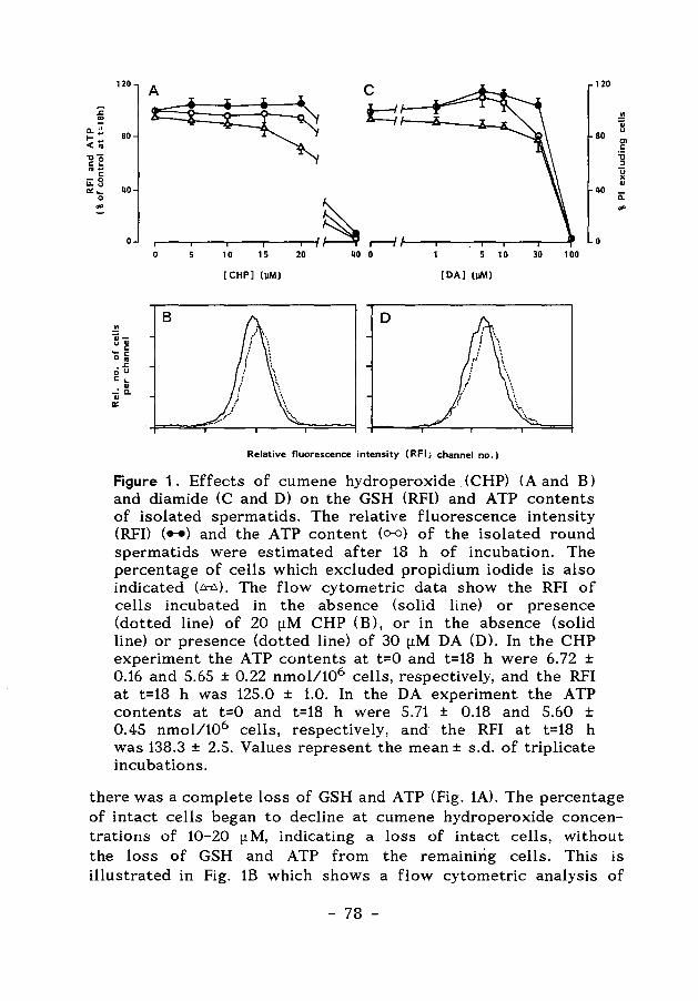
120 120
.!! ]
80 80 en
40
c "C :l
~ .. 40 a:
0 0
.!! Qi" ~ '-c 0"'
o-5 c ... ..:X. .. a::
0
B
10 15 20 40 0 5 10 30 100
[CHP] (pM) [DA] (pM)
D
Relative fluorescence intensity ( RFI; channel no.)
Figure 1. Effects of cumene hydroperoxide (CHP) (A and B) and diamide (C and D) on the GSH (RFD and ATP contents of isolated spermatids. The relative fluorescence intensity (RFI) (.-.) and the ATP content (o-o) of the isolated round spermatids were estimated after 18 h of incubation. The percentage of cells which excluded propidium iodide is also indicated (~). The flow cytometric data show the RFI of cells incubated in the absence (solid line) or presence (dotted line) of 20 [!M CHP (B), or in the absence (solid line) or presence (dotted line) of 30 [!M DA (D). In the CHP experiment the ATP contents at t=O and t=18 h were 6. 72 ± 0.16 and 5.65 ± 0.22 nmol/106 cells, respectively, and the RFI at t=18 h was 125.0 ± 1.0. In the DA experiment the ATP contents at t=O and t=18 h were 5.71 ± 0.18 and 5.60 ± 0.45 nmol/106 cells, respectively, and the RFI at t=18 h was 138.3 ± 2.5. Values represent the mean± s.d. of triplicate incubations.
there was a complete loss of GSH and ATP (Fig. 1A). The percentage of intact cells began to decline at cumene hydroperoxide concentrations of 10-20 [lM, indicating a loss of intact cells, without the loss of GSH and ATP from the remaining cells. This is illustrated in Fig. 1B which shows a flow cytometric analysis of
- 78 -
spermatids incubated for 18 h in the presence of 20 (1M cumene hydroperoxide. The RFI's of control cells and cumene hydroperoxidetreated cells were virtually the same.
Diamide was also used to study the capacity of spermatids to defend themselves against oxidative stress via the GSH redox cycle. The RFI (GSH) and ATP contents of spermatids, estimated after 18 h of incubation in the presence of different concentrations of diamide, showed comparable effects to those observed during exposure of the cells to cumene hydro peroxide. The GSH and A TP content of intact cells was maintained in the presence of low concentrations of diamide, up to 30 (1M (Fig. 1C and 1D), whereas incubation of the spermatids in the presence of higher concentrations of diamide resulted in a complete loss of cell viability.
The present results indicate that spermatids, incubated for 18 h in the presence of different concentrations of oxidizing compounds that are inactivated via the GSH redox cycle, can withstand the oxidative stress up to a certain threshold level.
_Activities of NAOP-dependent enzymes in Sertoli cells and spermatogenic cells.
The reduction of GSSG to GSH via the GSH redox cycle is dependent on the supply of reduced co-enzyme NADPH. NADPH is produced via the reactions catalysed by the enzymes glucose 6-phosphate dehydrogenase (G6P-DH) and 6-phosphogluconate dehydrogenase (6PG-DH) of the pentose phosphate pathway.
The activity of G6P-DH in spermatocytes and spermatids was approximately 10-fold lower than in Sertoli cells (Fig. 2). In contrast, the activity of 6PG-DH was comparable in spermatogenic cells and Sertoli cells. Other possible sources for the production of reduced co-enzyme NADPH are the reactions catalyzed by NADP-dependent isocitrate dehydrogenase and malic enzyme. There were no marked differences between the activities of these enzymes in Sertoli cells and the spermatogenic cell types (Table 1).
Activation of the pentose phosphate pathway in spermatids.
Under the present incubation conditions the ratio of [ 14CJC02 produced from [1-14 CJglucose and [6-14CJglucose was less than 0.1, indicating that the [14CJC02 was largely produced via the
- 79 -
---------------
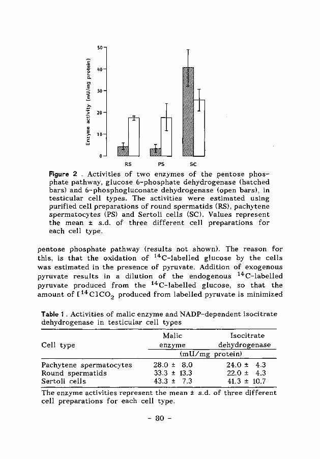
so
c 'iii
40 ... 0 ... c. Cl
~ 30 ::::> E
?: > 20
:;:;
JJ u .. cu E
10 >-N c w
0
RS PS sc Figure 2 . Activities of two enzymes of the pentose phosphate pathway, glucose 6-phosphate dehydrogenase (hatched bars) and 6-phosphogluconate dehydrogenase (open bars), in testicular cell types. The activities were estimated using purified cell preparations of round spermatids (RS), pachytene spermatocytes (PS) and Sertoli cells (SC). Values represent the mean ± s.d. of three different cell preparations for each cell type.
pentose phosphate pathway (results not shown). The reason for this, is that the oxidation of 14C-labelled glucose by the cells was estimated in the presence of pyruvate. Addition of exogenous pyruvate results in a dilution of the endogenous 14C-labelled pyruvate produced from the 14C-labelled glucose, so that the amount of [ 14 CJ C02 produced from labelled pyruvate is minimized
Table 1 . Activities of malic enzyme and NADP-dependent isocitrate dehydrogenase in testicular cell types
Cell type
Pachytene spermatocytes Round spermatids Sertoli cells
Malic enzyme
(mU/mg
28.0 ± 8.0 33.3 ± 13.3 43.3 ± 7.3
Isocitrate dehydrogenase
protein)
24.0 ± 4.3 22.0 ± 4.3 41.3 ± 10.7
The enzyme activities represent the mean ± s.d. of three different cell preparations for each cell type.
- 80-
and cannot interfere with the amount of [ 14CJC02 produced via the pentose phosphate pathway. This method to estimate pentose phosphate pathway activity was applied to spermatids, because these cells produce ATP through mitochondrial oxidation of exogenous pyruvate (Grootegoed et al., 1984).
When the spermatids were incubated in the presence of 10 t.tM cumene hydroperoxide for an incubation period of 18 h, the activity of the pentose phosphate pathway was increased 1.6 fold (Table 2). However, 20 t.tM diethyl maleate did not activate the pentose phosphate pathway in the spermatids during 18 h of incubation, although the GSH content was decreased to 34 % of controls (Table 2).
The results indicate that the pentose phosphate pathway in spermatids is active in the production of NADPH for the GSH redox cycle.
Table 2 . Effects of cumene hydroperoxide (CHP) and diethyl maleate (DEM) on the activity of the pentose phosphate pathway in isolated spermatids.
Incubation C02 GSH ATP condition ( nmol/106 cells) (nmol/106 cells) (nmol/106 cells)
Control 1.37 ± 0.14 3.76 ± 0.13 4.94 ± 0.23 CHP (10 t.tM) 2.22 ± 0.11 * 3.70 ± 0.15 4.04 ± 0.15 VEM (20 t.tM) 1.63 ± 0.19 1.26 ± 0.17 * 4.94 ± 0.34
* Significantly different from control (Student's t-test, p<0.005) Isolated round spermatids were incubated for 18 h in the absence or presence of CHP or DEM. The GSH and ATP contents were estimated at the end of the incubations, whereas C02 represents the total production of [14 CJ C02 from 14C-labelled glucose during the 18 h incubation period. The GSH and ATP contents at the start of the incubations were 4.46 ± 0.41 nmol/106 cells and 5.47 ± 0.16 nmol/106 cells, respectively. Values represent the mean ± s.d. of triplicate incubations.
Glutathione-S-transferase activity in total testis and isolated spermatogenic cells from rats
The results shown in Table 3 indicate that spermatocytes and spermatids contain a considerable level of GST activity, although
- 81 -
the specific enzyme activity (mli/mg protein) was somewhat lower in spermatids than in spermatocytes. The specific GST activity (mli/mg protein) of total testicular tissue from rats aged 36 days (used for isolation of the spermatogenic cells) was 344 ± 3, which is higher than the activity in advanced spermatogenic cells. This indicates that most of the testicular GST activity is present in the early spermatogenic cells and/or in the somatic cell types.
Table 3 . Glutathione S-transferase activity in pachytene spermatocytes and round spermatids
Cell type
Sperma tocytes Spermatids
Glutathione S-transferase activity mU/mg protein mU/106 cells
69.7 ± 2.4 37.5 ± 0.7
23.30 ± 0.80 4.74 ± 0.09
GST activities were estimated using 1-chloro-2,4-dinitrobenzene as substrate. Values represent the mean ± s.d. of triplicate measurements.
The presence of the enzyme GST in the spermatogenic cells indicates that it should be possible to lower the GSH content of isolated spermatids by incubation of the cells in the presence of GST substrates. This would allow a study on the role of GSH and the biosynthesis de novo of GSH in the spermatogenic cells.
Effects of diethyl maleate and 1,2-epoxy-3-(p-nitrophenoxy) propane on GSH levels of round spermatids
It was assessed whether incubation of isolated spermatids in the presence of GST substrates resulted in a loss of cellular GSH. The relative fluorescence intensity (GSH) and the ATP content of spermatids were estimated after incubation for 18 h in the presence of different concentrations of diethyl maleate. The results, presented in Fig. 3A showed that the GSH content of the cells was decreased in the presence of 10 [lM diethyl maleate, whereas the ATP content of the spermatids was fully maintained up to 100 [lM diethyl maleate. The percentage of intact cells also remained high in the presence of 100 [.LM diethyl maleate. These results indicate that GSH is lost from the cells, but that the cells remain intact. This is illustrated in Fig. 3B, which shows a flow cytometric analysis
- 82 -
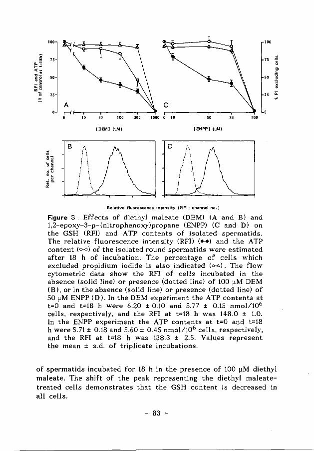
:;: "' a..";i
1--<-.. "tl-"0 '"z -I: 11.0
a:~ 0
~
100
75
50
25
.!! Oi-u.,
" ... " 0 ..
o-5 1: L
-=~ ., a:
~
A r-{
B
10
(\ i \
I \ ! \
I \. I •
30 100 300 1000 0 10
[OEM] (JIM)
D
50
[ENPP] (JIM)
:-. : \ ! ~
/ \ f \ I •
; \ ; ~ j \
/
Relative fluorescence intensity (RFI; channel no.)
75 100
Figure 3. Effects of diethyl maleate (DEM) (A and B) and 1,2-epoxy-3-p-(nitrophenoxy)propane (ENPP) (C and D) on the GSH (RFI) and ATP contents of isolated spermatids. The relative fluorescence intensity (RFI) ( ... ) and the ATP content (o-o) of the isolated round spermatids were estimated after 18 h of incubation. The percentage of cells which excluded propidium iodide is also indicated (~). The flow cytometric data show the RFI of cells incubated in the absence (solid line) or presence (dotted line) of 100 [.LM DEM (B), or in the absence (solid line) or presence (dotted line) of SO [.LM ENPP (D). In the DEM experiment the A TP contents at t=O and t=18 h were 6.20 ± 0.10 and 5.77 ± 0.15 nmol/106
cells, respectively, and the RFI at t=18 h was 148.0 ± 1.0. In the ENPP experiment the ATP contents at t=O and t=18 h were 5.71 ± 0.18 and 5.60 ± 0.45 nmol/106 cells, respectively, and the RFI at t=18 h was 138.3 ± 2.5. Values represent the mean ± s.d. of triplicate incubations.
100
75 ~ ., u
Ill
" 50 '5
" u )( .,
25 ii: .,
of spermatids incubated for 18 h in the presence of 100 [.LM diethyl maleate. The shift of the peak representing the diethyl maleatetreated cells demonstrates that the GSH content is decreased in all cells.
- 83 -
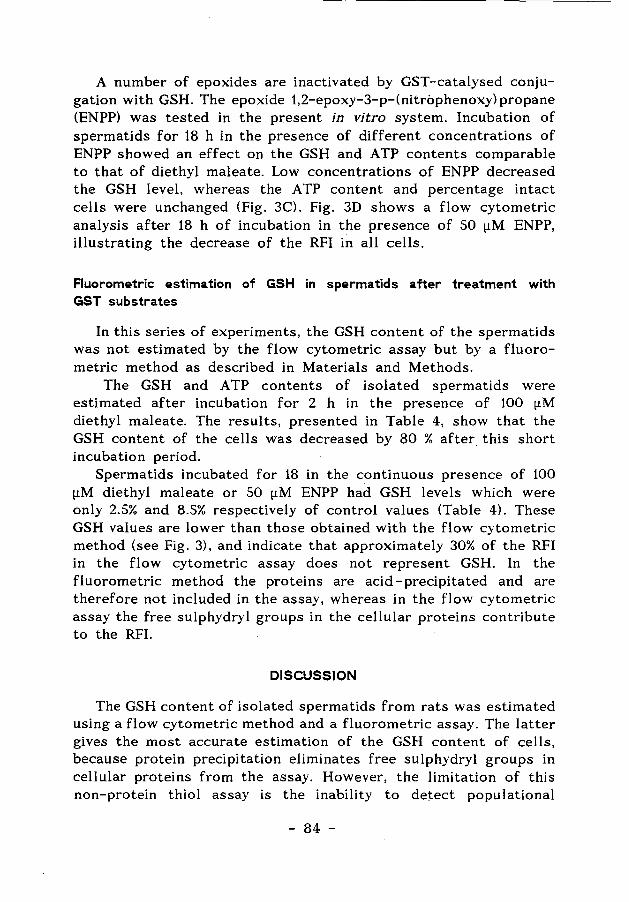
A number of epoxides are inactivated by GST-catalysed conjugation with GSH. The epoxide 1,2-epoxy-3-p-(nitrophenoxy) propane (ENPP) was tested in the present in vitro system. Incubation of spermatids for 18 h in the presence of different concentrations of ENPP showed an effect on the GSH and ATP contents comparable to that of diethyl maleate. Low concentrations of ENPP decreased the GSH level, whereas the ATP content and percentage intact cells were unchanged (Fig. 3C). Fig. 3D shows a flow cytometric analysis after 18 h of incubation in the presence of SO [!M ENPP, illustrating the decrease of the RFI in all cells.
Fluorometric estimation of GSH in spermatids after treatment with GST substrates
In this series of experiments, the GSH content of the spermatids was not estimated by the flow cytometric assay but by a fluorometric method as described in Materials and Methods.
The GSH and A TP contents of isolated spermatids were estimated after incubation for 2 h in the presence of 100 [!M diethyl maleate. The results, presented in Table 4, show that the GSH content of the cells was decreased by 80 % after this short incubation period.
Spermatids incubated for 18 in the continuous presence of 100 [!M diethyl maleate or SO [!M ENPP had GSH levels which were only 2.S% and 8.5% respectively of control values (Table 4). These GSH values are lower than those obtained with the flow cytometric method (see Fig. 3), and indicate that approximately 30% of the RFI in the flow cytometric assay does not represent GSH. In the fluorometric method the proteins are acid -precipitated and are therefore not included in the assay, whereas in the flow cytometric assay the free sulphydryl groups in the cellular proteins contribute to the RFI.
DISCUSSION
The GSH content of isolated spermatids from rats was estimated using a flow cytometric method and a fluorometric assay. The latter gives the most accurate estimation of the GSH content of cells, because protein precipitation eliminates free sulphydryl groups in cellular proteins from the assay. However, the limitation of this non-protein thiol assay is the inability to detect populational
- 84 -
Table 4. Effects of diethyl maleate <DEM) and 1,2-epoxy-3-p-(nitro phenoxy) propane (ENPP) on ATP and GSH content of spermatids.
Time Treatment ATP GSH (h) (nmol/106 cells) (nmol/106 cells)
0 Control 4.73 ± 0.53 2.79 ± 0.05 2 Control 4.62 ± 0.36 3.08 ± 0.14 2 DEM 4.81 ± 0.31 0.63 ± 0.03
18 Control 3.95 ± 0.25 2.48 ± 0.03 18 DEM 3.63 ± 0.48 0.06 ± 0.03 18 ENPP 3.21 ± 0.08 0.21 ± 0.02
Isolated round spermatids were incubated for 2 or 18 h in the absence or presence of 100 [lM DEM or SO [lM ENPP. Values represent the mean ± s.d. of triplicate incubations.
heterogeneity in the cell population (Rice et al. 1986). Therefore, we have also used the flow cytometric assay described by Poot et al. (1986, 1987), which gives information on the GSH content of individual cells.
To study the activity of the GSH redox cycle in the defence of spermatids against oxidative stress, the isolated cells were exposed to the oxidizing agents cumene hydroperoxide and diamide. The results indicate that round spermatids have a limited capacity to hactivate cumene hydroperoxide and diamide, as shown by the maintenance of their cellular GSH and ATP contents at low concentrations of the compounds. However, when the spermatids were exposed to somewhat higher concentrations of cumene hydroperoxide (40 [1M) and diamide (100 [1M), the capacity of this defence mechanism was unable to prevent cell degeneration, as indicated by the loss of cellular GSH and ATP and the decrease in percentage of intact cells. In contrast, cultured human fibroblasts (0.1 x 10⁶ cells in 0.5 ml) can withstand a concentration of 40 [.!M cumene hydroperoxide for 7 days (Poot et al., 1987).
The resistance of the spermatids to the effect of oxidizing agents, is probably limited by the rate of reduction of the GSSG in the GSH redox cycle. In this cycle, the prodl).ction of reduced co-enzyme NADPH via the pentose phosphate pathway might be rate-limiting. The present results indicate that the pentose phosphate pathway in spermatids was activated slightly during exposure of the cells to cumene hydroperoxide, as a consequence
- 85 -
of activation of the GSH redox cycle, but not during exposure to diethyl maleate. In spermatids, the activity of the first enzyme of the pentose phosphate pathway, glucose 6-phosphate dehydrogenase (G6P-DH), was found to be low as compared with the activity of 6-phosphogluconate dehydrogenase (6PG-DH). The first is encoded by a gene located on the X-chromosome, whereas 6PG-DH is encoded autosomally. The X-chromosome is inactivated early in spermatogenesis (Monesi, 1971), and it can be postulated, therefore, that G6P-DH may be a rate-limiting enzyme in the production of NADPH in spermatids. Furthermore, it can be questioned whether the spermatids in situ, in the seminiferous epithelium are exposed to a significant concentration of glucose (Grootegoed & Den Boer, 1989).
Another possible source for reduced co-enzyme NADPH in spermatids, to be used for the reduction of GSSG by the enzyme glutathione reductase, might be the reactions catalysed by the enzymes NADP-dependent isocitrate dehydrogenase and malic enzyme. It was observed that the isolated germ cells · contain these enzymes, but it has not been studied whether the supply of substrates for these enzymes in spermatids allows a sufficient rate of NADPH production.
Spermatogenic cells in situ probably are protected by Sertoli cells. The Sertoli cells have a large reserve capacity of the pentose phosphate pathway (Grootegoed et al., 1986b), and may serve as a buffer to inactivate, for the greater part, the peroxides which are generated in the seminiferous epithelium.
In the next step of this study the conjugation between GSH and electrophilic compounds with GSH via the glutathione Stransferase (GST) reaction was studied. It was found that GST activity is present in spermatids, although its activity in germ cells is low as compared with that in whole testis tissue. The specific activity of GST in total testis tissue is very high when compared to a number of other tissues, including the liver (Kraus & Kloft, 1980).
Many xenobiotics will decrease the GSH levels of cells or tissues by the GST catalysed conjugation with the sulphydryl group of GSH. Highly reactive electrophilic compounds, however, will also bind covalently, but non-selectively, to cellular macromolecules, leading to toxic effects. For this reason, the very reactive compound 1-chloro-2,4-dinitrobenzene could not be used to lower GSH levels in rat spermatids. Incubation of the cells in
- 86 -
the presence of low concentrations of this compound resulted in cell death, both A TP and GSH were lost from the cells (data not shown). Therefore, moderately reactive GST substrates were used in the present experiments to study their effect on the GSH content of the spermatids.
As described above; low concentration of cumene hydroperoxide and diamide did not affect the GSH content of the cells, because this involves the GSH redox cycle, which is a cyclical process. In contrast, exposure of isolated spermatids to low concentrations of diethyl maleate and ENPP resulted in a decrease of the GSH content of the cells. This decrease did not affect the viability of the cells, as indicated by the high cellular ATP content and the exclusion of propidium iodide.
In conclusion, the present study, using model compounds shows that isolated spermatids from rats can withstand a certain degree of oxidative stress which results in GSH oxidation (formation of GSSG), and which is accompanied by a limited increase in activity of the pentose phosphate pathway. Furthermore, the results indicate that the GSH content of isolated spermatids can be reduced effectively by model compounds and other xenobiotics reacting with GSH, without affecting the viability of the cells. These GSH-depleted spermatids can be used to study the biosynthesis de novo of GSH and the protective role of GSH in the germ cells.
Acknowledgment
This investigation received financial support from the World Health Organization, Special Programme of Resea.rch, Development and Research Training in Human Reproduction (project 87079).
REFERENCES
Bannai, S. (1984) Induction of cystine and glutamate transport activity in human fibroblasts by diethylmaleate and other electrophilic agents. journal of Biological Chemistry, 259, 2435-2440.
Calvin, H.l. 8. Turner, S.l. (1982) High levels of glutathione attained during postnatal development of rat testis. journal of Experi-
- 87 -
mental Zoology. 219, 389-393. Den Boer, P.J., Mac:kenbac:h, P. and Grootegoed, J.A. (1989) Glutathione
metabolism in cultured Sertoli cells and spermatogenic cells from hamsters. journal of Reproduction and Fertlity 87, 391-400.
Dulbec:c:o, R. & Vogt, M. (1954) Plaque formation and isolation of pure lines with poliomyelitis viruses. journal of Experimental Medicine, 99, 167-182.
Grootegoed, J.A., Jansen, R. & van der Molen, H.J. (1984) The role of glucose, pyruvate and lactate in ATP production by rat spermatocytes and spermatids. Biochimica et Biophysica Acta, 767, 248-256.
Grootegoed, J.A, Jansen, R. & van der Molen, H.J. (1985) Intercellular pathway of leucine catabolism in rat spermatogenic epithelium. Biochemical journal, 226, 889-892.
Grootegoed, J.A., Jansen, R. & van der Molen, H.J. (1986a) Effect of glucose on ATP dephosphorylation in rat spermatids.journal of Reproduction and Fertility, 77, 99-107.
Grootegoed, J.A., Oonk, R.B., Jansen, R. & van der Molen, H.J. (1986b) Metabolism of radiolabelled energy-yielding substrates by rat Sertoli cells. journal of Reproduction and Fertility, 77, 109-118.
Grootegoed, J.A. & Den Boer, P.J. (1989) Energy metabolism of spermatids: A review. In: Cellular and Molecular Events in Spermiogenesis as Targets for Fertility Regulation (e ds D. W. Hamilton and G.H.M. Waites), pp 193-215. Cambridge University Press.
Grosshans, K. & Calvin, H.l. (1985) Estimation of glutathione in purified populations of mouse testis germ cells. Biology of Reproduction, 33, 1197-1205.
Guthenberg, C., Alin, P. & Mannervik, B. (1985) Glutathione transferases from rat testis. In: Methods in Enzymology, Vol. 113, (ed. A. Meister) pp. 507-510. Academic Press, London
Habig, W.H. & Jakoby, W.B. (1981) Assays for differentiation of glutathione S-transferases. In: Methods in Enzymology, Vol. 77, (ed. W.B. jakoby), pp. 398-405. Academic Press, London.
Habig, W.H., Pabst, M.J. & Jakoby, W.B. (1974) Glutathione S-transferases. journal of Biological Chemistry, 249, 7130-7139.
Hissin, P.J. & Hilf, R. (1976) A fluorometric method for determination of oxidized and reduced glutathione in tissues. Analytical Biochemistry, 74, 214-226.
Ketterer, B. (1986) Detoxication reactions of glutathione and
- 88 -
glutathione transferases. Xenobiotica, 16, 957-973. Kraus, P. & Kloft H.-D. (1980) The activity of glutathione S-trans
ferases in various organs of the rat. Enzyme, 25, 158-160. Kraus, P. (1983) Purification and some properties of four glutathione
S-transferase isoenzymes from rat testis. Biochemistry International, 6, 599-607.
Lowry, O.H., Rosebrough, N.J., Farr, A.L. & Randall, R.J. (1951) Protein measurement with the foHn phenol reagent. journal of Biological Chemistry, 193, 265-275.
Meister, A. & Anderson, M.E. (1983) Glutathione. Annual Reviews of Biochemistry, 52, 711-760.
Meredith, M.J. & Dodson, M.L. (1987) Impaired glutathione biosynthesis in cultured human ataxia-telangiectasia cells. Cancer Research, 47, 4576-4581.
Monesi, V. (1971) Chromosome activities during meiosis and spermiogenesis. journal of Reproduction and Fertility, suppl. 13, 1-14.
Mukhtar, H., Lee, I.P. & Bend, J.R. (1978) Glutathione S-transferase activities in rat and mouse sperm and human semen. Biochemical and Biophysical Research Communications, 83, 1093-1098.
Plummer, J., Smith, B.R., Sies, H. & Bend, J.R. (1981) Chemical depletion of glutathione in vivo. In: Methods in Enzymology, Vol. 77, (ed. W.B. Jakoby), pp. 50-59. Academic Press, London
Poot, M., Verkerk, A., Koster, J.F. & Jongkind, J.F. (1986) De novo synthesis of glutathione in human fibroblasts during in vitro ar:eing and some metabolic diseases as measured by a flow cytometric method. Biochimica et Biophysica Acta, 883, 580-584.
Poot, M., Verkerk, A., Koster, J.F., Esterbauer, H. & Jongkind, J.F. (1987) Influence of cumene hydroperoxide and 4-hydroxynonenal on the glutathione metabolism during in vitro ageing of human skin fibroblasts. European journal of Biochemistry, 162, 287-291.
Reed, D.J. & Fariss, M.W. (1984) Glutathione depletion and susceptibility. Pharmacological Reviews, 36, 25S-33S ..
Rice, G.C., Bump, E.A., Shrieve, D.C, Lee, W. & Kovacs, M. (1986) Quantitative analysis of cellular glutathione by flow cytometry utilizing monochlorobimane: some applications to radiation and drug resistance in vitro and in vivo. Cancer Research, 46, 6105-6110.
Roth, M. (1969) Fluorometric assay of enzymes. In: Methods of Biochemical Analysis, Vol. 17 (ed. D. Glick), pp. 189-285. Interscience Publishers, New York.
Udenfriend, S., Stein, S., Bohlen, P., Dairman, W., Leimgruber, W. &
- 89 -
Weigele, M. (1972) Fluorescamine; a reagent fo·r assay of amino acids, peptides, proteins and primary amines in the picomole range. Science, 178, 871-872.
Volkova, N.P. &. Lankin, V.Z. (1984) Changes in activity of antioxidative enzymes during spermatogenesis. Bulletin of Experimental Biology and Medicine, 98, 1496-1498.
- 90 -
Chapter6
Effect of glutathione depletion on the cytotoxicity of xenobiotics and induction of single-strand DNA breaks by ionizing radiation in isolated hamster
round spermatids
P J. Den Boer, AA.WM. van Loon, P. Macken bach, G.P. van der Schans and J A. Grootegoed
Journal of Reproduction & Fertility (1990), in the press
- 91 -
Effect of glutathione depletion on the cytotoxicity of xenobiotics and induction of single-strand DNA breaks by ionizing radiation
in isolated hamster round spermatids.
Summary. The role of glutathione (GSH) in cellular protection mechanisms in round spermatids from hamsters was studied. Isolated spermatids were largely depleted of GSH by treating the cells for 2 h with the GSH conjugating agent diethyl maleate (OEM). This treatment resulted in a 90 % decrease of the cellular GSH content, but did not affect the ATP content. Exposure of isolated spermatids to cumene hydroperoxide (CHP), a compound which is detoxicated by the GSH redox cycle, showed that the cytotoxicity of the peroxide was markedly potentiated by GSH depletion of the cells. The cytotoxicity was reflected by the cellular ATP content. A decrease of the ATP content of the GSH-depleted spermatids was observed at 5-6 fold lower CHP concentrations, as compared to control cells. An increased cytotoxicity in GSH-depleted cells was also observed using 1-chloro-2,4-dinitrobenzene (CDNB), which is a reactive compound that is detoxicated by glutathione conjugation. The ind~ction of singlestrand DNA breaks by gamma radiation was 3-5 fold higher in GSH-depleted spermatids as compared to control cells. This radiation-induced damage was estimated under hypoxic conditions (500 ppm 0 2 in N2 ). GSH depletion did not affect the repair of single-strand DNA breaks following the irradiation. The present results indicate that cellular GSH has an important function in the defence mechanisms of round spermatids against peroxides, electrophilic xenobiotics and radiation-induced DNA damage.
Key words: glutathione; spermatids; Sertoli cells; gamma-radiation; xenobiotics; hamster
INTRODUCTION
Glutathione (GSH) is a tripeptide containing a cysteine residue with a fre:e sulfphhydryl group, that is present in different mammalian cell types in a 0.5 - 10 mM concentration (Meister & Anderson, 1983). Cellular GSH plays an important role in many biological processes including protection of cells. The sulphhydryl
- 92 -
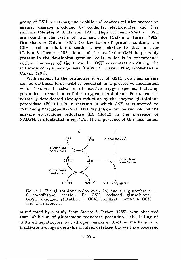
group of GSH is a strong nucleophile and confers cellular protection against damage produced by oxidants, electrophiles and free radicals (Meister & Anderson, 1983). High concentrations of GSH are found in the testis of rats and mice (Calvin & Turner, 1982; Grosshans & Calvin, 1985). On the basis of protein content, the GSH level in adult rat testis is even similar to that in liver (Calvin & Turner, 1982). Most of the testicular GSH is probably present in the developing germinal cells, which is in concordance with an increase of the testicular GSH concentration during the initiation of spermatogenesis (Calvin & Turner, 1982; Grosshans & Calvin, 1985).
With respect to the protective effect of GSH, two mechanisms can be outlined. First, GSH is essential in a protective mechanism which involves inactivation of reactive oxygen species, including peroxides, formed in cellular oxygen metabolism. Peroxides are normally detoxicated through reduction by the enzyme glutathione peroxidase (EC 1.11.1. 9), a reaction in which GSH is converted to oxidized glutathione (GSSG). This disulphide can be reduced by the enzyme glutathione reductase (EC 1.6.4.2) in the presence of NADPH, as illustrated in Fig. t(A). The importance of this mechanism
A
H20 H20 2
glutathione~· ~ peroxidase 1\
B
X ( xenobiotic)
GSSG GSH ---...... glutathione transferase
·~~~:::= .. X NADPH NADP+ GSX (conjugate)
Figure 1. The glutathione redox cycle (A) and the glutathione S- transferase reaction (B). GSH, reduced glutathione; GSSG, oxidized glutathione; GSX, conjugate between GSH and a xenobiotic.
is indicated by a study from Starke & Farber (1985), who observed that inhibition of glutathione reductase potentiated the killing of cultured hepatocytes by hydrogen peroxide. Another mechanism to inactivate hydrogen peroxide involves catalase, but we have focussed
- 93 -
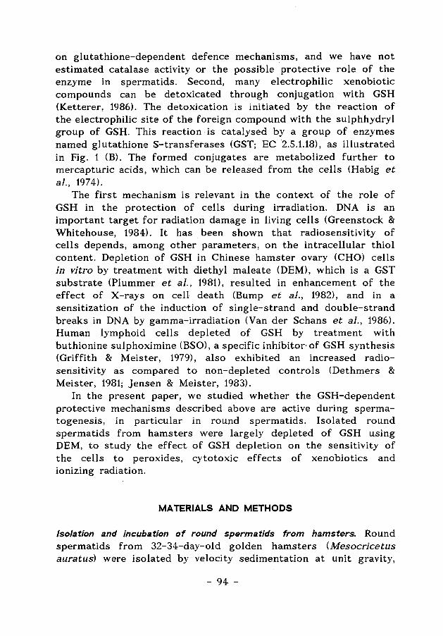
on glutathione-dependent defence mechanisms, and we have not estimated catalase activity or the possible protective role of the enzyme in spermatids. Second, many electrophilic xenobiotic compounds can be detoxicated through conjugation with GSH (Ketterer, 1986). The detoxication is initiated by the reaction of the electrophilic site of the foreign compound with the sulphhydryl group of GSH. This reaction is catalysed by a group of enzymes named glutathione S-transferases (GST; EC 2.5.1.18), as illustrated in Fig. 1 (B). The formed conjugates are metabolized further to mercapturic acids, which can be released from the cells (Habig et a/., 1974).
The first mechanism is relevant in the context of the role of GSH in the protection of cells during irradiation. DNA is an important target for radiation damage in living cells (Greenstock & Whitehouse, 1984). It has been shown that radiosensitivity of cells depends, among other parameters, on the intracellular thiol content. Depletion of GSH in Chinese hamster ovary (CHO) cells in vitro by treatment with diethyl maleate <DEM), which is a GST substrate (Plummer et al., 1981), resulted in enhancement of the effect of X-rays on cell death (Bump et al., 1982), and in a sensitization of the induction of single-strand and double-strand breaks in DNA by gamma-irradiation (Van der Schans et al., 1986). Human lymphoid cells depleted of GSH by treatment with buthionine sulphoximine (BSO), a specific inhibitor· of GSH synthesis (Griffith & Meister, 1979), also exhibited an increased radiosensitivity as compared to non-depleted controls (Dethmers & Meister, 1981; jensen & Meister, 1983).
In the present paper, we studied whether the GSH-dependent protective mechanisms described above are active during spermatogenesis, in particular in round spermatids. Isolated round spermatids from hamsters were largely depleted of GSH using DEM, to study the effect of GSH depletion on the sensitivity of the cells to peroxides, cytotoxic effects of xenobiotics and ionizing radiation.
MATERIALS AND METHODS
Isolation and incubation of round spermatids from hamsters. Round spermatids from 32-34-day-old golden hamsters (Mesocricetus auratus) were isolated by velocity sedimentation at unit gravity,
- 94 -
and further purified by Percoll-gradient centrifugation, using the same methods as described previously (Den Boer & Grootegoed, 1988). The isolated cells were incubated in Dulbecco's phosphatebuffered saline, supplemented with 12-mM sodium DL-lactate (PBS-L), or with lactate and 1.0-mM glucose (~BS-GU. The PBS also contained antibiotics (Grootegoed et al., 1985) and 0.1% bovine serum albumin (BSA) (Fraction V; Sigma Chemical Company, St. Louis, MO, USA). Approximately 0.3 x 10⁶ cells were incubated in a volume of 0.25 ml in polystyrene tubes (diameter 11.5 mm), at 32°C in air.
Depletion of glutathione in isolated round spermatids. The GST substrate diethyl maleate (Aldrich Chemie, Bruxelles, Belgium) was used to deplete the GSH pool of the spermatids. It has been shown that this compound can be used to lower GSH levels of spermatids from rats effectively, without affecting the viability and ATP content of the cells (Den Boer et al., 1988). The isolated spermatids were partly depleted of GSH, by incubating approximately 50x106 cells for 2 h in the presence of 100 [.LM-diethyl maleate, in 25 ml of PBS-L (containing 0.1% BSA) at 32 ° C. After the DEM treatment, the spermatids were washed twice with either PBS-L or PBS-GL (centrifugation for 5 min at 200 g) to remove the diethyl maleate. A stock solution of 310 mM- diethyl maleate was prepared in pure ethanol, and diluted with PBS to the desired concentration. The final ethanol concentration in the incubation medium was 0.03% (v/v), and this concentration of ethanol was also added to the controls.
Irradiation of isolated round spermatids and detection of single- strand DNA breaks. Spermatids were irradiated using a 6 °Co-gammasource (Gamma cell 100, Atomic Energy of Canada Ltd., Ottawa, Canada). Irradiations were performed at 0°C , using 1 ml of a cell suspension containing approximately 5x10⁶ spermatids in PBS-L. The cells were irradiated under different gas atmospheric conditions. The desired oxygen concentration in the incubations was reached by leading a gas mixture of 500 ppm 0 2 in N2 through the cell suspension, for 5 min before and during the irradiation. The oxygen content of the gas mixture leaving the system was measured as described by van der Schans et al. (1986). DNA damage in the spermatids was detected, after partial unwinding of the DNA by alkaline treatment, using a competitive
- 95 -
immunoassay as described by van der Schans et al. (1989). The immunoassay is based on a monoclonal antibody directed against single-stranded DNA. Single-strand DNA breaks result in an increase of the amount of single-strand DNA after alkaline treatment. All samples were assayed in triplicate in the ELISA, and the detected DNA damage is expressed as the % singlestrandedness of the DNA (% ss-DNA). The method is very sensitive and DNA damage can be determined using a low number of cells (van der Schans et al., 1989).
Estimation of the glutathione reductase activity. The glutathione reductase activity was estimated using the spectrophotometric method described by Carlberg & Mannervik (1985). The assay mixture contained 0.1 mM NADPH (stock of 2 mM in 10 mM Tris-HCl, pH 7.0) and 1 mM oxidized glutathione (GSSG), in 0.2-M potassium phosphate buffer with 2 mM EDTA (pH 7.0). The enzyme preparation was prepared by lysing 5x10⁶ spermatids in 500 [.d distilled water. After sonication (3 times for 5 sec, with a MSE 150 Watt Ultrasonic disintegrator, 20 kHz at a setting of 7 tJ.m), the homogenate was centrifuged (5 min, 15 000 g) and 100 tL 1 of the supernatant was added to the assay (final volume 1 ml).
Fluorometric method for determination of reduced glutathione. Determination of GSH contents in spermatids was performed according to the method of Hissin & Hilf (1976) and Grosshans & Calvin (1985), but with some slight modifications as described by Den Boer et al. (1989).
Estimation of cellular A TP content. The cellular A TP content of the spermatids was estimated using the bioluminescent firefly luciferin-luciferase reaction (Lumit PM from Lumac, Meise, Belgium) as described by Grootegoed et al. (1984), with a model 6100 Pi co -Lite Luminometer (Packard).
RESULTS
Glutathione depletion of isolated round spermatids by diethyl maleate
To define the conditions for GSH depletion of hamster round spermatids, the GSH content of the isolated cells was estimated at different time-intervals during 2 h of incubation in the presence
- 96 -
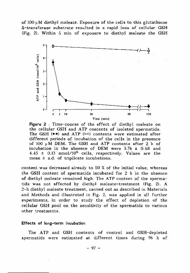
of 100 [!M diethyl maleate. Exposure of the cells to this glutathione S-transferase substrate resulted in a rapid loss of cellular GSH (Fig. 2). Within 5 min of exposure to diethyl maleate the GSH
5
..!!! 4 e "' 0 -0 3
E c
J:
~ 2 "0 c "' c..
1-<(
0 ~~~------~------------~~1 ~ 0 5 10 30 60 120
Time (min)
Figure 2 . Time-course of the effect of diethyl maleate on the cellular GSH and ATP contents of isolated spermatids. The GSH ( ... ) and ATP (o-o) contents were estimated after different periods of incubation of the cells in the presence of 100 [! M DEM. The GSH and ATP contents after 2 h of incubation in the absence of DEM were 3. 76 ± 0.48 and 4.45 ± 0.13 nmol/106 cells, respectively. Values are the mean ± s.d. of triplicate incubations.
content was decreased already to 50 % of the initial value, whereas the GSH content of spermatids incubated for 2 h in the absence of diethyl maleate remained high. The A TP content of the spermatids was not affected by diethyl maleate-treatment (Fig. 2). A 2-h diethyl maleate treatment, carried out as described in Materials and Methods and illustrated in Fig. 2, was applied in all further experiments, in order to study the effect of depletion of the cellular GSH pool on the sensitivity of the spermatids to various other treatments.
Effects of long-term incubation
The ATP and GSH contents of control and GSH-depleted spermatids were estimated at different times during 96 h of
- 97 -
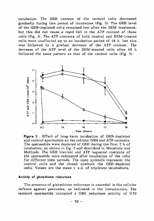
incubation. The GSH content of the control cells decreased gradually during this period of incubation (Fig. 3). The GSH level of the GSH-depleted cells remained low after the DEM treatment, but this did not cause a rapid fall in the ATP content of these cells (Fig. 3). The ATP contents of both control and DEM-treated cells were unaffected up to an incubation period ·of 48 h, but this was followed by a gradual decrease of the ATP content. The decrease of the ATP level of the DEM-treated cells after 48 h followed the same pattern as that of the control cells (Fig. 3).
5
"' : II f!
"' = 0 3 E c:
J:
~ 2 "0 c: ..
a.. 1-<
0
02 211 118 72 96
Time (hours)
Figure 3 . Effect of long-term incubation of GSH-depleted and control spermatids on the cellular GSH and ATP contents. The spermatids were depleted of GSH during the first 2 h of incubation, as shown in Fig. 2 and described in Materials and Methods. The GSH (circles) and ATP (squares) contents of the spermatids were estimated after incubation of the cells for different time periods. The open symbols represent the control cells and the closed symbols the GSH-depleted cells. Values are the mean ± s.d. of triplicate incubations.
Activity of glutathione reductase
The presence of glutathione reductase is essential in the cellular defence against peroxides, as indicated in the Introduction. The isolated spermatids contained a GSH reductase activity of 0.93
- 98 -
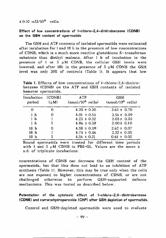
± 0.10 mU/10 6 cells.
Effect of low concentrations of 1-chloro-2,4-dinitrobenzene (CDNB) on the GSH content of spermatids
The GSH and ATP contents of isolated spermatids were estimated after incubation for 1 and 18 h in the presence of low concentrations of CDNB, which is a much more reactive glutathione S- transferase substrate than diethyl maleate. After 1 h of incubation in the presence of 1 or 5 [.LM CDNB, the cellular GSH levels were lowered, and after 18h in the presence of 5 [1M CDNB the GSH level was only 20% of controls (Table 1). It appears that low
Table 1. Effects of low concentrations of 1-chloro-2,4-dinitrobenzene (CDNB) on the ATP and GSH conten~s of isolated hamster spermatids.
Incubation [CDNBJ ATP GSH period ((1M) (nmol/106 cells) (nmol/106 cells)
0 0 4.30 ± 0.30 3.65 ± 0.70 1 h 0 4.01 ± 0.54 3.56 ± 0.09 1 h 1 4.23 ± 0.32 3.03± 0.03 1 h 5 4.86 ± 0.38 2.00± 0.10
18 h 0 4.58 ± 0.09 2.62 ± 0.07 18 h 1 4.74 ± 0.46 2.22 ± 0.05 18 h 5 4.56 ± 0.21 0.61 ± 0.05
Round spermatids were treated for different time periods with 1 and 5 (1M CDNB in PBS-GL. Values are the mean ± s.d. of triplicate incubations.
concentrations of CDNB can decrease the GSH content of the spermatids, but that this does not lead to an inhibition of ATP synthesis (Table 1). However, this may be true only when the cells are not exposed to higher concentrations of CDNB, or are not challenged otherwise to perform GSH-supported defence mechanisms. This was tested as described below.
Potentiation of the cytotoxic effect of 1-chloro-2,4- dinitrobenzene (CDNB) and cumenehydroperoxide (CHP) after GSH depletion of spermatids.
Control and GSH-depleted spermatids were used to evaluate
- 99 -
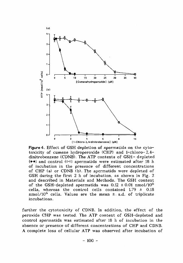
(a)
"' B o <D
0
0 E 1: (b)
ll. 1- 4 <
0 10 15 20 25 30 35 40
[Cumenehydroperoxide] ()IM)
8 10 [ 1-Chloro-2, 11-dinitrobenzene] (JIM)
Figure 4. Effect of GSH depletion of spermatids on the cytotoxicity of cumene hydroperoxide (CHP) and 1-chloro- 2,4-dinitrobenzene (CDNB). The ATP contents of GSH- depleted ( .. ) and control (o-o) spermatids were estimated after 18 h of incubation in the presence of different concentrations of CHP (a) or CDNB (b). The spermatids were depleted of GSH during the first 2 h of incubation, as shown in Fig. 2 and described in Materials and Methods. The GSH content of the GSH-depleted spermatids was 0.12 ± 0.01 nmol/106
cells, whereas the control cells contained 1. 79 ± 0.18 nmol/106 cells. Values are the mean ±. s.d. of triplicate incubations.
further the cytotoxicity of CDNB. In addition, the effect of the peroxide CHP was tested. The ATP content of GSH-depleted and control spermatids was estimated after 18 h of incubation in the absence or presence of different concentrations of CHP and CDNB. A complete loss of cellular ATP was observed after incubation of
- 100 -
the GSH-depleted spermatids in the presence of low concentrations of CHP (Fig. 4a) or CDNB (Fig. 4 b). The ATP content of the control spermatids was affected by these two compounds only at much higher concentrations (Fig. 4a and 4b). From these results, it can be concluded that the GSH-depleted cells are highly sensitive to both CHP and CDNB, as compared to control, nonGSH- depleted cells.
Effect of GSH depletion of spermatids on the induction and repair of single-strand DNA breaks (% ss-DNA).
The induction of single-strand DNA breaks (% ss-DNA) was estimated in control and GSH-depleted spermatids, which were irradiated with a gamma dose of 2 or S Gy. Irradiation resulted in a dose-dependent increase of the % ss-DNA (Fig. Sa), both in control and in GSH-depleted spermatids. However, the radiationinduced DNA damage in GSH- depleted cells was rv 3-fold higher than that in the control cells. The % ss-DNA of non-irradiated control cells and non-irradiated GSH-depleted cells were not different (Fig. Sa).
The results described above were obtained by irradiation of the cells under a gas atmosphere of SOO p.p.m. 0 2 in N2 . It has been reported that induction of radiation damage of cells is dependent on oxygen concentration, and that diminution of cellular GSH does no.. affect the induction of DNA damage by irradiation under aerobic conditions (Van der Schans et al., 1986). In agreement with this, no difference was observed in the % ss-DNA in GSHdepleted and control cells after irradiation under air (Fig. Sb). In this experiment, air was led through the medium during the irradiation. Under standard incubation conditions, the gas atmosphere was air, but the air was not led through the medium. Consequently, the oxygen concentration surrounding the cells in all other experiments was most probably lower than the oxygen concentration in air. This is also indicated by the experiments on induction and repair of DNA damage described below.
In the experiments to study the repair of gamma-radiation induced single-strand DNA breaks, GSH-depleted and control cells were gamma-irradiated under standard incubation conditions. The induction of DNA damage in spermatids depleted of GSH was approximately 4-fold higher as compared to that in control cells (Fig. 6a). During a subsequent incubation at 32 °C , both the GSH-
- 101 -
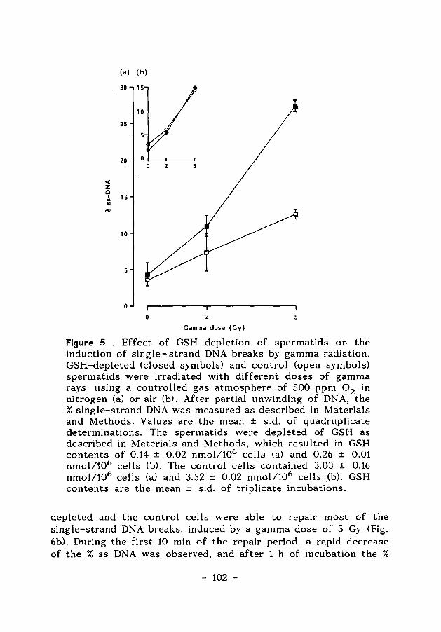
(a) (b)
30 15
10
25
5
20 2 5
< z 0
I 15 en Ill
dP
10
5
0
0 2 5
Gamma dose (Gy)
Figure 5 . Effect of GSH depletion of spermatids on the induction of single- strand DNA breaks by gamma radiation.· GSH-depleted (closed symbols) and control (open symbols) spermatids were irradiated with different doses of gamma rays, using a controlled gas atmosphere of 500 ppm 0 2 in nitrogen (a) or air (b). After partial unwinding of DNA, the % single-strand DNA was measured as described in Materials and Methods. Values are the mean ± s.d. of quadruplicate determinations. The spermatids were depleted of GSH as described in Materials and Methods, which resulted in GSH contents of 0.14 ± 0.02 nmol/106 cells (a) and 0.26 ± 0.01 nmol/106 cells (b). The control cells contained 3.03 ± 0.16 nmol/106 cells (a) and 3.52 ± 0.02 nmol/106 cells (b). GSH contents are the mean ± s.d. of triplicate incubations.
depleted and the control cells were able to repair most of the single-strand DNA breaks, induced by a gamma dose of 5 Gy (Fig. 6b). During the first 10 min of the repair period, a rapid decrease of the % ss-DNA was observed, and after 1 h of incubation the %
- 102 -
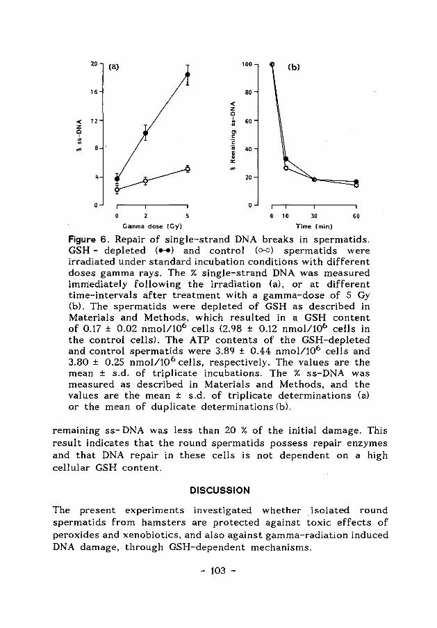
20 (a) 100 (b)
16 80
< z Q
12 I
60 < ., z ., c Cl
I 1: ., ., 1:
"" 8 "iii 40 E "' 0::
~ "" 4 20
0 0
0 2 5 0 10 30 60
Gamma dose (Gy) Time (min)
Figure 6. Repair of single-strand DNA breaks in spermatids. GSH - depleted ( ... ) and control (o-o) spermatids were irradiated under standard incubation conditions with different doses gamma rays. The % single-strand DNA was measured immediately following the irradiation (a), or at different time-intervals after treatment with a gamma-dose of 5 Gy (b). The spermatids were depleted of GSH as described in Materials and Methods, which resulted in a GSH content of 0.17 ± 0.02 nmol/106 cells (2.98 ± 0.12 nmol/106 cells in the control cells). The ATP contents of the GSH-depleted and control spermatids were 3.89 ± 0.44 nmol/106 cells and 3.80 ± 0.25 nmol/10 6 cells, respectively. The values are the mean ± s.d. of triplicate incubations. The % ss-DNA was measured as described in Materials and Methods, and the values are the mean ± s.d. of triplicate determinations (a) or the mean of duplicate determinations (b).
remaining ss- DNA was less than 20 % of the initial damage. This result indicates that the round spermatids possess repair enzymes and that DNA repair in these cells is not dependent on a high cellular GSH content.
DISCUSSION
The present experiments investigated whether isolated round spermatids from hamsters are protected against toxic effects of peroxides and xenobiotics, and also against gamma-radiation induced DNA damage, through GSH-dependent mechanisms.
- 103 -
To obtain information on such a role of GSH in spermatids, the spermatids were largely depleted of GSH. The GSH content of the spermatids was decreased effectively by incubating the cells in the presence of low concentrations of the GSH conjugating agent (GST substrate) diethyl maleate (DEM). Low concentrations of DEM were used, since DEM can also cause lipid peroxidation. Rat hepatocytes exposed to 3 mM DEM showed a pronounced increase of lipid peroxidation (Miccadei et al., 1988), whereas lower concentrations of DEM resulted in depletion of the GSH content but did not cause lipid peroxidation and cell death (Hogberg & Kristoferson, 1977). Another, non-toxic, method to deplete cells of GSH involves treatment of the cells with the GSH synthesis inhibitor buthionine sulphoximine (BSO) (Griffith & Meister, 1979). This method, however, has little effect on spermatids, because isolated spermatids have a low capacity to synthesize GSH (Den Boer et al., 1989), and BSO was therefore not applied.
The observation that there was no effect of GSH depletion on the ATP content of the spermatids during long-term incubation, indicates that there is no pronounced effect of GSH depletion on the viability of the isolated cells. This gave rise to the question to what extent GSH is involved in defence mechanisms in the isolated spermatids. Therefore, we studied whether GSH depletion of spermatids potentiated the cytotoxic effect of compounds, that are normally detoxicated through GSH-dependent mechanisms. To detect changes in the sensitivity of the spermatids to such compounds, cellular injury was assessed by estimating the ATP content of the spermatids. The ATP content may point to more subtle changes in cellular integrity, as compared to other parameters such as leakage of cytoplasmic enzymes and the uptake of trypan blue (Reed & Fariss, 1984).
Cellular GSH depletion of the spermatids resulted in a low capacity of the cells to withstand oxidative stress generated by cumene hydroperoxide (CHP), as indicated by the cytotoxic effect of low CHP concentrations in DEM-treated cells. This is in agreement with observations in tumour cells, showing that GSH depletion of mastocytoma and lymphoma cells markedly enhanced their sensitivity to lysis by H2 0 2 . Recovery of the tumour cell resistance to H2 0 2 after GSH depletion correlated with resynthesis of cellular GSH (Arrick et al., 1982).
Low 1-chloro-2,4-dinitrobenzene (CDNB) concentrations caus,ed GSH depletion of the isolated spermatids. This is similar to what
- 104 -
has been reported for tumour cells, including lymphoma cells which were depleted of GSH in vitro using CDNB (Arrick et al., 1982). CDNB, however, is a much more reactive GST substrate than DEM, and in addition this compound may cause cellular damage through other mechanisms. After raising the GSH level in Chang liver cells, CDNB was found to be more toxic, as concluded from a decreased cloning efficiency, and after lowering the cellular GSH level the toxicity was diminished (Bruggeman et al., 1988). This may indicate that the formed conjugate between CDNB and GSH is more cytotoxic than CDNB itself. In the spermatids, a decrease of the GSH pool at high CDNB concentrations was accompanied by complete loss of cellular ATP, indicative for cell death. Furthermore, diminution of the cellular GSH pool of the spermatids, by DEM treatment, resulted in a lower capacity of the cells to protect themselves against direct cytotoxic effects of CDNB, as indicated by the loss of ATP from the GSH-depleted spermatids at low CDNB concentrations. More. experiments will be necessary to elucidate the mechanism of the toxic effects of CDNB and its conjugate. However, it is clear that GSH in spermatids confers protection against CDNB toxicity.
The importance of intracellular free thiol groups in the prevention of cellular damage caused by ionizing radiation has be ~n established in various studies (Meister & Anderson, 1983). In the present experiments, it was observed that GSH-depleted spermatids were much more sensitive than the control cells to the induction of single-strand DNA breaks by ionizing radiation, as indicated by the higher % ss-DNA after irradiation with the same gamma-dose. This is in agreement with other studies showing that the radiosensitivity of cells, including CHO cells (Bump et al., 1982; Evans et al., 1984; Van der Schans et al., 1986), Hela cells (Van der Schans et al., 1986) and human lymphoid cells (Dethmers & Meister, 1981), is increased when the cells are depleted of GSH.
It was observed that irradiation of GSH-depleted spermatids and control cells under aerobic conditions (air led through the incubation medium) did not show an increased sensitization to radiation damage by the GSH depletion. This confirms the notion that, under aerobic conditions, radiation damage is not dependent on the cellular GSH concentration (Van der Schans et al., 1986). The relationship between the concentration of oxygen in the gas atmosphere during irradiation of cultured cells and the effect of depletion of GSH on their radiosensitivity, has been studied
- 105 -
extensively by van der Schans et a/. (1986). Thes'e studies showed that GSH depletion of Chinese hamster ovary (CHO) cells resulted in only a small or non- detectable sensitization to DNA damage, when the cells were irradiated under aerobic conditions. Similar results have been obtained by Bump et al. (1982), who observed that GSH depletion of CHO cells in vitro by DEM resulted in enhancement of the effect of X-rays on cell killing under hypoxic conditions but not under oxygenated conditions. This is explained by the fact that under aerobic conditions oxygen is present in such a large excess, that a lowering of the cellular GSH content does not affect the extent of damage fixation. From the results of the experiments in which spermatids were irradiated under standard incubation conditions, it appears that the oxygen concentration around and within the cells at the time of irradiation must have been very low. This can be concluded, because the sensitization of the spermatids to radia,tion damage by GSH depletion was not detected in air but was pronounced during incubation in 500 ppm 0 2 in N2 .
The repair of the radiation-induced damage in the DNA of the spermatids was not affected by the cellular GSH content. This is in agreement with observations for HeLa and CHO cells, illustrating that GSH depletion by DEM resulted in a sensitization of induction of DNA damage without an effect on the rate of repair of singlestrand DNA breaks (Evans et al., 1984; Vos et al., 1984). The present results show that round spermatids are able to repair single-strand breaks in DNA, and that this repair mechanism is very active in the isolated cells after irradiation. The capacity of the round spermatids for DNA repair, as compared to germ cells from other stages of spermatogenesis is under investigation.
It can be concluded from the present results, that a reduction of the cellular GSH content of isolated spermatids does not directly affect the viability of the cells, unless an oxidative challenge is generated or when toxic compounds are added. Cellular GSH in spermatids is involved in the protection of the cells against oxidative stress and toxic effects of xenobiotics, and is active in the prevention of DNA damage during irradiation. It has not been investigated which exogenous or endogenous factors are inactivated through GSH-supported defence mechanisms in spermatids in situ, in the spermatogenic epithelium. The GSH-mediated protection mechanisms in spermatids may rely on the maintenance of a large intracellular GSH pool. Furthermore,
- 106 -
it is very likely that Sertoli cells play an important role in this context, because the Sertoli cells enclose the germ cells, providing support and protection.
This investigation received financial support from the World Health Organization Special Programme of Research, Development and Research Training in Human Reproduction (project 87079), and The Institute of Radiopathology and Radiation Protection, grant 4.7.10, Leiden, The Netherlands.
REFERENCES
Arrick, B.A., Nathan, C.F., Griffith, O.W. & Cohn, Z.A. (1982) Glutathione depletion sensitizes tumor cells to oxidative cytolysis. j. bioi. Chem. 257, 1231-1237.
Bruggeman, I.M., Spenkelink, A., Temmink, J.H.M. & Van Bladeren, P.J. (1988) Differential effects of raising and lowering intracellular glutathione levels on the cytotoxicity of allyl isothiocyanate, tert-butylhydroperoxide and chlorodinitro- benzene. Toxicology in vitro. 2, 31-35.
Bump. E.A., Yu, N.Y. & Brown, J.M. (1982) Radiosensitization of hypoxic tumor cells by depletion of intracellular glutathione. Science 217, 544-545.
Calvin, H.l. & Turner, S.l. (1982) High levels of glutathione attained during postnatal development of rat testis. ]. exp. Zoo}. 219, 389-393.
Carlberg, I. & Mannervik, B. (1985) Glutathione reductase. In: Methods in Enzymology, vol. 113, pp. 484-490. Ed. A. Meister, Academic Press, London.
Den Boer, P.J. & Grootegoed, J.A. (1988) Mechanism of action of (-)gossypol on ATP production in isolated hamster spermatids. ]. Reprod. Fert. 83,693-700.
Den Boer, P.J., Verkerk, A., & Grootegoed, J.A. (1988) Effect of xenobiotics on glutathione and A TP levels in isolated round spermatids from rats. In: The Molecular and Cellular Endocrinology of the Testis, pp 241-246. Eds B.A. Cooke & R.M. Sharpe. Raven Press, New York.
Den Boer, P.J., Mackenbach, P. & Grootegoed, J.A. (1989) Glutathione metabolism in cultured Sertoli cells and spermatogenic cells
- 107 -
from hamsters. }. Reprod. Fert. 87, 391-400. Dethmers, J.K. & Meister, A. (1981) Glutathione export by human
lymphoid cells: depletion of glutathione by inhibition of its synthesis decreases export and increases sensitivity to irradiation. Proc. natl. A cad. Sci. U.S.A 78, 7 492-7 496.
Evans, J.W., Taylor, Y.C. & Brown, J.M. (1984) The role of glutathione and DNA strand break repair in determining the shoulder of the radiation survival curve. Br. ]. Cancer 49, Suppl. VI, 49-53.
Greenstock, C.L. & Whitehouse, R.P. (1984) Base damage in irradiated DNA and the effect of oxygen. In: Oxygen radicals in chemistry and biology, pp. 619-628. Eds W. Bors, M. Saran, M & D. Tait. Berlin; New York: de Gruyter.
Griffith, O.W. & Meister, A. (1979) Potent and specific inhibition of glutathione synthesis by buthionine sulfoximine (S- n- butyl homocysteine sulfoximine). ]. bioi. Chem. 254, 7558- 7560.
Grootegoed, J.A., Jansen, R. & van der Molen, H.J. (1984) The role of glucose, pyruvate and lactate in ATP production by rat spermatocytes and spermatids. Biochim. Biophys. Acta 767, 248-256.
Grootegoed, J.A., Jansen, R., and van der Molen, H.J. (1985) Intercellular pathway of leucine catabolism in rc'tt spermatogenic epithelium. Biochem. ]. 226, 889-892.
Grosshans, K. & Calvin, H.l. (1985) Estimation of glutathione in purified populations of mouse testis germ cells. Bioi. Reprod. 33, 1197-1205.
Habig, W.H., Pabst, M.J. & Jakoby, W.B. (1974) Glutathione S-transferases. ]. bioi. Chem. 249, 7130-7139.
Hissin, P.J. & Hilf, R. (1976) A fluorometric method for determination of oxidized and reduced glutathione in tissues. Analyt. Biochem. 74, 214-226.
Hogberg, J. & Kristoferson, A. (1977) A correlation between glutathione levels and cellular damage in isolated hepatocytes. Eur. ]. Biochem. 74,77-82.
Jensen, G.L. & Meister, A. (1983) Radioprotection of human lymphoid cells by exogenously supplied glutathione is mediated by y-glutamyl transpeptidase. Proc. natl. Acad. Sci. USA 80, 4714-4717.
Ketterer, B. (1986) Detoxication reactions of glutathione and glutathione transferases. Xenobiotica 16, 957-973.
Meister, A. & Anderson, M.E. (1983) Glutathione. Ann. Rev. Biochem. 52, 711-760.
Miccadei, S., Kyle, M.E., Gilfor, D. & Farber, J.L. (1988) Toxic
- 108 -
consequence of the abrupt depletion of glutathione in cultured rat hepatocytes. Archs Biochem. Biophys. 265, 311-320.
Plummer, J.L., Smith, B.R., Sies, H. &. Bend, J.R. (1981) Chemical depletion of glutathione in vivo. In: Methods in Enzymology, vol. 77, pp. S0-59. Ed. W.B. jakoby, Academic Press, London.
Reed, D.J. &. Fariss, M.W. (1984) Glutathione depletion and susceptibility. Pharmacal. Rev. 36, 2SS-33S.
Schans, G.P. van der, Vos, 0., Roos-Verheij, W.S.D. &. Lohman, P.H.M. (1986) The influence of oxygen on the induction of radiation damage in DNA in mammalian cells and after sensitization by intracellular glutathione depletion. Int. ]. Radiat. Bioi. SO, 453-465.
Schans, G.P. van der, Loon, A.A.W.M. van, Groenendijk, R.H. &. Baan, R.A. (1989) Detection of DNA damage in cells exposed to ionizing radiation by use of anti- single- stranded -DNA monoclonal antibody. Int. ]. radiat. Bioi. SS, 747-760.
Starke, P.E. &. Farber, J.L. (1985) Endogenous defenses against the cytotoxicity of hydrogen peroxide in cultured rat hepatocytes. ]. bioi. Chem. 260, 86-92.
Vos, 0., Schans, G.P. van der &. Roos-Verheij, W.S.D. (1984) Effects of BSO and DEM on thiol-level and radiosensitivity in Hela cells. Int. ]. Radiation Oncology Bioi. Phys. 10, 1249-1253.
- 109 -

Chapter 7
Glutathione metabolism in cultured Sertoli cells and spermatogenic cells from hamsters
Pl. Den Boer, P. Mackenbach and JA. Grootegoed
Journal of Reproduction & Fertility (1989) 87: 391-400
- 111 -
Glutathione metabolism in cultured Sertoli cells and spermatogenic cells from hamsters
P. J. Den Boer, P. Mackenbach and J. A. Grootegoed
Department of Biochemistry II, Medical Faculty, Erasmus University Rotterdam, P.O. Box 1738, 3000 DR Rotterdam, The Netherlands
Summary. Isolated spermatocytes and spermatids from hamsters contained a large amount of glutathione (GSH) ( -40 and 30 nmol GSH/mg protein, respectively), but showed a spontaneous decrease of GSH content during prolonged incubation (t112
- 35 h). Incubation of the germ cells in the presence of the glutathione biosynthesis inhibitor buthionine sulphoximine (BSO) provided evidence that the cells can perform glutathione synthesis. This synthesis, however, was not sufficient to maintain the GSH content of the isolated cells, or to restore the cellular GSH pool after depletion caused by exposure of the cells to the glutathione S-transferase substrate, diethyl maleate (DEM). Cultured Sertoli cells, containing -10 nmol GSH/mg protein, had a more active BSO-sensitive GSH synthesis system. The Sertoli cells, but also tubule fragments containing Sertoli cells and germ cells, were able to restore their GSH pool after DEMinduced depletion. DEM treatment of the tubule fragments resulted in a 90% decrease of the GSH content of the spermatocytes and spermatids present within the fragments. The GSH levels of the tubule fragments and the enclosed germ cells were restored during a subsequent incubation in the absence of DEM. As indicated above, such a recovery was not observed for isolated spermatocytes and spermatids. The results illustrate the importance of Sertoli cell-germ cell interaction, and point to a role of Sertoli cells in glutathione synthesis by the germ cells.
Keywords: glutathione; testis; Sertoli cells; sperrnatocytes; sperrnatids; hamster
Introduction
Glutathione (the tripeptide L-y-glutamyl-L-cysteinylglycine) is the most abundant non-protein thiol in mammalian cells. Reduced glutathione (GSH), with a free sulphydryl group, is involved in many biological processes, including synthesis of proteins and DNA, transport of amino acids and protection of cells against oxidation (Meister & Anderson, 1983). High concentrations of GSH have been found in rat and mouse testes (Calvin & Turner, 1982; Grosshans & Calvin, 1985). The concentration of GSH in rat testis (J..Lmol/g tissue) increases approximately 3-fold between Days 8 and 29 of post-natal development, coinciding with the onset of spermatogenesis, and is then maintained at a high level during further testicular development (Calvin & Turner, 1982). However, there is little information about the role and the metabolism of glutathione in Sertoli cells and spermatogenic cells.
Testicular glutathione levels can be reduced in immature mice (Calvin eta/., 1986) and, to a lesser extent, also in mature rats (Teaf et a/., 1987), by treatment of the animals with buthionine sulphoximine (BSO), which is a specific inhibitor of GSH synthesis (Griffith & Meister, 1979). However, these studies do not provide evidence that the testis can perform glutathione synthesis, and do not give information on the ability of the different testicular cell types to synthesize GSH.
- 112 -
In studies using isolated round spermatids from rats, it has been shown that the germ cells were partly depleted of glutathione after incubation with diethyl maleate (Den Boer et a!., 1988). This compound is a glutathione S-transferase substrate. There are a number of glutathione S-transferases (EC 2.5.1.18) which catalyse the reaction of GSH with a variety of substrates (Ketterer, 1986). The reaction of GSH with diethyl maleate leads to the formation of a conjugate, resulting in a loss of GSH from the cells.
In the present experiments, isolated spermatocytes, spermatids, Sertoli cells and tubule fragments from hamsters were used to study different aspects of testicular GSH metabolism in more detail. Hamsters were used since earlier studies indicated that isolated cells and tubular fragments indicated from hamsters are suitable model systems (Den Boer & Grootegoed, 1988a, b).
Materials and Methods
Isolation and incubation of tubule fragments. Immature 25-26-day-old hamsters (Mesocricetus auratus) were killed by cervical dislocation and the testes were removed. Tubule fragments were isolated by collagenase treatment as described by Den Boer & Grootegoed (1988b). The tubule fragments contained Sertoli cells and spermatogenic cells, and this preparation was also referred to as spermatogenic epithelium. The tubule fragments were incubated in Eagle's minimum essential medium (MEM) containing Earle's salts and 25 mM-Hepes (Gibco, Paisley, UK) supplemented with antibiotics (Grootegoed et a/., 1985), L-glutamine (292 mg/1) and I% fetal calf serum (FCS). The incubations were performed for 24, 48 or 72 hat 32°C under an atmosphere of 5% C0 2 in air, using 12-well plates or I 50 cm 2
(650 ml) tissue-culture flasks (Costar, Broadway, Cambridge, MA, USA), with -200 Jlg cellular protein/cm2 • From each sample the amounts of protein and GSH were estimated.
Isolation and incubation of spermatocytes and spermatids. Round spermatids and pachytene spermatocytes from 32-35-day-old hamsters were isolated by sedimentation at unit gravity and further purified by Percoll density centrifugation (Den Boer & Grootegoed, 1988a). The incubations were performed using Dulbecco's phosphate-buffered saline, containing 12 mM-DL-Iactate and 0· I% bovine serum albumin (BSA) (Fraction V: Sigma Chemical Company, St Louis, MO, USA), referred to as PBS-L. Approximately 0·3 x 106 cells were incubated in 0·25 ml PBS-L, in polystyrene tubes (diameter I I ·5 mm), at 32"C in air.
In another series of experiments, round spermatids and pachytene spermatocytes were isolated from tubule fragments, not only immediately after the preparation of the tubules using collagenase, but also after 24 and 72 h of culture of the tubule fragments. The methods used for this procedure were as described above.
Isolation and incubation of Sertoli cells. Sertoli cells were isolated from 25-day-old hamsters, using the method for isolation of rat Sertoli cells described by Oonk et a/. (I 985). The cells were cultured in MEM (Gibco, Grand Island, NY, USA), supplemented with non-essential amino acids, glutamine, antibiotics and I% FCS at 32°C for 48 h. This incubation was followed by hypotonic shock, to remove most of the germinal cells (Galdieri eta/., 1981), and an additional incubation period of24 hat 32°C. Subsequently, the cells were washed and incubated in MEM containing 0·1% BSA, in the absence of FCS. Effects of different compounds on GSH metabolism were estimated during incubations for 2 and 48 hat 32°C under an atmosphere of 5% C0 2 in air, in a final volume of 2 ml in 12-well plates (Costar) with -0· 12 mg protein/well.
Fluorometric method for determination of reduced glutathione. Determination of cellular GSH content was performed according to the method ofHissin & Hilf(l976) and Grosshans & Calvin (1985), with some slight modifications. Sertoli cells and tubule fragments were detached from the wells mechanically, pooled and centrifuged for 5 min at 600 g. The incubated germ cells in the tubes were not attached and were collected by centrifugation (5 min at 600 g). The supernatants were discarded and the pellets were frozen and stored at -80°C. For glutathione estimation, the cells were lysed in I 00-500 Jll water, and protein was precipitated by adding 50-250 Jll 3 M-perchloric acid containing I mM-EDTA. The mixture was kept on ice for I 0 min, and the precipitated protein was removed by centrifugation. The supernatant was neutralized by addition of2 M-KOH/0·3 M-Hepes, followed by centrifugation to remove KC104 •
The final GSH-assay mixture (500 Jll) contained 25 Jll of the neutralized supernatant, 450 Jll of a sodium-phosphate buffer (0·1 M-sodium phosphate, 5 mM-EDT A, pH 8·0) and 25 Jll of a solution of o-phthaldialdehyde (Sigma) in methanol (I mg/ml). After mixing and incubation for 15 min at room temperature, the fluorescence was determined using a fluorescence spectrophotometer (Kontron Instruments, SFM 25) at 420 nm (excitation at 350 nm).
Estimation of cellular ATP content. The cellular A TP content of the spermatocytes and spermatids was estimated with the bioluminescent firefly luciferin-luciferase reaction (Lumac) as described by Grootegoed eta/. (1984), using a model6100 Pico-Lite Luminometer (Packard).
Estimation of cellular protein. The amount of cellular protein was estimated according to Bradford (I 976), using bovine serum albumin as standard.
- 113 -
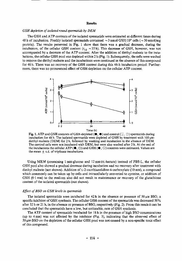
Results
GSH depletion of isolated round spermatids by DEM
The GSH and A TP contents of the isolated spermatids were estimated at different times during 48 h of incubation. Freshly isolated spermatids contained - 3 nmol GSH/1 06 cells (- 30 nmol/mg protein). The results presented in Fig. 1 show that there was a gradual decrease, during the incubation, of the cellular GSH content (t 112 -35 h). This decrease of GSH, however, was not accompanied by a decrease of the ATP content. After the addition of diethyl maleate to the incubations, the cellular GSH pool was depleted within 2 h (Fig. 1). Subsequently, the cells were washed to remove the diethyl maleate and the incubations were continued in the absence of this compound for 46 h. There was no recovery of the GSH content during this 46-h incubation period. Furthermore, there was no pronounced effect of GSH depletion on the cellular ATP content.
4 4
~ 3 3 ~ a:; a:; " " "' "' 0 0 ~ -0 2 0 E E ..s ..s a.. I 1- 1 Cll <( (!)
0 0 0 2 24 48
Time(h)
Fig.l. ATP and GSH contents ofGSH-depleted (•, e) and control (0, 0) spermatidsduring incubation for 48 h. The isolated sperrnatids were depleted ofGSH by treatment with 100 J.LMdiethyl maleate (DEM) for 2 h, followed by washing and incubation in the absence of DEM. The control cells were not incubated with DEM, but were also washed after 2 h. At the end of the incubations the cellular ATP (., D) and GSH (e, 0) contents were estimated. Values are the mean ± s.d. of triplicate incubations.
Using MEM (containing 1 mM-glucose and 12 mM-DL-lactate) instead of PBS-L, the cellular GSH pool also showed a gradual decrease during incubation and no recovery after treatment with diethyl maleate (not shown). Addition of L-2-oxothiazolidine-4-carboxylate (10 mM), a compound which commonly can be taken up by cells and intracellularly converted to cysteine, or addition of GSH (0·1 mM) to the medium also did not result in maintenance or recovery of the glutathione content of the isolated spermatids (not shown).
Effect of BSO on GSH levels in spermatids
The isolated spermatids were incubated for 42 h in the absence or presence of 50 J.LM BSO, a specific inhibitor of GSH synthesis. The cellular GSH content of the spermatids was decreased 50% after 32 h or 21 h, in the absence or presence of BSO, respectively (Fig. 2). From this result it can be concluded that the spermatids have a low, but noticeable, rate of GSH synthesis.
The ATP content of spermatids incubated for 18 h in the presence of high BSO concentrations (up to 4 mM) was not affected by the inhibitor (Fig. 3), indicating that the observed effect of 50 J.LM-BSO on the depletion of the cellular GSH pool was not caused by a non-specific toxic effect of this compound.
- 114 -
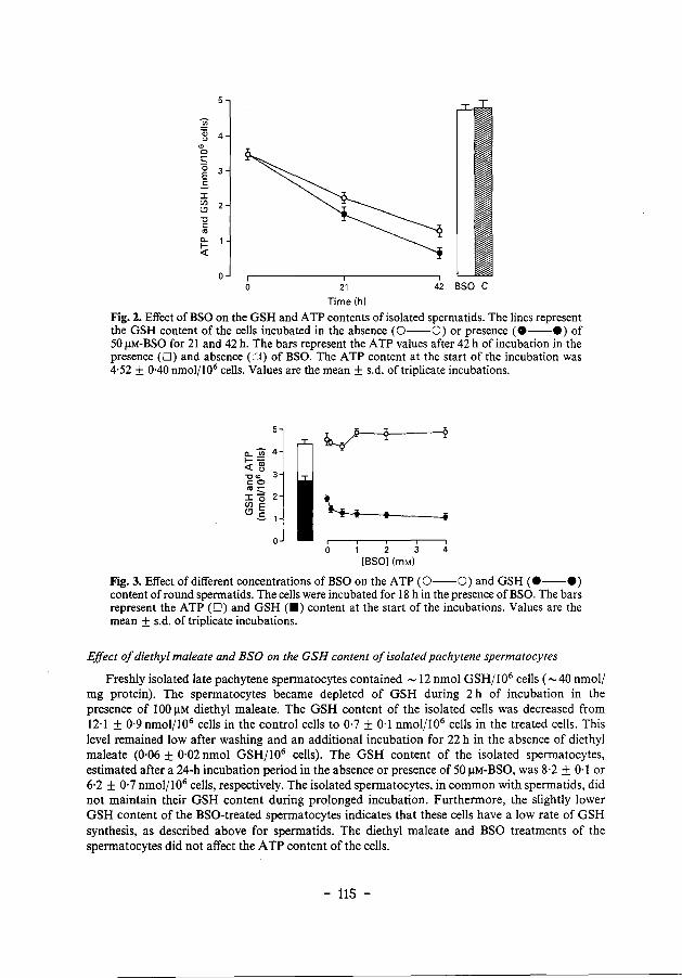
"' 0
~ 3 .s ::c Ul (.!)
"0 c:
"' ~ 1
~
0 0 21 42 BSO C
Time (h)
Fig. 2. Effect of BSO on the GSH and A TP contents of isolated spermatids. The lines represent the GSH content of the cells incubated in the absence (0--0) or presence (e--e) of 50 I!M-BSO for 2 I and 42 h. The bars represent the ATP values after 42 h of incubation in the presence (D) and absence (:<1) of BSO. The ATP content at the start of the incubation was 4·52 ± 0·40 nmol/106 cells. Values are the mean± s.d. of triplicate incubations.
I -i
0 1 2 3 4 [BSO] (mM)
Fig. 3. Effect of different concentrations ofBSO on the ATP (0--0) and GSH (e--e) content of round spermatids. The cells were incubated for 18 h in the presence ofBSO. The bars represent the ATP (D) and GSH (•) content at the start of the incubations. Values are the mean ± s.d. of triplicate incubations.
Effect of diethyl maleate and BSO on the GSH content of isolated pachytene spermatocytes
Freshly isolated late pachytene spermatocytes contained - 12 nmol GSH/106 cells ( -40 nmol/ mg protein). The spermatocytes became depleted of GSH during 2 h of incubation in the presence of 100 I!M diethyl maleate. The GSH content of the isolated cells was decreased from 12·1 ± 0·9nmoljl06 cells in the control cells to 0·7 ± 0·1 nmol/106 cells in the treated cells. This level remained low after washing and an additional incubation for 22 h in the absence of diethyl maleate (0·06 ± 0·02 nmol GSH/106 cells). The GSH content of the isolated spermatocytes, estimated after a 24-h incubation period in the absence or presence of 50 I!M-BSO, was 8·2 ± 0·1 or 6·2 ± 0·7 nmol/106 cells, respectively. The isolated spermatocytes, in common with spermatids, did not maintain their GSH content during prolonged incubation. Furthermore, the slightly lower GSH content of the BSO-treated spermatocytes indicates that these cells have a low rate of GSH synthesis, as described above for spermatids. The diethyl maleate and BSO treatments of the spermatocytes did not affect the ATP content of the cells.
- 115 -
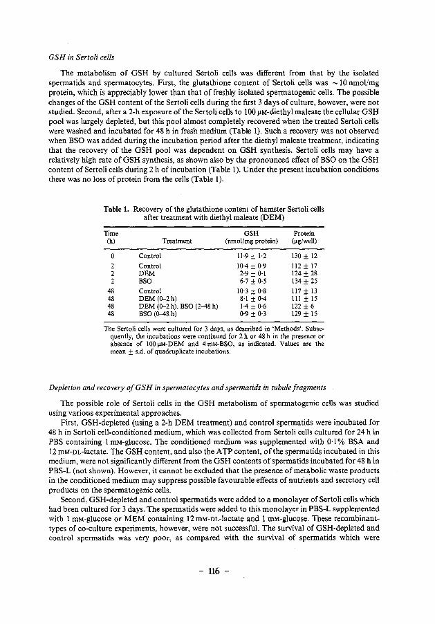
GSH in Sertoli cells
The metabolism of GSH by cultured Sertoli cells was different from that by the isolated spermatids and spermatocytes. First, the glutathione content of Sertoli cells was -10 nmol/mg protein, which is appreciably lower than that of freshly isolated spermatogenic cells. The possible changes of the GSH content of the Sertoli cells during the first 3 days of culture, however, were not studied. Second, after a 2-h exposure of the Sertoli cells to 100 J.lM-diethyl maleate the cellular GSH pool was largely depleted, but this pool almost completely recovered when the treated Sertoli cells were washed and incubated for 48 h in fresh medium (Table 1). Such a recovery was not observed when BSO was added during the incubation period after the diethyl maleate treatment, indicating that the recovery of the GSH pool was dependent on GSH synthesis. Sertoli cells may have a relatively high rate of GSH synthesis, as shown also by the pronounced effect of BSO on the GSH content of Sertoli cells during 2 h of incubation (Table 1). Under the present incubation conditions there was no loss of protein from the cells (Table 1).
Table 1. Recovery of the glutathione content of hamster Sertoli cells after treatment with diethyl maleate (DEM)
Time GSH Protein (h) Treatment (nmolfmg protein) (J.lg/well)
0 Control 11·9 ± 1·2 130 ± 12
2 Control 10·4 ± 0·9 112 ± 17 2 DEM 2·9 ± 0·1 124 ± 28 2 BSO 6·7 ± 0·5 134 ± 25
48 Control 10·3 ± 0·8 117 ± 13 48 DEM (0-2h) 8·1 ± 0·4 Ill± 15 48 DEM (0-2 h), BSO (2-48 h) 1·4 ± 0·6 122 ± 6 48 BSO (0-48h) 0·9 ± 0·3 129 ± 15
The Sertoli cells were cultured for 3 days, as described in 'Methods'. Subsequently, the incubations were continued for 2 h or 48 h in the presence or absence of 100 J.lM-DEM and 4 mM-BSO, as indicated. Values are the mean ± s.d. of quadruplicate incubations.
Depletion and recovery of GSH in spermatocytes and spermatids in tubule fragments
The possible role of Sertoli cells in the GSH metabolism of spermatogenic cells was studied using various experimental approaches.
First, GSH-depleted (using a 2-h DEM treatment) and control spermatids were incubated for 48 h in Sertoli cell-conditioned medium, which was collected from Sertoli cells cultured for 24 h in PBS containing I mM-glucose. The conditioned medium was supplemented with 0·1% BSA and 12 mM-DL-lactate. The GSH content, and also the ATP content, of the spermatids incubated in this medium, were not significantly different from the GSH contents of spermatids incubated for 48 h in PBS-L (not shown). However, it cannot be excluded that the presence of metabolic waste products in the conditioned medium may suppress possible favourable effects of nutrients and secretory cell products on the spermatogenic cells.
Second, GSH-depleted and control spermatids were added to a monolayer of Sertoli cells which had been cultured for 3 days. The spermatids were added to this monolayer in PBS-L supplemented with 1 roM-glucose or MEM containing 12 mM-DL-Iactate and I mM-glucose. These recombinanttypes of co-culture experiments, however, were not successful. The survival of GSH-depleted and control spermatids was very poor, as compared with the survival of spermatids which were
- 116 -
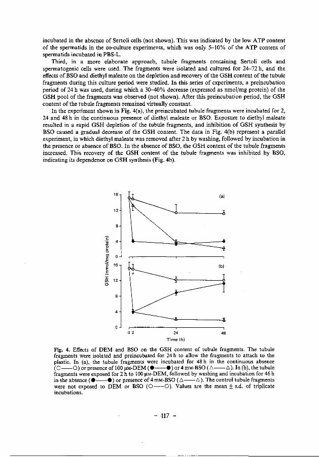
incubated in the absence of Sertoli cells (not shown). This was indicated by the low ATP content of the spermatids in the co-culture experiments, which was only 5-10% of the ATP content of spermatids incubated in PBS-L.
Third, in a more elaborate approach, tubule fragments containing Sertoli cells and spermatogenic cells were used. The fragments were isolated and cultured for 24-72 h, and the effects of BSO and diethyl maleate on the depletion and recovery of the GSH content of the tubule fragments during this culture period were studied. In this series of experiments, a preincubation period of 24 h was used, during which a 30-40% decrease (expressed as nmol/mg protein) of the GSH pool of the fragments was observed (not shown). After this preincubation period, the GSH content of the tubule fragments remained virtually constant.
In the experiment shown in Fig. 4(a), the preincubated tubule fragments were incubated for 2, 24 and 48 h in the continuous presence of diethyl maleate or BSO. Exposure to diethyl maleate resulted in a rapid GSH depletion of the tubule fragments, and inhibition of GSH synthesis by BSO caused a gradual decrease of the GSH content. The data in Fig. 4(b) represent a parallel experiment, in which diethyl maleate was removed after 2 h by washing, followed by incubation in the presence or absence of BSO. In the absence of BSO, the GSH content of the tubule fragments increased. This recovery of the GSH content of the tubule fragments was inhibited by BSO, indicating its dependence on GSH synthesis (Fig. 4b).
16 (a)
12
8
c ·;;; 4 0 :s. Cl 0 .€ 0 16 E .s :I:
12 en Cl
8
4
0 2 24 48
Time (h)
Fig. 4. Effects of OEM and BSO on the GSH content of tubule fragments. The tubule fragments were isolated and preincubated for 24 h to allow the fragments to attach to the plastic. In (a), the tubule fragments were incubated for 48 h in the continuous absence (0-0) or presence of 100 I!M-OEM (e--e) or4 mM-BSO (L':.--L':.). In (b), the tubule fragments were exposed for 2 h to 100 I!M-OEM, followed by washing and incubation for 46 h in the absence (e--e) or presence of 4mM-BSO (L':.--6). The control tubule fragments were not exposed to OEM or BSO (0--0). Values are the mean± s.d. of triplicate incubations.
- 117 -
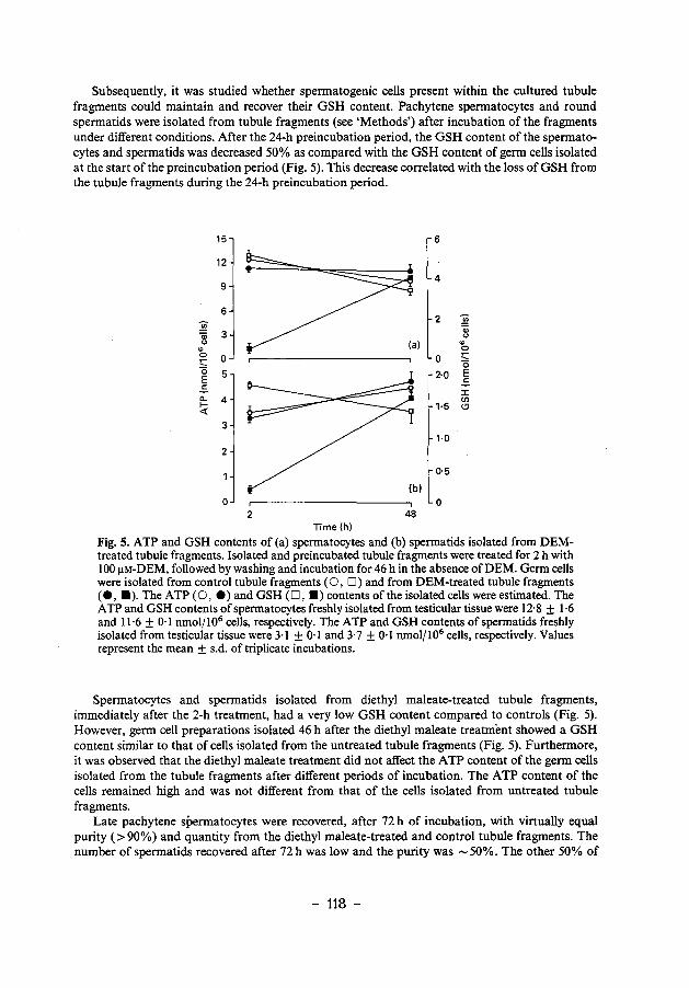
Subsequently, it was studied whether spermatogenic cells present within the cultured tubule fragments could maintain and recover their GSH content. Pachytene spermatocytes and round spermatids were isolated from tubule fragments (see 'Methods') after incubation of the fragments under different conditions. After the 24-h preincubation period, the GSH content of the spermatocytes and spermatids was decreased 50% as compared with the GSH content of germ cells isolated at the start of the preincubation period (Fig. 5). This decrease correlated with the loss of GSH from the tubule fragments during the 24-h preincubation period.
15 6
12
4 9
6 2i 2i
2
3 a;
a; (J
<> ., ., 0 0 0 0 -~ 0 0 5 2·0 E E ..s ..s ::r: a.. 4 en ~ 1·5 (!)
3 1·0
2
0·5
0 0 2 48
Time(h)
Fig. 5. ATP and GSH contents of (a) spermatocytes and (b) spermatids isolated from DEMtreated tubule fragments. Isolated and preincubated tubule fragments were treated for 2 h with 100 J.LM-DEM, followed by washing and incubation for 46 h in the absence ofDEM. Germ cells were isolated from control tubule fragments (0, D) and from DEM-treated tubule fragments ( e, • ). The A TP ( 0, e) and GSH ( 0, •) contents of the isolated cells were estimated. The ATP and GSH contents of spermatocytes freshly isolated from testicular tissue were 12·8 ± 1·6 and 11·6 ± 0·1 nmol/106 cells, respectively. The ATP and GSH contents of spermatids freshly isolated from testicular tissue were 3·1 ± 0·1 and 3·7 ± 0·1 nmol/106 cells, respectively. Values represent the mean ± s.d. of triplicate incubations.
Spermatocytes and spermatids isolated from diethyl maleate-treated tubule fragments, immediately after the 2-h treatment, had a very low GSH content compared to controls (Fig. 5). However, germ cell preparations isolated 46 h after the diethyl maleate treatment showed a GSH content similar to that of cells isolated from the untreated tubule fragments (Fig. 5). Furthermore, it was observed that the diethyl maleate treatment did not affect the ATP content of the germ cells isolated from the tubule fragments after different periods of incubation. The ATP content of the cells remained high and was not different from that of the cells isolated from untreated tubule fragments.
Late pachytene spermatocytes were recovered, after 72 h of incubation, with virtually equal purity (>90%) and quantity from the diethyl maleate-treated and control tubule fragments. The number of spermatids recovered after 72 h was low and the purity was -50%. The other 50% of
- 118 -
the cells in these spermatid preparations were mainly early primary spermatocytes. The germ cells recovered from the tubule fragments were resistant to trypsin and DNAase treatment, and were selected according to size and density. This, and also the ATP content of the cells, demonstrates that viable cells were recovered.
Discussion
In this study of the possible role of Sertoli cells in glutathione metabolism of spermatocytes and spermatids, the main approach was to evoke GSH depletion of isolated spermatogenic cells and tubule fragments and to estimate the recovery of the cellular GSH pool. Two methods were used to lower the glutathione levels. First, glutathione synthesis was inhibited using buthionine sulphoximine (BSO) (Griffith & Meister, 1979; Griffith, 1981), which is a specific inhibitor of y-glutamylcysteine synthetase (EC 6.3.2.2). Second, the cells were incubated with a substrate for glutathione S-transferase (GST). This enzyme catalyses the conjugation between GSH and electrophilic compounds. The a,~-unsaturated diester diethyl maleate is a suitable GST substrate and can be used to decrease the GSH level in intact animals via a GST-catalysed conjugation reaction (Allameh et a/., 1987). This compoJ.!nd can also be used to cause GSH depletion of cultured cells, including Chinese hamster ovary cells (Mitchell et a!., 1983; Rice et a!., 1986), human lung carcinoma cells (Brodie & Reed, 1985) and round spermatids from rats (Den Boer eta!., 1988), and was used in the present experiments to deplete the GSH pool of isolated spermatogenic cells, cultured Sertoli cells and tubule fragments from hamsters.
In isolated spermatocytes and spermatids from hamsters, a gradual decrease of the GSH content was observed during prolonged incubation. Under control incubation conditions this diminution did not lead to a concomitant loss of the viability of the cells, as indicated by the maintenance of the cellular A TP content. The decrease of the GSH content could reflect a low rate of GSH synthesis, relative to the rate of GSH expenditure. The synthesis of GSH in the germ cells may require exogenous compounds, but addition of glycine, cysteine and glutamate, or glutathione to the incubations did not prevent the gradual loss of cellular GSH. Furthermore, the GSH content of isolated spermatids was also not elevated using the compound L-2-oxothiazolidine-4-carboxylate, an oxoproline analogue which serves as a cysteine delivery system and can cause an increase of the GSH level of lymphocytes (Williamson eta!., 1982; Fidelus eta!., 1987).
The gradual loss ofGSH from the isolated germ cells is not explained by a complete absence of GSH synthesis in these cells. The isolated spermatocytes and spermatids appear to have a limited capacity to synthesize GSH. This is indicated by the observation that the decrease of the GSH pool was slightly accelerated during incubation of the cells in the presence of 50 J.!M-BSO. This effect on the cellular GSH content was not caused by a non-specific toxic effect of BSO, since the ATP content of isolated spermatids remained high after incubation in the presence of much higher BSO concentrations, up to 4 mM. The time-course of the BSO-induced depletion of GSH in spermatocytes and spermatids showed that the cellular GSH content was decreased only approximately 50% after 24 h of incubation.
It is not clear which biochemical processes determine the loss of GSH from the isolated spermatogenic cells. GSH can be used as an intracellular source of cysteine for incorporation into proteins. Another process may involve the export from the cells of GSH-related compounds, including oxidized glutathione (GSSG), GSH mixed disulphides, and GSH-conjugates (Meister & Anderson, 1983). Obviously, this latter mechanism of utilization of GSH is important for GSH-supported defence mechanisms.
Exposure of isolated rat spermatids to GST substrates leads to cellular GSH depletion as a result of the formation of conjugates (Den Boer et a!., 1988). This mechanism was used in the present experiments to deplete the GSH pool of isolated hamster spermatocytes and spermatids, by incubating the cells in the presence of diethyl maleate. This treatment did not affect the viability of
- 119 -
the cells, as indicated by the high ATP contents of the spermatogenic cells. With respect to the present discussion on GSH synthesis, it should be emphasized that isolated GSH-depleted spermatids did not recover their GSH content during prolonged incubation under different conditions (see 'Results').
Cultured Sertoli cells from immature hamsters have an active GSH synthesis system, as indicated by the recovery of the GSH pool after diethyl maleate-induced depletion, which was prevented by addition of BSO to the medium. We examined whether GSH synthesis in spermatogenic cells is dependent on the presence of Sertoli cells. A possible action of Sertoli cells on GSH synthesis in germ cells may not be exerted by a soluble factor. This is indicated by the observation that there was no effect of Sertoli cell-conditioned medium or Sertoli cell-germ cell co-culture on the GSH content of control and diethyl maleate-treated spermatids during 48 h of incubation (not shown). A possible role of Sertoli cells, however, was indicated in experiments using tubule fragments. In these experiments a preincubation period of 24 h was included to allow the tubule fragments to attach to the plastic surface of the culture wells or flasks. During this 24-h period there was a marked decrease of the GSH pool of the tubule fragments, which may reflect a decrease of the GSH content of the spermatogenic cells, rather than a loss of spermatogenic cells, for the following reasons. Cultured Sertoli cells showed a GSH content of 10 nmolfmg protein, whereas spermatids and spermatocytes contained 30 and 40 nmol GSH/mg protein. These approximate values indicate that most of the GSH in tubular fragments is present in the germ cells. Spermatids and spermatocytes isolated from the preincubated tubule fragments showed a GSH content which was 50% lower than that of germ cells freshly isolated from testicular tissue. The cause of this initial rapid decrease of the GSH pool is not known.
The indication for a role of Sertoli cells in germ-cell GSH synthesis was obtained by diethyl maleate treatment of the preincubated tubule fragments. The GSH depletion of the tubule fragments induced by the diethyl maleate treatment corresponded with a GSH depletion of the germ cells present within the fragments, down to a content of less than 10% of the controls. The subsequent recovery of the GSH content of the tubule fragments after removal of the diethyl maleate also corresponded with a recovery of the GSH content of the spermatogenic cells enclosed in the tubule fragments, up to control levels (a 10-fold increase). As discussed above, such a recovery of the GSH pool was not observed for diethyl maleate-treated isolated spermatocytes and spermatids.
From these observations, it can be suggested that germ cells in tubule fragments can synthesize GSH at a sufficiently high rate to recover from GSH depletion, whereas isolated germ cells may have a much lower rate of GSH synthesis. This points to a role of Sertoli cells in GSH synthesis by the germ cells. This aspect of Sertoli cell function might involve intercellular transport of amino acids, or a more direct stimulatory effect on GSH biosynthesis through an unknown mechanism.
The membrane-bound enzyme y-glutamyl transpeptidase (GGT; EC 2.3.2.2) is possibly involved in GSH synthesis through its function in the uptake of amino acids. The enzyme is thought to transfer the y-glutamyl group of extracellular GSH to an extracellular amino acid. The y-glutamyl amino acid is then transported into the cell (Meister & Anderson, 1983). The enzyme activity may be a rate-limiting factor in transport of a number of amino acids, including cysteine, and the maintenance of cellular glutathione levels. High activities of this enzyme are present in Sertoli cells, and the activity of the enzyme increases during post-natal testicular development (Lu & Steinberger, 1977). The activity of GGT in isolated spermatids is low (Lu & Steinberger, 1977), but this needs to be confirmed using a germ cell isolation procedure which does not include enzyme treatments, since GGT activities can be removed from cell surfaces by proteolytic enzymes (Meister et a!., 1981). If indeed the GGT activity of germ cells in situ is low, the germ cells may take up y-glutamyl amino acids produced by Sertoli cells.
The present results indicate that cell-to-cell interaction may be important for the role of Sertoli cells in glutathione synthesis in spermatogenic cells. The biochemical mechanism of this interaction, however, remains to be clarified.
- 120 -
This investigation received financial support from the World Health Organization Special Programme of Research, Development and Research Training in Human Reproduction (project 87079).
References
Allameh, A., Saxena, M., Mehta, L.N. & Raj, H.G. (1987) Comparison of pulmonary and hepatic glutathione depletion by diethyl maleate. Med. Sci. Res. 15, 957-958.
Bradford, M.M. (1976) A rapid and sensitive method for the quantitation of microgram quantities of protein utilizing the principle of protein-dye binding. Analyt. Biochem. 72, 248-254.
Brodie, A.E. & Reed, D.J. (1985) Buthionine sulfoximine of cystine uptake and glutathione biosynthesis in human lung carcinoma cells. Toxicol. appl. Pharmacal. 77,381-387.
Calvin, H.I. & Turner, S.I. (1982) High levels of glutathione attained during postnatal development of rat testis. J. exp. Zoo/. 219, 389-393.
Calvin, H.l., Medvedovsky, C. & Worgul, B.V. (1986) Near-total glutathione depletion and age-specific cataracts induced by buthionine sulfoximine in mice. Science, NY233, 553-555.
Den Boer, P.J. & Grootegoed, J.A. (1988a) Mechanism of action of (-)gossypol on A TP production in isolated hamster spermatids. J. Reprod. Fert. 83, 693-700.
Den Boer, P.J. & Grootegoed, J.A. (1988b) Differential effects of ( + )- and (-)-gossypol enantiomers on LDH-C4 activity of hamster spermatogenic epithelium in vitro. J. Reprod. Fert. 83, 701-709.
Den Boer, P.J., Verkerk, A. & Grootegoed, J.A. (1988) Effect of xenobiotics on glutathione and A TP levels in isolated round spermatids from rats. In The Molecular and Cellular Endocrinology of the Testis, pp. 241-246. Eds B. A. Cooke & R. M. Sharpe. Raven Press, New York.
Fidelus, R.K., Ginouves, P., Lawrence, D. & Tsan, M.-F. (1987) Modulation of intracellular glutathione concentrations alters lymphocyte activation and proliferation. Expl Cell Res. 170, 269-275.
Galdieri, M., Ziparo, E., Palombi, F., Russo, M.A. & Stefanini, M. (1981) Pure Sertoli cell cultures: a new model for the study of somatic-germ cell interactions. J. Androl. 5, 249-254.
Griffith, O.W. (1981) Depletion of glutathione by inhibition of biosynthesis. In Methods of Enzymology, vol. 77, pp. 59-{;3. Ed. B. Jakoby. Academic Press, New York.
Griffith, O.W. & Meister, A. (1979) Potent and specific inhibition of glutathione synthesis by buthionine sulfoximine (S-n-butyl homocysteine sulfoximine). J. bioi. Chern. 254, 7558-7560.
Grootegoed, J.A., Jansen, R. & van der Molen, H.J. (1984) The role of glucose, pyruvate and lactate
in A TP production by rat spermatocytes and spermatids. Biochim. Biophys. Acta 767,248-256.
Grootegoed, J.A., Jensen, R. & van der Molen, H.J. (1985) Intercellular pathway of leucine catabolism in rat spermatogenic epithelium. Biochem. J. 226, 889-892.
Grosshans, K. & Calvin, H.I. (1985) Estimation of glutathione in purified populations of mouse testis germ cells. Bioi. Reprod. 33, 1197-1205.
Hissin, P.J. & Hilf, R. (1976) A ftuorometric method for determination of oxidized and reduced glutathione in tissues. Analyt. Biochem. 74,214-226.
Ketterer, B. (1986) Detoxication reactions of glutathione and glutathionetransferases. Xenobiotica 16,957-973.
Lu, C. & Steinberger, A. (1977) Gamma-glutamyl transpeptidase activity in the developing rat testis. Enzyme localization in isolated cell types. Bioi. Rep rod. 17, 84-88.
Meister, A. & Anderson, M.E. (1983) Glutathione. Ann. Rev. Biochem. 52, 7ll-760.
Meister, A., Tate, S.S. & Griffith, O.W. (1981) yGlutamyl transpeptidase. In Methods of Enzymology, vol. 77, pp. 237-253. Ed. B. Jakoby. Academic Press, New York.
Mitchell, J.B., Russo, A., Biaglow, J.E. & McPherson, S. (1983) Cellular glutathione depletion by diethyl maleate or buthionine sulfoximine: no effect of glutathione depletion on the oxygen enhancement ratio. Radiation Res. 96, 422-428.
Oonk, R.B., Grootegoed, J.A. & van der Molen, H.J. ( 1985) Comparison of the effects of insulin and follitropin on glucose metabolism by Sertoli cells from immature rats. Molec. cell. Endocrinol. 42, 39-48.
Rice, G.C., Bump, E.A., Shrieve, D.C., Lee, W. & Kovacs, M. (1986) Quantitative analysis of cellular glutathione by flow cytometry utilizing monochlorobimane: some applications to radiation and drug resistance in vitro and in vivo. Cancer Res. 46, 6105-{;110.
Teaf, C.M., Bishop, J.B. & Harbison, R.D. (1987) Depression of glutathione in male reproductive tissues and potentiation of EMS-induced germ cell mutagenesis by L-buthionine sulfoximine. Teratogenesis, Carcinogenesis, and Mutagenesis 7, 497-513.
Williamson, J.M., Boettcher, B. & Meister, A. (1982) Intracellular cysteine delivery system that protects against toxicity by promoting glutathione synthesis. Proc. natn. Acad. Sci. USA 79, 6246-{;249.
Received 7 March 1989
- 121 -
Summary and Concluding Remarks
Spermatogenesis is a complex process of mitotic and meiotic cell divisions and cell differentiation, resulting in the production of spermatozoa from undifferentiated spermatogonia. The development of the spermatogenic cells is dependent on supporting activities of Sertoli cells. The Sertoli cells are target cells for folliclestimulating hormone (FSH) and testosterone.
Spermatogenic cells and spermatozoa possess a number of specific biochemical properties. Such properties are possible targets for various toxic compounds which cause disruption of spermatogenesis. Biochemical characteristics of advanced spermatogenic cells include the presence of the lactate. dehydrogenase isoenzyme LDH-C 4 (a key enzyme in the metabolism of the male germ cells) and the remarkable role of the mitochondria in the cellular energy metabolism.
Effects of toxic compounds on male reproductive function have been evaluated mainly in vivo on basis of testicular morphology and the fertilizing capacity of sperm as evaluated in mating tests. However, a number of molecular and cellular aspects of spermatogenesis can be studied in vitro. Spermatocytes, spermatids and Sertoli cells can be isolated and biochemical activities can be analyzed. In the experiments described in this thesis two in vitro systems were used, viz. fragments of the spermatogenic epithelium (Chapter 2, 7) and isolated cells (Sertoli cells, pachytene spermatocytes and round spermatids) (Chapter 3,4,5,6) from rats and hamsters. We have investigated whether specific biochemical properties of spermatids are targets for antispermatogenic agents.
Two groups of toxic agents were studied, viz. agents acting on energy metabolism and agents acting on glutathione. The energy metabolism of spermatids was studied because there are indications that ATP production in spermatids is a highly vulnerable process. Glutathione was studied since this compound is involved in the inactivation of toxic compounds and may also be a target for antispermatogenic agents. The effect of the toxic agents was investigated by estimation of the cellular ATP content, glutathione content and enzyme activities, following incubation of the cells in the presence of toxic agents.
- 122 -
The results of the experiments described in Chapter 2 and 3, concern the mechanism of action of gossypol, a well-known antispermatogenic compound. In cultured spermatogenic epithelium, an increase of the specific LDH-C 4 activity, which was observed during 48 h of culture in the absence of gossypol, was suppressed by addition of the (-) enantiomer of gossypol to the cultures. It was concluded that (-) gossypol either exerted a direct inhibitory effect on the enzyme or caused degeneration of spermatogenic cells and a loss of LDH-C 4 from the cells. In the experiments described in Chapter 3, using isolated round spermatids, it was observed that the ATP content of the spermatids was strongly decreased after 18 h of incubation in the presence of (-) gossypol, without a concomitant inhibition of the LDH-C 4 enzyme. This decrease of the ATP content was not accompanied by an effect of gossypol on the mitochondrial oxidation of pyruvate. It was concluded that (-) gossypol may bind with a high affinity to specific mitochondrial proteins which are involved in the coupling between electron transport and ATP synthesis.
In Chapter 4, ATP metabolism in spermatids is described in more detail. The experiments described show that the ATP content of isolated hamster spermatids is maintained when the cells utilize added lactate as an energy-yielding substrate, but can be lowered by high concentrations of glucose also in the presence of lactate. The A TP content was increased approximately 2 fold by incubation of the cells in medium containing adenosine. It was shown that spermatids isolated and incubated in the presence of lactate had an ATP content of approximately 4 nmol/106 cells and an energy charge > 0.90, and it is discussed that the ATP content of the spermatids in situ in the presence of low glucose concentrations may be very stable.
The metabolism of glutathione, as a possible target and defence system for antispermatogenic compounds, is described in the Chapters S, 6 and 7. Chapter S is the only chapter dealing with spermatogenic cells from rats. In subsequent experiments we have used hamsters (Chapter 6 and 7). In Chapter S two different glutathione-dependent defence mechanisms were studied in isolated rat spermatids. It was concluded that the spermatids possess an active glutathione redox cycle, although the capacity is limited. Furthermore, it was found that the cells were able to conjugate a number of xenobiotics with glutathione, catalyzed by glutathione S-transferase activity. The spermatids were partly depleted of
- 123 -
glutathione after exposure to glutathione S-transferase substrates, but this did not affect the viability of the cells. However, it was anticipated that the glutathione-depleted cells would be more vulnerable to oxidative stress or xenobiotic compounds.
In Chapter 6 experiments are described which show that the defence mechanisms demonstrated in Chapter S provide protection against a number of exogenous compounds. Exposure of glutathionedepleted spermatids to low concentrations of peroxides or alkylating agents resulted in a loss of cell viability, whereas under these conditions control cells were not affected. The high glutathione content, furthermore, restricted the induction of single-strand breaks in DNA by gamma radiation. The induction was 3-5 fold higher in GSH-depleted spermatids as compared to control cells.
The glutathione level in isolated spermatids and late- pachytene spermatocytes was very high, but the cells showed a relatively low rate of glutathione biosynthesis which was not adequate to maintain the high cellular GSH content during prolonged incubation of the cells (Chapter 7). Sertoli cells, on the other hand, had a relatively low glutathione content, whic,h showed a considerable turnover and was restored after depletion. Isolated late-pachytene spermatocytes and round spermatids were not able to replenish their glutathione content after depletion of the cellular glutathione pool. However, when the spermatocytes and spermatids were in contact with Sertoli cells in tubule fragments, depletion of the glutathione levels of the spermatogenic cells was followed by recovery. It was concluded that Sertoli cells probably play a role in glutathione synthesis by the germ cells, and a mechanism which involves intercellular transport of amino acids is suggested.
CONCLUDING REMARKS
ATP synthesis in spermatids is a sensitive target for antispermatogenic agents. The (-) enantiomer of gossypol exerts a deleterious effect on spermatogenic cells most likely through its action on mitochondrial ATP synthesis. Under the present incubation conditions, the (+) enantiomer showed not such an effect. It can be postulated that gossypol action involves high affinity binding to a mitochondrial protein. This interaction might be stereospecific, resulting in inhibition of the biological activity of the protein. This protein could be directly involved in ATP production, and
- 124 -
one possibility is that this protein is part of the F1F0
-ATP synthetase complex. However, this action of gossypol is probably not specific for germ cells. The more or less specific effect of gossypol in vivo on spermatogenic cells and spermatozoa may be related to the very high sensitivity of these cell types to inhibitors of mitochondrial function rather than to interaction of gossypol with proteins which are unique to the germ cells.
Glutathione in spermatids apparently is not a target for antispermatogenic agents. Isolated spermatids and spermatids associated with Sertoli cells become depleted of glutathione upon exposure to glutathione S-transferase substrates. However, germ cells in situ can recover due to their capacity to synthesize glutathione which involves Sertoli cells. From this it can be concluded that long-term exposure of the testis to low doses of glutathione S-transferase substrates will not result in glutathione depletion of the developing germ cells. It is anticipated that in vivo the epididymal spermatozoa may be subject to irreversible GSH depletion, since the epididymal spermatozoa are not directly protected and supported by Sertoli cells. Different compounds may gain access to the epididymis via active or passive transport, and in this manner drugs, which are substrates for glutathione S-transferase activity in spermatozoa, might interfere with male fertility. In this respect, it would be of great interest to investigate glutathione S-transferase isoenzymes in spermatozoa and the effect of glutathione depletion on sperm function.
The present results indicate that in vitro systems are suitable to evaluate effects of antispermatogenic agents on biochemical activities of spermatogenic cells. The experiments have provided information on vulnerable biochemical processes in these cells, which are possible targets. Knowledge on toxic effects of exogenous compounds may give further insight in the possible role of Sertoli cells and other fundamental aspects of the regulation of spermatogenesis.
- 125 -
Samenvatting
De vorming van zaadcellen (spermatozoa) uit voorloper cellen vindt plaats in de zaadbuisjes (tubuli seminiferi) in de zaadbal (testikel of testis). Dit proces wordt spermatogenese genoemd. De initiatie en de instandhouding van de spermatogenese staat onder controle van hormonen, met name van follikel-stimulerend hormoon (FSH) en testosteron. Deze hormonen hebben echter geen direct effect op de zich ontwikkelende zaadcellen, maar oefenen hun invloed uit op de Sertoli cellen, de steuncellen die nauw verbonden zijn met de spermatogenetische cellen in de zaadbuisjes en van groot belang zijn voor een goede ontwikkeling van deze cellen.
Spermatozoa zijn zeer gespecialiseerde cellen, en hun functioneren hangt af van een lange reeks van specifieke biochemische en celbiologische processen tijdens de ontwikkeling van de spermatogenetische cellen. Deze ontwikkeling start met een aantal (mitotische) delingen van spermatogonia, welke leidt tot de vorming van spermatocyten. Uit de spermatocyten ontstaan, door middel van (de meiotische delingen), spermatiden, die zich verder zonder celdeling ontwikkelen tot spermatozoa. Veel biochemische eigenschappen van spermatiden zijn uitvoerig onderzocht. Een daarvan is het voorkomen van eiwitten die aileen in spermatogenetische cellen en zaadcellen worden aangetroffen, zoals het lactaatdehydrogenase isoenzym LDH-C 4 . Een ander aspect betreft het koolhydraat metabolisme. Spermatiden zijn voor hun energievoorziening aangewezen op lactaat en pyruvaat en zijn daardoor afhankelijk van een goed functioneren van hun mitochondria.
De ontwikkeling van spermatogenetische cellen en de maturatie van spermatozoa is niet aileen gebaseerd op complexe processen die plaats vinden in deze cellen, maar is sterk afhankelijk van het extracellulair milieu in de zaadbuisjes en in de epididymis (bijbal). Verandering van dit milieu door bijvoorbeeld binnendringende giftige stoffen, kan leiden tot verstoring van de spermatogenese en zelfs tot infertiliteit (onvruchtbaarheid). Het is gebleken dat een groot aantal (giftige) stoffen van uiteenlopende aard preferentieel de spermatogenese verstoord.
Een effect van toxische verbindingen op de spermatogenese
- 126 -
wordt veelal bestudeerd met behulp van dierexperimenten. Hierbij wordt na toediening van deze stoffen bijvoorbeeld gekeken naar de histologie van de testis, het aantal geproduceerde spermatozoa, en naar het fertilizerend vermogen van de spermatozoa. Een aantal aspecten van de spermatogenese kan echter ook onderzocht worden door gebruik te maken van stukjes testisweefsel (fragmenten van de zaadbuisjes, spermatogenetisch epitheel) of door gebruik te maken van geisoleerde en gezuiverde cellen (Sertoli cellen, spermatocyten en spermatiden). In de experimenten beschreven in dit proefschrift is van deze mogelijkheid gebruik gemaakt. De nadruk ligt op het bestuderen van effecten van verschillende verbindingen op spermatiden.
In dit proefschrift is de mogelijkheid onderzocht of antispermatogenetische verbindingen een effect hebben op karakteristieke biochemische eigenschappen van spermatiden, met name LDH-C 4 en het energiemetabolisme. Daarnaast werd het metabolisme van glutathion in spermatiden onderzocht, omdat glutathion betrokken is bij het onschadelijk maken van giftige verbindingen. Indien glutathion concentraties of het glutathion metabolisme beinvloed wordt door antispermatogenetische verbindingen zou dit een oorzaak kunnen zijn van de antispermatogenetische effecten. Glutathion is in vrijwel aile lichaamscellen aanwezig en is niet aileen belangrijk voor het onschadelijk maken van giftige stoffen maar speelt ook een belangrijke rol bij tal van andere processen, zoals eiwit- en DNA synthese.
In de experimenten beschreven in de Hoofdstukken 2 en 3 is het werkingsmechanisme van gossypol onderzocht. Gossypol is een verbinding geisoleerd uit de katoenplant (Gossypium spec.) die de spermatogenese remt. De anticonceptieve werking van deze verbinding is bij toeval in China ontdekt en gossypol is daar gebruikt als "mannenpil". De wijze waarop gossypol de spermatogenese remt is echter niet bekend. Als mogelijke aangrijpingspunten zijn in de literatuur LDH-C 4 en ATP metabolisme genoemd. Gossypol geisoleerd uit de katoenplant is een racemisch mengsel (+/-), waarvan aileen de (-) vorm werkzaam is. In een kweek van fragmenten van het spermatogenetische epitheel bleek dat in aanwezigheid van (-) gossypol de LDH-C 4 activiteit sterk verlaagd was. Hieruit werd geconcludeerd dat gossypol of een direct, remmend, effect op het enzym uitoefende, of da.t het degeneratie van LDH-C4 bevattende cellen veroorzaakte. De afname van de enzymactiviteit kan, in het laatste geval, veroorzaakt worden door
- 127 -
lekkage van het enzym uit de degenererende cellen. In de experimenten beschreven in Hoofdstuk 3, zijn deze beide
mogelijkheden onderzocht. Hierbij werd gebruik gemaakt van geisoleerde spermatiden. Het bleek dat het ATP gehalte van de spermatiden sterk verlaagd was na 18 uur incubatie in aanwezigheid van (-) gossypol, terwijl de activiteit van het LDH-C4 niet geremd was. Uit deze en andere waarnemingen werd geconcludeerd dat gossypol niet direct aangrijpt op LDH-C 4 , maar mogelijk invloed heeft op een mitochondriaal eiwit betrokken bij de koppeling tussen electronenoverdracht en A TP synthese.
In Hoofdstuk 4 is het A TP metabolisme van spermatiden nader beschreven. Het A TP gehal te van spermatiden kon verlaagd worden door incubatie van de cellen met glucose, terwijl het toevoegen van lactaat On aan- of afwezigheid van lage glucoseconcentraties) de energievoorziening veilig kon stellen. De energy charge van geisoleerde spermatiden was grater dan 0.90. Spermatiden geincubeerd in aanwezigheid van adenosine vertoonden een verdubbel_ing van hun ATP gehalte. Het ATP gehalte van de spermatiden in situ zal waarschijnlijk ongeveer 4 nmol/106 cellen zijn.
In de experimenten beschreven in de Hoofdstukken S, 6 en 7 is onderzocht in welke mate glutathion in spermatogenetische cellen betrokken is bij de verdediging tegen giftige stoffen, maar ook of glutathion daardoor een doelwit is voor stoffen die de spermatogenese verstoren.
In Hoofdstuk S werden twee verschillende glutathion afhankelijke verdedigingssystemen in spermatiden van ratten onderzocht. Het bleek dat de spermatiden een actieve glutathion redox cyclus hebben en dat in de spermatiden een aantal giftige stoffen geconjugeerd kunnen worden met glutathion. Dit laatste leidde tot een daling van het glutathion gehalte in de· spermatiden met meer dan 90%, hetgeen echter geen invloed had op het A TP gehalte van de cellen.
De effectiviteit van beide verdedigingssystemen is beschreven in Hoofdstuk 6. Deze effectiviteit werd bepaald door de effecten van een peroxide en een alkylerende verbinding op spermatiden met een hoog of juist met een laag glutathion gehalte te bestuderen. Het bleek dat deze verbindingen bij de spermatiden met een laag glutathion gehalte celdood veroorzaakten, terwijl de controle cell en met een hoog glutathion gehal te, onder vergelijkbare omstandigheden, in Ieven bleven. Daarnaast werd waargenomen dat het hoge glutathion gehalte in spermatiden een beschermend effect
- 128 -
uitoefende tegen inductie van DNA schade door ioniserende straling. Er werd geconcludeerd dat glutathion in spermatiden, via verschillende mechanismen, een belangrijke beschermende rol speelt.
In dit opzicht was bet interessant om te onderzoeken of de spermatiden zelf instaat zijn glutatbion te syntbetiseren, om daarmee langdurig weerstand te kunnen bieden tegen toxische verbindingen. Biosynthese van glutathion door· spermatocyten, spermatiden en Sertoli cell en is bescbreven in Hoofdstuk 7. Het glutatbion gehalte in geisoleerde spermatocyten en spermatiden is relatief hoog, maar geisoleerde cellen konden dit niveau niet bandbaven, hetgeen wijst op een relatief lage de novo syntbese snelbeid. Sertoli cellen bleken een laag glutatbion gebalte te hebben, maar een hoge glutathion turn-over. De geisoleerde spermatogenetiscbe cellen konden hun glutatbion gehalte niet bestellen na depletie van hun intracellulaire voorraad. Echter, indien deze cellen in contact met Sertoli cellen stonden (in fragmenten van bet spermatogenetische epitheel) dan werd depletie van het glutathion gehalte gevolgd door herstel. Er werd geconcludeerd dat Sertoli cellen een rol spelen in de glutathion synthese van spermatiden, mogelijk door intercellulair transport van aminozuren.
De belangrijkste conclusies uit de resultaten van het beschreven
onderzoek zijn:
ATP syntbese in spermatiden is een zeer kwetsbaar proces en kan verstoord worden door antispermatogenetische verbindingen. De (-) enantiomeer van gossypol heeft een schadelijk effect op germinale cellen, hetgeen waarschijnlijk wordt veroorzaakt door een effect op de mitochondriale ATP synthese. De (+) enantiomeer was, onder de gebruikte incubatieomstandigheden, niet actief. Op grond van de resultaten wordt verondersteld dat (-) gossypol werkt door binding aan een mitochondriaal eiwit. Deze binding zou kunnen berusten op een stereospecifieke interactie en kunnen resulteren in remming van de biologische activiteit van dit eiwit. Een mogelijkheid is, dat dit eiwit deel uitmaakt van bet F0 FcATP synthetase complex. Het toxisch effect van gossypol lijkt niet specifiek gericht te zijn op spermatogenetische cellen, want het heeft ook een effect op andere celtypen. Het feit dat gossypol
- 129 -
schadelijke effecten te zien geeft op mannelijke fertiliteit, wijst wei op een hoge gevoeligheid van de spermatogenetische cellen en spermatozoa voor remmers van mitochondriale functies, maar staat waarschijnlijk niet in verband met binding van gossypol aan bepaalde eiwitten die exclusief voorkomen in deze cellen.
Glutathion in spermatiden is waarschijnlijk geen doel wit voor antispermatogenetische verbindingen. Blootstelling van spermatiden aan substraten voor glutathion $-transferase leidde tot een drastische verlaging van het glutathion gehalte, maar spermatogenetische cellen in situ, in samenwerking met Sertoli cellen, kunnen zich herstellen. Hieruit kan geconcludeerd worden dat langdurige blootstelling van de testis aan lage doses glutathion $-transferase substraten waarschijnlijk niet zal leiden tot een verlaging van het glutathion gehalte in de spermatogenetische cellen. Een mogelijkheid zou kunnen zijn dat in spermatozoa, in de epididymis Un vivo), wei een blijvende verlaging van het glutathion gehalte bewerkstelligd kan worden. Immers, verschillende verbindingen kunnen de epididymis goed bereiken en de spermatozoa in de epididymis worden niet meer direkt beschermd en ondersteund door de Sertoli cellen. Op grond van deze waarnemingen kan verondersteld worden dat bepaalde substraten voor glutathlon S-transferase de mannelijke fertiliteit kunnen verstoren. Het zou in dit verband interessant zijn om onderzoek 'te doen naar de aanwezigheid van verschillende glutathion S-transferase isoenzymen in spermatozoa en naar het effect van verlaging van bet glutathion gehalte in spermatozoa op hun bevruchtend vermogen.
De resultaten beschreven in dit proefschrift geven aan dat de gebruikte in vitro systemen geschikt zijn om biochemische effecten van antispermatogenetische verbindingen te bestuderen. De experimenten hebben duidelijke aanwiJzmgen opgeleverd voor mogelijke kwetsbare biochemische processen waarop exogene toxische verbindingen kunnen aangrijpen. Kennis van toxische effecten van bepaalde verbindingen op de spermatogenese kan tevens een bijdrage leveren aan het inzicht in de ondersteunende rol van de Sertoli cellen en andere aspecten voor de regulatie van de spermatogenese.
- 130 -
Papers related to the thesis
P.J. Den Boer and J.A. Grootegoed (1987). Effects of (-) and (+)enantiomers of gossypol on isolated round spermatids from hamsters. Ann. N.Y. Acad. Sci. 513, 535-537.
P.J. Den Boer, A. Verkerk and J.A. Grootegoed (1988). Effects of xenobiotics on glutathione and ATP levels in isolated round spermatids from rats. In: The Molecular and Cellular Endocrinology of the Testis, pp. 241-246. Eds B.A. Cooke and R.M. Sharpe. Raven Press, New York,
J.A. Grootegoed, P.J. Den Boer and P. Mackenbach (1989). Sertoli cell-germ cell communication. Ann. N.Y. Acad. Sci. 564, 232-242.
J.A. Grootegoed, P.J. Den Boer and P. Mackenbach (1989) Role of Sertoli cells in spermatogenesis. In: Perspectives in Andrology, pp. 167-173. Ed. M. Serio, Raven Press, New York,
- 131 -
CURRICULUM VITAE
Pieter johannes den Boer werd op 28 januari 1959 geboren te Dordrecht. In 1977 werd het diploma Atheneum B aan het Christelijk Lyceum te Dordrecht behaald. In hetzelfde jaar werd een begin gemaakt met de studie biologie aan de Rijksuniversiteit te Leiden. Het kandidaats-examen, met als specialisatierichting scheikunde <B4 ), werd in september 1980 afgelegd. Het doctoraalexamen werd op 29 mei 1984 behaald, met als hoofdvakken biologie en biochemie (dr. ir. L. Havekes, Gaubius Instituut, TNO Leiden; Prof. dr. P.H. van Knippenberg, vakgroep Biochemie, RUL) en bijvak dierenmorfologie (Prof, dr, j.L. Dubbeldam, vakgroep Organismale Zoologie, RUU.
Naast de studie biologie is hij van september 1983 tot augustus 19_84 medewerker geweest aan een morfometrisch onderzoek (Multi Centered Placenta Project) binnen de Stichting Samenwerking Delftse Ziekenhuizen (SSDZ) op de afdeling Kwantitatieve Pathologie.
In de periode september 1984 tot maart 1985 is werkzaam geweest als gastmedewerker bij de vakgroep Organismale Zoologie van de Rijksuniversiteit Leiden.
Van 1 april1985 tot 1 oktober 1988 was hij aangesteld als wetenschappelijk medewerker aan de Erasmus universiteit te Rotterdam op de afdeling Biochemie II (Chemische Endocrinologie). De resultaten van het daar verrichtte onderzoek zijn in dit proefschrift weergegeven.
Vanaf 15 november 1989 is hij werkzaam als biochemicus op de afdeling Cryobiologie van het Centraal Laboratorium van de Bloedtransfusiedienst van het Nederlandse Rode Kruis.
- 132 -
NAWOORD
Tot slot wil ik iedereen bedanken voor alle bijdragen, in wat voor vorm dan ook, welke de totstandkoming van dit proefschrift mogelijk hebben gemaakt.
Mijn promotor Henk van der Molen , die ondanks zijn drukke werkzaamheden mijn onderzoek op de voet heeft gevolgd.
Mijn co-promotor Anton Grootegoed voor de begeleiding van het onderzoek en het grondig doorlezen en verbeteren van alle manuscripten. Zijn vele ideeen en verbazende kennis van de biochemie waren een grote stimulans.
De !eden van de promotiecommissie prof. dr. jongkind, prof. dr. Koster en prof. dr. van der Werff ten Bosch voor het beoordelen van het proef schriftmanuscript.
ledereen van de afdeling biochemie II voor de, in vele opzichten, onvergetelijke tijd.
Een aantal (voormalige) medewerkers van "518" wil ik hier met name noemen,
Ruud, jij introduceerde mij in de kunst van het isoleren en zuiveren van cellen, daarnaast waren er nog tal van andere (niet) wetenschappelijke onderwerpen waarover we uitgebreid (gezellig) hebben gepraat.
Ingrid, voor ontspannende momenten in spannende tijden. Robert, jij bracht klank en kleur op de afdeling. Petra (onbetaalbaar, maar gelukkig hield je van yoghurt) en
Leen ( sommige liedjes zal ik nu nooit meer vergeten), het doet me een groat genoegen dat jullie heiden me ook als paranimfen terzijde willen staan.
jonathan en Carina jullie hebben me Iaten zien dat DIT niet echt het belangrijkste is.
ARDIE.
- 133 -

Appendix paper
Energy metabolism of spermatids: A review
J.A. Grootegoed and PJ. Den Boer
In: Cellular and Molecular Events in Spermiogenesis as Targets for Fertility Regulation, pp. 193-215 (1989). Eds. D.W Hamilton and G.H.M. Waites, Cambridge University Press
- 135 -
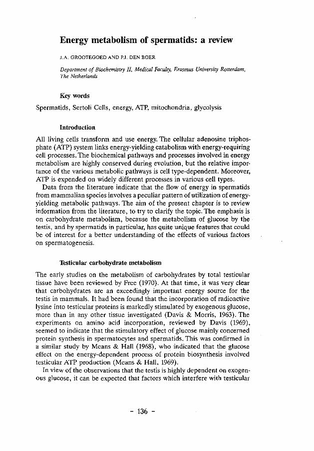
Energy metabolism of spermatids: a review
J.A. GROOTEGOED AND P.J. DEN BOER
Department of Biochemistry II, Medical Faculty, Erasmus University Rotterdam, The Netherlands
Key words
Spermatids, Sertoli Cells, energy, ATP, mitochondria, glycolysis
Introduction
All living cells transform and use energy. The cellular adenosine triphosphate (ATP) system Jinks energy-yielding catabolism with energy-requiring cell processes. The biochemical pathways and processes involved in energy metabolism are highly conserved during evolution, but the relative importance of the various metabolic pathways is cell type-dependent. Moreover, ATP is expended on widely different processes in various cell types.
Data from the literature indicate that the flow of energy in spermatids from mammalian species involves a peculiar pattern of utilization of energyyielding metabolic pathways. The aim of the present chapter is to review information from the literature, to try to clarify the topic. The emphasis is on carbohydrate metabolism, because the metabolism of glucose by the testis, and by spermatids in particular, has quite unique features that could be of interest for a better understanding of the effects of various factors on spermatogenesis.
Testicular carbohydrate metabolism
The early studies on the metabolism of carbohydrates by total testicular tissue have been reviewed by Free (1970). At that time, it was very clear that carbohydrates are an exceedingly important energy source for the testis in mammals. It had been found that the incorporation of radioactive lysine into testicular proteins is markedly stimulated by exogenous glucose, more than in any other tissue investigated (Davis & Morris, 1963). The experiments on amino acid incorporation, reviewed by Davis (1969), seemed to indicate that the stimulatory effect of glucose mainly concerned protein synthesis in spermatocytes and spermatids. This was confirmed in a similar study by Means & Hall (1968), who indicated that the glucose effect on the energy-dependent process of protein biosynthesis involved testicular ATP production (Means & Hall, 1969).
In view of the observations that the testis is highly dependent on exogenous glucose, it can be expected that factors which interfere with testicular
- 136 -
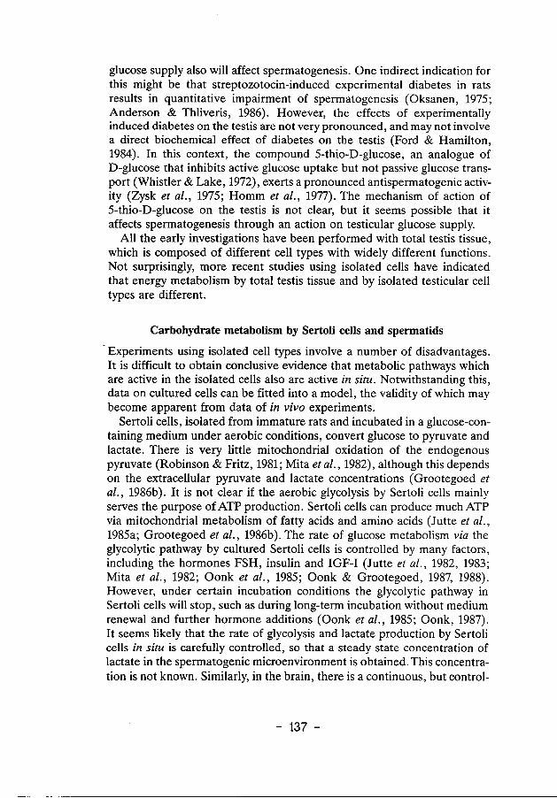
glucose supply also will affect spermatogenesis. One indirect indication for this might be that streptozotocin-induced experimental diabetes in rats results in quantitative impairment of spermatogenesis (Oksanen, 1975; Anderson & Thliveris, 1986). However, the effects of experimentally induced diabetes on the testis are not very pronounced, and may not involve a direct biochemical effect of diabetes on the testis (Ford & Hamilton, 1984). In this context, the compound 5-thio-D-glucose, an analogue of D-glucose that inhibits active glucose uptake but not passive glucose transport (Whistler & Lake, 1972), exerts a pronounced antispermatogenic activity (Zysk et al., 1975; Homm et al., 1977). The mechanism of action of 5-thio-D-glucose on the testis is not clear, but it seems possible that it affects spermatogenesis through an action on testicular glucose supply.
All the early investigations have been performed with total testis tissue, which is composed of different cell types with widely different functions. Not surprisingly, more recent studies using isolated cells have indicated that energy metabolism by total testis tissue and by isolated testicular cell types are different.
Carbohydrate metabolism by Sertoli cells and spermatids
Experiments using isolated cell types involve a number of disadvantages. It is difficult to obtain conclusive evidence that metabolic pathways which are active in the isolated cells also are active in situ. Notwithstanding this, data on cultured cells can be fitted into a model, the validity of which may become apparent from data of in vivo experiments.
Sertoli cells, isolated from immature rats and incubated in a glucose-containing medium under aerobic conditions, convert glucose to pyruvate and lactate. There is very little mitochondrial oxidation of the endogenous pyruvate (Robinson & Fritz, 1981; Mita et al., 1982), although this depends on the extracellular pyruvate and lactate concentrations (Grootegoed et al., 1986b). It is not clear if the aerobic glycolysis by Sertoli cells mainly serves the purpose ofATP production. Sertoli cells can produce much ATP via mitochondrial metabolism of fatty acids and amino acids (Jutte eta!., 1985a; Grootegoed eta!., 1986b). The rate of glucose metabolism via the glycolytic pathway by cultured Sertoli cells is controlled by many factors, including the hormones FSH, insulin and IGF-I (Jutte eta!., 1982, 1983; Mita et a!., 1982; Oonk et a!., 1985; Oonk & Grootegoed, 1987, 1988). However, under certain incubation conditions the glycolytic pathway in Sertoli cells will stop, such as during long-term incubation without medium renewal and further hormone additions (Oonk eta!., 1985; Oonk, 1987). It seems likely that the rate of glycolysis and lactate production by Sertoli cells in situ is carefully controlled, so that a steady state concentration of lactate in the spermatogenic microenvironment is obtained. This concentration is not known. Similarly, in the brain, there is a continuous, but control-
- 137 -
led supply, of lactate via the glycolytic pathway, and toxic concentrations of lactate are not reached (Cohen, 1985). The high concentration of lactate that is observed in testicular fluids collected postmortem is not a true reflection of the normal in vivo situation (Free, 1970). Data on rete testis fluid collected from living rams indicate a very low glucose concentration and a lactate concentration about equal to that of plasma (Setchell et al., 1969).
Spermatids contain all the enzyme activities which constitute the glycolytic pathway (Nakamura et al., 1982; Tvermyr et al., 1984). Furthermore, the isolated cells convert glucose to lactate (Mita & Hall, 1982; Nakamura et al., 1982). However, this glucose metabolism does not result in the maintenance of the cellular ATP content. Rather, exposure of isolated spermatids to glucose results in ATP depletion (Grootegoed et al., 1986a). This is an interesting but not fully understood phenomenon, as will be discussed below. At this point, it is sufficient to note that pyruvate and lactate are excellent energy-yielding substrates for isolated spermatids from different species, including rat, mouse and hamster.
The spermatids convert exogenous lactate to pyruvate and produce ATP via mitochondrial oxidation of pyruvate. A low concentration of glucose in combination with the presence of an extracellular lactate pool, which may be the actual situation in the spermatogenic microenvironment, is compatible with the energy requirements of the spermatogenic cells. The oxygen tension in the lumen of the testicular tubules also is consistent with an oxygen-dependent mode of ATP production (Free et al., 1976). The lactate requirement of the advanced spermatogenic cells could explain the above described effect of glucose on testicular ATP and protein synthesis. It is probable that factors which interfere with testicular glucose supply and lactate production by Sertoli cells at the same time in.terfere with spermatogenesis.
Of course, the metabolism of glucose by different testicular cell types is more complex than merely the glycolytic conversion of glucose to pyruvate and lactate. First, glucose is also important for the production of NADPH and ribose 5-phosphate via the pentose phosphate pathway. The activity of this pathway in isolated spermatogenic cells and Sertoli cells has been estimated (Robinson & Fritz, 1981; Grootegoed et al., 1984, 1986b). Second, glucose and other monosaccharides and derivatives are required for the biosynthesis of oligosaccharide side chains of glycoproteins ( Grootegoed et al., 1982; Gerton & Millette, 1986). Third, glucose can be converted into glycogen and lipids, and these aspects of glucose metabolism have been more extensively studied using cultured Sertoli cells (Robinson & Fritz, 1981; Jutte et al., 1985a).
In this context, it is also of interest that Sertoli cells contain aldose reductase (Ludvigson eta!., 1982), an enzyme of the polyol pathway which converts a number of aldoses to the corresponding ketoses (glucose to
- 138 -
fructose via sorbitol, catalyzed by aldose reductase and sorbitol dehydrogenase). There is sorbitol dehydrogenase activity in the testis, and this enzyme has been used as a marker for spermatogenesis (Bishop, 1968). However, no significant amounts of sorbitol, fructose or other hexoses have been detected in testicular fluids (see Waites & Gladwell, 1982), and the actual occurrence of the polyol pathway in the testis can be questioned.
Finally, there is information concerning the production of the cyclic alcoho[ myo-inositol from glucose by Sertoli cells (Robinson & Fritz, 1979). The concentration of myo-inositol in testis fluid is very high (approximately 2 mM in the rat; Hinton eta!., 1980).Apossible role for the highmyo-inositol concentration has not yet been defined, and there is no information on possible effects of myo-inositol on spermatids and spermatozoa.
The mitochondria of spermatids
It is probable that ATP production in spermatids is dependent on a faultless performance of their mitochondria. One indication for this is that agents which interfere with mitochondrial ATP production cause degeneration of the spermatogenic cells in vivo, at doses which do not exert pronounced effects on other cell types. For example, in rats spermatogenesis is severely impaired by feeding the animals low doses of fluoroacetate or fluoroacetamide (Mazzanti et a!., 1964; Novi, 1968; Steinberger & Sud, 1970; Sullivan et al., 1979). The mechanism of action of these compounds on spermatogenesis most likely involves conversion of fluoroacetate and fluoroacetamide to fluorocitrate, which blocks the citric acid cycle through inhibition of the enzyme aconitase, resulting in inhibition of mitochondrial. ATP production. This may illustrate that compounds with an overall toxic effect can more or less specifically affect spermatogenic cells, possibly because the advanced spermatogenic cells are remarkably sensitive to toxic agents. Gossypol, the extensiveiy studied antifertility agent from cottonseed, seems to be another example. There are a number of indications that gossypol exerts its major effect(s) through an uncoupling of the mitochondrial electron transport chain and oxidative phosphorylation (Reyes eta!., 1984; Den Boer & Grootegoed, 1987). The action of gossypol in vivo on spermatogenic cells might also be explained by the very high sensitivity of these cell types to inhibitors of mitochondrial function.
The mitochondria of spermatids have special properties. Firstly, mitochondria in round spermatids are located near the periphery of the cells, but the significance of this is unknown. Secondly, during spermatid elongation, mitochondria migrate to form a helical arrangement around the middle piece of the developing sperm. Lastly, mitochondria of spermatids are in a condensed configuration, the result of a series of morphological changes which take place in spermatocytes. During meiotic prophase the mitochondrial cristae swell until the matrix is flattened towards the
- 139 -
outer mitochondrial membrane (De Martino et at., 1979). The condensed type of mitochondria is observed in spermatocytes and round spermatids, and also in oocytes and early embryos (Stern et at., 1971). This is of interest, because oocytes and early embryos require exogenous pyruvate and lactate for ATP production (Biggers et at., 1967; Zeilmaker et at., 1972; Barbehenn et al., 1974). In oocytes and early embryos, the block in the glycolytic pathway has been assigned to the enzyme phosphofructokinase (Barbehenn et al., 1974).
The condensed configurational state of the mitochondria might be somehow related to their functional state. Quite similar changes in the configurational state of the crista! membranes of mitochondria in different cell types is induced by energizing conditions (Harris et al., 1969; Hackenbrock et al., 1971). This energized configuration involves condensation of the inner compartment and dilatation of the cristae.
From this, one might ask whether or notATP production in the mitochondria of spermatids occurs at a very high and perhaps maximal rate. The answer to this question is not readily available. However, it has been observed that the rate of pyruvate decarboxylation via the pyruvate dehydrogenase complex in isolated rat spermatogenic cells was not enhanced by incubating the cells in the presence of uncoupling agents (Grootegoed eta!., 1984). The conclusion is that mitochondrial pyruvate oxidation, and hence mitochondrial ATP production, are maximal during incubation of the cells in the presence of pyruvate and in the absence of the uncoupling agents. It should be noted, however, that the addition of exogenous pyruvate to the incubation medium of isolated spermatids results in awkward metabolic behaviour of the cells. The exogenous pyruvate is very rapidly converted to lactate, at the expense of reducing equivalents from cellular NADH (Grootegoed et al., 1984). Due to the urgent need for NADH, the pyruvate dehydrogenase complex and the citric acid cycle are maximally activated.
With respect to lactate metabolism, the situation is somewhat different. It can be calculated from published data ( Grootegoed et al., 1984) that the pyruvate dehydrogenase complex in rat spermatids attains approximately a third of its maximal activity during incubation of the cells in the presence of lactate. Spermatids in situ may metabolize exogenous lactate rather than pyruvate, as will be further discussed below, and it would appear that mitochondrial ATP production in round spermatids under physiological conditions is not rate-limited by the pyruvate dehydrogenase complex. However, there may be little reserve capacity of this mitochondrial enzyme complex to meet occasional higher energy demands. There are no indications, although there is also little information available, that the capacities of the citric acid cycle and the electron transport chain could become rate-limiting. At the same time, it cannot be excluded that there is another rate-limiting step, such as the mitochondrial F0 F1-ATPase complex.
- 140-
One other mitochondrial protein may play a particular role in the energy metabolism of spermatogenic cells. A testis-specific cytochrome c has been discovered in the mouse, which differs in 13 of its 104 amino acid residues from the ubiquitous cytochrome c (Hennig, 1975). This cytochrome Ct is detected in spermatocytes and the more advanced spermatogenic cells (Goldberg eta/., 1977). The specific function of cytochrome Cr is not known, but its presence is one more indication that the mitochondria in spermatids and spermatozoa have not yet disclosed all their properties.
Energy-yielding substrates for spermatids
Many authors agree that lactate is the best energy-yielding substrate for isolated round spermatids from rats (Jutte et al., 1981; Nakamura eta/., 1981a, b, 1984b, 1986b; Mita & Hall, 1982; Grootegoed eta/., 1984, 1986a). This is also true for spermatogenic cells from mice and hamsters (Den Boer & Grootegoed, unpublished results), and it would be of great interest to obtain information on the metabolism of energy-yielding substrates by the germ cells from other mammalian species including man.
The energy metabolism of male germ cells at the early stages of their development has not been extensively studied. The effects of lactate on ATP synthesis have been demonstrated not only for spermatids, but also for pachytene spermatocytes (Jutte et al., 1981, 1982; Grootegoed et al., 1984; Nakamura et al., 1984a, c). In this respect, however, there is no information on spermatogonia and early spermatocytes. Interesting observations on metabolic characteristics of fetal germ cells of the mouse have been reported (Brinster & Harstad, 1977). These observations show that early germ cells show a relatively high rate of pyruvate oxidation and contain lactate dehydrogenase (LDH) composed of predominantly B subunits, whereas other embryonic tissues have predominantly A subunits. Perhaps, some type of metabolic differentiation of male and female germ cells is initiated during prenatal gonadal development, or is an inherent property of cells of the germ line and the early embryo. Thus, the energy metabolism of the advanced spermatogenic cells may involve many highly specialized processes which do not occur in early germ cells, growing oocytes or early embryos.
As indicated above, the metabolism of exogenous pyruvate by isolated rat spermatids involves a high activity of the mitochondrial machinery to produce NADH not only for the electron transport chain but also for the conversion of pyruvate to lactate. Under these conditions, the cellular NAD±INADH system in the spermatids is probably highly oxidized, whereas during metabolism of exogenous lactate this redox system may be much more reduced (Grootegoed eta/., 1985a; Nakamura eta/., 1986b). Pyruvate and lactate have similar effects on the NAD±/NADH system in spermatozoa (Brooks & Mann, 1971, 1972; Milkowski & Lardy, 1977).
- 141 -
Conflicting results on the effect of exogenous pyruvate on the cellular ATP content of rat spermatids have been reported. The ATP content of the isolated spermatids either was maintained (Grootegoed et al., 1984), or was not maintained (Mita & Hall, 1982; Nakamura eta/., 1984b, 1986a), when the cells were incubated in the presence of pyruvate. There is no clear explanation why the spermatids might not be capable of producing ATP from exogenous pyruvate. Most likely, the discrepancy is caused by experimental errors (Grootegoed et al., 1984). Indeed, Nakamura eta!. (1986d) have observed that the ATP content of rat spermatids was maintained by pyruvate when the experimental conditions included isolation of the cells in the presence of proper energy substrates.
When isolated spermatocytes and spermatids are incubated in the presence of pyruvate and/or lactate, it is possible to study various processes such as biosynthesis of RNA and proteins, fucosylation of glycoproteins, transferrin-mediated iron uptake, and long-term effects of toxic compounds (Grootegoed et al., 1977, 1982; Jutte et al., 1985b; Gerton & Millette, 1986; Den Boer & Grootegoed, 1987; Toebosch eta/., 1987).
It has become apparent that lactate rather than pyruvate is the energyyielding substrate which is most likely used by the advanced spermatogenic cells in situ (Grootegoed eta!., 1984, 1985a; Nakamura eta!., 1984b, 1986b). This is also substantiated by the following. After culture of Sertoli cells at a high cell density for 24 h, the lactate concentration in the spent incubation medium was 2.5-3.0 mM, and the [lactate]/[pyruvate] ratio was approximately 10, which is the equilibrium ratio that is also observed for other cell systems (Grootegoed eta!., 1985b). In other words, the pyruvate concentration in the spermatogenic microenvironment is probably very low, the more so because spermatogenic cells convert exogenous pyruvate to lactate until the [lactate ]/[pyruvate] ratio has become very high (Grootegoed et a!., 1984). Another indication that lactate rather than pyruvate is the main energy-yielding substrate used under physiological conditions, is that rat spermatogenic cells in situ reduce a-ketoisocaproate to a-hydroxyisocaproate, which is indicative oflactate metabolism (Grootegoed eta!., 1985a).
The choices for the advanced spermatogenic cells to extract energy from other substrates are limited. Isolated spermatids cannot produce a significant net amount of ATP from glucose and amino acids (see above). The substrates succinate, acetate and glycerol also appeared to have no effect on the cellular ATP content, in spite of the relative simplicity of the metabolism of these compounds (unpublished observations). There is some f3-oxidation of endogenous fatty acids, and the spermatids also contain a small amount of acetyl- and acyl-carnitine (Grootegoed eta!., 1986a). Spermatozoa show ATP-yielding endogenous metabolism, probably including f3-oxidation of fatty acids (Setchell eta/., 1969; Ford & Harrison, 1981b; Inskeep & Hammerstedt, 1985). Spermatocytes and spermatids contain a marked activity of carnitine acetyltransferase (Vernon et al., 1971), and
- 142 -
acetyl-carnitine could have an energy storage function in advanced spermatogenic cells, as described for spermatozoa (Milkowski et al., 1976; Van Dop eta/., 1977). In isolated spermatids, however, fatty acid oxidation and the cellular content of acetyl-carnitine are not sufficient to maintain cellular ATP content in the absence of lactate (Grootegoed et al., 1986a).
In this respect, it is relevant to note that pachytene spermatocytes and round spermatids, isolated from mice, have been found to incorporate exogenous acetate and arachidonate into fatty acids (Grogan & Lam, 1982; Grogan & Huth, 1983). Moreover, mouse testicular cells incorporate acetate into cholesterol and the polyisoprenoid alcohol dolichol (Potter eta/., 1981). Dolichol functions in the form of dolichyl phosphate as carrier of carbohydrates in the biosynthesis of glycoproteins. These results indicate that complex synthetic pathways may occur in spermatogenic cells for which acetyl CoA is needed, so that acetyl CoA is not exclusively used as a fuel for mitochondrial ATP production.
Effects of glucose on spermatids
Isolated round spermatids from rats convert glucose to lactate (Mita & Hall, 1982; Nakamura eta/., 1982). Moreover, the spermatids convert 3Hlabelled glucose to 3H 20 at a considerable rate, which reflects metabolism of glucose via the glycolytic pathway (Grootegoed eta/., 1984). In spite of this, glucose is not an effective energy-yielding substrate for spermatids. The metabolism of glucose by isolated rat spermatids, during incubation in the absence of lactate, results in a high fructose 1,6-biphosphate concentration, but a reduction in the cellular ATP content (Grootegoed eta/., 1986a; Nakamura et al., 1986b). The effect on ATP is observed at low glucose and mannose concentrations (lo-s-10-4 M). Fructose causedATP depletion at 100 times higher concentrations (10-3 -10-2 M), and galactose had no effect at all (Grootegoed et al., 1986a). This agrees with the substrate specificity and the Km of hexokinase for the different substrates (Grootegoed et al., 1986a). The effects of hexoses on the ATP content of spermatids indicate that the flow of intermediates through the glycolytic pathway in the spermatids is restricted by a low activity of one or more enzymes which follow phosphofructokinase in the pathway.
It is not known if the high fructose 1,6-biphosphate content of the cells is responsible for the low ATP content. This question has been studied with respect to the action of the compound a-chlorohydrin, which exerts a rapid antifertility effect in male rats and other mammals. This action is believed to involve conversion of a-chlorohydrin to 3-chlorolactaldehyde by an NADP± dependent dehydrogenase enzyme, and selective inhibition of the glycolytic enzyme glyceraldehyde 3-phosphate dehydrogenase (GA3PDH) in spermatozoa (Ford & Harrison, 1983; Stevenson & Jones, 1985). There is a striking similarity between glucose metabolism by a-chlorohydrin-
- 143 -
treated spermatozoa (Ford & Harrison, 1986) and by isolated rat spermatids. This similarity concerns the restricted flow of fructose 1,6- biphosphate through the last part of the glycolytic pathway, concomitant with glucose-induced ATP dephosphorylation. The effect of glucose on the ATP content of a-chlorohydrin-treated spermatozoa was circumvented by giving the spermatozoa pyruvate and lactate (Ford & Harrison, 1985). From experiments using a-chlorohydrin-treated boar and rat spermatozoa, it was concluded that futile substrate cycles were active, but could not fully account for the combined effects of glucose and a-chlorohydrin on the ATP content (Ford & Harrison, 1987).
The factors which play a role in the control of ATP production via glycolysis in spermatozoa could be quite complex. In this respect, it is of interest to note that bull spermatozoa have been used as a model system, to study the ATP yield of glycolysis at low and high glucose fluxes (Hammerstedt & Lardy, 1983). It was observed that a low flux of glucose through the glycolytic pathway is accompanied by substrate cycling andATP dephosphorylation (Hammerstedt & Lardy, 1983). The ATP yield of glycolysis can be zero or negative, especially if the overall net ATP yield of glucose metabolism is positive as a result of mitochondrial oxidation of pyruvate originating from glucose.
Returning to the consideration of the peculiarities of the glycolytic pathway in isolated spermatids. As described above, all the glycolytic enzymes are present in rat spermatids and it was suggested that aldolase (Tvermyr et al., 1984) or GA3PDH (Nakamura et al., 1982) are the rate-limiting enzymes. On the other hand, it cannot be excluded that the apparent block in the second phase of glycolysis is not inherent to the cells, but represents an artefact of cell isolation, induced because of unstable activities or an unstable arrangement of glycolytic enzymes in spermatids.
It might be that GA3PDH is the weak link, especially in view of the experiments discussed above on a-chlorohydrin-treated spermatozoa, but there is no conclusive evidence. The concentrations of glycolytic intermediates have been estimated after incubation of the spermatids in the presence of glucose and in the absence of lactate (Nakamura et al., 1982). From a biochemical point of view these cells are severely damaged, due to the glucose-induced ATP depletion. Here is a paradox. On the one hand, glucose metabolism causes cell death, and on the other hand lactate-supported ATP production shuts down the glycolytic pathway. When the cells attain a high energy charge from the metabolism of lactate, this will result in inhibition of the glycolytic pathway through regulation of the phosphofructokinase reaction (Bosca et al., 1985). Hence, by using intact cells no reliable information on the flow of metabolites through the glycolytic pathway can be obtained.
It has been suggested that control of glycolysis in spermatocytes and spermatids could be effected through inhibition of GA3PDH by the adenine
- 144 -
nucleotides AMP, ADP and ATP in competition with NAD, rather than that glycolysis is regulated by the control enzymes hexokinase, phosphofructokinase and pyruvate kinase. (Nakamura et al., 1982, 1984a). However, there are no indications that the NAD content of the spermatogenic cells is sufficiently low to permit this mechanism of regulation. Moreover, there is a dramatic effect of glucose on ATP dephosphorylation in isolated spermatids, indicating that the cells are unsuccessful in controlling glucose metabolism.
The enzyme phosphofructokinase is a good candidate to act as the most important control point in glycolysis in spermatids. Phosphofructokinase can be effectively inhibited by ATP and other factors (Bosca et al., 1985; Hoskins & Stephens, 1969), but in spermatids sufficiently high concentrations ofATP may only be reached when the cells oxidize exogenous pyruvate or lactate. Endogenous pyruvate, produced from glucose, is probably converted to lactate, rather than being oxidized in the mitochondria, because spermatids may find difficulties in transporting reducing equivalents from cytosolic NADH across the inner mitochondrial membrane via a shuttle system. NADH is readily oxidized to NAD+, however, at the conversion of pyruvate to lactate in the cytosol. This deprives the mitochondria of the essential energy source pyruvate. Consequently, the cellular ATP content
·will be too low to effectively inhibit phosphofructokinase. In this situation, glucose is converted to glycolytic intermediates, but the limited flux of intermediates through the second phase of the glycolytic pathway does not enable the spermatids to produce ATP from glucose.
Spermatids can take up glucose via a specific carrier (Nakamura et al., 1987b). The fate of glucose inside cells is that it is phosphorylated by ATP to form glucose 6-phosphate, a reaction catalyzed by hexokinase. Most of the glucose 6-phosphate proceeds down the glycolytic pathway, but a minor catabolic pathway taken by glucose 6-phosphate is the pentose phosphate pathway. This pathway leads to two important products: NADPH and ribose 5-phosphate. The hexokinase reaction is not controlled by the energy status of the cell, and the pentose phosphate pathway can therefore also occur in spermatids when these cells produce ATP from exogenous lactate. However, there are two factors which could limit the metabolism of glucose via the pentose phosphate pathway in spermatids. First, the activity of glucose 6-phosphate dehydrogenase is comparatively low (see below), although the cells have some pentose phosphate pathway activity (Grootegoed et al., 1984). Second, an sufficient supply of glucose to the spermatogenic cells in situ.
The developing germ cells will require ribose 5-phosphate to meet the needs of the biosynthesis of nucleotide·s for RNA synthesis, but there is no information on the production of ribose 5-phosphate in spermatocytes and spermatids. Possibly, the amount of glucose available is sufficient to
- 145 -
support a low but adequate rate of ribose 5-phosphate production. Moreover, the ribose 5-phosphate that is required can be generated also via the non-oxidative-branch of the pentose phosphate pathway, so that an eventual low activity of glucose 6-phosphate dehydrogenase may not play a role in this respect. Spermatogenesis does not occur at great speed, and substantial net amounts of RNA may be produced during the many days that it takes for pachytene spermatocytes to develop into elongating spermatids, especially if the rate of synthesis of RNA precursors is low. However, it cannot be excluded that the pentose phosphate pathway in spermatids in situ is virtually. completely inhibited due to lack of substrate, and that other mechanisms operate to supply RNA precursors to the germ cells. One such mechanism could be the release of nucleosides by Sertoli ceJis, and the uptake and phosphorylation of these nucleosides by the germ ceJls. Isolated spermatocytes and spermatids incorporate exogenous uridine into RNA (Grootegoed et al., 1977).
It should be stressed that very little is known about the de novo synthesis of nucleotides and the purine salvage reactions in spermatogenic ceJls (Grootegoed eta/., 1986c). This is highly relevant not only with respect to RNA synthesis, but certainly also in relation to the ATP content of the cells. On the one hand, the metabolism of glucose by spermatocytes and spermatids results inATP dephosphorylation (see above), whereas glucose could be important for ribose 5-phosphate synthesis and the de novo synthesis of adenine nucleotides.
Finaiiy, it may be relevant to point out that in view of the more recent data on the effects of glucose on spermatids, it is difficult to explain earlier observations on the effects of glucose and 5-thio-D-glucose on protein synthesis by isolated spermatids from rats (Nakamura & Hall, 1976, 1977; Nakamura et a!., 1978). In these experiments, it was observed that the incorporation of radioactively labeled amino acids into proteins was stimulated by glucose and inhibited by 5-thio-D-glucose. It would be important to establish if this incorporation represented the actual rate of protein synthesis, rather than changes in the specific radioactivity of the precursor pools. It cannot be excluded that glucose exerts a stimulatory effect on protein synthesis independent of the effect on ATP, although protein synthesis would be strongly suppressed by Jack ()f ATP. The rate of amino acid incorporation into proteins of spermatocytes and spermatids is markedly increased when the glucose-containing incubation medium is supplemented with lactate (Jutte eta/., 1981; Nakamura eta/., 1981a).
Glycolytic enzymes in spermatids
There is a considerable amount of information on glycolytic enzymes in spermatids and spermatozoa including the expression of different iso-
- 146 -
enzymes of the glycolytic pathway. These include the isoenzymes of phosphoglycerate kinase (PGK) and enolase (ENO), and also of lactate dehydrogenase (LDH) which in the present discussion is considered as a reaction involved in glycolysis.
During spermiogenesis several glycolytic enzymes appear to be subjected to testis-specific post-translational modifications, which result in 'spermtype' enzymes or sperm-specific behaviour. These are hexokinase (Katzen et al., 1968; Harrison, 1971; Sosa et al., 1972), phosphoglucose isomerase (Buehr & McLaren, 1981) and aldolase (Gillis & Tamblyn, 1984). The kinetics of the enzymes hexokinase, phosphofructokinase and pyruvate kinase of rat spermatids have been studied, but no abnormalities of the kinetic behaviour of these enzymes has been observed (Nakamura et al., 1984d, 1986c, 1987a).
A side issue of a discussion on PGK, is that the X chromosome is genetically inactivated early in spermatogenesis (Monesi, 1971). During meiotic prophase, the X and Y chromosomes are condensed and form a heterochromatic body, the sex vesicle. It is not clear what purpose is served by the formation of the sex vesicle, but many chromosomal anomalies which disturb spermatogenesis seem to involve the X chromosome (de Boer & Searle, 1980). As a consequence of X chromosome inactivation, the spermatogenic cells may fail to maintain high activity levels of enzymes encoded by genes on the X chromosome, such as phosphoglycerate kinase A (PGKA) and glucose 6-phosphate dehydrogenase (G6PDH). There is probably no autosomal enzyme synthesized in spermatogenic cells to substitute for G6PDH, the first enzyme of the pentose phosphate pathway. Consequently, in spermatozoa the pentose phosphate pathway is probably almost completely absent (Setchell et al., 1969; Grootegoed et al., 1986c). With respect to PGK, the spermatogenic cells follow a different strategy. The mammalian genome contains an autosomal gene which encodes the isoenzyme PGK-B. Expression of this autosomal gene is restricted to the spermatogenic cells (VandeBerg et al., 1973). Recent information on the Pgk-2 gene (encoding PGK-B), shows that it is peculiar, in that it completely lacks introns and has the characteristics of a processed gene (McCarrey & Thomas, 1987; Boer et al., 1987). A functional processed gene is rare, and it seems that the spermatogenic cells have evolved a specific and original solution to solve the threatening problem of a missing link in the glycolytic pathway. This by itself indicates the importance of the glycolytic pathway for spermatozoa.
Enolase is a dimer coded for by three genes, producing subunits a-, [3-and y-, and there are developmental changes in the enolase isoenzyme patterns in different tissues (Rider &Taylor, 1974). The testis, and in particular late spermatids and spermatozoa, contain an unusual enolase isoenzyme (Edwards & Grootegoed, 1983). This ENO-S is distinguished from somatic enolases by physical properties. It is unknown if ENO-S contains subunits
- 147 -
encoded by another gene, and there is also no information which could explain the specific function of ENO-S.
Although we have learned a lot about the testis-specific lactate dehydrogenase isoenzyme LDH-C4 since its discovery in 1963 (Blanco & Zinkham, 1963; Goldberg, 1963, 1977), there is still uncertainty on why spermatogenic cells and spermatozoa would need LDH-C4• The isoenzyme is composed of four C subunits, coded for by a gene different from the A and B subunit genes. In different species, LDH-C4 shows a broad substrate specificity towards different a-keto acids. This is true especially for rats and mice (Coronel et al., 1983). The LDH-C4 enzyme from these animals converts branched chain a-keto acids to the corresponding a-hydroxy acids. The branched chain a-keto acids are derived from transamination of the branched chain amino acids leucine, isoleucine and valine. Cultured Sertoli cells from rats convert leucine to a-ketoisocaproate, and spermatogenic cells reduce this compound to a-hydroxyisocaproate (Grootegoed et al., 1983, 1985a). This represents a unique intercellular pathway of leucine catabolism, but it is not known if a function can be assigned to it. Possibly, the catalytic properties of LDH-C4 are not the main justification for its existence. The possibilities for a function which is directly related to specific physical properties of the isoenzyme seem to be unlimited, in view of the recent observation that lactate dehydrogenase can act as a structural protein in avian and crocodilian lenses (Wistow et al., 1987).
In spermatids, most of the LDH-C4 is found in the cytosol (Hintz & Goldberg, 1977). This isoenzyme is also located in the mitochondrial matrix of advanced spermatogenic cells and spermatozoa (Montamat & Blanco, 1976). Many authors have indicated the presence of LDH-C4 inside the mitochondria, in particular in spermatozoa, where it could be involved in a shuttle mechanism for the transport of reducing equivalents into and out of the mitochondria, or could catalyze direct intramitochondrial lactate oxidation (Milkowski & Lardy, 1977; Storey & Kayne, 1977; Gerez de Burgos, 1978). The transport of a protein into the mitochondrial matrix , involves a specific sequence of events, which is dependent on the synthesis of a precursor protein with an N-terminal extension (Schatz & Butow, 1983). This has not been shown for LDH-C subunits. However, results from the molecular cloning of the mouse C-subunit gene indicates the presence of multiple LDH-C gene-related sequences (Sakai et al., 1987). Moreover, it cannot be excluded that one gene codes for two proteins on opposite sides of the mitochondrial inner membrane (Fox, 1982). All in all, there is still no conclusive evidence for the presence of LDH-C4 in the mitochondrial matrix, such as could be provided by an immunocytochemical study at the ultrastructural level. Independent of this, the fact that most if not all of the enzymatic activity is outside the mitochondrial inner compartment indicates that the enzyme could very well catalyze the last reaction in the aerobic glycolysis of spermatozoa.
- 148 -
The energy budget of spermatids
In developing germ cells, the meiotic divisions are the last great expense of energy needed for the generation of four spermatids from one spermatocyte. The energy budget of spermatids does not include expenditures for DNA synthesis and the process of cell division. In round spermatids, there is still RNA and protein synthesis, but the rates must be modest since the cells do not undergo a new round of cell division. In elongating spermatids, RNA synthesis becomes completely arrested when the DNA is compacted in the nucleus, and the last major act of protein biosynthesis is the synthesis of the nuclear proteins involved in this compaction process (Monesi, 1971; see the Chapter by 000000000000, this volume).
There are other processes that consume ATP In most cells, much energy is expended on active transport of ions across the plasma membrane. The transport of Na+ and K+ by a specific transport system, the Na+-K+ pump, results in a high concentration of K+ and a low concentration of Na+ in the cells relative to the external medium. The Na+-K+ pump contains ATPase activity, and it has been reported that inhibition of the Na+ -K+ pump by the toxic glucoside ouabain results in a major reduction in the energy expenditure of tumor cells (Racker et al., 1983). Spermatids are in a. unique situation. There is a remarkable difference between the ionic composition of tubular fluid and plasma, most importantly in the K+ concentration, which at approximately 40 mM in tubular fluid, is 10 times higher than that in plasma (Tuck eta!., 1970; Levine & Marsh, 1971; Jenkins et al., 1980; see Waites & Gladwell, 1982). Concomitantly, the Na+ concentration in the tubular fluid is relatively low. In primary tubular fluid, which may more closely represent the fluid that surrounds the spermatogenic cells, the K+ concentration is even higher than in free-flow tubular fluid (Tuck eta!., 1970). From this, it can be speculated that spermatids are in an ionic microenvironment that approximates to the intracellular ionic composition, and may not have to pay much attention to the pumping of Na + and K+. Possibly, the active transport of Na+ and K+ across the plasma membrane is a minor item of the energy budget of spermatids.
Living cells also utilize the chemical energy of ATP for the performance of mechanical work. Early spermatids contain two motile components: the primary flagellum, and rhythmic cytoplasmic movements (Walt & Redinger, 1983). In late spermatids, the motility of the flagellum and the cytoplasm is reduced (Walt & Redinger, 1983). The spermatids contain F-actin, myosin and tubulin (Campanella eta!., 1979; Hecht eta!., 1984; Russell et al., 1986). F-actin and myosin together can use the chemical energy stored as ATP for the generation of force, and tubulin in the axonemal microtubules interacts with the ATPase activity of dynein arms. However, it is not possible to give an estimate of how much ATP is consumed in reactions generating force. Possibly, mechanical work is the most important process of ATP turnover in the developing spermatids.
- 149 -
From spermatid to spermatozoon
The middle piece of spermatozoa contains a mitochondrial system which is capable of producing ATP through oxidative phosphorylation (Ford & Harrison, 1981b). Yet, aerobic glycolysis seems to be important in the energy metabolism of mature spermatozoa (Peterson & Freund, 1970). In spermatozoa, energy metabolism is geared to producing ATP for the contractile work of motility, and it has been suggested that the energy metabolism of spermatozoa is similar to that of muscle cells (Storey & Kayne, 1980). This means that spermatozoa depend on a high rate of glycolysis to produce ATP for motility. It has been shown that rabbit spermatozoa contain a muscle-type pyruvate kinase isoenzyme that is suited for a high rate of glycolysis (Storey & Kayne, 1980). Furthermore, the potential catalytic activities of a number of glycolytic enzymes in spermatozoa of various species, including rat spermatozoa, are considerably in excess of the flux of intermediates through the glycolytic pathway (Harrison, 1971; Ford et al., 1981). While a detailed discussion of energy metabolism in spermatozoa is not practicable within the limits of the present text, particularly in view of marked species differences, it can be suggested that spermatozoa acquire glycolytic capacity compared to spermatids, and learn how to control glycolysis. This is not to say that under all circumstances glucose is the preferred energy-yielding substrate of spermatozoa. The adenine nucleotide concentrations in and motility of rat spermatozoa are supported by pyruvate and lactate better than by glucose (Ford & Harrison, 1981), and high concentrations of glycolytic intermediates accumulate in rat spermatozoa incubated in the presence of glucose. Furthermore, it has been suggested that lactate could be the preferred energy source of rabbit spermatozoa (Storey & Kayne, 1977). Yet, the behaviour of the glycolytic enzymes may be changed during late spermiogenesis, so that a more efficient glycolytic apparatus is obtained.
A number of glycolytic enzymes remain bound to the sperm middle piece, during treatment of spermatozoa with hypotonic media (Harrison & White, 1972: Storey & Kayne, 1975; Gillis & Tamblyn, 1984). This could indicate that specific mechanisms operate that influence the locations and interactions of the enzymes. Little is known about the intracellular transport and structural interactions of glycolytic enzymes in most cells. However, it is not a hard and fast rule that the soluble enzymes of glycolysis are freely diffusing in the cytosol (Fulton, 1982; Srere, 1987). For example, enolase is present in a complex with other proteins, so that it can be transported within nerve cells (Brady & Lasek, 1981). Noteworthy also, in view of the comparison between spermatozoa and flagellated protozoa (see the Chapter by Docampo, this volume), is that in Trypanosoma brucei the glycolytic enzymes are located in an organelle, the glycosome (Opperdoes & Borst, 1977). This could enable this organism to obtain all its energy through glycolysis. There is little information on the exact location of the glycolytic
- 150 -
enzymes in spermatozoa. Possibly, the glycolytic machinery is mainly associated with the mitochondria in the midpiece, and this could be effected also through the formation of enzyme complexes.
In sea urchin sperm, as in muscle cells, a phosphorylcreatine shuttle mediates the transport of high energy phosphate from the mitochondrion down the sperm tail (Tombes & Shapiro, 1985). It is not known whether a similar mechanism is present in mammalian sperm, or whether diffusion of ATP can account for the energy needed for the dynein-induced sliding of microtubules. However, it seems likely that special adaptations must take place during the last phase of spermiogenesis to ensure an optimal flow of energy from energy-yielding reactions to the motile proteins of the sperm axoneme.
During late spermiogenesis, a series of events may occur which end the random arrangements of glycolytic enzymes. Firstly, a number of genetic and posHranslational isoenzymes partially or completely take the place of the somatic glycolytic enzymes. Secondly, further post-translational modification of enzymes might occur via phosphorylation of serine and threonine residues, and also tyrosine residues (see the Chapter by Meijer et al., this volume). Thirdly, during the final stages of spermatid elongation, much of the spermatid cytoplasm flows from the flagellum to the spermatid head where it is removed via so-called tubulobulbar complexes (Russell, 1979). This could be accompanied by an increase in the concentration of cytosolic enzymes and an altered intracellular location. The concentration of enzymes in the cell is an important factor in the control of metabolite transfer between two sequential enzyme sites (Srivastava & Bernhard, 1986a, b). It can be speculated that during late spermiogenesis, even very shortly before spermiation, the enzyme-enzyme interactions among glycolytic enzymes are altered. This could result in proper control of glycolysis in spermatozoa.
Synopsis
The following working hypothesis is proposed: Glucose metabolism by isolated spermatids initiates a sequence of events that involves accumulation of glycolytic intermediates and ATP dephosphorylation. For unknown reasons, glucose metabolism goes out of control. In the spermatogenic epithelium, a similar response of spermatids to glucose does not occur, because the local glucose concentration is probably very low and the cells do not depend on glycolysis for ATP production. Rather, developing spermatids rely on the mitochondrial machinery to produce ATP. The advanced spermatogenic cells have specialized to utilize exogenous lactate for ATP production, and these cells have largely or completely given up the capacity to metabolize other energy-yielding substrates.
- 151 -
Inhibitors of mitochondrial ATP production have lethal effects on spermatids. On the other hand, compounds such as 3-chlorohydrin, or other factors that interfere with the second energy-conserving phase of glycolysis, will affect sperm cells but not the developing germ cells in the testis. Spermatozoa can produce ATP via a high rate of lactate production by glycolysis, but also via oxidation of exogenous lactate and endogenous substrates.
During late spermiogenesis, there are tumultuous changes in the interactions between cytosolic enzymes. These enzymes include genetic and posttranslational isoenzymes. Non-random arrangement of glycolytic enzymes may allow for stable control of glycolysis.
The regulation of energy metabolism in spermatids and spermatozoa could be much more complex than we can now anticipate. From the views presented, it may be inferred that impairment of sperm glycolysis, induced during spermiogenesis, or later, is an approach to interfere with sperm function without an effect on spermatogenesis. However, there are as yet no clear indications that defined compounds can act upon glycolytic enzymes in spermatozoa in a highly cell-specific manner. Nevertheless, research in this area should continue, especially to compile more data needed to attack the problem of cell-specific inhibition of sperm function.
Acknowledgement
This work received financial support from the Special Programme of Research, Development, and Research Training in Human Reproduction, World Health Organization.
References
Anderson, J.E. &Thliveris, T. (1986). Testicular histology in streptozotocin-induced diabetes. Anatomical Record 214:378-382.
Barbehenn, E.K., Wales, R.G. & Lowry, O.H. (1974). The explanation for the blockade of glycolysis in early mouse embryos. Proceedings of the National Academy of Sciences U.S.A. 71:1056-1060.
Biggers, J.D., Whittingham, D.G. & Donahue, R.P. (1967). The pattern of energy metabolism in the mouse oocyte and zygote. Proceedings of the National Academy of Sciences, U.S.A. 58:560-567.
Bishop, D.W. (1968). Sorbitol dehydrogenase in relation to spermatogenesis and fertility. Journal of Reproduction and Fertility 17:410-411.
Blanco, A. & Zinkham, WH. (1963). Lactate dehydrogenases in human testes. Science 139:601--{)02.
Boer, P.H., Adra, C.N., Lau, Y-F. & McBurney, M.W. (1987). The testis-specific phosphoglycerate kinase gene pgk-2 is a recruited retroposon. Molecular and Cellular Biology 7:3107-3112.
Boer, P." de & Searle, A.G. (1980). Workshop on chromosomal aspects of male sterility in mammals: Summary and synthesis. Journal of Reproduction and Fertility 60:257-265.
- 152 -
Bosca, L., Aragon, J.J. & Sols, A. (1985). Modulation of muscle phosphofructokinase at physiological concentration of enzyme. The Journal of Biological Chemistry 260:2100-2107.
Brady, S.T. & Lasek, R.J. (1981). Nerve-specific enolase and creatine phosphokinase in axonal transport: Soluble proteins and the axoplasmic matrix. Cell 23:515-523.
Brinster, R.L. & Harstad, H. (1977). Energy metabolism in primordial germ cells of the mouse. Experimental Cell Research 109:11-117.
Brooks, D.E. & Mann, T. (1971). NAD in the metabolism of motile spermatozoa. Nature (London) 234:301-302.
Brooks, D.E. & Mann, T. (1972). Relation between the oxidation state of nicotinamide-adenine dinucleotide and the metabolism of spermatozoa. The Biochemical Journal129:1023-1034.
Buehr, M. &, McLaren, A. (1981). An electrophoretically detectable modification of glucosephosphate isomerase in mouse spermatozoa. Journal of Reproduction and Fertility 63:169-173.
Campanella, C., Gabbiani, G., Baccetti, B., Burrini, A.G. & Pallini, V. (1979). Actin and myosin in the vertebrate acrosomal region. Journal ofSubmicroscopical Cytology 11:53-71.
Cohen, S.R. (1985). Why does brain make lactate? Journal of Theoretical Biology 112:429-432.
Coronel, C.E., Burgos, C., Gerez de Burgos, N.M., Rovai, L.E. & Blanco, A. (1983). Catalytic properties of the sperm-specific lactate dehydrogenase (LDH X or C4) from different species. The Journal of Experimental Zoology 225 :379-385.
Davis, J. R. Morris, R.N. (1963). Effect of glucose on incorporation of L-lysine-U-C14
into testicular proteins. American Journal of Physiology 205:833-836. Davis, J.R. (1969). Metabolic aspects of spermatogenesis. Biology of Reproduction
.1:93-118. de Martino, C., Floridi, A., Marcante, M.L., Malorni, W., Scorza Barcellona, P.,
Bellocci, M. & Silvestrini, B. (1979). Morphological, histochemical and biochemical studies on germ cell mitochondria of normal rats. Cell and Tissue Research 196:1-22.
den Boer, P.J. & Grootegoed, J.A. (1987). Effect of(-) and(+) enantiomers of gossypol on isolated round spermatids. Annals of the New York Academy of Sciences 513:535-537.
Edwards, Y.H. & Grootegoed, J.A. (1983). A sperm-specific enolase. Journal of. Reproduction and Fertility 68:305-310.
Ford, W.C.L. & Hamilton, D. W. (1984). The effect of experimentally induced diabetes on the metabolism of glucose by seminiferous tubules and epididymal spermatozoa from the rat. Endocrinology 115:716-721.
Ford, W.C.L. & Harrison, A. (1981a). The effect of 6-chloro-6- deoxysugars on adenine nucleotide concentrations in and motility of rat spermatozoa. Journal of Reproduction and Fertility 63:75-79.
Ford, W.C.L. & Harrison, A. (1981b). The role of oxidative phosphorylation in the generation of ATP in human spermatozoa. Journal of Reproduction and Fertility 63:271-278.
Ford, W.C.L. & Harrison, A. (1983). The activity of glyceraldehyde 3-phosphate dehydrogenase in spermatozoa from different regions of the epididymis in laboratory rodents treated with a-chlorohydrin or 6-chloro-deoxyglucose. Journal of Reproduction and Fertility 69:147-156.
Ford, W.C.L. & Harrison, A. (1985). The presence of glucose increases the lethal effect of a-chlorohydrin on ram and boar spermatozoa in vitro. Journal of Reproduction and Fertility 73:197-206.
- 153 -
Ford, W.C.L. & Harrison, A. (1986). The concerted effect of a-chlorohydrin and glucose on the ATP concentration in spermatozoa is associated with the accumulation of glycolytic intermediates. Journal of Reproduction and Fertility 77:537-545.
Ford, W.C.L. & Harrison, A. (1987). Futile substrate cycles in the glycolytic pathway of boar and rat spermatozoa and the effect of a-chlorohydrin. Journal of Reproduction and Fertility 79:21-32.
Ford, W.C.L., Harrison, A. & Waites, G.M.H. (1981). Effects of 6-chloro-6-deoxysugars on glucose oxidation in rat spermatozoa. Journal of Reproduction and Fertility 63:67-73 ·
Fox, T. (1982). Do mitochondria share enzymes with the rest of the cell? Nature 298:323.
Free, M.J. (1970). Carbohydrate metabolism in the testis. In: Johnson, A.D., Gomes, W.R. & Van Demark, D., eds. The Testis, Volume II. New York and London, Academic Press, pp.125-192.
Free, M.J., Schluntz, G.A. & Jaffe, R.A. (1976). Respiratory gas tensions in tissues and fluids of the male rat reproductive tract. Biology of Reproduction 14:481-488.
Fulton, A.B. (1982). How crowded is the cytoplasm? Cell 30:345-347. Gerez de Burgos, N.M., Burgos, C., Montamat, E.E., Moreno, J. & Blanco, A.
(1978). A shuttle system for the transfer of reducing equivalents in mouse sperm mitochondria. Biochemical and Biophysical Research Communications 81:644-649.
Gerton, G .L. & Millette, C. F. (1986). Stage-specific synthesis and fucosylation of plasma membrane proteins by mouse pachytene spermatocytes and round spermatids in culture. Biology of Reproduction 35:1025-1035.
Gillis, B.A.& Tamblyn, T.M. (1984). Association of bovine sperm aldolase with sperm subcellular components. Biology of Reproduction 31:25-35.
Goldberg, E. (1963). Lactic and malic dehydrogenases in human spermatozoa. Science 139:602-603.
Goldberg, E. (1977). Isozymes in testes and spermatozoa. In: Rattazzi, M.C., Scandalios, J.G. & Whitt, G.S., eds. Isozymes: Current topics in Biological and Medical Research, Volume 1. New York, Alan R. Liss, Inc., pp.79-124.
Goldberg, E., Sberna, D., Wheat, T.E., Urbanski, G.J. & Margoliash, E. (1977). Cytochrome c: Immunofluorescent localization of the testis-specific form. Science 196:1010-1012.
Grogan, W.M. & Lam, J.W. (1982). Fatty acid synthesis in isolated spermatocytes and spermatids of mouse testis. Lipids 17:604-611.
Grogan, W.M. & Huth, E.G. (1983). Biosynthesis of long-chain polyenoic acids from arachidonic acid in cultures of enriched spermatocytes and spermatids from mouse testis. Lipids 18:275-284.
Grootegoed, J.A., Grolle-Hey, A.H., Rommerts, F.F.G.& van der Molen, H.J. (1977). Ribonucleic acid synthesis in vitro in primary spermatocytes isolated from rat testis. The Biochemical Journal 168:23-31.
Grootegoed, J.A., Kruger-Sewnarain, B.C., Jutte, N.H.P.M., Rommerts, FFG. & van der Molen, H.J. (1982). Fucosylation of glycoproteins in rat spermatocytes and spermatids. Gamete Research 5:303-315.
Grootegoed, J.A., Jutte, N.H.P.M., Jansen, R., van der Molen, H.J. (1983). Hormonal activation of the supporting role of Sertoli cells in spermatogenesis. In: Dumont, J.E., Nunez, J. & Denton, R.M., eds. Hormones and Cell Regulation, Volume 7. Amsterdam, Elsevier Biomedical Press, pp.299-316.
Grootegoed, J.A., Jansen, R. & van der Molen, H.J. (1984). The role of glucose, pyruvate and lactate in ATP production by rat spermatocytes and spermatids. Biochimica et Biophysica Acta 767:248-256.
- 154 -
Grootegoed, J.A., Jansen, R. & van der Molen, H.J. (1985a). Intercellular pathway of leucine catabolism in rat spermatogenic epithelium. The Biochemical Journal 226:889-892.
Grootegoed, J.A., Oonk, R.B., Jansen, R. & van der Molen, H.J. (1985b). Spermatogenic cells utilize metabolic intermediates from Sertoli cells. In: Rolland, R., ed. Gamete Quality and Fertility Regulation, Amsterdam, Excerpta Medica, pp. 225-237.
Grootegoed, J.A., Jansen, R. & van der Molen, H.J. (1986a). Effect of glucose on ATP dephosphorylation in rat spermatids. Journal of Reproduction and Fertility 77:99-107.
Grootegoed, J.A., Oonk, R.B., Jansen, R. & van der Molen, H.J. (1986b). Metabolism of radio labelled energy-yielding substrates by rat Sertoli cells. Journal of Reproduction and Fertility 77:109-118.
Grootegoed, J.A., Oonk, R.B.,Toebosch,A.M.W. & Jansen, R. (1986c). Extracellular factors that contribute to the development of spermatogenic cells. In: Stefanini, M., Conti, M., Geremia, R. & Ziparo, E., eds. Molecular and Cellular Endocrinology of the Testis. Amsterdam, Excerpta Medica, pp. 215-225.
Hackenbrock, C.R., Rehn, T.G., Weinbach, E.C. & Lemasters, J.J. (1971). Oxidative phosphorylation and ultrastructural transformation in mitochondria in the intact ascites tumor cell. The Journal of Cell Biology 51:123-137.
Hammerstedt, R.H. & Lardy, H.A. (1983). The effect of substrate cycling on the ATP yield of sperm glycolysis. The Journal of Biological Chemistry 258:8759-8768.
Harris, R.A., Williams, C.H., Caldwell, M., Green, D.E. & Valdivia, E. (1969). Energized configurations of heart mitochondria in situ. Science 165:700-703.
Harrison, R.A.P. ( 1971). Glycolytic enzymes in mammalian spermatozoa. Activities and stabilities of hexokinase and phosphofructokinase in various fractions from sperm homogenates. The Biochemical Journa1124:741-750.
Harrison, R.A.P. & White, I.G. (1972). Glycolytic enzymes in the spermatozoa and cytoplasmic droplets of bull, boar and ram, and their leakage after shock. Journal of Reproduction and Fertility 30:105-115.
Hecht, N.B., Kleene, K.C., Distel, R.J. & Silver, L.M. (1984). The differential expression of the actins and tubulins during spermatogenesis in the mouse. Experimental Cell Research 153:275-280.
Hennig, B. (1975). Change of cytochrome c structure during development of the mouse. European Journal of Biochemistry 55:167-183.
Hinton, B.T., White, R.W. & Setchell, B.P. (1980). Concentrations of myo-inositol in the luminal fluid of the mammalian testis and epididymis. Journal of Reproduction and Fertility 58:395-399.
Hintz, M. & Goldberg, E. (1977). Immunohistochemical localization of LDH-X during spermatogenesis in mouse testes. Developmental Biology 57:375-384.
Homm, R.E., Rusticus, C. & Hahn, D.W. (1977). The antispermatogenic effects of 5-thio-D-glucose in male rats. Biology of Reproduction 17:698-700.
Hoskins, D.D. & Stephens, D.T. (1969). Regulatory properties of primate sperm phosphofructokinase. Biochimica et Biophysica Acta 191:292-302.
Inskeep, P.B. & Hammerstedt, R.H. (1985). Endogenous "metabolism by sperm in response to altered cellular ATP requirements. Journal of Cellular Physiology 123:180-190.
Jenkins, A.D., Lechene, C.P. & Howards, S.S. (1980). Concentrations of seven elements in the intraluminal fluids of the rat seminiferous tubules, rete testis, and epididymis. Biology of Reproduction 23:981-987.
Jutte, N.H.P.M., Grootegoed, J.A., Rommerts, F.F.G. & van der Molen, H.J. (1981). Exogenous lactate is essential for metabolic activities in isolated rat spermatocytes and spermatids. Journal of Reproduction and Fertility 62:399-405.
Jutte, N.H.P.M., Jansen, R., Grootegoed, J.A., Rommerts, F.F.G., Clausen, O.P.F. & van der Molen, H.J. (1982). Regulation of survival of rat pachytene spermato-
- 155 -
cytes by lactate supply from Sertoli cells. Journal of Reproduction and Fertility 65:431-438.
Jutte, N.H.P.M., Jansen, R., Grootegoed, J.A., Rommerts, F.F.G. & van der Molen, H.J. (1983). FSH stimulation of the production of pyruvate and lactate by rat Sertoli cells may be involved in hormonal regulation of spermatogenesis. Journal of Reproduction and Fertility 68:219-226.
Jutte, N.H.P.M., Eikvar, L., Levy, F.O. & Hansson, V. (1985a). Metabolism of palmitate in cultured rat Sertoli cells. Journal of Reproduction and Fertility 73:497-503.
Jutte, N.H.P.M., Jansen, R., Grootegoed, J.A., Rommerts, F.F.G. & van der Molen, H.J. (1985b). Protein synthesis by isolated pachytene spermatocytes in the absence of Sertoli cells. The Journal of Experimental Zoology 233:285-290.
Katzen, H.M., Soderman, D.D. & Cirillo, VT (1968). Tissue distribution and physiological significance of multiple forms of hexokinase. Annals of the New York Academy of Sciences 151:351-358.
Levine, N. & Marsh, D.J. (1971). Micropuncture studies of the electrochemical aspects of fluid and electrolyte transport in individual seminiferous tubules, the epididymis and the vas deferens in rats. The Journal of Physiology (London) 213:557-570.
Ludvigson, M.A., Waites, G .M.H. & Hamilton, D. W. (1982). Immunocytochemical evidence for the specific localization of aldose reductase in rat Sertoli cells. Biology of Reproduction 26:311-317.
Mazzanti, L., Lopez, M. & Berti, M.G. (1964). Selective destruction in testes induced by fluoroacetamide. Experientia (Basel) 21:492-493.
McCarrey, J.R. &Thomas, K. (1987). Human testis-specific PGK gene lacks introns and possesses characteristics of a processed gene. Nature 326:501-505.
Means, A.R. & Hall, P.F. (1968). Protein biosynthesis in the testis: !.Comparison between stimulation by FSH and glucose. Endocrinology 82:597-602.
Means, A.R. & Hall, P.F. (1969). Protein biosynthesis in the testis: II.Role of adenosine triphosphate (ATP) in stimulation by glucose. Endocrinology 83:86-96.
Meijer, D., von Lindern, M., van Agthoven, T., Grosveld, G., Talarico, D., Della Valle, G., Mackenbach, P. & Grootegoed, J.A. (1989). Expression of the c-ab! oncogene in mouse spermatids. This volume.
Milkowski, A.L., Babcock, D.F. & Lardy, H.A. (1976). Activation of bovine epididymal sperm respiration by caffeine: its transient nature and relationship to the utilization of acetylcarnitine. Archives of Biochemistry and Biophysics 176:250-256.
Milkowski, A.L. & Lardy, H.A. (1977). Factors affecting the redox state of bovine epididymal spermatozoa. Archives of Biochemistry and Biophysics 181:270-277.
Mita, M. & Hall, P.F. (1982). Metabolism of round spermatids from rats: Lactate as the preferred substrate. Biology of Reproduction 26:445-455 ..
Mita, M., Price, J.M. & Hall, P.F. (1982). Stimulation by follicle-stimulating hormone of synthesis of lactate by Sertoli cells from rat testis. Endocrinology 110:1535-1541.
Monesi, V. (1971). Chromosome activities during meiosis and spermiogenesis. Journal of Reproduction and Fertility Suppl 13:1-14.
Montamat, E.E. & Blanco, A. (1976). Subcellular distribution of the lactate dehydrogenase isozyme specific for testis and sperm. Experimental Cell Research 103:241-245.
Nakamura, M. & Hall, P.F. (1976). Inhibition by 5-thio-D-glucopyranose of protein biosynthesis in vitro in spermatids from rat testis. Biochimica et Biophysica Acta 447:474-483.
Nakamura, M. & Hall, P.F. (1977). Effect of 5-thio-D-glucose on protein synthesis in vitro by various types of cells from rat testes. Journal of Reproduction and Fertility 49:395-397.
- 156 -
Nakamura, M., Romrell, L.J. & Hall, P.F. (1978). The effects of temperature and glucose on protein biosynthesis by immature (round) spermatids from rat testes. The Journal of Cell Biology 79:1-9.
Nakamura, M., Hino, A., Yasumasu, I. & Kato, J. (1981a). Stimulation of protein synthesis in round spermatids from rat testes by lactate. Journal of Biochemistry 89:1309-1315.
Nakamura, M., Hino, A. & Kato, J. (1981b). Stimulation of protein synthesis in round spermatids from rat testes by lactate. II. Role of adenosine triphosphate (ATP). Journal of Biochemistry 90:933-940.
Nakamura, M., Fujiwara, A., Yasumasu, 1., Okinaga, S. & Arai, K. (1982). Regulation of glucose metabolism by adenine nucleotides in round spermatids from rat testes. The Journal of Biological Chemistry 257:13945-13950.
Nakamura, M., Okinaga, S. &Arai, K. (1984a). Inhibition of glyceraldehyde 3-phosphate dehydrogenase by adenine nucleotides in pachytene primary spermatocytes from rat testes. Andrologia 16:446-450.
Nakamura, M., Okinaga, S. & Arai, K. (1984b ). Metabolism of round spermatids: Evidence that lactate is preferred substrate. American Journal of Physiology 247:E234-E242.
Nakamura, M., Okinaga, S. &Arai, K. (1984c). Metabolism of pachytene primary spermatocytes from rat testes: Pyruvate maintenance of adenosine triphosphate level. Biology of Reproduction 30:1187-1197.
Nakamura, M., Suzuki, A., Okinaga, S. & Arai, K. (1984d). Stimulation of spermatid phosphofructokinase by fructose 2,6-biphosphate from rat testes. Development, Growth and Differentiation 26:11-16.
Nakamura, M., Kamachi, T., Okinaga, S. & Arai, K. (1986a). Metabolism of round spermatids: Pyruvate cannot maintain the ATP level. Development, Growth and Differentiation 28:489-498.
Nakamura, M., Okinaga, S. & Arai, K. (1986b). Studies of metabolism of round spermatids: Glucose as unfavorable substrate. Biology of Reproduction 35:927-935.
Nakamura, M., Okinaga, S. & Arai, K. (1986c). Kinetic properties of hexokinase of germ cells in rat testis. Andrologia 18:635-638.
Nakamura, M., Yamaguchi, K., Suzuki, A., Okinaga, S. & Arai, K. (1986d). Metabolism of round spermatids: A possible regulation of hexose transport. Development, Growth and Differentiation 28:499-504.
Nakamura, M., Okinaga, S. & Arai, K. (1987a). Metabolism of round spermatids: Kinetic properties of pyruvate kinase. Andrologia 19:91-96.
Nakamura, M., Okinaga, S. & Arai, K. (1987b). Studies of metabolism of round spermatids: Cytochalasin B binding to cell membrane in relation to glucose transport. Andrologia 19:178-182.
Novi, A.M. (1968). Injury of primary spermatocytes during the meiotic prophase and temporary arrest of spermatogenesis induced by fluoroacetamide in the rat. Virchows Archiv Abt. B, Zellpath. 1:346-364.
Oksanen, A. (1975). Testicular lesions of streptozotocin diabetic rats. Hormone Research 6:138-144.
Oonk, R.B. (1987). Effects of FSH, insulin and IGF-1 on rat Sertoli cells. Ph. D. Thesis, Erasmus University Rotterdam.
Oonk, R.B., Grootegoed, J.A. & van der Molen, H.J. (1985). Comparison of the effects of insulin and follitropin on glucose metabolism by Sertoli cells from immature rats. Molecular and Cellular Endocrinology 42:39-48.
Oonk, R.B. & Grootegoed, J.A. (1987). Identification of insulin receptors on rat Sertoli cells. Molecular and Cellular Endocrinology 49:51-62.
- 157 -
Oonk, R.B. & Grootegoed, J.A. (1988). Insulin-like growth factor-! (IGF-1) receptors on Sertoli cells from immature rats and age-dependent testicular binding of IGF-I and insulin. Molecular and Cellular Endocrinology 55:33-43.
Opperdoes, F.R. & Borst, P. (1977). Localization of nine glycolytic enzymes in a microbody-like organelle in Trypanosoma brucei: The glycosome. FEBS Letters 80:360-370.
Peterson, R.N. & Freund, M. (1970). ATP synthesis and oxidative metabolism in human spermatozoa. Biology of Reproduction 3:47-54.
Potter, J.E.R., Millette, C.F., James, M.J.& Kandutsch, A.A. {1981). Elevated cholesterol and dolichol synthesis in mouse pachytene spermatocytes. The Journal of Biological Chemistry 256:7150-7154.
Racker, E., Johnson, J. H. & Blackwell, M. T. {1983). The role ofATPase in glycolysis of Ehrlich ascites tumor cells. The Journal of Biological Chemistry 258:3702-3705.
Reyes, J.,Allen, J .,Tanphaichitr, N., Bellve,A.R. & Benos, D.J. (1984). Molecular mechanisms of gossypol action on lipid membranes. The Journal of Biological Chemistry 259:9607-9615.
Rider, C.C. &Taylor, C.B. (1974). Enolase isoenzymes in rat tissues. Electrophoretic, chromatographic, immunological and kinetic properties. Biochimica et Biophysica Acta 365:285-300.
Robinson, R. & Fritz, LB. (1979). Myoinositol biosynthesis by Sertoli cells, and levels of myoinositol biosynthetic enzymes in testis and epididymis. Canadian Journal of Biochemistry 57:962-967.
Robinson, R. & Fritz, LB. (1981). Metabolism of glucose by Sertoli cells in culture. Biology of Reproduction 24:1032-1041.
Russell, L.D. (1979). Further observations on tubulobulbar complexes formed by late spermatids and Sertoli cells in the rat testis. The Anatomical Record 194:213-232.
Russell, L.D., Weber, J.E. & Yogi, A.W. (1986). Characterization of filaments within the .subacrosomal space of rat spermatids during spermiogenesis. Tissue and Cell 18:887-898.
Sakai, I., Sharief, F.S. & Li, SS-L. (1987). Molecular cloning and nucleotide sequence of the eDNA for sperm-specific lactate dehydrogenase-C from mouse. The Biochemical Journal 242:619-622.
Schatz, G. & Butow, R.A. (1983). How are proteins imported into mitochondria? Cell 32:316-318.
Setchell, B.P., Scott,T.W., Voglmayr, J.K. &Waites, G.M.H. (1969). Characteristics of testicular spermatozoa and the fluid which transports them into the epididymis. Biology of Reproduction 1:40-66.
Sosa, A., Altamirano, E., Hernandez, P. & Rosado, A. (1972). Developmental pattern of rat testis hexokinase. Life Sciences 11:499-510.
Srere, P.A. (1987). Complexes of sequential metabolic enzymes. Annual Review of Biochemistry 56:89-124.
Srivastava, D.K. & Bernhard, S.A. (1986a). Metabolite transfer via enzyme-enzyme complexes. Science 234:1081-1086.
Srivastava, D.K. & Bernhard, S.A. (1986b). Enzyme-enzyme interactions and the regulation of metabolic reaction pathways. Current Topics in Cellular Regulation 28:1-68.
Steinberger, E. & Sud, B.N. (1970). Specific effect of fluoroacetamide on spermiogenesis. Biology of Reproduction 2:369-375.
Stern, S., Biggers, J.D. & Anderson, E. (1971). Mitochondria and early development of the mouse. Journal of Experimental Zoology 176:179.
- 158 -
Stevenson, D. & Jones, A.R. (1985). Production of (S)-3-chlorolactaldehyde from (S)- -chlorohydrin by boar spermatozoa and the inhibition of glyceraldehyde 3-phosphate dehydrogenase in vitro. Journal of Reproduction and Fertility 74:157-165.
Storey, B.T. & Kayne, F.J. (1975). Energy metabolism of spermatozoa. V. The Embden-Meyerhof pathway of glycolysis, activities of pathway enzymes in hypotonically treated rabbit epididymal spermatozoa. Fertility and Sterility 26:1257-1265.
Storey, B.T. & Kayne, F.J. (1977). Energy metabolism of spermatozoa. VI. Direct intramitochondrial lactate oxidation by rabbit sperm mitochondria. Biology of Reproduction 16:549-556.
Storey, B.T. & Kayne, F.J. (1980). Properties of pyruvate kinase and flagellar ATPase in rabbit spermatozoa: Relation to metabolic strategy of the sperm cell. The Journal of Experimental Zoology 211:361-367.
Sullivan, J.L., Smith, F.A. & Garman, R.H. (1979). Effects of fluoroacetate on the testis of the rat. Journal of Reproduction and Fertility 56:201-207.
Toebosch, A.M.W., Kroos, M.J. & Grootegoed, J.A. (1987). Transport of transferrin-bound iron into rat Sertoli cells and spermatids. International Journal of Andrology 10:753-764.
Tombes, R.M. & Shapiro, B.M. (1985). Metabolite channeling: A phosphorylcreatine shuttle to mediate high energy phosphate transport between sperm mitochondrion and tail. Cell 41:325-334.
Tuck, R.R., Setchell, B.P., Waites, G.M.H. & Young, J .A. (1970). The composition of fluid collected by micropuncture and catheterization from the seminiferous tubules and rete testis of rats. European Journal of Physiology 318:225-243.
Tvermyr, S.M., Froysa, A., Jutte, N.H.P.M. & Hansson, V. (1984). Glucose metabolism in rat germ cells : Mechanism of action of gossypol. Annals New York Academy of Sciences 438:543-545.
Vandeberg, J.L., Cooper, D.W. & Close, P.J. (1973). Mammalian testis phosphoglycerate kinase. Nature (New Biology) 243:48-50.
van Dop, C., Hutson, S.M. & Lardy, H.A. (1977). Pyruvate metabolism in bovine epididymal spermatozoa. The Journal of Biological Chemistry 252:1303-1308.
Vernon, R.G., Go, V.L.W. & Fritz, LB. (1971). Studies on spermatogenesis in rats. II. Evidence that carnitine acetyltransferase is a marker enzyme for the investigation of germ cell differentiation. Canadian Journal of Biochemistry 49:761-767.
Waites, G.M.H. & Gladwell, R.T. (1982). Physiological significance of fluid secretion in the testis and blood-testis barrier. Physiological Reviews 62:624-671.
Walt, H. & Redinger, C. (1983). Motile components in spermatids as related to transport of spermatids and spermatozoa. Andrologia 15:34-39.
Whistler, R.L. & Lake, W.C. (1972). Inhibition of cellular transport processes by 5-thio-D-glucopyranose. The Biochemical Journal 130:919-925.
Wistow, G.J., Mulders, J. W.M. & de Jong, W. W. (1987). The enzyme lactate dehydrogenase as a structural protein in avian and crocodilian lenses. Nature 326:622-624.
Zeilmaker, G.H., Hulsmann, W.C., Wensinck, F. & Verhamme, C. (1972). Oxygentriggered mouse oocyte maturation in vitro and lactate utilization by mouse oocytes and zygotes. Journal of Reproduction and Fertility 29:151-152.
Zysk, J.R., Bushway, A.A., Whistler, R.L. & Carlton, W.W. (1975). Temporary sterility produced in male mice by 5-thio-D-glucose. Journal of Reproduction and Fertility 45:69-72.
- 159 -