Dynamic hydroxymethylation of deoxyribonucleic acid marks
11
Dynamic hydroxymethylation of deoxyribonucleic acid marks differentiation-associated enhancers Aure ´ lien A. Se ´ randour 1,2 , Ste ´ phane Avner 1,2 , Fre ´ de ´ rik Oger 3,4,5,6 , Maud Bizot 1,2 , Fre ´ de ´ ric Percevault 1,7 , Ce ´ line Lucchetti-Miganeh 1,2 , Gae ¨ lle Palierne 1,2 , Ce ´ line Gheeraert 3,4,5,6 , Fre ´ de ´ rique Barloy-Hubler 1,2 , Christine Le Pe ´ ron 1,2 , Thierry Madigou 1,2 , Emmanuelle Durand 3,5,8 , Philippe Froguel 3,5,8 , Bart Staels 3,4,5,6 , Philippe Lefebvre 3,4,5,6 , Raphae ¨l Me ´ tivier 1,2 , Je ´ ro ˆ me Eeckhoute 1,2,3,4,5,6, * and Gilles Salbert 1,2, * 1 Universite ´ de Rennes 1, 2 CNRS UMR6290, Team SP@RTE, Campus de Beaulieu, Rennes F-35042, 3 Universite ´ Lille Nord de France, Lille F-59000, 4 INSERM UMR1011, Lille F-59000, 5 UDSL, Lille F-59000, 6 Institut Pasteur de Lille, Lille F-59019, 7 INSERM UMR1085, Campus de Beaulieu, Rennes F-35042 and 8 CNRS UMR8199, Lille F-59000, France Received April 26, 2012; Revised May 23, 2012; Accepted May 25, 2012 ABSTRACT Enhancers are developmentally controlled tran- scriptional regulatory regions whose activities are modulated through histone modifications or histone variant deposition. In this study, we show by genome-wide mapping that the newly discovered deoxyribonucleic acid (DNA) modification 5-hydro- xymethylcytosine (5hmC) is dynamically associated with transcription factor binding to distal regulatory sites during neural differentiation of mouse P19 cells and during adipocyte differentiation of mouse 3T3-L1 cells. Functional annotation reveals that regions gaining 5hmC are associated with genes ex- pressed either in neural tissues when P19 cells undergo neural differentiation or in adipose tissue when 3T3-L1 cells undergo adipocyte differenti- ation. Furthermore, distal regions gaining 5hmC together with H3K4me2 and H3K27ac in P19 cells behave as differentiation-dependent transcriptional enhancers. Identified regions are enriched in motifs for transcription factors regulating specific cell fates such as Meis1 in P19 cells and PPARc in 3T3-L1 cells. Accordingly, a fraction of hydroxy- methylated Meis1 sites were associated with a dynamic engagement of the 5-methylcytosine hydroxylase Tet1. In addition, kinetic studies of cytosine hydroxymethylation of selected enhancers indicated that DNA hydroxymethylation is an early event of enhancer activation. Hence, acquisition of 5hmC in cell-specific distal regulatory regions may represent a major event of enhancer progression toward an active state and participate in selective activation of tissue-specific genes. INTRODUCTION Enhancers are essential non-coding elements of the genome involved in long-distance cell-specific regulation of gene expression and whose mutation can impact on disease development (1). Although high sequence conser- vation across species is a hallmark of enhancers regulating neural gene expression during development (2), other regulatory regions involved in pluripotency or heart for- mation are weakly conserved (3,4). Hence, identification of enhancers cannot rely solely on their sequence conser- vation but requires the analysis of specific chromatin features such as histone post-translational modifications (5–7), histone variant deposition (8,9) and nucleosome sta- bility (10). *To whom correspondence should be addressed. Tel: +33 2 23 23 61 31; Fax: +33 2 23 23 67 94; Email: [email protected] Correspondence may also be addressed to Je´roˆme Eeckhoute. Tel:+33 3 20 97 42 19; Fax:+33 3 20 97 42 01; Email: [email protected]. Present address: Aure´lien A. Se´randour, Cancer Research UK, Cambridge Research Institute, Li Ka Shing Centre, Cambridge, UK. The authors wish it to be known that, in their opinion, the first two authors should be regarded as joint First Authors. Published online 22 June 2012 Nucleic Acids Research, 2012, Vol. 40, No. 17 8255–8265 doi:10.1093/nar/gks595 ß The Author(s) 2012. Published by Oxford University Press. This is an Open Access article distributed under the terms of the Creative Commons Attribution Non-Commercial License (http://creativecommons.org/licenses/ by-nc/3.0), which permits unrestricted non-commercial use, distribution, and reproduction in any medium, provided the original work is properly cited. Downloaded from https://academic.oup.com/nar/article/40/17/8255/2411534 by guest on 11 February 2022
Transcript of Dynamic hydroxymethylation of deoxyribonucleic acid marks
Dynamic hydroxymethylation of deoxyribonucleicacid marks differentiation-associated enhancersAurelien A. Serandour1,2, Stephane Avner1,2, Frederik Oger3,4,5,6, Maud Bizot1,2,
Frederic Percevault1,7, Celine Lucchetti-Miganeh1,2, Gaelle Palierne1,2,
Celine Gheeraert3,4,5,6, Frederique Barloy-Hubler1,2, Christine Le Peron1,2,
Thierry Madigou1,2, Emmanuelle Durand3,5,8, Philippe Froguel3,5,8,
Bart Staels3,4,5,6, Philippe Lefebvre3,4,5,6, Raphael Metivier1,2,
Jerome Eeckhoute1,2,3,4,5,6,* and Gilles Salbert1,2,*
1Universite de Rennes 1, 2CNRS UMR6290, Team SP@RTE, Campus de Beaulieu, Rennes F-35042, 3UniversiteLille Nord de France, Lille F-59000, 4INSERM UMR1011, Lille F-59000, 5UDSL, Lille F-59000, 6Institut Pasteurde Lille, Lille F-59019, 7INSERM UMR1085, Campus de Beaulieu, Rennes F-35042 and 8CNRS UMR8199, LilleF-59000, France
Received April 26, 2012; Revised May 23, 2012; Accepted May 25, 2012
ABSTRACT
Enhancers are developmentally controlled tran-scriptional regulatory regions whose activities aremodulated through histone modifications or histonevariant deposition. In this study, we show bygenome-wide mapping that the newly discovereddeoxyribonucleic acid (DNA) modification 5-hydro-xymethylcytosine (5hmC) is dynamically associatedwith transcription factor binding to distal regulatorysites during neural differentiation of mouse P19 cellsand during adipocyte differentiation of mouse3T3-L1 cells. Functional annotation reveals thatregions gaining 5hmC are associated with genes ex-pressed either in neural tissues when P19 cellsundergo neural differentiation or in adipose tissuewhen 3T3-L1 cells undergo adipocyte differenti-ation. Furthermore, distal regions gaining 5hmCtogether with H3K4me2 and H3K27ac in P19 cellsbehave as differentiation-dependent transcriptionalenhancers. Identified regions are enriched in motifsfor transcription factors regulating specific cellfates such as Meis1 in P19 cells and PPARc in3T3-L1 cells. Accordingly, a fraction of hydroxy-methylated Meis1 sites were associated with a
dynamic engagement of the 5-methylcytosinehydroxylase Tet1. In addition, kinetic studies ofcytosine hydroxymethylation of selected enhancersindicated that DNA hydroxymethylation is an earlyevent of enhancer activation. Hence, acquisition of5hmC in cell-specific distal regulatory regions mayrepresent a major event of enhancer progressiontoward an active state and participate in selectiveactivation of tissue-specific genes.
INTRODUCTION
Enhancers are essential non-coding elements of thegenome involved in long-distance cell-specific regulationof gene expression and whose mutation can impact ondisease development (1). Although high sequence conser-vation across species is a hallmark of enhancers regulatingneural gene expression during development (2), otherregulatory regions involved in pluripotency or heart for-mation are weakly conserved (3,4). Hence, identificationof enhancers cannot rely solely on their sequence conser-vation but requires the analysis of specific chromatinfeatures such as histone post-translational modifications(5–7), histone variant deposition (8,9) and nucleosome sta-bility (10).
*To whom correspondence should be addressed. Tel: +33 2 23 23 61 31; Fax: +33 2 23 23 67 94; Email: [email protected] may also be addressed to Jerome Eeckhoute. Tel:+33 3 20 97 42 19; Fax:+33 3 20 97 42 01; Email: [email protected] address:Aurelien A. Serandour, Cancer Research UK, Cambridge Research Institute, Li Ka Shing Centre, Cambridge, UK.
The authors wish it to be known that, in their opinion, the first two authors should be regarded as joint First Authors.
Published online 22 June 2012 Nucleic Acids Research, 2012, Vol. 40, No. 17 8255–8265doi:10.1093/nar/gks595
� The Author(s) 2012. Published by Oxford University Press.This is an Open Access article distributed under the terms of the Creative Commons Attribution Non-Commercial License (http://creativecommons.org/licenses/by-nc/3.0), which permits unrestricted non-commercial use, distribution, and reproduction in any medium, provided the original work is properly cited.
Dow
nloaded from https://academ
ic.oup.com/nar/article/40/17/8255/2411534 by guest on 11 February 2022
Putative regulatory regions defined by their DNAse Ihypersensitivity contain H2A.Z and H3.3 variants inunstable nucleosomes, which are thought to facilitatetranscription factor engagement and enhancer activation(8,11). In many instances, H2A.Z is associated withregions of low 5-methylcytosine (5mC) content (12,13), acharacteristic shared with the active chromatin markH3K4me2 (14). Despite the low-C.G dinucleotide (CpG)content of cell-specific enhancers (14), an implication ofdeoxyribonucleic acid (DNA) methylation in the regula-tion of their activity has been suggested (15–17). The dis-covery that Tet dioxygenases can generate5-hydroxymethylcytosine (5hmC) from 5mC (18,19) ledto the hypothesis that Tet could initiate active DNAdemethylation. The demonstration that Tet enzymes canfurther process 5hmC in 5-formylcytosine (5fC) and5-carboxylcytosine (5caC) and that the DNA glycosylaseTDG specifically cleaves 5fC and 5caC further supportsthis hypothesis (20–22). Recent genome-wide mappingstudies in embryonic stem (ES) cells have revealed that(i) 5hmC is enriched in gene bodies where it correlateswith gene expression levels, (ii) 5hmC is associated withlow gene expression when present at transcription startsites [TSSs (23–28)] and (iii) 5hmC is found at bindingsites for pluripotency transcription factors in undifferenti-ated ES cells (26,29–31). Nonetheless, information per-taining to the dynamics of 5hmC in regulatory regionsand its relationship with enhancer activity during celldifferentiation is still missing. Interestingly, 5hmC tis-sue levels are higher in differentiated cells than in stemcells (32) and increase with age in neural cells (33),suggesting that a dynamic modulation of cytosinehydroxymethylation could occur at cell-specific tran-scription regulatory regions during differentiation andaging. In this study, we used a paralleled thoroughanalysis of 5hmC dynamics during adipocyte and neuraldifferentiation to describe the relationship betweenDNA hydroxymethylation, transcription factor bind-ing and enhancer activation. Our data indicate thathydroxymethylation of cytosine at occupied transcriptionfactor-binding sites dynamically correlates with cell-specific enhancer activity.
MATERIALS AND METHODS
Cell culture and transfections
P19.6 mouse embryonal carcinoma cells were grown inhigh-glucose Dulbecco’s Modified Eagle’s Medium(DMEM) containing 10% fetal calf serum (GIBCO) anddifferentiated with 1 mM all-trans retinoic acid (RA) for48 h. Enhancer assays of selected regulatory regionscloned into the CpG-free luciferase expression vectorpCpGLCMV/EF1 (34) were run as described (17) onP19 cells treated or not with 1 mM RA for 48 h. Mouse3T3-L1 preadipocytes were obtained from American TypeCulture Collection and cultured in high-glucose DMEMsupplemented with 10% bovine calf serum (Hyclone-ThermoScientific). Mature adipocytes were harvestedafter 8 days of differentiation as described (35).
Hydroxymethylated DNA and chromatinimmunoprecipitation
Genomic DNA obtained from cultured cells (DNeasyBlood and Tissue kit, Qiagen) was sonicated to producefragments ranging from 200 to 500 bp. Hydroxyme-thylated DNA immunoprecipitation (hMeDIP) used20 mg of fragmented DNA, 2 mg of rabbit polyclonalantibody against 5hmC (Diagenode, CS-HMC-020) andfollowed the same procedure as described for methylatedDNA immunoprecipitation (17). hMeDIP samples re-covered from independent experiments in P19 and3T3-L1 cells were pooled and sequenced using IlluminaGenome Analyzer IIx (Institut de Biologie, IBL, Lille,France). H3K4me2, H3K27ac, Tet1 and Meis1 ChIPedDNA recovered from 10 to 12 independent ChIP experi-ments were pooled for library preparation and sequencingusing Illumina Genome Analyzer II at IGBMCsequencing facility (Strasbourg, France) or at IBL. Acomplete list of antibodies used in this study is providedin Supplementary Table S1.
Selective chemical labeling assay
Selective chemical labeling (SCL) was performed in tripli-cates with 500 ng of sonicated genomic DNA from P19cells ± RA according to recommendations from theHydroxymethyl Collector kit (Active Motif).
FAIRE assay
Formaldehyde assisted isolation of regulatory elements(FAIRE) was performed as described (36). RecoveredDNA was analyzed by real-time polymerase chainreaction (PCR) on a CFX96 real-time system (Biorad,France). All primer sequences are indicated inSupplementary Table S1. Primers were synthesized bySigma-Aldrich.
Transcriptomic analysis
Ten micrograms of P19 RNA was used as template forcomplementary DNA (cDNA) synthesis using theSuperscript Double-Stranded cDNA Synthesis Kit(Invitrogen). Hybridization of the cDNAs and scanningof the arrays (2006-08-03_MM8_60mer_expr) were per-formed in triplicates at the NimbleGen service facilities(Reykjavic, Iceland). Quantile normalization of the datathrough the robust multi-array average algorithm andall primary analyses were performed using theArraySTar software suite (DNAstar, Inc.). Regulatedgenes were identified through calculations of fold-changes(FC) in gene expression and were considered significantfor FC greater than 2, with P< 0.02 as determined by aBenjamini–Hochberg-corrected Student t-test. A similarprocedure was used to process 3T3-L1 transcriptomicdata from Mikkelsen et al. (6). Transcriptomic andsequencing data have been deposited in the GeneExpression Omnibus database under accession numberGSE27436.
8256 Nucleic Acids Research, 2012, Vol. 40, No. 17
Dow
nloaded from https://academ
ic.oup.com/nar/article/40/17/8255/2411534 by guest on 11 February 2022
Bioinformatics
hMeDIP-seq data and ChIP-seq data for Tet1, Meis1,H3K4me2 and H3K27ac in fastq format were mappedonto chromosomes of the mus musculus genome (mm8version) using bowtie (37). The .sam files were convertedto .bam files using SAMtools (38). The .bam files werethen processed to yield .wig files using MACS (39).
We developed our own peak-calling algorithm, whichconsiders each position of the signal in turn, looking forgenomic regions of length L bp within which at least ngenomic positions had signal values lying above a giventhreshold t. Overlapping regions satisfying these con-straints were then merged and considered as one singlepeak. Assuming the data follow a Poisson distribution,an approximate p-value P is associated to the valuechosen for threshold t according to:
P nb reads � tð Þ ¼ 1�X
i<t, i2N
�ie��
i!
� �
where m is the expected number of reads.Looking for genomic regions where 5hmC varied dy-
namically on differentiation, we first constructed theun-normalized signed raw differential signal for P19 and3T3 cells, as: 8position p, �Sp ¼
diffSp �undiffSp, a positive
value for genomic position p meaning an increase in signalfor that position during differentiation. The peak-callingalgorithm was then applied to 5hmC differential signalswith parameters (n=4; L=65; P< 1e�16) yielding 5hmCup-regulated and down-regulated genomic regions. Thealgorithm was also used to treat P19 Tet1 andMeis1 ChIP-seq signals with parameters (n=4; L=65;P< 1e�11 for Meis1 and 4e�7 for Tet1).
Sequence conservation was analyzed by creating awiggle file from the ‘PhastCons17way’ file fromUniversity of California, Santa Cruz (http://genome.ucsc.edu, 10 April 2012, date last accessed), which de-scribes conservation of DNA sequences between 17 verte-brates. This wiggle file was used to plot the averageconservation scores of identified regions.
RESULTS
Mapping of chromatin mark dynamics during celldifferentiation defines distinct classes of 5hmC-associatedputative regulatory regions
To characterize the dynamics of 5hmC and its correlationwith those of active chromatin marks H3K4me2 andH3K27ac [a hallmark of enhancer activation (7,40)]during cellular differentiation, we used two distinctmouse cell lines that undergo either neural differentiationon RA addition [P19 embryonal carcinoma cells (41)] oradipocyte differentiation on treatment with a differenti-ation cocktail (DC) containing insulin, dexamethasoneand IBMX [3T3-L1 cells (6)]. Hydroxymethylated DNAwas recovered from both cells lines by immunopre-cipitation (hMeDIP) with a specific polyclonal antibody(Supplementary Figure S1a) and processed for deepsequencing. As the use of 5hmC antibodies has been sug-gested to introduce a bias toward genomic regions with
high CpG numbers compared with the SCL and GLIBmethods (23,24), average profiles of CpG density inhMeDIP- (P19 cells) and SCL [cerebellum (24)] -enriched regions were plotted (Supplementary FigureS1b). Data showed that SCL-enriched regions were notcharacterized by a lower CpG density compared withhMeDIP-enriched regions (Supplementary Figure S1c).Hence, CpG density is unlikely to impose a strong biason our analysis. However, it cannot be excluded thatcertain 5hmC-containing regions with low CpG numberare not efficiently pulled down by our procedure.As described for ES cells (25,28), 5hmC was found to be
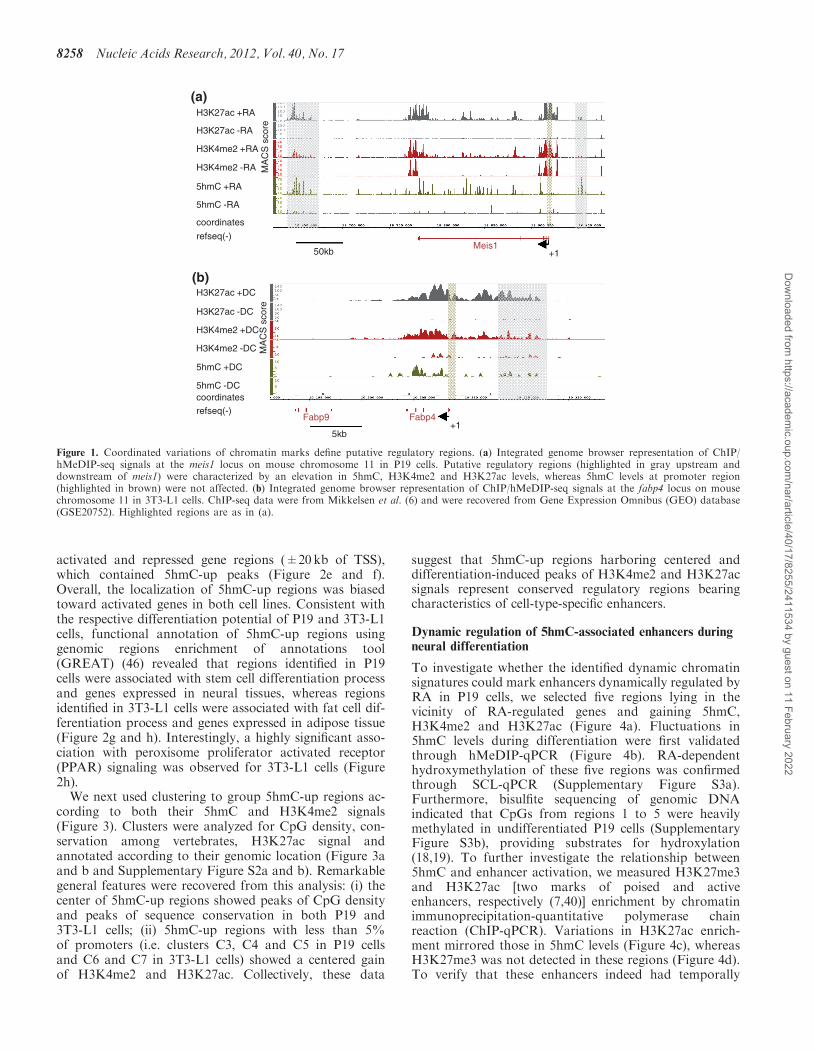
particularly abundant in gene-rich regions of both P19and 3T3-L1 cells (data not shown). When comparing5hmC levels in undifferentiated versus RA-treated P19cells, specific regions showed a remarkable increase in5hmC levels as exemplified with the meis1 locus, aRA-activated gene (Figure 1a, Supplementary Table S2)coding for a Hox cofactor (42). To determine whetherthese variations in 5hmC levels were associated withchanges in levels of other active chromatin marks, weran H3K4me2 and H3K27ac ChIP-seq assays in P19cells. Interestingly, H3K4me2 and H3K27ac alsoincreased in these regions on RA addition.Hydroxymethylation of the meis1 promoter region wasnot affected although H3K4me2 and H3K27ac raised(Figure 1a). Finally, consistent with previous observations(43), intragenic levels of 5hmC increased significantly atspecific positions when meis1 was expressed in response toRA. Similar dynamics were observed at the fatty acid-binding protein 4 (fabp4) locus in 3T3-L1 cells. Indeed,this activated gene (Supplementary Table S3) showedcorrelated elevation of 5hmC, H3K4me2 and H3K27aclevels in intragenic and distal putative regulatory regionsbut not immediately upstream of the TSS (Figure 1b).Hence, differentiation-associated chromatin remodelingin intragenic regions and putative distal regulatoryregions involves hydroxymethylation of DNA. Thisprovided a rationale for the further characterization ofregions that gain hmC (‘5hmC-up’ regions) during celldifferentiation.To allow genomic annotation, 5hmC-up regions were
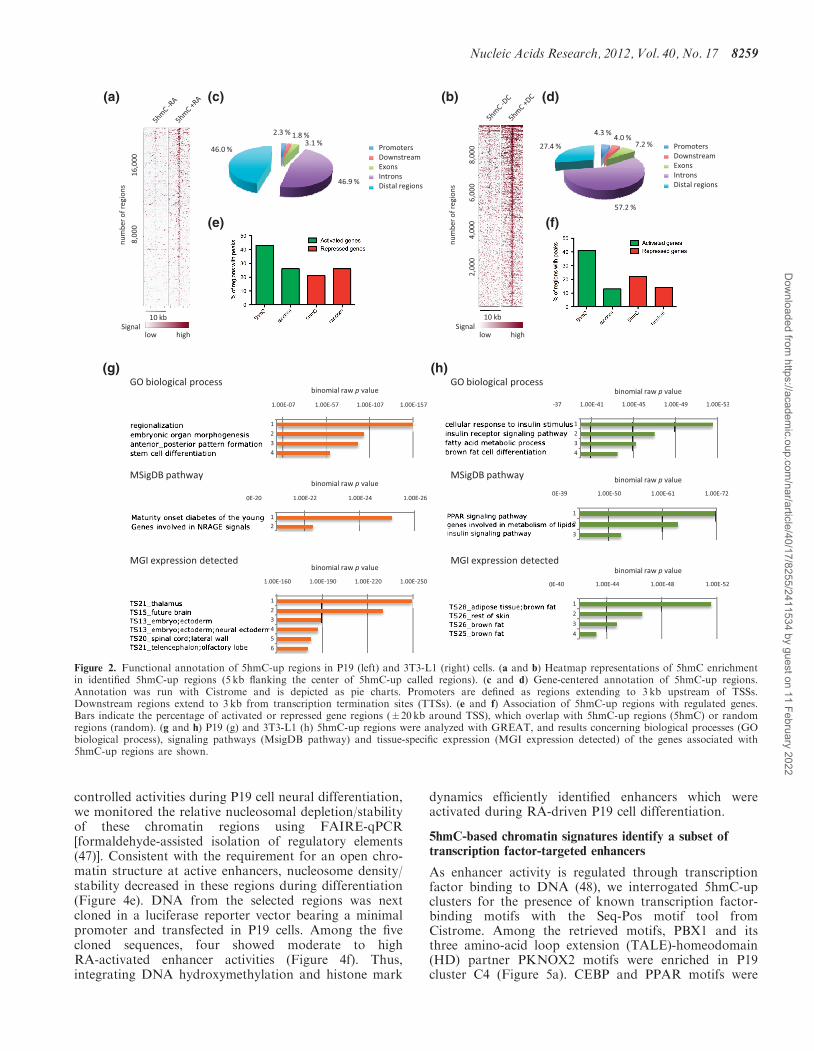
identified genome wide through direct comparison of ourhMeDIP-seq data from undifferentiated and differentiatedP19 and 3T3-L1 cells, as described in ‘Materials andMethods’ section (Supplementary Tables S4 and S5). Wethen generated heatmaps of 5hmC signals on 5hmC-upregions rank ordered according to their 5hmC content inthe presence of RA (Figure 2a) or DC (Figure 2b), usingCistrome (44). As shown in these heatmaps, peak callingefficiently identified 5hmC-up regions in both cell lines.Cis-regulatory Element Annotation System (45) indicatedthat genomic distribution of 5hmC-up regions differedbetween the two cell lines as intergenic regions werehighly represented in P19 compared with 3T3-L1(Figure 2c and d), indicating that the DNA hydroxy-methylation machinery was mobilized to differentgenomic locations during adipocyte and neural differenti-ation. We next questioned to what extent differentiation-regulated genes were associated with 5hmC-up regions inP19 and 3T3-L1 cells by analyzing the percentage of
Nucleic Acids Research, 2012, Vol. 40, No. 17 8257
Dow
nloaded from https://academ
ic.oup.com/nar/article/40/17/8255/2411534 by guest on 11 February 2022
activated and repressed gene regions (±20kb of TSS),which contained 5hmC-up peaks (Figure 2e and f).Overall, the localization of 5hmC-up regions was biasedtoward activated genes in both cell lines. Consistent withthe respective differentiation potential of P19 and 3T3-L1cells, functional annotation of 5hmC-up regions usinggenomic regions enrichment of annotations tool(GREAT) (46) revealed that regions identified in P19cells were associated with stem cell differentiation processand genes expressed in neural tissues, whereas regionsidentified in 3T3-L1 cells were associated with fat cell dif-ferentiation process and genes expressed in adipose tissue(Figure 2g and h). Interestingly, a highly significant asso-ciation with peroxisome proliferator activated receptor(PPAR) signaling was observed for 3T3-L1 cells (Figure2h).We next used clustering to group 5hmC-up regions ac-
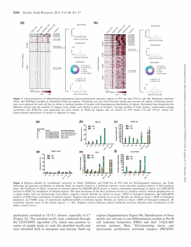
cording to both their 5hmC and H3K4me2 signals(Figure 3). Clusters were analyzed for CpG density, con-servation among vertebrates, H3K27ac signal andannotated according to their genomic location (Figure 3aand b and Supplementary Figure S2a and b). Remarkablegeneral features were recovered from this analysis: (i) thecenter of 5hmC-up regions showed peaks of CpG densityand peaks of sequence conservation in both P19 and3T3-L1 cells; (ii) 5hmC-up regions with less than 5%of promoters (i.e. clusters C3, C4 and C5 in P19 cellsand C6 and C7 in 3T3-L1 cells) showed a centered gainof H3K4me2 and H3K27ac. Collectively, these data
suggest that 5hmC-up regions harboring centered anddifferentiation-induced peaks of H3K4me2 and H3K27acsignals represent conserved regulatory regions bearingcharacteristics of cell-type-specific enhancers.
Dynamic regulation of 5hmC-associated enhancers duringneural differentiation
To investigate whether the identified dynamic chromatinsignatures could mark enhancers dynamically regulated byRA in P19 cells, we selected five regions lying in thevicinity of RA-regulated genes and gaining 5hmC,H3K4me2 and H3K27ac (Figure 4a). Fluctuations in5hmC levels during differentiation were first validatedthrough hMeDIP-qPCR (Figure 4b). RA-dependenthydroxymethylation of these five regions was confirmedthrough SCL-qPCR (Supplementary Figure S3a).Furthermore, bisulfite sequencing of genomic DNAindicated that CpGs from regions 1 to 5 were heavilymethylated in undifferentiated P19 cells (SupplementaryFigure S3b), providing substrates for hydroxylation(18,19). To further investigate the relationship between5hmC and enhancer activation, we measured H3K27me3and H3K27ac [two marks of poised and activeenhancers, respectively (7,40)] enrichment by chromatinimmunoprecipitation-quantitative polymerase chainreaction (ChIP-qPCR). Variations in H3K27ac enrich-ment mirrored those in 5hmC levels (Figure 4c), whereasH3K27me3 was not detected in these regions (Figure 4d).To verify that these enhancers indeed had temporally
H3K27ac +RA
H3K27ac -RA
(a)
H3K4me2 +RA
H3K4me2 -RA
5hmC +RA
5hmC -RA
coordinates
refseq(-)
50kb +1
H3K27ac +DC
(b)M
AC
S s
core
Meis1
H3K27ac -DC
H3K4me2 +DC
H3K4me2 -DC
5hmC +DC
5hmC -DCcoordinates
refseq(-)
5kb+1
MA
CS
sco
re
Fabp9 Fabp4
Figure 1. Coordinated variations of chromatin marks define putative regulatory regions. (a) Integrated genome browser representation of ChIP/hMeDIP-seq signals at the meis1 locus on mouse chromosome 11 in P19 cells. Putative regulatory regions (highlighted in gray upstream anddownstream of meis1) were characterized by an elevation in 5hmC, H3K4me2 and H3K27ac levels, whereas 5hmC levels at promoter region(highlighted in brown) were not affected. (b) Integrated genome browser representation of ChIP/hMeDIP-seq signals at the fabp4 locus on mousechromosome 11 in 3T3-L1 cells. ChIP-seq data were from Mikkelsen et al. (6) and were recovered from Gene Expression Omnibus (GEO) database(GSE20752). Highlighted regions are as in (a).
8258 Nucleic Acids Research, 2012, Vol. 40, No. 17
Dow
nloaded from https://academ
ic.oup.com/nar/article/40/17/8255/2411534 by guest on 11 February 2022
controlled activities during P19 cell neural differentiation,we monitored the relative nucleosomal depletion/stabilityof these chromatin regions using FAIRE-qPCR[formaldehyde-assisted isolation of regulatory elements(47)]. Consistent with the requirement for an open chro-matin structure at active enhancers, nucleosome density/stability decreased in these regions during differentiation(Figure 4e). DNA from the selected regions was nextcloned in a luciferase reporter vector bearing a minimalpromoter and transfected in P19 cells. Among the fivecloned sequences, four showed moderate to highRA-activated enhancer activities (Figure 4f). Thus,integrating DNA hydroxymethylation and histone mark
dynamics efficiently identified enhancers which wereactivated during RA-driven P19 cell differentiation.
5hmC-based chromatin signatures identify a subset oftranscription factor-targeted enhancers
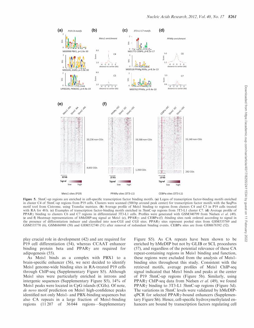
As enhancer activity is regulated through transcriptionfactor binding to DNA (48), we interrogated 5hmC-upclusters for the presence of known transcription factor-binding motifs with the Seq-Pos motif tool fromCistrome. Among the retrieved motifs, PBX1 and itsthree amino-acid loop extension (TALE)-homeodomain(HD) partner PKNOX2 motifs were enriched in P19cluster C4 (Figure 5a). CEBP and PPAR motifs were
(a) (c) (b) (d)
(f)(e)
(g) (h)
Figure 2. Functional annotation of 5hmC-up regions in P19 (left) and 3T3-L1 (right) cells. (a and b) Heatmap representations of 5hmC enrichmentin identified 5hmC-up regions (5 kb flanking the center of 5hmC-up called regions). (c and d) Gene-centered annotation of 5hmC-up regions.Annotation was run with Cistrome and is depicted as pie charts. Promoters are defined as regions extending to 3 kb upstream of TSSs.Downstream regions extend to 3 kb from transcription termination sites (TTSs). (e and f) Association of 5hmC-up regions with regulated genes.Bars indicate the percentage of activated or repressed gene regions (±20kb around TSS), which overlap with 5hmC-up regions (5hmC) or randomregions (random). (g and h) P19 (g) and 3T3-L1 (h) 5hmC-up regions were analyzed with GREAT, and results concerning biological processes (GObiological process), signaling pathways (MsigDB pathway) and tissue-specific expression (MGI expression detected) of the genes associated with5hmC-up regions are shown.
Nucleic Acids Research, 2012, Vol. 40, No. 17 8259
Dow
nloaded from https://academ
ic.oup.com/nar/article/40/17/8255/2411534 by guest on 11 February 2022
particularly enriched in 3T3-L1 clusters, especially in C7(Figure 5c). The enriched motifs were confirmed throughthe CENTDIST algorithm (53), which uses position tocenter of sample peaks to rank the identified motifs andwere identified both in intergenic and intronic 5hmC-up
regions (Supplementary Figure S4). Identification of thesemotifs was relevant to our differentiation models as Pre-Bcell leukemia homeobox (PBX) and their TALE-HDprotein partners Meis, TG-interacting factor andperoxisome proliferator activated receptor (PKNOX)
(a) (b)
Figure 3. Characterization of differentiation-dependent hydroxymethylated genomic regions in P19 (a) and 3T3-L1 cells (b). Heatmaps represent5hmC and H3K4me2 profiles in clusterized 5hmC-up regions. Clustering was run with Cistrome taking into account all signals. Clustering param-eters were adjusted for each cell line to obtain a minimal number of clusters with homogeneous distribution of signals. Horizontal lines demarcate thedifferent clusters and the number of regions lying within each cluster is given in brackets. Average profiles of CpG density, conservation amongvertebrates and H3K27ac were generated for each cluster of 5hmC-up regions and are shown for P19 cluster C4 and 3T3-L1 cluster C6.Gene-centered annotation of clusters is depicted as rings.
Figure 4. Regions defined by coordinated variations in 5hmC, H3K4me2 and H3K27ac in P19 cells are RA-dependent enhancers. (a) Tableindicating the genomic coordinates of selected 5hmC-up regions cloned in a luciferase reporter vector and their position relative to RA-regulatedgenes. (b) Validation of 5hmC variations in selected regions by hMeDIP-qPCR shown as relative enrichment (percentage of input). (c) ChIP-qPCRanalysis of H3K27ac enrichment of selected regions. The open chromatin of the Hus1 promoter served as a positive control, whereas amplification ofa region within Nf1a on chromosome 4 provided a negative control. Results are shown as mean±SEM of biological duplicates. (d) ChIP-qPCRanalysis of H3K27me3 enrichment of selected regions. Negative and positive controls are as in (c). Results are shown as mean±SEM of biologicalduplicates. (e) FAIRE assay of nucleosome depletion/stability in selected regions. Results are shown as mean±SEM of biological triplicates. (f)Luciferase reporter assay of the cloned regions �/+ RA. Negative control indicates relative luciferase activities obtained after transfection of theempty control vector.
8260 Nucleic Acids Research, 2012, Vol. 40, No. 17
Dow
nloaded from https://academ
ic.oup.com/nar/article/40/17/8255/2411534 by guest on 11 February 2022
play crucial role in development (42) and are required forP19 cell differentiation (54), whereas CCAAT enhancerbinding protein beta and PPARg are required foradipogenesis (55).
As Meis1 binds as a complex with PBX1 to abrain-specific enhancer (56), we next decided to identifyMeis1 genome-wide binding sites in RA-treated P19 cellsthrough ChIP-seq (Supplementary Figure S5). AlthoughMeis1 sites were particularly enriched in introns andintergenic sequences (Supplementary Figure S5), 14% ofMeis1 peaks were located in CpG islands (CGIs). Of note,de novo motif prediction on Meis1 high-confidence peaksidentified not only Meis1- and PBX-binding sequences butalso CA repeats in a large fraction of Meis1-bindingregions (11 207 of 36 044 regions—Supplementary
Figure S5). As CA repeats have been shown to beenriched by hMeDIP but not by GLIB or SCL procedures(57), and regardless of the potential relevance of these CArepeat-containing regions in Meis1 binding and function,these regions were excluded from the analysis of Meis1-binding sites throughout this study. Consistent with theretrieved motifs, average profiles of Meis1 ChIP-seqsignal indicated that Meis1 binds and peaks at the centerof P19 5hmC-up regions (Figure 5b). Similarly, usingPPARg ChIP-seq data from Nielsen et al. (49), we foundPPARg binding to 3T3-L1 5hmC-up regions (Figure 5d).The variations in 5hmC levels were validated by hMeDIP-qPCR for selected PPARg-bound enhancers (Supplemen-tary Figure S6). Hence, cell-specific hydroxymethylated en-hancers are bound by transcription factors regulating cell
(a) (b) (c) (d)
(e) (f)
Figure 5. 5hmC-up regions are enriched in cell-specific transcription factor binding motifs. (a) Logos of transcription factor-binding motifs enrichedin cluster C4 of 5hmC-up regions from P19 cells. Clusters were scanned (500 bp around peak center) for transcription factor motifs with the SeqPosmotif tool from Cistrome, using Transfac matrices. (b) Average profile of Meis1 binding to regions from clusters C4 and C5 in P19 cells treatedwith RA for 48 h. (c) Examples of transcription factor-binding motifs enriched in 5hmC-up regions from 3T3-L1 cluster C7. (d) Average profile ofPPARg binding to clusters C6 and C7 regions in differentiated 3T3-L1 cells. Profiles were generated with GSM340799 from Nielsen et al. (49).(e and f) Heatmap representations of hMeDIP-seq signal at Meis1 (e), PPARg- and CEBPa-(f) -binding sites rank ordered according to signal inthe presence of differentiation inducer and classified into non-CGI and CGI sites. PPARg sites represent pooled sites from GSM535769 andGSM535770 (6), GSM686980 (50) and GSM532740 (51) after removal of redundant binding events. CEBPa sites are from GSM678392 (52).
Nucleic Acids Research, 2012, Vol. 40, No. 17 8261
Dow
nloaded from https://academ
ic.oup.com/nar/article/40/17/8255/2411534 by guest on 11 February 2022
differentiation. To get further insight into the distributionof 5hmC at transcription factor-binding sites and the pro-portion of sites that become hydroxymethylated on differ-entiation, we then generated heatmaps of Meis1 andPPARg-binding sites according to their 5hmC signal(Figure 5e and f) with binding regions divided into CGIsand non-CGIs. As already described by others (26,57),5hmC signal in CGIs was low and did not increase withdifferentiation (Figure 5e and f). Conversely, almost allnon-CGI Meis1 sites gained 5hmC in RA-treated cells re-gardless of Meis1 signal. A differentiation-dependenthydroxymethylation of cytosines was also observed forPPARg- and CEBPa-binding regions in 3T3-L1 cells(Figure 5f).To determine the sequence of events occurring at enhan-
cers during differentiation, we then analyzed the kineticsof DNA hydroxymethylation, H3K27 acetylation andMeis1 recruitment at enhancers R1 to R5 during thefirst 48 h of RA-induced P19 cell differentiation. DNAhydroxymethylation was either concomitant (R1, R2and R5) with activation of enhancers or occurred before(R3 and R4) activation (Supplementary Figure S7).Hence, DNA hydroxymethylation might be an earlyevent of enhancer activation.
A fraction of hydroxymethylated Meis1-bound enhancersrecruits Tet1 during differentiation
Increased 5hmC levels at enhancers activated during dif-ferentiation suggested that Tet enzymes could be recruitedto these regions. Hence, we mapped Tet1-binding sites inP19 cells -/+RA through ChIP-seq assays. As described inRA-treated ES cells, Tet1 expression decreased on P19 celldifferentiation to reach levels comparable with those ofTet2 and Tet3 (Supplementary Figure S8). Consistentwith decreased expression, high-confidence (P� 3.e�7)Tet1-binding regions dropped from 15 039 in undifferen-tiated cells to 12 236 in RA-treated P19 cells. Similar to itsdistribution in ES cells, Tet1 was enriched in promoters(Supplementary Figure S9a) and in CGIs (SupplementaryFigure S9b). Despite its lower expression in RA-treatedcells, we identified 6096 high-confidence Tet1-up sites(P� 2.4e�9) among which 824 were located in CGIs(Supplementary Figure S9c). Interestingly, as evidencedby heatmaps, most of these Tet1-up regions werenot associated with a gain in 5hmC (SupplementaryFigure S9d), indicating that Tet1 enzymatic activitymight be regulated at its binding sites and/or that Tet1can serve additional functions as already suggested (25).Further analysis evidenced that H3K27ac levels raised inTet1-up regions which became hydroxymethylated but notin others (Supplementary Figure S9d).We then examined the engagement of Tet1 at non-CGI
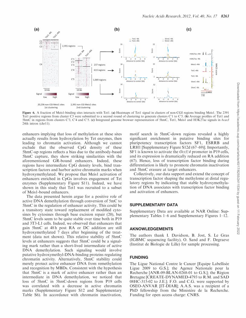
Meis1-binding sites and found that Tet1 associates witha fraction of Meis1 sites, which shows an enrichmentin CpGs (Figure 6a and Supplementary Figure S10).Through a second round of clustering, we identified1306 regions (Figure 6a) that show, as exemplified foran intronic region of the bcas3 gene (Figure 6c), a simul-taneous increase in Tet1 and DNA hydroxymethylationlevels during RA-induced differentiation. Interestingly, a
large fraction of these regions was already associated withlow levels of Tet1 in undifferentiated cells (Figure 6a) inline with premarking of Meis1-binding sites by low levelsof 5hmC (Figure 5e). Although Tet1 loading decreased onRA treatment at 613 specific Meis1-binding sites, average5hmC levels increased by 40% (Figure 6b). These dataindicate a complex relationship between Meis1 and Tet1and suggest that Meis1 engagement at Tet1-binding sitescould stimulate its recruitment and/or its activity.
DISCUSSION
Taken together, our data indicate a relationship betweenDNA hydroxymethylation and enhancer activation duringmouse cell differentiation. Our observations are in linewith previous studies reporting an enrichment in 5hmCat enhancers in correlation with H3K4me1, H3K4me2and H3K27ac levels in undifferentiated human stem cells(30,31) and that binding sites engaged with thepluripotency transcription factor NANOG bear 5hmC.On differentiation, ES cells tend to lose 5hmC, and itremains unknown whether the mark is lost in a generalway or if specific genomic regions can become hydroxy-methylated during programming of alternative cell states.In this study, we provide evidence that differentiation isassociated with dynamic DNA hydroxymethylation of asubset of cell-type-specific enhancers.
Pioneer studies focusing on the genome-wide distribu-tion of 5hmC showed that intragenic 5hmC is positivelyassociated with gene activity (26,29,58). Consistent withthis observation, 5hmC inhibits binding of severalMethyl CpG binding protein (MBD) including MeCP2(59, 60) and could reduce maintenance methylation byDNMT1 (61). As MeCP2 recruits enzymatic systemsthat drive chromatin condensation (62–64), oxidation of5mC could help chromatin opening and thereby facilitategene expression. As demonstrated for FOXA1-dependentenhancers, chromatin opening at enhancers is associatedwith their cell-specific activity (36). In this study, we es-tablish that chromatin opening, as assessed by FAIRE, isassociated with acquisition of 5hmC at selected enhancers.In parallel to the gain in 5hmC, H3K27 acetylation levelsraised, suggesting a model of enhancer activation thatinvolves the conversion of 5mC to 5hmC. In line withthis hypothesis, a recent study of glucocorticoid receptor(GR)-bound enhancers suggested that a fraction of GRsites distal to TSS and outside of CGIs are demethylatedon GR binding (65). Similar to what is observed for5hmC-up regions defined in this study, these enhancersare characterized by a peak of CpG density that is alsoobserved for DNAse I hypersensitive sites not overlappingwith CGIs (65), hence defining a category of enhancersthat show intermediate CpG density (compared withCGIs and the rest of the genome) and whose activity iscontrolled by methylation. Recently, Schubeler andcoworkers (66) described methylome remodeling throughtranscription factor binding at enhancers with low tointermediate levels of CpGs and associated with Tet1. Itis most likely that 5hmC-up regions identified in P19 and3T3-L1 cells belong to this particular category of
8262 Nucleic Acids Research, 2012, Vol. 40, No. 17
Dow
nloaded from https://academ
ic.oup.com/nar/article/40/17/8255/2411534 by guest on 11 February 2022
enhancers implying that loss of methylation at these sitesactually results from hydroxylation by Tet enzymes, thenleading to chromatin activation. Although we cannotexclude that the observed CpG density of these5hmC-up regions reflects a bias due to the antibody-based5hmC capture, they show striking similarities with theaforementioned GR-bound enhancers. Indeed, theseregions have intermediate CpG density levels, bind tran-scription factors and harbor active chromatin marks whenhydroxymethylated. We propose that Meis1 activation ofenhancers enriched in CpGs involves engagement of Tetenzymes (Supplementary Figure S11). Indeed, we haveshown in this study that Tet1 was recruited to a subsetof Meis1-bound enhancers.
The data presented herein argue for a positive role ofactive DNA demethylation through conversion of 5mC to5hmC in the regulation of enhancer activity. This could bea transitory state toward replacement of modified cyto-sines by cytosines through base excision repair (20), but5hmC levels seem to be quite stable over time both in P19and 3T3-L1 cells. Indeed, we observed that enhancers thatgain 5hmC at 48 h post RA or DC addition are stillhydroxymethylated 7 days after beginning of the treat-ment (data not shown). This relative stability of 5hmClevels at enhancers suggests that 5hmC could be a signal-ing mark rather than a short-lived intermediate of activeDNA demethylation. Such signaling would involveputative hydroxymethyl-DNA-binding proteins regulatingchromatin activity. Alternatively, 5hmC stability couldmerely protect active enhancer DNA from remethylationand recognition by MBDs. Consistent with the hypothesisthat 5hmC is a mark of active enhancer rather than anintermediate in DNA demethylation, we noticed thatloss of 5hmC in 5hmC-down regions from P19 cellswas correlated with a decrease in active chromatinmarks (Supplementary Figure S12 and SupplementaryTable S6). In accordance with chromatin inactivation,
motif search in 5hmC-down regions revealed a highlysignificant enrichment in putative binding sites forpluripotency transcription factors SF1, ESRRB andLRH1 [Supplementary Figure S12d (67–69)]. Importantly,SF1 is known to activate the Oct3/4 promoter in P19 cells,and its expression is dramatically reduced on RA addition(67). Hence, loss of transcription factor binding duringdifferentiation is likely to promote chromatin inactivationand 5hmC erasure at target enhancers.Collectively, our data support and extend the concept of
transcription factor shaping the methylome at distal regu-latory regions by indicating that stable hydroxymethyla-tion of DNA associates with transcription factor bindingand activation of enhancers.
SUPPLEMENTARY DATA
Supplementary Data are available at NAR Online: Sup-plementary Tables 1–6 and Supplementary Figures 1–12.
AKNOWLEDGEMENTS
The authors thank I. Davidson, B. Jost, S. Le Gras(IGBMC sequencing facility), O. Sand and F. Degraeve(Institut de Biologie de Lille) for sample processing.
FUNDING
The Ligue National Contre le Cancer [Equipe LabelliseeLigue 2009 to G.S.]; the Agence Nationale pour laRecherche [ANR-09-BLAN-0268-01 to G.S.]; the RegionBretagne [CREATE-DYNAMED-4793 to R.M. and SAD08HC-315-02 to J.E.]; F.O. and C.G. were supported byOSEO-ANVAR [IT-DIAB]; A.A.S. was a recipient of aPhD fellowship from the Ministere de la Recherche.Funding for open access charge: CNRS.
(a) (b) (c)
Figure 6. A fraction of Meis1-binding sites interacts with Tet1. (a) Heatmaps of Tet1 signal in clusters of non-CGI regions binding Meis1. The 2391Tet1 positive regions from cluster C3 were submitted to a second round of clustering to generate clusters C01 to C05. (b) Average profiles of Tet1 and5hmC in regions from clusters C03, C04 and C05. (c) Integrated genome browser representation of 5hmC, Tet1, Meis1 and H3K27ac signals in bcas324th intron (chr11).
Nucleic Acids Research, 2012, Vol. 40, No. 17 8263
Dow
nloaded from https://academ
ic.oup.com/nar/article/40/17/8255/2411534 by guest on 11 February 2022
Conflict of interest statement. None declared.
REFERENCES
1. Visel,A., Rubin,E.M. and Pennacchio,L.A. (2009) Genomic viewsof distant-acting enhancers. Nature, 461, 199–205.
2. Pennacchio,L.A., Ahituv,N., Moses,A.M., Prabhakar,S.,Nobrega,M.A., Shoukry,M., Minovitsky,S., Dubchak,I., Holt,A.,Lewis,K.D. et al. (2006) In vivo enhancer analysis of humanconserved non-coding sequences. Nature, 444, 499–502.
3. Blow,M.J., McCulley,D.J., Li,Z., Zhang,T., Akiyama,J.A.,Holt,A., Plajzer-Frick,I., Shoukry,M., Wright,C., Chen,F. et al.(2010) ChIP-seq identification of weakly conserved heartenhancers. Nat. Genet., 42, 806–810.
4. Fernandez-Tresguerres,B., Canon,S., Rayon,T., Pernaute,B.,Crespo,M., Torroja,C. and Manzanares,M. (2010) Evolution ofthe mammalian embryonic pluripotency gene regulatory network.Proc. Natl Acad. Sci. USA, 107, 19955–19960.
5. Hawkins,R.D., Hon,G.C., Lee,L.K., Ngo,Q., Lister,R.,Pelizzola,M., Edsall,L.E., Kuan,S., Luu,Y., Klugman,S. et al.(2010) Distinct epigenomic landscapes of pluripotent andlineage-committed human cells. Cell Stem Cell, 6, 479–491.
6. Mikkelsen,T.S., Xu,Z., Zhang,X., Wang,L., Gimble,J.M.,Lander,E.S. and Rosen,E.D. (2010) Comparative epigenomicanalysis of murine and human adipogenesis. Cell, 143, 156–169.
7. Rada-Iglesias,A., Bajpai,R., Swigut,T., Brugmann,S.A.,Flynn,R.A. and Wysocka,J. (2011) A unique chromatin signatureuncovers early developmental enhancers in humans. Nature, 470,279–283.
8. Jin,C., Zang,C., Wei,G., Cui,K., Peng,W., Zhao,K. andFelsenfeld,G. (2009) H3.3/H2A.Z double variant-containingnucleosomes mark ‘nucleosome-free regions’ of active promotersand other regulatory regions. Nat. Genet., 41, 941–945.
9. Goldberg,A., Banaszynski,L.A., Noh,K.M., Lewis,P.W.,Elsaesser,S.J., Stadler,S., Dewell,S., Law,M., Guo,X., Li,X. et al.(2010) Distinct factors control histone variant H3.3 localization atgenomic regions. Cell, 140, 678–691.
10. Gaulton,K.J., Nammo,T., Pasquali,L., Simon,J.M., Giresi,P.G.,Fogarty,M.P., Panhuis,T.M., Mieczkowski,P., Secchi,A., Bosco,D.et al. (2010) A map of open chromatin in human pancreaticislets. Nat. Genet., 42, 255–259.
11. He,H.H., Meyer,C.A., Shin,H., Bailey,S.T., Wei,G., Wang,Q.,Zhang,Y., Xu,K., Ni,M., Lupien,M. et al. (2010) Nucleosomedynamics define transcriptional enhancers. Nat. Genet., 42,343–347.
12. Conerly,M.L., Teves,S.S., Diolaiti,D., Ulrich,M., Eisenman,R.N.and Henikoff,S. (2010) Changes in H2A.Z occupancy and DNAmethylation during B-cell lymphomagenesis. Genome Res., 20,1383–1390.
13. Zemach,A., McDaniel,I.E., Siva,P. and Zilberman,D. (2010)Genome-wide evolutionary analysis of eukaryotic DNAmethylation. Science, 328, 916–919.
14. Edwards,J.R., O’Donnell,A.H., Rollins,R.A., Peckham,H.E.,Lee,C., Milekic,M.H., Chanrion,B., Fu,Y., Su,T., Hibshoosh,H.et al. (2010) Chromatin and sequence features that define the fineand gross structure of genomic methylation patterns. GenomeRes., 20, 972–980.
15. Lister,R., Pelizzola,M., Dowen,R.H., Hawkins,D., Hon,G., Tonti-Filippini,J., Nery,J.R., Lee,L., Ye,Z., Ngo,Q.-M. et al. (2009)Human DNA methylomes at base resolution show widespreadepigenomic differences. Nature, 462, 315–322.
16. Schmidl,C., Klug,M., Boeld,T.J., Andreesen,R., Hoffman,P.,Edinger,M. and Rehli,M. (2009) Lineage-specific DNAmethylation in T cells correlates with histone methylation andenhancer activity. Genome Res., 19, 1165–1174.
17. Serandour,A.A., Avner,S., Percevault,F., Demay,F., Bizot,M.,Lucchetti-Miganeh,C., Barloy-Hubler,F., Brown,M., Lupien,M.,Metivier,R. et al. (2011) Epigenetic switch involved in activationof pioneer factor FOXA1-dependent enhancers. Genome Res., 21,555–565.
18. Tahiliani,M., Koh,K.P., Shen,Y., Pastor,W.A., Bandukwala,H.,Brudno,Y., Agarwal,S., Lyer,L.M., Liu,D.R., Aravind,L. et al.(2009) Conversion of 5-methylcytosine to 5-hydroxymethylcytosine
in mammalian DNA by MLL partner TET1. Science, 324,930–935.
19. Ito,S., D’Alessio,A.C., Taranova,O.V., Hong,K., Sowers,L.C. andZhang,Y. (2010) Role of Tet proteins in 5mC to 5hmCconversion, ES cells self-renewal and inner cell mass specification.Nature, 466, 1129–1133.
20. He,Y.F., Li,B.Z., Li,Z., Liu,P., Wang,Y., Tang,Q., Ding,J., Jia,Y.,Chen,Z., Li,L. et al. (2011) Tet-mediated formation of5-carboxylcytosine and its excision by TDG in mammalian cells.Science, 333, 1303–1307.
21. Ito,S., Shen,L., Dai,Q., Wu,S.C., Collins,L.B., Swenberg,J.A.,He,C. and Zhang,Y. (2011) Tet proteins can convert5-methylcytosine to 5-formylcytosine and 5-carboxylcytosine.Science, 333, 1300–1303.
22. Maiti,A. and Drohat,A.C. (2011) Thymine DNA glycosylase canrapidly excise 5-formylcytosine and 5-carboxylcytosine: potentialimplications for active demethylation of CpG sites. J. Biol.Chem., 286, 35334–35338.
23. Pastor,W.A., Pape,U.J., Huang,Y., Henderson,H.R., Lister,R.,Ko,M., McLoughlin,E.M., Brudno,Y., Mahapatra,S.,Kapranov,P. et al. (2011) Genome-wide mapping of5-hydroxymethylcytosine in embryonic stem cells. Nature, 473,394–397.
24. Song,C.X., Szulwach,K.E., Fu,Y., Dai,Q., Yi,C., Li,X., Li,Y.,Chen,C.H., Zhang,W., Jian,X. et al. (2011) Selective chemicallabeling reveals the genome-wide distribution of5-hydroxymethylcytosine. Nat Biotechnol., 29, 68–72.
25. Williams,K., Christensen,J., Pedersen,M.T., Johansen,J.V.,Cloos,P.A.C., Rappsilber,J. and Helin,K. (2011) TET1 andhydroxymethylcytosine in transcription and DNA methylationfidelity. Nature, 473, 343–348.
26. Wu,H., D’Alessio,A.C., Ito,S., Wang,Z., Cui,K., Zhao,K.,Sun,Y.E. and Zhang,Y. (2011) Genome-wide analysis of5-hydroxymethylcytosine distribution reveals its dual function intranscriptional regulation in mouse embryonic stem cells. GenesDev., 25, 679–684.
27. Xu,Y., Wu,F., Tan,L., Kong,L., Xiong,L., Deng,J., Barbera,A.J.,Zheng,L., Zhang,H., Huang,S. et al. (2011) Genome-wideregulation of 5hmC, 5mC, and gene expression by Tet1hydroxylase in mouse embryonic stem cells. Mol. Cell., 42,451–464.
28. Yildirim,O., Li,R., Hung,J.H., Chen,P.B., Dong,X., Ee,L.S.,Weng,Z., Rando,O.J. and Fazzio,T.G. (2011) Mbd3/NURDcomplex regulates expression of 5-hydroxymethylcytosine markedgenes in embryonic stem cells. Cell, 147, 1498–1510.
29. Ficz,G., Branco,M.R., Seisenberger,S., Santos,F., Krueger,F.,Hore,T.A., Marques,C.J., Andrews,S. and Reik,W. (2011)Dynamic regulation of 5-hydroxymethylcytosine in mouse ES cellsand during differentiation. Nature, 473, 398–402.
30. Stroud,H., Feng,S., Morey Kinney,S., Pradhan,S. andJacobsen,S.E. (2011) 5-hydroxymethylcytosine is associated withenhancers and gene bodies in huan embryonic stem cells. GenomeBiol., 12, R54.
31. Szulwach,K.E., Li,X., Li,Y., Song,C.-X., Han,J.W., Kim,S.,Namburi,S., Hermetz,K., Kim,J.J., Rudd,M.K. et al. (2011)Integrating 5-hydroxymethylcytosine into the epigenomiclandscape of human embryonic stem cells. PLoS Genet., 7,e1002154.
32. Haffner,M.C., Chaux,A., Meeker,A.K., Esopi,D.M., Gerber,J.,Pellakuru,L.G., Toubaji,A., Argani,P., Iacobuzio-Donahue,C.,Nelson,W.G. et al. (2011) Global 5-hydroxymethylcytosinecontent is significantly reduced in tissue stem/progenitorcell compartments and in human cancers. Oncotarget, 2,627–637.
33. Szulwach,K.E., Li,X., Li,Y., Song,C.-X., Wu,H., Dai,Q., Irier,H.,Upadhyay,A.K., Gearing,M., Levey,A.I. et al. (2011)5-hmC-mediated epigenetic dynamics during postnatalneurodevelopment and aging. Nat. Neurosci., 14, 1607–1616.
34. Klug,M. and Rehli,M. (2006) Functional analysis of promoterCpG methylation using a CpG-free luciferase reporter vector.Epigenetics, 1, 127–130.
35. Lefebvre,B., Benomar,Y., Guedin,A., Langlois,A., Hennuyer,N.,Dumont,J., Bouchaert,E., Dacquet,C., Penicaud,L., Casteilla,L.et al. (2010) Proteasomal degradation of retinoid X receptor
8264 Nucleic Acids Research, 2012, Vol. 40, No. 17
Dow
nloaded from https://academ
ic.oup.com/nar/article/40/17/8255/2411534 by guest on 11 February 2022
alpha reprograms transcriptional activity of PPARgamma inobese mice and humans. J. Clin. Invest., 120, 1454–1468.
36. Eeckhoute,J., Lupien,M., Meyer,C.A., Verzi,M.P.,Shivdasani,R.A., Liu,X.S. and Brown,M. (2009) Cell-type selectivechromatin remodeling defines the active subset of FOXA1-boundenhancers. Genome Res., 19, 372–380.
37. Langmead,B., Trapnell,C., Pop,M. and Salzberg,S.L. (2009)Ultrafast and memory-efficient alignment of short DNAsequences to the human genome. Genome Biol., 10, R25.
38. Li,H., Handsaker,B., Wysoker,A., Fennell,T., Ruan,J., Homer,N.,Marth,G., Abecasis,G. and Durbin,R. (2009) The Sequencealignment/map format and SAMtools. Bioinformatics, 25,2078–2079.
39. Zhang,Y., Liu,T., Meyer,C.A., Eeckhoute,J., Johnson,W.E.,Bernstein,B.E., Nusbaum,C., Myers,R.M., Brown,M., Li,W. et al.(2008) Model-based Analysis of ChIP-Seq (MACS). Genome Biol.,9, R137.
40. Creyghton,M.P., Cheng,A.W., Welstead,G.G., Kooistra,T.,Carey,B.W., Steine,E.J., Hanna,J., Lodato,M.A., Frampton,G.M.,Sharp,P.A. et al. (2010) Histone H3K27ac separates active frompoised enhancers and predicts developmental state. Proc. NatlAcad. Sci. USA, 107, 21931–21936.
41. Jones-Villeneuve,E.M., McBurney,M.W., Rogers,K.A. andKalnins,V.I. (1982) Retinoic acid induces embryonal carcinomacells to differentiate into neurons and glial cells. J. Cell Biol., 94,253–262.
42. Moens,C.B. and Selleri,L. (2006) Hox cofactors in vertebratedevelopment. Dev. Biol., 291, 193–206.
43. Wu,H. and Zhang,Y. (2011) Tet1 and 5-hydroxymethylation: agenome-wide view in mouse embryonic stem cells. Cell Cycle, 10,2428–2436.
44. Liu,T., Ortiz,J.A., Taing,L., Meyer,C.A., Lee,B., Zhang,Y.,Shin,H., Wong,S.S., Ma,J., Lei,Y. et al. (2011) Cistrome: anintegrative platform for transcriptional regulation studies. GenomeBiol., 12, R83.
45. Shin,H., Liu,T., Manrai,A.K. and Liu,S. (2009) CEAS: cis-regulatory element annotation system. Bioinformatics, 25,2605–2606.
46. McLean,C.Y., Bristor,D., Hiller,M., Clarke,S.L., Schaar,B.T.,Lowe,C.B., Wenger,A.M. and Benjerano,G. (2010) GREATimproves functional interpretation of cis-regulatory regions. Nat.Biotechnol., 42, 1093–1100.
47. Giresi,P.G. and Lieb,J.D. (2009) Isolation of active regulatoryelements from eukaryotic chromatin using FAIRE (FormaldehydeAssisted Isolation of Regulatory Elements). Methods, 48, 233–239.
48. Ong,C.-T. and Corces,V.G. (2011) Enhancer function: newinsights into the regulation of tissue-specific gene expression. Nat.Rev. Genet., 12, 283–293.
49. Nielsen,R., Pedersen,T.A., Hagenbeek,D., Moulos,P.,Siersbaek,R., Megens,E., Denissov,S., Borgesen,M.,Francoijs,K.J., Mandrup,S. et al. (2008) Genome-wide profiling ofPPARgamma:RXR and RNA polymerase II occupancy revealstemporal activation of distinct metabolic pathways and changes inRXR dimer composition during adipogenesis. Genes Dev., 22,2953–2967.
50. Siersbaek,R., Nielsen,R., John,S., Sung,M.H., Baek,S., Loft,A.,Hager,G.L. and Mandrup,S. (2011) Extensive remodeling andestablishment of transcription factor ‘hotspots’ during earlyadipogenesis. EMBO J, 30, 1459–1472.
51. Lefterova,M.I., Steger,D.J., Zhuo,D., Qatanani,M., Mullican,S.E.,Tuteja,G., Manduchi,E., Grant,G.R. and Lazar,M.A. (2010)Cell-specific determinants of peroxisome proliferator-activatedreceptor gamma function in adipocytes and macrophages. Mol.Cell. Biol., 30, 2078–2089.
52. Schmidt,S.F., Jørgensen,M., Chen,Y., Nielsen,R., Sandelin,A. andMandrup,S. (2011) Cross species comparison of C/EBPa andPPARg profiles in mouse and human adipocytes revealsinterdependent retention of binding sites. BMC Genomics, 12, 152.
53. Zhang,Z., Chang,C.W., Goh,W.L., Sung,W.-K. and Cheung,E.(2011) CENTDIST: discovery of co-associated factors by motifdistribution. Nucleic Acids Res., 39, W391–W399.
54. Qin,P., Haberbusch,J.M., Zhang,Z., Soprano,K.J. andSoprano,D.R. (2004) Pre-B cell leukemia transcription factor(PBX) proteins are important mediators for retinoicacid-dependent endordermal and neuronal differentiation ofmouse embryonal carcinoma cells. J. Biol. Chem., 279,16263–16271.
55. Cristancho,A.G. and Lazar,M.A. (2011) Forming functional fat: agrowing understanding of adipocyte differentiation. Nat. Rev.Mol. Cell. Biol., 12, 722–734.
56. Jacobs,Y., Schnabel,C.A. and Cleary,M.L. (1999) Trimericassociation of Hox and Tale homeodomain proteins mediatesHoxb2 hindbrain enhancer activity. Mol. Cell. Biol., 19,5134–5142.
57. Matarese,F., Carrillo-de Santa Pau,E. and Stunnenberg,H.G.(2011) 5-Hydroxymethylcytosine: a new kid on the epigeneticblock? Mol. Syst. Biol., 7, 562.
58. Wu,H., D’Alessio,A.C., Ito,S., Xia,K., Wang,Z., Cui,K., Zhao,K.,Sun,Y.E. and Zhang,Y. (2011) Dual functions of Tet1 intranscriptional regulation in mouse embryonic stem cells. Nature,473, 389–393.
59. Valinluk,V., Tsai,H.H., Rogstad,D.K., Burdzy,A., Bird,A. andSowers,L.C. (2004) Oxidative damage to methyl-CpG sequencesinhibits the binding of methyl-CpG binding domain (MBD) ofmethyl-CpG binding protein 2 (MeCP2). Nucleic Acids Res., 32,4100–4108.
60. Jin,S.G., Kadam,S. and Pfeifer,G.P. (2010) Examination of thespecificity of DNA methylation profiling techniques towards5-methylcytosine and 5-hydroxymethylcytosine. Nucleic AcidsRes., 38, e125.
61. Valinluk,V. and Sowers,L.C. (2007) Endogenous cytosine damageproducts alter the site selectivity of human DNA maintenancemethyltransferase DNMT1. Cancer Res., 67, 946–950.
62. Jones,P.L., Veenstra,G.J., Wade,P.A., Vermaak,D., Kass,S.U.,Landsberger,N., Strouboulis,J. and Wolffe,A.P. (1998) MethylatedDNA and MeCP2 recruit histone deacetylase to represstranscription. Nat. Genet., 19, 187–191.
63. Nan,X., Ng,H.H., Johnson,C.A., Laherty,C.D., Turner,B.M.,Eisenman,R.N. and Bird,A. (1998) Transcriptional repression bythe methyl-CpG-binding protein MeCP2 involves a histonedeacetylase complex. Nature, 393, 386–389.
64. Fuks,F., Hurd,P.J., Wolf,D., Nan,X., Bird,A.P. andKouzarides,T. (2003) The methyl-CpG-binding protein MeCP2links DNA methylation to histone methylation. J. Biol. Chem.,278, 4035–4040.
65. Wiench,M., John,S., Baek,S., Johnson,T.A., Sung,M.-H.,Escobar,T., Simmons,C.A., Pearce,K.H., Biddie,S.C., Sabo,P.J.et al. (2011) DNA methylation status predicts cell-type specificenhancer activity. EMBO J., 30, 3028–3039.
66. Stadler,M.B., Murr,R., Burger,L., Ivanek,R., Lienert,F.,Scholer,A., Wirbelauer,C., Oakeley,E.J., Gaidatzis,V.K. andSchubeler,D. (2011) DNA-binding factors shape the mousemethylome at distal regulatory regions. Nature, 480, 490–495.
67. Barnea,E. and Bergman,Y. (2000) Synergy of SF1 and RAR inactivation of Oct3/4 promoter. J. Biol. Chem., 275, 6608–6619.
68. Gu,P., Goodwin,B., Chung,A.C.-K., Xu,X., Wheeler,D.A.,Price,R.R., Galardi,C., Peng,L., Latour,A.M., Koller,B.H. et al.(2005) Orphan nuclear receptor LRH-1 is required to maintainOct4 expression at the epiblast stage of embryonic development.Mol. Cell. Biol., 25, 3492–3505.
69. Zhang,X., Zhang,J., Wang,T., Esteban,M.A. and Pei,D. (2008)Esrrb activates Oct4 transcription and sustains self-renewal andpluripotency in embryonic stem cells. J. Biol. Chem., 283,35825–35833.
Nucleic Acids Research, 2012, Vol. 40, No. 17 8265
Dow
nloaded from https://academ
ic.oup.com/nar/article/40/17/8255/2411534 by guest on 11 February 2022