AR Expression in Breast Cancer CTCs Associates with Bone ...Dennis Sgroi1,3, Ravi Kapur4, David...
9
Signal Transduction AR Expression in Breast Cancer CTCs Associates with Bone Metastases Nicola Aceto 1,2 , Aditya Bardia 1,2 , Ben S. Wittner 1,2 , Maria C. Donaldson 1 , Ryan O'Keefe 1 , Amanda Engstrom 1 , Francesca Bersani 1,2 , Yu Zheng 1,2 , Valentine Comaills 1,2 , Kira Niederhoffer 1 , Huili Zhu 1 , Olivia Mackenzie 1 , Toshi Shioda 1,2 , Dennis Sgroi 1,3 , Ravi Kapur 4 , David T. Ting 1,2 , Beverly Moy 1,2 , Sridhar Ramaswamy 1,2 , Mehmet Toner 4,5 , Daniel A. Haber 1,2,6 , and Shyamala Maheswaran 1,5 Abstract Molecular drivers underlying bone metastases in human cancer are not well understood, in part due to constraints in bone tissue sampling. Here, RNA sequencing was performed of circulating tumor cells (CTC) isolated from blood samples of women with metastatic estrogen receptor (ER) þ breast cancer, comparing cases with progression in bone versus visceral organs. Among the activated cellular pathways in CTCs from bone-predominant breast cancer is androgen receptor (AR) signaling. AR gene expression is evident, as is its constitutively active splice variant AR-v7. AR expression within CTCs is correlated with the duration of treatment with aromatase inhibitors, suggesting that it contributes to acquired resistance to endocrine therapy. In an established breast cancer xenograft model, a bone-tropic derivative displays increased AR expres- sion, whose genetic or pharmacologic suppression reduces metastases to bone but not to lungs. Together, these observa- tions identify AR signaling in CTCs from women with bone- predominant ER þ breast cancer, and provide a rationale for testing androgen inhibitors in this subset of patients. Implications: This study highlights a role for the AR in breast cancer bone metastasis, and suggests that therapeutic targeting of the AR may benefit patients with metastatic breast cancer. Mol Cancer Res; 16(4); 720–7. Ó2018 AACR. Introduction Metastases to bone, brain, liver, and other visceral organs are primarily responsible for the lethality of breast cancer. Among these, bone metastases are the most common site of metastasis in ER þ breast cancer (1), but the molecular characteristics that drive cancer cells to initially seed and subsequently proliferate within the bone matrix are not well understood. To date, most insight has focused on bone turnover as a key permissive factor in metastasis, with agents that suppress osteoclast activity having clinical benefit in suppressing bone-related complication of cancer (2). Less is known about the tumor-cell intrinsic properties that may direct metastasis to bone versus other organs. In mouse models, formation of a pre-metastatic niche (3) and expression of specific cell adhesion molecules such as VCAM1, chemokines such as CXCR4, bone remodeling molecules includ- ing RANKL, OPN, and Jagged1, as well as integrins associated to tumor-derived exosomes have been proposed as mediators of bone metastasis (4–7). In advanced human cancer derived from many different tissues, metastases to bone are present along with metastases within multiple visceral organs. However, prostate cancer and some ER þ breast cancers are unique in having bone lesions as the sole site of metastatic disease, even in the presence of advanced disease (1). Thus, in addition to pathways that are intrinsic to bone, tumor-specific properties may also confer a predilection toward bone metastasis in these cancers. CTCs are cancer cells derived from either primary or metastatic tumor deposits that are detectable as they transit through the bloodstream of patients with cancer, and may initiate new metas- tases at distant sites (8, 9). In women who develop metastatic breast cancer several years after the removal of a primary breast tumor, CTCs are derived from metastatic lesions, and hence represent a "liquid biopsy" of multiple independent distant tumor deposits. Given the difficulty in direct sampling of bone metastases, the analysis of CTCs in this setting provides a unique opportunity to study patient-derived specimens to identify poten- tial molecular drivers of metastasis. Materials and Methods CTC capture and identification Blood specimens for CTC analysis were obtained after informed patient consent, per IRB protocol (05-300), at the Massachusetts General Hospital and in accordance with the Declaration of Helsinki. A maximum of 20 mL of blood was drawn in EDTA vacutainers. Approximately 6 to 12 mL of blood 1 Massachusetts General Hospital Cancer Center, Harvard Medical School, Boston, Massachusetts. 2 Department of Medicine, Harvard Medical School, Boston, Massachusetts. 3 Department of Pathology, Harvard Medical School, Boston, Massachusetts. 4 Center for Bioengineering in Medicine, Harvard Medical School, Boston, Massachusetts. 5 Department of Surgery, Harvard Medical School, Boston, Massachusetts. 6 Howard Hughes Medical Institute, Chevy Chase, Maryland. Current address for N. Aceto: Department of Biomedicine, University of Basel and University Hospital Basel, Basel, Switzerland. Corresponding Authors: Daniel A. Haber, Harvard Medical School, Building 149, 13th Street, Charlestown, MA 02129. Phone: 617-726-7805; Fax: 617-726-5637; E-mail: [email protected]; and Shyamala Maheswaran, [email protected] doi: 10.1158/1541-7786.MCR-17-0480 Ó2018 American Association for Cancer Research. Molecular Cancer Research Mol Cancer Res; 16(4) April 2018 720 on January 24, 2020. © 2018 American Association for Cancer Research. mcr.aacrjournals.org Downloaded from Published OnlineFirst February 16, 2018; DOI: 10.1158/1541-7786.MCR-17-0480
Transcript of AR Expression in Breast Cancer CTCs Associates with Bone ...Dennis Sgroi1,3, Ravi Kapur4, David...
Signal Transduction
AR Expression in Breast Cancer CTCs Associateswith Bone MetastasesNicola Aceto1,2, Aditya Bardia1,2, Ben S.Wittner1,2, Maria C. Donaldson1,Ryan O'Keefe1, Amanda Engstrom1, Francesca Bersani1,2, Yu Zheng1,2,Valentine Comaills1,2, Kira Niederhoffer1, Huili Zhu1, Olivia Mackenzie1, Toshi Shioda1,2,Dennis Sgroi1,3, Ravi Kapur4, David T. Ting1,2, Beverly Moy1,2, Sridhar Ramaswamy1,2,Mehmet Toner4,5, Daniel A. Haber1,2,6, and Shyamala Maheswaran1,5
Abstract
Molecular drivers underlying bone metastases in humancancer are not well understood, in part due to constraints inbone tissue sampling. Here, RNA sequencing was performed ofcirculating tumor cells (CTC) isolated from blood samples ofwomen with metastatic estrogen receptor (ER)þ breast cancer,comparing cases with progression in bone versus visceralorgans. Among the activated cellular pathways in CTCs frombone-predominant breast cancer is androgen receptor (AR)signaling. AR gene expression is evident, as is its constitutivelyactive splice variant AR-v7. AR expression within CTCs iscorrelated with the duration of treatment with aromataseinhibitors, suggesting that it contributes to acquired resistance
to endocrine therapy. In an established breast cancer xenograftmodel, a bone-tropic derivative displays increased AR expres-sion, whose genetic or pharmacologic suppression reducesmetastases to bone but not to lungs. Together, these observa-tions identify AR signaling in CTCs from women with bone-predominant ERþ breast cancer, and provide a rationale fortesting androgen inhibitors in this subset of patients.
Implications: This study highlights a role for the AR in breastcancer bone metastasis, and suggests that therapeutic targetingof the AR may benefit patients with metastatic breast cancer.Mol Cancer Res; 16(4); 720–7. �2018 AACR.
IntroductionMetastases to bone, brain, liver, and other visceral organs are
primarily responsible for the lethality of breast cancer. Amongthese, bone metastases are the most common site of metastasis inERþ breast cancer (1), but the molecular characteristics that drivecancer cells to initially seed and subsequently proliferate withinthe bonematrix are notwell understood. Todate,most insight hasfocused on bone turnover as a key permissive factor inmetastasis,with agents that suppress osteoclast activity having clinical benefitin suppressing bone-related complication of cancer (2). Less isknown about the tumor-cell intrinsic properties that may directmetastasis to bone versus other organs.
In mouse models, formation of a pre-metastatic niche (3) andexpression of specific cell adhesion molecules such as VCAM1,
chemokines such as CXCR4, bone remodeling molecules includ-ing RANKL, OPN, and Jagged1, as well as integrins associated totumor-derived exosomes have been proposed as mediators ofbone metastasis (4–7). In advanced human cancer derived frommany different tissues, metastases to bone are present along withmetastases within multiple visceral organs. However, prostatecancer and some ERþ breast cancers are unique in having bonelesions as the sole site ofmetastatic disease, even in the presence ofadvanced disease (1). Thus, in addition to pathways that areintrinsic to bone, tumor-specific properties may also confer apredilection toward bone metastasis in these cancers.
CTCs are cancer cells derived from either primary or metastatictumor deposits that are detectable as they transit through thebloodstream of patients with cancer, andmay initiate newmetas-tases at distant sites (8, 9). In women who develop metastaticbreast cancer several years after the removal of a primary breasttumor, CTCs are derived from metastatic lesions, and hencerepresent a "liquid biopsy" of multiple independent distanttumor deposits. Given the difficulty in direct sampling of bonemetastases, the analysis of CTCs in this setting provides a uniqueopportunity to study patient-derived specimens to identify poten-tial molecular drivers of metastasis.
Materials and MethodsCTC capture and identification
Blood specimens for CTC analysis were obtained afterinformed patient consent, per IRB protocol (05-300), at theMassachusetts General Hospital and in accordance with theDeclaration of Helsinki. A maximum of 20 mL of blood wasdrawn in EDTA vacutainers. Approximately 6 to 12 mL of blood
1Massachusetts General Hospital CancerCenter, HarvardMedical School, Boston,Massachusetts. 2Department of Medicine, Harvard Medical School, Boston,Massachusetts. 3Department of Pathology, Harvard Medical School, Boston,Massachusetts. 4Center for Bioengineering in Medicine, Harvard Medical School,Boston,Massachusetts. 5Department of Surgery, HarvardMedical School, Boston,Massachusetts. 6Howard Hughes Medical Institute, Chevy Chase, Maryland.
Current address for N. Aceto: Department of Biomedicine, University of Baseland University Hospital Basel, Basel, Switzerland.
Corresponding Authors: Daniel A. Haber, Harvard Medical School, Building 149,13th Street, Charlestown, MA 02129. Phone: 617-726-7805; Fax: 617-726-5637;E-mail: [email protected]; and Shyamala Maheswaran,[email protected]
doi: 10.1158/1541-7786.MCR-17-0480
�2018 American Association for Cancer Research.
MolecularCancerResearch
Mol Cancer Res; 16(4) April 2018720
on January 24, 2020. © 2018 American Association for Cancer Research. mcr.aacrjournals.org Downloaded from
Published OnlineFirst February 16, 2018; DOI: 10.1158/1541-7786.MCR-17-0480
was processed through the CTC-iChip within 4 hours from blooddraw. CTC-iChips were designed and fabricated as previouslydescribed (9). Before processing, whole blood samples wereexposed to biotinylated antibodies against CD45 (R&D Systems,BAM1430) and CD66b (AbD Serotec, MCA216, biotinylated inhouse) and then incubated with Dynabeads MyOne StreptavidinT1 (Invitrogen) to achieve magnetic labeling and depletion ofwhite blood cells (9). The CTC-enriched product was stained insolution with Alexa Fluor 488–conjugated antibodies againstEpCAM (Cell Signaling Technology, #5198), Cadherin 11 (R&DSystems, FAB17901G), andHER2 (BioLegend, #324410) to iden-tify CTCs, and TexasRed-conjugated antibodies against CD45 (BDBiosciences, BDB562279), CD14 (BD Biosciences, BDB562334),and CD16 (BD Biosciences, BDB562320) to identify contami-nating white blood cells.
Single-cell micromanipulationThe CTC-enriched product was collected in a 35-mm petri dish
and viewed using a Nikon Eclipse Ti inverted fluorescent micro-scope. Single CTCs and CTC clusters were identified based onintact cellular morphology, Alexa Fluor 488-positive staining andlack of TexasRed staining. Target cells were individually micro-manipulated with a 10 mm transfer tip on an Eppendorf Transfer-ManNK 2micromanipulator and ejected into PCR tubes contain-ing RNA protective lysis buffer (10� PCR Buffer II, 25 mmol/LMgCl2, 10%NP40, 0.1MDTT, SUPERase-In, Rnase Inhibitor, 0.5mmol/L UP1 Primer, 10 mmol/L dNTP, and Nuclease-free water)and immediately flash frozen in liquid nitrogen.
Single-cell RNA amplification and sequencingRNA samples extracted from CTCs were thawed on ice and
incubated at 70�C for 90 seconds. To generate cDNA, sampleswere treated with reverse transcriptionmastermix (0.05 mL RNaseinhibitor, 0.07 mL T4 gene 32 protein, and 0.33 mL SuperScript IIIReverse Transcriptase per 1� volume) and incubated on thermo-cycler at 50�C for 30minutes and 70�C for 15minutes. To removefree primers, 1.0 mL of EXOSAP mix was added to each sample,which was incubated at 37�C for 30 minutes and inactivated at80�C for 25minutes. Next, a 30-poly-A tail was added to the cDNAin each sample by incubating in master mix (0.6 mL 10� PCRBuffer II, 0.36 mL 25 mmol/L MgCl2, 0.18 mL 100 mmol/L dATP,0.3 mL terminal transferase, 0.3 mL RNase H, and 4.26 mL H2O per1� volume) at 37�C for 15minutes and inactivated at 70�C for 10minutes. A second-strand cDNAwas synthesized by dividing eachsample into four (to maximize the final yield) and incubating inmaster mix (2.2 mL 10� high fidelity PCR Buffer, 1.76 mL 2.5mmol/L each dNTP, 0.066 mL UP2 Primer at 100mmol/L, 0.88 mL50 mmol/L MgSO4, 0.44 mL Platinum Taq DNA Polymerase, and13.654 mL H2O per 1� volume) at 95�C for 3minutes, 50�C for 2minutes, and 72�C for 10minutes. PCR amplification (95�C for 3minutes, 20 cycles of 95�C for 30 seconds, 67�C for 1minute, and72�C for 6.6 minutes) was performed with master mix (4.1 mL10� high fidelity PCR buffer, 1.64 mL 50 mmol/L MgSO4, 4.1 mL2.5mmol/L each dNTP, 0.82mL AUP1Primer at 100mmol/L, 0.82mL AUP2 primer at 100 mmol/L, 0.82 mL platinum Taq DNApolymerase, and 6.7 mL H2O per 1� volume). The four reactionsof each sample were pooled and purified using the Qiagen PCRPurification Kit (Cat. No. 28106) and eluted in 50 mL EB buffer.Samples were selected by testing for genes Gapdh, ActB, Ptprc(CD45), Krt8, Krt18, and Krt19 using qPCR. Each sample wasagain divided in four and a second round of PCR amplification
(nine cycles of 98�C for 3 minutes, 67�C for 1 minute, and 72�Cfor 6.6 minutes) was performed with master mix (9 mL 10� highfidelity PCR buffer, 3.6 mL 50mmol/LMgSO4, 13.5 mL 2.5mmol/L each dNTP, 0.9 mL AUP1 Primer at 100 mmol/L, 0.9 mL AUP2primer at 100 mmol/L, 1.8 mL platinumTaqDNApolymerase, and59.1 mL H2O per 1� volume). Samples were pooled and purifiedusing Agencourt AMPure XP beads and eluted in 40 mL 1� low TEbuffer.
Sequencing library constructionDNAwas sheared using the Covaris S2 System, after adding 1�
low TE buffer and 1.2 mL shear buffer to each sample. Conditionsof the shearing program include: six cycles, 5�Cbath temperature,15�C bath temperature limit, 10% duty cycle, intensity of 5, 100cycles/burst, and 60 seconds. Samples were end-polished at roomtemperature for 30minutes with amaster mix (40 mL 5� reactionbuffer, 8 mL 10 mmol/L dNTP, 8 mL End Polish Enzyme1, 10 mLEnd Polish Enzyme2, and 14 mL H2O per 1� volume). DNAfragments larger than 500 bpwere removedwith 0.5� volumes ofAgencourt AMPure XP beads. Supernatant was transferred toseparate tubes. To size-select 200 to 500 bp DNA products,0.3� volumes of beads were added and samples were washedtwice with 70% EtOH. The products were eluted in 36 mL low TEbuffer. A dA-tail was added to each size-selected DNA by treatingwithmastermix (10 mL 5� reaction buffer, 1mL 10mmol/L dATP,and 5 mL A-Tailing Enzyme I per 1� volume) and incubated at68�C for 30 minutes and cooled to room temperature. To labeland distinguish each DNA sample for sequencing, barcode adap-tors (5500 SOLiD 4464405) were ligated to DNA using the 5500SOLiD Fragment Library Enzyme Module (4464413). Followingbarcoding, samples were purified twice using the AgencourtAMPure XP beads and eluted in 22 mL low TE buffer. Followinga round of PCR Amplification (95�C for 5 minutes, 12 cycles of95�C for 15 seconds, 62�C for 15 seconds, and 70�C for 1minute,and 70�C for 5 minutes), the libraries were purified with AMPureXP beads. Finally, to quantify the amount of ligated DNA, SOLiDLibrary TaqMan Quantitation Kit was used to perform qPCR.Completed barcoded libraries were then subjected to emulsionPCR with template beads preparation and sequenced on the ABI5500XL.
Analysis of RNA sequencing dataDetermination of reads-per-million (rpm): color space reads
were aligned using tophat and bowtie1 with the no-novel-juncsargument set with human genome version hg19 and transcrip-tome defined by the hg19 knownGene table from genome.ucsc.edu. Reads that did not align or aligned to multiple locations inthe genome were discarded. The hg19 table knownToLocusLinkfrom genome.ucsc.edu was used to map, if possible, each alignedread to the gene whose exons the read had aligned to. The readscount for each genewas the number of reads that were somappedto that gene. This count was divided by the total number of readsthat were mapped to any gene and multiplied by one million toform the reads-per-million (rpm) count.We used rpm rather thanrpkm because we noted a 30 bias in the alignments. Superviseddifferential gene expression: samples that showed high expressionof contaminant WBC markers and no expression of CTC markersat the RNA level were excluded from the analysis. The read countsfor all the CTCs from a patient were summed to form a read countfor that patient. Differential gene expression between groups ofpatients was then computed from the per-patient read counts
A Role for AR in Breast Cancer Bone Metastasis
www.aacrjournals.org Mol Cancer Res; 16(4) April 2018 721
on January 24, 2020. © 2018 American Association for Cancer Research. mcr.aacrjournals.org Downloaded from
Published OnlineFirst February 16, 2018; DOI: 10.1158/1541-7786.MCR-17-0480
using version 1.0.19 of the Bioconductor DESeq2 package. A genewas considered differentially expressed if its fold-change wasgreater than 2 and its false discovery rate (FDR) estimate asdetermined by DESeq2 was less than 0.25. The same procedurewas also applied to the per-sample read counts. To identifypathways enriched in the differentially expressed genes, weapplied a hypergeometric test for each gene set in the PID subsetof MSigDB v6.0. The resulting P-values were then adjusted byBenjamini/Hochberg and those with resulting FDR estimate at0.25 or less were considered enriched. We applied the sameprocedure to the entire MSigDB and filtered the resulting list ofenriched gene sets to find those that had "AR" or "androgen" intheir names or descriptions and then kept only those that wereclearly defined by AR activity and were not down regulated inresponse to AR activity. Androgen receptor (AR) signature meta-gene: we identified seven genes (NKX3-1, PIAS1, KLK3, KLK2,TMPRSS2,MAF, and FKBP5) that were common to at least four ofthe six AR-related gene sets that were used in our paper. To form asingle expression value for the AR signature in a sample, we tookthe mean of the log10(rpm) values of the seven genes for thatsample. To determine whether AR expression is correlated withsignature level we identified all gene sets inMSigDB that had "AR"or "androgen" in their names or descriptions. To these we addedthe Hieronymus signature (10). We then defined the level of thesignature as described above. We then computed the Pearson P-value for correlation of each signature level with AR expressionand adjusted those P-values for multiple-hypothesis testing usingtheHolmmethod. To quantify expression of AR splice variants wealtered the hg19 known Gene table from UCSC by removing theAR transcripts and adding the clinically relevant transcripts AR-FL,AR-v1, AR-v3, AR-v7, and AR-v12 as defined in ref. 11. We thenproceeded as above. Reads aligning to cryptic exon 3 were con-sidered evidence of AR-v7. Sequencing data have been depositedin the Gene Expression Omnibus database (accession no.GSE86978).
qPCR for AR target genesqPCR for AR target genes was performed with RT2 ProfilerTM
PCR Array Human AR Signaling Targets PCR Array (SABios-ciences). MDA-MB-231-Bo cells were treated with Enzalutamide(MDV3100; SelleckChem) at 10mmol/L or vehicle for 8 hours andthen RNA was extracted for expression analysis according to themanufacturer's instructions.
Tumorigenesis assaysAll mouse experiments were carried out in compliance with
institutional guidelines and upon IACUC review board approv-al (IACUC MGH# 2010N0000006). For tumor-derived bonemetastasis studies, 2 � 106 MDA-MB-231-Bo-GFP-Luciferasecells in 100 mL PBS 50% matrigel (BD biosciences) wereinjected orthotopically in NSG mice. For intra-tibia injectionstudies, 2.5 � 105 MDA-MB-231-Bo-GFP-Luciferase, Brx-07,Brx-61, or Brx-68 cells in 10 mL PBS were injected in the tibiaof NSG mice. Tumor burden was followed and quantified vianoninvasive bioluminescence imaging (IVIS, PerkinElmer) andimmunohistochemical staining. Enzalutamide (VWR) wasresuspended in DMSO, diluted in 0.5% carboxymethyl cellu-lose/0.25% Tween-80 solution, and administered daily via oralgavage at a dosage of 10 mg/kg. All mice had a subcutaneousimplant of a testosterone pellet (Innovative Research of Amer-ica, #SA-151).
IHCFormalin-fixed and paraffin-embedded human and mouse
specimens were sectioned and stained overnight at 4�C withantibodies against GFP (Cell Signaling Technology, #2956S) andAR (Cell Signaling Technology, #5153S). All specimens werecounterstained with Hematoxylin. Images of the entire tissuewere taken with ScanScope (Aperio).
ImmunofluorescenceFormalin-fixed samples were stained with antibodies against
AR (Cell Signaling Technology, #5153S) and Ki-67 (Life Tech-nologies, #180191Z), plus DAPI (Life Technologies). High-reso-lution images were obtained with an upright fluorescence micro-scope (Eclipse 90i; Nikon).
ImmunoblottingTotal protein lysates were obtained with SDS-hot buffer (2.5%
SDS, 250 mmol/L Tris-HCl pH 7.4) at 95�C and 30 mg of proteinfrom each sample was analyzed. Western blot analysis was per-formed with antibodies against AR (Cell Signaling Technology,#5153S) and b-actin (BD Biosciences, #612656).
Cell culture and reagentsMDA-MB-231-Bo cells were not authenticated and directly
obtained from J. Massagu�e (MSKCC, New York, NY) and prop-agated in DMEM (Life Technologies) supplemented with 10%FBS (Life Technologies). Upon confirming the absence of Myco-plasma (MP0040; Sigma), MDA-MB-231-Bo cells were used forall experiments before passage 10 was reached. CTC-derived celllines were established in our lab and maintained as previouslydescribed (12). The plasmid expressing GFP-Luciferase wasobtained from C. Ponzetto (University of Torino, Torino, Italy).AR TRC shRNAs were purchased from Thermo Scientific. Enza-lutamide was purchased from VWR. Lentiviral packaging vectors(Addgene) were used to transfect 293T cells (ATCC) and producelentiviral particles. Infections of target cells lines was performedovernight at a MOI ¼ 10 in growth medium containing 8 mg/mLpolybrene (Thermo Scientific).
ResultsThirty-twowomenwithmetastatic ERþbreast cancerwho failed
multiple lines of therapy were enrolled in an IRB-approvedobservational study at Massachusetts General Hospital (MGH).All patients had evidence of advanced disease progression andhad discontinued their therapy at the time of CTC collection.Patients with detectable CTCs (n ¼ 22) were classified dynami-cally based on which metastatic sites were progressing at the timeof blood sampling, identifying 13 (59%)whohad the appearanceof new bone metastases at the time of CTC isolation [termed"Bone (þ)"], versus 9 (41%)whohad either nobone involvementat all or had no newbonemetastases, and had disease progressionat visceral or soft tissue sites [termed "Visceral (þ)"; Supplemen-tary Table S1].
For each patient, approximately 10 mL of blood was processedthrough the CTC-iChip (9). From the initial sample of blood (10� 106 WBC/mL), the final microfluidic product contains approx-imately 500 residual WBCs per mL of whole blood processed,along with candidate CTCs. CTCs are identified by live stainingwith Alexa Fluor 488-conjugated antibodies against EpCAM,HER2, and CDH11, whereas WBCs are counterstained using
Aceto et al.
Mol Cancer Res; 16(4) April 2018 Molecular Cancer Research722
on January 24, 2020. © 2018 American Association for Cancer Research. mcr.aacrjournals.org Downloaded from
Published OnlineFirst February 16, 2018; DOI: 10.1158/1541-7786.MCR-17-0480

TexasRed-conjugated antibodies against the hematopoietic mar-kers CD45, CD16, andCD14 (Fig. 1A). CTCswere identified in 13patients with Bone (þ) and nine patients with Visceral (þ)disease, with a median of 3.5 CTCs per patient, leading to a totalof 77 samples representing either single CTC, CTC-clusters, or inrare patients with high numbers of CTCs, pools of up to threeCTCs (Fig. 1A; Supplementary Table S1).
To gain insight into the signaling dynamics that occur in CTCsthat are primarily derived from different metastatic sites, wegenerated RNA-sequencing data from all 77 CTC samples. Usinga fold change greater than 2 and a FDR threshold of 0.25 for theanalysis of our RNA-sequencing data, we found a total of 871genes upregulated in CTCs from Bone (þ) patients, versus 835genes upregulated in CTCs from Visceral (þ) patients (Fig. 1B).Analysis of these gene sets using the pathway interaction database
(PID) revealed differences betweenCTCs frompatients belongingto these two groups. CTCs from patients with Visceral (þ) diseasedisplayed activation of pathways involved in cell–cell junctions,cellular signaling, epithelial-to-mesenchymal transition (EMT),platelets signaling and immune cells-like signaling (Fig. 1C, left).Although CTCs from patients with Bone (þ) disease also showedactivation of pathways involved in EMT and immune cells-likesignaling, they displayed activation of unique signaling pathwaysrelated to receptor tyrosine kinase activity (e.g., EGFR, VEGFR,PI3K, and mTOR), epigenetic regulators (e.g., HDAC class 1 andclass 3), and AR signaling (Fig. 1C, right). Given the therapeuticopportunities presented by targetingARand its established criticalrole in prostate cancer, the prototype of bone-specific metastasis,we focused our functional validation on this pathway. Furtheranalysis of our data involving individual samples' assessment
Figure 1.
RNA sequencing of CTCs from breast cancer patients with progressive bone versus visceral metastasis. A, Schematic representation of sample processing throughthe CTC-iChip. Metastasis-derived CTCs were identified by staining for EpCAM, HER2, and CDH11 (green), whereas white blood cells were identified bystaining for CD45, CD16, and CD14 (red). B, Volcano plot showing the fold change versus FDR for each individual gene in CTCs from patients with Bone(þ) versusVisceral(þ) metastasis. C, Bar graph showing all significantly enriched pathways (FDR < 0.25) using the PID tool in CTCs from Bone(þ) versus Visceral(þ)patients. Pathways are grouped thematically into seven classes.
A Role for AR in Breast Cancer Bone Metastasis
www.aacrjournals.org Mol Cancer Res; 16(4) April 2018 723
on January 24, 2020. © 2018 American Association for Cancer Research. mcr.aacrjournals.org Downloaded from
Published OnlineFirst February 16, 2018; DOI: 10.1158/1541-7786.MCR-17-0480
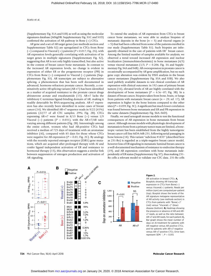
(Supplementary Fig. S1A and S1B) as well as using the molecularsignatures database (MSigDB; Supplementary Fig. S1C and S1D)confirmed the activation of AR pathway in Bone (þ) CTCs.
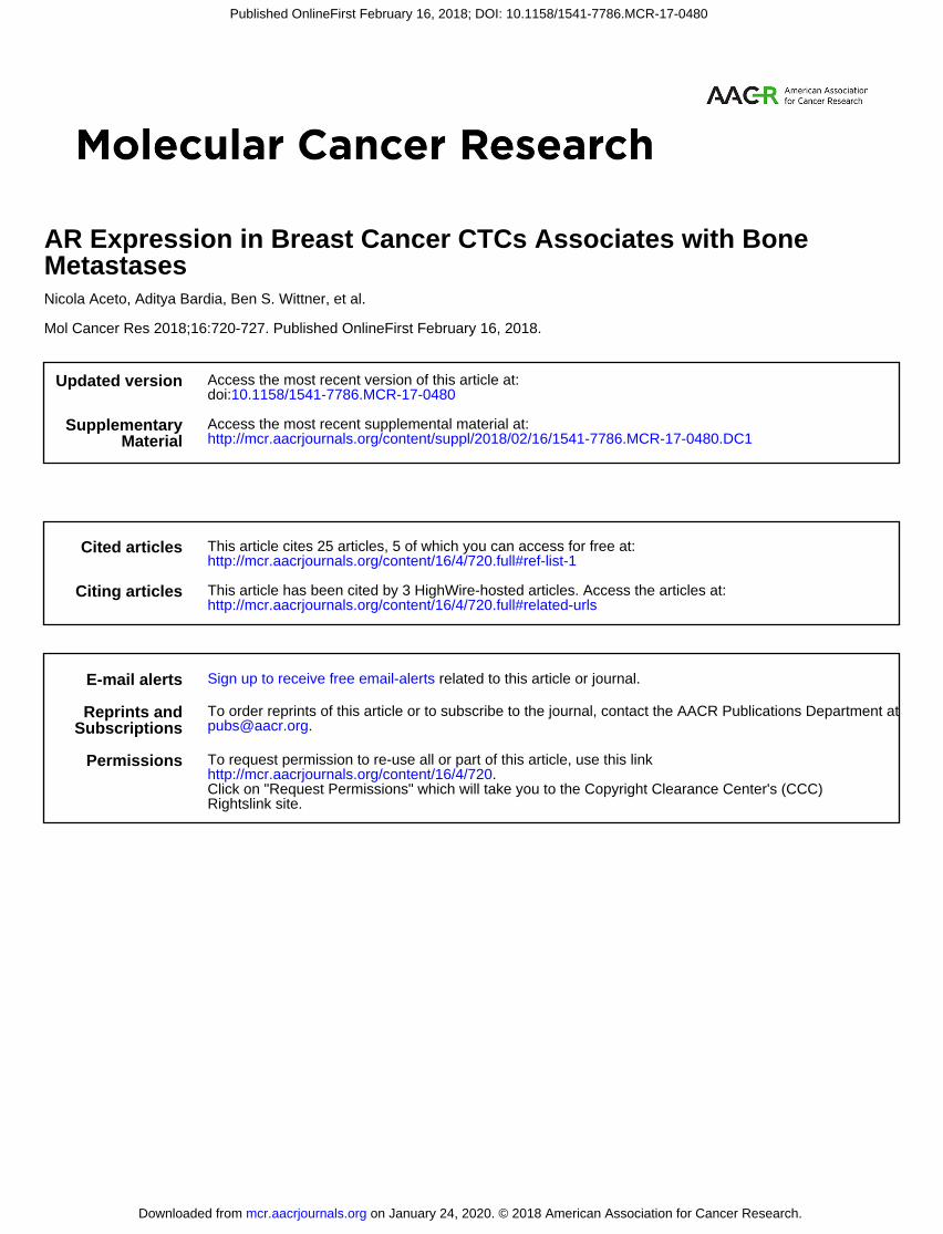
AR gene and a set of AR target genes (see methods section andSupplementary Table S2) are upregulated in CTCs from Bone(þ) compared to Visceral (þ) patients (P < 0.017; Fig. 2A), withAR expression levels generally correlating with activation of itstarget genes in multiple signatures (Supplementary Fig. S2),suggesting that AR is not only highly transcribed, but also activein the context of breast cancer bone metastasis. In contrast tothe increased AR expression levels, no change is evident inexpression of either ER or the progesterone receptor (PR) inCTCs from Bone (þ) compared to Visceral (þ) patients (Sup-plementary Fig. S3). AR transcripts are subject to alternativesplicing, a phenomenon that has been well documented inadvanced, hormone-refractory prostate cancer. Recently, a con-stitutively active AR splicing variant (AR-v7) has been identifiedas a marker of acquired resistance to the prostate cancer drugsabiraterone acetate and enzalutamide (13). AR-v7 lacks theinhibitory C-terminus ligand-binding domain of AR, making itreadily detectable by RNA-sequencing analysis. AR-v7 expres-sion has also recently been identified in some cases of breastcancer (14). We identified AR-v7 sequence reads in 9/22 (41%)patients (22/77 of all CTC samples; 29%; Fig. 2B). CTCsexpressing AR-v7 were found in 8/13 Bone (þ) versus 1/9Visceral (þ) patients (P ¼ 0.031), with the AR-v7/AR ratiovarying among different patients (Fig. 2B). Interestingly amongthe entire cohort, women who had AR-positive CTCs hadreceived a median of 725 days of treatment with an aromataseinhibitor (AI), compared with 85 days for those whose CTCswere negative for AR expression (P ¼ 0.01; Fig. 2C). By analogywith the recently reported estrogen receptor (ESR1) gene muta-tions, which are acquired after prolonged therapy with AI andconfer ligand independent activation of ER and resistance tohormonal therapy (15), this observation suggests a similar linkbetween suppression of estrogen production and activation ofAR signaling.
To extend the analysis of AR expression from CTCs to breastcancer bone metastases, we were able to analyze biopsies ofmetastatic deposits in the bone (n ¼ 5) and visceral organs (n¼ 5) that had been collected from some of the patients enrolled inour study (Supplementary Table S1). Such biopsies are infre-quently obtained in the care of patients with ERþ breast cancer.Among the limited number of samples available for analysis, weobserved a trend toward increased AR expression and nuclearlocalization (immunohistochemistry) in bone metastases (4/5)versus visceral metastases (1/5; P ¼ 0.206; Fig. 3A and Supple-mentary Fig. S4A and S4B). ARoverexpression inprostate cancer isoccasionally accompanied by AR gene amplification, but no suchgene copy alteration was evident by FISH analysis in the breastcancer metastases (Supplementary Fig. S5A and S5B). We alsoused publicly available datasets to test clinical correlates of ARexpression with clinical outcomes: in 192 cases of primary breasttumors (16), elevated levels of AR are highly correlated with thedevelopment of bone metastases (P ¼ 6.5e�07; Fig. 3B). In adataset of breast cancer, biopsies taken from bone, brain, or lungsfrom patients with metastatic breast cancer (n ¼ 29; ref. 17), ARexpression is higher in the bone lesions compared to the othersites (P¼ 0.039; Fig. 3C). A significant butmuch lower correlationwas found between bone metastasis and ER and PR expression inthe same datasets (Supplementary Fig. S6A–S6D).
Finally, we used xenograft mouse models to test the functionalconsequences of AR expression in bone metastasis from breastcancer. Althoughmousemodelswith physiologic development ofmetastasis tobone fromaprimary tumormass are limited, a bone-tropic variant has been established from the highly tumorigenicbreast cancer cell lineMDA-MB-231, following serial passaging inbone lesions (18). This variant "subclone #1833" (described hereas 231-Bo) is regarded as a triple-negative breast cancer model,however loss of ER signaling inmetastatic luminal breast cancer isawell-documentedmechanismof resistance to endocrine therapy(19), and AR expression correlates with bone metastasis inde-pendently of ER status (Supplementary Fig. S7), thusmaking 231-Bo cells a relevant model to validate our CTC data. 231-Bo cells
Figure 2.
AR activation in breast CTCs. A,Boxplots showing AR transcriptexpression in CTCs from Bone(þ)versus Visceral(þ) patients. Reads permillion (rpm) are computed per-patient(top). Boxplot shows the levels of theAR signature metagene representativeof AR activity (see methods section) inCTCs from patients with "Bone(þ)"(red) versus "Visceral(þ)" (blue)disease (bottom). B, Heatmap showingthe presence or absence of AR and AR-v7 reads, as well as the ratio betweenAR-v7 andAR reads, for each patient.C,Bar graph shows the mean number ofdays on AI treatment for patients withAR negative versus AR positive CTCs,and for patients with AR-v7 negativeversus AR-v7 positive CTCs. Error barsrepresent SEM. � , P ¼ 0.013.
Aceto et al.
Mol Cancer Res; 16(4) April 2018 Molecular Cancer Research724
on January 24, 2020. © 2018 American Association for Cancer Research. mcr.aacrjournals.org Downloaded from
Published OnlineFirst February 16, 2018; DOI: 10.1158/1541-7786.MCR-17-0480

overexpresses genes such as IL11 and CTGF, and its implanta-tion into the mammary fat pad of immunosuppressed mice(NSG) produces a reproducible number of bone metastases,along with numerous lung metastases (4, 18). Remarkably,231-Bo cells express 3.5-fold higher levels of AR protein,compared with the parental 231 cells (Fig. 4A) and are respon-sive to treatment with Enzalutamide, as shown by downregula-tion of AR-target genes including AR itself, ACKR3, KLK2, KLK3,KLK4, KRT8, ORM1, ORM2, PGC, SPDEF, STEAP4, andTMPRSS2 genes (Supplementary Fig. S8A and S8B). We gen-erated 231-Bo cells expressing multiple shRNA constructs tar-geting different regions of AR transcript (Supplementary Fig.S9) and compared their metastatic propensity with that of 231-Bo cells expressing a control shRNA. A testosterone pellet wasimplanted subcutaneously into female NSG mice, to ensurethat both ER and AR ligands were not limiting. As expected,implantation of GFP-Luciferase tagged 231-Bo cells into themammary fat pad resulted within 6 weeks in multiple lungmetastases, as well as a small number of defined bone metas-tases (a median of three foci per tibia). shRNA-mediated ARknockdown had no effect on the primary tumor size or theoverall presence of lung metastases, yet reduced the number ofbone metastases (P < 0.048; Fig. 4B and C; Supplementary Figs.S9 and S10A–S10C). We extended these findings using parental231-Bo-derived xenografts in mice treated with the AR inhibitorenzalutamide. Consistent with the shRNA-knockdown, chem-ical suppression of AR signaling suppressed bone metastases,without affecting primary tumor size or lung metastases (P <0.009; Fig. 3D; Supplementary Fig. S10A–S10C).
DiscussionBy analyzing single-cell RNA-sequencing profiles of CTCs, we
identify signaling pathways that are differentially activated in thecontext of bone versus visceralmetastasis. Among these pathways,increased AR expression appears to be noteworthy in bone-pre-dominant breast cancer, both in its occurrence as a function of AItherapy, and in its possible responsiveness to anti-androgenicintervention.
Signaling pathways that are upregulated in CTCs from patientswith progressing visceral metastases include those involved incell–cell junctions stability, intracellular signaling downstream ofRhoA, PTP1B, and other effectors, maintenance of EMT via TGFbreceptor. In contrast, CTCs from patients with progressing bonemetastases display RTK/PI3K/mTOR pathway activation, andincreased expression of cytokines such as IL3 and IL6, EMTregulators such as SMAD2, HDAC epigenetic regulators, and ARsignaling. Although in this study we investigated the functionalrelevance of AR in breast cancer bonemetastasis, detailed analysesof multiple other pathways may reveal new organ-specific depen-dencies of metastatic breast cancer cells.
AR expression iswell established in triple negative breast cancer(TNBC), and ongoing clinical trials are testing the effectiveness ofAR inhibitors in this subtypeof breast cancer (20), and in theothersubtypes (21). These inhibitors were developed for treatment ofprostate cancer, the prototype AR-drivenmalignancy. Physiologicandrogen-dependent activation of AR mediates transcription oftarget genes that are crucial for the development andmaintenanceof the prostate gland (22), a dependence that persists upon
Figure 3.
AR expression in metastatic breast cancer. A, Representative images of AR (brown) and hematoxylin (nuclei; blue)-stained human breast cancer bone metastasis(left) and soft-tissue metastasis (right) tissues. The bar graph (bottom) shows the number of AR-positive and AR-negative stains within the bone metastasisversus visceral or soft tissuemetastasis.B,Boxplot showsARexpression levels in primarybreast tumors (n¼ 192) frompatientswho later developedmetastasis in thebone versus other organs. C, Boxplot shows AR expression levels in metastatic breast cancer biopsies derived from lung, liver or bone (n ¼ 29).
A Role for AR in Breast Cancer Bone Metastasis
www.aacrjournals.org Mol Cancer Res; 16(4) April 2018 725
on January 24, 2020. © 2018 American Association for Cancer Research. mcr.aacrjournals.org Downloaded from
Published OnlineFirst February 16, 2018; DOI: 10.1158/1541-7786.MCR-17-0480

neoplastic transformation, with anti-androgen therapies as thestandard of care for metastatic prostate cancer. AR and androgensynthesis genes are commonly upregulated in bone metastasesfrom prostate cancer, compared to primary tumors (23). Andro-gens themselves are converted to estrogens by the enzyme aro-matase, the most common therapeutic target in women withpostmenopausal ERþ breast cancer. Indeed, in women receivingAI therapy for breast cancer, circulating levels of estradiol are low,whereas androgen levels are increased (24). It is in this setting ofprolonged estrogen deprivation resulting in increased androgens,that overexpression of AR may become clinically relevant in theprogression of ERþ breast cancer, a possibility that is supported byour observed correlation between the duration of AI therapy andpresence of AR-expressingCTCs. Furthermore, the detectionof theconstitutively activated, prostate cancer-associated AR-v7 splicevariant within CTCs from breast cancer patients with bonemetas-tases also suggests a functional role for androgen signaling.
AR expression in ERþbreast cancermay also constitute amarkerof more indolent, bone-predominant disease, rather than a directcontributor to proliferation in bone, although the suppression ofbone metastases by either AR knockdown or pharmacological ARsuppression in a mouse model of bone-tropic disease suggests afunctional consequence of AR expression. In addition, given theuse of AIs in the treatment of metastatic breast cancer, as well astheir recommended prolonged use in the adjuvant setting (25),monitoring for the emergence of androgen-dependent signalingmay ultimately help guide therapeutic choices. Taken all together,
noninvasive sampling and single-cell profiling of tumor cells frompatients with distinct metastatic outcomes provides an exception-al resource to study advanced stages of progression, with thepotential to impact therapeutic opportunities in the setting ofrefractory disease.
Disclosure of Potential Conflicts of InterestD.T. Ting reports receiving a commercial research grant from ACD
Biotechne and Affymetrix; and has received speakers bureau honoraria fromAffymetrix. Daniel A. Haber reports receiving a commercial research grantfrom Janssen (completed 2016); has ownership interest in MGH and hasapplied for patent protection for CTC-iChip and molecular signatures; and isa consultant/advisory board member for Janssen SAB (2017). S. Maheswaranhas ownership interest in and is a cofounder of a company built aroundthe CTC-iChip technology. No potential conflicts of interest were disclosedby the other authors.
Authors' ContributionsConception and design: N. Aceto, A. Bardia, R. Kapur, M. Toner, D.A. Haber,S. MaheswaranDevelopment of methodology: N. Aceto, M.C. Donaldson, R. O'Keefe,F. Bersani, R. Kapur, M. TonerAcquisition of data (provided animals, acquired and managed patients,provided facilities, etc.): N. Aceto, A. Bardia, M.C. Donaldson, R. O'Keefe,A. Engstrom, F. Bersani, Y. Zheng, V. Comaills, K. Niederhoffer, T. Shioda,D. Sgroi, D.T. TingAnalysis and interpretation of data (e.g., statistical analysis, biostatistics,computational analysis): N. Aceto, B.S. Wittner, R. O'Keefe, A. Engstrom,T. Shioda, D.T. Ting, B. Moy, S. Ramaswamy, S. Maheswaran
Figure 4.
TheAR contributes to spontaneous bonemetastasis in breast cancer.A, Immunoblot showsARexpression levels in parental, lung variant, andbone variantMDA-MB-231 cells. Error bars represent SEM. n ¼ 3; � , P < 0.04. B, Schematic of the experiment to determine the propensity of AR expressing bone variant MDA-MB-231cells to metastasize to the lung or bone (left). Representative images of GFP-stained primary breast tumor, lung and bone metastases (right) derived fromthe bone variant MDA-MB-231 cells inoculated into the mousemammary gland. C, The bar graph shows the number of animals with bone and lungmetastases in thegroups harboring tumors in which AR expression was suppressed with two different shRNAs versus control shRNA. n ¼ 8; � , P ¼ 0.048. D, The bar graphshows the number of animals with bone and lung metastases in the groups treated with vehicle or enzalutamide. n ¼ 8; �� , P < 0.009 (bottom).
Aceto et al.
Mol Cancer Res; 16(4) April 2018 Molecular Cancer Research726
on January 24, 2020. © 2018 American Association for Cancer Research. mcr.aacrjournals.org Downloaded from
Published OnlineFirst February 16, 2018; DOI: 10.1158/1541-7786.MCR-17-0480

Writing, review, and/or revisionof themanuscript:N.Aceto, A. Bardia, B.Moy,S. Ramaswamy, D.A. Haber, S. MaheswaranAdministrative, technical, or material support (i.e., reporting or organizingdata, constructing databases): F. Bersani, O. Mackenzie, D. Sgroi, M. TonerStudy supervision: D.A. Haber, S. MaheswaranOther (lab technician - assisted with experiments): H. Zhu
AcknowledgmentsThisworkwas supported by grants for Breast Cancer Research Foundation (to
D.A. Haber), Stand Up to Cancer (to D.A. Haber, M. Toner, S. Maheswaran),National Foundation for Cancer Research (to D.A. Haber), Howard HughesMedical Institute (to D.A. Haber), NIHCA129933 (to D.A. Haber), NIH K125K12CA087723 (to A. Bardia), NIBIB EB008047 (to M. Toner, D.A. Haber),Susan G. Komen for the CureKG09042 (to S. Maheswaran), ESSCO BreastCancer Fund (to S. Maheswaran), NCI Federal Share Program and Income (to S.Maheswaran), and the MGH-Johnson & Johnson Center for Excellence in CTCs(to M. Toner, S. Maheswaran). N. Aceto was supported by the Human Frontiers
Science Program, the Swiss National Science Foundation, and the Swiss Foun-dation for Grants in Biology and Medicine.
The authors are grateful to all the patients who participated in this study. Wethank C. Hart, A. McGovern, L.C. Davis, K. Harrington, A. Desmond, and theMassachusetts General Hospital (MGH) clinical research coordinators forhelp with the clinical studies; Drs. P.S. Spuhler and T.A. Barber for supportwith the CTC-iChip technology; L. Libby, J. Brockmann, and J. Rulka forexcellent technical support.
The costs of publication of this article were defrayed in part by thepayment of page charges. This article must therefore be hereby markedadvertisement in accordance with 18 U.S.C. Section 1734 solely to indicatethis fact.
Received August 31, 2017; revised November 29, 2017; accepted January 24,2018; published first February 16, 2018.
References1. Mundy GR. Metastasis to bone: causes, consequences and therapeutic
opportunities. Nat Rev Cancer 2002;2:584–93.2. Dougall WC, Holen I, Gonzalez Suarez E. Targeting RANKL in metastasis.
Bonekey Rep 2014;3:519.3. Kaplan RN, Riba RD, Zacharoulis S, Bramley AH, Vincent L, Costa C, et al.
VEGFR1-positive haematopoietic bone marrow progenitors initiate thepre-metastatic niche. Nature 2005;438:820–7.
4. Kang Y, Siegel PM, Shu W, Drobnjak M, Kakonen SM, Cordon-Cardo C,et al. A multigenic program mediating breast cancer metastasis to bone.Cancer Cell 2003;3:537–49.
5. Muller A, Homey B, Soto H, Ge N, Catron D, Buchanan ME, et al.Involvement of chemokine receptors in breast cancer metastasis. Nature2001;410:50–6.
6. Sethi N, Dai X, Winter CG, Kang Y. Tumor-derived JAGGED1 promotesosteolytic bone metastasis of breast cancer by engaging notch signaling inbone cells. Cancer Cell 2011;19:192–205.
7. Hoshino A, Costa-Silva B, Shen TL, Rodrigues G, Hashimoto A, Tesic MarkM, et al. Tumour exosome integrins determine organotropic metastasis.Nature 2015;527:329–35.
8. Aceto N, Bardia A, Miyamoto DT, Donaldson MC, Wittner BS, Spencer JA,et al. Circulating tumor cell clusters are oligoclonal precursors of breastcancer metastasis. Cell 2014;158:1110–22.
9. Ozkumur E, Shah AM, Ciciliano JC, Emmink BL, Miyamoto DT, BrachtelE, et al. Inertial focusing for tumor antigen-dependent and -indepen-dent sorting of rare circulating tumor cells. Sci Translat Med 2013;5:179ra47.
10. HieronymusH, Lamb J, Ross KN,PengXP,ClementC, RodinaA, et al. Geneexpression signature-based chemical genomic prediction identifies a novelclass of HSP90 pathway modulators. Cancer Cell 2006;10:321–30.
11. Lu C, Luo J. Decoding the androgen receptor splice variants. Transl AndrolUrol 2013;2:178–86.
12. Yu M, Bardia A, Aceto N, Bersani F, Madden MW, Donaldson MC,et al. Cancer therapy. Ex vivo culture of circulating breast tumor cellsfor individualized testing of drug susceptibility. Science 2014;345:216–20.
13. Antonarakis ES, Lu C, Wang H, Luber B, Nakazawa M, Roeser JC, et al. AR-V7 and resistance to enzalutamide and abiraterone in prostate cancer.N Engl J Med 2014;371:1028–38.
14. Hickey TE, Irvine CM, Dvinge H, Tarulli GA, Hanson AR, Ryan NK, et al.Expression of androgen receptor splice variants in clinical breast cancers.Oncotarget 2015;6:44728–44.
15. RobinsonDR,WuYM, Vats P, Su F, Lonigro RJ, Cao X, et al. Activating ESR1mutations in hormone-resistant metastatic breast cancer. Nat Genet2013;45:1446–51.
16. Bos PD, ZhangXH,Nadal C, ShuW,GomisRR,NguyenDX, et al. Genes thatmediate breast cancer metastasis to the brain. Nature 2009;459:1005–9.
17. Zhang XH, Wang Q, Gerald W, Hudis CA, Norton L, Smid M, et al. Latentbone metastasis in breast cancer tied to Src-dependent survival signals.Cancer cell 2009;16:67–78.
18. Minn AJ, Gupta GP, Siegel PM, Bos PD, Shu W, Giri DD, et al. Genes thatmediate breast cancer metastasis to lung. Nature 2005;436:518–24.
19. Gutierrez MC, Detre S, Johnston S, Mohsin SK, Shou J, Allred DC, et al.Molecular changes in tamoxifen-resistant breast cancer: relationshipbetween estrogen receptor, HER-2, and p38 mitogen-activated proteinkinase. J Clin Oncol 2005;23:2469–76.
20. Arce-Salinas C, Riesco-Martinez MC, Hanna W, Bedard P, Warner E.Complete response of metastatic androgen receptor-positive breast cancerto bicalutamide: case report and review of the literature. J Clin Oncol2016;34:e21–4.
21. O'Shaughnessy J, Campone M, Brain E, Neven P, Hayes D, Bondarenko I,et al. Abiraterone acetate, exemestane or the combination in postmeno-pausal patients with estrogen receptor-positive metastatic breast cancer.Ann Oncol 2016;27:106–13.
22. Yeh S, Tsai MY, Xu Q, Mu XM, Lardy H, Huang KE, et al. Generation andcharacterization of androgen receptor knockout (ARKO) mice: an in vivomodel for the study of androgen functions in selective tissues. Proc NatlAcad Sci U S A 2002;99:13498–503.
23. Montgomery RB, Mostaghel EA, Vessella R, Hess DL, Kalhorn TF, HiganoCS, et al. Maintenance of intratumoral androgens in metastatic prostatecancer: a mechanism for castration-resistant tumor growth. Cancer Res2008;68:4447–54.
24. Dimitrakakis C, Bondy C. Androgens and the breast. Breast Cancer Res2009;11:212.
25. Goss PE, Ingle JN, Pritchard KI, Robert NJ, Muss H, Gralow J, et al.Extending aromatase-inhibitor adjuvant therapy to 10 years. N Engl J Med2016;375:209–19.
www.aacrjournals.org Mol Cancer Res; 16(4) April 2018 727
A Role for AR in Breast Cancer Bone Metastasis
on January 24, 2020. © 2018 American Association for Cancer Research. mcr.aacrjournals.org Downloaded from
Published OnlineFirst February 16, 2018; DOI: 10.1158/1541-7786.MCR-17-0480
2018;16:720-727. Published OnlineFirst February 16, 2018.Mol Cancer Res Nicola Aceto, Aditya Bardia, Ben S. Wittner, et al. MetastasesAR Expression in Breast Cancer CTCs Associates with Bone
Updated version
10.1158/1541-7786.MCR-17-0480doi:
Access the most recent version of this article at:
Material
Supplementary
http://mcr.aacrjournals.org/content/suppl/2018/02/16/1541-7786.MCR-17-0480.DC1
Access the most recent supplemental material at:
Cited articles
http://mcr.aacrjournals.org/content/16/4/720.full#ref-list-1
This article cites 25 articles, 5 of which you can access for free at:
Citing articles
http://mcr.aacrjournals.org/content/16/4/720.full#related-urls
This article has been cited by 3 HighWire-hosted articles. Access the articles at:
E-mail alerts related to this article or journal.Sign up to receive free email-alerts
Subscriptions
Reprints and
To order reprints of this article or to subscribe to the journal, contact the AACR Publications Department at
Permissions
Rightslink site. Click on "Request Permissions" which will take you to the Copyright Clearance Center's (CCC)
.http://mcr.aacrjournals.org/content/16/4/720To request permission to re-use all or part of this article, use this link
on January 24, 2020. © 2018 American Association for Cancer Research. mcr.aacrjournals.org Downloaded from
Published OnlineFirst February 16, 2018; DOI: 10.1158/1541-7786.MCR-17-0480