Zeldzame Thoracale Tumoren - Maastricht UMC+ · 2018-02-20 · tumor voorste mediastinum Monique...
49
Zeldzame Thoracale Tumoren tumor voorste mediastinum Monique Hochstenbag OncoZon Januari 2018
Transcript of Zeldzame Thoracale Tumoren - Maastricht UMC+ · 2018-02-20 · tumor voorste mediastinum Monique...
Zeldzame Thoracale Tumoren
tumor voorste mediastinum
Monique Hochstenbag
OncoZon Januari 2018
• No Disclosures
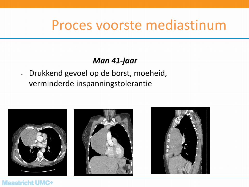
Proces voorste mediastinum
Man 41-jaar
• Drukkend gevoel op de borst, moeheid, verminderde inspanningstolerantie
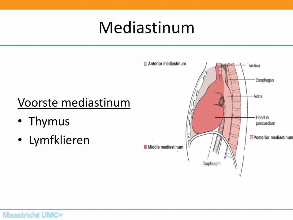
Mediastinum
Voorste mediastinum
• Thymus
• Lymfklieren
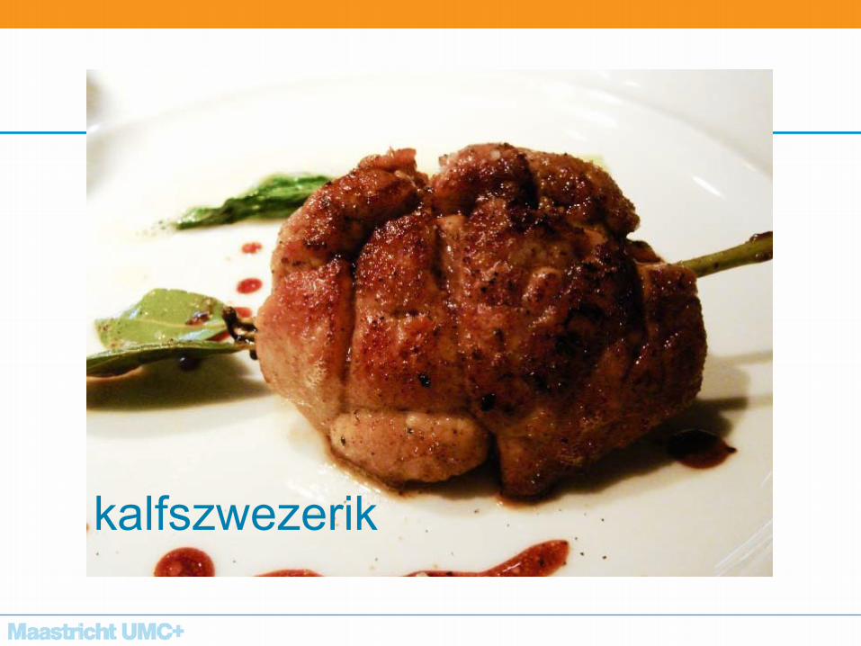
kalfszwezerik
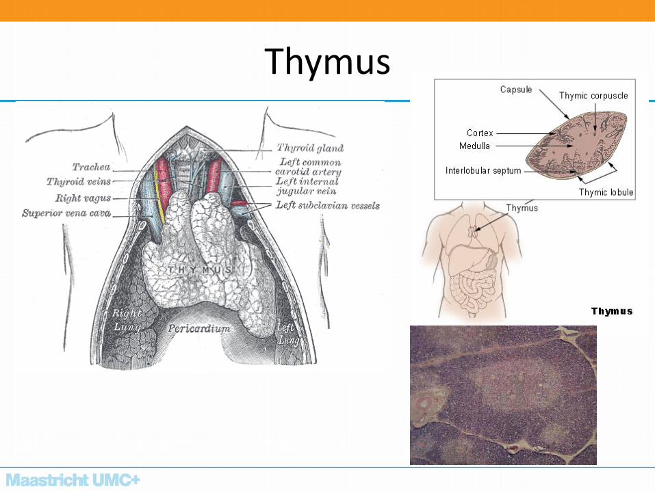
Thymus
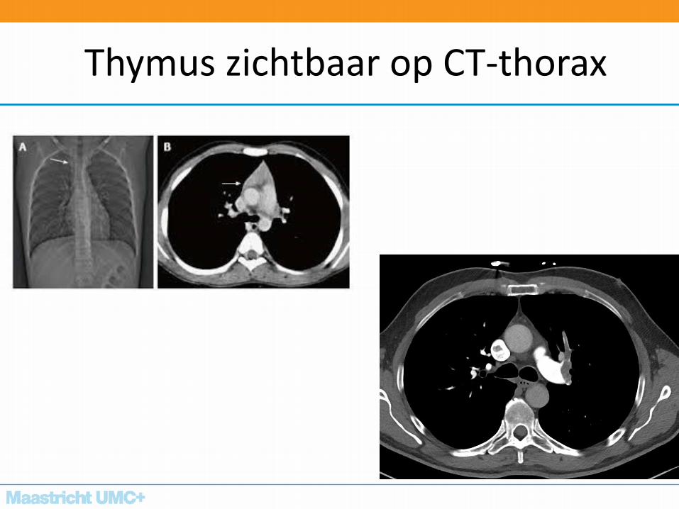
Thymus zichtbaar op CT-thorax
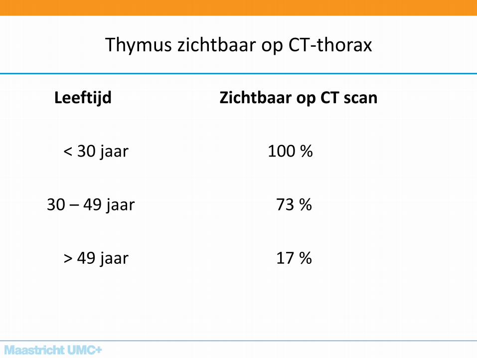
Thymus zichtbaar op CT-thorax
Leeftijd Zichtbaar op CT scan
< 30 jaar 100 %
30 – 49 jaar 73 %
> 49 jaar 17 %
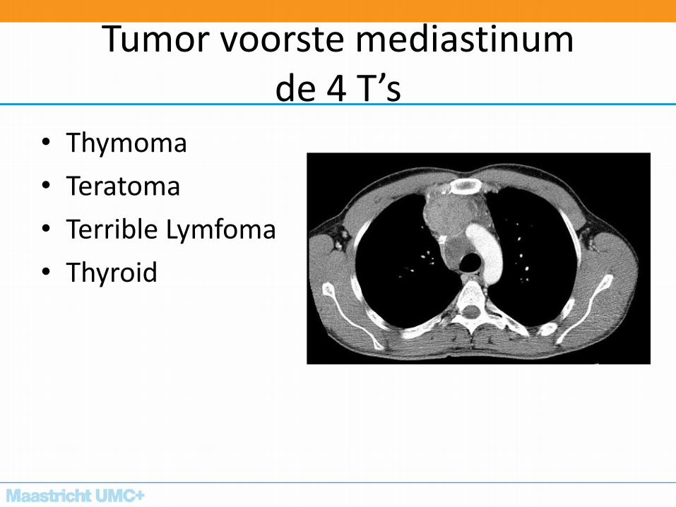
Tumor voorste mediastinum de 4 T’s
• Thymoma
• Teratoma
• Terrible Lymfoma
• Thyroid
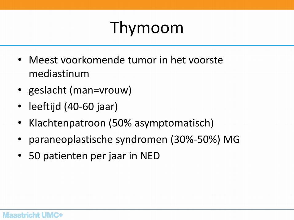
Thymoom
• Meest voorkomende tumor in het voorste mediastinum
• geslacht (man=vrouw)
• leeftijd (40-60 jaar)
• Klachtenpatroon (50% asymptomatisch)
• paraneoplastische syndromen (30%-50%) MG
• 50 patienten per jaar in NED
Biochemische merkers
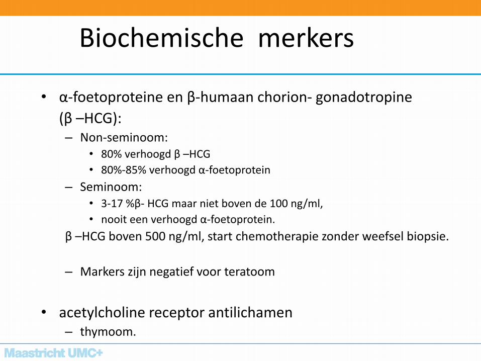
• α-foetoproteine en β-humaan chorion- gonadotropine
(β –HCG): – Non-seminoom:
• 80% verhoogd β –HCG
• 80%-85% verhoogd α-foetoprotein
– Seminoom: • 3-17 %β- HCG maar niet boven de 100 ng/ml,
• nooit een verhoogd α-foetoprotein.
β –HCG boven 500 ng/ml, start chemotherapie zonder weefsel biopsie.
– Markers zijn negatief voor teratoom
• acetylcholine receptor antilichamen – thymoom.
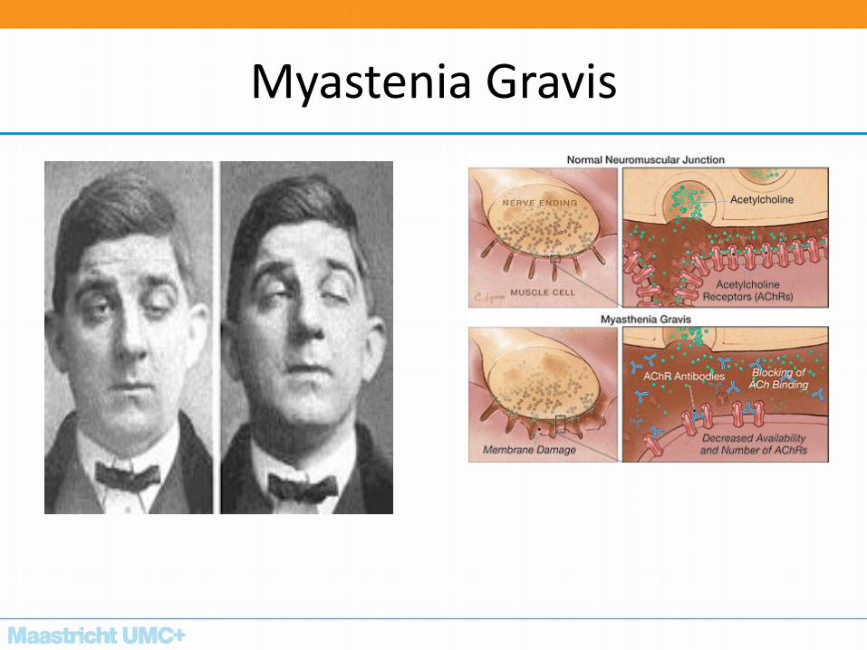
Myastenia Gravis
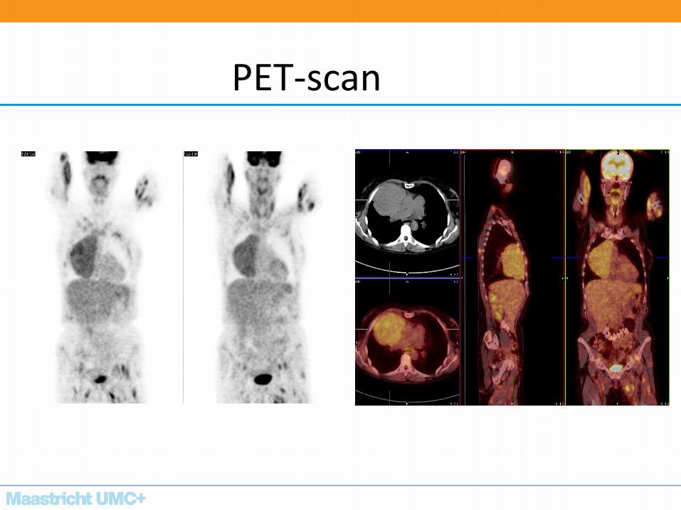
PET-scan
Invasieve diagnostiek
• Biopsie alleen geindiceerd bij primair irresectabele tumor
• Biopsie van een klinisch stadium I en II thymoom moet vermeden worden.
• Sporadische resectie klein lymfoom afwegen tegen “spill” bij thymoom-biopsie
Thymoom
– Histologische classificatie
– Klinische classificatie
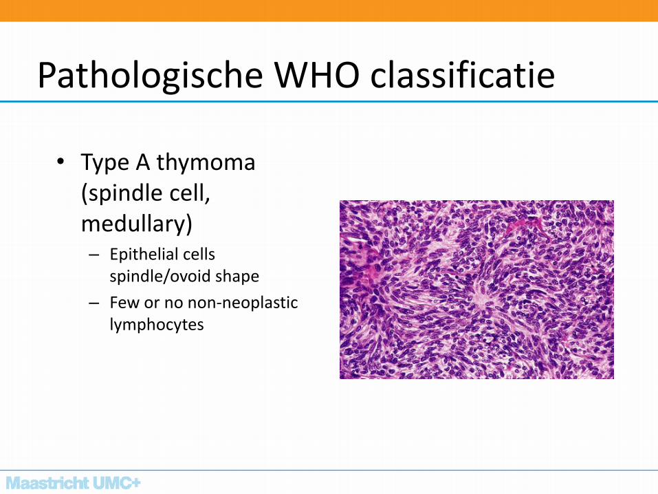
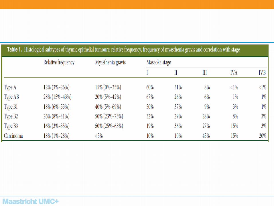
Pathologische WHO classificatie
• Type A thymoma (spindle cell, medullary) – Epithelial cells
spindle/ovoid shape
– Few or no non-neoplastic lymphocytes
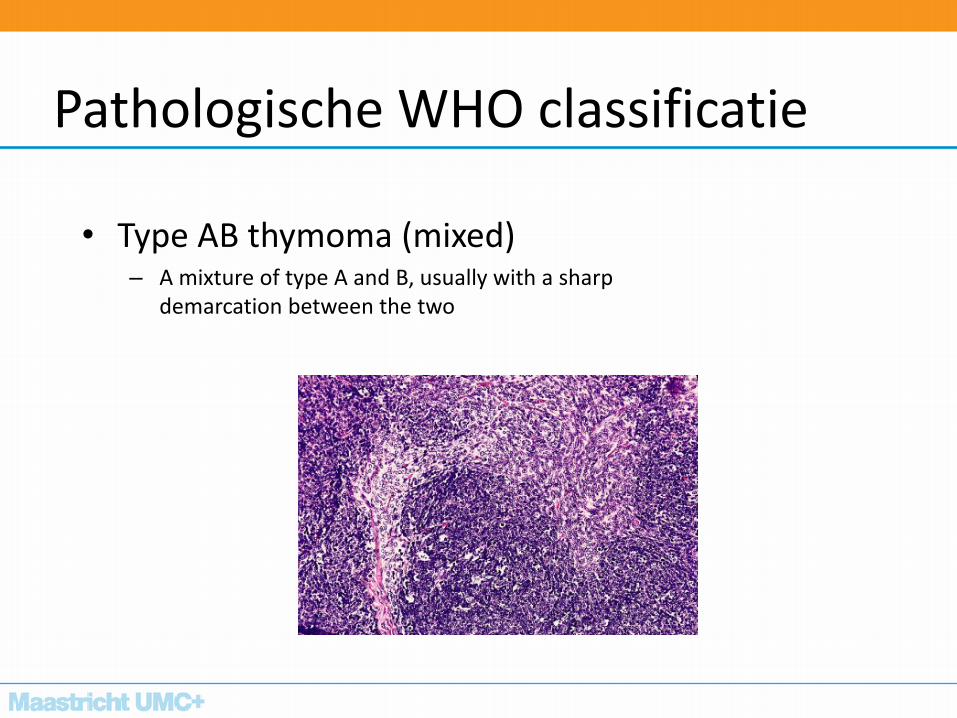
Pathologische WHO classificatie
• Type AB thymoma (mixed) – A mixture of type A and B, usually with a sharp
demarcation between the two
Pathologische WHO classificatie
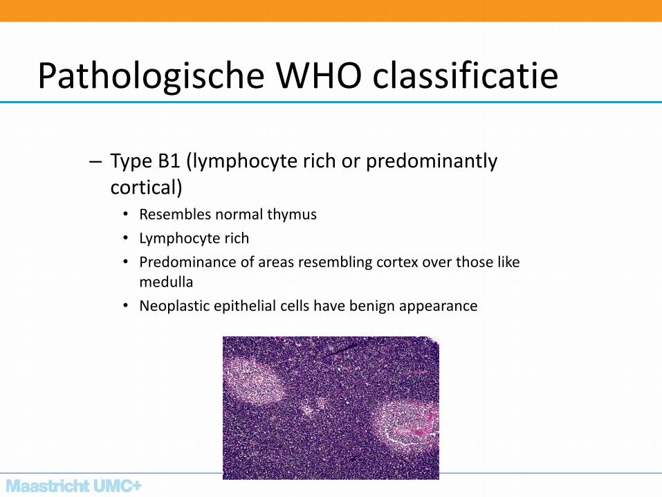
– Type B1 (lymphocyte rich or predominantly cortical) • Resembles normal thymus
• Lymphocyte rich
• Predominance of areas resembling cortex over those like medulla
• Neoplastic epithelial cells have benign appearance
Pathologische WHO classificatie
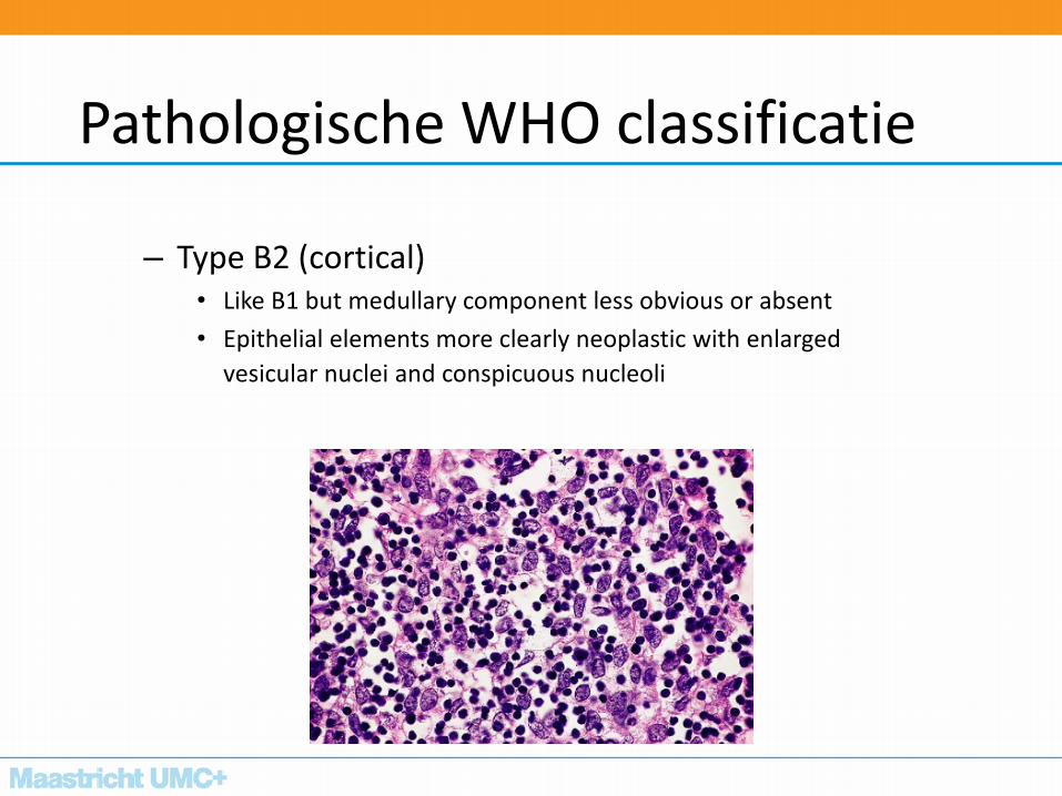
– Type B2 (cortical) • Like B1 but medullary component less obvious or absent
• Epithelial elements more clearly neoplastic with enlarged
vesicular nuclei and conspicuous nucleoli
Pathologische WHO classificatie
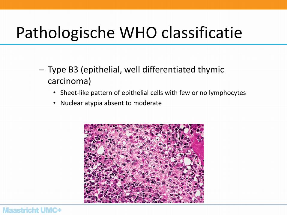
– Type B3 (epithelial, well differentiated thymic carcinoma) • Sheet-like pattern of epithelial cells with few or no lymphocytes
• Nuclear atypia absent to moderate
WHO classification
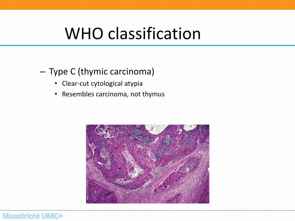
– Type C (thymic carcinoma) • Clear-cut cytological atypia
• Resembles carcinoma, not thymus
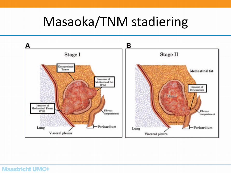
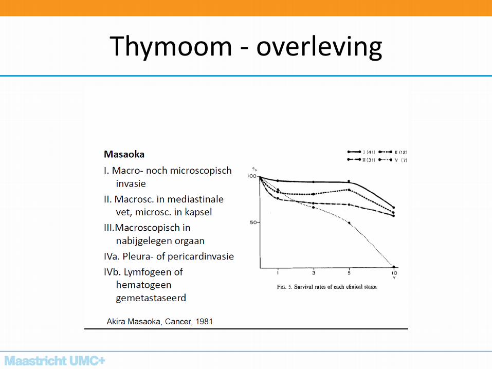
Masaoka/TNM stadiering
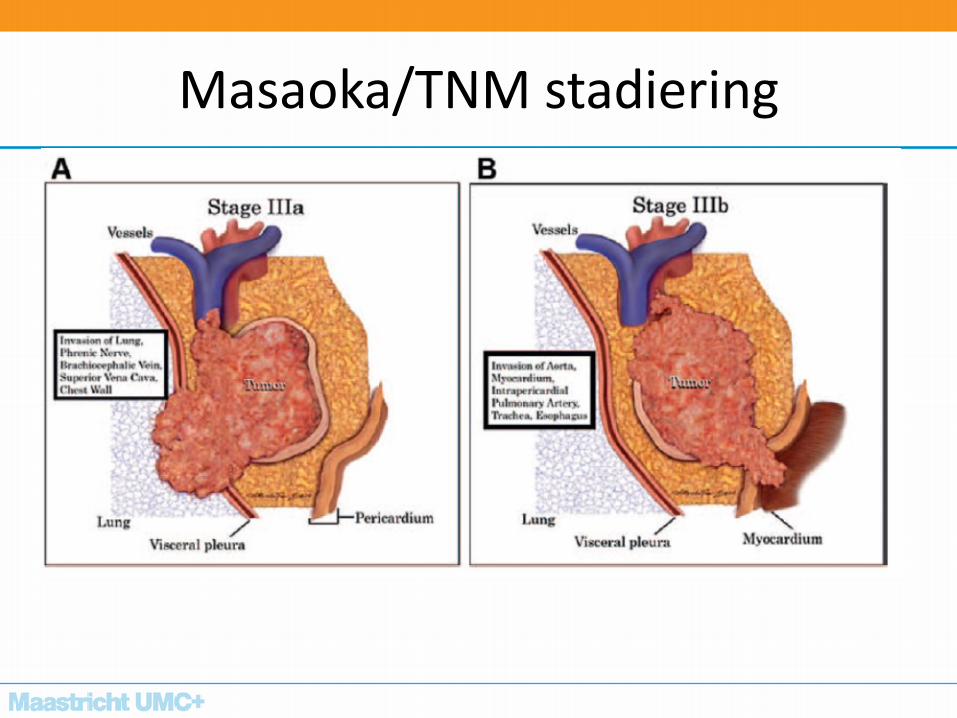
Masaoka/TNM stadiering
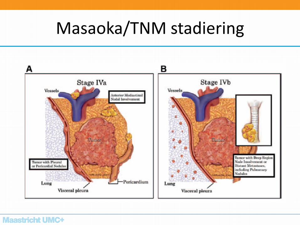
Masaoka/TNM stadiering
prognose
Histologische kenmerken
Stadium
Resectabiliteit
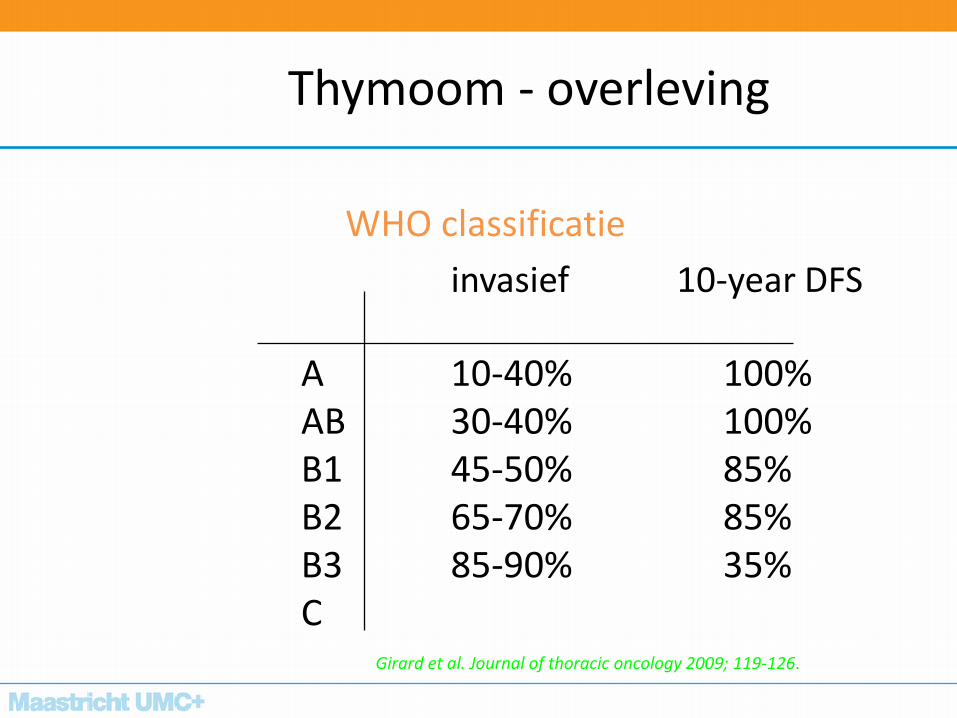
Thymoom - overleving
WHO classificatie
invasief 10-year DFS
A 10-40% 100% AB 30-40% 100% B1 45-50% 85% B2 65-70% 85% B3 85-90% 35% C Girard et al. Journal of thoracic oncology 2009; 119-126.
Thymoom - overleving
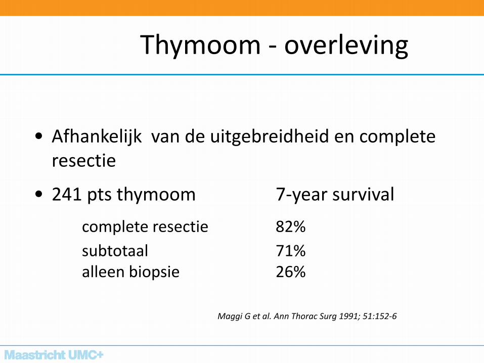
Thymoom - overleving
• Afhankelijk van de uitgebreidheid en complete resectie
• 241 pts thymoom 7-year survival
complete resectie 82%
subtotaal 71% alleen biopsie 26% Maggi G et al. Ann Thorac Surg 1991; 51:152-6

Proces voorste mediastinum
Man 41-jaar
• Drukkend gevoel op de borst, moeheid, verminderde inspanningstolerantie
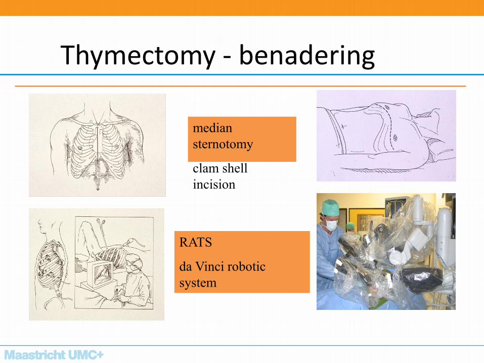
Thymectomy - benadering
median
sternotomy
clam shell
incision
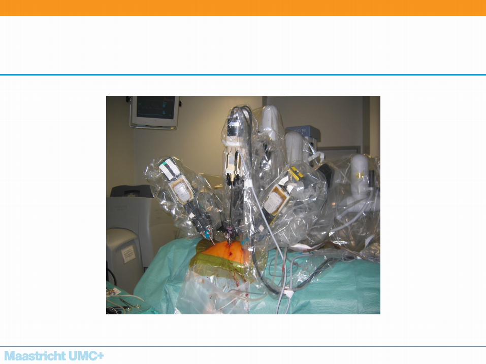
RATS
da Vinci robotic
system
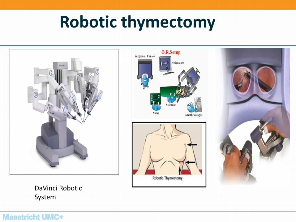
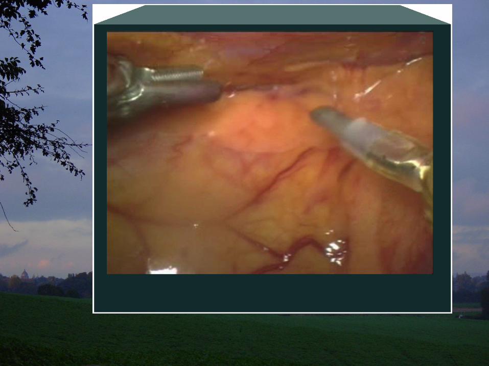
Robotic thymectomy
DaVinci Robotic System
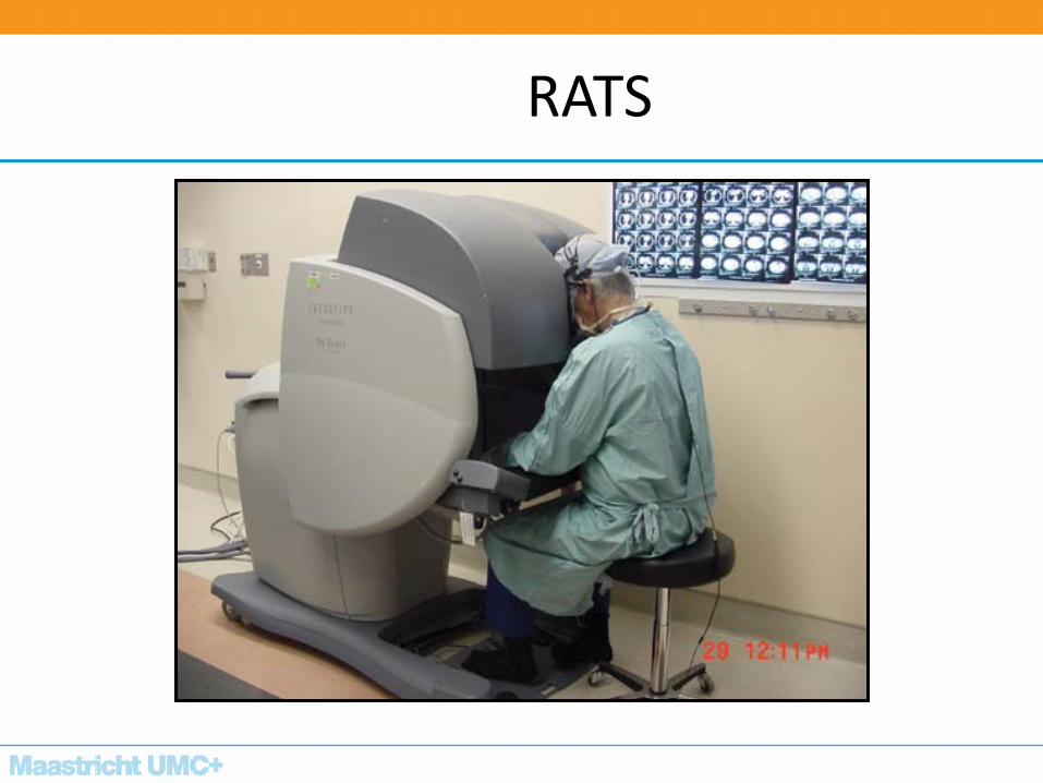
RATS
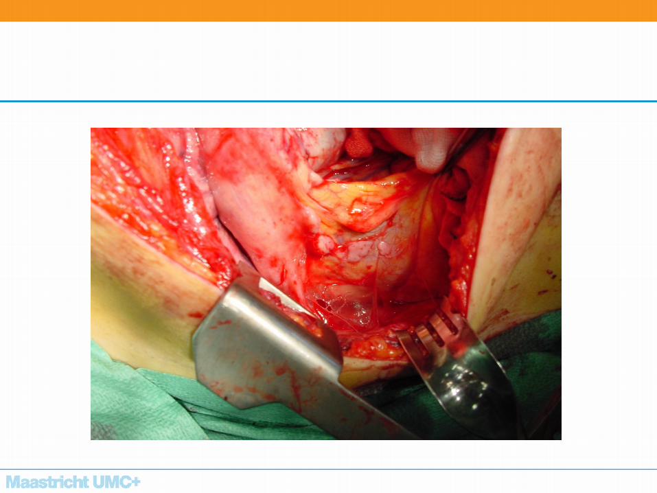
Thymoom - resectie
• complete resectie; delen nabijgelegen organen zo nodig meenemen
• openen pericard, uitbreiding bekijken
• N. frenicus altijd sparen
• debulking is beter dan open-dicht
• Altijd clips !
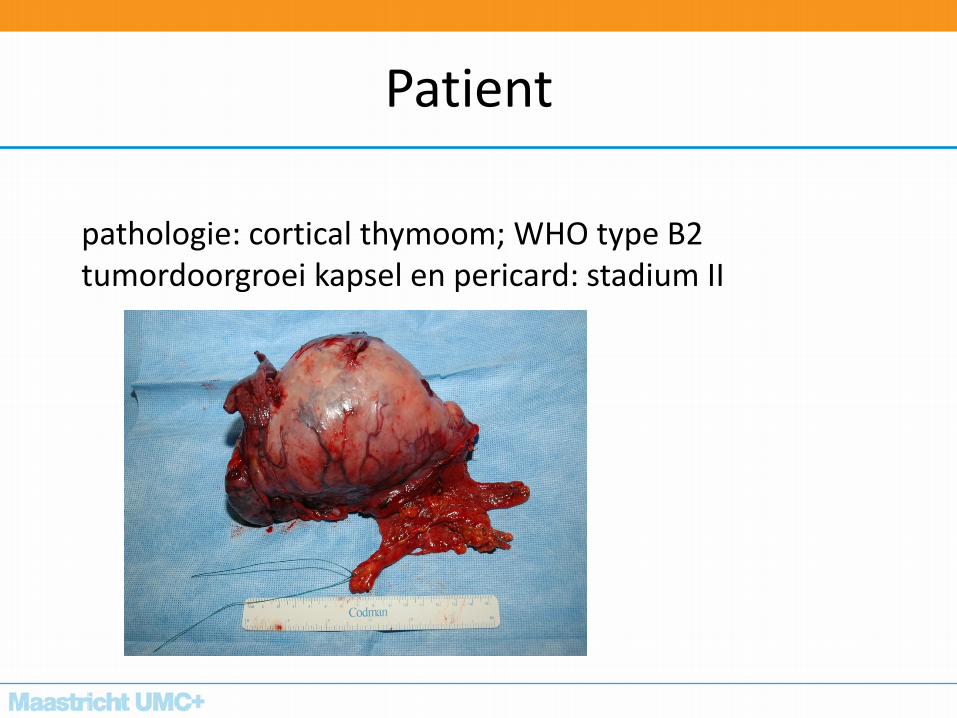
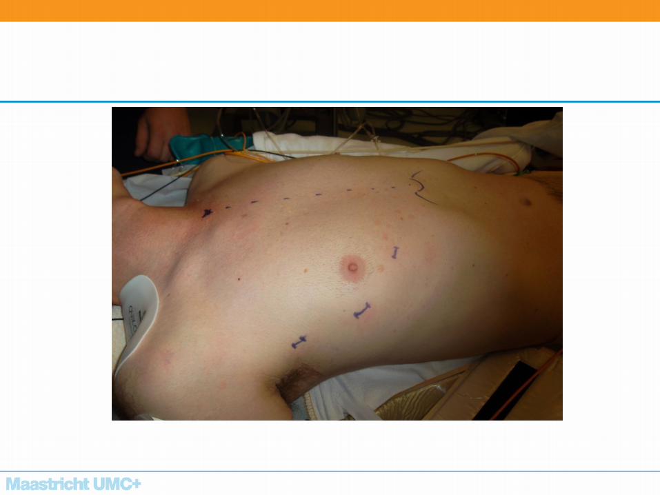
Patient
pathologie: cortical thymoom; WHO type B2 tumordoorgroei kapsel en pericard: stadium II
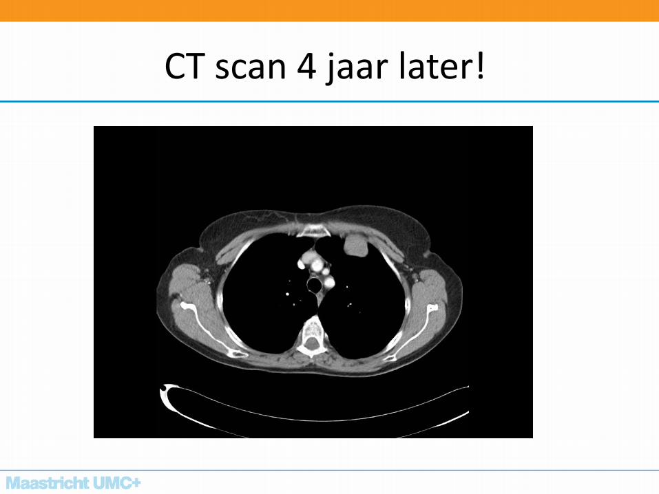
CT scan 4 jaar later!
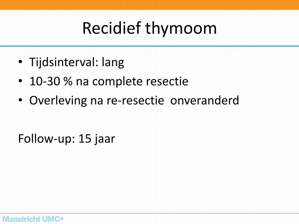
Recidief thymoom
• Tijdsinterval: lang
• 10-30 % na complete resectie
• Overleving na re-resectie onveranderd
Follow-up: 15 jaar
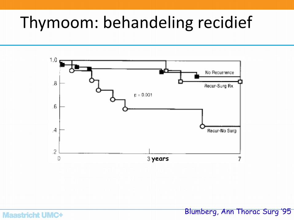
Thymoom: behandeling recidief
years
Survival of patients with no recurrence, N=61 Recurrence treated by surgical resection, N=13 Recurrence not treated surgically, N=12
Blumberg, Ann Thorac Surg ’95
MUMC
• Jaarlijks 60 thymectomie middels robot
• Team:
– prof. De Baets
– Prof. Maessen
– Dr. Hochstenbag
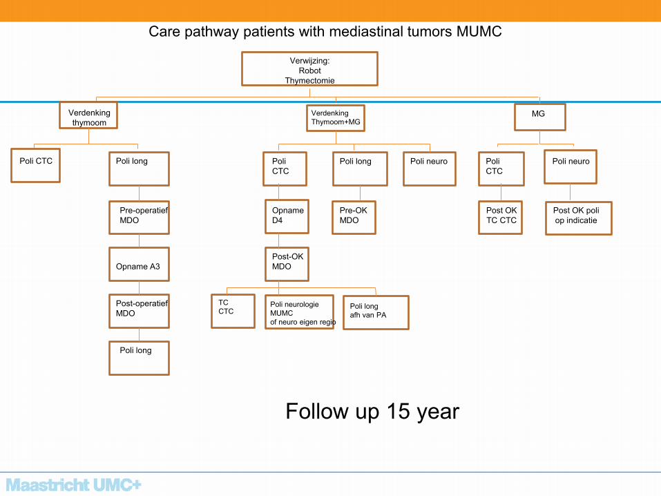
Care pathway patients with mediastinal tumors MUMC
Verwijzing:
Robot
Thymectomie
Verdenking
thymoom
Verdenking
Thymoom+MG MG
Poli CTC Poli long
Pre-operatief
MDO
Opname A3
Post-operatief
MDO
Poli long
Poli
CTC
Opname
D4
Post-OK
MDO
TC
CTC Poli neurologie
MUMC
of neuro eigen regio
Poli long
afh van PA
Poli long
Pre-OK
MDO
Poli neuro Poli
CTC
Post OK
TC CTC
Poli neuro
Post OK poli
op indicatie
Follow up 15 year
Toekomst
Single-arm, multicentric, phase II study of nivolumab in patients
with thymic carcinoma previously treated with chemotherapy
Nivothym trial
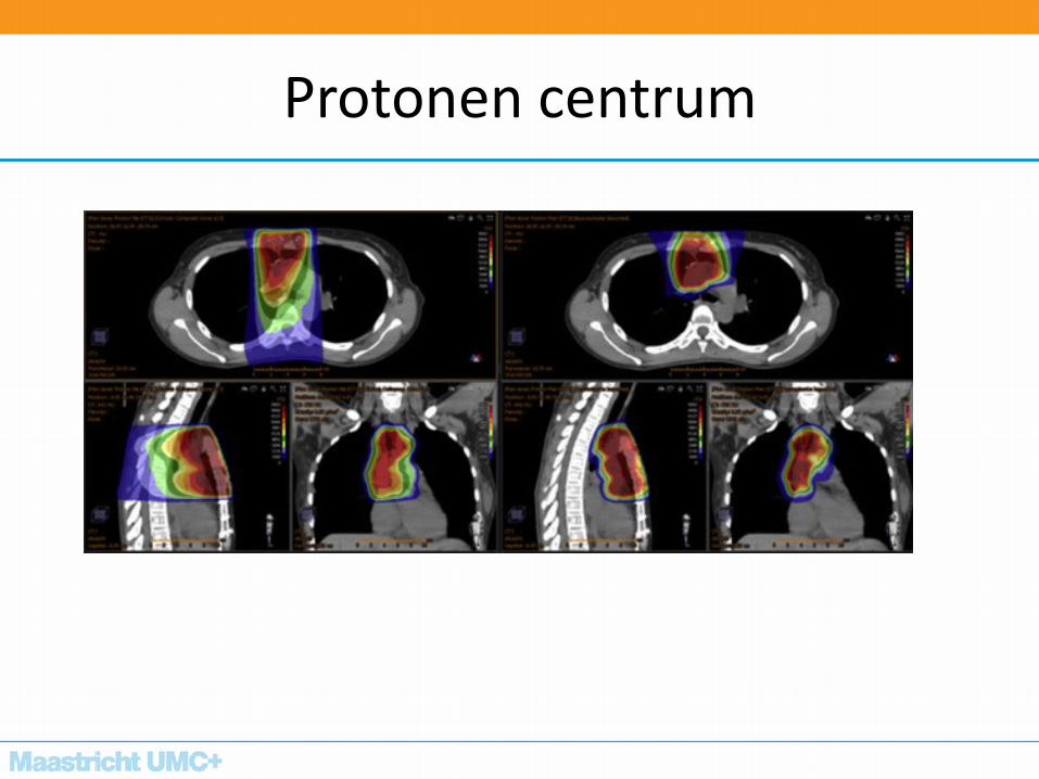
Protonen centrum
BEDANKT VRAGEN ?
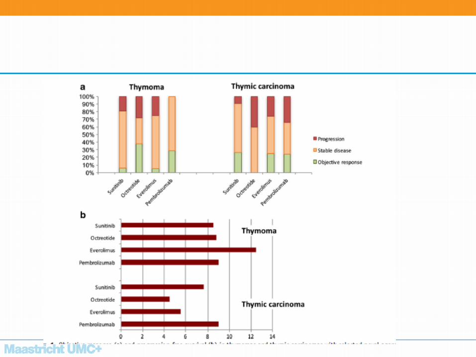
Targeted therapie
• Gefitinib
• Imatinib • RR 1-4%
• Erlotinib-bevacizumab
– fase 2 trial, recidief thymoom
– marginale activiteit