
Separate brain areas for processing human and dog faces as ...
13
Separate brain areas for processing human and dog faces as revealed by awake fMRI in dogs (Canis familiaris) Andie M. Thompkins 1,2 & Bhavitha Ramaiahgari 3 & Sinan Zhao 3 & Sai Sheshan Roy Gotoor 3 & Paul Waggoner 4 & Thomas S. Denney 1,3,5,6 & Gopikrishna Deshpande 1,3,5,6,7 & Jeffrey S. Katz 1,3,5,6 Published online: 22 October 2018 # Psychonomic Society, Inc. 2018 Abstract Functional magnetic resonance imaging (fMRI) has emerged as a viable method to study the neural processing underlying cognition in awake dogs. Working dogs were presented with pictures of dog and human faces. The human faces varied in familiarity (familiar trainers and unfamiliar individuals) and emotional valence (negative, neutral, and positive). Dog faces were familiar (kennel mates) or unfamiliar. The findings revealed adjacent but separate brain areas in the left temporal cortex for processing human and dog faces in the dog brain. The human face area (HFA) and dog face area (DFA) were both parametrically modulated by valence indicating emotion was not the basis for the separation. The HFA and DFA were not influenced by familiarity. Using resting state fMRI data, functional connectivity networks (connectivity fingerprints) were compared and matched across dogs and humans. These network analyses found that the HFA mapped onto the human fusiform area and the DFA mapped onto the human superior temporal gyrus, both core areas in the human face processing system. The findings provide insight into the evolution of face processing. Keywords fMRI . Connectivity . Dog cognition . Dog neuroimaging . Face processing Introduction Dogs and humans share a unique history that spans at least 18,000 years (Thalmann et al., 2013). It is undeniable that in those societies fostering the domestication of pet and working dogs, dogs have reached a unique social status unknown to any other non-human animal. Humans and dogs have developed a complex social repertoire and the study of canine cognition has begun providing rich evidence of evolutionary adaptations fostering cross-species interaction (e.g., Hare, Brown, Williamson, & Tomasello, 2002; Miklósi & Topál, 2013). For instance, dogs respond well to human communicative ges- tures, speech, pointing, and body posture. Dogs are clearly sensitive to human social cues and can discriminate faces. Recent evidence also indicates that there is a face-selective area in the dog brain of pet dogs for processing human and dog faces (Cuaya, Hernández-Pérez, & Cocha, 2016; Dilks et al., 2015). Using functional magnetic resonance imaging (fMRI) in awake dogs trained for detection (for reviews see Bunford, Andics, Kis, Miklósi, & Gácsi, 2017; Thompkins, Deshpande, Waggoner, & Katz, 2016), we investigated nuances in face regions of the domestic dog specific to cross-species stimulus processing and evolutionary origins of face processing. By comparing neural activation resultant of viewing human and dog face stimuli, we have identified adjacent but separate brain areas for processing of human faces and dog faces by domestic dogs. We have also mapped face-processing regions in the dog brain that are functionally analogous to those of humans. In this article, we briefly review face processing in humans followed by dogs before describing the experiment. Electronic supplementary material The online version of this article (https://doi.org/10.3758/s13420-018-0352-z) contains supplementary material, which is available to authorized users. * Jeffrey S. Katz [email protected] 1 Department of Psychology, Auburn University, Auburn, AL, USA 2 Hand in Paw, Inc., Birmingham, AL, USA 3 AU MRI Research Center, Department of Electrical & Computer Engineering, Auburn University, Auburn, AL, USA 4 Canine Detection Research Institute, Auburn University, Auburn, AL, USA 5 Alabama Advanced Imaging Consortium, Auburn University and University of Alabama Birmingham, Birmingham, AL, USA 6 Center for Neuroscience, Auburn University, Auburn, AL, USA 7 Center for Health Ecology and Equity Research, Auburn University, Auburn, AL, USA Learning & Behavior (2018) 46:561–573 https://doi.org/10.3758/s13420-018-0352-z
Transcript of Separate brain areas for processing human and dog faces as ...
Separate brain areas for processing human and dog faces as revealedby awake fMRI in dogs (Canis familiaris)
Andie M. Thompkins1,2 & Bhavitha Ramaiahgari3 & Sinan Zhao3& Sai Sheshan Roy Gotoor3 & Paul Waggoner4 &
Thomas S. Denney1,3,5,6 & Gopikrishna Deshpande1,3,5,6,7& Jeffrey S. Katz1,3,5,6
Published online: 22 October 2018# Psychonomic Society, Inc. 2018
AbstractFunctional magnetic resonance imaging (fMRI) has emerged as a viable method to study the neural processing underlyingcognition in awake dogs. Working dogs were presented with pictures of dog and human faces. The human faces varied infamiliarity (familiar trainers and unfamiliar individuals) and emotional valence (negative, neutral, and positive). Dog faces werefamiliar (kennel mates) or unfamiliar. The findings revealed adjacent but separate brain areas in the left temporal cortex forprocessing human and dog faces in the dog brain. The human face area (HFA) and dog face area (DFA) were both parametricallymodulated by valence indicating emotion was not the basis for the separation. The HFA and DFA were not influenced byfamiliarity. Using resting state fMRI data, functional connectivity networks (connectivity fingerprints) were compared andmatched across dogs and humans. These network analyses found that the HFA mapped onto the human fusiform area and theDFAmapped onto the human superior temporal gyrus, both core areas in the human face processing system. The findings provideinsight into the evolution of face processing.
Keywords fMRI . Connectivity . Dog cognition . Dog neuroimaging . Face processing
Introduction
Dogs and humans share a unique history that spans at least18,000 years (Thalmann et al., 2013). It is undeniable that inthose societies fostering the domestication of pet and workingdogs, dogs have reached a unique social status unknown to any
other non-human animal. Humans and dogs have developed acomplex social repertoire and the study of canine cognition hasbegun providing rich evidence of evolutionary adaptationsfostering cross-species interaction (e.g., Hare, Brown,Williamson, & Tomasello, 2002; Miklósi & Topál, 2013).For instance, dogs respond well to human communicative ges-tures, speech, pointing, and body posture. Dogs are clearlysensitive to human social cues and can discriminate faces.Recent evidence also indicates that there is a face-selective areain the dog brain of pet dogs for processing human and dogfaces (Cuaya, Hernández-Pérez, & Cocha, 2016; Dilks et al.,2015). Using functional magnetic resonance imaging (fMRI)in awake dogs trained for detection (for reviews see Bunford,Andics, Kis, Miklósi, & Gácsi, 2017; Thompkins, Deshpande,Waggoner, & Katz, 2016), we investigated nuances in faceregions of the domestic dog specific to cross-species stimulusprocessing and evolutionary origins of face processing. Bycomparing neural activation resultant of viewing human anddog face stimuli, we have identified adjacent but separate brainareas for processing of human faces and dog faces by domesticdogs. We have also mapped face-processing regions in the dogbrain that are functionally analogous to those of humans. Inthis article, we briefly review face processing in humansfollowed by dogs before describing the experiment.
Electronic supplementary material The online version of this article(https://doi.org/10.3758/s13420-018-0352-z) contains supplementarymaterial, which is available to authorized users.
* Jeffrey S. [email protected]
1 Department of Psychology, Auburn University, Auburn, AL, USA2 Hand in Paw, Inc., Birmingham, AL, USA3 AU MRI Research Center, Department of Electrical & Computer
Engineering, Auburn University, Auburn, AL, USA4 Canine Detection Research Institute, Auburn University,
Auburn, AL, USA5 Alabama Advanced Imaging Consortium, Auburn University and
University of Alabama Birmingham, Birmingham, AL, USA6 Center for Neuroscience, Auburn University, Auburn, AL, USA7 Center for Health Ecology and Equity Research, Auburn University,
Auburn, AL, USA
Learning & Behavior (2018) 46:561–573https://doi.org/10.3758/s13420-018-0352-z

Face processing in humans
Face processing is a vital component of human evolution andsocial cognition. By extracting information from faces,humans are able to recall a person’s identity, retrieve informa-tion about that identity, and use cues (such as the emotionalstate of the person) to aid in socialization (Paller et al., 2003).Across studies, the neuroanatomical structures and activationpatterns underlying the recognition of a person’s identity andassociated mental states (such as the person’s emotion) havebeen guided by evolving models of face processing, typicallydefined by two pathways: one that processes identity informa-tion and one that processes dynamic information such asmovement and intentions. Early face-processing theory fo-cused on identity (Bruce & Young, 1986) and expanded toconsider affect (Calder & Young, 2005). A later model byHaxby, Hoffman, and Gobbini (2000) became dominant andconsists of a core system activated by invariant traits (identity)and an extended system activated by dynamic traits that facil-itate communication, such as emotional expression or eyegaze. The Haxby model assigns processing of invariant facialaspects to the fusiform face area (FFA) and changeable aspectsto the posterior region of the superior temporal sulcus (pSTS).
An extension of the Haxby model was proposed byO’Toole et al. (2002) to account for dynamic motion of famil-iar faces, and this model was again expanded by Bernstein andYovel (2015). The current understanding of face processingdraws separation of regions between the ventral streamthrough the FFA (face form) and the dorsal stream throughthe STS (dynamic information, e.g., motion). Functional andanatomical face processing homologs have been consistentlyidentified across primate species in dorsal and ventral streams,and the number of face-selective areas is similar among pri-mates (Freiwald, Duchaine, & Yovel, 2016). Some authorshave provided supplemental evidence for an anterior process-ing area in humans where the dorsal and ventral streams mayconverge and where more robust familiarity representationsand socially relevant aspects may be processed (Duchaine &Yovel, 2015; di Oleggio Castello, Halchenko, Guntupalli,Gors, & Gobbini, 2017). Familiarity effects tied to semanticand emotional information emphasize the social role of facerecognition (Freiwald et al., 2016). Additionally, studies offamiliar face processing provide evidence for simultaneousactivation of emotion processing areas such as the amygdalaand insula (di Oleggio Castelloet al., 2017).
Collectively, these systems provide the basis for keenlytuned human expertise in extracting information from faces.Regardless of any nuances among theories, if domestic dogprocessing of faces is qualitatively similar to that of humans,then we should expect to see functionally analogous regionsof activation upon presentation of ecologically relevant stim-uli. In this case, we should expect to see activation of regionsimplicated in human and dog processing of familiar and
emotional faces, including the caudate (Cuaya et al., 2016),hippocampus (Paller et al., 2003), and amygdala (di OleggioCastello et al., 2017).
Face processing in dogs
Domestic dogs can discriminate faces, even when stimulusquality and richness are degraded (e.g., Huber, Racca, Scaf,Virányi, & Range, 2013). In addition to displaying highly-tuned recognition of human faces, dogs are also successfulat recognizing the emotions carried by those faces(Albuquerque et al., 2016; Barber, Randi, Müller, & Huber,2016; Müller, Schmitt, Barber, & Huber, 2015; Nagasawa,Murai, Mogi, & Kikusui, 2011). Of note, species that areevolutionarily more closely related to humans than dogs donot perform well on such tasks of human face recognition.This finding is described as the Bspecies-specific effect^(Pascalis & Bachevalier, 1998) of face recognition, a principleanalogous to the Bother-race effect^ in humans.When present-ed with images of human faces and monkey faces, rhesusmonkeys paid more attention to novel monkey faces thanfamiliar ones, and did not show differential attention betweennovel and familiar faces of humans (Pascalis & Bachevalier,1998). Further research has supported this effect, demonstrat-ing similar results in brown capuchins and Tonkean macaques(Dufour, Pascalis, & Petit, 2006). In both studies, for humanparticipants, attention was more differentially focused for hu-man faces than for monkey faces, in line with a species-specific effect.
Young human ch i ld ren , fo r compara t ive anddevelopmental reasons, are often included as a comparisongroup in studies of canine cognition. Racca, Guo, Meints,and Mills (2012) compared the ability of dogs to differentiatebetween emotional facial expressions by humans to that ofchildren, who can accurately categorize emotional faces byage 4 years. Upon presentation of positive, neutral, and nega-tive human and dog faces, the researchers found that dogs andhuman children showed different looking and lateralizationbehaviors. Specifically, data obtained from dogs suggestedhemispheric processing based on a valence model of emotionprocessing. That is, the dogs’ responses supported right hemi-sphere processing to negative emotions and left hemisphereprocessing to positive emotions. Further, it seems that neutralexpressions are processed similarly to negative expressions indogs, possibly due to the lack of approach signals that dogs areable to extract from positive expressions. In opposition to thisvalence-based model, the right hemisphere model was sup-ported by the children’s data, which suggests processing ofall emotional expressions by the right hemisphere. Thoughthis study might suggest that hemispheric processing in dogsand humans is species-specific, the authors note that ju-venile humans’ hemispheric use may change in adult-hood (Racca et al., 2012).
562 Learn Behav (2018) 46:561–573
Lateralization of face processing has been further supportedby Barber et al. (2016) and Siniscalchi, d’Ingeo, Fornelli, andQuaranta (2018). When presenting lab and pet dogs with facesof varying emotional content and familiarity, Barber et al.(2016) found that dogs demonstrated a strong left gaze biasfor all emotive stimuli. Additionally, there were subtle differ-ences in gaze biases according to familiarity of the model andthe content of the emotional expression. This data suggestsright hemisphere lateralization of face stimuli, which was alsosupported by Siniscalchi et al. (2018). Here, the authors iden-tified orienting asymmetries in response to human emotionalfaces. Specifically, dogs oriented their heads to the left in re-sponse to expressions of anger, fear, and happiness, indicatingprocessing in the right hemisphere. In this study, dogs exhibitedno orienting biases in response to human expressions of sad-ness. Taken together, these studies provide support for lateral-ization of emotional face processing, but the question remainsas to whether this lateralization is restricted to the right hemi-sphere for all emotional stimuli or if lateralization is split ac-cording to positive and negative valence.
Experience as the primary mediator of non-human atten-tion to human cues is of particular interest in any investigationof cross-species social cognition. Even in studies of otherspecies that have been domesticated by humans (e.g., horses:McKinley & Sambrook, 2000; goats: Kaminski, Riedel, Call,& Tomasello, 2005), dogs often outperform them, especiallyin the use of facial-based cues. Because dogs have been shownto perform well in object-choice tasks often without signifi-cant human interaction, their capacity to follow human-givencues has been considered by some to be a by-product of do-mestication (e.g., Hare et al., 2002). However, enculturationand experience cannot be ruled out for face processing in anyspecies, and some research suggests that ontogenetic effectsexist in non-dogs (Lyn, Russell, & Hopkins, 2010). In a studyby Barber et al. (2016), the role of ontogenetic experience wasemphasized, as substantial differences in face processing bypet dogs and laboratory dogs were identified. In summary,while domestication may be important in shaping human facediscrimination in dogs, ontogeny cannot be overlooked.
Given the capacity for human face discrimination in dogsand potential ontological influences, we may expect to seedifferences in discrimination performance (particularly in re-gard to emotional expression) that are mediated by role.Though dogs are most commonly thought of as pets, they alsofulfill a variety of working roles that may require more or lessattention to human cues depending on their role. For example,scent detection dogs are often involved in tasks requiring in-dependence and focus on olfactory cues, and are usuallykenneled not experiencing the same enculturation as pet dogs,whereas service dogs must attend closely to their handler’sstate of being and often live with humans. As such, processingof face identity and emotional expression may correlate withbetter or worse performance in specialized working roles.
Heterospecific Face Processing and Attachment
To develop a framework for studying dogs’ functioning intheir relationship with humans, we look to similar researchthat has been conducted with humans. Stoeckel, Palley,Gollub, Niemi, and Evins (2014) used fMRI to compare re-sponsiveness to child and pet dog images as seen by theirmothers/owners. Participants completed behavioral measuresfor assessment of attachment to their children and dogs, afterwhich they viewed images of their own child, their own dog,and unfamiliar dogs and children in the scanner and wereasked to score them according to valence and arousal.
Attachment measures indicated that 93% of participantswere extremely attached to their pet dog, considering him orher as a family member. Indeed, functional data revealed over-lapping regions of brain activation including those associatedwith reward, emotion, and affiliation, namely the amygdala,hippocampus, and fusiform gyrus. However, two contrasts didreveal significant differences between familiar conditions.Images of one’s child led to activation of the substantianigra/ventral tegmental area (implicated in reward and affilia-tion) whereas this pattern of activation was not seen withimages of one’s dog. And although the amygdala was activat-ed by both conditions, images of one’s dog led to greateractivation of the fusiform gyrus than did one’s child.Stoeckel et al. (2014) note that this may be due to the lackof language-based affiliation with dogs, as human-dog inter-action may be more dependent on face perception to pick upon emotion, gaze direction, and identity.
Using dog fMRI to study face processing
Despite growing evidence for complex social cognition in thedomestic dog, neurological investigations are new and few.Dilks et al. (2015) first provided evidence for a specializedface processing area within the brain of pet dogs. They foundthat the right inferior temporal cortex was differentially acti-vated for human videos as opposed to objects. Focusing onthis region of interest found for videos, next, they presentedpictures of human faces, dog faces, objects, scenes, andscrambled faces. They found that human and dog faces acti-vated the same area of the brain as the human videos. Faces,regardless of species, resulted in greater activation than ob-jects and scenes. Interestingly, there were no activation differ-ences between scrambled and unscrambled faces. To this end,processing of low-level features in face stimuli could be im-plicated as a reason for activation in this visual area. In com-paring pictures of faces versus objects, Cuaya et al.(2016) utilized whole-brain, voxel-wise analyses andidentified bilateral temporal cortex (ventral-posteriorregions) activation for faces, with slightly more activityin the left temporal cortex in pet dogs.
Learn Behav (2018) 46:561–573 563
These prior studies demonstrate evidence for selective pro-cessing of human and dog faces in domestic dogs; however,prior studies have not uncovered finer spatial specificity inprocessing of face stimuli of different species, perhaps partlydue to larger voxel sizes amongst other possible reasons suchas head motion. Further, these studies have focused solely onthe dog brain and have not made comparative efforts to iden-tify functional analogy with humans. The purpose of the pres-ent study was twofold. First, we sought to identify precise(and spatially fine-grained) regions of the dog brain implicat-ed in processing of dog and human face stimuli. Second, wesought tomake comparisons between dog and human process-ing of face stimuli. We hypothesized, based on prior research,that the brain regions involved in dogs’ processing of conspe-cific and human faces would be localized to the temporalcortex (Cuaya et al., 2016; Dilks et al., 2015), but spatiallydifferent between the two conditions given our improved spa-tial resolution and advanced techniques for motion correction.Working detector dogs were trained, using reinforcementlearning, to lie motionless and awake during fMRI and toattend to projected face stimuli that varied in emotional va-lence (human faces) and familiarity (human and dog faces).Through this work, we have identified separate regions withinthe dog brain for specific processing of human and dog faces.Further, such specificity was not driven by emotion orfamiliarity.
Methods
Subjects
Twelve dogs (Canis familiaris) were included in this study.All dogs were between 6 months and 3 years of age. Bothmale and female dogs were used in training and in scanning.All dogs remained awake for imaging, for which they weretrained to lie in a prone position on the scanner bed with headinserted into a human knee coil. Positive reinforcement wasprovided to keep dogs as still as possible and to desensitizethem to the scanner environment (Jia et al., 2014). Ethicalapproval for this study was obtained from the AuburnUniversity Institutional Animal Care and Use Committeeand all methods were performed in accordance with theirguidelines and regulations.
Image presentation
We used a stimulus-driven visual task in which awake andunrestrained dogs were presented with still images of humanand dog faces via an in-scanner projector screen. The humanfaces were still image captures of familiar (a dog’s handler)and unfamiliar (a stranger) people. Because familiarity withindividual handlers varied, the dogs were assigned to groups
based on common familiarity with handlers included in astimulus set. Group-specific human stimulus sets were select-ed from a pool of 51 stimuli (17 individuals × 3 valencetypes). For each dog group, the familiar face set was com-prised of the positive, neutral, and negative images for eachof four familiar handlers. The unfamiliar face set was drawnfrom the pool of unfamiliar individuals and included the fourpositive, four neutral, and four negative images most closelymatched in valence to the images in the familiar set. The dogfaces were still image captures of familiar (kennel mates) andunfamiliar (stranger) dogs. The dog stimulus set included fourfamiliar (kennel mate) dog images and four unfamiliar dogimages. These did not vary in valence.
The emotional content of each human image was rated by88 undergraduate students using a self-paced online survey.Participants were asked to score each image according to emo-tional valence. They were asked to identify the emotiondisplayed as well as the degree of that emotion on a scale fromBVery Low^ to BVery High.^Mean scores were calculated forindividual images, resulting in a composite score for eachstimulus ranging from -5 (negative) to +5 (positive; seeSupplementary Material, Fig. S1 for examples). Familiar andunfamiliar faces where matched for valence. Dog imagesdepicted dog faces in a relaxed state, as judged by the exper-imenters (see Supplementary Material, Fig. S2 for examples).We did not attempt to manipulate or score the dogs on valencebecause accuracy of human ratings of emotions in dog faceshave been shown to vary in accuracy according to experience(Bloom & Friedman, 2013).
All images were digitally analyzed using Fourier analysisin order to examine their spectral content (which determinescolor) and absolute magnitude (which determines image in-tensity). No significant differences were found between hu-man and dog faces (p = 0.45 for spectral content and p = 0.32for absolute magnitude, as determined by independent samplet-tests), implying that differences in brain responses to thesetwo types of stimuli will likely not be driven by low-levelfeatures such as color and intensity.
Scanning
Functional MRI data were acquired in a Siemens 3T Verio(Erlangen, Germany) scanner (see Jia et al., 2014). Each runtotaled 140 seconds and included either human or dog faces(which contained both familiar and unfamiliar faces). Withineach human stimulus set, there were four positive familiarimages, four neutral familiar images, four negative familiarimages, four positive unfamiliar images, four neutral unfamil-iar images, and four negative unfamiliar images. During eachscanning session, each dog was presented with all stimuli,randomly broken into runs of 140 s (12 stimuli per run). Fordogs that viewed dog face stimuli, runs also included eightrandomly-distributed dog face stimuli (four familiar and four
564 Learn Behav (2018) 46:561–573

unfamiliar). Face stimuli were presented via projector screenfor 5 s, after which a blank screen was presented for a variable3- to 11-s inter-stimulus interval (ISI). The optimized timingof the ISI was obtained fromOPTSEQ software (https://surfer.nmr.mgh.harvard.edu/optseq/). A 1-s repetition time (TR) wasused. Each dog completed four runs in a randomized order,with each run including stimuli from all conditions.
To be sure that each dog looked at each stimulus that waspresented during scanning, several precautions were taken.Such precautions were necessary to assure that only trials inwhich the dogs attended to the stimulus were analyzed.Attention was judged by two raters per video via simultaneousvideo recording of the dog’s eye inside the scanner. This videorecording was synchronized with stimulus presentation suchthat raters could parse through the video post hoc after theexperiment to determine whether the dog was attending to agiven stimulus or not. For each trial, if the dog’s eye wasvisibly open, the rater assigned a score of Byes.^ If the dog’seye was closed or not open enough for the pupil to be visible,then the rater assigned a score of Bno^ (see SupplementaryMaterial, Fig. S3 for examples.) Inter-rater reliability wasassessed for each trial, and only trials with perfect inter-rateragreement of attentiveness were retained for data analysis.
Data acquisition
Functional MRI data were acquired using a T2*-weighted sin-
gle shot echo planar imaging (EPI) sequence with 16 axialslices, slice thickness = 3 mm, repetition time (TR) = 1,000ms, echo time (TE) = 29 ms, field of view (FOV) = 150 × 150mm2, flip angle (FA) = 90°, in-plane resolution 2.3 × 2.3 mm,in-plane matrix = 64 × 64 and 200 temporal volumes in eachrun. In addition to the visual task described above, two restingstate runs were also acquired for each dog using identicalscanning parameters. During the resting state scan, no visualstimuli were provided to the dog and the canines were trainedto lay still with their eyes open. A human knee coil was usedas a head coil for the dog brain, and all dogs were trained tokeep their heads in the coil as still as possible (with eyes open)during the scanning. Anatomical images were acquired usingmagnetization-prepared rapid gradient echo (MPRAGE) se-quence for overlay and localization (TR = 1,990 ms, TE =2.85 ms, voxel size: 0.6 × 0.6 × 1 mm3, FA = 9°, and in-plane matrix 192 × 192, FOV = 152 × 152 mm2, number ofslices: 104). An external head motion-tracking device basedon an infra-red camera and reflector were used to obtain inde-pendent assessments of the dog’s head motion in high spatio-temporal resolution (see Jia et al., 2014, for details).
Image processing
Data processing was performed using SPM12 (http://www.fil.ion.ucl.ac.uk/spm/software/spm8/, Functional Imaging Lab,
The Welcome Trust Centre for NeuroImaging, The Instituteof Neurology at University College London). All data weresubjected to standard preprocessing steps, includingrealignment to the first functional image, spatialnormalization to our own canine template, spatial smoothingandmotion censoring (discussed in Jia et al., 2014). The singlecamera-based external motion-tracking device provided esti-mates of motion that had both high temporal and spatial res-olution. This information was used along with realignmentparameters for data quality assessment. The data werecompletely discarded when the external camera detected morethan one voxel motion because usable data cannot be retrievedin such a scenario even after motion censoring and interpola-tion. On the other hand, when motion detected from the ex-ternal camera was less than one voxel, we retrospectivelyestimated framewise displacement using realignment parame-ters, and used censoring and interpolation when framewisedisplacement exceeded 0.2 mm (Power et al., 2015).
Following preprocessing, general linear models (GLMs)were built at the individual subject level with regressors foreach condition as well as nuisance regressors derived frommotion estimates obtained from image-based realignment inaddition to those independently obtained from the externalmotion-tracking device. Using random effects analyses, ef-fects of interests were tested at the group level. Specifically,using t-tests, voxels that had significantly different activity forhuman versus dog faces were ascertained. Statistical signifi-cance was determined using a threshold of p < 0.05 FDR(false discovery rate) corrected.
In order to determine whether the brain regions foundabove were also parametrically modulated by emotions, weused parametric regressors in the individual subject GLMs.The amplitude of these regressors was scaled between valuesof +5 and – 5, representing the range of the emotional valenceratings of the human face stimuli. Brain regions that weresignificantly parametrically modulated by emotion in humanfaces were then determined at the group level. A similar anal-ysis was not performed for the dog face stimuli because theimages were neutral and did not vary in emotional valence.
In order to determine whether the main effects of dog versushuman faces were influenced by the familiarity of the humanand dog faces, familiar versus unfamiliar contrasts were testedat the group level in both human and dog samples. We inves-tigated whether (dog face vs. human face) and (familiar facevs. unfamiliar face) contrasts showed common voxels acrossthe dog brain. The basic level contrasts for familiarity andemotion are reported elsewhere (Thompkins, 2016).
Identification of functionally analogous brain regionsin humans
As noted in the results section, two small but distinct patchesof the temporal cortex were responsive more to dog faces as
Learn Behav (2018) 46:561–573 565
compared to human faces (referred to as dog face area orDFA) as well as more responsive to human faces as comparedto dog faces (referred to as human face area or HFA). The roleof such small, distinct, and specific patches of the temporalcortex in canine cognition is unclear due to the lack of corre-sponding literature. In contrast, the functions of specific hu-man brain regions within the temporal cortex have been verywell studied. Therefore, if we were to find the functional an-alogues of DFA and HFA regions in the human brain, it wouldaid interpretation of our results. Further, it would also informus about the evolutionary roles of these regions.Consequently, we mapped dog brain regions with significant-ly different activity for dog and human faces to functionallyanalogous regions in the human brain. This was achieved bycomparing the similarity of functional connectivity finger-prints of these regions using a permutation testing framework(please refer to the following papers for details regarding thismethod: Mars, Sallet, Neubert, & Rushworth, 2013; Marset al., 2014; Mars et al., 2016; Passingham, Stephan, &Kotter, 2002). This framework is based on the assumption thatif Region-A in the dog brain (such as DFA or HFA) has afunctional connectivity pattern similar to Region-B in the hu-man brain, then their relative functional roles in dog and hu-man brains, respectively, must also be similar. Therefore, ouraim was to find those regions in the human brain (via anexhaustive search), which have functional connectivity pro-files similar to DFA and HFA in the dog brain.
If connectivity were to be calculated between a seed (DFAorHFA in the dog brain) and all other graymatter voxels in thebrain, then it would yield high-dimensional profiles that canbe hard to summarize. Given that whole brain functional con-nectivity fingerprints have high dimensionality, we used theapproach proposed by Passingham et al. (2002) where thefingerprint is derived from certain Btarget^ regions that areselected to represent the broadest array of functions as possi-ble. In additional to reducing dimensionality, this approachalso avoids overfitting.
For the purposes of this study, Bfunctional connectivity^between two voxels/regions was defined as the Pearson’s cor-relation between time series derived from those two voxels/regions. We normalized the functional connectivity data usingthe maximum connection strength within the given brain.Thus, normalized functional connectivity metrics then repre-sented relative strength of connectivity and allowed us to com-pare connectivity profiles obtained from data acquired withdifferent scanning parameters for dogs and humans.
The characteristics of resting state fMRI data acquired fromdogs have already been described in the Data acquisition sec-tion. In humans, resting state functional connectivity was ob-tained using preprocessed resting state fMRI data from theHuman Connectome Project (HCP) (HCP 500 Subjects +MEG2Data Release). Our human sample included scans fromyoung healthy adults (age range: 22–35 years) obtained at 3T.
Based on Lebeau’s model (Patronek, Waters, & Glickman,1997), 154 human subjects were manually selected to matchthe gender, number of scans, and age in dog-year equivalents(Table 1) with our dog sample. Data from 30 dogs (includingthe 12 of focus in this study) with an age range of 12–36months were used in resting-state analyses. The HCP func-tional data were acquired on a Siemens Skyra 3T scannerusing multiplexed gradient-echo EPI sequence with slicethickness = 3 mm, TR = 720 ms, TE = 33.1 ms, FA = 52o,FOV = 208 × 180 mm2, in-plane matrix = 104 × 90 and 1,200temporal volumes in each run. For more details about dataacquisition and preprocessing of the HCP data, please seeBHCP 500 Subjects + MEG2 Data Release^ reference manual(https://www.humanconnectome.org/storage/app/media/documentation/s500/hcps500meg2releasereferencemanual.pdf) as well as Smith et al. (2013).
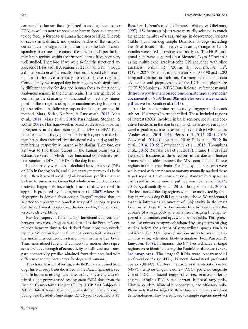
In order to determine connectivity fingerprints for eachsubject, 19 Btargets^ were identified. These included regionsof interest (ROIs) involved in basic sensory, social, and cog-nitive functions in the dog brain, which have also been impli-cated in guiding canine behavior in previous dog fMRI studies(Andics et al., 2014, 2016; Berns et al., 2012, 2015, 2016;Cook et al., 2014; Cuaya et al., 2016; Dilks et al., 2015; Jiaet al., 2014, 2015; Kyathanahally et al., 2015; Thompkinset al., 2016; Ramahihgari et al., 2018). Figure 1 illustratesthe spatial locations of these regions in the dog and humanbrains, while Table 2 shows the MNI coordinates of theseregions in the human brain. For the dogs, authors who werewell versed with canine neuroanatomymanually marked thesetarget regions (in our own custom standardized space asdiscussed in our previous publications (Jia et al., 2014,2015; Kyathanahally et al., 2015; Thompkins et al., 2016)).The locations of the dog regions were also motivated by find-ings in previous dog fMRI studies cited above.We understandthat this introduces an amount of subjectivity in the exactlocation of these ROIs, but would like to note that in theabsence of a large body of canine neuroimaging findings re-ported in a standardized space, this is inevitable. This proce-dure also mirrors the approach adopted by early neuroimagingstudies before the advent of standardized spaces (such asTalairach and MNI space) and co-ordinate based meta-analysis using activation likely estimation (Fox, Parsons, &Lancaster, 1998). In humans, the MNI co-ordinates of targetregions were identified using the BrainMap database (www.brainmap.org). The Btarget^ ROIs were: ventromedialprefrontal cortex (vmPFC), bilateral dorsolateral prefrontalcortex (dlPFC), bilateral ventrolateral prefrontal cortex(vlPFC), anterior cingulate cortex (ACC), posterior cingulatecortex (PCC), bilateral temporal cortex, bilateral inferiorparietal lobule (IPL), visual cortex, bilateral amygdala,bilateral caudate, bilateral hippocampus, and olfactory bulb.Please note that the target ROIs in dogs and humans need notbe homologous, they were picked to sample regions involved
566 Learn Behav (2018) 46:561–573
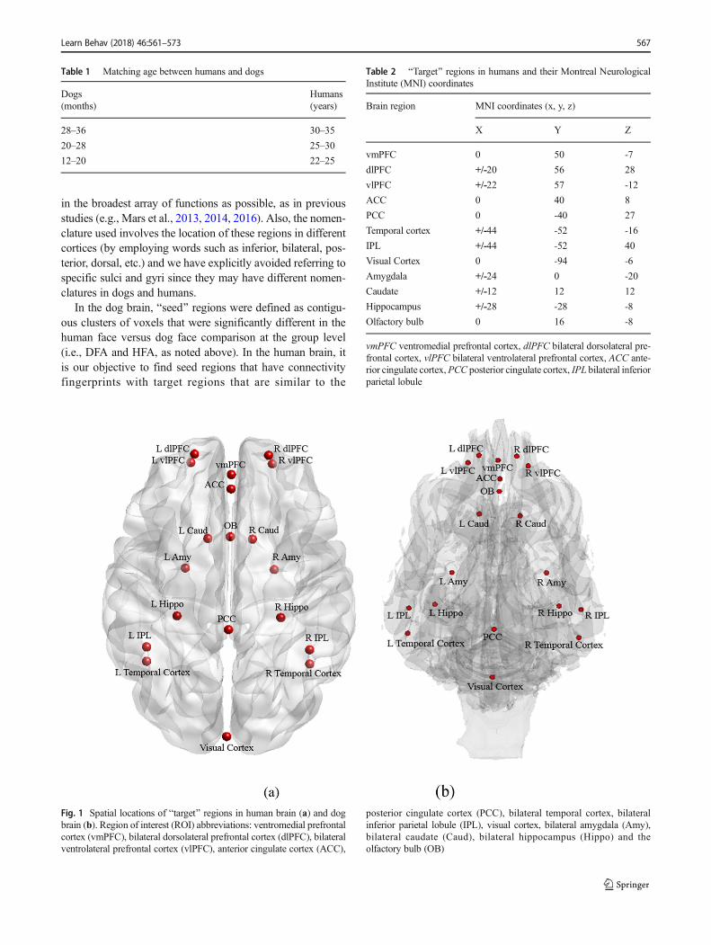
in the broadest array of functions as possible, as in previousstudies (e.g., Mars et al., 2013, 2014, 2016). Also, the nomen-clature used involves the location of these regions in differentcortices (by employing words such as inferior, bilateral, pos-terior, dorsal, etc.) and we have explicitly avoided referring tospecific sulci and gyri since they may have different nomen-clatures in dogs and humans.
In the dog brain, Bseed^ regions were defined as contigu-ous clusters of voxels that were significantly different in thehuman face versus dog face comparison at the group level(i.e., DFA and HFA, as noted above). In the human brain, itis our objective to find seed regions that have connectivityfingerprints with target regions that are similar to the
Fig. 1 Spatial locations of Btarget^ regions in human brain (a) and dogbrain (b). Region of interest (ROI) abbreviations: ventromedial prefrontalcortex (vmPFC), bilateral dorsolateral prefrontal cortex (dlPFC), bilateralventrolateral prefrontal cortex (vlPFC), anterior cingulate cortex (ACC),
posterior cingulate cortex (PCC), bilateral temporal cortex, bilateralinferior parietal lobule (IPL), visual cortex, bilateral amygdala (Amy),bilateral caudate (Caud), bilateral hippocampus (Hippo) and theolfactory bulb (OB)
Table 2 BTarget^ regions in humans and their Montreal NeurologicalInstitute (MNI) coordinates
Brain region MNI coordinates (x, y, z)
X Y Z
vmPFC 0 50 -7
dlPFC +/-20 56 28
vlPFC +/-22 57 -12
ACC 0 40 8
PCC 0 -40 27
Temporal cortex +/-44 -52 -16
IPL +/-44 -52 40
Visual Cortex 0 -94 -6
Amygdala +/-24 0 -20
Caudate +/-12 12 12
Hippocampus +/-28 -28 -8
Olfactory bulb 0 16 -8
vmPFC ventromedial prefrontal cortex, dlPFC bilateral dorsolateral pre-frontal cortex, vlPFC bilateral ventrolateral prefrontal cortex, ACC ante-rior cingulate cortex,PCC posterior cingulate cortex, IPL bilateral inferiorparietal lobule
Table 1 Matching age between humans and dogs
Dogs(months)
Humans(years)
28–36 30–35
20–28 25–30
12–20 22–25
Learn Behav (2018) 46:561–573 567

connectivity fingerprint of the dog seed regions (i.e., DFA andHFA) with corresponding target regions. Therefore, in thehuman brain, every voxel in the brain’s gray matter was con-sidered as a seed region. For each subject, the connectivityfingerprints of Bseed^ regions were assessed from the correla-tion of the time series of the seed regions (just the voxel timeseries in humans and mean time series of seed ROIs in dogs)with those of Btarget^ regions. It should be noted that thenumber of Btargets^ should not be too many to causeoverfitting, yet at the same time should be sufficient to capturethe diversity of the connectivity from the seed regions.
In order to find voxels in the human brain that share thesame fingerprint or pattern of connectivity with Bseed^ re-gions in dogs, the Manhattan distance between the mean con-nectivity fingerprint of each human seed and each dog seed(averaged over the human and the dog samples, respectively)were estimated (Fig. 2). If a and b are vectors of dimension krepresenting two connectivity fingerprints, then the
Manhattan distance between them is defined as ∑ki¼1jai−bij
In order to test the significance of the match between con-nectivity fingerprints of the dog Bseed^ regions and each vox-el in the human brain, permutation testing was employed (p <
0.05, FDR corrected). This provided a statistical criterionbased on which regions in the human brain that were func-tionally analogous to DFA and HFA in the dog brain could beidentified. This method does not utilize any anatomical con-straints regarding the location of the analogous region that isidentified. However, our results (presented in the Results sec-tion, below) as well as those from previous studies (Marset al., 2016; Passingham et al., 2002) indicate that the analo-gous regions identified made sense from an anatomicalperspective.
Results
Only 5% of the data across all dogs had to be discarded due tomotion of more than one voxel detected via the external cam-era. An additional 7% of the data had motion of less than onevoxel, but framewise displacement more than 0.2 mm. Thisdata was recovered via censoring and interpolation and wasnot discarded (Power et al., 2015). Given the small amount ofdata that had to be discarded (i.e., 5%), each dog had enoughdata to perform reliable data analysis (we had a threshold of
Fig. 2 Connectivity fingerprint matching approach illustratedgraphically. Individual Btarget^ regions are indicated by each vertex ofthe polygon showing the connectivity fingerprint. This approach is takengiven the dimensionality of the connectivity fingerprints. In the humanbrain, we show three arbitrary voxels for illustration in yellow, purple, andred colors and corresponding connectivity fingerprints. Note that in realdata, connectivity fingerprints are obtained for all gray matter voxel seedsin the human brain. Similarly, a connectivity fingerprint of a dog Bseed^region is shown in green color. Resting state functional connectivity (FC)
between the seed regions and 19 pre-selected target regions were used toderive these connectivity fingerprints shown as polygons. The finger-prints obtained from the dog and human seed regions were then comparedpairwise using the Manhattan distance measure. In our illustration, theyellow area in the human brain has a connectivity fingerprint with targetregions that are most similar to that obtained by the dog Bseed^ region.Therefore, the yellow voxel in the human brain could potentially be theregion functionally analogous to the green seed voxel in the dog brain
568 Learn Behav (2018) 46:561–573
75% of the data to be available in each dog, to be included inthe data analysis, but all dogs had more usable data than ourthreshold).
Figure 3 shows the primary contrasts of interest. Using awhole-brain voxel-wise analysis, greater neural activation inthe left temporal cortex by human face stimuli as contrastedagainst that of dog faces (human face > dog face) and greateractivation by dog faces as compared to human faces (dog face> human face) are identified as green (cluster size = 410voxels) and red regions (cluster size = 198 voxels), respective-ly. As shown in Fig. 3, we have identified adjacent, but clearlydistinct, activation areas separated by stimulus origin. For thepurposes of this paper, we delineate the area in green as thehuman face area (HFA) and the area in red as the dog face area(DFA). Both species-specific processing regions arelateralized to the left temporal lobe. We did not identify sucha distinction in any other areas of the dog brain.
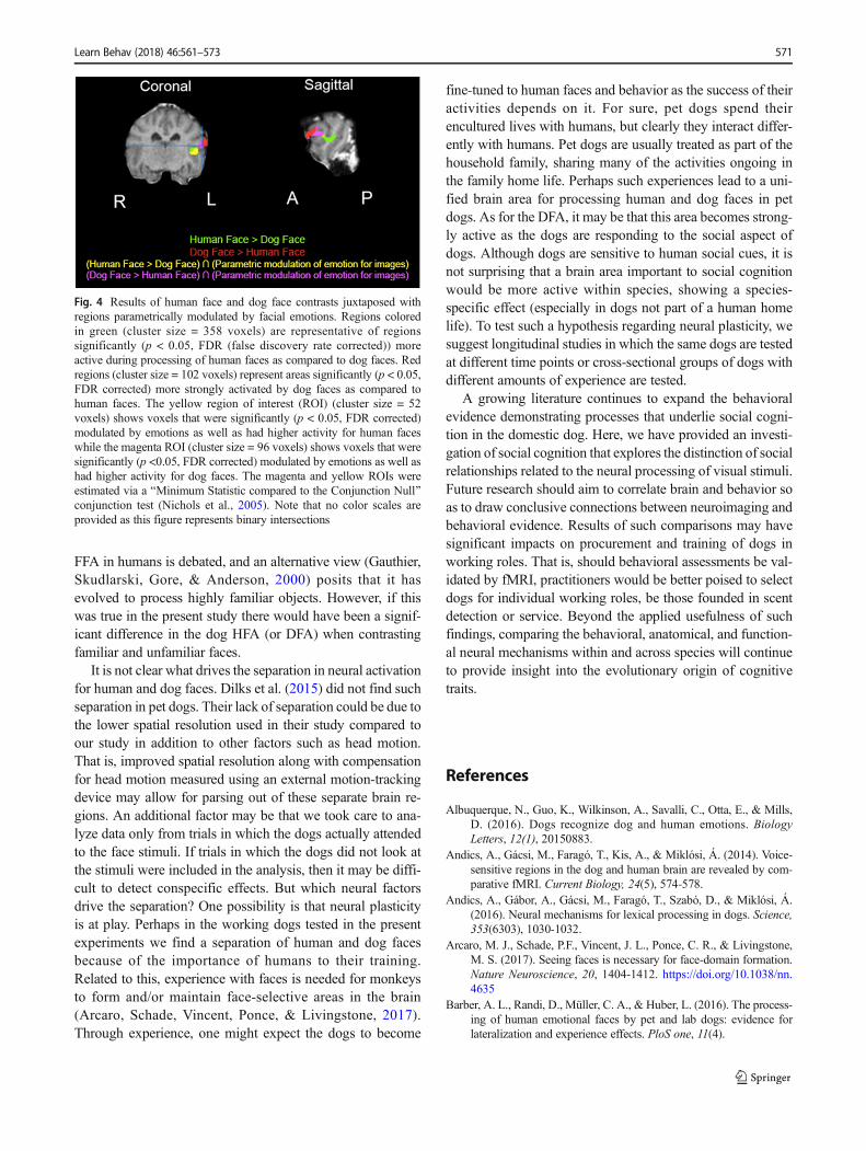
In order to test whether the contrast effects were influencedby the emotional content of the human faces, we conducted aparametric modulation analysis. Sub-regions of both the HFAand DFA were found to parametrically modulate with theemotions displayed by human faces (Fig. 4). Because separa-tion according to emotion was not found, the specificity ofthese regions in the dog brain to processing dog and humanfaces cannot be attributed to emotion alone.
In order to test whether the contrasts effects were influ-enced by the familiarity of the human and dog faces, contrastson (familiar > unfamiliar) human and dog faces were conduct-ed. These contrasts did not find differences in the HFA andDFA indicating that familiarity of faces did not drive thesefunctional differences in the left temporal lobe.
Connectivity fingerprint analyses revealed that the dogHFA maps to the human FFA and the dog DFA maps to thehuman STG indicating the potential for functional analogs. Ofnote, neuroimaging data from humans supports involvementof the fusiform face area (FFA) in the processing of structureand identity of faces and the superior temporal sulcus (STS) indynamic features and familiar face recognition (e.g., Bernstein& Yovel, 2015).
Discussion
We found activation differences for human and dog faces inthe canine temporal cortex, an area previously shown to besensitive to faces in the dog brain. Our analyses more precise-ly identified the spatial location of this face region and ex-panded our understanding of the functional mechanisms un-derlying activation in this brain area. We addressed the possi-bility that some likely low-level features drove this separation,and did not find significant differences between human anddog faces in terms of their spectra (which determines color) ormagnitude (which determines image intensity). Further, while
activation was modulated by emotional valence, it did notdictate separation of activation regions, and familiarity didnot play a role in the HFA or DFA. Finally, connectivity anal-yses revealed potentially functionally analogous networks in-volved in face processing in humans and dogs. The dog HFAmaps to the human FFA and the dog DFA maps to the humanSTG.
Evidence for a region of face processing within the dogbrain was provided by Dilks et al. (2015) and Cuaya et al.(2016).We have established further support for a face process-ing area in the dog brain and consider it functionally analo-gous to those found in humans, monkeys, and sheep. In all ofthese studies, regional activation in the temporal lobe wasidentified subsequent to the presentation of face stimuli. Thisaligns with past literature evidencing face-specific processingin humans, monkeys, and sheep, albeit with shifts in the spe-cific location in the temporal lobe for face processing acrossspecies (Kanwisher, McDermott, & Chun, 1997; Kendricket al., 2001; Sliwa, Planté, Duhamel, & Wirth., 2014). In ad-dition to demonstrating that there is a region of the dog brainthat is reliably activated by faces, an important addition of thecurrent study is that of cross-species regional distinction (i.e.,separate regions of processing for human and dog faces).Overall, combining findings of fMRI data from the presentstudy, Dilks et al. (2015), and Cuaya et al. (2016), with theprior behavior studies with dogs, there appears to be supportfor domain specificity for face processing in domestic dogs,although it is clearly premature to conclude a specific regionin the dog brain is causally involved in face perception (e.g.,Weiner et al., 2017). Additionally, lateralized face processinghas repeatedly been demonstrated in dogs (e.g., Barber et al.,2016; Siniscalchi, d’Ingeo, Fornelli, & Quaranta, 2018) andwas supported in our results.
As discussed earlier, humans are able to use identity andcues in successful socialization (Paller et al., 2003). Currentmodels of face processing highlight separation of regions be-tween the ventral stream (face form) and the dorsal stream(dynamic information) (e.g., Bernstein & Yovel, 2015;O’Toole et al., 2002) with some accounts evidencing a con-verging of these streams to allow for further processing ofsocially-relevant familiarity information (di Oleggio Castelloet al., 2017; Duchaine & Yovel, 2015). Stoeckel et al. (2014)found that, for images of familiar dog (pet) faces and familiarhuman (child) faces, human areas of activation included theamygdala, hippocampus, and fusiform gyrus. We have ex-tended the first steps in exploring the dog’s ability to makeuse of such capacities in their cohabitation with humans. Dogsalso appear to mimic the core and extended systems of faceprocessing initially described by human face-processingmodels, as they make use of both identity and emotional in-formation. These analogies with human processing of faceswarrant further exploration with attention paid to comparativemodeling. It should be noted that the functional role of the
Learn Behav (2018) 46:561–573 569
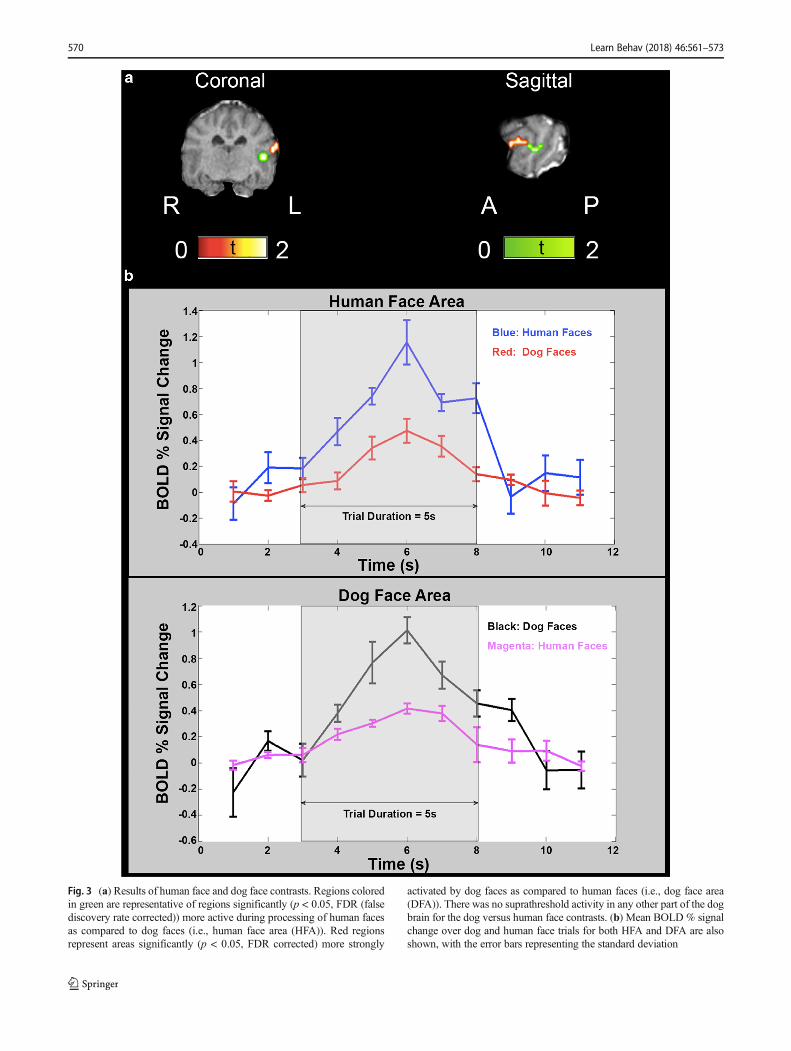
Fig. 3 (a) Results of human face and dog face contrasts. Regions coloredin green are representative of regions significantly (p < 0.05, FDR (falsediscovery rate corrected)) more active during processing of human facesas compared to dog faces (i.e., human face area (HFA)). Red regionsrepresent areas significantly (p < 0.05, FDR corrected) more strongly
activated by dog faces as compared to human faces (i.e., dog face area(DFA)). There was no suprathreshold activity in any other part of the dogbrain for the dog versus human face contrasts. (b) Mean BOLD % signalchange over dog and human face trials for both HFA and DFA are alsoshown, with the error bars representing the standard deviation
570 Learn Behav (2018) 46:561–573
FFA in humans is debated, and an alternative view (Gauthier,Skudlarski, Gore, & Anderson, 2000) posits that it hasevolved to process highly familiar objects. However, if thiswas true in the present study there would have been a signif-icant difference in the dog HFA (or DFA) when contrastingfamiliar and unfamiliar faces.
It is not clear what drives the separation in neural activationfor human and dog faces. Dilks et al. (2015) did not find suchseparation in pet dogs. Their lack of separation could be due tothe lower spatial resolution used in their study compared toour study in addition to other factors such as head motion.That is, improved spatial resolution along with compensationfor head motion measured using an external motion-trackingdevice may allow for parsing out of these separate brain re-gions. An additional factor may be that we took care to ana-lyze data only from trials in which the dogs actually attendedto the face stimuli. If trials in which the dogs did not look atthe stimuli were included in the analysis, then it may be diffi-cult to detect conspecific effects. But which neural factorsdrive the separation? One possibility is that neural plasticityis at play. Perhaps in the working dogs tested in the presentexperiments we find a separation of human and dog facesbecause of the importance of humans to their training.Related to this, experience with faces is needed for monkeysto form and/or maintain face-selective areas in the brain(Arcaro, Schade, Vincent, Ponce, & Livingstone, 2017).Through experience, one might expect the dogs to become
fine-tuned to human faces and behavior as the success of theiractivities depends on it. For sure, pet dogs spend theirencultured lives with humans, but clearly they interact differ-ently with humans. Pet dogs are usually treated as part of thehousehold family, sharing many of the activities ongoing inthe family home life. Perhaps such experiences lead to a uni-fied brain area for processing human and dog faces in petdogs. As for the DFA, it may be that this area becomes strong-ly active as the dogs are responding to the social aspect ofdogs. Although dogs are sensitive to human social cues, it isnot surprising that a brain area important to social cognitionwould be more active within species, showing a species-specific effect (especially in dogs not part of a human homelife). To test such a hypothesis regarding neural plasticity, wesuggest longitudinal studies in which the same dogs are testedat different time points or cross-sectional groups of dogs withdifferent amounts of experience are tested.
A growing literature continues to expand the behavioralevidence demonstrating processes that underlie social cogni-tion in the domestic dog. Here, we have provided an investi-gation of social cognition that explores the distinction of socialrelationships related to the neural processing of visual stimuli.Future research should aim to correlate brain and behavior soas to draw conclusive connections between neuroimaging andbehavioral evidence. Results of such comparisons may havesignificant impacts on procurement and training of dogs inworking roles. That is, should behavioral assessments be val-idated by fMRI, practitioners would be better poised to selectdogs for individual working roles, be those founded in scentdetection or service. Beyond the applied usefulness of suchfindings, comparing the behavioral, anatomical, and function-al neural mechanisms within and across species will continueto provide insight into the evolutionary origin of cognitivetraits.
References
Albuquerque, N., Guo, K., Wilkinson, A., Savalli, C., Otta, E., & Mills,D. (2016). Dogs recognize dog and human emotions. BiologyLetters, 12(1), 20150883.
Andics, A., Gácsi, M., Faragó, T., Kis, A., & Miklósi, Á. (2014). Voice-sensitive regions in the dog and human brain are revealed by com-parative fMRI. Current Biology, 24(5), 574-578.
Andics, A., Gábor, A., Gácsi, M., Faragó, T., Szabó, D., & Miklósi, Á.(2016). Neural mechanisms for lexical processing in dogs. Science,353(6303), 1030-1032.
Arcaro, M. J., Schade, P.F., Vincent, J. L., Ponce, C. R., & Livingstone,M. S. (2017). Seeing faces is necessary for face-domain formation.Nature Neuroscience, 20, 1404-1412. https://doi.org/10.1038/nn.4635
Barber, A. L., Randi, D., Müller, C. A., & Huber, L. (2016). The process-ing of human emotional faces by pet and lab dogs: evidence forlateralization and experience effects. PloS one, 11(4).
Fig. 4 Results of human face and dog face contrasts juxtaposed withregions parametrically modulated by facial emotions. Regions coloredin green (cluster size = 358 voxels) are representative of regionssignificantly (p < 0.05, FDR (false discovery rate corrected)) moreactive during processing of human faces as compared to dog faces. Redregions (cluster size = 102 voxels) represent areas significantly (p < 0.05,FDR corrected) more strongly activated by dog faces as compared tohuman faces. The yellow region of interest (ROI) (cluster size = 52voxels) shows voxels that were significantly (p < 0.05, FDR corrected)modulated by emotions as well as had higher activity for human faceswhile the magenta ROI (cluster size = 96 voxels) shows voxels that weresignificantly (p <0.05, FDR corrected) modulated by emotions as well ashad higher activity for dog faces. The magenta and yellow ROIs wereestimated via a BMinimum Statistic compared to the Conjunction Null^conjunction test (Nichols et al., 2005). Note that no color scales areprovided as this figure represents binary intersections
Learn Behav (2018) 46:561–573 571
Berns, G. S., Brooks, A. M., & Spivak, M. (2012). Functional MRI inawake unrestrained dogs. PloS one, 7(5), e38027.
Berns, G. S., Brooks, A. M., & Spivak, M. (2015). Scent of the familiar:an fMRI study of canine brain responses to familiar and unfamiliarhuman and dog odors. Behavioural Processes, 110, 37-46.
Berns, G. S., & Cook, P. F. (2016). Why did the dog walk into the MRI?.Current Directions in Psychological Science, 25(5), 363-369.
Bernstein, M., & Yovel, G. (2015). Two neural pathways of face process-ing: a critical evaluation of current models. Neuroscience &Biobehavioral Reviews, 55, 536-546.
Bloom, T., & Friedman, H. (2013). Classifying dogs’(Canis familiaris)facial expressions from photographs. Behavioural Processes, 96, 1-10.
Bruce, V., & Young, A. (1986). Understanding face recognition. BritishJournal of Psychology, 77(3), 305-327.
Bunford, N., Andics, A., Kis, A., Miklósi, Á., & Gácsi, M. (2017). Canisfamiliaris as a Model for Non-Invasive Comparative Neuroscience.Trends in Neurosciences, 40(7), 438-452.
Calder, A. J., & Young, A. W. (2005). Understanding the recognition offacial identity and facial expression. Nature Reviews Neuroscience,6(8), 641.
Cook, P. F., Spivak, M., & Berns, G. S. (2014). One pair of hands is notlike another: caudate BOLD response in dogs depends on signalsource and canine temperament. PeerJ, 2, e596.
Cuaya, L. V., Hernández-Pérez, R., & Concha, L. (2016). Our faces in thedog's brain: Functional imaging reveals temporal cortex activationduring perception of human faces. PloS one, 11(3).
di Oleggio Castello, M. V., Halchenko, Y. O., Guntupalli, J. S., Gors, J.D., & Gobbini, M. I. (2017). The neural representation of personallyfamiliar and unfamiliar faces in the distributed system for face per-ception. Scientific Reports, 7(1), 12237.
Dilks, D. D., Cook, P., Weiller, S. K., Berns, H. P., Spivak, M., & Berns,G. S. (2015). Awake fMRI reveals a specialized region in dog tem-poral cortex for face processing. PeerJ, 3.
Duchaine, B., & Yovel, G. (2015). A revised neural framework for faceprocessing. Annual Review of Vision Science, 1, 393-416.
Dufour, V., Pascalis, O., & Petit, O. (2006). Face processing limitation toown species in primates: a comparative study in brown capuchins,Tonkeanmacaques and humans. Behavioural Processes, 73(1), 107-113.
Fox, P. T., Parsons, L. M., & Lancaster, J. L. (1998). Beyond the singlestudy: function/location metanalysis in cognitive neuroimaging.Current Opinion in Neurobiology, 8(2), 178-187.
Freiwald, W., Duchaine, B., & Yovel, G. (2016). Face processing sys-tems: from neurons to real-world social perception. Annual Reviewof Neuroscience, 39, 325-346.
Gauthier, I., Skudlarski, P., Gore, J. C., & Anderson, A. W. (2000).Expertise for cars and birds recruits brain areas involved in facerecognition. Nature Neuroscience, 3(2), 191.
Hare, B., Brown, M., Williamson, C., & Tomasello, M. (2002). Thedomestication of social cognition in dogs. Science, 298(5598),1634-1636.
Haxby, J. V., Hoffman, E. A., & Gobbini, M. I. (2000). The distributedhuman neural system for face perception. Trends in CognitiveSciences, 4(6), 223-233
Huber, L., Racca, A., Scaf, B., Virányi, Z., & Range, F. (2013).Discrimination of familiar human faces in dogs (Canis familiaris).Learning and motivation, 44(4), 258-269.
Jia, H., Pustovyy, O. M., Waggoner, P., Beyers, R. J., Schumacher, J.,Wildey, C.,…Vodyanoy, V. J. (2014). Functional MRI of the olfac-tory system in conscious dogs. PLoS One, 9(1).
Jia, H., Pustovyy, O. M., Wang, Y., Waggoner, P., Beyers, R. J.,Schumacher, J., … Vodyanoy, V. J. (2015). Enhancement of odor-induced activity in the canine brain by zinc nanoparticles: A func-tional MRI study in fully unrestrained conscious dogs. ChemicalSenses, 41(1), 53-67.
Kaminski, J., Riedel, J., Call, J., & Tomasello, M. (2005). Domesticgoats, Capra hircus, follow gaze direction and use social cues inan object choice task. Animal Behaviour, 69(1), 11-18.
Kanwisher, N., McDermott, J., & Chun, M. M. (1997). The fusiform facearea: a module in human extrastriate cortex specialized for faceperception. Journal of Neuroscience, 17(11), 4302-4311.
Kendrick, K.M., da Costa, A. P., Leigh, A. E., Hinton,M. R., & Peirce, J.W. (2001). Sheep don't forget a face. Nature, 414, 165-166.
Kyathanahally, S. P., Jia, H., Pustovyy, O. M., Waggoner, P., Beyers, R.,Schumacher, J., … Vodyanoy, V. J. (2015). Anterior–posterior dis-sociation of the default mode network in dogs. Brain Structure andFunction, 220(2), 1063-1076.
Lyn, H., Russell, J. L., & Hopkins, W. D. (2010). The impact of environ-ment on the comprehension of declarative communication in apes.Psychological Science, 21(3), 360-365.
Mars RB, Neubert FX, Verhagen L, Sallet J, Miller KL, Dunbar RIM, &Barton RA (2014). Primate comparative neuroscience using mag-netic resonance imaging: Promises and challenges. Frontiers inNeuroscience, 8, 298.
Mars RB, Sallet J, Neubert FX, & Rushworth MFS. 2013.Connectivityprofiles reveal the relationship between brain areas for social cogni-tion in human and monkey temporoparietal cortex. Proceedings ofthe National Academy of Sciences, 110, 10806-10811.
Mars RB, Verhagen L, Gladwin TE, Neubert FX, Sallet J, & RushworthMFS (2016). Comparing brains by matching connectivity finger-prints. Neuroscience and Biobehavioral Reviews, 60, 90-97.
McKinley, J., & Sambrook, T. D. (2000). Use of human-given cues bydomestic dogs () and horses (Equus caballus). Animal Cognition,3(1), 13-22.
Miklósi, Á., & Topál, J. (2013). What does it take to become ‘bestfriends’? Evolutionary changes in canine social competence.Trends in Cognitive Sciences, 17(6), 287-294.
Müller, C. A., Schmitt, K., Barber, A. L., & Huber, L. (2015). Dogs candiscriminate emotional expressions of human faces. CurrentBiology, 25(5), 601-605.
Nagasawa, M., Murai, K., Mogi, K., & Kikusui, T. (2011). Dogs candiscriminate human smiling faces from blank expressions. AnimalCognition, 14(4), 525-533.
Nichols, T., Brett, M., Andersson, J., Wager, T., & Poline, J. B. (2005).Valid conjunction inference with the minimum statistic.Neuroimage, 25(3), 653-660.
O’Toole, A.J., Roark, D.A., Abdi, H., 2002. Recognizing moving faces: apsychological and neural synthesis. Trends in Cognitive Sciences, 6,261–266.
Paller, K. A., Ranganath, C., Gonsalves, B., LaBar, K. S., Parrish, T. B.,Gitelman, D. R., … Reber, P. J. (2003). Neural correlates of personrecognition. Learning & Memory, 10(4), 253-260.
Pascalis, O., & Bachevalier, J. (1998). Face recognition in primates: across-species study. Behavioural Processes, 43(1), 87-96.
Passingham, R. E., Stephan, K. E., & Kotter, R. (2002). The anatomicalbasis of functional localization in the cortex. Nature ReviewNeuroscience, 3(8), 606–16.
Patronek, G. J., Waters, D. J., & Glickman, L. T. (1997). Comparativelongevity of pet dpgs and humans: Implication for gerontology re-search. Journal of Gerontology: Biological Sciences, 52A(3), B171–B178.
Power, J. D., Schlaggar, B. L., & Petersen, S. E. (2015). Recent progressand outstanding issues in motion correction in resting state fMRI.Neuroimage, 105, 536-551.
Racca, A., Guo, K., Meints, K., & Mills, D. S. (2012). Reading faces:differential lateral gaze bias in processing canine and human facialexpressions in dogs and 4-year-old children. PLoS one, 7(4).
Ramaihgari B., Pustovyy, O.M., Waggoner, P., Beyers, R.J., Wildey, C.,Morrison, E., Salibi, N., Katz, J.S., Denney, T.S., Vodyanoy, V.J., &Deshpande G. (2018). Zinc Nanoparticles Enhance BrainConnectivity in the Canine Olfactory Network: Evidence From an
572 Learn Behav (2018) 46:561–573
fMRI Study in Unrestrained Awake Dogs. Frontiers in VeterinaryScience. 5:127. doi: https://doi.org/10.3389/fvets.2018.00127
Siniscalchi, M., d’Ingeo, S., Fornelli, S., & Quaranta, A. (2018).Lateralized behavior and cardiac activity of dogs in response tohuman emotional vocalizations. Scientific Reports, 8(1), 77.
Sliwa, J., Planté, A., Duhamel, J. R., & Wirth, S. (2014). Independentneuronal representation of facial and vocal identity in the monkeyhippocampus and inferotemporal cortex. Cerebral Cortex, 26(3),950-966.
Smith SM, Andersson J, Auerbach EJ, Beckmann CF, Bijsterbosch J,Douaud G, Duff E, Feinberg DA, Griffanti L, Harms MP, Kelly M,Laumann T, Miller KL, Moeller S, Petersen SE, Power J, Salimi-Khorshidi G, Snyder AZ, Vu A, Woolrich MW, Xu J, Yacoub E,Ŭgurbil K, Van Essen DC, Glasser MF (2013). Resting-state fMRIin the Human Connectome Project. NeuroImage, 80, 144-168.
Stoeckel, L. E., Palley, L. S., Gollub, R. L., Niemi, S. M., & Evins, A. E.(2014). Patterns of brain activation when mothers view their ownchild and dog: An fMRI study. PLoS One, 9(10).
Thalmann, O., Shapiro, B., Cui, P., Schuenemann, V. J., Sawyer, S. K.,Greenfield, D. L.,… Napierala, H. (2013). Complete mitochondrialgenomes of ancient canids suggest a European origin of domesticdogs. Science, 342(6160), 871-874.
Thompkins, A.M. (2016). Investigating the Dog-Human Social Bond viaBehavioral and fMRI Methodologies (Doctoral dissertation).Auburn University, AL.
Thompkins, A. M., Deshpande, G., Waggoner, P., & Katz, J. S. (2016).Functional Magnetic Resonance Imaging of the Domestic Dog:Research, Methodology, and Conceptual Issue. ComparativeCognition & Behavior Reviews, 11, 63-82.
Weiner, K.S., Barnett, M. A., Lorenz, S., Caspers, J., Stigliani, A.,Amunts, K., Zilles, K., Fischl, B., & Grill-Spector, K. (2017). TheCytoarchitecture of Domain-specific Regions in Human High-levelVisual Cortex. Cerebral Cortex, 27, 146–161, https://doi.org/10.1093/cercor/bhw361
Learn Behav (2018) 46:561–573 573