Postnatal Development of the Human Nasal Septum and its Related ...
144
Postnatal Development of the Human Nasal Septum and its Related Structures. J. van Loosen
Transcript of Postnatal Development of the Human Nasal Septum and its Related ...
Postnatal Development of the Human Nasal Septum and its Related Structures.
J. van Loosen
Copyright®:
J. van Loosen, 2000.
Niets uit deze uitgave mag vermenigvuldigd en/of openbaar gemaakt worden door middel van
druk, fotocopie, microfilm, of op welke andere wijze dan ook, zander voorafgaande schriftellijke
toestemming van de auteur.
All rights reserved. No part of this publucation may be reproduced, stored in a retrieval system,
or transmitted in any form by means, electronic, mechanical, photocopying, or otherwise,
without written permission form the copyright ownerS.
CIp-gegevens koninklijke bibliolheek, Den Haag:
Van Loosen J.
Post natal development of the human nasal septum and its related structures.
Thesis, Erasmus Univeristy Rotterdam, The Netherlands.
- Met literatuuropgave - Met samenvatting in het Nederlands.
ISBN 90·9013471· 9
Vormgeving en productie:
JC Art BY, Nieuwerkerk, The Netherlands.
POSTNATAL DEVELOPMENT OF THE HUMAN NASAL SEPTUM
AND ITS RELATED STRUCTURES.
An anatomical, radiological and histlogical study.
POSTNATALE ONTWIKKELING VAN HET MENSELIJKE
NEUSTUSSENSCHOT EN AANVERWANTE STRUCTUREN.
Een anatomische, radiologische en histologische studie.
PROEFSCHRIFT
TER VERKRIJGING VAN DE GRAAD VAN DOCTOR
AAN DE ERASMUS UNlVERSITEIT ROTTERDAM.
OP GEZAG VAN DE RECTOR MAGNIFICUS
PROF. DR. P.W.C. AKKERMANS M.A.
EN VOLGENS BESLUIT VAN HET COLLEGE VAN PROMOTIES.
DE OPENBARE VERDEDIGING ZAL PLAATSVINDEN OP
WOENSDAG 26 JANUARI 2000
DOOR
Johan van Loosen
geboren te Vlissingen
This study is part of the project Airway Stenosis.
Supervisor:
Dr. H,L, Venvoerd-Verhoef
Institute for Otorhinolaryngology
Erasmus Universiteit Rotterdam
PROMOTIECOMMISSIE
Promotor:
Overige leden:
Co-promotores:
Prof. Dr. C.D.A. Verwoerd
Prof. Dr. F.W.J. Hazebroek
Prof. Dr. S.E.R. Hovius
Prof. Dr. J. Voogd
Dr, D. van Velzen
Dr. H.L. Verwoerd-Verhoef
This study was supported by a grant from "de dne lichten n foundation,
Publication was supported by: ARTU Biologicals BV, ASTRA Pharmaceutica BY,
Beltone Nederland BY, Carl Zeiss BY, Entenned BY, Glaxo Wellcome BV, HAL Allergenen
Laboratoriurn BV, Lameris BV, L. de Haan Hoorapparaten BV, Hoechst Marion Roussel,
Hoortoestel Centrum Sneek BV, JC Art BY, Moduvice BV, DUeon Nederland BV, Pfizer BV,
Philips Hearing Technologies BV, Phonak BV, Raadgevers Medische Beroepen, Schering-Plough
BV, SmithKline Beecham Parma BV, Smith & Nephew Nederland B.V., Yamanouchi Pharma BV.
AAN ARSTRID, HANNA, ANNETTE, RIAN
PAUL EN MJJN OUDERS
Voorwoord.
Dit proefschrift is tot stand gekomen dankzij de inspanning van velen. Enkele
wil ik hiervan in het bijzonder noenlen.
Prof. Dr. C.D.A. Verwoerd en Dr. H.L. Verwoerd-Verhoef. Beste Carel en
Jetty, dankzij jullie nooit aflatende inspiratie en enthousiasme heb ik deze
lange en voor mijn gevoel soms "zware klus" voltooid. Jullie vasthoudend
heid en ervaring in het wetenschappelijk onderzoek hebben uiteindelijk
geJeid tot dit resultaat. Ik spreek hierbij mijn bijzondere erkentelijkheid uit
dat jullie er in geslaagd zijn mij te leiden naar het finale product: dit proef
schrift.
Dr. D. van Velzen. Beste Dick, jouw inventieve geest en dynamische karakter
hebben geleid tot een boeiend contact sinds 1986. Jij bent in staat geweest
dit onderzoek vanuit jouw vakoptiek te verrijken. Tijden van windstilte wer
den gevolgd door een explosieve werklusC nimmer -was het saai. Hiervoor
ben ik je erkentelijk.
Prof. Dr. U.L. Wagholikar. Dear Ulhas, I thank you for the opportunity that you
gave me to do research in your department.
It was an honour to cooperate with you and I am happy with the fact that we
still have contact after all these years.
Dr. D.N. Lanjewar. Dear Danesh, during our work at the Department of
Pathology we have become friends forever. Under difficult circumstances we
did the dissection work at the autopsy room. Besides that, you showed me
"the other part of the world" .
Dr. G.A. van Zanten. Beste Bert, veel dank voor de deskundige statistische
bewerking van mijn onderzoekresultaten. Dit he eft het onderzoek meer
gewicht gegeven.
Dr. C.V. Howard. Dear Viv, I thank you very much for the support in prepa
ring my articles and for your technical support in the histologic field.
Ing. R. Maas. Beste Ronald, hierbij wiI ik jou hartelijk bedanken voor het ont
werpen van het computerprogramma aangaande oppervlaktemetingen.
Verder wil ik achtereenvolgens bedanken Drs. Eisebeth Botman en Dr. Yiong
Yhiang voor de bewerking van de histologische preparaten.
De analisten en laboranten van de afdeling pathologie van het Academisch
Ziekenhuis Dijkzigt, het SSDZ te Delft, de afdeling pathologie te Liverpool en
de afdeling kinderpathologie van de Dalhousie Universiteit te Halifax ben ik
zeer erkentelijk voar het bewerken van Inijn materiaal.
Mw. Dr. S. Chadda-Ajwani. Beste Savi, jou ben ik in het bijzonder dank ver
schuldigd omdat jij de contacten hebt verzorgd met de afdeling Pathologie te
Bombay, India.
Het voorbereiden van het proefschrift gaat soms ten koste van de normale
praktijkvoering. Hierbij moet vaak een beroep worden gedaan op een wel
willen de coliega. In dit kader wit ik voor mijn Rotterdamse periode in het bij
zander bedanken Hans Hoeve en voar de jaren daarna mijn huidige
"Sneekse" coliega Paul Smet.
Graag wil ik voor het niet aflatende typewerk Caroline Kaat, Jacqueline
Aukes-Jager en Ingrid Grunstra dank zeggen.
Jack de Coninck ben ik veel dank verschuldigd voor de vele uren energie en
creativiteit die hij gestopt heeft in de vormgeving en productie van het proef
schrift.
Verder wil ik Rob Baatenburg de Jong en Paul Walters bedanken voor het feit
dat zij mijn paranymfen willen zijn.

Contents.
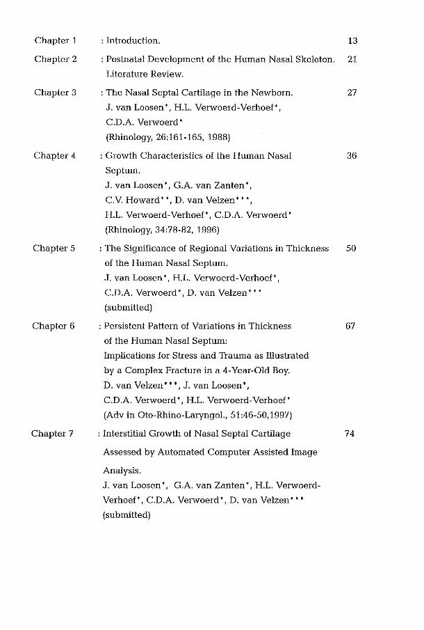
Chapter 1
Chapter 2
Chapter 3
Chapter 4
Chapter 5
Chapter 6
Chapter 7
: Introduction. 13
: Postnatal Development of the Human Nasal Skeleton. 21
Literature Review.
: The Nasal Septal Cartilage in the Newborn.
J. van Loosen' f H.L. Verwoerd-Verhoef* 1
C.D.A. Verwoerd'
(Rhinology, 26:161-165, 1988)
: Growth Characteristics of the Human Nasal
Septum.
J. van Loosen' I G,A. van Zanten' I
C.V. Howard· • I D. van Velzen' •• 1
H.L. Verwoerd-Verhoef', C.D.A. Velwoerd'
(Rhinology, 34:78-82, 1996)
: The Significance of Regional Variations in Thickness
of the Human Nasal Septum.
J. van Loosen', H.L. Verwoerd-Verhoef',
C.D.A. Verwoerd' r D. van Velzen' ••
(submitted)
: Persistent Pattern of Variations in Thickness
of the Human Nasal Septum:
Implications for Stress and Trauma as Illustrated
by a Complex Fracture in a 4-Year-Old Boy.
D. van Velzen··· I J. van Loosen·,
C.D.A. Verwoerd', H.L. Verwoerd-Verhoef'
(Adv in Oto-Rhino-Laryngol., 51:46-50,1997)
: Interstitial Growth of Nasal Septal Cartilage
Assessed by Automated Computer Assisted Image
Analysis.
J. van Loosen', G.A. van Zanten*, H.L. Venvoerd
VerhoeP r C,D.A. Venvoerd' r D. van Velzen' ••
(submitted)
27
36
50
67
74
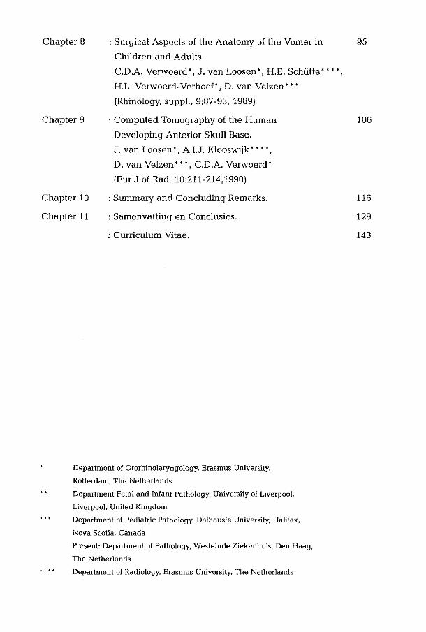
Chapter 8 : Surgical Aspects of the Anatomy of the Vomer in
Children and Adults.
C,D,A. Verwoerd· r J. van Loosen * , H.E. Schutte" ...... ,
H.L, Verwoerd-Verhoef*, D, van Velzen"""
(Rhinology, suppl., 9:87-93, 1989)
Chapter 9 : Computed Tomography of the Human
Developing Anterior Skull Base.
J. van Loosen· r A.I.J. Klooswijk· .... ",
D. van Velzen" .... r C.D.A. Venvoerd"
(Eur J of Rad, 10:211-214,1990)
Chapter 10 : Summmy and Concluding Remarks.
Chapter 11 : Samenvatting en Conclusies.
: Curriculum Vitae.
Department of Otorhinolaryngology, Erasmus University,
Rotterdam, The Netherlands
Department Fetal and Infant Pathology, University of Liverpool,
Liverpool, United Kingdom
Department of Pediatric Pathology, Dalhousie University, Halifax,
Nova Scotia, Canada
Present: Department of Pathology, Westeinde Ziekenhuis, Den Haag,
The Netherlands
Department of Radiology, Erasmus University, The Netherlands
95
106
116
129
143
Introduction.
13
The otorhinolaryngologist who is regularly confronted with nasal
obstruction in children, finds himself supported by sophisticated diagnostic
imaging (e.g. CT- and MRI-scanning) and makes use of similarly sophisticated
surgical approaches and techniques.
It is the latter, which lies at the root of a tendency to carry out surgical
interventions in the nasal region, inclusive of correction of nasal septum
deviation, at an even younger age. This has raised the question for nasal surgery
of the consequences for growth and development of the young and still rapidly
developing midface.
The literature until the present, does not agree on the presence and
extent of the effects of septal surgery for final outgrowth of the nose or midface
(Pirsig, 1974, 1986, Huizing, 1979, Ortiz-Monasterio and Oimedo, 1981, Stucker
et aI., 1984, Healy, 1986, Jugo, 1987, Verwoerd et aI., 1989, Walker et aI., 1993,
Potsic, 1997, Cotton and Myer 1999, Manning, Crysdale, Derkay, 1999).
In part inspired by van Limborgh (1970), as of the seventies, a conside
rable and coherent series of investigations has been carried out using the still
rapidly growing septum of the young rabbit as model (VenV'oerd et aI., 1976,
1979 a,b, 1980, 1991, Nolst Trenite et aI., 1987, 1988, Verwoerd-Verhoef et aI.,
1991). These demonstrated that the growing cartilaginous septum in the rabbit
determines the final heighth and length of the whole of the nose by expansive
growth. For longitudinal growth (in rabbits the anterior-posterior axis of the
nose), continuity of the septal cartilage proved to be essential. The same applies
to the growth in height (Verwoerd et al., 1979b). These findings are clinically
important, as in surgical correction of septal deviation, the continuity of the
septum is disrupted when deviated cartilaginous parts of the nasal septum are
removed and repositioned ..
Wound healing of the nasal septal perichondrium and cartilage in
rabbits were studied (VenV'oerd et aI., 1991 a,b, Venvoerd-Verhoef et al. 1998).
In contrast to the effects of partial septal resection in experimental animals, it
was demonstrated that elevation of the muco-periochondIiulll, either on one or
both sides, exerts no adverse effects on growth as long as there is no direct
damage to the cartilaginous tissues.
14
Resection 01, or injury to parts of the cartilaginous septum can modify
the pattern of septal growth and thus indirectly cause malformations of the bony
structures, even at some distance from the initial lesion (Nolst Trenite et aI.,
1987,1988). The filling of cartilaginous defects by re-implantation of autologous
cartilage may help to prevent septal perforation and the development of ex
cessive scar tissue. This may slightly improve subsequent nasal growth however,
this approach does not result in normalisation of growth as there remains, in
many cases, a component of bending and dislocation of the implanted material
relative to the non-mobilised parts of the septum (Verwoerd and Verwoerd
Verhoef, 1989; Verwoerd et aI., 1991; Verwoerd-Verhoef et aI., 1991; Verwoerd
Verhoef and Verwoerd, 1995).
The septal cartilage together with its bilateral triangular cartilages
approximately forms a T-bar-like three-dimensional anatomic entity, cranially
partially covered by the nasal bones. With regard to bone, perichondrium and
the T-bar-shaped dorsoseptal cartilage (triangular cartilages), the basic
morphology in children and rabbits show striking similarities (Verwoerd et aI.,
1989; Poublon et al.,1990).
During further growth the T-bar shaped cartilaginous structure is under
permanent stress. (Venvoerd et aI., 1989) Any kind of mechanical or surgical
trauma, destroying this entity, initiates an irreversible deviation of the cartilage
from its genetically determinated direction of growth.
In the newborn, the cartilaginous nasal skeleton is far more elaborate
than in the adult and may be considered as an extension of the cartilaginous
anlage of the anterior cranial base (Poublon et aI., 1990).
The importance of the septolateral cartilage (T-bar structure) for midfacial
growth was demonstrated by resection experiments in growing rabbits. The
triangular cartilages proved to be necessary for the normal development of the
nasal bones, the transverse expansion of the dorsal nasal meatus and the nOlmal
development of the nasal turbinates, (Poublon et aI., 1990; Venvoerd-Verhoef
and Venvoerd, 1995).
The cartilaginous septum also had influence on the developing nasal
bones (Verwoerd-Verhoef and Verwoerd, 1995), on the (normal) development of
the maxilla and, to a lesser extent, of other parts of the facial skull (Venvoerd et
aI., 1976, 1979).
15
However, the outgrowth of the midfacial skeleton (inclusive of the nose)
of the rabbit differs in some aspects from the human midfacial development.
With the enlargement of the brain, especially of the frontal region, and
the concomitant rotation of the eyes to the midline, there has been a relative
decrease of the intra-orbital distance in humans compared to that in the rabbit
(Enlow, 1990). This has resulted in a smaller region at the root of the nose and a
shortening of the snout. Thus, humans have close-set eyes and short, narrow
noses that do not interfere ·with binocular vision.
The growth of the frontal lobes have resulted in flexure of the cranial base
(Enlow, 1990). This results in the increased height of the midface, typical for man
and virtually absent in the rabbit (Takahashi, 1988, a,b).
These evolutionary changes have proforma effects on the final shape of the
human septum (Verwoerd and Verwoerd-Verhoef, 1989).
In contrast to the extensive knowledge available with respect to the
anatomy of the adult nasal septum in man (Lang, 1989; Tardy and Brown, 1990),
little is still known about the cartilaginous nasal skeleton in the growing child.
Detailed knowledge of the fine structures of the midface and
cartilaginous components thereof is especially important at young age. For it is
during this period that the cartilaginously developed chondrocranium, of which
the nasal septum is an integral part, shows initial a firm ossification as part of a
process of rapid growth. As knowledge of this period is almost entirely based on
animal data (rabbits), there is a need to detail the developmental anatomy of the
human nasal septum and to assess possible species differences.
In view of the above the following overall questions were raised forming
the aims of the studies reported subsequently in this thesis:
l. Which changes occur in the human nasal septum during the peri- and
later postnatal period specifically of:
a. The anatomy
b. The dimensions
of the various components?
II. What are the possible consequences of the recorded characteristics of
development for the application and interpretation of (imaging) diagnostic
assesment of the nasofrontal region in the growing child?
16
III. What are the possible consequences of septal trauma and surgery for the
recorded characteristics and for developnlent of the nasal skeleton?
After an initial overview of the background literature on the antenatal
anatomy and the available information on the anatomy of the human nasal
septum in Chapter 2, Question I is dealt with in Chapter 3 to 8. Question II is
addressed in Chapter 9, whereas the issues raised in Question III are addressed
in Chapters 4 to 8. A general and integrating discussion of the findings is
presented in Chapter 10.
References
Cotton RT, Myer ChM, 1999, Practical Pediatric Otolaryngology. Lippincolt,
Raven, Philadelphia.
Crysdale, WS, 1999, Septoplasty in Children-Yes, but Do the Right Thing, Arch
Otolaryngol Head Neck Surg, 125:701.
Derkay CS, 1999, A conservative role for septoplasty in young children, Arch
Otolaryngol Head Neck Surg, 125:702-703.
Enlow DH, 1990, Facial Growth, WE Saunders Company, Philadelphia.
Healy GB, 1986, An approach to the nasal septum in Children. Laryngoscope,
96: 1239-1242.
Huizing EH, 1979, Septum surgery in children. Indications, surgical technique
and long-term results. Rhinology, 17: 91-100.
Jugo SB, 1987, Total septum reconstruction through decortication (external)
approach in children. Arch Otolmyngol Head Neck Surg 113: 173-178.
Lang J, 1989, Clinical anatomy of the nose, nasal cavity and paranasal sinuses.
Thieme Verlag, New York.
17
Manning, SC, 1999, A 3-year-old child with a serverely deviated septum and
airway obstruction, Arch Otolaryngol Head Neck Surg, 125:699-700.
Nolst-Trenite GJ, Verwoerd CDA, Verwoerd-Verhoef HL, 1987, Reimplantation
of autologous septal cartilage in the growing nasal septum. I, Rhinology, 25:225-
237.
Nolst-Trenite GJ, Verwoerd CDA, Verwoerd-Verhoef HL, 1988, Reimplantation
of autologous septal cartilage in the growing nasal septum. II, Rhinology, 26:25-
32.
Ortiz-Monasterio, Olmedo A, 1981, Corrective rhinoplasty before puberty. A
long term follow-up. Plast Reconstr Surg, 68: 381-387.
Pirsig W, Knahl R, 1974, Rhinoplasty and the ain'lay in children, Facial Plast
Surg, 3: 225-234.
Pirsig W, 1986, Rhinoplastische Operationen bei Kindem: Erfahrungen an 92
FiiIlen, Laryng Rhinol, 53: 250-265.
Polsie W.P, et aI., 1997, Surgical pediatric otolaryngology, Thieme Verlag, New
York, Stuttgart.
Poublon RML, Verwoerd CDA, Verwoerd-Verhoef HL, 1990, Anatomy of the
upper lateral cartilages in the human newborn. Rhinology, 28:41-46.
Stucker FJ, Bryarly RC, Shocldy WW, 1984. Management of nasal trauma in
children. Arch Otolaryngol Head Neck Surg, 110: 190-192.
Takahashi R, 1988a, The fOlmation of the nasal septum and its evolutionary
paradox. Act Otolaryngol Suppl, 443: 1-160.
Talmhashi R, 1988b, The evolution of the nasal septum and the formation of
seplal deformity. Rhinology, 6: 1-23.
18
Tardy ME, Brown RJ, 1990, Surgical anatomy of the nose. Raven Press, New
York.
Van Limborgh J, 1970, A new view on the control of morphogenesis of the skull.
Acta Morphol Neerl Scand, 8: 143-160.
Verwoerd CDA, Verwoerd-Verhoef HL, Urbanus NAM, 1976, Skulls with facial
clefts, experimental study on the facial skeleton. Acta Otolmyngol, 81: 249-256.
Verwoerd CDA, Urbanus NAM, Verwoerd-Verhoel HL, 1979a, Growth
mechanisms in skulls with facial clefts. Acta Otolaryngol (Stockh), 87: 335-339.
Verwoerd CDA, Urbanns NAM, Nijdam DC, 1979b, The effects of septal
surgery on the growth of nose and maxilla. Rhinology, XVII: 53-63.
Verwoerd CDA, Urbanns NAM, Mastenbroel< GJ, 1980, The influence op
partial resection of the nasal septal cartilage on the growth of the upper jaw and
nose: an experinlental study in rabbits. Clin Otolaryngol, 5: 291-302.
Verwoerd CDA, Verwoerd-Verhoel HL, 1989, Developmental aspects of the
deviated nose. Facial Plast Surg, 612: 95-100.
Verwoerd CDA, Verwoerd-Verhoef HL, Meenwis CA, 1989, Stress and wound
healing in the cartilaginous nasal septum. Acta Otolaryngol (Stockh), 107: 441-
445.
Verwoerd CDA, Verwoerd-Verhoef HL, Meeuwis CA, 1991, Wound healing of
autologous inlplants in the nasal septal cartilage. ORL, 53: 310-314.
Verwoerd-Verhoel HL, Meenwis CA, van del' Henl RO, 1991, Histologic
evaluation of crushed cartilage grafts in the growing nasal septum of young
rabbits. ORL, 53:305-309.
19
Verwoerd-Verhoel HL, Verwoerd CDA, 1995. Sino-nasal surgery and growth:
an experimental study review. Rhinology, state of the art. Eds Tos M, Thomson
J and Balle V, Kugler Publications, Amsterdam/New York: 195-201.
Vel'woerd-Verhoel HL, ten Koppel PGJ, van Osch JVM, Meeuwis CA,
Verwoerd CDA, 1998, Wound healing of cartilage structures in the head and
neck region. Int. J Ped.Otorhinolaryngol, 43:241-251.
Walker PJ, Crysdale WS, Farkas LG, 1993, External septorhinoplasty in
children. Arch Otolaryngol Head Neck Surg, 119: 984-989.
20
Po s t nat a Ide vel 0 P lllent / of the Human Nas/al Skeleton. Li tera ture Review.
21
2.1. Postnatal growth of the human nose
From birth to adulthood the nose. as part of the viscerocranium, grows
and does so over a longer period than the neurocranium, transforming the baby
face into the adult face with prominent nose and jaws. The neurocranium
follows the growth of the brain, which reaches 90% of its adult volume at 6 years
of age (Moore et aI., 19(4). The growth of the viscerocranium does not cease
until 18 to 20 years of age (in fact, some growth may continue) and shows a
number of changes in rate. In the first months after birth the growth rate is high
and decreases slowly until the onset of sexual maturation in the early teens. The
rate of growth then accelerates rapidly (the adolescent growth spurt), reaching
a peak within a year or so and subsequently declining to approximately zero by
about 20 years (Tanner, 1962, Moore et aI., 1974). Consequently, it can take
many years before the effects of trauma or surgery on nasal growth become
evident. Thus, the follow-up of nasal growth after an injury at preschool age has
to be continued until after the adolescent growth spurt. Apart from the extra
growth of the viscerocraniuill, a change in rate of nasal growth around puberty
contributes to the development of the adult profile. During infancy and child
hood, the nose predominantly grows along the anterior-posterior axis. After
puberty, however, the increase in height of the nose S88nlS to be mOTe important
(Imai, 1953).
2.2. Specific anatomy of the infant nose
In the newborn the cartilaginous nasal skeleton is far more elaborate
than in the adult and may be considered as an extension of the cartilaginous
anlage of the anteIior cranial base (Scott, 1953, Ford, 1958, Poublon et al. 1990).
Except for the bony vomer, the neonatal septum is completely cartilaginous.
Superiorly, it nlerges with the cartilage of the crista Gallii posteriorly, it reaches
as far as the partly ossified sphenoid (Scott, 1953, anteriorly it is firmely attached
to the anterior nasal spine (Latham, 1968).
The septal cartilage and upper lateral cartilages are no separate
anatomical entities. They are parts of a T-bar-shaped structure (Poublon et ai.,
1990).
22
The upper lateral cartilages in the newborn are not triangular in shape,
but look like elongated vaults (dorsolateral cartilages) on both sides of the
supra -septal groove. They merge in cephalic direction with the cartilaginous
cranial base and laterally terminate in the nasal wall. The lower alar cartilages
are already found as separate structures. The nasal bones, formed by desmal
ossification, cover the cranial half of the cartilaginous nasal roof (Poublon et ai.,
1990).
The only bony part of the septum at this age, the vomer, is a complex
structure. The alae, formed by desmal ossification at either side of the inferior
part of the septal cartilage (Mall, 1906; Fawcett, 1911; Augier, 1931; Norback
1944) are already well developed. The unpaired lower triangular part is the
product of desmal ossification in the If space I' between the mucous membranes
lining both nasal fossae from the inferior edge of the septal cartilage to the bony
palate. The median inferior part of the vomer is extremely thin in the neonate
and the connection with the palate appears to be weak at dissection. Only
anteriorly is the nasal septum firmly connected to the upper jaw by collagen
fibers between the septal cartilage and the anterior nasal spine.
2.3. Postnatal changes in anatomy of the human nasal skeleton
At birth the cartilaginous nasal skeleton is a T-bar-like structure, exten
ding from the skull base to the tip of the nose (Poublon et ai, 1990). The later
development of this cartilage is characterized by simultaneous processes of
growth, ossification, regression, and remodelling. The cephalic parts of the
dorsolateral cartilages gradually disappear, leaving the comparatively much
smaller upper lateral cartilages in the adult stage. The variable degree of
extension (7 to 20 mm) of the upper laterals under the nasal bones reported in
literature (Straatsma and Straatsma, 1951; Hinderer, 1970) reflects differences in
degree of regression. The rate and mode of regression of the upper lateral
cartilages is not yet known. Incidental observations during surgery show that at
the age of 3 years, the nasal bones are still supported over the full length by the
underlying upper laterals cartilages.
The cartilaginous nasal septum shows growth and ossification at the
23
sanle time. SnIail centers of ossification would be present in the supeIior part of
the septal cartilage at birth and merge as the perpendicular plate, which extends
in posterior-anterior direction (Schultz-Coulon and Eckermeier, 1976). When the
latter reaches the bilateral alae of the bony vomer a posterior-Inferior part of the
septal cartilage is enclosed by bone (Scott, 1953; Moore et aI., 1974; Takahashi,
1988). The cartilage in tllls vomeral tunnel will be replaced by bone or may part
ly survive to adulthood (sphenoid tail) (Melsen, 1977).
The septovomeral junction appears to be highly variable. The superior
part of the vomer follows the frequently occurring deviation of the basal rim of
the cartilaginous septum, whereas the insertion of the inferior part reflects the
line of fusion of the palatal halves. TillS can result in an angle between the
superior and inferior part of the vomer (Takahashi, 1988). Moreover, the ala
vomeris on the convex side of the angle is often defective, so the septal cartilage
will project sidewards.
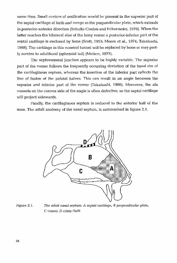
Finally, the cartilaginous septum is reduced to the anterior half of the
nose. The adult anatomy of the nasal septum, is summarised in figure 2.1.
Figure 2.1.
24
The adult nasal septum: A septal cartilage, B perpendicular plate,
C vomer, D crista Galli
References
Augier M, 1931, Squelette cephalique. In: Traite d'anatomie humaine. Poirer F
and Charpey P (eds), Masson et Cie, Paris.
Fawcett M, 1911, The development of the human maxilla, vomer and paraseptal
cartilages. J Anat Lond, 45:378-405.
Ford ERH, 1958, Growth of the human cranial base. Am J Orthod, 44: 499-506
Hinderer KH, 1970, Fundamentals of Anatomy and Surgery of the Nose. AI:
Aesculapius Publishing, Birmingham.
Imai S, 1953, Jikei Med J, 67: 106-115 (In Japan, cited in Takahashi, 1988, Acta
Otolaryngol [Suppl] (Stockh) 43:1-160.
Mall FF, 1906, On ossification centres in human emblYos less than one hundred
days old, Am J Anat, 5:433-458.
Melsen B, 1977, The nasal septum. Angle Orthod, 47:83-96.
Moore WJ, Lavelle CLB, 1974, Growth of Facial Skeleton in Hominoidea.
Academic Press r London,
Norback CR, 1944, The developmental anatomy of the human osseous skeleton
during the embryonic, foetal and circumnatal periods. Anat Rec, 88: 91-126.
Poublon RML, Verwoerd CDA, Verwoerd-Verhoei HL, 1990, Anatomy of the
upper lateral cartilages in the human newborn. Rhinology, 28:41-46.
Schultz-Coulon HJ, Ec\{ermeier L, 1976, Zum postnataIen Wachstum der
Aussennase. Acta Otolaryngol., 82:131-142.
Scott JH, 1953, The cartilage of the nasal septum. Br Dent J, 95:37-43.
25
Straatsma BR and Straatsma CR, 1951, The anatomical relationship of the lateral
nasal cartilage to the nasal bone and the cartilaginous nasal septum. Plast &
Reconstr Surg, 8: 443-455.
Takahashi R, 1988, The evolution of the nasal septum and the formation of septal
deformity. Rhinology Suppl, 6: 1-23.
Tanner JM, 1962, Growth at Adolescence. Blackwell, Oxford.
26
The Nasal Septal Cartilage in the Newborn.
21
3.1 Introduction
In neonates and young children it is well known that the nasal septum
is mainly cartilaginous (Cleland. 1861, Hillenbrand, 1933, Scott, 1953).
However, the morphological and histological characteristics of this septal
cartilage at a young age have scarcely been studied.
In rabbits it has been demonstrated that the cartilaginous septum shows a
specific pattern of regional differences in thickness and histological
differentiation (Meeuwis et aI, 1985, Verwoerd et aI, 1991).
The aim of the study was to investigate whether such a pattern of
regional differences also occurs in hunlan neonates.
3.2 Materials and Methods
The nasal septum of 3 normally developed human neonates without
signs of congenital malformations and stillborn after a pregnancy of 36, 38
and 42 weeks respectively, were exanlined.
After block dissection described by Melsen (1977), including the nasal
septum, part of the hard palate, the cribriform plate and the sphenoid bone,
semi -serial sections (5 micron) were cut in the transverse and frontal plane
and stained with haematoxylin and azophloxin. Using a photographic lateral
view of the prepared septum (Figure 3,1). semi-serial 5 micron sections
spaced at 1 mm, were used for a three-dimensional reconstruction (magnifi
cation factor 10 x).
The thickness measured in the cartilage is represented on a shading
coded diagram of the reconstruction (Figure 3.2).
3.3 Results
The thickness of the cartilage of each septum varies considerably:
from 400 micron in the anterior area to 3500 micron in the posterior region
(Figure 3.2). The cartilage increases in thickness from the anterior free ridge
28
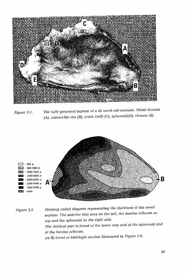
Figure 3.1.
CJ - 500 JI
C!';J - 500-1 000 })
.. - 1000-1500.u
II1II - 1500-2000 IJ
l1l'i - 2000-2500 .u _ - 2500-3000 IJ
_ - 3000-3500 IJ
li..'\l . bone
Figure 3.2
The fully prepared septum of a 42 week-aId-neonate. Nasal dorsum
(A), columellar rim (B), crista Galli (C), spllenoid(D), choana (E).
Slwding-coded diagram representing the thickness of the nasal
septum. The anterior thin area on the len the lamina cribrosa on
top and the sphenoid on the right side.
.B
The thickest part is found at the lower rear end of the sphenoid and
at the lamina cribrosa.
(A-B) Level of histologic section illustrated in Figure 3.6.
29
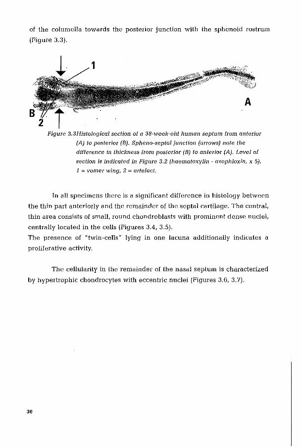
of the columella towards the posterior junction with the sphenoid rostrum
(Figure 3.3).
Figure 3.3Hist%gical section of a 38-week-old human septum from anterior
(A) to posterior (B). Spheno-septoJ junction (arrows) note tile
difference in thickness from posterior (B) to anterior (AJ. Level of
section is indicated in Figure 3.2 (haematoxylin - azophloxin, x 5).
1 = vomer wing, 2 = artefact.
In all specimens there is a significant difference in histology between
the thin part anteriorly and the remainder of the septal cartilage. The central,
thin area consists of small, round chondroblasts with prominent dense nuclei,
centrally located in the cells (Figures 3.4, 3.5).
The presence of "twin -cells" lying in one lacuna additionally indicates a
proliferative activity.
The cellularity in the remainder of the nasal septum is characterized
by hypertrophic chondrocytes with eccentric nuclei (Figures 3.6, 3.7).
30
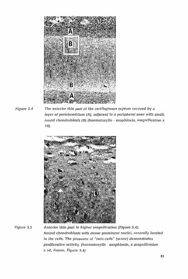
Figure 3.4
Figure 3.5
The anterior thin part of the cartilaginous septum covered by a
layer of perichondrium (A), adjacent to a peripheral zone with sman
round c1lOndroblasts (B) (haemaloxylin - azophloxin, magnification x
10).
Anterior thin pari in higher magnification (Figure 3.4).
Round cholldroblasts with dense prominent nuclei, centrally located
in the cells. The presence of "twin-cells" (arrow) demonstrates
proliferative activity. (lwematoxylin - azophloxin, x magnification
x 50, Frame, Figure 3.4)
31
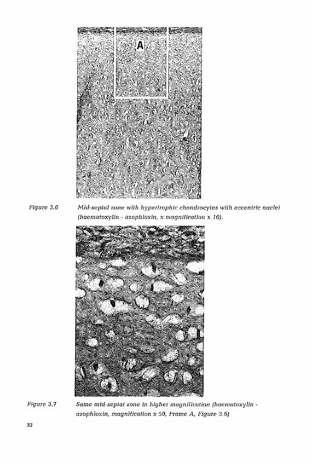
Figure 3.6
Figure 3.7
32
Mid-septal zone witlillypertropllic chondrocytes with eccentric nuclei
(haematoxylin - azopliloxin, x magnification x 10).
Same mid-septal zone in higher magnification (lwematoxylin -
azopliloxin, magnification x 50, Frame A, Figure 3.6)
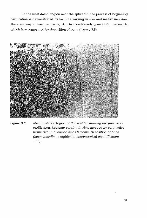
In the lllOst clorsul region near the sphenoid, the process of beginning
ossification is demonstrated by lacunae varying in size and lnatrix invasion.
Bone nlalTOW connective tissue, rich in blooclvessels grows into the lllatrix
which is accompanied by deposition of bone (Figure 3.8).
Figure 3.8 Most posterior region of tbe septum sbowing tbe process of ossification. Lacunae varying in size, invaded by connective
tissue ricb in lwemopoietic elements. Deposition of bone (haematoxylin - azophloxin, microscopical magnification
x 10).
33
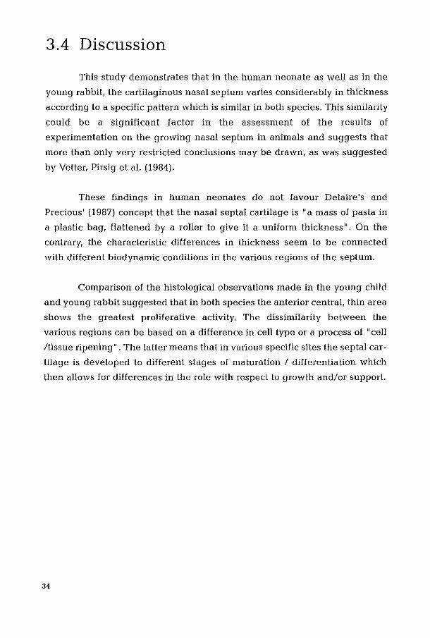
3.4 Discussion
This study demonstrates that in the human neonate as well as in the
young rabbit, the cartilaginous nasal septum varies considerably in thickness
according to a specific pattern which is similar in both species. This similarity
could be a significant factor in the assessment of the results of
experimentation on the growing nasal septum in animals and suggests that
more than only very restricted conclusions may be drawn, as was suggested
by Vetter, Pirsig et al. (1984).
These findings in human neonates do not favour Delaire's and
Precious' (1987) concept that the nasal septal cartilage is "a mass of pasta in
a plastic bag, flattened by a roller to give it a uniform thickness". On the
contrary, the characteristic differences in thickness seem to be connected
with different biodynamic conditions in the various regions of the septum.
Comparison of the histological observations made in the young child
and young rabbit suggested that in both species the anterior central, thin area
shows the greatest proliferative activity. The dissimilarity between the
various regions can be based on a difference in cell type or a process of "cell
ltissue ripening". The latter means that in various specific sites the septal car
tilage is developed to different stages of maturation I differentiation which
then allows for differences in the role with respect to growth andlor support.
34
References
Cleland J, 1861, On the relations of the vomer, ethmoid and intermaxillary
bones. Philos Trans, 152:289-321.
Delaire J, Precious D, 1987, Interaction of the development of the nasal
septum, the nasal pyramid and the face. Int J Ped Otorhinolaryngol, 12:311-
326.
Hillenbrand K, 1933, Entwicklung, Bau und Form Veranderungen der
menschlichen Nasenscheidewand in fetalen Leben. Arch Ohr-Nasen Kehlk
I-Ieilk, 1-24.
Meeuwis CA, Verwoerd-Verhoef HL, Heul RO van del', Verwoerd CDA,
1985, Morphological changes of the cartilaginous nasal septum after partial
resection in young rabbits. Clin Otolaryngol, 10:231-232.
Melsen B, 1977, The nasal septum. The Angle Orthod, 47:83-96.
Scoll JH, 1953, The cartilage of the nasal septum. Brit Dent J, 95:37-43.
Verwoerd CDA, Verwoerd-Verhoef HL, Meeuwis CA, van del' Heul RO,
1991, Wound healing of autologous implants in the nasal septal cartilage.
OR L, 53:310-314.
Veller U, Pirsig W, Helbing G, Heit W, Heinze E, 1984, Patterns of growth in
human septal cartilage, a review of new approaches. Int J Ped Otorhino
laryngol,7:63-74.
35
G row the h a rae t e r is Ii c s of the Human Nasal Septum.
36
4.1 Introduction
Effective nasal surgery is highly dependent on a comprehensive
knowledge of the anatomy, physiology, and biodynamics of the mid-facial
skeleton. The influence of the nasal septum on the growth of the midfacial
skeleton has been debated for more than a century. Recently, however, long
term follow-up studies in the rabbit have clearly demonstrated an important role
of the dorsoseptal cartilage with respect to the normal development of the nasal
bones, (pre-) maxilla and orbit, which is responsible for the postnatal changes of
the facial profile (Verwoerd et aI., 1995; Verwoerd-Verhoef et aI., 1995).
Although much has been published since the introduction of radiographic
cephalometry by Broadbent (1931) and Hofrath (1931) on the postnatal
development of the human cranium, very little is known of the growth and
development of the human nasal septum.
Schultz-Coulon and Eckermeier (1976) studied the postnatal changes in
the human nasal septum from the neonatal period to ten years of age. In this
study they multiplied height with length to obtain the lateral surface area of the
nasal septum. They demonstrated a rapid decrease of nasal septal growth after
birth and an extensive ossification process during the first 10 years of life.
No growth features of the human nasal septum, however, have been
described after the age of 10 years. In view of the fact that many surgeons prefer
to postpone corrective nasal surgery until after the pubertal growth spurt, data
on postpubertal growth are very inlPortant. Growth characteristics of the bony
and cartilaginous part of the septum, respectively, are not available in the
literature. The purpose of this study is to analyze the growth characteristics of
the separate parts of the human nasal septum from birth until the post-adoles
cent period.
In the adult, the nasal septum is composed of three parts: (1) the
cartilaginous septum; (2) the perpendicular plate; and (3) the vomer. The
perpendicular plate is the product of endochondral ossification of the
cartilaginous septum during childhood. The vomer is formed by intra
membranous ossification. In the neonate nearly all of the nasal septum is
cartilaginous. The septal cartilage extends from the columella anteriorly to the
sphenoid posteriorly, where it merges cranially with the cartilaginous" anlage"
of the anterior cranial base.
37
The vomer is represented in the neonate by a thin bony lamella between
the basal rim of the cartilaginous septum and palate, This inferior part of the
vomer shows extensions on both sides of the cartilaginous septum, These
"vomer blades" merge with the ossifying perpendicular plate (Verwoerd et aI"
1989),
This study deals with the cartilaginous septum and the perpendicular
plate and does not include the inferior part of the vomer which constitutes a
minor, morphogenetically not related part of the septum, The immediate reason
is that during preparation of the specimens a variable part remains fixed to the
palate, whereas the other parts of the septum were obtained in their entirety.
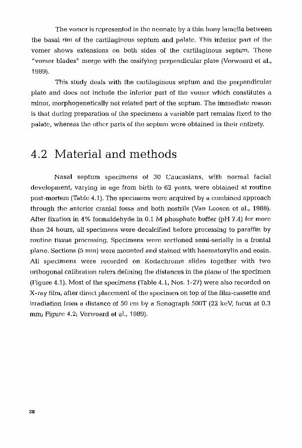
4.2 Material and methods
Nasal septum specinlens of 30 Caucasians, ·with normal facial
development, varying in age from birth to 62 years, were obtained at routine
post-mortem (Table 4.1). The specimens were acquired by a combined approach
through the anterior cranial fossa and both nostrils (Van Loosen et aI" 1988).
After fixation in 4% formaldehyde in 0.1 M phosphate buffer (pH 7.4) for more
than 24 hours, all specimens were decalcified before processing to paraffin by
routine tissue processing. Specimens were sectioned semi-serially in a frontal
plane. Sections (5 mm) were mounted and stained with haematoxylin and eosin.
All specimens were recorded on Kodachrome slides together with two
orthogonal calibration rulers defining the distances in the plane of the specimen
(Figure 4.1). Most of the specimens (Table 4.1, Nos. 1-27) were also recorded on
X-ray film, after direct placement of the specinlen on top of the film-cassette and
irradiation from a distance of 50 cm by a Senograph 500T (22 keY, focus at 0.3
mm, Figure 4.2, Verwoerd et aI" 1989).
38
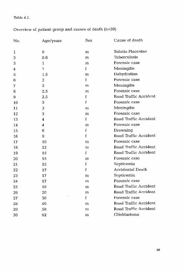
Table 4.1.
Overview of patient group and causes of death (n=30j.
No. Age/years Sex Cause of death
1 0 m Solutio Placentae
2 0.6 m Tuberculosis
3 1 m Forensic case
4 1 f Meningitis
5 1.5 m Dehydration
6 2 f Forensic case
7 2 m Meningitis
8 2.5 m Forensic case
9 2.5 f Road Traffic Accident
10 3 f Forensic case
11 3 m Meningitis
12 3 m Forensic case
13 4 f Road Traffic Accident
14 4 m Forensic case
15 8 f Drowning
16 9 f Road Traffic Accident
17 10 m Forensic case
18 12 m Road Traffic Accident
19 15 f Road Traffic Accident
20 15 m Forensic case
21 15 f Septicemia
22 17 f Accidental Death
23 17 m Septicemia
24 17 m Forensic case
25 18 m Road Traffic Accident
26 20 m Road Traffic Accident
27 30 f Forensic case
28 40 m Road Traffic Accident
29 58 m Road Traffic Accident
30 62 m Glioblastoma
39
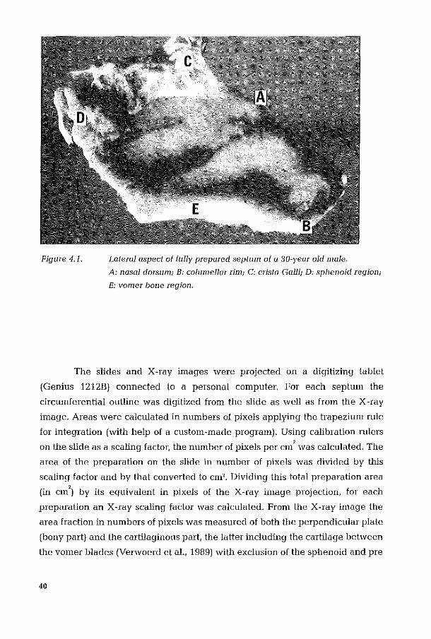
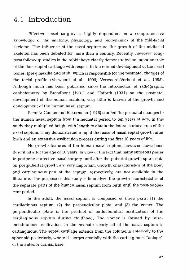
Figare 4.1. Lateral aspect of fully prepared septum of a 30-year old male.
A: nasal dorsum; B: COlllIllellar rim; C: crista Galli; D: sphenoid regioll;
E: vomer bone region.
The slides and X-ray images were projected on a digitizing tablet
(Genius 1212B) connected to a personal computer. For each septum the
circumferential outline was digitized from the slide as well as from the X-ray
image. Areas were calculated in numbers of pixels applying the trapeziullI rule
for integration (with help of a custom-made program). Using calibration rulers , on the slide as a scaling factor, the number of pixels per enl was calculated. The
area of the preparation on the slide in number of pixels was divided by this
scaling factor and by that converted to cm'. Dividing this total preparation area ,
(in cm) by its equivalent in pixels of the X-ray image projection, for each
preparation an X-ray scaling factor was calculated. From the X-ray image the
area fraction in numbers of pixels was measured of both the perpendicular plate
(bony part) and the cartilaginous part, the latter including the cartilage between
the vomer blades (Verwoerd et aI., 1989) with exclusion of the sphenoid and pre
40
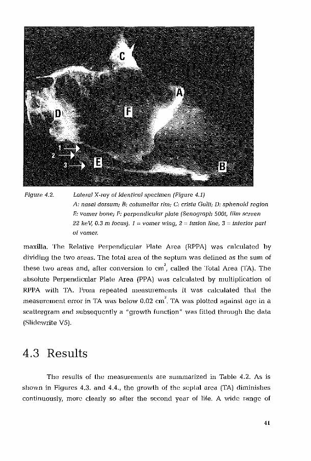
Figure 4.2. Lateral X-ray of identical specimen (Figure 4.1)
A: nasal dorsum; B: columellar rim; C: crista Galli; D: sphenoid region
E: vomer bone; F: perpendicular plate (Senograph SOOt, film screen
22 keY, 0.3 m focus). 1:;: vomer wing, 2:;: tusionline, 3:;: interior part
at vomer.
maxilla. The Relative Perpendicular Plate Area (RPPA) was calculated by
dividing the two areas. The total area of the septum was defined as the sum of . ,
these two areas and, after converSIOn to em , called the Total Area (TA). The
absolute Perpendicular Plate Area (PPA) was calculated by multiplication of
RPPA with TA. From repeated measurements it was calculated that the ,
measurement error in TA was below 0.02 em . TA was plotted against age in a
scattergram and subsequently a "growth function" was fitted through the data
(Slidewrite V5).
4.3 Results
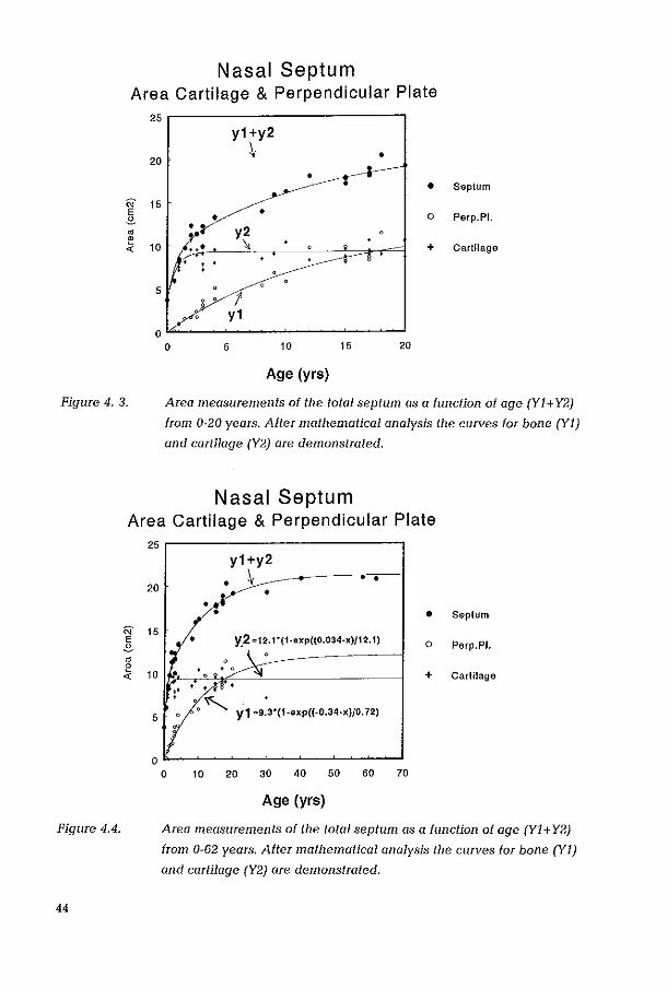
The results of the measurements are summarized in Table 4.2. As is
shown in Figures 4.3. and 4.4., the growth of the septal area (TA) diminishes
continuously, more clearly so after the second year of life. A wide range of
41
mathematical functions were tried, with help of a commercially available PC
program (Slide Write Plus for Windows Version 3.00, Advance Graphics
Software, Carlsbad, CA, USA) Curve fitting with this program includes linear
regression for linear function fitting and Levenberg-Marquardt fitting algorithm
for non-linear functions.
It appeared that total septum area data were described with the least amount of
error by a summation of two similar simple growth functions, Yl and Y2 in
Figures 4.3. and 4.4.
The standard error of the fit is 0.8 cm'. Given the much smaller error in
the area meaSllrenlents mentioned above, we conclude that the remaining
spread of the data around the curve Yl + Y2 is due to interindividual anatomical
variation and that the present data do not allow for assessment of more complex
growth functions. The functions Yl and Y2, as best fitting algorithms, to define
the area growth of each of the separate parts of the septum. Yl describes the
growth of the cartilaginous part including the cartilaginous part between the
vomer blades and concomitantly, Y2 that of the perpendicular plate. From the
parameters of the Yl function it could be surmised that the growth of the carti
laginous part starts at about -0.34 year, i.e. at about 4 months of gestation.
Furthermore, the tinle-constant of cartilage growth is 0.72 year, which means
that by 2.2 year the area has reached 95 % of its final value. Sinlilarly, from Y2 it
can be concluded that growth of the bony part starts at about term (0.034 year)
and has reached 95% of the final area at 36 years of age. An inlpression of the
growth rate of the various areas is obtained by taking the derivative of function
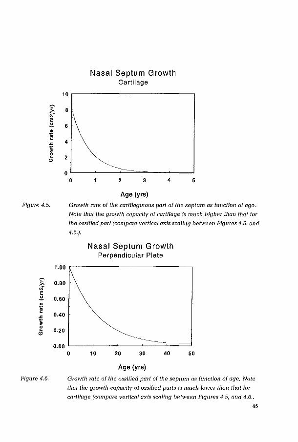
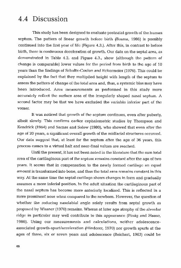
Yl and Y2 (Figures 4.5. and 4.6.). Although sinlilar in the pattern of age asso
ciated deceleration of growth, the overall capacity for growth of the cartilage is
far greater than that for the ossified part of the septum.
42
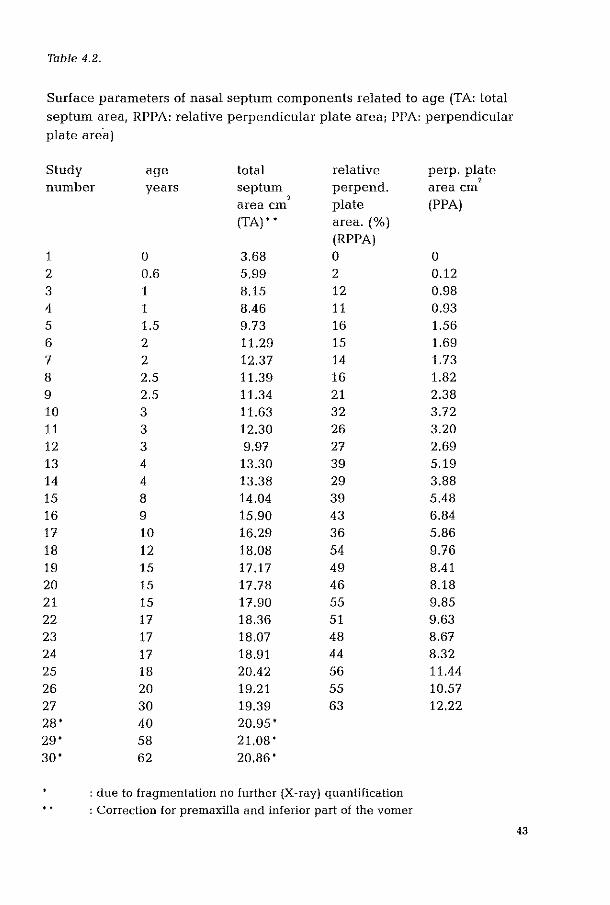
Table 4.2.
Surface parameters of nasal septum components related to age (TA: total septum area, RPPA: relative perpendicular plate area, PPA: perpendicular plate are'a)
Study age total relative perp. plate number years septum perpend. area em
, ,
plate (PPA) area em (TA) , , area. (%)
(RPPA) 1 0 3,68 0 0 2 0.6 5.99 2 0.12 3 1 8.15 12 0.98 4 1 8.46 11 0.93 5 1.5 9.73 16 1.56 6 2 11.29 15 1.69 7 2 12,37 14 1.73 8 2.5 11.39 16 1.82 9 2,5 11.34 21 2.38 10 3 11.63 32 3,72
11 3 12,30 26 3,20
12 3 9,97 27 2,69
13 4 13,30 39 5,19 14 4 13,38 29 3,88 15 8 14,04 39 5.48 16 9 15,90 43 6.84 17 10 16.29 36 5.86 18 12 18.08 54 9.76 19 15 17.17 49 8.41 20 15 17.78 46 8.18 21 15 17.90 55 9.85 22 17 18.36 51 9,63 23 17 18,07 48 8,67 24 17 18,91 44 8,32 25 18 20.42 56 11.44 26 20 19,21 55 10,57
27 30 19,39 63 12,22 28' 40 20,95 ' 29' 58 21.08 ' 30' 62 20,86'
: due to fragmentation no further (X-ray) quantification
" : Correction for premaxilla and inferior part of the vomer
43
Nasal Septum Area Cartilage & Perpendicular Plate
25r-------------------------,
y1+y2
20 ~ •
• • Septum
N 15 E • • 0 Perp.PI. .£.
• y2 • ;; 10 " , + Cartilage
5 /'
y1 0
0 5 10 15 20
Age (yrs)
Figure 4.3. Area measurements of the total septum as a function of age (Yl+Y2)
from 0-20 years. After mathematical analysis the curves for bone (Y1)
and curtilage (Y2) are demonstrated.
Figure 4.4.
44
Nasal Septum Area Cartilage & Perpendicular Plate
N E .£.
• ~ -<
25r-------------------------,
20
15
10
5
y1+y2
• \ • •
• • • Septum
• 0 Perp.PL
+ CarllIage
• "" Y1 0 ':3'(1 0'XP«00.34'X)/0.72)
o 10 20 30 40 50 60 70
Age (yrs)
Area measurements of the total septum as a function of age (Yl+Y2)
from 0-62 years. After mathematical analysis the curves for bone (YJ)
and cartilage (Y2) are demonstrated.
'i:' ~
'" E .!!. " ~ ~
.<: ~
~ 0 ~
(!J
10
8
6
4
2
0 0
Nasal Septum Growth Cartilage
2 3 4
Age (yrs)
5
Figure 4.5. Growth rate of the cartilaginous part of lhe septum as function of age.
Note that the growth capacity of cartilage is much higher than that for
the ossified part (compare vertical axis scaling between Figures 4.5. and
1.00
~ 0.80
'" E .!!. 0.60
" ~ ~
0.40 .<: ~
~ ~ 0.20
(!J
0.00
Figure 4.6.
4.6.).
Nasal Septum Growth Perpendicular Plate
0 10 20 30 40 50
Age (yrs)
Growth rate of the ossified part of the septum as function of age. Note
that the growth capacity of ossified parts is mllch lower than that for
cartilage (compare vertical axis scaling between Figures 4.5, and 4.6 ..
45
4.4 Discussion
This study has been designed to evaluate postnatal growth of the human
septum. The pattern of linear growth before birth (Bosma, 1986) is possibly
continued into the first year of life (Figure 4.3.). After this, in contrast to before
birth, there is continuous deceleration of growth. Our data on the septal area, as
demonstrated in Table 4.2. and Figure 4.3., show (although the pattern of
change is comparable) lower values for the period from birth to the age of 10
years than the findings of Schultz-Coulon and Eckermeier (1976). This could be
explained by the fact that they multiplied height with length of the septum to
assess the pattern of change of the total area and, thus, a systemic bias may have
been introduced. Area measurements as performed in this study more
accurately reflect the surface area of the irregularly shaped nasal septum. A
second factor may be that we have excluded the variable inferior part of the
vomer.
It was noticed that growth of the septum continues, even after puberty,
albeit slowly. This confirms earlier cephalometric studies by Thompson and
Kendrick (1964) and Sarnas and Solow (1980), who showed that even after the
age of 20 years, a significant overall growth of the midfacial structures occurred.
Our data suggest that, at least for the septum after the age of 36 years, this
process comes to a virtual halt and near-final values are reached.
Until the present, it has not been noted in the literature that the sum total
area of the cartilaginous part of the septum remains constant after the age of two
years. It seems that in compensation to the newly fonned cartilage an equal
amount is transfOffiled into bone, and thus the total area remains constant in this
way. At the same time the septal cartilage shows changes in form and gradually
assumes a more inferial position. In the adult situation the cartilaginous part of
the nasal septum has become more anteriorly localized. This is reflected in a
more prolllinent nose when compared to the newborn. However, the question of
whether the reducing nasolabial angle solely results from septal growth as
proposed by Wissner (1970) remains. Wheras at later age atrophy of the alveolar
ridge in particular may well contribute to this appearance (Pirsig and Haase,
1986). Using our measurements and calculations, neither adolescence
associated growth-spurt/acceleration (Hinderer, 1970) nor growth spurts at the
ages of three, six or seven years and adolescence (Reichert, 1963) could be
46
demonstrated for the human nasal septum. Being a transversal study,
differences as small as those noted in optimalized longitudinal cephalometric
investigations (Pirsig and Haase, 1986) cannot be detected, especially since such
small accelerations are known to vary greatly in age of onset (Rosenberger,
1934, Bergersen, 1972). The study of such individual accelerations additionally
should take into account besides sex, age-dependency differences (Riolo et aI.,
1974; Prahl-Andersen et al., 1979; Engel et aI., 1994).
Thus, as this investigation was of the transversal type, it does not
completely exclude the existence of growth spurts. However, the narrow spread
of the data around the regression line makes it improbable that significant
deviations from the average growth pattern exist. Interindividual variation in
time of onset and in size may well have been obscured in the present data set.
Therefore, a longitudinal study, with regularly-spaced metallic implants in the
nasal septum of an expelimental animal as used by Bjork (1955) for human skull
growth, may be required to finally clarify any remaining ambiguity.
In summary, the results of this study demonstrate that:
(1) the growth rate of the nasal septum is highest in the newborn and
slows down continuously, more clearly after the second year of life, but
continues even after puberty;
(2) the cartilaginous part of the nasal septum increases rapidly in sagittal
dimensions during the first years of life. Alter the age of two years the total area
of the cartilaginous septum Ienlains constant;
(3) endochondral ossification of the cartilaginous septum, resulting in
the formation of the perpendicular plate, starts after the first half-year of life. The
expansion of the perpendicular plate in the sagittal plane continues until after
puberty;
(4) the development of the cartilaginous nasal septum after the age of
two years is characterized by: (a) a balance between new formation of cartilage
and loss of cartilage by the process of endochondral ossification, and (b) a
constant remodelling and gradual shift to a relatively more anterior position;
47
References
Bergersen EO, 1972, The male adolescent facial growth spurt: Its prediction and
relation to skeletal maturation. Angle Orthod, 42: 319-338.
Bjiirl< A, 1955, Facial growth in man, studied with the aid of metallic implants.
Acta Odontol Scand, 13: 9-34.
Bosma JF, 1986, Anatomy of the Infant Head, John Hopkins University Press,
Boston.
Broadbent BH, 1931, A new X-ray technique and its application to orthodontia.
Angle Orthod, 1: 45-66,
Engel T, Van Loosen J, Van Zanten GA, Howard CV, Van Velzen D, 1994,
Postnatal growth of the craniofacial skeleton: A transversal cephalometric study.
Proc VIth Asean ORL Congress, Chiang Rai.
Hinderer KH, 1970, Fundamentals of Anatomy and Surgery of the Nose.
Aescolapius Publishing, Birmingham.
Hofrath TH, 1931, Die Bedeutung der Riintgenfern- und Abstand-Aufname fur
die Diagnostiek der Kiefer Anomalien. Fortschr Orthod, 1: 232-258.
Pirsig W, Haase S, 1986, Phasen des postnatalen Nasenwachstums: Ein
kritisches Obersicht. Laryng Rhinol Otol, 65: 243-249.
Prahl-Andersen B, Kowalski CJ, Heydendael PHJM, 1979, A Mixed
Longitudinal InterdisciplinaIY Study of Growth and Development. Academic
Press, New York.
Reichert H, 1963, Plastic surg81Y of the nose in children. Plast Reconstr Surg,
31: 51-56.
48
Riolo ML, Moyers RE, McNamara, JA, Stuart-Hunter W, 1974, An Atlas of
Craniofacial Growth. Center for Human Growth and Development, University of
Michigan, Ann Arbor.
Rosenberger HC, 1934, Growth and development of the naso-respiratory area in
childhood. Ann Otol Rhinol, 43: 495-512.
Sarnas KV, Solow B, 1980, Adult changes in skeletal and soft tissue profile. Em
J Orthod, 2: 1-12.
Schultz-Coulon HJ, Eckenneier L, 1976, Zum postnatalen Wachstum der
Nasenscheidewand. Acta Otolaryngol (Stockh), 82: 131-142.
Thompson JL, Kendrick GS, 1964, Changes in vertical dimensions of the human
male skull during the third and fourth decade of life. Anat Rec, 150: 209-214.
Van Loosen J, Verwoerd-Verhoef HL, Verwoerd CDA, 1988, The nasal septum
cartilage in the newborn. Rhinology, 26: 161-165.
Verwoerd CDA, Van Loosen J, Schiitte HE, Verwoerd-Verhoef HL, van Velzen
D, 1989, Surgical aspects of the anatomy of the vomer in children and adults.
Rhinology Suppl, 9: 87-93.
Verwoerd CDA, Mladina R, Nols! Trenite GJ, Pigott RW, 1995, The nose in
children with unilateral cleft lip and palate. Int J Ped Otorhinolaryngol Suppl,
32: 45-53.
Verwoerd-Verhoef HL, Verwoerd CDA, 1995. Sino-nasal surgery and growth:
an experimental study review. Rhinology, state of the art. Eds Tos lvI, Thomson
J and Balle V, Kugler Publications, Amsterdam/New York: 195-201.
Wissner G, 1970, Alters Veriinderungen von Gesichts- und Ohlmerkmalen.
Anthrop Anz, 32: 157-193.
49
The Significance of Regional Variations in Thickness of the Human Nasal Septum.
50
5,1 Introduction
The central role of the nasal septum, as a component of the load-bearing
ability of the nasal pyramid, has been recognised by a number of authors as
early as the fifth decade of this centmy (Riggs, 1953, Rubenstein, 1956, Luongo
et aI., 1958). The existence of interlocked stresses in the nasal septum was
demonstrated ex-vivo by Fry (1966, 1967) and in-vivo by Verwoerd et al. (1989
a,b, 1991, 1998). The importance of the central strut in the resistance model was
highlighted by Clarke (1967). It was later emphasized that the nasal septum and
the upper lateral cartilages form a T-bar shaped structure, which supposedly
has a greater mechanical strength than a 2-dimensional septum (Venvoerd et
aI., 1989b, 1998). At birth the cartilaginous nasal septum reaches from columella
to sphenoid and anteIior skull base. The perpendicular plate has not yet been
formed.
Van Loosen et al. (1988) demonstrated the regional differences in thick
ness of the human cartilaginous nasal septum in the neonatal period. In this
study it was shown that the thickness of cartilage varies considerably from 400
lllicron thickness in the anterior area to 3500 micron in the posterior region.
These variations mimic the pattern previously obs81ved in the rabbit (Tonneyck
Muller et aI., 1984, Verwoerd et aI., 1991). Variations in septal thickness are
scarcely mentioned in literature and when available, show considerable
differences amongst authors. For example Kowatscheff (1943) reported that the
posterior segment of the septal cartilage in the newborn and at'the age of three
months are similarly dimensioned as in adults: 2.1 - 2.5 mm (2100 - 2500 micron).
Zuckerkandl (1892) pictured the anterior-inferior part of the septum as an
"expansion" of 4 - 8 nun (4000-8000 micron). Cottle et al. (1958) stated that 3 -
4 mm (3000-4000 micron) is the width of the septal cartilage over wide areas.
Lang (1989) described the septal cartilage as a 3 - 4 nml (3000-4000 micron)
thick structure, but showed in his frontal sections of the adult nasal septum more
specific regional variations in thickness. without nlentioning then1. Delaire and
Precious (1987) also considered the nasal septum as a segment of cartilage with
a unifornl thickness.
As yet, a detailed study of the possible presence and extent of regional
variations in thickness between birth and adulthood is absent in literature.
51
Knowledge of this architecture of the cartilaginous septum would lead to a
better understanding of biomechanical properties and growth of the septum.
Van Loosen et a!. (1988) demonstrated that at birth the nasal septum,
except for the vomer' anlage', is completely cartilaginous. In the second half of
the first year of life, the septum progressively ossifies in posterior-anterior
direction by a process of endochondral ossification (Schultz-Coulon et a!., 1976).
Consequently, the cartilaginous structure changes into a partially osseous
(perpendicular plate) and partially cartilaginous, composite structure. Each of
the components may well have its own range of sagittal dimensions related to
age and growth, as was reported earlier (van Loosen et a!., 1996). Therefore, the
aim of this study is:
1. To describe the development of the regional variations in thickness of
the human cartilaginous nasal septum in relation to age.
2. To discuss the hypothetical effects of these differences in relation to
growth of the nose.
5,2 Materials and methods
Human nasal septa were collected by block dissection at post-mortem
from patients having died from intercurrent disease not affecting the nasal struc
tures. Eight patients, varying in age from newborn (38 weeks gestation) to 42
years of age were included in the study.
The specimens were fixed in 0.1 M phosphate buffered, pH 7.4, 4 %
formaldehyde for a minimum of 2 x 24 hours. After decalcification, specimens
were embedded in paraffin using routine tissue processing methods. Specimens
were semi-serially sectioned in a frontal plane, and consecutive 5 micron
sections, stained with Haematoxylin and Eosin, were prepared every 2.5 DInI for
the entire length of the block specinlen.
Slides were projected using a standard slide projector on a paper screen
after calibration. Three-dimensional representations were reconstructed as
published previously (van Loosen et a!., 1988). Camera lucida drawings at 1:200
lllagnification were used for the 3-dinlensional reconstruction (magnification
x10) of individual septa. The thickness measured in various areas of the septum
were represented on a grid-coded diagram. The changes with age for the
S2
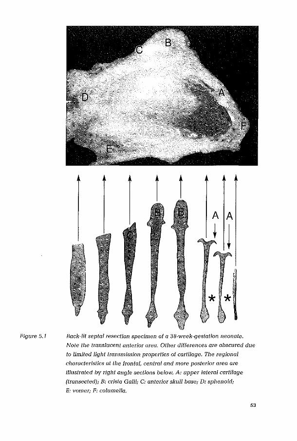
Figure 5.1
r t A
t
* * Back-lit septal resection specimen of a 38-week-gestation neonate.
Nole lhe translucent anterior area. Other differences are obscured due
to limited light transmission properties of cartilage. The regional
characteristics at the frontal, central and more posterior area are
illustrated by right angle sections below. A: upper lateral cartilage
(transected); B: crista Galli; C: anterior skull base; D: sphenoid;
E: vomer; F: columella.
53
different regions were studied by systematic comparison of the results for each
case (Figure 5.1).
5.3 Results
The terminology used in the description relates to the septum of the
head in the upright position; thus there are ant8Iior. posterior, inferior and
superior parts.
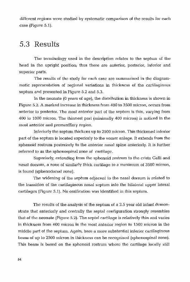
The results of the study for each case are summarised in the diagram
lllatic representation of regional variations in thickness of the cartilaginous
septum and presented in Figure 5.2 and 5.3.
In the neonate (0 years of age). the distribution in thickness is shown in
Figure 5.2. A marked increase in thickness from 400 to 3500 micron, occurs from
anterior to posterior. The most anterior part of the septum is thin, varying from
400 to 1000 micron. The thinnest part (minimally 400 micron) is noticed in the
most anterior and premaxillary region.
Inferiorly the septum thickens up to 2500 micron. This thickened inferior
part of the septum is located superiorly to the vomer anlage. It extends from the
sphenoid rostrum posteriorly to the anterior nasal spine anteriorly. It is further
referred to as the sphenospinal zone of cartilage.
Superiorly, extending from the sphenoid rostrum to the crista Galli and
nasal dorsum, a zone of similarly thick cartilage to a maximum of 3500 micron,
is found (sphenodorsal zone).
The widening of the septum adjacent to the nasal dorsum is related to
the transition of the cartilaginous nasal septum into the bilateral upper lateral
cartilages (Figure 5.1). No ossification was identified in this septum.
The results of the analysis of the septum of a 2.5 year old infant demon
strate that anteriorly and centrally the septal configuration strongly resembles
that of the neonate (Figure 5.2). The septal cartilage is relatively thin and varies
in thickness from 400 micron in the most anterior region to 1500 micron in the
middle part of the septum. Again, here a more substantial infelior cartilaginous
beam of up to 2500 micron in thickness can be recognised (sphenospinal zone).
This beam is based on the sphenoid rostrum where the cartilage locally still
54
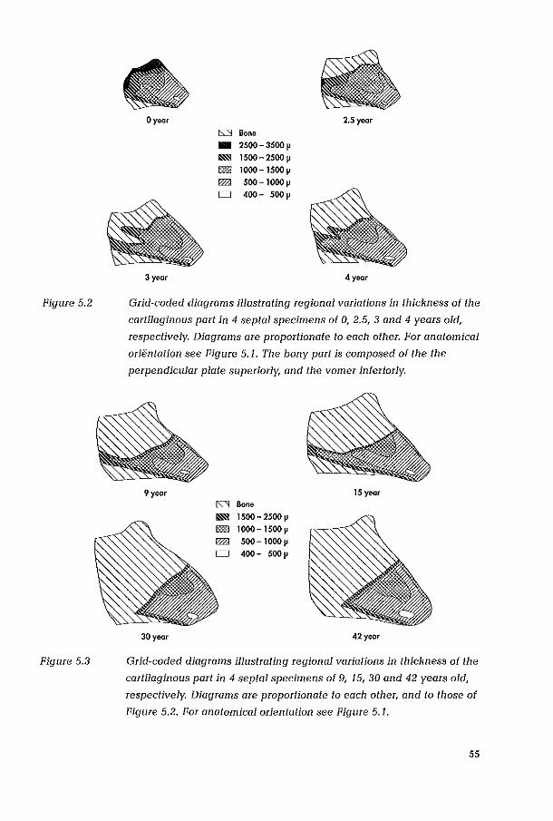
Figure 5.2
Figure 5.3
o year 2.5 year
E'l Bone -2500-3500 P ~ 1500- 2500 P m 1000- 1500 P [@ 500 - 1000 P D 400 - 500 P
3 year 4 year
Grid-coded diagrams illustrating regional variations in thickness of the
cartilaginous part in 4 septal specimens of 0, 2.5, 3 and 4 years old,
respectively. Diagrams are proportionate to each other. For anatomical
orientation see Figure 5.1. The bony part is composed of the the
perpendicular plate superiorly, and the vomer inferiorly.
9 year o Bone
~ 1500-25OOp looo-1500p 5OO-1ooop 400- 500p
15 year
30 year 42 year
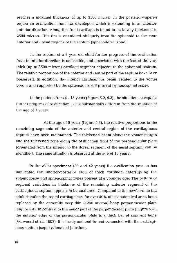
Grid-coded diagrams illustrating regional variations in thickness of the
cartilaginous part in 4 septal specimens of 9, 15,30 and 42 years old,
respectively. Diagrams are proportionate to each other, and to those of
Figure 5.2. For anatomical orientation see Figure 5.1.
55
reaches a 111aximal thickness of up to 3500 micron, In the posterior-superior
region an ossification front has developed which is extending in an inferior
anterior direction. Along this front cartilage is found to be locally thickened to
2500 micron. This rim is orientated obliquely from the sphenoid to the more
anterior and dorsal regions of the septum (sphenodorsal zone).
In the septum of a 3-year-old child further progress of the ossification
front in inferior directioh is noticeable, and associated with the loss of the very
thick (up to 3500 micron) cartilage segment adjacent to the sphenoid rostrum.
The relative proportions of the antmior and central part of the septum have been
preserved, In addition, the inferior cartilaginous bean1, related to the vanIer
border and supported by the sphenoid, is still present (sphenospinal zone).
In the periode from 4 - 15 years (Figure 5.2, 5.3), the situation, except for
further progress of ossification, is not substantially different from the situation at
the age of 3 years.
At the age of 9 years (Figure 5.3), the relative proportions in the
renIaining segments of the antetior and central region of the cartilaginous
septum have been maintained. The thickened beam along the vomer margin
and the thickened zone along the ossification front of the perpendicular plate
(orientated from the inferior to the dorsal segment of the nasal septum) can be
identified. The same situation is observed at the age of 15 years.
In the older specimens (30 and 42 years) the ossification process has
implicated the inferior-posterior area of thick cartilage, interrupting the
sphenodorsal and sphenospinal zones present at a younger age. The pattern of
regional variations in thickness of the remaining anterior segment of the
cartilaginous septum appears to be unaltered. COlllpared to the ne\vborn, in the
adult situation the septal cartilage has, for over 50% of its anatoillical area, been
replaced by the generally very thin (±200 micron) bony perpendicular plate
(Figure 5.4). In contrast to the major part of the perpendicular plate (Figure 5.5),
the anterior edge of the perpendicular plate is a thick bar of compact bone
(Verwoerd et aI., 1992). It is firmly and end-to-end connected with the cartilagi
nous septum (septo-ethmoidal junction).
56
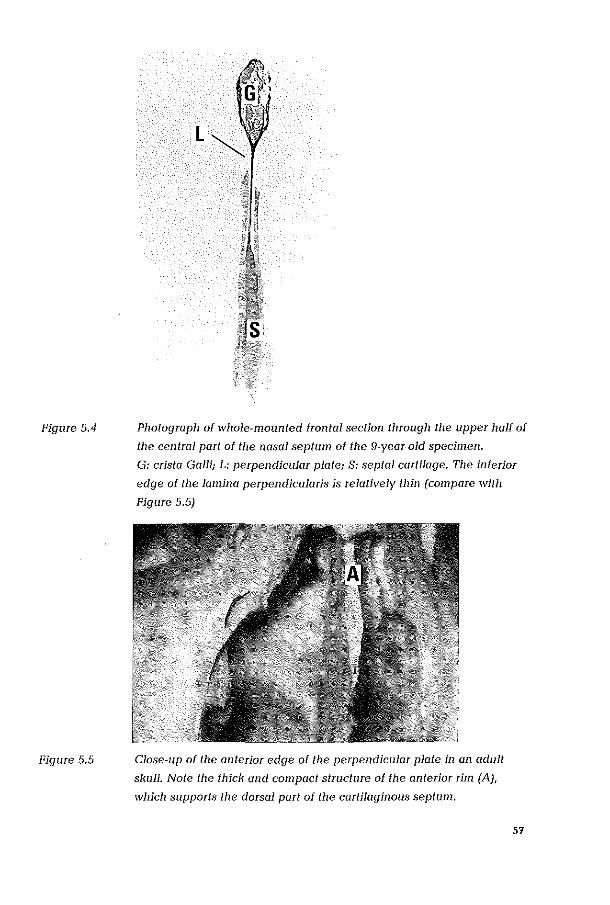
Figure 5.4
Figure 5.5
Photograph of whole-mounted frontal section through the upper half of
the central part of the nasal septum of the 9-year-old specimen.
G: crista Galli; L: perpendicular platej S: septal cartilage. The inferior
edge of the lamina perpendicularis is relatively thin (compare with
Figure 5.5)
Close-up of the anterior edge of the perpendicular plate in an adult
skull. Note the thick and compact structure of the anterior rim (AJ,
which supports the dorsal part of the cartilaginous septum.
57
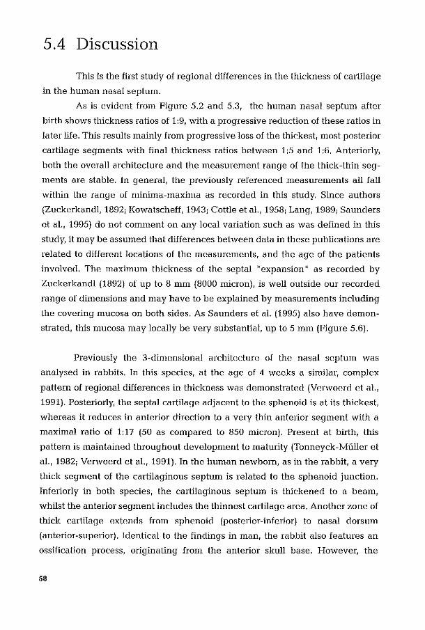
5.4 Discussion
This is the first study of regional differences in the thickness of cartilage
in the human nasal septum.
As is evident from Figure 5.2 and 5.3, the human nasal septum after
birth shows thickness ratios of 1:9, with a progressive reduction of these ratios in
later life. This results mainly from progressive loss of the thickest, most posterior
cartilage segments with final thickness ratios between 1:5 and 1:6. Anteriorly,
both the overall architecture and the measurement range of the thick-thin seg
ments are stable. In general, the previously referenced llleasurenlents all fall
within the range of lllinima-maxhna as recorded in this study. Since authors
(Zuckerkandl, 1892; Kowatscheff, 1943; Cottle et aI., 1958; Lang, 1989; Saunders
et aI., 1995) do not comment on any local variation such as was defined in this
study, it may be assumed that differences between data in these publications are
related to different locations of the measurements, and the age of the patients
involved. The maximum thickness of the septal "expansion" as recorded by
Zuckerkandl (1892) of up to 8 mm (8000 micron), is well outside our recorded
range of dimensions and may have to be explained by measurements including
the covering mucosa on both sides. As Saunders et al. (1995) also have demon
strated, this mucosa may locally be very substantial, up to 5 mm (Figure 5.6).
Previously the 3-dinlensional architecture of the nasal septum was
analysed in rabbits. In this species, at the age of 4 weeks a similar, complex
pattern of regional differences in thickness was demonstrated (Verwoerd et aI.,
1991). Posteriorly, the septal cartilage adjacent to the sphenoid is at its thickest,
whereas it reduces in anterior direction to a very thin antm10r segment with a
maximal ratio of 1:17 (50 as compared to 850 micron). Present at birth, this
pattern is maintained throughout development to maturity (Tonneyck-Miiller et
aI., 1982; Verwoerd et aI., 1991). In the human newborn, as in the rabbit, a very
thick segment of the cartilaginous septum is related to the sphenoid junction.
Infeliorly in both species, the cartilaginous septum is thickened to a beam,
whilst the anterior segment includes the thinnest cartilage area. Another zone of
thick cartilage extends from sphenoid (posterior-inferior) to nasal dorsum
(anterior-superior). Identical to the findings in man, the rabbit also features an
ossification process, originating frOlll the anterior skull base. However, the
S8
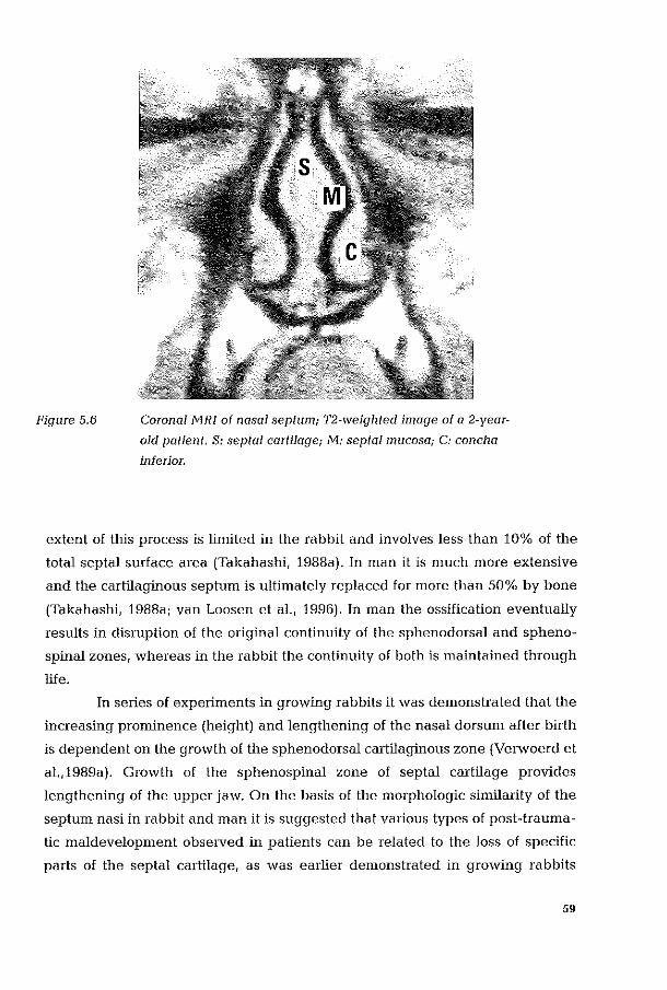
Figure 5.6 CoronallVJRI of nasal septum; T2-weighted image of a 2-year
old patient. S: septal cartilage; M: septal mucosa; C: concha
inferior.
extent of this process is limited in the rabbit and involves less than 10% of the
total septal surface area (Takahashi, 1988a). In man it is much more extensive
and the cartilaginous septum is ultimately replaced for more than 50% by bone
(Takahashi, 1988a; van Loosen et aI., 1996). In man the ossification eventually
results in disruption of the original continuity of the sphenodorsal and spheno
spinal zones, whereas in the rabbit the continuity of both is maintained through
life.
In series of expeIiments in growing rabbits it was demonstrated that the
increasing prominence (height) and lengthening of the nasal dorsum after birth
is dependent on the growth of the sphenodorsal cartilaginous zone (Verwoerd et
al.,1989a). Growth of the sphenospinal zone of septal cartilage provides
lengthening of the upper jaw. On the basis of the morphologic similarity of the
septum nasi in rabbit and man it is suggested that various types of post-trauma
tic maldevelopment observed in patients can be related to the loss of specific
parts of the septal cartilage, as was earlier demonstrated in growing rabbits
59
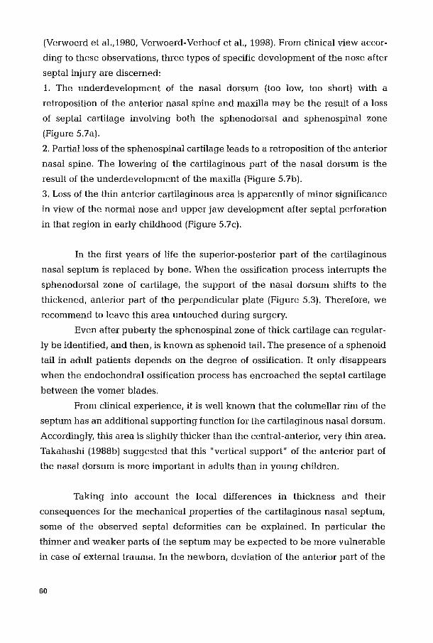
(Verwoerd et al.,1980, Verwoerd-Verhoef et aI., 1998). From clinical view accor
ding to these observations, three types of specific development of the nose after
septal injury are discerned:
1. The underdevelopment of the nasal dorsum (too low, too short) with a
retroposition of the anterior nasal spine and maxilla filay be the result of a loss
of septal cartilage involving both the sphenodorsal and sphenospinal zone
(Figure 5.7a).
2. Partial loss of the sphenospinal cartilage leads to a retroposition of the anterior
nasal spine. The lowering of the cartilaginous part of the nasal dorsum is the
result of the underdevelopment of the maxilla (Figure 5.7b).
3. Loss of the thin anterior cartilaginous area is apparently of minor significance
in view of the normal nose and upper jaw development after septal perforation
in that region in early childhood (Figure 5.7c).
In the first years of life the superior-posterior part of the cartilaginous
nasal septum is replaced by bone. When the ossification process interrupts the
sphenodorsal zone of cartilage, the support of the nasal dorsum shifts to the
thickened, anterior part of the perpendicular plate (Figure 5.3). Therefore, we
recommend to leave this area untouched during surgery.
Even after puberty the sphenospinal zone of thick cartilage can regular
ly be identified, and then, is known as sphenoid tail. The presence of a sphenoid
tail in adult patients depends on the degree of ossification. It only disappears
when the endochondral ossification process has encroached the septal cartilage
between the vomer blades.
From clinical experience, it is well known that the columellar rim of the
septum has an additional supporting function for the cartilaginous nasal dorsum.
Accordingly, this area is slightly thicker than the central-anterior, V8IY thin area.
Takahashi (1988b) suggested that this "vertical support" of the anterior part of
the nasal dorsum is more important in adults than in young children.
Taking into account the local differences in thickness and their
consequences for the mechanical properties of the cartilaginous nasal septumJ
some of the observed septal deformities can be explained. In particular the
thinner and weaker parts of the septum may be expected to be more vulnerable
in case of external trauma. In the newborn, deviation of the anterior part of the
60
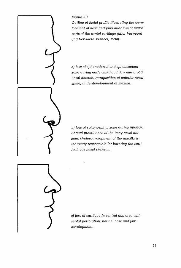
Figure 5.7
Outline of facial profile illustrating the deve
lopment of nose and jaws after loss of major
purts of the septal cartilage (after Venvoerd
and Verwoerd-Verhoef, 1998).
aJ loss of sphenodorsaJ and sphenospinaJ
zone during early clzilclllOOd: low and broad
nasal dorsum, retropositioll of anterior nasal
spine, underdevelopment of maxilla.
b) loss of sphenospinal zone during in/allcy:
normal prominence of the bony nasal dor
sum. Underdevelopment of the maxilla is
indirectly responsible for lowering the carti
laginous nasal skeleton.
c) loss of cartilage in central tllill area with
septal perforation: normal nose and jaw
development.
61
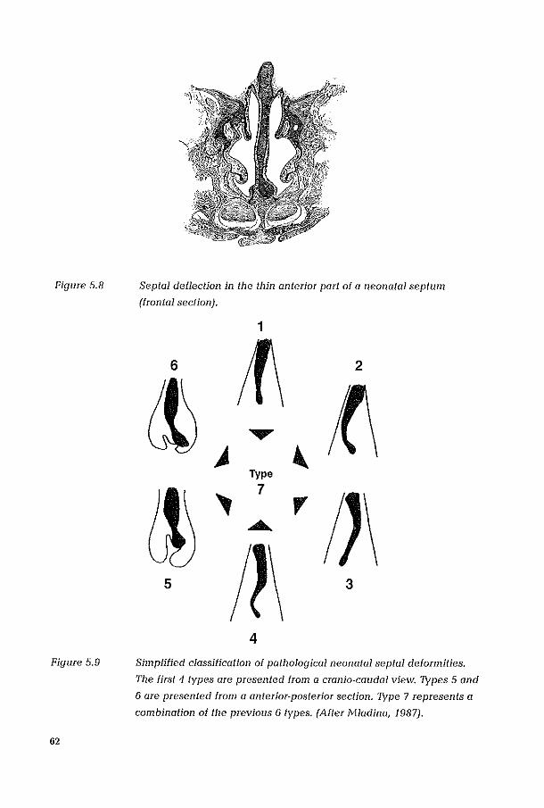
Figure 5.8
Figure 5.9
62
Septal deflection in the thin anterior part of a neonatal septum
(frontal section).
1
6
~ Type
7 , 5 3
4
Simplified classification of pathological neonatal septal deformities.
The first 4 types are presented from a cranio-caudal view. Types 5 and
6 are presented from a anterior-posterior section. Type 7 represents a
combination of the previous 6 types. (After Mladina, 1987).
septum has been well described (Gray, 1974; Jazbi, 1977; Kent et ai., 1988;
Brain, 1992). In this septal deformity, the deflection takes place, during labour,
in the thin anterior part (Figure 5.8).
Mladina (Mladina, 1987; Mladina et ai.,1989, 1990) has divided septal
deformities in 7 basic types (Figure 5.9). In most patients the bending of the
septal cartilage occurs in the thin or relatively thinner areas as described, for the
first time, in the present study. The types 1, 2 and 4 are noted as a vertical ridge
due to bending or fracture at various sites in the anterior thin area (MIa dina,
1987). The horizontal bending takes place in the thin part just above the basal
rim. At anterior rhinoscopy, the horizontal types of deviation (type 5 and 6)
appear as a single basal crest, which can reach the posterior part of the lateral
nasal wall (type 5). The crest is formed by the thick basal cartilaginous rim
located above the vomer bone. Another horizontal septal deviation (type 6) is
characterised by a deep gutter on one side of the septum (Figure 5.9).
In conclusion this study has demonstrated that:
1. The human cartilaginous nasal septum is characterised by a specific 3-dimen
sional architecture with thicker and thinner zones varying with a factor of 1:9 at
birth to 1:6 in adults. This pattern, maintained from birth to adulthood, is similar
to that previously demonstrated in rabbits.
2. The thickness in the various zones of the septal cartilage hardly increases
during further growth of the child. The nose develops in size by its sagittal
dimensions through a process of simultaneous cartilage growth and
endochondral ossification.
3. In young children the nasal dorsum is primarily based on the sphenoid via a
zone of thick cartilage; later in Iile it is based on the thickened part of the
anterior border of the perpendicular plate.
4. Various types of maldevelopment of nose and upper jaw in patients after an
injury in early childhood, can be explained by the loss of specific parts of the
septal cartilage, e.g. sphenodorsal and sphenospinal zone; loss of the extremely
thin anterior area of septal cartilage seems not to be important for nasal growth.
5. The 3-dimensional architecture of the nasal septum is important for under
standing the etiology of septal deviations, the origin of preferred fracture lines
and the risks of surgery for nasal development.
63
References:
Brain D, 1992, The ethiology of neonatal septal deviations. Facial Plast Surg,
8/4:191/193.
Clarke GM, 1967, The structural support of the nose. Arch Otol, 85:418-423.
Collie MH, Loring RM, Fischer GG, Gaynon IE, 1958, The maxilla - premaxilla
approach to extensive nasal septum surgery. Arch Otolmyng, 68:301-313.
Delaire J, Precious D, 1987, Interaction of the development of the nasal septum,
the nasal pyramid and the face. Int J Ped Otorhinolmyngol, 12:311-326.
Fry HJH, 1966, Interlocked stresses in human nasal septal cartilage. Br J Plast
Surg, 19:276-278.
Fry HJH, 1967, Nasal skeletal trauma and the interlocked stresses of the nasal
septal cartilage. Br J Plast Surg, 20:146-158.
Gray LP, 1974, Septal and associate cranial birth deformities: types, incidents
and treatment Med J Aust, 1: 557-563.
Jazbi B, 1977, Subluxation of the nasal septum in the newborn. Etiology, diag
nose and treatment. Otolaryngol. CHn. North Am, 10:125-138.
Kent SE, Right AP, Brain DJ, 1988, Neonatal septal deviations. J R Soc Med,
81:132-135.
Kowatschelf L, 1943, Bisonderkeiten der kindlichen Nase und ihre klinischen
Bedeutung. Z Hals-Nasen-u-Ohrenheilk, 48:345-374.
Lang J, 1989 r Clinical anatomy of the nose, nasal cavity and paranasal sinuses.
Thieme Verlag, New York.
64
Luongo RA, Luongo RA jr, 1958, Anatomical observations for rhinoplastic
surgery. Laryngoscope; 6:1076-1080.
Mladina R, 1987, The role of maxillar morphology in the development of patho
logical septal deformities. Rhinology, 25: 199-205.
Mladina R, Krajina Z, 1989, The influence of the caudal process on formation of
septal deformities. Rhinology, 27: 113-118.
Mladina R, Krajina Z, 1990, The influence of palato-cranial base (basomaxilla
ry) angle on the length of the caudal process of the nasal septum in man.
Rhinology, 28: 185-189.
Riggs RH, 1953, Relation of septal reconstruction to external deformation of the
nose. Laryngoscope, 63:293-303.
Rubenstein M, 1956, Fractures and post-trawnatic deformation of the nose. Arch
0101, 63:355-362.
Schultz-Coulon HJ, Eckermeier L, 1975, Zum postnatalen Wachstum der
Nasenscheidenwand. Acta Otolatyngol, 82:131-142.
Saunders MW, Jones NS, Kabala J, Lows J, 1995, An anatomical, histological
and magnetic resonance imaging study of the nasal septum. elin Otolaryngol;
20:343-438.
Takahashi R, 1988a, The formation of the nasal septum and the evolutional para
dox. Act Otolmyngol Suppl, 443:1-160.
Takahashi R, 1988b, The evolution of the nasal septum and the fOlmation of sep
tal deformity. Rhinology Suppl, 6:1-23.
Tonneyck-Miiller I, van der Werl, 1982, Die Entwicklung des Septum Nasi beim
Kaninchen. II. Untersuchungen nach regionalen Unterschieden in der Septum
Dicke. Acta Morphol Neerl-Scand, 20:379-391.
65
Van Loosen J, Verwoerd-Verhoei HL, Verwoerd CDA, 1988, The nasal septal
cartilage in the newborn. Rhinology, 26:161-165.
Van Loosen J, van Zanten GA, van Velzen D, Verwoerd-Verhoef HL, Verwoerd
CDA, 1996, Growth characteristics of the human nasal septum. Rhinology, 34:
78-S2.
Verwoerd CDA, Urbanus NAM, Mastenbl'oek GJ, 1980, The influence of partial
resections of the nasal septal cartilage on the growth of the upper jaw and the
nose: An experimental study in rabbits. Clin Otolaryngol, 5:291-302.
Vel'woel'd CDA, Vel'woerd-Vel'hoei HL, 1989a, Developmental aspects of the
deviated nose. J Facial Plast Surg, 612:95-100.
Vel'woel'd CDA, Vel'woel'd-Vel'hoef HL, Meeuwis CA, 1989b, Stress and
wound healing of the cartilaginous nasal septum. Acta Otolaryngol (Stockh),
107:441-445.
Vel'woerd CDA, Vel'woerd-Vel'hoef HL, Meeuwis CA, van del' Heul RO, 1991,
Wound healing of autologous implants in the nasal septal cartilage. 0 R L;
53:310-314.
Verwoerd CDA, 1992, Present day treatment of nasal fractures: Closed versus
open reduction. J Facial Plast Surg, 6:220-223.
Verwoel'd CDA, Vel'woerd-Vel'hoef HL, 1998, Rhinosurgery in children,
surgical and developmental aspects. In: Rhinoplasty, second edition, Nolst
Trenite GJ (ed), 1995, Kugler Publications, Amsterdam.
Verwoerd-Vel'hoei HL, ten Koppel PGJ, van Osch JVM, Meeuwis CA,
Verwoel'd CDA, 1998, Wound healing of cartilage structures in the head and
neck region. Int J Ped Otorhinolmyngol, 43:241-251.
Zuckerkandl E, 1892, Normale und pathologische Anatomie der Nasenhohle
und ihrer pneumatischen Anhange. In: Anatomie der Nasenscheidewand II
Zuckerkandl E (ed.), 1S92. Braumiiller, Wein.
66
Persistent Pattern of Variations in Thickness of the Human Nasal Septum: Implications for Stress and Trauma as Illustrated by a Complex Fracture in a 4-Year-old Boy.
61
6.1 Introduction
For surgical intervention after nasal trauma, it is desirable to be able to
predict at least to some extent the distribution of nasal fracture lines.
Harrison (1979) found reproducible patterns of fractures, beginning just
posterior to the nasal spine 2-3 mm above the boundary with the bony vomer
and extension posteriorly to the perpendicular plate of the ethmoid. Crossing
this boundary it angles to cranially, sequestering a 1 em wide strip of the plate.
Before reaching the cribrifornl plate a second anteIior angling results in a re
crossing of the boundary. Continuing in ventral direction the cartilage is
followed, leaving a narrow rim attached to the base of the skull. This pattern was
not descIibed as age dependent although the experiments were limited to adults
with a fractured nose and 15 cadavers without any age indication. For prediction
of fractures in children there seems as yet no empelical study. Additionally there
is no theoretical basis, which would predict the pattern as found by Harrison
(1979) since the models are either limited by the use of a perspex plate of single
thickness, or of 2 dimensions representing the bony and cartilaginous
component in an attempt to take into account the differences in elasticity of
perpendiclliarplate and the cartilaginous septum proper (Murray, 1987). Still the
consistent pattern of a cOlllplex C-shaped fracture, opening in anterior direction
can not be explained by these models.
Anatomical studies which would provide an alternative basis for the
experimental findings were until recent of conflicting nature with wide ranges
for nasal septal thickness being recorded in the literature. Zuckerkandl (1892)
gave the most extreme at 4-8 mm, willie Kowatscheff (1943), Cottle et al (1958)
and Lang (1989) all presented different results of measurements ranging from 2
mm (Kowatscheff, 1943) to 4 mm (Lang, 1989). However these measurements do
not take into account the considerable regional variations in thickness, which
appear in a consistent pattern with age as presented by van Loosen et aI., (1996).
The latter study however extends from the paediatric age range to adulthood (0-
42 years). If the findings of van Loosen et al (1996) were to be consistent with the
consistent pattern of fractures as recorded by Harrison (1979), predictability of
fractures might then be extended into childhood.
68
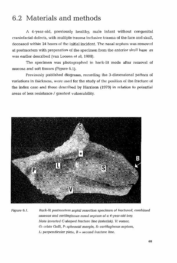
6.2 Materials and methods
A 4-year-old. previously healthy. male infant without congenital
craniofacial defects, with multiple trauma inclusive trauma of the face and skull,
deceased within 24 hours of the initial incident. The nasal septum was removed
at postmortem with preparation of the specimen from the anterior skull base as
was earlier described (van Loosen et aI, 1988).
The specimen was photographed in back-lit mode after removal of
mucosa and soft tissues (Figure 6.1).
Previously published diagrams, recording the 3-dimensional pattern of
variations in thickness, were used for the study of the position of the fracture of
the index case and those described by Harrison (1979) in relation to potential
areas of less resistance / greatest vulnerability.
Figure 6.1. Back-lit postmortem septal resection specimen of fractured, combined
osseous and cartilaginolls nasal septum of a 4-year-old boy.
Note inverted C-shaped fracture line (asterisk). V: vomer,
G: crista Galli, P: sphenoid margin, S: cartilaginous septum,
L: perpendicular plate, B = second fracture line.
69
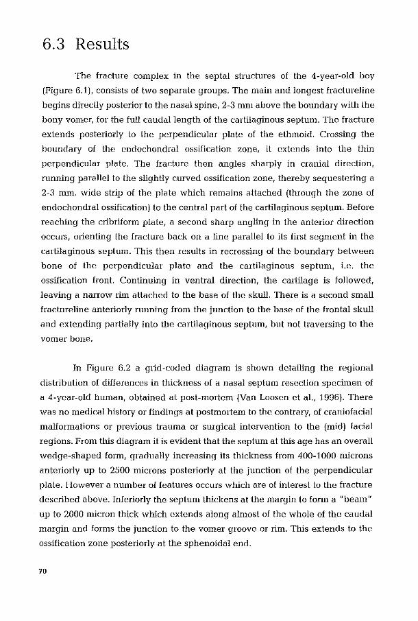
6.3 Results
The fracture complex in the septal structures of the 4-year-old boy
(Figure 6.1), consists of two separate groups. The main and longest fractureline
begins directly posterior to the nasal spine, 2-3 mm above the boundmy with the
bony vomer, for the futl caudal length of the cartilaginous septum. The fracture
extends posteriorly to the perpendicular plate of the ethmoid. Crossing the
boundary of the endochondral ossification zone, it extends into the thin
perpendicular plate. The fracture then angles sharply in cranial direction,
running paratlel to the slightly curved ossification zone, thereby sequestering a
2-3 mm. wide strip of the plate which remains attached (through the zone of
endochondral ossification) to the central part of the cartilaginous septum. Before
reaching the cribriform plate, a second sharp angling in the anterior direction
occurs, orienting the fracture back on a line parallel to its first segment in the
cartllaginous septum. This then results in recrossing of the boundary between
bone of the perpendicular plate and the cartilaginous septum, i.e. the
ossification front. Continuing in ventral direction, the cartilage is followed,
leaving a narrow rim attached to the base of the skull. There is a second small
fractureline anteriorly running from the junction to the base of the frontal skull
and extending partially into the cartllaginous septum, but not traversing to the
vomer bone.
In Figure 6.2 a grid -coded diagram is shown detailing the regional
distribution of differences in thickness of a nasal septum resection specllnen of
a 4-year-old human, obtained at post-mortem (Van Loosen et aI., 1996). There
was no medical history or findings at postmortenl to the contrary, of craniofacial
malformations or previous trauma or surgical intervention to the (mid) facial
regions. From this diagram it is evident that the septum at this age has an overall
wedge-shaped form, gradually increasing its thickness from 400-1000 microns
anteriorly up to 2500 microns posteriorly at the junction of the perpendicular
plate. However a nUlllber of features occurs which are of interest to the fracture
described above. Inferiorly the septulll thickens at the margin to fOfnl a "beam"
up to 2000 micron thick which extends along almost of the whole of the caudal
margin and forms the junction to the vallIer groove or lim. This extends to the
ossification zone posteriorly at the sphenoidal end.
70
Figure 6.2.
b:SI Bone l!!II1,500-2,500 ~m lID 1,000-1,500 ~m lID 500-1,000 ~m CJ 400 - 500 11m
B
Grid~coded diagram of a 4-year~01d case study, illustrating the
constistent pattern of regional variations in thickness (van Loosen et
a1., 1997). A. Fracture line. B. Second fracture line.
6.4 Discussion
As we have shown previously for the neonatal period in man (van
Loosen et al., 1988), a similarly complex architecture exists as that recorded in
Harrison's (1979) studies for adults. With respect to the interpretation of its
mechanical properties it is of interest that, the reproducibility of fractures in
these experiments is high. The main features in children and adults are a poste
rior thick and anterior thin plate with a superior and inferior "beam" of thicker
cartilage. The central segment in man is replaced by a thin perpendicular plate
(up to 400 micron). Aside from overall dimensional growth, the remaining frac
tion of the cartilaginous septum, at least during the first four decades of life,
maintains the same overall pattern of thick and thin areas. Also the vulnerabili
ty to mechanical forces may therefore similarly not change significantly with
age.
71
6.5 Conclusions
In summary our previous studies denlonstrate a consistent pattern of
regional variations in thickness of the human nasal septum. The case as
presented for the first time here shows a pattern of nasal septal fracture similar
to the lines appreciated in the study by Harrison (1979). Now it can be predicted
in children on the basis of the retained overall architecture, desClibed first at
birth (van Loosen et aI., 1988) and which, with the exception of posterior
replacement through endochondral ossification, is maintained throughout life.
72
References
Collie MH, Loring R, Fischer GG, Gaynon IE, 1958, The maxilla-premaxilla
approach to extensive nasal septum surgery. Arch Otolaryngol, 68:301-313.
Hanison D, 1979, Nasal injuries: their pathogenesis and treatment. Br J Plast
Surg, 32:57-64.
Kowatscheff L, 1943, Bisonderkeiten der kindlichen Nase und ihre klinische
Bedeutung. Z Hals-Nas-u-Ohrenheilk, 48:345-374.
Lang J, 1989, Clinical Anatomy of the nose, nasal cavity and paranasal sinuses,
33-34. Thieme Verlag, New York.
Murray JAM, 1987, The distribution of stress in the nasal septum in trauma: an
experimental model. Rhinology, 25:101-107.
Van Loosen J, Verwoerd-Verhoef HL, Verwoerd CDA, 1988, The nasal septal
cartilage in the newborn. Rhinology, 26:161-165.
Van Loosen J, van Velzen D, Verwoerd-Verhoef HL, Verwoerd CDA, 1994,
Regional variations in thickness of the human nasal septum. Proc VIth Asean
ORL Congress, Chiang RaL
Van Loosen J, Verwoerd-Verhoef HL, van Velzen D, Verwoerd CDA, 1996,
Regional differences in thickness of the human nasal cartilaginous septum.
1996. Proc. XVI Congress of the Eur. Rhinol Soc., Ghent.
Zuckerlmndl E, 1892, Normale und pathologische Anatomie der Nasenh6hle
und ihrer pneumatischen Anhange. In Anatomie der Nasenscheidewand II,
Zuckerkandl E (ed.), 1892, Braumiiller, Wien.
73
Interstitial Growth of Nasal Septal Cartilage Assessed by Automated Computer Assisted Image Analysis.
74
7.1 Introduction
Surgical intervention in the nasal septum requires a detailed under
standing of the development of the nasal septum with age, since trauma to areas
of growth may result in the development of deformities. Knowledge with respect
to the presence and distribution of "vulnerable" growth areas of the nose is as
yet not complete (Pirsig, 1986a,b, 1992; Verwoerd and Verwoerd-Verhoef,1989)
and quantitative studies are lacking. Literature on septal dimensions related to
age exist (Pirsig 1975; Vetter et aI., 1983,1984a,b) and investigations of the nasal
septum have concentrated primarily on the ossification zone (Schultz-Coulon
and Eckenneier, 1976). In the neonate the nasal septum is completely carti
laginous. The anterior thin area shows mainly small, round chondroblasts, ·with
the presence of "twin cells /I whereas the rernainder of the septum contains more
hypertrophic chondrocytes. In the most posterior region, near the sphenoid
rostrum endochondral ossification is recognised (van Loosen et aI., 1988).
During further growth of the nasal septum two processes can be
observed:
1. Cartilaginous septal growth by appositional and interstitial growth.
(Appositional growth: increase of the overall septal dimensions by external
deposition of neocartilage by cells descendent from the subperichondrial
cartilage. Interstitial growth: the increase of overall septal dimensions by pro
duction of intercellular substance, and to a lesser degree by an increase in cell
size)
2. An intensive ossification process responsible for the perpendicular plate.
Vetter et aI., (1983, 1984 a,b), described the metabolic activity of septal
cartilage in hunlans. Their studies, however do not include patients below the
age of 5 years. In this period the largest proportional increase in size of the
septum occurs (van Loosen et aI, 1996). In the nasal septum of the rabbit, a
relationship between the variations in thickness and quantitative observations
on the relationship between cells and intercellular substance have been found
(Tonneyck-Miiller and van cler Werf, 1982; Tonneyck-Miiller, 1984).
The aim of this study is to investigate the interstitial growth of
the nasal septal cartilage in relation to: a) age; b) locations.
15
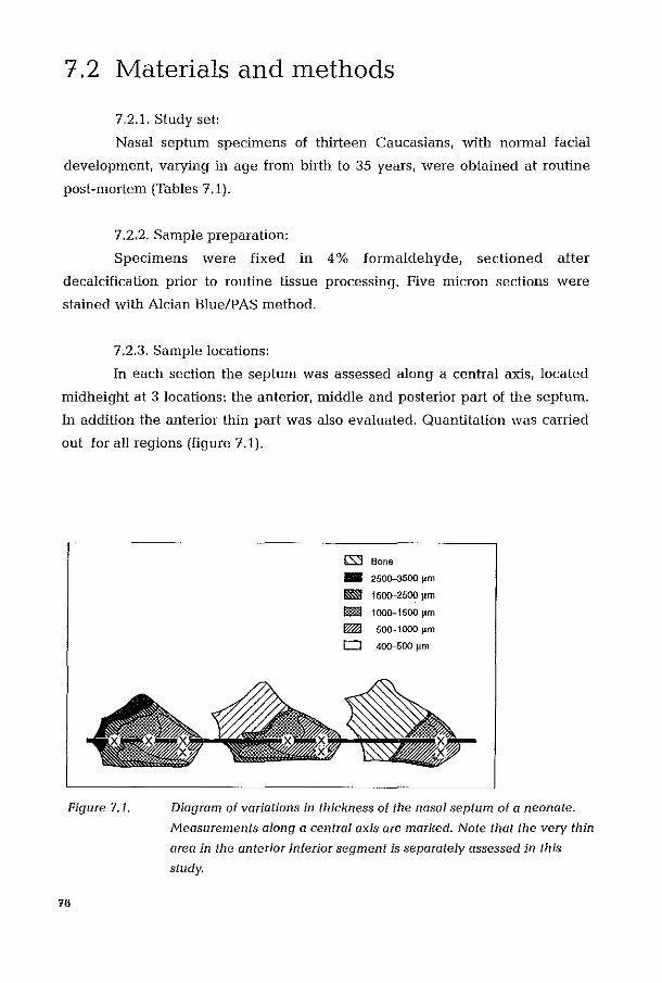
7,2 Materials and methods
7.2.1. Study set:
Nasal septum specimens of thirteen Caucasians, with normal facial
development, varying in age from birth to 35 years, were obtained at routine
post-mortem (Tables 7.1).
7.2.2. Sample preparation:
Specimens were fixed in 4 % formaldehyde, sectioned after
decalcification prior to routine tissue processing. Five micron sections ·were
stained with Alcian Blue/PAS method.
7.2.3. Sample locations:
In each section the septum was assessed along a central axis, located
midheight at 3 locations: the anterior, middle and posterior part of the septum.
In addition the anterior thin part was also evaluated. Quantitation was carried
out for all regions (figure 7.1).
Figure 7.1.
76
(;SJ Bone -2500-3500 11m
m:I 1500-2500 lim
I!!!iI 1000-1500 J.lm
I!!!iI 500-1000 J.1m
0 400-500 11m
Diagram of variations in thickness of the nasal septum of a neonate.
Measurements along a central axis are marked. Note that the ver)' thin
area in the anterior inferior segment is separately assessed in this
study.
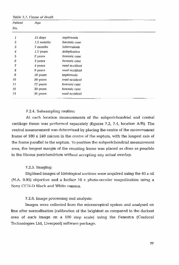
Table 7.1. Cause of death
Patient Age
No.
15 days septicemia
2 1.5 months forensic case
3 7 months tuberculosis 4 1,5 years dehydration
5 2 years forensic case
6 3 years forensic case
7 4 years road accident
8 9 years road accident
9 10 years septicemia
10 20 years lOod accident
11 27 years forensic case
12 30 years forensic case 13 35 years road accident
7.2.4. Subsampling routine:
At each location measurements of the subperichondrial and central
cartilage tissue was performed separately (figures 7.2, 7.4, location AlB). The
central llleasurement was determined by placing the centre of the measurelnent
frame of 180 x 240 micron in the centre of the septum, with the longest axis of
the frame parallel to the septum. To position the subpelichondrial measurement
area, the longest margin of the counting frame was placed as close as possible
to the fibrous perichondrium without accepting any actual overlap.
7.2.5. Imaging:
Digitised images of histological sections were acquired using the 40 x oil
(N.A. 0.95) objective and a further 10 x photo-occular magnification using a
Sony CCD-D Black and White camera.
7.2.6. Image processing and analysis:
Images were collected from the microscopical system and analysed on
line after normalisation (calibration of the brightest as compared to the darkest
area of each inlage on a 100 step scale) using the Fenestra (Confocal
Technologies Ltd, Liverpool) software package.
77
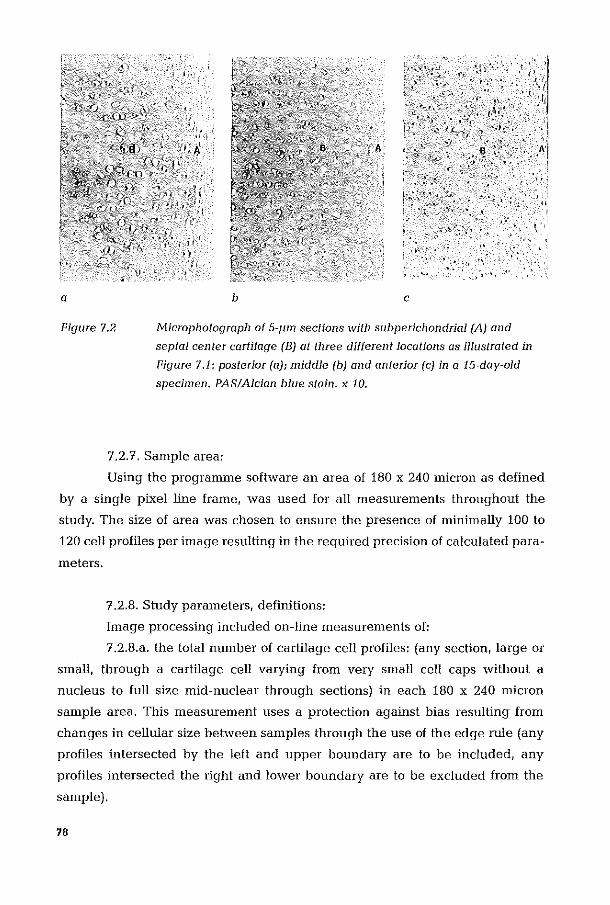
a b e
.,' .. ' . . ,>. ,"
. ". " ' .
Figure 7.2 Microphotograph of 5-pm sections with sflbperichondrial (A) and
septal center cartilage (B) at three different locations as illustrated in
Figure 7.i: posterior (a); middle (b) and anterior (e) in a i5·day·old
specimen. PAS/Aldan blue stain. x 10.
7.2.7. Sample area:
Using the programme software an area of 180 x 240 micron as defined
by a single pixel line frame, was used for all measurements throughout the
study. The size of area was chosen to ensure the presence of minimally 100 to
120 cell profiles per image resulting in the required precision of calculated para
meters.
7.2.8. Study parameters, definitions:
Image processing included on-line measur8rnents of:
7.2.8.a. the total number of cartilage cell profiles: (any section, large or
small, through a cartilage cell varying from velY small cell caps without a
nucleus to full size mid-nuclear through sections) in each 180 x 240 micron
sanlple area. This measurement uses a protection against bias resulting from
changes in cellular size between samples through the use of the edge rule (any
profiles intersected by the left and upper boundary are to be included, any
profiles intersected the right and lower boundary are to be excluded from the
sample).
78
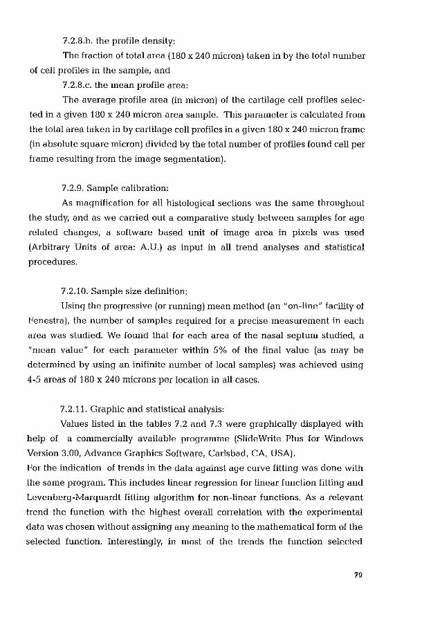
7.2.8.b. the profile density:
The fraction of total area (180 x 240 micron) taken in by the total number
of cell profiles in the sample, and
7.2.8.c. the mean profile area:
The average profile area (in micron) of the cartilage cell profiles selec
ted in a given 180 x 240 micron area sample. This parameter is calculated from
the total area taken in by cartilage cell profiles in a given 180 x 240 micron frame
(in absolute square micron) divided by the total number of profiles found cell per
frame resulting from the image segmentation).
7.2.9. Sample calibration:
As magnification for all histological sections was the same throughout
the study, and as we carried out a comparative study between samples for age
related changes, a software based unit of image area in pixels was used
(Arbitrary Units of area: A.U.) as input in all trend analyses and statistical
procedures.
7.2.10. Sample size definition:
Using the progressive (or running) mean method (an "on-line" facility of
Fenestra), the number of sanlples required for a precise nleasurement in each
area was studied. We found that for each area of the nasal septum studied, a
umean value" for each paranleter within 5% of the final value (as lllay be
determined by using an inifinite number of local samples) was achieved using
4-5 areas of 180 x 240 microns per location in all cases.
7.2.11. Graphic and statistical analysis:
Values listed in the tables 7.2 and 7.3 were graphically displayed with
help of a commercially available progranmle (Slide Write Plus for Windows
Version 3.00, Advance Graphics Software, Carlsbad, CA, USA).
For the indication of trends in the data against age CUlve fitting was done with
the same program. This includes linear regression for linear function fitting and
Levenberg-Marquardt fitting algorithm for non-linear functions. As a relevant
trend the function with the highest overall correlation with the experinlental
data was chosen without assigning any meaning to the mathematical fmm of the
selected function. Interestingly, in most of the trends the function selected
79
indicated a proportionality to the natural logarithm of age.
7.2.12. Statistical analysis:
Differences between locations were tested for statistical signilicane with
the Wilcoxon Matched-Pairs Signed-Ranks test using the SPSS programme,
Version 8.0 for Windows. With help of this progranmle also the statistical
significance of the relations between the cell profile aspects and age was tested
(2-tailed) parametrically using Pearson correlation coefficients and nonparame
trically by Spearman correlation coefficients.
7.2.13. Quality control and reproducibility studies:
All samples were analyzed twice by a non-medical scientific officer in a
strictly randomised and blinded fashion. Randomisation included intra-septal
location and subperichondrial versus central measurenlents.
Using randomisation 25 % of all samples / areas were re-measured by a
second observer for the purpose of inter-observer reproducibility assessment.
Reproducibility for the profile density and the mean profile area measurements
were better than 90% for inter- and better than 95% for intra-observer reprodu
cibility.
7.3 Results
The studied specimens vary in age from neonate to 32-year-old adult. In
the neonate nearly the total nasal septum is cartilaginous. The first signs of
endochondral ossification become manifest between 1 and 2 years of age.
Ossification starts adjacent to the anterior skull base and sphenoid. The ossi
fying front expands in anterior-inferior direction. This results in a gradual loss of
cartilage and simultaneously, in fOlmation of the perpendicular plate. Finally,
more than half of the adult nasal septum is composed of bone (perpendicular
plate). As a consequence of ossification the cartilage in the posterior and
midseptal area could be studied only in the younger specimens. The results are
sunmlarised in table 7.2 (profile area) and 7.3 (profile density), and illustrated in
Figure 7.3-7.6; the statistical analyses are presented in table 7.4.-7.6.
80
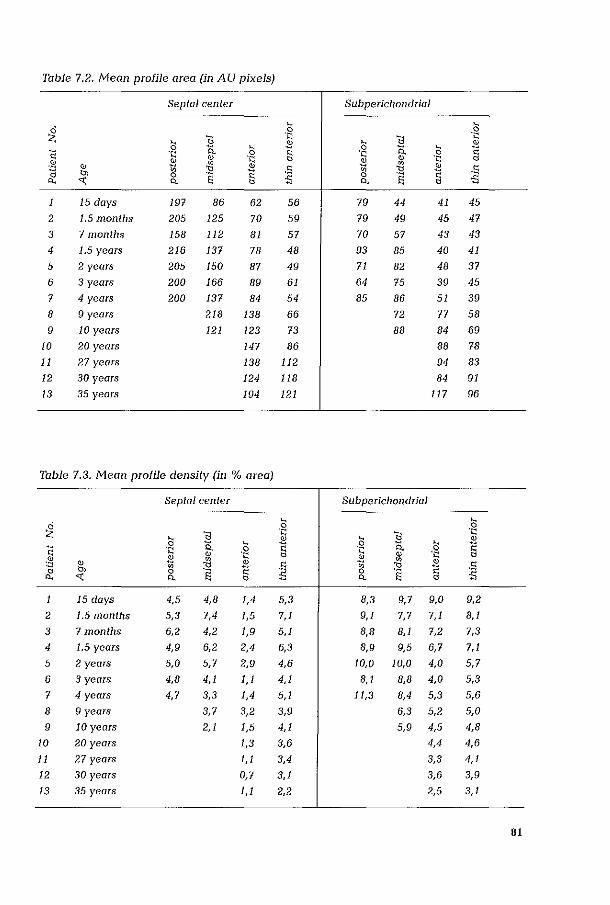
Table 7.2. Mean profile area (in AU pixels)
Septal center Subperkhondrial
" " ~ 0 0
11 ·S " <l
'Q
" jli 0 " " 0 15. ~
" 'Q "- 0 'Q § ~ 'Q ~ e 'Q ~ ~ ~ ~ " "' ~ " "
jli ,% " jli ,~
~ '" 0 § " 0. « "- s " "- S ~ " 1 15 days 197 86 62 5,6 79 44 41 45
2 1.5 months 205 125 70 59 79 49 45 47
3 7 month~ 158 112 81 57 70 57 43 43
4 1.5 years 216 137 78 48 93 85 40 41
5 2 years 205 150 87 49 71 82 48 37
6 3 years 200 166 89 61 64 75 39 45
7 4 years 200 137 84 54 85 86 51 39
8 9 years 218 138 66 72 77 58
9 10 year~ 121 123 73 88 84 69
10 20 years 147 86 88 78
11 27 years 138 112 94 83
12 30 years 124 118 84 91
13 35 years 194 121 117 96
Table 7.3. Mean profile density (in % area)
Septal center Subperic1lOndrial
" " ~ 0 0
11 ~ " <l 'Q
" jli 0 , 0 15. ~ ~ .. , "-
~ " 'Q " jli e ~ e ~ 'Q ~ e ] "
~ jli " ~ ,~ " ~ ~ '" 0 " 0 S " 0. « "- ~ " "- ~
15 days 4,5 4,8 1,4 5,3 8,3 9,7 9,0 9,2
2 1.5 months 5,3 7,4 1,5 7,1 9,1 7,7 7,1 8,1
3 7 months 6,2 4,2 1,9 5,1 8,8 8,1 7,2 7,3
4 1.5 years 4,9 6,2 2,4 6,3 8,9 9,5 6,7 7,1
5 2 years 5,0 5,7 2,9 4,6 10,0 10,0 4,0 5,7
6 3 years 4,8 4,1 1,1 4,1 8,1 8,8 4,0 5,3
7 4 years 4,7 3,3 1,4 5,1 11,3 8,4 5,3 5,6
8 9 years 3,7 3,2 3,9 6,3 5,2 5,0
9 10 years 2,1 1,5 4,1 5,9 4,5 4,8
10 20 years 1,3 3,6 4,4 4,6
11 27 years 1,1 3,4 3,3 4,1
12 30 years 0,7 3,1 3,6 3,9
13 35 years 1,1 2,2 2,5 3,1
81
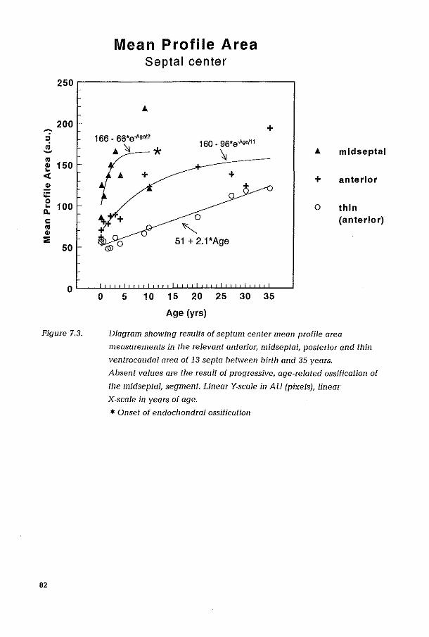
'":'
" .. ~ .. ., ... « ., ;;:: 0 ... a. c .. ., :;;
Mean Profile Area Septal center
250.-----------------------------,
200 + 166 - 66'e-A,0/2 160 - 96'e·A,,,,,
(;: ~ 150
'" + + (';
0
100 0 ,
50 51 + 2.1'Age
OL-~llL~~~~~~~~~~L__J
o 5 10 15 20 25 30 35
Age (yrs)
'" mldseptal
+ anterior
0 thin (anterior)
Figure 7.3. Diagram showing results of septum center mean profile area
measurements in the relevant anterior, midseptal, posterior and thin
ventrocQudaJ area of 13 septa between birth and 35 years.
82
Absent values are the result of progressive, age-related ossification of
tile midsepta/, segment. Linear Y-sca/e in AU (pixe/s), linear
X-scale in years of age.
* Onset of endochondral ossification
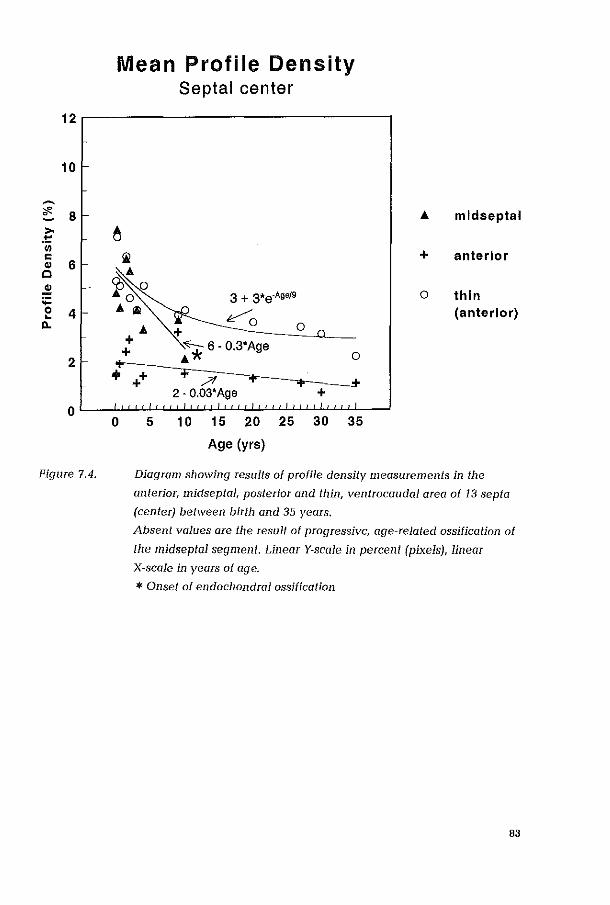
12
10
~
~ 8 ~
>-,t::
'" c OJ 6 c .! :;: 0 4 ...
D..
2
0
Figure 7.4.
Mean Profile Density Septal center
A mldseptal
6 !il + anterior
~ Ji. 0 3 + 3*e·Age/9
A iii ~ A + 0 0
.: ~ 6 . O.3*Age
o thin (anterior)
ojr A 0
+
0
++ + ?I + + .+ 2·0.03'Age +
5 10 15 20 25 30 35
Age (yrs)
Diagram slIowing results of profile density measurements in the
anterior, midseptal, posterior and thin, ventrocaudal alea of 13 septa
(center) between birth and 35 years.
Absent values ale the result of progressive, age-related ossification of
the midseptal segment. Linear Y·scale in percent (pixels), linear
X-scale in yeals of age.
* Onset at endochondral ossification
83
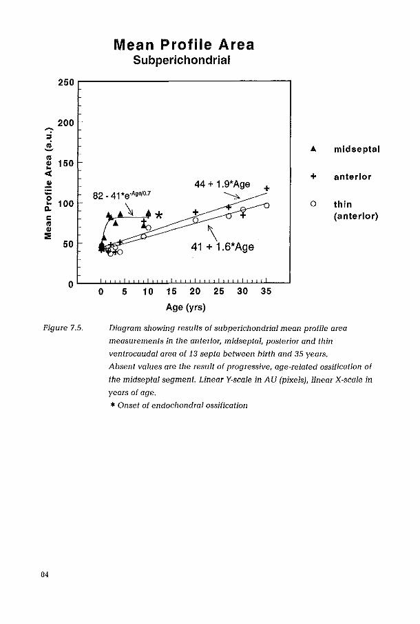
Mean Profile Area Subperichondrial
250,-----------------------------,
200 '"":' ,. co ~
co 150 CI> ..
< .!! :;: 0 .. 100
CI. c co CI>
::;; 50
0 0
Figure 7.5.
84
... midseptal
+ anterior 44 + 1.9'Age + ~
0 thin (anterior)
\ 41 + 1.6*Age
5 10 15 20 25 30 35
Age (yrs)
Diagram showing results of subpericJlOndrial mean profile area
measurements in the anterior, midseptal, posterior and thin
ventrocaudal area of 13 septa between birth and 35 years.
Absent values are the result of progressive, age-related ossification of
the midseptal segment. Linear V-scale in AU (pixels), linear X-scale in
years of age.
* Onset of endochondral ossification
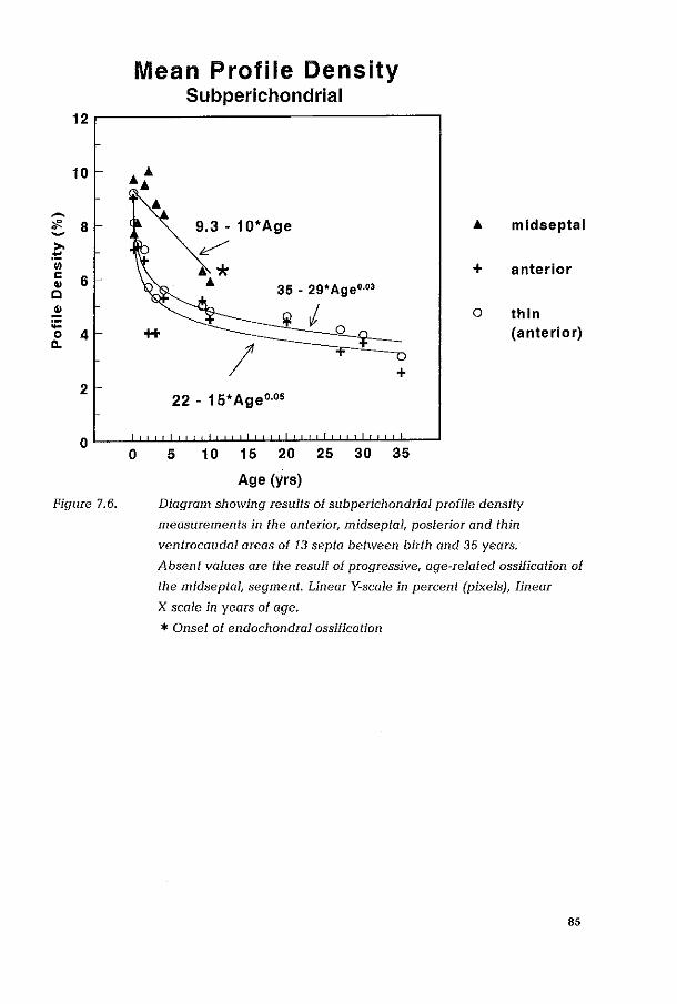
12
10
~
<f'. 8 ~
>-~
'iii c .. 6 C .. :;:: 0
11. 4
2
0
Figure 7.6.
Mean Profile Density
AA A
0
Subperichondrial
A mldseptal
+ anterior
35 - 29' Age""
Ijl I 0 thin 0 (anterior)
I + 22 - 15' Ageo.o,
5 10 15 20 25 30 35
Age (yrs)
Diagram showing results of subperic1lOndriaJ profile density
measurements in the anteriOT, midseptal, posterior and thin
ventrocaudal areas of 13 septa between birth and 35 years.
Absent values are the result of progressive, age-related ossification of
the midseptaJ, segment. Linear Y-scale in percent (pixels), linear
X-scale in years af age,
* Onset of endochondral ossification
85
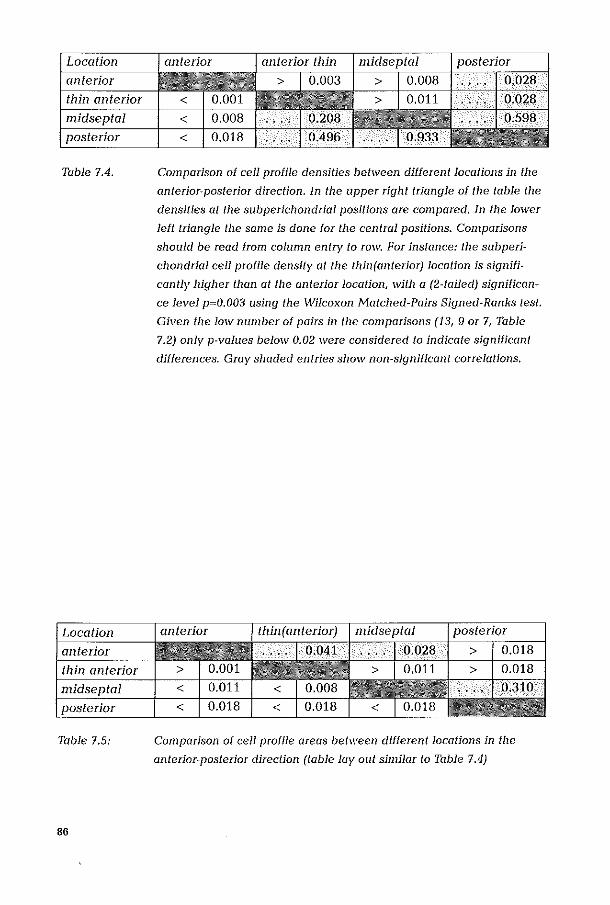
Table 7.4.
Table 7.5:
86
Comparison of cell profile densities between different locations in the
anterior~posterio[ direction. In the upper right triangle of the table the
densities at the subperichondriaJ positions ale compared. In the lower
left triangle the same is done for the central positions. Comparisons
should be read from column entr)' to row. For instance: the subperi
chondria1 cell profile density at the thin(anteriorj location is signifi
cantly higher than at the anterior location, with a (2-tailed) significan~
ce level p""O.003 using the \.VilCDXOll Matched-Pairs Signed-Ranks test.
Given the low number of pairs in the comparisons (13, 9 or 7, Table
7.2) only p-values below 0.02 weIe considered to indicate significant
differences. Gray shaded entries show non-significant correlations.
Comparison of cell profile areas between different locations in the
anterior-posterior direction (tabJe Jay Ollt similar to Table 7.4)
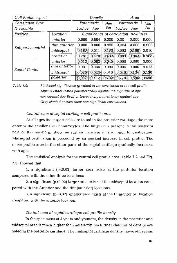
Cell Profile aspect Density Area
Correlation TYpe Parametric Non Parametric Non
X-variable LogAge Age Par LogAge Age Par
Position Location Significance of correlation (p-values)
anterior 0.000 0.004 0.000 0.001 0.000 0.000
0.000 0.000 0.000 0.000 0.003 thin anterior 0.004 Sub perichondrial
midseptal 0.187 0.011 0.170 0.002 0.109 0.016
Septal Center
Table 7.6:
posterior 0.281 0.170 0.432 0.883 0.963 0.908
anterior 0.513 0.083 0.045 0.000 0.000 0.000 thin anterior 0.001 0.000 0.000 0.006 0.000 0.013
midseptal 0.076 0.023 0.010 0.046 0.134 0.130 posterior 0.937 0.412 0.702 0.718 0.596 0.696
Statistical significance (p-value) of the correlation of the cell profile
aspects either tested parametrically against the logaritm of age
and against age itself or tested non parametrically against age.
Gray shaded entries show non-significant correlations.
Central zone of septal cartilage: cell profile area
At all ages the largest cells are found in the posterior cartilage, the more
anterior the smaller the chondrocytes. The large cells present in the posterior
part of the newborn, show no further increase in size prior to ossification.
Midseptal ossification is preceded by an marked increase in cell profile. The
mean profile area in the other parts of the septal cartilage gradually increases
with age.
The statistical analysis for the central cell profile area (Table 7.2 and Fig.
7.3) showed that:
1. a significant (p<0.02) larger area exists at the postmior location
compared with the other three locations;
2. a significant (p<0.02) larger area exists at the midseptallocation com
pared with the Anterior and the thin( anterior) locations;
3. a significant (p<0.02) smaller area exists at the thin(anterior) location
compared with the anterior location.
Central zone of septal cartilage: cell profile density
In the specimens of 4 years and younger, the density in the posterior and
midseptal area is much higher than anteriorly. No further changes of density are
noted in the posterior cartilage. The midseptal cartilage density, however, seems
87
to decrease in the specimens from 0 to 10 years of age (non significant). The density
in the anterior cartilage is only slightly (non significant) decreasing from the neo
natal to the adult stage. In all specimens the densitiy in the anterior thin area is
larger than in the anterior cartilage which nearly equals that of the posterior carti
lage.
This analysis for the central cell profile densities (Table 7.3 and Fig 7.4)
showed only a significantly (p<0.02) lower density at the anterior location compa
red with the other three locations.
Subpericilondrial cartilage: cell profile area
At birth the highest value for the cell profile area is found posteriorly, and
it remains unchanged until the onset of ossification. The midseptal cartilage shows
a rapid increase during the first year of life to reach, prior to ossification, a plateau
at the same level as observed in the posterior cartilage. Both the anterior and the
anterior thin area demonstrate a gradual increase of cell profile from birth to 35
years of life (oldest specimen). They finally reach a maximum comparable to the
values found in the midseptal and posterior regions.
From the statistical analysis (Wilcoxon) on differences in subperichondrial
cell profile areas between the locations in the anterior-posterior direction (Table 7.2
and Fig. 7.5) it was shown that:
1. a significant (p<0.02) larger area exists at the posterior location compared
with the anterior and the thin(anterior) locations, but not with the midseptal
location;
2. a significant (p<0.02) larger area exists at the midseptallocation com
pared with the Thin(anterior) location, but not with the anterior location;
3. no significant difference exists between the thin(anterior) location and
the anterior location.
SubpericilOndrial cartilage: cell profile density
At birth the density is relatively high in all areas. The midseptal region
shows a downward tendency with age. The anterior and anterior thin cartilage
demonstrate a very rapid decrease of density in the first 4 years of life, which is
slowing down after that period. In older specimens only a slight further decrease of
density was noted.
From the statistical analysis (Wilcoxon) on differences in subperichondrial
88
cell profile densities between the locations in the anterior-posterior direction
(Table 7.3 and Fig. 7.6) it was shown that:
1. no signicant difference exists between the posterior location and the
other three locations,
2. a significant (p<O.02) higher density exists at the midseptallocation
compared with the anterior, thin(anterior) location,
3. a significant (p<O.02) higher density exists at the thin(anterior) com
pared with the anterior location.
7.4 Discussion
Dimensional aspects of the postnatal development of the human nasal
septum were previously reported (Verwoerd et ai, 1989, Van Loosen et a!., 1996).
An interesting finding was that after birth the cartilaginous septum rapidly
expands to its maximal size at the age of 2 years. Further growth of the septum
appeared to be due to enlargement of the bony perpendicular plate (Van Loosen
et a!., 1996).
The present study deals with two features of the cartilaginous part of the
developing nasal septum, (1) the mean cell profile area, reflecting cell size, and
(2) the mean cell profile density, reflecting the relation between the cellular
component and the combined cellular and extra-cellular tissue components.
In literature no data on mitotic activity nor quantative histometric data
concerning evolution of cell size or intercellular volume could be found for the
human septum. These aspects, however, were studied in the cartilage of the
growing rabbit cricoid (Verwoerd-Verhoef et a!., 1997).
In that study it was concluded that a high mitotic activity at birth, in both
cartilage and perichondrium, slows down in a few weeks. After the age of 8
weeks no mitotic figures could be demonstrated and increase of cell size and/or
matrix components appeared responsible for further growth. It could suggest
that also in the nasal septum proliferation of cartilage cells is reshicted to the
first period of postnatal life.
Fronl our analysis of the series of human specimens of different ages, it
89
is evident that most changes occur dUling the first 10 years of life. Afterwards
only moderate and gradual changes were observed. Unfortunately, no
specimens were available in the age range from 10 to 20 years, the period in
which the adolescent growth spurt is known to take place. The evolution of the
measured values, before the age of 10 and after the age of 20, does not give any
indication of biological phenomena in the cartilage to be associated with a
growth spurt of the nasal septum.
The various parts of the cartilaginous septun1 show as far as profile area
and profile density are concerned, specific patterns of development. This
regionalisation coinciding with previously described regional differences in
septal thickness stresses that the nasal septum is not a homogeneous structure
with uniform properties. (van Loosen et ai, 1999). This conclusion is particularly
relevant for surgical interventions like partial resection, and transposition of
resected, eventually crushed, cartilage parts which all would interfere with the
functional 3-dimensional organisation of the septal cartilage.
An intriguing question is how this region-specific development is pro
grammed, and whether perichondrium and cartilage would play different roles
in this process. Regionalisation of the septal cartilage is not a unique human
phenomenon, as was demonstrated in a series of rabbit experinlents (Verwoerd
et ai., 1989, 1991; Verwoerd-Verhoef et ai., 1991, 1995). Detailed studies on
these questions, probably on a molecular biological level have not yet been
published, but are certenly needed for further iIlucidation.
Reviewing the specimens between 0 and 10 years of age, it is evident
there is a difference between the parts of the cartilaginous septum, which will
be involved in endochondral posterior and midseptal ossification and those
which remain cartilaginous ( anterior and anterior thin).
Prior to ossification the cartilage is characterised by a large cell profile. The
mean cell profile area in the anterior region changes from 60 AU to 120 AU.Cell
profile increases with a factor 2, whereas in the same period the cell density
hardly changes. Assuming that no cells are lost by apoptosis, a 100% increase of
the intercellular component may be concluded. Thus, the volume of cartilage
also increases with a factor 2. As the size of the cartilaginous septum does not
90
alter after the age of 2 years, the expansion of the cartilage is balanced by loss
of cartilage at the progressing front of ossification (Van Loosen et a!., 1996),
At birth the mean profile area in the subperichondrial and central zone
is the same for the anterior and anterior thin region, Cellular maturation of the
central zone explains the later increased cell profile in the anterior region, A
sinlilar phenonlenon is however to a lesser extent, observed in the anterior thin
region, where the subperichondrial character of the cells prevail,
In the period between 10-35 years of age, both in the anterior and the
anterior thin region the cell profile shows a gradual increase, the cell density a
minimal decrease. It is interesting that the anterior and anterior thin region
demonstrate permanent and consistent differences in cell profile and cell
density, The thin area is characterised by smaller cells and less intercellular
matrix in comparison to the anterior cartilage, This could relate to their different
bionlechanical function.
The antelior area is considered to contribute to the support of the nasal dorsum,
in contrast to the anterior thin area, which has most probably no 'load bearing'
function,
7.5 Conclusions
1. The results of this study demonstrate the dynamic nature of the carti
laginous septum during childhood, Increase in cell size, production of intercel
lular matrix and transformation into bone follow region-specific patterns, The
growing cartilaginous septum demonstrates an obvious three-dimensional orga
nisation in morphology and development. These biological features warn the
surgeon not to treat the nasal septum in children as just a supporting element.
Actually, better knowledge of the dynamic nature of the growing cartilage
should inlprove surgical strategies,
2, With increase of age, interstitional growth due to increase of cell size
and/or volume of intercellular substance shifts from mainly posterior to the more
anterior parts.
3, No signs for adolescence growth spurt of the nasal septum were
91
observed, but cannot completely be excluded.
4. As the absolute size of the septal cartilage reaches its maximal size at
the age of two, the later marked growth activity is most probably balanced by
endochondral ossification, resulting in the formation of the perpendicular plate.
References
Pirsig W, 1975, Die Regeneration des kindlichen Septumknorpels nach
Septumplastiken. Bine histologische Studie. Acta Otolaryngol, 79:451-459.
Pirsig W, 1986a, Rhinoplasty and the airway in children. Facial Plast Surg, 3:225-
234.
Pirsig W, 1986b, Open questions in nasal surgery in children. Rhinology, 24:37-
40.
Pirsig W, 1992, Growth of the deviated septum and its influence on midfacial
development. Facial Plast Surg, 8:224-232.
Schultz-Coulon HJ, Eckenneier L, 1976, Zum postnatalen Wachstum der
Nasenscheidewand. Acta Olola- ryngol, 82:131-142.
Tonneyck-Miiller I, van del' Werf F, 1982, Die Entwicklung des Septum Nasi
beim Kaninchen. II. Untersuchungen nach regionalen Unterschieden in der
Septum-Dicke. Acta Morphol Neerl Scand, 21:379-391.
Tonneyck-Miiller I, 1984, Die Entwicklung des Septum Nasi behn Kaninchen.
III. Quantitative Beobachtungen an Zelien und Interzellularsubstanz.
Acta Morphol Neerl Scand, 22:133-141.
Van Loosen J, Verwoerd-Verhoef HL, Verwoerd CD A, 1988, The nasal
cartilage in the newborn. Rhinology, 26: 161-165.
92
Van Loosen J, van Zanten GA, van Velzen D, Howard V, VerwoerdMVerhoef,
1996, Growth characteristics of the human nasal septum. Rhinology, 34:78-82.
Van Loosen J, Verwoerd-Verhoef HL, Verwoerd CDA, Van Velzen D, 1999,
The significance of regional variations in thickness of the human nasal septum.
Rhinology, submitted
Van Loosen J, van Zanlen GA, van Velzen D, Verwoerd-Verhoef HL, Verwoerd
CDA, 1996, Growth characteristics of the human nasal septum. Rhinology, 34:
78-82.
Verwoerd CDA, Verwoerd-Verhoef HL, 1989, Developmental aspects of the
deviated nose. Facial Plast Surg, 6:95-100.
Verwoerd CDA, Verwoerd-Verhoef HL, Meeuwis CA, 1989, Stress and wound
healing in the cartilaginous nasal septum. Acta Otolmyngol (Stockh), 107: 441-
445.
Verwoerd CDA, Verwoerd-Verhoef HL, Meeuwis CA, 1991, Wound healing of
autologous implants in the nasal septal cartilage. ORL, 53: 310-314.
Verwoerd-Verhoef HL, Meeuwis CA, van der Henl RO, 1991, Histologic
evaluation of crushed cartilage grafts in the growing nasal septum of young
rabbits. ORL, 53:305-309.
Verwoerd-Verhoef HL, Verwoerd CDA, 1995. Sino-nasal surgelY and growth:
an experimental study review. Rhinology, state of the art. Eds Tos M, Thomson
J and Balle V, Kugler Publications, Amsterdam/New York: 195-201.
Verwoerd-Verhoef HL, Adriaansen FCPM, Laeijendecker RJ, Van Osch GJVM,
Verwoerd CDA, 1997, Growth dynamics of the cricoid cartilage and subglottic
injury; An autoradiographic and histometJic study in the rabbit. Eur. Arch.
Otolaryngol, 254;1: 101-104.
93
Velter U, Pirsig W, Heinze E, 1983, Growth activity in human septal cartilage:
Age-dependent incorporation of labelled sulfate in different anatomic locations.
Plast Reconstr Surg, 71:167-179.
Vetter U, Pirsig W, Heinze E, 1984a, Postnatal growth of the human septal
cartilage. Acta Otolaryngol, 97:131-136.
Vetter U, Pirsig W, Helbing G, Heit W, Heinze E, 1984b, Patterns of growth in
human septal cartilage: A review of new approaches. Int J Ped Otorhino
Lmyngol,7:63-74.
94
Surgical Aspects of the Anatomy of the Vomer in Children and Adults.
95
8.1 Introduction
In most publications on septal surgery hardly any attention is paid to
the exact anatomy of the vomer and certainly not to the developmental changes
during childhood. Recently, Takahashi (1988) reviewed his anatomical studies of
the (isolated) vomer of dried human skulls together with many other aspects of
the septal development in various animal species.
The nasal septum is cartilaginous up to the 8th month of intra-uterine
life. Peter (1913); Hillenbrand (1933) and Schultz-Coulon and Eckermeier (1976)
described the development of the perpendicular plate by endochondral
ossification. The first anlage of the vomer is a bilateral centre of desmal
ossification just lateral to the basal rim of the septal cartilage (Fawcett, 1911).
Both centres fuse on the inferior side to form a bony gutter. The inferior part of
the vomer is the result of desnlal ossification located between the mucous
membranes lining both nasal fossae.
The perpendicular plate reaches the vomer between the 4th and 7th year after
birth (Hillenbrand, 1933) but according to Schultz-Coulon and Eckermeier
(1976), this may even occur after the age of 10 years. Thus, the most postero
inferior part of the cartilaginous septum ("sphenoid tail") is enclosed in a bony
tunnel (vomerine tunnel) and will ultimately ossify in most individuals (Scott
1953; Melsen 1977).
Developlnental variations at the junction of vanIer and perpendicular
plate are very common. A symmetrical development of the vomer is exception
rather than rule. In case only one valllera} ala is formed r during surgery the
cartilaginous sphenoid tail can be found extending along side the "vomeral
bone", actually the other ala. Often this situation is combined with a spina
vonleri.
To our knowledge reports describing the development of the nasal
septum radiographically have not been published. In this paper a radiographic
evaluation of the development of the vomer from birth to the age of 30 is
presented. Additionally, a short comment will be given on the anatomical
features of the vomer in skulls with facial clefts.
96
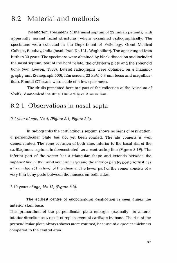
8.2 Material and methods
Postmortem specimens of the nasal septum of 22 Indian patients. with
apparently normal facial structures. where examined radiographically. The
specimens were collected in the Department of Pathology, Grant Medical
Collega, Bombay, India (head: Prof. Dr. U.L. Wagholikar). The ages ranged from
birth to 30 years. The specimens were obtained by block dissection and included
the nasal septum, part of the hard palate, the cribriform plate and the sphenoid
bone (van Loosen, 1988). Lateral radiographs were obtained on a manuno
graphy unit (Senograph 500t, fibn screen, 22 keY, 0.3 mm focus and magnifica
tion). Frontal CT-scans were made of a few specimens.
The skulls presented here are part of the collection of the Museum of
Vrolik, Anatomical Institute, University of Amsterdam.
8.2.1 Observations in nasal septa
0-1 year of age; N= 4, (Figure 8.1, Figure 8.2).
In radiographs the cartilaginous septum shows no signs of ossification:
a perpendicular plate has not yet been formed. The ala vomeris is well
demonstrated. The zone of fusion of both alae, inferior to the basal rim of the
cartilaginous septum, is demonstrated as a contrasting line (Figure 8.1F). The
inferior part of the vomer has a hiangular shape and extends between the
superior line of the fused vomerine alae and the inferior palate; posteriorly it has
a free edge at the level of the choana. The lower part of the vomer consists of a
very thin bony plate between the mucosa on both sides.
1-10 years of age; N= 13, (Figure 8.3).
The earliest centre of endochondral ossification is seen annex the
anterior skull base.
This primordium of the perpendicular plate enlarges gradually in antero
inferior direction as a result of replacement of cartilage by bone. The rim of the
perpendicular plate always shows more contrast, because of a greater thickness
compared to the central area.
97
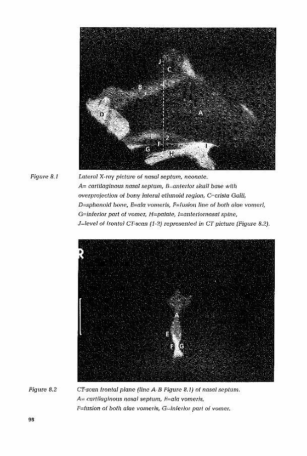
Figure 8.1
Figure 8.2
98
Lateral X-ray picture of nasal septum, neonate.
A= cartilaginous nasal septum, B=anterior skull base with
overprojection of bOllY lateral ethmoid region, C=crista Galli,
D=sphenoid bone, E=ala vomeris, F=fusion line of both alae vomeri,
G=inferior part of vomer, H=palate, I=anteriomasal spine,
J=level of frontal CT-scan (1-2) represented in CT picture (Figure 8.2).
CT-scan frontal plane (line A-B Figure 8.1) of nasal septum.
A= cartilaginous nasal septum, E=ala vomeris,
F=fusion of both alae vomeris, G=inferior part of vomer.
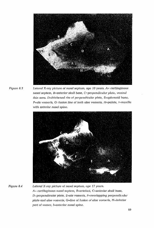
Figure 8.3
Figure 8.4
Lateral X-ray picture of nasal septum, age 10 years. A= cartilaginous
nasal septem, B=anterior skull base, C=perpendicular plate, ventral
thin area. D=lhickened rim of perpendicular plate, E=sphenoid bone,
F=ala Yomeris, G=fusion line of botli alae yomeris, H=palate, I=maxilla
willi anterior nasal spine.
Lateral X-ray picture of nasal septum, age 17 years.
A= cartilaginous nasal septem, B=artefact, C=anterior skull base,
D=perpendicular plate, E=ala vomeris, F=overlapping perpendicular
plate and alae vomeris, G=line of fusion of alae Yomeris, H=inferior
part of vomer, I=anterior nasal spine.
99
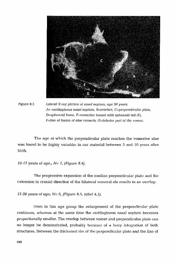
Figure 8.5 Lateral X-ray picture of nasal septum, age 30 years.
A= cartilaginous nasal septum, B=artefact, C=perpendicular plate,
D=sphenoid bone, E=vomerine tunnel with sphenoid tail (?),
F=line of fusion of alae vomeris, G=inferior part of the vomer.
The age at which the perpendicular plate reaches the vomerine alae
was found to be highly variable: in our material between 3 and 10 years after
birth.
10-17 years of age.; N= 7, (Figure 8.4).
The progressive expansion of the median perpendicular plate and the
extension in cranial direction of the bilateral vomeral ala results in an overlap,
17-30 years of age; N= 6, (Figure 8.5, tabel 4.1).
Even in this age group the enlargement of the perpendicular plate
continues, whereas at the same time the cartilaginous nasal septulll beconles
proportionally smaller. The overlap between vomer and perpendicular plate can
no longer be demonstrated, probably because of a bony integration of both
structures. Between the thickened rim of the perpendicular plate and the line of
100
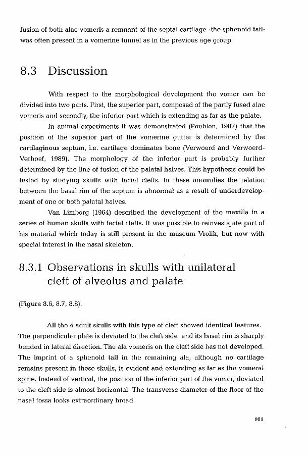
fusion of both alae vomeris a remnant of the septal cartilage -the sphenoid tail
was often present in a vomerine tunnel as in the previous age group.
8.3 Discussion
With respect to the morphological development the vomer can be
divided into two parts. First, the superior part, composed of the partly fused alae
vomeris and secondly, the inferior part which is extending as far as the palate.
In animal experiments it was demonstrated (Poublon, 1987) that the
position of the superior part of the vomerine gutter is determined by the
cartilaginous septum, i.e. cartilage dominates bone (Verwoerd and Verwoerd
Verhoef, 1989). The morphology of the inferior part is probably further
determined by the line of fusion of the palatal halves. This hypothesis could be
tested by studying skulls with facial clefts. In these anomalies the relation
between the basal rim of the septum is abnormal as a result of underdevelop
ment of one or both palatal halves.
Van Limborg (1964) described the development of the maxilla in a
series of human skulls with facial clefts. It was possible to reinvestigate part of
his material which today is still present in the museum Vrolik, but now with
special interest in the nasal skeleton.
8.3.1 Observations in skulls with unilateral cleft of alveolus and palate
(Figure 8.6,8.7,8.8).
All the 4 adult skulls with this type of cleft showed identical features.
The perpendicular plate is deviated to the cleft side and its basal rim is sharply
bended in lateral direction. The ala vomelis on the cleft side has not developed.
The inlprint of a sphenoid tail in the remaining ala, although no cartilage
remains present in these skulls, is evident and extending as far as the vomeral
spine. Instead of vertical, the position of the inferior part of the vomer, deviated
to the cleft side is almost horizontal. The transverse diameter of the floor of the
nasal fossa looks extraordinary broad.
101
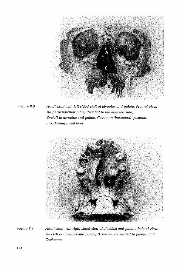
Figure 8,6
Figure 8,7
102
Adult skull with left~sided cleft of alveolus and palate, Frontal view;
A= perpendicular plate, diviated to the affected side,
B=cleft in alveolus and palate, C=vomer: 'horizontal' position,
broadening nasal floor
Adult skull with right-sided cleft of alveolus and palate, Palatal view,
A= cleft of alveolus and palate, B=vomer, connected to palatal half,
C=choana
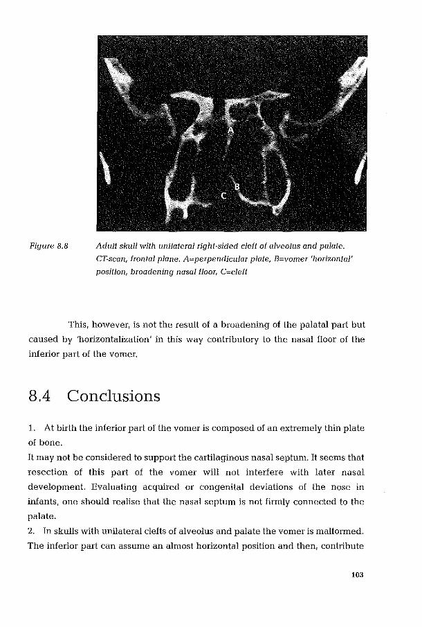
Figure 8.8 Adult skull with unilateral right-sided cleft of alveolus and palate.
CT-scan, frontal plane. A:=:perpendicular plate, B:=:vomer 'horizontal'
position, broadening nasal floor, C:=:cleft
This, however, is not the result of a broadening of the palatal part but
caused by 'horizontalization' in this way contributory to the nasal floor of the
inferior part of the vomer.
8.4 Conclusions
1. At birth the inferior part of the vomer is composed of an extremely thin plate
of bone.
It may not be considered to support the cartilaginous nasal septmn. It seems that
resection of this part of the vomer will not interfere with later nasal
development. Evaluating acquired or congenital deviations of the nose in
infants, one should realise that the nasal septum is not firmly connected to the
palate.
2. In skulls with unilateral clefts of alveolus and palate the vomer is malformed.
The inferior part can assume an almost horizontal position and then r contribute
103
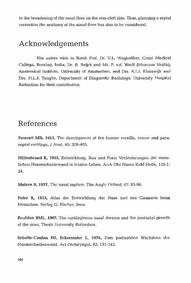
to the broadening of the nasal floor on the non-cleft side. Thus, planning a septal
correction the anatomy of the nasal floor has also to be considered.
Acknowledgements
The autors wish to thank Prof. Dr. U.L. Wagholikar, Grant Medical
Collega, Bombay, India, Dr. B. Baljet and Mr. F. v.d. Werff (Museum Vrolik).
Anatomical Institute, University of Amsterdam, and Drs. AI.J. Klooswijk and
Drs. H.L.J. Tanghe, Department of Diagnostic Radiology, University Hospital
Rotterdam for their contribution.
References
Fawcett MD, 1911, The development of the human maxilla, vomer and para
septal cartilage, J Anat, 45: 378-405.
Hillenbrand K, 1933, Entwicklung, Bau und Form Veriine!erungen der mens
lichen Nasenscheidewane! in fetalen Leben. Arch Ohr Nasen Kehl Heilk, 135:1-
24.
Melsen B, 1977, The nasal septum. The Angle Orthoe!, 47: 83-96.
Peter K, 1913, Atlas der Entwicklung der Nase une! e!es Gaumens beim
Menschen. Verlag G. Fischer, Jena.
Poublon RML, 1987, The cartilaginous nasal dorsum and the postnatal growth
of the nose, Thesis University Rotterdam.
Schultz-Coulon HJ, Eckermeier L, 1976, Zum postnatalen Wachstum der
Nasenscheidenwand. Act Otolaryngol, 82: 131-142.
104
Scott JH, 1953, The cartilage of the nasal septum. Br Dent J, 95: 37-43.
Takahashi R, 1988, The formation of the nasal septum and the etiology of septal
deformity. Act Otolaryngol, suppl, 443: 1-160.
Van Limborgh J, 1964, Some aspects of the development of the cleft affected
face. Int symposium, University of Zurich 1964, Hans Huber publishers, Bern
and Stuttgart.
Van Loosen J, Verwoerd-Verhoel HL, Verwoerd CDA, 1988, The nasal septal
cartilage in the newborn. Rhinology, 26: 161-165.
Verwoerd CDA, Verwoerd-Verhoel HL, 1989, Developmental aspects of the
deviated nose. J Facial Plastic Surg, 6: 95-100.
Verwoerd CDA, Verwoerd-Verhoef HL, 1998, Rhinosurgery in children;surgical
and developmental aspects. In: Rhinoplasty (2nd ed.), Nolst Trenite GJ (ed),
Kugler Publications, The Hague.
105
Computed Tomography of the Human Developing Ante rio r Sku II Base.
106
9.1 Introduction
Computed tomography (CT) is now widely used to investigate the
abnormalities of the anterior skull base. However, little attention has been given
to the different manifestations of the developing and ossifying anterior skull
base and to problems in the interpretation of CT in the first years of life.
Parts of the anterior skull base are fOlmed by membranous ossification
of fibrous tissue, whereas the anlage of other medial parts is cartilaginous
(Schaeffer, 1920; Mugnier, 1964; Hamilton, 1972) including the nasal septum
and crista Galli (van Loosen and van Velzen, 1989). The replacement of this
cartilage by bone is not complete at birth and continues during childhood. The
whole of the complex, consisting of the nasal septum, crista Galli, lamina
cribrosa (cribrifonn plate) and lateral ethmoids, initially develops as a cartilagi
nous structure (Fawcett, 1911; De Beer, 1937; Scott, 1953). In the neonate the
nasal septum is still cartilaginous (van Loosen et ai, 1988) and the upper lateral
cartilages extend into the anterior base of the skull (Poublon et aI., 1990). During
further development of the infant, between the first and sixth year of life,
ossification of the lamina cribrosa begins by fusion of the ossifying nasal septum
and the lateral ethmoid (Scott, 1953; Ford, 1958; Krimpotic-Nemanic, 1977;
Fairbanks, 1983).
This study was canied out to evaluate the CT pattern of ossifying structures in
humans.
9.2 Materials and Methods
9,2.1 Fetal material
16 human fetuses, referred to our laboratory for pathological
examination after spontaneous abortion, evenly distributed between 18 and 32
weeks gestational age were studied after routine fixation in 0.1 M phosphate
buffered formaldehyde 4 %, pH 7.4.
107
9.2.2 Infant material
Midfacial CT images were available in 3 children. (1. 2 and 6 years of
age respectively). who had been assessed for neurological symptoms.
The midfacial block specimen from the youngest child was also available for
histology.
9.2.3 Computed Tomography
CT of all fetal skulls was performed in coronal sections using a Philips
Tomoscan 350 with contiguous 1.5 mm slices.
In children only those CT scans were used, which were primmily
acquired in a coronal plane with identical slice thickness and spacing, to avoid
differences in resolution due to the method of data -acquisition.
9.2.4 Magnetic Resonance Imaging (MRI)
For the documentation of the non-osseous cartilaginous structures, 6
representative fetal skulls were available. They were analysed in a previous
collaborative study using MRI (Dyna scan, 4.2 Tesla MRI Zentraliorschungs
Institut, CIBA-GEIGY Basel), in the same plane (van Loosen and van Velzen,
1989; Allegrini et ai, 1990).
9.2.5 Tissue processing and histology
All midfacial blocks were decalcified and embedded in paraffin. 5 mu
semiserial sections were cut in a frontal plane and stained with combined Alcian
Blue I PAS Stain.
9.3 Results
9.3.1 Fetal age group
In all stages histological analysis demonstrated that the medial part
108
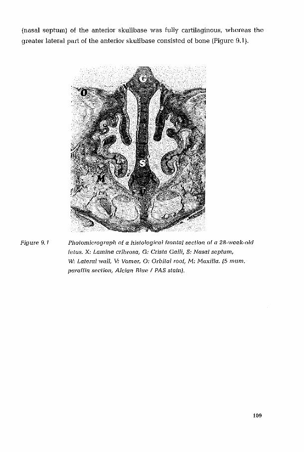
(nasal septum) of the anterior skullbase was fully cartilaginous, whereas the
greater lateral part of the anterior skullbase consisted of bone (Figure 9.1).
Figure 9.1 Photomicrograph of a histological frontal section of a 28-week-old
fetus. X: Lamina cribrosu, G: Crista Galli, S: Nasal septum,
W: Lateral wall, V: Vomer, 0: Orbital roof, M: Maxilla, (5 mum.
paraffin section, Alcian Blue / PAS stain).
109
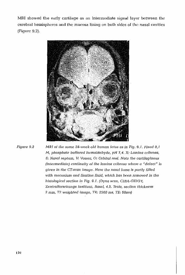
MRI showed the early cartilage as an intermediate signal layer between the
cerebral hemispheres and the mncosa lining on both sides of the nasal cavities
(Figure 9.2).
Figure 9.2
110
MRI of the same 28-week-old human fetus as in Fig. 9.1. Fixed 0,1
M, phosphate buffered formaldehyde, pH 7,4. X: Lamina cribrosu,
S: Nasal septum, V: Vomer, 0: Orbital roof. Note the cartilaginous
(intermediate) continuity of the lamina cribrosa where a "defect" is
given in the CT-scan image. Here the nasal fossa is partly filled
with meconium and fixation fluid, which has been removed in the
histological section in Fig. 9,1. (Dyna scan, elBA-GEIGY,
Zentraliorsclwngs Inslituut, Basel, 4,2. TesIa, section thickness
2 mm, T2 weighted image, TR: 2560 ms, TE: 60ms)
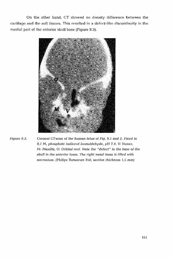
On the other hand, CT showed no density difference between the
cartilage and the soft tissues. This resulted in a defect-like discontinuity in the
medial part of the anterior skull base (Figure 9.3).
Figure 9.3. Coronal CT-scan of the human fetus of Fig. 9.1 and 2. Fixed in
0,1 !vi, pilospilate buffered formaldeilyde, pH 7.4. V: Vomer,
M: Maxilla, 0: Orbital roof. Note the "defect" in the base of the
skull in the anterior fossa. The right nasal fossa is filled with
meconium. (Philips Tomoscan 350, section thickness 1,5 mIll)
111
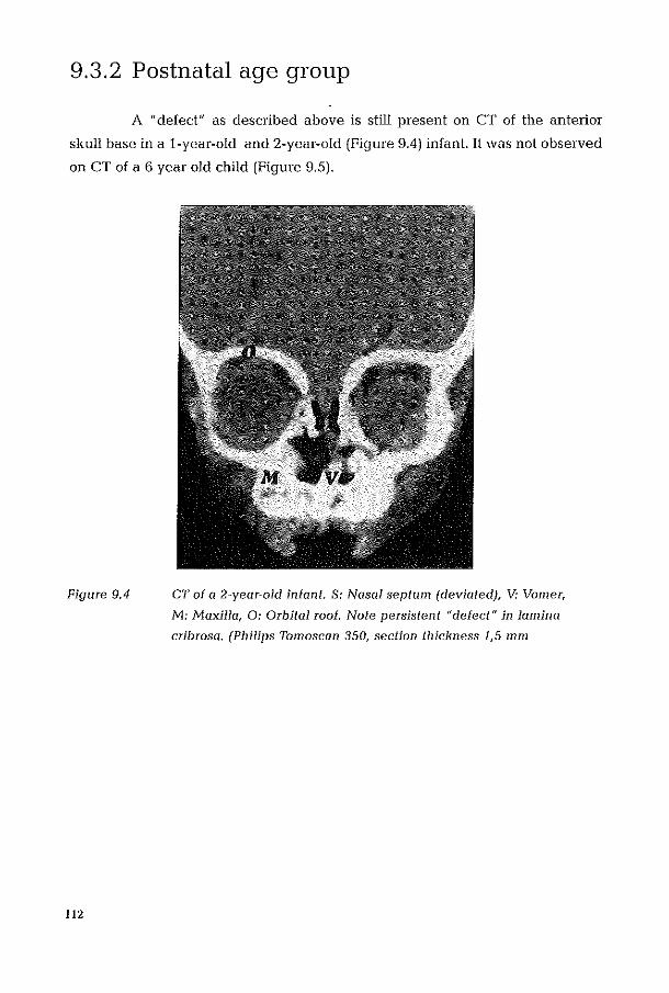
9.3.2 Postnatal age group
A "defect" as described above is still present on CT of the anterior
skull base in a 1-year-old and 2-year-old (Figure 9.4) infant. It was not observed
on CT of a 6 year old child (Figure 9,5).
Figure 9.4
112
CT of Q 2-year~old infant. S: Nasal septum (deviated), V: Vomer,
M: Maxilla, 0: Orbital rool. Note persistent II defect" in lamina
cribrosa. (Philips Tomoscan 350, section thickness 1,5 mm
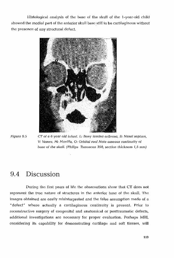
Histological analysis of the base of the skull of the i-year-old child
showed the medial part of the anterior skull basestill to be cartilaginous without
the presence of any structural defect.
Figure 9.5 CT of a 6 year old infant. L: Bony lamina cribrosa, S: Nasal septum,
V: Vomer, M: Maxilla, 0: Orbital roof Note osseous continuity of
base of the skull. (Philips TomoscQn 350, section thickness 1,5 mm)
9.4 Discussion
During the first years of life the observations show that CT does not
represent the true nature of structures in the anterior base of the skull. The
images obtained are easily misinterpreted and the false assumption made of a
"defect" where actually a cartilaginous continuity is present. Prior to
reconstructive surgery of congenital and anatOluical or posttraulnatic defects,
additional investigations are necessary for proper evaluation. Perhaps MRI,
considering its capabillty for demonstrating cartilage and soft tissues, will
113
become a better modality method of supplying this additional information,
especially as it collects and displays its data in very comparable modes.
The finding that the ossification of the lamina cribrosa in this, albeit
limited, material takes places between the second and sixth year of life, has not
been reported before. Detailed studies of the process of ossification of the
different contributing components of the base of the skull are warranted.
9.5 Acknowledgements
The authors are indebted to Dr. P.R. Allegrini and Dr. G.R. Bullock
(Zentralforschungs Institut, CIBA-Geigy Basel) for the use of MRI scan findings
derived from a previous collaborative study reported on separately (Allegrini et
aI, 1990) and to Dr. H.L. Verwoerd-Verhoef for her suggestions.
References
Allegrini PR, Van Velzen D, Van Loosen J, BullocI< GR, 1990, Differentiation of
proliferat\ng from r.esting cartilage in human fetal nasal septum by Magnetic
Resonance Imaging. Magn Res in Med, 14: 369-376.
De Beer GR, 1937, The development of the vertebrate skull. Oxford University
Press, Oxford.
Fairbanks DNF, 1983, Embryology and Anatomy. In: Bluestone C D (ed), 1983.
Pediatric Otolaryngology, Saunders, Philadelphia.
Fawcett E, 1911, The development of the human maxilla, vomer and paraseptal
cartilages. J Anat Lond, 45: 378-405.
Ford EHR, 1958, Growth of the human cranial base. AmJ Orthod, 44, 7: 498-506.
114
Hamilton WJ, Boyd, JD, Mossman, WH, 1972, Human embryology; prenatal
development of form and function. 4th edition, W. Heffer and sons Ltd.
Cambridge.
Krimpotic-Nemanic J, 1977, Entwicklungsgeschichte und Anatomie der Nase
und der Nasennebenhiihlen. In: Berendes J, Link R, Zollner F. (eds). 1977 Hals
Nasen Ohrenheilkunde in Praxis und Klinik, Georg Thieme Verlag Stuttgart.
Mugnier A, 1964, Embryologie et developpement bucco-facial. Masson et Cie,
Paris.
Poublon RML, Verwoerd CDA, Verwoerd-Verhoef HL, 1990, Anatomy of the
upper lateral cartilages in the human newborn. Rhinology, 28: 41-46.
Schaeffer JP, 1920, The nose, paranasal sinuses, nasolacrimal passageways and
olfactory organs in man. P Blakistons's Son and Co, Philadelphia.
Scott J H, 1953, The cartilage of the nasal septum. Br Dent J, 95: 37-43.
Van Loosen J, Van Velzen, Dr 1989, Cartilaginous structures in human midfacial
development. Ped Pathol, 8, 3: 350.
Van Loosen J, Verwoerd-Verhoef H L, Verwoerd CDA, 1988, The nasal septal
cartilage in the newborn. Rhinology 26: 161-165.
115
Summary and Concluding Remarks.
116
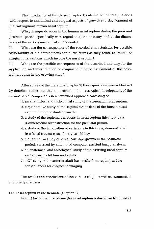
The introduction of this thesis (chapter 1) culminated in three questions
with respect to anatomical and surgical aspects of growth and development of
the cartilaginous human nasal septum:
1. What changes do occur in the human nasal septum during the peri- and
postnatal period, specifically with regard to a) the anatomy, and b) the dimen
sions of the various anatomical conlponents?
II. l"Ihat are the consequences of the recorded characteristics for possible
vulnerability of the cartilaginous septal structures as they relate to trauma or
surgical interventions which involve the nasal septum?
III. What are the possible consequences of the described anatomy for the
application and interpretation of diagnostic imaging assessment of the naso
frontal region in the growing child?
After survey of the literature (chapter 2) these questions were addressed
by detailed studies into the dimensional and microscopical development of the
various septal components in a combined approach consisting of:
1. an anatomical and histological study of the neonatal nasal septum.
2. a quantitative study of the sagittal dinlensions of the human nasal
septum during postnatal growth.
3. a study of the regional variations in nasal septum thickness by a
3-dimensional reconstruction for the postnatal period.
4. a study of the implication of variations in thickness, demonstrated
in a facial trauma case of a 4-year-old boy.
5. a quantitative study of septal cartilage growth in the postnatal
period, assessed by automated computer-assisted image analysis.
6. an anatomical and radiological study of the ossifying nasal septum
and vomer in children and adults.
7. a CT-study of the anterior skull-base (cribriform region) and its
consequences for diagnostic imaging.
The results and conclusions of the various chapters will be summarised
and bliefly discussed.
The nasal septum in the neonate (chapter 3)
In most textbooks of anatomy the nasal septum is described to consist of
117
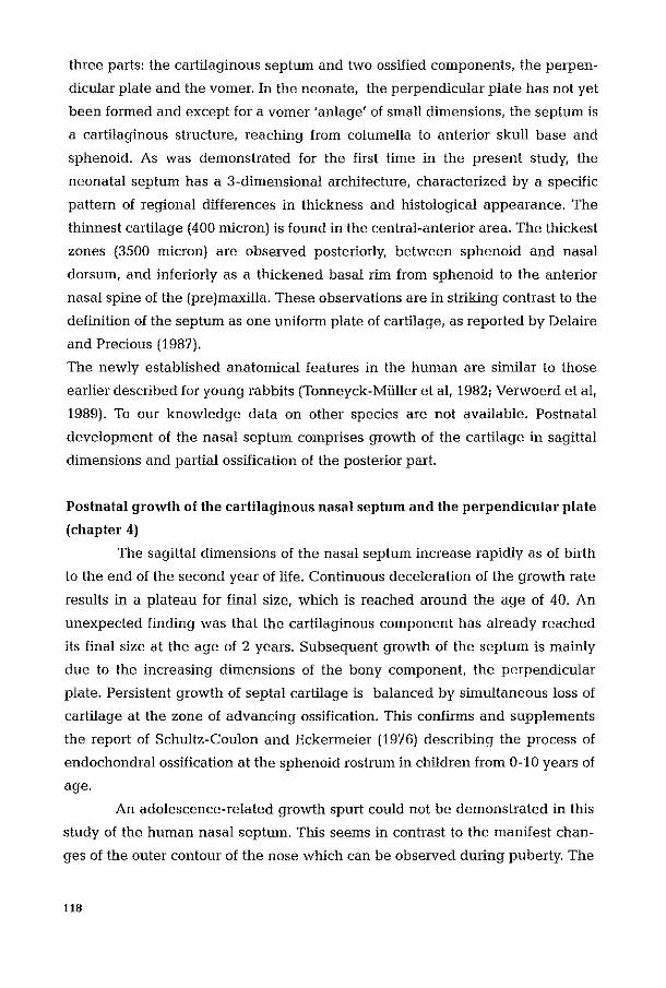
three parts: the cartilaginous septum and two ossified components, the perpen
dicular plate and the vomer. In the neonate, the perpendicular plate has not yet
been formed and except for a vomer 'anlage' of small dinlensions, the septum is
a cartilaginous structure, reaching from columella to anterior skull base and
sphenoid. As was demonstrated for the first time in the present study, the
neonatal septum has a 3-dimensional architecture, characterized by a specific
pattern of regional differences in thickness and histological appearance. The
thinnest cartilage (400 micron) is found in the central-anterior area. The thickest
zones (3500 micron) are observed posteriorly, between sphenoid and nasal
dorsum, and inferiorly as a thickened basal rim from sphenoid to the anterior
nasal spine of the (pre)maxilla. These observations are in striking contrast to the
definition of the septum as one uniform plate of cartilage, as reported by Delaire
and Precious (1987).
The newly established anatomical features in the human are sintilar to those
earlier described for young rabbits (Tonneyck-Miiller et aI, 1982; Verwoerd et aI,
1989). To our knowledge data on other species are not available. Postnatal
development of the nasal septum comprises growth of the cartilage in sagittal
dimensions and partial ossification of the posterior part.
Postnatal growth of the cartilaginous nasal septum and the perpendicular plate
(chapter 4)
The sagittal dimensions of the nasal septum increase rapidly as of birth
to the end of the second year of life. Continuous deceleration of the growth rate
results in a plateau for final size, which is reached around the age of 40. An
unexpected finding was that the cartilaginous component has already reached
its final size at the age of 2 years. Subsequent growth of the septum is mainly
due to the increasing dimensions of the bony component, the perpendicular
plate. Persistent growth of septal cartilage is balanced by simultaneous loss of
cartilage at the zone of advancing ossification. This confirms and supplements
the report of Schultz-Coulon and Eckermeier (1976) describing the process of
endochondral ossification at the sphenoid rostrum in children from 0-10 years of
age.
An adolescence-related growth spurt could not be demonstrated in this
study of the human nasal septum. This seems in contrast to the manifest chan
ges of the outer contour of the nose which can be observed dming puberty. The
118
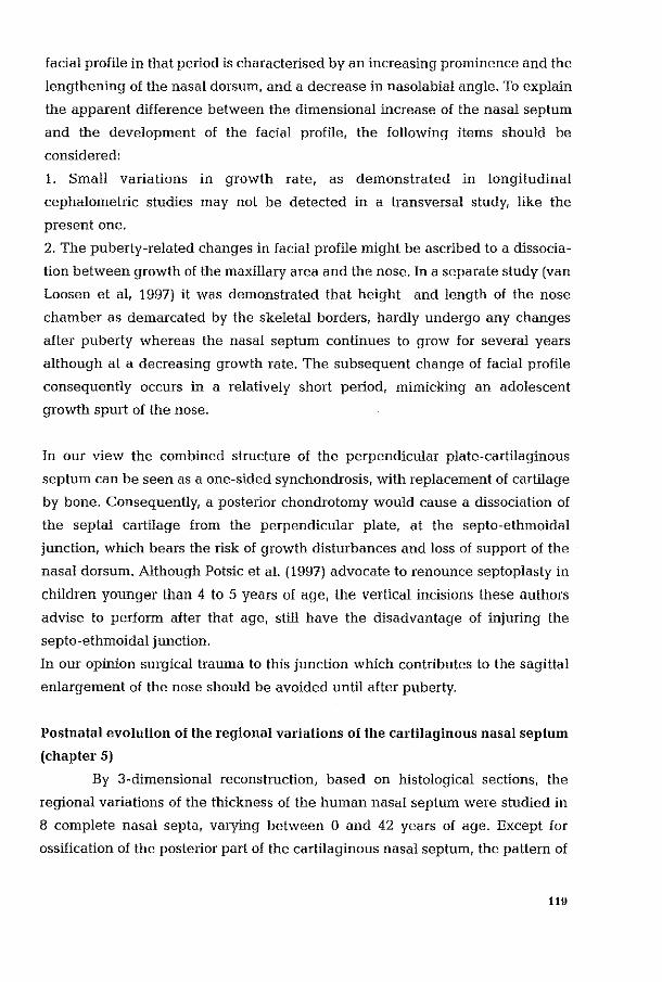
facial profile in that period is characterised by an increasing prominence and the
lengthening of the nasal dorsum, and a decrease in nasolabial angle. To explain
the apparent difference between the dimensional increase of the nasal septum
and the development of the facial profile, the following items should be
considered:
1. Small variations in growth rate, as demonstrated in longitudinal
cephalometric studies may not be detected in a transversal study, like the
present one.
2. The puberty-related changes in facial profile might be ascribed to a dissocia
tion between growth of the maxillary area and the nose. In a separate study (van
Loosen et ai, 1997) it was demonstrated that height and length of the nose
chamber as demarcated by the skeletal borders, hardly undergo any changes
after puberty whereas the nasal septum continues to grow for several years
although at a decreasing growth rate. The subsequent change of facial profile
consequently occurs in a relatively short period, mimicking an adolescent
growth spurt of the nose.
In our view the combined structure of the perpendicular plate-cartilaginous
septum can be seen as a one-sided synchondrosis, with replacement of cartilage
by bone. Consequently, a posterior chondrotomy would cause a dissociation of
the septal cartilage from the perpendicular plate, at the septo-ethmoidal
junction, which bears the risk of growth disturbances and loss of support of the
nasal dorsum. Although Potsic et al. (1997) advocate to renounce septoplasty in
children younger than 4 to 5 years of age, the vertical incisions these authors
advise to perform after that age, still have the disadvantage of injuring the
septo-ethmoidal junction.
In our opinion surgical trauma to this junction which contributes to the sagittal
enlargement of the nose should be avoided until after puberty.
Postnatal evolution 01 the regional variations of the cartilaginous nasal septum
(chapter 5)
By 3-dimensional reconstruction, based on histological sections, the
regional variations of the thickness of the human nasal septum were studied in
8 complete nasal septa, varying between 0 and 42 years of age. Except for
ossification of the posterior part of the cartilaginous nasal septum, the pattern of
119
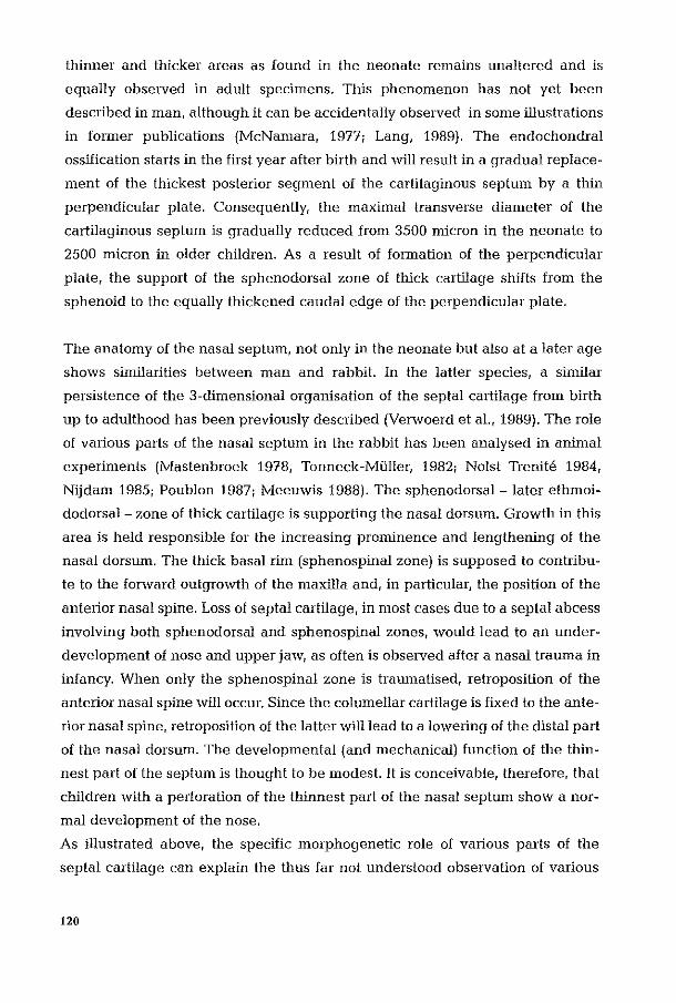
thinner and thicker areas as found in the neonate remains unaltered and is
equally observed in adult specimens. This phenomenon has not yet been
described in man, although it can be accidentally observed in some illustrations
in former publications (McNamara, 1977, Lang, 1989). The endochondral
ossification starts in the first year after birth and will result in a gradual replace
ment of the thickest posterior segment of the cartilaginous septum by a thin
perpendicular plate. Consequently, the maximal transverse diameter of the
cartilaginous septum is gradually reduced from 3500 micron in the neonate to
2500 micron in older children. As a result of formation of the perpendicular
plate, the support of the sphenodorsal zone of thick cartilage shifts from the
sphenoid to the equally thickened caudal edge of the perpendicular plate.
The anatomy of the nasal septum, not only in the neonate but also at a later age
shows similarities between man and rabbit. In the laller species, a similar
persistence of the 3-dimensional organisation of the septal cartilage from birth
up to adulthood has been previously described (Verwoerd et aI., 1989). The role
of various parts of the nasal septum in the rabbit has been analysed in animal
experiments (Mastenbroek 1978, Tonneck-Miiller, 1982, Nolst Trenite 1984,
Nijdam 1985, Poublon 1987, Meeuwis 1988). The sphenodorsal - later ethmoi
dodorsal- zone of thick cartilage is supporting the nasal dorsum. Growth in this
area is held responsible for the increasing prominence and lengthening of the
nasal dorsum. The thick basal rim (sphenospinal zone) is supposed to contribu
te to the forward outgrowth of the maxilla and, in particular, the position of the
antelior nasal spine. Loss of septal cartilage, in most cases due to a septal abcess
involving both sphenodorsal and sphenospinal zones, wonid lead to an under
development of nose and upper jaw, as often is observed after a nasal trauma in
infancy. When only the sphenospinal zone is trarmlatised, retroposition of the
anterior nasal spine will occur. Since the columellar cartilage is fixed to the ante
rior nasal spine, retroposition of the laller will lead to a lowering of the distal part
of the nasal dorsum. The developmental (and mechanical) function of the thin
nest part of the septum is thought to be modest. It is conceivable, therefore, that
children with a perforation of the thinnest part of the nasal septum show a nor
mal development of the nose.
As illustrated above, the specific morphogenetic role of various parts of the
septal cartilage can explain the thus far not understood observation of various
120
types of facial (mal)development occUlTing after traumatic loss of septal
cartilage. Although differing opinions are put forward in publications about the
relationship between septal perforation and nasal development (Pirsig, 1992), it
is evident that a perforation will affect nasal growth depending on the exact
localisation of the defect in the septum, or not.
Post-mortem study of the fractured septum in a 4-year-old child (chapter 6)
The fracture in this patient involved both the cartilaginous septum and
the perpendicular plate.
The fracture line can be characterized as a lie". The vertical part is situated in
the thin central area of the perpendicular plate. The inferior extension crosses
the septo-ethmoidal junction and follows the thinner zone of septal cartilage just
superior to the thickened basal rim. The superior extension of the -C- also
crosses the anterior edge of the perpendicular plate and runs into the thinner
zone just under the nasal dorsum. OUf observation is in contrast with the recent
publication of Shapiro (1996) in which he stated that -C-shaped fractures,
common in adults, do not exist in children. Hanison (1979) was the first to
desclibe the C-shaped septal fracture in adults without giving any biological
explanation. Actually, we demonstrated that the C-shape fracture line tends to
follow the thinnest, most vulnerable, parts of the septum.
Developmental aspects 01 septal deviations ( chapter 5 and 6)
The regional differences in thickness of the human nasal septum can
also help to understand the preferred sites of septal deviations, as they were
described by Mladina (Mladina, 1987; Mladina and Krajina, 1989). Their
observations were confirmed by Gi-Min et al. (1995) in a nationwide survey in
Korea, including more than 9000 persons, on the prevalence and risks of septal
deformities. Both authors considered most deviations to develop after trauma in
early childhood, but were not aware of the regional differences in the structure
of the nasal septum. The preference of septal deviations and angulations for the
anterior site, as reported by Mladina (category 1 and 2), seems to coincide with
the thinner parts of the nasal septum, as recorded in our study .. The fracture
pattern as described in chapter 6, would develop in more complex types of
septal deviations involving both the cartilaginous and bony septum, like the
types 3 - 7 as classified by Mladina (1987, 1989).
121
I! may be assumed that the thinner areas of the septum are less resistent to
mechanical forces and tend to fracture more easily, Mapping of thinner and
thicker areas (with different elasticity and plasticity modus) contribute to under
standing some mechanical aspects of nasal fracturing due to external trauma in
childhood,
Interstitial growth of nasal septal cartilage (chapter 7)
The interstitial growth of the nasal septal cartilage expressed in the
mean cell profile area and the mean cell profile density was studied for 4
different areas and 2 different locations, using an automated Computer Assisted
Image Analysis Technique, The results for the posterior, middle, anterior and
ant81ior thin area in central and subperichondrial regions were plotted against
age.
I! was demonstrated that the cartilaginous part of the human nasal septum has
a specific 3-dimensional organization with regard to local differences of cell size
and the proportionate amount of intercellular matrix in relation to location
within the septum, These data are in agreement with histochemical studies of
Vetter et al. (1983, 1984a, b), who demonstrated that the human nasal cartilage
is built up from different areas displaying partial age dependency, strict local
distribution and predominance of either matrix synthesis or cell replication and
proliferative activity, The latter studies, however, do not differentiate between
subperichondrial and central parts of the septum, and give no specific definition
of the exact location (pirsig, 1986),
The quantative histometric approach of our study images the complete carti
laginous septum, and revealed that from birth till its endochondral ossification
at the age of 4 years, the posterior region demonstrates no changes in cell size
nor cell cell density, It can be concluded that the cartilage in the posterior area
shows no signs of interstitial growth until it becomes ossified around 4 years of
age, In the midseptal zone there is a rapid increase of cell profile area for both
locations, central and subperichondrial, till the age of 2 years, The cell density
decreases linear and rapidly till the age of 10 years: the time of endochondral
ossification of central and subperichondrial regions in that area. In conclusion,
the interstitial growth in the midseptal area is fast and continues until the
ossification process encroaches the cartilage, In the anterior part of the septum,
the cell profile area in central as well as subperichondrial region enlarges
122
gradually but over a long period, at least far into adulthood. Simultaneously, the
cell density shows a marked decrease, indicating a continuous production of
intercellular substance.
Interstitial growth of the septal cartilage up to the adult age can play an im
portant role in the formation of angulation or overlap of septal fragments after
traumatic fracturing, and can explain the increase in dimensions of these septal
anomalies, even in adult patients.
Developmental anomalies 01 the vomer (chapter 8)
During rhinosurgery, malfOlmations of the septovomeral junction and
the ethmoidovomeraljunction are often observed. X-ray pictures (lateral projec
tion) of nasal septum specimens from ages between 0 and 42 years have brought
new- information about the occurrence of anatonlical variations. The anlage of
the vomer is complex. The inferior part is a plate consisting of a existing of bony
lamella between the basal rim of the septal cartilage and the superior surface of
the palate. In the neonate, the bone of the inferior part is so fragile that it is pos
sible to mobilize the palate versus the cartilaginous septum of the nose. The
superior part of the vomer is formed by ossification on both sides of the basal rim
of the septal cartilage (vomer wings). Superior and inferior parts together form
a V-shaped bony gutter enclosing the cartilaginous rim. The junction between
perpendicular plate and vomer frequently shows anatomical variations. The
ossifying front of the perpendicular plate can reach the bottom of the vomeral
groove or save a strip of cartilage between the vonleral wings. These wings
often develop asymmetrically. A local deviation of the basal rim of the septum to
one side, in combination with underdevelopment of the vomeral wing on the
sanle side, is a common finding. Such a spina vOlneris is in most cases accom
panied by a persistent posterior part of the septal cartilage, the sphenoid tail.
Then the sphenoid tail extends in posterior direction, along the unilaterally
formed vomeral wing.
In patients with a unilateral cleft of alveolus and palate, the nasal floor
on the non-cleft side is malformed. In these cases the basal rim of the septal
cartilage and the perpendicular plate are deviated to the cleft side. At the same
time, the inferior part of the vomer extending from the palatal half on the non
cleft side to the deviated basal rim of the septum, shifts into an almost horizon
tal position, thereby broadening the nasal floor on the non-cleft side. The
123
consequence is an angle between the inferior part of the vomer and the superior
part, which is fused with the perpendicular plate.
Ossification of the anterior skull base (chapter 9)
In the neonate the skeleton of the nose and the middle part of the
anterior skull base from one cartilaginous complex. In particular, the cribriform
plate area has been studied by computed tomography (CT) and histopathology.
It was demonstrated that at a young age also the anterior paramedian skull base
stili is completely cartilaginous. Ossification of this structure takes place
between the second and the sixth year of life. During the first years of life, the
cartilaginous prefornlation of the antm10r skull base creates a Ilpseudo defect"
on CT in the coronal plane. Early presence of ossified segments in the cribriform
area, as reported by Bosma (1986), could not be identified.
In a recent publication on the maturation processes of the skull base and
midface, Naidich et a!., (1996) suggest that in a small part of the population,
ossification nlay commence very early after birth. In other individuals ossifica
tion may not be complete even as late as the sixth year of life. From the reviewed
literature, it is not always clear whether the CT- sampling is sufficiently
representative. A considerable vmiability seems to, or at least may, exist con
cerning this segment of the base of the skull. The introduction of MRI, providing
sufficient resolution through the efficient use of T1 and T2 weighted images,
produced an opportunity for high contrast representation of cartilaginous
structures. Furthermore, it even allows for functional assessment of cartilage
through its principal ability to differentiate between proliferating and resting
e.g. immature and mature cartilage due to the differences in water-binding
properties of the more or less sulphated glycosaminoglycans of the matrix
(Allegrini et aI., 1990).
In contrast to CT-scanning, the representation of the soft/bony tissue-to
air interface remains a problem in MRI (Zinreich et a!., 1996). Nevertheless, this
imaging technique seems for now to be the essential replenishment of diagnos
tic imaging for the anterior skull base, in the first years of life.
124
Conclusion
The last decades brought an innovation of nasal surgery in adult patients. New
instrunlents were introduced, new surgical methods were defined, based on
better understanding of the anatomy. This study has demonstrated that the
morphology of the nose in children is different, and has stressed the dynamic
nature of the cartilaginous nasal skeleton during postnatal growth. In view of the
potential benefits of modern surgical interventions further exploration of the
mechanisllls in development and wound healing of cartilage remains a
challenge.
References
Allegrini PR, van Velzen D, van Loosen J, Bullock GR 1990, Differentiation of
proliferating from resting cartilage in human fetal nasal septum by Magnetic
Resonance Imaging, Mag Res in Med, 14: 369-376.
Bosma JF, 1986, The anatomy of the infant head. John Hopkins University Press,
Baltimore, USA.
Delaire J, Precious D, 1987, Interaction of the development of the nasal septum,
the nasal pyramid and the face. Int J Ped Otorhinolaryngol, 12: 311-326.
Gi-Min Y, Won Jung H, Sun Kim C, 1995, The prevalence study of nasal septal
deformities in Korea: Results of a nation-wide survey. Rhinology, 33: 61-65.
Harrison D, 1979, Nasal injuries: their pathogenesis and treatment. Br J Plast
Surg, 32:57-64.
Lang J, 1989, Clinical anatomy of the nose, nasal cavity and paranasal sinuses.
Thieme Verlag, New York, Stuttgart.
125
McNamara JA jr (ed.), 1977, Monograph No.7. Craniofacial Growth Series,
Center for Human Growth and Development, University of Michigan, Ann
Arbor.
Maslenbroek GJ, 1978, De invloed van parliele resectie van van het neus
tussenschot op de uitgroei van bovenkaak en neus. Thesis, University of
Amsterdam.
Meeuwis CA, 1988, Wondreac!ies in het kraakbenige neustussenschot. Thesis,
Erasmus University Rotterdam.
Mia dina R , 1987, The role of maxillar morphology in the development of
pathological septal deformities. Rhinology, 25:199-205.
Mia dina R, Krajina Z, 1989, The influence of the caudal process on the
formation of septal deformities. Rhinology, 27:113-118.
Naidieh TP, Zimmerman RA, Bauer BS, Altman NR, Bilaniuk LT, 1996, Midface:
Embryology and congenital lesions. In: Head and Neck Imaging, Som PM,
Curtin HD (eds). Mosby, St. Louis.
Nolst Trenite GJ, 1984, Implantaten in een groeiend neustussenschot. Thesis,
Erasnlus University Rotterdanl.
Nijdam DC, 1985, Schedelgroei na partiele submukeuze resectie van het neus
tussenschot. Thesis, University of Anlsterdam.
Ph'sig W, 1986, Rhinoplasty and the airway in children. J Fac Plast Surg, 3: 225-
234.
Pirsig W, 1992, Growth of the deviated septum and its influence on midfacial
development. J Fac Plast Surg, 8: 224-232.
Polsie W.P, Cotton R.T., Handler S.D., 1997, Surgical pediatric otolaryngology,
Thieme Verlag, New York, Stuttgart.
126
Poublon RML, 1987, The cartilaginous nasal dorsum and postnatal growth of the
nose. Thesis, EraSlllUS University Rotterdam.
Shapiro AM, 1996, Injuries of the nose, facial bones and paranasal sinuses. In:
Paediatric Otolaryngology, Bluestone C D, Stool S E, Kenna M A (eds), W B
Saunders, Philadelphia.
Schultz-Coulon HJ, Eckermeier L, 1976, Zum postnatalen Wachstum der
Nasenscheidewand, Acta OtolaryngoI, 82: 131-142.
Tonneyck-MiiIler I, van der Wed, 1982, Die EntwickIung des Septum Nasi beim
Kaninchen. II. Untersuchungen nach regionalen Unterschieden in der Septum
Dicke. Acta Morphol Neerl-Scand, 20:379-391.
Tonneyc!,-Miiller I, 1984, Die Entwicklung des Septum Nasi beim Kaninchen,
III. Quantitative Beobachtungen an Zellen und Interzellularsubstanz. Acta
Morphol Neerl-Scand 22: 133-141.
Van Loosen J, Baatenburg de Jong RJ, van Zanten GA, Engel T, Lanjewar DN,
van Velzen D, 1997, A cephalometric analysis of the nasal septal growth. Clin
Otolaryngol, 22: 453-458.
Verwoerd CDA, Verwoerd-Verhoef HL, 1989, Developmental aspects of the
deviated nose. J Fac Plast Surg, 6:95-100.
Verwoerd CDA, 1992, Present day treatment of nasal fractures: closed versus
open reduction. Fac Plast Surg, 8: 220-223.
Verwoerd CDA, Verwoerd-Verhoef HL, 1998, Rhinosurgery in children;surgical
and developmental aspects. In: Rhinoplasty (2nd ed.). Nolst Trenite GJ (ed),
Kugler Publications, The Hague.
Veller U, Pirsig W, Heinze E, 1983, Growth activity in human septal cartilage:
Age-dependent incorporation of labelled sulfate in different anatomic locations.
Plast Reconstr Surg, 71:167-179.
127
Vetter U, Pirsig W, Heinze E, 1984a, Postnatal growth of the human septal
cartilage. Acta Otolaryngol, 97:131-136.
Vetter U, Pirsig W, Helbing G, Heit W, Heinze E, 1984b, Patterns of growth in
human septal cartilage: A review of new approaches. Int J Ped Otorhino
Laryngol,7:63-74.
Zinreich SJ, Benson ML, Oliverio PJ, 1996, Sino-nasal cavities: CT normal
anatomy, inlaging of the osteo-meatal complex, and functional endoscopic
surgery. In: Head and Neck Imaging, Som PM, Curtin HD (eds), Mosby,
St.Louis.
128
Samenvatting en Conclusies.
129
In de laatste twee decennia is vooral door de Rolterdamse Pediatrische
KNO Groep experimenteel onderzoek gedaan naar de postnatale groei van de
neus en de invloed daarop van beschadiging of defecten van het kraakbenige
neustussenschot. Hierbij bleek dat chirurgische of traumatische interventies aan
het neustussenschot specifieke stoornissen kunnen geven van de uitgroei van
neus en soms oak van bovenkaak en orbita.
AIs men poogt de resultaten van deze experimenten te extrapoleren
naar de mens blijkt dat in de literatuur bijzonder weinig bekend is over het nog
snel groeiende neustussenschot op de kinderleeftijd,
Deze constatering heeft geleid tot drie vragen (Hoofdstuk 1):
I. Welke ontwikkelingen vinden in het humane neustussenschot plaats
gedurende de postnatale periode met belrekking tot:
a. de anatomie
b, de ontwikkeling van de verschillende delen (lamina perpendicularis,
kraakbenige neustussenschot en vomer)?
II, Welke zijn de mogelijke consequenties van deze ontwikkelingen voor
de eventuele vulnerabiliteit van het neustussenschol in relatie tot trauma en chi
rurgische interventies?
III. Welke consequenties heeft deze anatomische ontwikkeling vom de
interprelatie bij beeldvonnende diagnosliek van de nasofronlale regio in het
jonge kind?
Na een literatuursludie (Hoofdsluk 2) werden deze vragen beantwoord
door macroscopisch en microscopisch onderzoek naar de ontwikkeIing van de
verschillende componenten van het humane neusseptum,
Dit onderzoek beslaat uit de volgende onderdelen:
1. Een anatomische en histologische studie van hel neonatale neustus
senschot (Hoofdsluk 3),
2, Een kwantilatieve studie van de groei van hel humane neustussen
schot gedurende de postnalale periode (Hoofdstuk 4),
130
3. Een studie van de regionale dikteverschillen in het humane neustus
senschot met behulp van een 3-dimensionale reconstructie (Hooldstuk 5).
4. De consequenties van deze dikteverschillen met betrekking tot lrac
turen in het humane neustussenschot aan de hand van de beschrijving van het
aangezichtsletsel bij een 4-jarig kind (Hooldstuk 6).
5. Een kwantitatieve s!udie naar de interstitiele groei van het kraakbe
rug neustussenschot (Hooldstuk 7).
6. Een anatomische en radiologische studie van het neustussenschot en
vomer bij kinderen en volwassenen (Hooldstuk 8).
7. Een morlologische studie van ontwikkelingen in de voorste schedel
groeve en de consequenties voor de beeldvormende diagnostiek (Hooldstuk 9).
De resultaten en de conclusies van de verschillende hoofdstukken wor
den samengevat en kort bediscussieerd.
Het neustussenschot van de pasgeborene (Hoofdstu]{ 3)
In de meeste anatomische leerboeken word! het neus!ussenschot
beschreven als bestaande uit een kraakbenig deel en twee benige componen
ten, de lamina perpendicularis en het vomer. Bij de pasgeborene is de lamina
perpendicularis nog niet, en het vonler nauwelijks gevorrnd,
Het neustussenschot is vrijwel geheel kraakbenig en reikt van columella tot
voorste schedelgroeve en sphenoid.
In dit onderzoek wordt voar het eerst aangetoond dat het neustussen
schot van de pasgeborene een specilieke 3-dimensionale opbouw heef!, geka
rakteriseerd door een patroon van regionale dikteverschillen en histologische
beelden. Het dunste kraakbeen (400 micron) bevindt zich in het anterieure deel
van het septum. De dikste gebieden (3500 micron) worden aangetroffen poste
lieur tussen het sphenoid en het neusdorsulll en inferieur als een verdikte
131
kraakbenige zone verlopend van het sphenoid naar de spina nasalis anterior
van de maxilla.
Deze waarnenlingen zijn in tegenspraak met een eerdere publicatie \vaarin het
neustussenschot wordt beschreven als een structuur zonder dikteverschillen
(Delaire and Precious, 1987).
Een vergelijkbare bouw van het neustussenschot werd eerder beschreven voar
het jonge konijn (Tonneijk-Miiller et ai, 1982; Venvoerd et ai, 1989).
Voar zover ODS bekend zijn er geen gegevens van deze aard bij andere dier
soorten beschikbaar.
Poslnalale groei van hel kraakbenige neuslussenschol en de lamina perpendi
cularis (Hoofdsluk 4)
Het neustussenschot neemt in lengte en hoogte zeer snel toe vana! de
geboorle tot aan hel einde van het tweede levensjaar. Hiema resulteert een aan
vankelijk snel en later geleidelijk afnemende groeisnelheid uiteindelijk in een
"plateau" waarbij de maximale omvang in het me diane vlak is bereikt rand het
40e levensjaar. Een onvenvachte bevinding is dat het cartilagineuze deel reeds
op 2-jarige leeftijd zijn maximale omvang heef!. Hieruit valt te concluderen dat
de groei van het totale septum (exclusie! het onderste deel van het vomer) na
deze periode is toe te schrijven aan uitbreiding van de benige lamina perpendi
cularis als gevolg van doorgaande enchondrale verbening van het neustussen
schol.
Het daarmee gepaard gaande verlies van kraakbeen wordt kennelijk gecom
penseerd door groei elders in het kraakbenige septum. De bevindingen in dit
onderzoek bevestigen en vullen het onderzoek van Schultz-Coulon en
Eckermeier (1976) aan, die het proces van enchondrale verbening ter plaatse
van het sphenoid beschreven bij kinderen jonger dan 10 jaar.
Door een posterieure chondrotomie zoals toegepast wordt in de sep
tumchirurgie worden het septale kraakbeen en de lamina perpendicularis van
elkaar gescheiden, TIlet als risico dat het verbeningsproces en hiermee de groei
en de ondersteuning van het neusdorsum worden verstoord. Dit risico is niet
132
beperkt tot kinderen beneden de leeftijd van 4 tot 5 jaar zoals Potsie et al (1997)
aannenlen.
Naar onze mening dient chirurgie van deze regio welke bijdraagt tot de uitgroei
van de neus bij, voorkeur vermeden te worden tot na de puberteit.
Aanwijzingen voor een puberteitsgroeispurt ·werden in deze studie VOGr
het humane neustussenschol niel gevonden. Oit kan verklaard worden doordat
kleine variaties in groeisnelheid, zoals in sommige longitudinale studies waaI
genoillen, in een transversale studie zoals deze niet aangetoond kunnen woy
den. Het niet vastslellen van een puberteitsgroeispurt lijkt ook in tegenspraak
met de verandering van het profiel van het aangezicht en de neus in het bij
zander, die kan worden waargenomen tijdens en na de puberteit. Wat de neus
betreft gaat het dan om toename van de lengte van de neusrug en de hoogte van
de neus ten opziehte van de bovenkaak en tenslolte om een verkleining van de
nasolabiale hoek.
Oeze veranderingen van het aangezichtsprofiel behoeven niet per se op een
groeispurt van de neus te wijzen, maar zouden oak kunnen worden toegeschre
ven aan een verschil in groeisnelheid tussen het maxillaire gebied en het neus
tussenschot tijdens deze periode.
In een separate studie (Van Loosen et ai, 1997) werd aangetoond dat de hoogle
en lengte van de benige neuskamer nauwelijks enige veranderingen tijdens en
na de puberteit ondergaan, terwijl het neustussenschot (en de neus) dan nog
een aantal jaren, zij het met afnemende snelheid, doorgroeit. Oit kan verant
woordelijk zijn voor een ogenschijnlijke puberteitsgroeispurt van de neus.
Poslnalale ontwikl<eling van regionale dikteverschillen van het kraakbenige
neuslussenschot (Hooldsluk 5)
ODor middel van 3-dimensionale reconstructies gebaseerd op histologi
sche coupes werden de regionale dikteverschillen bestudeerd in 8 humane
neustus- senscholten, varierend in leeftijd van a tot 42 jaar. Afgezien van het
verlies van het posterieure gedeeJte van het kraakbenige neustussenschot als
gevolg van verbening blijven de dikke en dunne gebieden, zoals deze werden
133
gevonden bij de pasgeborene (Hoofdstuk 3), tot op de volwassen leeftijd aan
wezig. Dit is - merkwaardig genoeg - niet eerder bij de mens beschreven. Toch
kan dit fenomeen in enkele illustraties van eerdere publicaties duidelijk worden
herkend (McNamara, 1977; Lang, 1989).
De enchondrale verbening van het kraakbenige neustussenschot start geduren
de het eerste levensjaar met als resultaat dat het dikste posterieur gelegen
kraakbeep wordt vervangen door de benige lamina perpendicularis.
Zodoende wordt de maximale dikte van het overblijvende septale kraakbeen
gereduceerd van 3500 micron bij de pasgeborene tot 2500 micron bij oudere
klnderen. Het dikke kraakbeen wordt nog steeds gevonden in de twee eerder
beschreven zones: (1) tussen sphenoid (later voorrand lamina perpendicularis)
en neusdorsum (2) tussen sphenoid en spina nasalis anterior.
Bij het konijn is eenzelfde 3-dimensionale opbouw van het septale
kraakbeen beschreven (Verwoerd et aI, 1989). De rol van verschillende - dikke-
1'e en dunnere ~ delen van het neustussenschot voar de uitgroei van de neus,
werden bij dit proefdier bestudeerd (Mastenbroek, 1978; Nolst Trenite, 1984;
Nijdam, 1985; Poublon 1987; Meeuwis 1988).
Groei in het sphenodorsale -later ethmoidodorsale - gebied van dik kraakbeen
bleek verantwoordelijk voar een verhoging en verlenging van het neusdorsum.
De dikke basale zone (sphenospinale zone) stimuleert de voorwaartse uitgroei
van de maxilla. Wanneer de experimenteel aangetoonde morfogenetische rol
van de diverse delen van het septum nasi ook mag worden toegekend aan de
overeenkomstige delen van het humane neustussenschot, is de verscheidenheid
in gestoorde neusuitgroei na een partieel verlies van het septale kraakbeen te
begrijpen. Omvat het verlies zowel de sphenodorsale als de sphenospinale zone,
dan leidt dit tot een onderontwikkeling van de neus en soms ook van de boven
kaak. Dit laatste wordt vaak beschreven na een neustrauma gedurende de vr08-
ge kinderjaren. Ben traumatisch defect van de sphenospinale zone leidt later tot
een retropositie van de spina nasalis anterior. Omdat het septale kraakbeen in
de columella verbonden is met de geretroponeerde spina nasalis anterioT, gaat
dit gepaard met een verlaging van het kraakbenige neusdorsum.
Het dunne gedeelte van het cartilagineuze septum heeft een bescheiden
functie ten aanzien van ontwikkeling en sleun. Dit verklaart waarom kinderen
134
met een perforatie van het centrale dunne gedeeJte van het kraakbenige sep
tum een normale uitgroei van de neus vertonen.
Verschillende theorieen omtrent de relatie tussen septumperforatie en
ontwikkeling van de neus zijn beschreven door Pirsig (1992). Het is nu duidelijk
geworden dat de vraag of een perforatie al of niet interfereert met de groei van
de neus, afhankelijk is van de exacte localisatie van het defect in het neustus
senschot.
Een postmortem studle van een gelractureerd neustussenschot bij een 4-jarig
kind (Hooldstuk 6)
De fractuurlijn heef! een C-vormig verloop. Het verticale deel is geloca
liseerd in het centrale dunne deel van de lamina perpendicularis. De inferieure
uitbreiding passeert de grens tussen de lamina perpendicularis en het septale
kraakbeen en voIgt de dunne zone van het septale kraakbeen net boven de zich
verdikkende bas ale zane. De uitbreiding naar boven van de C-fractuur gaat
door de verbinding van lamina perpendicularis en het kraakbenige neustussen
sehat en verloopt in een dunne zone juist onder het neusdorsum. Deze observa
tie is in tegenspraak met recente publicaties van Shapiro (1996) waarin hij stelt
dat C-vonnige fracturen, welke bij volwassenen regelmatig gevonden worden,
bij kinderen niet bestaan. Harrison (1979) beschreef als eerste het frequente
voorkomen van de C-vormige fractuur bij volwassenen zander daarvoor een
biomechanische uitleg te geven. Dil onderzaek toont aan dat de C-vormige frac
tUUT samenvalt met de dunner meest vulnerabele delen van het neustussen
schot.
Ontstaan van septumdeviaUes (Hooldstuk 5 en 6)
Mladina (1987) en Mladina en Krajina (1989) waren de eersten, die
beschreven dat septumdeviaties bij voorkeur op bepaalde plaatsen voorkomen.
Hun observaties werden bevestigd door Gi-Min et al (1995) in een landelijk
onderzoek in Korea bij 9000 personen. Beide groepen van auteurs beschouwen
135
de meeste septumdeviaties als het gevolg van trauma in de vroege jeugd. Toch
waren beide zich niet bewust van de achterliggende oorzaak van de voorkeurs
trajecten. Het voorkolllen van de meeste septumdeviaties en verbuigingen in
het anterieure gedeeJte, zoals beschreven door Mladina (categorie 1 en 2) lijkt
gerelateerd aan het dunnere gedeeJte van het neustussenschot, zoals beschre
yen in hoofdstuk 5. Het fractuurpatroon, zoals beschreven in hoofdstuk 6, zou bij
verdere uitgroei kunnen leiden tot een llleer complex type van septumdeviatie
waarbij zowel het kraakbenige en benige gedeeJte van het neustussenschot
betrokken zijn. Dergelijke complexe deviaties komen overeen met de types 3-7
volgen de klassificatie van Mladina (1987, 1989). Het lijkt aannemelijk dat de
dunne gedeeJten van het septum mindel' resistent zijn tegen mechanische belas
ting en dam'door eerder de neiging hebben tot fractuurvorming. Het in kaart
brengen van dunnere en dikkere delen (met verschillende elasticiteit en plasti
citeit) draagt bij tot een betel' begrip van de mechanische aspecten van septum
fracturen als gevolg van extern trauma op de kinderleeftijd.
Interstitiille groei van het kraal{benige nenstussenschot (Hoofdsluk 7)
De dikle van het kraakbenige neustussenschot neemt na de geboorle
niet loe. Daarentegen neemt de omvang in het sagittale vlak gedurende de eer
ste 2 levensjaren snel toe. Later wordt de veronderstelde groei van het kraak
benige septum volledig "gecompenseerd" door enchondrale verbening tel'
plaatse van de voorrand van de lamina perpendicularis. Over groei door prolife
ratie van cellen is niets bekend met betrekking tot het septale kraakbeen. In dit
onderzoek werd voor het eerst de interstitiuele groei (door toename van celvo
lume en hoeveelheid celtussenstof) globaal in kami gebracht.
Interstitiele groei bleek prontinent aanwezig en vertoonde bovendien
regionale verschillen, zoals eerder voor de septale dikte waren gevonden.
Interessant is dat in eerdere histochemische studies (Vetter et ai, 1983, 1984 a, b,
Pirsig 1986) vergelijkbare regionale verschillen in matrixsynthese en prolilera
tie capaciteit werden vastgesteld. Interstitiele groei van het seplale kraakbeen
speelt een belangrijke fol in de vornling van overlap en deviaties van septum
fragmenten na trauma en kan tevens de toename van deze afwijklngen zelfs bij
volwassen patienten verklaren.
136
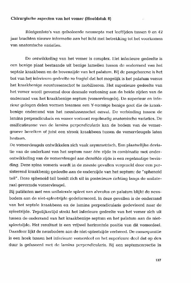
Chirurgische aspecten van hel vomer (Hooldsluk 8)
Ronlgenfoto's van geisoleerde neussepla met leeftijden tussen 0 en 42
jam brachten nieuwe informatie aan het licht met betrekking tot het voorkomen
van anatomische variaties.
De ontwikkeling van het vomer is complex. Het inferieure gedeeJte is
een benige plaat bestaande uit benige lamellen tussen de onderrand van het
septale kraakbeen en de bovenzijde van het palatum. Bij de pasgeborene is het
bot van he~ inferieure gedeelte zo fragiel dat het mogelijk is het palatum versus
het kraakbenige neustussenschot te mobiliseren. Het superieure gedeelte van
het vomer wordt gevormd door desmale verbening aan de beide zijden van de
onderrand van het kraakbenige septum (vomervleugels). De superieur en infe
rieur gelegen delen vormen tesamen een Y-vormige benige goot die de kraak
benige onderrand van het neustussenschot omvat. De verbinding tussen de
lanrina perpendicularis en vomer vertoant regelmatig anatomische valiaties. De
ossificatiezone van de lamina perpendiculmis kan de badem van de vomer
groeve bereiken of juist een strook kraakbeen tussen de vomervleugels laten
bestaan.
De vomervleugels ontwikkelen zich vaak asymmellisch. Een plaatselijke devia
tie van de onderkant van het septunl naar een zijde in combinatie met onder
ontwikkeling van de vomervleugel aan dezelfde zijde is een regelmatige bevin
ding. Deze spina vomeris wordt in de meeste gevallen vergezeld door een per
sisterend kraakbenig gedeelte aan de onderzijde van het septum: de "sphenoid
tail". Deze sphenoid tail breidt zich uit in postmieure lichting langs de unilate
raal gevormde vomervleugel.
Bij patienten met een unilaterale spleet van alveolus en palatum blijkt de neus
bodem aan de niet-spleetzijde gedefomleerd. In deze gevallen is de onderrand
van het septale kraakbeen en de lamina perpendicularis gedevieerd naar de
spleetzijde. Tegelijkertijd strekt het inferieure gedeeJte van het vomer zich uit
tussen de onderrand van het kraakbenige septum en het palatum aan de niet
spleetzijde. Het resultaat is een vIijwel horizontale positie van dit vomerdeel.
Dam'door lijkt de neusbodem aan de niet-spleetzijde verbreed. De consequentie
is een hoek tussen het inferieure vomerdeel en het superieure deel dat op den
duur is gefuseerd met de lamina perpendiculaIis. Bij een septumcorrectie in
137
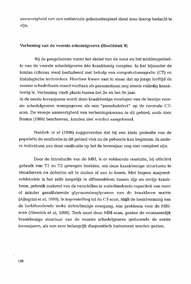
aanwezigheid van een unilaterale gehemeJtespleet dient men hierop bedacht te
zijn.
Verbening van de voorsle schedelgl'Oeve (Hooldsluk 9)
Bij de pasgeborene vormt het skelet van de neus en hel middengedeel
Ie van de voorste schedelgroeve een kraakbenig complex. In het bijzonder de
lamina cribrosa werd bestudeerd met behulp van computertomografie (CT) en
hislologische technieken. Hierdoor kwam vasl Ie staan dat op jonge leeftijd de
voorste schedelbasis zowel mediaan als paramediaan nog sleeds volledig kraak
benig is. Verbening vindt plaals tussen hel 2e en hel 6e jaar.
In de eerste levensjaren wordt deze kraakbenige voorloper van de benige voor
sle schedelgroeve weergegeven als een "pseudodefect" op de coronale CT
scan. De vroege aanwezigheid van verbeningskernen in dit gebied, zoals door
Bosma (1986) beschreven, konden niet worden aangetoond.
Naidich el al (1996) suggereerden dat bij een klein gedeeJte van de
populatie de ossificatie in dit gebied vlak na de geboorte kan beginnen. In ande
re individuen zou deze ossificatie op het 6e levensjaar nog niet compleet zijn.
Door de introductie van de MRI, is er voldoende resolutie, bij efficient
gebruik van T1 en T2 gewogen beelden, om deze kraakbenige structuren te
visualiseren en defecten uit te sluiten of aan te tonen. Met hogere magneet
veldsterkte is het zelfs mogelijk Ie differentieren tussen rijp en onrijp kraak
been, gebrnik makend van de verschillen in waterbindende capaciteit van meer
of minder gesulfateerde glycosaminoglycanen van de kraakbeen malrix
(Allegrini et ai, 1990). In tegenslelling 101 de CT-scan, blijft de beeldvorming van
de luchlhoudende weke delen/benige overgang, een probleem voor de MRI
scan (Zinreich et ai, 1996). Tach moet deze MRI-scan, gezien de voornamelijk
kraakbenige slructuur van de voorste schedelgroeve gedurende de eerste
levensjaren, als een zeer belangrijk diagnostisch instrument worden gezien.
138
Conclusie
De neuschirurgie bij volwassen patienten is de laatste decennia verrijkt
met nieuwe concepten, nieuwe technieken en verfijnder instrumentarium. Voor
de pediatrisch gerichte KNO-arts is de verleiding groot deze innovatie ook toe
te pass en bij kinderen en de traditionele terughoudendheid voor chirurgisch
ingrijpen op deze leeftijd te laten varen.
De biologische basis voor de chirurgie van het neustussenschot tijdens
de groei is nog zeer onvolledig. Het in dit proefschrift beschreven onderzoek
beoogt bij te dragen aan het 0pvullen van enkele van de vele lacunes.
Met betrekking tot het neustussenschot op de kinderleeftijd werden
(voor het eerst) beschreven:
a. de specifieke bouw bij de pasgeborene, en de veranderingen tijdens
de kinderjaren:
b. de dimensionele groei;
c. de evolutie van het kraakbenige septum met enerzijds afbraak van
kraakbeen, anderzijds interstitiele groei en tegelijkertijd behoud van de ken
merkende 3-dimensionale organisatiei
d. het verloop van fractuurlijnen in het septum.
Ook werden hypothesen geformuleerd met betrekking tot:
a. de gevolgen van partieel verlies van septaal kraakbeen voor de ver
dere groei van de neus;
b. de biomechanische achtergrond van de voorkeurslocalisaties van
fracturen en septunldeviaties.
139
References
Allegrini PR, van Velzen D, van Loosen J, Bullock GR 1990, Differentiation of
proliferating from resting cartilage in human fetal nasal septum by Magnetic
Resonance Imaging, Mag Res in Med, 14: 369-376.
Bosma JF, 1986, The anatomy of the infant head. John Hopkins University Press,
Baltimore, USA.
Delaire J, Precious D, 1987, Interaction of the development of the nasal septum,
the nasal pyramid and the face. Int J Ped Otorhinolaryngol, 12: 311-326.
Gi-Min Y, Won Jung H, Sun Kim C, 1995, The prevalence study of nasal septal
deformities in Korea: Results of a nation-wide survey. Rhinology, 33: 61-65.
Hanison D, 1979, Nasal injuries: their pathogenesis and treatment. Br J Plast
Surg, 32:57-64.
Lang J, 1989, Clinical anatomy of the nose, nasal cavity and paranasal sinuses.
Thieme Verlag, New York, Stuttgart.
McNamara JA jr (ed.), 1977, Monograph No.7. Craniofacial Growth Series,
Center for Human Growth and Development, University of Michigan, Ann
Arbor.
Mastenbroek GJ, 1978, De invloed van partiele resectie van van het neus
tussenschot op de uitgroei van bovenkaak en neus. Thesis, University of
Amsterdam.
Meeuwis CA, 1988, Wondreacties in het kraakbenige neustussenschot. Thesis,
Erasmus University Rotterdam.
Mladina R , 1987, The role of maxillar morphology in the development of
pathological septal deformities. Rhinology, 25:199-205.
140
Mladina R, Krajina Z, 1989, The influence of the caudal process on the
formation of septal deformities. Rhinology, 27:113-118.
Naidich TP, Zimmerman RA, Bauer BS, Altman NR, BiIaniuk LT, 1996, Midface:
Embryology and congenital lesions. In: Head and Neck Imaging, Som PM,
Curtin HD (eds), Mosby, St. Louis.
Nolst 1i'enile GJ, 1984, Implantaten in een groeiend neustussenschot. Thesis,
Erasmus University Rotterdam.
Nijdam DC, 1985, Schedelgroei na partiiHe submukeuze resectie van het neus
tussenschot. Thesis, University of Amsterdam.
Plrsig W, 1986, Rhinoplasty and the airway in children. J Fac Plast Surg, 3: 225-
234.
Pirsig W, 1992, Growth of the deviated septum and its influence on midfacial
development. J Fac Plast Surg, 8: 224-232.
Potsie W.P, Cotton R.T., Handler S.D., 1997, Surgical pediatric otolaryngology,
Thieme Verlag, New York, Stuttgart.
Poublon RML, 1987, The cartilaginous nasal dorsum and postnatal growth of the
nose. Thesis, Erasmus University Rotterdam.
Shapiro AM, 1996, Injuries of the nose, facial bones and paranasal sinuses. In:
Paediatric Otolaryngology, Bluestone C D, Stool S E, Kenna M A (eds), W B
Saunders, Philadelphia.
Schultz-Coulon HJ, Eckermeier L, 1976, Zum postnatalen Wachstum der
Nasenscheidewand, Acta Otolaryngol, 82: 131-142.
Tonneyck-MiiIler I, van del' Wert, 1982, Die Entwicklung des Septum Nasi beim
Kaninchen. II. Untersuchungen nach regionalen Unterschieden in der Septum
Dicke. Acta Morphol Neerl-Scand, 20:379-391.
141
Tonneyek-Miiller I, 1984, Die Entwieklung des Septum Nasi beim Kaninehen,
Ill. Quantitative Beobachtungen an Zellen und Interzellularsubstanz. Acta
Morphol Neerl-Scand 22: 133-141.
Van Loosen J, Baatenburg de Jong RJ, van Zanten GA, Engel T, Lanjewar DN,
van Velzen D, 1997, A cephalometric analysis of the nasal septal growth. Clin
Otolaryngol, 22: 453-458.
Verwoerd CDA, Verwoerd-Verhoef HL, 1989, Developmental aspects of the
deviated nose. J Fac Plast Surg, 6:95-100.
Verwoerd CDA, 1992, Present day treatment of nasal fractures: closed versus
open reduction. Fae Plast Surg, 8: 220-223.
Verwoerd CDA, Verwoerd-Vel'hoef HL, 1998, Rhinosurgery in children,surgical
and developmental aspects. In: Rhinoplasty (2nd ed.), Nolst Trenite GJ (ed),
Kugler Publications, The Hague.
Veller U, Pirsig W, Heinze E, 1983, Growth activity in human septal cartilage:
Age-dependent incorporation of labelled sulfate in different anatomic locations.
Plast Reconstr Surg, 71:167-179.
Veller U, Pirsig W, Heinze E, 1984a, Postnatal growth of the human septal
cartilage. Acta Ololaryngol, 97:131-136.
Veller U, Pirsig W, Helbing G, Heit W, Heinze E, 1984b, Patterns of growth in
human septal cartilage: A review of new approaches. Int J Ped Otorhino
Laryngol,7:63-74.
Zinreich SJ, Benson ML, Oliverio PJ, 1996, Sino-nasal cavities: CT normal
anatonlY, imaging of the osteo-nleatal complex, and functional endoscopic
surgery. In: Head and Neck Imaging, Som PM, Curtin HD (eds), Mosby, St.Louis.
142

Curriculum Vitae. 143
De auteur van het proefschrift werd op 7 februari 1957 geboren in Vlissingen.
Na het eindexamen VWO (B-richting) aan de Rijksscholengemeenschap
"Scheldemond" te Vlissingen begon hij in 1976 met de studie geneeskunde
aan de Erasmus Universiteit te Rotterdan1.
Het arts-examen werd in 1983 behaald. In hetzelfde jaar werd aangevangen
met de studie tandheelkunde aan de Universtiteit van Amsterdam (later
ACTCA) en in 1984 werd het docloraal examen eerste fase behaald.
Aansluitend startte de auteur een assistentschap op de afdeling keel-, neus-,
en oorheelkunde in de KNO-kliniek van het Academisch Ziekenhuis Dijkzigt
te Rotterdam.
In 1985 begon hij daadwerkelijk met de opleiding tot KNO-arts. De auteur
werd op 1 februari 1989 als KNO-arts in het specialisten register ingeschre
ven,
Van 1989-1990 was de auteur als staflid werkzaam in het Academisch
Ziekenhuis Dijkzigt.
In 1990-1993 was hij als senior staflid werkzaam in het Sophia Kinder
ziekenhuis te Rotterdam (samen met het Dijkzigt ziekenhuis vormt deze
instelIing het Academisch Ziekenhuis Rotterdam).
Sinds 1 december 1993 is de auteur werkzaam als KNO-arts in het Antonius
Ziekenhuis te Sneek.
Per 1 november 1998 is de auteur benoemd tot Fellow of the Royal Society of
Medicine (FRSM) te Londen, Groot Britannie.
144