Large gangliocytic paraganglioma of the duodenum: …...Core tip: We present the case of a patient...
5
Large gangliocytic paraganglioma of the duodenum: A rare entity Alejandra Gordillo Hernández, Eduardo Dominguez-Adame Lanuza, Auxiliadora Cano Matias, Rosario Perez Huertas, Katherine Maria Gallardo Rodriguez, Purificacion Gallinato Perez, Fernando Oliva Mompean Alejandra Gordillo Hernández, Auxiliadora Cano Matias, Rosario Perez Huertas, Fernando Oliva Mompean, Servicio de Cirugia General, Hospital Universitario Virgen de Macarena, 41009 Sevilla, Spain Eduardo Dominguez-Adame Lanuza, Jefe de Sección de Cirugia Laparoscopica Avanzada y Cirugía Bariatrica, Hospital Universitario Virgen de Macarena, 41009 Sevilla, Spain Katherine Maria Gallardo Rodriguez, Purificacion Gallinato Perez, Servicio de Anatomia Patologica, Hospital Universitario Virgen de Macarena, 41009 Sevilla, Spain Author contributions: Gordillo Hernández A designed and performed the research, and wrote the paper; Gallardo Rodriguez KM, Gallinato Perez P, Cano Matias A, Perez Huertas R, Oliva Mompean F and Dominguez-Adame Lanuza E contributed new reagents or analytic tools; Gordillo Hernández A and Gallardo Rodriguez KM analyzed the data. Institutional review board statement: The study was reviewed and approved by the Hospital Universitario Virgen de Macarena Review Committee. Informed consent statement: The patient provided informed written consent prior to study enrollment. Conflict-of-interest statement: The authors declare that they have no competing interests. Open-Access: This article is an open-access article which was selected by an in-house editor and fully peer-reviewed by external reviewers. It is distributed in accordance with the Creative Commons Attribution Non Commercial (CC BY-NC 4.0) license, which permits others to distribute, remix, adapt, build upon this work non-commercially, and license their derivative works on different terms, provided the original work is properly cited and the use is non-commercial. See: http://creativecommons.org/ licenses/by-nc/4.0/ Correspondence to: Alejandra Gordillo Hernández, MD, Servicio de Cirugia General, Hospital Universitario Virgen de Macarena, Avenida Doctor Fedriani 3, 41009 Sevilla, Spain. [email protected] Telephone: +34-658-154289 Fax: +34-954-557347 Received: March 24, 2015 Peer-review started: March 26, 2015 First decision: April 27, 2015 Revised: May 14, 2015 Accepted: June 4, 2015 Article in press: June 8, 2015 Published online: August 27, 2015 Abstract Gangliocytic paragangliomas are rare tumors that almost exclusively occur within the second portion of the duodenum. Although these tumors generally have a benign clinical course, they have the potential to recur or metastasize to regional lymph nodes. The case report presented here describes a 57-year-old female patient with melena, progressive asthenia, anemia, and a mass in the second-third portion of the duodenum that was treated by local excision. The patient was diagnosed with a friable bleeding tumor. The histologic analysis showed that the tumor was a 4 cm gangliocytic paraganglioma without a malignant cell pattern. In the absence of local invasion or distant metastasis, endoscopic resection represents a feasible, curative therapy. Although endoscopic polypectomy is currently considered the treatment of choice, it is not recommended if the size of the tumor is > 3 cm and/or there is active or recent bleeding. Patients diagnosed with a gangliocytic paraganglioma should be closely followed-up for possible local recurrence. Key words: Duodenum; Gangliocytic paraganglioma; Ganglion cells © The Author(s) 2015. Published by Baishideng Publishing Group Inc. All rights reserved. CASE REPORT 170 August 27, 2015|Volume 7|Issue 8| WJGS|www.wjgnet.com Submit a Manuscript: http://www.wjgnet.com/esps/ Help Desk: http://www.wjgnet.com/esps/helpdesk.aspx DOI: 10.4240/wjgs.v7.i8.170 World J Gastrointest Surg 2015 August 27; 7(8): 170-173 ISSN 1948-9366 (online) © 2015 Baishideng Publishing Group Inc. All rights reserved.
Transcript of Large gangliocytic paraganglioma of the duodenum: …...Core tip: We present the case of a patient...
Large gangliocytic paraganglioma of the duodenum: A rare entity
Alejandra Gordillo Hernández, Eduardo Dominguez-Adame Lanuza, Auxiliadora Cano Matias, Rosario Perez Huertas, Katherine Maria Gallardo Rodriguez, Purificacion Gallinato Perez, Fernando Oliva Mompean
Alejandra Gordillo Hernández, Auxiliadora Cano Matias, Rosario Perez Huertas, Fernando Oliva Mompean, Servicio de Cirugia General, Hospital Universitario Virgen de Macarena, 41009 Sevilla, Spain
Eduardo Dominguez-Adame Lanuza, Jefe de Sección de Cirugia Laparoscopica Avanzada y Cirugía Bariatrica, Hospital Universitario Virgen de Macarena, 41009 Sevilla, Spain
Katherine Maria Gallardo Rodriguez, Purificacion Gallinato Perez, Servicio de Anatomia Patologica, Hospital Universitario Virgen de Macarena, 41009 Sevilla, Spain
Author contributions: Gordillo Hernández A designed and performed the research, and wrote the paper; Gallardo Rodriguez KM, Gallinato Perez P, Cano Matias A, Perez Huertas R, Oliva Mompean F and Dominguez-Adame Lanuza E contributed new reagents or analytic tools; Gordillo Hernández A and Gallardo Rodriguez KM analyzed the data.
Institutional review board statement: The study was reviewed and approved by the Hospital Universitario Virgen de Macarena Review Committee.
Informed consent statement: The patient provided informed written consent prior to study enrollment.
Conflict-of-interest statement: The authors declare that they have no competing interests.
Open-Access: This article is an open-access article which was selected by an in-house editor and fully peer-reviewed by external reviewers. It is distributed in accordance with the Creative Commons Attribution Non Commercial (CC BY-NC 4.0) license, which permits others to distribute, remix, adapt, build upon this work non-commercially, and license their derivative works on different terms, provided the original work is properly cited and the use is non-commercial. See: http://creativecommons.org/licenses/by-nc/4.0/
Correspondence to: Alejandra Gordillo Hernández, MD, Servicio de Cirugia General, Hospital Universitario Virgen de Macarena, Avenida Doctor Fedriani 3, 41009 Sevilla, Spain. [email protected]
Telephone: +34-658-154289 Fax: +34-954-557347
Received: March 24, 2015Peer-review started: March 26, 2015First decision: April 27, 2015Revised: May 14, 2015Accepted: June 4, 2015Article in press: June 8, 2015Published online: August 27, 2015
AbstractGangliocytic paragangliomas are rare tumors that almost exclusively occur within the second portion of the duodenum. Although these tumors generally have a benign clinical course, they have the potential to recur or metastasize to regional lymph nodes. The case report presented here describes a 57-year-old female patient with melena, progressive asthenia, anemia, and a mass in the second-third portion of the duodenum that was treated by local excision. The patient was diagnosed with a friable bleeding tumor. The histologic analysis showed that the tumor was a 4 cm gangliocytic paraganglioma without a malignant cell pattern. In the absence of local invasion or distant metastasis, endoscopic resection represents a feasible, curative therapy. Although endoscopic polypectomy is currently considered the treatment of choice, it is not recommended if the size of the tumor is > 3 cm and/or there is active or recent bleeding. Patients diagnosed with a gangliocytic paraganglioma should be closely followed-up for possible local recurrence.
Key words: Duodenum; Gangliocytic paraganglioma; Ganglion cells
© The Author(s) 2015. Published by Baishideng Publishing Group Inc. All rights reserved.
CASE REPORT
170 August 27, 2015|Volume 7|Issue 8|WJGS|www.wjgnet.com
Submit a Manuscript: http://www.wjgnet.com/esps/Help Desk: http://www.wjgnet.com/esps/helpdesk.aspxDOI: 10.4240/wjgs.v7.i8.170
World J Gastrointest Surg 2015 August 27; 7(8): 170-173ISSN 1948-9366 (online)
© 2015 Baishideng Publishing Group Inc. All rights reserved.
Core tip: We present the case of a patient with a rare duodenal gangliocytic paraganglioma that was treated by tumorectomy. Although there is currently no consensus for treatment, this report demonstrates that local conservative tumorectomy is a feasible, curative therapy. Patients diagnosed with a gangliocytic paraganglioma should be closely followed-up for possible local recurrence.
Gordillo Hernández A, Dominguez-Adame Lanuza E, Cano Matias A, Perez Huertas R, Gallardo Rodriguez KM, Gallinato Perez P, Oliva Mompean F. Large gangliocytic paraganglioma of the duodenum: A rare entity. World J Gastrointest Surg 2015; 7(8): 170-173 Available from: URL: http://www.wjgnet.com/1948-9366/full/v7/i8/170.htm DOI: http://dx.doi.org/10.4240/wjgs.v7.i8.170
INTRODUCTIONGangliocytic paragangliomas (GPs) are rare neuroendocrine tumors that predominantly arise within the second part of the duodenum. GPs seldom recur or metastasize to regional lymph nodes[1], and are considered epithelial tumors according to the classification of tumors of the digestive tract by the World Health Organization[2]. Diagnosis of GP can be achieved based on histopathology showing epithelioid, spindle and ganglion cells, which are similarly observed for paragangliomas[3]. This report describes the case of a 57yearold woman with melena, progressive asthenia, anemia, and a mass in the secondthird portion of the duodenum that was finally diagnosed as a GP.
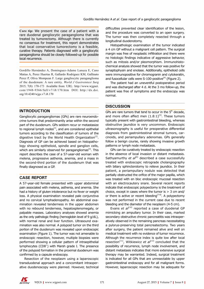
CASE REPORTA 57yearold female presented with upper abdominal pain associated with melena, asthenia, and anemia. She had a history of gluten intolerance but no fever or weight loss. A physical examination revealed pale conjunctiva and no cervical lymphadenopathy. An abdominal examination revealed tenderness in the upper abdomen with no rebound tenderness, hepatosplenomegaly, or palpable masses. Laboratory analyses showed anemia as the only pathologic finding (hemoglobin level of 9 g/dL), with normal renal and liver function. Ultrasound examination was also normal. A polypoid tumor on the third portion of the duodenum was revealed upon endoscopic examination (Figure 1). The tumor was not amenable to endoscopic resection, however, multiple biopsies were performed showing a cellular pattern of intraepithelial lymphocytes (CD8+) with Marsh grade I. The presence of the polypoid formation in the proximal duodenum was confirmed by a capsule endoscopy.
Resection of the neoplasm using a laparoscopic transduodenal approach and a concomitant intraoperative duodenoscopy were planned. However, technical
difficulties prevented clear identification of the lesion, and the procedure was converted to an open surgery. The tumor was then completely resected through a longitudinal duodenotomy.
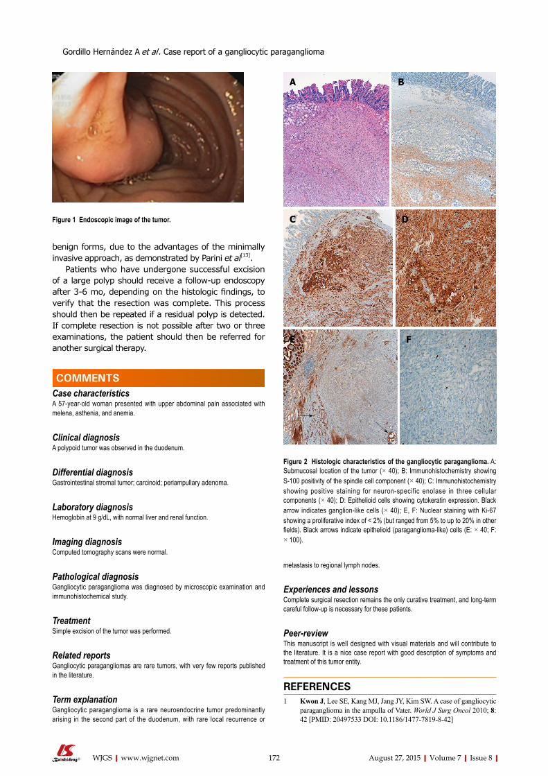
Histopathologic examination of the tumor indicated a 4 cm GP without a malignant cell pattern. The surgical margin was free of neoplastic infiltration and there were no histologic findings indicative of aggressive behavior, such as mitosis and/or pleomorphism. Immunohistochemical analysis showed that the tumor was positive for synaptophysin and enolase. Additionally, epithelioid cells were immunopositive for chromogranin and cytokeratin, and fusocellular cells were S100positive[4] (Figure 2).
The patient had an uneventful postoperative period and was discharged after 4 d. At the 3 mo followup, the patient was free of symptoms and the endoscopy was normal.
DISCUSSIONGPs are rare tumors that tend to occur in the 5th decade, and more often affect men (1.8:1)[5]. These tumors typically present with gastrointestinal bleeding, whereas obstructive jaundice is very uncommon. Endoscopic ultrasonography is useful for preoperative differential diagnosis from gastrointestinal stromal tumors, carcinoids, and periampullary adenomas. GPs generally follow a benign course, rarely showing invasive growth patterns or lymph node metastasis.
GPs can be curatively treated by endoscopic resection in the absence of local invasion or distant metastasis. Sathyamurthy et al[6] described a case successfully treated with endoscopic retrograde cholangiography with biliary sphincterotomy to relieve jaundice. In their patient, a periampullary nodule was detected that partially obstructed the orifice of the major papilla, which was treated with en bloc endoscopic mucosal resection with an electrocautery snare. Several recent reports indicate that endoscopic polypectomy is the treatment of choice, except in cases where the tumor is > 3 cm and/or there is active or recent bleeding[79]. A polypectomy was not performed in the current case due to recent bleeding and the diameter of the neoplasm (45 cm).
Evans et al[10] reported a case of duodenal GP mimicking an ampullary tumor. In their case, marked secondary obstructive chronic pancreatitis was intraoperatively observed in the remaining pancreas necessitating a pyloruspreserving total pancreatectomy. Two years after surgery, the patient remained alive and well on medical treatment with no evidence of tumor recurrence. Although the recurrence index is quite low after local resection[11], Witkiewicz et al[12] concluded that the possibility of recurrence, lymph node involvement, and distant metastasis indicates that more extensive surgical therapy may be warranted. Indeed, surgical treatment is indicated for all GPs that are unresectable by upper gastrointestinal endoscopy and for all malignant forms. However, laparoscopic resection may be adequate for
Gordillo Hernández A et al . Case report of a gangliocytic paraganglioma
171 August 27, 2015|Volume 7|Issue 8|WJGS|www.wjgnet.com
benign forms, due to the advantages of the minimally invasive approach, as demonstrated by Parini et al[13].
Patients who have undergone successful excision of a large polyp should receive a followup endoscopy after 36 mo, depending on the histologic findings, to verify that the resection was complete. This process should then be repeated if a residual polyp is detected. If complete resection is not possible after two or three examinations, the patient should then be referred for another surgical therapy.
COMMENTSCase characteristicsA 57-year-old woman presented with upper abdominal pain associated with melena, asthenia, and anemia.
Clinical diagnosisA polypoid tumor was observed in the duodenum.
Differential diagnosisGastrointestinal stromal tumor; carcinoid; periampullary adenoma.
Laboratory diagnosisHemoglobin at 9 g/dL, with normal liver and renal function.
Imaging diagnosisComputed tomography scans were normal.
Pathological diagnosisGangliocytic paraganglioma was diagnosed by microscopic examination and immunohistochemical study.
TreatmentSimple excision of the tumor was performed.
Related reportsGangliocytic paragangliomas are rare tumors, with very few reports published in the literature.
Term explanation Gangliocytic paraganglioma is a rare neuroendocrine tumor predominantly arising in the second part of the duodenum, with rare local recurrence or
metastasis to regional lymph nodes.
Experiences and lessonsComplete surgical resection remains the only curative treatment, and long-term careful follow-up is necessary for these patients.
Peer-reviewThis manuscript is well designed with visual materials and will contribute to the literature. It is a nice case report with good description of symptoms and treatment of this tumor entity.
REFERENCES1 Kwon J, Lee SE, Kang MJ, Jang JY, Kim SW. A case of gangliocytic
paraganglioma in the ampulla of Vater. World J Surg Oncol 2010; 8: 42 [PMID: 20497533 DOI: 10.1186/1477-7819-8-42]
172 August 27, 2015|Volume 7|Issue 8|WJGS|www.wjgnet.com
Figure 1 Endoscopic image of the tumor.
A B
C D
FE
Figure 2 Histologic characteristics of the gangliocytic paraganglioma. A: Submucosal location of the tumor (× 40); B: Immunohistochemistry showing S-100 positivity of the spindle cell component (× 40); C: Immunohistochemistry showing positive staining for neuron-specific enolase in three cellular components (× 40); D: Epithelioid cells showing cytokeratin expression. Black arrow indicates ganglion-like cells (× 40); E, F: Nuclear staining with Ki-67 showing a proliferative index of < 2% (but ranged from 5% to up to 20% in other fields). Black arrows indicate epithelioid (paraganglioma-like) cells (E: × 40; F: × 100).
COMMENTS
Gordillo Hernández A et al . Case report of a gangliocytic paraganglioma
10.4321/S1130-01082004000900008]8 Nagai T, Torishima R, Nakashima H, Tanahashi J, Iwata M,
Ookawara H, Yokoyama S, Yada K, Sato R, Murakami K, Fujioka T. Duodenal gangliocytic paraganglioma treated with endoscopic hemostasis and resection. J Gastroenterol 2004; 39: 277-283 [PMID: 15065006 DOI: 10.1007/s00535-003-1289-2]
9 Yang JI, Choi JS, Lee GH, Kim BW, Moon SJ, Kang MS, Ahn HJ. A case of ampullary gangliocytic paraganglioma. Korean J Intern Med 2014; 29: 375-378 [PMID: 24851073 DOI: 10.3904/kjim.2014.29.3.375]
10 Evans JD, Wilson PG, Barber PC, Neoptolemos JP. Duodenal gangliocytic paraganglioma presenting as an ampullary tumor. Int J Pancreatol 1996; 20: 131-134 [PMID: 8968869]
11 Scheithauer BW, Nora FE, LeChago J, Wick MR, Crawford BG, Weiland LH, Carney JA. Duodenal gangliocytic paraganglioma. Clinicopathologic and immunocytochemical study of 11 cases. Am J Clin Pathol 1986; 86: 559-565 [PMID: 2877566]
12 Witkiewicz A, Galler A, Yeo CJ, Gross SD. Gangliocytic paragan-glioma: case report and review of the literature. J Gastrointest Surg 2007; 11: 1351-1354 [PMID: 17653595 DOI: 10.1007/s11605-007-0217-9]
13 Parini U, Nardi M, Loffredo A, Fabozzi M, Roveroni M. Laparoscopic resection of duodenal gangliocytic paraganglioma. A case report. Chir Ital 2007; 59: 551-558 [PMID: 17966779]
P- Reviewer: Petronella P, Scheidbach H S- Editor: Tian YL L- Editor: A E- Editor: Jiao XK
2 Kleihues P, Louis DN, Scheithauer BW, Rorke LB, Reifenberger G, Burger PC, Cavenee WK. The WHO classification of tumors of the nervous system. J Neuropathol Exp Neurol 2002; 61: 215-225; discussion 226-229 [PMID: 11895036]
3 Perrone T, Sibley RK, Rosai J. Duodenal gangliocytic paragan-glioma. An immunohistochemical and ultrastructural study and a hypothesis concerning its origin. Am J Surg Pathol 1985; 9: 31-41 [PMID: 2578747 DOI: 10.1097/00000478-198501000-00007]
4 Hoffmann KM, Furukawa M, Jensen RT. Duodenal neuroendocrine tumors: Classification, functional syndromes, diagnosis and medical treatment. Best Pract Res Clin Gastroenterol 2005; 19: 675-697 [PMID: 16253893 DOI: 10.1016/j.bpg.2005.05.009]
5 Narang V, Behl N, Sood N, Puri H. Gangliocytic paraganglioma of duodenum. Case Rep Pathol 2013; 2013: 378582 [PMID: 24073351 DOI: 10.1155/2013/378582]
6 Sathyamurthy A, Choudhary A, Ng D, Okponobi S, Diaz-Arias A, Grewal A, Hammoud GM. Obstructive jaundice due to a rare periampullary tumor. World J Gastrointest Oncol 2013; 5: 195-197 [PMID: 24137522 DOI: 10.4251/wjgo.v5.i10.195]
7 Sánchez-Pobre P, Sáenz-López S, Rodríguez S, Sánchez F, Alemany I, López G, Colina F, Martínez-Montiel P, Marín JC, Castellano G, Solís Herruzo JA. Safe endoscopic resection of gangliocytic paraganglioma of the major duodenal papilla. Rev Esp Enferm Dig 2004; 96: 660-662; 663-664 [PMID: 15506909 DOI:
173 August 27, 2015|Volume 7|Issue 8|WJGS|www.wjgnet.com
Gordillo Hernández A et al . Case report of a gangliocytic paraganglioma

© 2015 Baishideng Publishing Group Inc. All rights reserved.
Published by Baishideng Publishing Group Inc8226 Regency Drive, Pleasanton, CA 94588, USA
Telephone: +1-925-223-8242Fax: +1-925-223-8243
E-mail: [email protected] Desk: http://www.wjgnet.com/esps/helpdesk.aspx
http://www.wjgnet.com