Forearm Fracture 1 Bismillah
17
Forearm Fracture Name Rizki Ria Sari (C 111 07 241) Mentors dr. Teuku Nanta A. dr. Risqi T. Tuahuns Supervisor dr. Jainal Arifin, Sp.OT Orthopedic and Traumatology Department Hasanuddin University 2011
Transcript of Forearm Fracture 1 Bismillah
Forearm Fracture
NameRizki Ria Sari (C 111 07 241)
Mentorsdr. Teuku Nanta A.dr. Risqi T. Tuahuns
Supervisordr. Jainal Arifin, Sp.OT
Orthopedic and Traumatology DepartmentHasanuddin University
2011
Description
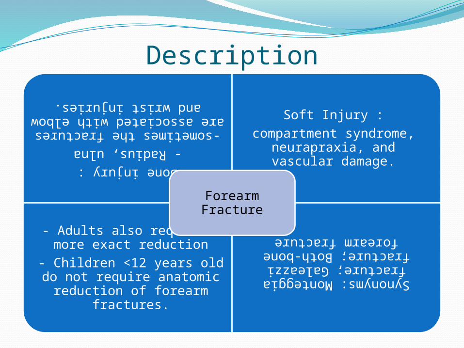
Bone injury :- Radius, ulna
-sometimes the fractures are associated with elbow and
wrist injuries.
Soft Injury :compartment syndrome,
neurapraxia, and vascular damage.
- Adults also require a more exact reduction
- Children <12 years old do not require anatomic reduction of forearm
fractures.
Synonyms: Monteggia fracture; Galeazzi fracture; Both-bone forearm fracture
Forearm Fracture

Monteaggia Fractures and Galeazzi Fractures
a. Monteaggia Fracturesb. Galeazzi Fractures
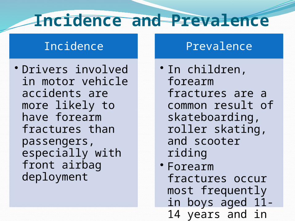
Incidence and PrevalenceIncidence
• Drivers involved in motor vehicle accidents are more likely to have forearm fractures than passengers, especially with front airbag deployment
Prevalence
• In children, forearm fractures are a common result of skateboarding, roller skating, and scooter riding
• Forearm fractures occur most frequently in boys aged 11-14 years and in girls aged 8-11 years
Risk FactorsHigh-energy traumaOsteoporosis
Etiology
motor vehicle accidents, fall from a height crushing injury
High Energ
y Traum
a
Falls
Low Energ
y Traum
a

Sign and SymptomsPainSwellingLose of elbow or wrist motionDeformityImportant : assessment of forearm for skin and soft-tissue (neurovascular) compromise
Physical ExaminationCareful examination of the entire involved extremity is mandatory, including:
Detailed neurologic and vascular evaluations Assessment of the soft tissues
Compartments, anterior (flexor) and posterior (extensor), are checked for evidence of compartment syndrome.
Compartment pressure is measured if the forearm feels tight or if the patient displays pain out of proportion to the injury.
Test Imaging•AP, lateral, and oblique views of the wrist and the entire forearm, as well as AP and lateral views of the ipsilateral elbow, are mandatory.• Radiographic signs of injury to the distal
radioulnar joint include:• Fracture at the base of the ulnar styloid• Widening of the joint space on the AP view• Dislocation of the radius relative to the ulna on
the lateral view• Radial shortening >5 mm
• If the radial head is located properly, a line drawn through the radial head and shaft on any radiographic projection should align with the capitellum of the elbow.• If dislocation of the radial head is suspected
clinically, a lateral radiograph of the elbow with the arm in supination may be helpful.

Pathological FindingsMost forearm fractures are either transverse or short oblique in configuration.
Comminution is variable (none to moderate).
Treatment; General Measures•Pain medication should be administered only after a careful physical examination, including documentation of neurovascular status.•The forearm should be elevated, with application of ice to the fracture site to help to reduce swelling.•In general, closed treatment of diaphyseal fractures is best used for stable (<50% of the shaft diameter displaced), isolated fractures of the distal 2/3 of the ulna with 10° of angular deformity.•Fractures of the proximal 1/3 of the ulna and fractures of the distal 2/3 of the ulna with >10° angulation are best treated operatively.•Pediatric both-bone fractures do not remodel well and should be treated with surgery if reduction cannot be maintained
Treatment; General MeasuresIn addition, most radial shaft fractures, except those that are nondisplaced, and virtually all both-bone forearm fractures in adults (prone to shortening and angulation) require surgical management.Closed forearm fractures of 1 or both bones that are displaced minimally should be splinted in a neutral position to prevent additional displacement and possible neurovascular injury.In general, forearm fractures with associated ligamentous injuries, either distally (wrist) or proximally (elbow), are unstable injuries.
These more severe injuries require early surgical intervention for reduction and stabilization of both the forearm fractures and the associated ligamentous injuries.
Spesial Therapy
•Early ROM of the elbow and fingers is important to help to reduce soft-tissue scarring and to prevent contractures
Physical therapy
•Acetaminophen plus a mild narcotic are used most often in the immediate postinjury period for pain control
Medication
Surgical•Surgical options include percutaneous Kirschner wire fixation, external fixation, intramedullary nailing, and plate and screw fixation. Acute bone grafting is unnecessary • For open fractures, irrigation and debridement with the administration of intravenous antibiotic emergent basis.• If the open wound is not massively contaminated, the
fractures are stabilized after debridement.• With massive contamination, fixation is performed in a
delayed fashion.•Radial and ulnar fractures usually are stabilized rigidly with 3.5-mm dynamic compression plates.•Locking plates seem to have no advantages compared with nonlocking plates•Pediatric fractures may be treated with plating or with intramedullary nailing.• Results with intramedullary nail fixation seem to be
superior

Prognose•In general, most nondisplaced or minimally displaced fractures in children who undergo closed treatment heal well, with good return of forearm function
•Minimally displaced isolated ulna fracture have excellent results when treated with functional bracing
•The prognosis in adults with displaced fractures of the radius and ulna and closed treatment is poor.
•For fractures treated with open reduction and rigid internal fixation, the prognosis for achieving union is ~95%
•Because rigid fixation allows early ROM, patients who have no associated severe soft-tissue injuries should experience only mild loss of forearm rotation.

Complication
•Decreased ROM •Synostosis •Malunion •NonunionNon Operative treatment•Late infections•Iatrogenic nerve injuries•Vascular injuries•Loss of fixationOperative treatment
•exquisite pain on passive stretch of the digits.•Constrictive dressings should be released down to
the skin at the 1st symptom or sign of compartment syndrome.•If pain is not improved, compartment pressure
should be measured.•Confirmation of the diagnosis requires emergent
fasciotomy of the forearm.
Compartment Syndrom
Patient MonitoringFollow-up care should be arranged within 1
week after injury for repeat physical examination and repeat radiographs before and after the application of a cast, to verify fracture position when cast treatment is chosen.
Additional follow-up every 2-3 weeks then is necessary to assess healing of the fracture site and to guide early ROM of the fingers and elbow.
Healing of closed forearm fractures usually takes 4-6 weeks for a child and 6-12 weeks for an adult