Een nationale richtlijn voor Decubituspreventie
94
2012 www.kce.fgov.be KCE REPORT 193A EEN NATIONALE RICHTLIJN VOOR DECUBITUSPREVENTIE
Transcript of Een nationale richtlijn voor Decubituspreventie

2012 www.kce.fgov.be
KCE REPORT 193A
EEN NATIONALE RICHTLIJN VOOR DECUBITUSPREVENTIE

Het Federaal Kenniscentrum voor de Gezondheidszorg Het Federaal Kenniscentrum voor de Gezondheidszorg is een parastatale, opgericht door de
programmawet (1) van 24 december 2002 (artikelen 259 tot 281) die onder de bevoegdheid valt van de Minister van Volksgezondheid en Sociale Zaken. Het Centrum is belast met het realiseren van beleidsondersteunende studies binnen de sector van de gezondheidszorg en de ziekteverzekering.
Raad van Bestuur Effectieve Leden Plaatsvervangende Leden
Voorzitter Pierre Gillet Leidend ambtenaar RIZIV (vice-voorzitter) Jo De Cock Benoît Collin Voorzitter FOD Volksgezondheid (vice-voorzitter) Dirk Cuypers Chris Decoster Voorzitter FOD Sociale Zekerheid
(vice-voorzitter) Frank Van Massenhove Jan Bertels
Administrateur-generaal FAGG Xavier De Cuyper Greet Musch Vertegenwoordigers Minister van Volksgezondheid Bernard Lange François Perl Marco Schetgen Annick Poncé Vertegenwoordigers Minister van Sociale Zaken Olivier de Stexhe Karel Vermeyen Ri De Ridder Lambert Stamatakis Vertegenwoordigers Ministerraad Jean-Noël Godin Frédéric Lernoux Daniel Devos Bart Ooghe Intermutualistisch Agentschap Michiel Callens Frank De Smet Patrick Verertbruggen Yolande Husden Xavier Brenez Geert Messiaen Beroepsverenigingen van de artsen Marc Moens Roland Lemye Jean-Pierre Baeyens Rita Cuypers Beroepsverenigingen van de verpleegkundigen Michel Foulon Ludo Meyers Myriam Hubinon Olivier Thonon Ziekenhuisfederaties Johan Pauwels Katrien Kesteloot Jean-Claude Praet Pierre Smiets Sociale partners Rita Thys Leo Neels Paul Palsterman Celien Van Moerkerke Kamer van Volksvertegenwoordigers Lieve Wierinck

Controle Regeringscommissaris Yves Roger
Directie Algemeen Directeur
Raf Mertens
Programmadirectie Christian Léonard Kristel De Gauquier
Contact Federaal Kenniscentrum voor de Gezondheidszorg (KCE) Doorbuilding (10e verdieping) Kruidtuinlaan 55 B-1000 Brussel Belgium T +32 [0]2 287 33 88 F +32 [0]2 287 33 85 [email protected] http://www.kce.fgov.be


2012 www.kce.fgov.be
KCE REPORT 193A GOOD CLINICAL PRACTICE
EEN NATIONALE RICHTLIJN VOOR DECUBITUSPREVENTIE DIMITRI BEECKMAN, CATHY MATHEÏ, AURÉLIE VAN LANCKER, SABINE VAN HOUDT, GEERT VANWALLEGHEM, LUC GRYSON, HILDE HEYMAN, CHRISTIAN THYSE, ADINDA TOPPETS, SABINE STORDEUR, KOEN VAN DEN HEEDE

COLOFON Titel: Een nationale richtlijn voor Decubituspreventie
Auteurs: Dimitri Beeckman (UGent), Cathy Matheï (KULeuven), Aurélie Van Lancker (UGent), Sabine Van Houdt (KULeuven), Geert Vanwalleghem (CNC vzw/ WCS/ H.-Hartziekenhuis Roeselare-Menen vzw), Luc Gryson (CNC vzw), Hilde Heyman (WCS), Christian Thyse (AFISCeP.be), Adinda Toppets (UZLeuven), Sabine Stordeur (KCE), Koen Van den Heede (KCE)
Reviewers: Marijke Eyssen (KCE); Dominique Paulus (KCE)
Externe experten: Diégo Backaert (Thuiszorg Groep Backaert); Hilde Beele (UZ Gent); Lieven Decaevele (OLV-Ziekenhuis, Aalst); Daniëlle Declercq (Institut Jules Bordet); Véronique del Marmolle (Hopital Erasme, ULB); Aurélia Bustillo (Hopital Erasme, ULB); Anne Hermand (Clinique Universitaire Saint-Luc, Bruxelles); Miguel Lardennois (SPF Santé Publique - FOD Volksgezondheid); Louis Paquay (Wit-Gele Kruis); Dominique Putzeys (CIPIQ-s); Lisette Schoonhoven (Radboud Universiteit Nijmegen), Evelien Touriany (Militair Hospitaal Koningin Astrid); Dirk Van De Looverbosch (Zelfstandig Huisarts); Katrien Vanderwee (O.L.V. van Lourdes ziekenhuis Waregem); Pascal Van Waeyenberghe (Home Health Care wound care BVBA); Christiane Vranken (CHU Liège)
Acknowledgements: We bedanken Liz Avital (NCGC, VK), Katie Jones (NCGC, VK) en Julie Neilson (NCGC, VK) voor de samenwerking bij de voorbereiding van de ‘evidence reviews’.
Externe Validatoren: Nicky Cullum (University of Manchester); Siegfried Geens (CEBAM); Philippe Hanson (CHU UCL Mont-Godinne)
Belangenconflict: Dominique Putzeys verklaart fondsen ontvangen te hebben voor het uitvoeren van onderzoek gerelateerd aan de preventie van doorligwonden. Diégo Backaert, Hilde Beele, Anne Hermand, Adinda Toppets, Geert Vanwalleghem, Pascal Van Waeyenberghe verklaren betalingen om te spreken, opleidingsvergoedingen, reisondersteuning of betaling voor deelname aan een symposium gerelateerd aan decubituspreventie ontvangen te hebben.
Layout: Ine Verhulst

Disclaimer: • De externe experten werden geraadpleegd over een (preliminaire) versie van het wetenschappelijke rapport. Hun opmerkingen werden tijdens vergaderingen besproken. Zij zijn geen coauteur van het wetenschappelijke rapport en gingen niet noodzakelijk akkoord met de inhoud ervan.
• Vervolgens werd een (finale) versie aan de validatoren voorgelegd. De validatie van het rapport volgt uit een consensus of een meerderheidsstem tussen de validatoren. Zij zijn geen coauteur van het wetenschappelijke rapport en gingen niet noodzakelijk alle drie akkoord met de inhoud ervan.
• Tot slot werd dit rapport unaniem goedgekeurd door de Raad van Bestuur. • Alleen het KCE is verantwoordelijk voor de eventuele resterende vergissingen of onvolledigheden
alsook voor de aanbevelingen aan de overheid. Publicatiedatum: 10 januari 2013
Domein: Good Clinical Practice (GCP)
MeSH: Pressure ulcer ; Practice Guidelines; Prevention and control
NLM classificatie: WR 598
Taal: Nederlands, Engels
Formaat: Adobe® PDF™ (A4)
Wettelijk depot: D/2012/10.273/95
Copyright: De KCE-rapporten worden gepubliceerd onder de Licentie Creative Commons « by/nc/nd » http://kce.fgov.be/nl/content/de-copyrights-van-de-kce-rapporten.
Hoe refereren naar dit document? Beeckman D, Matheï C, Van Lancker A, Van Houdt S, Vanwalleghem G, Gryson L, Heyman H, Thyse C, Toppets A, Stordeur S, Van den Heede K. Een nationale richtlijn voor Decubituspreventie. Good Clinical Practice (GCP). Brussel: Federaal Kenniscentrum voor de Gezondheidszorg (KCE). 2012. KCE Reports 193A. D/2012/10.273/95.
Dit document is beschikbaar op de website van het Federaal Kenniscentrum voor de Gezondheidszorg.


KCE Report 193A Decubituspreventie i
VOORWOORD
Voor patiënten met een verminderde mobiliteit kan de kwaliteit van de zorg die ze krijgen, aan bed of in de zetel, een enorm verschil maken. Een heel belangrijk element daarbij is de preventie van decubitus, de zogenaamde doorligwonden. Ze ontstaan, typisch op de stuit, de hielen, het achterhoofd, bij langdurige druk- en schuifkracht op de huid en onderliggende weefsels. Het gaat hier absoluut niet om een triviale complicatie: de letsels kunnen zeer uitgebreid zijn en gepaard gaan met ernstig en langdurig ongemak en extra kosten voor patiënt en samenleving. Niet toevallig wordt de preventie van decubitus nationaal en internationaal gezien als een belangrijk onderdeel van kwaliteitsbeleid in zowel ziekenhuizen, rustoorden, revalidatiecentra, als in de thuiszorg. De FOD Volksgezondheid vroeg aan het KCE een richtlijn uit te werken om het goed klinisch handelen van zorgverleners te ondersteunen met de meest actuele wetenschappelijke bevindingen terzake. Gelukkig bestond er rond decubitus al heel wat expertise in ons land. Voor dit project kon het KCE beroep doen op de deskundige inbreng van wetenschappelijke equipes van de Universiteit Gent en de KULeuven. Maar we keken ook verder dan onze landsgrenzen, en dat zorgde voor een primeur! Voor het eerst werkte het KCE in dit project samen met het vermaarde Britse National Institute for Health and Clinical Excellence (NICE), en met hun onderaannemer, National Clinical Guideline Centre (NCGC) in London. De samenwerking zorgde voor een intense wisselwerking tussen de verschillende equipes op het vlak van de gehanteerde methodieken. Dit resulteerde in een wetenschappelijk eindproduct dat door alle betrokken partners voluit wordt gedragen. Samen met de Belgische wondzorg verenigingen (CNC vzw, WCS, AFISCeP.be) werden de wetenschappelijke bevindingen vertaald naar aanbevelingen voor de dagelijkse klinische praktijkvoering. Ook deze Guideline vond uiteraard geen ‘magic bullet’ om decubitus te voorkomen. Maar met een goede, gecombineerde aanpak op maat kan men vele patiënten heel wat leed besparen.
Raf MERTENS Algemeen Directeur

ii Decubituspreventie KCE Report 193A
SAMENVATTING INLEIDING Het European Pressure Ulcer Advisory Panel (EPUAP) definieert decubitus als een plaatselijk letsel van de huid en/of de onderliggende weefsels dat ontstaat als een interne reactie op een externe mechanische belasting op zachte biologische weefsels, meestal over een benig uitsteeksel. Deze externe mechanische belasting kan een kracht zijn die loodrecht op het huidoppervlak wordt uitgeoefend (drukkracht), een kracht die parallel met het huidoppervlak optreedt (schuifkracht), of een combinatie van druk- en schuifkracht. De ernst van decubitus varieert van niet-wegdrukbare roodheid van de intacte huid tot het afsterven van weefsels waaronder huid, subcutaan vet, spier en bot. In zijn classificatie-systeem, definieerde EPUAP decubitus Categorie I als een niet-wegdrukbare roodheid van de intacte huid, decubitus Categorie II als ontvelling of blaarvorming, decubitus Categorie III als een oppervlakkig huiddeffect, en decubitus Categorie IV als een uitgebreide weefselschade. De prevalentie van decubitus in Europese landen blijft hoog: tussen 8,9% en 18,1% in ziekenhuizen en tussen 6,4% en 31,4% in woonzorgcentra. In België zijn er alleen nationale prevalentie cijfers beschikbaar voor de ziekenhuizen. Deze bedroeg 12,1% (Categorie I-IV). Decubitus gaat gepaard met zeer veel ongemak, comorbiditeit en kosten. Bijgevolg is de preventie van decubitus één van de meest toegepaste verpleegkundige interventies in zowel ziekenhuizen, residentiële zorg, als in de thuiszorg. Het doel van dit onderzoek was om een klinische praktijkrichtlijn te ontwikkelen voor de risico-inschatting en de preventie van decubitus bij volwassenen en kinderen die opgenomen zijn in ziekenhuizen, residentiële voorzieningen (waaronder woonzorgcentra en revalidatieinstellingen) en bij hen die thuis worden verzorgd. De klinische praktijkrichtlijn is bedoeld om klinische besluitvorming te ondersteunen bij alle zorgverleners die betrokken zijn bij de zorg voor personen die risico lopen op de ontwikkeling van decubitus.

KCE Report 193A Decubituspreventie iii
Incontinentie geassocieerde dermatitis wordt in de klinische praktijk vaak foutief geclassificeerd als decubitus. Deze klinische praktijkrichtlijn omvat echter niet de risicobeoordeling en het beheer van incontinentie-geassocieerde dermatitis omwille van de unieke aard en de specifieke etiologie van deze huidaandoening. De behandeling van decubitus worden opgenomen in een afzonderlijke nationale klinische praktijkrichtlijn.
DOELSTELLINGEN EN METHODEN Volgende klinische vragen werden onderzocht: 1. Wat is de klinische doeltreffendheid van methoden voor risico-
inschatting bij de preventie van decubitus? 2. Wat is de voorspellende waarde van risico inschattingsmethoden op
de ontwikkeling van decubitus? 3. Wat is de klinische doeltreffendheid van beoordelingsmethoden van
de huid bij de preventie van decubitus? 4. Wat is de voorspellende waarde van beoordelingsmethoden van de
huid op de ontwikkeling van decubitus? 5. Wat is de klinische doeltreffendheid van huidmassage bij de preventie
van decubitus? 6. Hoe en aan welke frequentie moet wisselhouding plaatsvinden bij de
preventie van decubitus? 7. Welke zijn de klinisch meest doeltreffende drukspreidende
materialen/hulpmiddelen voor de preventie van decubitus? 8. Welke zijn de klinisch meest doeltreffende drukspreidende
materialen/hulpmiddelen voor de preventie van decubitus aan de hiel? 9. Welke zijn de klinisch meest doeltreffende interventies op het vlak van
voeding of hydratatie voor de preventie van decubitus bij mensen met en zonder voedingsdeficiëntie?
Deze richtlijn werd ontwikkeld in samenwerking met de National Clinical Guideline Centre (NCGC, VK). De uitwerking van de verschillende thema's werd tussen beide organisaties verdeeld.
De zoekstrategieën in OVID Medline, EMBASE, CINAHL en de Cochrane Library (uitgevoerd tussen maart en september 2012) richtte zich op systematische reviews en gerandomiseerde gecontroleerde studies (RCT's) voor de evaluatie van interventies en op prospectieve cohort studies voor prognostische evaluaties. Er was geen beperking in datum van publicatie. Gebaseerd op de ‘evidence reviews’ (die het niveau van bewijskracht volgens het GRADE-systeem aanduiden) formuleerde het team aanbevelingen. Bovendien werden andere elementen als ‘goede klinische praktijk’ (‘best practice’) omschreven. Deze laatsten waren niet gebaseerd op ‘evidence reviews’ maar op twee bestaande richtlijnen (nl. EPUAP/NPUAP 2009 & NICE 2001). Deze richtlijnen werden geselecteerd op basis van een systematische zoekstrategie en op een kwaliteitsbeoordeling (met behulp van AGREEII) die door drie onafhankelijke beoordelaars werd uitgevoerd. Alle aanbevelingen en elementen van ‘goede klinische praktijk’ werden beoordeeld door een panel van deskundigen (d.w.z. 19/10/2012) via een formele procedure.

iv Decubituspreventie KCE Report 193A
Tabel 1 – Niveaus van bewijskracht volgens het GRADE-systeem.
Kwaliteitsniveau Definitie Methodologische kwaliteit van ondersteunend bewijsmateriaal Hoog We betrouwen er sterk op dat het werkelijke effect dicht bij het
geschatte effect ligt RCT’s zonder ernstige beperkingen of overweldigend bewijs uit observationele studies
Matig We hebben een matig vertrouwen in het geschatte effect: het werkelijke effect zal waarschijnlijk dicht bij het geschatte effect liggen, maar de mogelijkheid bestaat dat er een aanzienlijk verschil is
RCT’s met ernstige beperkingen (inconsistente resultaten, methodologische beperkingen, indirect, of onnauwkeurig) of uitzonderlijk sterk bewijs uit observationele studies
Laag Ons vertrouwen in het geschatte effect is beperkt: het werkelijke effect kan aanzienlijk verschillen van het geschatte effect
RCT's met zeer ernstige beperkingen of observationele studies of patiëntenreeksen
Zeer laag We hebben erg weinig vertrouwen in het geschatte effect: het
werkelijke effect zal waarschijnlijk aanzienlijk verschillen van het geschatte effect
Tabel 2 – Graad van aanbeveling volgens het GRADE-systeem.
Niveau Definitie Sterk De gewenste effecten van een interventie wegen duidelijk op tegen de ongewenste effecten (de interventie moet in de praktijk gebracht
worden), of de ongewenste effecten van een interventie wegen duidelijk op tegen de gewenste effecten (de interventie moet niet in de praktijk gebracht worden)
Zwak De gewenste effecten van een interventie wegen waarschijnlijk op ten opzichte van de ongewenste effecten (de interventie moet waarschijnlijk in de praktijk gebracht worden), of de ongewenste effecten van een interventie wegen waarschijnlijk op ten opzichte van de gewenste effecten (de interventie moet waarschijnlijk niet in de praktijk gebracht worden)

KCE Report 193A Decubituspreventie v
KLINISCHE AANBEVELINGEN VOOR DE PREVENTIE VAN DECUBITUS De details van de studie kunnen worden geraadpleegd in het wetenschappelijk rapport en het bijhorende supplement. De onderstaande tabellen omvatten de aanbevelingen en de elementen van ‘goede klinische praktijk’. De tabellen volgen de volgorde van de hoofdstukken van het wetenschappelijk rapport.
Algemene overwegingen Het afstemmen van decubitus preventie op maat van elke patiënt
Goede klinische praktijk
Decubituspreventie moet bestaan uit een gecombineerde aanpak die wordt afgestemd op de context en de noden van het individu. De principes van gezamenlijke besluitvorming dienen te worden gerespecteerd: • Bij de preventie moet je met verschillende factoren rekening houden. Daarbij horen onder andere de medische toestand van het individu, het volledige
zorgplan en de voorkeur van het individu. De noden van het individu en de omstandigheden moeten regelmatig opnieuw geëvalueerd worden. • Een individueel zorgplan is aangepast aan de resultaten van een grondige evaluatie van de geïdentificeerde risicofactoren, de individuele doelstellingen
en de voorkeuren. Het zorgplan is ontwikkeld in interactie met het individu, mantelzorger(s) en de zorgverleners. De geplande en goedgekeurde of geweigerde acties moeten geregistreerd worden in het dossier en gecommuniceerd aan alle betrokken zorgverleners (ook indien de persoon verandert van zorgsetting)
Educatie en training van zorgverleners in de preventie van decubitus
Goede klinische praktijk
Training en opleiding moeten aangepast worden aan de noden van de individuele zorgverlener en de specifieke verantwoordelijkheden van de groep van zorgverleners. De volgende componenten moeten een onderdeel vormen van elk opleidings/trainings programma:
• Etiologie en risicofactoren die een decubitus kunnen uitlokken; • Classificatie van decubitus; • Het onderscheiden van een decubitus van andere types van huiddefecten; • Risico-inschatting; • Huidinspectie en beoordeling; • De keuze en gebruik van druk verdelende middelen; • Herpositioneren; • Voedingsaspecten;

vi Decubituspreventie KCE Report 193A
Goede klinische praktijk
• Methodes voor het documenteren van de risicobepaling en de preventieve acties; • Het belang van een multidisciplinaire aanpak; • De opleiding van het individu zelf en zijn mantelzorger.
Risico-inschatting Aanbevelingen Graad van aanbeveling Niveau van bewijskracht Een risico-inschatting moet op een gestructureerde manier uitgevoerd worden met als doel individuen te detecteren die een risico hebben op het ontwikkelen van decubitus. Deze gestructureerde aanpak moet onder andere volgende componenten bevatten: • Klinische beoordeling gebaseerd op de kennis van belangrijke risicofactoren; • Het gebruik van een risicoschaal. Vermits er geen afdoende klinische
bewijskracht is om een specifieke risicoschaal aan te raden (Braden, Norton, Waterloo, ....), maakt men best gebruik van een risicoschaal die aangepast is aan de doelgroep (volwassenen, kinderen, ouderen, ...), de zorgomgeving (algemene verpleegafdeling, intensieve zorgen, kinderafdeling, woon- en zorgcentrum, thuiszorg, ....) en de ervaring en expertise van de groep van zorgverleners.
• Een uitgebreide huidinspectie om veranderingen van de intacte huid op te sporen.
Sterk Zeer Laag
Goede klinische praktijk
Een risico-inschatting moet uitgevoerd worden bij het eerste contact met het individu. Een her-evaluatie moet op regelmatige tijdstippen gebeuren en zeker wanneer er een verandering is in de gezondheidstoestand van het individu. Het vastleggen van het tijdsinterval tussen twee risico-inschattingen moet op individuele basis bepaald worden. Bij een klinische beoordeling moet er rekening gehouden worden met verschillende belangrijke risicofactoren zoals verminderde mobiliteit, immobiliteit, schuifkracht, verminderde gevoeligheid, acute achteruitgang van de algemene gezondheidstoestand, incontinentie, verminderd bewustzijn, ouderdom, voorgeschiedenis van decubitus, vasculaire aandoeningen, ondervoeding of ondervulling (niet limitatieve lijst) De risico-inschatting moet geregistreerd worden en toegankelijk zijn voor alle betrokken leden van het multidisciplinair team.

KCE Report 193A Decubituspreventie vii
Huidbeoordeling Aanbeveling Graad van aanbeveling Niveau van bewijskracht Een uitgebreide huidinspectie van het hoofd tot aan de voeten, met speciale aandacht voor gevoelige plaatsen, vooral ter hoogte van de beenderige uitsteeksels, moet een onderdeel vormen van een structurele risico-inschatting.
Sterk Laag
Goede klinische praktijk
Een huidinspectie moet integraal deel uitmaken van de routinezorg. De frequentie van inspectie zal mogelijks moeten aangepast worden aan de evolutie in de toestand van het individu (verbetering of achteruitgang) en in de ernst van het risico. Inspecteer de huid regelmatig op tekens van roodheid en niet wegdrukbare roodheid. Bij de huidinspectie hoort ook een beoordeling van lokale warmte, zwelling of hard aanvoelen van de huid. Observeer de huid ook op schade van druk veroorzaakt door medische materialen. Als het individu een donkere huid heeft, moet je hem/haar beschouwen als een individu met een risico op decubitus, indien er: • Een purper of paars gekleurde zone is op de huid; • Een gelokaliseerde warmte is, die indien er schade ontstaat, overgaat in een koude zone; • Lokale zwelling is; • Een lokale verharding is. Elke informatie over een verandering van de huid moet geregistreerd worden in het dossier, meegedeeld en toegankelijk gemaakt worden voor de betrokken leden van het multidisciplinair team.

viii Decubituspreventie KCE Report 193A
Huidmassage Aanbeveling Graad van aanbeveling Niveau van bewijskracht Het toepassen van massage en hard wrijven op de huid, in het bijzonder boven beenderige uitsteeksels moet worden vermeden.
Sterk Zeer Laag
Goede klinische praktijk
De klinische werkzaamheid van verschillende types van huidproducten (zoals crèmes of zalven) gebruikt voor andere doeleinden (zoals hydratatie van de huid, of huidbescherming) zijn niet bestudeerd voor deze richtlijn. Het aanbrengen van deze producten moet gebeuren met een zachte techniek. Vermijd het hard wrijven op de huid.
Wisselhouding Aanbevelingen Graad van aanbeveling Niveau van bewijskracht Een wisselhoudingsschema (met daarin specificaties van houding en frequentie) moet uitgewerkt en geregistreerd worden voor alle individuen met verhoogde kans op het ontwikkelen van decubitus.
Sterk Zeer Laag
Individuen met verhoogde kans op het ontwikkelen van decubitus, moeten wisselhouding krijgen. De frequentie, methode van herpositioneren en de houding moeten bepaald worden op basis van de beoordeling en evaluatie van de toestand van het individu.
Sterk Zeer Laag
Hierbij moet rekening gehouden worden met: • De ernst van het risico; • De medische toestand; • De toestand van de huid; • Het niveau van activiteit en mobiliteit; • Het comfort; • Het zorgplan; • De karakteristieken van de onderlaag waar het individu op zit of ligt.
Wisselhouding - in liggende positie: • Wisselhouding, gebruik makend van de 30° zijligging, met ondersteuning van de rug en de
stuit vrij, wordt aangeraden als het individu dit aankan en zijn of haar toestand dit toelaat.
Sterk
Zeer Laag

KCE Report 193A Decubituspreventie ix
Goede klinische praktijk
Wisselhouding - techniek: • Wisselhouding moet toegepast worden (alternerend rechter zijde, rug, linker zijde, … ) als het individu dit kan tolereren of zijn/haar medische toestand dit
toelaat. Eventueel kan ook buikligging overwogen worden. Vermijd houdingen die de druk verhogen, zoals de 90° zijlig of de half zittende houding; • Verhoog het contactoppervlak tussen het individu en het ondersteunende oppervlak om de druk te verdelen en de druk maximaal te verlagen op de huid
en onderliggende weefsels van het individu; • Voorkom druk en schuifkracht op de huid; • Vermijd, het individu te positioneren op een beenderig uitsteeksel, zeker indien er niet-wegdrukbare roodheid aanwezig is; • Hulpmiddelen om manueel patiënten te herpositioneren moeten op de juiste manier gebruikt worden (hef het individu op en sleep hem/haar niet) om
schade ten gevolge van schuif- en wrijfkracht te beperken. Verwijder dit materiaal (slings, tilzakken,…) onmiddellijk na gebruik van onder het individu indien dit schade kan veroorzaken aan de huid of onderliggend weefsels (een dun glijzeil kan getolereerd worden, en helpt om schuifkracht te verminderen, indien dit ook gecombineerd wordt met een goede positionering).
• Vermijd dat het hoofdeinde van het bed hoger dan 30° geplaatst wordt en dat het individu onderuit zakt bij het rechtop zitten in bed (verhoging van druk en schuifkracht t.h.v. sacrum en coccyx). Pas de Semi-Fowler houding toe wanneer het individu op de rug ligt. Plaats hierbij het hoofdeinde van het bed in een 30° positie en zorg ervoor dat de knieën licht geplooid zijn (30°).
• Voorkom druk van medische apparatuur of ander materiaal op de huid en onderliggend weefsels (zoals tubes, drainagesystemen, spuiten, driewegkraantjes, verpakkingsmateriaal,…).
Wisselhoudingsschema: • Observeer de toestand van de huid en het algemeen comfort van het individu regelmatig. Als het resultaat van het wisselhoudingsschema bij het individu
niet voldoet (bijvoorbeeld indien er niet wegdrukbare roodheid ontstaat) zullen de frequentie, methode en toegepaste houdingen opnieuw herbekeken moeten worden. Het resultaat hiervan moet geregistreerd en toegankelijk gemaakt moeten worden voor alle leden van het multidisciplinair team.
Wisselhouding – zittende houding: • Positioneer het individu zo dat alles binnen bereik is zodat hij/zij nog in de mogelijkheid is om zijn/haar normale activiteiten nog uit te voeren; • Beperk, bij individuen met een verhoogd risico op decubitus, de tijd dat ze opzitten. De tijd dat individuen in een stoel of in de zetel zitten, moet op
individuele basis bepaald worden. Pas de tijd dat een individu opzit regelmatig aan op basis van de toestand van het individu. Hou daarbij ook rekening met het comfort, de waardigheid, het totale zorgplan, de medische toestand en de kenmerken van het drukverdelend zitkussen dat gebruikt wordt;
• Positioneer een individu in een houding waarbij hij/zij de activiteiten kan uitvoeren met een minimum aan druk- of schuifkracht ter hoogte van de huid en onderliggende weefsels. Zorg er, bij het rechtop zitten, voor dat de benen in een hoek van 90° zijn met maximale ondersteuning van de knieën en voeten. Voorkom een hoek van meer dan 90° ter hoogte van de heupen, om de druk op de zitbeenderen minimaal te houden. Plaats de voeten van het individu op de grond op een bankje als de voeten de grond niet raken. Zorg er, bij een achteroverzittende houding, voor dat de benen ondersteund zijn en de hielen zweven.

x Decubituspreventie KCE Report 193A
Wisselhouding – operatiezaal: Aansluitend op het gebruik van specifieke onderlagen op een operatietafel, zijn er nog andere preventieve maatregelen die moeten in acht genomen worden tijdens een operatie: • Positioneer het individu zo dat het risico op het ontwikkelen van decubitus vermindert. Doe dit door schuifkracht te vermijden; • Ontlast de hielen volledig. Doe dit door de druk over het onderbeen te verdelen, zonder te veel druk op de achillespees te plaatsen. Het kniegewricht moet
in lichte flexie zijn en ondersteund worden; Wisselhouding – opleiding: • Individuen (of mantelzorgers) moeten, indien ze willen en dit aankunnen, aangeleerd worden wat drukverdeling is en hoe dit te bereiken (indien mogelijk
gecombineerd met actieve lichaamsoefeningen).
Drukspreidende materialen/hulpmiddelen Aanbevelingen Graad van aanbeveling Niveau van
bewijskracht Het gebruik van drukverdelende middelen (laag technologische continu lage drukmatrassen of hoog technologische matrassen) is aan te raden bij individuen met een verhoogd risico op het ontwikkelen van decubitus. Vermits er geen afdoende klinische bewijskracht is om één drukverdelend systeem boven een ander aan te bevelen moet de beslissing genomen worden op basis van een globale evaluatie en beoordeling van het individu. Hierbij moet rekening gehouden worden met de ernst van het risico op decubitus, het comfort, de algemene gezondheidstoestand van het individu en de geschiktheid van elk product in de verschillende zorgomgevingen. Andere factoren (zoals reinigingsmogelijkheden, type van hoes, hartmassagemogelijkheden, bijkomende functies, ontsmetting en kostprijs) kunnen bijdragen tot het maken van een keuze.
Sterk Zeer Laag
Het gebruik van matrassen die geen drukverdelende of druk ontlastende eigenschappen hebben, moet vermeden worden bij individuen met verhoogd risico op decubitus.
Sterk Zeer Laag
Op een operatietafel wordt aangeraden om druk verdelende onderlagen te gebruiken. Overweeg het gebruik van onderlagen, met visco-elastische polymeren, op een operatietafel. Er zijn verschillende hulpmiddelen beschikbaar om de druk te verdelen (zoals een gezichtskussen voor individuen in buikligging), maar er is geen hulpmiddel beter bevonden dan een ander. Daarom kan het gebruik van een welbepaald type voor drukverdelende doeleinden niet worden aanbevolen.
Sterk Laag

KCE Report 193A Decubituspreventie xi
Goede klinische praktijk
Controleer de drukverdelende systemen regelmatig op hun correcte werking. Gebruik in een opzittende houding (zetel/stoel/rolstoel) een drukverdelend zitkussen bij individuen met een verhoogd risico op het ontwikkelen van decubitus. • Er is geen afdoend bewijs dat het ene kussen met specifieke drukverdelende eigenschappen beter is dan het andere. Daarom kan er geen welbepaald
type kussen met drukverdelende mogelijkheden aangeraden worden. De beslissing over welk kussen met drukverdelende eigenschappen gebruikt moet worden, moet worden aangepast aan het individu en de context.
Preventie van decubitus aan de hiel Aanbeveling Graad van aanbeveling Niveau van
bewijskracht Het gebruik van hulpmiddelen die de hielen volledig laten zweven, in combinatie met een onderlaag met druk ontlastende eigenschap, wordt aangeraden voor individuen met verhoogd risico op het ontwikkelen van decubitus. Geen enkel hulpmiddel is beter bevonden dan een ander. Daarom kan het gebruik van een welbepaald type niet worden aanbevolen.
Sterk Zeer Laag
Goede klinische praktijk
Voor individuen die bedlegerig zijn of individuen die in een stoel zitten met relaxhouding, moeten hielontlastende hulpmiddelen gebruikt worden om de hielen volledig te laten zweven. Doe dit door de druk over het onderbeen te verdelen, zonder te veel druk op de achillespees te plaatsen. Het kniegewricht moet in lichte flexie zijn en ondersteund worden. Controleer de huid van de hielen regelmatig;

xii Decubituspreventie KCE Report 193A
Voeding en hydratatie Goede klinische praktijk
Het opvolgen van de voedingstoestand van het individu als onderdeel van een globale beoordeling van het individu maakt deel uit van goede klinische praktijkvoering. Die beoordeling moet bij aanvang geregistreerd worden en volgende onderdelen bevatten: • Huidig gewicht en lengte; • Recent gewichtsverlies; • Eetgewoontes; • Recente veranderingen in de eetgewoontes en innamehoeveelheid. Als er ondervoeding wordt verondersteld, moeten zorgverleners een gedetailleerde screening ondernemen. Het gebruik van een schaal die een inschatting maakt van het risico op ondervoeding kan aangewezen zijn als hulpmiddel. In geval van (een verhoogd risico op) ondervoeding moet dit multidisciplinair besproken worden.
Aanbeveling Graad van aanbeveling Niveau van bewijskracht Daar er geen afdoende bewijskracht is dat één voedingsinterventie (zoals orale voedingssupplementen en/of sondevoeding) beter is dan een andere, kan er geen specifiek aanvullend dieet met voedingssupplementen aangeraden worden om het ontwikkelen van decubitus te voorkomen.
Sterk Zeer Laag

KCE Report 193A Decubituspreventie xiii
DISCUSSIE De preventie van decubitus is van het allergrootste belang voor de kwaliteit van zorg, zowel in de thuiszorg, als in ziekenhuizen en andere residentiële settings. In die context geeft deze richtlijn nuttige aanbevelingen ter ondersteuning van de dagelijkse praktijk van zorgverleners, bij voorkeur gebaseerd op de beste beschikbare bewijskracht.
Gebruik van deze richtlijn Deze richtlijn moet aanleiding geven tot het opzetten van een ruime bewustmakingscampagne die zich richt op alle betrokken zorgverleners. Enerzijds kan ze worden gebruikt als onderdeel van een uitgebreid decubituspreventie programma (bijvoorbeeld, implementeren van een bundel van goede klinische praktijken, bewustmakingscampagne, opvolging en feedback, referentieverpleegkundige wondzorg). Een toenemend aantal studies toont immers aan dat organisaties die zo’n benadering hanteren als onderdeel van kwaliteitsbeleid succesvol zijn in het reduceren van de incidentie van decubitus. Anderzijds is het wetenschappelijk materiaal van deze richtlijn bedoeld om verspreid te worden door wetenschappelijke en professionele organisaties. Zij kunnen dit materiaal omvormen tot aantrekkelijke en gebruiksvriendelijke hulpmiddelen aangepast aan de specifieke doelgroep. Ze zullen ook een sleutelrol spelen bij een verspreiding die gebruik maakt van verschillende kanalen zoals websites of sessies van permanente vorming.
Beste beschikbare bewijskracht Er dient opgemerkt te worden dat voor veel preventieve maatregelen die momenteel in de klinische dagelijkse praktijk worden genomen er weinig (of geen) bewijskracht bestaat, ofwel alleen bewijskracht gebaseerd op studies met belangrijke methodologische beperkingen. Nochtans is afwezigheid van evidentie niet noodzakelijk hetzelfde als evidentie voor de
afwezigheid van een effect. In afwachting van meer studies met een hoge methodologische kwaliteit op dit gebied zijn de voorgestelde opties gebaseerd op een internationale consensus die werd bevestigd door de Belgische deskundigen die tijdens dit project werden geraadpleegd. Gezien het ontbreken van een degelijke evidentiebasis is het belangrijk om deze richtlijn af te stemmen op de specifieke behoeften van de organisatie of de omgeving, evenals op de behoeften en voorkeuren van de personen bij wie er preventieve maatregelen nodig zijn.
Nood aan verder onderzoek Verder onderzoek is nodig binnen dit domein. Ondanks het belang van dit probleem is er immers weinig methodologisch robuust klinische onderzoek beschikbaar. Twee prioriteiten komen uit dit onderzoek naar voren. • Ten eerste moeten nieuwe onderzoeken worden opgezet om het nut
van risico- en huidbeoordelingsmethoden te evalueren. Het probleem van bestaande onderzoeken ligt in hun heterogeniteit : doelpopulatie, beschrijving van de huidstatus, te overwegen risicofactoren, toegepaste preventieve maatregelen. Daarom zouden toekomstige onderzoeken baat hebben bij een verbeterde en gestandaardiseerde beschrijving van deze aspecten. Dit moet een meer nauwkeurige berekening van de voorspellende waarde van huid- en risicobeoordelingsmethoden toelaten.
• Ten tweede is er een nood aan studies die “hoog-technologische” alternerende drukmatrassen vergelijken met “laag technologische” constant lage drukmatrassen. Daarnaast is er ook nood aan onderzoek over de optimale frequentie van wisselhouding (ook in combinatie met andere maatregelen zoals het gebruik van drukspreidende materialen/hulpmiddelen), om zo het beschikbare personeel op de meest efficiënte manier te kunnen inschakelen. Naast het vergelijken van klinische doeltreffendheid dienen deze studies ook de kosteneffectiviteit van preventieve maatregelen te evalueren.

xiv Decubituspreventie KCE Report 193A
BELEIDS- AANBEVELINGENa
Aan de verantwoordelijken van het Health Research System: • Elke 5 jaar moet een beoordeling van de literatuur gebeuren om na te gaan of er
wijzigingen zijn in de beschikbare evidentie die een actualisering van deze richtlijn (of delen ervan) noodzakelijk maken.
Ter attentie van de Federale raad voor de kwaliteit van de verpleegkundige activiteit, in samenspraak met de Nationale Raad voor KwaliteitsPromotie: • Op basis van de inhoud van deze richtlijn moeten proces- en resultaatsindicatoren worden
ontwikkeld en geïmplementeerd. Deze indicatoren moeten worden afgestemd op de bestaande initiatieven inzake decubitus indicatoren.
Ter attentie van de FOD Volksgezondheid, Veiligheid van de Voedselketen en Leefmilieu: • Deze richtlijn moet worden omgezet en verspreid als procedures, protocollen,
scholingsprogramma's, enz. met een gebruiksvriendelijk formaat voor dagelijks gebruik. Dit moet gebeuren in nauwe samenwerking met beroepsorganisaties.
Ter attentie van de verantwoordelijken van veranderingsprocessen in thuiszorg, ziekenhuizen en residentiële settings: • Het integreren van uitgebreide multidisciplinaire programma’s voor decubituspreventie
(bijvoorbeeld, implementeren van een bundel van goede klinische praktijken, bewustmakingscampagne, opvolging en feedback, referentieverpleegkundige wondzorg, multidisciplinair decubituspreventie comité) in het globale kwaliteitsbeleid van de organisatie. Naast de verpleegkundigen, zou deze multidisciplinaire benadering ook beroep moeten doen op de geriaters en dermatologen in de ziekenhuizen, op de CRA in de ROB/RVTs en op de huisarts voor patiënten in de thuissituatie.
a Alleen het KCE is verantwoordelijk voor de aanbevelingen aan de overheid

KCE Report 193 Prevention Pressure Ulcers 1
TABLE OF CONTENTS
LIST OF TABLES ................................................................................................................................................ 3 LIST OF ABBREVIATIONS ................................................................................................................................. 4
SCIENTIFIC REPORT ........................................................................................................................... 5 1 INTRODUCTION ................................................................................................................................... 5 1.1 WHAT ARE PRESSURE ULCERS? ..................................................................................................... 5 1.2 RELEVANCE OF THE GUIDELINE ...................................................................................................... 7 1.3 SCOPE .................................................................................................................................................. 8 2 METHODOLOGY .................................................................................................................................. 8 2.1 CLINICAL QUESTIONS ........................................................................................................................ 8 2.2 INTERNATIONAL COLLABORATION ................................................................................................ 11 2.3 LITERATURE SEARCHES .................................................................................................................. 11
2.3.1 Search strategy ..................................................................................................................... 11 2.3.2 Quality appraisal .................................................................................................................... 11 2.3.3 Data extraction and analysis ................................................................................................. 13
2.4 FORMULATION OF RECOMMENDATIONS: COLLABORATION BETWEEN RESEARCH TEAM, GUIDELINE DEVELOPMENT GROUP AND EXTERNAL EXPERTS ................................................ 14
3 FINAL RECOMMENDATIONS ............................................................................................................ 16 3.1 INTRODUCTION ................................................................................................................................. 16 3.2 RISK ASSESSMENT ........................................................................................................................... 18
3.2.1 Introduction ............................................................................................................................ 18 3.2.2 Clinical effectiveness ............................................................................................................. 18 3.2.3 Prognostic .............................................................................................................................. 19 3.2.4 Conclusion ............................................................................................................................. 23 3.2.5 Recommendations and Best practices for clinical practice ................................................... 24
3.3 SKIN ASSESSMENT ........................................................................................................................... 25 3.3.1 Clinical effectiveness ............................................................................................................. 25 3.3.2 Prognostic .............................................................................................................................. 26 3.3.3 Conclusion ............................................................................................................................. 28

2 Prevention Pressure Ulcers KCE Report 193
3.3.4 Recommendations and Best practices for clinical practice ................................................... 29
3.4 SKIN MASSAGE .................................................................................................................................. 30 3.4.1 Introduction ............................................................................................................................ 30 3.4.2 Review question .................................................................................................................... 30 3.4.3 Clinical evidence ................................................................................................................... 30 3.4.4 Conclusion ............................................................................................................................. 30
3.5 REPOSITIONING ................................................................................................................................ 31 3.5.1 Introduction ............................................................................................................................ 31 3.5.2 Review question .................................................................................................................... 31 3.5.3 Clinical evidence ................................................................................................................... 31 3.5.4 Conclusion ............................................................................................................................. 35 3.5.5 Recommendations and Best practices for clinical practice ................................................... 35
3.6 REDISTRIBUTING DEVICES .............................................................................................................. 40 3.6.1 Introduction ............................................................................................................................ 40 3.6.2 Review question .................................................................................................................... 40 3.6.3 Clinical evidence ................................................................................................................... 40 3.6.4 Conclusion ............................................................................................................................. 50 3.6.5 Recommendations and Best practices for clinical practice ................................................... 51
3.7 HEEL ULCER PREVENTION (DEVICES) .......................................................................................... 52 3.7.1 Introduction ............................................................................................................................ 52 3.7.2 Review question .................................................................................................................... 52 3.7.3 Clinical evidence ................................................................................................................... 52 3.7.4 Conclusion ............................................................................................................................. 53 3.7.5 Recommendations and Best practices for clinical practice ................................................... 53
3.8 NUTRITION/HYDRATION ................................................................................................................... 55 3.8.1 Introduction ............................................................................................................................ 55 3.8.2 Review question .................................................................................................................... 55 3.8.3 Clinical evidence ................................................................................................................... 55 3.8.4 Conclusion ............................................................................................................................. 57

KCE Report 193 Prevention Pressure Ulcers 3
3.8.5 Recommendations and Best practices for clinical practice ................................................... 58 4 DISCUSSION....................................................................................................................................... 59 4.1 PRESSURE ULCERS: AN IMPORTANT HEALTH PROBLEM HAMPERED BY A LACK OF
HIGH-QUALITY RESEARCH ON HOW TO PREVENT THEM .......................................................... 59 4.2 SUMMARY OF RESULTS AND COHERENCE WITH OTHER RECENT REVIEWS ........................ 59 4.3 ABSENCE OF EVIDENCE IS NOT THE SAME AS EVIDENCE OF ABSENCE ................................ 60 4.4 QUALITY IMPROVEMENT .................................................................................................................. 61
4.4.1 Comprehensive programs for preventing pressure ulcers .................................................... 61 4.4.2 Quality indicators ................................................................................................................... 61
4.5 SUGGESTIONS FOR FURTHER RESEARCH .................................................................................. 61 4.6 HOW TO USE THIS GUIDELINE ........................................................................................................ 61
REFERENCES .................................................................................................................................... 62
LIST OF TABLES Table 1 – Classification of Pressure Ulcers according to NPUAP/EPUAP5 ........................................................ 6 Table 2 – Prevalence of pressure ulcers in adults in a selection of European countries .................................... 7 Table 3 – Description of GRADE elements for intervention studies (Source: NCGC, 2012)32 .......................... 12 Table 4 – Levels of evidence (Source: Balshem et al. 2011)33 .......................................................................... 13 Table 5 – Research team and responsibilities ................................................................................................... 15

4 Prevention Pressure Ulcers KCE Report 193
LIST OF ABBREVIATIONS
ABBREVIATION DEFINITION A&E Accident & Emergency AFM Alternative Foam Mattress AP Alternating Pressure AUC Area Under the Curve (ROC-curve) CI Confidence Interval CLP Constant Low Pressure CNC Clinical Nursing Consulting CPG Clinical Practice Guideline DMSO Dimethyl Sulfoxide EPUAP European Pressure Ulcer Advisory Panel GDG Guideline Development Group IAD Incontinence Associated Dermatitis ICU Intensive Care Unit IDL Indentation Load Deflection KCE Belgian Healthcare Knowledge Centre KUL Katholieke Universiteit Leuven NBE Non-Blanchable Erythema NCGC National Clinical Guideline Centre NICE National Institute for Health and Clinical Excellence NPUAP National Pressure Ulcer Advisory Panel PICO PU
Population, Intervention, Comparison, Outcome Pressure Ulcer
RCT Randomized Controlled Trial SFM Standard Foam Mattress UGENT Universiteit Gent WCS VZW Wondzorgvereniging

KCE Report 193 Prevention Pressure Ulcers 5
SCIENTIFIC REPORT 1 INTRODUCTION 1.1 What are Pressure Ulcers? A pressure ulcer can be defined as a localized injury of the skin and/or underlying tissue resulting from an internal response to an external mechanical load, applied to soft biological tissues, generally over a bony prominence. This external mechanical load can be a force perpendicular to the skin surface (pressure), a force parallel to the skin surface (shear), or a combination of pressure and shear. The aetiology of pressure ulcer development is multi-factorial. The role of individual factors, their importance and their interaction remains unknown.1 Biomechanical research shows that a mechanical load will lead to (1) a reduced supply of oxygen in the tissue (leading to ischemia, including hypoxia, glucose depletion, and tissue acidification), (2) a reduced supply of nutrients, and (3) an accumulation of waste products.1,2 The role of other contributing factors, such as (1) direct cell deformation, (2) impaired lymphatic drainage, and (3) reperfusion damage is not yet fully understood.2 Pressure ulcers most often develop over the sacrum, ischial tuberosities, trochanters, femoral condyles, malleoli, and heels.3 Additionally, the National Pressure Ulcer Advisory Panel (NPUAP, USA) also recognizes the risk of pressure ulcer development beneath medical devices such as catheters, oxygen tubes, ventilator tubes, semi-rigid cervical collars.4,5 The severity of a pressure ulcer varies from non-blanchable erythema of the intact skin to tissue destruction involving skin, subcutaneous fat, muscle and bone. Numerous tools have been developed to classify the severity of a pressure ulcer.6,7 In 1989, NPUAP developed a classification using four grades (Table 1). This classification was adopted by the European Pressure Ulcer Advisory Panel (EPUAP) in 1999 with some minor textual changes.6 As part of a 2009 international guideline development process, NPUAP and EPUAP developed a common international classification system for pressure ulcers.5

6 Prevention Pressure Ulcers KCE Report 193
In this classification system a pressure is defined as: • Category I as a non-blanchable erythema of the intact skin; • Category II as an abrasion or a blister; • Category III as a superficial ulcer; • Category IV as a deep ulcer.5 A Category II lesion should not be used to describe other superficial skin lesions such as skin tears, tape burns, incontinence associated dermatitis (IAD), maceration or excoriation.5
Table 1 – Classification of Pressure Ulcers according to NPUAP/EPUAP5 Category Description
Category/Stage I Non-blanchable erythema
Intact skin with non-blanchable redness of a localized area usually over a bony prominence. Darkly pigmented skin may not have visible blanching; its colour may differ from the surrounding area. The area may be painful, firm, soft, warmer or cooler as compared to adjacent tissue. Category I may be difficult to detect in individuals with dark skin tones. May indicate “at risk” persons.
Category/Stage II Partial thickness skin loss
Partial thickness loss of dermis presenting as a shallow open ulcer with a red pink wound bed, without slough. May also present as an intact or open/ruptured serum-filled or sero-sanguineous filled blister. Presents as a shiny or dry shallow ulcer without slough or bruising. This category should not be used to describe skin tears, tape burns, incontinence associated dermatitis, maceration or excoriation.
Category/Stage III Full thickness skin loss
Full thickness skin loss. Subcutaneous fat may be visible but bone, tendon or muscle are not exposed. Slough may be present but does not obscure the depth of tissue loss. May include undermining and tunnelling. The depth of a Category/Stage III pressure ulcer varies by anatomical location. The bridge of the nose, ear, occiput and malleolus do not have (adipose) subcutaneous tissue and Category/Stage III ulcers can be shallow. In contrast, areas of significant adiposity can develop extremely deep Category/Stage III pressure ulcers. Bone/tendon is not visible or directly palpable.
Category/Stage IV Full thickness tissue loss
Full thickness tissue loss with exposed bone, tendon or muscle. Slough or eschar may be present. Often includes undermining and tunnelling. The depth of a Category/Stage IV pressure ulcer varies by anatomical location. The bridge of the nose, ear, occiput and malleolus do not have (adipose) subcutaneous tissue and these ulcers can be shallow. Category/Stage IV ulcers can extend into muscle and/or supporting structures (e.g., fascia, tendon or joint capsule) making osteomyelitis or osteitis likely to occur. Exposed bone/muscle is visible or directly palpable.
KCE Report 193 Prevention Pressure Ulcers 7
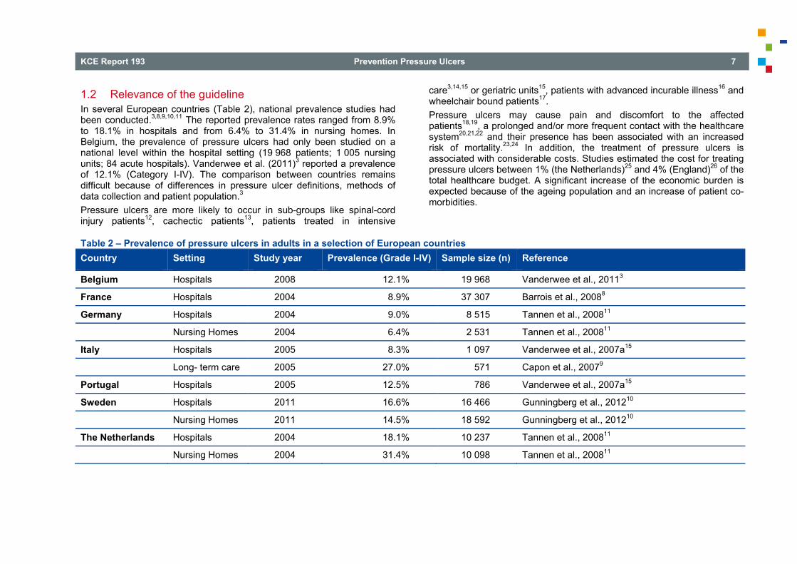
1.2 Relevance of the guideline In several European countries (Table 2), national prevalence studies had been conducted.3,8,9,10,11 The reported prevalence rates ranged from 8.9% to 18.1% in hospitals and from 6.4% to 31.4% in nursing homes. In Belgium, the prevalence of pressure ulcers had only been studied on a national level within the hospital setting (19 968 patients; 1 005 nursing units; 84 acute hospitals). Vanderwee et al. (2011)3 reported a prevalence of 12.1% (Category I-IV). The comparison between countries remains difficult because of differences in pressure ulcer definitions, methods of data collection and patient population.3 Pressure ulcers are more likely to occur in sub-groups like spinal-cord injury patients12, cachectic patients13, patients treated in intensive
care3,14,15 or geriatric units15, patients with advanced incurable illness16 and wheelchair bound patients17. Pressure ulcers may cause pain and discomfort to the affected patients18,19, a prolonged and/or more frequent contact with the healthcare system20,21,22 and their presence has been associated with an increased risk of mortality.23,24 In addition, the treatment of pressure ulcers is associated with considerable costs. Studies estimated the cost for treating pressure ulcers between 1% (the Netherlands)25 and 4% (England)26 of the total healthcare budget. A significant increase of the economic burden is expected because of the ageing population and an increase of patient co-morbidities.
Table 2 – Prevalence of pressure ulcers in adults in a selection of European countries Country Setting Study year Prevalence (Grade I-IV) Sample size (n) Reference
Belgium Hospitals 2008 12.1% 19 968 Vanderwee et al., 20113
France Hospitals 2004 8.9% 37 307 Barrois et al., 20088
Germany Hospitals 2004 9.0% 8 515 Tannen et al., 200811
Nursing Homes 2004 6.4% 2 531 Tannen et al., 200811
Italy Hospitals 2005 8.3% 1 097 Vanderwee et al., 2007a15
Long- term care 2005 27.0% 571 Capon et al., 20079
Portugal Hospitals 2005 12.5% 786 Vanderwee et al., 2007a15
Sweden Hospitals 2011 16.6% 16 466 Gunningberg et al., 201210
Nursing Homes 2011 14.5% 18 592 Gunningberg et al., 201210
The Netherlands Hospitals 2004 18.1% 10 237 Tannen et al., 200811
Nursing Homes 2004 31.4% 10 098 Tannen et al., 200811

8 Prevention Pressure Ulcers KCE Report 193
1.3 Scope The aim of this study was to develop a clinical practice guideline (CPG) on risk assessment and prevention of pressure ulcers in adults and children being admitted to hospitals, long-term care facilities (including nursing homes, rehabilitation facilities and long-term chronic care hospitals) and those receiving home care. The CPG will cover the following topics: • Risk assessment; • Skin assessment; • Skin massage; • Repositioning; • Devices for prevention (mattresses, overlays, cushions); • Devices for heel ulcer prevention; • Nutrition and hydration for prevention. The CPG is intended to support clinical decision-making in all health care professionals involved in the care for the individuals at risk for pressure ulcer development. The CPG will not cover risk assessment and management of incontinence associated dermatitis because of the unique nature and the specific aetiology of this skin disorder.27 The treatment of pressure ulcers will be the subject of a separate CPG.
2 METHODOLOGY 2.1 Clinical questions The clinical questions were the result of a scoping review of existing guidelines and consecutive discussions within the multidisciplinary research team (see Table 5) and the multidisciplinary expert panel (see also 2.4). The clinical questions were refined based on discussions with our international partner (see 2.2). The CPG addresses the following clinical questions (for detailed protocols, see appendix 1-9): 1. What is the clinical effectiveness of risk assessment tools in the
prevention of pressure ulcers? • Population: individuals of all ages in all settings; • Intervention: risk assessment tool, clinical judgement based on risk
factors; • Comparison: Each other, no risk assessment; • Outcomes:
o Critical outcomes for decision making: Proportion of participants developing new pressure ulcers (dichotomous outcome)(describe different categories of ulcer);
o Important outcomes: patient acceptability; rate of development of pressure ulcers; time to develop new pressure ulcer (time to event data); time in hospital or in other health care setting (continuous data); health-related quality of life (continuous data).

KCE Report 193 Prevention Pressure Ulcers 9
2. What is the predictive ability of risk assessment tools for pressure ulcer development?
• Population: individuals of all ages in all settings without a pressure ulcer;
• Intervention: risk assessment tool, clinical judgement based on risk factors;
• Outcomes: o Critical outcomes: Incidence of pressure ulcers (all grades and
grades 2-4) – up to one week; incidence of pressure ulcers (all grades and grades 2-4) – up to three months;
o Statistical measures: Area under the ROC (AUC), sensitivity for a defined threshold, specificity for a defined threshold.
3. What is the clinical effectiveness of skin assessment methods in the prevention of pressure ulcers?
• Population: individuals of all ages in all settings; • Intervention: skin assessment methods: diascopy and skin
temperature; • Comparison: Each other, no skin assessment, other method; • Outcomes:
o Critical outcomes for decision making: Proportion of participants developing new pressure ulcers (dichotomous outcome)(describe different categories of ulcer); patient acceptability;
o Important outcomes: rate of development of pressure ulcers; time to develop new pressure ulcer (time to event data); time in hospital or in other health care setting (continuous data); health-related quality of life (continuous data).
4. What is the predictive ability of skin assessment methods for pressure ulcer risk?
• Population: individuals of all ages in all settings; • Intervention: skin assessment methods: ultrasonography, ultrasound,
durometer/durometry, diascopy (finger method and transparent disk), elastometer, haptic finger, multispectral imaging device, multiwavelength imaging, multispectral images, digital colour images,
clinical assessment, transcutaneous oximetry, termographic scanner, tympanic thermometers (to measure skin temperature), doppler blood flowmetry, laser, doppler imaging;
• Outcomes: o Critical outcomes: Incidence of pressure ulcers (all grades and
grades 2-4) – up to one week; incidence of pressure ulcers (all grades and grades 2-4) – up to three months;
o Statistical measures: Area under the ROC (AUC), sensitivity for a defined threshold, specificity for a defined threshold, Diagnostic Odds Ratio.
5. What is the clinical effectiveness of skin massage in the prevention of pressure ulcers?
• Population: individuals of all ages in all settings; • Intervention: skin massage (method, products, frequency); • Comparison: no skin massage; other preventive methods; • Outcomes:
o Critical outcomes for decision making: Proportion of participants developing new pressure ulcers (dichotomous outcome)(describe different categories of ulcer); skin damage;
o Important outcomes: Patient acceptability; rate of development of pressure ulcers; time to develop new pressure ulcers; time in hospital or time in other healthcare setting; health related quality of life.
6. How and at what frequency should repositioning be undertaken for the prevention of pressure ulcers?
• Population: individuals of all ages in all settings; • Intervention: repositioning technique; frequency of repositioning;
different positions (e.g. 90-degree lateral rotation, 30-degree tilt); devices included for repositioning: profiling bed & tilt in space chairs;
• Comparison: no repositioning; different frequencies of repositioning; different positions for repositioning;

10 Prevention Pressure Ulcers KCE Report 193
• Outcomes:
o Critical outcomes for decision making: proportion of participants developing new pressure ulcers (dichotomous outcome) (describe different categories of ulcer);
o Important outcomes: patient acceptability; rate of development of pressure ulcers; time to develop new pressure ulcer (time to event data); time in hospital or in other health care setting (continuous data); health-related quality of life (continuous data).
7. What are the most clinically effective pressure re-distributing devices for the prevention of pressure ulcers?
• Population: individuals of all ages in all settings; • Intervention: mattresses/overlays; beds; seating; others like pillows,
postural support, and limb protectors; • Comparison: each other or no intervention; • Outcomes:
o Critical outcomes for decision making: proportion of participants developing new pressure ulcers (dichotomous outcome)(describe different categories of ulcer);
o Important outcomes: patient acceptability; rate of development of pressure ulcers; time to develop new pressure ulcer (time to event data); time in hospital or other health care setting (continuous data); health-related quality of life (continuous data).
8. What are the most clinically effective pressure re-distributing devices for the prevention of heel pressure ulcers?
• Population: individuals of all ages in all settings; • Intervention: heel-specific devices as preventive strategies (i.e. air-
filled booties, foam foot protectors, gel foot protectors, pillows and other aids, splints or other medical devices, sheepskins for heels - synthetic and natural, pressure relief ankle foot orthosis) and non heel-specific devices (Mattresses/overlays and beds);
• Comparison: each other or no intervention;
• Outcomes: o Critical outcomes for decision making: proportion of participants
developing new pressure ulcers (dichotomous outcome)(describe different categories of ulcer);
o Important outcomes: patient acceptability; rate of development of pressure ulcers; time to develop new pressure ulcer (time to event data); time in hospital or other health care setting (continuous data); health-related quality of life (continuous data).
9. What are the most clinically effective interventions with nutrition or hydration for the prevention of pressure ulcers for people with and without nutritional deficiency?
• Population: individuals of all ages in all settings with and without nutritional deficiencies;
• Intervention: nutritional interventions (supplementation or special diet); hydrational strategies as preventive strategies;
• Comparison: usual diet (participant’s usual diet or the standard hospital diet), other supplementation; other special diet;
• Outcomes: o Critical outcomes for decision making: proportion of participants
developing new pressure ulcers (dichotomous outcome); o Important outcomes: patients acceptability of supplements – e.g.
measured by compliance, tolerance, reports of unpalatability; rate of development of pressure ulcers; time to develop new pressure ulcer (time to event data); time in hospital or other health care setting (continuous data); (dichotomous data); health-related quality of life (continuous data).

KCE Report 193 Prevention Pressure Ulcers 11
2.2 International collaboration The National Clinical Guideline Centrea (NCGC), commissioned by The National Institute for Health and Clinical Excellence (NICE, United Kingdom) is currently producing a clinical guideline on the prevention and treatment of pressure ulcers to replace its existing guidelines.28,29,30 The CPG will be developed de novo. The nine research questions regarding risk assessment, skin assessment and prevention of pressure ulcers were fully in common with those of the KCE and the elaboration of the topics was divided between both organisations. A collaboration agreement was set up between NCGC and KCE concerning the following: 1. Scope: the collaboration concerned the search for evidence (search
strategy + selection), quality appraisal, evidence tables and the development of the evidence reports. The formulation of evidence statements and recommendations was the responsibility of the two organisations separately.
2. Form of cooperation: five research questions were elaborated by KCE (questions 1-5), while the four other common questions were elaborated by NCGC (questions 6-9).
3. Cross-validation was done after each of the following steps: o Development of the search strategy; o Selection of the literature; o Quality appraisal and elaboration of evidence tables; o Evidence report.
a The National Clinical Guideline Centre (NCGC) is a multi-disciplinary health
services research team funded by the National Institute for Health and Clinical Excellence (NICE). They produce evidence based clinical practice guidelines commissioned by NICE.
2.3 Literature searches 2.3.1 Search strategy The search for peer-reviewed articles included a search in OVID Medline, EMBASE, CINAHL and the Cochrane Library (see appendices 1-9 for search strings). The search was limited to articles published in English, French and Dutch for the evidence reports produced by KCE (Questions 1-5, performed by the KCE-team) while for the evidence reports that were produced by NCGC (Questions 6-9, performed by the NCGC-team) searches were restricted to articles published in English in line with the NCGC methodology. No date restriction was used. For most questions, the search focused on high-quality systematic reviews (i.e. reviews matching the PICO’s; extensive quality assessment; data available for GRADE input) and randomized controlled trials (RCTs) (see protocols in appendices 1-9 for more details). However, when RCTs were unavailable the search was expanded to observational studies (see protocols in appendices 1-9 for details). For the prognostic research questions (risk assessment – Q2 and skin assessment – Q4), the search focused on prospective cohort studies (see protocols in appendices 2 and 4 for details). All literature searches were done between March and September 2012. Search strategies were checked by reviewing the reference lists of relevant key papers and requesting the advice of the expert panel about additional papers. The identified studies were selected by one reviewer based on title and abstract. For all eligible studies, the full-text was retrieved. Studies were selected if relevant to the review question (PICO: population, intervention, comparison, outcome). A quality assurance check was performed by a second reviewer on 10% of the search results. In case no full-text was available, the study was not taken into account to develop the final recommendations.

12 Prevention Pressure Ulcers KCE Report 193
2.3.2 Quality appraisal A quality appraisal was done for each individual study and for each outcome. All critical appraisals were done by one researcher. The quality of the retrieved RCTs and observational studies was assessed using the corresponding checklists of the National Institute for Health and Clinical Excellence (NICE).31 For each clinical question the quality of the available evidence was summarized for each outcome using the GRADE-system and GRADEpro software (http://ims.cochrane.org/gradepro). The latter could not be used for the prognostic research question on risk – and skin assessment. Levels of evidence were regarded as being ‘HIGH’ for RCTs and ‘LOW’ for observational studies. In a subsequent step, the level of evidence was downgraded (and/or upgraded in case of observational studies) based on the assessment of the risk of bias, inconsistency, indirectness, imprecision, publication bias (see description in Table 3). Each quality element being considered to have “serious” or “very serious” risk of bias was downgraded with 1 or 2 points respectively. The downgraded/upgraded scores were then summed and an overall quality rating was assigned (see Table 4). An outcome with only RCTs, for example, starts ‘HIGH’ but can be downgraded to ‘MODERATE’, ‘LOW’ or ‘VERY LOW’ when 1, 2 or 3 points were deducted, respectively. The reasons or criteria used for downgrading were specified in the footnotes.
Table 3 – Description of GRADE elements for intervention studies (Source: NCGC, 2012)32 Quality element Description
Study limitations (Risk of bias)
Limitations in the study design and implementation may bias the estimates of the treatment effect. Major limitations in studies decrease the confidence in the estimate of the effect.
Inconsistency Inconsistency refers to an unexplained heterogeneity of results.
Indirectness Indirectness refers to differences in study population, intervention, comparator and outcomes between the available evidence and the protocol.
Imprecision Results are imprecise when studies include relatively few patients and few events and thus have wide Confidence Intervals around the estimate of the effect relative to the clinically important threshold.
Publication bias Publication bias is a systematic underestimate or an overestimate of the underlying beneficial or harmful effect due to the selective publication of studies.

KCE Report 193 Prevention Pressure Ulcers 13
Table 4 – Levels of evidence (Source: Balshem et al. 2011)33 Quality level Definition Methodological quality of supporting evidence
High We are very confident that the true effect lies close to that of the estimate of the effect
RCTs without important limitations or overwhelming evidence from observational studies
Moderate We are moderately confident in the effect estimate: the true effect is likely to be close to the estimate of the effect, but there is a possibility that it is substantially different
RCTs with important limitations (inconsistent results, methodological flaws, indirect, or imprecise) or exceptionally strong evidence from observational studies
Low Our confidence in the effect estimate is limited: the true effect may be substantially different from the estimate of the effect
RCTs with very important limitations or observational studies or case series Very Low We have very little confidence in the effect estimate: the true
effect is likely to be substantially different from the estimate of the effect
A more detailed description of the GRADE elements can be found in appendix 10.
2.3.3 Data extraction and analysis For each primary study, data were extracted by one reviewer. Following study characteristics were tabulated using a standard template: reference, patient characteristics, intervention/comparison, outcome measures, effect size, comments. An adapted version of the template was used to extract data for prognostic research questions (see appendices 2 and 4). A meta-analysis was done if possible using Revman-software (http://ims.cochrane.org/revman). The specific review strategies were defined in the study protocols (see appendices 1-9). In general, studies were combined in a meta-analysis if the clinical (e.g. similar patient population, intervention, comparison, outcome) and statistical heterogeneity were acceptable. The unit of analysis was separated in studies measuring outcomes at the patient or ulcer level. The following groups were considered separately as strata (children and adults) or subgroups (different categories of pressure ulcers; different ulcer locations). In absence of appropriate data, forest plot(s) were generated for each outcome using single studies for didactic purposes.
Fixed-effects (Mantel-Haenszel) techniques were used to calculate risk ratios (relative risk) for the binary outcomes. The continuous outcomes were analysed using an inverse variance method for pooling weighted mean differences and where the studies had different scales, standardised mean differences were used. Statistical heterogeneity was assessed by considering the chi-squared test for significance at p<0.1 or an I-squared inconsistency statistic of >50% to indicate significant heterogeneity. In case of heterogeneity and a sufficient number of studies, sensitivity analyses were conducted based on risk of bias and pre-specified subgroup analyses were carried out as defined in the protocol. Assessments of potential differences in effect between subgroups were based on the chi-squared tests for heterogeneity statistics between subgroups. If no sensitivity analysis was found to completely resolve statistical heterogeneity then a random effects (DerSimonian and Laird) model was employed to provide a more conservative estimate of the effect. The means and standard deviations of continuous outcomes were required for meta-analysis. However, in cases where standard deviations were not reported, the standard error was calculated if the p-values or 95%

14 Prevention Pressure Ulcers KCE Report 193
confidence intervals (CIs) were reported and meta-analysis was undertaken with the mean difference and standard error using the generic inverse variance method in Cochrane Review Manager (RevMan5) software. Where p-values were reported as “less than”, a conservative approach was undertaken. For example, if p value was reported as “p<0.001”, the calculations for standard deviations were based on a p-value of 0.001. The authors used the area under the receiver operating characteristic (AUC) to illustrate and evaluate the prognostic performance of risk assessment tools. In addition, the 95% CI for each scale (within studies and between studies; if data are available) was extracted and used to calculate the median AUC and range. The determination of an "ideal" cut-off value is almost always a trade-off between sensitivity (true positives) and specificity (true negatives). As both change for each "cut-off" value, it becomes difficult for the reader to imagine which cut-off is ideal. The AUC curve offers a graphical illustration of these trade-offs for each "cut-off" value. The maximum value for the AUC is 1.0, indicating a (theoretically) perfect test (i.e., 100% sensitive and 100% specific). An AUC of 0.5 indicates no discriminative value (i.e., 50% sensitive and 50% specific). Three cut-off scores were determined for each scale with an acceptable median AUC for the purpose of the risk assessment review. The sensitivity and specificity of these cut-off scores were determined by the median sensitivity. The sensitivity, specificity and diagnostic odds ratio for the prognostic question on skin assessment were calculated.
2.4 Formulation of recommendations: Collaboration between research team, Guideline Development Group and external experts
A subgroup of researchers (see Table 5) was responsible for systematic searches, retrieval and appraisal of the evidence and the writing of the evidence report (procedure used to draft evidence statements is described in appendix 10; 1.7). A second group within the research team drafted recommendations based on the retrieved evidence (Table 5) and assigned a grade of recommendation to each recommendation using the GRADE system (see appendix 10). Researchers responsible for drafting the recommendations were involved in the expert panel to discuss the evidence reports and vice versa. The draft of the recommendations and the evidence tables were circulated to the expert panel prior to each face-to-face meeting. The expert panel that consisted of 4 home care nurses, 8 hospital nurses, 2 nursing home nurses, 1 general practitioner, 1 dermatologist and 1 representative of the Belgian Ministry of Public Health (FOD Volksgezondheid – SPF Santé Publique) had the following tasks: • To verify that the research is complete and that the interpretation of
the evidence is correct; • To assess the relevance of the conclusions and the selected studies in
relation to the Belgian context; • To verify the evidence statements; • To participate in the drawing up of recommendations. The expert panel met on 3 occasions: 12 March 2012; 10 September 2012; 19 October 2012. Based on the evidence review, the research team (i.e. authors of this report) formulated recommendations. In addition Best practices were formulated. The latter were not based on the evidence reviews but on two existing guidelines (i.e. EPUAP/NPUAP 20095 & NICE 2001, 2003, 200528,29,30,34,35,36) that were retained after a comprehensive systematic search (see appendix 11 for details).

KCE Report 193 Prevention Pressure Ulcers 15
Table 5 – Research team and responsibilities Expert Organisation Area of expertise Researchers team
responsible for evidence reportsb
Working group responsible for drafting recommendations
Guideline Development Group (GDG)
Dimitri Beeckman UGENT Assistant Professor in Nursing Science
X X
Cathy Matheï KUL Professor in General Medicine
X X
Aurélie Van Lancker UGENT Researcher X X
Sabine Van Houdt KUL Researcher X X
Geert Vanwalleghem Clinical Nursing Consulting or CNC/WCS (VZW Wondzorgvereniging)
Clinical nurse specialist wound care – Hospital setting
X X
Luc Gryson CNC Clinical nurse specialist wound care
X X
Hilde Heyman WCS Clinical nurse specialist wound care – Nursing home setting
X X
Christian Thyse AFIScep.be Clinical nurse specialist wound care
X X
Adinda Toppets UZLeuven Clinical nurse specialist wound care
X
Sabine Stordeur KCE KCE-senior expert X X X
Koen Van den Heede KCE KCE-expert X X X
b Evidence reports for clinical questions 4-7 were produced by NCGC (Liz Avital, Katie Jones, Julie Neilson)

16 Prevention Pressure Ulcers KCE Report 193
All recommendations and Best practices were reviewed by a panel of experts (i.e. 19/10/2012) using a formal procedure. Two weeks before the final expert meeting, all experts received the recommendations and Best practices. As a preparation of the meeting all invited experts were asked to score each recommendation on a 5-point Likert-scale, with a score of ‘1’ indicating ‘completely disagree’, ‘2’ indicating ‘somewhat disagree’, ‘3’ indicating ‘unsure’, ‘4’ indicating ‘somewhat agree’, and ‘5’ indicating ‘completely agree’ (the experts were also able to answer ‘not applicable’ in case they were not familiar with the underlying evidence or rationale). In case an expert disagreed with the recommendation or Best practice (score ‘1’ or ‘2’), (s)he was asked to provide appropriate evidence or rationale, respectively. All scores were then anonymized and summarized into a median score, minimum score, maximum score and % of ‘agreement-scores (score ‘4’ and ‘5’) to allow a targeted discussion (see appendix 12). The recommendations were then discussed during a face-to-face meeting on 19/10/2012. Based on this discussion a final draft of the recommendations and Best practices was prepared. In appendix 12, an overview is provided of how the comments of the external experts were taken into account. Recommendations and Best practices were only changed if important evidence or rationale supported this change. Based on the discussion meeting a final draft of recommendations and Best practices was prepared and circulated to the expert panel.
3 FINAL RECOMMENDATIONS 3.1 Introduction In this chapter we will describe for each of the research questions a summary of the available evidence as well as the recommendations for clinical practice. In addition to the recommendations also Best practices are described. The formulations of the latter are based on expert discussions of existing guidelines. Two sets of general ‘Best practices’ were formulated: • Tailoring pressure ulcer prevention to individual needs based on the
principles of shared decision making; • Education and training.

KCE Report 193 Prevention Pressure Ulcers 17
Best Practices
Pressure ulcer prevention should be a combined approach, tailored to individual needs and situations, and should be based on the principles of shared decision making: • Prevention should take into account several factors such as the individual’s medical condition, the overall plan of care and the individual’s preferences.
The needs of the individual and the context should be re-assessed regularly; • An individual plan of care should be adopted based on assessment data, identified risk factors and individual goals and preferences. The plan is
developed in interaction with the individual, informal caregivers and the healthcare professional. The planned and agreed/refused actions are documented and communicated to all relevant caregivers (also in case transition between care settings takes place).
Training and education should be tailored both to the needs of individual caregiver and to the responsibilities of the group of professionals. Following components should be considered as part of each educational/training programme: • Aetiology and risk factors predisposing to pressure ulcers; • Classification of pressure ulcers; • Differential diagnosis with other types of skin lesions; • Risk assessment; • Skin assessment; • Selection and use of pressure redistributing devices; • Repositioning; • Nutritional aspects; • Methods of documenting risk assessments and preventive activities; • The importance of an interdisciplinary approach; • The education of the individual and their informal caregivers.

18 Prevention Pressure Ulcers KCE Report 193
3.2 Risk assessment 3.2.1 Introduction Risk assessment aims to identify susceptible patients for pressure ulcer development in order to target appropriate preventive interventions. Both risk assessment scales and clinical judgment are widely described as methods being used in day-to-day practice. Risk assessment scales are based on risk factors associated with pressure ulcer development.37 In a risk assessment scale, risk factors are listed and clinicians are expected to denote the presence, degree, or absence of the risk factor.37 Such a list of risk factors aims to provide guidance to the specific cut-off point for risk or to specific preventive interventions that should be initiated.37 A scale must accurately identify these individuals who are at risk, as well as those who are not- and do this consistently. Several risk assessment scales have been developed in the past38. Numerous risk assessment scales exist. We will organise the results according to the most commonly reported39 and used risk assessment scales: • The Braden-scale (includes the following subscales: nutrition, mobility,
sensory perception, moisture, activity, friction and shear); • The Norton-scale (includes the following subscales: mobility,
continence, mental status, general health, activity); • The Waterlow-scale (includes the following subscales:
nutrition/appetite, mobility, build/weight, continence, sex/age, skin type, tissue malnutrition, neurological deficit, major surgery/trauma, medication);
• The Cubbin-Jackson which is adapted from the Norton scale (includes the following subscales: nutrition, mobility, build/weight, continence, mental status, age, skin condition, hygiene, hemodynamic status, respiration);
• The Braden-Q which is adapted from Braden for paediatric population (includes the following subscales: activity, nutrition, mobility, sensory perception, moisture, friction and shear, tissue perfusion and oxygenation).
Next to the use of a risk assessment scale, clinical judgment can be used to assess the level of risk for pressure ulcer development.5 The aim of this review was to guide health care professionals in their decision making about which method for risk assessment is most appropriate to detect individuals at risk for pressure ulcer prevention. The review on risk assessment includes two parts. In a first review (see 3.2.2) we will focus on the clinical effectiveness of risk assessment as part of a complex intervention for pressure ulcer prevention. A second review (see 3.2.3) will focus on the prognostic ability of risk assessment (scales and clinical judgment).
3.2.2 Clinical effectiveness
3.2.2.1 Review question What is the clinical effectiveness of risk assessment tools in the prevention of pressure ulcers?
3.2.2.2 Clinical evidence A Cochrane review by Moore and Cowan (2010)40 was identified and used as reference for this review. The Cochrane review was an update of three systematic reviews by Cullum (1995)41, McGough (1999)42 and Pancorbo-Hidalgo (2006)39. None of the studies included in these systematic reviews were included in the Cochrane review as none of them were randomized controlled trials. The Cochrane review by Moore and Cowan (2010)43, included one RCT44. The Cochrane review was updated through a systematic search of multiple electronic databases. This resulted in 241 records: 47 in Medline (OVID), 36 in CINAHL, 108 in Embase and 50 in the Cochrane Library, of which 52 duplicates were removed. Based on screening of title and/or abstract 188 records were excluded. One study (a RCT published by Webster 201145) was reviewed in detail and retained in addition to the RCT included in the Cochrane review. Both RCTs included patients with and without a pressure ulcer at baseline and in both studies preventive measures were given to patients.

KCE Report 193 Prevention Pressure Ulcers 19
Quality of Studies The methodological quality of the two RCTs was poor. The study of Saleh (2009)44 had the most methodological flaws: absence of sequence generation, blinding, intention-to-treat analysis, and a priory sample size calculation. In addition, the allocation concealment was poor and the study had a high drop-out rate. The study of Webster (2010)45 was of higher quality. However, there was no blinding of health care professional and the sample size was lower than calculated. In addition, both studies included patients with pressure ulcers at baseline. In appendix 1 the level of evidence can be found per outcome after applying the GRADE-methodology. The evidence base for all outcomes has been rated as low to very low quality. Evidence statements • One study (n=180) showed that clinical judgement is potentially more
clinical effective at reducing pressure ulcer incidence (all grades) compared to the Braden scale (VERY LOW QUALITY). 44
• One study (n=180) showed there may be no clinical difference between the group that used the Braden scale and received a wound management training and the group that only received the training (all grades) (VERY LOW QUALITY).44
• One study (n=180) showed that clinical judgement without training is potentially more clinical effective at reducing pressure ulcer incidence (all grades) compared to the group receiving training only (VERY LOW QUALITY). 44
• One study (n=821) showed there may be no clinical difference between the group that used the Waterlow scale and the group that used clinical judgement (all grades), but the direction could favour clinical judgement (LOW QUALITY).45
• One study (n=820) showed there may be no clinical difference between the group that used the Waterlow scale and the group that used clinical judgement (grade 2), but the direction could favour clinical judgement (LOW QUALITY).45
• One study (n=821) showed that the Ramstadius scale is potentially more clinical effective at reducing pressure ulcer incidence (all grades) compared to the Waterlow scale (MODERATE QUALITY).45
• One study (n=820) showed that the Ramstadius scale is potentially more clinical effective at reducing pressure ulcer incidence (grade 2) compared to the Waterlow scale (MODERATE QUALITY).45
• One study (n=821) showed that the Ramstadius scale may be more clinical effective at reducing pressure ulcer incidence (all grades) compared to clinical judgement (LOW QUALITY).45
• One study (n=821) showed that the Ramstadius scale may be more clinical effective at reducing pressure ulcer incidence (grade 2) compared to clinical judgement (LOW QUALITY).45
3.2.3 Prognostic
3.2.3.1 Review question What is the predictive ability of risk assessment tools for pressure ulcer development?
3.2.3.2 Clinical evidence The systematic review by Pancorbo-Hidalgo (2006)39 was used as a reference for this review. All studies included in the Pancorbo-Hidalgo (2006) 39 review, identified through the update and meeting the criteria of our review were reviewed in detail. Sensitivity and specificity of each scale and cut-off score were re-calculated by using the raw data as presented in the individual studies. Some adjustments were made to the Pancorbo-Hidalgo review39. The review by Pancorbo-Hidalgo et al. (2006)39 included 32 studies, of which five were excluded because they didn’t meet the inclusion criteria of our review: • One was excluded as it was a retrospective cohort study46; • Another study was removed as it was written in Spanish47; • Three other studies were excluded as they included patients with a
pressure ulcer at start of the study.48,49,50

20 Prevention Pressure Ulcers KCE Report 193
The update of the Pancorbo-Hidalgo (2006)39 review yielded 16 additional articles resulting in a final inclusion of 44 studies.51-93 Quality of Studies In general there were many concerns regarding the quality of the included studies. The absence of a description of patient enrolment, of time points when patients dropped out (discharge, death, transfer, pressure ulcer development) from the study, of an imputation technique, a poor description of definition and measurement of index test, and an event rate lower than 100 were the most important methodological flaws. In addition, patients received preventive measure which influenced the results on predictive validity. In five studies it was unclear if patients with pressure ulcers at start of the study were included.62,75,84,86,87 The results from these studies should be interpreted with caution. In appendix 2 the level of evidence can be found per scale for studies reporting on the area under the receiver operating characteristics curve. The evidence base for all scales has been rated as being of low to very low quality. Evidence statements Braden scale: • Area under the receiver-operating curve:
o Nine studies showed a median AUC of 79.0 (range 55.0 to 88.0) for the Braden scale (all populations) indicating a fair discriminating power (VERY LOW QUALITY).59,62,67,69,80,83,84,88,94
o Five studies showed a median AUC of 68.0 (range 55.0 to 81.0) for the Braden scale (general population) indicating a poor discriminating power (VERY LOW QUALITY).59,62,67,80,83
o Four studies showed a median AUC of 79.0 (range 71.0 to 88.0) for the Braden scale (intensive care population) indicating a fair discriminating power (VERY LOW QUALITY).69,84,88,94
o One study showed a median AUC of 74.0 (95% CI: 63.0-84.0) for the modified Braden scale (general) indicating a fair discriminating power (LOW QUALITY).59
• Predictive ability for different cut-off values:
o Two studies (one of the studies consisted of three independent samples) (general population) showed a median sensitivity of 59.0 (range 50.0-78.0) and a corresponding specificity of 70.5 (range 52.0-81.0) for the Braden scale based on a cut-off score of ≤ 17 and a follow-up period < 1 week (LOW QUALITY).56,57
o Two studies (one of the studies consisted of three independent samples) (general population) showed a median sensitivity of 70.0 (range 60.0-88.0) and a corresponding specificity of 58.0 (range 48.0-81.0) for the Braden scale based on a cut-off score of ≤ 18 and a follow-up period < 1 week (LOW QUALITY).56,57
o Two studies (one of the studies consisted of three independent samples) (general population) showed a median sensitivity of 83.5 (range 51.0-100.0) and a corresponding specificity of 60.5 (range 42.0-73.0) for the Braden scale based on a cut-off score of ≤ 19 and a follow-up period point < 1 week (LOW QUALITY).56,57
o One study (ICU population) showed a sensitivity of 87.5 and a specificity of 64.1 for the Braden scale based on a cut-off score of ≤ 12 and a follow-up period point of 48 hours (VERY LOW QUALITY).94
o One study (ICU population) showed a sensitivity of 75.0 and a specificity of 82.1 for the Braden scale based on a cut-off score of ≤ 13 and a follow-up period point < 1 week (VERY LOW QUALITY).94
o One study (ICU population) showed a sensitivity of 76.9 and a specificity of 29.6 for the Braden scale based on a cut-off score of ≤ 16 and a follow-up period point < 1 week (LOW QUALITY).64
o Ten studies (some studies had multiple samples) (general population) showed a median sensitivity of 79.5 (range 46.2-100.0) and a corresponding specificity of 73.6 (range 14.0-100.0) for the Braden scale based on a cut-off score of ≤ 18 and a follow-up period point > 1 week (VERY LOW QUALITY).55-59,65,71,76,79,82
o Five studies (some studies had multiple samples) (general population) showed a median sensitivity of 86.3 (range 71.4-100.0) and a corresponding specificity of 67.5 (range 42.9-77.8)

KCE Report 193 Prevention Pressure Ulcers 21
for the Braden scale based on a cut-off score of ≤ 19 and a follow-up period point > 1 week (LOW QUALITY).55-58,82
o Five studies (some studies had multiple samples) (general population) showed a median sensitivity of 93.2 (range 43.2-100.0) and a corresponding specificity of 53.5 (range 31.6-66.7) for the Braden scale based on a cut-off score of ≤ 20 and a follow-up period point > 1 week (LOW QUALITY).55-58,82
o One study (ICU) showed a sensitivity of 75.0 and a specificity of 66.7 for the Braden scale based on a cut-off score of ≤ 15 and a follow-up period point > 1 week (LOW QUALITY).57
o Two studies (ICU) showed a mean sensitivity of 90.2 (range 83.3-97.1) and a corresponding specificity of 45.0 (range 26.0-63.9) for the Braden scale based on a cut-off score of ≤ 16 and a follow-up period point > 1 week (LOW QUALITY).57,84
o One study (ICU) showed a sensitivity of 87.5 and a specificity of 50.0 for the Braden scale based on a cut-off score of ≤ 17 and a follow-up period point > 1 week (LOW QUALITY).57
o One study (general population) showed a sensitivity of 42.9 and a specificity of 63.4 for the Braden scale (grade 2+ PU) based on a cut-off score of ≤ 17, a follow-up period point > 1 week (LOW QUALITY).81
o One study (general population) showed a sensitivity of 100.0 and a specificity of 34.1 for the Braden scale (grade 2+ PU) based on a cut-off score of ≤ 18, a follow-up period point > 1 week (LOW QUALITY).81
o One study (general population) showed a sensitivity of 100.0 and a specificity of 22.0 for the Braden scale (grade 2+ PU) based on a cut-off score of ≤ 19, a follow-up period point > 1 week (LOW QUALITY).81
Norton scale: • Area under the receiver-operating curve:
o Two studies showed a mean AUC of 65.0 (range 56.0 to 74.0) for the Norton scale (general population) indicating a poor discriminating power (LOW QUALITY).80,83
• Predictive ability for different cut-off values: o Four studies (general population) showed a median sensitivity of
45.7 (range 0.0-88.9) and a corresponding specificity of 80.6 (range 61.0-94.4) for the Norton scale based on a cut-off score of ≤ 14 and a follow-up period point > 1 week (VERY LOW QUALITY).70,73,87,92
o One study (general population) showed a sensitivity of 45.9 and a specificity of 60.3 for the Norton scale based on a cut-off score of ≤ 15, a follow-up period point > 1 week (LOW QUALITY).83
o Two studies (general population) showed a mean sensitivity of 70.5 (range 60.0-81.0) and a corresponding specificity of 44.9 (range 31.0-58.8) for the Norton scale based on a cut-off score of ≤ 16 and a follow-up period point > 1 week (VERY LOW QUALITY).79,86
Waterlow scale: • Area under the receiver-operating curve:
o Four studies showed a median AUC of 60.0 (range 54.0 to 90.0) for the Waterlow scale (all populations) indicating a poor discriminating power (VERY LOW QUALITY).52,60,83,85
o Three studies showed a median AUC of 61.0 (range 54.0 to 90.0) for the Waterlow scale (general population) indicating a poor discriminating power (VERY LOW QUALITY).52,60,83,85
o One study showed an AUC of 59.0 (95% CI 54.0-65.0) for the Waterlow scale (intensive care population) indicating that the scale fails to discriminate (LOW QUALITY).60
• Predictive ability for different cut-off values: o One study (general population) showed a sensitivity of 71.4 and a
specificity of 67.0 for the Waterlow scale based on a cut-off score of ≥ 17, a follow-up period of 48 hours (VERY LOW QUALITY).85

22 Prevention Pressure Ulcers KCE Report 193
o One study (general population) showed a sensitivity of 85.7 and a specificity of 36.9 for the Waterlow scale based on a cut-off score of ≥ 20, a follow-up period of < 1 week (VERY LOW QUALITY).85
o Three studies (general population) showed a median sensitivity of 87.5 (range 82.3-89.6) and a corresponding specificity of 28.2 (range 22.4-85.2) for the Waterlow scale based on a cut-off score of ≥ 10 and a follow-up period point > 1 week (LOW QUALITY).52,83,92
o One study (general population) showed a sensitivity of 48.8 and a specificity of 94.4 for the Waterlow scale based on a cut-off score of ≥ 15, a follow-up period of < 1 week (LOW QUALITY).52
o Two studies (general population) showed a mean sensitivity of 84.3 (range 73.3-95.2) and a corresponding specificity of 40.8 (range 38.0-43.5) for the Waterlow scale based on a cut-off score of ≥ 16 and a follow-up period point > 1 week (VERY LOW QUALITY).79,86
o One study (general population) showed a sensitivity of 80.9 and a specificity of 28.5 for the Waterlow scale (grade 2+ PU) based on a cut-off score of ≤ 15, a follow-up period point > 1 week (LOW QUALITY).93
Cubbin-Jackson scale: • Area under the receiver-operating curve:
o Two studies showed a mean AUC of 87.0 (range 83.0 to 90.0) for the Cubbin-Jackson scale (ICU) indicating a good discriminating power (LOW QUALITY).69,84
• Predictive ability for different cut-off values: o One study (ICU population) showed a sensitivity of 88.6 and a
specificity of 61.0 for the Cubbin-Jackson scale based on a cut-off score of ≤ 24 and a follow-up period point > 1 week (LOW QUALITY).84
o One study (ICU population) showed a sensitivity of 95.0 and a specificity of 81.6 for the Cubbin-Jackson scale based on a cut-off score of ≤ 28 and a follow-up period point > 1 week (LOW QUALITY).69
Braden-Q scale: • Area under the receiver-operating curve:
o One study showed an AUC of 83.0 (95% CI 76.0-91.0) for the Braden-Q scale (paediatric ICU) indicating a good discriminating power (LOW QUALITY).61
• Predictive ability for different cut-off values: o One study (paediatric ICU population) showed a sensitivity of 75.6
and a specificity of 67.8 for the Braden-Q scale based on a cut-off score of ≤ 15 and a follow-up period point > 1 week (LOW QUALITY).61
o One study (paediatric ICU population) showed a sensitivity of 88.4 and a specificity of 58.1 for the Braden-Q scale based on a cut-off score of ≤ 16 and a follow-up period point > 1 week (LOW QUALITY).61
o One study (paediatric ICU population) showed a sensitivity of 91.9 and a specificity of 44.1 for the Braden-Q scale based on a cut-off score of ≤ 17 and a follow-up period point > 1 week (LOW QUALITY).61
Clinical judgement: • Predictive ability:
o Two studies (general population) showed a mean sensitivity of 50.9 (range 50.0-51.7) and a corresponding specificity of 68.9 (range 58.1-79.7) for clinical judgement based on a follow-up period point > 1 week (LOW QUALITY).82,91
Braden scale versus Norton scale versus Waterlow scale: • Predictive ability:
o One study examined the Braden, Norton and Waterlow scale in the same patient sample (general population). The scales had a similar discriminating power (55.0 versus 56.0 versus 61.0) (LOW QUALITY).80,83

KCE Report 193 Prevention Pressure Ulcers 23
Braden scale versus Norton scale versus Fragmment scale: • Predictive ability:
o One study examined the Braden, Norton and Fragmment scale in the same patient sample (general population). The scales had a similar discriminating power (74.0 versus 74.0 versus 79.0) (LOW QUALITY).59,62,67,80,83
Braden scale versus Cubbin-Jackson scale versus Douglas scale: • Predictive ability:
o One study examined the Braden, Cubbin-Jackson and Douglas scale in the same patient sample (ICU population). The Cubbin-Jackson scale had a higher discriminating power compared to the Braden and Douglas scale (LOW QUALITY).69,84
Braden scale versus Cubbin-Jackson scale versus Song and Choi scale: • Predictive ability:
o One study examined the Braden, Cubbin-Jackson and Song and Choi scale in the same patient sample (ICU population). The Cubbin-Jackson scale had a higher discriminating power compared to the Braden and Song and Choi scale (LOW QUALITY).69,84
Braden scale versus modified Braden scale • Predictive ability:
o One study examined the Braden and modified Braden scale in the same patient sample (general population). The Braden scale had a higher discriminating power compared to the modified Braden scale (LOW QUALITY).59
Braden scale at different time points • Predictive ability:
o One study examined the Braden scale at different times points in the same patient sample (ICU population). The Braden scale at different time point had similar discriminating power (48 hours, 4 and 6 days) (VERY LOW QUALITY).69,84,88,94
Waterlow scale at different time points • Predictive ability:
o One study examined the Waterlow scale at different times points in the same patient sample (general population). The Waterlow scale after 48 hours had the highest discriminating power compared to 4 and 6 days (VERY LOW QUALITY).52,60 83,85
Other scales: • The Douglas scale, the Fragmment scale, the Song and Choi scale,
The Northern Hospital Pressure Ulcer Prevention Plan were examined in four studies and revealed a fair to good discriminating power (MODERATE TO LOW QUALITY).69,78,80,84
3.2.4 Conclusion
• Clinical effectiveness o There is no sound evidence base that supports superior
clinical effectiveness of one risk assessment approach over another in the prevention of pressure ulcer development.
o In addition, study results should be interpreted with caution, because studies had a very low quality and included patients with and without a pressure ulcer at start of the study.
• Predictive ability o No clear conclusions about the prognostic ability of risk
assessment tools can be drawn. o None of the risk assessment tools being studied and none of
their thresholds outperform the others. A large heterogeneity between studies in terms of population, reported cut-off scores, and time points of assessment has been illustrated. In addition, the included studies in this review had high to very high risk of bias.
o Evidence on risk-assessment tools that are not wide-spread indicated a fair to very good discriminating power and good sensitivity and specificity. However, these results are based on single studies.

24 Prevention Pressure Ulcers KCE Report 193
3.2.5 Recommendations and Best practices for clinical practice
Recommendation Strength of Recommendation
Level of Evidence
A structured approach for risk assessment should be used to identify individuals at risk of developing pressure ulcers. This structured approach should include all of following components: • Clinical judgement informed by knowledge of key risk factors; • The use of a risk assessment tool. As clinical studies do not demonstrate the superiority of one risk
assessment tool over another, decisions about which risk assessment tool (Braden, Norton, Waterlow…) to be used should be based on the intended population (adults, children, elderly,…) and the intended care setting (intensive care units, general wards, paediatrics, home care…) and the experience and expertise of the healthcare staff;
• A comprehensive skin assessment to evaluate any alterations to intact skin.
Strong Very Low
Best Practices
Pressure ulcer risk assessment should be performed at the first contact with the individual. Reassessment should be undertaken at regular time intervals and if there is any change in the individual’s medical condition. The decision on time intervals should be based on an individual basis. Clinical judgment should take into account several key risk factors such as reduced mobility, immobility, pressure, shear, sensory impairment, acute deterioration of general health status, incontinence, reduced level of consciousness, advanced age, previous history of pressure damage, vascular disease, under-nutrition, poor hydration status (non-limitative list). Risk assessment should be documented and made accessible to all members of the multidisciplinary team.

KCE Report 193 Prevention Pressure Ulcers 25
3.3 Skin assessment 3.3.1 Clinical effectiveness
3.3.1.1 Introduction A structured skin assessment is widely considered to be an integral part of a comprehensive pressure ulcer prevention strategy.5 Multiple methods can be used to observe the skin for early signs of pressure ulcer development. The most commonly used method in clinical practice is diascopy (finger method or transparent disk method). If applying the finger method, an amount of pressure is applied on the erythema of the skin (mainly over a bony prominence). If the erythema does not disappear when the finger is being removed, the erythema is considered to be non-blanchable (Category I pressure ulcer). The transparent disk method includes the use of a plastic transparent plastic to press on the erythema. If the erythema under the transparent disk does not blanch, it is regarded to be non-blanchable erythema. An increase or decrease in skin temperature can also be indicative of pressure damage. An increase in temperature at the area can indicate inflammation or infection, whereas cool skin may indicate poor perfusion and ischaemia. Assessing skin temperature may be particularly important in patients with darker skin, where the more evident area of erythema is not visible. The aim of this review was to guide health care professionals in their decision making about which method for skin assessment is most appropriate to detect individuals at risk for pressure ulcer prevention. This review includes two parts. In a first review we will focus on the clinical effectiveness of three skin assessment methods (i.e. diascopy; finger method; skin temperature) as part of a complex intervention for pressure ulcer prevention. A second review will focus on the prognostic ability of a wide variety of skin assessment methods (ultrasonography, ultrasound, durometer/durometry, diascopy, elastometer, haptic finger, multispectral imaging device, multi-wavelength imaging, multispectral images, digital colour images, clinical assessment, transcutaneous oximetry, thermographic scanner, tympanic thermometers (to measure skin temperature), doppler blood flowmetry, laser, doppler imaging).
3.3.1.2 Review question What is the clinical effectiveness of skin assessment methods in the prevention of pressure ulcers?
3.3.1.3 Clinical evidence One randomized controlled trial of Vanderwee et al. (2007b) was included in this review.95 In this study, the authors studied the effectiveness of using “daily skin assessment with transparent disk” as the only risk assessment strategy in the prevention of pressure ulcer incidence. If non-blanchable erythema appeared, preventive measures were started. They compared this with the use of a Braden score in combination with daily skin assessment using a transparent disk. In this control group preventive measures were started when non-blanchable erythema was observed or in case of a Braden-score of ≤17. Quality of Studies The included study had an adequate sample size (without drop-outs), reported on sequence generation and allocation concealment. Only blinding was not possible due to practical and ethical reasons. In appendix 3 the level of evidence can be found per outcome after applying the GRADE-methodology. The evidence base for all outcomes has been rated as moderate quality. Evidence statements • One study (n=1 617) showed that there is no difference between skin
assessment with transparent disk (NBE, non-blanchable erythema) and skin assessment with transparent disk combined with the Braden scale (control) for the assignment of preventive measures to reduce pressure ulcer incidence (grade 2-4) (MODERATE QUALITY).95
• One study (n=1 617) showed that time to development of pressure ulcers (grade 2-4) was shorter for skin assessment with transparent disk (NBE) than for skin assessment with transparent disk combined with the Braden scale (control) (MODERATE QUALITY).95

26 Prevention Pressure Ulcers KCE Report 193
3.3.2 Prognostic
3.3.2.1 Review question What is the predictive ability of skin assessment methods for pressure ulcer risk?
3.3.2.2 Clinical evidence Four prognostic studies60,96-98 were included in this review. Sensitivity and specificity were re-calculated by using the raw data as presented in the individual studies. Quality of Studies In general the quality of the studies was poor. The absence of a description of patient enrolment or use of a consecutive sampling, the absence of an imputation technique, and an event rate lower than 100 were the most important methodological flaws60,96-98. In addition, two studies96,98 had an unclear duration of the follow-up period and one study98 had significant missing data. Moreover, patients received preventive measure which influenced the results on predictive validity. In appendix 4 the level of evidence can be found per scale for studies reporting on the area under the receiver operating characteristics curve. The evidence base for all scales has been rated as low to very low quality. Evidence statements Sensitivity and specificity are the most commonly used and recommended statistics for evaluating the predictive validity of pressure ulcer risk assessment scales and skin assessment methods, assuming that a good and useful scale or skin assessment should have both high sensitivity and high specificity. There is, however, an important difference between a diagnostic/prognostic screening test and a risk assessment scale or skin assessment method. In contrast with prognostic and diagnostic tests, skin assessment methods (and risk assessment scales) are used to identify patients in need of preventive measures and are intended to contribute to the prevention of pressure ulcer development. In that way, they differ from diagnostic/prognostic screening tests. If preventive measures are used, the probability that a patient will develop a pressure ulcer at the start of the study will not remain constant until its end. The use of effective prevention will alter the sensitivity and specificity of the risk assessment scales and
skin assessment methods. The results should therefore be considered with caution. Subjective nursing assessment of moist skin: • One study (ICU patients) showed a sensitivity of 76%, a specificity of
65% and a diagnostic odds ratio of 5.9 for the subjective nursing assessment of moist skin as a predictor for the development of pressure ulcers (grades 2-4) according to the European Pressure Ulcer Advisory Panel classification system (LOW QUALITY).60
Subjective nursing assessment of oedematous skin: • One study (ICU patients) showed a sensitivity of 64%, a specificity of
77% and a diagnostic odds ratio of 5.7 for the subjective nursing assessment of oedematous skin as a predictor for the development of pressure ulcers (grades 2-4) according to the European Pressure Ulcer Advisory Panel classification system (LOW QUALITY).60
Subjective nursing assessment of mottled skin: • One study (ICU patients) showed a sensitivity of 33%, a specificity of
92% and a diagnostic odds ratio of 5.4 for the subjective nursing assessment of mottled skin as a predictor for the development of pressure ulcers (grades 2-4) according to the European Pressure Ulcer Advisory Panel classification system (LOW QUALITY).60
Subjective nursing assessment of livid skin: • One study (ICU patients) showed a sensitivity of 31%, a specificity of
92% and a diagnostic odds ratio of 5.0 for the subjective nursing assessment of livid skin as a predictor for the development of pressure ulcers (grades 2-4) according to the European Pressure Ulcer Advisory Panel classification system (LOW QUALITY).60
Subjective nursing assessment of centralised circulation: • One study (ICU patients) showed a sensitivity of 71%, a specificity of
70% and a diagnostic odds ratio of 5.8 for the subjective nursing assessment of centralised circulation as a predictor for the development of pressure ulcers (grades 2-4) according to the European Pressure Ulcer Advisory Panel classification system (LOW QUALITY).60

KCE Report 193 Prevention Pressure Ulcers 27
Subjective nursing assessment of cyanosis: • One study (ICU patients) showed a sensitivity of 45%, a specificity of
81% and a diagnostic odds ratio of 3.5 for the subjective nursing assessment of cyanosis as a predictor for the development of pressure ulcers (grades 2-4) according to the European Pressure Ulcer Advisory Panel classification system (LOW QUALITY).60
Subjective nursing assessment of reddened skin: • One study (ICU patients) showed a sensitivity of 69%, a specificity of
70% and a diagnostic odds ratio of 5.1 for the subjective nursing assessment of reddened skin as a predictor for the development of pressure ulcers (grades 2-4) according to the European Pressure Ulcer Advisory Panel classification system (LOW QUALITY).60
Subjective nursing assessment of hyperaemic skin: • One study (ICU patients) showed a sensitivity of 21%, a specificity of
91% and a diagnostic odds ratio of 2.9 for the subjective nursing assessment of reddened skin as a predictor for the development of pressure ulcers (grades 2-4) according to the European Pressure Ulcer Advisory Panel classification system (LOW QUALITY).60
Presence of blanchable erythema assessed by the finger test: • One study (hospitalized patients) showed a sensitivity of 75%, a
specificity of 77% and a diagnostic odds ratio of 9.9 for the assessment of blanchable erythema by the finger test as a predictor for the development of pressure ulcers according to the European Pressure Ulcer Advisory Panel classification system (LOW QUALITY).96
• Two studies showed a wide range of specificity and diagnostic odds ratios.96,98 One study showed a sensitivity of 75%, a specificity of 76% and a diagnostic odds ratio of 9.4 for the assessment of blanchable erythema by the finger test as a predictor for the development of PU (grades 2-4) according to the European Pressure Ulcer Advisory Panel classification system.96 The other study showed a sensitivity of 75%, a specificity of 10% and a diagnostic odds ratio of 0.33 for the assessment of blanchable erythema by the finger test as a predictor for the development of pressure ulcers (grades 2-4) according to the
European Pressure Ulcer Advisory Panel classification system (LOW QUALITY).98
Thermography: • One study (hospitalized patients) showed a sensitivity of 100%, a
specificity of 74% and a diagnostic odds ratio of 36.7 for thermography (presence of thermal anomaly – an area of the skin at least 1°C warmer than the surrounding skin) as a predictor for the development of skin breakdown (LOW QUALITY).97
Presence of non-blanchable erythema assessed by the finger test: • One study (surgical in-patients) showed a sensitivity of 73%, a
specificity of 74% and a diagnostic odds ratio of 8.0 for the assessment of non-blanching erythema by the finger test as a predictor for the development of pressure ulcers (grades 2-4) according to the European Pressure Ulcer Advisory Panel classification system (LOW QUALITY).98

28 Prevention Pressure Ulcers KCE Report 193
3.3.3 Conclusion
• Clinical effectiveness o One single RCT provided evidence of moderate quality that
the time to develop a pressure ulcer was significantly longer when the start of preventive measures was based on a combined approach of risk and skin assessment (i.e. Braden score in combination with daily skin assessment with transparent disk: preventive measures were started when non-blanchable erythema appeared or in case of a Braden-score of ≤17) compared to a decision based on daily skin assessment with transparent disk only (i.e. preventive measures were started when non-blanchable erythema appeared). However, there was no difference in effect on pressure ulcer incidence.
• Predictive ability o No clear conclusions about the prognostic ability of skin
assessment methods can be drawn due to large heterogeneity between studies in terms of results, population, reported assessment methods and timing of the performed assessments.
o One study (hospitalized patients) showed a sensitivity of 100% and a specificity of 74% for thermography (presence of thermal anomaly – an area of the skin at least 1°C warmer than the surrounding skin) as a predictor for the development of skin breakdown.
o One study (surgical in-patients) showed a sensitivity of 73% and a specificity of 74% for the assessment of non-blanchable erythema by the finger test as a predictor for the development of pressure ulcers (grades 2-4) according to the European Pressure Ulcer Advisory Panel classification system. Two studies investigating the predictive ability of blanchable erythema by the finger test illustrated similar sensitivity rates (75%) but a wide range in specificity (10% - 76%).

KCE Report 193 Prevention Pressure Ulcers 29
3.3.4 Recommendation and Best practices for clinical practice
Recommendation Strength of Recommendation
Level of Evidence
A comprehensive head-to-toe skin assessment with special attention to vulnerable areas (especially bony prominences) should be part of a structured risk assessment approach.
Strong Low
Best Practices
Skin assessment should be an integral part of routine care and the frequency of inspection may need to be adapted in response to the changes in the individual’s condition (improvement or deterioration) and level of risk. Inspect the skin regularly for signs of redness and non-blanchable erythema. Skin assessment should also include assessment for localized heat, oedema, or induration (hardness). Observe the skin for pressure damage caused by medical devices. With individuals with darkly pigmented skin, consider an individual at risk when there are: • purplish/bluish localised areas of skin; • localised heat which, if tissue becomes damaged, is replaced by coolness; • localised oedema; • localised induration. Any skin changes should be documented, made accessible and communicated to the members of the multidisciplinary team.

30 Prevention Pressure Ulcers KCE Report 193
3.4 Skin massage 3.4.1 Introduction One of the practices still widely used to prevent development of pressure ulcers is massage. Although massage is internationally no longer officially recommended as a preventive method, the technique is still practiced in many health care settings.5 The aim of this review is to study the clinical effectiveness of massage as an intervention to prevent pressure ulcer development.
3.4.2 Review question What is the clinical effectiveness of skin massage in the prevention of pressure ulcers?
3.4.3 Clinical evidence One randomized clustered double-blind cross-over clinical trial was included in this review.99 This trial assessed the effectiveness of massage with dimethyl sulfoxide (DMSO) or Vaseline added to position change in preventing pressure ulcers compared with position change only. Residents of 8 Dutch nursing homes received either position change only or massage with position change. The first product (Vaseline or DMSO) was used during 4 weeks (period 1), and after a wash-out period of 2 weeks, the other product (DMSO or Vaseline) during 4 weeks (period 2). Crossover design was judged to be inappropriate here since authors are reporting the number of patients with pressure ulcers. People who have had the outcome (PU) in period 1 should not be entered in period 2 (because different population compared to the start of period 1 and they have already had the event) and because there may be a time dependence to pressure ulcer development. For these reasons only the data of period 1 are used. Patient acceptability and skin damage as critical outcomes were not covered by the study. No studies focusing on children were found.
3.4.3.1 Quality of studies The quality of the included study was poor. The absence of power calculations, correct randomization procedures and blinded assessments were the most important methodological flaws. In addition, analyses were not corrected for clustering. The number of patients included was small (N=79) and analysis were not corrected for clustering. Grades of pressure ulcers developed during the study were not reported. In appendix 5 the level of evidence can be found per outcome after applying the GRADE-methodology. The evidence base for all outcomes has been rated as very low quality.
3.4.3.2 Evidence statements Proportion of people developing pressure ulcers • One cross-over study (n= 49) showed that there may be no clinical
difference between the group that received massage with indifferent cream (Vaseline) and position change compared to the group that only received position change for reducing the incidence of pressure ulcers, but the direction of effect favoured the position change only (VERY LOW QUALITY).99
• One cross-over study (n= 37) showed that position change only is potentially more clinical effective to reduce the incidence of pressure ulcers compared to massage with DMSO cream in combination with position change (VERY LOW QUALITY).99
• One cross-over study (n= 60) showed that massage with indifferent cream (Vaseline) in combination with position change is potentially more clinical effective to reduce the incidence of pressure ulcers compared to massage with DMSO cream in combination with position change (VERY LOW QUALITY).99
3.4.4 Conclusion
One study was identified that evaluated clinical effectiveness of skin massage for the prevention of pressure ulcers. This study was not sufficiently powered to detect benefit or rule out harm of skin massage in the prevention of pressure ulcers. No evidence could be retrieved on patient acceptability or skin damage.

KCE Report 193 Prevention Pressure Ulcers 31
3.4.5 Recommendation and Best practice for clinical practice
Recommendation Strength of Recommendation
Level of Evidence
Skin massage and rubbing, particularly over bony prominences, should be avoided to prevent pressure ulcers. Strong Very Low
Best Practice
The clinical effectiveness of various types of skin products (e.g. creams, ointments) being intended for other purposes (such as skin hydration, skin protection) was not studied for this guideline. Applying such products requires a gentle application technique; rubbing of the skin should be avoided.
3.5 Repositioning 3.5.1 Introduction Patient repositioning is considered to be an integral component of a pressure ulcer management strategy. The aim of patient repositioning (or turning) is to relieve pressure and shear on particular body parts being at risk for pressure ulcer development. This pressure can result in sustained deformation of the tissues (skin, muscle, underlying structures) and in ischemia of the affected area. Repositioning may vary in terms of (1) frequency, (2) postures, and (3) methods. In practice, repositioning methods include both small shifts in position undertaken by the patient, and full support for lateral repositioning by healthcare professionals. Literature recommends different postures, the 90° laterally inclined, prone, and 30° tilt position being mentioned mostly 5. The aim of this review was to guide clinicians in their decision making about (1) frequency, (2) postures, and (3) methods for repositioning to be used for the prevention of pressure ulcers.
3.5.2 Review question How and at what frequency should repositioning be undertaken for prevention of pressure ulcers?
3.5.3 Clinical evidence We searched for randomised trials assessing effectiveness of repositioning for the prevention of pressure ulcers in patients of all ages in any setting. Nine randomized controlled trials (three cluster randomised trials100,101,15 and six parallel RCTs102-107 were included in this review. Included populations varied from geriatric patients to critically ill infants and children, all assessed in different inpatient hospital settings. Four trials included geriatric patients with a mean age of 80 years, one trial included acute in-patients with a mean age of 70 years and the sixth trial included infants and children. Two studies regarding turning tables were included in the Cochrane Review ‘Support surfaces for the prevention of pressure ulcers’108. However, these studies were included in this review about repositioning since they were deemed to be more related to repositioning than to redistributing devices. Studies looked at different reposition techniques applied at different time intervals. For the purpose of this review, the trials have been grouped and analysed in five different comparisons: • Repositioning (frequent turning with or without the use of pressure
reducing mattress) versus no repositioning (standard care without turning);100
• Different frequencies of repositioning;100,15,103

32 Prevention Pressure Ulcers KCE Report 193
• Different positions for repositioning – 30° tilt position versus 90° lateral and supine position101,107 and semi recumbent position (i.e., 45° position of the head and back) versus standard care (supine position)104;
• Different positions for repositioning – prone/semi recumbent positioning versus control supine positioning;102
• Turning tables for repositioning.105,106 Trials reported the incidence of pressure ulcers (proportion of participants developing pressure ulcers, Grades I-IV)15,101,107, the ‘time to pressure ulcer development’ and patient tolerability. Included studies had varying time periods (ranging from one night to 5 weeks). Cluster randomised trials and trials including children102 have been analysed separately.
3.5.3.1 Quality of studies In general the methodological quality of the included studies was poor. The majority of studies were not blinded15,101-103,106 and did not use an intention-to-treat analysis15,100,101,103,105,106. Four studies had unclear allocation concealment.15,103,105,106, In addition, power calculation was only done in four studies15,100,101,104. Moreover two of these studies had a sample size lower than the desired power151,01. Four of the remaining studies had a small sample size. In appendix 6 the level of evidence can be found per outcome after applying the GRADE-methodology. The evidence base for all outcomes has been rated as being of low or very low quality.
3.5.3.2 Evidence statements Repositioning compared to no repositioning Proportion of people developing pressure ulcers • One study (n=574) showed there is potentially no clinical difference
between the group that received frequent turning (2 hourly) compared to the group that received standard care for reducing the incidence of pressure ulcers (Grade 1 - non-blanching erythema), the direction of effect favoured standard care (standard hospital mattress) (VERY LOW QUALITY).100
• One study (n=569) showed there is potentially no clinical difference between the group that received frequent turning (3 hourly) and the group that received standard care for reducing the incidence of pressure ulcers (Grade 1 - non-blanching erythema), but the direction of the effect of the estimate could favour either intervention (VERY LOW QUALITY).100
• One study (n=577) showed there is potentially no clinical difference between the group that received pressure reducing mattress in combination with less frequent turning (4 hourly) compared to the group that received standard care for the incidence of pressure ulcer (Grade 1 - non-blanching erythema), but the direction of the effect of the estimate could favour either intervention (VERY LOW QUALITY).100
• One study (n=578) showed there is potentially no clinical difference between the group that received a pressure-reducing mattress in combination with less frequent turning (6 hourly) and the group that received standard care for the incidence of pressure ulcers (Grade 1 - non-blanching erythema), but the direction of the effect of the estimate could favour either intervention (VERY LOW QUALITY).100
• One study (n=574) showed that frequent turning (2 hourly) is clinically more effective at reducing the incidence of pressure ulcers (Grade 2 and above) compared to the group that received standard care (LOW QUALITY).100
• One study (n= 569) showed that frequent turning (3 hourly) is potentially clinically more effective at reducing the incidence of pressure ulcers (Grade 2 and above) compared to the group that received standard care (VERY LOW QUALITY).100
• One study (n=577) showed that the use of a pressure reducing mattress in combination with less frequent turning (4 hourly) is clinically more effective at reducing the incidence of pressure ulcers (Grade 2 and above) compared to the group that received standard care (LOW QUALITY).100

KCE Report 193 Prevention Pressure Ulcers 33
• One study (n=574) showed that the use of a pressure reducing mattress in combination with less frequent turning (6 hourly) is clinically more effective at reducing the incidence of pressure ulcers (Grade 2 and above) compared to the group that received standard care (LOW QUALITY).100
Different frequencies of repositioning Proportion of people developing pressure ulcers • One study (n=121) showed there may be no clinical difference
between the group that received frequent turning (2 hourly) compared to the group that received frequent turning (3 hourly) for the incidence of pressure ulcer (grade 1 - non-blanching erythema), the direction of the estimate of effect could favour either intervention (VERY LOW QUALITY).100
• One study (n=129) showed there is potentially no clinical difference between the group that received frequent turning (2 hourly) compared to the group that used a pressure reducing mattress in combination with less frequent turning (4 hourly) follow up for the incidence of pressure ulcers (Grade I - non-blanching erythema), the direction of the effect favoured the 4 hour turning and pressure-reducing mattress (VERY LOW QUALITY).100
• One study (n=126) showed there may be no clinical difference between the group that received frequent turning (2 hourly) compared to the group that used a pressure reducing mattress in combination with less frequent turning (6 hourly) for reducing the incidence of pressure ulcers, the direction of the estimate of effect could favour either intervention (Grade I - non-blanching erythema) (VERY LOW QUALITY).100
• One study (n=124) showed there may be no clinical difference between the group that received frequent turning (3 hourly) compared to the group that used a pressure reducing mattress in combination with less frequent turning (4 hourly) for reducing the incidence of pressure ulcers (Grade I - non-blanching erythema), but the direction of the estimate of effect could favour either intervention (VERY LOW QUALITY).100
• One study (n=121) showed there may be no clinical difference between the group that received frequent turning (3 hourly) compared to the group that used a pressure reducing mattress in combination with less frequent turning (6 hourly) for reducing the incidence of pressure ulcer (Grade I non-blanching erythema), the direction of the estimate of effect could favour either intervention (VERY LOW QUALITY).100
• One study (n=129) showed there may be no clinical difference between the two groups that used a pressure reducing mattress in combination with less frequent turning (4 versus 6 hourly) for reducing the incidence of pressure ulcers (Grade 1 - non-blanching erythema), the direction of effect could favour either intervention (VERY LOW QUALITY).100
• One study (n=235) showed there may be no clinical difference between repositioning with unequal time interval (2 hours in a lateral position and 4 hours in a supine position) compared to repositioning with equal time interval (4 hourly) for reducing the incidence of pressure ulcers (Grade 2 and above) but the direction of the estimate off effect could favour the unequal time interval(VERY LOW QUALITY).15
• One study (n=19) showed there may be no clinical difference between the group that received repositioning with unequal time interval (small unscheduled shifts) compared to the group that received repositioning with equal time interval (2 hourly) at 2 weeks follow up for the incidence of pressure ulcers (Grade 2 and above), but the direction of the estimate of effect could favour the equal time interval (2 hourly) (VERY LOW QUALITY).103
• One study (n=121) showed the 2 hourly turning scheme may be more clinically effective for reducing the incidence of pressure ulcers (Grade 2 and above) when compared to the 3 hourly turning scheme (VERY LOW QUALITY).100
• One study (n=129) showed a pressure reducing mattress in combination with less frequent turning (4 hourly) is potentially more clinically effective in reducing pressure ulcers (Grade 2 and above) when compared to frequent turning (2 hourly) (VERY LOW QUALITY).100

34 Prevention Pressure Ulcers KCE Report 193
• One study (n=126) showed there may be no clinical difference between the group that received frequent turning (2 hourly) compared to the group that used a pressure reducing mattress in combination with less frequent turning (6 hourly) for reducing the incidence of pressure ulcers (grade 2 and above), the direction of the estimate of effect could favour either intervention (VERY LOW QUALITY).100
• One study (n=124) showed a pressure reducing mattress in combination with less frequent turning (4 hourly) is clinically more effective at reducing the incidence of pressure ulcers (Grade 2 and above) when compared to frequent turning (3 hourly) (VERY LOW QUALITY).100
• One study (n=121) showed a pressure reducing mattress in combination with less frequent turning (6 hourly) may be clinically more effective at reducing the incidence of pressure ulcers (Grade 2 and above) compared to the 3 hourly turning scheme (VERY LOW QUALITY).100
• One study (n=129) showed the use of a pressure reducing mattress in combination with less frequent turning (4 hourly) is potentially more clinically effective at reducing the incidence of pressure ulcers (Grade 2 and above) when compared to the use of a pressure reducing mattress in combination with 6 hourly turning scheme (LOW QUALITY).100
Different positions for repositioning Proportion of people developing pressure ulcers • One study (n=213) showed that repositioning using the 30° tilt (3
hourly at night) is potentially more clinically effective at reducing pressure ulcers (Grade I - IV) when compared to the 90° lateral position (6 hourly at night) (VERY LOW QUALITY).101
• One study (n=46) showed that the 90° lateral position (at night) may be more clinically effective at reducing the incidence of pressure ulcers (Grade 1: non-blanching erythema) when compared to the 30° tilt over one night (VERY LOW QUALITY).107
• One study (n=221) showed there may be no clinical difference between the group that received semi recumbent positioning (45°
position of the head and back) when compared to the group that received supine positioning (standard care) for reducing the incidence of pressure ulcers (Grade I-IV), the direction of the estimate of effect could favour either intervention (LOW QUALITY).104
• One study (n=213) reported evidence of the 30° tilt (3 hourly at night) (26 days, range 3 days) to pressure ulcer development compared to 90° lateral position (6 hourly at night) (17 days, range 24 days) but no other statistics were reported. The clinical importance is unknown (VERY LOW QUALITY).101
• One study (n=213) reported that 22% of patients could not tolerate repositioning using the 30° but the results were only given for one arm of the trial. The clinical importance is unknown (VERY LOW QUALITY).107
Turning tables
• Two studies (n=151) showed there may be no clinical difference between a kinetic treatment table and standard care for reducing the incidence of pressure ulcers (all grades), but the direction of the estimate of effect could favour the standard care (VERY LOW QUALITY).106,105
• One study (n=86) reported evidence for a kinetic treatment table and standard care for the time in hospital. The number of days in hospital was 6.7 for the kinetic treatment table and 11.6 days for standard care. The clinical importance is unknown.106
Critically ill infants and children: different positions for repositions (prone positioning) versus control (supine positioning) Proportion of people developing pressure ulcers • One study (n=102) showed supine positioning (2 hour cyclic rotation)
may be more clinically effective at reducing pressure ulcers (Grade 2 and above) when compared to prone positioning (VERY LOW QUALITY).102

KCE Report 193 Prevention Pressure Ulcers 35
3.5.4 Conclusion
• There was evidence that repositioning compared to no repositioning is clinically effective.
• No sound evidence was found for clear differences between different repositioning schedules. However, to reduce the incidence of pressure ulcers grade 2 or above, there is a potential clinical effectiveness of 4 or 6 hour turning combined with a pressure-reducing mattress compared to 2-3 hour turning on a standard institutional mattress, 4 hour turning combined with a pressure-reducing mattress compared to a 6 hour turning combined with a pressure-reducing mattress.
• One study showed that positioning using 30° tilt every 3 hours at night is potentially more effective at reducing all grades of pressure ulcer compared to 90° lateral every 6 hours at night.
• These conclusions should be interpreted with caution, because all studies had a low or very low quality.
3.5.5 Recommendations and Best practices for clinical practice
Recommendation Strength of Recommendation
Level of Evidence
A repositioning protocol (including specifications about posture and frequency) should be established and documented for each person at risk for pressure ulcer development.
Strong Very Low
Individuals being at risk for pressure ulcer development should be repositioned. The frequency and method for repositioning and the posture should be determined and adapted based on an individual assessment and should take into account: • the level of risk; • the individual’s medical condition; • the individual’s skin condition; • the individual’s level of activity and mobility; • the individual’s comfort; • the individual’s overall plan of care; • the characteristics of the support surface.
Strong Very Low

36 Prevention Pressure Ulcers KCE Report 193
Repositioning – lying position: • Repositioning using the 30° tilted side-lying position is recommended if the individual can tolerate it and
her/his medical condition allows (back supported and sacrum free).
Strong
Very Low

KCE Report 193 Prevention Pressure Ulcers 37
Best Practices
Repositioning technique: • Repositioning should be undertaken (alternately, right side, back, left side) if the individual can tolerate this and her/his medical condition allows. Also the
prone position can be considered. Avoid postures that increase pressure, such as the 90-degree side-lying position, or the semi-recumbent position.
• Increase the contact surface between the individual and the support surface to redistribute and reduce the pressure maximally on the individual’s skin
and underlying tissue. • Avoid the skin being exposed to pressure or shearing forces. • Avoid positioning the individual on a bony prominence, especially if non-blanchable erythema is present. • Manual handling devices should be used correctly (Lift – don’t drag – the individual) in order to minimise shear and friction damage. After manipulation of
the individual, any slings, hoists, sleeves or other parts of the handling equipment being used should be removed immediately if they can damage the skin (a slide sheet can be tolerated and helps to prevent shear forces in combination with a good posture).
• If sitting in a more upright position in bed is needed, a head-of-bed elevation of more than 30° and (a subsequent) slouched position (increasing pressure and shear on the sacrum and coccyx) should be avoided. A Semi-Fowler's position (the head of the bed at approximately 30° and the knees in 30° flexion) should be used if the individual is lying in a supine position.

38 Prevention Pressure Ulcers KCE Report 193
• Avoid pressure of medical devices or other materials directly on the skin and underlying tissue (e.g. tubes, drainage systems, syringes, caps….). Repositioning schedule: • Assess the individual’s skin condition and general comfort on a regular basis. If the individual is not responding to the repositioning regimen as being
expected (e.g. if a not-preexisting category I pressure ulcer occurs), the frequency, method, and applied postures for repositioning should be reconsidered, documented and made accessible to all members of the multidisciplinary team.
Repositioning seating: • Position the individual so as to maintain his/her full range of normal activities. Make sure that everything he/she needs is in reach; • The time that individuals at risk for pressure ulcer development are seated in a chair should be limited. The time an individual is seated in a chair should
be determined and continuously adapted based on an individual assessment and should take into account comfort/dignity, the overall plan of care, the medical condition, and the characteristics of the pressure relieving devices used;
• Position a seated individual in a posture so as to maintain his/her usual range of activities with minimum pressure and shear forces exerted on the skin and soft tissues. When sitting upright ensure the individual’s lower limbs are supported in optimal alignment (e.g. 90º at hip, knee and foot). Avoid positioning hips at an ankle greater than 90º, to minimise pressure on ischial tuberosities. Place the feet of the individual on the ground or on a footstool or footrest when the feet do not reach the floor. When sitting back in an armchair, position the individual with the feet up and heels offloaded.
30°30°

KCE Report 193 Prevention Pressure Ulcers 39
Repositioning - operating room: In addition to the use of overlays on the operating table, other general preventive measures should be undertaken during surgery: • Position the individual in such a way as to reduce the risk of pressure ulcer development, especially by avoiding shear forces. • Elevate the heels completely (offload them) in such a way to redistribute the weight of the leg along the calf without putting all the pressure on the
Achilles tendon. The knee should be in flexion and supported. Repositioning – Patient education: • Individuals (or informal carers assisting the individual) who are willing and able should be taught the principles of weight re-distribution and how to
achieve this (if possible combined with active exercises).

40 Prevention Pressure Ulcers KCE Report 193
3.6 Redistributing devices 3.6.1 Introduction Pressure-relieving devices (e.g. mattresses/overlays, beds, seat cushions and sheepskins) are commonly used to help prevent ulcer development. They are used with the aim to reduce/redistribute the pressure between an individual and the support surface, to reduce shearing forces and to control for the local microclimate. Redistributing devices are often used in combination with repositioning.5,108,109 There are many different types of redistributing devices available. Therefore, we have conducted a systematic review with the aim to guide clinicians in their decision making in daily practice about which redistributing devices to use in the prevention of pressure ulcers.
3.6.2 Review question What are the most clinically effective pressure redistributing devices for the prevention of pressure ulcers?
3.6.3 Clinical evidence We identified a recent Cochrane review by McInnes et al. (2011)108 that was adapted and updated for the current review. We quality assured the Cochrane review108,109 and it was of very high quality. Since it matched the majority of our protocol we used the information within it to populate our review for the summary of studies table, forest plots and for the quality assessment of studies (see Appendix 7). Changes or additions were made based on differences in the protocol or for the purposes of GRADE. We removed 7 of the 53 studies that were included in the Cochrane review. Four studies110-113 were removed from this review as they included only heel ulcers and will be covered in the heel ulcer prevention review (see 0). One other study (Economides, 1995)114 was excluded as it looked at wound breakdown rather than incidence of pressure ulcers. Two other studies (Gentilello, 1988105 and Summer, 1989106) were excluded from this review as they were deemed more relevant to the repositioning review. Five additional studies17,115-118 were identified in our search, which were not included in the review, and have been extracted (see Appendix 7).
Fifty-one studies in total were included in this review.17,26,51,114-161 This review identified studies in different settings: operating theatre119,129,146,149, intensive care units117,137,141,153,155,157,160, orthopaedic ward54,126,128,131,136,144,148,152, accident and emergency ward134, extended care facilities123-125,127,143, nursing homes17,118,130,142,145, different types of hospital wards.26,115,120,121,133,139,150,161 Several studies did not specify the study setting.51,122,132,135,136,138,140,147,151,156,158,159 Evidence from these studies is summarised in the clinical GRADE evidence profiles (see Appendix 7). See also the study selection, flow chart, forest plots, study evidence tables and exclusion list in Appendix 7. Various types of redistributing devices are used, and the Cochrane review108 categorised them as: • Low-tech (non-powered) constant low pressure support surfaces; • High-tech support surfaces; • Other support surfaces (e.g. operating table overlay, turning
beds/frames, wheelchair cushions and limb protectors). We adopted this classification to structure the current review. As pre-specified in our protocol we looked at grades 2 pressure ulcers and above as well as all grades of ulcer. This deviates from the Cochrane review since it included studies regardless of whether grade 1 ulcers were described separately. However they stated that studies comparing the incidence of pressure ulcers of grade 2 or greater were more likely to be reliable.109 Grading systems are variable but from the studies which reported grades 2 and above separately the EPUAP or NPUAP classification system5 was most commonly used. For those studies that did not use the EPUAP/NPUAP and reported grades of ulcer separately the distinction was usually a break in the skin or worse. The Cochrane review also found that methods for measuring outcomes such as comfort, durability, reliability and acceptability were not well developed. Where data were presented they did give details in the characteristics of included studies table, but did not incorporate into their analysis. As these were critical outcomes for this review, we have included these outcomes in the GRADE evidence tables. The Cochrane review meta-analysed studies where there was more than one trial for an outcome which compared similar devices. The results were

KCE Report 193 Prevention Pressure Ulcers 41
pooled using a fixed effect model, but if heterogeneity (I2 = 50% or above and the p value was less than 0.10) was found they used a random-effect model. Authors stated that they assumed that the risk ratio remained constant for different lengths of follow-up and so were pooled if participants were followed-up for different lengths of time.109
3.6.3.1 Quality of studies The Cochrane review indicated that the included primary studies suffered from major methodological problems such as allocation bias (unclear randomisation method in 51% of the studies; only 30% of studies detailed allocation concealment), blinding (blinded for outcome assessment in only 15% of RCTs), incomplete outcome data (unclear or inappropriately addressed in 52% of the included studies).108 The authors noted that blinding was not always possible (e.g. difficult to disguise the support surface, patients were too ill to be removed from their beds to assess for pressure ulcers) but that means to minimise bias were considered (e.g. using a second assessor and presenting inter-rater reliability; independent assessment of photographic evidence of the pressure ulcer status).108 Several of these methodological problems were also present in the 5 additional studies. None of these studies blinded the outcome assessors. Grisell et al. (2008)116 and Malbrain et al. (2010)117 reported single-blinding (patients not aware about type of redistributing device). Insufficient details about randomization (i.e. sequence generation and allocation concealment) were reported in three studies.116-118 Incomplete outcome data were adequately addressed in all 5 studies but very high dropout rates (303 of the 610 randomized patients) were reported by Demarre et al. (2012)115. In addition baseline-differences (in 30% of studies included in the Cochrane review) between the intervention and control group were reported by Brienza et al. (2010)17; (Malbrain et al. 2010)117 and Van Leen et al. (2011)118. Van Leen et al. (2011)118 reported more pressure ulcer prone patients (high risk based on Norton) in the intervention group, Malbrain et al. (2010)117 reported significant differences in per-albumin and age and Brienza et al. (2010)17 reported slightly fewer males in the segment foam cushion Group compared to the skin protection cushion Group.
3.6.3.2 Evidence statements Low-tech (non-powered) constant low pressure surfaces
Low tech constant low pressure support surfaces are conforming support surfaces that mould around the shape of the patient to distribute the body weight over a large area and include constant low-pressure devices.108,109 They include: • Alternative foam mattresses/overlays: these are comfortable and aim
to redistribute pressure over a larger contact area; • Gel-filled mattresses/overlays: mode of action as above; • Fibre-filled mattresses/overlays: mode of action as above; • Air-filled mattresses/overlays: mode of action as above; • Water-filled mattresses/overlays: mode of action as above; • Bead-filled mattresses/overlays: mode of action as above; • Sheepskins: mode of action unclear. Standard foam hospital mattresses are not considered as CLP for the purpose of this review. The Cochrane review compared standard foam hospital mattresses with low specification (low-tech), constant low-pressure (CLP) supports. However, it is important to note that there is not an international definition of what a standard foam mattress is, and it can change over time, within countries, and even within hospitals.108
Constant low-pressure supports (CLP) versus standard foam mattresses (SFM) for pressure ulcer prevention:
• One study (n=36) showed a cubed foam mattress (COMFORTEX DECUBE) is potentially more clinically effective at reducing the incidence of pressure ulcers (grades 2-4) when compared to a standard foam mattress (standard polypropylene SG40) (VERY LOW QUALITY).136
• One study (n=170) showed a softform mattress is more clinically effective at reducing the incidence of pressure ulcers (grades 2-4) when compared to a standard foam mattress (LOW QUALITY).133

42 Prevention Pressure Ulcers KCE Report 193
• One study (n=36) showed a cubed foam mattress (COMFORTEX DECUBE) is potentially more clinically effective at reducing the incidence of pressure ulcers (all grades) when compared to a standard foam mattress (standard polypropylene SG40) (VERY LOW QUALITY).136
• One study (n=75) showed a bead-filled mattress (BEAUFORT) is potentially more clinically effective at reducing the incidence of pressure ulcers (all grades) when compared to a standard foam mattress (VERY LOW QUALITY).131
• One study (n=316) suggested a water-filled mattress is potentially more clinically effective at reducing the incidence of pressure ulcers (all grades) when compared to a standard foam mattress (VERY LOW QUALITY).51
• Two studies (n=644) showed alternative foam pressure-reducing mattresses (CLINIFLOAT, OMNIFOAM, SOFTFORM, STM5, THERAREST, VAPOURLUX) are more clinically effective at reducing the incidence of pressure ulcers (all grades) when compared to standard foam mattresses (LOW QUALITY).122,152
• One study (n=1 166) showed there is potentially no clinical difference between a hi-spec foam mattress/cushion (visco-polymer energy absorbing foam mattress (CONFORM-ED)) and a standard foam mattress (KING’s FUND, LINKNURSE, SOFTFOAM, TRANSFOAM, KING’s FUND MATTRESS with a speNco OR PROPAD mattress overlay) for reducing the incidence of pressure ulcers (all grades), the direction of effect favoured the hi-spec foam mattress/cushion (VERY LOW QUALITY).162
• One study (n=170) showed there is no clinical difference between a softform mattress (SOFTFOAM) and a standard foam mattress (130 mm NHS foam mattress) for scoring the mattress as very uncomfortable (LOW QUALITY).133
• One study (n=170) showed there may be no clinical difference between a softform mattress (SOFTFOAM) and a standard foam mattress (130 mm NHS foam mattress) for scoring the mattress as uncomfortable, but the direction of the estimate of effect could favour either intervention (VERY LOW QUALITY).133
• One study (n=170) showed a softform mattress (SOFTFOAM) is more likely to be scored as adequate for comfort when compared to a standard foam mattress (130 mm NHS foam mattress) (LOW QUALITY).133
• One study (n=170) showed a softform mattress (SOFTFOAM) is more likely to be scored as comfortable when compared to a standard foam mattress (130 mm NHS foam mattress) (LOW QUALITY).133
• One study (n=170) showed a softform mattress (SOFTFOAM) is potentially more likely to be scored as very comfortable when compared to a standard foam mattress (130 mm NHS foam mattress), however there is uncertainty in the size of effect (VERY LOW QUALITY).133
• One study (n=706) showed there is no clinical difference between a hi-spec mattress (visco-polymer energy absorbing foam mattress (CONFORM-ED)) for comfort rating when compared to a standard foam mattress (KING’s FUND, LINKNURSE, SOFTFOAM, TRANSFOAM, KING’s FUND MATTRESS with a speNco OR PROPAD mattress overlay) (LOW QUALITY).162
• One study (n=36) reported medians for cubed-mattress (COMFORTEX DECUBE) vs. standard mattress (standard polypropylene SG40) for the length of stay in hospital. Median differences ranged from 4-120 days, no estimate of effect or precision could be derived (VERY LOW QUALITY).136
Alternative foam mattress vs. standard foam mattress for pressure ulcer prevention:
• Five studies (n=2 016) showed there is potentially a clinical benefit for various alternatives of mattresses when compared to standard mattresses, however there was considerable heterogeneity in results (VERY LOW QUALITY).122,133,136,152,162
• Two studies (n=206) showed alternative foam mattresses (SOFTFOAM) are more clinically effective at reducing the incidence of pressure ulcers (grades 2-4) when compared to a standard foam mattress (130 mm NHS foam mattress) (LOW QUALITY).133,136

KCE Report 193 Prevention Pressure Ulcers 43
Comparisons between alternative foam supports for pressure ulcer prevention:
• One study (n=505) showed an alternative foam mattress (CLINIFLOAT, OMNIFOAM, SOFTFORM, STM5, THERAREST, VAPOURLUX) is more clinically effective at reducing the incidence of pressure ulcers (all grades) when compared to standard NHS foam mattress (REYLON 150mm) (LOW QUALITY).152
• One study (n=40) showed a foam mattress (MAXIFLOAT) is potentially more clinically effective at reducing the incidence of pressure ulcers (all grades) when compared to an foam mattress overlay (IRIS 3000) (VERY LOW QUALITY).158
• One study (n=84) showed a solid foam mattress overlay is potentially more clinically effective at reducing the incidence of pressure ulcers (all grades) when compared to a convoluted foam mattress overlay (LOW QUALITY).139
• One study (n=100) showed there may be no clinical difference between a pressure-reducing foam mattress (TRANSFOAM) and a pressure-reducing foam mattress (TRANSFOAMWAVE) at reducing the incidence of pressure ulcers (all grades), the direction of the estimate of effect could favour either intervention (VERY LOW QUALITY).132
• One study (n=40) showed a foam mattress replacement (MAXIFLOAT) is potentially more clinically effective at reducing the incidence of pressure ulcers (grades 2 and above) when compared to an foam overlay mattress (IRIS 300) (VERY LOW QUALITY).158
• One study (n=40) reported evidence for foam mattress replacement (MAXIFLOAT) vs. foam overlay mattress (IRIS 300) for the time to pressure ulcer development. A value favouring maxifloat was the only reported statistic (not statistically significant).158
Comparisons between “low-tech” constant low-pressure supports:
• One study (n=40) showed an constant low pressure mattress (CARITAL OPTIMA) is potentially more clinically effective at reducing the incidence of pressure ulcers (all grades) when compared to a standard foam mattress (10cm tick foam density 35kg/m³) (VERY LOW QUALITY).155
• One study (n=84) showed there may be no clinical difference between dry flotation mattress (SOFLEX) and dry flotation mattress (ROHO) for reducing the incidence of pressure ulcers (all grades), but the direction of the estimate of effect could favours the dry flotation mattress (ROHO) (VERY LOW QUALITY).126
• One study (n=84) showed there may be no clinical difference between dry flotation mattress (SOFLEX) and dry flotation mattress (ROHO) for reducing the incidence of pressure ulcers (grades 2 and above), but the direction of the estimate of effect could favours the dry flotation mattress (ROHO) (VERY LOW).126
• One study (n=66) showed there may be no clinical difference between a gel mattress and an air-filled overlay (SOFCARE) (all grades of ulcer), but the direction of the estimate of effect could favour the gel mattress (VERY LOW QUALITY).142
• One study (n=66) showed there may be no clinical difference between a gel mattress and an air-filled mattress (SOFCARE) for reducing the incidence of pressure ulcers (grades 2 and above), but the direction of the estimate of effect could favour the gel mattress (VERY LOW QUALITY).142
• One study (n=37) showed a static air mattress (GAY MAR SOFCARE) may be more clinically effective for reducing the incidence of pressure ulcers (all grades) compared to a water mattress (LOTUS PXM 3666) (VERY LOW QUALITY).153
• One study (n=68) showed there may be no clinical difference between foam overlay and a silicore overlay (SPENCO) for reducing the incidence of pressure ulcers (grades 2 and above), but the direction of the estimate of effect could favour the silicore overlay (SPENCO) (VERY LOW QUALITY).154

44 Prevention Pressure Ulcers KCE Report 193
• Three studies (n=1 281) showed that Australian medical sheepskin is clinically more effective than no sheepskin for reducing the incidence of pressure ulcers (all grades) (VERY LOW QUALITY).138,144,145
• Three studies (n=1 281) showed that Australian medical sheepskin is potentially clinically more effective than no sheepskin for reducing the incidence of pressure ulcers (grades 2 and above) (VERY LOW QUALITY).138,144,145
• One study (n=74) showed a static air overlay is potentially more clinically effective at reducing the incidence of pressure ulcers (grades 2 and above) when compared to a cold foam mattress (VERY LOW QUALITY).118
• One study (n=441) reported evidence for Australian medical sheepskin for comfort ratings. Results were only reported for the Australian medical sheepskin arm of the group, the clinical importance is unknown (VERY LOW QUALITY).138
• One study (n=297) reported evidence for Australian medical sheepskin and withdrawal from the study due to discomfort. Results were only reported for the Australian medical sheepskin arm of the group, the clinical importance is unknown (VERY LOW QUALITY).144
• One study (n=84) showed there is no clinical difference between dry flotation mattress (SOFLEX) and dry flotation mattress (ROHO) for comfort scoring of very uncomfortable (MODERATE QUALITY).126
• One study (n=84) showed there is potentially no clinical difference between dry flotation mattress (SOFLEX) and dry flotation mattress (ROHO) for comfort scoring of uncomfortable, the direction of effect favoured dry flotation mattress (ROHO) (LOW QUALITY).126
• One study (n=84) showed there may be no clinical difference between dry flotation mattress (SOFLEX) and dry flotation mattress (ROHO) for comfort scoring of adequate, but the direction of the estimate of effect could favour either intervention (VERY LOW QUALITY).126
• One study (n=84) showed there may be no clinical difference between dry flotation mattress (SOFLEX) and dry flotation mattress (ROHO) for comfort scoring of comfortable, but the direction of the estimate of effect could favour either intervention (VERY LOW QUALITY).126
• One study (n=84) showed there may be no clinical difference between dry flotation mattress (SOFLEX) and dry flotation mattress (ROHO) for comfort scoring of very comfortable, but the direction of the estimate of effect could favour either intervention (VERY LOW QUALITY).126
• One study (n=297) reported that a Australian medical sheepskin is potentially more effective at increasing time to onset of first ulcer when compared to no Australian medical sheepskin, the clinical effect was unknown. (VERY LOW QUALITY).138
• One study (n=543) reported evidence for Australian medical sheepskin vs. no sheepskin. Number of days to time of onset of first ulcer was reported but no other results were given, the clinical importance is unknown (VERY LOW QUALITY).145
3.6.3.3 Evidence Statements “High-tech” pressure supports “High-tech” pressure supports include: • Alternating-pressure mattresses/overlays: patient lies on air-filled sacs
that inflate and deflate sequentially to relieve pressure at different anatomical sites for short periods; these may incorporate a pressure sensor;
• Air-fluidised beds: warmed air circulates through fine ceramic beads covered by a permeable sheet; allowing support over a larger contact area (CLP);
• Low-air-loss beds: patients are supported on a series of air sacs through which warmed air passes (CLP).
This section outlines these three main groups of support.

KCE Report 193 Prevention Pressure Ulcers 45
Alternating-pressure compared with constant low pressure
Alternating-pressure versus standard foam mattress • Two studies (n=409) showed that an alternating air mattress/overlay is
more clinically effective at reducing the incidence of pressure ulcers (all grades) when compared to a standard foam mattress (LOW QUALITY).51,151
• One study (n=82) showed that an alternating air mattress/overlay is potentially more clinically effective at reducing the incidence of pressure ulcers (grades 2 and above) when compared to a standard foam mattress (VERY LOW QUALITY).151
Alternating-pressure versus constant low-pressure • Eleven studies (n=1 622) showed there is potentially no clinical
difference between various alternating pressure mattresses and various low-pressure mattresses for reducing the incidence of pressure ulcers (all grades), the direction of effect favoured alternating-pressure mattresses (VERY LOW QUALITY).51,117,
120,123,127,148,153,154,159-161, • One study (n=230) showed that various alternating pressure
mattresses were clinically more effective at reducing the incidence of pressure ulcers (all grades) when compared to various constant low pressure mattresses (LOW QUALITY).117,148,160
• Four studies (n=331) showed there is potentially no clinical difference between alternating pressure overlay and silicore or foam overlay for reducing the incidence of pressure ulcers (all grades and all types of patients), the direction of effect favoured the alternating pressure overlay (VERY LOW QUALITY).123,127,154,159
• Three studies (n=458) showed there may be no clinical difference between alternating pressure and water or static air mattress for reducing the incidence of pressure ulcers (all grades), but the direction of the estimate of effect could favour either intervention (VERY LOW QUALITY).51,148,153
• One study (n=140) showed there may be no clinical difference between alternating pressure setting in mattress (DUO 2) and constant low pressure mattress setting on mattress (DUO 2) for
reducing the incidence of pressure ulcers (all grades), but the direction of the estimate of effect could favour either intervention (VERY LOW QUALITY).120
• One study (n=447) showed there may be no clinical difference between alternating pressure air mattress (ALPHA-X-CELL) and visco-elastic foam mattress (TEMPUR) for reducing the incidence of pressure ulcers (all grades), but the direction of the estimate of effect could favour either intervention (VERY LOW QUALITY).161
• One study (n=16) showed there may be no clinical difference between alternating pressure (NIMBUS 3) vs. dry flotation mattress overlay (ROHO) for reducing the incidence of pressure ulcers (all grades), but the direction of the estimate of effect could favour either intervention (VERY LOW QUALITY).117
• Two studies (n=151) showed there may be no clinical difference between alternating pressure and silicore for reducing the incidence of pressure ulcers (all grades of ulcers) in patients without chronic neurological conditions, but the direction of the estimate of effect could favour the alternating pressure (VERY LOW QUALITY).154,159
• Two studies (n=180) showed there may be no clinical difference between alternating pressure and silicore for reducing the incidence of pressure ulcers (all grades of ulcers) in patients with chronic neurological conditions, the direction of effect favoured alternating pressure (VERY LOW QUALITY).123,127
• Six studies (n=826) showed there is potentially no clinical difference between alternating pressure and silicore for reducing the incidence of pressure ulcers (grades 2 and above), the direction of effect favoured alternating pressure (VERY LOW QUALITY).117,148,154,160,161
• One study (n=187) showed there may be no clinical difference between alternating pressure overlay and silicore overlay for drop out due to discomfort, but the direction of the estimate of effect could favour the silicore (VERY LOW QUALITY).123
• One study (n=50) showed there may be no clinical difference between a dynamic flotation mattress (NIMBUS 2) and alternating pressure overlay cushion and low pressure inflatable mattress (REPOSE SYSTEM) and cushion for comfort rating, the direction of estimate of

46 Prevention Pressure Ulcers KCE Report 193
effect could favour the dynamic flotation mattress (NIMBUS 2) and alternating pressure overlay cushion (VERY LOW QUALITY).148
• One study (n=140) reported there is no difference in length of stay between alternating pressure setting on mattress (DUO 2) and constant low pressure mattress setting on mattress (DUO 2) but no results were given to estimate the clinical effect (VERY LOW QUALITY).120
Alternating pressure and constant low pressure in intensive care units/post intensive care units (factorial design) for pressure ulcer prevention • One study (n=160) showed there is potentially a clinical difference
between standard mattress in ICU/standard foam mattress post-ICU and alternating pressure mattress (NIMBUS) in ICU/Standard foam mattress post-ICU for reducing the incidence of pressure ulcers (all grades), the direction of effect favoured the standard mattress in ICU/standard foam mattress post-ICU (VERY LOW QUALITY).141
• One study (n=155) showed there may be no clinical difference between a standard mattress in ICU/standard foam mattress post-ICU and standard ICU/constant low pressure mattress (TEMPUR) post-ICU for reducing the incidence of pressure ulcers (all grades), but the direction of the estimate of effect could favour the standard ICU/constant low pressure mattress (TEMPUR) post-ICU (VERY LOW QUALITY).141
• One study (n=155) showed there may be no clinical difference between an alternating pressure (NIMBUS) ICU/SFM post-ICU and a standard ICU/constant low pressure mattress (TEMPUR) post-ICU for reducing the incidence of pressure ulcers (all grades), but the direction of the estimate of effect could favour the alternating pressure (NIMBUS) ICU/SFM post-ICU (VERY LOW QUALITY).141
• One study (n=157) showed there may be no clinical difference between a standard ICU/SFM post-ICU and an alternating pressure (NIMBUS) ICU/SFM post-ICU for reducing the incidence of pressure ulcers (all grades), but the direction of the estimate of effect could favour either intervention (VERY LOW QUALITY).141
• One study (n=157) showed there may be no clinical difference between an alternating pressure (NIMBUS) ICU/SFM post-ICU and
an alternating pressure mattress (NIMBUS) ICU/Constant low pressure mattress (TEMPUR)CLP post-ICUU for reducing the incidence of pressure ulcers (all grades), but the direction of the estimate of effect could favour either intervention (VERY LOW QUALITY).141
• One study (n=157) showed there may be no clinical difference between standard ICU/constant low pressure mattress (TEMPUR) post-ICU and alternating pressure (NIMBUS) ICU/SFM post-ICU for reducing the incidence of pressure ulcers (all grades), but the direction of the estimate of effect could favour alternating pressure (NIMBUS) ICU/SFM post-ICU (VERY LOW QUALITY).141
Comparisons between different alternating pressure devices
• One study (n=44) showed there may be no clinical difference between alternating-pressure mattress (TRINOVA) and control for reducing the incidence of pressure ulcers (all grades), but the direction of the estimate of effect could favour either intervention (VERY LOW QUALITY).156
• One study (n=610) showed there is potentially a clinical difference between alternating low pressure air mattress with multi-stage inflation and deflation of air cells and standard (CLINACTIV, HILLROM) alternating low pressure air mattress with single-stage inflation and deflation of air cells for reducing the incidence of pressure ulcers (all grades), the direction of effect favoured alternating low pressure air mattress with multi-stage inflation and deflation of air cells (VERY LOW QUALITY).115
• One study (n=62) showed an alternating-pressure mattress with two layers of air cells (PEGASUS AIRWAVE SYSTEM) is potentially more clinically effective at reducing the incidence of pressure ulcers (grades 2 and above) when compared to an alternating-pressure large cell ripple mattress (VERY LOW QUALITY).128
• One study (n=75) showed no difference between an alternating-pressure mattress (PEGASUS AIRWAVE SYSTEM)and alternating-pressure mattress (PEGASUS CAREWAVE SYSTEM) for reducing the incidence of pressure ulcers (grade 2 and above) (LOW QUALITY).135

KCE Report 193 Prevention Pressure Ulcers 47
• One study (n=44) showed there may be no difference between an alternating-pressure mattress (TRINOVA) and control for reducing the incidence of pressure ulcers (grade 2 and above), but the direction of the estimate of effect could favour alternating-pressure mattress (TRINOVA) (VERY LOW QUALITY, forest plot missing).156
• One study (n=1 971) showed there is potentially no clinical difference between an alternating pressure overlay and an alternating-pressure mattress for reducing the incidence of pressure ulcer (grade 2 and above), the direction of effect favoured the alternating pressure mattress (VERY LOW QUALITY).147
• One study (n=62) showed there may be no clinical difference between an alternating pressure bed (THERAPULSE) and alternating pressure mattress (HILL-ROM DUO) for reducing the incidence of pressure ulcers (grades 2 and above), but the direction of the estimate effect could favour the an alternating pressure bed (THERAPULSE) (VERY LOW QUALITY).157
• One study (n=610) showed there may be no clinical difference between alternating low pressure air mattress with multi-stage inflation and deflation of air cells and standard (CLINACTIV, HILLROM) alternating low pressure air mattress with single-stage inflation and deflation of air cells for reducing the incidence of pressure ulcers (grades 2 and above), but the direction of the estimate of effect could favour either intervention (VERY LOW QUALITY).115
• One study (n=610) showed there may be no clinical difference between alternating low pressure air mattress with multi-stage inflation and deflation of air cells and standard (CLINACTIV, HILLROM) alternating low pressure air mattress with single-stage inflation and deflation of air cells for withdrawal due to discomfort, but the direction of the estimate of effect could favour the alternating low pressure air mattress with multi-stage inflation and deflation of air cells (VERY LOW QUALITY).115
• One study (n=18) reported evidence for alternating-pressure mattress (TRINOVA) versus control but data was only reported for the alternating-pressure mattress (TRINOVA)arm. The clinical importance is unknown (VERY LOW QUALITY).156
• One study (n=50) reported evidence for an alternating pressure bed (THERAPULSE) versus duo but data was only given for mean and range for the length of stay in hospital for those who developed a pressure ulcer. The clinical importance is unknown (VERY LOW QUALITY).157
• One study (n=38) reported evidence for an alternating pressure bed (THERAPULSE) versus duo but data was only given for mean and range for the length of stay in hospital for those who did not develop a pressure ulcer. The clinical importance is unknown (VERY LOW QUALITY).157
• One study (n=610) reported evidence for alternating low pressure air mattress with multi-stage inflation and deflation of air cells vs. standard (CLINACTIV, HILLROM) alternating low pressure air mattress with single-stage inflation and deflation of air cells for time to develop a new pressure ulcer. The mean and interquartile ranges were given and a p value showing no significant difference. The clinical importance is unknown (VERY LOW QUALITY).115
Low-air loss beds Three studies26,121,137 evaluated the use of low-air-loss beds. Such devices provide a flow of air that assists in controlling the microclimate of the patient’s skin.5 • One study (n=123) showed a low-air-loss bed (KINAIR) is potentially
more clinically effective at reducing the incidence of pressure ulcers (all grades) when compared to other static air mattress overlay (EHOB WAFFLE) (VERY LOW QUALITY).121
• Three studies (n=319) showed a low-air-loss bed (KINAIR/CLENSICAIR) is more clinically effective at reducing the incidence of pressure ulcers (grades 2 and above) when compared to static air mattress overlay (EHOB WAFFLE) or standard ICU bed or standard care (standard bed or foam, air alternating pressure mattress) (LOW QUALITY).26,121,137
• Two studies (n=221) showed a low-air-loss bed (KINAIR) is more clinically effective at reducing the incidence of pressure ulcers (grades 2 and above) when compared to static air mattress overlay (EHOB WAFFLE) or standard ICU bed (LOW QUALITY).26,121,137

48 Prevention Pressure Ulcers KCE Report 193
• One study (n=10) reported evidence between low air loss hydrotherapy (CLENSICAIR) and standard care (standard bed or foam, air alternating pressure mattress) but only reported the comfort for one arm, which was mixed for comfortable and uncomfortable. The clinical importance is unknown (VERY LOW QUALITY).26
• One study (n=106) reported evidence that there was more discomfort with the low air loss hydrotherapy (CLENSICAIR) than the standard care (standard bed or foam, air alternating pressure mattress). The clinical importance is unknown (VEFY LOW QUALITY).26
3.6.3.4 Evidence Statements Other Support Surfaces The other support surfaces included: • Operating table overlays: mode of action as above; • Limb protectors: pads and cushions of different forms to protect bony
prominences; • Turning beds/frames: these work by aiding manual repositioning of the
patient, or by motor driven turning and tilting; • Wheelchair cushions: either conforming cushions that reduce contact
pressures by increasing surface area in contact, or mechanical cushions e.g. alternating pressure.
Operating table overlay:
• Operating room foam mattress (indentation load deflection (IDL) 25% (density 1.3 cubic feet, IDL 30lb)) versus usual care o One study (n=413) showed usual care is potentially more
effective than indentation load deflection operating mattresses for reducing the incidence of pressure ulcers (all grades) (LOW QUALITY).163
o One study (n=413) showed usual care may be more effective than indentation load deflection operating mattresses for reducing the incidence of pressure ulcers (grade II and above) (LOW QUALITY).163
o One study (n=413) reported evidence for patient acceptability. Patients on the indentation load deflection operating mattress
were significantly more likely to have skin changes than those on the usual care operating room table, no further information is provided (VERY LOW QUALITY).163
• Operating table overlay versus no overlay o One study (n=416) showed a visco-elastic polymer pad operating
table overlay is potentially more effective than no overlay for reducing the incidence of pressure ulcers (all grades) (LOW QUALITY).146
o One study (n=175) showed there may be no clinical difference between a visco-elastic foam operating table overlay and no overlay for reducing the incidence of pressure ulcers (all grades), but the direction of the estimate favours no overlay (VERY LOW QUALITY).129
o One study (n=175) showed there may be no clinical difference between a visco-elastic foam operating table overlay and no overlay for reducing the incidence of pressure ulcers (grades 2 and above), but the direction of the estimate of effect could favour either intervention (VERY LOW QUALITY).129
Schultz (1999)163 compared an operating theatre mattress with usual care. The Cochrane authors stated that they contacted the author for details on stage of ulcer by group and the name of the product and had no response. Due to the lack of data for each group the findings could not be reported in this review.

KCE Report 193 Prevention Pressure Ulcers 49
Face pillows in operating theatre
• Disposable polyurethane foam prone head positioner (OSI) vs neoprene air filled bladder (dry flotation) (ROHO) face pillow o One study (n=44) showed a ROHO face pillow is potentially more
clinically effective at reducing the incidence of pressure ulcers (all grades) when compared to an OSI face pillow, although there is uncertainty in the estimate of effect (VERY LOW QUALITY).116
o One study (n=44) showed a ROHO face pillow may be more clinically effective at reducing the incidence of pressure ulcers (grade 2 and above) when compared to an OSI face pillow, although there is uncertainty in the estimate of effect (VERY LOW QUALITY).116
• Disposable polyurethane foam prone head positioner (OSI) vs. prone view protective helmet system with a disposable polyurethane foam prone head positioner (DUPACO) o One study (n=44) showed a Dupaco face pillow is potentially
more clinically effective at reducing the incidence of pressure ulcers (all grades) when compared to an OSI face pillow, although there is uncertainty in the estimate of effect (VERY LOW QUALITY).116
o One study (n=44) showed a Dupaco face pillow may be more clinically effective at reducing the incidence of pressure ulcers (grade 2 and above) when compared to an OSI face pillow, although there is uncertainty in the estimate of effect (VERY LOW QUALITY).116
• Neoprene air filled bladder (dry flotation) (ROHO) face pillow vs prone view protective helmet system with a disposable polyurethane foam prone head positioner (DUPACO) o One study (n=44) showed there is no clinical difference between
ROHO face pillow and Dupaco face pillow for reducing incidence of pressure ulcers (all grades) (LOW QUALITY).116
o One study (n=44) showed there is no clinical difference between ROHO face pillow and Dupaco face pillow for reducing incidence of pressure ulcers (grades 2 and above) (LOW QUALITY).116
Multi-cell pulsating dynamic mattress system (MICROPULSE) vs. standard mattress for surgical patients
• Two studies (n=368) showed that a micropulse system is clinically more effective than standard mattress for reducing the incidence of pressure ulcers (all grades) (LOW QUALITY)119,149
• One study (n=170) showed there is a potentially clinical difference between micropulse system and standard mattress for reducing the incidence pressure ulcers (grade 2 and above) (VERY LOW QUALITY).119
• One study (n=8) reported evidence between micropulse system and standard mattress for length of stay in hospital for those who developed ulcers. The clinical importance is unknown (VERY LOW QUALITY).119
Visco-elastic foam (TEMPUR-PEDIC) Accident &Emergency (A&E) overlay and ward mattress vs. standard A &E overlay and ward mattress
• One study (n=101) showed there may be no clinical difference between visco-elastic foam (TEMPUR-PEDIC) A&E overlay and ward mattress and standard A&E overlay and ward mattress for reducing the incidence of pressure ulcers (grade 2 and above), the direction of effect could favour either intervention (VERY LOW QUALITY).134
• One study (n=101) showed there may be no clinical difference between visco-elastic foam (TEMPUR-PEDIC) A&E overlay and ward mattress and standard A&E overlay and ward mattress for reducing the incidence of pressure ulcers (all grades), the direction of effect could favour the visco-elastic foam (TEMPUR-PEDIC) (VERY LOW QUALITY).134
Comparison between profiling bed and flat-based bed
• One study (n=70) showed there is no clinical difference between a profiling bed with a pressure-reducing foam mattress and a flat-based bed with a pressure-reducing mattress (LOW QUALITY).140
50 Prevention Pressure Ulcers KCE Report 193
Seat cushions Seat cushions for pressure ulcer prevention • Two studies (n=300) showed there is no clinical difference between a
slab foam cushion and a bespoke contoured foam cushion for reducing the incidence of pressure ulcers (all grades) (LOW QUALITY).124,143
• One study (n=141) showed gel cushion with foam base (JAY) is potentially more clinically effective at reducing the incidence of pressure ulcers (all grades) when compared to foam cushion (LOW QUALITY).124
• One study (n=32) showed a pressure-reducing cushion may be more clinically effective at reducing the incidence of pressure ulcers (all grades) when compared to a standard foam cushion (3inch convoluted foam cushion) (EGGRATE) (VERY LOW QUALITY).130
• One study (n=232) showed a skin protection cushion is potentially clinically more effective than a segmented foam cushion for reducing the incidence of pressure ulcers (for all grades for sitting related ischial tuberosities) (VERY LOW QUALITY).17
• One study (n=232) showed a skin protection cushion is potentially more effective than a segmented foam cushion for reducing the incidence of pressure ulcers (for all grades for ischial tuberosities and sacral/coccyx) (VERY LOW QUALITY).17
• One study (n=163) showed there may be no clinical difference between a gel cushion with foam base (JAY) and a foam cushion for withdrawal due to discomfort, the direction of effect could favour a jay gel cushion (VERY LOW QUALITY).125
3.6.4 Conclusion
Conclusion • Both constant low-pressure devices as well as alternating (high-
tech) pressure devices were clinically more effective than standard foam mattresses to prevent the development of pressure ulcers.
• The evidence about competing continuously low-pressure devices did not show clear differences in effectiveness. Neither did the comparisons between alternating devices with constant-low pressure devices or comparisons between competing types of alternating pressure devices showed clear differences. Two studies showed a low-air-loss bed is more clinically effective at reducing the incidence of pressure ulcers (grades 2 and above) when compared to other support surfaces. However, there is evidence from one study that there was more discomfort with the low air loss bed than the standard bed. The clinical importance of this finding is unclear.
• Three trials showed that sheepskin is potentially clinically more effective than no sheepskin for reducing the incidence of pressure ulcers (grades 2 and above).
• One large trial showed that a visco-elastic polymer pad operating table overlay is potentially more effective than no overlay for reducing the incidence of pressure ulcers (all grades). In addition, two studies showed a micropulse overlay system is clinically more effective than standard treatment (gel pad in operating room and a replacement mattress postoperatively)).
• One small study compared a profiling bed with a pressure-reducing foam mattress and found no clinical difference between both devices.
• There is insufficient evidence to determine the value of seat cushions and various types of face pillows as pressure ulcer prevention strategies.
• All conclusions from the included studies were weakened due to major limitations: the poor methodological quality of the trials, the lack of replication of most comparisons and the fact that “standard mattresses” are often not clearly defined.

KCE Report 193 Prevention Pressure Ulcers 51
3.6.5 Recommendations and Best practices for clinical practice
Recommendations Strength of Recommendation
Level of Evidence
The use of pressure redistributing devices (low-tech constant low pressure surfaces or high-tech support surfaces) is recommended for individuals at risk of pressure ulcers development. As clinical studies did not demonstrate the superiority of one pressure redistributing device over another, decisions about which pressure redistributing device to use should be based on an overall assessment of the individual including level of risk, comfort and general health state. Appropriateness of each device in different care settings, and other considerations (e.g. cleaning, type of mattress cover, Cardiopulmonary resuscitation-function, disinfection and cost) can contribute to guide the choice.
Strong Very Low
Mattresses without pressure redistributing or relieving characteristics should be avoided to prevent ulcers development in at risk individuals.
Strong Very Low
Pressure redistributing overlays are recommended on the operating table. Consider the use of a visco elastic polymer support surface on the operating table. Several devices to redistribute pressure (e.g. face pillows for individuals in a prone position on the operating table) are available but no devices have shown to out-perform another, therefore no recommendation can be made about which type to use for pressure redistribution purposes.
Strong Low
Best Practices
Verify the functioning of the pressure redistributing device on a regular basis. Use a pressure redistributing seat cushion for an individual at risk of pressure ulcer development when in a seated position: • No seat cushion with specific pressure redistributing devices showed to out-perform another, therefore no recommendation can be made about which
specific type of cushion to use for pressure redistribution purposes. The decision on which type of pressure redistributing seat cushion to use should be adapted to the individual’s needs and context.

52 Prevention Pressure Ulcers KCE Report 193
3.7 Heel ulcer prevention (devices) 3.7.1 Introduction The heels are particularly vulnerable to pressure ulcer development as the calcaneum (heel bone) is subject to high levels of pressure and shear on a small surface area, only minimally being protected by a thin covering of subcutaneous fat5. Prevention should aim to remove pressure and shear totally from the heels. Specific devices should be placed to elevate the heel (offload them) as to distribute the weight of the leg along the calf without putting pressure on the Achilles tendon. A plethora of different devices and features is currently available to prevent heel pressure ulcers5. The aim of this review was to describe the current evidence about the effectiveness of different devices for heel pressure ulcer prevention and to provide recommendations for clinical practice.
3.7.2 Review question What is the clinical effectiveness of pressure-redistributing devices for the prevention of heel ulcers?
3.7.3 Clinical evidence Five randomized controlled trials were included in the review.110,112,113,115,119,127,147,149,151,155,160,161,164-167
3.7.3.1 Quality of Studies In general the methodological quality of the included studies was poor. The majority of studies were not blinded110,112,113,164-167 and did not report on allocation concealment.112,113,119,167 One study did not report on sequence generation167 and three studies did not use an intention-to-treat analysis.112,113,167 Two studies had no a priory sample size calculation,110,167 and two additional studies112,164 had a sample size lower than calculated. In appendix 8 the level of evidence can be found per outcome after applying the GRADE-methodology. The evidence base for all outcomes has been rated as moderate to very low quality.
3.7.3.2 Evidence statements
• One study (n=164) showed there may be no clinical difference between a bunny boot fleece cushion heel protector and an egg crate heel lift positioner for reducing the incidence of heel pressure ulcers, but the direction of the estimate of effect favoured the bunny boot fleece cushion heel protector (VERY LOW QUALITY).112
• One study (n=153) showed there may be a clinical difference for a bunny boot fleece cushion heel protector compared to foot waffle air cushion for reducing the incidence of heel pressure ulcers (VERY LOW QUALITY).112
• One study (n=163) showed there may be a clinical difference for an egg crate heel lift positioner compared a foot waffle air cushion for reducing the incidence of heel pressure ulcers (VERY LOW QUALITY).112
• One study (n=52) showed there may be a clinical difference for a foot waffle heel elevation device compared to a heel elevation pillow for reducing the incidence of heel pressure ulcers (VERY LOW QUALITY).113
• One study (n=52) reported a significant difference between a foot waffle heel elevation device (10 days) and a heel elevation pillow (13 days) for time to heel pressure ulcer (VERY LOW QUALITY).113
• One study (n=239) showed an eggcrate suspension boot heel elevation device plus a pressure-reducing support surface is more clinically effective at reducing the incidence of heel pressure ulcers when compared to a pressure-redistributing surface alone (MODERATE QUALITY).164
• One study (n=240) reported themed analysis for the opinions of patients on the comfort of an eggcrate suspension boot heel elevation device. 32% of patients felt the boots interfered with sleep, 41% felt it adversely affected movement in bed, 59% rated them as comfortable overall. Poor concordance reasons were given as the weight and bulk of the boot (36%), heat (particularly at night) (31%) and discomfort (24%). The clinical importance is not known (VERY LOW QUALITY).164

KCE Report 193 Prevention Pressure Ulcers 53
• One study (n=70) showed a foam body support (perpendicular foam blocks covered with jersey) is more clinically effective at reducing the incidence of pressure ulcers when compared to usual care (MODERATE QUALITY).110
• One study (n=70) showed a significant difference between a foam body support (perpendicular foam blocks covered with jersey) (5.6 days) and usual care (2.8 days) for the mean time without a heel pressure ulcer (VERY LOW QUALITY).110
• One study (n=unclear) showed a polyurethane hydrocellular foam dressing may be more clinically effective when compared to a protective bandage for reducing the incidence of heel pressure ulcers (VERY LOW QUALITY).167
3.7.4 Conclusion
• There is evidence of moderate quality that using a pressure relieving device for heel off-loading, combined with the use of a pressure redistributing mattress, is clinically more effective than the use of a pressure redistributing mattress alone to prevent pressure ulcer development. However, there is no clear evidence about which heel ulcer prevention device is clinically most effective.

54 Prevention Pressure Ulcers KCE Report 193
3.7.5 Recommendations and Best practices for clinical practice
Recommendation Strength of Recommendation Level of Evidence
The use of devices that ensure that heels are free of the surface of the bed in combination with a mattress with pressure-relieving characteristics is recommended for individuals at risk for pressure ulcers development. No device had been shown to out-perform another, therefore no recommendation can be made about which type to use for pressure redistribution purposes.
Strong Very Low
Best Practices
The use of devices that ensure that heels are free of the surface of the bed in combination with a mattress with pressure-relieving characteristics is recommended for individuals at risk for pressure ulcers development. No device had been shown to out-perform another, therefore no recommendation can be made about which type to use for pressure redistribution purposes. For bedridden individuals or individuals sitting in a chair in backward position with the feet up, heel-protection devices should offload the heel completely. This can be done by distributing the weight of the leg along the calf without putting pressure on the Achilles tendon. The knee should be in slight flexion and supported. Inspect the skin of the heels regularly.

KCE Report 193 Prevention Pressure Ulcers 55
3.8 Nutrition/hydration 3.8.1 Introduction One of the international proposed approaches to prevent pressure ulcers is to optimise hydrational and nutritional status of patient.5 The rationale is that decreased calorie intake and dehydration may decrease the tolerance of the skin and underlying tissue to pressure, friction and shearing force, increasing the risk of skin breakdown.168 This is supported by the fact that several studies showed associations between poor nutritional status and related factors (e.g. low body weight, insufficient oral food intake, low BMI) and pressure ulcers.169,170 In clinical practice the standard hospital diet is therefore often complemented with nutritional supplements (oral or parenteral nutrition) or a special diet. Therefore it was considered important to clinical practice to conduct a systematic review to summarize the best available evidence about nutritional interventions such as oral nutritional supplements and/or tube feeding in the prevention of pressure ulcers.
3.8.2 Review question What are the most clinically effective nutritional interventions for the prevention of pressure ulcers?
3.8.3 Clinical evidence No studies were found for hydrational interventions to prevent the occurrence of pressure ulcers. A Cochrane Review by Langer (2003)168 including four RCTs about the effect of nutritional interventions to prevent pressure ulcers was found. We updated the Cochrane review with four other studies, Dennis et al. (2005)171, Craig et al. (1998)172, Theilla et al. (2007)173 and Oloffson et al. (2007)174. Dennis et al. (2005)171, Craig et al. (1998)172 and Oloffson et al. (2007)174 were not looking at pressure ulcers, but rather pressure ulcers were an event or complication that occurred during these trials. The literature search and Cochrane reviewers identified five RCTs comparing participants who received nutritional supplementation in addition to their standard diet (which was the hospital standard diet) to those who received only the standard hospital diet.171,175-178 These studies
all included older people who were in hospital. Houwing et al. (2003)177 and Hartgrink et al. (1998)178 included patients with hip fracture, Delmi et al. (1990)176 included patients with fractured neck of the femur, Bourdel-Marchasson et al. (2000)175 included critically ill patients and Dennis et al. (2005)171 included stroke patients. Hartgrink et al. (1998)178 gave patients a supplement of energy and protein by nasogastric tube compared to the standard hospital diet. Studies follow-up period ranged from 2 weeks to 6 months. The supplements included various compositions of protein, carbohydrate, vitamins and minerals. One study172 included long-term patients with type 2 diabetes. Researchers gave the patients a disease-specific (reduced-carbohydrate and modified fat) formula compared to the standard high carbohydrate formula. Patients were followed up for 3 months. Another study173 gave patients suffering from lung injury a macronutrient diet plus lipids and vitamins compared to a macronutrient diet alone. These patients were followed up for 7 days. One RCT174 with femoral neck fracture patients who were given protein-enriched meals compared to normal postoperative care and followed them up for 4 months. We have meta-analysed the results in contrast to the original Cochrane review168 to lump the studies together aiming to gain a greater confidence in the evidence and then report on heterogeneity of studies if this exists. We meta-analysed studies together that looked at nutritional supplements in addition to standard hospital diet (which mainly included energy and protein) versus the standard hospital diet.171,175-178 We conducted another meta-analysis of these studies of nutritional supplements and also included a study (Oloffson et al., 2007)174 with a protein diet compared to the standard hospital diet since all of the interventions had a high proportion of protein. Some of the studies gave the results separately by grade of pressure ulcer that occurred as well as all grades of ulcers that occurred. We have split the results (see appendix 9) to show data for all pressure ulcers and for those with grade 2-4 ulcers (with details of the classification system of grading).

56 Prevention Pressure Ulcers KCE Report 193
3.8.3.1 Quality of studies In general the methodological quality of the included studies was poor. The majority of studies were not blinded171,173-176,178, had unclear sequence allocation172,173,175-178 and allocation concealment172,173,175-178. In addition, power calculation was only done in one study171, baseline differences between intervention and control group were observed in 4 studies173-176 and pressure ulcers were not defined in 4 studies171,172,174,176. In Appendix 9 the level of evidence can be found per outcome after applying the GRADE-methodology. The evidence base for all outcomes has been rated as being of low or very low quality.
3.8.3.2 Evidence statements In this paragraph we describe the clinical statements that result from the evidence review (see Appendix 9 for details). • One study (n=672) (critically ill older people) showed there is
potentially no clinical difference between a supplement containing protein, fat, carbohydrates, minerals and vitamins and standard diet for reducing the incidence of pressure ulcers, the direction of effect favoured the supplement (VERY LOW QUALITY).175
• One study (n=672) (critically ill older people) reported compliance of 60% for the first week and 99% for the 2nd week for the supplements group. The clinical importance is unknown (VERY LOW QUALITY).175
• One study (n=103) (older people with a hip fracture) showed there may be no clinical difference between a supplement containing high amounts of protein, arginine, zinc and antioxidants and a standard diet for reducing the incidence of pressure ulcers (grades 2 and above), but the direction of the estimate of effect could favour the supplement (VERY LOW QUALITY).177
• One study (n=103) (older people with a hip fracture) showed there may be no clinical difference between a supplement containing high amounts of protein, arginine, zinc and antioxidants and a standard diet for reducing the incidence of pressure ulcers (all grades), but the direction of the estimate of effect could favour the supplement (VERY LOW QUALITY).177
• One study (n=103) (older people with a hip fracture) reported compliance of 70% for a supplement containing high amounts of protein, arginine, zinc and antioxidants. The clinical importance is unknown (VERY LOW QUALITY).177
• One study (n=52) (older people with a fractured neck of the femur) showed there may be no clinical difference between a supplement containing protein, carbohydrate, lipid, calcium, vitamin A, vitamin D, vitamins E, B1, B2, B6, B12, C, nicotinamide, folate, calcium pantothenate, biotin, and minerals and standard diet for reducing the incidence of pressure ulcers, but the direction of the estimate of effect could favour the supplement (VERY LOW QUALITY).176
• One study (n=52) (older people with a fractured neck of the femur) reported a supplement containing protein, carbohydrate, lipid, calcium, vitamin A, vitamin D, vitamins E, B1, B2, B6, B12, C, nicotinamide, folate, calcium pantothenate, biotin, and minerals was said to be well-tolerated and completely ingested and no side-effects were observed. The clinical importance is unknown (VERY LOW QUALITY).176
• One study (n=52) (older people with a fractured neck of the femur) reported medians for a supplement containing protein, carbohydrate, lipid, calcium, vitamin A, vitamin D, vitamins E, B1, B2, B6, B12, C, nicotinamide, folate, calcium pantothenate, biotin, and minerals and the standard diet for time in hospital. The median for the supplement was 24 days (range 13-157) and 40 days (range 10-259) for the standard diet. No estimate of effect or precision could be derived (VERY LOW QUALITY).176
• One study (n=4 023) (older hospitalized patients with a stroke) showed there is potentially no difference between a nutritional supplement (360mL at 6.27kJmL and 62.5gL in protein) and standard hospital diet for reducing the incidence of pressure ulcers, but the direction of the estimate favoured the supplement (VERY LOW QUALITY).171
• One study (n=4 023) (older hospitalized patients with a stroke) reported evidence between nutritional supplement (360mL at 6.27kJmL and 62.5gL in protein) and standard hospital diet. A crude compliance rate of 96% and 48% of those who were supposed to only

KCE Report 193 Prevention Pressure Ulcers 57
receive the normal diet had some supplements, crude compliance of 98%. The clinical importance is unknown (VERY LOW QUALITY).171
• One study (n=4 023) (older hospitalized patients with a stroke) showed there is potentially no clinical difference between a nutritional supplement (360mL at 6.27kJmL and 62.5gL in protein) and standard hospital diet in length of time in the hospital, but the direction of effect favoured the standard hospital diet (LOW QUALITY).171
• One study (n=101) (older people with a hip fracture) showed there may be no clinical difference between a supplement of tube fed energy and protein and standard diet for reducing the incidence of pressure ulcers (grades 2 and above), but the direction of effect could favour the supplement (VERY LOW QUALITY).178
• One study (n=101) (older people with a hip fracture) showed there may be no clinical difference between a supplement of tube fed energy and protein and standard diet for reducing the incidence of pressure ulcers (all grades), but the direction of effect could favour the supplement (VERY LOW QUALITY).178
• One study (n=32) (long-term care residents with diabetes mellitus type II) showed there may be no clinical difference between a disease-specific supplement (reduced-carbohydrate, modified-fat formula) and a standard high-carbohydrate formula for reducing the incidence of pressure ulcers, but the direction of effect could favour the disease specific supplement (VERY LOW QUALITY).172
• One study (n=32) (long-term care residents with diabetes mellitus type II) reported no differences for number of adverse events reported. The clinical importance is unknown (VERY LOW QUALITY).172
• One study (n=95) (critically ill, mechanically ventilated patients suffering from an acute lung injury) showed there may be no clinical difference between a macronutrient diet plus lipids (elcosapentanoic acid, gamma-linolenic acid, vitamins A, C and E) and a macronutrient diet (high fat, low carbohydrate, enteral formula) in mechanically ventilated critically ill patient for reducing the incidence of pressure ulcers (all grades), but the direction of effect could favour macronutrient diet plus lipids (VERY LOW QUALITY).173
• One study (n=95) (critically ill, mechanically ventilated patients suffering from an acute lung injury) showed there may be no clinical difference between a macronutrient diet plus lipids (elcosapentanoic acid, gamma-linolenic acid, vitamins A, C and E) and a macronutrient diet (high fat, low carbohydrate, enteral formula) in mechanically ventilated critically ill patient for reducing the incidence of pressure ulcers (grades 2 and above), but the direction of effect could favour macronutrient diet plus lipids (VERY LOW QUALITY).173
• One study (n=157) (patients with a fractured femoral neck) showed a protein-enriched meal is potentially more clinically effective at reducing the incidence of pressure ulcers when compared to normal postoperative care (VERY LOW QUALITY).174
• One study (n=157) (patients with a fractured femoral neck) showed there is potentially no clinical difference between a protein-enriched meal and normal postoperative care for time in hospital, the direction of effect favoured the protein-enriched meal (LOW QUALITY).174
• Five studies pooled (n=4 951) (older people with a hip fracture or a stroke) showed there is potentially no clinical difference between oral supplements and normal hospital diet for reducing the incidence of pressure ulcers, the direction of effect favoured the oral supplements (VERY LOW QUALITY).171,175-178
• Six studies pooled (n=5 108) (older people with a hip fracture or a stroke and patients with a fractured femoral neck) showed there is potentially no clinical difference between nutritional supplementation and normal hospital diet for reducing the incidence of pressure ulcers, the direction of effect favoured the oral supplements (VERY LOW QUALITY).171,174 175-178
3.8.4 Conclusion
• Pooled study results of 6 randomized trials showed that nutritional supplements had a favourable effect on the incidence of pressure ulcers compared to standard hospital diet but this effect is potentially not clinically important. The clinical importance of the evidence about patient acceptability (compliance) and length-of-time in hospital is unclear.

58 Prevention Pressure Ulcers KCE Report 193
3.8.5 Recommendations and Best practices for clinical practice
Best Practices
Best practice includes monitoring the nutritional status of individuals as part of a general assessment procedure and as an ongoing process throughout an individual’s episode of care. Initially, this assessment should include documentation and monitoring of the following factors: • current weight and height; • recent weight loss; • usual eating habits; • recent changes in eating habits and intake. If nutritional risk is suspected, practitioners should undertake more detailed screening. A formal nutritional risk assessment scale may be preferred to help with this and nutritionally compromised individuals should be discussed multidisciplinary.
Recommendation Strength of Recommendation Level of Evidence
As clinical studies did not demonstrate the superiority of one nutritional intervention such as oral nutritional supplements and/or tube feeding on another, no specific complementary diet with nutritional supplements can be recommended to prevent the development of pressure ulcers.
Strong Very Low

KCE Report 193 Prevention Pressure Ulcers 59
4 DISCUSSION 4.1 Pressure ulcers: an important health problem hampered
by a lack of high-quality research on how to prevent them
Pressure ulcer prevalence rates remain high and are associated with considerable discomfort, comorbidity and costs. As a consequence pressure ulcer prevention is one of the most frequently applied healthcare interventions in different settings (hospitals, nursing homes and homecare). Some interventions often require a lot of nursing resources (e.g. repositioning of patients) and can be costly (e.g. high tech redistributing mattresses). There is however a large discrepancy between the relevance of this topic and the availability of methodologically sound clinical studies that focused on the risk assessment and prevention of pressure ulcers. The limitations have been described extensively in recent reviews40,108,109,168,179,180 and are confirmed in this study. In general, the included RCTs are largely under-powered and have common methodological flaws such as: lack of allocation concealment; lack of baseline comparability; lack of blind – or independently verified – outcome assessment; failing to report whether or not participants were free from pressure ulcers (especially for GRADE 1 pressure ulcers) on study entry; lack of adequate definition for pressure ulcer status; a poor description of standard care and co-interventions (e.g. repositioning in case of re-distributing devices). In addition, the different follow-up times, the various types of interventions (e.g. in case of pressure relieving devices many different types are tested) and the different patient populations contributed to substantial clinical heterogeneity. Furthermore, there is a lack of replication studies in large samples. It is also important to highlight some topic specific methodological limitations. First, risk assessment tools aim to stratify patients likely to develop pressure ulcers. However, the published studies are not well designed to evaluate the predictive validity of risk assessment tools. After all, the patients in validity studies are usually subject to pressure prevention strategies and so intrinsic risk is not being assessed. Predictive validity cannot be properly determined when the probability of the
outcomes reduces as a result of the prediction.181,182 Second, evaluating the clinical effectiveness of skin massage is difficult since it is impossible to disentangle the skin massage technique from the potential effects of the products used during the massage on the prevention of pressure ulcer incidence. Third, re-distributing devices are often compared with a “standard” mattress. However, most studies lack a clear definition of what is a “standard” mattress. In addition, the lack of standard turning regimen on different types of mattresses hinders the isolation of the effects of different mattresses in the reduction of pressure ulcer incidence because of an interaction with the turning regimen.109
4.2 Summary of results and coherence with other recent reviews
Given the major methodological limitations of the available studies for each topic, all results obtained should be interpreted with caution. Below we summarize the findings as follows: • None of the risk assessment tools being studied and none of their
thresholds outperform the others in assessing the risk of pressure ulcer development. The conclusion of Pancorbo-Hidalgo et al. (2006)39 that the Braden scale provided the best balance between sensitivity and specificity and that the Braden and Norton scales better predict pressure ulcer risk than nurses clinical judgment are slightly toned down by the current review. On the other hand, the current review confirms the lack of evidence to support superior clinical effectiveness of one risk assessment approach over another in the prevention of pressure ulcer development (Body of evidence: very low quality).39,40
• No clear conclusions about the prognostic ability of skin assessment methods can be drawn due to the methodological limitations mentioned above. Yet, there are indications that the balance between specificity and sensitivity is better for thermography (presence of thermal anomaly – an area of the skin at least 1°C warmer than the surrounding skin) and non-blanchable erythema by the finger test than for blanchable erythema by the fingertest (Body of evidence: low quality). In addition, one single RCT provided evidence of moderate quality that the time to develop a pressure ulcer was significantly longer when the start of preventive measures was based on a combined approach of risk and skin assessment (i.e. Braden score in

60 Prevention Pressure Ulcers KCE Report 193
combination with daily skin assessment with transparent disk: preventive measures were started when non-blanchable erythema appeared or in case of a Braden-score of ≤17) compared to a decision based on daily skin assessment with transparent disk only (i.e. preventive measures were started when non-blanchable erythema appeared). However, there was no difference in effect on pressure ulcer incidence.
• No evidence for clinical effectiveness (or harm) of skin massage in the prevention of pressure ulcers was found. (Body of evidence: very low quality).
• There is evidence that repositioning compared to no repositioning is clinically effective which confirms results of an earlier review180. Yet, no sound evidence was found for clear differences between different repositioning regimens (frequency and posture) (Body of evidence: very low quality).
• There is evidence illustrating that both constant low-pressure devices as well as alternating (high-tech) pressure devices were clinically more effective than standard foam mattresses to prevent the development of pressure ulcers (Body of evidence: very low quality). As in a previous review109 the evidence about competing constant low-pressure devices did not show clear differences in effectiveness (Body of evidence: very low quality). Neither did the comparisons between alternating devices with constant-low pressure devices or comparisons between competing types of alternating pressure devices showed clear differences (Body of evidence: very low quality). Yet, there are indications that a low-air-loss bed is more effective at reducing the incidence of pressure ulcers when compared to other support surfaces, potentially causing more discomfort (Body of evidence: low quality). In addition, as indicated before by McInnes (2012)109 there are indications that specific medical sheepskin is more effective than no sheepskin for reducing the incidence of pressure ulcers (Body of evidence: very low quality).
• There are indications from a large trial that a visco-elastic polymer pad operating table overlay is potentially more effective than no overlay for reducing the incidence of pressure ulcers (Body of evidence: low quality). In addition, a micropulse overlay system is clinically more effective than a gel pad in operating room and a replacement mattress postoperatively (Body of evidence: low quality).
• There is insufficient evidence to determine the clinical effectiveness of profiling beds, seat cushions and various types of face pillows as pressure ulcer prevention strategies.
• There is evidence of moderate quality that using a pressure relieving device for heel off-loading, combined with the use of a pressure redistributing mattress, is clinically more effective than the use of a pressure redistributing mattress alone to prevent heel pressure ulcer development. However, there is no clear evidence about which heel ulcer prevention device is clinically most effective.
• Pooled study results of 6 randomized trials showed that nutritional supplements in addition to a standard diet had a favourable effect on the incidence of pressure ulcers compared to standard hospital diet alone. But this effect is potentially not clinically important which is in line with the conclusions of previous reviews (Body of evidence: very low quality).168,180
4.3 Absence of evidence is not the same as evidence of absence
Based on the evidence reviews we cannot formulate specific recommendations on, for instance, how frequent a patient should be repositioned and what type of mattresses should be used. However, it should be stressed that absence of evidence is not the same as evidence for absence of clinical effectiveness.183 In general, the topics of this guideline are largely understudied and the (few) published studies are generally underpowered to illustrate clinical effectiveness (or rule out harm). The lack of pre-market evaluation of medical devices is well-known. A previous KCE-report showed the lack of research in the pre-market phase of innovative high-risk medical devices (e.g. pacemakers; coronary stents) (Hulstaert et al., 2011). In this study this lack of evidence is confirmed for devices (not classified as being “high risk” devices) used

KCE Report 193 Prevention Pressure Ulcers 61
for the prevention of pressure ulcer development such as mattresses, seat cushions, heel protectors, etc.
4.4 Quality improvement 4.4.1 Comprehensive programs for preventing pressure ulcers Studies report large variation in pressure ulcer rates across organizations184,185 and the prevention of pressure ulcers is seen to be a key quality improvement goal186 Despite the lack of evidence on the effectiveness of specific interventions there is a growing literature evaluating multi-facetted multidisciplinary interventions to prevent pressure ulcers as part of comprehensive quality improvement programs. These interventions include the implementation of ‘bundles of best practices’, awareness campaigns, staff education, clinical monitoring and feedback, skin care champions/ resource nurse, clinical decision aids. There are indications that organizations that use such comprehensive programs are successful in reducing the incidence of pressure ulcers.187-189
4.4.2 Quality indicators Pressure ulcer incidence has been included in international quality indicators sets.190,191 Also in Belgium, the incidence of pressure ulcers has been included in the Belgian Health system Performance Report 2012. Besides monitoring the outcome it is also recommended to develop process indicators based on this guideline. This should be aligned with the existing efforts undertaken by the Federal Council of Nursing Care Quality.
4.5 Suggestions for further research It is clear that vigorous research efforts are needed to improve the body of knowledge concerning assessment of risk for pressure ulcer development and the effectiveness of preventive strategies. For each of the topics under study there is a need for more independent, well designed multi-centre studies. Some suggestions can be made for priority setting: • Design studies that allow evaluating the predictive validity of risk and
skin assessment methods. This would ideally require an inception cohort without risk reduction strategies and with individuals free of pressure ulcers.182 An alternative is to better document the combination of risk and preventive measures to enable a more
accurate calculation of the predictive validity.181 Consecutively these risk and skin assessment methods should be evaluated as part of a complex intervention to prevent pressure ulcers by means of high quality RCTs.
• There is a need for studies that compare “high tech” alternating pressure mattresses with the “low tech” constant low pressure ones. In the same way, there is a need for research on the optimal frequency (also in combination with other measures such as the use of redistributing devices), in order to allocate the workforce in the most efficient way. Besides comparing the clinical effectiveness these studies should therefore also incorporate the evaluation of cost-effectiveness.
4.6 How to use this guideline This guideline should be considered as a starting point to develop a broad awareness campaign and develop quality improvement programs that targets all caregivers concerned. On one hand it can be used as a tool to support health policies to improve the quality of care: support of actions to increase caregivers’ awareness and to improve their practice, development (or revision) of sets of process and outcome quality indicators. These indicators might be integrated into larger sets used in ambulatory or institutional settings. On the other hand the scientific material of this guideline is intended to be disseminated by scientific and professional organisations. They can transform this material into attractive and user-friendly tools tailored to caregivers groups. They will also play a key role by a dissemination that makes use of diverse channels such as websites or sessions of continuing education.192

62 Prevention Pressure Ulcers KCE Report 193
REFERENCES
1. Gawlitta D, Oomens CW, Bader DL, Baaijens FP, Bouten CV. Temporal differences in the influence of ischemic factors and deformation on the metabolism of engineered skeletal muscle. J Appl Physiol. 2007;103(2):464-73.
2. Loerakker S, Stekelenburg A, Strijkers GJ, Rijpkema JJ, Baaijens FP, Bader DL, et al. Temporal effects of mechanical loading on deformation-induced damage in skeletal muscle tissue. Ann Biomed Eng. 2010;38(8):2577-87.
3. Vanderwee K, Defloor T, Beeckman D, Demarre L, Verhaeghe S, Van DT, et al. Assessing the adequacy of pressure ulcer prevention in hospitals: a nationwide prevalence survey. BMJ Qual Saf. 2011;20(3):260-7.
4. Black JM, Cuddigan JE, Walko MA, Didier LA, Lander MJ, Kelpe MR. Medical device related pressure ulcers in hospitalized patients. Int Wound J. 2010;7(5):358-65.
5. European Pressure Ulcer Advisory Panel, National Pressure Ulcer Advisory Panel. Prevention and treatment of pressure ulcers: Clinical Practice Guideline. Washington (DC): 2009.
6. Beeckman D, Schoonhoven L, Fletcher J, Furtado K, Gunningberg L, Heyman H, et al. EPUAP classification system for pressure ulcers: European reliability study. J Adv Nurs. 2007;60(6):682-91.
7. Dealy C, Lindholm C. Pressure Ulcer Classification. London: European Pressure Ulcer Advisory Panel & Springler-Verlag; 2012.
8. Barrois B, Labalette C, Rousseau P, Corbin A, Colin D, Allaert F, et al. A national prevalence study of pressure ulcers in French hospital inpatients. J Wound Care. 2008;17(9):373-9.
9. Capon A, Pavoni N, Mastromattei A, Di LD. Pressure ulcer risk in long-term units: prevalence and associated factors. J Adv Nurs. 2007;58(3):263-72.
10. Gunningberg L, Hommel A, Baath C, Idvall E. The first national pressure ulcer prevalence survey in county council and municipality settings in Sweden. J Eval Clin Pract. 2012.
11. Tannen A, Dassen T, Halfens R. Differences in prevalence of pressure ulcers between the Netherlands and Germany--associations between risk, prevention and occurrence of pressure

KCE Report 193 Prevention Pressure Ulcers 63
ulcers in hospitals and nursing homes. J Clin Nurs. 2008;17(9):1237-44.
12. Alderden J, Whitney JD, Taylor SM, Zaratkiewicz S. Risk profile characteristics associated with outcomes of hospital-acquired pressure ulcers: a retrospective review. Crit Care Nurse. 2011;31(4):30-43.
13. Kottner J, Gefen A, Lahmann N. Weight and pressure ulcer occurrence: a secondary data analysis. Int J Nurs Stud. 2011;48(11):1339-48.
14. Shahin ES, Dassen T, Halfens RJ. Pressure ulcer prevalence and incidence in intensive care patients: a literature review. Nurs Crit Care. 2008;13(2):71-9.
15. Vanderwee K, Clark M, Dealey C, Gunningberg L, Defloor T. Pressure ulcer prevalence in Europe: a pilot study. J Eval Clin Pract. 2007;13(2):227-35.
16. Maida V, Corbo M, Dolzhykov M, Ennis M, Irani S, Trozzolo L. Wounds in advanced illness: a prevalence and incidence study based on a prospective case series. Int Wound J. 2008;5(2):305-14.
17. Brienza D, Kelsey S, Karg P, Allegretti A, Olson M, Schmeler M, et al. A randomized clinical trial on preventing pressure ulcers with wheelchair seat cushions. J Am Geriatr Soc. 2010;58(12):2308-14.
18. Gorecki C, Closs SJ, Nixon J, Briggs M. Patient-reported pressure ulcer pain: a mixed-methods systematic review. J Pain Symptom Manage. 2011;42(3):443-59.
19. Hopkins A, Dealey C, Bale S, Defloor T, Worboys F. Patient stories of living with a pressure ulcer. J Adv Nurs. 2006;56(4):345-53.
20. Allman RM, Goode PS, Burst N, Bartolucci AA, Thomas DR. Pressure ulcers, hospital complications, and disease severity: impact on hospital costs and length of stay. Adv Wound Care. 1999;12(1):22-30.
21. Graves N, Birrell F, Whitby M. Effect of pressure ulcers on length of hospital stay. Infect Control Hosp Epidemiol. 2005;26(3):293-7.
22. Theisen S, Drabik A, Stock S. Pressure ulcers in older hospitalised patients and its impact on length of stay: a retrospective observational study. J Clin Nurs. 2012;21(3-4):380-7.
23. Bo M, Massaia M, Raspo S, Bosco F, Cena P, Molaschi M, et al. Predictive factors of in-hospital mortality in older patients admitted to a medical intensive care unit. J Am Geriatr Soc. 2003;51(4):529-33.
24. Redelings MD, Lee NE, Sorvillo F. Pressure ulcers: more lethal than we thought? Adv Skin Wound Care. 2005;18(7):367-72.
25. Severens JL, Habraken JM, Duivenvoorden S, Frederiks CM. The cost of illness of pressure ulcers in The Netherlands. Adv Skin Wound Care. 2002;15(2):72-7.
26. Bennett RG, Baran PJ, DeVone LV, Bacetti H, Kristo B, Tayback M, et al. Low airloss hydrotherapy versus standard care for incontinent hospitalized patients. J Am Geriatr Soc. 1998;46(5):569-76.
27. Gray M, Beeckman D, Bliss DZ, Fader M, Logan S, Junkin J, et al. Incontinence-associated dermatitis: a comprehensive review and update. J Wound Ostomy Continence Nurs. 2012;39(1):61-74.
28. National Institute for Health Clinical Excellence. Inherited Clinical Guideline B. Pressure Ulcer Risk Assessment and Prevention. London: National Institute for Clinical Excellence; 2001.
29. National Institute for Health Clinical Excellence. Pressure ulcer prevention: pressure ulcer risk assessment and prevention, including the use of pressure-relieving devices (beds, mattresses and overlays) for the prevention of pressure ulcers in primary and secondary care. London: National Institute for Clinical Excellence; 2003.
30. National Institute for Health Clinical Excellence. Pressure ulcers: The management of pressure ulcers in primary and secondary care. London: National Institute for Clinical Excellence; 2005.
31. National Institute for Health Clinical Excellence. The guidelines manual. London: National Institute for Health and Clinical Excellence; 2009. Available from: http://www.nice.org.uk/aboutnice/howwework/developingniceclinicalguidelines/clinicalguidelinedevelopmentmethods/GuidelinesManual2009.jsp

64 Prevention Pressure Ulcers KCE Report 193
32. NCGC. Venous thromboembolic diseases: the management of venous thromboembolic diseases and the role of thrombophilia testing, Clinical Guideline, Methods, evidence and recommendations. London: Royal College of Physicians; 2009.
33. Balshem H, Helfand M, Schunemann HJ, Oxman AD, Kunz R, Brozek J, et al. GRADE guidelines: 3. Rating the quality of evidence. J Clin Epidemiol. 2011;64(4):401-6.
34. National Institute for Health Clinical Excellence. Guidelines on pressure ulcer risk management and prevention - guideline. London: National Institute for Clinical Excellence; 2001.
35. National Institute for Health Clinical Excellence. Pressure relieving devices: the use of pressure relieving devices for the prevention of pressure ulcers in primary and secondary care. London: National Institute for Clinical Excellence; 2003.
36. National Institute for Health Clinical Excellence. The prevention and treatment of pressure ulcers. London: National Institute for Clinical Excellence; 2005.
37. Papanikolaou P, Lyne P, Anthony D. Risk assessment scales for pressure ulcers: a methodological review. Int J Nurs Stud. 2007;44(2):285-96.
38. Nixon J, McGough A. Principles of patient assessment: screening for pressure ulcers and potential risk. In: Morison M, editor. The prevention and treatment of pressure ulcers. London: Mosby; 2001. p. 55-74.
39. Pancorbo-Hidalgo PL, Garcia-Fernandez FP, Lopez-Medina IM, Alvarez-Nieto C. Risk assessment scales for pressure ulcer prevention: a systematic review. J Adv Nurs. 2006;54(1):94-110.
40. Moore ZEH, Cowman S. Risk assessment tools for the prevention of pressure ulcers. Cochrane Database Syst Rev. 2008(3).
41. Cullum N, Deeks JJ, Fletcher AW, Sheldon TA, Song F. Preventing and treating pressure sores. BMJ Group. 1995;4(4):289.
42. McGough A. A systematic review of the effectiveness of risk assessment tools in the prevention and treatment of pressure sores. York: University of York; 1999.
43. Moore ZE, Cowman S. Repositioning for the prevention of pressure ulcers. Wound Repair Regen. 2010;18(2):A14.
44. Saleh M, Anthony D, Parboteeah S. The impact of pressure ulcer risk assessment on patient outcomes among hospitalised patients. J Clin Nurs. 2009;18(13):1923-9.
45. Webster J, Coleman K, Mudge A, Marquart L, Gardner G, Stankiewicz M, et al. Pressure ulcers: effectiveness of risk-assessment tools. A randomised controlled trial (the ULCER trial). BMJ Quality and Safety. 2011;20(4):297-306.
46. Bergquist S, Frantz R. Braden scale: Validity in community-based older adults receiving home health care. Elsevier. 2001;14(1):36-43.
47. Fuentelsaz GC. Validación de la escala EMINA: un instrumento de valoración del riesgo de desarrollar úlceras por presión en pacientes hospitalizados. Enerferia Clinica. 2001;11(3):97-103.
48. Boyle M, Green M. Pressure sores in intensive care: defining their incidence and associated factors and assessing the utility of two pressure sore risk assessment tools. Aust Crit Care. 2001;14(1):24-30.
49. Gunningberg L, Lindholm C, Carlsson M, Sjödén PO. Implementation of risk assessment and classification of pressure ulcers as quality indicators for patients with hip fractures. Wiley Online Library. 1999;8(4):396-406.
50. Hagisawa S, Barbenel J. The limits of pressure sore prevention. Royal Society of Medicine Press. 1999;92(11):576-8.
51. Andersen KE, Jensen O, Kvorning SA, Bach E. Prevention of pressure sores by identifying patients at risk. Bmj. 1982;284(6326):1370-1.
52. Anthony D, Reynolds T, Russell L. A regression analysis of the Waterlow score in pressure ulcer risk assessment. England. 2003;17(2):216-23.
53. Barnes D, Payton RG. Clinical application of the Braden Scale in the acute-care setting. Dermatol Nurs. 1993;5(5):386-8.

KCE Report 193 Prevention Pressure Ulcers 65
54. Bergstrom N, Demuth PJ, Braden BJ. A clinical trial of the Braden Scale for Predicting Pressure Sore Risk. The Nursing Clinics of North America. 1987;22(2):417-29.
55. Bergstrom N, Braden BJ, Laguzza A, Holman V. The Braden Scale for Predicting Pressure Sore Risk. Nurs Res. 1987;36(4):205-10.
56. Bergstrom N, Braden B, Kemp M, Champagne M, Ruby E. Predicting pressure ulcer risk: a multisite study of the predictive validity of the Braden Scale. Nurs Res. 1998;47(5):261-9.
57. Braden BJ, Bergstrom N. Predictive validity of the Braden Scale for pressure sore risk in a nursing home population. Wiley Online Library. 1994;17(6):459-70.
58. Capobianco ML, McDonald DD. Factors affecting the predictive validity of the Braden Scale. Adv Wound Care. 1996;9(6):32-6.
59. Chan WS, Pang SM, Kwong EW. Assessing predictive validity of the modified Braden scale for prediction of pressure ulcer risk of orthopaedic patients in an acute care setting. England. 2009;18(11):1565-73.
60. Compton F, Hoffmann F, Hortig T, Strauss M, Frey J, Zidek W, et al. Pressure ulcer predictors in ICU patients: nursing skin assessment versus objective parameters. Erratum appears in J Wound Care. 2008 Nov;17(11):493. England. 2008;17(10):417-24.
61. Curley MAQ, Razmus IS, Roberts KE, Wypij D. Predicting pressure ulcer risk in pediatric patients: the Braden Q Scale. Nurs Res. 2003;52(1):22-33.
62. de Souza DM, Santos VL, Iri HK, Sadasue Oguri MY. Predictive validity of the Braden Scale for Pressure Ulcer Risk in elderly residents of long-term care facilities. United States. 2010;31(2):95-104.
63. Edwards M. The levels of reliability and validity of the Waterlow pressure sore risk calculator. J Wound Care. 1995;4(8):373-8.
64. Feuchtinger J, Halfens R, Dassen T. Pressure ulcer risk assessment immediately after cardiac surgery--does it make a difference? A comparison of three pressure ulcer risk assessment instruments within a cardiac surgery population. England. 2007;12(1):42-9.
65. Goodridge DM, Sloan JA, LeDoyen YM, McKenzie JA, Knight WE, Gayari M. Risk-assessment scores, prevention strategies, and the incidence of pressure ulcers among the elderly in four Canadian health-care facilities. The Canadian Journal of Nursing. 1998;30(2):23-34.
66. Halfens RJG, Van Achterberg T, Bal RM. Validity and reliability of the Braden scale and the influence of other risk factors: a multi-centre prospective study. Elsevier. 2000;37(4):313-9.
67. Hatanaka N, Yamamoto Y, Ichihara K, Mastuo S, Nakamura Y, Watanabe M, et al. A new predictive indicator for development of pressure ulcers in bedridden patients based on common laboratory tests results. England. 2008;61(4):514-8.
68. Jalali R, Rezaie M. Predicting pressure ulcer risk: comparing the predictive validity of 4 scales. United States. 2005;18(2):92-7.
69. Kim E, Lee S, Lee E, Eom M. Comparison of the predictive validity among pressure ulcer risk assessment scales for surgical ICU patients. Aust J Adv Nurs. 2009;26(4):87-94.
70. Kwong E, Pang S, Wong T, Ho J, Shao-ling X, Li-jun T. Predicting pressure ulcer risk with the modified Braden, Braden, and Norton scales in acute care hospitals in Mainland China. Appl Nurs Res. 2005;18(2):122-8.
71. Langemo DK, Olson B, Hunter S, Hanson D, Burd C, Cathcart-Silberberg T. Incidence and prediction of pressure ulcers in five patient care settings. Decubitus. 1991;4(3):25-36.
72. Lewicki LJ, Mion LC, Secic M. Sensitivity and specificity of the Braden Scale in the cardiac surgical population. Journal of Wound Ostomy & Continence Nursing. 2000;27(1):36-41.
73. Lincoln R, Roberts R, Maddox A, Levine S, Patterson C. Use of the norton pressure sore risk assessment scoring system with elderly patients in acute care. J Enterostomal Ther. 1986;13:132-8.
74. Lindgren M, Unosson M, Krantz AM, Ek AC. A risk assessment scale for the prediction of pressure sore development: reliability and validity. Wiley Online Library. 2002;38(2):190-9.
75. Lothian P. Identifying and protecting patients who may get pressure sores. Nurs Stand. 1989;18(4):26-9.

66 Prevention Pressure Ulcers KCE Report 193
76. Lyder CH, Yu C, Emerling J, Mangat R, Stevenson D, Empleo-Frazier O, et al. The Braden Scale for pressure ulcer risk: evaluating the predictive validity in Black and Latino/Hispanic elders. Elsevier. 1999;12(2):60-8.
77. Ongoma C, Schmollgruber S, Langley GC. Predictive validity of pressure risk assessment scales in a private sector trauma intensive care unit. Southern African Journal of Critical Care. 2005;21(2):78-86.
78. Page KN, Barker AL, Kamar J. Development and validation of a pressure ulcer risk assessment tool for acute hospital patients. United States. 2011;19(1):31-7.
79. Pang SM, Wong TK. Predicting pressure sore risk with the Norton, Braden, and Waterlow scales in a Hong Kong rehabilitation hospital. Nurs Res. 1998;47(3):147-53.
80. Perneger TV, Rae AC, Gaspoz JM, Borst F, Vitek O, Heliot C. Screening for pressure ulcer risk in an acute care hospital: development of a brief bedside scale. J Clin Epidemiol. 2002;55(5):498-504.
81. Ramundo JM. Reliability and validity of the Braden Scale in the home care setting. Journal of Wound Ostomy & Continence Nursing. 1995;22(3):128-34.
82. Salvadalena GD, Snyder ML, Brogdon KE. Clinical trial of the Braden Scale on an acute care medical unit. Journal of Enterostomal Therapy Nursing. 1992;19(5):160-5.
83. Schoonhoven L, Haalboom JRE, Bousema MT, Algra A, Grobbee DE, Grypdonck MH, et al. Prospective cohort study of routine use of risk assessment scales for prediction of pressure ulcers. British Medical Journal Publishing Group. 2002;325(7368):797.
84. Seongsook RNJ, Ihnsook RNJ, Younghee RNL. Validity of pressure ulcer risk assessment scales; Cubbin and Jackson, Braden, and Douglas scale. Int J Nurs Stud. 2004;41(2):199-204.
85. Serpa LF, de Gouveia Santos VL, Gomboski G, Rosado SM. Predictive validity of Waterlow Scale for pressure ulcer development risk in hospitalized patients. United States. 2009;36(6):640-6.
86. Smith I. Waterlow/Norton scoring system: a ward view. CARE-Science and Practice. 1989;7(4):93-5.
87. Stotts NA. Predicting pressure ulcer development in surgical patients. Heart & Lung. 1988;17(6 Pt 1):641-7.
88. Suriadi, Sanada H, Sugama J, Thigpen B, Kitagawa A, Kinosita S, et al. A new instrument for predicting pressure ulcer risk in an intensive care unit. J Tissue Viability. 2006;16(3):21-6.
89. Suriadi, Sanada H, Sugama J, Thigpen B, Subuh M. Development of a new risk assessment scale for predicting pressure ulcers in an intensive care unit. England. 2008;13(1):34-43.
90. Towey AP, Erland SM. Validity and reliability of an assessment tool for pressure ulcer risk. Decubitus. 1988;1(2):40-8.
91. VandenBosch T, Montoye C, Satwicz M, Durkee-Leonard K, Boylan-Lewis B. Predictive validity of the Braden Scale and nurse perception in identifying pressure ulcer risk. Elsevier. 1996;9(2):80-6.
92. Wai-Han C, Kit-Wai C, French P, Yim-Sheung L, Lai-Kwan T. Which pressure sore risk calculator? A study of the effectiveness of the Norton scale in Hong Kong. Elsevier. 1997;34(2):165-9.
93. Weststrate JT, Hop WC, Aalbers AG, Vreeling AW, Bruining HA. The clinical relevance of the Waterlow pressure sore risk scale in the ICU. Intensive Care Med. 1998;24(8):815-20.
94. Serpa LF, Santos VL, Campanili TC, Queiroz M. Predictive validity of the Braden scale for pressure ulcer risk in critical care patients. Brazil. 2011;19(1):50-7.
95. Vanderwee K, Grypdonck M, Defloor T. Non-blanchable erythema as an indicator for the need for pressure ulcer prevention: a randomized-controlled trial. J Clin Nurs. 2007;16(2):325-35.
96. Konishi C, Sugama J, Sanada H, Okuwa M, Konya C, Nishizawa T, et al. A prospective study of blanchable erythema among university hospital patients. Int Wound J. 2008;5:470-5.
97. Newman P, Davis NH. Thermography as a predictor of sacral pressure sores. Age Ageing. 1981;10:14-8.

KCE Report 193 Prevention Pressure Ulcers 67
98. Nixon J, Cranny G, Bond S. Skin alterations of intact skin and risk factors associated with pressure ulcer development in surgical patients: a cohort study. Int J Nurs Stud. 2007;44:655-63.
99. Duimel-Peeters IG, Halfens JG, Ambergen AW, Houwing RH, Berger PF, Snoeckx LH. The effectiveness of massage with and without dimethyl sulfoxide in preventing pressure ulcers: a randomized, double-blind cross-over trial in patients prone to pressure ulcers. Int J Nurs Stud. 2007;44(8):1285-95.
100. Defloor T, De BD, Grypdonck MH. The effect of various combinations of turning and pressure reducing devices on the incidence of pressure ulcers. Int J Nurs Stud. 2005;42(1):37-46.
101. Moore Z, Cowman S, Conroy RM. A randomised controlled clinical trial of repositioning, using the 30degrees tilt, for the prevention of pressure ulcers. J Clin Nurs. 2011;20(17-18):2633-44.
102. Fineman LD, LaBrecque MA, Shih MC, Curley MAQ. Prone positioning can be safely performed in critically ill infants and children. Pediatr Crit Care Med. 2006;7(5):413-22.
103. Smith AM, Malone JA. Preventing pressure ulcers in institutionalized elders: assessing the effects of small, unscheduled shifts in body position. Decubitus. 1990;3(4):20-4.
104. van Nieuwenhoven CA, Vandenbroucke-Grauls C, van Tiel FH, Joore HC, van Schijndel RJ, van d T, et al. Feasibility and effects of the semirecumbent position to prevent ventilator-associated pneumonia: a randomized study. Crit Care Med. 2006;34(2):396-402.
105. Gentilello L, Thompson DA, Tonnesen AS, Hernandez D, Kapadia AS, Allen SJ, et al. Effect of a rotating bed on the incidence of pulmonary complications in critically ill patients. Crit Care Med. 1988;16(8):783-6.
106. Summer WR, Curry P, Haponikm EF, Nelson S, Elston R. Continuous mechanical turning of intensive care unit patients shortens length of stay in some diagnostic-related groups. J Crit Care. 1989;4:45-53.
107. Young T. The 30 degree tilt position vs the 90 degree lateral and supine positions in reducing the incidence of non-blanching
erythema in a hospital inpatient population: a randomised controlled trial. J Tissue Viability. 2004;14(3):88, 90, 2-88, 90, 6.
108. McInnes E, Jammali-Blasi A, Bell-Syer Sally EM, Dumville JC, Cullum N. Support surfaces for pressure ulcer prevention. Cochrane Database Syst Rev. 2011;Issue 4:CD001735.
109. McInnes E, Jammali-Blasi A, Bell-Syer S, Dumville J, Cullum N. Preventing pressure ulcers-Are pressure-redistributing support surfaces effective? A Cochrane systematic review and meta-analysis. Int J Nurs Stud. 2012;49(3):345-59.
110. Cadue JF, Karolewicz S, Tardy C, Barrault C, Robert R, Pourrat O. Prevention of heel pressure sores with a foam body-support device. A randomized controlled trial in a medical intensive care unit. Presse Med. 2008;37(1 Pt 1):30-6.
111. Ewing MR, Garrow C, Presley TA, Ashley C, Kisella NM. Further experiences in the use of sheep skins as an aid in nursing. Aust Nurses J. 1964:215-9.
112. Gilcreast DM, Warren JB, Yoder LH, Clark JJ, Wilson JA, Mays MZ. Research comparing three heel ulcer-prevention devices. Journal of Wound Ostomy and Continence Nursing. 2005;32(2):112-20.
113. Tymec AC, Pieper B, Vollman K. A comparison of two pressure-relieving devices on the prevention of heel pressure ulcers. Adv Wound Care. 1997;10(1):39-44.
114. Economides NG, Skoutakis VA, Carter CA, Smith VH. Evaluation of the effectiveness of two support surfaces following myocutaneous flap surgery. Adv Wound Care. 1995;8(1):49-53.
115. Demarre L, Beeckman D, Vanderwee K, Defloor T, Grypdonck M, Verhaeghe S. Multi-stage versus single-stage inflation and deflation cycle for alternating low pressure air mattresses to prevent pressure ulcers in hospitalised patients: A randomised-controlled clinical trial. Int J Nurs Stud. 2012;49(4):416-26.
116. Grisell M, Place HM. Face tissue pressure in prone positioning: a comparison of three face pillows while in the prone position for spinal surgery. Spine. 2008;33(26):2938-41.
117. Malbrain M, Hendriks B, Wijnands P, Denie D, Jans A, Vanpellicom J, et al. A pilot randomised controlled trial comparing reactive air and

68 Prevention Pressure Ulcers KCE Report 193
active alternating pressure mattresses in the prevention and treatment of pressure ulcers among medical ICU patients. J Tissue Viability. 2010;19(1):7-15.
118. van Leen M, Hovius S, Neyens J, Halfens R, Schols J. Pressure relief, cold foam or static air? A single center, prospective, controlled randomized clinical trial in a Dutch nursing home. J Tissue Viability. 2011;20(1):30-4.
119. Aronovitch SA, Wilber M, Slezak S, Martin T, Utter D. A comparative study of an alternating air mattress for the prevention of pressure ulcers in surgical patients. Ostomy Wound Manage. 1999;45(3):34-.
120. Cavicchioli A, Carella G. Clinical effectiveness of a low-tech versus high-tech pressure-redistributing mattress. J Wound Care. 2007;16(7):285-9.
121. Cobb GA, Yoder LH, Warren JB. Pressure ulcers: patient outcomes on a KinAir bed or EHOB waffle mattress. Bethesda, Maryland, USA: TriService Nursing Research Program (TSNRP); 1997. Available from: http://www.ntis.gov/search/product.aspx?ABBR=PB2002107655
122. Collier ME. Pressure-reducing mattresses. J Wound Care. 1996;5(5):207-11.
123. Conine TA, Daechsel D, Lau MS. The role of alternating air and Silicore overlays in preventing decubitus ulcers. Int J Rehabil Res. 1990;13(1):57-65.
124. Conine TA, Daechsel D, Hershler C. Pressure sore prophylaxis in elderly patients using slab foam or customised contoured foam wheelchair cushions. Occupational Therapy Journal of Research. 1993;13(2):101-16.
125. Conine TA, Hershler C, Daechsel D, Peel C, Pearson A. Pressure ulcer prophylaxis in elderly patients using polyurethane foam or Jay wheelchair cushions. Int J Rehabil Res. 1994;17(2):123-37.
126. Cooper PJ, Gray DG, Mollison J. A randomised controlled trial of two pressure-reducing surfaces. J Wound Care. 1998;7(8):374-6.
127. Daechsel D, Conine TA. Special mattresses: effectiveness in preventing decubitus ulcers in chronic neurologic patients. Arch Phys Med Rehabil. 1985;66(4):246-8.
128. Exton-Smith AN, Overstall PW, Wedgwood J, Wallace G. Use of the 'air wave system' to prevent pressure sores in hospital. Lancet. 1982;1(8284):1288-90.
129. Feuchtinger J, de BR, Dassen T, Halfens R. A 4-cm thermoactive viscoelastic foam pad on the operating room table to prevent pressure ulcer during cardiac surgery. J Clin Nurs. 2006;15(2):162-7.
130. Geyer MJ, Brienza DM, Karg P, Trefler E, Kelsey S. A randomized control trial to evaluate pressure-reducing seat cushions for elderly wheelchair users. Adv Skin Wound Care. 2001;14(3):120-9.
131. Goldstone LA, Norris M, O'Reilly M, White J. A clinical trial of a bead bed system for the prevention of pressure sores in elderly orthopaedic patients. J Adv Nurs. 1982;7(6):545-8.
132. Gray D, Smith M. A randomized controlled trial of two pressure-reducing foam mattresses. European Wound Management Association Conference; Harrogate, UK. 1998.
133. Gray DG, Campbell M. A randomized clinical trial of two types of foam mattresses. J Tissue Viability. 1994;4(4):128-32.
134. Gunningberg L, Lindholm C, Carlsson M, Sjoden PO. Effect of visco-elastic foam mattresses on the development of pressure ulcers in patients with hip fractures. J Wound Care. 2000;9(10):455-60.
135. Hampton S. Evaluation of the new Cairwave Therapy System in one hospital trust. Br J Nurs. 1997;6(3):167-70.
136. Hofman A, Geelkerken RH, Wille J, Hamming JJ, Hermans J, Breslau PJ. Pressure sores and pressure-decreasing mattresses: controlled clinical trial. Lancet. 1994;343(8897):568-71.
137. Inman KJ, Sibbald WJ, Rutledge FS. Clinical utility and cost-effectiveness of an air suspension bed in the prevention of pressure ulcers. JAMA. 1993;269:1139-43.
138. Jolley DJ, Wright R, McGowan S, Hickey MB, Campbell DA, Sinclair RD, et al. Preventing pressure ulcers with the Australian Medical Sheepskin: an open-label randomised controlled trial. Med J Aust. 2004;180(7):324-7.

KCE Report 193 Prevention Pressure Ulcers 69
139. Kemp MG, Kopanke D, Tordecilla L, Fogg L, Shott S, Matthiesen V, et al. The role of support surfaces and patient attributes in preventing pressure ulcers in elderly patients. Res Nurs Health. 1993;16(2):89-96.
140. Keogh A, Dealey C. Profiling beds versus standard hospital beds: effects on pressure ulcer incidence outcomes. J Wound Care. 2001;10(2):15-9.
141. Laurent S. Effectiveness of pressure decreasing mattresses in cardiovascular surgery patients: a controlled clinical trial. 3rd European Conference for Nurse Managers; Brussels, Belgium. 1998.
142. Lazzara DJ, Buschmann MT. Prevention of pressure ulcers in elderly nursing home residents: are special support surfaces the answer? Decubitus. 1991;4(4):42-4, 6, 8.
143. Lim R, Sirett R, Conine TA, Daechsel D. Clinical trial of foam cushions in the prevention of decubitis ulcers in elderly patients. J Rehabil Res Dev. 1988;25(2):19-26.
144. McGowan S, Montgomery K, Jolley D, Wright R. The role of sheepskins in preventing pressure ulcers in elderly orthopaedic patients. Primary Intention. 2000;8(4):1-8.
145. Mistiaen P, Achterberg W, Ament A, Halfens R, Huizinga J, Montgomery K, et al. The effectiveness of the Australian Medical Sheepskin for the prevention of pressure ulcers in somatic nursing home patients: A prospective multicenter randomized-controlled trial (ISRCTN17553857). Wound Repair Regen. 2010;18(6):572-9.
146. Nixon J, McElvenny D, Mason S, Brown J, Bond S. A sequential randomised controlled trial comparing a dry visco-elastic polymer pad and standard operating table mattress in the prevention of post-operative pressure sores. Int J Nurs Stud. 1998;35(4):193-203.
147. Nixon J, Cranny G, Iglesias C, Nelson EA, Hawkins K, Phillips A, et al. Randomised, controlled trial of alternating pressure mattresses compared with alternating pressure overlays for the prevention of pressure ulcers: PRESSURE (pressure relieving support surfaces) trial. BMJ. 2006;332(7555):1413.
148. Price P, Bale S, Newcombe R, Harding K. Challenging the pressure sore paradigm. J Wound Care. 1999;8(4):187-90.
149. Russell JA, Lichtenstein SL. Randomized controlled trial to determine the safety and efficacy of a multi-cell pulsating dynamic mattress system in the prevention of pressure ulcers in patients undergoing cardiovascular surgery. Ostomy Wound Manage. 2000;46(2):46-5.
150. Russell L, Reynolds TM, Carr J, Evans A, Holmes M. Randomised controlled trial of two pressure-relieving systems. J Wound Care. 2000;9(2):52-5.
151. Sanada H, Sugama J, Matsui Y, Konya C, Kitagawa A, Okuwa M, et al. Randomised controlled trial to evaluate a new double-layer air-cell overlay for elderly patients requiring head elevation. J Tissue Viability. 2003;13(3):112-4, 6, 8.
152. Santy JE, Butler MK, Whyman JD. A comparison study of 6 types of hospital mattress to determine which most effectively reduces the incidence of pressure sores in elderly patients with hip fractures in a District General Hospital. Report to Northern & Yorkshire Regional Health Authority. 1994.
153. Sideranko S, Quinn A, Burns K, Froman RD. Effects of position and mattress overlay on sacral and heel pressures in a clinical population. Res Nurs Health. 1992;15(4):245-51.
154. Stapleton M. Preventing pressure sores--an evaluation of three products. Geriatr Nur (Lond). 1986;6(2):23-5.
155. Takala J, Varmavuo S, Soppi E. Prevention of pressure sores in acute respiratory failure: a randomised controlled trial. Clin Intensive Care. 1996;7(5):228-35.
156. Taylor L. Evaluating the Pegasus Trinova: a data hierarchy approach. Br J Nurs. 1999;8(12):771-8.
157. Theaker C, Kuper M, Soni N. Pressure ulcer prevention in intensive care - a randomised control trial of two pressure-relieving devices. Anaesthesia. 2005;60(4):395-9.
158. Vyhlidal SK, Moxness D, Bosak KS, Van Meter FG, Bergstrom N. Mattress replacement or foam overlay? A prospective study on the incidence of pressure ulcers. Appl Nurs Res. 1997;10(3):111-20.

70 Prevention Pressure Ulcers KCE Report 193
159. Whitney JD, Fellows BJ, Larson E. Do mattresses make a difference? J Gerontol Nurs. 1984;10(9):20-5.
160. Gebhardt KS, Bliss MR, Winwright PL, Thomas J. Pressure-relieving supports in an ICU. J Wound Care. 1996;5(3):116-21.
161. Vanderwee K, Grypdonck MH, Defloor T. Effectiveness of an alternating pressure air mattress for the prevention of pressure ulcers. Age Ageing. 2005;34(3):261-7.
162. Russell LJ, Reynolds TM, Park C, Rithalia S, Gonsalkorale M, Birch J, et al. Randomized clinical trial comparing 2 support surfaces: results of the Prevention of Pressure Ulcers Study. Adv Skin Wound Care. 2003;16(6):317-27.
163. Schultz A, Bien M, Dumond K, Brown K, Myers A. Etiology and incidence of pressure ulcers in surgical patients. Aorn J. 1999;70(3):434, 7-40, 43-49.
164. Donnelly J, Winder J, Kernohan WG, Stevenson M. An RCT to determine the effect of a heel elevation device in pressure ulcer prevention post-hip fracture. J Wound Care. 2011;20(7):309-8.
165. Gray DG, Smith M. Comparison of a new foam mattress with the standard hospital mattress. J Wound Care. 2000;9(1):29-31.
166. Jesurum J, Joseph K, Davis JM, Suki R. Balloons, beds, and breakdown. Effects of low-air loss therapy on the development of pressure ulcers in cardiovascular surgical patients with intra-aortic balloon pump support. Crit Care Nurs Clin North Am. 1996;8(4):423-40.
167. Torra IBJ, Rueda LJ, Camanes G, Herrero NE, Blanco BJ, Balleste TJ, et al. Preventing pressure ulcers on the heel: a Canadian cost study. Dermatol Nurs. 2009;21(5):268-72.
168. Langer G, Schloemer G, Knerr A, Kuss O, Behrens J. Nutritional interventions for preventing and treating pressure ulcers. Cochrane database of systematic reviews (Online). 2003;Issue 4:CD003216.
169. Banks M, Bauer J, Graves N, Ash S. Malnutrition and pressure ulcer risk in adults in Australian health care facilities. Nutrition. 2010;26(9):896-901.
170. Shahin ES, Meijers JM, Schols JM, Tannen A, Halfens RJG, Dassen T. The relationship between malnutrition parameters and pressure ulcers in hospitals and nursing homes. Nutrition. 2010;26(9):886-9.
171. Dennis MS, Lewis SC, Warlow C. Routine oral nutritional supplementation for stroke patients in hospital (FOOD): a multicentre randomised controlled trial. Lancet. 2005;365(755):763.
172. Craig LD, Nicholson S, Silverstone FA, Kennedy RD. Use of a reduced-carbohydrate,modified-fat enteral formula for improving metabolic control and clinical outcomes in long-term care residents with type 2 diabetes: results of a pilot trial. Nutrition. 1998;14(6):529-34.
173. Theilla M, Singer P, Cohen J, Dekeyser F. A diet enriched in eicosapentanoic acid, gamma-linolenic acid and antioxidants in the prevention of new pressure ulcer formation in critically ill patients with acute lung injury: A randomized, prospective, controlled study. Clin Nutr. 2007;26(6):752-7.
174. Olofsson B, Stenvall M, Lundstrom M, Svensson O, Gustafson Y. Malnutrition in hip fracture patients: An intervention study. J Clin Nurs. 2007;16(11):2027-38.
175. Bourdel-Marchasson I, Barateau M, Rondeau V, Dequae-Merchadou L, Salles-Montaudon N, Emeriau JP, et al. A multi-center trial of the effects of oral nutritional supplementation in critically ill older inpatients. GAGE Group. Groupe Aquitain Geriatrique d'Evaluation. Nutrition. 2000;16(1):1-5.
176. Delmi M, Rapin CH, Bengoa JM, Delmas PD, Vasey H, Bonjour JP. Dietary supplementation in elderly patients with fractured neck of the femur. Lancet. 1990;335(8696):1013-6.
177. Houwing RH, Rozendaal M, Wouters-Wesseling W, Beulens JWJ, Buskens E, Haalboom JR. A randomised, double-blind assessment of the effect of nutritional supplementation on the prevention of pressure ulcers in hip-fracture patients. Clin Nutr. 2003;22(4):401-5.
178. Hartgrink HH, Wille J, Konig P, Hermans J, Breslau PJ. Pressure sores and tube feeding in patients with a fracture of the hip: a randomized clinical trial. Clin Nutr. 1998;17(6):287-92.

KCE Report 193 Prevention Pressure Ulcers 71
179. McInnes E, Bell-Syer SE, Dumville JC, Legood R, Cullum NA. Support surfaces for pressure ulcer prevention. Cochrane Database Syst Rev. 2008;Issue 4:CD001735.
180. Reddy M, Gill SS, Rochon PA. Preventing pressure ulcers: a systematic review. JAMA. 2006;296(8):974-84.
181. Defloor T, Grypdonck MF. Validation of pressure ulcer risk assessment scales: a critique. J Adv Nurs. 2004;48(6):613-21.
182. Jull A, Griffiths P. Is pressure sore prevention a sensitive indicator of the quality of nursing care? A cautionary note. Int J Nurs Stud. 2010;47(5):531-3.
183. Altman DG, Bland JM. Absence of evidence is not evidence of absence. Aust Vet J. 1996;74(4):311.
184. Tannen A, Dietz E, Dassen T, Halfens R. Explaining the national differences in pressure ulcer prevalence between the Netherlands and Germany--adjusted for personal risk factors and institutional quality indicators. J Eval Clin Pract. 2009;15(1):85-90.
185. Van den Heede K, Sermeus W, Diya L, Lesaffre E, Vleugels A. Adverse outcomes in Belgian acute hospitals: retrospective analysis of the national hospital discharge dataset. Int J Qual Health Care. 2006;18(3):211-9.
186. McGlynn EA, Cassel CK, Leatherman ST, DeCristofaro A, Smits HL. Establishing national goals for quality improvement. Med Care. 2003;41(1 Suppl):I16-I29.
187. Niederhauser A, VanDeusen LC, Parker V, Ayello EA, Zulkowski K, Berlowitz D. Comprehensive programs for preventing pressure ulcers: a review of the literature. Adv Skin Wound Care. 2012;25(4):167-88.
188. Soban LM, Hempel S, Munjas BA, Miles J, Rubenstein LV. Preventing pressure ulcers in hospitals: A systematic review of nurse-focused quality improvement interventions. Jt Comm J Qual Patient Saf. 2011;37(6):245-52.
189. van Gaal BG, Schoonhoven L, Mintjes JA, Borm GF, Hulscher ME, Defloor T, et al. Fewer adverse events as a result of the SAFE or SORRY? programme in hospitals and nursing homes. part i: primary outcome of a cluster randomised trial. Int J Nurs Stud. 2011;48(9):1040-8.
190. AHRQ. Guide to patient safety indicators. Rockville, MD: Agency for Healthcare Research and Quality (AHRQ). 2007.
191. Van den Heede K, Clarke SP, Sermeus W, Vleugels A, Aiken LH. International experts' perspectives on the state of the nurse staffing and patient outcomes literature. J Nurs Scholarsh. 2007;39(4):290-7.
192. Registered Nurses' Association of Ontario. Risk Assessment & Prevention of Pressure Ulcers. Ontario: RNAO; 2005.