ECHOCARDIOGRAPHY IN EBSTEIN'S ANOMALY ECHOCARDIOGRAFIE BIJ ... · echocardiography in ebstein's...
86
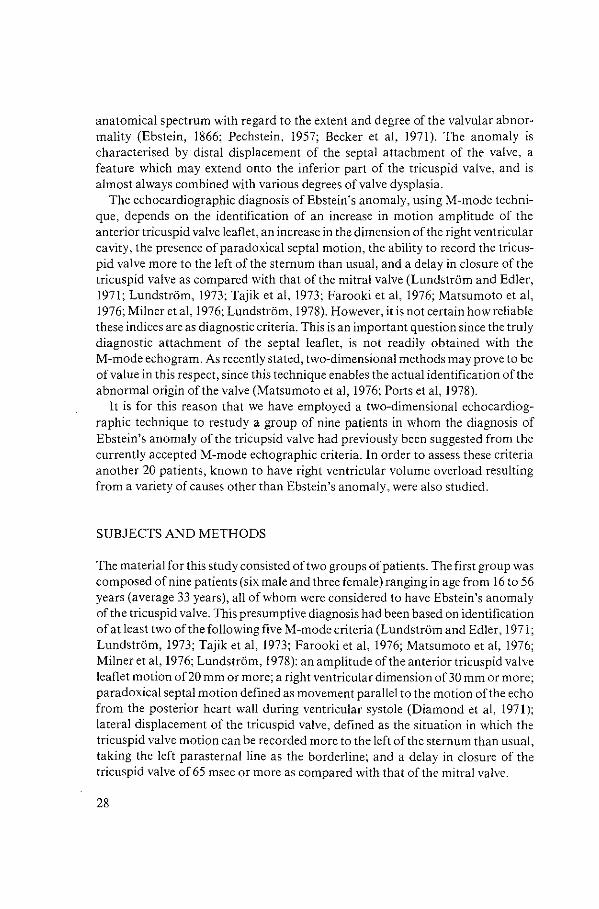
ECHOCARDIOGRAPHY IN EBSTEIN'S ANOMALY ECHOCARDIOGRAFIE BIJ DE ZIEKTE VAN EBSTEIN PROEFSCHRIFT TER VERKRIJGING VAN DE GRAAD VAN DOCTOR IN DE GENEESKUNDE AAN DE ERASMUS UNIVERSITEIT ROTTERDAM OP GEZAG VAN DE RECTOR MAGNIFICUS PROF. DR. M.W. VAN HOF EN VOLGENS BESLUIT VAN HET COLLEGE VAN DEKANEN. DE OPENBARE VERDEDIGING ZAL PLAATSV!NDEN OP woensdag 25 april1984 te 15.45 uur DOOR WILHELMINA JOHANNA LIGTVOET-GUSSENHOVEN GEBORENTESUNGE!GERONG ROTTERDAM, 1984
Transcript of ECHOCARDIOGRAPHY IN EBSTEIN'S ANOMALY ECHOCARDIOGRAFIE BIJ ... · echocardiography in ebstein's...
ECHOCARDIOGRAPHY IN EBSTEIN'S ANOMALY
ECHOCARDIOGRAFIE BIJ DE ZIEKTE VAN EBSTEIN
PROEFSCHRIFT
TER VERKRIJGING VAN DE GRAAD VAN DOCTOR IN DE
GENEESKUNDE
AAN DE ERASMUS UNIVERSITEIT ROTTERDAM OP GEZAG VAN DE RECTOR MAGNIFICUS
PROF. DR. M.W. VAN HOF EN VOLGENS BESLUIT VAN HET COLLEGE VAN DEKANEN.
DE OPENBARE VERDEDIGING ZAL PLAATSV!NDEN OP
woensdag 25 april1984 te 15.45 uur
DOOR
WILHELMINA JOHANNA LIGTVOET-GUSSENHOVEN
GEBORENTESUNGE!GERONG
ROTTERDAM, 1984
PROMOTOREN: PROF. DR. A.E. BECKER PROF. P.G. HUGENHOLTZ
Publication of this thesis was supported by: Fonds Doctor Catharine van Tussenbroek, Stichting 'De Drie Lichten'.

A an Kees
A an mijn ouders
Dit proefschrift werd bewerkt op de afdelingen Patholgie van het Academisch Ziekenhuis van de Universiteit van Amsterdam, Wilhelmina Gasthuis en Academisch Medisch Centrum en de afdeling Experimentele Echocardiografie, Thoraxcentrum van de Erasmus Universiteit Rotterdam.
Cover design: Kees de Vries The design represents a four-chamber image of an Ebstein's malformation. The left side shows the right atrium and right ventricle of the original drawing of the specimen and the right side shows the left atrium and left ventricle as it is seen with echocardiography.
Zetwerk: Hoi studio
CONTENTS
Chapter I - Introduction.
Chapter 2- Historical review. Pathology. Echocardiography.
Chapter 3- Basic aspects of anatomy and echocardiography in Ebstein's anomaly: a correlation. Elma J Gussenhoven and Anton E Becker. Published in Congenital Heart Disease: morphologic echocardiographic correlations. Churchill Livingstone, 1983.
Chapter 4- Echocardiographic criteria for Ebstein's anomaly of the tricuspid valve. Gussenhoven W J, Spitaels SEC, Born N and Becker A E. Published in British Heart Journal43:31-37, 1980.
Page
3
19
27
Chapter 5- Variability in the time interval between 37 tricuspid and mitral valve closure in Ebstein's anomaly. Gussenhoven W J, Jansen J R C, Born Nand Ligtvoet C M. In press: Journal of Clinical Ultrasound.
Chapter 6- The septal tricuspid and mitral valve 45 relationship in normal hearts and in hearts with Ebstein's anomaly: an echographic and anatomic study. Elma J Gussenhoven, Patricia A Stewart, Volkert H de Villeneuve, Catherina E Essed, Kees M Ligtvoet and Anton E Becker. In press: The American Journal of Cardiology.

CONTENTS Page
Chapter 7- The role of echocardiography in assessing the 57 functional class of the patient with Ebstein's anomaly. Gussenhoven W J, de Villeneuve V H, Hugenholtz P G, v Meurs-v Woezik H, Ligtvoet C M and Becker A E. In press: European Heart Journal.
Summary. Samenvatting.
References.
Dankwoord. Curriculum vitae. Publications of the author.
63 65
67
73 75 77

CHAPTER 1
INTRODUCTION
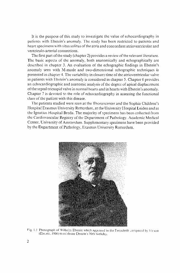
In 1866, Wilhelm Ebstein (Fig. 1.1 ), described an abnormality of the tricuspid valve in an otherwise normal heart, in a male of 19 years. The valve showed extensive distal displacement of septal and inferior leaflets and an abnormal morphology of the anterior leaflet. The abnormally situated tricuspid valve orifice divided the right ventricle into a proximal atrialized segment and a small distal ventricular pumping chamber. The eponym 'Ebstein's has since been linked with this type of congenital malformation.
In subsequent years the typical morphology of this anomaly has been described in another 20 cases, but always on the basis of postmortem studies. Tourniaire and coworkers, in 1949, were the first to diagnose Ebstein's anomaly in the living patient. The first publication in the Dutch literature appeared in 1958 by Vermeersch, followed by reports of Rook and Hoedemaker (1965), Becker (1972), Viersma eta! (1975) and Eygelaar eta! (1975; 1977).
Ebstein's anomaly is found in less than 1% of patients with congenital malformations of the heart (Fontana and Edwards, 1962).
It is difficult to generalize about the course and prognosis of the individual patient, who suffers from this condition. Less than 5% of the patients will survive beyond the age of 50 years (Genton and Blount, 1967). The longest survival yet recorded is of an 85-year-old man (Seward et a!, 1979). The reported mean age at death, amongst those who have been recognized as having Ebstein's anomaly, is approximately 30 years (Vacca et a!, 1958; Keith et a!, 1978). However, the reported percentages of the age at death varies. In a series of 60 heart specimens collected by Vacca and associates ( 1958) 8% died within the first year oflife, whilst 28% died within the first lO years of life. Kumar eta! (1971) documented that out of a series of 22 specimens 36% died within the first 6 months of life, whilst 77% died within the first 10 years of life.
There is no sex prevalence (Vacca eta!, 1958; Genton and Blount, 1967). Examples of a familiar occurrence have been reported (Gueron et al, 1966; Donegan et al, 1968; Watson, 1974; Lo et al, 1979), although the mode of inheritance remains unclear. The clinical profile and the pathology of this anomaly vary considerably from one individual to the other.
At present Ebstein's anomaly of the tricuspid valve is a well-known 'entity' among the vast number of congenital malformations of the heart. In the clinical setting, however, the diagnosis often remains a problem, not at least since the signs and symptoms and the electrocardiographic and radiologic features in themselves are nonspecific and present only in classical cases. Ultrasound techniques, introduced in the early seventies, appear to be a major asset in this respect.
It is the purpose of this study to investigate the value of echocardiography in patients with Ebstein's anomaly. The study has been restricted to patients and heart specimens with situs solitus of the atria and concordant atrioventricular and ventriculo-arterial connections.
The first part of the study (chapter 2) provides a review of the relevant literature. The basic aspects of the anomaly, both anatomically and echographically are described in chapter 3. An evaluation of the echographic findings in Ebstein's anomaly seen with M-mode and two-dimensional echographic techniques is presented in chapter 4. The variability in closure time of the atrioventricular valve in patients with Ebstein's anomaly is considered in chapter 5. Chapter 6 provides an echocardiographic and anatomic analysis of the degree of apical displacement of the septal tricuspid valve in normal hearts and in hearts with Ebstein's anomaly. Chapter 7 is devoted to the role of echocardiography in assessing the functional class of the patient with this disease.
The patients studied were seen at the Thoraxcenter and the Sophia Children's Hospital Erasmus University Rotterdam, at the University Hospital Lei den and at the Ignatius Hospital Breda. The majority of specimens has been collected from the Cardiovascular Registry of the Department of Pathology, Academic Medical Center, University of Amsterdam. Supplementary specimens have been provided by the Department of Pathology, Erasmus University Rotterdam.
Fig. I .I Photograph of Wilhelm Ebstein which appeared in the Festschrift composed by his son (Ebstein. 1906) to celebrate Ebsti:in 's 70th birthday.
2
CHAPTER2
HISTORICAL REVIEW
The vast literature data which all relate to Ebstein's anomaly has been screened for information relevant to this echographic study. It is clearly beyond the scope of this work to represent all data on this disease with respect to the diagnosis such as clinical signs and symptoms and haemodynamic and electrocardiographic findings. For this the interested reader is referred to current textbooks on pediatric cardiology, although it should be stated that most of the authors present only the diagnostic criteria of the 'classical' patient with Ebstein's anomaly and do not discuss the fact that the majority of these patients do not fit into that category.
PATHOLOGY
The anomaly was first described by Wilhelm Ebstein (!836- 1912) in 1866. An abridged English translation appeared in an excellent review by Yater and Shapiro in 1937. The first complete translation into English of Ebstein's original paper appeared in 1968 (Schiebler et al). It is of interest that yet another translation into English appeared in 1974 (Sekelj and Benfey), albeit that the motive remains unclear. An informative review of the life story of Ebstein and on the historical background of his now famous case report has been provided by Mann and Lie in 1979.
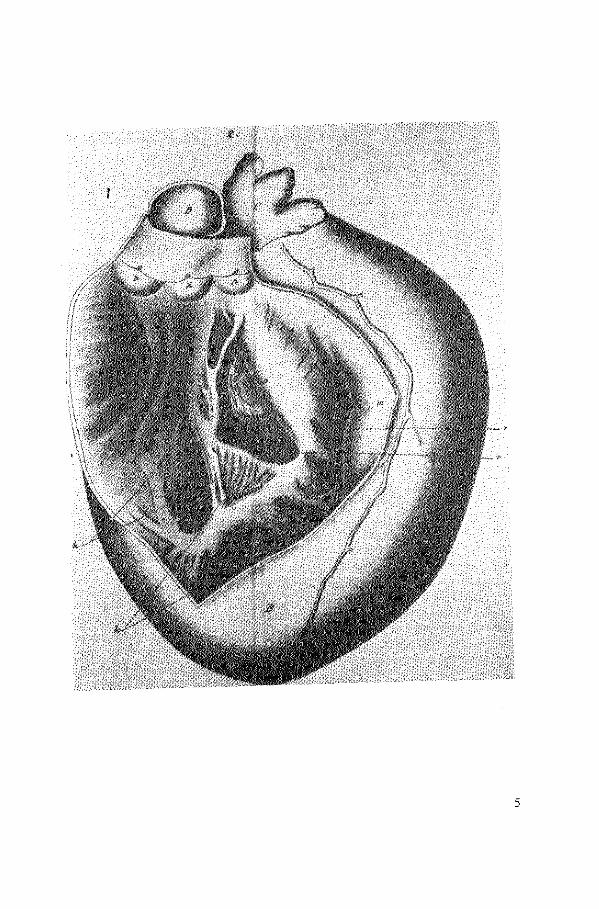
Ebstein described in detail the clinical and pathological findings in a young man, 19 years of age. The clinical history revealed that the patient had been short of breath and had complained of palpitations since childhood. At the time of admission he presented with congestive heart failure. Physical examination revealed marked cyanosis of the face with the rest of the body skin pale. In addition, a marked jugular venous pulsation synchronous with the heart beat was noted. The systolic thrill and murmur were undoubtedly caused by tricuspid insufficiency. The patient died in pulmonary edema eight days after admission. The autopsy was performed by Ebstein on the 6th of July, 1864 and his findings were published in Reichert's Archives, January 1866. His meticulous description of this case was illustrated with two excellent drawings made by Wyss. The basic anomaly is best understood by quoting from the original paper and drawings (Figs. 2.1 and 2.2):
'Wenden wir uns jetzt zur Beschreibung des rechten Ventrikels (B), so springt sofort ein durchaus abnormes Verhalten der Valvula tricuspidalis in die Augen. Es entspringt niimlich von dem ganz in normaler Weise entwickelten Annulus fibro-cartilagineus dexter (e) und zwar entsprechend der vorderen (m) und hinteren (n) Wand des rechten Ventrikels eine Membran (h, hJ, welche in die
3
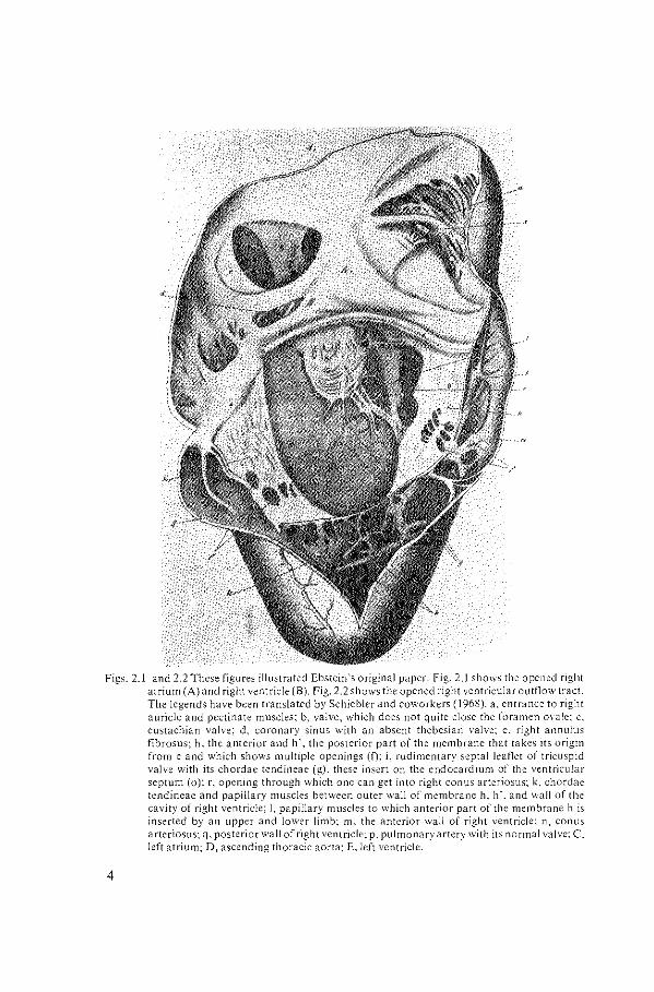
Figs. 2 1 and 2.2 These figures illustrated Ebstein's original paper. Fig.. 2.1 shows the opened right atrium (A) and right ventricle (B). Fig. 2.2 shows the opened right ventricular ovtf1ow tract. The legends have been translated by Schiebler and coworkers ( 1968). a, entrance to right auricle and pectinate muscles; b, valve, which does not quite close the foramen 0\·ale; c, eustachian valve; d, coronary sinus with an absent thebesian valve; e. right annulus fibrosus; h. the anterior and h', the posterior part of the membrane that takes its origin from e and which shows multiple openings (f); i, rudimentary septal leaflet of tricuspid valve with its chordae tendineae (g), these insert on the endocardium of the ventricular septum (o): r, opening through \vhich one can get into right conus arteriosus; k, chordae tendineae and papillary muscles between outer wall of membrane h. h'. and wall of the cavity of right ventricle; l, papillary muscles to which anterior pan of the membrane h is inserted by an upper and lower limb; m. the anterior wal! of right ventricle: n, conus arteriosus; q. posterior wal! of right ventricle; p. pulmonary artery with its normal valve; C. left atrium; D. ascending thoracic aorta: E, left ventricle.
4
5
hintere Hiilfte des Endocardium des Septum ventriculorum (o) iibergeht. Diese Membran stellt im Zusammenhange mit der stark getriibten und verdickte hinteren Hiilfte des Endocardium des Septum ventriculorum eines nach unten sowie auch nach rechts vollkommen geschlossenen Sack dar, welcher bei unserer Schnittfiihrung geoffnet wurde, und der mit dem ubrigen Endocardium resp. Innenjliiche des rechten Ventrikels in fa/gender Weise zusammen-hiingt ........ '. '15 Mm. unter dem Annulus fibro-cartilagineus dexter, entsprechend und dicht unterhalb des hiiutigen Theiles der Kammerscheidewand, entspringt vom Endocardium ein mit breiter Basis nach oben und der Spitze nach unten gerichteter dreieckiger Zipfel (i), von etwa Viergroschenstiick-Grosse, welcher sich mit sehr zahlreichen, zum Theil sehr Iangen, dilnnen, zarten, von seiner Spitze (g), hauptsiichlich aber von seiner hinteren Fliiche entspringenden Sehnenfiiden zum gr6ssten Theile in das Endocardium, zum kleinsten Theile an einem in der Mitte des Septum ventricu/orum gelegenen, in die HerzOhhle frei vorspringenden Papillarmuskel (/) inserirt ....... '.
In other words the essential malformation documented by Ebstein consists of an anomalous insertion of the tricuspid valve. The drawings clearly show that the septal and posterior valve leaflets were adherent to the ventricular wall and the mobile free parts were displaced towards the apex of the right ventricle. Moreover, the drawings also suggest that partial agenesis of the valve has occurred (see Fig. 2.1). This aspect of the malformation is not generally quoted in the literature. As far as we are aware only a few reports document this particular feature (Lev et al, 1970; Becker et al, 1971). Consequent to the valve abnormalities part of the right ventricular cavity communicates openly with the right atrium. The anterior leaflet of the valve also showed an abnormal morphology. It was large, curtain-like and fenestrated and also draped the right ventricular free wall, anchored at multiple sites by papillary muscle-like structures and chordae.
Twelve years elapsed before a second heart with these peculiar abnormalities of the tricuspid valve was described by Rauchfuss (1878). The first report in the English literature appeared in 1900 by MacCallum. Approximately 20 other specimens had been documented (see for reviews Yater and Shapiro, 1937; Tourniaire et al, 1949) before the first patient was reported by Tourniaire and coworkers, in 1949, in whom the diagnosis of Ebstein's anomaly was made on clinical grounds. They described a female of 39 years of age with signs and symptoms which they considered to fit only with the congenital anomaly described by Ebstein. In their conclusion they literally state:
6
'Chez notre malade, !'absence d'antfcfdents rhumatismaux, les paroxysmes tachycardiques supraventriculaires, le bloc de branche droite, I' augmentation elective des cavitfs droites (oreillette et infundibulum pulmonaire), I' existence enfin d'une insuffissance tricuspidienne organique confirmfe par le cathete-
risme cardiaque et Ia regurgitation auriculaire droite, constituent un ensemble cohhent. On ne peutfaire un autre diagnostic que celui de maladie d'Ebstein.'
Since then the clinical diagnosis Ebstein's disease of the tricuspid valve has been established with increasing frequency, although the disease remains rare among the many other forms of congenital heart disease.
A review of the accumulated data regarding the anatomy of Ebstein 's anomaly reveals that the pathology is not uniform and varies considerably from one heart to the other (Pechstein, 1957; Lev et al, 1970; Becker et al, 1971; Zuberbuhler et al, 1979). Indeed, each of the two major anatomic features, i.e. valve displacement and abnormal leaflet morphology, may vary independently (Becker et al, 1971 )-
Apical displacement always affects the septal leaflet, but it may involve the posterior leaflet. The leaflets are never displaced beyond the junction between the inlet and trabecular part of the right ventricle (Anderson and Lie, 1978; Anderson et al, 1979; Zuberbuhler et al, 1979). In our personal experience the anterior tricuspid valve leaflet has never been involved in the process of downward displacement. Anderson and coworkers ( !979) reported that the anterior leaflet is usually not displaced, suggesting that occasionally it may be abnormally inserted. Shiina et al (1983) documented apical displacement of the anterior leaflet echographically in 14% (!)of the patients. The degree of distal displacement of the septal and posterior leaflets may vary from cases with minimum displacement of the valve leaflets, to hearts where the greater part of the right ventricular inlet is denuded and the leaflets drape the apical trabeculations (Watson, 1968; Beckeret al, 1971; Zuberbuhleret al, 1979). The greater part of the affected leaflets is usually firmly adherent to the right ventricular wall. In some specimens the leaflets are completely blended with the ventricular wall, while in other hearts the leaflets may be partially detached thus creating cavities between the abnormally developed valve tissue and the ventricular endocardium (Anderson and Lie, 1978; Anderson et al, 1979).
The first report classifying the degree of valve displacement and that of valve dysplasia appeared in 1971 by Becker and coworkers. Dysplasia and apical displacement were each divided into 3 grades. Apical displacement- which the authors describe as downward displacement - was determined as the distance between the anticipated normal basal attachment of the leaflets and the right ventricular apex. Grade I was represented by apical displacement of less than 10%, taking into account that the normal septal attachment of the tricuspid valve dipped below the expected annular attachment. Grade 2 consisted of a displacement between 10 and 50% and grade 3 involved a displacement of more than 50%. They showed that apical displacement was always accompanied by leaflet dysplasia, but that dysplasia of the leaflets may occur with or without displacement. The maximum grades of apical displacement did not in all cases coincide with the
7
maximum grades of dysplasia. In 35% of their cases, both features were equally classified as grade 3. Unfortunately, the authors do not mention the ages of the specimens studied in relation with the severity of apical displacement.
Recently, Zuberbuhler et al (1979) subdivided the anatomic spectrum of Ebstein's anomaly into mild, moderate and severe based on the extent of apical displacement and the degree of valvular dysplasia. Apparently the study of Becker et al (1971) has escaped notice since the authors in no way refer to the grading system previously elaborated. Zuberbuhler and associates described 14 hearts. In each of these hearts the apical displacement came down to the level of the junction between the inlet and the trabecular parts of the right ventricle. The main differences on which a distinction of three categories were based are variations in the amount of leaflet involved and the degree of valve dysplasia. This is a subclassification of the morphology, which in no way can be compared to the previously described classification by Becker et al (1971). The use of the terms 'mild, moderate and severe', moreover, suggests a correlation with the clinical situation. However, no such information is provided. We feel therefore that the use of such terms is inappropriate, particularly since it has yet to be shown that the degree and extent of the leaflet malformation bears a direct relationship to the clinical symptomatology of the patient (see chapter 7).
Lev and associates (1970) have classified Ebstein's anomaly into 'simple' and 'complicated' forms, based on the presence or absence of associated cardiac lesions. This classification was also derived from the study of heart specimens, so that the terms should not be considered an indication of the clinical situation.
The intrinsic abnormality of the tricuspid valve leaflets is generally categorized as 'dysplasia' (Becker et al, 1971; 1972). The degree of dysplasia may vary considerably, even at different sites within one and the same heart. The characteristics of dysplasia include a) focal or diffuse thickening of the leaflets, b) deficient development of chordae and papillary muscles, c) improper separation of the valve components from the ventricular wall, and d) focal agenesis of valvular tissue (Lev et a!, 1970; Becker et a!, 1971 ).
According to some authors hearts with relatively little dysplasia of the valve leaflets are classified as 'mild,' while hearts which exhibit more pronounced dysplastic features are categorized as 'severe' (Lev et al, 1970; Zuberbuhler et al, 1979). It should be re-emphasized, however, that neither one of these authors has attempted to correlate these terms with the clinical well-being of the patient. The terms are apparently purely anatomic. The anterior tricuspid valve leaflet may occasionally be normal, but almost always is abnormal in the sense that it is large and may contain muscle with or without abnormal fibrous strands (Anderson and Lie, 1978). The free edge may no longer be truly free, since it may be directly attached to the ventricular wall (Anderson and Lie, 1978; Anderson eta!, 1979). It thus appears that dysplasia often affects the anterior leaflet despite the fact that it is normally attached to the atrioventricular annulus.
8
The combined effects of valve dysplasia and apical displacement may create an obstruction to the flow of blood from the atrial side to the ventricular pumping side. Obstruction as the leading feature has major implications not only from a haemodynamic point of view, but also for surgical repair since according to Zuberbuhler et al ( 1979) this arrangement may necessitate the replacement of the affected leaflets by an artificial valve. Recently, Marcelletti eta! (1980) documented a patient with an almost imperforate Ebstein's malformation of the tricuspid valve as an unexpected finding, in whom they successfully executed a modified Fontan procedure. This demonstrates that a good insight of the wide anatomic spectrum which includes valve displacement and dysplasia, has important consequences not only when it comes to the art of establishing the clinical diagnosis but also for the surgeon when confronted with this disease.
Displacement of the tricuspid valve results in a subdivision of the right ventricle into proximal and distal compartments. The proximal chamber is transformed into an integral part of the right atrium. Hence, this compartment is commonly referred to as 'the atrialized portion' of the right ventricle. The term should be strictly used in this sense; this is important since in some recent works 'atrialization' is held synonymous with 'dilatation' (Allwork eta!, 1976; Zuberbuhler eta!, 1979). The wall of the atrialized portion may be dilated and thin-walled, but this is not a conditio sine qua non. When present the wall may appear transparent with only occasional muscle fibers (Pechstein, 1957; Lev eta!, 1970; Anderson and Lie, 1978; Anderson et al, 1979; Zuberbuhler eta!, 1979). Pechstein (1957) wondered whether the extreme thinning of the atrialized ventricular wall was inherent to the disease or secondary to the altered haemodynamics. On the other hand, Anderson and Lie (1978) stated that dilatation was the result of a congenital deficiency in myocardial structure, although they provided no data to substantiate their concept. The extreme thinning of the atrialized right ventricular wall in Ebstein's anomaly should be distinguished from the parchment-like abnormality which characterizes Uhl's anomaly (Uhl, 1952). In the latter condition the tricuspid valve does not show the features present in Ebstein's malformation. Similarly, congenital giant right atrium should be distinguished from Ebstein's anomaly (Bailey, 1955).
The distal compartment, which in reality constitutes the right-sided pumping chamber may be of normal size, but usually is markedly diminished in dimension. A number of heart specimens has been described in which this pumping compartment was dilated and thin-walled (Anderson and Lie, 1978; Anderson et al, 1979). The presence of such a dilated pumping chamber is considered an ominous sign usually impairing the surgical results (Anderson and Lie, 1978).
Ebstein 's anomaly of the tricuspid valve without associated anomalies is rare although the incidence of such additional abnormalities is usually underestimated in clinical studies. Watson (1974) has shown a much higher incidence of associated cardiac malformations in necropsy studies (81 %) than in clinical studies (48%).
9
Ebstein's anomaly is almost always associated with a patent foramen ovale or a secundum type atrial septal defect present also in the heart described by Ebstein (see Fig. 2.1). An incidence of60% is reported from heart specimens and a 42% incidence from catheterization studies (Watson, 1974). Lev and coworkers (1970) found an atrial septal defect in 77% of their autopsy specimens. The defect was classified as persistent patency of the foramen ovale in 60% of the heart specimens and in 50% of the catheterization studies (Watson, 1974). Lev et al (1970) classified 35% of the atrial septal defects as a patent foramen oval e.
The next most frequent associated cardiac lesions are ventricular septal defect and pulmonary valve stenosis respectively (Lev eta!, 1970; Becker eta!, 1971; Kumar et al, 1971; Watson, 1974; Zuberbuhler et al, 1979; Ebaid et al, 1980). The ventricular septal defect usually is of the perimembranous type, but occasionally it can be muscular (Zuberbuhler et al, 1979). The ventricular septal defect may be localized in either the proximal or the distal right ventricular compartment (Becker et al, 1971). In hearts in which the opening is proximal to the displaced tricuspid valve, a 'left ventricular- right atrial' shunt may occur. Such a defect was present in two of the three hearts with Ebstein's malformation containing a ventricular septal defect, described by Becker et al (1971). It is surprising that few other reports exist in which the precise position of the ventricular septal defect relative to the displaced valve is specifically stated. This lack of information could indicate that such 'left ventricular- right atrial' defects are extremely rare, since it is hard to accept that a defect in that location would pass unnoticed.
The incidence of ventricular septal defects documented in the literature varies considerably. In general the percentage varies between 4% in a clinical series (Watson, 1974) and 12% in autopsy studies (Lev eta!, 1970; Becker eta!, 1971; Kumar eta!, 1971; Watson eta!, 1974). An astonishing high incidence of 43% was reported by Zuberbuhler et a1 (1979). A reason for this remarkable discrepancy with the literature is not provided by the authors but it may reflect a selection of their collection.
The incidence of isolated pulmonary valve stenosis reported varies from 5% in a clinical series (Watson, 1974) to 27% in an autopsy study (Becker eta!, 1971). In less than 6%, a ventricular septal defect occurs with a pulmonary valve stenosis (Becker eta!, 1971; Kumar eta!, 1971; Watson, 1974). Hearts with pulmonary valve atresia and intact ventricular septum, particularly those with hypoplasia of the right ventricle, show an increased incidence of Ebstein's anomaly. The frequency of its occurrence however also varies considerably from one author to the other. Elliot eta! (1963) report a 75% incidence versus a 40% incidence reported by Lev eta! ( 1970) and a 27% frequency documented by Zuberbuhler and Anderson ( 1979).
The first specimen designated as containing an 'atretic' Ebstein's malformation was documented by Kumar and coworkers in 1971. Subsequently, two cases have been recognized as an imperforate valve (Gerlis and Anderson, 1976; Anderson et al,
10
1977; Anderson et al 1979; Zuberbuhler et al, 1979). Anderson and coworkers (1979) stated that an imperforate tricuspid valve occurs in about 10% of hearts with Ebstein's anomaly. When we include the two specimens with only a 'pinhole' communication, described by Zuberbuhler et al (1979), the incidence of imperforate Ebstein's in their series of 14 specimens becomes even higher (29%), indeed an overwhelming high incidence when compared with the data from Watson (1974). He documented 505 patients ofEbstein's anomaly, but no mention is made of an imperforate valve. The difference cannot be explained solely on the basis that his experience is largely clinical, since the data of93 postmortem investigations were included. The reason for the discrepancy remains speculative, although the fact that hearts with small fenestrations are included as 'imperforate' could be the explanation. We would rather restrict the use of the term 'imperforate' to those instances in which the valve is truly imperforate and, hence, without any defects. In our experience an 'imperforate' tricuspid valve is a rarity.
Abnormalities of left ventricular contractility as well as abnormalities of mitral valve morphology, either in isolation or combined, have been reported in conjunction with Ebstein's anomaly (Lev et al, 1970; Monibi et al, 1978; Worms et al, 1980; Cabin and Roberts, 1981). The mitral valve may exhibit fibrous thickening of the leaflets and the valve may be stenotic. Mitral valve prolapse may also coincide but, when present, it usually occurs in adult patients.
Finally, other cardiovascular lesions may sporadically be associated with Ebstein's anomaly such as cor tria tria tum sinistrum (From et al, 1973; CastanedaZuniga et al, 1982), atrioventricular septal defect (Caruso et al, 1978; Handler et al, 1981 ), complete transposition of the great arteries (Kumar et al, 1971 ), tetralogy of Fallot (Ito et al, 1977), aortic coarctation (Watson, 1974) and patent ductus arteriosus (Kumar et al, 1971; Watson, 1974).
Although the present study is limited to hearts with concordant atrioventricular and ventriculo-arterial connections, it should be known that Ebstein's anomaly may also occur in congenitally corrected transposition. In such hearts the tricuspid valve abnormality is left-sided but otherwise similar to that in 'classical' Ebstein's anomaly (Edwards, 1954; Kernen, 1958; Becker et al, 1971; Anderson et al, 1978; Losekoot et al, 1983). Dysplasia as well as apical displacement may occur, although the former feature is by far the most common. Only one case has been documented in which apical displacement was the sole feature (Becker et al, 1971 ).
Surprisingly, the incidence ofEbstein's malformation in congenitally corrected transposition noted from autopsy studies varies considerably. Some studies suggest that apical displacement of the tricuspid valve occurs with a high incidence (Becker et al, 1971; Anderson et al, 1978; Otero Coto et al, 1980), while others report a low incidence (Losekoot, 1967) or suggest that this feature does not occur at all (Allwork et al, 1976; Soto et al, 1977). The latter conclusion was based on angiographic investigations. In the pathologic study of Allwork et al (1976) an abnormal tricuspid valve with dysplastic features was found in 76% but the
11
authors claim that atrialization did not occur. lt is likely that they meant to say that a dilated and thin-walled segment of the atrialized portion of the 'inverted' right ventricle was absent since it is most unlikely that none of their specimens would have no apical displacement.
Finally, it should be mentioned that Ebstein's anomaly of the anatomic mitral valve has been documented (Ferencz and Dammann, 1957; Ruschhaupt et al, 1976; Anderson et al, 1979). This condition is extremely rare and the diagnosis thus far has only been made at autopsy. In the cases reported the inferior leaflet of the mitral valve was displaced downward into the left ventricle, resulting in an atrialized portion of the left ventricle. The mitral valve leaflets were dysplastic. The aortic valve leaflets remained in fibrous continuity with the dysplastic anterior mitral valve leaflet. In two cases the left ventricular cavity was hypoplastic (Ruschhaupt et al, 1976; Anderson et al, 1979). In the present study this rare condition will not be included.
ECHOCARDIOGRAPHY
The ultrasound technique has greatly contributed to the study of functional anatomy in the living patient. In patients with congenital heart disease echocardiography is presently an important diagnostic tool, which in some instances already has replaced invasive techniques (Shub eta!, 1983; Macartney, 1983; Smallhorn et al, 1983). This section will be devoted to the echographic features of Ebstein's anomaly.
M-rnode observations
The first echocardiographic diagnosis of Ebstein's anomaly was reported by LundstrOm in 1969. Further and more detailed observations were documented in 1971. These features, considered as diagnostic for Ebstein's disease, were a dilated right ventricle, an anterior tricuspid valve leaflet showing a slow posterior movement during diastole and the finding that the leaflet was recorded more lateral from the medioclavicular line than usual. Subsequent reports (Kotler and Tabatznik, 1971; Crews et al, 1972) added threefurther criteria, i.e. the paradoxical septal motion, the increased excursion of the anterior tricuspid valve leaflet and the delayed closure time of the tricuspid valve versus that of the mitral valve. These authors described a delay in tricuspid valve closure between 40 and 120 msec. This finding was considered highly relevant, since normally the mitral valve closure precedes tricuspid valve closure by only 20 to 30 msec (Leatham, 1954; Reinhold and Rudke, 1957; Brooks et al, 1979). In the subsequent years the significance of M-mode echocardiography for diagnosing Ebstein's anomaly was promoted by many investigators (Lundstrom, 1973; Tajik eta!, 1973; Yuste et al, 1974; Kotler,
12
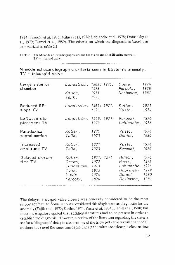
1974; Farooki et al, 1976; Milner et al, 1976; Lablanche et al, 1978; Dobrinsky et a!, 1979; Daniel et al, 1980). The criteria on which the diagnosis is based are summarized in table 2.1.
Table 2.1 TheM-mode echocardiographic criteria for the diagnosis of Ebsteins anomaly. TV= tricuspid valve.
M~mode echocardiographic criteria seen in Ebstein 's anomaly. TV ~ tricuspid valve
Large anterior Lundstrom, 7 969; 7 971; Yuste, 1974 chamber 1973 Farooki, 1976
Kotler, 7 971 Desimone~ 7 98 7 Toiik, 1973
Reduced EF~ Lundstrom, 7 969; 7 977; Kotler, 7 977 slope TV 7973 Yuste, 7 974
Leftward dis~ Lundstrom, 1969; 1977; Faraoki, 7976 placement TV 7 973 Lablanche, 7978
Paradoxical Kotler, 7977 Yuste, 7974 septal motion Taiik, 1973 Daniel, 1980
Increased Kotler, 7 977 Yuste, 7974 amplitude TV Taiik, 1973 Farooki, 1976
Delayed closure Kotler, 1977; 7 974 Milner, 7976 time TV Crews~ 7972 Ports, 7978
Lundstrom, 7 973 Lablanc he, 7978 Taiik, 7973 Dobrinski, 1979 Yuste, 1974 Daniel, 7980 Farooki. 1976 Desimone~ 1981
The delayed tricuspid valve closure was generally considered to be the most important feature. Some authors considered this single item as diagnostic for the anomaly (Tajik et al, 1973; Kotler, 1974; Yuste et al, 1974; Daniel et al, 1980) but most investigators opined that additional features had to be present in order to establish the diagnosis. However, a revie\\/ of the literature regarding the criteria set for a 'diagnostic' delay in closure time of the tricuspid valve reveals that not all authors have used the same time lapse. In fact the mitral-to-tricuspid closure time
13
interval was documented as either 30 msec (Farooki et al, 1976), 40 msec (Yuste et al, 1974), 50 msec (Crews et al, 1972), 60 msec(Lundstrom, 1973) or 65 msec (Daniel et al, 1980). Interestingly enough, the reports prior to 1975 indicate that all patients with Ebstein's anomaly had a delay in closure of the tricuspid valve (Kotler et al, 1971; Crews et al, 1972; Lundstrom, 1973; Tajik et al, 1973; Yuste et al, 1974), while investigators in the following years found patients with a proven Ebstein 's anomaly in whom the M-mode echocardiogram showed a mitral-totricuspid interval within the normal range (Milner et al, 1976; Farooki et al, 1976; Lablanche et al, 1978; Ports et al, 1978; Dobrinsky et al, 1979; Daniel et al, 1980; Roudaut et al, 1981). Hence it is questionable whether a delayed tricuspid valve closure time can be used as a reliable feature, particularly when it is taken in isolation. Nevertheless most authors- even today- will use this feature as the most important M-mode criterion although in the sense of 'suggestive' rather than 'diagnostic'.
The genesis of the delay in tricuspid valve closure has been a matter of debate also. Crews and coworkers (1972), combining phonocardiographic and echocardiographic studies, found a direct relationship between the delay of the tricuspid valve closure time and the severity of right bundle branch block, thereby endorsing independently a previous phonocardiographic study of Pocock et al ( 1969). In contrast, Lundstrom (1973) was unable to find such a correlation. He concluded that a mechanical factor, directly related to the malformed tricuspid valve, was the most likely explanation for the delayed tricuspid valve closure. He felt strengthened in this opinion by the fact that one of his patients with Ebstein's anomaly and a pre-excitation syndrome type B also showed a delayed tricuspid valve closure, while early closure would have been expected due to early right ventricular excitation. Tajik et al (1973) came independently to the same conclusion also based on the study of a patient with a type B Wolff-Parkinson-White syndrome.
More recently, however, Koiwaya et al (1979) reported an early closure of the tricuspid valve in a case ofEbstein's anomaly with type B Wolff-Parkinson-White syndrome. The matter is further complicated since it has been documented that volume overload of the right heart without Ebstein's anomaly can produce a distinct delay in tricuspid valve closure time (French eta!, 1975; Milner eta!, 1976; Daniel et al, 1980; Gussenhoven et al, 1980- see also chapter 4). These observations further undermine the statement that delayed tricuspid valve closure is 'diagnostic' for Ebstein's anomaly.
It appears also that the delay in tricuspid valve closure time does not correlate with the functional class of the patient (Lundstrom, 1973), the age of the patient or with the presence or absence of associated cardiac lesions (Farooki eta!, 1976).
14
Two-dimensional observations
The first diagnostic criteria for Ebstein 's anomaly using a multi-crystal twodimensional system were defined by Hagan and coworkers in 1974. They were able to recognize the apical displacement of the septal tricuspid valve leaflet as well as an elongated anterior tricuspid valve leaflet with an increased excursion. It was subsequently shown that of these echographic features apical displacement was the characteristic sign for Ebstein's anomaly (Matsumoto et al, 1976; Ports eta!, 1978; Silverman and Schiller, 1978; Gussenhoven eta!, 1980- see chapter 4; Kambe eta!, 1980; Roudaut eta!, 1981; Desimone and Kronzon, 1981; Shiina et a!, 1981; 1983). Two-dimensional echocardiography has the potential to visualize the anatomy in cross-sectional fashion, it allows a much better insight into the relationship between the different structures. It also demonstrates additional features such as an increased size of the right atrium, an increased excursion of the anterior tricuspid valve leaflet and a decrease in left ventricular dimensions. As previously stated, these latter features are non-specific since they may occur also in hearts with right ventricular volume overload.
The specificity of apical displacement of the septal tricuspid valve leaflet for the diagnosis Ebstein's anomaly is emphasized by the fact that thusfar only one false positive diagnosis has been made with this criterion. The patient was later shown to have hypoplasia of the left ventricle with marked enlargement of the right ventricle (Hirschklau eta!, 1977). A few cases have been documented in which two-dimensional echocardiography failed to identify apical displacement of the septal tricuspid valve leaflet (Matsumoto et a!, 1976; Hirschklau et a!, 1977; Kambe et al, 1980). Matsumoto and coworkers (1976) failed to demonstrate the displaced origin of the septal valve leaflet in 2 out of 7 patients with Ebstein's disease. Hirschklau eta! (1977) studied 10 patients with an Ebstein's anomaly of the tricuspid valve. In one of these the anomaly occurred in association with hypoplasia of the right ventricle. In this particular infant the lower insertion oft he septal tricuspid valve leaflet was not identified. Kambe et a! (1980) could not measure the displacement of the septal leaflet in the four chamber view in 3 out of 11 patients. In one of these an apical displacement of 15 mm was found at operation. In the second patient the septal tricuspid valve leaflet was visualized but the mitral valve never appeared in the same cross-section and hence the point of reference to determine apical displacement was absent. Unfortunately, the authors made no further comment on their third patient.
From an anatomical point of view (see also chapter 3) it is not surprising that failures in diagnosis can occur, since the septal leaflet can be minute and may hardly be visible. In other instances the leaflet may have 'moved' towards the outflow tract of the right ventricle and may thus disappear from the usual cross-section.
15
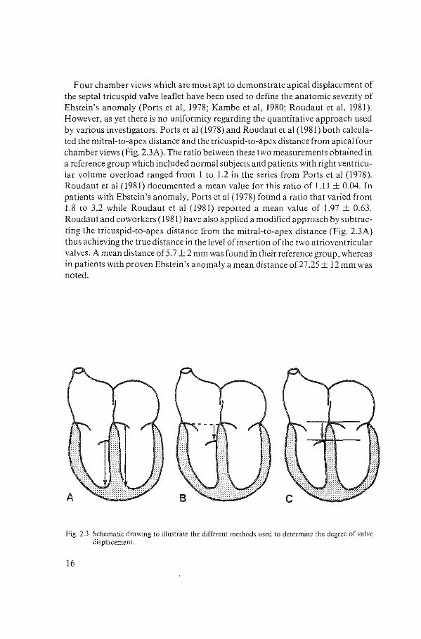
Four chamber views which are most apt to demonstrate apical displacement of the septal tricuspid valve leaflet have been used to define the anatomic severity of Ebstein's anomaly (Ports et al, 1978; Kambe et al, 1980; Roudaut et al, 1981). However, as yet there is no uniformity regarding the quantitative approach used by various investigators. Ports et al ( 1978) and Roudaut et al (1981) both calculated the mitral-to-apex distance and the tricuspid-to-apex distance from apical four chamber views (Fig. 2.3A). The ratio between these two measurements obtained in a reference group which included normal subjects and patients with right ventricular volume overload ranged from 1 to 1.2 in the series from Ports eta] (1978). Roudaut eta] (1981) documented a mean value for this ratio of 1.11 ± 0.04. In patients with Ebstein's anomaly, Ports et al (1978) found a ratio that varied from 1.8 to 3.2 while Roudaut eta] (1981) reported a mean value of 1.97 ± 0.63. Roudaut and coworkers (1981) have also applied a modified approach by subtracting the tricuspid-to-apex distance from the mitral-to-apex distance (Fig. 2.3A) thus achieving the true distance in the level of insertion of the two atrioventricular valves. A mean distance of5.7 ± 2 mm was found in their reference group, whereas in patients with proven Ebstein's anomaly a mean distance of27 .25 ± 12 mm was noted.
Fig. 2.3 Schematic drawing to illustrate the different methods used to determine the degree of valve displacement.
16
The question can be raised whether this approach is clinically applicable and whether it can be applied irrespective of the age of the patient and the size of the heart as suggested by the authors (Ports eta!, 1978; Roudaut eta!, 1981). In our opinion this is not the case (see also chapter 6).
Direct measurements of valve displacement have also been reported. Ports and coworkers (1978) have measured the distance between the atrioventricular groove and the displaced tricuspid valve (Fig. 2.3B). In all patients with Ebstein's anomaly a displaced origin of the tricuspid valve was noted with a mean value of 38 mm and a range of 20 to 70 mm. In their reference group a displacement of the valve insertion was not seen. The question can be raised therefore whether a reliable echo graphic image of the atrioventricular groove can be obtained. The pictures produced by Ports eta! (1978) show a number of echolines which they consider to represent the atrioventricular groove, which illustrates that this anatomic structure is a poor reference point for precise measurement.
Finally, Kambe and coworkers ( 1980) calculated the distance between both atrioventricular valves directly (Fig. 2.3C). In patients with proven Ebstein's anomaly they found a mean value of apical displacement of the tricuspid valve of 21 mm with a range of 14 to 32 mm. The authors give no data regarding the measurements in normal individuals and in patients with right ventricular volume overload other than stating that 'no displacement' occurred. Evemy and Hunter (1981) commented on these findings by pointing out that in the normal heart the two valves have a different level of insertion, a feature of significance in identifying ventricular morphology.
Hence, it can be concluded from this review of the literature that most authors consider echocardiography, both two-dimensional and M-mode, an important tool for the diagnosis ofEbstein's anomaly. At the same time, however, it becomes evident that much refinement has to be introduced regarding the diagnostic reliability of the various features and their role in predicting the functional class of the patient.
17

CHAPTER3
BASIC ASPECTS OF ANATOMY AND ECHOCARDIOGRAPHY IN EBSTEIN'S ANOMALY: A CORRELATION
BASIC ANATOMY
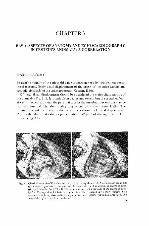
Ebstein's anomaly of the tricuspid valve is characterized by two distinct anatomical features; firstly distal displacement of the origin of the valve leaflets and secondly dysplasia of the valve apparatus (Ebstein, 1866).
Of these, distal displacement should be considered the major characteristic of the anomaly (Fig. 3.l).lt is variable in degree and extent, but the septal leaflet is always involved, although the part that crosses the membranous septum may be normally inserted. The abnormality may extend on to the inferior leaflet. The origin of the antero-superior valve leaflet never shows such distal displacement. Due to the abnormal valve origin an 'atrialized' part of the right ventricle is formed (Fig. 3.1 ).
Fig. 3.1 Classical example of Ebstein's anomaly of the tricuspid valve. A, A window has been cut in the anterior right ventricular wall, which reveals the sail-like dysplastic antero-superior tricuspid valve leaflet (aTL). 8, The same specimen after removal of the antero-superior leaflet. The septal and inferior components of the tricuspid valve show extreme distal displacement. being plastered to the septal surface and inferior free wall. A large 'atrialized' part of the right ventricle is Lhus formed.
19
Dysplasia of the valve apparatus is the additional feature which completes the anatomical characteristics of'classical' Ebstein's anomaly. In some instances part of the valve is composed of myxomatous tissue, forming a valve collar rather than a leaflet. There may be complete lack of chordae. This architecture is particularly seen on the septal surface. The antero-superior leaflet, on the other hand, is mostly sail-like, often fenestrated and may in part be muscularized, inserting into the trabeculae of the right ventricle (Fig. 3.1B).
Towards the apex the septal, inferior and antero-superior leaflets may fuse, either completely or in part, so that in some hearts the inner surface of the inflow part of the right ventricle is formed by a 'blanket' of dysplastic valve tissue (Fig. 3.1B). The latter condition is also known as 'imperforate Ebstein's anomaly' and exemplifies an imperforate atrioventricular connection.
The degree and extent of valve dysplasia may vary from one individual to the other, like the degree and extent of distal displacement (Becker eta!, 1971).
The anomaly is almost always associated with an atrial septal defect of the fossa ovalis variety. Other malformations may occur such as ventricular septal defects. These may open into the atrialized part of the right ventricle cranial to the level of the valve origin. The perimembranous type of ventricular septal defect is the most common. Congenital pulmonary valve stenosis may occur, albeit infrequently. Ebstein's anomaly also has a tendency to be associated with pre-excitation of the Wolff-Parkinson-White variety.
ECHOGRAPH!C ANATOMY
Since the extent and degree of the anatomical features may differ from case to case, it follows that Ebstein's malformation from an anatomical point of view constitutes a spectrum (Becker eta!, 1971). Consequently the clinical diagnosis of minor forms will be difficult, whereas major abnormalities will be readily recognized as 'classical' Ebstein's anomaly.
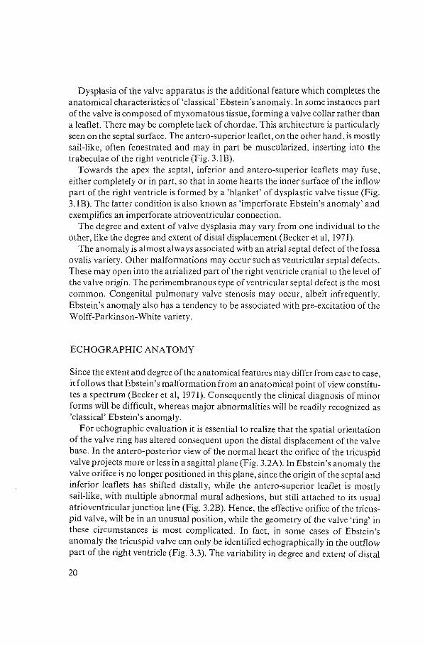
For echographic evaluation it is essential to realize that the spatial orientation of the valve ring has altered consequent upon the distal displacement of the valve base. In the antero-posterior view of the normal heart the orifice of the tricuspid valve projects more or less in a sagittal plane (Fig. 3.2A). In Ebstein's anomaly the valve orifice is no longer positioned in this plane, since the origin of the septal and inferior leaflets has shifted distally, while the antero-superior leaflet is mostly sail-like, with multiple abnormal mural adhesions, but still attached to its usual atrioventricular junction line (Fig. 3.2B). Hence, the effective orifice of the tricuspid valve, will be in an unusual position, while the geometry of the valve 'ring' in these circumstances is most complicated. In fact, in some cases of Ebstein's anomaly the tricuspid valve can only be identified echographically in the outflow part of the right ventricle (Fig. 3.3). The variability in degree and extent of distal
20
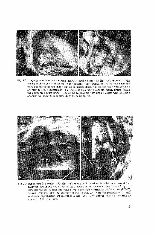
Fig. 3.2 A comparison between a normal heart (A) and a heart with Ebstein's anomaly of the tricuspid valve (B) with regard to the effective valve orifice. In the normal heart the principal orifice (dotted line) is almost in sagittal plane, while in the heart with Ebstein's anomaly the orifice (dotted line) has shifted to an almost horizontal plane, directly facing the pulmonic ostium (PO). It should be emphasized that not all hearts with Ebstein's anomaly will show this abnormality to the same degree.
Fig. 3.3 Echograms in a patient with Ebstein's anomaly of the tricuspid valve. A subcostal four chamber view shows not a trace of the tricuspid valve (A), while a parasternal long axis view (B) reveals the tricuspid valve (TV) in the right ventricular outflow tract (RVOT; arrow). Compare also the anatomy shown in Fig. 3.4. Note the presence of a small ventricular septal defect just beneath the aorta (Ao ). R V =right ventricle; VS =ventricular septum; LA= left atrium.
21
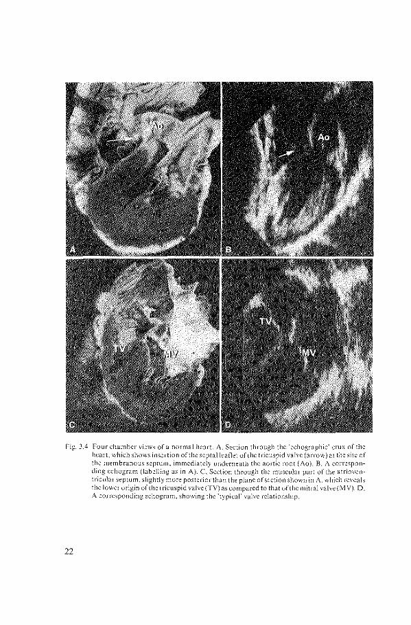
Fig. 3.4 Four chamber views of a normal heart. A. Section through the 'echographic' crux of the heart. which shows insertion of the septallea!1et of the tricuspid valve (arrow) at the site of the membranous septum, immediately underneath the aortic root (Ao). B. A corresponding cchogram (labelling as in A). C Section through the muscular part of the atrioventricular septum, slightly more posterior than the plane of section shown in A. which reveals the lo>vcr origin of the tricuspid valve (TV) as compared to that oft he mitral valve (MY). D. A corresponding echogram, showing the 'typical' valve relationship.
22
displacement will create a spectrum of anomalous valve ring positions, unified by the fact that they all share the site of the central fibrous body as the hinge.
The septal attachment of the tricuspid valve is abnormal in the sense that it originates closer to the apex of the ventricle. But in more anterior planes the site of origin normalizes, so that the relationship between the antero-superior tricuspid valve leaflet and the aortic root is mostly normal. Despite the normal relationship, however, the peculiar sail-like architecture of the antero-superior leaflet may underlie its echographic detection as an abnormal leaflet, the mobility of which may vary from case to case.
The echocardiographer should therefore focus on the septal attachment of the tricuspid valve. Cross-sections through the crux of the heart, in four chamber fashion, display this area. In the normal heart the septal tricuspid valve attaches to the central fibrous body, immediately adjacent to the origin of the aortic root (Fig. 3.4). Sections slightly more posterior, through the muscular part of the atrioventricular septum, will reveal the typical relationship between the tricuspid and mitral valve attachments (Fig. 3.4). Usually, the former originates at a lm..ver level than the mitral valve; an anatomical feature that is also used in identifying ventricular morphology. The degree of maximal 'distal diplacement' in normal hearts varies considerably, dependent also on the age of the individual, but the mean difference with the mitral valve is approximately 6 millimetres (Gussenhoven, unpublished observations; Roudaut et al, 1981).
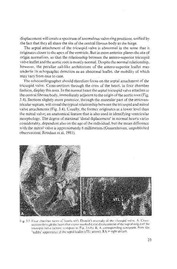
Fig. ~.5 Four chamber \icws of hearts with Ebstein's anomaly of the trieu~pid Yalvc. A, Cross~ection through the heart that shows marked distal displacement of the septal origin of the tricuspid \"ah.':'e (arrow; compare to Fig. ~.4A). B, A corresponding cchogram. Not~.: the:
'nobby' appearance of the septal kaflet lsTL". <HHWv'). RA =right atrium.
23

CHAPTER4
ECHOCARDIOGRAPHIC CRITERIA FOR EBSTEIN'S ANOMALY OF THE TRICUSPID VALVE
SUMMARY
The diagnostic echocardiographic features of Ebstein's malformation of the tricuspid valve have been evaluated in two groups of patients, using M-mode and two-dimensional techniques. The first group consisted of nine patients in whom previous M-mode studies had suggested the existence ofEbstein's anomaly. The second group consisted of 20 patients, all suffering from right heart overload, in whom Ebstein's malformation was excluded at open heart surgery. TheM-mode studies disclosed that none of the criteria currently employed could be Considered diagnostic. A delay in tricuspid valve closure of more than 65 msec, considered the most reliable indicator, was also present in eight of20 'controls'. The characteristic anatomical feature, that is distal displacement of the septal tricuspid leaflet, was never identified with certainty using M-mode echograms, in contrast to twodimensional echograms which showed a high degree of accuracy. Twodimensional techniques disclosed an abnormal insertion in six of nine patients in the first group, while a normal insertion was positively identified in 13 of 14 patients with right heart overload. In two of the nine patients in whom Ebstein's anomaly was suggested by M-mode criteria, a normal septal origin was identified and all further attempts to substantiate this diagnosis failed. In one patient from the first group, the two-dimensional study was inconclusive regarding positive identification of the septal origin.
Open heart surgery showed a normal origin of the septal leaflet, though the valve was plastered to the septal surface by short chordae. Only once among 14 'controls' was the septal attachment inconclusively identified with the twodimensional echograms. Surgery, however, excluded the presence of Ebstein's anomaly in this patient. Two-dimensional echocardiography, aiming at visualising the septal origin of the tricuspid valve, thus seems to be useful in establishing the diagnosis ofEbstein's malformation of the tricuspid valve.
INTRODUCTION
The clinical diagnosis of Ebstein's anomaly of the tricuspid valve is notoriously difficult, both with invasive and non-invasive investigations. The problem relates to the fact that Ebstein's anomaly, though an entity in itself, constitutes an
27
anatomical spectrum with regard to the extent and degree of the valvular abnormality (Ebstein, 1866; Pechstein, 1957; Becker et al, 1971). The anomaly is characterised by distal displacement of the septal attachment of the valve, a feature which may extend onto the inferior part of the tricuspid valve, and is almost always combined with various degrees of valve dysplasia.
The echocardiographic diagnosis of Ebstein's anomaly, using M-mode technique, depends on the identification of an increase in motion amplitude of the anterior tricuspid valve leaflet, an increase in the dimension of the right ventricular cavity, the presence of paradoxical septal motion, the ability to record the tricuspid valve more to the left of the sternum than usual, and a delay in closure of the tricuspid valve as compared with that of the mitral valve (Lundstrom and Edler, 1971; Lundstrom, 1973; Tajik eta!, 1973; Farooki eta!, 1976; Matsumoto et al, 1976; Milner et al, 1976; Lundstrom, 1978). However, it is not certain how reliable these indices are as diagnostic criteria. This is an important question since the truly diagnostic attachment of the septal leaflet, is not readily obtained with the M-mode echo gram. As recently stated, two-dimensional methods may prove to be of value in this respect, since this technique enables the actual identification of the abnormal origin of the valve (Matsumoto et al, 1976; Ports et al, 1978).
It is for this reason that we have employed a two-dimensional echocardiographic technique to restudy a group of nine patients in whom the diagnosis of Ebstein's anomaly of the tricupsid valve had previously been suggested from the currently accepted M-mode echo graphic criteria. In order to assess these criteria another 20 patients, known to have right ventricular volume overload resulting from a variety of causes other than Ebstein's anomaly, were also studied.
SUBJECTS AND METHODS
The material for this study consisted oftwo groups of patients. The first group was composed of nine patients (six male and three female) ranging in age from 16 to 56 years (average 33 years), all of whom were considered to have Ebstein's anomaly of the tricuspid valve. This presumptive diagnosis had been based on identification of at least two of the following five M-mode criteria (Lundstrom and Edler, 1971; Lundstrom, 1973; Tajik eta!, 1973; Farooki eta!, 1976; Matsumoto eta!, 1976; Milner eta!, 1976; Lundstrom, 1978): an amplitude of the anterior tricuspid valve leaflet motion of20 mm or more; a right ventricular dimension of30 mm or more; paradoxical septal motion defined as movement parallel to the motion of the echo from the posterior heart wall during ventricular systole (Diamond et al, 1971 ); lateral displacement of the tricuspid valve, defined as the situation in which the tricuspid valve motion can be recorded more to the left of the sternum than usual, taking the left parasternal line as the borderline; and a delay in closure of the tricuspid valve of 65 msec or more as compared with that of the mitral valve.
28
In each of these nine patients invasive studies were done in an attempt to verify the presumptive diagnosis.
The second group comprised 20 patients (eight men and 12 women) ranging in age from 21 to 66 years (average 38 years). They were selected because cardiac catheterisation had shown right heart overload in each. In 13, this was the result of an isolated atrial septal defect of the fossa ovalis type, and in two of an atrial septal defect complicated by partial anomalous pulmonary venous connection; another four had tricuspid regurgitation in the presence of longstanding mitral regurgitation and/or stenosis. One patient had an atrial septal defect in the presence of pulmonary stenosis. In each patient the possibility ofEbstein's anomaly had been ruled out at open heart surgery.
The same echographic studies were performed in both groups. The M-mode studies were performed with the EchoCardiovisor 01, and recorded on a line scan recorder (Honeywell visicorder 1856) using light-sensitive paper (Kodak type 1895). The tracings were recorded at paper speed of 50 mm/sec. The twodimensional images were made with a dynamically focused multiscan system (Ligtvoet et al, 1977).
ANATOMY
In order to appreciate the diagnostic possibilities of both M-mode and twodimensional echocardiograms for Ebstein's anomaly, it is necessary to recall the basic anatomical derangements in this condition.
The disease is characterised by two main features (Ebstein, 1866; Pechstein, 1957; Becker et al, 1971). First, part of the basal attachment of the tricuspid valve is displaced distally, thereby creating a so-called atrialised part of the right ventricle (Fig. 4.1 ). Distal displacement affects the septal leaflet but this varies in extent with the inferior leaflet. The anterior leaflet, on the other hand, always originates from the annulus fibrosus. The second anatomical feature of Ebstein's anomaly is dysplasia of the valve, often characterised by a sail-like deformity of the 'non-displaced' anterior leaflet. However, as with distal displacement, the extent and degree of valve dysplasia may vary considerably from one patient to the other (Pechstein, 1957; Becker et al, 1971). In some instances the 'sail-like' anterior leaflet may be free floating, while in others the leaflet is 'plastered' to the right ventricular free wall. In the latter circumstance the valve leaflet can be much restricted in its mobility. In rare circumstances, moreover, the septal, inferior and anterior leaflets may become continuous, thus creating a severe inlet obstruction to the right ventricular outlet; a situation that clinically may mimic 'classical' tricuspid atresia.
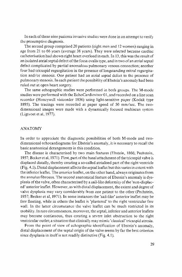
From the point of view of echographic identification of Ebstein's anomaly, distal displacement of the septal origin of the valve seems by far the best criterion since dysplasia in itself is not readily distinctive (Fig. 4.1).
29
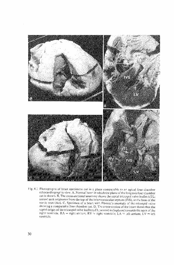
Fig. 4.1 Photographs of heart specimens cut in a plane comparable to an apical four chamber echocardiographic view. A. Normal heart in which the plane of the long axis four chamber cut is shown. B, The cross-sectional anatomy sho\vs the septal tricuspid valve leaflet (sTL; arrow) as it originates from the top of the interventricular septum (IVS). at the base of the aortic root (Ao). C, Specimen of a heart with Ebstein's anomaly of the tricuspid valve showing a comparable four chamber cut. D, The cross-section of the heart shows that the septal origin of the tricuspid valve leaflet (sTL; arrow) is displaced towards the apex of the right ventricle. RA =right atrium; RV =right ventricle: LA= left atrium; LV= left ventricle.
30
RESULTS
The echographic data obtained from the nine patients initially diagnosed as having an Ebstein's anomaly of the tricuspid valve are shown in table 4.1. The M-mode studies revealed that the motion amplitude of the anterior tricuspid valve leaflet was increased in all, whereas in eight there was an abnormal increase of the right ventriGular dimension. Lateral 'displacement' of the tricuspid valve was obvious in seven patients, while this feature was inconclusive in two. Delay in closure of the tricuspid valve of more than 65 milliseconds was present in eight and paradoxical septal motion in six patients.
Deliberate attempts to identify the septal attachment of the tricuspid valve using theM-mode method were unsuccesful in all nine patients. In only one (case 7) was the suggestion of a low insertion raised, since the septal tricuspid valve leaflet was recorded only when the ultrasound beam swept close to the right ventricular apex (Fig.4.2).
In some instances an accumulation of echoes was observed at the right ventricular site of the interventricular septum, but these echoes were never positively identified as representing a displaced valve leaflet (Fig.4.3A).
Two-dimensional echograms, on the other hand, visualized the septal attachment of the tricuspid valve in eight patients and distal displacement was positively
Fig. 4.2 A male patient, 23 years of age (table 4.1; case 7) with proven Ebstein's anomaly. In the M~mode recording the septal tricuspid valve leaflet (sTC arrow) appeared only when the ultrasound beam swept close to the right ventricular apex. The long and short arrows indicate the time of closure of the tricuspid and mitral valve, respectively. Note the delayed closure of the former. RVOT =right ventricular outflow tract; Ao =aorta; LA= left atrium; LV= left ventricle; IVS =interventricular septum; RV =right ventricle.
31
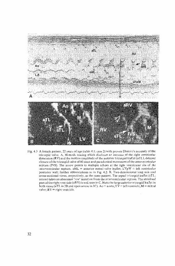
Fig. 4.3 A female patient, 22 years ofage{table 4.1; case 2) with proven Ebstein's ano111aly of the tricuspid valve. A, M-mode tracing which discloses an increase of the right ventricular dimension (R V) and the motion amplitude of the anterior tricuspid leaflet (aTL), delayed closure of the tricuspid valve of80 msec and paradoxical movement of the interventricular septum (IVS). The arrow points to multiple echoes at the right ventricular site of the interventricular septum. aML =anterior mitral valve leaflet; LVpW =left ventricular posterior wall; further abbreviations as in Fig. 4.2. B, Two-dimensional long axis and cross-sectional views, respectively, in the same patient. The septal tricuspid leaflet (sTL; arrow) takes an abnormal 'low' insertion from the interventricular septum. The atrialised part of the right ventricle (aR V) is well seen in C. Note the large anterior tricuspid leaflet in both views (aTL in 38 and open arrow in 3C). Ao =aorta; LV= left ventricle; M =mitral valve; RV =right ventricle.
32
identified in six. The 'ideal' position of the transducer, enabling positive identification, varied from one individual to the other and encompassed classical long axis, cross-sectional and apical four chamber views (Figs. 4.3B,C and 4.4A). In two patients (table 4.1; cases 4 and 9) the septal attachment was identified at a normal level, that is close to the aortic root (compare Figs. 4.4A and B) at the junctional level between mitral valve, interventricular septum and interatrial septum; catheter studies confirmed the presence of tricuspid regurgitation, but in neither was there any indication of Ebstein 's anomaly. Simultaneous pressure and electrical recordings did not disclose the presence of an atrialized part of the right ventricle. In one patient (table 4.1; case 3) the two-dimensional echogram did not visualize the septal attachment of the tricuspid valve. This patient is of particular significance since cardiac catheterisation studies disclosed a large atrial septal defect with tricuspid regurgitation, while Ebstein 's anomaly could not be excluded with certainty from the angiograms. At open heart surgery it was noticed that the septal tricuspid valve leaflet took a normal origin from the right-sided annulus
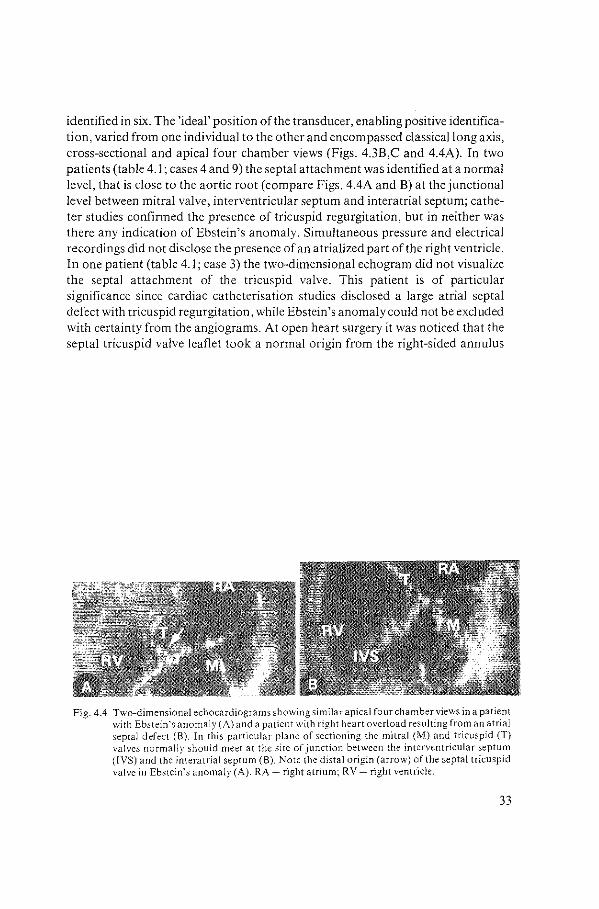
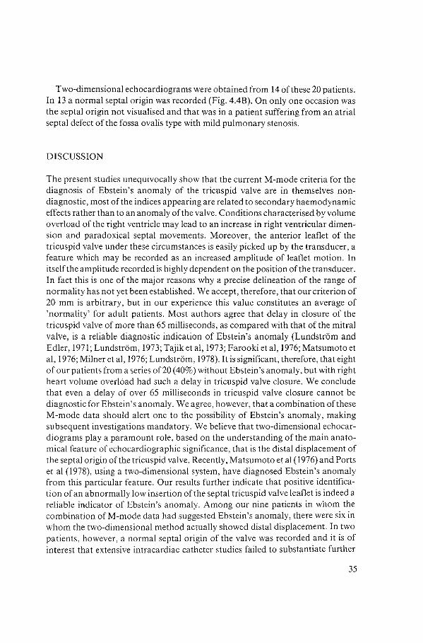
Fig. 4.4 Two-dimensional echocardio!!rams showing similar apical four chamber views in a patient ~ with Ebstein's anomaly {A) a;d a patient wiih right heart overload resulting from an atrial
septal defect (B). In this particular plane of sectioning the mitral (M) and tricuspid (T) valves normally should meet at the site of junction between the interventricular septum (IVS) and the interatrial septum (B). Note the distal origin (arrow) of the septal tricuspid valve in Ebstein's anomaly (A). RA =right atrium; RV =right ventricle.
33
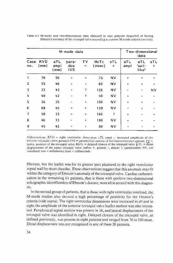
Table 4.1 M-mode and two-dimensional data obtained in nine patients suspected of having Ebstein's anomaly of the tricuspid valve according to current M-mode criteria (see text).
M-mode data Two-dimensional data
Case RVD aTL para- TV MeTe sTL aTL aTL sTL no. (mm) amp I dox ~ ( msee) .f amp I •sail- .f
( mm) IVS like•
70 50 + + 75 NV + + +
2 55 48 + + 80 NV + + +
3 22 43 + ? 120 NV NV
4 40 42 ? 40 NV +
5 36 35 + 100 NV + +
6 88 45 + + 120 NV + +
7 50 33 + + 160 ? +
8 46 33 + 180 NV + +
9 45 42 + + 80 NV + +
Abbreviations: RVD = right ventricular dimension; aTL amp! = increased amplitude of the anterior tricuspid valve: paradox rvs =paradoxical motion of the interventricular septum: n = lateral position of the tricuspid valve; MeTe= delayed closure of the tricuspid valve ~sTL =distal displacement of the septal tricuspid valve leaflet; +, present; -, absent; ?, questionable; NV, not visualized; mm = mi!limetres; msec =milliseconds.
fibrosus, but the leaflet was for its greater part plastered to the right ventricular septal wall by short chordae. These observations suggest that this anomaly may fit within the category ofEbstein's anomaly of the tricuspid valve. Cardiac catheterisation in the remaining six patients, that is those with positive two-dimensional echographic identification ofEbstein's disease, were all in accord with this diagnosis.
In the second group of patients, that is those with right ventricular overload, the M-mode studies also showed a high percentage of positivity for the Ebstein 's criteria (vide supra). The right ventricular dimensions were increased in all and in eight the amplitude of the anterior tricuspid valve leaflet motion was also increased. Paradoxical septal motion was present in 16, and lateral displacement of the tricuspid valve was identified in eight. Delayed closure of the tricuspid valve, as defined previously, was present in eight patients and ranged from 70 to 160 msec. Distal displacement was not recognised in any of these 20 patients.
34
+
+
+
+
Two-dimensional echocardiograms were obtained from 14 of these 20 patients. In 13 a normal septal origin was recorded (Fig. 4.4B). On only one occasion was the septal origin not visualised and that was in a patient suffering from an atrial septal defect of the fossa ovalis type with mild pulmonary stenosis.
DISCUSSION
The present studies unequivocally show that the current M-mode criteria for the diagnosis of Ebstein's anomaly of the tricuspid valve are in themselves nondiagnostic, most of the indices appearing are related to secondary haemodynamic effects rather than to an anomaly of the valve. Conditions characterised by volume overload of the right ventricle may lead to an increase in right ventricular dimension and paradoxical septal movements. Moreover, the anterior leaflet of the tricuspid valve under these circumstances is easily picked up by the transducer, a feature which may be recorded as an increased amplitude of leaflet motion. In itself the amplitude recorded is highly dependent on the position of the transducer. In fact this is one of the major reasons why a precise delineation of the range of normality has not yet been established. We accept, therefore, that our criterion of 20 mm is arbitrary, but in our experience this value constitutes an average of 'normality' for adult patients. Most authors agree that delay in closure of the tricuspid valve of more than 65 milliseconds, as compared with that of the mitral valve, is a reliable diagnostic indication of Ebstein's anomaly (Lundstrom and Edler, 1971; Lundstrom, 1973; Tajik et al, 1973; Farooki et al, 1976; Matsumoto et al, 1976; Milner et al, 1976; Lundstrom, 1978). It is significant, therefore, that eight of our patients from a series of20 (40%) without Ebstein's anomaly, but with right heart volume overlOad had such a delay in tricuspid valve closure. We conclude that even a delay of over 65 milliseconds in tricuspid valve closure cannot be diagnostic for Ebstein's anomaly. We agree, however, that a combination of these M-mode data should alert one to the possibility of Ebstein's anomaly, making subsequent investigations mandatory. We believe that two-dimensional echocardiograms play a paramount role, based on the understanding of the main anatomical feature of echocardiographic significance, that is the distal displacement of the septal origin of the tricuspid valve. Recently, Matsumoto et al (1976) and Ports et a! (1978), using a two-dimensional system, have diagnosed Ebstein's anomaly from this particular feature. Our results further indicate that positive identification of an abnormally low insertion of the septal tricuspid valve leaflet is indeed a reliable indicator of Ebstein's anomaly. Among our nine patients in whom the combination ofM-mode data had suggested Ebstein's anomaly, there were six in whom the two-dimensional method actually showed distal displacement. In two patients, however, a normal septal origin of the valve was recorded and it is of interest that extensive intracardiac catheter studies failed to substantiate further
35
this diagnosis. In one patient the septal attachment was not visualised with the two-dimensional technique. This inconclusive finding is significant since open heart surgery in this patient disclosed an abnormal septal valve leaflet. The valve took its normal origin, but the leaflet was for its greater part plastered to the septal surface. One could argue that this is not Ebstein's anomaly of the tricuspid valve, but, on the other hand, this observation also serves to indicate that even with two-dimensional techniques some extreme variants of Ebstein 's anomaly will not be recognised. For instance, this might occur when extreme distal displacement of the septal and inferior leaflets is associated with a membrane-like continuum into the anterior leaflet, thereby creating a so-called imperforate Ebstein's anomaly. Nevertheless, the two-dimensional method seems accurate as further substantiated by the findings in our group of patients without Ebstein's anomaly. While the M-mode data in some of them suggested the possibility ofEbstein's anomaly, the two-dimensional echograms showed a normal septal origin in almost all. In only one patient was the septal origin not visualised, a patient suffering from an atrial septal defect and mild pulmonary stenosis.
To obtain the best results, the transducer should be placed in a transverse position close to the apex of the heart and perpendicular to the plane of the long axis view. The operator should direct the plane of the image slightly upwards, so that the aortic root region is clearly visualised. The plane of 'sectioning' thus fluctuates between the standard cross-sectional and apical four chamber views. Abnormalities in the septal attachment of the tricuspid leaflet may thus be identified and one might expect even a small rim of dysplastic and displaced valve tissue to be detected. We, therefore, believe that the two-dimensional echocardiogram is valuable in patients suspected of having Ebstein's anomaly of the tricuspid valve.
36
CHAPTERS
VARIABILITY IN THE TIME INTERVAL BETWEEN TRICUSPID AND MITRAL VALVE CLOSURE
IN EBSTEIN'S ANOMALY
An echographic study
ABSTRACT
The time interval between mitral and tricuspid valve closure was measured from M-rnode echocardiograms in patients with Ebstein's anomaly. It was found that this time interval demonstrated a range of values within each patient. In the present study we assessed the parameters which influenced the variability. The use of different transducer positions on the chestwall was found to be the predominant factor. There was no correlation between the measured time interval, its variability and the moment of measurement during breathing. However, a period of breathholding significantly reduced the variability. This study indicates that a wide range of time intervals might be measured in a single patient. We conclude therefore, that this parameter should be used with great care in the diagnosis of Ebstein's anomaly.
INTRODUCTION
In normal subjects, mitral valve closure precedes tricuspid valve closure (Leatham, !954; Reinhold and Rudke, !957), a feature which can be demonstrated using M-mode echocardiography (Milner eta!, 1976). Abnormal hemodynamics may coincide with a change in atrioventricular valve closure relationship (Milner et al, 1976). For example, delayed tricuspid valve closure time relative to mitral valve closure (MeTe) has been observed in patients with Ebstein's anomaly. Some authors considered this finding highly suggestive for the diagnosis of Ebstein's anomaly (Milner eta!, 1976; Farooki eta!, 1976; Daniel eta!, 1980). However, a delay in tricuspid valve closure time has also been documented in patients not having this disease (French eta!, 1975; Milner eta!, 1976; Daniel et al, 1980; Gussenhoven eta!, !980).
During the course of studying patients with Ebstein's anomaly of the tricuspid valve, we noted that the delay in tricuspid valve closure time measured within each patient was variable. Although the sensitivity of two-dimensional echocardiography for the diagnosis of Ebstein's anomaly is high in comparison to the use of M-mode echocardiography (Ports eta!, 1978; Roudaut eta!, 1981), the purpose of
37
the present study is to emphasize the reliability of theM-mode parameter, i.e. the time interval between mitral and tricuspid valve closure and to determine the sources of its variability.
METHODS AND MATERIAL
Intra- and interobserver tests performed in a clinical series of patients with Ebstein's anomaly have indicated that a difference of 5 msec was measured when identical beats were considered and when a paper speed of 100 mm/sec was used.
We assume that the number of sources that might possibly contribute to the variability in the time interval between mitral and tricuspid valve closure includes: -Influence of the instrumentation; -Error caused by the methodology of the measurement; -Location of the transducer on the chestwall; -Transducer manipulation instability; - ResPiratory variation. These variables have been extensively investigated in three patients known with Ebstein's anomaly (Ebl; Ebll; Ebiii). The diagnosis Ebstein's anomaly was based on the accumulated data obtained from the clinical studies including cardiac catheterization and angiocardiography. Previous M-mode investigations obtained in the three selected patients demonstrated a range in the measured time interval between mitral and tricuspid valve closure with a mean value of SO msec in two patients and a mean value of 120 msec in one patient. As it V-/as noted that the variability of the MeTe, attained simultaneously, was mainly due to the tricuspid valve component, we have confined the present study to the variability of the tricuspid valve closure only.
For evaluation of the above mentioned variables M-mode echocardiograms of the patients were recorded at a paper speed of 100 mm/sec. The point of tricuspid valve closure was identified by the point of coaptation of the leaflets, or that instant at which the leaflets sharply terminated their rapid approximations at the onset of systole (Henry eta!, 1979). The tricuspid valve closure time was related to the onset of the Q-wave of the electrocardiogram.l\1easurements were made when equal preceding RR-imervals were present.
In excess of 2000 cardiac beats were analyzed. During this study it was noted that atrioventricular valve closing points may appear at different depths on the M-mode tracing (Fig. 5.1 ). We believe that this observation is the result of off-axis sensitivity of the transducer combined with changes of cardiac position due to, for instance, patient's breathing (Roelandt, 1977). The leaflet is a three-dimensional structure and off-axis sensitivity of the transducer may result in picking up any closing point of the leaflet. Measurements from such a point cannot be correctly compared with measurements carried out on echoes resulting from the main axis
38
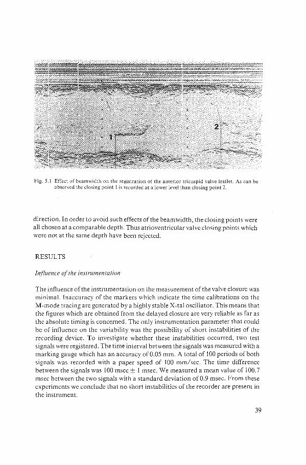
Fig. 5.1 Effect of beamwidth on the registration of the anterior tricuspid valve leaflet. As can be observed the closing point 1 is recorded at a lower level than closing point 2.
direction. In order to avoid such effects of the beam width, the closing points were all chosen at a comparable depth. Thus atrioventricular valve closing points which were not at the same depth have been rejected.
RESULTS
Influence of the instrumentation
The influence of the instrumentation on the measurement of the valve closure was minimal. Inaccuracy of the markers which indicate the time calibrations on the M-mode tracing are generated by a highly stable X-tal oscillator. This means that the figures which are obtained from the delayed closure are very reliable as far as the absolute timing is concerned. The only instrumentation parameter that could be of influence on the variability was the possibility of short instabilities of the recording device. To investigate whether these instabilities occurred, two test signals were registered. The time interval between the signals was measured with a marking gauge which has an accuracy of0.05 mm. A total of 100 periods of both signals was recorded with a paper speed of 100 mm/sec. The time difference between the signals was 100 msec ± 1 msec. We measured a mean value of 100.7 msec between the two signals with a standard deviation of 0.9 msec. From these experiments we conclude that no short instabilities of the recorder are present in the instrument.
39
Error caused by the methodology of the measurements
In all cases a standard ruler was used to perform the measurements. The measurement error which can be made, may be divided into I) that caused by imprecision of the reading of the ruler and 2) the error caused by drawing vertical lines on theM-mode tracing.
In order to assess the accuracy of the measurements two signals were written on different levels simulating the situation present on an M-mode recording. Vertical lines were drawn using a triangular ruler. The time interval between the signals was measured with a marking gauge. One hundred periods of this signal configuration were recorded. The recording speed used was 100 mm/sec. The time difference of the test signals was 100 msec ± 1 msec. With this measurement technique a mean value of 99.8 msec and a standard deviation of 1.2 msec was observed. We conclude that the error in measurement technique is minimal.
Location of the transducer on the chestwall
In order to evaluate whether the transducer position on the chestwall influenced the variability in time interval between mitral and tricuspid valve closure, continuous M-mode registrations were made from different transducer positions on the chestwall (A-D). The results are shown in table 5.1.
We deduce that the total range of tricuspid valve closure is larger than the range observed from one transducer position only. It is evident that transducer positions are in part responsible for the range in tricuspid valve closure. Theoretically the explanation for this dependency of transducer position may be due to the fact that using a different transducer position another part of the tricuspid valve may be transected by the soundbeam. As illustrated schematically in Fig. 5.2, the tricuspid valve closure occurs first at level A and slightly thereafter at level B. Thus, when the transducer is directed towards level A an earlier valve closure can be expected than when the transducer is directed towards level B.
In order to evaluate this aspect the following formula:
ilt = 2d. tan (1/ 2a)
Vs+Va
was derived which approximates the time difference in closing (Llt) between level A and B (Fig. 5.2). In this formula only the velocity component in the closing direction is estimated. The upward motion of any point on the leaflets during closure was neglected. The velocity of both septal (Vs) and anterior (Va) tricuspid valve leaflets were calculated from theM-mode tracings of the three patients with an Ebstein's anomaly as tabulated in table 5.1. When both septal and anterior
tricuspid valve leaflets co-apt with an arbitrarily taken angle a of~ and assuming
40
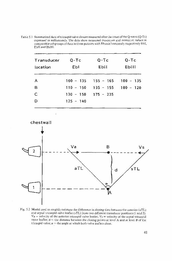
Table 5.1 Summarized data of tricuspid valve closure measured afterthe onset of the Q-wave (Q-Tc) expressed in milliseconds. The data show measured maximum and minimum values in comparable subgroups of data in three patients with Ebstein's anomaly respectively Ebl, Ebll and Ebiii.
Transducer Q-Tc Q-Tc Q-Tc
location Ebl Ebll Eblll
A 1 00 - 135 155 165 100 135
B 11 0 150 135 155 100 - 120
c 130 - 150 175 - 235
D 125 140
chestwall
B Vs ~-------- Va ------------
aTL d sTL
Fig. 5.2 Model used to roughly estimate the difference in closing time between the anterior (aTL) and septal tricuspid valve leaflet (sTL) from two different transducer positions (1 and 2). Va = velocity of the anterior tricuspid valve leaflet: Vs = velocity of the septal tricuspid valve leaflet; d =the distance between the closing points at level A and at level 8 of the tricuspid valve; ex =the angle at which both valve leaflets close.
41
that level A and Bare located 10 mm from each other, the ilt determined were respectively:
Eb I - ilt: 32 msec (Vs = 80 mm/sec; Va = 540 mm/sec) Eb II - ilt: 23 msec (Vs = 25 mm/sec; Va = 830 mm/sec) Eb III- ilt: 33 msec (Vs = 70 mm/sec; Va = 530 mm/sec)
From these results it became clear that a change in the transducer position on the chestwall introduces a difference in closing time of the tricuspid valve. These theoretical differences are in the same order of magnitude as can be observed from table 5.1.
The instability of the transducer manipulation
In order to investigate the influence of the transducer manipulation instabilities caused by manual vibration due to the operator, a measurement device was attached to the transducer and the vibration angle was determined during one minute of investigation. This experiment has been repeated four times. A vibration angle of five degrees maximum was observed. This vibration angle introduces an area of uncertainty dependent on the distance between the transducer and the position at which the tricuspid valve closes. In the three patients in table 5.1 a transducer vibration angle of 5 degrees would yield a ilt of 18 msec.
Respiratory variation
We have assumed that the differences in pressure at atrial level during respiration may in part be responsible for the measured variability of tricuspid valve closure time. In order to evaluate this a pneumatograph was used to register the patient's respiration simultaneously with an M-mode echocardiogram. The patients were allowed to breathe spontaneously during the investigation. In addition recordings were made during a period that the patient was requested not to breathe. Recordings were obtained from different transducer positions on the chestwalL It appeared that measurements obtained both during inspiration as well as during expiration showed a variability in closure time, not different from the data shown in table 5.1. Furthermore, when these time intervals were plotted against the percentage of the inspiration and expiration cycle, no significant correlation was observed between patient's breathing and the measured time interval. The variability of the time interval (Q-Tc), measured from one transducer position during breathholding, reduced to a maximum of 15 msec.
42
DISCUSSION
A prolonged time interval between mitral and tricuspid valve closure has been used as an important M-mode criterion for the diagnosis of Ebstein's anomaly (Milner eta!, 1976; Farooki eta!, 1976; Daniel eta!, 1980). Interestingly enough, the reports prior to 1975 indicate that all patients with Ebstein's anomaly had a delay in closure of the tricuspid valve, whereas subsequent investigators disclosed a number of patients with proven Ebstein's anomaly in whom the M-mode echocardiogram showed a time interval within the normal range. A review of the criteria set for a 'diagnostic' time interval reveals that not all authors have used the same time lapse. In fact, the mitral-to-tricuspid interval was documented as either 30, 40, 50,60 or 65 msec in different studies (Lundstrom, 1973; Tajik eta!, 1973; Yuste et al, 1974; Farooki eta!, 1976; Daniel eta!, 1980). This implies that the sensitivity of this parameter is intimately related to the value used as 'diagnostic'. Indeed, when 40 msec is considered as a diagnostic time interval, a high sensitivity can be expected, whereas a value of65 msec will result in a low sensitivity rate. This aspect is of great significance, particularly when we consider the time interval measured within a single patient with Ebstein's anomaly, who presented a range of 30 to 70 msec. An MeTe of30 msec might indicate absence ofEbstein's anomaly, whereas at the same time an MeTe of 70 msec would make the diagnosis very likely.
In order to appreciate the reasons for the variability of the time interval measured between mitral and tricuspid valve closure and consequently the value of this M-mode parameter for the diagnosis Ebstein's anomaly, we have analyzed a number of variables.
Experiments concerning the influence of the instrumentation indicated that no short instabilities of the recorder were present. Thus the degree of variance in atrioventricular valve closure cannot be explained from the instrumentation part.
The error caused by the methodology of the measurements was minimal and in the same order of magnitude as observed from the intra- and interobserver test. During our routine studies we have used a standard ruler in order to avoid the time consuming method of the marking gauge. Measurements were made to the nearest 0.5 mm. The error introduced by such a ruler will be 5 msec at a paper speed of 100 mm/sec and 10 msec at a paper speed of 50 mm/sec. Hence, the observed variability of atrioventricular valve closure can only partly be explained by the methodology of the measurement.
The third variable investigated, i.e. the influence of the different transducer positions on the chestwall, revealed that the total range of atrioventricular valve closure is larger than the range observed from one transducer position only. We believe that using a different transducer position another part of the atrioventricular valve may be 'insonated' and, hence, another 'closing moment' of the valve will be recorded. Taking into account the tricuspid valve morphology in Ebstein's
43
anomaly, in which the valve usually is enlarged (Anderson and Lie, 1978), we may expect a number of different 'closing moments' of the valve. Indeed experiments, not included in this study, have shown that the range of tricuspid valve closure in patients with Ebstein's anomaly was larger compared to the range obtained in subjects with a normal valve morphology.
The range of atrioventricular valve closure present at a single transducer position was found to be the result of two factors. First, the aiming instability of the transducer caused by the operator during the investigation and second, the influence of patient's breathing. Measurements obtained during breathholding showed a more stable interval. Apparently, a period ofbreathholding minimizes the effect of aiming instability caused by the investigator and rules out the influence of intracardiac pressure differences during respiration.
We conclude that the variety in closing time is mainly related to the transducer position and aiming instability. The influence of the latter parameter may have been so large that it has obscured a possible relationship between patient's breathing and the measured closure time of the valve. We assume that the mean value of MeTe obtained from various transducer positions and from simultaneously recorded mitral and tricuspid valve closure is simply a characteristic of a particular patient. Hence, in our clinical practice, we avoid the use of this M-mode parameter as a diagnostic feature. Two-dimensional echocardiography, in this respect, is unequivocally better suited for the diagnose of Ebstein 's anomaly.
44
CHAPTER6
THE SEPTAL TRICUSPID AND MITRAL VALVE RELATIONSHIP IN NORMAL HEARTS AND IN HEARTS
WITH EBSTEIN'S ANOMALY
An echographic and anatomic study
SUMMARY
Distal displacement of the septal attachment ofthe tricuspid valve leaflet is widely accepted as an important feature for the diagnosis Ebstein's anomaly. This feature can be readily established with two-dimensional echocardiography. However, experience has shown that marked variability exists in normal hearts with respect to this particular anatomic aspect. For this reason a study has been undertaken comparing heart specimens with echocardiograms ofboth normal persons and of patients with proven Ebstein's malformation. Because of the growing significance of fetal echocardiography the study has been expanded to include fetuses.
Positive identification of a distance between two atrioventricular valves, contributing to ventricular identification, is feasible, both anatomically and echographically, in fetuses in the second and third trimester. The difference in the insertion site between the two valves increases from infants to adults as shown both by echographic and by anatomic studies. An overlap between normal hearts and hearts with Ebstein's anomaly occurs, particularly when minimum distances were tabulated. The maximum differences in Ebstein's anomaly, on the other hand, usually reached far beyond those found in the normal.
INTRODUCTION
The septal insertion of the tricuspid valve in normal hearts is at a lower level than that of the mitral valve. Coronal sections through the heart, comparable to four chamber echographic views, readily reveal this disposition (Tajik et a!, 1978; Anderson and Becker, 1980).
Echocardiographers use this anatomic detail as a helpful guide in identifying the morphologic right and the morphologic left ventricle, both in hearts with atrioventricular concordance and with atrioventricular discordance (Hagler et al, 1981; Becker and Gussenhoven, 1981; Losekoot eta!, 1983).
However, the level of valve insertion can differ among individuals. The question arises to what extent this anatomic variability may hamper the prompt diagnosis of Ebstein's anomaly, since the latter is characterized by a variable degree of apical
45
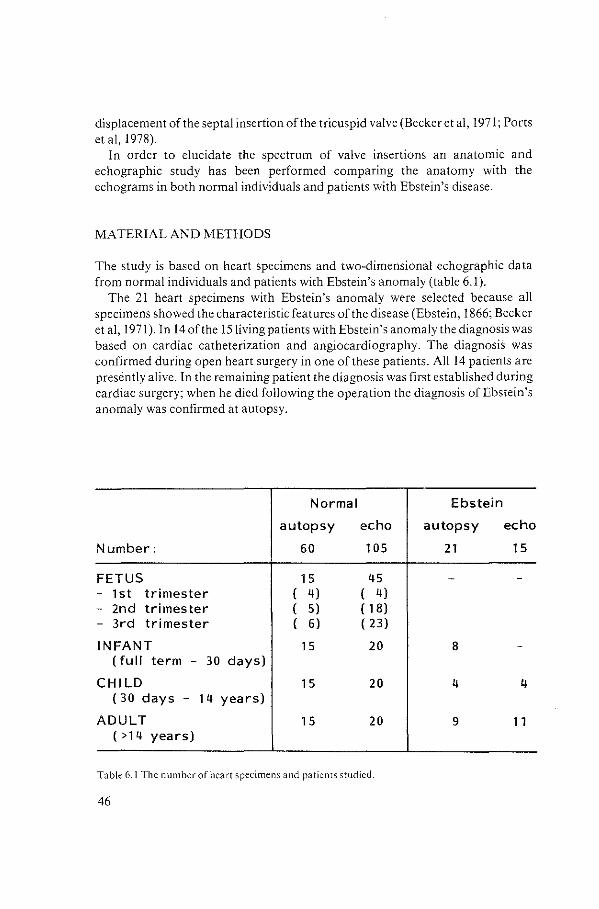
displacement of the septal insertion of the tricuspid valve (Becker eta!, !97!; Ports et a!, !978).
In order to elucidate the spectrum of valve insertions an anatomic and echographic study has been performed comparing the anatomy with the echograms in both normal individuals and patients with Ebstein's disease.
MATERIAL AND METHODS
The study is based on heart specimens and two-dimensional echographic data from normal individuals and patients with Ebstein's anomaly (table 6.!).
The 21 heart specimens with Ebstein's anomaly were selected because all specimens showed the characteristic features of the disease (Ebstein, 1866; Becker eta!, !971). In 14 of the IS living patients with Ebstein's anomaly the diagnosis was based on cardiac catheterization and angiocardiography. The diagnosiS was confirmed during open heart surgery in one of these patients. Alll4 patients are presently alive. In the remaining patient the diagnosis was first established during cardiac surgery; when he died following the operation the diagnosis ofEbstein's anomaly was confirmed at autopsy.
Normal Ebstein
autopsy echo autopsy echo
Number: 60 105 21 15
FETUS 15 45 - 1st trimester ( 4) ( 4) - 2nd trimester ( 5) ( 18) - 3rd trimester ( 6) ( 23)
INFANT 15 20 8 (full term - 30 days)
CHILD 1 5 20 4 4 ( 30 days - 14 years)
ADULT 1 5 20 9 11 (>14 years)
Table 6.1 The number of heart specimens and patients studied.
46
All heart specimens were fixed routinely in formalin. In the normal specimens the septal junction area, immediately adjacent to the aortic root, was exposed using a four chamber cross-section comparable to an echographic four chamber view through the crux of the heart (Fig. 6.1A). Additional sections were made with a slight posterior tilt of the plane of section (Fig. 6.1B). In each specimen, differences were noted in the distance between the septal tricuspid and mitral valves. The minimum and maximum distance between the two valve insertions was measured, similar to the method as described for the echographic images (vide infra).
In only two of the 21 specimens with Ebstein's anomaly a four chamber section was made as in the normal specimens. In the remaining hearts a different technique was used. In these hearts the septal attachment of the mitral valve leaflet was projected on to the right septal surface (Fig. 6.2A). Pins were placed at the site of highest and lowest points, whereafter the minimum and maximum distance between the septal mitral and the septal tricuspid valve leaflet was measured (Fig. 6.2B).
The two-dimensional four chamber images were obtained from subcostal and or apical positions. The still-frames which showed the minimum and maximum difference in valve insertion of both leaflets were selected in early systole (Fig. 6.3). In 3 patients with an Ebstein's anomaly the still-frames were selected from end-diastole, since the septal tricuspid valve leaflet was minute and its precise insertion was obscured in early systole by the huge anterior tricuspid valve leaflet. Polaroid pictures were taken of the still-frames. Lines were drawn at a right angle to the ventricular septum through the points where the atrioventricular valve attaches to the ventricular septum (Fig. 6.3). The perpendicular distance between these two lines was measured and expressed in millimetres. From this the minimum and maximum distance, between both atrioventricular valves were calculated. For each group the overall average was calculated, as well as the extremes, indicating the range of values, and the average minimum (~) and average maximum (max) distance. In the fetal group the small dimensions of the heart and its internal structures did not allow reliable measurements other than the identification of whether or not a distance between both valves was present.
RESULTS
NORMAL FETAL HEART
Forty-one of the 45 echographic investigations could be used. In 4 fetuses the four chamber images were unsatisfactory, since both atrioventricular valve insertions were not recorded simultaneously.
47
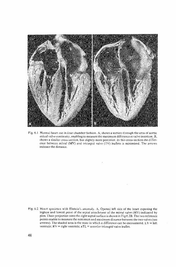
Fig. 6.1 Normal heart cut in four chamber fashion. A, shows a section through the area of aortic mitral valve continuity. enabling to measure the maximum difference in valve insertion. 8, shows a similar cross-section, but slightly more posterior. In this cross-section the difference between mitral (MV) and tricuspid valve (TV) leaflets is minimized. The arrows indicate the distance.
Fig. 6.2 Heart specimen with Ebstein's anomaly. A, Opened left side of the heart exposing the highest and lowest point of the septal attachment of the mitral valve (MV) indicated by pins. Their projection onto the right septal surface is shown in Fig 6.28. The two reference points enable to measure the minimum and maximum distance between the two valves (see arrows). The shaded area is the zone in which a difference can be encountered. LV= left ventricle; RV =right ventricle; aTL =anterior tricuspid valve leaflet.
48
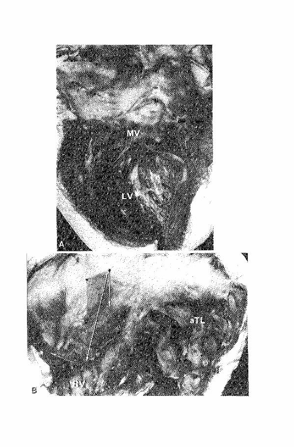
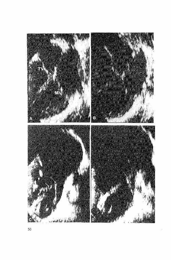
Fig. 6.3 Four chamber echograms in a normal heart (A and B) and in a heart with Ebstein's anomaly (C and D)~ In the normal heart the tricuspid valve (TV) is attached to the ventricular septum (VS) at a lower level then the mitral valve (MY). The maximum distance (between arrows) between both atrioventricular valves was obtained in an anterior plane, immediately beneath the aortic root (A). The minimum value was measured from the section which traversed the crux of the heart posteriorly (B). In the heart with Ebstein's anomaly the cross-section through the area adjacent to the aortic root revealed the minimum distance between both valves (C), while the more posterior plane showed the maximum distance (D).
In the first trimester no difference in the distance betweeen two valves was identified, neither anatomically nor echo graphically. In the second trimester the difference became apparent. One anatomic specimen, a heart of a fetus of 16 weeks gestational age, showed no difference in valve insertion, but the remaining4 hearts clearly showed a lower insertion of the tricuspid valve. Moreover, echographically in almost half of the fetuses the characteristic valve relationship was identified. The success rate in the third trimester was 100% for the anatomic group and 83% in the echographic group.
INFANTS, CHILDREN AND ADULT HEART: NORMAL AND EBSTEIN'S ANOMALY
The results are summarized in table 6.2. The minimum and maximum differences in valve insertions, as determined
echographically in children and adults in both normal and those having Ebstein's anomaly, are compared in Fig. 6.4. In one patient investigated echographically, the displaced septal tricuspid valve leaflet appeared in the right ventricular outflow tract and, therefore, the septal insertion was not identified in the usual four chamber cross-section (see also chapter 3, Fig. 3). A similar condition was encountered in two heart specimens.
51
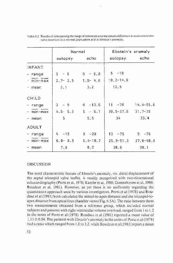
Table 6.2 Results of determining the range of minimum and maximum difference in atrioventricular valve insertion in a normal population and in Ebstein's anomaly.
Normal Ebstein 's anomaly
autopsy echo autopsy echo
INFANT
- range 0 - 6 0 6.8 5 -18 ----
- min-max 2. 7- 3.5 1. 8- 4.6 10.2-14.8
- mean 3. 1 3.2 12. 5
CHILD
- range 2 - 9 0 -13.6 11 -70 14.4-55.6 -- --
- min-max 4.6- 5.3 5 - 6. 1 30.5-37.6 31.7-35
- mean 5 5.5 34 33.4
ADULT
- range 4 -15 0 -20 13 -75 5 -76 - --
- min-max 6. 8- 8.5 6.4-10.7 25.9-51.3 27.6-48.6
- mean 7.6 8.5 38.6 38. 1
DISCUSSION
The most characteristic feature of Ebstein's anomaly, viz. distal displacement of the septal tricuspid valve leaflet, is readily recognized with two-dimensional echocardiography (Ports et al, !978; Kambe eta!, 1980; Gussenhoven eta!, 1980; Roudaut et al, 1981). However, as yet there is no uniformity regarding the quantitative approach used by various investigators. Ports et al (1978) and Roudaut eta! (!98!) both calculated the mitral-to-apex distance and the tricuspid-toapex distance from apical four chamber views (Fig. 6.5A). The ratio between these two measurements obtained from a reference group, which included normal subjects and patients with right ventricular volume overload, ranged from 1 to 1.2 in the series of Ports eta! (1978). Roudaut eta! (1981) reported a mean value of !.II± 0.04. The patients with Ebstein's anomaly in the series of Ports eta! (1978) had a ratio which ranged from 1.8 to 3.2, while Roudaut et al ( 1981) report a mean
52
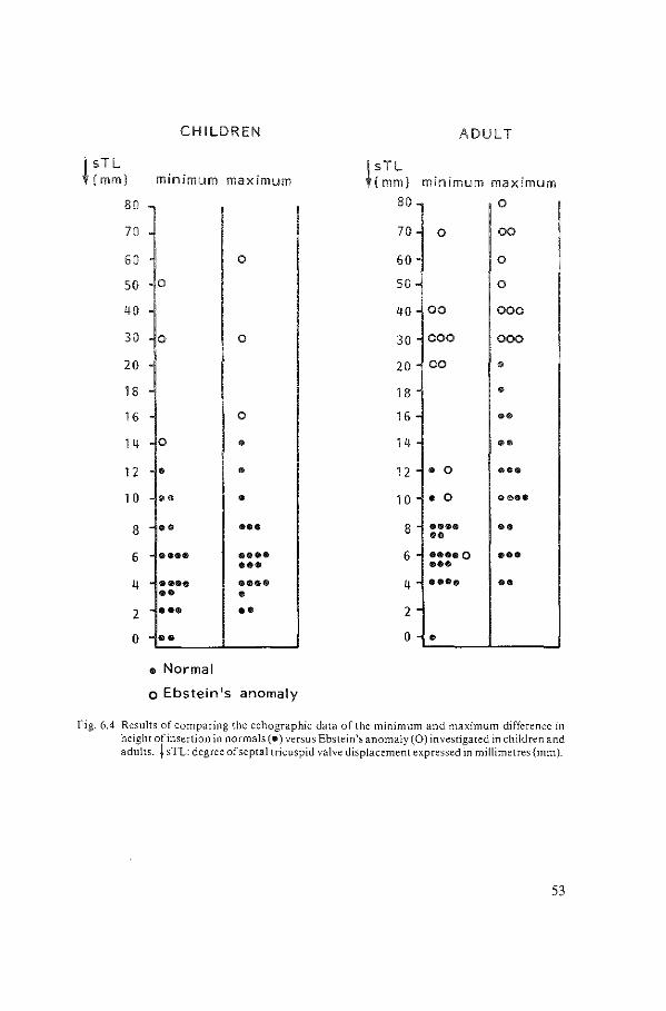
CHILDREN ADULT
l sTL ( mm) minimum maximum
! sTL ( mm) minimum maximum
80 80 0
70 70 0 00
60 0 60 0
50 0 50 0
40 40 00 000
30 0 0 30 000 000
20 20 00 • 18 18 • 16 0 16 •• 14 0 • 14 •• 12 • • 12 • 0 ••• 1 0 •• • 1 0 • 0 ••••
8 •• ••• 8 •••• •• •• 6 •••• •••• 6 ••••o ••• ••• ••• 4 •••• •••• 4 • ••• • • •• 0
2 ••• •• 2
0 •• 0 • • Normal
o Ebstein's anomaly
Fig. 6.4 Results of comparing the echographic data of the minimum and maximum difference in height of insertion in normals (•) versus Ebstein's anomaly (0) investigated in children and adults. + sTL: degree of septal tricuspid valve displacement expressed in millimetres (mm).
53
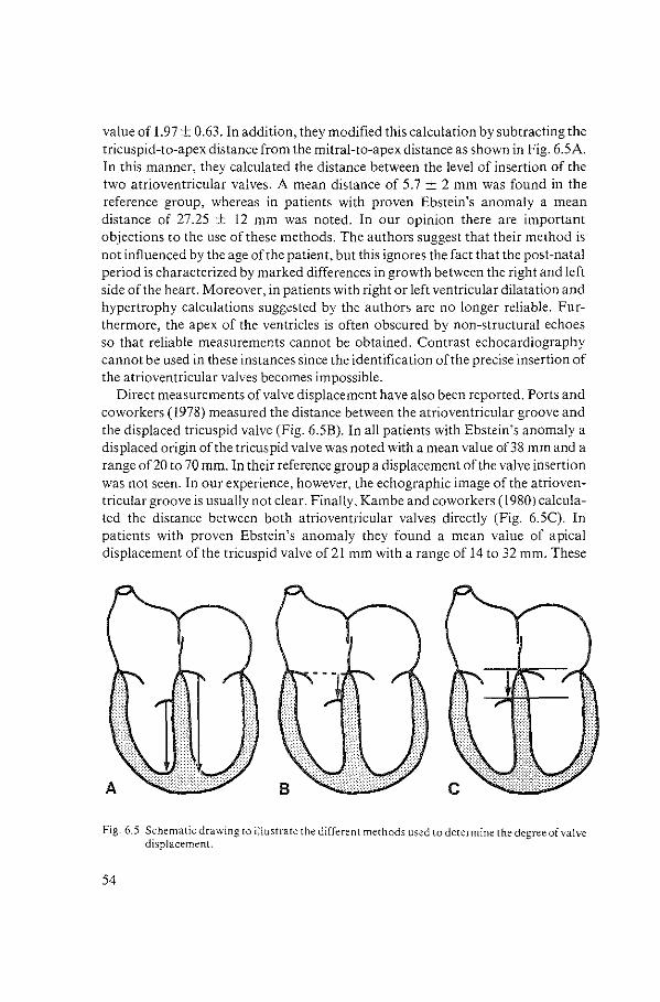
value of 1.97 ± 0.63. In addition, they modified this calculation by subtracting the tricuspid-to-apex distance from the mitral-to-apex distance as shown in Fig. 6.5A. In this manner, they calculated the distance between the level of insertion of the two atrioventricular valves. A mean distance of 5.7 ± 2 mm was found in the reference group, whereas in patients with proven Ebstein's anomaly a mean distance of 27.25 ± 12 mm was noted. In our opinion there are important objections to the use of these methods. The authors suggest that their method is not influenced by the age of the patient, but this ignores the fact that the post-natal period is characterized by marked differences in growth between the right and left side of the heart. Moreover, in patients with right or left ventricular dilatation and hypertrophy calculations suggested by the authors are no longer reliable. Furthermore, the apex of the ventricles is often obscured by non-structural echoes so that reliable measurements cannot be obtained. Contrast echocardiography cannot be used in these instances since the identification of the precise insertion of the atrioventricular valves becomes impossible.
Direct measurements of valve displacement have also been reported. Ports and coworkers (1978) measured the distance between the atrioventricular groove and the displaced tricuspid valve (Fig. 6.5B). In all patients with Ebstein's anomaly a displaced origin of the tricuspid valve was noted with a mean value of38 mm and a range of20 to 70 mm. In their reference group a displacement of the valve insertion was not seen. In our experience, however, the echographic image of the atrioventricular groove is usually not clear. Finally, Kambe and coworkers (1980) calculated the distance between both atrioventricular valves directly (Fig. 6.5C). In patients with proven Ebstein's anomaly they found a mean value of apical displacement of the tricuspid valve of 21 mm with a range of 14 to 32 mm. These
Fig. 6.5 Schematic drawing to illustrate the different methods used to determine the degree of valve displacement.
54
authors give no data regarding the measurements in normal individuals and in patients with right ventricular volume overload other than stating that 'no displacement' occurred. It suggests that they are informed about the precise range of normality, although they do not give any data.
In our opinion the approach used by Kambe et al (1980) is the most reliable method to quantify the degree of atrioventricular valve displacement, since it is based on direct measurement of the distance between the two valves. Moreover, this method is not affected by growth alterations between the right and left ventricle.
The present study, set up along these lines, reveals some features of clinical relevance which as yet have not been reported. The identification of a difference in atrioventricular valve insertion was not feasible in the first trimester of pregnancy. This is due to the physical limitations of ultrasound and to the stage of development of the tricuspid valve. Recent embryologic data on atrioventricular valve development show that the process of valve formation is completed by the 12th week of gestation (Pexieder, 1983). According to Van Oils ( 1984) the formation of the septal tricuspid valve leaflet is not complete until the 16th week of gestation. These aspects are of paramount significance as far as the pre-natal echographic diagnosis of atrioventricular valve abnormalities is concerned. In the second and third trimester, the valves are better delineated and the present study shows that echographic identification of a difference in atrioventricular valve insertion in normals is feasible. Furthermore, this study revealed that the degree of valve displacement varied according to the plane of the cross-section in one and the same individual. Generally lower values were obtained from apical cross-sections, whereas subcostal cross-sections revealed a wider gap. In normal cases minimum values were recorded when the section traversed the crux of the heart, while maximum values were obtained when the cross-section was in a more anterior level immediately beneath the aortic root (see Figs. 6.1 and 6.3A and B). The reverse was noted in patients with Ebstein 's anomaly. Minimum values were noted when an anterior tilt of transsection was applied, while maximum values of valve displacement were obtained when a posterior cross-section was used (see Figs. 6.3C and D). Occasionally the conventional four chamber cross-section will not reveal any septal insertion, since the leaflet attaches to the trabecular part rather than the inlet part of the septum.
The overall average of atrioventricular displacement increased with age: both in normal hearts and in patients with Ebstein's anomaly. In the usual case of Ebstein's anomaly the degree of distal displacement was considerably larger than that in the reference groups. However, in some hearts the minimum values fell within the range of normality as determined anatomically and echographically. A comparison of the minimum and maximum values obtained echographically in normal children and adults with those in patients with Ebstein's anomaly shows an overlap only in the range of minimum values. The maximum values, on the
55

other hand, clearly discriminated between normal valve insertion and that in Ebstein 's anomaly. In the present series a maximum difference in the level of valve insertion of 15 mm or more discriminated between the normal and Ebstein's anomaly in children. In adults this value was found to be above 20 mm.
This study thus shows that the usual echographic cross-section may produce an overlap with regard to the difference in valve insertion in patients with Ebstein's anomaly and normal individuals. Despite the fact that our study reveals that a distinction can be ~ade by calculating the maximum difference, one should be prepared to accept that on occasion a patient with an unequivocal Ebstein's malformation can be encountered in whom the diagnosis cannot be made with certainty solely on the basis of apical displacement of the septal tricuspid valve leaflet.
56
CHAPTER 7
THE ROLE OF ECHOCARDIOGRAPHY IN ASSESSING THE FUNCTIONAL CLASS OF THE PATIENT WITH EBSTEIN'S
ANOMALY
SUMMARY
In 23 patients with Ebstein's anomaly of the tricuspid valve the functional class of the patients has been related to the echocardiographic parameters generally used to diagnose this disorder. These are the extent of apical displacement of the tricuspid valve and the delay in tricuspid valve closure time related to that of the mitral valve. In addition, the functional class of the patients has also been evaluated in relation to the severity of tricuspid valve insufficiency and to the presence or absence of associated cardiac abnormalities.
The study revealed that the echocardiographic parameters i.e. the degree of apical displacement of the tricuspid valve and the delayed closure time of the tricuspid valve have no predictive value for the patient's clinical condition. On the other hand, both tricuspid valve insufficiency and the presence of additional cardiac anomalies have a direct correlation with the well-being of the patient.
INTRODUCTION
The severity of the anomaly in Ebstein's disease of the tricuspid valve has been related to the degree of apical displacement ofthe valve leaflets (Ports eta!, 1978; Roudaut eta!, 1981) and by other workers also to the delay in tricuspid valve closure time (Milner eta!, 1976; Giuliani eta!, 1979). Both features are well assessed by echocardiography. The functional class of the patient is frequently considered to be directly correlated to these features (Ports eta!, 1978; Giuliani et a!, 1978; Roudaut et al, 1981). However, there are as yet no data to support this concept. In fact, one may argue that with a well functioning valve apparatus and in the absence of additional cardiac defects, the degree of apical displacement or the delay in valve closure time should have no major impact on the well-being of the patient. Hence, factors other than the echographically assessed parameters may play a more important role in predicting the condition ofthe patient.
The present study has been undertaken to evaluate these points.
57
MATERIAL AND METHODS
Twenty-three patients with Ebstein's anomaly were studied. In each instance echocardiograms and angiograms were available. In 14 of the 23 patients the diagnosis was based on the data obtained by cardiac catheterization and angiocardiography. In one of these patients the diagnosis was confirmed during open heart surgery. In eight patients the diagnosis was made on echographic criteria, viz. an apical displacement of the septal tricuspid valve leaflet of more than 20 mm compared to the level of insertion of the corresponding septal mitral valve leaflet (Ports et al, 1978; Gussenhoven et al, 1984; see chapter 6). In the remaining patient the diagnosis could not be made on the echocardiograms, or on cardiac catheterization, but was established at cardiac surgery. This patient died following the operation; at autopsy the diagnosis Ebstein's anomaly was confirmed.
M-mode tracings obtained from various transducer positions on the chest wall were used to determine the average time interval between mitral and tri~uspid valve closure (Gussenhoven et al, 1984; see chapter 5). The paper speed used was 50 o_r 100 mm/sec. Two-dimensional four chamber images obtained from subcostal and apical positions were used to determine the degree of valve displacement. The still-frame which showed the maximum difference in valve insertion of both valve leaflets was selected in early systole. In 3 patients, however, the still-frames were selected from end-diastole, since the septal tricuspid valve leaflet was minute and its precise insertion was obscured in early systole by the huge anterior tricuspid valve leaflet. Polaroid pictures were taken of the still-frames. Lines were drawn at right angle to the ventricular septum through the point of attachment of each of the septal valve leaflets. The perpendicular distance between these two lines was measured and expressed in millimetres.
The presence of tricuspid valve insufficiency was determined in all patients on the basis of angiocardiography and graded as mild (1), moderate (2), moderately severe (3) and severe (4) according to the criteria set by Grossman (1980).
Associated cardiac malformations were tabulated on the basis of a full investigation that encompassed cardiac catheterization and angiocardiography. The patients were subdivided into three functional classes according to the criteria set by the New York Heart Association (New York, 1953). In two patients (table 7.1; nos. 21, 22) who underwent corrective surgery, the parameters used in this study were evaluated prior to operation. One patient (no. 1) who underwent pulmonary valvulotomy and closure of a secundum type atrial septal defect and one patient (no. 15) who underwent closure of the secundum type atrial septal defect, were studied following the operation.
58
RESULTS
The time interval between mitral and tricuspid valve closure varied considerably from one patient to the other and ranged from 5 to 130 msec, with a mean value of 75 msec (table 7.1). Apical displacement of the septal tricuspid valve leaflet of more than 20 mm occurred in 21 patients. In one patient (no.l2) the septal tricuspid valve leaflet could not be identified in four chamber cross-sections, most likely due to extreme apical displacement. The valve motion was observed in the right ventricular outflow tract on the conventional left ventricular long axis view. In the second patient (no.21) the echographic diagnosis was a ventricular septal defect with an overriding tricuspid valve and pulmonary valve stenosis. The diagnosis of Ebstein's anomaly was established at cardiac surgery and confirmed at autopsy. The echocardiograms in retrospect showed a distance between both atrioventricular valves of only IS mm (Gussenhoven eta!, 1984).
The degree of tricuspid insufficiency was graded as mild in 13 patients, moderate in 4 patients, moderately severe in 4 patients and severe in 2 patients. The associated cardiac lesions were secundum type atrial septal defects in 13 patients, a ventricular septal defect of the perimembranous type opening into the distal part of the right ventricle in two patients, pulmonary valve stenosis in two patients, mitral valve insufficiency in one patient and the type B Wolff-Parkinson-White syndrome in two patients (table 7.1).
The 23 patients were classified as class I in 10 patients, class II in 10 and class III in 3 patients.
The correlations are shown in tables 7.1 and 7 .2. The relationships between the degree of tricuspid insufficiency and the given parameters are summarized in table 7.3.
DISCUSSION
In contrast to the many echocardiographic studies concerned with the diagnostic aspects of Ebstein's anomaly of the tricuspid valve, little is known about the correlation between these features and the condition of the patients. Ebstein's anomaly has been subcategorized into 'simple' and 'complicated' by Lev et al ( 1970) and into 'mild', 'moderate' and 'severe' by Zuberbuhler et al (1979), but always on the basis of the anatomy rather than on cardiac function.
Lundstrom (1973) and Farooki eta! (1976) mention that they were unable to find a relationship between the functional classification of the patient and the M-mode features of Ebstein's disease. Milner et al (1976) reported two patients with a clinical mild form of the disease both of whom had a delay in the tricuspid valve closure time of 55 msec. In their opinion the short delay in valve closure, viz. below the 'diagnostic' value of 65 msec, could explain the mild clinical course in
59
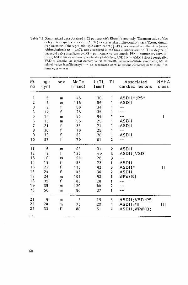
Table 7 .I Summarized data obtained in 23 patients with Ebstein's anomaly. The mean value of the delay in tricuspid valve closure (MeTe) is expressed in milliseconds (msec). The maximum displacement of the septal tricuspid valve leaflet ( ~ sTL) is expressed in millimetres (mm). Abbreviations: nv = t sTL not visualized in the four chamber section; TI =degree of tricuspid valve insufficiency; PS =pulmonary valve stenosis; PS* =pulmonary valvulo-to my; ASD II= secundum type atrial septal defect; ASD II*= ASD II closed surgically; VSD = ventricular septal defect; WPW = Wolff-Parkinson-White syndrome; MI = mitral valve insufficiency:--= no associated cardiac lesions detected; m = male: f = female; yr =years.
Pt age sex MeTe .fsTL Tl Associated NYHA no ( yr) ( msec) (mm) cardiac lesions class
6 m 45 30 ASDII*;PS* 2 6 m 11 5 56 1 ASDII 3 9 f 80 34 1 4 1 4 f 25 35 1 5 1 4 m 65 44 1 6 1 9 m 55 29 1 ASDII 7 21 f 35 71 1 ASDII 8 30 f 70 29 1 9 33 f 80 76 1 ASDII
1 0 57 f 70 61 2
11 6 m 65 31 2 ASDII 12 9 f 130 nv 3 ASDII ;VSD 1 3 1 0 m 90 28 3 14 1 9 f 85 73 1 ASDII 15 22 f 11 0 42 3 ASDII* II 16 24 f 45 36 2 ASDII 1 7 24 m 1 OS 42 1 WPW(B) 1 8 35 f 105 20 1 1 9 35 m 120 44 2 20 50 m 80 37
21 4 m 5 1 5 3 ASDII;VSD;PS 22 24 m 75 29 4 ASDII ;MI Ill 23 33 f 80 51 4 ASDII;WPW(B)
60
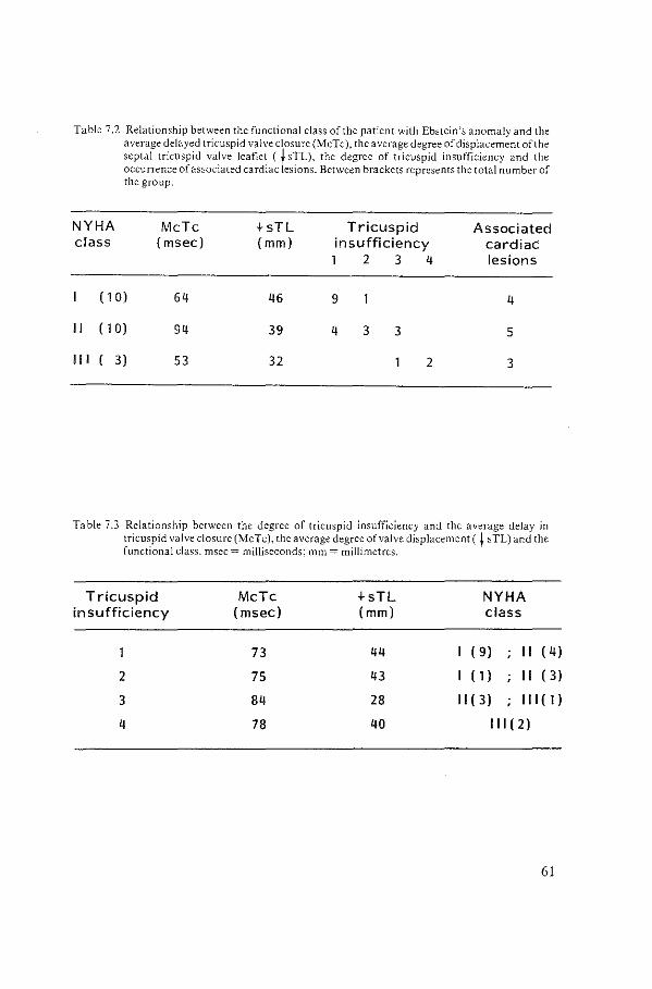
Table 7.2 Relationship between the functional class of the patient with Ebstein's anomaly and the average delayed tricuspid valve closure (MeTe), the average degree of displacement of the septal tricuspid valve leaflet ( f sTL), the degree of tricuspid insufficiency and the occurrence of associated cardiac lesions. Between brackets represents the total number of the group.
NYHA MeTe +sTL Tricuspid Associated class ( msec) (mm) insufficiency cardiac
1 2 3 4 lesions
( 1 0) 64 46 9 4
II ( 1 0) 94 39 4 3 3 5
Ill ( 3) 53 32 2 3
Table 7.3 Relationship between the degree or tricuspid insufficiency and the average delay in tricuspid valve closure (MeTe), the average degree of valve displacement ( f sTL) and the functional class. msec = milliseconds; mm = millimetres.
Tricuspid in sufficiency
2
3
4
MeTe ( msec)
73
75
84
78
+sTL (mm)
44
43
28
40
NYHA class
( 9) II ( 4)
I (1) II ( 3)
II ( 3) Ill ( 1)
Ill ( 2)
61
these patients. Giuliani and coworkers (1979) suggested that patients with Ebstein's anomaly with minor defects had no or little delay in the tricuspid versus mitral valve closure, while those with a pronounced delay in tricuspid valve closure were likely to have the severe form. The authors, however, do not define the terms 'little' and 'pronounced', nor the terms 'minor' and 'severe'. In fact, apart from the aforementioned statements, they do not further elaborate on the clinical course of these patients.
Ports et al (1978) and Roudaut et al (1981) suggest that the clinical condition of the patient is determined by several factors. The most important of these are the degree of tricuspid valve displacement, tricuspid valve regurgitation as well as the presence of associated cardiac defects. In the latter category atrial septal defects with a right-to-left shunt were considered of particular significance. However, the authors have not correlated the different parameters with the clinical well-being of the patients.
Recently, however, Shiina and coworkers (1983) have shown that the well-being ofthe patient is intimately related to the presence or absence of associated cardiac lesions. Patients who fit in functional class I or II showed in 46% an associated cardiac defect, whereas 96% of the patients belonging to class III or IV showed an associated cardiac abnormality. In the majority the defect was a secundum type atrial septal defect.
The present study shows that no relationship exists between the functional class of the patients and the echographic parameters useful to diagnose Ebstein's anomaly, such as the delay in tricuspid valve closure time and the degree of apical displacement of the septal tricuspid valve leaflet. The differences in the mean values of the time interval measured between mitral and tricuspid valve closure time, found in the classes I and II, were in this study not statistically significant. In fact, the comparison of the degree of apical displacement with the functional class of the patients revealed that the most disabled patients had only mild displacement of the tricuspid valve, while patients in class I had severe tricuspid valve displacement (see table 7 .2). A satisfactory explanation for this observation is as yet lacking.
Tricuspid valve insufficiency was the most reliable feature predicting the functional class of the patient and it is of interest that this feature did not correlate with the degree of delay in the tricuspid valve closure time and with the degree of valve displacement. Associated cardiac anomalies, moreover, also played a key role in the well-being of the patients. Atrial septal defects, defined as anatomic defects, allowing a right-to-left shunt, were indeed the most inspected lesions. For all these reasons we suggest that the use of terms such as 'mild', 'moderate' or 'severe' in patients with Ebstein's anomaly, are restricted to indicate the functional class of the patient.
62
SUMMARY
In this thesis the value of echocardiography is evaluated for the diagnosis of Ebstein's anomaly of the tricuspid valve. This congenital heart defect, first described in 1866 by Wilhelm Ebstein, is characterized by an apical displacement of the septal and inferior tricuspid valve leaflets and by dysplasia of the tricuspid valve apparatus.
Since the publication of Ebstein, this congenital heart defect had been recognized and documented several times but only on the basis of postmortem studies. T ourniaire and coworkers ( 1949) were the first to diagnose Ebstein 's anomaly in the living patient. Ultrasound techniques, introduced in the early seventies, appear to be a major step forward in establishing the diagnosis non-invasively.
This thesis describes the value of both M-mode and two-dimensional echocardiography for the diagnosis of Ebstein's anomaly. The study is restricted to patients and heart specimens with situs solitus of the atria, concordant atrioventricular and ventriculo-arterial connections. Knowledge regarding the anatomy of Ebstein's anomaly is essential in order to fully appreciate and understand the echographic information. During the course of this study we investigated this congenital heart disease in 23 selected patients and in 21 heart specimens. In the first part of this thesis (chapter 2) an extensive review is presented of the literature relevant for this study. The anatomic spectrum of the anomaly is described. The degree of apical displacement of the leaflets varies from little to pronounced and the degree of valye dysplasia may present a similar spectrum. Ebstein 's anomaly may be associated with other congenital heart defects, such as atrial septal defect, ventricular septal defect, pulmonary valve stenosis and Wolff-Parkinson-White syndrome.
A review of the echocardiographic literature reveals that a prolonged time interval between mitral and tricuspid valve closure is the most important M-mode criterion. Two-dimensional echocardiography has introduced the possibility of demonstrating the displacement of the septal tricuspid valve leaflet towards the right ventricular apex.
The basic aspects of the anomaly, both echo graphically and anatomically, are described in chapter 3. Ebstein's anomaly is characterized by distal displacement of the origin of the valve leaflets and by dysplasia of the valve apparatus. Distal displacement may affect the septal and inferior leaflets, but the anterior leaflet always originates from the annulus fibrosus. The variability in degree and extent of distal displacement will create a spectrum of anomalous valve ring positions. Dysplasia of the valve apparatus usually affects all leaflets. The anterior valve leaflet is often sail-like. In some instances it may be free floating, while in others the leaflet is plastered to the right ventricular free walL From the point of view of echographic identification of Ebstein's anomaly, distal displacement of the septal
63
origin of the valve seems by far the best criterion, since dysplasia in itself is not distinctive.
An evaluation of the echographic findings in Ebstein's anomaly seen with M-mode and two-dimensional echocardiography is presented in chapter 4. Twodimensional echocardiography is the method per excellence to demonstrate the anatomic most prominent feature of the disease, i.e. the apical displacement of the valve. The prolonged time interval between mitral and tricuspid valve closure, measured on an M-mode echocardiogram, appears to be a criterion suggestive rather than diagnostic for Ebstein's anomaly. The value of this M-mode parameter is further elaborated in chapter 5 with emphasis on its variability measured within a single patient. We have assumed that the mean value of a prolonged time interval, obtained from different transducer positions and from simultaneously recorded mitral and tricuspid valve closure, is representative for that particular patient rather than for the diagnosis.
Chapter 6 provides an echocardiographic and anatomic analysis of the degree of apical displacement of the septal tricuspid valve leaflet in normal hearts and in hearts with Ebstein's anomaly. The difference in the insertion site between two atrioventricular valves increased with age, both echographically and morphologically. When the minimum distances in insertion site were measured an overlap was found between normal hearts and hearts with Ebstein's anomaly. The maximum values, on the other hand, clearly discriminated between normal valve insertion and that in Ebstein's anomaly. In the present study a maximum difference in the level of valve insertion of 15 mm or more discriminated between normal and Ebstein's anomaly in children. In adults this value was found to be above 20 mm.
Finally, chapter 7 is devoted to the value of echocardiography in assessing the functional class of the patient with this disease. It appears that echocardiography has no predictive value as far as the patient's functional class is concerned since no relationship was found between the patient's condition and the prolonged time interval between mitral and tricuspid valve closure and the degree of apical displacement of the tricuspid valve leaflet. Therefore we suggest that the use of terms such as mild, moderate or severe is restricted to indicate the functional class of the patient. Altogether this study bas indicated that in particular twodimensional echocardiography is an important asset to the diagnosis of Ebstein's anomaly.
64
SAMENVATTING
In dit proefschrift wordt de betekenis van echocardiografie beschreven voor de diagnostiek van de ziekte van Ebstein van de tricuspidalisklep. Deze aangeboren hartafwijking, voor bet eerst beschreven door Wilhelm Ebstein in 1866, word! gekenmerkt (a) door een verlaagde insertie van de septale en inferiore tricuspidalisklepslippen en (b) door klepdysplasie. Na de publikatie van Ebstein werd deze afwijking nog meerdere mal en herkend en beschreven, maar de herkenning van dit ziektebeeld op grand van klinische criteria gebeurde pas voor het eerst in 1949 door Tourniaire en medewerkers. De ontwikkeling van ultrageluidsapparatuur in begin zeventiger jaren maakte het mogelijk de diagnose ook non-invasiefte stellen.
In deze studie wordt de waarde van zowel M-mode als twee-dimensionale echocardiografie beschreven voor bet stellen van de diagnose Ebstein's anomalie. We hebben ons beperkt tot patienten en hartpreparaten met een situs solitus van de atria en concordante atrioventriculaire en ventriculo-arterieele connecties. Kennis omtrent de anatomic van Ebstein's anomalie is onontbeerlijk om de gegevens met echografie te kunnen begrijpen en naar waarde te beoordelen. In de afgelopen jaren hebben we de gelegenheid gehad deze aangeboren hartafwijking bij 23 patienten en 21 hartpreparaten te bestuderen. Het eerste gedeelte van dit proefschrift (hoofdstuk 2) gaat uitgebreid in op de literatuurgegevens relevant voor dit onderzoek. Beschreven wordt bet spectrum waarmee de afwijking zich anatomisch manifesteert. De mate van verlaagde klepinsertie varieert van gering tot uitgesproken, terwijl ook de mate van klepdysplasie een dergelijke variatie vertoont. De afwijking kan vergezeld gaan van andere hartafwijkingen zoals een atrium septum defect, een ventrikel septum defect, een pulmonaalklep stenose en bet syndroom van Wolff-Parkinson-White. Uit bet overzicht van de echografische iiteratuur blijkt dat een verlate sluiting van de tricuspidalisklep vergeleken met bet moment van sluiten van de mitralisklep een belangrijk criterium is voor bet stellen van de diagnose met behulp van de M-mode techniek.
De twee-dimensionale echocardiografie daarentegen is in staat bet belangrijkste anatomische criterium in beeld te brengen, te weten de verlaagde klepinsertie van de tricuspidalisklep.
De morfologische bijzonderheden van deze hartafwijking, van belang voor zowel de pathologic als de echocardiografie worden in hoofdstuk 3 beschreven. De afwijking van Ebstein wordt gekenmerkt door enerzijds een verlaagde klep insertie en anderzijds door klepdysplasie. De verlaagde klepinsertie zit met name in bet septale deel, waarbij de verlaging zo ver apicaalwaarts kan zijn, dat de klepring zelfin een geheel ander vlak komt te staan. Dit heeft echocardiografische betekenis. De klepdysplasie wordt in aile klepdelen aanwezig gevonden, maar is vaak bet meest uitgesproken in de vaak nog normaal ge'insereerde voorste klep, waardoor een soliede gordijn-achtige klep kan worden gevormd welke evenzeer
65
van echografische betekenis is, doordat deze merkwaardige uitslagen kunnen worden waargenomen.
In hoofdstuk 4 wordt de diagnostische waarde van de M-mode en de tweedimensionale echografie afgewogen. Het anatomische kenmerk van verlaagde klepinsertie blijkt bij uitstek geschikt te zijn om door middel van tweedimensionale echografie te worden gedetecteerd. Een verlate sluiting van de tricuspidalisklep ten opzichte van de mitralisklep sluiting blijkt een minder betrouwbaar criterium te zijn.
In hoofdstuk 5 wordt ingegaan op de sensitiviteit van dit sluitingsinterval en wordt gezocht naar de oorzaak van de variabiliteit van dit tijdsinterval gemeten bij de patient. Het blijkt dat een gemiddeld tijdsinterval, gemeten van verschillende transducer posities op de borstwand en van simultaan geregistreerde mitralis- en tricuspidalisklep sluiting, beschouwd moet worden als een 'persoonlijk' gegeven van de onderzochte patient en niet zozeer als diagnostisch criterium.
In hoofdstuk 6 wordt een twee-dimensionale en anatomische studie beschreven die de grenzen vastlegt in insertie verschil tussen de septale tricuspidalis en septale mitralisklep zowel in normale harten als in harten met de anomalie van Ebstein. Het blijkt dat het insertie verschil tussen beide atrioventriculaire kleppen gezien kan worden in het foetale hart in het 2e en 3e trimester van de zwangerschap. De gemiddelde afstand tussen beide kleppen neemt toe met de leeftijd zowel echografisch als morfologisch. Een overlap in de gemeten insertie afstand werd gevonden tussen normale harten en hart en met een Ebstein 's anomalie met name wanneer de gemeten minimum waarden van de patienten werden vergeleken. De maximum waarden zijn echter waardevol om tussen beide groepen te differentitren. Bij kinderen lag de grens van verlaagde insertie bij 15 mm; bij volwassenen werd de grens bij 20 mm gevonden.
Tenslotte geeft hoofdstuk 7 de relatie weertussen de echografische gegevens van een patient en zijn validiteit. Het blijkt dat echocardiografie voor het bepalen van de validiteit geen betekenis heeft, gezien het feit dat er geen direct verband gevonden werd tussen de validiteit van de patient en het veri ate tijdsinterval van de tricuspidalisklep sluiting in vergelijking met de mitralisklep sluiting en de mate van verlaagde klepinserti.e. Indien derhalve de termen licht, matig of ernstig gebruikt worden met betrekking tot de ziekte van Ebstein, kunnen deze enkel van toepassing zijn wanneer ze de validiteit van de patient weergeven.
Dit onderzoek heeft aangetoond dat met name twee-dimensionale echocardiografie een belangrijke aanwinst is voor de diagnostiek van de ziekte van Ebstein.
66
REFERENCES
Allwork S P, Ben tal! H H, Becker A E, Cameron H, Gerlis L M, Wilkinson J Land Anderson R H 1976 Congenitally Corrected Transposition oft he Great Arteries: Morphologic study of 32 cases. American Journal of Cardiology 38:910-922
Anderson K R. Danielson G K, McGoon DC and Lie J T 1978 Ebstein's anomaly of the left sided tricuspid valve. Pathological anatomy of the valvular malformation. Circulation 58.3: 87-91
Anderson K Rand Lie J T 1978 Pathologic anatomy of Ebstein's anomaly of the heart revisited. American Journal of Cardiology 41: 739-745
Anderson K R, Zuberbuhler J R, Anderson R H, Becker A E and Lie J T 1979 Morphologic spectrum of Ebstein's anomaly of the heart: a review. Mayo Clinic Proceedings: 54:174-180
Anderson R H, Wilkinson J L, Gerlis L M, Smith A and Becker A E 1977 Atresia of the right atrioventricular orifice. British Heart Journal39:414-428
Anderson R Hand Becker A E 1980 Cardiac Anatomy- An integrated text and colour atlas. Gower Medical Publishing. Churchill Livingstone, Edinburgh
Bailey C P 1955 Surgery of the heart. Lea and Febiger. Philadelphia: 413 Becker A E, Becker M J and Edwards J E 1971 Pathologic spectrum of dysplasia of the tricuspid
valve. Features in common with Ebstein's malformation. Archives of Pathology 91: 167-178
Becker A E 1972 Tricuspidalis klepdysplasie en de relatie met de klepmisvorming van Ebstein. Nederlands Tijdschrift voor Geneeskunde 116.10: 417-418
Becker A E and Gussenhoven W J 1981 Segmental analysis of congenital heart disease. A correlation between pathology and echocardiography. In: Rijsterborgh H (ed.): Echocardiology, 307-321. Martin us NijhoffPublishers, The Hague.
Brooks N, Leech G and Leatham A 1979 Factors responsible for normal splitting of the first heart sound. High-Speed echophonocardiographic study of valve movement. British Heart Journal42: 695-702
Cabin H Sand Roberts W C 1981 Ebstein's anomaly of the tricuspid valve and prolapse of the tricuspid valve. American Heart Journall01.2: 177-180
Caruso G, Losekoot T G and Becker A E 1978 Ebstein's anomaly in persistent common atrioventricular canal. British Heart Journal40, 11: 1275-1279
Castaneda-Zuniga W, Nath H P, Moller J H, Edwards J E 1982 Left-sided anomalies in Ebstein's malformation of the tricuspid valve. Pediatric Cardiology 3: 181-185
Crews T L, Pridie R B, Benham Rand Leatham A 1972 Auscultatory and phonocardiographic findings in Ebstein 's anomaly (correlation of first heart sounds with ultrasonic records of tricuspid valve movements). British Heart Journal 34: 681-687
Daniel W, Rathsack P, WalpurgerG, Kahle A, Gisbertz R, SchmitzJ and Lichtlen PR 1980 Value of M-mode echocardiography for non-invasive diagnosis of Ebstein's anomaly. British Heart Journal43: 38-44
Desimone A Rand Kronzon I 198llllustrative Echocardiogram- Giant right atrium in Ebsteln's anomaly. Chest 79.1:80-82
Diamond M A, Dillon J C, Haine C L, ChangS, Feigenbaum H 1971 Echocardiographicfeatures of atrial septal defect. Circulation 43: 129-135
Donegan C C jr, Moore M M, Wiley T M, Hernandez FA, Green J Rand Schieb\er Q L 1968 Familial Ebstein's anomaly of the tricuspid valve. American Heart Journal 75: 375-379
Drobinski G, Bejean-Lebuisson A, Evans J I, Fournial J F, KinG and GrosgogeatY 1979 Formes atypiques de Ia maladie d'Ebstein: apports et limites de J'tchographie et du cathhisme cardiaque. Annales de Medecine Interne 130.10:459-465
Ebaid M, Vila J H A, Pedroso C 0, Stolf NAG, Verginell G, Macruz R 1980 Anomalia Tipo Ebstein associada a comunicayao interventricular- relato de 4 casos. Arquivos Brasileiros de Cardiologia 34.2:129-133
Ebstein W 1866 Ueber einen sehr seltenen Fall von Insufficienz der Valvula tricuspidalis, bedingt durch eine angeborene hochgradige Missbildung derselben. Archiv fUr Anatomy und Physiology 33: 238-254
67
Ebstein E 1906 Wilhelm Ebstein's Arbeiten aus den Jahren 1859-1906 1907 Deutsche Archiv fi.ir Klinische Medizin 89: 367-378
Edwards J E 1954 Differential diagnosis of mitral stenosis - A clinicopathologic review of simulating conditions. Laboratory Investigation 3: 89-115
Elliott L P, Adams Jr P and Edwards J E 1963 Pulmonary atresia with intact ventricular septum. British Heart Journal25: 489-50 I
Evemy K and HunterS A 1981 2D-echo position of septal tricuspid leaflet in Ebstein's anomaly and normal hearts. Letter to the Editor. American Heart Journal101: 358-359
Eijgelaar A, Homan van der Heide J N, Viersma J W, Van de WallE, Kok H G H, Kruizinga Ken Blickman J R 1975 Operatieve behandeling van de ziekte van Ebstein (resu\taten vai1 de correctie vo!gens Hardy bij een patient). Nederlands Tijdschrift voor Geneeskunde 119.18o 727-728
Eijgelaar A, Homan van der Heide J N, Viersma J W, Van der Wal E en Kruizinga K 1977 Operatieve behandeling van ziekte van Ebstein? Nederlands Tijdschrift voor Geneeskunde 121.9: 352-360
Farooki Z Q, Henry J G, GreenE W 1976 Echocardiographic spectrum ofEbstein 's anomaly of the tricuspid valve. Circulation 53.1: 63-68
Ferencz C and Dammann J F jr 1957 Significance of the pulmonary vascular bed in congenital heart disease. Lesions of the left side of the heart causing obstruction of the pulmonary venous return. Circulation XVI: 1046-1056
Fontana R Sand Edwards J E 1962 Congenital Cardiac Disease: A review of 357 cases studied pathologically. Philadelphia, W B Saunders. 29
French J W, Baum D and Popp R L 1975 Echocardiographic findings in Uhl's anomaly. Demonstration of diastolic pulmonary valve opening. American Journal of Cardiology 36: 349-353
From A H L, Mazzitello W F, Judd AS, Edwards] E 1973 Ebstein's malformation of the tricuspid valve associated with valvular stenosis and cor triatriatum. Chest 64: 248-251
Gent onE and BlountS G 1967 The spectrum ofEbstein's anomaly. American Heart Journal 73.3: 395-425
Gerlis M and Anderson R H 1976 Cor triatriatum dexter with imperforate Ebstein's anomaly. British Heart Journal 38: 108-111
Gils FA W van 1984 (in press) The development of the atrioventricular valves and the fibrous skeleton in the human heart. Role of extra cardiac tissue in cardiac development. Thesis. Leiden, The Netherlands
Giuliani E R, Foster V, Brandenburg R 0, Mair D D 1979 Ebstein's anomaly- The clinical features and natural history of Ebstein's anomaly of the tricuspid valve. Mayo Clinic Proceedings 54' 163-173
Grossman Wed. 1980 Cardiac Catheterization and Angiography. Lea & Febiger, Philadelphia. 323. Gueron M, Hirsch M, Stern J, Cohen W and Levy M J 1966 Familial Ebstein's anomaly with
emphasis on the surgical treatment. American Journal of Cardiology 18: 105-111 Gussenhoven W J, Spitaels SEC, Born Nand Becker A E 1980 Echocardiographic criteria for
Ebstein's anomaly of the tricuspid valve. British Heart Journal43: 31-37 Gussenhoven W J, Spitae!s SEC, Born Nand Becker A E 1980 Echocardiographic criteria for
Ebstein's anomaly of the tricuspid valve. Letter to the Editor. British Heart Journal 44: 231
Gussenhoven W J and Becker A E 1983 Congenital heart disease: morphologic echocardiographic correlations. Churchill Livingstone, Edinburgh
Gussenhoven W J, Essed C E, BasE and de Villeneuve V H 1984 (in press) Echocardiographic diagnosis of overriding tricuspid valve in a child with Ebstein's anomaly. Pediatric Cardiology
Gussenhoven W J, Jansen J R C, Born Nand Ligtvoet C M 1984(in press) Variability of the time interval between tricuspid and mitral valve closure in Ebstein's anomaly- An echographic study. Journal of Clinical Ultrasound.
Gussenhoven W J, Stewart P A, de Villeneuve V H, Essed C E, Ligtvoet C M and Becker A E (in press) The septal tricuspid and mitral valve relationship in normal hearts and in hearts with Ebstein's anomaly. American Journal of Cardiology
68
Hagan A, Sahn D J and Friedman W F 1974 Cross-sectional echocardiographic features of Ebstein's malformation. Circulation 49/50. III
HaglerD J, Tajik AJ. SewardJ B, Edwards W D, Mair D D and Ritter D G 1981 Atrioventricular and Ventriculoarterial Discordance (Corrected Transposition of the Great Arteries). Mayo Clinic Proceedings 56:591-600
Handler J B, Berger T J, Miller R H, Hagan AD, Peniston R Land Vieweg WVR 1981 Partial atrioventricular canal in association with Ebstein' anomaly -Echocardiographic diagnosis and surgical correction. Chest 80.4: 515-517
Henry J G, Gordon Sand Timmis G C 1979 Corrected transposition of great vessels and Ebstein's anomaly of tricuspid valve - Echocardiographic findings. British Heart Journal 41: 249-252
Hirschklau M J, Sahn D J, Hagan A, Williams DE and Friedman W F 1977 Cross-sectional echocardiographic features ofEbstein's anomaly of the tricuspid valve. American Journal of Cardiology 40: 400-404
Ito S, Kudo T, Kainuma Y, Morikawa T, Mori K, An do M, Mimori S, Hashimoto A, Takao A. and Konno S 1977 Tetralogy of Fallot associated with serious anomalies. Journal Japanese Association Thoracic Surgery 25: 61-(271)-67-(277)
Kambe T, Ichimiya S, Toguchi M, Hibi N, Fukui Y, Nishimura K, Sakamoto Nand Hojo Y 1980 Apex and subxiphoid approaches to Ebstein 's anomaly using cross-sectional echocardiography. American Heart JoumallOO. 1: 53-57
Keith J D, RoweR D and Vlad P (eds) 1978 Heart Disease in Infancy and Childhood (3rd ed) p.847-855
Kernen J A 1958 Corrected transposition of the great vessels of the heart. American Journal of Cardiology 56: 583-590
Koiwaya Y, Narabayashi H, Koyanagi S, Matsugushi H, Tanaki S, ImaizumiT, Kuroiwa A, Nakamura M and Hirata T 1979 Early closure of the tricuspid valve in a case ofEbstein's anomaly with type B Wolff-Parkinson-White syndrome. Circulation 60. 2: 446-450
Kotler M Nand Tabatznik B 1971 Recognition of Ebstein's anomaly by ultrasound technique. Circulation 43/44. I
Kotler M N 1974 Tricuspid valve in Ebstein's anomaly. Circulation 49: 194 Kumar A E. Fyler DC, Miettinen 0 Sand Nadas AS 1971 Ebstein's anomaly -Clinical profile and
natural history. American Journal of Cardiology 28: 84-95 Lablanc he J M, Rey C and Dupuis C 1978 L'echocardiographie dans Ia maladie d'Ebstein. Archives
Maladies du Coeur: 546-552 Leatham A 1954 Splitting of the first and second heart sounds. Lancet 25: 607-613 Lev M, Liberthson R R,Joseph R H, Seten C E, Kunske R D, EcknerF A 0 and Miller R A 1970 The
pathologic anatomy of Ebstein's disease. Archives of Pathology 90: 334-343 Ligtvoet C M, Ridder J, Lancee C T 1977 A dynamically focussed multiscan system. In: Born N, ed.
Echocardiology with Doppler applications and real time imaging. Martinus Nijhoff Publishers, The Hague: 313-323
Lo K S, Loventhal P and Walton J Ajr 1979 Familial Ebstein 's anomaly. Cardiology 64.4: 246-255 Losekoot G 1967 Gecorrigeerde transposities, Thesis, Amsterdam, The Netherlands Losekoot T G, Anderson R H, Becker A E, Danielson G K and Soto B 1983 Congenital corrected
transposition, Churchill Livingstone, Edinburgh LundstrOm N R 1969 Reflected ultrasound in the diagnosis of congenital heart disease. In: Ultra
sound. Proc. of the 1st World Congress on Ultrasonic Diagnostics in Medicine Wien, Verlag der Wiener Medizinischen Akademie vol. 3
LundstrOm N Rand Edler I 1971 Ultrasound cardiography in infants and children. Acta Paediatrica Scandinavica 60: 117-128
LundstrOm N R 1973 Echocardiography in the diagnosis of Ebstein's anomaly of the tricuspid valve. Circulation 47: 597-605
LundstrOm N R 1978 The tricuspid valve. In: LundstrOm N R, ed. Echocardiography in congenital heart disease. Elsevier/North Holland, Amsterdam: 117-122
MacCallum W G 1900 Congenital malformations of the heart as i!lustrated by the specimens in the pathological museum of the John Hopkins Hospital. John Hopkins Hospital Bulletin ll: 69-71
69
Macartney F J 1983 Surgery without cardiac catheterization in children. In: Abstracts of the 5th Symposium on Echocardiology Rotterdam, Special issue of Ultrasonoor Bulletin: 45
Mann R J and Lie J T 1979 The life story of Wilhelm Ebstein ( 1836-! 9 !2) and his almost overlooked description of a congenital heart disease. Mayo Clinic Proceedings 54: 197-204
Marce!etti C, Di.iren DR, Schui!enburg R M and Becker A E 1980 Fontan's operation for Ebstein's anomaly. Journal of Thoracic Cardiovascular Surgery 79: 63-66
Matsumoto M, Matsuo H, Nagata S, Hamanaka Y, Fujita T, Kawashima Y. Nimura Y and Abe H 1976 Visualization of Ebstein's anomaly of the tricuspid valve by two- dimensional and standard echocardiography. Circulation 53.1: 69-79
Milner S, Meyer R A. Venables A W, Korfhagen J and Kaplan S 1976 Mitral and tricuspid valve closure in congenital heart disease. Circulation 53.3: 513-518
Monibi A A, Neches W H, Lenox C C. ParkS C, Mathews Rand Zuberbuhler J R 1978 Left ventricular anomalies associated with Ebstein's malformation of the tricuspid valve. Circulation 57.2:303-306
New York Heart Association: criteria committee. Nomenclature for diagnosis of the heart and blood vessels, ed. 5, New York, 1953.
Otero-Coto E, Perez-Martinez V, Palacios V and Caffarena J M 1980 Ebstein's anomaly of the left-sided atrioventricular (A V) valve in A V discordance. Journal ofThoracic Cardiovascular Surgery 28: 364-367
Pechstein J 1957 Beitrag zur Ebsteinschen Anomalie der Valvula tricuspidalis. Archives for Kreislaufforschung 26: 282-337
Pexieder T 1983 Personal communications Pocock W A, Tucker R B K and Barlow J B 1969 Mild Ebstein's anomaly. British HeartJournal 31:
327-336 Ports T A. Silverman N Hand Schiller N B 1978 Two-dimensional echocardiographic assessment of
Ebstein's anomaly. Circulation 58. 2: 336-343 Rauchfuss 1878 Gerhards Handbuch der Kinderkrankheiten 4A: 137 Reinhold J, Rudke U 1957 Relation of the first and second heart sounds to events in the cardiac
cycle. British HeartJournal19: 473-480 Roelandt J R T C 1977 Practical Echocardiology. Research Studies Press, Forest Grove, Oregon Rook L and Hoedemaker Ph J 1965 Ziekte van Ebstein met pulmonalisstenose. Nederlands
Tijdschrift voor Geneeskunde l09.I.ll: 537-538 Roudaut R, Dudeffant P, Bi!ies M A, Choussat A and Dal!occhio M i98l Etude Ccho
cardiographique bidimensionelle de Ia maladie d'Ebstein. Archives Maladies du Coeur 74.10:1177-1184
Ruschhaupt D G, Bharati Sand Lev M 1976 Mitral valve malformation of Ebstein type in absence of corrected transposition. American Journal of Cardiology 38: 109-112
SchieblerG L, Gravenstein J Sand Van Mierop L H S 1968 Ebstein's anomaly of the tricuspid valve -Translation of original description with comments. The American Journal of Cardiology 22: 867-873
Sekelj P and Benfey B G 1974 Historical landmarks: Ebstein's anomaly of the tricuspid valve. American Heart Journal88.1: 108-114
Seward J B, Tajik A J, Feist D J and Smith H C 1979 Ebstein's anomaly in an 85- year-old man. Mayo Clinic Proceedings 54:193-196
Shiina A, Seward J B, Hagler D J. Tajik A J and Danielson G K Ebstein's anomaly: 2-dimensional echo-surgical correlation: echo predictors of tricuspid valve plication vs. replacement? Abstract. Circulation 64.IV, 237
Shiina A, Seward J B, Tajik A J, Hagler D J. and Danielson G K 1983 Two-dimensional echographic-surgical correlation in Ebstein's anomaly: preoperative determination of patients requiring tricuspid valve plication vs replacement. Circulation 68.3:534-544
Shub C, Dimopoulos IN, Seward J B, Cal!ahan J A, Tancredi R G, Schattenberg TT, Reeder G S, Hagler 0 J, Tajik A J 1983 Sensitivity of two-dimensional echocardiography in the direct visualization of atrial septal defect utilizing the subcostal approach: experience with 154 patients. Journal American College of Cardiology 2.1:127-135
Silverman N Hand Schiller N B 1978 Apex echocardiography: a two-dimensional technique for evaluation of congenital heart disease. Circulation 57:503-511
70
Smallhorn J F, Huhta J C. Adams P A, Anderson R H, Wilkinson J L, Macartney F J !983 Cross-sectional echocardiographic assessment of coarctation in the sick neonate and infant. British Heart Journal50:349-361
Soto B, Bargeron Jr L M, Bream P Rand Elliott L P 1977 Conditions with atrioventricular discordance- angiographic study. Peadiatric Cardiology (Eds) Anderson R Hand Shinebourne E A, 207-223
Tajik A J, Gau G T, Giuliani E R, Ritter D G and Schattenberg T T 1973 Echocardiogram in Ebstein's anomaly with Wolff-Parkinson-White preexitation syndrome type B. Circulation 47:813-818
Tajik A J, Seward J B. Hagler D J, Mair D D and Lie J T 1978 Two-Dimensional Real-Time Ultrasonic Imaging of the Heart and Great Vessels. Mayo Clinic Proceedings 53:271-303
Tourniaire A, Deyrieux F and Tortulier M 1949 Maladie d'Ebstein: essai de Diagnostic Clinique. Archives des Maladies du Coeur 42: 1211-1216
Uhl H S M 1952 A previously undescribed congenital malformation of the heart: almost total absence of the myocardium of the right ventricle. Bull Johns Hopkins Hospital 91: 197-205
Vacca J B, Bussmann D Wand Mudd J G 1958 Ebstein's anomaly: complete review of 108 cases. American Journal of Cardiology 2:210-226
Vermeersch A 1958 Syndroom van Ebstein. Nederlands Tijdschrift voor Geneeskunde 102.11.29: 1428
Viersma J W, Van de Wall E, Kok H G H, Eijgelaar A, Kruizinga Ken B\ickman J R !975 Resultaten van de operatieve correctie (volgens Hardy) bij een patient met het syndroom van Ebstein. Nederlands Tijdschrift voor Geneeskunde 119.4: 179-180
Watson H (ed.) 1968 Paediatric Cardiology. The Mosby Company Watson H !974 Natural history of Ebstein's anomaly of tricuspid valve in childhood and adoles
cence. An international co-operative study of 505 cases. British Heart Journal 36: 417-427 Worms AM, Ravault M C, Ethevenot G, Hoeffer J C and Penot C 1980 Prolapsus mitral et
anomalies ventriculaires gauches dans Ia malformation d'Ebstein de Ia tricuspide. Archives des Maladies du Coeur 73.5:499-508
Yater W M and Shapiro M J 1937 Congenital displacement of the tricuspid valve (Ebstein's disease) review and report of a case with electro-cardiographic abnormalities and detailed histologic study of the conduction system. Annals oflnternal Medicine II: 1043-1062
Yuste P. MinguezJ, Aza V, Senor J, Asin E and Martinez-Bordiu C 1974 Echocardiography in the diagnosis of Ebsrein's anomaly. Chest 66.3:273-277
Zuberbuhler J R, Allwork S P, and Anderson R H 1979 The spectrum ofEbstein's anomaly of the tricuspid valve. Journal of Thoracic Cardiovascular Surgery 77: 202-211
Zuberbuhler J Rand Anderson R H 1979 Morphological variations in pulmonary atresia with intact ventricular septum. British Heart Journal41: 281-288
71

DANKWOORD
Ik realiseer mij dat dit proefschrift onmogelijk tot stand kon worden gebracht als vrucht van enkel mijn inzet. Een woord van dank en respect aan de velen die mij hierin categorisch hebben bijgestaan, is mij uit bet hart gegrepen.
Zander reserve gaat mijn welgemeende dank uit naar Professor A.E. Becker, verbonden aan bet Academisch Medisch Centrum Amsterdam. De afstand tussen Maas-en hoofdstad werd als handicap ruimschoots geliquideerd door bet enthousiasme waarmee hij me heeft begeleid. Omdat hij me, naar ik me realiseer, in feite wetenschappelijk gekneed heeft, schroom ik niet hem als de mentor ad perpetuum van dit proefschrift te bestempelen.
Professor P.G. Hugenholtz ben ik bijzonder erkentelijk voor de vele mogelijkheden die ik heb kunnen benutten in bet Thoraxcentrum en voor zijn steun aan mijn wetenschappelijk werk door de jaren heen.
Op de afdelingen Kindercardiologie van bet Sophia Kinderziekenhuis Rotterdam en bet Academisch Ziekenhuis Lei den heb ik volop de gelegenheid gehad mij te ontworstelen aan de kinderschoenen op het vakgebied van de echocardiografie. Dank aan de heren Professor V.H. de Villeneuve en Professor J. Rohmer, die mij door hun gastvrijheid hiertoe in de gelegenheid hebben gesteld.
Professor N. Born ben ik zeer erkentelijk voor zijn kritische begeleiding door de jaren heen en voor de grote mate van vrijheid die hij mij liet.
De hulp van de stafvan het Audiovisueel Centrum mag niet onvermeld blijven. Met name wil ik bier Wilfried Meun en Mady Boesman noemen, omdat zij met hetgeen ze op de gevoelige plaat hebben vastgelegd, bij mij een snaar van gelijke strekking hebben geraakt.
De afdeling Experimentele Echocardiografie van het Thoraxcentrum heeft zonder aarzeling dit project in zijn bagage opgenomen. Onder het motto "utile dulci" is een bijdrage tot stand gekomen die getuigt van oprechte belangstelling.
Hetty Verspui en Lies beth van der Starre hebben als keuzepraktikanten borg gestaan voor het onontbeerlijke basismateriaal en hebben mij op gepaste wijze over hun bevindingen gesondeerd.
Zeer gewaardeerd worden oak de up-to-date echografische registraties van Wim Vletter en Jacky McGhie.
Voor de juiste idiomatische interpretatie van de Angelsaksische taal heb ik aan Patricia Stewart grote steun gehad.
De vereeuwiging van dit oeuvre is bet resultaat van telkens weer opnieuw uittikken van de manuscripten die de basis vormen van dit proefschrift. Corrie Eefting en Roos van Dongen: aanjullie mijn oprechte dank.
Het Interuniversitair Cardiologisch Instituut betekende voor mij een conditio sine qua non. Het zijn imrners de contacten onder deze interuniversitaire noemer geweest die mij in staat hebben gesteld de kennis te vergaren die aan dit proef-
73

schrift ten grondslag ligt. lk ben me dit terdege bewust. Tenslotte mijn baas en eega: Kees, bedankt voor je onverdroten inspirerende en
collegiale instelling.
74
CURRICULUM VITAE
Elma Ligtvoet-Gussenhoven werd geboren op 20 augustus 1951 in Sungei Gerong, Indonesia. Het lager- en bet voortgezet onderwijs werd gevolgd in Nederland, achtereenvo1gens te Leiden en Breda, waar in 1969 aan de Rijks HBS bet HBS-B diploma werd behaald.
De studie geneeskunde werd begonnen in 1969 te Lei den. Het artsexamen werd met goed gevo1g afgelegd in 1976. Hierna volgde een aanstelling als wetenschappelijk medewerkster bij bet Interuniversitair Cardiologisch Instituut met als standplaats de Erasmus Universiteit te Rotterdam. De opdracht luidde 'wetenschappelijk onderzoek op bet gebied van echocardiografie'.
Dank zij bet Interuniversitair Cardiologisch Instituut kwam de samenwerking tot stand met Professor A. E. Becker, patholoog-anatoom destijds verbonden aan bet Wilhelmina Gasthuis, nu Academisch Medisch Centrum te Amsterdam. De samenwerking heeft geresulteerd in de publicatie van een boek betreffende de correlatie van pathologie van aangeboren hartafwijkingen met echocardiografie (Congenital Heart Disease: morphologic echocardiographic correlations, Elma J. Gussenhoven and Anton E. Becker. Churchill Livingstone, Edinburgh, 1983).
Een en ander heeft tevens geleid tot diverse publicaties en voordrachten met betrekking tot de aangeboren hartafwijking en echocardiografie. In deze periode werd ook de basis gelegd voor dit proefschrift.
75

PUBLICATIONS OF THE AUTHOR
W.J. Gussenhoven: Unusual Echocardiograms, Interuniversity Cardiology Institute, 1979. W.J. Gussenhoven, J. Rohmer and C.M.Ligtvoet: Congenital Heart Disease studied with a high
resolution linear array system. Echocardiology, C.T. La ned~ (ed.) Proceedings 3rd Symposium on Echocardiology. Martin us NijhoffPublishers, 313-335, 1979.
W.J. Gussenhoven, C.M. Ligtvoet: Untersuchungen kongenitaler Herzmissbildungen mit einem dynamisch fokusierten multiscan system. Proceedings III. Munchener Echosymposium. In : Echokardiographie. Georg Thieme Verlag, 158-170, 1979.
W.J. Gussenhoven, J.A.M. te Riele. W. Scherpenzeel and J. Roelandt: Echocardiographic pattern in an aneurysm of the membranous interventricular septum. Chest: 77:541--543, 1980.
W .J. Gussenhoven, L. Kappen, C.M. Ligtvoet and J.H. Eggink: Clinical evaluation of a hand-held ultrasound device in paediatric cardiology. Ultrasound in Medicine and Biology 6, 3, 271-277, 1980.
W.J. Gussenhoven, S.E.C. Spitaels, N. Bam and A.E. Becker: Echocardiographic criteria for Ebstein's anomaly of the tricuspid valve. British Heart Journal, 43, 1, 31-37, 1980.
W.J. Gussenhoven, S.E.C. Spitaels. N. Born and A.E. Becker: Echocardiographic criteria for Ebstein's anomaly of the tricuspid valve. Letter to the Editor. British Heart Journal 44, 231, 1980.
W.J. Gussenhoven, V.H. de Villeneuve. J. Rohmer: Echocardiographic recognition of the Univentricular Heart. Echokardiographie IV Mi.inchener Symp. Georg Thieme Verlag, 266-273. 1980.
W.J. Gussenhoven, C.M. Ligtvoet, N. Born and V.H. de Villeneuve: Congenital cardiac malformations seen with the dynamically focussed linear array system. Proceedings recent advances in noninvasive cardiology. Stafleu's Scientific Publishing Co., Alphen aan de Rijn, Brussels, 1981.
W.J. Gussenhoven and A.E. Becker: The influence of anatomic variability in the erroneous interpretation of echocardiograms in congenital cardiac abnormalities. Echocardiology. H. Rijsterborgh (ed.), Martin us NijhoffPublishers, 323-333, 1981.
W.J. Gussenhoven, C.M. Ligtvoet en C.E. Essed: Een septaal aneurysma. Echocardiografische en patholoog-anatomische bevindingen. Ultrasonoor Bulletin, 9-1, 1981.
W.J. Gussenhoven, C. E. Essed and E. Bas: A case report of a persistent right sinus venosus valve. British Heart Journal 47.2: 183-185, 1982.
W.J. Gussenhoven, S.E.C. Spitaels, N. Born and A.E. Becker: Echocardiographic criteria for Ebstein's anomaly of the tricuspid valve. Review in: Clinical Ultrasound Review, F. Winsberg (ed.) 391-393, 1982.
W.J. Gussenhoven, C.E. Essed and V.H. de Villeneuve: Two-dimensional echocardiographic diagnosis of atrial and ventricular septal defects. Is it reliable? The Journal of Cardiovascular Ultrasonography 1.4, 367-372,1982.
W.J. Gussenhoven and A. E. Becker: Congenital Heart Disease, morphologic echo-cardiographic correlations, Churchill Livingstone, Edinburgh, 1983.
W.J. Gussenhoven and M. Naeff: Echocardiography in congenitally corrected transposition. In: Congenitally corrected transposition by T.G. Losekoot et a!. Churchill Livingstone. 68-79, 1983.
W.J. Gussenhoven, H.F.M. Busch, W.J. Kleijer and V.H. de Villeneuve: Echocardiographic features in the cardiac type of glycogen storage disease II. European Heart Journal 4, 41-43, 1983.
W.J. Gussenhoven, B.F. van Leenen, W. Kuis, V.H. de Villeneuve, N. Born and H. van Meurs-van Woezik: Comparison of the internal diameter of the great arteries in congenital heart disease. A cross-sectional echocardiographic study. British Heart Journal 49.1, 45-50, 1983.
W.J. Gussenhoven, B. F. van Leenen, W. Kuis, V.H. de Villeneuve, N. Born and H. van Meurs-van Woezik: Comparison of internal diameter of great arteris in congenital heart disease. Cardiology Digest I, 13-15, 1984
77
C.M. Ligtvoet, J. Ridder, C.T. Lancee, F. Hagemeijer, W.B. Vletter and W.J. Gussenhoven: A dynamically focused multiscan system. In: Echocardiology. N. Born (ed.), Martinus Nijhoff Medical Division, The Hague, 313-323, 1977.
J.A. VogeL G. van Zwieten, N. Born and W.J. Gussenhoven: Data processing of time-motion information in Echocardiography. In: Echocardiology. N. Born (ed.), Martinus Nijhoff Medical Division, The Hague, 313-323, 1977.
N. Born. W.J. Gussenhoven, J. Roelandt and J.W. Wladimiroff: Experiments with a hand-held real-time imaging system. Proceedings Dubrovnik, 1979.
C.M. Ligtvoet, W.J. Gussenhoven, J.A. Vogel and J.W. Wladimiroff: Dynamic focussing and miniaturization in ultrasound. Fundamentals in technical progress. International Symposium. Ed. J. Garsou eta!. 1979.
C.M. Ligtvoet, W .J. Gussenhoven, J. Roelandt. J. W. Wladimiroff: Clinical evaluation of a handheld scanner in echocardiology. Ultrasound in Medicine and Biology. IV Miinchener Symposium 1979. In Echokardiographie, Georg Thieme Verlag, 183-189, 1980.
N. Born en W.J. Gussenhoven: Echocardiografie in de jaren '70. Ultrasonoor Bulletin 8-2, 46-49, 1980.
B. F. v. Leenen en W.J. Gussenhoven: Kwantitatief onderzoek naar de diameter van de aorta en arteria pulmonalis in normale en pathologische situaties met behulp van real-time tweedimensionale echocardiografie, Rotterdam. 1980.
L. van der Starre, H. Verspui en W.J. Gussenhoven: Enkele echocardiografische aspecten van Morbus Ebstein. Een studie naar de relatie tussen de validiteit van de patienten en de verkregen echocardiografische gegevens, Rotterdam. 1981.
N. Born, W.J. Gussenhoven and J. Roelandt Ultrasonic diagnostic methods and their limitations. Proceedings Paediatric Cardiology, Churchill Livingstone, Edinburgh, 357-370. 1981.
A.E. Becker and W.J. Gussenhoven: Segmental analysis of congenital heart disr-:ase. A correlation between pathology and echocardiography. H. Rijsterborgh (ed.): Echocardio!ogy. Martin us NijhoffPublishers, 307-321, 1981.
H.A.Y. Draulans-Noe, P.J. Voogd, A. C. G. Wenink and E. Ligtvoet-Gussenhoven: Echocardiography and angiocardiography in atrioventricular defect. In: The Ventricular Septum of the Heart, Wenink A. C. G. eta!. (eds.). Martin us NijhoffPublishers. 139-155, 1981.
W. Kuis en W.J. Gussenhoven: Echocardiografische versus patholoog-anatomische studie naar relevante echografische en anatomische correlaties bij het atrioventriculaire septum defect, Rotterdam, 1982.
A.E. Becker and W.J. Gussenhoven: Cross-sectional anatomy and echocardiography in congenital heart disease. Proceedings Paediatric Cardiology, !51-169, 1983.
P. Stewart, J. W. Wladimiroff and W.J. Gussenhoven: Antenatal real-time ultrasound diagnosis of a congenital cardiac malformation. European Journal of Obstetrics, Gynaecology and reproductive Biology 14.4, 233-237, 1983.
R. Spritzer, A. Versprille and W.J. Gussenhoven: Het effect van positieve drukbeademing op de shunt door het foramen ovale. Vereniging Nederlandse Kinderartsen, Noordwijkerhout, 1983.
In press:
W.J. Gussenhoven. C. E. Essed, E. Bos and V.H. de Villeneuve: Echocardiographic diagnosis of overriding tricuspid valve in a child with Ebstein's anomaly. Paediatric Cardiology.
W.J. Gussenhoven, J.R.C. Jansen, N. Born and C.M. Liglvoet: Variability in the time interval between tricuspid and mitral valve closure in Ebstein's anomaly. Journal of Clinical Ultrasound.
W.J. Gussenhoven, P.A. Stewart, V.H. de Villeneuve, C.E. Essed, C.M. LigtVoet and A.E. Becker: The septal tricuspid and mitral valve relationship in normal hearts and in hearts with Ebstein's anomaly - an echographic and anatomic study. The American Journal of Cardiology.
W.J. Gussenhoven, V.H. de Villeneuve, H. van Meurs-van Woezik, P.G. Hugenholtz, C.M.
78
Ligtvoet and A. E. Becker: The role of echocardiography in assessing the functional class of the patient with Ebstein's anomaly.European Heart Journal.
Abstracts:
C.M. Ligtvoet, H. Rijsterborgh, W .J. Gussenhoven and N. Born: Miniaturization -a new approach in two-dimensional imaging. 3rd European Congress on Ultrasonics in Medicine, Bologna, 1978.
W.J. Gussenhoven: Twee-dimensionale echografie in de kindercardiologie. Ultrasonoor Bulletin 10: 37, 1979.
W.J. Gussenhoven, J. Rohmer, C.M. Ligtvoet: Congenital heart diseases studied with a high resolution linear array system. 3rd Symposium on Echocardiology, Erasmus University Rotterdam, 1979.
C.M. Ligtvoet, W.J. Gussenhoven, J. Roelandt and J.W. Wladimiroff: The usefulness of a handheld linear array scanner in fetal echocardiology and adult echocardiology. 3rd Symposium on Echocardiology. Erasmus University Rotterdam, 1979.
W.J. Gussenhoven, V.H. de Villeneuve, T.N. Buis-Liem, A.E. Becker: Echocardiographic criteria for univentricular hearts. World Congress Paediatric Cardiology, London, 1980.
W.J. Gussenhoven, J. Roelandt and A. E. Becker: The reliability ofechocardiographic criteria for the diagnosis of Ebstein's anomaly. European Congress of Cardiology. Paris, 1980.
W.J. Gussenhoven, J. Ottenkamp, C.M. Ligtvoet: A dynamically focused linear array system used in congenital heart diseases. 12th European Congress for non-invasive cardiovascular dynamics, Athens, 1980.
C.M. Ligtvoet, W.J. Gussenhoven, J.W. Wladimiroff, J. Roelandt: A hand-held two- dimensional ultrasound device used in cardiology. 12th European Congress for non-invasive cardiovascular dynamics, Athens, 1980.
W.J. Gussenhoven. N. Bam, V.H. de Villeneuve and C.M. Ligtvoet: Misinterpretation of echocardiograms in pediatric cardiology. 4th European congress on ultrasonics in medicine. Dubrovnik, 1981.
W.J. Gussenhoven. B.F. van Leenen, C.E. Essed, E. Bos and V.H. de Villeneuve: Changes in the ratio of aorta versus pulmonary artery area in tetralogy ofF allot and tricuspid atresia. A pre- and post-operative two-dimensional echographic study. Association of European Paediatric Cardiologists. Milan, 1981.
N. Born and W.J. Gussenhoven: Possibilities of tissue identification in the heart. VI Congresso Nazionale SISUM, Firenze, 1981.
W.J. Gussenhovcn, B.F. van Leenen, C.M. Ligtvoet and V.H. de Villeneuve: Variations of the internal diameter of the aorta and pulmonary artery in congenital heart disease detected with two-dimensional echocardiography. Third Meeting of the World Federation for Ultrasound in Medicine and Biology, Brighton, 1982.
W.J. Gussenhoven, P.A. Stewart, C.E. Essed, C.M. Ligtvoet: The septal tricuspid and mitral valve relationship in normal hearts versus Ebstein's anomaly. International Congress on Cardiovascular Imaging, Barcelona. 1983.
W.J. Gussenhoven, .LW. WladimirofL V.H. de Villeneuve, C.M. Ligtvoet: Echocardiographic variability in attachment of septal tricuspid and mitral valve leaflets. Third Joint Meeting of the Working Groups of the European Society of Cardiology, Spa. 1983.
R. Spritzer. A. Versprille, \V .J. Gussenhoven: Effect of continuous positive pressure ventilation on the shunt through the foramen ovale. Second European Meeting on Intensive Care. Geneve, 1983.
H. van Meurs-van Woezik, W.J. Gussenhoven, J. McGhie, K. Laird-Meeter. V.H. de Villeneuve, E. Bos and J. Roelandt: Echocardiographic profile of adolescent and adults after total repair of tetralogy of fallot. 9e European Congres of Cardiology, DUsseldorf. 1984.
79