Clinical Study High Intensity Focused Ultrasound versus ...
10
Clinical Study High Intensity Focused Ultrasound versus Brachytherapy for the Treatment of Localized Prostate Cancer: A Matched-Pair Analysis Fouad Aoun, 1 Ksenija Limani, 1 Alexandre Peltier, 1 Quentin Marcelis, 2 Marc Zanaty, 1 Alexandre Chamoun, 2 Marc Vanden Bossche, 2 Thierry Roumeguère, 2 and Roland van Velthoven 1 1 Department of Urology, Jules Bordet Institute, Universit´ e Libre de Bruxelles, 1000 Brussels, Belgium 2 Department of Urology, Erasme Hospital, Universit´ e Libre de Bruxelles, 1070 Brussels, Belgium Correspondence should be addressed to Fouad Aoun; [email protected] Received 4 March 2015; Accepted 9 June 2015 Academic Editor: Nazareno Suardi Copyright © 2015 Fouad Aoun et al. is is an open access article distributed under the Creative Commons Attribution License, which permits unrestricted use, distribution, and reproduction in any medium, provided the original work is properly cited. Purpose. To evaluate postoperative morbidity and long term oncologic and functional outcomes of high intensity focused ultrasound (HIFU) compared to brachytherapy for the treatment of localized prostate cancer. Material and Methods. Patients treated by brachytherapy were matched 1:1 with patients who underwent HIFU. Differences in postoperative complications across the two groups were assessed using Wilcoxon’s rank-sum or 2 test. Kaplan-Meier curves, log-rank tests, and Cox regression models were constructed to assess differences in survival rates between the two groups. Results. Brachytherapy was significantly associated with lower voiding LUTS and less frequent acute urinary retention ( < 0.05). Median oncologic follow-up was 83 months (13–123 months) in the HIFU cohort and 44 months (13–89 months) in the brachytherapy cohort. Median time to achieve PSA nadir was statistically shorter in the HIFU. Biochemical recurrence-free survival rate was significantly higher in the brachytherapy cohort compared to HIFU cohort (68.5% versus 53%, < 0.05). No statistically significant difference in metastasis-free, cancer specific, and overall survivals was observed between the two groups. Conclusion. HIFU and brachytherapy are safe with no significant difference in cancer specific survival on long term oncologic follow-up. Nonetheless, a randomized controlled trial is needed to confirm these results. 1. Introduction In the last two decades, transperineal low dose rate (LDR) brachytherapy emerged as a therapeutic option for patients with organ confined prostate cancer. is technique was sup- ported by technical advances in transrectal ultrasound (TRUS), advent of template guidance, and improved dosime- try [1]. In 2012, the American Brachytherapy Society (ABS) provided an updated consensus guideline on patient selec- tion, workup, treatment, postimplant dosimetry, and follow- up [2]. e panel recommended prostate brachytherapy as a monotherapy for low risk organ confined prostate cancer patients and some patients with intermediate risk disease [2]. e technique is safe and effective but carries a nonnegligible risk of severe toxicities to the urethra, bladder, neurovascular bundles, and rectum because of their anatomic proximity to the prostate and the high dose intensity close to the radiation source [3]. In addition, the rapid decline in radiation dose can lead to suboptimal control outside the planned area of treat- ment [4]. Available evidence on oncologic outcomes is based on case series with only one prospective randomized trial comparing brachytherapy to other primary treatment options for organ confined prostate cancer [5]. ese facts have con- tributed to the development and application of new minima- lly invasive approaches to organ confined prostate cancer. Among these therapies, high intensity focused ultrasound (HIFU) emerged as a valid mini-invasive therapy for organ confined prostate cancer, using focused ultrasound to gener- ate areas of intense heat to induce tissue necrosis. e ability of HIFU to achieve thermoablation of prostatic lesion was Hindawi Publishing Corporation Advances in Urology Volume 2015, Article ID 350324, 9 pages http://dx.doi.org/10.1155/2015/350324
Transcript of Clinical Study High Intensity Focused Ultrasound versus ...
Clinical StudyHigh Intensity Focused Ultrasound versusBrachytherapy for the Treatment of Localized Prostate Cancer:A Matched-Pair Analysis
Fouad Aoun,1 Ksenija Limani,1 Alexandre Peltier,1
Quentin Marcelis,2 Marc Zanaty,1 Alexandre Chamoun,2 Marc Vanden Bossche,2
Thierry Roumeguère,2 and Roland van Velthoven1
1Department of Urology, Jules Bordet Institute, Universite Libre de Bruxelles, 1000 Brussels, Belgium2Department of Urology, Erasme Hospital, Universite Libre de Bruxelles, 1070 Brussels, Belgium
Correspondence should be addressed to Fouad Aoun; [email protected]
Received 4 March 2015; Accepted 9 June 2015
Academic Editor: Nazareno Suardi
Copyright © 2015 Fouad Aoun et al. This is an open access article distributed under the Creative Commons Attribution License,which permits unrestricted use, distribution, and reproduction in any medium, provided the original work is properly cited.
Purpose. To evaluate postoperativemorbidity and long termoncologic and functional outcomes of high intensity focused ultrasound(HIFU) compared to brachytherapy for the treatment of localized prostate cancer. Material and Methods. Patients treated bybrachytherapy were matched 1 : 1 with patients who underwent HIFU. Differences in postoperative complications across the twogroups were assessed using Wilcoxon’s rank-sum or 𝜒2 test. Kaplan-Meier curves, log-rank tests, and Cox regression models wereconstructed to assess differences in survival rates between the two groups. Results. Brachytherapy was significantly associated withlower voiding LUTS and less frequent acute urinary retention (𝑝 < 0.05). Median oncologic follow-up was 83 months (13–123months) in the HIFU cohort and 44 months (13–89 months) in the brachytherapy cohort. Median time to achieve PSA nadir wasstatistically shorter in the HIFU. Biochemical recurrence-free survival rate was significantly higher in the brachytherapy cohortcompared to HIFU cohort (68.5% versus 53%, 𝑝 < 0.05). No statistically significant difference in metastasis-free, cancer specific,and overall survivals was observed between the two groups. Conclusion. HIFU and brachytherapy are safe with no significantdifference in cancer specific survival on long term oncologic follow-up. Nonetheless, a randomized controlled trial is needed toconfirm these results.
1. Introduction
In the last two decades, transperineal low dose rate (LDR)brachytherapy emerged as a therapeutic option for patientswith organ confined prostate cancer.This technique was sup-ported by technical advances in transrectal ultrasound(TRUS), advent of template guidance, and improved dosime-try [1]. In 2012, the American Brachytherapy Society (ABS)provided an updated consensus guideline on patient selec-tion, workup, treatment, postimplant dosimetry, and follow-up [2]. The panel recommended prostate brachytherapy as amonotherapy for low risk organ confined prostate cancerpatients and some patients with intermediate risk disease [2].The technique is safe and effective but carries a nonnegligiblerisk of severe toxicities to the urethra, bladder, neurovascular
bundles, and rectum because of their anatomic proximity tothe prostate and the high dose intensity close to the radiationsource [3]. In addition, the rapid decline in radiation dose canlead to suboptimal control outside the planned area of treat-ment [4]. Available evidence on oncologic outcomes is basedon case series with only one prospective randomized trialcomparing brachytherapy to other primary treatment optionsfor organ confined prostate cancer [5]. These facts have con-tributed to the development and application of new minima-lly invasive approaches to organ confined prostate cancer.Among these therapies, high intensity focused ultrasound(HIFU) emerged as a valid mini-invasive therapy for organconfined prostate cancer, using focused ultrasound to gener-ate areas of intense heat to induce tissue necrosis. The abilityof HIFU to achieve thermoablation of prostatic lesion was
Hindawi Publishing CorporationAdvances in UrologyVolume 2015, Article ID 350324, 9 pageshttp://dx.doi.org/10.1155/2015/350324
2 Advances in Urology
proven on MRI imaging and histologically on posttreatmentbiopsies and on operative specimens [6–9]. Oncologic out-comes were first reported in mid-1990 and subsequently theuse of HIFU therapy has expanded [10, 11]. Different caseseries have been published reporting safety and efficacy ofHIFU aswell as favorable perioperative and oncologic results.Recently, we published long term results of a cohort of 110consecutive patients with organ confined prostate cancerprimarily treated with whole gland HIFU [12]. At ten yearsof follow-up, we estimated a biochemical recurrence-free sur-vival (BRFS) rate, an overall survival (OS) rate, and a cancerspecific survival (CSS) rate of 40%, 72%, and 90%, respec-tively. Nonetheless, the European Association of Urology(EAU), the American Urological Association (AUA), and theNational Comprehensive Cancer Network (NCCN) do notrecommend the routine use of HIFU in the primary treat-ment of prostate cancer given the absence of prospective ran-domized controlled trials comparing HIFU with conven-tional treatment options and the paucity of long term onco-logic follow-up data.The aim of this study is to evaluate peri-and postoperative morbidity and long term oncologic andfunctional outcomes of whole gland HIFU compared withbrachytherapy. We thus performed a matched-pair analysiscontrolling for clinical and pathologic variables comparingpatients treated by HIFU and brachytherapy during the sameperiod.
2. Materials and Methods
Patients scheduled to undergo brachytherapy or HIFU fororgan confined prostate cancer in our two academic hospitalswere prospectively enrolled between September 2001 andDecember 2012. Pooled prospectively collected data were ret-rospectively analyzed. Institutional review board approvalwas obtained from the two centers. Inclusion criteria for thetwo groups of patients were whole gland primary therapywith curative intent for an organ confined prostate cancer,prostate specific antigen (PSA) < 20 ng/mL, Gleason score≤ 7 (3 + 4), clinical stage T1N0M0-T2N0M0, and a follow-up longer than 12 months. Baseline physical examinationand PSAmeasurements were obtained for all patients. Extra-capsular tumor extension and lymph node status were alsoassessed for all patients using pelvic CT orMRI. Patients withincomplete oncologic data were excluded from the study.
Our technique of whole glandHIFUhad been thoroughlydescribed [12]. All patients were treated by a single experi-enced surgeon (RVV) with AblathermHIFU devices (EDAP-TMS, Vaulx-en-Velin, France). From September 2001 toMarch 2006, patients were treated with the first commerciallyavailable HIFU device from Ablatherm (Maxis-Technomed,Lyon, France) and since April 2006 with Ablatherm Inte-grated Imaging (Ablatherm, EDAP, Lyon, France). The sameteam (radiation therapist and physicist) and two experiencedsurgeons (Alexandre Peltier and Marc Vanden Bossche)performed LDR brachytherapy in the same years. All patientswere treated by a permanent transperineal interstitial pre-loaded-free needles implantation of Iode125 using a real-time
biplanar ultrasound-guided system. The postoperative dosi-metric assessment was performed in all patients using com-puted tomography as recommended by the American Bra-chytherapy Society guidelines at one month of the implantwhich is considered essential for maintenance of a satisfac-tory quality assurance program [2]. In our institution theupper volume limit for HIFU and brachytherapy proceduresis set to 40 cc and 50 cc, respectively. Patients with prostatesexceeding this threshold are offered neoadjuvant cytoreduc-tive androgen deprivation therapy (ADT). Hormonal treat-ment is always discontinued at the time of surgery.
2.1. Outcomes. Postoperatively, patients were followed withserial serum PSA determinations and digital rectal examina-tions at regular intervals. Oncologic outcomes were evaluatedusing the D’Amico tumor recurrence risk group classifica-tion system [13]. Biochemical recurrence rates were definedusing the American Society for Therapeutic Radiology andOncology (ASTRO)/Phoenix criteria (nadir + 2 ng/mL) andthe Stuttgart criteria (nadir + 1.2 ng/mL) [14, 15].
Individual PSA nadir was identified in each patient. PSAnadir was defined as the lowest PSA value reached duringfollow-up. Cause of death was identified from patient file orfrom physician correspondence and all prostate cancer spe-cific deathswere verified.Overall quality of life and costs werenot reported in this study. The follow-up period was definedas the interval between surgery and last available monitoringdata or the date of death. Complications were prospectivelyrecorded and retrospectively graded according to the Cla-vien-Dindo score [16, 17]. Urinary functional outcomes werereported using physician reported rates. De novo or exacer-bating postoperative LUTS were noted in the early settingand at long term of follow-up (>1 year). Stress incontinencewas graduated according to Stamey into three grades [18].Grade 1 was defined as loss of urine during heavy exercises,using not more than one pad per day, Grade 2 as loss of urineduring light exercises but not at rest or during sleep, andGrade 3 as total loss of urine occurring at rest or during sleep.Patients that were able to penetrate their partner withoutmechanical or pharmacological support were rated potent.
2.2. Statistics. Patients treated by brachytherapy were mat-ched 1 : 1 with patients undergoing whole gland HIFU in thesame years. The matching procedure was blinded to the out-come in order to avoid selection bias. Matching criteria werein the following order: Gleason score, PSA, clinical tumorstage, D’Amico risk, and age. To confirm an appropriate mat-ching, the absence of significant clinical and pathologicdifferences between the two cohorts of patients was assessedusing Wilcoxon’s rank-sum or 𝜒2 test, as appropriate. Similaranalyses were conducted to investigate differences in periop-erative and pathologic variables.
Univariate logistic regressions were performed to evalu-ate the impact of the surgical approach on complication occu-rrence. To evaluate possible amelioration of the technicalaspects of the HIFU technique with the new device, we cate-gorized the patients sequentially into two groups and ana-lyzed the changes of themorbidity rate. Kaplan-Meier curves,
Advances in Urology 3
Table 1: Baseline patient characteristics after matching.
Brachytherapy(𝑛 = 70)
HIFU(𝑛 = 70) 𝑝 value
Median age (IQR; SD),years 69 (54–79; 6.5) 74 (62–86;
4.47) <0.01
Clinical stage (T) 0.54T1a 1 2T1b 2 6T1c 38 31T2a 20 19T2b 9 12
Gleason score 1≤6 51 517 19 19
PSA (ng/mL) 0.23≤10 57 50>10 et ≤20 13 20
D’Amico riskclassification 0.33
Low 33 31Intermediate 37 39
Neoadjuvant ADT 14 19 0.43ASA score 0.68
1 8 102 47 423 15 18
Median follow-up(IQR), months 44 (21–70) 83 (29–98) <0.01
log-rank test, and univariate Cox regressionwere constructedto analyze the influence of the surgical approach on recur-rence-free survival, metastasis-free survival, cancer specificsurvival, and overall survival. A𝑝 value< 0.05was consideredto indicate statistical significance. A statistical analysis wasperformed with SPSS v. 20 (IBM Corp., Armonk, NY, USA).
3. Results
During the period of the study, 106 patients underwent LDRbrachytherapy. Patients with incomplete oncologic data (4patients) or limited follow-up < 12 months (32 patients) wereexcluded. A total of 70 patients have been included in thefinal analysis. These patients were matched with an equalnumber of patients treated by whole gland HIFU duringthe same years. Matching was successful with no statisticallysignificant difference across the two groups except for the age(Table 1); patients operated by HIFUwere older than patientsundergoing brachytherapy (𝑝 < 0.01). The overall clinicaland pathologic characteristics of the entire prospective HIFUcohort from which patients were selected for matching canbe observed in Table 2. Median oncologic follow-up wasstatistically higher for the HIFU cohort compared to thebrachytherapy cohort (83 months versus 44 months, 𝑝 <0.01). PSA nadir was noted in 95.7% of patients after HIFU
Table 2: Baseline and tumour characteristics of 110 patients withlocalized prostate cancer whowere treated by a single session of highintensity focused ultrasound.
Mean age, years [range] 76.1 ± 6.2 [61–86]Mean preoperative PSA, ng/mL [range] 12.1 ± 4.1 [0.55–49.0]Mean prostate volume, mL [range] 29.3 ± 6.0 [18–39]Hormone, 𝑛 (%)Yes 37 (33.6)No 73 (66.4)
Gleason score, 𝑛 (%)≤6 69 (62.7)7 24 (21.8)≥8 17 (15.5)
Stage, 𝑛 (%)T1 51 (46.4)T2 59 (53.6)
D’Amico risk group∗,𝑁 (%)Low 40 (36.4)Intermediate 49 (44.5)High 21 (19.1)
∗Risk group based on D’Amico definition (according to Stage, Gleason, andPSA).
+ Censored
Prob
abili
ty o
f the
abse
nce o
f nad
ir 1.0
0.8
0.6
0.4
0.2
0.0
Time to achieve PSA nadir
Months0 10 20 30 40 50
1
2
3
4
37
29
31
39
31
24
3
3
22
17
0
2
12
11
1
9
6
0
5
2
Brachytherapy low riskHIFU low risk
Brachytherapy intermediate riskHIFU intermediate risk
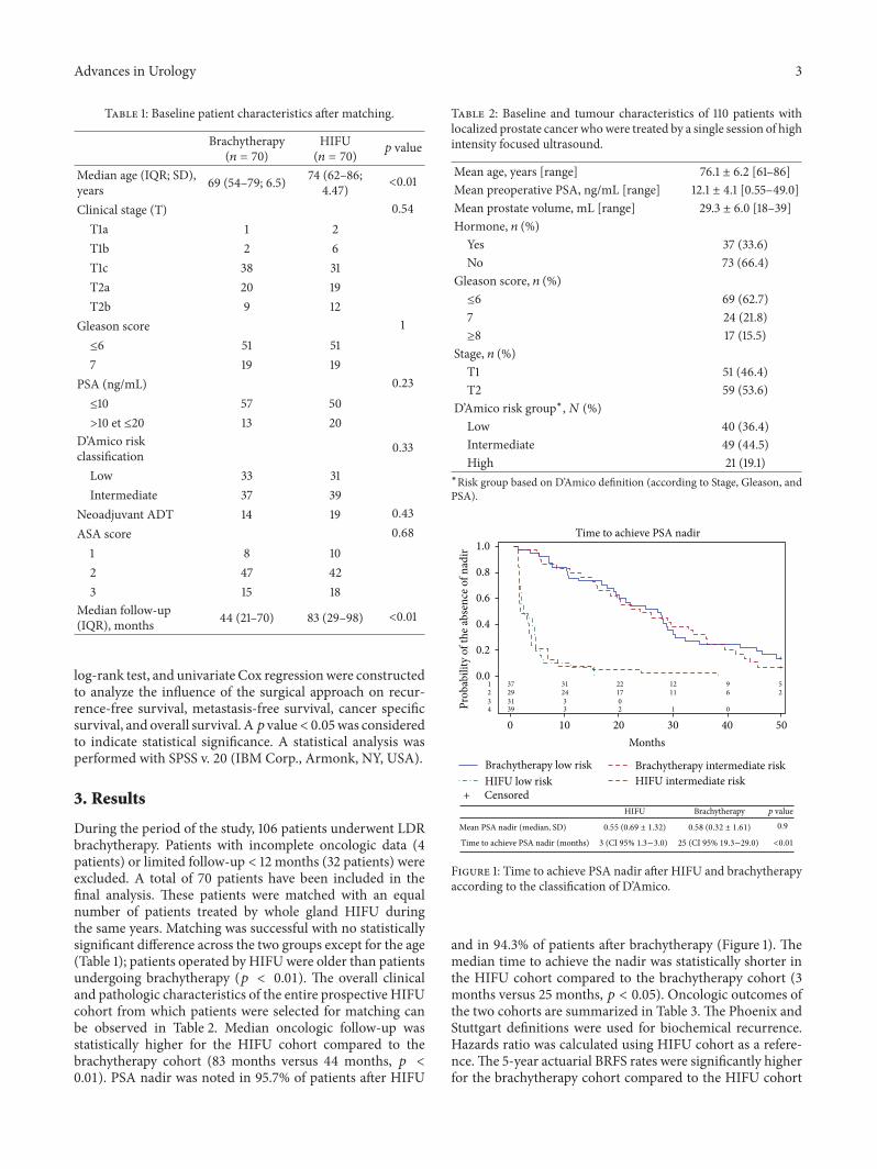
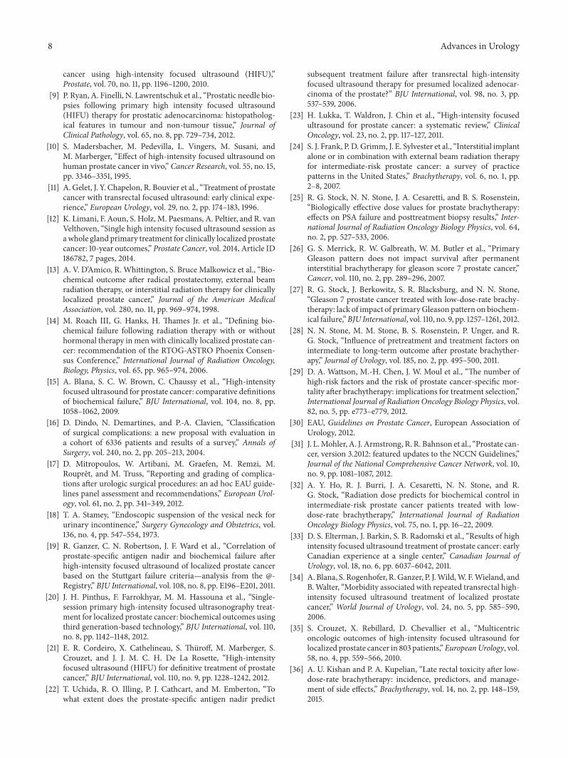
Mean PSA nadir (median, SD)
Time to achieve PSA nadir (months)
HIFU
0.55 (0.69 ± 1.32)
3 (CI 95% 1.3–3.0)
Brachytherapy
0.58 (0.32 ± 1.61)
25 (CI 95% 19.3–29.0)
p value
0.9
<0.01
Figure 1: Time to achieve PSA nadir after HIFU and brachytherapyaccording to the classification of D’Amico.
and in 94.3% of patients after brachytherapy (Figure 1). Themedian time to achieve the nadir was statistically shorter inthe HIFU cohort compared to the brachytherapy cohort (3months versus 25 months, 𝑝 < 0.05). Oncologic outcomes ofthe two cohorts are summarized in Table 3. The Phoenix andStuttgart definitions were used for biochemical recurrence.Hazards ratio was calculated using HIFU cohort as a refere-nce.The 5-year actuarial BRFS rates were significantly higherfor the brachytherapy cohort compared to the HIFU cohort
4 Advances in Urology
Table 3: Oncologic outcomes for the cohort stratified according to the D’Amico risk classification.
Brachytherapy HIFU∗ Hazards ratio (CI∗∗ 95%)Biochemical recurrence-free survivalrates
Phoenix definition: 68.5% Phoenix definition: 53.1% 0.41 (0.19–0.81)Stuttgart definition: 60.9% Stuttgart definition: 51.3% 0.39 (0.19–0.74)
Low risk Phoenix definition: 77.5% Phoenix definition: 68% 0.31 (0.09–0.94)Stuttgart definition: 77.5% Stuttgart definition: 56.3% 0.31 (0.10–0.84)
Intermediate risk Phoenix definition: 58.8% Phoenix definition: 44.9% 0.47 (0.17–1.13)Stuttgart definition: 58.8% Stuttgart definition: 42% 0.41 (0.15–0.97)
Metastasis-free survival rates 79.8% 85% 1.08 (0.36–2.95)Cancer specific survival rates 92% 89% 0.67 (0.32–1.29)Overall survival rates 97.5% 88% 0.24 (0.01–1.34)∗HIFU: high intensity focused ultrasound; ∗∗CI: confidence interval.
Brachytherapy low riskHIFU low risk
Brachytherapy intermediate riskHIFU intermediate risk
Months0 20 40 60 80
1.0
0.8
0.6
0.4
0.2
0.01
2
3
4
37
29
31
39
37
29
30
32
30
23
23
28
20
14
20
23
17
10
17
17
13
5
15
13
7
1
12
13
6
1
9
11
0
1
7
9
Prob
abili
ty o
f sur
viva
l
+ Censored
(a) Stuttgart definition
Brachytherapy low riskHIFU low risk
Brachytherapy intermediate riskHIFU intermediate risk
37
29
31
39
37
29
30
34
30
23
23
28
20
14
21
24
17
10
18
20
13
5
16
15
8
1
13
13
7
1
10
11
1
1
8
9
Months0 20 40 60 80
1.0
0.8
0.6
0.4
0.2
0.01
2
3
4
Prob
abili
ty o
f sur
viva
l
+ Censored
(b) Phoenix definition
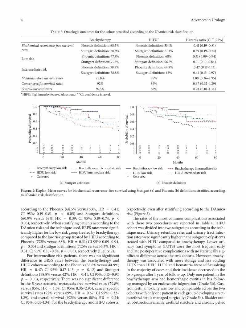
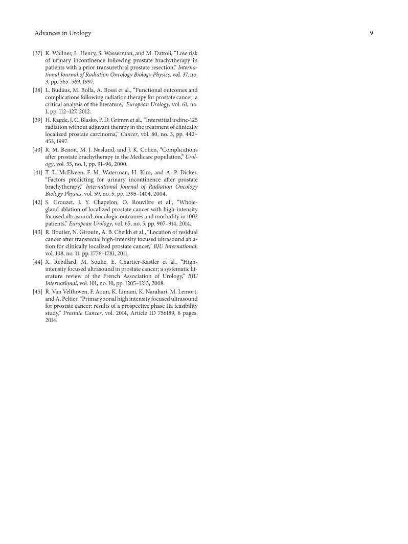
Figure 2: Kaplan-Meier curves for biochemical recurrence-free survival using Stuttgart (a) and Phoenix (b) definitions stratified accordingto D’Amico risk classification.
according to the Phoenix (68.5% versus 53%, HR = 0.41;CI 95%: 0.19–0.81, 𝑝 < 0.05) and Stuttgart definitions(60.9% versus 53%, HR = 0.39; CI 95%: 0.19–0.74, 𝑝 <0.05), respectively.When stratifying patients according to theD’Amico risk and the technique used, BRFS rates were signif-icantly higher for the low risk group treated by brachytherapycompared to the low risk group treated by HIFU according toPhoenix (77.5% versus 68%, HR = 0.31; CI 95%: 0.09–0.94,𝑝 = 0.05) and Stuttgart definitions (77.5% versus 56.3%,HR =0.31; CI 95%: 0.10–0.84, 𝑝 = 0.03), respectively (Figure 2).
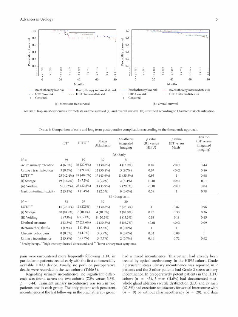
For intermediate risk patients, there was no significantdifference in BRFS rates between the brachytherapy andHIFU cohorts according to the Phoenix (58.8% versus 44.9%;HR = 0.47; CI 95%: 0.17–1.13, 𝑝 = 0.12) and Stuttgartdefinitions (58.8% versus 42%; HR = 0.41; CI 95%: 0.15–0.97,𝑝 = 0.05), respectively. There was no significant differencein the 5-year actuarial metastasis-free survival rates (79.8%versus 85%, HR = 1.08; CI 95%: 0.36–2.95), cancer specificsurvival rates (92% versus 89%, HR = 0.67; CI 95%: 0.32–1.29), and overall survival (97.5% versus 88%; HR = 0.24;CI 95%: 0.01–1.34), for the brachytherapy and HIFU cohorts,
respectively, even after stratifying according to the D’Amicorisk (Figure 3).
The rates of the most common complications associatedwith these two procedures are reported in Table 4. HIFUcohort was divided into two subgroups according to the tech-nique used. Urinary retention rates and urinary tract infec-tion rates were significantly higher in the subgroup of patientstreated with HIFU compared to brachytherapy. Lower uri-nary tract symptoms (LUTS) were the most frequent earlyand late postoperative complications with no statistically sig-nificant difference across the two cohorts. However, brachy-therapy was associated with more storage and less voidingLUTS than HIFU. LUTS and hematuria were self-resolvingin the majority of cases and their incidence decreased in thetwo groups after 1 year of follow-up. Only one patient in thebrachytherapy arm had hemorrhagic cystitis in his follow-up managed by an endoscopic fulguration (Grade 3b). Gas-trointestinal toxicity was low and comparable across the twocohorts with only one patient in each group developing a rect-ourethral fistula managed surgically (Grade 3b). Bladder out-let obstructions mainly urethral stricture and chronic pelvic
Advances in Urology 5
37
29
31
39
37
29
31
39
31
24
27
37
21
14
25
34
17
10
23
32
13
6
20
27
8
1
17
26
7
1
14
22
1
1
10
18
Brachytherapy low riskHIFU low risk
Brachytherapy intermediate riskHIFU intermediate risk
Months0 20 40 60 80
1.0
0.8
0.6
0.4
0.2
0.01
2
3
4
Prob
abili
ty o
f sur
viva
l
+ Censored
(a) Metastasis-free survival
37
29
31
39
37
29
31
39
31
24
28
37
21
18
25
35
17
12
23
34
13
8
20
28
9
3
17
27
8
3
14
23
1
1
10
19
Brachytherapy low riskHIFU low risk
Brachytherapy intermediate riskHIFU intermediate risk
Months0 20 40 60 80
1.0
0.8
0.6
0.4
0.2
0.01
2
3
4
Prob
abili
ty o
f sur
viva
l
+ Censored
(b) Overall survival
Figure 3: Kaplan-Meier curves for metastasis-free survival (a) and overall survival (b) stratified according to D’Amico risk classification.
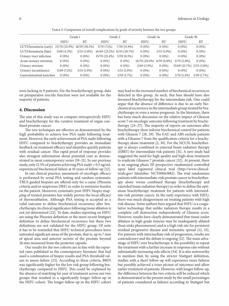
Table 4: Comparison of early and long term postoperative complications according to the therapeutic approach.
BT∗ HIFU∗∗ MaxisAblatherm
Ablathermintegratedimaging
p value(BT versusHIFU)
p value(BT versusMaxis)
p value(BT versusintegratedimaging)
(A) Early𝑁 = 59 90 39 31 — — —Acute urinary retention 4 (6.8%) 16 (22.9%) 12 (30.8%) 4 (12.9%) 0.02 <0.01 0.44Urinary tract infection 5 (8.5%) 15 (21.4%) 12 (30.8%) 3 (9.7%) 0.07 <0.01 0.86LUTS∗∗∗ 25 (42.4%) 28 (40.0%) 17 (43.6%) 11 (35.5%) 0.93 1 0.68(i) Storage 19 (32.2%) 5 (7.2%) 3 (7.7%) 2 (6.4%) <0.01 <0.01 0.01(ii) Voiding 6 (10.2%) 23 (32.8%) 14 (35.9%) 9 (29.1%) <0.01 <0.01 0.04Gastrointestinal toxicity 2 (3.4%) 1 (1.4%) 1 (2.6%) 0 (0.0%) 0.59 1 0.78
(B) Long term𝑁 = 53 69 39 30 — — —LUTS∗∗∗ 14 (26.4%) 19 (27.5%) 12 (30.8%) 7 (23.3%) 1 0.82 0.96(i) Storage 10 (18.9%) 7 (10.1%) 4 (10.3%) 3 (10.0%) 0.26 0.30 0.36(ii) Voiding 4 (7.5%) 12 (17.4%) 8 (20.5%) 4 (13.3%) 0.18 0.18 0.45Urethral stricture 2 (3.8%) 17 (24.6%) 12 (30.8%) 5 (16.7%) <0.01 <0.01 0.09Rectourethral fistula 1 (1.9%) 1 (1.4%) 1 (2.6%) 0 (0.0%) 1 1 1Chronic pelvic pain 0 (0.0%) 3 (4.3%) 3 (7.7%) 0 (0.0%) 0.34 0.08 1Urinary incontinence 2 (3.8%) 5 (7.2%) 3 (7.7%) 2 (6.7%) 0.44 0.72 0.62∗Brachytherapy, ∗∗high intensity focused ultrasound, and ∗∗∗lower urinary tract symptoms.
pain were encountered more frequently following HIFU inparticular in patients treated early with the first commerciallyavailable HIFU device. Finally, no peri- or postoperativedeaths were recorded in the two cohorts (Table 5).
Regarding urinary incontinence, no significant differ-ence was found across the two cohorts (7.2% versus 3.8%,𝑝 = 0.44). Transient urinary incontinence was seen in twopatients one in each group. The only patient with persistentincontinence at the last follow-up in the brachytherapy group
had a mixed incontinence. This patient had already beentreated by optical urethrotomy. In the HIFU cohort, Grade1 persistent stress urinary incontinence was reported in 2patients and the 2 other patients had Grade 2 stress urinaryincontinence. In preoperatively potent patients in the HIFUcohort (𝑛 = 43), 5 men (11.6%) had documented post-whole gland ablation erectile dysfunction (ED) and 27 men(62.8%) had erections satisfactory for sexual intercourse with(𝑛 = 9) or without pharmacotherapy (𝑛 = 20), and data
6 Advances in Urology
Table 5: Comparison of overall complications by grade of severity between the two groups.
Grade 1 Grade 2 Grade 3a Grade 3bHIFU BT HIFU BT HIFU BT HIFU BT
LUTS/hematuria (early) 23/70 (32.9%) 18/59 (30.5%) 5/70 (7.1%) 7/59 (11.9%) 0 (0%) 0 (0%) 0 (0%) 0 (0%)LUTS/hematuria (late) 3/69 (4.3%) 2/53 (3.8%) 16/69 (23.2%) 11/53 (20.7%) 0 (0%) 1/53 (1.9%) 0 (0%) 0 (0%)Urinary tract infection 0 (0%) 0 (0%) 15/70 (21.4%) 5/59 (8.5%) 0 (0%) 0 (0%) 0 (0%) 0 (0%)Acute urinary retention 0 (0%) 0 (0%) 0 (0%) 0 (0%) 14/70 (20.0%) 4/59 (6.8%) 2/70 (2.8%) 0 (0%)Urinary stricture 0 (0%) 0 (0%) 0 (0%) 0 (0%) 2/69 (2.9%) 0 (0%) 15/69 (21.7%) 2/53 (3.8%)Urinary incontinence 5/69 (7.2%) 1/53 (1.9%) 0 (0%) 1/53 (1.9%) 0 (0%) 0 (0%) 0 (0%) 0 (0%)Gastrointestinal toxicities 0 (0%) 0 (0%) 0 (0%) 1/59 (1.7%) 0 (0%) 0 (0%) 1/70 (1.4%) 1/59 (1.7%)
were lacking in 9 patients. For the brachytherapy group, dataon preoperative erectile function were not available for themajority of patients.
4. Discussion
The aim of this study was to compare retrospectively HIFUand brachytherapy for the curative treatment of organ con-fined prostate cancer.
The two techniques are effective as demonstrated by thehigh probability to achieve low PSA nadir following treat-ment. However, the early achievement of PSA nadir followingHIFU compared to brachytherapy provides an immediatefeedback on treatment efficacy and identifies quickly patientswith residual cancer. This rapid proof of response providesalso stringent information about potential cure as demon-strated in most contemporary series [19–21]. In our previousstudy, only 12.5% of patients achieving PSAnadir< 0.5 ng/mLexperienced biochemical failure at 10 years of follow-up [12].
In our clinical practice, assessment of oncologic efficacyis performed by serial PSA testing and random systematicTRUS guided biopsies are offered only for a cause (Phoenixcriteria and/or suspicious DRE) in order to minimise burdenon the patient. Moreover, systematic post-HIFU biopsy map-ping of treated prostates has widely proven the local efficacyof thermoablation. Although PSA testing is accepted as avalid outcome to define biochemical recurrence after bra-chytherapy, its clinical significance following tissue ablation isnot yet determined [22]. To date, studies reporting on HIFUare using the Phoenix definition or the more recent Stuttgartdefinition to define biochemical recurrence but these twodefinitions are not validated for the HIFU group. Of noteit has to be reminded that HIFU technical procedures leaveuntreated significant areas of the prostate, that is, up to 7mmof apical area and anterior sectors of the prostate beyond26mmmeasured from the posterior capsule.
Our results for the two cohorts are in line with the repor-ted rates published in the contemporary literature that hadused a combination of biopsy results and PSA threshold val-ues to assess failure [23]. According to these criteria, BRFSwas significantly higher for the low risk group following bra-chytherapy compared to HIFU. This could be explained bythe absence of matching for year of treatment across our twogroups that resulted in a significantly longer follow-up inthe HIFU cohort. The longer follow-up in the HIFU cohort
may lead to the increased number of biochemical recurrencesdetected in this group. As such, this bias should have alsofavoured brachytherapy for the intermediate risk. One couldargue that the absence of difference is due to an early bio-chemical recurrence in the intermediate group treated by bra-chytherapy or even a worse prognosis. In the literature, therehas been much discussion on the relative impact of Gleasonscore 7 on oncologic outcome following treatment by brachy-therapy [24–27]. The majority of reports on outcomes afterbrachytherapy show inferior biochemical control for patientswith Gleason 7 [28, 29]. The EAU and ABS exclude patientswith a Gleason 7 from the qualification criteria for a brachy-therapy alone treatment [2, 30]. For the NCCN, brachyther-apy is always combined to external beam radiation therapy(EBRT) for intermediate risk patients [31]. A new study hadsuggested the need for high quality and high-dose treatmentto eradicate Gleason 7 prostate cancer [32]. At present, thereis an ongoing phase III prospective randomized controlledopen label registered clinical trial (http://www.clinical-trials.gov/ Identifier: NCT00063882). The trial randomizespatientswith intermediate-risk prostate cancer to brachyther-apy alone versus combined therapy (brachytherapy andextended beam radiation therapy) in order to define the opti-mum brachytherapy treatment for patients with intermed-iate-risk prostate cancer. In the early experience with HIFU,there was much disagreement on treating patients with highrisk disease. Some authors have argued that HIFU is a coagu-lative technology that unlike radiation therapy results in acomplete cell destruction independently of Gleason score.However, results have clearly demonstrated that tissue underablation in high grade tumour may be inadequately ablated(heat sinks phenomenon) and is a high risk site for persistentresidual progressive disease and metastatic spread [12, 33].For patients with intermediate risk of progression, results arecontradictory and the debate is ongoing [12]. The main adva-ntage of HIFU over brachytherapy is the possibility to repeatthe treatment with a further increase in response rate withoutsubstantially increasing side effects [34]. It is also noteworthyto mention that, by using the stricter Stuttgart definition,studies with a short follow-up will experience more failuresbut possibly achieved a truer picture of outcomes and allowearlier treatment of patients. However, with longer follow-up,the difference between the two criteria will be reduced whichis demonstrated in the present study by the small percentagesof patients considered as failures according to Stuttgart but
Advances in Urology 7
not to Phoenix definition. The 5-year actuarial metastasis-free survival, cancer specific survival, and overall survivalrates were not significantly different between the two cohortsand are in line with the reported rates in nonrandomizedpublished case series [35]. However, the retrospective designand the insufficient follow-up limit further conclusion.
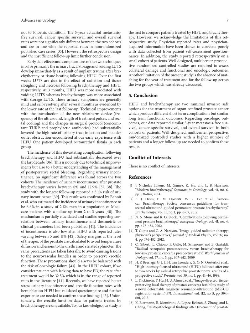
Early side effects and complications of the two techniquesinvolve primarily the urinary tract. Storage and voiding LUTSdevelop immediately as a result of implant trauma after bra-chytherapy or tissue heating following HIFU. Over the firstweeks LUTS are due to the effect of radiation and tissuesloughing and necrosis following brachytherapy and HIFU,respectively. At 3 months, HIFU was more associated withvoiding LUTS whereas brachytherapy was more associatedwith storage LUTS. These urinary symptoms are generallymild and self-resolving after several months as evidenced bythe lower rate at the last follow-up. Technical improvementswith the introduction of the new Ablatherm device (fre-quency of the ultrasound, length of treatment pulses, and rec-tal cooling) and the changes in surgical protocol (concomi-tant TURP and prophylactic antibiotics) had substantiallylowered the high rate of urinary tract infection and bladderoutlet obstruction encountered at our early experience withHIFU. One patient developed rectourethral fistula in eachgroup.
The incidence of this devastating complication followingbrachytherapy and HIFU had substantially decreased overthe last decade [36].This is not only due to technical improve-ments but also to a better understanding of the managementof postoperative rectal bleeding. Regarding urinary incon-tinence, no significant difference was found across the twocohorts. The incidence of urinary incontinence after prostatebrachytherapy varies between 0% and 12.9% [37, 38]. Thestudy with the longest follow-up reported a 5.1% risk of uri-nary incontinence [39]. This result was confirmed by Benoitet al. who estimated the incidence of urinary incontinence tobe 6.6% in a study of 2,124 men in a population of Medi-care patients with a follow-up from 2 to 3 years [40]. Themechanism is partially elucidated and studies reporting cor-relation between urinary incontinence and dosimetric orclinical parameters had been published [41]. The incidenceof incontinence is also low after HIFU with reported ratesvarying between 5 and 11% [42]. Safety margins at the levelof the apex of the prostate are calculated to avoid temperaturediffusion and lesions to the urethra and striated sphincter.Thesame precautions are taken laterally, to avoid causing lesionsto the neurovascular bundles in order to preserve erectilefunction. These precautions should always be balanced withthe risk of oncologic failure [43]. In the HIFU cohort, if weconsider patients with lacking data to have ED, the rate aftertreatment would be 32.5% which is in the range of reportedrates in the literature [44]. Recently, we have reported betterstress urinary incontinence and erectile function rates withhemiablation HIFU but validated questionnaire and furtherexperience are needed to confirm these findings [45]. Unfor-tunately, the erectile function data for patients treated bybrachytherapy are unavailable. To our knowledge, our study is
the first to compare patients treated byHIFU and brachyther-apy. However, we acknowledge the limitations of this ret-rospective study. Physician reported rates and physician-acquired information have been shown to correlate poorlywith data collected from patient self-assessment question-naires. In addition, the study reported retrospectively on asmall cohort of patients.Well-designed,multicenter, prospec-tive, randomized controlled studies are required to assesscollateral damage and functional and oncologic outcomes.Another limitation of the present study is the absence of mat-ching for the year of treatment and for the follow-up acrossthe two groups which was already discussed.
5. Conclusion
HIFU and brachytherapy are two minimal invasive safeoptions for the treatment of organ confined prostate cancerwhich produce different short term complications but similarlong term functional outcomes. Regarding oncologic out-comes, our data reported similar 5-year metastasis-free sur-vival, cancer specific survival, and overall survival in bothcohorts of patients. Well-designed, multicenter, prospective,randomized controlled studies with a higher number ofpatients and a longer follow-up are needed to confirm theseresults.
Conflict of Interests
There is no conflict of interests.
References
[1] J. Nicholas Lukens, M. Gamez, K. Hu, and L. B. Harrison,“Modern brachytherapy,” Seminars in Oncology, vol. 41, no. 6,pp. 831–847, 2014.
[2] B. J. Davis, E. M. Horwitz, W. R. Lee et al., “Ameri-can Brachytherapy Society consensus guidelines for tran-srectal ultrasound-guided permanent prostate brachytherapy,”Brachytherapy, vol. 11, no. 1, pp. 6–19, 2012.
[3] N. N. Stone and R. G. Stock, “Complications following perma-nent prostate brachytherapy,” European Urology, vol. 41, no. 4,pp. 427–433, 2002.
[4] T. Gupta and C. A. Narayan, “Image-guided radiation therapy:physician’s perspectives,” Journal of Medical Physics, vol. 37, no.4, pp. 174–182, 2012.
[5] C. Giberti, L. Chiono, F. Gallo, M. Schenone, and E. Gastaldi,“Radical retropubic prostatectomy versus brachytherapy forlow-risk prostatic cancer: a prospective study,”World Journal ofUrology, vol. 27, no. 5, pp. 607–612, 2009.
[6] H. P. Beerlage, G. J. L. H. van Leenders, G. O. N. Oosterhof et al.,“High-intensity focused ultrasound (HIFU) followed after oneto two weeks by radical retropubic prostatectomy: results of aprospective study,” Prostate, vol. 39, no. 1, pp. 41–46, 1999.
[7] L.Dickinson, Y.Hu,H.U.Ahmed et al., “Image-directed, tissue-preserving focal therapy of prostate cancer: a feasibility study ofa novel deformable magnetic resonance-ultrasound (MR-US)registration system,” BJU International, vol. 112, no. 5, pp. 594–601, 2013.
[8] K. Biermann, R. Montironi, A. Lopez-Beltran, S. Zhang, and L.Cheng, “Histopathological findings after treatment of prostate
8 Advances in Urology
cancer using high-intensity focused ultrasound (HIFU),”Prostate, vol. 70, no. 11, pp. 1196–1200, 2010.
[9] P. Ryan, A. Finelli, N. Lawrentschuk et al., “Prostatic needle bio-psies following primary high intensity focused ultrasound(HIFU) therapy for prostatic adenocarcinoma: histopatholog-ical features in tumour and non-tumour tissue,” Journal ofClinical Pathology, vol. 65, no. 8, pp. 729–734, 2012.
[10] S. Madersbacher, M. Pedevilla, L. Vingers, M. Susani, andM. Marberger, “Effect of high-intensity focused ultrasound onhuman prostate cancer in vivo,” Cancer Research, vol. 55, no. 15,pp. 3346–3351, 1995.
[11] A. Gelet, J. Y. Chapelon, R. Bouvier et al., “Treatment of prostatecancer with transrectal focused ultrasound: early clinical expe-rience,” European Urology, vol. 29, no. 2, pp. 174–183, 1996.
[12] K. Limani, F. Aoun, S. Holz, M. Paesmans, A. Peltier, and R. vanVelthoven, “Single high intensity focused ultrasound session asawhole gland primary treatment for clinically localized prostatecancer: 10-year outcomes,” Prostate Cancer, vol. 2014, Article ID186782, 7 pages, 2014.
[13] A. V. D’Amico, R. Whittington, S. Bruce Malkowicz et al., “Bio-chemical outcome after radical prostatectomy, external beamradiation therapy, or interstitial radiation therapy for clinicallylocalized prostate cancer,” Journal of the American MedicalAssociation, vol. 280, no. 11, pp. 969–974, 1998.
[14] M. Roach III, G. Hanks, H. Thames Jr. et al., “Defining bio-chemical failure following radiation therapy with or withouthormonal therapy in men with clinically localized prostate can-cer: recommendation of the RTOG-ASTRO Phoenix Consen-sus Conference,” International Journal of Radiation Oncology,Biology, Physics, vol. 65, pp. 965–974, 2006.
[15] A. Blana, S. C. W. Brown, C. Chaussy et al., “High-intensityfocused ultrasound for prostate cancer: comparative definitionsof biochemical failure,” BJU International, vol. 104, no. 8, pp.1058–1062, 2009.
[16] D. Dindo, N. Demartines, and P.-A. Clavien, “Classificationof surgical complications: a new proposal with evaluation ina cohort of 6336 patients and results of a survey,” Annals ofSurgery, vol. 240, no. 2, pp. 205–213, 2004.
[17] D. Mitropoulos, W. Artibani, M. Graefen, M. Remzi, M.Roupret, and M. Truss, “Reporting and grading of complica-tions after urologic surgical procedures: an ad hoc EAU guide-lines panel assessment and recommendations,” European Urol-ogy, vol. 61, no. 2, pp. 341–349, 2012.
[18] T. A. Stamey, “Endoscopic suspension of the vesical neck forurinary incontinence,” Surgery Gynecology and Obstetrics, vol.136, no. 4, pp. 547–554, 1973.
[19] R. Ganzer, C. N. Robertson, J. F. Ward et al., “Correlation ofprostate-specific antigen nadir and biochemical failure afterhigh-intensity focused ultrasound of localized prostate cancerbased on the Stuttgart failure criteria—analysis from the @-Registry,” BJU International, vol. 108, no. 8, pp. E196–E201, 2011.
[20] J. H. Pinthus, F. Farrokhyar, M. M. Hassouna et al., “Single-session primary high-intensity focused ultrasonography treat-ment for localized prostate cancer: biochemical outcomes usingthird generation-based technology,” BJU International, vol. 110,no. 8, pp. 1142–1148, 2012.
[21] E. R. Cordeiro, X. Cathelineau, S. Thuroff, M. Marberger, S.Crouzet, and J. J. M. C. H. De La Rosette, “High-intensityfocused ultrasound (HIFU) for definitive treatment of prostatecancer,” BJU International, vol. 110, no. 9, pp. 1228–1242, 2012.
[22] T. Uchida, R. O. Illing, P. J. Cathcart, and M. Emberton, “Towhat extent does the prostate-specific antigen nadir predict
subsequent treatment failure after transrectal high-intensityfocused ultrasound therapy for presumed localized adenocar-cinoma of the prostate?” BJU International, vol. 98, no. 3, pp.537–539, 2006.
[23] H. Lukka, T. Waldron, J. Chin et al., “High-intensity focusedultrasound for prostate cancer: a systematic review,” ClinicalOncology, vol. 23, no. 2, pp. 117–127, 2011.
[24] S. J. Frank, P. D. Grimm, J. E. Sylvester et al., “Interstitial implantalone or in combination with external beam radiation therapyfor intermediate-risk prostate cancer: a survey of practicepatterns in the United States,” Brachytherapy, vol. 6, no. 1, pp.2–8, 2007.
[25] R. G. Stock, N. N. Stone, J. A. Cesaretti, and B. S. Rosenstein,“Biologically effective dose values for prostate brachytherapy:effects on PSA failure and posttreatment biopsy results,” Inter-national Journal of Radiation Oncology Biology Physics, vol. 64,no. 2, pp. 527–533, 2006.
[26] G. S. Merrick, R. W. Galbreath, W. M. Butler et al., “PrimaryGleason pattern does not impact survival after permanentinterstitial brachytherapy for gleason score 7 prostate cancer,”Cancer, vol. 110, no. 2, pp. 289–296, 2007.
[27] R. G. Stock, J. Berkowitz, S. R. Blacksburg, and N. N. Stone,“Gleason 7 prostate cancer treated with low-dose-rate brachy-therapy: lack of impact of primaryGleason pattern on biochem-ical failure,”BJU International, vol. 110, no. 9, pp. 1257–1261, 2012.
[28] N. N. Stone, M. M. Stone, B. S. Rosenstein, P. Unger, and R.G. Stock, “Influence of pretreatment and treatment factors onintermediate to long-term outcome after prostate brachyther-apy,” Journal of Urology, vol. 185, no. 2, pp. 495–500, 2011.
[29] D. A. Wattson, M.-H. Chen, J. W. Moul et al., “The number ofhigh-risk factors and the risk of prostate cancer-specific mor-tality after brachytherapy: implications for treatment selection,”International Journal of Radiation Oncology Biology Physics, vol.82, no. 5, pp. e773–e779, 2012.
[30] EAU, Guidelines on Prostate Cancer, European Association ofUrology, 2012.
[31] J. L.Mohler, A. J. Armstrong, R. R. Bahnson et al., “Prostate can-cer, version 3.2012: featured updates to the NCCN Guidelines,”Journal of the National Comprehensive Cancer Network, vol. 10,no. 9, pp. 1081–1087, 2012.
[32] A. Y. Ho, R. J. Burri, J. A. Cesaretti, N. N. Stone, and R.G. Stock, “Radiation dose predicts for biochemical control inintermediate-risk prostate cancer patients treated with low-dose-rate brachytherapy,” International Journal of RadiationOncology Biology Physics, vol. 75, no. 1, pp. 16–22, 2009.
[33] D. S. Elterman, J. Barkin, S. B. Radomski et al., “Results of highintensity focused ultrasound treatment of prostate cancer: earlyCanadian experience at a single center,” Canadian Journal ofUrology, vol. 18, no. 6, pp. 6037–6042, 2011.
[34] A. Blana, S. Rogenhofer, R. Ganzer, P. J.Wild,W. F.Wieland, andB.Walter, “Morbidity associated with repeated transrectal high-intensity focused ultrasound treatment of localized prostatecancer,” World Journal of Urology, vol. 24, no. 5, pp. 585–590,2006.
[35] S. Crouzet, X. Rebillard, D. Chevallier et al., “Multicentriconcologic outcomes of high-intensity focused ultrasound forlocalized prostate cancer in 803 patients,”EuropeanUrology, vol.58, no. 4, pp. 559–566, 2010.
[36] A. U. Kishan and P. A. Kupelian, “Late rectal toxicity after low-dose-rate brachytherapy: incidence, predictors, and manage-ment of side effects,” Brachytherapy, vol. 14, no. 2, pp. 148–159,2015.
Advances in Urology 9
[37] K. Wallner, L. Henry, S. Wasserman, and M. Dattoli, “Low riskof urinary incontinence following prostate brachytherapy inpatients with a prior transurethral prostate resection,” Interna-tional Journal of Radiation Oncology Biology Physics, vol. 37, no.3, pp. 565–569, 1997.
[38] L. Budaus, M. Bolla, A. Bossi et al., “Functional outcomes andcomplications following radiation therapy for prostate cancer: acritical analysis of the literature,” European Urology, vol. 61, no.1, pp. 112–127, 2012.
[39] H. Ragde, J. C. Blasko, P. D. Grimm et al., “Interstitial iodine-125radiation without adjuvant therapy in the treatment of clinicallylocalized prostate carcinoma,” Cancer, vol. 80, no. 3, pp. 442–453, 1997.
[40] R. M. Benoit, M. J. Naslund, and J. K. Cohen, “Complicationsafter prostate brachytherapy in the Medicare population,” Urol-ogy, vol. 55, no. 1, pp. 91–96, 2000.
[41] T. L. McElveen, F. M. Waterman, H. Kim, and A. P. Dicker,“Factors predicting for urinary incontinence after prostatebrachytherapy,” International Journal of Radiation OncologyBiology Physics, vol. 59, no. 5, pp. 1395–1404, 2004.
[42] S. Crouzet, J. Y. Chapelon, O. Rouviere et al., “Whole-gland ablation of localized prostate cancer with high-intensityfocused ultrasound: oncologic outcomes and morbidity in 1002patients,” European Urology, vol. 65, no. 5, pp. 907–914, 2014.
[43] R. Boutier, N. Girouin, A. B. Cheikh et al., “Location of residualcancer after transrectal high-intensity focused ultrasound abla-tion for clinically localized prostate cancer,” BJU International,vol. 108, no. 11, pp. 1776–1781, 2011.
[44] X. Rebillard, M. Soulie, E. Chartier-Kastler et al., “High-intensity focused ultrasound in prostate cancer; a systematic lit-erature review of the French Association of Urology,” BJUInternational, vol. 101, no. 10, pp. 1205–1213, 2008.
[45] R. Van Velthoven, F. Aoun, K. Limani, K. Narahari, M. Lemort,andA. Peltier, “Primary zonal high intensity focused ultrasoundfor prostate cancer: results of a prospective phase IIa feasibilitystudy,” Prostate Cancer, vol. 2014, Article ID 756189, 6 pages,2014.

Submit your manuscripts athttp://www.hindawi.com
Stem CellsInternational
Hindawi Publishing Corporationhttp://www.hindawi.com Volume 2014
Hindawi Publishing Corporationhttp://www.hindawi.com Volume 2014
MEDIATORSINFLAMMATION
of
Hindawi Publishing Corporationhttp://www.hindawi.com Volume 2014
Behavioural Neurology
EndocrinologyInternational Journal of
Hindawi Publishing Corporationhttp://www.hindawi.com Volume 2014
Hindawi Publishing Corporationhttp://www.hindawi.com Volume 2014
Disease Markers
Hindawi Publishing Corporationhttp://www.hindawi.com Volume 2014
BioMed Research International
OncologyJournal of
Hindawi Publishing Corporationhttp://www.hindawi.com Volume 2014
Hindawi Publishing Corporationhttp://www.hindawi.com Volume 2014
Oxidative Medicine and Cellular Longevity
Hindawi Publishing Corporationhttp://www.hindawi.com Volume 2014
PPAR Research
The Scientific World JournalHindawi Publishing Corporation http://www.hindawi.com Volume 2014
Immunology ResearchHindawi Publishing Corporationhttp://www.hindawi.com Volume 2014
Journal of
ObesityJournal of
Hindawi Publishing Corporationhttp://www.hindawi.com Volume 2014
Hindawi Publishing Corporationhttp://www.hindawi.com Volume 2014
Computational and Mathematical Methods in Medicine
OphthalmologyJournal of
Hindawi Publishing Corporationhttp://www.hindawi.com Volume 2014
Diabetes ResearchJournal of
Hindawi Publishing Corporationhttp://www.hindawi.com Volume 2014
Hindawi Publishing Corporationhttp://www.hindawi.com Volume 2014
Research and TreatmentAIDS
Hindawi Publishing Corporationhttp://www.hindawi.com Volume 2014
Gastroenterology Research and Practice
Hindawi Publishing Corporationhttp://www.hindawi.com Volume 2014
Parkinson’s Disease
Evidence-Based Complementary and Alternative Medicine
Volume 2014Hindawi Publishing Corporationhttp://www.hindawi.com