CLINICAL AND VIROLOGICAL STUDIES ON a … Henricus Leonardus...CLINICAL AND VIROLOGICAL STUDIES ON...
160
CLINICAL AND VIROLOGICAL STUDIES ON a-INTERFERON TREATMENT OF CHRONIC HEPATITIS TYPE B H.L.A. JANSSEN
-
Upload
truongkien -
Category
Documents
-
view
213 -
download
0
Transcript of CLINICAL AND VIROLOGICAL STUDIES ON a … Henricus Leonardus...CLINICAL AND VIROLOGICAL STUDIES ON...

CLINICAL AND VIROLOGICAL STUDIES ON a-INTERFERON TREATMENT OF CHRONIC HEPATITIS TYPE B
H.L.A. JANSSEN

CLINICAL AND VIROLOGICAL STUDIES ON a-INTERFERON
TREATMENT OF CHRONIC HEPATffiS TYPE B
KUNISCH EN VIROLOGISCH ONDERZOEK NAAR a-INTERFERON
BEHANDELING BIJ CHRONISCHE HEPATITIS B
PROEFSCHRIFT
ter verkrijging van de graad van doctor
aan de Erasmus Universiteit Rotterdam
op gezag van rector magnificos
Prof. dr. C.J. Rijnvos
en volgens besluit van het College van Dekanen.
De openbare verdediging zal plaatsvinden op
woensdag 16 juni 1993 om 13.45 uur
door
Henricus Leonardus Antonius Janssen
geboren te Alkmaar
Promotiecommissie
Promotor: Prof.dr. S.W. Schalm
Overige leden: Dr. R.A. Heijtink
Prof. dr. F.T. Bosman
Prof. J.H.P. Wilson
This study was performed at the Department of Internal Medicine II of the University Hospital Dijkzigt
in Rotterdam. The Netherlands. Financial support for this thesis was kindly given by Glaxo B.V.,
Scheriog-Plough B.V. and Wellcome Pharmaceuticals B.V.

Aan mijn ouders
Aan Inez

Contents Page
Chapter 1 Introduction 1
Chapter 2 Survival and prognostic indicators of HBsAg-positive cirrhosis of the liver. 9
The Role of HBeAg seroconversion
Chapter 3 Antiviral effect of prolonged intermittent lymphoblastoid alpha-interferon 25
treatment in chronic hepatitis B
Chapter 4 Alpha-interferon and zidovudine combination therapy for chronic hepatitis 39
type B. Results of a randomized placebo-controlled trial
Chapter 5 Repeated courses of alpha-interferon for treatment of chronic hepatitis B 55
Chapter 6 Quantitative assessment of hepatitis B virus DNA in chronic hepatitis B: 67
Comparison of two solution hybridization assay
Chapter 7 Measurement of HBsAg to monitor hepatitis B viral replication in patients 81
on a-interferon therapy
Chapter 8 Fatal decompensation of chronic viral hepatitis associated with alpha- 93
interferon treatment
Chapter 9 Seizures associated with low dose a-interferon treatment of chronic 105
hepatitis B
Chapter 10 Snicide associated with alpha-interferon therapy for chronic viral hepatitis 111
Chapter 11 Discussion: How to optimize a-interferon therapy for chronic hepatitis B 119
SlllllliJ.afY 141
Samenvatting 143
Dankwoord 145
Curriculum Vitae 147

Abbrevations
ALT
anti-HBc
anti-HBe
anti-HBs
ARA-AMP
AST
AZT
CAH
CPH
CI
cpm
DNA
DNA-p
HBcAg
HBeAg
HBsAg
HBV
HCV
HDV
HIV
HLA
IFN
IgG
IgM
MU
PCR
pre-Sl Ag
pre-S2 Ag
RIA
RNA
alanine aminotransferase
antibodies against hepatitis B core antigen
antibodies against hepatitis B e antigen
antibodies against hepatitis B snrface antigen
adenine arabinoside monophosphate
aspartate aminotransferase
3 '-azido-3' -deoxythymidine
chronic active hepatitis
chronic persistent hepatitis
confidence interval
counts per minute
deoxyribonucleic acid
deoxyribonucleic acid polymerase
hepatitis B core antigen
hepatitis B e antigen
hepatitis B snrface antigen
hepatitis B virus
hepatitis C virus
hepatitis delta virus
human immunodeficiency virus
human leukocyte antigen
interferon
immunoglobulin G
immunoglobulin M
mega-units
polymerase chain reaction
hepatitis B pre-Sl antigen
hepatitis B pre-S2 antigen
radioimmunoassay
ribonucleic acid


CHAPTER 1
INTRODUCTION

The Hepatitis B Virus
The hepatitis B virus (HBV) is a double-stranded DNA virus belonging to the group of
hepadna viridae (1). The replication of HBV is believed to occur preferentially in the
hepatocyte. Analysis of the nucleotide sequence of the virus revealed 4 open reading
frames, regions of the genome which may code for viral antigens (1). Although the HBV
genome contains only 3200 nucleotides its compacmess and circnlar composition,
employing overlapping genes for production of several viral proteins, make the virus
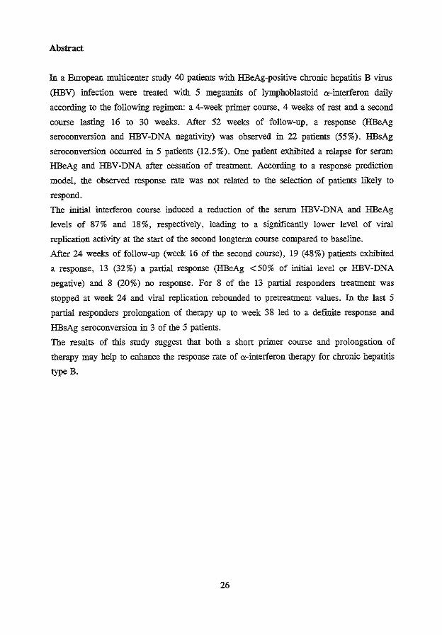
highly efficient for replication. The replication cycle of HBV is illustrated in figure 1.
After entry in the hepatocyte the virus is uncoated and the genomic DNA is converted to a
supercoiled form of covalently closed fully double stranded DNA which is transcribed to
pregenontic and messenger RNA. Viral messenger RNA is transported to the cytoplasm
HBV RNA preger'\Ome, -o H8sAg. 0 HBcAg. 6 HSVONA polymerase, e
HBcAg-MHC class 1 ant1gen-HBs Ag. + Figure 1. HBV replication cycle in hepaJocytes. DNA-P: DNA polymerase; HBcAg:
hepatitis B core antigen; HBsAg: hepatitis B surface antigen; HBV: hepatitis B virus;
MHC: ITI£ljor histocompatibility complex; RNA-p: RNA polymerase. !from L. Berk et al.
Current Opinion in Infectious Diseases 1989;2:419-23; with permission).
3
where it codes for production of viral proteins. Pregenomic RNA is reverse transcrihed
into a minus strand of DNA which is utilized as a template for completion of the plus
strand of the newly synthesized viral DNA. The HBV -DNA-polymerase enzyme which
exerts both a DNA polymerase and a reverse transcriptase activity plays a crucial role in
the replication cycle of the virus.
Chronic Hepatitis B
Inoculation with the virus causes hepatocellular necrosis and inflammation which ranges in
severity from an asymtomatic infection that resolves completely to severe, symptomatic
infection with progressive or even fatal illness (2). It is still not clear why certain patients
progress to a chronic hepatitis B infection while the majority clears HBV after an acute
infection. One of the possibilities is that chronic HBV carriage results in part from a
deficiency in IFN production. Several studies have showed a lower endogeneous IFN
response in individuals with chronic hepatitis B (3). This low IFN production is likely to
impair the display of HLA class I proteins and thereby impede the clearance of
hepatocytes harbouring replicating virus (4).
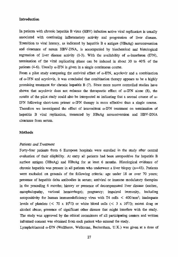
In the patients who develop a chronic hepatitis there is a substantial risk of disease
progression to cirrhosis, portal hypertension and hepatocellular carcinoma (5,6; figure 2).
It is unlikely that HBV causes its damaging effect on liver cells by direct cytopathogenic
changes because large quantities of HBsAg and HBcAg are found in the hepatocytes of
many asymptomatic and appearantly healthy HBV carriers. Both for the pathogenesis of
the liver damage and for succesful eradication of the disease the interaction between
various structural components of the virus and the immune system appear to be essential.
In untreated chronic hepatitis B patients HBV probably replicates less vigorously as time
passes, maybe because of an increasing immunological attack on virus-infected
hepatocytes (7,8). This phenomenon is accompanied by a fall of serum HBV-DNA and
increased liver damage as signalled by abnormal liver ftmction tests. Cessation of HBV
replication as indicated by an HBeAg serovonversion and a marked suppression of HBV
DNA, usually heralds a last and quiescent phase of the disease which may in time be
followed by a total eradication of the virus (HBsAg seroconversion) (9-11). The process
of HBeAg seroconversion is often preceded by intense hepatic inflanunation and symptoms
of fatigue. The aim of antiviral therapy with a-IFN is to assist the host in eliminating
HBV replication early in the course of the chronic infection.
4
recovery 90% acute hT:~is B
chronic hepatitis B
/ acL• recovery cirrhosis
<1%
a
b acute liver failure
~~b inactive portal hypertension hepatocellular cirrhosis chronic liver failure
a: antiviral therapy
b: symptomatic therapy
liver transplantation
Figure 2. Course of hepatitis B infection.
Alpha-Interferon Treatment of Chronic Hepatitis B
carcinoma
Intervention treatment of chronic hepatitis B is targeted at patients with active viral
replication, preferably at a stage before signs and symptoms of cirrhosis or significant
disease activity have occurred. Although the genetic heterogeneity of HBV, recently
disclosed by molecular biology (12), has necessitated a reconsideration of the classical
serologic distinctions between active virus replication and viral latency, the presence of
HBeAg and HBV-DNA is still considered to be the main indication for antiviral therapy.
a-IFN probably interferes directly with HBV production by activation of intracellular
enzymes like 2'-5' oligoadenylate synthetase or protein kinase, leading to an activation of
ribonucleases that destroy viral messenger RNA. As hrnnune modifying substance a-IFN
augments the natural killer cell activity and causes enhanced expression of HLA class I
proteins on the hepatocyte surface thereby facilitating recognition and lysis of virus
infected cells by the cellular hrnnune system (13). In addition, a-IFN may boost the
production of the HBV transcripts that code for HBcAg thereby promoting an increased
hepatocyte membrane expression of this antigen which is one of the key viral proteins
recognised by the cytotoxic T cells (14).
5
Although a-IFN is the first agent which has repeatedly shown efficacy for chronic
hepatitis B in large randomized controlled trials, more than half of the patients do not
respond and are left with continuing disease activity (15,16). Because several individual
variables (e.g. high level of inflammatory activity, low level of virus replication and a
short duration of infection) are associated with a higher response rate (17,18) and because
the adverse effects of a-IFN are often a cause dose reduction, timing and dosage of a-IFN
are important in deterntining the potency of this therapy. The scope of this thesis is
comprised to the question how we could safely improve the indication for and antiviral
effect of a-IFN therapy in chronic hepatitis type B.
The objectives of the study are:
1. To assess the prognostic role of HBeAg seroconversion in patients with HBV-related
cirrhosis of the liver (chapter 2).
2. To evaluate the efficacy of modifications (prolonged intermittent therapy,
combination therapy with zidovudine and a-IFN retreatrnent) of a-IFN treatment in
chronic hepatitis B (chapter 3, 4 and 5).
3. To evaluate the usefulness of quantitated HBV-DNA and HBsAg assessments for
monitoring of chronic hepatitis B patients undergoing a-IFN therapy (chapter 6 and
7).
4. To review the frequency and clinical aspects of serious side effects (fatal hepatic
decompensation, seizures and suicidal behaviour) associated with a-IFN therapy for
chronic viral hepatitis (chapter 8, 9 and 10).
6
References
1. Tiollais P, Pourcel C, Dejean A. The hepatitis B virus. Narure 1985;317:489-95.
2. Hoofuagle JH, Alter HJ. Chronic viral hepatitis. In: Vyas GN, Dienstag JL, Hoofua
gle JH eds. Viral hepatitis and liver disease. New York: Grone and Stratton,
1984;97-113.
3. Davis GL, Hoofuagle JH. Interferon in viral hepatitis: Role in pathogenesis and
treatment. Hepatology 1986;6:1038-41.
4. Peters M. Mechanisms of action of interferons. Seminars in Liver Disease
1989;9:235-9.
5. Viola LA, Barrison IG, Coleman JC, Paradinas FJ, Fluker JL, Evans BA, Murray
Lyon IM. Natural history of liver disease in chronic hepatitis B surface antigen
carriers. Survey of 100 patients from Great Britain. Lancet 1981;2:1156-9.
6. Beasley RP, Hwang L-Y, Lin C-C, Chien C-S. Hepatocellular carcinoma and
hepatitis B virus. A prospective study of 22707 men in Taiwan. Lancet 1981 ;2: 1129-
33.
7. Alexander GJM, Williams R. Narural history and therapy of chronic hepatitis B virus
infection. Am J Med 1988;85:143-6.
8. Hoofuagle JH, Shafritz DA, Popper H. Chronic type B hepatitis and the "healthy"
HBsAg carrier state. Hepatology 1987;7:758-63.
9. Realdi G, Alberti A, Rugge M, Bortoloni F, Rigoli AM, Tremolada F, Ruol A.
Seroconversion from HBe antigen to antiHBe in chronic hepatitis B virus infection.
Gastroenterology 1980;79: 195-9.
10. Fattovich G, Rugge M, Brollo L, Pontisso P, Noventa F, Guido M, Alberti A, eta!.
Clinical virologic and histologic outcome following seroconversion from HBeAg to
antiHBe in chronic hepatitis type B. Hepatology 1986;6:167-72.
11. Korenman J, Baker B, Waggoner J, Everhart JE, Di Bisceglie AM, Hoofuagle JH.
Long-term rentission of chronic hepatitis B after alpha-interferon therapy. Ann Int
Med 1991;114:629-34.
12. Carman W, Jacyna M, Hadziyannis S, Karayiannis P, McGarvey MJ, Makris A,
Thomas HC. Mutation preventing formation of hepatitis e antigen in patients with
chronic HBV infection. Lancet 1989;2:588-91.
13. Billiau A. The mode of actions of interferons in viral infections and their possible
role in the control of hepatitis B. J Hepatol1986;3:S171-9.
14. Milich DR. Immune response to hepatitis B virus proteins: relevance of the murine
model. Seminars in Liver Disease 1991;11:93-112.
7
15. Hoofnagle JH, Peters M, Mullen KD, Jones DB, Rustgi V, Di Bisceglie A, Hallahan
C. Randomized controlled trial of recombinant human a-interferon in patients with
chronic hepatitis B. Gastroenterology 1988;95:1318-25.
16. Perrillo RP, Schiff ER, Davis GL, Bodenheimer HC Jr, Lindsay K, Payne J,
Dienstag JL. A randomized controlled trial of interferon alfa-2b alone and after
prednisone withdrawal for the treatment of chronic hepatitis B. N Eng! J Med
1990;323:295-301.
17. McDonald JA, Caruso L, Karayiannis P, Scully U, Harris JRW, Forster GE,
Thomas HC. Dintinished responsiveness of male homosexual chronic hepatitis B
virus carriers with H1LV-ffi antibodies to recombinant a-interferon. Hepatology
1987;7:719-23.
18. Brook MG, Karayiannis P, Thomas HC. Which patients with chronic hepatitis B
virus infection will respond to alpha-interferon therapy? A statistical analysis of
predictive factors. Hepatology 1989;10:761-3.
8
CHAPTER2
SURVIVAL AND PROGNOSTIC INDICATORS IN HBsAg-POSITIVE CIRRHOSIS
OF THE LIVER. THE ROLE OF HBeAg SEROCONVERSION.
F.E. de Jongh1, H.L.A. Janssen', R.A. de Man1, W.C.J. Hop2
, S.W. Schalm1, M. van
Blankenstein1•
1. Department of Hepatogastroenterology-lnternal Medicine U.
2. Department of Epidemiology and Biostatistics.
University Hospital Dijkzigt, Rotterdam, The Netherlands.
Gastroenterology 1992;103:1630-1635
Abstract
To evaluate indications for new therapies, such as liver transplantation and antiviral therapy,
we assessed survival of histologically proven HBsAg-positive cirrhosis of the liver in a cohort
of 98 patients, followed for a mean period of 4.3 years. The overall survival probability was
92% at 1 year, 79% at 3 years and 71% at 5 years.
Variables significantly associated with the duration of survival were age, serum aspartate
aminotransferase levels, the presence of esophageal varices and all 5 components of the
Child-Pugh index (bilirubin, albumin, coagulation factors, ascites and/or encephalopathy).
Multivariate analysis showed that ouly age, bilirubin and ascites were independently related
to survival.
Survival for patients with decompensated cirrhosis (determined by the presence of ascites
and/or jaundice and/or encephalopathy and/or a history of variceal bleeding) and compensated
cirrhosis at 5 years was 14 and 84%, respectively.
For patients with compensated liver cirrhosis, HBeAg positivity was also a prognostic factor
with a 5-year survival of 72% for HBeAg-positive cirrhosis and 97% for HBeAg-negative
cirrhosis; the risk of death was decreased by a factor 2.2 when HBeAg seroconversion
occurred during follow-up.
We conclude that liver transplantation should be considered for patients with decompensated
HBsAg-positive liver cirrhosis and antiviral therapy for patients with HBeAg-positive
compensated cirrhosis.
10
Introduction
Chronic hepatitis B is associated with substantial morbidity and mortality dependiog on the
subgroups as healthy carrier, chronic hepatitis or cirrhosis (1 ,2). Primary hepatocellular
carcinoma and liver failure probably account for more than 50% of all deaths among HBsAg
carriers (3,4). Follow-up studies show that mortality is linked predominantly to cirrhosis (1,5-
12). After the development of liver cirrhosis the five-year survival rate varies between 52 and
80% (9,13-16). Major causes of death for cirrhotic patients are, in order of frequency:
hepatocellular carcinoma, liver failure and upper gastrointestinal bleeding (17-20).
Little data on the prognosis of HBsAg-positive cirrhosis for West-European patients are
available. The present paper reports on mortality of HBsAg-positive liver cirrhosis in a
predominantly white West-European population. A total of 12 possible determinants of
survival were analyzed by univariate and multivariate methods. In addition, the prognostic
role of the hepatitis B e antigen (HBeAg) and the influence of HBeAg-seroconversion on the
risk of death were detennined. Such analyses may help to defme indications for and timing
of new treatment modalities such as antiviral therapy and liver transplantation for HBsAg
positive cirrhosis.
Patients and Methods
Patients.
From January 1970 to June 1990 almost 450 patients with chronic hepatitis B virus (HBV)
infection visited the Department Hepatogastroenterology of our institution, which serves a
tertiairy referral function. Eighty-three percent of these patients underwent a liver biopsy.
Cirrhosis was consecutively diagnosed in 98 patients. The criteria for the diagnosis of HBV
related cirrhosis were the presence of serum HBsAg and a liver biopsy that showed cirrhosis
or "probably" cirrhosis (21). Of the non-biopsied pstieuts 4 exhibited signs that could be
related to advanced liver disease (albumin < 34 gil n=3: ascites n=2; jaundice n=2).
Because these signs cannot unequivocally be linked to cirrhosis we did not include these
patients in the study.
Design of the study.
We calculated the actuarial survival of the 98 patients with HBsAg-positive cirrhosis who
visited our department. The following characteristics, present at the beginning of follow-up,
were evaluated to detennine their prognostic significance for survival: sex, age, subjective
symptoms, alcohol intake, ascites, encephalopathy, esophageal varices, HBeAg status,
11
serumaspartate aminotransferase (AST), bilirubin, albumin and coagulation factors. In
addition, the effect of HBeAg-seroconversion during follow-up on mortality was analyzed.
Follow-up.
Follow-up started after histopathological confinnation of HBsAg-positive liver cirrhosis or -
when histopathological confirmation had already been obtained elsewhere - at the time of the
first visit to our department. Clinical and laboratory parameters were followed at regular
intervals (at least every 6 months). Survival from entry into the study was evaluated up to 1
September 1990. Three patients who had moved abroad were lost to follow-up. The mean
follow-up time was 4.3 years (range 0.1 to 18 years).
Clinical and laboratory assessment.
HBsAg, HBeAg, antibodies to HBsAg, antibodies to HBeAg and antibodies to the hepatitis
C and D virus were measured with commercially available enzyme-linked immunosorbent
assays or solid-phase radioimmunoassays (Abbott Laboratories, North Chicago, Ill., USA).
The presence of antibodies to hepatitis C was cottflrmed by a recombinant immunoblot assay
(Ortho). To achieve complete and uniform HBeAg testing, sera obtained before 1980 were
collected and retested with the presently used radioimmunoassay test system (Abbott, North
Chicago, Ill., USA). HBeAg-seroconversion was defmed as the absence of serum HBeAg for
at least six months. AST, bilirubin and albumin were determined with the sequentisl multiple
autoanalyzer (12-panel SMA; Techniconlnstruments Corp., Tarrytown, N.Y.). Coagulation
factors were assessed by Normotest' and Trombotest'.
All biopsy specimens - including those from referring hospitals - were judged by a single
experienced pathologist according to international standards (21). Ascites was diagnosed by
physical examination and ultrasonography of the abdomen which was catried out routinely.
Esophageal varices were present when clearly demonstrated by standardized radiologic
examination or endoscopy (grade TI, ill or IV) (22). Twelve patients did not undergo
esophageal examination; esophageal varices were assumed not to be present in these cases.
Presence of hepatic encephalopathy was confirmed by spectral analysis of the
electroencephalogram (23).
Statistical analysis.
Survival analysis - irrespective of the cause of death - was carried out by the Kaplan-Meier
method. For univariate analysis the log rank test to compare survival curves was used.
Variables thst were statistically significant (two-sided, P<0.05) according to the univariate
analyses were subsequently introduced into the multivariate analysis, as described by Cox
12
(24). The relation between HBeAg seroconversion and subsequent death rates was evaluated
according to a statistical method that corrected for response-time bias for patients who
underwent HBeAg seroconversion (25).
Results
The initial patient characteristics are presented in table 1. At presentation the median age was
46 years (range 24-81), the median bilirubin level was 12 f'lllOI/1 (range 8-602; normal< 14
;<JllOIII) and the median AST level 54 U/1 (range 14-383; normal<30 U/1). Seventy-six
percent of the patients were West-European caucasians. The risk factors for transmission of
the HBV were: HBV -positive heterosexual partner in 4%, intravenous drug abuse in 5%,
blood transfusion in 6%, (para)medical work in 6%, homosexual contacts in 18%, stay in an
endemic area for HBV in 21% and unknown in 40% of the patients.
Twenty-six of the 98 patients (27%) died during the follow-up period. Ten patients died of
hepatocellular carcinoma, I 0 due to liver failure or fatal bleeding of the upper gastrointestinal
tract and 6 due to non-liver-related causes. Estimated survival for HBsAg-positive liver
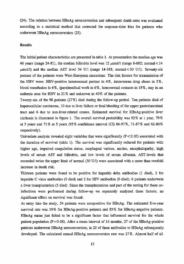
cirrhosis is illustrated in figure l. The overall survival probability was 92% at 1 year, 79%
at 3 years and 71% at 5 years (95% confidence interval (CI) 86-97%, 71-87% and 62-80%
respectively).
Univariate analysis revealed eight variables that were significantly (P<0.05) associated with
the duration of survival (table 1). The survival was significantly reduced for patients with
higher age, impaired coagulation status, esophageal varices, ascites, encephalopathy, high
levels of serum AST and bilirubin, and low levels of serum albumin. AST -levels that
exceeded twice the upper limit of normal (30 U/1) were associated with a more than twofold
increase in death risk.
Thirteen patients were found to be positive for hepatitis delta antibodies (1 died), 2 for
hepatitis C virus antibodies (0 died) and 2 for HIV antibodies (0 died); 6 patients underwent
a liver transplantation (3 died). Since the transplantations and part of the testing for these co
infections were performed during follow-up we separately analyzed these factors; no
significant effect on survival was found.
At entry into the study, 54 patients were seropositive for HBeAg. The estimated five-year
survival rate was 59% for HBeAg-positive patients and 83% for HBeAg-negative patients.
HBeAg status just failed to be a significant factor that influenced survival for the whole
patient population (P=0.08). After a mean interval of 16 months, 27 of the HBeAg-positive
patients underwent HBeAg seroconversion; in 25 of them antibodies to HBeAg subsequently
developed. The calculated annual HBeAg seroconversion rate was 27%. Almost half of all
13
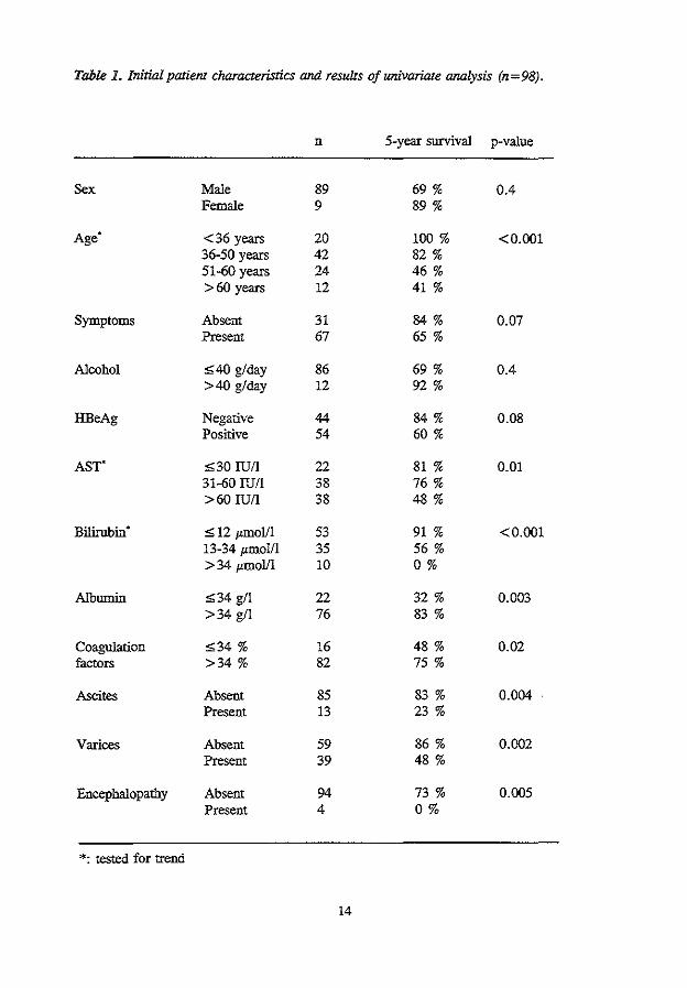
Table 1. Initial patient characteristics and results of univariate analysis (n = 98).
n 5-year survival p-value
Sex Male 89 69 % 0.4 Female 9 89%
Age .
<36 years 20 100% <0.001 36-50 years 42 82% 51-60 years 24 46% >60 years 12 41%
Symptoms Absent 31 84% O.Q7 Present 67 65 %
Alcohol ;S;40 g/day 86 69% 0.4 >40 g/day 12 92 %
HBeAg Negative 44 84% 0.08 Positive 54 60%
AST" ,;;30 IU/1 22 81% 0.01 31-60 lUll 38 76% >60 IU/1 38 48%
Bilirubin" ;S; 12 j.tillOI/1 53 91% <0.001 13-34 l'mol/1 35 56 % > 34 jLIDOlll 10 0%
Albumin ;S;34 gil 22 32 % 0.003 >34 gil 76 83 %
Coagulation ;S;34 % 16 48% 0.02 factors >34% 82 75%
Ascites Absent 85 83% 0.004 Present 13 23%
Varices Absent 59 86% 0.002 Present 39 48%
Encephalopathy Absent 94 73% 0.005 Present 4 0%
*: tested for trend
14
%survival 100
80
60
40
20
(88)
(72)
1 2
(59)
------------------~(4~4~)------~(~30) 71%
3 4 5
years
Figure 1. Survival of 98 patienJ:s with HBsAg-positive cirrhosis. Curve obtained by 6-month
interpolation of the Kaplan-Meier estimates. Numbers along the curve denote the number of
patients at risk.
HBeAg seroconversions occurred after treatment with a-interferon. No difference in survival
was found for patients with a spontaneous or therapy induced HBeAg seroconversion.
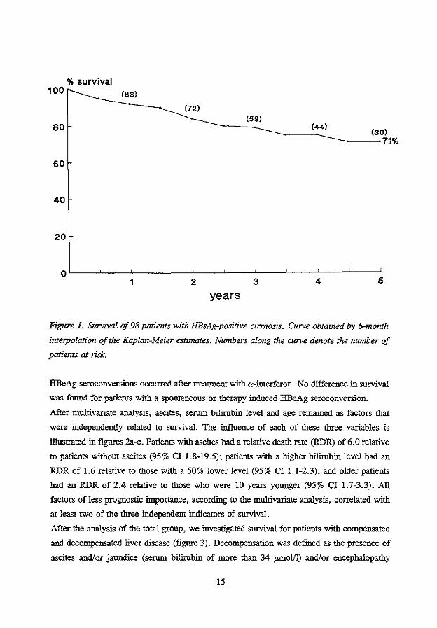
After multivariate analysis, ascites, serum bilirubin level and age remained as factors that
were independently related to survival. The influence of each of these three variables is
illustrated in figures 2a-c. Patients with ascites had a relative death rate (RDR) of 6.0 relative
to patients without ascites (95% CI 1.8-19.5); patients with a higher bilirubin level had an
RDR of 1.6 relative to those with a 50% lower level (95% CI 1.1-2.3); and older patients
had an RDR of 2.4 relative to those who were 10 years younger (95% CI 1.7-3.3). All
factors of less prognostic importance, according to the multivariate analysis, correlated with
at least two of the three independent indicators of survival.
After the analysis of the total group, we investigated survival for patients with compensated
and decompensated liver disease (figure 3). Decompensation was defmed as the presence of
ascites and/or jaundice (serum bilirubin of more than 34 !'moll!) and/or encephalopathy
15
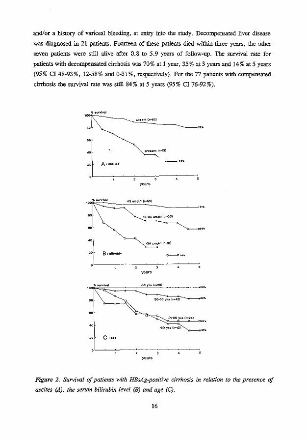
and/or a history of variceal bleeding, at entry into the study, Decompensated liver disease
was diagnosed in 21 patients, Fourteen of these patients died within three years, the other
seven patients were still alive after 0.8 to 5.9 years of follow-up. The survival rate for
patients with decompensated cirrhosis was 70% at 1 year, 35% at 3 years and 14% at 5 years
(95% CI 48-93%, 12-58% and 0-31%, respectively). For the 77 patients with compensated
cirrhosis the survival rate was still 84% at 5 years (95% CI 76-92%).
abaent (n-tiS) ·:1\----------~'----------. -
A' ucltea
•
~n•13)
-~3'1.
years
13-34 urno!/1 {n·35)
•311 umolll {n•10)
years
•~-~~~~~--~~~==~~~~'~'·~·~<··;'';'~~~=====: . -60 sa-so yra (n•42) 2
'
•
'" 20 C:age
years
Figure 2. Survival of patients with HBsAg-positive cirrhosis in relaJion to the presence of
ascites (A), the serum bilirubin level (B) and age (C).
16
%survival 1 OOI'jt--"*----*-"-"-*- Compensated (n•77)
80
60
40 Decompensated (n•21)
20 -------14%
years
Figure 3. Survival of HBsAg-positive patients with compensated or decompensated cirrhosis.
For the group of patients with compensated cirrhosis, age and HBeAg status at entry were
the only factors that significantly influenced the survival rate. The five-year survival
probability was 72% for HBeAg-positive patients compared to 97% for HBeAg-negative
patients (P=0.03; 95% CI 55-87% and 92-100%, respectively) (figure 4a). After adjustment
for age the difference in survival between compensated patients with and without detectable
HBeAg at presentation remained significant (P=0.04). The estimated actuarial HBeAg
seroconversion rate at 1 and 5 years was 22% and 63%, respectively. For patients who
showed clearance of HBeAg from serum, there was a 2.2 fold decrease in death rate (RDR
0.45; 95% CI 0.1-2.1) compared to patients who remained HBeAg positive. The age-adjusted
RDR after HBeAg seroconversion was 0.57 (95% CI 0.1-2.6). No difference in survival was
found between patients with different HBeAg starus in the decompensated group (figure 4b).
17
%survival HBeAg negative (n•34) 10 '" 80 HBeAg positive (n•43)
72.
60
40
20 A
0 2 3 4 5
years
%survival 10
HBeAg negative (n-10)
60
HBeAg positive (n•11) 40
••• 20 B
" 0 2 3 4 5
years
Figure 4. Survival of HBsAg-positive patients with compensated (A) and decompensated (B)
cirrhosis in relation to the HBeAg-status at entry.
Discussion
The results of this smdy show an estimated 71% five-year survival rate after histological
diagnosis of HBsAg-positive liver cirrhosis in a predominantly white West-European
population. Hepatocellular carcinoma and liver failure with or without variceal bleeding were
the main causes of death. Comparable results were obtained in prognostic smdies on HBsAg
positive cirrhosis in other geographical areas of the world with estimated five-year survival
rates of 55% in the USA, 80% in Taiwan, 66% in Japan and 52% in Italy (13-16). It is
18
conceivable that both our own survival rate and those of others are somewhat pessimistic
since most study centers function as regional hospitals where many tertiairy referrals are
treated.
In the present study, all investigated indicators of impaired liver function or portal
hypertension - bilirubin, albumin, blood coagulation status, the presence of ascites or
encephalopathy and evidence of esophageal varices - were strongly associated with survival,
according to the univariate analyses. The incidence of both liver transplantation and
concontitant vital infections (hepatits C, delta, HIV) was low and did not influence the
outcome of survival in this study. After multivariate analysis age, ascites and total serum
bilirubin remained as the most powerful prognostic indicators. A previous study of prognostic
factors of HBsAg-positive cirrhosis selected age above 40 years, serum bilirubin above 1.5
mg/dl (25 mmol/1), ascites and spider nevi as independent indicators of mortality (14). Other
studies on the prognosis for patients with cirrhosis (viral, alcoholic or cryptogenic) also
indicate that the strongest indicators of survival are parameters that relate to the synthetic
function of the liver and the presence of portal hypertension (26-29)' We separately analyzed
the survival rate for patients with compensated and decompensated cirrhosis, since the
findings for the whole group may have been influenced by the selected inclusion of patients
with an extremely poor prognosis who were referred to our deparnnent for liver
transplantation or treatment of variceal bleeding. To defme decompensation of liver disease
we selected four generally accepted criteria known to influence the survival of patients with
liver disease (29). For patients with decompensated cirrhosis the five-year survival rate was
only 14%. In contrast, patients with compensated cirrhosis exhibited a very good prognosis
with a five-year survival rate of 84%. Interestingly, in the compensated group none of the
patients (n=30) for whom follow-up was continued beyond the 5th year died in the 5 years
thereafter (data not shown).
Active HBV replication is associated with ongoing inflannnatory activity and progression of
liver disease. HBeAg seroconversion, indicating a transition to viral latency, is' usually
accompanied by biochentical and histological regression of liver disease activity (30-32). With
the availability of antiviral agents, such as a-interferon, cessation of the viral replicating
phase can be induced in about one-third of the patients (33,34). However, it is not yet known
whether HBeAg seroconversion leads to improved survival for cirrhotic patients (30,35).
Some authors state that the severity of the underlying liver histology at the time active viral
replication ceases is critical for the final outcome (35). In the present study, in which all
patients had sintilar histopathological diagnoses, patients without detectable HBeAg in the
serum had a more favorable prognosis than those with HBeAg. This phenomenon was even
more prominent among patients with compensated liver disease for whom there was a
19
significantly improved life expectancy for HBeAg-negative patients and a strong trend towards
better survival after HBeAg seroconversion during follow-up. This significance of the HBeAg
status did not disappear after adjustment for age. the only other factor that was indepently
related to the survival of patients with compensated cirrhosis. For patients with
decompensated cirrhosis, the HBeAg status did not influence the survival rate and it thus
seems likely that there is a time point in the course of the disease when HBeAg
seroconversion will no longer lead to an improved prognosis.
These results imply that there is a strong indication for antiviral therapy in patients with
HBeAg-positive compensated liver cirrhosis, whereas liver transplantation will rarely be
indicated in view of the very good prognosis for this group. However, if hepatic
decompensation occurs, liver transplantation appears to be the major therapeutic option to
reduce the risk of death. The exact timing of surgery as well as measures to suppress viral
replication to reduce the risk of reinfection of the graft remain to be determined.
20
References
1. Viola LA, Barrison IG, Coleman JC, Paradinas FJ, Fluker JL, Evans BA, Murray
Lyon IM. Natural history of liver disease in chronic hepatitis B surface antigen
carriers. Survey of 100 patients from Great Britain. Lancet 1981;2:1156-9.
2. Hoofnagle JH, Shafritz DA, Popper H. Chronic type B hepatitis and the "healthy"
HBsAg carrier state. Hepatology 1987;7:758-63.
3. Beasley RP, Hwang L-Y, Lin C-C, Chien C-S. Hepatocellular carcinoma and hepatitis
B virus. A prospective study of 22 707 men in Taiwan. Lancet 1981;2:1129-33.
4. Sakuma K, Takahara T, Okuda K, Tsuda F, Mayumi M. Prognosis of hepatitis B
virus surface antigen carriers in relation to routine liver function tests: a prospective
study. Gastroenterology 1982;83:114-7.
5. Johnson PJ, Williams R. Cirrhosis and the aetiology of hepatocellular carcinoma. J
Hepatol;1987:140-7.
6. Yang P-M, Chen D-S, Lai M-Y, Su I-J, Huang G-T, Lin J-T, Sheu J-C, et a!.
Clinicopathologic studies of asymptomatic HBsAg carriers older thao 40 years.
Hepatogastroenterol 1987;34:251-4.
7. Fattovich G, Brollo L, Giustina G, Noventa F, Pontisso P, Alberti A, Realdi G, et
a!. Natural history and prognostic factors for chronic hepatitis type B. Gut
1991;32:294-8.
8. Dudley FJ, Scheuer PJ, Sherlock S. Natural history of hepatitis associated antigen
positive chronic liver disease. Lancet 1972;2: 1388-93.
9. Lo K-J, Tong MJ, Chien M-C, Tsai Y-T, Liaw Y-F, Yang K-C, Chian H, eta!. The
natural course of hepatitis B surface antigen positive chronic active hepatitis in
Taiwan. J Infect Dis 1982;146:205-10.
10. Van Waes L, Segers J, Van Egmond J, Van Nimmen L, Barbier F, Wieme R,
Demeulenaere L. Chronic liver disease and hepatitis B antigen: a prospective study.
Br Med J 1974;3:444-6.
11. De Groote J, Fevery J, Lepoutre L. Long term follow-up of chronic active hepatitis
of moderate severity. Gut 1978;19:510-3.
12. Liaw Y-F, Tai D-I, Chu C-M, Chen T-J. The development of cirrhosis in patients
with chronic type B hepatitis: a prospective study. Hepatology 1988;8:493-6.
13. Liaw Y-F, LinD-Y, Chen T-J, Chu C-M. Natural course after the development of
cirrhosis in patients with chronic type B hepatitis: a prospective study. Liver
1989;9:235-41.
14. Weissberg n, Andres LL, Sntith CI, Weick S, Nichols JE, Garcia G, Robinson WS,
21
eta!. Survival in chronic hepatitis B. An analysis of 379 patients. Ann Intern Med
1984;101:613-6.
15. Tanaka R, Itoshinta T, Nagashinta H. Follow-up study of 582liver cirrhosis patients
for 26 years in Japan. Liver 1987;7:316-24.
16. D'Amico G, Morabito A, Pagliaro L, Marubini E, Caltagirone M, Filippazzo G,
Gatto G, et al. Survival and prognostic indicators in compensated and decompensated
cirrhosis. Dig Dis Sci 1986;31:468-75.
17. Ornata M, Ashcaval M, Liew C-T, Peters RL. Hepatocellular carcinoma in U.S.A.
Aetiologic consideration, localisation of hepatitis B antigens. Gastroenterology
1979;76:279-87.
18. Obata H, Hayashi N, Motoike Y, Hisarnitsu T, Okuda H, Kobayashi S, Nishioka K. A prospective study on the development of hepatocellular carcinoma from liver
cirrhosis with persistent hepatitis B virus infection. Int J Cancer 1980;25:741-7.
19. Oka H, Kurioka N, Kim K, Kanno T, Kuroki T, Mizoguchi Y, Kobayashi K.
Prospective study of early detection of hepatocellular carcinoma in patients with
cirrhosis. Hepatology 1990;12:680-7.
20. Beasley RP. Hepatitis B virus. The major etiology of hepatocellular carcinoma.
Cancer 1988;61:1942-56.
21. Schlichting P, Fauerholt L, Christensen E, Poulsen H, Juhl E, Tygstrup N. Clinical
relevance of restrictive morphological criteria for the diagnosis of cirrhosis in liver
biopsies. Liver 1981;1:56-61.
22. Ginai AZ, Van Buuren HR, Hop WCJ, Schalm SW. A quantitative radiological
method for evaluation of esophageal varices. A comparison of barium swallow with
endoscopy. Submitted for publication.
23. Vander Rijt CD, Schalm SW, De Groot GH, De Vlieger M. Objective measurement
of hepatic encephalopathy by means of automated EEG analysis.
Electroencephalography and clinical neurophysiology 1984;57:423-26.
24. Cox DR. Regression models and life tables. J R Stat Soc 1972;34:187-220.
25. Hop WCJ, Van Buuren HR. A method to evaluate changes in prognostic status during
follow-up, illustrated by an assessment of the effect of a rebleed in patients with
oesophageal variceal bleeding. Comput Bioi Med 1989;19:181-8.
26. Milani A, Marra L, Siciliano M, Rossi L. Prognostic significance of clinical and
laboratory parameters in liver cirrhosis. A multivariate statistical approach.
Hepatogastroenterol 1985;32:270-2.
27. Christensen E, Schlichting P, Fauerholdt L, Juhl E, Poulsen H, Tygstrup N, Balslov
JT, et a!. Changes of laboratory variables with time in cirrhosis: prognostic and
22
therapeutic significance. Hepatology 1985;5:843-53.
28. Rossi L, Milani A, Marra L, Siciliano M. Grading scores and survivorship functions
in liver cirrhosis: a comparative statistical analysis of various predictive models.
Hepatogastroenterol 1986;33:240-3.
29. Christensen E, Schlichting P, Fauerholdt L, Gluud C, Andersen PK, Juhl E, Poulsen
H, et al. Prognostic value of Child-Turcotte criteria in medically treated cirrhosis.
Hepatology 1984;4:430-5.
30. Realdi G, Alberti A, Rugge M, Bortolotti F, Rigoli AM, Tremolada F, Ruol F.
Seroconversion from hepatitis B e antigen to anti-HBe in chronic hepatitis B virus
infection. Gastroenterology 1980;79:195-9.
31. Fattovich G, Rugge M, Brollo L, Pontisso P, Novento F, Guido M, Alberti A, et al.
Clinical, virologic and histologic outcome following seroconversion from HBeAg to
anti-HBe in chronic hepatitis type B. Hepatology 1986;6: 167-72.
32. Hoofnagle lli, Dusheiko GM, Seeff LB, Jones EA, Waggoner JG, Buskell Bales Z.
Seroconversion from hepatitis B e antigen to antibody in chronic type B hepatitis. Ann
Intern Med 1981;94:744-8.
33. Hoofnagle lli, Peters M, Mullen KD, Jones DB, Rustgi V, Di Bisceglie A, Hallahan
C, et al. Randomized controlled trial of recombinant human «-interferon in patients
with chronic hepatitis B. Gastroenterology 1988;95:1318-25.
34. Perrillo RP, SchiffER, Davis GL, Bodenheimer HC Jr, Lindsay K, Payne J, Dienstag
JL, et al. A randomized controlled trial of interferon alfa-2b alone and after predniso
ne withdrawal for the treatment of chronic hepatitis B. N Eng! J Med 1990;323:295-
301.
35. Su I-J, Lai M-Y, Hsu H-C, Chen D-S, Yang P-M, Chuang S-M, Sung J-L. Diverse
virological, histopathological and prognostic implications of seroconversion from
hepatitis B e antigen to anti-HBe in chronic hepatitis B virus infection. J Hepatol
1986;3:182-9.
23

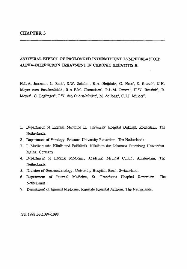
CHAPTER3
ANTIVIRAL EFFECT OF PROLONGED INTERMITTENT LYMPHOBLASTOID
ALPHA-INTERFERON TREATMENT .IN CHRONIC HEPATITIS B.
H.L.A. Janssen', L. Berk', S.W. Schalm1, R.A. HeijJ:ink2, G. Hess3, S. Rossol3, K-H.
Meyer zum Boschenfeide', R.A.F.M. Chamuleau4, P.L.M. Jansen', H.W. Reesink', B.
Meyer', C. Beglinger', J.W. den Onden-Muller", M. de Jong6, C.J.J. Mulder'.
1. Department of Internal Medicine II, University Hospital Dijkzigt, Rotterdam, The
Netherlands.
2. Department of Virology, Erasmus University Rotterdam, The Netherlands.
3. I. Medizinische Klinik und Poliklinik, Klinikum der Johannes Gutenberg Universitat,
Mainz, Germany.
4. Department of Internal Medicine, Academic Medical Centre, Amsterdam, The
Netherlands.
5. Division of Gastroenterology, University Hospital, Basel, Switzerland.
6. Department of Internal Medicine, St. Franciscus Hospital Rotterdam, The
Netherlands.
7. Department of Internal Medicine, Rijnstate Hospital Arnhem, The Netherlands.
Gut 1992;33:1094-1098
Abstract
In a European multicenter study 40 patients with HBeAg-positive chronic hepatitis B virus
(HBV) infection were treated with 5 megaunits of lymphoblastoid a-interferon daily
according to the following regimen: a 4-week primer course, 4 weeks of rest and a second
course lasting 16 to 30 weeks. After 52 weeks of follow-up, a response (HBeAg
seroconversion and HBV-DNA negativity) was observed in 22 patients (55%). HBsAg
seroconversion occurred in 5 patients (12.5%). One patient exhibited a relapse for serum
HBeAg and HBV-DNA after cessation of treatment. According to a response prediction
model, the observed response rate was not related to the selection of patients likely to
respond.
The initial interferon course induced a reduction of the serum HBV -DNA and HBeAg
levels of 87% and 18%, respectively, leading to a significantly lower level of viral
replication activity at the start of the second longterm course compared to baseline.
After 24 weeks of foiiow-up (week 16 of the second course), 19 (48%) patients exhibited
a response, 13 (32%) a partial response (HBeAg <50% of initial level or HBV-DNA
negative) and 8 (20%) no response. For 8 of the 13 partial responders treatment was
stopped at week 24 and viral replication rebounded to pretreatment values. In the last 5
partial responders prolongation of therapy up to week 38 led to a definite response and
HBsAg seroconversion in 3 of the 5 patients.
The results of this study suggest that both a short primer course and prolongation of
therapy may help to enhance the response rate of a-interferon therapy for chronic hepatitis
type B.
26
Introduction
In patients with chronic hepatitis B virus (HBV) infection active viral replication is usually
associated with continuing inflammatory activity and progression of liver disease.
Transition to viral latency, as indicated by hepatitis B e antigen (HBeAg) seroconversion
and clearance of serum HBV-DNA, is accompanied by biochemical and histological
regression of liver disease activity (1-3). With the availability of a-interferon (IFN),
termination of the viral replicating phase can be induced in about 30 to 40% of the
patients (4-6). Usually a-IFN is given in a single continuous course.
From a pilot study comparing the antiviral effect of a-IFN, acyclovir and a combination
of a-IFN and acyclovir, it was concluded that combination therapy appears to be a highly
promising treatment for chronic hepatitis B (7). Since more recent controlled studies have
shown that acyclovir does not enhance the therapeutic effect of a-IFN alone (8), the
results of the pilot study could also be interpreted as indicating that a second course of a
IFN following short-term primer a-IFN therapy is more effective than a single course.
Therefore we investigated the effect of intertnittent a-IFN treatment on termination of
hepatitis B viral replication, measured by HBeAg seroconversion and HBV-DNA
clearance from serum.
Methods
Patients and Treatment
Forty-four patients from 6 European hospitals were enrolled in the study after central
evaluation of their eligibility. At entry all patients had been seropositive for hepatitis B
surface antigen (HBsAg) and HBeAg for at least 6 months. Histological evidence of
chronic hepatitis was present in all patients who underwent a liver biopsy (n=43). Patients
were excluded on grounds of the following criteria: age under 18 or over 70 years;
presence of hepatitis delta antibodies in serum; antiviral or inunune modulatory therapies
in the preceding 6 months; history or presence of decompensated liver disease (ascites,
encephalopathy, variceal hemorrhage); pregnancy; impaired inununity, including
seropositivity for human inununodeficiency virus with T4 cells < 400/mm3; inadequate
levels of platelets ( < 70 x 109/l) or white blood cells ( < 3 x 1091!); recent drug or
alcohol abuse; presence of significant other disease that might interfere with the study.
The study was approved by the ethical committees of all participating centers and written
informed consent was obtained from each patient who entered the study.
Lymphoblastoid a-IFN (Wellferon, Wellcome, Beckeoham, U.K.) was given at a dose of
27
5 megaunits daily according to the following regimen: a 4-week primer course, a 4-week
rest period and a second a-IFN course lasting 16 weeks; for 5 patients from Rotterdam
the duration of the second course was prolonged up to 30 weeks. Patients were taught to
self-administer a-IFN subcutaneously. During the first 4 days of a-IFN therapy indome
thacin or paracetamol was given to suppress early side-effects. a-IFN treatment was
discontinued in the event of absence of HBeAg on two successive occasions or intolerable
side-effects. Follow-up began at the start of therapy and was continued for 52 weeks.
Clinical and Laboratory Evaluation
Patients were seen, and if indicated examined, at the outpatient clinic every 2 to 4 weeks
during treatment and every 4 to 8 weeks during the period thereafter. Laboratory
assessment was performed every 2 weeks during the treatment period and every 4 to 6
weeks after discontinuation of therapy. On these occasions routine hematological studies
were performed and serum markers of viral replication (HBeAg and HBV -DNA) and
aspartate aminotransferase (AST) activity were measured. Every 3 months additional
biochemical and virological measurements including the prothrombin time and the levels
of alanine aminotransferase, albumin, bilirubin, HBsAg, and antibodies against HBsAg as
well as HBeAg were performed. Liver biopsies were taken within 6 months of entry to the
study for histological assessment.
All virological parameters were determined centrally at the Rotterdam hepatitis laboratory.
HBsAg was assessed using a commercial radioimmunoassay kit (Abbott, ill., USA).
HBeAg was measured quantitatively using a radioimmunoassay (Abbott, Ill., USA). For
quantification a PIN ratio (counts of patient sample/counts of negative control sample) was
determined for each patient in a fixed serum dilution. HBeAg seroconversion was defined
as a PiN ratio under 2.1 for undiluted serum from 2 consecutive blood samples. HBV
DNA was measured by a liquid hybridization assay using an iodine-125 probe (Abbott,
Ill., USA). Antibodies to hepatitis C virus were determined, retrospectively, by enzyme
immunoassay (Abbott, Ill., USA); positive results were confirmed by a recombinant
immunoblot assay (Ortho Diagnostics Systems, USA). Routine serobiochemical tests were
performed using automated techniques (Coulter, Tecbnicon, NY, USA).
The criteria for response to treatment were: HBeAg seroconversion and serum HBV-DNA
negativity. The criteria for partial response to treatment were: a decrease in serum HBeAg
of 50% or more of the initial level or serum HBV-DNA negativity with sustained HBeAg
positivity after 24 weeks of follow-up.
To analyze whether the response rate was related to treatment modification or selection of
patients, the actual response was compared with a predicted response that was obtained
28
with a response model developed by Brook et al. (9). The prediction of response was
based on pretreatment AST levels, the presence of a history of acute hepatitis and HIV
antibody starus.
Statistics
Differences in dichotomous and other discrete variables were analyzed by the Fisher exact
and Chi-square tests, respectively. For continuous variables medians were used because
the results lacked normal distribution. The two-sample Wilcoxon rank sum test was used
to analyze unpaired observations and the Wilcoxon signed rank test to analyze paired
observations.
Results
Four of the 44 patients were withdrawn from the study. Two patients were found to be
HBeAg negative at the start or within one week of treatment while pre-entry assessments
were positive. Two patients were lost to follow-up: one withdrew from treatment after 2
weeks for reasons unrelated to the study protocol and one patient failed to comply with the
protocol after cessation of therapy.
Patient cbatacteristics at entry are shown in table I. Of the 40 patients who were
analyzed, one was serum HBV-DNA negative on entry to the study.
Response
A response to treatment (HBeAg seroconversion and serum HBV-DNA negativity) was
observed in 22 of the 40 patients (55%; 95% confidence interval (CI) 40-70%). Five
patients (12.5%; 95% Cl 4-27%) became negative for HBsAg. Sustained AST
normalization occurred in 16 out of 19 responders with elevated AST levels on entry to
the study. Fignre 1 shows the timing of elimination of viral parameters and sustained AST
normalization in the response group. All but one patient exhibited the response while on
a-IFN therapy. A characteristic sequence in clearance of HBV-DNA and HBeAg and then
normalization of AST levels was observed for the majority of the responders. One
responder showed a relapse for serum HBeAg, HBV-DNA and AST 20 weeks after
cessation of therapy. Of the 18 nonresponders, 5 exhibited normalization of AST levels
and 6 became- transiently- serum HBV-DNA negative.
Differences in characteristics between responders and nonresponders are shown in table 1.
The pretreatment serum HBV-DNA level was lower and the AST level higher in the
response group. Also the duration of HBsAg positivity was shorter and the presence of
29
Table 1. Patient characteristics at entry into the study.
Total Group Response Nonresponse
n=40 n=22 n=18
Age (yr) 38.5 (18-67) 42 (27-67) 37 (18-56)
Male/Fenta!e 32/8 17/5 15/3
Homo/Heterosexual 9/31 3/19 6/12
HBsAg duration· (mo) 27 (6-132) 14 (6-132) 33 (12-96)
History acute hepatitis 2 1 1
Histology: CPH 8 2 6
CAH 20 12 8
cirrhosis 11 8 3
Aoti-HCV positive 1 1 0
H1V -status: negative 38 21 17
unknown 2 1 1
HBeAg" (P/N ratio) 12.4 (2.2-18.8) 13.4 (2.5-18.8) 11.9 (2.2-16.4)
HBV-DNA" (pg/ml) 102 (1-1001) 79 (1-730) 127.5 (3-1001)
HBV-DNA < 50 pg/ml 11 8 3
51-100 9 6 3
101-300 13 5 8
> 300 7 3 4
AST" (U/1) 57.5 (15-475) 66.5 (15-475) 51.5 (16-113)
AST < 30 U/l 7 3 4
31-60 14 7 7
61-100 11 5 6
>100 8 7 1
* median (range)
AST normal < 30 U/1; HBV-DNA cut-off: 1.5 pg/ml; HBeAg cut-off: PIN ratio 2.1
30
IFN 100 100%
86%
80
eo
40 AST ---·
22.5%-20
HBsAg
oL_~~--L--L~--~-L--L--L-J--L_~--L-~~
0 4 8 12 16 20 24 28 32 36 40 44 48 52 56 60
weeks
Figure 1. Cumulative percentage of clearance of serum HBV-DNA, HBeAg
serocorrversion, normalization of serum AST and HBsAg seroconversion for patients who
responded to therapy (n=22).
cirrhosis was more prominent in responders. However, no statistical difference between
responders and nontesponders was found for any of these parameters.
According to the Brook model, a response was predicted for 14 of the 40 patients (35%;
95% CI 20-50%), with a positive predictive value of 79% and a negative predictive value
of 58%. Compared to this prediction the actual response rate of 55% was significantly
higher (P = 0.03).
Intennittent Treatment
Changes in serum AST, HBV-DNA and HBeAg values ate shown in figure 2. The primer
a-IFN course reduced HBV-DNA and HBeAg levels by 87 and 18 percent, respectively,
but the decrease did not continue during the 4 weeks without therapy. Nevertheless, at the
start of the second a-IFN course, the serum HBeAg aod HBV -DNA levels were signifi
cantly lower compared to baseline values. No difference was found in the AST levels at
the start of the two courses; however both courses seemed to induce a peak of AST values
that often preceeds a response to therapy.
31
HBeAg (PIN ratio)
14 111111111
8 -.
6
4
2
' ' ' ' ';
HBVDNA
IFN
AST
..... ---------------
HBVDNA !ootmn AST (Uill
120
100
80
60
40
---120
o~~~J_~~~~~~~~~=p±9==~=F~o -6 0 4 8 12 16 20 24 28 32 36 40 44 48 52
weeks
Figure 2. Median levels of HBeAg, HBV-DNA and ASTin serum (n=40).
Prolonged Treatmem
After 24 weeks of follow-up (week 16 of the second course), 19 (48%; 95% CI 32-63%)
patients exhibited a response, 13 (32%; 95% CI 18-47%) a partial response and 8 (20%;
95% CI 8-32%) a defmite nonresponse. For 8 of the 13 partial responders, treatment was
stopped at 24 weeks. After cessation of therapy the serum HBeAg and HBV-DNA levels
in these patients rebounded to baseline levels (figure 3). For the last 5 partial responders
a-IFN therapy was prolonged up to week 38. Three of these 5 exhibited an additional
response during prolonged therapy (figure 3), and all 3 showed HBsAg seroconversion
shortly after the response.
Side Effects of Imerjeron
During the first week of the initial a-IFN course, a transient flu-like syndrome with fever,
chills and myalgia was observed in nearly all of the patients. At the beginning of the
second a-IFN course, the majority again reported these symptoms but to a lesser intensity.
The predominant adverse effects after the first days of therapy were fatigue (73%),
32
HBeAg p/n ratio
20
15
5
IFN
HBeAg
HBVDNA
HBVDNA pg/ml
350
300
250
200
150
100
50
0 0 -6 0 4 8 12 16 20 24 28 32 36 40 44 48 52
HBeAg p/n ratio
20 -weeks
HBeAg
HBVDNA
IFN
HBVDNA pg/ml
500
400
300
200
100
0~~~-L~~~~~~-L~~~~~L$-L~~ -6 0 4 8 12 16 20 24 28 32 36 40 44 48 52
weeks
Figure 3. Serwn HBeAg and HBV-DNA levels in a partial responder treated until 24
weeks (protocol therapy) and in a partial responder treated until 32 weeks (prolonged
therapy) who exhibited an additional response.
33
myalgia (60%), anorexia (43%), irritability (30%) and bair loss (15%). Two patients
developed an acute psychosis: the first, who had just finished therapy, required intensive
psychiatric care for 2 weeks; the second, who had been treated with a-IFN for 3 weeks,
feU and fractured the acetabulum. Another patient had a generalized seizure after 4 and 11
weeks; he was put on anticonvulsive medication and a-IFN was tapered to 1.25 megaunits
daily. Side effects led to dose reduction in 11 subjects (28%). The reasons for dose
reduction were fatigue in 6 cases, thrombocytopenia in 2, neurotoxicity in 2 and
leukopenia in 1. Four (36%) out of these 11 patients exhibited a response.
Discussion
In recent years several strategies have been tested to enhance the efficacy of a-IFN
therapy for chronic hepatitis B. None of these strategies - including additional therapy
with agents such as gamma-interferon, prednisone, acyclovir or adeninide arabinoside
(5,8,10-12) - has proved to be more beneficial than a standard a-IFN course of 12-16
weeks.
In this uncontroiied pilot study we investigated the effect of prolonged intermittent a-IFN
therapy in 40 patients. Fifty-five percent of the patients responded with HBeAg
seroconversion and loss of serum HBV-DNA. HBsAg seroconversion occurred in 12.5%
of all treated patients and in almost 25% of those who responded. A majority of the
responders demonstrated a characteristic sequence in loss of serum HBV-DNA and
HBeAg foiiowed by normalization of the AST value. HBeAg seroconversion was often
heralded by a rise in transaminase activity and loss of HBV-DNA. Stiii, in 6 patients
HBV-DNA disappeared from the serum but quickly reappeared after discontinuation of
therapy, indicating that persistent absence of HBeAg is the best indicator for termination
of the viral replicating phase.
Although several differences in pretreatment characteristics of responders versus
nonresponders were observed, none of them was found to be significant. The predicted
response rate, based on a combination of pretreatment factors (9), was significantly lower
(35%) than the actual response rate (55%). This result suggests that the high response rate
was not caused by selection of patients likely to respond but by the treatment modifica
tion.
It is difficult to determine the contribution of intermittent therapy to the high response rate
obtained in this uncontroiied study. The rationale for the initial 4-week course was to
decrease viral replication and increase inflammatory activity before the start of the
Iongterm course, thereby enhancing the possibilty of response to therapy. At the start of
34
the second a-IFN course, viral replication, as monitored by serum HBV-DNA and HBeAg
levels, was indeed significantly lower compared to baseline values. In addition, both a
IFN courses induced transient elevation of aminotransferase levels. Therefore both courses
could have induced a response in some patients and the double transaminase peak could
thus be indicative of early and late responders. These interesting observations certainly
justify further evaluation of intermittent treatment in a controlled setting.
Close on-line monitoring of the effects of a-IFN on viral replication revealed that in
several patients HBeAg and HBV-DNA serum values decreased continuously and were
almost negative when treatment was stopped (week 24) at which point a relapse occurred.
Therefore, we decided to offer prolonged treatment to the remaining patients who
approximated a response at the end of the scheduled therapy (partial response). For 3 of
the 5 partial responders this approach led to a definite response and subsequently to
HBsAg seroconversion. Of the 2 patients who did not respond to prolonged treatment one
had extremely high initial serum HBV-DNA levels ( > 1000 pglml), while both patients
had near normal AST levels at entry. These features - low serum aminotransferase and
high HBV-DNA levels - have been suggested to interfere with the response to a-IFN
treatment (4,5,9,13). Onr fmdings indicate that prolonging a-IFN therapy may increase
the HBeAg and HBsAg seroconversion rates in the subset of patients that partially respond
to standard interferon treatment. To select patients eligible for treatment prolongation,
close monitoring of quantified levels of HBeAg and HBV -DNA is essential.
From the literature it is not clear whether prolongation of therapy beyond 4 months yields
a higher response rate. Prolonged therapy can further decrease HBV replication which
may trigger the cellular immune response leading to the hepatocytotoxic reaction that
eradicates viral replication (14). In most parts of the world the standard duration of
therapy is considered to be 12-16 weeks, usually leading to a response rate of 30-40% (4-
6,15). a-IFN has been given for 24 weeks or longer in a few - mainly Mediterranean -
studies (response rates 26% to 70%) (16-18). A controlled ttial comparing 12 to 24 weeks
of treatment failed to demonstrate any beneficial effect of the prolonged therapy (19).
However, the outcome of that study was markedly iofluenced by a lack of compliance (8
of the 20 patients withdrew from the longer course). In the present study none of the
patients had to withdraw from therapy because of side effects, irrespective of the length of
treatment. Both the intermittent treatment schedule and intensive patient monitoring with
mental support may have contributed to the good compliance. Nevertheless, 3 of our
patients had major neuro-psychiatric side effects (psychosis, seizures). Future studies
might elucidate whether these serious side effects relate to dose and/ or duration of the a
IFN treatment.
35

In summary, prolonged intermittent a-IFN therapy resulted in HBeAg seroconversion and
serum HBV-DNA negativity in 55% and in HBsAg seroconversion in 12.5% of the
patients. The high rate of induced viral latency was probably related firstly to the short
initial a-IFN course that reduced viral replication significantly before the start of the
second longterrn course and secondly to prolongation of therapy in partial responders
which induced additional HBeAg seroconversion.
36
References
1. Realdi G, Alberti A, Rugge M, Bertolotti F, Rigoli AM, Tremolada F, Ruol A.
Seroconversion from HBe antigen to antiHBe in chronic hepatitis B virus infection.
Gastroenterology 1980;79:!95-9.
2. Hoofnagle JH, Dusheiko GM, Seef LB, Jones EA, Waggoner JG, Bales ZB.
Seroconversion from HBe antigen to antibody in chronic type B hepatitis. Ann Intern
Med 1981;94:744-8.
3. Fattovich G, Rugge M, Brollo L, Pontisso P, Noventa F, Gnido M, Alberti A,
Realdi G. Clinical virologic and histologic outcome following seroconversion from
HBeAg to antiHBe in chronic hepatitis type B. Hepatology 1986;6:167-72.
4. Hoofnagle JH, Peters M, Mullen KD, Jones DB, Rustgi V, Di Bisceglie A, et a!.
Randomized controlled trial of recombinant human a-interferon in patients with
chronic hepatitis B. Gastroenterology 1988;95:1318-25.
5. Perrillo RP, Schiff ER, Davis GL, Bodenheimer HC Jr, Lindsay K, Payne J,
Dienstag JL, et al. A randomized controlled trial of interferon alfa-2b alone and after
prednisone withdrawal for the treatment of chronic hepatitis B. N Eng! J Med
1990;323:295-301.
6. Brook MG, Chan G, Yap I, Karayiannis P, Lever AM, Jacyna M, Main J, Thomas
HC. Randomised controlled trial of lymphoblasoid interferon alfa in Europid men
with chronic hepatitis B virus infection. BMJ 1989;299:652-6.
7. Schalm SW, Van Buuren HR, Heijtink RA, De Man RA. Acyclovir enhances the
antiviral effect of interferon in chronic hepatitis type B. Lancet 1985;ii:358-60.
8. Berk L, Schalm SW, De Man RA, Heijtink RA, Berthelot P, et al. Failure of
acyclovir to enhance the antiviral effect of alpha lymphoblastoid interferon on HBe
seroconversion in chronic heaptitis B. A multi-centre randomized controlled trial. J
Hepatol 1992;14:305-9.
9. Brook MG, Karayiannis P, Thomas HC. Which patients with chronic hepatitis B
virus infection will respond to alpha-interferon therapy? A statistical analysis of
predictive factors. Hepatology 1989;10:761-3.
10. Garcia G, Smith CI, Weissberg JI, Eisenberg M, Bissett J, et a!. Adenide
arabinoside monophosphate (vidarabine phosphate) in combination with human
leukocyte interferon in the treatrnem of chronic hepatitis B. Ann Intern Med
1987;107:278-85.
11. Di Bisceglie AM, Rustgi VK, Kassianides C, Lisken-Melman M, Park Y, Waggoner
JG, Hoofnagle JH. Therapy of chronic hepatitis B with recombinant human alpha
37
and gamma interferon. Hepatology 1990;2:266-70.
12. Hoofnagle JH. a-Interferon therapy of chronic hepatitis B. Current status and
recommendations. J Hepatol1990;ll:S100-7.
13. l.ok ASF, Lai CL, Wu PC, Leung EKY. Long-term follow-up in a randontised
controlled trial of recombinant alpha-2 interferon in Chinese patients with chronic
hepatitis B infection. Lancet 1988;ii:298-302.
14. Thomas HC. The hepatitis B virus and the host response. J Hepatoi1990;11:S83-9.
15. Fattovich G, Brollo L, Boscaro S, Pontisso P, Giustina G, et a!. Long-term effect of
low dose recombinant interferon therapy in patients with chronic hepatitis B. J
Hepatoll989;9:331-7.
16. Saracco G, Mazzella G, Rosina F, Cancellieri C, Lattore V, eta!. A controlled trial
of huntan lymphoblastoid interferon in chronic hepatitis B in Italy. Hepatology
1989;10:336-41.
17. Carreno V, Porres JC, Mora I, Battolome J, Bas C, et a!. Prolonged (6 months)
treatment of chronic hepatitis B virus infection with recombinant leukocyte A
interferon. Liver 1987;7:325-32.
18. Alexander GJM, Bralnn J, Fagan EA, Smith HM, Daniels HM, Eddleston ALWF,
Williams R. Loss of HBsAg with interferon therapy in chronic hepatitis B virus
infection. Lancet 1987;ii:66-9.
19. Scnlly U, Shein R, Karayiannis P, McDonald JA, Thomas HC. Lymphoblastoid
interferon therapy of chronic hepatitis HBV infection. A comparison of 12 vs 24
weeks of thrice weekly treatment. J Hepatol1987;5:51-8.
38
CHAPTER4
ALPHA-INTERFERON AND ZIDOVUDINE COMBINATION THERAPY FOR
CHRONIC HIEPATITIS TYPE B. RESULTS OF A RANDOMIZED PLACEBO
CONTROLLED TRIAL.
H.LA. Janssen1, L Berk1, R.A. Heijtink', F.J.W. ten Kate3
, S.W. Schalm1•
1. Department of Internal Medicine II, University Hospital Dijkzigt, Rotterdam,
The Netherlands.
2. Department of Virology, Erasmus University, Rotterdam, The Netherlands.
3. Department of Pathology, Academic Medical Center, Amsterdam, The Netherlands.
Hepatology 1993;17:383-388
Abstract
Alpha-interferon ("-IFN) therapy leads to HBeAg seroconversion in only one-third of the
patients with chronic hepatitis B. In an attempt to increase the seroconversion rate we
investigated the combination of "-IFN and zidovudine in a subset of patients with a
presumably low response rate for "-IFN monotherapy. In a double-blind controlled trial
24 HBeAg-positive patients were randomized to receive lymphoblastoid "-IFN in doses
increasing to 5 MU s.c. daily, combined with zidovudine given orally in doses increasing
from 500 to 1000 mg or placebo daily for 16 weeks. Treannent effects were monitored by
quantitative assessment of HBV-DNA, HBeAg and HBV-DNA polymerase. Six months
after termination of therapy 1112 (8%; 95% CI 2-39%) patients treated with "-IFN plus
zidovudine and 2/12 (17%; 95% CI 2-48%) patients from the control group exhibited a
response (HBeAg seroconversion). All patients remained HBsAg positive. The only
responder of the "-IFN-zidovudine group relapsed after cessation of therapy so that none
of the zidovudine-treated patients were HBeAg negative at the end of follow-up. No
significant difference in AST or any of the virological markers was observed between the
2 groups during the entire course of the stody. Adverse effects (anemia, leukopenia)
necessitated a reduction in the dose of zidovudine in 50% and "-IFN in 42% of the pa
tients treated with "-IFN plus zidovudine; in the control group these rates were 0% for
placebo and 8% for "-IFN. In conclusion, the antiviral effect of "-IFN in chronic
hepatitis B was not enhanced by additional zidovudine treannent. The combination therapy
induced considerable side effects leading to dose reduction for both zidovudine and "-IFN.
For combination therapy with a-IFN, oral nucleoside analogues with a more potent
antiviral effect and less toxicity than zidovudine should be developed.
40
Introduction
Alpha-interferon (a-IFN) is currently the most effective therapy for chronic hepatitis B
virus (HBV) infection. Nevertheless only one-third of the patients respond to this
treatment with virological and biochemical remission of the disease (1-3). One of the ways
to enhance the response rate for a-IFN is combination therapy with nucleoside analognes.
Zidovudine (3'-azido-3'-deoxythymidine, AZT) has proven to be a potent inhibitor of the
reverse transcriptase activity of the human immunodeficiency virus (HIV) (4,5). Since
reverse transcriptase activity also plays an important role in the HBV replication cycle,
zidovudine would appear to be a promising candidate for combination therapy with a-IFN
which attacks the virus via other steps in the HBV replication cycle and via the cellular
immune system.
After both in vitro and in vivo studies demonstrated that zidovudine caused a marked
decrease in HBV replication, as monitored by HBV-DNA polymerase (6), we initiated a
randomized controlled trial to compare the antiviral effect of a-IFN alone with that of the
combination of a-IFN and zidovudine.
Patients and Methods
Twenty-four patients were randomized to receive a-IFN therapy combined with either
zidovudine or placebo. The inclusion criteria were serum hepatitis B surface antigen
(HBsAg) positivity for at least 12 months, histological evidence of chronic hepatitis and
the presence of serum hepatitis B e antigen (HBeAg) for at least 6 months. Since the
toxicity profile of zidovudine was not completely known at the time of the design of the
study we were only permitted to treat patients who exhibited at least one of the following
characteristics: previous nonresponse to a-IFN, anti-HIV positivity and/or the presence of
cirrhosis. Patients were excluded for the following reasons: age below 18 or above 65
years; presence of antibodies against the hepatitis delta virus in serum; low hemoglobin
level ( < 7 mmol/1), thrombocyte count ( < 100 x 109/1) or leukocyte count ( < 3 x 10⁹/1);
anti-HIV seropositivity with a CD4 cell count below 400 per ml; decompensated liver
disease (ascites, albuntin < 30 gil, encephalopathy, history of variceal bleeding);
pregnancy; recent drog or alcohol abuse; antiviral or immunosuppressive therapy in the 6
months prior to enrollment; presence of other significant diseases which might interfere
with completion of the study.
Lymphoblastoid a-interferon (Wellferon, Wellcome, Beckenham, UK) was given subcuta
neously to all patients according to an increasing dose schedule of 1.5 ntillion units daily
41
for 4 weeks, 3 million units daily for 8 weeks and 5 million units daily for 4 weeks.
Patients were carefully instructed to self-administer the a-IFN. Simultaneously with the
16-week a-IFN course, zidovudine or placebo (Wellcome, Beckenbam, UK) was
administered orally in a dose of 250 mg twice daily for the first 8 weeks and 500 mg
twice daily for the last 8 weeks.
All patients were followed in the hepatology outpatient clinic of our hospital which has a
tertiary referral function. Twenty-nine consecutive patients were eligible for the study;
five were excluded during the pretreatment 6-week screening period: two decided not to
participate, one HIV-infected patient exhibited a drop in the CD4 cell count under 400/ml,
one patient cleared serum HBeAg and in another the HBeAg level dropped to borderline
positivity.
For randomization a serial number that corresponded to a set of boules containing either
zidovudine or placebo capsules was randomly assigned to each consecutive patient who
started therapy. Investigators and patients were unaware whether zidovudine or placebo
was given. To exclude any indication of the dose of zidovudine, all patients were given 2
capsules twice daily during the entire treatment period. The randomization code was
prepared by the Wellcome Foundation (Beckenbam,UK), kept in a sealed envelope by the
hospital pharmacist and opened after the last patient had completed therapy. All randomi
zed patients gave written informed consent before participation. The study was approved
by the hospital committee of medical ethics.
Patients were seen by a physician in the hospital at monthly intervals and followed for 6
months after cessation of therapy. Blood samples were obtained weekly during therapy
and monthly in the period thereafter. HBeAg, HBV-DNA polymerase, aspartate amino
transferase (AST), hemoglobin and leukocytes were assessed in all samples. The HBV
DNA level was determined monthly. Additional routine hematological, biochemical and
virological tests were performed at entry and at the termination of both therapy and
follow-up. Patients were asked to undergo a liver biopsy within 6 months prior to the start
of therapy and at the end of follow-up. The biopsies were examined under code by a
single experienced pathologist who was unaware of the chronological order of the biopsies
and the design of the trial. Histological scoring was based on the histological activity
index, as described by Knodel! (7).
The hematological, chemical and virological measurements were each performed in a
single laboratory. HBsAg and antibodies against HBsAg, HBeAg, hepatitis C and hepatitis
delta virus were assessed by enzyme immunoassays (Abbott, Ill, USA): positive results for
hepatitis C antibodies were confirmed by a recombinant immunoblot assay (Orlho
Diagnostic Systems, USA). HBeAg, HBV-DNA polymerase and HBV-DNA were
42
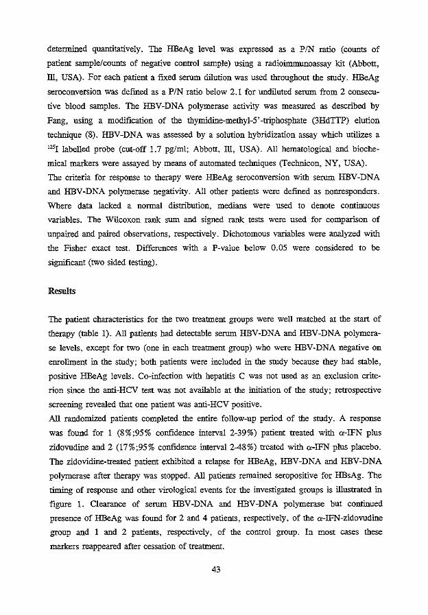
determlned quantitatively. The HBeAg level was expressed as a PIN ratio (counts of
patient sample/counts of negative control sample) using a radioimmunoassay kit (Abbott.
ill, USA). For each patient a fixed serum dilution was used throughout the study. HBeAg
seroconvetsion was defmed as a PIN ratio below 2.1 for undiluted serum from 2 consecu
tive blood samples. The HBV -DNA polymerase activity was measured as described by
Fang, using a modification of the thymidine-methyl-5' -triphosphate (3HdTTP) elution
technique (8). HBV-DNA was assessed by a solution hybridization assay which utilizes a 125I labelled probe (cut-off 1.7 pg/mi; Abbott, lll, USA). All hematological and bioche
mical markers were assayed by means of automated techniques (Technicon, NY, USA).
The criteria for response to therapy were HBeAg seroconversion with serum HBV-DNA
and HBV-DNA polymerase negativity. All other patients were defined as nonresponders.
Where data lacked a normal distribution, medians were used to denote continuous
variables. The Wilcoxon rank sum and signed rank tests were used for comparison of
unpaired and paired observations, respectively. Dichotomous variables were analyzed with
the Fisher exact test. Differences with a P-value below 0.05 were considered to be
significant (two sided testing).
Results
The patient characteristics for the two treatment groups were weii matched at the start of
therapy (table 1). All patients had detectable serum HBV-DNA and HBV-DNA polymera
se levels, except for two (one in each treatment group) who were HBV-DNA negative on
enroiiment in the study; both patients were included in the study because they had stable,
positive HBeAg levels. Co-infection with hepatitis C was not used as an exclusion crite
rion since the anti-HCV test was not available at the initiation of the study; retrospective
screening revealed that one patient was anti-HCV positive.
All randomized patients completed the entire foiiow-up period of the study. A response
was found for 1 (8%;95% confidence interval 2-39%) patient treated with a-IFN plus
zidovudine and 2 (17%;95% confidence interval 2-48%) treated with a-IFN plus placebo.
The zidovidine-treated patient exhibited a relapse for HBeAg, HBV-DNA and HBV-DNA
polymerase after therapy was stopped. All patients remained seropositive for HBsAg. The
tinting of response and other virological events for the investigated groups is illustrated in
fignre 1. Clearance of serum HBV-DNA and HBV-DNA polymerase but continued
presence of HBeAg was found for 2 and 4 patients, respectively, of the a-IFN-zidovudine
group and 1 and 2 patients, respectively, of the control group. In most cases these
markers reappeared after cessation of treatment.
43
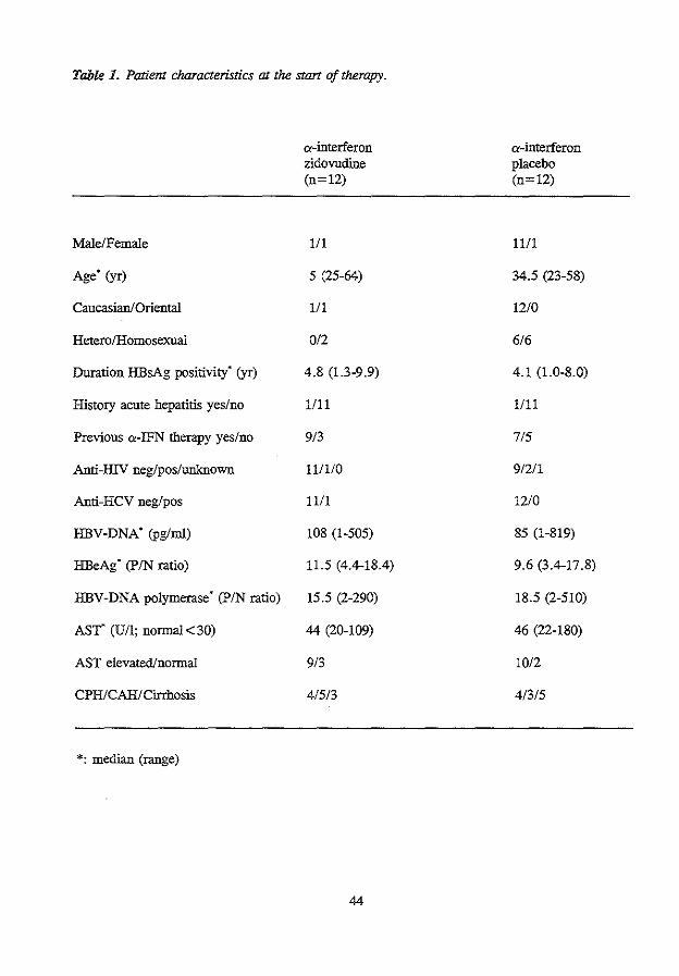
Table 1. Patient characteristics 01 the start of therapy.
Male/Female
Age (yr)
Caucasian/Oriental
Fletero/F!omosexual
Duration .FIBsAg positivity• (yr)
F!istory acute hepatitis yes/no
Previous a-IFN therapy yes/no
Anti-.HlV neg/pos/unknown
Anti-F!CV neg!pos
.FIBV-DNA• (pglml)
.FIBeAg· (PIN ratio)
.FIBV-DNA polymerase (PIN ratio)
AST• (U/1; normal< 30)
AST elevated/normal
CPF!!CAF!I Cirrhosis
*: median (range)
a-interferon zidovudine (n=12)
1/1
5 (25-64)
1/1
0/2
4.8 (1.3-9.9)
1/11
9/3
111110
1111
108 (1-505)
11.5 (4.4-18.4)
15.5 (2-290)
44 (20-109)
9/3
4/5/3
44
a-interferon placebo (n=12)
11/1
34.5 (23-58)
12/0
6/6
4.1 (1.0-8.0)
1111
7/5
9/2/1
12/0
85 (1-819)
9.6 (3.4-17.8)
18.5 (2-510)
46 (22-180)
10/2
4/3/5
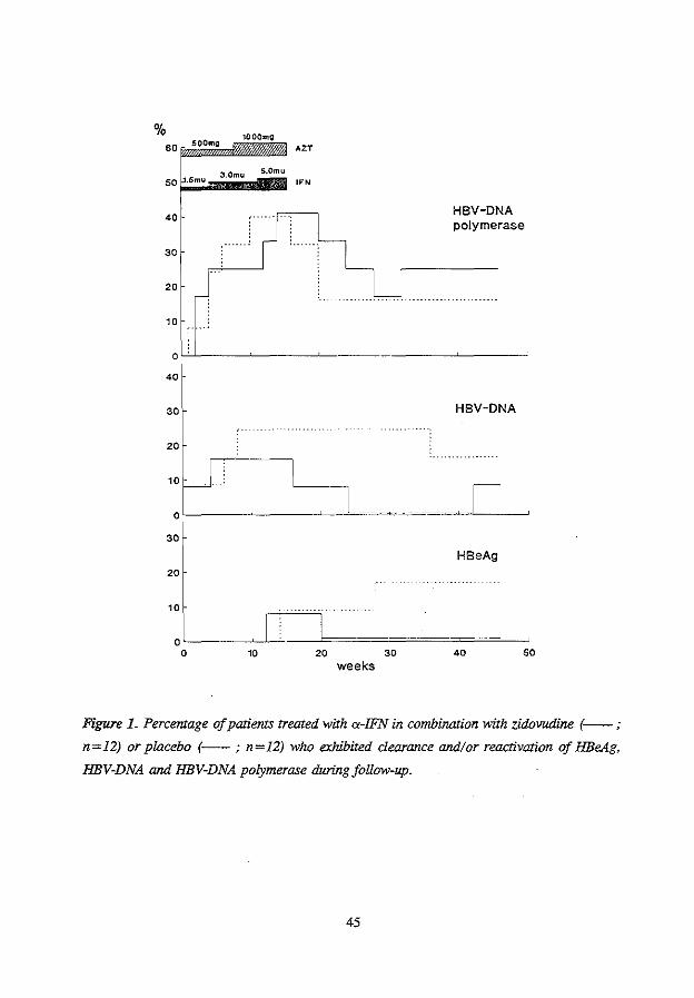
% SO SOOmg
1000mg
SO .5mu 3.0mu S.Omu
40
30
20
10
AZT
OFN
HBV-DNA polymerase
0'-'-'---~--~---~---~--~
40
30 HBV-DNA
20
30
HBeAg 20
weeks
Figure L Percentage of patients treated with a-IFN in combination with zidovudine (--- ;
n=12) or placebo (---; n=I2) who exhibited dearance and/or reactivation of HBeAg,
HBV-DNA and HBV-DNA polymerase during follow-up.
45
During the treatment period the median decrease in HBV-DNA was 71% and 67%,
HBeAg 29% and 20%, and HBV-DNA polymerase 62% and 71% for the a-IFN-zidovu
dine and a-IFN-placebo groups, respectively; these results indicate a significant decrease
in these viral markers for both groups (figure 2). However, after therapy all 3 parameters
rebounded to pretreatment levels among the majority of the patients, leaving no significant
difference between the initial virological levels and those at the end of the observation
period. Of the 19 patients with an elevated AST level at entry only the 2 permanent
responders and 1 nonresponder treated with a-IFN and zidovudine exhibited sustained
normalization of AST. In 2 patients from each treatment group - all nonresponders - the
AST level rose to more than twice the initial value during therapy. Comparison of the two
therapy groups did not show a significant difference in AST or any of the quantified
virological markers during the entire course of the study (figure 2).
The results of histological scoring are shown in table 2. One patient from each therapy
group refused to undergo the second biopsy. A decrease in the histology activity index
was found for 3 (27%) patients treated with zidovudine and 4 (36%) treated with placebo,
including the 2 persistent responders. No significant difference was found either within or
between the two groups in initial and posttreatment histological scores.
Adverse effects of a-IFN and/or zidovudine therapy were observed in the majority of the
patients (table 3). Despite the low initial dose of a-IFN most patients experienced flu-like
disease with fever, fatigue and myalgia, primarily during the first 2 weeks of treatment.
The predominant side effect later in the course of therapy was fatigue. Almost all adverse
effects were more common among patients treated with a-IFN and zidovudine. In
particular anemia and leukopenia were pronounced in this group; in 2 patients the anemia
was symptomatic and necessitated blood transfusions (hemoglobin < 5. 0 mmol/1). One
(HlV-positive) patient from the a-IFN-placebo group experienced epileptic insults in the
lOth and 16th weeks of therapy; after the first seizure a-IFN was continued because no
clear relationship between therapy and insult was suspected (9). After the end of the
observation period hepatocellular carcinoma was diagnosed in one patient while another
died of a variceal hemorrhage. Both patients had received a-IFN plus zidovudine;
however a direct relation between these serious events and the treatment regimen appears
unlikely. For a total of 9 patients the dose of zidovudine and/or a-IFN had to be reduced
(table 3): in 4 cases zidovudine was reduced, all because of anemia; in 3 a-IFN was
reduced, all because of leukopenia; and in 2 both zidovudine and a-IFN were reduced
because of leukopenia with anemia or thrombocytopenia. The time of dose reduction
ranged from 8 to 14 (median 12) weeks of therapy for zidovudine and from 8 to 13 weeks
(median 12) for a-interferon; after the dose reduction zidovudine was discontinued in 4
46
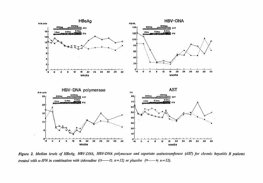
.,. "'~ HBeAg '"'"' HBV -DNA &OOm IOOO.t AZT 140 OOOmg toOOmt loJ.T
14 u ... u a.Omv &.Omu lfN 120 U~~tu a.o~r~~~ e.omu lfN
12 '* 100 !t, 10 ... - ----· : * 80 ' . •·······- .
· .. ,. • ..... \ ~ .:'-1• 6 ' ' ' ', ....... f . ~
2 :20 *-·---
0 0 -6 0 4 8 12 t6 :20 24 :26 32 36 42 -6 0 4 8 12 '16 20 24 26 32 36 40
weeks weeks
HBV-DNA polymerase AST PIH r&tiCI U/1
26 r 600mt IOOOnlt AZT 80 60011111 100011111 AU
••f 1.6mu 3.0mll 6.0mll IFN 70 Mmu 3.0mll 6.0mu IFN
•• • ."·-.. -· -.. 16~ \', R SO · . .,.1!1..!11, +·"'"
40 .\_ /' ~-!Of \ vr ...... ; ·-y· .. -·· ·---- ... __ * 30 .•
·r .. . :: 0 I I I 0 -a o 4 a 12 1e 20 24 2e a2 aa -12 -e o 4 a 12 ta 20 24 2e a2 aa 42
weeks weeks
Figure 2. Median levels of HBeAg, HBV-DNA, HBV-DNA polymerase and aspartate aminotransferase (AST) for chronic hepatitis B patients
treated with a-/FN in combination with zidovudine (a--o; n=J2) or placebo (*------*; n=l2).
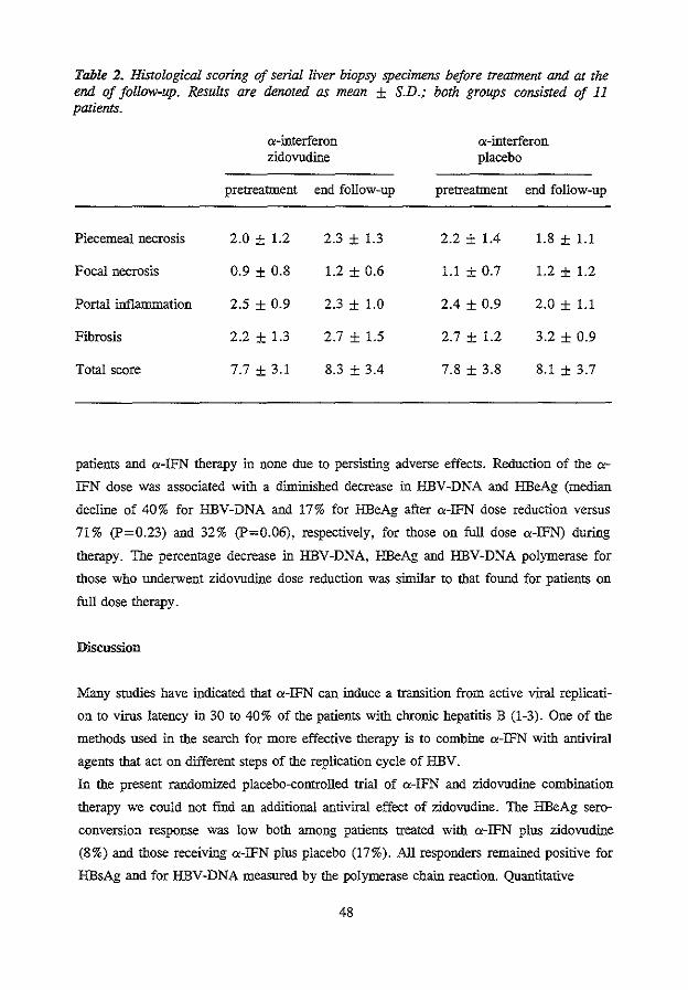
Table 2. Histological scoring of serial liver biopsy specimens before treatment and at the end of follow-up. Results are denoted as mean ± S.D.; both groups consisted of 11 patients.
a-interferon a-interferon zidovudine placebo
pretreatment end follow-up pretreatment end follow-up
Piecemeal necrosis 2.0 ± 1.2 2.3 ± 1.3 2.2 ± 1.4 1.8 ± 1.1
Focal necrosis 0.9 ± 0.8 1.2 ± 0.6 1.1 ± 0.7 1.2 ± 1.2
Portal inflammation 2.5 ± 0.9 2.3 ± 1.0 2.4 ± 0.9 2.0 ± 1.1
Fibrosis 2.2 ± 1.3 2.7 ± 1.5 2.7 ± 1.2 3.2 ± 0.9
Total score 7.7 ± 3.1 8.3 ± 3.4 7.8 ± 3.8 8.1 ± 3.7
patients and a-IFN therapy in none due to persisting adverse effects. Reduction of the a
IFN dose was associated with a diminished decrease in HBV-DNA and HBeAg (median
decline of 40% for HBV-DNA and 17% for HBeAg after a-IFN dose reduction versus
71% (P=0.23) and 32% (P=0.06), respectively, for those on full dose a-IFN) during
therapy. The percentage decrease in HBV-DNA, HBeAg and HBV-DNA polymerase for
those who underwent zidovudine dose reduction was similar to that found for patients on
full dose therapy.
Discussion
Many srudies have indicated that a-IFN can induce a transition from active viral replicati
on to virus latency in 30 to 40% of the patients with chronic hepatitis B (1-3). One of the
methods used in the search for more effective therapy is to combine a-IFN with antiviral
agents that act on different steps of the replication cycle of HBV.
In the present randomized placebo-controlled trial of a-!FN and zidovudine combination
therapy we could not find an additional antiviral effect of zidovudine. The HBeAg sere
conversion response was low both among patients treated with a-IFN plus zidovudine
(8%) and those receiving a-IFN plus placebo (17%). All responders remained positive for
HBsAg and for HBV-DNA measured by the polymerase chain reaction. Quantitative
48
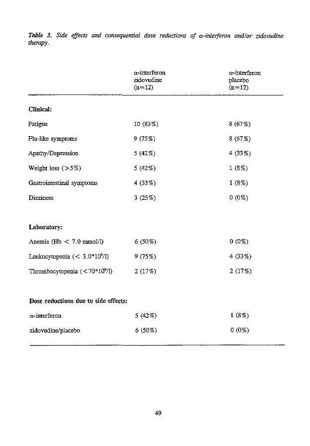
Table 3. Side effects and consequential dose reductions of a-interferon and/or zidovudine therapy.
Clinical:
Fatigue
Flu-like symptoms
Apathy /Depression
Weight loss (> 5%)
Gastrointestinal symptoms
Dizziness
Laboratory:
Anemia (Hb < 7.0 mmol/1)
Leukocytopenia ( < 3.0*109/1)
Thrombocytopenia ( <70*109/l)
Dose reductions due to side effects:
a-interferon
zidovudine/placebo
a-interferon zidovudine (n=12)
10 (83%)
9 (75%)
5 (42%)
5 (42%)
4 (33%)
3 (25%)
6 (50%)
9 (75%)
2 (17%)
5 (42%)
6 (50%)
49
a-ID.terferon placebo (n=12)
8 (67%)
8 (67%)
4 (33%)
1 (8%)
1 (8%)
0 (0%)
0 (0%)
4 (33%)
2 (17%)
1 (8%)
0 (0%)
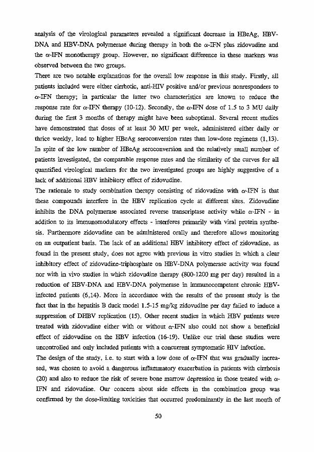
analysis of the virological parameters revealed a significant decrease in HBeAg, HBV
DNA and HBV-DNA polymerase during therapy in both the a<-IFN plus zidovudine and
the a<-IFN monotherapy group. However, no significant difference in these markers was
observed between the two groups.
There are two notable explanations for the overall low response in this smdy. Firstly, all
patients included were either cirrhotic, anti-HIV positive and/or previous nonresponders to
a<-IFN therapy; in particular the latter two characteristics are known to reduce the
response rate for a<-IFN therapy (10-12). Secondly, the a<-IFN dose of 1.5 to 3 MU daily
during the frrst 3 months of therapy might have been suboptimal. Several recent smdies
have demonstrated that doses of at least 30 MU per week, administered either daily or
thrice weekly, lead to higher HBeAg seroconversion rates than low-dose regimens (1,13).
In spite of the low number of HBeAg seroconversion and the relatively small number of
patients investigated, the comparable response rates and the similarity of the curves for all
quantified virological markers for the two investigated groups are highly suggestive of a
lack of additional HBV inhibitory effect of zidovudine.
The rationale to smdy combination therapy consisting of zidovudine with a<-IFN is thst
these compounds interfere in the HBV replication cycle at different sites. Zidovudine
inhibits the DNA polymerase associated reverse transcriptase activity while a<-IFN - in
addition to its innnunomodulatory effects - interferes primarily with viral protein synthe
sis. Furthermore zidovudine can be administered orally and therefore allows monitoring
on an outpatient basis. The lack of an additional HBV inhibitory effect of zidovudine, as
found in the present smdy, does not agree with previous in vitro smdies in which a clear
inhibitory effect of zidovudine-triphosphate on HBV-DNA polymerase activity was found
nor with in vivo studies in which zidovudine therapy (800-1200 mg per day) resulted in a
reduction of HBV-DNA and HBV-DNA polymerase in immunocompetent chronic REV
infected patients (6,14). More in accordance with the results of the present smdy is the
fact that in the hepatitis B duck model 1.5-15 mg/kg zidovudine per day failed to induce a
suppression of DHBV replication (15). Other recent studies in which HBV patients were
treated with zidovudine either with or without a<-IFN also could not show a beneficial
effect of zidovudine on the HBV infection (16-19). Unlike our trial these smdies were
uncontrolled and only included patients with a concurrent symptomatic HIV infection.
The design of the smdy, i.e. to start with a low dose of a<-IFN that was gradually increa
sed, was chosen to avoid a dangerous inflammatory exacerbation in patients with cirrhosis
(20) and also to reduce the risk of severe bone marrow depression in those treated with a
IFN and zidovudine. Our concern about side effects in the combination group was
confirmed by the dose-limiting toxicities that occurred predominantly in the last month of
50
therapy when high doses of a-IFN and zidovudine were given. The severe anemia and
leukopenia which only occurred in the a-IFN-zidovudine group indicates that a synergistic
bone marrow-suppressing effect of a-IFN and zidovudine is probable (21). Similar
adverse effects of a-IFN-zidovudine combination therapy were recently observed in mY
infected patients with Kaposi sarcoma. The maximum tolerated dose for these patients was
approximately 800 mg zidovudine and 5 MU a-IFN per day (22). In our patients additio
nal toxic effects may have been generated by a delay in the biotransformation and clea
rance of zidovudine due to limited hepatic function (23).
So far, combination of a-IFN with other antiviral agents has induced considerable side
effects and has not proven to be more effective than a-IFN alone (24-26). Nevertheless,
nucleoside analogues and a-IFN interfere with HBV replication through different
inhibitory mechanisms and in theory their combination could thus lead to a synergistic
antiviral effect. On the other hand since nucleoside analogues may affect the intmune
system, they could potentially reduce the important intmuooregulatory properties of a
IFN. Moreover, it is essential to avoid the problem - as encouotered in the present study -
of a combination therapy that is so toxic that the dose of not only the nucleoside analogue
but also a-IFN has to be reduced.
In summary, we could not find an additional antiviral effect when zidovudine was given in
combination with a-IFN therapy to chronic HBeAg-positive HBV -infected patients.
Patients uodergoing the .combination therapy exhibited more dose-limiting side effects of
both zidovudine and a-IFN than those on a-IFN monotherapy. In subsequent clinical
studies to elucidate the antiviral effect of combination therapy, a-IFN should only be
combined with antiviral agents which have demonstrated optimal efficacy and very low
toxicity in HBV replication models.
51
References
1. Perrillo RP, Schiff ER, Davis GL, Bodenheimer HC Jr, Lindsay K, Payne J,
Dienstag JL, et al. A randomized controlled trial of interferon alfa-2b alone and
after prednisone withdrawal for the treatment of chronic hepatitis B. N Eng! J
Med 1990;323:295-301.
2. Hoofnagle JH, Peters M, Mullen KD, Jones DB, Rustgi V, Di Bisceglie A,
Hallaban C, et al. Randomized controlled trial of recombinant human a-interferon
in patients with chronic hepatitis B. Gastroenterology 1988;95:1318-25.
3. Brook MG, Chan G, Yap I, Karayiannis P, Lever AM, Jacyna M, Main J,
Thomas HC. Randomised controlled trial of lymphoblasoid interferon alfa in
Enropid men with chronic hepatitis B virus infection. Br Med J 1989;299:652-6.
4. Mitsuya H, Weinhold KI, Furman PA, et al. 3'-azido 3'-deoxythymidine (BW
A509U): an antiviral agent that inhibits the infectivity and cytopathic effect of
human T-lymphotropic virus type III/lymphadenopathy-associated virus in vitro.
Proc Nat! Acad Sci USA 1985;82:7096-100.
5. Fischl MA, Richman DD, Grieco MH, Gottlieb MS, Volberding PA, Laskin OL,
Leedom JM, et al. The efficacy of azidothyntidine (AZT) in the treatment of
patients with AIDS and AIDS-related complex: a double-blind placebo-controlled
trial. N Eng! J Med 1987;317:185-91.
6. Berk L, Schalm SW, Heijtink RA. Zidovudine inhibits hepatitis B virus replicati
on. Antiviral Research 1992;19:111-8.
7. Knodel! RG, Ishak KG, Black WC, Chan TS, Craig R, Kaplowitz N, Kiernan
TW, Wollman J. Formulation and application of a numerical scoring system for
assessing histological activity in asymptomatic chronic active hepatitis. Hepatology
1981;1:431-5.
8. Fang CT, Neth N, Pieleck M, Doeld RY. Modified technique of the detection of
hepatitis B virus specific DNA-polymerase. J Virol Methods 1981;2:349-56.
9. Janssen HLA, Berk L, Vermeulen M, Schahn SW. Seizures associated with low
dose a-interferon. Lancet 1990;336: 1580.
10. McDonald JA, Caruso L, Karayiannis P, Scully U, Harris JRW, Forster GE,
Thomas HC. Dintinished responsiveness of male homosexnal chronic hepatitis B
virus carriers with HTLV -ill antibodies to recombinant a-interferon. Hepatology
1987;7:719-23.
11. Brook MG, Karayiannis P, Thomas HC. Which patients with chronic hepatitis B
virus infection will respond to alpha-interferon therapy? A statistical analysis of
52
predictive factors. Hepatology 1989;10:761-3.
12. Janssen HLA, Schalm SW, Berk L, Man de RA, Heijtink RA. Repeated courses
of alpha-interferon for treatment of chronic hepatitis type B. J Hepatol
1993;17:s47-51.
13. Brook MG, McDonald JA, Karayiannis P, Caruso L, Forster G, Harris JRW,
Thomas HC. Randomised contrl of interferon alfa 2A (rbe) (Roferon-A) for the
treatment of chronic hepatitis B virus (HBV) infection: factors that influence
response. Gut 1989;30: 1116-1122.
14. Nordenfelt E, Lofgren B, Chattopadhyaya J, Oberg 0. Inhibiton of hepatitis B
virus DNA polymerase by 3'-azido-3'-deoxythymidine triphosphate but not by its
threo analogue. J Med Virol 1987;22:231-6.
15. Haritani H, Uchida T, Okuda Y, Shikata T. Effect of 3'-azido-3'-deoxythymidine
on replication of duck hepatitis B virus in vivo and in vitro. J Med Virol 1989;
29:244-8.
16. Hess G, Rosso! S, Voth R, Gerken G, Ramadori G, Drees N, Meyer zum Bu
schenfelde K-H. Treatment of patients with chronic type B hepatitis and concur
rent human immunodeficiency virus infection with a combination of interferon
alpha and azidothymidine: a pilot stody. Digestion 1989;43:56-9.
17. Marcellin P, Pialoux G, Girard P-M, Boyer N, Martinot-Peignoux M, Loriot M
A, Dazza MC, Benhatoou JP. Absence of effect of zidovndine on replication of
hepatitis B virus in patients with chronic mv and HBV infection. N Eng! J Med
1989;321: 1758.
18. Farraye FA, Matoish DM, Zeldis JB. Preliminary evidence that azidothymidine
does not affect hepatitis B virus replication in acquired innnunodeficiency
syndrome (AIDS) patients. J Med Virol 1989;29:266-7.
19. Gilson RJC, Hawkins AE, Kelly GK, Gill SK, Weller IVD. No effect on hepatitis
B virus replication in homosexual men with symptomatic HIV -1 infection. AIDS
1991;5:217-20.
20. Janssen HLA, Brouwer JT, Nevens F, Sanchez-Tapias JM, Craxi A, Hadziyannis
S. Fatal hepatic decompensation associated with interferon alfa. BMJ
1993;306:107-8.
21. Berman E, Duigou-Ostendorf R, Krown SE, Fanucchi MP, Chou J, Hirsch MS,
Clarkson BD, Chou T -C. Synergistic cytotoxic effect of azidothymidine and
recombinant interferon alpha on normal human bone marrow progenitor cells.
Blood 1989;74: 1281-6.
22. Krown SE, Gold JWM, Niedzwiecki D, Bundow D, Flomenberg N, Gansbacher
53
B, Brew BJ. Interferon-a with zidovudine: Safety, tolerance and clinical and
virologic effects in patients with Kaposi sarcoma associated with acquired
immunodeficiency syndrome (AIDS). Ann Intern Med 1990;112:812-21.
23. Taburet A-M, Naveau S, Zorza G, Colin J-N, Delfraissy J-F, Cbaput J-C, Singlas
E. Pharmacokinetics of zidovudine in patients with liver cirrhosis. Clin Pbarmacol
Ther 1990;47:731-9.
24. Di Bisceglie AM, Rustgi VK, Kassianides C, Lisken-Melman M, Park Y, Waggo
ner JG, Hoofnagle JH. Therapy of chronic hepatitis B with recombinant human
alpba and gamma interferon. Hepatology 1990;2:266-70.
25. Berk L, Scbalm SW, De Man RA, Heijtink RA, Berthelot P, et al. Failure of
acyclovir to enhance the antiviral effect of alpba lymphoblastoid interferon on
HBe-seroconversion in chronic heaptitis B. A multi-centre randomized controlled
trial. J Hepatol1992;14:305-9.
26. Garcia G, Smith CI, Weissberg JI, Eisenberg M, Bissett J, et al. Adenide
arabinoside monophospbate (vidarabine phospbate) in combination with human
leukocyte interferon in the treatment of chronic hepatitis B. Ann Intern Med
1987;107:278-85.
54
CHAPTER 5
REPEATED COURSES OF ALPHA-INTERFERON FOR TREATMENT OF
CHRONIC HEPATITIS TYPE B.
H.L.A. Janssen1, S.W. Schalm1, L. Berk1, R.A. de Man1
, R.A. Heijtink'.
1. Department of Internal Medicine II, University Hospital Dijkzigt, Rotterdam, The
Netherlands
2. Department of Virology, Erasmus University, Rotterdam, The Netherlands.
Journal of Hepatology 1993;17:S47-S51
Abstract
In chronic hepatitis B transition from active replication to viral latency (HBeAg serocon
version) usually leads to remission of the disease. Alpha-interferon (IFN) therapy induces
HBeAg seroconversion in about one-third of the patients, thus leaving the majority of
patients with persistent disease.
Eighteen chronic hepatitis B patients who did not respond (HBeAg seroconversion and
clearance of HBV-DNA) to an initial 16-week course of a-IFN subsequently received o:
IFN again after at least 6 months of no therapy. The repeated therapy consisted of 1.5 - 5
MU lymphoblastoid a-IFN daily for 16 weeks. Treatment effects were monitored by
quantitative measurement of HBeAg and HBV-DNA. To analyze whether the results were
related to patient characteristics known to affect the response to initial treatment, a
predicted response rate, based on pretreatment factors, was determined.
After a follow-up of 52 weeks, 2 of the 18 patients (11 %) had responded to therapy. Two
additional patients became HBV-DNA negative with sustained HBeAg positivity. All pa
tients remained HBsAg positive. According to the pretreatment parameters, a response
was predicted for 9 of the 18 patients (50%). This predicted response rate was signifi
cantly higher than the actual response rate (P=0.03).
In conclusion, this pilot study with moderate dosages of a-IFN suggests that the HBeAg
seroconversion rate after repeated a-IFN treatment is low for previous nonresponders and
probably is not related to important clinical characteristics that influence the response to
initial a-IFN treatment. A large controlled ttial with higher doses of a-IFN is desirable to
further evaluate the benefits of retreatment.
56
Introduction
In chronic hepatitis B virus (HBV) infections, termination of the viral replicating phase, as
indicated by HBeAg seroconversion, is accompanied by pronounced clinical, biochemical
and histological regression of liver disease activity (1,2). Alpha-interferon (IFN) therapy
usually results in HBeAg seroconversion in 30-40% of the patients (3,4). This fmding
implies that a-IFN treatment is not successful in more than half of the cases and that these
patients are left with persisting disease activity. The best therapeutic approach for these
patients is still not !mown.
From various reports it appears that patient characteristics, such as immunocompetence
and the degree of both inflammatory activity and viral replication, can influence the
response to a-IFN therapy (3,5).
To determine the effect of repeated courses of a-IFN we studied the results of a-IFN
retreatment in patients who did not respond with HBeAg seroconversion to previous
courses of a-IFN. Furthermore, we investigated the predictive value of clinical pretreat
ment factors for response to a-IFN therapy.
Patients and Methods
Twenty courses of a-IFN retreatment were given to 18 chronic hepatitis B patients who
did not respond to previous therapy. Only patients who had received an initial course of
lymphoblastoid a-IFN (Wellferon,Wellcome,Beckenham,U.K.) for at least 16 weeks in a
dosage of more than 20 MU per week were included in the study. The repeated a-IFN
course started after a therapy-free period of at least 6 months. All patients were seropositi
ve for HBeAg and HBV-DNA in the 6 months preceding the start of the repeated therapy.
Liver biopsies were taken within a 3-month period prior to repeated therapy. For the 2
patients who received two courses of a-IFN retreatment only the last course was analyzed.
During the repeated course lymphoblastoid a-IFN was given subcutaneously for a period
of 4 months in an increasing daily dose of 1.5 MU (4 weeks), 3 MU (8 weeks) and 5 MU
(4 weeks). Follow-up ended 52 weeks after the start of this course of therapy.
Treatment effects were monitored by quantitative measurement of HBV -DNA, HBeAg and
aspartate aminotransferase (AST) in serum obtained weekly during therapy and monthly in
the follow-up period. Every 3 months additional biochemical and virological measure
meats, including the prothrombin time, HBsAg and the levels of alanine aminotransferase,
albumin and bilirubin, were performed. A response was defmed as the occurrence of
57
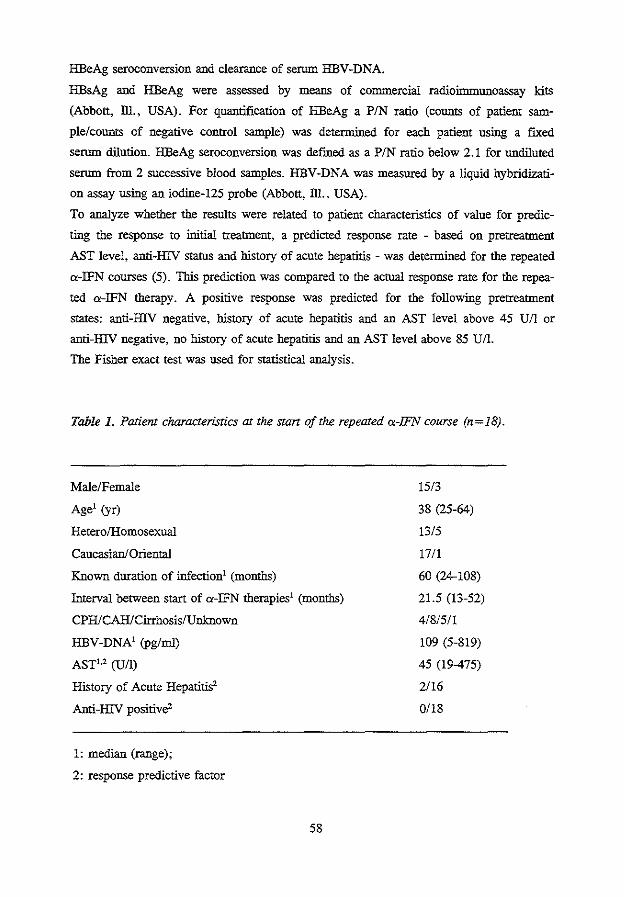
HBeAg seroconversion and clearance of serum HBV-DNA.
HBsAg and HBeAg were assessed by means of commercial radioimmunoassay kits
(Abbott, lli., USA). For quantification of HBeAg a PIN ratio (counts of patient sam
ple/counts of negative control sample) was determined for each patient using a fixed
serum dilution. HBeAg seroconversion was defmed as a PIN ratio below 2.1 for undiluted
serum from 2 successive blood samples. HBV-DNA was measured by a liquid hybridizati
on assay using an iodine-125 probe (Abbott, lli., USA).
To analyze whether the results were related to patient characteristics of value for predic
ting the response to initial tteatment, a predicted response rate - based on pretreatment
AST level, anti-HIV status and history of acute hepatitis -was determined for the repeated
a-IFN courses (5). This prediction was compared to the actual response rate for the repea
ted a-IFN therapy. A positive response was predicted for the following pretreatment
states: anti-HIV negative, history of acute hepatitis and an AST level above 45 U/1 or
aoti-HIV negative, no history of acute hepatitis and an AST level above 85 U/1.
The Fisber exact test was used for statistical analysis.
Table 1. Patient characteristics a1 the stan of the repealed a-IFN course (n = 18).
Male/Female
Age1 (yr)
Hetero/Homosexual
Caucasian/Oriental
Known duration of infection' (months)
Interval between start of a-IFN therapies' (months)
CPH/CAH/Cirrhosis/Unknown
HBV -DNA' (pg/ml)
AST1~ (U/1)
History of Acute Hepatitis'
Anti-HIV positive"
1: median (range);
2: response predictive factor
58
15/3
38 (25-64)
13/5
17/1
60 (24-108)
21.5 (13-52)
4/8/511
109 (5-819)
45 (19-475)
2/16
0/18
Results
Patient characteristics at the start of the repeated course are shown in table 1. Histological
evidence of chtonic hepatitis was found for all patients who underwent a liver biopsy
(n=17). AST levels were elevated in 16 of the 18 patients. None of the patients were
seropositive for antibodies to hepatitis C or hepatitis delta virus. One patient was of
Chinese origin. one Turkish; all the others were West-European caucasians.
At the end of follow-up 2 of the 18 patients (11 %) had responded to therapy (figure 1).
The responses occurred at week 10 and 14 of treanuent. Two patients became negative
only for HBV-DNA, one of them relapsed to HBV-DNA positivity after cessation of
therapy. All patients remained HBsAg positive.
Figure 2 shows the median level of HBV-DNA and AST. During therapy the HBV-DNA
and HBeAg levels decreased by 80% and 31%, respectively. There was more than a 50%
... 60
50
40
30
20
10
r----l---,22%
0 '----'-----.,.,.k 16
(end ol therapy)
11%
D HBV-DNA negative
R HBeAg aeroconveralon
week 52
Figure 1. Percemage of patients who exhibited clearance of HBV-DNA and HBeAg at the
end of the repeated <x-IFN course and at the end of follow-up (n=l8). The venical lines
denote the 95% confidence imerval.
59
HBV-DNA
AST
180
HIBV-ONA: pg/ml AST, U/l
8 • 16 ~ M ~ ~ $ " ~
weeks
Figure 2. Median HBV-DNA and AST levels during trearment and follow-up of the
repeated a-IFN course (n=l8).
reduction of the HBV-DNA level in all 18 patients and of the HBeAg level in 5 of the 18.
The AST level showed only minor fluctuations without evidence of a clear peak that ntight
be indicative of the immunological response which can terminate the HBV replicating
phase. Only 2 of the 18 patients - both nonresponders - exhibited a more than 2-fold rise
in AST levels during a-IFN treatment.
On the basis of the pretreatment characteristics, a response was predicted for 9 of the 18
patients (50%); the predicted response rate was significantly higher than the actual
response rate for a-IFN retreatment (P=0.03). Both responders belonged to the group of
patients for whom a response was predicted.
One of the 2 patients who received a total of 3 a-IFN courses exhibited a response in the
lOth week of the second course. which consisted of interntittenl a-IFN therapy (figure 3).
However, 12 weeks after the therapy was discontinued he suffered a relapse: both the
HBeAg and HBV -DNA became positive with a sudden rise in antinotransferase levels. His
last course of therapy which started 6 months after the relapse was unsuccessful. The AST
60
level for this patient was 3 to 4 times higher at the start of his second course as compared
to the other - nonresponsive - courses.
HBV-DNA AST
400
300
200
100
• • • • • • • • •
5.0 mu
HBV-DNA
HBV-DNA,pg/ml AST,U/1
6.0 mu
AST
• • • • HBeAg
30 5.0mu 1"5 . = IFN
3637383940
Figure 3. HBeAg status and levels of HBV-DNA and AST for a patienJ who received 3
courses of a.-IFN and exhibited a response and relapse during the second course.
Discussion
The effects of a.-IFN retreatment in chronic HBV-infected patients have not yet been
studied in detail. In this pilot study the HBeAg seroconversion rate for repeated a.-IFN
treatment of previous nonresponders appears to be low (11%). The type of interferon used
aod the duration of the successive therapeutic courses were comparable for each patient.
According to the prediction model described by Brook (5), a response was predicted for
9/18 (50%) patients. This figure was significantly higher than the actual response rate for
retreatment. According to the same prediction model. none of the 18 patients were
expected to respond to the first course of treannent. Thus, there was no difference
between the actual aod the predicted response rates for initial therapy (figure 4).
In all patients there was a clear decline in HBV -DNA levels during retreatment, sugges-
61
ting the presence of a direct inhibitory effect of a-IFN on HBV replication. However, the
virus-specific immunological response, which is usually reflected by a rise in inflam
matory activity (AST) and might be the most prominent effect of a-IFN therapy, appeared
to be absent in most of the patients. It thus seems important to focus on the immuno
regulatory properties of a-IFN to unravel the problem of nonresponse. a-IFN leads to an
increased expression of HLA type I protein on the hepatocyte snrface, thereby facilitating
recognition and lysis of virus-infected cells by the cellular immune system. This a-IFN
induced immunological response is accompanied by a rise in serum tumor necrosis factor
a, interleukin-1~ and the CD4/CD8 lymphocyte ratio; it is unknown, however, whether
these phenomena themselves exert a specific antiviral action or whether they only coincide
with other more important steps in the cascade of cytokine activities. Unfortunately, the
outcome of a-IFN therapy cannot be predicted by the pretreatment CD4/CD8 lymphocyte
ratio, natural killer cell activity or lymphocyte proliferative response (6,7).
- Initial treatment
'!1. response 80
60
50%
40
20
0% o~_,._~----
predicted response
- Repeated treatment
11%
0%
actual response
Figure 4. The predicted arul actual response rates for the initial arul repeated a-IFN
course. The vertical lines denote the 95% confidence interval.
62
Other factors that might influence the response rate are the number of cellular IFN recep
tors and the level of antibodies to C<-IFN. The IFN receptor expression appears to drop
quickly after C<-IFN treatment is started (8). This inhibitory effect might be compensated
by an increase in the IFN receptor binding affinity (9). Nevertheless, patients with
different patterns of response to C<-IFN treatment do not seem to differ in the expression
of lFN receptors on peripheral blood mononuclear cells. Antibodies to C<-IFN were not
present in any of the patients in this study (data not shown). The question whether C<-IFN
antibodies indeed influence the a-IFN response rate is still a subject of debate (10,11).
Maybe these antibodies play an important role in the low responsiveness of patients from
oriental areas (12).
It is unclear whether longer a-IFN retreatment with higher dosages would be more
effective than the therapeutic regimen used in the present study. Longer courses of therapy
looks promising for patients who partially respond to a first course of a-IFN (13) and
might be efficacious for those who previously did not respond to the standard (16 week)
a-IFN therapy. Combination of antiviral agents is also an option but the results of a-IFN
combined with acyclovir, adenine arabinoside monophosphate or prednison have been
disappointing for patients undergoing initial treatment (3,14,15). Regimens combining a
IFN with ')'-interferon or interleukin-2 are also not superior to a-IFN alone (16,17).
However from a theoretical viewpoint the combination of different cytokines appears to be
attractive and this alternative certainly deserves further evaluation (6). Another approach
for repeated therapy is to follow the patient regularly until the time when the amino
transferase levels have increased (preferably > 100 U/1) or the HBV-DNA levels are low
(preferably < 50 pg/ml). Starting retreatment at that moment might result in enhanced
response rates (18). Nevertheless, in our experience only 2 of the 9 patients with
promising pretreatment factors responded to a-lFN retreatment. Thus, the predictive value
of clinical pretreatment factors derived from first a-IFN courses (anti-HIV status, history
of acute hepatitis and aminotransferase levels) was not confirmed for repeated a-IFN
therapy in this study.
63
References
l. Realdi G, Alberti A, Rugge M, Bertolotti F, Rigoli AM, Tremolada F, Ruol A.
Seroconversion from HBe antigen to antiHBe in cbronic hepatitis B virus infection.
Gastroenterology 1980;79:195-9.
2. Fattovich G, Rugge M, Brollo L, Pontisso P, Noventa F, Guido M, Alberti A,
Realdi G. Clinical virologic and histologic outcome following seroconversion from
HBeAg to antiHBe in cbronic hepatitis type B, Hepatology 1986;6: 167-72.
3. Perrillo RP, Schiff ER, Davis GL, Bodenheimer HC Jr, Lindsay K, Payne J,
Dienstag JL, et al. A randomized controlled trial of interferon alfa-2b alone and
after prednisone withdrawal for the treatment of cbronic hepatitis B. N Eng! J Med
1990;323:295-301.
4. Brook MG, Chan G, Yap I, Karayiannis P, Lever AM, Jacyna M, Main J,
Thomas HC. Randomised controlled trial of lymphoblasoid interferon alfa in
Europid men with cbronic hepatitis B virus infection. BMJ 1989;299:652-6.
5. Brook MG, Karayiannis P, Thomas HC. Which patients with cbronic hepatitis B
virus infection will respond to alpha-interferon therapy? A statistical analysis of
predictive factors. Hepatology 1989;10:761-3.
6. Daniels HM, Maeger A, Eddleston ALW, Alexander GJM, Williams R. Spontane
ous production of tumor necrosis factor a and interleukin-1{3 during interferon-a
treatment of cbronic HBV infection. Lancet 1990;335:875-7.
7. Scully U, Brown D, Lloyd C, Shein R, Thomas HC. Immunological studies
before and during interferon therapy in cbronic HBV infection: Identification of
factors predicting response. Hepatology 1990;12:1111-7.
8. Nakajima S, Kuroki T, Shintani M, Kurai 0, Takeda T, Nishiguchi S, Susumu S,
et al. Changes in interferon receptors on peripheral blood mononuclear cells from
patients with cbronic hepatitis B being treated with interferon. Hepatology 1990; 1-
2:1261-5.
9. Lau JYN, Sheron N, Morris AG, Bamford AB, Alexander GJM, Williams R.
Interferon-a:: receptor expression and regulation in chronic hepatitis B virus
infection. Hepatology 1991;13:332-8.
10. Craxi A, DiMarco V, Volpes R, Palazzo U. Anti-alpha interferon antibodies after
alpha interferon treatment in patients with cbronic viral hepatitis. Hepato-gastraen
terol 1988;35:304-5.
11. Porres JC, Carreno V, Mercedes R, Marron JA, Bartoleme J. Interferon antibodies
in patients with cbronic HBV infection treated with recombinant interferon. J
64
Hepatol 1989;8:351-7.
12. Lok ASF, Lai CL, Leung EKY. Interferon antibodies may negate the antiviral
effects of recombinant <>-interferon treatment in patients with chronic hepatitis B
virus infection. Hepatology 1990;12:1266-70.
13. Janssen HLA, Berk L, Heijtink RA, Schalm SW, Hess G, Rosso! S, Meyer zum
Buschenfelde KH, et al. The antiviral effect of prolonged intermittent lymphoblas
toid alpha-interferon treatment in chronic hepatitis B. Gut 1992;33:1094-8.
14. Berk L, Schalm SW, De Man RA, Heijtink RA, Berthelot P, Brechot C, Boboc B,
et al. Failure of acyclovir to enhance the antiviral effect of alpha lymphoblastoid
interferon on HBe-seroconversion in chronic heaptitis B. A multi-centre randomi
zed controlled trial. J Hepatol 1992;14:305-9.
15. Garcia G, Smith Cl, Weissberg ll, Eisenberg M, Bissett J, Nair PV, Mastre B, et
a!. Adenide arabinoside monophosphate (vidarabine phosphate) in combination with
human leukocyte interferon in the treatment of chronic hepatitis B. Ann Intern
Med 1987;107:278-85.
16. Di Bisceglie AM, Rustgi VK, Kassianides C, Lisker-Melman M, Park Y, Waggo
ner JG, Hoofnagle JH. Therapy of chronic hepatitis B with recombinant human
alpha and gamma interferon. Hepatology 1990;11:266-70.
17. Hess G, Rosso! G, Iervolino M, Gotzler P, Meyer zum Buschenfelde. Interferon
(IFN) alpha vs IFN/interleukin II in patients with chronic hepatitis B previously
unresponsive to IFN. J Hepatol 1991;13:$35 (abstract).
18. Thomas HC, Karayiannis P, Brook G. Treatment of hepatitis B virus infection
with interferon. Factors predicting response to interferon. J Hepatol 1991;13:$4-7.
65

CHAPTER6
QUANTITATIVE ASSESSMENT OF HEPATITIS B VIRUS DNA IN CHRONIC
HEPATITIS B: COMPARISON OF TWO SOLUTION HYBRIDIZATION ASSAYS
H.L.A. Janssen'. Y.A.M. Schoenmaker-Weber, H. Kruining2, S.W. Schalm1, R.A.
Heijtink'
1. Department of Internal Medicine II, University Hospital Dijkzigt, Rotterdam, The
Netherlands.
2. Department of Virology, Erasmus University, Rotterdam, The Netherlands.
Journal of Medical Virology 1993;40:307-312
Abstract
We compared two solution hybridization assays, the Aff!Probe assay (Orion Corporation)
and the Abbott HBV-DNA assay (Abbott), for quantitative measurement of hepatitis B
virus (HBV) DNA in serum samples obtained from chronic HBsAg carriers. Forty
transversally collected (group 1) and 83 serially collected (group 2) serum samples from
chronic hepatitis B patients were tested with both assays. The serial serum samples were
obtained from 6 patients who underwent a-interferon therapy with different outcomes
(nonresponse, HBeAg seroconversion, HBeAg and HBsAg seroconversion).
In group 1 we found a good correlation (r=0.91; P<0.001) between the HBV-DNA
results of the two assays. Two samples (5%) were HBV-DNA positive according to the
Abbott but negative according to the AfflProbe assay; for all other samples the HBV-DNA
status corresponded. In group 2 the assays gave colinear HBV-DNA results during follow
up of 5 of the 6 patients (correlation for the total group: r=0.90; P<0.001). Neverthe
less, in both groups the AfflProbe assay yielded about 5-10 times higher HBV-DNA levels
than the Abbott HBV-DNA assay (P<O.OOI). These discordant results were most
probably due to standardization differences of the positive control samples of the two test
systems. This observation underlines the need for international standardization of HBV
DNA and uniform reference panels.
68
Introduction
Advances in hepatitis B virus (HBV) DNA detection methods based on molecular
hybridization techniques have opened the way for reliable quantification of HBV replicati
on (1,2,3). As a result HBV-DNA has become an important parameter of the effect of
antiviral therapy (4). Assays presently used to detect other markers of viral replication,
such as hepatitis B e antigen (HBeAg) or the expression of hepatitis B core antigen
(HBcAg) in liver cells, are difficult to quantify and therefore less suitable for treatment
monitoring. Furthermore, the liok between viral replication and HBeAg has been
questioned since the discovery of HBV pre-core mutants that replicate without producing
HBeAg (5).
HBV-DNA detection by dot-blot hybridization is widely used in research laboratories but
is not yet applied in routine diagnostic laboratories. A similar problem exists for the
quantification of HBV-DNA after amplification by polymerase chain reaction (PCR) (6,7).
Hybridization in solution, which has been found to be suitable for HBV -DNA detection in
serum, was recently incorporated in commercial test systems. The Abbott HBV-DNA
assay (Abbott Laboratories, Chicago, lil., USA) which is based on the separation of
hybrids and free probe by column chromatography, appears to give satisfactory quantitati
ve results (8,9). Using sera obtained primarily from chrouic hepatitis B patients under
going a-interferon therapy we compared the HBV-DNA results of this assay with those of
another solution hybridization assay (AfflProbe assay, Orion Corporation, Helsinki,
Finland) which depends on affmity-based hybrid collection (10,11)
Materials and Methods
Patients and Serum Samples
Two groups of sera were selected from chrouic hepatitis B surface antigen (HBsAg)
positive patients: Forty serum samples (group 1) were collected transversally from 36
patients. These sera were classified on the basis of the HBV-DNA level measured by the
Abbott HBV-DNA assay as follows: below 1.7 pg/ml (under the cut-off; group 1A), 1.7
to 50 pg/ml (group 1B), 50 to 100 pg/ml (group 1C) and above 100 pg/ml (group lD).
Each of these subgroups included 10 sera. All samples were negative for antibodies
against the human immunodeficiency virus; antibodies against hepatitis C and D virus
were detected in 1 and 4 of the serum samples, respectively. All sera which tested HBV
DNA negative in the Abbott HBV -DNA assay were also negative for HBeAg; all other
sera were HBeAg positive.
69
Eighty-three serum samples (group 2) were collected serially from 6 patients treated with
a-interferon and followed for a period of 1 year. Sera were obtained every 4 weeks
during a-interferon therapy and every 4-16 weeks in the period thereafter. All patients
received 5 mega-units lymphoblastoid a-interferon (Wellferon, Wellcome, Beckenham,
UK) per day according to the same protocol: a 4-week primer course, 4 weeks without
therapy and a second a-interferon course lasting 16 weeks (12). All patients were HBeAg
positive and had elevated serum aspartate antinotransferase levels at the start of therapy.
None exhibited antibodies against hepatitis C or D virus or against the human immunode
ficiency virus. Two patients remained HBeAg positive, 2 patients exhibited HBeAg
seroconversion and 2 patients exhibited both HBeAg and HBsAg seroconversion during
follow-up. One of the patients who only seroconverted for HBeAg showed HBeAg
reactivation after cessation of therapy.
Affinity-based Hybridization Assay (Affi.Probe Assay)
The AffiProbe assay is based on sandwich hybridization in solution followed by affinity
based hybrid collection (10,11). Target HBV-DNA is allowed to hybridize with a 35S
Iabelled detector probe and a biotinylated capmre DNA. The hybrids formed are then
collected onto an affinity matrix coated with streptavidin, from which they are eluted for
measurement by scintillation counting. Detectable label is bound to the matrix if the
correct target is present in the reaction.
The concentration of the positive control was 1.67 x 10⁹ HBV-DNA molecnles (5840
pg/ml; cloned double stranded DNA) per ml. The diluent for the test samples was used as
negative control. Thirty 1'1 of the control samples were tested in duplicate and the mean
cpm values were used. The cpm values for the negative control and the 1/100, 1/10 and
1/1 dilutions of the positive control sample (equivalent to 5 x 10', 5 x 10⁶ and 5 x 10⁷
DNA molecnles, respectively) were plotted logarithmically for construction of the standard
curve. The cut-off value - if at least 40 cpm and 1.5 times the cpm for the negative
control - was set as the cpm value for the 11100 dilution of the positive control. The
manufacmrer allows the use of 50 to 500 1'1 sample volumes; we chose a 100 1'1 sample
volume which is standard for the Abbott HBV-DNA assay. The detection level for the
AffiProbe assay using 100 1'1 of sample was 17.2 pg!ml.
Column-based Solution Hybridization Assay (Abbott HBV-DNA Assay)
The Abbott HBV-DNA assay was performed according to the instructions of the manufac
turer (Abbott, Chicago, !11, USA) (8). Briefly, 3 separate reagents were added to 100 1'1
of serum for solubilization and denaturation of the nucleic acids. A 125I-labelled HBV-
70
DNA probe was then added to the samples for overnight hybridization. Free probe was
separated from the 125I-labelled hybrids by passage over a prepacked column. After elution
of the hybrids from the column radioactivity was measured in a gamma counter. The
amount of HBV-DNA, expressed as pg/ml, was calculated according to the following
formula:
sample counts - mean negative control counts --------------------------------------x103 mean positive control counts - negative control counts
The concentration of the positive control was 3 x 10⁷ genomes/ml (103 ± 10 pg/ml;
native HBV-DNA). The detection level of the assay was 1.7 pg of HBV-DNA per mi.
Polymerase Chain Reaction
Serum samples were subjected to HBV-DNA extraction and amplified by means of a
polymerase chain reaction (PCR) as described previously (13,14). Two oligonucleotide
primer sets that were specific for the surface and core regions of the HBV-DNA genome
were used. Absence of HBV -DNA for both primer sets was considered a negative result.
Serological Assays
HBsAg and HBeAg were measured by solid pbase radioinnnunoassays (Abbott, Chicago,
Ill., USA). Aspartate aminotransferase levels were determined with a sequential multiple
autoanalyzer (Technicon Instruments Corp., Tarrytown, NY, USA).
Statistics
Pearson's correlation coefficient (r) was calculated to assess the correlation between the
HBV-DNA levels obtained with the two assays. For the serial samples we also calculated
the correlation coefficient for each individual patient and compared these values to 0,
using the Wilcoxon signed-rank test. Differences between the HBV-DNA results of the
two assays or between the AffiProbe/Abbott HBV-DNA ratio's were analyzed with the
Student's t-test after logarithmic transformation of the HBV-DNA value. HBV-DNA
results below the cnt-off values of the assays were not quantified and therefore not
included in the assessment of the correlation or statistical differences. P-values were two
sided, the significance level was 0. 05.
71
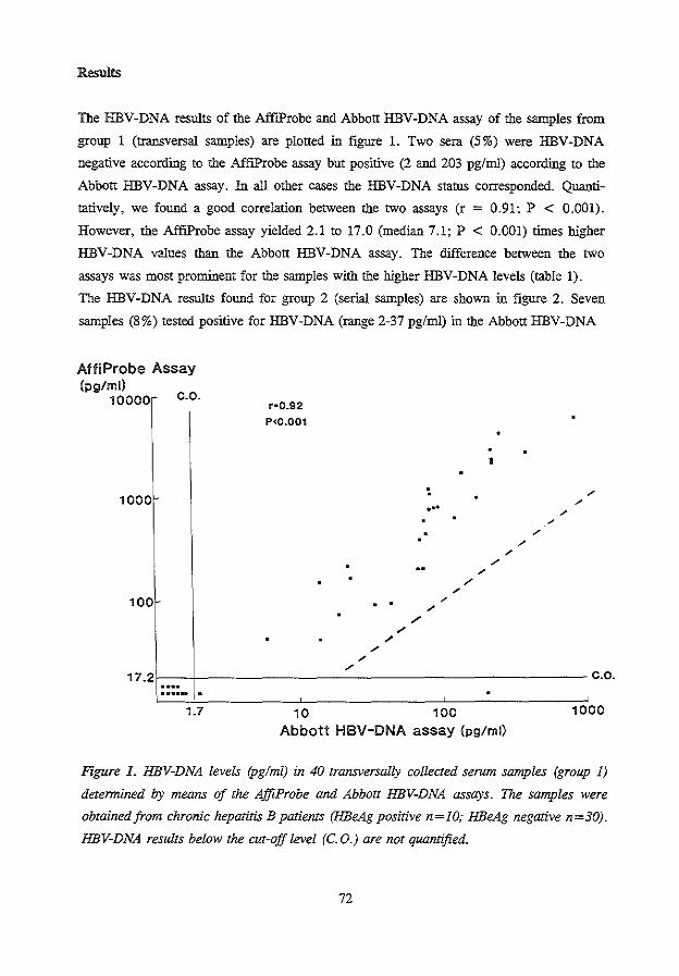
Results
The HBV-DNA results of the Aff!Probe and Abbott HBV-DNA assay of the samples from
group 1 (transversal samples) are plotted iu figure 1. Two sera (5%) were HBV-DNA
negative according to the Aff!Probe assay but positive (2 and 203 pglml) according to the
Abbott HBV-DNA assay. In all other cases the HBV-DNA status corresponded. Quanti
tatively, we found a good correlation between the two assays (r = 0.91; P < 0.001).
However, the AfflProbe assay yielded 2.1 to 17.0 (median 7.1; P < 0.001) times higher
HBV-DNA values than the Abbott HBV-DNA assay. The difference between the two
assays was most promiuent for the samples with the higher HBV-DNA levels (table 1).
The HBV -DNA results found for group 2 (serial samples) are shown iu figure 2. Seven
samples (8%) tested positive for HBV-DNA (range 2-37 pg/ml) iu the Abbott HBV-DNA
AffiProbe Assay (pg/ml)
10000r c.o.
1000
100
r•0.92
P<0.001
. .
/
" /
...
/
•
/
"
17.2f--+---------------------- c.o.
1.7 10 100 1000
Abbott HBV-DNA assay (pg/ml)
Figure L HBV-DNA levels (pglml) in 40 transversally collected serum samples (group 1)
determined by means of the AffiProbe and Abbott HBV-DNA assays. The samples were
obtained from chronic hepatitis B patients (HBeAg positive n=IO; HBeAg negative n=30).
HBV-DNA results below the cut-off level (C. 0.) are not quantified.
72
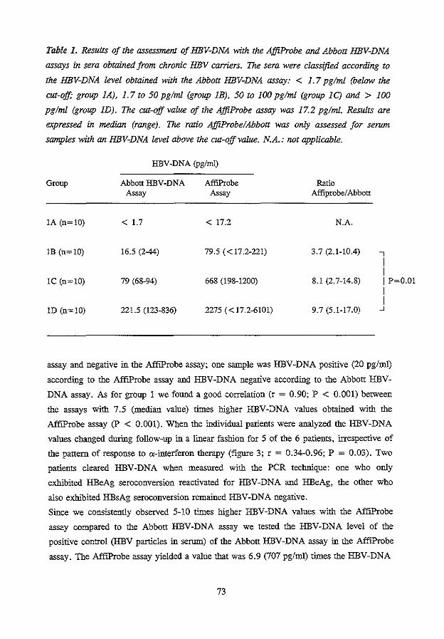
Table I. Results of the assessment of HBV-DNA with the AffiProbe and Abbott HBV-DNA
assays in sera obtained from chronic HBV carriers. The sera were classified according to
the HBV-DNA level obtained with the Abbott HBV-DNA assay: < 1. 7 pglml (below the
cut-off; group 1A), 1.7 to 50 pg/ml (group 1B), 50 to 100 pglml (group 1C) and > 100
pg/ml (group 1D). The cut-off value of the AffiProbe assay was 17.2 pglml. Results are
expressed in median (range). The ratio AffiProbe/Abbott was only assessed for serum
samples with an HBV-DNA level above the cut-off value. N.A.: not applicable.
HBV-DNA (pg/ml)
Group Abbott HBV-DNA AfliProbe Ratio Assay Assay Affiprobe/ Abbott
!A (n=!O) < 1.7 < 17.2 N.A.
!B (n=!O) 16.5 (2-44) 79.5 ( < 17.2-221) 3.7 (2.1-10.4) ..., I I
!C (n=10) 79 (68-94) 668 (198-1200) 8.1 (2.7-14.8) I P=O.O! I I
lD (n=!O) 221.5 (123-836) 2275 ( < 17.2-6101) 9.7 (5.1-17.0) _J
assay and negative in the AffiProbe assay; one sample was HBV-DNA positive (20 pg/ml)
according to the AffiProbe assay and HBV-DNA negative according to the Abbott HBV
DNA assay. As for group I we found a good correlation (r = 0.90; P < 0.001) between
the assays with 7.5 (median value) times higher HBV-DNA values obtained with the
AffiProbe assay (P < 0.001). When the individual patients were analyzed the HBV-DNA
values cbanged during follow-up in a linear fashion for 5 of the 6 patients, irrespective of
the pattern of response to a-interferon therapy (figure 3; r = 0.34-0.96; P = 0.03). Two
patients cleared HBV -DNA when measured with the PCR technique: one who only
exhibited HBeAg seroconversion reactivated for HBV-DNA and HBeAg, the other who
also exhibited HBsAg seroconversion remained HBV-DNA negative.
Since we consistently observed 5-10 times higher HBV-DNA values with the AffiProbe
assay compared to the Abbott HBV-DNA assay we tested the HBV-DNA level of the
positive control (HBV particles in serum) of the Abbott HBV -DNA assay in the AffiProbe
assay. The Aff!Probe assay yielded a value that was 6.9 (707 pg/ml) times the HBV-DNA
73
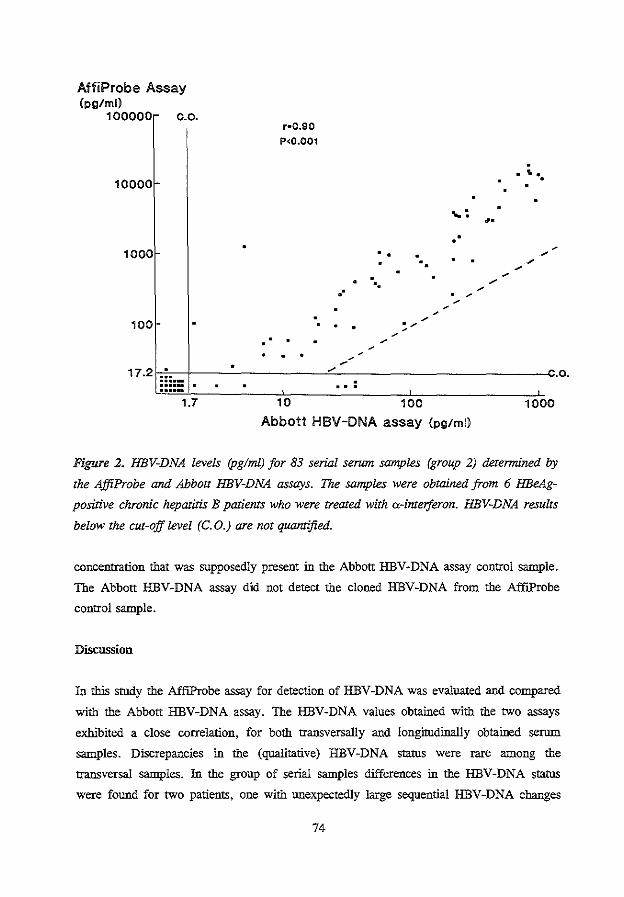
AffiProbe Assay (pg/ml)
100000 c.o.
1oooor-
1000
100
r•0.90
P<0.001
.· .. . ..
~--/
. .
----
... ·. ... : •• . .
--------/
--. ------
17.21-;~,;;i~;-===-.-f---:._ _____ _,.-::_.-.-.-------------C.O.
1.7 10 100 1000
Abbott HBV-DNA assay (pg/ml)
Figure 2. HBV-DNA levels (pglml) for 83 serial serum samples (group 2) determined by
the AffiProbe and Abbott HBV-DNA assays. The samples were obtained from 6 HBeAg
positive chronic hepatitis B patients who were treated with ex-interferon. HBV-DNA results
below the cut-off level (C. 0.) are not quantified.
concentration that was supposedly present in the Abbott HBV-DNA assay control sample.
The Abbott HBV-DNA assay did not detect the cloned HBV-DNA from the AffiProbe
control sample.
Discussion
In this study the AffiProbe assay for detection of HBV -DNA was evaluated and compared
with the Abbott HBV-DNA assay. The HBV-DNA values obtained with the two assays
exhibited a close correlation. for both transversally and longitudinally obtained serum
samples. Discrepancies in the (qualitative) HBV-DNA status were rare among the
transversal samples. In the group of serial samples differences in the HBV-DNA status
were found for two patients. one with unexpectedly large sequential HBV-DNA changes
74
according to the AfflProbe assay, the other suggesting a minor rebound phenomenon
according to the Abbott HBV-DNA assay only.
To obtain optimal comparability we used the same sample volume (100 !'I) in each test.
According to the manufacturers' instructions this resulted in a 1. 7 pg/rnl cut-off level for
the Abbott HBV-DNA and 17.2 pg/rnl for the AffiProbe assay. Since the HBV-DNA
values obtained with the AffiProbe assay were generally 5-10 times higher than those of
the Abbott HBV -DNA assay - a fmding that was also reported by others (G Gerken,
personal communication) - the difference in cut-off level did not markedly influence the
sensitivity of the AffiProbe as compared with the Abbott HBV-DNA assay. The divergen
ce between the HBV-DNA values of the Aff!Probe and Abbott HBV-DNA assay was most
notable for sera with high HBV -DNA concentrations.
In practical terms both the AfflProbe and the Abbott HBV-DNA assay appear to be
suitable for HBV -DNA monitoring of patients undergoing antiviral therapy. Since both
tests are based on nucleic acid hybridization in solution, they ensure easier handling of
samples and a faster hybridization rate than solid phase hybridization techniques (3 ,5). In
addition, the results are given in numerical values which allows unequivocal interpretation
and circumvents the possible interobserver variation of dot-spot methods. Nevertheless,
the assays described in this study could be improved by eliminating the radioactive probes
which require safety precautions and limit the shelf-life of the test kits (15,16). In our
experience the AfflProbe assay appeared to be more elaborate than the Abbott HBV-DNA
assay due to the many working steps involved, including ethanol precipitation and
resuspension of DNA, a step that may introduce an experimental error. For example, loss
of the HBV -DNA containing pellet after precipitation could potentially lead to false
negative results. The instructions of the Aff!Probe assay suggest using 50 and 500 !'! of
serum. In our experience a volume of 100 !'I of serum, as was used in the Abbott HBV
DNA assay, resulted in satisfactory assessment of HBV -DNA levels in most samples.
However, for precise quantitative measuremem of low-range HBV-DNA samples and for
therapeutic endpoint determinations, the use of 500 !"I of serum appears preferable because
of its lower detection level.
The question which of the 2 assays gives the closest approximation of the actual HBV
DNA content of the samples is difficult to answer. Since the Aff!Probe assay uses cloned
HBV-DNA (circular covalently closed double stranded DNA) as standard which fits in the
format of that assay but not in that of the Abbott HBV-DNA assay which uses native
HBV-DNA (partially single stranded DNA), positive control samples could not be exchan
ged. The result for the Abbott control sample tested in the Affiprobe assay was in
agreement with the results for the patiem samples in which 5-10 times higher HBV-DNA
75
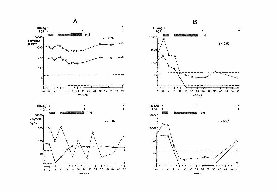
HBeAg+ PCR +
100000 HBVDNA (pg/mll
10ood
100
1111111
A • •
IFN r • 0.76
• •
-----------------------~ 10
------------------------~ 1 -6 0 4 8 12 16 20 24 28 32 38 40 44 48 52
HBeAg + PCR +
10000
HBVDNA (pg/ml)
10001
100
10
1111111
weeks
• •
IFN
r • 0.34
• •
~---------------------· 1~~-r~~~~4-~~_L~~-r~~~ ~ 0 8 12 16 20 24 w 32 36 40 44 48 ~
weeks
HBeAg + PCR +
1111111
B •
IFN •
r M 0.92
------0
1
________ t ________________ * I I I I I I t I I j je I* l ; I I I r
~o o
HBeAg + PCR +
10000 1111111
0
8 12 18 20 24 w 32 38 40 44 48 w weeks
IFN
r • 0.77
• +
--0
------· 8 12 ffi w 24 20 32 38 40 44 48 w
weeks
HBeAg + PCR +
10000 ..
1000
100
10
c IFN
r • 0.92
, ~~-~-.-~~I~ 1-:-1-:-10;--·:-~- -~-~- ~-~-~-i ~ 0 4 8 12 16 20 24 28 32 36 40 44 48 ~
weeks
HBeAg + PCil +
1000 - • IFN
r • 0.96
•
------------=--~---------~ .~~~~~~~~~rk~*=~~~=f -6 0 4 6 12 16 20 24 26 32 36 40 44 46 52
weeks
Figure 3. HBV-DNA levels (pg!ml) found by means of
the AjjiProbe (a-o) and Abbott HBV-DNA (*---
-*) assays for 6 patients (group 2) undergoing a
inteiferon therapy. Two patients remained HBeAg
positive (panel A), 2 patients exhibited HBeAg sera
conversion (panel B) and 2 patients exhibited both
HBeAg and HBsAg seroconversion (panel C) during
follow-up. The cut-off HBV-DNA level was 17.2 pg!ml
for the AjjiProbe (0-----0) and I. 7 pglml for the Ab
bott HBV-DNA (*-----*) assay. IFN: a-inteiferon;
PCR: HBV-DNA measured with polymerase chain
reaction. F~
levels were found in the Aff!Probe compared with the Abbott assay. This observation
reduces the discordant but consistent differences between the two assay systems to
standardization differences of the positive control samples. Defining an HBV-DNA
standard with native DNA may suffer from differences in the relative composition of
structural forms of the viral DNA between the standard virus preparation and the sera of
patients. On the other hand, the use of cloned viral HBV-DNA as standard, as was done
earlier in dot blot hybridization, may be hampered by the fact that cloned HBV-DNA does
not simulate solubilization and hybridization properties of native HBV-DNA in the assays.
The differences in the quantitation of HBV -DNA in this study prove that there is a clear
need for standardization and assessment of a uniform HBV-DNA reference panel. The use
of complete hepatitis B virion appears to be the most realistic option for these reference
sera. At present several European centers are trying to achieve this goal in concerted
action (17).
78
References
1. Bonino F, Hoyer B, Nelson J, EngleR, Venne G, Gerin J. Hepatitis B virus DNA
in the sera of HBsAg carriers: a marker of active hepatitis B virus replication in
the liver. Hepatology 1981;1:386-91.
2. Brechot C, Scotto J, Charnay P, Hadchouel M, Degos F, Trepo C, Tiollais P.
Detection of hepatitis B virus DNA: a direct appraisal of the chtonic carrier state.
Lancet 1981;2:765-8.
3. Berninger M, Hammer M, Hoyer B, Gerin JL. An assay for the detection of the
DNA of hepatitis B virus in serum. J Med Virol 1982;9:57-68.
4. Yokosuka 0, Ornata M, Imazeki F, Okuda K, Summers J. Changes of hepatitis B
virus DNA in liver and serum caused by recombinant leukocyte interferon
treatment: analysis of intrahepatic replicative hepatitis B virus DNA. Hepatology
1985;5;728-34.
5. Lieberman HM, LaBrecque DR, Kew MC, Hadziyannis SJ, Shafritz DA. Detecti
on of hepatitis B virus DNA directly in human serum by a simplified molecular
hybridization test: comparison to HBeAg/anti-HBe status in HBsAg carriers.
Hepatology 1983;3:285-91.
6. Kaneko S, Miller RH, Feinstone SM, Unoura M, Kobayashi K, Hattori N, Purcell
RH. Detection of serum hepatitis B virus DNA in patients with chtonic hepatitis
using the polymerase chain reaction assay. Proc Nat! Acad Sci 1989;86:312-6.
7. Gerken G, Paterlini P, Manns M, et a!. Assay of hepatitis B virus DNA by
polymerase chain reaction and its relationship to pre-s- and s-encoded viral snrface
antigens. Hepatology 1991;13:158-66.
8. Kuhns MC, McNamara AL, Cabal CM, Decker RH, Thiers V, Brechot C,
Tiollais P. A new assay for quantitative detection of hepatitis B viral DNA in
human serum. pp.258-62. In viral hepatitis and liver disease, Zuckerman AJ (ed),
Alan Liss Inc, New York, NY 1988.
9. Zarski JP, Kuhns MC, Berk L, Degas F, Schahn SW, Tiollais P, Brechot C.
Comparison of a quantitative standardized HBV-DNA assay and a classical spot
hybridization test in chtonic active hepatitis B patients undergoing antiviral
therapy. Res Viral 1989;140:283-91.
10. Jalava T, Ranki M, Bengtstrom M, Pohjanpelto P, Kallio A. A rapid and quantita
tive solution hybridization method for detection of HBV-DNA in serum. J Viral
Meth 1992;36:171-80.
11. Ranki M, Leinonen AW, Jalava T, Nieminen P, Soares VRX, Paavonen J, Kallio
79
A. Use of the AffiProbe HPV test for detection of human papillomavirus DNA in
genital scrapes. J Clin Microbial 1990;9:2076-81.
12. Janssen HLA, Berk L, Schalm SW, Heijtink RA, Hess G, Rosso! S, Meyer zum
Buschenfelde K-H, et al. The antiviral effect of prolonged intermittent lympho
blastoid alpha-interferon treatment in chronic hepatitis B. Gut 1992;33:1094-8.
13. Quint WGV, Bruyn de, Kruining H, Heijtink RA. HBV-DNA detection by gene
amplification in acute hepatitis B. Hepatology 1990;12:653-6.
14. Kroes ACM, Quint WGV, Heijtink RA. Significance of isolated hepatitis B core
antibodies detected by enzyme immunoassay in a high risk population. J Med Vir
1991;35:96-100.
15. Larzul D, Thiers V, Courouce AM, Brechot C, Guesdon J-L. Non-radioactive
hepatitis B virus DNA probe for detection of HBV -DNA in serum. J Hepatol
1987;5:199-204.
16. Naoumov NV, Lau JYN, Daniels HM, Alexander GJM, Williams R. Detection of
HBV-DNA using a digoxigeuin-labelled probe. A rapid technique without loss of
sensitivity. J Hepatol 1991;12:382-5.
17. Gerken G, Gerlich WH, Brechot C, Meyer zum Buschenfelde K-H. Biological
standards of hepatitis B virus assay. J Hepatol 1992;15:251-5.
80
CHAPTER 7
MEASUREMENT OF HllsAg TO MONITOR HEPATITIS B VIRAL
REPLICATION IN PATIENTS ON a-INTERFERON THERAPY
H.L.A. Janssen1, C.J. Kerkhof, R.A. Heijtink', S.W. Schalm1
1. Department of Internal Medicine II, University Hospital Dijkzigt, Rotterdam, The
Netherlands.
2. Department of Virology, Erasmus University, Rotterdam, The Netherlands.
Antiviral Research 1993; accepted for publication
Abstract
For the monitoring of chronic hepatitis B patients undergoing antiviral therapy,
measurement of hepatitis B virus (HBV) DNA is widely used in research laboratories but
is not yet a routine clinical procedure. We investigated whether quantitative measurement
of HBsAg can be used to monitor viral replication in chronic hepatitis B patients under
going a-interferon therapy. HBsAg was measured quantitatively in serum samples
collected serially before and after the HBeAg seroccnversion date from 69 patients with
HBeAg seroconversion and 17 patients with both HBeAg and HBsAg seroccnversion.
Serial samples from 39 HBsAg-positive patients without seroconversion (18 HBeAg
positive, 21 HBeAg negative) served as controls. From 12 other patients on a-interferon
therapy (nonresponse n=6, HBeAg seroconversion n=3, HBeAg and HBsAg serocon
version n=3) monthly samples were collected until 6 months after cessation of therapy.
HBsAg was measured quantitatively with an inununometric assay (Amersharn, UK) and
compared to the levels of HBV-DNA and HBeAg. In 56/69 (81 %) patients with only
HBeAg seroconversion, the HBsAg level dropped after HBeAg seroconversion; the
median HBsAg level was 8.39 !'glml (range 0.01-57.51) before HBeAg seroconversion
compared to 3.53 !'g/ml (range 0.002-68.66) after seroconversion (P<0.001). No
siginificant decrease in HBsAg was found for the control group. For the group of 12
patients treated with a-interferon we found parallel resnlts and a good correlation between
the HBsAg level and both the HBV-DNA (r=0.76; P<0.001) and the HBeAg (r=0.70;
P < 0.001) level, irrespective of the response pattern.
Quantified assessment of HBsAg appears promising as a simple and cheap method for
monitoring viral replication in chronic hepatitis B. This test deserves to be further
evaluated and compared with quantitated HBeAg and HBV DNA assays to deterntine its
usefulness for medical decision-making regarding a-interferon therapy.
82
Introduction
Chronic hepatitis B is a common liver disease which may progress to cirrhosis with such
complications as portal hypertension and hepatocellular carcinoma (1,2). With the
availability of effective antiviral agents for chronic hepatitis B, quantitative monitoring of
hepatitis B virus (HBV) replication has become an important factor in patient management
(3,4). The aim of antiviral therapy is to induce a transition from active HBV replication to
viral latency which is usually accompanied by arrest of disease progression (5,6). To
monitor HBV replication measurement of HBV-DNA is widely used in research laborato
ries but not in routine clinical laboratories because of its time-consuming methods and its
high cost in material and personnel (7,8). There is a need for a simple and cheap assay to
assess the level of HBV replication.
Hepatitis B "surface" antigen (HBsAg) was the ftrst hepatitis B-related antigen discovered
and is by definition present in all chronic hepatitis B patients (9). HBsAg testing is
therefore part of the routine diagnostic assessments in almost every hospital around the
world. We investigated the level of HBsAg in patients who exhibited cessation of viral
replication, as indicated by clearance of HBeAg and HBV-DNA. Furthermore we
determined whether quantitative measurement of HBsAg can be used as a marker of viral
replication in patients undergoing a-interferon therapy.
Patients and Methods
Patients and Treatment
HBsAg was assessed quantitatively in sera collected from 4 different groups of chronic
HBsAg carriers (total of 137 patients): Sixty-nine patients exhibited HBeAg
seroconversion (group A), 39 after a-interferon therapy (5 MU per day for 4 to 6 months)
and 30 spontaneously. Seventeen patients exhibited both HBeAg and HBsAg
seroconversion within 6 months (group B), 8 after a-interferon treatment and 9
spontaneously. Thirty-nine HBsAg-positive patients (HBeAg positive n=18; HBeAg
negative n=21) without a-interferon therapy and without HBeAg and/or HBsAg
seroconversion served as controls (group C). Two serial samples were collected 6 to 12
months before and after the HBeAg seroconversion date; similarly in the control group
two sera were collected 12 to 24 months apart. The remaining group (group D) consisted
of 12 patients who were followed longitudinally during a-interferon therapy with different
outcomes: nonresponse in 6, HBeAg seroconversion in 3 and HBeAg as well as HBsAg
seroconversion in 3 patients. a-Interferon was given in a dose of 5 MU daily. in courses
83
of 1 and 4 months duration, separated by one month of rest (10). Serum samples of these
12 patients were collected every month during therapy and every 1-2 months afterwards
(follow-up 1 year). In addition to quantitative HBsAg measurement, HBV-DNA and
HBeAg were assessed quantitatively in the samples from group D.
All patients receiving a-interferon therapy were HBeAg and HBV-DNA positive at the
start of therapy. HBeAg seroconversion was always accompanied by clearance of serum
HBV-DNA. All patients studied were negative for antibodies against the human
immunodeficiency virus and hepatitis C and D virus.
Laboratory methods
HBsAg was detected by an enzyme immunoassay (EIA) employing enhanced luminescence
(Amerlite HBsAg assay, Amersham, United Kingdom). Serum sample and mouse anti
HBs monoclonal antibody labeled with peroxidase were incubated in microwells coated
with a second anti-HBs monoclonal antibody of different specificity. After removal of
unbound material the peroxidase activity of the conjugate was measured by an enhanced
luminescence reaction (11). The sensitivity of the assay was approximately 0.3 ng/ml of
HBsAg (ad and ay). When appropriate, samples were tested undiluted or diluted 1/100
and 1/400 to remain within the detection range of the assay. HBsAg was quantified using
a serial dilution curve of a reference sample from the Paul Ehrlich Institute (Langen,
Germany) containing both the ad and ay (mixture 1:1) subtypes of HBsAg. Results were
expressed in micrograms of HBsAg per ml of serum. HBV-DNA was measured by a
liquid-phase hybridization assay using an 1251-labelled HBV-DNA probe (Abbott HBV
DNA assay, Abbott, Chicago, USA). Free probe was separated from the l2SI-labelled
hybrids by passage over a prepacked column. The HBV -DNA level was expressed in
picograms per ml of serum; the cut-off of the assay was I. 7 pg/ml. HBeAg was measured
by a radioimmunoassay (Abbott HBeAg test, Abbott, Chicago, USA). For quantification
the PIN ratio (counts patient sample/counts negative control sample) was determined. For
each patient a flxed serum dilution (1/1, 115, 1125, 11125 or 1/625) was maintained.
HBeAg seroconversion was defmed as a PiN ratio below 2.1 for undiluted serum.
Statistics
Medians were used because the HBsAg results lacked a normal distribution (also after
logarithmic transformation). The two-sample Wilcoxon rank sum test was used to analyze
unpaired observations and the Wilcoxon signed rank test to analyze paired observations.
Spearman's correlation coefficient (r) was calculated to assess the correlation between the
levels of HBsAg and HBV-DNA or HBeAg. P-values were two-sided, the significance
84
level was 0.05.
Results
The levels of HBsAg before and after seroconversion for HBeAg and HBsAg are
illustrated in table 1. The HBsAg level decreased in 56 of the 69 patients who exhibited
only HBeAg seroconversion (group A; median decrease of 59%; P<O.OOl). The median
decrease in HBsAg was 59% for patients with a-interferon-induced HBeAg seroconversion
and 55% for those with spontaneous HBeAg seroconversion. The difference between the
HBsAg levels before and after HBeAg seroconversion was significant for both groups.
The median HBsAg level in the initial samples was higher for the patients who underwent
both HBeAg and HBsAg seroconversion (group B) - independent of a-interferon therapy -
in comparison to those who only exhibited HBeAg seroconversion (group A); these
differences were not statistically significant.
For both the HBeAg-positive and HBeAg-negative control group (group C) we did not
HBV·DNA (pg/ml) 10000
1000
·. •.
100
.-
10 ' .
. · : 1
.. 0 10
·'
. .
·. -•
r•0.76 P<0.001
20 30 40 50 60 HBsAg (microgr/ml}
Figure 1. HBV-DNA and HBsAg levels in 148 serum samples obtained from 12 chronic
hepatitis B patients (group D) treated with a-inteiferon.
85
Table 1. Median HBsAg level (range) before and after HBeAg seroconversion or HBeAg and HBsAg seroconversion.
A HBeAg IFN treatment seroconversion
No treatment
Total
B HBeAg and HBsAg IFN treatment seroconversion
No treatment
Total
c HBeAg~positive controls
HBeAg-negative controls :
* cut-off HBsAg assay: 0.3 ng/ml N.A.: not applicable
n
39
30
69
8
9
17
18
21
HBsAg level (~g/ml)
month 0 month 12
8.39 (0.01-57.51) 3.53 (0.002-68.66)
8.43 (0.02-52.12) 3.63 (0.002-65.99)
8.39 (0.01-57.51) 3.53 (0.002-68.66)
32.58 (4.12-42.31) < 0.001'
12.04 (0.02-61.69) < 0.001'
13.09 (0.02-61.69) < 0.001'
25.55 (1.51-75.65) 21.70 (0.03-79.43)
4.96 (0.13-17.12) 6.01 (0.01-17.30)
P value
< 0.001
0.02
< 0.001
N.A.
N.A.
N.A.
0.87
0.18
HBeAg (PIN ratio) 20
15
10 •
5
·: .
.... . .
. ...
' ' . .. ..
·. .. ..
r•0.70 P<0.001
OL_----~----J------------L----~----~ 0 10 20 30 40 50 60
HBsAg (microgr/ml)
Figure 2. HBeAg and HBsAg lt!Ve/s in 148 serum samples obtained from 12 chronic
hepatitis B patients (group D) treated with o:-inteiferon.
fmd significant differences between the HBsAg level in the initial and follow-up serum
samples (table 1). The baseline HBsAg level for the 21 HBeAg-negative controls was
lower compared to the HBsAg results of all other subgroups (P<0.05).
A total of 148 serum samples were obtained from the 12 patients who were followed
longitodinally during a-interferon therapy (group D). For this group we found co-linear
results and a good correlation between the HBsAg level and both the HBV-DNA (r=0.76;
P<0.001) and the HBeAg (r=0.70; P<O.OOl) level (figure 1 and 2). No difference in
the correlation coefficients was found between patients with different response patterns
(nonrespone n=6, HBeAg seroconversion n=3, HBeAg and HBsAg seroconversion n=3;
figure 3).
Discussion
In the present stody we assessed the quantitative HBsAg level in relation to loss of HBV
replication ·and evaluated the usefulness of HBsAg for monitoring antiviral therapy. As in
87
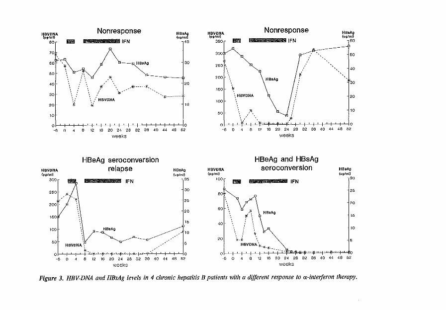
HavoNA Nonresponse ""''' HavoNA Nonresponse ""''' {pglml) (u1Jiml) {pglml) (ug/ml)
aor H IFN v\0 350r llliil IFN ,eo
70 3oo \ p/ j60
80 * HBsAg 30 250 / / ~40
50 ' " .' '," * 'f ', ,' ', ,' ', ZOO HBsAg : 40 ' : \ ' ' 20 ' f30 ', : ' * ' -*··-·-*-
', : ', / \~-- 1150 30 \ ,' \ / '%------ HBVDNA 1\!. j : -!20
•, .' \ ,' UBVDNA 100
20 ¥- * 10
10 50 ,*, ~ :' i 10
/ \ ;, 0 0 -6 0 4 8 12 16 20 24 28 32 38 40 44 48 ~
HBVDNA {pfllml)
300
260
200
160
100
60
~eks
HBeAg seroconversion relapse
" -~
HBVDNA •:
"-
IFN
liB sAg
liB sAg {uo/ml)
35
30
26
20
16
10
6
0 f-1--1-*--f-l ;- 0 -6 0 4 8 12 16 20 24 28 32 36 40 44 48 ~
weeks
ol ' I T' I t·•·v-Hi'+J'' I ' I ' I ' I ' I ' I ' lo -8 0 4 8 12 16 20 24 ~ 32 36 40 44 48 M
weeks
HBVDNA (potml)
100
80
60
40
20
HBeAg and HBsAg seroconversion
111111 IFN
,~ \ HBsAg r, ' ' ' '
! \ ~-"' \
HBVONA ¥'J!E..*---
ol I I I ' I I ' l"o·f"t .. ·$··-~-·-t·'-~'-
ttBsAg (ug/ml)
30
26
20
16
10
6
-6 0 4 8 12 18 20 24 28 32 36 40 44 48 ~
weeks
Figure 3, HBV-DNA and HBsAg levels in 4 chronic hepatitis B patients with a dijfere/11 response to o:-i/1/eljeron therapy.
previous studies, we found that the transition from HBV replication to viral latency, as
indicated by HBeAg seroconversion, is accompanied by a significant decrease in the
HBsAg level (12-14). Other s gene encoded proteins, such as pre-Sl Ag and pre-S2 Ag,
have also been shown to correlate with the degree of HBV replication (15).
Patients who underwent HBeAg seroconversion (no HBsAg seroconversion; group A)
during "-interferon therapy exhibited a similar decrease in HBsAg level as patients who
seroconverted spontaneously, suggesting that therapy-induced and spontaneous HBeAg
seroconversion result in comparable reductions of the virus load. Since we included only a
few patients who did not respond to "-interferon we cannot judge whether HBsAg
assessment prior to therapy could help to predict the outcome of this treatment. Eight
patients exhibited both HBeAg and HBsAg seroconversion after "-interferon treatment
(group B). The initial HBsAg level found for these patients was similar to that for those
who exhibited only HBeAg seroconversion (group A) during therapy, indicating that the
amount of HBsAg in serum before therapy is not predictive for the occurrence of HBsAg
seroconversion after a-interferon therapy.
For patients who were followed closely during "-interferon therapy (group D) we found a
good correlation between the HBsAg and the HBeAg or HBV -DNA levels. In most
patients the HBV-DNA level dropped first, followed by the HBeAg and HBsAg levels. A
similar sequence was described by Zonlim and Hess in previous studies (4,16).
Interestingly, we found co-linear results for the HBsAg, HBeAg and HBV-DNA levels,
irrespective of the response (nonresponse, HBeAg seroconversion, HBeAg and HBsAg
seroconversion) to therapy. The interval between the start of the decrease in the HBV
DNA and HBsAg levels was short which suggests that the HBsAg level reflects changes in
viral replication in a direct way.
With the availability of effective antiviral agents, quantified parameters of HBV
replication have become important for patient management and the development of new
treatment regimens. One of the reasons why HBsAg is as yet rarely quantified in studies
involving antiviral therapy conld be that the range of this antigen can only be covered by
using several dilution steps in assays presently available for qualitative measurement.
Using the HBsAg Amerlite kit we were able to obtain reliable quantification in 3 dilution
steps. More than 80% of the samples could be measured reliably after a 1!100 dilution
step. A second possible reason for the lack of interest in measuring the level of HBsAg in
patients treated with "-interferon could be the obvious fact that HBsAg does not disappear
completely from serum after cessation of viral replication as detected by HBeAg
seroconversion. Although the HBsAg level decreased in the majority of our patients who
exhibited HBeAg seroconversion it remained high in some. Therefore we could not define
89
an HBsAg cut-off level which corresponds to disappearance of HBeAg and which could be
used as endpoint determination for "-interferon therapy. On the other band, quantitative
HBsAg assessment could indicate in most cases a sigoificant decrease in viral replication
during therapy like HBeAg and HBV-DNA measurement. In comparison to quantitative
assessment of HBeAg and HBV-DNA, HBsAg quantitation is less laborious and cheaper,
especially when performed by assays designed for quantitative measurement (Amerlite
assay, Abbott lMX). For the many laboratories which do not have the ability to measure
HBV-DNA or HBeAg quantitatively, HBsAg measurement might be a good alternative to
assess cbanges in HBV replication. In addition to HBsAg measurement a simple
qualitative HBeAg/anti-HBe assay indicating seroconversion is most reliable for endpoint
determination of "-ioterferon therapy. Further studies should elucidate whether the HBsAg
level can also be used as a virus marker for patients with hepatitis D or for those who
carry HBV mutants with a deletion in the pre-core region of the genome which impedes
HBeAg production (17,18). For these patients, who often have active disease without the
presence of HBeAg, the effect of treatment is currently monitored by assessment of
antinotransferase levels.
In conclusion we found that transition from HBV replication to viral latency, as indicated
by HBeAg seroconversion, is accompanied by a signifcant decrease in the HBsAg level
and that the quantified HBsAg level correlates well with the quantified HBeAg and HBV
DNA levels. Therefore quantitative measurement of HBsAg appears promisiog as a simple
and cheap method for monitoring HBV replication and deserves further evaluation of its
usefttlness for medical decision-making regarding "-interferon therapy.
90
References
1. Hoofnagle JH, Alter HJ. Chronic viral hepatitis. In: Vyas GN, Dienstag JL,
Hoofnagle JH eds. Viral hepatitis and liver disease. New York: Grone and
Stratton, 1984;97-113.
2. Beasley RP, Hwang L-Y, Lin C-C, Chien C-S. Hepatocellular carcinoma and
hepatitis B virus. A prospective study of 22707 men in Taiwan. Lancet
1981;2:1129-33.
3.. Perrillo RP, Schiff ER, Davis GL, Bodenheimer HC Jr, Lindsay K, Payne J,
Dienstag JL et a!. A randomized controlled trial of interferon alfa-2b alone and
after prednisone withdrawal for the treatment of chronic hepatitis B. N Eng! J Med
1990;323:295-301.
4. Zoulim F, Mimms L, Floreani M, Pichoud C, Chemin I, Kay A, Vitvitski L,
Trepo C. New assays for quantitative determination of viral markers in
management of chronic hepatitis B virus infection. J Clin Micro bioi 1992;30: 1111-
9.
5. Realdi G, Alberti A, Rugge M, Bortoloni F, Rigoli AM, Tremolada F, Ruol A.
Seroconversion from HBe antigen to antiHBe in chronic hepatitis B virus infection.
Gastroenterology 1980;79:195-9.
6. Fattovich G, Rugge M, Brollo L, Pontisso P, Noventa F, Gnido M, Alberti A, et
a!. Clinical virologic and histologic outcome following seroconversion from
HBeAg to antiHBe in chronic hepatitis type B. Hepatology 1986;6:167-72.
7. Bonino F, Hoyer B, Nelson J, EngleR, Verme G, Gerin J. Hepatitis B virus DNA
in the sera of HBsAg carriers: a marker of active hepatitis B virus replication in
the liver. Hepatology 1981;1:386-91.
8. Lieberman HM, LaBrecque DR, Kew MC, Hadziyannis SJ, Shafritz DA. Detecti
on of hepatitis B virus DNA directly in human serum by a simplified molecular
hybridization test: comparison to HBeAg/anti-HBe status in HBsAg carriers.
Hepatology 1983;3:285-91.
9. Blumberg BS, Alter HJ, Visnich. A "new" antigen in leukentia sera. JAMA
1965;191:541-6.
10. Janssen HLA, Berk L, Schahn SW, Heijtiuk RA, Hess G, Chamuleau RAFM, et
a!. The antiviral effect of prolonged intermittent lymphoblastoid alpha-interferon
treatment in chronic hepatitis B. Gut 1992;33:1094-8.
11. Whitehead TP, Thorpe GHG, Carter TIN, Croucutt C, Kricka U. Enhanced
luntinesence procedure for sensitive determination of peroxidase-labelled conjugates
91
in immunoassay. Nature 1983;305:158-9.
12. Chien DY, Vyas GN. Correlation of the hepatitis B surface antigen and e antigens.
N Eng! J Med 1978;299: 1253-4.
13. Heijtink RA, Schalm SW. Quantitative assay of hepatitis B surface antigen and
DNA polymerase activity. N Eng! J Med 1978;301:165.
14. Frosner GG, Schomerus H, Wiethnann KH, Zachoval R, Bayer B, Backer U, Gat
hof GA, Sugg U. Diagnostic significance of quantitative hepatitis B surface antigen
in acute and chronic hepatitis b infection. Eur J Clin Microbiol 1982;1:52-8.
15. Yuki N, Hayashi N, Katayama K, Kashara A, Ueda K, Fusamoto H, Sato N,
Kamada T. Quantitative analysis of pre-Sl and pre-S2 in relation to HBsAg
expression. Hepatology 1990;11:38-43.
16. Hess G, Gerlich W, Gerken G, Manns M, Hutteroth TH, Meyer zum Buschenfel
de. The effect of recombinant a-interferon treattnent on serum levels of hepatitis B
virus-encoded proteins in man. Hepatology 1987;7:704-8.
17. Carman W, Jacyna M, Hadziyannis S, Karayiannis P, McGarvey MJ, Makris A,
Thomas HC. Mutation preventing formation of hepatitis e antigen in patients with
chronic HBV infection. Lancet 1989;2:588-91.
18. Brunetto MR, Giarin MM, Oliveri F, Chiaberge E, Baldi M, Alfarano A, Serra A,
et al. Wild type and e antigen-minus hepatitis B viruses and course of chronic
hepatitis. Proc Nat! Acad Sci 1991;88:4186-90.
92
CHAPTER 8
FATAL DECOMPENSATION OF CHRONIC VIRAL HEPATITIS ASSOCIATED
WITH ALPHA-INTERFERON TREATMEJI<"f
H.L.A. Janssen', J.T. Brouwer', F. Nevens2, J. Fevery2
, P. Marcellin3, J.M. Sanchez
Tapias', A. Craxi5, S. Hadziyannis6, S.W. Schahn' and the European concerted action on
viral hepatitis (EUROHEP).
L Department of Internal Medicine II, University Hospital Dijkzigt, Rotterdam, The
Netherlands.
2. Department of Hepato-Gastroenterology, University Hospital, Leuven, Belgium.
3. Service d'Hepatologie, Hopital Beaujon, Clichy, France.
4. Liver Unit, Hospital Clinic I Provencial, Barcelona, Spain.
5. Divisione di Medicina Interna, University of Palermo, Italy.
6. Academic Department of Medicine, Hippokration General Hospital, Athens,
Greece.
British Medical Journal 1993;306:107-108
(adapted version of this chapter)
Abstract
As a result of an inquiry among 16 European hospitals, in which 2,490 chronic viral
hepatitis patients have been treated with a-interferon, we report on 9 patients with
cirrhosis who exhibited a fatal decompensation of their liver disease during or shortly after
a-interferon therapy. Seven patients had chronic hepatitis B, 1 chronic hepatitis C and 1 a
co-infection of hepatitis B and C. Four of the 9 patients had no evidence of decompen
sated liver disease previously (Child classification A at the start of therapy) and 5 exhibi
ted signs of decompensation in the past (Child classification A (n= 1), B (n=3) and C
(n=1) at the start of therapy). The pattern of decompensation during therapy included
ascites in 9, encephalopathy in 7 and jaundice in 6 patients. The cause of death was liver
failure in 6 and variceal bleeding in 3 cases. Particularly in patients with previously
compensated liver disease the relation between the fatal decompensation and the a
interferon therapy appears highly probable. A mechanism which could explain most of the
observed deaths in this study is that a-interferon induces an exacerbation of inflammatory
activity, leading to a fatal destruction of the limited residual capacity of the liver in
patients with cirrhosis.
The present study suggests that a-interferon can induce a severe aggravation of the liver
disease and that it is important to closely monitor patients with cirrhosis undergoing this
therapy. A very careful attitude with regard to a-interferon is necessary for patients with
decompensated cirrhosis who should only be treated on a clear indication by experienced
hepatologists.
94
Introduction
Currently, alpha-interferon is the most promising therapy for chronic viral hepatitis. A
sustained response can be obtained in approximately 30% of the patients with chronic
hepatitis B or C (1,2). Also in patients with cirrhosis a-interferon may hnprove the
outcome of disease and obviate the need for liver transplantation (3). A response in
chronic hepatitis B, as defined by loss of viral replication (hepatitis B e antigen serocon
version), is usually accompanied by a transient rise in antinotransferases (4). This
inflanunatory exacerbation ensues the risk of a hepatic decompensation in cirrhotic
patients. We describe 9 patients with viral hepatitis related cirrhosis who died from
decompensated liver disease during or shortly after a-interferon treatment, and discuss the
possible contributing role of a-interferon.
Case report
A 44-year old male with hepatitis B e antigen positive chronic hepatitis B was started on 5
megaunits of recombinant a-interferon per day at the Rotterdam University Hospital. His
liver disease was not detected until 6 months previously when he complained of ankle
edema and ascites after a long car drive from Morocco to the Netherlands. Liver biopsy
showed a macronodular cirrhosis with active hepatitis and hepatitis B core antigen expres
sion in 30% of the hepatocytes; endoscopic exantination showed medium-sized esophageal
varices. At the start of therapy his liver disease appeared to be compensated, both the
ascites and edema were undetectable with diuretic therapy. During the first 8 weeks of
treatment he only complained of chills and a slight fatigue, symptoms that can be
attributed to the a-interferon therapy. In this period the aspartate antinotransferase level
doubled while no substantial change was found for the serum hepatitis B virus DNA level
(figure 1). In the 11th week of treatment the patient complained of jaundice, drowsiness
and reappearance of the ascites. He was admitted to hospital and the a-interferon therapy
was discontinued. The aspartate aminotransferase and bilirubin level had risen to 830 U/1
(normal<30) and 93 p.Illol/1 (normal<14), respectively. The anunonia level was 115
JLIDOI/1 (normal< 30) and a grade IIb encephalopathy was found on spectral analysis of the
electroencephalogram (5). He was started on lactnlose and 7 liters of ascitic fluid were
drained. There were no signs of bacterial peritonitis. No markers of any hepatotrophic
virus causing a superinfection were detected in serum or liver tissue. During the exacerba
tion the serum hepatitis B virus DNA level dropped to 11 pg/tnl (cut off 1.7 pg/tnl;
Genostics, Abbott, USA), however no hepatitis B e antigen seroconversion occurred.
95
HBV-DNA (pg/ml)
bilirubin (umol/1) 111111111111111111111111111111111111111111111111111111111111111111111111111111111111111111 140
120
L----...._ HBV-ONA 100
80
60
40 bilirubin:'
:
IFN 5 MU/day AST (U/1)
1000
800
600
400
200
20 -:-· -~~"""'--_:-:: __ :::_-.:::: .. :-::.-.'/
o~-L~~-L~~-L~~-L~~~~o ~ 0 2 4 6 8 10 ~ u 16 18
weeks t Flgure L Level of serum hepatitis B virus DNA (pglml), aspartate aminotransferase (u/1)
and bilirubin (pmol/1) in a chronic hepatitis B patient (case 5) who exhibited a fatal
decompensation of liver disease during a-inteiferon therapy.
After 2 more weeks during which the condition of the patient improved (bilirubin and
aspartate antinotransferase level decreased) he suddenly developed a variceal hemorrhage
and died.
Survey
To review the frequency and clinical aspects of a-interferon related fatal decompensation
of liver disease a questionnaire was sent out to 19 European centres with a large experien
ce in a-interferon therapy for chronic vital hepatitis. Sixteen hospitals from 9 countries
responded. In these centres a total number of 2,490 chronic viral hepatitis patients bad
been treated with a-interferon. Only cases in which the patient exhibited an aggravation of
liver disease with fatal outcome during or within 2 months after discontinuation of a
interferon were included. Eight more cases from 5 different hospitals were reported. From
96
Table la. Characteristics of previously compensated chronic viral hepatitis patiems who exhibted fatal liver failure associated with a-interferon therapy.
GROUP A age sex type of previous dose/duration type of signs of decomp. last results before death HBeAg cause of
hepatitis decompensation IFN IFN during IFN AST bilirubin albumin seroconv. death (Un) <rmol/1) (g/1)
Case 1a 62 male B No 5 MU t.i.w. recombinant ascites 244 648 43 Yes liver failure 12 weeks a-2b jaundice
encephalopathy
Case2 41 male B No 3 MU t.i.w. recombinant ascites 302 680 28 N.A. liver failure (HBe-mutant) 3 weeks a-2b jaundice
encephalopathy
Case3 60 female c No 10 MU t.i.w. recombinant ascites 76 410 28 N.A. liver failure 11 weeks a-2b jaundice
encephalopathy
Case4 62 male B No 5 MU daily recombinant ascites 394 226 26 No variceal bleed 8 weeks a-2b jaundice (after steroid withdrawal)
a: reference nr 13. N.A.: not applicable; IFN: a-interferon; HBeAg: hepatitis Be antigen; AST: aspartate aminotransferase
Table lb. Characteristics of previously decompensated chronic viral hepatitis patients who exhibted fatalli~·er failure associated with o:-inteiferon therapy.
GROUPB age sex type of previous dose/duration type of signs of decamp. last results before death HBeAg cause of
hepatitis decompensation IFN IFN during IFN AST bilirubin albumin seroconv. death (U/1) (pmol/1) (g/1)
Case 5a 44 male B Yes 5 MU daily recombinant ascites 79 88 27 No variceal bleed (ascites) II weeks o:-2a jaundice (edema) encephalopathy
Case 6b 44 male Band C Yes 3 MU t.i.w_ lymphoblastoid ascites 289 272 28 N.A. liver failure (ascites) 10 weeks jaundice H-R syndr. (jaundice) encephalopathy peritonitis
Case7 70 male B Yes 2 MU t.i.w. recombinant ascites 9 III 34 Yes liver failure (ascites) 24 weeks o:-2b encephalopathy
(variceal bleed) (encephalopathy)
Case Sb 36 male B Yes 2 MU t.i.w. recombinant ascites 16 52 27 Yes variceal bleed (ascites) 16 weeks o:-2b peritonitis
Case 9b 53 male B Yes 2 MU t.i.w. recombinant ascites 500 171 29 No liver failure (ascites) 3 weeks o:-2b jaundice (HCC) encephalopathy
a: presented above b: patients on waiting list for liver transplantation N.A.: not applicable; HCC: hepatocellular carcinoma; H-R syndr.: hepato-renal syndrome; IFN:o:-interferon; HBeAg: hepatitis Be antigen; AST: aspartate aminotransferse
the total of 9 patients 4 had no evidence of decompensated liver disease (presence of
ascites, jaundice, encephalopathy or variceal bleeding) before a-interferon therapy was
started (group A; table 1a) and 5 patients had signs of decompensated liver disease in the
past (group B; table 1b). From group B, I patient (case 5) appeared to be recompensated
at the start of treatment while 3 other patients (case 6, 8 and 9) were on the waiting list
for hepatic transplantation. Case 9 was the only patient with evidence for hepatocellular
carcinoma. According to the Child-Pugh criteria (6) case 1-5 had class A, case 6, 8 and 9
class B, and case 7 class C cirrhosis at the start of the a-interferon therapy.
At the time of death the median age was 53 years. Seven patients had chronic hepatitis B
of whom 1 (case 2) carried a mutant type of virus with a deletion in the pre-core region of
the genome which impedes hepatitis B e antigen production (7). One patient had chronic
hepatitis C and one had a co-infection of chronic hepatitis B (hepatitis B e antigen
negative) and C. No serum markers that indicate a viral superimposed infection of the
liver appeared during the period of decompensation in any of the patients. Furthermore
none had been exposed to hepatotoxic drugs or other possible causes of liver injury.
Pretreatment histologic examination showed chronic active hepatitis with cirrhosis in all
cases. The weekly dosage of a-interferon in group A ranged from 9 to 35 MU; in group
B, 1 patient received a dose of 35 MU/week and 3 less than 10 MU/week. In 7 of the 9
patients a-interferon was administered thrice weekly. Three of the 6 hepatitis B e antigen
positive hepatitis B patients exhibited an hepatitis B e antigen seroconversion during
therapy. The hepatitis C and the hepatitis B mutant infected patients (case 2, 3 and 6) did
not respond to the a-interferon therapy with normalization of antinotransferase values. The
clinical complications occurred within the first 3 months of therapy in 7 of the 9 patients.
In the other 2 patients (case 7 and 8) the scheduled a-interferon course was completed
with clearance of hepatitis B virus DNA but clinical deterioration developed 2 and 8
weeks after discontinuation of treatment. The following signs of decompensation were
present after start of therapy: ascites in 9 patients (previously present in 5), encephalopa
thy in 7 (previously present in 1) and jaundice in 6 (previously present in 1). Spontaneous
bacterial peritonitis was found in 2 patients (case 6 and 8; previously present in 1). The
reported eventual cause of death was liver failure in 6 and hypovolemic shock due to
variceal bleeding in 3 patients.
Discussion
We describe 9 chronic viral hepatitis patients who underwent a-interferon therapy with
fatal outcome. All patients had cirrhosis and 5 of the 9 had signs of decompensated liver
99
clisease previously. It must be empbasized that the patients described were selected from a
large population treated with <>-interferon. Nevertheless these cases suggest that <>
interferon treatment can induce a dangerous aggravation of the liver disease and that a
cautious attimde is warranted for treatment of cirrhotic patients.
Spontaneous exacerbations of chronic viral hepatitis have been reported to cause severe
hepatic injury that may result in fatal liver failure (8,9,10). Withdrawal of intmunosup
pressive or cytotoxic therapy can induce similar complications in patients with chronic
vital hepatitis (11,12). A first example of fatal exacerbation induced by <>-interferon
therapy, was recently reported by Marcellin eta!. (13; case 1 of this smdy).
The relation between liver failure and <>-interferon appeared probable in the patients who
decompensated during therapy and never bad an episode of decompensation before the
start of treatment (group A). In case 4 the withdrawal of prednison could have been an
additional impems for the occurence of the fatal exacerbation. The death of the 4 patients
who already exhibited signs of decompensated liver disease at the start of treatment (case
6-9 from group B) might have been secondary to a spontaneous progression of the disease
and can thus not unequivocally be linked to the <>-interferon therapy.
In the literamre several explanations can be found for an association between <>-interferon
and fatal liver injury. In chronic hepatitis B <>-interferon facilitates lysis of virus-infected
cells by the cellular intmune system which leads to an increase of inflammatory activity
that could be lethal in a liver with limited residna! capacity (4,9,14). A second possibility
may be that after clearance of hepatitis B e antigen, whether or not <>-interferon induced,
coexisting precore mutant hepatitis B viruses are selected (15). These mutants have been
implicated in fatal exacerbations (16,17). Other recently postulated reasons for liver
failure secondary to <>-interferon therapy are a direct hepatocellular toxic effect of <>
interferon and <>-interferon induced autointmune-mediated chronic active hepatitis (18,19).
In our smdy none of the patients bad evidence of autointmune chronic active hepatitis
during <>-interferon treatment.
The clinical pattern of deterioration in our patients mostly involved appearance of ascites,
jaundice and encephalopathy that progressed even after <>-interferon therapy was stopped.
A relative late discontinuation of therapy in some of the patients could be mentioned as
the reason for the nnfavourable outcome. In any case it is clear that careful monitoring of
cirrhotic patients who develop signs of decompensation under <>-interferon therapy is
essential.
Despite the serious adverse effects that can result from <>-interferon in decompensated
cirrhosis it is - in our opinion - justified to keep a-interferon in consideration as a therapy
for these patients. Firstly, because a response with transition to viral latency can lead to a
100
marked regression of liver disease and probably prolonged survival (3,20). Secondly, for
those cases in which liver transplantation remains the only option, transition to viral
latency reduces the risk of reinfection of the graft (21). It is important, however, that the
a-interferon therapy in decompensated patients is monitored closely by an experienced
hepatologist, preferably in a centre with a liver transplantation program. Since 5 of the 9
patients described received an a-interferon dose under 10 MU per week, a-interferon
related fatal decompensation is not restricted to high dose courses. Nonetheless, from a
practical viewpoint it seems prudent to start with a low dose of a-interferon and carefully
adjust the dose to the clinical, biochemical and virological condition of the patient. If
severe decompensation occurs, possibilities to alter the course of the disease are early
discontinuation of a-interferon treatment and in case of severe inflammatory activity a
corticosteroid rescue course (22).
Acknowledgement
We are indebted to the following investigators who participated in the survey: Lisbon,
Portugal, M. Carneiro de Moura; London, United Kingdom, G.M. Dusheiko; Milan,
Italy, L.R. Fassati; Brussels, Belgium, A.P. Geubel; Hannover-Siegburg, Germany, R.
Muller; Padua-Sassari, Italy, G. Realdi; Amsterdam, The Netherlands, H.W. Reesink;
Turin, Italy, M. Rizzetto; Jerusalem, Israel, D. Shouval; London, United Kingdom, H.C.
Thomas.
101
References
1. Perrillo RP, SchiffER, Davis GL, Bodenheimer HC Jr, Lindsay K, Payne J, et a!.
A randomized controlled trial of interferon alfa-2b alone and after prednisone withdr
awal for the treannent of chronic hepatitis B. N Eng! J Med 1990;323:295-301.
2. Davis GL, Balan LA, SchiffER, Lindsay K, Bodenheimer HC, Perrillo RP, et al.
Treannent of chronic hepatitis C with recombinant interferon alfa. A multicenter
randomized, controlled trial. N Eng! J Med 1989;321: 1501-6
3. Kassianides C, Di Bisceglie AM, Hoofnagle JH, Mullen K, Peters M, Rustgi V, et
al. Alpha-interferon therapy in patients with decompensated chronic type B hepatitis.
In: Zuckerman AJ, ed. Viral hepatitis and liver diseases. New York: Alan R.
Liss,1988:840-3.
4. Hoofnagle JH, Peters M, Mullen KD, Jones DB, Rustgi V, Di Bisceglie A, et al.
Randomized controlled trial of recombinant human lFN in patients with chronic
hepatitis B. Gastroenterology 1988;95:1318-25.
5. Vander Rijt CD, Schahn SW, De Groot GH, De Vlieger M. Objective measurement
of hepatic encephalopathy by means of automated EEG analysis. Electroencephalo
graphy and clinical neurophysiology 1984;57:423-26.
6. Pogh RNH, Murray-Lyon !M, Dawson JL, Pietroni MC, Williams R. Transection of
the oespohagus for bleeding oesophageal varices. Br J Surg 1973;60:646-51.
7. Carman W, Jacyna M, Hadziyannis S, Karayiannis P, McGarvey MJ, Makris A,
Thomas HC. Mutation preventing formation of hepatitis e antigen in patients with
chronic HBV infection. Lancet 1989;2:588-91.
8. Liaw YF, Yang SS, Chen TJ, Chu CM. Acute exacerbation in hepatitis Be antigen
positive chronic hepatitis type B patients. A clinicopathological study. J Hepatol
1985;1: 227-33.
9. Sheen IS, Liaw YF, Tai Dl, Chu CM. Hepatic decompensation associated with
hepatitis B e antigen clearance in chronic type B hepatitis. Gastroenterology 1985;89-
:732-5.
10. Levy P, Marcellin P, Martinot-Peignoux M, Degott C, Nataf J, Benhamou JP.
Clinical course of spontaneous rectivation of hepatitis B virus infection in patients
with chronic hepatitis B. Hepatology 1990;12:570-4.
11. Galbraith RM, Eddleston AL WF, Williams R, Zuckerman AJ, Bagshawe KD.
Fuintinant hepatic failure in leuketttia and choriocarcinoma related to withdrawal of
cytotoxic drug therapy. Lancet 1975;2:528-30.
12. Nair PV, Tong MJ, Stevenson D, Roskamp D, Boone C. Effects of short-term, high-
102
dose prednisone treatment of patients with HBsAg-positive chronic active hepatitis.
Liver 1985;5:8-12.
13. Marcellin P, Colin JF, Boyer N, Bemuau J, Degott C, Hirschauer C, Benhamou JP.
Fatal exacerbation of chronic hepatitis B induced by recombinant alpha-interferon
(letter). Lancet 1991;338:828.
14. Billiau A. The mode of actions of interferons in viral infections and their possible
role in the control of hepatitis B. J Hepatoi1986;3:S171-9.
15. Akabane Y, Yamanaka T, Suzuki H, Sugai Y, Tsuda F, Yotsumoto S, et al. Chronic
active hepatitis with hepatitis B virus DNA and antibody against e antigen in the
serum. Gastroenterology 1990;99:1113-9.
16. Carman WF, Fagan EA, Hadziyannis S, Karayiannis P, Tassopoulos NC, Williams
R, et al. Association of a precore- variant of hepatitis B virus with fulminant hepati
tis. Hepatology 1991;14:219-22.
17. Ornata M, Ehata T, Yokosuka 0, Hosoda K, Ohto M. Mutations in the precore
region of hepatitis B virus DNA in patients with fulminant and severe hepatitis. N
Eng! J Med 1991;324:1699-704.
18. Durand JM, Kaplanski G. Portal I, Scheiner C, Berland Y. Soubeyrand J. Liver
failure due to recombinant alpha interferon. Lancet 1991;338:1268-9.
19. Silva MO, Reddy KR, Jeffers U, Hill M, SchiffER. Interferon-induced chronic
active hepatitis? Gastroenterology 1991;101:840-2.
20. Areias J, Marcellin P, Samuel D, Bismuth A, Bismuth H, Benhamou JP. Recombi
nant alpha interferon in patients with decompensated cirrhosis related to HBV
(abstract). J Hepatol 1991;13:S5.
21. Samuel D, Bismuth A, Mathieu D, Arulnaden JL, Reynes M, Benhamou JP, et al.
Passive intmunoprophylaxis after liver rransplantation in HBsAg-positive patients.
Lancet 1991;337:813-5.
22. Lau JYN, Bird GLA, Gimson AES, Alexander GJM, Williams R. Treatment of
HBV reactivation after withdrawal of intmunosupression. Lancet 1991;337:802.
103

CHAPTER 9
SEIZURES ASSOCIATED WITH LOW DOSE a-INTERFERON TREATMENT OF
CHRONIC HEPATITIS B
H.L.A. Janssen', L. Berk', M. Vermeulen2, S.W. Schalm'.
1. Department of Internal Medicine II.
2. Department of Neurology.
University Hospital Dijkzigt, Rotterdam, The Netherlands.
Lancet 1990;336: 1580

Introduction
The predominant neuropsychiatric side effects associated with a-interferon (IFN) treatment
for chronic vital hepatitis are lethargy, lack of concentration, depression, emotional
lability and confusion (1). We observed generalized seizures during low dose lymphoblas
toid a-IFN treatment in 3 patients with HBeAg-positive chronic hepatitis B. Neither of the
patients had a histoty of neurologic disease and we propose that seizures should be added
to the list of neurotoxic effects of IFN treatment for patients with chronic vital hepatitis.
Case Reports
Case I
A 24-year-old homosexual man with asymptomatic HIV -positive (CDC II) chronic
hepatitis B, participating in a trial which evaluated combination therapy of a-IFN and
retrovir, was started on 1.5 MU a-IFN daily; the dosage was increased to 3 MU after 4
weeks. After 10 weeks of treatment he had a generalized seizure, lasting 5 minutes. After
recovety neurologic examination, cerebral CT -scan and analysis of cerebrospinal flnid
were normal. Neuropsychological assessment revealed no signs of HIV -encephalopathy.
Electroencephalogram showed a diffuse slow wave activity without epileptiform dismrban
ces. He was started on phenytoin. IFN was continued because no clear relation between
the seizure and IFN treatment was suspected. Six weeks later he had a second generalized
seizure, 4 days after discontinuation of phenytoin therapy. He was given carbamazepin;
the IFN course was completed a few days later. No neurologic sequelae have developed
until now, 7 months later.
Case 2
A 49-year-old man with chronic hepatitis B entered a study of prolonged intermittent IFN
treatment. He started with 5 MU of a-IFN daily. Four days after the first IFN-course of 4
weeks he had a generalized seizure. Neurologic examination, electroencephalogram and
cerebral CT-scan, performed a few days later were normal. He was given diazeparo. IFN
was reinstituted 4 weeks later and in the third week of this course the patient again
experienced a generalized seizure. He was started on carbamazepin; IFN was tapered to
1.25 MU daily and discontinoed 17 weeks later. No seizures or other neurological
problems have occured until now, 1 year later.
107
Case 3
A 28-year-old man suffering from chronic renal failure due to hepatitis B related mem
branoproliferative glomerulonephritis was treated with 3 MU of a-IFN thrice weekly.
After 2 weeks of IFN treatment he developed a Moraxella peritonitis secondary to
peritoneal dialysis. He was treated with gentamycin and rifampicin; IFN was continued.
One week later he had a generalized seizure, lasting 10 minutes. The patient was afebrile
and signs of peritonitis or meningitis were absent. There was no evidence of hypertensive
encephalopathy. Diazepam was given; IFN was discontinued. The next day neurologic
examination, electroencephalogram and cerebral CT -scan were normal; lumbar puncture
was not performed in view of his normal temperature and rapid recovery. The patient was
discharged from the hospital elsewhere, 5 days later. No further seizures have been
observed until now, 8 months later.
Discussion
The 3 patients described had seizures which appeared to be related to IFN treatment.
Apart from the electroencephalographic fmdings in the first patient which could have been
induced by IFN (2), neurologic investigations were normal and at follow-up no signs or
symptoms of a cerebral disorder have developed. Seizures have not been reponed
previously as a side effect of IFN treatment in chronic viral hepatitis. In cancer patients,
receiving high doses (10-30 MU/m2) of recombinant a-a-IFN, seizures were occasionally
documented (3,4). Unlike these studies, we used low doses of lymphoblastoid a-IFN and
the seizures we observed were not accompanied by other neuropsychiatric symptoms.
The mechanism by which IFN can induce seizures remains unclear. Passage of IFN
through the blood-brain barrier was reponed in animals (5), however, in humans IFN
does hardly cross the blood-brain barrier and no correlations between neurotoxicity and
IFN levels in cerebrospinal fluid could be made. In vitro, !FN is known to enhance the
excitability of neurons (6). IFN is also known to stimulate endorphin-like activity
suggesting that interference of IFN with the neuroendocrinic system is possible (7). Thus
it is questionable whether IFN itself or other induced substances are responsible for the
neurotoxic effects.
Now that IFN will be used more extensively in chronic viral hepatitis, it is imponant to
warn physicians that even low doses of IFN may induce seizures, irrespective of the
presence of other signs of neurotoxicity. In our experience, combination of adequate
anticonvulsive medication during continued !FN treatment can be succesful in preventing
fUrther seizures.
108
References
1. McDonald EM, Mann AH, Thomas HC. Interferons as mediators of psychiatric mor
bidity. Lancet 1987; ii:1175-8.
2. Smedley H, Katrak M, Sikora K, Wheeler T. Neurological effects of recombinant
human interferon. Br Med J 1983; 286:262-4.
3. Kirkwood JM, Ernstoff MS, Davis CA, Reiss M, Ferraresi R, Rudnick SA. Compari
son of intramuscular and intravenous recombinant alpha-2 interferon in melanoma
and other cancers. Ann Intern Med 1985; 103:32-6.
4. Ochs J, Abromowitch M, Rudnick S, Murphy SB. Phase I-II smdy of recombinant
alpha-2 interferon against advanced leukemia and lymphoma in children. J Clin
Oncology 1986; 4:883-7.
5. Habif DV, Lipton R, Cantell K. Interferon crosses blood-cerebrospinal fluid barrier
in moukeys. Proc Soc Exp Bioi Med 1975; 149:287-9.
6. Calvet MC, Gresser I. Interferon enhances the excitability of cultured neurones.
Nature 1979; 278:558-60.
7. Blalock JE, Smith EM. Human leukocyte interferon (Hu!FN-a): potent endorphin-like
opioid activity. Biochem biophys res comm 1981; 101:472-8.
109

CHAPTER 10
SUICIDE ASSOCIATED WITH ALPHA-INTERFERON THERAPY FOR
CHRONIC VIRAL HEPATITIS
H.L.A. Janssen', J.T. Brouwer', R.C. van der Mast', S.W. Schalm1•
1. Deparonent of Internal Medicine TI.
2. Department of Psychiatry.
University Hospital Dijkzigt, Rotterdam, The Netherlands.
Submitted for publication
Abstract
We report on 2 attempted suicides and 1 completed suicide during or shortly after alpha
interferon (IFN) therapy for chronic viral hepatitis. While on therapy all 3 patients
developed a psychiatric disorder leading to their suicidal behaviour. In a survey among
flfteen European hospitals 3 cases of attempted and 2 of completed suicide during a-IFN
therapy for chronic viral hepatitis, were additionally reported. None of the patients had a
psychiatric history. a-IFN is known to lead to neuropsychiatric symptoms and our
observations strongly suggest that these mental disorders conld lead to suicidal behaviour.
Therefore it is important that physicians, patients and their family are informed about the
potential risk of the emotional and psychiatric disturbances that can occur during a-IFN
therapy.
112
Introduction
Alpha-interferon (IFN) has become the major therapeutic modality for chronic viral
hepatitis. The spectrum of side effects is one of the main drawbacks of this treatment (1).
Neuropsychiatric toxicity, including cognitive dysfunction, irritability, change in
personality and emotional lability, give rise to predominantly interpersonal problems,
discontinuation of work and domestic discord (2,3). Other more serious IFN-induced signs
of neurotoxicity include delirium, depression and seizures (2-4). These complications are
thought to be reversible with dose reduction or cessation of therapy. We report on 3
patients who attempted to commit suicide during or innnediately after treatment with a
IFN for chronic viral hepatitis.
Patients
Patient I was a 49-year-old marketing manager, married and father of three children. He
had cirrhosis due to chronic hepatitis B with fatigue as major symptom. He had no
psychiatric history. Serum HBeAg and HBV-DNA were positive and he was treated with
5 million units lymphoblastoid a-IFN daily. During the first months of therapy he
complained of anorexia, extreme fatigue and depressive feelings, symptoms that could be
attributed to a-IFN therapy. Just before cessation (week 12) of the succesful (HBeAg
seroconversion) a-IFN course his behaviour changed completely. He was noted to be
overactive, irritable and aggressive. He did not sleep anymore and thought he had
contracted AIDS. One week later, a compulsary admission to the Psychiatry Department
was necessary because of suicidal ideation and violent behaviour against his wife. Mental
status exantination elicited a manic psychosis characterized by agitation, dysphoria and
persecutory delusions. There were no gross cognitive dysfunctions. One day after
admission he attempted to swallow a dishwashing brush to deliberately lacerate his
esophageal varices. Because of this suicidal behaviour and because of aggressive outburst
towards staff personnel he had to be isolated. He was treated with 5-l 0 mg haloperidol
daily, recovered completely and could be discharged from hospital within 4 weeks. Until
now, three years later, he has been without any psychiatric symptoms.
Patient 2 was a 40-year-old housewife, married and mother of three children. She had
contracted hepatitis C two years earlier, after receiving blood transfusions during a partial
hepatectomy after a motorbike accident. Since that time she had suffered severe fatigue.
She had a history of depressive feelings which occurred as a result of marital problems at
113
the age of 36 years, but she hadn't been in psychiatric treatment. Her aminotransferase
values were about 5 thnes the upper limit of normal; on liver biopsy she had chronic
active hepatitis without evidence of cirrhosis. For the chronic hepatitis C infection (serum
anti-HCV and HCV-RNA positive) she received 6 million units recombinant a-IFN thrice
weekly. During therapy she became increasingly withdrawn and depressed despite the fact
that she responded to therapy with normalisation of aminotransferase values. Furthermore,
she was very disappointed and angry that she could not get enough domestic help to
relieve her from her daily household work. After 14 weeks of therapy she attempted
silicide by taking an overdose of diazepam (40-60 mg). She was admitted to hospital and
a-IFN was discontinued immediately. On admission she was somnolent but not
disoriented. One day later she -became agitated and insisted on going home. On mental
status examinstion she exhibited no severe psychopathology, particularly no snicidal
ideation and she was therefore discharged from the hospital. Subsequently she entered
psychotherapy and her emotional problems disappeared.
Patiem 3 was a 62-year-old chronic hepatitis B patient, married and father of two
children. He had retired from his job as autopsy assistent 1 year before. At this job he had
been infected with the hepatitis B virus 2.5 years previously. He had no psychiatric
history. He was serum HBeAg and HBV-DNA positive, and had a mild chronic active
hepatitis on histologic examinstion. He received 5 million units recombinant a-IFN daily
for 4 and 16 weeks, separated by a 4-week interval without therapy. At the start of
therapy he had no complaints. During treatment he complained of fatigue, myalgia, hair
loss and lack of concentration. These side-effects of a-IFN did not seem to progress
during the course of therapy. After 19 weeks of treatment he unexpectedly committed
silicide by jumping before a train. When we - together with his family - retrospectively
analysed his treatment course, it appeared that about 1 week before his death he had lost
all interest for his usnal daily activities and had become increasingly apathetic, withdrawn
and reticent, symptoms suggestive of a depression. He had not overtly expressed snicidal
thoughts. Nevertheless, his family was convinced that the suicide was related to the mental
condition induced by a-IFN treatment.
Discussion
These 3 patients who became suicidal during or directly after a-IFN therapy were part of
215 patients with chronic viral hepatitis who have been treated with a-IFN (in courses of
3 to 6 months duration) at our department since 1985. At the start of a-IFN therapy all 3
114
patients lived a relatively normal life and did not have apparent psychiatric problems or a
psychiatric history. Furthermore, they had no history of alcohol or drug abuse, were anti
HIV negative and had compensated liver disease with a good short-term prognosis. During
a-IFN therapy they developed various mental disorders, in particular affective syndromes
(emotional lability, apathy, depression and mania), that resulted in suicide attempts. The 2
patients who survived exhibited a complete psychiatric recovery after discontinuation of a
IFN.
To obtain more information on the frequency of this serious complication during a-IFN
therapy we sent out a questionnaire to 19 European centres with large experience in a
IFN treatment of chronic viral hepatitis (participants of the European community
sponsored concerted action on viral hepatitis: EUROHEP (5)). We asked about suicide
attempts during - or within one month of discontinuation of - a-IFN therapy for chronic
viral hepatitis. Fifteen hospitals from 10 couotries responded. Among a total of 2,575
patients 2 completed and 3 attempted suicides were reported. One patient had chronic
hepatitis B, 2 hepatitis C and 2 hepatitis D. Four of the 5 patients received at least 15
million units a-IFN per week (range 9-26 million units). The duration of therapy varied
from 1.5 to 12 months (median 6 months). None of the 5 patients had decompensated
liver disease and although 3 of the 5 contracted hepatitis by intravenous drug abuse none
had an apparent psychiatric history and none was anti-HIV positive. Previously, Renault et
a!. described 2 a-IFN-treated patients with delirium who had developed suicidal ideation;
one of them injected a-IFN intravenously in an attempt to commit suicide (3).
Since suicide appears to be an uncommon event during treatment with a-IFN, one might
question whether this complication is indeed associated with a-IFN therapy. We could not
fmd reports on the incidence of suicide attempts for untreated patients with chronic viral
hepatitis. The combined incidence of suicide and suicide attempts over a period of 6
months in the general Dutch population, matched for age and sex to patients described in
this paper is estimated to be 0.2- 0.6%o (6,7). The observed incidence of suicide attempts
for our population treated with a-IFN was 13.9%o. The fact that none of our patients had
serious psychiatric problems prior to therapy, that suicide has also been encoutered among
a-IFN-treated patients in other hepatology clinics arouod the world and that several mental
disorders (depression, psychosis and delirium) which could lead to suicidal behaviour are
acknowledged as side effects of a-IFN therapy (2,3), argue in favor of a relation between
suicide and a-IFN therapy.
The exact role of IFN in the origin of the wide range of organic mental disorders is as yet
uoknown. Although passage of IFN through the blood-brain barrier is limited, psychiatric
symptoms and EEG abuormalities during IFN therapy have been described in detail
115
(2,3,8). IFN or one of its metabolites could be directly neurotoxic but may also induce
other substances which interfere with neuronal excitation or neuroendocrine regulation
(9,10).
Although we cannot prove directly that c<-IFN induces psychiatric disturbances which lead
to suicidal behaviour, this complication is so important and dangerous that physicians
should be aware of the potential risk. Since it is difficult to predict which of the patients
who exhibit common and reversible side-effects such as fatigue and mood changes are at
risk of becoming suicidal, both the patient and the family should be informed to recognize
signs of cognitive dysfunction, psychosis, affective disturbances and suicidality. If these
signs occur immediate discontinuation of a-IFN therapy and psychiatric treatment are
indicated, in which case the prognosis is good (3). Furthermore, physicians should be
reluctant to start patients with a psychiatric history on an a-IFN treatment programme
without psychiatric consultation beforehand.
Acknowledgement
We thank the following investigators who participated in the survey: M. Carneiro de
Moura, Lisbon, Portugal; A. Craxi, Palermo, Italy; G.M. Dusheiko, London, United
Kingdom; L.R. Fassati, Milan, Italy; A.P. Geubel, Brussels, Belgium; S. Hadziyannis,
Athens, Greece; P. Marcellin, Paris, France; R. Muller, Hannover-Siegburg, Germany;
G. Realdi, Padua-Sassari, Italy; H.W. Reesink, Amsterdam, The Netherlands; M.
Rizzetto, Turin, Italy; J.M. Sanchez-Tapias, Barcelona, Spain; D. Shouval, Jerusalem,
Israel; H. C. Thomas, London, United Kingdom; C. Trepo, Lyon, France.
116
References
1. Renault PR, Hoofnagle ill. Side effects of alpha interferon. Sem liv dis 1989;9:273-
7.
2. McDonald EM, Mann AH, Thomas HC. Interferons as mediators of psychiatric
morbidity. Lancet 1987;ii:!175-8.
3. Renault PR, Hoofnagle JH, Park Y, Mullen KD, Peters M, Jones B, Rustgi V, Jones
A. Psychiatric complications of long-term interferon alfa therapy. Arch Intern Med
1987;147:1577-80.
4. Janssen HLA, Berk L, Vermeulen M, Schalm SW. Seizures associated with low-dose
a-interferon. Lancet 1990;336:1580.
5. Schalm SW. Eurohep - A European Community sponsored concerted action on viral
hepatitis: its rationale and execution. J Hepatol 1992;15:1-3.
6. Diekstra RFW, Kerkhof AJFM. Suicide in 24 Europese Ianden, 1972-1984. Ned
Tijdschr Geneeskd 1988;132:67-70.
7. Diekstra RFW, Maris R, PlattS, Schmidtke A, Sonneck G. Suicide and its preven
tion. The role of attitude and iruitation. Leiden: EJ Brill, 1989.
8. Suter CC, Westmoreland BF, Hermann RC. Electroencephalographic abnormalities
in interferon encephalopathy: a preliminary report. Mayo Clin Proc 1984;59:847-50.
9. Calve! MC, Gresser I. Interferon enhances the excitability of cultured neurones.
Nature 1979;278:558-60.
10. Smith EM, Blalock JE. Human lymphocyte production of corticotropin and
endorphin-like substances: association with leukocyte interferon. Proc Nat! Acad Sci
1981;78:7530-4.
117

CHAPTER 11
DISCUSSION: HOW TO OPriMIZE ALPHA-INTERFERON THERAPY FOR
CHRONIC HEPATITIS B
H.LA. Janssen1, R.A. Heijtink', S.W. Schalm1
1. Department of Internal Medicine II, University Hospital Dijkzigt Rotterdam, The
Netherlands.
2. Department of Virology, Erasmus University, Ronerdam, The Netherlands.

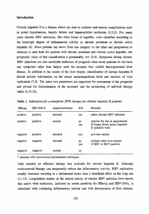
Introduction
Chronic hepatitis B is a disease which can lead to cirrhOsis and serious complications such
as portal hypertension, hepatic failure and hepatocellular carcinoma (1,2,3). For many
years chronic HBV infections, like other forms of hepatitis, were classified according to
the histologic degtee of inflammatory activity as chronic persistent or chronic active
hepatitis (4). Since patients can move from one category to the other and progtession to
cirrhosis is seen both for patients with chronic persistent and chronic active hepatitis, the
prognostic value of this classification is presumably low (5,6). Symptoms during chronic
HBV infections are also unreliable indicators of prognosis since many patients do not have
any symptoms other than fatigue until the moment they exhibit decompensated liver
disease. In addition to the results of the liver biopsy, classification of chronic hepatitis B
should include information on the serum aminotransferase level and markers of viral
replication (7,8). The latter two parameters are important for assessment of the prognosis
and pivotal for determination of the necessity and the monitoring of antiviral therapy
(table 1) (9,10).
Table 1. Indications for a-inteiferon (lFN) therapy for chronic hepatitis B patients.
HBeAg HBV-DNA'
positive positive
positive positive
negative positive
negative negative
negative negative
Aminotransferase
elevated
normal
elevated
elevated
normal
IFN
yes
no yes
yes
no yes
no
* detected with conventional hybridization techniques
Remarks
active chronic HBV infection
monitor for rise in aminotransf. if biopsy shows active hepatitis if patient's wish
pre-core mutant
exclude other liver disease if HDV or HCV positive
Until recently no effective therapy was available for chronic hepatitis B. Although
corticosteroid therapy can temporarily reduce the inflammatory activity, HBV replication
usually increases resulting in a detrimental rather than a beneficial effect in the long run
(11,12). Longitudinal studies on the natural history of chronic HBV infection have shown
that active viral replication, indicated by serum positivity for HBeAg and HBV-DNA, is
associated with continuing inflammatory activity and with deterioration of liver disease.
121
Termination of viral replication with HBeAg and HBV -DNA clearance (HBeAg serocon
version) is usually accompanied by clinical, biochemical and histological remission of
liver disease activity, and presumably with a better quality of life (13,14). "-IFN can
induce HBeAg seroconversion in 30 to 40 percent of the patients with chronic hepatitis B
(15-19). In more than 85% of the cases this response is sustained and more than half of
the patients become HBsAg negative after long-term follow-up (20). Until now it has not
been clear whether successful "-IFN therapy reduces the risk for hepatocellular carcino
ma.
Modification of the Alpha-Interferon Treatment Schedule
Standard Treatmen1
In randomized controlled trials both recombinant and human lymphoblastoid "-IFN have
been shown to be effective in inducing HBeAg seroconversion in chronic hepatitis B
patients. Many studies on "-IFN treatment in HBV infections, however, are characterized
by a considerable heterogeneity in patient population and response rate, contain few
patients and should therefore be interpreted with caution. Perrillo et a!. (15) reported on a
large randomized controlled trial of recombinant "-IFN treatment: HBeAg seroconversion
with histologic improvement was achieved in 38% of the patients who received the
standard treatment of 5 mega U "-interferon daily for 16 weeks compared to 7% of the
untreated controls. In the USA and in northwestern Europe the standard duration of "-IFN
therapy is now considered to be 12 to 16 weeks. The most effective dosage of "-IFN for
chronic hepatitis B is probably 30 MU per week. Regimens with "-IFN dosages under 15
MU per week have been shown to lack efficacy while doses above 30 MU weekly are too
toxic for the vast majority of patients (15). There does not appear to be a difference in
response rate berween daily dosages and a schedule of 3 administrations per week.
Prolonged Treatmem
In several trials from southern Europe "-IFN therapy was given for approximately 24
weeks (19,21). One of these studies suggests but does not prove an additional effect of
prolonged treatment schedules. In the UK a comparison berween 12 and 24 weeks of
lymphoblastoid "-IFN treatment could not demonstrate a beneficial effect of the prolonged
course (22). The results of this study were, however, markedly influenced by a lack of
compliance (8 of the 20 patients withdrew from the longer course) mainly due to side
effects of "-IFN. Results of a pilot study performed in our institute suggest that extending
"-IFN treatment for up to 32 weeks might eultance the HBeAg seroconversion and HBsAg
122
seroconversion rates (23). Interestingly, none of the patients in this study had to disconti
nue a-IFN therapy because of intolerance. On the grounds of these results a group of
European hospitals (Eurohep; 24) initiated a large randomized trial to evaluate whether
prolongation of a-IFN therapy for chronic hepatitis B is effective in enhancing the rate of
HBeAg seroconversion in patients who did not respond to standard a-IFN treatment of 16
weeks duration.
Intermittent Treatment
Intermittent therapy is a rather new approach to increase the response rate for a-IFN
treatment (23). We conducted a pilot Study consisting of a one-month printing course of
a-IFN, one month of rest and a second course lasting 4 months. The rationale for the
initial month of therapy was to decrease viral replication and induce a short immunologic
rebound before the start of the second long-term course, thereby enhancing the possibility
of successful therapy. Forty-eight percent of these patients exhibited HBeAg serocon
version. The level of HBV replication, as monitored by quantitative HBV-DNA and
HBeAg assessment at the start of the second course, was significantly lower than at
baseline. Furthermore we observed a double flare of the median aminotransferase values,
indicating that both courses could have induced a response in some of the patients. In our
opinion these interesting results justify evaluation of intermittent a-IFN therapy in a
controlled setting.
Combination Treatment with Alpha-Interferon
Alpha-lnteiferon and other Immwzemodulating Agents
A withdrawal course of corticosteroids before a-IFN therapy may combine enhanced
immunity with suppression of HBV replication. Several controlled trials which evaluated
the benefit of pretreatment with prednisone (tapered from 45-60 to 15-20 mg per day in 6
weeks followed by 30-35 MU a-IFN per week for 16 weeks) did not show an additional
effect of the priming steroid course (15,25,26). Nevertheless, in two trials the HBeAg
seroconversion rate of the prednisone-a-IFN schedule was markedly higher for the
subgroup of patients with low aminotransferase levels at baseline. This fmding suggests
that patients with mild inflammation may benefit from this combination therapy. The
benefit of combining a-IFN with another immunomodifying agent, levarnisol, is questiona
ble as a recent study has demonstrated that this combination resulted in a lower response
rate than a-IFN monotherapy (27). Simultaneous administration of a-IFN with other
cytokines such as y-interferon or inter!eukin-2 appears to be an attractive option since
123
expression of not only HLA type I but also HLA type II antigens on the hepatocyte
membrane is stimulated (28). This could lead to an additional impetus for the attack of
virus-infected hepatocytes by the cellular immune system. However, the results of several
studies of these combination therapies have so far been disappointing (29,30).
Alpha-Interferon and Nucleoside Analogues
In addition to a-IFN, which exerts its antiviral effect primarily by stimulation of cytotoxic
T cells and inhibition of HBV protein synthesis, nucleoside analogues interfere at a diffe
rent site in the HBV replication cycle inhibiting HBV DNA polymerase, a crucial enzyme
for viral DNA synthesis. ARA-AMP, acyclovir and zidovudine are at this moment the
most extensively investigated agents in HBV infected humans. Garcia and colleagues
investigated the alternating administration of ARA-AMP and a-IFN in comparison to
ARA-AMP and placebo (31). No effect of ARA-AMP alone or the combination conld be
demonstrated. About 25% of the patients receiving ARA-AMP suffered from painful
paresthesia necessitating dose reduction. The combination of a-IFN with other nucleoside
analogues like acyclovir, descylovir or zidovudine was extensively studied in our institute.
As a result of the positive fmdings of preliminary studies on the combination of a-IFN
with acyclovir derivatives (32,33), we initiated a large randomized controlled trial
comparing patients on combined a-IFN (5 MU daily for 16 weeks) and acyclovir (2 g
daily iv for 4 weeks) therapy with untreated controls (34). Only 25% of the treated
patients responded with HBeAg seroconversion as opposed to 14% of the controls. Simi
larly, we conducted a controlled trial comparing the antiviral effect of a-IFN and
zidovudine with that of a-IFN and placebo after both in vitro and in vivo pilot studies
showed that zidovudine caused a decrease in HBV replication (35-37). a-IFN and
zidovudine were given for 16 weeks in doses increasing to 5 MU and 1000 mg daily,
respectively. Again we were disappointed as there was no significant difference in the
response rate between the 2 groups and quantitative analyses of viral markers indicated
that no additional virus-inhibiting effect was obtained by adding zidovudine to the a-IFN
therapy. Furthermore, in the combination group bone marrow toxicity was prominent,
leading to reduction of the dose of both a-IFN and zidovudine in more than 40% of the
cases.
The conclusion to be derived from these results is that as yet no combination therapy
appears more effective than a-IFN alone and that several combinations give rise to an
increase in dose-limiting toxicity. Therefore in future studies a-IFN should only be co
administered with agents which combine a potent antiviral effect with a very low toxicity
profile.
124

Response Prediction of Alpha-Interferon Therapy
Since only one-third of the chronic hepatitis B patients respond to a-IFN therapy and since
a-IFN is a costly drug associated with marked side effects, it is important to know what
the chances of a succes are before the start of a-IFN therapy. The most important
variables which have been found to influence the response to a-IFN are listed in table 2
(15,16,18,23,38,39). In almost all studies on factors predictive for response, a high
aminotransferase level and a low level of viral replication (HBV-DNA) appeared to
enhance the response rate. Because the spontaneous HBeAg seroconversion rate is much
higher in this type of patient than in other untreated patients it is presumed that the
immune system is already in an activated state. For this reason some investigators
question the usefulness of a-IFN for patients with such a profile. From our experience a
IFN therapy may be beneficial for this group because the response rate with a-IFN is
higher than that for controls matched for the level of HBV replication and inflammatory
activity.
Table 2. Factors with predictive value for the occurrence of HBeAg seroconversion in
chronic hepatitis B patients treared with a-interferon.
Pretreatment
High aminotransferase level
Low HBV-DNA level
HlV antibodies absent
History of acute symptomatic hepatitis
Short duration of HBsAg positivity
Non-oriental race
Absence of immunosuppressive therapy
Inflammatory activity on liver biopsy
During treatment
Rise or peak of aminotransferases
HBV-DNA negativity
Rise of IgM anti-HBc
Brook and associates applied univariate and logistic regression analysis to pretreatment
variables of 114 chronic hepatitis B patients treated with a-IFN (40). They found the
levels of HBV -DNA and aspartate aminotransferase as well as the history of acute
hepatitis to be independently related to response. Using these same variables plus the HlV
status they constructed a model which could predict a response to therapy with a sensiti-
125
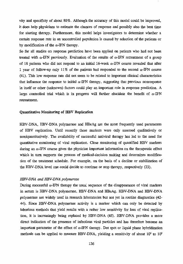
vity and specificty of about 80%. Although the accuracy of this model could be improved,
it does help physicians to estimate the chances of response and possibly also the best time
for starting therapy. Furthermore, this model helps investigators to determine whether a
certain response rate in an uncontrolled population is caused by selection of the patients or
by modification of the a-IFN therapy.
So far all studies on response prediction have been applied on patients who had not been
treated with a-IFN previously. Evaluation of the results of a-IFN retreatment of a group
of 18 patients who did not respond to an initial 16-week a-IFN course revealed that after
1 year of follow-up ouly 11% of the patients had responded to the second a-IFN course
(41). This low response rate did not seem to be related to important clinical characteristics
that influence the response to initial a-IFN therapy, suggesting that previous nonresponse
in itself or other (unknown) factors could play an important role in response prediction. A
large controlled trial which is in progress will further elucidate the benefit of a-IFN
retreatment.
Quantitative Monitoring of HBV Replication
HBV-DNA, HBV-DNA polymerase and HBeAg are the most frequently used parameters
of HBV replication. Until recently these markers were only assessed qualitatively or
semiquantitatively. The availability of successful antiviral therapy has led to the need for
quantitative monitoring of viral replication. Close monitoring of quantified HBV markers
during an a-IFN course gives the physician important information on the therapeutic effect
which in turn supports the process of medical-decision making and determines modifica
tion of the treatment schedule. For example, on the basis of a decline or stabilization of
the HBV-DNA level one could decide to continue or stop therapy, respectively (23).
HBV-DNA and HBV-DNA polymerase
During successful a-IFN therapy the usual sequence of the disappearance of viral markers
in serum is HBV-DNA polymerase, HBV-DNA and HBeAg. HBV-DNA and HBV-DNA
polymerase are widely used in research laboratories but not yet in routine diagnostics ( 42-
44). Since HBV-DNA polymerase activity is a marker which can ouly be detected by
laborious methods that yield results with a rather low sensitivity for loss of viral replica
tion, it is increasingly being replaced by HBV-DNA (45). HBV-DNA provides a more
direct indication of the presence of infectious viral particles and has therefore become an
important parameter of the effect of a-IFN therapy. Dot spot or liquid phase hybridization
methods can be applied to measure HBV-DNA, yielding a sensitivity of about 10'' to 10'
126
genomes per ml (46,47). In a comparison of two commercially available HBV-DNA
assays we found a good correlation between the HBV-DNA concentrations (48). However,
the actual agreement of the results was low because the HBV-DNA level between the
assays consistently differed by a factor 5 to 10. These discordant results were most
probably due to differences in standardization of the positive control samples, indicating
the need for a uniform HBV-DNA reference panel (49). In addition to conventional
techniques HBV-DNA can also be assessed by means of the polymerase chain reaction
(PCR) (50-52). By in vitro amplification of target DNA, levels below 10 genomes can be
detected. The clinical relevance of this extremely sensitive method is as yet unknown
because many HBsAg-positive carriers with inactive liver disease are still HBV-DNA
positive by PCR. Longitudinal studies bave demonstrated that a negativate PCR assesse
ment of HBV-DNA often approximates the moment of HBsAg seroconversion (20).
Hepatitis B related Alztigens
Of the HBV antigens present in serum HBeAg probably reflects the level of HBV
replication most directly. For patients infected with wild type HBV the dynamics of the
HBeAg level correlate closely with those of the HBV-DNA and HBV-DNA polymerase
levels (23,53). HBeAg seroconversion is an important event which indicates a transition
from HBV replication to viral latency. Unlike HBV-DNA negativity determined by dot
blot or solution hybridization methods, which can be followed by a relapse, HBeAg
seroconversion is almost always sustained (20,23). Therefore, quantitative HBeAg
assessment could be used both for monitoring and endpoint determination of antiviral
therapy in patients carrying wild type HBV (53). At present a standard test for quantitative
measurement of HBeAg is being developed (L. Mimms, personal communication) (54).
Patients carrying a mutant type of HBV with a deletion in the pre-core region of the
genome cannot produce HBeAg and often have active viral replication in the absence
HBeAg (55,56). Although the efficacy of a-IFN against this HBV variant is still a subject
of debate (57,58), obviously HBV-DNA and not HBeAg is the viral marker of choice for
monitoring treatment of these patients.
Other viral antigens which can be detected in serum are the s gene encoded proteins
HBsAg, pre-Sl Ag and pre-S2 Ag. All 3 markers bave been found to correlate with the
degree of HBV replication (59-61). Patients who were followed during a-IFN therapy
exhibited a good correlation between the HBsAg and the HBeAg or HBV-DNA level (62).
Since more than 90% of the patients who respond (HBeAg and HBV-DNA negative) to a
IFN remain HBsAg positive, the HBsAg cannot serve as an assessment for the endpoint of
a-IFN therapy. Furthermore, the wide range of HBsAg concentrations necessitate the use
127
of dilution steps in the quantitative measurement of this antigen. Nevertheless, for those
laboratories without the possibility to measure HBV-DNA or HBeAg quantitatively,
HBsAg measurement might be an alternative for assessment of changes in HBV replicati
on. HBsAg could also be a marker of interest for patients with hepatitis D or pre-core
mutants who often have active disease without the presence of HBeAg.
Decompensated Liver Disease and Alpha-Interferon
As in other forms of liver disease, indicators of hnpaired liver function (bilirubin,
albumin, coagulation factors) and portal hypertension (ascites and vaticeal bleeding) are
related to the survival of patients with HBsAg-positive cirrhosis (63-65). In addition to
these variables the HBeAg status also seetuS to be a factor that influences life expectancy
of cirrhotic HBV patients since the event of HBeAg seroconversion is associated with
prolonged survival (65). However, if patients exhibit signs of hepatic decompensation the
HBeAg status appears to loose its predictive value, suggesting that HBeAg seroconversion
in decompensated patients - whether a-IFN-induced or not - will not lead to an hnproved
prognosis. Nevertheless, anecdotal results in the USA indicate that a-IFN-induced arrest
of HBV replication in decompensated patients may lead to termination of disease progres
sion, disappearance of symptoms and preservation of the remaining hepatic function (66).
During therapy the majority of patients developed serious bacterial infections requiriog
antibiotic therapy. Moreover, either exacerbation of hepatitis or side effects led to
restriction of dose and/or duration of therapy in all cases. ln addition to its potential
therapeutic effect on the patient's own liver a-IFN may also be beneficial for patients with
decompensated disease for which liver transplantation remains the only realistic therapeu
tic option. In these patients pretransplant transition to viral latency would reduce the risk
of infection of the graft (67). Currently a controlled trial to investigate the benefit of low
dose a-IFN for patients with decompensated HBV cirrhosis is in progress (R. Perrillo,
personal communication).
That a-IFN therapy for patients with (decompensated) cirrhosis is not without risks was
illnstrated by 9 reported cases of fatal decompensation during or shortly after a-IFN
therapy (68,69). Ail patients had cirrhosis and 5 did not have signs of decompensation at
the start of therapy. It is conceivable that the transient exacerbation of hepatitis, which is a
sign of a-IFN-facilitated hepatocyte lysis, could be deadly in patients with limited residnal
liver capacity. One of the 9 patients first received a short -term prednisone course which
could have been an additional factor in the occunence of fatal decompensation (70). One
could conclude from these results that physicians should closely monitor a-IFN-treated
128
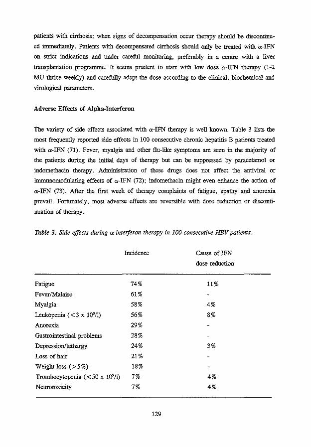
patients with cirrhosis; when signs of decompensation occur therapy should be discontinu
ed immediately. Patients with decompensated cirrhosis should only be treated with a-IFN
on strict indications and under careful monitoting, preferably in a centre with a liver
transplantation programme. It seems prudent to start with low dose a-IFN therapy (1-2
MU thrice weekly) and carefully adapt the dose according to the clinical, biochemical and
vitological parameters.
Adverse Effects of Alpha-Interferon
The variety of side effects associated with a-IFN therapy is well known. Table 3 lists the
most frequently reported side effects in 100 consecutive chronic hepatitis B patients treated
with a-IFN (71). Fever, myalgia and other flu-like symptoms are seen in the majority of
the patients duting the initial days of therapy but can be suppressed by paracetamol or
indomethacin therapy. Administration of these drugs does not affect the antiviral or
immunomodulating effects of a-IFN (72); indomethacin might even enhance the action of
a-IFN (73). After the first week of therapy complaints of fatigue, apathy and anorexia
prevail. Fortunately, most adverse effects are reversible with dose reduction or disconti
nuation of therapy.
Table 3. Side effects during a-interferon therapy in 100 consecutive HBV patients.
Fatigue
Fever/Malaise
Myalgia
Leukopenia ( < 3 x 1 0911)
Anorexia
Gastrointestinal problems
Depression/lethargy
Loss of hair
Weight loss (>5%)
Trombocytopenia (<50 x 109/1)
Neurotoxicity
Incidence
74%
61%
58%
56%
29%
28%
24%
21%
18%
7%
7%
129
Cause ofiFN
dose reduction
11%
4%
8%
3%
4%
4%
A serious side effect of a-IFN which is mostly not reversible is autoimmune-mediated
thyroid disease (74). Although it appears to be confined mainly to hepatitis C, both
hyperthyroidism and hypothyroidism can also occur during a-IFN therapy for HBV-iofec
ted patients. Sioce these diseases can be treated successfully, screemng of thyroid
hormones before and during therapy is recommended.
Neuropsychiatric Side Effects
A group of underestimated symptoms ioduced by a-IFN is the spectrum attributable to
neuropsychiatric toxicity. In particular lethargy, loss of concentration, depressive feeliogs,
emotional lability and irritability are not always diagnosed or thought to be related to a
IFN therapy (75,76). These symptoms appear rather innocent but they often give rise to
domestic problems or temporary discontinuation of work. The best way to reduce these
problems is careful explanation that the symptoms are treatment-related and will disappear
once therapy has been discontinued. As a second step dose reduction or psychiatric
counseliog should be considered. More serious forms of neuropsychiatric toxicity ioclude
depression, psychosis and seizures (77, 78). One of the most dramatic consequences of the
serious psychiatric disorders arisiog from a-IFN therapy is suicidal behavior. Reuault et
a!. described one patient who became suicidal during a-IFN therapy (77). After an exten
sive survey io 16 European hospitals we found 5 cases of attempted and 3 of completed
suicide among a total of about 2,500 chronic viral hepatitis patients on a-IFN therapy
(79). Even though it appears to be an uncommon event and we cannot prove directly that
the suicides were caused by a-IFN, the fact that none of the patients had a psychiatric
history, none had a poor life expectancy and all developed serious psychiatric disorders
(depression, psychosis and delirium) during a-IFN therapy argne io favor of a relation
between a-IFN therapy and suicide. Therefore, although this complication is rare it is so
dangerous that it is important to iostruct patients and family to recognize signs suggestive
of serious cognitive dysftmction and affective disturbances. If these signs occur psychiatric
consultation and/ or discontinuation of therapy is warranted.
Acknowledgement
We thank RA de Man for critically reviewiog the manuscript.
130
References
1. Hoofnagle lli, Alter IU. Chronic viral hepatitis. In: Vyas GN, Dienstag JL, Hoofna
gle lli eds. Viral hepatitis and liver disease. New York: Grone and Stratton, 1984;97-
113.
2. Viola LA, Barrison IG, Coleman JC, Paradinas FJ, Fluker JL, Evans BA, Murray
Lyon IM. Natural history of liver disease in chronic hepatitis B surface antigen
carriers. Survey of 100 patients from Great Britain. Lancet 1981;2:1156-9.
3. Beasley RP, Hwang L-Y, Lin C-C, Chien C-S. Hepatocellular carcinoma and hepatitis
B virus. A prospective study of 22 707 men in Taiwan. Lancet 1981;2:1129-33.
4. De Grootte J, Desmet VJ, Gedigk P et al. A classification of chronic hepatitis. Lancet
1968;ii:626-8.
5. Scheuer P. Classification of chronic viral hepatitis: a need for reasessment. J Hepatol
1991;13:372-4.
6. Fattovich G, Brollo L, Alberti A, Pontisso P, Realdi G, Ruol A. Chronic persistent
hepatitis: A clinical and morphological follow-up study. J Hepatol 1990;11:29-33.
7. Sakuma K, Saitoh N, Kasai M, Jitsukawa H, Yoshino I, Yamaguchi M, Nobutomo K
et al. Relative risk of death due to liver disease among Japanese male adults having
various statuses for hepatitis B s and e antigen/antibody in serum: a prospective study.
Hepatology 1988;8:1642-6.
8. Hoofnagle lli, Shafritz DA, Popper H. Chronic type B hepatitis and the "healthy"
HBsAg carrier state. Hepatology 1987;7:758-63.
9. Fattovich G, Brollo L, Giustina G, Noventa F, Pontisso P, Alberti A, Realdi G et al.
Natural history and prognostic factors for chronic hepatitis type B. Gut 1991;32:294-
8.
10. Hoofnagle JH. a-Interferon therapy for chronic hepatitis B. Current status and
reconnnendations. J Hepatol 1990;11 :S100-7.
11. Lam KC, Lai CL, Trepo C, Wu PC. Deleterious effect of prednisolone in HBsAg
positive chronic active hepatitis. N Eng! J Med 1981 ;304:380-6.
12. A trial group of the european association for the study of the liver. Steroids in chronic
B hepatitis. A randomized, double-blind, multinational trial on the effect of low-dose,
long-term treattnent on survival. Liver 1986;6:227-32.
13. Realdi G, Alberti A, Rugge M, Bortolotti F, Rigoli AM, Tremolada F, Ruol A.
Seroconversion from HBe antigen to antiHBe in chronic hepatitis B virus infection.
GastrOenterology 1980;79: 195-9.
14. Fattovich G, Rugge M, Brollo L, Pontisso P, Noventa F, Gnido M, Alberti A, et al.
131
Clinical virologic and histologic outcome following seroconversion from HBeAg to
antiHBe in chronic hepatitis type B. Hepatology 1986;6:167-72.
15. Perrillo RP, SchiffER, Davis GL, Bodenheimer HC Jr, Lindsay K, Payne J, Dienstag
JL. A randomized controlled trial of interferon alfa-2b alone and after prednisone
withdrawal for the treatment of chronic hepatitis B. N Eng! J Med 1990;323:295-301.
16. Hoofnagle JH, Peters M, Mullen KD, Jones DB, Rustgi V, Di Bisceglie A, Hallahan
C. Randomized controlled trial of recombinant human a-interferon in patients with
chronic hepatitis B. Gastroenterology 1988;95:1318-25.
17. Alexander GJM, Brahm J, Fagan EA, Smith HM, Daniels HM, Eddelston ALWF,
Williams R. Loss of HBsAg with interferon therapy in chronic hepatitis B virus
infection. Lancet 1987;ii:66-9.
18. Brook MG, Chan G, Yap I, Karayiannis P, Lever AM, Jacyna M, Main J, et al.
Randomised controlled trial of lymphoblasoid interferon alfa in Europid men with
chronic hepatitis B virns infection. BMJ 1989;299:652-6.
19. Saracco G, Mazzella G, Rosina F, Cancellieri C, Lattore V, Raise E, Rocca Get al.
A controlled trial of human lymphoblastoid interferon in chronic hepatitis B in Italy.
Hepatology 1989;10:336-41.
20. Koremnan J, Baker B, Waggoner J, Everhart JE, Di Bisceglie AM, Hoofnagle JH.
Long-term remission of chronic hepatitis B after alpha-interferon therapy. Ann Int
Med 1991;114:629-34.
21. Porres JC, Carreno V, Mora I, Gutiez J, Moreno A, Cajal SR, Marron JA, Bartolo
me J. Different doses of recombinant interferon alpha in the treattnent for chronic
hepatitis B patients without antibodies against the human inununodeficiency deficiency
virns. Hepatogastroenterology 1988;35:300-3.
22. Scully U, Shein R, Karayiannis P, McDonald JA, Thomas HC. Lymphoblastoid
interferon therapy of chronic hepatitis HBV infection. A comparison of 12 VS 24
weeks of thrice weekly treattnent. J Hepatol1987;5:51-8.
23. Janssen HLA, Berk L, Schalm SW, Heijtink RA, Hess G, Rosso! S, Meyer zum
Buschenfelde K-H, et al. The antiviral effect of prolonged intermittent lympho
blastoid alpha-interferon treatment in chronic hepatitis B. Gut 1992;33:1094-8.
24. Schalm SW. Eurohep. A European community sponsored concerted action on viral
hepatitis: its rationale and execution. J Hepatol 1992;15:1-3.
25. Fevery J, Elewaut A, Michielsen P, Nevens F, Eyken v P, Adler M, Desmet V.
Efficacy of interferon alfa-2b with or without prednisone withdrawal in treatment of
chronic viral hepatitis B. A prospective double-blind Belgian-Dutch study. J Hepatol
1990;1l:S108-12.
132
26. Lok ASF, Wu P-C, Lai C-L, Lau JYN, EKY Leung, LSK Wong, OCK Ma et a!. A
controlled trial of interferon with or without prednisone printing for chronic hepatitis
B. Gastroenterology 1992;102:2091-7.
27. Fattovich G, Brollo L, Giustina G, Ruvoletto MG, Pontisso P, Belussi F, Alberti A.
Combination therapy with lymphoblastoid interferon and levamisole in chronic
hepatitis type B. J Hepatol 1991;13:S27.
28. Billiau A. The mode of actions of interferons in viral infections and their possible role
in the control of hepatitis B. J Hepatoi1986;3:S171-9.
29. Di Bisceglie AM, Rustgi VK, Kassianides C, Lisker-Melman M, Park Y, Waggoner
JG, Hoofnagle JH. Therapy of chronic hepatitis B with recombinant human alpha and
gamma interferon. Hepatology 1990;11:266-70.
30. Hess G, Rosso! G, Iervolino M, Gotzler P, Meyer zum Buschenfelde K-H. Interferon
(IFN) alpha vs IFN/interleukin II in patients with chronic hepatitis B previously
unresponsive to IFN. J Hepatol 1991;13:S35.
31. Garcia G, Smith CI, Weissberg JI, Eisenberg M, Bissett J, Nair PV, Mastre B, et a!.
Adenide arabinoside monophosphate (vidarabine phosphate) in combination with
human leukocyte interferon in the treatment of chronic hepatitis B. Aun Intern Med
1987;107:278-85.
32. Schahn SW, Van Buuren HR, Heijtiuk RA, De Mao RA. Acyclovir enhances the
antiviral effect of interferon in chronic hepatitis type B. Laocet 1985;ii:358-60.
33. Man RA de, Schahn SW, Heijtiuk RA, Chamuleau RAFM, Reesink HW, Ouden J
den, Grijm R, et a!. Interferon plus descyclovir in chronic hepatitis B:incidence of
virus marker elimination and reactivation. In: Viral hepatitis aod liver disease;
Zuckerman AJ (ed.); Alan R Liss Inc., New York 1988:913-6.
34. Berk L, Schahn SW, De Man RA, Heijtiuk RA, Berthelot P, Brechot C, Boboc B, et
al. Failure of acyclovir to enhance the antiviral effect of alpha lymphoblastoid
interferon on HBe-seroconversion in chronic heaptitis B. A multi-centre randomized
controlled trial. J Hepatol 1992;14:305-9.
35. Berk L, Schahn SW, Heijtiuk RA. Zidovudine inhibits hepatitis B virns replication.
Antiviral Research 1992;19:111-8.
36. Nordenfelt E, Lofgren B, Chattopadhyaya J, Oberg 0. lnhibiton of hepatitis B virns
DNA polymerase by 3 '-azido-3 '-deoxythytnidine triphosphate but not by its threo
analogne. J Med Virol 1987;22:23Hi.
37. Janssen HLA, Berk L, Heijtiuk RA, Kate ten FJW, Schalm SW. Alpha-interferon and
zidovudine combination therapy for chronic hepatitis type B. Results of a randomized
placebo-controlled trial. Hepatology 1993;17:383-8.
133
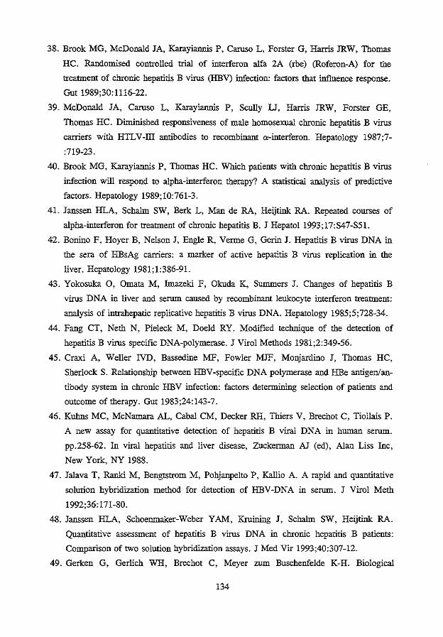
38. Brook MG, McDonald JA, Karayiannis P, Caruso L, Forster G, Harris JRW, Thomas
HC. Randomised controlled trial of interferon alfa 2A (rbe) (Roferon-A) for the
treatment of chronic hepatitis B virus (HBV) infection: factors that influence response.
Gut 1989;30:1116-22.
39. McDonald JA, Caruso L, Karayiaonis P, Scully U, Harris JRW, Forster GE,
Thomas HC. Diminished responsiveness of male homosexual chronic hepatitis B virus
carriers with HTLV-ID antibodies to recombinant a-interferon. Hepatology 1987;7-
:719-23.
40. Brook MG, Karayiannis P, Thomas HC. Which patients with chronic hepatitis B virus
infection will respond to alpha-interferon therapy? A statistical analysis of predictive
factors. Hepatology 1989;10:761-3.
41. Janssen HLA, Schahn SW, Berk L, Man de RA, Heijtink RA. Repeated courses of
alpha-interferon for treatment of chronic hepatitis B. J Hepatol 1993;17:S47-S5L
42. Bonino F, Hoyer B, Nelson J, Engle R, Verme G, Gerin J. Hepatitis B virus DNA in
the sera of HBsAg carriers: a marker of active hepatitis B virus replication in the
liver. Hepatology 1981;1:386-91.
43. Yokosuka 0, Ornata M, Imazeki F, Okuda K, Summers J. Changes of hepatitis B
virus DNA in liver and serum caused by recombinant leukocyte interferon treatment:
analysis of intrahepatic replicative hepatitis B virus DNA. Hepatology 1985;5;728-34.
44. Fang CT, Neth N, Pieleck M, Doeld RY. Modified technique of the detection of
hepatitis B virus specific DNA-polymerase. J Virol Methods 1981;2:349-56.
45. Craxi A, Weller !VD, Bassedine MF, Fowler MJF, Monjardino J, Thomas HC,
Sherlock S. Relationship between HBV-specific DNA polytnerase and HBe antigen/an
tibody system in chronic HBV infection: factors determining selection of patients and
outcome of therapy. Gut 1983;24:143-7.
46. Kuhns MC, McNamara AL, Cabal CM, Decker RH, Thiers V, Brechot C, Tiollais P.
A new assay for quantitative detection of hepatitis B viral DNA in human serum.
pp.258-62. In viral hepatitis and liver disease, Zuckerman AJ (ed), Alan Liss Inc,
New York, NY 1988.
47. Jalava T, Ranki M, Bengtstrom M, Pohjanpelto P, Kallio A. A rapid and quantitative
solution hybridization method for detection of HBV-DNA in serum. J Viral Meth
1992;36: 171-80.
48. Janssen HLA, Schoenmaker-Weber YAM, Kroining J, Schahn SW, Heijtink RA.
Quantitative assessment of hepatitis B virus DNA in chronic hepatitis B patients:
Comparison of two solution hybridization assays. J Med Vir 1993;40:307-12.
49. Gerken G, Gerlich WH, Brechot C, Meyer zum Buschenfelde K-H. Biological
134
standards of hepatitis B virus assay. J Hepatol 1992;15:251-5.
50. Kaneko S, Miller RH, Feinstone SM, Unoura M, Kobayashi K, Hattori N, Purcell
RH. Detection of serum hepatitis B virus DNA in patients with chronic hepatitis using
the polymerase chain reaction assay. Proc Nat! Acad Sci 1989;86:312-6.
51. Gerken G, Paterlini P, Manns M, et al. Assay of hepatitis B virus DNA by polymera
se chain reaction and its relationship to pre-s- and s-encoded viral snrface antigens.
Hepatology 1991;13:158-66.
52. Baker BL, Di Bisceglie AM, Kaneko S, Miller R, Feinstone SM, Waggoner JG,
Hoofnagle JH. Detemination of hepatitis B virus DNA in serum using the polymerase
chain reaction:clinical significance and correlation with serological and biochemical
markers. Hepatology 1991;13:632-6.
53. Berk L, Janssen HLA, Man de RA, Schahn SW, Heijtink RA. Complementary
quantitative HBeAg monitoring during antiviral therapy for chronic hepatitis B.
Submitted for publication.
54. Robbins D, Wright T, Coleman C, Umhoefer L, Moore B, Spronk A, Douville C et
al. Serological detection of HBeAg and anti-HBe using automated microparticle
enzyme inununoassays. J Viral Meth 1992;38:267-81.
55. Carman W, Jacyna M, Hadziyannis S, Karayiannis P, McGarvey MJ, Makris A,
Thomas HC. Mutation preventing formation of hepatitis e antigen in patients with
chronic HBV infection. Lancet 1989;2:588-91.
56. Brunetto MR, Giarin MM, Oliveri F, Chiaberge E, Baldi M, Alfarano A, Serra A, et
al. Wild type and e antigen-minus hepatitis B viruses and course of chronic hepatitis.
Proc Nat! Acad Sci 1991;88:4186-90.
57. Brunetto MR, Oliveri F, Rocca G, Criscuolo D, Chiaberge E, Capalbo M, David E,
et al. Natural course and response to interferon of chronic hepatitis B accompanied by
antibody to hepatitis Be antigen. Hepatology 1989;10:198-202.
58. Fattovich G, Farci P, Rugge M, Brollo L, Mandas A, Pontisso P, Giustins G, et al.
A randomized controlled trial of lymphoblastoid interferon-a in patients with chronic
hepatitis Blacking HBeAg. Hepatology 1992;15:584-9.
59. Hess G, Gerlich W, Gerken G, Manns M, Hutteroth TH, Meyer zum Buscheufelde.
The effect of recombinant a-interferon treatment on serum levels of hepatitis B virus
encoded proteins in man. Hepatology 1987;7:704-8.
60. Yuki N, Hayashi N, Katayama K, Kasbara A, Ueda K, Fusamoto H, Sato N, Kamada
T. Quantitative analysis of pre-S 1 and pre-S2 in relation to HBsAg expression.
Hepatology 1990;11:38-43.
61. Zoulim F, Mimms L, Floreani M, Pichoud C, Chemin I, Kay A, Vitvitski L, Trepo
135
C. New assays for quantitative determination of viral markers in management of
chronic hepatitis B virus infection. J Clin Microbiol1992;30:1111-9.
62. Janssen HLA, Heijtink RA, Schalm SW. HBsAg measurement for monitoring of
hepatitis B virus replication in patients treated with O<-interferon. Antiviral Research
1993; in press.
63. Weissberg JJ, Andres LL, Smith CI, Weick S, Nichols JE, Garcia G, Robinson WS,
et al. Survival in chronic hepatitis B. An analysis of 379 patients. Ann Intern Med
1984; 101:613-6.
64. D'Amico G, Morabito A, Pagliaro L, Marubini E, Caltagirone M, Filippazzo G,
Gatto G, et al. Survival and prognostic indicators in compensated and decompensated
cirrhosis. Dig Dis Sci 1986;31:468-75.
65. Jongh de FE, Janssen HLA, Man de RA, Hop WCJ, Schalm SW, Blankenstein van
M. Survival and prognostic indicators in hepatitis B surface antigen-positive cirrhosis
of the liver. Gastroenterology 1992;103:1630-5.
66. Kassianides C, Di Bisceglie AM, Hoofoagle JH, Mullen K, Peters M, Rustgi V, et al.
Alpha-interferon therapy in patients with decompensated chronic type B hepatitis. In:
Zuckerman AJ, ed. Viral hepatitis and liver diseases. New York: Alan R. Liss,19-
88:840-3.
67. Samuel D, Bismuth A, Mathieu D, Arulnaden JL, Reynes M, Benhamou JP, et al.
Passive irnmunoprophylaxis after liver transplantation in HBsAg-positive patients.
Lancet 1991;337:813-5.
68. Marcellin P, Colin JF, Boyer N, Bemuau J, Degott C, Hirschauer C, Benhamou JP.
Fatal exacerbation of chronic hepatitis B induced by recombinant alpha-interferon (let
ter). Lancet 1991;338:828.
69. Janssen HLA, Brouwer JT, Nevens F, Sanchez-Tapias, Craxi A, Hadziyannis S. Fatal
hepatic decompensation associated with interferon alfa. BMJ 1993;306:107-8.
70. Nair PV, Tong MJ, Stevenson D, Roskarnp D, Boone C. Effects of short-term, high
dose prednisone treatment of patients with HBsAg-positive chronic active hepatitis.
Liver 1985;5:8-12.
71. Janssen HLA, Berk L, Man de RA, Heijtink RA, Schalm SW. Antivirale behandeling
met alfa-interferon bij 100 patienten met chronische hepatitis B. Ned Tijdschr Geneesk
1992;136:835-9.
72. Berk L, Man de RA, Lindemans J, Heijtink RA, Schalm SW. Modulation of alpha
interferon effects by indomethacin and paracetamol in chronic hepatitis B. Submitted
for publication.
73. Andreone P, Cursaro C, Miniero R, Sprovieri C, Gasbarinni G. Indomethacin
136
increases serum 2-5 OAS before and after IFNO<in HBV-positive CAH. J Hepatol
1992; 16:S20.
74. Lisker-Melman M, Di Bisceglie AM, Usala SJ, Weintraub B, Murray LM, Hoofnagle
JH. Development of thyroid disease during therapy of chronic viral hepatitis with
interferon alfa. Gastroenterology 1992;102:2155-60.
75. McDonald EM, Mann AH, Thomas HC. Interferons as mediators of psychiatric
morbidity. Lancet 1987;ii:1175-8.
76. Adanas F, Quesada JR, Gutterman JU. Neuropsychiatric manifestations of human
leukocyte interferon therapy in patients with cancer. JAMA 1984;252:938-41.
77. Renault PR, Hoofnagle JH, Park Y, et a!. Psychiatric complications of long-term
interferon alfa therapy. Arch Intern Med 1987;147:1577-80.
78. Janssen HLA, Berk L, Vermeulen M, Schalm SW. Seizures associated with low-dose
O<-interferon. Lancet 1990;336: 1580.
79. Janssen HLA, Brouwer JT, Mast van de RC, Schalm SW. Suicide associated with
alpha-interferon therapy for chronic viral hepatitis. Submitted for publication.
137


SUMMARY
SAMENVATriNG
DANKWOORD
CURRIC11JLUM VITAE

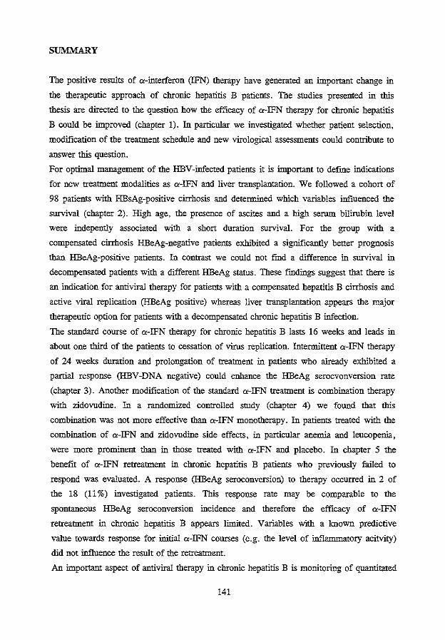
SUMMARY
The positive results of a-interferon (IFN) therapy have generated an important change in
the therapeutic approach of chronic hepatitis B patients. The studies presented in this
thesis are directed to the question how the efficacy of a-IFN therapy for chronic hepatitis
B could be improved (chapter 1). In particular we investigated whether patient selection,
modification of the treatment schedule and new virological assessments could contribute to
answer this question.
For optimal management of the HBV-infected patients it is important to defme indications
for new treatment modalities as a-IFN and liver transplantation. We followed a cohort of
98 patients with HBsAg-positive cirrhosis and determined which variables influenced the
survival (chapter 2). High age, the presence of ascites and a high serum bilirubin level
were indepently associated with a short duration survival. For the group with a
compensated cirrhosis HBeAg-negative patients exhibited a significantly better prognosis
than HBeAg-positive patients. In contrast we could not fmd a difference in survival in
decompensated patients with a different HBeAg status. These fmdings suggest that there is
an indication for antiviral therapy for patients with a compensated hepatitis B cirrhosis and
active viral replication (HBeAg positive) whereas liver transplantation appears the major
therapeutic option for patients with a decompensated chronic hepatitis B infection.
The standard course of a-IFN therapy for chronic hepatitis B lasts 16 weeks and leads in
about one third of the patients to cessation of virus replication. Intermittent a-IFN therapy
of 24 weeks duration and prolongation of treatment in patients who already exhibited a
partial response (HBV-DNA negative) could enhance the HBeAg serocvonversion rate
(chapter 3). Another modification of the standard a-IFN treatment is combination therapy
with zidovudine. In a randomized controlled study (chapter 4) we found that this
combination was not more effective than a-IFN monotherapy. In patients treated with the
combination of a-IFN and zidovudine side effects, in particular anemia and leucopenia,
were more prominent than in those treated with a-IFN and placebo. In chapter 5 the
benefit of a-IFN retreatment in chronic hepatitis B patients who previously failed to
respond was evaluated. A response (HBeAg seroconversion) to therapy occurred in 2 of
the 18 (11 %) investigated patients. This response rate may be comparable to the
spontaneous HBeAg seroconversion incidence and therefore the efficacy of a-IFN
retreatment in chronic hepatitis B appears limited. Variables with a known predictive
value towards response for initial a-IFN courses (e.g. the level of inflammatory acitvity)
did not influence the result of the retreatment.
An important aspect of antiviral therapy in chronic hepatitis B is monitoring of quantitated
141
viral markers. Using these markers one could inform patients during a-IFN treatment
about the effect of the therapy and one could either prolong or stop therapy in case of a
partial or no response, respectively. The most important marker reflecting the level of
virus replication is the Hepatitis B virus DNA (HBV-DNA). We compared 2 HBV-DNA
test systems (Abbott HBV-DNA assay, Abbott and Aff!Probe assay, Orion) in sera
obtained from patients treated with a-IFN (chapter 6). Although there was a good
correlation between the HBV-DNA results of the assays the AffiProbe test yielded
systematically 5 to 10 times higher HBV-DNA values than the Abbott assay. These
discordant results were most probably due to differences in the standardization of the
positive control samples of the test systems indicating the need for a uniform HBV-DNA
reference panel. Since HBV-DNA can ouly be measured quantitatively with time
consuming and costly methods its detection has been resrricted to research laboratories so
far. For the many hospitals not having the possibility to assess HBV-DNA there ntigbt be
a cheap and simpel alternative, quantitative HBsAg measurement. The level of HBsAg
decreases significantly after cessation of HBV replication (HBeAg seroconversion) and for
patients who were followed during a-IFN therapy we found a good correlation between
the HBsAg level and both the quantitative HBV-DNA and HBeAg values, irrespective of
the result of therapy (chapter 7).
Due to a virus specific immunological response a-IFN can cause a transient exacerbation
of inflammatory activity in chronic hepatitis B patients. This exacerbation could lead to a
fatal hepatic decompensation in patients with cirrhosis (chapter 8). After a patient in our
institute died due to a fatal decompensation during a-IFN therapy we sent out a
questionnaire to study the frequency and clinical aspects of this serious complication.
Among 2490 patients 8 additional cases with fatal hepatic decompensation during a-IFN
therapy were reported. Seizures and suicide attempts are 2 other complications associated
with a-IFN therapy of patients with chronic viral hepatitis. In chapters 9 and 10 we
described several patients who exhibited these serious neuropsychiarric adverse events of
a-JFN treatment.
In the chapter 11 the findings of studies presented in this thesis are discussed within an
overview of other recent developments of a-IFN therapy for chronic hepatitis B.
142
SAMENV ATTING
De hoopgevende hehandelingsresultaten met O<-interferon (lFN) hebben in de laatste jaren
geleid tot een belangrijke wijziging in het medisch handelen bij patienten met chronische
hepatitis B. Het onderzoek in deze dissertatie richt zich op de vraag hoe de effectiviteit
van O<-IFN behandeling bij chronische hepatitis B verbeterd kan worden (hoofdstuk 1).
Met name is onderzocht in welke mate de selectie van patienten. verandering van het
hehandelingsschema en nieuwe virologische vervolgmethoden hieraan kunnen bijdragen.
Het is voor een goed medisch beleid belangrijk om te weten in welke fase van de
leverziekte hehandeling met O<-IFN geindiceerd is en wanneer beter overgegaan kan
worden op levertransplantatie. Wij onderzochten welke factoren de overleving van een
cohort van 98 patienten met een HBV-gerelateerde levercirrhose beinvloeden. Hoge
leeftijd, de aanwezigheid van ascites en een hoog serum bilirubine-gehalte waren
onafhankelijk van andere factoren geassocieerd met een korte overlevingsduur. In de
groep met een gecompenseerde cirrhose hadden HBeAg-negatieve patienten een significant
langere overleving dan HBeAg-positieve patienten. In tegenstelling was er bij de groep
van gedecompenseerde patienten geen verschil in overleving bij patienten met een
verschillende HBeAg status. Deze bevinding suggereert dat er voor patienten met een
gecompenseerde hepatitis B cirrhose met virus replicatie (HBeAg positief) een indicatie
bestaat om door middel van antivirale behandeling een viruslatentie te induceren terwijl bij
patienten met een gedecompenseerde leverziekte levertransplantatie waarschijnlijk de
belangrijkste therapeutische optie is.
De standaard O<-IFN behandeling bij chronische hepatitis B duurt 16 weken en leidt in
ongeveer een derde van de patienten tot een verdwijning van de virusreplicatie.
Intermitterende hehandeling gedurende 24 weken en een verlenging van de behandeling bij
patienten die a! een partiele response (HBV-DNA negatief) vertonen zou mogelijk kunnen
lei den tot een verhoging van bet aantal HBeAg seroconversies (hoofdstuk 3). Een andere
modificatie van de standaard behandeling met O<-IFN is combinatie therapie met
zidovudine. In een gecontroleerd onderzoek (hoofdstuk 4) werd aangetoond dat deze
combinatie niet effectiever is dan O<-IFN monotherapie. In de groep behandeld met de
combinatie van O<-IFN en zidovudine waren er beduidend meer bijwerkingen, met name
anemie en leukopenie, dan in de groep die aileen O<-IFN kteeg. In hoofdstuk 5 wordt
nagegaan of herhehandeling van chronische hepatitis B patienten die bij een voorgaande
behandeling geen response (HBeAg seroconversie) ondergingen effectief is. Van de 18
patienten waren er slechts 2 (ll %) die een response vertoonden. Dit percentage is niet
boger dan het spontane HBeAg seroconversie percentage en de effectiviteit van
143
herbehandeling met a-IFN lijkt dus beperkt. Factoren die een bewezen predictieve waarde
ten aanzien van de response bij een eerste a-IFN therapie hebben (bijvoorbeeld het niveau
van ontstekingsactiviteit) bleken de nitkomst van de herbehandeling Diet te beinvloeden.
Een belangrijk aspect van de antivirale behandeling bij chronische hepatitis B is het
vervolgen van gekwantificeerde virusparameters (hoofdstuk 6 en 7). Bij a-IFN therapie
kan men met behulp van deze waarden de patient informeren over het beloop van de
behandeling en kan men bepalen of er a! dan niet een response aanstaande is zodat de
behandeling respectievelijk gecontinueerd of gestopt dient te worden. Een van de
belangrijkste parameters die de mate van virusreplicatie weergeven is het hepatitis B virus
DNA (HBV-DNA). Wij hebben 2 HBV-DNA testsystemen (HBV-DNA assay, Abbott en
AffiProbe assay, Orion) vergeleken in sera van patienten behandeld met a-IFN. Hoewel er
een zeer goede correlatie bleek te bestaan tussen de HBV-DNA nitslagen van de 2 testen
was er een systematisch 5 tot 10-voudig verschil mssen de uitkomsten. Uit onderzoek naar
positieve controle panels van de testen bleek er een verschil in de standaardisatie van de
hoeveelheid HBV-DNA in deze panels te zijn. Deze bevinding maakt duidelijk dat er
behoefte is aan een uniform HBV-DNA referentiepanel. Omdat het HBV-DNA aileen met
behulp van bewerkelijke en kostbare methoden kwantitatief gemeten kan worden is de
nitvoering van deze bepaling vooralsnog beperkt tot gespecialiseerde laboratoria. Voor de
vele centra die Diet de mogelijkheid hebben om HBV -DNA te meten is er wellicht een
goedkoop en simpel alternatief, de kwantitatieve HBsAg bepaling. De HBsAg concentratie
in serum blijkt significant te dalen na het verdwijnen van de virusreplicatie (HBeAg
seroconversie) en bij patienten die vervolgd zijn tijdens behandeling met a-IFN blijkt er
onafhankelijk van de uitkomst van de therapie een goede correlatie te zijn mssen het de
kwantitatieve HBsAg waarden en zowel de kwantitatieve HBV-DNA als HBeAg waarden.
Bij a-IFN behandeling van chronische hepatitis B patienten kan er een door het
immuunsysteem gegenereerde tijdelijke exacerbatie van ontstekingsactiviteit optreden.
Deze exacerbatie kan bij patienten met een cirrhose leiden tot een fatale
leverdecompensatie (hoofdstuk 8). Naar aanleiding van een dergelijke casus in ons
ziekenhnis onderzochten wij met behulp van een enquete de frequentie en klinische
aspecten van deze ernstige complicatie van de behandeling. Epilepsie en suicidepogingen
zijn 2 andere ernstige complicaties welke gerelateerd lijken te zijn aan a-IFN therapie bij
chronische virale hepatitis. In hoofdstuk 9 en 10 zijn enkele cases beschreven die de
kliniek van deze ernstige neuropsychiatrische bijwerkingen weergeven.
In de discussie (hoofdstuk 11) worden de bevindingen van de studies nit dit proefschrift
geplaatst binnen een overzicht van andere recente ontwikkelingen van antivirale
behandeling met a-IFN bij chronische hepatitis B.
144
DANKWOORD
Vele mensen hebben mij ondersteund gedurende de jaren die ik aan dit proefschrift heb
gewerkt. Zeer in het bijzonder mijn promotor Prof. dr. Solko Schalm die, inuner optimis
tisch en met een nauwelijks te evenaren dosis energie en nieuwe ideeen, de verschillende
onderzoeksprojecten tot een goed einde hielp brengen.
Ruud Heijtink ben ik zeer erkentelijk voor de wijze waarop hij mij, even geduldig als
adequaat, het terrein van de virologie heeft laten ontdekk.en. De door hem beheerde
serumbank van de Rotterdamse hepatitis B populatie is een "gouden" bron voor vee!
onderzoek geweest.
Rob de Man dank ik hattelijk voor de prettige samenwerking en de vele goede adviezen.
Met name zijn advies om "een onderzoeksproject af te maken, alvorens in een an.der ver
zeild te raken", heeft mijn werkwijze vee! efficienter gemaakt. Luuk Berk wil ik bedanken
voor het ontwerp en de initiatie van verschillende studies bescbreven in dit proefscbrift.
Felix de Jongh dank ik voor de grote hoeveelheid werk die hij verzet heeft in het kader
van onze "survival" studie.
Van Hans Brouwer en Heuk van Buuren kteeg ik vee! hulp om klinische problemen op te
lossen. Laatstgenoemde dank ik tevens voor het informeel overleg en de spirituosa
vrijdags na zessen.
Op adrninistratief gebied heb ik zeer vee! medewerking gehad van de "dames van het
secretariaat". Met name ontving ik vee! hulp van Stanny de Roode en Willeke Beukman
bij het verwerken van onderzoeksgegevens. Ook de anderen die mij hebben bijgestaan in
de door mij allerminst geliefde adrninistratieve bezigheden (bet zal eenieder op de afdeling
bekend zijn) ben ik zeer erkentelijk.
Jan Boot dank ik voor de enorme ondersteuning bij het ontwarren van problemen op het
computerscherm. Zonder zijn inzet zou ik dit proefschrift niet binnen de beoogde periode
van 3 jaar bebben ktnmen af ronden.
Yvonoe Scboemnaker, Coby Kerkhof, Gertie de Mutsert, Edwin Fries en Hans Kruining
wil ik bedanken voor het nauwgezet uitvoeren van vrijwel aile serologiscbe hepatitis B
bepalingen voor ntijn onderzoek.
Op het gebied van de statistiek werd ik op ktmdige wijze met raad en daad bijgestaan door
ir. W.C. Hop.
De !eden van de promotiecomntissie, Prof. J.H.P. Wilson en Prof. dr. F.T. Bosman, wil
ik van harte bedanken voor hun inspanning bij het beoordelen van bet manuscript.
Tenslotte dank ik Rob de Knegt voor zijn enorme hulp bij bet organiseren van de
promotie en voor de morele ondersteuning in de laatste drukke maanden.
145

CURRICULUM VITAE
De auteur van dit proefschrift werd geboren op 19 mei 1964 te Alkmaar. Hij groeide op
in de Achterhoek waar hij achtereenvolgens in Groenlo (Marianum) en Silvolde (Isala
College) het V.W.O. bezocht. In 1982 verhuisde hij naar Nijmegen om aan de Katholieke
Uuiversiteit geneeskunde te studeren. Het doctoraal examen werd behaald in juli 1986 en
het artsexamen in oktober 1989. Van januari tot en met oktober 1988 verrichtte hij
onderzoek naar de neurotoxiciteit van cyclosporine bij levertransplantaties (supervisie P.C.
de Groen, M.D.) op de afdeling Farmacologie van de Mayo Cliuic in Rochester,
Minnesota, U.S.A. Na het artsexamen was hij voor een periode van 3 jaar werkzaam op
de afdeliog Interne Geneeskunde II van het Academisch Ziekenhuis Dijkzigt te Rotterdam
(hoofd Prof. J.H.P. Wilson). In deze periode werd onder Ieiding van Prof. dr. S.W.
Schalm gewerkt aan het onderzoek beschreven in dit proefschrift. In januari 1993 werd
begonnen met de opleiding tot interuist in het Westeinde Ziekenhuis te 's Gravenhage
(opleider dr. E.J. Buurke).
147