
β CCX-CKR · CCX-CKR upon stimulation with CCL19, CCL21 or CCL25 using three different...
27
1 β-arrestin recruitment and G protein signaling by the atypical human chemokine decoy receptor CCX-CKR Anne O. Watts 1# , Folkert Verkaar 1,2# , Miranda M.C. van der Lee 2 , Claudia A. W. Timmerman 2 , Martien Kuijer 1 , Jody van Offenbeek 1,2 , Lambertus H. C. J. Van Lith 2 , Martine J. Smit 1 , Rob Leurs 1 , Guido J. R. Zaman 3 , Henry F. Vischer 1 * 1 Leiden/Amsterdam Center for Drug Research, Division of Medicinal Chemistry, Faculty of Sciences, VU University Amsterdam, Amsterdam, the Netherlands 2 Merck Research Laboratories, Molecular Pharmacology & DMPK, Oss, the Netherlands 3 Netherlands Translational Research Center B.V., Oss, The Netherlands # These authors contributed equally to this work *Corresponding author: Dr. Henry F. Vischer, Leiden/Amsterdam Center for Drug Research, Division of Medicinal Chemistry, Faculty of Science, VU University Amsterdam, De Boelelaan 1083, 1081 HV Amsterdam, The Netherlands. E-mail: [email protected] Running title: Atypical chemokine receptor signaling CAPSULE Background: CCX-CKR is considered to be a chemokine decoy receptor that is unable to signal. Results: chemokines induce β-arrestin recruitment to CCX-CKR and PTX-dependent CRE activity. Conclusion: PTX-sensitive G proteins hinder CCX-CKR coupling to other G proteins and consequently keep receptor silent. Significance: Recruitment of β-arrestin to CCX- CKR requests re-evaluation of the signaling capacity of this atypical receptor. SUMMARY Chemokine receptors form a large subfamily of G protein-coupled receptors that predominantly activate heterotrimeric G i proteins and are involved in immune cell migration. CCX-CKR is an atypical chemokine receptor with high affinity for CCL19, CCL21 and CCL25 chemokines, but is not known to activate intracellular signaling pathways. However, CCX-CKR acts as decoy receptor and efficiently internalizes these chemokines, thereby preventing their interaction with other chemokine receptors, like CCR7 and CCR9. Internalization of fluorescently labeled CCL19 correlated with β-arrestin2-GFP translocation. Moreover, recruitment of β-arrestins to CCX-CKR in response to CCL19, CCL21, and CCL25 was demonstrated using enzyme-fragment complementation and bioluminescence resonance energy transfer methods. To unravel why CCX- CKR is unable to activate G i signaling, CCX-CKR chimeras were constructed by substituting its intracellular loops with the corresponding CCR7 or CCR9 domains. The signaling properties of chimeric CCX-CKR receptors were characterized using a cAMP-responsive element (CRE)-driven reporter gene assay. Unexpectedly, wild type CCX-CKR and a subset of the chimeras induced an increase in CRE activity in response to CCL19, CCL21 and CCL25 in the presence of the G i inhibitor pertussis toxin. CCX-CKR signaling to CRE required an intact DRY motif. These data suggest that inactive G i proteins impair CCX-CKR signaling most likely by hindering the interaction of this receptor with pertussis toxin-insensitive G proteins that transduce signaling to CRE. On the other hand, recruitment of the putative signaling scaffold β-arrestin to CCX-CKR in response to chemokines might allow activation of yet to be identified signal transduction pathways. INTRODUCTION Chemokine receptors form a large family of G protein-coupled receptors (GPCRs) involved in migration, activation and differentiation of immune cells. The 19 known human chemokine receptors and their ~50 endogenous peptide ligands form a complex system in which many chemokine receptors can bind multiple chemokines, and many chemokines can bind more than one receptor (1). Chemokine receptors signal predominantly via heterotrimeric G i proteins, resulting in an inhibition of cAMP production by http://www.jbc.org/cgi/doi/10.1074/jbc.M112.406108 The latest version is at JBC Papers in Press. Published on January 22, 2013 as Manuscript M112.406108 Copyright 2013 by The American Society for Biochemistry and Molecular Biology, Inc. by guest on January 18, 2020 http://www.jbc.org/ Downloaded from
Transcript of β CCX-CKR · CCX-CKR upon stimulation with CCL19, CCL21 or CCL25 using three different...
1
β-arrestin recruitment and G protein signaling by the atypical human chemokine decoy receptor CCX-CKR Anne O. Watts1#, Folkert Verkaar1,2#, Miranda M.C. van der Lee2, Claudia A. W. Timmerman2, Martien Kuijer1, Jody van Offenbeek1,2, Lambertus H. C. J. Van Lith2, Martine J. Smit1, Rob Leurs1, Guido J. R. Zaman3, Henry F. Vischer1* 1 Leiden/Amsterdam Center for Drug Research, Division of Medicinal Chemistry, Faculty of Sciences, VU University Amsterdam, Amsterdam, the Netherlands 2 Merck Research Laboratories, Molecular Pharmacology & DMPK, Oss, the Netherlands 3 Netherlands Translational Research Center B.V., Oss, The Netherlands # These authors contributed equally to this work *Corresponding author: Dr. Henry F. Vischer, Leiden/Amsterdam Center for Drug Research, Division of Medicinal Chemistry, Faculty of Science, VU University Amsterdam, De Boelelaan 1083, 1081 HV Amsterdam, The Netherlands. E-mail: [email protected] Running title: Atypical chemokine receptor signaling CAPSULE Background: CCX-CKR is considered to be a chemokine decoy receptor that is unable to signal. Results: chemokines induce β-arrestin recruitment to CCX-CKR and PTX-dependent CRE activity. Conclusion: PTX-sensitive G proteins hinder CCX-CKR coupling to other G proteins and consequently keep receptor silent. Significance: Recruitment of β-arrestin to CCX-CKR requests re-evaluation of the signaling capacity of this atypical receptor. SUMMARY Chemokine receptors form a large subfamily of G protein-coupled receptors that predominantly activate heterotrimeric Gi proteins and are involved in immune cell migration. CCX-CKR is an atypical chemokine receptor with high affinity for CCL19, CCL21 and CCL25 chemokines, but is not known to activate intracellular signaling pathways. However, CCX-CKR acts as decoy receptor and efficiently internalizes these chemokines, thereby preventing their interaction with other chemokine receptors, like CCR7 and CCR9. Internalization of fluorescently labeled CCL19 correlated with β-arrestin2-GFP translocation. Moreover, recruitment of β-arrestins to CCX-CKR in response to CCL19, CCL21, and CCL25 was demonstrated using enzyme-fragment complementation and bioluminescence resonance energy transfer methods. To unravel why CCX-CKR is unable to activate Gi signaling, CCX-CKR
chimeras were constructed by substituting its intracellular loops with the corresponding CCR7 or CCR9 domains. The signaling properties of chimeric CCX-CKR receptors were characterized using a cAMP-responsive element (CRE)-driven reporter gene assay. Unexpectedly, wild type CCX-CKR and a subset of the chimeras induced an increase in CRE activity in response to CCL19, CCL21 and CCL25 in the presence of the Gi inhibitor pertussis toxin. CCX-CKR signaling to CRE required an intact DRY motif. These data suggest that inactive Gi proteins impair CCX-CKR signaling most likely by hindering the interaction of this receptor with pertussis toxin-insensitive G proteins that transduce signaling to CRE. On the other hand, recruitment of the putative signaling scaffold β-arrestin to CCX-CKR in response to chemokines might allow activation of yet to be identified signal transduction pathways. INTRODUCTION Chemokine receptors form a large family of G protein-coupled receptors (GPCRs) involved in migration, activation and differentiation of immune cells. The 19 known human chemokine receptors and their ~50 endogenous peptide ligands form a complex system in which many chemokine receptors can bind multiple chemokines, and many chemokines can bind more than one receptor (1). Chemokine receptors signal predominantly via heterotrimeric Gi proteins, resulting in an inhibition of cAMP production by
http://www.jbc.org/cgi/doi/10.1074/jbc.M112.406108The latest version is at JBC Papers in Press. Published on January 22, 2013 as Manuscript M112.406108
Copyright 2013 by The American Society for Biochemistry and Molecular Biology, Inc.
by guest on January 18, 2020http://w
ww
.jbc.org/D
ownloaded from
2
adenylyl cyclases (ACs) and induction of intracellular calcium mobilization (2). Interestingly, the chemokine receptors D6, DARC, CCX-CKR, CXCR7 and CCRL2 bind chemokines with high affinity, but ligand binding does not result in G protein-mediated intracellular calcium mobilization or chemotaxis (3). Rather, these atypical chemokine receptors act as decoy receptors to regulate chemokine availability. Upon internalization, the receptor-bound chemokines are either targeted for lysosomal degradation or transported across the cell to be subsequently exposed or released on the other side of the cell (i.e. transcytosis) (3). CCX-CKR is a high affinity receptor for the chemokines CCL19/ELC, CCL21/SLC and CCL25/TECK (4). These chemokines are important for the development of acquired immunity by activating CCR7 or CCR9. CCL19 and CCL21 recruit CCR7-expressing dendritic and T cells into the T cell compartments of secondary lymphoid organs (5). CCL25, on the other hand, recruits antigen-experienced lymphocytes to the small intestine by activating CCR9 (6). Mouse CCX-CKR knockout models demonstrated that CCX-CKR is important for steady-state homing of dendritic cells to skin-draining lymph nodes, T cell differentiation and immune response kinetics in an experimental auto-immune encephalomyelitis model, by regulating local chemokine levels (7,8). Moreover, CCR7 and CCR9 have been shown to play a role in various cancers (9). CCX-CKR scavenges CCL19 and CCL21 both in vitro and in vivo, thereby decreasing the free concentration of these chemokines (7,8,10). Indeed, low CCX-CKR expression levels in breast cancer tumor cells were correlated with an increase in lymph node metastasis and consequently poor survival rate of patients (11). Additionally, CCX-CKR has been suggested to mediate CCL19 and CCL21 transcytosis across lymphatic endothelium, although direct evidence is still lacking (12). CCX-CKR is expressed in many tissues, including heart, lung, and intestine, as well as by stromal cells of skin-draining lymph nodes, thymic epithelial cells, and a number of hematopoietic cell types (4,7,8,13-16). Transgenic overexpression of CCX-CKR decreased hematopoietic precursor cell numbers in the thymic anlage at embryonic stages, whereas cell numbers returned to normal levels in newborn and adult mice (7).
Following or alternative to G protein coupling, activated GPCRs can also recruit β-arrestin upon phosphorylation of their intracellular domains. β-arrestin bound to the GPCR may then act as a scaffold protein for receptor internalization and G protein-independent signaling to for example extracellular stimulus-regulated kinase 1/2 (ERK1/2) and Akt (protein kinase B) (17). The atypical chemokine receptor CXCR7 is internalized and signals exclusively through β-arrestin in a chemokine-dependent manner (18,19). D6 constitutively recruits β-arrestin, which is essential for the observed continuous internalization of D6 (20). However, a more recent study provided evidence that β-arrestin were not necessarily involved in D6 internalization but increased receptor stability (21). CCX-CKR has been previously suggested to internalize CCL19 in a β-arrestin-independent manner (10). In the present study, however, we demonstrate for the first time the concentration-dependent recruitment of β-arrestins to the atypical chemokine receptor CCX-CKR upon stimulation with CCL19, CCL21 or CCL25 using three different methodologies in various transfected cell lines. Moreover, we provide evidence that Gi proteins impair CCX-CKR-mediated signaling to CRE. These new aspects of CCX-CKR signaling provide additional avenues through which the role of CCX-CKR may be further explored. EXPERIMENTAL PROCEDURES Materials - Cell culture media were purchased from PAA Laboratories GmbH (Pasching, Austria). All non-labeled chemokines were obtained from PeproTech (London, UK). AlexaFluor 647-labeled CCL19 (CCL19-AF) was purchased from Almac (Craigavon, UK). 125I-labeled CCL19 (125I-CCL19) was purchased from Perkin Elmer (Boston, MA). Parathyroid hormone (PTH1-34) was obtained from Bachem (Bubendorf, Switzerland). Anti-mouse IgG-AlexaFluor 647 and anti-rat IgG-AlexaFluor 647 were purchased from R&D Systems (Abingdon, UK). Unless stated otherwise, all chemicals were purchased from Sigma-Aldrich (Steinheim, Germany). Nonidet P-40 (NP-40) was purchased from Roche Diagnostics (Almere, the Netherlands).
by guest on January 18, 2020http://w
ww
.jbc.org/D
ownloaded from
3
DNA constructs - pCMV-Prolink was purchased from DiscoveRx (Fremont, CA). Full-length human CCX-CKR in pECFP-N1 was provided by Marloes van der Zwam (Universitair Medisch Centrum Groningen, Groningen, the Netherlands). KOZAK-optimized CCX-CKR cDNA was amplified from this vector using PCR with Phusion polymerase enzyme (Finnzymes, Espoo, Finland) and was subcloned into pCMV-Prolink. cDNA encoding CCX-CKR N-terminally extended with 3 HA-tags (3xHA-CCX-CKR) and pertussis toxin (PTX)-insensitive mutants of Gαi1, Gαi2 and Gαi3 (Gαi/C351I), each in pcDNA3.1+ (Invitrogen, Carlsbad, CA), were purchased from Missouri S&T cDNA Resource Center (www.cdna.org; Rolla, MO). cDNA encoding CCX-CKR in pcDNA3.1 was a gift from Dr. Biber (Universitair Medisch Centrum Groningen, Groningen, the Netherlands). CCX-CKR and 3xHA-CCX-CKR were subcloned into pcDEF3 (a gift from Dr. Langer, Robert Wood Johnson Medical School, Piscataway, NJ). CCX-CKR cDNA was fused in frame with Renilla luciferase (Rluc) using a PCR-based method, as previously described (22). β-arrestin1 and β-arrestin2 enhanced yellow fluorescent protein EYFP fusion constructs were described previously (23). The R3.50A mutant of CCX-CKR was constructed by site-directed mutagenesis and re-introduced into pcDEF3 (CCX/R3.50A). To construct the other CCX-CKR mutants, unique restriction endonuclease sites were first introduced by synthesizing a CCX-CKR-encoding DNA fragment (GenScript, Piscataway, NJ, USA) with the following silent mutations: 399CAGCAT404 into 399ATCGAT404 to introduce ClaI in transmembrane (TM) helix 3-encoding sequence, 600TCAAATGCTAG610 into 600CCAAATGCTGG610 to introduce PflmI in TM5, 858AGTCACA864 into 858GGTCACC864 to introduce BstEII in TM7, and 922GCATCT927 into 922GCTAGC927 to introduce NheI in intracellular helix 8. This synthesized fragment was subcloned in pcDEF3 using BamHI and XbaI sites that flanked the open reading frame at the 5’ and 3’-end. Next, synthesized DNA sequences coding for IL2 and IL3 loops of CCR7 or CCR9 were inserted using ClaI-BlpI and PflmI-BstEII, respectively (Fig 5). The C-terminal ST/A-encoding DNA fragment was introduced using
NheI-XbaI. All generated constructs were sequence verified prior to use. The human parathyroid hormone receptor 1 (PTH1R) cDNA was provided by Winfried Mulder (Merck Research Laboratories, Oss, the Netherlands) and subsequently subcloned into pIRESpuro2 (Clontech, Mountain view, CA). BRET-based cAMP biosensor (pcDNA3.1-(L)-His-CAMYEL #ATCC-MBA-277) was purchased from American Type Culture Collection (Manassas, VA) (24). Cell culture and transfection – All cells were maintained at 37°C in a humidified atmosphere containing 5% CO2. U2OS cells stably expressing Green fluorescent protein (GFP)-tagged human β-arrestin2 (U2OS-β-arr2-GFP) were described previously (25). U2OS-β-arr2-GFP cells were stably transfected with pcDEF3-3xHA-CCX-CKR and pIRESpuro2 in a 4:1 molar ratio using Fugene 6 transfection reagent (Roche, Indianapolis, IN), followed by selection of single cell clones by addition of 2 µg/ml puromycin (Calbiochem, San Diego, CA) to culture medium. The U2OS-PTH1R cell line was derived from U2OS-β-arr2-GFP by transfection of pIRESpuro2-PTH1R and selection with 2 µg/ml puromycin. Parental Chinese hamster ovary (CHO)-K1 PathHunter cells stably expressing β-arrestin2 coupled to an inactive N-terminal β-galactosidase deletion mutant termed enzyme acceptor (β-arrestin2-EA) were purchased from DiscoveRx (Fremont, CA), and are referred to in this paper as CHO-β-arr2 cells. These cells were maintained in DMEM F12 supplemented with 10% v/v bovine calf serum (Hyclone, Logan, UT), 250 µg/ml hygromycin, 100 U/ml penicillin and 100 µg/ml streptomycin (all from Invitrogen, Carlsbad, CA). CHO-β-arr2 cells were stably transfected with pCMV-CCX-CKR-Prolink using Lipofectamine 2000 (Invitrogen) to yield the CHO-CCX-CKR cell line. CHO-K1 PathHunter cells stably expressing PTH1R-ProLink, CCR7-ProLink, and CCR9-ProLink (CHO-PTH1R, CHO-CCR7, CHO-CCR9, respectively) were purchased from DiscoveRx, and cultured in the medium mentioned above, supplemented with 800 µg/ml geneticin (Invitrogen). HEK293T cells were purchased from the American Type Culture Collection (ATCC) and maintained in DMEM supplemented with 10% (v/v) fetal calf serum (Integro, Dieren, the
by guest on January 18, 2020http://w
ww
.jbc.org/D
ownloaded from

4
Netherlands), 50 IU/ml penicillin and 50 µg/ml streptomycin. For radioligand binding, ELISA and reporter gene assays, HEK293T cells were transiently co-transfected with 500 ng/1·106 cells CRE-Luc DNA, 125 ng/1·106 cells receptor DNA in the absence or presence of 63 ng/1·106 cells Gα subunit DNA using linear polyethylenimine (MW 25,000; Polysciences, Warrington, PA) as described previously (26). For bioluminescence resonance energy transfer (BRET)-based β-arrestin1/2 recruitment assays, the HEK293T cells were co-transfected with 250 ng CCX-CKR-Rluc and 1 µg β-arrestin1/2-EYFP DNA per 1·106 cells, whereas 125 ng CCX-CKR cDNA was co-transfected with 250 ng pcDNA3.1-(L)-His-CAMYEL for the BRET-based cAMP biosensor experiments. High content analysis (HCA) of β-arrestin-2 redistribution - U2OS-β-arr2-GFP, U2OS-PTHR1, or U2OS-CCX-CKR cells were seeded in clear-bottom 96-well plates (PerkinElmer Life Sciences, Waltham, MA) with 15,000 cells/well in 90 µl assay medium (DMEM F12 supplemented with 2% (v/v) fetal calf serum, 100 U/ml penicillin and 100 µg/ml streptomycin) and cultured overnight. The following day, medium was replaced with 45 µl assay medium. Five microliter assay medium containing chemokines was added and cells were incubated at 37°C for 45 minutes. Cells were fixed in 4% (v/v) paraformaldehyde (BioConnect, Huissen, the Netherlands) and incubated with 1 µM Hoechst (Invitrogen) in PBS for 30 minutes. Plates were analyzed on an Operetta automated fluorescence microscope (PerkinElmer Life Sciences). Enzyme fragment complementation (EFC)-based β-arrestin recruitment assays - Cells were seeded in 384-well CulturPlates (PerkinElmer, Boston, MA) at 10,000 cells/well in 15 µl OPTI-MEM (Invitrogen) containing 1% v/v bovine calf serum (assay medium). The next day, cells were stimulated with 10 µl chemokine in assay medium and then returned to the incubator for 90 min. Cells were disrupted using 12 µl substrate-containing lysis buffer from the PathHunter Detection Kit in the formulation specified by the supplier (DiscoveRx). Plates were incubated in the dark for 90 min at room temperature before measurement of β-galactosidase activity
(luminescence) on an Envision multilabel plate reader (PerkinElmer). BRET-based β-arrestin recruitment assay - One day after transfection, HEK293T cells were transferred to poly-L-lysine-coated white 96-well plates. Growth medium contained 100 ng/ml PTX (Sigma-Aldrich, Steinheim, Germany) where applicable. The next day, medium was replaced with Hank’s Balanced Salt Solution (HBSS) containing 100 ng/ml PTX where applicable and fluorescence was measured on a Victor3 multilabel plate reader (ex. 485 nm; em. 535 nm; PerkinElmer Life Sciences). Ten minutes after coelenterazine-h (5 µM final concentration; Promega, Madison, WI) addition, ligand solutions in HBSS supplemented with 0.05% BSA were added in the stated concentrations and incubated for an additional 5 minutes. Bioluminescence resonance energy transfer (BRET) (em. 535 nm) and Rluc expression (em. 460 nm) were measured with a Victor3 multilabel plate reader. Baseline-corrected BRET ratios were calculated by first dividing BRET by Rluc emission values, followed by subtraction of the BRET ratio of cells expressing CCX-CKR-Rluc alone. cAMP accumulation assay - cAMP measurements in CHO-CCX-CKR cells were performed with a homogenous time-resolved fluorescence (HTRF) kit from Cisbio (Gif-sur-Yvette, France) essentially as described previously (27). Briefly, chemokines diluted in 10 µl dilution buffer either containing forskolin (0.5 µM final concentration) or DMSO control were dispensed in 384-well OptiPlates (PerkinElmer Life Sciences). Subsequently, 7,500 CHO-CCX-CKR cells in 10 µl assay medium (DMEM F12 containing 5 µg/ml apo-transferrin (Sigma-Aldrich, Steinheim, Germany), 1 µg/ml insulin, 100 U/ml penicillin and 100 µg/ml streptomycin) were added and plates were incubated at 37°C for 60 minutes. Cells were disrupted by addition of 10 µl cAMP-XL665 conjugate and 10 µl Europium-labeled anti-cAMP antibody diluted in lysis buffer (all provided with the kit). Plates were incubated in the dark at room temperature for 60 minutes before measurement of time-resolved fluorescence at 615 nm and 665 nm on an Envision multilabel plate reader (PerkinElmer Life Sciences). Absolute
by guest on January 18, 2020http://w
ww
.jbc.org/D
ownloaded from
5
cAMP concentrations were calculated from a cAMP standard. Inositol phosphate accumulation assay – Inositol phosphate measurements in CHO-CCX-CKR cells were performed a homogenous time-resolved fluorescence (HTRF) kit from Cisbio (Gif-sur-Yvette, France). CHO-CCX-CKR cells were plated in 384-well proxiPlates with 10,000 cells/well in 20 µl assay medium (DMEM F12 containing 10% (v/v) fetal calf serum, 100 U/ml penicillin and 100 µg/ml streptomycin) and were allowed to adhere overnight. Medium was aspirated and cells were stimulated with 14 µl chemokine in stimulation buffer (provided in the kit) containing 0.1% (w/v) bovine serum albumin (BSA; Sigma-Aldrich, Steinheim, Germany). Plates were incubated at 37°C for 90 minutes before the addition of 3 µl IPone-XL665 conjugate and 3 µl Europium-labeled anti-IPone antibody diluted in lysis buffer (all provided with the kit). Plates were incubated in the dark at room temperature for 60 minutes before measurement of time-resolved fluorescence at 615 nm and 665 nm on an Envision multilabel plate reader. Absolute IPone concentrations were calculated from an IPone standard. Western blotting - Isolation of CHO-CCX-CKR lysates and western blotting was performed as described previously [66]. The following antibodies were used at the indicated dilutions: mouse anti-β-actin: 1:5000 (Abcam; Cambridge, UK), rabbit-anti-total Akt: 1:1000, rabbit anti-phospho-Ser473 Akt: 1:1000, rabbit anti-phospho-Thr202/Tyr204 ERK1/2: 1:2000 (Cell Signaling Technology; Danvers, MA), mouse anti-total ERK1/2:1:2000 (Invitrogen), anti-mouse-HRP conjugate: 1:2000 and anti-rabbit-HRP conjugate: 1:2000 (Promega; Leiden, the Netherlands). Cyclic AMP-responsive element (CRE)-driven reporter gene - One day after transfection, HEK293T cells were transferred to poly-L-lysine-coated white 96-well plates. Where required, growth medium contained 100 ng/ml PTX. The following day, growth medium was replaced with serum-free medium supplemented with 0.05% BSA and 100 ng/ml PTX, 1 µM forskolin (FSK) and/or ligands, as indicated. After 6 to 8 hours incubation at 37°C, stimulation medium was
removed and cells were incubated for 5 minutes with 25 µl substrate solution (39 mM Tris.H3PO4 pH 7.8, 39% glycerol, 2.6% Triton X-100, 860 µM dithiothreitol, 18 mM MgCl2, 825 µM ATP, 77 µM disodium pyrophosphate, 230 µg/ml beetle luciferin) (Promega, Madison, WI). Luminescence was measured using a Victor3 multilabel plate reader. BRET-based cAMP biosensor assay – Changes in cAMP levels were detected using a BRET-based cAMP sensor in a similar manner as the BRET-based β-arrestin recruitment assay, except that cells were rinsed once with HBSS, and incubated with fresh HBSS for 30 min before being stimulated. In addition, the non-specific phosphodiesterase 3-isobutyl-1-methylxanthine was added simultaneously with coelenterazine-h to a final concentration of 40 µM (23). Radioligand binding and internalization - One day after transfection, HEK293T cells were transferred to poly-L-lysine (Sigma-Aldrich, St. Louis, MO) coated 96 well plates. The next day, whole cells were incubated for four hours at 4°C with ~0.75 nM [125I]-CCL19 in binding buffer (50 mM HEPES, 100 mM NaCl, 1 mM CaCl2, 5 mM MgCl2, pH 7.4, 0.5% BSA) containing indicated concentrations of unlabeled displacer. Incubations were terminated by washing the cells with ice-cold wash buffer (50 mM HEPES, 0.5 M NaCl, 1 mM CaCl2, 5 mM MgCl2, pH 7.4) followed by lysis in RIPA buffer (0.5% NP-40, 0.5% sodium deoxycholate and 0.1% sodium dodecyl sulfate). Cell lysates were transferred to vials and counted in a Wallac Compugamma counter (PerkinElmer Life Sciences). [125I]-CCL19 internalization assays were performed in a similar manner as binding assays on intact cells, except that incubations were performed in a 37°C waterbath (28). Incubations were terminated at the indicated time intervals by placing the 48-well plates on ice and rapid removal of free [125I]-CCL19 by washing the cells three times with ice-cold wash buffer. The fraction [125I]-CCL19 bound to receptors at the cell surface was removed using ice-cold acidified DMEM (pH 2.0). The cells were then solubilized in RIPA buffer to collect the acid-resistant (i.e. internalized) [125I]-CCL19 fraction, which was quantified using a Wallac Compugamma counter.
by guest on January 18, 2020http://w
ww
.jbc.org/D
ownloaded from
6
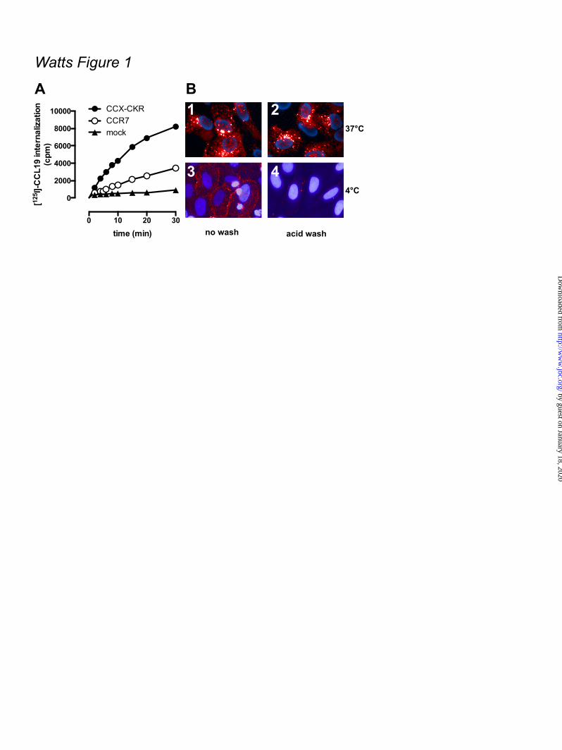
Control experiments confirmed that acid treatment removed all surface-bound chemokine. ELISA - One day after transfection, HEK293T cells were seeded in 96 well plates and cultured overnight. Next, the cells were fixed for 30 minutes with 4% formaldehyde in PBS and washed twice with tris-buffered saline (TBS; 150 mM NaCl, 10 mM Tris.HCl, pH 7.5). Half of the samples were permeabilized by incubating the cells for 30 minutes with 0.5% NP-40 in TBS after fixation. Samples were then blocked for 4 hours at room temperature with 1% fat free milk in 0.1 M NaHCO3, pH 8.6. Samples were incubated overnight at 4ºC with mouse anti-hCCX-CKR clone 2F11 (a kind gift from Dr. Chiba, Tokyo University of Science, Japan) diluted 1:1000 in TBS containing 1% fat-free milk powder and were subsequently washed with TBS. Samples were incubated for 3 hours at room temperature with goat anti-mouse HRP-conjugated antibody (Bio-Rad Laboratories, Veenendaal, the Netherlands) diluted 1:2500 in 0.1 M NaHCO3, pH 8.6, containing 1% fat-free milk powder. After samples were washed with TBS, OPD substrate solution (2.2 mM o-phenylenediamine, 35 mM citric acid, 66 mM Na2HPO4, 0.015% H2O2, pH 5.6) was added. The reaction was stopped by addition of 1 M H2SO4 and the absorbance at 490 nm was measured with a PowerWave plate reader (BioTek, Winooski, VT). Data analysis - Sigmoidal concentration-response curves were plotted using Graphpad Prism 5.0 software. All data are presented as averages ± standard error of the mean (SEM). Statistical analyses were performed using Graphpad Prism 5.0 software. RESULTS CCL19 is internalized by CCX-CKR and co-localizes with β-arrestin – The CCX-CKR binds CCL19 with a ~6- and ~30-fold higher affinity (pKi) than CCL25 and CCL21, respectively (Table 1). This rank order of binding affinities is in line with the previously reported 50% inhibitory concentration (IC50) values (4). Hitherto, the only functional read-out for CCX-CKR has been the measurement of chemokine internalization (10). HEK293T cells transiently transfected with CCX-CKR internalized [125I]-CCL19 substantially faster
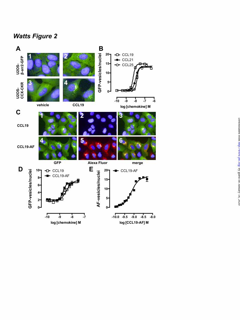
than cells expressing CCR7 (Fig 1A), which confirms previous observations (10). Next, CCX-CKR was stably expressed in U2OS human osteosarcoma cells that already express GFP-tagged β-arrestin2. These U2OS-CCX-CKR cells were treated for 45 minutes with 100 nM CCL19 conjugated to Alexa Fluor 647 (CCL19-AF) and imaged using automated high content fluorescence microscopy. CCL19-AF was internalized and accumulated in U2OS-CCX-CKR cells as indicated by the resistance to an acid wash (Fig 1B1,2). Moreover, CCL19-AF internalization was impaired at 4°C (Fig 1B3,4). This accumulation of CCL19-AF in U2OS-CCX-CKR cells was CCX-CKR-dependent, as it was not observed when CCL19-AF was incubated with parental control cells (U2OS-β-arr2-GFP) or derived cells that overexpressed the human parathyroid hormone receptor type 1 (U2OS-PTH1R) (data not shown). Next, it was evaluated whether chemokine binding to CCX-CKR can induce translocation of β-arrestin2-GFP to putative endocytic vesicles. In non-stimulated parental U2OS-β-arr2-GFP and U2OS-CCX-CKR cells, β-arrestin2-GFP was uniformly distributed throughout the cytoplasm (Fig 2A1,3). In response to CCL19, β-arrestin2-GFP coalesced into vesicles in U2OS-CCX-CKR cells (Fig 2A4). CCL19 was unable to cause β-arrestin2-GFP redistribution in parental U2OS-β-arr2-GFP cells that did not express CCX-CKR (Fig 2A2) or in U2OS-β-arr2-GFP cells stably expressing PTH1R (U2OS-PTH1R) (data not shown). Subsequently, U2OS-CCX-CKR cells were treated with increasing concentrations of CCL19, CCL21 or CCL25 for 45 minutes and vesicle formation was quantified. The three chemokines had similar efficacy to induce β-arrestin2-GFP translocation (Fig 2B), with CCL19 having a ~4-fold higher potency than CCL21 and CCL25 (Table 1). CCL19-AF induced β-arrestin2-GFP translocation with a similar potency as CCL19 (Fig 2C1,4 and 2D). Moreover, CCL19-AF co-localized with β-arrestin2-GFP in vesicles (Fig 2C4-6). CCL19-AF induced β-arrestin2-GFP-vesicle and AF-vesicle formation with comparable potencies (Fig 2E; Table 1). The enzyme fragment complementation (EFC)-based PathHunterTM β-arrestin assay was subsequently used to monitor enzyme acceptor-coupled β-arrestin2 (β-arr2-EA) recruitment to ProlinkTM-tagged CCX-CKR in response to
by guest on January 18, 2020http://w
ww
.jbc.org/D
ownloaded from
7
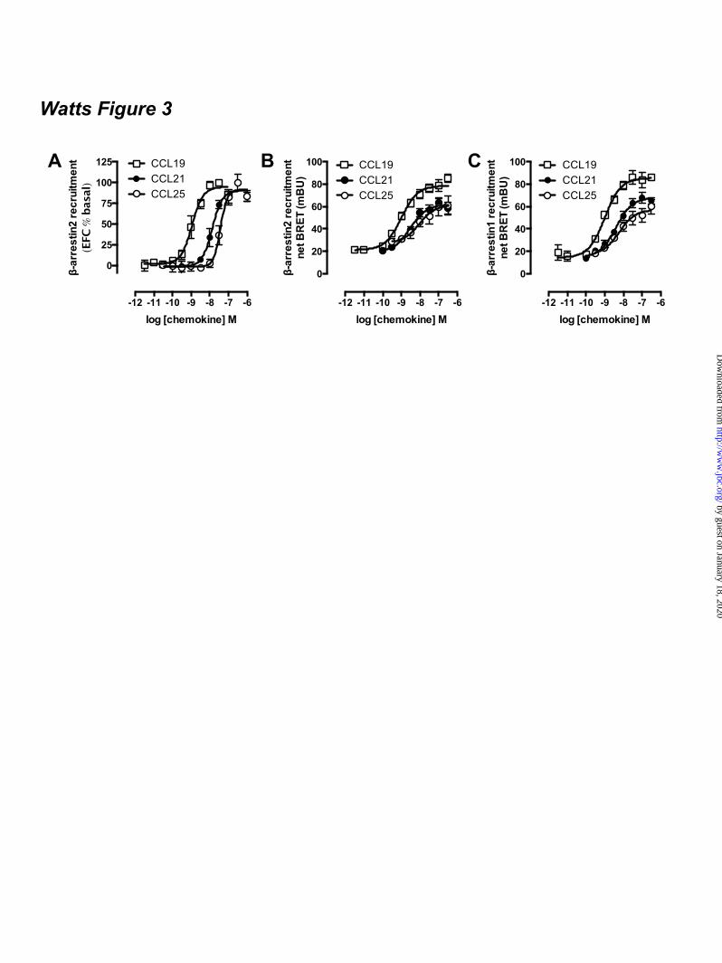
chemokine stimulation. To this end, CCX-CKR-ProLink was stably expressed in CHO cells harboring β-arr2-EA (CHO-β-arr2). Stimulation of the resultant CHO-CCX-CKR cell line with CCL19, CCL21 and CCL25 for 90 minutes resulted in a concentration-dependent reconstitution of β-galactosidase enzyme activity as a consequence of the chemokine-induced close proximity between CCX-CKR-Prolink and β-arr2-EA (Fig. 3A). All three chemokines act as full agonists in recruiting β-arrestin2 to CCX-CKR in this EFC-based assay format, with CCL19 being ~8- and ~12.5-fold more potent than CCL21 and CCL25, respectively (Table 1). As expected, CCL19 and CCL21 induced β-arr2-EA recruitment to CCR7-ProLink in CHO-CCR7 cells, whereas CCL25 stimulated the β-arr2-EA recruitment to CCR9-ProLink in CHO-CCR9 cells (data not shown). In contrast, the parental CHO-β-arr2 cells and those expressing the human parathyroid hormone receptor 1 (CHO-PTH1R) did not show an increase in β-galactosidase activity upon incubation with CCL19 (data not shown). Next, the potency and efficacy of a panel of chemokines to recruit β-arrestin2 to CCX-CKR in these CHO-CCX-CKR cells was evaluated. With the exception of CCL9/10, which acts a low potency (pEC50 = 6.2 ± 0.1) partial agonist (intrinsic activity α = 0.5 in comparison to full agonist CCL19), all tested chemokines (i.e. vMIP-II, CCL3, CCL4, CCL6, CCL23, CXCL6, CXCL8, CXCL9, CXCL10, CXCL11, CXCL12, CXCL13) were unable to reconstitute β-galactosidase activity in these CHO-CCX-CKR cells when added up to a concentration of 1 µM (data not shown). Bioluminescence resonance energy transfer (BRET) technology was then used to confirm this close proximity between CCX-CKR and β-arrestin2 upon stimulation with CCL19, CCL21, and CCL25 (Fig 3B; Table 1). Moreover, a similar concentration-dependent increase in BRET ratio was observed between CCX-CKR-Rluc and β-arrestin1-EFYP in response to these chemokines (Fig 3C; Table 1). CCL19 was the most potent chemokine in inducing β-arrestin1/2 recruitment to CCX-CKR, whereas CCL21 and CCL25 had comparable potency. To evaluate whether β-arrestin1/2 recruitment to CCX-CKR required phosphorylation of the CCX-CKR C-terminal tail, all serine and threonine residues in this domain
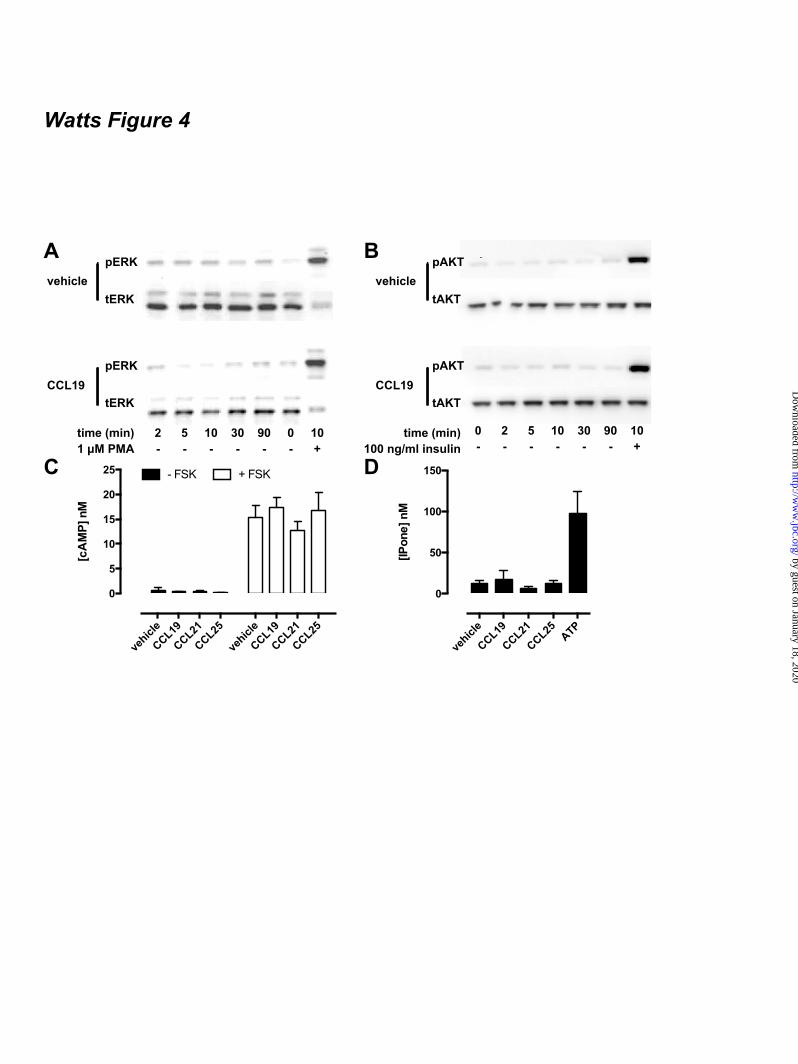
were alanine substituted (i.e. 323SWRRQRQSVEEFPFDSEGPTEPTSTFS349 into 323AWRRQRQAVEEFPFDAEGPAEPAAAFA349). The expression level of this CCX-CKR-ST/A-Rluc mutant was approximately 60% of that of non-mutant CCX-CKR-Rluc, as determined by Renilla luciferase activity and ELISA on intact cells (data not shown). However, binding assays showed that CCX-CKR-ST/A-Rluc had no affinity for [125I]-CCL19 and [125I]-CCL25, and consequently was not able to recruit β-arrestin2-EYFP in response to chemokines (data not shown). CCX-CKR does not activate typical chemokine receptor-induced β-arrestin- or G protein-mediated signaling - To evaluate the ability of CCX-CKR to activate downstream effectors of β-arrestin signaling, CHO-CCX-CKR cells were stimulated with vehicle or 100 nM CCL19 for 2 to 90 minutes and lysates were probed for phosphorylation of ERK1/2 (Thr202/Tyr204) and Akt (Ser473). CCL19-induced phosphorylation of ERK1/2 was not observed, whereas treatment with the positive reference PMA for 10 minutes elicited robust ERK1/2 phosphorylation (Fig 4A). Furthermore, CCL19 did not affect Akt phosphorylation in CHO-CCX-CKR cells, whereas treatment with insulin caused a marked increase in Akt phosphorylation (Fig 4B). In summary, no CCX-CKR-induced signaling to ERK1/2 or Akt was observed. Since most chemokine receptors signal through Gi proteins upon receptor activation, CHO-CCX-CKR cells were stimulated with 100 nM CCL19, CCL21 or CCL25 for 60 minutes and cAMP levels were subsequently determined using an HTRF-based assay. These chemokines did not affect basal or forskolin (FSK)-induced cAMP accumulation, suggesting that CCX-CKR is not coupling to Gs or Gi proteins, respectively (Fig. 4C). This was not due to an inability of the chemokines to induce such responses, as CCL19 and CCL25 decreased FSK-induced cAMP levels in CHO-CCR7 and CHO-CCR9 cells, respectively (data not shown). The ability of CCX-CKR to activate Gq proteins was also investigated. Intracellular inositol phosphate concentrations in CHO-CCX-CKR cells were not affected by treatment with CCL19, CCL21 and CCL25 (Fig.
by guest on January 18, 2020http://w
ww
.jbc.org/D
ownloaded from
8
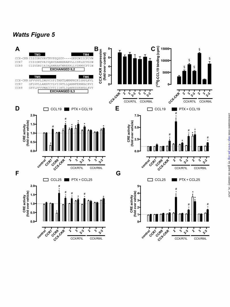
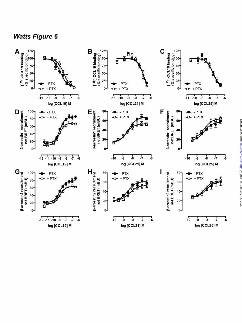
4D). In contrast, inositol phosphate formation was observed in CHO-CCX-CKR cells after activation of endogenous purinoceptors by 100 µM ATP (Fig. 4D). CCX-CKR IL2/3 chimeras stimulate cAMP signaling - In contrast to CCR7 and CCR9, the atypical chemokine receptor CCX-CKR did not decrease FSK-induced cAMP levels in response to CCL19 and CCL25 stimulation. CCX-CKR chimeras were constructed by substitution of intracellular loop (IL) 2 and/or 3 with corresponding sequences of CCR7 and CCR9 to investigate whether intracellular domains of CCX-CKR might impede G protein coupling (Fig 5a). These CCR7 and CCR9 ILs were hypothesized to confer Gi-coupling capacity to the CCX-CKR chimeras, resulting in decreased adenylyl cyclase activity. A cAMP-responsive element (CRE)-driven luciferase reporter gene assay was used to quantify chemokine-stimulated changes in cAMP levels in HEK293T cells transiently expressing wildtype CCX-CKR or CCX-CKR chimeras. Cell surface expression of all CCX-CKR chimeras was comparable (p > 0.05) to wild type CCX-CKR as measured by ELISA (Fig 5B), whereas [125I]-CCL19 binding to CCX-CKR chimeras was significantly increased (p < 0.05) in comparison to wild type CCK-CKR (Fig 5C). Except CCX/R9IL3 bound less [125I]-CCL19 than wild type CCX-CKR. As expected, FSK-induced CRE-activity was decreased by treatment with 100 nM CCL19 in cells expressing CCR7 but not in cells transfected with wild type or chimeric CCX-CKR constructs (Fig 5D-E). CCL19 does not interact with CCR9 and consequently had no effect on FSK-induced CRE activity in CCR9-expressing cells. As a control for Gi-mediated signaling responses, cells were pre-treated overnight with 100 ng/ml of the inhibitor Bordetella pertussis toxin (PTX). As anticipated, PTX abolished the Gi-mediated decrease in CRE activity in CCR7-expressing HEK293T cells in response to CCL19. On the other hand, PTX significantly potentiated the CCL19-induced increase in CRE activity in cells expressing wild type CCX-CKR, CCX/R7IL3, CCX/R7IL2IL3, and CCX/R9IL2IL3 (Fig 5D). Pre-treatment with PTX did not affect the binding affinities of CCL19, CCL21, and CCL25 for CCX-CKR (Fig 6A-C). Likewise, the potencies of these chemokines to recruit β-
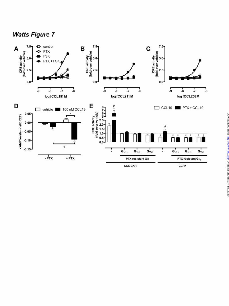
arrestin1/2 to CCX-CKR were not affected by PTX (Fig 6D-I). Next, the effect of CCL19 treatment on basal adenylyl cyclase activity was measured in the absence and presence of PTX pre-treatment. Only cells expressing CCX/R9IL2 showed a significant increase in CRE activity upon stimulation with CCL19 in the absence of FSK and PTX as compared to vehicle-treated cells (Fig 5E). Interestingly, PTX pre-treatment increased CCL19-induced CRE activity in cells expressing wild type CCX-CKR, CCX/R7IL2, CCX/R7IL2IL3, and CCX/R9IL2IL3 (Fig 5E). The absence of CCL19 responsiveness of CCX/R9IL3 with and without PTX pre-treatment might be related to the reduction in CCL19 binding to this mutant receptor as compared to wild type CCX-CKR (Fig 5C). The CCR9 chemokine CCL25 showed a comparable CRE-inducing activity profile on CCX-CKR and CCX-CKR chimeras as CCL19 (Fig 5F-G). As expected CCL25 activated CCR9 but not CCR7, resulting in an inhibition of forskolin-induced CRE activity (Fig 5F). Surprisingly, however, PTX-pretreatment significantly increased CCR9-mediated CRE activity upon CCL25 stimulation in a forskolin-dependent manner (Fig 5F). Gi proteins impair CCX-CKR signaling to CRE – The apparent ability of wild type CCX-CKR to stimulate CRE-mediated luciferase activity in response to CCL19 upon PTX pre-treatment, prompted us to focus on wild type CCX-CKR signaling to CRE in more detail. First, concentration-response curves of CCL19, CCL21, and CCL25 were measured in the absence and presence of PTX (100 ng/ml) and/or FSK (1 µM). In the absence of PTX pre-treatment, these chemokines did not affect CRE activity in the absence (control) or presence of FSK, when tested at concentrations up to 316 nM (Fig 7A-C). Pre-treatment overnight with PTX resulted in an increased CRE activity upon stimulation with >100 nM CCL19 or >316 nM CCL25 (Fig 7A-C). Interestingly, pre-treatment with PTX and chemokine stimulation in the presence of FSK showed that PTX and FSK synergistically increased CRE activity upon stimulation with CCL19, CCL21, or CCL25 as compared to vehicle-stimulated cells (Fig 7A-C). Moreover, this co-treatment with PTX and FSK significantly increased cAMP levels in HEK293T cells co-
by guest on January 18, 2020http://w
ww
.jbc.org/D
ownloaded from
9
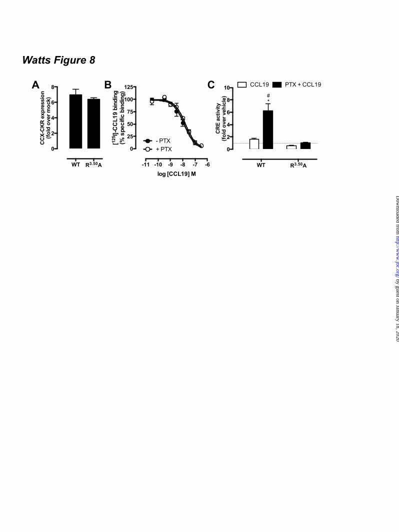
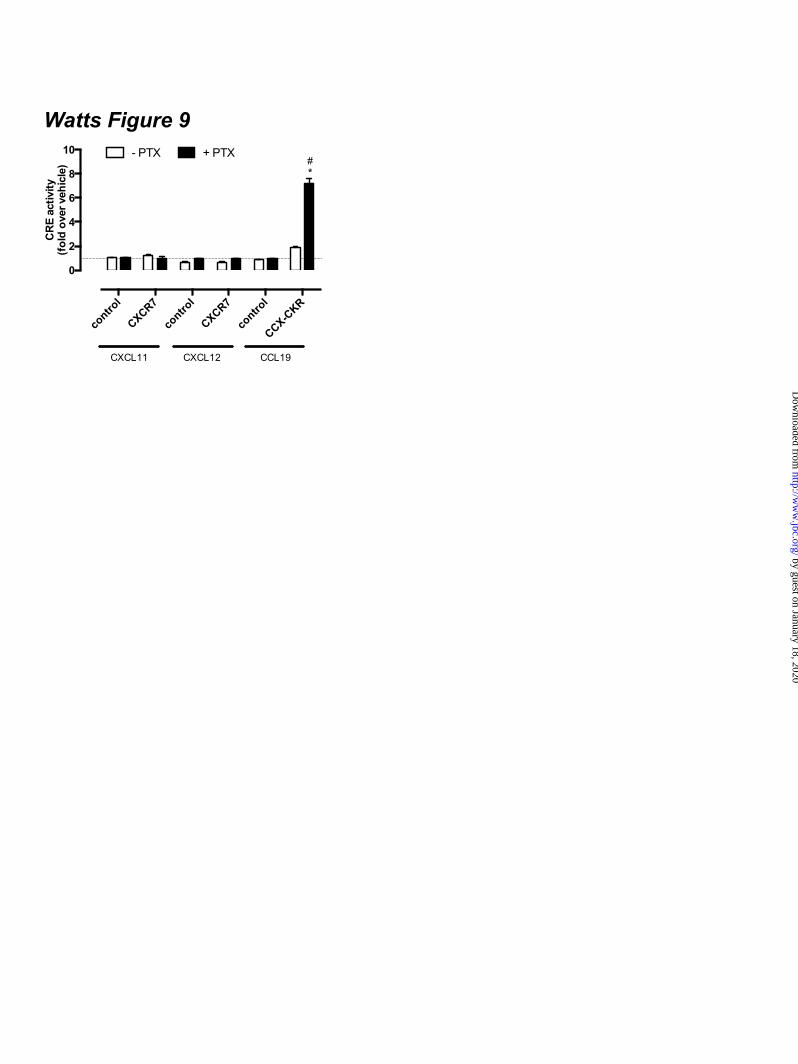
expressing CCX-CKR and a BRET-based cAMP biosensor upon stimulation with 100 nM CCL19 as compared to cells that were not pre-treated with PTX and/or stimulated with vehicle (Fig 7D). Binding of cAMP to the EPAC domain of the biosensor results in a decrease of intramolecular BRET (23,24). Co-transfection of PTX-insensitive Gαi1-C351I, Gαi2-C352I or Gαi3-C351I mutants (29) in CCX-CKR-expressing cells abolished the CCL19-induced increase in CRE activity upon PTX pre-treatment (Fig 7E), which suggests that interaction of Gi proteins with CCX-CKR impairs signaling of this receptor to CRE. Co-expression of the PTX-resistant Gi proteins did not affect CCX-CKR surface levels or limit expression of the luciferase reporter gene in response to 10 µM FSK (data not shown). Functional expression of the Gαi1-C351I, Gαi2-C352I or Gαi3-C351I mutants was confirmed by the inability of PTX to inhibit FSK-induced CRE activity by CCR7 upon stimulation with CCL19 (Fig 7E). CCX-CKR-mediated CRE activation requires an intact DRY motif - The conserved DRY motif of GPCRs at the border of transmembrane domain 3 and IL2 is known to be essential for controlling G protein activation, and mutation of R3.50 to alanine in this motif generally results in a receptor that is unable to activate G proteins (30). Wild type CCX-CKR and CCX/R3.50A were expressed at similar levels (t-test p > 0.05) at the cell surface (Fig 8A). Similar to wild type CCX-CKR (Fig.6A), and had comparable affinities for CCL19 with or without PTX pre-treatment (Fig 8B). In contrast to wild type CCX-CKR, CCX/R3.50A was unable to activate CRE upon CCL19 stimulation in cells pre-treated with PTX (Fig 8C), which supports the hypothesis that activation of CRE by wild type CCX-CKR is G protein-dependent. PTX pre-treatment did not unmask CRE activation by CXCR7 – Several studies indicated that CXCR7 is seen as a decoy receptor due to its incapability to induce G protein signaling (18,31). On the other hand, CXCR7 signals to ERK1/2 and Akt in a β-arrestin-biased manner upon stimulation with CXCL11/ITAC or CXCL12/SDF1α (18,31). CXCR7-expressing HEK293T cells were pre-treated overnight with PTX to evaluate whether Gi
proteins might hamper CXCR7-induced G protein signaling via a similar mechanism as observed for CCX-CKR. The CXCR7 chemokines CXCL11 and CXCL12 (100 nM) were not able to increase FSK-induced CRE activity in the absence or presence of PTX in cells transfected with CXCR7 (Fig 9). CXCL12 decreased FSK-induced CRE activity in mock and CXCR7-expressing HEK293T cells that were not pre-treated with PTX, by interacting with the chemokine receptor CXCR4 (Fig 9). CXCR4 is endogenously expressed on HEK293(T) cells (32). DISCUSSION CCX-CKR plays an important role in controlling the migration of immune and cancer cells that express the chemokine receptors CCR7 and CCR9, by reducing the availability of CCL19, CCL21 and CCL25 through internalization (8,10,11,15). CCX-CKR internalization was previously found to be independent of β-arrestins and clathrin-coated pits (CCPs), as dominant-negative β-arrestin, Eps15 and Rab5 mutants did not impair CCL19 internalization in CCX-CKR-expressing HEK293 cells (10). In addition, CCL19 was internalized in β-arrestin1/2-null mouse embryo fibroblasts (MEF) expressing CCX-CKR but not in cells expressing CCR7 (10). On the other hand, dominant-negative caveolin isoforms and cholesterol depletion diminished CCL19 sequestering by CCX-CKR, which indicated that CCX-CKR is internalized via caveolae (10). However, inspection of the intracellular C-terminal tail of CCX-CKR reveals the presence of multiple serine and threonine residues that might function as substrate for GPCR kinases and promote the interaction with β-arrestins upon phosphorylation (33). In this study, we demonstrate for the first time that stimulation of CCX-CKR with CCL19, CCL21 and CCL25 indeed induced β-arrestin2-GFP translocation. CCX-CKR did not translocate β-arrestin2-GFP in the absence of chemokines, which is in contrast to the constitutively phosphorylated chemokine decoy receptor D6 (21). Moreover, fluorescently labeled CCL19 co-localized in the same intracellular vesicles as β-arrestin2-GFP, suggesting recruitment of β-arrestin to CCL19-bound CCX-CKR. EFC and BRET, which are two different close-proximity-based approaches, then demonstrated actual recruitment of β-arrestin to
by guest on January 18, 2020http://w
ww
.jbc.org/D
ownloaded from
10
CCX-CKR in response to chemokines. The recruitment of β-arrestin to CCX-CKR and co-localization with internalized CCL19 suggests that a possible involvement of β-arrestins in CCX-CKR endocytosis cannot be excluded. The occurrence of parallel internalization pathways for one receptor is not unprecedented. For instance, the dopamine D1 receptor internalizes through both caveolae and CCP (34,35). Interestingly, inhibition of CCP-dependent endocytosis by various methods did not affect D1 receptor internalization, suggesting that β-arrestin and CCP might not be the dominant pathway for this receptor (34). Besides functioning as interaction site for proteins of the endocytotic machinery, GPCR-bound β-arrestin can also assemble specific signaling scaffolds, such as for example β-arrestin2/ERK/Raf/MEK and Akt/β-arrestin2/PP2A (36,37). CCX-CKR was however unable to activate downstream signaling to ERK1/2 and Akt upon stimulation with CCL19, whereas this CCL19 activates ERK1/2 in a β-arrestin2-dependent manner via CCR7 (38). Importantly, β-arrestin can activate numerous other kinases as well as phosphatases in addition to ERK1/2 and Akt (39) and the activation of these additional signal effectors through CCX-CKR remains to be investigated, preferably in physiologically relevant assay systems. Previous studies showed that CCX-CKR is unable to induce G protein-dependent intracellular Ca2+ mobilization (7,14). In line with these observations and in contrast to CCR7 and CCR9, CCX-CKR was unable to modulate intracellular cAMP and inositol phosphate levels upon stimulation with 100 nM CCL19, CCL21, and CCL25. Nevertheless, analysis of the CCX-CKR amino acid sequence reveals the conservation of typical GPCR signaling motifs at the bottom of transmembrane (TM) helix 3 (i.e. DRY) and TM7-helix 8 (i.e. NPxxY(x)7F) (40-42), as well as a reasonable sequence identity with CCR7 and CCR9 in the TM helices that flank intracellular loops 2 and 3 (Fig 6A). Interestingly, substitution of CCX-CKR IL2 and/or IL3 with corresponding CCR7 and CCR9 sequences conferred a small but consistent CRE stimulatory activity to CCX-CKR mutants in response to chemokines, rather than the anticipated Gi-mediated decrease in CRE signaling. Preventing Gi protein interaction with
GPCRs by pre-treating the cells with PTX resulted in a potentiation of this signaling by the CCX-CKR mutants. In the absence of forskolin, PTX-treatment resulted in a significant chemokine-induced CRE activation by CCX/R7IL2, whereas CCX/R9IL2 activated CRE both in the absence and presence of PTX. These data for the two CCX-CKR chimeras suggest that the IL2 of CCX-CKR decreases the propensity to interact with G proteins. Moreover, this PTX pre-treatment unmasked signaling by wild type CCX-CKR, resulting in a significant increase in CRE-activity in response to CCL19, CCL21 and CCL25. The chemokine concentration-response curves did not allow proper determination of potency and efficacy. However, CCL19 seemed slightly more potent to activate CRE than CCL21 and CCL25, which corresponds to their rank order of CCX-CKR affinities and the β-arrestin recruitment potencies in the different assay formats (Table 1). Interestingly, all ligands are approximately 30-100 fold less potent to stimulate CRE activity in comparison to recruiting β-arrestins, which might suggest that β-arrestin is not involved in CRE activation in PTX-treated CCX-CKR-expressing cells by interacting with the transcription factor CREB and the histone acetyltransferase p300 as previously observed for delta-opioid receptor (43). Moreover, the absence of CCL19-induced ERK1/2 phosphorylation in CCX-CKR-expressing cells indicates that CRE activation is also not induced via a β-arrestin- and ERK1/2-dependent pathway, which has been recently described in tracheal epithelial cells (44). The recent crystal structure of the β2-adrenoceptor in complex with Gαs protein showed that the conserved R3.50 in the DRY motif interacts directly with the G protein (42), which was earlier observed as well in the structure of opsin in complex with a C-terminal peptide of Gα protein (45). Ala-substitution of R3.50 in CCX-CKR completely abolished CCL19-induced CRE activation in PTX-treated cells, suggesting that the interaction of PTX-insensitive G proteins with CCX-CKR is crucial to signal to CRE. Besides Gs proteins, also Gq proteins have been shown to stimulate several adenylyl cyclase subtypes via their βγ-subunits (46,47). The fact that CCX-CKR-mediated CRE activation is only observed after disabling Gi proteins to interact with the receptor
by guest on January 18, 2020http://w
ww
.jbc.org/D
ownloaded from
11
suggests that Gi proteins might impair the interaction of PTX-insensitive G proteins to couple to CCX-CKR upon chemokine stimulation. The atypical chemokine receptor CXCR7 signals exclusively through β-arrestins (18,31). Interestingly, however, BRET assays showed that CXCR7 interacts constitutively with Gi proteins without activating them (48). A similar scenario of constitutive or chemokine-induced interaction of inactive Gi proteins with CCX-CKR is hypothesized to maintain this decoy receptor “silenced” for typical chemokine receptor-induced G protein signaling by impeding subsequent and apparently less favorable coupling of PTX-insensitive G proteins to this receptor (Fig 10). On the other hand, pre-treatment of CXCR7-expressing cells with PTX did not unmask CRE activation in response to CXCL11 and CXCL12, suggesting that Gi-mediated silencing of CCX-CKR-induced CRE activation is at least specific for this receptor and not the consequence of other cellular changes. Detection of G protein
interactions with CCX-CKR using BRET-based methods might unravel the identity and dynamics of G protein subtypes that are recruited to the receptor. In conclusion, the atypical chemokine receptor CCX-CKR is capable of increasing cAMP levels and CRE activity in response to chemokine stimulation, however PTX-sensitive G proteins normally prevent this signaling. On the other hand, the chemokine-induced recruitment of β-arrestins to CCX-CKR might open avenues to activate yet to be identified G protein-independent signaling pathways. ACKNOWLEDGEMENTS The authors thank Winfried Mulder (MSD), Marloes van der Zwam (University of Groningen, The Netherlands) and Dr. Chiba (Tokyo University of Science, Japan) for providing reagents. This research was funded by the Dutch Top Institute Pharma initiative (program D1-105).
REFERENCES 1. Scholten, D., Canals, M., Maussang, D., Roumen, L., Smit, M., Wijtmans, M., de Graaf, C.,
Vischer, H., and Leurs, R. (2012) Br. J. Pharmacol. 165, 1617-1643 2. O'Hayre, M., Salanga, C. L., Handel, T. M., and Allen, S. J. (2008) Biochem. J. 409, 635-649 3. Ulvmar, M. H., Hub, E., and Rot, A. (2011) Exp. Cell Res. 317, 556-568 4. Gosling, J., Dairaghi, D. J., Wang, Y., Hanley, M., Talbot, D., Miao, Z., and Schall, T. J. (2000)
J. Immunol. 164, 2851-2856 5. Forster, R., Davalos-Misslitz, A. C., and Rot, A. (2008) Nat. Rev. Immunol. 8, 362-371 6. Pabst, O., Ohl, L., Wendland, M., Wurbel, M. A., Kremmer, E., Malissen, B., and Forster, R.
(2004) J. Exp. Med. 199, 411-416 7. Heinzel, K., Benz, C., and Bleul, C. C. (2007) Proc. Natl. Acad. Sci. U. S. A. 104, 8421-8426 8. Comerford, I., Nibbs, R. J., Litchfield, W., Bunting, M., Harata-Lee, Y., Haylock-Jacobs, S.,
Forrow, S., Korner, H., and McColl, S. R. (2010) Blood 116, 4130-4140 9. Lazennec, G., and Richmond, A. (2010) Trends Mol. Med. 16, 133-144 10. Comerford, I., Milasta, S., Morrow, V., Milligan, G., and Nibbs, R. (2006) Eur. J. Immunol. 36,
1904-1916 11. Feng, L. Y., Ou, Z. L., Wu, F. Y., Shen, Z. Z., and Shao, Z. M. (2009) Clin. Cancer Res. 15,
2962-2970 12. Haraldsen, G., and Rot, A. (2006) Eur. J. Immunol. 36, 1659-1661 13. Khoja, H., Wang, G., Ng, C. T., Tucker, J., Brown, T., and Shyamala, V. (2000) Gene 246, 229-
238 14. Townson, J. R., and Nibbs, R. J. (2002) Eur. J. Immunol. 32, 1230-1241 15. Kriegova, E., Tsyrulnyk, A., Arakelyan, A., Mrazek, F., Ordeltova, M., Petzmann, S., Zatloukal,
J., Kolek, V., du Bois, R. M., Popper, H., and Petrek, M. (2006) Inflammation Res. 55, 441-445 16. Rode, I., and Boehm, T. (2012) Proc. Natl. Acad. Sci. U. S. A. 109, 3463-3468 17. Rajagopal, S., Rajagopal, K., and Lefkowitz, R. J. (2010) Nat. Rev. Drug Discovery 9, 373-386 18. Rajagopal, S., Kim, J., Ahn, S., Craig, S., Lam, C. M., Gerard, N. P., Gerard, C., and Lefkowitz,
R. J. (2010) Proc. Natl. Acad. Sci. U. S. A. 107, 628-632
by guest on January 18, 2020http://w
ww
.jbc.org/D
ownloaded from
12
19. Luker, K. E., Gupta, M., and Luker, G. D. (2009) FASEB J. 23, 823-834 20. Galliera, E., Jala, V. R., Trent, J. O., Bonecchi, R., Signorelli, P., Lefkowitz, R. J., Mantovani, A.,
Locati, M., and Haribabu, B. (2004) J. Biol. Chem. 279, 25590-25597 21. McCulloch, C. V., Morrow, V., Milasta, S., Comerford, I., Milligan, G., Graham, G. J., Isaacs, N.
W., and Nibbs, R. J. (2008) J. Biol. Chem. 283, 7972-7982 22. Vischer, H. F., Nijmeijer, S., Smit, M. J., and Leurs, R. (2008) Biochem. Biophys. Res. Commun.
377, 93-97 23. Scholten, D., Canals, M., Wijtmans, M., de Munnik, S., Nguyen, P., Verzijl, D., de Esch, I.,
Vischer, H., Smit, M., and Leurs, R. (2012) Br. J. Pharmacol. 166, 898-911 24. Jiang, L. I., Collins, J., Davis, R., Lin, K. M., DeCamp, D., Roach, T., Hsueh, R., Rebres, R. A.,
Ross, E. M., Taussig, R., Fraser, I., and Sternweis, P. C. (2007) J. Biol. Chem. 282, 10576-10584 25. Van Lith, L. H., Oosterom, J., Van Elsas, A., and Zaman, G. J. (2009) J. Biomol. Screen. 14,
1067-1075 26. Nijmeijer, S., Leurs, R., Smit, M. J., and Vischer, H. F. (2010) J. Biol. Chem. 285, 29632-29641 27. van Der Lee, M. M., Bras, M., van Koppen, C. J., and Zaman, G. J. (2008) J. Biomol. Screen. 13,
986-998 28. Blomenrohr, M., Heding, A., Sellar, R., Leurs, R., Bogerd, J., Eidne, K. A., and Willars, G. B.
(1999) Mol. Pharmacol. 56, 1229-1237 29. Bahia, D. S., Wise, A., Fanelli, F., Lee, M., Rees, S., and Milligan, G. (1998) Biochemistry 37,
11555-11562 30. Rovati, G. E., Capra, V., and Neubig, R. R. (2007) Mol. Pharmacol. 71, 959-964 31. Kumar, R., Tripathi, V., Ahmad, M., Nath, N., Mir, R. A., Chauhan, S. S., and Luthra, K. (2012)
Cell. Immunol. 272, 230-241 32. Atwood, B. K., Lopez, J., Wager-Miller, J., Mackie, K., and Straiker, A. (2011) BMC Genomics
12, 14 33. Liggett, S. B. (2011) Sci. Signal. 4, pe36 34. Kong, M. M., Hasbi, A., Mattocks, M., Fan, T., O'Dowd, B. F., and George, S. R. (2007) Mol.
Pharmacol. 72, 1157-1170 35. Vickery, R. G., and von Zastrow, M. (1999) J. Cell Biol. 144, 31-43 36. Shenoy, S. K., and Lefkowitz, R. J. (2011) Trends Pharmacol. Sci. 32, 521-533 37. DeFea, K. A. (2011) Cell. Signal. 23, 621-629 38. Zidar, D. A., Violin, J. D., Whalen, E. J., and Lefkowitz, R. J. (2009) Proc. Natl. Acad. Sci. U. S.
A. 106, 9649-9654 39. Xiao, K., Sun, J., Kim, J., Rajagopal, S., Zhai, B., Villen, J., Haas, W., Kovacs, J. J., Shukla, A.
K., Hara, M. R., Hernandez, M., Lachmann, A., Zhao, S., Lin, Y., Cheng, Y., Mizuno, K., Ma'ayan, A., Gygi, S. P., and Lefkowitz, R. J. (2010) Proc. Natl. Acad. Sci. U. S. A. 107, 15299-15304
40. Rosenbaum, D. M., Rasmussen, S. G., and Kobilka, B. K. (2009) Nature 459, 356-363 41. Nygaard, R., Frimurer, T. M., Holst, B., Rosenkilde, M. M., and Schwartz, T. W. (2009) Trends
Pharmacol. Sci. 30, 249-259 42. Rasmussen, S. G., DeVree, B. T., Zou, Y., Kruse, A. C., Chung, K. Y., Kobilka, T. S., Thian, F.
S., Chae, P. S., Pardon, E., Calinski, D., Mathiesen, J. M., Shah, S. T., Lyons, J. A., Caffrey, M., Gellman, S. H., Steyaert, J., Skiniotis, G., Weis, W. I., Sunahara, R. K., and Kobilka, B. K. (2011) Nature 477, 549-555
43. Kang, J., Shi, Y., Xiang, B., Qu, B., Su, W., Zhu, M., Zhang, M., Bao, G., Wang, F., Zhang, X., Yang, R., Fan, F., Chen, X., Pei, G., and Ma, L. (2005) Cell 123, 833-847
44. Manson, M. E., Corey, D. A., Rymut, S. M., and Kelley, T. J. (2011) Biochemistry 50, 6022-6029 45. Scheerer, P., Park, J. H., Hildebrand, P. W., Kim, Y. J., Krauss, N., Choe, H. W., Hofmann, K. P.,
and Ernst, O. P. (2008) Nature 455, 497-502 46. Wettschureck, N., and Offermanns, S. (2005) Physiol. Rev. 85, 1159-1204
by guest on January 18, 2020http://w
ww
.jbc.org/D
ownloaded from
13
47. Maruko, T., Nakahara, T., Sakamoto, K., Saito, M., Sugimoto, N., Takuwa, Y., and Ishii, K. (2005) Naunyn Schmiedebergs Arch. Pharmacol. 372, 153-159
48. Levoye, A., Balabanian, K., Baleux, F., Bachelerie, F., and Lagane, B. (2009) Blood 113, 6085-6093
FOOTNOTES The abbreviations used are: CRE, cAMP-responsive element; GPCR, G protein-coupled receptor; AC, adenylyl cyclase; ERK1/2, extracellular stimulus-regulated kinase 1/2; NP-40, Nonidet P-40; PTX, pertussis toxin; Rluc, Renilla luciferase; EYFP, enhanced yellow fluorescent protein; TM, transmembrane; PTH1R, parathyroid hormone receptor 1; GFP, green fluorescent protein; CHO, chinese hamster ovary; EA, enzyme acceptor; BRET, bioluminescence resonance energy transfer; HBSS, Hank’s Balanced Salt Solution; HCA, high content analysis; EFC, enzyme fragment complementation; HTRF, homogenous time-resolved fluorescence, FSK, forskolin; TBS, tris-buffered saline. FIGURE LEGENDS Figure 1. CCX-CKR internalizes CCL19. (A) HEK293T cells transiently transfected with CCX-CKR, CCR7, or empty plasmid (mock) were incubated with [125I]-CCL19 for the indicated time. Surface bound [125I]-CCL19 was removed by an acid wash, and internalized [125I]-CCL19 was quantified. (B) U2OS osteosarcoma cells expressing β-arrestin2 fused to green fluorescent protein (U2OS-β-arr2-GFP) were stably transfected with vector coding for CCX-CKR (U2OS-CCX-CKR cells). U2OS-CCX-CKR cells were treated with Alexafluor647-coupled CCL19 (CCL19-AF) at 37°C (upper panels) or 4°C (lower panels) for 45 minutes. Then, cells were washed with an acidic buffer (0.2 M glycine, (pH3.0), 0.5 M NaCl) or PBS for 5 minutes before fixation and staining. Alexa Fluor 647 and Hoechst signals are depicted in red and blue, respectively. Figure 2. Internalized CCL19 and β-arrestin co-localize in endocytic vesicles in cells expressing CCX-CKR. (A) U2OS-β-arr2-GFP and U2OS-CCX-CKR cells were treated with 100 nM CCL19 for 45 minutes. Cells were then fixed, stained with Hoechst and imaged using fluorescence microscopy for GFP (green) and nuclei (blue). (B) U2OS-CCX-CKR cells were stimulated with increasing concentrations of CCL19, CCL21 and CCL25 for 45 minutes. The recruitment of β-arrestin2 to CCX-CKR was quantified as the amount of GFP- containing vesicles per nucleus. (C) U2OS-CCX-CKR cells were treated with 100 nM CCL19 or CCL19-AF for 45 minutes, followed by fluorescence microscopy. GFP is shown in green, Alexafluor647 is in red and nuclei are stained blue. (D) U2OS-CCX-CKR cells were treated with increasing doses of CCL19 and CCL19-AF for 45 minutes followed by determination of GFP-containing vesicle formation. (E) Quantification of AF-containing vesicle formation of U2OS-CCX-CKR cells treated with increasing concentrations of CCL19-AF. Figure 3. CCX-CKR recruits β-arrestin1 and β-arrestin2 in a chemokine-dependent manner. (A) CHO cells stably expressing CCX-CKR fused to a peptide fragment of β-galactosidase and β-arrestin2 coupled to a complementary β-galactosidase mutant (CHO-CCX-CKR cells) were treated with ascending concentrations of CCL19, CCL21 and CCL25 for 90 minutes before measurement of β-galactosidase activity. (B,C) Potencies of (B) β-arrestin2 and (C) β-arrestin1 recruitment induced by CCL19 (white squares), CCL21 (black circles) and CCL25 (white circles) were determined using a BRET-based assay in HEK293T cells transiently co-transfected with CCX-CKR-Rluc and β-arrestin-EYFP. Data are presented as averages ± SEM of 2-4 independent experiments performed in triplicate. Figure 4. CCX-CKR does not activate G protein-dependent or β-arrestin-dependent intracellular signaling pathways. (A and B) CHO-CCX-CKR cells were stimulated with 100 nM CCL19 or vehicle
by guest on January 18, 2020http://w
ww
.jbc.org/D
ownloaded from
14
for several time periods. Control cells were treated with (A) 1 µM PMA or (B) 100 ng/ml insulin. Cell lysates were then analyzed for levels of (A) phosphorylated Thr202/Tyr204-ERK1/2 (pERK) and total ERK1/2 (tERK) or (B) phosphorylated Ser473-Akt (pAkt) and total Akt (tAkt). (C) CHO-CCX-CKR cells were treated with 100 nM CCL19, CCL21 and CCL25 in the presence or absence of 0.5 µM FSK for 60 minutes. Subsequently, cells were lysed and intracellular cAMP concentrations were measured. (D) CHO-CCX-CKR cells were stimulated with 100 nM CCL19, CCL21 and CCL25 for 90 minutes before measurement of intracellular IPone concentrations. As a positive control, cells were treated with 100 µM ATP. Figure 5. IL2 and IL3 of functional chemokine receptors do not convey Gαi-activating properties to CCX-CKR. (A) CCX-CKR, CCR7, and CCR9 amino acid sequences of TM3-IL2-TM4, TM5-IL3-TM6 are aligned on the basis of highly conserved residues. The IL2 and IL3 sequences that have been exchanged in the CCX-CKR chimeras are indicated with white boxes. HEK293T cells were co-transfected with a CRE-driven reporter gene and CCX-CKR, CCX/R7IL2, CCX/R7IL3, CCX/R7IL2IL3, CCX/R9IL2, CCX/R9IL3 or CCX/R9IL2IL3 as indicated. (B) Cell surface expression was measured by ELISA. (C) [125I]-CCL19 binding was performed on intact cells. Cells were pre-treated with PTX (100 ng/mL) overnight (filled bars). Cells were treated with vehicle, (D-E) 100 nM CCL19, or (F-G) 100 nM CCL25 for 5 hours in the (D, F) presence or (E, G) absence of 1 µM FSK. Statistical comparisons were performed using one-way analysis of variance followed by the Bonferroni test. Significant difference in cell surface expression and [125I]-CCL19 binding as compared to wild type CCX-CKR is indicated by § (B-C) Significant difference (p < 0.05) in CRE activity between vehicle and corresponding chemokine-treated cells is indicated by *, whereas # indicates significant difference in chemokine-induced CRE activity between cells pretreated with or without PTX (D-G). Data are shown as averages ± SEM of fold over vehicle data from 4 independent experiments performed in duplicate (B-E) or 2 independent experiments performed in triplicate (F-G). Figure 6. CCX-CKR chemokine binding affinities are not dependent on Gαi proteins. [125I]-CCL19 displacement curves were performed for (A) CCL19, (B) CCL21 and (C) CCL25 in the absence (empty symbols) and presence (filled symbols) of 100 ng/ml PTX in HEK293T cells transiently transfected with CCX-CKR. β-arrestin1 (D-F) and β-arrestin2 (G-I) recruitment induced by CCL19 (D,G), CCL21 (E,H) and CCL25 (F,I) were determined using a BRET-based assay in HEK293T cells transiently co-transfected with CCX-CKR-Rluc and β-arrestin-EYFP without (empty symbols) and with (filled symbols) PTX (100 ng/ml) pretreatment. Data are shown as averages ± SEM of normalized specific binding of two independent experiments performed in duplicate. Figure 7. PTX potentiates ligand-induced CRE activation mediated by CCX-CKR. (A-C) HEK293T cells were co-transfected with a CRE-driven reporter gene and CCX-CKR. Concentration response curves of (A) CCL19, (B) CCL21, and (C) CCL25 were obtained in the absence of PTX and FSK (£), in the presence of 1µM FSK (¢), in the presence of 100 ng/ml PTX (�) or the presence of both PTX and FSK (�). (D) HEK293T cells were transiently transfected with CCX-CKR-coding plasmid along with a plasmid encoding the CAMYEL cAMP biosensor. Cells were pre-treated or not with 100 ng/ml PTX overnight. Cells were stimulated with 100 nM CCL19 in the presence of 1 µM FSK for 10 min prior to measurement of intramolecular BRET signal. Elevation of cAMP levels is detected as decreased BRET ratio. Data are shown as averages ± SEM of normalized and baseline corrected BRET ratios from three independent experiments performed in duplicate (E) HEK293T cells were co-transfected with a CRE-driven reporter gene, CCX-CKR or CCR7 and the PTX-insensitive mutants of Gαi subunit Gαi1/C351I, Gαi2/C352I and Gαi3/C351I as indicated. Cells were pre-treated (black bars) or not (empty bars) with 100 ng/ml PTX overnight. Cells were incubated with vehicle or 100 nM CCL19 in the presence of 1 µM FSK for 6 to 8 hours. Statistical comparisons to identify significant differences between vehicle and chemokine-stimulated cells (*) as well as between control and PTX-pretreated cells (#) were performed using one-way analysis of variance followed by the Bonferroni test. P value < 0.05 indicate significant
by guest on January 18, 2020http://w
ww
.jbc.org/D
ownloaded from
15
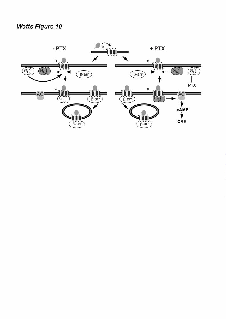
difference. Graphs (A-C, and E) show averages ± SEM of fold over vehicle data from three to six independent experiments performed in duplicate. Figure 8. Potentiation of CCX-CKR-mediated CRE activation is G protein-dependent. (A) Cell surface expression of wildtype CCX-CKR and the DRY mutant CCX/R3.50A in the reporter gene assay was determined using ELISA with anti-CCX-CKR antibody. Graph shows averages ± SEM of fold over mock data from two independent experiments performed in duplicate. (B) [125I]-CCL19 displacement curves with CCL19 were obtained in the absence (empty symbols) or presence (filled symbols) of 100 ng/ml PTX in HEK293T cells transfected with CCX/R3.50A. Data are shown as averages ± SEM of normalized specific binding of one independent experiment performed in duplicate. (C) HEK293T cells were co-transfected with a CRE-driven reporter gene and wild type CCX-CKR or CCX/R3.50A as indicated. Cells were pre-treated with 100 ng/ml PTX (empty and black bars) overnight. Cells were incubated with vehicle (empty bars) or 100 nM CCL19 (filled bars) in the presence of 1 µM FSK for 5 hours. Statistical comparisons to identify significant differences between vehicle and chemokine-stimulated cells (*) as well as between control and PTX-pretreated cells (#) were performed using one-way analysis of variance followed by the Bonferroni test. P value < 0.05 indicate significant difference. Graph show averages ± SEM of fold over vehicle data from two independent experiments performed in duplicate. Figure 9. CXCR7 does not mediate CRE activation. HEK293T cells were co-transfected with a CRE-driven reporter gene and empty vector (-), CXCR7 or CCX-CKR as indicated. Cells were pre-treated (black bars) or not (empty bars) with 100 ng/ml PTX overnight. Cells were incubated with vehicle, CXCL11, CXCL12 or CCL19 (100 nM chemokine) as indicated in the presence of 1 µM FSK. Data are shown as averages ± SEM of fold over vehicle data from two independent experiments performed in duplicate. Statistical comparisons to identify significant differences between vehicle and chemokine-stimulated cells (*) as well as between control and PTX-pretreated cells (#) were performed using one-way analysis of variance followed by the Bonferroni test. P value < 0.05 indicate significant difference. Figure 10. Proposed model of CCX-CKR interaction with β-arrestins and G proteins. Chemokine binding to CCX-CKR (a) recruits Gi proteins and β-arrestin (β-arr) with high affinity (b), consequently hindering the low affinity interaction between CCX-CKR and Gs proteins. Inactive Gi protein may stay bound to CCX-CKR, whereas the chemokine-bound CCX-CKR internalizes with β-arrestin (c). Inhibition of Gi coupling to CCX-CKR by PTX pre-treatment did not affect β-arrestin recruitment but allows Gs to interact with CCX-CKR (d), which results in stimulation of adenylyl cyclase (AC) and CRE activity (e).
by guest on January 18, 2020http://w
ww
.jbc.org/D
ownloaded from
16
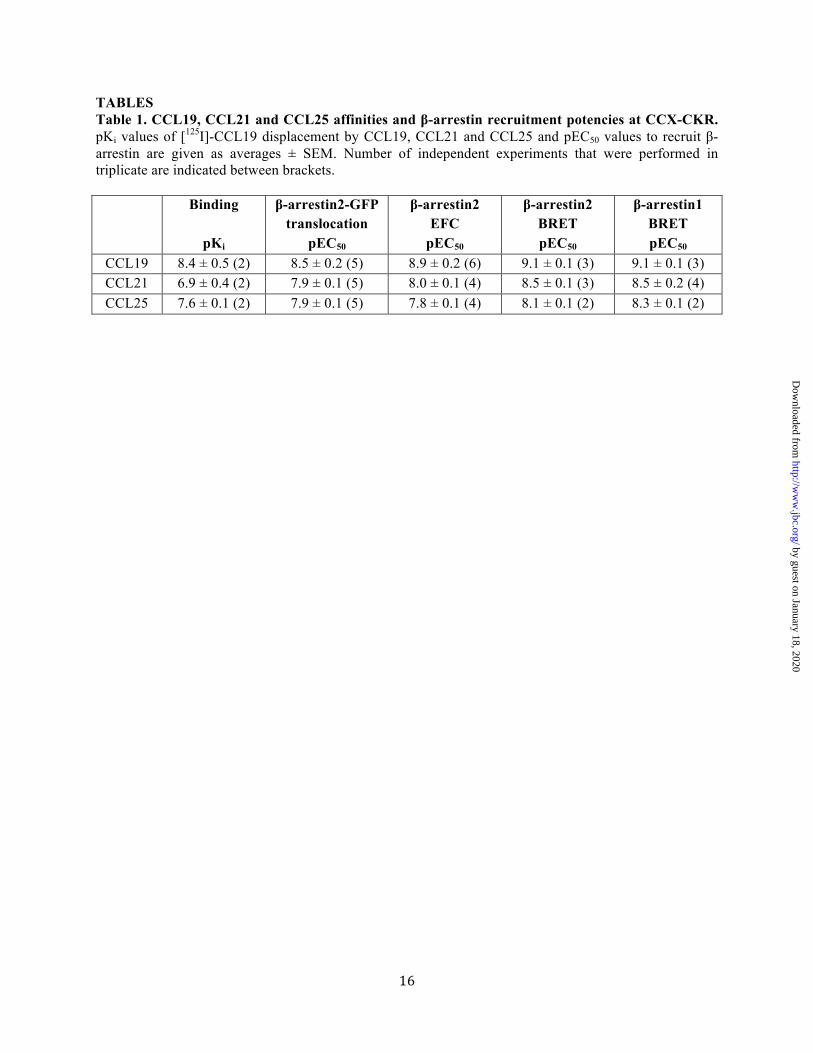
TABLES Table 1. CCL19, CCL21 and CCL25 affinities and β-arrestin recruitment potencies at CCX-CKR. pKi values of [125I]-CCL19 displacement by CCL19, CCL21 and CCL25 and pEC50 values to recruit β-arrestin are given as averages ± SEM. Number of independent experiments that were performed in triplicate are indicated between brackets.
Binding
pKi
β-arrestin2-GFP translocation
pEC50
β-arrestin2 EFC
pEC50
β-arrestin2 BRET pEC50
β-arrestin1 BRET pEC50
CCL19 8.4 ± 0.5 (2) 8.5 ± 0.2 (5) 8.9 ± 0.2 (6) 9.1 ± 0.1 (3) 9.1 ± 0.1 (3) CCL21 6.9 ± 0.4 (2) 7.9 ± 0.1 (5) 8.0 ± 0.1 (4) 8.5 ± 0.1 (3) 8.5 ± 0.2 (4) CCL25 7.6 ± 0.1 (2) 7.9 ± 0.1 (5) 7.8 ± 0.1 (4) 8.1 ± 0.1 (2) 8.3 ± 0.1 (2)
by guest on January 18, 2020http://w
ww
.jbc.org/D
ownloaded from
Leurs, Guido J. R. Zaman and Henry F. VischerMartien Kuijer, Jody van Offenbeek, Lambertus H. C. J. van Lith, Martine J. Smit, Rob
Anne O. Watts, Folkert Verkaar, Miranda M. C. van der Lee, Claudia A. W. Timmerman,decoy receptor CCX-CKR
-arrestin recruitment and G protein signaling by the atypical human chemokineβ
published online January 22, 2013J. Biol. Chem.
10.1074/jbc.M112.406108Access the most updated version of this article at doi:
Alerts:
When a correction for this article is posted•
When this article is cited•
to choose from all of JBC's e-mail alertsClick here
by guest on January 18, 2020http://w
ww
.jbc.org/D
ownloaded from


![ewY; % ik¡p :i;s - aiparisangh.com · 2 16 ls 31 vxLr] 2018 ekSr ds eqag esa lek, lo.kZ [kkldj czkã.k tc tkfr vkSj o.kZ iwNdj ukoksa esa cSBus dh ckr djrs gSa rks yxrk gS fd ck](https://static.fdocuments.nl/doc/165x107/5cc2726188c9933e3a8e0c4e/ewy-ikp-is-2-16-ls-31-vxlr-2018-eksr-ds-eqag-esa-lek-lokz-kkldj.jpg)
![lk>h nLrd - JAGORI · lk>h nLrd ifjp; la?k"kZ dh ;k=kk vuojr~ py jgh gS ,d vius vanj vkSj nwljh [kqn ls ckgj] lekt ... ckr fNik ugha jgh gwa lc [kqydj crk jgh gwaA* esjs ;g](https://static.fdocuments.nl/doc/165x107/5c7ea67809d3f2b93f8b873a/lkh-nlrd-lkh-nlrd-ifjp-lakkz-dh-kkk-vuojr-py-jgh-gs-d-vius-vanj-vksj.jpg)