Voor het bijwonen van de – a clinical exploration - dds.nl · Voor het bijwonen van de ......
258
Pharmacological fMRI; – a clinical exploration R. Goekoop, MD.
Transcript of Voor het bijwonen van de – a clinical exploration - dds.nl · Voor het bijwonen van de ......
Pharmacological fMRI;– a clinical exploration
R. Goekoop, MD.
Pharm
acological fMR
I; a clinical exploration – R
. Goekoop, M
D.
Uitnodiging
Voor het bijwonen van de openbare verdediging van
het proefschrift
Pharmacological fMRI; a clinical exploration
Op maandag 16 januari 2006 om 13:45u
in de Aula van het hoofdgebouw van de Vrije
Universiteit, de Boelelaan 1105,
1081 HV te Amsterdam.
Na afloop van de promotie is er een receptie in de hal naast
de Aula (Boelelaanzijde).
Graag tot dan,
Rutger GoekoopThomsonlaan 17a I2565 KX Den Haag
Paranimfen:
Yvonne Goekoop-Ruiterman06-51902347
Jeske Damoiseaux06-19626240

Pharmacological fMRI;
A clinical exploration
R. Goekoop, MD.
The studies described in this thesis were performed at the Department of Neurology / Alzheimer Center, Endocrinology and Radiology of the VU University Medical Center, de Boelelaan 1117, 1081 HV in Amsterdam, the Netherlands, and at the Department of Clinical and Experimental Psychology of the University of Amsterdam, Roetersstraat 15, 1018 WB Amsterdam, the Netherlands. All studies were performed using a Siemens Magnetom Sonata 1.5 T magnetic resonance imaging scanner.
Financial support for the publication of this thesis was kindly provided by:– Stichting Alzheimer Nederland– Neuropsychiatrie Foundation Amsterdam, Nederland– Janssen-Cilag B.V. Nederland– Eli Lilly B.V. Nederland– Lundbeck B.V. Nederland– AEGON Nederland
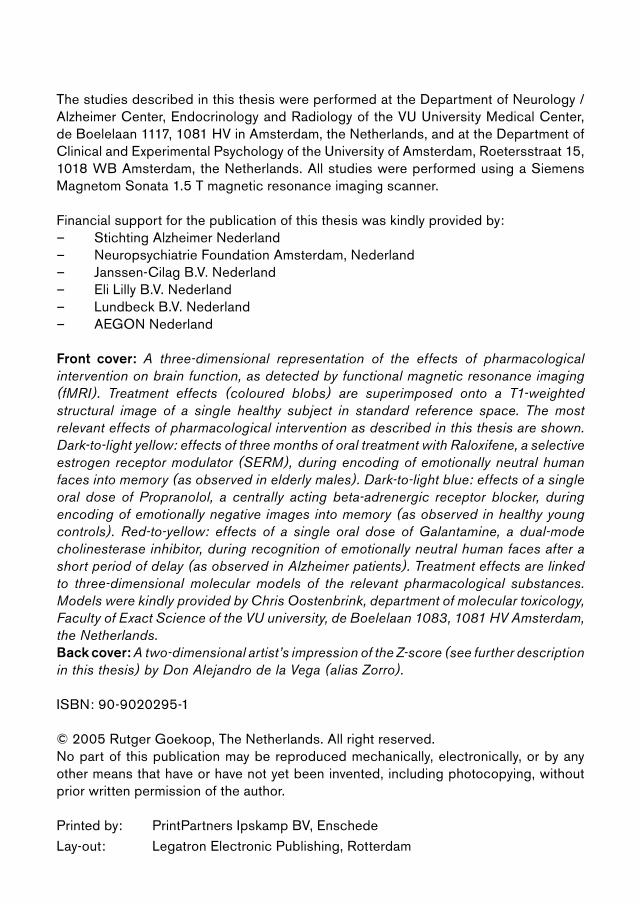
Front cover: A three-dimensional representation of the effects of pharmacological intervention on brain function, as detected by functional magnetic resonance imaging (fMRI). Treatment effects (coloured blobs) are superimposed onto a T1-weighted structural image of a single healthy subject in standard reference space. The most relevant effects of pharmacological intervention as described in this thesis are shown. Dark-to-light yellow: effects of three months of oral treatment with Raloxifene, a selective estrogen receptor modulator (SERM), during encoding of emotionally neutral human faces into memory (as observed in elderly males). Dark-to-light blue: effects of a single oral dose of Propranolol, a centrally acting beta-adrenergic receptor blocker, during encoding of emotionally negative images into memory (as observed in healthy young controls). Red-to-yellow: effects of a single oral dose of Galantamine, a dual-mode cholinesterase inhibitor, during recognition of emotionally neutral human faces after a short period of delay (as observed in Alzheimer patients). Treatment effects are linked to three-dimensional molecular models of the relevant pharmacological substances. Models were kindly provided by Chris Oostenbrink, department of molecular toxicology, Faculty of Exact Science of the VU university, de Boelelaan 1083, 1081 HV Amsterdam, the Netherlands.Back cover: A two-dimensional artist’s impression of the Z-score (see further description in this thesis) by Don Alejandro de la Vega (alias Zorro).
ISBN: 90-9020295-1
© 2005 Rutger Goekoop, The Netherlands. All right reserved.No part of this publication may be reproduced mechanically, electronically, or by any other means that have or have not yet been invented, including photocopying, without prior written permission of the author.
Printed by: PrintPartners Ipskamp BV, EnschedeLay-out: Legatron Electronic Publishing, Rotterdam
VRIJE UNIVERSITEIT
Pharmacological fMRI; a clinical exploration
ACADEMISCH PROEFSCHRIFT
ter verkrijging van de graad Doctor aande Vrije Universiteit Amsterdam,
op gezag van de rector magnificusprof.dr. T. Sminia,
in het openbaar te verdedigenten overstaan van de promotiecommissie
van de faculteit der Geneeskundeop zondag 16 januari 2005 om 13.45 uur
in de aula van de universiteit,De Boelelaan 1105
door
Rutger Goekoop
geboren te Leiden

Promotoren: prof.dr. Ph. Scheltens prof.dr. F. Barkhof
copromotor: dr. S.A.R.B. Rombouts

TIMING TOAST (on doing research – RG)
There’s an art of knowing when.Never try to guess.
Toast until it smokes and thentwenty seconds less.
Piet Hein (1905-1996); Danish poet / scientist / architect / all-round human being

Voor jullie
Contents
1. General introduction 121.1 MRI and fMRI 121.2 phMRI 151.3 Aim of this thesis: exploring the use of phMRI in a clinical context 161.4 phMRI: studying changes in neurotransmission 171.5 Memory tasks (‘paradigms’) used in the current study 191.6 Outline of this thesis 20
2. Effects of beta-adrenergic blockade on amygdala function in healthy young subjects 232.1 Noradrenaline mediates amygdala activation in men and women during encoding of emotional material 23
3. Effects of raloxifene treatment on brain function in healthy elderly males 473.1 Effects of raloxifene treatment on brain function during encoding 473.2 Effects of raloxifene treatment on brain function during recognition 71
4. Effects of galantamine challenge on brain function in MCI and AD patients 934.1 Effects of galantamine challenge on brain function in MCI patients 934.2 Effects of galantamine challenge on brain function in AD patients 1134.3 Differential response to galantamine challenge in MCI and AD patients 135
5. General discussion 1695.1 Chapter 2: effects of beta-adrenergic blockade on amygdala function in healthy young subjects 169 Main effects of task performance: emotional encoding 170 Treatment effects and performance changes 171 Decreased amygdala activation: direct or indirect relations with impaired noradrenergic neurotransmission? 172 Gender-specificity of propranolol: amygdala function and performance accuracy 173 Possible clinical relevance 173 Conclusion 174
5.2. Chapter 3: effects of raloxifene treatment on brain function in healthy elderly males 175 Main effects of face encoding and –recognition 176 Treatment effects and performance changes 176 Process-specificity of raloxifene 176 Possible treatment mechanism of raloxifene 176 Possible clinical relevance 178 Conclusion 1785.3 Chapter 4: effects of galantamine challenge on brain function in MCI and AD patients 179 Main effects of face encoding, –recognition and working memory performance 180 Treatment effects and performance changes 180 Process-specificity of galantamine 180 Region-specificity of galantamine 181 Disease-(stage)-specificity of galantamine 181 Dependence on exposure duration 182 Possible treatment mechanism of galantamine 182 Possible clinical relevance 184 Conclusion 1855.4 Global summary 1875.5 Suggestions for future research 193
Reference list 197 Nederlandse samenvatting (Dutch summary) 215 Dankwoord (Acknowledgements) 227 Curriculum vitae 235 Publicaties (Publications) 237 Appendix (colour images) 239

List of abbreviations
AchChEI Acetyl-cholinesterase inhibitorAD Alzheimer’s diseaseBB BetablockerBL Baseline (no treatment)BOLD Blood oxygenation level dependentBP Blood pressureCAT CategoryCDR Clinical dementia rating scaleEEG ElectroencephalographyENCOD EncodingEPI Echo planar imagingEV Explanatory variable / regressorFEAT fMRI expert analysis toolfMRI Functional magnetic resonance imagingFN False negative / false rejectionFP False positive / false hitFSL fMRIB’s software libraryFWHM Full width at half maximumGAL Galantamine (Reminyl®)GLM General linear modelHR Heart rateHRF Hemodynamic response functionIAPS International affective picture systemMCI Mild cognitive impairmentMEG MagnetoencephalographyMMSE Mini-mental state examinationMRI Magnetic resonance imagingN-back n-letter backNINCDS-ADRDA National institute of neurological disorders and stroke – Alzheimer’s disease and related disorders associationNYU New York UniversityPET Positron emission tomography
phMRI Pharmacological functional magnetic resonance imagingPL / PLAC PlaceboRAL Raloxifene (Evista®)RECOG RecognitionROI Region of interestSCL Symptoms checklistSD Single dose (acute exposure)SERM Selective estrogen receptor modulator (e.g. raloxifene)SPECT Single photon emission computed tomographySS Steady state (prolonged exposure)TN True negative / correct rejectionTP True positive / correct hitTR TreatmentWM Working memoryZ-score Normalised T-statistic (expresses signal-to-noise ratio in fMRI studies).

Chapter 1:
General introduction
12
Chapter 1: General Introduction
1. General introduction
This thesis explores the use of pharmacological functional magnetic resonance imaging (pharmacological fMRI or phMRI) within a clinical context.
1.1 MRI and fMRIMagnetic resonance imaging (MRI) is a non-invasive imaging technique that is used in clinical practice to make high-resolution digital images of water-containing human tissues. Ever since its first appearance in hospitals in the 1980s, it has greatly enhanced the ability of clinicians to make accurate diagnoses by providing a detailed view of structural abnormalities occurring within normal appearing tissue. MRI scanners examine the effects of a wide range of tissue characteristics on the ability of atomic nuclei (usually protons) to absorb and emit radiofrequency radiation within a strong magnetic field. Functional magnetic resonance imaging (fMRI) is an extension of traditional MRI, in which the varying magnetic properties of the oxygen transporter molecule haemoglobin are exploited to examine brain function in human subjects (Jezzard et al., 2001).
When activity of neurons in neural tissue increases (e.g. subjects receive sensory stimuli), neurotransmitters and metabolites are released into the extracellular environment. This leads to an increase in local blood flow, volume and oxygenation levels through a process called ‘neuro-vascular coupling’ (Logothetis & Pfeuffer, 2004). Blood flow and oxygen concentrations are raised beyond a level necessary to compensate for local oxygen and energy demands (perhaps to facilitate oxygen extraction by raising the diffusion coefficient, or neurotransmitter washout by raising blood flow). The result is that active brain regions are characterised by increased blood oxygenation levels relative to their less active states. Haemoglobin with oxygen (oxyhemoglobin) has no significant magnetic properties of its own, and will not affect the static magnetic field of the MR scanner (i.e. oxyhemoglobin is ‘diamagnetic’). Haemoglobin without oxygen (deoxyhemoglobin), however, has magnetic properties that result in the formation of magnetic field gradients around blood vessels (i.e. deoxyhemoglobin is ‘paramagnetic’). Such gradients distort local magnetic field homogeneity, which decreases phase coherence of protons (shorten T2*) and reduces signal readout from such regions. Thus, decreased neural activity is accompanied by more local magnetic field distortions and therefore less MR signal. In contrast, increased activity of neural tissue leads to less magnetic field distortions and more MR signal.
Functional imaging experiments involve the recording of these blood oxygenation level dependent (BOLD) signal intensity changes in time, by scanning large numbers
13
1.1 MRI MRI and fMRI
of brain volumes in rapid succession (i.e. creating a movie, or ‘timeseries’). Special sequences of gradient field manipulations and proton excitation with non-ionising radiation within the radiofrequency range are required to optimise detection of significant BOLD signal intensity changes. One of the most popular and widely used sequences in fMRI imaging experiments is the echo planar imaging (EPI) sequence, which allows rapid and continuous acquisition of hundreds to thousands of brain volumes within minutes to hours (i.e. one brain volume every ~2s). A typical fMRI experiment lasts for several minutes and produces a time series consisting of several hundreds of brain volumes. Each of these volumes is made up of several thousands of volume elements (voxels), which represent the smallest unit of spatial resolution of the functional image (typically ~3 x 3 x 3mm for studies in individuals).
In most fMRI studies, subjects are scanned while performing a certain task (or ‘paradigm’). In such paradigms, conditions of interest alternate with reference conditions to produce differences in BOLD signal intensity. Task conditions have to be repeated in order to reduce the effects of noise-variables and detect signal changes that correlate significantly with task stimuli. Two basic task designs are possible. ‘Block designs’ involve alternations of task conditions with relatively long durations (i.e. 30-40s). Such designs allow for detection of global functional effects of task performance in relatively short amounts of time. ‘Event-related’ designs involve alternations of discrete stimuli (i.e. 0.1–5s), which allows analyses of isolated event types and subcomponent processes during task performance (Buckner et al., 1996).
Most commonly, a model-fitting approach is used that examines the degree to which actual changes in signal intensity measured at each voxel during task performance (in a single subject) conform to a user-specified hypothesis of signal behaviour (based on prior knowledge of the onset times of all stimuli in a particular task) (Jezzard et al., 2001). Based on this model, average signal intensity changes from baseline and corresponding standard deviations are estimated for each task condition in a voxelwise manner. Estimated mean signal intensity change is then divided by its standard deviation (i.e. noise), to produce a signal-to-noise ratio, which is expressed as a T-value. In order to facilitate between-voxel and between-session comparisons, this T-value is normalised with respect to its standard deviation, to produce a Z-value (Z-score; a normalised T-value). For technical reasons, fMRI is not well able to provide reliable measures of absolute signal intensity (the MR signal may show ‘drifts’ in time, the correction of which requires rapid online calibration). fMRI studies therefore usually report relative measures of signal intensity, which involve a comparison of average signal intensity changes from baseline between two conditions of interest. The assumption is made
14
Chapter 1: General Introduction
that drifts in signal intensity equally affect signal intensity during both conditions, hence their subtraction should produce a stable value. The resulting ‘contrast maps’ are three-dimensional functional images in which signal intensity at each voxel represents a new Z-score, which expresses the difference between estimated signal intensity changes calculated for both conditions of interest, corrected for the noise levels found under both conditions (e.g. hot colours indicate significantly stronger stimulus-related activation during one condition versus another (i.e. activations), and blue colours represent significantly lower stimulus-related activation for one condition than for another (i.e. deactivations). Since functional images are of low spatial resolution when compared to conventional anatomical images obtained with structural MRI, contrast maps are superimposed onto structural brain volumes for ease of localisation of the effects.
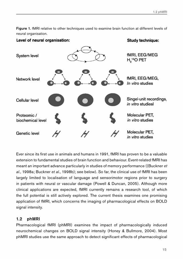
fMRI is a non-invasive imaging technique, which is important in terms of its further development as a clinical tool. When performed at conventional spatial resolution, fMRI produces functional neuroimages that reflect brain function at the level of neural networks and systems. This places it in a central position with respect to techniques examining similar effects at other levels of neural organisation (Figure 1). When compared to other non-invasive techniques used to examine brain function at a system level (e.g. electroencephalography (EEG), magnetoencephalography (MEG) or positron emission tomography (PET)), fMRI combines several unique features (Jezzard et al., 2001). These include:
A high spatial resolution (compared to EEG or MEG).A good temporal resolution (compared to H2
15O PET). This allows event-related analyses of subcomponent processes during task performance (Buckner, 1998).The ability for repeated measurements in single subjects with no technical constraints put to the number and duration of the experiments, which makes it a highly flexible technique (PET investigations are limited by the half-life and cumulative dose of radioactive tracers).
Because of its high flexibility and ability for event-related analyses, fMRI has gradually replaced H2
15O PET as the non-invasive imaging technique of choice to examine brain function at high spatial resolution. Possible constraints of fMRI are its low signal-to-noise ratios, its relative measures, and the fact that the BOLD signal is not a direct measure of neural activity. BOLD signal intensity changes may therefore be difficult to interpret in terms of underlying neurodynamics (Logothetis & Pfeuffer, 2004).
1.2.
3.
15
1.2 phMRI
Figure 1. fMRI relative to other techniques used to examine brain function at different levels of neural organisation.
Ever since its first use in animals and humans in 1991, fMRI has proven to be a valuable extension to fundamental studies of brain function and behaviour. Event-related fMRI has meant an important advance particularly in studies of memory performance ((Buckner et al., 1998a; Buckner et al., 1998b); see below). So far, the clinical use of fMRI has been largely limited to localisation of language and sensorimotor regions prior to surgery in patients with neural or vascular damage (Powell & Duncan, 2005). Although more clinical applications are expected, fMRI currently remains a research tool, of which the full potential is still actively explored. The current thesis examines one promising application of fMRI, which concerns the imaging of pharmacological effects on BOLD signal intensity.
1.2 phMRIPharmacological fMRI (phMRI) examines the impact of pharmacologically induced neurochemical changes on BOLD signal intensity (Honey & Bullmore, 2004). Most phMRI studies use the same approach to detect significant effects of pharmacological
16
Chapter 1: General Introduction
intervention on brain function: ‘main effects’ during task performance within a group of subjects (i.e. brain activation or deactivation as a result of task stimuli) are compared statistically between placebo and active medication conditions to produce group-level contrast images of ‘treatment effects’. The field is still in its early stages. Since 1999, some 60 articles have been published concerning the topic of phMRI. The first phMRI studies mainly involved feasibility studies in animals and fundamental studies of neurotransmitter system function in humans (e.g. neurotransmitter depletion or overexpression) (Shah & Marsden, 2004). Such studies showed that changes in brain function as a result of pharmacological treatment may be region-specific, process-specific and even genome specific and depend on age and cognitive capacity (Honey & Bullmore, 2004).
Apart from studying fundamental processes, phMRI has been used to examine the effects of pharmacological intervention on brain function within a clinical context. So far, clinical phMRI studies mainly involved feasibility studies in patients, studies of pharmacotherapeutic mechanisms and the effects of certain vulnerability traits (such as substance abuse and addiction) on the functional response to an addictive substance. The (differential) diagnostic and prognostic value of phMRI has been addressed only recently and will be considered further in this thesis.
Knowledge of pharmacological effects on neural systems in vivo is still quite limited. PET studies (Gee, 2003; Moresco et al., 2001) and EEG studies (Fingelkurts et al., 2005) of pharmacological intervention have yielded important information, yet technical difficulties prevent a single technique from providing a comprehensive picture. Since the various imaging modalities are complementary rather than competitive (Rudin & Weissleder, 2003), it is hoped that a combination of high spatial and temporal resolution allows for an optimal view of the effects of pharmacological intervention on brain function in living subjects.
1.3 Aim of this thesis: exploring the use of phMRI in a clinical contextGiven its combined qualities as a high-resolution, non-invasive imaging technique that allows for event-related analyses and repeated measurements within the same subject, fMRI can be used to answer several questions concerning pharmacological effects on brain function, which may be relevant to its use in a clinical context:
Does a pharmacological substance of interest indeed produce changes in the BOLD response?Are treatment effects on the BOLD signal region- and process-specific?Are functional changes specific to certain subgroups of subjects or patients (e.g. gender-specific, disease-specific)?
1.
2.3.
17
1.4 phMRI: studying changes in neurotransmission
Are functional changes dependent on drug dosage and exposure duration?Can fMRI be used to examine the treatment mechanism of a pharmacological substance?Do the observed functional changes have (early, differential) diagnostic value, or predictive value for treatment response and eventual clinical outcome?
Following a tradition of memory research in relation to Alzheimer’s disease and related disorders, this thesis attempts to address these questions by studying effects of pharmacological substances on brain function in controls and patients during memory task performance. Studies were performed in collaboration with the departments of Neurology (Alzheimer Center), Radiology, Physics and Medical Technology, Statistics, and Endocrinology of the VU University Medical Centre, and with the department of Clinical and Experimental Psychology of the University of Amsterdam, the Netherlands.
Studying drug effects on memory function in controls and patients requires some knowledge of the neurochemistry that is modulated pharmacologically, its putative effects on the memory systems under investigation, and the pathological changes occurring in these memory systems in disease. The following paragraphs will therefore briefly address some of the key concepts of neuropharmacology in relation to functional neuroimaging studies of memory performance that provided the global context for the studies reported in this thesis.
1.4 phMRI: studying changes in neurotransmissionMost neuropharmacological substances exert their influence on brain function by interacting with neurotransmitter systems. Most classical neurotransmitter systems contribute to memory performance (for a recent meta-analysis, see (Myhrer, 2003)). The current thesis focuses on the pharmacological modulation of three neurochemical systems in relation to memory function within the central nervous system: the cholinergic, noradrenergic and the sex-steroid system.
The central cholinergic system originates in the brain stem and basal forebrain, where the neurotransmitter ‘acetylcholine’ is produced in discrete cholinergic nuclei (e.g. nucleus basalis of Meynert) and distributed diffusely across the brain (Selden et al., 1998). At the molecular and cellular level, acetylcholine interacts with nicotinic or muscarinic cholinergic receptors (ligand-gated ion channels) to modulate the transmembrane potential and induce protein synthesis through activation of G-protein coupled second messenger systems (Dani, 2001). Thus, in the short run, cholinergic activity provides the arousal state necessary for learning new information (McGaugh, 2004), and for directing selective attention to relevant versus non-relevant stimuli (Sarter
4.5.
6.
18
Chapter 1: General Introduction
et al., 2005). In the long run, cholinergic activity contributes to memory formation by modulating the connection strengths of newly formed synapses and stabilising synaptic connections between networks that encode relevant associations (Mesulam, 1996; Little et al., 1998; Rezvani & Levin, 2001; Gu, 2002). A loss of cholinergic function may be observed in several clinical conditions including Alzheimer’s disease (AD). In AD, atrophy of the basal forebrain nuclei produces a cholinergic deficit, which is thought to contribute significantly to the symptomatology of the disease. Apart from memory impairment (Bartus, 2000), cholinergic dysfunction has been associated with neuropsychiatric symptoms such as attention deficits, sleep disorders, loss of verbal fluency, anxiety, depression, and psychosis (Assal & Cummings, 2002). Although the role of a cholinergic deficit in AD has been well established, the extent to which cholinergic function is impaired in AD, along with the time of onset of this impairment, are still subjects of debate. Recent results suggest that cholinergic function is intact and may even be upregulated in early stages of AD, such as mild cognitive impairment (MCI). MCI is defined as a slowly progressive memory decline without the involvement of another domain of cognitive function, that does not interfere significantly with activities of daily living (Petersen et al., 2001). MCI patients are at increased risk of developing AD, but clinical outcome may vary considerably at this stage. It has been hypothesised that conversion from MCI to AD (partly) reflects the inability of the cholinergic system to compensate for progressive memory deficits as a result of early hippocampal damage (DeKosky et al., 2002).
When compared to the cholinergic system, considerably less is known about the central adrenergic system. This system originates from discrete nuclei within the brain stem (mainly the nucleus coeruleus), where the neurotransmitter ‘noradrenaline’ is produced and transported diffusely across the brain. At the molecular and cellular level, noradrenaline interacts with alpha- and beta- adrenergic receptors (G-protein coupled receptors) to modulate the transmembrane potential and induce protein synthesis through activation of cyclic AMP related second messenger pathways (Waterhouse et al., 1991). The noradrenergic system is thought to be relevant to memory encoding and consolidation through a number of different actions, all of which result from the processing of (highly) emotionally relevant or meaningful information. These actions involve amygdala-mediated enhancement of cortical arousal, stimulation of the stress response, and stimulation of the cholinergic system in the brainstem (McGaugh, 2004). A loss of noradrenergic function (e.g. atrophy of the nucleus coeruleus) is thought to be relevant to a number of clinical conditions, including memory loss in AD, cognitive and movement disorders in Parkinson’s disease (Marien et al., 2004), anxiety and panic disorders, depression, and post-traumatic stress disorder (LeDoux, 1998).
19
1.5 Memory tasks (‘paradigms’) used in the current study
The effects of sex steroids (e.g. estrogen, testosterone, progesterone) on memory function are complex and still relatively unclear. Sex steroids are produced in male and female gonads as a result of the actions of a cascade of hormones that are produced in the hypothalamus and pituitary. They exert a broad range of effects across the entire human body, including the central nervous system (Pfaff, 2005), where they may have neuroprotective effects (Wise et al., 2005). At the molecular and cellular level, estrogens interact with alpha- and beta-estrogen receptors (ligand-dependent transcription factors) to produce a slow genomic effect, and with membrane-bound estrogen receptors to directly affect neural signalling (Vasudevan et al., 2005; Wolf, 2003; Bisagno et al., 2003). Apart from affecting neural architecture during development, sex steroids have modulatory effects on the four primary neuromodulatory neurotransmitter systems (i.e. the cholinergic, noradrenergic, serotonergic and dopaminergic systems) (Bernardi et al., 2003; Korol, 2004). Thus, sex steroids are thought to be responsible for global differences in behaviour between the sexes (Cahill, 2003). A drop in estrogen levels during menopause has been implicated in an increased risk of postmenopausal women for mild cognitive impairment (MCI) and Alzheimer’s disease (AD) (Yaffe et al., 2005).
1.5 Memory tasks (‘paradigms’) used in the current studyTo examine the impact of pharmacological intervention into noradrenergic, cholinergic and sex-steroid systems on brain function during memory performance, several fMRI paradigms were constructed. These paradigms activated brain structures relevant to different aspects of memory performance (for a review on the neuropsychology and taxonomy of memory, see (Gazzaniga et al., 2000)).
An n-letter back working memory task was used to examine brain function during short term working memory performance (see further descriptions in this thesis). In both controls and patients, this task reproducibly activates bilateral parietal and prefrontal brain regions with a preference for the left hemisphere. Brain function within these areas is thought to represent automatic maintenance and effortful manipulation of symbolic information (letters) in short term memory in the absence of the original stimulus (Owen et al., 2005).
Face encoding and -recognition tasks were used to examine (intermediate term) episodic memory performance for unfamiliar and emotionally neutral human faces (see further descriptions in this thesis). Face encoding and -recognition tasks are among the most established of memory tasks and have been widely used in functional neuroimaging studies to elicit reproducible patterns of brain function in both controls and patients (Small et al., 1999). Encoding tasks produce brain function related to the encoding
20
Chapter 1: General Introduction
phase (naive situation) of memory performance. Recognition tasks produce brain function during retrieval of familiar or attempted retrieval of unfamiliar items (i.e. during the non-naive situation, or ‘retrieval-mode’). fMRI allows event-related analyses of brain function related to distinct response types during retrieval (e.g. correct hits, correct rejections, false hits, and false rejections), which when contrasted allow further analysis of subcomponent processes during recognition, such as successful recognition and encoding during attempted retrieval (Buckner, 1998; Daselaar et al., 2003).
Finally, an encoding task was created using images from the international affective picture system (IAPS; see further descriptions in this thesis) (Lang & Bradley, 1997). This paradigm allows visualisation of brain function during encoding of aversive stimuli (i.e. disgusting, harmful or threatening information) into memory, and was developed in collaboration with Dr. A.H. van Stegeren and Prof. Dr. W.TA.M. Everaerd from the department of Clinical and Experimental Psychology of the University of Amsterdam. The primary function of this paradigm was to optimise detection of amygdala function during emotional memory performance.
1.6 Outline of this thesisA total of three pharmacological substances were examined for their effects on brain function during memory task performance. These include propranolol, which is a centrally and peripherally acting blocker of beta-adrenergic neurotransmission (Ananth & Lin, 1986), raloxifene, which is a selective estrogen receptor modulator (SERM) of which the effects on brain function remain to be investigated (Heringa, 2003), and galantamine, which is a weak cholinesterase inhibitor with a strong sensitising effect on nicotinergic receptors and known efficacy in the treatment of memory deficits in AD (Raskind, 2003). Studies reported below are summarised in the order in which they appear in this thesis (subject and content of these studies partly mirror the rapid developments in the field of phMRI):
In Chapter 2, propranolol was used to block beta-adrenergic neurotransmission in the central nervous system of healthy young subjects. Since animal studies have shown that neurotransmission in the amygdala is predominantly noradrenergic, we hypothesised that a singe oral dose of propranolol in humans would interfere with normal amygdala function during encoding of emotionally charged information (IAPS pictures; see above) into memory. This phMRI study aimed to link functional and behavioural data on amygdala function in animals and humans by examining the influence of pharmacological impairment of beta-adrenergic neurotransmission on amygdala function in human subjects. The possibility that both sexes respond differentially to beta-adrenergic blockade is also discussed.
21
1.6 Outline of this thesis
In Chapter 3, two studies are presented that examine the effects of the SERM raloxifene on brain function. These studies examined long-term effects of raloxifene treatment while anticipating its clinical prescription in elderly males. Effects of long-term (three months) raloxifene treatment were examined during encoding (Chapter 3.1) and recognition (Chapter 3.2) of emotionally neutral human faces into memory. Predictions are made with respect to behavioural changes based on the observed effects of treatment, and a hypothesis is presented for a possible treatment mechanism of raloxifene.
In Chapter 4, three phMRI studies are presented that examine the effects of short-term galantamine exposure on brain function in patients with mild cognitive impairment (MCI) and Alzheimer’s disease (AD). Effects of galantamine challenge were evaluated at different exposure durations (i.e. acute (single dose) and prolonged (5 days) exposure) on brain function during face encoding, face recognition and n-letter back working memory performance. Effects of galantamine intake are first examined on brain function during face encoding and working memory performance in MCI (Chapter 4.1) and AD patients (Chapter 4.2) separately. Similar effects are then examined on brain function during recognition, which are compared directly between MCI and AD patient groups (Chapter 4.3).
Chapter 4.1 examines the feasibility of detecting effects of pharmacological challenge in patients with MCI. Studies examining cholinergic system reactivity to pharmacological challenge in MCI patients may be used to assess the functional status of the cholinergic system in disease, which may be relevant in terms of predicting further memory decline, and possibly conversion to AD.
Chapter 4.2 examines effects of galantamine challenge in AD patients. Like chapter 4.1, cholinergic reactivity is evaluated with respect to brain function during face encoding and working memory performance. Differences with cholinergic reactivity of MCI patients are discussed, and effects of galantamine challenge on the shape of the BOLD response are investigated.
Chapter 4.3 examines MCI and AD patients for a differential response to galantamine challenge during face recognition. Such a differential response may indicate a difference in the functional status of the cholinergic system in both patient groups. The issue of region-, process- and disease-specificity of treatment effects is raised. Additionally, the possible clinical (e.g. diagnostic) value of a differential response to pharmacological challenge is highlighted.
In Chapter 5, findings from our phMRI studies are summarised and discussed within the context of the clinical potential of phMRI, after which suggestions are made for future studies.

Chapter 2
Effects of beta-adrenergic blockade on amygdala function in healthy young subjects
2.1 Noradrenaline mediates amygdala activation in men and women during encoding of emotional material
Van Stegeren AH, Goekoop R, Everaerd WTAM, Scheltens P, Barkhof F, Kuijer JP, Rombouts SARB
Neuroimage. 2005; 24(3):898-909. PMID: 15652324
24
Chapter 2: Effects of beta-adrenergic blockade on amygdala function in healthy young subjects
Abstract
The amygdala is a pivotal structure in humans for encoding of emotional information, as shown by recent imaging studies. It is unknown which neurotransmitters are specifically involved in the human amygdala, although in animal studies noradrenaline was shown to be essential. In our study participants received the betablocker propranolol (which blocks the noradrenergic response) or placebo when watching neutral to highly negative arousing pictures. Amygdala activation, monitored with functional magnetic resonance imaging (fMRI), increased with emotional intensity of the pictures under placebo condition. Betablockade selectively decreased amygdala activation for emotional pictures of the second highest category, but not for the highest or lower (neutral) category pictures. Two findings add to the existing knowledge in this area. First, the activation pattern in the amygdala under placebo condition shows a non-linearity related to the emotional categories of the pictures. Secondly, propranolol disturbs this activation pattern in the amygdala. Explorations with respect to gender show a similar effect of betablockade on amygdala activation in both men and women, but a difference in its effect on long term memory for emotional pictures. This study supports the hypothesis that the neurotransmitter noradrenaline also mediates amygdala activity in humans when processing emotional stimuli and that betablockers can disrupt the normal activation pattern in the amygdala.
Key words: amygdala; fMRI; noradrenaline; gender.
25
2.1 Noradrenaline mediates amygdala activation in men and women during encoding of emotional material
Introduction
A critical function of the human amygdala is the processing of emotional information. This process varies from the perception of stimuli that have emotional significance (Anderson & Phelps, 2001) to the continuing stages in the memory process . Exposure to aversive stimuli in multiple sensory modalities induces activation of the amygdala. Several imaging studies using a variety of visual, auditory, olfactory or gustatory stimuli evoked amygdala activation in humans. Even unpleasant interoceptive or painful sensations appear to induce amygdala activation (Zald, 2003). Although the activation pattern of the amygdala was consistently found in reaction to aversive stimuli (Fischer et al., 2003; Garrett & Maddock, 2001; O’Doherty et al., 2001; Phan, Fitzgerald et al., 2004; Phelps et al., 2001; Stark et al., 2003; Zald & Pardo, 2002), activation was also found in reaction to positively valenced stimuli from multiple sensory modalities (Garavan et al., 2001; Hamann et al., 1999; Lane, Chua, & Dolan, 1999; Lane et al., 1997). Recent findings also provide evidence for a role of the amygdala in reaction to visual sexual stimuli as contrasted with non-sexual stimuli (Canli & Gabrieli, 2004; Hamann et al., 2004) as well as for an important role in regulating human sexual behavior (Baird et al., 2004). It appears that all these responses are modulated by the arousal level, hedonic strength or motivational value of the stimuli.
With respect to the role of the amygdala in memory processes this structure was shown to be active during the encoding of emotional stimuli (Adolphs et al., 2000; Cahill et al., 1996; Canli et al., 2000; Hamann et al., 1999) or emotional context (Erk et al., 2003). Other studies stressed the important role of the amygdala in enhancing the strength of long-term memory for emotional stimuli, hence its role in consolidation processes (Cahill et al., 1996; McGaugh et al., 1996). The role of the amygdala as part of a neural network in relation to emotional memory was put forward in several studies. Two of these recent reports stressed the importance of the interaction between the amygdala and hippocampus that appears to be essential for a successful encoding and consolidation of emotional stimuli and situations (Phelps, 2004; Richardson, Strange, & Dolan, 2004). A recent path analysis using structural equation modeling also addressed aspects of the ‘memory modulation hypothesis’. They showed increased functional connectivity between the amygdala and the ipsilateral parahippocampal gyrus and ventrolateral prefrontal cortex during emotional relative to a neutral film viewing condition (Kilpatrick & Cahill, 2003). Using event-related fMRI during encoding of emotional and neutral pictures Dolcos et al. (Dolcos, LaBar, & Cabeza, 2004) found support for the modulation hypothesis, stating that better memory for emotionally arousing events
26
Chapter 2: Effects of beta-adrenergic blockade on amygdala function in healthy young subjects
(compared with non-arousing neutral events) is due to an effect of the amygdala on the medial temporal lobe (MTL) memory system. More anecdotal evidence shows that memories of emotionally arousing events tend to be more vivid and to persist longer than do memories of neutral events. Apparently, the relevance and salience of a stimulus is important in survival of the species. It seems likely that processing of emotional information is mediated by neurotransmitters that have a relation to arousal, such as noradrenaline.
In animal studies noradrenaline was shown to be one of the essential neurotransmitters in the (basolateral) amygdala, related to emotional processing (McGaugh, 2000). Pharmacological findings indicate that activation of postsynaptic alpha1-adrenoreceptors potentiates beta-adrenoceptor-mediated activation of cAMP (cyclic Amino-Mono-Phosphate) formation (Ferry, Roozendaal, & McGaugh, 1999a). However, this has only been shown in animal studies, where noradrenergic agonists, such as clenbuterol, injected directly into the amygdala, improved memory performance in rats (Ferry & McGaugh, 1999). In contrast, noradrenaline antagonistic agents such as propranolol, atenolol or zinterol, not only injected directly into the amygdala but also in several nuclei around the amygdaloidal complex and projecting on the amygdala, had the opposite (i.c. deteriorating) effect on later memory performance (Quirarte et al., 1997). These data support the hypothesis that the memory-modulating effect of the amygdala adrenergic system is mediated, at least in part, by the activation of beta-adrenoceptors in the amygdala.
In humans central noradrenergic mechanisms appeared to be essential in memory performance for emotional material. Beta-adrenergic blockade with a central and peripheral acting agent (propranolol) did affect memory for emotional stimuli (Cahill et al., 1994; van_Stegeren et al., 1998), whereas a peripherally acting betablocker (nadolol) did not have the same memory disturbing properties (van_Stegeren et al., 1998). And stimulation of the central noradrenergic system with yohimbine resulted in the enhancement, whereas blockade with the betablocker metoprolol resulted in a reduction of recall and recognition of emotional material in man (O’Carroll et al., 1999). Until now it has remained unclear where in the human brain these centrally acting noradrenergic receptors exert their effect.
In this study we monitored amygdala activation with fMRI during encoding of sets of pictures after having taken a betablocker (a noradrenergic antagonist) on one day or a placebo on the second day. We wanted to test the specific hypothesis – known from the animal literature – that the amygdala is mediated by noradrenergic activation in the human brain as well. We hypothesized that if noradrenergic activation in the amygdala
27
2.1 Noradrenaline mediates amygdala activation in men and women during encoding of emotional material
is essential in processing emotional information, amygdala activation under betablocker condition should be lower than under placebo condition, when subjects are confronted with emotional stimuli.
Methods
SubjectsThirty right-handed subjects (15 male, 15 females; mean age 20.93 ± 2.38, ranging from 18 to 28 years) without medical or psychiatric history were selected after an introduction interview, where they were screened with the Symptom Check List (SCL-90) (mean score = 104.03 ± 9.48) and a biographic questionnaire. Screening with the SCL-90 was carried out and scored using normative ratings for a healthy population. All subjects fell in the “normal range” with scores ‘below average’ and ‘low’. This was done because in several studies psychopathological disorders were shown to affect volume as well as functioning of the amygdala (Hull, 2001). Subjects were all students of the University of Amsterdam and received course credit for their participation. The Medical Ethical Committee of the VU Medical Center (VUMC) approved the experiment and informed consent was obtained from all subjects.
Design, Material and ProcedureIn this study we used a randomized, double blind, placebo-controlled event-related design. On two consecutive days subjects came to the fMRI department of the VUMC. After an acclimatization period of 15 minutes, heart rate (HR) was measured for baseline (BL) values, before they entered the scanner (Pre-scan) and immediately after the scanning procedure (Post-scan) on both days. Double blind they received either a placebo (PL) or betablocker (BB) in random order over the two days (PL-BB or BB-PL). Drug order was divided over the sexes and the days as follows: data of 14 males were included in the analysis; 6 of them received pills in the order BB-PL, 8 males in the reverse order PL-BB; data of 14 females were included and drug order was evenly divided. A resting period of 90 minutes was needed to have the drug reach peak plasma levels (Gilman & Goodman, 1996). Stimulus material consisted of two sets of 92 pictures (Set 1 and Set 3) selected from the International Affective Picture System (IAPS) (Lang et al., 1997). Both sets were divided in four categories (CAT1-CAT4) that ranged conform IAPS norms in emotional valence from neutral (5.0) to extremely negative pictures (2.0) and in arousal from low (3.2) to highly arousing (6.2).
28
Chapter 2: Effects of beta-adrenergic blockade on amygdala function in healthy young subjects
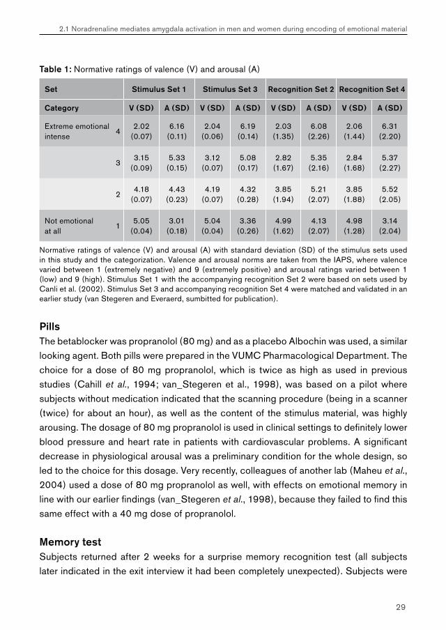
To study memory performance the two stimulus sets were complemented with 48 additional pictures each (Recognition Set 2 and Set 4) that served as foils that had similar valence and arousal properties (see Table 1 for details). These sets were tested and validated in an earlier study (van Stegeren & Everaerd, 2004) where the sets showed to be completely identical with respect to emotional judgment by the subjects, in evoking physiological reactions and having identical memorizing properties. One of the stimulus- and recognition sets (set 1 and set 2, with four additional slides) has been used in earlier fMRI experiments (Canli, Desmond et al., 2002; Canli et al., 2000) where amygdala activation was found during presentation of the more emotional negative pictures. Set order was counterbalanced across the subjects over the two test days. After pre-scan HR measurements subjects were positioned in the MRI scanner, where a structural scan was made first. Then the experimental (event-related) functional imaging started. Subjects were presented with blocks of 8 pictures containing random assortments of pictures across all four emotional categories. After each picture (presentation time 3 seconds), subjects were asked on screen (within 3 seconds) to indicate the emotional intensity of the previous picture by pressing one of four buttons with their right hand, with 1 being ‘not emotional at all’ to 4 being ‘extreme emotional’. These individual emotional ratings were used to classify the pictures for further event-related fMRI analysis. This method of correlating the event-related activity in the amygdala to subjects personal ratings was used in earlier studies as well (Cahill, Uncapher et al., 2004; Canli, Desmond et al., 2002; Canli et al., 2000). Phan v (Phan et al., 2003) specifically tested the hypothesis that incorporating subjective emotional ratings improved the sensitivity for detecting activation in regions including the amygdala, and found support for it.
After a block of 8 pictures with 8 emotional ratings by the subject, 8 gray screens (presentation time 3 seconds) served as a resting (baseline) period, also presented as events and jittered (Donaldson & Buckner, 2001). This was done to allow amygdala activity, when evoked by emotional pictures, to return to baseline levels as much as possible and to contrast activation levels during stimulus presentation with baseline levels. The procedure on the second day was identical to the first – apart from the content of the drug they received.
29
2.1 Noradrenaline mediates amygdala activation in men and women during encoding of emotional material
Table 1: Normative ratings of valence (V) and arousal (A)
Set Stimulus Set 1 Stimulus Set 3 Recognition Set 2 Recognition Set 4
Category V (SD) A (SD) V (SD) A (SD) V (SD) A (SD) V (SD) A (SD)
Extreme emotional intense
42.02
(0.07)6.16
(0.11)2.04
(0.06)6.19
(0.14)2.03
(1.35)6.08
(2.26)2.06
(1.44)6.31
(2.20)
33.15
(0.09)5.33
(0.15)3.12
(0.07)5.08(0.17)
2.82(1.67)
5.35(2.16)
2.84(1.68)
5.37(2.27)
24.18
(0.07)4.43
(0.23)4.19
(0.07)4.32
(0.28)3.85
(1.94)5.21
(2.07)3.85
(1.88)5.52
(2.05)
Not emotional at all
15.05
(0.04)3.01
(0.18)5.04
(0.04)3.36
(0.26)4.99
(1.62)4.13
(2.07)4.98
(1.28)3.14
(2.04)
Normative ratings of valence (V) and arousal (A) with standard deviation (SD) of the stimulus sets used in this study and the categorization. Valence and arousal norms are taken from the IAPS, where valence varied between 1 (extremely negative) and 9 (extremely positive) and arousal ratings varied between 1 (low) and 9 (high). Stimulus Set 1 with the accompanying recognition Set 2 were based on sets used by Canli et al. (2002). Stimulus Set 3 and accompanying recognition Set 4 were matched and validated in an earlier study (van Stegeren and Everaerd, sumbitted for publication).
PillsThe betablocker was propranolol (80 mg) and as a placebo Albochin was used, a similar looking agent. Both pills were prepared in the VUMC Pharmacological Department. The choice for a dose of 80 mg propranolol, which is twice as high as used in previous studies (Cahill et al., 1994; van_Stegeren et al., 1998), was based on a pilot where subjects without medication indicated that the scanning procedure (being in a scanner (twice) for about an hour), as well as the content of the stimulus material, was highly arousing. The dosage of 80 mg propranolol is used in clinical settings to definitely lower blood pressure and heart rate in patients with cardiovascular problems. A significant decrease in physiological arousal was a preliminary condition for the whole design, so led to the choice for this dosage. Very recently, colleagues of another lab (Maheu et al., 2004) used a dose of 80 mg propranolol as well, with effects on emotional memory in line with our earlier findings (van_Stegeren et al., 1998), because they failed to find this same effect with a 40 mg dose of propranolol.
Memory testSubjects returned after 2 weeks for a surprise memory recognition test (all subjects later indicated in the exit interview it had been completely unexpected). Subjects were
30
Chapter 2: Effects of beta-adrenergic blockade on amygdala function in healthy young subjects
shown the recognition sets of (92 + 48) 140 pictures in the same set order as they had been offered on day 1 and 2 of the experiment and were asked after each picture to indicate whether they had seen that picture before by pressing one of three buttons. Possible answers were:1 = no, never seen before; 2 = it looks familiar, but I am not sure; and: 3 = yes, I have seen this picture before. Memory scores were calculated by counting the correctly recognized items (answers 2 + 3: ‘familiar’ and with certainty recalled pictures together) and were expressed as percentage correctly recognized pictures per category. Two subjects were excluded from analysis because their memory score was well below chance level (< 30%). False positive rates were around 17%, which is in line with earlier studies in this area.
fMRI acquisitionTwenty-three slices positioned perpendicular to the long axis of the hippocampus and completely covering the amygdala and hippocampus were collected using a gradient-echo echo-planar imaging pulse-sequence (in-plane voxel size 3 x 3 mm; 2.5 mm thick, 0.5 mm gap; repetition time, 2130 ms; echo time, 50 ms; total number of scans in each subject ~620). A T1-weighted structural MRI-scan was also acquired (MPRAGE; inversion time: 300 ms, TR = 15 ms; TE = 7 ms; flip angle = 8°; 160 coronal slices, 1 x 1 x 1.5mm voxels). Each subject’s functional images in the current experiment were first inspected to ensure adequate signal in both amygdalae.
fMRI Data AnalysisAll MRI analyses were carried out using FEAT (FMRI Expert Analysis Tool) Version 5.00, part of FSL (FMRIB’s Software Library, www.fmrib.ox.ac.uk/fsl). Pre-statistics processing included slice-timing correction using Fourier-space time-series phase-shifting; motion correction (Jenkinson et al., 2002); non-brain removal (Smith, 2002); spatial smoothing using a Gaussian kernel of FWHM 8mm; mean-based intensity normalization of all volumes by the same factor; highpass temporal filtering (Gaussian-weighted LSF straight line fitting, with sigma = 100.0s). Time-series statistical analysis was carried out with local autocorrelation correction (Woolrich et al., 2001), modeling in each subject the events using a double gamma hemodynamic response function and its temporal derivative. Then co-registration to high resolution scans and subsequently to standard space images was carried out (Jenkinson et al., 2002; Jenkinson & Smith, 2001). Comparisons of interest were implemented as linear contrasts. Next a higher-level analysis was carried out using a local analysis of mixed effects (Beckmann, Jenkinson & Smith, 2003). A repeated measures model was used with regressors
31
2.1 Noradrenaline mediates amygdala activation in men and women during encoding of emotional material
modeling betablocker (BB) and placebo (PL) scans separately. A regressor to correct for session effects (change between scan 1 and 2) was also included in the model.
Main effects were calculated for the PL scans first, contrasting each emotional category to baseline. For these scans, Z (Gaussian T/F) statistic images were thresholded using clusters determined by Z > 2.3 and a corrected cluster significance threshold of p = 0.05 (Forman et al., 1995; Friston et al., 1994; Worsley et al., 1992). The amygdala was defined in standard space on the average structural scan of all subjects. First we determined amygdala activation during all of the (CAT > Baseline) comparisons. We determined based on the anatomy of the average structural scan of all subjects, which activations could be identified as being in the amygdalae. All these activated regions in the amygdalae were included in the further analyses of interactions as region of interest (ROI). The analyses testing for the interaction Drug x Category were again thresholded using clusters determined by Z > 2.3 and a cluster corrected significance threshold of p = 0.05.
Results
Physiological MeasuresHeart rate was used as a marker to check for successful betablockade by propranolol. Heart rate was registered at baseline (BL), just before subjects entered the scanner (Pre-scan) and immediately after the scanning procedure (Post-scan) and was analyzed with a General Linear Model (GLM) (3 time x 2 drug). Baseline heart rate levels for all subjects were higher on day 1 compared to day 2 (p < .05), perhaps expressing a higher level of anticipation anxiety before the first scanning session began. Fortunately subjects were randomly allocated to each drug condition, so on both days the pill groups did not differ at BL measurement. Checking for the intended drug manipulation revealed that heart rate was significantly lower at Pre-scan and Post-scan measurements in the propranolol condition, showing that the betablocker was effective during the complete scanning procedure (Figure 1A). As had been found in earlier studies (Lang et al., 1993; Suarez et al., 2004), men had significantly higher heart rate levels at BL than women on both days, but since men and women were as evenly as possible divided over the pill-order conditions, this also did not affect baseline HR levels between the drug conditions.
32
Chapter 2: Effects of beta-adrenergic blockade on amygdala function in healthy young subjects
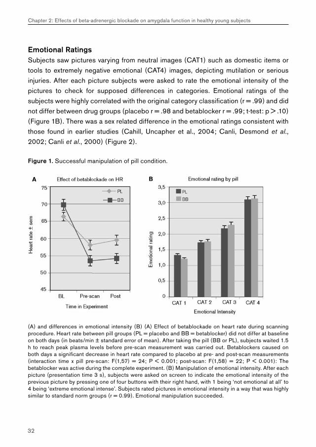
Emotional RatingsSubjects saw pictures varying from neutral images (CAT1) such as domestic items or tools to extremely negative emotional (CAT4) images, depicting mutilation or serious injuries. After each picture subjects were asked to rate the emotional intensity of the pictures to check for supposed differences in categories. Emotional ratings of the subjects were highly correlated with the original category classification (r = .99) and did not differ between drug groups (placebo r = .98 and betablocker r = .99; t-test: p > .10) (Figure 1B). There was a sex related difference in the emotional ratings consistent with those found in earlier studies (Cahill, Uncapher et al., 2004; Canli, Desmond et al., 2002; Canli et al., 2000) (Figure 2).
Figure 1. Successful manipulation of pill condition.
(A) and differences in emotional intensity (B) (A) Effect of betablockade on heart rate during scanning procedure. Heart rate between pill groups (PL = placebo and BB = betablocker) did not differ at baseline on both days (in beats/min ± standard error of mean). After taking the pill (BB or PL), subjects waited 1.5 h to reach peak plasma levels before pre-scan measurement was carried out. Betablockers caused on both days a significant decrease in heart rate compared to placebo at pre- and post-scan measurements (interaction time x pill pre-scan: F(1,57) = 24; P < 0.001; post-scan: F(1,58) = 22; P < 0.001): The betablocker was active during the complete experiment. (B) Manipulation of emotional intensity. After each picture (presentation time 3 s), subjects were asked on screen to indicate the emotional intensity of the previous picture by pressing one of four buttons with their right hand, with 1 being ‘not emotional at all’ to 4 being ‘extreme emotional intense’. Subjects rated pictures in emotional intensity in a way that was highly similar to standard norm groups (r = 0.99). Emotional manipulation succeeded.
33
2.1 Noradrenaline mediates amygdala activation in men and women during encoding of emotional material
fMRI Results At first level mean activation (per subject per session) as measured with fMRI, was calculated for pictures of the same emotional category and contrasted with a baseline condition of gray screens. Then at higher level (across subjects) these contrasts were analyzed in a General Linear Model (GLM). In the placebo group, amygdala activation increased with emotional intensity of the pictures. There was no difference in amygdala activation between CAT1 and CAT2 pictures, but emotional pictures (CAT3 and 4) evoked more amygdala activation than neutral (CAT1) pictures under placebo condition (Figure 3A and 4A). The main purpose of this study was to test if amygdala activation was affected by betablockade. Amygdala activation was analyzed with a GLM with contrasts between subsequent emotional categories 1 to 4 (CAT2 with CAT1, CAT3 with CAT1 etc.) and with ‘drug’ as between variable. In this way we tested how amygdala activation depended on the emotional intensity of the pictures, and – testing our main hypothesis – whether noradrenergic blockade with propranolol affects this activation pattern (Table 2).
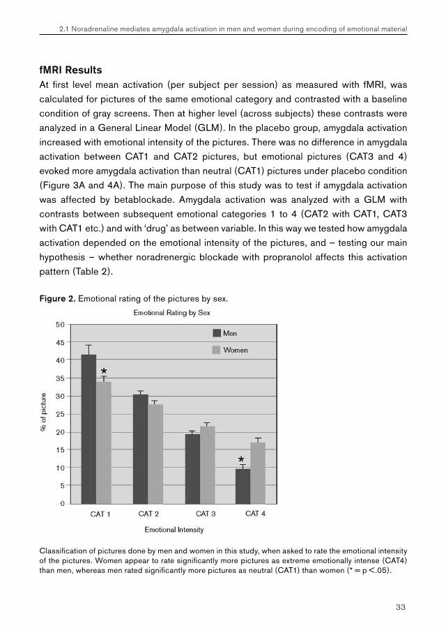
Figure 2. Emotional rating of the pictures by sex.
Classification of pictures done by men and women in this study, when asked to rate the emotional intensity of the pictures. Women appear to rate significantly more pictures as extreme emotionally intense (CAT4) than men, whereas men rated significantly more pictures as neutral (CAT1) than women (* = p <.05).
34
Chapter 2: Effects of beta-adrenergic blockade on amygdala function in healthy young subjects
Evidence to support the main hypothesis of this study was provided by these contrasts where amygdala activation under betablocker condition was subtracted from the activation under placebo condition. Increased amygdala activation for the emotional CAT3 pictures, compared to the neutral CAT1 pictures, was significantly lower when subjects had taken a betablocker (Z = 3.63; cluster corrected p < .05) (Figure 3B). But a remarkable image appeared for the pictures with the highest emotional intensity (CAT4): the (random) presentation of these highly negative pictures led to comparable amygdala activation in both drug conditions (Figure 4A and B). Secondly, it was noteworthy that no overall BOLD effect of the betablocker could be found (Figure 5): there was no effect of beta-adrenergic blockade on amygdala activity during presentation of pictures from the neutral and low emotional categories (CAT1 and 2). This strongly supports the idea that noradrenergic blockade selectively affects amygdala activation in humans when confronted with emotional, but not neutral stimuli. If the emotional intensity of the stimulus is too extreme, such as the CAT4 pictures in this study, the effect of betablockade on the amygdala might be overruled by the emotional or presumably the arousal properties of the stimulus. Based on these findings we hypothesized that this reaction could be explained in terms of a dose-response relationship of the noradrenergic system in the amygdala, where in the light of this study the ‘dose’ is referring to the emotional intensity of the pictures. To explore this line of reasoning we carried out some additional analyses where we explicitly predicted a linear versus a non-linear relationship between the emotional intensity of the stimulus material and amygdala activation. We hypothesized that if noradrenergic activation in the amygdala is characterized by a curvilinear relationship, and if betablockade is exerting its effect on beta-adrenergic receptors in the amygdala, then this type of relation should be disturbed in the betablocker condition.
We created a linear contrast on first level (on subject per session level) that had to be entered in a demeaned format (–1.5; –0.5; 0.5; 1.5) as well as a non-linear contrast (–1.125; –0.125; 0.875; 0.375) (see figure 6A). Entering these contrasts in the higher-level analysis we found a significant fit of a curvilinear relationship between stimuli and activation pattern in the amygdala under placebo condition more than under betablocker condition (see figures 6B + C). Direct comparison of both conditions showed a trend (p = .09) that showed that activation under PL condition more than the BB condition is fitting to a curvilinear activation pattern, herewith supporting our hypothesis of a curvilinear activation pattern in the amygdala for the placebo group. Testing the linear contrast revealed also a significant fit under PL as well as BB condition (PL: Z = 4.32 in Right and Z = 4.51 in Left amygdala; p < .005) (BB: Z = 4.46 in Right and Z = 4.09
35
2.1 Noradrenaline mediates amygdala activation in men and women during encoding of emotional material
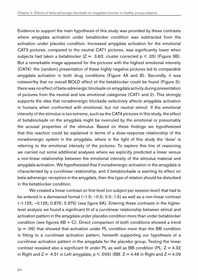
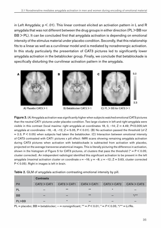
in Left Amygdala; p < .01). This linear contrast elicited an activation pattern in L and R amygdala that was not different between the drug groups in either direction (PL > BB nor BB > PL). It can be concluded first that amygdala activation is depending on emotional intensity of the stimulus material under placebo condition. Secondly, that this relationship fits to a linear as well as a curvilinear model and is mediated by noradrenergic activation. In this study particularly the presentation of CAT3 pictures led to significantly lower amygdala activation in the betablocker group. Finally, we conclude that betablockade is specifically disturbing the curvilinear activation pattern in the amygdala.
Figure 3. (A) Amygdala activation was significantly higher when subjects watched emotional CAT3 pictures than the neutral CAT1 pictures under placebo condition. Two large clusters in left and right amygdala were visible in this contrast (local maxima: right amygdala at coordinates 18, 0, –14; Z = 4.49; P<0.005;left amygdala at coordinates –16, –8, –12; Z = 5.05, P < 0.01). (B) No activation passed the threshold (of Z = 2.3, P < 0.05) when subjects had taken the betablocker. (C) Interaction between emotional intensity of CAT3 contrasted with CAT1 pictures x pill effect: fMRI scans showing remaining amygdala activation during CAT3 pictures when activation with betablockade is subtracted from activation with placebo, projected on the average transverse anatomical images. This is literally picturing the difference in activation, shown in the histogram of Figure 5 for CAT3 pictures, of clusters that pass the threshold (* = P < 0.05; cluster corrected). An independent radiologist identified this significant activation to be present in the left amygdala (maximal activation cluster on coordinate x = –16; y = –8; z = –12; Z = 3.63, cluster corrected P < 0.05). Right in images is left in brain.
Table 2. GLM of amygdala activation contrasting emotional intensity by pill.
Contrasts
Pill CAT2 > CAT1 CAT3 > CAT1 CAT4 > CAT1 CAT3 > CAT2 CAT4 > CAT3
PL − ** ** * −
BB − − ** − */**
PL>BB − * − − −
PL = placebo; BB = betablocker; − = nonsignificant; ** = P < 0.01; * = P < 0.05; */** = Li/Re.
36
Chapter 2: Effects of beta-adrenergic blockade on amygdala function in healthy young subjects
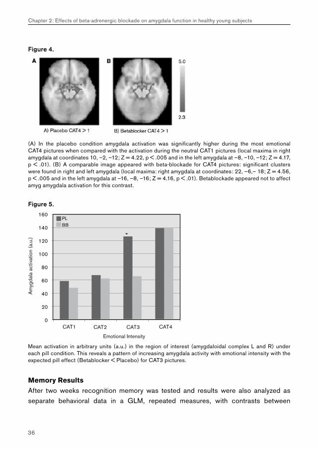
Figure 4.
(A) In the placebo condition amygdala activation was significantly higher during the most emotional CAT4 pictures when compared with the activation during the neutral CAT1 pictures (local maxima in right amygdala at coordinates 10, –2, –12; Z = 4.22, p < .005 and in the left amygdala at –8, –10, –12; Z = 4.17, p < .01). (B) A comparable image appeared with beta-blockade for CAT4 pictures: significant clusters were found in right and left amygdala (local maxima: right amygdala at coordinates: 22, –6,– 18; Z = 4.56, p < .005 and in the left amygdala at –16, –8, –16; Z = 4.16, p < .01). Betablockade appeared not to affect amyg amygdala activation for this contrast.
Figure 5.
Mean activation in arbitrary units (a.u.) in the region of interest (amygdaloidal complex L and R) under each pill condition. This reveals a pattern of increasing amygdala activity with emotional intensity with the expected pill effect (Betablocker < Placebo) for CAT3 pictures.
Memory Results
After two weeks recognition memory was tested and results were also analyzed as separate behavioral data in a GLM, repeated measures, with contrasts between
37
2.1 Noradrenaline mediates amygdala activation in men and women during encoding of emotional material
subsequent emotional categories 1 to 4 (CAT2 with CAT1, CAT3 with CAT2 etc.) and with ‘drug’ as between variable (4 emotional intensity x 2 drug). This revealed a pattern for memory performance highly similar to that of the fMRI data of amygdala activation (figure 7). A main effect of emotional intensity emerged (F(3,52) = 31.47; p < .001): memory performance under placebo as well as betablocker condition increased with emotional intensity of the pictures. But there also was a significant interaction effect (intensity x drug: F(3,52) = 2.42; p < .05): this was specifically found in the contrast between the more emotional CAT3 compared with CAT2 pictures. The increase in memory performance from CAT3 compared with CAT2 pictures was bigger when subjects had taken a PL than under BB condition (F(1,54) = 4.31; p <.05).
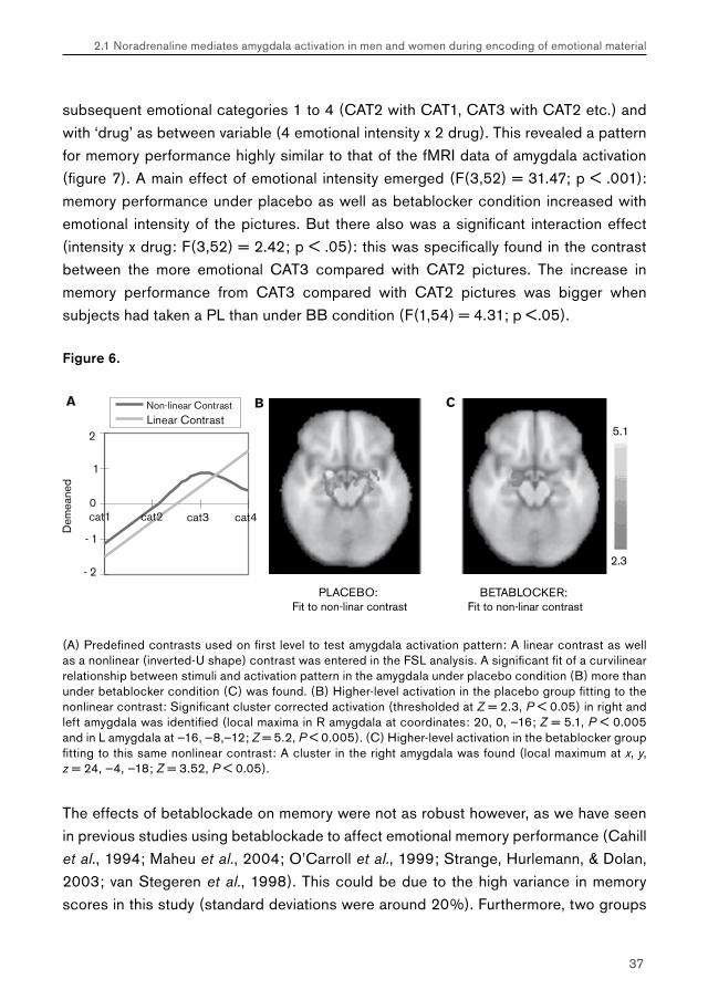
Figure 6.
(A) Predefined contrasts used on first level to test amygdala activation pattern: A linear contrast as well as a nonlinear (inverted-U shape) contrast was entered in the FSL analysis. A significant fit of a curvilinear relationship between stimuli and activation pattern in the amygdala under placebo condition (B) more than under betablocker condition (C) was found. (B) Higher-level activation in the placebo group fitting to the nonlinear contrast: Significant cluster corrected activation (thresholded at Z = 2.3, P < 0.05) in right and left amygdala was identified (local maxima in R amygdala at coordinates: 20, 0, —16; Z = 5.1, P < 0.005 and in L amygdala at —16, —8,—12; Z = 5.2, P < 0.005). (C) Higher-level activation in the betablocker group fitting to this same nonlinear contrast: A cluster in the right amygdala was found (local maximum at x, y, z = 24, —4, —18; Z = 3.52, P < 0.05).
The effects of betablockade on memory were not as robust however, as we have seen in previous studies using betablockade to affect emotional memory performance (Cahill et al., 1994; Maheu et al., 2004; O’Carroll et al., 1999; Strange, Hurlemann, & Dolan, 2003; van Stegeren et al., 1998). This could be due to the high variance in memory scores in this study (standard deviations were around 20%). Furthermore, two groups
38
Chapter 2: Effects of beta-adrenergic blockade on amygdala function in healthy young subjects
of 14 subjects might be regarded as sufficient with respect to the analysis of fMRI data, it is a relatively small group in memory studies, hence affecting the power of these outcomes.
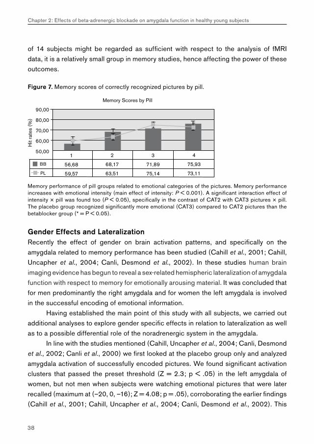
Figure 7. Memory scores of correctly recognized pictures by pill.
Memory performance of pill groups related to emotional categories of the pictures. Memory performance increases with emotional intensity (main effect of intensity: P < 0.001). A significant interaction effect of intensity × pill was found too (P < 0.05), specifically in the contrast of CAT2 with CAT3 pictures × pill. The placebo group recognized significantly more emotional (CAT3) compared to CAT2 pictures than the betablocker group (* = P < 0.05).
Gender Effects and LateralizationRecently the effect of gender on brain activation patterns, and specifically on the amygdala related to memory performance has been studied (Cahill et al., 2001; Cahill, Uncapher et al., 2004; Canli, Desmond et al., 2002). In these studies human brain imaging evidence has begun to reveal a sex-related hemispheric lateralization of amygdala function with respect to memory for emotionally arousing material. It was concluded that for men predominantly the right amygdala and for women the left amygdala is involved in the successful encoding of emotional information.
Having established the main point of this study with all subjects, we carried out additional analyses to explore gender specific effects in relation to lateralization as well as to a possible differential role of the noradrenergic system in the amygdala.
In line with the studies mentioned (Cahill, Uncapher et al., 2004; Canli, Desmond et al., 2002; Canli et al., 2000) we first looked at the placebo group only and analyzed amygdala activation of successfully encoded pictures. We found significant activation clusters that passed the preset threshold (Z = 2.3; p < .05) in the left amygdala of women, but not men when subjects were watching emotional pictures that were later recalled (maximum at (–20, 0, –16); Z = 4.08; p = .05), corroborating the earlier findings (Cahill et al., 2001; Cahill, Uncapher et al., 2004; Canli, Desmond et al., 2002). This
39
2.1 Noradrenaline mediates amygdala activation in men and women during encoding of emotional material
was specifically so for CAT3 pictures and not, as in the above mentioned studies, in the most extreme negative emotional category (CAT4). Pictures of CAT4 that were later correctly remembered evoked significant activation in the left parahippocampal gyrus in women, but not men. In contrast to previous studies, we were not able to show right amygdala activation passing the threshold in men, when watching later successfully encoded emotional pictures. Merging all data (placebo and betablocker groups together) showed a similar image: left amygdala activation (–20, 0, –16) in women correlated with later recalled CAT3 pictures (Z = 3.46; p = .05); no significant activation in men could be shown. No effects for CAT4 pictures with respect to gender and memory could be found in this study, although we cannot exclude that the smaller number of items assigned to CAT4 as compared to CAT3, resulting in decreased statistical power for estimating CAT4 activation, may also have caused this.
In addition, we tested whether women and men differ in amygdala activation when watching emotional pictures under influence of a betablocker. We therefore analyzed 14 men and 14 women separately with respect to the drug effect on amygdala activation for the specific contrast of CAT3 > CAT1 pictures, that we earlier found to reveal the drug effect for the whole group. Mean activation was calculated in the defined ROI’s for men and women separately and plotted (figure 8 A + B). For both men and women betablockade significantly decreased amygdala activation for CAT3–CAT1 pictures. Activation clusters in left (Z = 3.73, p < .01) and right amygdala (Z = 3.96, p < .05) present in the placebo group for women did not pass the threshold under betablocker condition. Also in men activation under placebo condition evoked clusters in left (Z = 4.53, p <.05) and right (Z = 2.89, p <.05) amygdala, but no clusters passed the threshold under betablocker condition. This means that betablockade is effective in decreasing amygdala activation evoked by CAT3 pictures compared with placebo in both men and women separately.
We also analyzed the behavioral memory data for men and women separately using SPSS and a GLM, with drug and emotional category as within factors. For both men and women memory performance increased with the emotional intensity (main effect of category: for both men and women: p < .001). But only in women there was a significant interaction between emotional intensity and drug (p < .05), specifically emerging between CAT2 and CAT3 pictures (figure 8 C+D). A remarkable similarity between these memory data and amygdala activation in reaction to the betablocker is visible in women (figure 8 A + C). For men separately virtually no effect of betablockade on memory was shown in this study. In women, the decrease in amygdala activation at encoding under betablocker condition when watching CAT3 pictures was accompanied by a decrease in later memory performance for this category of pictures.
40
Chapter 2: Effects of beta-adrenergic blockade on amygdala function in healthy young subjects
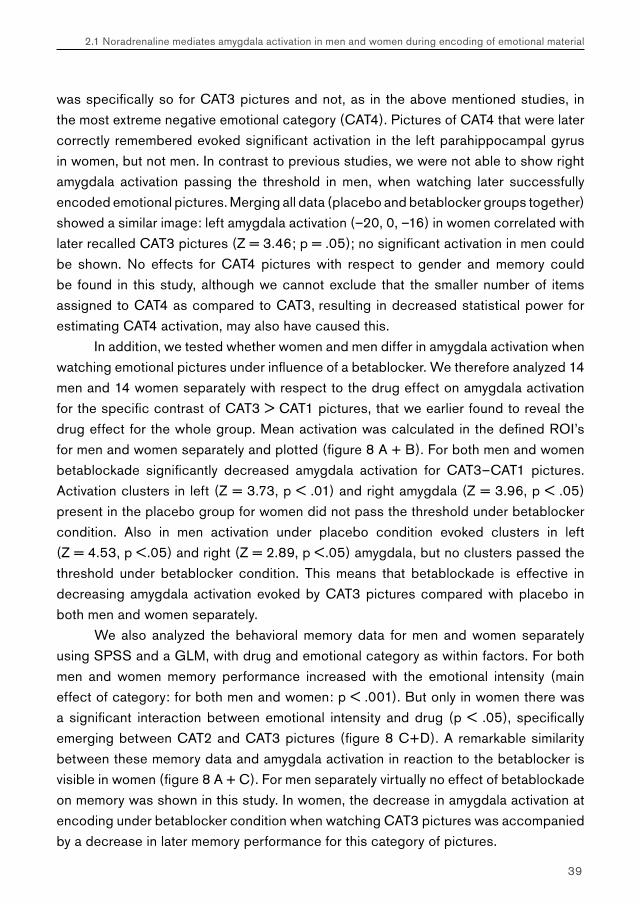
Figure 8.
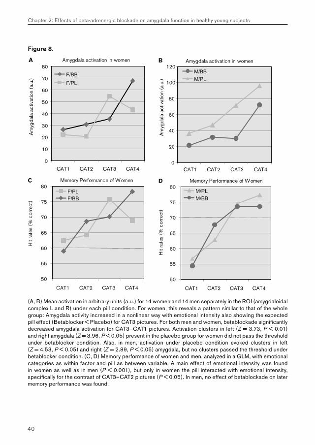
(A, B) Mean activation in arbitrary units (a.u.) for 14 women and 14 men separately in the ROI (amygdaloidal complex L and R) under each pill condition. For women, this reveals a pattern similar to that of the whole group: Amygdala activity increased in a nonlinear way with emotional intensity also showing the expected pill effect (Betablocker < Placebo) for CAT3 pictures. For both men and women, betablockade significantly decreased amygdala activation for CAT3–CAT1 pictures. Activation clusters in left (Z = 3.73, P < 0.01) and right amygdala (Z = 3.96, P < 0.05) present in the placebo group for women did not pass the threshold under betablocker condition. Also, in men, activation under placebo condition evoked clusters in left (Z = 4.53, P < 0.05) and right (Z = 2.89, P < 0.05) amygdala, but no clusters passed the threshold under betablocker condition. (C, D) Memory performance of women and men, analyzed in a GLM, with emotional categories as within factor and pill as between variable. A main effect of emotional intensity was found in women as well as in men (P < 0.001), but only in women the pill interacted with emotional intensity, specifically for the contrast of CAT3–CAT2 pictures (P < 0.05). In men, no effect of betablockade on later memory performance was found.
41
2.1 Noradrenaline mediates amygdala activation in men and women during encoding of emotional material
Discussion
The results of this study support our hypothesis about the role of noradrenaline in amygdala activation during the processing of emotional information. Neutral and very light emotional pictures do not activate the amygdala significantly compared with baseline levels, but negative emotional pictures lead to a significant increase in amygdala activation under placebo condition. When the (central and peripheral) noradrenergic receptors were ‘blocked’ with this dosage of 80 mg propranolol, amygdala activation monitored with fMRI decreased when subjects were confronted with the emotional (CAT3) stimuli. Memory performance deteriorated accordingly for the group, suggesting that the amygdala plays a role in the effective encoding, but possibly in the consolidation process of emotional stimuli as well. In two human studies was shown that the level of noradrenaline during memory consolidation was related to enhanced long-term memory, and that post-learning stress hormone-related activity interacts with arousal at initial encoding to modulate memory consolidation (Cahill et al., 2003; Southwick et al., 2002). When we explored the relationship between emotion intensity and activation it appears that amygdala activation under placebo condition is best described by a curvilinear pattern, although a fit to a linear contrast was found as well. This was specifically so for the placebo group and not the betablocker group, showing that the betablocker evoked a differential activation pattern in the amygdala. In animal literature this curvilinear pattern of reactivity in brain nuclei has been shown several times. Most of the times this was seen in dose-response studies, where apart from a time-dependent, also a dose dependent U-curve or inverted U-curve was found (Ferry et al., 1999a; Ferry, Roozendaal, & McGaugh, 1999b; Roozendaal, Quirarte, & McGaugh, 2002). A hypothesis that is concordant with our findings would be that noradrenergic activity in the amygdala has a dose dependent character, visible as a curvilinear activation pattern. Propranolol, which is a competitive betablocker, is disrupting this pattern by occupying (part of the) beta-adrenergic receptors in the amygdala. An important other factor that cannot be ruled out is that other neurotransmitters or hormones interact with the noradrenergic system in the amygdala. A logical candidate for this scenario is cortisol that has an apparent relation with stressful stimuli and situations. Several animal studies showed that glucocorticoids interact with and influence the efficacy of noradrenergic stimulation in the basolateral amygdala (Ferry et al., 1999a; McIntyre et al., 2003; Roozendaal, 2002; Roozendaal et al., 2001; Roozendaal & McGaugh, 1996, 1997a). The scanning procedure was long enough to have this part of the hypothalamic-pituitary-adrenal (HPA) axis come into action. However, it should be noted explicitly that
42
Chapter 2: Effects of beta-adrenergic blockade on amygdala function in healthy young subjects
the effect on amygdala activation during the CAT4 pictures is not a temporal effect: as already mentioned all emotional categories were randomly presented, so the effect should at least partly be related to the properties of this emotional category.
A different type of explanation can be inferred from more fundamental and technical studies on principles of event-related fMRI. Friston et al. (Friston et al., 2000) pointed at the importance of nonlinearities in evoked responses in fMRI, particularly with the advent of event-related fMRI. These nonlinearities are commonly expressed as interactions among stimuli that can lead to the suppression and increased latency of responses to a stimulus that are incurred by a preceding stimulus. They used the Balloon/Windkessel model and showed that this is sufficient to account for nonlinearities observed in evoked fMRI responses. They also showed that the parameters responsible for this phenomenon were all biologically plausible. However, the analyzing post-processing software used in our study (FSL) anticipates on these phenomena by adjusting the form of the hemodynamic response – in our study to a double-gamma hemodynamic response function (HRF). Elaborating on the above mentioned study Mechelli et al. (Mechelli, Price, & Friston, 2001) tried to investigate the dependence of BOLD responses on different patterns of stimulus input or neuronal changes, such as stimulus presentation rate. The estimated BOLD response dramatically decreases with increasing rates of presentation. The response to a stimulus is modulated by preceding stimuli to give a non-linear refractoriness that depends on the interstimulus interval or rate. In both studies of Canli (Canli, Desmond et al., 2002; Canli et al., 2000) and of Cahill (Cahill, Uncapher et al., 2004) stimuli were presented for a period of 2.88 s with an intertrial interval of 12.96 s, during which a fixation cross was presented. This could lead to a ‘back to baseline’ activation pattern after each stimulus.
However, this parameter (presentation rate) is playing a role in every emotional category of the pictures and – because they were presented randomly with respect to the category – cannot solely account for the saturation effect of CAT4 related to CAT3 pictures. A specific other parameter, namely the amplitude of a stimulus, could be the factor to be responsible for this effect. Although Mechelli et al. (Mechelli et al., 2001) used a model and did not use emotional material, they did show that the increase in BOLD response with stimulus amplitude shows a saturation effect, which can be explained in terms of hemodynamic refractoriness. Perhaps the combination of high presentation rate (compared to earlier studies) and the amplitude/intensity of the CAT4 pictures lead to this saturation effect in the highest emotional category.
Recently the necessity to anticipate and account for the effect of sex in emotionally influenced memory research is put forward in several papers (Cahill, 2003b; Cahill et
43
2.1 Noradrenaline mediates amygdala activation in men and women during encoding of emotional material
al., 2003; Cahill et al., 2001; Cahill, Uncapher et al., 2004; Canli, Desmond et al., 2002). This study underlines this requirement: although power decreased by splitting the group, it could be shown that women and men react differently when confronted with these emotional stimuli. As in earlier studies, the judgment of the emotional intensity of the stimuli differed in a way that women rate more pictures as highly emotional intense than men. So their personal judgment was used to categorize the pictures. Amygdala activation in both sexes increased with emotional intensity in the placebo group with a flattening in activation between CAT3 and CAT4 pictures (contrast was n.s. in either direction). Betablockade was efficient to reduce the increased amygdala activation in both men and women for the emotional CAT3 pictures, but memory performance two weeks later was only affected in women – analogous to their amygdala activation pattern. In men the amygdala activation pattern under placebo condition paralleled their later memory performance. But the decrease of amygdala activation during CAT3 pictures under betablocker condition did not go together with the expected decrease in memory performance of the emotional CAT3 pictures. Only a few studies to date have directly compared the activation pattern of men and women with respect to emotional information processing. This is – to our knowledge – the first study that directly shows the effect of betablockade on amygdala activation in both sexes and of a differentiation in the effect of betablockade on long term memory performance. It seems that the direct reactivity of the amygdala on emotional material is comparable for men and women. Because one of the main roles of the amygdala is commonly viewed as ‘labeling for danger and harm’ it is understandable that no gender difference is found in this signaling phase, since this information is of equal (survival) importance for both sexes. There is a differentiation however in the effect of betablockade on long-term memory performance. This effect can be explained by a different sensitivity for the effect of the central and peripheral acting betablocker propranolol. Several factors such as gender and specifically the phase of menstrual cycle and use of oral contraceptives in women affect the activity of the HPA-axis (Hampson, 1990, 1990). Propranolol blood concentrations are raised by the use of contraceptive pills, so help to optimize the pharmacological action of betablockers (Kendall et al., 1984). And high levels of estrogen enhance noradrenaline secretion and noradrenergic receptor sensitization (Herbison et al., 2000).
In addition a point of attention is proposed for future studies: in animal research the dosage used to test for certain effects is almost always related to the body weight of the subject animal. In human studies, on the contrary, this is hardly ever done. In our study it might simply be so, that the relative dosage of 80 mg propranolol for men was lower than for women, since their body weight was higher than that of the women (mean ± s.e.m. = 75.3 ± 2.0 for men and 65.5 ± 2.1 for women; t-test: p <. 005).
44
Chapter 2: Effects of beta-adrenergic blockade on amygdala function in healthy young subjects
Finally, women also appear to have a slower beta-blocker catabolism (Maheu & Lupien, 2003) (Lupien_S, personal communication) that could be responsible for the memory data. If betablockade was active for a longer period in women than in men, than the consolidation phase of the encoded stimuli might be affected for a longer time in women than in men. The effect of noradrenaline appears to have its greatest impact at the time of encoding, but some studies found effects during the stage of consolidation too (Cahill et al., 2003; Southwick et al., 2002). If propranolol was effective in women for a longer period than in men, due to the difference in catabolism, then the consolidation of the emotional stimuli could be attenuated in women more than in men. This could be in line with our finding that men don’t show the decrement in memory performance for the higher emotional categories under betablocker condition, that could be due to a faster wash out time during the stage of consolidation.
In fact this finding has similarities with the finding of Maheu et al. (Maheu et al., 2004) who were not able to show memory impairing effects of a 40 mg dose of propranolol in men. They used a narrated slide show, based on earlier work of Cahill et al. (Cahill et al., 1994) and Van Stegeren et al.(van_Stegeren et al., 1998), that covers a story with a mild emotional phase in between two neutral phases of the story line. They explained their lack of effect on memory by the composition of their subject group, which consisted of men only. They expanded the experiment with a higher dosage (80 mg) and did find an effect in men on (short and) long-term memory performance for the story. Bearing in mind that the stimulus set used in this study is much more stress evoking than the slide show and that the condition of being in a scanner for an hour is pretty stressful too, it could contribute to this lack of effect on memory in men, even with this higher dose of 80 mg propranolol.
Furthermore, in an interesting study Strange et al. (Strange et al., 2003) studied the effect of propranolol (40 mg) on (short-term) memory for emotional words that were presented embedded in a list of neutral words and report two remarkable and interconnected findings. They showed that enhanced memory for the emotional (E) words was connected with a significant decrement in memory performance for the words that preceded the emotional oddball words (E–1 words) and that these effects were more pronounced in women. Propranolol reversed both effects in a way that E words were equally remembered to neutral words and memory for the E–1 nouns was now better than the control words. Memory was significantly more impaired in women than in men, a phenomenon that the authors explain could reflect a higher sensitivity in females to the disruptive effect of noradrenaline on consolidation. They refer to animal models that demonstrate enhancing as well as impairing effects of noradrenergic
45
2.1 Noradrenaline mediates amygdala activation in men and women during encoding of emotional material
stimulation on memory reflecting an inverted –U function. Their data demonstrate that the same level of (nor)adrenergic activation can produce memory enhancement and impairment depending on the timing of that activation and that this effect was stronger in women. Although their findings were related to short-term memory effects, it would be well in line with the effects on long-term memory presented here.
An earlier study supported the hypothesis that emotional arousal enhanced long-term memory for central information in men via activation of the right amygdala/hemisphere function. In women arousal would enhance long-term memory for peripheral details via activation of the left amygdala/hemisphere function (Cahill & van Stegeren, 2003). Although is was impossible to check for central and peripheral details in the memory test of this stimulus material, part of these findings are in line with the idea that encoding of emotional information is accompanied by left amygdala activation in women. Whether this is due to the processing of central or peripheral details of the pictures cannot be answered in this study.
What has been a remarkable and consistent finding in all studies on emotional memory that used betablockers (Cahill et al., 1994; Maheu et al., 2004; O’Carroll et al., 1999; van Stegeren, Everaerd, & Gooren, 2002; van_Stegeren et al., 1998), is that the emotional rating of the stimulus material by the subjects is not affected by betablockade. Apparently the cognitive appraisal of the emotionality or intensity is not fully correlated with the emotional and/or physical reaction to that same stimulus. This strongly suggests that different neural mechanisms are responsible for the ‘cognitive’ judgment of a stimulus or situation versus the physical arousal reaction or the emotional labeling that takes place. Kensinger and Corkin (Kensinger & Corkin, 2004) used functional MRI and behavioral studies to show that distinct cognitive and neural processes contribute to emotional memory enhancement for arousing information versus valenced, non-arousing information. They conclude that the effect of arousing information was strongly connected to an amygdalar-hippocampal network, whereas the valenced, non arousing information was supported by a prefrontal cortex-hippocampal network implicated in controlled encoding processes.
Although the choice for the use of subjects’ personal rating to analyze fMRI data with respect to emotional categories was a deliberate one, it remains unclear whether subjects (cognitive) rating of the emotional intensity of the picture is reliable with respect to gender. In a recent study Hamann and colleagues showed that men and women differ in brain activation although personal ratings of the experienced arousal was comparable. They compared fMRI responses of men and women to sexually arousing and neutral pictures. Men showed greater activation in the hypothalamus and amygdala, even when
46
Chapter 2: Effects of beta-adrenergic blockade on amygdala function in healthy young subjects
females (in retrospective) reported greater subjective arousal (Hamann et al., 2004). These findings suggested that the amygdala mediates sex differences in response to positive and appetitive emotional stimuli. It is conceivable that personalized subjective ratings represent another dimension and also in the case of negative emotional stimuli represent a system that does not completely parallel the activation pattern in the ‘emotional’ amygdala. It should be concluded that the cognitive judgment of emotionality of a stimulus is not mediated by a noradrenergic depending system.
In a recent article Hamann and Canli (Hamann & Canli, 2004) give an overview of functional brain imaging studies to show how individual differences among subjects might modulate neural responses during emotion processing. They make a plea for including individual differences to provide a new window into the field of neurobiology of emotion and memory, that can complement traditional approaches.
This study was meant to establish a general principle in emotional information processing, namely the role of noradrenaline in the amygdala and to see if men and women differ in this process. It can be concluded that also in humans noradrenaline is needed to allow amygdala activation in reaction to emotional stimuli. Moderation of amygdala activation with betablockers as done in this study may have an impact on long-term effects such as consolidation of emotional experiences. Using more personalized criteria to define stimuli and reactivity of the subjects can lead to interesting new findings and further complement missing parts of information. In this study again a strong argument was made to take participants gender into account in future studies.
Acknowledgements
This study was supported by a grant from the Foundation for Behavioral and Educational Sciences of the Netherlands Organisation of Scientific Research (NWO) and the CSC Amsterdam. The authors thank Martine Hoogman and Martine Baaij for their assistance in this study.
Chapter 3
Effects of raloxifene treatment on brain function in healthy elderly males
3.1 Effects of raloxifene treatment on brain function during encoding
Raloxifene exposure enhances brain activation during memory performance in healthy elderly males; its possible relevance to behavior
Goekoop R,(*) Duschek EJJ,(*), Knol DL, Barkhof F, Netelenbos C, Scheltens P, Rombouts SARB. (*) Both authors contributed equally to the study.
Neuroimage 2005; 25(1):63-75. PMID: 15734344
48
Chapter 3: Effects of raloxifene treatment on brain function in healthy elderly males
Abstract
Raloxifene is a selective estrogen receptor modulator (SERM) that is prescribed in females only, but its use in male subjects is increasingly considered. With a growing number of patients having potential benefit from raloxifene, the need for an assessment of its effects on brain function is growing. Effects of estrogens on brain function are very subtle and difficult to detect by means of neuropsychological assessment. Functional imaging techniques, however, have been relatively successful in detecting such changes. This study used functional magnetic resonance imaging (fMRI) to examine effects of raloxifene treatment on memory function. Healthy elderly males (n = 28; mean age 63.6 years, SD 2.4) were scanned during performance on a face encoding paradigm. Scans were made at baseline and after 3 months of treatment with either raloxifene (n = 14) or placebo (n = 14). Treatment effects were analyzed using mixed-effects statistical analysis (FSL). Activation during task performance involved bilateral parietal and prefrontal areas, anterior cingulate gyrus, and inferior prefrontal, occipital, and mediotemporal areas bilaterally. When compared to placebo, raloxifene treatment significantly enhanced activation in these structures (Z > 3.1), except for mediotemporal areas. Task performance accuracy diminished in the placebo group (P = 0.02), but remained constant in the raloxifene group ( P = 0.60). In conclusion, raloxifene treatment enhanced brain activation in areas spanning a number of different cognitive domains, suggesting an effect on cortical arousal. Such effects may translate into small effects on behavior, including effects on attention and working memory performance, executive functions, verbal skills, and episodic memory. Further neuropsychological assessment is necessary to test the validity of these predictions.
Keywords: Raloxifene; SERM; estrogen; fMRI; memory; arousal.
49
3.1 Effects of raloxifene treatment on brain function during encoding
Introduction
Selective estrogen receptor modulators (SERMs) are compounds that display both agonist and antagonist effects on estrogen receptors in a tissue-selective manner (Riggs & Hartmann, 2003). Raloxifene is a SERM of which several beneficial effects have been reported in postmenopausal women, including a reduced risk of breast carcinoma (Cummings et al., 1999), a reduced risk of vertebral fractures (Ettinger et al., 1999; Delmas et al., 2002), and beneficial effects on markers of cardiovascular disease, such as serum lipids and coagulation factors (Delmas et al., 1997; Walsh et al., 1998; de Valk-de Roo GW et al., 1999). To date, raloxifene can be prescribed in postmenopausal women only, to counter effects of osteoporosis. Meanwhile, a growing number of beneficial effects of raloxifene treatment are reported that may also apply to male patients. Raloxifene treatment is therefore considered in males, which have already shown good tolerance to the substance (Blum et al., 2000; Doran et al., 2001). So far, however, research into the effects of raloxifene treatment in humans has lacked a more detailed assessment of its effects on brain function and mental performance. With an increasing number of patients experiencing a potential benefit from raloxifene, the need for such an assessment is growing.
A large body of evidence suggests that sex steroids may influence brain function. In both sexes, estradiol is thought to be primarily responsible for effects of sex steroids on neural excitability, with testosterone in male subjects first requiring conversion to estradiol in order to exert its effects (Longcope et al., 1969; MacDonald et al., 1979). Estrogen receptors occur throughout the brain in both male and female subjects. Gender differences may influence their pattern of expression, suggesting different functions in both sexes (Osterlund et al., 2000; Zhang et al., 2002; Kruijver et al., 2003). A consistent finding is that low sex steroid levels are associated with impaired memory performance in both sexes (Morley et al., 1997; Wolf et al., 1999; Barrett-Connor et al., 1999; Maki & Resnick, 2001; Senanarong et al., 2002). In contrast, enhanced estrogen levels in both males and females correlate with increased memory function (Sherwin, 2003a; Sherwin, 2003b). It has even been suggested that estrogen supplementation may prevent Alzheimer’s disease in postmenopausal women (Simpkins et al., 1997; Paganini-Hill & Henderson, 1996). However, randomized controlled clinical trials found no significant effects of estrogen treatment in dementia (Polo-Kantola et al., 1998). Instead, three recently published controlled clinical trials from the Women’s Health Initiative (WHI) examining effects of hormone therapy in postmenopausal women have found adverse effects of combined estrogen and progestin treatment on brain function,
50
Chapter 3: Effects of raloxifene treatment on brain function in healthy elderly males
including effects on mental performance and an increased risk of dementia (Rapp et al., 2003; Shumaker et al., 2003; Wassertheil-Smolleret al., 2003). Thus, estrogens affect mental performance under physiological circumstances, and the effects of long-term pharmacological intervention into sex steroid systems are still unclear. Indeed, very little is known about the effects of estrogen agonists on male brain function (Sherwin, 2003b). Future prescription of raloxifene in male subjects therefore requires additional study.
So far, research into the effects of sex steroid or SERM treatment on brain function has largely depended on neuropsychological studies of behavior. Such studies typically show only minimal changes in behavioral measures (Sherwin, 2003b; Sherwin, 2003a). In contrast, functional imaging techniques have shown relatively clear results after sex steroid treatment. Both estrogen and testosterone may increase brain activation in areas relevant to memory, reasoning, judgment and emotions (Azad et al., 2003; Maki & Resnick, 2001). Although the clinical significance of neurofunctional changes in the absence of behavioral changes may be questioned, the possibility that they bear some extra insight into behavior should not be dismissed (Wilkinson & Halligan, 2004). Hence, it has been suggested that functional imaging techniques may have greater sensitivity to the effects of sex steroids on brain function (and possibly behavior) than most neuropsychological assessment scales (Maki & Resnick, 2001; Neele et al., 2001). For this reason, we examined the ability of functional magnetic resonance imaging (fMRI) to screen for effects of raloxifene treatment on male brain function in a small group of subjects. Based on results from previous imaging studies (Maki & Resnick, 2001; Sherwin, 2003b) and the sparse amount of data on the effects of estrogens on male brain function (Sherwin, 2003b), we hypothesized that raloxifene would influence brain activation during visuospatial memory performance. A face encoding task was chosen to elicit brain activation, since sex differences have been reported for visuospatial memory scores in general (Sherwin, 2003b; Kampen & Sherwin, 1996) and face encoding and recognition scores in particular (Yonker et al., 2003). Such ‘sexual dimorphism’ with respect to visuospatial memory suggests an effect of sex steroids on neural function associated with this task, making it a potentially sensitive tool to detect effects of treatment.
51
3.1 Effects of raloxifene treatment on brain function during encoding
Materials and methods
Study design Subjects were screened for participation in a randomized, double-blind, placebo-controlled study design. fMRI was performed at baseline (BL; no medication; session 1) and after 3 months of a once-daily oral intake of raloxifene 120 mg or placebo (TR; session 2). Both BL and TR sessions were performed on exactly the same time of day in each subject. If data acquisition failed, the subject’s consent was asked for an additional scanning visit, up until which time the relevant medication regime (raloxifene or placebo) was continued, to obtain a maximum number of complete data sets. Study period extension was not to exceed 10 days.
Subject recruitment The medical ethical review board of the VU University Medical Center of Amsterdam approved the study. Thirty healthy, right-handed elderly male subjects, aged 60 to 70 years old (mean 63.6 years, SD 2.4; range 60–69 years) were recruited by advertisement in local newspapers. All subjects provided informed consent during a screening visit in which the procedure was explained and contraindications were checked. Subjects were excluded if they had any significant medical, neurological or psychiatric illness, or if they were taking any medication or other substances that are known to influence cerebral functioning. Exclusion criteria to MRI involved the presence of metallic implants in high-risk areas and a history of claustrophobia. Formal education was determined using a Dutch system (low, middle, high). Functional MRI (fMRI)
Data acquisitionImaging was carried out on a 1.5-T Sonata MR scanner (Siemens, Erlangen, Germany), using a standard circularly polarized head coil with foam padding to restrict head motion. For fMRI, an echo planar imaging sequence was used (echo time 60 ms, flip angle 908, matrix 64 x 64, field of view 192 x 192 mm), to obtain 21 transverse slices (thickness 5 mm, interslice-gap 1 mm). Task stimuli were projected on a screen located at the head end of the scanner table via an LCD projector located outside the scanner room. Subjects viewed the screen through a mirror located on the head coil. In each hand, subjects held an fMRI compatible response-box through which they were able to react to task stimuli by pressing a single button using one of their index-fingers. A T1-weighted structural MRI scan was obtained of each subject (MPRAGE; inversion time: 300 ms, TR = 15 ms; TE = 7 ms; flip angle = 88; 160 coronal slices, 1 x 1 x 1.5
52
Chapter 3: Effects of raloxifene treatment on brain function in healthy elderly males
mm voxels). Total scanning time including structural imaging on average was 21 min for each visit.
Encoding taskA face-encoding task was used to examine brain activation related to episodic memory for visuospatial information. This task produces activation in bilateral prefrontal, bilateral parietal, anterior cingulate and (posterior) mediotemporal structures with a preference for the right hemisphere (Small et al., 1999), which is in correspondence with the visuospatial nature of this task (Kelley et al., 1998). Two different but comparable versions were constructed and randomized across the scanning sessions (BL, TR). A spare version was made in case data acquisition failed during one of the scanning sessions. The paradigm was practiced on a laptop computer to familiarize subjects with the procedure (1 day before the start of the first session and 5 min before the onset of the first measurements of each session, with subject in scanner). During the first 10.5s, subjects saw a circle indicating time left before the onset of the first condition. To avoid investigator’s bias, task performance data were only viewed after all scan visits had been completed.
A block design was applied with two alternating conditions: Condition 1 (ENCOD) consisted of 4 blocks of 42s, each containing six unfamiliar faces presented sequentially on a black background (presentation time 6s, followed by a 1-s delay). Condition 2 (FIX) consisted of four blocks of 44s each, in which a white fixation cross (X) was presented on a black background. Prior to scanning, subjects were instructed verbally to remember the faces for future testing and to classify each face according to its gender by pressing one of two buttons (instructions ‘Left: male’, ‘Right: female’ also appearing alongside the pictures). Male and female faces were balanced across the ENCOD blocks. The entire task thus consisted of eight alternating blocks of ENCOD and FIX conditions and was preceded by a 21s FIX condition. Mean reaction times for gender discrimination were recorded and a performance accuracy score varying from –1 to 1 (with 0 indicating chance levels) were calculated by subtracting false answers from correct answers and dividing the result by the total number of items (24). Total task duration was 6 min and 12s.
Immediately after encoding, encoding success was assessed using a recognition task, while subjects were still in the scanner. Twenty-four faces were presented sequentially in random order, of which 12 had been shown during encoding and 12 were new (presentation time 5s for each face, followed by a white fixation-cross presented for 3s on a black background). Subjects indicated whether the presented faces were
53
3.1 Effects of raloxifene treatment on brain function during encoding
familiar or unfamiliar by pressing one of two buttons (instructions ‘Left: familiar’ and ‘Right: unfamiliar’ also appearing alongside the pictures). Similar performance measures were calculated as for gender discrimination.
Statistical analysis of task performance data After first asserting that raloxifene and placebo groups did not differ in their mean performance measures at baseline (using a multivariate analysis of variance), task performance measures during BL (no intake) were subtracted from those after 3 months of treatment (TR). A multivariate analysis of variance was then performed using SPSS, in which differential scores were entered as dependent variables in an analysis of the medication effect with medication ‘regime’ (two levels: raloxifene, placebo) as a fixed factor and ‘test version’ (two levels: 1 and 2) as a covariate. If a significant effect of this covariate was found, the model was adjusted to contain it as a fixed factor in a subsequent analysis of the medication effect. Effects of the factor ‘regime’ were then considered representative of an effect of intervention with either raloxifene or placebo.
Analysis of functional neuroimaging data Functional datasets were analyzed using FSL (fMRIB’s Software Library, http://www.fmrib.ox.ac.uk/fsl). The first five volumes of each data set were discarded to account for T1-saturation effects. At first level (individuals), the following pre-processing was applied: non-brain removal, slice-timing correction using Fourier-space time series phase-shifting, motion correction and spatial smoothing using a Gaussian kernel of FWHM 8 mm, mean-based intensity normalization of all volumes by the same factor and high and low pass temporal filtering (Jenkinson et al., 2002; Smith, 2002). Registration of functional images to high resolution and/or standard images was carried out using an intermodal registration tool based on the correlation ratio (Jenkinson & Smith, 2001). After pre-processing, the following statistics were applied on a voxelwise basis on each time series, using local autocorrelation correction (Woolrich et al., 2001): signal change during the ENCOD condition was modeled as a box car, which had the same alternation frequency as the ENCOD condition and convolved with a gamma function, to model the hemodynamic response. The FIX condition was not modeled (except for the first FIX-period of 21s), to prevent overspecification of the model. Model fitting generated whole brain images of parameter estimates and corresponding variance, representing average signal change during ENCOD as compared to FIX levels (both for BL and TR regimes). These images were resampled to 2 x 2 x 2 mm in standard space and fed into in a second-level statistical analysis to examine treatment effects at group level.
54
Chapter 3: Effects of raloxifene treatment on brain function in healthy elderly males
At second (group) level, activation images of main effects during encoding were produced by summing all individual activation maps for each session and medication regime separately [i.e., BL (placebo), BL (raloxifene), TR (placebo) and TR (raloxifene)] and across all sessions and regimes [i.e., BL (placebo) + BL (raloxifene) + TR (placebo) + TR (raloxifene)] in a mixed-effects higher-level analysis using clusters determined by Z >N 2.3 and a corrected cluster significance threshold of P = 0.05 (Woolrich et al., 2004; Friston et al., 1994; Worsley et al., 1992; Beckmann et al., 2003). Effects of medication were then calculated using a repeated measures model, in which two explanatory variables (EVs) contrasted BL and TR sessions of placebo and raloxifene groups, while assuming separate variances for these groups. Two additional EVs coded for effects of test version on signal response, thus removing effects of this possible confounder. The main contrasts of interest were [TR (raloxifene) – BL (raloxifene)] <> [TR (placebo) – BL (placebo)], testing for an interaction between raloxifene and placebo activation levels before and after treatment. This interaction analysis was restricted to activated regions only (by masking for main effects summed across all treatment conditions, see above). A Z value of 3.1 ( P < 0.001) was used as a threshold for significant brain activation, with a minimal cluster size of 80 mm3 to remove single-voxel noise (Woolrich et al., 2004; Forman et al., 1995; Friston et al., 1994; Worsley et al., 1992). The model was then adjusted to contain separate regressors for recognition accuracy for both treatment groups. This allowed treatment effects to be analyzed for correlations with performance changes both for raloxifene and placebo groups separately and their interactions. Images were rendered on a 3D mean anatomical brain volume of all subjects in standard space (Talairach & Tournoux, 1988) for display purposes. Coordinates of local maxima were extracted and fed into the Talairach Daemon Client version 1.1 (Research Imaging Center; University of Texas, Health Science Center, San Antonio, TX) for identification of the activated brain structures.
Results
Demographics Mean age of subjects was 64.1 (SD 2.4) year in the placebo group and 63.1 (SD 2.5) year in the raloxifene group. The placebo group contained two smokers versus one in the raloxifene group. Education level was equal in both groups.
55
3.1 Effects of raloxifene treatment on brain function during encoding
Subject compliance and discontinuation One subject (placebo group) was claustrophobic and data quality was poor in another subject (raloxifene group). These data were removed from the analysis, yielding 28 complete data sets containing both BL and TR data (14 in raloxifene group, 14 in placebo group). In one subject, the second (TR) scan was repeated after 7 days because of scanner errors during the second session. Subject compliance was good as assessed by the tablet counts and there were no dropouts. No significant side effects were reported.
Task performance Mean overall task performance accuracy for male-female discrimination (encoding accuracy) was 0.99 (SD 0.04) and mean overall reaction time 1.03s (SD 0.29s) (Table 3). Recognition accuracy was 0.67 (SD 0.20) with mean overall reaction time 1.64s (SD 0.33s). A significant interaction was found between mean recognition accuracy scores of raloxifene and placebo groups at baseline and after treatment (P = 0.021): recognition accuracy in the placebo group decreased slightly after treatment (P = 0.022), whereas performance accuracy in the raloxifene group remained constant (P = 0.60). No other measures of task performance showed significant changes between raloxifene and placebo groups after treatment.
Analysis of functional neuroimages Main effects of face encoding involved extensive activation in ventral and dorsal occipital (visual) areas, and bilateral parietal, bilateral prefrontal and anterior cingulate gyri areas, with a preference for the right hemisphere. Extensive bilateral activation of thalamic structures also occurred. Additional activation was found in bilateral posterior hippocampal areas (Figure 1; Table 1). Pairwise comparisons showed no significant differences in activation levels at baseline between raloxifene and placebo groups (data not shown). The main contrast (examining the interaction between activation levels of raloxifene and placebo groups before and after treatment) showed increased activation after raloxifene intake when compared to placebo (Figure 2; Table 2). Areas included bilateral middle frontal gyri, bilateral parietal lobules and anterior cingulate/superior prefrontal, left inferior frontal gyri and the lingual gyri.
Raloxifene treatment produced increases, but not decreases, in activation levels when compared to placebo (Z = 3.1). Plots of percentual signal changes (relative to global mean values) of peak voxels of local maxima illustrate the size of signal changes for the main contrast of interest (Figure 2). When both treatment groups were examined
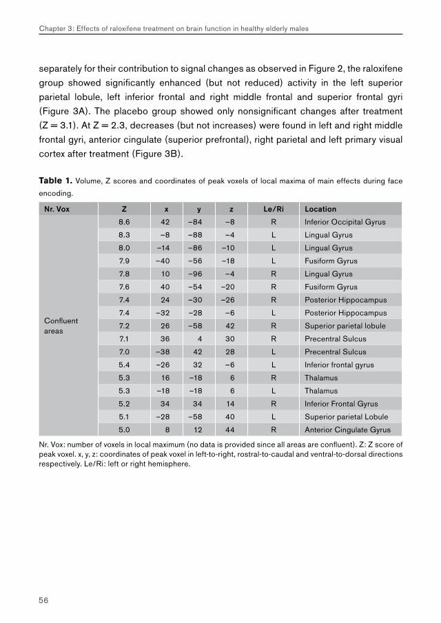
56
Chapter 3: Effects of raloxifene treatment on brain function in healthy elderly males
separately for their contribution to signal changes as observed in Figure 2, the raloxifene group showed significantly enhanced (but not reduced) activity in the left superior parietal lobule, left inferior frontal and right middle frontal and superior frontal gyri (Figure 3A). The placebo group showed only nonsignificant changes after treatment (Z = 3.1). At Z = 2.3, decreases (but not increases) were found in left and right middle frontal gyri, anterior cingulate (superior prefrontal), right parietal and left primary visual cortex after treatment (Figure 3B).
Table 1. Volume, Z scores and coordinates of peak voxels of local maxima of main effects during face
encoding.
Nr. Vox Z x y z Le/Ri Location
Confluentareas
8.6 42 –84 –8 R Inferior Occipital Gyrus
8.3 –8 –88 –4 L Lingual Gyrus
8.0 –14 –86 –10 L Lingual Gyrus
7.9 –40 –56 –18 L Fusiform Gyrus
7.8 10 –96 –4 R Lingual Gyrus
7.6 40 –54 –20 R Fusiform Gyrus
7.4 24 –30 –26 R Posterior Hippocampus
7.4 –32 –28 –6 L Posterior Hippocampus
7.2 26 –58 42 R Superior parietal lobule
7.1 36 4 30 R Precentral Sulcus
7.0 –38 42 28 L Precentral Sulcus
5.4 –26 32 –6 L Inferior frontal gyrus
5.3 16 –18 6 R Thalamus
5.3 –18 –18 6 L Thalamus
5.2 34 34 14 R Inferior Frontal Gyrus
5.1 –28 –58 40 L Superior parietal Lobule
5.0 8 12 44 R Anterior Cingulate Gyrus
Nr. Vox: number of voxels in local maximum (no data is provided since all areas are confluent). Z: Z score of peak voxel. x, y, z: coordinates of peak voxel in left-to-right, rostral-to-caudal and ventral-to-dorsal directions respectively. Le/Ri: left or right hemisphere.
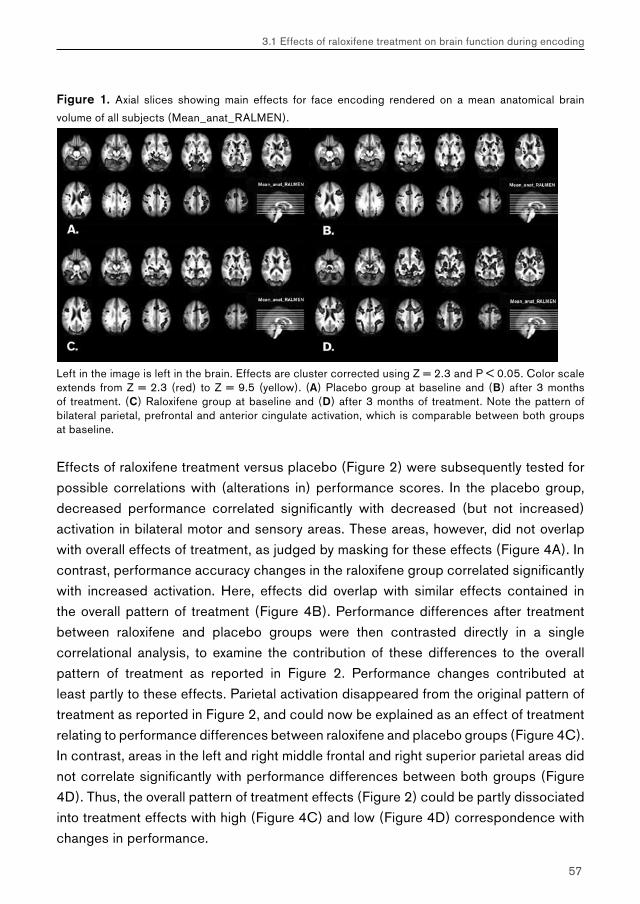
57
3.1 Effects of raloxifene treatment on brain function during encoding
Figure 1. Axial slices showing main effects for face encoding rendered on a mean anatomical brain
volume of all subjects (Mean_anat_RALMEN).
Left in the image is left in the brain. Effects are cluster corrected using Z = 2.3 and P < 0.05. Color scale extends from Z = 2.3 (red) to Z = 9.5 (yellow). (A) Placebo group at baseline and (B) after 3 months of treatment. (C) Raloxifene group at baseline and (D) after 3 months of treatment. Note the pattern of bilateral parietal, prefrontal and anterior cingulate activation, which is comparable between both groups at baseline.
Effects of raloxifene treatment versus placebo (Figure 2) were subsequently tested for possible correlations with (alterations in) performance scores. In the placebo group, decreased performance correlated significantly with decreased (but not increased) activation in bilateral motor and sensory areas. These areas, however, did not overlap with overall effects of treatment, as judged by masking for these effects (Figure 4A). In contrast, performance accuracy changes in the raloxifene group correlated significantly with increased activation. Here, effects did overlap with similar effects contained in the overall pattern of treatment (Figure 4B). Performance differences after treatment between raloxifene and placebo groups were then contrasted directly in a single correlational analysis, to examine the contribution of these differences to the overall pattern of treatment as reported in Figure 2. Performance changes contributed at least partly to these effects. Parietal activation disappeared from the original pattern of treatment as reported in Figure 2, and could now be explained as an effect of treatment relating to performance differences between raloxifene and placebo groups (Figure 4C). In contrast, areas in the left and right middle frontal and right superior parietal areas did not correlate significantly with performance differences between both groups (Figure 4D). Thus, the overall pattern of treatment effects (Figure 2) could be partly dissociated into treatment effects with high (Figure 4C) and low (Figure 4D) correspondence with changes in performance.
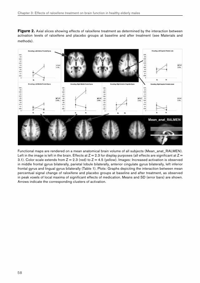
58
Chapter 3: Effects of raloxifene treatment on brain function in healthy elderly males
Figure 2. Axial slices showing effects of raloxifene treatment as determined by the interaction between activation levels of raloxifene and placebo groups at baseline and after treatment (see Materials and
methods).
Functional maps are rendered on a mean anatomical brain volume of all subjects (Mean_anat_RALMEN). Left in the image is left in the brain. Effects at Z = 2.3 for display purposes (all effects are significant at Z = 3.1). Color scale extends from Z = 2.3 (red) to Z = 4.5 (yellow). Images: Increased activation is observed in middle frontal gyrus bilaterally, parietal lobule bilaterally, anterior cingulate gyrus bilaterally, left inferior frontal gyrus and lingual gyrus bilaterally (Table 1). Plots: Graphs depicting the interaction between mean percentual signal change of raloxifene and placebo groups at baseline and after treatment, as observed in peak voxels of local maxima of significant effects of medication. Means and SD (error bars) are shown. Arrows indicate the corresponding clusters of activation.
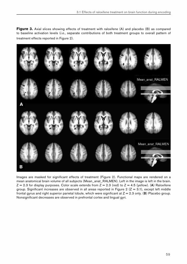
59
3.1 Effects of raloxifene treatment on brain function during encoding
Figure 3. Axial slices showing effects of treatment with raloxifene (A) and placebo (B) as compared to baseline activation levels (i.e., separate contributions of both treatment groups to overall pattern of
treatment effects reported in Figure 2).
Images are masked for significant effects of treatment (Figure 2). Functional maps are rendered on a mean anatomical brain volume of all subjects (Mean_anat_RALMEN). Left in the image is left in the brain. Z = 2.3 for display purposes. Color scale extends from Z = 2.3 (red) to Z = 4.5 (yellow). (A) Raloxifene group. Significant increases are observed in all areas reported in Figure 2 (Z = 3.1), except left middle frontal gyrus and right superior parietal lobule, which were significant at Z = 2.3 only. (B) Placebo group. Nonsignificant decreases are observed in prefrontal cortex and lingual gyri.
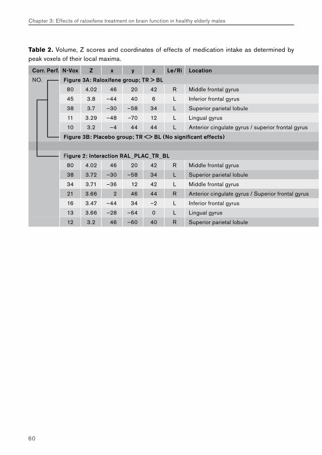
60
Chapter 3: Effects of raloxifene treatment on brain function in healthy elderly males
Table 2. Volume, Z scores and coordinates of effects of medication intake as determined by peak voxels of their local maxima.
Corr. Perf. N-Vox Z x y z Le/Ri Location
NO. Figure 3A: Raloxifene group; TR > BL
80 4.02 46 20 42 R Middle frontal gyrus
45 3.8 –44 40 6 L Inferior frontal gyrus
38 3.7 –30 –58 34 L Superior parietal lobule
11 3.29 –48 –70 12 L Lingual gyrus
10 3.2 –4 44 44 L Anterior cingulate gyrus / superior frontal gyrus
Figure 3B: Placebo group; TR <> BL (No significant effects)
Figure 2: Interaction RAL_PLAC_TR_BL
80 4.02 46 20 42 R Middle frontal gyrus
38 3.72 –30 –58 34 L Superior parietal lobule
34 3.71 –36 12 42 L Middle frontal gyrus
21 3.66 2 46 44 R Anterior cingulate gyrus / Superior frontal gyrus
16 3.47 –44 34 –2 L Inferior frontal gyrus
13 3.66 –28 –64 0 L Lingual gyrus
12 3.2 46 –60 40 R Superior parietal lobule
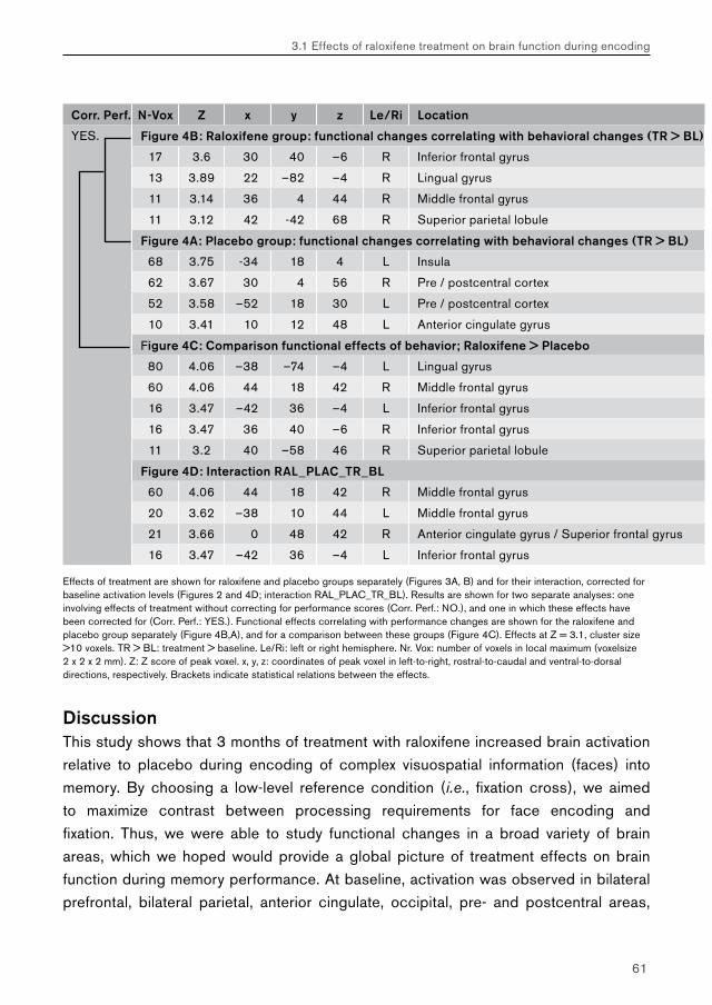
61
3.1 Effects of raloxifene treatment on brain function during encoding
Corr. Perf. N-Vox Z x y z Le/Ri Location
YES. Figure 4B: Raloxifene group: functional changes correlating with behavioral changes (TR > BL)
17 3.6 30 40 –6 R Inferior frontal gyrus
13 3.89 22 –82 –4 R Lingual gyrus
11 3.14 36 4 44 R Middle frontal gyrus
11 3.12 42 -42 68 R Superior parietal lobule
Figure 4A: Placebo group: functional changes correlating with behavioral changes (TR > BL)
68 3.75 -34 18 4 L Insula
62 3.67 30 4 56 R Pre / postcentral cortex
52 3.58 –52 18 30 L Pre / postcentral cortex
10 3.41 10 12 48 L Anterior cingulate gyrus
Figure 4C: Comparison functional effects of behavior; Raloxifene > Placebo
80 4.06 –38 –74 –4 L Lingual gyrus
60 4.06 44 18 42 R Middle frontal gyrus
16 3.47 –42 36 –4 L Inferior frontal gyrus
16 3.47 36 40 –6 R Inferior frontal gyrus
11 3.2 40 –58 46 R Superior parietal lobule
Figure 4D: Interaction RAL_PLAC_TR_BL
60 4.06 44 18 42 R Middle frontal gyrus
20 3.62 –38 10 44 L Middle frontal gyrus
21 3.66 0 48 42 R Anterior cingulate gyrus / Superior frontal gyrus
16 3.47 –42 36 –4 L Inferior frontal gyrus
Effects of treatment are shown for raloxifene and placebo groups separately (Figures 3A, B) and for their interaction, corrected for baseline activation levels (Figures 2 and 4D; interaction RAL_PLAC_TR_BL). Results are shown for two separate analyses: one involving effects of treatment without correcting for performance scores (Corr. Perf.: NO.), and one in which these effects have been corrected for (Corr. Perf.: YES.). Functional effects correlating with performance changes are shown for the raloxifene and placebo group separately (Figure 4B,A), and for a comparison between these groups (Figure 4C). Effects at Z = 3.1, cluster size >10 voxels. TR > BL: treatment > baseline. Le/Ri: left or right hemisphere. Nr. Vox: number of voxels in local maximum (voxelsize 2 x 2 x 2 mm). Z: Z score of peak voxel. x, y, z: coordinates of peak voxel in left-to-right, rostral-to-caudal and ventral-to-dorsal directions, respectively. Brackets indicate statistical relations between the effects.
Discussion This study shows that 3 months of treatment with raloxifene increased brain activation relative to placebo during encoding of complex visuospatial information (faces) into memory. By choosing a low-level reference condition (i.e., fixation cross), we aimed to maximize contrast between processing requirements for face encoding and fixation. Thus, we were able to study functional changes in a broad variety of brain areas, which we hoped would provide a global picture of treatment effects on brain function during memory performance. At baseline, activation was observed in bilateral prefrontal, bilateral parietal, anterior cingulate, occipital, pre- and postcentral areas,
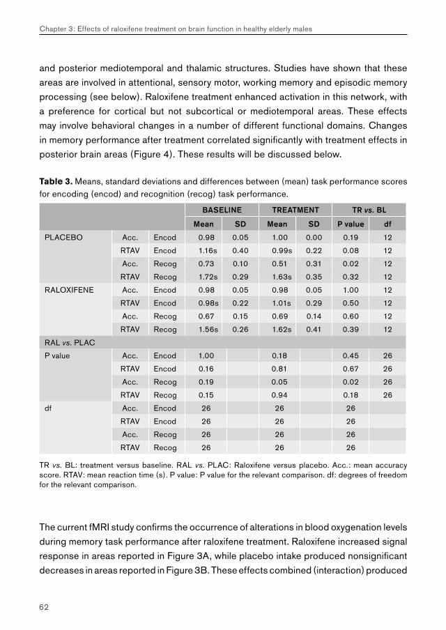
62
Chapter 3: Effects of raloxifene treatment on brain function in healthy elderly males
and posterior mediotemporal and thalamic structures. Studies have shown that these areas are involved in attentional, sensory motor, working memory and episodic memory processing (see below). Raloxifene treatment enhanced activation in this network, with a preference for cortical but not subcortical or mediotemporal areas. These effects may involve behavioral changes in a number of different functional domains. Changes in memory performance after treatment correlated significantly with treatment effects in posterior brain areas (Figure 4). These results will be discussed below.
Table 3. Means, standard deviations and differences between (mean) task performance scores for encoding (encod) and recognition (recog) task performance.
BASELINE TREATMENT TR vs. BL
Mean SD Mean SD P value df
PLACEBO Acc. Encod 0.98 0.05 1.00 0.00 0.19 12
RTAV Encod 1.16s 0.40 0.99s 0.22 0.08 12
Acc. Recog 0.73 0.10 0.51 0.31 0.02 12
RTAV Recog 1.72s 0.29 1.63s 0.35 0.32 12
RALOXIFENE Acc. Encod 0.98 0.05 0.98 0.05 1.00 12
RTAV Encod 0.98s 0.22 1.01s 0.29 0.50 12
Acc. Recog 0.67 0.15 0.69 0.14 0.60 12
RTAV Recog 1.56s 0.26 1.62s 0.41 0.39 12
RAL vs. PLAC
P value Acc. Encod 1.00 0.18 0.45 26
RTAV Encod 0.16 0.81 0.67 26
Acc. Recog 0.19 0.05 0.02 26
RTAV Recog 0.15 0.94 0.18 26
df Acc. Encod 26 26 26
RTAV Encod 26 26 26
Acc. Recog 26 26 26
RTAV Recog 26 26 26
TR vs. BL: treatment versus baseline. RAL vs. PLAC: Raloxifene versus placebo. Acc.: mean accuracy score. RTAV: mean reaction time (s). P value: P value for the relevant comparison. df: degrees of freedom for the relevant comparison.
The current fMRI study confirms the occurrence of alterations in blood oxygenation levels during memory task performance after raloxifene treatment. Raloxifene increased signal response in areas reported in Figure 3A, while placebo intake produced nonsignificant decreases in areas reported in Figure 3B. These effects combined (interaction) produced
63
3.1 Effects of raloxifene treatment on brain function during encoding
a significant signal increase in bilateral prefrontal, bilateral parietal, anterior cingulate, and left inferior prefrontal areas, along with activation of lingual gyri (Figure 2). Apart from neuronal activity, both testosterone and estrogen influence the muscular tone of cerebral vasculature and PET studies have repeatedly shown a general rise in signal intensity levels after estrogen challenge (Maki & Resnick, 2001; Maki & Resnick, 2000; Paganini-Hill & Henderson, 1996). Hence, it is possible that some of the observed signal changes represented a change in neurovascular coupling (D’Esposito et al., 1999). Such vascular effects, however, usually involve changes in absolute global mean intensity levels, which were ignored in our analysis. Furthermore, it is very unlikely that the highly spatially localized (and stimulus-related) pattern of signal enhancement observed in this study was due to a selective effect of raloxifene on cerebral vasculature in these areas. The pattern of signal enhancement can be localized to functionally meaningful areas (see below) and strongly suggests that at least part of the effect of raloxifene treatment was of neurogenic origin.
From fMRI data alone, it is difficult to judge whether the observed effects represent beneficial or adverse effects on brain function. Based on previous findings in Alzheimer’s disease and related dementias and pharmacological fMRI studies of cholinergic treatment of patients with Alzheimer’s disease, a trend has emerged to regard increased brain activation during memory performance as something beneficial (Furey et al., 2000; Kumari et al., 2003; Machulda et al., 2003). This observation, however, can by no means serve as a general rule. The impact of the observed neurofunctional effects on behavior therefore needs to be further evaluated, preferably by means of randomized placebo-controlled clinical trials involving large groups of patients.
Given the modest results of larger trials involving neuropsychological assessment, we did not expect any behavioral changes after raloxifene treatment to be significant in our population. Hence, no follow-up was done of the cognitive status of our subjects. Since the main aim of the present study was to screen for neurofunctional effects in a small number of subjects, our main focus was on the functional data. Quite unexpectedly, however, we found a significant decrease in recognition accuracy scores after 3 months of treatment in the placebo group (i.e., from 73% to 51% above chance levels, i.e., from 87% correct to 76% correct; P = 0.02; Table 3), whereas performance levels in the raloxifene group remained constant ( P = 0.60). Such differences in performance may have been due to small sample size (n = 2 x 14) and a selective biasing of one group over the other. Since no follow-up of the cognitive status of the subjects was performed, it was not possible to assess whether the observed decrease in performance was part of a more solid trend, indicating a pathological decline in memory performance in the
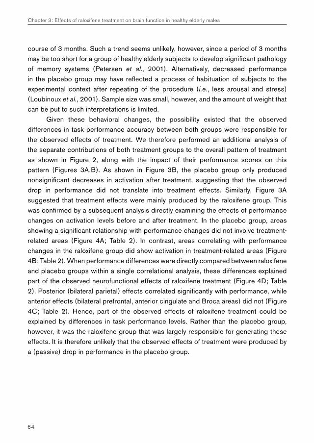
64
Chapter 3: Effects of raloxifene treatment on brain function in healthy elderly males
course of 3 months. Such a trend seems unlikely, however, since a period of 3 months may be too short for a group of healthy elderly subjects to develop significant pathology of memory systems (Petersen et al., 2001). Alternatively, decreased performance in the placebo group may have reflected a process of habituation of subjects to the experimental context after repeating of the procedure (i.e., less arousal and stress) (Loubinoux et al., 2001). Sample size was small, however, and the amount of weight that can be put to such interpretations is limited.
Given these behavioral changes, the possibility existed that the observed differences in task performance accuracy between both groups were responsible for the observed effects of treatment. We therefore performed an additional analysis of the separate contributions of both treatment groups to the overall pattern of treatment as shown in Figure 2, along with the impact of their performance scores on this pattern (Figures 3A,B). As shown in Figure 3B, the placebo group only produced nonsignificant decreases in activation after treatment, suggesting that the observed drop in performance did not translate into treatment effects. Similarly, Figure 3A suggested that treatment effects were mainly produced by the raloxifene group. This was confirmed by a subsequent analysis directly examining the effects of performance changes on activation levels before and after treatment. In the placebo group, areas showing a significant relationship with performance changes did not involve treatment-related areas (Figure 4A; Table 2). In contrast, areas correlating with performance changes in the raloxifene group did show activation in treatment-related areas (Figure 4B; Table 2). When performance differences were directly compared between raloxifene and placebo groups within a single correlational analysis, these differences explained part of the observed neurofunctional effects of raloxifene treatment (Figure 4D; Table 2). Posterior (bilateral parietal) effects correlated significantly with performance, while anterior effects (bilateral prefrontal, anterior cingulate and Broca areas) did not (Figure 4C; Table 2). Hence, part of the observed effects of raloxifene treatment could be explained by differences in task performance levels. Rather than the placebo group, however, it was the raloxifene group that was largely responsible for generating these effects. It is therefore unlikely that the observed effects of treatment were produced by a (passive) drop in performance in the placebo group.
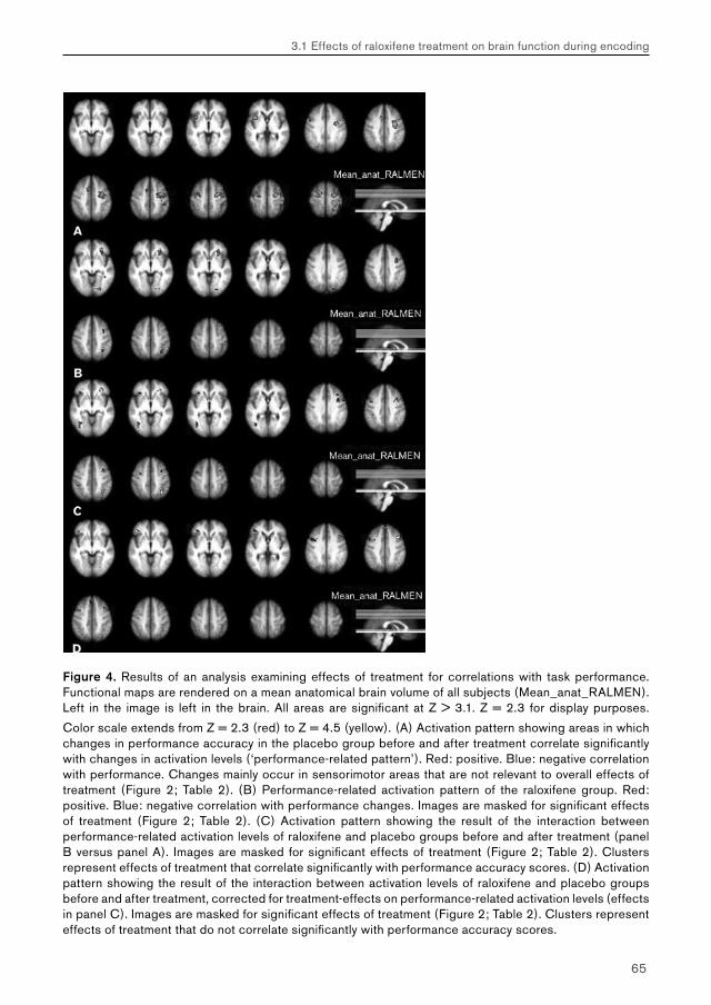
65
3.1 Effects of raloxifene treatment on brain function during encoding
Figure 4. Results of an analysis examining effects of treatment for correlations with task performance. Functional maps are rendered on a mean anatomical brain volume of all subjects (Mean_anat_RALMEN). Left in the image is left in the brain. All areas are significant at Z > 3.1. Z = 2.3 for display purposes.
Color scale extends from Z = 2.3 (red) to Z = 4.5 (yellow). (A) Activation pattern showing areas in which changes in performance accuracy in the placebo group before and after treatment correlate significantly with changes in activation levels (‘performance-related pattern’). Red: positive. Blue: negative correlation with performance. Changes mainly occur in sensorimotor areas that are not relevant to overall effects of treatment (Figure 2; Table 2). (B) Performance-related activation pattern of the raloxifene group. Red: positive. Blue: negative correlation with performance changes. Images are masked for significant effects of treatment (Figure 2; Table 2). (C) Activation pattern showing the result of the interaction between performance-related activation levels of raloxifene and placebo groups before and after treatment (panel B versus panel A). Images are masked for significant effects of treatment (Figure 2; Table 2). Clusters represent effects of treatment that correlate significantly with performance accuracy scores. (D) Activation pattern showing the result of the interaction between activation levels of raloxifene and placebo groups before and after treatment, corrected for treatment-effects on performance-related activation levels (effects in panel C). Images are masked for significant effects of treatment (Figure 2; Table 2). Clusters represent effects of treatment that do not correlate significantly with performance accuracy scores.
66
Chapter 3: Effects of raloxifene treatment on brain function in healthy elderly males
The results of these functional–behavioral associations are compatible with the existence of an active process in the raloxifene group (as judged by enhanced activation in treatment-related areas) that may have maintained performance in this group at a certain level, whereas the absence of such a process in the placebo group may have produced a drop in performance (not translating into treatment effects). Hence, although perhaps unlikely, it is possible that raloxifene intake actively blocked a normal decrease in performance as a result of habituation. A canceling of training effects has been reported previously in male subjects receiving testosterone injections, and may involve effects of estrogens on attentional levels (Wolf et al., 2000). However, because of small sample size, further study is necessary to corroborate these findings.
The locations of the brain areas activated by raloxifene intake may provide a clue as to the nature of the affected functions. The observed pattern of signal changes after raloxifene treatment appears to be an enhancement of the activation pattern already observed during task performance (the main effects; Figure 1; Table 1). Such effects are almost invariantly observed during performance on any task that requires active processing of complex visuospatial information. A typical combination of bilateral parietal, bilateral prefrontal and anterior cingulate activation is produced that remotely resembles a ‘cloverleaf’ pattern (Figure 1). Previous studies have found that bilateral parietal activation within this pattern is related to automatic processing (i.e., working memory performance; (Rowe et al., 2000)), whereas activation in bilateral (pre)frontal areas usually involves more effortful processing (i.e., executive functions including response selection; (Fink et al., 1999; Rowe et al., 2000; Badre & Wagner, 2004) and activation of the frontal eye fields (i.e., gaze shifting; (Luna et al., 1998)). Anterior cingulate activation may mediate between effortful (prefrontal) and automatic (parietal) processing streams by selecting the most relevant of incoming stimuli, shifting conscious attention to this stimulus and directing effortful processing strategies against it, in order to generate a response that may alter the situation. This process has been referred to as ‘cognitive control’ in various neuroimaging studies (Badre & Wagner, 2004; Braver & Barch, 2002; Fink et al., 1999; Li et al., 2004b; Weissman et al., 2003). Thus, raloxifene intake may have affected a number of neural functions related to this process.
Apart from affecting the cortical ‘cloverleaf’ pattern, raloxifene treatment enhanced brain activation in the left inferior prefrontal cortex, in or near Broca’s area (Figures 2 and 3A). Activation in this area is suggestive of increased verbal processing, possibly reflecting increased use of verbal strategies to encode the presented visuospatial information into memory. Increased verbal memory is a commonly reported finding after estrogen exposure (Rice & Morse, 2003). Although still a matter of debate, estrogens
67
3.1 Effects of raloxifene treatment on brain function during encoding
may influence the lateralization of neural processing, possibly by enhancing verbal processing strategies that are dominantly represented in the left hemisphere at the cost of visuospatial processing strategies represented in the right hemisphere (Dietrich et al., 2001; Kelley et al., 1998). Data from this study therefore predict an effect of raloxifene on verbal performance, which may require additional study by means of neuropsychological assessment.
Raloxifene treatment enhanced activation in cortical, but not subcortical or mediotemporal areas (Figures 2). This lack of mediotemporal activation may be due to a biasing effect in the sense that, for several reasons, mediotemporal (and especially hippocampal) activation has proven hard to image (Greicius et al., 2003). In a previous study, however, we used the same face encoding task as described in the current study to examine effects of cholinergic stimulation on brain activation in patients with mild cognitive impairment. Enhancement of hippocampal activation was observed, showing that this task may detect changes in mediotemporal activation even in patients with some hippocampal damage (Goekoop et al., 2004). Data from this study therefore suggests a preference for cortical, but not subcortical or mediotemporal activation changes. Thus, raloxifene treatment may have affected neocortical functions, rather than episodic memory itself, which depends largely on subcortical (limbic) and mediotemporal structures (Schacter & Wagner, 1999). Such a conclusion is supported by results from previous studies. (Shaywitz et al., 1999) examined changes in brain activation during both verbal and visuospatial memory performance after estrogen treatment of postmenopausal women for 3 weeks as compared to placebo.
Similar to results from the present study, enhanced activation was found in bilateral parietal and prefrontal structures, but not in subcortical or mediotemporal structures. Results were explained as an action of estrogen on cortical processes such as (verbal) working memory, but not episodic memory. Additionally, based on a review of a large number of studies, it has been suggested that effects of estrogens on memory and cognition represent a preferential targeting of neocortical areas, especially the prefrontal cortex, rather than memory circuits in subcortical or mediotemporal areas (Keenan et al., 2001). This conclusion is further supported by results from the current study, showing that raloxifene treatment produced no activation in the fusiform gyri, which are key structures in the processing of facial stimuli and activate strongly during task performance (Tarr & Gauthier, 2000) (Table 1). An effect of raloxifene might therefore have been expected in these areas. Instead, lingual gyri showed a small increase in activation. These structures are involved in visual perception, and activation changes in these structure may represent an effect on visuospatial attention (Bentley et al., 2004;
68
Chapter 3: Effects of raloxifene treatment on brain function in healthy elderly males
Lawrence et al., 2002; Rezvani & Levin, 2001). However, their involvement should be viewed within the context of the main effects during task performance (Table 1). Face encoding produced maximum intensity signal changes in the lingual gyri, which may have made them disproportionally sensitive to effects of treatment. Thus, effects of raloxifene treatment during memory performance mainly involved higher cortical areas, but not mediotemporal areas. This makes it less likely raloxifene treatment has a direct effect on episodic memory during encoding.
Overall, raloxifene intake increased activation levels in brain areas spanning a number of different cognitive domains (see above). Such a multitude of affected regions makes it unlikely that raloxifene exerted a specific influence on each of these structures separately. Instead, the observed pattern of treatment effects is suggestive of a nonspecific effect of raloxifene on brain function. Such an effect may involve an influence on cortical arousal. Estrogens are known to exert a global influence on all primary neuromodulatory neurotransmitter systems (i.e., serotonergic, dopaminergic, nor-adrenergic and cholinergic systems), which are crucially involved in regulating arousal processes across the entire cortical mantle (Bernardi et al., 2003; McEwen, 2002). Indeed, in patients with Alzheimer’s disease, cholinesterase inhibitors are used to stimulate the cholinergic system, which results in a small but therapeutic increase in episodic memory. This effect is thought to be secondary to a nonspecific effect of acetylcholine on cortical arousal and attention (Freo et al., 2002; Coull, 1998). fMRI studies have shown that modulation of cholinergic, dopaminergic and nor-adrenergic systems affects activation levels in a multitude of brain areas (Coull, 1998; Thiel, 2003; Strange & Dolan, 2004). Based on such findings, it has been suggested that many of the discrepancies in the literature on the behavioral effects of estrogens may be accounted for by assuming a context-dependent effect of estrogens on cortical arousal (Morgan et al., 2004). In this view, estrogens may exert a global effect on cortical arousal through their actions on multiple neurotransmitter systems, with small effects on behavior as secondary phenomena. Such a mechanism may explain previous results of PET studies showing a generalized increase in global activation levels after estrogen treatment (Maki & Resnick, 2001; Maki & Resnick, 2000; Paganini-Hill & Henderson, 1996), and the modest changes in behavior usually associated with such treatment. In addition, it may explain the activation of multiple brain areas after raloxifene treatment as observed in this study.
69
3.1 Effects of raloxifene treatment on brain function during encoding
Both estrogens and SERMs have been described as potent neuroprotectors, which may decrease ischaemic damage after injury and reduce cell death in neurodegenerative diseases, such as Alzheimer’s and Parkinson’s disease. Such effects may involve pathways that are only partially understood. Recent findings of adverse effects of estrogen treatment in postmenopausal women, including an increased risk of stroke and Alzheimer’s disease, have added to the controversy over the role of estrogens and SERMs in neuroprotection (Murphy et al., 2003). Estrogens, however, exert their effects on a variety of different levels, including effects on brain structure (organizational effects) and function (activational effects) (Bisagno et al., 2003). Both beneficial and adverse effects of chronic estrogen treatment may be unevenly balanced across these domains, and may also be context-dependent (see above). Hence, both structural and functional effects need to be studied separately, in order to obtain an integrated picture of the effects of (partial) estrogen agonists on mental functioning. This study has focused on the functional domain, showing that raloxifene treatment enhances activation in a variety of brain areas. Such changes may translate into behavioral changes that can be detected using neuropsychological assessment scales. Since they may represent secondary effects of raloxifene as a result of enhanced cortical arousal, such behavioral changes are likely to be small. This exploratory study predicts enhancement of working memory performance, executive functions, verbal skills and possibly episodic memory performance. Further neuropsychological assessment is necessary to test these predictions. Together with studies on the effects of estrogen agonists on brain structure, such knowledge may provide a comprehensive view on the effectiveness of SERM treatment in elderly subjects.
Acknowledgements
We would like to thank the Oxford fMRIB team who is responsible for producing, maintaining and freely distributing FSL. Acknowledgements go to Stichting Alzheimer and Neuropsychiatrie Foundation for financial support, and Ans Nicolaas- Merkus for subject recruitment. Serge Rombouts is supported by a grant from the Netherlands Organisation for Scientific Research (NWO; grantnr. 916.36.117).


Chapter 3
Effects of raloxifene treatment on brain function in healthy elderly males
3.2 Effects of raloxifene treatment on brain function during recognition
Raloxifene treatment enhances brain activation during recognition of familiar items; a pharmacological fMRI study in healthy elderly males
Goekoop R, Barkhof F, Duschek EJJ, Netelenbos C, Knol DL, Scheltens P, Rombouts SARB.
Neuropsychopharmacology 2005, in press.
72
Chapter 3: Effects of raloxifene treatment on brain function in healthy elderly males
Abstract
Raloxifene is a selective estrogen receptor modulator that may delay the onset of mild cognitive impairment in elderly women. Effects of raloxifene treatment on mental performance in males remain to be investigated. In a previous functional magnetic resonance imaging (fMRI) study, we showed that raloxifene treatment enhanced brain activation in elderly males during encoding of new information (faces) into memory. The current study used fMRI in the same group of subjects to screen for effects of raloxifene treatment on brain function during face recognition. Healthy elderly males (n = 28; mean age 63.6 years, SD 2.4) were scanned at baseline and after three months of treatment with either raloxifene 120 mg (n = 14) or placebo (n = 14) in a randomized, double blind, placebo controlled study design. Functional data were analyzed in an event-related fashion with respect to correct hits and correct rejections using FSL software. Performance data were analyzed with respect to recognition accuracy, latency, and response bias. Functional effects of treatment were found on brain activation related to correct hits only. When compared to placebo treatment, raloxifene treatment enhanced brain activation in the left posterior parahippocampal area (Z = 3.9) and right inferior prefrontal cortex (Z = 3.5). Recognition accuracy scores remained stable in the raloxifene group, whereas the placebo group showed a small but significant decrease in accuracy scores (p = 0.02). No significant effects were found on response bias or latency. In conclusion, raloxifene treatment affects brain function during memory performance in a way that may reflect increased arousal during initial encoding, with downstream effects on brain function during retrieval of information. Behaviorally, such neurofunctional effects may actively block decreased memory performance as a result of context-dependency. The validity of these predictions can be tested in large-scale clinical trials.
Keywords: fMRI;SERM; raloxifene; male; memory; recognition.
73
3.2 Effects of raloxifene treatment on brain function during recognition
Introduction
Raloxifene is a selective estrogen receptor modulator (SERM) that is used to treat osteoporosis in postmenopausal women (Heringa, 2003). Raloxifene has positive effects on cardiovascular risk factors, and is not associated with an increased risk of stroke and cardiovascular disease that is observed for regular estrogen treatment (Barrett-Connor et al., 2002). For such reasons, raloxifene treatment is considered an attractive alternative for prevention and treatment of osteoporosis and heart disease in male subjects as well (Doran et al., 2001; Blum et al., 2000). Recent results from a randomized controlled clinical trial show that raloxifene treatment at 120mg daily for three years may successfully delay the onset of mild cognitive impairment (MCI) and possibly Alzheimer’s Disease (AD) in elderly women (Yaffe eet al., 2005). Thus, the need for an assessment of the effects of raloxifene treatment on male brain function and mental performance is growing.
Estrogens and SERMs such as raloxifene may differentially affect mental performance, including memory performance, in both sexes (Cahill, 2003). It is therefore unclear whether the preventive effects of raloxifene treatment in women can be extrapolated to males. To our knowledge, no studies have been published that examined effects of raloxifene treatment on memory performance in male subjects. Such studies usually involve large-scale clinical trials in which subjects or patients are examined for several years in order to detect significant effects of pharmacological treatment on neuropsychological measures of behavior. While anticipating the results of such studies, the current study examined the effects of three months of raloxifene treatment on brain function during memory task performance using functional magnetic resonance imaging (fMRI). Task-related fMRI yields both functional and behavioral measures of treatment effects, which may serve as preliminary findings prior to large-scale investigations of similar effects in controlled clinical trials. fMRI may be more sensitive and less ambiguous to the effects of sex steroids on mental performance than conventional neuropsychological assessment scales (Maki & Resnick, 2001). Additionally, fMRI may provide an insight into the treatment mechanism of raloxifene. Treatment effects on brain function can be mapped to individual brain structures at high spatial resolution (Honey & Bullmore, 2004). The behavioral significance of such effects may be examined either by direct correlation of behavioral measures with such effects (Goekoop et al., 2005a), or by examining brain function associated with distinct response types during memory performance (e.g. correct hits, correct rejections) (Buckner et al., 1998a). This allows a detailed analysis of the effects of SERMs on memory function, which may eventually
74
Chapter 3: Effects of raloxifene treatment on brain function in healthy elderly males
be relevant to the design and development of new SERMs that target specific aspects of memory performance, such as encoding and retrieval processes (Bernardi et al., 2003).
In a previous fMRI study, we examined effects of raloxifene treatment versus placebo on brain function in healthy elderly males during encoding of new information (human faces) into memory (Goekoop et al., 2005a). Raloxifene globally enhanced brain activation during face encoding, with the exception of subcortical, medial temporal and visual structures. This was interpreted as a global effect of raloxifene treatment on cortical arousal, rather than a specific influence on each of the cognitive domains represented by this pattern (Goekoop et al., 2005a). Such effects are in line with findings from a large number of studies, suggesting that estrogens have a context-dependent effect on cortical arousal (Morgan et al., 2004; Cahill & Alkire, 2003). Based on the locations of treatment effects, we predicted small effects of raloxifene treatment on working memory performance, executive functions and verbal skills (Goekoop et al., 2005a).
Since no treatment effects were found in subcortical or medial temporal areas during encoding (despite activation of these structures during task performance), we considered an effect of raloxifene treatment on episodic memory to be less likely. However, the effects of raloxifene treatment on episodic memory could not be fully assessed, since only the encoding-phase of memory performance was examined. Since a growing number of imaging studies has shown that pharmacological substances affect brain function in process-specific manner (Honey & Bullmore, 2004; Thiel, 2003), raloxifene treatment may have unique effects on brain function during encoding, consolidation or retrieval stages of memory performance. We therefore used fMRI to examine the effects of three months of raloxifene treatment on brain function during face recognition. Given the reported efficacy of raloxifene treatment in preventing the onset of memory impairment in elderly women, we hypothesized that raloxifene treatment would eventually affect brain function during retrieval of episodic information.
Methods
Study DesignSubjects were screened for participation in a randomized, double blind, placebo-controlled repeated measures design. In each subject, fMRI was performed at baseline (BL; no medication; session 1) and after three months of a once daily oral intake of raloxifene 120 mg or placebo (TR; session 2). This dose was chosen because a previous
75
3.2 Effects of raloxifene treatment on brain function during recognition
study showed no significant effects of raloxifene 60 mg on markers of bone turnover in males (Doran et al., 2001). BL and TR sessions were performed on the same time of day in each subject (margin 30 minutes). If data acquisition failed, the subject’s consent was asked for an additional scanning visit, up until which time the relevant medication regime (raloxifene or placebo) was continued, to obtain a maximum number of complete datasets. Study period extension was not to exceed 10 days.
Subject recruitmentThe medical ethical review board of the VU University Medical Center of Amsterdam approved the study. Thirty healthy, right-handed elderly males, aged 60 to 70 years (mean 63.6 years, SD 2.4; range 60–69 years) were recruited by advertisement in local newspapers. This was the same study population that was examined previously for effects of raloxifene treatment on brain function during face encoding . All subjects provided informed consent during a screening visit in which the procedure was explained and contraindications were checked. The mental status of subjects was assessed by means of a structured clinical interview. This interview involved questions regarding the subjects’ general health, mental health, social status, history and intoxications. Exclusion criteria involved (a history of) psychiatric disease (i.e. any diagnosable disorders according to DSM-4-TR criteria (American Psychiatric Association, 2000), any neurological illness (memory complaints in particular), and the use of any medication or other substances that are known to influence cerebral functioning (including > 0.5 pack of cigarettes, > 4 glasses of alcohol). Elaborate physical examination and laboratory tests were performed to back the clinical interview. Exclusion criteria to MRI involved the presence of metallic implants in high-risk areas and a history of claustrophobia. Formal education was determined using a dutch system (low, middle, high). On the day of the second scanning session (after 3 months), a similar structured clinical interview was performed with a particular focus on side-effects of treatment. Compliance was assessed by pill counts and the subjects’ comments.
Functional MRI (fMRI)Imaging was carried out on a 1.5 T Sonata MR scanner (Siemens, Erlangen, Germany), using a standard circularly polarized head coil with foam padding to restrict head motion. For fMRI, an echo planar imaging sequence was used (echo time 60 ms, flip angle 90°, matrix 64 x 64, field of view 192 x 192mm), to obtain 21 transverse slices (thickness 5 mm, interslice-gap 1 mm). Task stimuli were projected on a screen located at the head end of the scanner table via an LCD projector located outside the scanner
76
Chapter 3: Effects of raloxifene treatment on brain function in healthy elderly males
room. Subjects viewed the screen through a mirror located on the head coil. In each hand, subjects held an fMRI compatible response-box through which they were able to react to task stimuli by pressing a single button using one of their index-fingers. A T1-weighted structural MRI-scan was obtained of each subject (MPRAGE; inversion time: 300 ms, TR = 15 ms; TE = 7 ms; flip angle = 8°; 160 coronal slices, 1 x 1 x 1.5mm voxels). Total scanning time including structural imaging on average was 21 minutes for each visit.
Face recognition task Immediately after face encoding, which involved the presentation of four blocks of unfamiliar faces (24 in total), alternating with blocks of fixation (Goekoop et al., 2005a), a recognition task was administered. Total scanning time was 6’12’’ minutes for the encoding task and 3’40’’ minutes for the recognition task. Hence, minimum delay between items presented during encoding and recognition tasks was ~1 minute, and maximum delay ~10 minutes. During the first 10.5s of each memory-task, subjects saw a circle indicating time left before the onset of the first condition. During face recognition, 24 faces, of which 12 had been shown during encoding and 12 were new, were presented sequentially in random order on a black background. Presentation time was 5s for each face, followed by a white fixation-cross presented for 3s on a black background. We chose to contrast faces with a ‘low level’ fixation cross (X) in order to maximize contrast between processing demands for both conditions, which would result in a large number of brain areas being activated for faces > fixation contrasts, including visual and sensorimotor areas. This would increase chances of finding significant effects of treatment. Subjects were instructed verbally to indicate whether the presented faces were familiar or unfamiliar by pressing one of two buttons (written instructions “Left: familiar” and “Right: unfamiliar” also appeared alongside the pictures). Mean reaction times and the number of hits and misses were recorded. To avoid investigator’s bias, task performance data were only viewed after both scanning visits had been completed.
Two comparable versions of each paradigm were constructed and randomized across subjects and scanning-sessions (baseline, treatment), to reach equal numbers of subjects receiving test versions 1 and 2 in both treatment groups. A third ‘spare’ version was available in case data-acquisition might fail during one of the scanning sessions. Each paradigm contained unique items (faces), to avoid any overlap or interference with previously observed items at the time of the second scanning session. Paradigms were practised extensively to minimize effects of skill learning in the course of the scanning sessions. A practicing task was used that was similar to tasks used for neuroimaging,
77
3.2 Effects of raloxifene treatment on brain function during recognition
but contained less items (practicing items did not overlap with those of tasks used for scanning purposes). A day before the onset of the first scanning session, subjects visited the hospital and practiced both encoding and retrieval tasks. On the day of the first scanning session, minutes before the onset of the first measurements, the tasks were again practiced with subjects in the scanner. On the day of the the second scanning session, subjects practiced the procedure within the scanner only. Total time for one scanning visit including instructions of memory tasks was approximately one hour.
Statistical analysis of task performance dataPost-hoc sorting of response types yielded a number of true positive (TP, correct hits), true negative (TN, correct rejections), false positive (FP, false hits), false negative (FN, false rejections) and forgotten items (Forgot). Based on these responses, an overall performance accuracy score (‘Pr’) was calculated by using the following formula: Pr = FP – TP (Corwin, 1994). This measure was divided by the total number of new items (12) to correct for 50% chance levels, yielding scores between –1 and 1. A ‘false-alarm rate’ (‘FAR’) was calculated using the following formula: FAR = (FP + 0.5) / 13. A measure of response bias ‘Br’ was then calculated using the following formula: Br = FAR / (1–Pr). This measure indicates the chance that subjects will guess that tested items are targets when they are in an uncertain state. High values of Br indicate a tendency to produce high numbers of false positive responses, whereas low values of Br indicate high uncertainty (Corwin, 1994).
Three independent variables were examined for effects of raloxifene versus placebo treatment: i.e. accuracy (Pr), latency (RTav) and response bias (Br). For each variable, a single mixed effects (ME) linear model was specified using SPSS 12.0, with ‘group’ (2 levels: RAL, PLAC), ‘scanorder’ (2 levels: session 1, session 2) and ‘test version’ (2 levels: 1 and 2) as fixed factors, ‘scanorder’ being the repeated factor, and ‘subjectnr’ defining linked measurements from the same subject. This model allowed calculation of significant effects of raloxifene versus placebo treatment (interaction group*scanorder), as well as effects of raloxifene or placebo treatment separately (custom contrasts examining ‘placebo: session 2 – session 1’, and ‘raloxifene: session 2 – session 1’) and differences in performance between raloxifene and placebo groups at baseline and after treatement (custom contrasts examining ‘session1: RAL – PLAC’, and ‘session 2: RAL – PLAC’). Effects were examined using a threshold for significance defined by p < 0.05, Bonferroni corrected for multiple comparisons. The advantage of this model is that it is more accurate than conventional models, allows calculation
78
Chapter 3: Effects of raloxifene treatment on brain function in healthy elderly males
of all effects of interest within the confines of a single statistical model, and is closely related to the model used for analysis of the functional data. Since our previous analysis of the same performance data used the conventional but less accurate (multi-model) approach, the significance of the observed effects as reported in the current study may vary to some degree with those reported earlier (Goekoop et al., 2005a). However, the basic trends remain the same (see results section).
Analysis of functional neuroimaging dataFunctional datasets of individual subjects were analyzed using FSL 3.2 (Smith et al., 2004). The first five volumes of each dataset were discarded to account for T1-saturation effects. At first level (individuals), the following pre-processing was applied: non-brain removal, slice-timing correction using Fourier-space time-series phase-shifting, motion correction and spatial smoothing using a Gaussian kernel of FWHM 8mm, mean-based intensity normalization of all volumes by the same factor and high and low pass temporal filtering (Jenkinson et al., 2002; Smith, 2002). Functional neuroimages of each subject were coregistered to corresponding structural images in native space, and structural images were registered to structural Talairach standard images (Talairach & Tournoux, 1988) defined by the Montreal Neurological Institute standard brain supplied with FSL. The same transformation matrices used for structural-to-standard transformations were then used for functional-to-standard space transformations of coregistered functional images. All registrations were carried out using an intermodal registration tool based on the correlation ratio (Jenkinson & Smith, 2001). After pre-processing, the following statistics was applied on a voxelwise basis on each time series, using local autocorrelation correction (Woolrich et al., 2001): signal change during face recognition was modeled in an event-related fashion, using separate regressors for TP, TN, FP, FN and forgotten response types (see above). Signal variance during fixation (X condition) was not modeled, to prevent overspecification of the model. Type and onset time of the events were determined by post-hoc sorting, based on the responses given by the individual subjects. Thus, all individuals obtained a unique model of signal response containing a regressor for each response type, which was convolved with a gamma function to model the hemodynamic response. Model fitting generated whole brain images in native space of parameter estimates and corresponding variance, representing average signal change during a particular condition (e.g. TP, TN, FP, FN, forgot) versus fixation (X, an implicit baseline condition). Because of a limited number of false responses, effects of raloxifene treatment versus placebo were examined on ‘TP’, ‘TN’, ‘TP > TN’ and ‘TN > TP’ contrasts only, i.e. false responses were ignored in further analyses.
79
3.2 Effects of raloxifene treatment on brain function during recognition
Since our face recognition task contained both novel and familiar items, both encoding and retrieval processes occurred during task performance (Buckner et al., 2001). Such processes can be studied separately by means of event-related analyses of brain function during TP and TN items. TP and TN versus low-level fixation (X) contrasts (‘loose comparisons’) examine more general aspects of recognition memory performance, which are biased with respect to successful retrieval processes (TP > X) and encoding processes (TN > X), respectively (Buckner et al., 1998a). TP > TN contrasts specifically examine brain areas where signal intensity during successful retrieval of familiar information was significantly higher than signal intensity during successful rejection / encoding of new information. Areas of significant signal differences are therefore thought to represent ‘successful retrieval’ processes. Conversely, the reverse contrast TN > TP examines brain function related to ‘encoding during attempted retrieval’ (Daselaar et al., 2003). TP <> TN contrasts (‘tight comparisons’) thus provide additional information concerning specific subcomponent processes during retrieval. Contrast images were resampled to 2 x 2 x 2 mm in standard space and fed into in a second-level statistical analysis to examine effects of raloxifene treatment on (distinct aspects of) brain function during retrieval.
Activation images of main effects during recognition were produced by calculating an average activation map based on all individual activation maps for each session, medication regime and event-type separately (i.e. BL (placebo), BL (raloxifene), TR (placebo) and TR (raloxifene)) in a mixed effects higher level analysis (Woolrich et al., 2004) using clusters determined by Z < 2.3 and a corrected cluster significance threshold of P = 0.05 (Forman et al., 1995; Friston et al., 1994; Worsley et al., 1992). Main effects at baseline were first tested for significant differences between raloxifene and placebo groups using the following contrasts: BL (RAL) <> BL (PLAC), at Z = 3.1 (p < 0.001). Effects of treatment were then calculated using a repeated measures model, in which two explanatory variables (EVs) contrasted BL and TR sessions of placebo and raloxifene groups, allowing separate variances for both groups. Two additional EVs (one for each treatment group) coded for effects of test version on signal response. The main contrasts of interest were [TR (raloxifene) – BL (raloxifene)] <> [TR (placebo) – BL (placebo)], testing for an interaction between raloxifene and placebo groups before and after treatment. Analyses were performed using a threshold of Z = 3.1 (p < 0.001). Images were rendered on a mean anatomical brain volume of all subjects in standard space (Talairach & Tournoux, 1988) for display purposes.
80
Chapter 3: Effects of raloxifene treatment on brain function in healthy elderly males
Results
DemographicsMean age of subjects was 64.1 (SD 2.4) year in the placebo group and 63.1 (SD 2.5) year in the raloxifene group. The placebo group contained two smokers versus one in the raloxifene group. Education-level was equal in both groups (Chi square = 0.26; p = 0.88).
Subject compliance and discontinuationOne subject (placebo group) was claustrophobic and data quality of both sessions was poor in another subject (raloxifene group). These data were discarded, yielding 28 complete datasets containing BL and TR data (n = 14 raloxifene group, n = 14 placebo group). Rescanning was performed once in a single individual (placebo group) because of poor data quality during the second scanning session. Subject compliance was good as assessed by tablet counts and there were no dropouts. No significant side effects were reported.
Task PerformanceNumbers and percentages of all response types (i.e. TP, TN, FP and FN responses) are shown in Table 1A. Mean overall recognition accuracy was 0.60 (SD 0.20) above 50% chance level, with mean overall reaction time 1.63s (SD 0.33s). Table 1B displays the significance of the difference between mean performance values (accuracy, latency and response bias) across both groups and scanning sessions. The placebo group showed significantly better mean recognition accuracy scores at baseline than the raloxifene group (p = 0.022). This trend was reversed after treatment (p = 0.052). After treatment, recognition accuracy increased significantly in the raloxifene group (p = 0.046), but decreased in the placebo group (p = 0.003), with the interaction being significant at p = 0.003. Response latency was not significantly affected in both groups. Response bias showed a trend toward a decrease in the placebo group (p = 0.08), but remained stabile in the raloxifene group (p = 0.086), with the interaction being non significant (p = 0.17). No significant effects were found of the covariate test version, or its interactions with other factors, on measures of task performance.
81
3.2 Effects of raloxifene treatment on brain function during recognition
Brain functionMain effects of face recognition (correct responses) involved activation of ventral and dorsal occipital (visual) areas, bilateral inferior parietal, parahippocampal, superior temporal, prefrontal areas and the lateral sulci (Figure 1, Table 2). These effects were very similar to previous patterns of brain function observed during delayed recognition memory performance (Rugg et al., 2002). Activation patterns for TP and TN decisions differed significantly: TP > TN contrasts showed activation bilaterally in primary visual cortex, anterior and posterior cingulate cortex, inferior parietal lobes and insula, whereas unilateral activation was observed in right motor cortex, right basal ganglia, left cerebellum, and left inferior, middle and superior frontal cortex (Z > 2.3 cluster corrected (cluster threshold Z = 3.1); Figure 1). The lateralization in precentral areas likely represents increased motor activity of the right hand and fingers during TP decisions when compared to TN decisions. In contrast, only left motor cortex showed increased activation in TN > TP contrasts (Figure 1).
The main contrast examining the interaction between raloxifene and placebo groups at baseline and after treatment showed significant increases in activation during TP decisions only. Effects occurred in the left parahippocampal area and right inferior prefrontal cortex (Figure 2C; Table 3; Z > 3.1). Nearly identical effects were found on brain function during TN decisions, but these were not significant at Z > 3.1 (Figure 2D; Z > 2.3). When studying the separate contributions of each group to treatment effects reported in Figure 2, the raloxifene group contributed mostly to the observed effects of treatment (Figure 2A, 2B). No significant effects were found of placebo intake on brain function. No significant treatment effects were found on contrasts describing TP <> TN activation differences. No decreases in activation were observed after treatment with either raloxifene or placebo. No significant differences were found at baseline between activation levels of raloxifene and placebo groups within areas of significant treatment effects (Z > 3.1). Plots of percent signal change (from global mean values) of peak voxels of local maxima illustrate intensity changes in both groups before and after treatment (Figure 2).
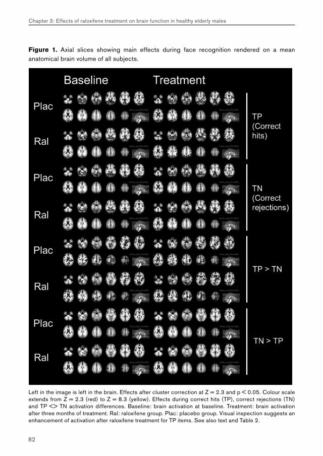
82
Chapter 3: Effects of raloxifene treatment on brain function in healthy elderly males
Figure 1. Axial slices showing main effects during face recognition rendered on a mean anatomical brain volume of all subjects.
Left in the image is left in the brain. Effects after cluster correction at Z = 2.3 and p < 0.05. Colour scale extends from Z = 2.3 (red) to Z = 8.3 (yellow). Effects during correct hits (TP), correct rejections (TN) and TP <> TN activation differences. Baseline: brain activation at baseline. Treatment: brain activation after three months of treatment. Ral: raloxifene group. Plac: placebo group. Visual inspection suggests an enhancement of activation after raloxifene treatment for TP items. See also text and Table 2.
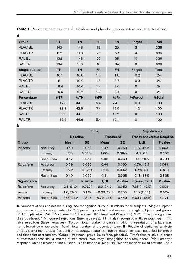
83
3.2 Effects of raloxifene treatment on brain function during recognition
Table 1. Performance measures in raloxifene and placebo groups before and after treatment.
AGroup TP TN FP FN Forgot Total
PLAC BL 142 148 18 25 3 336
PLAC TR 112 143 25 52 4 336
RAL BL 132 148 20 36 0 336
RAL TR 134 150 18 34 0 336
Single subject TP TN FP FN Forgot Total
PLAC BL 10.1 10.6 1.3 1.8 0.2 24
PLAC TR 8 10.2 1.8 3.7 0.3 24
RAL BL 9.4 10.6 1.4 2.6 0 24
RAL TR 9.6 10.7 1.3 2.4 0 24
Percentage %TP %TN %FP %FN %Forgot %Total
PLAC BL 42.3 44 5.4 7.4 0.9 100
PLAC TR 33.3 42.6 7.4 15.5 1.2 100
RAL BL 39.3 44 6 10.7 0 100
RAL TR 39.9 44.6 5.4 10.1 0 100
B Time Significance
Baseline Treatment Treatment versus Baseline
Group Mean SE Mean SE T, df P value
Placebo Accuracy 0.69 0.030 0.47 0.060 -3.2, 42.2 0.003*
Latency 1.76s 0.076s 1.66s 0.094s -1.3, 6.1 0.252
Resp. Bias 0.47 0.059 0.35 0.058 -1.8, 18.5 0.083
Raloxifene Accuracy 0.59 0.030 0.64 0.060 0.79, 42.2 0.043*
Latency 1.59s 0.076s 1.61s 0.094s 0.25, 6.1 0.810
Resp. Bias 0.40 0.059 0.41 0.058 0.18, 18.5 0.858
Significance T, df P value T, df P value F (num, den) P value
Raloxifene Accuracy –2.5, 21.9 0.022* 2.0, 24.0 0.052 7.85 (1,42.2) 0.008*
versus Latency –1.6, 20.8 0.125 –0.38, 24.0 0.706 1.15 (1,6.1) 0.324
Placebo Resp. Bias –0.88, 21.2 0.393 0.79, 24.0 0.440 2.03 (1,18.5) 0.171
A. Numbers of hits and misses during face recognition. ‘Group’: numbers for all subjects. ‘Single subject’: average numbers for single subjects. ‘%’: percentage of hits and misses for single subjects and group. ‘PLAC’ : placebo. ‘RAL’: Raloxifene. ‘BL’: Baseline. ‘TR’: Treatment (3 months). ‘TP’: correct recognitions (true positives). ‘TN’: correct rejections (true negatives). ‘FP’: False recognitions (false positives). ‘FN’: false rejections (false negatives). ‘Forgot’: total number of cases in which presentation of a face was not followed by a key-press. ‘Total’: total number of presented items. B. Results of statistical analysis of task performance data (recognition accuracy, response latency, response bias) specified by group and timepoint of treatment. ‘Group’: treatment group (raloxifene, placebo). ‘Time’: time relative to onset of treatment (baseline, 3 months of treatment). ‘Accuracy’: recognition accuracy score (Pr); ‘Latency’: response latency (reaction time). ‘Resp. Bias’: response bias (Br). ‘Mean’: mean value of statistic. ‘SE’:
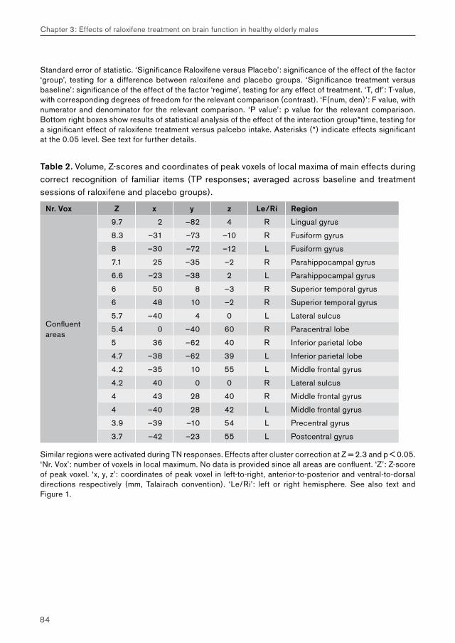
84
Chapter 3: Effects of raloxifene treatment on brain function in healthy elderly males
Standard error of statistic. ‘Significance Raloxifene versus Placebo’: significance of the effect of the factor ‘group’, testing for a difference between raloxifene and placebo groups. ‘Significance treatment versus baseline’: significance of the effect of the factor ‘regime’, testing for any effect of treatment. ‘T, df’: T-value, with corresponding degrees of freedom for the relevant comparison (contrast). ‘F(num, den)’: F value, with numerator and denominator for the relevant comparison. ‘P value’: p value for the relevant comparison. Bottom right boxes show results of statistical analysis of the effect of the interaction group*time, testing for a significant effect of raloxifene treatment versus palcebo intake. Asterisks (*) indicate effects significant at the 0.05 level. See text for further details.
Table 2. Volume, Z-scores and coordinates of peak voxels of local maxima of main effects during correct recognition of familiar items (TP responses; averaged across baseline and treatment sessions of raloxifene and placebo groups).
Nr. Vox Z x y z Le/Ri Region
Confluentareas
9.7 2 –82 4 R Lingual gyrus
8.3 –31 –73 –10 R Fusiform gyrus
8 –30 –72 –12 L Fusiform gyrus
7.1 25 –35 –2 R Parahippocampal gyrus
6.6 –23 –38 2 L Parahippocampal gyrus
6 50 8 –3 R Superior temporal gyrus
6 48 10 –2 R Superior temporal gyrus
5.7 –40 4 0 L Lateral sulcus
5.4 0 –40 60 R Paracentral lobe
5 36 –62 40 R Inferior parietal lobe
4.7 –38 –62 39 L Inferior parietal lobe
4.2 –35 10 55 L Middle frontal gyrus
4.2 40 0 0 R Lateral sulcus
4 43 28 40 R Middle frontal gyrus
4 –40 28 42 L Middle frontal gyrus
3.9 –39 –10 54 L Precentral gyrus
3.7 –42 –23 55 L Postcentral gyrus
Similar regions were activated during TN responses. Effects after cluster correction at Z = 2.3 and p < 0.05. ‘Nr. Vox’: number of voxels in local maximum. No data is provided since all areas are confluent. ‘Z’: Z-score of peak voxel. ‘x, y, z’: coordinates of peak voxel in left-to-right, anterior-to-posterior and ventral-to-dorsal directions respectively (mm, Talairach convention). ‘Le/Ri’: left or right hemisphere. See also text and Figure 1.
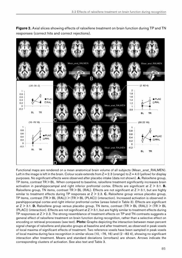
85
3.2 Effects of raloxifene treatment on brain function during recognition
Figure 2. Axial slices showing effects of raloxifene treatment on brain function during TP and TN responses (correct hits and correct rejections).
Functional maps are rendered on a mean anatomical brain volume of all subjects (Mean_anat_RALMEN). Left in the image is left in the brain. Colour scale extends from Z = 2.3 (orange) to Z = 4.0 (yellow) for display purposes. No significant effects were observed after placebo intake (data not shown). A. Raloxifene group, TP items, contrast TR > BL. When compared to baseline, raloxifene treatment significantly increases brain activation in parahippocampal and right inferior prefrontal cortex. Effects are significant at Z > 3.1. B. Raloxifene group, TN items, contrast TR > BL (RAL). Effects are not significant at Z > 3.1, but are highly similar to treatment effects during TP responses at Z > 2.3. C. Raloxifene group versus placebo group, TP items, contrast (TR > BL (RAL)) > (TR > BL (PLAC)) (interaction). Increased activation is observed in parahippocampal cortex and right inferior prefrontal cortex (areas listed in Table 3). Effects are significant at Z > 3.1. D. Raloxifene group versus placebo group, TN items, contrast (TR > BL (RAL)) > (TR > BL (PLAC)) (interaction). Effects are not significant at Z > 3.1, but are highly similar to treatment effects during TP responses at Z > 2.3. The strong resemblance of treatment effects on TP and TN contrasts suggests a general effect of raloxifene treatment on brain function during recognition, rather than a selective effect on encoding or retrieval processes (see text). Plots: Graphs depicting the interaction between mean percent signal change of raloxifene and placebo groups at baseline and after treatment, as observed in peak voxels of local maxima of significant effects of treatment. Two reference voxels have been sampled in peak voxels of local maxima during face recognition in similar slices (10, –76, 16) and (2 –82 4), showing no significant interaction after treatment. Means and standard deviations (errorbars) are shown. Arrows indicate the corresponding clusters of activation. See also text and Table 3.
86
Chapter 3: Effects of raloxifene treatment on brain function in healthy elderly males
Discussion
In a previous study, we reported effects of raloxifene treatment on brain activation during encoding of unfamiliar faces (Goekoop et al., 2005a). Results of the current study complement our previous findings by examining brain activation during recognition of the encoded items.
Effects of raloxifene treatment: behavioral dataAn unexpected drop in recognition accuracy was observed in the placebo group after 3 months of treatment, whereas performance in the raloxifene group increased slightly (Table 1). The nature of this drop in accuracy scores is unclear, but a number of explanations are possible. First, although group size of the current study is large to fMRI standards, it is rather small in terms of behavioral studies. Such small groups may show random differences in mean performance measures (Wilkinson & Halligan, 2004). Second, a drop in recognition accuracy at time2 (retest phase) with respect to time1 (test phase) is a regularly observed phenomenon in test-retest studies. Such effects are attributed to changes in context between test- and retest phases, and may involve ‘true’ decreases in sensitivity to old or new items as well as a change in response bias (i.e. subjects perform worse when they are insecure, or in an unfamiliar environment) (Feenan & Snodgrass, 1990). We therefore considered the possibility that a change in response bias was responsible for the observed decline in performance accuracy in the placebo group. A trend was observed for a difference in response bias (Br) in the placebo group (p = 0.08), but no significant effects of raloxifene versus placebo treatment were found. The possibility therefore exists that a change in response bias explains the observed decline in performance in the placebo group, but group size may not have been large enough to produce a significant difference. A change in response bias may reflect the subject’s sensitivity to interference by changes in context between encoding and retrieval phases, practicing and scanning stages, or between first and second scanning sessions (e.g. no practicing round was held the day before the second scanning session). Since test items did not overlap between different test versions and the factor ‘testversion’ was no significant confounder of task performance scores, we consider it unlikely that specific test items or the use of different test versions acted as significant distracters. Since both groups were matched with respect to age, gender and education level, we considered problems of group matching to be an unlikely source of performance differences between raloxifene and placebo groups. Additionally, a
87
3.2 Effects of raloxifene treatment on brain function during recognition
pathological decline of memory performance in the placebo group was considered unlikely, since these changes in performance occurred over a relatively short period of time (3 months) in otherwise healthy subjects (Goekoop et al., 2005a). Future studies may require to include more subjects and perform more detailed assessment of cognitive status (including measures of context-dependency), in order to evaluate the effects of pharmacological substances on brain function and mental performance.
Effects of raloxifene treatment: functional-behavioral relationshipsPrevious studies have shown that (small) behavioral changes do not necessarily translate into functional effects observed using fMRI at current group sizes (Wilkinson & Halligan, 2004). To examine whether performance changes in each group separately were in any way related to the observed functional effects of treatment, we studied the separate contributions of raloxifene and placebo groups to the overall pattern of treatment effects on brain function during recognition. Treatment with placebo had no significant effect on brain activation. Indeed, treatment effects were mainly due to effects of raloxifene intake (Figure 2A, 2B). This indicated that the drop in performance observed in the placebo group was not a significant confounder of functional effects of raloxifene treatment versus placebo as reported in this study (Figure 2C, 2D). Similarly, we examined the separate contributions of both groups to effects of raloxifene intake versus placebo on brain activation during encoding in our previous study (Goekoop et al., 2005a). Only the raloxifene group showed significant increases in brain activation in treatment-related areas, again indicating that placebo intake and subsequent performance changes were not a significant confounder of treatment effects during encoding.
Although further studies are necessary to corroborate our current findings, our data are consistent with the hypothesis that a ‘normal’ decrease in performance accuracy as a result of the subjects’ sensitivity to a difference in context between test- and retest phases was countered by an active process related to raloxifene treatment (Goekoop et al., 2005a). A reduction in the variability of recognition accuracy scores has been observed previously in elderly women receiving estrogen treatment (Wegesin & Stern, 2004). Additionally, a canceling of learning effects has been observed in male subjects receiving testosterone injections (Wolf et al., 2000), which may involve estrogen-mediated mechanisms (Wolf, 2003; Longcope et al., 1969). Such effects may involve effects of estrogens on arousal and attentional levels rather than direct effects on episodic memory. Estrogens are thought to influence brain function through a context-dependent effect on cortical arousal (Morgan et al., 2004). Such effects involve well-documented effects of estrogens on the four primary neuromodulatory
88
Chapter 3: Effects of raloxifene treatment on brain function in healthy elderly males
neurotransmitters that regulate cortical arousal states (i.e. serotonin, dopamine, noradrenaline and acetylcholine) (Bernardi et al., 2003; Korol, 2004). An increase in cholinergic (or noradrenergic) arousal is known to reflect an increase in signal-to-noise levels in neural networks, which translates into enhanced attention and working memory performance in animals and humans (Sarter et al., 2005). This mechanism of action is thought to be an important factor underlying improvement of memory performance in patients with Alzheimer’s disease that are treated with cholinesterase inhibitors (Sarter et al., 2005). Similarly, raloxifene treatment may have improved memory performance by enhancing cortical arousal (signal-to-noise-levels) during initial encoding, thereby reducing sensitivity to interference as a result of context-changes. Future studies may require to examine effects of raloxifene treatment on physiological measures of arousal and attention in order to examine this hypothesis in more detail.
Effects of raloxifene treatment: encoding versus recognition During face encoding, signal intensity during task performance was enhanced symmetrically across a wide range of neocortical areas (Goekoop et al., 2005a). This rather generalized enhancement of cortical brain activation during face encoding was interpreted as a global effect of raloxifene intake on cortical arousal, rather than a specific effect of raloxifene treatment on all cognitive domains represented by this pattern (Goekoop et al., 2005a). Treatment effects observed in the current study showed both similarities and differences with treatment effects during initial encoding. During both phases of memory performance, raloxifene intake produced increases in brain activation. Although decreases may have been possible in theory, these were not observed. The reason for the absence of signal decreases is unclear, but may reflect effects of increased arousal, since enhanced arousal levels increase rather than decrease the reactivity of neural networks (Coull, 1998; Morgan et al., 2004). In contrast to face encoding, however, treatment effects during face recognition did not involve a widespread increase of main effects of task performance in cortical structures. Direct effects of cortical arousal may therefore not have been relevant to treatment effects during face recognition. Indeed, a recent study shows that noradrenaline and neural steroids such as cortisol may modulate memory consolidation by interacting with arousal levels at initial encoding, rather than recognition (Cahill & Alkire, 2003). Although raloxifene intake may have influenced brain function during recognition independently of encoding, it is therefore possible that the observed effects of raloxifene treatment during recognition represent indirect, or ‘downstream’ effects of treatment effects during initial encoding, or consolidation processes occurring between the two tasks. Clearly, more
89
3.2 Effects of raloxifene treatment on brain function during recognition
research is needed to examine the neurodynamic changes underlying the observed effects of treatment, along with the possible interactions between treatment effects occurring at different stages of memory performance.
The current study shows that raloxifene treatment eventually affects brain function during memory retrieval. Since identical procedures of functional data analysis were performed, the asymmetric distribution of treatment effects across encoding and retrieval phases of memory performance suggests that the effects of raloxifene, like the effects of many other pharmacological substances, are process-specific (Honey & Bullmore, 2004). A direct statistical comparison of treatment effects between encoding and recognition phases was not performed, however, since these tasks were too dissimilar (i.e. block versus event-related designs) to allow meaningful comparisons. We therefore report our findings of the effects of cholinergic challenge separately for both memory tasks. Although encoding processes during attempted retrieval may differ from encoding during attempted encoding (Reber et al., 2002; Rombouts et al., 2001), studies of brain function during retrieval allow analyses of encoding and retrieval processes within the same scanning session (see materials and methods), which avoids some of the potential confounds that may be introduced by across-session comparisons (e.g. differences in task design, subject positioning, motion artifacts).
Significant effects of raloxifene treatment were found on brain activation during TP decisions (familiar items), but not on TN decisions (unfamiliar items) (Figure 2C). Although this may indicate a preference for raloxifene treatment to influence successful recognition rather than encoding during attempted retrieval, the small number of items analyzed per subject somewhat limit these findings (Table 1). Indeed, when the threshold for significant brain function was lowered for contrast images representing treatment effects during TN decisions, treatment effects were found that were nearly identical to those observed for TP decisions (Figure 2D). This suggested that raloxifene treatment affected more general aspects of recognition memory function that are examined equally by ‘loose’ comparisons of brain function during TP and TN decisions versus fixation (see materials and methods). In order to explore this possibility in more detail, we studied the effects of raloxifene on ‘tight’ TP <> TN comparisons of brain function, which examine specific encoding and retrieval processes during task performance (see materials and methods). No significant effects of raloxifene treatment were found, supporting the view that raloxifene treatment affected general aspects of recognition memory performance, rather than specific subcomponent processes during retrieval. Brain areas showing treatment effects for TP items included the right inferior frontal gyrus and left parahippocampal cortex (Figure 2C, Table 3). These structures are
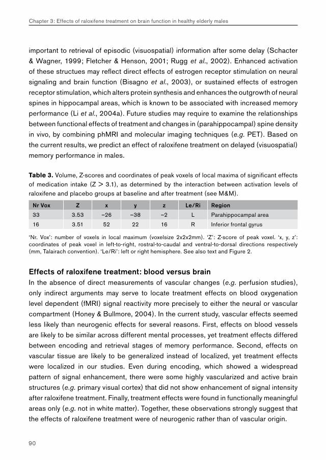
90
Chapter 3: Effects of raloxifene treatment on brain function in healthy elderly males
important to retrieval of episodic (visuospatial) information after some delay (Schacter & Wagner, 1999; Fletcher & Henson, 2001; Rugg et al., 2002). Enhanced activation of these structues may reflect direct effects of estrogen receptor stimulation on neural signaling and brain function (Bisagno et al., 2003), or sustained effects of estrogen receptor stimulation, which alters protein synthesis and enhances the outgrowth of neural spines in hippocampal areas, which is known to be associated with increased memory performance (Li et al., 2004a). Future studies may require to examine the relationships between functional effects of treatment and changes in (parahippocampal) spine density in vivo, by combining phMRI and molecular imaging techniques (e.g. PET). Based on the current results, we predict an effect of raloxifene treatment on delayed (visuospatial) memory performance in males.
Table 3. Volume, Z-scores and coordinates of peak voxels of local maxima of significant effects of medication intake (Z > 3.1), as determined by the interaction between activation levels of raloxifene and placebo groups at baseline and after treatment (see M&M).
Nr Vox Z x y z Le/Ri Region
33 3.53 –26 –38 –2 L Parahippocampal area
16 3.51 52 22 16 R Inferior frontal gyrus
‘Nr. Vox’: number of voxels in local maximum (voxelsize 2x2x2mm). ‘Z’: Z-score of peak voxel. ‘x, y, z’: coordinates of peak voxel in left-to-right, rostral-to-caudal and ventral-to-dorsal directions respectively (mm, Talairach convention). ‘Le/Ri’: left or right hemisphere. See also text and Figure 2.
Effects of raloxifene treatment: blood versus brainIn the absence of direct measurements of vascular changes (e.g. perfusion studies), only indirect arguments may serve to locate treatment effects on blood oxygenation level dependent (fMRI) signal reactivity more precisely to either the neural or vascular compartment (Honey & Bullmore, 2004). In the current study, vascular effects seemed less likely than neurogenic effects for several reasons. First, effects on blood vessels are likely to be similar across different mental processes, yet treatment effects differed between encoding and retrieval stages of memory performance. Second, effects on vascular tissue are likely to be generalized instead of localized, yet treatment effects were localized in our studies. Even during encoding, which showed a widespread pattern of signal enhancement, there were some highly vascularized and active brain structures (e.g. primary visual cortex) that did not show enhancement of signal intensity after raloxifene treatment. Finally, treatment effects were found in functionally meaningful areas only (e.g. not in white matter). Together, these observations strongly suggest that the effects of raloxifene treatment were of neurogenic rather than of vascular origin.
91
3.2 Effects of raloxifene treatment on brain function during recognition
Summary and conclusions Raloxifene treatment enhanced brain activation in male subjects during recognition of familiar items. A possible treatment mechanism involves enhanced memory retrieval as a result of increased cortical arousal during initial encoding, which reduces effects of context-dependency. Similar effects may underlie the ability of long-term raloxifene treatment to delay the onset of mild cognitive impairment in elderly women. Our combined studies on the effects of raloxifene treatment on episodic memory performance in male subjects predict the occurrence of small behavioral effects on working memory and delayed (visuospatial) memory performance, executive functions and verbal skills, which are secondary to enhanced arousal and attention during initial encoding. Further neuropsychological studies involving larger groups of subjects are necessary to test the validity of these predictions.
Acknowledgements
We would like to thank Stichting Alzheimer and the Neuropsychiatrie Foundation for financial support, and Ans Nicolaas-Merkus for subject recruitment. Serge Rombouts is supported by a grant from the Netherlands Organisation for Scientific Research (NWO; grantnr. 916.36.117).

Chapter 4
Effects of galantamine challenge on brain function in MCI and AD patients
4.1 Effects of galantamine challenge on brain function in MCI patients
Challenging the cholinergic system in mild cognitive impairment: a pharmacological fMRI study
Goekoop R, Rombouts SARB, Jonker C, Hibbel A, Knol DL, Truyen L, Barkhof F, Scheltens P.
Neuroimage 2004; 23(4):1450-9. PMID: 15589109
94
Chapter 4: Effects of galantamine challenge on brain function in MCI and AD patients
Abstract
Mild cognitive impairment (MCI) often represents an early form of Alzheimer’s Disease (AD). In both MCI and AD, characteristic cholinergic changes may occur. Functional magnetic resonance imaging (fMRI) may help to examine neurochemical changes in early disease, by studying signal reactivity to pharmacological challenge. In this study, MCI patients (n = 28; mean age 73.6 ± 7.5; MMSE 27.0 ± 1.2) were scanned during task performance in a randomized trial under three different medication regimes: at baseline (BL; no GAL), after a single oral dose of GAL (SD) and after prolonged exposure (Steady State: SS). Memory tasks included an episodic face encoding task and a parametric n-letter back working memory task. Alterations in brain activation patterns before and after treatment were analyzed for both tasks using multi-level statistical analysis. Significant increases in brain activation from BL were observed after prolonged exposure only. For face encoding (n = 28), these involved left prefrontal areas, the anterior cingulate gyrus, left occipital areas and left posterior hippocampus. For working memory (n = 28), increased activation was found in right precuneus and right middle frontal gyrus, coinciding with increased accuracy-scores after GAL-treatment. In conclusion, cholinergic challenge produces alterations in brain activation patterns in elderly MCI patients that can be detected with fMRI. This should encourage further functional imaging studies to examine the status of neurotransmitter systems in disease.
Keywords: fMRI; MCI; galantamine; challenge study; cholinergic system.
95
4.1 Effects of galantamine challenge on brain function in MCI patients
Introduction
Alzheimer’s Disease (AD) is characterized by progressive atrophy of medial temporal, frontal and parietal brain structures. These structural changes give rise to clinical symptoms such as amnesia, agnosia and aphasia (Braak et al., 1999; Geula, 1998). Atrophy of basal forebrain nuclei is another major feature in AD, resulting in pathological neurochemical changes throughout the brain. Of these, low acetylcholine levels in particular are thought to contribute significantly to symptoms in AD (Bartus, 2000; Mesulam, 2004). Current therapies against AD largely aim at restoring low acetylcholine levels with pharmacological agents (Lanctot et al., 2003; Trinh et al., 2003). Functional imaging techniques are currently being investigated for their ability to detect neurochemical changes and monitor effects of treatment in dementia (Burn & O’Brien, 2003; Freo et al., 2002).
Mild cognitive impairment (MCI) represents a functional continuum between normality and the earliest signs of dementia (most commonly AD). Patients with MCI are at increased risk of developing AD, but clinical outcome may vary considerably (Petersen et al., 2001). Recent evidence shows that alterations in cholinergic system activity also occur in MCI patients. Markers of cholinergic function are upregulated in MCI, possibly to compensate for incipient neurofunctional defects (DeKosky et al., 2002). Functional imaging studies of cholinergic system (re)activity in MCI patients may therefore reveal important clinical information. First, such studies may link the functional status of the cholinergic system to the occurrence of symptoms in very early AD, indicating the necessity of starting cholinomimetic therapy (Volkow et al., 2001). Second, reactivity to cholinergic challenge may predict clinical responsiveness to cholinomimetic treatment, indicating the sensibility of starting a particular therapy (Volkow et al., 2001; Nobili et al., 2002; Doraiswamy et al., 2000). Thirdly, the ability of the cholinergic system to compensate for small decreases in neural function (by altering cerebral plasticity and changing signal-to-noise levels in neural networks) is now increasingly recognized (Parry et al., 2003; Mesulam, 2004; Burggren et al., 2002). If this is true, cholinergic system ‘viability’ (Volkow et al., 2001), or residual function, may be a measure of ‘compensatory reserve’ (Mesulam, 2004) and may influence a patient’s prognosis.
In view of the above, we set out to study the feasibility of using fMRI to detect cholinergic system reactivity to selective pharmacological challenge in elderly patients with MCI. As a cholinomimetic agent, we used galantamine (GAL), which is a cholinesterase inhibitor with known therapeutic potential and has an additional modulatory effect on nicotinergic receptors (Raskind, 2003). To study the distribution
96
Chapter 4: Effects of galantamine challenge on brain function in MCI and AD patients
of GAL effects across memory systems, memory tasks were used that examine both episodic and working memory (WM) performance. To study the onset time of the effect, fMRI was performed after both a single dose challenge with GAL and after prolonged exposure.
Materials and Methods
Study DesignSubjects were screened for participation in a randomized study design in which patients themselves served as controls. fMRI was performed at baseline (BL, no medication), after oral intake of a single dose of 8 mg GAL with water (Single Dose; SD) and after prolonged exposure to GAL (Steady State (SS)); i.e. a 120 hour period (5 days) spread over 6 weekdays: 4 mg GAL (first gift, evening of day 1); 4 mg GAL b.i.d. (mornings and evenings; 4 consecutive days); 4 mg GAL (final gift, morning of day 6)). The minimum serum GAL concentration at 4 mg b.i.d. in healthy controls is 10.6 ± 4.0 ng/ml, with maximum levels 30.7 ± 6.2 ng/ml. At this rate, steady state plasma levels are reached within 2–3 days (Mannens et al., 2002; Zhao et al., 2002). Timing of scanning sessions was such that, on average, plasma levels could be considered equal at the time of scanning for SD and SS regimes, i.e. 3 hours after SD and 9 hours after SS regimes. BL, SD and SS regimes were randomized across scanning sessions to prevent session (e.g. learning) effects from interfering with possible effects of medication. To avoid carry-over effects, SD and SS regimes were separated by a washout period of two days of zero GAL intake (which is more than 6 times the half-life of galantamine (7.4 hours) (Mannens et al., 2002; Zhao et al., 2002). Scanning sessions were exactly one week apart and each patient was scanned on the same hour of day during all three sessions. If data acquisition failed, the subject’s consent was asked for a fourth and final scanning visit, for which the relevant medication regime was readministered, to obtain a maximum number of reliable datasets.
Subject recruitmentThirty healthy elderly MCI patients, aged 54 to 89 years (mean 73.6 ± 7.7) were recruited from the Alzheimer Center at the VU Medical Center, Amsterdam, the Netherlands. MCI patients were diagnosed using Petersen’s criteria for amnestic MCI, i.e. a slowly progressive memory decline without the involvement of any other domain of cognitive function, that did not interfere significantly with activities of daily living (Petersen et
97
4.1 Effects of galantamine challenge on brain function in MCI patients
al., 2001). Before inclusion in the study, elaborate neuropsychological profiling was performed during clinical investigation. Inclusion criteria were a mini mental state examination (MMSE) (Folstein et al., 1975b) score of 26 or higher, a clinical dementia rating (CDR) scale score of 0.5 (Morris, 1993), and a New York University (NYU) paragraph recall test-score not exceeding 10 items on delayed recall. Formal education was determined on a discrete scale with three levels (1 = low, 2 = middle, 3 = high). All patients provided informed consent under supervision of a lawful caretaker during a screening visit in which the procedure was explained and contraindications were checked. Patients were excluded if they had any significant medical, neurological or psychiatric illness, or if they were taking medication or other substances that are known to influence cerebral function, including antidepressants and other cholinesterase inhibitors. Patients were excluded if their history showed excessive nicotine or alcohol intake (> 0.5 packs of cigarettes, > 8 glasses of an alcoholic substance a day), a severe allergy to pharmacological substances or their constitutive compounds, or the use of any experimental medication within three months prior to enrollment in this trial. Exclusion criteria to MRI involved the presence of a pacemaker, metallic implants in high-risk areas (i.e. vessel clips) and a history of claustrophobia.
Functional MRI (fMRI)Data acquisition
Imaging was carried out on a 1.5 T Sonata MR scanner (Siemens, Erlangen, Germany), using a standard circularly polarized head coil with foam padding to restrict head motion. For fMRI, an echo planar imaging sequence was used (echo time 60 ms, flip angle 90°, matrix 64 × 64, field of view 192 × 192 mm), to obtain 21 transverse slices (thickness 5 mm, interslice-gap 1 mm). Task stimuli were projected on a screen located at the head end of the scanner table via an LCD projector located outside the scanner room. Subjects viewed the screen through a mirror located on the head coil. In each hand, subjects held an fMRI compatible response-box through which they were able to react to task stimuli by pressing a single button using one of their index-fingers. A T1–weighted structural MRI-scan was obtained of each subject (MPRAGE; inversion time: 300 ms, TR = 15 ms; TE = 7 ms; flip angle = 8°; 160 coronal slices, 1 × 1 × 1.5 mm voxels).
Paradigms (memory-tasks)
Two paradigms were used to examine different types of memory function. A well-stablished face-encoding task was used to cover the intermediate-term episodic memory domain.
98
Chapter 4: Effects of galantamine challenge on brain function in MCI and AD patients
This task produces activation in visual, parietal, prefrontal, anterior cingulate, and medial temporal brain structures bilaterally (Small et al., 1999). A parametric N-letter back task was used to examine brain activation during working memory (WM) performance. This task activates a combination of frontal and parietal brain structures and the anterior cingulate gyri (Braver et al., 1997). Given the hypothetical dichotomy between episodic memory (EM) and working memory (WM), an attempt was made to map the effects of GAL challenge to alterations in brain activation patterns within distinct brain structures and their associated memory systems. Three different but comparable versions of each paradigm were constructed and randomized across the scanning-sessions (BL, SD, SS). A fourth ‘spare’ version was made in case data-acquisition might fail during one of the scanning sessions. All paradigms were practised extensively using dummy tasks to ensure that patients mastered the general procedure of task performance before scanning (one day before the start of the first session on a laptop computer during a home visit: both paradigms; fifteen minutes before the onset of each scanning session: n-back task; five minutes before the onset of the first measurements, with subject in scanner: face encoding paradigm). During the first 10.5s of each memory-task, subjects saw a circle indicating time left before the onset of the first condition. To avoid investigator’s bias, task performance data were only viewed after all three scan visits had been completed. Total time for one scanning session including instructions of memory tasks was approximately one hour.
Episodic memory task (face encoding)
The encoding task consisted of a simple block-design with two alternating conditions: Condition 1 (‘ENCOD’) consisted of 4 blocks of 42s each. Each block contained 6 unfamiliar faces presented sequentially in random order on a black background (presentation time 6s, followed by a 1s delay). Male and female faces were balanced across the blocks. Condition 2 (‘FIX’) consisted of 4 blocks of 44s each, in which a white fixation cross (X) was presented on a black background. The entire task thus consisted of 8 alternating blocks of ENCOD and FIX conditions and was preceded by a 21s FIX condition. Prior to scanning, subjects were instructed verbally to remember the faces for future testing and to classify each face according to its gender by pressing one of two buttons (written instructions “Left: male”, “Right: female” also appearing alongside the pictures). Mean reaction times for gender discrimination were recorded and a performance accuracy-score varying from –1 to 1 (with 0 indicating chance level) was calculated by subtracting false answers from correct answers and dividing the result by the total number of items (24). Total task duration was 6’12’’minutes.
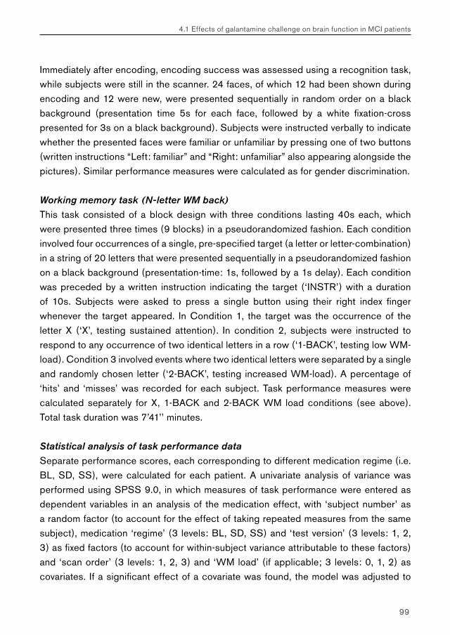
99
4.1 Effects of galantamine challenge on brain function in MCI patients
Immediately after encoding, encoding success was assessed using a recognition task, while subjects were still in the scanner. 24 faces, of which 12 had been shown during encoding and 12 were new, were presented sequentially in random order on a black background (presentation time 5s for each face, followed by a white fixation-cross presented for 3s on a black background). Subjects were instructed verbally to indicate whether the presented faces were familiar or unfamiliar by pressing one of two buttons (written instructions “Left: familiar” and “Right: unfamiliar” also appearing alongside the pictures). Similar performance measures were calculated as for gender discrimination.
Working memory task (N-letter WM back)
This task consisted of a block design with three conditions lasting 40s each, which were presented three times (9 blocks) in a pseudorandomized fashion. Each condition involved four occurrences of a single, pre-specified target (a letter or letter-combination) in a string of 20 letters that were presented sequentially in a pseudorandomized fashion on a black background (presentation-time: 1s, followed by a 1s delay). Each condition was preceded by a written instruction indicating the target (‘INSTR’) with a duration of 10s. Subjects were asked to press a single button using their right index finger whenever the target appeared. In Condition 1, the target was the occurrence of the letter X (‘X’, testing sustained attention). In condition 2, subjects were instructed to respond to any occurrence of two identical letters in a row (‘1-BACK’, testing low WM-load). Condition 3 involved events where two identical letters were separated by a single and randomly chosen letter (‘2-BACK’, testing increased WM-load). A percentage of ‘hits’ and ‘misses’ was recorded for each subject. Task performance measures were calculated separately for X, 1-BACK and 2-BACK WM load conditions (see above). Total task duration was 7’41’’ minutes.
Statistical analysis of task performance data
Separate performance scores, each corresponding to different medication regime (i.e. BL, SD, SS), were calculated for each patient. A univariate analysis of variance was performed using SPSS 9.0, in which measures of task performance were entered as dependent variables in an analysis of the medication effect, with ‘subject number’ as a random factor (to account for the effect of taking repeated measures from the same subject), medication ‘regime’ (3 levels: BL, SD, SS) and ‘test version’ (3 levels: 1, 2, 3) as fixed factors (to account for within-subject variance attributable to these factors) and ‘scan order’ (3 levels: 1, 2, 3) and ‘WM load’ (if applicable; 3 levels: 0, 1, 2) as covariates. If a significant effect of a covariate was found, the model was adjusted to
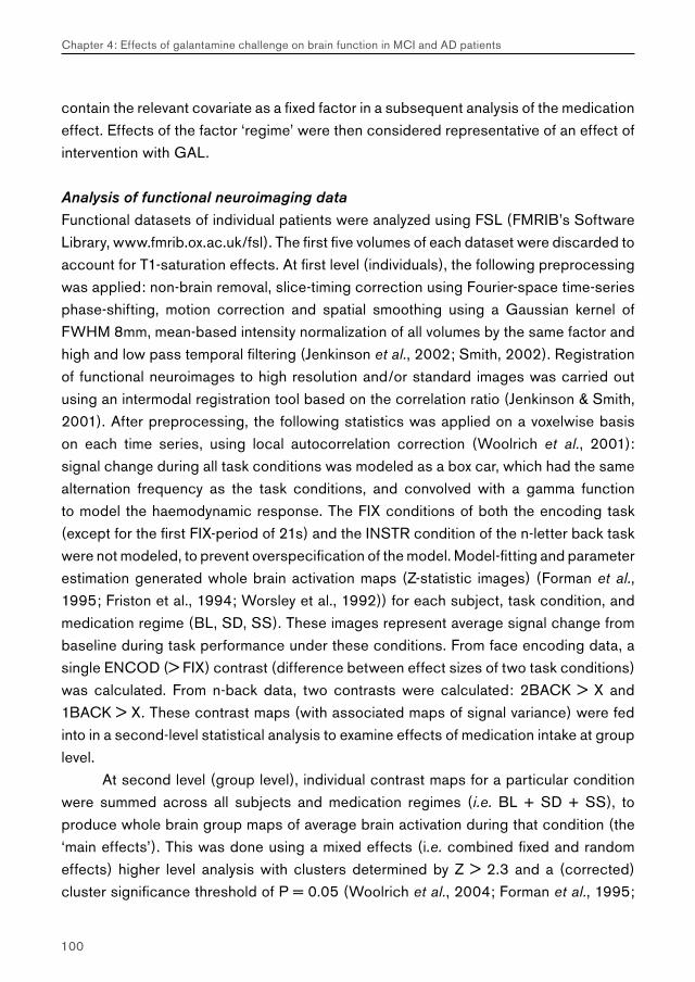
100
Chapter 4: Effects of galantamine challenge on brain function in MCI and AD patients
contain the relevant covariate as a fixed factor in a subsequent analysis of the medication effect. Effects of the factor ‘regime’ were then considered representative of an effect of intervention with GAL.
Analysis of functional neuroimaging data
Functional datasets of individual patients were analyzed using FSL (FMRIB’s Software Library, www.fmrib.ox.ac.uk/fsl). The first five volumes of each dataset were discarded to account for T1-saturation effects. At first level (individuals), the following preprocessing was applied: non-brain removal, slice-timing correction using Fourier-space time-series phase-shifting, motion correction and spatial smoothing using a Gaussian kernel of FWHM 8mm, mean-based intensity normalization of all volumes by the same factor and high and low pass temporal filtering (Jenkinson et al., 2002; Smith, 2002). Registration of functional neuroimages to high resolution and/or standard images was carried out using an intermodal registration tool based on the correlation ratio (Jenkinson & Smith, 2001). After preprocessing, the following statistics was applied on a voxelwise basis on each time series, using local autocorrelation correction (Woolrich et al., 2001): signal change during all task conditions was modeled as a box car, which had the same alternation frequency as the task conditions, and convolved with a gamma function to model the haemodynamic response. The FIX conditions of both the encoding task (except for the first FIX-period of 21s) and the INSTR condition of the n-letter back task were not modeled, to prevent overspecification of the model. Model-fitting and parameter estimation generated whole brain activation maps (Z-statistic images) (Forman et al., 1995; Friston et al., 1994; Worsley et al., 1992)) for each subject, task condition, and medication regime (BL, SD, SS). These images represent average signal change from baseline during task performance under these conditions. From face encoding data, a single ENCOD (> FIX) contrast (difference between effect sizes of two task conditions) was calculated. From n-back data, two contrasts were calculated: 2BACK > X and 1BACK > X. These contrast maps (with associated maps of signal variance) were fed into in a second-level statistical analysis to examine effects of medication intake at group level.
At second level (group level), individual contrast maps for a particular condition were summed across all subjects and medication regimes (i.e. BL + SD + SS), to produce whole brain group maps of average brain activation during that condition (the ‘main effects’). This was done using a mixed effects (i.e. combined fixed and random effects) higher level analysis with clusters determined by Z > 2.3 and a (corrected) cluster significance threshold of P = 0.05 (Woolrich et al., 2004; Forman et al., 1995;
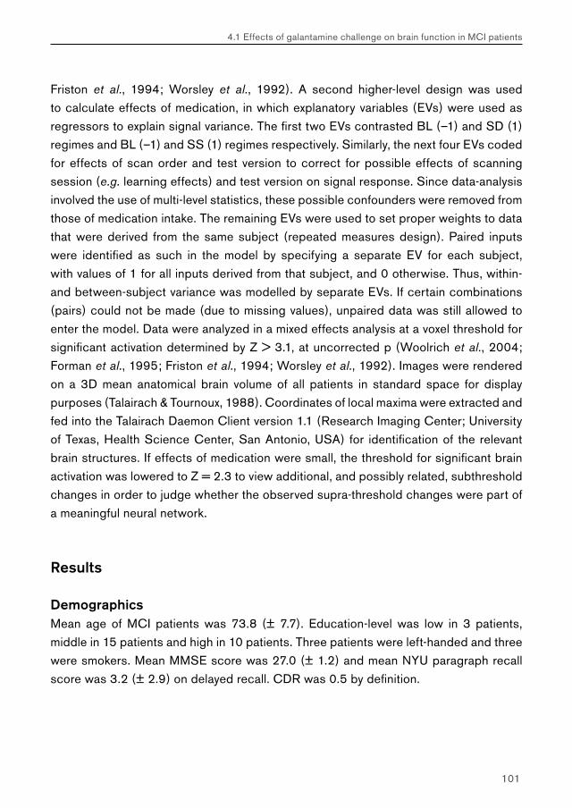
101
4.1 Effects of galantamine challenge on brain function in MCI patients
Friston et al., 1994; Worsley et al., 1992). A second higher-level design was used to calculate effects of medication, in which explanatory variables (EVs) were used as regressors to explain signal variance. The first two EVs contrasted BL (–1) and SD (1) regimes and BL (–1) and SS (1) regimes respectively. Similarly, the next four EVs coded for effects of scan order and test version to correct for possible effects of scanning session (e.g. learning effects) and test version on signal response. Since data-analysis involved the use of multi-level statistics, these possible confounders were removed from those of medication intake. The remaining EVs were used to set proper weights to data that were derived from the same subject (repeated measures design). Paired inputs were identified as such in the model by specifying a separate EV for each subject, with values of 1 for all inputs derived from that subject, and 0 otherwise. Thus, within- and between-subject variance was modelled by separate EVs. If certain combinations (pairs) could not be made (due to missing values), unpaired data was still allowed to enter the model. Data were analyzed in a mixed effects analysis at a voxel threshold for significant activation determined by Z > 3.1, at uncorrected p (Woolrich et al., 2004; Forman et al., 1995; Friston et al., 1994; Worsley et al., 1992). Images were rendered on a 3D mean anatomical brain volume of all patients in standard space for display purposes (Talairach & Tournoux, 1988). Coordinates of local maxima were extracted and fed into the Talairach Daemon Client version 1.1 (Research Imaging Center; University of Texas, Health Science Center, San Antonio, USA) for identification of the relevant brain structures. If effects of medication were small, the threshold for significant brain activation was lowered to Z = 2.3 to view additional, and possibly related, subthreshold changes in order to judge whether the observed supra-threshold changes were part of a meaningful neural network.
Results
DemographicsMean age of MCI patients was 73.8 (± 7.7). Education-level was low in 3 patients, middle in 15 patients and high in 10 patients. Three patients were left-handed and three were smokers. Mean MMSE score was 27.0 (± 1.2) and mean NYU paragraph recall score was 3.2 (± 2.9) on delayed recall. CDR was 0.5 by definition.
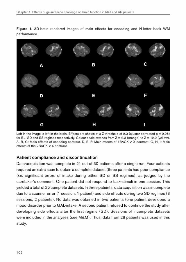
102
Chapter 4: Effects of galantamine challenge on brain function in MCI and AD patients
Figure 1. 3D-brain rendered images of main effects for encoding and N-letter back WM performance.
Left in the image is left in the brain. Effects are shown at a Z-threshold of 2.3 (cluster corrected p = 0.05) for BL, SD and SS regimes respectively. Colour scale extends from Z = 2.3 (orange) to Z = 12.0 (yellow). A, B, C: Main effects of encoding contrast. D, E, F: Main effects of 1BACK > X contrast. G, H, I: Main effects of the 2BACK > X contrast.
Patient compliance and discontinuationData-acquisition was complete in 21 out of 30 patients after a single run. Four patients required an extra scan to obtain a complete dataset (three patients had poor compliance (i.e. significant errors of intake during either SD or SS regimes), as judged by the caretaker’s comment. One patient did not respond to task-stimuli in one session. This yielded a total of 25 complete datasets. In three patients, data acquisition was incomplete due to a scanner error (1 session, 1 patient) and side effects during two SD regimes (3 sessions, 2 patients). No data was obtained in two patients (one patient developed a mood disorder prior to GAL-intake. A second patient refused to continue the study after developing side effects after the first regime (SD). Sessions of incomplete datasets were included in the analyses (see M&M). Thus, data from 28 patients was used in this study.
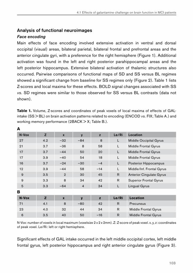
103
4.1 Effects of galantamine challenge on brain function in MCI patients
Analysis of functional neuroimagesFace encoding
Main effects of face encoding involved extensive activation in ventral and dorsal occipital (visual) areas, bilateral parietal, bilateral frontal and prefrontal areas and the anterior cingulate gyri, with a preference for the right hemisphere (Figure 1). Additional activation was found in the left and right posterior parahippocampal areas and the left posterior hippocampus. Extensive bilateral activation of thalamic structures also occurred. Pairwise comparisons of functional maps of SD and SS versus BL regimes showed a significant change from baseline for SS regimes only (Figure 2). Table 1 lists Z-scores and local maxima for these effects. BOLD signal changes associated with SS vs. SD regimes were similar to those observed for SS versus BL contrasts (data not shown).
Table 1. Volume, Z-scores and coordinates of peak voxels of local maxima of effects of GAL-intake (SS > BL) on brain activation patterns related to encoding (ENCOD vs. FIX; Table A.) and working memory performance (2BACK > X; Table B.).
AN-Vox Z x y z Le/Ri Location
27 4.2 –32 –84 8 L Middle Occipital Gyrus
21 3.7 –36 8 58 L Middle Frontal Gyrus
17 3.7 –44 50 20 L Middle Frontal Gyrus
17 3.9 –40 54 18 L Middle Frontal Gyrus
16 3.7 –24 –30 –4 L Posterior Hippocampus
12 3.9 –44 58 –14 L Middle/Inf. Frontal Gyrus
9 3.5 2 30 45 R Anterior Cingulate Gyrus
9 3.3 8 34 42 R Superior Frontal Gyrus
5 3.3 –64 4 34 L Lingual Gyrus
BN-Vox Z x y z Le/Ri Location
71 4.1 8 –80 42 R Precuneus
23 4.0 32 44 34 R Middle Frontal Gyrus
6 3.5 40 50 –16 R Middle Frontal Gyrus
N-Vox: number of voxels in local maximum (voxelsize 2 x 2 x 2mm). Z: Z-score of peak voxel. x, y, z: coordinates of peak voxel. Le/Ri: left or right hemisphere.
Significant effects of GAL intake occurred in the left middle occipital cortex, left middle frontal gyrus, left posterior hippocampus and right anterior cingulate gyrus (Figure 3).
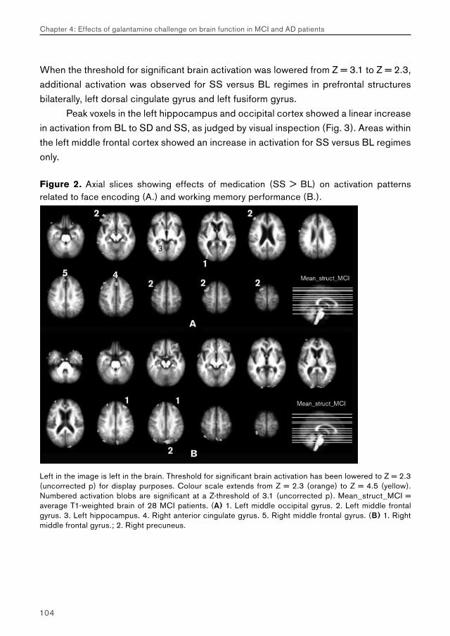
104
Chapter 4: Effects of galantamine challenge on brain function in MCI and AD patients
When the threshold for significant brain activation was lowered from Z = 3.1 to Z = 2.3, additional activation was observed for SS versus BL regimes in prefrontal structures bilaterally, left dorsal cingulate gyrus and left fusiform gyrus.
Peak voxels in the left hippocampus and occipital cortex showed a linear increase in activation from BL to SD and SS, as judged by visual inspection (Fig. 3). Areas within the left middle frontal cortex showed an increase in activation for SS versus BL regimes only.
Figure 2. Axial slices showing effects of medication (SS > BL) on activation patterns related to face encoding (A.) and working memory performance (B.).
Left in the image is left in the brain. Threshold for significant brain activation has been lowered to Z = 2.3 (uncorrected p) for display purposes. Colour scale extends from Z = 2.3 (orange) to Z = 4.5 (yellow). Numbered activation blobs are significant at a Z-threshold of 3.1 (uncorrected p). Mean_struct_MCI = average T1-weighted brain of 28 MCI patients. (A) 1. Left middle occipital gyrus. 2. Left middle frontal gyrus. 3. Left hippocampus. 4. Right anterior cingulate gyrus. 5. Right middle frontal gyrus. (B) 1. Right middle frontal gyrus.; 2. Right precuneus.
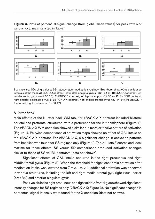
105
4.1 Effects of galantamine challenge on brain function in MCI patients
Figure 3. Plots of percentual signal change (from global mean values) for peak voxels of various local maxima listed in Table 1.
BL: baseline; SD: single dose; SS: steady state medication regimes. Error-bars show 95% confidence intervals of the mean A: ENCOD contrast, left middle occipital gyrus (-32 –84 8). B: ENCOD contrast, left middle frontal gyrus (–44 50 20). C: ENCOD contrast, left hippocampus (-24 30 4). D: ENCOD contrast; right anterior cingulate gyrus E: 2BACK > X contrast, right middle frontal gyrus (32 44 34). F: 2BACK > X contrast, right precuneus (8 –80 42).
N-letter back
Main effects of the N-letter back WM task for 1BACK > X contrast included bilateral parietal and prefrontal structures, with a preference for the left hemisphere (Figure 1). The 2BACK > X WM condition showed a similar but more extensive pattern of activation (Figure 1). Pairwise comparisons of activation maps showed no effect of GAL-intake on the 1BACK > X contrast. For 2BACK > X, a significant change in activation patterns from baseline was found for SS regimes only (Figure 2). Table 1 lists Z-scores and local maxima for these effects. SS versus SD comparisons produced activation changes similar to those of SS vs. BL contrasts (data not shown).
Significant effects of GAL intake occurred in the right precuneus and right middle frontal gyrus (Figure 3). When the threshold for significant brain activation after medication intake was lowered from Z = 3.1 to 2.3, additional activation was observed in various structures, including the left and right medial frontal gyri, right visual cortex (area V2) and anterior cingulate gyrus.
Peak voxels in the right precuneus and right middle frontal gyrus showed significant intensity changes for SS regimes only (2BACK > X; Figure 3). No significant changes in percentual signal intensity were found for the X-condition (data not shown).
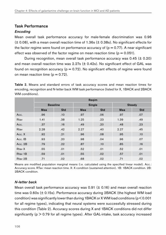
106
Chapter 4: Effects of galantamine challenge on brain function in MCI and AD patients
Task PerformanceEncoding
Mean overall task performance accuracy for male-female discrimination was 0.96 (± 0.08), with a mean overall reaction time of 1.36s (± 0.38s). No significant effects for the factor regime were found on performance accuracy of (p = 0.77). A near significant effect was observed of the factor regime on mean reaction time (p = 0.051).
During recognition, mean overall task performance accuracy was 0.45 (± 0.20) and mean overall reaction time was 2.27s (± 0.42s). No significant effect of GAL was found on recognition accuracy (p = 0.72). No significant effects of regime were found on mean reaction time (p = 0.72).
Table 2. Means and standard errors of task accuracy scores and mean reaction times for encoding, recognition and N-letter back WM task performance (listed for X, 1BACK and 2BACK WM conditions).
Reqim
Baseline Single Steady
Mea Std Mea Std Mea Std
Acc. .96 .10 .97 .06 .97 .07
Rtav 1.41 .38 1.25 .23 1.39 .49
Acc. .37 .16 .49 .20 .48 .23
Rtav 2.28 .42 2.27 .40 2.27 .45
Acc. X .92 .21 .96 .08 .95 .10
Acc. IB .93 .20 .98 .04 .96 .07
Acc. 2B .79 .22 .87 .10 .85 .16
Rtav X .55 .01 .52 .01 .52 .01
Rtav 1B .59 .01 .55 .02 .57 .01
Rtav 2B .71 .02 .68 .02 .71 .02
Means are modified population marginal means (i.e. calculated using the specified linear model). Acc.: Accuracy score. RTav: mean reaction time. X: X condition (sustained attention). 1B: 1BACK condition. 2B: 2BACK condition.
N-letter back
Mean overall task performance accuracy was 0.91 (± 0.16) and mean overall reaction time was 0.60s (± 0.15s). Performance accuracy during 2BACK (the highest WM load condition) was significantly lower than during 1BACK or X WM load conditions (p < 0.001 for all regime types), indicating that neural systems were successfully stressed during this condition (Table 2). Accuracy scores during X and 1BACK conditions did not differ significantly (p > 0.79 for all regime types). After GAL-intake, task accuracy increased
107
4.1 Effects of galantamine challenge on brain function in MCI patients
(p = 0.004) and latency decreased (p = 0.046) significantly from baseline values. The effect on task accuracy was only significant across WM load conditions, but not for each WM load condition separately (i.e. X: p = 0.18; 1BACK: p = 0.30; 2BACK: p = 0.053). This was also true for task latency (i.e. X: p = 0.20; 1BACK: p = 0.17; 2BACK: p = 0.31). No significant effect was found for the interaction regime × WM load on either accuracy (p = 0.11) or latency (p = 0.65) scores, indicating that GAL intake did not preferentially influence performance accuracy for any particular WM load condition. The increase in task accuracy from BL was largest during SS (p < 0.01). The maximum effect on task latency occurred after SD intake (p = 0.042).
Discussion
The functional status of the cholinergic system is thought to contribute significantly to symptoms in AD (Bartus, 2000; Mesulam, 2004). In advanced Alzheimer’s Disease, low levels of cholinergic markers have been found post-mortem (DeKosky et al., 2002). The anticholinergic agent scopolamine may induce acute symptoms of moderately-severe amnesia in healthy controls, making pharmacologically induced memory-loss a successful model to study the cholinergic contribution to AD symptomatology (Assal & Cummings, 2002). Additionally, cholinergic system dysfunction in AD patients has been linked to a decrease in verbal fluency and neuropsychiatric symptoms such as mood disorders, psychotic states, and a lack of concentration and mental agility (Assal & Cummings, 2002). Restoring acetylcholine levels pharmacologically may significantly benefit AD patients (Lanctot et al., 2003; Trinh et al., 2003). Studies examining cholinergic system (re)activity in early AD may therefore be of clinical value. This study demonstrates the feasibility of using fMRI to detect cholinergic system reactivity in elderly patients with MCI.
It is not exactly known how the cholinergic system is able to regulate neural activity. Cholinergic stimulation may improve neural information processing by increasing the signal-to-noise ratio of neural networks, while cholinergic inhibition has the opposite effect (Mesulam, 1996; Little et al., 1998; Rezvani & Levin, 2001). This mechanism is however not disease-specific, i.e. when given to healthy controls, cholinomimetic drugs may improve attention and memory performance as well (which is why they are sometimes referred to as ‘smart-drugs’ (Rose, 2002)). From the small number of studies that investigated the anatomy of the human cholinergic system, it is known to originate within various nuclei of the basal forebrain, particularly Ch2 and Ch4, from which
108
Chapter 4: Effects of galantamine challenge on brain function in MCI and AD patients
acetylcholine is transported by discrete bilateral bundles of afferents and distributed diffusely across the hippocampal area and entire neocortex respectively (Geula, 1998; Selden et al., 1998). The cholinergic system plays an important role in modulating brain activity in networks associated with a variety of neural functions, particularly arousal, attention and working memory performance (Clarke, 1995; Levin & Simon, 1998; Rezvani & Levin, 2001). A study by Furey et al. showed that the administration of the cholinesterase inhibitor physiostigmine to healthy controls increased activation in the (ventral) occipital cortex and reduced prefrontal activation load during spatial WM performance, suggesting that cholinergic stimulation increased the efficiency of visual perceptive processing and (hence) decreased effortful processing in prefrontal areas (Furey et al., 2000). Several other neuroimaging studies have investigated the effects of cholinomimetic and anticholinergic substances on brain function in healthy controls (Ernst et al., 2001; Kumari et al., 2003; Lawrence et al., 2002; Parry et al., 2003; Sperling et al., 2002) and AD patients (Rombouts et al., 2000). These were the first fMRI studies to demonstrate alterations in brain activation patterns during memory task performance after cholinergic modulation. Cholinergic stimulation increased activation mainly in fusiform areas during episodic memory performance (face encoding) (Rombouts et al., 2002), while cholinergic inhibition reduced activity in fusiform, hippocampal and prefrontal areas (Sperling et al., 2002). Effects of cholinergic enhancement on WM related brain activation included (left) prefrontal, cingulate, parietal and occipital regions (Ernst et al., 2001; Due et al., 2002; Kumari et al., 2003; Parry et al., 2003; Furey et al., 2000; Lawrence et al., 2002). The various study populations generally showed an improvement in task performance measures after cholinergic stimulation.
In this study, GAL challenge resulted in a significant signal increase for SS, but not SD, versus BL regimes in MCI patients. Pairwise comparisons of SS and SD regimes showed similar activation patterns as those for SS vs. BL comparisons, again indicating that activation during SD was minimal (Figure 1). This does not mean that SD application of GAL had no effect on brain activation. Some regions showed a gradual increase from BL for SD and SS regimes (Figure 3). With plasma levels of GAL considered near equal for both regimes at the time of scanning (see Materials & Methods), such a difference in magnitude of response may be due to the effects of exposure-time to GAL. A longer period of exposure may have provided more time for synaptic rearrangement of task-related networks resulting in more activation when these networks were stimulated (Fujii et al., 1999). Since however SD related changes in brain activation were non-significant, we will focus on SS versus BL effects only.
109
4.1 Effects of galantamine challenge on brain function in MCI patients
Overall, the observed changes in brain activation after GAL challenge were small in number, extent and intensity, even though we scanned a relatively large sample of patients (n = 28). This may be due to the fact that scans of elderly patients may show greater anatomical and haemodynamic variability than healthy age-matched controls and contain more movement artifacts, which may hamper signal detection (D’Esposito et al., 2003). Also, activation levels may be decreased in MCI patients (Machulda et al., 2003). Despite such constraints, increased activation was observed in areas within the left prefrontal cortex, left hippocampus and left medial occipital gyrus during performance on an episodic memory task (face encoding). When the threshold for significant brain activation was lowered (Z = 2.3), effects were found in prefrontal structures bilaterally, the (anterior) cingulate gyrus and visual cortex. These structures together have been proposed to form a (face) encoding network (Druzgal & D’Esposito, 2001) and previous studies have shown that cholinergic modulation may influence this network (Rombouts et al., 2002; Sperling et al., 2001). Neuropsychological interpretation of the observed effects is difficult, since activation observed during the ENCOD condition was somewhat aspecific to episodic memory (additional activation was produced, e.g. visual and possibly working memory-related activation). By choosing a very low-level baseline (i.e. fixation cross), we aimed to increase contrast (to raise chances of finding significant activation changes after GAL treatment), but at the cost of some neuropsychological specificity. Medio-temporal and especially hippocampal activation, however, are considered specific to episodic memory (Schacter & Wagner, 1999), indicating that the observed change in hippocampal activation after GAL challenge may well have involved episodic memory performance. This is in line with previous reports saying that GAL may influence episodic memory (Raskind, 2003). Task performance measures in this study however did not corroborate this finding. Neither attentional measures during encoding, nor recognition scores after encoding showed significant changes after GAL intake. This may reflect low statistical power for this type of analysis (i.e. small sample size and a ceiling effect during male-female discrimination). Alternatively, there may be no relation between enhanced activity in the (posterior) hippocampal area and increased episodic memory performance. The hippocampal area, however, is among the first to show neuropathological changes in Alzheimer’s Disease (Braak et al., 1999) and may have decreased activation levels in early AD (Rombouts et al., 2000). The observed hippocampal reactivity to cholinergic stimulation may therefore bear some clinical relevance.
Effects of GAL-intake on activation patterns during performance on the lower WM load condition (1BACK > X) were not significant. Main effects for this task condition
110
Chapter 4: Effects of galantamine challenge on brain function in MCI and AD patients
involved effect sizes of smaller magnitude than those observed during increased WM load performance (2BACK > X), indicating that the lower WM load condition required less neural processing. A possible effect of GAL intake on tissue excitability might therefore not have become explicit. The 2BACK > X contrast did show an increase after GAL treatment (SS) in some areas. Effects mainly included the right precuneus and left middle frontal gyrus. Again, this only partly represented areas known to increase their activation levels in healthy controls after cholinergic stimulation. When the threshold for significant brain activation was lowered, additional effects were found in the middle frontal gyrus bilaterally, visual cortex, caudate nucleus and left temporal lobe. Similar areas have been reported in healthy controls after nicotine intake, possibly representing a cholinergic effect on visual attention (Lawrence et al., 2002). Plots representing percentual signal change showed that GAL intake did not significantly alter activation levels during the X condition in areas where an increase in the 2BACK > X contrast was found. Hence, the enhancement of the 2BACK > X contrast in these areas involved a true increase in activation during the 2BACK condition (Figure 3). Task performance data showed a highly significant effect of GAL-treatment on task accuracy scores (p = 0.004), and a significant decrease in reaction times (p = 0.046), indicating a clear effect of GAL intake on neural function associated with this task. This effect however was not more pronounced for any of the three WM conditions, suggesting that GAL intake consitituted a non-specific effect on (sustained) attentional systems. This conclusion is supported by the literature on the functions of the cholinergic system (Levin & Simon, 1998; Rezvani & Levin, 2001). Although no effect of GAL intake was found on activation levels during the X condition, this does not preclude a role of GAL in the enhancement of attentional processing. Rather, performance scores during the X condition may not optimally reflect attentional processing and 2BACK > X might be a more suitable measure.
The current study used two rather unrelated paradigms, which have independently shown reproducible alterations in brain activation in AD as compared to controls (Braver et al., 1997; Small et al., 1999). This was done to raise chances of finding significant effects of medication intake, but prevented useful comparisons between activation levels during encoding and working memory performance (and no randomisation was done of the order in which both tasks were administered). Future studies however may require to use paradigms that allow tighter comparisons between episodic memory and working memory performance.
This study was not placebo-controlled. We should therefore be cautious to conclude that the observed changes were due to the effects of GAL alone. Placebo-
111
4.1 Effects of galantamine challenge on brain function in MCI patients
effects however, if present, should probably have occurred in comparable magnitude after acute as well as chronic dosage, and the absence of effects after acute dosage (but the presence of effects after chronic dosage), suggests that possible placebo effects remained sub-threshold. The (highly) significant effect on attentional and WM task performance scores when compared to face recognition, further makes it unlikely that the findings in this study were solely due to a placebo effect. Effects of task version and especially scan order, which were accounted for in this study, may prove significant confounders in any study examining pharmacological effects on brain activation. With these effects removed, the residual signal changes may be considered more true representations of an effect of medication intake.
In conclusion, this study has listed several brain areas in MCI patients that show increased activation after GAL challenge. This seems to confirm the feasibility of using fMRI to study cholinergic system reactivity in elderly patients with a possible cholinergic deficit. Longitudinal studies should determine whether a differential response to cholinergic stimulation exists in MCI patients that show further decline in mental functions versus patients with more stable functions. Such a differential response may point to a role of the cholinergic system in compensating for small incipient neurofunctional defects. Furthermore, the spectrum of symptoms and signs of MCI patients can be correlated with cholinergic system reactivity, and vice versa. This may allow us to determine the relative contribution of the cholinergic system to signs and symptoms in early AD. Such knowledge may help to predict clinical decline in patients exhibiting such characteristics, and to target these symptoms more selectively with specific pharmacological agents.
Acknowledgements
Serge Rombouts is supported by a grant from the Netherlands Organisation for Scientific Research (NWO; grantnr. 916.36.117). We would like to thank Stephen Smith for advice on data analysis using FSL, Stichting Alzheimer for financial support and Dr. H. Geerts for supplying references of the pharmacokinetics of GAL. This trial was partly supported by Johnson&Johnson Pharmaceutical Research and Development.

Chapter 4
Effects of galantamine challenge on brain function in MCI and AD patients
4.2 Effects of galantamine challenge on brain function in AD patients
Challenging the cholinergic system in Alzheimer’s disease: a pharmacological fMRI study
Goekoop R, Rombouts SARB, Barkhof F, Scheltens P.
Submitted for publication.
114
Chapter 4: Effects of galantamine challenge on brain function in MCI and AD patients
Abstract
Most functional magnetic resonance imaging (fMRI) studies examine blood oxygenation level dependent (BOLD) signal changes using a single default model of the BOLD response to task stimuli. Such studies are therefore biased with respect to signal variance explained by that single regressor. The current study examine signal variance across a wider range of timepoints, to provide a more complete account of signal changes as a result of pharmacological intervention. AD patients (n = 18; age 74.5 years ± 8.2; MMSE 22.5 ± 2.4) were scanned during performance on a face encoding and n-letter back working memory task. Brain function during task performance was examined under three different pharmacological conditions: at baseline (no treatment), and after acute (single dose) and prolonged exposure (5 days) to galantamine (a cholinesterase inhibitor). Significant effects of galantamine intake occurred during face encoding only, and after acute galantamine exposure only. When considering signal changes using the default model of the BOLD response, acute galantamine challenge increased cortical responses when compared to baseline in cingulate areas (Z = 4.4) and visual cortex (Z = 3.8). When considering a model based on a smaller-than-default delay (5s) of the BOLD response, additional treatment effects (increases in signal amplitude) were found in the visual cortex (Z = 3.8). Reconstruction of the average BOLD response to task stimuli showed an increase in BOLD signal intensity and a decrease in response latency in areas of significant treatment effects. In conclusion, accounting for the temporal characteristics of the BOLD response may help to explain residual signal variance caused by pharmacological intervention (cholinergic challenge) in elderly AD patients, which would otherwise be ignored by standard analyses of such effects. Cholinergic challenge increases and quickens the cortical response to task stimuli in the visual cortex in AD patients. Such effects may serve as candidate clinical markers in studies examining the functional status of the cholinergic system in disease.
Keywords: fMRI; AD; memory; galantamine; challenge study.
115
4.2 Effects of galantamine challenge on brain function in AD patients
Introduction
Most functional magnetic resonance imaging (fMRI) studies use a single default hemodynamic response function (HRF) to model blood oxygenation level dependent (BOLD) signal response to task stimuli. Since this default model of the hemodynamic response has a fixed onset time and latency to peak intensity (usually ~6s), the results of such studies are biased with respect to signal variance explained by this single default model. By modeling signal changes across a range of different timepoints after stimulus onset, it is possible to study residual variance in the data that is otherwise not accounted for by standard analyses of signal changes. Studies of the temporal characteristics of the BOLD response to task stimuli therefore allow for a more realistic view on the neurovacular events that contribute to BOLD signal reactivity. Using similar approaches, previous fMRI studies have found differences in BOLD response shape and latency between brain regions (Huettel & McCarthy, 2001), task conditions (Henson et al., 2002; Kollias et al., 2000) and with aging and disease (D’Esposito et al., 2003; Richter & Richter, 2003; Rombouts et al., 2005b). Although their origins remain unknown, BOLD signal latency shifts are likely to reflect changes in neural and/or vascular response latencies under a range of different conditions.
In a previous study, we showed that the BOLD response to task stimuli in patients with mild Alzheimer’s disease (AD) is delayed rather than decreased in visual cortex, when compared to healthy controls and patients with mild memory complaints (Rombouts et al., 2005b). Thus, brain function in AD patients may show disease-specific changes that may escape detection in standard fMRI analyses. The origin the observed BOLD latency shifts changes remains unclear, but may reflect pathological changes in vascular reactivity and neurovascular coupling, as well as strictly neurogenic effects such as altered (cholinergic) neurotransmission in AD patients, when compared to healthy controls (Rombouts et al., 2005b). Such functional changes may serve as clinical markers that can be tested for their early diagnostic value, or predictive value with respect to disease outcome and response to (cholinergic) treatment.
The current study used a similar approach to examine the effects of cholinergic stimulation in AD patients. In AD, impaired cholinergic neurotransmission is thought to contribute significantly to disease symptomatology (Assal & Cummings, 2002; Bartus, 2000; Mesulam, 2004). Current therapies against AD largely aim at restoring low acetylcholine levels with pharmacological agents (Lanctot et al., 2003; Trinh et al., 2003). By challenging the cholinergic system in AD patients pharmacologically with a cholinergic agent, it may be possible obtain markers of cholinergic system viability
116
Chapter 4: Effects of galantamine challenge on brain function in MCI and AD patients
that can be tested for their clinical value (Goekoop et al., 2004). In a previous study, we showed that even a brief cholinergic challenge with the cholinesterase inhibitor galantamine may produce region-, process-, and disease-specific effects on BOLD signal reactivity (Goekoop et al., 2005c). By examining the effects of galantamine challenge on brain function across a range of different timepoints, it may be possible to obtain a more complete view of such effects. This may facilitate research into treatment mechanisms, and the predictive value of the observed effects with respect to disease outcome and response to cholinergic therapy in AD patients (Goekoop et al., 2004; Goekoop et al., 2005c).
Cholinergic treatment is thought to enhance delayed memory performance by increasing cortical arousal and attention (Sarter et al., 2003). Cholinergic stimulation may improve accuracy scores on delayed memory tasks, and may decrease response latency (reaction times) on tasks that require motor responses to task stimuli (Sarter et al., 2005). In healthy controls, fMRI studies of cholinergic enhancement have mostly shown increases in signal intensity using default models of the BOLD response to task stimuli (Goekoop et al., 2004; Saykin et al., 2004; Thiel, 2003). In some studies, decreases in signal intensity occurred alongside increases, which were suggested to reflect a reduced need of effortful processing as a result of increased automatic processing of visual stimuli (Furey et al., 2000), or a reduction in expectation-driven (top-down) selective biasing of visual cortex activity (Bentley et al., 2004). So far, only two fMRI studies reported effects of cholinergic enhancement in AD patients (Rombouts et al., 2002; Goekoop et al., 2005c). These studies showed increases in BOLD signal intensity as a result of cholinergic stimulation. To our knowledge, however, no fMRI studies have been published that examined the effects of cholinergic stimulation on the temporal characteristics of the BOLD response. EEG studies of event-related potentials (ERPs) in AD patients have shown that cholinergic enhancement may decrease rather than increase response latencies to task stimuli in patients with AD (Werber et al., 2003; Thomas et al., 2001). We therefore hypothesized that cholinergic challenge would increase the amplitude and decrease the latency of the BOLD response to task stimuli in the current study.
117
4.2 Effects of galantamine challenge on brain function in AD patients
Methods
Study DesignPatients were screened for participation in a randomized study design with patients serving as their own controls. As a cholinergic stimulant, we chose galantamine, which is a ‘dual mode’ cholinesterase inhibitor with an additional modulatory effect on nicotinic receptors that has known therapeutic efficacy and relatively few side effects in AD patients (Raskind, 2003). fMRI was performed at baseline (baseline, no medication), after oral intake of a single dose of galantamine with water (acute), and after prolonged exposure to galantamine (prolonged). Baseline, acute and prolonged regimes were randomized across scanning sessions to prevent between-session (e.g. learning) effects from interfering with possible effects of medication. Thus, three scanning sessions were performed in each patient, with each session corresponding to a different medication regime. Scanning sessions for different regimes were exactly one week apart and occurred at the same hour of day in each patient. Acute intake involved oral ingestion of 8mg galantamine with water. Prolonged exposure involved a 120 hour period (5 days) of galantamine intake, spread over 6 weekdays, during which period steady state plasma levels were reached. On the evening of day 1, a first dose of galantamine was given (day 1: 4 mg, oral ingestion of tablet), after which followed 4 consecutive days of galantamine intake (day 2-5: 2 x 4 mg of galatamine each day, mornings and evenings; 4 consecutive days, oral ingestion), and a final dose of galantamine on the morning of day 6 (day 6: 4 mg, oral ingestion of tablet). At this rate, steady state plasma levels are reached within 2–3 days in healthy controls (with minimum serum galantamine concentrations 10.6 ± 4.0 ng/ml and maximum levels 30.7 ± 6.2 ng/ml (Mannens et al., 2002; Zhao et al., 2002)). To avoid carry-over effects between the regimes, periods of acute and prolonged intake were separated by a washout period of at least two days of zero galantamine intake, which is more than 6 times the half-life of galantamine (7.4 hours) (Mannens et al., 2002; Zhao et al., 2002). Scanning sessions were performed 3 hours after acute (1 x 8mg) and 9 hours after prolonged (2dd 4mg; 5days) exposure. Dosage and timing of sessions was such that galantamine plasma levels after acute and prolonged exposure could be considered equal on average at the time of scanning, which enabled comparisons between treatment effects produced by different exposure durations.
118
Chapter 4: Effects of galantamine challenge on brain function in MCI and AD patients
Subject recruitmentThe study had the approval of the review board of the committee of medical ethics of the VU University Medical Center in Amsterdam, the Netherlands. Twenty elderly AD patients, aged 55 to 83 years (mean 74.5; ± 8.2), were recruited from the memory outpatient clinic (Alzheimer Center) of the department of neurology. In a previous fMRI study, data from the same AD patients was used to examine effects of galantamine challenge on brain function during delayed retrieval of information (Goekoop et al., 2005c). The current study examined data from these patients to study effects of galantamine challenge on brain function during encoding and working memory performance. AD patients were diagnosed using NINCDS-ADRDA criteria for AD (McKhann et al., 1984). All patients provided informed consent under supervision of a lawful caretaker during a screening visit in which the procedure was explained and contraindications were checked. Apart from neuropsychological assessment during clinical investigation, AD patients underwent additional MMSE (Folstein et al., 1975a), CDR (Morris, 1997) and NYU-paragraph recall tests, which were used for cognitive profiling. Inclusion criteria were an MMSE of 19 or higher and CDR score of 1.0 or higher. Formal education was determined on a discrete scale with three levels (1 = low, 2 = middle, 3 = high). Patients were excluded if they had any significant medical, neurological or psychiatric illness (other than MCI or AD), or if they were taking medication or other substances that are known to influence cerebral function, including antidepressants and cholinesterase inhibitors. Patients were excluded if their history showed excessive nicotine or alcohol intake (> 0.5 packs of cigarettes, > 4 glasses of an alcoholic substance a day), a severe allergy to pharmacological substances or their constitutive compounds, or the use of any experimental medication within three months prior to enrollment in the trial. Exclusion criteria to MRI involved the presence of a pacemaker, metallic implants in high-risk areas (i.e. vessel clips) and a history of claustrophobia. Subjects were excluded if their structural scan showed signs of overt structural pathology (e.g. high quantities of age-related white matter hyperintensities, lacunar infarctions, and cortical atrophy), as determined by an experienced neuroradiologist (FB).
Functional MRI (fMRI)Imaging was carried out on a Siemens Magnetom Sonata 1.5 T scanner (Siemens, Erlangen, Germany), using a standard circularly polarized head coil with foam padding to restrict head motion. For fMRI, an echo planar imaging sequence was used (echo time 60ms, flip angle 90°, matrix 64 × 64, field of view 192 × 192 mm), to obtain 21 transverse slices (thickness 5 mm, interslice-gap 1 mm). Task stimuli were projected
119
4.2 Effects of galantamine challenge on brain function in AD patients
on a screen located at the head end of the scanner table via an LCD projector located outside the scanner room. Patients viewed the screen through a mirror located on the head coil. If necessary, visual acuity was corrected using MRI-compatible plastic glasses. In each hand, patients held an fMRI compatible response-box through which they were able to react to task stimuli by pressing the left or right button using their index-fingers. A T1-weighted structural MRI-scan was obtained of each subject (MPRAGE; inversion time: 300 ms, TR = 15 ms; TE = 7 ms; flip angle = 8°; 160 coronal slices, 1 × 1 × 1.5 mm voxels).
Face encoding taskA face encoding task was used to examine episodic memory for visuospatial information (Small et al., 1999). This task consisted of a simple block-design with two alternating conditions: condition 1 (‘ENCOD’) consisting of 4 blocks of 42s each, and condition 2 (‘FIX’), consisting of 4 blocks of 44s each. Each ENCOD block contained 6 unfamiliar and emotionally neutral faces presented sequentially in random order on a black background (presentation time 6s, followed by a 1s delay). Male and female faces were balanced across the blocks. In each ‘FIX’ block, a white fixation cross (X) was presented on a black background. The entire task thus consisted of 8 alternating blocks of ENCOD and FIX conditions and was preceded by a 21s FIX condition. Prior to scanning, subjects were instructed verbally to remember the faces for future testing and to classify each face according to its gender by pressing one of two buttons (written instructions “Left: male”, “Right: female” also appearing alongside the pictures). Total task duration was 6 minutes and 12 seconds. Immediately following encoding, encoding success was assessed using a recognition task, while subjects were still in the scanner. 24 faces, of which 12 had been shown during encoding and 12 were new, were presented sequentially in random order on a black background (presentation time 5s for each face, followed by a white fixation-cross presented for 3s on a black background). Subjects were instructed to indicate whether a presented face had been shown previously by pressing one of two buttons (written instructions “Seen previously?”; “<= Yes ” and “=> No” appearing alongside the pictures). Total task duration was 3 minutes and 40 seconds.
Working memory task This task consisted of a block design with three conditions lasting 40s each. Each condition involved 4 occurrences of a single, pre-specified target (a letter or letter-combination) in a string of 20 letters that were presented sequentially in a
120
Chapter 4: Effects of galantamine challenge on brain function in MCI and AD patients
pseudorandomized fashion on a black background (presentation-time: 1s, followed by a 1s delay). During condition 1, the target was the occurrence of the letter X (‘X’, testing sustained attention). During condition 2, subjects were instructed to respond to any occurrence of two identical letters in a row (‘1-BACK’, testing low working memory load). Condition 3 involved events where two identical letters were separated by a single other (randomly chosen) letter (‘2-BACK’, testing increased working memory load). Each condition was preceded by a written instruction (‘INSTR’) indicating the target, with a duration of 10s. Subjects were asked to press a single button using their right index finger whenever the target appeared. Each condition was presented three times (9 blocks) in a pseudorandomized fashion. A percentage of ‘hits’ and ‘misses’ was recorded for each subject. Task performance measures were calculated separately for X, 1-BACK and 2-BACK conditions (see above). Total task duration was 7’41’’ minutes.
Three different but comparable versions of each paradigm were constructed and randomized across the scanning-sessions (baseline, acute and prolonged regimes). All paradigms were practised extensively using dummy tasks to make sure that patients mastered the general procedure of task performance before scanning: one day before the start of the first session, a home visit was scheduled during which all memory tasks were practiced on a laptop computer. Five minutes before the onset of the first measurements, the face encoding paradigm was practiced while patients were in the scanner. During the first 10.5s of each task, patients saw a circle indicating time left before the onset of the first condition. Total time for one scanning session including instructions of memory tasks was approximately one hour.
Statistical analysis of task performance dataFor each patient, performance accuracy scores and mean reaction times were calculated separately for performance under each exposure duration (i.e. baseline, acute, prolonged). Gender discrimination and recognition accuracy scores were calculated by subtracting false answers from correct answers and dividing the result by the total number of items (24). Performance scores thus varied from –1 (100% incorrect) to 1 (100% correct), with 0 indicating chance level (50% correct, 50% incorrect). Accuracy scores for n-letter back performance were calculated that varied between 0 (0% correct) and 1 (100% correct). Mean reaction times were recorded for all tasks and response types separately. A univariate analysis of variance (ANOVA) was performed using SPSS 9.0, in which measures of task performance were entered as dependent variables in an analysis of the medication effect, with ‘subject number’ as a random factor (to account
121
4.2 Effects of galantamine challenge on brain function in AD patients
for the effect of taking repeated measures from the same subject), medication ‘regime’ (3 levels: baseline, acute, prolonged) and ‘test version’ (3 levels: 1, 2, 3) as fixed factors (to account for within-subject effects attributable to these factors) and ‘scan order’ (3 levels: 1, 2, 3) and ‘working memory load’ (if applicable; 3 levels: 0, 1, 2) as covariates. If a significant effect of a covariate was found, the model was adjusted to contain the relevant covariate as a fixed factor in a subsequent analysis of the medication effect (i.e. effects of factor ‘regime’).
Analysis of functional neuroimaging dataFunctional datasets were analyzed using FSL (FMRIB’s Software Library, www.fmrib.ox.ac.uk/fsl). The first five volumes of each dataset were discarded to account for T1-saturation effects. At first level (individuals), the following preprocessing was applied: non-brain removal, slice-timing correction using Fourier-space time-series phase-shifting, motion correction and spatial smoothing using a Gaussian kernel of FWHM 6mm, mean-based intensity normalization of all volumes by the same factor and high and low pass temporal filtering (Jenkinson et al., 2002; Smith, 2002). Registration of functional neuroimages to anatomic and standard images was carried out using an intermodal registration tool based on the correlation ratio (Jenkinson & Smith, 2001). After preprocessing, signal change during task performance was analyzed in an event-related fashion for encoding tasks, and in a blockwise manner for working memory tasks. For face encoding tasks, signal change was modeled by box cars that had the same duration and alternation frequency as the individual task stimuli (faces). For n-letter back working memory tasks, signal change was modeled as box cars that had the same duration and alternation frequency as the corresponding task conditions. The resulting vectors were convolved with a gamma function (a default hemodynamic response function (HRF)) to model the hemodynamic response, and the result served as a regressor in a general linear model analysis to detect significant effects task performance at subject level. The FIX condition of the encoding task and the INSTR conditions of the n-letter back task were not modeled (i.e. were implicit baseline conditions), to prevent overspecification of the model.
In order to examine changes in the shape or latency of the BOLD response after cholinergic challenge, we used an analysis technique that has been described earlier (Rombouts et al., 2005b). Apart from a default HRF-based model assuming a maximum BOLD reponse amplitude at 6 sec after stimulus onset, we included 6 additional temporal variants of the default HRF. Each of these additional HRFs assumed a different delay of the BOLD response (i.e. MA reached after 5s, 4s, 3s, 2s, 1s, and 0s respectively).
122
Chapter 4: Effects of galantamine challenge on brain function in MCI and AD patients
Box cars representing task conditions were separately convolved with each of these additional HRFs. The resulting regressors were orthogonalized with respect to both the first (default) regressor (HRF with MA = 6s) and each of the other regressors, to make sure that all seven regressors modeled different aspects of the same data. The combination of these regressors modeled the whole of signal variability within a time interval between 0 and 6s (positive correlations) and 6 and ~10s (negative correlations) from stimulus onset. This method is analogous to an analysis of the temporal derivative, but allows visualization of activation changes across a broader range of timepoints.
For each regressor, voxelwise model-fitting with local autocorrelation correction (Woolrich et al., 2001) generated 3D images representing average intensity changes corresponding to that regressor, as well as corresponding variance images. Thus, for face encoding data, seven ENCOD > FIX contrast images was calculated for each subject, each corresponding to a different timepoint after stimulus onset. For n-back data, activation during X (X), 1-back (1BACK) and 2-back (2BACK) conditions was examined at seven different timepoints, along with two contrasts comparing activation levels between working memory conditions (i.e. 2BACK > X and 1BACK > X). These contrasts were calculated for all runs and sessions of each patient, yielding activation images for all patients, task conditions, onset times and exposure durations (baseline, acute, prolonged). These images were subsequently carried up to group level, for examination of main effects of task performance, and analysis of treatment effects.
Main effects during task performance were calculated by averaging individual contrast maps for a particular regressor (MAs 0s – 7s) and treatment condition (i.e. baseline, acute, prolonged) in a mixed effects group level analysis with clusters determined by Z > 2.3 and a (corrected) cluster significance threshold of P = 0.05 (Woolrich et al., 2004; Forman et al., 1995; Friston et al., 1994; Worsley et al., 1992). To calculate treatment effects, a group level mixed effects repeated measures model was used, which examined individual contrast maps for effects of treatment, scanning order, test version, and repeated measurements from single subjects (Woolrich et al., 2004). Analysis was limited to areas showing significant activation changes during task performance only (i.e. areas defined by main effects, see above). Treatment effects were examined at a voxel threshold for significant brain activation determined by Z > 3.1 (corresponding to p = 0.001). An F-test was used to detect any effect of cholinergic challenge (either acute or prolonged). If present, specific contributions of acute and prolonged exposure to this effect were analyzed using pairwise comparisons (T-tests).
Based on group level data, the average BOLD response to task stimuli (face encoding) or task conditions (n-letter back) could be reconstructed. For each of the
123
4.2 Effects of galantamine challenge on brain function in AD patients
seven regressors modelling signal changes at different post-stimulus onset times at single subject level, the amount of signal change explained by that regressor at group level (the parameter estimate ‘beta’), was sampled in peak voxels of local maxima of significant effects of treatment, yielding a beta for each regressor. Each regressor was then multiplied by its corresponding beta, and all multiplied regressors were summed, to obtain a reconstruction of the actual BOLD response after stimulus onset. This was done for all exposure durations separately (baseline, acute and prolonged exposure), allowing visualization of changes in the shape and latency of the BOLD response to task stimuli or -conditions under all treatment conditions.
Results
Demographics and results of cognitive profilingMean age of the 18 AD patients (11 male, 7 female) was 74.5 (± 8.2) years. Education-level was low in 7 patients, intermediate in 10 patients and high in one patient. One patient was left-handed. Mean MMSE score was 22.5 (± 2.4) and mean NYU paragraph recall score was 2.3 (± 1.3) on immediate recall and 0.0 (± 1.0) on delayed recall. Mean overall CDR score was 1.6 (± 0.5). These results are representative of mild AD.
Patient compliance and discontinuationData-acquisition was complete in 18 out of 20 patients without the need for additional scanning sessions. One patient was claustrophobic and another patient withdrew consent in the initial stages of the study. Thus, data of 18 patients were available for analysis. Patient compliance was good, as judged by pill-counts and the caretaker’s comments. Non-compliance occurred in 4 patients. In all but one case, non-compliance involved the intake of more galantamine than specified. Since these errors occurred in early stages of prolonged regimes only (i.e. day 1–2), an adequate build-up of plasma levels was considered to have occurred. Non-compliance was therefore judged not to have interfered significantly with plasma levels at the time of scanning. Side effects (nausa) occurred in 3 patients after acute intake of galantamine, but did not interfere with study participation. One patient did not respond to task-stimuli in a single session and this session was discarded. Thus, data of 18 patients (17 complete datasets, 1 incomplete dataset containing two sessions) was used for the analysis.
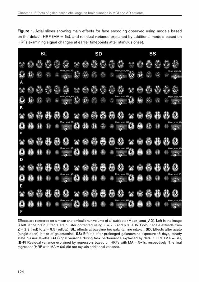
124
Chapter 4: Effects of galantamine challenge on brain function in MCI and AD patients
Figure 1. Axial slices showing main effects for face encoding observed using models based on the default HRF (MA = 6s), and residual variance explained by additional models based on HRFs examining signal changes at earlier timepoints after stimulus onset.
Effects are rendered on a mean anatomical brain volume of all subjects (Mean_anat_AD). Left in the image is left in the brain. Effects are cluster corrected using Z = 2.3 and p < 0.05. Colour scale extends from Z = 2.3 (red) to Z = 9.5 (yellow). BL: effects at baseline (no galantamine intake); SD: Effects after acute (single dose) intake of galantamine. SS: Effects after prolonged galantamine exposure (5 days, steady state plasma levels). (A) Signal variance during task performance explained by default HRF (MA = 6s), (B-F) Residual variance explained by regressors based on HRFs with MA = 5–1s, respectively. The final regressor (HRF with MA = 0s) did not explain additional variance.
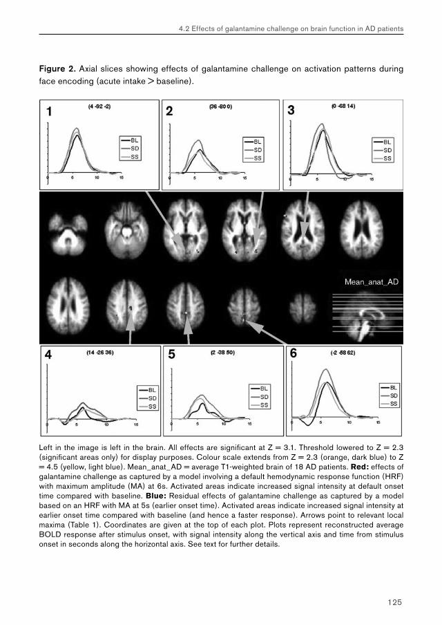
125
4.2 Effects of galantamine challenge on brain function in AD patients
Figure 2. Axial slices showing effects of galantamine challenge on activation patterns during face encoding (acute intake > baseline).
Figure 2. Axial slices showing effects of galantamine challenge on activation patterns during face encoding (acute intake > baseline).
Left in the image is left in the brain. All effects are significant at Z = 3.1. Threshold lowered to Z = 2.3 (significant areas only) for display purposes. Colour scale extends from Z = 2.3 (orange, dark blue) to Z = 4.5 (yellow, light blue). Mean_anat_AD = average T1-weighted brain of 18 AD patients. Red: effects of galantamine challenge as captured by a model involving a default hemodynamic response function (HRF) with maximum amplitude (MA) at 6s. Activated areas indicate increased signal intensity at default onset time compared with baseline. Blue: Residual effects of galantamine challenge as captured by a model based on an HRF with MA at 5s (earlier onset time). Activated areas indicate increased signal intensity at earlier onset time compared with baseline (and hence a faster response). Arrows point to relevant local maxima (Table 1). Coordinates are given at the top of each plot. Plots represent reconstructed average BOLD response after stimulus onset, with signal intensity along the vertical axis and time from stimulus onset in seconds along the horizontal axis. See text for further details.
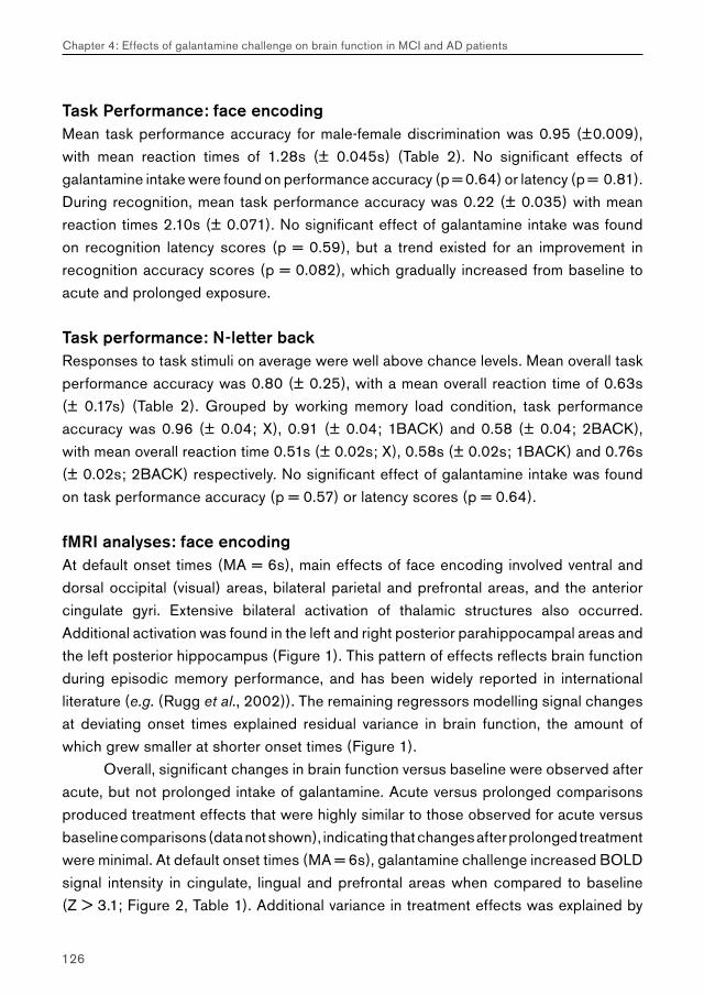
126
Chapter 4: Effects of galantamine challenge on brain function in MCI and AD patients
Task Performance: face encodingMean task performance accuracy for male-female discrimination was 0.95 (±0.009), with mean reaction times of 1.28s (± 0.045s) (Table 2). No significant effects of galantamine intake were found on performance accuracy (p = 0.64) or latency (p = 0.81). During recognition, mean task performance accuracy was 0.22 (± 0.035) with mean reaction times 2.10s (± 0.071). No significant effect of galantamine intake was found on recognition latency scores (p = 0.59), but a trend existed for an improvement in recognition accuracy scores (p = 0.082), which gradually increased from baseline to acute and prolonged exposure.
Task performance: N-letter backResponses to task stimuli on average were well above chance levels. Mean overall task performance accuracy was 0.80 (± 0.25), with a mean overall reaction time of 0.63s (± 0.17s) (Table 2). Grouped by working memory load condition, task performance accuracy was 0.96 (± 0.04; X), 0.91 (± 0.04; 1BACK) and 0.58 (± 0.04; 2BACK), with mean overall reaction time 0.51s (± 0.02s; X), 0.58s (± 0.02s; 1BACK) and 0.76s (± 0.02s; 2BACK) respectively. No significant effect of galantamine intake was found on task performance accuracy (p = 0.57) or latency scores (p = 0.64).
fMRI analyses: face encodingAt default onset times (MA = 6s), main effects of face encoding involved ventral and dorsal occipital (visual) areas, bilateral parietal and prefrontal areas, and the anterior cingulate gyri. Extensive bilateral activation of thalamic structures also occurred. Additional activation was found in the left and right posterior parahippocampal areas and the left posterior hippocampus (Figure 1). This pattern of effects reflects brain function during episodic memory performance, and has been widely reported in international literature (e.g. (Rugg et al., 2002)). The remaining regressors modelling signal changes at deviating onset times explained residual variance in brain function, the amount of which grew smaller at shorter onset times (Figure 1).
Overall, significant changes in brain function versus baseline were observed after acute, but not prolonged intake of galantamine. Acute versus prolonged comparisons produced treatment effects that were highly similar to those observed for acute versus baseline comparisons (data not shown), indicating that changes after prolonged treatment were minimal. At default onset times (MA = 6s), galantamine challenge increased BOLD signal intensity in cingulate, lingual and prefrontal areas when compared to baseline (Z > 3.1; Figure 2, Table 1). Additional variance in treatment effects was explained by
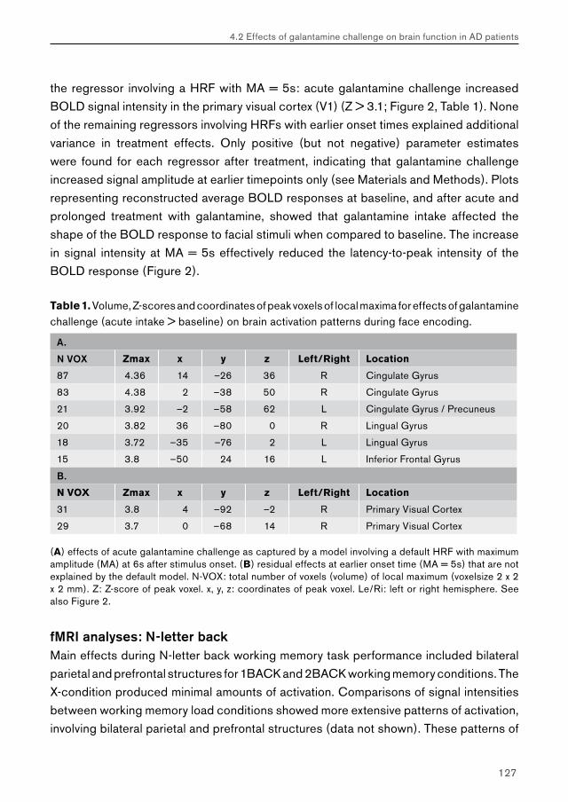
127
4.2 Effects of galantamine challenge on brain function in AD patients
the regressor involving a HRF with MA = 5s: acute galantamine challenge increased BOLD signal intensity in the primary visual cortex (V1) (Z > 3.1; Figure 2, Table 1). None of the remaining regressors involving HRFs with earlier onset times explained additional variance in treatment effects. Only positive (but not negative) parameter estimates were found for each regressor after treatment, indicating that galantamine challenge increased signal amplitude at earlier timepoints only (see Materials and Methods). Plots representing reconstructed average BOLD responses at baseline, and after acute and prolonged treatment with galantamine, showed that galantamine intake affected the shape of the BOLD response to facial stimuli when compared to baseline. The increase in signal intensity at MA = 5s effectively reduced the latency-to-peak intensity of the BOLD response (Figure 2).
Table 1. Volume, Z-scores and coordinates of peak voxels of local maxima for effects of galantamine challenge (acute intake > baseline) on brain activation patterns during face encoding.
A.
N VOX Zmax x y z Left/Right Location
87 4.36 14 –26 36 R Cingulate Gyrus
83 4.38 2 –38 50 R Cingulate Gyrus
21 3.92 –2 –58 62 L Cingulate Gyrus / Precuneus
20 3.82 36 –80 0 R Lingual Gyrus
18 3.72 –35 –76 2 L Lingual Gyrus
15 3.8 –50 24 16 L Inferior Frontal Gyrus
B.
N VOX Zmax x y z Left/Right Location
31 3.8 4 –92 –2 R Primary Visual Cortex
29 3.7 0 –68 14 R Primary Visual Cortex
(A) effects of acute galantamine challenge as captured by a model involving a default HRF with maximum amplitude (MA) at 6s after stimulus onset. (B) residual effects at earlier onset time (MA = 5s) that are not explained by the default model. N-VOX: total number of voxels (volume) of local maximum (voxelsize 2 x 2 x 2 mm). Z: Z-score of peak voxel. x, y, z: coordinates of peak voxel. Le/Ri: left or right hemisphere. See also Figure 2.
fMRI analyses: N-letter backMain effects during N-letter back working memory task performance included bilateral parietal and prefrontal structures for 1BACK and 2BACK working memory conditions. The X-condition produced minimal amounts of activation. Comparisons of signal intensities between working memory load conditions showed more extensive patterns of activation, involving bilateral parietal and prefrontal structures (data not shown). These patterns of
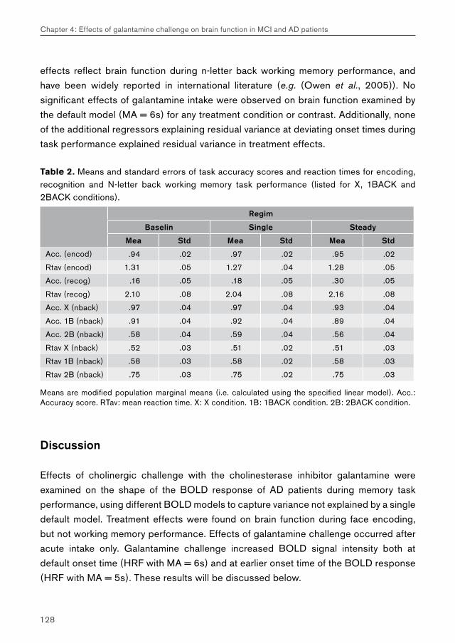
128
Chapter 4: Effects of galantamine challenge on brain function in MCI and AD patients
effects reflect brain function during n-letter back working memory performance, and have been widely reported in international literature (e.g. (Owen et al., 2005)). No significant effects of galantamine intake were observed on brain function examined by the default model (MA = 6s) for any treatment condition or contrast. Additionally, none of the additional regressors explaining residual variance at deviating onset times during task performance explained residual variance in treatment effects.
Table 2. Means and standard errors of task accuracy scores and reaction times for encoding, recognition and N-letter back working memory task performance (listed for X, 1BACK and 2BACK conditions).
Regim
Baselin Single Steady
Mea Std Mea Std Mea Std
Acc. (encod) .94 .02 .97 .02 .95 .02
Rtav (encod) 1.31 .05 1.27 .04 1.28 .05
Acc. (recog) .16 .05 .18 .05 .30 .05
Rtav (recog) 2.10 .08 2.04 .08 2.16 .08
Acc. X (nback) .97 .04 .97 .04 .93 .04
Acc. 1B (nback) .91 .04 .92 .04 .89 .04
Acc. 2B (nback) .58 .04 .59 .04 .56 .04
Rtav X (nback) .52 .03 .51 .02 .51 .03
Rtav 1B (nback) .58 .03 .58 .02 .58 .03
Rtav 2B (nback) .75 .03 .75 .02 .75 .03
Means are modified population marginal means (i.e. calculated using the specified linear model). Acc.: Accuracy score. RTav: mean reaction time. X: X condition. 1B: 1BACK condition. 2B: 2BACK condition.
Discussion
Effects of cholinergic challenge with the cholinesterase inhibitor galantamine were examined on the shape of the BOLD response of AD patients during memory task performance, using different BOLD models to capture variance not explained by a single default model. Treatment effects were found on brain function during face encoding, but not working memory performance. Effects of galantamine challenge occurred after acute intake only. Galantamine challenge increased BOLD signal intensity both at default onset time (HRF with MA = 6s) and at earlier onset time of the BOLD response (HRF with MA = 5s). These results will be discussed below.
129
4.2 Effects of galantamine challenge on brain function in AD patients
Effects of galantamine challenge: process-specificityIn contrast to face encoding (Figure 2, Table 1), galantamine challenge produced no effects on brain function during working memory performance, despite analyses of treatment effects across a number of different contrasts and timepoints after stimulus onset. The asymmetric distribution of treatment effects across encoding and working memory tasks may be explained by several factors. First, they may reflect differences in task design. Since we scanned a population of AD patients, we aimed to minimize scanning time by reducing the number of alternating blocks during n-letter back working memory performance, and to exclude task conditions requiring working memory loads exceeding those required for 2-back performance (from previous experience, we knew that higher working memory loads require too much effort of mild AD patients and produce chance level accuracy scores). Since n-letter back working memory tasks contained more conditions of interest (3) than face encoding (1), face encoding paradigms involved more blocks per condition (4 blocks) than n-letter back task (3 blocks). This may have produced better signal-to-noise ratios during face encoding (Z-scores up to 9.5) than for working memory performance (Z scores up to 6.5), which may have lowed chances of finding significant effects of treatment.
Alternatively, effects of cholinergic challenge may be process-specific, i.e. may preferentially involve face encoding rather than working memory processes in AD patients. Process-specificity has been observed previously in pharmacological fMRI studies (Honey & Bullmore, 2004; Thiel, 2003) and may reflect the combined effects of receptor expression and drug characteristics (e.g. some mental processes may be more dependent on specific nicotinergic or muscarinergic receptor subtypes than others (Jann et al., 2002)). The current study, however, did not allow for a more detailed analysis of the process-specificity of galantamine-induced treatment effects. Face- encoding and n-letter back working memory tasks were chosen because of their proven reliability in eliciting activation patterns in controls and AD patients (Small et al., 1999; Owen et al., 2005). These paradigms, however, differ substantially with respect to the information content of the presented stimuli (visuospatial information (faces) versus symbolic (lexical) information). We therefore refrained from further studies of process-specificity, which would require tight statistical comparisons of treatment effects between encoding and working memory domains. Future pharmacological imaging studies may require a balancing of the number of task conditions and timepoints scanned for each block, and keep the information content of encoding and working memory paradigms as similar as possible, in order to allow for more detailed studies of the process-specificity of pharmacological substances.
130
Chapter 4: Effects of galantamine challenge on brain function in MCI and AD patients
Effects of galantamine challenge: changes in shape and latency of the BOLD response
At default onset time (MA = 6s), increased signal amplitude was observed in (posterior) cingulate and prefrontal cortices during face encoding. Additional variance was explained by a regressor explaining signal variance at earlier onset time (MA = 5s) than the default HRF (MA = 6s). This shows that effects of pharmacological treatment may be overlooked if standard procedures of fMRI data analysis are used. At MA = 5s, an increase in BOLD signal amplitude (relative to baseline) was found in the primary visual cortex. Treatment effects at MA = 5s did not overlap with those observed at default onset times (MA = 6s), suggesting that effects of galantamine challenge either occurred at MA = 5s or at MA = 6s. Reconstruction of average BOLD responses after galantamine challenge, however, showed that such sharp distinctions could not be made (Figure 2). Amplitude changes could be observed at both timepoints in each brain area, but each time reached significance for one timepoint and brain area only. This suggests that the effects observed at both timepoints reflect a single phenomenon affecting both latency and intensity of the BOLD response. Since BOLD signal intensity was increased at earlier onset times, response latency was effectively decreased as a result of galantamine challenge. This confirmed our hypothesis that cholinergic stimulation increases the intensity and decreases the latency of the BOLD response to task stimuli.
Although difficult to interpret from BOLD signal changes alone, the observed effects may involve increased arousal, attention and stimulus processing, which are well-described effects of cholinergic stimulation (Sarter et al., 2005). Posterior cingulate activation has been linked to enhanced visuospatial attention (Mesulam et al., 2001). Previous studies of cholinergic stimulation have further shown increases in prefrontal and cingulate cortex activation (Thiel, 2003). These structures may interoperate with visual cortex to direct (selective) visual attention to potentially relevant stimuli (Small et al., 2003). The observed increase in BOLD response intensity at earlier onset times (effects at MA = 5s) in the visual cortex may reflect a decrease in neural response-latency as a result of an increase in stimulus-driven (bottom-up) attentional performance. Previous studies have shown that a shift in attention to a (new) stimulus enhances cortical reactivity in areas involved in early selection of relevant information, such as the visual cortex (Mangun, 1995; Coull, 1998; Bentley et al., 2004). The observed effects at MA = 5s may therefore reflect increased early processing of facial stimuli. These findings are especially relevant given our previous findings of a delayed cortical
131
4.2 Effects of galantamine challenge on brain function in AD patients
response to facial stimuli in the visual cortex of AD patients (Rombouts et al., 2005b). Acute galantamine challenge may therefore improve signal intensity at default onset time, as well as the temporal characteristics of the BOLD response. The long-term therapeutic effects of galantamine treatment on episodic memory in AD patients may be secondary to these immediate effects (Sarter et al., 2005).
We should be cautious, however, to conclude that the observed effects are strictly neurogenic in nature. The effects of pharmacological substances on brain function may involve both vascular, glial and neurogenic effects (Honey & Bullmore, 2004; Logothetis & Pfeuffer, 2004). Without the aid of complementary methods, it is not possible to attribute the observed BOLD latency shift to any of these three compartments with a high degree of certainty (Kollias et al., 2000; Richter & Richter, 2003). Although this may be problematic in fundamental studies of the effects of pharmacologcial substances on brain function, it is likely to be less of an obstacle to clinical studies of neurotransmitter system function, since reactivity of both vascular, glial and neural tissues to pharmacological challenge may contain information regarding the functional status of the neurotransmitter system under investigation. Clinical studies examining the effects of pharmacological intervention on the shape and latency of the BOLD response to task stimuli may therefore follow the same pragmatic approach taken in analogous studies of latency changes of event-related potentials (ERPs) to oddball stimuli. ERP studies in AD patients have shown peak responses of P300 to be delayed by about 40ms as compared to control subjects (Ball et al., 1989; Polich & Herbst, 2000). P300 seems especially sensitive to cholinergic changes (Hammond et al., 1987). Cholinergic stimulation may cause a (partial) remittance of this P300 delay in AD patients, which is shortened by 15–25ms (Werber et al., 2003; Thomas et al., 2001). Both the delay of P300 in AD patients and its latency-shift after cholinergic treatment have been proposed to serve as clinical markers, which may aid in early (differential) diagnosis of AD and may help to predict response to cholinomimetic treatment in these patients (Frodl et al., 2002; Green & Levey, 1999; Knott et al., 2002; Polich & Herbst, 2000; Thomas et al., 2001; Werber et al., 2003). Similarly, changes in the shape and latency of the BOLD response to task stimuli may provide additional information concerning the functional status of the cholinergic system in AD, and have predictive value with respect to disease outcome and response to cholinergic therapy.
132
Chapter 4: Effects of galantamine challenge on brain function in MCI and AD patients
Effects of galantamine challenge: dependence on exposure durationIn the current study, activation changes were observed after acute, but not prolonged galantamine exposure. With plasma levels considered equal at the time of scanning for both acute and prolonged treatment regimes, such differences may be explained by effects of receptor (de)sensitization within the context of a cholinergic deficit. In AD patients, low acetylcholine levels are thought to be responsible for a significant proportion of symptoms (Giacobini, 2004). Low acetylcholine levels may induce sensitization of nicotinic and muscarinic cholinergic receptors on recipient neurons, resulting in high reactivity to acute galantamine challenge (Erb et al., 2001; Svedberg et al., 2002). Similarly, prolonged galantamine exposure may cause desensitization of (muscarinic) cholinergic receptors, with low reactivity to cholinergic stimulation (Quick & Lester, 2002; Volkow et al., 2001). Thus, the strong reaction to acute challenge with galantamine in AD patients may reflect acute nicotinic receptor sensitization in the presence of a cholinergic deficit, whereas the non-significant response after prolonged galantamine exposure may reflect (muscarinic) receptor desensitization within the context of (partially) restored acetylcholine levels. Similar effects of receptor sensitization have been suggested to underlie differences in BOLD signal reactivity to nicotine exposure between smokers and non-smokers (Ernst et al., 2001) and the varying effect sizes of different cholinesterase inhibitors in AD patients (Geerts et al., 2002). Future studies may require a combination of molecular imaging techniques (e.g. PET) and phMRI in order to link functional changes as a result of pharmacological intervention to alterations in receptor expression profiles at the molecular level.
Effects of galantamine challenge: disease-specificityThe results of the current study are difficult to compare with those of other studies, since these involved different groups of subjects, cholinergic substances, memory tasks and exposure durations (Thiel, 2003; Saykin et al., 2004). In a previous study, however, we used the same dosing regimen and analysis methods as reported in the current study to examine the effects of galantamine challenge on brain function during delayed recognition of emotionally neutral human faces in patients with AD and with mild cognitive impairment, which is a disease stage preceding the development of AD (Goekoop et al., 2005c). We found that the effects of acute galantamine challenge were region-specific, process-specific, and disease-stage-specific: hippocampal activation was enhanced in AD patients in a manner that favoured encoding of novel information during retrieval, and posterior cingulate, temporal and prefrontal activation was enhanced in MCI patients in a way that favoured successful retrieval of previously learned information. The differential
133
4.2 Effects of galantamine challenge on brain function in AD patients
functional response of MCI and AD patients to galantamine challenge during retrieval was thought to reflect differences in the functional status of the cholinergic system at different stages of disease, which is in line with recent findings at a molecular and clinical level. Such a differential response may be relevant to clinical studies examining the differential diagnostic value of such changes, or their predictive value with respect to clinical decline and response to cholinergic therapy.
In another study, we used the same design and memory tasks as described in the current study to examine the effects of galantamine challenge on brain function during face encoding and n-letter back working memory performance in patients with MCI (Goekoop et al., 2004). In contrast to AD patients, effects of galantamine challenge in MCI patients occurred after prolonged galantamine exposure only. During face encoding, effects occurred in lateral visual, hippocampal and prefrontal areas. During working memory performance, effects occurred in right precuneus and right prefrontal cortex. Thus, MCI and AD patients responded differently to cholinergic challenge during face encoding and working memory performance, suggesting a difference in the functional status of the cholinergic system between these patient groups. When examined statistically, however, the observed differences in BOLD signal reactivity between MCI and AD patients were not significant (Z > 3.1; data not shown). Thus, the face encoding and working memory paradigms, as described in the current study, may be less suitable to study disease-specific effects of cholinergic challenge in a clinical setting. Alternatively, disease-specific alterations in brain function as a result of cholinergic challenge may themselves be process-specific, i.e. may only become apparent during performance under certain task conditions (e.g. delayed recognition memory performance). By systematically varying a number of different parameters, it may be possible to find disease-specific biomarkers that can aid in the assessment of cholinergic system function in early AD.
General considerationsIn general, treatment effects reported in the current study were small in both magnitude and spatial extent, which may reflect low sample size (n = 18). Similarly, the absence of significant effects of galantamine intake on measures of task performance may reflect small sample size, since galantamine has a well-establised effects on episodic memory performance in AD patients (Raskind, 2003). Additionally, AD patients may show more variance in both brain anatomy (atrophy) and the hemodynamic response to neural stimulation when compared to healthy controls or MCI patients, which may reduce signal-to-noise ratios and hamper detection of significant effects of treatment
134
Chapter 4: Effects of galantamine challenge on brain function in MCI and AD patients
(Johnson et al., 2000; D’Esposito et al., 2003; Vandenbroucke et al., 2004). Future studies may therefore require inclusion of more subjects and corrections for brain atrophy (other than standard warping techniques) to enhance sensitivity to effects of pharmacological intervention. Since this study was not placebo-controlled, some of the observed changes may have represented placebo effects. However, the absence of significant effects of galantamine intake on measures of task performance suggests that placebo-effects were not a significant confounder in terms of behavior. Since no significant effects of galantamine intake were found on brain function after prolonged exposure, this suggests that placebo effects remained sub-threshold for this condition. By accounting for effects of task version and scan order, the residual signal changes may be considered more true representations of an effect of medication intake.
In conclusion, galantamine challenge increased the amplitude and reduced the latency of the BOLD response to task stimuli in AD patients during encoding of new information into memory. This shows that pharmacological intervention may produce effects that are easily missed in standard fMRI analyses. By systematically examining the effects of cholinergic challenge on a range of BOLD signal characteristics, it may be possible to obtain a set of markers of cholinergic system function that may predict clinical decline and treatment response in AD patients.
Acknowledgements
We would like to thank the Oxford fMRIB team for producing, maintaining and freely distributing FSL. Acknowledgements go to Stichting Alzheimer for financial support. Serge Rombouts is supported by a grant from the Netherlands Organisation for Scientific Research (NWO; grantnr. 916.36.117). This study was partly supported by Johnson&Johnson Pharmaceutical Research and Development. The sponsors had no influence on trial design, analyses, or conduct of the study.
Chapter 4
Effects of galantamine challenge on brain function in MCI and AD patients
4.3 Differential response to galantamine challenge in MCI and AD patients
Cholinergic challenge in patients with Alzheimer’s disease and mild cognitive impairment differentially affects hippocampal activation; a pharmacological fMRI study
Goekoop R, Scheltens P, Barkhof F, Rombouts SARB
Brain 2005, in press. PMID: 16251213
136
Chapter 4: Effects of galantamine challenge on brain function in MCI and AD patients
Summary
Pharmacological functional magnetic resonance imaging (phMRI) examines the impact of pharmacologically induced neurochemical changes on brain function at a system level. The current phMRI study directly compared effects of cholinergic stimulation on brain function between patients with Alzheimer’s disease and mild cognitive impairment, a disease stage preceding the development of Alzheimer’s disease. Brain function during recognition of (un)familiar information was examined for changes after exposure to galantamine, a cholinesterase inhibitor used for treating memory deficits in Alzheimer’s disease. Alzheimer patients (n = 18; age 74.5 years ± 8.2; MMSE 22.5 ± 2.4) and patients with mild cognitive impairment (n = 28; mean age 73.6 ± 7.5; MMSE 27.0 ± 1.2) were scanned during face recognition under three different conditions: at baseline, and after acute and prolonged exposure (5 days) to galantamine. Functional data were analyzed in an event-related fashion. In both groups, acute exposure produced strong increases in brain activation (Z > 3.1). Prolonged exposure produced less strong effects that mainly involved decreases in activation (Z > 3.1). In mild cognitive impairment, acute exposure increased activation in posterior cingulate, left inferior parietal, and anterior temporal lobe. Prolonged exposure decreased activation in similar posterior cingulate areas, and in bilateral prefrontal areas. Effects were stronger for positive (‘familiar’) than for negative (‘unfamiliar’) decisions, indicating that the effect was specific to memory retrieval. In Alzheimer patients, acute exposure increased activation bilaterally in hippocampal areas, whereas prolonged exposure decreased activation in these areas. Effects were more pronounced for negative than for positive decisions, suggesting a preferential effect on memory encoding. Unique profiles of signal reactivity were found in a number of areas, including left inferior parietal lobe and left hippocampus proper. The reactivity of posterior cingulate and hippocampal structures to cholinergic challenge suggests a key role of the cholinergic system in the functional processes that lead to Alzheimer’s disease. The differential response to cholinergic challenge in mild cognitive impairment and Alzheimer patients may reflect a difference in the functional status of the cholinergic system between both groups, which is in line with recent results showing a differential clinical response to cholinergic treatment.
Keywords: fMRI; mild cognitive impairment; Alzheimer’s disease; galantamine; challenge study.
137
4.3 Differential response to galantamine challenge in MCI and AD patients
Introduction
Alzheimer’s Disease is characterized by a progressive neurodegenerative process that initially affects only the entorhinal cortex and hippocampus in the medial temporal lobe, but gradually spreads outward to affect the entire cortical mantle in more advanced stages of disease (Braak et al., 1999). Loss of hippocampal function is considered a primary factor in causing memory problems in Alzheimer’s disease. Additionally, a cholinergic deficit is found that may contribute significantly to memory problems (Bartus, 2000; Mesulam, 2004). Apart from symptoms of amnesia, low acetylcholine levels have been associated with a variety of other clinical manifestations, including impaired verbal fluency and neuropsychiatric symptoms (Assal & Cummings, 2002). Current therapies against Alzheimer’s disease largely aim at restoring low acetylcholine levels with pharmacological agents (Lanctot et al., 2003; Trinh et al., 2003). Although the role of a cholinergic deficit in Alzheimer’s disease has been well established, the extent to which cholinergic function is impaired in Alzheimer’s disease, along with the time of onset of this impairment, are still subjects of debate. In vivo measurements of cholinergic receptor expression at different stages of Alzheimer’s disease using positron emission tomography (PET) have shown receptor abnormalities that change with disease progression (Nordberg, 2001). Post-mortem studies have shown decreased levels of molecular markers of cholinergic function in advanced stages of Alzheimer’s disease, but not in mild cognitive impairment, a disease stage preceding the development of Alzheimer’s disease. Instead, such markers may be upregulated in MCI, possibly to compensate for incipient neurofunctional defects (DeKosky et al., 2002). This suggests that cholinergic system function is relatively spared in early stages of Alzheimer’s disease, and is gradually affected in more advanced stages of disease. Despite these findings, however, the question remains whether the alterations observed at the molecular level indeed affect brain function at a system level. Evidence for a difference in cholinergic system function between mild cognitive impairment and Alzheimer patients may be relevant for subsequent dosage and timing of pharmacological treatment (Freo et al., 2002; Ackerman et al., 2000; Thiel, 2003; Goekoop et al., 2004; Saykin et al., 2004).
Pharmacological functional magnetic resonance imaging (pharmacological fMRI or phMRI) is a technique that is used to study the impact of pharmacologically induced neurochemical changes on brain function at a system level (Honey & Bullmore, 2004). Since many psychopharmacological substances target specific neurotransmitter systems, phMRI may be used to study neurotransmitter system function in both healthy subjects and patients. Effects of neurotransmitter depletion or overexpression on
138
Chapter 4: Effects of galantamine challenge on brain function in MCI and AD patients
brain function and behavior have been studied in healthy controls to model disease mechanisms (e.g. (Sperling et al., 2002; Thiel, 2003)). Additionally, the therapeutic mechanism of a number of compounds has been studied in patients by examining changes in brain function after pharmacological treatment. Effects of pharmacological substances on brain function have been found to be region-specific, process-specific and even genome-specific, and may vary with age and cognitive capacity (Honey & Bullmore, 2004). Recent results show that phMRI may successfully predict clinical response to pharmacological treatment in patients with major depression, by correlating initial changes in brain function to clinical outcome after treatment (Fu et al., 2004). Based on such findings, it has been suggested that the cortical response to pharmacological challenge is also disease-specific, i.e. reflects the functional status of the neurotransmitter system under investigation (Thiel, 2003; Honey & Bullmore, 2004; Saykin et al., 2004; Fu et al., 2004; Goekoop et al., 2004). We therefore used phMRI to compare changes in brain function as a result of cholinergic stimulation between patients with mild cognitive impairment and Alzheimer’s disease. In two previous phMRI studies, we examined cholinergic system reactivity in both groups separately while varying a number of parameters, including memory tasks and exposure durations (Goekoop et al., 2004; Goekoop et al., 2005b). Short periods of exposure to the cholinesterase inhibitor galantamine affected brain activation during encoding of unfamiliar information in different brain areas in both patient groups, suggesting a differential involvement of the cholinergic system. Treatment effects were very small, however. When compared statistically, no significant differential response to cholinergic challenge was observed between patients with mild cognitive impairment and Alzheimer’s disease (Goekoop et al., 2005b).
So far, most phMRI studies of cholinergic stimulation in patients examined brain function during encoding and working memory performance. Since the effects of cholinergic challenge may be process-specific (Honey & Bullmore, 2004)), cholinergic stimulation may differentially affect encoding and recognition stages of memory performance. For this reason, we used phMRI to compare effects of cholinergic challenge on brain function associated with recognition of previously stored information between patients with mild cognitive impairment and Alzheimer’s disease. The current study involved the same design and patient groups as reported in our previous studies (Goekoop et al., 2004; Goekoop et al., 2005b). A face-recognition task was used in which the familiarity of subjects was tested with items that had been presented a few minutes before, during encoding. As a cholinergic stimulant we used galantamine, which is a cholinesterase inhibitor (with an additional sensitizing effect on nicotinic receptors)
139
4.3 Differential response to galantamine challenge in MCI and AD patients
that has known therapeutic efficacy in Alzheimer patients (Raskind, 2003). Effects were examined after both acute (single dose) and prolonged (5 days) galantamine exposure. We hypothesized that cholinergic challenge would differentially affect brain function in patients with mild cognitive impairment and Alzheimer’s disease during face recognition. If so, this would provide in vivo evidence for a differential involvement of the cholinergic system in different stages of disease, with consequences for brain function at a system level.
Materials and Methods
Study DesignPatients were screened for participation in a randomized study design with patients serving as their own controls. fMRI was performed at baseline (baseline, no medication), after oral intake of a single dose of galantamine with water (acute) and after prolonged exposure to galantamine (prolonged). Thus, three scanning sessions were performed in each patient, each of which corresponded to a different medication regime. Scanning sessions for different regimes were exactly one week apart and occurred at the same hour of day for each patient. Acute intake involved oral ingestion of 8mg galantamine with water. Prolonged exposure involved a 120 hour period (5 days) of galantamine intake, spread over 6 weekdays, during which period steady state plasma levels were reached, i.e. 4 mg galantamine (first gift, evening of day 1), 4 mg galantamine b.i.d. (mornings and evenings; 4 consecutive days), 4 mg galantamine (final gift, morning of day 6). At this rate, steady state plasma levels are reached within 2–3 days in healthy controls (with minimum serum galantamine concentrations 10.6 ± 4.0 ng/ml and maximum levels 30.7 ± 6.2 ng/ml (Mannens et al., 2002; Zhao et al., 2002)). Baseline, acute and prolonged regimes were randomized across scanning sessions to prevent between-session (e.g. learning) effects from interfering with possible effects of medication. To avoid carry-over effects between the regimes, periods of acute and prolonged intake were separated by a washout period of at least two days of zero galantamine intake, which is more than 6 times the half-life of galantamine (7.4 hours) (Mannens et al., 2002; Zhao et al., 2002). Scanning sessions were performed 3 hours after acute (1 x 8mg) and 9 hours after prolonged (2dd 4mg; 5days) exposure. Dosage and timing of sessions was such that, on average, galantamine plasma levels after acute and prolonged exposure could be considered equal at the time of scanning. This was done in order to facilitate comparisons between treatment effects produced by different exposure durations.
140
Chapter 4: Effects of galantamine challenge on brain function in MCI and AD patients
Subject recruitmentThe study had approval of the review board of the committee of medical ethics of the VU University Medical Center in Amsterdam, the Netherlands. Thirty (30) elderly patients with mild cognitive impairment, 9 male, 21 female, aged 73.6; ± 7.7 (range 54 to 89 years) were recruited from the Alzheimer Center at the VU Medical Center, Amsterdam, the Netherlands. Patients with mild cognitive impairment were diagnosed using Petersen’s criteria for amnestic mild cognitive impairment, i.e. a slowly progressive memory decline without the involvement of another domain of cognitive function, that did not interfere significantly with activities of daily living (Petersen et al., 2001). For further details, see (Goekoop et al., 2004). Additionally, twenty (20) age matched patients with Alzheimer’s disease, 11 male, 9 female, aged 74.5; ± 8.2 (range 55 to 83 years), were recruited in a similar fashion. Alzheimer patients were diagnosed using the NINCDS-ADRDA criteria for Alzheimer’s disease (McKhann et al., 1984). For further details, see (Goekoop et al., 2005b). All patients provided informed consent according to the declaration of Helsinki under supervision of a lawful caretaker during a screening visit in which the procedure was explained and contraindications were checked. Apart from neuropsychological assessment during clinical investigation, all patients underwent additional MMSE (Folstein et al., 1975a), CDR (Morris, 1997) and NYU-paragraph recall tests, which were used for cognitive profiling. Formal education was determined on a discrete scale with three levels (1 = low, 2 = middle, 3 = high). Patients were excluded if they had any significant medical, neurological or psychiatric illness (other than mild cognitive impairment or Alzheimer’s disease), or if they were taking medication or other substances that are known to influence cerebral function, including antidepressants and cholinesterase inhibitors. Patients were excluded if their history showed excessive nicotine or alcohol intake (> 0.5 packs of cigarettes, > 4 glasses of an alcoholic substance a day), a severe allergy to pharmacological substances or their constitutive compounds, or the use of any experimental medication within three months prior to enrollment in the trial. Exclusion criteria to MRI involved the presence of a pacemaker, metallic implants in high-risk areas (i.e. vessel clips) and a history of claustrophobia.
Functional MRI (fMRI)Data acquisition
Imaging was carried out on a 1.5 T Sonata scanner (Siemens, Erlangen, Germany), using a standard circularly polarized head coil with foam padding to restrict head motion. For fMRI, an echo planar imaging sequence was used (echo time 60ms, flip
141
4.3 Differential response to galantamine challenge in MCI and AD patients
angle 90°, matrix 64 × 64, field of view 192 × 192 mm), to obtain 21 transverse slices (thickness 5 mm, interslice-gap 1 mm). Task stimuli were projected on a screen located at the head end of the scanner table via an LCD projector located outside the scanner room. Subjects viewed the screen through a mirror located on the head coil. In each hand, subjects held an fMRI compatible response-box through which they were able to react to task stimuli by pressing the left or right button using their index-fingers. A T1–weighted structural MRI-scan was obtained of each subject (MPRAGE; inversion time: 300 ms, TR = 15 ms; TE = 7 ms; flip angle = 8°; 160 coronal slices, 1 × 1 × 1.5 mm voxels).
Memory-task: face recognition
A face recognition task was administered immediately after a face encoding task. The encoding task involved the presentation of four blocks of unfamiliar faces (24 in total) alternating with blocks of fixation (Goekoop et al., 2004; Goekoop et al., 2005b). During face recognition, 24 faces were presented sequentially in random order on a black background, of which 12 had been shown during encoding and 12 were new. Each face was presented for a duration of 5s, and was followed by a white fixation-cross presented for 3s on a black background. Patients were instructed to indicate whether a presented face had been shown previously by pressing one of two buttons. Written instructions “Seen previously?”; “<= Yes ” and “=> No” also appeared alongside the pictures. Response types (familiar, unfamiliar, none) and response latencies to individual stimuli were recorded.
Three different but comparable versions of each paradigm were constructed and randomized across the scanning-sessions (baseline, acute and prolonged regimes). All paradigms were practised using dummy tasks to ensure that patients mastered the general procedure of task performance before scanning. One day before the start of the first session, a home visit was scheduled during which all memory tasks were practiced on a laptop computer. Five minutes before the onset of the first measurements, the face encoding paradigm was practiced again, while patients were in the scanner. During the first 10.5s of each task, patients saw a circle indicating time left before the onset of the first condition. Total time for one scanning session including instructions of memory tasks was approximately one hour.
Analysis of behavioral dataDifferences between demographic values and neuropsychological test scores were analyzed with SPSS 11.5, using chi-square analysis for discrete parameters (gender,
142
Chapter 4: Effects of galantamine challenge on brain function in MCI and AD patients
education level, CDR scores), and a multivariate analysis of variance (ANOVA) for continuous parameters (age, MMSE, NYU-paragraph scores). Overall accuracy scores and mean reaction times during recognition task performance were calculated for each patient. This was done separately for performance under each scanning session / exposure duration (i.e. baseline, acute, prolonged). Overall accuracy scores were calculated by subtracting false answers (false rejections (FN) + false recognitions (FP)) from correct answers (correct rejections (TN) + correct recognitions (TP)), and dividing the result by the total number of items (24 if no misses). Accuracy scores thus varied from –1 (100% incorrect) to 1 (100% correct), with 0 indicating chance level (50% correct, 50% incorrect). For analysis of treatment effects, a mixed effects ANOVA was performed in which accuracy scores and response latencies were entered as dependent variables, with medication ‘regime’ (3 levels: baseline, acute, prolonged), ‘group’ (2 levels: mild cognitive impairment and Alzheimer’s disease), ‘test version’ (3 levels: 1, 2, 3) and ‘scan order’ (3 levels: 1, 2, 3) as fixed factors, and education level (3 levels: low, medium, high) and gender (2 levels: male and female) as covariates. ‘Scanorder’ was specified as the repeated factor, to account for the effect of taking repeated measures from the same subject. Given the large number of possible interactions between these terms, Akaike’s information criterion (AIC) was used to calculate an optimal model-structure, which at an IAC value of –17.0 was found to contain only two-way interactions between all terms. Non-significant interactions were eliminated from the analysis in a stepwise process (two steps). Effects of the interaction regime*group were then considered representative of a differential effect of galantamine treatment on behavior in both groups (p < 0.05, Bonferroni corrected for multiple comparisons).
Analysis of functional neuroimaging dataFunctional datasets of individual subjects were analyzed using FSL (Smith et al., 2004). The first five volumes of each dataset were discarded to account for T1-saturation effects. At first level (individuals), the following pre-processing was applied: non-brain removal, slice-timing correction using Fourier-space time-series phase-shifting, motion correction and spatial smoothing using a Gaussian kernel of FWHM 8mm, mean-based intensity normalization of all volumes by the same factor and high (0.02 Hz) and low pass temporal filtering (Jenkinson et al., 2002; Smith, 2002). Registration of functional neuroimages to high resolution and/or standard images was carried out using an intermodal registration tool based on the correlation ratio (Jenkinson & Smith, 2001). After pre-processing, the following statistics was applied on a voxelwise basis on each time series, using local autocorrelation correction (Woolrich et al., 2001):
143
4.3 Differential response to galantamine challenge in MCI and AD patients
signal change during face recognition was modeled in an event-related fashion, using separate regressors for TP, TN, FP and FN response types (see above). Type and onset time of the events were determined by post-hoc sorting, based on the responses given by the individual subjects. Signal variance during fixation (X condition) was not modeled, to prevent overspecification of the model. Thus, a unique model of signal response was obtained for each individual patient, containing a single regressor for each response type and their temporal derivative, which was convolved with a gamma function to model the hemodynamic response. Model fitting generated whole brain native space images of parameter estimates for each condition, representing average signal change during face recognition versus fixation (X), along with corresponding variance images. To reduce the size of the analysis, only effects on brain activation during correct responses (i.e. true positive and true negative responses) were further considered. Thus, the following lower-level contrasts were generated: TP (versus X), TN (versus X) , and TP <> TN. TP and TN versus low-level fixation (X) contrasts examine general aspects of recognition memory performance, which are biased with respect to successful retrieval processes (TP > X) and encoding processes (TN > X), respectively (Buckner et al., 2001). TP > TN contrasts specifically examine brain areas where signal intensity during successful retrieval of familiar information was significantly stronger than signal intensity during successful rejection / encoding of new information. Areas of significant signal differences may therefore represent areas associated with ‘successful retrieval’. Conversely, the reverse contrast TN > TP examined brain function related to ‘encoding during attempted retrieval’. Brain areas that show significant effects for these contrasts may differ from those involved in either TP or TN (versus X) decisions, thus providing additional information concerning specific subcomponent processes during retrieval (Daselaar et al., 2003). The corresponding functional images were resampled to 2 x 2 x 2 mm in standard space and fed into in a group level statistical analysis to examine activation patterns for differences between treatment durations (baseline, acute and prolonged exposure).
At group level, average activation maps (‘main effects’) were calculated for all lower-level contrasts (see above). This was done for all regimes types in both patient groups in a mixed effects higher level analysis (Woolrich et al., 2004), using clusters determined by Z > 2.3 and a corrected cluster significance threshold of P = 0.05 (Forman et al., 1995; Friston et al., 1994; Worsley et al., 1992). Effects at baseline were then tested for significant changes after acute or prolonged exposure (‘treatment effects’). Treatment effects were calculated for each group separately, and for their between-group comparison. This was done in a single group level analysis using a mixed effects
144
Chapter 4: Effects of galantamine challenge on brain function in MCI and AD patients
‘Triple T-test’ model (http://www.fmrib.ox.ac.uk/fsl/feat5/index.html), which examined lower level contrast maps of both groups for effects of treatment, scanning order, test version, and the act of taking repeated measurements from single subjects (Woolrich et al., 2004; Goekoop et al., 2004; Goekoop et al., 2005b). Separate variances were assumed for both patient groups. Treatment effects were examined at a voxel threshold for significant brain activation determined by Z > 3.1 (i.e. p = 0.001), and a minimal clustersize of 160mm3. An F-test was used to test for any effect of cholinergic challenge (either acute or prolonged). If present, specific contributions of acute and prolonged exposure were analyzed using pairwise comparisons (T-tests). All group analyses were performed in a common reference space (standard space) (Talairach & Tournoux, 1988). For display purposes, treatment effects pertaining to a specific group were rendered on mean anatomical brain volumes corresponding to that group in standard space. Results of comparisons of treatment effects between groups were rendered on a mean anatomical brain volume of all patients in standard space.
Results
Demographics & results of cognitive profilingTable 1 shows the results of demographic and cognitive profiling in mild cognitive impairment and Alzheimer’s disease patient groups. Where possible, statistics are given to indicate the significance of a difference between the groups.
Patient compliance and discontinuationData from 28 patients with mild cognitive impairment (25 complete datasets including scans at baseline, and after acute and prolonged exposure, and 3 incomplete datasets) and 18 Alzheimer patients (17 complete datasets, 1 incomplete dataset) was used in the current study comments (see also Table 1). Compliance was good, as assessed by pill-counts and the caretaker’s comments. For further details, see (Goekoop et al., 2004; Goekoop et al., 2005b).
Task PerformanceTable 2 lists numbers and percentages of response types (i.e. TP, TN, FP and FN responses) for mild cognitive impairment and Alzheimer groups, specified by regime type. Table 3 shows means and standard errors of overall accuracy and latency scores during face recognition for each patient group and regime type separately. Information
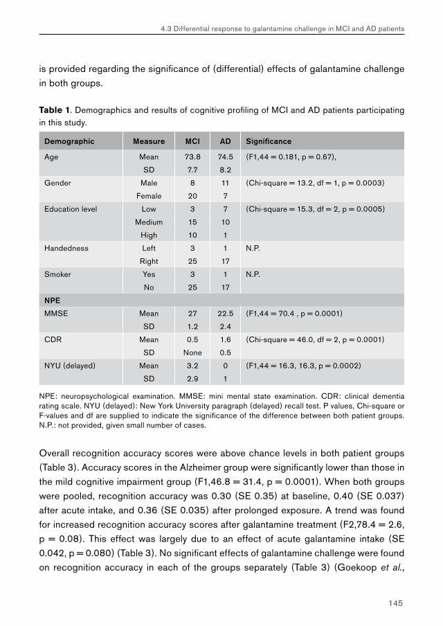
145
4.3 Differential response to galantamine challenge in MCI and AD patients
is provided regarding the significance of (differential) effects of galantamine challenge in both groups.
Table 1. Demographics and results of cognitive profiling of MCI and AD patients participating in this study.
Demographic Measure MCI AD Significance
Age Mean 73.8 74.5 (F1,44 = 0.181, p = 0.67),
SD 7.7 8.2
Gender Male 8 11 (Chi-square = 13.2, df = 1, p = 0.0003)
Female 20 7
Education level Low 3 7 (Chi-square = 15.3, df = 2, p = 0.0005)
Medium 15 10
High 10 1
Handedness Left 3 1 N.P.
Right 25 17
Smoker Yes 3 1 N.P.
No 25 17
NPE
MMSE Mean 27 22.5 (F1,44 = 70.4 , p = 0.0001)
SD 1.2 2.4
CDR Mean 0.5 1.6 (Chi-square = 46.0, df = 2, p = 0.0001)
SD None 0.5
NYU (delayed) Mean 3.2 0 (F1,44 = 16.3, 16.3, p = 0.0002)
SD 2.9 1
NPE: neuropsychological examination. MMSE: mini mental state examination. CDR: clinical dementia rating scale. NYU (delayed): New York University paragraph (delayed) recall test. P values, Chi-square or F-values and df are supplied to indicate the significance of the difference between both patient groups. N.P.: not provided, given small number of cases.
Overall recognition accuracy scores were above chance levels in both patient groups (Table 3). Accuracy scores in the Alzheimer group were significantly lower than those in the mild cognitive impairment group (F1,46.8 = 31.4, p = 0.0001). When both groups were pooled, recognition accuracy was 0.30 (SE 0.35) at baseline, 0.40 (SE 0.037) after acute intake, and 0.36 (SE 0.035) after prolonged exposure. A trend was found for increased recognition accuracy scores after galantamine treatment (F2,78.4 = 2.6, p = 0.08). This effect was largely due to an effect of acute galantamine intake (SE 0.042, p = 0.080) (Table 3). No significant effects of galantamine challenge were found on recognition accuracy in each of the groups separately (Table 3) (Goekoop et al.,
146
Chapter 4: Effects of galantamine challenge on brain function in MCI and AD patients
2004; Goekoop et al., 2005b). No significant effect was observed for the interaction regime*group, indicating that galantamine challenge did not differentially affect recognition accuracy between patient groups (F2,83.7 = 2.0, p = 0.14) (Table 3).
Latency scores did not differ significantly between mild cognitive impairment and Alzheimer patients (F1,45.9 = 0.123, p = 0.72). When both groups were pooled, response latency was 2.28s (SE 0.068s) at baseline, 2.25s (SE 0.070s) after acute intake, and 2.31s (SE 0.069s) after prolonged exposure. No effect of galantamine intake was found on response latency when groups were pooled (F2,70.4 = 0.78, p = 0.46), or in both groups separately (Table 3) (Goekoop et al., 2004; Goekoop et al., 2005b). No significant effect was observed for the interaction regime*group, indicating that galantamine challenge did not differentially affect response latency in both patient groups (F2,75.5 = 0.37, p = 0.69) (Table 3).
fMRI analysesMain effects: mild cognitive impairment and Alzheimer patients
Table 1 lists numbers and percentages of correct and incorrect responses in both patient groups (Table 1). Mild cognitive impairment and Alzheimer patients activated roughly the same structures during recognition task performance. At baseline (no treatment), main effects during correct (TP and TN) responses in both groups involved activation of ventral and dorsal occipital (visual) areas, bilateral inferior parietal, (para)hippocampal, superior temporal and prefrontal areas and the lateral sulci (Figure 1A, 1B, 1E, 1F). Activation patterns of TP and TN decisions differed significantly in both groups (Figure 1C, 1G). A baseline TP > TN contrast averaged over all subjects showed activation bilaterally in primary visual cortex, anterior and posterior cingulate cortex, inferior parietal lobes and anterior temporal lobe, whereas unilateral activation was observed in right motor cortex, right basal ganglia, left cerebellum, and left inferior, middle and superior frontal cortices. The lateralization of activation in right motor-related areas likely represents increased motor activity of the left hand and fingers during TP decisions when compared to TN decisions. In contrast, only left motor cortex showed increased activation in group-level TN > TP contrasts (Figure 1D, 1H).
Treatment effects: mild cognitive impairment
When compared to baseline, acute exposure to galantamine increased brain activation in a large number of structures, including (left) posterior cingulate cortex, left anterior temporal lobe, left superior parietal, left superior temporal cortex, right frontal lobe and cerebellum. These effects were mainly due to activation changes during true positive
147
4.3 Differential response to galantamine challenge in MCI and AD patients
(TP) decisions (Figure 2A, 2B; Table 4A, 4B), (Z > 3.1). No significant decreases were observed after acute galantamine intake. Prolonged exposure produced no significant increases in brain activation (Z > 3.1; data not shown). Instead, decreases in brain activation were found in (left) posterior cingulate, right middle frontal and bilateral superior prefrontal cortex. Again, these effects were mainly due to activation changes during true positive (TP) decisions (Figure 2C, 2D; Table 4C, 4D), (Z > 3.1). When effects of acute and prolonged effects of galantamine intake were compared (acute <> prolonged), the differences were significant (Z > 3.1). Increases after acute intake and decreases in similar areas after prolonged exposure summed during these comparisons in areas of significant overlap (i.e. posterior cingulate cortex), producing stronger effects (data not shown). Significant treatment effects were found on a contrast describing TP > TN (but not TN > TP) activation differences. Acute galantamine intake produced strong increases in right visual cortex, left parahippocampal cortex, right inferior prefrontal and medial prefrontal cortex (Figure 2E, Table 4E) (Z > 3.1). Prolonged intake of galantamine decreased brain activation in right caudate nucleus and left putamen (Figure 2F, Table 4F). Differences between acute and prolonged exposure were significant (Z > 3.1; data not shown).
Treatment effects: Alzheimer patients
When compared to baseline, acute exposure to galantamine increased brain activation in a limited number of structures, including the vermis of the cerebellum, right inferior temporal gyrus and the parahippocampal areas bilaterally. Effects of response type were substantial. When TN items were considered separately, additional treatment effects were found in the body of the left hippocampus (Figure 3A, 3B; Table 5A, 5B), (Z > 3.1). No significant decreases in brain activation were observed after acute galantamine intake. Prolonged exposure produced no significant increases in activation (Z > 3.1; data not shown). Instead, a substantial decrease in brain activation was found in right (para)hippocampal cortex. Again, these effects were mainly due to activation changes during true negative (TN) decisions (Figure 3C, 3D; Table 5C, 5D), (Z > 3.1). When acute and prolonged effects of galantamine intake were compared, the differences were significant (Z > 3.1). Increases after acute intake and decreases in similar areas after prolonged exposure showed a significant overlap in right (para)hippocampal cortex, producing stronger effects when compared (data not shown). No significant treatment effects were found on contrasts describing TP <> TN activation differences.
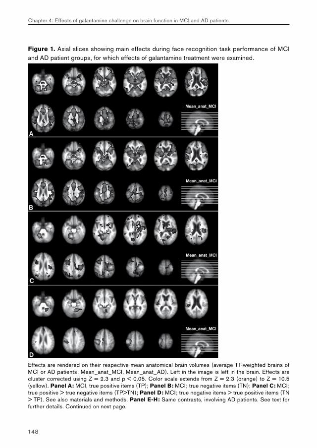
148
Chapter 4: Effects of galantamine challenge on brain function in MCI and AD patients
Figure 1. Axial slices showing main effects during face recognition task performance of MCI and AD patient groups, for which effects of galantamine treatment were examined.
Effects are rendered on their respective mean anatomical brain volumes (average T1-weighted brains of MCI or AD patients: Mean_anat_MCI, Mean_anat_AD). Left in the image is left in the brain. Effects are cluster corrected using Z = 2.3 and p < 0.05. Color scale extends from Z = 2.3 (orange) to Z = 10.5 (yellow). Panel A: MCI, true positive items (TP); Panel B: MCI; true negative items (TN); Panel C: MCI; true positive > true negative items (TP>TN); Panel D: MCI; true negative items > true positive items (TN > TP). See also materials and methods. Panel E-H: Same contrasts, involving AD patients. See text for further details. Continued on next page.
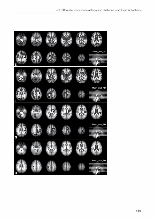
149
4.3 Differential response to galantamine challenge in MCI and AD patients
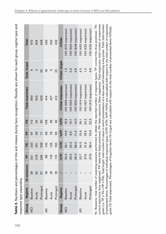
150
Chapter 4: Effects of galantamine challenge on brain function in MCI and AD patients
Tab
le 2
. Num
bers
and
per
cent
ages
of h
its a
nd m
isse
s du
ring
face
rec
ogni
tion.
Res
ults
are
sho
wn
for
each
gro
up, r
egim
e ty
pe a
nd
resp
onse
typ
e se
para
tely
.
Gro
upR
egim
eN
r se
ssio
nsTP
TNFP
FNTo
tal r
esp
ons
esTo
tal_
forg
otTo
tal
MC
IB
asel
ine
2822
123
397
112
66
39
672
Acu
te26
218
251
599
462
22
624
Pro
long
ed27
226
239
7187
623
256
48
AD
Bas
elin
e18
98
142
66
108
414
184
32
Acu
te18
118
135
799
542
75
432
Pro
long
ed18
115
150
55
9241
220
432
Gro
upR
egim
e–
%TP
%TN
%FP
%FN
%To
tal r
esp
ons
es%
Tota
l_fo
rgot
%To
tal
MC
IB
asel
ine
–3
3.3
35
.114
.616
.910
0 (6
63
resp
onse
s)1.
310
0 (6
72 r
espo
nses
)
Acu
te–
35
.04
0.4
9.5
15.1
100
(622
res
pons
es)
0.3
100
(624
res
pons
es)
Pro
long
ed–
36
.33
8.4
11.4
14.0
100
(623
res
pons
es)
3.9
100
(64
8 re
spon
ses)
AD
Bas
elin
e–
23.7
34
.315
.926
.110
0 (4
14 r
espo
nses
)4
.210
0 (4
32 r
espo
nses
)
Acu
te–
27.6
31.6
18.5
22.2
100
(427
res
pons
es)
1.2
100
(432
res
pons
es)
Pro
long
ed–
27.9
36
.413
.322
.310
0 (4
12 r
espo
nses
)4
.610
0 (4
32 r
espo
nses
)
Nr.
Ses
sion
s: t
otal
num
ber
of s
cann
ing
sess
ions
per
form
ed t
o ob
tain
the
rep
orte
d nu
mbe
r of
res
pons
e ty
pes.
TP
: cor
rect
hits
(tr
ue p
ositi
ves)
. TN
: co
rrec
t rej
ectio
ns (
true
neg
ativ
es).
FP: f
alse
hits
(fa
lse
posi
tives
). FN
: fal
se r
ejec
tions
(fa
lse
nega
tives
). To
tal r
espo
nses
: tot
al n
umbe
r of r
espo
nses
gi
ven
(i.e
. TP
+ T
N +
FP
+ F
N).
Tota
l for
got:
tota
l num
ber o
f cas
es in
whi
ch p
rese
ntat
ion
of a
face
was
not
follo
wed
by
a ke
y-pr
ess.
Tot
al: t
otal
num
ber
of p
rese
nted
item
s. P
erce
ntag
es o
f ind
ivid
ual r
espo
nse
type
s (%
TP, %
TN, %
FP, %
FN) a
re c
alcu
late
d w
ith r
espe
ct to
the
tota
l num
ber o
f res
pons
es
give
n (%
Tot
al r
espo
nses
). P
erce
ntag
es o
f for
gott
en it
ems
(% fo
rgot
) ar
e ca
lcul
ated
with
res
pect
to th
e to
tal n
umbe
r of
pre
sent
ed it
ems
(%To
tal)
.
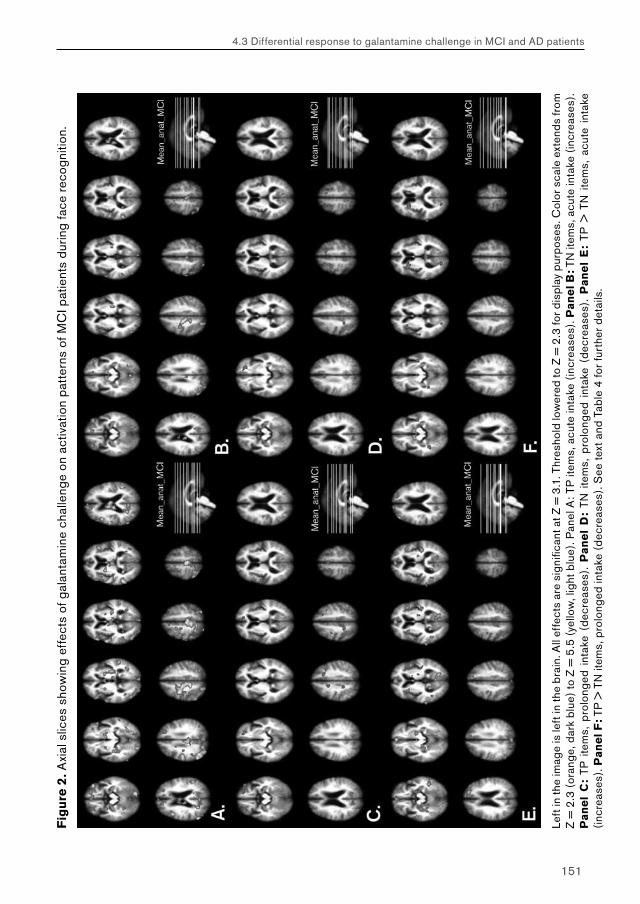
151
4.3 Differential response to galantamine challenge in MCI and AD patients
Fig
ure
2. A
xial
slic
es s
how
ing
effe
cts
of g
alan
tam
ine
chal
leng
e on
act
ivat
ion
patt
erns
of M
CI p
atie
nts
durin
g fa
ce r
ecog
nitio
n.
Left
in th
e im
age
is le
ft in
the
brai
n. A
ll ef
fect
s ar
e si
gnifi
cant
at Z
= 3
.1. T
hres
hold
low
ered
to Z
= 2
.3 fo
r dis
play
pur
pose
s. C
olor
sca
le e
xten
ds fr
om
Z =
2.3
(or
ange
, dar
k bl
ue)
to Z
= 5
.5 (
yello
w, l
ight
blu
e). P
anel
A: T
P it
ems,
acu
te in
take
(in
crea
ses)
. Pa
ne
l B: T
N it
ems,
acu
te in
take
(in
crea
ses)
. P
an
el
C:
TP i
tem
s, p
rolo
nged
int
ake
(dec
reas
es).
Pa
ne
l D
: TN
ite
ms,
pro
long
ed i
ntak
e (d
ecre
ases
). P
an
el
E:
TP >
TN
ite
ms,
acu
te i
ntak
e (in
crea
ses)
. Pa
ne
l F: T
P >
TN
item
s, p
rolo
nged
inta
ke (
decr
ease
s). S
ee te
xt a
nd T
able
4 fo
r fu
rthe
r de
tails
.
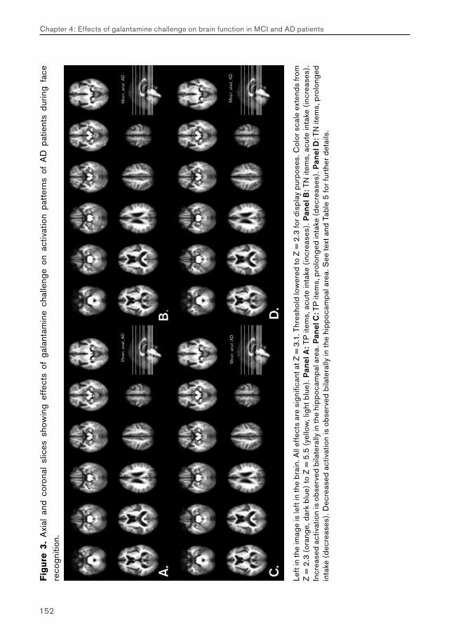
152
Chapter 4: Effects of galantamine challenge on brain function in MCI and AD patients
Fig
ure
3.
Axi
al a
nd c
oron
al s
lices
sho
win
g ef
fect
s of
gal
anta
min
e ch
alle
nge
on a
ctiv
atio
n pa
tter
ns o
f A
D p
atie
nts
durin
g fa
ce
reco
gniti
on.
Left
in th
e im
age
is le
ft in
the
brai
n. A
ll ef
fect
s ar
e si
gnifi
cant
at Z
= 3
.1. T
hres
hold
low
ered
to Z
= 2
.3 fo
r dis
play
pur
pose
s. C
olor
sca
le e
xten
ds fr
om
Z =
2.3
(or
ange
, dar
k bl
ue)
to Z
= 5
.5 (
yello
w, l
ight
blu
e). P
anel
A: T
P it
ems,
acu
te in
take
(in
crea
ses)
. Pan
el B
: TN
item
s, a
cute
inta
ke (
incr
ease
s).
Incr
ease
d ac
tivat
ion
is o
bser
ved
bila
tera
lly in
the
hipp
ocam
pal a
rea.
Pan
el C
: TP
item
s, p
rolo
nged
inta
ke (
decr
ease
s). P
anel
D: T
N it
ems,
pro
long
ed
inta
ke (
decr
ease
s). D
ecre
ased
act
ivat
ion
is o
bser
ved
bila
tera
lly in
the
hipp
ocam
pal a
rea.
See
text
and
Tab
le 5
for
furt
her
deta
ils.
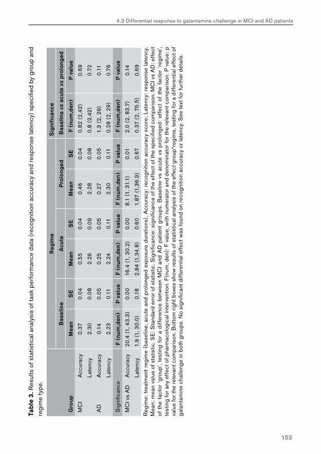
153
4.3 Differential response to galantamine challenge in MCI and AD patients
Tab
le 3
. Res
ults
of s
tatis
tical
ana
lysi
s of
task
per
form
ance
dat
a (r
ecog
nitio
n ac
cura
cy a
nd r
espo
nse
late
ncy)
spe
cifie
d by
gro
up a
nd
regi
me
type
.
Reg
ime
Sig
nific
ance
Bas
elin
eA
cute
Pro
long
edB
asel
ine
vs a
cute
vs
pro
long
ed
Gro
upM
ean
SE
Mea
nS
EM
ean
SE
F (n
um,d
en)
P v
alue
MC
IA
ccur
acy
0.3
70
.04
0.5
50
.04
0.4
60
.04
0.8
2 (2
,42
)0
.69
Late
ncy
2.3
00
.08
2.2
60
.09
2.2
80
.08
0.8
(2
,42
)0
.72
AD
Acc
urac
y0
.14
0.0
50
.25
0.0
50
.27
0.0
51.
3 (2
, 29
)0
.11
Late
ncy
2.2
30
.11
2.2
40
.11
2.3
00
.11
0.2
8 (2
, 29
)0
.76
Sig
nific
ance
F (n
um,d
en)
P v
alue
F (n
um,d
en)
P v
alue
F (n
um,d
en)
P v
alue
F (n
um,d
en)
P v
alue
MC
I vs
AD
Acc
urac
y20
.4 (1
, 43
.3)
0.0
016
.4 (1
, 30
.2)
0.0
08
.1 (1
, 31.
1)0
.01
2.0
(2
, 83
.7)
0.1
4
Late
ncy
1.9
(1, 3
0.0
)0
.18
2.8
4 (1
,34
.8)
0.6
01.
87 (1
,36
.9)
0.6
70
.37
(2, 7
5.5
)0
.69
Reg
ime:
tre
atm
ent
regi
me
(bas
elin
e, a
cute
and
pro
long
ed e
xpos
ure
dura
tions
). A
ccur
acy:
rec
ogni
tion
accu
racy
sco
re;
Late
ncy:
res
pons
e la
tenc
y.
Mea
n: m
ean
valu
e of
sta
tistic
. SE
: Sta
ndar
d er
ror o
f sta
tistic
. Sig
nific
ance
: sig
nific
ance
of t
he e
ffec
t of t
he s
peci
fied
com
paris
on. M
CI v
s A
D: e
ffec
t of
the
fac
tor
‘gro
up’,
test
ing
for
a di
ffer
ence
bet
wee
n M
CI
and
AD
pat
ient
gro
ups.
Bas
elin
e vs
acu
te v
s pr
olon
ged
: ef
fect
of
the
fact
or ‘
regi
me’
, te
stin
g fo
r an
y ef
fect
of p
harm
acol
ogic
al in
terv
entio
n. F
(num
, den
): F
val
ue, w
ith n
umer
ator
and
den
omin
ator
for
the
rele
vant
com
paris
on. P
val
ue: p
va
lue
for t
he re
leva
nt c
ompa
rison
. Bot
tom
rig
ht b
oxes
sho
w re
sults
of s
tatis
tical
ana
lysi
s of
the
effe
ct g
roup
*reg
ime,
test
ing
for a
diff
eren
tial e
ffec
t of
gala
ntam
ine
chal
leng
e in
bot
h gr
oups
. No
sign
ifica
nt d
iffer
entia
l eff
ect w
as fo
und
on r
ecog
nitio
n ac
cura
cy o
r la
tenc
y. S
ee te
xt fo
r fu
rthe
r de
tails
.
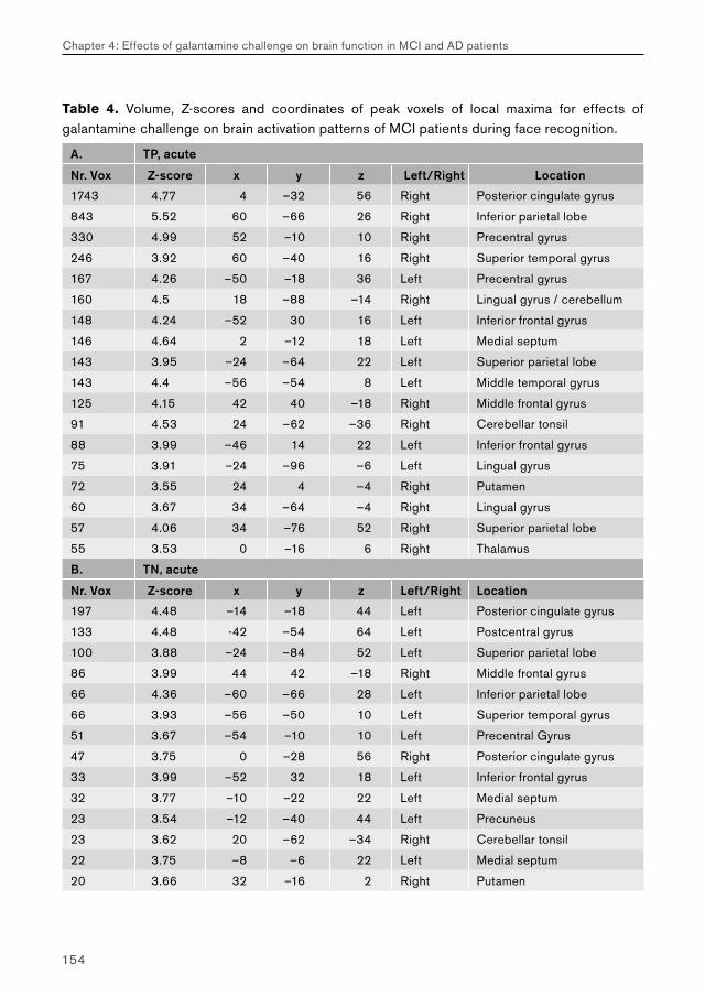
154
Chapter 4: Effects of galantamine challenge on brain function in MCI and AD patients
Table 4. Volume, Z-scores and coordinates of peak voxels of local maxima for effects of galantamine challenge on brain activation patterns of MCI patients during face recognition.
A. TP, acute
Nr. Vox Z-score x y z Left/Right Location
1743 4.77 4 –32 56 Right Posterior cingulate gyrus
843 5.52 60 –66 26 Right Inferior parietal lobe
330 4.99 52 –10 10 Right Precentral gyrus
246 3.92 60 –40 16 Right Superior temporal gyrus
167 4.26 –50 –18 36 Left Precentral gyrus
160 4.5 18 –88 –14 Right Lingual gyrus / cerebellum
148 4.24 –52 30 16 Left Inferior frontal gyrus
146 4.64 2 –12 18 Left Medial septum
143 3.95 –24 –64 22 Left Superior parietal lobe
143 4.4 –56 –54 8 Left Middle temporal gyrus
125 4.15 42 40 –18 Right Middle frontal gyrus
91 4.53 24 –62 –36 Right Cerebellar tonsil
88 3.99 –46 14 22 Left Inferior frontal gyrus
75 3.91 –24 –96 –6 Left Lingual gyrus
72 3.55 24 4 –4 Right Putamen
60 3.67 34 –64 –4 Right Lingual gyrus
57 4.06 34 –76 52 Right Superior parietal lobe
55 3.53 0 –16 6 Right Thalamus
B. TN, acute
Nr. Vox Z-score x y z Left/Right Location
197 4.48 –14 –18 44 Left Posterior cingulate gyrus
133 4.48 -42 –54 64 Left Postcentral gyrus
100 3.88 –24 –84 52 Left Superior parietal lobe
86 3.99 44 42 –18 Right Middle frontal gyrus
66 4.36 –60 –66 28 Left Inferior parietal lobe
66 3.93 –56 –50 10 Left Superior temporal gyrus
51 3.67 –54 –10 10 Left Precentral Gyrus
47 3.75 0 –28 56 Right Posterior cingulate gyrus
33 3.99 –52 32 18 Left Inferior frontal gyrus
32 3.77 –10 –22 22 Left Medial septum
23 3.54 –12 –40 44 Left Precuneus
23 3.62 20 –62 –34 Right Cerebellar tonsil
22 3.75 –8 –6 22 Left Medial septum
20 3.66 32 –16 2 Right Putamen
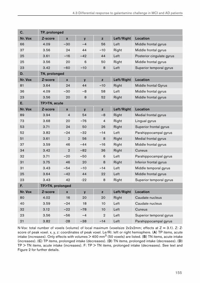
155
4.3 Differential response to galantamine challenge in MCI and AD patients
C. TP, prolonged
Nr. Vox Z-score x y z Left/Right Location
66 4.09 –30 –4 56 Left Middle frontal gyrus
37 3.56 24 44 –10 Right Middle frontal gyrus
25 3.61 –16 –42 44 Left Posterior cingulate gyrus
25 3.56 20 6 50 Right Middle frontal gyrus
23 3.42 –60 –10 8 Left Superior temporal gyrus
D. TN, prolonged
Nr. Vox Z-score x y z Left/Right Location
81 3.64 24 44 –10 Right Middle frontal Gyrus
36 4.09 –30 –8 58 Left Middle frontal gyrus
23 3.56 20 8 52 Right Middle frontal gyrus
E. TP>TN, acute
Nr. Vox Z-score x y z Left/Right Location
89 3.94 4 54 –8 Right Medial frontal gyrus
73 3.68 20 –76 4 Right Lingual gyrus
53 3.71 24 50 26 Right Superior frontal gyrus
52 3.82 –24 –32 –14 Left Parahippocampal gyrus
51 3.61 2 56 8 Right Medial frontal gyrus
37 3.59 46 –44 –16 Right Middle frontal gyrus
34 3.42 2 –82 36 Right Cuneus
32 3.71 –20 –50 6 Left Parahippocampal gyrus
31 3.75 46 20 8 Right Inferior frontal gyrus
31 3.43 –54 –10 –14 Left Middle temporal gyrus
25 3.64 –42 44 22 Left Middle frontal gyrus
23 3.43 42 -22 8 Right Superior temporal gyrus
F. TP>TN, prolonged
Nr. Vox Z-score x y z Left/Right Location
80 4.02 16 20 20 Right Caudate nucleus
40 3.59 –24 18 10 Left Caudate nucleus
32 3.12 –22 –78 10 Left Cuneus
23 3.56 –56 –4 2 Left Superior temporal gyrus
21 3.82 -28 –38 –14 Left Parahippocampal gyrus
N-Vox: total number of voxels (volume) of local maximum (voxelsize 2x2x2mm; effects at Z = 3.1). Z: Z-score of peak voxel. x, y, z: coordinates of peak voxel. Le/Ri: left or right hemisphere. (A) TP items, acute intake (increases). Only effects with volumes > 400 mm3 (50 voxels) are listed. (B) TN items, acute intake (increases). (C) TP items, prolonged intake (decreases). (D) TN items, prolonged intake (decreases). (D) TP > TN items, acute intake (increases). F: TP > TN items, prolonged intake (decreases). See text and Figure 2 for further details.
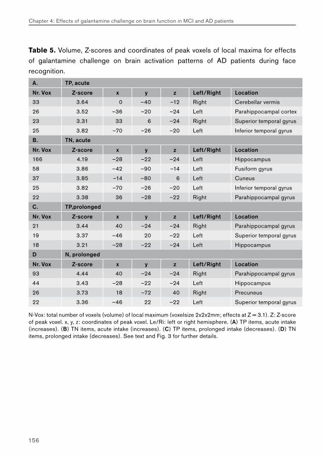
156
Chapter 4: Effects of galantamine challenge on brain function in MCI and AD patients
Table 5. Volume, Z-scores and coordinates of peak voxels of local maxima for effects of galantamine challenge on brain activation patterns of AD patients during face recognition.
A. TP, acute
Nr. Vox Z-score x y z Left/Right Location
33 3.64 0 –40 –12 Right Cerebellar vermis
26 3.52 –36 –20 –24 Left Parahippocampal cortex
23 3.31 33 6 –24 Right Superior temporal gyrus
25 3.82 –70 –26 –20 Left Inferior temporal gyrus
B. TN, acute
Nr. Vox Z-score x y z Left/Right Location
166 4.19 –28 –22 –24 Left Hippocampus
58 3.86 –42 –90 –14 Left Fusiform gyrus
37 3.85 –14 –80 6 Left Cuneus
25 3.82 –70 –26 –20 Left Inferior temporal gyrus
22 3.38 36 –28 –22 Right Parahippocampal gyrus
C. TP,prolonged
Nr. Vox Z-score x y z Left/Right Location
21 3.44 40 –24 –24 Right Parahippocampal gyrus
19 3.37 –46 20 –22 Left Superior temporal gyrus
18 3.21 –28 –22 –24 Left Hippocampus
D N, prolonged
Nr. Vox Z-score x y z Left/Right Location
93 4.44 40 –24 –24 Right Parahippocampal gyrus
44 3.43 –28 –22 –24 Left Hippocampus
26 3.73 18 –72 40 Right Precuneus
22 3.36 –46 22 –22 Left Superior temporal gyrus
N-Vox: total number of voxels (volume) of local maximum (voxelsize 2x2x2mm; effects at Z = 3.1). Z: Z-score of peak voxel. x, y, z: coordinates of peak voxel. Le/Ri: left or right hemisphere. (A) TP items, acute intake (increases). (B) TN items, acute intake (increases). (C) TP items, prolonged intake (decreases). (D) TN items, prolonged intake (decreases). See text and Fig. 3 for further details.
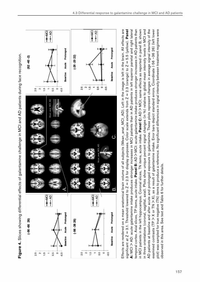
157
4.3 Differential response to galantamine challenge in MCI and AD patients
Fig
ure
4. S
lices
sho
win
g di
ffer
entia
l eff
ects
of g
alan
tam
ine
chal
leng
e in
MC
I and
AD
pat
ient
s du
ring
face
rec
ogni
tion.
Eff
ects
are
ren
dere
d on
a m
ean
anat
omic
al b
rain
vol
ume
of a
ll su
bjec
ts (
Mea
n_an
at_M
CI_
AD
). Le
ft in
the
imag
e is
left
in t
he b
rain
. All
effe
cts
are
sign
ifica
nt a
t Z
= 3
.1. T
hres
hold
low
ered
to
Z =
2.3
for
dis
play
pur
pose
s. C
olor
sca
le e
xten
ds f
rom
Z =
2.3
(or
ange
) to
Z =
5.5
(ye
llow
). P
an
el
A:
MC
I > A
D:
acut
e ga
lant
amin
e in
take
pro
duce
s st
rong
er in
crea
ses
in M
CI p
atie
nts
than
in A
D p
atie
nts
in le
ft s
uper
ior
parie
tal a
nd r
ight
late
ral
tem
pora
l cor
tex.
Axi
al s
lices
, TP
item
s, a
cute
inta
ke. P
an
el B
: AD
> M
CI:
acut
e ga
lant
amin
e in
take
pro
duce
s st
rong
er in
crea
ses
in A
D p
atie
nts
than
in
MC
I pat
ient
s in
the
left
hip
poca
mpu
s. C
oron
al s
lices
, TN
item
s, a
cute
inta
ke. P
an
el
C:
AD
> M
CI;
sam
e ef
fect
s as
rep
orte
d in
pan
el B
, sho
wn
in t
hree
orie
ntat
ions
(co
rona
l, sa
gitt
al, a
xial
). P
lots
sho
w u
niqu
e pe
rcen
t si
gnal
cha
nges
(in
%)
rela
tive
to g
loba
l mea
n in
tens
ity le
vels
in M
CI a
nd
AD
pat
ient
s at
bas
elin
e an
d af
ter
acut
e an
d pr
olon
ged
expo
sure
to
gala
ntam
ine.
The
se p
lots
rep
rese
nt c
hang
es in
ave
rage
sig
nal i
nten
sity
of
the
exam
ined
con
tras
ts (
TP >
X o
r TN
> X
), as
a r
esul
t of
gal
anta
min
e in
take
. Err
or b
ars
depi
ct s
tand
ard
erro
rs. C
oord
inat
e (-
56
–3
6 26
) (le
ft lo
wer
pl
ot) w
as s
ampl
ed fo
r tru
e ne
gativ
e (T
N) i
tem
s to
pro
duce
a re
fere
nce.
No
sign
ifica
nt d
iffer
ence
s in
sig
nal i
nten
sity
bet
wee
n tr
eatm
ent r
egim
es w
ere
obse
rved
in th
is a
rea.
See
text
and
Tab
le 6
for
furt
her
deta
ils.
158
Chapter 4: Effects of galantamine challenge on brain function in MCI and AD patients
Treatment effects: mild cognitive impairment versus Alzheimer patients
Visual inspection of treatment effects in both patient groups showed both differences and similarities in reactivity to galantamine challenge (Figure 2, Figure 3). In both groups, maximum reactivity to galantamine challenge occurred after acute exposure. Effects of acute exposure involved increases in activation only. Additionally, both groups showed decreases in brain activation after prolonged exposure, which occurred in brain areas similar to those showing increases in activation after acute exposure. Effects of prolonged exposure were of smaller magnitude than effects of acute exposure. Both groups differed in the spatial extent, amplitude and location of treatment effects. The mild cognitive impairment group showed a larger number of activation changes than the Alzheimer group, which were spread across a larger number of brain structures. Activation changes in the mild cognitive impairment group involved cortical (posterior cingulate, prefrontal, lateral temporal) and subcortical areas, but not the hippocampal areas. In contrast, activation changes in the Alzheimer group mainly involved hippocampal areas.
A direct statistical comparison between treatment effects of mild cognitive impairment and Alzheimer patients showed significant differences in treatment response between both groups. With respect to acute effects of galantamine intake, the mild cognitive impairment group produced significantly more activation changes than Alzheimer patients in a number of areas, including the left posterior parietal cortex (Figure 4A; Table 6A) (Z > 3.1). These effects were found for TP decisions only. Conversely, enhancement of hippocampal activation during TN decisions was significantly stronger in the Alzheimer group than in the mild cognitive impairment group (Figure 4B; Table 6B) (Z > 3.1). No significant between-group differences were found in the magnitude of treatment effects described by TP <> TN activation differences. Additionally, no differences were found with respect to effects of prolonged galantamine intake (Z > 3.1). Thus, significant differences were found in the reactivity of patients with mild cognitive impairment and Alzheimer’s disease to cholinergic challenge. Plots of percent signal change (relative to global mean signal intensity) in peak voxels of local maxima of treatment effects illustrate the significance of the differences in intensity changes between both groups (Figure 4).
159
4.3 Differential response to galantamine challenge in MCI and AD patients
Discussion
The current study examined patients with mild cognitive impairment and Alzheimer’s disease for a differential response to cholinergic stimulation with the cholinesterase inhibitor galantamine by using phMRI with a face recognition task. To our knowledge, this is the first event-related phMRI study to directly compare effects of pharmacological intervention on brain activation during delayed recognition between patient groups. In two previous phMRI studies, we examined effects of cholinergic challenge in mild cognitive impairment and Alzheimer patients separately, while they engaged in encoding and working memory tasks (Goekoop et al., 2004; Goekoop et al., 2005b). The reactivity of mild cognitive impairment and Alzheimer patients to acute and prolonged galantamine exposure was examined using identical procedures as described in the current study. Overall, treatment effects were small in both spatial extent and magnitude. Patients with mild cognitive impairment responded to prolonged galantamine exposure only (both tasks), whereas Alzheimer patients responded to acute galantamine challenge only (encoding only). Such differences in reactivity to cholinergic stimulation may be due to a number of confounding factors (Goekoop et al., 2005b), including a preference of galantamine for certain mental processes, and a difference in the functional status of the cholinergic system between these groups (i.e. process-specificity and disease-specificity of galantamine treatment; see below). When treatment effects during encoding were compared statistically between both groups, however,, no significant differential response to cholinergic challenge was observed between mild cognitive impairment and Alzheimer patients (Goekoop et al., 2005b).
Process-specificityThe current study extended our previous research by reporting effects of cholinergic challenge in the same patient groups during face recognition. Although identical procedures were followed, a larger number of stronger effects was found in both groups than observed for face encoding and working memory performance, supporting previous findings that effects of cholinergic enhancement are process-specific (Honey & Bullmore, 2004; Thiel, 2003). Direct comparisons between treatment effects across different memory tasks were not made, however, since these tasks were too dissimilar (i.e. block versus event-related designs, lexical versus visuospatial processing) to allow meaningful comparisons of cholinergic reactivity across different memory domains. We therefore report our findings of the effects of cholinergic challenge separately for all memory tasks. Nevertheless, we were able to examine the specificity of galantamine
160
Chapter 4: Effects of galantamine challenge on brain function in MCI and AD patients
challenge with respect to encoding and retrieval processes by studying brain function during retrieval alone. Since our face recognition task contained both novel and familiar items, both encoding and retrieval processes occurred during task performance (Buckner et al., 2001). By examining brain function during correct hits (TP items) and correct rejections (TN items), brain regions that are involved in (successful) retrieval and encoding-during attempted retrieval may be studied separately (see materials and methods). Although encoding processes during attempted retrieval may differ slightly from encoding during attempted encoding (Rombouts et al., 2001; Rugg et al., 2002), this methodology allows examination of process-specificity of pharmacological compounds with respect to encoding and retrieval processes within the same scanning session, which avoids some of the potential confounds that may be introduced by across-task or across-session comparisons of treatment effects (e.g. task design, relative timing of scanning sessions). If confirmed, a preferential targeting of memory retrieval processes rather than memory encoding by galantamine may have some clinical significance. For instance, the maximum benefit of cholinergic therapy with galantamine may be limited by existing pathology affecting the initial encoding of information. Future studies of drug design and development may therefore benefit from information provided by phMRI studies, by studying effects of pharmacological treatment in relation to specific neural processes. Such studies may help to increase efficacy and reduce side effects of novel psychopharmacological compounds.
Mild cognitive impairmentIn patients with mild cognitive impairment, acute galantamine challenge enhanced brain activation in posterior cingulate cortex, anterior and lateral temporal areas, parietal and prefrontal areas, the basal ganglia and in medial septal areas (Figure 2, Table 3), which are all known to depend on cholinergic innervation (Selden et al., 1998). Effects were stronger during familiar (TP) than for unfamiliar (TN) decisions, suggesting a preferential effect on brain activation during retrieval of familiar information. This was confirmed by subsequent analyses examining effects of galantamine challenge on brain activation during TP > TN decisions. Effects were found for TP > TN decisions in medial and right prefrontal cortex, visual cortex and left parahippocampal area, which are part of a well-described encoding-retrieval network (Simons & Spiers, 2003). Galantamine intake may therefore have affected successful retrieval in these patients. No effects were found for TN > TP decisions, suggesting that galantamine challenge in mild cognitive impairment did not specifically affect the encoding of new pictures during attempted retrieval.
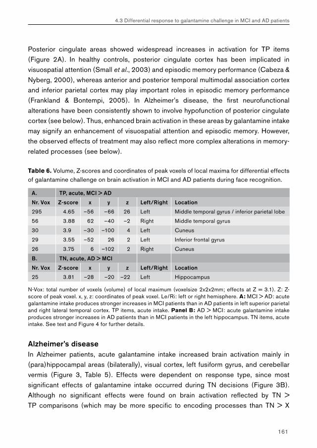
161
4.3 Differential response to galantamine challenge in MCI and AD patients
Posterior cingulate areas showed widespread increases in activation for TP items (Figure 2A). In healthy controls, posterior cingulate cortex has been implicated in visuospatial attention (Small et al., 2003) and episodic memory performance (Cabeza & Nyberg, 2000), whereas anterior and posterior temporal multimodal association cortex and inferior parietal cortex may play important roles in episodic memory performance (Frankland & Bontempi, 2005). In Alzheimer’s disease, the first neurofunctional alterations have been consistently shown to involve hypofunction of posterior cingulate cortex (see below). Thus, enhanced brain activation in these areas by galantamine intake may signify an enhancement of visuospatial attention and episodic memory. However, the observed effects of treatment may also reflect more complex alterations in memory-related processes (see below).
Table 6. Volume, Z-scores and coordinates of peak voxels of local maxima for differential effects of galantamine challenge on brain activation in MCI and AD patients during face recognition.
A. TP, acute, MCI > AD
Nr. Vox Z-score x y z Left/Right Location
295 4.65 –56 –66 26 Left Middle temporal gyrus / inferior parietal lobe
56 3.88 62 –40 –2 Right Middle temporal gyrus
30 3.9 –30 –100 4 Left Cuneus
29 3.55 –52 26 2 Left Inferior frontal gyrus
26 3.75 6 –102 2 Right Cuneus
B. TN, acute, AD > MCI
Nr. Vox Z-score x y z Left/Right Location
25 3.81 –28 –20 –22 Left Hippocampus
N-Vox: total number of voxels (volume) of local maximum (voxelsize 2x2x2mm; effects at Z = 3.1). Z: Z-score of peak voxel. x, y, z: coordinates of peak voxel. Le/Ri: left or right hemisphere. A: MCI > AD: acute galantamine intake produces stronger increases in MCI patients than in AD patients in left superior parietal and right lateral temporal cortex. TP items, acute intake. Panel B: AD > MCI: acute galantamine intake produces stronger increases in AD patients than in MCI patients in the left hippocampus. TN items, acute intake. See text and Figure 4 for further details.
Alzheimer’s diseaseIn Alzheimer patients, acute galantamine intake increased brain activation mainly in (para)hippocampal areas (bilaterally), visual cortex, left fusiform gyrus, and cerebellar vermis (Figure 3, Table 5). Effects were dependent on response type, since most significant effects of galantamine intake occurred during TN decisions (Figure 3B). Although no significant effects were found on brain activation reflected by TN > TP comparisons (which may be more specific to encoding processes than TN > X
162
Chapter 4: Effects of galantamine challenge on brain function in MCI and AD patients
comparisons), this suggests a preferential effect of galantamine intake on brain activation involving encoding during attempted retrieval. Cholinergic modulation of brain activation in visual areas and fusiform cortex has been observed previously during face encoding tasks and may involve alterations in neural processing requirements that are specific to perception and processing of facial stimuli (Rombouts et al., 2002; Sperling et al., 2001). The increase in hippocampal activation after galantamine intake in these patients is remarkable, since hippocampal atrophy is considered the primary deficit in Alzheimer’s disease, which is mainly responsible for the observed symptoms of memory impairment (Braak et al., 1999). Previous fMRI studies have found decreased hippocampal activation in Alzheimer patients when compared to healthy subjects (Rombouts et al., 2000; Remy et al., 2005). To our knowledge, this is the first phMRI study to demonstrate an enhancement of hippocampal function in Alzheimer patients after cholinergic stimulation. Such signal changes may be related to the extent of clinical improvement after cholinergic therapy. Future clinical studies may therefore want to focus on changes in hippocampal function after cholinergic challenge during recognition memory performance, in order to predict treatment response and long-term clinical outcome in Alzheimer patients.
Region- process- and disease-specificityConvergent evidence from functional studies including PET, SPECT and fMRI shows that resting state hypometabolism or hypofunction in limbic areas is central to pathology in both very early Alzheimer’s disease (i.e. mild cognitive impairment) and mild-to-moderate Alzheimer’s disease (Matsuda, 2001; Nestor et al., 2003; Greicius et al., 2004; Rombouts et al., 2005a). In the mild cognitive impairment stage, mainly posterior cingulate structures are affected, although sensitive region-of-interest techniques have detected hypometabolism in thalamus and hippocampus as well (Nestor et al., 2003). In more advanced stages of Alzheimer’s disease, the same limbic network is affected more extensively, and may show additional hypometabolism in hippocampal, temporoparietal and frontal association cortices (Nestor et al., 2003). Although the earliest structural changes appear in the medial temporal lobe (entorhinal cortex and hippocampus; (Braak et al., 1999)), functional changes first appear in posterior cingulate areas (Chetelat et al., 2003). This has lead to the hypothesis that functional changes in posterior cingulate areas represent secondary effects of structural changes in hippocampal areas (Matsuda, 2001). Indeed, hippocampal and posterior cingulate areas are strongly connected through limbic structures such as the thalamus and mamillary bodies (Nestor et al., 2003). In a recent study in patients with mild cognitive
163
4.3 Differential response to galantamine challenge in MCI and AD patients
impairment, posterior cingulate hypometabolism has been related specifically to (deficits in) retrieval memory performance, whereas hippocampal hypometabolism was related to (deficits in) memory encoding (Chetelat et al., 2003). Hypometabolism in Alzheimer patients therefore seems to be region-specific (posterior cingulate versus hippocampus), disease-stage specific (mild cognitive impairment versus Alzheimer’s disease), and process-specific (recognition versus encoding). These findings show a strong similarity with the results from the current challenge study, which show enhancement of posterior cingulate activation in mild cognitive impairment related to retrieval, and increases in hippocampal activation in Alzheimer’s disease that are likely to be related to encoding (see above). Although a definite link between resting state limbic hypofunction and the reactivity of posterior cingulate and hippocampal structures to cholinergic challenge could not be made in the current (task-related) study, our results show that a cholinergic factor may be relevant to hypofunction in posterior cingulate and hippocampal areas as observed in early Alzheimer’s disease.
The nature of this possible cholinergic influence and its time of onset remain unclear from the present data. Hypometabolism in posterior cingulate (mild cognitive impairment) and hippocampal areas (Alzheimer’s disease) may simply reflect a hypofunction of the cholinergic system, which would argue for an early involvement of cholinergic system dysfunction in Alzheimer’s disease. According to this view, the selective enhancement of hippocampal and posterior cingulate areas after galantamine challenge may reflect enhanced sensitivity of these structures to cholinergic stimulation as a result of cholinergic denervation. Both the hippocampus proper and the posterior cingulate gyrus are densely innervated by cholinergic fibers originating from separate branches (Ch2 and Ch4, respectively) of the basal forebrain cholinergic system (Selden et al., 1998). Selective denervation of (cholinergic) hippocampal afferents, as observed in Alzheimer’s disease, may alter both nicotinic and muscarinic receptor expression (Nordberg, 2001), producing increased receptor sensitization (Erb et al., 2001; Svedberg et al., 2002) and an increased response of brain structures to cholinergic challenge (Goekoop et al., 2005b). However, our current results also fit a different scenario, where posterior cingulate cortex hypometabolism and recognition deficits (as a result of early medial temporal atrophy) are kept partially in check by an intact cholinergic system. Hippocampal hypometabolism and encoding deficits (as observed in later stages of Alzheimer’s disease) would then reflect a definite failure of cholinergic system compensation. According to this view, increased activation in posterior cingulate and hippocampal structures after cholinergic stimulation may reflect a partial remission from a hypofunctional state of these structures, and cholinergic therapy may simply be
164
Chapter 4: Effects of galantamine challenge on brain function in MCI and AD patients
an add-on to natural cholinergic system compensation. It is likely that a combination of both scenarios, i.e. receptor hypersensitivity and partial compensation, may best explain the observed results. Future studies may require to combine molecular imaging techniques (e.g. PET) and phMRI in order to relate cholinergic receptor status to signal changes in specific brain structures and corresponding clinical phenotypes.
Mild cognitive impairment and Alzheimer’s disease: similarities in cholinergic system reactivityWhen signal changes in mild cognitive impairment and Alzheimer patients were visually compared, both groups showed signal increases after acute intake of galantamine, and signal decreases after prolonged exposure (Figure 4, Table 6). In some cases, increases and decreases in brain activation as observed for different exposure durations involved similar brain areas (Figure 2A,C; 2B,D; Figure 3A,C; 3B,D). The reason for this signal behavior is unclear, but may involve effects of nicotinic and muscarinic receptor sensitization after galantamine treatment. A strong response to acute galantamine challenge may reflect (nicotinic) receptor sensitization under normal or hypocholinergic circumstances, as it may occur in mild cognitive impairment or Alzheimer’s disease. In contrast, decreased levels of brain activation when compared to baseline may represent (muscarinic) receptor desensitization due to prolonged exposure to galantamine (Volkow et al., 2001; Quick & Lester, 2002). Similar effects of receptor (de)sensitization have been suggested to underlie differential responses in smokers versus non-smokers (Ernst et al., 2001), and the modest effects of long-term treatment with cholinesterase inhibitors (Geerts et al., 2002). This mechanism seems to fit most of our results, including our previous findings in Alzheimer patients during face encoding, where effects were observed after acute intake, but not after prolonged intake (Goekoop et al., 2005b). However, it does not fit our previous findings in patients with mild cognitive impairment during encoding, where effects were only observed after prolonged exposure (Goekoop et al., 2004). The origin of this ‘signal deviance’ in mild cognitive impairment remains unknown, but can be explained by the actions of several factors. Patients with mild cognitive impairment responded selectively to prolonged exposure (encoding) and to acute galantamine challenge (recognition). A difference in plasma concentrations between acute and prolonged regime types is therefore unlikely to fully explain the observed differences (see also materials and methods), since such a factor is likely to exert a comparable influence on brain function during encoding and recognition. Since patients with mild cognitive impairment responded both to prolonged and acute exposure to galantamine challenge, a disease-specific factor (e.g. the
165
4.3 Differential response to galantamine challenge in MCI and AD patients
absence of cholinergic receptor sensitization in mild cognitive impairment) is not likely to give a complete account of these signal changes. Since data-analyses were identical for both patient groups, a combination of disease-specific (MCI versus Alzheimer’s disease) and process-specific (encoding versus retrieval) factors seems to offer the best explanation for signal reactivity in the mild cognitive impairment group with respect to different exposure durations.
Other studies have shown increases in brain activation after weeks of treatment with different memory tasks and cholinomimetic substances in healthy controls or patients (Rombouts et al., 2002; Thiel, 2003; Parry et al., 2003; Saykin et al., 2004; Bentley et al., 2004). Since effects of cholinergic therapy may take 6–12 weeks to reach their maximum (Scarpini et al., 2003), short term effects of cholinergic treatment may represent a transitory state between immediate effects and therapeutic effects of a pharmacological substance, be process-specific, compound-specific, disease- specific, or may involve some other phenomenon. Future phMRI studies may require to investigate more memory tasks, pharmacological substances and timepoints after treatment in order to solve these issues.
Mild cognitive impairment and Alzheimer’s disease: differences in cholinergic system reactivityWhen compared statistically, patients with mild cognitive impairment and Alzheimer’s disease differed significantly with respect to their reactivity to cholinergic challenge (Figure 4, Table 6). Left hippocampal activation was significantly more enhanced in Alzheimer patients than in patients with mild cognitive impairment (Figure 4B, Table 6B), whereas left anterior and posterior temporal activation was more enhanced in patients with mild cognitive impairment than in Alzheimer patients (Figure 4A, Table 6A). Plots of percent signal change showed unique profiles of cholinergic reactivity in both patient groups for a number of brain areas (Figure 4). Overall, these comparisons show that mild cognitive impairment and mild-to-moderate Alzheimer patients respond uniquely to cholinergic challenge, suggesting that cholinergic system function is differentially affected in earlier and later stages of Alzheimer’s disease. This is in line with previous findings from post-mortem studies, showing characteristic cholinergic changes in mild cognitive impairment and Alzheimer patients at the molecular level (DeKosky et al., 2002). The current study shows that such neurochemical alterations may eventually affect brain function in living subjects. No significant difference was found in posterior cingulate reactivity to cholinergic challenge between mild cognitive impairment and Alzheimer patients. This may reflect a lack of power, but may also
166
Chapter 4: Effects of galantamine challenge on brain function in MCI and AD patients
point to comparable reactivity between both patient groups (i.e. because of smaller group size, posterior cingulate reactivity to galantamine challenge may have remained sub-threshold in Alzheimer patients). If a cholinergic component is indeed relevant to hippocampal and posterior cingulate hypofunction, comparable reactivity of posterior cingulate structures to cholinergic challenge would fit previous findings of comparable rates of hypometabolism in posterior cingulate structures in mild cognitive impairment and Alzheimer patients (Nestor et al., 2003). In contrast, the differential response in hippocampal areas may reflect more severe cholinergic system impairment in later stages of Alzheimer’s disease, which is not yet present in patients with mild cognitive impairment. Since no differential response was found between mild cognitive impairment and Alzheimer patients on measures of task performance (Table 3), the relationship of the observed effects of treatment with changes at a behavioral level are unclear from the current study. This may reflect small sample size and short exposure durations rather than a true absence of such effects, since galantamine treatment has a well-documented effect on memory retrieval (Raskind & Truyen, 2002). Additionally, a trend was found for significant increases in recognition accuracy of galantamine intake across subjects (p = 0.08), suggesting that galantamine intake did have an effect on recognition accuracy in the current study.
Cholinergic system reactivity: possible clinical relevanceA differential response to cholinergic challenge in patients with mild cognitive impairment and Alzheimer’s disease may be clinically relevant. Previous phMRI studies have shown that both brain activation at baseline and initial effects of pharmacological treatment may successfully predict long-term treatment response and clinical outcome in patients with major depression (Davidson et al., 2003; Fu et al., 2004). Similarly, differences in cholinergic system reactivity as observed in the current study may to some degree reflect the clinical status of patients with memory complaints (Goekoop et al., 2004). Interestingly, recent results from clinical trials show that mild cognitive impairment and Alzheimer patients respond differentially to long-term treatment with cholinesterase inhibitors. Treatment with a number of different cholinergic agonists improves memory performance in Alzheimer patients (Lanctot et al., 2003), but seems to have only limited effects in patients with mild cognitive impairment (Ihl, 2003; Salloway et al., 2004). Such differences in treatment response may reflect underlying differences in the functional status of the cholinergic system as suggested by the current study. Follow-up of mild cognitive impairment and Alzheimer patients should provide a measure of disease progression, which may be correlated with the observed effects of galantamine
167
4.3 Differential response to galantamine challenge in MCI and AD patients
challenge on brain function in order to examine the predictive value of these effects in terms of disease outcome and response to treatment. phMRI may prove to be a valuable tool in clinical studies, since it offers non-invasive assessment of neurotransmitter deficits, and provides additional information concerning effects at system level that may not be found with any other technique (Sarter et al., 1996). However, the eventual clinical significance of phMRI studies depends strongly on the outcome of within-subject test-retest reliability studies (Saykin et al., 2004). Although treatment strategies can be reconsidered based on results produced at group level, much will depend on the ability of clinicians to demonstrate incipient neurotransmitter system dysfunction in individual patients.
General considerationsWith respect to underlying neural processes, effects of pharmacological challenge as measured by fMRI should be interpreted with some caution. fMRI examines changes in blood oxygenation level dependent (BOLD) signal intensity, which is an indirect measure of neural activity (Matthews & Jezzard, 2004). BOLD signal changes represent changes in blood flow, volume and oxygenation as a result of metabolic changes, which are secondary to changes in neural activity. Such effects may reflect changes in both neural excitation and inhibition (Matthews & Jezzard, 2004). Future studies may therefore want to combine electrophysiological techniques (such as EEG) with phMRI in order to assess the nature of neural changes underlying the BOLD response. An important additional consideration for all phMRI studies is that pharmacological intervention, including cholinergic stimulation (Tsukada et al., 2000), may affect vascular tissue as well as neural tissue (Honey & Bullmore, 2004). However, such objections are likely to be less of an obstacle to clinical studies than to fundamental studies of cholinergic system function, since reactivity of both vascular and neural tissues to cholinergic challenge may contain information regarding the functional status of the cholinergic system in disease.
The current study had some limitations. Differences in cholinergic system function between patients with mild cognitive impairment and Alzheimer’s disease were examined in a cross-sectional manner. Although time-consuming, future phMRI studies may require to examine the development of a cholinergic deficit in time by using longitudinal study designs. Comparisons between patient groups in the current study may have suffered from bias introduced by group differences in sex and education level (Table 1). Although little is known about the effects of gender on brain function, recent studies show that such effects may be significant (Cahill, 2003; Lee et al., 2002). Future phMRI studies
168
Chapter 4: Effects of galantamine challenge on brain function in MCI and AD patients
may therefore require to balance male-female ratios to avoid effects of this possible confounder. Additionally, this study was not placebo controlled. Placebo effects may therefore have confounded some of the effects of treatment reported in both groups separately, although no effects were found at a behavioral level. If present, however, placebo effects are likely to have cancelled out in between-group comparisons.
ConclusionsGalantamine challenge affected brain function in posterior cingulate (mild cognitive impairment) and hippocampal areas (Alzheimer’s disease), suggesting a key role of the cholinergic system in the functional processes that lead to Alzheimer’s disease. A differential response to cholinergic challenge was found in mild cognitive impairment and Alzheimer patients, suggesting a difference in the functional status of the cholinergic system in earlier and later stages of disease. phMRI challenge tests may prove to be a valuable instrument to examine the functional status of central neurotransmitter systems in disease. Such studies may help to assess neurotransmitter system pathology, monitor disease progression, and predict response to pharmacological therapy. In addition, phMRI may be used to develop new drugs that target specific aspects of mental performance, such as encoding and retrieval processes.
Acknowledgements
We would like to thank the Oxford fMRIB team for producing, maintaining and freely distributing FSL. Acknowledgements go to Stichting Alzheimer for financial support. Serge Rombouts is supported by a grant from the Netherlands Organisation for Scientific Research (NWO; grantnr. 916.36.117). This study was partly supported by Johnson&Johnson Pharmaceutical Research and Development. The sponsors had no influence on trial design, analyses, or conduct of the study.
Chapter 5
General discussion
It is still early days for pharmacological functional magnetic resonance imaging (phMRI), but progress is being made with rapid paces. The following sections will discuss the empirical chapters of this thesis (Chapters 2-4) within the global context of the clinical applicability of phMRI.
5.1 Chapter 2: effects of beta-adrenergic blockade on amygdala function in healthy young subjects
This study aimed to link previous findings of neurotransmission in animals and brain function in humans by examining the effects of propranolol intake on amygdala function in healthy human subjects. Modulation of amygdala function by means of beta-adrenergic blockade would demonstrate the dependency of this brain structure on intact noradrenergic neurotransmission. More specifically, we hypothesised that propranolol intake would decrease amygdala activation during emotional performance as compared to placebo.
170
Chapter 5: General discussion
Main effects of task performance: emotional encoding
Main effects of task performance in this study showed increasing amygdala activation with increasing emotional intensity of the stimuli (i.e. category 1–4 pictures). Signal intensity within both amygdala showed a non-linear response (Chapter 2, Figure 5). This finding contradicts that of another study, which found a linear increase in signal intensity of the amygdala with increasing emotional intensity of presented stimuli (Canli et al., 2000). The degree of linearity of the observed response of the amygdala to stimuli of increasing emotional intensity was examined by fitting a straight line and a curvilinear response through the observed pattern of signal increase. The curvilinear response produced a better fit, as judged by its corresponding Z scores, but the linear response also significantly explained the observed signal intensity changes (Chapter 2, Figure 6). This is not surprising, given the fact that a linear response is a reasonable estimation of the curvilinear response used in this analysis. Several reasons are given for the non-linear response observed in this study, which deserve additional comment.
First, the fact that subjects rated fewer images as category 4 or 3 when compared to the number of images rated as category 1 or 2 (Chapter 2, Figure 2) may have produced differences in statistical power for estimating corresponding signal intensity changes. Within certain limits, however, a lack of statistical power may alter the variance of the observed effect size, but not the intensity itself. It should also be noted that the number hits for each emotional category decreased linearly with increasing emotional intensity in both males and females (Chapter 2, Figure 2). If the number of items analysed would indeed affect signal intensity within the amygdala, it may have produced linear rather than non-linear effects. Thus, it seems unlikely that a difference in the number of items analysed for each condition fully explains the non-linear pattern of brain function observed with increasing emotional intensity.
Second, the non-linear relationship may reflect an inverted-U shape, which has been observed previously in studies examining dose-response relationships between adrenoceptor agonist concentration (drugs) and task performance in rats (Honey & Bullmore, 2004). This seems unlikely, however, given the fact that these studies concerned drug effects on behaviour. Such effects do not serve well as a means to explain normal physiological signal response of human amygdala to stimuli of increasing emotional intensity.
Thirdly, the BOLD response may show non-linear tendencies with increasing presentation rate, which may be attributed to hemodynamic refractoriness (Mechelli et al., 2001). Though this is true, presentation rate is likely to have an equally limiting
171
5.1 Chapter 2: effects of beta-adrenergic blockade on amygdala function in healthy young subjects
effect on signal amplitude for all categories of emotional intensity, and not just category 4. A combination of high presentation rate and emotional intensity may, however, have produced hemodynamic refractoriness specifically for category 4 items.
There is no need, however, to attribute the observed non-linear phenomena strictly to vascular effects (such as hemodynamic refractoriness). At any level of neural organisation, the neural response to an increase in stimulus intensity is characterised by a sigmoid shape, which indicates the degree to which continuous waves of electric dendritic potential are converted into axonal pulses (Freeman, 2000). The BOLD response within the amygdala may therefore show a sigmoid-shaped response to stimuli of increasing emotional intensity for strictly neural reasons. When observing signal intensity changes with increasing emotional intensity within the placebo group (Chapter 2, Figure 5), a sigmoid-shape seems plausible. However, it is difficult to infer such a shape from a graph that has only four datapoints (i.e. emotional categories). Future research should therefore combine fMRI and EEG analyses in animals (and humans) to examine the impact of a range of emotionally distressing stimuli on the functional response of the amygdala.
Perhaps the most plausible explanation is one that accounts for the fact that subjective emotional ratings were used to classify pictures into 4 categories. This approach is likely to provide a more direct relationship between the actual emotional rating of subjects and their corresponding amygdala activation (Phan et al., 2003). However, it creates the possibility that subjects find items from different IAPS categories to be of similar emotional value (personally, I had difficulty separating IAPS category 3 and 4, or 1 and 2 items at times). If the differences that exist between the average emotional intensity of pictures that have been assigned to different subjective categories are small, a linear relationship between emotional intensity and stimulus category is lost. If subjective ratings indeed correspond strongly with amygdala activation, this may explain the ‘non-linear’ response of the amygdala to stimuli of increasing emotional intensity as observed in our study.
Treatment effects and performance changesPropranolol intake disrupted the non-linear response to pictures of increasing emotional intensity as observed under placebo conditions. A significant decrease in amygdala function occurred for category 3 > 1 pictures only. The reasons for this isolated signal response are unclear, but several explanations are possible.First, the response to beta-blockade may reflect an ‘inverted-U shape’ of efficacy. Such shapes are observed regularly in neuropharmacological studies examining dose-
172
Chapter 5: General discussion
response relationships in animals and humans (Honey & Bullmore, 2004). Both high and low doses of a pharmacological substance fail to produce an effect, and maximum efficacy is observed for intermediate concentrations, yielding the inverted U. In our study, the concentration of propranolol was kept at a single value whereas emotional intensity was varied. If higher levels of amygdala activity indeed relate to higher levels of beta-adrenergic neurotransmission, a single dose of 80 mg of propranolol may have been insufficient to block beta-adrenergic neurotransmission related to the processing of images of the highest emotional category. Additionally, propranolol concentrations of 80 mg may have disrupted normal neurophysiology during exposure to category 1 and 2 pictures, leading to non-significant changes in signal intensity.
As an alternative, it is possible that adrenergic neurotransmission is only important to amygdala function from a certain threshold of emotional intensity onward. Propranolol may therefore only affect amygdala function above a certain threshold of amygdala activity (i.e. category 2 and up). Such ‘delayed’ effects, however, cannot explain equal amounts of brain function for category 4 items under propranolol and placebo conditions. If such a threshold exists, a combination with the above suggestion of a dose-response relationship would seem to be more likely.
With respect to performance, propranolol intake impaired recognition accuracy in women at least until two weeks after drug challenge and encoding of the presented pictures. Changes in recognition accuracy as a result of propranolol resembled those of signal intensity changes within the amygdala (Chapter 2, Figures 5 and 7), but this relationship was not tested directly. In can be concluded, however, that propranolol intake decreased memory performance in a way that is likely to have involved decreases in amygdala activation.
Decreased amygdala activation: direct or indirect relations with impaired noradrenergic neurotransmission?Decreased amygdala activation as observed in our study may involve direct as well as indirect effects of changes in noradrenergic neurotransmission. Since all neurotransmitter systems interact at some level (Gu, 2002; Jones, 2003), it is difficult to link the observed effects directly to a decrease in noradrenergic neurotransmission. As predicted, however, propranolol decreased amygdala activation, whereas increased amygdala activation was not observed. This may be additional support for a more direct relationship between amygdala function and noradrenergic neurotransmission in humans. Future studies may require to measure adrenergic activity in human subjects directly using molecular imaging techniques such as PET.
173
5.1 Chapter 2: effects of beta-adrenergic blockade on amygdala function in healthy young subjects
Gender-specificity of propranolol: amygdala function and performance accuracyStudies have shown that brain function and pharmacological effects on brain function may differ between the sexes. Knowledge of such differences may be relevant in terms of optimising the design, development and dosing of drugs with respect to a patient’s gender (Cahill, 2003). Gender-specificity was therefore examined with respect to main effects of task performance (lateralisation of amygdala function), the response of the amygdala to propranolol intake, and (propranolol-induced changes in) recognition accuracy scores.
Under placebo conditions, women showed significantly more left amygdala function in (Category 3 > 1) than men, supporting findings from previous studies suggesting a gender-specific lateralisation of amygdala function (Cahill, 2003). Previous studies have shown gender-specific effects of propranolol on recognition memory performance (Maheu et al., 2004; Strange et al., 2003). In our study, propranolol intake did not affect memory scores in males, but decreased recognition accuracy for category 3 pictures in females, which seemed to mimic the drop in left amygdala activation for these pictures observed in this group. This may indicate a gender-specific effect of propranolol intake on memory performance. However, since none of these effects were tested statistically, it remains unclear whether gender influenced any of these parameters in a significant way.
Gender-specific effects of propranolol intake may reflect the action of a number of confounds, including gender differences in the activity of the HPA-axis. Additionally, the pharmacokinetics and -dynamics of propranolol may differ between the sexes. Such factors may potentate beta-blocker activity in women, which may explain previous findings of decreased memory performance after propranolol intake in women, but not in men. Future studies may require to use more objective measures of drug action (e.g. plasma concentrations) to control for these possible confounds.
Possible clinical relevance A consistent finding in studies of emotional memory performance is that emotional ratings are independent of the effects of beta blockade (Maheu et al., 2004). Beta-adrenergic blockade may therefore be used to pharmacologically dissociate specific mental processes (e.g. ‘emotional’ and ‘cognitive’ processes). Pharmacological imaging studies using propranolol may thus be useful in terms of isolating and localising the primary pathological deficit in diseases that are characterised by aberrant function of several subcomponent processes (e.g. ‘emotional’ and ‘cognitive’ processes), such
174
Chapter 5: General discussion
as obsessive compulsive disorder (OCD), post traumatic stress disorder (PTSD), or major depression. Additionally, the contribution of adrenergic neurotransmission to neural function within brain structures supporting psychopathology or personality traits can be examined. Such studies may increase our understanding of the dependence of various brain areas that constitute axis I (psychopathology) and axis II (personality) disorders on specific neurotransmitter systems, which may aid in the development of a hypothesis-driven pharmacotherapy for these conditions. Finally, centrally acting beta-blockers may be effective in the prevention of psychotrauma. Studies have shown that the administration of a single dose of propranolol in subjects at increased risk of developing PTSD significantly lowers the chance of negative conditioning at the time of a traumatic experience, thereby preventing the development of PTSD (LeDoux, 1998). Functional neuroimaging studies of amygdala function may help to identify subjects at increased risk of developing PTSD, after which adequate preventive measures can be taken.
ConclusionPropranolol significantly interfered with normal amygdala function in healthy human controls. This supports the hypothesis that amygdala function is dependent (either directly or indirectly) on noradrenergic neurotransmission. Women (but not men) showed a significant decrease in memory performance after propranolol intake (category 3 items). This may be related to a differential effect of propranolol on amygdala function in both sexes. Further studies are necessary to examine the clinical significance of (gender-specific) effects of beta-adrenergic blockade on brain function in human subjects.
Chapter 5
General discussion
5.2. Chapter 3: effects of raloxifene treatment on brain function in healthy elderly males
The effects of three months of treatment with the selective estrogen receptor modulator (SERM) raloxifene were examined on brain function during memory task performance in healthy elderly males. This was done to examine the susceptibility of male subjects to raloxifene treatment (which may encourage further research into effects of SERMs in males) and to examine the treatment mechanism of raloxifene (which may eventually contribute to the design and development of new SERMs with less side effects). Two studies were performed. Chapter 3.1 describes a study of the effects of raloxifene treatment on brain function during encoding of unfamiliar information (faces) into memory. Chapter 3.2 describes a similar study examining effects of raloxifene treatment on brain function during recognition.

176
Chapter 5: General discussion
Main effects of face encoding and –recognitionMain effects of face encoding and –recognition were similar to those observed previously using comparable tasks in healthy controls, elderly subjects and patients (Rugg et al., 2002; Small et al., 1999).
Treatment effects and performance changesRaloxifene intake affected brain function and performance accuracy, suggesting that raloxifene affects mental function in elderly women as well as in males. Future studies may therefore want to examine the ability of raloxifene treatment to prevent the onset of MCI and AD in males.
Process-specificity of raloxifeneRaloxifene intake produced substantially more treatment effects during face encoding than during recognition. A separate analysis showed that treatment effect during face encoding were significantly stronger than effects during face recognition, but not vice versa (Z > 3.1; data not published), suggesting that its effects on brain function are process-specific. Since face encoding data were analysed using a block design and recognition data using event-related analyses, such process-specificity may involve both encoding versus retrieval processes, and tonic (trait) versus phasic (state) modes of neural processing (Duzel et al., 1999; Otten et al., 2002). Future studies should therefore aim to maximise similarity between analysis methods in order to examine process-specificity of pharmacological substances more specifically with respect to different memory domains.
Possible treatment mechanism of raloxifenephMRI may provide information on treatment mechanisms in several ways. First, the spatial configuration of treatment effects may suggest a certain mechanism. Generalised and symmetric versus localised and asymmetric effects may indicate effects on arousal processes rather than the alteration of specific sub-functions of mental performance. Anterior versus posterior effects suggests effects on cognition rather than perception. Cortical versus subcortical processes suggests effects on conscious or sensory versus unconscious or motor processes, whereas left versus right effects may indicate a preference for symbolic rather than visuospatial effects. Such interpretations, however, are rather coarse and are not sufficient proof of a particular treatment mechanism (e.g. globalised enhancement may reflect vascular as well an neurogenic effects).
177
5.2 Chapter 3: effects of raloxifene treatment on brain function in healthy elderly males
The specificity of phMRI with respect to the behavioural significance of treatment effects can be enhanced by making fMRI paradigms and their corresponding contrasts ‘inherently process-specific’, i.e. the contrast between two conditions has behavioural significance of its own. Such contrasts may, for instance, involve ‘tight’ comparisons between TP and TN items, which yield information on brain areas involved in processes involving successful retrieval or encoding-with-retrieval-attempt (Buckner, 1998; Daselaar et al., 2003). Additionally, the behavioural significance of treatment effects can be assessed directly through correlations with behavioural measures (3.1). This option may be especially suited to enhance the behavioural significance of treatment effects on brain function examined by ‘loose’ comparisons between task conditions (i.e. conditions of interest (e.g. ‘face encoding’) versus low-level reference conditions (e.g. point fixation), which presumably examine more general aspects of mental processes that are not very specific). An interesting approach to examining treatment mechanisms is the study of direct statistical relationships between signal intensities of treatment effects that occur in different functional domains, such as encoding and retrieval processes (e.g. between-task connectivity analyses). Since encoding necessarily precedes recognition of familiar information, a significant correlation between signal intensities of treatment effects during encoding and recognition of familiar (TP) items may indicate the existence of a causal relationship between the two (i.e. effects during encoding predict those during recognition). Such studies can examine correlations between signal intensities of treatment effects within different regions of interest, but can equally well be performed in a voxelwise manner. Additionally, cross-modality comparisons (e.g. fMRI-EEG/MEG-PET) may help to relate treatment effects as detected by phMRI to complementary physiological measures at different levels of neural organisation, thus providing a more detailed picture of treatment mechanisms of pharmacological substances. Finally, physiological variance in plasma levels of hormones involving the hypothalamus-gonadal axis (including estrogen and testosterone) may affect BOLD signal reactivity to raloxifene treatment, and act as confounders in analyses of treatment effects and –mechanisms. Thus, future phMRI studies may require objective measurement of plasma levels of relevant neurochemical and hematological parameters and drug concentrations, which can be added as additional regressors in analyses of changes in BOLD signal intensity.
Both fundamental studies of treatment mechanisms and clinical studies of functional biomarkers depend on the ability to make reliable functional-behavioural associations. Although the size of our study group was large to fMRI standards, it was too small to perform elaborate correlations of treatment effects with behavioural
178
Chapter 5: General discussion
traits or changes. Future phMRI studies examining functional-behavioural relations should therefore involve group sizes that parallel those usually reported in conventional behavioural studies (e.g. n > 60).
Our studies have shown that the effects of pharmacological treatment on brain function do not necessarily have to be simple. Neurofunctionally, the treatment mechanism of raloxifene may involve an increase in signal-to-noise levels (i.e. increased global arousal) during initial encoding. This leads to an enhancement of attention and working memory performance, which causes downstream effects on posterior hippocampal and prefrontal brain function during consolidation and / or delayed retrieval of information. Behaviourally, such effects may improve the ability of subjects to discriminate between similar stimuli in ambiguous situations (i.e. subjects are less distracted), thus leading to increased recognition memory performance. The extent to which the functional effects of raloxifene treatment relate to the neuroprotective effects of estrogens (which is thought to involve relatively slow metabotropic changes and alterations in hippocampal spine formation (Wise et al., 2005)), or immediate effects of raloxifene treatment on brain function, remains unclear from the present data. The ability of raloxifene to prevent the onset of mild cognitive impairment in elderly women, however, may partly involve such neuroprotective qualities (Yaffe et al., 2005). Future studies may require a combination of molecular imaging studies and phMRI in order to examine the treatment mechanism of pharmacological substances such as raloxifene in more detail.
Possible clinical relevancephMRI studies of SERM treatment may be used to examine whether substances of unknown psychotropic potential affect brain function in humans. The mechanism of action of such substances can be examined, and the predictive power of BOLD signal intensity changes with respect to clinical and behavioural measures can be assessed.
ConclusionRaloxifene treatment for three months affected brain function and memory performance in elderly males, encouraging further studies into its effects on brain function and mental performance. Future studies may require to examine whether raloxifene treatment can prevent the onset of MCI and AD in elderly males. Studies of the treatment mechanism of raloxifene may help to improve the design and assessment of novel SERMs with increased efficacy and less side effects. If it is proven that SERM treatment can successfully prevent cognitive decline in males, phMRI of SERM treatment may identify brain areas in which functional or molecular changes may predict long-term clinical outcome and/or response to SERM therapy.
Chapter 5
General discussion
5.3 Chapter 4: effects of galantamine challenge on brain function in MCI and AD patients
Studies presented in Chapter 3 form the main body of this thesis. They examined the ability of fMRI to identify brain areas that respond significantly to cholinergic stimulation with the cholinesterase inhibitor galantamine in patients with mild cognitive impairment (MCI) and Alzheimer’s disease (AD), while they were engaged in memory task performance.Studies reported in Chapter 4.1 (MCI patients) and 4.2 (AD patients) were similar in design and rationale (i.e. to study the functional status of the cholinergic system in disease). In Chapter 4.1, the feasibility of detecting cholinergic system reactivity was explored within the general context of disease-predictability in MCI. In Chapter 4.2, effects of galantamine challenge were examined on BOLD signal amplitude across a broader range of timepoints than is usual in most model-based fMRI analyses. Such explorations may increase chances of finding markers of cholinergic system function and disease status in AD patients. Chapter 4.3 examined MCI and AD patients for a differential response to galantamine challenge during face recognition. Such a differential response may indicate the presence of a difference in the functional status of the cholinergic system in MCI and AD patients, which may have consequences for subsequent research into diagnostic and treatment strategies.
180
Chapter 5: General discussion
Main effects of face encoding, –recognition and working memory performanceFunctional effects of task performance were similar to those observed previously using comparable tasks in healthy controls and elderly patients (Braver et al., 1997; Owen et al., 2005; Rugg et al., 2002; Small et al., 2000). For a comparison of brain function during face encoding between healthy controls, MCI and AD patients, see (Rombouts et al., 2005b). Main effects during performance on all tests were less intense in AD patients than in healthy controls or MCI patients. This may reflect the combined effects of several factors, including increased hemodynamic and anatomic variance and movement artefacts in AD patients when compared to controls or MCI patients. Additionally, AD patients had lower task accuracy scores, which may reflect increased distraction, disorientation, and overall memory performance. Although such factors may hamper detection of significant treatment effects, it is unclear whether they also influence the region-, process- and disease-specificity of pharmacological substances.
Treatment effects and performance changesGalantamine challenge affected brain function and performance accuracy, suggesting that even ultra-short durations of galantamine intake may affect brain function and behaviour to a degree that allows studies of the clinical significance of these effects.
Process-specificity of galantamineIn both MCI and AD patients, treatment effects during face encoding and working memory performance were small and of low magnitude. Effects of galantamine challenge during recognition were substantially larger in both patient groups. This suggests that the effects of galantamine on brain function are process-specific. Process-specificity was not tested statistically, however, since several dissimilarities in task design prevented us from making informative comparisons of treatment effects across encoding, recognition and working memory domains (Chapters 4.2, 4.3). Face- encoding, -recognition, and n-letter back working memory tasks were chosen because of their proven effectiveness and reliability in eliciting activation patterns. If possible, however, future studies may require to maximise symmetry of design between memory tasks in order to facilitate studies of the process-specificity of pharmacological substances with respect to different memory domains.
Although process-specificity of galantamine challenge was not examined statistically for encoding, working memory and retrieval phases, we were able to study process-specificity with respect to encoding and retrieval by examining brain function
181
5.3 Chapter 4: effects of galantamine challenge on brain function in MCI and AD patients
during retrieval alone (Chapters 3.2, 4.3). Treatment effects during retrieval clustered into (MCI – successful retrieval – posterior cingulate), and (AD – encoding-during-retrieval – hippocampus) associations. Significant differences were found between MCI and AD patients within the left hippocampal area. This showed that the reactivity of MCI and AD patients to cholinergic challenge was both region- process- and disease-specific.
Region-specificity of galantaminePrevious studies have shown similar clusters of region- process- and disease-specific areas of hypometabolism in MCI and AD patients. The exact nature of this factor remains unknown, and could not be further deduced from our studies. Region-specific effects of cholinergic challenge may involve effects of receptor sensitisation as well as a partial restoring of impaired cholinergic system function. Our findings suggest that a cholinergic factor may be important to regional hypometabolism in MCI and AD patients.
Disease-(stage)-specificity of galantamineThe effects of galantamine challenge on brain function during encoding and working memory performance were small. Even though visual inspection suggested a difference, treatment effects during face encoding and working memory performance were not significantly different in MCI and AD patients. Treatment effects during face recognition, however, differed significantly between MCI and AD patients. This suggests a difference in the functional status of the cholinergic system between different stages of AD, which becomes especially manifest during certain mental processes (i.e. retrieval). So far, a differential contribution of cholinergic system dysfunction to disease symptomatology in AD has been mainly deduced from post-mortem examinations and pharmacological models of cholinergic depletion in healthy controls (DeKosky et al., 2002; Mesulam, 2004). Additionally, a difference in cholinergic system function between early and later stages of AD is suggested by recent studies, showing that MCI patients consistently show less clinical improvement than AD patients after cholinergic therapy (Ihl, 2003; Salloway et al., 2004). Our study confirms the existence of a difference in the functional status of the cholinergic system between living MCI and AD patients, and provides the location of (some of the) physiological correlates of these differences. Such functional effects may serve as biomarkers that relay information concerning the functional status of the cholinergic system at various developmental stages of AD. Future studies may therefore require further analysis of the clinical significance of the observed effects of treatment.
182
Chapter 5: General discussion
Dependence on exposure durationOnly immediate effects of galantamine exposure were examined (i.e. single dose and five days of exposure). Since effects of cholinergic treatment usually take 4-12 weeks to reach their maximum (Scarpini et al., 2003), studies of the immediate effects of cholinergic agents are likely to give an incomplete account of therapeutic mechanisms. However, it is possible that such effects already bear some information concerning cholinergic system function (Davidson et al., 2003; Fu et al., 2004). The presented studies therefore qualify as pharmacological provocation (or ‘challenge’) tests. Such studies are centred on the general notion that biological systems respond differently to external pharmacological influences when they are compromised (Hatzinger, 2000; Gijsman et al., 2004).
A single oral dose of galantamine already produced region- process- and disease-specific effects on brain function in elderly patients. This encourages further studies of pharmacological challenge tests in a clinical setting, since such tests are most valuable if maximum amounts of clinical information can be obtained in minimal amounts of time. Treatment effects differed significantly between acute and prolonged exposure, indicating that the effects of galantamine on brain function are dependent on exposure duration and are likely to develop in time. The responsivity of MCI and AD patients to acute and prolonged exposure durations across different memory tasks is difficult to integrate into a coherent whole. A significant part of the observed effects may be explained by effects of receptor sensitisation and –desensitisation, within the context of impaired cholinergic system function. The differential effects of acute and prolonged cholinergic challenge, however, may themselves be region- process- and disease-specific. Future studies may require to combine phMRI and molecular imaging studies (e.g. PET studies examining cholinergic receptor status, second messenger pathways and the structural integrity of cholinergic neurons) in order to link cholinergic receptor status more firmly to cholinergic system function.
Possible treatment mechanism of galantamineGiven the ultra-short exposure durations, it may not be appropriate to refer to the observed effects as “treatment” effects. Rather, they may represent transient neurofunctional and behavioural changes that precede the development of more stable effects that eventually lead to an improvement of memory performance. Given the fact that functional effects of galantamine challenge during retrieval are disease-specific, it may not be possible to find a fixed mechanism of action that applies to all patients and disease stages. Rather, galantamine may have global effects, which may be similar in both MCI and AD patients.
183
5.3 Chapter 4: effects of galantamine challenge on brain function in MCI and AD patients
Although an impressive amount of studies have been published that concern the cholinergic system in some way, its function is still unclear. In a recent review, Sarter et al. attempted to integrate existing knowledge of cholinergic system function into a coherent picture (Sarter et al., 2005). In their view, both stimulus-related neural activity and cognitive activity can ‘recruit’ cortical attention systems via basal forebrain corticopetal cholinergic projections. Thus, detection of relevant signals can be enhanced in a top-down and bottom up fashion, both of which require cholinergic system function. As a general mechanism, enhanced cholinergic neurotransmission is said to increase signal-to-noise ratios, which facilitates detection of relevant (sensory and cognitive) stimuli (see also 5.2). The disruption of cholinergic system function, as observed in AD, decreases signal-to-noise levels, leading to decreased detection of relevant stimuli, decreased attention, and eventually low working memory and episodic memory performance. Increases of cholinergic system activity may increase neural activation in any brain area involved in information processing at some point in time. Decreased cholinergic system function may decrease brain function during task performance, as shown in experimental studies of cholinergic blockade. Increased brain function as a result of cholinergic stimulation may, in turn, decrease brain function in remote brain areas (e.g. (Furey et al., 2000)). It is therefore difficult to say whether the observed effects are directly or indirectly related with changes in cholinergic system function (see also 5.1). Apart from immediate effects on cortical arousal and attention, acetylcholine may have long-term effects on memory performance through modulation of NMDA-dependent synaptic plasticity that requires protein synthesis and structural remodelling of synapses. A single straightforward effect of cholinergic neurotransmission on brain function therefore seems unlikely.
Since we used encoding and working memory tasks that were of limited inherent process-specificity (see 5.2), and treatment effects were of such a small magnitude as to discourage correlations with neuropsychological or task performance data, our ability to examine the mechanism of action of galantamine challenge was limited to studies of the location of the various treatment effects (see above, 5.2). Despite the limited value of such observations, the location of the observed effects seem to fit existing knowledge regarding the actions of the cholinergic system on brain function at a system level:
In MCI patients, increased activation in visual, hippocampal and prefrontal areas during encoding may reflect enhanced attention, working memory performance and face processing. In AD patients, BOLD shape changes as a result of galantamine challenge are compatible with an attention-based shift in emphasis to earlier stages of
184
Chapter 5: General discussion
neural processing, reflecting increased use of early selection processes (see 4.3). Both functional and behavioural data suggested an effect on working memory performance in MCI patients (4.1. 4.2, 4.3). This may reflect increased arousal levels and attention, which leads to a cascade of increased working memory and episodic memory performance as secondary effects (Sarter et al., 2005). Such effects were not found in AD patients, however, suggesting a lack of power (4.3).
When compared to encoding and working memory tasks, the recognition task was of greater inherent process-specificity (see above, 5.2). Indeed, the specificity of galantamine challenge for successful retrieval processes in MCI patients suggests that brain function during recognition was affected more or less independently from encoding. This seems to somewhat contradict the general conception of a cascade of neural events affecting arousal levels, working memory and episodic memory in a pseudoserial order (Chapter 4.1, 5.3.1). In AD patients, however, treatment effects mainly involved encoding processes (during attempted retrieval). Studies of direct statistical relations between treatment effects on different mental processes (i.e. between-task connectivity studies) may help to improve our insight into the mechanism of action of cholinesterase inhibitors in different stages of disease (Chapter 3.2).
Regardless of its specific actions in specific brain regions and during different stages of memory performance and disease, galantamine may improve brain function in general by increasing cholinergic signalling within and between brain areas. The differential responses of patients to different exposure durations further suggest that receptor sensitisation may be an important factor in the therapeutic efficacy of galantamine. First, an immediate response to galantamine enhances brain function in sensitised areas. Prolonged exposure (first weeks) then decreases activation in brain function, after which a stable therapeutic effect is reached at 6–8 weeks, which is likely to be characterised by different functional changes. Future studies may require to examine treatment effects across a number of different timepoints after treatment onset in order to study the evolution of a therapeutic mechanism of pharmacological substances.
Possible clinical relevanceStudies of process-specificity may be important to the design, development and clinical testing of new drugs that specifically target certain aspects of memory performance (e.g. encoding or retrieval processes), while leaving others unaffected. Such studies may help to maximise the efficacy and reduce unwanted side effects of pharmacological substances. Studies of region-specificity may improve our knowledge
185
5.1 Chapter 2: effects of beta-adrenergic blockade on amygdala function in healthy young subjects
of the pathophysiological mechanisms of AD, which may involve selective encoding and retrieval deficits that depend on the functional status of the cholinergic system. Studies of disease-specificity of cholinergic substances are interesting particularly from a perspective of disease predictability. By performing follow-up on patients that have received cholinergic stimulation early in disease (e.g., the MCI stadium), BOLD signal reactivity to pharmacological challenge may be compared between groups of converters and non-converters, or progressors and non-progressors (Fu et al., 2004). A significant difference between both groups may associate cholinergic system activity with disease progression or conversion. Additionally, the extent to which BOLD signal reactivity to pharmacological challenge can predict long-term response to cholinergic therapy in MCI or AD patients can be tested by comparing functional changes between groups of responders and non-responders (Mega et al., 2004). Apart from changes in BOLD signal amplitude at a fixed timepoint after stimulus onset, changes in the shape and latency of the BOLD response may be tested for their clinical relevance (Rombouts et al., 2005b). Studies of BOLD signal reactivity to pharmacological challenge may help to increase our understanding of the relative contribution of different neurotransmitter systems to signs, symptoms and treatment response in a number of different clinical conditions.
ConclusionUltra-short galantamine challenge affected BOLD signal reactivity in MCI and AD patients in a region-specific, process-specific, disease-specific and exposure duration-dependent manner. These findings may be of indirect, yet important clinical value.


Chapter 5
General discussion
5.4 Global summary
188
Chapter 5: General discussion
phMRI allows studies of the impact of pharmacological substances on brain function at a high spatial resolution. Its full potential is still actively explored. So far, phMRI studies have mainly involved feasibility studies in animals and humans, and fundamental studies of the effects of pharmacological intervention on brain function. The current thesis explored the boundary of phMRI research where fundamental studies of pharmacological effects on brain function may gradually shift toward clinical applications. The following answers can be given with respect to the questions that were raised in the Introduction:
The effects of psychotropic drugs on brain function are small, but can be detected using fMRI (Chapters 2-4).The results of our studies confirm previous findings that the effects of pharmacological compounds on BOLD signal response can be region- and process-specific (Honey & Bullmore, 2004) (Chapters 3, 4).We have found evidence that the effects of psychotropic drugs on brain function are gender- and disease-specific (Chapters 2, 4). Treatment effects may depend on drug exposure duration (Chapter 4): in case of cholinergic stimulation, a single oral drug challenge may already produce region- proces- and disease-specific effects on brain function (Chapter 4).We have devised a method to examine the mechanism of action / treatment mechanism of drugs on brain function with a high degree of detail (Chapter 3, 4).If large groups of patients are included, studies of the early differential diagnostic and predictive value of the observed effects are possible (Chapter 4).
The value of these findings for fundamental or clinical neuroscience depends strongly on the information content of the BOLD signal. The added value of fMRI over other non-invasive imaging techniques lies mainly in its high spatial resolution, reasonable temporal resolution and high flexibility of use (see introduction). Such features allow researchers to determine where in the brain certain processes occur. Additionally, fMRI connectivity studies allow studies of the temporal relationships between brain regions, which may solve questions as to where and when these processes occur. For several reasons, however, BOLD fMRI may not tell you what actually happens in the brain (Donaldson, 2004): BOLD signal changes represent complex interactions between neural, neuroglial and vascular factors. Though neural changes are strongly linked to changes in BOLD signal intensity, such contributions may include both changes in neuronal excitation and inhibition (Logothetis & Pfeuffer, 2004). The use of relative
1.
2.
3.
4.
5.
6.
189
5.4 Global summary
measures of signal intensity changes in task-related fMRI studies further complicates interpretation of BOLD signal changes. Since neural processes are essentially non-linear in nature, linear subtraction of average signal intensities associated with two distinct cognitive processes may not always provide a good indication of the degree of difference between two neural processes (Sartori & Umilta, 2000). Average signal intensity during performance under reference conditions may itself be affected by several factors, which may cause researchers to falsely attribute changes in signal intensity to conditions of interest (Stark & Squire, 2001). Model-based analyses make further assumptions about BOLD signal response (e.g. delay) that are not likely to be true under all circumstances (Petersson et al., 1999) (Chapter 4.2). Studies in (elderly) patients further complicate fMRI studies because of factors such as atrophy, hemodynamic variance and movement artefacts (D’Esposito et al., 2003). Effects of pharmacological modulation on BOLD signal intensity are particularly difficult to interpret, since pharmacological substances may interact with all factors that contribute to BOLD signal reactivity (e.g. neural, glial, vascular factors, age, sex, task performance measures and plasma kinetics of drugs) (Honey & Bullmore, 2004) (Chapter 2, 3, 4). Additionally, the temporal resolution of conventional BOLD fMRI is limited to a timescale of hundreds of milliseconds, which precludes analysis of a large number of neural processes that occur below this threshold (Menon et al., 1998). This multitude of interacting features that contribute to BOLD signal reactivity makes it impossible to determine the exact nature of the various processes underlying BOLD signal changes in phMRI studies. In order to provide a comprehensive picture of the effects of pharmacological substances on brain function, fMRI therefore needs to be combined with complementary techniques that provide additional information, such as molecular imaging techniques (PET) (Rudin & Weissleder, 2003), electrophysiological techniques (EEG/MEG) (Hamandi et al., 2004), and neuropsychological examinations.
As a technique that provides spatial information, findings from phMRI studies may be interesting from a fundamental point of view. Studies of region- and process-specificity of pharmacological substances may reveal the spatial organisation of individual brain structures (e.g. amygdala; chapter 2) or neural systems (e.g. encoding, recognition, or working memory systems; chapters 3,4) that functionally depend on particular neurochemical systems. Thus, our studies suggest that the amygdala functionally depend on the beta-adrenergic system during encoding of new information into memory (Chapter 2). Additionally, brain function in higher cortical regions seems to depend on estrogens during encoding rather than recognition of information (Chapter 3). Finally, hippocampal and posterior cingulate regions depend on the (nicotinergic)
190
Chapter 5: General discussion
cholinergic system during recognition rather than encoding of novel information, with specific subcomponent processes during retrieval being selectively affected depending on particular disease stages (Chapter 4). Such findings are difficult to obtain with other techniques and may be of important heuristic value for future neuroscientific studies.
As a technique that studies spatiotemporal information, phMRI connectivity studies may show to which degree the interaction between spatially separated neural systems depends on specific neurotransmitter systems. Connectivity between neural systems may be examined within and between individuals with respect to a single functional domain (e.g. working memory), and between different functional domains (e.g. between-task connectivity studies). In the latter case, studies in which connectivity is examined between functional domains that are to some degree separated in time (i.e. encoding and delayed retrieval processes) may be of particular interest, since this provides a broader view of the time-scale on which pharmacological effects on brain function operate (delayed recognition may involve hours, days, or even years). Since retrieval of familiar information necessarily follows encoding of that information, between-task connectivity studies may be used to examine the degree to which brain function at timepoint 1 (e.g. encoding, consolidation) causally relates to (predicts) brain function at timepoint 2 (e.g. retrieval of familiar information), and how these temporal relationships are altered by pharmacological intervention. By varying the treatment duration, and studying effects of drugs on brain function at different stages of neural development, the timescale of phMRI studies can be further increased. This may provide insight into long-term functional interactions that exist between neural systems and their associated neurotransmitter systems across periods of weeks, months or even years. Since these spatiotemporal relationships may differ with age, gender and disease, knowledge of the physiological differences in such relations between subjects may help to understand changes in neurotransmitter system function that occur during normal development and disease.
Findings from phMRI studies may also be relevant from a clinical point of view. phMRI may be used to study the treatment mechanism of pharmacological substances in patients in relation to disease-specific mental processes (Chapters 3, 5.2). Knowledge of the process-specificity and mechanism of action of psychotropic drugs may improve the design, development and clinical testing of new drugs that target some aspects of brain function while leaving others unaffected. This may help to maximise the efficacy and reduce unwanted side effects of these agents. Conversely, knowledge of the type of processes that are affected in disease may affect a clinician’s choice for pharmacological therapy with known specificity to these processes.
191
5.4 Global summary
Single dose phMRI challenge studies in patients may reveal region- process- and disease-specific anomalies that may involve the spatial extent and temporal characteristics of the BOLD signal (in resting state studies), or the latency and shape of the BOLD response to task stimuli (in event-related or blocked studies). Such challenge studies may be a time-efficient way of assessing the functional status of neurotransmitter systems in disease. The fact that BOLD signal changes may partly reflect vascular phenomena should not be a great obstacle to studies examining the clinical significance of BOLD signal changes, since reactivity of both vascular, glial and neural tissues to pharmacological challenge may contain information regarding the functional status of neurotransmitter systems in disease.
An important question however, is whether phMRI studies will be able to demonstrate clinically informative functional biomarkers in individual patients. To date, fMRI has played only a limited role in clinical practice because the combined effects of several factors (e.g. low signal-to-noise ratios, use of relative measures, varying reproducibility and sensitivity to motion artefacts) so far prevented detection of large amounts of robust, quantifiable and clinically meaningful information in single patients (Jezzard et al., 2001; Powell & Duncan, 2005). Most fMRI studies therefore report results of group analyses. Studies are currently underway that examine the ability of BOLD fMRI to demonstrate clinically meaningful markers in individual patients (Small et al., 2002). Meanwhile, group studies of treatment effects may already be of clinical relevance to individuals. As a heuristic tool, phMRI may be used to identify clinically relevant changes in specific brain areas at group level, the equivalents of which can be examined in individual patients using techniques that do have the sensitivity and specificity to detect neurofunctional events in single subjects (such as qEEG or molecular PET) (Fingelkurts et al., 2005; Moresco et al., 2001). Additionally, group level phMRI challenge tests may be used to associate disease-specific alterations in neurotransmitter system function with specific (profiles of) signs, symptoms and side effects. When recognised in individual patients, such clinical profiles may be treated more effectively using pharmacological agents that target the associated neurotransmitter systems.


Chapter 5
General discussion
5.5 Suggestions for future research
194
Chapter 5: General discussion
Future phMRI studies should involve randomised, double blind, placebo controlled cross-over studies, in which effects of pharmacological intervention are compared between patients and control groups that have been extensively profiled using neuropsychological investigations and matched for a number of different confounders, including age, gender, intoxications, co-morbidity and brain atrophy. If tolerated by subjects and patients, future phMRI studies may include objective measurements of drug action (e.g. blood-tests or molecular PET before and after each scanning session), which can be entered as additional covariates in phMRI analyses of treatment effects. Since treatment effects are small, study groups should be large, e.g. > 60 subjects. Adequate knowledge of a chemical’s pharmacokinetics and –dynamics should be obtained. These characteristics define dosing and timing of drug intake relative to scanning sessions, which are highly relevant to the detection of significant effects of treatment.
For task-related phMRI studies in patients, paradigms should be developed that tax neural processes (or target brain areas) that are relevant to the disease processes under investigation. Since performance scores may differ between different versions of a single type of paradigm, paradigms require validation at a behavioural level before they are used for imaging purposes. When studying process-specificity of pharmacological substances, the content of paradigms (items) and the method of data analysis (event-related, block, mixed) should be kept as similar as possible, in order to allow tight comparisons between different functional domains. Future phMRI studies may require to use mixed event-related – block designs (Otten et al., 2002). Such designs allow studies of both tonic (trait) and phasic (state) aspects of neural performance, on which the effects of pharmacological intervention can be evaluated separately. Apart from being of important fundamental interest, studies of process-specificity with respect to both tonic and phasic mental processes facilitate comparisons between functional domains (different tasks) and pharmacological substances, and raise the probability of detecting significant effects of treatment.
Preferably, brain function should be assessed during both task performance and non-task (or ‘resting state’) conditions, using both model-based and model-free analyses, which allow an unbiased view of signal changes and their modulation by pharmacological intervention (Beckmann et al., 2003). phMRI cannot do without software that allows for advanced group-level repeated-measures analyses that include multiple covariates and their interactions. Based on personal experience with SPM (Turner et al., 1998), AFNI (Cox, 1996) and FSL (Smith et al., 2004) software, FSL seems to be preferable for phMRI studies, since it is one of the few non-commercial packages that combines statistical multifacetness and robustness with a user-friendly
195
5.5 Suggestions for future research
interface, an excellent mailbase and extensive and clearly written documentation.phMRI studies are multidisciplinary by nature and therefore benefit from frequent exchange of knowledge between physicists, medical doctors, psychologists, pharmacologists, statisticians and pharmaceutical industries. Such exchanges should lead to a general conception of brain function and clear hypotheses that can be tested using appropriate methodologies. The lack of a general conception of brain function currently prevents the widespread use of a hypothetical-deductive approach in neuroscience, pharmacological imaging studies in particular. Both fundamental and clinical studies may therefore benefit from efforts to summarise current knowledge of parameters that contribute to brain function into a global network of relationships between all levels of neural organisation (i.e. molecular, cellular, network, system, behavioural and social levels). By simulating the impact of pharmacological intervention on biological systems at different levels of neural organisation, predictions can be made with respect to functional changes that can be tested by subsequent experiments.
Since all neurotransmitter systems are interconnected and receive input from other neural systems (Gu, 2002; Jones, 2003), pharmacological challenge directed at specific neurotransmitter receptors will eventually alter brain function in remote brain areas in an aspecific manner. The ability of phMRI to associate brain function within neural systems with the activity of specific neurotransmitters is therefore only relative. With some modifications, however, connectivity studies may provide more insight into the hierarchical relationships between global networks of connected brain regions and their associated neurotransmitter systems. In order to disentangle their mutual relationships, effective connectivity studies may be used to test specific hypotheses concerning the hierarchical relationships between neural systems (Patel et al., 2005; Salvador et al., 2005) and their dependencies on specific neurotransmitters, by examining changes in connectivity patterns as a result of pharmacological intervention. Such studies may involve alternated pharmacological stimuli (e.g. cholinergic – dopaminergic – adrenergic – serononergic) within single scanning sessions to maximise the heterogeneity of connectivity patterns associated with different pharmacological challenges, which may facilitate estimation of the relative position of each neurotransmitter system within the assumed hierarchy of neural systems.
At present, pharmacological imaging studies are still in their infancy, but progress is being made with rapid paces. If developments in conceptual frameworks, imaging technology and analysis methods continue to improve at their current pace, pharmacological fMRI is likely to become an important instrument in the search for biological markers that are of potential clinical interest.

197
Reference list
Reference list
Ackerman, D.L., Greenland, S., Bystritsky, A., Small, G.W., 2000. Side effects and time course of
response in a placebo- controlled trial of fluoxetine for the treatment of geriatric depression.
Journal of Clinical Psychopharmacology 20, 658-665.
American Psychiatric Association. Diagnostic and Statistical Manual of Mental Disorders DSM-IV-TR
(Text Revision), 2000.
Ref Type: Book, Whole ISBN 0890420254
Ananth, J., Lin, K.M., 1986. Propranolol in Psychiatry – Therapeutic Uses and Side-Effects.
Neuropsychobiology 15, 20-27.
Anderson, S.D., 1996. Challenge tests to assess airway hyperresponsiveness and efficacy of drugs
used in the treatment of asthma. J. Aerosol Med. 9, 95-109.
Assal, F., Cummings, J.L., 2002. Neuropsychiatric symptoms in the dementias. Current Opinion in
Neurology 15, 445-450.
Azad, N., Pitale, S., Barnes, W.E., Friedman, N., 2003. Testosterone treatment enhances regional
brain perfusion in hypogonadal men. J Clin. Endocrinol. Metab 88, 3064-3068.
Badre, D., Wagner, A.D., 2004. Selection, integration, and conflict monitoring; assessing the nature
and generality of prefrontal cognitive control mechanisms. Neuron 41, 473-487.
Ball, S.S., Marsh, J.T., Schubarth, G., Brown, W.S., Strandburg, R., 1989. Longitudinal P300 Latency
Changes in Alzheimers-Disease. Journals of Gerontology 44, M195-M200.
Barrett-Connor, E., Goodman-Gruen, D., Patay, B., 1999. Endogenous sex hormones and cognitive
function in older men. J. Clin. Endocrinol. Metab 84, 3681-3685.
Barrett-Connor, E., Grady, D., Sashegyi, A., Anderson, P.W., Cox, D.A., Hoszowski, K., Rautaharju,
P., Harper, K.D., 2002. Raloxifene and cardiovascular events in osteoporotic postmenopausal
women – Four-year results from the MORE (Multiple Outcomes of Raloxifene Evaluation)
randomized trial. Jama-Journal of the American Medical Association 287, 847-857.
Bartus, R.T., 2000. On neurodegenerative diseases, models, and treatment strategies: Lessons
learned and lessons forgotten a generation following the cholinergic hypothesis. Experimental
Neurology 163, 495-529.
Beckmann, C.F., Jenkinson, M., Smith, S.M., 2003. General multilevel linear modeling for group
analysis in FMRI. Neuroimage 20, 1052-1063.
Beckmann, C.F., Smith, S.M., 2005. Tensorial extensions of independent component analysis for
multisubject FMRI analysis. Neuroimage 25, 294-311.
Bentley, P., Husain, M., Dolan, R.J., 2004. Effects of cholinergic enhancement on visual stimulation,
spatial attention, and spatial working memory. Neuron 41, 969-982.
Chapter 5: General discussion
197
198
Pharmacological fMRI; A clinical exploration
Bernardi, F., Pluchino, N., Stomati, M., Pieri, M., Genazzani, A.R., 2003. CNS: sex steroids and
SERMs. Ann. N. Y. Acad. Sci. 997, 378-388.
Bisagno, V., Bowman, R.E., Luine, V.N., 2003. Functional aspects of estrogen neuroprotection.
Endocrine 21, 33-41.
Blum, A., Hathaway, L., Mincemoyer, R., Schenke, W.H., Csako, G., Waclawiw, M.A., Panza, J.A.,
Cannon, R.O., III, 2000. Hormonal, lipoprotein, and vascular effects of the selective estrogen
receptor modulator raloxifene in hypercholesterolemic men. Am. J. Cardiol. 85, 1491-1494.
Braak, E., Griffing, K., Arai, K., Bohl, J., Bratzke, H., Braak, H., 1999. Neuropathology of Alzheimer’s
disease: what is new since A. Alzheimer? Eur. Arch. Psychiatry Clin. Neurosci. 249, 14-22.
Braver, T.S., Cohen, J.D., Nystrom, L.E., Jonides, J., Smith, E.E., Noll, D.C., 1997. A parametric study
of prefrontal cortex involvement in human working memory. Neuroimage 5, 49-62.
Braver, T.S., Barch, D.A., 2002. A theory of cognitive control, aging cognition, and neuromodulation.
Neuroscience and Biobehavioral Reviews 26, 809-817.
Buckner, R.L., Bandettini, P.A., O’Craven, K.M., Savoy, R.L., Petersen, S.E., Raichle, ME, Rosen,
B.R., 1996. Detection of cortical activation during averaged single trials of a cognitive task
using functional magnetic resonance imaging. Proc. Acad. Natl. Sci. USA 93, 14878-14883.
Buckner, R.L., 1998. Event-related fmri and the hemodynamic response. Hum. Brain Mapp. 6, 373-
377.
Buckner, R.L., Koutstaal, W., Schacter, D.L., Dale, A.M., Rotte, M., Rosen, B.R., 1998a. Functional-
anatomic study of episodic retrieval II – selective averaging of event-related fMRI trial to test
the retrieval success hypothesis. Neuroimage 7, 163-175.
Buckner, R.L., Koutstaal, W., Schacter, D.L., Wagner, A.D., Rosen, B.R., 1998b. Functional anatomic
study of episodic retrieval using fMRI I - retrieval effort versus retrieval success. Neuroimage
7, 151-162.
Buckner, R.L., Wheeler, M.E., Sheridan, M.A., 2001. Encoding processes during retrieval tasks. J.
Cogn. Neurosci. 13, 406-415.
Burggren, A.C., Small, G.W., Sabb, F.W., Bookheimer, S.Y., 2002. Specificity of brain activation
patterns in people at genetic risk for Alzheimer disease. American Journal of Geriatric
Psychiatry 10, 44-51.
Burn, D.J., O’Brien, J.T., 2003. Use of functional imaging in parkinsonism and dementia. Movement
Disorders 18, S88-S95.
Cabeza, R., Nyberg, L., 2000. Imaging cognition II: An empirical review of 275 PET and fMRI studies.
J. Cogn. Neurosci. 12, 1-47.
Cahill, L., 2003. Sex-related influences on the neurobiology of emotionally influenced memory.
Amygdala in Brain Function: Bacic and Clinical Approaches 985, 163-173.
Cahill, L., Alkire, M.T., 2003. Epinephrine enhancement of human memory consolidation: Interaction
with arousal at encoding. Neurobiology of Learning and Memory 79, 194-198.
199
Reference list
Canli, T., Zhao, Z., Brewer, J., Gabrieli, J.D.E., Cahill, L., 2000. Event-related activation in the human
amygdala associates with later memory for individual emotional experience. J. Neurosci. 20.
Chetelat, G., Desgranges, B., de la Sayette, V., Viader, F., Berkouk, K., Landeau, B., Lalevee, C., Le
Doze, F., Dupuy, B., Hannequin, D., Baron, J.C., Eustache, F., 2003. Dissociating atrophy and
hypometabolism impact on episodic memory in mild cognitive impairment. Brain 126, 1955-
1967.
Clarke, P.B.S., 1995. Nicotinic Receptors and Cholinergic Neurotransmission in the Central-Nervous-
System. Diversity of Interacting Receptors 757, 73-83.
Corwin, J., 1994. On Measuring Discrimination and Response Bias: Unequal Numbers of Targets and
Distractors and Two Classes of Distractors. Neuropsychology 8, 110-117.
Coull, J.T., 1998. Neural correlates of attention and arousal: Insights from electrophysiology, functional
neuroimaging and psychopharmacology. Prog. Neurobiol. 55, 343-361.
Cox, R.W., 1996. AFNI: software for analysis and visualization of functional magnetic resonance
neuroimages. Comput. Biomed. Res. 29, 162-173.
Cummings, S.R., Eckert, S., Krueger, K.A., Grady, D., Powles, T.J., Cauley, J.A., Norton, L., Nickelsen,
T., Bjarnason, N.H., Morrow, M., Lippman, M.E., Black, D., Glusman, J.E., Costa, A., Jordan,
V.C., 1999. The effect of raloxifene on risk of breast cancer in postmenopausal women: results
from the MORE randomized trial. Multiple Outcomes of Raloxifene Evaluation. JAMA 281,
2189-2197.
D’Esposito, M., Zarahn, E., Aguirre, G.K., Rypma, B., 1999. The effect of normal aging on the coupling
of neural activity to the bold hemodynamic response. Neuroimage 10, 6-14.
D’Esposito, M., Deouell, L.Y., Gazzaley, A., 2003. Alterations in the bold FMRI signal with ageing and
disease: A challenge for neuroimaging. Nature Reviews Neuroscience 4, 863-872.
Dani, J.A., 2001. Overview of nicotinic receptors and their roles in the central nervous system.
Biological Psychiatry 49, 166-174.
Daselaar, S.M., Veltman, D.J., Rombouts, S.A.R.B., Raaijmakers, J.G.W., Jonker, C., 2003.
Neuroanatomical correlates of episodic encoding and retrieval in young and elderly subjects.
Brain 126, 43-56.
Davidson, R.J., Irwin, W., Anderle, M.J., Kalin, N.H., 2003. The neural substrates of affective processing
in depressed patients treated with venlafaxine. Am. J. Psychiatry 160, 64-75.
de Valk-de Roo GW, Stehouwer, C.D., Meijer, P., Mijatovic, V., Kluft, C., Kenemans, P., Cohen, F.,
Watts, S., Netelenbos, C., 1999. Both raloxifene and estrogen reduce major cardiovascular risk
factors in healthy postmenopausal women: A 2-year, placebo-controlled study. Arterioscler.
Thromb. Vasc. Biol. 19, 2993-3000.
DeKosky, S.T., Ikonomovic, M.D., Styren, S.D., Beckett, L., Wisniewski, S., Bennett, D.A., Cochran,
E.J., Kordower, J.H., Mufson, E.J., 2002. Upregulation of choline acetyltransferase activity
in hippocampus and frontal cortex of elderly subjects with mild cognitive impairment. Ann.
Neurol. 51, 145-155.
200
Pharmacological fMRI; A clinical exploration
Delmas, P.D., Bjarnason, N.H., Mitlak, B.H., Ravoux, A.C., Shah, A.S., Huster, W.J., Draper, M.,
Christiansen, C., 1997. Effects of raloxifene on bone mineral density, serum cholesterol
concentrations, and uterine endometrium in postmenopausal women. N. Engl. J. Med. 337,
1641-1647.
Delmas, P.D., Ensrud, K.E., Adachi, J.D., Harper, K.D., Sarkar, S., Gennari, C., Reginster, J.Y., Pols,
H.A., Recker, R.R., Harris, S.T., Wu, W., Genant, H.K., Black, D.M., Eastell, R., 2002. Efficacy
of raloxifene on vertebral fracture risk reduction in postmenopausal women with osteoporosis:
four-year results from a randomized clinical trial. J. Clin. Endocrinol. Metab 87, 3609-3617.
Dietrich, T., Krings, T., Neulen, J., Willmes, K., Erberich, S., Thron, A., Sturm, W., 2001. Effects of
blood estrogen level on cortical activation patterns during cognitive activation as measured by
functional MRI. Neuroimage 13, 425-432.
Donaldson, D.I., 2004. Parsing brain activity with fMRI and mixed designs: What kind of a state is
neuroimaging in? Trends in Neurosciences 27, 442-444.
Doraiswamy, P.M., Chen, J.G., Charles, H.C., 2000. Brain magnetic resonance spectroscopy – Role
in assessing outcomes in Alzheimer’s disease. Cns Drugs 14, 457-472.
Doran, P.M., Riggs, B.L., Atkinson, E.J., Khosla, S., 2001. Effects of raloxifene, a selective estrogen
receptor modulator, on bone turnover markers and serum sex steroid and lipid levels in elderly
men. J. Bone Miner. Res. 16, 2118-2125.
Druzgal, T.J., D’Esposito, M., 2001. A neural network reflecting decisions about human faces. Neuron
32, 947-955.
Due, D.L., Huettel, S.A., Hall, W.G., Rubin, D.C., 2002. Activation in mesolimbic and visuospatial
neural circuits elicited by smoking cues: Evidence from functional magnetic resonance
imaging. Am. J. Psychiatry 159, 954-960.
Duzel, E., Cabeza, R., Picton, T.W., Yonelinas, A.P., Scheich, H., Heinze, H.J., Tulving, E., 1999. Task-
related and item-related brain processes of memory retrieval. Proc. Acad. Natl. Sci. USA 96,
1794-1799.
Erb, C., Troost, J., Kopf, S., Schmitt, U., Loffelholz, K., Soreq, H., Klein, J., 2001. Compensatory
mechanisms enhance hippocampal acetylcholine release in transgenic mice expressing
human acetylcholinesterase. Journal of Neurochemistry 77, 638-646.
Ernst, M., Matochik, J.A., Heishman, S.J., Van Horn, J.D., Jons, P.H., Henningfield, J.E., London, E.D.,
2001. Effect of nicotine on brain activation during performance of a working memory task.
Proc. Acad. Natl. Sci. USA 98, 4728-4733.
Ettinger, B., Black, D.M., Mitlak, B.H., Knickerbocker, R.K., Nickelsen, T., Genant, H.K., Christiansen,
C., Delmas, P.D., Zanchetta, J.R., Stakkestad, J., Gluer, C.C., Krueger, K., Cohen, F.J., Eckert,
S., Ensrud, K.E., Avioli, L.V., Lips, P., Cummings, S.R., 1999. Reduction of vertebral fracture
risk in postmenopausal women with osteoporosis treated with raloxifene: results from a 3-year
randomized clinical trial. Multiple Outcomes of Raloxifene Evaluation (MORE) Investigators.
JAMA 282, 637-645.
201
Reference list
Feenan, K., Snodgrass, J.G., 1990. The effect of context on discrimination and bias in recognition
memory for pictures and words. Mem. Cognit. 18, 515-527.
Fingelkurts, A.A., Fingelkurts, A.A., Kdhknenb, S., 2005. New perspectives in pharmaco-
electroencephalography. Progress in Neuro-Psychopharmacology & Biological Psychiatry
29, 193-199.
Fink, G.R., Marshall, J.C., Halligan, P.W., Frith, C.D., Driver, J., Frackowiak, R.S.J., Dolan, R.J., 1999.
The neural consequences of conflict between intention and the senses. Brain 122, 497-512.
Fletcher, P.C., Henson, R.N.A., 2001. Frontal lobes and human memory – Insights from functional
neuroimaging. Brain 124, 849-881.
Folstein, M.F., Folstein, S.E., McHugh, P.R., 1975a. “Mini-mental state”. A practical method for grading
the cognitive state of patients for the clinician. J. Psychiatr. Res. 12, 189-198.
Folstein, M.F., Folstein, S.E., McHugh, P.R., 1975b. “Mini-mental state”. A practical method for grading
the cognitive state of patients for the clinician. J. Psychiatr. Res. 12, 189-198.
Forman, S.D., Cohen, J.D., Fitzgerald, M., Eddy, W.F., Mintun, M.A., Noll, D.C., 1995. Improved
Assessment of Significant Activation in Functional Magnetic-Resonance-Imaging (Fmri) – Use
of A Cluster-Size Threshold. Magn. Reson. Med. 33, 636-647.
Frankland, P.W., Bontempi, B., 2005. The organization of recent and remote memories. Nature
Reviews Neuroscience 6, 119-130.
Freeman WJ. How brains make up their minds, New York: Columbia University Press, 2000. Ref Type:
Book, Whole 0231120087
Freo, U., Pizzolato, G., Dam, M., Ori, C., Battistin, L., 2002. A short review of cognitive and functional
neuroimaging studies of cholinergic drugs: implications for therapeutic potentials. J. Neural
Transm. 109, 857-870.
Friston, K.J., Worsley, K.J., Frackowiak, R.S.J., Mazziotta, J.C., Evans, A., 1994. Assessing the
Significance of Focal Activations Using their Spatial Extent. Hum. Brain Mapp. 1, 210-220.
Frodl, T., Hampel, H., Juckel, G., Burger, K., Padberg, F., Engel, R.R., Moller, H.J., Hegerl, U., 2002.
Value of event-related P300 subcomponents in the clinical diagnosis of mild cognitive
impairment and Alzheimer’s Disease. Psychophysiology 39, 175-181.
Fu, C.H.Y., Williams, S.C.R., Cleare, A.J., Brammer, M.J., Walsh, N.D., Kim, J., Andrew, C.M., Pich,
E.M., Williams, P.M., Reed, L.J., Mitterschiffthaler, M.T., Suckling, J., Bullmore, E.T., 2004.
Attenuation of the neural response to sad faces in major depression by antidepressant
treatment – A prospective, event-related functional magnetic resonance imaging study. Arch
Gen Psychiatry 61, 877-889.
Fujii, S., Ji, Z.X., Morita, N., Sumikawa, K., 1999. Acute and chronic nicotine exposure differentially
facilitate the induction of LTP. Brain Research 846, 137-143.
202
Pharmacological fMRI; A clinical exploration
Furey, M.L., Pietrini, P., Haxby, J.V., 2000. Cholinergic enhancement and increased selectivity of
perceptual processing during working memory. Science 290, 2315-2319.
Gazzaniga MS Bizzi E Black I B Blakemore C Cosmides L DiGirolamo G J Kosslyn S M LeDoux J E
Levelt W J M Movshon A J Posner M I Rakic P Schacter D L Smith E E Tooby J Tulving E. The
new cognitive neurosciences, Cambridge, Massachusetts, USA; London, England: The MIT
press, 2000.
Ref Type: Book, Whole 0262071959
Gee, A.D., 2003. Neuropharmacology and drug development. British Medical Bulletin 65, 169-177.
Geerts, H., Finkel, L., Carr, R., Spiros, A., 2002. Nicotinic receptor modulation: advantages for
successful Alzheimer’s disease therapy. Journal of Neural Transmission-Supplement 203-
216.
Geula, C., 1998. Abnormalities of neural circuitry in Alzheimer’s disease – Hippocampus and cortical
cholinergic innervation. Neurology 51, S18-S29.
Giacobini, E., 2004. Cholinesterase inhibitors: new roles and therapeutic alternatives. Pharmacol.
Res. 50, 433-440.
Gijsman, H.J., Cohen, A.F., van Gerven, J.M.A., 2004. The application of the principles of clinical
drug development to pharmacological challenge tests of the serotonergic system. Journal of
Psychopharmacology 18, 7-13.
Goekoop, R., Rombouts, S.A.R.B., Jonker, C., Hibbel, A., Knol, D.L., Truyen, L., Barkhof, F., Scheltens,
P., 2004. Challenging the cholinergic system in mild cognitive impairment: a pharmacological
fMRI study. Neuroimage 23, 1450-1459.
Goekoop, R., Duschek, E.J.J., Knol, D.L., Barkhof, F., Netelenbos, C., Scheltens, P., Rombouts,
S.A.R.B., 2005a. Raloxifene exposure enhances brain activation during memory performance
in healthy elderly males; its possible relevance to behavior. Neuroimage 25, 63-75.
Goekoop, R., Rombouts, S.A., Barkhof, F., Scheltens, P., 2005b. Challenging the cholinergic system
in Alzheimer’s disease; a pharmacological fMRI study. Human Brain Mapping, submitted.
Goekoop, R., Scheltens, P., Barkhof, F., Rombouts, S.A., 2005c. Cholinergic challenge in Alzheimer
patients and mild cognitive impairment differentially affects hippocampal activation – a
pharmacological fMRI study. Brain.
Green, J., Levey, A.I., 1999. Event-related potential changes in groups at increased risk for Alzheimer
disease. Archives of Neurology 56, 1398-1403.
Greicius, M.D., Krasnow, B., Boyett-Anderson, J.M., Eliez, S., Schatzberg, A.F., Reiss, A.L., Menon,
V., 2003. Regional analysis of hippocampal activation during memory encoding and retrieval:
fMRI study. Hippocampus 13, 164-174.
Greicius, M.D., Srivastava, G., Reiss, A.L., Menon, V., 2004. Default-mode network activity
distinguishes Alzheimer’s disease from healthy aging: Evidence from functional MRI. Proc.
Acad. Natl. Sci. USA 101, 4637-4642.
203
Reference list
Gu, Q., 2002. Neuromodulatory transmitter systems in the cortex and their role in cortical plasticity.
Neuroscience 111, 815-835.
Hamandi, K., Salek-Haddadi, A., Fish, D.R., Lemieux, L., 2004. EEG/functional MRI in epilepsy: The
Queen Square Experience. J. Clin. Neurophysiol. 21, 241-248.
Hammond, E.J., Meador, K.J., Aungdin, R., Wilder, B.J., 1987. Cholinergic Modulation of Human P3
Event-Related Potentials. Neurology 37, 346-350.
Hatzinger, M., 2000. Neuropeptides and the hypothalamic-pituitary-adrenocortical (HPA) system:
review of recent research strategies in depression. World J. Biol. Psychiatry 1, 105-111.
Henson, R.N.A., Price, C.J., Rugg, M.D., Turner, R., Friston, K.J., 2002. Detecting latency differences
in event-related BOLD responses: Application to words versus nonwords and initial versus
repeated face presentations. Neuroimage 15, 83-97.
Heringa, M., 2003. Review on raloxifene: profile of a selective estrogen receptor modulator.
International Journal of Clinical Pharmacology and Therapeutics 41, 331-345.
Honey, G., Bullmore, E., 2004. Human pharmacological MRI. Trends in Pharmacological Sciences
25, 366-374.
Huettel, S.A., McCarthy, G., 2001. Regional differences in the refractory period of the hemodynamic
response: An event-related fMRI study. Neuroimage 14, 967-976.
Ihl, R., 2003. The impact of drugs against dementia on cognition in aging and mild cognitive
impairment. Pharmacopsychiatry 36, S38-S43.
Jann, M.W., Shirley, K.L., Small, G.W., 2002. Clinical pharmacokinetics and pharmacodynamics of
cholinesterase inhibitors. Clinical Pharmacokinetics 41, 719-739.
Jenkinson, M., Smith, S., 2001. A global optimisation method for robust affine registration of brain
images. Medical Image Analysis 5, 143-156.
Jenkinson, M., Bannister, P., Brady, M., Smith, S., 2002. Improved optimization for the robust and
accurate linear registration and motion correction of brain images. Neuroimage 17, 825-841.
Jezzard P Matthews P M Smith S M. Functional MRI - An introduction to methods, New York: Oxford
University Press, 2001.
Ref Type: Book, Whole 0192630717
Johnson, S.C., Saykin, A.J., Baxter, L.C., Flashman, L.A., Santulli, R.B., McAllister, T.W., Mamourian,
A.C., 2000. The relationship between fMRI activation and cerebral atrophy: Comparison of
normal aging and Alzheimer disease. Neuroimage 11, 179-187.
Jones, B.E., 2003. Arousal systems. Frontiers in Bioscience 8, S438-S451.
Kampen, D.L., Sherwin, B.B., 1996. Estradiol is related to visual memory in healthy young men.
Behav. Neurosci. 110, 613-617.
Keenan, P.A., Ezzat, W.H., Ginsburg, K., Moore, G.J., 2001. Prefrontal cortex as the site of estrogen’s
effect on cognition. Psychoneuroendocrinology 26, 577-590.
204
Pharmacological fMRI; A clinical exploration
Kelley, W.M., Miezin, F.M., Mcdermott, K.B., Buckner, R.L., Raichle, M.E., Cohen, N.J., Ollinger, J.M.,
Akbudak, E., Conturo, T.E., Snyder, A.Z., Petersen, S.E., 1998. Hemispheric specialization
in human dorsal frontal cortex and medial temporal lobe for verbal and nonverbal memory
encoding. Neuron 20, 927-936.
Knott, V., Mohr, E., Mahoney, C., Engeland, C., Ilivitsky, V., 2002. Effects of acute nicotine
administration on cognitive event-related potentials in tacrine-treated and non-treated patients
with Alzheimer’s disease. Neuropsychobiology 45, 156-160.
Kollias, S.S., Golay, X., Boesiger, P., Valavanis, A., 2000. Dynamic characteristics of oxygenation-
sensitive MRI signal in different temporal protocols for imaging human brain activity.
Neuroradiology 42, 591-601.
Korol, D.L., 2004. Role of estrogen in balancing contributions from multiple memory systems.
Neurobiol. Learn. Mem. 82, 309-323.
Kruijver, F.P., Balesar, R., Espila, A.M., Unmehopa, U.A., Swaab, D.F., 2003. Estrogen-receptor-beta
distribution in the human hypothalamus: similarities and differences with ER alpha distribution.
J Comp Neurol. 466, 251-277.
Kumari, V., Gray, J.A., Ffytche, D.H., Mitterschiffthaler, M.T., Das, M., Zachariah, E., Vythelingum, G.N.,
Williams, S.C.R., Simmons, A., Sharma, T., 2003. Cognitive effects of nicotine in humans: an
fMRI study. Neuroimage 19, 1002-1013.
Lanctot, K.L., Herrmann, N., Yau, K.K., Khan, L.R., Liu, B.A., Loulou, M.M., Einarson, T.R., 2003.
Efficacy and safety of cholinesterase inhibitors in Alzheimer’s disease: a meta-analysis.
Canadian Medical Association Journal 169, 557-564.
Lang, P.J., Bradley, M.M., 1997. International affective picture system (IAPS); Technical manual and
affective ratings. NIMH Center for the study of emotion and attention.
Lawrence, N.S., Ross, T.J., Stein, E.A., 2002. Cognitive mechanisms of nicotine on visual attention.
Neuron 36, 539-548.
LeDoux JE. The emotional brain: the mysterious underpinnings of emotional life, New York: Touchstone,
1998.
Ref Type: Book, Whole 0684803828
Lee, T.M.C., Liu, H.L., Hoosain, R., Liao, W.T., Wu, C.T., Yuen, K.S.L., Chan, C.C.H., Fox, P.T., Gao,
J.H., 2002. Gender differences in neural correlates of recognition of happy and sad faces in
humans assessed by functional magnetic resonance imaging. Neurosci. Lett. 333, 13-16.
Levin, E.D., Simon, B.B., 1998. Nicotinic acetylcholine involvement in cognitive function in animals.
Psychopharmacology 138, 217-230.
Li, C.J., Brake, W.G., Romeo, R.D., Dunlop, J.C., Gordon, M., Buzescu, R., Magarinos, A.M., Allen,
P.B., Greengard, P., Luine, V., McEwen, B.S., 2004a. Estrogen alters hippocampal dendritic
spine shape and enhances synaptic protein immunoreactivity and spatial memory in female
mice. Proc. Acad. Natl. Sci. USA 101, 2185-2190.
205
Reference list
Li, Z.H., Sun, X.W., Wang, Z.X., Zhang, X.C., Zhang, D.R., He, S., Hu, X.P., 2004b. Behavioral and
functional MRI study of attention shift in human verbal working memory. Neuroimage 21, 181-
191.
Lindsay, J.R., Nieman, L.K., 2005. Differential diagnosis and imaging in Cushing’s syndrome.
Endocrinol. Metab Clin. North Am. 34, 403-21, x.
Little, J.T., Johnson, D.N., Minichiello, M., Weingartner, H., Sunderland, T., 1998. Combined nicotinic
and muscarinic blockade in elderly normal volunteers: Cognitive, behavioral, and physiologic
responses. Neuropsychopharmacology 19, 60-69.
Logothetis, N.K., Pfeuffer, J., 2004. On the nature of the BOLD fMRI contrast mechanism. Magn
Reson. Imaging 22, 1517-1531.
Longcope, C., Kato, T., Horton, R., 1969. Conversion of blood androgens to estrogens in normal adult
men and women. J Clin. Invest 48, 2191-2201.
Loubinoux, I., Carel, C., Alary, F., Boulanouar, K., Viallard, G., Manelfe, C., Rascol, O., Celsis, P.,
Chollet, F., 2001. Within-session and between-session reproducibility of cerebral sensorimotor
activation: a test--retest effect evidenced with functional magnetic resonance imaging. J.
Cereb. Blood Flow Metab 21, 592-607.
Luna, B., Thulborn, K.R., Strojwas, M.H., McCurtain, B.J., Berman, R.A., Genovese, C.R., Sweeney,
J.A., 1998. Dorsal cortical regions subserving visually guided saccades in humans: An fMRI
study. Cereb. Cortex 8, 40-47.
Lyon, A.W., Larsen, E.T., Edwards, A.L., 2004. The impact of new guidelines for glucose tolerance
testing on clinical practice and laboratory services. CMAJ. 171, 1067-1069.
MacDonald, P.C., Madden, J.D., Brenner, P.F., Wilson, J.D., Siiteri, P.K., 1979. Origin of estrogen in
normal men and in women with testicular feminization. J Clin. Endocrinol. Metab 49, 905-
916.
Machulda, M.M., Ward, H.A., Borowski, B., Gunter, J.L., Cha, R.H., O’Brien, P.C., Petersen, R.C.,
Boeve, B.F., Knopman, D., Tang-Wai, D.F., Ivnik, R.J., Smith, G.E., Tangalos, E.G., Jack, C.R.,
2003. Comparison of memory fMRI response among normal, MCI, and Alzheimer’s patients.
Neurology 61, 500-506.
Maelicke, A., Samochocki, M., Jostock, R., Fehrenbacher, A., Ludwig, J., Albuquerque, E.X., Zerlin, M.,
2001. Allosteric sensitization of nicotinic receptors by galantamine, a new treatment strategy
for Alzheimer’s disease. Biol. Psychiatry 49, 279-288.
Maheu, F.S., Joober, R., Beaulieu, S., Lupien, S.J., 2004. Differential effects of adrenergic and
corticosteroid hormonal systems on human short- and long-term declarative memory for
emotionally arousing material. Behavioral Neuroscience 118, 420-428.
Maki, P.M., Resnick, S.M., 2000. Longitudinal effects of estrogen replacement therapy on PET
cerebral blood flow and cognition. Neurobiol. Aging 21, 373-383.
206
Pharmacological fMRI; A clinical exploration
Maki, P.M., Resnick, S.M., 2001. Effects of estrogen on patterns of brain activity at rest and during
cognitive activity: a review of neuroimaging studies. Neuroimage. 14, 789-801.
Mangun, G.R., 1995. Neural Mechanisms of Visual Selective Attention. Psychophysiology 32, 4-18.
Mannens, G.S.J., Snel, C.A.W., Hendrickx, J., Verhaeghe, T., Le Jeune, L., Bode, W., van Beijsterveldt,
L., Lavrijsen, K., Leempoels, J., Van Osselaer, N., Van Peer, A., Meuldermans, W., 2002. The
metabolism and excretion of galantamine in rats, dogs, and humans. Drug Metabolism and
Disposition 30, 553-563.
Marien, M.R., Colpaert, F.C., Rosenquist, A.C., 2004. Noradrenergic mechanisms in neurodegenerative
diseases: a theory. Brain Research Reviews 45, 38-78.
Matsuda, H., 2001. Cerebral blood flow and metabolic abnormalities in Alzheimer’s disease. Annals
of Nuclear Medicine 15, 85-92.
Matthews, P.M., Jezzard, P., 2004. Functional magnetic resonance imaging. Journal of Neurology
Neurosurgery and Psychiatry 75, 6-12.
McEwen, B., 2002. Estrogen actions throughout the brain. Recent Progress in Hormone Research,
Vol 57 57, 357-384.
McGaugh, J.L., 2004. The amygdala modulates the consolidation of memories of emotionally arousing
experiences. Annual Review of Neuroscience 27, 1-28.
McKhann, G., Drachman, D., Folstein, M., Katzman, R., Price, D., Stadlan, E.M., 1984. Clinical
diagnosis of Alzheimer’s disease: report of the NINCDS-ADRDA Work Group under the
auspices of Department of Health and Human Services Task Force on Alzheimer’s Disease.
Neurology 34, 939-944.
Mechelli, A., Price, C.J., Friston, K.J., 2001. Nonlinear coupling between evoked rCBF and BOLD
signals: A simulation study of hemodynamic responses. Neuroimage 14, 862-872.
Mega, M.S., Dinov, I.I., Porter, V., Chow, G., Reback, E., Davoodi, P., O’Connor, S.M., Carter, M.F.,
Felix, J., Amezcua, H., Cummings, J.L., Toga, A.W., 2004. Metabolic patterns associated with
the clinical response to galantamine therapy: An FDG-PET study. Neurobiology of Aging 25,
S187.
Menon, R.S., Luknowsky, D.C., Gati, J.S., 1998. Mental chronometry using latency-resolved functional
MRI. Proc. Acad. Natl. Sci. USA 95, 10902-10907.
Mesulam, M.M., 1996. The systems-level organization of cholinergic innervation in the human cerebral
cortex and its alterations in Alzheimer’s disease. Cholinergic Mechanisms: from Molecular
Biology to Clinical Significance 109, 285-297.
Mesulam, M.M., Nobre, A.C., Kim, Y.H., Parrish, T.B., Gitelman, D.R., 2001. Heterogeneity of cingulate
contributions to spatial attention. Neuroimage 13, 1065-1072.
Mesulam, M., 2004. The cholinergic lesion of Alzheimer’s disease: Pivotal factor or side show?
Learning & Memory 11, 43-49.
207
Reference list
Moresco, R.M., Messa, C., Lucignani, G., Rizzo, G., Todde, S., Gilardi, M.C., Grimaldi, A., Fazio, F.,
2001. Pet in psychopharmacology. Pharmacological Research 44, 151-159.
Morgan, M.A., Schulkin, J., Pfaff, D.W., 2004. Estrogens and non-reproductive behaviors related to
activity and fear. Neuroscience and Biobehavioral Reviews 28, 55-63.
Morley, J.E., Kaiser, F., Raum, W.J., Perry, H.M., III, Flood, J.F., Jensen, J., Silver, A.J., Roberts, E.,
1997. Potentially predictive and manipulable blood serum correlates of aging in the healthy
human male: progressive decreases in bioavailable testosterone, dehydroepiandrosterone
sulfate, and the ratio of insulin-like growth factor 1 to growth hormone. Proc. Natl. Acad. Sci.
USA 94, 7537-7542.
Morris, J.C., 1993. The Clinical Dementia Rating (CDR): current version and scoring rules. Neurology
43, 2412-2414.
Morris, J.C., 1997. Clinical dementia rating: a reliable and valid diagnostic and staging measure for
dementia of the Alzheimer type. Int. Psychogeriatr. 9 Suppl 1, 173-176.
Murphy, S.J., McCullough, L.D., Littleton-Kearney, M.T., Hurn, P.D., 2003. Estrogen and selective
estrogen receptor modulators - Neuroprotection in the Women’s Health Initiative Era.
Endocrine 21, 17-26.
Myhrer, T., 2003. Neurotransmitter systems involved in learning and memory in the rat: a meta-analysis
based on studies of four behavioral tasks. Brain Research Reviews 41, 268-287.
Neele, S.J., Rombouts, S.A., Bierlaagh, M.A., Barkhof, F., Scheltens, P., Netelenbos, J.C., 2001.
Raloxifene affects brain activation patterns in postmenopausal women during visual encoding.
J. Clin. Endocrinol. Metab 86, 1422-1424.
Nestor, P.J., Fryer, T.D., Smielewski, P., Hodges, J.R., 2003. Limbic hypometabolism in Alzheimer’s
disease and mild cognitive impairment. Ann. Neurol. 54, 343-351.
Nobili, F., Koulibaly, M., Vitali, P., Migneco, O., Mariani, G., Ebmeier, K., Pupi, A., Robert, P.H.,
Rodriguez, G., Darcourt, J., 2002. Brain perfusion follow-up in Alzheimer’s patients during
treatment with acetylcholinesterase inhibitors. J. Nucl. Med. 43, 983-990.
Nordberg, A., 2001. Nicotinic receptor abnormalities of Alzheimer’s disease: therapeutic implications.
Biol. Psychiatry 49, 200-210.
Osterlund, M.K., Gustafsson, J.A., Keller, E., Hurd, Y.L., 2000. Estrogen receptor beta (ERbeta)
messenger ribonucleic acid (mRNA) expression within the human forebrain: distinct distribution
pattern to ERalpha mRNA. J Clin. Endocrinol. Metab 85, 3840-3846.
Otten, L.J., Henson, R.N.A., Rugg, M.D., 2002. State-related and item-related neural correlates of
successful memory encoding. Nature Neuroscience 5, 1339-1344.
Owen, A.M., McMillan, K.M., Laird, A.R., Bullmore, E., 2005. N-back working memory paradigm: A
meta-analysis of normative functional neuroimaging. Hum. Brain Mapp. 25, 46-59.
Paganini-Hill, A., Henderson, V.W., 1996. Estrogen replacement therapy and risk of Alzheimer
disease. Arch. Intern. Med. 156, 2213-2217.

208
Pharmacological fMRI; A clinical exploration
Parry, A.M.M., Scott, R.B., Palace, J., Smith, S., Matthews, P.M., 2003. Potentially adaptive functional
changes in cognitive processing for patients with multiple sclerosis and their acute modulation
by rivastigmine. Brain 126, 2750-2760.
Patel, R.S., Bowman, F.D., Rilling, J.K., 2005. A Bayesian approach to determining connectivity of the
human brain. Hum. Brain Mapp.
Petersen, R.C., Doody, R., Kurz, A., Mohs, R.C., Morris, J.C., Rabins, P.V., Ritchie, K., Rossor, M., Thal,
L., Winblad, B., 2001. Current concepts in mild cognitive impairment. Archives of Neurology
58, 1985-1992.
Petersson, K.M., Nichols, T.E., Poline, J.B., Holmes, A.P., 1999. Statistical limitations in functional
neuroimaging I. Non-inferential methods and statistical models. Philosophical Transactions of
the Royal Society of London Series B-Biological Sciences 354, 1239-1260.
Pfaff, D., 2005. Hormone-driven mechanisms in the central nervous system facilitate the analysis of
mammalian behaviours. J. Endocrinol. 184, 447-453.
Phan, K.L., Taylor, S.F., Welsh, R.C., Decker, L.R., Noll, D.C., Nichols, T.E., Britton, J.C., Liberzon, I.,
2003. Activation of the medial prefrontal cortex and extended amygdala by individual ratings
of emotional arousal: A fMRI study. Biological Psychiatry 53, 211-215.
Polich, J., Herbst, K.L., 2000. P300 as a clinical assay: rationale, evaluation, and findings. International
Journal of Psychophysiology 38, 3-19.
Polo-Kantola, P., Portin, R., Polo, O., Helenius, H., Irjala, K., Erkkola, R., 1998. The effect of short-
term estrogen replacement therapy on cognition: a randomized, double-blind, cross-over trial
in postmenopausal women. Obstet. Gynecol. 91, 459-466.
Powell, H.W., Duncan, J.S., 2005. Functional magnetic resonance imaging for assessment of
language and memory in clinical practice. Curr. Opin. Neurol. 18, 161-166.
Quick, M.W., Lester, R.A.J., 2002. Desensitization of neuronal nicotinic receptors. Journal of
Neurobiology 53, 457-478.
Rapp, S.R., Espeland, M.A., Shumaker, S.A., Henderson, V.W., Brunner, R.L., Manson, J.E., Gass,
M.L.S., Stefanick, M.L., Lane, D.S., Hays, J., Johnson, K.C., Coker, L.H., Dailey, M., Bowen,
D., 2003. Effect of estrogen plus progestin on global cognitive function in postmenopausal
women - The Women’s Health Initiative Memory Study: A randomized controlled trial. Jama-
Journal of the American Medical Association 289, 2663-2672.
Raskind, M., Truyen, L., 2002. Galantamine has cognitive benefits for patients with Alzheimer’s
disease after 36 months of continuous treatment. Neurobiology of Aging 23, 429.
Raskind, M.A., 2003. Update on Alzheimer drugs (galantamine). Neurologist 9, 235-240.
Reber, P.J., Siwiec, R.M., Gitleman, D.R., Parrish, T.B., Mesulam, M.M., Paller, K.A., 2002. Neural
correlates of successful encoding identified using functional magnetic resonance imaging. J.
Neurosci. 22, 9541-9548.
209
Reference list
Remy, F., Mirrashed, F., Campbell, B., Richter, W., 2005. Verbal episodic memory impairment in
Alzheimer’s disease: a combined structural and functional MRI study. Neuroimage 25, 253-
266.
Rezvani, A.H., Levin, E.D., 2001. Cognitive effects of nicotine. Biological Psychiatry 49, 258-267.
Rice, K., Morse, C., 2003. Measuring cognition in menopause research: a review of test use.
Climacteric 6, 2-22.
Richter, W., Richter, M., 2003. The shape of the fMRI BOLD response in children and adults changes
systematically with age. Neuroimage 20, 1122-1131.
Riggs, B.L., Hartmann, L.C., 2003. Selective estrogen-receptor modulators – mechanisms of action
and application to clinical practice. N. Engl. J Med. 348, 618-629.
Rombouts, S.A.R.B., Barkhof, F., Veltman, D.J., Machielsen, W.C.M., Witter, M.P., Bierlaagh, M.A.,
Lazeron, R.H.C., Valk, J., Scheltens, P., 2000. Functional MR imaging in Alzheimer’s disease
during memory encoding. AJNR Am J Neuroradiol 21, 1869-1875.
Rombouts, S.A.R.B., Barkhof, F., Witter, M.P., Machielsen, W.C.M., Scheltens, P., 2001. Anterior
medial temporal lobe activation during attempted retrieval of encoded visuospatial scenes: An
event-related fMRI study. Neuroimage 14, 67-76.
Rombouts, S.A.R.B., Barkhof, F., van Meel, C.S., Scheltens, P., 2002. Alterations in brain activation
during cholinergic enhancement with rivastigmine in Alzheimer’s disease. Journal of Neurology
Neurosurgery and Psychiatry 73, 665-671.
Rombouts, S.A., Barkhof, F., Goekoop, R., Stam, C.J., Scheltens, P., 2005a. Altered resting state
networks in mild cognitive impairment and mild Alzheimer’s disease: An fMRI study. Hum.
Brain Mapp.
Rombouts, S.A.R.B., Goekoop, R., Stam, C.J., Barkhof, F., Scheltens, P., 2005b. Delayed rather
than decreased BOLD response as a marker for early Alzheimer’s disease. Neuroimage 26,
1078-1085.
Rose, S.P.R., 2002. ‘Smart Drugs’: do they work? Are they ethical? Will they be legal? Nature
Reviews Neuroscience 3, 975-979.
Rowe, J.B., Toni, I., Josephs, O., Frackowiak, R.S.J., Passingham, R.E., 2000. The prefrontal cortex:
Response selection or maintenance within working memory? Science 288, 1656-1660.
Rudin, M., Weissleder, R., 2003. Molecular imaging in drug discovery and development. Nat. Rev.
Drug Discov. 2, 123-131.
Rugg, M.D., Otten, L.J., Henson, R.N.A., 2002. The neural basis of episodic memory: evidence from
functional neuroimaging. Philosophical Transactions of the Royal Society of London Series
B-Biological Sciences 357, 1097-1110.
Rush, A.J., Giles, D.E., Schlesser, M.A., Orsulak, P.J., Parker, C.R., Jr., Weissenburger, J.E., Crowley,
G.T., Khatami, M., Vasavada, N., 1996. The dexamethasone suppression test in patients with
mood disorders. J. Clin. Psychiatry 57, 470-484.
210
Pharmacological fMRI; A clinical exploration
Salloway, S., Ferris, S., Kluger, A., Goldman, R., Griesing, T., Kumar, D., Richardson, S., 2004.
Efficacy of donepezil in mild cognitive impairment – A randomized placebo-controlled trial.
Neurology 63, 651-657.
Salvador, R., Suckling, J., Coleman, M.R., Pickard, J.D., Menon, D., Bullmore, E., 2005.
Neurophysiological architecture of functional magnetic resonance images of human brain.
Cereb. Cortex 15, 1332-1342.
Sarter, M., Bruno, J.P., Givens, B., Moore, H., McGaughy, J., McMahon, K., 1996. Neuronal mechanisms
mediating drug-induced cognition enhancement: Cognitive activity as a necessary intervening
variable. Cognitive Brain Research 3, 329-343.
Sarter, M., Bruno, J.P., Givens, B., 2003. Attentional functions of cortical cholinergic inputs: What
does it mean for learning and memory? Neurobiology of Learning and Memory 80, 245-256.
Sarter, M., Hasselmo, M.E., Bruno, J.P., Givens, B., 2005. Unraveling the attentional functions of
cortical cholinergic inputs: interactions between signal-driven and cognitive modulation of
signal detection. Brain Research Reviews 48, 98-111.
Sartori, G., Umilta, C., 2000. How to avoid the fallacies of cognitive subtraction in brain imaging.
Brain and Language 74, 191-212.
Saykin, A.J., Wishart, H.A., Rabin, L.A., Flashman, L.A., Mchugh, T.L., Mamourian, A.C., Santulli,
R.B., 2004. Cholinergic enhancement of frontal lobe activity in mild cognitive impairment.
Brain 127, 1574-1583.
Scarpini, E., Scheltens, P., Feldman, H., 2003. Treatment of Alzheimer’s disease: current status and
new perspectives. Lancet Neurol. 2, 539-547.
Schacter, D.L., Wagner, A.D., 1999. Medial temporal lobe activations in fMRI and PET studies of
episodic encoding and retrieval. Hippocampus 9, 7-24.
Selden, N.R., Gitelman, D.R., Salamon-Murayama, N., Parrish, T.B., Mesulam, M.M., 1998. Trajectories
of cholinergic pathways within the cerebral hemispheres of the human brain. Brain 121, 2249-
2257.
Senanarong, V., Vannasaeng, S., Poungvarin, N., Ploybutr, S., Udompunthurak, S., Jamjumras, P.,
Fairbanks, L., Cummings, J.L., 2002. Endogenous estradiol in elderly individuals: cognitive
and noncognitive associations. Arch. Neurol. 59, 385-389.
Shah, Y.B., Marsden, C.A., 2004. The application of functional magnetic resonance imaging to
neuropharmacology. Curr. Opin. Pharmacol. 4, 517-521.
Shaywitz, S.E., Shaywitz, B.A., Pugh, K.R., Fulbright, R.K., Skudlarski, P., Mencl, W.E., Constable,
R.T., Naftolin, F., Palter, S.F., Marchione, K.E., Katz, L., Shankweiler, D.P., Fletcher, J.M.,
Lacadie, C., Keltz, M., Gore, J.C., 1999. Effect of estrogen on brain activation patterns in
postmenopausal women during working memory tasks. JAMA 281, 1197-1202.
Sherwin, B.B., 2003a. Estrogen and cognitive functioning in women. Endocrine Reviews 24, 133-
151.
211
Reference list
Sherwin, B.B., 2003b. Steroid hormones and cognitive functioning in aging men: a mini-review. J.
Mol. Neurosci. 20, 385-393.
Shumaker, S.A., Legault, C., Rapp, S.R., Thal, L., Wallace, R.B., Ockene, J.K., Hendrix, S.L., Jones,
B.N., Assaf, A.R., Jackson, R.D., Kotchen, J.M., Wassertheil-Smoller, S., Wactawski-Wende,
J., 2003. Estrogen plus progestin and the incidence of dementia and mild cognitive impairment
in postmenopausal women – The Women’s Health Initiative Memory Study: A randomized
controlled trial. Jama-Journal of the American Medical Association 289, 2651-2662.
Simons, J.S., Spiers, H.J., 2003. Prefrontal and medial temporal lobe interactions in long-term
memory. Nature Reviews Neuroscience 4, 637-648.
Simpkins, J.W., Green, P.S., Gridley, K.E., Singh, M., de Fiebre, N.C., Rajakumar, G., 1997. Role of
estrogen replacement therapy in memory enhancement and the prevention of neuronal loss
associated with Alzheimer’s disease. Am. J. Med. 103, 19S-25S.
Small, D.M., Gitelman, D.R., Gregory, M.D., Nobre, A.C., Parrish, T.B., Mesulam, M.M., 2003. The
posterior cingulate and medial prefrontal cortex mediate the anticipatory allocation of spatial
attention. Neuroimage 18, 633-641.
Small, S.A., Perera, G.M., DeLaPaz, R., Mayeux, R., Stern, Y., 1999. Differential regional dysfunction
of the hippocampal formation among elderly with memory decline and Alzheimer’s disease.
Ann. Neurol. 45, 466-472.
Small, S.A., Perera, G.M., Nava, A., DeLaPaz, R., Mayeux, R., Stern, Y., 2000. Regionally selective
hippocampal dysfunction associated with APOE-4 genotype detected by fMRI. Neurology
54, A79.
Small, S.A., Tsai, W.Y., DeLaPaz, R., Mayeux, R., Stern, Y., 2002. Imaging hippocampal function
across the human life span: is memory decline normal or not? Ann. Neurol. 51, 290-295.
Smith, S.M., 2002. Fast robust automated brain extraction. Hum. Brain Mapp. 17, 143-155.
Smith, S.M., Jenkinson, M., Woolrich, M.W., Beckmann, C.F., Behrens, T.E.J., Johansen-Berg, H.,
Bannister, P.R., De Luca, M., Drobnjak, I., Flitney, D.E., Niazy, R.K., Saunders, J., Vickers, J.,
Zhang, Y.Y., De Stefano, N., Brady, J.M., Matthews, P.M., 2004. Advances in functional and
structural MR image analysis and implementation as FSL. Neuroimage 23, S208-S219.
Sperling, R.A., Bates, J.F., Cocchiarella, A.J., Schacter, D.L., Rosen, B.R., Albert, M.S., 2001.
Encoding novel face-name associations: A functional MRI study. Hum. Brain Mapp. 14, 129-
139.
Sperling, R., Greve, D., Dale, A., Killiany, R., Holmes, J., Rosas, H.D., Cocchiarella, A., Firth, P.,
Rosen, B., Lake, S., Lang, N., Routledge, C., Albert, M., 2002. Functional MRI detection of
pharmacologically induced memory impairment. Proc. Acad. Natl. Sci. USA 99, 455-460.
Stark, C.E.L., Squire, L.R., 2001. When zero is not zero: The problem of ambiguous baseline
conditions in fMRI. Proc. Acad. Natl. Sci. USA 98, 12760-12765.
212
Pharmacological fMRI; A clinical exploration
Strange, B.A., Hurlemann, R., Dolan, R.J., 2003. An emotion-induced retrograde amnesia in humans
is amygdala- and beta-adrenergic-dependent. Proc. Acad. Natl. Sci. USA 100, 13626-13631.
Strange, B.A., Dolan, R.J., 2004. beta-Adrenergic modulation of emotional memory-evoked
human amygdala and hippocampal responses. Proc. Acad. Natl. Sci. USA 101, 11454-
11458.
Svedberg, M.M., Svensson, A.L., Johnson, M., Lee, M., Cohen, O., Court, J., Soreq, H., Perry, E.,
Nordberg, A., 2002. Upregulation of neuronal nicotinic receptor subunits alpha 4, beta 2, and
alpha 7 in transgenic mice overexpressing human acetylcholinesterase. Journal of Molecular
Neuroscience 18, 211-222.
Talairach J Tournoux P. Co-Planar Stereotaxic Atlas of the Human Brain, Stuttgart, Germany: Thieme
Verlag, 1988.
Ref Type: Book, Whole ISBN 3137117011
Tarr, M.J., Gauthier, I., 2000. FFA: a flexible fusiform area for subordinate-level visual processing
automatized by expertise. Nature Neuroscience 3, 764-769.
Thiel, C.M., 2003. Cholinergic modulation of learning and memory in the human brain as detected
with functional neuroimaging. Neurobiology of Learning and Memory 80, 234-244.
Thomas, A., Iacono, D., Bonanni, L., D’Andreamatteo, G., Onofrj, M., 2001. Donepezil, rivastigmine, and
vitamin E in Alzheimer disease: A combined P300 event-related potentials/neuropsychologic
evaluation over 6 months. Clinical Neuropharmacology 24, 31-42.
Travain, M.I., Wexler, J.P., 1999. Pharmacological stress testing. Semin. Nucl. Med. 29, 298-318.
Trinh, N.H., Hoblyn, J., Mohanty, S.U., Yaffe, K., 2003. Efficacy of cholinesterase inhibitors in the
treatment of neuropsychiatric symptoms and functional impairment in Alzheimer disease – A
meta-analysis. Jama-Journal of the American Medical Association 289, 210-216.
Tsukada, H., Sato, K., Kakiuchi, T., Nishiyama, S., 2000. Age-related impairment of coupling
mechanism between neuronal activation and functional cerebral blood flow response was
restored by cholinesterase inhibition: PET study with microdialysis in the awake monkey brain.
Brain Research 857, 158-164.
Turner, R., Howseman, A., Rees, G.E., Josephs, O., Friston, K.J., 1998. Functional magnetic resonance
imaging of the human brain - data acquisition and analysis. Exp. Brain Res. 123, 5-12.
Vandenbroucke, M.W.G., Goekoop, R., Duschek, E.J.J., Netelenbos, J.C., Kuijer, J.P.A., Barkhof, F.,
Scheltens, P., Rombouts, S.A.R.B., 2004. Interindividual differences of medial temporal lobe
activation during encoding in an elderly population studied by fMRI. Neuroimage 21, 173-
180.
Vasudevan, N., Kow, L.M., Pfaff, D., 2005. Integration of steroid hormone initiated membrane action
to genomic function in the brain. Steroids 70, 388-396.
Volkow, N.D., Ding, Y.S., Fowler, J.S., Gatley, S.J., 2001. Imaging brain cholinergic activity with
positron emission tomography: Its role in the evaluation of cholinergic treatments in Alzheimer’s
dementia. Biological Psychiatry 49, 211-220.
213
Reference list
Walsh, B.W., Kuller, L.H., Wild, R.A., Paul, S., Farmer, M., Lawrence, J.B., Shah, A.S., Anderson, P.W.,
1998. Effects of raloxifene on serum lipids and coagulation factors in healthy postmenopausal
women. JAMA 279, 1445-1451.
Wassertheil-Smoller, S., Hendrix, S.L., Limacher, M., Heiss, G., Kooperberg, C., Baird, A., Kotchen,
T., Curb, J.D., Black, H., Rossouw, J.E., Aragaki, A., Safford, M., Stein, E., Laowattana, S.,
Mysiw, W.J., 2003. Effect of estrogen plus progestin on stroke in postmenopausal women
– The Women’s Health Initiative: A randomized trial. Jama-Journal of the American Medical
Association 289, 2673-2684.
Waterhouse, B.D., Sessler, F.M., Liu, W., Lin, C.S., 1991. Second messenger-mediated actions
of norepinephrine on target neurons in central circuits: a new perspective on intracellular
mechanisms and functional consequences. Prog. Brain Res. 88, 351-362.
Wegesin, D.J., Stern, Y., 2004. Inter- and intraindividual variability in recognition memory: Effects of
aging and estrogen use. Neuropsychology 18, 646-657.
Weissman, D.H., Giesbrecht, B., Song, A.W., Mangun, G.R., Woldorff, M.G., 2003. Conflict
monitoring in the human anterior cingulate cortex during selective attention to global and local
object features. Neuroimage 19, 1361-1368.
Werber, E.A., Gandelman-Marton, R., Klein, C., Rabey, J.M., 2003. The clinical use of P300 event
related potentials for the evaluation of cholinesterase inhibitors treatment in demented patients.
J. Neural Transm. 110, 659-669.
Wilkinson, D., Halligan, P., 2004. Opinion – The relevance of behavioural measures for functional-
imaging studies of cognition. Nature Reviews Neuroscience 5, 67-73.
Wise, P.M., Dubal, D.B., Rau, S.W., Brown, C.M., Suzuki, S., 2005. Are estrogens protective or risk
factors in brain injury and neurodegeneration? Reevaluation after the women’s health initiative.
Endocrine Reviews 26, 308-312.
Wolf, O.T., Kudielka, B.M., Hellhammer, D.H., Torber, S., McEwen, B.S., Kirschbaum, C., 1999. Two
weeks of transdermal estradiol treatment in postmenopausal elderly women and its effect
on memory and mood: verbal memory changes are associated with the treatment induced
estradiol levels. Psychoneuroendocrinology 24, 727-741.
Wolf, O.T., Preut, R., Hellhammer, D.H., Kudielka, B.M., Schurmeyer, T.H., Kirschbaum, C., 2000.
Testosterone and cognition in elderly men: A single testosterone injection blocks the practice
effect in verbal fluency, but has no effect on spatial or verbal memory. Biological Psychiatry
47, 650-654.
Wolf, O.T., 2003. Cognitive functions and sex steroids. Annales D Endocrinologie 64, 158-161.
Woolrich, M.W., Behrens, T.E.J., Beckmann, C.F., Jenkinson, M., Smith, S.M., 2004. Multilevel
linear modelling for FMRI group analysis using Bayesian inference. Neuroimage 21, 1732-
1747.
Woolrich, M.W., Ripley, B.D., Brady, M., Smith, S.M., 2001. Temporal autocorrelation in univariate
linear modeling of FMRI data. Neuroimage 14, 1370-1386.
214
Pharmacological fMRI; A clinical exploration
Worsley, K.J., Evans, A.C., Marrett, S., Neelin, P., 1992. A 3-Dimensional Statistical-Analysis for Cbf
Activation Studies in Human Brain. Journal of Cerebral Blood Flow and Metabolism 12, 900-
918.
Yaffe, K., Krueger, K., Cummings, S.R., Blackwell, T., Henderson, V.W., Sarkar, S., Ensrud, K., Grady,
D., 2005. Effect of raloxifene on prevention of dementia and cognitive impairment in older
women: The multiple outcomes of raloxifene evaluation (MORE) randomized trial. Am. J.
Psychiatry 162, 683-690.
Yonker, J.E., Eriksson, E., Nilsson, L.G., Herlitz, A., 2003. Sex differences in episodic memory: minimal
influence of estradiol. Brain Cogn 52, 231-238.
Zhang, J.Q., Cai, W.Q., Zhou, d.S., Su, B.Y., 2002. Distribution and differences of estrogen receptor
beta immunoreactivity in the brain of adult male and female rats. Brain Res 935, 73-80.
Zhao, Q.Y., Iyer, G.R., Verhaeghe, T., Truyen, L., 2002. Pharmacokinetics and safety of galantamine
in subjects with hepatic impairment and healthy volunteers. Journal of Clinical Pharmacology
42, 428-436.
215
Nederlandse samenvatting (Dutch summary)
Nederlandse samenvatting (Dutch summary)
Dit proefschrift beschrijft de resultaten van een verkennend onderzoek naar een nieuwe techniek die gebruikt kan worden om de effecten van geneesmiddelen op hersenaktiviteit af te beelden: pharmacologische functionele magnetic resonance imaging (farmacologische fMRI of phMRI). Met behulp van deze techniek werden de effecten onderzocht van drie verschillende klassen van medicijnen (de bètablokker propranolol, de selectieve oestrogeen-receptor modulator (SERM) raloxifene en de cholinesteraseremmer galantamine) op hersenaktiviteit van respectievelijk gezonde jongere en oudere controles, en patiënten met geheugenklachten. Aan de hand van de resultaten van dit onderzoek werd nagegaan in hoeverre phMRI toepasbaar zou kunnen zijn in een klinische context.
In hoofdstuk 1 wordt een korte uitleg gegeven van de begrippen MRI, fMRI en phMRI. De MRI scanner is al bijna 30 jaar een vast onderdeel van het instrumentarium van radiologen. MRI is een volledig non-invasieve scantechniek die artsen in staat stelt om haarscherpe, driedimensionale afbeeldingen te maken van de verschillende organen van levende patiënten en de eventuele ziekteprocessen die zich daarin afspelen. fMRI is een meer recente toepassing van MRI, waarbij de wisselende magnetische eigenschappen van de rode bloedkleurstof ‘hemoglobine’ worden benut om zwart-wit contrast te geven aan MR plaatjes (Jezzard et al., 2001). Afhankelijk van de mate waarin hersengebieden aktief zijn wordt een sterker of minder sterk bloed-oxygenatie afhankelijk (BOLD) MR signaal gemeten. De meeste fMRI studies vergelijken de gemiddelde signaalintensiteiten van hersengebieden tussen actieve en een minder actieve condities van een bepaalde taak, die tijdens het scannen wordt uitgevoerd. Dit levert driedimensionaal plaatjes op van het hele brein, waarin de gemiddelde intensiteitsverschillen tussen twee taak-condities zijn af te lezen (“contrast plaatjes”). phMRI is een verdere nuancering van fMRI, waarbij contrastplaatjes worden onderzocht op toenames of afnames van intensiteitsverschillen als gevolg van blootstelling aan een bepaalde farmacologische stof.
Er is nog maar weinig onderzoek gedaan naar de klinische waarde van farmacologisch onderzoek door middel van phMRI. De eerste phMRI studies zijn zo’n 10 jaar oud en betreffen vooral dierexperimenteel onderzoek en haalbaarheidsstudies (Shah & Marsden, 2004). fMRI heeft een aantal sterke punten, zoals de non-invasiviteit, flexibiliteit en hoge spatiële en goede temporele resolutie, waardoor deze techniek in aanmerking komt voor gebruik in de kliniek. fMRI heeft daarentegen ook een aantal zwakke punten, zoals een lage signaal-ruis verhouding, het gebruik van relatieve maten (contrast plaatjes) en problemen met de interpretatie van het BOLD signaal, die
216
Pharmacological fMRI; A clinical exploration
mogelijk een barrière vormen voor een snelle introductie in de klinische praktijk (Jezzard et al., 2001). De sterke eigenschappen van fMRI zorgen ervoor dat deze techniek zich goed leent voor het beantwoorden van een aantal specifieke vragen ten aanzien van de effecten van farmacologische substanties op hersenaktiviteit. Deze vragen stonden centraal in dit proefschrift:
Kan fMRI inderdaad effecten van farmacologische substanties op hersenfunctie aantonen?Zijn deze effecten regio- en proces-specifiek?Reageren bepaalde subgroepen van proefpersonen of patiënten op een karakteristieke manier op farmacologische interventie (bijvoorbeeld geslachts- of ziekte-specifieke effecten)?Zijn deze functionele veranderingen afhankelijk van dosis of duur van blootstelling?Kan phMRI worden gebruikt om het werkingsmechanisme van farmacologische substanties te verhelderen?Hebben de gemeten effecten (vroeg- en differentieel-) diagnostische waarde, of predictieve waarde voor behandelingssucces en uiteindelijk klinisch beloop?
In hoofdstuk 2–4 van dit proefschrift komen deze vragen één voor één aan bod. Dit gebeurt aan de hand van studies van drie verschillende geneesmiddelen (propranolol, raloxifene, galantamine; zie boven). Van deze geneesmiddelen is bekend dat zij een effect (zouden kunnen) hebben op hersenfunctie tijdens geheugenprestatie. Dit doen ze door te interfereren met de overdracht van bepaalde signaalstoffen tussen hersencellen, de zogenaamde neurotransmitters. Dit proefschrift beschrijft daarom de resultaten van een zestal studies naar drie verschillende neurotransmittersystemen: het noradrenerge systeem (met behulp van propranolol), het cholinerge systeem (met behulp van galantamine) en het centrale systeem van de geslachtshormonen (met behulp van raloxifene). Verschillende groepen van patiënten en controles kregen ieder een verschillend geneesmiddel, afhankelijk van de vraagstelling van het onderzoek.
Groepen van gezonde jonge en oudere controles, en oudere patiënten met lichte geheugenklachten en de ziekte van Alzheimer werden gescand door middel van fMRI tijdens het uitvoeren van speciaal voor dit onderzoek ontwikkelde geheugentestjes (paradigmata). Hierdoor waren we in staat om hersengebieden zichtbaar te maken die betrokken waren bij verschillende aspecten van geheugenfunctie. Een ‘face-encoding’ taak werd gebruikt om hersengebieden zichtbaar te maken die betrokken zijn bij
1.
2.3.
4.
5.
6.
217
Nederlandse samenvatting (Dutch summary)
het onthouden van onbekende menselijke gezichten (Small et al., 1999). Een ‘face-recognition’ taak werd kort daarna afgenomen om de gebieden zichtbaar te maken die betrokken zijn bij de succesvolle herkenning van eerder getoonde gezichten, of niet-succesvolle herkenning van nieuwe gezichten. Verder gebruikten we een n-letter back werkgeheugen taak om hersengebieden te activeren die zich bezig houden met de tijdelijke opslag en bewerking van nieuwe informatie (letters of lettercombinaties) (Owen et al., 2005). Ten slotte werd een geheugentaak ontwikkeld die tot doel had hersengebieden te aktiveren die betrokken zijn bij de opslag van emotioneel geladen informatie. Hiertoe werd, in samenwerking met Dr. A.H. van Stegeren and Prof. Dr. W.TA.M. Everaerd van de afdeling Klinische en Experimentele Psychologie van de Universiteit van Amsterdam, het bestaande International Affective Picture System (IAPS) (Lang & Bradley, 1997) aangepast voor gebruik in de MR scanner. Voor deze taak werden uitsluitend gebruikt gemaakt van IAPS-plaatjes met een negatieve emotionele lading (valentie), zoals verwondingen of verminkingen. Het doel van deze taak was stimuleren van de amandelkernen (‘amygdala’): kleine bolvormige structuren aan de binnenzijde van de slaapkwab en aan de basis van de hersenen, die specifiek betrokken zijn bij het verwerken van (met name) negatief valente stimuli. Vervolgens werden de verschillende paradigmata gebruikt om de invloed te onderzoeken van de verschillende geneesmiddelen op de hersenaktivatiepatronen van de proefpersonen en patiënten. Hiertoe werd gebruik gemaakt van de fMRI expert analysis tool (FEAT) uit het data-analyse pakket fMRIB software library (FSL) (Smith et al., 2004).
Hoofdstuk 2 beschrijft een studie naar de effecten van de stof ‘propranolol’ op hersenfunctie bij gezonde jonge controles tijdens het uitvoeren van de taak die het emotionele geheugen test (IAPS voor fMRI). Propranolol is een remmer van de (nor)adrenerge neurotransmissie, zoals die wordt verzorgd door beta-adrenerge receptoren (een zogenaamde ‘bètablokker’). Uit dieronderzoek is bekend dat neurotransmissie in de amygdala met name van noradrenerge aard is (McGaugh, 2004). Hoewel het aannemelijk is dat dit bij mensen ook zo is, is de relatie tussen amygdala-aktiviteit en noradrenerge neurotransmissie bij levende mensen nog niet eerder onderzocht. De hypothese bij deze studie was dan ook dat propranolol 80 mg éénmalig (tablet per os) de aktivatie van de amygdala tijdens een emotionele geheugentaak ten opzichte van een placebogroep zou verminderen.
De placebogroep liet bij toenemende negatieve emotionele intensiteit van de plaatjes (4 categorieën) een onevenredige toename van de signaalintensiteit in de amygdala zien. Deze ‘non-lineaire respons’ kan verschillende oorzaken hebben, variërend van een fysiologische neurale respons tot taakeffecten. Propranolol gaf
218
Pharmacological fMRI; A clinical exploration
een significante vermindering van de signaalintensiteit in de amygdala bij categorie 3 plaatjes, wat in overeenstemming was met de hypothese. In mensen lijkt daarom de functie van de amygdala afhankelijk te zijn van intacte noradrenerge neurotransmissie. Het blijft echter de vraag of het gemeten effect direct of indirect het gevolg is van propranolol-inname. Aangezien het BOLD signaal (zie boven) onvoldoende informatie bevat over gebeurtenissen op moleculair niveau zal een definitief antwoord over de aard van de neurotransmissie in de amygdala in levende proefpersonen pas gegeven kunnen worden door middel van PET studies, of andere vormen van ‘molecular imaging’.
Deze studie probeerde tevens een antwoord te geven op de vraag of de effecten van propranolol geslachtsspecifiek zijn. We hebben inderdaad aanwijzingen gevonden voor een geslachtsspecifieke lateralisatie van amygdalafunctie onder placebo (vrouwen links, mannen rechts). Hoewel mannen en vrouwen niet significant verschilden in hun functionele respons op propranolol was de geheugenprestatie van vrouwen twee weken na inname van propranolol significant slechter, terwijl dit bij mannen niet zo was. Dit lijkt erop te wijzen dat vrouwen inderdaad anders reageren op emotionele stimuli en noradrenerge blokkade dan mannen, maar de resultaten moeten worden gerepliceerd in grotere studies.
Hoofdstuk 3 beschrijft een studie naar de effecten van de stof ‘raloxifene’ (Evista®, Lilly) op hersenfunctie van gezonde oudere mannen tijdens het uitvoeren van een face-encoding (3.1) and –recognition taak (3.2). Raloxifene is een selectieve oestrogeen receptor modulator (SERM), die gebruikt wordt als middel tegen osteoporose (botontkalking) bij postmenopausale vrouwen (Heringa, 2003). Verder zou raloxifene een gunstig effect op hart- en vaatziekten kunnen hebben. Aangezien mannen, net als vrouwen, oestrogeen-receptoren tot expressie brengen in vrijwel alle weefsels zouden de gunstige eigenschappen van raloxifene tot op zekere hoogte ook voor mannen kunnen gelden. Van de effecten van SERMs op hersenaktiviteit is echter zowel bij mannen als bij vrouwen weinig bekend. Bij oudere vrouwen is inmiddels een lichte preventieve werking van raloxifene aangetoond (langdurige behandeling) ten aanzien van de ontwikkeling van mild cognitive impairment (MCI; een ziektecategorie die voorafgaat aan AD) (Yaffe et al., 2005). Zo’n effect zou ook bij mannen kunnen optreden (Bisagno et al., 2003). Om deze reden onderzochten we de effecten van een langdurige behandeling (3 maanden) met raloxifene 160mg (1dd1, tablet per os) op geheugenprestatie en hersenaktivatiepatronen tijdens het onthouden en het herkennen van getoonde gezichten.
Behandeling met raloxifene gaf een sterke toename van hersenaktiviteit tijdens het opslaan (encoding) van gezichten, en een lichte toename van hersenaktiviteit tijdens het
219
Nederlandse samenvatting (Dutch summary)
herkennen van gezichten. De geheugenprestatie van de raloxifene groep bleef constant tussen beide scan-sessies, terwijl die van de placebogroep licht daalde. Dit laat zien dat er bij mannen wel degelijk effecten van raloxifene op hersenfunctie, en misschien ook op gedrag te verwachten zijn. Verder suggereert dit dat de effecten van raloxifene ‘proces-specifiek’ zijn, dat wil zeggen dat raloxifene bij voorkeur bepaalde processen beïnvloedt (encoding) en andere minder (recognitie). Door de behandelingseffecten tijdens encoding te correleren met veranderingen in taakprestatie-scores, en door de effecten van raloxifene te onderzoeken in relatie tot hersenaktiviteit tijdens het (al of niet succesvol) herkennen van getoonde informatie waren we in staat een beter inzicht te verkrijgen in het werkingsmechanisme van raloxifene. Een mogelijk werkingsmechanisme is er één waarbij raloxifene een toename veroorzaakt van ‘arousal’ (de mate van alertheid) van een proefpersoon tijdens het initiële opslaan van informatie (encoding), waarna een verminderde afleidbaarheid, een toegenomen werkgeheugen, en/of verbeterde consolidatie van geheugensporen uiteindelijk leiden tot een betere herkenning van de getoonde plaatjes (ten opzichte van een onbehandelde groep). Aangezien de medicatie-effecten klein zijn zal toekomstig onderzoek van de effecten van raloxifene op hersenfunctie baat hebben bij grotere groepen deelnemers. Dit laat vervolgens een uitgebreider netwerk toe van correlaties tussen neurochemische, neurofysiologische, functionele en gedragsgerelateerde maten dat nodig is om een beter inzicht te krijgen in het werkingsmechanisme van deze stof. Hieruit zal ook moeten blijken of de gemeten functionele effecten inderdaad hun weerslag hebben op geheugen en gedrag. Hoewel de onderzochte groep mannen groot was naar fMRI begrippen was hij te klein om definitieve uitspraken te doen over veranderingen op gedragsniveau.
Hoofdstuk 4 beschrijft een studie naar de effecten van de stof ‘galantamine’ (Reminyl®, Johnsson&Johnsson) op hersenfunctie van oudere patiënten met lichte geheugenklachten (mild cognitive impairment (MCI), een voorstadium van de ziekte van Alzheimer) en met de ziekte van Alzheimer (AD). Bij AD bestaat er in wisselende mate een tekort aan verschillende neurotransmitters, met name acetylcholine. Lage concentraties acetylcholine worden voor een belangrijk deel verantwoordelijk geacht voor de geheugenklachten (Bartus, 2000), taalklachten en psychische klachten van Alzheimer patiënten (Assal & Cummings, 2002). Medicamenteuze therapie met cholinesteraseremmers is er daarom op gericht het cholinerge tekort weer aan te vullen. Deze ‘cholinerge therapie’ is één van de weinige concrete maatregelen die momenteel tegen AD genomen kunnen worden. Cholinerge therapie leidt binnen 4 – 12 weken tot een lichte verbetering (5 – 20%) van de geheugenfunctie (Scarpini et al., 2003). Ondanks de bewezen effectiviteit is het exacte werkingsmechanisme van cholinesterase-
220
Pharmacological fMRI; A clinical exploration
remmers (inclusief galantamine) niet goed bekend. Over het algemeen wordt de effectiviteit van deze middelen toegeschreven aan een toename van arousal en een daardoor verbeterde aandachtsfunctie, met secundaire gevolgen voor werkgeheugen, taalfunctie en episodisch geheugen (Sarter et al., 2005).
In onze studies waren wij speciaal geïnteresseerd in de effecten van een zeer kortdurende blootstelling aan galantamine op de hersenfunctie van MCI en AD patiënten: effecten van galantamine inname werden onderzocht naar aanleiding van een éénmalige dosis van 8mg (tablet 4mg 1dd2 per os) en vijf dagen inname van 8mg per dag (tablet 4 mg 2dd1 per os). Aangezien de klinische effecten van galantamine pas maximaal zijn na vier tot zes weken (Raskind, 2003) was deze studie niet geschikt om het therapeutisch mechanisme van deze stof in zijn volledigheid te onderzoeken. De centrale gedachte bij deze studies was dan ook dat de effecten van kortdurende stimulatie met galantamine, zoals gemeten met phMRI, al informatie zouden kunnen bevatten over de functionele toestand van het centrale cholinerge systeem (volgens het principe van de farmacologische provokatietest (zie bijvoorbeeld (Gijsman et al., 2004; Travain & Wexler, 1999; Anderson, 1996; Lindsay & Nieman, 2005; Lyon et al., 2004; Rush et al., 1996; Hatzinger, 2000)). Recent post-mortem onderzoek op moleculair niveau laat zien dat het cholinerge tekort bij MCI patiënten waarschijnlijk minder groot is dan bij AD patiënten, aangezien de ziekte in dit stadium nog niet zo ver gevorderd is (DeKosky et al., 2002). Als deze verschillen doorwerken tot op het niveau van neurale systemen bij levende MCI en AD patiënten, dan zouden de hersenaktivatiepatronen van beide patientengroepen op een unieke (ziektespecifieke) manier moeten reageren op galantamineprovokatie. Dit kan informatie opleveren over de hersengebieden en neurale processen die betrokken zijn bij verschillen in de aktiviteit van het cholinerge systeem. Verder zouden dergelijke ziektespecifieke effecten als markers kunnen dienen in klinische vervolgstudies, die de mate van achteruitgang van patiënten met geheugenklachten (en het succes van cholinerge therapie) proberen te voorspellen op basis van onderzoek naar de functionele toestand van het cholinerge systeem.
Hoofdstuk 4.1 beschrijft effecten van galantamine stimulatie bij MCI patiënten tijdens het uitvoeren van een face encoding- en werkgeheugentaak. Dit wordt gedaan met het oog op de voorspelbaarheid van verdere achteruitgang van geheugenfunctie in deze groep. Hoofdstuk 4.2 doet hetzelfde bij AD patiënten, en onderzoekt tevens de effecten van galantamine-stimulatie op de vorm van de BOLD respons (zie boven), welke ook een markerfunctie zou kunnen vervullen. Hoofdstuk 4.3 vergelijkt de effecten van galantamine stimulatie tussen MCI en AD patiënten tijdens het uitvoeren van een gezichtsherkenningstaak (face recognition). Eventuele ziektespecifieke effecten zouden als markers kunnen dienen in klinische vervolgstudies.
221
Nederlandse samenvatting (Dutch summary)
Zowel MCI als AD patiëntengroepen reageerden op galantamine-provokatie met kleine veranderingen van hersenaktivatiepatronen tijdens het uitvoeren van face-encoding en werkgeheugentaken (4.1, 4.2). Beide patiëntengroepen vertoonden echter een sterke cholinerge reactiviteit tijdens gezichtsherkenning (face recognition). Het feit dat de effecten van cholinerge stimulatie op hersenfunctie afhankelijk zijn van de taak die wordt uitgevoerd suggereert dat de effecten van galantamine, net als die van raloxifene en andere geneesmiddelen, proces-specifiek zijn.
Zowel MCI als bij AD patiënten reageerden zowel op éénmalige als op langduriger blootstelling aan galantamine, afhankelijk van het soort taak dat werd afgenomen. Verschillen tussen éénmalige en langduringer behandeling waren steeds significant. In alle drie de studies traden de meeste effecten op naar aanleiding van een éénmalige dosis galantamine. Verder bestonden er aanwijzingen voor een afname van hersenaktiviteit naar aanleiding van langdurige behandeling (5 dagen). Dit zou kunnen wijzen op de effecten van acute receptorsensitisatie (Maelicke et al., 2001), die vervolgens teniet worden gedaan als gevolg van receptor-desensitisatie (Quick & Lester, 2002; Volkow et al., 2001)). Deze desensitisatie zou het begin kunnen betekenen van een reeks veranderingen die uiteindelijk het gewenste therapeutisch effect opleveren. De effecten van galantamine zijn dus inderdaad afhankelijk van de duur van blootstelling aan het middel. Dit suggereert dat therapeutische effecten niet onmiddellijk maximaal intreden, maar zich ontwikkelen zijn in de tijd. Dit is in overeenstemming met klinische bevindingen die laten zien dat de effecten van cholinerge therapie meestal pas maximaal zijn na 4 – 12 weken (Scarpini et al., 2003). Verder onderzoek van de effecten van galantamine op verschillende tijdspunten na aanvang van de behandeling is nodig om een beter inzicht te krijgen in het werkingsmechanisme van deze stof.
Galantamine stimuleerde bij MCI patiënten selectief de aktivatie van het posterieure cingulum, de temporaalkwab en frontaalkwab tijdens herkenning van oude informatie (correcte herkenning), terwijl het bij AD patiënten juist de aktivatie van de hippocampus tijdens de opslag van nieuwe informatie tijdens de herkenningspoging (correcte verwerping) stimuleerde. De verschillen in cholinerge reactiviteit tussen MCI en AD patienten waren significant (Z = 3.1). Dit laat zien dat de effecten van galantamine, behalve proces-specifiek, tevens regio-specifiek en ziekte-(stadium)specifiek zijn.
Deze bevindingen zijn opmerkelijk, aangezien ze aansluiten bij bevindingen van andere groepen die deels gedaan zijn met behulp van andere technieken. Al eerder was gevonden dat functionele en/of metabole afwijkingen bij MCI en AD patiënten regio- proces- en ziektespecifiek zijn volgens een patroon dat nauw aansluit bij onze bevindingen (Matsuda, 2001; Chetelat et al., 2003; Nestor et al., 2003; Rombouts
222
Pharmacological fMRI; A clinical exploration
et al., 2000). Uit onze studie blijkt nu dat voor dergelijke clusters van relaties een cholinerge factor van belang kan zijn. Tot nu toe was het belang van een verminderde functie van het cholinerge systeem bij het ontstaan van AD voornamelijk gebleken uit post-mortem onderzoek. Onze resultaten suggereren dat het cholinerge systeem in levende patiënten een belangrijke rol speelt bij het ontstaan van deze ziekte, en dat er, afhankelijk van het ziektestadium, specifieke hersengebieden en –processen betrokken zijn bij de gevonden cholinerge tekorten.
Een tweede opmerkelijke bevinding is dat een éénmalige dosis van galantamine in beide patiëntengroepen al tot zo’n grote heterogeniteit aan responsen leidt. Deze ‘ziekte-specifieke’ respons op galantamineprovokatie zou van klinische betekenis kunnen zijn. Klinisch onderzoek laat zien dat AD patiënten over het algemeen gunstiger reageren dan MCI patiënten op een behandeling met cholinesteraseremmers (Lanctot et al., 2003; Ihl, 2003; Salloway et al., 2004). Zulke verschillen in klinische respons zouden kunnen berusten op de verschillen in functionele respons zoals die uit onze studie naar voren komen. Deze functionele verschillen zouden op hun beurt weer een moleculaire basis kunnen hebben (DeKosky et al., 2002) (zie boven). Uit recent phMRI onderzoek blijkt inderdaad dat de initiële veranderingen van hersenaktivatiepatronen naar aanleiding van farmacologische interventie met een serotonine heropnameremmer voorspellend zijn voor het klinisch beloop van patiënten die lijden aan depressie in engere zin (Fu et al., 2004). De reactiviteit van MCI en AD patiënten op cholinerge provokate zou op een vergelijkbare manier het klinisch beloop of respons op cholinerge therapie kunnen voorspellen. Er is dus verder onderzoek nodig om de klinische waarde vast te stellen van cholinerge provokatietests bij patiënten met mild cognitive impairment en de ziekte van Alzheimer.
Aangezien de effecten van galantamine uitsluitend werden onderzocht naar aanleiding van korte blootstellingsduren was het niet mogelijk therapeutisch mechanisme van galantamine in zijn volledigheid te onderzoeken. De specificiteit van galantamine voor hersenfunctie tijdens gezichtsherkenning lijkt echter de gangbare theorie van een cascade van verhoogde arousal, aandacht, werkgeheugen of episodisch geheugen (Sarter et al., 2005) enigszins tegen te spreken. De effecten van galantamine op hersenfunctie kunnen echter nog steeds (soms met enige moeite) in een context van verhoogde arousal en aandacht worden geïnterpreteerd.
Hoofdstuk 5 bevat een samenvatting van alle bevindingen en suggesties voor verder onderzoek met behulp van phMRI. Op de vragen die gesteld werden in de inleiding (hoofdstuk 1) kunnen nu de volgende antwoorden gegeven worden:
223
Nederlandse samenvatting (Dutch summary)
Effecten van geneesmiddelen op hersenfunctie zijn klein, maar kunnen inderdaad worden gemeten met behulp van fMRI (hoofdstuk 2-4).De resultaten van ons onderzoek sluiten aan bij die van eerder onderzoek waaruit blijkt dat de effecten van geneesmiddelen op hersenfunctie regio-specifiek en proces-specifiek kunnen zijn (Honey & Bullmore, 2004) (hoofdstuk 2-4).De effecten van geneesmiddelen op hersenfunctie kunnen tevens geslachts- en ziekte-(stadium)-specifiek zijn (hoofdstuk 2,4).Onze studies tonen aan dat deze effecten afhankelijk zijn van de duur van blootstelling aan het farmacologische agens: in het geval van cholinerge stimulatie leidde een éénmalige dosis galantamine al tot regio- proces- en ziektespecifieke effecten (hoofdstuk 4).We hebben een methode ontwikkeld om de effecten van geneesmiddelen op hersenfunctie tijdens geheugenfunctie te onderzoeken in relatie tot gedragsveranderingen en specifieke hersenprocessen (hoofdstuk 3).Indien grote groepen patiënten geïncludeerd worden zijn uitgebreide studies naar de klinische (vroegdiagnostische en predictieve) waarde van de gemeten effecten zeer goed mogelijk (hoofdstuk 4,5).
De waarde van deze bevindingen voor fundamenteel of toegepast (klinisch) onderzoek hangt sterk af van het informatiegehalte van de BOLD respons. De meerwaarde van fMRI is vooral gelegen in de hoge spatiële resolutie en de flexibiliteit van de techniek (zie boven). Hiermee kan men relatief gemakkelijk onderzoeken waar bepaalde neurale fenomenen zich afspelen. Daarnaast kunnen connectiviteits-studies helpen een indruk te verschaffen van de verbondenheid van de verschillende neurale systemen in de ruimte en de tijd. Hiermee kunnen tot op zekere hoogte ook vragen worden beantwoord over het waar en wanneer van neurale processen. Aangezien BOLD fMRI echter indirecte en vaak relatieve maten van neurale functie levert, en er vele verschillende factoren bijdragen aan BOLD signaalintensiteit, is het lastig om aan de hand van BOLD fMRI alleen uit te vinden wat er nu precies gebeurt in de hersenen. Bovendien heeft BOLD fMRI een beperkte temporele resolutie, wat het onmogelijk maakt om de vaak snel verlopende neurale processen in détail te onderzoeken. Voor een completer beeld van neurale processen in ruimte en tijd en op verschillende schalen van neurale organisatie zijn dus combinaties van technieken nodig. Met name combinaties van fMRI studies met studies van genetische profielen (receptoren, second messenger systemen), receptorprofielen (door middel van molecular imaging technieken zoals PET), elektrofysiologische fenomenen (EEG/MEG) en studies van gedragsveranderingen (neuropsychologie) lijken hierbij interessant.
1.
2.
3.
4.
5.
6.
224
Pharmacological fMRI; A clinical exploration
In deze context kunnen de bevindingen van phMRI studies interessant zijn voor fundamenteel onderzoek naar hersenfunctie. Studies van regio- en proces-specificiteit kunnen behulpzaam zijn bij het bepalen van de mate waarin verschillende hersenstructuren (zoals de amygdala) of neurale systemen (zoals die betrokken zijn bij (tijdelijke) opslag en herkenning van informatie) voor hun functioneren afhankelijk zijn van bepaalde neurotransmitter systemen. Connectiviteitsstudies kunnen zelfs een indruk verschaffen van de mate waarin de verbondenheid tussen neurale systemen (in de ruimte en de tijd) afhankelijk is van bepaalde vormen van neurotransmissie. Door de blootstellingsduur aan geneesmiddelen te variëren, of effecten van geneesmiddelen te bepalen met een interval (weken, maanden, jaren) kan een indruk verkregen worden van de mate waarin de neurotransmitter-afhankelijkheid van bepaalde neurale systemen varieert in de tijd. Verschillen in zulke afhankelijkheidsrelaties zoals die zouden kunnen optreden tussen geslachten of leeftijdsgroepen kunnen helpen een indruk te verschaffen over de rol van verschillende neurotransmitter systemen tijdens de normale ontwikkeling en veroudering.
phMRI studies zouden ook van klinisch belang kunnen zijn. phMRI kan worden gebruikt om het werkingsmechanisme van psychotrope geneesmiddelen te onderzoeken (zoals bij raloxifene, hoofdstuk 3), wat vervolgens weer informatie kan opleveren over de aard van het ziekteproces dat wordt gecorrigeerd. Studies van de regio- en proces-specificiteit van geneesmiddelen kunnen helpen bij het ontwikkelen van farmaca die meer gericht inwerken op specifieke (ziekte)processen (hoofdstuk 3,4). Dit zou mogelijk de effectiviteit van geneesmiddelen kunnen verhogen en het aantal bijwerkingen verminderen. Farmacologische provokatietests zouden ziektespecifieke functionele veranderingen kunnen identificeren die van vroeg diagnostische, differentieel-diagnostische en/of prognostische waarde kunnen zijn, of het succes van farmacotherapie kunnen voorspellen. De vraag is natuurlijk in hoeverre phMRI in staat is om klinisch relevante functionele biomarkers aan te tonen in individuele patiënten. Toekomstig onderzoek zal moeten uitwijzen in hoeverre de lage signaal-ruisverhouding en de reproduceerbaarheid van deze techniek een barrière vormen voor het gebruik van phMRI in de klinische praktijk (Jezzard et al., 2001; Powell & Duncan, 2005). Ondertussen kunnen groepsstudies al resultaten leveren die van klinisch belang zouden kunnen zijn voor individuele patiënten: met phMRI provokatiestudies zouden bijvoorbeeld relaties kunnen worden gelegd tussen bepaalde klinische verschijnselen en een afwijkende reactiviteit van centrale neurotransmittersystemen. Zijn zulke relaties eenmaal gelegd, dan kunnen patiënten die zich presenteren met de bewuste symptomen of afwijkingen worden behandeld met een gerichte farmacotherapie.
225
Nederlandse samenvatting (Dutch summary)
De medicatie-effecten die wij vonden waren vrij klein. Toekomstige phMRI studies zouden daarom baat kunnen hebben van het includeren van grotere groepen patiënten om sterkere effecten te kunnen vinden. Deze medicatie-effecten zouden vervolgens in relatie gebracht kunnen worden met een uitgebreid netwerk van relaties tussen neurochemische, neurofysiologische, gedragsmatige en klinische waarnemingen om een beter inzicht te krijgen in de betekenis van deze effecten voor fundamenteel en klinisch onderzoek. De ontwikkelingen in de methodologie van fMRI staan ondertussen niet stil. Er is inmiddels een arsenaal aan nieuwe analysemethoden beschikbaar gekomen, zoals model-free analyses (Beckmann & Smith, 2005), hierarchical connectivity studies (Patel et al., 2005; Salvador et al., 2005) en gecombineerde EEG/fMRI technieken (Hamandi et al., 2004), die van belang zouden kunnen zijn voor toekomstig phMRI onderzoek. Ook al staat farmacologische fMRI nog in de kinderschoenen, er wordt met grote stappen vooruitgang geboekt. Het is dus waarschijnlijk dat phMRI, in combinatie met andere technieken, een belangrijk bijdrage zal gaan leveren aan de zoektocht naar biologische markers met een bruikbare klinische waarde.
Amsterdam, 1-11-2005.

227
Dankwoord (Acknowledgements)
Dankwoord (Acknowledgements)
Graag wilde ik, in pseudochronologische volgorde, de volgende personen bedanken:
Prof. Barkhof, beste Frederik, ik wilde je hartelijk bedanken voor de mogelijkheid die je me hebt geboden onderzoek te doen in het VU Medisch Centrum. Ik ben er meer dan dankbaar voor dat jullie me uiteindelijk hebben aangenomen. Je optimisme en relativeringsvermogen zorgden altijd voor een frisse kijk op de complexe problematiek van mijn onderzoek. Je heldere theoretische én pragmatische commentaar op mijn studies heb ik als erg prettig ervaren. Je was altijd aanwezig met goed advies, en toen mijn drie jaar bij de neurologie om waren heb je ‘de ideale bijbaan’ (IAC werk) voor me geregeld, om een soepele overgang naar de eindfase van de promotie mogelijk te maken. Ik wilde je bedanken voor al je inspanningen en het prettige contact gedurende de afgelopen vier-en-een-half jaar.
Prof. Scheltens, beste Philip, ik wilde je graag hartelijk bedanken voor de mogelijkheid die je me hebt geboden onderzoek te doen bij het Alzheimercentrum van het VUMC. Hoewel ik het je soms erg lastig heb gemaakt met allerlei declaraties van taxikosten, computers, harddrives en studiekosten heb ik je in vier-en-een-half jaar tijd nooit je goede humeur zien verliezen. Ik wil je bedanken voor het prettige contact, je goede commentaar op m’n stukken, de gezellige borrels en etentjes, symposia en congressen, voor de verlenging van mijn driejarige promotieduur, en je zachte edoch dwingende aansporingen om het werk binnen 4 jaar af te maken.
Dr. Rombouts, beste Serge. Geen enkel neuropathologisch proces zal mij er nog van kunnen weerhouden om jou te herinneren als “the Man, the Myth, the Legend” -zoals je het zelf nogal eens pleegde uit te drukken - ‘who told me everything he knew - and more’. Jarenlang hebben we elkaar dagelijks gezien en hebben we dagelijks gelunched, terwijl de gesprekken die we daarbij voerden lang niet altijd van een luchtig karakter waren. fMRI als vakgebied bleek wereldwijd nog half in de prehistorie te verkeren: geen leesbaar boek, pasgeboren scanners, hoekige software, recalcitrante hardware en een delicaat soort conceptuele wanorde op werkelijk alle fronten. De eerste 2.5 jaar van mijn promotietraject kenmerkten zich daarom door perioden van soms maanden achter elkaar, waarin we ons vrijwel dagelijks geconfronteerd zagen met weer nieuwe problemen op het gebied van data management en –analyse. Hierdoor was het soms wekenlang onmogelijk om bepaalde rekenprocedures af te ronden. Ondertussen tikte de termijn
228
Pharmacological fMRI; A clinical exploration
van 3 jaar onderzoek rustig door. Op het hoogtepunt van de frustratie vergeleek ik deze ‘Mission Impossible’ soms met die van de ‘Knights who say Ni’ van Monty Python, die de Graalridders opdragen dikke bomen om te hakken met gezouten haring. Na 2.5 jaar kwam je gelukkig met een kersvers analysepakket op de proppen (FSL) dat ons in staat stelde onze problemen grotendeels op te lossen. Ik denk dat dit de voorwaarde is geweest voor het feit dat er nu een boekje ligt dat ik kan verdedigen.Terwijl we al behoorlijk opgesloten zaten in ons fMRI-universum zaten we ook nog eens op dezelfde kamer, wegens ruimtegebrek in het VUMC. Dit is voor ons allebei niet altijd even makkelijk geweest. Gelukkig wist je de moeilijke momenten steeds weer op te vangen met een humorvolle anekdote. Ik wil je hartelijk bedanken voor je goede en betrokken begeleiding en het prettige contact. Ik hoop dat je een prachtig onderzoek gaat opzetten in het LUMC en dat we snel nog eens een biertje kunnen gaan drinken. En als ik dan ooit m’n geluk weer eens ga beproeven in de fMRI, dan zou ik je graag weer om advies komen vragen.
This thesis leaned heavily on the expertise of our ‘guys in Oxford’, the members of the fMRIB team who are responsible for developing, maintaining and freely distributing FSL (www.fmrib.ox.ac.uk/fsl/). I would especially like to thank Dr. Stephen Smith, head of the FSL team, for his clear advice on data analysis. FSL flatly outperforms any software package we tried in terms of statistical robustness, flexibility, options for data analysis and userfriendlyness. Additionally, I’d like to thank professor P.M. Matthews, head of the department of Neurology, Center for Functional Magnetic Resonance Imaging of the Brain (fMRIB), Oxford, UK, for taking part in my promotion committee.
Dr. Kuijer, beste Joost, als tweede kamergenoot ben je de stille getuige geweest van vrijwel al onze onderzoeksactiviteiten. Je rust, analytisch vermogen en uitgebreide kennis op het gebied van MR aangelegenheden zijn daarbij een grote steun geweest. Als ‘systeembeheerder’ tegen wil en dank heb je in de eerste jaren de nukken van de Linux-bak dragelijk weten te houden. Als ik nieuwe software nodig had, of als er aanpassingen moesten worden gemaakt in scripts of instellingen, dan was een oplossing nooit ver weg. Je hebt regelmatig meegedacht over de manier van verwerven en verwerken van functionele data in het kader van mijn studies. Ook voor andere onderzoekers (neurologie en cardiologie) bleek je een grote steun. Ik wil je bedanken voor een aangename vier jaar. Ook wilde ik Petra Pouwels en Mark Hofman bedanken voor de vele lunches die we als directe collega’s met elkaar genoten hebben.
229
Dankwoord (Acknowledgements)
Sander Daselaar, Willem Machielsen, Richard Lazeron, Odile van den Heuvel, Steven Scholte, jullie waren de senior fMRI promovendi toen ik begon. Sander en Richard hebben me nog flink op weg geholpen met het programmeren van paradigmata en automatisering van data-analyse in SPM, waarvoor veel dank. Steven wilde ik vooral bedanken voor de vele uitjes, etentjes, borrels en feestjes en discussies, waarom ik erg veel plezier heb gehad. Ik hoop ik dat we elkaar nog vaak kunnen spreken.
Graag wilde ik mijn collega-onderzoekers van de afdeling neurologie (Alzheimer, MS), radiologie en statistiek bedanken voor de gezellige momenten:
Esther Pelgrim, beste Esther. Onze verschillende onderzoeken betroffen hetzelfde geneesmiddel en dezelfde farmaceut, met alle verwikkelingen van dien. Het was de periode 11 september, Bush, Irak, Pim Fortuyn, etc. etc. etc. Voor alle junioronderzoekers is het prettig om hun onderzoeksaktiviteiten te kunnen zien in het licht van een globale infrastructuur, en je hebt me daarbij erg geholpen. Ook Niki Schoonenboom en Ilse van Straaten wilde ik bedanken voor de gezelligheid en succes wensen met de opleiding neurologie.Verder natuurlijk Ingeborg Bosma, Laura van de Pol, Femke Bouwman, Alida Gouw, Wiesje van der Flier, Yolande Pijnenburg, Wouter Henneman en Sophie Ponten (neurologie Dementie), en Jolijn Kragt, Jessica Nielssen, Bas Jasperse en Jack Zwemmer (neurologie MS), Hugo Vrenken, Giorgos Karas, Jeroen Geurts, Tijmen Korteweg, Jasper Sluimer en António Bastos Leite (radiologie MS en Alzheimer) en Dirk Knol (medische statistiek). Dankzij jullie was onderzoek doen een complex maar gezellig uitje tussen twee opleidingen door. Ik wens jullie veel succes met jullie promoties en hoop jullie snel weer te kunnen spreken.
In het kader van de galantamine-studie (GAL-NED-10) wilde ik met name Alexia Loots, Deborah Brouwer en Gosia van Dinter van Janssen-Cilag in Tilburg, Dr. Hugo Geerts van In Silico Biosciences in Berwyn, Pennsylvania, USA, en Dr. Luc Truyen van Johnsson&Johnsson pharmaceutical Research and Development, Titusville, New Jersey, USA, bedanken voor de prettige samenwerking.
In het kader van de raloxifene studie (RALMAN) wilde ik Dr. E.J.J. Duschek, Prof. C. Netelenbos en A. Nicolaas-Merkus van de afdeling Endocrinologie in het VUMC bedanken voor de prettige samenwerking.
230
Pharmacological fMRI; A clinical exploration
Dr. Duschek, beste Erik, als onvervalst dokter in witte jas kwam je regelmatig bij ons ‘experimentelen’ langs om poolshoogte te nemen over de vorderingen van de studie. Samen hebben we het eerste RALMAN-stuk geschreven. Ik heb erg veel gehad aan je praktische benadering, en de efficiëntie waarmee je met Ans de proefpersonen rekruteerde en screende.Mw. Nicolaas, beste Ans, met volle overgave heb je de 30 raloxifene-mannen gerekruteerd en telefonisch ondervraagd. Bedankt voor de snelheid en nauwkeurigheid waarmee dit gebeurde en het prettige contact.Prof. Netelenbos, beste Coen, ik wilde je bedanken voor het ongekende enthousiasme waarmee je voortdurend onze studies hebt benaderd. Je energieke houding en optimisme waren een belangrijke stimulans om ermee door te gaan en er wat moois van te maken. Ik wil je bedanken voor deelname aan mijn promotiecommissie.
In verband met de propranolol studie (BETAMYG) wilde ik Dr. A.H. van Stegeren en haar promotor Prof. W.T.A.M. Everaerd van de afdeling psychologie in de UvA bedanken voor de prettige samenwerking.
Dr. van Stegeren, beste Anda, samen met Walter Everaerd kwam je bij ons langs met de vraag of wij geïnteresseerd waren in een onderzoek naar farmacologische modulatie van de aktiviteit van de amygdala. Gezien het interessante onderwerp hoefde ik daar niet lang over na te denken, en ik heb de theoretische input die ik van jou als psycholoog kreeg als erg prettig ervaren. Het draaide uit op een flinke klus, wat gepaard ging met intensief telefoon- en mailverkeer. Uiteindelijk hebben we een mooi paradigma ontworpen (IAPS voor fMRI) en daarmee de scanner letterlijk laten kreunen onder de last van onze sequenties. Ik wens je veel succes als postdoc psychologie in de UvA. Ook wilde ik de stagiaires Martine Baaij en Martine Hoogland bedanken voor al het werk dat ze gedaan hebben en de vele scanuren die we samen hebben gemaakt.Prof. Everaerd, beste Walter, ik heb je maar een beperkt aantal keren gesproken, maar de manier waarop je open stond voor nieuwe ideeën heb ik als bijzonder prettig ervaren.
In het kader van onze samenwerking met de afdeling psychiatrie wilde ik graag Dr. D.J.Veltman en Dr. M.M.A. Nielen bedanken voor de gezellige samenwerking:
Dr. Veltman, beste Dick, ik wilde je bedanken voor alle keren dat ik je kon bellen met vragen over data-analyse en bevindingen. Van huis uit lag psychiatrie wat dichter in mijn buurt dan de neurologie, en het was dan ook met een aangenaam gevoel van herkenning
231
Dankwoord (Acknowledgements)
dat ik iets van de specifieke problematiek van dit vakgebied kon terugvinden in jouw bezigheden. Ik ben blij dat we uiteindelijk zijn gaan samenwerken door gegevens van mijn studies te combineren met studies van depressieve patiënten, en ik hoop dat we in de toekomst nog meer tijd krijgen om onderzoek te doen. Bedankt voor je deelname aan mijn promotiecommissie.Dr. Nielen, beste Marjan, ik wil je veel succes wensen met de depressiestudies en je bedanken voor de prettige en gezellige samenwerking.
Als ‘blokhoofd Sonata’ en oprichter van de fMRI leesclub heb ik regelmatig contact gehad met Marjan Nielen, Peter Remijnse, Saskia Wolfensberger, Ursula Klumpers, Ysbrandt van der Werff, Ellemarije Altena, Niels van Strien, Hanne Lehn, Lucia Arwert, Michiel de Ruyter en Jennifer Ramautar. Ik wilde jullie bedanken voor de prettige samenwerking en de enthousiaste deelname aan de leesclub. Beste Peter en Saskia: heel veel succes met de laatste loodjes; het kan echt!
Na 2-3 jaar kwamen Jeske Damoiseaux (neurologie), Alette Wessels (endocrinologie), en Nicole Oei (psychologie Leiden) mij gezelschap houden als promovendi in de fMRI:
Beste Jeske, je bent een kei. Je hebt een complex maar erg boeiend onderwerp voor de kiezen gekregen (resting state fMRI). Ik wens je veel succes met het afronden van je promotie en ik hoop dat er nog een heleboel interessante en liefst ook klinisch relevante bevindingen uit komen rollen. Alvast veel plezier straks in het buitenland.Beste Alette, er waren drie promovendi voor je die bedankt hebben voor het onderwerp, maar jij had de langste adem. Komt vast door die hobo van je. Ik wens je veel succes met afronden, en reken binnenkort op een succesvolle promotie.Beste Nicole, het was lachen. Bedankt voor de vele gesprekken, maandagavondborrels, vis-met-boontjes, en geniaal-muzikale invloeden.
In het kader van mijn scanactiviteiten wil ik graag Dulci Lieveld bedanken voor het probleemloos inplannen van al mijn scansessies, en natuurlijk ook de MR laboranten, met name Karin Barbiers, Erwin Kist en José van Vught voor hun zonnige aanwezigheid en vriendelijke steun.
Verder wilde ik de stagiair(e)s bedanken met wie ik in de post-processingruimte al die uren heb doorgebracht: Spyros Drosopoulos, Myriam Vandenbroucke, Anneke Hibbel, Lotte Hendrixs, Judith Serrarens, en Friso Hoefnagels.
232
Pharmacological fMRI; A clinical exploration
In het kader van mijn activiteiten op de geheugenpoli van het Alzheimercentrum wil ik Philip Scheltens bedanken voor de mogelijkheid om dit te doen, en Dr. Yolande A.L. Pijnenburg voor de gezellige en uitstekende supervisie. Freek Gillissen, Ed van der Bent en Gill McLean voor de organisatie als zorgmanagers. Verder ‘de dames van de neuro-poli’, Ina Homan en Marijke van den Brand, voor hun spitsvondigheid en kritische blik. Ook de secretaresses van de poli, Anita Dekker, Brenda van Nieuwkuyk en Karin van Gennep wilde ik bedanken voor hun hulp bij de administratieve kant van mijn onderzoek. Verder Salah Hamad, en Herma Ridder voor het opvragen van statussen en het opvangen van mijn patiënten die kwamen voor de screening. Ook wil ik Regina Wijhenke-Rakim en Els van Deventer voor bedanken voor hun ondersteunende activiteiten en het doen van mijn literatuur-searches. Verder wilde ik Prof. Dr. Kees J. Stam en Prof. Dr. Cees Jonker bedanken voor de betrokkenheid bij de studies waar we mede-auteurs van zijn.
Beste Kees, ik wil je bedanken voor deelname aan mijn promotiecommissie. Ik heb een grote bewondering voor je bijzondere kijk op de neurowetenschappen en je vermogen om complexe concepten helder over te dragen. Ik wilde je verder bedanken voor je referenties naar literatuur over nonlineaire fenomenen en je veel succes wensen met je wetenschappelijke aktiviteiten en werkzaamheden bij het ICEN.
Beste Cees, bedankt voor je consult-functie bij mijn eerste galantamine-studie en de interessante discussies over neuro-ethiek.
In verband met mijn werkzaamheden bij het Image Analysis Center (IAC) wilde ik met name Saskia de Vries, Elisa Denslagen, Hicham Addi, Remko de Jong en Antoine Meijerman bedanken voor de lol die we hebben gehad. Dat geldt zeker ook voor Ernesto Sanz-Arigita en Geert de Vos, die in dezelfde ruimte aan andere projecten werkten. Thanks for all the discussion, Music & Moccacinos, guys.
Natuurlijk zou dit onderzoek niet plaats hebben kunnen vinden zonder de inzet en onbaatzuchtigheid van de vele patiënten en oudere proefpersonen die ik heb mogen scannen. Bij deze wilde ik hen hartelijk bedanken voor hun doorzettingsvermogen gedurende de periode van het onderzoek. Ook al wisten zij dat dit soort onderzoek zijn vruchten pas gaat afwerpen na vele generaties hebben ze hun medewerking verleend vanuit het abstracte idee van een betere maatschappij in een verre toekomst. Dit soort altruïsme is zeldzamer geworden, en ik vond het dus een voorrecht om met hen te kunnen samenwerken. Gedurende mijn onderzoek heb ik in totaal ongeveer 345 scanacties
233
Dankwoord (Acknowledgements)
uitgevoerd. Analyse van deze gegevens leverde uiteindelijk meer dan 200 Gigabite aan informatie op. Het is maar een begin, maar ik denk dat we nu toch iets hebben gevonden wat bruikbaar is voor de toekomst.
Uiteindelijk wil ik mijn docenten (met name meneer Splinter, meneer Doornenbal en meneer Oudshoorn), mijn vrienden, mijn ouders (Jaap en Els Goekoop), mijn broer en schoonzusje (Robbert en Yvonne Goekoop), en kleine Jop en Julie bedanken voor hun liefde en geduld.

235
Curriculum vitae
Rutger Goekoop (Leiden, 1975) finished highschool in Leiden (Dr. W.A. Visser ’t Hooft Lyceum, Gymnasium B) in 1993 and went on to study Medicine in the same city. He graduated from Medical School in 1998 and was registered as a general physician in 2000. Following an interest in human genetics and cellular biology, he worked on projects involving virally induced apoptosis in tumor cells (Prof. Dr. M.H.M. Noteborn, Dr. A.A. van Oorschot, 1995) and alternative splicing of familial hemiplegic migraine-related calcium channel gene product (Prof. Dr. M.M. Ferrari, Dr. A.M. van den Maagdenberg, 2000) at the Sylviuslaboratory, Leiden, the Netherlands. Following an interest in neuroradiology, he worked on a project involving structural MRI of age-related white matter lesions (Prof. Dr. M.M. van Buchem, Dr. A. Spilt, 2001) at the department of Neuroradiology, LUMC, the Netherlands). By May 2001, he started working on the current thesis as a PhD student at the department of Neurology and the Alzheimer Center, VU University Medical Center, Amsterdam, the Netherlands (Prof. Dr. Ph Scheltens, Prof. Dr. F. Barkhof, Dr. S.A.R.B. Rombouts). Rutger Goekoop currently works as an assistant in psychiatry at Parnassia Psychomedical Center, The Hague, The Netherlands.
Curriculum vitae

237
Publicaties (Publications)
Goekoop R, Rombouts SA, Jonker C, Hibbel A, Knol DL, Truyen L, Barkhof F, Scheltens P.
Challenging the cholinergic system in mild cognitive impairment: a pharmacological fMRI
study. Neuroimage. 2004 Dec;23(4):1450-9. PMID: 15589109.
Goekoop R, Rombouts SA, Barkhof F, Scheltens P. Challenging the cholinergic system in
Alzheimer’s disease: a pharmacological fMRI study. Submitted.
Goekoop R, Rombouts SA, Barkhof F, Scheltens P. Cholinergic challenge in patients with
Alzheimer’s disease and mild cognitive impairment differentially affects hippocampal activation;
a pharmacological fMRI study. Brain 2005, in press.
Goekoop R, Duschek EJ, Knol DL, Barkhof F, Netelenbos C, Scheltens P, Rombouts SA.
Raloxifene exposure enhances brain activation during memory performance in healthy elderly
males; its possible relevance to behavior. Neuroimage. 2005 Mar;25(1):63-75. Epub 2005
Jan 12. PMID: 15734344.
Goekoop R, Barkhof F, Duschek EJ, Netelenbos C, Knol DL, Scheltens P, Rombouts SA.
Raloxifene exposure enhances brain activation during recognition of familiar information; a
pharmacological fMRI study in healthy elderly males. Neuropsychopharmacology 2005, in
press.
van Stegeren AH, Goekoop R, Everaerd W, Scheltens P, Barkhof F, Kuijer JP, Rombouts SA.
Noradrenaline mediates amygdala activation in men and women during encoding of emotional
material. Neuroimage. 2005 Feb 1;24(3):898-909. PMID: 15652324.
Rombouts SA, Goekoop R, Stam CJ, Barkhof F, Scheltens P. Delayed rather than decreased BOLD
response as a marker for early Alzheimer’s disease. Neuroimage. 2005 Jul 15;26(4):1078-85.
Epub 2005 Apr 26. PMID: 15961047.
Vandenbroucke MW, Goekoop R, Duschek EJ, Netelenbos JC, Kuijer JP, Barkhof F, Scheltens P,
Rombouts SA. Interindividual differences of medial temporal lobe activation during encoding
in an elderly population studied by fMRI. Neuroimage. 2004 Jan;21(1):173-80. PMID:
14741654.
Rombouts SA, Barkhof F, Goekoop R, Stam CJ, Scheltens P. Altered resting state networks in mild
cognitive impairment and mild Alzheimer’s disease: an fMRI study. Hum Brain Mapp. 2005
Jun 13; [Epub ahead of print] PMID: 15954139.
Rombouts SA, van Swieten JC, Pijnenburg YA, Goekoop R, Barkhof F, Scheltens P. Loss of
frontal fMRI activation in early frontotemporal dementia compared to early AD. Neurology.
2003 Jun 24;60(12):1904-8. PMID: 12821731.
Publicaties (Publications)


Appendix (colour images)

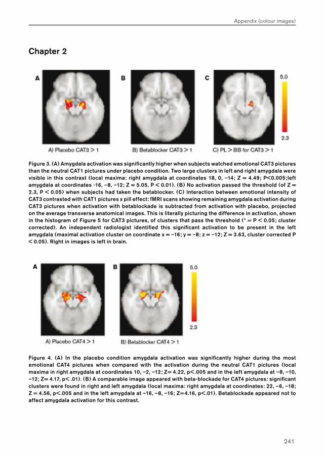
241
Chapter 2
Figure 3. (A) Amygdala activation was significantly higher when subjects watched emotional CAT3 pictures than the neutral CAT1 pictures under placebo condition. Two large clusters in left and right amygdala were visible in this contrast (local maxima: right amygdala at coordinates 18, 0, –14; Z = 4.49; P<0.005;left amygdala at coordinates -16, –8, –12; Z = 5.05, P < 0.01). (B) No activation passed the threshold (of Z = 2.3, P < 0.05) when subjects had taken the betablocker. (C) Interaction between emotional intensity of CAT3 contrasted with CAT1 pictures x pill effect: fMRI scans showing remaining amygdala activation during CAT3 pictures when activation with betablockade is subtracted from activation with placebo, projected on the average transverse anatomical images. This is literally picturing the difference in activation, shown in the histogram of Figure 5 for CAT3 pictures, of clusters that pass the threshold (* = P < 0.05; cluster corrected). An independent radiologist identified this significant activation to be present in the left amygdala (maximal activation cluster on coordinate x = –16; y = –8; z = –12; Z = 3.63, cluster corrected P < 0.05). Right in images is left in brain.
Figure 4. (A) In the placebo condition amygdala activation was significantly higher during the most emotional CAT4 pictures when compared with the activation during the neutral CAT1 pictures (local maxima in right amygdala at coordinates 10, –2, –12; Z= 4.22, p< .005 and in the left amygdala at –8, –10, –12; Z= 4.17, p< .01). (B) A comparable image appeared with beta-blockade for CAT4 pictures: significant clusters were found in right and left amygdala (local maxima: right amygdala at coordinates: 22, –6, –18; Z = 4.56, p< .005 and in the left amygdala at –16, –8, –16; Z=4.16, p< .01). Betablockade appeared not to affect amygdala activation for this contrast.
Appendix (colour images)
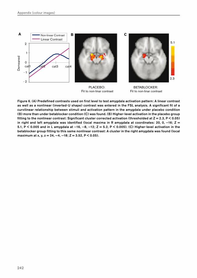
242
Appendix (colour images)
Figure 6. (A) Predefined contrasts used on first level to test amygdala activation pattern: A linear contrast as well as a nonlinear (inverted-U shape) contrast was entered in the FSL analysis. A significant fit of a curvilinear relationship between stimuli and activation pattern in the amygdala under placebo condition (B) more than under betablocker condition (C) was found. (B) Higher-level activation in the placebo group fitting to the nonlinear contrast: Significant cluster corrected activation (thresholded at Z = 2.3, P < 0.05) in right and left amygdala was identified (local maxima in R amygdala at coordinates: 20, 0, −16; Z = 5.1, P < 0.005 and in L amygdala at −16, −8, −12; Z = 5.2, P < 0.005). (C) Higher-level activation in the betablocker group fitting to this same nonlinear contrast: A cluster in the right amygdala was found (local maximum at x, y, z = 24, −4, −18; Z = 3.52, P < 0.05).
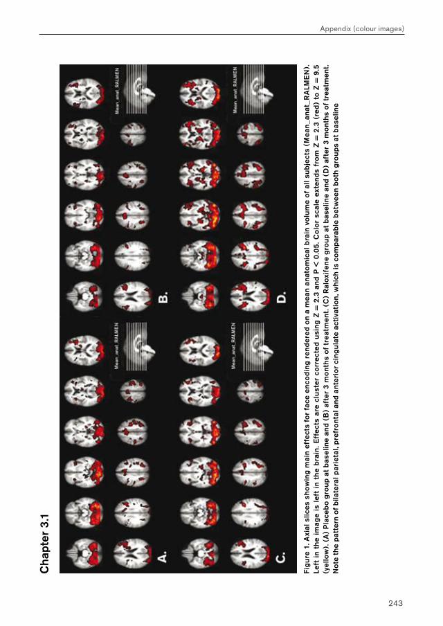
243
Appendix (colour images)
Cha
pter
3.1
Fig
ure
1. A
xial
slic
es s
how
ing
mai
n ef
fect
s fo
r fa
ce e
nco
ding
ren
der
ed o
n a
mea
n an
ato
mic
al b
rain
vo
lum
e of
all
subj
ects
(M
ean
_ana
t_R
ALM
EN
).
Left
in
the
imag
e is
lef
t in
th
e b
rain
. Eff
ects
are
clu
ster
co
rrec
ted
usin
g Z
= 2
.3 a
nd P
< 0
.05
. Co
lor
scal
e ex
tend
s fr
om
Z =
2.3
(re
d)
to Z
= 9
.5
(yel
low
). (
A)
Pla
ceb
o g
roup
at
bas
elin
e an
d (B
) af
ter
3 m
ont
hs o
f tr
eatm
ent.
(C)
Ral
oxif
ene
gro
up a
t b
asel
ine
and
(D)
afte
r 3
mo
nths
of
trea
tmen
t. N
ote
the
pat
tern
of b
ilate
ral p
arie
tal,
pre
fro
ntal
and
ant
erio
r ci
ngul
ate
acti
vati
on,
whi
ch is
co
mp
arab
le b
etw
een
bot
h g
roup
s at
bas
elin
e
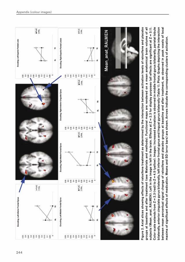
244
Appendix (colour images)
. Fig
ure
2. A
xial
slic
es s
how
ing
effe
cts
of r
alox
ifen
e tr
eatm
ent
as d
eter
min
ed b
y th
e in
tera
ctio
n b
etw
een
acti
vati
on
leve
ls o
f ra
loxi
fene
and
pla
ceb
o g
roup
s at
bas
elin
e an
d af
ter
trea
tmen
t (s
ee M
ater
ials
and
met
hods
). F
unct
iona
l m
aps
are
rend
ered
on
a m
ean
anat
om
ical
bra
in v
olu
me
of a
ll su
bjec
ts (
Mea
n_a
nat_
RA
LME
N).
Lef
t in
th
e im
age
is le
ft in
th
e b
rain
. Eff
ects
at
Z =
2.3
fo
r di
spla
y p
urp
oses
(al
l eff
ects
are
sig
nific
ant
at Z
= 3
.1).
C
olo
r sca
le e
xten
ds fr
om
Z =
2.3
(re
d) t
o Z
= 4
.5 (
yello
w).
Imag
es: I
ncre
ased
act
ivat
ion
is o
bser
ved
in m
iddl
e fr
ont
al g
yrus
bila
tera
lly, p
arie
tal l
ob
ule
bila
tera
lly, a
nter
ior c
ing
ulat
e gy
rus
bila
tera
lly, l
eft i
nfer
ior f
ront
al g
yrus
and
ling
ual
gyr
us b
ilate
rally
(Ta
ble
1).
Plo
ts: G
rap
hs d
epic
ting
the
inte
ract
ion
bet
wee
n m
ean
per
cent
ual
sig
nal
chan
ge
of r
alox
ifen
e an
d p
lace
bo
gro
ups
at b
asel
ine
and
afte
r tr
eatm
ent,
as o
bser
ved
in p
eak
voxe
ls o
f lo
cal
max
ima
of s
igni
fican
t eff
ects
of m
edic
atio
n. M
eans
and
SD
(er
ror
bar
s) a
re s
how
n. A
rrow
s in
dica
te t
he
corr
esp
ond
ing
clus
ters
of a
ctiv
atio
n.
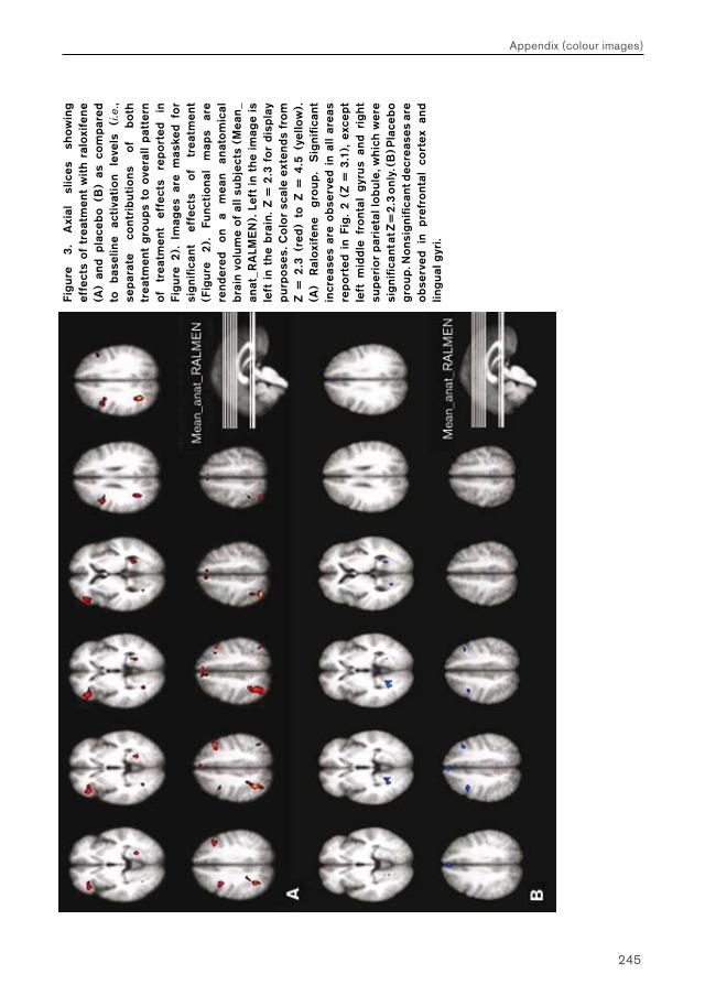
245
Appendix (colour images)
Fig
ure
3.
Axi
al
slic
es
show
ing
effe
cts
of t
reat
men
t w
ith
ralo
xife
ne
(A)
and
pla
ceb
o (B
) as
co
mp
ared
to
b
asel
ine
acti
vati
on
leve
ls
(i.e
., se
par
ate
cont
rib
uti
ons
of
b
oth
trea
tmen
t g
roup
s to
ove
rall
pat
tern
of
tr
eatm
ent
effe
cts
rep
ort
ed
in
Fig
ure
2).
Im
ages
are
mas
ked
for
sig
nific
ant
effe
cts
of
trea
tmen
t (F
igur
e 2
).
Func
tio
nal
map
s ar
e re
nder
ed
on
a m
ean
anat
om
ical
b
rain
vo
lum
e of
all
subj
ects
(M
ean
_an
at_R
ALM
EN
). L
eft
in t
he
imag
e is
le
ft i
n th
e b
rain
. Z =
2.3
fo
r di
spla
y p
urp
oses
. Co
lor
scal
e ex
tend
s fr
om
Z
= 2
.3 (
red
) to
Z =
4.5
(ye
llow
).
(A)
Ral
oxif
ene
gro
up.
Sig
nific
ant
incr
ease
s ar
e o
bser
ved
in a
ll ar
eas
rep
ort
ed i
n Fi
g.
2 (Z
= 3
.1),
exc
ept
left
mid
dle
fro
ntal
gyr
us a
nd r
igh
t su
per
ior
par
ieta
l lo
bul
e, w
hich
wer
e si
gni
fican
t at Z
= 2
.3 o
nly.
(B) P
lace
bo
gro
up. N
ons
igni
fican
t dec
reas
es a
re
obs
erve
d in
pre
fro
ntal
co
rtex
and
lin
gu
al g
yri.
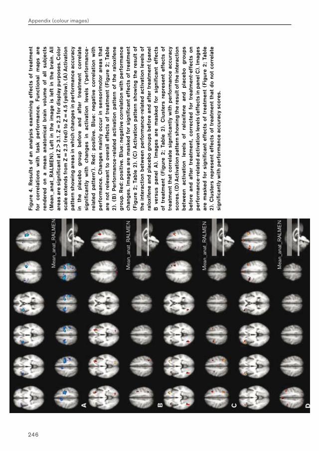
246
Appendix (colour images)
Fig
ure
4.
Res
ults
of
an a
naly
sis
exam
inin
g ef
fect
s of
tre
atm
ent
for
corr
elat
ions
w
ith
task
p
erfo
rman
ce.
Func
tio
nal
map
s ar
e re
nder
ed o
n a
mea
n an
ato
mic
al b
rain
vo
lum
e of
all
subj
ects
(M
ean
_ana
t_R
ALM
EN
). L
eft
in t
he
imag
e is
lef
t in
th
e b
rain
. A
ll ar
eas
are
sig
nific
ant a
t Z >
3.1
. Z =
2.3
for
disp
lay
pur
pos
es. C
olo
r sc
ale
exte
nds
fro
m Z
= 2
.3 (
red
) to
Z =
4.5
(ye
llow
). (
A)
Act
ivat
ion
pat
tern
sho
win
g ar
eas
in w
hich
ch
ang
es in
per
form
ance
acc
urac
y in
th
e p
lace
bo
gro
up
bef
ore
an
d af
ter
trea
tmen
t co
rrel
ate
sig
nific
antl
y w
ith
chan
ges
in
ac
tiva
tio
n le
vels
(‘
per
form
ance
-re
late
d p
atte
rn’)
. R
ed:
pos
itiv
e. B
lue
: ne
gat
ive
corr
elat
ion
wit
h p
erfo
rman
ce.
Ch
ang
es m
ainl
y o
ccur
in
sens
ori
mot
or
area
s th
at
are
not
rele
vant
to
over
all
effe
cts
of t
reat
men
t (F
igur
e 2
; Ta
ble
2
). (
B)
Per
form
ance
-rel
ated
act
ivat
ion
pat
tern
of
the
ralo
xife
ne
gro
up. R
ed: p
osit
ive.
Blu
e: n
egat
ive
corr
elat
ion
wit
h p
erfo
rman
ce
chan
ges
. Im
ages
are
mas
ked
for
sig
nific
ant
effe
cts
of t
reat
men
t (F
igur
e 2
; Ta
ble
2).
(C
) A
ctiv
atio
n p
atte
rn s
how
ing
the
resu
lt of
th
e in
tera
ctio
n b
etw
een
per
form
ance
-rel
ated
act
ivat
ion
leve
ls o
f ra
loxi
fene
and
pla
ceb
o g
roup
s b
efo
re a
nd a
fter
tre
atm
ent
(pan
el
B v
ersu
s p
anel
A).
Im
ages
are
mas
ked
for
sig
nific
ant
effe
cts
of t
reat
men
t (F
igur
e 2
; Ta
ble
2).
Clu
ster
s re
pre
sent
eff
ects
of
trea
tmen
t th
at c
orr
elat
e si
gni
fican
tly
wit
h p
erfo
rman
ce a
ccur
acy
sco
res.
(D
) Act
ivat
ion
pat
tern
sho
win
g th
e re
sult
of th
e in
tera
ctio
n b
etw
een
acti
vati
on
leve
ls
of
ralo
xife
ne
and
pla
ceb
o g
roup
s b
efo
re a
nd a
fter
tre
atm
ent,
corr
ecte
d fo
r tr
eatm
ent-
effe
cts
on
per
form
ance
-rel
ated
act
ivat
ion
leve
ls (
effe
cts
in p
anel
C).
Imag
es
are
mas
ked
for
sig
nific
ant
effe
cts
of t
reat
men
t (F
igur
e 2
; Ta
ble
2
). C
lust
ers
rep
rese
nt e
ffec
ts o
f tr
eatm
ent
that
do
not
corr
elat
e si
gni
fican
tly
wit
h p
erfo
rman
ce a
ccur
acy
sco
res.
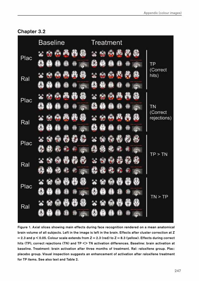
247
Appendix (colour images)
Chapter 3.2
Figure 1. Axial slices showing main effects during face recognition rendered on a mean anatomical
brain volume of all subjects. Left in the image is left in the brain. Effects after cluster correction at Z
= 2.3 and p < 0.05. Colour scale extends from Z = 2.3 (red) to Z = 8.3 (yellow). Effects during correct
hits (TP), correct rejections (TN) and TP <> TN activation differences. Baseline: brain activation at
baseline. Treatment: brain activation after three months of treatment. Ral: raloxifene group. Plac:
placebo group. Visual inspection suggests an enhancement of activation after raloxifene treatment
for TP items. See also text and Table 2.
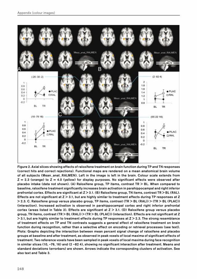
248
Appendix (colour images)
Figure 2. Axial slices showing effects of raloxifene treatment on brain function during TP and TN responses (correct hits and correct rejections). Functional maps are rendered on a mean anatomical brain volume of all subjects (Mean_anat_RALMEN). Left in the image is left in the brain. Colour scale extends from Z = 2.3 (orange) to Z = 4.0 (yellow) for display purposes. No significant effects were observed after placebo intake (data not shown). (A) Raloxifene group, TP items, contrast TR > BL. When compared to baseline, raloxifene treatment significantly increases brain activation in parahippocampal and right inferior prefrontal cortex. Effects are significant at Z > 3.1. (B) Raloxifene group, TN items, contrast TR > BL (RAL). Effects are not significant at Z > 3.1, but are highly similar to treatment effects during TP responses at Z > 2.3. C. Raloxifene group versus placebo group, TP items, contrast (TR > BL (RAL)) > (TR > BL (PLAC)) (interaction). Increased activation is observed in parahippocampal cortex and right inferior prefrontal cortex (areas listed in Table 3). Effects are significant at Z > 3.1. (D) Raloxifene group versus placebo group, TN items, contrast (TR > BL (RAL)) > (TR > BL (PLAC)) (interaction). Effects are not significant at Z > 3.1, but are highly similar to treatment effects during TP responses at Z > 2.3. The strong resemblance of treatment effects on TP and TN contrasts suggests a general effect of raloxifene treatment on brain function during recognition, rather than a selective effect on encoding or retrieval processes (see text). Plots: Graphs depicting the interaction between mean percent signal change of raloxifene and placebo groups at baseline and after treatment, as observed in peak voxels of local maxima of significant effects of treatment. Two reference voxels have been sampled in peak voxels of local maxima during face recognition in similar slices (10, –76, 16) and (2 –82 4), showing no significant interaction after treatment. Means and standard deviations (errorbars) are shown. Arrows indicate the corresponding clusters of activation. See also text and Table 3.
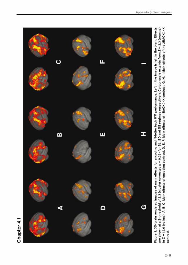
249
Appendix (colour images)
Cha
pter
4.1
Fig
ure
1. 3
D-b
rain
ren
der
ed im
ages
of
mai
n ef
fect
s fo
r en
codi
ng a
nd N
-let
ter
bac
k W
M p
erfo
rman
ce. L
eft
in t
he
imag
e is
left
in t
he
bra
in. E
ffec
ts
are
show
n at
a Z
-thr
esho
ld o
f 2
.3 (
clus
ter
corr
ecte
d p
= 0
.05
) fo
r B
L, S
D a
nd S
S r
egim
es r
esp
ecti
vely
. Co
lour
sca
le e
xten
ds f
rom
Z =
2.3
(o
rang
e)
to Z
= 1
2.0
(ye
llow
). A
, B, C
: Mai
n ef
fect
s of
enc
odi
ng c
ont
rast
. D, E
, F: M
ain
effe
cts
of 1
BA
CK
> X
co
ntra
st. G
, H, I
: Mai
n ef
fect
s of
th
e 2B
AC
K >
X
cont
rast
.
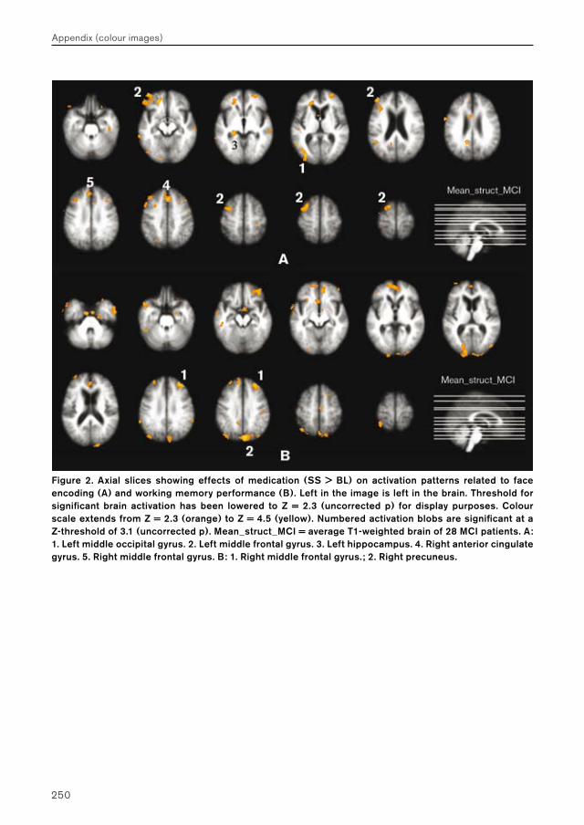
250
Appendix (colour images)
Figure 2. Axial slices showing effects of medication (SS > BL) on activation patterns related to face encoding (A) and working memory performance (B). Left in the image is left in the brain. Threshold for significant brain activation has been lowered to Z = 2.3 (uncorrected p) for display purposes. Colour scale extends from Z = 2.3 (orange) to Z = 4.5 (yellow). Numbered activation blobs are significant at a Z-threshold of 3.1 (uncorrected p). Mean_struct_MCI = average T1-weighted brain of 28 MCI patients. A: 1. Left middle occipital gyrus. 2. Left middle frontal gyrus. 3. Left hippocampus. 4. Right anterior cingulate gyrus. 5. Right middle frontal gyrus. B: 1. Right middle frontal gyrus.; 2. Right precuneus.
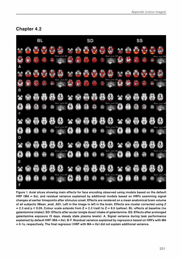
251
Appendix (colour images)
Chapter 4.2
Figure 1. Axial slices showing main effects for face encoding observed using models based on the default HRF (MA = 6s), and residual variance explained by additional models based on HRFs examining signal changes at earlier timepoints after stimulus onset. Effects are rendered on a mean anatomical brain volume of all subjects (Mean_anat_AD). Left in the image is left in the brain. Effects are cluster corrected using Z = 2.3 and p < 0.05. Colour scale extends from Z = 2.3 (red) to Z = 9.5 (yellow). BL: effects at baseline (no galantamine intake); SD: Effects after acute (single dose) intake of galantamine. SS: Effects after prolonged galantamine exposure (5 days, steady state plasma levels). A. Signal variance during task performance explained by default HRF (MA = 6s), B-F. Residual variance explained by regressors based on HRFs with MA = 5-1s, respectively. The final regressor (HRF with MA = 0s) did not explain additional variance.
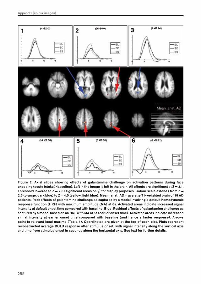
252
Appendix (colour images)
Figure 2. Axial slices showing effects of galantamine challenge on activation patterns during face encoding (acute intake > baseline). Left in the image is left in the brain. All effects are significant at Z = 3.1. Threshold lowered to Z = 2.3 (significant areas only) for display purposes. Colour scale extends from Z = 2.3 (orange, dark blue) to Z = 4.5 (yellow, light blue). Mean_anat_AD = average T1-weighted brain of 18 AD patients. Red: effects of galantamine challenge as captured by a model involving a default hemodynamic response function (HRF) with maximum amplitude (MA) at 6s. Activated areas indicate increased signal intensity at default onset time compared with baseline. Blue: Residual effects of galantamine challenge as captured by a model based on an HRF with MA at 5s (earlier onset time). Activated areas indicate increased signal intensity at earlier onset time compared with baseline (and hence a faster response). Arrows point to relevant local maxima (Table 1). Coordinates are given at the top of each plot. Plots represent reconstructed average BOLD response after stimulus onset, with signal intensity along the vertical axis and time from stimulus onset in seconds along the horizontal axis. See text for further details.
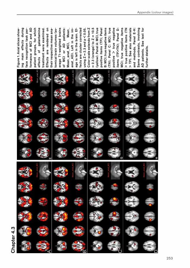
253
Appendix (colour images)C
hapt
er 4
.3Fi
gur
e 1.
Axi
al s
lices
sho
w-
ing
mai
n ef
fect
s du
ring
fa
ce r
eco
gni
tio
n ta
sk p
er-
form
ance
of
MC
I an
d A
D
pat
ient
gro
ups,
fo
r w
hich
ef
fect
s of
g
alan
tam
ine
trea
tmen
t w
ere
exam
ined
. E
ffec
ts
are
rend
ered
o
n th
eir
resp
ecti
ve m
ean
ana-
tom
ical
bra
in v
olu
mes
(av
-er
age
T1-w
eig
hte
d b
rain
s of
M
CI
or
AD
p
atie
nts:
M
ean
_ana
t_M
CI,
Mea
n_
anat
_AD
). L
eft
in t
he
im-
age
is l
eft
in t
he
bra
in. E
f-fe
cts
are
clus
ter
corr
ecte
d us
ing
Z =
2.3
and
p <
0.0
5.
Co
lor s
cale
ext
ends
fro
m Z
=
2.3
(o
rang
e) t
o Z
= 1
0.5
(y
ello
w).
Pan
el A
: MC
I, tr
ue
pos
itiv
e it
ems
(TP
); P
anel
B
: MC
I; tr
ue
neg
ativ
e it
ems
(TN
);
Pan
el
C:
MC
I;
tru
e p
osit
ive
>
tru
e ne
gat
ive
item
s (T
P>
TN);
P
anel
D
: M
CI;
tr
ue
neg
ativ
e it
ems
> t
rue
pos
itiv
e it
ems
(TN
>
TP
). S
ee a
lso
mat
eria
ls
and
met
hods
. P
anel
E-H
: S
ame
cont
rast
s, i
nvo
lvin
g A
D p
atie
nts.
See
tex
t fo
r fu
rth
er d
etai
ls.
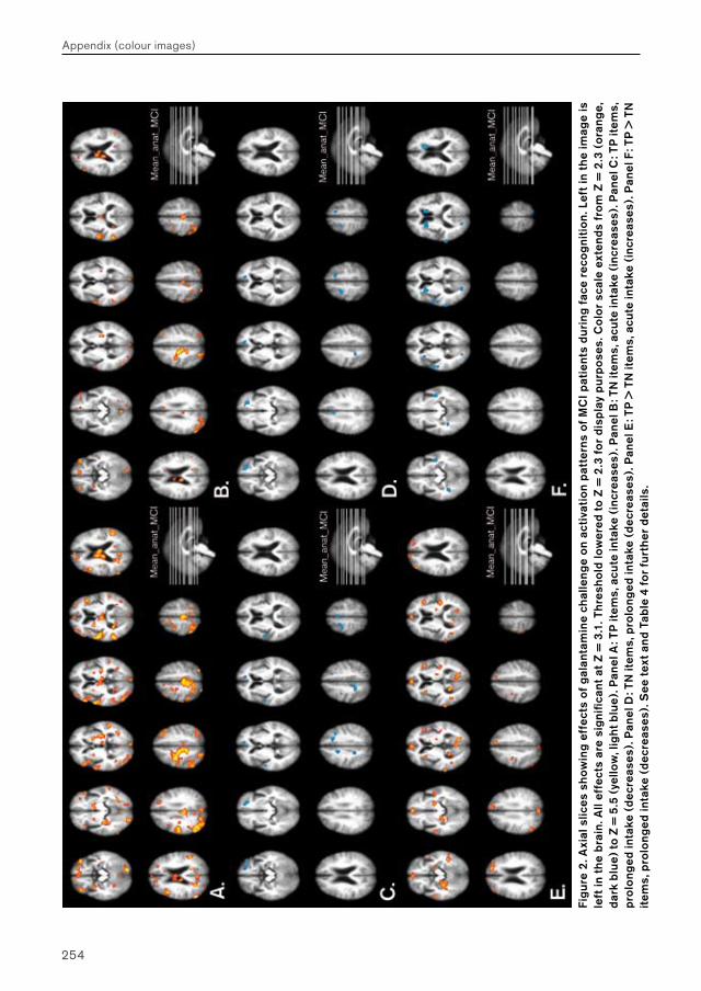
254
Appendix (colour images)
Fig
ure
2. A
xial
slic
es s
how
ing
effe
cts
of g
alan
tam
ine
chal
leng
e o
n ac
tiva
tio
n p
atte
rns
of M
CI p
atie
nts
duri
ng f
ace
reco
gni
tio
n. L
eft
in t
he
imag
e is
le
ft in
th
e b
rain
. All
effe
cts
are
sig
nific
ant
at Z
= 3
.1. T
hres
hold
low
ered
to
Z =
2.3
fo
r di
spla
y p
urp
oses
. Co
lor
scal
e ex
tend
s fr
om
Z =
2.3
(o
rang
e,
dar
k b
lue)
to Z
= 5
.5 (
yello
w, l
igh
t blu
e). P
anel
A: T
P it
ems,
acu
te in
take
(in
crea
ses)
. Pan
el B
: TN
item
s, a
cute
inta
ke (
incr
ease
s). P
anel
C: T
P it
ems,
p
rolo
nged
inta
ke (
dec
reas
es).
Pan
el D
: TN
item
s, p
rolo
nged
inta
ke (
dec
reas
es).
Pan
el E
: TP
> T
N it
ems,
acu
te in
take
(in
crea
ses)
. Pan
el F
: TP
> T
N
item
s, p
rolo
nged
inta
ke (
dec
reas
es).
See
tex
t and
Tab
le 4
fo
r fu
rth
er d
etai
ls.
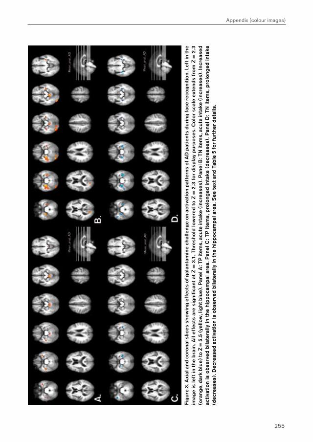
255
Appendix (colour images)
Fig
ure
3. A
xial
and
co
rona
l slic
es s
how
ing
effe
cts
of g
alan
tam
ine
chal
leng
e o
n ac
tiva
tio
n p
atte
rns
of A
D p
atie
nts
duri
ng fa
ce r
eco
gni
tio
n. L
eft i
n th
e im
age
is le
ft in
th
e b
rain
. All
effe
cts
are
sig
nific
ant
at Z
= 3
.1. T
hres
hold
low
ered
to
Z =
2.3
fo
r di
spla
y p
urp
oses
. Co
lor
scal
e ex
tend
s fr
om
Z =
2.3
(o
rang
e, d
ark
blu
e) to
Z =
5.5
(ye
llow
, lig
ht b
lue)
. Pan
el A
: TP
item
s, a
cute
inta
ke (
incr
ease
s). P
anel
B: T
N it
ems,
acu
te in
take
(in
crea
ses)
. Inc
reas
ed
acti
vati
on
is o
bser
ved
bila
tera
lly in
th
e hi
pp
oca
mp
al a
rea.
Pan
el C
: TP
ite
ms,
pro
long
ed in
take
(d
ecre
ases
). P
anel
D: T
N i
tem
s, p
rolo
nged
inta
ke
(dec
reas
es).
Dec
reas
ed a
ctiv
atio
n is
obs
erve
d b
ilate
rally
in t
he
hip
po
cam
pal
are
a. S
ee t
ext a
nd T
able
5 f
or
furt
her
det
ails
.
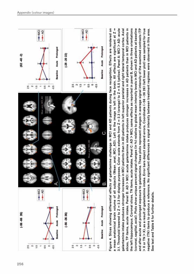
256
Appendix (colour images)
Fig
ure
4. S
lices
sho
win
g di
ffer
enti
al e
ffec
ts o
f g
alan
tam
ine
chal
leng
e in
MC
I an
d A
D p
atie
nts
duri
ng f
ace
reco
gni
tio
n. E
ffec
ts a
re r
end
ered
on
a m
ean
anat
om
ical
bra
in v
olu
me
of a
ll su
bjec
ts (
Mea
n_a
nat_
MC
I_A
D).
Lef
t in
th
e im
age
is l
eft
in t
he
bra
in.
All
effe
cts
are
sig
nific
ant
at Z
=
3.1
. Thr
esho
ld l
ower
ed t
o Z
= 2
.3 f
or
disp
lay
pur
pos
es. C
olo
r sc
ale
exte
nds
fro
m Z
= 2
.3 (
ora
nge)
to
Z =
5.5
(ye
llow
). P
anel
A:
MC
I >
AD
: ac
ute
g
alan
tam
ine
inta
ke p
rodu
ces
stro
nger
incr
ease
s in
MC
I pat
ient
s th
an in
AD
pat
ient
s in
left
sup
erio
r p
arie
tal a
nd r
igh
t lat
eral
tem
po
ral c
ort
ex. A
xial
sl
ices
, TP
ite
ms,
acu
te i
ntak
e. P
anel
B:
AD
> M
CI:
acu
te g
alan
tam
ine
inta
ke p
rodu
ces
stro
nger
inc
reas
es i
n A
D p
atie
nts
than
in
MC
I p
atie
nts
in
the
left
hip
po
cam
pus
. Co
rona
l slic
es, T
N it
ems,
acu
te in
take
. Pan
el C
: AD
> M
CI;
sam
e ef
fect
s as
rep
ort
ed in
pan
el B
, sho
wn
in t
hree
ori
enta
tio
ns
(co
rona
l, sa
gitt
al, a
xial
). P
lots
sho
w u
niqu
e p
erce
nt s
igna
l ch
ang
es (
in %
) re
lati
ve to
glo
bal
mea
n in
tens
ity
leve
ls in
MC
I and
AD
pat
ient
s at
bas
elin
e an
d af
ter
acu
te a
nd p
rolo
nged
exp
osur
e to
gal
anta
min
e. T
hes
e p
lots
rep
rese
nt c
han
ges
in a
vera
ge
sig
nal i
nten
sity
of
the
exam
ined
co
ntra
sts
(TP
>
X o
r TN
> X
), a
s a
resu
lt of
gal
anta
min
e in
take
. Err
or
bar
s d
epic
t st
and
ard
erro
rs. C
oo
rdin
ate
(–56
–36
26
) (l
eft
low
er p
lot)
was
sam
ple
d fo
r tr
ue
neg
ativ
e (T
N)
item
s to
pro
duce
a r
efer
ence
. No
sig
nific
ant
diff
eren
ces
in s
igna
l int
ensi
ty b
etw
een
trea
tmen
t re
gim
es w
ere
obs
erve
d in
thi
s ar
ea.
See
tex
t and
Tab
le 6
fo
r fu
rth
er d
etai
ls.