Proefschrift Maarse
253
Prenatal Detection of Oral Clefts Wiesje Maarse
-
Upload
nicole-nijhuis -
Category
Documents
-
view
270 -
download
8
description
Â
Transcript of Proefschrift Maarse
Prenatal Detection of Oral Clefts
Wiesje Maarse
Prenatal D
etection o
f Oral C
lefts Diag
nostic, G
enetic and E
thical Asp
ects W
iesje Maarse
UITNODIGING
U bent uitgenodigd voor het
bijwonen van de openbare
verdediging van het proefschrift
Prenatal Detection of Oral Clefts
door
Wiesje Maarse
op donderdag 27 augustus
2015 om 14.30 uur precies
in de Senaatszaal van het
Academiegebouw,
Domplein 29 te Utrecht
Borrel na afloop in de Oranjerie,
te Utrecht
Paranimfen
Michelle Ebbeling
06-42805524
Anna Janssen-Bakker
06-18884713
Wiesje Maarse
Diamantweg 49
3523 CM Utrecht
Wiesje Maarse was born on the 17th of August, 1984 in Nijmegen, the Ne-therlands. After gra-duating cum laude from the Montessori College in Nijmegen in 2002 she went on studying medicine at the University of Utrecht. During her
medical education she followed some internships abroad, and spent several months in South Africa and Australia, where she participated in scientific research on hand surgery, under the guidance of Dr. G.I. Bain. She obtained her medical degree in 2008 after which she was given the opportunity to participate in the development of the guideline “Counselling after prenatal detection of orofacial cleft in the Netherlands”. At the same time she started the research described in this thesis. In 2009 Wiesje commenced her plastic surgery residency, starting off with two years at the Department of Surgery, Diakonessenhuis in Utrecht (Dr. G.J. Clevers) and four years at the Department of Plastic, Reconstructive and Hand Surgery in Utrecht (dr. A.H. Schuurman) and St. Antonius Hospital in Nieuwegein (dr. A.B. Mink van der Molen). During her residency, she was board member of the junior association of plastic surgery. In 2014 she finished her plastic surgery residency, obtained her European Board Plastic and Aesthetic Surgery (EBOBRAS) exams, and joined the Division of Plastic, Reconstructive and Hand Surgery and the Military Hospital in Utrecht as a faculty member where she specializes in breast and reconstructive surgery. She has a fellowship planned in breast and reconstructive surgery at the University of Toronto, Canada in 2016 (Dr. T Zhong and Dr. S.O. Hofer).Currently, Wiesje lives together with Roeland de Wilde in Utrecht.
voor Roeland

Prenatal Detection of Oral CleftsDiagnostic, genetic and ethical aspects
Wiesje Maarse
Cover: © Manolo Valdés, Fondation Marguerite et Aimé Maeght, Saint-
Paul (France), Retrato con medio rostro azul, 1999
Layout: Nicole Nijhuis, Gildeprint, The Netherlands
ISBN: 978-94-6233-028-3
This thesis was (partly) accomplished with financial support from the Department
of Plastic, Reconstructive and hand surgery University Medical Centre Utrecht and
Wilhemina’s Children hospital, the Dutch Society of Plastic Surgery, ABN Amro,
Allergan B.V., AllweCare medical B.V., BlooMEDical Benelux, Chipsoft B.V, Dalton
Medicare B.V., Emdaplast, Examvision, Handencentrum Utrecht, Kwaliteitsinstituut
van Medisch Specialisten, Mediplast, Quamedical, van Wijngaarden Medical, Junior
Vereniging Plastische Chirurgie
© Copyright Wiesje Maarse, Utrecht 2015
All rights reserved. No parts of this thesis may be reproduced, stored in retrieval
system or transmitted in any form or by any means without prior written permission
of the author.
Prenatal Detection of Oral CleftsDiagnostic, Genetic and Ethical Aspects
Prenatale detectie van schisis
Diagnostische, Genetische en Ethische Aspecten
(met een samenvatting in het Nederlands)
Proefschrift
ter verkrijging van de graad van doctor aan de Universiteit Utrecht op gezag van de
rector magnificus, prof.dr. G.J. van der Zwaan, ingevolge het besluit van het college
voor promoties in het openbaar te verdedigen op woensdag 27 augustus 2015 des
middags te 2.30 uur
door
Wiesje Maarse
geboren op 17 augustus 1984
te Nijmegen

Promotoren: Prof. dr. M. Kon
Prof. dr. J.J.M. van Delden
Copromotor: Dr. A.B. Mink van der Molen
CONTENTS
Chapter 1 General introduction and outline of the thesis 7
Part I Classification of Oral Cleft
Chapter 2 A practical prenatal ultrasound classification system for 31
common oral clefts
Prenatal Diagnosis, 2015 Jun 10. [Epub ahead of print]
Part II Screening for Oral Cleft
Chapter 3 Prenatal Screening for Orofacial Clefts in the Netherlands: 49
A Preliminary Report on the Impact of a National Screening
System
Cleft Palate-Craniofacial Journal, March 2011, Vol. 48 (2) 183-9
Chapter 4 Diagnostic accuracy of transabdominal ultrasound in detecting 67
prenatal cleft lip and palate: a systematic review
Ultrasound Obstet Gynecol 2010; 35: 495–502
Chapter 5 Prenatal ultrasound screening for orofacial cleft 89
Ultrasound Obstet Gynecol 2011; 38: 434–439
Chapter 6 The accuracy of prenatal ultrasound in determining the type of 105
orofacial cleft
Prenatal Diagnosis, 2015, Feb 26. [Epub ahead of print]
Chapter 7 A systematic review of associated structural and chromosomal 117
defects in oral clefts: when is prenatal genetic analysis indicated?
J Med Genet 2012; 49: 490–498
Part III Prenatal Counseling
Chapter 8 Professional opinion on oral cleft during pregnancy; 151
a comparison between Israel and the Netherlands
Prenatal Diagnosis, 2015 Jun;35(6):544-8
Chapter 9 Parental attitude on prenatal diagnosis of cleft lip and palate; 163
a prospective cohort study
Submitted
Chapter 10 Prenatal detection of oral cleft: defending a more directive 191
approach in counseling
Submitted
Part IV Discussion and Summary
Chapter 11 General discussion and future perspectives 205
Chapter 12 English summary 221
Dutch summary/Nederlandse samenvatting 227
Addendum List of co-authors and affiliations 237
List of Abbreviations 241
Publications 243
Acknowledgments/Dankwoord 245

1General introduction and
outline of the thesis

R1
R2
R3
R4
R5
R6
R7
R8
R9
R10
R11
R12
R13
R14
R15
R16
R17
R18
R19
R20
R21
R22
R23
R24
R25
R26
R27
R28
R29
R30
R31
R32
R33
R34
1 | General introduction and outline of the thesis
8
R1
R2
R3
R4
R5
R6
R7
R8
R9
R10
R11
R12
R13
R14
R15
R16
R17
R18
R19
R20
R21
R22
R23
R24
R25
R26
R27
R28
R29
R30
R31
R32
R33
R34
9
Since the introduction of standard prenatal screening in the Netherlands by means of
ultrasound in 2007, parents can be confronted with a child with a cleft lip and/or palate
already during pregnancy. As a consequence, this brings up the necessity for accurate
counseling. When informing parents on outcome and prognosis of orofacial clefts it is
crucial to understand its epidemiology, embryology, pathophysiology and treatment.
Epidemiology
Oral cleft is a congenital malformation that can involve the lip, palate, nose and
underlying bony framework in various severities, and is the most common craniofacial
anomaly considering the incidence of approximately 1:700 births1. The frequency of
cleft lip with or without cleft palate varies across race and sex, with an incidence as
high as 1:450 births among Asian and Native American populations, 1:1,000 among
Caucasian populations, and 1:2,000 among African American populations. In contrast,
in isolated cleft palate there appears to be no such heterogeneity with an incidence
among races of about 1:2,000 births23. Boys are twice as likely to be affected compared
to girls, whereas this relationship is reversed for isolated cleft palate4, 5. A unilateral lip
occurs twice as frequently on the left side versus the right side and is nine times more
common than a bilateral cleft lip 5, 6. Unaffected (i.e., noncleft) parents who have one
child with an oral cleft have an estimated recurrence risk of 4%, which rises to 9% with
two affected children. An affected individual, has a 4% chance to become parent of a
newborn with an oral cleft, with an increasing chance up to 17% if there is already an
affected child 2, 7. Recurrence increases with the severity of the cleft8.
Oral clefts can be isolated, but are often associated with other congenital anomalies 9.
Studies on associated anomalies in newborns with oral cleft report variable rates, also
since different definitions are used, encompassing anomalies as only major non-facial
congenital anomalies to all anomalies, including minor10. Despite these differences, it
is evident that the prevalence of associated anomalies is related to the cleft category,
of which only cleft lip shows a lower prevalence of associated anomalies in comparison
to cleft lip with palate or cleft palate only 11-15.
R1
R2
R3
R4
R5
R6
R7
R8
R9
R10
R11
R12
R13
R14
R15
R16
R17
R18
R19
R20
R21
R22
R23
R24
R25
R26
R27
R28
R29
R30
R31
R32
R33
R34
1 | General introduction and outline of the thesis
10
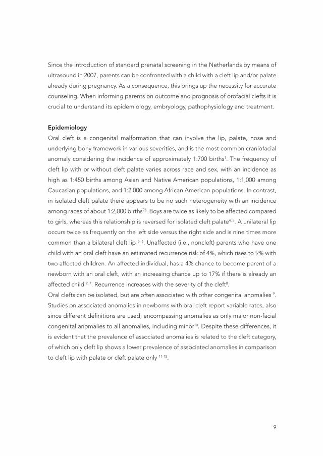
Embryology
The basic morphology of the face is established between the 4th and 10th week of
human development. At five weeks’ gestation, when the embryo is 3mm long, the
ectoderm in the vicinity of the neural plate folds itself to form the neural tube. Special
neural crest cells of ectodermal origin differentiate to form a special ectomesenchyme.
Migration of the latter over and around the head is essential for the development of
five facial prominences surrounding the primitive oral cavity16. A as a result of fusion
of these five, namely the midline frontonasal prominence, two paired prominences,
the maxillary and mandibular prominences, form the face (figure 1). The mesenchyme
in the frontonasal prominence covers the forebrain (prosencephalon) and arises from
neural crest cells derived from the midbrain (mesencephalon) and forebrain, whereas
the maxillary and mandibular prominences are part of the first pharyngeal arch, and
receive contributions from both the midbrain and hindbrain (rhombencephalon)17.
The development of the upper lip begins during the 4th week of gestation and is
completed by the 7th week18. On the frontonasal prominence two bilateral ectodermal
thickenings are formed which invaginate to develop an oval nasal pit, hence dividing
the frontonasal prominence into a lateral and medial nasal process. Simultaneously, in
the 5th week the paired maxillary prominences enlarge and push forward by growing
ventrally and medially. During the 6th week, the medial nasal processes migrate to each
other and fuse to form the primordium of the bridge and septum of the nose. By
the end of week 7, the inferior tips of the medial nasal process fuse in the midline
and form the intermaxillary segment. From this intermaxillary segment, the nasal tip,
columella, philtrum, labial tubercle, frenulum and primary palate are derived. The
maxillary prominences give rise to the upper jaw and the sides of the upper lip and the
mouth is reduced to its final width when fusion of the latter with the lateral portions of
the mandibular prominences. Lastly, the mandibular prominence forms the mandible
and lower lip to complete the oral aperture.
R1
R2
R3
R4
R5
R6
R7
R8
R9
R10
R11
R12
R13
R14
R15
R16
R17
R18
R19
R20
R21
R22
R23
R24
R25
R26
R27
R28
R29
R30
R31
R32
R33
R34
11
Intermaxillary process
Frontonasal prominence
Maxillary prominence
Mandibular prominence
Lateral nasal process
Medial nasal process
The secondary palate fuses later than the primary palate and is formed by the lateral
palatal shelves, which are derived from the medial walls of the maxillary prominences.
At first, these shelves grow downward, parallel to the lateral surface of the tongue. The
tongue descends by forward growth and lowering of the mandibular prominences,
allowing palatal shelve elevation. At the end of the 7th week, the shelves rotate rapidly
into a horizontal position and then fuse with each other and the primary palate. This
fusion proceeds from anteriorly, right behind the incisive foramen, to the posterior,
thereby separating the nasal and oral cavities. The nasal septum, originates from the
frontonasal prominence and grows simultaneously downward to fuse with the primary
and secondary palate along the midline 17.
Facial clefting is the result of failure of both growth and fusion of the facial prominences
anywhere. More specifically, failure of fusion of the intermaxillary process and maxillary
prominences gives rise to a cleft of the primary palate including the lip, alveolar
Figure 1 This figure was published earlier in Larsen’s human embryology, Gary C. Schoenwolf, Page 565, Copyright Elsevier 2009 and reprinted with permission of Prof. Schoenwolf who provided the figure.
R1
R2
R3
R4
R5
R6
R7
R8
R9
R10
R11
R12
R13
R14
R15
R16
R17
R18
R19
R20
R21
R22
R23
R24
R25
R26
R27
R28
R29
R30
R31
R32
R33
R34
1 | General introduction and outline of the thesis
12
process, and the hard palpate anterior to the incisive foramen, which notably can
occur on either or both sides. Disruption of the confluence between the two palatal
shelves results in a cleft of the secondary palate. Moreover, cleft palate may occur as
secondary to mandibular dysplasia, in which the first pharyngeal arch is not developed
appropriately. As a consequence, the tongue will not be lowered and will physically
obstruct palatal shelve elevation. This form of secondary cleft palate resulting
from a smaller mandibula (micrognathia) and accruing with glossoptosis (backward
displacement of the tongue) in combination with often respiratory failure is referred to
as Robin sequence17, 19.
Although cleft lip and palate often occur simultaneously, the two defects differ in
distribution with respect to sex, familial association, race and geography as mentioned
earlier. Moreover, and as discussed above they differ on an embryological level in
the fusion process, namely fusion of the intermaxillary segment with the maxillary
prominences versus the fusion of the palatal shelves. Therefore, the cleft lip and cleft
palate probably have a different etiology 17.
Midline or median clefts arise due to incomplete merging of the medial nasal processes
which form the intermaxillary segment. These are part of holoprosencephaly, which
includes a spectrum of forebrain development defects 17. Together with atypical or
craniofacial facial clefts they are rare and should be considered as separate craniofacial
anomalies because of the different pathogenesis 20-22.
Pathology and related functional issues
A cleft can either be unilateral or bilateral and demonstrate a variable expression
pattern, including microform, minor, incomplete and complete forms. In addition, they
occur in a diversity of bone, cartilage and soft tissue deficiencies, thereby representing
different functional problems. Microform cleft lip is an anomaly formerly termed
“formefruste” and represents the most minor of cleft lip deformities. It manifests as
a vermillion notch that is less than 3 mm in height compared with the Cupid’s bow
of the noncleft side, without a nasal deformity. A minor cleft lip is characterized by a
vermillion notch greater than 3 mm combined with a vertical depression above the
R1
R2
R3
R4
R5
R6
R7
R8
R9
R10
R11
R12
R13
R14
R15
R16
R17
R18
R19
R20
R21
R22
R23
R24
R25
R26
R27
R28
R29
R30
R31
R32
R33
R34
13
notch into the nasal sill, a variable degree of nose deformity, and possibly a cleft of the
alveolus 23. Usually, these minimal clefts of the lip require surgical treatment, if not for
aesthetics of the lip then for restoration of the underlying disruption of the orbicularis
oris muscle.
An incomplete cleft lip has a variable degree of separation but by definition has an
intact nasal sill, commonly referred to as Simonart band. A complete cleft lip extends
through the nasal sill and floor and may or may not extend to the underlying alveolus
and palate. A bilateral cleft lip can present as a combination of any of the above 24. In
a complete bilateral cleft lip and palate the prolabium and premaxilla, both derivatives
of the intermaxillary process remain entirely separated from the lateral lip and maxillary
arch elements as they fuse with the maxillary prominences25.
9
Pathology and related functional issues
A cleft can either be unilateral or bilateral and demonstrate a variable expression pattern, including
microform, minor, incomplete and complete forms. In addition, they occur in a diversity of bone, cartilage
and soft tissue deficiencies, thereby representing different functional problems. Microform cleft lip is an
anomaly formerly termed “formefruste” and represents the most minor of cleft lip deformities. It
manifests as a vermillion notch that is less than 3 mm in height compared with the Cupid’s bow of the
noncleft side, without a nasal deformity. A minor cleft lip is characterized by a vermillion notch greater
than 3 mm combined with a vertical depression above the notch into the nasal sill, a variable degree of
nose deformity, and possibly a cleft of the alveolus 23. Usually, these minimal clefts of the lip require
surgical treatment, if not for aesthetics of the lip then for restoration of the underlying disruption of the
orbicularis oris muscle.
An incomplete cleft lip has a variable degree of separation but by definition has an intact nasal sill,
commonly referred to as Simonart band. A complete cleft lip extends through the nasal sill and floor and
may or may not extend to the underlying alveolus and palate. A bilateral cleft lip can present as a
combination of any of the above 24. In a complete bilateral cleft lip and palate the prolabium and
premaxilla, both derivatives of the intermaxillary process remain entirely separated from the lateral lip
and maxillary arch elements as they fuse with the maxillary prominences25.
Figure 2 This figure was published earlier in Plastic Surgery, Peter C. Neligan, Volume Three, William Y. Hoffman, Cleft
Palate, Page 786, Copyright Elsevier 2013
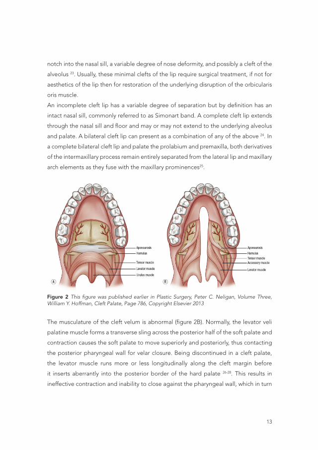
Figure 2 This figure was published earlier in Plastic Surgery, Peter C. Neligan, Volume Three, William Y. Hoffman, Cleft Palate, Page 786, Copyright Elsevier 2013
The musculature of the cleft velum is abnormal (figure 2B). Normally, the levator veli
palatine muscle forms a transverse sling across the posterior half of the soft palate and
contraction causes the soft palate to move superiorly and posteriorly, thus contacting
the posterior pharyngeal wall for velar closure. Being discontinued in a cleft palate,
the levator muscle runs more or less longitudinally along the cleft margin before
it inserts aberrantly into the posterior border of the hard palate 26-28. This results in
ineffective contraction and inability to close against the pharyngeal wall, which in turn
R1
R2
R3
R4
R5
R6
R7
R8
R9
R10
R11
R12
R13
R14
R15
R16
R17
R18
R19
R20
R21
R22
R23
R24
R25
R26
R27
R28
R29
R30
R31
R32
R33
R34
1 | General introduction and outline of the thesis
14
can result in air escape through the nose and difficult speech. In addition, aberrant
levator positioning as well as an abnormal fusion with the tendon of the tensor veli
palatine muscle is thought to impair the function of the tensor muscle in assisting
Eustachian tube function and is presumed to be contributory to cleft otopathology.
Finally, there can be a submucous cleft palate presenting as the classic triad of bifid
uvula, midline notching of the posterior hard palate in combination with diastasis of
the velar musculature 29.
Besides the aesthetic complications of oral cleft there are functional problems related
to the abnormal anatomy. As briefly described above, the velopharyngeal insufficiency
of the palate musculature together with possible lip incompetence and abnormal
dental position complicates normal speech. Furthermore, the incidence of otitis media
effusion has been found to be present in 96-100% of cleft patients30 which can lead to
chronic obstruction and hearing disability on the long term. Children with oral cleft
also have an increased risk to develop feeding difficulties31. For oral intake there are
two separate activities necessary, namely generation of negative intraoral pressure
and swallowing. To create negative pressure lips close anteriorly and the velum seals
the pharynx posteriorly, both requiring an intact upper lip and palate. The failure of
velopharyngeal closure is the basis of problems concerning breast-feeding and the
need for a special Haberman feeding-bottle in newborns. Swallowing on a different
note involves complex neuromuscular interaction of the tongue and pharynx. Children
with oral clefts generally do not have difficulty with swallowing and aspiration unless
intrinsic muscular abnormality of the tongue and pharynx is present. When a cleft
palate is present food may reflux into the nasal cavity. Feeding problems can have
adverse effect on growth32, can lead to aspiration31 and may also have negative impact
on maternal attachment33.
Lastly, 25-30 % of the oral cleft patients will develop severe midface retrusion because
of insufficient maxillary growth, resulting in the typical deformity of class III malocclusion
and many other dentofacial deformities 34. Because of anatomical abnormalities of the
nose, such as a deviated septum and structural deformities of the nasal cartilages,
there are also functional airway and aesthetic problems. Approximately 60% of the
patients have difficulty breathing through the nose35.
R1
R2
R3
R4
R5
R6
R7
R8
R9
R10
R11
R12
R13
R14
R15
R16
R17
R18
R19
R20
R21
R22
R23
R24
R25
R26
R27
R28
R29
R30
R31
R32
R33
R34
15
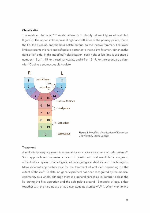
Classification
The modified Kernahan36, 37 model attempts to classify different types of oral cleft
(figure 3). The upper limbs represent right and left sides of the primary palate, that is
the lip, the alveolus, and the hard palate anterior to the incisive foramen. The lower
limb represents the hard and soft palate posterior to the incisive foramen, either on the
right or left side. In this modified Y classification, each right or left limb is assigned a
number, 1-5 or 11-15 for the primary palate and 6-9 or 16-19, for the secondary palate,
with 10 being a submucous cleft palate
11
deformities 34. Because of anatomical abnormalities of the nose, such as a deviated septum and structural
deformities of the nasal cartilages, there are also functional airway and aesthetic problems. Approximately
60% of the patients have difficulty breathing through the nose35.
Classification
The modified Kernahan36, 37 model attempts to classify different types of oral cleft (figure 3). The upper
limbs represent right and left sides of the primary palate, that is the lip, the alveolus, and the hard palate
anterior to the incisive foramen. The lower limb represents the hard and soft palate posterior to the
incisive foramen, either on the right or left side. In this modified Y classification, each right or left limb is
assigned a number, 1-5 or 11-15 for the primary palate and 6-9 or 16-19, for the secondary palate, with 10
being a submucous cleft palate
Figure 3 Modified classification of Kernohan. Copyright by Ingrid Jansen.
Treatment
A multidisciplinary approach is essential for satisfactory treatment of cleft patients38. Such approach
encompasses a team of plastic and oral maxillofacial surgeons, orthodontists, speech pathologists,
otolaryngologists, dentists and psychologists. Many different approaches exist for the treatment of oral
cleft depending on the extent of the cleft. To date, no generic protocol has been recognized by the medical
Treatment
A multidisciplinary approach is essential for satisfactory treatment of cleft patients38.
Such approach encompasses a team of plastic and oral maxillofacial surgeons,
orthodontists, speech pathologists, otolaryngologists, dentists and psychologists.
Many different approaches exist for the treatment of oral cleft depending on the
extent of the cleft. To date, no generic protocol has been recognized by the medical
community as a whole, although there is a general consensus in Europe to close the
lip during the first operation and the soft palate around 12 months of age, either
together with the hard palate or as a two-stage palatoplasty39,40, 41. When mentioning
Figure 3 Modified classification of Kernohan. Copyright by Ingrid Jansen.
R1
R2
R3
R4
R5
R6
R7
R8
R9
R10
R11
R12
R13
R14
R15
R16
R17
R18
R19
R20
R21
R22
R23
R24
R25
R26
R27
R28
R29
R30
R31
R32
R33
R34
1 | General introduction and outline of the thesis
16
surgical correction, the ultimate goal is to reconstruct a symmetrically balanced lip and
nose, with good columellar length and repair of the orbicularis muscle. The aim for
cleft palate repair is to create an anatomically intact and functional palate to optimize
feeding, achieve normal speech and minimize maxillary growth restriction25.
Presurgical, nasoalveolar molding, is a technique to facilitate surgical closure by
preoperatively narrowing down the alveolar cleft 42. This is combined with a nasal stent
as outrigger to shape the alar cartilage into a more natural position 43-46. Since cartilage
is most flexible and moldable during the first weeks after birth47, it is preferable to start
nasoalveolar molding as soon as possible. Although nasoalveolar molding facilitates
surgical closure and is beneficial for achieving nasal symmetry on short-term, the
evidence is not substantial to support its effects on long-term outcome 48. Opponents
say the shape of the cartilage will relapse partially during the first year42. Others suggest
that the necessary the frequent outpatient visits to adjust the device, necessary to
optimize molding, is a burden to the family and leads to compliance failure49.
A cheiloplasty for unilateral cleft lip is usually performed between 3 and 6 months after
birth. “All cleft lip surgeons have their favorite surgical technique for repairing unilateral
cleft lip. The operation is usually a hybrid of training experience and imagination” 50.
The ideal lip repair should approximate the medial and lateral lip elements at all levels
(i.e. nostril sill, cutaneous roll, vermillion-cutaneus junction, and vermillion-mucosal
junction) without interruption or loss of landmarks and achieve balance by providing
length where tissue is short and excision where height is excessive25. The noncleft side
heights provide the measures that must be created by the repair on the cleft side.
The cleft side medial height will need to be lengthened and the lateral lip height
will often need alteration to match that of the non-affected side. At the level of the
cutaneous roll, the entire length of Cupid’s bow should be preserved in the medial lip
element. In the lateral lip element, Noordhoff’s point should be preserved and used
to form the base of the philtral column incision51. The cutaneous roll of the medial and
lateral lip elements should be approximated in side-to-side fashion. Vermillion height
deficiency below the cleft side half of Cupid’s bow should be augmented and the red
lip elements should be approximated with attention to creating a proper vermillion-
mucosal junction 25.
R1
R2
R3
R4
R5
R6
R7
R8
R9
R10
R11
R12
R13
R14
R15
R16
R17
R18
R19
R20
R21
R22
R23
R24
R25
R26
R27
R28
R29
R30
R31
R32
R33
R34
17
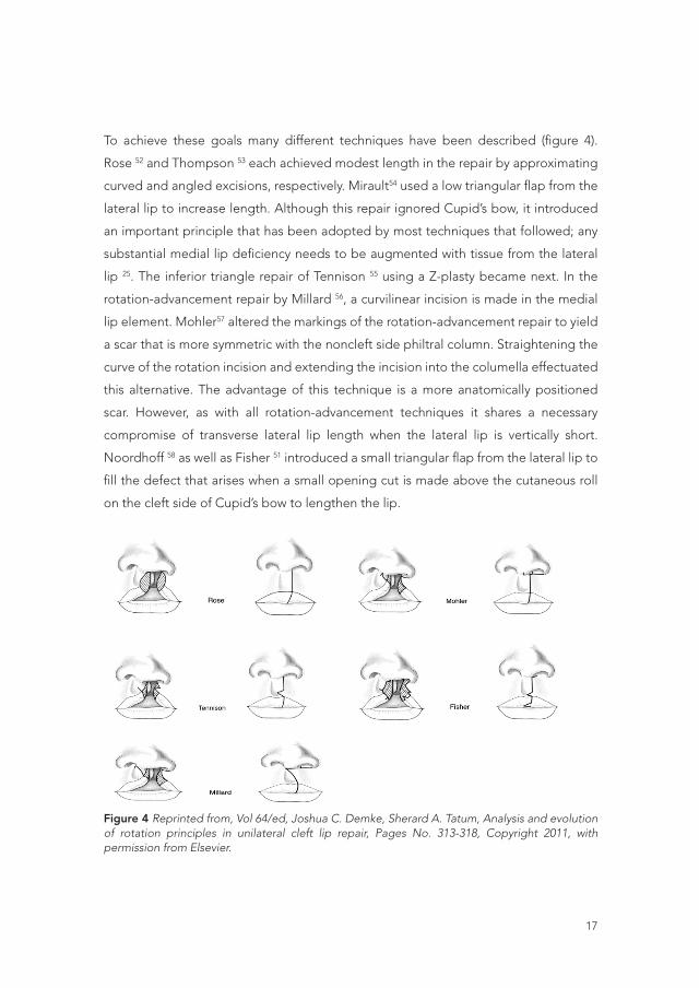
To achieve these goals many different techniques have been described (figure 4).
Rose 52 and Thompson 53 each achieved modest length in the repair by approximating
curved and angled excisions, respectively. Mirault54 used a low triangular flap from the
lateral lip to increase length. Although this repair ignored Cupid’s bow, it introduced
an important principle that has been adopted by most techniques that followed; any
substantial medial lip deficiency needs to be augmented with tissue from the lateral
lip 25. The inferior triangle repair of Tennison 55 using a Z-plasty became next. In the
rotation-advancement repair by Millard 56, a curvilinear incision is made in the medial
lip element. Mohler57 altered the markings of the rotation-advancement repair to yield
a scar that is more symmetric with the noncleft side philtral column. Straightening the
curve of the rotation incision and extending the incision into the columella effectuated
this alternative. The advantage of this technique is a more anatomically positioned
scar. However, as with all rotation-advancement techniques it shares a necessary
compromise of transverse lateral lip length when the lateral lip is vertically short.
Noordhoff 58 as well as Fisher 51 introduced a small triangular flap from the lateral lip to
fill the defect that arises when a small opening cut is made above the cutaneous roll
on the cleft side of Cupid’s bow to lengthen the lip.
13
should be augmented and the red lip elements should be approximated with attention to creating a proper
vermillion-mucosal junction 25.
To achieve these goals many different techniques have been described (figure 4). Rose 52 and Thompson53
each achieved modest length in the repair by approximating curved and angled excisions, respectively.
Mirault54 used a low triangular flap from the lateral lip to increase length. Although this repair ignored
Cupid’s bow, it introduced an important principle that has been adopted by most techniques that
followed; any substantial medial lip deficiency needs to be augmented with tissue from the lateral lip25.
The inferior triangle repair of Tennison55 using a Z-plasty became next. In the rotation-advancement
repair by Millard 56, a curvilinear incision is made in the medial lip element. Mohler57 altered the markings
of the rotation-advancement repair to yield a scar that is more symmetric with the noncleft side philtral
column. Straightening the curve of the rotation incision and extending the incision into the columella
effectuated this alternative. The advantage of this technique is a more anatomically positioned scar.
However, as with all rotation-advancement techniques it shares a necessary compromise of transverse
lateral lip length when the lateral lip is vertically short. Noordhoff58 as well as Fisher 51 introduced a small
triangular flap from the lateral lip to fill the defect that arises when a small opening cut is made above the
cutaneous roll on the cleft side of Cupid’s bow to lengthen the lip.
Figure 4 Reprinted from, Vol 64/ed, Joshua C. Demke, Sherard A. Tatum, Analysis and evolution of rotation principles in
unilateral cleft lip repair, Pages No. 313-318, Copyright 2011, with permission from Elsevier.
Figure 4 Reprinted from, Vol 64/ed, Joshua C. Demke, Sherard A. Tatum, Analysis and evolution of rotation principles in unilateral cleft lip repair, Pages No. 313-318, Copyright 2011, with permission from Elsevier.
R1
R2
R3
R4
R5
R6
R7
R8
R9
R10
R11
R12
R13
R14
R15
R16
R17
R18
R19
R20
R21
R22
R23
R24
R25
R26
R27
R28
R29
R30
R31
R32
R33
R34
1 | General introduction and outline of the thesis
18
The timing for closure of the palate is still under dispute. Since the driving force
for palatoplasty is development and achievement of normal speech, most would
agree that best results are correlated with closure near the time of the infant’s
language acquisition, which is before 12 months of age in the normal developing
baby 59, 60. Besides closure and lengthening of the palate to minimize postoperative
velopharyngeal insufficiency, other aims of cleft palate repair are to minimize maxillary
and alveolar growth disturbances and prevent fistula formation. The closure of a
cleft palate is known to alter normal facial development61. When comparing patients
with uncorrected cleft palate to corrected patients there is a significant reduction in
maxillary sagittal length, retrusion, and maxillary dental-arch width in the uncorrected
group 62, 63. Another controversial subject of surgical repair is the timing and number
of stages used. Several authors argue that flap elevation damages the periosteum
and subsequent postsurgical scarring negatively impacts maxillary growth, something
that could be minimized using two-stage approach with delayed repair of the hard
plate 64-66. However, proponents of early single-stage repair maintain that early closure
improves speech outcomes through promotion of proper phonetic development and
that improved speech outcomes outweigh potential growth restriction that may ensue 67, 68. Finally, there is ongoing discussion on what type of technique to use for closing the
secondary palate. A recently published trial 69 showed that a Furlow double-opposing
Z-plasty compared to a von Langebeck palatoplasty with intraveloplasty results in
improved velopharyngeal function. A disadvantage of the Furlow technique is that
length is achieved at the expense of lateral tightening 25. In order to maximize results
after surgery postoperative speech therapy is essential, regardless of the technique
used to close the palate.
If there exists an alveolar cleft, this must be treated for stable bone continuity of the
maxillary arch, stable environment and bone support of erupting teeth, piriform bone
support of the nasal base besides for separation of oral and nasal cavities. An alveolar
cleft is associated with variable anomalies in dental development that must be taken
into consideration with timing of surgery, technique and postsurgical orthodontic
planning 70. Management protocols differ but always comprehend a combination of
orthodontic treatment and bone grafting, mostly before eruption of the adult cleft
R1
R2
R3
R4
R5
R6
R7
R8
R9
R10
R11
R12
R13
R14
R15
R16
R17
R18
R19
R20
R21
R22
R23
R24
R25
R26
R27
R28
R29
R30
R31
R32
R33
R34
19
canine side. Nowadays bypassing or enhancing autologous bone grafting by means
of tissue engineering solutions has become an important topic in alveolar cleft
grafting. Replacement of the autologous bone graft will result in absence of donor site
morbidity71. By closure of the lip, palate and alveolar grafting all the surgical steps in
basic cleft care are taken. Afterwards the treatment becomes more individualistically
driven. Moreover, during this phase of treatment patients have reached an age at
which they can influence their own treatment. Orthognatic surgery and secondary lip
corrections as well as rhinoplasty after adolescent growth spurt are frequently needed
to improve aesthetics, dental occlusion and airway function of the nose72, 73.
In conclusion, the course of oral cleft treatment is complex and spans often the entire
childhood, adolescence and adulthood. Although much of the primary management
of patients is surgically driven and focusing on function, the overall goal of treatment
includes also psychosocial and social wellbeing of the patient and his/her family.
An oral cleft that involves the nose and lip imposes evident facial differences and is
therefore expected to have impact on social interactions and quality of life 74. Overall,
the majority of children and adults with an oral cleft do not appear to experience major
psychosocial problems 75, although disturbances have been observed such as behavior
problems, anxiety, depression, aesthetic dissatisfaction with facial appearance in both
children and adults 76. The treatment of children with oral cleft is continuously evolving,
but current techniques and treatment protocols generate excellent functional and
aesthetic results and prognosis, with most studies reporting high rates of treatment
satisfaction 77, 78.
Introduction of prenatal screening
Since the introduction of standard prenatal screening, oral clefts are being diagnosed
on prenatal ultrasound. Where formerly parents were confronted with a child with a
cleft lip and/or palate at birth, nowadays they can are already be informed during
pregnancy. In The Netherlands, transabdominal ultrasound screening at 20 weeks
of pregnancy was made universally accessible by legislation and health insurance
coverage in 200779. The screening was aimed initially at Down syndrome, but the
possibility for secondary screening for oral cleft and other congenital anomalies was
R1
R2
R3
R4
R5
R6
R7
R8
R9
R10
R11
R12
R13
R14
R15
R16
R17
R18
R19
R20
R21
R22
R23
R24
R25
R26
R27
R28
R29
R30
R31
R32
R33
R34
1 | General introduction and outline of the thesis
20
included as well. Consequently, this added a whole new dimension to cleft care. The
changes in screening programme and related questions and concerns initiated the
research for this thesis.
In 1981, Christ and Meininger80 were the first to report the detection of a cleft lip
and palate deformity on ultrasound. Ever since, detection rates increased from
approximately 5% in the early 1980s to over 26% in the late 1990s 81 and have increased
to about 23-58% in most recent prospective studies 82, 83. Subsequently, there is a need
for accurate information to aid in prenatal counseling. When informing parents on
outcome, prognosis and treatment the type of cleft as well as the presence of other
congenital anomalies are crucial information. Furthermore, in clinical practice there
is often the discussion whether further invasive tests should be offered prenatally to
identify chromosomal defects. This will in turn influence counseling and management
of the pregnancy significantly 15. However, the reported rates of associated anomalies
in prenatal cleft populations vary greatly among reported studies 12-14, 82.
In response to increasing prenatal detection rates of oral cleft, the cleft lip and palate
team of the Wilhelmina Children’s Hospital in Utrecht set up a prenatal cleft clinic to
participate in counseling. Parents meet here with an obstetrician, plastic surgeon and
a medical physiologist of the team. The etiology and pathogenesis of an oral cleft are
then explained as well as the medical, surgical and psychosocial needs of a child with
a cleft lip and possible cleft palate. Although former studies have concluded that most
parents prefer to know the diagnosis prenatally rather than at birth 84-87, the effect of
counseling has never been evaluated prospectively during pregnancy. Former studies
assessing the impact of a prenatally detected oral cleft are of retrospective design,
implying that at the time of assessment the child was already born. This could affect a
parent’s view on the impact of an oral cleft diagnosis. Potential advantages of prenatal
cleft diagnosis were formulated by Johnson and Sandy 88, including psychosocial
preparation, opportunity for parent education, planned neonatal care, anticipation
on possible feeding problems as well as increased reproductive awareness can be
maximally exploited. On the contrary, there may be emotional disturbance of the
pregnancy 84 and more importantly the concern of increasing numbers of termination
R1
R2
R3
R4
R5
R6
R7
R8
R9
R10
R11
R12
R13
R14
R15
R16
R17
R18
R19
R20
R21
R22
R23
R24
R25
R26
R27
R28
R29
R30
R31
R32
R33
R34
21
of pregnancy (TOP) of fetuses with an isolated oral cleft. The latter concerns were
raised by reports from Israel were numbers of TOP for oral cleft reach more than 95%89.
Influences on the decision making process of parents expecting a child with an oral
cleft have been discussed in several expert opinions. The ‘perception of burden’ of
an oral cleft and the possible stigmatization of a ’less-than-perfect’ child have been
described 90. It has been shown that a dedicated cleft team can educate parents in
order to prepare them optimally and reduce the anxiety associated with the diagnosis
of a fetal malformation 91. Other studies assessing a woman’s decisions after diagnosis
with an abnormality other than oral cleft revealed considerations like the severity of
the abnormality and its visualization on ultrasound scan 92-95. In addition, influences
of the caregiver who counsels the couple 96, the ‘child’s best interest’ and economic
issues have been described with respect to parents’ decisions 97. The aforementioned
ambiguities led to formation of research.
Outline and aims of this thesis
The purposes of this thesis are to obtain more knowledge on the accuracy of
ultrasound screening and the incidence of associated anomalies besides an oral cleft.
In addition, the aim is to obtain more insight into opinion of professionals as well as
the psychosocial, and moral aspects in thoughts and attitudes of parents expecting a
child with an oral cleft.
The first part of this thesis focuses on the classification of oral cleft. In chapter 2, a
new prenatal oral cleft classification is defined in order to improve uniformity between
ultrasonographers in daily practice. In part two, the introduction and accuracy of
screening for oral cleft is covered. In chapter 3 the Dutch routine screening system for
physical congenital anomalies is described, with a focus on oral cleft. The diagnostic
accuracy of oral cleft by ultrasound is assessed in a systematic review in chapter 4,
besides in a prospective study in chapter 5. The precision of prenatal ultrasound in
determining the type of oral cleft is described in chapter 6. Since oral clefts are being
diagnosed prenatally more frequently, there is the need for more accurate information
on the risk of associated anomalies and chromosomal defects. In chapter 7 a systematic
review was conducted to investigate the prevalence of associated anomalies per
R1
R2
R3
R4
R5
R6
R7
R8
R9
R10
R11
R12
R13
R14
R15
R16
R17
R18
R19
R20
R21
R22
R23
R24
R25
R26
R27
R28
R29
R30
R31
R32
R33
R34
1 | General introduction and outline of the thesis
22
type of cleft, which can aid prenatal counseling. Part three of the thesis addresses
to prenatal counseling op parents expecting a child with an oral cleft. Different
factors are thought to influence parental opinion on oral cleft, one of them being the
obstetric care provider’s attitude. In chapter 8 the providers’ opinions about oral cleft
and possible termination of pregnancy for isolated oral cleft are compared between
Israel and The Netherlands. Israel was chosen for comparison because of high rates
of TOP for isolated oral cleft. In chapter 9 we obtained insight into the psychosocial
and moral considerations of prospective parents concerning oral clefts, the burden of
oral clefts and parents’ attitude toward possible termination of pregnancy. Finally, in
chapter 10 three types of counseling are discussed, informative counseling, shared
decision making and paternalistic counseling, to agree on which type of counseling
is the most appropriate for cleft lip and palate teams. This thesis is completed by a
general discussion and implications for further research in chapter 11.

R1
R2
R3
R4
R5
R6
R7
R8
R9
R10
R11
R12
R13
R14
R15
R16
R17
R18
R19
R20
R21
R22
R23
R24
R25
R26
R27
R28
R29
R30
R31
R32
R33
R34
23
REFERENCES
1. P. A. Mossey, J. Little, R. G. Munger, M. J. Dixon and W. C. Shaw. Cleft lip and palate. Lancet 2009; 374: 1773-1785.
2. G. R. Fraser and J. S. Calnan. Cleft lip and palate: seasonal incidence, birth weight, birth rank, sex, site, associated malformations and parental age. A statistical survey. Arch Dis Child 1961; 36: 420-423.
3. S. A. Tanaka, R. C. Mahabir, D. C. Jupiter and J. M. Menezes. Updating the epidemiology of isolated cleft palate. Plast Reconstr Surg 2013; 131: 650e-652e.
4. A. Derijcke, A. Eerens and C. Carels. The incidence of oral clefts: a review. Br J Oral Maxillofac Surg 1996; 34: 488-494.
5. A. Czeizel. Studies of cleft lip and cleft palate in east European populations. Prog Clin Biol Res 1980; 46: 249-296.
6. M. Tolarova. Orofacial clefts in Czechoslovakia. Incidence, genetics and prevention of cleft lip and palate over a 19-year period. Scand J Plast Reconstr Surg Hand Surg 1987; 21: 19-25.
7. D. F. Wyszynski, T. H. Beaty and N. E. Maestri. Genetics of nonsyndromic oral clefts revisited. Cleft Palate Craniofac J 1996; 33: 406-417.
8. M. Melnick, D. Bixler, P. Fogh-Andersen and P. M. Conneally. Cleft lip+/-cleft palate: an overview of the literature and an analysis of Danish cases born between 1941 and 1968. Am J Med Genet 1980; 6: 83-97.
9. J. Milerad, O. Larson, D. D. Ph, C. Hagberg and M. Ideberg. Associated malformations in infants with cleft lip and palate: a prospective, population-based study. Pediatrics 1997; 100: 180-186.
10. W. Maarse, A. M. Rozendaal, E. Pajkrt, C. Vermeij-Keers, A. B. Mink van der Molen and M. J. van den Boogaard. A systematic review of associated structural and chromosomal defects in oral clefts: when is prenatal genetic analysis indicated? J Med Genet 2012; 49: 490-498.
11. S. J. Walker, R. H. Ball, C. J. Babcook and M. M. Feldkamp. Prevalence of aneuploidy and additional anatomic abnormalities in fetuses and neonates with cleft lip with or without cleft palate: a population-based study in Utah. J Ultrasound Med 2001; 20: 1175-1180; quiz 1181-1172.
12. D. A. Nyberg, G. K. Sickler, F. N. Hegge, D. J. Kramer and R. J. Kropp. Fetal cleft lip with and without cleft palate: US classification and correlation with outcome. Radiology 1995; 195: 677-684.
13. S. J. Berge, H. Plath, P. T. Van de Vondel, T. Appel, B. Niederhagen, J. J. Von Lindern, R. H. Reich and M. Hansmann. Fetal cleft lip and palate: sonographic diagnosis, chromosomal abnormalities, associated anomalies and postnatal outcome in 70 fetuses. Ultrasound Obstet Gynecol 2001; 18:
14. F. Perrotin, L. M. de Poncheville, H. Marret, C. Paillet, J. Lansac and G. Body. Chromosomal defects and associated malformations in fetal cleft lip with or without cleft palate. Eur J Obstet Gynecol Reprod Biol 2001; 99: 19-24.
15. J. C. Gillham, S. Anand and P. J. Bullen. Antenatal detection of cleft lip with or without cleft palate: incidence of associated chromosomal and structural anomalies. Ultrasound Obstet Gynecol 2009; 34: 410-415. DOI 10.1002/uog.6447.
16. T. Sadler. Langman’s medical embryology: Baltimore, 1995, 69-92.17. W. Larsen. Larsen’s human embryology Philadelphia, 2009.18. W. Larsen. Larsen’s human embryology Elsevier/Chruchill Livingstone: Philadelphia, 2009,
663-570.19. C. C. Breugem and A. B. Mink van der Molen. What is ‘Pierre Robin sequence’? J Plast
Reconstr Aesthet Surg 2009; 62: 1555-1558.
R1
R2
R3
R4
R5
R6
R7
R8
R9
R10
R11
R12
R13
R14
R15
R16
R17
R18
R19
R20
R21
R22
R23
R24
R25
R26
R27
R28
R29
R30
R31
R32
R33
R34
1 | General introduction and outline of the thesis
24
20. K. R. Losee JE. Comprehensice Cleft Care. McGraw-Hill: New York, 2009, 273-284.21. J. B. Mulliken. Double unilimb Z-plastic repair of microform cleft lip. Plast Reconstr Surg
2005; 116: 1623-1632. 22. D. M. Fisher and B. C. Sommerlad. Cleft lip, cleft palate, and velopharyngeal insufficiency.
Plast Reconstr Surg 2011; 128: 342e-360e. 23. B. C. Sommerlad, C. Fenn, K. Harland, D. Sell, M. J. Birch, R. Dave, M. Lees and A. Barnett.
Submucous cleft palate: a grading system and review of 40 consecutive submucous cleft palate repairs. Cleft Palate Craniofac J 2004; 41: 114-123.
24. J. L. Paradise. Middle ear problems associated with cleft palate. An internationally-oriented review. Cleft Palate J 1975; 12: 17-22.
25. J. Reid, N. Kilpatrick and S. Reilly. A prospective, longitudinal study of feeding skills in a cohort of babies with cleft conditions. Cleft Palate Craniofac J 2006; 43: 702-709.
26. B. Felix-Schollaart, J. B. Hoeksma and B. Prahl-Andersen. Growth comparison between children with cleft lip and/or palate and controls. Cleft Palate Craniofac J 1992; 29: 475-480.
27. M. L. Speltz, G. C. Armsden and S. S. Clarren. Effects of craniofacial birth defects on maternal functioning postinfancy. J Pediatr Psychol 1990; 15: 177-196.
28. D. M. DeLuke, A. Marchand, E. C. Robles and P. Fox. Facial growth and the need for orthognathic surgery after cleft palate repair: literature review and report of 28 cases. J Oral Maxillofac Surg 1997; 55: 694-697; discussion 697-698.
29. D. W. Warren and A. F. Drake. Cleft nose. Form and function. Clin Plast Surg 1993; 20: 769-779.
30. H. C. Noordhoff MS, Wu J. Multidisciplinary management of cleft lip and palate in Taiwan. In Multidisciplinary management of cleft lip and palate in Taiwan, B. J (ed). WB Saunders 1990: Philidelphia, 1990, 18-26.
31. M. S. Noordhoff. Response to Kernahan’s commentary on symbolic representation of cleft lip and palate by Friedman et al. (1991). Cleft Palate Craniofac J 1992; 29: 96.
32. K. T. Chen and M. S. Noordhoff. Open tip rhinoplasty. Ann Plast Surg 1992; 28: 119-130.33. W. C. Shaw, G. Semb, P. Nelson, V. Brattstrom, K. Molsted, B. Prahl-Andersen and K. K.
Gundlach. The Eurocleft project 1996-2000: overview. Journal of cranio-maxillo-facial surgery : official publication of the European Association for Cranio-Maxillo-Facial Surgery 2001; 29: 131-140; discussion 141-132.
34. E. J. Liou, M. Subramanian, P. K. Chen and C. S. Huang. The progressive changes of nasal symmetry and growth after nasoalveolar molding: a three-year follow-up study. Plast Reconstr Surg 2004; 114: 858-864.
35. D. J. Maull, B. H. Grayson, C. B. Cutting, L. L. Brecht, F. L. Bookstein, D. Khorrambadi, J. A. Webb and D. J. Hurwitz. Long-term effects of nasoalveolar molding on three-dimensional nasal shape in unilateral clefts. Cleft Palate Craniofac J 1999; 36: 391-397.
36. B. H. Grayson and C. B. Cutting. Presurgical nasoalveolar orthopedic molding in primary correction of the nose, lip, and alveolus of infants born with unilateral and bilateral clefts. Cleft Palate Craniofac J 2001; 38: 193-198.
37. B. C. Pai, E. W. Ko, C. S. Huang and E. J. Liou. Symmetry of the nose after presurgical nasoalveolar molding in infants with unilateral cleft lip and palate: a preliminary study. Cleft Palate Craniofac J 2005; 42: 658-663.
38. S. Suri and B. D. Tompson. A modified muscle-activated maxillary orthopedic appliance for presurgical nasoalveolar molding in infants with unilateral cleft lip and palate. Cleft Palate Craniofac J 2004; 41: 225-229.
39. H. Fry and W. V. Robertson. Interlocked stresses in cartilage. Nature 1967; 215: 53-54.40. P. van der Heijden, P. U. Dijkstra, C. Stellingsma, B. F. van der Laan, A. G. Korsten-Meijer
and S. M. Goorhuis-Brouwer. Limited evidence for the effect of presurgical nasoalveolar molding in unilateral cleft on nasal symmetry: a call for unified research. Plast Reconstr Surg 2013; 131: 62e-71e.
R1
R2
R3
R4
R5
R6
R7
R8
R9
R10
R11
R12
R13
R14
R15
R16
R17
R18
R19
R20
R21
R22
R23
R24
R25
R26
R27
R28
R29
R30
R31
R32
R33
R34
25
41. L. Sischo, J. W. Chan, M. Stein, C. Smith, J. van Aalst and H. L. Broder. Nasoalveolar Molding: Prevalence of Cleft Centers Offering NAM and Who Seeks It. Cleft Palate Craniofac J 2012; 49: 270-275.
42. J. Thompson. Unilateral cleft lip repair. In Oper Tech Plast Recontrs Surg, 1995, 175-181.43. W. Rose. In On Hare Lip and Cleft Palate, H. Lewis (ed): London, 1891.44. J. Thompson. And artistic and mathematically accurate method or repairing the defects in
cases of harelip. Surg Gynecol Obstet 1912; 14: 498-505.45. B. J. Blair VP. Mirault operation for single harelip. Surg Gynecol Obstet 1930; 51: 81-98.46. C. W. Tennison. The repair of the unilateral cleft lip by the stencil method. Plast Reconstr
Surg (1946) 1952; 9: 115-120.47. D. R. Millard. Extensions of the rotation-advancement principle for wide unilateral cleft lips.
Plast Reconstr Surg 1968; 42: 535-544.48. L. R. Mohler. Unilateral cleft lip repair. Plast Reconstr Surg 1987; 80: 511-517.49. M. Noordhoff. The Surgical Tecnique for the Unilateral Cleft Lip-Nasal Deformity. Noordhoff
craniofacial Foundation: Taipee, Taiwan, 1997.50. D. M. Fisher. Unilateral cleft lip repair: an anatomical subunit approximation technique.
Plast Reconstr Surg 2005; 116: 61-71. 51. K. L. Chapman and M. A. Hardin. Phonetic and phonologic skills of two-year-olds with cleft
palate. Cleft Palate Craniofac J 1992; 29: 435-443. 52. D. S. Dorf and J. W. Curtin. Early cleft palate repair and speech outcome. Plast Reconstr
Surg 1982; 70: 74-81.53. K. J. Chepla and A. K. Gosain. Evidence-based medicine: cleft palate. Plast Reconstr Surg
2013; 132: 1644-1648. 54. Z. Q. Chen, Y. F. Qian, G. M. Wang and G. Shen. Sagittal maxillary growth in patients with
unoperated isolated cleft palate. Cleft Palate Craniofac J 2009; 46: 664-667. 55. B. Ye, C. Ruan, J. Hu, Y. Yang, A. Ghosh, S. Jana and G. Zhang. A comparative study
on dental-arch morphology in adult unoperated and operated cleft palate patients. J Craniofac Surg 2010; 21: 811-815.
56. Y. F. Liao, I. Y. Yang, R. Wang, C. Yun and C. S. Huang. Two-stage palate repair with delayed hard palate closure is related to favorable maxillary growth in unilateral cleft lip and palate. Plast Reconstr Surg 2010; 125: 1503-1510.
57. T. Yamanishi, J. Nishio, M. Sako, H. Kohara, Y. Hirano, Y. Yamanishi, T. Adachi, S. Miya and T. Mukai. Early two-stage double opposing Z-plasty or one-stage push-back palatoplasty?: comparisons in maxillary development and speech outcome at 4 years of age. Ann Plast Surg 2011; 66: 148-153.
58. H. Friede, J. Lilja and A. Lohmander. Long-Term, Longitudinal Follow-Up of Individuals With UCLP After the Gothenburg Primary Early Veloplasty and Delayed Hard Palate Closure Protocol: Maxillofacial Growth Outcome. Cleft Palate Craniofac J 2012; 49: 649-656.
59. W. Pradel, D. Senf, R. Mai, G. Ludicke, U. Eckelt and G. Lauer. One-stage palate repair improves speech outcome and early maxillary growth in patients with cleft lip and palate. J Physiol Pharmacol 2009; 60 Suppl 8: 37-41.
60. E. Willadsen. Influence of timing of hard palate repair in a two-stage procedure on early speech development in danish children with cleft palate. Cleft Palate Craniofac J 2012; 49: 574-595.
61. W. N. Williams, M. B. Seagle, M. I. Pegoraro-Krook, T. V. Souza, L. Garla, M. L. Silva, J. S. Machado Neto, J. C. Dutka, J. Nackashi, S. Boggs, J. Shuster, J. Moorhead, W. Wharton, M. I. Graciano, M. C. Pimentel, M. Feniman, S. H. Piazentin-Penna, J. Kemker, M. C. Zimmermann, C. Bento-Goncalvez, H. Borgo, I. L. Marques, A. P. Martinelli, J. C. Jorge, P. Antonelli, J. F. Neves and M. E. Whitaker. Prospective clinical trial comparing outcome measures between Furlow and von Langenbeck Palatoplasties for UCLP. Ann Plast Surg 2011; 66: 154-163.
R1
R2
R3
R4
R5
R6
R7
R8
R9
R10
R11
R12
R13
R14
R15
R16
R17
R18
R19
R20
R21
R22
R23
R24
R25
R26
R27
R28
R29
R30
R31
R32
R33
R34
1 | General introduction and outline of the thesis
26
62. P. C. Neligan. Plastic Surgery. SAUNDERS an imprint of Elsevier Inc: London, 2013, 585.63. P. van der Heijden, A. G. Korsten-Meijer, B. F. van der Laan, H. P. Wit and S. M. Goorhuis-
Brouwer. Nasal growth and maturation age in adolescents: a systematic review. Arch Otolaryngol Head Neck Surg 2008; 134: 1288-1293.
64. S. Mathes. Plastic Surgery: Philadelphia, 2006.65. A. F. Klassen, E. Tsangaris, C. R. Forrest, K. W. Wong, A. L. Pusic, S. J. Cano, I. Syed, M. Dua,
S. Kainth, J. Johnson and T. Goodacre. Quality of life of children treated for cleft lip and/or palate: a systematic review. J Plast Reconstr Aesthet Surg 2012; 65: 547-557.
66. O. Hunt, D. Burden, P. Hepper and C. Johnston. The psychosocial effects of cleft lip and palate: a systematic review. Eur J Orthod 2005; 27: 274-285.
67. B. Grollemund, A. Guedeney, M. P. Vazquez, A. Picard, V. Soupre, P. Pellerin, E. Simon, M. Velten, C. Dissaux, I. Kauffmann, C. Bruant-Rodier and A. Danion-Grilliat. Relational development in children with cleft lip and palate: influence of the waiting period prior to the first surgical intervention and parental psychological perceptions of the abnormality. BMC Pediatr 2012; 12: 65.
68. S. N. Noor and S. Musa. Assessment of patients’ level of satisfaction with cleft treatment using the Cleft Evaluation Profile. Cleft Palate Craniofac J 2007; 44: 292-303.
69. Z. E. Berger and L. J. Dalton. Coping with a cleft: psychosocial adjustment of adolescents with a cleft lip and palate and their parents. Cleft Palate Craniofac J 2009; 46: 435-443.
70. J. E. Christ and M. G. Meininger. Ultrasound diagnosis of cleft lip and cleft palate before birth. Plast Reconstr Surg 1981; 68: 854-859.
71. C. Stoll, B. Dott, Y. Alembik and M. Roth. Evaluation of prenatal diagnosis of cleft lip/palate by foetal ultrasonographic examination. Ann Genet 2000; 43: 11-14.
72. K. Offerdal, N. Jebens, T. Syvertsen, H. G. Blaas, O. J. Johansen and S. H. Eik-Nes. Prenatal ultrasound detection of facial clefts: a prospective study of 49,314 deliveries in a non-selected population in Norway. Ultrasound Obstet Gynecol 2008; 31: 639-646.
73. K. A. Russell, V. M. Allen, M. E. MacDonald, K. Smith and L. Dodds. A population-based evaluation of antenatal diagnosis of orofacial clefts. Cleft Palate Craniofac J 2008; 45: 148-153.
74. M. S. Matthews, M. Cohen, M. Viglione and A. S. Brown. Prenatal counseling for cleft lip and palate. Plast Reconstr Surg 1998; 101: 1-5.
75. A. Davalbhakta and P. N. Hall. The impact of antenatal diagnosis on the effectiveness and timing of counselling for cleft lip and palate. Br J Plast Surg 2000; 53: 298-301.
76. R. Nusbaum, R. E. Grubs, J. E. Losee, C. Weidman, M. D. Ford and M. L. Marazita. A qualitative description of receiving a diagnosis of clefting in the prenatal or postnatal period. J Genet Couns 2008; 17: 336-350.
77. C. Rey-Bellet and J. Hohlfeld. Prenatal diagnosis of facial clefts: evaluation of a specialised counselling. Swiss Med Wkly 2004; 134: 640-644.
78. N. Johnson and R. S. J. Prenatal diagnosis of cleft lip and palate. Cleft Palate Craniofac J 2003; 40: 186-189.
79. Z. Blumenfeld, I. Blumenfeld and M. Bronshtein. The early prenatal diagnosis of cleft lip and the decision-making process. Cleft Palate Craniofac J 1999; 36: 105-107.
80. M. C. Jones. Prenatal diagnosis of cleft lip and palate: detection rates, accuracy of ultrasonography, associated anomalies, and strategies for counseling. Cleft Palate Craniofac J 2002; 39: 169-173.
81. R. P. Strauss. Beyond easy answers: prenatal diagnosis and counseling during pregnancy. Cleft Palate Craniofac J 2002; 39: 164-168.
82. M. I. Evans, M. A. Sobiecki, E. L. Krivchenia, D. A. Duquette, A. Drugan, R. F. Hume, Jr. and M. P. Johnson. Parental decisions to terminate/continue following abnormal cytogenetic prenatal diagnosis: “what” is still more important than “when”. Am J Med Genet 1996; 61: 353-355.
R1
R2
R3
R4
R5
R6
R7
R8
R9
R10
R11
R12
R13
R14
R15
R16
R17
R18
R19
R20
R21
R22
R23
R24
R25
R26
R27
R28
R29
R30
R31
R32
R33
R34
27
83. C. Julian, P. Huard, J. Gouvernet, J. F. Mattei and S. Ayme. Physicians’ acceptability of termination of pregnancy after prenatal diagnosis in southern France. Prenat Diagn 1989; 9: 77-89.
84. L. Zahed, M. Nabulsi and H. Tamim. Attitudes towards prenatal diagnosis and termination of pregnancy among health professionals in Lebanon. Prenat Diagn 2002; 22: 880-886.
85. J. Zlotogora. Parental decisions to abort or continue a pregnancy with an abnormal finding after an invasive prenatal test. Prenat Diagn 2002; 22: 1102-1106. DOI 10.1002/pd.472.
86. H. Drake, M. Reid and T. Marteau. Attitudes towards termination for fetal abnormality: comparisons in three European countries. Clin Genet 1996; 49: 134-140.
87. A. Raz. “Important to test, important to support”: attitudes toward disability rights and prenatal diagnosis among leaders of support groups for genetic disorders in Israel. Soc Sci Med 2004; 59: 1857-1866.


Part I
Classification of Oral Cleft

2A practical prenatal ultrasound
classification system for common oral clefts
Prenatal Diagnosis, 2015 Jun 10. [Epub ahead of print]
W. MAARSE, C.W.B. BOONACKER, C.C. BREUGEM, M. KON,
G.T.R. MANTEN, A.B. MINK VAN DER MOLEN
R1
R2
R3
R4
R5
R6
R7
R8
R9
R10
R11
R12
R13
R14
R15
R16
R17
R18
R19
R20
R21
R22
R23
R24
R25
R26
R27
R28
R29
R30
R31
R32
R33
R34
2 | Ultrasound classification for oral cleft
32
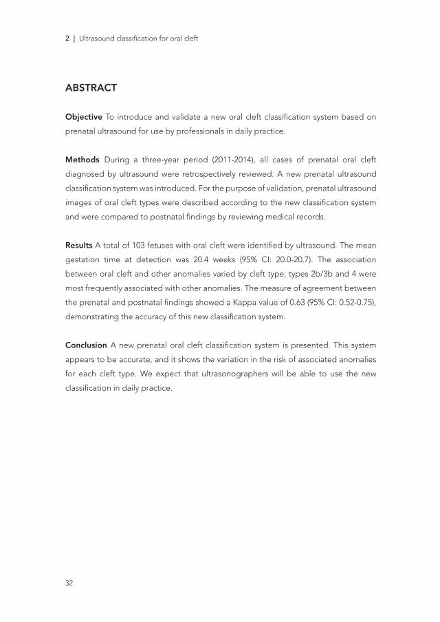
ABSTRACT
Objective To introduce and validate a new oral cleft classification system based on
prenatal ultrasound for use by professionals in daily practice.
Methods During a three-year period (2011-2014), all cases of prenatal oral cleft
diagnosed by ultrasound were retrospectively reviewed. A new prenatal ultrasound
classification system was introduced. For the purpose of validation, prenatal ultrasound
images of oral cleft types were described according to the new classification system
and were compared to postnatal findings by reviewing medical records.
Results A total of 103 fetuses with oral cleft were identified by ultrasound. The mean
gestation time at detection was 20.4 weeks (95% CI: 20.0-20.7). The association
between oral cleft and other anomalies varied by cleft type; types 2b/3b and 4 were
most frequently associated with other anomalies. The measure of agreement between
the prenatal and postnatal findings showed a Kappa value of 0.63 (95% CI: 0.52-0.75),
demonstrating the accuracy of this new classification system.
Conclusion A new prenatal oral cleft classification system is presented. This system
appears to be accurate, and it shows the variation in the risk of associated anomalies
for each cleft type. We expect that ultrasonographers will be able to use the new
classification in daily practice.
R1
R2
R3
R4
R5
R6
R7
R8
R9
R10
R11
R12
R13
R14
R15
R16
R17
R18
R19
R20
R21
R22
R23
R24
R25
R26
R27
R28
R29
R30
R31
R32
R33
R34
33
INTRODUCTION
Oral cleft (OC) is the most common congenital facial malformation; it occurs in
approximately 1 of 700 live births1. Transabdominal ultrasound (US) prenatal screening
is the standard of care in most Western countries, resulting in an increased frequency
of prenatal OC diagnoses2, 3.
Several postnatal classification systems for OC have been described in the literature,
all of which have aimed to simplify the diversity and complexity of cleft lip and palate
anomalies based on embryologic and anatomic principles4, 5, 6. An accurate OC
diagnosis is essential because different types of cleft are associated with specific risks of
anomalies and chromosomal disorders7. Moreover, an accurate diagnosis is important
because it correlates with the severity of the malformation, prognosis and outcome8.
In practice, different types of OC have specific considerations in terms of aesthetic,
functional (hearing, feeding, dentition, speaking) and psychosocial (construction of
self-image and relational and attachment disturbances) outcomes8.
Nyberg and colleagues9 were the only authors to describe a US classification of
OC, including five categories of clefts: type 1: cleft lip; type 2: unilateral cleft lip and
palate; type 3: bilateral cleft lip and palate; type: 4 midline cleft lip and palate; and
type 5: cleft associated with amniotic bands or limb-body-wall complex. However, this
classification system has several shortcomings. First, cleft palate alone is not included.
Second, the Nyberg classification does not differentiate unilateral versus bilateral cleft
lip (and alveolus) alone. Third, in our opinion, types 4 and 5 should not be included in
a prenatal US classification system because they are holoprosencephalic and atypical
clefts (including a spectrum of forebrain development defects), respectively10. Such
atypical clefts have a different embryologic pathogenesis and are rare compared to
the more common paramedian clefting, such as those that Nyberg et al. classified
as types 1-311, 12. Another argument for omitting Nyberg types 4 and 5 is their rare
incidence in combination with the high lethal outcome associated with most median
cleft types13. In conclusion, the prenatal classification of Nyberg is incomplete and
does not match postnatal classifications. Thus, the aim of our study was to define a
R1
R2
R3
R4
R5
R6
R7
R8
R9
R10
R11
R12
R13
R14
R15
R16
R17
R18
R19
R20
R21
R22
R23
R24
R25
R26
R27
R28
R29
R30
R31
R32
R33
R34
2 | Ultrasound classification for oral cleft
34
new prenatal OC classification, based singularly on structural/anatomic findings, using
a practical approach that can be easily applied by every professional performing
prenatal fetal anomaly scans.
EMBRYOLOGY
Thorough knowledge of embryological development in the craniofacial region can
help ultrasonographers to understand and distinguish among different types of OC.
Facial clefting is the result of a disturbance in both growth and fusion during basic facial
morphology development between the 4th and 10th weeks of human development
and involves five facial prominences14. As a result of fusion of the midline frontonasal
prominence and the two paired prominences (i.e., the maxillary and mandibular
prominences), the face is formed (Figure 1). On the frontonasal prominence, two
bilateral ectodermal thickenings form to invaginate as an oval nasal pit, dividing the
frontonasal prominence into a lateral and a medial nasal process. By the end of the 7th
week of gestation, the inferior tips of the medial nasal process fuse along the midline
and form the intermaxillary segment.10 Failure of the intermaxillary process and the
maxillary prominences to fuse from posterior to anterior results in clefting of the lip,
alveolar process, and hard palate anterior to the incisive foramen (primary palate). Such
clefts are therefore always paramedian clefts and are termed unilateral or bilateral cleft
lip, in some cases including the alveolus and primary palate.
The secondary palate, which is located dorsally to the incisive foramen, fuses after the
primary palate and is formed by the lateral palatal shelves, which are derived from the
medial walls of the maxillary prominences. The fusion process normally starts directly
behind the incisive foramen in the middle and extends anteriorly to posteriorly, ending
with the uvula, which is indicative of a closed soft palate15. Disruption of the confluence
between the two palatal shelves results in a cleft of the secondary palate. A secondary
cleft palate can occur simultaneously with clefting of the lip and primary palate.
However, cleft palate only can also occur as a consequence of mandibular dysplasia,
in which the first pharyngeal arch does not develop appropriately. In such cases, the
tongue will not be lowered and will physically obstruct palatal shelve elevation10.
This secondary cleft palate type, resulting from a small mandible (micrognathia)
R1
R2
R3
R4
R5
R6
R7
R8
R9
R10
R11
R12
R13
R14
R15
R16
R17
R18
R19
R20
R21
R22
R23
R24
R25
R26
R27
R28
R29
R30
R31
R32
R33
R34
35
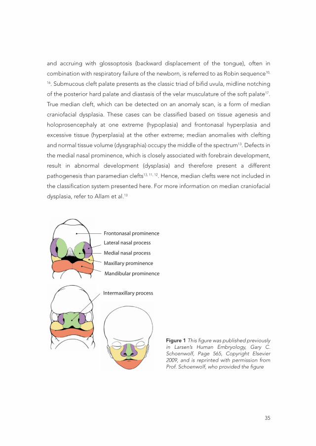
and accruing with glossoptosis (backward displacement of the tongue), often in
combination with respiratory failure of the newborn, is referred to as Robin sequence10,
16. Submucous cleft palate presents as the classic triad of bifid uvula, midline notching
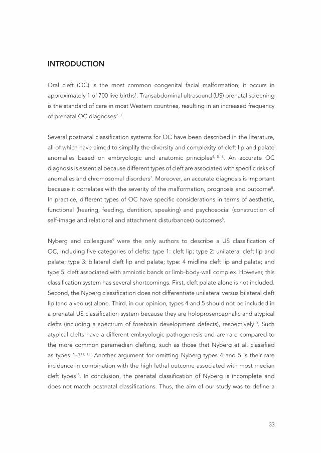
of the posterior hard palate and diastasis of the velar musculature of the soft palate17.
True median cleft, which can be detected on an anomaly scan, is a form of median
craniofacial dysplasia. These cases can be classified based on tissue agenesis and
holoprosencephaly at one extreme (hypoplasia) and frontonasal hyperplasia and
excessive tissue (hyperplasia) at the other extreme; median anomalies with clefting
and normal tissue volume (dysgraphia) occupy the middle of the spectrum13. Defects in
the medial nasal prominence, which is closely associated with forebrain development,
result in abnormal development (dysplasia) and therefore present a different
pathogenesis than paramedian clefts13, 11, 12. Hence, median clefts were not included in
the classification system presented here. For more information on median craniofacial
dysplasia, refer to Allam et al.13
Intermaxillary process
Frontonasal prominence
Maxillary prominence
Mandibular prominence
Lateral nasal process
Medial nasal process
Figure 1 This figure was published previously in Larsen’s Human Embryology, Gary C. Schoenwolf, Page 565, Copyright Elsevier 2009, and is reprinted with permission from Prof. Schoenwolf, who provided the figure
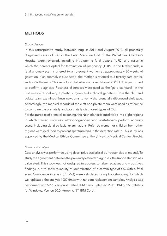
R1
R2
R3
R4
R5
R6
R7
R8
R9
R10
R11
R12
R13
R14
R15
R16
R17
R18
R19
R20
R21
R22
R23
R24
R25
R26
R27
R28
R29
R30
R31
R32
R33
R34
2 | Ultrasound classification for oral cleft
36
METHODS
Study design
In this retrospective study, between August 2011 and August 2014, all prenatally
diagnosed cases of OC in the Fetal Medicine Unit of the Wilhelmina Children’s
Hospital were reviewed, including intra-uterine fetal deaths (IUFD) and cases in
which the parents opted for termination of pregnancy (TOP). In the Netherlands, a
fetal anomaly scan is offered to all pregnant women at approximately 20 weeks of
gestation. If an anomaly is suspected, the mother is referred to a tertiary care center,
such as Wilhelmina Children’s Hospital, where a more detailed 2D/3D US is performed
to confirm diagnosis. Postnatal diagnoses were used as the ‘gold standard.’ In the
first week after delivery, a plastic surgeon and a clinical geneticist from the cleft and
palate team examined these newborns to verify the prenatally diagnosed cleft type.
Accordingly, the medical records of the cleft and palate team were used as reference
to compare the prenatally and postnatally diagnosed types of OC.
For the purpose of prenatal screening, the Netherlands is subdivided into eight regions
in which trained midwives, ultrasonographers and obstetricians perform anomaly
scans, including detailed facial examinations. Referred women or children from other
regions were excluded to prevent spectrum bias in the detection rate18. This study was
approved by the Medical Ethical Committee at the University Medical Center Utrecht.
Statistical analysis
Data analysis was performed using descriptive statistics (i.e., frequencies or means). To
study the agreement between the pre- and postnatal diagnoses, the Kappa statistic was
calculated. This study was not designed to address to false-negatives and – positives
findings, but to show reliability of identification of a certain type of OC with a fetal
scan. Confidence intervals (CI, 95%) were calculated using bootstrapping, for which
we replicated the analysis 1000 times with random replacement samples. Analysis was
performed with SPSS version 20.0 (Ref: IBM Corp. Released 2011. IBM SPSS Statistics
for Windows, Version 20.0. Armonk, NY: IBM Corp).
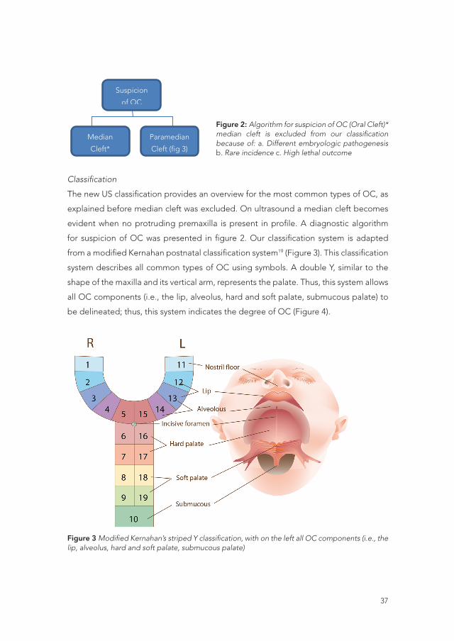
R1
R2
R3
R4
R5
R6
R7
R8
R9
R10
R11
R12
R13
R14
R15
R16
R17
R18
R19
R20
R21
R22
R23
R24
R25
R26
R27
R28
R29
R30
R31
R32
R33
R34
37
Suspicion
of OC
Median
Cleft*
Paramedian
Cleft (fig 3)
Figure 2: Algorithm for suspician of OC (Oral Cleft) * median cleft is excluded from our classification because of:
a. Different embryologic pathogenesis b. Rare incidence c. High lethal outcome
Classification
The new US classification provides an overview for the most common types of OC, as
explained before median cleft was excluded. On ultrasound a median cleft becomes
evident when no protruding premaxilla is present in profile. A diagnostic algorithm
for suspicion of OC was presented in figure 2. Our classification system is adapted
from a modified Kernahan postnatal classification system19 (Figure 3). This classification
system describes all common types of OC using symbols. A double Y, similar to the
shape of the maxilla and its vertical arm, represents the palate. Thus, this system allows
all OC components (i.e., the lip, alveolus, hard and soft palate, submucous palate) to
be delineated; thus, this system indicates the degree of OC (Figure 4).
Figure 3 Modified Kernahan’s striped Y classification, with on the left all OC components (i.e., the lip, alveolus, hard and soft palate, submucous palate)
Figure 2: Algorithm for suspicion of OC (Oral Cleft)* median cleft is excluded from our classification because of: a. Different embryologic pathogenesis b. Rare incidence c. High lethal outcome
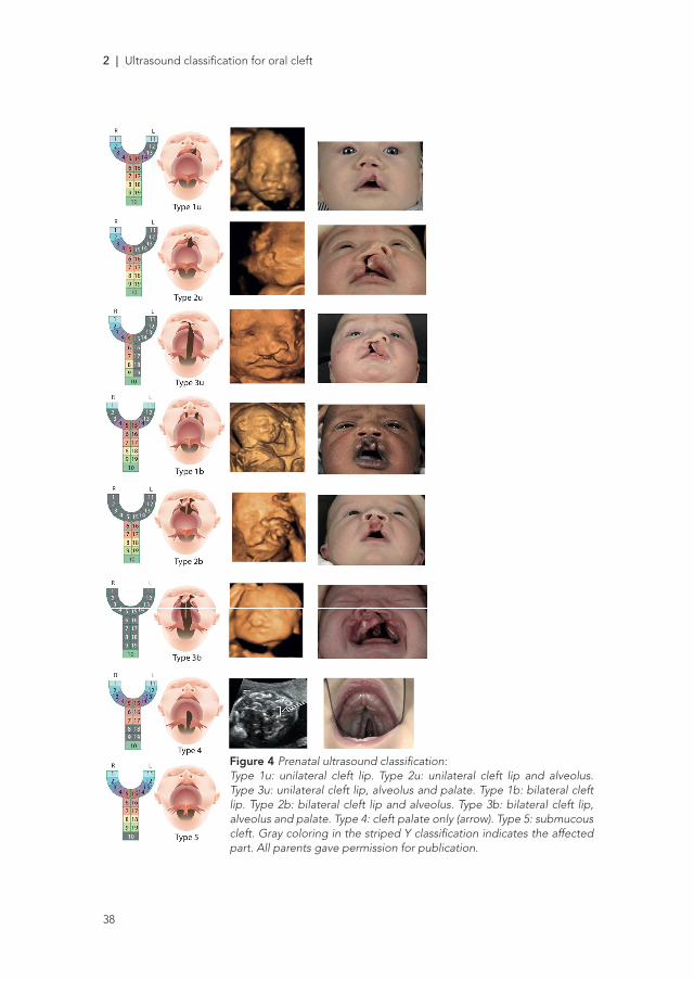
R1
R2
R3
R4
R5
R6
R7
R8
R9
R10
R11
R12
R13
R14
R15
R16
R17
R18
R19
R20
R21
R22
R23
R24
R25
R26
R27
R28
R29
R30
R31
R32
R33
R34
2 | Ultrasound classification for oral cleft
38
29
Figure 3 Prenatal ultrasound classification:
Type 1u: unilateral cleft lip. Type 2u: unilateral
cleft lip and alveolus. Type 3u: unilateral cleft lip,
alveolus and palate. Type 1b: bilateral cleft lip.
Type 2b: bilateral cleft lip and alveolus. Type 3b:
bilateral cleft lip, alveolus and palate. Type 4:
cleft palate only (arrow). Type 5: submucous cleft.
Gray coloring in the striped Y classification
indicates the affected part. All parents gave
permission for publication.
RESULTS
In total, 103 fetuses with an OC were identified by US. In one case, a type 4 cleft (cleft palate only) was
diagnosed at 20 weeks of gestation but was not confirmed at birth; this case was the only false positive in
the study (1/103 = 0.97%) and was excluded from the study. The mean gestation time at detection was
20.4 weeks (95% CI: 20.0-20.7).
Figure 4 Prenatal ultrasound classification:Type 1u: unilateral cleft lip. Type 2u: unilateral cleft lip and alveolus. Type 3u: unilateral cleft lip, alveolus and palate. Type 1b: bilateral cleft lip. Type 2b: bilateral cleft lip and alveolus. Type 3b: bilateral cleft lip, alveolus and palate. Type 4: cleft palate only (arrow). Type 5: submucous cleft. Gray coloring in the striped Y classification indicates the affected part. All parents gave permission for publication.
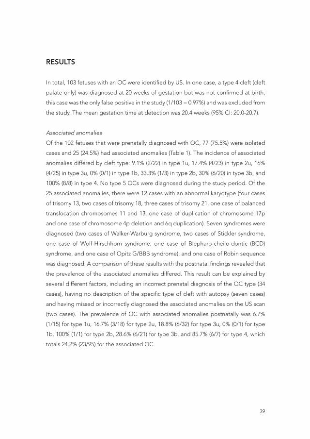
R1
R2
R3
R4
R5
R6
R7
R8
R9
R10
R11
R12
R13
R14
R15
R16
R17
R18
R19
R20
R21
R22
R23
R24
R25
R26
R27
R28
R29
R30
R31
R32
R33
R34
39
RESULTS
In total, 103 fetuses with an OC were identified by US. In one case, a type 4 cleft (cleft
palate only) was diagnosed at 20 weeks of gestation but was not confirmed at birth;
this case was the only false positive in the study (1/103 = 0.97%) and was excluded from
the study. The mean gestation time at detection was 20.4 weeks (95% CI: 20.0-20.7).
Associated anomalies
Of the 102 fetuses that were prenatally diagnosed with OC, 77 (75.5%) were isolated
cases and 25 (24.5%) had associated anomalies (Table 1). The incidence of associated
anomalies differed by cleft type: 9.1% (2/22) in type 1u, 17.4% (4/23) in type 2u, 16%
(4/25) in type 3u, 0% (0/1) in type 1b, 33.3% (1/3) in type 2b, 30% (6/20) in type 3b, and
100% (8/8) in type 4. No type 5 OCs were diagnosed during the study period. Of the
25 associated anomalies, there were 12 cases with an abnormal karyotype (four cases
of trisomy 13, two cases of trisomy 18, three cases of trisomy 21, one case of balanced
translocation chromosomes 11 and 13, one case of duplication of chromosome 17p
and one case of chromosome 4p deletion and 6q duplication). Seven syndromes were
diagnosed (two cases of Walker-Warburg syndrome, two cases of Stickler syndrome,
one case of Wolf-Hirschhorn syndrome, one case of Blepharo-cheilo-dontic (BCD)
syndrome, and one case of Opitz G/BBB syndrome), and one case of Robin sequence
was diagnosed. A comparison of these results with the postnatal findings revealed that
the prevalence of the associated anomalies differed. This result can be explained by
several different factors, including an incorrect prenatal diagnosis of the OC type (34
cases), having no description of the specific type of cleft with autopsy (seven cases)
and having missed or incorrectly diagnosed the associated anomalies on the US scan
(two cases). The prevalence of OC with associated anomalies postnatally was 6.7%
(1/15) for type 1u, 16.7% (3/18) for type 2u, 18.8% (6/32) for type 3u, 0% (0/1) for type
1b, 100% (1/1) for type 2b, 28.6% (6/21) for type 3b, and 85.7% (6/7) for type 4, which
totals 24.2% (23/95) for the associated OC.
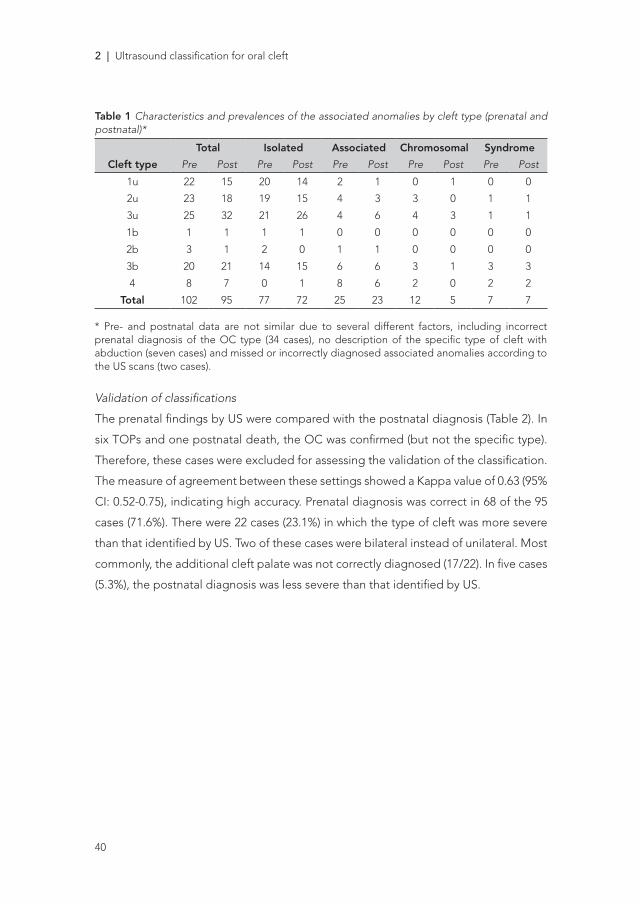
R1
R2
R3
R4
R5
R6
R7
R8
R9
R10
R11
R12
R13
R14
R15
R16
R17
R18
R19
R20
R21
R22
R23
R24
R25
R26
R27
R28
R29
R30
R31
R32
R33
R34
2 | Ultrasound classification for oral cleft
40
Table 1 Characteristics and prevalences of the associated anomalies by cleft type (prenatal and postnatal)*
Total Isolated Associated Chromosomal Syndrome
Cleft type Pre Post Pre Post Pre Post Pre Post Pre Post
1u 22 15 20 14 2 1 0 1 0 0
2u 23 18 19 15 4 3 3 0 1 1
3u 25 32 21 26 4 6 4 3 1 1
1b 1 1 1 1 0 0 0 0 0 0
2b 3 1 2 0 1 1 0 0 0 0
3b 20 21 14 15 6 6 3 1 3 3
4 8 7 0 1 8 6 2 0 2 2
Total 102 95 77 72 25 23 12 5 7 7
* Pre- and postnatal data are not similar due to several different factors, including incorrect prenatal diagnosis of the OC type (34 cases), no description of the specific type of cleft with abduction (seven cases) and missed or incorrectly diagnosed associated anomalies according to the US scans (two cases).
Validation of classifications
The prenatal findings by US were compared with the postnatal diagnosis (Table 2). In
six TOPs and one postnatal death, the OC was confirmed (but not the specific type).
Therefore, these cases were excluded for assessing the validation of the classification.
The measure of agreement between these settings showed a Kappa value of 0.63 (95%
CI: 0.52-0.75), indicating high accuracy. Prenatal diagnosis was correct in 68 of the 95
cases (71.6%). There were 22 cases (23.1%) in which the type of cleft was more severe
than that identified by US. Two of these cases were bilateral instead of unilateral. Most
commonly, the additional cleft palate was not correctly diagnosed (17/22). In five cases
(5.3%), the postnatal diagnosis was less severe than that identified by US.
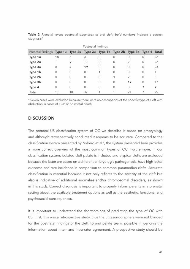
R1
R2
R3
R4
R5
R6
R7
R8
R9
R10
R11
R12
R13
R14
R15
R16
R17
R18
R19
R20
R21
R22
R23
R24
R25
R26
R27
R28
R29
R30
R31
R32
R33
R34
41
Table 2 Prenatal versus postnatal diagnoses of oral cleft; bold numbers indicate a correct diagnosis*
Postnatal findings
Prenatal findings Type 1u Type 2u Type 3u Type 1b Type 2b Type 3b Type 4 Total
Type 1u 14 5 3 0 0 0 0 22
Type 2u 1 9 10 0 0 2 0 22
Type 3u 0 4 19 0 0 0 0 23
Type 1b 0 0 0 1 0 0 0 1
Type 2b 0 0 0 0 1 2 0 3
Type 3b 0 0 0 0 0 17 0 17
Type 4 0 0 0 0 0 0 7 7
Total 15 18 32 1 1 21 7 95
* Seven cases were excluded because there were no descriptions of the specific type of cleft with obduction in cases of TOP or postnatal death.
DISCUSSION
The prenatal US classification system of OC we describe is based on embryology
and although retrospectively conducted it appears to be accurate. Compared to the
classification system presented by Nyberg et al.9, the system presented here provides
a more correct overview of the most common types of OC. Furthermore, in our
classification system, isolated cleft palate is included and atypical clefts are excluded
because the latter are based on a different embryologic pathogenesis, have high lethal
outcome and rare incidence in comparison to common paramedian clefts. Accurate
classification is essential because it not only reflects to the severity of the cleft but
also is indicative of additional anomalies and/or chromosomal disorders, as shown
in this study. Correct diagnosis is important to properly inform parents in a prenatal
setting about the available treatment options as well as the aesthetic, functional and
psychosocial consequences.
It is important to understand the shortcomings of predicting the type of OC with
US. First, this was a retrospective study, thus the ultrasonographers were not blinded
for the postnatal findings of the cleft lip and palate team, possible influencing the
information about inter- and intra-rater agreement. A prospective study should be
R1
R2
R3
R4
R5
R6
R7
R8
R9
R10
R11
R12
R13
R14
R15
R16
R17
R18
R19
R20
R21
R22
R23
R24
R25
R26
R27
R28
R29
R30
R31
R32
R33
R34
2 | Ultrasound classification for oral cleft
42
performed to assess final reliable accuracy. Second, there are more variations in OC
possible than those described in this US classification system. In addition to being
unilateral or bilateral, OC demonstrates a variable expression pattern, including
microform, minor, incomplete and complete forms. However, the US classification
system used in this study aims to provide a practical tool for ultrasonographers to use
in daily practice. Third, a submucous cleft (type 5) will likely never be diagnosed by
US because the outer anatomy is normal. Submucous clefts are often diagnosed later
in life, when children appear to have difficulty speaking. Because of the short follow-
up period, the incidence of type 5 OC is not reliable. Fourth, isolated CP (type 4) is
rarely diagnosed prenatally, although when type 4 is suspected, it is generally correct
. Even when a cleft lip was diagnosed by US, visualization of the palate was difficult in
several cases. Hence, when there is a suspicion of a cleft lip and alveolus, the parents
should be told during counseling that there is a reasonable risk of an additional cleft
palate (Table 1). This diagnosis can greatly alter the long-term prognosis for the child
because it is commonly associated with difficulties in speech20, feeding21, 22, hearing20,
and postoperative mid-facial retrusion23.
Recently, several studies have attempted to improve upon the detection of the cleft
palate. Hassan et al.24 described visualization of the tongue abutting the nasal septum
in the transverse view as strongly indicating the presence of a cleft of the hard palate.
Bäumler et al.25 illustrated high diagnostic accuracy with 3D axial visualization of the
hard palate. Furthermore, Wilhelm et al.15 suggested the use of a novel marker for
diagnosing clefts of the soft palate with the sonographic appearance of an ‘equal
sign,’ referring to a normal uvula. An unremarkable uvula implies the presence of in
intact palate because, as we explained in the embryology section, the palate closes
from the anterior to the posterior. Several other techniques using three-dimensional
US have been described, such as the reversed face view26 and angled insonation26.
However, it remains a great challenge to identify cleft palate alone without a cleft lip or
micrognathia to alert the ultrasonographer to examine the fetal face in greater detail.
Isolated cleft palate represents approximately one-third of the total incidence of OC1;
therefore, it should have a place in prenatal classification systems.
R1
R2
R3
R4
R5
R6
R7
R8
R9
R10
R11
R12
R13
R14
R15
R16
R17
R18
R19
R20
R21
R22
R23
R24
R25
R26
R27
R28
R29
R30
R31
R32
R33
R34
43
CONCLUSION
This new prenatal US classification system offers a systematic and practical approach
for prenatal screening of oral clefts and, unlike the system presented by Nyberg,
provides a accurate correlation with the postnatal cleft diagnosis. The classification
system presented here is based on a commonly used postnatal system; therefore, it
will improve communication between obstetricians and cleft lip and palate teams. We
expect that parental counseling in both fields will improve as a result of using this
classification system.
R1
R2
R3
R4
R5
R6
R7
R8
R9
R10
R11
R12
R13
R14
R15
R16
R17
R18
R19
R20
R21
R22
R23
R24
R25
R26
R27
R28
R29
R30
R31
R32
R33
R34
2 | Ultrasound classification for oral cleft
44
REFERENCES
1. P. A. Mossey, J. Little, R. G. Munger, et al. Cleft lip and palate. Lancet 2009; 374: 1773-1785.2. W. Maarse, L. R. Pistorius, W. K. Van Eeten, et al. Prenatal ultrasound screening for orofacial
clefts. Ultrasound Obstet Gynecol 2011; 38: 434-439. 3. S. Ensing, C. E. Kleinrouweler, S. M. Maas, et al. Influences of the introduction of the 20
weeks fetal anomaly scan on prenatal diagnosis and management of fetal facial clefts. Ultrasound Obstet Gynecol 2013.
4. D. A. Kernahan and R. B. Stark. A new classification for cleft lip and cleft palate. Plast Reconstr Surg Transplant Bull 1958; 22: 435-441.
5. A. W. Smith, A. K. Khoo and I. T. Jackson. A modification of the Kernahan “Y” classification in cleft lip and palate deformities. Plast Reconstr Surg 1998; 102: 1842-1847.
6. H. Koch, M. Grzonka and J. Koch. Cleft malformation of lip, alveolus, hard and soft palate, and nose (LAHSN)--a critical view of the terminology, the diagnosis and gradation as a basis for documentation and therapy. Br J Oral Maxillofac Surg 1995; 33: 51-58.
7. W. Maarse, A. M. Rozendaal, E. Pajkrt, et al. A systematic review of associated structural and chromosomal defects in oral clefts: when is prenatal genetic analysis indicated? J Med Genet 2012; 49: 490-498.
8. G. Le Dref, B. Grollemund, A. Danion-Grilliat, et al. Towards a new procreation ethic: the exemplary instance of cleft lip and palate. Medicine, health care, and philosophy 2013; 16: 365-375.
9. D. A. Nyberg, G. K. Sickler, F. N. Hegge, et al. Fetal cleft lip with and without cleft palate: US classification and correlation with outcome. Radiology 1995; 195: 677-684.
10. W. Larsen. Larsen’s human embryology Philadelphia, 2009.11. V.K. ten Donkelaar HJ The neural crest and craniofacial malformations. In Clinical
Neuroembryology. Springer Berlin Heidelberg: New York,, 2006.12. N. G. Hartwig, C. Vermeij-Keers, H. E. De Vries, et al. Limb body wall malformation complex:
an embryologic etiology? Hum Pathol 1989; 20: 1071-1077.13. K. A. Allam, D. C. Wan, H. K. Kawamoto, et al. The spectrum of median craniofacial
dysplasia. Plast Reconstr Surg 2011; 127: 812-821. 14. T. Sadler. Langman’s medical embryology: Baltimore, 1995, 69-92.15. L. Wilhelm and H. Borgers. The ‘equals sign’: a novel marker in the diagnosis of fetal
isolated cleft palate. Ultrasound Obstet Gynecol 2010; 36: 439-444. 16. C. C. Breugem and A. B. Mink van der Molen. What is ‘Pierre Robin sequence’? J Plast
Reconstr Aesthet Surg 2009; 62: 1555-1558. 17. B. C. Sommerlad, C. Fenn, K. Harland, et al. Submucous cleft palate: a grading system and
review of 40 consecutive submucous cleft palate repairs. Cleft Palate Craniofac J 2004; 41: 114-123.
18. W. Maarse, L. R. Pistorius, W. K. van Eeten, et al. Prenatal orofacial cleft; accuracy of ultrasound screening. Ultrasound Obstet Gynecol 2010.
19. H. C. Noordhoff MS, Wu J. Multidisciplinary management of cleft lip and palate in Taiwan. In Multidisciplinary management of cleft lip and palate in Taiwan, B. J (ed). WB Saunders 1990: Philidelphia, 1990, 18-26.
20. E. M. Marrinan, R. A. LaBrie and J. B. Mulliken. Velopharyngeal function in nonsyndromic cleft palate: relevance of surgical technique, age at repair, and cleft type. Cleft Palate Craniofac J 1998; 35: 95-100.
21. I. A. de Vries, C. C. Breugem, A. M. van der Heul, et al. Prevalence of feeding disorders in children with cleft palate only: a retrospective study. Clin Oral Investig 2014; 18: 1507-1515.
22. J. L. Paradise. Middle ear problems associated with cleft palate. An internationally-oriented review. Cleft Palate J 1975; 12: 17-22.

R1
R2
R3
R4
R5
R6
R7
R8
R9
R10
R11
R12
R13
R14
R15
R16
R17
R18
R19
R20
R21
R22
R23
R24
R25
R26
R27
R28
R29
R30
R31
R32
R33
R34
45
23. D. M. DeLuke, A. Marchand, E. C. Robles et al. Facial growth and the need for orthognathic surgery after cleft palate repair: literature review and report of 28 cases. J Oral Maxillofac Surg 1997; 55: 694-697; discussion 697-698.
24. W. A. Hassan and C. C. Lees. Facial cleft detected: is the palate normal? Best practice & research Clinical obstetrics & Gynecology 2014; 28: 379-389.
25. M. Baumler, J. M. Faure, M. Bigorre, C. et al. Accuracy of prenatal three-dimensional ultrasound in the diagnosis of cleft hard palate when cleft lip is present. Ultrasound Obstet Gynecol 2011; 38: 440-444.
26. S. Campbell and C. C. Lees. The three-dimensional reverse face (3D RF) view for the diagnosis of cleft palate. Ultrasound Obstet Gynecol 2003; 22: 552-554.

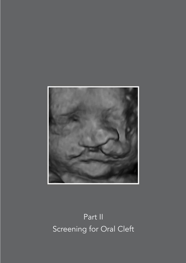
Part II
Screening for Oral Cleft

3Prenatal Screening for Orofacial Clefts in
the Netherlands: A Preliminary Report on
the Impact of a National Screening System
Cleft Palate-Craniofacial Journal, March 2011, Vol. 48 (2) 183-9
A.B. MINK VAN DER MOLEN, W. MAARSE, L.R. PISTORIUS,
H.F.N. SWANENBURG DE VEYE, C.C. BREUGEM
R1
R2
R3
R4
R5
R6
R7
R8
R9
R10
R11
R12
R13
R14
R15
R16
R17
R18
R19
R20
R21
R22
R23
R24
R25
R26
R27
R28
R29
R30
R31
R32
R33
R34
3 | Prenatal screening for oral cleft
50
ABSTRACT
In the Netherlands a new and countrywide routine screening system for physical
congenital anomalies was introduced by legislation in 2007. The screening was aimed
initially at Down syndrome, but under specific conditions secondary screening for cleft
lip and palate and other congenital malformations is performed as well. This article
gives an overview of the Dutch system, with a focus on cleft lip (and palate) and the
report of one center’s experience. In general, voluntary termination of pregnancy
in the second trimester has increased slightly since the introduction of the current
system, but the termination-of-pregnancy rate for isolated cleft lip and palate remains
limited in the Netherlands.
R1
R2
R3
R4
R5
R6
R7
R8
R9
R10
R11
R12
R13
R14
R15
R16
R17
R18
R19
R20
R21
R22
R23
R24
R25
R26
R27
R28
R29
R30
R31
R32
R33
R34
51
The technical progress in ultrasound (US) screening allows for ever-earlier and
increasingly reliable detection of a diversity of physical congenital anomalies in the
prenatal period, including cleft lip1. For this reason, high-resolution, transabdominal
US examination has become the standard of care in many countries including the
Netherlands. This development has led to new legislation in the Netherlands regarding
prenatal US. In the Netherlands, the Population Screening Act (PSA)—enacted
in 1996—allows the Dutch Minister of Health, Welfare, and Sport (HWS) to issue a
permit for certain types of population screening. Since the enactment of the PSA, it
is obligatory that the Minister of HWS consult the Health Council of the Netherlands
before he or she issues a permit for such screenings. The task of the Health Council’s
committee on the PSA is to advise on the issue of permits for population screening.
In October 2006 the Health Council wrote an extensive preliminary report advising
the Minister of HWS3, followed by a final report on the same subject focused on the
screening for Down syndrome and neural tube defects2. Based on the preliminary
advice in 2006 and the legal ramifications of the PSA, it was decided at the end of 2006
to encompass the medical technical innovations in the field of prenatal screening in
new legislation, effective in 2007. It should be noted that the Netherlands was quite
late in setting rules in this field because prenatal US was available for many years for
individual parents at their own expense or for a medical reason. Any regulation for
routine screening of all pregnant women was postponed because for a long time, the
Minister of HWS defended the view that in general any diagnostic work should be
performed routinely only when there were implications for treatment or prevention;
the possibilities for prevention of or prenatal treatment for congenital abnormalities
during pregnancy were judged minimal or absent. The introduction of routine prenatal
US for all pregnant women, therefore, was considered an unnecessary luxury, only
making health care more expensive, and was deferred for many years. This view of the
Ministry of HWS was in accordance with the principles for prenatal screening programs
as reviewed by Simopoulos4. However, the result of this policy was lack of transparency,
with individual institutions, doctors, or other health care workers offering pregnant
women a variety of prenatal screenings according to their own insights, sometimes
covered by health care insurance and sometimes not. Equal access to the same quality
of screening was no longer ensured. Pressure from the professionals in the field and
R1
R2
R3
R4
R5
R6
R7
R8
R9
R10
R11
R12
R13
R14
R15
R16
R17
R18
R19
R20
R21
R22
R23
R24
R25
R26
R27
R28
R29
R30
R31
R32
R33
R34
3 | Prenatal screening for oral cleft
52
patients’ platforms seeking standardization and uniform access slowly brought about
a change of mind at the ministerial level. Next, the Ministry of HWS stipulated one
uniform system for the whole country as prerequisite for any adjustment, and in 2006
the Minister of HWS asked the opinion of the Health Council about several proposals to
improve the situation. The advice of the Health Council3 induced the definitive change
in policy by the government. According to the Minister of HWS, the focus of routine
US screening in the Netherlands is not prevention and treatment. The screening was
designed primarily to inform future parents about possible defects and their treatment
options. Thus, the screening was never intended to improve the overall health at a
population level. To the knowledge of the authors, the government performed no
fundamental cost-benefit study for prenatal US screening in the Netherlands to
support the change in policy in 2006.
The new legislation organizes prenatal screening in the Netherlands in detail.
Important issues include the fact that Dutch health insurance companies were required
to cover prenatal screening by US in their policies and that a new Central Organization
for Quality Control and Data Acquisition of Prenatal Screening (Central Organization)
was founded and funded. The health care insurance companies, the societies of the
medical specialists involved, the society of general practitioners, patients’ platform,
and the regional referral centers are represented in this Central Organization. Its task
is to ensure countrywide quality criteria, to draft uniform information to patients, to
design a uniform database for the screening, and to ensure uniform protocols and
uniform contracts with counselors and US diagnostic centers. The Central Organization
was incorporated in the structure of the Rijksinstituut voor Volksgezondheid en
Milieu (RIVM)5, a government research institute for public health and a safe living
environment in the Netherlands. Minimum quality criteria for counselors were set, and
as a consequence, professionals can only be involved in the counseling for prenatal
screening on the condition that counseling is done weekly as a routine part of their
practice (minimum 50 per year). Similarly, quality criteria for persons performing the
basic US screening (Structureel Echoscopisch Onderzoek) were drafted6. The RIVM
provides a Web site with ample information on the subject, including all relevant
documents in Dutch in pdf format and a useful version in English about the screening5.
R1
R2
R3
R4
R5
R6
R7
R8
R9
R10
R11
R12
R13
R14
R15
R16
R17
R18
R19
R20
R21
R22
R23
R24
R25
R26
R27
R28
R29
R30
R31
R32
R33
R34
53
Ministerial permission to organize the screening was granted to eight regional
nonprofit centers (trusts) in the Netherlands, specifically founded for this purpose.
These trusts oversee the screening in their regions and engage sonologists, midwives,
and obstetricians within their region to perform the screening. A contract for screening
includes an agreement on the quality of the US equipment, experience, training, and
continuing education of the person performing the US and the counseling. Information
of each US screening will be collected directly (online) into a new countrywide
database system. This information, together with the postnatal findings, should give
the opportunity to provide important information about the quality of the US screening
and its consequences in the future.
The average population covered by a regional referral center is about 2 million
people, which translates to about 20,000 pregnancies annually per center. In order
to reduce costs and to ensure easy access for women, the basic screening takes
place at primary-care level, mostly by certified midwives or sonologists. In case of
a suspected abnormality, the woman is referred to a certified obstetrician in one of
the eight regional referral centers (or one of their satellites) for further analysis and
counseling. Further analysis always includes advanced US screening. The standard for
advanced US screening is set by the Dutch Society for Gynecology and Obstetrics and
is available electronically6. So far, only transabdominal US is used for routine screening
in the Netherlands.
The focus of the screening protocol is to identify fetuses with Down syndrome or
neural tube defects3. Screening for Down syndrome entails a risk assessment based on
the combination of maternal age, biochemical markers such as pregnancy-associated
plasma protein A (PAPP-A) and the beta subunit of human choriogonadotropin
(fbeta-hCG) in the blood of the mother in the first trimester of the pregnancy, and
US measurement of the nuchal translucency between 11 and 14 weeks of gestation.
To screen for neural tube defects an additional US is performed at 18 to 22 weeks of
gestation. During the latter, a systematic screening of the whole body is done with a
primary focus on neural tube defects. This particular screening enables detection of
other birth defects, including orofacial clefts. Currently about 25% of the pregnant
women participate in the Down screening and 95% of the women opt for the US at 18
to 22 weeks.
R1
R2
R3
R4
R5
R6
R7
R8
R9
R10
R11
R12
R13
R14
R15
R16
R17
R18
R19
R20
R21
R22
R23
R24
R25
R26
R27
R28
R29
R30
R31
R32
R33
R34
3 | Prenatal screening for oral cleft
54
Informed Consent
All pregnant women are offered the option to obtain information about prenatal
screening. They remain free in their choice whether to obtain the information and
subsequently whether to participate in screening7,8,9. This means that screening
is not performed routinely. The screening is a complex issue, where future parents
are encouraged to make well-informed choices about whether to participate in
the screening. Important is the fact that parents should be informed before the US
examination about the right not to know, the technical aspects of the screening,
and its potential implications10. Potential implications include abnormal findings (not
specifically cleft lip and palate), and in those cases, if parents wish, the advice to seek
further genetic counseling, which may lead to amniocentesis. Thus, informed consent
is mandatory before embarking upon this screening process. Midwives, general
practitioners, and gynecologists involved in the initial counseling should be trained in
counseling practice for this purpose.
Practical Experience With Screening for Cleft Lip and Palate in the Utrecht Regional
Unit
The reported reliability of the US in detecting cleft lip and/or palate (CL/P) varies
considerably11. Several factors may account for the variation, such as the population
screened (high risk or low risk), the timing of the US, the experience of the sonographer,
the quality of the US equipment, the study design, and the definitions of the outcome
parameters. Two studies are important because they were conducted in a nonselected
low-risk population, comparable with the Dutch routine screening. Russell et al.12
studied 10,822 births in the period 1992 to 2002 who had US screening from 18 to 20
weeks of pregnancy. Screening of the face was a standard part of the US performed.
The detection ratio for cleft lip with or without cleft palate (CL ± P) was 23% (29/127
babies with CL ± P detected prenatally within a population of 108,220 births in a
10-year period). Isolated cleft palate (CP) without cleft lip and no further congenital
abnormalities was not detected on US in this study. The prenatal detection ratio
increased with time from 15% (from 1992 to 1996) to 30% (from 1996 to 2002). A study
by Offerdal et al.1 confirms these findings with an overall detection ratio of 45% (34/77)
for isolated CL ± P in the low-risk population and a significant increase in the detection
R1
R2
R3
R4
R5
R6
R7
R8
R9
R10
R11
R12
R13
R14
R15
R16
R17
R18
R19
R20
R21
R22
R23
R24
R25
R26
R27
R28
R29
R30
R31
R32
R33
R34
55
rate from 35% to 58% between the two 9-year periods of the study. It is expected that
the detection rate will continue to rise slowly over time due to technical innovations in
the equipment and increased experience of sonographers. The specificity of the US
for the detection of isolated CL ± P in these studies and other studies is high— almost
100%. The experience at the University Medical Center Utrecht is in line with these
studies.
When an orofacial cleft is suspected, pregnant women are referred to a specialized
obstetrician in one of the eight designated centers or their satellites for further
investigation. This referral system works well. A discussion point is the maximum
acceptable time between initial (abnormal) US and the referral to the designated
center because parents are kept in uncertainty during this period. Another reason to
see the parents without delay is the upper limit to perform termination of pregnancy
for a medical reason, which is 24 weeks in the Netherlands. Thus, the time interval
to perform karyotyping and other genetic screening frequently is limited. Any wait
is undesirable, but in a practical sense often unavoidable, and should be kept to a
minimum. The aim is to see the parents in the referral center as soon as possible, and
5 working days is considered the maximum delay.
The images of the first US are stored and made available to the referral center. In
all referral centers US screening is repeated using a more extensive protocol6. A
diagnostic US examination is therefore needed to confirm the provisional diagnosis
made at the screening US examination. In the past concerns have been raised about
possible detrimental effects of US for the unborn child, but these were not confirmed
in recent randomized controlled trials13. Current US study protocols6 adhere to the
principle ‘‘as low as reasonably achievable (ALARA)’’14, thus sound level/intensity is
set as low as possible to allow the optimal image with the minimum US exposure. In
normal clinical practice it is extremely improbable to exceed the maximum intensity
levels with gray-scale two- dimensional and three-dimensional US. The US exposure
needed to detect and confirm a CL ± P is therefore considered as safe.
The current legislation does not yet provide a detailed guideline as to what should
happen after confirmation of the diagnosis. The following describes the current
practice in our unit in Utrecht with a focus on CL ± P. After confirmation of a cleft lip on
US, more detailed assessment is initiated in our unit, implying that the unborn child is
R1
R2
R3
R4
R5
R6
R7
R8
R9
R10
R11
R12
R13
R14
R15
R16
R17
R18
R19
R20
R21
R22
R23
R24
R25
R26
R27
R28
R29
R30
R31
R32
R33
R34
3 | Prenatal screening for oral cleft
56
first screened by US by the obstetrician for other defects and a careful family history
focused on birth defects is performed.
In Utrecht the consultation of a clinical geneticist is always advised in case any associated
abnormalities are detected on US and/or family history suggests syndromal background
or increased risk. In such cases karyotyping by amniocentesis is recommended to the
parents and usually carried out, provided the results are available before 24 weeks
of pregnancy (or in case parents prefer to know anyway). Besides karyotyping other
genetic tests are available, and in Utrecht a multiplex ligation-dependent probe
amplification (MLPA) test to rule out 22q11.2 deletion syndrome is usually done as well.
In case any other structural abnormalities on US and/or chromosomal abnormalities
are found, these findings and their possible implications are discussed with the parents
by the obstetrician and the geneticist. If the parents opt for voluntary termination of
pregnancy, this will take place at the University Hospital instead of an anonymous
abortion clinic. This is in line with the recommendations of the legislation and offers the
opportunity to perform further examination on the fetus (such as autopsy) and to verify
the accuracy of the prenatal diagnosis, which is important for quality control of the
system and for the parents regarding the prognosis for possible future pregnancies.
In this context the issue of false-positive findings is relevant. False-positive findings have
occurred, but rarely, in the first line during the first US screening in the Utrecht area.
These were at all times detected during more extensive follow-up US studies. A third
or even a fourth US is made and all other relevant tests are performed to be absolutely
sure about the diagnosis of the CL ± P and possible associated congenital anomalies.
This is in the stage before significant interventions such as a termination are even
considered. In the 20 years of prenatal US screening at the University Medical Centre
Utrecht, no cases of misdiagnosis of CL ± P leading to an unintended termination have
ever occurred, according to our obstetricians.
In case no other defects are found and the family history is negative too, the parents
are offered the option of consultation with a geneticist and/or fetal karyotyping in
our regional unit. In these cases parents and counselors should carefully weigh the
potential benefits and risks of karyotyping by performing amniocentesis (0.3%
spontaneous abortion) against the benefit of more information about the unborn
child. The literature mentions quite a significant percentage of associated anomalies
R1
R2
R3
R4
R5
R6
R7
R8
R9
R10
R11
R12
R13
R14
R15
R16
R17
R18
R19
R20
R21
R22
R23
R24
R25
R26
R27
R28
R29
R30
R31
R32
R33
R34
57
and/or chromosomal abnormalities associated with CL ± P (as a group), with a variance
of 24%15 to 48%16 for CLP in prenatal studies. A critical appraisal of the studies on
associated anomalies, however, shows that the available literature is hampered by a
lot of methodological problems, such as inclusion of cases (life birth or all deliveries),
variation in follow-up period after birth, variation in the definition of clinical expression
of anomalies, variation in technologies available for detection of associated anomalies,
patient selection (tertiary centers), differences in population (white or Asian), and
prenatal versus postnatal studies. Another problem with the available literature is that
in many studies all associated anomalies are lumped together with no discrimination
regarding severity. Counseling in this area is certainly not an easy thing to do. Parents
may ask us the question: How much chance remains that significant (syndromal)
abnormalities are still present unexpectedly after birth in case of a normal advanced
US screening (except the cleft), normal karyotyping, and a negative family history? This
is an important question, but unfortunately sufficient data are not currently available
in the literature for an accurate answer to this seemingly simple question. New studies
and better information are urgently needed in this field. In the meantime, we aim to
find a balance between informing this group of parents about the risk of associated
anomalies and trying to not worry them unnecessarily.
The cleft team is routinely consulted in all cases, except when very severe (lethal)
additional anomalies (such as holoprosencephaly or trisomy 13 or 18) are identified,
unless the parents choose to continue with the pregnancy regardless of the outcome.
The cleft team offers the parents further detailed counseling about the possible
consequences of having a CLP. To this purpose the department of gynecology and
obstetrics and the cleft team have joined forces and set up a special outpatient
clinic. In the outpatient clinic the parents meet a multidisciplinary team, including
an obstetrician, the medical psychologist, and a plastic surgeon from our cleft team.
During this visit the obstetrician repeats the US in the presence of the plastic surgeon.
This allows the plastic surgeon to obtain an ultrasonic three-dimensional view of the
cleft. In our experience, this setting improves the quality of the counseling. The etiology
and pathogenesis of a CL ± P are explained to the parents and the medical, surgical,
clinical, and psychosocial needs of a child with a cleft lip and possible cleft palate are
discussed. Explanation is given about the treatment protocol of our cleft unit, and
R1
R2
R3
R4
R5
R6
R7
R8
R9
R10
R11
R12
R13
R14
R15
R16
R17
R18
R19
R20
R21
R22
R23
R24
R25
R26
R27
R28
R29
R30
R31
R32
R33
R34
3 | Prenatal screening for oral cleft
58
differences with other cleft teams’ protocols are elucidated. Possible operations are
described when asked for, and questions and concerns of the parents are addressed.
In the Netherlands, home delivery is still performed regularly. In the opinion of our
obstetricians there is no major objection against delivery at home of a child with a
cleft provided that no other abnormalities are detected prenatally, pregnancy controls
are normal, and the mother has normal health. Discussing the risks and benefits of
home delivery in the case of a cleft lip frequently forms an important part of the
counseling. The potential feeding problems in case of a cleft palate are explained and
the measures the parents should take to make feeding as easy as possible, such as
buying a Habermann bottle and equipment to express milk from the breast in case the
mother prefers to give the baby her own milk. A comprehensive patient information
booklet about cleft lip and palate and its treatment, written at our institution, is always
provided to the parents. The telephone number of the cleft team coordinator and an e-
mail contact address for the cleft team are given to the parents. In many cases there is
frequent contact by e-mail between future parents and the medical psychologist of our
team about remaining questions and issues during the last trimester of the pregnancy.
Although we did not research this specifically, we learned from the feedback of parents
that the possibility to keep in contact with the team and/or medical psychologist
during the remainder of the pregnancy is appreciated by parents.
A major advantage of this multidisciplinary approach is that pregnant women and
health care providers such as the gynecologist, midwife, and nurses are well prepared
and know what kind of potential problems to expect at birth. Potential advantages
of prenatal cleft diagnosis as formulated by Johnson and Sandy17—including
psychological preparation, opportunity for parent education, planned neonatal care,
and preparation for feeding as well as increased reproductive choices for parents—can
be maximally exploited.
Parents tell our medical psychologist that coping with a less-than-perfect child is
facilitated by the information they have gathered about the condition before birth.
The fact that the cleft team can be contacted at any time gives assurance as well. Our
experience supports the statement of Rey-Bellet and Hohlfeld18 that it is essential for
parents to understand that they are not alone in this situation, and this should be a key
message in the antenatal counseling process.
R1
R2
R3
R4
R5
R6
R7
R8
R9
R10
R11
R12
R13
R14
R15
R16
R17
R18
R19
R20
R21
R22
R23
R24
R25
R26
R27
R28
R29
R30
R31
R32
R33
R34
59
Because parents are well informed, it seems that the stress of the parents seeing
their child after birth with a cleft is less than the stress of parents in the unexpected
cases, which is in accordance with Nusbaum19. The study of Kemp et al.20 suggests
that counseling by specialist staff reduces the levels of parental anxiety associated
with the diagnosis of a fetal surgical malformation. Our clinical experience seems
to be in accordance with these observations, but a prospective study is needed to
confirm the positive effect of our counseling policy during the pregnancy. This could
be accomplished by studying the coping, stress, uncertainty, and comfort of parents
for whom diagnosis was obtained before birth (with counseling) compared with those
who were not identified.
With such preparation there is, in general, no need to consult the cleft team instantly
after birth, and appointments with the team can be scheduled accordingly.
Choices Made by Dutch Mothers
One of the goals of the Central Organization of the Ministry of Health is to collect all
data concerning prenatal US throughout the country by means of an Internet-based
registration system, so that the findings of each US performed and its follow-up can be
registered directly. In this manner the Central Organization should be able to collect a
unique set of data. Unfortunately, the software to analyze this significant data set has
not yet been fully developed and consequently, only limited national data are currently
available.
The Inspection for Health Care in the Netherlands, however, publishes a detailed
report about the number of voluntary terminations of pregnancy in the Netherlands
every year, thus affording the opportunity to monitor the possible impact of the new
screening policy on the termination rate. In 2007, the latest year for which data are
available, the total number of terminations stabilized and the termination number
remained stable at 8.6 per 1000 women between 15 and 44 years of age living in the
Netherlands, the same as in 2005 and 200621. This is low compared with many other
countries but in line with neighboring countries such as Germany and Belgium. There
was, however, an increase of 14.4% in the total number of pregnancy terminations
performed in the second term of pregnancy (13 weeks), and the number of terminations
performed in a hospital (instead of an abortion clinic) between 20 and 24 weeks
R1
R2
R3
R4
R5
R6
R7
R8
R9
R10
R11
R12
R13
R14
R15
R16
R17
R18
R19
R20
R21
R22
R23
R24
R25
R26
R27
R28
R29
R30
R31
R32
R33
R34
3 | Prenatal screening for oral cleft
60
increased in comparison to 2005 from 7.4% (140) to 11.5% (270). The trend toward
more second-term and hospital terminations (i.e., on medical grounds) started in 2003
and is attributed to the increase in US screening, even before the current legislation
was introduced (as more and more women got a prenatal US over the last decade).
Nationwide data about the number of terminations performed specifically in case of
CL ± P (as a group) are not currently available. However, the data from the Utrecht
region show a very low termination rate for isolated facial clefts (i.e., cases where US
and/or other tests have not found any other abnormality). The Utrecht unit covers
about 20,000 pregnancies per year. In the 4-year period from 2005 through 2008, a
total of 78 cases of cleft lip (and palate) were identified on US prenatally. Of these,
45 were identified as isolated cases of CL ± P, and 33 had associated anomalies on
US and/or genetic screening. Three cases of isolated CL ± P and 22 associated cases
(mostly trisomy 13 or 18) were terminated by abortion. The regional referral unit at the
Erasmus Medical Centre in Rotterdam has recently reported similar numbers22.
Rozendaal et al.23 have studied the numbers of the registration of prevalence of CL ±P
in the Netherlands by the Dutch Society for Cleft Lip and Craniofacial Abnormalities
(DSCLCA) in a recent report on all birth defects in the Netherlands in the period 1997
to 2007 structured by the Minister of HWS24. All cleft teams report the type of the cleft
of all new babies in a standardized format directly online to the DSCLCA. The number
of newborns with a CL ± P varied between a maximum of 395 newborns (2003) and a
minimum of 315 newborns (2005) on a population of 16.3 million people and a total of
187,910 live births in 2005. The national average prevalence of CL ± P of the 10-year
period 1997 to 2007 was 16.6 per 10,000 life births (11.2 for CL ± P and 5.4 for CP). In
the same 10-year period, the registry seems to reveal a slowly declining trend in the
prevalence of CL ± P (as a group) in the Netherlands23. More detailed analysis of the
numbers by Rozendaal et al.23 suggests a stable prevalence of CP but a decline in CL
± P. The declining trend in the numbers of CL ± P reported by Rozendaal et al.23 is,
however, debatable because numbers are small and the trend may still be within the
spread of yearly variation.
The combination of more pregnant women undergoing US screening (even before
current legislation was introduced) and increase in second trimester and hospital
terminations make it plausible that prenatal screening forms the basis of the
R1
R2
R3
R4
R5
R6
R7
R8
R9
R10
R11
R12
R13
R14
R15
R16
R17
R18
R19
R20
R21
R22
R23
R24
R25
R26
R27
R28
R29
R30
R31
R32
R33
R34
61
declining prevalence of CL ± P (if indeed true) in the Netherlands. This is in line with
international trends25. Because termination for isolated CL ± P remains uncommon
in the Netherlands, the declining trend in prevalence in the Netherlands is most
likely due to termination of pregnancy for multiple congenital anomalies, including
CL/P. An alternative explanation brought forward is the Dutch guideline for folic acid
supplement23. Folic acid supplements are recommended in the Netherlands from 4
weeks before conception to 8 weeks after conception. In the embryo the lip is fused
at 7 to 8 weeks after conception, but the palate takes longer (7 to 12 weeks). If we
assume that folic acid decreases the chance of CL/P, the current guideline may have
a more significant positive effect on the fusion process of the lip compared with the
palate, which may account for the lower numbers of babies born with a CL ± P. The
beneficial effect of folic acid is, however, still controversial, some studies finding no
effect26; whereas, other studies suggest that folic acid containing supplements may
have a protecting effect27.
The termination rate for isolated cleft lip (and palate), therefore, remains relatively low
and appears to differ from the development in, for example, Israel28,29. The explanation
for the low termination rate in the Netherlands merits further study, but a good system
of prenatal screening, proper counseling of the future parents, sufficient availability of
cleft teams to treat the deformity (for 90% of the population, within 1 hour’s drive), and
health care insurance coverage for cleft lip and palate treatment may all play a role.
Jones30 cited several other factors that possibly influence the voluntary termination
of pregnancy rate for CL/P and may help explain the contrast of our Dutch results
to those reported by Blumenfeld et al.28. First, the group from Haifa is identifying
clefts at an earlier time in pregnancy and with transvaginal US as compared with the
transabdominal US in the Netherlands at 18 to 22 weeks. In the Netherlands most
women have, therefore, experienced significant fetal movement by the time they are
making their choice. Second, there may be a fundamental difference in the ‘‘perception
of burden’’ of clefting and other congenital abnormalities between the Dutch and
Israeli populations. Cultural, religious, and socioeconomic factors are well-known
influences on parental willingness to accept the birth of a ‘‘less than perfect’’ child30.
R1
R2
R3
R4
R5
R6
R7
R8
R9
R10
R11
R12
R13
R14
R15
R16
R17
R18
R19
R20
R21
R22
R23
R24
R25
R26
R27
R28
R29
R30
R31
R32
R33
R34
3 | Prenatal screening for oral cleft
62
Future Implications
An important issue is uniformity in counseling of parents after detecting a suspected
orofacial cleft on US. In this article we describe our experience in Utrecht, but a
questionnaire sent to all Dutch cleft teams in 2007 showed significant local differences
in prenatal counseling practice between cleft teams, a few cleft teams not being
involved at all. Now that the government has outlined a uniform system of general
prenatal screening in the Netherlands, it seems relevant to develop a uniform
counseling process for the regional centers in case a CL ± P is detected on US. To
achieve this goal it is desirable that specialists from the local cleft teams learn to
work closely together with the people involved in the screening process to achieve
the required high-quality care in this complex field. To provide a solid basis for this
cooperative care, the Dutch Society of Plastic and Reconstructive Surgery initiated
the development of a national multidisciplinary evidence-based guideline for prenatal
counseling of pregnant women carrying a child with possible a CL ± P in 2008. All
parties involved in cleft care will be represented. Both parents and individuals with CL
± P will participate in the development of this guideline, as well as parents who have
already undergone the process of prenatal screening and counseling, because they
have experienced the potential drawbacks of prenatal screening such as emotional
disruption of the pregnancy and the inability to correct the problem prenatally31. The
guideline is not only aimed at improving the quality of the information provided to the
parents but also optimizing the way they are informed because parents perceive the
manner in which they receive the diagnosis as important19. Based on a combination of
factors (technical innovations in genetic diagnostic tools, increasing knowledge about
the genetic background of syndromes related to CL ± P, and the demand of [some]
parents to be maximally informed about the genetics of their child), we foresee an
increasingly important role of the clinical geneticist in the counseling process itself and
therefore in the guideline being developed. It will be of interest to monitor the impact
of the new regulation and the counseling on the overall pregnancy termination rate for
CL ± P in Netherlands in years to come.
R1
R2
R3
R4
R5
R6
R7
R8
R9
R10
R11
R12
R13
R14
R15
R16
R17
R18
R19
R20
R21
R22
R23
R24
R25
R26
R27
R28
R29
R30
R31
R32
R33
R34
63
CONCLUSION
In the Netherlands a new nationwide routine screening system for congenital birth
defects was introduced in 2007. The main aim of this system is the detection of Down
syndrome and neural tube defects. Additionally, screening for CL ± P is also performed.
The screening is offered to all pregnant women, who are free to participate or not. The
system is regulated by a central organization for prenatal screening and covered by
the health insurances. As a consequence more children with a cleft are diagnosed
prenatally. In response to this, the existing cleft teams in the Netherlands are setting
up specialized prenatal cleft clinics to participate in the counseling of pregnant women
carrying a child with a possible CL ± P. The termination of pregnancy rate for isolated
CL ± P remains low. A national evidence-based guideline for prenatal counseling for CL
± P is currently in preparation. The primary goal is to provide complete, independent,
unbiased, and uniform information about CL ± P to parents in the prenatal period. The
secondary aim of this guideline is to accomplish uniformity in the counseling process in
the eight designated Dutch referral centers for prenatal screening and their associated
cleft teams in case a CL ± P is diagnosed prenatally.
R1
R2
R3
R4
R5
R6
R7
R8
R9
R10
R11
R12
R13
R14
R15
R16
R17
R18
R19
R20
R21
R22
R23
R24
R25
R26
R27
R28
R29
R30
R31
R32
R33
R34
3 | Prenatal screening for oral cleft
64
REFERENCES
1. Offerdal K, Jebens N, Syvertsen T, Blaas HG, Johansen OJ, Eik-Nes SH. Prenatal ultrasound detection of facial clefts: a prospective study of 49,314 deliveries in a non-selected population in Norway. Ultrasound Obstet Gynecol. 2008;31:639–646.
2. Gezondheidsraad. Commissie WBO. Wet bevolkingsonderzoek: prenatale screening op downsyndroom en neuralebuisdefecten. Den Haag: Gezondheidsraad; 2007. Publicatie nr 2007/05WBO.
3. Gezondheidsraad. Commissie WBO. Wet bevolkingsonderzoek: aanzet tot een landelijk programma voor prenatale screening; downsyndroom en neuralebuisdefecten. Den Haag: Gezondheidsraad; 2006. Publicatie nr 2006/03WBO.
4. Simopoulos AP. Genetic screening: programs, principles, and research—thirty years later. Public Health Genomics. 2009;12:105–111.
5. Rijks Instituut voor Gezondheid en Milieu (RIVM). Extensive information on screening (including a version in English). Available at the RIVM Web site http://www.rivm.nl/pns/down-seo/. Accessed February 24, 2010.
6. Standaard voor geavanceerd echoscopisch onderzoek. Available at the NVOG Web site http://nvog-documenten.nl/. Accessed April 5, 2010.
7. Berk NW, Marazita ML, Cooper ME. Medical genetics on the cleft palate–craniofacial team: understanding parental preference. Cleft Palate Craniofac J. 1999;36:30–35.
8. De Crespigny L, Savulescu J. Is paternalism alive and well in obstetric ultrasound? Helping couples choose their children. Ultrasound Obstet Gynecol. 2002;20:213–216.
9. Savulescu J. Is there a “right not to be born “? Reproductive design making, options and the right to information. J Med Ethics 2001; 28 (2): 65-7.
10. Saal HM. Prenatal diagnosis: when the clinician disagrees with the patient’s decision. Cleft Palate Craniofac J. 2002;39:174–178.
11. Maarse W, Bergé SJ, Pistorius LR, Kon M, Breugem CC, Barneveld TA, Mink van der Molen AB. Diagnostic accuracy of ultrasound in detecting prenatal cleft lip and palate—a systematic review. Ultrasound Obstet Gynecol. 2010;35:495–502.
12. Russell KA, Allen VM, MacDonald ME, Smith K, Dodds L. A population-based evaluation of antenatal diagnosis of orofacial clefts. Cleft Palate Craniofac J. 2008;45:148–153.
13. Newnham JP, Doherty DA, Kendall GE, Zubrick SR, Landau LL, Stanley FJ. Effects of repeated prenatal ultrasound examinations on childhood outcome up to 8 years of age: follow-up of a randomized controlled trial. Lancet. 2004;364:2038–2044.
14. Sardanelli F, Hunink MG, Gilbert FJ, Di Leo G, Krestin GP. Evidence- based radiology: why and how? Eur Radiol. 2010;20:1–15.
15. Perrotin F, de Poncheville LM, Marret H, Paillet C, Lansac J, Body G. Chromosomal defects and associated malformations in fetal cleft lip with or without cleft palate. Eur J Obstet Gynecol Reprod Biol. 2001;99:19–24.
16. Berge´ SJ, Plath H, Van de Vondel PT, Appel T, Niederhagen B, Von Lindern JJ, Reich RH, Hansmann M. Fetal cleft lip and palate: sonographic diagnosis, chromosomal abnormalities, associated anom- alies and postnatal outcome in 70 fetuses. Ultrasound Obstet Gynecol. 2001;18:422–431.
17. Johnson CY, Little J. Folate intake, markers of folate status and oral clefts: is the evidence converging? Int J Epidemiol. 2008;37:1041–1058. Johnson N, Sandy R. Prenatal diagnosis of cleft lip and palate. Cleft Palate Craniofac J. 2003;40:186–189.
18. Rey-Bellet C, Hohlfeld J. Prenatal diagnosis of facial clefts: evaluation of a specialised counselling. Swiss Med Wkly. 2004;134:640–644.
R1
R2
R3
R4
R5
R6
R7
R8
R9
R10
R11
R12
R13
R14
R15
R16
R17
R18
R19
R20
R21
R22
R23
R24
R25
R26
R27
R28
R29
R30
R31
R32
R33
R34
65
19. Nusbaum R, Grubs RE, Losee JE, Weidman C, Ford MD, Marazita ML. A qualitative description of receiving a diagnosis of clefting in the prenatal or postnatal period. J Genet Couns. 2008;17:336–350.
20. Kemp J, Davenport M, Pernet A. Antenatally diagnosed surgical anomalies: the psychological effect of parental antenatal counseling. J Pediatr Surg. 1998;33:1376–1379.
21. van der Wal G. Jaarrapportage 2007 van de Wet afbreking zwangerschap. Inspectie voor de Gezondheidszorg. Den Haag: Inspectie voor de Gezondheidszorg; 2008. Available at the IGZ Web site http://www.igz. nl/. Accessed February 24, 2010.
22. Exalto N, Cohern-Overbeek TE, van Adrichem LN, Oudesluijs GG, Hoogeboom AJ, Wildschut HI. Prenataal vastgestelde schisis. Ned Tijdschr Geneesk. 2009;153:1124–1129.
23. Rozendaal AM, Mohangoo AD, Luijsterburg AJM, Bakker MK, Ongkosuwito EM, Vermeij-Keers C. Prevalentie van schisis in Neder- land en Noord Nederland. blz. 83–94.
24. Mohangoo AD, Buitendijk SE. Aangeboren afwijkingen in Nederland 1997–2007; Gebaseerd op de landelijke verloskunde en neonatologie registraties. TNO rapport | KvL/P&Z 2009.112 | December 2009. Available at TNO Web site http://www.tno.nl/downloads/raa_in_ nederland_97_07_09.112.pdf). Accessed February 24, 2010.
25. da Silva Dalben G. Termination of pregnancy after prenatal diagnosis of cleft lip and palate possible influence on reports of prevalence. Oral Surg Oral Med Oral Pathol Oral Radiol Endod. 2009;107:759– 762.
26. Badovinac RL, Werler MM, Williams PL, Kelsey KT, Hayes C. Folic acid-containing supplement consumption during pregnancy and risk for oral clefts: a meta-analysis. Birth Defects Res A Clin Mol Teratol. 2007;79:8–15.
27. Blumenfeld Z, Blumenfeld I, Bronshtein M. The early prenatal diagnosis of cleft lip and the decision-making process. Cleft Palate Craniofac J. 1999;36:105–107.
28. Bronshtein M, Blumenfeld I, Blumenfeld Z. Early prenatal diagnosis of cleft lip and its potential impact on the number of babies with cleft lip. Br J Oral Maxillofac Surg. 1996;34:486–487.
29. Jones MC. Prenatal diagnosis of cleft lip and palate: experiences in Southern California. Cleft Palate Craniofac J. 1999;36:107–109.
30. Matthews MS, Cohen M, Viglione M, Brown AS. Prenatal counseling for cleft lip and palate. Plast Reconstr Surg. 1998;101:1–5.

R1
R2
R3
R4
R5
R6
R7
R8
R9
R10
R11
R12
R13
R14
R15
R16
R17
R18
R19
R20
R21
R22
R23
R24
R25
R26
R27
R28
R29
R30
R31
R32
R33
R34
4Diagnostic accuracy of transabdominal
ultrasound in detecting prenatal cleft lip
and palate: a systematic review
Ultrasound Obstet Gynecol 2010; 35: 495 –502
W. MAARSE, S.J. BERGE , L. PISTORIUS, T. VAN BARNEVELD,
M. KON, C.C. BREUGEM, A.B. MINK VAN DER MOLEN
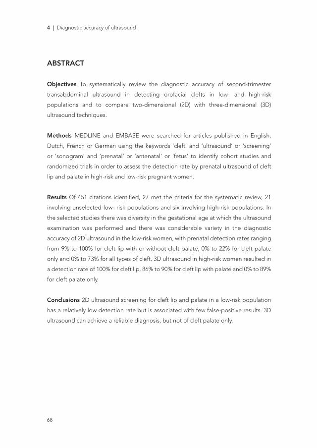
R1
R2
R3
R4
R5
R6
R7
R8
R9
R10
R11
R12
R13
R14
R15
R16
R17
R18
R19
R20
R21
R22
R23
R24
R25
R26
R27
R28
R29
R30
R31
R32
R33
R34
4 | Diagnostic accuracy of ultrasound
68
ABSTRACT
Objectives To systematically review the diagnostic accuracy of second-trimester
transabdominal ultrasound in detecting orofacial clefts in low- and high-risk
populations and to compare two-dimensional (2D) with three-dimensional (3D)
ultrasound techniques.
Methods MEDLINE and EMBASE were searched for articles published in English,
Dutch, French or German using the keywords ‘cleft’ and ‘ultrasound’ or ‘screening’
or ‘sonogram’ and ‘prenatal’ or ‘antenatal’ or ‘fetus’ to identify cohort studies and
randomized trials in order to assess the detection rate by prenatal ultrasound of cleft
lip and palate in high-risk and low-risk pregnant women.
Results Of 451 citations identified, 27 met the criteria for the systematic review, 21
involving unselected low- risk populations and six involving high-risk populations. In
the selected studies there was diversity in the gestational age at which the ultrasound
examination was performed and there was considerable variety in the diagnostic
accuracy of 2D ultrasound in the low-risk women, with prenatal detection rates ranging
from 9% to 100% for cleft lip with or without cleft palate, 0% to 22% for cleft palate
only and 0% to 73% for all types of cleft. 3D ultrasound in high-risk women resulted in
a detection rate of 100% for cleft lip, 86% to 90% for cleft lip with palate and 0% to 89%
for cleft palate only.
Conclusions 2D ultrasound screening for cleft lip and palate in a low-risk population
has a relatively low detection rate but is associated with few false-positive results. 3D
ultrasound can achieve a reliable diagnosis, but not of cleft palate only.
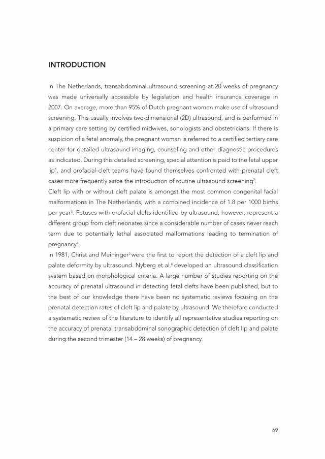
R1
R2
R3
R4
R5
R6
R7
R8
R9
R10
R11
R12
R13
R14
R15
R16
R17
R18
R19
R20
R21
R22
R23
R24
R25
R26
R27
R28
R29
R30
R31
R32
R33
R34
69
INTRODUCTION
In The Netherlands, transabdominal ultrasound screening at 20 weeks of pregnancy
was made universally accessible by legislation and health insurance coverage in
2007. On average, more than 95% of Dutch pregnant women make use of ultrasound
screening. This usually involves two-dimensional (2D) ultrasound, and is performed in
a primary care setting by certified midwives, sonologists and obstetricians. If there is
suspicion of a fetal anomaly, the pregnant woman is referred to a certified tertiary care
center for detailed ultrasound imaging, counseling and other diagnostic procedures
as indicated. During this detailed screening, special attention is paid to the fetal upper
lip1, and orofacial-cleft teams have found themselves confronted with prenatal cleft
cases more frequently since the introduction of routine ultrasound screening2.
Cleft lip with or without cleft palate is amongst the most common congenital facial
malformations in The Netherlands, with a combined incidence of 1.8 per 1000 births
per year3. Fetuses with orofacial clefts identified by ultrasound, however, represent a
different group from cleft neonates since a considerable number of cases never reach
term due to potentially lethal associated malformations leading to termination of
pregnancy4.
In 1981, Christ and Meininger5 were the first to report the detection of a cleft lip and
palate deformity by ultrasound. Nyberg et al.6 developed an ultrasound classification
system based on morphological criteria. A large number of studies reporting on the
accuracy of prenatal ultrasound in detecting fetal clefts have been published, but to
the best of our knowledge there have been no systematic reviews focusing on the
prenatal detection rates of cleft lip and palate by ultrasound. We therefore conducted
a systematic review of the literature to identify all representative studies reporting on
the accuracy of prenatal transabdominal sonographic detection of cleft lip and palate
during the second trimester (14 – 28 weeks) of pregnancy.
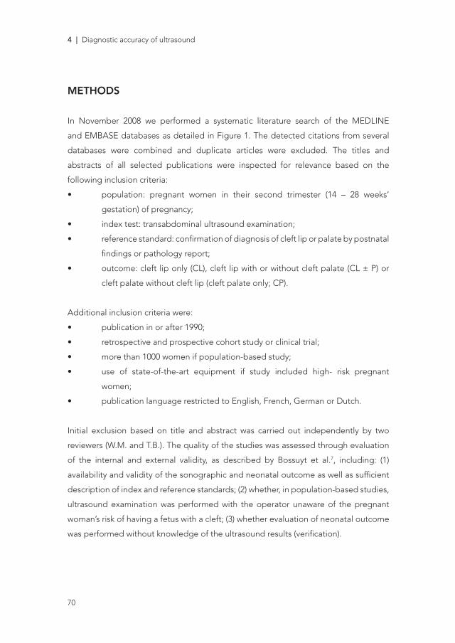
R1
R2
R3
R4
R5
R6
R7
R8
R9
R10
R11
R12
R13
R14
R15
R16
R17
R18
R19
R20
R21
R22
R23
R24
R25
R26
R27
R28
R29
R30
R31
R32
R33
R34
4 | Diagnostic accuracy of ultrasound
70
METHODS
In November 2008 we performed a systematic literature search of the MEDLINE
and EMBASE databases as detailed in Figure 1. The detected citations from several
databases were combined and duplicate articles were excluded. The titles and
abstracts of all selected publications were inspected for relevance based on the
following inclusion criteria:
• population: pregnant women in their second trimester (14 – 28 weeks’
gestation) of pregnancy;
• index test: transabdominal ultrasound examination;
• reference standard: confirmation of diagnosis of cleft lip or palate by postnatal
findings or pathology report;
• outcome: cleft lip only (CL), cleft lip with or without cleft palate (CL ± P) or
cleft palate without cleft lip (cleft palate only; CP).
Additional inclusion criteria were:
• publication in or after 1990;
• retrospective and prospective cohort study or clinical trial;
• more than 1000 women if population-based study;
• use of state-of-the-art equipment if study included high- risk pregnant
women;
• publication language restricted to English, French, German or Dutch.
Initial exclusion based on title and abstract was carried out independently by two
reviewers (W.M. and T.B.). The quality of the studies was assessed through evaluation
of the internal and external validity, as described by Bossuyt et al.7, including: (1)
availability and validity of the sonographic and neonatal outcome as well as sufficient
description of index and reference standards; (2) whether, in population-based studies,
ultrasound examination was performed with the operator unaware of the pregnant
woman’s risk of having a fetus with a cleft; (3) whether evaluation of neonatal outcome
was performed without knowledge of the ultrasound results (verification).
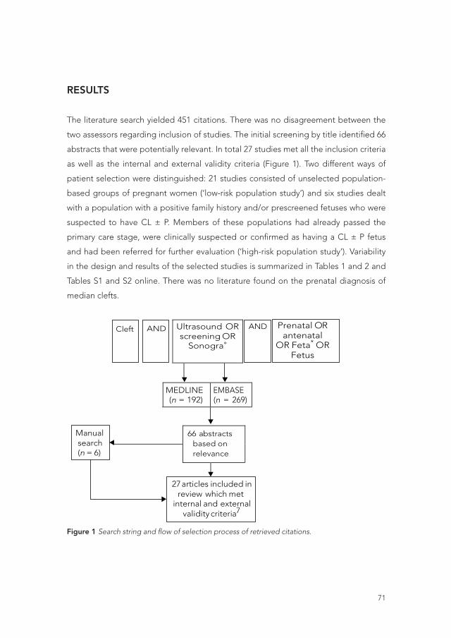
R1
R2
R3
R4
R5
R6
R7
R8
R9
R10
R11
R12
R13
R14
R15
R16
R17
R18
R19
R20
R21
R22
R23
R24
R25
R26
R27
R28
R29
R30
R31
R32
R33
R34
71
RESULTS
The literature search yielded 451 citations. There was no disagreement between the
two assessors regarding inclusion of studies. The initial screening by title identified 66
abstracts that were potentially relevant. In total 27 studies met all the inclusion criteria
as well as the internal and external validity criteria (Figure 1). Two different ways of
patient selection were distinguished: 21 studies consisted of unselected population-
based groups of pregnant women (‘low-risk population study’) and six studies dealt
with a population with a positive family history and/or prescreened fetuses who were
suspected to have CL ± P. Members of these populations had already passed the
primary care stage, were clinically suspected or confirmed as having a CL ± P fetus
and had been referred for further evaluation (‘high-risk population study’). Variability
in the design and results of the selected studies is summarized in Tables 1 and 2 and
Tables S1 and S2 online. There was no literature found on the prenatal diagnosis of
median clefts.
496 Maarse et al.
Copyright � 2010 ISUOG. Published by John Wiley & Sons, Ltd. Ultrasound Obstet Gynecol 2010; 35: 495 –502.
Ultrasound OR screening OR
Sonogra∗
66 abstracts based on relevance
detection rates of cleft lip and palate by ultrasound. We therefore conducted a systematic review of the literature to identify all representative studies reporting on the accuracy of prenatal transabdominal sonographic detection of cleft lip and palate during the second trimester (14 – 28 weeks) of pregnancy.
METHODS
In November 2008 we performed a systematic literature search of the MEDLINE and EMBASE databases as detailed in Figure 1. The detected citations from several databases were combined and duplicate articles were excluded. The titles and abstracts of all selected publications were inspected for relevance based on the following inclusion criteria:
• population: pregnant women in their second trimester
(14 – 28 weeks’ gestation) of pregnancy; • index test: transabdominal ultrasound examination; • reference standard: confirmation of diagnosis of cleft
lip or palate by postnatal findings or pathology report; • outcome: cleft lip only (CL), cleft lip with or without
cleft palate (CL ± P) or cleft palate without cleft lip (cleft palate only; CP).
Additional inclusion criteria were:
• publication in or after 1990; • retrospective and prospective cohort study or clinical
trial; • more than 1000 women if population-based study; • use of state-of-the-art equipment if study included high-
risk pregnant women; • publication language restricted to English, French,
German or Dutch.
Initial exclusion based on title and abstract was carried out independently by two reviewers (W.M. and T.B.). The quality of the studies was assessed through evaluation
of the internal and external validity, as described by Bossuyt et al.7, including: (1) availability and validity of the sonographic and neonatal outcome as well as sufficient description of index and reference standards; (2) whether, in population-based studies, ultrasound examination was performed with the operator unaware of the pregnant woman’s risk of having a fetus with a cleft; (3) whether evaluation of neonatal outcome was performed without knowledge of the ultrasound results (verification).
RESULTS
The literature search yielded 451 citations. There was no disagreement between the two assessors regarding inclusion of studies. The initial screening by title identified 66 abstracts that were potentially relevant. In total 27 studies met all the inclusion criteria as well as the internal and external validity criteria (Figure 1). Two different ways of patient selection were distinguished: 21 studies consisted of unselected population-based groups of pregnant women (‘low-risk population study’) and six studies dealt with a population with a positive family history and/or prescreened fetuses who were suspected to have CL ± P. Members of these populations had already passed the primary care stage, were clinically suspected or confirmed as having a CL ± P fetus and had been referred for further evaluation (‘high- risk population study’). Variability in the design and results of the selected studies is summarized in Tables 1 and 2 and Tables S1 and S2 online. There was no literature found on the prenatal diagnosis of median clefts.
Low-risk populations
Of 21 low-risk population studies (Table 1), all were cohort studies, seven8–14 being retrospective and 1415 – 28
being prospective. The retrospective cohort studies reported a large variety in the diagnostic accuracy of 2D ultrasound, with detection rates ranging from
Manual search (n = 6)
Cleft AND AND
MEDLINE (n = 192)
EMBASE (n = 269)
27 articles included in review which met
internal and external validity criteria7
Prenatal OR antenatal
OR Feta* OR Fetus
0%11 to 70%13 for prenatal detection of all types of cleft, 33%10 to 88%9 for prenatal detection of CL ± P and 0%8,10,12,13 to 22%9 for detection of CP. In prospective cohort studies, detection ranged from 9%17
to 73%23 for prenatal detection of all types of cleft, 9%21 to 100%26 for prenatal detection of CL ± P and 0%17,22,24,28 to 7%16 for detection of CP. In several studies, all prenatal diagnosis of CL ± P was confirmed following delivery or postmortem, indicating no false- positive diagnoses9,10,13,15,18,19,23,26,27 or high specificity when there was complete follow-up of all negative ultrasound diagnosis21,22,24.
The two most recent prospective cohort studies22,24
evaluated a large study population to assess the accuracy of detecting facial clefts in a low-risk population. Russell et al.24 assessed 108 220 births in the period from 1997
Figure 1 Search string and flow of selection process of retrieved citations.
to 2002, during which prenatal ultrasound at 18 to 20 weeks was available to all pregnant women (2D
Figure 1 Search string and flow of selection process of retrieved citations.
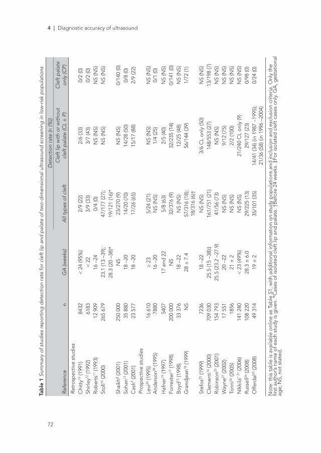
R1
R2
R3
R4
R5
R6
R7
R8
R9
R10
R11
R12
R13
R14
R15
R16
R17
R18
R19
R20
R21
R22
R23
R24
R25
R26
R27
R28
R29
R30
R31
R32
R33
R34
4 | Diagnostic accuracy of ultrasound
72
Tab
le 1
Sum
mar
y of
stu
die
s re
por
ting
det
ectio
n ra
te fo
r cle
ft li
p a
nd p
alat
e of
two-
dim
ensi
onal
ultr
asou
nd s
cree
ning
in lo
w-r
isk
pop
ulat
ions
Det
ectio
n ra
te (n
(%))
Refe
renc
en
GA
(wee
ks)
All
typ
es o
f cle
ftC
left
lip
with
or w
ithou
t cl
eft p
alat
e (C
L ±
P)
Cle
ft p
alat
e on
ly (C
P)
Retr
osp
ectiv
e st
udie
sC
hitt
y10 (1
991)
8432
< 2
4 (9
5%)
2/9
(22)
2/6
(33)
0/2
(0)
Shirl
ey12
(199
2)61
83<
22
3/9
(33)
3/7
(43)
0/2
(0)
Ro
ber
ts11
(199
3)12
909
16 –
240/
4 (0
)N
S (N
S)N
S (N
S)St
oll14
(200
0)26
5 67
923
.1 (1
3 –3
9);
47/1
77 (2
7);
NS
(NS)
NS
(NS)
28.3
(20
–38)
*19
/121
(16)
*Sh
aikh
8 (20
01)
250
000
NS
23/2
70 (9
)N
S (N
S)0/
140
(0)
Soha
n13 (2
001)
35 8
8018
–20
14/2
0 (7
0)14
/28
(50)
0/8
(0)
Cas
h9 (20
01)
23 5
7718
–20
17/2
6 (6
5)15
/17
(88)
2/9
(22)
Pros
pec
tive
stud
ies
Levi
20 (1
995)
And
erso
n28 (1
995)
16 6
1078
80≥
2316
–20
5/24
(21)
N
S (N
S)N
S (N
S)
1/4
(25)
NS
(NS)
0/1
(0)
Haf
ner19
(199
7)54
0717
and
22
5/8
(63)
2/5
(40)
NS
(NS)
Forr
este
r17 (1
998)
200
000
NS
32/3
76 (9
)32
/235
(14)
0/14
1 (0
)B
oyd
15 (1
998)
33 3
7618
–22
NS
(NS)
12/2
5 (4
8)N
S (N
S)G
rand
jean
18 (1
999)
NS
28 ±
7.4
57/3
16 (1
8);
18/3
16 (6
)†56
/144
(39)
1/72
(1)
Stef
os25
(199
9)72
3618
–22
NS
(NS)
3/6
CL
onl
y (5
0)N
S (N
S)C
lem
enti16
(200
0)70
9 03
025
.5 (1
5 –3
8)‡
161/
751
(21)
148/
553
(27)
13/1
98 (7
)R
ob
inso
n23 (2
001)
154
793
25.5
(23.
2 –2
7.9)
41/5
6 (7
3)N
S (N
S)N
S (N
S)W
ayne
27 (2
002)
17 5
5120
–22
NS
(NS)
9/12
(75)
NS
(NS)
Tonn
i26 (2
005)
1856
21 ±
2N
S (N
S)2/
2 (1
00)
NS
(NS)
Nik
kila
¨ 21 (2
006)
141
240
< 2
3 (4
9%)
NS
(NS)
21/2
40 C
L o
nly
(9)
NS
(NS)
Rus
sell24
(200
8)10
8 22
028
.3 ±
6.0
29/2
25 (1
3)29
/127
(23)
0/98
(0)
Offe
rdal
22 (2
008)
49 3
1419
± 2
35/1
01 (3
5)14
/41
(34)
(in
1987
–19
95);
21/3
6 (5
8) (i
n 19
96 –
2004
)0/
24 (0
)
No
te: t
his
tab
le is
ava
ilab
le o
nlin
e as
Tab
le S
1, w
ith a
dd
itio
nal i
nfo
rmat
ion
on
stud
y p
op
ulat
ions
and
incl
usio
n an
d e
xclu
sio
n cr
iteria
. Onl
y th
e fir
st a
utho
r’s n
ame
of e
ach
stud
y is
giv
en. *
Cas
es o
f iso
late
d c
left
lip
and
pal
ate.
†B
efo
re 2
4 w
eeks
. ‡Fo
r is
ola
ted
cle
ft c
ases
onl
y. G
A, g
esta
tiona
l ag
e; N
S, n
ot
stat
ed.
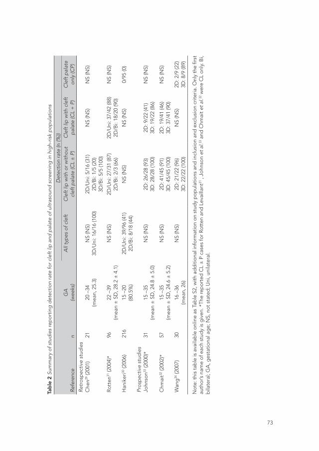
R1
R2
R3
R4
R5
R6
R7
R8
R9
R10
R11
R12
R13
R14
R15
R16
R17
R18
R19
R20
R21
R22
R23
R24
R25
R26
R27
R28
R29
R30
R31
R32
R33
R34
73
Tab
le 2
Sum
mar
y of
stu
die
s re
por
ting
det
ectio
n ra
te fo
r cle
ft li
p a
nd p
alat
e of
ultr
asou
nd s
cree
ning
in h
igh-
risk
pop
ulat
ions
Det
ectio
n ra
te (n
(%))
Refe
renc
en
GA
(wee
ks)
All
typ
es o
f cle
ftC
left
lip
with
or w
ithou
t cl
eft p
alat
e (C
L ±
P)
Cle
ft li
p w
ith c
left
p
alat
e (C
L +
P)
Cle
ft p
alat
e on
ly (C
P)
Retr
osp
ectiv
e st
udie
sC
hen29
(200
1)21
20 –
34(m
ean,
25.
3)N
S (N
S)3D
/Uni
: 16/
16 (1
00)
2D/U
ni: 5
/16
(31)
2D/B
i: 1/
5 (2
0)3D
/Bi:
5/5
(100
)
NS
(NS)
NS
(NS)
Ro
tten
31 (2
004)
*96
22 –
39(m
ean
± S
D, 2
8.2
± 4
.1)
NS
(NS)
2D/U
ni: 2
7/31
(87)
2D/B
i: 2/
3 (6
6)2D
/Uni
: 37/
42 (8
8)2D
/Bi:
18/2
0 (9
0)N
S (N
S)
Han
iker
i30 (2
006)
216
15 –
20(8
0.5%
)2D
/Uni
: 39/
96 (4
1)2D
/Bi:
8/18
(44)
NS
(NS)
NS
(NS)
0/95
(0)
Pros
pec
tive
stud
ies
John
son33
(200
0)*
3115
–35
(mea
n ±
SD
, 24.
8 ±
5.0
)N
S (N
S)2D
: 26/
28 (9
3)3D
: 28/
28 (1
00)
2D: 9
/22
(41)
3D: 1
9/22
(86)
NS
(NS)
Chm
ait32
(200
2)*
5715
–35
(mea
n ±
SD
, 24.
6 ±
5.2
)N
S (N
S)2D
: 41/
45 (9
1)3D
: 45/
45 (1
00)
2D: 1
9/41
(46)
3D: 3
7/41
(90)
NS
(NS)
Wan
g34
(200
7)30
16 –
36(m
ean,
26)
NS
(NS)
2D: 2
1/22
(96)
3D: 2
2/22
(100
)N
S (N
S)2D
: 2/9
(22)
3D: 8
/9 (8
9)
No
te: t
his
tab
le is
ava
ilab
le o
nlin
e as
Tab
le S
2, w
ith a
dd
itio
nal i
nfo
rmat
ion
on
stud
y p
op
ulat
ions
and
incl
usio
n an
d e
xclu
sio
n cr
iteria
. Onl
y th
e fir
st
auth
or’s
nam
e o
f eac
h st
udy
is g
iven
. *Th
e re
po
rted
CL
± P
cas
es fo
r Ro
tten
and
Lev
ailla
nt31
, Jo
hnso
n et
al.33
and
Chm
ait e
t al.32
wer
e C
L o
nly.
Bi,
bila
tera
l; G
A, g
esta
tiona
l ag
e; N
S, n
ot
stat
ed; U
ni, u
nila
tera
l.
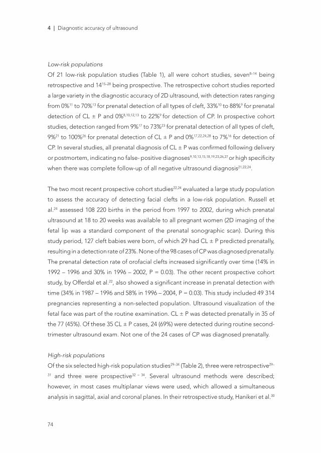
R1
R2
R3
R4
R5
R6
R7
R8
R9
R10
R11
R12
R13
R14
R15
R16
R17
R18
R19
R20
R21
R22
R23
R24
R25
R26
R27
R28
R29
R30
R31
R32
R33
R34
4 | Diagnostic accuracy of ultrasound
74
Low-risk populations
Of 21 low-risk population studies (Table 1), all were cohort studies, seven8–14 being
retrospective and 1415–28 being prospective. The retrospective cohort studies reported
a large variety in the diagnostic accuracy of 2D ultrasound, with detection rates ranging
from 0%11 to 70%13 for prenatal detection of all types of cleft, 33%10 to 88%9 for prenatal
detection of CL ± P and 0%8,10,12,13 to 22%9 for detection of CP. In prospective cohort
studies, detection ranged from 9%17 to 73%23 for prenatal detection of all types of cleft,
9%21 to 100%26 for prenatal detection of CL ± P and 0%17,22,24,28 to 7%16 for detection of
CP. In several studies, all prenatal diagnosis of CL ± P was confirmed following delivery
or postmortem, indicating no false- positive diagnoses9,10,13,15,18,19,23,26,27 or high specificity
when there was complete follow-up of all negative ultrasound diagnosis21,22,24.
The two most recent prospective cohort studies22,24 evaluated a large study population
to assess the accuracy of detecting facial clefts in a low-risk population. Russell et
al.24 assessed 108 220 births in the period from 1997 to 2002, during which prenatal
ultrasound at 18 to 20 weeks was available to all pregnant women (2D imaging of the
fetal lip was a standard component of the prenatal sonographic scan). During this
study period, 127 cleft babies were born, of which 29 had CL ± P predicted prenatally,
resulting in a detection rate of 23%. None of the 98 cases of CP was diagnosed prenatally.
The prenatal detection rate of orofacial clefts increased significantly over time (14% in
1992 – 1996 and 30% in 1996 – 2002, P = 0.03). The other recent prospective cohort
study, by Offerdal et al.22, also showed a significant increase in prenatal detection with
time (34% in 1987 – 1996 and 58% in 1996 – 2004, P = 0.03). This study included 49 314
pregnancies representing a non-selected population. Ultrasound visualization of the
fetal face was part of the routine examination. CL ± P was detected prenatally in 35 of
the 77 (45%). Of these 35 CL ± P cases, 24 (69%) were detected during routine second-
trimester ultrasound exam. Not one of the 24 cases of CP was diagnosed prenatally.
High-risk populations
Of the six selected high-risk population studies29–34 (Table 2), three were retrospective29–
31 and three were prospective32 – 34. Several ultrasound methods were described;
however, in most cases multiplanar views were used, which allowed a simultaneous
analysis in sagittal, axial and coronal planes. In their retrospective study, Hanikeri et al.30
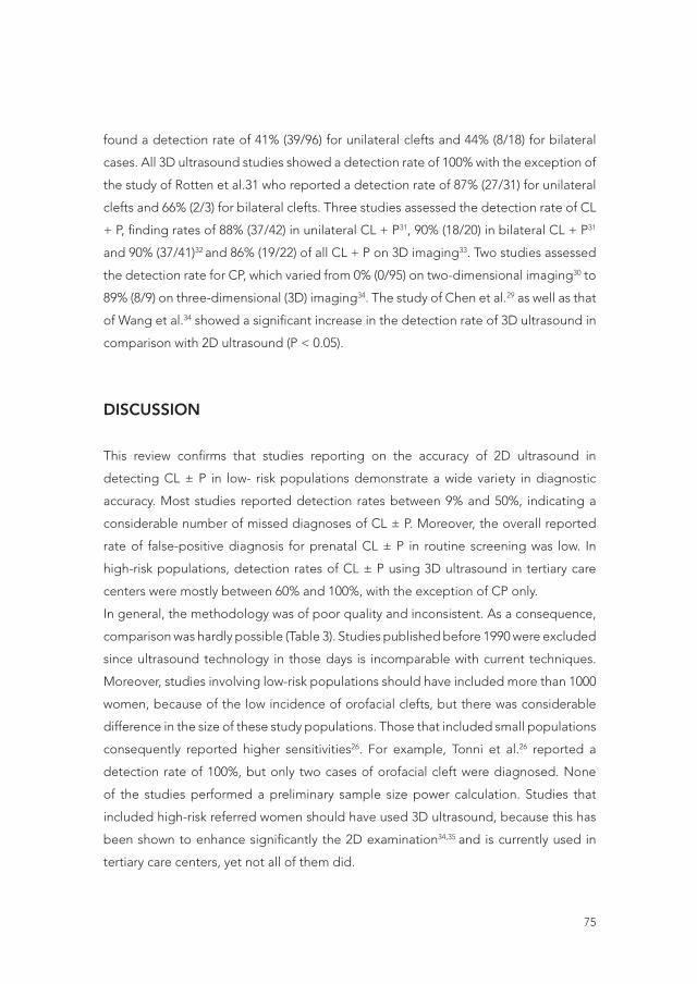
R1
R2
R3
R4
R5
R6
R7
R8
R9
R10
R11
R12
R13
R14
R15
R16
R17
R18
R19
R20
R21
R22
R23
R24
R25
R26
R27
R28
R29
R30
R31
R32
R33
R34
75
found a detection rate of 41% (39/96) for unilateral clefts and 44% (8/18) for bilateral
cases. All 3D ultrasound studies showed a detection rate of 100% with the exception of
the study of Rotten et al.31 who reported a detection rate of 87% (27/31) for unilateral
clefts and 66% (2/3) for bilateral clefts. Three studies assessed the detection rate of CL
+ P, finding rates of 88% (37/42) in unilateral CL + P31, 90% (18/20) in bilateral CL + P31
and 90% (37/41)32 and 86% (19/22) of all CL + P on 3D imaging33. Two studies assessed
the detection rate for CP, which varied from 0% (0/95) on two-dimensional imaging30 to
89% (8/9) on three-dimensional (3D) imaging34. The study of Chen et al.29 as well as that
of Wang et al.34 showed a significant increase in the detection rate of 3D ultrasound in
comparison with 2D ultrasound (P < 0.05).
DISCUSSION
This review confirms that studies reporting on the accuracy of 2D ultrasound in
detecting CL ± P in low- risk populations demonstrate a wide variety in diagnostic
accuracy. Most studies reported detection rates between 9% and 50%, indicating a
considerable number of missed diagnoses of CL ± P. Moreover, the overall reported
rate of false-positive diagnosis for prenatal CL ± P in routine screening was low. In
high-risk populations, detection rates of CL ± P using 3D ultrasound in tertiary care
centers were mostly between 60% and 100%, with the exception of CP only.
In general, the methodology was of poor quality and inconsistent. As a consequence,
comparison was hardly possible (Table 3). Studies published before 1990 were excluded
since ultrasound technology in those days is incomparable with current techniques.
Moreover, studies involving low-risk populations should have included more than 1000
women, because of the low incidence of orofacial clefts, but there was considerable
difference in the size of these study populations. Those that included small populations
consequently reported higher sensitivities26. For example, Tonni et al.26 reported a
detection rate of 100%, but only two cases of orofacial cleft were diagnosed. None
of the studies performed a preliminary sample size power calculation. Studies that
included high-risk referred women should have used 3D ultrasound, because this has
been shown to enhance significantly the 2D examination34,35 and is currently used in
tertiary care centers, yet not all of them did.
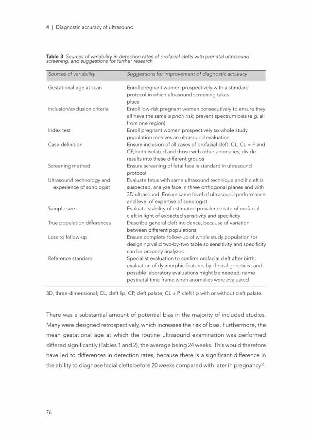
R1
R2
R3
R4
R5
R6
R7
R8
R9
R10
R11
R12
R13
R14
R15
R16
R17
R18
R19
R20
R21
R22
R23
R24
R25
R26
R27
R28
R29
R30
R31
R32
R33
R34
4 | Diagnostic accuracy of ultrasound
76
Table 3 Sources of variability in detection rates of orofacial clefts with prenatal ultrasound screening, and suggestions for further research
Sources of variability Suggestions for improvement of diagnostic accuracy
Gestational age at scan Enroll pregnant women prospectively with a standard protocol in which ultrasound screening takes placeInclusion/exclusion criteria Enroll low-risk pregnant women consecutively to ensure they all have the same a priori risk; prevent spectrum bias (e.g. all from one region)Index test Enroll pregnant women prospectively so whole study population receives an ultrasound evaluation Case definition Ensure inclusion of all cases of orofacial cleft: CL, CL ± P and CP, both isolated and those with other anomalies; divide results into these different groupsScreening method Ensure screening of fetal face is standard in ultrasound protocolUltrasound technology and Evaluate fetus with same ultrasound technique and if cleft is experience of sonologist suspected, analyze face in three orthogonal planes and with 3D ultrasound. Ensure same level of ultrasound performance and level of expertise of sonologistSample size Evaluate stability of estimated prevalence rate of orofacial cleft in light of expected sensitivity and specificityTrue population differences Describe general cleft incidence, because of variation between different populationsLoss to follow-up Ensure complete follow-up of whole study population for designing valid two-by-two table so sensitivity and specificity can be properly analyzedReference standard Specialist evaluation to confirm orofacial cleft after birth; evaluation of dysmorphic features by clinical geneticist and possible laboratory evaluations might be needed; name postnatal time frame when anomalies were evaluated
3D, three-dimensional; CL, cleft lip; CP, cleft palate; CL ± P, cleft lip with or without cleft palate.
There was a substantial amount of potential bias in the majority of included studies.
Many were designed retrospectively, which increases the risk of bias. Furthermore, the
mean gestational age at which the routine ultrasound examination was performed
differed significantly (Tables 1 and 2), the average being 24 weeks. This would therefore
have led to differences in detection rates, because there is a significant difference in
the ability to diagnose facial clefts before 20 weeks compared with later in pregnancy36.
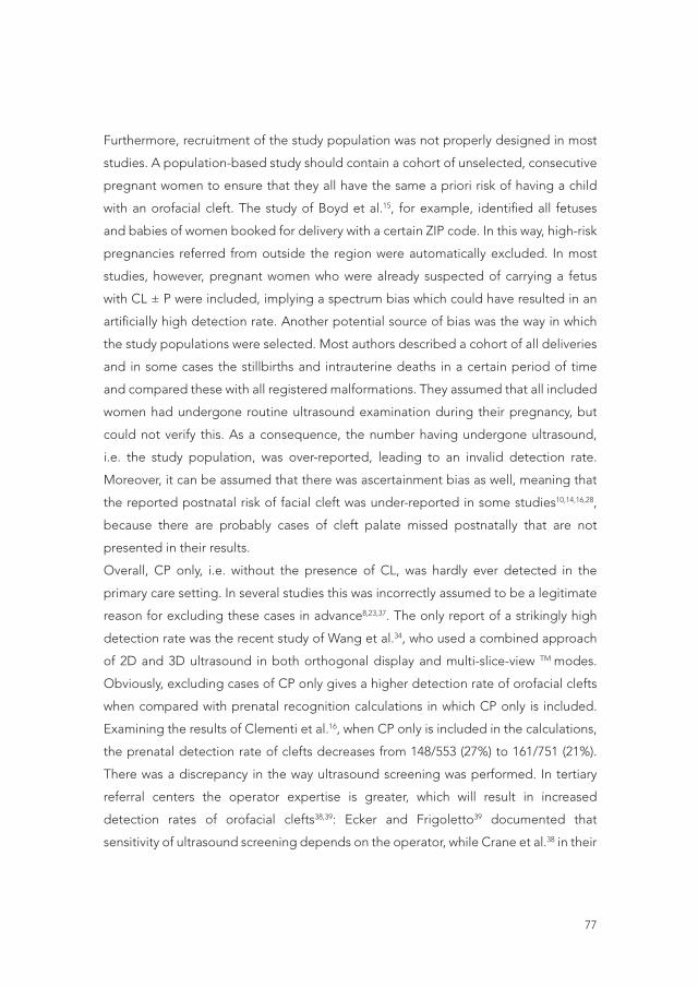
R1
R2
R3
R4
R5
R6
R7
R8
R9
R10
R11
R12
R13
R14
R15
R16
R17
R18
R19
R20
R21
R22
R23
R24
R25
R26
R27
R28
R29
R30
R31
R32
R33
R34
77
Furthermore, recruitment of the study population was not properly designed in most
studies. A population-based study should contain a cohort of unselected, consecutive
pregnant women to ensure that they all have the same a priori risk of having a child
with an orofacial cleft. The study of Boyd et al.15, for example, identified all fetuses
and babies of women booked for delivery with a certain ZIP code. In this way, high-risk
pregnancies referred from outside the region were automatically excluded. In most
studies, however, pregnant women who were already suspected of carrying a fetus
with CL ± P were included, implying a spectrum bias which could have resulted in an
artificially high detection rate. Another potential source of bias was the way in which
the study populations were selected. Most authors described a cohort of all deliveries
and in some cases the stillbirths and intrauterine deaths in a certain period of time
and compared these with all registered malformations. They assumed that all included
women had undergone routine ultrasound examination during their pregnancy, but
could not verify this. As a consequence, the number having undergone ultrasound,
i.e. the study population, was over-reported, leading to an invalid detection rate.
Moreover, it can be assumed that there was ascertainment bias as well, meaning that
the reported postnatal risk of facial cleft was under-reported in some studies10,14,16,28,
because there are probably cases of cleft palate missed postnatally that are not
presented in their results.
Overall, CP only, i.e. without the presence of CL, was hardly ever detected in the
primary care setting. In several studies this was incorrectly assumed to be a legitimate
reason for excluding these cases in advance8,23,37. The only report of a strikingly high
detection rate was the recent study of Wang et al.34, who used a combined approach
of 2D and 3D ultrasound in both orthogonal display and multi-slice-view TM modes.
Obviously, excluding cases of CP only gives a higher detection rate of orofacial clefts
when compared with prenatal recognition calculations in which CP only is included.
Examining the results of Clementi et al.16, when CP only is included in the calculations,
the prenatal detection rate of clefts decreases from 148/553 (27%) to 161/751 (21%).
There was a discrepancy in the way ultrasound screening was performed. In tertiary
referral centers the operator expertise is greater, which will result in increased
detection rates of orofacial clefts38,39: Ecker and Frigoletto39 documented that
sensitivity of ultrasound screening depends on the operator, while Crane et al.38 in their
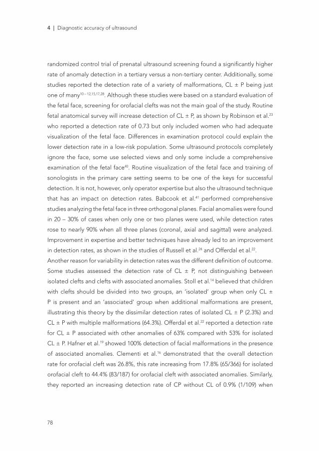
R1
R2
R3
R4
R5
R6
R7
R8
R9
R10
R11
R12
R13
R14
R15
R16
R17
R18
R19
R20
R21
R22
R23
R24
R25
R26
R27
R28
R29
R30
R31
R32
R33
R34
4 | Diagnostic accuracy of ultrasound
78
randomized control trial of prenatal ultrasound screening found a significantly higher
rate of anomaly detection in a tertiary versus a non-tertiary center. Additionally, some
studies reported the detection rate of a variety of malformations, CL ± P being just
one of many10 – 12,15,17,28. Although these studies were based on a standard evaluation of
the fetal face, screening for orofacial clefts was not the main goal of the study. Routine
fetal anatomical survey will increase detection of CL ± P, as shown by Robinson et al.23
who reported a detection rate of 0.73 but only included women who had adequate
visualization of the fetal face. Differences in examination protocol could explain the
lower detection rate in a low-risk population. Some ultrasound protocols completely
ignore the face, some use selected views and only some include a comprehensive
examination of the fetal face40. Routine visualization of the fetal face and training of
sonologists in the primary care setting seems to be one of the keys for successful
detection. It is not, however, only operator expertise but also the ultrasound technique
that has an impact on detection rates. Babcook et al.41 performed comprehensive
studies analyzing the fetal face in three orthogonal planes. Facial anomalies were found
in 20 – 30% of cases when only one or two planes were used, while detection rates
rose to nearly 90% when all three planes (coronal, axial and sagittal) were analyzed.
Improvement in expertise and better techniques have already led to an improvement
in detection rates, as shown in the studies of Russell et al.24 and Offerdal et al.22.
Another reason for variability in detection rates was the different definition of outcome.
Some studies assessed the detection rate of CL ± P, not distinguishing between
isolated clefts and clefts with associated anomalies. Stoll et al.14 believed that children
with clefts should be divided into two groups, an ‘isolated’ group when only CL ±
P is present and an ‘associated’ group when additional malformations are present,
illustrating this theory by the dissimilar detection rates of isolated CL ± P (2.3%) and
CL ± P with multiple malformations (64.3%). Offerdal et al.22 reported a detection rate
for CL ± P associated with other anomalies of 63% compared with 53% for isolated
CL ± P. Hafner et al.19 showed 100% detection of facial malformations in the presence
of associated anomalies. Clementi et al.16 demonstrated that the overall detection
rate for orofacial cleft was 26.8%, this rate increasing from 17.8% (65/366) for isolated
orofacial cleft to 44.4% (83/187) for orofacial cleft with associated anomalies. Similarly,
they reported an increasing detection rate of CP without CL of 0.9% (1/109) when
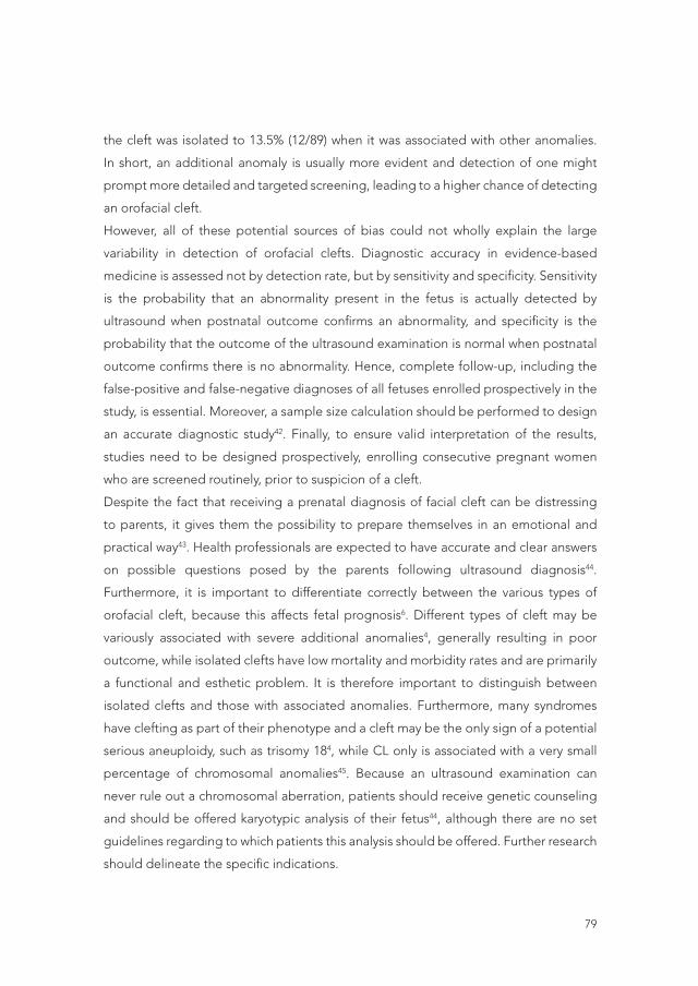
R1
R2
R3
R4
R5
R6
R7
R8
R9
R10
R11
R12
R13
R14
R15
R16
R17
R18
R19
R20
R21
R22
R23
R24
R25
R26
R27
R28
R29
R30
R31
R32
R33
R34
79
the cleft was isolated to 13.5% (12/89) when it was associated with other anomalies.
In short, an additional anomaly is usually more evident and detection of one might
prompt more detailed and targeted screening, leading to a higher chance of detecting
an orofacial cleft.
However, all of these potential sources of bias could not wholly explain the large
variability in detection of orofacial clefts. Diagnostic accuracy in evidence-based
medicine is assessed not by detection rate, but by sensitivity and specificity. Sensitivity
is the probability that an abnormality present in the fetus is actually detected by
ultrasound when postnatal outcome confirms an abnormality, and specificity is the
probability that the outcome of the ultrasound examination is normal when postnatal
outcome confirms there is no abnormality. Hence, complete follow-up, including the
false-positive and false-negative diagnoses of all fetuses enrolled prospectively in the
study, is essential. Moreover, a sample size calculation should be performed to design
an accurate diagnostic study42. Finally, to ensure valid interpretation of the results,
studies need to be designed prospectively, enrolling consecutive pregnant women
who are screened routinely, prior to suspicion of a cleft.
Despite the fact that receiving a prenatal diagnosis of facial cleft can be distressing
to parents, it gives them the possibility to prepare themselves in an emotional and
practical way43. Health professionals are expected to have accurate and clear answers
on possible questions posed by the parents following ultrasound diagnosis44.
Furthermore, it is important to differentiate correctly between the various types of
orofacial cleft, because this affects fetal prognosis6. Different types of cleft may be
variously associated with severe additional anomalies4, generally resulting in poor
outcome, while isolated clefts have low mortality and morbidity rates and are primarily
a functional and esthetic problem. It is therefore important to distinguish between
isolated clefts and those with associated anomalies. Furthermore, many syndromes
have clefting as part of their phenotype and a cleft may be the only sign of a potential
serious aneuploidy, such as trisomy 184, while CL only is associated with a very small
percentage of chromosomal anomalies45. Because an ultrasound examination can
never rule out a chromosomal aberration, patients should receive genetic counseling
and should be offered karyotypic analysis of their fetus44, although there are no set
guidelines regarding to which patients this analysis should be offered. Further research
should delineate the specific indications.
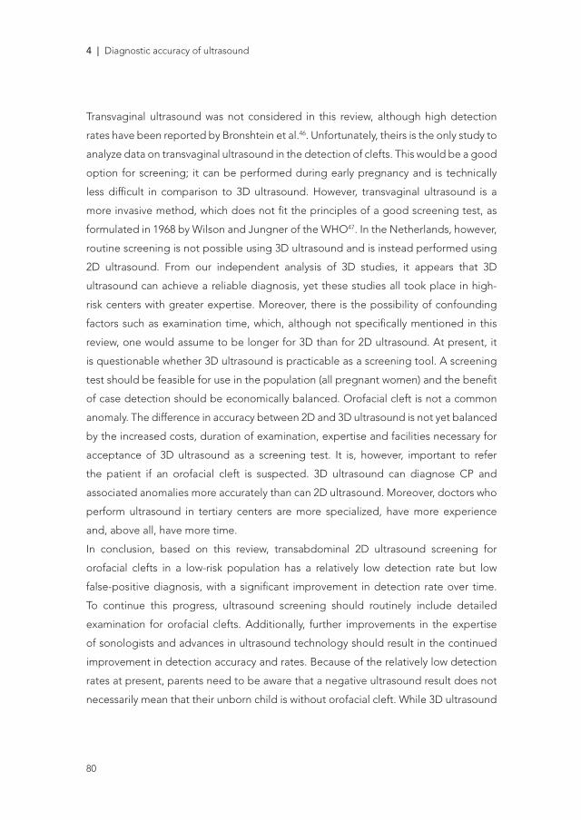
R1
R2
R3
R4
R5
R6
R7
R8
R9
R10
R11
R12
R13
R14
R15
R16
R17
R18
R19
R20
R21
R22
R23
R24
R25
R26
R27
R28
R29
R30
R31
R32
R33
R34
4 | Diagnostic accuracy of ultrasound
80
Transvaginal ultrasound was not considered in this review, although high detection
rates have been reported by Bronshtein et al.46. Unfortunately, theirs is the only study to
analyze data on transvaginal ultrasound in the detection of clefts. This would be a good
option for screening; it can be performed during early pregnancy and is technically
less difficult in comparison to 3D ultrasound. However, transvaginal ultrasound is a
more invasive method, which does not fit the principles of a good screening test, as
formulated in 1968 by Wilson and Jungner of the WHO47. In the Netherlands, however,
routine screening is not possible using 3D ultrasound and is instead performed using
2D ultrasound. From our independent analysis of 3D studies, it appears that 3D
ultrasound can achieve a reliable diagnosis, yet these studies all took place in high-
risk centers with greater expertise. Moreover, there is the possibility of confounding
factors such as examination time, which, although not specifically mentioned in this
review, one would assume to be longer for 3D than for 2D ultrasound. At present, it
is questionable whether 3D ultrasound is practicable as a screening tool. A screening
test should be feasible for use in the population (all pregnant women) and the benefit
of case detection should be economically balanced. Orofacial cleft is not a common
anomaly. The difference in accuracy between 2D and 3D ultrasound is not yet balanced
by the increased costs, duration of examination, expertise and facilities necessary for
acceptance of 3D ultrasound as a screening test. It is, however, important to refer
the patient if an orofacial cleft is suspected. 3D ultrasound can diagnose CP and
associated anomalies more accurately than can 2D ultrasound. Moreover, doctors who
perform ultrasound in tertiary centers are more specialized, have more experience
and, above all, have more time.
In conclusion, based on this review, transabdominal 2D ultrasound screening for
orofacial clefts in a low-risk population has a relatively low detection rate but low
false-positive diagnosis, with a significant improvement in detection rate over time.
To continue this progress, ultrasound screening should routinely include detailed
examination for orofacial clefts. Additionally, further improvements in the expertise
of sonologists and advances in ultrasound technology should result in the continued
improvement in detection accuracy and rates. Because of the relatively low detection
rates at present, parents need to be aware that a negative ultrasound result does not
necessarily mean that their unborn child is without orofacial cleft. While 3D ultrasound
R1
R2
R3
R4
R5
R6
R7
R8
R9
R10
R11
R12
R13
R14
R15
R16
R17
R18
R19
R20
R21
R22
R23
R24
R25
R26
R27
R28
R29
R30
R31
R32
R33
R34
81
can achieve a reliable diagnosis of fetal CL ± P, this does not rule out cases of CP
only. It is therefore important to refer pregnant women to a tertiary care center when
an orofacial cleft is suspected in the primary care setting. Finally, 3D ultrasound can
provide a more precise image of the defect, allowing parents to produce realistic
expectations.
R1
R2
R3
R4
R5
R6
R7
R8
R9
R10
R11
R12
R13
R14
R15
R16
R17
R18
R19
R20
R21
R22
R23
R24
R25
R26
R27
R28
R29
R30
R31
R32
R33
R34
4 | Diagnostic accuracy of ultrasound
82
REFERENCES
1. NVOG, modelprotocol SEO. http://www.nvog-documenten. nl/index.php?pagina=/richtlijin/item/pagina.php&richtlijn id=693 [Accessed 5 April 2009].
2. Mink van der Molen AB. Een kleine imperfectie: geïsoleerde schisis geen reden voor abortus. Medisch Contact 2007; 62: 1116 –1119 [article in Dutch].
3. Vermey-Keers C. NVSCA (Dutch Society of Orofacial Cleft and Craniofacial Malformations) registration, 2009.
4. Berge SJ, Plath H, Van de Vondel, Appel T, Niederhagen B, Von5. Lindern JJ, Reich RH, Hansmann M. Fetal cleft lip and palate: sonographic diagnosis,
chromosomal abnormalities, associated anomalies and postnatal outcome in 70 fetuses. Ultrasound Obstet Gynecol 2001; 18: 422 –431.
6. Christ JE, Meininger MG. Ultrasound diagnosis of cleft lip and cleft palate before birth. Plast Reconstr Surg 1981; 68: 854 –859.
7. Nyberg DA, Sickler GK, Hegge FN, Kramer DJ, Kropp RJ. Fetal cleft lip with and without cleft palate: US classification and correlation with outcome. Radiology 1995; 195: 677 –684.
8. Bossuyt PM, Reitsma JB, Bruns DE, Gatsonis CA, Glasziou PP, Irwig LM, Moher D, Rennie D, de Vet HC, Lijmer JG; Standards for Reporting of Diagnostic Accuracy. The STARD statement for reporting studies of diagnostic accuracy: explanation and elaboration. Ann Intern Med 2003; 138: W1 –W12.
9. Shaikh D, Mercer NS, Sohan K, Kyle P, Soothill P. Prenatal diagnosis of cleft lip and palate. Br J Plast Surg 2001; 54: 288 –289.
10. Cash C, Set P, Coleman N. The accuracy of antenatal ultra- sound in the detection of facial clefts in a low-risk screening population. Ultrasound Obstet Gynecol 2001; 18: 432 –436.
11. Chitty LS, Hunt GH, Moore J, Lobb MO. Effectiveness of rou- tine ultrasonography in detecting fetal structural abnormalities in a low risk population. BMJ 1991; 303: 1165 –1169.
12. Roberts AB, Hampton E, Wilson N. Ultrasound detection of fetal structural abnormalities in Auckland 1988 –9. N Z Med J 1993; 106: 441 –443.
13. Shirley IM, Bottomley F, Robinson VP. Routine radiographer screening for fetal abnormalities by ultrasound in an unselected low risk population. Br J Radiol 1992; 65: 564 –569.
14. Sohan K, Freer M, Mercer N, Soothill P, Kyle P. Prenatal detection of facial clefts. Fetal Diagn Ther 2001; 16: 196 –199.
15. Stoll C, Dott B, Alembik Y, Roth M. Evaluation of prenatal diagnosis of cleft lip/palate by foetal ultrasonographic examination. Ann Genet 2000; 43: 11 –14.
16. Boyd PA, Chamberlain P, Hicks NR. 6-year experience of prenatal diagnosis in an unselected population in Oxford, UK. Lancet 1998; 352: 1577 –1581.
17. Clementi M, Tenconi R, Bianchi F, Stoll C. Evaluation of prenatal diagnosis of cleft lip with or without cleft palate and cleft palate by ultrasound: experience from 20 European registries. EUROSCAN study group. Prenat Diagn 2000; 20: 870 –875.
18. Forrester MB, Merz RD, Yoon PW. Impact of prenatal diagnosis and elective termination on the prevalence of selected birth defects in Hawaii. Am J Epidemiol 1998; 148: 1206 –1211.
19. Grandjean H, Larroque D, Levi S. The performance of routine ultrasonographic screening of pregnancies in the Eurofetus Study. Am J Obstet Gynecol 1999; 181: 446 –454.
20. Hafner E, Sterniste W, Scholler J, Schuchter K, Philipp K. Prenatal diagnosis of facial malformations. Prenat Diagn 1997; 17: 51 –58.
21. Levi S, Schaaps JP, De HP, Coulon R, Defoort P. End-result of routine ultrasound screening for congenital anomalies: the Belgian Multicentric Study 1984 –92. Ultrasound Obstet Gynecol 1995; 5: 366 –371.
22. Nikkila A, Rydhstroem H, Kallen B, Jorgensen C. Ultrasound screening for fetal anomalies in southern Sweden: a population- based study. Acta Obstet Gynecol Scand 2006; 85: 688 –693.
R1
R2
R3
R4
R5
R6
R7
R8
R9
R10
R11
R12
R13
R14
R15
R16
R17
R18
R19
R20
R21
R22
R23
R24
R25
R26
R27
R28
R29
R30
R31
R32
R33
R34
83
23. Offerdal K, Jebens N, Syvertsen T, Blaas HG, Johansen OJ, Eik-Nes SH. Prenatal ultrasound detection of facial clefts: a prospective study of 49314 deliveries in a non-selected population in Norway. Ultrasound Obstet Gynecol 2008; 31: 639 –646.
24. Robinson JN, McElrath TF, Benson CB, Doubilet PM, West- gate MN, Holmes L, Lieberman ES, Norwitz ER. Prenatal ultrasonography and the diagnosis of fetal cleft lip. J Ultrasound Med 2001; 20: 1165 –1170.
25. Russell KA, Allen VM, MacDonald ME, Smith K, Dodds L. A population-based evaluation of antenatal diagnosis of orofacial clefts. Cleft Palate Craniofac J 2008; 45: 148 –153.
26. Stefos T, Plachouras N, Sotiriadis A, Papadimitriou D, Almoussa N, Navrozoglou I, et al. Routine obstetrical ultra- sound at 18 –22 weeks: our experience on 7,236 fetuses. J Matern Fetal Med 1999; 8: 64 –69.
27. Tonni G, Centini G, Rosignoli L. Prenatal screening for fetal face and clefting in a prospective study on low-risk population: can 3- and 4-dimensional ultrasound enhance visualization and detection rate? Oral Surg Oral Med Oral Pathol Oral Radiol Endod 2005; 100: 420 –426.
28. Wayne C, Cook K, Sairam S, Hollis B, Thilaganathan B. Sensi- tivity and accuracy of routine antenatal ultrasound screening for isolated facial clefts. Br J Radiol 2002; 75: 584 –589.
29. Anderson N, Boswell O, Duff G. Prenatal sonography for the detection of fetal anomalies: results of a prospective study and comparison with prior series. AJR Am J Roentgenol 1995; 165: 943 –950.
30. Chen ML, Chang CH, Yu CH, Cheng YC, Chang FM. Prenatal diagnosis of cleft palate by three-dimensional ultrasound. Ultrasound Med Biol 2001; 27: 1017 –1023.
31. Hanikeri M, Savundra J, Gillett D, Walters M, McBain W. Antenatal transabdominal ultrasound detection of cleft lip and palate in Western Australia from 1996 to 2003. Cleft Palate Craniofac J 2006; 43: 61 –66.
32. Rotten D, Levaillant JM. Two- and three-dimensional sono- graphic assessment of the fetal face. 2. Analysis of cleft lip, alveolus and palate. Ultrasound Obstet Gynecol 2004; 24: 402 –411.
33. Chmait R, Pretorius D, Jones M, Hull A, James G, Nelson T, Moore T. Prenatal evaluation of facial clefts with two- dimensional and adjunctive three-dimensional ultrasonography: a prospective trial. Am J Obstet Gynecol 2002; 187: 946 –949.
34. Johnson DD, Pretorius DH, Budorick NE, Jones MC, Lou KV, James GM, Nelson TR. Fetal lip and primary palate: three- dimensional versus two-dimensional US. Radiology 2000; 217: 236 –239.
35. Wang LM, Leung KY, Tang M. Prenatal evaluation of facial clefts by three-dimensional extended imaging. Prenat Diagn 2007; 27: 722 –729.
36. Carlson DE. The ultrasound evaluation of cleft lip and palate –a clear winner for 3D. Ultrasound Obstet Gynecol 2000; 16: 299 –301.
37. Calzolari E, Pierini A, Astolfi G, Bianchi F, Neville AJ, Riv- ieri F. Associated anomalies in multi-malformed infants with cleft lip and palate: An epidemiologic study of nearly 6 million births in 23 EUROCAT registries. Am J Med Genet A 2007; 143: 528 –537.
38. Stoll C, Wiesel A, Queisser-Luft A, Froster U, Bianca S, Clementi M. Evaluation of the prenatal diagnosis of limb reduction deficiencies. EUROSCAN Study Group. Prenat Diagn 2000; 20: 811 –618.
39. Crane JP, LeFevre ML, Winborn RC, Evans JK, Ewigman BG, Bain RP, Frigoletto FD, McNellis D. A randomized trial of prenatal ultrasonographic screening: impact on the detec tion, management, and outcome of anomalous fetuses. The RADIUS Study Group. Am J Obstet Gynecol 1994; 171: 392 –399.
40. Ecker JL, Frigoletto FD. Routine ultrasound screening in low- risk pregnancies: imperatives for further study. Obstet Gynecol 1999; 93: 607 –610.
41. AIUM Practice Guideline for the performance of an antepartum obstetric ultrasound examination. J Ultrasound Med 2003; 22: 1116 –1125.
R1
R2
R3
R4
R5
R6
R7
R8
R9
R10
R11
R12
R13
R14
R15
R16
R17
R18
R19
R20
R21
R22
R23
R24
R25
R26
R27
R28
R29
R30
R31
R32
R33
R34
4 | Diagnostic accuracy of ultrasound
84
42. Babcook CJ, McGahan JP, Chong BW, Nemzek WR, Sala- mat MS. Evaluation of fetal midface anatomy related to facial clefts: use of US. Radiology 1996; 201: 113 –118.
43. Bossuyt PM, Reitsma JB, Bruns DE, Gatsonis CA, Glasziou PP, Irwig LM, Moher D, Rennie D, de Vet HC, Lijmer JG; Standards for Reporting of Diagnostic Accuracy Group. The STARD statement for reporting studies of diagnostic accuracy: explanation and elaboration. The Standards for Reporting of Diagnostic Accuracy Group. Croat Med J 2003; 44: 639 –650.
44. Nusbaum R, Grubs RE, Losee JE, Weidman C, Ford MD, Marazita ML. A qualitative description of receiving a diagnosis of clefting in the prenatal or postnatal period. J Genet Couns 2008; 17: 336 –350.
45. Jones MC. Prenatal diagnosis of cleft lip and palate: detection rates, accuracy of ultrasonography, associated anomalies, and strategies for counseling. Cleft Palate Craniofac J 2002; 39: 169 –173.
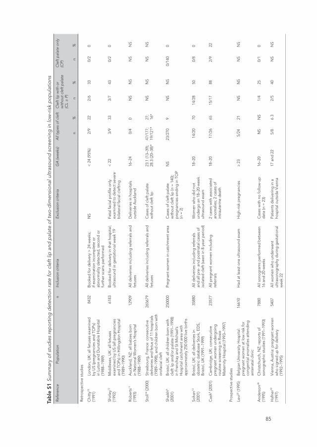
R1
R2
R3
R4
R5
R6
R7
R8
R9
R10
R11
R12
R13
R14
R15
R16
R17
R18
R19
R20
R21
R22
R23
R24
R25
R26
R27
R28
R29
R30
R31
R32
R33
R34
85
Tab
le S
1 Su
mm
ary
of s
tud
ies
rep
ortin
g d
etec
tion
rate
for c
left
lip
and
pal
ate
of tw
o-d
imen
sion
al u
ltras
ound
scr
eeni
ng in
low
-ris
k p
opul
atio
ns
Refe
renc
ePo
pul
atio
nn
Incl
usio
n cr
iteria
Excl
usio
n cr
iteria
GA
(wee
ks)
All
typ
es o
f cle
ft
Cle
ft li
p w
ith o
r w
ithou
t cle
ft p
alat
e (C
L ±
P)
Cle
ft p
alat
e on
ly(C
P)
n %
n
%
n
%
Ret
rosp
ectiv
e st
udie
s
Chi
tty10
(1
991)
Lo
ndo
n, U
K: a
ll fe
tuse
s ex
amin
ed
by
US
(pre
gna
ncie
s an
d T
OPs
) in
Lut
on
and
Dun
stab
le H
osp
ital
(198
8–19
89)
8432
Bo
oke
d fo
r d
eliv
ery
> 2
4 w
eeks
; if
exam
inat
ion
inco
mp
lete
or
abno
rmal
ity d
etec
ted
, sec
ond
or
furt
her
scan
s p
erfo
rmed
NS
< 2
4 (9
5%)
2/9
222/
633
0/2
0
Shirl
ey12
(1
992)
M
idd
lese
x, U
K: a
ll fe
tuse
s ex
amin
ed b
y U
S (a
ll p
reg
nanc
ies
and
TO
Ps) i
n H
illin
gd
on
Ho
spita
l (1
989–
1990
)
6183
Bo
oke
d fo
r d
eliv
ery
in t
hat
hosp
ital;
ultr
aso
und
in g
esta
tiona
l wee
k 19
Feta
l fac
ial p
rofil
e o
nly
exam
ined
to
det
ect
seve
re
bila
tera
l fac
ial c
left
ing
< 2
2 3/
933
3/7
430/
20
Ro
ber
ts11
(1
993)
A
uckl
and
, NZ:
all
bab
ies
bo
rn
in N
atio
nal W
om
en’s
Ho
spita
l (1
988–
1989
)
1290
9A
ll d
eliv
erie
s in
clud
ing
refe
rral
s an
d
fetu
ses
Del
iver
ies
in h
osp
itals
o
utsi
de
Auc
klan
d16
–24
0/4
0N
SN
SN
SN
S
Sto
ll14 (2
000)
St
rasb
our
g, F
ranc
e: c
ons
ecut
ive
del
iver
ies
and
fetu
s o
f 11
hosp
itals
(1
989–
1998
), an
d c
hild
ren
bo
rn w
ith
oro
faci
al c
left
2656
79A
ll d
eliv
erie
s in
clud
ing
refe
rral
s an
d
fetu
ses
Cas
es o
f cle
ft p
alat
e w
itho
ut c
left
lip
23.1
(13–
39);
28.3
(20–
38)*
47/1
77;
19/1
21*
27;
16*
NS
NS
NS
NS
Shai
kh8
(200
1)B
risto
l, U
K: a
ll ch
ildre
n b
orn
with
cl
eft
lip a
nd/o
r p
alat
e (1
991–
1998
) in
Fre
ncha
y an
d S
t M
icha
el’s
Ho
spita
l cat
chm
ent
area
with
ap
pro
xim
atel
y 25
0 00
0 liv
e b
irths
2500
00Pr
egna
nt w
om
en in
cat
chm
ent
area
Cas
es o
f cle
ft p
alat
e w
itho
ut c
left
lip
(n =
140
); p
reg
nanc
ies
end
ing
in T
OP
(n =
2)
NS
23/2
709
NS
NS
0/14
00
Soha
n13
(200
1)B
risto
l, U
K: a
ll d
eliv
erie
s in
o
bst
etric
dat
abas
e St
ork
, ED
S,
Bris
tol,
UK
(199
1–19
99)
3588
0A
ll d
eliv
erie
s in
clud
ing
refe
rral
s an
d a
ll p
re-
and
po
stna
tal c
ases
of
iso
late
d c
left
(see
n in
8-y
ear
per
iod
)
Wo
men
who
did
no
t un
der
go
an
18–2
0-w
eek
ultr
aso
und
exa
m
18–2
0 1
4/20
70
14/2
850
0/8
0
Cas
h9 (20
01)
Cam
brid
ge,
UK
: co
nsec
utiv
e p
reg
nant
wo
men
und
erg
oin
g
rout
ine
scre
enin
g in
Ro
sie
Mat
erni
ty H
osp
ital (
1993
–199
7)
2357
7A
ll p
reg
nant
wo
men
incl
udin
g
refe
rral
s2
case
s w
ith a
sso
ciat
ed
ano
mal
ies;
2 c
ases
of
intr
aute
rine
dea
th
18–2
017
/26
6515
/17
882/
922
Pro
spec
tive
stud
ies
Levi
20 (1
995)
Bel
gia
n U
nive
rsity
Ho
spita
l: p
reg
nant
wo
men
at
low
ris
k fo
r co
ngen
ital a
nom
alie
s at
tend
ing
an
tena
tal c
linic
1661
0H
ad a
t le
ast
one
ultr
aso
und
exa
mH
igh-
risk
pre
gna
ncie
s≥
235/
2421
NS
NS
NS
NS
And
erso
n28
(199
5)C
hris
tchu
rch,
NZ:
rep
ort
s o
f so
nog
rap
hic
stud
ies
(199
1–19
93)
788
0A
ll so
nog
ram
s p
erfo
rmed
bet
wee
n 16
and
20
wee
ksC
ases
with
no
follo
w-u
p
dat
a (n
= 2
3)16
–20
NS
NS
1/4
250/
10
Haf
ner19
(1
997)
Vie
nna,
Aus
tria
: pre
gna
nt w
om
en
who
sig
ned
up
for
del
iver
y (1
992–
1995
)
5407
All
wo
men
who
und
erw
ent
ultr
aso
nog
rap
hy d
urin
g g
esta
tiona
l w
eek
22
Patie
nts
del
iver
ing
in a
ho
spita
l out
sid
e V
ienn
a17
and
22
5/8
6 3
2/5
40N
SN
S
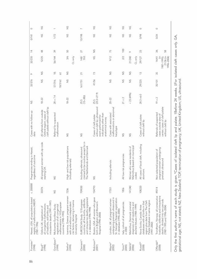
R1
R2
R3
R4
R5
R6
R7
R8
R9
R10
R11
R12
R13
R14
R15
R16
R17
R18
R19
R20
R21
R22
R23
R24
R25
R26
R27
R28
R29
R30
R31
R32
R33
R34
86
Forr
este
r17
(199
8)H
awai
i, U
SA; a
ll p
op
ulat
ion-
bas
ed
reg
istr
y o
f mal
form
atio
ns (H
BD
P)
(198
7–19
96)
c. 2
0000
0Pr
egna
ncie
s en
din
g in
Haw
aii,
reg
ard
less
of o
utco
me
Cas
es w
ith n
o fo
llow
-up
d
ata
NS
32/3
769
32/2
3514
0/14
10
Bo
yd15
(1
998)
Oxf
ord
, UK
: reg
istr
atio
n o
f al
l co
ngen
ital m
alfo
rmat
ions
o
f liv
ebirt
hs, s
till b
irths
and
in
trau
terin
e d
eath
s (1
991–
1997
)
3337
6A
ll p
reg
nant
wo
men
with
zip
co
de
star
ting
OX
All
refe
rral
s fr
om
out
sid
e O
xfo
rd re
gio
n; c
ases
of
clef
t p
alat
e w
itho
ut c
left
lip
18–2
2N
SN
S12
/25
48N
SN
S
Gra
ndje
an18
(1
999)
Mul
ticen
ter
Eur
ofe
tus
Stud
y:
reg
istr
atio
n o
f all
cong
enita
l m
alfo
rmat
ions
in 1
4 E
uro
pea
n co
untr
ies
(199
0–19
93)
NS
NS
Ref
erra
l fo
r su
spec
ted
m
alfo
rmat
ion
28 ±
7.4
57/3
16;
18/3
16†
18;
6†
56/1
4439
1/72
1
Stef
os25
(1
999)
Io
anni
na, G
reec
e: p
reg
nant
wo
men
w
ith a
nd w
itho
ut in
dep
end
ent
risk
fact
ors
, del
iver
ing
in Io
anna
U
nive
rsity
Ho
spita
l
7236
Hig
h- a
nd lo
w-r
isk
po
pul
atio
ns
(incl
udin
g re
ferr
als)
NS
18–2
2N
SN
S3/
6
CL
onl
y
50N
SN
S
Cle
men
ti16
(200
0)E
UR
OSC
AN
Stu
dy:
12
Eur
op
ean
coun
trie
s; a
ll liv
e b
irths
, stil
lbirt
hs
and
intr
aute
rine
dea
ths
reg
iste
red
in
a d
atab
ase
(199
6-19
98)
7090
30In
clud
ing
refe
rral
s; u
ltras
oun
d
clai
med
to
be
rout
ine
exce
pt
for
in
The
Net
herla
nds
and
Den
mar
k
NS
25.5
(1
5–38
)‡16
1/75
121
148/
553
2713
/198
7
Ro
bin
son23
(2
001)
Bo
sto
n, U
SA: a
ll w
om
en w
ho g
ave
birt
h in
Brig
ham
and
Wo
men
’s H
osp
ital (
1990
–200
0)
1547
93N
SC
ases
of c
left
pal
ate
with
out
cle
ft li
p; u
ltras
oun
d
exam
per
form
ed fo
r in
dic
atio
ns o
ther
tha
n an
ato
mic
al s
urve
y
25.5
(23.
2–27
.9)
41/5
6 73
NS
NS
NS
NS
Way
ne27
(2
002)
Lond
on,
UK
: all
pre
gna
nt w
om
en
und
erg
oin
g p
rena
tal u
ltras
oun
d in
St
. Geo
rge’
s H
osp
ital;
reg
istr
atio
n in
dat
abas
e (1
996–
2000
)
1755
1In
clud
ing
refe
rral
sC
ases
with
ass
oci
ated
m
alfo
rmat
ions
or
abno
rmal
ka
ryo
typ
e
20–2
2N
SN
S9/
1275
NS
NS
Tonn
i26
(200
5)It
aly:
co
hort
of a
ll p
reg
nanc
ies
in 2
003
185
6A
ll lo
w-r
isk
pre
gna
ncie
sN
S21
± 2
NS
NS
2/2
100
NS
NS
Nik
kilä
21
(200
6)M
alm
öhu
s, D
enm
ark:
pre
nata
l d
iag
nosi
s o
f all
new
bo
rns
and
ab
ort
ed fe
tuse
s (1
984–
1999
)
1412
40W
om
en w
ho w
ere
resi
den
ts o
f M
alm
öhu
s (b
ased
on
mun
icip
al
cod
es)
NS
< 2
3 (4
9%)
NS
NS
21/2
40
CL
onl
y
9N
SN
S
Rus
sell24
(2
008)
No
va S
cotia
, Can
ada:
thr
ee
dat
abas
es w
ith a
ll b
irths
in t
hat
reg
ion
plu
s d
ata
fro
m T
he C
left
Pa
late
Dat
abas
e in
sam
e re
gio
n (1
997–
2002
)
1082
20A
ll ca
ses
of f
acia
l cle
ft, i
nclu
din
g
abo
rtio
nsC
ases
of c
left
pal
ate
with
out
cle
ft li
p28
.3 ±
6.0
29/2
2513
29/1
2723
0/98
0
Offe
rdal
22
(200
8)Tr
ond
heim
, No
rway
: all
cons
ecut
ive
liveb
irths
, stil
lbirt
hs a
nd in
trau
terin
e d
eath
s, d
ivid
ed in
to t
wo
per
iod
s (1
987–
1995
and
199
6–20
04)
4931
4N
on-
sele
cted
po
pul
atio
n o
f p
reg
nant
wo
men
und
erg
oin
g
pre
nata
l ultr
aso
und
Ref
erra
ls o
f sus
pec
ted
ca
ses
out
sid
e ar
ea o
f ce
nter
; cas
es o
f cle
ft p
alat
e w
itho
ut c
left
lip
19 ±
235
/101
3514
/41 (in
19
87–1
995)
; 21
/36
(in
1996
–200
4)
34;
58
0/24
0
Onl
y th
e fir
st a
utho
r’s n
ame
of
each
stu
dy
is g
iven
. *C
ases
of
iso
late
d c
left
lip
and
pal
ate.
†B
efo
re 2
4 w
eeks
. ‡F
or
iso
late
d c
left
cas
es o
nly.
GA
, g
esta
tiona
l ag
e; N
S, n
ot
stat
ed; N
Z, N
ew Z
eala
nd; T
OP,
ter
min
atio
n o
f pre
gna
ncy;
UK
, Uni
ted
Kin
gd
om
; US,
ultr
aso
und
.
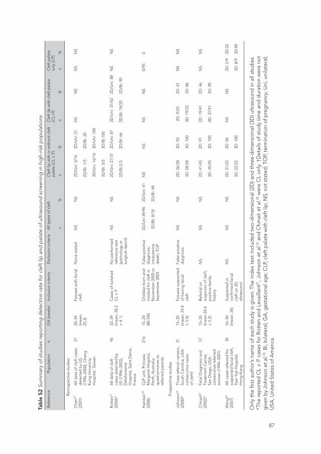
R1
R2
R3
R4
R5
R6
R7
R8
R9
R10
R11
R12
R13
R14
R15
R16
R17
R18
R19
R20
R21
R22
R23
R24
R25
R26
R27
R28
R29
R30
R31
R32
R33
R34
87
Forr
este
r17
(199
8)H
awai
i, U
SA; a
ll p
op
ulat
ion-
bas
ed
reg
istr
y o
f mal
form
atio
ns (H
BD
P)
(198
7–19
96)
c. 2
0000
0Pr
egna
ncie
s en
din
g in
Haw
aii,
reg
ard
less
of o
utco
me
Cas
es w
ith n
o fo
llow
-up
d
ata
NS
32/3
769
32/2
3514
0/14
10
Bo
yd15
(1
998)
Oxf
ord
, UK
: reg
istr
atio
n o
f al
l co
ngen
ital m
alfo
rmat
ions
o
f liv
ebirt
hs, s
till b
irths
and
in
trau
terin
e d
eath
s (1
991–
1997
)
3337
6A
ll p
reg
nant
wo
men
with
zip
co
de
star
ting
OX
All
refe
rral
s fr
om
out
sid
e O
xfo
rd re
gio
n; c
ases
of
clef
t p
alat
e w
itho
ut c
left
lip
18–2
2N
SN
S12
/25
48N
SN
S
Gra
ndje
an18
(1
999)
Mul
ticen
ter
Eur
ofe
tus
Stud
y:
reg
istr
atio
n o
f all
cong
enita
l m
alfo
rmat
ions
in 1
4 E
uro
pea
n co
untr
ies
(199
0–19
93)
NS
NS
Ref
erra
l fo
r su
spec
ted
m
alfo
rmat
ion
28 ±
7.4
57/3
16;
18/3
16†
18;
6†
56/1
4439
1/72
1
Stef
os25
(1
999)
Io
anni
na, G
reec
e: p
reg
nant
wo
men
w
ith a
nd w
itho
ut in
dep
end
ent
risk
fact
ors
, del
iver
ing
in Io
anna
U
nive
rsity
Ho
spita
l
7236
Hig
h- a
nd lo
w-r
isk
po
pul
atio
ns
(incl
udin
g re
ferr
als)
NS
18–2
2N
SN
S3/
6
CL
onl
y
50N
SN
S
Cle
men
ti16
(200
0)E
UR
OSC
AN
Stu
dy:
12
Eur
op
ean
coun
trie
s; a
ll liv
e b
irths
, stil
lbirt
hs
and
intr
aute
rine
dea
ths
reg
iste
red
in
a d
atab
ase
(199
6-19
98)
7090
30In
clud
ing
refe
rral
s; u
ltras
oun
d
clai
med
to
be
rout
ine
exce
pt
for
in
The
Net
herla
nds
and
Den
mar
k
NS
25.5
(1
5–38
)‡16
1/75
121
148/
553
2713
/198
7
Ro
bin
son23
(2
001)
Bo
sto
n, U
SA: a
ll w
om
en w
ho g
ave
birt
h in
Brig
ham
and
Wo
men
’s H
osp
ital (
1990
–200
0)
1547
93N
SC
ases
of c
left
pal
ate
with
out
cle
ft li
p; u
ltras
oun
d
exam
per
form
ed fo
r in
dic
atio
ns o
ther
tha
n an
ato
mic
al s
urve
y
25.5
(23.
2–27
.9)
41/5
6 73
NS
NS
NS
NS
Way
ne27
(2
002)
Lond
on,
UK
: all
pre
gna
nt w
om
en
und
erg
oin
g p
rena
tal u
ltras
oun
d in
St
. Geo
rge’
s H
osp
ital;
reg
istr
atio
n in
dat
abas
e (1
996–
2000
)
1755
1In
clud
ing
refe
rral
sC
ases
with
ass
oci
ated
m
alfo
rmat
ions
or
abno
rmal
ka
ryo
typ
e
20–2
2N
SN
S9/
1275
NS
NS
Tonn
i26
(200
5)It
aly:
co
hort
of a
ll p
reg
nanc
ies
in 2
003
185
6A
ll lo
w-r
isk
pre
gna
ncie
sN
S21
± 2
NS
NS
2/2
100
NS
NS
Nik
kilä
21
(200
6)M
alm
öhu
s, D
enm
ark:
pre
nata
l d
iag
nosi
s o
f all
new
bo
rns
and
ab
ort
ed fe
tuse
s (1
984–
1999
)
1412
40W
om
en w
ho w
ere
resi
den
ts o
f M
alm
öhu
s (b
ased
on
mun
icip
al
cod
es)
NS
< 2
3 (4
9%)
NS
NS
21/2
40
CL
onl
y
9N
SN
S
Rus
sell24
(2
008)
No
va S
cotia
, Can
ada:
thr
ee
dat
abas
es w
ith a
ll b
irths
in t
hat
reg
ion
plu
s d
ata
fro
m T
he C
left
Pa
late
Dat
abas
e in
sam
e re
gio
n (1
997–
2002
)
1082
20A
ll ca
ses
of f
acia
l cle
ft, i
nclu
din
g
abo
rtio
nsC
ases
of c
left
pal
ate
with
out
cle
ft li
p28
.3 ±
6.0
29/2
2513
29/1
2723
0/98
0
Offe
rdal
22
(200
8)Tr
ond
heim
, No
rway
: all
cons
ecut
ive
liveb
irths
, stil
lbirt
hs a
nd in
trau
terin
e d
eath
s, d
ivid
ed in
to t
wo
per
iod
s (1
987–
1995
and
199
6–20
04)
4931
4N
on-
sele
cted
po
pul
atio
n o
f p
reg
nant
wo
men
und
erg
oin
g
pre
nata
l ultr
aso
und
Ref
erra
ls o
f sus
pec
ted
ca
ses
out
sid
e ar
ea o
f ce
nter
; cas
es o
f cle
ft p
alat
e w
itho
ut c
left
lip
19 ±
235
/101
3514
/41 (in
19
87–1
995)
; 21
/36
(in
1996
–200
4)
34;
58
0/24
0
Onl
y th
e fir
st a
utho
r’s n
ame
of
each
stu
dy
is g
iven
. *C
ases
of
iso
late
d c
left
lip
and
pal
ate.
†B
efo
re 2
4 w
eeks
. ‡F
or
iso
late
d c
left
cas
es o
nly.
GA
, g
esta
tiona
l ag
e; N
S, n
ot
stat
ed; N
Z, N
ew Z
eala
nd; T
OP,
ter
min
atio
n o
f pre
gna
ncy;
UK
, Uni
ted
Kin
gd
om
; US,
ultr
aso
und
.
Tab
le S
2 Su
mm
ary
of s
tud
ies
rep
ortin
g d
etec
tion
rate
for c
left
lip
and
pal
ate
of u
ltras
ound
scr
eeni
ng in
hig
h-ris
k p
opul
atio
ns
Refe
renc
ePo
pul
atio
nn
GA
(wee
ks)
Incl
usio
n cr
iteria
Excl
usio
n cr
iteria
All
typ
es o
f cle
ft
Cle
ft li
p w
ith o
r with
out c
left
p
alat
e (C
L ±
P)
Cle
ft li
p w
ith c
left
pal
ate
(CL+
P)C
left
pal
ate
only
(CP)
n
% n
%
n
% n
%
Ret
rosp
ectiv
e st
udie
s
Che
n29
(200
1)A
ll d
ata
of c
left
cas
es
det
ecte
d b
y U
S (1
996–
2000
), C
heng
K
ung
Uni
vers
ity
Ho
spita
l, Ta
iwan
2120
–34
(mea
n,
25.3
)
Fetu
ses
with
faci
al
clef
tN
one
sta
ted
NS
NS
2D/U
ni: 5
/16
2D/B
i: 1/
5
3D/U
ni: 1
6/16
3D/B
i: 5/
5
2D/U
ni:.3
1
2D/B
i: 20
3D/U
ni: 1
00
3D/B
i:100
NS
NS
NS
NS
Ro
tten
31
(200
4)*
All
dat
a o
f cle
ft
case
s d
etec
ted
by
US
(199
6–20
02),
Del
afo
ntai
ne
Ho
spita
l, Sa
int
Den
is,
Fran
ce
9622
–39
(mea
n, 2
8.2
± 4
.1)
Cas
es o
f iso
late
d
CL
± P
No
per
form
ed
refe
renc
e te
st
(pat
holo
gy
or
surg
ical
rep
ort
)
NS
NS
2D/U
ni: 2
7/31
2D/B
i:2/3
2D/U
ni: 8
7
2D/B
i: 66
2D/U
ni: 3
7/42
2D/B
i: 18
/20
2D/U
ni: 8
8
2D/B
i: 90
NS
NS
Han
iker
i30
(200
6)C
LP u
nit,
Prin
cess
M
arg
aret
Ho
spita
l, Pe
rth,
Aus
tral
ia:
que
stio
nnai
res
to
refe
rred
par
ents
216
15–2
0(8
0.5%
)C
hild
ren
bo
rn a
nd
trea
ted
for
clef
t in
N
ove
mb
er 2
000
or
Sep
tem
ber
200
3
Fals
e-p
osi
tive
dia
gno
sis;
in
trau
terin
e d
eath
; TO
P
2D/U
ni:3
9/96
2D
/Bi:
8/18
2D/U
ni: 4
1
2D/B
i: 44
NS
NS
NS
NS
0/95
0
Pro
spec
tive
stud
ies
John
son33
(2
000)
*Th
ree
refe
rral
cen
ters
, So
uth
Car
olin
a, U
SA:
cons
ecut
ive
case
s o
f cle
ft†
3115
–35
(mea
n, 2
4.8
± 5
.0)
Fetu
ses
susp
ecte
d
of h
avin
g fa
cial
cl
eft
Fals
e-p
osi
tive
dia
gno
sis
NS
NS
2D: 2
6/28
3D: 2
8/28
2D: 9
3 3D
: 100
2D
: 9/2
2
3D: 1
9/22
2D: 4
1
3D: 8
6
NS
NS
Chm
ait32
(2
002)
* Fe
tal D
iag
nosi
s an
d
Trea
tmen
t C
ente
r, Sa
n D
ieg
o, U
SA:
cons
ecut
ive
refe
rred
w
om
en (1
994–
2001
)
5715
–35
(mea
n 24
.6
± 5
.2)
Ref
erra
l on
susp
icio
n o
f cle
ft;
po
sitiv
e fa
mily
hi
sto
ry
NS
NS
NS
2D: 4
1/45
3D: 4
5/45
2D: 9
1
3D: 1
00
2D: 1
9/41
3D: 3
7/41
2D: 4
6
3D: 9
0
NS
NS
Wan
g34
(2
007)
All
case
s re
ferr
ed fo
r su
spec
ted
faci
al c
left
, Ts
an Y
uk H
osp
ital,
Ho
ng K
ong
3016
–36
(mea
n, 2
6)
Susp
ecte
d o
r co
nfirm
ed fa
cial
cl
eft
on
2D
ultr
aso
und
NS
NS
NS
2D: 2
1/22
3D: 2
2/22
2D: 9
6
3D: 1
00
NS
NS
2D: 2
/9
3D: 8
/9
2D:2
2
3D:8
9
Onl
y th
e fir
st a
utho
r’s n
ame
of e
ach
stud
y is
giv
en. T
he in
dex
tex
t in
clud
ed t
wo
-dim
ensi
ona
l (2D
) and
thr
ee-d
imen
sio
nal (
3D) u
ltras
oun
d in
all
stud
ies.
*T
he r
epo
rted
CL
± P
cas
es fo
r R
ott
en a
nd L
evai
llant
31, J
ohn
son
et a
l.33 a
nd C
hmai
t et
al.32
wer
e C
L o
nly.
†D
etai
ls o
f stu
dy
time
and
dur
atio
n w
ere
not
giv
en b
y Jo
hnso
n et
al.33
. Bi,
bila
tera
l; G
A, g
esta
tiona
l ag
e; C
LP, c
left
pal
ate
with
cle
ft li
p; N
S, n
ot s
tate
d; T
OP,
term
inat
ion
of p
reg
nanc
y; U
ni, u
nila
tera
l; U
SA, U
nite
d S
tate
s o
f Am
eric
a.

R1
R2
R3
R4
R5
R6
R7
R8
R9
R10
R11
R12
R13
R14
R15
R16
R17
R18
R19
R20
R21
R22
R23
R24
R25
R26
R27
R28
R29
R30
R31
R32
R33
R34
5Prenatal ultrasound screening
for orofacial clefts
Ultrasound Obstet Gynecol 2011; 38: 434 –439
W. MAARSE, L.R. PISTORIUS, W.K. VAN EETEN, C.C. BREUGEM, M. KON,
M.J.H. VAN DEN BOOGAARD, A.B. MINK VAN DER MOLEN
R1
R2
R3
R4
R5
R6
R7
R8
R9
R10
R11
R12
R13
R14
R15
R16
R17
R18
R19
R20
R21
R22
R23
R24
R25
R26
R27
R28
R29
R30
R31
R32
R33
R34
5 | Prenatal screening of oral cleft
90
ABSTRACT
Objectives To evaluate the sensitivity and specificity of ultrasound for detecting
prenatal facial clefts in low-risk and high-risk populations.
Methods This study prospectively followed up a non- selected population, namely
all pregnant women who underwent routine second-trimester prenatal ultrasound
screening in the Utrecht region during the 2-year period from January 2007 to
December 2008.
Results A total of 35 924 low-risk and 2836 high- risk pregnant women underwent
ultrasound screening. Orofacial clefts were present in 62 cases, an incidence of 1: 624.
The distribution of clefts was as follows: 18 (29%) cleft lip, 25 (40%) cleft lip with cleft
palate, 17 (27%) cleft palate only, one median cleft and one atypical cleft. Of these,
38 (61%) were unilateral and 23 (37%) were bilateral. Thirty-nine per cent (24/62) had
associated anomalies, with most chromosomal defects found in the cleft lip with cleft
palate and cleft palate only groups. Cleft lip with or without cleft palate was detected
prenatally in 38/43 cases, a sensitivity of 88%. No case of cleft palate only was detected
prenatally. There were three false- positive cases, of which two were fetuses with
multiple congenital deformities.
Conclusions Ultrasound screening has a high sensitivity for the detection of cleft lip
with and without cleft palate in high-risk and low-risk pregnancies in our region, where
well-trained sonographers carry out primary screening. The key to a high sensitivity of
prenatal ultrasound is likely to be a combination of excellent training of sonographers,
referral to specialized centers when a cleft is suspected, routine visualization of the
fetal face and advances in ultrasound techniques.
R1
R2
R3
R4
R5
R6
R7
R8
R9
R10
R11
R12
R13
R14
R15
R16
R17
R18
R19
R20
R21
R22
R23
R24
R25
R26
R27
R28
R29
R30
R31
R32
R33
R34
91
INTRODUCTION
In The Netherlands, transabdominal two-dimensional (2D) ultrasound screening during
the second trimester of pregnancy was made universally accessible by legislation and
health insurance coverage in 2007. Evaluation of the upper lip for possible cleft lip and
palate is an optional element in this screening (NVOG Dutch Society of Obstetrics &
Gynecology protocol1). With an incidence of cleft lip with or without cleft palate (CL ±
P) and cleft palate only (CP) of 1.8 per 1000 births2, this translates to approximately 320
cases of orofacial cleft per year in The Netherlands3. As 90 – 95% of Dutch pregnant
women undergo ultrasound screening, cleft palate teams are commonly confronted
with prenatally diagnosed clefts.
Classification of orofacial clefts diagnosed by ultra- sound is generally carried out
according to the system of Nyberg et al.4. However, in this classification no distinction
is made between the alveolus and the hard palate, which, in the presence of CL, can
be important in the detection of an additional CP. Proper classification is essential
because different types of cleft may be variably associated with additional anomalies
and chromosomal disorders4–7, while isolated clefts are associated with low mortality
and morbidity rates and are primarily a functional and esthetic problem8.
In a recently published systematic review it was concluded that transabdominal 2D
ultrasound screening in a low-risk population has a relatively low rate of detection of
orofacial clefts8, with detection rates of 23 – 58% in the most recent large prospective
studies9,10. Moreover, in most studies reporting on the accuracy of ultrasound
screening, inconsistent methodological quality is observed. In this prospective cohort
study, the sensitivity and specificity of ultrasound in detecting orofacial clefts during
prenatal screening in a low-risk and a high-risk population is presented, as well as the
percentage of associated anomalies per type of orofacial cleft.
R1
R2
R3
R4
R5
R6
R7
R8
R9
R10
R11
R12
R13
R14
R15
R16
R17
R18
R19
R20
R21
R22
R23
R24
R25
R26
R27
R28
R29
R30
R31
R32
R33
R34
5 | Prenatal screening of oral cleft
92
METHODS
This prospective follow-up study investigated the detection of orofacial clefts by
ultrasound screening in The Netherlands during the period of January 2007 to December
2008. For the purpose of screening, The Netherlands is subdivided into eight regions,
in which trained midwives, sonographers and obstetricians perform routine 2D ultra-
sound screening in several centers. Every region has one main referral center to which
pregnant women are referred if an abnormality is suspected. Upon referral, certified
obstetricians perform further 2D and three-dimensional (3D) ultrasound examinations,
and the patient is counseled by members of the cleft palate team11. The standard for
advanced 2D ultrasound examination is set by the Dutch Society for Gynaecology and
Obstetrics (NVOG), the documentation of which is available electronically1. Ultrasound
screening routinely includes detailed examination for orofacial clefts.
Study population
The study cohort comprised all screened pregnant women in the geographically well-
defined screening region of Utrecht. The study population was divided into a low-
risk population and a high-risk population. This separation was made to ensure that
all pregnant women in each group had a comparable a priori risk. Pregnant women
who underwent routine screening formed the low-risk population, and the high-risk
population consisted of pregnant women with a positive family history of orofacial
cleft or other risk factors for fetal abnormalities. The high- risk population and fetuses
suspected of orofacial cleft were seen in a tertiary referral center (the Wilhelmina
Children’s Hospital). When an associated anomaly was detected, karyotyping by
amniocentesis and consultation with a clinical geneticist was recommended. When
parents opted for termination of pregnancy (TOP), mostly because of the presence of
associated anomalies, an autopsy was recommended to verify the prenatal diagnosis.
When parents opted to continue a pregnancy, they met a multidisciplinary team, which
included an obstetrician and two members of the cleft palate team: a plastic surgeon
and a medical psychologist. In the first week after delivery all newborns were examined
by a plastic surgeon and a pediatrician or a clinical geneticist from the cleft palate
team to verify prenatally detected orofacial clefts. Newborns in the region of Utrecht
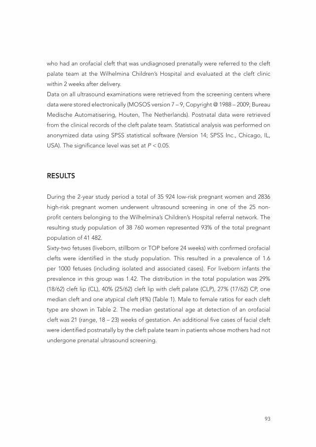
R1
R2
R3
R4
R5
R6
R7
R8
R9
R10
R11
R12
R13
R14
R15
R16
R17
R18
R19
R20
R21
R22
R23
R24
R25
R26
R27
R28
R29
R30
R31
R32
R33
R34
93
who had an orofacial cleft that was undiagnosed prenatally were referred to the cleft
palate team at the Wilhelmina Children’s Hospital and evaluated at the cleft clinic
within 2 weeks after delivery.
Data on all ultrasound examinations were retrieved from the screening centers where
data were stored electronically (MOSOS version 7 – 9, Copyright @ 1988 – 2009; Bureau
Medische Automatisering, Houten, The Netherlands). Postnatal data were retrieved
from the clinical records of the cleft palate team. Statistical analysis was performed on
anonymized data using SPSS statistical software (Version 14; SPSS Inc., Chicago, IL,
USA). The significance level was set at P < 0.05.
RESULTS
During the 2-year study period a total of 35 924 low-risk pregnant women and 2836
high-risk pregnant women underwent ultrasound screening in one of the 25 non-
profit centers belonging to the Wilhelmina’s Children’s Hospital referral network. The
resulting study population of 38 760 women represented 93% of the total pregnant
population of 41 482.
Sixty-two fetuses (liveborn, stillborn or TOP before 24 weeks) with confirmed orofacial
clefts were identified in the study population. This resulted in a prevalence of 1.6
per 1000 fetuses (including isolated and associated cases). For liveborn infants the
prevalence in this group was 1.42. The distribution in the total population was 29%
(18/62) cleft lip (CL), 40% (25/62) cleft lip with cleft palate (CLP), 27% (17/62) CP, one
median cleft and one atypical cleft (4%) (Table 1). Male to female ratios for each cleft
type are shown in Table 2. The median gestational age at detection of an orofacial
cleft was 21 (range, 18 – 23) weeks of gestation. An additional five cases of facial cleft
were identified postnatally by the cleft palate team in patients whose mothers had not
undergone prenatal ultrasound screening.
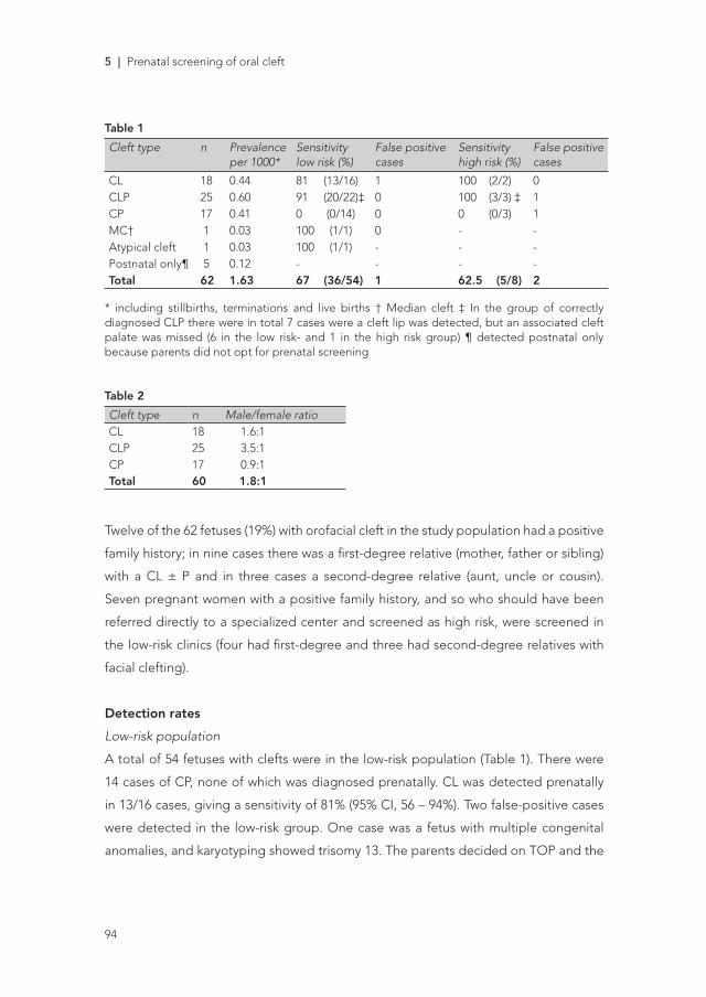
R1
R2
R3
R4
R5
R6
R7
R8
R9
R10
R11
R12
R13
R14
R15
R16
R17
R18
R19
R20
R21
R22
R23
R24
R25
R26
R27
R28
R29
R30
R31
R32
R33
R34
5 | Prenatal screening of oral cleft
94
Table 1
Cleft type n Prevalence per 1000*
Sensitivity low risk (%)
False positive cases
Sensitivity high risk (%)
False positive cases
CL 18 0.44 81 (13/16) 1 100 (2/2) 0CLP 25 0.60 91 (20/22)‡ 0 100 (3/3) ‡ 1CP 17 0.41 0 (0/14) 0 0 (0/3) 1MC† 1 0.03 100 (1/1) 0 - -Atypical cleft 1 0.03 100 (1/1) - - -Postnatal only¶ 5 0.12 - - - -Total 62 1.63 67 (36/54) 1 62.5 (5/8) 2
* including stillbirths, terminations and live births † Median cleft ‡ In the group of correctly diagnosed CLP there were in total 7 cases were a cleft lip was detected, but an associated cleft palate was missed (6 in the low risk- and 1 in the high risk group) ¶ detected postnatal only because parents did not opt for prenatal screening
Table 2
Cleft type n Male/female ratioCL 18 1.6:1CLP 25 3.5:1CP 17 0.9:1Total 60 1.8:1
Twelve of the 62 fetuses (19%) with orofacial cleft in the study population had a positive
family history; in nine cases there was a first-degree relative (mother, father or sibling)
with a CL ± P and in three cases a second-degree relative (aunt, uncle or cousin).
Seven pregnant women with a positive family history, and so who should have been
referred directly to a specialized center and screened as high risk, were screened in
the low-risk clinics (four had first-degree and three had second-degree relatives with
facial clefting).
Detection rates
Low-risk population
A total of 54 fetuses with clefts were in the low-risk population (Table 1). There were
14 cases of CP, none of which was diagnosed prenatally. CL was detected prenatally
in 13/16 cases, giving a sensitivity of 81% (95% CI, 56 – 94%). Two false-positive cases
were detected in the low-risk group. One case was a fetus with multiple congenital
anomalies, and karyotyping showed trisomy 13. The parents decided on TOP and the
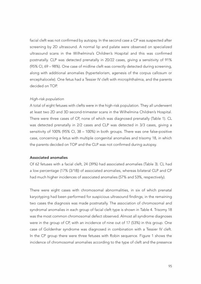
R1
R2
R3
R4
R5
R6
R7
R8
R9
R10
R11
R12
R13
R14
R15
R16
R17
R18
R19
R20
R21
R22
R23
R24
R25
R26
R27
R28
R29
R30
R31
R32
R33
R34
95
facial cleft was not confirmed by autopsy. In the second case a CP was suspected after
screening by 2D ultrasound. A normal lip and palate were observed on specialized
ultrasound scans in the Wilhelmina’s Children’s Hospital and this was confirmed
postnatally. CLP was detected prenatally in 20/22 cases, giving a sensitivity of 91%
(95% CI, 69 – 98%). One case of midline cleft was correctly detected during screening,
along with additional anomalies (hypertelorism, agenesis of the corpus callosum or
encephalocele). One fetus had a Tessier IV cleft with microphthalmia, and the parents
decided on TOP.
High-risk population
A total of eight fetuses with clefts were in the high-risk population. They all underwent
at least two 2D and 3D second-trimester scans in the Wilhelmina Children’s Hospital.
There were three cases of CP, none of which was diagnosed prenatally (Table 1). CL
was detected prenatally in 2/2 cases and CLP was detected in 3/3 cases, giving a
sensitivity of 100% (95% CI, 38 – 100%) in both groups. There was one false-positive
case, concerning a fetus with multiple congenital anomalies and trisomy 18, in which
the parents decided on TOP and the CLP was not confirmed during autopsy.
Associated anomalies
Of 62 fetuses with a facial cleft, 24 (39%) had associated anomalies (Table 3). CL had
a low percentage (17% (3/18)) of associated anomalies, whereas bilateral CLP and CP
had much higher incidences of associated anomalies (57% and 53%, respectively).
There were eight cases with chromosomal abnormalities, in six of which prenatal
karyotyping had been performed for suspicious ultrasound findings; in the remaining
two cases the diagnosis was made postnatally. The association of chromosomal and
syndromal anomalies in each group of facial cleft type is shown in Table 4. Trisomy 18
was the most common chromosomal defect observed. Almost all syndrome diagnoses
were in the group of CP, with an incidence of nine out of 17 (53%) in this group. One
case of Goldenhar syndrome was diagnosed in combination with a Tessier IV cleft.
In the CP group there were three fetuses with Robin sequence. Figure 1 shows the
incidence of chromosomal anomalies according to the type of cleft and the presence
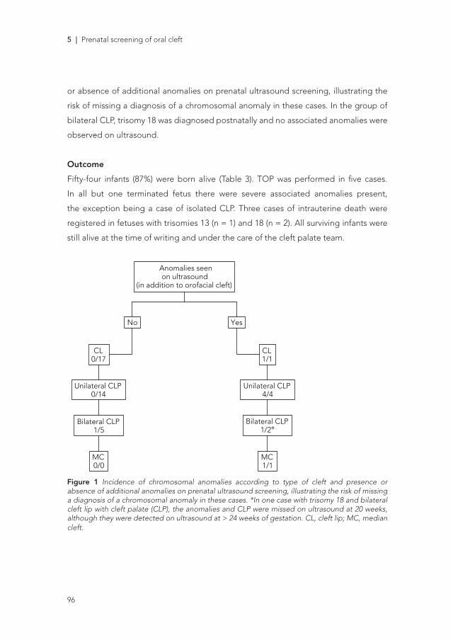
R1
R2
R3
R4
R5
R6
R7
R8
R9
R10
R11
R12
R13
R14
R15
R16
R17
R18
R19
R20
R21
R22
R23
R24
R25
R26
R27
R28
R29
R30
R31
R32
R33
R34
5 | Prenatal screening of oral cleft
96
or absence of additional anomalies on prenatal ultrasound screening, illustrating the
risk of missing a diagnosis of a chromosomal anomaly in these cases. In the group of
bilateral CLP, trisomy 18 was diagnosed postnatally and no associated anomalies were
observed on ultrasound.
Outcome
Fifty-four infants (87%) were born alive (Table 3). TOP was performed in five cases.
In all but one terminated fetus there were severe associated anomalies present,
the exception being a case of isolated CLP. Three cases of intrauterine death were
registered in fetuses with trisomies 13 (n = 1) and 18 (n = 2). All surviving infants were
still alive at the time of writing and under the care of the cleft palate team.
Prenatal orofacial cleft: accuracy of screening 437
Table 4 Summary of anomalies, syndromes and chromosomal defects per cleft type
Cleft type Anomaly Syndromes/sequenceChromosomaldefects
Prenatallydetected
CL Umbilical hernia NoClub foot No
Trisomy 18 Yes
CLP (uni) Hypospadias NoHirschsprung’s disease NoVSD Trisomy 21 Yes (both anomalies)
Trisomy 18 (× 3) Yes (× 3)
CLP (bi) Hypertelorism, ACC YesTrisomy 13 YesTrisomy 9p YesTrisomy 18 No
CP Pierre–Robin (× 3) No (× 2)*, Yes (× 1)Stickler syndrome (× 2) No (× 2)Kabuki syndrome (× 2) No (× 2)†van der Woude syndrome NoBeckwith–Wiedemann syndrome No‡
MC Hypertelorism, ACC,encephalocele
Yes
Atypical cleft Tessier IV cleft, microphthalmia Goldenhar syndrome Yes
*One case with club foot on ultrasound. †One case with oligohydramnios on ultrasound. ‡Amniotic hernia on ultrasound. ACC, agenesis ofthe corpus callosum; bi, bilateral; CL, cleft lip; CLP, cleft lip with cleft palate; CP, cleft palate only; MC, median cleft; uni, unilateral; VSD,ventricular septal defect.
No Yes
CL0/17
Unilateral CLP0/14
Bilateral CLP1/5
MC0/0
CL1/1
Unilateral CLP4/4
Bilateral CLP1/2∗
MC1/1
Anomalies seenon ultrasound
(in addition to orofacial cleft)
Figure 1 Incidence of chromosomal anomalies according to type ofcleft and presence or absence of additional anomalies on prenatalultrasound screening, illustrating the risk of missing a diagnosis ofa chromosomal anomaly in these cases. *In one case with trisomy18 and bilateral cleft lip with cleft palate (CLP), the anomalies andCLP were missed on ultrasound at 20 weeks, although they weredetected on ultrasound at > 24 weeks of gestation. CL, cleft lip;MC, median cleft.
fetus there were severe associated anomalies present, theexception being a case of isolated CLP. Three cases ofintrauterine death were registered in fetuses with trisomies13 (n = 1) and 18 (n = 2). All surviving infants were stillalive at the time of writing and under the care of the cleftpalate team.
DISCUSSION
Through introduction of routine prenatal screening,cleft palate teams are more frequently confronted withprenatally diagnosed clefts. Increased awareness offacial clefting and associated anomalies might clarifypregnancy options. When a diagnosis is made, parentshave the opportunity to prepare themselves emotionallyand practically12 by meeting a multidisciplinary teamconsisting of an obstetrician, a psychologist and a plasticsurgeon. This allows the parents to discuss the surgicaloptions as well as the demands of a growing child with acleft lip and palate.
This prospective study of ultrasound screening demon-strates a sensitivity of 88% for CL ± P; however, theoverall sensitivity for orofacial clefts was lower – 65%in the low-risk group and 62.5% in the high-riskgroup – because no cases of isolated CP were diagnosedprenatally. Pregnant women for whom there was sus-picion of a fetal facial cleft were referred to a referralcenter for 2D and 3D ultrasound scans where obstetriciansdetected additional cleft palates in three of six cases (50%)in which there was initially a cleft only lip identified byprimary ultrasound. These rates are high in compari-son with former studies that reported variable orofacialcleft detection rates of 23–59.6% on 2D ultrasound and66–100% on 3D ultrasound8,13. In our study, sources ofbias were minimized by determining the diagnostic accu-racy of screening tests8. A large study population in awell-defined geographic area was prospectively enrolled.Also, high-risk and low-risk pregnant women were stud-ied separately. The relative risk of familial recurrence wasrecently reported to be 32 (95% CI, 24.6–40.3) for anyCL in first-degree relatives and 56 (95% CI, 37.2–84.8)
Copyright 2011 ISUOG. Published by John Wiley & Sons, Ltd. Ultrasound Obstet Gynecol 2011; 38: 434–439.
Figure 1 Incidence of chromosomal anomalies according to type of cleft and presence or absence of additional anomalies on prenatal ultrasound screening, illustrating the risk of missing a diagnosis of a chromosomal anomaly in these cases. *In one case with trisomy 18 and bilateral cleft lip with cleft palate (CLP), the anomalies and CLP were missed on ultrasound at 20 weeks, although they were detected on ultrasound at > 24 weeks of gestation. CL, cleft lip; MC, median cleft.
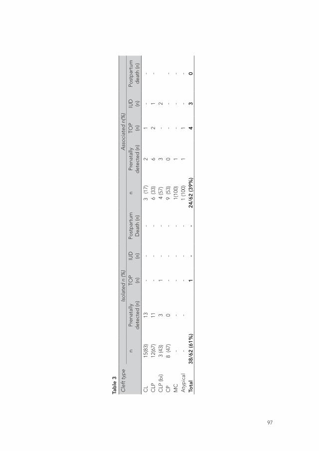
R1
R2
R3
R4
R5
R6
R7
R8
R9
R10
R11
R12
R13
R14
R15
R16
R17
R18
R19
R20
R21
R22
R23
R24
R25
R26
R27
R28
R29
R30
R31
R32
R33
R34
97
Tab
le 3
Cle
ft ty
pe
Isol
ated
n (%
)A
ssoc
iate
d n
(%)
nPr
enat
ally
d
etec
ted
(n)
TOP
(n)
IUD
(n)
Post
par
tum
D
eath
(n)
nPr
enat
ally
d
etec
ted
(n)
TOP
(n)
IUD
(n)
Post
par
tum
d
eath
(n)
CL
15(8
3)
13-
--
3 (
17)
21
--
CLP
12
(67)
11-
--
6 (3
3)6
21
-C
LP (b
i)3
(43)
31
--
4 (5
7)3
-2
CP
8 (4
7)0
--
-9
(53)
0-
--
MC
--
--
-1(
100)
1-
--
Aty
pic
al
--
--
-1
(100
)1
1-
-To
tal
38/6
2 (6
1%)
1-
-24
/62
(39%
)4
30
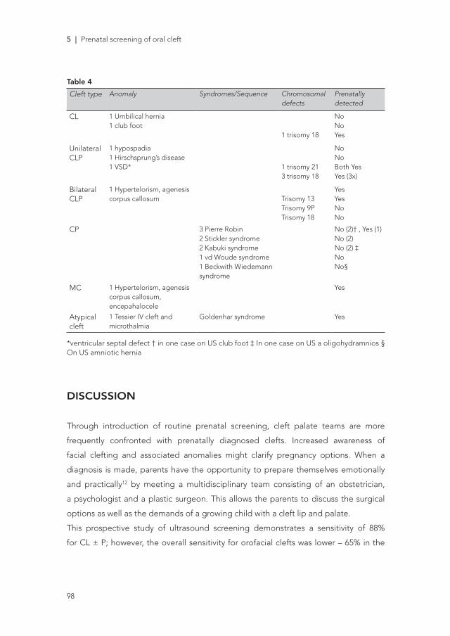
R1
R2
R3
R4
R5
R6
R7
R8
R9
R10
R11
R12
R13
R14
R15
R16
R17
R18
R19
R20
R21
R22
R23
R24
R25
R26
R27
R28
R29
R30
R31
R32
R33
R34
5 | Prenatal screening of oral cleft
98
Table 4
Cleft type Anomaly Syndromes/Sequence Chromosomal defects
Prenatally detected
CL 1 Umbilical hernia1 club foot
1 trisomy 18
NoNoYes
UnilateralCLP
1 hypospadia1 Hirschsprung’s disease1 VSD* 1 trisomy 21
3 trisomy 18
NoNoBoth YesYes (3x)
BilateralCLP
1 Hypertelorism, agenesis corpus callosum Trisomy 13
Trisomy 9PTrisomy 18
YesYesNoNo
CP 3 Pierre Robin2 Stickler syndrome2 Kabuki syndrome1 vd Woude syndrome1 Beckwith Wiedemann syndrome
No (2)† , Yes (1)No (2)No (2) ‡NoNo§
MC 1 Hypertelorism, agenesis corpus callosum, encepahalocele
Yes
Atypical cleft
1 Tessier IV cleft and microthalmia
Goldenhar syndrome Yes
*ventricular septal defect † in one case on US club foot ‡ In one case on US a oligohydramnios § On US amniotic hernia
DISCUSSION
Through introduction of routine prenatal screening, cleft palate teams are more
frequently confronted with prenatally diagnosed clefts. Increased awareness of
facial clefting and associated anomalies might clarify pregnancy options. When a
diagnosis is made, parents have the opportunity to prepare themselves emotionally
and practically12 by meeting a multidisciplinary team consisting of an obstetrician,
a psychologist and a plastic surgeon. This allows the parents to discuss the surgical
options as well as the demands of a growing child with a cleft lip and palate.
This prospective study of ultrasound screening demonstrates a sensitivity of 88%
for CL ± P; however, the overall sensitivity for orofacial clefts was lower – 65% in the
R1
R2
R3
R4
R5
R6
R7
R8
R9
R10
R11
R12
R13
R14
R15
R16
R17
R18
R19
R20
R21
R22
R23
R24
R25
R26
R27
R28
R29
R30
R31
R32
R33
R34
99
low-risk group and 62.5% in the high-risk group – because no cases of isolated CP
were diagnosed prenatally. Pregnant women for whom there was suspicion of a fetal
facial cleft were referred to a referral center for 2D and 3D ultrasound scans where
obstetricians detected additional cleft palates in three of six cases (50%) in which there
was initially a cleft only lip identified by primary ultrasound. These rates are high in
comparison with former studies that reported variable orofacial cleft detection rates of
23 – 59.6% on 2D ultrasound and 66 – 100% on 3D ultrasound8,13. In our study, sources
of bias were minimized by determining the diagnostic accuracy of screening tests8. A
large study population in a well-defined geographic area was prospectively enrolled.
Also, high-risk and low-risk pregnant women were studied separately. The relative risk
of familial recurrence was recently reported to be 32 (95% CI, 24.6 – 40.3) for any CL
in first-degree relatives and 56 (95% CI, 37.2 – 84.8) for cleft palate alone14,15. Postnatal
follow-up was at least 1 year, long enough to detect the majority of associated
anomalies. Ultrasound evaluation of the study population was carried out according to
a standardized protocol. To perform routine prenatal screening, minimal quality criteria
were set for sonographers (with a minimum of 150 – 200 examinations annually)11.
Consequently, obstetricians performing ultrasound examinations in a high-risk setting
have more experience and the opportunity to use 3D ultrasound techniques, which
resulted in the detection of additional cleft palates in three of six cases (50%). This is
in concordance with Crane et al., who reported significantly higher detection rates in
tertiary centers vs. non-tertiary centers16.
Three cases of false-positive diagnoses were identified; in two fetuses with multiple
congenital anomalies and trisomy 13 and 18, respectively, an ultrasound diagnosis of
CLP was not confirmed by autopsy. In the third case, CP was suspected on screening
by 2D ultrasound, but subsequent referral revealed no CP on specialized ultrasound.
The specificity of prenatal ultrasound will be high because of the large group of true
negatives and it is therefore not informative to calculate diagnostic accuracy. The
impact of a falsely diagnosed anomaly can be severe. It is important to differentiate
correctly between different types of facial clefts because they are variously associated
with additional anomalies4,6,7. Previous pre- natal studies have quoted rates of from
0%6 to 20%4 for CL, 24%7 to 52%6 for unilateral CLP and 48%7 to 79%6 for bilateral
clefting, which are in concordance with our results. Cases of CL were associated with
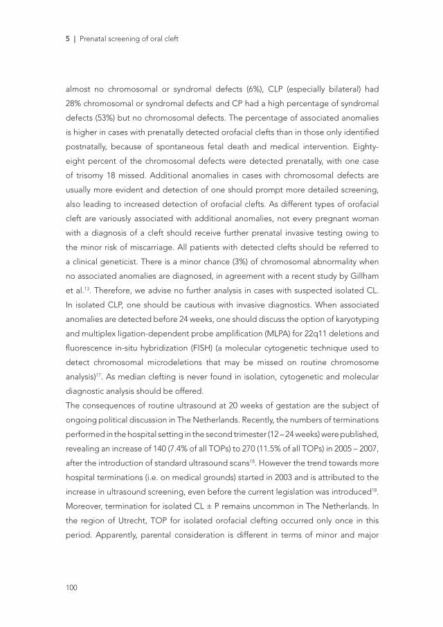
R1
R2
R3
R4
R5
R6
R7
R8
R9
R10
R11
R12
R13
R14
R15
R16
R17
R18
R19
R20
R21
R22
R23
R24
R25
R26
R27
R28
R29
R30
R31
R32
R33
R34
5 | Prenatal screening of oral cleft
100
almost no chromosomal or syndromal defects (6%), CLP (especially bilateral) had
28% chromosomal or syndromal defects and CP had a high percentage of syndromal
defects (53%) but no chromosomal defects. The percentage of associated anomalies
is higher in cases with prenatally detected orofacial clefts than in those only identified
postnatally, because of spontaneous fetal death and medical intervention. Eighty-
eight percent of the chromosomal defects were detected prenatally, with one case
of trisomy 18 missed. Additional anomalies in cases with chromosomal defects are
usually more evident and detection of one should prompt more detailed screening,
also leading to increased detection of orofacial clefts. As different types of orofacial
cleft are variously associated with additional anomalies, not every pregnant woman
with a diagnosis of a cleft should receive further prenatal invasive testing owing to
the minor risk of miscarriage. All patients with detected clefts should be referred to
a clinical geneticist. There is a minor chance (3%) of chromosomal abnormality when
no associated anomalies are diagnosed, in agreement with a recent study by Gillham
et al.13. Therefore, we advise no further analysis in cases with suspected isolated CL.
In isolated CLP, one should be cautious with invasive diagnostics. When associated
anomalies are detected before 24 weeks, one should discuss the option of karyotyping
and multiplex ligation-dependent probe amplification (MLPA) for 22q11 deletions and
fluorescence in-situ hybridization (FISH) (a molecular cytogenetic technique used to
detect chromosomal microdeletions that may be missed on routine chromosome
analysis)17. As median clefting is never found in isolation, cytogenetic and molecular
diagnostic analysis should be offered.
The consequences of routine ultrasound at 20 weeks of gestation are the subject of
ongoing political discussion in The Netherlands. Recently, the numbers of terminations
performed in the hospital setting in the second trimester (12 – 24 weeks) were published,
revealing an increase of 140 (7.4% of all TOPs) to 270 (11.5% of all TOPs) in 2005 – 2007,
after the introduction of standard ultrasound scans18. However the trend towards more
hospital terminations (i.e. on medical grounds) started in 2003 and is attributed to the
increase in ultrasound screening, even before the current legislation was introduced18.
Moreover, termination for isolated CL ± P remains uncommon in The Netherlands. In
the region of Utrecht, TOP for isolated orofacial clefting occurred only once in this
period. Apparently, parental consideration is different in terms of minor and major
R1
R2
R3
R4
R5
R6
R7
R8
R9
R10
R11
R12
R13
R14
R15
R16
R17
R18
R19
R20
R21
R22
R23
R24
R25
R26
R27
R28
R29
R30
R31
R32
R33
R34
101
deformities, especially when a deformity is readily amenable to reconstruction19.
In conclusion, this study cohort of a non-selected population of 38 760 pregnant women
included 62 fetuses with facial clefts. Thirty-nine per cent of these had associated
anomalies. There was a high sensitivity of detecting orofacial clefts by ultrasound
screening. The high rate of detection of an orofacial cleft is probably a result of the
excellent training and experience of the sonographers, referral to specialized centers
when a cleft is suspected, routine visualization of the fetal face and advances in
ultrasound techniques. Different types of orofacial cleft relate differently to associated
anomalies and thus to ultimate outcome. For this reason, pregnancies in which there
is suspicion of a fetal cleft with associated anomalies should be routinely referred for
further genetic analysis.
R1
R2
R3
R4
R5
R6
R7
R8
R9
R10
R11
R12
R13
R14
R15
R16
R17
R18
R19
R20
R21
R22
R23
R24
R25
R26
R27
R28
R29
R30
R31
R32
R33
R34
5 | Prenatal screening of oral cleft
102
REFERENCES
1 The Dutch Society of Obstetrics and Gynaecology, standards of routine ultrasound examination – modelprotocollen – Structureel Echoscopisch Onderzoek (SEO) 1.1. http://www.rivm.nl/pns/Images/nvog%20modelprotocol%20seo tcm95 – 62247.pdf.
2 Vermey Keers C. NVSCA (Dutch Society of Orofacial Cleft and Craniofacial Malformations) registration. 2009.
3 Mohangoo BS AD. Aangeboren afwijkingen in Nederland 1997 –2007; Gebaseerd op de landelijke verloskunde en neonatologie registraties, 2009; 83 –94.
4 Nyberg DA, Sickler GK, Hegge FN, Kramer DJ, Kropp RJ. Fetal cleft lip with and without cleft palate: US classification and correlation with outcome. Radiology 1995; 195:677 –684.
5 Offerdal K, Jebens N, Syvertsen T, Blaas HG, Johansen OJ, Eik-Nes SH. Prenatal ultrasound detection of facial clefts: a prospective study of 49 314 deliveries in a non-selected population in Norway. Ultrasound Obstet Gynecol 2008; 31: 639 646.
6 Berge SJ, Plath H, Van de Vondel PT, Appel T, Niederhagen B, Von Lindern JJ, Reich RH, Hansmann M. Fetal cleft lip and palate: sonographic diagnosis, chromosomal abnormalities, associated anomalies and postnatal outcome in 70 fetuses. Ultrasound Obstet Gynecol 2001; 18: 422 –431.
7 Perrotin F, de Poncheville LM, Marret H, Paillet C, Lansac J, Body G. Chromosomal defects and associatedmalformations in fetal cleft lip with or without cleft palate. Eur J Obstet Gynecol Reprod Biol 2001; 99: 19 –24.
8 Maarse W, Berge SJ, Pistorius L, van Barneveld T, Kon M, Breugem C, Mink van der Molen AB. Diagnostic accuracy of transabdominal ultrasound in detecting prenatal cleft lip and palate: a systematic review. Ultrasound Obstet Gynecol 2010; 35: 495 –502.
9 Offerdal K, Blaas HG, Eik-Nes SH. Prenatal detection of trisomy 21 by second-trimester ultrasound examination and maternal age in a non-selected population of 49 314 births in Norway. Ultrasound Obstet Gynecol 2008; 32: 493 –500.
10 Russell KA, Allen VM, MacDonald ME, Smith K, Dodds L. A population-based evaluation of antenatal diagnosis of orofacial clefts. Cleft Palate Craniofac J 2008; 45: 148 –153.
11 Mink van der Molen AB, Maarse W, Pistorius L, de Veye HS, Breugem CC. Prenatal screening for cleft lip and palate in the Netherlands: a preliminary report of the impact of a national screening system. Cleft Palate Craniofac J 2011; 48: 183 189.
12 Nusbaum R, Grubs RE, Losee JE, Weidman C, Ford MD, Marazita ML. A qualitative description of receiving a diagnosis of clefting in the prenatal or postnatal period. J Genet Couns 2008; 17: 336 –350.
13 Gillham JC, Anand S, Bullen PJ. Antenatal detection of cleft lip with or without cleft palate: incidence of associated chromosomal and structural anomalies. Ultrasound Obstet Gynecol 2009; 34: 410 –415.
14 Lie RT, Wilcox AJ, Skjaerven R. A population-based study of the risk of recurrence of birth defects. N Engl J Med 1994; 331: 1–4.
15 Sivertsen A, Wilcox AJ, Skjaerven R, Vindenes HA, Abyholm F, Harville E, Lie RT. Familial risk of oral clefts by morphological type and severity: population based cohort study of first degree relatives. BMJ 2008; 336: 432 –434.
16 Crane JP, LeFevre ML, Winborn RC, Evans JK, Ewigman BG, Bain RP, Frigoletto FD, McNellis D. A randomized trial of prenatal ultrasonographic screening: impact on the detection, management, and outcome of anomalous fetuses. The RADIUS Study Group. Am J Obstet Gynecol 1994; 171: 392 –399.
17 Suri M. Craniofacial syndromes. Semin Fetal Neonatal Med 2005; 10: 243 –257.18 Jaarrapportage 2007 van de Wet afbreking zwangerschap. http://www.igz.nl/zoeken/
download.aspx?download= Jaarrapportage 2007 Wet afbreking zwangerschap.pdf.

19 Jones MC. Prenatal diagnosis of cleft lip and palate: detection rates, accuracy of ultrasonography, associated anomalies, and strategies for counseling. Cleft Palate Craniofac J 2002; 39: 169 –173.

R1
R2
R3
R4
R5
R6
R7
R8
R9
R10
R11
R12
R13
R14
R15
R16
R17
R18
R19
R20
R21
R22
R23
R24
R25
R26
R27
R28
R29
R30
R31
R32
R33
R34
6The accuracy of prenatal ultrasound in
determining the type of orofacial cleft
Prenatal Diagnosis, 2015, Feb 26. [Epub ahead of print]
C.S. LOOZEN, W.MAARSE, G.T. MANTEN, L. PISTORIUS, C.C.BREUGEM
R1
R2
R3
R4
R5
R6
R7
R8
R9
R10
R11
R12
R13
R14
R15
R16
R17
R18
R19
R20
R21
R22
R23
R24
R25
R26
R27
R28
R29
R30
R31
R32
R33
R34
6 | Accuracy in the type of oral cleft
106
ABSTRACT
Objectives The aim of this study was to assess the accuracy of prenatal transabdominal
ultrasound in determining the oral cleft type.
Methods A retrospective cohort study was performed on all consecutive cases of
orofacial cleft diagnosed by prenatal ultrasound examination in the Wilhelmina
Children’s Hospital, a tertiary referral hospital, between January 2002 and December
2012. Prenatal findings were compared to postnatal diagnoses.
Results 134 patients were included. The mean gestational age at ultrasound examination
was 24 weeks + 5 days. Prenatal diagnosis was in accordance with postnatal findings
in 76.9% of cases (103/134) with regard to the cleft type. Underestimation of the cleft
occurred in 19.4% (26/134), whereas in 3.7% (5/134) the extent was overestimated. In
distinguishing bilateral from unilateral clefts, no errors were made.
Conclusion Prenatal ultrasound is accurate in assessing the types of orofacial clefts
in a large majority of the cases and is completely accurate in distinguishing between
uni- and bilateral clefts. This study indicates that it is a reliable technique to assess
the cleft type, which is important for counseling future parents. Although, the clinician
should be aware of the fact that a cleft palate is easily missed, and subsequently
underestimation of cleft extend is frequent.
R1
R2
R3
R4
R5
R6
R7
R8
R9
R10
R11
R12
R13
R14
R15
R16
R17
R18
R19
R20
R21
R22
R23
R24
R25
R26
R27
R28
R29
R30
R31
R32
R33
R34
107
INTRODUCTION
Orofacial clefts (OFCs) are common craniofacial malformations.1 In the Netherlands,
their combined incidence is about 1.8 per 1000 live births per year, resulting in
approximately 320 cases of OFC per year.2 OFCs can be categorized in different
subtypes, however a subdivision of three subtypes is most frequently described: cleft
lip (CL), cleft lip and palate (CLP) and isolated cleft palate (CP). To make a better
assessment of the prenatal diagnosis in this study, involvement of the alveolus (CLA)
was also assessed since this is of importance in counseling future parents.
The distribution of cleft type differs per country. In the Netherlands the distribution
was reported to be 29% CL, 40% CLP and 27% CP. 61% of the clefts were unilateral and
37% bilateral.3 The distribution of clefts in Canada was reported to be 17% CL, 42%
CLP and 41% CP.4 Canadian data report that clefts were 24% bilateral, 24% unilateral
right and 52% unilateral left.5 Ultrasonographers and cleft and palate teams (consisting
of an obstetrician, plastic surgeon, psychologist and clinical geneticist) are commonly
confronted with prenatally diagnosed clefts nowadays.3 Firstly, this could be attributed
to the increased number of women undergoing ultrasound (US) screening during
the second trimester of pregnancy since a countrywide screening programme was
introduced by legislation and insurance in the Netherlands in 2007. This programme
has now become standard care, resulting in a 90-95% participation of all pregnant
women in the Netherlands. Secondly, it could be attributed to the improved detection
as reported in different studies concerning the accuracy in prenatally detecting OFCs.6
An accurate prenatal diagnosis is of upmost importance in counseling future parents
on prenatal findings and to enable parents to prepare themselves in an emotional
and practical way.7 It is important for parents to know what to expect from the physical
appearance of their child and the trajectory that has to be undertaken in the hospital
after birth. Furthermore, prenatal differentiation between the various types of cleft
is important because the different OFC types are associated with various other
congenital anomalies and chromosomal abnormalities8 of which some have a great
impact on fetal prognosis with regard to morbidity and mortality.9
Different studies show a wide range in results concerning the prevalence of congenital
anomalies associated with OFCs, e.g. malformations of the limbs and congenital heart
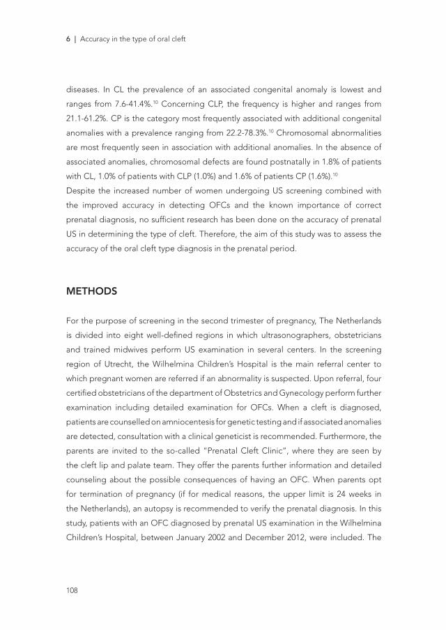
R1
R2
R3
R4
R5
R6
R7
R8
R9
R10
R11
R12
R13
R14
R15
R16
R17
R18
R19
R20
R21
R22
R23
R24
R25
R26
R27
R28
R29
R30
R31
R32
R33
R34
6 | Accuracy in the type of oral cleft
108
diseases. In CL the prevalence of an associated congenital anomaly is lowest and
ranges from 7.6-41.4%.10 Concerning CLP, the frequency is higher and ranges from
21.1-61.2%. CP is the category most frequently associated with additional congenital
anomalies with a prevalence ranging from 22.2-78.3%.10 Chromosomal abnormalities
are most frequently seen in association with additional anomalies. In the absence of
associated anomalies, chromosomal defects are found postnatally in 1.8% of patients
with CL, 1.0% of patients with CLP (1.0%) and 1.6% of patients CP (1.6%).10
Despite the increased number of women undergoing US screening combined with
the improved accuracy in detecting OFCs and the known importance of correct
prenatal diagnosis, no sufficient research has been done on the accuracy of prenatal
US in determining the type of cleft. Therefore, the aim of this study was to assess the
accuracy of the oral cleft type diagnosis in the prenatal period.
METHODS
For the purpose of screening in the second trimester of pregnancy, The Netherlands
is divided into eight well-defined regions in which ultrasonographers, obstetricians
and trained midwives perform US examination in several centers. In the screening
region of Utrecht, the Wilhelmina Children’s Hospital is the main referral center to
which pregnant women are referred if an abnormality is suspected. Upon referral, four
certified obstetricians of the department of Obstetrics and Gynecology perform further
examination including detailed examination for OFCs. When a cleft is diagnosed,
patients are counselled on amniocentesis for genetic testing and if associated anomalies
are detected, consultation with a clinical geneticist is recommended. Furthermore, the
parents are invited to the so-called “Prenatal Cleft Clinic”, where they are seen by
the cleft lip and palate team. They offer the parents further information and detailed
counseling about the possible consequences of having an OFC. When parents opt
for termination of pregnancy (if for medical reasons, the upper limit is 24 weeks in
the Netherlands), an autopsy is recommended to verify the prenatal diagnosis. In this
study, patients with an OFC diagnosed by prenatal US examination in the Wilhelmina
Children’s Hospital, between January 2002 and December 2012, were included. The
R1
R2
R3
R4
R5
R6
R7
R8
R9
R10
R11
R12
R13
R14
R15
R16
R17
R18
R19
R20
R21
R22
R23
R24
R25
R26
R27
R28
R29
R30
R31
R32
R33
R34
109
initial evaluation was performed within a week from diagnosis by a screening clinic
and was done by a fetal medicine specialist. All examinations were carried out trans
abdominally using a General Electric Voluson730 Expert equipped with a RAB4-8L
probe or a E8 US system equipped with a RAB4-8D probe (GE Medical Sytems, Kretz
US, Zipf, Austria). The main focus of the US examination was to exclude associated
fetal anomalies, and to provide a first assessment of the extent of the OFC. A follow-
up evaluation by the fetal medicine specialist was done within four weeks, with the
same ultrasound equipment, and in the presence of a plastic paediatric surgeon. The
main purpose of this evaluation was to determine the extent of the OFC. When the
cleft was limited to the lip, hence with intact alveolar ridge, the cases were classified
as CL. If there was a discontinuity of the maxillary arch frontal of the foramen incisivum
but the palate seemed intact, the cases were classified as CLA. If the lip, alveolus and
palate seemed affected the cases were classified as CLP. If the lip and alveolar ridge
seemed intact and there was suspicion of an affected palate the cases were classified
as CP. Furthermore it was described whether the cleft was uni- or bilateral. 2D US
was performed in all cases and an additional 3D US was performed if the fetus was
in a good position. In this study we included the cleft diagnosis of the last scan that
was performed. Patients with major associated anomalies (e.g. trisomy 13/18) were
excluded since the cleft was subordinate to other malformations and therefore not
well described. The US data were retrieved from the electronic information system
developed to record and process the US measurement and images from obstetrical
examinations (MOSOS; U; Version 11.0, BMA, Houten, the Netherlands).
Postnatal diagnoses were retrieved from electronic clinical records reported by
the cleft lip and palate team. Medical ethical board approval was obtained for this
study. All data were collected in a database. Prenatal diagnoses were compared
with postnatal findings. Accuracy of prenatal US was calculated based on postnatal
definitive diagnoses.
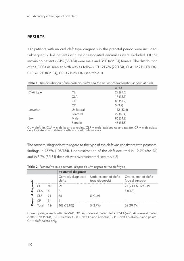
R1
R2
R3
R4
R5
R6
R7
R8
R9
R10
R11
R12
R13
R14
R15
R16
R17
R18
R19
R20
R21
R22
R23
R24
R25
R26
R27
R28
R29
R30
R31
R32
R33
R34
6 | Accuracy in the type of oral cleft
110
RESULTS
139 patients with an oral cleft type diagnosis in the prenatal period were included.
Subsequently, five patients with major associated anomalies were excluded. Of the
remaining patients, 64% (86/134) were male and 36% (48/134) female. The distribution
of the OFCs as seen at birth was as follows: CL: 21.6% (29/134), CLA: 12.7% (17/134),
CLP: 61.9% (83/134), CP: 3.7% (5/134) (see table 1).
Table 1. The distribution of the orofacial clefts and the patient characteristics as seen at birth
n (%)Cleft type CL 29 (21.6)
CLA 17 (12.7)CLP 83 (61.9)CP 5 (3.7)
Location Unilateral 112 (83.6)Bilateral 22 (16.4)
Sex Male 86 (64.2)Female 48 (35.8)
CL = cleft lip, CLA = cleft lip and alveolus, CLP = cleft lip/alveolus and palate, CP = cleft palate only. Unilateral = unilateral clefts and cleft palates only
The prenatal diagnosis with regard to the type of the cleft was consistent with postnatal
findings in 76.9% (103/134). Underestimation of the cleft occurred in 19.4% (26/134)
and in 3.7% (5/134) the cleft was overestimated (see table 2).
Table 2. Prenatal versus postnatal diagnosis with regard to the cleft type
Postnatal diagnosis
Pre
nata
l dia
gno
sis
Correctly diagnosed clefts
Underestimated clefts (true diagnosis)
Overestimated clefts(true diagnosis)
CL 50 29 - 21 (9 CLA, 12 CLP)
CLA 8 3 - 5 (CLP)
CLP 71 66 5 (CLA) -
CP 5 5 - -
Total 134 103 (76.9%) 5 (3.7%) 26 (19.4%)
Correctly diagnosed clefts: 76.9% (103/134), underestimated clefts: 19.4% (26/134), over-estimated clefts: 3.7% (5/134). CL = cleft lip, CLA = cleft lip and alveolus, CLP = cleft lip/alveolus and palate, CP = cleft palate only.
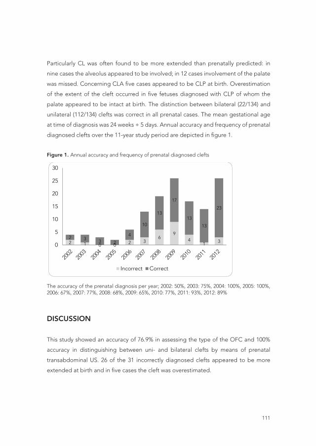
R1
R2
R3
R4
R5
R6
R7
R8
R9
R10
R11
R12
R13
R14
R15
R16
R17
R18
R19
R20
R21
R22
R23
R24
R25
R26
R27
R28
R29
R30
R31
R32
R33
R34
111
Particularly CL was often found to be more extended than prenatally predicted: in
nine cases the alveolus appeared to be involved; in 12 cases involvement of the palate
was missed. Concerning CLA five cases appeared to be CLP at birth. Overestimation
of the extent of the cleft occurred in five fetuses diagnosed with CLP of whom the
palate appeared to be intact at birth. The distinction between bilateral (22/134) and
unilateral (112/134) clefts was correct in all prenatal cases. The mean gestational age
at time of diagnosis was 24 weeks + 5 days. Annual accuracy and frequency of prenatal
diagnosed clefts over the 11-year study period are depicted in figure 1.
Figure 1. Annual accuracy and frequency of prenatal diagnosed cleftsFigure 1. Annual accuracy and frequency of prenatal diagnosed clefts
The accuracy of the prenatal diagnosis per year; 2002: 50%, 2003: 75%, 2004: 100%, 2005: 100%, 2006: 67%, 2007: 77%, 2008: 68%, 2009: 65%, 2010: 77%, 2011: 93%, 2012: 89%
Blz. 108, R 10 chance risk
Blz 108, R 26 for of all OFCs
Blz 109, R 6 echo ultrasound
Blz 109, R 16 are is based on a low number
Blz 109, R 12 big large
Blz 109, R23 Toevoegen: To assess whether
Blz 109, R25 does not –> doesn’t
Blz 109, R 26 had been was
Blz 110, R12 is occurs
2 1 0 0 2 36
94
1 32 3 3 2
4
10
13
17
13
13
23
0
5
10
15
20
2002
25
30
Incorrect Correct
2003
2004
2005
2006
2007
2008
2009
2010
2011
2012
The accuracy of the prenatal diagnosis per year; 2002: 50%, 2003: 75%, 2004: 100%, 2005: 100%, 2006: 67%, 2007: 77%, 2008: 68%, 2009: 65%, 2010: 77%, 2011: 93%, 2012: 89%
DISCUSSION
This study showed an accuracy of 76.9% in assessing the type of the OFC and 100%
accuracy in distinguishing between uni- and bilateral clefts by means of prenatal
transabdominal US. 26 of the 31 incorrectly diagnosed clefts appeared to be more
extended at birth and in five cases the cleft was overestimated.
R1
R2
R3
R4
R5
R6
R7
R8
R9
R10
R11
R12
R13
R14
R15
R16
R17
R18
R19
R20
R21
R22
R23
R24
R25
R26
R27
R28
R29
R30
R31
R32
R33
R34
6 | Accuracy in the type of oral cleft
112
An accurate diagnosis is of upmost importance for counselling parents on prenatal
findings and enabling parents to prepare themselves in an emotional and practical
way.7 Furthermore, an accurate diagnosis of cleft type is essential in determining the
risk of severe associated congenital anomalies since different subtypes have a different
correlation, as was mentioned in the introduction.8,9
Diagnosing a small indentation or incomplete cleft of the alveolus is extremely
difficult. Involvement of the alveolus is of clinical importance because it often means
a second operation, higher risk for a speaking disorder and dentofacial deformities.
Therefore, we also included CLA in our analysis. This has resulted in “strict” analysis; if
this differentiation is not made, the accuracy of US increases to 84%.
Accuracy rates of prenatal US are not consistent in literature. Berggren et al.11 reported
an accuracy rate of prenatal US of 43.2% (19/44) and Demircioglu et al.12 reported an
accuracy of 75.0% (66/88). These differences can be explained by small study size,
variation in study methods (multiple examiners and different US approaches) and/or
the discrepancy in terminology used. In our study all US scans were performed by
gynecologists specialized in advanced US examination in the Wilhelmina Children’s
Hospital. Yet in both studies11, 12 the most common reported error was a more extensive
cleft at birth than prenatally diagnosed, which was in line with our results.
A limitation of this study is the fact that the cases included may not be totally
representative for all OFCs, since all cases were referrals from clefts identified at
screening performed at a primary care setting. Subtle clefts may be missed at the
referring centers and thus not seen at the Wilhelmina Children’s Hospital. The clefts
referred may be more severe and therefore it may be easier to assess the type of
cleft. The examination could appear more thorough than if the general population
was used.
In our study period US examination appeared to be unable to visualize the cleft palate.
The abnormality of the palate was missed in 17 fetuses, even when the ultrasonographer
was focused on the possibility of a cleft palate. Visualizing the palate is difficult since
the palate has a dome-shaped structure and is surrounded by osseous structures. In
2010 Wilhelm et al.13 described a method to visualize the palate by US; they state that
an intact palate resembles an “equals sign” ultrasound pattern on US. In our study
five (5/134) cases of isolated cleft palate were detected, of which three cases in 2011
R1
R2
R3
R4
R5
R6
R7
R8
R9
R10
R11
R12
R13
R14
R15
R16
R17
R18
R19
R20
R21
R22
R23
R24
R25
R26
R27
R28
R29
R30
R31
R32
R33
R34
113
and two cases in 2012. This detection in the last two years could be explained by the
implementation of the Wilhelm”s method.
Over the years an increase in prenatally diagnosed clefts is seen (see figure 1) with
19 cases included before and 115 cases after the implementation of the universal
US screening in 2007. This increase could be explained by the increased number of
women undergoing US screening and the improved detection rate of OFCs.6 With
regard to defining the type of cleft, the accuracy presents a variable picture over
the years with an average accuracy of 73.7% before 2007 and 77.4% from 2007-2012.
Although data before 2007 are based on a low number of observations annually, we
did not exclude these cases because there has not been a large change in technique
or equipment to evaluate suspected clefts over those years. In the last two years of our
study, US is more accurate (2011: 93%, 2012: 89%) in comparison to the first three years
(2008: 68%, 2009: 65%, 2010: 77%) since the introduction of the screening programme
in 2007. The improvement could be explained by the implementation of the earlier
mentioned Wilhelm”s sign in 2010. Improvement in expertise and better skills of the
ultrasonographers might also attribute to the increase in accuracy. To assess whether
or not this trend will continue, we have to perform further study with longer follow-up.
This study doesn’t compare different techniques used for imaging the fetal palate but
was limited to test the validity of the existing techniques used by the Wilhelmina’s
Children’s Hospital cleft team. Determining whether or not the palate is involved
when clefting of the lip and alveolus are found requires different methods like “3D
reverse face view” revealed by Campbell et al.14. This technique allows a relatively
straightforward assessment of the fetal palate with a high degree of accuracy (8/8 =
100%; 95% ci 62.8- 100.0%). Moreover, Sommerlad et al.15 also describes methods to
more accurately diagnose a cleft of the alveolar ridge. To improve the accuracy of US
in determining the type of cleft future research should investigate other techniques,
e.g. transvaginal US or 4th dimension US.
R1
R2
R3
R4
R5
R6
R7
R8
R9
R10
R11
R12
R13
R14
R15
R16
R17
R18
R19
R20
R21
R22
R23
R24
R25
R26
R27
R28
R29
R30
R31
R32
R33
R34
6 | Accuracy in the type of oral cleft
114
CONCLUSION
Prenatal ultrasound is accurate in assessing the types of orofacial clefts in a large
majority of the cases and is completely accurate in distinguishing between uni- and
bilateral clefts. This study indicates that it is a reliable technique to assess the cleft type,
which is important for counseling future parents. Although, the clinician should be
aware of the fact that a cleft palate is easily missed, and subsequently underestimation
of cleft extend occurs frequent.
R1
R2
R3
R4
R5
R6
R7
R8
R9
R10
R11
R12
R13
R14
R15
R16
R17
R18
R19
R20
R21
R22
R23
R24
R25
R26
R27
R28
R29
R30
R31
R32
R33
R34
115
REFERENCES
1. Mossey PA, Little J, Munger RG, et al. Cleft lip and palate. Lancet. 2009 Nov 21; 374 (9703):1773-85.
2. Van den Berg MP, Hage JJ, Kortekaas RT, et al. Cleft lip and palate, a continuous problem for the patient? Ned Tijdschr Geneeskd. 1994 May 21; 138 (21): 1058-61
3. Maarse W, Pistorius LR, van Eeten WK, et al. Prenatal ultrasound screening for orofacial clefts. Ultrasound Obstet Gynecol. 2011; 38: 434–439
4. Pavri S, Forrest CR. Demographics of orofacial clefts in Canada from 2002 to 2008. Cleft Palate Craniofac J. 2013 Mar;50(2):224-30.
5. Gundlach KK, Maus C. Epidemiological studies on the frequency of clefts in Europe and world-wide. J Craniomaxillofac Surg. 2006 Sep; 34 Suppl 2:1-2.
6. Maarse W, Berge SJ, Pistorius L, et al. Diagnostic accuracy of transabdominal ultrasound in detecting prenatal cleft lip and palate, a systematic review. Ultrasound Obtet Gynecol. 2010; 35: 495-502.
7. Nusbaum R, Grubs RE, Losee JE, et al. A qualitative description of receiving a diagnosis of clefting in prenatal or postnatal period. J. Genet Couns 2008; 17: 336-350
8. Berge SJ, Plath H, Van de Vondel, et al. Fetal cleft lip and palate: sonographic diagnosis, chromosomal abnormalities, associated anomalies and postnatal outcome in 70 fetuses. Ultrasound Obstet Gynecol 2001; 18: 422-431
9. Nyberg DA, Sickler GK, Hegge FN, et al. Fetal cleft lip with and without cleft palate: US classification and correlation with outcome. Radiology 1995; 195: 677-684
10. Maarse W, Rozendaal AM, Pajkrt E, et al. A systematic review of associated structural and chromosomal defects in oral clefts: when is prenatal genetic analysis indicated? J Med Genet. 2012 Aug; 49 (8): 490-8.
11. Berggren H, Hansson E, Uvemark A, et al. Prenatal ultrasound detection of cleft lip, or cleft palate, or both, in southern Sweden, 2006-2010. J Plast Surg Hand Surg. 2012 Apr; 46(2): 69-74.
12. Demircioglu M, Kangesu L, Ismail A, et al. Increasing accuracy of antenatal ultrasound diagnosis of cleft lip with or without cleft palate, in cases referred to the North Thames London Region. Ultrasound Obstet Gynecol 2008; 31: 647–651
13. Wilhelm L, Borgers H. The ‘equals sign’: a novel marker in the diagnosis of fetal isolated cleft palate. Ultrasound Obstet Gynecol. 2010 Oct; 36 (4): 439-44.
14. Campbell S, Lees C, Moscoso G, et al. Ultrasound antenatal diagnosis of cleft palate by a new technique: the 3D “reverse face” view. Ultrasound Obstet Gynecol 2005; 25: 12–18
15. Sommerlad M, Patel N, Vijayalakshmi B, et al. Detection of lip, alveolar ridge and hard palate abnormalities using two-dimensional ultrasound enhanced with the three-dimensional reverse-face view. Ultrasound Obstet Gynecol. 2010 Nov; 36(5):596-600.

7A systematic review of associated
structural and chromosomal defects in
oral clefts: when is prenatal genetic
analysis indicated?
J Med Genet 2012; 49: 490–498
W.MAARSE*, A.M. ROZENDAAL* E.PAJKRT, C. VERMEIJ-KEERS,
A.B. MINK VAN DER MOLEN, M.J.H. VAN DEN BOOGAARD
* Joined first authorship; these authors contributed equally to this work
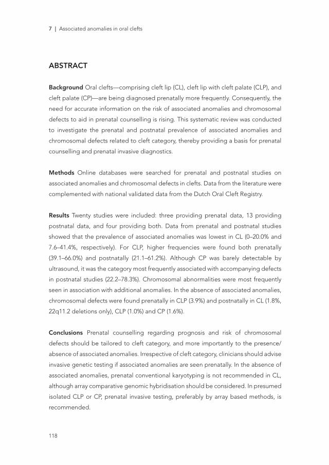
R1
R2
R3
R4
R5
R6
R7
R8
R9
R10
R11
R12
R13
R14
R15
R16
R17
R18
R19
R20
R21
R22
R23
R24
R25
R26
R27
R28
R29
R30
R31
R32
R33
R34
7 | Associated anomalies in oral clefts
118
ABSTRACT
Background Oral clefts—comprising cleft lip (CL), cleft lip with cleft palate (CLP), and
cleft palate (CP)—are being diagnosed prenatally more frequently. Consequently, the
need for accurate information on the risk of associated anomalies and chromosomal
defects to aid in prenatal counselling is rising. This systematic review was conducted
to investigate the prenatal and postnatal prevalence of associated anomalies and
chromosomal defects related to cleft category, thereby providing a basis for prenatal
counselling and prenatal invasive diagnostics.
Methods Online databases were searched for prenatal and postnatal studies on
associated anomalies and chromosomal defects in clefts. Data from the literature were
complemented with national validated data from the Dutch Oral Cleft Registry.
Results Twenty studies were included: three providing prenatal data, 13 providing
postnatal data, and four providing both. Data from prenatal and postnatal studies
showed that the prevalence of associated anomalies was lowest in CL (0–20.0% and
7.6–41.4%, respectively). For CLP, higher frequencies were found both prenatally
(39.1–66.0%) and postnatally (21.1–61.2%). Although CP was barely detectable by
ultrasound, it was the category most frequently associated with accompanying defects
in postnatal studies (22.2–78.3%). Chromosomal abnormalities were most frequently
seen in association with additional anomalies. In the absence of associated anomalies,
chromosomal defects were found prenatally in CLP (3.9%) and postnatally in CL (1.8%,
22q11.2 deletions only), CLP (1.0%) and CP (1.6%).
Conclusions Prenatal counselling regarding prognosis and risk of chromosomal
defects should be tailored to cleft category, and more importantly to the presence/
absence of associated anomalies. Irrespective of cleft category, clinicians should advise
invasive genetic testing if associated anomalies are seen prenatally. In the absence of
associated anomalies, prenatal conventional karyotyping is not recommended in CL,
although array comparative genomic hybridisation should be considered. In presumed
isolated CLP or CP, prenatal invasive testing, preferably by array based methods, is
recommended.
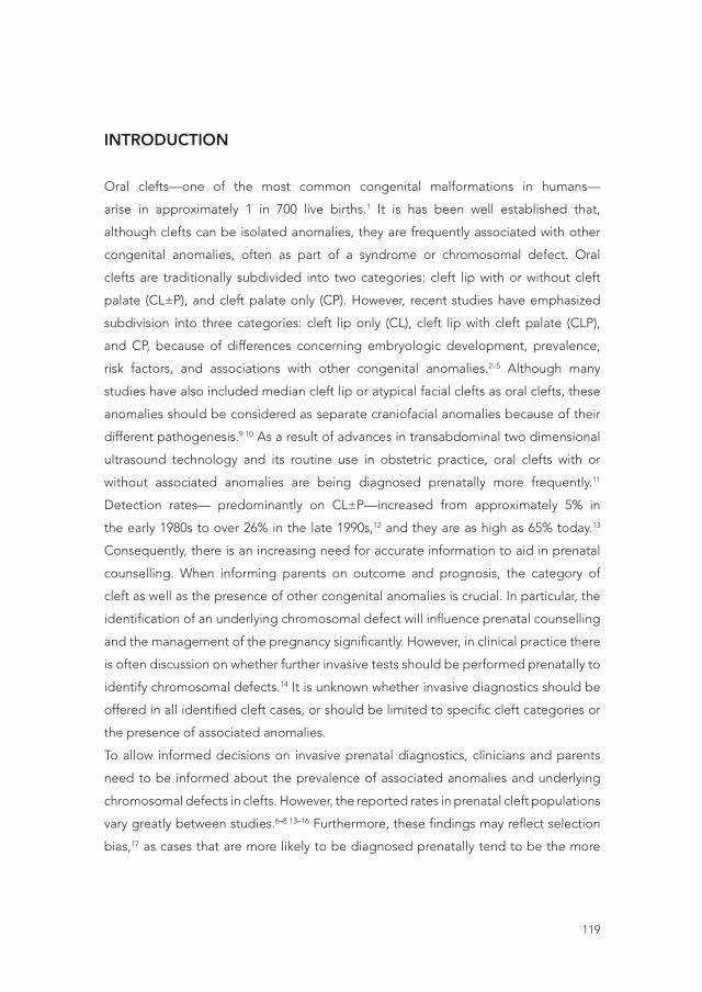
R1
R2
R3
R4
R5
R6
R7
R8
R9
R10
R11
R12
R13
R14
R15
R16
R17
R18
R19
R20
R21
R22
R23
R24
R25
R26
R27
R28
R29
R30
R31
R32
R33
R34
119
INTRODUCTION
Oral clefts—one of the most common congenital malformations in humans—
arise in approximately 1 in 700 live births.1 It is has been well established that,
although clefts can be isolated anomalies, they are frequently associated with other
congenital anomalies, often as part of a syndrome or chromosomal defect. Oral
clefts are traditionally subdivided into two categories: cleft lip with or without cleft
palate (CL±P), and cleft palate only (CP). However, recent studies have emphasized
subdivision into three categories: cleft lip only (CL), cleft lip with cleft palate (CLP),
and CP, because of differences concerning embryologic development, prevalence,
risk factors, and associations with other congenital anomalies.2–5 Although many
studies have also included median cleft lip or atypical facial clefts as oral clefts, these
anomalies should be considered as separate craniofacial anomalies because of their
different pathogenesis.9 10 As a result of advances in transabdominal two dimensional
ultrasound technology and its routine use in obstetric practice, oral clefts with or
without associated anomalies are being diagnosed prenatally more frequently.11
Detection rates— predominantly on CL±P—increased from approximately 5% in
the early 1980s to over 26% in the late 1990s,12 and they are as high as 65% today.13
Consequently, there is an increasing need for accurate information to aid in prenatal
counselling. When informing parents on outcome and prognosis, the category of
cleft as well as the presence of other congenital anomalies is crucial. In particular, the
identification of an underlying chromosomal defect will influence prenatal counselling
and the management of the pregnancy significantly. However, in clinical practice there
is often discussion on whether further invasive tests should be performed prenatally to
identify chromosomal defects.14 It is unknown whether invasive diagnostics should be
offered in all identified cleft cases, or should be limited to specific cleft categories or
the presence of associated anomalies.
To allow informed decisions on invasive prenatal diagnostics, clinicians and parents
need to be informed about the prevalence of associated anomalies and underlying
chromosomal defects in clefts. However, the reported rates in prenatal cleft populations
vary greatly between studies.6–8 13–16 Furthermore, these findings may reflect selection
bias,17 as cases that are more likely to be diagnosed prenatally tend to be the more
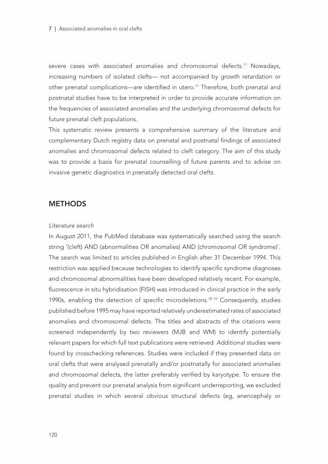
R1
R2
R3
R4
R5
R6
R7
R8
R9
R10
R11
R12
R13
R14
R15
R16
R17
R18
R19
R20
R21
R22
R23
R24
R25
R26
R27
R28
R29
R30
R31
R32
R33
R34
7 | Associated anomalies in oral clefts
120
severe cases with associated anomalies and chromosomal defects.11 Nowadays,
increasing numbers of isolated clefts— not accompanied by growth retardation or
other prenatal complications—are identified in utero.11 Therefore, both prenatal and
postnatal studies have to be interpreted in order to provide accurate information on
the frequencies of associated anomalies and the underlying chromosomal defects for
future prenatal cleft populations.
This systematic review presents a comprehensive summary of the literature and
complementary Dutch registry data on prenatal and postnatal findings of associated
anomalies and chromosomal defects related to cleft category. The aim of this study
was to provide a basis for prenatal counselling of future parents and to advise on
invasive genetic diagnostics in prenatally detected oral clefts.
METHODS
Literature search
In August 2011, the PubMed database was systematically searched using the search
string ‘(cleft) AND (abnormalities OR anomalies) AND (chromosomal OR syndrome)’.
The search was limited to articles published in English after 31 December 1994. This
restriction was applied because technologies to identify specific syndrome diagnoses
and chromosomal abnormalities have been developed relatively recent. For example,
fluorescence in situ hybridisation (FISH) was introduced in clinical practice in the early
1990s, enabling the detection of specific microdeletions.18 19 Consequently, studies
published before 1995 may have reported relatively underestimated rates of associated
anomalies and chromosomal defects. The titles and abstracts of the citations were
screened independently by two reviewers (MJB and WM) to identify potentially
relevant papers for which full text publications were retrieved. Additional studies were
found by crosschecking references. Studies were included if they presented data on
oral clefts that were analysed prenatally and/or postnatally for associated anomalies
and chromosomal defects, the latter preferably verified by karyotype. To ensure the
quality and prevent our prenatal analysis from significant underreporting, we excluded
prenatal studies in which several obvious structural defects (eg, anencephaly or
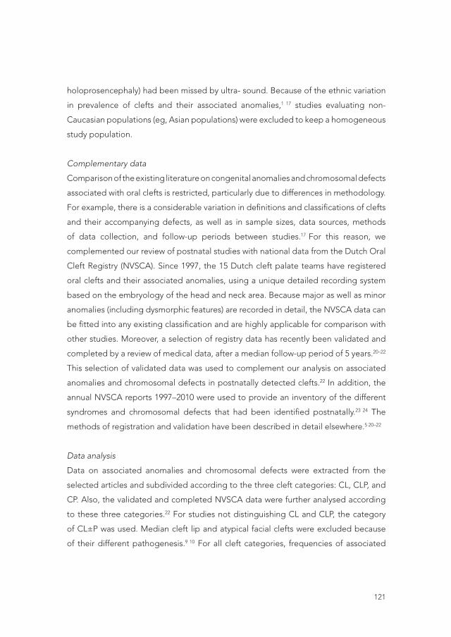
R1
R2
R3
R4
R5
R6
R7
R8
R9
R10
R11
R12
R13
R14
R15
R16
R17
R18
R19
R20
R21
R22
R23
R24
R25
R26
R27
R28
R29
R30
R31
R32
R33
R34
121
holoprosencephaly) had been missed by ultra- sound. Because of the ethnic variation
in prevalence of clefts and their associated anomalies,1 17 studies evaluating non-
Caucasian populations (eg, Asian populations) were excluded to keep a homogeneous
study population.
Complementary data
Comparison of the existing literature on congenital anomalies and chromosomal defects
associated with oral clefts is restricted, particularly due to differences in methodology.
For example, there is a considerable variation in definitions and classifications of clefts
and their accompanying defects, as well as in sample sizes, data sources, methods
of data collection, and follow-up periods between studies.17 For this reason, we
complemented our review of postnatal studies with national data from the Dutch Oral
Cleft Registry (NVSCA). Since 1997, the 15 Dutch cleft palate teams have registered
oral clefts and their associated anomalies, using a unique detailed recording system
based on the embryology of the head and neck area. Because major as well as minor
anomalies (including dysmorphic features) are recorded in detail, the NVSCA data can
be fitted into any existing classification and are highly applicable for comparison with
other studies. Moreover, a selection of registry data has recently been validated and
completed by a review of medical data, after a median follow-up period of 5 years.20–22
This selection of validated data was used to complement our analysis on associated
anomalies and chromosomal defects in postnatally detected clefts.22 In addition, the
annual NVSCA reports 1997–2010 were used to provide an inventory of the different
syndromes and chromosomal defects that had been identified postnatally.23 24 The
methods of registration and validation have been described in detail elsewhere.5 20–22
Data analysis
Data on associated anomalies and chromosomal defects were extracted from the
selected articles and subdivided according to the three cleft categories: CL, CLP, and
CP. Also, the validated and completed NVSCA data were further analysed according
to these three categories.22 For studies not distinguishing CL and CLP, the category
of CL±P was used. Median cleft lip and atypical facial clefts were excluded because
of their different pathogenesis.9 10 For all cleft categories, frequencies of associated
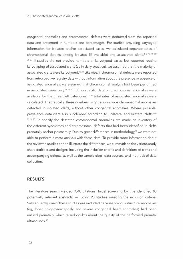
R1
R2
R3
R4
R5
R6
R7
R8
R9
R10
R11
R12
R13
R14
R15
R16
R17
R18
R19
R20
R21
R22
R23
R24
R25
R26
R27
R28
R29
R30
R31
R32
R33
R34
7 | Associated anomalies in oral clefts
122
congenital anomalies and chromosomal defects were deducted from the reported
data and presented in numbers and percentages. For studies providing karyotype
information for isolated and/or associated cases, we calculated separate rates of
chromosomal defects among isolated (if available) and associated clefts.6–8 13–15 22
25–27 If studies did not provide numbers of karyotyped cases, but reported routine
karyotyping of associated clefts (as in daily practice), we assumed that the majority of
associated clefts were karyotyped.13 22 Likewise, if chromosomal defects were reported
from retrospective registry data without information about the presence or absence of
associated anomalies, we assumed that chromosomal analysis had been performed
in associated cases only.16 26 28–31 If no specific data on chromosomal anomalies were
available for the three cleft categories,32–36 total rates of associated anomalies were
calculated. Theoretically, these numbers might also include chromosomal anomalies
detected in isolated clefts, without other congenital anomalies. Where possible,
prevalence data were also subdivided according to unilateral and bilateral clefts.6–8
13 14 25 To specify the detected chromosomal anomalies, we made an inventory of
the different syndromes and chromosomal defects that had been identified in clefts
prenatally and/or postnatally. Due to great differences in methodology,17 we were not
able to perform a meta-analysis with these data. To provide more information about
the reviewed studies and to illustrate the differences, we summarised the various study
characteristics and designs, including the inclusion criteria and definitions of clefts and
accompanying defects, as well as the sample sizes, data sources, and methods of data
collection.
RESULTS
The literature search yielded 9540 citations. Initial screening by title identified 88
potentially relevant abstracts, including 20 studies meeting the inclusion criteria.
Subsequently, one of these studies was excluded because obvious structural anomalies
(eg, lobar holoprosencephaly and severe congenital heart anomalies) had been
missed prenatally, which raised doubts about the quality of the performed prenatal
ultrasounds.37
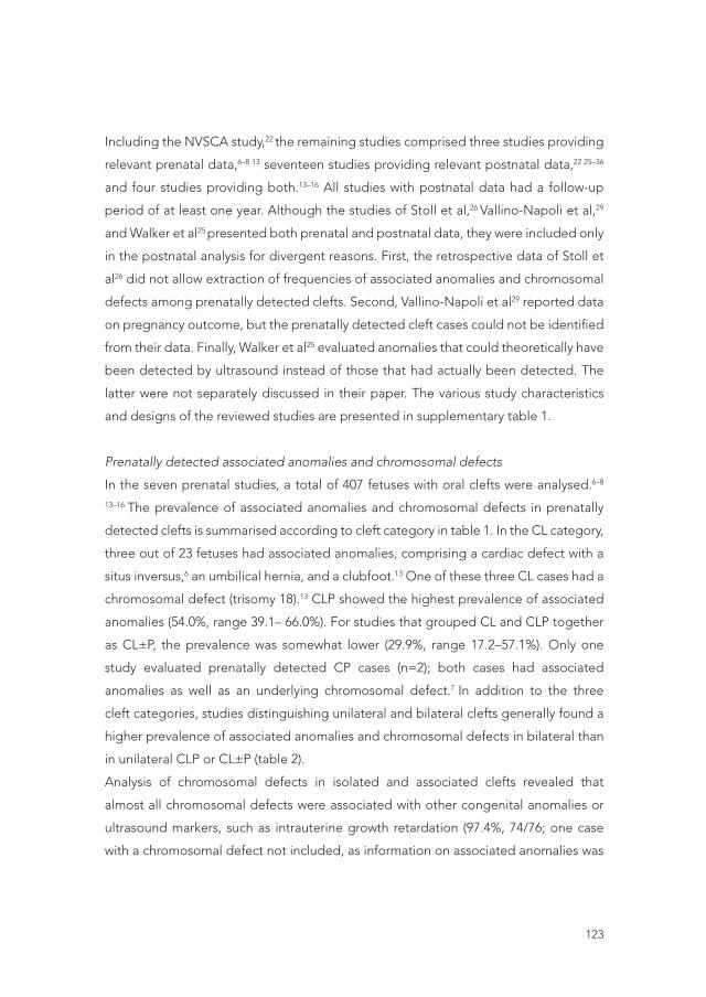
R1
R2
R3
R4
R5
R6
R7
R8
R9
R10
R11
R12
R13
R14
R15
R16
R17
R18
R19
R20
R21
R22
R23
R24
R25
R26
R27
R28
R29
R30
R31
R32
R33
R34
123
Including the NVSCA study,22 the remaining studies comprised three studies providing
relevant prenatal data,6–8 13 seventeen studies providing relevant postnatal data,22 25–36
and four studies providing both.13–16 All studies with postnatal data had a follow-up
period of at least one year. Although the studies of Stoll et al,26 Vallino-Napoli et al,29
and Walker et al25 presented both prenatal and postnatal data, they were included only
in the postnatal analysis for divergent reasons. First, the retrospective data of Stoll et
al26 did not allow extraction of frequencies of associated anomalies and chromosomal
defects among prenatally detected clefts. Second, Vallino-Napoli et al29 reported data
on pregnancy outcome, but the prenatally detected cleft cases could not be identified
from their data. Finally, Walker et al25 evaluated anomalies that could theoretically have
been detected by ultrasound instead of those that had actually been detected. The
latter were not separately discussed in their paper. The various study characteristics
and designs of the reviewed studies are presented in supplementary table 1.
Prenatally detected associated anomalies and chromosomal defects
In the seven prenatal studies, a total of 407 fetuses with oral clefts were analysed.6–8
13–16 The prevalence of associated anomalies and chromosomal defects in prenatally
detected clefts is summarised according to cleft category in table 1. In the CL category,
three out of 23 fetuses had associated anomalies, comprising a cardiac defect with a
situs inversus,6 an umbilical hernia, and a clubfoot.13 One of these three CL cases had a
chromosomal defect (trisomy 18).13 CLP showed the highest prevalence of associated
anomalies (54.0%, range 39.1– 66.0%). For studies that grouped CL and CLP together
as CL±P, the prevalence was somewhat lower (29.9%, range 17.2–57.1%). Only one
study evaluated prenatally detected CP cases (n=2); both cases had associated
anomalies as well as an underlying chromosomal defect.7 In addition to the three
cleft categories, studies distinguishing unilateral and bilateral clefts generally found a
higher prevalence of associated anomalies and chromosomal defects in bilateral than
in unilateral CLP or CL±P (table 2).
Analysis of chromosomal defects in isolated and associated clefts revealed that
almost all chromosomal defects were associated with other congenital anomalies or
ultrasound markers, such as intrauterine growth retardation (97.4%, 74/76; one case
with a chromosomal defect not included, as information on associated anomalies was
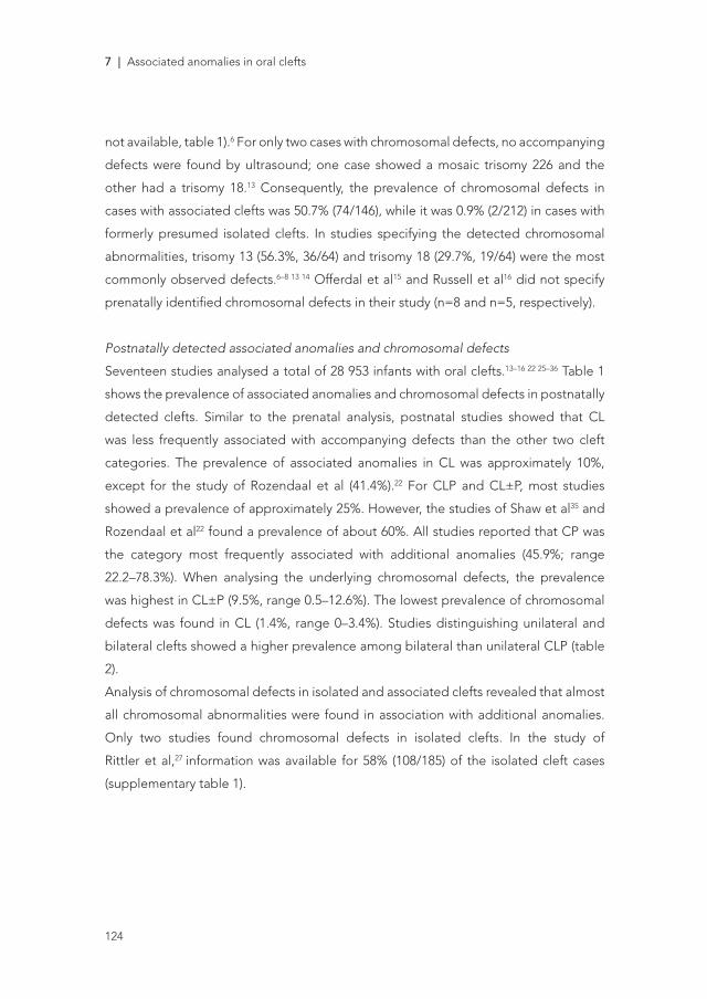
R1
R2
R3
R4
R5
R6
R7
R8
R9
R10
R11
R12
R13
R14
R15
R16
R17
R18
R19
R20
R21
R22
R23
R24
R25
R26
R27
R28
R29
R30
R31
R32
R33
R34
7 | Associated anomalies in oral clefts
124
not available, table 1).6 For only two cases with chromosomal defects, no accompanying
defects were found by ultrasound; one case showed a mosaic trisomy 226 and the
other had a trisomy 18.13 Consequently, the prevalence of chromosomal defects in
cases with associated clefts was 50.7% (74/146), while it was 0.9% (2/212) in cases with
formerly presumed isolated clefts. In studies specifying the detected chromosomal
abnormalities, trisomy 13 (56.3%, 36/64) and trisomy 18 (29.7%, 19/64) were the most
commonly observed defects.6–8 13 14 Offerdal et al15 and Russell et al16 did not specify
prenatally identified chromosomal defects in their study (n=8 and n=5, respectively).
Postnatally detected associated anomalies and chromosomal defects
Seventeen studies analysed a total of 28 953 infants with oral clefts.13–16 22 25–36 Table 1
shows the prevalence of associated anomalies and chromosomal defects in postnatally
detected clefts. Similar to the prenatal analysis, postnatal studies showed that CL
was less frequently associated with accompanying defects than the other two cleft
categories. The prevalence of associated anomalies in CL was approximately 10%,
except for the study of Rozendaal et al (41.4%).22 For CLP and CL±P, most studies
showed a prevalence of approximately 25%. However, the studies of Shaw et al35 and
Rozendaal et al22 found a prevalence of about 60%. All studies reported that CP was
the category most frequently associated with additional anomalies (45.9%; range
22.2–78.3%). When analysing the underlying chromosomal defects, the prevalence
was highest in CL±P (9.5%, range 0.5–12.6%). The lowest prevalence of chromosomal
defects was found in CL (1.4%, range 0–3.4%). Studies distinguishing unilateral and
bilateral clefts showed a higher prevalence among bilateral than unilateral CLP (table
2).
Analysis of chromosomal defects in isolated and associated clefts revealed that almost
all chromosomal abnormalities were found in association with additional anomalies.
Only two studies found chromosomal defects in isolated clefts. In the study of
Rittler et al,27 information was available for 58% (108/185) of the isolated cleft cases
(supplementary table 1).
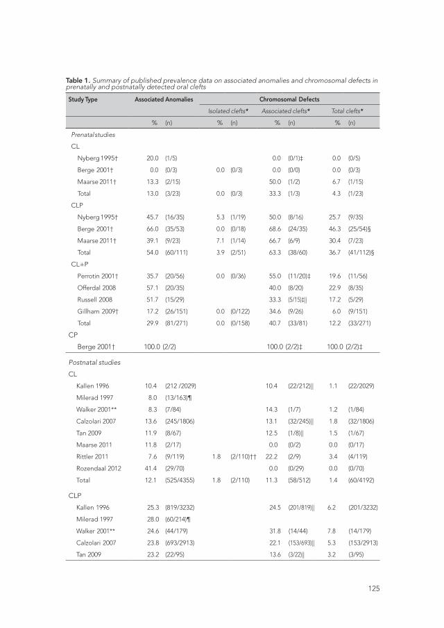
R1
R2
R3
R4
R5
R6
R7
R8
R9
R10
R11
R12
R13
R14
R15
R16
R17
R18
R19
R20
R21
R22
R23
R24
R25
R26
R27
R28
R29
R30
R31
R32
R33
R34
125
Table 1. Summary of published prevalence data on associated anomalies and chromosomal defects in prenatally and postnatally detected oral clefts
Study Type Associated Anomalies Chromosomal Defects
Isolated clefts* Associated clefts* Total clefts*
% (n) % (n) % (n) % (n)
Prenatal studies
CL
Nyberg 1995† 20.0 (1/5) 0.0 (0/1)‡ 0.0 (0/5)
Berge 2001† 0.0 (0/3) 0.0 (0/3) 0.0 (0/0) 0.0 (0/3)
Maarse 2011† 13.3 (2/15) 50.0 (1/2) 6.7 (1/15)
Total 13.0 (3/23) 0.0 (0/3) 33.3 (1/3) 4.3 (1/23)
CLP
Nyberg 1995† 45.7 (16/35) 5.3 (1/19) 50.0 (8/16) 25.7 (9/35)
Berge 2001† 66.0 (35/53) 0.0 (0/18) 68.6 (24/35) 46.3 (25/54)§
Maarse 2011† 39.1 (9/23) 7.1 (1/14) 66.7 (6/9) 30.4 (7/23)
Total 54.0 (60/111) 3.9 (2/51) 63.3 (38/60) 36.7 (41/112)§
CL±P
Perrotin 2001† 35.7 (20/56) 0.0 (0/36) 55.0 (11/20)‡ 19.6 (11/56)
Offerdal 2008 57.1 (20/35) 40.0 (8/20) 22.9 (8/35)
Russell 2008 51.7 (15/29) 33.3 (5/15)‡|| 17.2 (5/29)
Gillham 2009† 17.2 (26/151) 0.0 (0/122) 34.6 (9/26) 6.0 (9/151)
Total 29.9 (81/271) 0.0 (0/158) 40.7 (33/81) 12.2 (33/271)
CP
Berge 2001† 100.0 (2/2) 100.0 (2/2)‡ 100.0 (2/2)‡
Postnatal studies
CL
Kallen 1996 10.4 (212 /2029) 10.4 (22/212)|| 1.1 (22/2029)
Milerad 1997 8.0 (13/163)¶
Walker 2001** 8.3 (7/84) 14.3 (1/7) 1.2 (1/84)
Calzolari 2007 13.6 (245/1806) 13.1 (32/245)|| 1.8 (32/1806)
Tan 2009 11.9 (8/67) 12.5 (1/8)|| 1.5 (1/67)
Maarse 2011 11.8 (2/17) 0.0 (0/2) 0.0 (0/17)
Rittler 2011 7.6 (9/119) 1.8 (2/110)†† 22.2 (2/9) 3.4 (4/119)
Rozendaal 2012 41.4 (29/70) 0.0 (0/29) 0.0 (0/70)
Total 12.1 (525/4355) 1.8 (2/110) 11.3 (58/512) 1.4 (60/4192)
CLP
Kallen 1996 25.3 (819/3232) 24.5 (201/819)|| 6.2 (201/3232)
Milerad 1997 28.0 (60/214)¶
Walker 2001** 24.6 (44/179) 31.8 (14/44) 7.8 (14/179)
Calzolari 2007 23.8 (693/2913) 22.1 (153/693)|| 5.3 (153/2913)
Tan 2009 23.2 (22/95) 13.6 (3/22)|| 3.2 (3/95)
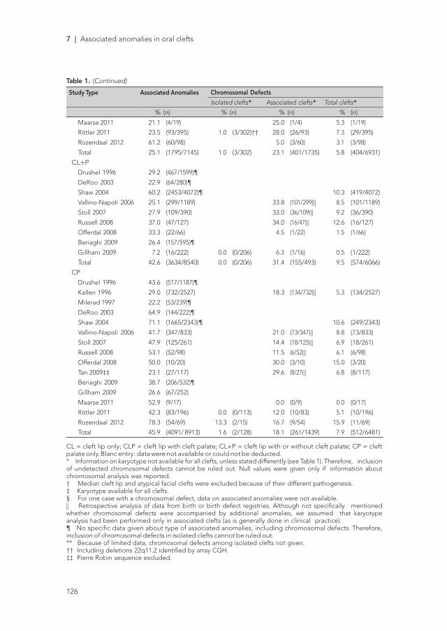
R1
R2
R3
R4
R5
R6
R7
R8
R9
R10
R11
R12
R13
R14
R15
R16
R17
R18
R19
R20
R21
R22
R23
R24
R25
R26
R27
R28
R29
R30
R31
R32
R33
R34
7 | Associated anomalies in oral clefts
126
Table 1. (Continued)
Study Type Associated Anomalies Chromosomal Defects
Isolated clefts* Associated clefts* Total clefts*
% (n) % (n) % (n) % (n)
Maarse 2011 21.1 (4/19) 25.0 (1/4) 5.3 (1/19)
Rittler 2011 23.5 (93/395) 1.0 (3/302)†† 28.0 (26/93) 7.3 (29/395)
Rozendaal 2012 61.2 (60/98) 5.0 (3/60) 3.1 (3/98)
Total 25.1 (1795/7145) 1.0 (3/302) 23.1 (401/1735) 5.8 (404/6931)
CL±P
Drushel 1996 29.2 (467/1599)¶
DeRoo 2003 22.9 (64/280)¶
Shaw 2004 60.2 (2453/4072)¶ 10.3 (419/4072)
Vallino-Napoli 2006 25.1 (299/1189) 33.8 (101/299)|| 8.5 (101/1189)
Stoll 2007 27.9 (109/390) 33.0 (36/109)|| 9.2 (36/390)
Russell 2008 37.0 (47/127) 34.0 (16/47)|| 12.6 (16/127)
Offerdal 2008 33.3 (22/66) 4.5 (1/22) 1.5 (1/66)
Beriaghi 2009 26.4 (157/595)¶
Gillham 2009 7.2 (16/222) 0.0 (0/206) 6.3 (1/16) 0.5 (1/222)
Total 42.6 (3634/8540) 0.0 (0/206) 31.4 (155/493) 9.5 (574/6066)
CP
Drushel 1996 43.6 (517/1187)¶
Kallen 1996 29.0 (732/2527) 18.3 (134/732)|| 5.3 (134/2527)
Milerad 1997 22.2 (53/239)¶
DeRoo 2003 64.9 (144/222)¶
Shaw 2004 71.1 (1665/2343)¶ 10.6 (249/2343)
Vallino-Napoli 2006 41.7 (347/833) 21.0 (73/347)|| 8.8 (73/833)
Stoll 2007 47.9 (125/261) 14.4 (18/125)|| 6.9 (18/261)
Russell 2008 53.1 (52/98) 11.5 (6/52)|| 6.1 (6/98)
Offerdal 2008 50.0 (10/20) 30.0 (3/10) 15.0 (3/20)
Tan 2009‡‡ 23.1 (27/117) 29.6 (8/27)|| 6.8 (8/117)
Beriaghi 2009 38.7 (206/532)¶
Gillham 2009 26.6 (67/252)
Maarse 2011 52.9 (9/17) 0.0 (0/9) 0.0 (0/17)
Rittler 2011 42.3 (83/196) 0.0 (0/113) 12.0 (10/83) 5.1 (10/196)
Rozendaal 2012 78.3 (54/69) 13.3 (2/15) 16.7 (9/54) 15.9 (11/69)
Total 45.9 (4091/ 8913) 1.6 (2/128) 18.1 (261/1439) 7.9 (512/6481)
CL = cleft lip only; CLP = cleft lip with cleft palate; CL±P = cleft lip with or without cleft palate; CP = cleft palate only. Blanc entry: data were not available or could not be deducted.* Information on karyotype not available for all clefts, unless stated differently (see Table 1). Therefore, inclusion of undetected chromosomal defects cannot be ruled out. Null values were given only if information about chromosomal analysis was reported.† Median cleft lip and atypical facial clefts were excluded because of their different pathogenesis.‡ Karyotype available for all clefts.§ For one case with a chromosomal defect, data on associated anomalies were not available.|| Retrospective analysis of data from birth or birth defect registries. Although not specifically mentioned whether chromosomal defects were accompanied by additional anomalies, we assumed that karyotype analysis had been performed only in associated clefts (as is generally done in clinical practice).¶ No specific data given about type of associated anomalies, including chromosomal defects. Therefore, inclusion of chromosomal defects in isolated clefts cannot be ruled out.** Because of limited data, chromosomal defects among isolated clefts not given.†† Including deletions 22q11.2 identified by array CGH.‡‡ Pierre Robin sequence excluded.
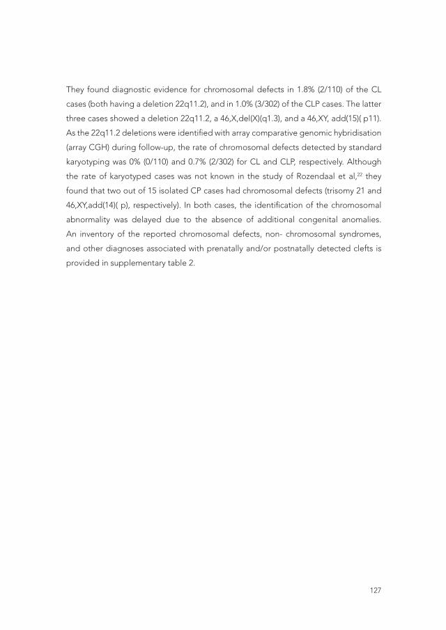
R1
R2
R3
R4
R5
R6
R7
R8
R9
R10
R11
R12
R13
R14
R15
R16
R17
R18
R19
R20
R21
R22
R23
R24
R25
R26
R27
R28
R29
R30
R31
R32
R33
R34
127
They found diagnostic evidence for chromosomal defects in 1.8% (2/110) of the CL
cases (both having a deletion 22q11.2), and in 1.0% (3/302) of the CLP cases. The latter
three cases showed a deletion 22q11.2, a 46,X,del(X)(q1.3), and a 46,XY, add(15)( p11).
As the 22q11.2 deletions were identified with array comparative genomic hybridisation
(array CGH) during follow-up, the rate of chromosomal defects detected by standard
karyotyping was 0% (0/110) and 0.7% (2/302) for CL and CLP, respectively. Although
the rate of karyotyped cases was not known in the study of Rozendaal et al,22 they
found that two out of 15 isolated CP cases had chromosomal defects (trisomy 21 and
46,XY,add(14)( p), respectively). In both cases, the identification of the chromosomal
abnormality was delayed due to the absence of additional congenital anomalies.
An inventory of the reported chromosomal defects, non- chromosomal syndromes,
and other diagnoses associated with prenatally and/or postnatally detected clefts is
provided in supplementary table 2.
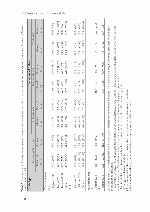
R1
R2
R3
R4
R5
R6
R7
R8
R9
R10
R11
R12
R13
R14
R15
R16
R17
R18
R19
R20
R21
R22
R23
R24
R25
R26
R27
R28
R29
R30
R31
R32
R33
R34
7 | Associated anomalies in oral clefts
128
Tab
le 2
. Su
mm
ary
of p
ublis
hed
pre
vale
nce
dat
a on
ass
ocia
ted
ano
mal
ies
and
chr
omos
omal
def
ects
in p
rena
tally
and
pos
tnat
ally
det
ecte
d u
nila
tera
l an
d b
ilate
ral o
ral c
left
s
Stud
y Ty
pe
Chr
om
oso
mal
Def
ects
Ass
ocia
ted
Ano
mal
ies
Isol
ated
cle
fts*
Ass
ocia
ted
cle
fts*
Tota
l cle
fts*
Uni
late
ral
Bila
tera
l
%
(n)
%
(n)
Uni
late
ral
Bila
tera
l
%
(n)
%
(n)
Uni
late
ral
Bila
tera
l
%
(n)
%
(n)
Uni
late
ral
Bila
tera
l
%
(n)
%
(n)
Pren
atal
stu
die
s
CLP N
yber
g 1
995†
40.0
(6
/15)
55
.0 (1
0/20
)11
.1
(1/9
) 0.
0 (
0/10
)33
.3
(2/6
) 54
.4
(6/1
0)20
.0
(3/1
5)
30.0
(6/
20)
Ber
ge
2001
†52
.0
(13/
25)
78.6
(22
/28)
0.0
(0/
12)
0.0
(0/
6)61
.5
(8/1
3)
72.7
(16
/22)
32.0
(8
/25)
58
.6 (
17/2
9)§
Maa
rse
2011
†35
.3
(6/1
7)
50.0
(3
/6)
0.0
(0/
11)
33.3
(1
/3)
66.7
(4
/6)
66.7
(2
/3)
23.5
(4
/17)
50
.0
(3/6
)
Tota
l45
.6
(26/
57)
64.8
(35
/54)
4.8
(1/
32)
5.3
(1/
19)
57.7
(1
5/25
) 68
.6 (
24/3
5)26
.3
(15/
57)
47.3
(26
/55)
§
CL±
P
Perr
otin
200
1†24
.1(7
/29)
48.1
(13/
27)
0.0
(0/2
2)0.
0(0
/14)
57.1
(4/7
)‡53
.8(7
/13)
‡13
.8(4
/29)
25.9
(7/2
7)
Gill
ham
200
9†15
.5(1
8/11
6)22
.9(8
/35)
0.0
(0/9
8)0.
0(0
/27)
33.3
(6/1
8)37
.5(3
/8)
5.2
(6/1
16)
8.6
(3/3
5)
Tota
l17
.2(2
5/14
5)33
.9(2
1/62
)0.
0(0
/110
)0.
0(0
/41)
40.0
(10/
25)
47.6
(10/
21)
6.9
(10/
145)
16.1
(10/
62)
Post
nata
l stu
die
sC
L Wal
ker
2001
||8.
3(6
/72)
8.3
(1/1
2)16
.7(1
/6)
0.0
(0/1
)1.
4(1
/72)
0.0
(0/1
2)C
LP Wal
ker
2001
||20
.5(2
3/11
2)31
.3(2
1/67
)21
.7(5
/23)
42.9
(9/2
1)4.
5(5
/112
)13
.4(9
/67)
CL
= c
left
lip
onl
y; C
LP =
cle
ft li
p w
ith c
left
pal
ate;
CL±
P =
cle
ft li
p w
ith o
r with
out
cle
ft p
alat
e; C
P =
cle
ft p
alat
e o
nly.
Bla
nc e
ntry
: dat
a w
ere
not a
vaila
ble
o
r co
uld
no
t b
e d
educ
ted
.*
Inf
orm
atio
n o
n ka
ryo
typ
e no
t av
aila
ble
for
all c
left
s, u
nles
s st
ated
diff
eren
tly (s
ee T
able
1).
Ther
efo
re, i
nclu
sio
n o
f und
etec
ted
chr
om
oso
mal
def
ects
ca
nno
t b
e ru
led
out
. Nul
l val
ues
wer
e g
iven
onl
y if
info
rmat
ion
abo
ut c
hro
mo
som
al a
naly
sis
was
rep
ort
ed.
† M
edia
n cl
eft
lip a
nd a
typ
ical
faci
al c
left
s w
ere
excl
uded
bec
ause
of t
heir
diff
eren
t p
atho
gen
esis
.‡
Kar
yoty
pe
avai
lab
le fo
r al
l cle
fts.
§ F
or
one
cas
e w
ith a
chr
om
oso
mal
def
ect,
dat
a o
n as
soci
ated
ano
mal
ies
wer
e no
t av
aila
ble
.||
Bec
ause
of l
imite
d d
ata,
chr
om
oso
mal
def
ects
am
ong
iso
late
d c
left
s no
t g
iven
.
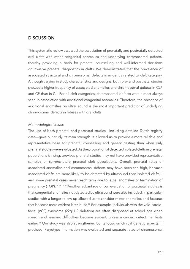
R1
R2
R3
R4
R5
R6
R7
R8
R9
R10
R11
R12
R13
R14
R15
R16
R17
R18
R19
R20
R21
R22
R23
R24
R25
R26
R27
R28
R29
R30
R31
R32
R33
R34
129
Tab
le 2
. Su
mm
ary
of p
ublis
hed
pre
vale
nce
dat
a on
ass
ocia
ted
ano
mal
ies
and
chr
omos
omal
def
ects
in p
rena
tally
and
pos
tnat
ally
det
ecte
d u
nila
tera
l an
d b
ilate
ral o
ral c
left
s
Stud
y Ty
pe
Chr
om
oso
mal
Def
ects
Ass
ocia
ted
Ano
mal
ies
Isol
ated
cle
fts*
Ass
ocia
ted
cle
fts*
Tota
l cle
fts*
Uni
late
ral
Bila
tera
l
%
(n)
%
(n)
Uni
late
ral
Bila
tera
l
%
(n)
%
(n)
Uni
late
ral
Bila
tera
l
%
(n)
%
(n)
Uni
late
ral
Bila
tera
l
%
(n)
%
(n)
Pren
atal
stu
die
s
CLP N
yber
g 1
995†
40.0
(6
/15)
55
.0 (1
0/20
)11
.1
(1/9
) 0.
0 (
0/10
)33
.3
(2/6
) 54
.4
(6/1
0)20
.0
(3/1
5)
30.0
(6/
20)
Ber
ge
2001
†52
.0
(13/
25)
78.6
(22
/28)
0.0
(0/
12)
0.0
(0/
6)61
.5
(8/1
3)
72.7
(16
/22)
32.0
(8
/25)
58
.6 (
17/2
9)§
Maa
rse
2011
†35
.3
(6/1
7)
50.0
(3
/6)
0.0
(0/
11)
33.3
(1
/3)
66.7
(4
/6)
66.7
(2
/3)
23.5
(4
/17)
50
.0
(3/6
)
Tota
l45
.6
(26/
57)
64.8
(35
/54)
4.8
(1/
32)
5.3
(1/
19)
57.7
(1
5/25
) 68
.6 (
24/3
5)26
.3
(15/
57)
47.3
(26
/55)
§
CL±
P
Perr
otin
200
1†24
.1(7
/29)
48.1
(13/
27)
0.0
(0/2
2)0.
0(0
/14)
57.1
(4/7
)‡53
.8(7
/13)
‡13
.8(4
/29)
25.9
(7/2
7)
Gill
ham
200
9†15
.5(1
8/11
6)22
.9(8
/35)
0.0
(0/9
8)0.
0(0
/27)
33.3
(6/1
8)37
.5(3
/8)
5.2
(6/1
16)
8.6
(3/3
5)
Tota
l17
.2(2
5/14
5)33
.9(2
1/62
)0.
0(0
/110
)0.
0(0
/41)
40.0
(10/
25)
47.6
(10/
21)
6.9
(10/
145)
16.1
(10/
62)
Post
nata
l stu
die
sC
L Wal
ker
2001
||8.
3(6
/72)
8.3
(1/1
2)16
.7(1
/6)
0.0
(0/1
)1.
4(1
/72)
0.0
(0/1
2)C
LP Wal
ker
2001
||20
.5(2
3/11
2)31
.3(2
1/67
)21
.7(5
/23)
42.9
(9/2
1)4.
5(5
/112
)13
.4(9
/67)
CL
= c
left
lip
onl
y; C
LP =
cle
ft li
p w
ith c
left
pal
ate;
CL±
P =
cle
ft li
p w
ith o
r with
out
cle
ft p
alat
e; C
P =
cle
ft p
alat
e o
nly.
Bla
nc e
ntry
: dat
a w
ere
not a
vaila
ble
o
r co
uld
no
t b
e d
educ
ted
.*
Inf
orm
atio
n o
n ka
ryo
typ
e no
t av
aila
ble
for
all c
left
s, u
nles
s st
ated
diff
eren
tly (s
ee T
able
1).
Ther
efo
re, i
nclu
sio
n o
f und
etec
ted
chr
om
oso
mal
def
ects
ca
nno
t b
e ru
led
out
. Nul
l val
ues
wer
e g
iven
onl
y if
info
rmat
ion
abo
ut c
hro
mo
som
al a
naly
sis
was
rep
ort
ed.
† M
edia
n cl
eft
lip a
nd a
typ
ical
faci
al c
left
s w
ere
excl
uded
bec
ause
of t
heir
diff
eren
t p
atho
gen
esis
.‡
Kar
yoty
pe
avai
lab
le fo
r al
l cle
fts.
§ F
or
one
cas
e w
ith a
chr
om
oso
mal
def
ect,
dat
a o
n as
soci
ated
ano
mal
ies
wer
e no
t av
aila
ble
.||
Bec
ause
of l
imite
d d
ata,
chr
om
oso
mal
def
ects
am
ong
iso
late
d c
left
s no
t g
iven
.
DISCUSSION
This systematic review assessed the association of prenatally and postnatally detected
oral clefts with other congenital anomalies and underlying chromosomal defects,
thereby providing a basis for prenatal counselling and well-informed decisions
on invasive prenatal diagnostics in clefts. We demonstrated that the prevalence of
associated structural and chromosomal defects is evidently related to cleft category.
Although varying in study characteristics and designs, both pre- and postnatal studies
showed a higher frequency of associated anomalies and chromosomal defects in CLP
and CP than in CL. For all cleft categories, chromosomal defects were almost always
seen in association with additional congenital anomalies. Therefore, the presence of
additional anomalies on ultra- sound is the most important predictor of underlying
chromosomal defects in fetuses with oral clefts.
Methodological issues
The use of both prenatal and postnatal studies—including detailed Dutch registry
data—gave our study its main strength. It allowed us to provide a more reliable and
representative basis for prenatal counselling and genetic testing than when only
prenatal studies were evaluated. As the proportion of detected isolated clefts in prenatal
populations is rising, previous prenatal studies may not have provided representative
samples of current/future prenatal cleft populations. Overall, prenatal rates of
associated anomalies and chromosomal defects may have been too high, because
associated clefts are more likely to be detected by ultrasound than isolated clefts,11
and some prenatal cases never reach term due to lethal anomalies or termination of
pregnancy (TOP).16 25 26 29 Another advantage of our evaluation of postnatal studies is
that congenital anomalies not detected by ultrasound were also included. In particular,
studies with a longer follow-up allowed us to consider minor anomalies and features
that become more evident later in life.22 For example, individuals with the velo-cardio-
facial (VCF) syndrome (22q11.2 deletion) are often diagnosed at school age when
speech and learning difficulties become evident, unless a cardiac defect manifests
earlier.38 Our study was also strengthened by its focus on clinical genetic aspects. If
provided, karyotype information was evaluated and separate rates of chromosomal
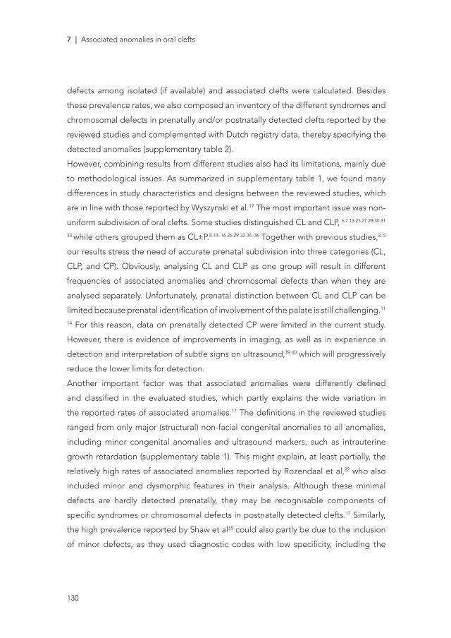
R1
R2
R3
R4
R5
R6
R7
R8
R9
R10
R11
R12
R13
R14
R15
R16
R17
R18
R19
R20
R21
R22
R23
R24
R25
R26
R27
R28
R29
R30
R31
R32
R33
R34
7 | Associated anomalies in oral clefts
130
defects among isolated (if available) and associated clefts were calculated. Besides
these prevalence rates, we also composed an inventory of the different syndromes and
chromosomal defects in prenatally and/or postnatally detected clefts reported by the
reviewed studies and complemented with Dutch registry data, thereby specifying the
detected anomalies (supplementary table 2).
However, combining results from different studies also had its limitations, mainly due
to methodological issues. As summarized in supplementary table 1, we found many
differences in study characteristics and designs between the reviewed studies, which
are in line with those reported by Wyszynski et al.17 The most important issue was non-
uniform subdivision of oral clefts. Some studies distinguished CL and CLP, 6 7 13 25 27 28 30 31
33 while others grouped them as CL±P.8 14–16 26 29 32 34–36 Together with previous studies,2–5
our results stress the need of accurate prenatal subdivision into three categories (CL,
CLP, and CP). Obviously, analysing CL and CLP as one group will result in different
frequencies of associated anomalies and chromosomal defects than when they are
analysed separately. Unfortunately, prenatal distinction between CL and CLP can be
limited because prenatal identification of involvement of the palate is still challenging.11
14 For this reason, data on prenatally detected CP were limited in the current study.
However, there is evidence of improvements in imaging, as well as in experience in
detection and interpretation of subtle signs on ultrasound,39 40 which will progressively
reduce the lower limits for detection.
Another important factor was that associated anomalies were differently defined
and classified in the evaluated studies, which partly explains the wide variation in
the reported rates of associated anomalies.17 The definitions in the reviewed studies
ranged from only major (structural) non-facial congenital anomalies to all anomalies,
including minor congenital anomalies and ultrasound markers, such as intrauterine
growth retardation (supplementary table 1). This might explain, at least partially, the
relatively high rates of associated anomalies reported by Rozendaal et al,22 who also
included minor and dysmorphic features in their analysis. Although these minimal
defects are hardly detected prenatally, they may be recognisable components of
specific syndromes or chromosomal defects in postnatally detected clefts.17 Similarly,
the high prevalence reported by Shaw et al35 could also partly be due to the inclusion
of minor defects, as they used diagnostic codes with low specificity, including the
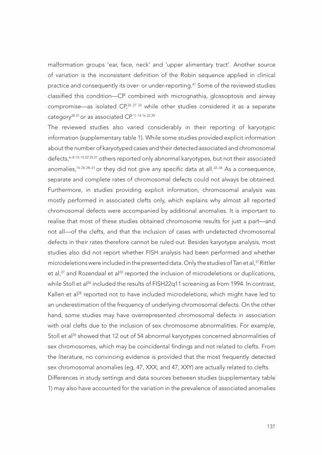
R1
R2
R3
R4
R5
R6
R7
R8
R9
R10
R11
R12
R13
R14
R15
R16
R17
R18
R19
R20
R21
R22
R23
R24
R25
R26
R27
R28
R29
R30
R31
R32
R33
R34
131
malformation groups ‘ear, face, neck’ and ‘upper alimentary tract’. Another source
of variation is the inconsistent definition of the Robin sequence applied in clinical
practice and consequently its over- or under-reporting.41 Some of the reviewed studies
classified this condition—CP combined with micrognathia, glossoptosis and airway
compromise—as isolated CP,26 27 33 while other studies considered it as a separate
category28 31 or as associated CP.11 14 16 22 29
The reviewed studies also varied considerably in their reporting of karyotypic
information (supplementary table 1). While some studies provided explicit information
about the number of karyotyped cases and their detected associated and chromosomal
defects,6–8 13–15 22 25 27 others reported only abnormal karyotypes, but not their associated
anomalies,16 26 28–31 or they did not give any specific data at all.32–36 As a consequence,
separate and complete rates of chromosomal defects could not always be obtained.
Furthermore, in studies providing explicit information, chromosomal analysis was
mostly performed in associated clefts only, which explains why almost all reported
chromosomal defects were accompanied by additional anomalies. It is important to
realise that most of these studies obtained chromosome results for just a part—and
not all—of the clefts, and that the inclusion of cases with undetected chromosomal
defects in their rates therefore cannot be ruled out. Besides karyotype analysis, most
studies also did not report whether FISH analysis had been performed and whether
microdeletions were included in the presented data. Only the studies of Tan et al,31 Rittler
et al,27 and Rozendaal et al22 reported the inclusion of microdeletions or duplications,
while Stoll et al26 included the results of FISH22q11 screening as from 1994. In contrast,
Kallen et al28 reported not to have included microdeletions, which might have led to
an underestimation of the frequency of underlying chromosomal defects. On the other
hand, some studies may have overrepresented chromosomal defects in association
with oral clefts due to the inclusion of sex chromosome abnormalities. For example,
Stoll et al26 showed that 12 out of 54 abnormal karyotypes concerned abnormalities of
sex chromosomes, which may be coincidental findings and not related to clefts. From
the literature, no convincing evidence is provided that the most frequently detected
sex chromosomal anomalies (eg, 47, XXX; and 47, XXY) are actually related to clefts.
Differences in study settings and data sources between studies (supplementary table
1) may also have accounted for the variation in the prevalence of associated anomalies
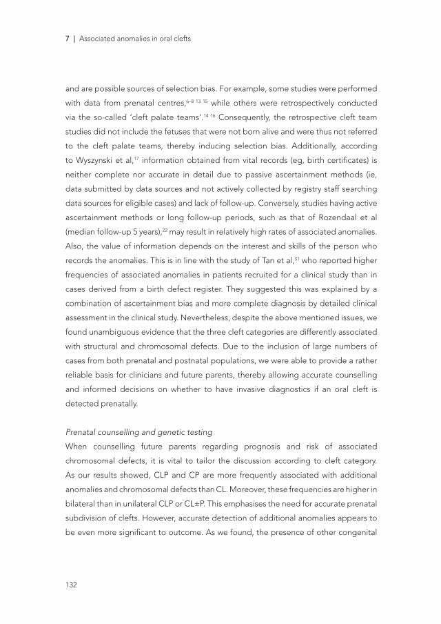
R1
R2
R3
R4
R5
R6
R7
R8
R9
R10
R11
R12
R13
R14
R15
R16
R17
R18
R19
R20
R21
R22
R23
R24
R25
R26
R27
R28
R29
R30
R31
R32
R33
R34
7 | Associated anomalies in oral clefts
132
and are possible sources of selection bias. For example, some studies were performed
with data from prenatal centres,6–8 13 15 while others were retrospectively conducted
via the so-called ‘cleft palate teams’.14 16 Consequently, the retrospective cleft team
studies did not include the fetuses that were not born alive and were thus not referred
to the cleft palate teams, thereby inducing selection bias. Additionally, according
to Wyszynski et al,17 information obtained from vital records (eg, birth certificates) is
neither complete nor accurate in detail due to passive ascertainment methods (ie,
data submitted by data sources and not actively collected by registry staff searching
data sources for eligible cases) and lack of follow-up. Conversely, studies having active
ascertainment methods or long follow-up periods, such as that of Rozendaal et al
(median follow-up 5 years),22 may result in relatively high rates of associated anomalies.
Also, the value of information depends on the interest and skills of the person who
records the anomalies. This is in line with the study of Tan et al,31 who reported higher
frequencies of associated anomalies in patients recruited for a clinical study than in
cases derived from a birth defect register. They suggested this was explained by a
combination of ascertainment bias and more complete diagnosis by detailed clinical
assessment in the clinical study. Nevertheless, despite the above mentioned issues, we
found unambiguous evidence that the three cleft categories are differently associated
with structural and chromosomal defects. Due to the inclusion of large numbers of
cases from both prenatal and postnatal populations, we were able to provide a rather
reliable basis for clinicians and future parents, thereby allowing accurate counselling
and informed decisions on whether to have invasive diagnostics if an oral cleft is
detected prenatally.
Prenatal counselling and genetic testing
When counselling future parents regarding prognosis and risk of associated
chromosomal defects, it is vital to tailor the discussion according to cleft category.
As our results showed, CLP and CP are more frequently associated with additional
anomalies and chromosomal defects than CL. Moreover, these frequencies are higher in
bilateral than in unilateral CLP or CL±P. This emphasises the need for accurate prenatal
subdivision of clefts. However, accurate detection of additional anomalies appears to
be even more significant to outcome. As we found, the presence of other congenital
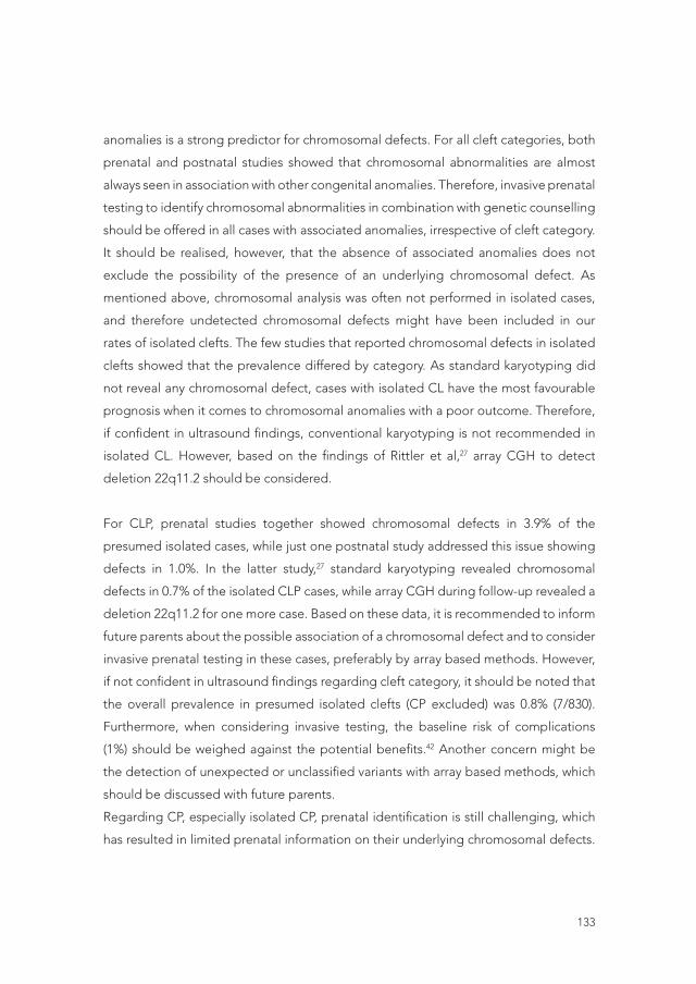
R1
R2
R3
R4
R5
R6
R7
R8
R9
R10
R11
R12
R13
R14
R15
R16
R17
R18
R19
R20
R21
R22
R23
R24
R25
R26
R27
R28
R29
R30
R31
R32
R33
R34
133
anomalies is a strong predictor for chromosomal defects. For all cleft categories, both
prenatal and postnatal studies showed that chromosomal abnormalities are almost
always seen in association with other congenital anomalies. Therefore, invasive prenatal
testing to identify chromosomal abnormalities in combination with genetic counselling
should be offered in all cases with associated anomalies, irrespective of cleft category.
It should be realised, however, that the absence of associated anomalies does not
exclude the possibility of the presence of an underlying chromosomal defect. As
mentioned above, chromosomal analysis was often not performed in isolated cases,
and therefore undetected chromosomal defects might have been included in our
rates of isolated clefts. The few studies that reported chromosomal defects in isolated
clefts showed that the prevalence differed by category. As standard karyotyping did
not reveal any chromosomal defect, cases with isolated CL have the most favourable
prognosis when it comes to chromosomal anomalies with a poor outcome. Therefore,
if confident in ultrasound findings, conventional karyotyping is not recommended in
isolated CL. However, based on the findings of Rittler et al,27 array CGH to detect
deletion 22q11.2 should be considered.
For CLP, prenatal studies together showed chromosomal defects in 3.9% of the
presumed isolated cases, while just one postnatal study addressed this issue showing
defects in 1.0%. In the latter study,27 standard karyotyping revealed chromosomal
defects in 0.7% of the isolated CLP cases, while array CGH during follow-up revealed a
deletion 22q11.2 for one more case. Based on these data, it is recommended to inform
future parents about the possible association of a chromosomal defect and to consider
invasive prenatal testing in these cases, preferably by array based methods. However,
if not confident in ultrasound findings regarding cleft category, it should be noted that
the overall prevalence in presumed isolated clefts (CP excluded) was 0.8% (7/830).
Furthermore, when considering invasive testing, the baseline risk of complications
(1%) should be weighed against the potential benefits.42 Another concern might be
the detection of unexpected or unclassified variants with array based methods, which
should be discussed with future parents.
Regarding CP, especially isolated CP, prenatal identification is still challenging, which
has resulted in limited prenatal information on their underlying chromosomal defects.
R1
R2
R3
R4
R5
R6
R7
R8
R9
R10
R11
R12
R13
R14
R15
R16
R17
R18
R19
R20
R21
R22
R23
R24
R25
R26
R27
R28
R29
R30
R31
R32
R33
R34
7 | Associated anomalies in oral clefts
134
However, postnatal karyotyping of isolated CP cases revealed a chromosomal defect in
1.6%. In this category in particular, specific syndromes, such as VCF (22q11.2 deletion),
Treacher-Collins, and Stickler, have to be considered. As presented in supplementary
table 2, these syndrome diagnoses were frequently reported in the evaluated literature.
Therefore, until more information on chromosomal defects in prenatally presumed
isolated CP is available, we advise consideration of invasive genetic testing and
consultation by a clinical geneticist if an isolated CP is detected prenatally. A prenatal
diagnostic algorithm according to cleft category is presented in figure 1.
Oral cleft detected
prenatally on US
Associated anomalies on US
Isolated oral cleft on US
Invasive prenatal testing, preferably CL
by array CGH
CLP CP
Standard prenatal consulting of
clinical geneticist
Karyotyping not recommended;
consider array CGH
Consider invasive
prenatal testing,
preferably by array CGH
Consider invasive prenatal testing,
preferably by array CGH
Prenatal
consulting of cleft palate team*
Prenatal consulting of cleft
palate team
Consider prenatal consulting of
clinical geneticist
Consider prenatal consulting of
clinical geneticist
Postnatal
consulting of clinical geneticist
Figure 1. Algorithm for invasive genetic testing according to oral cleft category. CL = cleft lip only; CLP = cleft lip with cleft palate; CP = cleft palate only; US = ultrasound; array CGH = array-comparative genomic hybridization. * If a normal karyotype is confirmed or invasive genetic testing is declined.
Prenatal consulting of cleft
palate team*
Prenatal consulting of cleft
palate team
Figure 1. Algorithm for invasive genetic testing according to oral cleft category. CL = cleft lip only; CLP = cleft lip with cleft palate; CP = cleft palate only; US = ultrasound; array CGH = array-comparative genomic hybridization. * If a normal karyotype is confirmed or invasive genetic testing is declined.
R1
R2
R3
R4
R5
R6
R7
R8
R9
R10
R11
R12
R13
R14
R15
R16
R17
R18
R19
R20
R21
R22
R23
R24
R25
R26
R27
R28
R29
R30
R31
R32
R33
R34
135
Based on the above findings, more accurate prenatal ultrasound screening will improve
counselling, especially regarding palatal involvement. Therefore, we advise the referral
of pregnant women with a fetus suspected of having an oral cleft to a tertiary care
centre where more specific ultrasound screening can be performed. In addition, if a
normal karyotype is confirmed or invasive testing is declined, future parents should
be counselled by a multidisciplinary cleft palate team that focuses on psychosocial
support, education on management of clefts, and parents’ options, TOP being one of
them.43–46 Finally, it is crucial to distinguish median clefts and atypical facial clefts from
oral clefts. These different craniofacial anomalies are associated with other congenital
anomalies and have a different prognosis, and should therefore be referred to and
treated by specialised multidisciplinary craniofacial teams.
Future studies
The use of array CGH in clinical practice is rising, and it is expected that it will be
implemented as standard prenatal diagnostics in the near future. Compared to
conventional karyotyping, array CGH can detect smaller chromosome deletions and
duplications. To gain more insight into the yield of array CGH in cases with clefts, it
would be interesting to perform array CGH in a large cohort of cases with prenatally
and postnatally detected clefts. This would also give us more information about the
proportion and types of chromosomal defects that are missed in cases that have
not been karyotyped or studied by array based methods. Particularly with regard to
prenatally presumed isolated clefts, this is essential to reach consensus on the role of
invasive genetic testing in these cases. As was demonstrated by the NVSCA data,23
24 clefts can be associated with various microdeletions and duplications. This implies
that array CGH should be the standard technique to identify chromosomal defects in
children with oral clefts.
Finally, follow-up studies are needed to gain more insight into additional abnormalities
and chromosomal anomalies identified after birth. This can aid in more optimal
counselling of future parents, especially with regard to unexpected anomalies in
presumed isolated clefts, and timely treatment of children with clefts.
R1
R2
R3
R4
R5
R6
R7
R8
R9
R10
R11
R12
R13
R14
R15
R16
R17
R18
R19
R20
R21
R22
R23
R24
R25
R26
R27
R28
R29
R30
R31
R32
R33
R34
7 | Associated anomalies in oral clefts
136
CONCLUSIONS
This systematic review presents unambiguous evidence that the different cleft
categories are variously associated with additional congenital anomalies and
underlying chromosomal defects. This emphasises the need for accurate subdivision
of CL, CLP and CP for both ultrasound screening and postnatal follow-up. However,
the most important predictor of chromosomal abnormalities is the presence of
associated anomalies, and we urge clinicians to advise invasive testing in these cases.
In the absence of associated anomalies, cases with CL have the most favourable
prognosis and do not require conventional karyotyping. In presumed isolated CLP and
CP, professionals should explain the possible association of a chromosomal defect
and consider invasive genetic testing, preferably by array based methods. In all cleft
categories, an association with deletion 22q11.2 should be considered.
Accurate prenatal diagnosis by ultrasound is essential in the quality of counselling,
especially with regard to palatal involvement and associated anomalies. Therefore,
a pregnant woman with a fetus suspected of having an oral cleft should be referred
to a tertiary care centre where more specific ultrasound screening can be performed.
Finally, follow-up studies, including array CGH, are needed to gain more insight into
additional abnormalities and chromosomal defects missed in associated and presumed
isolated clefts. This would aid in more optimal counselling and timely treatment of
children with oral clefts.
ACKNOWLEDGEMENTS
The authors would like to thank the clinical cytogeneticist Ellen van Binsbergen for
her cytogenetic input as well as the clinical cytogeneticist Heleen Schuring-Blom and
psychologist Henriëtte Swanenburg de Veye for their helpful discussion and critically
reading of the manuscript. They would also like to express their gratitude to the Dutch
cleft palate teams and the NVSCA, including its board. Without their efforts, the
NVSCA would not have succeeded.
R1
R2
R3
R4
R5
R6
R7
R8
R9
R10
R11
R12
R13
R14
R15
R16
R17
R18
R19
R20
R21
R22
R23
R24
R25
R26
R27
R28
R29
R30
R31
R32
R33
R34
137
REFERENCES
1 Mossey PA, Little J, Munger RG, Dixon MJ, Shaw WC. Cleft lip and palate. Lancet 2009;374:1773–85.
2 Luijsterburg AJM, Vermeij-Keers C. Recording and classification common oral clefts. In: Lilja J Transactions, the 9th international congress on cleft palate and related craniofacial anomalies. Götenborg: Elanders Novum, 2001:243–50.
3 Harville EW, Wilcox AJ, Lie RT, Vindenes H, Abyholm F. Cleft lip and palate versus cleft lip only: are they distinct defects? Am J Epidemiol 2005;162:448–53.
4 Rittler M, Lopez-Camelo JS, Castilla EE, Bermejo E, Cocchi G, Correa A, Csaky-Szunyogh M, Danderfer R, De Vigan C, De Walle H, Da Graca Dutra M, Hirahara F, Martinez-Frias ML, Merlob P, Mutchinick O, Ritvanen A, Robert-Gnansia E, Scarano G, Siffel C, Stoll C, Mastroiacovo P. Preferential associations between oral clefts and other major congenital anomalies. Cleft Palate Craniofac J 2008;45:525–32.
5 Luijsterburg AJM, Vermeij-Keers C. Ten years recording common oral clefts with a new descriptive system. Cleft Palate Craniofac J 2011;48:173–82.
6 Nyberg DA, Sickler GK, Hegge FN, Kramer DJ, Kropp RJ. Fetal cleft lip with and without cleft palate: US classification and correlation with outcome. Radiology 1995;195:677–84.
7 Berge SJ, Plath H, Van de Vondel PT, Appel T, Niederhagen B, Von Lindern JJ, Reich RH, Hansmann M. Fetal cleft lip and palate: sonographic diagnosis, chromosomal abnormalities, associated anomalies and postnatal outcome in 70 fetuses. Ultrasound Obstet Gynecol 2001;18:422–31.
8 Perrotin F, De Poncheville LM, Marret H, Paillet C, Lansac J, Body G. Chromosomal defects and associated malformations in fetal cleft lip with or without cleft palate. Eur J Obstet Gynecol Reprod Biol 2001;99:19–24.
9 Van der Meulen J, Mazzola R, Strickler M, Raphael B. Classification of craniofacial malformations. In: Stricker M, Van der Meulen JC, Raphael B, Mazzola R, Tolhurst DE, Murray JE Craniofacial malformations. Edinburgh: Churchill Livingstone, 1990:149–309.
10 Vermeij-Keers C. Craniofacial embryology and morphogenesis: normal and abnormal. In: Stricker M, Van der Meulen JC, Raphael B, Mazzola R, Tolhurst DE, Murray JE Craniofacial malformations. Edinburgh: Churchill Livingstone, 1990:27–60.
11 Maarse W, Berge SJ, Pistorius L, Van Barneveld T, Kon M, Breugem C, Van der Molen AB. Diagnostic accuracy of transabdominal ultrasound in detecting prenatal cleft lip and palate: a systematic review. Ultrasound Obstet Gynecol 2010;35:495–502.
12 Stoll C, Dott B, Alembik Y, Roth M. Evaluation of prenatal diagnosis of cleft lip/palate by foetal ultrasonographic examination. Ann Genet 2000;43:11–14.
13 Maarse W, Pistorius LR, Van Eeten WK, Breugem CC, Kon M, Van den Boogaard MJ, Mink van Der Molen AB. Prenatal ultrasound screening for orofacial clefts. Ultrasound Obstet Gynecol 2011;38:434–9.
14 Gillham JC, Anand S, Bullen PJ. Antenatal detection of cleft lip with or without cleft palate: incidence of associated chromosomal and structural anomalies. Ultrasound Obstet Gynecol 2009;34:410–15.
15 Offerdal K, Jebens N, Syvertsen T, Blaas HG, Johansen OJ, Eik-Nes SH. Prenatal ultrasound detection of facial clefts: a prospective study of 49,314 deliveries in a non-selected population in Norway. Ultrasound Obstet Gynecol 2008;31:639–46.
16 Russell KA, Allen VM, MacDonald ME, Smith K, Dodds L. A population-based evaluation of antenatal diagnosis of orofacial clefts. Cleft Palate Craniofac J 2008;45:148–53.
17 Wyszynski DF, Sarkozi A, Czeizel AE. Oral clefts with associated anomalies: methodological issues. Cleft Palate Craniofac J 2006;43:1–6.
R1
R2
R3
R4
R5
R6
R7
R8
R9
R10
R11
R12
R13
R14
R15
R16
R17
R18
R19
R20
R21
R22
R23
R24
R25
R26
R27
R28
R29
R30
R31
R32
R33
R34
7 | Associated anomalies in oral clefts
138
18 Altherr MR, Bengtsson U, Elder FF, Ledbetter DH, Wasmuth JJ, McDonald ME, Gusella JF, Greenberg F. Molecular confirmation of Wolf-Hirschhorn syndrome with a subtle translocation of chromosome 4. Am J Hum Genet 1991;49:1235–42.
19 Desmaze C, Scambler P, Prieur M, Halford S, Sidi D, Le Deist F, Aurias A. Routine diagnosis of DiGeorge syndrome by fluorescent in situ hybridization. Hum Genet 1993;90:663–5.
20 Rozendaal AM, Luijsterburg AJM, Mohangoo AD, Ongkosuwito EM, Anthony S, Vermeij-Keers C. Validation of the NVSCA registry for common oral clefts: study design and first results. Cleft Palate Craniofac J 2010;47:534–43.
21 Rozendaal AM, Luijsterburg AJM, Mohangoo AD, Ongkosuwito EM, De Vries E, Vermeij-Keers C. Validation of the Dutch Registry of Common Oral Clefts: quality of recording specific oral cleft features. Cleft Palate Craniofac J. Published Online First: 23 November 2010. doi:10.1597/10-109.
22 Rozendaal AM, Luijsterburg AJM, Ongkosuwito EM, Van den Boogaard M-JH, De Vries E, Hovius SER, Vermeij-Keers C. Delayed diagnosis and underreporting of congenital anomalies associated with oral clefts in the Netherlands: a national validation study. J Plast Reconstr Aesthet Surg 2012;65:780–90.
23 Luijsterburg AJM, Vermeij-Keers C. NVSCA registry common oral clefts. Annual reports 1997–2007 of the Dutch Association for Cleft Palate and Craniofacial Anomalies (in Dutch). Rotterdam: Department of Plastic and Reconstructive Surgery, Erasmus MC, University Medical Center Rotterdam, 1999–2008. ISSN 1571-876X.
24 Luijsterburg AJM, Rozendaal AM, Trenning AH, Ongkosuwito EM, Vermeij-Keers C. NVSCA registry common oral clefts. Annual reports 2008–2010 of the Dutch Association for Cleft Palate and Craniofacial Anomalies (in Dutch). Rotterdam: Department of Plastic and Reconstructive Surgery, Erasmus MC, University Medical Center Rotterdam, 2010–2011. ISSN 1571-876X.
25 Walker SJ, Ball RH, Babcook CJ, Feldkamp MM. Prevalence of aneuploidy and additional anatomic abnormalities in fetuses and neonates with cleft lip with or without cleft palate: a population-based study in Utah. J Ultrasound Med 2001;20:1175–80; quiz 81-2.
26 Stoll C, Alembik Y, Dott B, Roth MP. Associated malformations in patients with oral clefts. Am J Med Genet A 2007;143A:2463–5.
27 Rittler M, Cosentino V, Lopez-Camelo JS, Murray JC, Wehby G, Castilla EE. Associated anomalies among infants with oral clefts at birth and during a 1-year follow-up. Am J Med Genet A 2011;155A:1588–96.
28 Kallen B, Harris J, Robert E. The epidemiology of orofacial clefts. 2. Associated malformations. J Craniofac Genet Dev Biol 1996;16:242–8.
29 Vallino-Napoli LD, Riley MM, Halliday JL. An epidemiologic study of orofacial clefts with other birth defects in Victoria, Australia. Cleft Palate Craniofac J 2006;43: 571–6.
30 Calzolari E, Pierini A, Astolfi G, Bianchi F, Neville AJ, Rivieri F. Associated anomalies in multi-malformed infants with cleft lip and palate: an epidemiologic study of nearly 6 million births in 23 EUROCAT registries. Am J Med Genet A 2007;143:528–37.
31 Tan TY, Amor DJ, Riley M, Halliday J, Kilpatrick N, Simms K, White SM. Registry- and clinic-based analyses of birth defects and syndromes associated with cleft lip/ palate in Victoria, Australia. Cleft Palate Craniofac J 2009;46:583–7.
32 Druschel CM, Hughes JP, Olsen CL. First year-of-life mortality among infants with oral clefts: New York State, 1983–1990. Cleft Palate Craniofac J 1996;33:400–5.
33 Milerad J, Larson O, Ph DD, Hagberg C, Ideberg M. Associated malformations in infants with cleft lip and palate: a prospective, population-based study. Pediatrics 1997;100:180–6.
34 DeRoo LA, Gaudino JA, Edmonds LD. Orofacial cleft malformations: associations with maternal and infant characteristics in Washington State. Birth Defects Res A Clin Mol Teratol 2003;67:637–42.
R1
R2
R3
R4
R5
R6
R7
R8
R9
R10
R11
R12
R13
R14
R15
R16
R17
R18
R19
R20
R21
R22
R23
R24
R25
R26
R27
R28
R29
R30
R31
R32
R33
R34
139
35 Shaw GM, Carmichael SL, Yang W, Harris JA, Lammer EJ. Congenital malformations in births with orofacial clefts among 3.6 million California births, 1983–1997. Am J Med Genet A 2004;125A:250–6.
36 Beriaghi S, Myers SL, Jensen SA, Kaimal S, Chan CM, Schaefer GB. Cleft lip and palate: association with other congenital malformations. J Clin Pediatr Dent 2009;33:207–10.
37 Chmait R, Pretorius D, Moore T, Hull A, James G, Nelson T, Jones M. Prenatal detection of associated anomalies in fetuses diagnosed with cleft lip with or without cleft palate in utero. Ultrasound Obstet Gynecol 2006;27:173–6.
38 Swanenburg de Veye HF, Beemer FA, Mellenbergh GJ, Wolters WH, Heineman-de Boer JA. An investigation of the relationship between associated congenital malformations and the mental and psychomotor development of children with clefts. Cleft Palate Craniofac J 2003;40:297–303.
39 Timmerman E, Pajkrt E, Maas SM, Bilardo CM. Enlarged nuchal translucency in chromosomally normal fetuses: strong association with orofacial clefts. Ultrasound Obstet Gynecol 2010;36:427–32.
40 Wilhelm L, Borgers H. The ‘equals sign’: a novel marker in the diagnosis of fetal isolated cleft palate. Ultrasound Obstet Gynecol 2010;36:439–44.
41 Breugem CC, Mink van der Molen AB. What is ‘Pierre Robin sequence’? J Plast Reconstr Aesthet Surg 2009;62:1555–8.
42 Tabor A, Philip J, Madsen M, Bang J, Obel EB, Norgaard-Pedersen B. Randomised controlled trial of genetic amniocentesis in 4606 low-risk women. Lancet 1986;1:1287–93.
43 Davalbhakta A, Hall PN. The impact of antenatal diagnosis on the effectiveness and timing of counselling for cleft lip and palate. Br J Plast Surg 2000;53:298–301.
44 Jones MC. Prenatal diagnosis of cleft lip and palate: detection rates, accuracy of ultrasonography, associated anomalies, and strategies for counseling. Cleft Palate Craniofac J 2002;39:169–73.
45 Byrnes AL, Berk NW, Cooper ME, Marazita ML. Parental evaluation of informing interviews for cleft lip and/or palate. Pediatrics 2003;112:308–13.
46 Johnson N, Sandy R. Prenatal diagnosis of cleft lip and palate. Cleft Palate Craniofac J 2003;40:186–9.
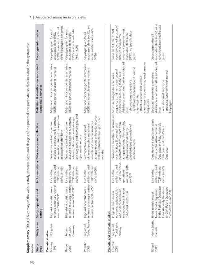
R1
R2
R3
R4
R5
R6
R7
R8
R9
R10
R11
R12
R13
R14
R15
R16
R17
R18
R19
R20
R21
R22
R23
R24
R25
R26
R27
R28
R29
R30
R31
R32
R33
R34
7 | Associated anomalies in oral clefts
140
Sup
ple
men
tary
Tab
le 1
Sum
mar
y of
the
vario
us s
tud
y ch
arac
teris
tics
and
des
igns
of t
he p
rena
tal a
nd p
ostn
atal
stu
die
s in
clud
ed in
the
syst
emat
ic
revi
ew
Stud
y ty
pe
Stud
y ar
eaSt
udy
po
pul
atio
n an
d
sett
ing
Incl
usio
n cr
iter
iaD
ata
sour
ces
and
co
llect
ion
Defi
niti
on
& c
lass
ifica
tio
n as
soci
ated
co
ngen
ital
ano
mal
ies
Kar
yoty
pe
info
rmat
ion
Pre
nata
l stu
die
sN
yber
g
1995
Not
giv
enH
igh-
risk
obst
etric
cas
es
refe
rred
to te
rtia
ry re
ferr
al
cent
res
1988
-199
3*
Live
birt
hs,
still
birt
hs, a
nd
TOP
with
ora
l cl
efts
(n=
40)†
Pros
pec
tive
and
retr
osp
ectiv
e an
alys
is o
f son
ogra
ms
& p
ostn
atal
in
form
atio
n re
trie
ved
by
insp
ectio
n at
aut
opsy
or a
fter
birt
h
Maj
or a
nd m
inor
con
gen
ital a
nom
alie
s;
IUG
R an
d o
ther
ultr
asou
nd m
arke
rs‡
Kar
yoty
pe
giv
en fo
r mos
t as
soci
ated
cle
fts
(94%
, 17
/18)
; num
ber
of i
sola
ted
cl
efts
kar
yoty
ped
not
giv
en
Ber
ge
2001
Reg
ion
of B
onn,
G
erm
any
Hig
h-ris
k ob
stet
ric c
ases
re
ferr
ed to
a s
ing
le te
rtia
ry
refe
rral
cen
tre
1991
-200
0*
Live
birt
hs,
still
birt
hs, a
nd
TOP
with
ora
l cl
efts
(n=
59)†
Pros
pec
tive
and
retr
osp
ectiv
e an
alys
is o
f son
ogra
ms
& p
ostn
atal
fo
llow
-up
dat
a fr
om n
eona
tolo
gis
t an
d s
urg
eons
, and
pat
holo
gic
al a
nd
cyto
gen
etic
reco
rds
Maj
or a
nd m
inor
con
gen
ital a
nom
alie
s;
IUG
R an
d o
ther
ultr
asou
nd m
arke
rs‡
Kar
yoty
pe
giv
en fo
r mos
t as
soci
ated
cle
fts
(94%
, 33
/35)
and
isol
ated
cle
fts
(76%
, 16/
21)
Perr
otin
20
01Re
gio
n of
To
urs,
Fra
nce
Hig
h-ris
k ob
stet
ric c
ases
re
ferr
ed to
a s
ing
le te
rtia
ry
refe
rral
cen
tre
1991
-199
9*
Live
birt
hs,
still
birt
hs, a
nd
TOP
with
ora
l cl
efts
(n=
56)†
Retr
osp
ectiv
e ev
alua
tion
of
ultr
ason
ogra
phi
c an
d c
linic
al
reco
rds,
and
asc
erta
inm
ent v
ia
auto
psy
rep
orts
or h
osp
ital r
ecor
ds
with
a p
ostn
atal
follo
w-u
p o
f 3-1
2 m
onth
s
Maj
or a
nd m
inor
con
gen
ital a
nom
alie
s;
IUG
R an
d o
ther
ultr
asou
nd m
arke
rsK
aryo
typ
e g
iven
for a
ll as
soci
ated
cle
fts
(n=
20),
and
so
me
isol
ated
cle
fts
(39%
, 14
/36)
Pre
nata
l and
Po
stna
tal s
tud
ies
Offe
rdal
20
08Re
gio
n of
Tr
ond
heim
, N
orw
ay
Preg
nant
wom
en in
a
non-
sele
cted
pop
ulat
ion
who
und
erw
ent r
outin
e ul
tras
ound
exa
min
atio
n 19
87-2
004
(n=
49,3
14)
Live
birt
hs,
still
birt
hs, a
nd
TOP
(≥16
wee
ks
ges
tatio
n)
with
ora
l cle
fts
(n=
101)
Pros
pec
tive
colle
ctio
n of
ultr
asou
nd
exam
inat
ions
& p
rena
tal a
nd
pos
tnat
al fo
llow
-up
dat
a fr
om
auto
psy
rep
orts
, pho
tog
rap
hs,
phy
sica
l exa
min
atio
ns b
y p
aed
iatr
icia
ns a
nd re
view
of
med
ical
reco
rds
Stru
ctur
al c
ong
enita
l ano
mal
ies,
se
que
nces
, and
non
-chr
omos
omal
sy
ndro
mes
acc
ord
ing
to th
e IC
D;
Ad
diti
onal
ano
mal
ies
furt
her s
ubd
ivid
ed
into
:- c
hrom
osom
e ab
erra
tions
- syn
dro
mes
/seq
uenc
es w
ith n
orm
al
chro
mos
omes
- s
truc
tura
l ano
mal
ies
with
out
chro
mos
omal
ab
erra
tions
, syn
dro
mes
or
seq
uenc
es
Som
e cl
efts
(31%
, 31/
101
wer
e ka
ryot
yped
; pre
senc
e/ab
senc
e of
ab
norm
al
kary
otyp
e g
iven
for m
ost
asso
ciat
ed c
left
s 62
%,
29/4
7); n
o sp
ecifi
c d
ata
giv
en
Russ
ell
2008
Nov
ia S
cotia
, C
anad
aB
irths
to re
sid
ents
of
Nov
ia S
cotia
reg
iste
red
in
the
Perin
atal
Dat
abas
e,
Feta
l Ano
mal
y D
atab
ase,
or
Cle
ft P
alat
e D
atab
ase
1992
-200
2 (n
=10
8,22
0)
Live
birt
hs,
still
birt
hs, a
nd
TOP
with
ora
l cl
efts
(n=
225)
Dat
a fr
om th
e p
opul
atio
n-b
ased
N
ovia
Sco
tia A
tlee
Perin
atal
D
atab
ase,
the
Feta
l Ano
mal
y D
atab
ase,
and
Cle
ft P
alat
e D
atab
ase
Stru
ctur
al c
ong
enita
l ano
mal
ies;
Ad
diti
onal
ano
mal
ies
furt
her s
ubd
ivid
ed
into
:- w
ith a
bno
rmal
kar
yoty
pe
- str
uctu
ral/
synd
rom
ic w
ith n
orm
al
kary
otyp
e
Dat
a su
gg
est t
hat a
ll as
soci
ated
cle
fts
(n=
99) w
ere
kary
otyp
ed; n
o sp
ecifi
c d
ata
giv
en
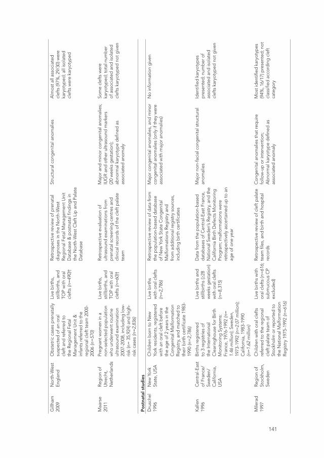
R1
R2
R3
R4
R5
R6
R7
R8
R9
R10
R11
R12
R13
R14
R15
R16
R17
R18
R19
R20
R21
R22
R23
R24
R25
R26
R27
R28
R29
R30
R31
R32
R33
R34
141
Sup
ple
men
tary
Tab
le 1
Sum
mar
y of
the
vario
us s
tud
y ch
arac
teris
tics
and
des
igns
of t
he p
rena
tal a
nd p
ostn
atal
stu
die
s in
clud
ed in
the
syst
emat
ic
revi
ew
Stud
y ty
pe
Stud
y ar
eaSt
udy
po
pul
atio
n an
d
sett
ing
Incl
usio
n cr
iter
iaD
ata
sour
ces
and
co
llect
ion
Defi
niti
on
& c
lass
ifica
tio
n as
soci
ated
co
ngen
ital
ano
mal
ies
Kar
yoty
pe
info
rmat
ion
Pre
nata
l stu
die
sN
yber
g
1995
Not
giv
enH
igh-
risk
obst
etric
cas
es
refe
rred
to te
rtia
ry re
ferr
al
cent
res
1988
-199
3*
Live
birt
hs,
still
birt
hs, a
nd
TOP
with
ora
l cl
efts
(n=
40)†
Pros
pec
tive
and
retr
osp
ectiv
e an
alys
is o
f son
ogra
ms
& p
ostn
atal
in
form
atio
n re
trie
ved
by
insp
ectio
n at
aut
opsy
or a
fter
birt
h
Maj
or a
nd m
inor
con
gen
ital a
nom
alie
s;
IUG
R an
d o
ther
ultr
asou
nd m
arke
rs‡
Kar
yoty
pe
giv
en fo
r mos
t as
soci
ated
cle
fts
(94%
, 17
/18)
; num
ber
of i
sola
ted
cl
efts
kar
yoty
ped
not
giv
en
Ber
ge
2001
Reg
ion
of B
onn,
G
erm
any
Hig
h-ris
k ob
stet
ric c
ases
re
ferr
ed to
a s
ing
le te
rtia
ry
refe
rral
cen
tre
1991
-200
0*
Live
birt
hs,
still
birt
hs, a
nd
TOP
with
ora
l cl
efts
(n=
59)†
Pros
pec
tive
and
retr
osp
ectiv
e an
alys
is o
f son
ogra
ms
& p
ostn
atal
fo
llow
-up
dat
a fr
om n
eona
tolo
gis
t an
d s
urg
eons
, and
pat
holo
gic
al a
nd
cyto
gen
etic
reco
rds
Maj
or a
nd m
inor
con
gen
ital a
nom
alie
s;
IUG
R an
d o
ther
ultr
asou
nd m
arke
rs‡
Kar
yoty
pe
giv
en fo
r mos
t as
soci
ated
cle
fts
(94%
, 33
/35)
and
isol
ated
cle
fts
(76%
, 16/
21)
Perr
otin
20
01Re
gio
n of
To
urs,
Fra
nce
Hig
h-ris
k ob
stet
ric c
ases
re
ferr
ed to
a s
ing
le te
rtia
ry
refe
rral
cen
tre
1991
-199
9*
Live
birt
hs,
still
birt
hs, a
nd
TOP
with
ora
l cl
efts
(n=
56)†
Retr
osp
ectiv
e ev
alua
tion
of
ultr
ason
ogra
phi
c an
d c
linic
al
reco
rds,
and
asc
erta
inm
ent v
ia
auto
psy
rep
orts
or h
osp
ital r
ecor
ds
with
a p
ostn
atal
follo
w-u
p o
f 3-1
2 m
onth
s
Maj
or a
nd m
inor
con
gen
ital a
nom
alie
s;
IUG
R an
d o
ther
ultr
asou
nd m
arke
rsK
aryo
typ
e g
iven
for a
ll as
soci
ated
cle
fts
(n=
20),
and
so
me
isol
ated
cle
fts
(39%
, 14
/36)
Pre
nata
l and
Po
stna
tal s
tud
ies
Offe
rdal
20
08Re
gio
n of
Tr
ond
heim
, N
orw
ay
Preg
nant
wom
en in
a
non-
sele
cted
pop
ulat
ion
who
und
erw
ent r
outin
e ul
tras
ound
exa
min
atio
n 19
87-2
004
(n=
49,3
14)
Live
birt
hs,
still
birt
hs, a
nd
TOP
(≥16
wee
ks
ges
tatio
n)
with
ora
l cle
fts
(n=
101)
Pros
pec
tive
colle
ctio
n of
ultr
asou
nd
exam
inat
ions
& p
rena
tal a
nd
pos
tnat
al fo
llow
-up
dat
a fr
om
auto
psy
rep
orts
, pho
tog
rap
hs,
phy
sica
l exa
min
atio
ns b
y p
aed
iatr
icia
ns a
nd re
view
of
med
ical
reco
rds
Stru
ctur
al c
ong
enita
l ano
mal
ies,
se
que
nces
, and
non
-chr
omos
omal
sy
ndro
mes
acc
ord
ing
to th
e IC
D;
Ad
diti
onal
ano
mal
ies
furt
her s
ubd
ivid
ed
into
:- c
hrom
osom
e ab
erra
tions
- syn
dro
mes
/seq
uenc
es w
ith n
orm
al
chro
mos
omes
- s
truc
tura
l ano
mal
ies
with
out
chro
mos
omal
ab
erra
tions
, syn
dro
mes
or
seq
uenc
es
Som
e cl
efts
(31%
, 31/
101
wer
e ka
ryot
yped
; pre
senc
e/ab
senc
e of
ab
norm
al
kary
otyp
e g
iven
for m
ost
asso
ciat
ed c
left
s 62
%,
29/4
7); n
o sp
ecifi
c d
ata
giv
en
Russ
ell
2008
Nov
ia S
cotia
, C
anad
aB
irths
to re
sid
ents
of
Nov
ia S
cotia
reg
iste
red
in
the
Perin
atal
Dat
abas
e,
Feta
l Ano
mal
y D
atab
ase,
or
Cle
ft P
alat
e D
atab
ase
1992
-200
2 (n
=10
8,22
0)
Live
birt
hs,
still
birt
hs, a
nd
TOP
with
ora
l cl
efts
(n=
225)
Dat
a fr
om th
e p
opul
atio
n-b
ased
N
ovia
Sco
tia A
tlee
Perin
atal
D
atab
ase,
the
Feta
l Ano
mal
y D
atab
ase,
and
Cle
ft P
alat
e D
atab
ase
Stru
ctur
al c
ong
enita
l ano
mal
ies;
Ad
diti
onal
ano
mal
ies
furt
her s
ubd
ivid
ed
into
:- w
ith a
bno
rmal
kar
yoty
pe
- str
uctu
ral/
synd
rom
ic w
ith n
orm
al
kary
otyp
e
Dat
a su
gg
est t
hat a
ll as
soci
ated
cle
fts
(n=
99) w
ere
kary
otyp
ed; n
o sp
ecifi
c d
ata
giv
en
Gill
ham
20
09N
orth
-Wes
t En
gla
ndO
bst
etric
cas
es p
rena
tally
su
spec
ted
of a
n or
al
clef
t and
refe
rred
to
the
Reg
iona
l Fet
al
Man
agem
ent U
nit &
in
fant
s re
ferr
ed to
the
reg
iona
l cle
ft te
am 2
000-
2006
(n=
570)
Live
birt
hs,
still
birt
hs, a
nd
TOP
with
ora
l cl
efts
(n=
490)
†
Retr
osp
ectiv
e re
view
of p
rena
tal
dia
gno
ses
in th
e N
orth
-Wes
t Re
gio
nal F
etal
Man
agem
ent U
nit
Dat
abas
e &
pos
tnat
al fi
ndin
gs
in
the
Nor
th-W
est C
left
Lip
and
Pal
ate
Dat
abas
e
Stru
ctur
al c
ong
enita
l ano
mal
ies
Alm
ost a
ll as
soci
ated
cl
efts
(97%
, 29/
30) w
ere
kary
otyp
ed; a
ll is
olat
ed
clef
ts w
ere
kary
otyp
ed
Maa
rse
2011
Reg
ion
of
Utr
echt
, N
ethe
rland
s
Preg
nant
wom
en in
a
non-
sele
cted
pop
ulat
ion
who
und
erw
ent r
outin
e ul
tras
ound
exa
min
atio
n 20
07-2
008,
incl
udin
g lo
w-
risk
(n=
35,
924)
and
hig
h-ris
k ca
ses
(n=
2,83
6)
Live
birt
hs,
still
birt
hs, a
nd
TOP
with
ora
l cl
efts
(n=
60)†
Retr
osp
ectiv
e ev
alua
tion
of
ultr
asou
nd e
xam
inat
ions
from
p
rena
tal s
cree
ning
cen
tres
and
cl
inic
al re
cord
s of
the
clef
t pal
ate
team
Maj
or a
nd m
inor
con
gen
ital a
nom
alie
s;
IUG
R an
d o
ther
ultr
asou
nd m
arke
rs(2
0 w
eeks
ges
tatio
n);
abno
rmal
kar
yoty
pe
defi
ned
as
asso
ciat
ed a
nom
aly
Som
e cl
efts
wer
e ka
ryot
yped
; tot
al n
umb
er
of a
ssoc
iate
d a
nd is
olat
ed
clef
ts k
aryo
typ
ed n
ot g
iven
Po
stna
tal s
tud
ies
Dru
sche
l 19
96N
ew Y
ork
Stat
e, U
SAC
hild
ren
bor
n to
New
Yo
rk re
sid
ents
, reg
iste
red
w
ith a
n or
al c
left
bef
ore
the
age
of 2
yea
rs in
the
Con
gen
ital M
alfo
rmat
ion
Reg
istr
y, a
nd m
atch
ed to
th
eir b
irth
cert
ifica
te 1
983-
1990
(n=
2,78
6)
Live
birt
hs
with
ora
l cle
fts
(n=
2,78
6)
Retr
osp
ectiv
e re
view
of d
ata
from
th
e p
opul
atio
n-b
ased
dat
abas
e of
New
Yor
k St
ate
Con
gen
ital
Mal
form
atio
ns R
egis
try
and
fr
om a
dd
ition
al re
gis
try
sour
ces,
in
clud
ing
birt
h ce
rtifi
cate
s
Maj
or c
ong
enita
l ano
mal
ies,
and
min
or
cong
enita
l ano
mal
ies
(onl
y if
they
wer
e as
soci
ated
with
maj
or a
nom
alie
s)
No
info
rmat
ion
giv
en
Kal
len
1996
Cen
tral
-Eas
t of
Fra
nce/
Sw
eden
/ C
alifo
rnia
, U
SA
Birt
hs re
gis
tere
d
to 3
reg
istr
ies
of
the
Inte
rnat
iona
l C
lear
ing
hous
e fo
r Birt
h M
onito
ring
Sys
tem
s:
Fran
ce, 1
976-
1992
(n=
1.
46 m
illio
n); S
wed
en,
1973
-199
2 (n
=2.
07 m
illio
n);
Cal
iforn
ia, 1
983-
1990
(n
=1.
62 m
illio
n)
Live
birt
hs a
nd
still
birt
hs (≥
28
wee
ks g
esta
tion)
w
ith o
ral c
left
s (n
=8,
315)
Dat
a fr
om th
e p
opul
atio
n-b
ased
d
atab
ases
of C
entr
al-E
ast F
ranc
e,
Nat
iona
l Sw
eden
’s Re
gis
try,
and
the
Cal
iforn
ia B
irth
Def
ects
Mon
itorin
g
Prog
ram
; mal
form
atio
ns w
ere
retr
osp
ectiv
ely
asce
rtai
ned
up
to a
n ag
e of
one
yea
r
Maj
or n
on-f
acia
l con
gen
ital s
truc
tura
l an
omal
ies
Iden
tified
kar
yoty
pes
p
rese
nted
; num
ber
of
asso
ciat
ed a
nd is
olat
ed
clef
ts k
aryo
typ
ed n
ot g
iven
Mile
rad
19
97Re
gio
n of
St
ockh
olm
, Sw
eden
Chi
ldre
n w
ith o
ral c
left
s re
ferr
ed to
the
reg
iona
l cl
eft p
alat
e te
am o
f St
ockh
olm
or r
epor
ted
to
the
Nat
iona
l Mal
form
atio
n Re
gis
try
1975
-199
2 (n
=61
6)
Live
birt
hs w
ith
oral
cle
fts
(n=
616;
su
bm
ucou
s C
P ex
clud
ed)
Retr
osp
ectiv
e re
view
of c
left
pal
ate
team
file
s, a
nd b
irth
and
hos
pita
l re
cord
s
Con
gen
ital a
nom
alie
s th
at re
qui
re
follo
w-u
p o
r int
erve
ntio
n;A
bno
rmal
kar
yoty
pe
defi
ned
as
asso
ciat
ed a
nom
aly
Mos
t id
entifi
ed k
aryo
typ
es
(94%
, 16/
17) p
rese
nted
; not
cl
assi
fied
acc
ord
ing
cle
ft
cate
gor
y
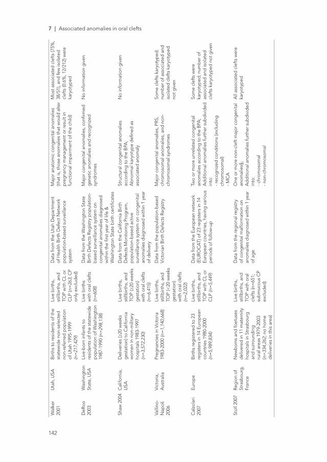
R1
R2
R3
R4
R5
R6
R7
R8
R9
R10
R11
R12
R13
R14
R15
R16
R17
R18
R19
R20
R21
R22
R23
R24
R25
R26
R27
R28
R29
R30
R31
R32
R33
R34
7 | Associated anomalies in oral clefts
142
Wal
ker
2001
Uta
h, U
SAB
irths
to re
sid
ents
of t
he
stat
ewid
e no
n-se
lect
ed
non-
refe
rred
pop
ulat
ion
of U
tah
1995
-199
9 (n
=21
7,42
9)
Live
birt
hs,
still
birt
hs, a
nd
TOP
with
CL
or
CLP
(n=
263;
CP
only
exc
lud
ed)
Dat
a fr
om th
e U
tah
Dep
artm
ent
of H
ealth
Birt
h D
efec
t Net
wor
k p
opul
atio
n-b
ased
sur
veill
ance
sy
stem
Maj
or a
nato
mic
con
gen
ital a
nom
alie
s (th
at is
, tho
se a
nom
alie
s th
at w
ould
alte
r p
reg
nanc
y m
anag
emen
t or r
esul
t in
func
tiona
l im
pai
rmen
t of t
he c
hild
)
Mos
t ass
ocia
ted
cle
fts
(75%
, 38
/51)
, and
few
isol
ated
cl
efts
(0.6
%, 1
2/21
2) w
ere
kary
otyp
ed
DeR
oo
2003
Was
hing
ton
Stat
e, U
SALi
ve-b
orn
infa
nts
to
resi
den
ts o
f the
sta
tew
ide
pop
ulat
ion
of W
ashi
ngto
n 19
87-1
990
(n=
298,
138)
Live
birt
hs
with
ora
l cle
fts
(n=
608)
Dat
a fr
om th
e W
ashi
ngto
n St
ate
Birt
h D
efec
ts R
egis
try
pop
ulat
ion-
bas
ed s
urve
illan
ce s
yste
m o
n co
ngen
ital a
nom
alie
s d
iag
nose
d
with
in th
e fir
st y
ear o
f life
&
Was
hing
ton
Stat
e b
irth
cert
ifica
tes
Maj
or c
ong
enita
l ano
mal
ies,
con
firm
ed
gen
etic
ano
mal
ies
and
reco
gni
zed
sy
ndro
mes
No
info
rmat
ion
giv
en
Shaw
200
4C
alifo
rnia
, U
SAD
eliv
erie
s (≥
20 w
eeks
g
esta
tion)
to C
alifo
rnia
w
omen
in n
on-m
ilita
ry
hosp
itals
198
3-19
97
(n=
3,57
2,23
0)
Live
birt
hs,
still
birt
hs, a
nd
TOP
(≥20
wee
ks
ges
tatio
n)w
ith o
ral c
left
s (n
=6,
415)
Dat
a fr
om th
e C
alifo
rnia
Birt
h D
efec
ts M
onito
ring
Pro
gra
m,
pop
ulat
ion-
bas
ed a
ctiv
e su
rvei
llanc
e sy
stem
on
cong
enita
l an
omal
ies
dia
gno
sed
with
in 1
yea
r of
del
iver
y
Stru
ctur
al c
ong
enita
l ano
mal
ies
acco
rdin
g to
the
BPA
;A
bno
rmal
kar
yoty
pe
defi
ned
as
asso
ciat
ed a
nom
aly
No
info
rmat
ion
giv
en
Valli
no-
Nap
oli
2006
Vic
toria
, A
ustr
alia
Preg
nanc
ies
in V
icto
ria
1983
-200
0 (n
=1,
140,
668)
Live
birt
hs,
still
birt
hs, a
nd
TOP
(<20
wee
ks
ges
tatio
n)
with
ora
l cle
fts
(n=
2,02
2)
Dat
a fr
om th
e p
opul
atio
n-b
ased
V
icto
rian
Birt
h D
efec
ts R
egis
try
Maj
or c
ong
enita
l ano
mal
ies,
PRS
, ch
rom
osom
al a
nom
alie
s, a
nd n
on-
chro
mos
omal
syn
dro
mes
Som
e cl
efts
kar
yoty
ped
; nu
mb
er o
f ass
ocia
ted
and
is
olat
ed c
left
s ka
ryot
yped
no
t giv
en
Cal
zola
ri 20
07Eu
rop
eB
irths
reg
iste
red
to 2
3 re
gis
ters
in 1
4 Eu
rop
ean
coun
trie
s 19
80-2
000
(n=
5,98
9,83
4)
Live
birt
hs,
still
birt
hs, a
nd
TOP
with
CL
or
CLP
(n=
5,44
9)
Dat
a fr
om th
e Eu
rop
ean
netw
ork
(EU
ROC
AT)
of 2
3 re
gis
ters
in 1
4 Eu
rop
ean
coun
trie
s, h
avin
g v
ario
us
per
iod
s of
follo
w-u
p
Two
or m
ore
unre
late
d c
ong
enita
l an
omal
ies
acco
rdin
g to
the
BPA
;A
dd
ition
al a
nom
alie
s fu
rthe
r sub
div
ided
in
to:
- rec
ogni
zed
con
diti
ons
(incl
udin
g
chro
mos
omal
)- M
CA
Som
e cl
efts
wer
e ka
ryot
yped
; num
ber
of
asso
ciat
ed a
nd is
olat
ed
clef
ts k
aryo
typ
ed n
ot g
iven
Stol
l 200
7Re
gio
n of
St
rasb
ourg
, Fr
ance
New
bor
ns a
nd fo
etus
es
del
iver
ed in
11
mat
erni
ty
hosp
itals
in S
tras
bou
rg
and
sur
roun
din
g
rura
l are
as 1
979-
2003
(n
=33
4,26
2; n
o ho
me
del
iver
ies
in th
is a
rea)
Live
birt
hs,
still
birt
hs, a
nd
TOP
with
ora
l cl
efts
(n=
651;
su
bm
ucou
s C
P ex
clud
ed)
Dat
a fr
om th
e re
gio
nal r
egis
try
of c
ong
enita
l mal
form
atio
n on
an
omal
ies
dia
gno
sed
with
in 1
yea
r of
ag
e
One
or m
ore
non-
clef
t maj
or c
ong
enita
l an
omal
ies§
;A
dd
ition
al a
nom
alie
s fu
rthe
r sub
div
ided
in
to:
- chr
omos
omal
- non
-chr
omos
omal
All
asso
ciat
ed c
left
s w
ere
kary
otyp
ed
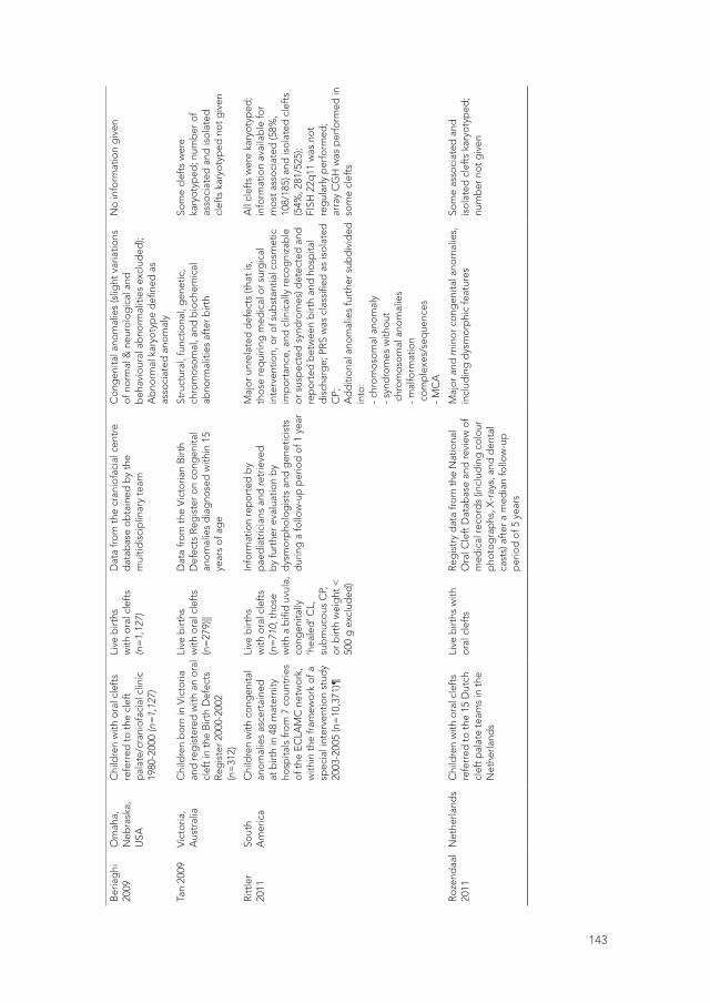
R1
R2
R3
R4
R5
R6
R7
R8
R9
R10
R11
R12
R13
R14
R15
R16
R17
R18
R19
R20
R21
R22
R23
R24
R25
R26
R27
R28
R29
R30
R31
R32
R33
R34
143
Ber
iag
hi
2009
Om
aha,
N
ebra
ska,
U
SA
Chi
ldre
n w
ith o
ral c
left
s re
ferr
ed to
the
clef
t p
alat
e/cr
anio
faci
al c
linic
19
80-2
000
(n=
1,12
7)
Live
birt
hs
with
ora
l cle
fts
(n=
1,12
7)
Dat
a fr
om th
e cr
anio
faci
al c
entr
e d
atab
ase
obta
ined
by
the
mul
tidis
cip
linar
y te
am
Con
gen
ital a
nom
alie
s (s
light
var
iatio
ns
of n
orm
al &
neu
rolo
gic
al a
nd
beh
avio
ural
ab
norm
aliti
es e
xclu
ded
);A
bno
rmal
kar
yoty
pe
defi
ned
as
asso
ciat
ed a
nom
aly
No
info
rmat
ion
giv
en
Tan
2009
Vic
toria
, A
ustr
alia
Chi
ldre
n b
orn
in V
icto
ria
and
reg
iste
red
with
an
oral
cl
eft i
n th
e B
irth
Def
ects
Re
gis
ter 2
000-
2002
(n
=31
2)
Live
birt
hs
with
ora
l cle
fts
(n=
279)
||
Dat
a fr
om th
e V
icto
rian
Birt
h D
efec
ts R
egis
ter o
n co
ngen
ital
anom
alie
s d
iag
nose
d w
ithin
15
year
s of
ag
e
Stru
ctur
al, f
unct
iona
l, g
enet
ic,
chro
mos
omal
, and
bio
chem
ical
ab
norm
aliti
es a
fter
birt
h
Som
e cl
efts
wer
e ka
ryot
yped
; num
ber
of
asso
ciat
ed a
nd is
olat
ed
clef
ts k
aryo
typ
ed n
ot g
iven
Ritt
ler
2011
Sout
h A
mer
ica
Chi
ldre
n w
ith c
ong
enita
l an
omal
ies
asce
rtai
ned
at
birt
h in
48
mat
erni
ty
hosp
itals
from
7 c
ount
ries
of th
e EC
LAM
C n
etw
ork,
w
ithin
the
fram
ewor
k of
a
spec
ial i
nter
vent
ion
stud
y 20
03-2
005
(n=
10,3
71)¶
Live
birt
hs
with
ora
l cle
fts
(n=
710;
thos
e w
ith a
bifi
d u
vula
, co
ngen
itally
‘h
eale
d’ C
L,
sub
muc
ous
CP,
or
birt
h w
eig
ht <
50
0 g
exc
lud
ed)
Info
rmat
ion
rep
orte
d b
y p
aed
iatr
icia
ns a
nd re
trie
ved
b
y fu
rthe
r eva
luat
ion
by
dys
mor
pho
log
ists
and
gen
etic
ists
d
urin
g a
follo
w-u
p p
erio
d o
f 1 y
ear
Maj
or u
nrel
ated
def
ects
(tha
t is,
th
ose
req
uirin
g m
edic
al o
r sur
gic
al
inte
rven
tion,
or o
f sub
stan
tial c
osm
etic
im
por
tanc
e, a
nd c
linic
ally
reco
gni
zab
le
or s
usp
ecte
d s
ynd
rom
es) d
etec
ted
and
re
por
ted
bet
wee
n b
irth
and
hos
pita
l d
isch
arg
e; P
RS w
as c
lass
ified
as
isol
ated
C
P;A
dd
ition
al a
nom
alie
s fu
rthe
r sub
div
ided
in
to:
- chr
omos
omal
ano
mal
y- s
ynd
rom
es w
ithou
t c
hrom
osom
al a
nom
alie
s- m
alfo
rmat
ion
com
ple
xes/
seq
uenc
es- M
CA
All
clef
ts w
ere
kary
otyp
ed;
info
rmat
ion
avai
lab
le fo
r m
ost a
ssoc
iate
d (5
8%,
108/
185)
and
isol
ated
cle
fts
(54%
, 281
/525
);FI
SH 2
2q11
was
not
re
gul
arly
per
form
ed;
arra
y C
GH
was
per
form
ed in
so
me
clef
ts
Roze
ndaa
l 20
11N
ethe
rland
sC
hild
ren
with
ora
l cle
fts
refe
rred
to th
e 15
Dut
ch
clef
t pal
ate
team
s in
the
Net
herla
nds
Live
birt
hs w
ith
oral
cle
fts
Reg
istr
y d
ata
from
the
Nat
iona
l O
ral C
left
Dat
abas
e an
d re
view
of
med
ical
reco
rds
(incl
udin
g c
olou
r p
hoto
gra
phs
, X-r
ays,
and
den
tal
cast
s) a
fter
a m
edia
n fo
llow
-up
p
erio
d o
f 5 y
ears
Maj
or a
nd m
inor
con
gen
ital a
nom
alie
s,
incl
udin
g d
ysm
orp
hic
feat
ures
Som
e as
soci
ated
and
is
olat
ed c
left
s ka
ryot
yped
; nu
mb
er n
ot g
iven
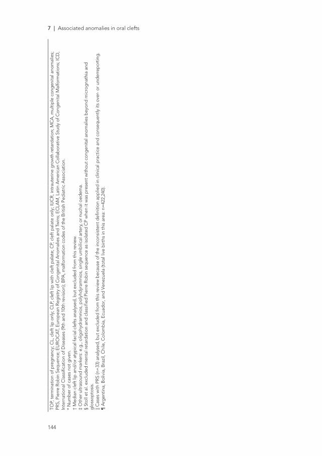
R1
R2
R3
R4
R5
R6
R7
R8
R9
R10
R11
R12
R13
R14
R15
R16
R17
R18
R19
R20
R21
R22
R23
R24
R25
R26
R27
R28
R29
R30
R31
R32
R33
R34
7 | Associated anomalies in oral clefts
144
TOP,
term
inat
ion
of p
reg
nanc
y; C
L, c
left
lip
onl
y; C
LP, c
left
lip
with
cle
ft p
alat
e; C
P, c
left
pal
ate
only
; IU
CR,
intr
aute
rine
gro
wth
reta
rdat
ion;
MC
A, m
ultip
le c
ong
enita
l ano
mal
ies;
PR
S, P
ierr
e Ro
bin
Seq
uenc
e; E
URO
CA
T, E
urop
ean
Reg
istr
y of
Con
gen
ital A
nom
alie
s an
d T
win
s; E
CLA
M, L
atin
Am
eric
an C
olla
bor
ativ
e St
udy
of C
ong
enita
l Mal
form
atio
ns; I
CD
, In
tern
atio
nal C
lass
ifica
tion
of D
isea
ses
(9th
and
10t
h re
visi
on);
BPA
, mal
form
atio
n co
des
of t
he B
ritis
h Pe
dia
tric
Ass
ocia
tion.
* N
umb
er o
f cas
es n
ot g
iven
. †
Med
ian
clef
t lip
and
/or a
typ
ical
faci
al c
left
s an
alys
ed, b
ut e
xclu
ded
from
this
revi
ew.
‡ O
ther
ultr
asou
nd m
arke
rs: e
.g.,
olig
ohyd
ram
nios
, pol
yhd
yram
nios
, sin
gle
um
bili
cal a
rter
y, o
r nuc
hal o
edem
a.
§ St
oll e
t al.
excl
uded
men
tal r
etar
dat
ion
and
cla
ssifi
ed P
ierr
e Ro
bin
seq
uenc
e as
isol
ated
CP
whe
n it
was
pre
sent
with
out c
ong
enita
l ano
mal
ies
bey
ond
mic
rog
nath
ia a
nd
glo
ssop
tosi
s.||
Cas
es w
ith P
RS (n
=33
) ana
lyse
d, b
ut e
xclu
ded
from
this
revi
ew b
ecau
se o
f the
inco
nsis
tent
defi
nitio
n ap
plie
d in
clin
ical
pra
ctic
e an
d c
onse
que
ntly
its
over
- or u
nder
rep
ortin
g.
¶ A
rgen
tina,
Bol
ivia
, Bra
zil,
Chi
le, C
olom
bia
, Ecu
ador
, and
Ven
ezue
la (t
otal
live
birt
hs in
this
are
a: n
=42
2,24
0).
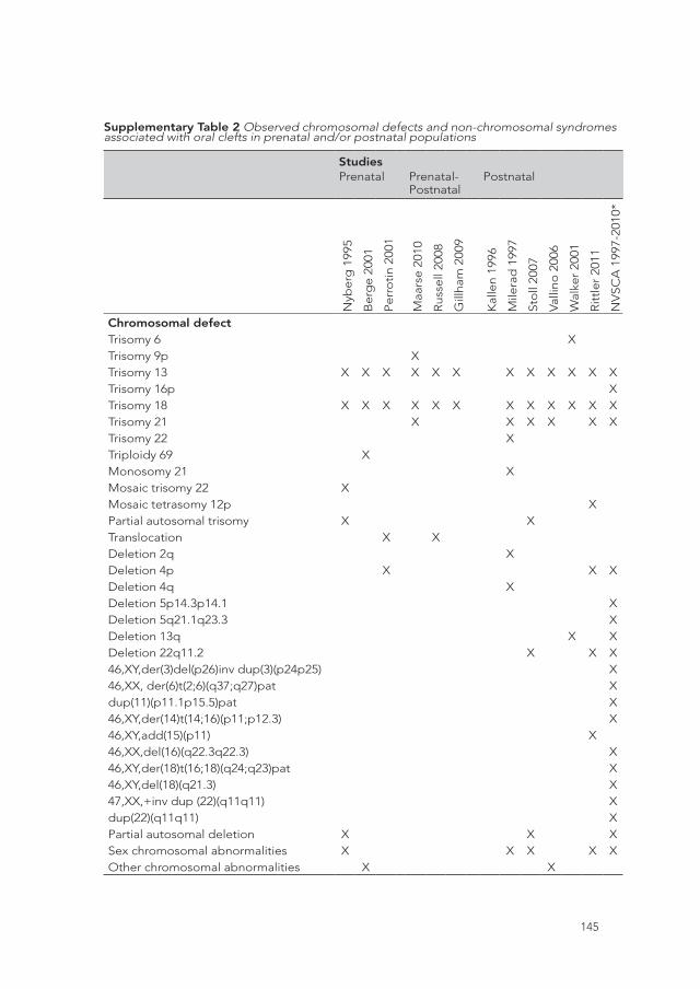
R1
R2
R3
R4
R5
R6
R7
R8
R9
R10
R11
R12
R13
R14
R15
R16
R17
R18
R19
R20
R21
R22
R23
R24
R25
R26
R27
R28
R29
R30
R31
R32
R33
R34
145
Supplementary Table 2 Observed chromosomal defects and non-chromosomal syndromes associated with oral clefts in prenatal and/or postnatal populations
StudiesPrenatal Prenatal-
PostnatalPostnatal
Nyb
erg
199
5
Be
rge
200
1
Pe
rro
tin
200
1
Maa
rse
201
0
Ru
sse
ll 20
08
Gill
ham
200
9
Kal
len
199
6
Mile
rad
199
7
Sto
ll 20
07
Valli
no
200
6
Wal
ker
2001
Rit
tle
r 20
11
NV
SC
A 1
997-
2010
*
Chromosomal defectTrisomy 6 XTrisomy 9p XTrisomy 13 X X X X X X X X X X X XTrisomy 16p XTrisomy 18 X X X X X X X X X X X XTrisomy 21 X X X X X XTrisomy 22 XTriploidy 69 XMonosomy 21 XMosaic trisomy 22 XMosaic tetrasomy 12p XPartial autosomal trisomy X XTranslocation X XDeletion 2q XDeletion 4p X X XDeletion 4q XDeletion 5p14.3p14.1 XDeletion 5q21.1q23.3 XDeletion 13q X XDeletion 22q11.2 X X X46,XY,der(3)del(p26)inv dup(3)(p24p25) X46,XX, der(6)t(2;6)(q37;q27)pat Xdup(11)(p11.1p15.5)pat X46,XY,der(14)t(14;16)(p11;p12.3) X46,XY,add(15)(p11) X46,XX,del(16)(q22.3q22.3) X46,XY,der(18)t(16;18)(q24;q23)pat X46,XY,del(18)(q21.3) X47,XX,+inv dup (22)(q11q11) Xdup(22)(q11q11) XPartial autosomal deletion X X XSex chromosomal abnormalities X X X X XOther chromosomal abnormalities X X
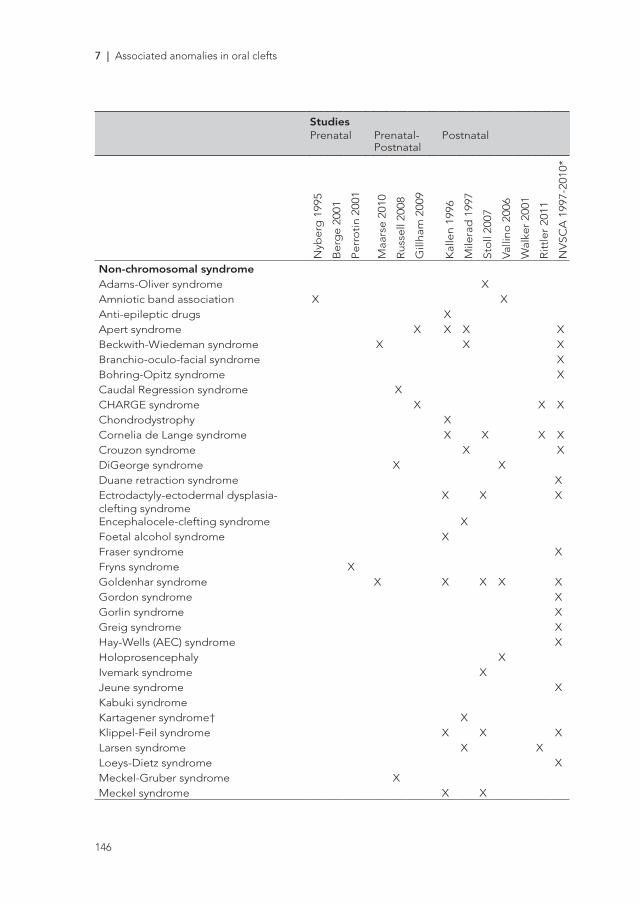
R1
R2
R3
R4
R5
R6
R7
R8
R9
R10
R11
R12
R13
R14
R15
R16
R17
R18
R19
R20
R21
R22
R23
R24
R25
R26
R27
R28
R29
R30
R31
R32
R33
R34
7 | Associated anomalies in oral clefts
146
StudiesPrenatal Prenatal-
PostnatalPostnatal
Nyb
erg
199
5
Be
rge
200
1
Pe
rro
tin
200
1
Maa
rse
201
0
Ru
sse
ll 20
08
Gill
ham
200
9
Kal
len
199
6
Mile
rad
199
7
Sto
ll 20
07
Valli
no
200
6
Wal
ker
2001
Rit
tle
r 20
11
NV
SC
A 1
997-
2010
*
Non-chromosomal syndromeAdams-Oliver syndrome XAmniotic band association X XAnti-epileptic drugs XApert syndrome X X X XBeckwith-Wiedeman syndrome X X XBranchio-oculo-facial syndrome XBohring-Opitz syndrome XCaudal Regression syndrome XCHARGE syndrome X X XChondrodystrophy XCornelia de Lange syndrome X X X XCrouzon syndrome X XDiGeorge syndrome X XDuane retraction syndrome XEctrodactyly-ectodermal dysplasia-clefting syndrome
X X X
Encephalocele-clefting syndrome XFoetal alcohol syndrome XFraser syndrome XFryns syndrome XGoldenhar syndrome X X X X XGordon syndrome XGorlin syndrome XGreig syndrome XHay-Wells (AEC) syndrome XHoloprosencephaly XIvemark syndrome XJeune syndrome XKabuki syndromeKartagener syndrome† XKlippel-Feil syndrome X X XLarsen syndrome X XLoeys-Dietz syndrome XMeckel-Gruber syndrome XMeckel syndrome X X
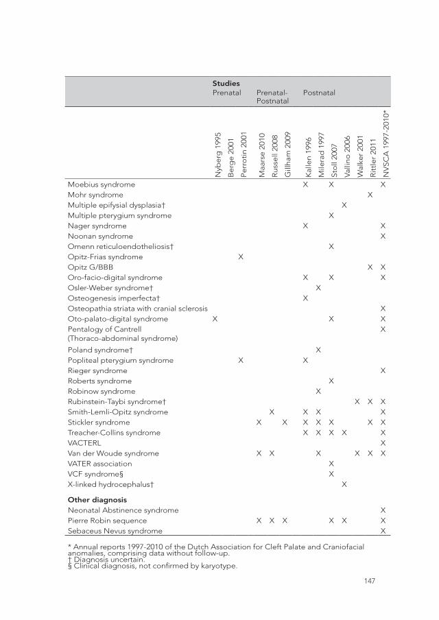
R1
R2
R3
R4
R5
R6
R7
R8
R9
R10
R11
R12
R13
R14
R15
R16
R17
R18
R19
R20
R21
R22
R23
R24
R25
R26
R27
R28
R29
R30
R31
R32
R33
R34
147
StudiesPrenatal Prenatal-
PostnatalPostnatal
Nyb
erg
199
5
Be
rge
200
1
Pe
rro
tin
200
1
Maa
rse
201
0
Ru
sse
ll 20
08
Gill
ham
200
9
Kal
len
199
6
Mile
rad
199
7
Sto
ll 20
07
Valli
no
200
6
Wal
ker
2001
Rit
tle
r 20
11
NV
SC
A 1
997-
2010
*
Moebius syndrome X X XMohr syndrome XMultiple epifysial dysplasia† XMultiple pterygium syndrome XNager syndrome X XNoonan syndrome XOmenn reticuloendotheliosis† XOpitz-Frias syndrome XOpitz G/BBB X XOro-facio-digital syndrome X X XOsler-Weber syndrome† XOsteogenesis imperfecta† XOsteopathia striata with cranial sclerosis XOto-palato-digital syndrome X X XPentalogy of Cantrell (Thoraco-abdominal syndrome)
X
Poland syndrome† XPopliteal pterygium syndrome X XRieger syndrome XRoberts syndrome XRobinow syndrome XRubinstein-Taybi syndrome† X X XSmith-Lemli-Opitz syndrome X X X XStickler syndrome X X X X X X XTreacher-Collins syndrome X X X X XVACTERL XVan der Woude syndrome X X X X X XVATER association XVCF syndrome§ XX-linked hydrocephalus† X
Other diagnosisNeonatal Abstinence syndrome XPierre Robin sequence X X X X X XSebaceus Nevus syndrome X
* Annual reports 1997-2010 of the Dutch Association for Cleft Palate and Craniofacial anomalies, comprising data without follow-up. † Diagnosis uncertain. § Clinical diagnosis, not confirmed by karyotype.

R1
R2
R3
R4
R5
R6
R7
R8
R9
R10
R11
R12
R13
R14
R15
R16
R17
R18
R19
R20
R21
R22
R23
R24
R25
R26
R27
R28
R29
R30
R31
R32
R33
R34

Part III
Prenatal Counseling

8Professional opinion on oral cleft
during pregnancy: a comparison
between Israel and the Netherlands
Prenatal Diagnosis, 2015 Jun;35(6):544-8
W. MAARSE, C.W.B. BOONACKER, O. LAPID, H.F.N. SWANENBURG DE VEYE,
Z. WEINER, M. KON, J.J.M. VAN DELDEN, A.B. MINK VAN DER MOLEN
R1
R2
R3
R4
R5
R6
R7
R8
R9
R10
R11
R12
R13
R14
R15
R16
R17
R18
R19
R20
R21
R22
R23
R24
R25
R26
R27
R28
R29
R30
R31
R32
R33
R34
8 | Professional opinion on oral cleft
152
ABSTRACT
Objective To assess the opinion of obstetric care providers who perform prenatal
ultrasounds to screen for anomalies and who advise women about their options,
including termination of pregnancy, when an oral cleft is detected. We compared
providers’ opinions about pregnancy termination for isolated oral cleft in the
Netherlands, where the number of terminations is low, and in Israel, where the number
is high.
Methods Online questionnaires were used. The questions assessed the providers’
views regarding the estimated burden of treatment, the functioning ability and the
level of happiness of children with an oral cleft and their parents. Additionally, we
assessed providers’ opinions on pregnancy termination for isolated oral cleft.
Results In the Netherlands, more professionals considered oral cleft a disability [rate
differences (RD) 17.8%, 95% CI: 0.5-33.1%] than in Israel. In the Netherlands, 10.6%
of respondents (compared with 11.1% in Israel) thought that an isolated cleft was a
reason for TOP [RD 0.6%, 95% CI: -12-10.9%].
Conclusions Prenatal care providers in the Netherlands and Israel do not differ in their
opinions about the severity of oral cleft and the acceptability of TOP for an isolated
oral cleft. This study shows that prenatal care providers’ attitudes do therefore not
explain the dramatic difference between these countries in the number of TOP for
isolated oral cleft.
R1
R2
R3
R4
R5
R6
R7
R8
R9
R10
R11
R12
R13
R14
R15
R16
R17
R18
R19
R20
R21
R22
R23
R24
R25
R26
R27
R28
R29
R30
R31
R32
R33
R34
153
INTRODUCTION
Oral clefts are among the most common congenital malformations; they occur in
approximately 1 out of 700 live births1. Because the use of ultrasound screening during
pregnancy has become the standard of care, the prenatal diagnosis of oral clefts is
common and has a detection rate of 88%2.
There is some controversy about the parental, social and ethical consequences of
prenatal detection of oral clefts3. The prenatal detection of an oral cleft has numerous
advantages. Not only are the parents better prepared psychologically for the anomaly,
but awareness of the diagnosis provides an opportunity to provide education about
both the treatment of oral clefts and the management of possible feeding problems.
The prenatal diagnosis of an oral cleft also provides an opportunity to offer additional
genetic testing to screen for possible associated anomalies and chromosomal
defects4,5. The disadvantages of the prenatal diagnosis of oral cleft are the possibility
of a false positive diagnosis and the resulting emotional stress on the pregnancy6,7.
The fact that at present time prenatal surgery may not be an option or may carry very
high risks is another disadvantage8. Additionally there are concerns about the possible
increasing number of terminations of pregnancy (TOP) of fetuses with an isolated oral
cleft9. Because most nonsyndromic oral clefts are nonlethal birth defects that have an
excellent functional and aesthetic prognosis with proper treatment, the use of prenatal
diagnosis raises important ethical issues, especially in terms of TOP10,11.
High numbers of TOPs for prenatally diagnosed isolated oral clefts have been
reported in Israel (93.4%)9. In the Netherlands, the number of parents who choose
TOP when an oral cleft is detected is low (6.7%)12. The reasons for these differences
are unknown. In addition to cultural and religious influences, the perception of burden
and the stigma of having a ‘less-than-perfect’ child may be significant contributing
factors13. Caregivers who counsel future parents could be another influencing factor14.
Little is known about prenatal care providers’ attitudes towards various conditions for
which prenatal diagnosis is available. The goal of the present study was to assess the
opinions about oral clefts and TOP among obstetric care providers in Israel and the
Netherlands. Specifically, we sought to determine whether personal attitudes were
R1
R2
R3
R4
R5
R6
R7
R8
R9
R10
R11
R12
R13
R14
R15
R16
R17
R18
R19
R20
R21
R22
R23
R24
R25
R26
R27
R28
R29
R30
R31
R32
R33
R34
8 | Professional opinion on oral cleft
154
a plausible explanation for the large difference in TOPs for isolated cleft in these
countries.
METHODS
A cross-sectional quantitative study was performed. In the Netherlands, routine
screening two-dimensional (2D) ultrasound is offered to all pregnant women at
approximately 20 weeks of gestation. In Israel, a first screening is offered earlier,
between 14 and 16 weeks of gestation.
In August 2013, an online questionnaire was distributed digitally in the Netherlands
(Dutch) and Israel (Hebrew) to all obstetric care providers who perform prenatal
ultrasounds. In the Netherlands, we approached a subgroup of the Dutch Society
of Obstetrics and Gynecology composed only of gynecologists and midwives who
specialize in prenatal care. In the Netherlands, trained midwives, sonographers
and gynecologists perform routine 2D ultrasound screening in several centers. If an
abnormality is suspected, the pregnant woman is referred to a specialized center,
where certified gynecologists perform more extensive two-, and three-dimensional
ultrasound examinations and provide counseling. In Israel, the questionnaire was sent
to all gynecologists through the Israeli Society of Obstetrics and Gynecology. Unlike
in the Netherlands, there is no subgroup of gynecologists in Israel who specialize in
prenatal care.
In both countries, gynecologists offer future parents a referral to a cleft team for
additional prenatal counseling when an oral cleft is diagnosed prenatally. However,
the parents ultimately decide whether to take advantage of this opportunity.
The questionnaire used in this study was derived from one previously published by
Korenromp et al.15, who explored decision-making processes regarding TOP for Down
syndrome. The questionnaire was translated into Hebrew (in a back-and-forward
manner) by two of the authors (O.L. and M.K).
In the questionnaire, obstetric care providers were asked about their perception of the
estimated severity of an oral cleft (by means of whether they would describe a cleft as
R1
R2
R3
R4
R5
R6
R7
R8
R9
R10
R11
R12
R13
R14
R15
R16
R17
R18
R19
R20
R21
R22
R23
R24
R25
R26
R27
R28
R29
R30
R31
R32
R33
R34
155
a ‘trait’, a ‘little different from normal’, a ‘cosmetic defect’, a ‘disability’, a ‘disorder’
or a ‘disease’) and whether they believed that an oral cleft was a reason for a TOP. To
place this question in perspective, the care providers were also asked for their opinion
regarding other reasons for TOP (Table 1). Additionally, care providers were asked their
opinion about the estimated treatment burden for the child and the parents (on a scale
of 0-100, where 0 was ‘not serious’ and 100 was ‘very serious’) and the influence of the
oral cleft on the parent’s and child’s happiness and ability to function (on a scale from
0-100, where 0 was ‘not seriously influenced’ and 100 was ‘very serious influenced’;
Table 2).
Statistical analysis was performed using SPSS statistical software (version 20.0) and
Rothman’s Episheet (11 June 2008). We used the rate (percentage) or mean differences
with corresponding 95% confidence intervals (CIs) to compare the Dutch and Israeli
results.
RESULTS
In the Netherlands, the response rate was 42.8% (142/332), and the questionnaire
was completed by 92 (64.8%) gynecologists, 19 (13.4%) midwives, 19 (13.4%) ultra-
sonographers and 12 (8.4%) other professionals, such as a pediatricians or medical
geneticists. In Israel, all of the respondents were gynecologists (n=41). The exact
number of Israeli gynecologists who perform prenatal ultrasounds is not known;
however, another survey study from Israel17 estimated that 100 gynecologists perform
this service. Thus, the response rate was similar. In the Netherlands, more professionals
had diagnosed an oral cleft in their professional careers: 88% compared with 72.5% in
Israel [rate difference (RD) 15.5%, 95% CI: 0.7-30.4%].
The oral cleft descriptions that the respondents used are outlined in Table 1. None of
the respondents defined an oral cleft as a trait similar to hair color. In both countries,
‘disability’ was the most frequent answer, though the percentage was higher in the
Netherlands (60.3%) compared with Israel [42.5%; RD 17.8%, 95% CI: 0.5-33.1%]. Thirty-
five percent of the Israeli professionals classified an oral cleft as a cosmetic defect,
whereas 20.6% of the respondents in the Netherlands used that classification [RD
-14.4%, 95 CI:-30.7-1.8%].
R1
R2
R3
R4
R5
R6
R7
R8
R9
R10
R11
R12
R13
R14
R15
R16
R17
R18
R19
R20
R21
R22
R23
R24
R25
R26
R27
R28
R29
R30
R31
R32
R33
R34
8 | Professional opinion on oral cleft
156
There was no difference in the percentage of professionals in the Netherlands (10.6%)
and in Israel (11.1%) who thought that an isolated cleft was a reason for TOP [RD 0.6%,
95% CI: -12- to 10.9%]. However, there were significant differences in the circumstances
under which professionals considered TOP an option (a positive RD indicated that
the percentage in the Netherlands was higher, and a negative RD indicated that the
percentage in Israel was higher). These circumstances included Down syndrome [RD
15.9%, 95% CI: 0.9 to 30.9%], spina bifida [RD 28.8%, 95% CI: 13.5 to 44.2%], wheelchair
dependency [RD -20%, 95% CI: -36.2 to -3.9%] and diseases that would occur later in
life [RD 22.6%, 95% CI: 10.7 to 34.5%].
Table 1 Professional opinions about oral cleft
Question The Netherlands (N = 142)N (%)
Israel(N = 41)N (%)
Rate differences (95% CI)
How would you define oral cleft? Small difference from normal Cosmetic defect Disability Disorder Disease
5 (3.5%)29 (20.6%)85 (60.3%)3 (2.1%)0 (0.0%)
3 (7.5%)14 (35.0%)17 (42.5%)3 (7.5%)1 (2.5%)
-4% (-12.7 to 4.8%)-14.4% (-30.7 to 1.8%)17.8% (0.5 to 35.1%)-5.4% (-13.9 to 3.1%)-2.5% (-7.3 to 3.2%)
Do you think isolated oral cleft is a reason for TOP? Yes 15 (10.6%) 4 (11.1%) -0.6% (-12 to 10.9%)
Are there circumstances in which TOP could be an option? Down syndrome Spina bifida Other mental handicap or disorder Wheelchair dependency Other physical handicap, such as blindness, deafness, muscle disorder or metabolic disease Expected death before, during or within a month after birth Limited life expectancy (until 30 years old; for example, cystic fibrosis) Disease occurring later in life
123 (86.6%)131 (92.3%)108(76.1%)72 (50.7%)108 (76.1%)
128 (90.1%)
79 (55.6%)
46 (32.4%)
29 (70.7%)26 (63.4%)30 (73.2%)29 (70.7%)29 (70.7%)
34 (82.9%)
22 (53.7%)
4 (9.8%)
15.9% (0.9 to 30.9%)28.8% (13.5 to 44.2%)2.9% (-12.4 to 18.2%)-20% (-36.2 to -3.9)5.3% (-10.3 to 20.9%)
7.2% (-5.3 to 19.7%)
2.0% (-15.4 to 19.3%)
22.6% (10.7 to 34.5%)
Bold = statistically significant. TOP = Termination Of Pregnancy
R1
R2
R3
R4
R5
R6
R7
R8
R9
R10
R11
R12
R13
R14
R15
R16
R17
R18
R19
R20
R21
R22
R23
R24
R25
R26
R27
R28
R29
R30
R31
R32
R33
R34
157
Table 2 presents the Dutch and Israeli professionals’ opinions about the estimated
burden of an oral cleft and its impact on happiness and overall life functioning for
afflicted children and their parents. Using a similar scale from 0 to 100, the Dutch
professionals’ responses indicated that they viewed the repair of oral cleft as a more
serious burden for children and parents than the Israeli professionals did: the reported
scores were 59.5 and 34.7 [mean difference 24.8, 95% CI 15.7 to 33.9] for children and
63.0 and 40.9 [mean difference 22.1, 95% CI 13.4 to 30.8] for parents, respectively.
Conversely, compared with the Israeli professionals, the Dutch professionals thought
that an oral facial cleft had less effect on a child’s overall functioning [21.4 and 11.3,
respectively; mean difference -10.1 95% CI -17.2 to -2.9]. There was no difference
between the Dutch and Israeli professionals’ opinions regarding the influence of an
oral cleft on the happiness of the children or their parents.
Table 2 Professional opinions about oral cleft
Question The Netherlands(N = 142) Mean (sd)
Israel(N = 41)Mean (sd)
Mean differences (95% CI)
How seriously do you think the oral cleft treatment will burden the child? 59.5 (23.8) 34.7 (28.9) 24.8 (15.7 to 33.9)
How seriously do you think the oral cleft treatment will burden the parents? 63.0 (22.4) 40.9 (27.6) 22.1 (13.4 to 30.8)
How seriously do you think this will affect the child’s happiness? 27.7 (21.5) 24.7 (22.9) 2.9 (-5.2 to 11.0)
How seriously do you think this will affect the parents’ happiness? 27.8 (21.5) 31.2 (30.4) -3.4 (-12.0 to 5.2)
How seriously do you think this will affect the child’s overall functioning? 11.3 (14.6) 21.4 (31.1) -10.1 (-17.2 to -2.9)
Bold = statistically significant. All answers were given on a scale from 0 (not serious) to 100 (very serious)
DISCUSSION
The results of this study show that a minority of obstetric care providers in the
Netherlands and Israel favor TOP in case of isolated oral cleft. This percentage
was comparable in both countries (11%). This is in contrast to the high number of
R1
R2
R3
R4
R5
R6
R7
R8
R9
R10
R11
R12
R13
R14
R15
R16
R17
R18
R19
R20
R21
R22
R23
R24
R25
R26
R27
R28
R29
R30
R31
R32
R33
R34
8 | Professional opinion on oral cleft
158
parents in Israel who choose TOP in case of an isolated oral cleft9. Because obstetric
care providers in the Netherlands and Israel reported comparable positive opinions
about oral clefts, we conclude that their personal beliefs therefore cannot explain the
dramatic difference in TOP rates between these two countries.
Several studies have attempted to explain the differences in parental decisions
about TOP among countries13,16,17. A combination of factors play a role, including the
child and family’s best interest, the severity of the abnormality, economic issues, the
treatment burden, religious background, legal restrictions and gestational age at the
time of diagnosis. The latter three factors were the only well-defined issues in both the
Netherlands and Israel. Routine prenatal ultrasound is offered in both the Netherlands
and in Israel, and participation is higher than 95% in both countries2,17; however, the
gestational age at which ultrasound is offered differs. In Israel, women are offered
an ultrasound between 14 and 169 weeks gestation, whereas in the Netherlands,
ultrasound is performed at 20 weeks. It may be easier to make the difficult decision
to terminate a pregnancy at an earlier gestational age. In both countries, late TOP is
legal. Additionally, religion differs in Israel and the Netherlands. In Israel, according
to Jewish law, the interruption of pregnancy is generally forbidden after 40 days of
pregnancy, and Islamic Law absolutely forbids TOP after the 120th day of pregnancy.
However, the majority of the Jewish population is secular and considers TOP to be
legitimate18. In the Netherlands, the majority of people are not religious (62% according
to the Netherlands Institute for Social Research; only 17% believe that their religion is
very important). Both countries have very religious minorities. In conclusion, religious
doctrine does not seem to explain the large difference in TOP rates. Moreover, no
specific research has evaluated how religious beliefs influence decisions about TOP
when an isolated oral cleft is suspected.
In addition to technical, legal and religious issues, parental attitudes towards oral clefts
no doubt play an important role in the decision to terminate a pregnancy. Parents’
personal views affect their perception of the severity of the condition, and they are
also influenced by the medical ramifications, which depend on and are mediated by
the care provider’s counseling10,24,25. Thus, the care providers’ perception of the severity
of an oral cleft matters. In this survey, we showed that the majority of care providers
R1
R2
R3
R4
R5
R6
R7
R8
R9
R10
R11
R12
R13
R14
R15
R16
R17
R18
R19
R20
R21
R22
R23
R24
R25
R26
R27
R28
R29
R30
R31
R32
R33
R34
159
considered oral clefts a disability or a cosmetic defect and that there were no major
differences in this rating between the countries. Whether an individual judges an oral
cleft as severe or not severe is both culturally and socially determined3. In a study
by Wyszinski et al.,16 37.9% of the parents of children with an oral cleft in Argentina
considered it a ‘trait’. In contrast, parents interviewed in Saudi Arabia worried that
“ladies with oral clefts will have a hard time finding a husband and will not have a
normal social life, so termination of such an abnormality will be better”26. In our study,
we showed that Israeli and Dutch care providers judged the severity of an oral cleft
similarly, so this could not explain the differences in TOP rates. In addition, Some of
the respondents in this study stated that their opinion was insignificant for deciding
whether isolated oral cleft was a reason for TOP. They stated that the decision to
terminate the pregnancy was ultimately the parents’. Although we agree that decisions
regarding TOP in case of an affected fetus are and should be the parents’, counselors
should be able to empower the parents to be able to make an autonomous choice. It is
the counselor’s responsibility to inform parents about the etiology, pathogenesis and
associated anomalies as well as the medical, surgical, and clinical needs of a child with
an oral cleft. However, a former study19 showed that many parents were unsatisfied with
the initial information they received at the ultrasound unit. In the study by Hsieh20 and
Rey-Bellet21, some mothers expressed feeling anxious about their baby’s condition,
and, because of the limited information provided by professionals, some even feared
that the condition might be associated with impaired intelligence. Therefore, the input
from a cleft lip and palate team during the prenatal period is of utmost importance.
Usually, the diagnosis of an oral cleft comes as a shock to parents, and proper counseling
helps to reduce the anguish of having a baby with an oral cleft22. To reduce anxiety
and uncertainty, clear and consistent information about the anomaly and its treatment,
etiology and prognosis must be provided during initial counseling12,23. Kuttenberger
et al. suggest23 that such counseling should be provided by a multidisciplinary cleft
lip and palate team, a position that we support. Moreover, our study shows that it
is uncommon for an obstetrician to detect an oral cleft, whereas cleft lip and palate
teams have the expertise required to treat children with oral clefts in daily practice.
Unfortunately, this study provides no information about possible differences in the
counseling techniques used by obstetric care providers in Israel and the Netherlands.
R1
R2
R3
R4
R5
R6
R7
R8
R9
R10
R11
R12
R13
R14
R15
R16
R17
R18
R19
R20
R21
R22
R23
R24
R25
R26
R27
R28
R29
R30
R31
R32
R33
R34
8 | Professional opinion on oral cleft
160
It remains unknown whether all parents in Israel who choose TOP in the case of an
isolated oral cleft were willing or able to consult a cleft lip and palate team before
making their decision. This service is routinely offered in the Netherlands. This area
warrants further research because possible differences in actual counseling practices
between Israel and the Netherlands may explain part of the variation in TOP rates
despite the fact that gynecologists in both countries hold similar personal opinions
about cleft and TOP.
The major strength of this study is its comparison of two countries in which TOP rates
for isolated cleft differ markedly . Moreover, this is the first study to survey professional
opinions about oral clefts. Our study has some methodological shortcomings. First,
although a response rate of approximately 40% might seem low, it is common for
anonymous medical surveys27. This rate could even be considered high because the
questionnaires were distributed by e-mail rather than administered in person. Second,
there was a baseline difference in the responses depending of the respondent’s
profession. Subgroup analyses that included only gynecologists showed similar results
(data not shown). Finally, the actual response rate in Israel could only be estimated
because the exact number of Israeli gynecologists that perform prenatal ultrasounds is
not known, although an independent nationwide survey reported that there were 100
such gynecologists17.
CONCLUSION
Prenatal care providers in the Netherlands and Israel do not differ in their opinions on
the severity of oral clefts and the acceptability of TOP for an isolated oral cleft. This
study shows that provider opinion does not explain the dramatic difference between
these countries in TOP for isolated oral cleft. It can be expected that in the future,
ultrasound sensitivity for detecting oral clefts will only increase, resulting in a greater
number of prenatally diagnosed clefts. Although counseling factors were not studied,
we advise gynecologists to refer parents to a cleft lip and palate team prenatally. It is
important to provide parents who are expecting a child with an oral cleft with balanced
counseling and adequate information.
R1
R2
R3
R4
R5
R6
R7
R8
R9
R10
R11
R12
R13
R14
R15
R16
R17
R18
R19
R20
R21
R22
R23
R24
R25
R26
R27
R28
R29
R30
R31
R32
R33
R34
161
REFERENCES
1. Mossey PA, Little J, Munger RG, et al. Cleft lip and palate. Lancet 2009;374(9703):1773–85.2. Maarse W, Pistorius LR, Van Eeten WK, et al. Prenatal ultrasound screening for orofacial
clefts. Ultrasound Obstet Gynecol 2011;38(4):434–9.3. Strauss RP. Beyond easy answers: prenatal diagnosis and counseling during pregnancy.
Cleft Palate Craniofac J 2002;39(2):164–8.4. Johnson N, Sandy JR. Prenatal diagnosis of cleft lip and palate. Cleft Palate Craniofac J
2003;40(2):186–9.5. Maarse W, Rozendaal AM, Pajkrt E, et al. A systematic review of associated structural and
chromosomal defects in oral clefts: when is prenatal genetic analysis indicated? J Med Genet 2012;49(8):490–8.
6. Matthews MS, Cohen M, Viglione M, et al. Prenatal counseling for cleft lip and palate. Plast Reconstr Surg 1998;101(1):1–5.
7. Nusbaum R, Grubs RE, Losee JE, et al. A qualitative description of receiving a diagnosis of clefting in the prenatal or postnatal period. J Genet Couns 2008;17(4):336–50.
8. Wojcicki P, Drozdowski PH. In utero surgery – current state of the art – part II. Med Sci Monit 2011;17(12):RA262–70.
9. Bronshtein M, Blumenfeld I, Blumenfeld Z. Early prenatal diagnosis of cleft lip and its potential impact on the number of babies with cleft lip. Br J Oral Maxillofac Surg 1996;34(6):486–7.
10. Blumenfeld Z, Blumenfeld I, Bronshtein M. The early prenatal diagnosis of cleft lip and the decision-making process. Cleft Palate Craniofac J 1999;36(2):105–7.
11. Hager C. Termination of pregnancy with a prenatal diagnosis of cleft lip: cultural differences and ethical analysis. Plast Surg Nurs 2002;22(1):24–8.
12. Mink van der Molen AB, Maarse W, Pistorius L, et al. Prenatal screening for orofacial clefts in the Netherlands: a preliminary report on the impact of a national screening system. Cleft Palate Craniofac J 2011;48(2):183–9.
13. Jones MC. Prenatal diagnosis of cleft lip and palate: experiences in Southern California. Cleft Palate Craniofac J 1999;36(2):107–9.
14. Drake H, Reid M, Marteau T. Attitudes towards termination for fetal abnormality: comparisons in three European countries. Clin Genet 1996;49(3):134–40.
15. Korenromp MJ, Page-Christiaens GC, van den Bout J, et al. Maternal decision to terminate pregnancy in case of Down syndrome. Am J Obstet Gynecol 2007;196(2):149 e1–11.
16. Wyszynski DF, Perandones C, Bennun RD. Attitudes toward prenatal diagnosis, termination of pregnancy, and reproduction by parents of children with nonsyndromic oral clefts in Argentina. Prenat Diagn 2003;23(9):722–7.
17. Mishori Dery A, Carmi R, Shoham VI. Attitudes toward the acceptability of reasons for pregnancy termination due to fetal abnormalities among prenatal care providers and consumers in Israel. Prenat Diagn 2008;28(6):518–24.
18. Zlogotogora J, Haklai Z, Rotem N, et al. The impact of prenatal diagnosis and termination of pregnancy on the relative incidence of malformations at birth among Jews and Muslim Arabs in Israel. Isr Med Assoc J 2010;12:539–42.
19. Berggren H, Hansson E, Uvemark A, et al. Prenatal compared with postnatal cleft diagnosis: what do the parents think? J Plast Surg Hand Surg 2012;46(3-4):235–41.
20. Hsieh YT, Chao YM, Shiao JS. A qualitative study of psychosocial factors affecting expecting mothers who choose to continue a cleft lip and/or palate pregnancy to term. J Nurs Res 2013;21(1):1–9.
21. Rey-Bellet C, Hohlfeld J. Prenatal diagnosis of facial clefts: evaluation of a specialised counselling. Swiss Med Wkly 2004;134(43–44):640–4.
R1
R2
R3
R4
R5
R6
R7
R8
R9
R10
R11
R12
R13
R14
R15
R16
R17
R18
R19
R20
R21
R22
R23
R24
R25
R26
R27
R28
R29
R30
R31
R32
R33
R34
8 | Professional opinion on oral cleft
162
22. Davalbhakta A, Hall PN. The impact of antenatal diagnosis on the effectiveness and timing of counselling for cleft lip and palate. Br J Plast Surg 2000;53(4):298–301.
23. Kuttenberger J, Ohmer JN, Polska E. Initial counselling for cleft lip and palate: parents’ evaluation, needs and expectations. Int J Oral Maxillofac Surg 2010;39(3):214–20.
24. Beeri M, Haramati Z, Rein JJ, et al. Parental knowledge and views of pediatric congenital heart disease. Isr Med Assoc J 2001;3(3):194–7.
25. Sagi M, Meiner V, Reshef N, et al. Prenatal diagnosis of sex chromosome aneuploidy: possible reasons for high rates of pregnancy termination. Prenat Diagn 2001;21(6):461–5.
26. Alsulaiman A, Abu-Amero KK. Parent’s attitude toward prenatal diagnosis and termination of pregnancy could be influenced by other factors rather than by the severity of the condition. Prenat Diagn 2013;33(3):257–61.
27. Asch DA, Jedrziewski MK, Christakis NA. Response rates to mail surveys published in medical journals. J Clin Epidemiol 1997;50(10):1129–36.
9Parental attitude on prenatal
diagnosis of cleft lip and palate;
a prospective cohort study
Submitted
W. MAARSE (MD)*, C.W.B. BOONACKER*, H.F.N. SWANENBURG DE VEYE,
M. KON, C.C. BREUGEM, A.B. MINK VAN DER MOLEN, J. J.M. VAN DELDEN
* Joined first authorship; these authors contributed equally to this work
R1
R2
R3
R4
R5
R6
R7
R8
R9
R10
R11
R12
R13
R14
R15
R16
R17
R18
R19
R20
R21
R22
R23
R24
R25
R26
R27
R28
R29
R30
R31
R32
R33
R34
9 | Parental attitude on oral cleft
164
ABSTRACT
Objectives The prenatal diagnosis of oral clefts (OCs) by ultrasound can pose an
ethical dilemma. The purpose of this study was to obtain additional insight into the
psychosocial and moral considerations of prospective parents concerning OCs, the
burden of OCs and parents’ attitude toward possible termination of pregnancy (TOP)
to improve counseling in the future.
Study design Between August 2011 and August 2014, a prospective cohort
questionnaire study was administered to parents visiting our prenatal clinic.
Results Most of the parents described an oral cleft as a cosmetic disability (43.6%) or
as “just a little different” (26.6%). These parents expected that the OC would not affect
their own happiness or the happiness of their future child. Health professionals had a
considerable influence on parental opinion. A minority (6.4%, 5/85) of the respondents
considered TOP, and none of the participants chose to terminate the pregnancy. The
estimated severity of the oral cleft and the estimated influence on the child’s happiness
both had a significant influence on whether TOP was considered (p=0.04).
Conclusions This study suggests that the active involvement of cleft lip and palate
teams is worthwhile. Caregivers should be aware that their counseling is important in
the decision making of parents.
R1
R2
R3
R4
R5
R6
R7
R8
R9
R10
R11
R12
R13
R14
R15
R16
R17
R18
R19
R20
R21
R22
R23
R24
R25
R26
R27
R28
R29
R30
R31
R32
R33
R34
165
INTRODUCTION
The finding of a facial malformation on prenatal ultrasound can be a major psychosocial
burden for parents, who might express signs of severe distress 1-3. The most common
congenital facial malformation is oral cleft (OC), which arises in approximately 1 of 700
live births 4. As the use of routine scanning of the face with ultrasound has become
standard, prenatal diagnosis of OCs is now possible and accounts for a detection rate
of 88% 5. In response, specialized prenatal cleft clinics participate in the counseling of
parents expecting a child with a possible OC 6 to help them cope with the news and
prepare them for the birth of their child.
There is controversy concerning the preferred consequences of the prenatal detection
of malformations. In addition to the parental issues, there are social and ethical
implications of a prenatal diagnosis 7. The advantages of prenatal knowledge are
that parents can psychologically prepare for the anomaly, there is an opportunity for
education, parents can plan neonatal care, and there is an opportunity to perform
genetic counseling 3, 5, 8. However, parents may experience emotional disturbance of
the pregnancy once they know their child has an OC 1. Further disadvantages of the
prenatal detection of a cleft include the imprecision of the diagnosis 9, impossibility of
fetal surgery in nearly all cases, and general concern regarding the increased number
of terminations of pregnancy (TOPs) of fetuses with an isolated OC 10, 11. Nonetheless,
it is widely accepted that most parents prefer to know the diagnosis prenatally than be
confronted with the anomaly after birth 1, 2, 8, 9, 12, 13.
For optimal counseling, it is imperative to comprehend the experience of receiving a
diagnosis of an OC during the prenatal period. However, all previous studies assessing
the impact of a prenatally detected OC are of retrospective design, implying that, at
the time of assessment, the child was already born. The latter scenario might affect a
parent’s view on the impact of an OC diagnosis. Consequently, the parents’ perspective
regarding the expected severity, burden and possible TOP of a fetus with OC in the
prenatal period is unknown. The purpose of this study was to obtain additional insight
into the psychosocial and moral considerations of prospective parents concerning
R1
R2
R3
R4
R5
R6
R7
R8
R9
R10
R11
R12
R13
R14
R15
R16
R17
R18
R19
R20
R21
R22
R23
R24
R25
R26
R27
R28
R29
R30
R31
R32
R33
R34
9 | Parental attitude on oral cleft
166
OCs, the burden of OCs and parents’ attitudes toward possible TOP as well as their
opinions regarding current counseling to improve future counseling.
MATERIALS AND METHODS
Prenatal consultation
In The Netherlands, a routine screening two-dimensional (2D) ultrasound is offered
to all pregnant women at approximately 20 weeks of gestation 6. If an anomaly is
suspected, the mother is referred to a tertiary care center, where a more elaborate
2D/3D ultrasound is performed to confirm the diagnosis. Moreover, parents are given
advice concerning possible genetic counseling. If an OC is diagnosed, parents in
the region of Utrecht are invited to visit a special outpatient clinic where counseling
is performed by a plastic surgeon and a medical physiologist from the cleft lip and
palate team. The purpose of the counseling is to offer parents information concerning
etiology, pathology, treatment, and prognosis as well as psychosocial aspects and
advice regarding postnatal feeding. Specifically, parents receive written information
about treatment and pre- and post-surgical photographs of children with an OC.
Study design
A prospective cohort study was performed between August 2011 and August 2014.
All of the parents visiting the prenatal cleft clinic for their unborn child with an isolated
OC were asked to participate in the study by answering a questionnaire. The medical
psychologist acquired informed consent. Only Dutch-speaking parents were included
due to linguistic issues. The parents were asked to participate after they received
counseling by the cleft lip and palate team.
Questionnaires
The questionnaire used in this study was derived from one previously published by
Korenromp et al.14, who examined parental opinion on prenatally detected Down
syndrome. The questionnaire consisted of four parts (full version available online).
The first part provided basic information, such as sociodemographic and medical
R1
R2
R3
R4
R5
R6
R7
R8
R9
R10
R11
R12
R13
R14
R15
R16
R17
R18
R19
R20
R21
R22
R23
R24
R25
R26
R27
R28
R29
R30
R31
R32
R33
R34
167
characteristics, including a family history of OC, religion, and expected characteristics
of the child’s OC. In the second part of the questionnaire, parents were asked to
describe OCs in general and about their thoughts and attitudes toward the impact
and expected burden of the OC of their child. Furthermore, the parents were asked to
describe the people who influenced their opinion of OC. In the third part, the parents
were asked whether TOP would ever be an option and, what factors influenced their
decision. Finally, parents rated the prenatal counseling. Parents individually filled out
an extensive standardized questionnaire. Hence, two questionnaires per child were
obtained. Questionnaires were analyzed anonymously.
Statistical analysis
Questionnaire data were summarized using descriptive statistics—i.e., frequencies or
medians with interquartile ranges (IQRs) per question. Two groups were compared:
those who did and those who did not consider TOP as an option in the case of an OC.
Furthermore, obstetric characteristics were compared between those mothers who
did and those who did not return the questionnaire to assess whether these baseline
characteristics were comparable. The descriptions of an OC were compared among
the different types of OCs. All of the comparisons were made using chi-squared
statistics for dichotomous or the non-parametric Mann-Whitney U-test for continuous
outcomes. A p-value < 0.05 was considered statistically significant. Analysis was
performed using SPSS version 20.0 15.
Ethics
The medical ethical commission of the University Medical Center Utrecht approved
the study (nr 09/207), for which all participants provided informed consent.
RESULTS
Participants
During the study period, 81 couples expecting a child with an isolated OC were
referred to the tertiary clinic (see figure 1). One couple decided to terminate the
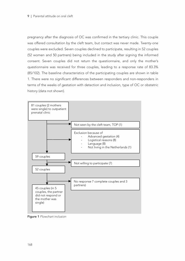
R1
R2
R3
R4
R5
R6
R7
R8
R9
R10
R11
R12
R13
R14
R15
R16
R17
R18
R19
R20
R21
R22
R23
R24
R25
R26
R27
R28
R29
R30
R31
R32
R33
R34
9 | Parental attitude on oral cleft
168
pregnancy after the diagnosis of OC was confirmed in the tertiary clinic. This couple
was offered consultation by the cleft team, but contact was never made. Twenty-one
couples were excluded. Seven couples declined to participate, resulting in 52 couples
(52 women and 50 partners) being included in the study after signing the informed
consent. Seven couples did not return the questionnaire, and only the mother’s
questionnaire was received for three couples, leading to a response rate of 83.3%
(85/102). The baseline characteristics of the participating couples are shown in table
1. There were no significant differences between responders and non-responders in
terms of the weeks of gestation with detection and inclusion, type of OC or obstetric
history (data not shown).
Correcties vanaf blz 151 (chapter 8,9,10) Blz 151 Jan 29 DOI: 10.1002/pd.4570 Blz 151 delete spatie tussen C. W.B en tussen H. F.N. en tussen A. B. Blz 156 Table 1 juiste lay out (zie aparte bijlage) Blz 157 Table 2 juiste lay out (zie aparte bijlage) Blz 157, R12 en 19 cases Blz 159, R 13 voeg in na ‘counseling’ should Blz 165, R 8 clinics to participate Blz 166, R 8 The Netherlands Blz 168 Figure 1 in juiste lettertype (onderschrift al wel juist)
Blz 172, R18 Spatie voor disease), zodat in zelfde lijn met de rest van tabel Blz 173, R24 toevoegen aan onderschrift: N/A = not applicable Blz 181 QUESTIONNAIRE (volgens mij had je dat al verbeterd)
81 couples (2 mothers were single) to outpatient prenatal clinic
59 couples
Exclusion because of - Advanced gestation (4) - Logistical reasons (8) - Language (8) - Not living in the Netherlands (1)
Not willing to participate (7)
52 couples
45 couples (in 5 couples, the partner did not respond or the mother was single)
No response (7 complete couples and 3 partners)
Not seen by the cleft team, TOP (1)
Figure 1 Flowchart inclusion
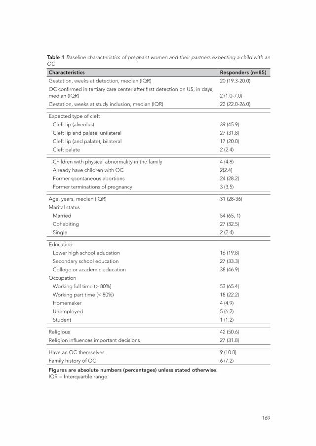
R1
R2
R3
R4
R5
R6
R7
R8
R9
R10
R11
R12
R13
R14
R15
R16
R17
R18
R19
R20
R21
R22
R23
R24
R25
R26
R27
R28
R29
R30
R31
R32
R33
R34
169
Table 1 Baseline characteristics of pregnant women and their partners expecting a child with an OC
Characteristics Responders (n=85)
Gestation, weeks at detection, median (IQR) 20 (19.3-20.0)
OC confirmed in tertiary care center after first detection on US, in days, median (IQR) 2 (1.0-7.0)
Gestation, weeks at study inclusion, median (IQR) 23 (22.0-26.0)
Expected type of cleft
Cleft lip (alveolus) 39 (45.9)
Cleft lip and palate, unilateral 27 (31.8)
Cleft lip (and palate), bilateral 17 (20.0)
Cleft palate 2 (2.4)
Children with physical abnormality in the family 4 (4.8)
Already have children with OC 2(2.4)
Former spontaneous abortions 24 (28.2)
Former terminations of pregnancy 3 (3,5)
Age, years, median (IQR) 31 (28-36)
Marital status
Married 54 (65, 1)
Cohabiting 27 (32.5)
Single 2 (2.4)
Education
Lower high school education 16 (19.8)
Secondary school education 27 (33.3)
College or academic education 38 (46.9)
Occupation
Working full time (> 80%) 53 (65.4)
Working part time (< 80%) 18 (22.2)
Homemaker 4 (4.9)
Unemployed 5 (6.2)
Student 1 (1.2)
Religious 42 (50.6)
Religion influences important decisions 27 (31.8)
Have an OC themselves 9 (10.8)
Family history of OC 6 (7.2)
Figures are absolute numbers (percentages) unless stated otherwise.IQR = Interquartile range.
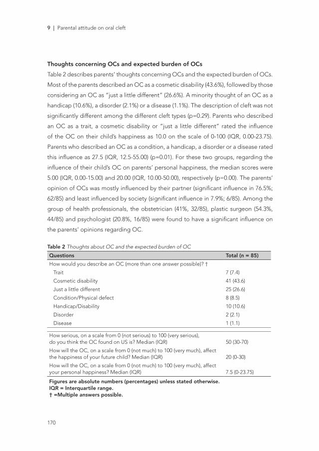
R1
R2
R3
R4
R5
R6
R7
R8
R9
R10
R11
R12
R13
R14
R15
R16
R17
R18
R19
R20
R21
R22
R23
R24
R25
R26
R27
R28
R29
R30
R31
R32
R33
R34
9 | Parental attitude on oral cleft
170
Thoughts concerning OCs and expected burden of OCs
Table 2 describes parents’ thoughts concerning OCs and the expected burden of OCs.
Most of the parents described an OC as a cosmetic disability (43.6%), followed by those
considering an OC as “just a little different” (26.6%). A minority thought of an OC as a
handicap (10.6%), a disorder (2.1%) or a disease (1.1%). The description of cleft was not
significantly different among the different cleft types (p=0.29). Parents who described
an OC as a trait, a cosmetic disability or “just a little different” rated the influence
of the OC on their child’s happiness as 10.0 on the scale of 0-100 (IQR, 0.00-23.75).
Parents who described an OC as a condition, a handicap, a disorder or a disease rated
this influence as 27.5 (IQR, 12.5-55.00) (p=0.01). For these two groups, regarding the
influence of their child’s OC on parents’ personal happiness, the median scores were
5.00 (IQR, 0.00-15.00) and 20.00 (IQR, 10.00-50.00), respectively (p=0.00). The parents’
opinion of OCs was mostly influenced by their partner (significant influence in 76.5%;
62/85) and least influenced by society (significant influence in 7.9%; 6/85). Among the
group of health professionals, the obstetrician (41%, 32/85), plastic surgeon (54.3%,
44/85) and psychologist (20.8%, 16/85) were found to have a significant influence on
the parents’ opinions regarding OC.
Table 2 Thoughts about OC and the expected burden of OC
Questions Total (n = 85)
How would you describe an OC (more than one answer possible)? †
Trait 7 (7.4)
Cosmetic disability 41 (43.6)
Just a little different 25 (26.6)
Condition/Physical defect 8 (8.5)
Handicap/Disability 10 (10.6)
Disorder 2 (2.1)
Disease 1 (1.1)
How serious, on a scale from 0 (not serious) to 100 (very serious), do you think the OC found on US is? Median (IQR) 50 (30-70)
How will the OC, on a scale from 0 (not much) to 100 (very much), affectthe happiness of your future child? Median (IQR) 20 (0-30)
How will the OC, on a scale from 0 (not much) to 100 (very much), affect your personal happiness? Median (IQR) 7.5 (0-23.75)
Figures are absolute numbers (percentages) unless stated otherwise.IQR = Interquartile range.† =Multiple answers possible.
R1
R2
R3
R4
R5
R6
R7
R8
R9
R10
R11
R12
R13
R14
R15
R16
R17
R18
R19
R20
R21
R22
R23
R24
R25
R26
R27
R28
R29
R30
R31
R32
R33
R34
171
Attitudes toward the termination of pregnancy
A minority of participants (6.4%, 5/85) considered TOP, and none chose to terminate
the pregnancy. The parental attitudes toward TOP in general and toward TOP in the
case of an OC are described in table 3. Most parents did not feel that they had to
defend their decision to continue the pregnancy (95.1%, 77/85), and they did not
perceive pressure regarding this decision. In contrast, the majority of the participants
thought that TOP could be an option in some situations (75.3%, 58/85). For example,
41.1% of the participants reported that TOP would be an option in the case of Down
syndrome. The estimated severity of the OC (p=0.03) and its estimated influence on
the happiness of the child (p=0.04) were related to the consideration of TOP. Type of
OC, religion, family history of OC and estimated influence on the parents’ personal
happiness were not of significant influence.
Rating of the counseling by the cleft team
More than half of the participants reported that they needed professional support after
the discovery of an OC during pregnancy (57.7%, 45/85) (table 4). Overall, participants
were (very) satisfied with the information.
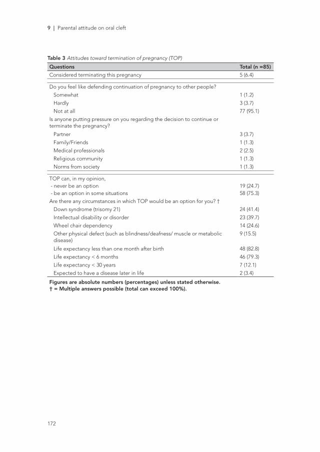
R1
R2
R3
R4
R5
R6
R7
R8
R9
R10
R11
R12
R13
R14
R15
R16
R17
R18
R19
R20
R21
R22
R23
R24
R25
R26
R27
R28
R29
R30
R31
R32
R33
R34
9 | Parental attitude on oral cleft
172
Table 3 Attitudes toward termination of pregnancy (TOP)
Questions Total (n =85)
Considered terminating this pregnancy 5 (6.4)
Do you feel like defending continuation of pregnancy to other people?
Somewhat 1 (1.2)
Hardly 3 (3.7)
Not at all 77 (95.1)
Is anyone putting pressure on you regarding the decision to continue or terminate the pregnancy?
Partner 3 (3.7)
Family/Friends 1 (1.3)
Medical professionals 2 (2.5)
Religious community 1 (1.3)
Norms from society 1 (1.3)
TOP can, in my opinion, - never be an option - be an option in some situations
19 (24.7)58 (75.3)
Are there any circumstances in which TOP would be an option for you? †
Down syndrome (trisomy 21) 24 (41.4)
Intellectual disability or disorder 23 (39.7)
Wheel chair dependency 14 (24.6)
Other physical defect (such as blindness/deafness/ muscle or metabolic disease)
9 (15.5)
Life expectancy less than one month after birth 48 (82.8)
Life expectancy < 6 months 46 (79.3)
Life expectancy < 30 years 7 (12.1)
Expected to have a disease later in life 2 (3.4)
Figures are absolute numbers (percentages) unless stated otherwise.† = Multiple answers possible (total can exceed 100%).
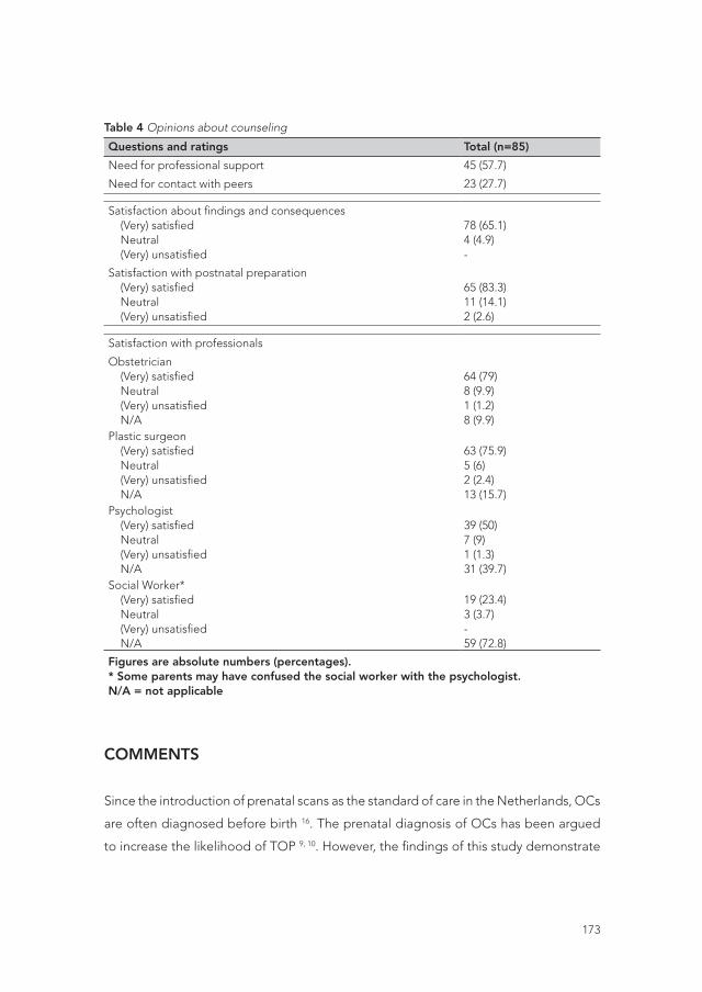
R1
R2
R3
R4
R5
R6
R7
R8
R9
R10
R11
R12
R13
R14
R15
R16
R17
R18
R19
R20
R21
R22
R23
R24
R25
R26
R27
R28
R29
R30
R31
R32
R33
R34
173
Table 4 Opinions about counseling
Questions and ratings Total (n=85)
Need for professional support 45 (57.7)
Need for contact with peers 23 (27.7)
Satisfaction about findings and consequences (Very) satisfied Neutral (Very) unsatisfied
78 (65.1)4 (4.9)-
Satisfaction with postnatal preparation (Very) satisfied Neutral (Very) unsatisfied
65 (83.3)11 (14.1)2 (2.6)
Satisfaction with professionals
Obstetrician (Very) satisfied Neutral (Very) unsatisfied N/A
64 (79)8 (9.9)1 (1.2)8 (9.9)
Plastic surgeon (Very) satisfied Neutral (Very) unsatisfied N/A
63 (75.9)5 (6)2 (2.4)13 (15.7)
Psychologist (Very) satisfied Neutral (Very) unsatisfied N/A
39 (50)7 (9)1 (1.3)31 (39.7)
Social Worker* (Very) satisfied Neutral (Very) unsatisfied N/A
19 (23.4)3 (3.7)-59 (72.8)
Figures are absolute numbers (percentages). * Some parents may have confused the social worker with the psychologist.N/A = not applicable
COMMENTS
Since the introduction of prenatal scans as the standard of care in the Netherlands, OCs
are often diagnosed before birth 16. The prenatal diagnosis of OCs has been argued
to increase the likelihood of TOP 9, 10. However, the findings of this study demonstrate

R1
R2
R3
R4
R5
R6
R7
R8
R9
R10
R11
R12
R13
R14
R15
R16
R17
R18
R19
R20
R21
R22
R23
R24
R25
R26
R27
R28
R29
R30
R31
R32
R33
R34
9 | Parental attitude on oral cleft
174
that most future parents in the Netherlands believe that an isolated OC is a cosmetic
disability or “just a little different” and, therefore, is not a reason for TOP. Health care
professionals had significant influence on the parents’ perceived thoughts regarding
OCs, particularly the obstetrician and plastic surgeon of the cleft lip and palate team.
Perceived burden of OCs
In this study, parents seemed not to perceive OCs as a serious medical condition, and
they believed the cleft would not be of influence on the happiness of their future child.
Other studies showed dissimilar perspectives regarding the burden of OCs and fear of
stigmatization. Parents perceived the defect as severe 17, 18, feared for the intelligence
of the child 2 and were anxious about the child’s self-esteem and self-image 19. The
relatively positive attitude concerning OCs in this study may be explained by several
factors. First, the questionnaire was administered after counseling, suggesting the
importance of the counseling by our cleft lip and palate team. Second, the positive
attitude may be a consequence of the timing of the study inclusion; parents were
pregnant and had not yet seen their child with OC born. Third, these results must be
placed within the context of the Dutch Health Care System, where excellent services
for people with disabilities are offered and the entire treatment (0-18 years) is always
covered by medical insurance 20. Fourth, one could argue that Dutch society is tolerant
of people with disabilities. Although this study did not address the emotional distress
of the prenatal diagnosis of OCs, previous studies showed guilt, anxiety and sadness
as common sentiments after the prenatal diagnosis of an OC 1, 2, 13. A person with
an OC is often characterized as one who shows difficulty in speaking or someone
with a scarred face 2. Moreover, on the internet, “worst-case scenario pictures” are
often circulated 1. Therefore, it is extremely important that the cleft lip and palate
team be actively involved in prenatal counseling 3, can explain treatment options and
the expected difficulties with speech and hearing problems and can show realistic
pictures of postoperative results. Finally, this study showed that more than half of the
participants reported that they needed professional support after the discovery of an
OC during pregnancy. The psychologist on the cleft lip and palate team can conduct a
psychological assessment and provide appropriate referrals if needed 1, 21.
R1
R2
R3
R4
R5
R6
R7
R8
R9
R10
R11
R12
R13
R14
R15
R16
R17
R18
R19
R20
R21
R22
R23
R24
R25
R26
R27
R28
R29
R30
R31
R32
R33
R34
175
Attitudes toward TOP
In the literature, there are striking differences among countries with respect to TOP rates
for OC. During our study period, only one couple decided to terminate the pregnancy,
whereas 95.6% of the parents never considered TOP. These figures are in accord with
results from Argentina 22 and the UK 13, where none and 7.5% of the parents considered
TOP, respectively. Our results are in sharp contrast with those from Israel 10 and Taiwan 19, where 93.4% and 53.1% of the affected fetuses, respectively, were aborted. A variety
of factors are suggested to be responsible for these differences, such as religious,
legal, social, cultural and financial circumstances 8. The present study found that the
perception of burden—or the estimated severity of the cleft—and estimated influence
of OC on the happiness of the future child were significant factors for the decision to
consider TOP. Although this study could not compare results with countries where TOP
for OC is common, it is possible that the burden of OCs in these countries is perceived
as more serious. Although religion and legal issues were not a significant factor in the
consideration of TOP, it can be argued that, in countries such as Argentina, where TOP
is legally restricted and most of the people are active participants in the Catholic faith,
these factors do play a role 22. In the Netherlands, TOP has been legalized since 1981.
For example, more than 95% of couples in the Netherlands opt for TOP in the case of
prenatally diagnosed Down syndrome 23. In our study, 75.3% of the parents stated that
TOP could be an option in some situations. This finding also indicates that OCs are not
perceived as serious, and thus, most parents did not consider TOP.
Evaluation of prenatal counseling
In general, in this study, parents were satisfied with the prenatal counseling. It should
be understood that, in our facility, prenatal counseling is a team effort (obstetrician,
plastic surgeon and medical psychologist) and is conducted regularly. The obstetrician
repeats an anomaly scan in the presence of the partner, plastic surgeon and medical
psychologist. Subsequently, there is ample time reserved for counseling (45 min) by the
plastic surgeon and medical psychologist. Thus, our psychologist and plastic surgeon
are actively involved in facilitating the process of coping with the diagnosis and in
the discussion of future perspectives, respectively. More specifically, the ultrasonic
three-dimensional view allows the plastic surgeon to tailor prenatal counseling to
R1
R2
R3
R4
R5
R6
R7
R8
R9
R10
R11
R12
R13
R14
R15
R16
R17
R18
R19
R20
R21
R22
R23
R24
R25
R26
R27
R28
R29
R30
R31
R32
R33
R34
9 | Parental attitude on oral cleft
176
the exact OC type, including its severity and anticipated postnatal management.
The involvement of plastic surgeons during the prenatal phase is a relatively new
phenomenon. Kemp et al. showed that the anxiety of prospective parents was
significantly reduced by the counseling of a pediatric surgeon 21. Cleft lip and palate
teams have extensive experience treating children with OC. During the study period,
there was one couple that decided, before talking to the cleft lip and palate team, to
terminate the pregnancy. Although we find counseling of utmost importance to future
parents, they cannot be forced to participate 9.
Methodological issues
This is the first study to prospectively investigate consecutive parents and, in contrast
to previous research, included a relatively large number of participants. Hence, this
study involved a homogeneous population only featuring isolated clefts, thereby
correcting for case-mixing with more severe cleft forms. The response rate of 83.3%
was fairly high considering that the risk of sample bias was low. Moreover, there were no
significant differences between responders and non-responders. Finally, participants
were included during pregnancy, and thus the option to terminate the pregnancy
was realistic at 20 weeks of gestation. Because previous studies concerning coping
with the prenatal diagnosis of clefts were of retrospective and cross-sectional design 1, 2, 8, 9, 12, 13, 24, 25, their results not only might contain ascertainment bias but also might
have influenced parental attitudes toward OC because the children were already born
and/or treated (surgically). Furthermore, in contrast to other studies, the process of
counseling in our study was uniform because there was always a plastic surgeon and
psychologist from the cleft lip and palate team involved 1, 12.
Some limitations of this study can be noted. For example, because there were no
parents in the study population who actually chose TOP, we could only compare
groups of parents who did and did not consider TOP. Additionally, the results revealed
by this study may not be applicable to other populations than the Dutch due to
cultural, religious and socioeconomic factors known to influence parental reproductive
choices 22. Finally, questionnaires were assessed after parents had been counseled;
thus, no conclusions can be drawn about the influence of counseling on the opinions
and attitudes of parents.
R1
R2
R3
R4
R5
R6
R7
R8
R9
R10
R11
R12
R13
R14
R15
R16
R17
R18
R19
R20
R21
R22
R23
R24
R25
R26
R27
R28
R29
R30
R31
R32
R33
R34
177
In conclusion this study is the first to describe the thoughts and attitudes of parents
expecting a child with an isolated OC during pregnancy. Dutch parents did not
perceive an isolated OC as a severe condition, and most did not consider terminating
the pregnancy. Future parents reported that they needed professional support and
that they were satisfied with the existing multidisciplinary prenatal counseling. These
results confirmed that active involvement of cleft lip and palate teams is extremely
helpful. Greater awareness of parental experiences demonstrated that a realistic
picture of OC should be presented to the parents without fear that the parents would
interpret the given information as undesirable. Additionally, professionals should
be aware of their authority while counseling because, regardless of the positive or
negative effect, they have significant influence on parents’ opinions concerning OC.
ACKNOWLEDGEMENTS
The authors would like to thank the parents of the children born with an oral cleft
during the years 2011-2014 for participation in this study. The authors would further like
to thank G.T.R. Manten, obstetrician of the Wilhelmina Children’s Hospital in Utrecht,
the Netherlands for her helpful discussion and critically reading of the manuscript.
R1
R2
R3
R4
R5
R6
R7
R8
R9
R10
R11
R12
R13
R14
R15
R16
R17
R18
R19
R20
R21
R22
R23
R24
R25
R26
R27
R28
R29
R30
R31
R32
R33
R34
9 | Parental attitude on oral cleft
178
REFERENCES
1. R. Nusbaum, R. E. Grubs, J. E. Losee, C. Weidman, M. D. Ford and M. L. Marazita. A qualitative description of receiving a diagnosis of clefting in the prenatal or postnatal period. J Genet Couns 2008; 17: 336-350.
2. C. Rey-Bellet and J. Hohlfeld. Prenatal diagnosis of facial clefts: evaluation of a specialised counselling. Swiss Med Wkly 2004; 134: 640-644.
3. M. C. Jones. Prenatal diagnosis of cleft lip and palate: detection rates, accuracy of ultrasonography, associated anomalies, and strategies for counseling. Cleft Palate Craniofac J 2002; 39: 169-173.
4. P. A. Mossey, J. Little, R. G. Munger, M. J. Dixon and W. C. Shaw. Cleft lip and palate. Lancet 2009; 374: 1773-1785.
5. W. Maarse, A. M. Rozendaal, E. Pajkrt, C. Vermeij-Keers, A. B. Mink van der Molen and M. J. van den Boogaard. A systematic review of associated structural and chromosomal defects in oral clefts: when is prenatal genetic analysis indicated? J Med Genet 2012; 49: 490-498.
6. A. B. Mink van der Molen, W. Maarse, L. Pistorius, H. S. de Veye and C. C. Breugem. Prenatal screening for orofacial clefts in the Netherlands: a preliminary report on the impact of a national screening system. Cleft Palate Craniofac J 2011; 48: 183-189. DOI 10.1597/09-021.
7. R. P. Strauss. Beyond easy answers: prenatal diagnosis and counseling during pregnancy. Cleft Palate Craniofac J 2002; 39: 164-168.
8. N. Johnson and R. S. J. Prenatal diagnosis of cleft lip and palate. Cleft Palate Craniofac J 2003; 40: 186-189.
9. M. S. Matthews, M. Cohen, M. Viglione and A. S. Brown. Prenatal counseling for cleft lip and palate. Plast Reconstr Surg 1998; 101: 1-5.
10. M. Bronshtein, I. Blumenfeld and Z. Blumenfeld. Early prenatal diagnosis of cleft lip and its potential impact on the number of babies with cleft lip. Br J Oral Maxillofac Surg 1996; 34: 486-487.
11. G. Le Dref, B. Grollemund, A. Danion-Grilliat and J. C. Weber. Towards a new procreation ethic: the exemplary instance of cleft lip and palate. Med Health Care Philos 2013; 16: 365-375.
12. H. Berggren, E. Hansson, A. Uvemark, H. Svensson and M. Becker. Prenatal compared with postnatal cleft diagnosis: what do the parents think? J Plast Surg Hand Surg 2012; 46: 235-241.
13. A. Davalbhakta and P. N. Hall. The impact of antenatal diagnosis on the effectiveness and timing of counselling for cleft lip and palate. Br J Plast Surg 2000; 53: 298-301.
14. M. J. Korenromp, G. C. Page-Christiaens, J. van den Bout, E. J. Mulder and G. H. Visser. Maternal decision to terminate pregnancy in case of Down syndrome. Am J Obstet Gynecol 2007; 196: 149 e141-111.
15. R. I. S. S. f. W. IBM Corp., Version 20.0; IBM Corp., Armonk, NY. In Book, Editor (ed)^(eds). City.
16. W. Maarse, L. R. Pistorius, W. K. Van Eeten, C. C. Breugem, M. Kon, M. J. Van den Boogaard and A. B. Mink van Der Molen. Prenatal ultrasound screening for orofacial clefts. Ultrasound Obstet Gynecol 2011; 38: 434-439.
17. M. Clementi, R. Tenconi, F. Bianchi and C. Stoll. Evaluation of prenatal diagnosis of cleft lip with or without cleft palate and cleft palate by ultrasound: experience from 20 European registries. EUROSCAN study group. Prenat Diagn 2000; 20: 870-875.
18. M. Sagi, S. Shiloh and T. Cohen. Application of the Health Belief Model in a study on parents’ intentions to utilize prenatal diagnosis of cleft lip and/or palate. Am J Med Genet 1992; 44: 326-333.
R1
R2
R3
R4
R5
R6
R7
R8
R9
R10
R11
R12
R13
R14
R15
R16
R17
R18
R19
R20
R21
R22
R23
R24
R25
R26
R27
R28
R29
R30
R31
R32
R33
R34
179
19. Y. T. Hsieh, Y. M. Chao and J. S. Shiao. A qualitative study of psychosocial factors affecting expecting mothers who choose to continue a cleft lip and/or palate pregnancy to term. J Nurs Res 2013; 21: 1-9.
20. D. Wertz. Eugenics is alive and well: a survey of genetic professionals around the world. Sci Context 1998; 11: 493-510.
21. J. Kemp, M. Davenport and A. Pernet. Antenatally diagnosed surgical anomalies: the psychological effect of parental antenatal counseling. J Pediatr Surg 1998; 33: 1376-1379.
22. D. F. Wyszynski, C. Perandones and R. D. Bennun. Attitudes toward prenatal diagnosis, termination of pregnancy, and reproduction by parents of children with nonsyndromic oral clefts in Argentina. Prenat Diagn 2003; 23: 722-727.
23. M. J. Korenromp, G. C. Page-Christiaens, J. van den Bout, E. J. Mulder, J. A. Hunfeld, C. M. Potters, J. J. Erwich, C. J. van Binsbergen, J. T. Brons, J. R. Beekhuis, A. W. Omtzigt and G. H. Visser. A prospective study on parental coping 4 months after termination of pregnancy for fetal anomalies. Prenat Diagn 2007; 27: 709-716.
24. N. W. Berk, M. L. Marazita and M. E. Cooper. Medical genetics on the cleft palate-craniofacial team: understanding parental preference. Cleft Palate Craniofac J 1999; 36: 30-35.
25. S. Maes, A. Demey and J. Appelboom-Fondu. Impact of ultrasound for facial cleft on mother-child relationships. Ann N Y Acad Sci 1998; 847: 249-251.
R1
R2
R3
R4
R5
R6
R7
R8
R9
R10
R11
R12
R13
R14
R15
R16
R17
R18
R19
R20
R21
R22
R23
R24
R25
R26
R27
R28
R29
R30
R31
R32
R33
R34
9 | Parental attitude on oral cleft
180
QUESTIONNAIRE
GENERAL QUESTIONS
For the medical psychologist1. What type of clefting can be seen on the ultrasound?O unilateral cleft lip O bilateral cleft lipO unilateral cleft lip and jaw O bilateral cleft lip and jawO unilateral cleft lip, jaw and palate O bilateral cleft lip, jaw and palateO cleft palate without cleft lip
For the mother/father 2. Age....... years old
3. Civil status O married O widow/widowerO co-habiting O divorcedO long-distance relationship O single
4. If necessary: how long have you been living with your current partner? ....... year
5. Education O Elementary school O High schoolO University or similar 6. Job status O (almost) full time (80 % or more) O unemployed/on welfareO part-time (less than 80 %) O studentO housewife/man O other:... 7. If religious, what faith do you adhere to? O Religious but no specific faith O ChristianO Islam O not religiousO Judaism O other:...
QUESTIONS ABOUT ANY PREVIOUS PREGNANCIES
8. Is this your or your wife’s first pregnancy? O yes O no, the ............. 9. Have you or your wife ever had a spontaneous miscarriage? O yes, ....... times O no 10. Have any pregnancies ever been aborted? O yes, ....... times O no
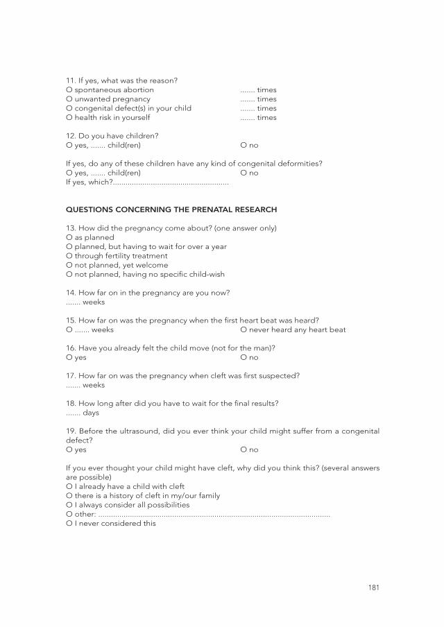
R1
R2
R3
R4
R5
R6
R7
R8
R9
R10
R11
R12
R13
R14
R15
R16
R17
R18
R19
R20
R21
R22
R23
R24
R25
R26
R27
R28
R29
R30
R31
R32
R33
R34
181
11. If yes, what was the reason?O spontaneous abortion ....... timesO unwanted pregnancy ....... timesO congenital defect(s) in your child ....... timesO health risk in yourself ....... times
12. Do you have children? O yes, ....... child(ren) O no
If yes, do any of these children have any kind of congenital deformities?O yes, ....... child(ren) O noIf yes, which?.......................................................
QUESTIONS CONCERNING THE PRENATAL RESEARCH
13. How did the pregnancy come about? (one answer only)O as planned O planned, but having to wait for over a yearO through fertility treatmentO not planned, yet welcomeO not planned, having no specific child-wish
14. How far on in the pregnancy are you now?....... weeks
15. How far on was the pregnancy when the first heart beat was heard?O ....... weeks O never heard any heart beat
16. Have you already felt the child move (not for the man)?O yes O no
17. How far on was the pregnancy when cleft was first suspected?....... weeks
18. How long after did you have to wait for the final results?....... days
19. Before the ultrasound, did you ever think your child might suffer from a congenital defect?O yes O no
If you ever thought your child might have cleft, why did you think this? (several answers are possible)O I already have a child with cleftO there is a history of cleft in my/our familyO I always consider all possibilitiesO other: ..............................................................................................................O I never considered this
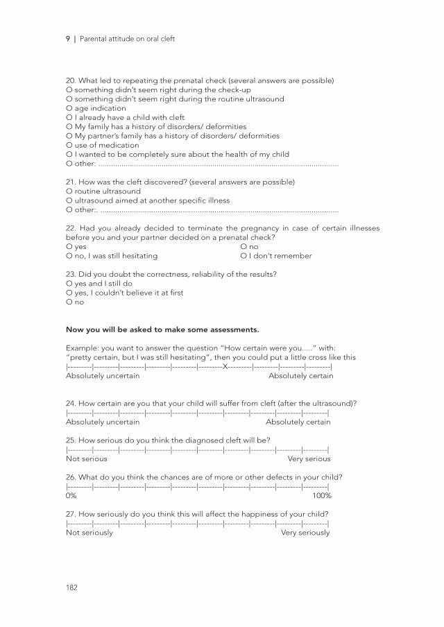
R1
R2
R3
R4
R5
R6
R7
R8
R9
R10
R11
R12
R13
R14
R15
R16
R17
R18
R19
R20
R21
R22
R23
R24
R25
R26
R27
R28
R29
R30
R31
R32
R33
R34
9 | Parental attitude on oral cleft
182
20. What led to repeating the prenatal check (several answers are possible)O something didn’t seem right during the check-upO something didn’t seem right during the routine ultrasoundO age indicationO I already have a child with cleftO My family has a history of disorders/ deformitiesO My partner’s family has a history of disorders/ deformitiesO use of medicationO I wanted to be completely sure about the health of my childO other: ..................................................................................................................
21. How was the cleft discovered? (several answers are possible)O routine ultrasoundO ultrasound aimed at another specific illnessO other:. .................................................................................................................
22. Had you already decided to terminate the pregnancy in case of certain illnesses before you and your partner decided on a prenatal check?O yes O noO no, I was still hesitating O I don’t remember
23. Did you doubt the correctness, reliability of the results?O yes and I still do O yes, I couldn’t believe it at firstO no
Now you will be asked to make some assessments.
Example: you want to answer the question “How certain were you.....” with:“pretty certain, but I was still hesitating”, then you could put a little cross like this|---------|---------|---------|---------|---------|---------X---------|---------|---------|---------|Absolutely uncertain Absolutely certain 24. How certain are you that your child will suffer from cleft (after the ultrasound)?|---------|---------|---------|---------|---------|---------|---------|---------|---------|---------|Absolutely uncertain Absolutely certain 25. How serious do you think the diagnosed cleft will be?|---------|---------|---------|---------|---------|---------|---------|---------|---------|---------|Not serious Very serious
26. What do you think the chances are of more or other defects in your child?|---------|---------|---------|---------|---------|---------|---------|---------|---------|---------|0% 100%
27. How seriously do you think this will affect the happiness of your child?|---------|---------|---------|---------|---------|---------|---------|---------|---------|---------|Not seriously Very seriously
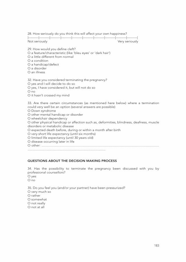
R1
R2
R3
R4
R5
R6
R7
R8
R9
R10
R11
R12
R13
R14
R15
R16
R17
R18
R19
R20
R21
R22
R23
R24
R25
R26
R27
R28
R29
R30
R31
R32
R33
R34
183
28. How seriously do you think this will affect your own happiness?|---------|---------|---------|---------|---------|---------|---------|---------|---------|---------|Not seriously Very seriously
29. How would you define cleft?O a feature/characteristic (like ‘bleu eyes’ or ‘dark hair’)O a little different from normalO a conditionO a handicap/defectO a disorderO an illness
32. Have you considered terminating the pregnancy?O yes and I will decide to do soO yes, I have considered it, but will not do soO noO it hasn’t crossed my mind
33. Are there certain circumstances (as mentioned here below) where a termination could very well be an option (several answers are possible)O Down syndromeO other mental handicap or disorderO wheelchair dependencyO other physical handicap or affection such as, deformities, blindness, deafness, muscle disorders or metabolic diseaseO expected death before, during or within a month after birth O very short life expectancy (until six months)O limited life expectancy (until 30 years old)O disease occurring later in lifeO other: .............................................................................................................................................................................................................................
QUESTIONS ABOUT THE DECISION MAKING PROCESS
34. Has the possibility to terminate the pregnancy been discussed with you by professional counsellors? O yes O no
35. Do you feel you (and/or your partner) have been pressurized?O very much so O rather O somewhatO not reallyO not at all
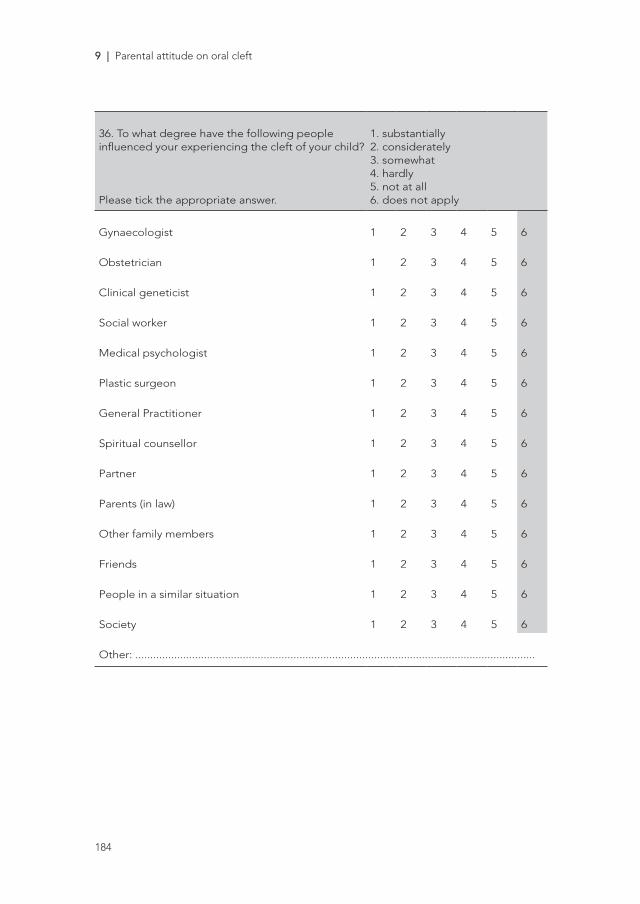
R1
R2
R3
R4
R5
R6
R7
R8
R9
R10
R11
R12
R13
R14
R15
R16
R17
R18
R19
R20
R21
R22
R23
R24
R25
R26
R27
R28
R29
R30
R31
R32
R33
R34
9 | Parental attitude on oral cleft
184
36. To what degree have the following people influenced your experiencing the cleft of your child?
Please tick the appropriate answer.
1. substantially2. considerately3. somewhat4. hardly5. not at all6. does not apply
Gynaecologist 1 2 3 4 5 6
Obstetrician 1 2 3 4 5 6
Clinical geneticist 1 2 3 4 5 6
Social worker 1 2 3 4 5 6
Medical psychologist 1 2 3 4 5 6
Plastic surgeon 1 2 3 4 5 6
General Practitioner 1 2 3 4 5 6
Spiritual counsellor 1 2 3 4 5 6
Partner 1 2 3 4 5 6
Parents (in law) 1 2 3 4 5 6
Other family members 1 2 3 4 5 6
Friends 1 2 3 4 5 6
People in a similar situation 1 2 3 4 5 6
Society 1 2 3 4 5 6
Other: ......................................................................................................................................
R1
R2
R3
R4
R5
R6
R7
R8
R9
R10
R11
R12
R13
R14
R15
R16
R17
R18
R19
R20
R21
R22
R23
R24
R25
R26
R27
R28
R29
R30
R31
R32
R33
R34
185
37. To what degree have the following people influenced your decision to carry out the pregnancy (fill out only when you had doubts about doing so)?
Please tick the appropriate answer.
1. substantially2. considerately3. somewhat4. hardly5. not at all6. does not apply
Gynaecologist 1 2 3 4 5 6
Obstetrician 1 2 3 4 5 6
Clinical geneticist 1 2 3 4 5 6
Social worker 1 2 3 4 5 6
Medical psychologist 1 2 3 4 5 6
Plastic surgeon 1 2 3 4 5 6
General Practitioner 1 2 3 4 5 6
Spiritual counsellor 1 2 3 4 5 6
Partner 1 2 3 4 5 6
Parents (in law) 1 2 3 4 5 6
Other family members 1 2 3 4 5 6
Friends 1 2 3 4 5 6
People in a similar situation 1 2 3 4 5 6
Society 1 2 3 4 5 6
Other: .......................................................................................................................................
38. Perhaps you have had the feeling not being able to choose. If that is the case, what brought on that feeling? .....................................................................................................................................................................................................................................................................................................................................................................................................................................................................................................................................................................................................................................................................................................................................................................................................................O does not apply, since I feel I have had a choice
R1
R2
R3
R4
R5
R6
R7
R8
R9
R10
R11
R12
R13
R14
R15
R16
R17
R18
R19
R20
R21
R22
R23
R24
R25
R26
R27
R28
R29
R30
R31
R32
R33
R34
9 | Parental attitude on oral cleft
186
39. If you feel pressurized when making a decision, who is pressurizing you? (several answers are possible)O partnerO family/friendsO medical teamO congregationO the values within societyO other: ..................................................................................................................O does not apply as I don’t feel pressurized
40. Do you and your partner agree on the decision to carry out the pregnancy?O does not apply/no partnerO noO yes
41. If there are people around you with cleft or any other congenital defect, does that influence your decision to carry out the pregnancy?O very much soO ratherO somewhatO not reallyO not at allO does not apply; I don’t know anyone with cleft or any other congenital defect
42. Are you having doubts about carrying out the pregnancy?O very muchO ratherO somewhatO not reallyO not at all
43. If you are having doubts, what causes them? (several answers are possible)O being overwhelmedO It is against my faith/ outlook on life O I didn’t know much about the diagnosed defectO I had the feeling I was killing a childO I doubted the diagnosisO the diagnosis was uncertainO my partner and I did not agreeO I couldn’t oversee the consequencesO my mind and my heart were strugglingO fear about the procedure (pain, complications)O other: ................................................................................................................................................................................................................................................................................................................................................................................................................................................................................................................................................................................................................................................................
R1
R2
R3
R4
R5
R6
R7
R8
R9
R10
R11
R12
R13
R14
R15
R16
R17
R18
R19
R20
R21
R22
R23
R24
R25
R26
R27
R28
R29
R30
R31
R32
R33
R34
187
44. Have you had to defend your decision to carry out the pregnancy towards certain people?O very muchO ratherO a little O hardlyO no
QUESTIONS ABOUT SUPPORT
45. Are you in need of support?O yes, alone O yes, with partnerO yes, in a groupO noO other: ………………………………………………………………………………
46. Does your belief influence the major decisions in your life? (one answer only)O always O rarelyO often O never O sometimes O does not apply
47. Do you feel supported by your belief in this experience? (one answer only)O very much O noO rather O on the contraryO somewhat O does not apply
48. Have your convictions changed after what has happened?O yes, they have become strongerO they have remained the sameO no, I’ve had more doubtsO does not apply
49. In case of problems or difficulties, how many people around you – with the exception of your partner – can you rely on for true support? ....... people
QUESTIONS ABOUT THE COUNSELLING
51. What did you think about the course of actions in the ultrasound practice and at the Wilhemina’s Childeren Hospital around or during the prenatal check? Do you have any suggestions or criticism?................................................................................................................................................................................................................................................................................................................................................................................................................................................................................................................................................................................................................................................................................................................................................................................................................................................................................................................................................................................................................................................................................................................................................................................................................................
R1
R2
R3
R4
R5
R6
R7
R8
R9
R10
R11
R12
R13
R14
R15
R16
R17
R18
R19
R20
R21
R22
R23
R24
R25
R26
R27
R28
R29
R30
R31
R32
R33
R34
9 | Parental attitude on oral cleft
188
52. How satisfied are you with the given information? 1. very satisfied2. rather satisfied 3. neutral4. rather dissatisfied 5. very dissatisfied
About the prenatal check 1 2 3 4 5
About the results and the consequences of the check 1 2 3 4 5
About carrying out the pregnancy and preparing the birth of a child with cleft
1 2 3 4 5
53. Are you satisfied with the counselling received? 1. very satisfied2. rather satisfied 3. satisfied as well as dissatisfied4. rather dissatisfied5. very much dissatisfied6. never had any contact
Gynaecologist 1 2 3 4 5 6
Obstetrician 1 2 3 4 5 6
Clinical geneticist 1 2 3 4 5 6
Social worker 1 2 3 4 5 6
Medical psychologist 1 2 3 4 5 6
General Practitioner 1 2 3 4 5 6
Plastic Surgeon 1 2 3 4 5 6
Spiritual counsellor 1 2 3 4 5 6
Other: .............................................................................................................................. 6
54. What did you not like about the counsel? ............................................................................................................................................................................................................................................................................................................................................................................................................
R1
R2
R3
R4
R5
R6
R7
R8
R9
R10
R11
R12
R13
R14
R15
R16
R17
R18
R19
R20
R21
R22
R23
R24
R25
R26
R27
R28
R29
R30
R31
R32
R33
R34
189
55. What did you like about the counsel?............................................................................................................................................................................................................................................................................................................................................................................................................
56. What could be improved about the counsel?............................................................................................................................................................................................................................................................................................................................................................................................................57. At this moment, do you feel the need to contact other parents whose child has been diagnosed with cleft on the ultrasound and/or parents who already have an older child with cleft?O yes, very much so O no, not reallyO yes, actually O no, not at all
STATEMENTS
58. In previous scientific studies in this field, pregnant women have given some reasons for their decision to terminate the pregnancy.
Please indicate whether or not these reasons apply to you as well?
Don’t think too long, a spontaneous answer will do.
These mothers gave the following reasons for terminating their pregnancy:
1. Totally agree2. Partially agree3. Undecided4. Partially disagree5. Totally disagree
I think a child with cleft will make me unhappy 1 2 3 4 5
I’m afraid I will regret having a child with cleft 1 2 3 4 5
I’m afraid having a child with cleft will be held against me 1 2 3 4 5
I think that society shows little respect for children with cleft 1 2 3 4 5
I will feel disabled having a child with cleft 1 2 3 4 5
I think the cleft diagnosed is too serious 1 2 3 4 5
I think there is too much insecurity about the consequences of cleft
1 2 3 4 5
I think the cleft will put too much strain on the child itself. 1 2 3 4 5
I comply with my partner’s wish to terminate the pregnancy. 1 2 3 4 5
R1
R2
R3
R4
R5
R6
R7
R8
R9
R10
R11
R12
R13
R14
R15
R16
R17
R18
R19
R20
R21
R22
R23
R24
R25
R26
R27
R28
R29
R30
R31
R32
R33
R34
9 | Parental attitude on oral cleft
190
Don’t think too long, a spontaneous answer will do.
These mothers gave the following reasons for terminating their pregnancy:
1. Totally agree2. Partially agree3. Undecided4. Partially disagree5. Totally disagree
I think there will be too much strain on myself 1 2 3 4 5
I think it will interfere with my career/job 1 2 3 4 5
I think there will be too much strain on my relationship 1 2 3 4 5
I think there will be too much strain on my other child(ren) 1 2 3 4 5
I think there will be too much financial strain 1 2 3 4 5
FINALLY, A FEW QUESTIONS ABOUT YOUR PARTICIPATION IN THIS STUDY.Have you missed certain items in this questionnaire? If yes, please indicate which.......................................................................................................................................................................................................................................................................................................................................................................................................................................................................................................................................................................................................................................................................................................................................................................................................................
10Professional counseling after detection
of oral cleft. Defending shared-decision
making
Submitted
W. MAARSE, A.B. MINK VAN DER MOLEN,
H.F.N. SWANENBURG DE VEYE, M.KON, J.J.M. VAN DELDEN
R1
R2
R3
R4
R5
R6
R7
R8
R9
R10
R11
R12
R13
R14
R15
R16
R17
R18
R19
R20
R21
R22
R23
R24
R25
R26
R27
R28
R29
R30
R31
R32
R33
R34
10 | Counseling
192
ABSTRACT
Since the introduction of a more detailed fetal ultrasound, not only severe but all
sorts of less severe abnormalities such as oral cleft (OC) are identified. Consequently,
prenatal counseling is not only the domain of clinical geneticists and obstetricians
anymore, the pediatric surgeon is involved nowadays. Although genetic counseling
during pregnancy was generally supposed to be non-directive, there is no consensus
concerning the most appropriate type of counseling in case of oral cleft. This debate
explores three types of counseling: non-directive, shared decision making, and
paternalistic counseling. Elaborating the pro- and contra arguments for these types
of counseling can provide physicians with the insight on the ethical background and
guide them in the handling of this practical dilemma.
R1
R2
R3
R4
R5
R6
R7
R8
R9
R10
R11
R12
R13
R14
R15
R16
R17
R18
R19
R20
R21
R22
R23
R24
R25
R26
R27
R28
R29
R30
R31
R32
R33
R34
193
INTRODUCTION
Current prenatal screening, in general, includes a first trimester risk assessment for
the most common aneuploidies and a second trimester ultrasound (US) to identify
fetal anomalies. Whereas screening was developed to inform prospective parents
and give them the choice of continuation or termination of pregnancy in case of a
limited number of abnormalities, the unavoidable trend – stimulated by technical
advancement and ever improving US - is towards more detailed screening 1, leading to
an increased detection of all sorts of less severe abnormalities and discussions about
termination of pregnancy (TOP) for those abnormalities. Oral clefts (OCs) are amongst
the most common congenital malformations in humans and arise in approximately 1
of 700 live births 2 with a prenatal US detection rate of 88% 3.
Case description: Mr. and Mrs. de Jong (32 and 30 years old, respectively)
undergo routine prenatal US at 20 weeks of gestation, when a cleft lip and
palate is detected. Not prepared for detection of an abnormality they are
shocked. They have heard vaguely of oral cleft before and Mr. de Jong recalls
an old classmate with a scar of his upper lip and speaking weirdly. The midwife
assessing the US does not know much about oral cleft, but arranges referral to
the special prenatal clinic of the Wilhelmina’s Children Hospital. Searching the
Internet the parents find all sorts of pictures, differing from an awful looking
baby to a perfectly normal looking operated child. In doubt concerning the
prospect of their future child they consider termination of pregnancy.
The finding of a facial malformation on prenatal ultrasound can be a major psychosocial
burden for parents, who may express signs of severe distress 5,4. Nevertheless, non-
syndromic oral clefts are nonlethal birth defects and most have an excellent functional
and aesthetic prognosis once accurately treated. In the Netherlands a minority of the
parents considers termination of pregnancy (TOP) in case an OC is detected (Maarse
and others, submitted ) 6. In other countries as Israel, the TOP rate for OC is higher 7.
Consequently the use of prenatal diagnosis raises important ethical issues, for both
parents and physicians. Prospective parents, in case they consider TOP, need to decide
R1
R2
R3
R4
R5
R6
R7
R8
R9
R10
R11
R12
R13
R14
R15
R16
R17
R18
R19
R20
R21
R22
R23
R24
R25
R26
R27
R28
R29
R30
R31
R32
R33
R34
10 | Counseling
194
in a very short period of time (before 24 weeks of gestation). Physicians, responsible for
prenatal care, have to determine their tone of counseling.
Before the introduction of prenatal US, parents were usually confronted with OC at
birth. Because of increased prenatal detection rates 3, cleft lip and palate teams should
participate in prenatal counseling. In our hospital in Utrecht, the Netherlands, we have
set up a specialized clinic to participate in the counseling of prospective parents.
In such a clinic an obstetrician together with a plastic surgeon and a psychologist
perform counseling. They provide information concerning prognosis, treatment, and
psychosocial morbidity. We previously (Maarse et al. submitted) demonstrated that
prospective parents were very satisfied concerning this type of counseling. In addition,
this study showed that physicians have significant influence on parents’ opinion
concerning the expected burden of an OC. In a different study, it was found that
obstetricians perceive OC as a more serious condition in comparison to prospective
parents 8. All together this stresses the possible influence of professionals in the
counseling process of prospective parents, especially in those cases when prospective
parents consider TOP.
The involvement of pediatric surgeons in counseling during the prenatal phase is a
relatively new phenomenon 9. Surgeons are inclined to provide “best-practice advise”
to the patient 10, and are actors of a more direct approach in daily practice. Cleft lip
and palate teams struggle with the tone of prenatal counseling (Maarse and others.
submitted). The main concern of those teams is to provide sufficient and complete
information, and to allow prospective parents to make an informed decision and to
respect and protect parental autonomy 11. Elements considered to be a necessary
condition for autonomous decision-making are: the presence of valuable options, clear
information about the alternatives and the absence of guiding influences 13, 12, 15, 14. The
question is how can we determine which type of counseling is most appropriate? We
provide in this paper an overview of historic and current types of counseling, including
their pros and cons as found in the literature, based on which we suggest what form of
counseling is best justifiable.
R1
R2
R3
R4
R5
R6
R7
R8
R9
R10
R11
R12
R13
R14
R15
R16
R17
R18
R19
R20
R21
R22
R23
R24
R25
R26
R27
R28
R29
R30
R31
R32
R33
R34
195
DIFFERENT TYPES OF COUNSELING
Non-directive counseling
Non-directive counseling is comparable to the informative model of Emanuel and
Emanuel 16, in which the physician provides all the facts, hence provides no advice and
there is only a role for the patient’s values. The physician is the purveyor of technical
expertise and the active conception of patient autonomy is patient control over
medical decision-making without any pressure from the counsellor. This means that
‘the counselor does not direct the decision-making process, but only provides all the
information needed for making an informed-decision’ 17.
Shared decision making
In shared decision making, health professionals and patients are partners in making
difficult decisions 18. Some argue that respect for autonomy does not require the
patient to make a decision alone and unaided 19. Being autonomous encompasses the
freedom to delegate the decision to others, to look for help and even to be influenced
by others. Shared decision-making concerns a range of approaches, specifically the
interpretive and deliberative model of Emanuel and Emanuel 16. Both aim to assist in
articulating patient’s values, determine what he or she actually wants, and to select the
available medical interventions that realize these values. The level of involvement of
the physician may vary, from working with the patient to elucidate his or her values, to
suggesting or getting involved in determining which health values are more valuable.
Importantly, the physician does not dictate the patient; it is the patient who ultimately
decides which course of action is best. In these models the physician is an advisor or a
friend, a teacher. Accordingly the active conception of patient autonomy is a relational
one in which self-understanding and moral self-development play an important role 16.
Paternalistic counseling
In this type of counseling the physician uses his or her skills to determine what is best
for the patient, predominantly from a medical perspective, and hence presents only
selected information. In the paternalistic model of Emanuel and Emanuel (Emanuel
and Emanuel 1992), the physician acts as the patient’s guardian. The active conception
R1
R2
R3
R4
R5
R6
R7
R8
R9
R10
R11
R12
R13
R14
R15
R16
R17
R18
R19
R20
R21
R22
R23
R24
R25
R26
R27
R28
R29
R30
R31
R32
R33
R34
10 | Counseling
196
of patient autonomy is assent; the physician decides what is best for the patient, who
can only confirm the option presented to him.
DISCUSSION OF THREE MODELS
Arguments given in favor of non-directive counseling
According to Emanuel and Emanuel (Emanuel and Emanuel 1992), in non-directive
counseling there is a high level of patient autonomy, understood in its liberal way.
Individuals are best placed to judge themselves how to pursue the perception of good
(Mill 1978), or in other words to choose what is best for them. Also in the context of
prenatal screening, reproductive choices should be personal (Hertig and others 2014).
The physician should only provide facts and the patient is the one who should value
this information and make a decision (Bolt 1997).
Objections to non-directive counseling
Non-directive counseling is supposed to create an emotional distance between the
physician and the patient (Williams and others 2002) and there is no caring approach,
because this implies understanding of patient values (Emanuel and Emanuel 1992).
Moreover it withholds essential qualities of the physician, namely their experience.
In other words non-directive counseling reduces the role of the physician to that of a
technologist, which is not the essence of doctoring (Emanuel and Emanuel 1992). Also
it supposes that a person possesses known and fixed values, whereas people are often
uncertain about what they want(Emanuel and Emanuel 1992) and additionally they
are emotional and unable to make a rational decision in such a short period of time
after detection of a malformation (Caplan 2014). Some authors argue that objective
information in counseling is not possible (Bolt 1997). Counseling can’t be value neutral
and a truly non-directive counseling is rarely observed in daily practice 22. Furthermore
a non-directive approach may seem confusing for patients who are used to physicians
who give their opinion 10. Even worse, patients can feel guilty, uncertain and angry
when no recommendations are given 23.
R1
R2
R3
R4
R5
R6
R7
R8
R9
R10
R11
R12
R13
R14
R15
R16
R17
R18
R19
R20
R21
R22
R23
R24
R25
R26
R27
R28
R29
R30
R31
R32
R33
R34
197
Arguments in favor of shared decision making
“Autonomy means that a person is able to relate to him- or herself and to others in
both a critically reasoned way and his or her way”, in essence you relate to opinions
and advice of others” 22. In shared decision making the physician assists the patient in
articulating his or her values, so that he or she realizes what is best for them. Patients’
values are not fixed 16. Kuitert argues that a patient needs assistance to become an
autonomous individual by helping the patient to understand; a person is autonomous
when he or she can give good reasons for her or his choices 24. So providing only
information is not enough to support those concerned in making a good choice.
Shared decision making might allow patients to consider implications they might not
have thought about and gain insight into the consequences of certain choices.
Objections to shared decision making
For shared decision making, the physician has to gain insight into patient’s values and
relate these to his or her own values. It can be argued that the values of physicians
should not be involved here 16. Furthermore it seems rather difficult to gain insight into
patients’ values in such limited time during prenatal consultation. Also a patient and
physician do not always have to agree, which cannot only undermine their relationship,
it can also change the tone of counseling when parents and patients perceive the
burden of an anomaly differently 25. Finally there is a risk that physicians withhold
information because of fear or anxiety of patients, or because they think the patient
has low emotional intelligence 10. Physicians can easily channel parents into a decision
that they feel is correct, whereas they are actually manipulative 26.
Arguments in favor of paternalistic counseling
Some argue that physicians have unique knowledge and experience with certain
abnormalities and consequently know what therapy benefits the patient’s values 16.
According to Conly 27 this approach is not paternalistic but instead a projection of
what is good, based not on physician values, but upon expertise and experience.
Patients will never be perfectly rational (and will be overwhelmed after discovering
they will not have a “perfect baby”) and their values do not line up in a neat and
orderly fashion, in other words they need a little ‘paternalistic help’. Levy 28 argues “the
R1
R2
R3
R4
R5
R6
R7
R8
R9
R10
R11
R12
R13
R14
R15
R16
R17
R18
R19
R20
R21
R22
R23
R24
R25
R26
R27
R28
R29
R30
R31
R32
R33
R34
10 | Counseling
198
picture of a rational individual that underlies the doctrine of informed consent is not
psychologically realistic: we cannot expect patients to take on so much of the burden
of making choices that will advance their own significant interests. Mild coercion will
not just improve the quality of the decision, but it will also increase autonomy ” 28. To
conclude, Levy believes that there is bias in how patients judge information and that
this will reduce their quality of decision making, in contrast to physicians who are not
prejudiced by fear and other motivations.
Objections to paternalistic counseling
Paternalistic counseling can be risky since patients are very easily influenced by
physicians 21. This type of counseling can easily reform into real pressure 28 and
even has a risk to drift towards eugenics 22. In eugenics there is, among other huge
problems, a violation of autonomy 22. In some countries like China, geneticists openly
admit that prenatal screening is used as a manner to improve the progeny 26. This
latter practice shows that although physicians have more expertise, their motivations
are - similar to patients - never value neutral. Their experience will therefore bias their
way of counseling.
DISCUSSION
As the use of routine scanning of the face with US has become standard, prenatal
diagnosis of OCs is now common practice. Cleft lip and palate teams are involved
in counseling and apparently influence prospective parents’ opinion concerning the
burden of an OC. Consequently, they have to determine the most appropriate form
of counseling. Three types of counseling were described, specifically non-directive,
shared decision making and paternalistic counseling.
After exploring the main pro- and contra-arguments of different types of counseling
given in the literature, we think a more directive approach than the non-directive one is
appropriate. We therefore think shared decision making is the best counseling model
in case of OC. We see several reasons for this position. First, prospective parents need
help to become autonomous. After diagnosis of an OC they are usually overwhelmed
R1
R2
R3
R4
R5
R6
R7
R8
R9
R10
R11
R12
R13
R14
R15
R16
R17
R18
R19
R20
R21
R22
R23
R24
R25
R26
R27
R28
R29
R30
R31
R32
R33
R34
199
which is not an ideal period for rational decision-making. Second, if they consider
TOP they should decide in a very short period of time because legal regulations which
prohibit late termination, at least in the Netherlands. Physicians of the cleft lip and
palate team have extensive experience with OC and can therefore empower parents. It
can be argued, however, that the plastic surgeon might have self-interest in optimistic
counseling, since treating children with OC is his or her daily work. However the
majority of children with OC have normal intelligence so counseling is not optimistic,
but rather realistic. Third, “getting more involved” in counseling expresses a more
caring approach: “you take some of the burden from them” 21 and reduce their anxiety 29, 9. Prospective parents can feel that they are supported and realize they are not alone
in raising a child with a condition hitherto unknown to them. A more directive attitude
is also appropriate because it enables parents to gain insight into specific motives and
into the meaning of a particular choice 22; to get there it is sometimes necessary for
physicians to express opinions and give recommendations. This approach aids parents
to ultimately critically assess their own values and preferences, which in turn leads to
(good) personal choice making. Finally, it can be argued that in developed countries
treatment of children with OC is generally excellent, due to good health care facilities
and adequate medical insurance, which implies there is really also a benefit to be
gained.
Implications for clinical practice
To argue for shared decision making in the counseling practice of OC, influences not
only the counseling as such, but also the organization of the prenatal care path. In
order to provide for the interactions that constitute shared decision making, a few
elements are needed; fast referral by the midwife or obstetrician after detection to
a multidisciplinary team containing not only specialists in surgical treatment of OC,
but also in psychological care. Moreover there should be ample time reserved for
counseling. The specialist of the cleft lip and palate team is, contrary to the obstetrician,
the one who takes care of the baby after birth. The parents will feel more trusting when
discussing with the doctor who will try to correct the malformation and who will follow
up their child long term, until adolescence or even beyond.
R1
R2
R3
R4
R5
R6
R7
R8
R9
R10
R11
R12
R13
R14
R15
R16
R17
R18
R19
R20
R21
R22
R23
R24
R25
R26
R27
R28
R29
R30
R31
R32
R33
R34
10 | Counseling
200
CONCLUSION
We propose that professionals in prenatal counseling after OC is diagnosed, take
a more directive approach as in shared decision. This has implications both for the
direct interaction as for the organization of the prenatal care path of OC. Good care in
this respect is contributing to parents becoming autonomous. However, this does not
mean that professionals should dictate parents. In this approach parents are able to
ultimately judge for themselves the expected impact of OC on their prospective child
and family and make their own decisions.
R1
R2
R3
R4
R5
R6
R7
R8
R9
R10
R11
R12
R13
R14
R15
R16
R17
R18
R19
R20
R21
R22
R23
R24
R25
R26
R27
R28
R29
R30
R31
R32
R33
R34
201
REFERENCES
1. L. J. Salomon, Z. Alfirevic, V. Berghella, C. Bilardo, E. Hernandez-Andrade, S. L. Johnsen, K. Kalache, K. Y. Leung, G. Malinger, H. Munoz, F. Prefumo, A. Toi and W. Lee. Practice guidelines for performance of the routine mid-trimester fetal ultrasound scan. Ultrasound Obstet Gynecol 2011; 37: 116-126.
2. P. A. Mossey, J. Little, R. G. Munger, M. J. Dixon and W. C. Shaw. Cleft lip and palate. Lancet 2009; 374: 1773-1785.
3. W. Maarse, L. R. Pistorius, W. K. Van Eeten, C. C. Breugem, M. Kon, M. J. Van den Boogaard and A. B. Mink van Der Molen. Prenatal ultrasound screening for orofacial clefts. Ultrasound Obstet Gynecol 2011; 38: 434-439.
4. R. Nusbaum, R. E. Grubs, J. E. Losee, C. Weidman, M. D. Ford and M. L. Marazita. A qualitative description of receiving a diagnosis of clefting in the prenatal or postnatal period. J Genet Couns 2008; 17: 336-350.
5. M. C. Jones. Prenatal diagnosis of cleft lip and palate: experiences in Southern California. Cleft Palate Craniofac J 1999; 36: 107-109.
6. S. Ensing, C. E. Kleinrouweler, S. M. Maas, C. M. Bilardo, C. M. Van der Horst and E. Pajkrt. Influence of the 20-week anomaly scan on prenatal diagnosis and management of fetal facial clefts. Ultrasound Obstet Gynecol 2014; 44: 154-159.
7. M. Bronshtein, I. Blumenfeld and Z. Blumenfeld. Early prenatal diagnosis of cleft lip and its potential impact on the number of babies with cleft lip. Br J Oral Maxillofac Surg 1996; 34: 486-487.
8. W. Maarse, C. W. Boonacker, O. Lapid, H. F. Swanenburg De Veye, Z. Weiner, M. Kon, J. J. Van Delden and A. B. Mink van der Molen. Professional opinion on oral cleft during pregnancy: a comparison between Israel and the Netherlands. Prenat Diagn 2015.
9. A. Benachi and S. Sarnacki. Prenatal counselling and the role of the paediatric surgeon. Semin Pediatr Surg 2014; 23: 240-243.
10. C. Williams, P. Alderson and B. Farsides. Is nondirectiveness possible within the context of antenatal screening and testing? Soc Sci Med 2002; 54: 339-347.
11. D. Biarent. Fetal anomalies and the pediatrician. Ann N Y Acad Sci 1998; 847: 3-9.12. I. Berlin. Two concept of liberty. Four essays on Liberty. Oxford University Press: London,
1969, 118-172.13. R. Arneson. Freedom and desire. Can J Philos1 1985; 15: 425-448.14. J. C. T Beauchamp. Principles of Biomedical Ethics. Oxford University Press: New York,
2001.15. J. D. Rendtorff. Basic ethical principles in European bioethics and biolaw: autonomy,
dignity, integrity and vulnerability--towards a foundation of bioethics and biolaw. Med Health Care Philos 2002; 5: 235-244.
16. E. J. Emanuel and L. L. Emanuel. Four models of the physician-patient relationship. JAMA 1992; 267: 2221-2226.
17. E. Rantanen, M. Hietala, U. Kristoffersson, I. Nippert, J. Schmidtke, J. Sequeiros and H. Kaariainen. What is ideal genetic counselling? A survey of current international guidelines. Eur J Hum Genet 2008; 16: 445-452.
18. A. A. Kon. Difficulties in judging patient preferences for shared decision-making. J Med Ethics 2012; 38: 719-720.
19. C. Schneider. The practice of autonomy: patients, doctors, and medical decisions. Oxford University Press: New York, 1998.
20. D. Mill. On Liberty. Hacket Publishing Company, Inc: Indinapolis/Cambridge, 1978 (1859).21. S. G. Hertig, S. Cavalli, C. Burton-Jeangros and B. S. Elger. ‘Doctor, what would you do
in my position?’ Health professionals and the decision-making process in pregnancy monitoring. J Med Ethics 2014; 40: 310-314.
R1
R2
R3
R4
R5
R6
R7
R8
R9
R10
R11
R12
R13
R14
R15
R16
R17
R18
R19
R20
R21
R22
R23
R24
R25
R26
R27
R28
R29
R30
R31
R32
R33
R34
10 | Counseling
202
22. I. Bolt. Goede raad: over autonomie en goede redenen in de praktijk van de genetische counseling. In Book Goede raad : over autonomie en goede redenen in de praktijk van de genetische counseling, Editor (ed)^(eds). VU University City, 1997.
23. A. L. Caplan. Why autonomy needs help. J Med Ethics 2014; 40: 301-302. 24. H.Kuitert. Mag alles wat kan? Ethiek en medisch handelen. Ten Have: Baarn, 1989.
25. A. de Jong, W. J. Dondorp, S. G. Frints, C. E. de Die-Smulders and G. M. de Wert. Advances in prenatal screening: the ethical dimension. Nat Rev Genet 2011; 12: 657-663.
26. D. Wertz. Eugenics is alive and well: a survey of genetic professionals around the world. Sci Context 1998; 11: 493-510.
27. S. Conly. Against autonomy: justifying coercive paternalism. J Med Ethics 2014; 40: 349. 28. N. Levy. Forced to be free? Increasing patient autonomy by constraining it. J Med Ethics
2014; 40: 293-300. 29. L. Aite, A. Zaccara, M. Cuttini, N. Mirante, A. Nahom and P. Bagolan. Lack of institutional
pathways for referral: results of a survey among pediatric surgeons on prenatal consultation for congenital anomalies. Prenat Diagn 2013; 33: 904-907.

Part IV
Discussion and Summary

11General discussion and
future perspectives

R1
R2
R3
R4
R5
R6
R7
R8
R9
R10
R11
R12
R13
R14
R15
R16
R17
R18
R19
R20
R21
R22
R23
R24
R25
R26
R27
R28
R29
R30
R31
R32
R33
R34
11 | General discussion and future perspectives
206
R1
R2
R3
R4
R5
R6
R7
R8
R9
R10
R11
R12
R13
R14
R15
R16
R17
R18
R19
R20
R21
R22
R23
R24
R25
R26
R27
R28
R29
R30
R31
R32
R33
R34
207
Since the introduction of routine prenatal screening with ultrasound in the Netherlands
in 2007, parents are confronted with the diagnosis of oral cleft (OC) already during
pregnancy1. This imposed a new dimension in cleft care in the Netherlands. As a
consequence to increasing prenatal detection rates, the cleft lip and palate team of
the Wilhelmina Children’s Hospital in Utrecht set up a multidisciplinary prenatal cleft
clinic in which a plastic surgeon, an obstetrician, medical psychologist and clinical
geneticist together facilitate in counseling. Although the diagnosis of OC prenatally
and related counseling results in better education and preparation before birth, it also
poses a significant ethical dilemma. Even though cleft lip and palate teams in the
Netherlands can provide excellent care, there is concern that fewer children with OC
will be born due to TOP in the near future as a result of prenatal screening. The latter
inspired us for the start of this thesis.
In order to improve counseling we needed accurate information. In the first part of
this thesis we assessed the different types of OC that can be detected by screening.
Furthermore we evaluated the accuracy of detection of OC, the association with other
anomalies and the role of invasive genetic tests. The second part of the thesis faces
the prenatal counseling process of OC itself. We evaluated the opinion and attitude of
both professionals and prospective parents on OC, the expected burden of OC and
their opinion about TOP. Finally in the last part, we discussed the ethical background
of counseling and argued which type of counseling is most appropriate in case of OC.
Below, these two parts of this thesis will be discussed separately under the headings of
accuracy of screening for OC and prenatal counseling.
ACCURACY OF SCREENING FOR ORAL CLEFT
In chapter 3 we explained the emergence of prenatal screening of OC in the
Netherlands. Suitably in chapter 4 we showed, in a systematic review, the large variety
in diagnostic accuracy of detection of OC by US. With detection rates described
in this review, ranging from 0-75%, counseling seemed difficult. However since the
start of routine screening in the Netherlands at 20 weeks of gestation, prenatal
R1
R2
R3
R4
R5
R6
R7
R8
R9
R10
R11
R12
R13
R14
R15
R16
R17
R18
R19
R20
R21
R22
R23
R24
R25
R26
R27
R28
R29
R30
R31
R32
R33
R34
11 | General discussion and future perspectives
208
ultrasound (US) has become a uniform process. This allowed us to start a prospective
study to assess the quality of detection by US (chapter 5), which demonstrated a
much higher accuracy of detection of OC by current US techniques in comparison
to former studies. However, isolated cleft palate was rarely diagnosed and in those
cases where a cleft lip was suspected, the incidence of an additional cleft palate was
often underestimated (i.e. false negative). Somehow, the detection of a cleft palate
remains difficult; a former US classification did not even include an isolated cleft palate
as a separate type2. In recent years several studies aimed to improve the detection of
cleft palate by introducing new sonographic techniques3. One of these techniques,
the appearance of an ‘equal sign’4, is currently used in our clinic. This marker refers
to a normal uvula. Since we understand from embryology that the palate closes from
anterior to posterior, an unremarkable uvula thus implies the presence of an intact
palate. The introduction of this technique is a plausible explanation for the few cases
of isolated cleft palate detected in our prenatal clinic in comparison to none at all
before introduction in 2011. Moreover, in chapter 6 we observed increased accuracy
in classifying the type of OC over time. However, the equal sign is not yet reliable
enough; even when a cleft lip is diagnosed there is a fair chance of missing the
additional cleft palate as we showed in chapter 2. Also two recent studies14, 15 showed
the same number of misdiagnoses (approximately 30%) when compared to accuracy
in determining the type of OC in our studies. This had mainly to do with missing the
additional cleft palate. The latter has clinical implications since cleft palate is often
associated with difficulties in speech5, feeding6, hearing7 and mid-facial protrusion8.
Isolated cleft palate represents approximately one-third of all OC9. Therefore, we
proposed isolated cleft palate to be included in prenatal classification systems. A
reliable and reproducible classification of prenatal OC, as we presented in chapter 2,
is essential because an accurate diagnosis is a prerequisite for informing prospective
parents adequately on the prognosis and outcome. Furthermore different types of
cleft are variably related to specific risks of associated anomalies, as we showed in
chapter 7. We discovered that the presence of other congenital anomalies on US, in
combination with OC is a strong predictor for chromosomal defects. Therefore, we
proposed that additional genetic counseling should be offered to those women in
whom associated anomalies are observed, irrespective of cleft category. One should
R1
R2
R3
R4
R5
R6
R7
R8
R9
R10
R11
R12
R13
R14
R15
R16
R17
R18
R19
R20
R21
R22
R23
R24
R25
R26
R27
R28
R29
R30
R31
R32
R33
R34
209
realize however, that absence of associated anomalies does not preclude the presence
of underlying chromosomal defects. Based on our algorithm discussed in chapter
7, parents should be routinely advised to consider consultation of a geneticist and
invasive testing in case of cleft lip ± palate and isolated cleft palate. When considering
invasive testing the baseline risk on complications hereof (<1%) should be outweighed
by the potential benefits10, 11. Another concern that should be discussed with parents is
the possible detection of unexpected or unclassified variants of chromosomal defects
with array based methods12, 13. In conclusion, we showed an unambiguous evidence
that different cleft categories can be associated with a variety of additional congenital
anomalies and underlying chromosomal defects. This emphasizes the need for an
accurate classification of OC to be used in US screening.
Implications for further research
To improve the diagnostic precision of current OC classification it is mainly important
to improve the visualization of the palate, as outlined above. The new prenatal US
classification system presented in this thesis can provide a systematic and practical
approach for prenatal screening. Additionally, implementation of new techniques, like
the ‘equal sign’4 or three-dimensional reverse-face view16 and improving the training of
sonographers could be beneficial to improve the accuracy in diagnosis OC.
In the Netherlands, the vast majority of anomalies associated with an OC are detected
with US. However, it is important to be aware of the chance of additional (later)
medical or developmental issues in an apparently isolated OC. The latter is especially
relevant since the identification of isolated cases of OC is increasing (chapter 4). First,
prospective studies with longer follow-up are needed to gain more insight in minor
abnormalities and syndromes that generally become more evident later in life, such
as the 22q11.2 deletion syndrome17. Second, technological advancement in genetics
is moving quickly, thereby improving the ability to analyze the human genome faster
and at lower cost. As a result, the use of next generation sequencing is expected to be
implemented in the near future.
R1
R2
R3
R4
R5
R6
R7
R8
R9
R10
R11
R12
R13
R14
R15
R16
R17
R18
R19
R20
R21
R22
R23
R24
R25
R26
R27
R28
R29
R30
R31
R32
R33
R34
11 | General discussion and future perspectives
210
Future perspectives
The role of invasive prenatal testing could change completely with the introduction of
another technical innovation, the so-called non-invasive prenatal testing (NIPT). NIPT,
refers to cell-free fetal DNA-based tests in maternal plasma20, 21 that can be performed
as early as after 7 weeks of pregnancy22. The main advantage of this test is that the
procedure-related miscarriages of invasive techniques are avoided since NIPT requires
only blood samples from the mother. Current prenatal screening in the Netherlands
includes a first-trimester risk assessment for the most common aneuploides and a
second-trimester fetal ultrasound to identify fetal anomalies18. In case of a positive
risk assessment, invasive procedures are offered. However, based on the advice of
the Dutch Health Council19 in December 2013, ministerial permission was granted to
perform NIPT as the first follow-up test if the combined test indicates an increased
risk of trisomy. Although NIPT seems a more reliable screening test for common
aneuploidies when compared to the combined test, at this point NIPT alone is not
yet recommended as an routine alternative due to the lack of research results in the
general population21. If NIPT becomes the test of first choice, the combination test
may no longer be needed, implying that nuchal translucency measurement by US as
part of the combined test may no longer be performed in the near future23. A different
advantage of NIPT, although currently not in its scope, is the fact that subchromosomal
abnormalities, such as 22q11 deletion24 can also be detected. This might influence
the number of children born with an associated OC, since NIPT is performed in an
early stage of pregnancy thereby possibly influencing the decision to terminate the
pregnancy. Nonetheless, ultrasounds screening around 20 weeks of gestation will
probably remain part of routine screening because it was introduced to screen for
observable structural anomalies, which are not detectable by NIPT. Therefore, it is
expected that the detection of “truly” isolated OC after a negative NIPT test by US
around 20 weeks of gestation will remain part of our clinical reality.
R1
R2
R3
R4
R5
R6
R7
R8
R9
R10
R11
R12
R13
R14
R15
R16
R17
R18
R19
R20
R21
R22
R23
R24
R25
R26
R27
R28
R29
R30
R31
R32
R33
R34
211
PRENATAL COUNSELING
In chapter 9 we prospectively analyzed thoughts and attitudes of parents expecting
a child with OC. The findings demonstrated the opposite to the concerns put forward
in literature; namely that the prenatal diagnosis of OC would lead to an increase of
TOP27, 28. We found that most future parents in the Netherlands believe that an isolated
OC is a cosmetic disability or “just a little different”. Thus most parents do not even
consider TOP. Other studies demonstrated a more serious expected burden of OC
and hence an increased fear of stigmatization. Such parents perceived the defect
as severe30, 31, feared for the intelligence of the child32 and were anxious about the
child’s self-esteem and -image33. In the literature there are striking differences among
countries with respect to TOP rates for OC, with fairly high rates in countries like Israel28
and rather low rates in the Netherlands (chapter 9). It is possible that the perceived
burden of OCs in those countries is higher, which would be an explanation for higher
rates of TOP for isolated OC. In chapter 8 we found that the opinion about OC of the
professional also did not explain the dramatic difference in TOP between Israel and the
Netherlands; they did not differ significantly in their judgment on the severity of OC
and the acceptability of TOP. In the literature a variety of other factors are suggested
to be responsible for the differences, such as religious, legal, social, cultural, financial
circumstances, gestational age at diagnosis besides the expected burden of OC29.
In addition, it not always clear how the prenatal care path of a fetus with an OC is
organized in these countries nor is it known if and how counseling is performed.
Furthermore, in chapter 9 we found that health care professionals have significant
influence on the parents’ perception of thoughts regarding OCs, particularly by the
obstetrician and plastic surgeon of the cleft lip and palate team. Other studies confirmed
that perception of the severity of certain conditions are mediated by the care provider’s
counseling34-36. In chapter 8 we found that obstetricians in the Netherlands perceive
OC as a disability, in contrast to prospective parents who think of OC as a cosmetic
disability. The cleft lip and palate team should be aware of these differences. Given
the fact that most prospective parents did not even consider TOP when an isolated
OC was detected, TOP is not always discussed during counseling in our hospital. The
most important goal of counseling by the cleft lip and palate team is to give extensive
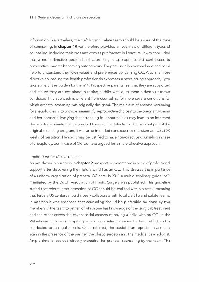
R1
R2
R3
R4
R5
R6
R7
R8
R9
R10
R11
R12
R13
R14
R15
R16
R17
R18
R19
R20
R21
R22
R23
R24
R25
R26
R27
R28
R29
R30
R31
R32
R33
R34
11 | General discussion and future perspectives
212
information. Nevertheless, the cleft lip and palate team should be aware of the tone
of counseling. In chapter 10 we therefore provided an overview of different types of
counseling, including their pros and cons as put forward in literature. It was concluded
that a more directive approach of counseling is appropriate and contributes to
prospective parents becoming autonomous. They are usually overwhelmed and need
help to understand their own values and preferences concerning OC. Also in a more
directive counseling the health professionals expresses a more caring approach, “you
take some of the burden for them”39. Prospective parents feel that they are supported
and realize they are not alone in raising a child with a, to them hitherto unknown
condition. This approach is different from counseling for more severe conditions for
which prenatal screening was originally designed. The main aim of prenatal screening
for aneuploidies is ‘to provide meaningful reproductive choices’ to the pregnant woman
and her partner19, implying that screening for abnormalities may lead to an informed
decision to terminate the pregnancy. However, the detection of OC was not part of the
original screening program; it was an unintended consequence of a standard US at 20
weeks of gestation. Hence, it may be justified to have non-directive counseling in case
of aneuploidy, but in case of OC we have argued for a more directive approach.
Implications for clinical practice
As was shown in our study in chapter 9 prospective parents are in need of professional
support after discovering their future child has an OC. This stresses the importance
of a uniform organization of prenatal OC care. In 2011 a multidisciplinary guideline25,
26 initiated by the Dutch Association of Plastic Surgery was published. This guideline
stated that referral after detection of OC should be realized within a week, meaning
that tertiary US centers should closely collaborate with local cleft lip and palate teams.
In addition it was proposed that counseling should be preferable be done by two
members of the team together, of which one has knowledge of the (surgical) treatment
and the other covers the psychosocial aspects of having a child with an OC. In the
Wilhelmina Children’s Hospital prenatal counseling is indeed a team effort and is
conducted on a regular basis. Once referred, the obstetrician repeats an anomaly
scan in the presence of the partner, the plastic surgeon and the medical psychologist.
Ample time is reserved directly thereafter for prenatal counseling by the team. The
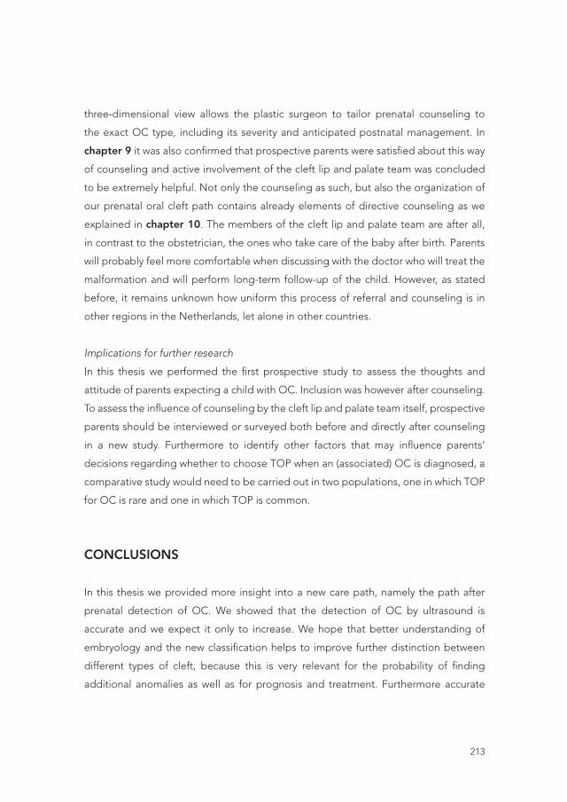
R1
R2
R3
R4
R5
R6
R7
R8
R9
R10
R11
R12
R13
R14
R15
R16
R17
R18
R19
R20
R21
R22
R23
R24
R25
R26
R27
R28
R29
R30
R31
R32
R33
R34
213
three-dimensional view allows the plastic surgeon to tailor prenatal counseling to
the exact OC type, including its severity and anticipated postnatal management. In
chapter 9 it was also confirmed that prospective parents were satisfied about this way
of counseling and active involvement of the cleft lip and palate team was concluded
to be extremely helpful. Not only the counseling as such, but also the organization of
our prenatal oral cleft path contains already elements of directive counseling as we
explained in chapter 10. The members of the cleft lip and palate team are after all,
in contrast to the obstetrician, the ones who take care of the baby after birth. Parents
will probably feel more comfortable when discussing with the doctor who will treat the
malformation and will perform long-term follow-up of the child. However, as stated
before, it remains unknown how uniform this process of referral and counseling is in
other regions in the Netherlands, let alone in other countries.
Implications for further research
In this thesis we performed the first prospective study to assess the thoughts and
attitude of parents expecting a child with OC. Inclusion was however after counseling.
To assess the influence of counseling by the cleft lip and palate team itself, prospective
parents should be interviewed or surveyed both before and directly after counseling
in a new study. Furthermore to identify other factors that may influence parents’
decisions regarding whether to choose TOP when an (associated) OC is diagnosed, a
comparative study would need to be carried out in two populations, one in which TOP
for OC is rare and one in which TOP is common.
CONCLUSIONS
In this thesis we provided more insight into a new care path, namely the path after
prenatal detection of OC. We showed that the detection of OC by ultrasound is
accurate and we expect it only to increase. We hope that better understanding of
embryology and the new classification helps to improve further distinction between
different types of cleft, because this is very relevant for the probability of finding
additional anomalies as well as for prognosis and treatment. Furthermore accurate
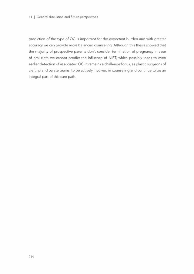
R1
R2
R3
R4
R5
R6
R7
R8
R9
R10
R11
R12
R13
R14
R15
R16
R17
R18
R19
R20
R21
R22
R23
R24
R25
R26
R27
R28
R29
R30
R31
R32
R33
R34
11 | General discussion and future perspectives
214
prediction of the type of OC is important for the expectant burden and with greater
accuracy we can provide more balanced counseling. Although this thesis showed that
the majority of prospective parents don’t consider termination of pregnancy in case
of oral cleft, we cannot predict the influence of NIPT, which possibly leads to even
earlier detection of associated OC. It remains a challenge for us, as plastic surgeons of
cleft lip and palate teams, to be actively involved in counseling and continue to be an
integral part of this care path.
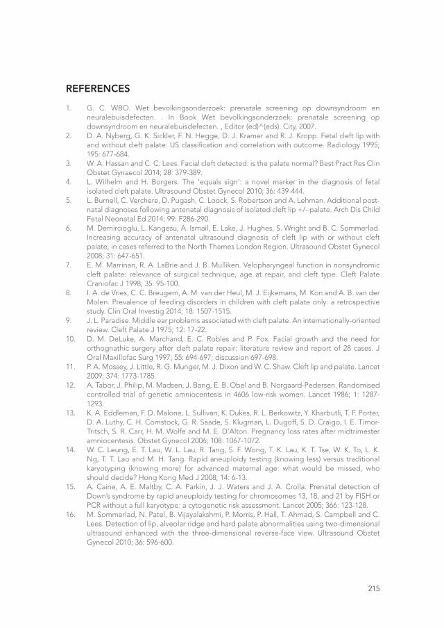
R1
R2
R3
R4
R5
R6
R7
R8
R9
R10
R11
R12
R13
R14
R15
R16
R17
R18
R19
R20
R21
R22
R23
R24
R25
R26
R27
R28
R29
R30
R31
R32
R33
R34
215
REFERENCES
1. G. C. WBO. Wet bevolkingsonderzoek: prenatale screening op downsyndroom en neuralebuisdefecten. . In Book Wet bevolkingsonderzoek: prenatale screening op downsyndroom en neuralebuisdefecten. , Editor (ed)^(eds). City, 2007.
2. D. A. Nyberg, G. K. Sickler, F. N. Hegge, D. J. Kramer and R. J. Kropp. Fetal cleft lip with and without cleft palate: US classification and correlation with outcome. Radiology 1995; 195: 677-684.
3. W. A. Hassan and C. C. Lees. Facial cleft detected: is the palate normal? Best Pract Res Clin Obstet Gynaecol 2014; 28: 379-389.
4. L. Wilhelm and H. Borgers. The ‘equals sign’: a novel marker in the diagnosis of fetal isolated cleft palate. Ultrasound Obstet Gynecol 2010; 36: 439-444.
5. L. Burnell, C. Verchere, D. Pugash, C. Loock, S. Robertson and A. Lehman. Additional post-natal diagnoses following antenatal diagnosis of isolated cleft lip +/- palate. Arch Dis Child Fetal Neonatal Ed 2014; 99: F286-290.
6. M. Demircioglu, L. Kangesu, A. Ismail, E. Lake, J. Hughes, S. Wright and B. C. Sommerlad. Increasing accuracy of antenatal ultrasound diagnosis of cleft lip with or without cleft palate, in cases referred to the North Thames London Region. Ultrasound Obstet Gynecol 2008; 31: 647-651.
7. E. M. Marrinan, R. A. LaBrie and J. B. Mulliken. Velopharyngeal function in nonsyndromic cleft palate: relevance of surgical technique, age at repair, and cleft type. Cleft Palate Craniofac J 1998; 35: 95-100.
8. I. A. de Vries, C. C. Breugem, A. M. van der Heul, M. J. Eijkemans, M. Kon and A. B. van der Molen. Prevalence of feeding disorders in children with cleft palate only: a retrospective study. Clin Oral Investig 2014; 18: 1507-1515.
9. J. L. Paradise. Middle ear problems associated with cleft palate. An internationally-oriented review. Cleft Palate J 1975; 12: 17-22.
10. D. M. DeLuke, A. Marchand, E. C. Robles and P. Fox. Facial growth and the need for orthognathic surgery after cleft palate repair: literature review and report of 28 cases. J Oral Maxillofac Surg 1997; 55: 694-697; discussion 697-698.
11. P. A. Mossey, J. Little, R. G. Munger, M. J. Dixon and W. C. Shaw. Cleft lip and palate. Lancet 2009; 374: 1773-1785.
12. A. Tabor, J. Philip, M. Madsen, J. Bang, E. B. Obel and B. Norgaard-Pedersen. Randomised controlled trial of genetic amniocentesis in 4606 low-risk women. Lancet 1986; 1: 1287-1293.
13. K. A. Eddleman, F. D. Malone, L. Sullivan, K. Dukes, R. L. Berkowitz, Y. Kharbutli, T. F. Porter, D. A. Luthy, C. H. Comstock, G. R. Saade, S. Klugman, L. Dugoff, S. D. Craigo, I. E. Timor-Tritsch, S. R. Carr, H. M. Wolfe and M. E. D’Alton. Pregnancy loss rates after midtrimester amniocentesis. Obstet Gynecol 2006; 108: 1067-1072.
14. W. C. Leung, E. T. Lau, W. L. Lau, R. Tang, S. F. Wong, T. K. Lau, K. T. Tse, W. K. To, L. K. Ng, T. T. Lao and M. H. Tang. Rapid aneuploidy testing (knowing less) versus traditional karyotyping (knowing more) for advanced maternal age: what would be missed, who should decide? Hong Kong Med J 2008; 14: 6-13.
15. A. Caine, A. E. Maltby, C. A. Parkin, J. J. Waters and J. A. Crolla. Prenatal detection of Down’s syndrome by rapid aneuploidy testing for chromosomes 13, 18, and 21 by FISH or PCR without a full karyotype: a cytogenetic risk assessment. Lancet 2005; 366: 123-128.
16. M. Sommerlad, N. Patel, B. Vijayalakshmi, P. Morris, P. Hall, T. Ahmad, S. Campbell and C. Lees. Detection of lip, alveolar ridge and hard palate abnormalities using two-dimensional ultrasound enhanced with the three-dimensional reverse-face view. Ultrasound Obstet Gynecol 2010; 36: 596-600.
R1
R2
R3
R4
R5
R6
R7
R8
R9
R10
R11
R12
R13
R14
R15
R16
R17
R18
R19
R20
R21
R22
R23
R24
R25
R26
R27
R28
R29
R30
R31
R32
R33
R34
11 | General discussion and future perspectives
216
17. H. F. Swanenburg de Veye, F. A. Beemer, G. J. Mellenbergh, W. H. Wolters and J. A. Heineman-de Boer. An investigation of the relationship between associated congenital malformations and the mental and psychomotor development of children with clefts. Cleft Palate Craniofac J 2003; 40: 297-303.
18. Y. M. Lo, N. Corbetta, P. F. Chamberlain, V. Rai, I. L. Sargent, C. W. Redman and J. S. Wainscoat. Presence of fetal DNA in maternal plasma and serum. Lancet 1997; 350: 485-487.
19. E. Mersy, L. J. Smits, L. A. van Winden, C. E. de Die-Smulders, A. D. Paulussen, M. V. Macville, A. B. Coumans and S. G. Frints. Noninvasive detection of fetal trisomy 21: systematic review and report of quality and outcomes of diagnostic accuracy studies performed between 1997 and 2012. Hum Reprod Update 2013; 19: 318-329.
20. C. F. Wright and H. Burton. The use of cell-free fetal nucleic acids in maternal blood for non-invasive prenatal diagnosis. Hum Reprod Update 2009; 15: 139-151.
21. L. J. Salomon, Z. Alfirevic, V. Berghella, C. Bilardo, E. Hernandez-Andrade, S. L. Johnsen, K. Kalache, K. Y. Leung, G. Malinger, H. Munoz, F. Prefumo, A. Toi and W. Lee. Practice guidelines for performance of the routine mid-trimester fetal ultrasound scan. Ultrasound Obstet Gynecol 2011; 37: 116-126.
22. H. Council. Health Council of the Netherlands. NIPT: dynamics and ethics of prenatal screening. In Book Health Council of the Netherlands. NIPT: dynamics and ethics of prenatal screening., Editor (ed)^(eds). Health Council of the Netherlands: City, 2013.
23. A. de Jong, W. J. Dondorp, S. G. Frints, C. E. de Die-Smulders and G. M. de Wert. Advances in prenatal screening: the ethical dimension. Nat Rev Genet 2011; 12: 657-663.
24. M. A. Ferres, L. Hui and D. W. Bianchi. Antenatal noninvasive DNA testing: clinical experience and impact. Am J Perinatol 2014; 31: 577-582.
25. M. S. Matthews, M. Cohen, M. Viglione and A. S. Brown. Prenatal counseling for cleft lip and palate. Plast Reconstr Surg 1998; 101: 1-5.
26. M. Bronshtein, I. Blumenfeld and Z. Blumenfeld. Early prenatal diagnosis of cleft lip and its potential impact on the number of babies with cleft lip. Br J Oral Maxillofac Surg 1996; 34: 486-487.
27. M. Clementi, R. Tenconi, F. Bianchi and C. Stoll. Evaluation of prenatal diagnosis of cleft lip with or without cleft palate and cleft palate by ultrasound: experience from 20 European registries. EUROSCAN study group. Prenat Diagn 2000; 20: 870-875.
28. M. Sagi, S. Shiloh and T. Cohen. Application of the Health Belief Model in a study on parents’ intentions to utilize prenatal diagnosis of cleft lip and/or palate. Am J Med Genet 1992; 44: 326-333.
29. C. Rey-Bellet and J. Hohlfeld. Prenatal diagnosis of facial clefts: evaluation of a specialised counselling. Swiss Med Wkly 2004; 134: 640-644.
30. Y. T. Hsieh, Y. M. Chao and J. S. Shiao. A qualitative study of psychosocial factors affecting expecting mothers who choose to continue a cleft lip and/or palate pregnancy to term. J Nurs Res 2013; 21: 1-9.
31. M. C. Jones. Prenatal diagnosis of cleft lip and palate: experiences in Southern California. Cleft Palate Craniofac J 1999; 36: 107-109.
32. C. Hager. Termination of pregnancy with a prenatal diagnosis of cleft lip: cultural differences and ethical analysis. Plast Surg Nurs 2002; 22: 24-28.
33. M. Sagi, V. Meiner, N. Reshef, J. Dagan and J. Zlotogora. Prenatal diagnosis of sex chromosome aneuploidy: possible reasons for high rates of pregnancy termination. Prenat Diagn 2001; 21: 461-465.
34. A. Alsulaiman and K. K. Abu-Amero. Parent’s attitude toward prenatal diagnosis and termination of pregnancy could be influenced by other factors rather than by the severity of the condition. Prenat Diagn 2013; 33: 257-261.

35. S. G. Hertig, S. Cavalli, C. Burton-Jeangros and B. S. Elger. ‘Doctor, what would you do in my position?’ Health professionals and the decision-making process in pregnancy monitoring. J Med Ethics 2014; 40: 310-314.
36. N. V. v. P. Chirurgie. Counseling na prenataal vastgestelde schisis. Een multidisciplinaire richtlijn voor een integraal zorgproces. In Book Counseling na prenataal vastgestelde schisis. Een multidisciplinaire richtlijn voor een integraal zorgproces, Editor (ed)^(eds). NVPC: City, 2011.
37. http://www.nvpc.nl/uploads/stand/76NVPC110401DOC-MW-Richtlijn_Prenatale_Schisis_def76.pdf. Counseling na prenataal vastgestelde schisis. Een multidisciplinaire richtlijn voor een integraal zorgproces. In Book Counseling na prenataal vastgestelde schisis. Een multidisciplinaire richtlijn voor een integraal zorgproces, Editor (ed)^(eds). NVPC: City, 2011.

12English summary
Dutch summary/Nederlandse samenvatting

R1
R2
R3
R4
R5
R6
R7
R8
R9
R10
R11
R12
R13
R14
R15
R16
R17
R18
R19
R20
R21
R22
R23
R24
R25
R26
R27
R28
R29
R30
R31
R32
R33
R34
12 | English summary
220
R1
R2
R3
R4
R5
R6
R7
R8
R9
R10
R11
R12
R13
R14
R15
R16
R17
R18
R19
R20
R21
R22
R23
R24
R25
R26
R27
R28
R29
R30
R31
R32
R33
R34
221
SUMMARY
Since the introduction of standard prenatal screening in the Netherlands by means of
ultrasound in 2007, parents are confronted with a child with an oral cleft already during
pregnancy. As a response, some cleft lip and palate teams have set up a specialized
prenatal cleft clinic to participate in the counseling of pregnant women. The aim of this
study was to investigate different factors of prenatal detection of oral cleft, in order to
improve prenatal counseling. Primarily, counseling is important to inform parents about
the medical aspects of oral cleft. Since this thesis focusses on only the prenatal aspects
of oral cleft, in chapter 1 we provide a state of the art overview on epidemiology,
embryology, pathophysiology and treatment. Oral cleft is a congenital malformation
that can involve the lip, palate, nose and underlying bony fragments in various
severities, and is the most common craniofacial anomaly considering an incidence of
approximately 1:700 births. Oral cleft is a result of failure of both growth and fusion of
the face, which is established between the 4th and 10th week of human development.
Besides the aesthetic complication of oral cleft there are functional problems related
to the abnormal anatomy. In brief, speech can be complicated due to velopharyngeal
insufficiency, there can be hearing disability, feeding difficulties and dentofacial
deformities. A multidisciplinary approach is essential for satisfactory treatment of cleft
patients. Surgical correction consist of reconstruction of a symmetrically balanced
lip and nose and an intact and functional palate to optimize speech. The course of
treatment spans often the entire childhood and although the primary management is
surgically driven, the overall goal of treatment includes also a psychosocial wellbeing
of the patient and his or her family. Prenatal detection of oral cleft has added a new
dimension to the above described cleft care.
Part I Classification of Oral Cleft
Since introduction of screening for fetal anomalies, ultrasonographers are challenged
to diagnose not only an oral cleft, but moreover the precise type of oral cleft. Accurate
diagnosis is essential because it correlates with the severity of the malformation, and
different treatment and prognosis. In chapter 2, a new prenatal oral cleft classification
is defined using a practical approach that could be easily applied in ultrasound
R1
R2
R3
R4
R5
R6
R7
R8
R9
R10
R11
R12
R13
R14
R15
R16
R17
R18
R19
R20
R21
R22
R23
R24
R25
R26
R27
R28
R29
R30
R31
R32
R33
R34
12 | English summary
222
practice. During a three-year period (2011-2014), all cases of oral cleft diagnosed by
ultrasound where retrospectively reviewed, which resulted in a total of 103 fetuses.
For the purpose of validation prenatal images were compared to postnatal findings.
The measurement of agreement between these pre- and postnatal findings showed
a Kappa value of 0.63 (95% CI: 0.52-0.75), demonstrating the accuracy of this new
classification system.
Part II Screening for Oral Cleft
In chapter 3, the history and background of screening for fetal anomalies, with a focus
on oral cleft is presented. A countrywide routine screening, initially only for Down
syndrome and neural tube defects, but later also for oral cleft, was introduced by
legislation in 2007. The Netherlands were quite late in setting rules for this field. Routine
screening was postponed because the Ministry of Health, Welfare and Sport defended
the view that possibilities for prenatal treatment and prevention were minimal. After
legislation, equal access and standard coverage by health care insurance was ensured.
The screening is thus primarily to inform future parents about a possible defect and
about their postnatal treatment options. Informed consent, after being educated about
potential implications of screening and the right not to know, is mandatory. In order
to reduce costs and ensure easy access, basic screening takes places at primary care
level, by certified midwives and sonologists. In case of a suspected abnormality the
women is referred to a certified obstetrician in one of eight regional referral centers,
who performs more detailed screening. Except when very severe anomalies, such as
trisomy 13 or 18 are identified, the cleft lip and palate team is routinely consulted in
the region of Utrecht. In this special set up prenatal clinic the obstetrician repeats the
scan in the presence of the plastic surgeon, after which counseling takes place by
both the plastic surgeon and medical psychologist of the team, so that parents are
well prepared. Due to more ultrasound screening, there is an increase of second term
pregnancy terminations, although data from Utrecht show a very low termination rate
for isolated clefts. To establish a uniform counseling process in case an oral cleft is
detected, a national multidisciplinary evidence-based guideline was initiated.
R1
R2
R3
R4
R5
R6
R7
R8
R9
R10
R11
R12
R13
R14
R15
R16
R17
R18
R19
R20
R21
R22
R23
R24
R25
R26
R27
R28
R29
R30
R31
R32
R33
R34
223
Chapters 4, 5 and 6 all assess the diagnostic accuracy of ultrasound screening of
oral cleft. A systematic review (chapter 4), covering all articles published before 2008,
showed considerable variety in accuracy of two-dimensional ultrasound in low-risk
women (unselected population based studies), with detection rates ranging from 9%
to 100% for cleft lip with- or without cleft palate, 0% to 22% for cleft palate only and
0% to 73% for all types of cleft. There were only few false-positive cases. In general the
methodology was of poor quality and subsequently the review provides the sources
of variability and suggestions for further research. Chapter 5 revealed the results of
a prospective study, in which all pregnant women who underwent routine second-
trimester screening in the Utrecht region during the 2-year period from January 2007
to December 2008. There were 62 cases of oral cleft, providing an incidence of 1: 624.
The distribution of clefts was as follows: 18 (29%) cleft lip (CL), 25 (40%) cleft lip with
cleft palate (CLP), 17 (27%) cleft palate only (CP), one median cleft and one atypical
cleft. Of these, 38 (61%) were unilateral and 23 (37%) were bilateral. Thirty-nine per cent
(24/62) had associated anomalies, with most chromosomal defects found in the CLP
and CP groups. Cleft lip with or without cleft palate (CL ± P) was detected prenatally
in 38/43 cases, a sensitivity of 88%. No case of CP was detected prenatally. There
were three false- positive cases, of which two were fetuses with multiple congenital
deformities. In conclusion there was a high sensitivity for the detection of CL ± P in our
region. The key to a high sensitivity of prenatal ultrasound is likely to be a combination
of excellent training of sonographers, referral to specialized centers when a cleft is
suspected, routine visualization of the fetal face and advances in ultrasound techniques.
In chapter 6 we aimed to determine the accuracy of ultrasound in assessing the type
of cleft. Prenatal diagnosis was in accordance with postnatal findings in 76.9% of
cases (103/134) with regard to the cleft type. Underestimation of the cleft occurred
in 19.4% (26/134), whereas in 3.7% (5/134) the extent of the cleft was overestimated.
In distinguishing bilateral from unilateral clefts, no errors were made. These results
indicated that prenatal ultrasound is a reliable technique to assess the cleft type, which
is important in counseling future parents. Although, the clinician should be aware of
the fact that a cleft palate is easily missed, and subsequently underestimation of cleft
severity is frequent. This study served as basis for developing a new classification,
described in chapter 2.
R1
R2
R3
R4
R5
R6
R7
R8
R9
R10
R11
R12
R13
R14
R15
R16
R17
R18
R19
R20
R21
R22
R23
R24
R25
R26
R27
R28
R29
R30
R31
R32
R33
R34
12 | English summary
224
When informing parents on outcome and prognosis, not only the cleft type but also
the risk of associated anomalies is crucial. In particular the presence of an underlying
chromosomal defect will influence prenatal counseling and management. In chapter
7 on overview is given on associated anomalies and chromosomal defects in oral
clefts. Data from the literature were completed with validated data from the Dutch
Oral Cleft Registry. The pre- and postnatal studies showed that the prevalence of
associated anomalies was lowest in CL (0–20.0% and 7.6–41.4%, respectively). For CLP,
higher frequencies were found both prenatally (39.1–66.0%) and postnatally (21.1–
61.2%). Although CP was barely detectable by ultrasound, it was the category most
frequently associated with accompanying defects in postnatal studies (22.2–78.3%).
Chromosomal abnormalities were most frequently seen in association with additional
anomalies. In the absence of associated anomalies, chromosomal defects were found
prenatally in CLP (3.9%) and postnatally in CL (1.8%, 22q11.2 deletions only), CLP (1.0%)
and CP (1.6%). This systematic review showed that prenatal counselling regarding
prognosis and risk of chromosomal defects should be tailored to cleft category, and
more importantly to the presence or absence of associated anomalies. Irrespective of
cleft category, clinicians should advise invasive genetic testing if associated anomalies
are seen prenatally. In the absence of associated anomalies, prenatal conventional
karyotyping was not recommended in CL, although array comparative genomic
hybridisation should be considered. In presumed isolated CLP or CP, prenatal invasive
testing, preferably by array-based methods, was recommended.
Part III Prenatal Counseling
Since this thesis aimed to improve prenatal counseling for oral cleft, we made an effort
to identify different factors which are thought to influence parental opinion on oral
cleft, one of them being the obstetric care provider’s attitude. In chapter 8 providers’
opinions about pregnancy termination for isolated oral cleft were compared in the
Netherlands, where the number of terminations is low, and in Israel, where the number
is high. Online questionnaires were used. The questions assessed the providers’ views
regarding the estimated burden of treatment, the functioning ability and the level of
happiness of children with an oral cleft and their parents. Additionally, we assessed
providers’ opinions on pregnancy termination for isolated oral cleft. In the Netherlands,
R1
R2
R3
R4
R5
R6
R7
R8
R9
R10
R11
R12
R13
R14
R15
R16
R17
R18
R19
R20
R21
R22
R23
R24
R25
R26
R27
R28
R29
R30
R31
R32
R33
R34
225
more professionals considered oral cleft a disability [rate differences (RD) 17.8%, 95%
CI: 0.5-33.1%] than in Israel. In the Netherlands, 10.6% of respondents (compared with
11.1% in Israel) thought that an isolated cleft was a reason for TOP [RD 0.6%, 95% CI:
-12-10.9%]. In conclusion prenatal care providers between two countries do not differ
in their opinions about the severity of oral cleft and the acceptability of TOP for an
isolated oral cleft. This study shows that prenatal care providers’ attitudes do therefore
not explain the dramatic difference between these countries in the number of TOP
for isolated oral cleft. In chapter 9 we analyzed considerations of prospective parents
concerning OCs, the burden of OCs and parents’ attitude toward possible TOP to
improve counseling in the future. Between August 2011 and August 2014, a prospective
cohort questionnaire study was administered with parents visiting our prenatal clinic.
Most of the parents described an oral cleft as a cosmetic disability (43.6%) or as “just
a little different” (26.6%). These parents expected that the OC would not affect their
own happiness or the happiness of their future child. Health professionals had a
considerable influence on parental opinion. A minority (6.4%, 5/85) of the respondents
considered TOP, and none of the participants chose to terminate the pregnancy. The
estimated severity of the oral cleft and the estimated influence on the child’s happiness
both had a significant influence on whether TOP was considered (p=0.04). This study
suggests that the active involvement of cleft lip and palate teams is worthwhile.
Caregivers should be aware that their counseling is important in the decision making
of parents. Chapter 8 and 9 show that parents and health care professionals perceive
the expected burden of cleft differently. Moreover it was assessed that health care
professionals had significant influence on the parents’ perceived thoughts regarding
OCs. All together this stresses the influence of professionals in counseling prospective
parents, especially when considering TOP. How can we determine whether and when
a more directive attitude of the counselor is justified? In Chapter 10 three types of
counseling are explored: informative, shared decision making, and paternalistic
counseling. Elaborating the pro- and contra arguments for these types of counseling
can provide physicians insight on the ethical background of this practical dilemma.
R1
R2
R3
R4
R5
R6
R7
R8
R9
R10
R11
R12
R13
R14
R15
R16
R17
R18
R19
R20
R21
R22
R23
R24
R25
R26
R27
R28
R29
R30
R31
R32
R33
R34
12 | English summary
226
In conclusion this thesis provides more insight into a new dimension of cleft care,
namely the prenatal detection. When counseling expectant parents on impact of OC,
several topics should be reviewed, including postnatal management like feeding,
surgical and supporting treatment options, cosmetic outcomes and risk for speech
and hearing difficulties, which are all related to specific types of cleft. Classification
and accurate diagnosis is therefore of upmost importance. Additionally parents would
like to know about the increased risk of additional malformations. This information
will determine their expected burden of OC and their attitude towards possible
termination the pregnancy. This thesis will help improving not only the information
given with counseling, but also help to determine the tone of counseling for OC.
R1
R2
R3
R4
R5
R6
R7
R8
R9
R10
R11
R12
R13
R14
R15
R16
R17
R18
R19
R20
R21
R22
R23
R24
R25
R26
R27
R28
R29
R30
R31
R32
R33
R34
227
NEDERLANDSE SAMENVATTING
In 2007 is het Structureel Echografisch Onderzoek (SEO), ofwel de 20 wekenecho,
toegevoegd aan de prenatale zorgstandaard voor zwangere vrouwen in Nederland. Dit
had implicaties voor de prenatale detectie en zorgpad van schisis, die daarvoor vaak
pas na de geboorte werd ontdekt. Als reactie hierop hebben sommige schisisteams
in Nederland een speciaal prenataal schisisspreekuur opgezet met als doel de
counseling van de zwangere vrouw en haar partner die een kindje met een schisis
verwachten. Het doel van dit onderzoek is om de verschillende aspecten rondom
de prenatale detectie van schisis te onderzoeken, om op deze manier uiteindelijk
de counseling te verbeteren. Omdat het onderzoek zich alleen richt op de prenatale
kant van schisis, is in hoofdstuk 1 een overzicht gegeven van de epidemiologie,
embryologie, pathosfysiologie en behandeling van schisis. Op die manier kan beter
worden ingeschat wat de impact is van een schisis voor de ouders en het kind.
Schisis is een congenitale malformatie waarbij de lip, het palatum, de neus en het
onderliggende bot en kraakbeen in verschillende vorm kunnen zijn aangedaan. Het
is de meest voorkomende craniofaciale afwijking en heeft een incidentie van 1:700
nieuwgeborenen. Schisis ontstaat door het van falen van groei en fusie in de embryonale
ontwikkeling van het aangezicht, tussen de 4e en de 10e week van de zwangerschap.
Door de afwijkende anatomie bij een schisis zijn er niet alleen cosmetische, maar ook
functionele gevolgen; de spraak kan aangedaan zijn ten gevolge van velopharyngeale
insufficiëntie, er kunnen oorproblemen, voedingsproblemen en dentofaciale
afwijkingen voorkomen bij kinderen met een schisis. Een multidisciplinaire aanpak
is essentieel voor een goede behandeling van kinderen met schisis. De chirurgische
behandeling bestaat uit reconstructie van een symmetrisch gebalanceerde lip en neus
en een intact palatum voor goede spraak. De behandeling beslaat de gehele jeugd
en ondanks de primaire focus op chirurgie, is het algemene doel van de behandeling
ook het psychosociale welbevinden van de patiënt en zijn of haar familie. Prenatale
detectie heeft een nieuwe dimensie toegevoegd aan de hierboven beschreven
behandeling van schisis.
R1
R2
R3
R4
R5
R6
R7
R8
R9
R10
R11
R12
R13
R14
R15
R16
R17
R18
R19
R20
R21
R22
R23
R24
R25
R26
R27
R28
R29
R30
R31
R32
R33
R34
12 | Dutch summary/Nederlandse samenvatting
228
Deel 1 Classificatie van schisis
Met de introductie van de prenatale echo en de detectie van foetale afwijkingen,
worden echografisten niet alleen geacht om een schisis te detecteren, maar ook om
het juiste type schisis in beeld te brengen. Een accurate diagnose is essentieel omdat
het correleert met de ernst van de schisis, en tevens een verschillende behandeling en
prognose. In hoofdstuk 2, wordt een nieuwe, praktische classificatie geïntroduceerd
die gemakkelijk in de echopraktijk zou kunnen worden toegepast. Gedurende
een periode van drie jaar (2011-2014), werden alle kinderen met een prenatale
schisis retrospectief onderzocht. Dit resulteerde in 103 casussen van prenataal
gediagnosticeerde schisis, die in het kader van het validatie van de classificatie werden
vergeleken met de postnatale diagnose. Het niveau van overeenkomst tussen de pre-
en postnatale bevinding resulteerde in een Kappa waarde van 0.63 (95% CI: 0.52-0.75).
Dat wijst op een accurate classificatie.
Deel 2 Screening van schisis
In hoofdstuk 3 wordt de achtergrond van de start van prenatale screening naar
aangeboren afwijkingen beschreven. In 2007 werd een WBO-vergunning verleend
voor de SEO, initieel alleen voor de screening van Down syndroom en spina
bifida, maar later ook voor schisis. Na het afgeven van de vergunning werd gelijke
toegang gewaarborgd door het opnemen van de SEO in het basispakket van de
zorgverzekeraars. In vergelijking met andere Europese landen was Nederland laat
met de invoering van een nationale prenatale screening. Het ministerie van Sport,
Welzijn en Gezondheid stelde de vergunning eerder uit, omdat de mogelijkheden
voor prenatale behandeling en preventie van de aangeboren afwijkingen, normaal
gesproken voorwaarden voor screening, minimaal zijn. Primair is de screening dus
bedoeld om zwangeren te informeren over de mogelijke aanwezigheid van een
aangeboren afwijking en de bijbehorende postnatale behandelopties. Een informed
consent voorafgaand aan de prenatale screening, nadat ouders geïnformeerd zijn over
de potentiële gevolgen van screening, is verplicht. Om de kosten laag te houden en
om een gelijke toegang te garanderen, wordt de SEO verricht door verloskundigen en
echografisten in de eerste lijn. Bij een verdenking op een aangeboren afwijking wordt de
zwangere vrouw verwezen naar een gespecialiseerde gynaecoloog in één van de acht
R1
R2
R3
R4
R5
R6
R7
R8
R9
R10
R11
R12
R13
R14
R15
R16
R17
R18
R19
R20
R21
R22
R23
R24
R25
R26
R27
R28
R29
R30
R31
R32
R33
R34
229
Nederlandse verwijscentra. Aldaar wordt een Gespecialiseerd UltrageluidsOnderzoek
(GUO) verricht. In Utrecht wordt bij de detectie van een schisis, met uitzondering van
zeer ernstige afwijkingen zoals een trisomie 13 of -18, routinematig het schisisteam
geconsulteerd. Er is in 2007 een gespecialiseerd prenataal spreekuur gestart, waarbij
de gynaecoloog de echo herhaalt in aanwezigheid van de plastisch chirurg. Hierna is
er ruimte voor gecombineerde counseling door de plastisch chirurg en de psycholoog
van het schisisteam. Op deze manier worden de ouders zo goed mogelijk voorbereid
op de komst van een kindje met een schisis. Om uniforme counseling te waarborgen, is
door de Nederlandse Vereniging van Plastische Chirurgie (NVCP) het initiatief genomen
tot de ontwikkeling van een nationale evidence-based richtlijn. Ten gevolge van een
hoger percentage vrouwen dat prenatale screening ondergaat, is er de afgelopen jaren
een stijging gesignaleerd van het aantal late zwangerschapsafbrekingen. Het aantal
zwangerschapsafbreking voor geïsoleerde schisis in Nederland lijkt daarentegen zeer
laag.
In hoofdstuk 4, 5 en 6 is de nauwkeurigheid van de prenatale echo in de detectie
van schisis onderzocht. Hoofdstuk 4 is een systematic review van alle artikelen die de
kwaliteit van de echo in detectie van schisis onderzochten, vòòr 2008. Deze lieten een
grote variatie zien in de detectie van schisis in een laag risico populatie. De detectie
ratio’s varieerden van 9-100% voor schisis van de lip eventueel in combinatie met
schisis van het palatum (CL ± P), 0-22% voor schisis van het palatum (CP) en 0-75% voor
alle typen schisis. Er waren maar enkele vals-positieve casussen. Over het algemeen
was de kwaliteit van de methodologie in deze studies laag en daarom geeft het review
een overzicht van potentiële bronnen van variabiliteit en suggesties voor volgend
onderzoek.
In hoofdstuk 5 werden alle vrouwen die een SEO ondergingen in de regio Utrecht
gedurende een periode van 2 jaar (2007- 2008) prospectief gevolgd. In deze groep
werden 62 kinderen met een schisis geïdentificeerd, wat vertaalde naar een incidentie
van 1:624. De verdeling van de verschillende typen schisis was als volgt: 18 (29%) CL,
25 (40%) CLP, 17 (27%) CP, één mediane schisis en één atypische schisis. Hiervan was
38 (61%) unilateraal en 23 (37%) bilateraal. Bij 39% (24/62) was er sprake van één of
meerdere bijkomende afwijkingen, waarbij de chromosomale afwijkingen het meest
voorkwamen in combinatie met een CL ± P en een CP. CL ± P werd in 38/43 van
R1
R2
R3
R4
R5
R6
R7
R8
R9
R10
R11
R12
R13
R14
R15
R16
R17
R18
R19
R20
R21
R22
R23
R24
R25
R26
R27
R28
R29
R30
R31
R32
R33
R34
12 | Dutch summary/Nederlandse samenvatting
230
de gevallen prenataal gedetecteerd door middel van echo, wat resulteerde in een
sensitiviteit van 88%. CP werd geen één keer prenataal gedetecteerd. In drie gevallen
was er sprake van een vals positieve bevinding, waarvan er bij twee foetussen sprake
was van multipele bijkomende afwijkingen. Concluderend was er sprake van een hoge
sensitiviteit in de detectie van prenatale schisis in onze regio. De sleutel naar een
hogere sensitiviteit lijkt een combinatie van goede training van de echografisten,
verwijzing naar een gespecialiseerd centrum bij een verdenking op een schisis, routine
visualisatie van het aangezicht en tot slot technische ontwikkeling van echotechnieken.
In hoofdstuk 6 hebben we specifiek gekeken naar de nauwkeurigheid van de echo in
het bepalen van het type schisis. De prenatale diagnoses van het type schisis kwamen
in 76.9% (103/134) gevallen overeen met de postnatale bevindingen. Onderschatting
van de ernst van de schisis gebeurde in 19.4% (26/134) en in 3.7% (5/134) gevallen werd
de schisis ernstiger ingeschat dan deze postnataal bleek te zijn. Er werden geen fouten
gemaakt in het onderscheid tussen unilaterale en bilaterale schisis. Deze resultaten
suggereren dat de prenatale echo een redelijk betrouwbare techniek is voor het
inschatten van de ernst van de schisis. Het voorspellen van het type schisis is belangrijk
bij de counseling van ouders die een kind met een schisis verwachten. Het schisisteam
en de gynaecoloog moeten zich wel bewust zijn van het feit dat een schisis van het
palatum vaker wordt gemist, met andere woorden de ernst van de schisis wordt vaker
onderschat met de echo. Deze studie diende als basis voor de ontwikkeling van een
nieuwe classificatie die is gepresenteerd in hoofdstuk 2.
Bij het informeren van ouders over de uitkomst en prognose van schisis, is niet alleen
het type, maar ook het risico op bijkomende afwijkingen behalve de schisis van belang.
Vooral de kans op een onderliggende chromosomale afwijking kan de prenatale
counseling en –beleid beïnvloeden. In hoofdstuk 7, wordt een overzicht gegeven van
bijkomende afwijkingen en chromosomale abnormaliteiten die zich samen kunnen
voordoen met schisis. Gegevens uit de literatuur zijn aangevuld met data van de
Nederlandse Vereniging van Schisis en Craniofaciale Afwijkingen (NVSCA). De pre-
en postnatale studies lieten zien dat de prevalentie van bijkomende afwijkingen het
laagste is in CL (0-20.0% en resp. 7.6-41.1%). Bij CLP werd een hogere frequentie
bijkomende afwijkingen gezien, namelijk in de prenatale studies een prevalentie van
39.1%-66% en in de postnatale studies 21.1-61.2%. Ondanks het feit dat CP bijna nooit
R1
R2
R3
R4
R5
R6
R7
R8
R9
R10
R11
R12
R13
R14
R15
R16
R17
R18
R19
R20
R21
R22
R23
R24
R25
R26
R27
R28
R29
R30
R31
R32
R33
R34
231
prenataal werd gedetecteerd, lieten de postnatale studies zien dat deze groep het
vaakst samengaat met bijkomende afwijkingen, namelijk in 22.2-78.3%. Chromosomale
afwijkingen werden het meest gezien bij schisis plus. Bij een, met echo, veronderstelde
geïsoleerde schisis, werden bij CLP in 3.9% en bij CL in 1.8% (enkel 22q11 deleties) en
1.6% in CP. Deze systematic review toonde aan dat prenatale counseling ten aanzien van
de prognose en risico op chromosomale afwijkingen afhankelijk is van het type schisis
dat gezien wordt op de echo. De meest belangrijke factor in het voorspellen van de
aanwezigheid van een chromosomale abnormaliteit is het voorkomen van bijkomende
afwijkingen in combinatie met de schisis. Onafhankelijk van het type schisis, adviseren
wij dan ook prenatale karyotypering in het geval van andere bijkomende afwijkingen
bij schisis. Als er geen verdenking is op bijkomende afwijkingen, wordt karyotypering
bij CL afgeraden, maar array- CGH zou overwogen kunnen worden. In veronderstelde
geïsoleerde CLP of CP, werden prenataal invasieve testen, bij voorkeur met array-CGH
geadviseerd.
Deel III Prenatale Counseling
In dit deel van het onderzoek gaan we op zoek naar factoren die mogelijk
invloed hebben op de meningsvorming van ouders die een kind met een schisis
verwachten. In de literatuur wordt gesuggereerd dat de houding van de professional
van invloed kan zijn op het besluit van de ouders, met name ten aanzien van
zwangerschapsafbreking. In hoofdstuk 8 wordt de mening van de gynaecoloog over
zwangerschapsafbreking bij geïsoleerde schisis vergeleken tussen Israël en Nederland.
Deze twee landen zijn gekozen voor vergelijking, omdat het aantal ouders dat voor een
zwangerschapsafbreking kiest bij geïsoleerde schisis in Israël hoog is en in Nederland
laag. Door middel van vragenlijsten is gekeken naar de mening van de gynaecoloog
ten aanzien van: ingeschatte ernst van een schisis, de ingeschatte mate van invloed
van schisis op het levensgeluk van de ouders en het kind en de ingeschatte mate van
invloed op dagelijks functioneren.
Daarnaast onderzochten we de mening van de professionals ten aanzien van de
keuze voor zwangerschapsafbreking vanwege een geïsoleerde schisis. Nederlandse
gynaecologen zien, in tegenstelling tot hun Israëlische collega’s, schisis vaker als
een afwijking [rate differences (RD) 17.8%, 95% CI: 0.5-33.1%]. In Nederland vindt
R1
R2
R3
R4
R5
R6
R7
R8
R9
R10
R11
R12
R13
R14
R15
R16
R17
R18
R19
R20
R21
R22
R23
R24
R25
R26
R27
R28
R29
R30
R31
R32
R33
R34
12 | Dutch summary/Nederlandse samenvatting
232
10.6% van de respondenten dat geïsoleerde schisis een reden kan zijn voor de keuze
voor zwangerschapsafbreking. In Israel is dat 11.1% [RD 0.6%, 95% CI: -12-10.9%].
Concluderend verschillen de gynaecologen van beide landen niet substantieel
van mening ten aanzien van de ernst van een schisis en de aanvaardbaarheid van
zwangerschapsafbreking bij geïsoleerde schisis. Deze studie geeft dus geen verklaring
voor het grote verschil in ratio zwangerschapsafbreking bij geïsoleerde schisis in Israël
en Nederland.
In hoofdstuk 9 is de mening van aanstaande ouders onderzocht ten aanzien van de
ingeschatte ernst, belasting van een schisis en hun houding tegenover de keuze voor
zwangerschapsafbreking bij geïsoleerde schisis. Tussen augustus 2011 en augustus
2014, is een prospectieve vragenlijst studie verricht onder zwangeren en hun partner
die de prenatale schisispoli bezochten. De meeste aanstaande ouders beschrijven
schisis als een “cosmetische afwijking” (43.6%) of als “net wat anders dan normaal”
(26.6%). De ouders die schisis als zodanig beschrijven, verwachten dat schisis op zichzelf
niet van invloed is op hun eigen – of op het levensgeluk van hun kind. Professionals
van het schisisteam lijken van invloed te zijn op de meningsvorming van ouders. Een
minderheid van de ouders (6.4%, 5/85) overwoog zwangerschapsafbreking maar
uiteindelijk koos geen van de ouders voor die optie. De ingeschatte ernst van schisis
en de verwachte invloed op het levensgeluk van het kind zijn twee significante factoren
(p=0.04) op overwegen van een zwangerschapsafbreking. Deze studie suggereert
dat de prenatale poli van het schisisteam waardevol is. Hoewel de leden van het
schisisteam en de gynaecoloog moeten beseffen dat ze wel degelijk invloed hebben
op de mening van ouders ten aanzien van de schisis.
Hoofdstuk 8 en 9 laten dus zien dat de aanstaande ouders en gynaecologen van
mening verschillen ten aanzien van de ernst van een schisis. Tevens suggereren de
studies dat de mening van de professional van invloed is op de aanstaande ouders.
Dit tezamen benadrukt de invloed van prenatale counseling, zeker als de aanstaande
ouders een zwangerschapsafbreking overwegen. Hoe bepalen we welke mate van een
directieve houding een counselor mag aannemen?
In hoofdstuk 10 worden drie types van counseling uitgelegd, te weten informatieve-,
shared decision-making en paternalistische counseling. Door het bediscussiëren van
pro- en contra argumenten voor al deze typen counseling kan de arts inzicht krijgen
R1
R2
R3
R4
R5
R6
R7
R8
R9
R10
R11
R12
R13
R14
R15
R16
R17
R18
R19
R20
R21
R22
R23
R24
R25
R26
R27
R28
R29
R30
R31
R32
R33
R34
233
in de ethische achtergrond van counseling en zich zo een mening vormen over dit
praktische dilemma.
Concluderend geeft dit onderzoek inzicht in een nieuwe dimensie van schisiszorg,
namelijk de prenatale fase. Bij de prenatale counseling van schisis komen meerdere
aspecten aan bod; het postnatale plan, te weten de voeding, de chirurgische en
ondersteunende behandeling, de cosmetische en functionele uitkomst, risico op
gehoor- en spraakproblemen. Deze factoren hangen allen samen met het type
schisis. Daarom is accurate diagnostiek en prenatale classificatie van uitermate groot
belang. Daarnaast willen ouders geïnformeerd worden over het risico op bijkomende
afwijkingen. Deze informatie tezamen bepaalt hun verwachte ernst en belasting van
de schisis en hun houding ten aanzien van een mogelijk zwangerschapsafbreking. Dit
onderzoek helpt niet alleen in het verbeteren van de te geven informatie, maar ook bij
het bepalen van de toonzetting van counseling bij prenatale schisis.


Addendum
List of co-authors and affiliations
List of Abbreviations
Publications
Acknowledgments/Dankwoord
Curriculum Vitae

R1
R2
R3
R4
R5
R6
R7
R8
R9
R10
R11
R12
R13
R14
R15
R16
R17
R18
R19
R20
R21
R22
R23
R24
R25
R26
R27
R28
R29
R30
R31
R32
R33
R34
A | List of co-authors and affiliations
236
R1
R2
R3
R4
R5
R6
R7
R8
R9
R10
R11
R12
R13
R14
R15
R16
R17
R18
R19
R20
R21
R22
R23
R24
R25
R26
R27
R28
R29
R30
R31
R32
R33
R34
237
LIST OF CO-AUTHORS AND AFFILIATIONS
T.A. VAN BARNEVELD (Ir)
Director of Knowledge Institute of Medical Specialists
S.J. BERGÉ (MD, PhD)
Professor of Oral and Maxillofacial Surgery, Department of Oral and Maxillofacial
Surgery, University Medical Centre Nijmegen, Nijmegen, The Netherlands
M.J.H. VAN DEN BOOGAARD (MD, PhD)
Clinical Geneticist, Department of Medical Genetics, University Medical Centre
Utrecht, Utrecht, The Netherlands
C.W.B. BOONACKER (PhD)
Epidemiologist, Julius Center for Health Sciences and Primary Care, University Medical
Center Utrecht, Utrecht, The Netherlands
C.C. BREUGEM (MD, PhD)
Plastic Surgeon, Department of Plastic and Reconstructive Surgery, University
Medical Centre Utrecht, Wilhelmina Children’s Hospital, Utrecht, The
Netherlands
J.J.M. VAN DELDEN (MD, PhD)
Professor of Medical Ethics, Julius Center for Health Sciences and Primary Care,
University Medical Center Utrecht, Utrecht, The Netherlands
M. KON (MD, PhD)
Emeritus Professor of Plastic Surgery, Department of Plastic and Reconstructive
Surgery, University Medical Centre Utrecht, Wilhelmina Children’s Hospital,
Utrecht, The Netherlands
R1
R2
R3
R4
R5
R6
R7
R8
R9
R10
R11
R12
R13
R14
R15
R16
R17
R18
R19
R20
R21
R22
R23
R24
R25
R26
R27
R28
R29
R30
R31
R32
R33
R34
A | List of co-authors and affiliations
238
O. LAPID (MD, PhD)
Plastic Surgeon, Department of Plastic and Reconstructive Surgery,
Academic Medical Center, Amsterdam, The Netherlands
C.S. LOOZEN (MD)
Student in Plastic Surgery, currently PhD student in St. Antonius Hospital
Nieuwegein, Department of Surgery
G.T.R. MANTEN (MD, PhD)
Obstetrician, Department of Obstetrics and Gynecology, University Medical
Centre Utrecht, Utrecht, The Netherlands
A.B. MINK VAN DER MOLEN (MD, PhD)
Plastic Surgeon, Department of Plastic and Reconstructive Surgery, University
Medical Centre Utrecht, Wilhelmina Children’s Hospital, Utrecht, The
Netherlands
E. PAJKRT (MD, PhD)
Obstetrician, Department of Obstetrics and Gynaecology, Fetal Medicine Unit,
Academic Medical Center Amsterdam, Amsterdam, The Netherlands
L.R. PISTORIUS (MD, PhD)
Obstetrician, Department of Obstetrics and Gynecology, University Medical
Centre Utrecht, Utrecht, The Netherlands (currently working in Mediclinic
Panorama and the Department Obstetrics and Gynaecology of the University
of Stellenbosch / Tygerberg Hospital)
A.M. ROOZENDAAL (MD, PhD)
Research Unit of the Department of Plastic and Reconstructive Surgery, Erasmus MC,
University Medical Center Rotterdam,
R1
R2
R3
R4
R5
R6
R7
R8
R9
R10
R11
R12
R13
R14
R15
R16
R17
R18
R19
R20
R21
R22
R23
R24
R25
R26
R27
R28
R29
R30
R31
R32
R33
R34
239
H.F.N. SWANENBURG DE VEYE (PhD)
Psychologist, Department of Pediatric Psychology, Wilhelmina Children’s Hospital,
University Medical Center Utrecht, Utrecht, The Netherlands
C. VERMEIJ-KEERS (MD, PhD)
Research Unit of the Department of Plastic and Reconstructive Surgery, Erasmus MC,
University Medical Center Rotterdam,
Z. WEINER (MD)
Obstetrician, Department of Obstetrics and Gynecology, Rambam Medical Center,
Haifa, Israel

R1
R2
R3
R4
R5
R6
R7
R8
R9
R10
R11
R12
R13
R14
R15
R16
R17
R18
R19
R20
R21
R22
R23
R24
R25
R26
R27
R28
R29
R30
R31
R32
R33
R34
A | List of Abbreviations
240
R1
R2
R3
R4
R5
R6
R7
R8
R9
R10
R11
R12
R13
R14
R15
R16
R17
R18
R19
R20
R21
R22
R23
R24
R25
R26
R27
R28
R29
R30
R31
R32
R33
R34
241
LIST OF ABBREVATIONS
array CGH array Comparative Genomic Hybridisation
BCD Blepharo-Cheilo-Dontic (syndrome)
CI Confidence Intervals
CL Cleft Lip only
CLA Cleft lip and Alveolus
CL ± P Cleft Lip with or without Cleft Palate
CP Cleft Palate only
EUROCAT European Registry of Congenital Anomalies and Twins
FISH Fluorescence In Situ Hybridisation
HWS Health, Welfare, and Sport
IUFD Intra-Uterine Fetal Death
ICD International Classification of Diseases
IUCR Intra-Uterine Growth Retardation
IQR Interquartile Range
MC Median Cleft
MCA Multiple Congenital Anomalies
MLPA Multiplex Ligation-dependent Probe Amplification
NVOG Dutch Society for Gynaecology and Obstetrics
NVSCA Dutch Society for Cleft Lip and Craniofacial Abnormalities
OC/OFC Oral Cleft/Orofacial Cleft
PRS Pierre Robin Sequence
PSA Population Screening Act
RD Rate Difference
RIVM Rijksinstituut voor Volksgezondheid en Milieu
SD Standard Deviation
SNP array Single Nucleotide Polymorphism array
TOP Termination of Pregnancy
US Ultrasound
VCF Velo-Cardio-Facial syndrome (22q11.2 deletion)
VSD Ventricular Septal Defect
2D Two-dimensional
3D Three-dimensional

R1
R2
R3
R4
R5
R6
R7
R8
R9
R10
R11
R12
R13
R14
R15
R16
R17
R18
R19
R20
R21
R22
R23
R24
R25
R26
R27
R28
R29
R30
R31
R32
R33
R34
A | Publications
242
R1
R2
R3
R4
R5
R6
R7
R8
R9
R10
R11
R12
R13
R14
R15
R16
R17
R18
R19
R20
R21
R22
R23
R24
R25
R26
R27
R28
R29
R30
R31
R32
R33
R34
243
LIST OF PUBLICATIONS
Maarse W, Swanenburg de Veye HFN, Kon M, Mink van der Molen AB, van Delden,
JJM. Prenatal detection of oral cleft: defending a more directive approach in
counseling, Submitted
Maarse W, Boonacker CWB, Swanenburg de Veye HFN, Kon M Breugem CC, Mink
van der Molen AB, van Delden, JJM. Prenatal diagnosis of cleft lip and palate and
parental attitude; a prospective cohort study. Submitted
Maarse W, Boonacker CWB, Breugem CC, Manten, W, Kon M, Mink van der Molen AB.
Practical prenatal ultrasound classification for common oral cleft, Prenatal Diagnosis,
2015 Jun 10. [Epub ahead of print]
Loozen CS, Maarse W, Manten GT, Pistorius L, Breugem CC. The accuracy of prenatal
ultrasound in determining the type of orofacial cleft. Prenatal Diagnosis., 2015, Feb 26.
[Epub ahead of print]
Maarse W, Boonacker CWB, Lapid O, Swanenburg de Veye HFN, Weiner, Z, Kon M,
van Delden JJM, Mink van der Molen AB. Professional opinion on oral cleft during
pregnancy: a comparison between Israel and the Netherlands. Prenatal Diagnosis,
2015 Jun;35(6):544-8
Maarse W, Rozendaal AM, Pajkrt E, Vermeij-Keers C, Mink van der Molen AB, van
den Boogaard MJ. A systematic review of associated structural and chromosomal
defects in oral clefts: when is prenatal genetic analysis indicated? J Med Genet. 2012
Aug;49(8):490-8
Maarse W, Pistorius LR, Van Eeten WK, Breugem CC, Kon M, Van den Boogaard MJ,
Mink van Der Molen AB. Prenatal ultrasound screening for orofacial clefts. Ultrasound
Obstet Gynecol. 2011 Oct;38(4):434-9
R1
R2
R3
R4
R5
R6
R7
R8
R9
R10
R11
R12
R13
R14
R15
R16
R17
R18
R19
R20
R21
R22
R23
R24
R25
R26
R27
R28
R29
R30
R31
R32
R33
R34
A | Publications
244
Mink van der Molen AB, Maarse W, Pistorius L, de Veye HS, Breugem CC.Cleft Prenatal
screening for orofacial clefts in the Netherlands: a preliminary report on the impact of a
national screening system. Cleft Palate Craniofac J 2011 Mar;48(2):183-9
A.B. Mink van der Molen, M. Smit, F.W.M. Pennings-van der Linden, H.F.N. Swanenburg
de Veye, N. Hartwig, H. van Spijker, S.J. Bergé, E. Pajkrt, R. Heydanus, P.H.M. Spauwen,
dr. C. Vermeij-Keers, M-J. van den Boogaard, A.M. Kuijpers-Jagtman, W. Maarse, T.A.
van Barnveld. Richtlijn. Counseling na prenataal vastgestelde schisis, 2011 © Copyright
NVPC
Maarse W, Bergé SJ, Pistorius L, van Barneveld T, Kon M, Breugem C, Mink van der
Molen AB. Diagnostic accuracy of transabdominal ultrasound in detecting prenatal cleft
lip and palate: a systematic review. Ultrasound Obstet. Gynecol. 2010 Apr;35(4):495-
502
W. Maarse, A.B. Mink van der Molen, L. Pistorius. Prenatal orofacial cleft, announcement
of new guidelines. Dutch Journal of midwives, June 2009
Maarse W, Watts AC, Bain GI. Medium-term outcome following intra-articular
corticosteroid injection in first CMC joint arthritis using fluoroscopy. Hand Surg.
2009;14(2-3):99-104
Maarse W, Jonasse Y, Ausems MG, Schipper ME, van Hillegersberg R. First case of
invasive breast cancer following prophylactic bilateral skin sparing mastectomy in a
BRCA1 mutation carrier Eur J Surg Oncol. 2009 Sep;35(9):1016-8
R1
R2
R3
R4
R5
R6
R7
R8
R9
R10
R11
R12
R13
R14
R15
R16
R17
R18
R19
R20
R21
R22
R23
R24
R25
R26
R27
R28
R29
R30
R31
R32
R33
R34
245
ACKNOWLEDGMENTS/DANKWOORD
Ooit op de dansvloer gestaan met plastisch chirurgen, kaakchirurgen, psychologen,
orthodontisten, kno-artsen, klinisch genetici en tandartsen? Ik wel, in Götenburg, een
mooiere afsluiting van zes jaar onderzoek in het Wilhelmina Kinderziekenhuis had ik
me niet kunnen wensen. Het was een inspirerende periode. Soms moeilijk, er zitten
helaas maar zeven dagen in de week, waarvan er sowieso vijf naar de opleiding gingen.
Wetenschap bedrijven is een mooie uitdaging en bovenal een absolute toevoeging
op de kliniek.
Het proefschrift is nu klaar en dat heb ik uiteraard niet alleen gedaan. Iedereen die aan
het onderzoek heeft bijgedragen wil ik bedanken!
Prof. dr. M. Kon, promotor, beste professor. Vanaf het begin was u betrokken bij mijn
onderzoek. Uw adviezen, luisterend oor en persoonlijke betrokkenheid waren zeer
waardevol. Het was dankzij uw hulp dat de contacten in Israël tot stand kwamen. Onze
bezoeken aan Haifa, Tel Aviv en Jeruzalem, eerst met z’n tweeën en later ook met
Aebele en Henriette waren bijzonder. Uw enthousiasme voor het uitzoeken van de
leukste restaurants en het organiseren van het culturele en historische aspect van de
reizen zal ik nooit vergeten. Veel dank voor de fijne samenwerking!
Prof. dr. J.J.M. van Delden, promotor, beste Hans. Voor het ethische deel van dit
proefschrift werd jouw hulp ingeroepen. En dat bleek onmisbaar. De conversaties
in jouw kamer waren voor mij een letterlijke “vlucht” uit de chirurgische wereld en
zette me weer met beide benen op de grond. Je liet mijn hersenen voor het eerst in
de opleiding echt kraken en de literatuur die je me voorschotelde kon ik niet, zoals
normaal doorheen racen, maar moest ik soms wel voor drie keer lezen voordat ik het
begreep. Heel veel dank voor je vertrouwen, fijne gesprekken en veilige landing!
Hartelijk!
Dr. A.B. Mink van der Molen, co-promotor, beste Aebele. De eerste keer dat wij
elkaar ontmoeten had je meteen al honderd ideeën en plannen voor me. Dat is nooit
R1
R2
R3
R4
R5
R6
R7
R8
R9
R10
R11
R12
R13
R14
R15
R16
R17
R18
R19
R20
R21
R22
R23
R24
R25
R26
R27
R28
R29
R30
R31
R32
R33
R34
A | Acknowledgments/Dankwoord
246
opgehouden. Ik bewonder jouw passie en oneindige enthousiasme voor je vak en de
wetenschap. Je hebt een groot hart voor je opleidingsassistenten en je zal altijd achter
ons staan. Ik heb me zeer gewaardeerd gevoeld bij jou en ik ben blij dat ik je collega
ben geworden in het UMC Utrecht. Jij hebt me ongelooflijk gestimuleerd, heel veel
dank!
Leden van de beoordelingscommissie, Prof.dr. V.V.A.M. Knoers, Prof.dr. R. Koole, Prof.
dr. G.H.A. Visser, Prof.dr. C.M.A.M. van der Horst, Prof.dr. J. Gussekloo, dank voor het
lezen en beoordelen van mijn proefschrift en uw deelname in de oppositie.
Dr. Swanenburg-de Veye, beste Henriette. Zonder jou was dit proefschrift er niet
geweest. Jouw input en de inclusie van ouders voor ons onderzoek waren onmisbaar.
Jouw jarenlange ervaring als psycholoog van het schisisteam, maar bovenal ook als
wetenschapper waren zeer belangrijk. En wat was het fijn om af en toe stoom uit te
blazen bij jou! Ik hoop dat we vaak nog even koffie drinken en wie weet in de toekomst
wel als familie ;)
Dr. C.W.B. Boonacker, beste Chantal. Wat bewonder ik jou! Jouw nauwkeurigheid in
het verwerken van de data en je hulp bij de statistiek waren zeer belangrijk. Maar
bovenal je humor en passie voor je werk. Het was altijd heel gezellig! En wat ben ik blij,
dat je ondanks persoonlijke problemen bij ons onderzoek betrokken bent gebleven.
Heel veel dank!
Dr. M.J. van den Boogaard, beste Marie-José. Wat hebben we geploeterd met de
cijfers! Maar dankzij jouw streven naar perfectie hebben we een prachtig artikel
geschreven. Ook veel dank voor jouw input op het laatste moment.
Dr. L.R. Pistorius en dr. G.T.R Manten, gynaecologen, beste Lou en Wendy. Lou, je hebt
me veel geholpen met de prenatale data, veel dank. Wendy, toen Lou naar Zuid-Afrika
terug ging, heb jij het overgenomen. Veel dank voor je hulp met de echo gegevens en
het kritisch lezen van mijn artikelen.
R1
R2
R3
R4
R5
R6
R7
R8
R9
R10
R11
R12
R13
R14
R15
R16
R17
R18
R19
R20
R21
R22
R23
R24
R25
R26
R27
R28
R29
R30
R31
R32
R33
R34
247
Dr. C.C. Breugem, beste Corstiaan. Initiator van de prenatale poli. Mede dankzij jou is
het onderzoek geslaagd. Dank ook voor het kritisch lezen van mijn artikelen en input
bij de embryologiestukken.
Charlotte Loozen, dank voor je bijdrage die je hebt geleverd aan dit proefschrift.
Het was leuk om je wetenschappelijk te begeleiden. Heel veel succes met je
promotieonderzoek bij de chirurgie.
Stafleden plastische chirurgie UMCU, eerst als assistent, nu als collega. Arnold,
opleider, efficiënt opereren leer(de) ik van jou! Pascal, wat fijn om nog steeds met
jou te opereren, bevlogen, geduldig, een fantastische microchirurg en hart voor je
patiënten! Yvette, jij inspireerde mijn passie voor de mammachirurgie en ik heb grote
bewondering voor het werk wat jij het afgelopen half jaar hebt verzet. Petje af! En dan
tot slot Anne-Floor, nog even kamergenoten geweest! Het was heeel gezellig en ik
hoop op nog veel etentjes, trekker rijden en champagne in Kerk-Avezaath!
Stafleden plastische chirurgie van het Antonius Ziekenhuis. Wat een superjaar heb ik
gehad bij jullie, aan operaties geen gebrek!
Stafleden en arts-assistenten chirurgie van het Diakonessenhuis in Utrecht. Het is
alweer even geleden, maar de basis van de heelkunde werd gelegd bij jullie. Heel veel
dank daarvoor.
Beste arts-assistenten plastische chirurgie, eerst Paul, Dalibor, Miriam en Sven, jullie
maakte de start in het UMCU zeer gezellig en leerzaam. Eline, heel wat tripjes waren
samen met jou, je bent een fantastische chirurg en bevlogen collega! Later, Eva,
Willemijn, Gilean, Margriet, Emma, Mireille, Roos, Robbert-Jan, Mischka en Alwine,
dank voor de gezelligheid, biertjes en mooie ski-trips!
Bea en Margreet en later Janneke, secretaresses van de afdeling plastisch chirurgie,
dank voor de thee/koffie en koekjes, ondersteunende woorden en luisterend oor!
Onmisbaar voor onze afdeling!
R1
R2
R3
R4
R5
R6
R7
R8
R9
R10
R11
R12
R13
R14
R15
R16
R17
R18
R19
R20
R21
R22
R23
R24
R25
R26
R27
R28
R29
R30
R31
R32
R33
R34
A | Acknowledgments/Dankwoord
248
Lieve, lieve, lieve jaarclub. Al meer dan tien jaar bij elkaar en nog altijd even hecht. Wat
hebben we veel gedeeld met elkaar; liefde(sverdriet), reizen, borrels, foto’s, bruiloften
en andere feesten. Nu zelfs met mannen. Annelieke, mijn sportbuddy, onmisbaar in de
drukte van de dag! Lieve Eef, wat een fijne gesprekken hebben wij! Zonder jullie was
Utrecht een stuk minder leuk!
Laura en Hilde, vriendinnen van de studie, wat is het fijn om af en toe bij te kletsen
en elkaar luisterend oor te zijn in de perikelen van de opleiding, onderzoek, en het
medisch specialisten bestaan. Veel dank!
Lieve Linda, Fleur en Eva, al meer dan dertig jaar (en twintig jaar..) vriendinnen. Dat is
toch heel bijzonder! Ondanks de afstand en baby’s blijven we elkaar zien en dat nog
hopelijk tot we bejaard zijn!
Lieve Hanneke Toemen, dank voor het terug vinden van de balans. Je hebt me
overeind geholpen toen ik even wankelde, dat vergeet ik niet!
Mijn paranimfen; Michelle Ebbeling en Anna Janssen-Bakker;
Lieve Michelle, lief vriendinnetje! Samen de geneeskunde nerds van de jaarclub,
maar ondertussen zoveel meer! Onze reis naar Afrika lijkt al zolang geleden, het
was fantastisch mooi! Je bent er altijd voor me en dat lijkt normaal, maar is echt
heel bijzonder. Ik bewonder jou als mens maar zeker ook als collega met hart voor
je patiënten(tjes) en “oog voor detail”. Jij weet als geen ander wie ik ben! Een
vriendschap van goud!
Lieve Anna, weet je nog dat ik voor het eerst bij je kwam logeren? Het is al dertien jaar
geleden, maar ik wist meteen dat je een van de warmste mensen bent die ik zou leren
kennen! Jouw enthousiasme als vriendin is heel mooi en fijn. Jij bent altijd oprecht
geïnteresseerd en ik ben heel blij dat je straks naast me staat, al is het ditmaal zonder
toga ;)
R1
R2
R3
R4
R5
R6
R7
R8
R9
R10
R11
R12
R13
R14
R15
R16
R17
R18
R19
R20
R21
R22
R23
R24
R25
R26
R27
R28
R29
R30
R31
R32
R33
R34
249
Lieve familie van den Dungen, al die neven en nichten met fantastische carrières over
heel de wereld, jullie zijn allemaal toppers! Lieve ooms en tantes, altijd interesse, en
als ik wil overal een logeerplek, zo fijn! Familie Maarse, eindelijk nog een Maarse de
promoveert! Yoehoe! Altijd hartelijk en gezellig bij jullie, dank!
Lieve Roeland sr. en Pauline, vanaf het eerste moment de fijnste schoonouders!
Roeland sr., wat was het fijn om, zeker in de beginjaren, even mijn ervaringen als dokter
met jou te delen. Jouw jarenlange ervaring als huisarts zette dingen in perspectief.
Pauline, je bent een ontzettend warm persoon, dank voor je belangstelling in mijn
onderzoek, werk en ik hoop op nog heel veel tripjes samen!
Lieve zusjes en broers de Wilde Marlieke, Sofieke, Maarten wat een promotienerds
met z’n allen! Merijn, sorry voor alle medische gesprekken, eindelijk een niet-dokter in
de familie, het werd tijd. Dank voor alle interesse!
Lieve Anouk, wat een fantastische schoonzus heb ik toch! Slim, doortastend en
bovendien mijn persoonlijke bron in Den Haag! Altijd in voor een borrel! Dank voor
alle interesse in mijn onderzoek.
Lieve Jan en Floor, broertje en zusje. Zo fijn dat ik jullie blonde grote zus mag zijn. Jan,
wandelende encyclopedie, lolbroek, de band die wij hebben is heel belangrijk voor
mij! In Utrecht waren we een act op de dansvloer en nu een reismaatje en nog steeds
maak je me hard aan het lachen! Ik ben trots op je keuzes en je passie voor je werk.
Lief zusje! We bellen dagelijks en bespreken zowat alles! Je bent een slimmerd en ik
ben zo trots op jou! Ik hoop nu iets meer tijd te hebben om naar Amsterdam te komen,
zodat we nog meer kunnen kletsen of opereren (?)!
Lieve pappa en mamma, de ambitie heb ik van jullie. ‘De weide wereld in’ is met de
paplepel ingegoten! Het begon met eindeloos overhoren, wat een geduld, pappa de
alfa en mamma de bèta, taart voor mijn enige onvoldoende ooit, en later de stimulatie
om maar vooral zover mogelijk van Nijmegen weg te gaan studeren en voor alles te
knokken! Pap je hebt zelfs nog mijn proefschrift ‘nagekeken’. Dank voor alles!
R1
R2
R3
R4
R5
R6
R7
R8
R9
R10
R11
R12
R13
R14
R15
R16
R17
R18
R19
R20
R21
R22
R23
R24
R25
R26
R27
R28
R29
R30
R31
R32
R33
R34
A | Acknowledgments/Dankwoord
250
Allerliefste Roeland, maatje en grote liefde. De allerbelangrijkste! Jij was er vanaf
dag één bij! Zonder jou was dit proefschrift er zeker niet! Jouw talenknobbel, streef
naar perfectie en niet te-stoppen-werklust hebben me zoveel geholpen. Twee jaar
van elkaar gescheiden door onze gezamenlijke ambities, jij in Hopkins, ik hier, we zijn
er alleen maar sterker door geworden. En nu twee boekjes binnen één maand. De
moeilijke momenten van onderzoek doen, maar ook de leukste momenten, ik ben zo
gelukkig dat ik ze met jou mocht delen. Met jou durf ik iedere uitdaging aan! Vanaf nu
wat meer niet-medische dan ! Ik hou van je.

Prenatal Detection of Oral Clefts
Wiesje Maarse
Prenatal D
etection o
f Oral C
lefts Diag
nostic, G
enetic and E
thical Asp
ects W
iesje Maarse
UITNODIGING
U bent uitgenodigd voor het
bijwonen van de openbare
verdediging van het proefschrift
Prenatal Detection of Oral Clefts
door
Wiesje Maarse
op donderdag 27 augustus
2015 om 14.30 uur precies
in de Senaatszaal van het
Academiegebouw,
Domplein 29 te Utrecht
Borrel na afloop in de Oranjerie,
te Utrecht
Paranimfen
Michelle Ebbeling
06-42805524
Anna Janssen-Bakker
06-18884713
Wiesje Maarse
Diamantweg 49
3523 CM Utrecht
Wiesje Maarse was born on the 17th of August, 1984 in Nijmegen, the Ne-therlands. After gra-duating cum laude from the Montessori College in Nijmegen in 2002 she went on studying medicine at the University of Utrecht. During her
medical education she followed some internships abroad, and spent several months in South Africa and Australia, where she participated in scientific research on hand surgery, under the guidance of Dr. G.I. Bain. She obtained her medical degree in 2008 after which she was given the opportunity to participate in the development of the guideline “Counselling after prenatal detection of orofacial cleft in the Netherlands”. At the same time she started the research described in this thesis. In 2009 Wiesje commenced her plastic surgery residency, starting off with two years at the Department of Surgery, Diakonessenhuis in Utrecht (Dr. G.J. Clevers) and four years at the Department of Plastic, Reconstructive and Hand Surgery in Utrecht (dr. A.H. Schuurman) and St. Antonius Hospital in Nieuwegein (dr. A.B. Mink van der Molen). During her residency, she was board member of the junior association of plastic surgery. In 2014 she finished her plastic surgery residency, obtained her European Board Plastic and Aesthetic Surgery (EBOBRAS) exams, and joined the Division of Plastic, Reconstructive and Hand Surgery and the Military Hospital in Utrecht as a faculty member where she specializes in breast and reconstructive surgery. She has a fellowship planned in breast and reconstructive surgery at the University of Toronto, Canada in 2016 (Dr. T Zhong and Dr. S.O. Hofer).Currently, Wiesje lives together with Roeland de Wilde in Utrecht.
voor Roeland