NBM-HD-1:ANovelHistoneDeacetylaseInhibitorwith ...
12
Hindawi Publishing Corporation Evidence-Based Complementary and Alternative Medicine Volume 2012, Article ID 781417, 11 pages doi:10.1155/2012/781417 Research Article NBM-HD-1: A Novel Histone Deacetylase Inhibitor with Anticancer Activity Wei-Jan Huang, 1 Yu-Chih Liang, 2 Shuang-En Chuang, 3 Li-Ling Chi, 4 Chi-Yun Lee, 4 Chia-Wei Lin, 4 Ai-Ling Chen, 4 Jing-Shi Huang, 4 Chun-Jung Chiu, 4 Cheng-Feng Lee, 4 Chung-Yang Huang, 4 and Chia-Nan Chen 4 1 Graduate Institute of Pharmacognosy, College of Medicine, Taipei Medical University, Taipei 110, Taiwan 2 School of Medical Laboratory Science and Biotechnology, College of Medicine, Taipei Medical University, Taipei 100, Taiwan 3 National Institute of Cancer Research, National Health Research Institutes, Miaoli County, Zhunan 350, Taiwan 4 New Drug Research and Development Center, NatureWise Biotech & Medicals Corporation, Nankang, Taipei 115, Taiwan Correspondence should be addressed to Chia-Nan Chen, [email protected] Received 14 June 2011; Revised 11 August 2011; Accepted 11 August 2011 Academic Editor: Vassya Bankova Copyright © 2012 Wei-Jan Huang et al. This is an open access article distributed under the Creative Commons Attribution License, which permits unrestricted use, distribution, and reproduction in any medium, provided the original work is properly cited. HDAC inhibitors (HDACis) have been developed as promising anticancer agents in recent years. In this study, we synthesized and characterized a novel HDACi, termed NBM-HD-1. This agent was derived from the semisynthesis of propolin G, isolated from Taiwanese green propolis (TGP), and was shown to be a potent suppressor of tumor cell growth in human breast cancer cells (MCF-7 and MDA-MB-231) and rat glioma cells (C6), with an IC 50 ranging from 8.5 to 10.3 μM. Western blot demonstrated that levels of p21 (Waf1/Cip1) , gelsolin, Ac-histone 4, and Ac-tubulin markedly increased after treatment of cancer cells with NBM-HD- 1. After NBM-HD-1 treatment for 1–4 h, p-PTEN and p-AKT levels were markedly decreased. Furthermore, we also found the anticancer activities of NBM-HD-1 in regulating cell cycle regulators. Treatment with NBM-HD-1, p21 (Waf1/Cip1) gene expression had markedly increased while cyclin B1 and D1 gene expressions had markedly decreased. On the other hand, we found that NBM- HD-1 increased the expressions of tumor-suppressor gene p53 in a dose-dependent manner. Finally, we showed that NBM-HD-1 exhibited potent antitumor activity in a xenograft model. In conclusion, this study demonstrated that this compound, NBM-HD- 1, is a novel and potent HDACi with anticancer activity in vitro and in vivo. 1. Introduction Chromatin, a densely packed higher-order complex structure containing DNA and proteins, is present in eukaryotic cells. The functions of chromatin are to package DNA into a small volume to fit in eukaryotic cells and to serve as a mechanism to control gene expression and DNA replication. Major com- ponents of chromatin are negatively charged DNA or RNA and other associated proteins especially histones. Histones contain an octamer complex of two copies of H2A, H2B, H3, and H4 proteins, which are very basic because of a richness of the positively charged amino acids such as Arg and Lys [1, 2]. Within nuclei of eukaryotic cells, regulation of gene expres- sion occurs through a dynamic process between two different forms of heterochromatin and euchromatin [3, 4]. In gen- eral, heterochromatin is highly compact and shuts down or inactivates gene expression. In contrast, euchromatin is loosely packed and is more accessible to RNA polymerases in order to regulate gene expression. Posttranslational mod- ifications of chromatin, such as acetylation or deacetylation at Lys residues, methylation at Lys or Arg residues, and phos- phorylation at Ser resides, are important in gene regulation [5]. All these chemical modifications of amino acids occur on the N-terminal tail of histones [6]. Structural modifications of histones mainly occur via acetylation or deacetylation of the N-terminal tail which affects chromatin remodeling and modulation of gene ex- pression [7, 8]. The states of acetylation and deacetylation of histones are controlled by two different types of enzymes [9]: histone acetyltransferases (HATs) and histone deacetylases (HDACs). HATs trigger a reaction that preferentially acet- ylates specific Lys substrates (histones and nonhistone
Transcript of NBM-HD-1:ANovelHistoneDeacetylaseInhibitorwith ...
Hindawi Publishing CorporationEvidence-Based Complementary and Alternative MedicineVolume 2012, Article ID 781417, 11 pagesdoi:10.1155/2012/781417
Research Article
NBM-HD-1: A Novel Histone Deacetylase Inhibitor withAnticancer Activity
Wei-Jan Huang,1 Yu-Chih Liang,2 Shuang-En Chuang,3 Li-Ling Chi,4
Chi-Yun Lee,4 Chia-Wei Lin,4 Ai-Ling Chen,4 Jing-Shi Huang,4 Chun-Jung Chiu,4
Cheng-Feng Lee,4 Chung-Yang Huang,4 and Chia-Nan Chen4
1 Graduate Institute of Pharmacognosy, College of Medicine, Taipei Medical University, Taipei 110, Taiwan2 School of Medical Laboratory Science and Biotechnology, College of Medicine, Taipei Medical University, Taipei 100, Taiwan3 National Institute of Cancer Research, National Health Research Institutes, Miaoli County, Zhunan 350, Taiwan4 New Drug Research and Development Center, NatureWise Biotech & Medicals Corporation, Nankang, Taipei 115, Taiwan
Correspondence should be addressed to Chia-Nan Chen, [email protected]
Received 14 June 2011; Revised 11 August 2011; Accepted 11 August 2011
Academic Editor: Vassya Bankova
Copyright © 2012 Wei-Jan Huang et al. This is an open access article distributed under the Creative Commons Attribution License,which permits unrestricted use, distribution, and reproduction in any medium, provided the original work is properly cited.
HDAC inhibitors (HDACis) have been developed as promising anticancer agents in recent years. In this study, we synthesized andcharacterized a novel HDACi, termed NBM-HD-1. This agent was derived from the semisynthesis of propolin G, isolated fromTaiwanese green propolis (TGP), and was shown to be a potent suppressor of tumor cell growth in human breast cancer cells(MCF-7 and MDA-MB-231) and rat glioma cells (C6), with an IC50 ranging from 8.5 to 10.3 μM. Western blot demonstrated thatlevels of p21(Waf1/Cip1), gelsolin, Ac-histone 4, and Ac-tubulin markedly increased after treatment of cancer cells with NBM-HD-1. After NBM-HD-1 treatment for 1–4 h, p-PTEN and p-AKT levels were markedly decreased. Furthermore, we also found theanticancer activities of NBM-HD-1 in regulating cell cycle regulators. Treatment with NBM-HD-1, p21(Waf1/Cip1) gene expressionhad markedly increased while cyclin B1 and D1 gene expressions had markedly decreased. On the other hand, we found that NBM-HD-1 increased the expressions of tumor-suppressor gene p53 in a dose-dependent manner. Finally, we showed that NBM-HD-1exhibited potent antitumor activity in a xenograft model. In conclusion, this study demonstrated that this compound, NBM-HD-1, is a novel and potent HDACi with anticancer activity in vitro and in vivo.
1. Introduction
Chromatin, a densely packed higher-order complex structurecontaining DNA and proteins, is present in eukaryotic cells.The functions of chromatin are to package DNA into a smallvolume to fit in eukaryotic cells and to serve as a mechanismto control gene expression and DNA replication. Major com-ponents of chromatin are negatively charged DNA or RNAand other associated proteins especially histones. Histonescontain an octamer complex of two copies of H2A, H2B, H3,and H4 proteins, which are very basic because of a richness ofthe positively charged amino acids such as Arg and Lys [1, 2].Within nuclei of eukaryotic cells, regulation of gene expres-sion occurs through a dynamic process between two differentforms of heterochromatin and euchromatin [3, 4]. In gen-eral, heterochromatin is highly compact and shuts down
or inactivates gene expression. In contrast, euchromatin isloosely packed and is more accessible to RNA polymerasesin order to regulate gene expression. Posttranslational mod-ifications of chromatin, such as acetylation or deacetylationat Lys residues, methylation at Lys or Arg residues, and phos-phorylation at Ser resides, are important in gene regulation[5]. All these chemical modifications of amino acids occuron the N-terminal tail of histones [6].
Structural modifications of histones mainly occur viaacetylation or deacetylation of the N-terminal tail whichaffects chromatin remodeling and modulation of gene ex-pression [7, 8]. The states of acetylation and deacetylation ofhistones are controlled by two different types of enzymes [9]:histone acetyltransferases (HATs) and histone deacetylases(HDACs). HATs trigger a reaction that preferentially acet-ylates specific Lys substrates (histones and nonhistone
2 Evidence-Based Complementary and Alternative Medicine
proteins) to generate a relaxed form of chromatin, whichalters gene expressions. HDACs trigger an opposite reactionby removing acetyl groups from Lys substrates to generate acompact form of the chromatin structure [10]. In general,high levels of acetylation of core histones induce highlytranscribed genes, and gene silencing was found to be highlycorrelated with low levels of acetylation [11]. Many studiesdemonstrated tumor-suppressor gene silencing in cancer[12, 13], and this can be an important therapeutic cancertarget. HDAC inhibitors (HDACis) can block deacetyla-tion reactions to maintain a state of high levels of acetylationof core histones to activate silent genes such as p53,p21(Wa f 1/Cip1), RB, p16, BRCA1, and BRCA2 [14–17]. Restor-ing normal tumor-suppressor gene function is an importantstrategy in cancer treatment [18]. HDACis can achieve this.Inhibition of HDAC’s function can target certain specificgenes which result in cell-cycle arrest, and induce eitherdifferentiation or apoptosis in several cancer cell lines [19–22]. Other studies demonstrated that HDACis efficientlyinhibited tumor growth in human xenograft models and maybe developed as novel anticancer agents [23, 24].
Mammalian HDACs are divided into four classes basedon their function and structural homologies to yeast HDACs.Rpd3 (class I), Hda1 (class II), Sir2 (class III), and class IVare atypical categories [25]. Class I HDACs include HDAC-1,-2, -3, and -8. Class II HDACs are able to shuttle betweenthe nucleus and cytoplasm, include HDAC-4, -5, -6, -7,-9, and -10. HDAC-6 is a very important enzyme thataffects cytoskeletal regulation, cell migration, and cell-cellinteractions. It can also regulate several biological processes[26]. To design potent anticancer drugs, one needs to focuson inhibiting class I HDACs, because they are found in thenucleus, affect the tertiary chromatin structure, and altermany gene expressions involved in cancer-cell proliferation,differentiation, and apoptosis [27]. HDACs are implicatedin cancer not only for modulating histones but also nonhi-stone proteins. First, modifications of histone proteins areimplicated in epigenetics that may lead to cancers. Secondly,nonhistone proteins (such as p53, α-tubulin, HIF-1α, andHSP90) are also implicated in several oncogenic pathways.HDACis were found to produce few side effects in patientsand to have a high capacity for anticancer activities [28].Therefore, HDACis are categorized as targeted anticancerdrugs. Currently, many potent pan-HDACis (nonspecificHDACis) have been identified and are in clinical trials, suchas SAHA, MS-275, LAQ-824, FK-228, PXD-101, valproicacid, and sodium phenylbutyrate [29].
Propolis, a natural resinous product, is collected fromvarious plant sources by honeybees, which use it to seal holesin their honeycombs. It is one of the world’s most popularfunctional foods. Ten propolins (propolins A–J) of the activecomponents were isolated and characterized from Taiwanesegreen propolis (TGP) by our laboratory. Propolins wereshown to be powerful antioxidants with anticancer proper-ties. Propolin G was isolated as a seventh compound fromTGP [30], and it was found to induce apoptotic effects viaactivation of caspases in two different types of brain cancercell lines. This study used propolin G as a starting material
via semisynthesis to develop a novel and potent HDACi andevaluated its anticancer activities in vitro and in vivo.
2. Materials and Methods
2.1. Cell Culture and Cell Number Determination. HumanMCF-7 breast cancer, rat C6 glioma cells were purchasedfrom the Food Industry Research and Development Institute(Hsinchu, Taiwan). All cell lines were cultured in Dulbecco’smodified Eagle’s medium (DMEM, Gibco, Grand Island,NY, USA) containing 10% fetal bovine serum (FBS), a 1%dilution of penicillin-streptomycin, and 2 mM glutamine.Cells were maintained at 37◦C in a humidified atmosphere of95% air and 5% CO2. Human MDA-MB-231 breast cancercells were also purchased from the Food Industry Researchand Development Institute (Hsinchu, Taiwan). Cells werecultured in L-15 medium (Gibco, Grand Island, NY, USA)containing 10% FBS, a 1% dilution of penicillin-streptomy-cin, and 2 mM glutamine. Cells were maintained at 37◦C ina humidified atmosphere of 100% air. Cells were maintainedat 37◦C in a humidified atmosphere of 95% air and 5% CO2.NBM-HD-1 was dissolved in DMSO (dimethyl sulfoxide)and prepared at a fixed concentration of 10 mg/mL. Cells(1.0 × 10⁶ per dish) were cultured in a 100 mm dish andincubated for 14 h before treatment with DMSO or withvarious concentrations of NBM-HD-1 (2.5, 5.0, 7.5, and10.0 μg/mL) for different times. NBM-HD-1 is a small com-pound (MW 584 Da). When these values were converted tomolarities, they were 4.3, 8.5, 12.8, and 17.0 μM, respectively.All of these treatments used various doses of NBM-HD-1. The vehicle (DMSO) in the cell culture medium was ata fixed concentration of 2 μL/mL. Cells were counted anddetermined by a trypan blue exclusion assay.
2.2. Primary Astrocytes. Rat astrocytes were prepared fromthe cerebral cortexes of 17-day-old embryonic rats. The ratcerebral cortex was dissected and incubated with trypsin atroom temperature for 5 min. Brain cortex cells were then me-chanically dissociated with a fire-narrowed Pasteur pipette inthe culture medium and plated at a density of 2×10⁶ cells for10 mL culture medium in a 100 mm dish. Brain cortex cellswere cultured in DMEM containing 10% FBS, a 1% dilutionof penicillin-streptomycin solution, and 2 mM glutamine for6 days. Brain cortex cells were rapidly proliferated. Ninety-five percent of the brain cortex cells were differentiationinto astrocytes and expressed GFAP (glial fibrillary acidicprotein). GFAP is an intermediate filament protein that wasthrough to be specific biomarker for astrocytes in CNS(central nervous system). Astrocytes (3.0×10⁵ per well) wereseeded in 6-well plates and incubated for 14 h then treatedwith NBM-HD-1 at various concentrations for 48 h for PCRassay.
2.3. Total HDACs Enzymatic Activity Assay. Total HDACsenzyme activity was determined using the Boc-Lys(Ac)-AMCfluorometric HDAC activity assay kit (BioVision, MountainView, CA, USA). C6 cells were treated with NBM-HD-1 andsodium butyrate (SB; Sigma, St. Louis, MO, USA) for 48 h.
Evidence-Based Complementary and Alternative Medicine 3
Cells were then collected and nuclear fractions were isolatedusing a NucBuster extraction kit (71183-3; Novagen, SanDiego, CA, USA). Equal amounts of nuclear fraction proteins(30.0 μg) were analyzed using a Fluorometric HDAC ActivityAssay Kit (k330-100; BioVision). The fluorescence intensitywas measured using a fluorometric reader with excitation at360 nm and emission at 460 nm.
2.4. Confocal Microscopy. MCF-7 cells were cultured on six-well culture plates (3.0 × 10⁵/well) and treated with NBM-HD-1 at a fixed concentration of 17.0 μM, 4.0 mM SB, orwith SAHA (Alexis Biochemicals, Plymouth Meeting, PA,USA) as a positive control at 5.0 μM for 24 h. After treatment,cells were fixed with methanol (80%) for 30 min, washedwith phosphate-buffered saline (PBS), then treated withTriton X-100 (0.3%, v/v) for 5 min, and washed with PBS.Cells were blocked with 5% bovine serum albumin (BSA) for1 h and washed with PBS, and then the specific antibodiesof anti-Ac-histone 3 (1 : 1000) or antigelsolin (1 : 1000)were added overnight. Cells were washed and 2′ antibodies(1 : 1000; anti-mouse immunoglobulin G- (IgG-)FITC oranti-rabbit IgG-TRITC) were added for 1 h. The morphologyof nuclear chromatin was defined by analysis of the DNA-binding dye, DAPI (1.0 mg/mL, 5 μL), and protein expression(gelsolin and Ac-histone 3) levels using a confocal imager(BD, Franklin Lakes, NJ, USA).
2.5. Western Blot Assay. C6 and MCF-7 cells (1.0 × 10⁶) on100 mm dishes were treated with 17.0 μM NBM-HD-1 forvarious times. After treatment, cells were collected and resus-pended in 60 μL lysis buffer (RIPA lysis buffer, Millipore,Temecula, CA, USA). Equal amounts of proteins (30.0 μg)were mixed with 2x sample buffer, then resolved by 12.5%SDS-PAGE and electrotransferred to a PVDF membrane(Millipore, Bedford, MA, USA). Equivalent protein loadingwas verified by staining the membrane with the reversibledye, amido black (Sigma). This was followed by overnightblocking with a solution composed of 20 mM Tris-HCl (pH7.4), 125 mM NaCl, 0.2% Tween 20, and 3% BSA. Specificantibodies used were anti-Ac-tubulin, antigelsolin, β-actin(1 : 1000 of mouse monoclonal antibodies; Sigma), anti-Ac-histone 4 (1 : 1000 of rabbit polyclonal antibodies; Upstate),anti-PTEN, anti-phospho-PTEN, and anti-phospho-AKT(1 : 1000 dilution of rabbit polyclonal antibodies; Cell Signal-ing Technology). Proteins were detected by enhanced chem-iluminescence (ECL; Amersham Pharmacia Biotech, Amer-sham, UK).
2.6. Gene Expression Analysis. Total RNAs were isolated fromC6 cells and rat astrocytes. Cells (3.0 × 10⁵ per well) wereseeded in 6-well plates and incubated for 14 h then treatedwith NBM-HD-1 at various concentrations for 48 h. Cellswere lysed, and the total RNA was extracted with an RNeasyMini kit (Qiagen, Valencia, CA, USA). Complementary(c)DNA was prepared by a First Strand cDNA Synthesis Kit(Toyobo, Osaka, Japan). A multiplex PCR was performed(using the primers that are described in SupplementaryTable S1 in Supplementary Material available online at
doi:10.1155/2012/781417). After an initial denaturation at95◦C for 1 s, 30 cycles were performed at 95◦C for 30 s, 52◦Cfor 30 s, and 72◦C for 60 s. The last cycle was followed by a5 min extension at 72◦C.
2.7. Analysis of the Cell Cycle. C6 and MCF-7 cells (1.0 ×10⁶/dish) in a 100 mm dish were treated with variousconcentrations (4.3∼17.0 μM) of NBM-HD-1 for 48 h. Cellswere trypsinized and collected with ice-cold PBS. Cells wereresuspended in 200 μL PBS and fixed by adding 800 μLof iced 100% ethanol then incubated overnight at −20◦C.Cell pellets were collected by centrifugation, resuspendedin 1 mL of hypotonic buffer (0.5% Triton X-100 in PBSand 1.0 μg/mL RNase A), and incubated at 37◦C for 30 min.Then, 1 mL of PI solution (50.0 μg/mL) was added, and themixture was allowed to stand at 4◦C for 30 min. The cellularDNA content was then analyzed by FACScan cytometry(Becton Dickinson).
2.8. Xenograft Model Assay. Female BALB/c nude mice (5weeks old) were injected subcutaneously (5.0 × 10⁶ cells/100 μL/mice) with MDA-MB-231 cells. After 2-3 weeks oftreatment, approximately 50 mm3 tumors were apparent inall mice. Animals were then allocated at random to one ofthree groups (n = 4). Three groups of nude mice weretreated with NBM-HD-1 at 50 and 100 mg/kg/day (throughan intraperitoneal injection) respectively, and vehicle controlgroup was treated with DMSO/cremophor. Mice were treatedevery day for 35 days with NBM-HD-1 or the controlvehicle. The tumor weight was calculated after the mice weresacrificed.
2.9. Statistical Analysis. Animal test and antiproliferationassay results are presented as the mean ± SD. Student’s t-testwas used to calculate the statistical significance of differencesbetween each group and the control group. Differences wereconsidered statistically significant if P < 0.05.
3. Results
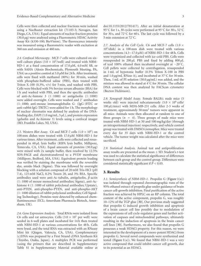
3.1. Semisynthesis of NBM-HD-1. Propolin G (Figure 1(a))was isolated through repeated chromatographic runs of the95% ethanol extract of propolis glue under guidance of braincancer cell-growth inhibition. Final purification of the activefraction was achieved by HPLC on an RP column. The totalcontent of the active component, propolin G, was roughly10–12% of the TGP glue [30]. Our previous study suggestedthat propolin G induced growth inhibition and apoptosisof a brain cancer cell line possibly due to modulation ofthe expressions of cell cycle-regulator genes and further acti-vation of caspases and mitochondrial pathways, ultimatelyresulting in the induction of apoptosis in the brain cancercell lines [30]. Furthermore, we also found that propolin Gpossesses a weak HDACi property. For this reason, we wereinterested in the development of a more-potent HDACi frompropolin G. Several novel compounds were synthesized andtested (Figure 1(a)). We found that NBM-HD-1 was a veryactive compound that could inhibit cancer cell growth, dueto its potential as an HDACi.
4 Evidence-Based Complementary and Alternative Medicine
(a)
(b)(c)
0 24 48 720
5
10
15
20
25
∗∗ ∗∗∗ ∗∗∗
Time (h)
O
O
O
O
O
O
O
O
OH
OH
OH
OH
OH
OH
CH3
CH3CH3
CH3
CH3
CH3
CH3
CH3 CH3
CH3
CH3 CH3
H3C
H3C
H3C
H3C
H3C
H3C
H3C
H3C
H3CO
H3CO
H3CO
H3CO
OCH3
OCH3
OCH3
OCH3
Propolin G
NBM-HD-1
NBM-HD-1-1
NBM-HD-1-2
NBM-HD-1
C6
MCF-7
MDA-MB-231
Control
Control
Control
Control
HO
HO
HO
HO
HO
HO
NBM-HD-1 4.3 μM
NBM-HD-1 8.5 μM NBM-HD-1 17 μM
NBM-HD-1 4.3 μM
NBM-HD-1 8.5 μM NBM-HD-1 17 μM
NBM-HD-1 4.3 μM
NBM-HD-1 8.5 μM NBM-HD-1 17 μM
Cel
lnu
mbe
r(1×
105)
Figure 1: NBM-HD-1 inhibition of cancer cells growth. (a) Structures of propolin G, NBM-HD-1-1, NBM-HD-1-2, and NBM-HD-1. (b)Cells growth inhibition of NBM-HD-1 after treatment with various concentrations in rat C6 glioma cells, human MCF-7 breast cancer cells,and human MDA-MB-231 breast cancer cells for 48 h. (c) Human MDA-MB-231 breast cancer cells (3.0 × 10⁵ per well) were cultured in6-well plates and incubated for 14 h, then treated with a fixed concentration of 17.0 μM for 1–3 days. Data are shown as the mean ± SD.∗∗P < 0.01; ∗∗∗P < 0.001 versus the control. All these tests were performed in three independent experiments. A representative experimentof the three replicates is shown.
3.2. Inhibition of Cell Growth by NBM-HD-1. C6, MCF-7,and MDA-MB-231 cells were treated with NBM-HD-1 atvarious concentrations of 4.3–17.0 μM for 48 h (Figure 1(b)).These three cancer cell lines were sensitive to NBM-HD-1,with IC50 values of 8.5–10.3 μM (Table 1).
Furthermore, we also evaluated the effect of NBM-HD-1 treatment in various human cancer cell lines includinghuman Hs683 glioma cells, human HT-29 colon cancer cells,and human DBTRG-05MG glioblastoma cancer cells andhuman Hs68 fibroblast cells (Table 1). All of these cell lines
Evidence-Based Complementary and Alternative Medicine 5
exhibited similar results to those of C6 and MCF-7 or MDA-MB-231 cells. MCF-7, C6, and HT-29 cells were found tobe more sensitive than other cancer cell lines. The normalcell line of Hs68 cells was more resistant than other cancercell lines. These results suggested that NBM-HD-1 is selectivefor inhibition of different types of cell growth. On the otherhand, we also evaluated the inhibition of cell growth activityafter treatment of MDA-MB-231 cells with a fixed concentra-tion of 17.0 μM NBM-HD-1 for 24, 48, and 72 h as shown inFigure 1(c). NBM-HD-1 significantly suppressed MDA-MB-231 cell growth in a time-dependent manner. These resultsdemonstrated that NBM-HD-1 possesses the ability toinhibit cancer cell growth. Furthermore, comparison withthree different series of analogs of methylated-propolinG (NBM-HD-1-1), hydrated-propolin G (NBM-HD-1-2),and methylated-hydrated-propolin G (NBM-HD-1) wasalso evaluated for inhibition of the cell-growth capacity(Figure 1(a)). This result demonstrates that methylation orhydration of propolin G alone does not possess excellentactivity for cell growth inhibition (data not shown). NBM-HD-1 was derived from propolin G via the two processesof methylation and hydration (Supplementary Material). Onthe other hand, we also evaluated NBM-HD-1 and propolinG in MDA-MB-231 cells. Treatment with equal concentra-tion (17.0 μM) of compounds, NBM-HD-1 inhibited cellgrowth and induced differentiation. However, propolin Ginduced cytotoxicity (data not shown). Taken together, theseresults suggest that both methylation and hydration are re-quired to obtain an active compound and might be a potentdifferentiation inducer.
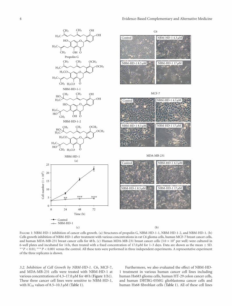
3.3. NBM-HD-1 Is an Inhibitor of HDAC. Using a trypanblue exclusion assay to evaluate cell growth inhibition, wefound that NBM-HD-1 significantly inhibited cell growthbut not induced cytotoxic effect to those cancer cell lines.Inhibition of cell proliferation was clearly present in NBM-HD-1-treated cells (Table 1 and Figures 1(b) and 1(c)). Todetermine whether NBM-HD-1 is an inhibitor of HDAC, C6cells were treated with various concentrations of NBM-HD-1 for 48 h and then nuclear-fraction proteins were isolatedto determine the inhibition of HDAC enzyme activity.As shown in Figure 2(a), NBM-HD-1 markedly inhibitednuclear HDACs enzyme activity compared to SB (sodiumbutyrate, a noted HDACi as a positive control). This resultsuggests that NBM-HD-1 may be an HDACi. As shown inFigures 2(b) and 2(c), both compounds (SB and SAHA)significantly upregulated Ac-histone 3 and gelsolin proteinexpressions. Similarly, we also found that NBM-HD-1 sig-nificantly increased expression of both proteins (Ac-histone3 and gelsolin). These results suggest that NBM-HD-1 is anHDACi.
3.4. Upregulation of Ac-Histone 4 Levels by NBM-HD-1.Next, we examined whether NBM-HD-1 inhibited cellgrowth through inhibition of HDACs activity in C6 andMCF-7 cells. For this, we used a western blot assay to evaluatewhether NBM-HD-1 caused suppression of cell growththrough inhibition of HDACs activity. Cells were treated with
Table 1: Inhibition of cancer cell lines by NBM-HD-1. Cells wereincubated with NBM-HD-1 for 3 days.
Cell lines IC50 (μM)
MCF-7 8.5
C6 8.7
MDA-MB-231 10.3
Hs683 12.7
HT-29 9.4
DBTRG-05MG 12.3
Hs68 (normal cell line) 25.0
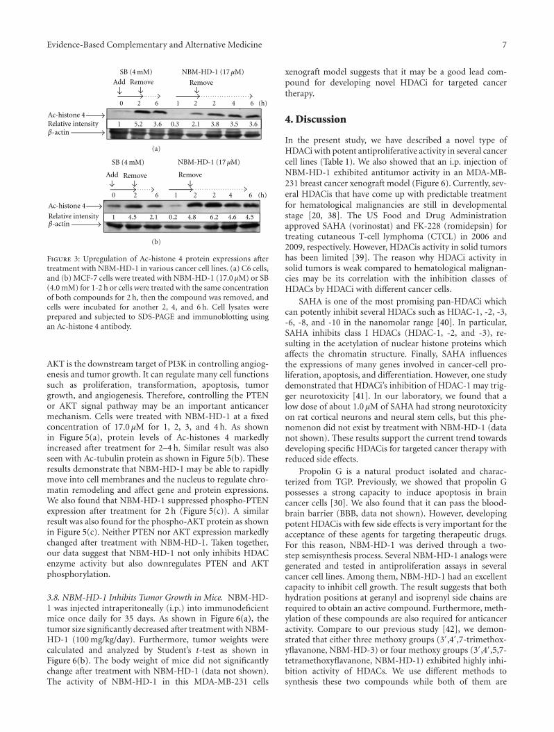
NBM-HD-1 (17.0 μM) or SB (4.0 mM) and Ac-histone 4 lev-els significantly increased compared to the untreated group.In this experiment, cells were treated with individual com-pounds for 2 h as above, then the compounds were removed,and cells were incubated without the drug for another 6 h(SB) or 2, 4, and 6 h (NBM-HD-1). As shown in Figures 3(a)and 3(b), SB-induced Ac-histone 4 protein levels significantlydecreased 6 h after removal of SB. In comparison, the declinein Ac-histone 4 protein levels induced by NBM-HD-1 wasslower than those induced by SB. Because SB is a very polarcompound and can easily dissolve in water, it could veryeasily be removed from the culture medium. However, thepolarity of NBM-HD-1 is less than SB, hence, the watersolubility of NBM-HD-1 is relatively less than SB. We as-sumed that it could easily pass through cell membranes andenter the nucleus. Therefore, after removing NBM-HD-1from the culture media, cells were still being acted on byNBM-HD-1.
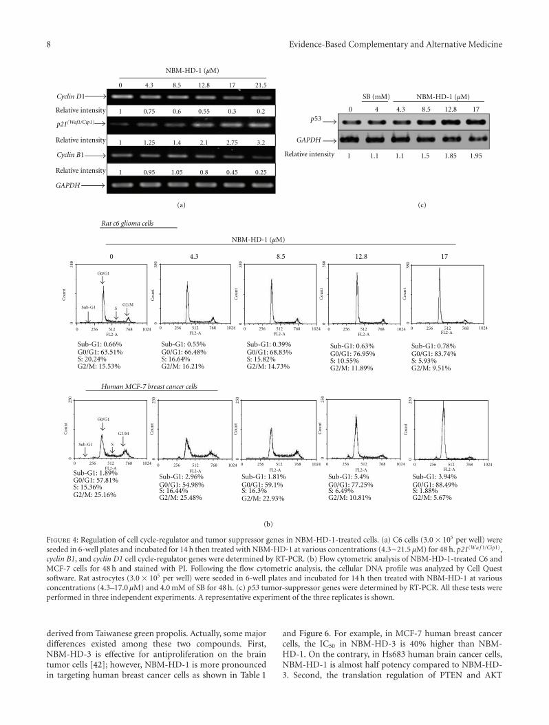
3.5. Effects of NBM-HD-1 on Cell-Cycle Regulators. Recently,a study demonstrated that the D1 cyclin and cyclin-dependent kinase inhibitor, p21(Waf1/Cip1), is an importantregulator of the cell cycle in breast cancer [31]. Many studiesalso demonstrated that cell-growth inhibition is regulated bycell-cycle regulators, including the cyclin-dependent kinaseinhibitor, p21(Waf1/Cip1), and cyclins B1, D1, and E [32–34].We investigated the inhibitory effect of NBM-HD-1 on C6cells growth. Using an RT-PCR, we studied gene expressionsof these cell-cycle regulators in C6 cells after treatment withNBM-HD-1 at various concentrations (4.3–21.5 μM) for48 h. In each case, multiple gene analyses with GAPDHwere used to standardize mRNA levels over the course ofthe experiment. p21(Wa f 1/Cip1) gene expressions markedlyincreased after treatment with NBM-HD-1 at concentrationsof 12.8–21.5 μM; while cyclin B1 and D1 gene expressionsmarkedly decreased after treatment with NBM-HD-1 at con-centrations of 12.8–21.5 μM as shown in Figure 4(a). Severalstudies demonstrated an association of cancer progressionand cell-cycle regulation. Based on these findings, cell-cycleregulation has become a target for controlling cancers [35,36]. p21(Wa f 1/Cip1) is a very important cell-cycle regulatorand tumor-suppressor gene. Next, we used flow cytometryto evaluate whether NBM-HD-1-caused cell growth sup-pression is via regulated cell cycle. C6 and MCF-7 cells weretreated with NBM-HD-1 (0–17.0 μM) for 48 h for the cell
6 Evidence-Based Complementary and Alternative Medicine
SB (mM)
0 4 4.3 8.5 12.8 170
1
7
6
5
4
3
2
∗∗∗∗∗∗
∗∗∗
∗ ∗
NBM-HD-1 (μM)
Rel
ativ
eFl
uor
esce
nce
Un
ite
(RFU
,1×
105)
(a)
SB4 mM
Control
NBM-HD-117 μM
Phase Ac-histone 3
(b)
DAPI Gelsolin Merge
Control
SAHA5 μM
NBM-HD-117 μM
(c)
Figure 2: Determination of the inhibition of total HDAC activity by NBM-HD-1. (a) C6 cells (6.0× 10⁵) were seeded in 60 mm dishes andincubated for 14 h then treated with various concentrations (4.3–17.0 μM) of NBM-HD-1 and SB (4.0 mM, as a positive control) for 48 h,and then the nuclear fraction protein was isolated to determine the inhibition of total HDAC enzyme activity. Data are shown as the mean± SD. ∗P < 0.05; ∗∗∗P < 0.001 versus the control. C6 and MCF-7 cells were cultured in six-well culture plates (3.0 × 10⁵/well) and treatedwith NBM-HD-1 (at a fixed concentration of 17.0 μM), SAHA (5.0 μM), or SB (4.0 mM) for 24 h. Immunostaining was used to evaluatebiomarkers of HDACis such as Ac-histone 3 and gelsolin after cells were treated with HDACis. (b) Ac-histone 3 and (c) gelsolin protein levelswere determined by confocal microscopy. All these tests were performed in three independent experiments. A representative experiment ofthe three replicates is shown.
cycle analysis. Our result indicates no significantly increasedapoptotic cell population (sub-G1 phase) following NBM-HD-1 (0–17.0 μM) treatment (Figure 4(b)). However, the cellpopulation at the G0/G1 phase was significantly increasedin a dose-dependent manner as shown in Figure 4(b). Takentogether, we think that p21(Wa f 1/Cip1) gene overexpressionmay be increased the cell population at the G0/G1 phaseafter treatment with NBM-HD-1.
3.6. Upregulation of Tumor-Suppressor Gene by NBM-HD-1.Rat primary astrocytes were normal brain cells in CNS. Cellswere treated with NBM-HD-1 at various concentrations (0∼17.0 μM) for 48 h. An RT-PCR was used to measure thetumor-suppressor gene expression. As shown in Figure 4(c),
p53 gene expressions were significantly upregulated aftertreatment with NBM-HD-1 compared to SB (4.0 mM). Thisresult demonstrates that NBM-HD-1 may have had the capa-bility to restore or awaken tumor-suppressor genes. HDACisare thought to be capable of waking up silenced expression oftumor-suppressor genes [37]. Our results suggest that NBM-HD-1 may be useful in controlling cancer-cell proliferationand maintaining or upregulating the function of tumor-suppressor genes in normal cells.
3.7. NBM-HD-1 Inhibits HDAC Enzymatic Activity, AlsoSuppresses the Phosphorylation of PTEN and AKT. This studyalso explored the anticancer mechanisms of NBM-HD-1.The PI3K pathway plays a key role in cell-signal transduction.
Evidence-Based Complementary and Alternative Medicine 7
β-actin
Ac-histone 4
0 2 6 1 2 2 4 6 (h)
SB (4 mM) NBM-HD-1 (17 μM)
1 5.2 3.6 0.3 2.1 3.8 3.5 3.6
Add Remove Remove
Relative intensity
(a)
β-actin
Ac-histone 4
0 2 6 1 22 4 6 (h)
SB (4 mM) NBM-HD-1 (17 μM)
1 4.5 2.1 0.2 4.8 6.2 4.6 4.5
Add Remove Remove
Relative intensity
(b)
Figure 3: Upregulation of Ac-histone 4 protein expressions aftertreatment with NBM-HD-1 in various cancer cell lines. (a) C6 cells,and (b) MCF-7 cells were treated with NBM-HD-1 (17.0 μM) or SB(4.0 mM) for 1-2 h or cells were treated with the same concentrationof both compounds for 2 h, then the compound was removed, andcells were incubated for another 2, 4, and 6 h. Cell lysates wereprepared and subjected to SDS-PAGE and immunoblotting usingan Ac-histone 4 antibody.
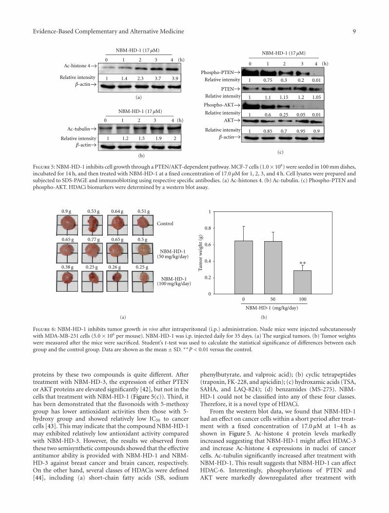
AKT is the downstream target of PI3K in controlling angiog-enesis and tumor growth. It can regulate many cell functionssuch as proliferation, transformation, apoptosis, tumorgrowth, and angiogenesis. Therefore, controlling the PTENor AKT signal pathway may be an important anticancermechanism. Cells were treated with NBM-HD-1 at a fixedconcentration of 17.0 μM for 1, 2, 3, and 4 h. As shownin Figure 5(a), protein levels of Ac-histones 4 markedlyincreased after treatment for 2–4 h. Similar result was alsoseen with Ac-tubulin protein as shown in Figure 5(b). Theseresults demonstrate that NBM-HD-1 may be able to rapidlymove into cell membranes and the nucleus to regulate chro-matin remodeling and affect gene and protein expressions.We also found that NBM-HD-1 suppressed phospho-PTENexpression after treatment for 2 h (Figure 5(c)). A similarresult was also found for the phospho-AKT protein as shownin Figure 5(c). Neither PTEN nor AKT expression markedlychanged after treatment with NBM-HD-1. Taken together,our data suggest that NBM-HD-1 not only inhibits HDACenzyme activity but also downregulates PTEN and AKTphosphorylation.
3.8. NBM-HD-1 Inhibits Tumor Growth in Mice. NBM-HD-1 was injected intraperitoneally (i.p.) into immunodeficientmice once daily for 35 days. As shown in Figure 6(a), thetumor size significantly decreased after treatment with NBM-HD-1 (100 mg/kg/day). Furthermore, tumor weights werecalculated and analyzed by Student’s t-test as shown inFigure 6(b). The body weight of mice did not significantlychange after treatment with NBM-HD-1 (data not shown).The activity of NBM-HD-1 in this MDA-MB-231 cells
xenograft model suggests that it may be a good lead com-pound for developing novel HDACi for targeted cancertherapy.
4. Discussion
In the present study, we have described a novel type ofHDACi with potent antiproliferative activity in several cancercell lines (Table 1). We also showed that an i.p. injection ofNBM-HD-1 exhibited antitumor activity in an MDA-MB-231 breast cancer xenograft model (Figure 6). Currently, sev-eral HDACis that have come up with predictable treatmentfor hematological malignancies are still in developmentalstage [20, 38]. The US Food and Drug Administrationapproved SAHA (vorinostat) and FK-228 (romidepsin) fortreating cutaneous T-cell lymphoma (CTCL) in 2006 and2009, respectively. However, HDACis activity in solid tumorshas been limited [39]. The reason why HDACi activity insolid tumors is weak compared to hematological malignan-cies may be its correlation with the inhibition classes ofHDACs by HDACi with different cancer cells.
SAHA is one of the most promising pan-HDACi whichcan potently inhibit several HDACs such as HDAC-1, -2, -3,-6, -8, and -10 in the nanomolar range [40]. In particular,SAHA inhibits class I HDACs (HDAC-1, -2, and -3), re-sulting in the acetylation of nuclear histone proteins whichaffects the chromatin structure. Finally, SAHA influencesthe expressions of many genes involved in cancer-cell pro-liferation, apoptosis, and differentiation. However, one studydemonstrated that HDACi’s inhibition of HDAC-1 may trig-ger neurotoxicity [41]. In our laboratory, we found that alow dose of about 1.0 μM of SAHA had strong neurotoxicityon rat cortical neurons and neural stem cells, but this phe-nomenon did not exist by treatment with NBM-HD-1 (datanot shown). These results support the current trend towardsdeveloping specific HDACis for targeted cancer therapy withreduced side effects.
Propolin G is a natural product isolated and charac-terized from TGP. Previously, we showed that propolin Gpossesses a strong capacity to induce apoptosis in braincancer cells [30]. We also found that it can pass the blood-brain barrier (BBB, data not shown). However, developingpotent HDACis with few side effects is very important for theacceptance of these agents for targeting therapeutic drugs.For this reason, NBM-HD-1 was derived through a two-step semisynthesis process. Several NBM-HD-1 analogs weregenerated and tested in antiproliferation assays in severalcancer cell lines. Among them, NBM-HD-1 had an excellentcapacity to inhibit cell growth. The result suggests that bothhydration positions at geranyl and isoprenyl side chains arerequired to obtain an active compound. Furthermore, meth-ylation of these compounds are also required for anticanceractivity. Compare to our previous study [42], we demon-strated that either three methoxy groups (3′,4′,7-trimethox-yflavanone, NBM-HD-3) or four methoxy groups (3′,4′,5,7-tetramethoxyflavanone, NBM-HD-1) exhibited highly inhi-bition activity of HDACs. We use different methods tosynthesis these two compounds while both of them are
8 Evidence-Based Complementary and Alternative Medicine
0 4.3 8.5 12.8 17 21.5
NBM-HD-1 (μM)
NBM-HD-1 (μM)
NBM-HD-1 (μM)
Relative intensity
Relative intensity
Relative intensity
Relative intensity
1 0.75 0.6 0.55 0.3 0.2
1 1.25 1.4 2.1 2.75 3.2
1 0.95 1.05 0.8 0.45 0.25
0 4.3 8.5 12.8 17
Sub-G1: 0.66%G0/G1: 63.51%S: 20.24%G2/M: 15.53%
Sub-G1: 0.55%G0/G1: 66.48%S: 16.64%G2/M: 16.21%
Sub-G1: 0.39%G0/G1: 68.83%S: 15.82%G2/M: 14.73%
Sub-G1: 0.63%G0/G1: 76.95%S: 10.55%G2/M: 11.89%
Sub-G1: 0.78%G0/G1: 83.74%S: 5.93%G2/M: 9.51%
Sub-G1: 1.89%G0/G1: 57.81%S: 15.36%G2/M: 25.16%
Sub-G1: 2.96%G0/G1: 54.98%S: 16.44%G2/M: 25.48%
Sub-G1: 1.81%G0/G1: 59.1%S: 16.3%G2/M: 22.93%
Sub-G1: 5.4%G0/G1: 77.25%S: 6.49%G2/M: 10.81%
Sub-G1: 3.94%G0/G1: 88.49%S: 1.88%G2/M: 5.67%
0
4
4.3 8.5 12.8 17
1 1.1 1.1 1.5 1.85 1.95
Cou
nt
FL2-A
0C
oun
t0
380
Cou
nt
038
0
Cou
nt
038
0
Cou
nt
038
0
Cou
nt
038
0
0 256 512 768 1024FL2-A
0 256 512 768 1024
FL2-A0 256 512 768 1024
FL2-A0 256 512 768 1024
FL2-A0 256 512 768 1024
FL2-A0 256 512 768 1024
FL2-A0 256 512 768 1024
FL2-A0 256 512 768 1024
FL2-A0 256 512 768 1024
FL2-A0 256 512 768 1024
Sub-G1
Sub-G1
G0/G1
G0/G1
S
S
G2/M
G2/M
250
Cou
nt
025
0
Cou
nt
025
0
Cou
nt
025
0
Cou
nt
025
0
(a)
(b)
(c)
Cyclin D1
p21(Waf1/Cip1)
Cyclin B1
GAPDH
GAPDH
SB (mM)
p53
Rat c6 glioma cells
Human MCF-7 breast cancer cells
Figure 4: Regulation of cell cycle-regulator and tumor suppressor genes in NBM-HD-1-treated cells. (a) C6 cells (3.0 × 10⁵ per well) wereseeded in 6-well plates and incubated for 14 h then treated with NBM-HD-1 at various concentrations (4.3∼21.5 μM) for 48 h. p21(Wa f 1/Cip1),cyclin B1, and cyclin D1 cell cycle-regulator genes were determined by RT-PCR. (b) Flow cytometric analysis of NBM-HD-1-treated C6 andMCF-7 cells for 48 h and stained with PI. Following the flow cytometric analysis, the cellular DNA profile was analyzed by Cell Questsoftware. Rat astrocytes (3.0 × 10⁵ per well) were seeded in 6-well plates and incubated for 14 h then treated with NBM-HD-1 at variousconcentrations (4.3–17.0 μM) and 4.0 mM of SB for 48 h. (c) p53 tumor-suppressor genes were determined by RT-PCR. All these tests wereperformed in three independent experiments. A representative experiment of the three replicates is shown.
derived from Taiwanese green propolis. Actually, some majordifferences existed among these two compounds. First,NBM-HD-3 is effective for antiproliferation on the braintumor cells [42]; however, NBM-HD-1 is more pronouncedin targeting human breast cancer cells as shown in Table 1
and Figure 6. For example, in MCF-7 human breast cancercells, the IC50 in NBM-HD-3 is 40% higher than NBM-HD-1. On the contrary, in Hs683 human brain cancer cells,NBM-HD-1 is almost half potency compared to NBM-HD-3. Second, the translation regulation of PTEN and AKT
Evidence-Based Complementary and Alternative Medicine 9
Phospho-PTEN
PTENβ-actin
β-actin
Phospho-AKT
AKT
0 1 2 3 4 (h)
Relative intensity
Relative intensity
Relative intensity
Relative intensity
1
1
0.75 0.3 0.2 0.01
1.1 1.15 1.2 1.05
1 0.6 0.25 0.05 0.01
1 0.85 0.7 0.95 0.9
(a)
(b)(c)
β-actin
Ac-tubulin
0 1 2 3 4 (h)
0 1 2 3 4 (h)
Relative intensity
Relative intensity 1
1.2 1.5 1.9 2
Ac-histone 4
1
1.4 2.3 3.7 3.9
NBM-HD-1 (17 μM)NBM-HD-1 (17 μM)
NBM-HD-1 (17 μM)
Figure 5: NBM-HD-1 inhibits cell growth through a PTEN/AKT-dependent pathway. MCF-7 cells (1.0× 10⁶) were seeded in 100 mm dishes,incubated for 14 h, and then treated with NBM-HD-1 at a fixed concentration of 17.0 μM for 1, 2, 3, and 4 h. Cell lysates were prepared andsubjected to SDS-PAGE and immunoblotting using respective specific antibodies. (a) Ac-histones 4. (b) Ac-tubulin. (c) Phospho-PTEN andphospho-AKT. HDACi biomarkers were determined by a western blot assay.
Control
NBM-HD-1(50 mg/kg/day)
NBM-HD-1(100 mg/kg/day)
0.9 g 0.53 g 0.64 g 0.51 g
0.65 g 0.77 g 0.65 g 0.5 g
0.38 g 0.25 g 0.26 g 0.25 g
(a)
Tum
orw
eigh
t(g
)
0
0.2
0.4
0.6
0.8
1
∗∗
0 50 100
NBM-HD-1 (mg/kg/day)
(b)
Figure 6: NBM-HD-1 inhibits tumor growth in vivo after intraperitoneal (i.p.) administration. Nude mice were injected subcutaneouslywith MDA-MB-231 cells (5.0× 10⁶ per mouse). NBM-HD-1 was i.p. injected daily for 35 days. (a) The surgical tumors. (b) Tumor weightswere measured after the mice were sacrificed. Student’s t-test was used to calculate the statistical significance of differences between eachgroup and the control group. Data are shown as the mean ± SD. ∗∗P < 0.01 versus the control.
proteins by these two compounds is quite different. Aftertreatment with NBM-HD-3, the expression of either PTENor AKT proteins are elevated significantly [42], but not in thecells that treatment with NBM-HD-1 (Figure 5(c)). Third, ithas been demonstrated that the flavonoids with 5-methoxygroup has lower antioxidant activities then those with 5-hydroxy group and showed relatively low IC50 to cancercells [43]. This may indicate that the compound NBM-HD-1may exhibited relatively low antioxidant activity comparedwith NBM-HD-3. However, the results we observed fromthese two semisynthetic compounds showed that the effectiveantitumor ability is provided with NBM-HD-1 and NBM-HD-3 against breast cancer and brain cancer, respectively.On the other hand, several classes of HDACis were defined[44], including (a) short-chain fatty acids (SB, sodium
phenylbutyrate, and valproic acid); (b) cyclic tetrapeptides(trapoxin, FK-228, and apicidin); (c) hydroxamic acids (TSA,SAHA, and LAQ-824); (d) benzamides (MS-275). NBM-HD-1 could not be classified into any of these four classes.Therefore, it is a novel type of HDACi.
From the western blot data, we found that NBM-HD-1had an effect on cancer cells within a short period after treat-ment with a fixed concentration of 17.0 μM at 1–4 h asshown in Figure 5. Ac-histone 4 protein levels markedlyincreased suggesting that NBM-HD-1 might affect HDAC-3and increase Ac-histone 4 expressions in nuclei of cancercells. Ac-tubulin significantly increased after treatment withNBM-HD-1. This result suggests that NBM-HD-1 can affectHDAC-6. Interestingly, phosphorylations of PTEN andAKT were markedly downregulated after treatment with
10 Evidence-Based Complementary and Alternative Medicine
NBM-HD-1. This result suggests that NBM-HD-1’s inhi-bition of cancer-cell growth may be via downregulation ofthe PTEN/AKT signal pathway. Based on these findings, wespeculated that the PTEN/AKT pathway is involved in NBM-HD-1’s induction of cancer-cell growth inhibition in MCF-7cells. Taken together, our data suggest that NBM-HD-1 notonly inhibits HDACs enzyme activity but also suppresses thePTEN/AKT pathway in cancer cells.
Concluding this study, we obtained a novel HDACi,called NBM-HD-1, derived from propolin G using two sim-ple steps (methylation and hydration, Supplementary Ma-terial). Our data suggest that NBM-HD-1 may be a potentHDACi which suppresses cancer-cell growth in several cancercell lines. This anticancer effect did not occur through acytotoxic effect (Figures 1(b) and 4(b)). Its anticancer mech-anisms may involve two pathways. First, NBM-HD-1 inhib-ited HDAC enzyme activity, affecting chromatin core his-tones and induced changes in gene expression to controlthe cell cycle. Second, NBM-HD-1 suppressed the PTEN/AKT pathway to inhibit cancer cell growth. These two factorsmay be pertinent to the anticancer effect of NBM-HD-1treatment. Developing promising HDACis for treating solidtumors is a difficult problem. NBM-HD-1 seems to have thepotential to resolve this problem.
Funding
The paper was supported by Grants from the NationalScience Foundation Directorate for Engineering of Taiwan(SBIR, 1Z-960165) and the National Science Council ofTaiwan (NSC97-2320-B-038-010-MY3).
Conflict of Interests
No potential conflict of interests were disclosed.
References
[1] R. J. DeLange and E. L. Smith, “Histones: structure andfunction,” Annual Review of Biochemistry, vol. 40, pp. 279–314,1971.
[2] H. Ris and D. F. Kubai, “Chromosome structure,” AnnualReview of Genetics, vol. 4, pp. 263–294, 1970.
[3] F. Back, “The variable condition of euchromatin and het-erochromatin,” International Review of Cytology, vol. 45,pp. 25–64, 1976.
[4] L. Berlowitz and D. Pallotta, “Acetylation of nuclear proteinin the heterochromatin and euchromatin of mealy bugs,”Experimental Cell Research, vol. 71, no. 1, pp. 45–48, 1972.
[5] R. Papait, E. Monti, and I. M. Bonapace, “Novel approacheson epigenetics,” Current Opinion in Drug Discovery and Devel-opment, vol. 12, no. 2, pp. 264–275, 2009.
[6] B. M. Turner, “Histone acetylation and an epigenetic code,”BioEssays, vol. 22, no. 9, pp. 836–845, 2000.
[7] G. L. Hager, J. G. McNally, and T. Misteli, “Transcription dy-namics,” Molecular Cell, vol. 35, no. 6, pp. 741–753, 2009.
[8] C. R. Clapier and B. R. Cairns, “The biology of chro-matin remodeling complexes,” Annual Review of Biochemistry,vol. 78, pp. 273–304, 2009.
[9] A. Miremadi, M. Z. Oestergaard, P. D. P. Pharoah, and C.Caldas, “Cancer genetics of epigenetic genes,” Human Mo-lecular Genetics, vol. 16, no. 1, pp. R28–R49, 2007.
[10] D. Mottet and V. Castronovo, “Histone deacetylases: targetenzymes for cancer therapy,” Clinical and Experimental Metas-tasis, vol. 25, no. 2, pp. 183–189, 2008.
[11] B. R. Selvi and T. K. Kundu, “Reversible acetylation of chroma-tin: implication in regulation of gene expression, disease andtherapeutics,” Biotechnology Journal, vol. 4, no. 3, pp. 375–390,2009.
[12] P. W. Atadja, “HDAC inhibitors and cancer therapy,” Progressin Drug Research, vol. 67, pp. 175–195, 2011.
[13] M. Esteller, “Epigenetics in cancer,” The New England Journalof Medicine, vol. 358, no. 11, pp. 1148–1159, 2008.
[14] W. Y. Chen, E. C. Bailey, S. L. McCune, J. Y. Dong, and T. M.Townes, “Reactivation of silenced, virally transduced genes byinhibitors of histone deacetylase,” Proceedings of the NationalAcademy of Sciences of the United States of America, vol. 94,no. 11, pp. 5798–5803, 1997.
[15] J. E. Sutherland, W. Peng, Q. W. Zhang, and M. Costa, “Thehistone deacetylase inhibitor trichostatin A reduces nickel-induced gene silencing in yeast and mammalian cells,” Muta-tion Research, vol. 479, no. 1-2, pp. 225–233, 2001.
[16] T. Cherrier, S. Suzanne, L. Redel et al., “p21(WAF1) gene pro-moter is epigenetically silenced by CTIP2 and SUV39H1,” On-cogene, vol. 28, no. 38, pp. 3380–3389, 2009.
[17] Y. Zhang, T. Carr, A. Dimtchev, N. Zaer, A. Dritschilo, andM. Jung, “Attenuated DNA damage repair by trichostatin athrough BRCA1 suppression,” Radiation Research, vol. 168,no. 1, pp. 115–124, 2007.
[18] J. P. Cantor, D. Iliopoulos, A. S. Rao et al., “Epigenetic mod-ulation of endogenous tumor suppressor expression in lungcancer xenografts suppresses tumorigenicity,” InternationalJournal of Cancer, vol. 120, no. 1, pp. 24–31, 2007.
[19] H. M. Prince, M. J. Bishton, and R. W. Johnstone, “Panobino-stat (LBH589): a potent pan-deacetylase inhibitor with prom-ising activity against hematologic and solid tumors,” FutureOncology, vol. 5, no. 5, pp. 601–612, 2009.
[20] L. Stimson, V. Wood, O. Khan, S. Fotheringham, and N. B. LaThangue, “HDAC inhibitor-based therapies and haematolog-ical malignancy,” Annals of Oncology, vol. 20, no. 8, pp. 1293–1302, 2009.
[21] V. M. Richon, J. Garcia-Vargas, and J. S. Hardwick, “Devel-opment of vorinostat: current applications and future per-spectives for cancer therapy,” Cancer Letters, vol. 280, no. 2,pp. 201–210, 2009.
[22] M. Dickinson, R. W. Johnstone, and H. M. Prince, “Histonedeacetylase inhibitors: potential targets responsible for theiranti-cancer effect,” Investigational New Drugs, vol. 28, supple-ment 1, pp. S3–S20, 2010.
[23] T. U. Bracker, A. Sommer, I. Fichtner, H. Faus, B. Haendler,and H. Hess-Stumpp, “Efficacy of MS-275, a selective inhibitorof class I histone deacetylases, in human colon cancer models,”International Journal of Oncology, vol. 35, no. 4, pp. 909–920,2009.
[24] Y. Sasaki, H. Negishi, M. Idogawa et al., “Histone deacetylaseinhibitor FK228 enhances adenovirus-mediated p53 familygene therapy in cancer models,” Molecular Cancer Therapeu-tics, vol. 7, no. 4, pp. 779–787, 2008.
[25] W. S. Xu, R. B. Parmigiani, and P. A. Marks, “Histone deacet-ylase inhibitors: molecular mechanisms of action,” Oncogene,vol. 26, no. 37, pp. 5541–5552, 2007.
Evidence-Based Complementary and Alternative Medicine 11
[26] J. Arts, P. Angibaud, A. Marien et al., “R306465 is a novelpotent inhibitor of class I histone deacetylases with broad-spectrum antitumoral activity against solid and haematologi-cal malignancies,” The British Journal of Cancer, vol. 97, no. 10,pp. 1344–1353, 2007.
[27] X. Ma, H. H. Ezzeldin, and R. B. Diasio, “Histone deacetylaseinhibitors: current status and overview of recent clinical trials,”Drugs, vol. 69, no. 14, pp. 1911–1934, 2009.
[28] P. A. Konstantinopoulos, G. P. Vandoros, and A. G.Papavassiliou, “FK228 (depsipeptide): a HDAC inhibitor withpleiotropic antitumor activities,” Cancer Chemotherapy andPharmacology, vol. 58, no. 5, pp. 711–715, 2006.
[29] S. Shankar and R. K. Srivastava, “Histone deacetylase in-hibitors: mechanisms and clinical significance in cancer:HDAC inhibitor-induced apoptosis,” Advances in Experimen-tal Medicine and Biology, vol. 615, pp. 261–298, 2008.
[30] W. J. Huang, C. H. Huang, C. L. Wu et al., “Propolin G,a prenylflavanone, isolated from Taiwanese propolis, inducescaspase-dependent apoptosis in brain cancer cells,” Journal ofAgricultural and Food Chemistry, vol. 55, no. 18, pp. 7366–7376, 2007.
[31] C. E. Caldon, R. J. Daly, R. L. Sutherland, and E. A. Musgrove,“Cell cycle control in breast cancer cells,” Journal of CellularBiochemistry, vol. 97, no. 2, pp. 261–274, 2006.
[32] A. L. Gartel, “p21(WAF1/CIP1) and cancer: a shifting paradigm?”BioFactors, vol. 35, no. 2, pp. 161–164, 2009.
[33] A. M. Egloff, L. A. Vella, and O. J. Finn, “Cyclin B1 and othercyclins as tumor antigens in immunosurveillance and immun-otherapy of cancer,” Cancer Research, vol. 66, no. 1, pp. 6–9,2006.
[34] J. K. Kim and J. A. Diehl, “Nuclear cyclin D1: an oncogenicdriver in human cancer,” Journal of Cellular Physiology,vol. 220, no. 2, pp. 292–296, 2009.
[35] H. Z. Chen, S. Y. Tsai, and G. Leone, “Emerging roles of E2Fs incancer: an exit from cell cycle control,” Nature Reviews Cancer,vol. 9, no. 11, pp. 785–797, 2009.
[36] A. Satyanarayana and P. Kaldis, “Mammalian cell-cycle regu-lation: several Cdks, numerous cyclins and diverse compen-satory mechanisms,” Oncogene, vol. 28, no. 33, pp. 2925–2939,2009.
[37] M. Esteller, “Profiling aberrant DNA methylation in hemato-logic neoplasms: a view from the tip of the iceberg,” ClinicalImmunology, vol. 109, no. 1, pp. 80–88, 2003.
[38] D. M. Vigushin and R. C. Coombes, “Histone deacetylaseinhibitors in cancer treatment,” Anti-Cancer Drugs, vol. 13,no. 1, pp. 1–13, 2002.
[39] J. Arts, P. King, A. Marien et al., “JNJ-26481585, a novel“second-generation” oral histone deacetylase inhibitor, showsbroad-spectrum preclinical antitumoral activity,” ClinicalCancer Research, vol. 15, no. 22, pp. 6841–6851, 2009.
[40] W. K. Kelly and P. A. Marks, “Drug insight: histone deacetylaseinhibitors—development of the new targeted anticancer agentsuberoylanilide hydroxamic acid,” Nature Clinical PracticeOncology, vol. 2, no. 3, pp. 150–157, 2005.
[41] D. Kim, C. L. Frank, M. M. Dobbin et al., “Deregulation ofHDAC1 by p25/Cdk5 in neurotoxicity,” Neuron, vol. 60, no. 5,pp. 803–817, 2008.
[42] W. J. Huang, C. W. Lin, C. Y. Lee et al., “NBM-HD-3, a novelhistone deacetylase inhibitor with anticancer activity throughmodulation of PTEN and AKT in brain cancer cells,” Journalof Ethnopharmacology, vol. 136, no. 1, pp. 156–167, 2011.
[43] J. M. Jeong, C. H. Choi, S. K. Kang, I. H. Lee, J. Y. Lee, and H.Jung, “Antioxidant and chemosensitizing effects of flavonoidswith hydroxy and/or methoxy groups and structure-activity
relationship,” Journal of Pharmacy & Pharmaceutical Sciences,vol. 10, no. 4, pp. 537–546, 2007.
[44] A. Al-Janadi, S. R. Chandana, and B. A. Conley, “Histonedeacetylation: an attractive target for cancer therapy?” Drugsin R & D, vol. 9, no. 6, pp. 369–383, 2008.

Submit your manuscripts athttp://www.hindawi.com
Stem CellsInternational
Hindawi Publishing Corporationhttp://www.hindawi.com Volume 2014
Hindawi Publishing Corporationhttp://www.hindawi.com Volume 2014
MEDIATORSINFLAMMATION
of
Hindawi Publishing Corporationhttp://www.hindawi.com Volume 2014
Behavioural Neurology
EndocrinologyInternational Journal of
Hindawi Publishing Corporationhttp://www.hindawi.com Volume 2014
Hindawi Publishing Corporationhttp://www.hindawi.com Volume 2014
Disease Markers
Hindawi Publishing Corporationhttp://www.hindawi.com Volume 2014
BioMed Research International
OncologyJournal of
Hindawi Publishing Corporationhttp://www.hindawi.com Volume 2014
Hindawi Publishing Corporationhttp://www.hindawi.com Volume 2014
Oxidative Medicine and Cellular Longevity
Hindawi Publishing Corporationhttp://www.hindawi.com Volume 2014
PPAR Research
The Scientific World JournalHindawi Publishing Corporation http://www.hindawi.com Volume 2014
Immunology ResearchHindawi Publishing Corporationhttp://www.hindawi.com Volume 2014
Journal of
ObesityJournal of
Hindawi Publishing Corporationhttp://www.hindawi.com Volume 2014
Hindawi Publishing Corporationhttp://www.hindawi.com Volume 2014
Computational and Mathematical Methods in Medicine
OphthalmologyJournal of
Hindawi Publishing Corporationhttp://www.hindawi.com Volume 2014
Diabetes ResearchJournal of
Hindawi Publishing Corporationhttp://www.hindawi.com Volume 2014
Hindawi Publishing Corporationhttp://www.hindawi.com Volume 2014
Research and TreatmentAIDS
Hindawi Publishing Corporationhttp://www.hindawi.com Volume 2014
Gastroenterology Research and Practice
Hindawi Publishing Corporationhttp://www.hindawi.com Volume 2014
Parkinson’s Disease
Evidence-Based Complementary and Alternative Medicine
Volume 2014Hindawi Publishing Corporationhttp://www.hindawi.com