Master Thesis- Joana Filipa Pereira de Matos - MIB alice ...
52
Transcript of Master Thesis- Joana Filipa Pereira de Matos - MIB alice ...


“Learn from yesterday, live for today, hope for tomorrow. The important thing is to not stop questioning.”
Albert Einstein

THE ROLE OF SPHINGOSINE KINASE 1 IN IL-7-MEDIATED SIGNALING AND T-CELL ACUTE LYMPHOBLASTIC LEUKEMIA
1
ACKNOWLEDGMENTS I would like to thank all the persons who have given their valuable contribution in
different ways to this study. They have all made a big effort to make possible to
complete this hard task. I would like to acknowledge for their encouragement words,
support, cooperation and collaboration. I would like to thank my colleagues, family,
friends and educators.
I would like to express my deep gratitude to my supervisor and Group leader of
JBarata’s Lab – Cancer Biology Unit, PhD João Barata, for his patient guidance,
enthusiastic encouragement as well as the useful critiscim he provided to this work.
It is also a great pleasure to acknowledge to the Faculty of Medicine of Coimbra
University internal supervisor PhD Henrique Girão for his support.
I would like to extend my thanks to all JBarata’s lab team: PhD Rita Fragoso,
PhD Isabel Alcobia, PhD Leonor Sarmento, PhD Leila Martins, PhD student Nádia
Correia, PhD student Daniel Ribeiro, PhD student Alice Melão and MSc student
Mariana Oliveira and past-members PhD João Tavanez and MSc Vanda Póvoa; for the
orientation, encouragement and the valuable technical support they provided while
performing the laboratorial work and data analysis.
I wish to thank my Master of Biomedical Research Colleagues for sharing their
ideas and for their motivation words.
Finally, I wish to thank my parents, my sister and my friends for their support and
encouragement throughout my study.

THE ROLE OF SPHINGOSINE KINASE 1 IN IL-7-MEDIATED SIGNALING AND T-CELL ACUTE LYMPHOBLASTIC LEUKEMIA
2
INDEX
ACKNOWLEDGMENTS ....................................................................................... 1 INDEX ................................................................................................................... 2 INDEX OF FIGURES ............................................................................................ 3 ABBREVIATIONS INDEX ..................................................................................... 4 ABSTRACT ........................................................................................................... 6 INTRODUCTION ................................................................................................... 7 1.1. AN OVERVIEW OF T-CELL DEVELOPMENT ........................................................ 7 1.2. IL-7 SIGNALING AND ITS IMPORTANCE IN NORMAL T-CELL DEVELOPMENT ......... 9 1.3. T-CELL ACUTE LYMPHOBLASTIC LEUKEMIA (T-ALL) ...................................... 11 1.4. IL-7 SIGNALING IN T-ALL DEVELOPMENT ....................................................... 13 1.5. SPHINGOLIPID RHEOSTAT ............................................................................ 14 1.6. S1P IN NORMAL HEMATOPOIESIS .................................................................. 15 1.7. THE SPHINGOSINE KINASES: DIFFERENCES AND FUNCTIONS ......................... 16 1.8. SPHINGOSINE KINASE 1 ................................................................................ 16 1.9. SPHINGOSINE KINASE 2 ................................................................................ 17 1.10. SPHINGOSINE KINASE ROLE IN MALIGNANT DEVELOPMENT ............................. 18
OBJECTIVES ...................................................................................................... 20 MATERIALS AND METHODS ............................................................................ 22 3.1. REAGENTS AND ANTIBODIES ........................................................................ 22 3.2. CELL CULTURE ............................................................................................ 22 3.3. IN VITRO CULTURE ....................................................................................... 22 3.4. PROTEIN EXTRACTION ................................................................................. 23 3.5. WESTERN BLOT ........................................................................................... 23 3.6. MEMBRANE STRIPPING ................................................................................ 23 3.7. QUANTITATIVE REAL-TIME PCR ................................................................... 24 3.8. ANALYSIS OF CELL VIABILITY, SIZE AND CD71 SURFACE EXPRESSION ............ 24 3.9. CELL CYCLE ANALYSIS ................................................................................. 25 3.10. ASSESSMENT OF MITOCHONDRIAL MEMBRANE POTENTIAL (ΔΨM) ................... 25 3.11. PROLIFERATION ASSAYS .............................................................................. 25 3.12. SPHINGOSINE KINASE ACTIVITY ASSAY ......................................................... 25 3.13. STATISTICAL ANALYSIS ................................................................................. 26
RESULTS ............................................................................................................ 27 4.1. SPHINGOSINE KINASE EXPRESSION IN T-ALL ............................................... 27 4.2. SPHINGOSINE KINASE INHIBITION EFFECT IN IL-7 MEDIATED-SIGNALING ......... 29 4.3. SPHINGOSINE KINASE INHIBITION REDUCES CELL SURVIVAL VIA ENHANCING
CASPASE-DEPENDENT APOPTOSIS ................................................................ 31 4.4. SPHINGOSINE KINASE INHIBITION PREVENTS CELL CYCLE PROGRESSION AND
REDUCES CELL PROLIFERATION .................................................................... 35 4.5. SPHINGOSINE KINASE INHIBITION DECREASES CELL SIZE AND TRANSFERRIN
RECEPTOR EXPRESSION ............................................................................... 36
DISCUSSION ...................................................................................................... 38 CONCLUSION .................................................................................................... 42 REFERENCES .................................................................................................... 43
THE ROLE OF SPHINGOSINE KINASE 1 IN IL-7-MEDIATED SIGNALING AND T-CELL ACUTE LYMPHOBLASTIC LEUKEMIA
3
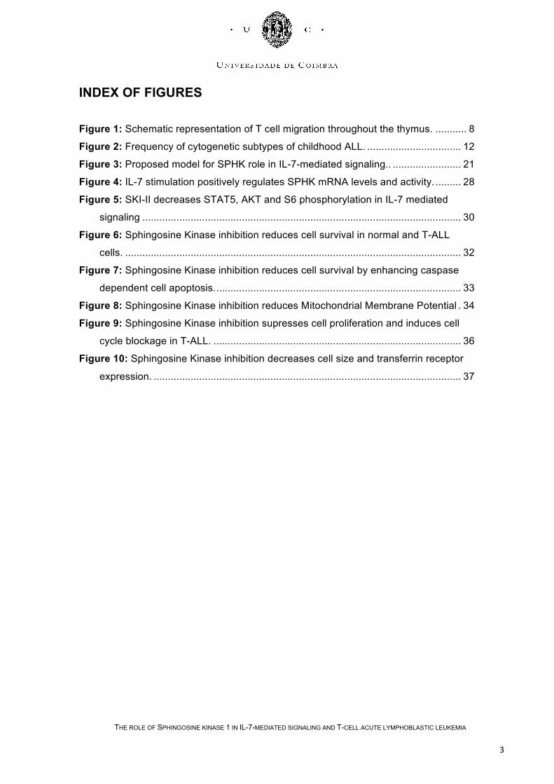
INDEX OF FIGURES
Figure 1: Schematic representation of T cell migration throughout the thymus. ........... 8
Figure 2: Frequency of cytogenetic subtypes of childhood ALL. ................................. 12
Figure 3: Proposed model for SPHK role in IL-7-mediated signaling.. ........................ 21
Figure 4: IL-7 stimulation positively regulates SPHK mRNA levels and activity. ......... 28
Figure 5: SKI-II decreases STAT5, AKT and S6 phosphorylation in IL-7 mediated
signaling ................................................................................................................ 30
Figure 6: Sphingosine Kinase inhibition reduces cell survival in normal and T-ALL
cells. ...................................................................................................................... 32
Figure 7: Sphingosine Kinase inhibition reduces cell survival by enhancing caspase
dependent cell apoptosis. ...................................................................................... 33
Figure 8: Sphingosine Kinase inhibition reduces Mitochondrial Membrane Potential . 34
Figure 9: Sphingosine Kinase inhibition supresses cell proliferation and induces cell
cycle blockage in T-ALL. ....................................................................................... 36
Figure 10: Sphingosine Kinase inhibition decreases cell size and transferrin receptor
expression. ............................................................................................................ 37
THE ROLE OF SPHINGOSINE KINASE 1 IN IL-7-MEDIATED SIGNALING AND T-CELL ACUTE LYMPHOBLASTIC LEUKEMIA
4
ABBREVIATIONS INDEX
αβ T cell: Alpha beta T cell
β-ME: β-mercaptoethanol
ΔΨM: Mitochondrial Transmembrane
Potential
γC: Gamma-common chain of the IL-2
receptor family
γδ T cell: Gama delta T cell
3H-TdR: Tritiated Thymidine
7AAD: 7-Aminoactinomycin D
AEBSF: 4-(2-Aminoethyl)
Enzenesulfonyl Fluoride Hydrochloride
Akt/PKB: v-akt Murine Thymoma Viral
Oncogene Homolog 1 / Protein Kinase
B
AnnV: Annexin V
Apaf-1: Apoptotic protease activating
factor 1
APC: Allophycocyani
APS: Ammonium Persulfate
BAD: BCL2-associated agonist of cell
death
Bcl-2: B-cell CLL/Lymphoma 2
Bcl-XL: Bcl-2 like 1
Bim: BCL2-like 11
BM: Bone marrow
CD: Cluster of differentiation
CDK: Cyclin Dependent Kinases
CDKN1A: Cyclin-dependent kinase
inhibitor 1A (p21, Cip1)
cDNA: coding Deoxyribonucleic Acid
CIB1: Calcium and integrin-binding
protein 1
CLP: Common lymphoid progenitors
CMJ: Corticomedullary junction
CMP: Common myeloid progenitors
Cyt c: Cytochrome c
DC: Dendritic cell
DMSO: Dimethyl sulfoxide
DNA: Deoxyribonucleic Acid
DN: Double negative
dATP: Deoxyadenosine triphosphate
dNTP: Deoxyribonucleotide
DP: Double positive
EDG: G coupled receptor
EGF: Epidermal growth factor
ERK: Extracellular-signal-regulated
kinases
ETP: Early thymic progenitor
FACS: Fluorescence Activated Cell
Sorting
FBS: Fetal Bovine Serum
FTY720: Fingolimod
FOXO1/3a: Forkhead family of
transcription factors
GABP: GA-binding protein
Gfi-1: Growth factor-indenpendent-1
Glut-1: Glucose transporter 1
GM: Granulocyte/ monocyte
CPCR: G protein-coupled receptors
GSK-3: Glycogen synthase kinase 3
IL-2: Interleukin 2
IL-7: Interleukin 7
IL-7R: IL-7 Receptor
Jak: Janus Kinase
THE ROLE OF SPHINGOSINE KINASE 1 IN IL-7-MEDIATED SIGNALING AND T-CELL ACUTE LYMPHOBLASTIC LEUKEMIA
5
HSC: Hematopoietic Stem Cells
MØ : Macrophages
Mcl-1: Myeloid cell leukemia
sequence 1
MFI: Mean Fluorescence Intensity
MHC: Major histocompatibility
complex
mRNA: messenger Ribonucleic Acid
Myc: Myelocytomatosis Viral
Oncogene Homolog
MkE: Megakaryocyte/ erythrocyte
MPP: Multi potent progenitors
NF-kB: Factor Nuclear kappa B
NGF: Nerve growth factor
NK: Natural killer cells
NLS: Nuclear localization signals
NES: Nuclear export signals
N.d.: No data
PARP: Poly (ADP-ribose)
Polymerase-1
PCR: Polymerase Chain Reaction
PH domain: Pleckstrin Homology
domain
PI: Propidium iodide
PI3K: Phosphoinositide 3-kinase
PLC: Phospholipase C
PP2A: Protein phosphatase 2A
PTEN: Phosphatase and Tensin
Homolog
RT-PCR: Real time polymerase chain
reaction
siRNA: Small interfering RNA
SDS-PAGE: Sodium Dodecyl Sulfate
Polyacrylamide Gel Electrophoresis
SP: Single positive
S1P: Sphingosine-1-phosphate
S1PR: Sphingosine-1 phosphate
receptor
SPHK: Sphingosine kinase
SPL: Sphingosine-1-phosphate lyase
SPP:Sphingosine-1-phosphate
phosphatase
SOCS: Suppressor of cytokine
signaling
STAT: Signal Transducers and
Activators of Transcription
SCZ: Subcapsular zone
T-ALL: T-cell Acute Lymphoblastic
Leukemia
TBS-T: Tris-Buffered Saline and
Tween 20
TCR: T-cell receptor
TEMED: Tetramethylethylenediamine
T-LGL: T-cell large granular
lymphocytic leukaemia
TLSP: Thymic stromal lymphopoietin
TSP: Thymic seeding progenitors
TEC: Thymic epithelial cell
TMRE: Tetramethylrhodamine Ethyl
Ester
TRAF2: Tumour necrosis factor
receptor-associated factor 2
VEGF: Vascular endothelial growth
factor
WBC: White blood cell counts

THE ROLE OF SPHINGOSINE KINASE 1 IN IL-7-MEDIATED SIGNALING AND T-CELL ACUTE LYMPHOBLASTIC LEUKEMIA
6
ABSTRACT Acute lymphoblastic leukemia (ALL) is the most common malignancy in pediatric
patients and is characterized by bone marrow and peripheral blood invasion from
malignant lymphoblasts. Approximately 15% of children and 25% of adult ALL cases
are of T-cell phenotype (T-ALL), which is associated with high risk and poorer
prognosis.
Interleukin-7 (IL-7) and its receptor (IL-7R) are essential for normal T-cell
development and homeostasis. However, IL-7/IL-7R-mediated signaling may also
partake in leukemia development, as demonstrated by the identification of IL-7Rα gain-
of-function mutations in around 9% of T-ALL patients.
Sphingosine Kinase (SPHK) is a lipid kinase that promotes cell viability by
phosphorylating sphingosine and thereby regulating the ceramide/sphingosine 1-
phosphate (S1P) rheostat. Cancer cells frequently display high levels of SPHK, and
SPHK expression has been correlated with cancer patients’ outcome. Previous studies
have shown that increased SPHK levels are correlated with increased cell viability and
inhibition of apoptosis in chronic myeloid leukemia and acute myeloid leukemia.
Here, we show that SPHK is an important player in IL-7-mediated signaling in T-
ALL. Initially, we demonstrated that SPHK1 expression was increased in T-ALL cells
compared to its normal counterparts. We then hypothesized that SPHK1 could be
involved in IL-7-mediated positive effects in T-ALL cells (both IL-7-dependent and IL-
7Rα mutant), as well as in normal T-cells. We demonstrated that IL-7 activates SPHK
activity without significantly impacting on its expression. SPHK inhibition completely
prevented IL-7-mediated activation of PI3K/AKT and STAT5 pathways, suggesting that
SPHK activity is fundamental for the activation of IL-7-dependent survival pathways. In
accordance, inhibition of SPHK decreased IL-7-dependent maintenance of
mitochondrial membrane potential and cell viability. In addition, SPHK was necessary
for IL-7-dependent cell cycle progression, with its inhibition inducing an arrest in
G0/G1. Finally, SPHK inhibition downregulated CD71 surface expression and cell size
in T-ALL.
In summary, our study identifies SPHK1 as an essential modulator of IL-7-
mediated signaling in T-ALL, and opens new possible therapeutic approaches by using
SPHK pharmacological inhibitors in the treatment of T-ALL patients.
Keywords: T-Cell Acute Lymphoblastic Leukemia, Interleukine-7, Sphingosine Kinase

THE ROLE OF SPHINGOSINE KINASE 1 IN IL-7-MEDIATED SIGNALING AND T-CELL ACUTE LYMPHOBLASTIC LEUKEMIA
7
INTRODUCTION
1.1. An overview of T-cell development The thymus is organized into four different regions that include the subcapsular
zone (SCZ), the cortex, the medulla, and the corticomedullary junction (CMJ, Figure 1).
The subcapsular zone is composed by cortical thymic epithelial cells (cTECs), and the
cortex has a mixture of cTECs, fibroblasts, and macrophages (MØ). The medulla
contains a stromal network of dendritic cells (DCs) and medullary thymic epithelial cells
(mTECs). The corticomedullary junction contains a dense network of endothelial cells,
which helps the access into the thymus (1). The production of different chemokines,
cytokines and ligands is required to interact with T-cell progenitors in development.
One of the important cytokines is IL-7, which is produced by stromal cells in thoracic
thymus, cervical thymus and bone marrow being required for T cell development and
homeostasis (2).
T-cell development occurs in the thymus. Detailed below is a summary of T-cell
development in the mouse, where the process is better characterized. The most
fundamental steps described here appear to be similar in humans. Thymic seeding
progenitors (TSPs), that arrive from bone marrow (BM) (3), develop to early thymic
progenitors (ETPs) in the thymic epithelium. The ETPs in the thymus are named CD4-
CD8- double-negative (DN) in stage 1. DN cells can be organized into four fractions
(DN1 to DN4) (4) that are characterized by the absence of CD4 and CD8 surface
expression and the differential expression of CD25, CD44, and CD117 according to
development. DN1 cells (CD44+ CD25- CD117+) are heterogeneous and have potential
to originate αβ T cells, γδ T cells, NK cells, dendritic cells, macrophages and B cells
(3). The ETPs with high levels of CD117 are the most efficient at giving rise to T cells
(5). Subsequently, DN1 cells leave the corticomedullary junction and migrate into the
cortex towards the subcapsular zone, and differentiate into DN2 cells (CD25+ CD44+
CD117+) (1, 6). DN2 thymocytes start to rearrange TCRβ, TCRγ and TCRδ , which are
mediated by Rag1 and Rag2 (3). When αβ and γδ T cell fate is determined, DN2 cells
differentiate into DN3 (CD25+CD44loCD117lo) within the subcapsular zone (7). After
that, DN3 thymocytes with low expression of CD27 (DN3a) give rise to DN3b, CD27
high expressing cells, which leads to β-selection. This essential checkpoint requires
TCRβ chains correctly rearranged, CD3 components and an invariant pre-TCR chain

THE ROLE OF SPHINGOSINE KINASE 1 IN IL-7-MEDIATED SIGNALING AND T-CELL ACUTE LYMPHOBLASTIC LEUKEMIA
8
(pTα)(8). The transition to DN4 (CD25-CD44-CD117-) represents the beginning of the
return of cells towards the medulla (9). Initially, during the migration, CD8 is
upregulated (10) (CD4 in humans), resulting in an immature intermediate single
positive (SP) population, and TCRα recombination is also initiated (3). Then, cells
achieve the CD4+ and CD8+ double positive stage (DP). DP cells undergo negative and
positive selection that favors thymocytes that interact with intermediate avidity for self-
peptide-MHC complexes (presented by cTECs, DCs, and fibroblasts) and express self-
MHC restricted TCRs (11). The DP thymocytes then differentiate into the CD4 single
positive (SP, CD4+CD8−) or CD8 SP (CD4−CD8+) lineages (12). Subsequently, SP
cells migrate into the medulla and suffer a second negative selection, which exclude
cells with high-affinity T-cell receptors (TCRs) for self-antigens, which avoid
autoreactive T cell generation (13). One of the most important players responsible for
T-cell migration is sphingosine-1-phosphate receptor 1 (S1PR1). At the end of T-cell
maturation, S1PR expression increases at the cell surface, which allows cell migration
from thymus to peripheral blood, where sphingosine-1- phosphate (S1P) levels are
higher (14).
Figure 1: Schematic representation of T cell migration throughout the thymus (from Koch and
Radtke. 2011).
THE ROLE OF SPHINGOSINE KINASE 1 IN IL-7-MEDIATED SIGNALING AND T-CELL ACUTE LYMPHOBLASTIC LEUKEMIA
9
The IL-7 receptor is composed by an alpha subunit (IL-7R ) (shared by the TLSP
receptor) and the common gamma chain (γc), which is shared among five additional
cytokine receptors (IL-2, -4, -9, -15 and -21) (15, 16). IL-7R is encoded by a gene on
chromosome 5p13 and contains 8 exons. The cytokine anchorage to its receptor leads
to receptor heterodimerization. IL-7R has no intrinsic kinase activity and so its signaling
activation is dependent on two receptor-associated Janus kinases 1 and 3 (JAK1 and
JAK3), which become phosphorylated and activated after IL-7 binding (17). JAK1 is
associated with IL-7R , while JAK3 binds to γ chain (18, 19). These events precede
the receptor tyrosine phosphorylation at residue 449 (Tyr449), which creates docking
sites for SH2 domain proteins, such as STAT family members (20). The STAT5 and
PI3K pathways, which are activated through JAK interaction, have been involved in
signal transduction networks that enhance the transcription of several genes related
with cell survival and proliferation of T-cells (21). These molecules could activate pro-
apoptotic factors or factors that are required for cell cycle progression, for instance Bcl-
2 or Cyclin D1 proteins, accordingly. On the other hand, these molecules also decrease
the expression of some elements that control cell cycle progression, such as p27Kip1.
(22, 23).
IL-7Rα starts to be expressed at DN2 stage, but just at DN3 stage IL-7 signaling
appears to outset, simultaneously with TCR selection (24, 25). Then, IL-7Rα
expression is downregulated at DN4 until the DP stage (26). IL-7Rα shutdown is
required at the end of DN stage for DP cell differentiation, enhancing TCF-1 and LEF-1
expression, which are transcription factors absolutely critical for T cell production (27).
Interestingly, due to the lack of IL-7 signaling, DP thymocytes are smaller, metabolically
inactive and pre-programmed to cell death, as indicated by the absence of BCL-2 and
glucose transporter 1 (Glut-1) expression (26). Cell death of DP thymocytes could be
rescued by survival signals, which are determined by thymic positive selection without
losing self-tolerance. This means that only co-receptor tuning cells with a weak
responsiveness to self-MHC receive other pro-survival signals from IL-7, excluding
potential autoreactive cells (28). The TCR positive selection signals are also
responsible for IL-7R re-expression on immature thymocytes. Additionally, IL-7R
appears to have another crucial function, which is to instruct for CD8 T cell lineage
1.2. IL-7 signaling and its importance in normal T-cell development

THE ROLE OF SPHINGOSINE KINASE 1 IN IL-7-MEDIATED SIGNALING AND T-CELL ACUTE LYMPHOBLASTIC LEUKEMIA
10
differentiation (29, 30). After CD8/CD4 lineage is established, thymocytes become
dependent on IL-7 signaling no matter their previous choice. This requirement is just
discarded with T-cell activation or memory cell differentiation when other chain
cytokines replace its survival role (31).
Several studies have indicated that IL-7 is the most potent survival factor in T-
cells and has a strong effect on thymopoiesis (32, 33). In vivo, using IL-7 or IL-7R
blocking antibodies into wild type mice leads to severe defects on thymopoiesis,
decreasing total thymocyte number, and blocking T-cell development at DN3 stage (32,
34). Furthermore, the IL-7 activity is not limited to thymocyte development: it is also
responsible for the homeostasis and activity of mature T-cells (35). Lymphoid
homeostasis refers to the preservation of the number and diversity of lymphocytes in
an organism and this is ensured by limiting levels of IL-7 and regular contact with self-
MHC (35). Given the important role that IL-7 has on regulating hematopoiesis and
cellular homeostasis, its expression and production are highly regulated and site-
specific. There are several transcription factors that have been associated with IL-7Rα
expression, such as GA-binding protein (GABP), FoxO1, Runx 1 and Runx 3 (21, 36,
37). These molecules are responsible to control IL-7R transcription. In CD8+ SP T-
cells, the growth factor-independent-1 (Gfi-1) has been identified as a repressor of IL-
7Rα transcription, which is upregulated after IL-7 signaling activation (21). Thus,
downregulation of IL7Rα happens upon IL-7 signaling or T-cell activation. This allows
T-cells to achieve the maximum profit of this limiting survival cytokine, in a process that
is named “altruistic model” (38). The suppressor of cytokine signaling (SOCS) can
inhibit IL-7 signaling induced by multiple cytokines and growth factors (39). These
proteins will inhibit the STAT binding by occupying its docking site, and targeting
proteins for degradation. All together, these events enable IL-7 control, which is
required to escape from pathway saturation, and this fine-tuning of the signal is
essential to achieve the molecular balance for cell survival. IL-7Rα downregulation is
also critical for normal T-cell development, allowing to select the immature thymocytes
that correctly rearrange a functional and useful T-cell receptor (TCR). Autoimmune and
chronic inflammatory diseases, and lymphoproliferative disorders have been correlated
with abnormal IL-7 signaling levels (40, 41).

THE ROLE OF SPHINGOSINE KINASE 1 IN IL-7-MEDIATED SIGNALING AND T-CELL ACUTE LYMPHOBLASTIC LEUKEMIA
11
Acute lymphoblastic leukemia (ALL) is the most common cancer in pediatric
patients, which has been organized into precursor T (or T-cell), precursor B, and B-cell
(Burkitt) phenotypes. These groups can be sub-characterized according to recurrent
karyotypic abnormalities.
T-cell acute lymphoblastic leukaemia (T-ALL) is an aggressive malignancy that
comes up during thymocyte development, which leads to an arrest at early stages of T-
cell maturation. This disease comprises 15% of pediatric and 25% of adults’ acute
lymphoblastic leukemia events. This pathology is clinically characterized through the
extension of bone marrow involvement, normally with 25% or more blasts present in
the BM, and also with the presence of blasts in the peripheral blood (42, 43).
Regarding its clinical features, T-ALL frequently presents splenomegaly, adenopathy,
mediastinal lymph nodes and thrombocytopenia (44). Despite BM being the
predominant site of this disease, this malignancy tends to disseminate to all over the
body, and could even affect the central nervous system (45, 46).
Different genetic abnormalities in TCR gene loci have been linked to T-cell
transformation in T-ALL development. These alterations are promoted by chromosomal
translocations that lead to the juxtaposition of genes encoding transcriptional regulators
close to TCR gene enhancers and promoters, which drive its expression. This
comprises genes such as MYC, TAL1, LYL1, LMO2, TLX1, TLX3 HOXA and MYC
(Figure 2) (47). These genetic alterations are used to classify T-ALL into subgroups.
Other genetic abnormalities have also been described, for instance mutations and non-
random chromosomal translocations driving constitutive activation of NOTCH1, which
altogether are reported in 31-60% of T-ALL patients (48).
Nowadays, T-ALL treatment regimens include a combination of up to 10 different
drugs, such as L-asparaginase, vincristine, prednisone, cyclophosphamide,
doxorubicin, cytarabine, cyclophosphamide (49), normally composed by an intensive
induction followed by consolidation and continuation therapy phases (49). Due to the
fact that patients tend to suffer of central nervous system involvement, T-ALL patients
normally receive a prophylaxis treatment for central nervous system with triple
intrathecal medications (methrotrexate, cytosine arabinoside and hydrocortisone) (49).
1.3. T-cell Acute Lymphoblastic Leukemia (T-ALL)

THE ROLE OF SPHINGOSINE KINASE 1 IN IL-7-MEDIATED SIGNALING AND T-CELL ACUTE LYMPHOBLASTIC LEUKEMIA
12
There are some variables that influence patient’s prognosis and have been used
to determine high risk patients, such as age, race, white blood cell counts (WBC),
hemoglobin, and presence of a mediastinal mass or extramedullary disease (50).
Regarding the age at diagnosis, children with < 1 year and 10 years have worse
prognosis. In these higher risk ages, there is an inverse correlation between age and
prognosis (50, 51). Males and blacks have a slightly worse prognosis (52). Different
studies have shown that WBC counts ( 50 x 109/l) and central nervous system
involvement have also worse prognosis (50). Maturational stage of the dominant clone
has been suggested to predict patient’s outcome, with some evidence, although
controversial, that more mature stages, as cortical T-ALL, have better prognosis.
Children with T-ALL have worse prognosis comparing with B-ALL, with a higher
number of high-risk patients and a higher tendency to have treatment resistance.
Despite the comparable outcomes for T- and B-ALL high-risk patients, relapsed T-ALL
have a disastrous prognosis with 3-year rates of event-free survival <15% (53). Over
the past years, patient’s outcome has been improved through more efficient tools for
risk stratification, more aggressive chemotherapies and stem cell transplantation to
overall survival rate of 70% for children and 30-40% for adults below 60 years and 10%
above 60 years. Despite these progresses, the high incidence of relapses remains a
huge problem in T-ALL.
Figure 2: Frequency of cytogenetic subtypes of childhood ALL. It shows the relative frequencies of T-
ALL (yellow) genetic subtypes. This pie chart does not include submicroscopic genetic alterations. Data
from Pui et al (2012).
THE ROLE OF SPHINGOSINE KINASE 1 IN IL-7-MEDIATED SIGNALING AND T-CELL ACUTE LYMPHOBLASTIC LEUKEMIA
13
Considering that IL-7 signaling is critical for normal T cell development and cell
survival, it is acceptable to hypothesize that survival of malignant T cells could be
supported by IL-7. It has also been shown that pathological constitutive activation of IL-
7 pathway enhances T-ALL progression, which has been associated with its
leukemogenic potential (54).
AS described previously, the altered responses of leukemic cells underlie critical
changes in gene expression. One of the most frequent mutated genes in T-ALL is
NOTCH1, whose mutations are present in more than 50% of T-ALL patients, as
already mentioned. IL7R is a well-known direct target of NOTCH1, hence its
upregulation in T-ALL with mutated NOTCH1 (48). Overexpression of IL-7 is also
observed in thymocytes from AKR/J mice, which are highly susceptible to develop
spontaneously thymic lymphomas (55). Besides this, several studies have
demonstrated that a majority of T-ALL patients respond to IL-7 in vitro and IL-7
participates in disease expansion in xenotransplantation models of human T-ALL (56,
57). Taken together, these evidences suggest that deregulation of IL7-mediated
signaling has a central role in T-ALL development.
Furthermore, there are several genetic hints that indirectly discloses the
importance of IL-7 in T-ALL, such as activating mutations in JAK1. These gain-of-
function mutations are present in 10-20% of T-ALL, and enhance constitutive activation
of STATs, AKT and ERK proteins that are downstream of IL-7R (58). Additionally,
JAK3 gain-of-function mutations have also been described in 10% of a subgroup of
high-risk (T-ALL) patients T-ALLs characterized by Early T cell phenotype (ETP-ALL)
(59). Altogether, these studies indicate that JAK1 and JAK3 mutations have
transforming potential in T-ALL.
Recently, it has been shown that gain-of-function mutations in IL-7Rα are present
in 10% of T-ALL patients. IL-7Rα mutants induce constitutive signaling independently
not only of its ligand but also of γc and JAK3. In most cases, these mutations give rise
to intermolecular disulfide bonds, which result from the insertion of an unpaired
cysteine in the juxtamembrane region of IL-7Rα, a mutational hotspot region encoded
by exon 6, leading to receptor homodimerization. This event triggers JAK/STAT5 and
PI3K/Akt/mTOR pathways. Murine B-cell line (Ba/F3) transduced with mutant hIL-7Rα
1.4. IL-7 signaling in T-ALL development

THE ROLE OF SPHINGOSINE KINASE 1 IN IL-7-MEDIATED SIGNALING AND T-CELL ACUTE LYMPHOBLASTIC LEUKEMIA
14
showed that the mutants promote both cell cycle progression and viability in the
absence of growth factors (54). These findings gave a new perspective of IL-7/IL-7R-
mediated signaling in T-ALL, which could potentiate new therapeutic strategies.
As previously described, IL-7 signaling leads to the activation of JAK/STAT and
PI3K/Akt/mTOR pathways that enhance the transcription of several genes related with
cell survival and proliferation (60). JAK/STAT5 is required for IL-7-dependent T-cell
lymphomagenesis in mice (61), however its role in human T-ALL remains to be
elucidated. PI3K activates downstream factors such as Akt and its downstream target
mTOR. Genetic alterations in this pathway (including defects in its major negative
regulator, the lipid phosphatase PTEN) affect 48% of T-ALL cases (62). However,
hyperactivation of PI3K/Akt pathway is even more frequent in T-ALL due to
posttranslational alterations, which contribute to leukemia cell maintenance (60).
Notably, IL-7 is able to upregulate the activity of this pathway even further and T-ALL
cells, in contrast to healthy T cell precursors, activate PI3K/Akt/mTOR pathway upon
IL-7 stimulation in a manner that is responsible not only for cell cycle progression, but
also for Bcl-2 upregulation and the consequent increase in cell viability (63, 64).
These findings demonstrate an oncogenic role of the IL-7 receptor in T-ALL and
suggest, perhaps, that the majority of T-ALL subtypes harbors an activated IL-7R
pathway, suggesting the IL-7Rα as a potential therapeutic target in this malignancy.
Sphingolipid metabolites have been identified as signaling players with a
meaningful influence in cell fate, leading to cell proliferation/ survival or apoptosis.
Therefore, sphingolipid synthesis and degradation pathways are highly regulated
processes, which are determined by different players. Ceramide can be generated by
de novo synthesis, breakdown of sphingomyelin or complex glycosphingolipids, and
then can be deacylated into sphingosine. A key molecule in these pathways is
sphingosine kinase (SPHK), which is responsible for the conversion of sphingosine into
sphingosine-1-phosphate (S1P). The levels of these molecules have a role in the
balance between cell apoptosis (sphingosine) or cell survival (S1P). This important
switch, named sphingolipid rheostat, has been related with survival, proliferation,
migration, angiogenesis, inflammation and differentiation (65). The biologically active
phospholipid S1P can be reversibly modified into sphingosine by S1P phosphatases
1.5. Sphingolipid Rheostat
THE ROLE OF SPHINGOSINE KINASE 1 IN IL-7-MEDIATED SIGNALING AND T-CELL ACUTE LYMPHOBLASTIC LEUKEMIA
15
(SPP1 and SPP2), or cleaved irreversibly into hexadecenal and phosphoetanolamide
by S1P lyase (SPL) (66, 67).
Commonly, S1P is generated intracellularly and then exported by ATP-binding
cassette (ABC) super family of transporters or other mechanisms (68). S1P in the
circulation can bind to a family of five S1P-specific cell surface G protein-coupled
receptors (GPCRs) (S1PR1 to S1PR5) and enhances different signaling pathways, an
event that is named inside-out signaling. For instance, PI3K/AKT, ERK1/2 or PLC are
some of those S1P-dependent activation signaling pathways that are responsible for
cell proliferation, migration, survival, angiogenesis, maturation, inflammation, immunity,
chemotaxis and trafficking (65). S1P functions are not exclusively extracellular, since
S1P also modulates histone deacetylases leading to c-fos, cell cycle regulator
CDKN1A (p21) expression (69) and the ubiquitin ligase activity of tumor necrosis factor
receptor-associated factor 2 (TRAF2) (70). S1P-induced TRAF2 activity and NF-kB
activity afterwards have a pro-survival/pro-proliferative effect.
S1P has been described as one of the major players in lymphocyte movement
between BM and secondary lymphoid tissues into peripheral blood. S1P differential
gradient between the lymphoid organs and the periphery allows lymphocyte egress.
The first evidence of this function came from the discovery that the sphingosine
analogue and SPHK inhibitor, Fingolimod (FTY720), leads to rapid but reversible
lymphopenia, and lymphocytes are retained at secondary lymphoid organs
simultaneously (14, 71). These studies also showed that S1P leads to rapid receptor
internalization and degradation and this leads to a decrease in cell response. The
same effect that occurs in mature lymphocytes happens in thymocytes (72). At the end
of thymic maturation, S1PR overexpression is required to enable the exit from the
thymus. The role of S1P in lymphocyte migration is also involved in inflammatory
processes (73). The injured tissues increase their S1P levels, which promotes
lymphocyte retention through S1PR1. These findings altogether demonstrate that S1P
has an important role in hematopoiesis, however further experiments are required to
clarify its function.
1.6. S1P in normal hematopoiesis
THE ROLE OF SPHINGOSINE KINASE 1 IN IL-7-MEDIATED SIGNALING AND T-CELL ACUTE LYMPHOBLASTIC LEUKEMIA
16
There are two human SPHK genes, SPHK1 and SPHK2, which are located on
chromosome 17 (17q25.2) and 19 (19q13.2) respectively, and encode SPHK1 and
SPHK2 proteins. These are highly conserved lipid kinases which are responsible for
S1P synthesis (74). Despite their high polypeptide sequence similarity (80%), sharing
five highly conserved regions (C1-C5), SPHK2 presents two extra polypeptide regions
at the N-Terminus and a central proline-rich region (75). Their differences are not only
restricted to their structure. In fact, SPHKs display distinct development expression,
adult tissue distribution, substrate binding, and subcellular localization, which could
foresee distinct physiological functions (76). Although SPHK isoforms are expressed in
all tissues in humans, SPHK1 is highest expressed in lung, spleen and leukocytes (77),
whereas SPHK2 is increased in the kidney and liver. In addition, SPHKs are also
differentially expressed in embryonic development (78). In mouse, SPHK1 expression
increases earlier (E7-E11), while SPHK2 expression raises later (E15-E17) in
embryogenesis (78).
Different studies show that SPHK1 and SPHK2 may have antagonistic roles in
diverse pathological conditions, such as inflammatory arthritis, kidney injury,
lipopolysaccharide-induced injury and cancer. In the majority of the reports, SPHK1
enhances cell survival and proliferation in different types of cancer (79-81), while there
is no agreement respecting to SPHK2 functions. Two contrasting roles, pro-apoptotic
and anti-apoptotic functions are suggested and SPHK subcellular position seems to be
critical in this choice.
Despite their different roles, genetic deletion of SPHK1 or SPHK2 has no effect in
normal development in mice, and deletion of both SPHKs results in mice lifeless in
utero due to several abnormalities in angiogenesis and neurogenesis (82). Altogether,
these data indicate that SPHKs have some functional redundancy, at least in normal
mouse development.
There are three different SPHK1 isoforms (SPHK1a, SPHK1b, and SPHK1c) that
diverge in their N-terminal domain (75). SPHK1a is secreted by human umbilical-vein
endothelial cells and seems to contribute to increased S1P plasma levels (83).
1.7. The Sphingosine Kinases: differences and functions
1.8. Sphingosine kinase 1

THE ROLE OF SPHINGOSINE KINASE 1 IN IL-7-MEDIATED SIGNALING AND T-CELL ACUTE LYMPHOBLASTIC LEUKEMIA
17
SPHK1b is located at the cell membrane (84), while SPHK1c is at the Golgi and is
present in lower levels comparing with its homologous (83). Despite these differences,
all of them have intrinsic catalytic activity, and can be additionally activated by growth
factors (EGF, PDGF, NGF, VEGF), cross-linking of immunoglobulin receptors, TGFB
and TNFα, adhesion molecules (PECAM1) (85), interleukins, Ca2+ increasing agents
and phorbol esters (74). Whereas some of these molecules lead to a rapid and
transient activation of SPHK, promoting posttranslational modifications or changing its
intracellular localization, others induce a biphasic mechanism, increasing initially its
activity and then its transcription, such as EGF, estrogen, 1,25-dihydroxyvitamin D3 or
histamine.
Activation of SPHK1 was shown to be mediated by protein kinase C, classical
MAPK/extracellular-signal-regulated kinases (ERKs) and PI3K/mTOR and promotes
phosphorylation of SHPK1 at Ser225 by ERK1/2 (86). The phosphorylation of SPHK1
increases its selectivity for phosphatidylserine at the inner leaflet of the plasma
membrane via the side chains Thr54 and Asn89. This transient activation could be
countered by protein phosphatase 2A (PP2A) (87). In this model, phosphorylation of
Ser225 promotes a conformational alteration that exposes Thr54 and Asn89 and/or other
phosphatidylserine-binding sites, which helps membrane translocation (88). Enzymatic
activity is explained by the proximity of SPHK1 to its substrate, sphingosine, and by the
stimulatory action of phosphatidilserine (88). Some recent evidences show that calcium
and integrin-binding protein 1 (CIB1) facilitates active translocation of SPHK1 to the
plasma membrane in a calcium-myristoyl dependent way and it is maintained at this
localization through acidic phospholipids and other interactions (89).
SPHK2 presents two isoforms, named SPHK2-S and SPHK2-L. The last
mentioned isoform contains 36 additional amino acids and is the most common in
humans (83).
SPHK2 can also be activated by different factors, such as EGF (90), TNF- (91),
cross-linking of the IgE receptor Fc RI (92), phorbol esters (90) and hypoxia conditions
(93). This lipid kinase, similarly to SPHK1, can also be activated by ERK1/2 by
phosphorylation at Ser351 and Thr578 (94).
1.9. Sphingosine kinase 2

THE ROLE OF SPHINGOSINE KINASE 1 IN IL-7-MEDIATED SIGNALING AND T-CELL ACUTE LYMPHOBLASTIC LEUKEMIA
18
SPHK2 displays nuclear localization (NLS) and export (NES) signals (95, 96).
When SPHK2 is located in the nucleus, it prevents DNA synthesis (96) and regulates
epigenetic modifications modulating HDAC1/2 (97). Increased acetylation of H3, H4
and H2B leads to increased expression of cyclin-dependent kinase inhibitor (CDKN1A)
and the transcriptional regulator FOS (69). Besides this, SPHK2 localized at the
nucleus seems to be related with contact inhibition responses, which decrease cell
proliferation. On the other hand, it has been shown that SPHK2 is located at
endoplasmic reticulum under stress conditions, which is associated with increased
levels of pro-apoptotic ceramide (76). SPHK2 positioning at mitochondrial membrane
also leads to cell apoptosis via S1P, BAK and cytochrome c release (97). By contrast,
SPHK2 placed at the plasma membrane promotes cell proliferation. Interestingly, it has
been shown that SPHK1 artificially targeted to the ER or nucleus leads to apoptosis
(76). These findings indicate the major involvement of the SPHK location in its
functions.
Different studies have demonstrated that SPHK1 displays oncogenic features.
Normal NIH3T3 fibroblasts that have upregulated SPHK1 show tumorigenic features,
as indicated by colony growth in soft agar (98).
Overexpression of SPHK1 has been shown in different types of cancer, such as
breast, lung, ovary, stomach, uterus, kidney, where its levels are 2-fold higher
comparing with normal tissue (99, 100). Furthermore, SPHK1 upregulation has also
been associated with poor prognosis in gastric, breast, brain and lung cancer patients
(81, 101, 102). Moreover, SPHK inhibitors significantly decrease tumor growth in mice
(103, 104) and cells lacking SPHK1 become more sensitive to chemotherapeutic
treatments (105).
Several studies have demonstrated the proliferation potential of SPHK1 in
hematological malignancies. SPHK1 activation is promoted by interleukin-6 in multiple
myeloma cells (106), and PDGF seems to induce a similar effect in T-cell large
granular lymphocytic leukemia (T-LGL) (107). Interesting, protein kinase C (PKC),
classical MAPK/ ERKs and PI3K/mTOR are some of the signaling pathways that are
implicated in SPHK1 activation and which have been shown to be deregulated in
leukemia (86). In erythroleukemia, SPHK1 overexpression is required to promote
1.10. Sphingosine kinase role in malignant development
THE ROLE OF SPHINGOSINE KINASE 1 IN IL-7-MEDIATED SIGNALING AND T-CELL ACUTE LYMPHOBLASTIC LEUKEMIA
19
leukemia development and progression (108), and BCR/ABL1 increased SPHK1
expression in CML (109). Furthermore, S1P pro-survival signals avoid cytochrome c
and Smac/Diablo release in leukemia cell lines, upregulating anti-apoptotic molecules
such as Mcl-1 in chronic myeloid leukemia and multiple myelomas (110). Inhibition of
SPHK1 activity in leukemia cells, for instance in CML (111) and T-LGL (112), promotes
cell death.
As described before, while SPHK1 role in cancer is well established, SPHK2
appears to have a dual role in cancer development and progression. Some studies
have demonstrated that SPHK2 has also a similar function to SPHK1. In MCF7 human
breast cancer cells, SPHK2 knockdown prevents growth of xenografted tumors (113).
Further, some studies showed that chemical inhibition of SPHK2 reduced proliferation
and induced autophagy cell death in kidney, prostate and breast cell lines (114, 115).
Although the effects of SPHK2 in hematological malignancies are much less
known, the importance of SPHK2 starts to emerge. Recently, Wallington-Beddoe et al.
showed that SPHK2 has a role in B-ALL, through the expression of the oncogene MYC
and that SPHK2 activity signature is increased in ALL patients (116).
Together, these studies elucidate the critical involvement of SPHK in malignant
development, and consequently SPHK arises as an evident therapeutic target. There
are several pre-clinical studies using FTY720 inhibitor in disseminated lymphoma
(117), CML (118), murine cell line models of AML with KIT mutations (119), and also in
rat models of NK leukemia (119). Moreover, in mice engrafted with human AML U937
cell line, treatment with a SPHK1 inhibitor reduces tumor growth (120), and in a human
xenograft model of Ph+ ALL, a SPHK2 inhibitor has anti-leukemic potential, which
cooperates with Imatinib (116). Nonetheless, the effects of SPHK1 and SPHK2
inhibitors on hematological malignancies are still poorly explored.

THE ROLE OF SPHINGOSINE KINASE 1 IN IL-7-MEDIATED SIGNALING AND T-CELL ACUTE LYMPHOBLASTIC LEUKEMIA
20
OBJECTIVES
Taken all of the findings summarized above together, we hypothesized that
SPHK1 and/or 2 (hereby jointly reffered to as SPHK) may be involved in T-ALL and
particularly in IL-7-mediated signaling in this malignancy. Hence, this research project
intended to investigate the importance of SPHK in normal and mutant IL-7Rα signaling
in T-ALL.
To accomplish our main goal, we defined some critical questions:
1. What are the levels of SPHK expression? Are they increased in T-ALL
patients?
2. Does IL-7 regulate SPHK expression and/or activity?
3. Is SPHK involved in IL-7 mediated signaling in T-ALL cells?
4. What are the effects of SPHK inhibition on IL-7-mediated cell viability,
cell cycle, activity and proliferation of T-ALL cells?
In order to address these questions, we used the following cell lines: D1, a
murine IL-7-dependent thymocyte-like cell line; TAIL7, a human IL-7-dependent
primary-like T-ALL cell line; DND4.1, a human IL-7Rα mutant T-ALL cell line. These
cell lines were cultured with a SPHK pan inhibitor, SKI-II. Although we performed some
analyses discriminating between SPHK1 and 2, the functional studies we performed so
far made use only of SKI-II. As such, we opted to name SPHK1 and SPHK2 jointly as
SPHK in most parts of the present thesis, although we are aware that not necessarily
the two are involved in IL-7-mediated signaling.
We speculated that SPHK may partake in and drive IL-7-mediated signaling
pathways, by being required for full activation of IL-7 downstream elements (Figure 3).
If confirmed, this could constitute the first step towards the characterization of SPHK as
a key molecule in IL-7-mediated signaling and a new potential therapeutic target in T-
ALL.

THE ROLE OF SPHINGOSINE KINASE 1 IN IL-7-MEDIATED SIGNALING AND T-CELL ACUTE LYMPHOBLASTIC LEUKEMIA
21
Figure 3: Proposed model for SPHK role in IL-7-mediated signaling. The dashed arrows represent our
hypothesis, as SPHK could partake in IL-7-mediated signaling pathways by being required to the full
activation of IL-7 downstream elements.

THE ROLE OF SPHINGOSINE KINASE 1 IN IL-7-MEDIATED SIGNALING AND T-CELL ACUTE LYMPHOBLASTIC LEUKEMIA
22
MATERIALS AND METHODS
3.1. Reagents and Antibodies
SKI-II, 2-(p-Hydroxyanilino)-4-(p-chlorophenyl) thiazole, which is a SPHK pan-
inhibitor, was synthesized by Calbiochem® (Merck Millipore).
The following were used as primary antibodies: p27kip1 (BD Transduction
Laboratories), phospho-JAK3 (Y980), JAK3, JAK1, STAT5, SPHK1, Actin, Caspase-3,
(Santa Cruz Biotechnology), phospho-STAT5A/B (Y694/Y699; Upstate Biotechnology),
and phospho-JAK1 (Y1022/1023), phospho-Akt (S473), phospho-Erk1/2 (T202/Y204),
Akt, Erk1/2, phospho-PTEN (S380) and PTEN (Cell Signaling Technology).
Immunodetection was performed with horseradish peroxidase–conjugated anti–mouse
IgG, anti–rabbit IgG or anti–goat IgG (Promega).
3.2. Cell Culture
TAIL7, a human T-ALL IL-7 dependent primary-like cell line, was grown in the
presence of 10ng/ml of human IL-7, in RPMI medium (Gibco) supplemented with 5% of
fetal bovine serum (FBS) (PAA Laboratories GmbH), at a concentration of 2 ×
106 cells/ml. The human T-ALL cell line DND-4.1 was cultured in RPMI 10% of FBS
(Biowest) at a concentration of 0,5× 10⁶ cells/ml. The murine IL-7 dependent cell line
D1 was grown in the presence of 50 ng/ml of human IL-7 in RPMI with 10% of FBS
(PAA), at a concentration of 1 × 10⁶ cells/ml. The human T-ALL cell line Jurkat was
cultured in RPMI 10% of FBS (PAA) at 0,5× 10⁶ cells/ml.
3.3. In Vitro Culture
TAIL7 cells were plated in 6-well plates or 96-well plates at 2×10⁶ cells/ml at 37°C
with 5% CO2. Serum and IL-7 starved TAIL7 cells for 24h were incubated in RPMI 5%
FBS (control medium) with 20 ng/ml of IL-7, SKI-II or both. Following, cells were
harvested and processed for western blot or FACS analysis, accordingly.
D1 cells were plated in 6-well plates or 96-well plates at 1×10⁶ cells/ml at 37°C
with 5% CO2 in RPMI 10% FBS (control medium) in the presence of 50 ng/ml IL-7 and
SKI-II, or both. D1 cells were starved in RPMI without FBS and IL-7 for 2h prior to any
stimulation.

THE ROLE OF SPHINGOSINE KINASE 1 IN IL-7-MEDIATED SIGNALING AND T-CELL ACUTE LYMPHOBLASTIC LEUKEMIA
23
DND-4.1 cells were plated in 6-well plates or 96-well plates at 0,5×10⁶ cells/ml at
37°C with 5% CO2 in RPMI 10% FBS (control medium) with or without SKI-II.
3.4. Protein Extraction
Collected cells were centrifuged at 3500 rpm for 5 minutes at 4ºC in order to
obtain a cell pellet. Cell pellet was ressuspended in lysis buffer (50mM Tris-Base;
150mM NaCl; 5mM EDTA; 1mM NaOVa; 10mM NaF; 10mM NaPyrophosphate; 1%
NP-40, supplemented with protease inhibitor cocktail Complete Mini (Roche) and
protease inhibitor AEBSF (1mM)). The extracts were centrifuged at 13000 rpm for 15
minutes at 4°C and the supernatants were collected. Total protein quantification was
performed by Bradford assay (absorbance at 595nm). Protein extracts were
ressuspended in Laemmli buffer and denatured at 95 °C for 5 minutes.
3.5. Western Blot
Samples were separated by SDS-PAGE (12 or 14% of acrylamide gel) and
transferred onto a nitrocellulose membrane. Precision Plus ProteinTM (Bio-Rad)
molecular weight standards were used as reference. Protein transference was carried
out using conventional transference system (90 min, 400mA). Membranes were
submerged in Ponceau S solution in order to evaluate the protein content. The
membranes were then blocked (3% skimmed milk, TBS-T buffer) and then incubated
with primary antibody diluted in TBS-T (in pre-established dilutions and antibody
specific) overnight at 4°C under constant agitation. After washing the membranes for
25 min in TBS-T (in a total of three washing steps), the membranes were incubated
with the respective secondary antibody diluted (1:5000) in a 3% milk/TBS-T buffer
blocking solution and incubated for 1 hour at room temperature under constant
agitation. Chemiluminescence detection was carried out using the Pierce ECL Plus
Western Blotting Substrate (Thermo Fisher Sientific Inc.). Membranes were revealed
using the Curix60 (AGFA HealthCare).
3.6. Membrane Stripping
In order to perform a new blot of the membrane, the antibodies were removed by
stripping process. The membranes were incubated in the stripping solution (Tris-HCl,
THE ROLE OF SPHINGOSINE KINASE 1 IN IL-7-MEDIATED SIGNALING AND T-CELL ACUTE LYMPHOBLASTIC LEUKEMIA
24
β-mercaptoethanol (100mM), 10% SDS, pH 6.7) for 30 minutes with mild agitation at
56ºC. Finally, the membrane was washed in water and then in TBS-T. Primary and
secondary antibody solutions were then applied as described before.
3.7. Quantitative Real-Time PCR
The mRNA expression of SPHK1 and 2 was quantified using a 7500 Fast or ViiA
7 Real-Time PCR System (Applied Biosystems). The following primer sequences were
used, human SPHK1 forward: CTT GCA GCT CTT CCG GAG TC and reverse: CAC
GCT GAT GCT CAC TGA GC; mouse SPHK1 forward: TGT GAA CCA CTA TGC TGG
GTA and reverse: CAG CCC AGA AGC AGT GTG, 18 S rRNA forward: GGA GAG
GGA GCC TGA GAA ACG and reverse: CGC GGC TGC TGG CAC CAG ACT T,
human SPHK2 forward: CTG TCT GCT CCG AGG ACT GC and reverse: GAC CCC
CAA AGG GAT TGA CCA AT; mouse SPHK2 forward: GGA GACG GGC TGC TTT
ACG AG and reverse: (tudo o que está sublinhado não está a preto, está a cinza.
Confere) GATCCACAGGGGAGGACACC.
3.8. Analysis of cell viability, size and CD71 surface expression
Cell viability was determined by double-staining with APC-conjugated Annexin V
(eBioscience) and 7-AAD (BD Biosciences) followed by flow cytometric analysis on a
LSRFortessa flow cytometer (BD Biosciences). Briefly, cells were washed with PBS
and ressuspended in 100 µL of binding buffer with Annexin V and 7-AAD. After 15
minutes of incubation at room temperature in the dark, 100 µL of binding buffer were
added and the samples were acquired. Fully viable cells were identified as the Annexin
V and 7-AAD double-negative populations.
Cell size was assessed by analysis of SSC versus FSC flow cytometry plots
gated on the live cell population. Samples were analyzed using LSRFortessa flow
cytometer (BD Biosciences) and FACSDiva software (BD Biosciences). Results were
analyzed by FlowJo software (Tree Star).
Transferrin receptor (CD71) surface expression was analyzed by flow cytometry
using FITC-conjugated anti-CD71 (eBioscience) antibody. Results were expressed as
the specific mean intensity of fluorescence (MIF).
THE ROLE OF SPHINGOSINE KINASE 1 IN IL-7-MEDIATED SIGNALING AND T-CELL ACUTE LYMPHOBLASTIC LEUKEMIA
25
3.9. Cell Cycle Analysis
Cells were harvested, washed with PBS and fixed in 80% ethanol at -20ºC. Cells
were ressuspended in PBS containing 50 ug/ml RNase A and incubated at 37ºC for 30
minutes. Samples were labeled with 100ul/ml propidium iodide (PI) and were analyzed
using a FACSCalibur flow cytometer (BD Biosciences). Cell cycle modeling was
performed using ModfitTM LT software (Verity Software House Biosciences).
3.10. Assessment of Mitochondrial Membrane Potential (Δψm)
Cells were harvested and stained in culture medium with TMRE (Sigma-Aldrich)
to a final concentration of 100 nM, and incubated for 15 min at 37°C. Cells were
analyzed for TMRE intensity by flow cytometry. Samples were analyzed using
LSRFortessa flow cytometer (BD Biosciences) and FACSDiva software (BD
Biosciences). Results were analyzed by FlowJo software (Tree Star).
3.11. Proliferation Assays
Cells were cultured in triplicates in flat bottom 96-well plates at the normal cell
culture density and conditions as indicated previously. IL-7 cytokine and/or SPHK
inhibitor were added in the experimental conditions mentioned above. Cells were
incubated with [3H] labelled thymidine at the concentration of 1 Ci/well for 16 h (TAIL7
cell line) or 8h (remaining cell lines) before harvest. DNA synthesis, as measured by
[3H] thymidine incorporation, was assessed using a scintillation counter (Perkin Elmer).
Average and standard deviation of triplicates were calculated.
3.12. Sphingosine Kinase Activity Assay
The SPHK Activity was measured through a commercial kit from Echelon Inc.
(SPHK Activity Assay). This is a based luminescent assay that measures SPHK
activity by detecting the consumption of ATP transformation of sphingosine into S1P.
The experimental conditions were conducted as described previously. Total cell
lysates were collected in lysis buffer (described previously in western blot section) and
supplemented with Triton-X 100 (0,1%) or KCL (1M) in order to discriminate SPHK1
and SPHK2 activity, respectively. The assay was performed accordingly to
THE ROLE OF SPHINGOSINE KINASE 1 IN IL-7-MEDIATED SIGNALING AND T-CELL ACUTE LYMPHOBLASTIC LEUKEMIA
26
manufacturer instructions. The luminescence signal was detected after one hour of
incubation with the microplate reader Infinite M20 (Tecan). The integration time was
set to 1000ms per well.
3.13. Statistical analysis
Statistical computations were performed with GraphPad Prism version 5.0 for
Windows (GraphPad Software). In what concerns the differences among groups, it
was performed ANOVA, t-Student or Mann-Whitney statistical tests, as appropriate.
Differences among groups were considered statistically significant at p ≤0,05 (95%
confidence interval).

THE ROLE OF SPHINGOSINE KINASE 1 IN IL-7-MEDIATED SIGNALING AND T-CELL ACUTE LYMPHOBLASTIC LEUKEMIA
27
RESULTS
4.1. Sphingosine Kinase Expression in T-ALL
Given the existing evidence that SPHK is upregulated in other leukemias, we
analyzed gene expression of SPHK1 and SPHK2 in T-ALL patients and normal
thymocytes by qRT-PCR. SPHK1, but not SPHK2, transcript levels were increased in
T-ALL samples (p = 0.0462) comparing with normal thymocytes (Figure 4A). We also
analysed a publicly available dataset (GSE7186) for SPHK expression, that
corroborated our initial analysis and showed that SPHK1 expression was higher in T-
ALL (p = 0.0439) as compared to normal bone marrow samples (Figure 4A and 4B).
In order to determine the effect of IL-7 stimulation on SPHK1 gene expression,
we incubated TAIL7 and D1 cell lines in the presence or absence of IL-7. We found
that IL-7 did not significantly affect SPHK1 expression (Figure 4C). Similar results were
obtained for SPHK2 expression with or without IL-7 stimulation (data not shown). The
effect of IL-7 on SPHK activity was then evaluated in TAIL7 and D1 cell lines. In
contrast to the expression levels, IL-7 stimulation in TAIL7 cells clearly upregulated
total SPHK activity (p = 0.0393, Figure 4D). In D1 cells, we observed that SPHK1
activity was higher than SPHK2 activity and that IL-7 withdrawal decreased the activity
of both enzymes (p = 0.0305 and p = 0.0042, Figure 4D).
Together, these results show that SPHK1 is significantly more expressed in T-
ALL samples compared to their normal counterparts and that IL-7 stimulation positively
regulates SPHK kinase activity without significantly affecting SPHK transcript levels.

THE ROLE OF SPHINGOSINE KINASE 1 IN IL-7-MEDIATED SIGNALING AND T-CELL ACUTE LYMPHOBLASTIC LEUKEMIA
28
Figure 4: IL-7 stimulation positively regulates SPHK mRNA levels and activity. (A) Peripheral blood
or bone marrow samples were collected from T-ALL patients at the time of diagnosis and total RNA was
extracted. Statistical analysis was performed using Mann-Whitney test from triplicates. Relative expression
of SPHK1 was significantly different between groups (p = 0.0462), whereas no statistical differences in
SPHK2 expression were observed. (B) A publicly available dataset of gene expression (GSE7186) was
manual analysed. Statistical analysis was performed using Mann-Whitney test (p = 0.0439). (C) TAIL7
cells were starved for 24h and cells were collected after 16 and 24 hours of incubation in the presence or
absence of 20ng/ml of IL-7. D1 cells were starved for 2h and cells were collected after 8 and 16 hours of
incubation with presence or absence of 50 ng/ml of IL-7. Then RNA was extracted with subsequent cDNA
production and qRT-PCR analysis. The observed differences between IL-7 presence and absence were
not statistically significant. (D) TAIL7 cells were starved for 24h and protein extracts were collected after 24
hours of incubation in the presence or absence of 20ng/ml of IL-7. D1 cells protein extracts were collected
after 4 hours of IL-7 withdrawal. Then total SPHK, SPHK1 and SPHK2 activities were measured with
specific buffers by commercially available kinase activity assay. Statistical analysis was performed using 1-
Way ANOVA from duplicates, p = 0.0393 in TAIL7 cells. In D1 cells was performed a t-Test from duplicate
values, p = 0.0305 for SPHK1 conditions and p = 0.0042 between SPHK2 conditions.

THE ROLE OF SPHINGOSINE KINASE 1 IN IL-7-MEDIATED SIGNALING AND T-CELL ACUTE LYMPHOBLASTIC LEUKEMIA
29
4.2. Sphingosine Kinase inhibition effect in IL-7 mediated-signaling
To determine the role of SPHK in IL-7-mediated signaling in normal and T-ALL
cells, we used different cell lines. In particular, D1 (‘normal’, non-tumorigenic thymocyte
IL-7-dependent murine cells), TAIL7 (T-ALL cells, IL-7-dependent) and DND-4.1 (T-
ALL cells, IL-7Rα mutant) were treated with the pan-SPHK inhibitor SKI-II. D1 and
TAIL7 cells were starved to potentiate the responsiveness to IL-7 and then treated with
SKI-II. The inhibitor blocked IL-7-mediated upregulation of STAT5, AKT and S6
phosphorylation without affecting total protein levels (Figure 5A). These effects on IL-7-
mediated signaling were present in both normal and T-ALL contexts, i.e. in D1 and
TAIL7 cells, respectively (Figure 5A).
To determine the impact of SPHK inhibition on IL-7Rα gain-of-function mutations,
we used the DND-4.1 cell line, which has a heterozygous 12 nucleotide insertion in the
IL-7R gene (IL7R p.L242_L243insLSRC).This alteration is similar to the mutations that
were described in 10% of T-ALL patients, which promote constitutive activation of IL-
7R without IL-7 requirement. Western blot analysis of DND-4.1 cells corroborated the
constitutive phosphorylation of IL-7R downstream targets, such as STAT5 and AKT
(Figure 5A). SKI-II treatment significantly downregulated STAT5 and AKT
phosphorylation levels and had a perceptible, although minor, effect on S6
phosphorylation (Figure 5A).
In order to identify the proximal targets of SPHK in IL-7 signaling, we next
conducted a time course experiment with SKI-II pre-incubation for two hours with
subsequent IL-7 stimulation. The early responses to IL-7 stimulation in the presence of
SPHK inhibition were analysed by western blot. Our data demonstrate that SPHK
activity is required for very early STAT5 and JAK-1 phosphorylation (30 seconds post-
stimulation), as well as for JAK3 early phosphorylation (1 minute after IL-7 stimulation)
(Figure 5B). Phosphorylation of downstream effectors, such as AKT and S6, was also
affected. In contrast, PTEN phosphorylation, which is not IL-7-dependent, was not
affected by SKI-II (Figure 5B).
These data suggest that somehow SPHK activity is required for optimal triggering
of IL-7-mediated signaling in both normal and pathological contexts.

THE ROLE OF SPHINGOSINE KINASE 1 IN IL-7-MEDIATED SIGNALING AND T-CELL ACUTE LYMPHOBLASTIC LEUKEMIA
30
Figure 5: SKI-II decreases STAT5, AKT and S6 phosphorylation in IL-7 mediated signaling. (A) D1
cells were starved for 2h and total protein extracts were collected after 2h incubation with 10µM of SPHK1
specific inhibitor and 15 min in the presence of 50 ng/ml of IL-7. TAIL7 cells were starved for 24h and total
protein extracts were collected after 2h incubation with 10µM of SPHK1 specific inhibitor and 15 min in the
presence of 20ng/ml of IL-7. DND-4.1 total protein extracts were collected after 2h incubation in the
presence/absence of 10µM of SPHK1 specific inhibitor. STAT5, AKT, PTEN and S6 total and
phosphorylated and Actin were analysed (B) D1 cells were starved for 2h and total protein extracts were
collected after 2h incubation with SPHK inhibitor or vehicle and in the presence or absence of IL-7 for 30
seconds; 1, 5, 15 and 30 minutes. JAK3, JAK1, STAT5, AKT, PTEN total and phosphorylated and Actin
proteins were analysed

THE ROLE OF SPHINGOSINE KINASE 1 IN IL-7-MEDIATED SIGNALING AND T-CELL ACUTE LYMPHOBLASTIC LEUKEMIA
31
4.3. Sphingosine Kinase inhibition reduces cell survival via enhancing caspase-dependent apoptosis
Next, we sought to evaluate the functional impact of our molecular observations.
We found that SPHK inhibition decreases cell survival in normal murine thymocytes (p
= 0.0003) and human T-ALL cells (p < 0.0001, Figure 6). This effect was dose- (Figure
6A) and time-dependent (p < 0.0001, Figure 6B).
To characterize the cell death mechanism triggered by SPHK inhibition, we
performed annexin V/ 7AAD staining, in order to evaluate whether apoptosis was
involved. The different cell populations could be identified as follows: viable (7AAD and
annexin V double-negative), early apoptotic (annexin V positive, 7AAD negative), and
late apoptotic/necrotic (7AAD and annexin V double-positive) cells. Inhibition of SPHK
led to an increase in the frequency of early and late apoptotic cells (Figure 7A), both of
which contribute to increase the percentages of apoptotic cells in D1 (p = 0.0265),
TAIL7 (p < 0.0001) and DND-4.1 (p = 0.0005, Figure 7B). The process of apoptosis or
programmed cell death (PCD) can also be defined by caspase-3 and PARP cleavage.
In accordance with the annexin V/ 7AAD flow cytometry data, western blot analysis of
DND-4.1 cells after 24h of SKI-II treatment showed increased cleavage of both
caspase 3 and PARP (Figure 7C).
Mitochondrial membrane potential was described as an important parameter of
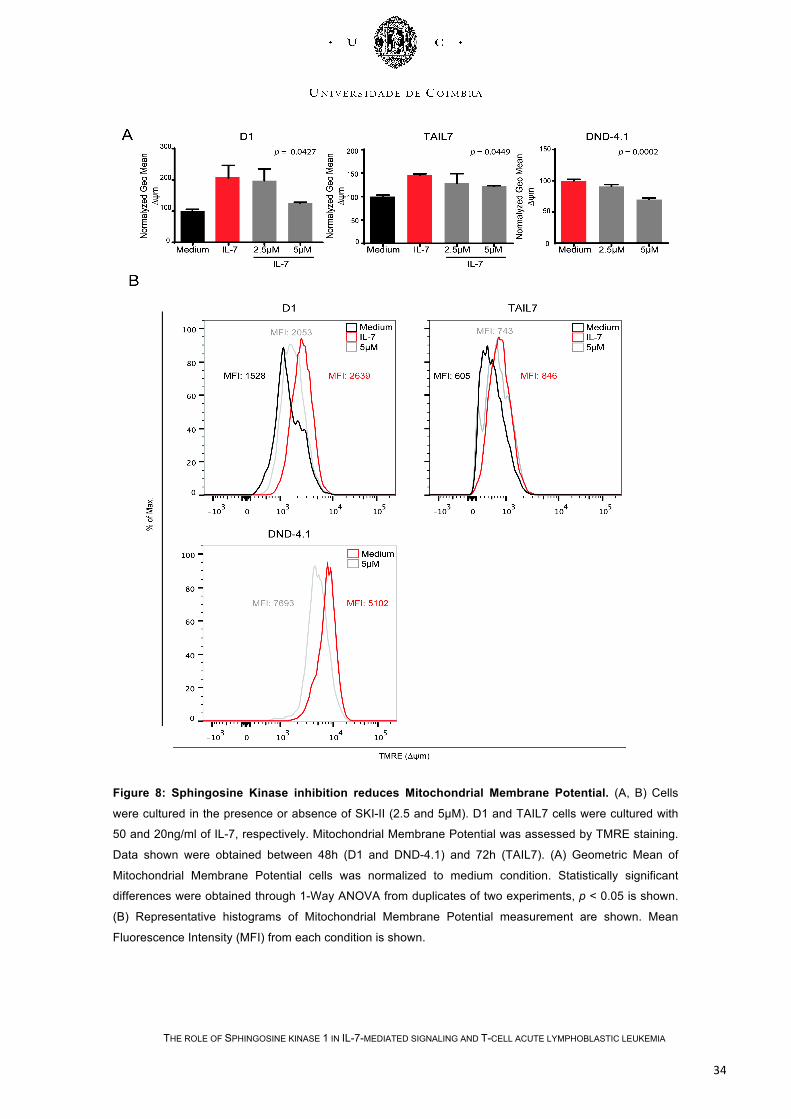
mitochondrial function used as a very early indicator of apoptosis. The collapse of the
mitochondrial transmembrane potential occurs at the same time of the opening of
mitochondrial permeability transition pores, promoting cytochrome c release, which
enhances other downstream events in the apoptotic cascade. As expected, SKI-II
decreased mitochondrial membrane potential in D1 (p = 0.0427), TAIL7 (p = 0.0449)
and DND-4.1 (p = 0.0002) cells (Figure 8).
Taken together, these results strongly indicate that SPHK activity is required for
IL-7 mediated survival of T-ALL cells and murine thymocytes.

THE ROLE OF SPHINGOSINE KINASE 1 IN IL-7-MEDIATED SIGNALING AND T-CELL ACUTE LYMPHOBLASTIC LEUKEMIA
32
Figure 6: Sphingosine Kinase inhibition reduces cell survival in normal and T-ALL cells. (A) D1 and
DND-4.1 cell viability was evaluated in the presence or absence of different concentrations (1, 2.5, 5 µM)
of SKI-II. TAIL7 cell viability was evaluated in the presence or absence of different concentrations (0.1, 1,
5, 10 µM) of SKI-II. D1 and TAIL7 cells were cultured with 50 and 20ng/ml of IL-7, respectively. Data
shown were obtained between 48h (D1 and DND-4.1) and 72h (TAIL7). Viability was obtained by flow
cytometry with FSCxSSC analysis and normalized to medium conditions. Statistical analysis was
performed using 2-Way ANOVA from duplicates of two experiments, p = 0.0003 and p < 0.0001. (B) D1,
TAIL7 and DND-4.1 cell viability was evaluated in the presence or absence of different concentrations
(5µM) of SKI-II. D1 and TAIL7 cells were cultured with 50 and 20ng/ml of IL-7, respectively. Viability was
obtained by flow cytometry with FSCxSSC analysis and normalized to IL-7 condition in D1 and TAIL7 cell
lines, and to medium condition in DND-4.1. Statistical analysis was performed using 2-Way ANOVA from
duplicates of two experiments, p < 0.0001.

THE ROLE OF SPHINGOSINE KINASE 1 IN IL-7-MEDIATED SIGNALING AND T-CELL ACUTE LYMPHOBLASTIC LEUKEMIA
33
Figure 7: Sphingosine Kinase inhibition reduces cell survival by enhancing caspase dependent cell
apoptosis. (A, B) Cells were cultured in the presence or absence of SKI-II (2.5 and 5µM). D1 and TAIL7
cells were cultured with 50 and 20ng/ml of IL-7, respectively. AnnexinV/7AAD staining assessed apoptotic
and viable cells. Data shown was obtained between 48h (D1 and DND-4.1) and 72h (TAIL7). (B)
Percentages of apoptotic cells were normalized to medium condition. Statistical analysis of D1 and TAIL7
cell lines were performed using 2-Way ANOVA from duplicates of two experiments, p = 0.00265 and p <
0.0001, respectively. Statistically significant differences of DND-4.1 were obtained through 1-Way ANOVA
from duplicates of two experiments, p = 0.0005 is shown. (C) DND-4.1 total protein extracts were collected
after 24h incubation in the presence/absence of 10µM of SPHK1 specific inhibitor. PARP and caspase-3
cleavage was analysed.
THE ROLE OF SPHINGOSINE KINASE 1 IN IL-7-MEDIATED SIGNALING AND T-CELL ACUTE LYMPHOBLASTIC LEUKEMIA
34
Figure 8: Sphingosine Kinase inhibition reduces Mitochondrial Membrane Potential. (A, B) Cells
were cultured in the presence or absence of SKI-II (2.5 and 5µM). D1 and TAIL7 cells were cultured with
50 and 20ng/ml of IL-7, respectively. Mitochondrial Membrane Potential was assessed by TMRE staining.
Data shown were obtained between 48h (D1 and DND-4.1) and 72h (TAIL7). (A) Geometric Mean of
Mitochondrial Membrane Potential cells was normalized to medium condition. Statistically significant
differences were obtained through 1-Way ANOVA from duplicates of two experiments, p < 0.05 is shown.
(B) Representative histograms of Mitochondrial Membrane Potential measurement are shown. Mean
Fluorescence Intensity (MFI) from each condition is shown.

THE ROLE OF SPHINGOSINE KINASE 1 IN IL-7-MEDIATED SIGNALING AND T-CELL ACUTE LYMPHOBLASTIC LEUKEMIA
35
4.4. Sphingosine Kinase inhibition prevents cell cycle progression and reduces cell proliferation
Since IL-7 has been shown to promote not only viability but also cell cycle
progression of T-ALL cells, the effects of SKI-II on cell cycle and proliferation after IL-7
stimulation were also measured. SKI-II treatment decreased cell proliferation of IL-7-
cultered TAIL7 cells (p = 0.0002) and DND-4.1 cells (p = 0.0189, Figure 9A). The same
results were obtained for D1 cell line (data not shown). Regarding the effects on cell
cycle, SKI-II led to a cell cycle arrest in G0/G1 in TAIL7 and DND-4.1 cell lines (Figure
9B). Similar results were obtained for D1 cell line (data not shown).
These results indicate that SPHK activity is essential for IL-7-mediated cell cycle
progression and proliferation.

THE ROLE OF SPHINGOSINE KINASE 1 IN IL-7-MEDIATED SIGNALING AND T-CELL ACUTE LYMPHOBLASTIC LEUKEMIA
36
Figure 9: Sphingosine Kinase inhibition supresses cell proliferation and induces cell cycle
blockage in T-ALL. (A, B) Cells were cultured in the presence or absence of SKI-II (2.5 and 5µM). TAIL7
cells were cultured with 20ng/ml of IL-7. (A) Data shown were obtained between 48h (DND-4.1) and 72h
(TAIL7). Thymidine incorporation values were normalized to medium condition. Statistical analysis of
TAIL7 and DND-4.1 cell lines were performed using 1-Way ANOVA from duplicates of two experiments, p
= 0.0002 and p = 0.0189, respectively is shown. (B) Data shown were obtained between 24h (DND-4.1)
and 48h (TAIL7). Cell cycle phase percentages were evaluated by flow cytometry analysis of PI staining in
ethanol-fixed and permeabilized cells.
4.5. Sphingosine Kinase inhibition decreases cell size and transferrin receptor expression
IL-7 was shown to promote cell size increase (cell growth), which associates with
increased metabolism and expression of ‘activation’ markers such as the transferring
receptor CD71. Inhibition of SPHK impaired IL-7-mediated TAIL7 cell growth cells (p =
0.0109) and promoted the atrophy of DND-4.1 cells (p = 0.0016, Figure 10A).
Surprisingly, in contrast to T-ALL cells, SKI-II treatment increased the cell size of D1

THE ROLE OF SPHINGOSINE KINASE 1 IN IL-7-MEDIATED SIGNALING AND T-CELL ACUTE LYMPHOBLASTIC LEUKEMIA
37
murine thymocytes (p = 0.0041, Figure 10A). In accordance, we found that SPHK
inhibition was negatively affected CD71 expression in both TAIL7 (p = 0.0324) and
DND-4.1 cells (p < 0.0001, Figure 10B). In agreement with the cell size response,
CD71 was also more expressed after SKI-II treatment in D1 cells (p = 0.0025, Figure
10B).
These results suggest that SPHK inhibition decreases cell size and transferrin
receptor expression selectively in T-ALL cells.
Figure 10: Sphingosine Kinase inhibition decreases cell size and transferrin receptor expression.
(A, B) Cells were cultured in the presence or absence of SKI-II (2.5 and 5µM). TAIL7 cells were cultured
with 20ng/ml of IL-7. Data shown were obtained between 48h (D1 and DND-4.1) and 72h (TAIL7). (A) Cell
size was obtained by flow cytometry with FSCxSSC analysis. TAIL7 and DND-4.1 were normalized to
medium conditions. D1 values were normalized to IL-7 condition, due to the lower % of viable cells in
medium condition, we were not able to measure cell size (n.d. - no data). Statistical analysis was
performed using 1-Way ANOVA from a representative experiment, p = 0.0041, p = 0.0109 and p = 0.0016,
respectively. (B) Transferrin receptor (CD71) expression was evaluated by specific antibody staining and
analyzed by flow cytometry. TAIL7 and DND-4.1 were normalized to medium condition. D1 values were
normalized to IL-7 condition, due to the lower % of viable cells in medium condition, we were not able to
measure CD71 (n.d. - no data). Statistical analysis was performed using 1-Way ANOVA from a
representative experiment, p = 0.0025, p = 0.0324 and p < 0.0001, respectively.

THE ROLE OF SPHINGOSINE KINASE 1 IN IL-7-MEDIATED SIGNALING AND T-CELL ACUTE LYMPHOBLASTIC LEUKEMIA
38
DISCUSSION
The critical role of SPHKs in the development of a wide range of tumors has been
exhaustively demonstrated, including in breast, lung, ovary, stomach, uterus, and
kidney cancers, and both chronic and acute myeloid leukemias (99, 100). It is well-
known that SPHK1 expression or/and activity are increased in cancer cells, being
related with cell survival and proliferation (79). In contrast, SPHK2 function is still
controversial, apparently displaying two contrasting functions under different
conditions. In mouse embryonic kidney cells (HEK293) or mouse embryonic fibroblasts,
SPHK2 silencing with small interfering RNA (siRNA) prevented the induction of
apoptosis (121). In opposition, in human breast cancer cells, SPHK2 knockdown
inhibited the growth of xenografted tumours (113). SPHKs subcellular localization has
been pointed out as the decisive factor, nonetheless additional experiments are
required (76).
Recent studies have established the importance of SPHKs in hematological
malignancies. In Multiple Myeloma (MM), IL-6 stimulation leads to increased SPHK1
expression and activity, which upregulates myeloid cell leukaemia (Mcl-1), increasing
cell proliferation and survival (106). Recently, it has been demonstrated that SPHK2
activity, but not its expression, was higher in ALL samples than normal B-cell
progenitors (116). In this work, we show that SPHK1, but not SPHK2, may have an
important role in T-ALL pathology since it is significantly overexpressed in malignant
samples. In agreement with this notion, it has been shown that SPHK inhibition
promotes apoptosis of human T-ALL Jurkat cells (110), suggesting that SPHK
overexpression in T-ALL cells is of functional relevance.
SPHK has been indicated as a cytokine-activated kinase. As stated in the
previous paragraph, IL-6 stimulates SPHK1 activity and leads to a decrease in both
sphingosine and ceramide levels, which leads to suppression of cell apoptosis in MM
(106). Furthermore, ERK1/2 and PI3K/mTOR are the main players responsible for
SPHK1 phosphorylation and activation (106). Constitutive activation of IL-7 pathway,
which drives the activation of JAK/STAT and both of these pathways (63), has been
associated with leukemogenesis in T-ALL. In that sense, IL-7 signaling emerges as an
obvious candidate for SPHK activation, and in fact we demonstrated that IL-7
stimulation leads to increased activity of both SPHK1 and SPHK2. It remains, however,
to be understood what is the functional role of each of the two kinases in the context of
IL-7-mediated signaling.
THE ROLE OF SPHINGOSINE KINASE 1 IN IL-7-MEDIATED SIGNALING AND T-CELL ACUTE LYMPHOBLASTIC LEUKEMIA
39
Previously, it has been reported that SPHK translocation from the cytosol to the
plasma membrane is absolutely required for SPHK activation. These data suggest that
plasma membrane localization is essential not only for SPHK activation, thus placing
SPHK in close proximity to its substrate, sphingosine, but also to facilitate its product,
S1P, acting on intracellular signaling targets or moving to the extracellular
compartment and stimulating its G protein-coupled receptors (88). Here, we
demonstrated for the first time that SPHK has a critical role in IL-7 and IL-7R-mediated
signaling, and is absolutely required for the activation of downstream elements in IL-7
signaling pathways in normal T-cell precursors and T-ALL cells. Before, it was
proposed that IL-7Rα association with clathrin and subsequent internalization is
necessary for optimal IL-7-mediated signal transduction (122). The time-course
experiments we now conducted showed that SPHK inhibition has an early effect on IL-
7 signaling, which could indicate that SPHK is placed very upstream, at close proximity
to the JAKs and PI3K - possibly even physically interacting with them or with the IL-7R.
Alternatively, SPHK inhibition may promote changes in lipid membrane composition
and by this way decrease IL-7 internalization, compromising IL-7-mediated signaling.
Further experiments are required to clarify if and what are the molecular mechanisms
by which SPHK may affect IL-7R internalization. On the other hand, SKI-II inhibits the
conversion of sphingosine and ceramide into S1P, promoting intracellular accumulation
of ceramide and sphingosine. This elevation of ceramide activates serine/threonine
protein phosphatases PP1 and PP2A that dephosphorylate Akt (120). Akt deactivation
is mediated not only by IL-7 signaling shutdown but also by increased ceramide, which
could explain the effect of SKI-II in the regulation of PI3K/AKT pathway activation. This,
however, would not justify the impact on JAK/STAT pathway.
Importantly, in agreement with its effects on key survival pathways, we further
demonstrate that in T-ALL and normal thymocytes, SPHK is absolutely required for IL-
7-mediated pro-survival effects. Previously it was demonstrated that IL-7 induces the
phosphorylation of Akt, which interacts with its downstream targets GSK-3, FOXO1,
and FOXO3a in a PI3K-dependent manner, leading to their inactivation and nuclear
export, which promote cell viability. In addition, GSK-3 can also phosphorylate and
inhibit NF-ATc, a transcription factor involved in proliferation and Bcl-2 gene
transcription (63). It was also well shown that sphingosine rheostat is responsible for
apoptosis regulation. Death-inducing signals reach the mitochondria, promoting
cytocrome c (cyt c), adaptor of protein Apaf-1, and precursor of caspase-9, Smac
diablo and dATP release. Pro-caspase 9 is cleaved and activates other caspases, such
THE ROLE OF SPHINGOSINE KINASE 1 IN IL-7-MEDIATED SIGNALING AND T-CELL ACUTE LYMPHOBLASTIC LEUKEMIA
40
as caspase-3, -6 or -7, which lead to apoptosis. Some reports indicate that increased
levels of ceramide are responsible for mediating Fas- and TNF-α-induced apoptosis
and precede previously described mitochondrial events in different stress conditions.
S1P has an opposite effect, inhibiting cyt c and Smac/Diablo translocation to the
cytoplasm, thereby preventing apoptosis. These findings suggest that a combination of
both conditions, decreased S1P formation and increased ceramide levels, will promote
cell apoptosis, which is achieved through SPHK inhibition (110). Our data is consistent
with these reports, since SKI-II induced mitochondrial transmembrane potential loss,
caspase-3 and PARP cleavage, and consequent apoptosis of malignant as well as
normal T-cell precursors. Of note, mitochondrial transmembrane potential loss has
been observed during ceramide-induced effector phase of apoptosis.
In non-transformed NIH 3T3 fibroblasts, sphingosine kinase transfection
significantly increases the proliferative rate of fibroblasts (123). Our data demonstrated
that SPHK inhibition decreases the proportion of cells in S-phase and blocks the G1/S
transition in IL-7 stimulated normal and T-ALL cells. This could be explained possibly
by the negative effect of SKI-II on proliferative signals, such as Cyclin-dependent
Kinase (Cdc2 and Cdk2 kinases) expression, which can also be regulated by IL-7-
mediated signaling (22). GSK-3β, which is inactivated by phosphorylation by Akt, is
described as responsible for cyclin D1 regulation, contributing to cell cycle arrest. Since
SPHK inhibition represses the activation of IL-7-mediated pathways, cell cycle related
molecules are also expected to be repressed, explaining potentially the effect seen in
T-ALL cells and normal T-cells after SPHK inhibition.
There are evidences that suggest a relation between increased cell size, cell
metabolism and oncogenesis. TFR1 (CD71) is upregulated in order to supply increased
cell metabolism requirements. Previously it was shown that IL-7 contributes to T-ALL
cell growth and metabolic activation, increasing cell size and surface expression of
CD71. Those events are dependent on PI3K/Akt activation (63). Recently, Pham et al.
showed that SPHK1 expression leads to increase of CD71 surface levels and
transferrin uptake in a manner that appears to be dependent also on c-Myc (124). We
showed that SKI-II decreases CD71 expression and cell size in T-ALL cells. It remains
to be determined if this effect is dependent on SPHK1-mediated activation of PI3K
pathway. Surprisingly, SPHK inhibition upregulated CD71 surface expression and cell
size in normal thymocytes, represented by the mouse D1 cell line. It is possible that
normal T-cell precursors differ from T-ALL cells in IL-7-mediated, SPHK-dependent
regulation of metabolism - an observation that may be of therapeutic importance. It

THE ROLE OF SPHINGOSINE KINASE 1 IN IL-7-MEDIATED SIGNALING AND T-CELL ACUTE LYMPHOBLASTIC LEUKEMIA
41
should be noted, nonetheless, that experiments involving primary human thymocytes
are required to confirm the data obtained so far with D1 cells.
THE ROLE OF SPHINGOSINE KINASE 1 IN IL-7-MEDIATED SIGNALING AND T-CELL ACUTE LYMPHOBLASTIC LEUKEMIA
42
CONCLUSION
IL-7 and its receptor are crucially involved in T-cell leukemia pathophysiology.
The current study provides evidence that IL-7/IL-7R-mediated signaling upregulates
SPHK1 kinase activity and absolutely requires SPHK for full activation of downstream
effectors. The activation of IL-7 pathway is not only related with increased viability, but
also involved in the regulation of cell size and proliferation. Altogether, our results
indicate that SPHK plays an important role in IL-7-mediated signaling and its
downstream effects on T-ALL cells. Together with our evidence that SPHK1 is
overexpressed in T-ALL cells, these findings could constitute a solid argument for the
further deepening of the understanding of the role of SPHK in T-ALL, which could help
define whether SPHK is a valid molecular target for therapeutic intervention in T-ALL.
To complement our in vitro studies, analysis of inhibition of SPHK in primary T-ALL
samples should be performed and in vivo efficacy of inhibitors of SPHK for the
treatment of T-ALL should be tested.

THE ROLE OF SPHINGOSINE KINASE 1 IN IL-7-MEDIATED SIGNALING AND T-CELL ACUTE LYMPHOBLASTIC LEUKEMIA
43
REFERENCES
1. Petrie HT, Zuniga-‐Pflucker JC. Zoned out: functional mapping of stromal signaling microenvironments in the thymus. Annual review of immunology. 2007;25:649-‐79. Epub 2007/02/13. 2. Mazzucchelli RI, Warming S, Lawrence SM, Ishii M, Abshari M, Washington AV, et al. Visualization and identification of IL-‐7 producing cells in reporter mice. PloS one. 2009;4(11):e7637. Epub 2009/11/13. 3. Ciofani M, Zuniga-‐Pflucker JC. The thymus as an inductive site for T lymphopoiesis. Annual review of cell and developmental biology. 2007;23:463-‐93. Epub 2007/05/18. 4. Zuniga-‐Pflucker JC. When three negatives made a positive influence in defining four early steps in T cell development. J Immunol. 2012;189(9):4201-‐2. Epub 2012/10/23. 5. Porritt HE, Rumfelt LL, Tabrizifard S, Schmitt TM, Zuniga-‐Pflucker JC, Petrie HT. Heterogeneity among DN1 prothymocytes reveals multiple progenitors with different capacities to generate T cell and non-‐T cell lineages. Immunity. 2004;20(6):735-‐45. Epub 2004/06/11. 6. Takahama Y. Journey through the thymus: stromal guides for T-‐cell development and selection. Nature reviews Immunology. 2006;6(2):127-‐35. Epub 2006/02/24. 7. Burtrum DB, Kim S, Dudley EC, Hayday AC, Petrie HT. TCR gene recombination and alpha beta-‐gamma delta lineage divergence: productive TCR-‐beta rearrangement is neither exclusive nor preclusive of gamma delta cell development. J Immunol. 1996;157(10):4293-‐6. Epub 1996/11/15. 8. von Boehmer H. Unique features of the pre-‐T-‐cell receptor alpha-‐chain: not just a surrogate. Nature reviews Immunology. 2005;5(7):571-‐7. Epub 2005/07/07. 9. Porritt HE, Gordon K, Petrie HT. Kinetics of steady-‐state differentiation and mapping of intrathymic-‐signaling environments by stem cell transplantation in nonirradiated mice. The Journal of experimental medicine. 2003;198(6):957-‐62. Epub 2003/09/17. 10. MacDonald HR, Budd RC, Howe RC. A CD3-‐ subset of CD4-‐8+ thymocytes: a rapidly cycling intermediate in the generation of CD4+8+ cells. European journal of immunology. 1988;18(4):519-‐23. Epub 1988/04/01. 11. Starr TK, Jameson SC, Hogquist KA. Positive and negative selection of T cells. Annual review of immunology. 2003;21:139-‐76. Epub 2002/11/05. 12. Singer A, Adoro S, Park JH. Lineage fate and intense debate: myths, models and mechanisms of CD4-‐ versus CD8-‐lineage choice. Nature reviews Immunology. 2008;8(10):788-‐801. Epub 2008/09/20. 13. Klein L, Hinterberger M, Wirnsberger G, Kyewski B. Antigen presentation in the thymus for positive selection and central tolerance induction. Nature reviews Immunology. 2009;9(12):833-‐44. Epub 2009/11/26. 14. Matloubian M, Lo CG, Cinamon G, Lesneski MJ, Xu Y, Brinkmann V, et al. Lymphocyte egress from thymus and peripheral lymphoid organs is dependent on S1P receptor 1. Nature. 2004;427(6972):355-‐60. Epub 2004/01/23. 15. Mazzucchelli R, Durum SK. Interleukin-‐7 receptor expression: intelligent design. Nature reviews Immunology. 2007;7(2):144-‐54. Epub 2007/01/30. 16. Giri JG, Ahdieh M, Eisenman J, Shanebeck K, Grabstein K, Kumaki S, et al. Utilization of the beta and gamma chains of the IL-‐2 receptor by the novel cytokine IL-‐15. The EMBO journal. 1994;13(12):2822-‐30. Epub 1994/06/15. 17. Jiang Q, Li WQ, Aiello FB, Mazzucchelli R, Asefa B, Khaled AR, et al. Cell biology of IL-‐7, a key lymphotrophin. Cytokine & growth factor reviews. 2005;16(4-‐5):513-‐33. Epub 2005/07/06. 18. Reichel M, Nelson BH, Greenberg PD, Rothman PB. The IL-‐4 receptor alpha-‐chain cytoplasmic domain is sufficient for activation of JAK-‐1 and STAT6 and the induction of IL-‐4-‐specific gene expression. J Immunol. 1997;158(12):5860-‐7. Epub 1997/06/15. 19. Boussiotis VA, Barber DL, Nakarai T, Freeman GJ, Gribben JG, Bernstein GM, et al. Prevention of T cell anergy by signaling through the gamma c chain of the IL-‐2 receptor. Science. 1994;266(5187):1039-‐42. Epub 1994/11/11.
THE ROLE OF SPHINGOSINE KINASE 1 IN IL-7-MEDIATED SIGNALING AND T-CELL ACUTE LYMPHOBLASTIC LEUKEMIA
44
20. Lin JX, Migone TS, Tsang M, Friedmann M, Weatherbee JA, Zhou L, et al. The role of shared receptor motifs and common Stat proteins in the generation of cytokine pleiotropy and redundancy by IL-‐2, IL-‐4, IL-‐7, IL-‐13, and IL-‐15. Immunity. 1995;2(4):331-‐9. Epub 1995/04/01. 21. Park JH, Yu Q, Erman B, Appelbaum JS, Montoya-‐Durango D, Grimes HL, et al. Suppression of IL7Ralpha transcription by IL-‐7 and other prosurvival cytokines: a novel mechanism for maximizing IL-‐7-‐dependent T cell survival. Immunity. 2004;21(2):289-‐302. Epub 2004/08/17. 22. Barata JT, Cardoso AA, Boussiotis VA. Interleukin-‐7 in T-‐cell acute lymphoblastic leukemia: an extrinsic factor supporting leukemogenesis? Leukemia & lymphoma. 2005;46(4):483-‐95. Epub 2005/07/16. 23. Kim K, Lee CK, Sayers TJ, Muegge K, Durum SK. The trophic action of IL-‐7 on pro-‐T cells: inhibition of apoptosis of pro-‐T1, -‐T2, and -‐T3 cells correlates with Bcl-‐2 and Bax levels and is independent of Fas and p53 pathways. J Immunol. 1998;160(12):5735-‐41. Epub 1998/06/24. 24. Vicente R, Swainson L, Marty-‐Gres S, De Barros SC, Kinet S, Zimmermann VS, et al. Molecular and cellular basis of T cell lineage commitment. Seminars in immunology. 2010;22(5):270-‐5. Epub 2010/07/16. 25. von Freeden-‐Jeffry U, Solvason N, Howard M, Murray R. The earliest T lineage-‐committed cells depend on IL-‐7 for Bcl-‐2 expression and normal cell cycle progression. Immunity. 1997;7(1):147-‐54. Epub 1997/07/01. 26. Yu Q, Erman B, Bhandoola A, Sharrow SO, Singer A. In vitro evidence that cytokine receptor signals are required for differentiation of double positive thymocytes into functionally mature CD8+ T cells. The Journal of experimental medicine. 2003;197(4):475-‐87. Epub 2003/02/20. 27. Yu Q, Erman B, Park JH, Feigenbaum L, Singer A. IL-‐7 receptor signals inhibit expression of transcription factors TCF-‐1, LEF-‐1, and RORgammat: impact on thymocyte development. The Journal of experimental medicine. 2004;200(6):797-‐803. Epub 2004/09/15. 28. Park JH, Adoro S, Lucas PJ, Sarafova SD, Alag AS, Doan LL, et al. 'Coreceptor tuning': cytokine signals transcriptionally tailor CD8 coreceptor expression to the self-‐specificity of the TCR. Nature immunology. 2007;8(10):1049-‐59. Epub 2007/09/18. 29. Park JH, Adoro S, Guinter T, Erman B, Alag AS, Catalfamo M, et al. Signaling by intrathymic cytokines, not T cell antigen receptors, specifies CD8 lineage choice and promotes the differentiation of cytotoxic-‐lineage T cells. Nature immunology. 2010;11(3):257-‐64. Epub 2010/02/02. 30. Brugnera E, Bhandoola A, Cibotti R, Yu Q, Guinter TI, Yamashita Y, et al. Coreceptor reversal in the thymus: signaled CD4+8+ thymocytes initially terminate CD8 transcription even when differentiating into CD8+ T cells. Immunity. 2000;13(1):59-‐71. Epub 2000/08/10. 31. Cho JH, Boyman O, Kim HO, Hahm B, Rubinstein MP, Ramsey C, et al. An intense form of homeostatic proliferation of naive CD8+ cells driven by IL-‐2. The Journal of experimental medicine. 2007;204(8):1787-‐801. Epub 2007/08/01. 32. Bhatia SK, Tygrett LT, Grabstein KH, Waldschmidt TJ. The effect of in vivo IL-‐7 deprivation on T cell maturation. The Journal of experimental medicine. 1995;181(4):1399-‐409. Epub 1995/04/01. 33. Sudo T, Nishikawa S, Ohno N, Akiyama N, Tamakoshi M, Yoshida H. Expression and function of the interleukin 7 receptor in murine lymphocytes. Proceedings of the National Academy of Sciences of the United States of America. 1993;90(19):9125-‐9. Epub 1993/10/01. 34. Grabstein KH, Waldschmidt TJ, Finkelman FD, Hess BW, Alpert AR, Boiani NE, et al. Inhibition of murine B and T lymphopoiesis in vivo by an anti-‐interleukin 7 monoclonal antibody. The Journal of experimental medicine. 1993;178(1):257-‐64. Epub 1993/07/01. 35. Pellegrini M, Calzascia T, Toe JG, Preston SP, Lin AE, Elford AR, et al. IL-‐7 engages multiple mechanisms to overcome chronic viral infection and limit organ pathology. Cell. 2011;144(4):601-‐13. Epub 2011/02/08. 36. Egawa T, Tillman RE, Naoe Y, Taniuchi I, Littman DR. The role of the Runx transcription factors in thymocyte differentiation and in homeostasis of naive T cells. The Journal of experimental medicine. 2007;204(8):1945-‐57. Epub 2007/07/25. 37. Kerdiles YM, Beisner DR, Tinoco R, Dejean AS, Castrillon DH, DePinho RA, et al. Foxo1 links homing and survival of naive T cells by regulating L-‐selectin, CCR7 and interleukin 7 receptor. Nature immunology. 2009;10(2):176-‐84. Epub 2009/01/13.

THE ROLE OF SPHINGOSINE KINASE 1 IN IL-7-MEDIATED SIGNALING AND T-CELL ACUTE LYMPHOBLASTIC LEUKEMIA
45
38. Munitic I, Williams JA, Yang Y, Dong B, Lucas PJ, El Kassar N, et al. Dynamic regulation of IL-‐7 receptor expression is required for normal thymopoiesis. Blood. 2004;104(13):4165-‐72. Epub 2004/08/26. 39. Yoshimura A, Naka T, Kubo M. SOCS proteins, cytokine signalling and immune regulation. Nature reviews Immunology. 2007;7(6):454-‐65. Epub 2007/05/26. 40. Mazzucchelli RI, Riva A, Durum SK. The human IL-‐7 receptor gene: deletions, polymorphisms and mutations. Seminars in immunology. 2012;24(3):225-‐30. Epub 2012/03/20. 41. Churchman SM, Ponchel F. Interleukin-‐7 in rheumatoid arthritis. Rheumatology (Oxford). 2008;47(6):753-‐9. Epub 2008/03/22. 42. Reiter A, Schrappe M, Tiemann M, Parwaresch R, Zimmermann M, Yakisan E, et al. Successful treatment strategy for Ki-‐1 anaplastic large-‐cell lymphoma of childhood: a prospective analysis of 62 patients enrolled in three consecutive Berlin-‐Frankfurt-‐Munster group studies. Journal of clinical oncology : official journal of the American Society of Clinical Oncology. 1994;12(5):899-‐908. Epub 1994/05/01. 43. Reiter A, Schrappe M, Parwaresch R, Henze G, Muller-‐Weihrich S, Sauter S, et al. Non-‐Hodgkin's lymphomas of childhood and adolescence: results of a treatment stratified for biologic subtypes and stage-‐-‐a report of the Berlin-‐Frankfurt-‐Munster Group. Journal of clinical oncology : official journal of the American Society of Clinical Oncology. 1995;13(2):359-‐72. Epub 1995/02/01. 44. Mitchell CD, Gordon I, Chessells JM. Clinical, haematological, and radiological features in T-‐cell lymphoblastic malignancy in childhood. Clinical radiology. 1986;37(3):257-‐61. Epub 1986/05/01. 45. Burkhardt B, Moericke A, Klapper W, Greene F, Salzburg J, Damm-‐Welk C, et al. Pediatric precursor T lymphoblastic leukemia and lymphoblastic lymphoma: Differences in the common regions with loss of heterozygosity at chromosome 6q and their prognostic impact. Leukemia & lymphoma. 2008;49(3):451-‐61. Epub 2008/02/26. 46. Burkhardt B, Woessmann W, Zimmermann M, Kontny U, Vormoor J, Doerffel W, et al. Impact of cranial radiotherapy on central nervous system prophylaxis in children and adolescents with central nervous system-‐negative stage III or IV lymphoblastic lymphoma. Journal of clinical oncology : official journal of the American Society of Clinical Oncology. 2006;24(3):491-‐9. Epub 2006/01/20. 47. Aifantis I, Raetz E, Buonamici S. Molecular pathogenesis of T-‐cell leukaemia and lymphoma. Nature reviews Immunology. 2008;8(5):380-‐90. Epub 2008/04/19. 48. Weng AP, Ferrando AA, Lee W, Morris JPt, Silverman LB, Sanchez-‐Irizarry C, et al. Activating mutations of NOTCH1 in human T cell acute lymphoblastic leukemia. Science. 2004;306(5694):269-‐71. Epub 2004/10/09. 49. Amylon MD, Shuster J, Pullen J, Berard C, Link MP, Wharam M, et al. Intensive high-‐dose asparaginase consolidation improves survival for pediatric patients with T cell acute lymphoblastic leukemia and advanced stage lymphoblastic lymphoma: a Pediatric Oncology Group study. Leukemia. 1999;13(3):335-‐42. Epub 1999/03/23. 50. Smith M, Arthur D, Camitta B, Carroll AJ, Crist W, Gaynon P, et al. Uniform approach to risk classification and treatment assignment for children with acute lymphoblastic leukemia. Journal of clinical oncology : official journal of the American Society of Clinical Oncology. 1996;14(1):18-‐24. Epub 1996/01/01. 51. Pieters R, Schrappe M, De Lorenzo P, Hann I, De Rossi G, Felice M, et al. A treatment protocol for infants younger than 1 year with acute lymphoblastic leukaemia (Interfant-‐99): an observational study and a multicentre randomised trial. Lancet. 2007;370(9583):240-‐50. Epub 2007/07/31. 52. Hunger SP, Lu X, Devidas M, Camitta BM, Gaynon PS, Winick NJ, et al. Improved survival for children and adolescents with acute lymphoblastic leukemia between 1990 and 2005: a report from the children's oncology group. Journal of clinical oncology : official journal of the American Society of Clinical Oncology. 2012;30(14):1663-‐9. Epub 2012/03/14. 53. Einsiedel HG, von Stackelberg A, Hartmann R, Fengler R, Schrappe M, Janka-‐Schaub G, et al. Long-‐term outcome in children with relapsed ALL by risk-‐stratified salvage therapy: results of trial acute lymphoblastic leukemia-‐relapse study of the Berlin-‐Frankfurt-‐Munster Group 87. Journal of clinical oncology : official journal of the American Society of Clinical Oncology. 2005;23(31):7942-‐50. Epub 2005/11/01.

THE ROLE OF SPHINGOSINE KINASE 1 IN IL-7-MEDIATED SIGNALING AND T-CELL ACUTE LYMPHOBLASTIC LEUKEMIA
46
54. Zenatti PP, Ribeiro D, Li W, Zuurbier L, Silva MC, Paganin M, et al. Oncogenic IL7R gain-‐of-‐function mutations in childhood T-‐cell acute lymphoblastic leukemia. Nature genetics. 2011;43(10):932-‐9. Epub 2011/09/06. 55. Laouar Y, Crispe IN, Flavell RA. Overexpression of IL-‐7R alpha provides a competitive advantage during early T-‐cell development. Blood. 2004;103(6):1985-‐94. Epub 2003/11/01. 56. Silva A, Laranjeira AB, Martins LR, Cardoso BA, Demengeot J, Yunes JA, et al. IL-‐7 contributes to the progression of human T-‐cell acute lymphoblastic leukemias. Cancer research. 2011;71(14):4780-‐9. Epub 2011/05/20. 57. Barata JT, Silva A, Abecasis M, Carlesso N, Cumano A, Cardoso AA. Molecular and functional evidence for activity of murine IL-‐7 on human lymphocytes. Experimental hematology. 2006;34(9):1133-‐42. Epub 2006/08/31. 58. Flex E, Petrangeli V, Stella L, Chiaretti S, Hornakova T, Knoops L, et al. Somatically acquired JAK1 mutations in adult acute lymphoblastic leukemia. The Journal of experimental medicine. 2008;205(4):751-‐8. Epub 2008/03/26. 59. Bains T, Heinrich MC, Loriaux MM, Beadling C, Nelson D, Warrick A, et al. Newly described activating JAK3 mutations in T-‐cell acute lymphoblastic leukemia. Leukemia. 2012;26(9):2144-‐6. Epub 2012/03/20. 60. Silva A, Yunes JA, Cardoso BA, Martins LR, Jotta PY, Abecasis M, et al. PTEN posttranslational inactivation and hyperactivation of the PI3K/Akt pathway sustain primary T cell leukemia viability. The Journal of clinical investigation. 2008;118(11):3762-‐74. Epub 2008/10/03. 61. Abraham N, Ma MC, Snow JW, Miners MJ, Herndier BG, Goldsmith MA. Haploinsufficiency identifies STAT5 as a modifier of IL-‐7-‐induced lymphomas. Oncogene. 2005;24(33):5252-‐7. Epub 2005/05/05. 62. Gutierrez A, Sanda T, Grebliunaite R, Carracedo A, Salmena L, Ahn Y, et al. High frequency of PTEN, PI3K, and AKT abnormalities in T-‐cell acute lymphoblastic leukemia. Blood. 2009;114(3):647-‐50. Epub 2009/05/22. 63. Barata JT, Silva A, Brandao JG, Nadler LM, Cardoso AA, Boussiotis VA. Activation of PI3K is indispensable for interleukin 7-‐mediated viability, proliferation, glucose use, and growth of T cell acute lymphoblastic leukemia cells. The Journal of experimental medicine. 2004;200(5):659-‐69. Epub 2004/09/09. 64. Barata JT, Cardoso AA, Nadler LM, Boussiotis VA. Interleukin-‐7 promotes survival and cell cycle progression of T-‐cell acute lymphoblastic leukemia cells by down-‐regulating the cyclin-‐dependent kinase inhibitor p27(kip1). Blood. 2001;98(5):1524-‐31. Epub 2001/08/25. 65. Rosen H, Goetzl EJ. Sphingosine 1-‐phosphate and its receptors: an autocrine and paracrine network. Nature reviews Immunology. 2005;5(7):560-‐70. Epub 2005/07/07. 66. Brindley DN. Lipid phosphate phosphatases and related proteins: signaling functions in development, cell division, and cancer. Journal of cellular biochemistry. 2004;92(5):900-‐12. Epub 2004/07/20. 67. Bandhuvula P, Tam YY, Oskouian B, Saba JD. The immune modulator FTY720 inhibits sphingosine-‐1-‐phosphate lyase activity. The Journal of biological chemistry. 2005;280(40):33697-‐700. Epub 2005/08/25. 68. Kobayashi N, Nishi T, Hirata T, Kihara A, Sano T, Igarashi Y, et al. Sphingosine 1-‐phosphate is released from the cytosol of rat platelets in a carrier-‐mediated manner. Journal of lipid research. 2006;47(3):614-‐21. Epub 2005/12/24. 69. Hait NC, Allegood J, Maceyka M, Strub GM, Harikumar KB, Singh SK, et al. Regulation of histone acetylation in the nucleus by sphingosine-‐1-‐phosphate. Science. 2009;325(5945):1254-‐7. Epub 2009/09/05. 70. Alvarez SE, Harikumar KB, Hait NC, Allegood J, Strub GM, Kim EY, et al. Sphingosine-‐1-‐phosphate is a missing cofactor for the E3 ubiquitin ligase TRAF2. Nature. 2010;465(7301):1084-‐8. Epub 2010/06/26. 71. Graler MH, Goetzl EJ. The immunosuppressant FTY720 down-‐regulates sphingosine 1-‐phosphate G-‐protein-‐coupled receptors. FASEB journal : official publication of the Federation of American Societies for Experimental Biology. 2004;18(3):551-‐3. Epub 2004/01/13.
THE ROLE OF SPHINGOSINE KINASE 1 IN IL-7-MEDIATED SIGNALING AND T-CELL ACUTE LYMPHOBLASTIC LEUKEMIA
47
72. Pappu R, Schwab SR, Cornelissen I, Pereira JP, Regard JB, Xu Y, et al. Promotion of lymphocyte egress into blood and lymph by distinct sources of sphingosine-‐1-‐phosphate. Science. 2007;316(5822):295-‐8. Epub 2007/03/17. 73. Pham TH, Okada T, Matloubian M, Lo CG, Cyster JG. S1P1 receptor signaling overrides retention mediated by G alpha i-‐coupled receptors to promote T cell egress. Immunity. 2008;28(1):122-‐33. Epub 2008/01/01. 74. Leclercq TM, Pitson SM. Cellular signalling by sphingosine kinase and sphingosine 1-‐phosphate. IUBMB life. 2006;58(8):467-‐72. Epub 2006/08/19. 75. Pitson SM. Regulation of sphingosine kinase and sphingolipid signaling. Trends in biochemical sciences. 2011;36(2):97-‐107. Epub 2010/09/28. 76. Maceyka M, Sankala H, Hait NC, Le Stunff H, Liu H, Toman R, et al. SphK1 and SphK2, sphingosine kinase isoenzymes with opposing functions in sphingolipid metabolism. The Journal of biological chemistry. 2005;280(44):37118-‐29. Epub 2005/08/25. 77. Melendez AJ, Carlos-‐Dias E, Gosink M, Allen JM, Takacs L. Human sphingosine kinase: molecular cloning, functional characterization and tissue distribution. Gene. 2000;251(1):19-‐26. Epub 2000/06/23. 78. Liu H, Sugiura M, Nava VE, Edsall LC, Kono K, Poulton S, et al. Molecular cloning and functional characterization of a novel mammalian sphingosine kinase type 2 isoform. The Journal of biological chemistry. 2000;275(26):19513-‐20. Epub 2000/04/07. 79. Bayerl MG, Bruggeman RD, Conroy EJ, Hengst JA, King TS, Jimenez M, et al. Sphingosine kinase 1 protein and mRNA are overexpressed in non-‐Hodgkin lymphomas and are attractive targets for novel pharmacological interventions. Leukemia & lymphoma. 2008;49(5):948-‐54. Epub 2008/05/03. 80. Li J, Guan HY, Gong LY, Song LB, Zhang N, Wu J, et al. Clinical significance of sphingosine kinase-‐1 expression in human astrocytomas progression and overall patient survival. Clinical cancer research : an official journal of the American Association for Cancer Research. 2008;14(21):6996-‐7003. Epub 2008/11/05. 81. Van Brocklyn JR, Jackson CA, Pearl DK, Kotur MS, Snyder PJ, Prior TW. Sphingosine kinase-‐1 expression correlates with poor survival of patients with glioblastoma multiforme: roles of sphingosine kinase isoforms in growth of glioblastoma cell lines. Journal of neuropathology and experimental neurology. 2005;64(8):695-‐705. Epub 2005/08/18. 82. Mizugishi K, Yamashita T, Olivera A, Miller GF, Spiegel S, Proia RL. Essential role for sphingosine kinases in neural and vascular development. Molecular and cellular biology. 2005;25(24):11113-‐21. Epub 2005/11/30. 83. Venkataraman K, Thangada S, Michaud J, Oo ML, Ai Y, Lee YM, et al. Extracellular export of sphingosine kinase-‐1a contributes to the vascular S1P gradient. The Biochemical journal. 2006;397(3):461-‐71. Epub 2006/04/21. 84. Kihara A, Anada Y, Igarashi Y. Mouse sphingosine kinase isoforms SPHK1a and SPHK1b differ in enzymatic traits including stability, localization, modification, and oligomerization. The Journal of biological chemistry. 2006;281(7):4532-‐9. Epub 2005/12/22. 85. Bergom C, Gao C, Newman PJ. Mechanisms of PECAM-‐1-‐mediated cytoprotection and implications for cancer cell survival. Leukemia & lymphoma. 2005;46(10):1409-‐21. Epub 2005/10/01. 86. Pitson SM, Moretti PA, Zebol JR, Lynn HE, Xia P, Vadas MA, et al. Activation of sphingosine kinase 1 by ERK1/2-‐mediated phosphorylation. The EMBO journal. 2003;22(20):5491-‐500. Epub 2003/10/09. 87. Barr RK, Lynn HE, Moretti PA, Khew-‐Goodall Y, Pitson SM. Deactivation of sphingosine kinase 1 by protein phosphatase 2A. The Journal of biological chemistry. 2008;283(50):34994-‐5002. Epub 2008/10/15. 88. Stahelin RV, Hwang JH, Kim JH, Park ZY, Johnson KR, Obeid LM, et al. The mechanism of membrane targeting of human sphingosine kinase 1. The Journal of biological chemistry. 2005;280(52):43030-‐8. Epub 2005/10/26. 89. Jarman KE, Moretti PA, Zebol JR, Pitson SM. Translocation of sphingosine kinase 1 to the plasma membrane is mediated by calcium-‐ and integrin-‐binding protein 1. The Journal of biological chemistry. 2010;285(1):483-‐92. Epub 2009/10/27.

THE ROLE OF SPHINGOSINE KINASE 1 IN IL-7-MEDIATED SIGNALING AND T-CELL ACUTE LYMPHOBLASTIC LEUKEMIA
48
90. Hait NC, Sarkar S, Le Stunff H, Mikami A, Maceyka M, Milstien S, et al. Role of sphingosine kinase 2 in cell migration toward epidermal growth factor. The Journal of biological chemistry. 2005;280(33):29462-‐9. Epub 2005/06/14. 91. Mastrandrea LD, Sessanna SM, Laychock SG. Sphingosine kinase activity and sphingosine-‐1 phosphate production in rat pancreatic islets and INS-‐1 cells: response to cytokines. Diabetes. 2005;54(5):1429-‐36. Epub 2005/04/28. 92. Olivera A, Urtz N, Mizugishi K, Yamashita Y, Gilfillan AM, Furumoto Y, et al. IgE-‐dependent activation of sphingosine kinases 1 and 2 and secretion of sphingosine 1-‐phosphate requires Fyn kinase and contributes to mast cell responses. The Journal of biological chemistry. 2006;281(5):2515-‐25. Epub 2005/12/01. 93. Wacker BK, Park TS, Gidday JM. Hypoxic preconditioning-‐induced cerebral ischemic tolerance: role of microvascular sphingosine kinase 2. Stroke; a journal of cerebral circulation. 2009;40(10):3342-‐8. Epub 2009/08/01. 94. Hait NC, Bellamy A, Milstien S, Kordula T, Spiegel S. Sphingosine kinase type 2 activation by ERK-‐mediated phosphorylation. The Journal of biological chemistry. 2007;282(16):12058-‐65. Epub 2007/02/22. 95. Ding G, Sonoda H, Yu H, Kajimoto T, Goparaju SK, Jahangeer S, et al. Protein kinase D-‐mediated phosphorylation and nuclear export of sphingosine kinase 2. The Journal of biological chemistry. 2007;282(37):27493-‐502. Epub 2007/07/20. 96. Igarashi N, Okada T, Hayashi S, Fujita T, Jahangeer S, Nakamura S. Sphingosine kinase 2 is a nuclear protein and inhibits DNA synthesis. The Journal of biological chemistry. 2003;278(47):46832-‐9. Epub 2003/09/05. 97. Strub GM, Paillard M, Liang J, Gomez L, Allegood JC, Hait NC, et al. Sphingosine-‐1-‐phosphate produced by sphingosine kinase 2 in mitochondria interacts with prohibitin 2 to regulate complex IV assembly and respiration. FASEB journal : official publication of the Federation of American Societies for Experimental Biology. 2011;25(2):600-‐12. Epub 2010/10/21. 98. Pitson SM, Xia P, Leclercq TM, Moretti PA, Zebol JR, Lynn HE, et al. Phosphorylation-‐dependent translocation of sphingosine kinase to the plasma membrane drives its oncogenic signalling. The Journal of experimental medicine. 2005;201(1):49-‐54. Epub 2004/12/30. 99. French KJ, Schrecengost RS, Lee BD, Zhuang Y, Smith SN, Eberly JL, et al. Discovery and evaluation of inhibitors of human sphingosine kinase. Cancer research. 2003;63(18):5962-‐9. Epub 2003/10/03. 100. Johnson KR, Johnson KY, Crellin HG, Ogretmen B, Boylan AM, Harley RA, et al. Immunohistochemical distribution of sphingosine kinase 1 in normal and tumor lung tissue. The journal of histochemistry and cytochemistry : official journal of the Histochemistry Society. 2005;53(9):1159-‐66. Epub 2005/06/01. 101. Ruckhaberle E, Rody A, Engels K, Gaetje R, von Minckwitz G, Schiffmann S, et al. Microarray analysis of altered sphingolipid metabolism reveals prognostic significance of sphingosine kinase 1 in breast cancer. Breast cancer research and treatment. 2008;112(1):41-‐52. Epub 2007/12/07. 102. Li W, Yu CP, Xia JT, Zhang L, Weng GX, Zheng HQ, et al. Sphingosine kinase 1 is associated with gastric cancer progression and poor survival of patients. Clinical cancer research : an official journal of the American Association for Cancer Research. 2009;15(4):1393-‐9. Epub 2009/02/21. 103. Park YS, Hakomori S, Kawa S, Ruan F, Igarashi Y. Liposomal N,N,N-‐trimethylsphingosine (TMS) as an inhibitor of B16 melanoma cell growth and metastasis with reduced toxicity and enhanced drug efficacy compared to free TMS: cell membrane signaling as a target in cancer therapy III. Cancer research. 1994;54(8):2213-‐7. Epub 1994/04/15. 104. Kohno M, Momoi M, Oo ML, Paik JH, Lee YM, Venkataraman K, et al. Intracellular role for sphingosine kinase 1 in intestinal adenoma cell proliferation. Molecular and cellular biology. 2006;26(19):7211-‐23. Epub 2006/09/19. 105. Bonhoure E, Pchejetski D, Aouali N, Morjani H, Levade T, Kohama T, et al. Overcoming MDR-‐associated chemoresistance in HL-‐60 acute myeloid leukemia cells by targeting sphingosine kinase-‐1. Leukemia. 2006;20(1):95-‐102. Epub 2005/11/11.

THE ROLE OF SPHINGOSINE KINASE 1 IN IL-7-MEDIATED SIGNALING AND T-CELL ACUTE LYMPHOBLASTIC LEUKEMIA
49
106. Li QF, Wu CT, Duan HF, Sun HY, Wang H, Lu ZZ, et al. Activation of sphingosine kinase mediates suppressive effect of interleukin-‐6 on human multiple myeloma cell apoptosis. British journal of haematology. 2007;138(5):632-‐9. Epub 2007/08/10. 107. Zhang L, Orban M, Lorenz M, Barocke V, Braun D, Urtz N, et al. A novel role of sphingosine 1-‐phosphate receptor S1pr1 in mouse thrombopoiesis. The Journal of experimental medicine. 2012;209(12):2165-‐81. Epub 2012/11/14. 108. Le Scolan E, Pchejetski D, Banno Y, Denis N, Mayeux P, Vainchenker W, et al. Overexpression of sphingosine kinase 1 is an oncogenic event in erythroleukemic progression. Blood. 2005;106(5):1808-‐16. Epub 2005/05/14. 109. Li QF, Huang WR, Duan HF, Wang H, Wu CT, Wang LS. Sphingosine kinase-‐1 mediates BCR/ABL-‐induced upregulation of Mcl-‐1 in chronic myeloid leukemia cells. Oncogene. 2007;26(57):7904-‐8. Epub 2007/06/30. 110. Cuvillier O, Levade T. Sphingosine 1-‐phosphate antagonizes apoptosis of human leukemia cells by inhibiting release of cytochrome c and Smac/DIABLO from mitochondria. Blood. 2001;98(9):2828-‐36. Epub 2001/10/25. 111. Baran Y, Salas A, Senkal CE, Gunduz U, Bielawski J, Obeid LM, et al. Alterations of ceramide/sphingosine 1-‐phosphate rheostat involved in the regulation of resistance to imatinib-‐induced apoptosis in K562 human chronic myeloid leukemia cells. The Journal of biological chemistry. 2007;282(15):10922-‐34. Epub 2007/02/17. 112. Zhang R, Shah MV, Yang J, Nyland SB, Liu X, Yun JK, et al. Network model of survival signaling in large granular lymphocyte leukemia. Proceedings of the National Academy of Sciences of the United States of America. 2008;105(42):16308-‐13. Epub 2008/10/15. 113. Weigert A, Schiffmann S, Sekar D, Ley S, Menrad H, Werno C, et al. Sphingosine kinase 2 deficient tumor xenografts show impaired growth and fail to polarize macrophages towards an anti-‐inflammatory phenotype. International journal of cancer Journal international du cancer. 2009;125(9):2114-‐21. Epub 2009/07/21. 114. French KJ, Zhuang Y, Maines LW, Gao P, Wang W, Beljanski V, et al. Pharmacology and antitumor activity of ABC294640, a selective inhibitor of sphingosine kinase-‐2. The Journal of pharmacology and experimental therapeutics. 2010;333(1):129-‐39. Epub 2010/01/12. 115. Antoon JW, White MD, Slaughter EM, Driver JL, Khalili HS, Elliott S, et al. Targeting NFkB mediated breast cancer chemoresistance through selective inhibition of sphingosine kinase-‐2. Cancer biology & therapy. 2011;11(7):678-‐89. Epub 2011/02/11. 116. Wallington-‐Beddoe CT, Powell JA, Tong D, Pitson SM, Bradstock KF, Bendall LJ. Sphingosine Kinase 2 Promotes Acute Lymphoblastic Leukemia by Enhancing MYC Expression. Cancer research. 2014. Epub 2014/04/02. 117. Liu Q, Alinari L, Chen CS, Yan F, Dalton JT, Lapalombella R, et al. FTY720 shows promising in vitro and in vivo preclinical activity by downmodulating Cyclin D1 and phospho-‐Akt in mantle cell lymphoma. Clinical cancer research : an official journal of the American Association for Cancer Research. 2010;16(12):3182-‐92. Epub 2010/05/13. 118. Neviani P, Santhanam R, Oaks JJ, Eiring AM, Notari M, Blaser BW, et al. FTY720, a new alternative for treating blast crisis chronic myelogenous leukemia and Philadelphia chromosome-‐positive acute lymphocytic leukemia. The Journal of clinical investigation. 2007;117(9):2408-‐21. Epub 2007/08/25. 119. Roberts KG, Smith AM, McDougall F, Carpenter H, Horan M, Neviani P, et al. Essential requirement for PP2A inhibition by the oncogenic receptor c-‐KIT suggests PP2A reactivation as a strategy to treat c-‐KIT+ cancers. Cancer research. 2010;70(13):5438-‐47. Epub 2010/06/17. 120. Paugh SW, Paugh BS, Rahmani M, Kapitonov D, Almenara JA, Kordula T, et al. A selective sphingosine kinase 1 inhibitor integrates multiple molecular therapeutic targets in human leukemia. Blood. 2008;112(4):1382-‐91. Epub 2008/05/31. 121. Okada T, Ding G, Sonoda H, Kajimoto T, Haga Y, Khosrowbeygi A, et al. Involvement of N-‐terminal-‐extended form of sphingosine kinase 2 in serum-‐dependent regulation of cell proliferation and apoptosis. The Journal of biological chemistry. 2005;280(43):36318-‐25. Epub 2005/08/17.

THE ROLE OF SPHINGOSINE KINASE 1 IN IL-7-MEDIATED SIGNALING AND T-CELL ACUTE LYMPHOBLASTIC LEUKEMIA
50
122. Henriques CM, Rino J, Nibbs RJ, Graham GJ, Barata JT. IL-‐7 induces rapid clathrin-‐mediated internalization and JAK3-‐dependent degradation of IL-‐7Ralpha in T cells. Blood. 2010;115(16):3269-‐77. Epub 2010/03/02. 123. Olivera A, Kohama T, Edsall L, Nava V, Cuvillier O, Poulton S, et al. Sphingosine kinase expression increases intracellular sphingosine-‐1-‐phosphate and promotes cell growth and survival. The Journal of cell biology. 1999;147(3):545-‐58. Epub 1999/11/05. 124. Pham DH, Powell JA, Gliddon BL, Moretti PA, Tsykin A, Van der Hoek M, et al. Enhanced expression of transferrin receptor 1 contributes to oncogenic signalling by sphingosine kinase 1. Oncogene. 2013. Epub 2013/11/28.