Management Strategies in Hemodialysis Vascular Access · flow (67). Stenoses in AVF tend to occur...
158
Management Strategies in Hemodialysis Vascular Access J. van der Linden
Transcript of Management Strategies in Hemodialysis Vascular Access · flow (67). Stenoses in AVF tend to occur...
Management Strategies in Hemodialysis Vascular Access
J. van der Linden
Management Strategies in Hemodialysis Vascular Access
Management strategieën voor de hemodialyse vaattoegang
Proefschrift
ter verkrijging van de graad van doctor aan deErasmus Universiteit Rotterdam
op gezag van derector magnificus
Prof.dr. S.W.J. Lamberts
en volgens besluit van het College voor PromotiesDe openbare verdediging zal plaatsvinden op
woensdag 18 januari 2006 om 13:45 uur
door
Joke van der Linden
geboren te Dordrecht
2
Promotiecommissie
Promotor: Prof.dr. W. Weimar
Overige leden: Prof.dr. P.M.T. Pattynama
Prof.dr. H. van UrkProf.dr. H.A.P. Pols
Copromotoren: Dr. M.A. van den DorpelDr. P.J. Blankestijn
3

CONTENTS
Chapter 1 Introduction
Chapter 2 Outline of the thesis
Chapter 3 Forearm venous distensibility predicts successful arterio-venous fistula
Chapter 4 Role of forearm blood flow reserve in early failure of arterio-venous fistula
Chapter 5 Graft surveillance: venous pressure, access flow, or the combination?
Chapter 6 Short- and long-term functional effect of percutaneous transluminal angio-
plasty in hemodialysis vascular access
Chapter 7 Coagulation and hemodialysis access thrombosis
Chapter 8 Summary and general discussion
Chapter 9 Samenvatting
Dankwoord
Curriculum vitae
Bibliography
4

Chapter 1
INTRODUCTION
5
INTRODUCTION
Gaining proper access to the circulation is a prerequisite for hemodialysis in order to transport
blood from the patient to the artificial kidney and back to the patient again. In 1943, Dr. Willem
J. Kolff first encountered the problem of gaining access to the bloodstream for hemodialysis (1).
After 34 days of puncturing readily accessible blood vessels he failed to find further possibilities
to enter the bloodstream. As a consequence his patient died.
The chronic hemodialysis era began almost 20 years later, when Quinton and Scribner introduced
the first external arterio-venous shunt constructed of Teflon, that allowed repeated access to the
vascular system (2). Unfortunately, infection and thrombosis often limited the long-term use of
this access type. In 1966 a major breakthrough in vascular access surgery was achieved by the
introduction of the first endogenous fistula by Brescia and Cimino (3-5). They created a side-to-
side anastomosis between the radial artery and cephalic vein, which ensured function as a
vascular access. Later, brachio-cephalic and transposed brachio-basilic fistulae were constructed
by Dunlop (6) and Stonebridge (7) respectively. At present the arterio-venous fistula (AVF) is
still considered to be the vascular access of choice. After adequate maturation, the AVF has the
highest long-term patency rates because of low risk of thrombosis and infection (8,9).
Unfortunately, creation of an AVF is not always possible as a consequence of prior vascular
access surgery, or insufficient caliber of forearm vessels. Subsequently, the use of various
prosthetic grafts has emerged as an alternative to native fistula. These arterio-venous grafts
(AVG) are typically positioned in the forearm, in a looped or straight configuration. Nowadays,
polytetrafluoroethylene (PTFE) is the most used graft-material. Unfortunately, AVG appear to
be associated with a significantly higher risk of thrombosis and infection as compared with AVF
(10-13).
6
Since the introduction of the AVF and the PTFE AVG, little improvement has been made in the
vascular access field. Still, vascular access related complications are one of the most important
reasons for patient hospitalisation, morbidity and even mortality (13,14). In the United States
access complications are estimated to cost about to $1 billion per annum, and are responsible for
17-30% of all hospital admissions in dialysis patients (15-17). Interestingly, the cost of vascular
access related care was found to be more than fivefold higher for patients with AVG compared
with patients with a functioning AVF (18). In an attempt to improve overall patency rates and
reduce access related costs, the NFK-DOQI committee currently recommends that in any dialysis
center the majority of new dialysis patients should have a primary AVF constructed (19).
Despite these recommendations large variations in vascular access practice patterns are found.
Fistula use still is much lower in the United States than in Europe (9). AVF use was found to be
actually 0% in some facilities in the United States (9). These differences are frequently attributed
to unfavourable patient characteristics such as diabetes, peripheral vascular disease, and older age
of patients (9,20,21). However, the observed differences persist after adjustment for these patient
characteristics. Facility’s preferences and approaches to vascular access practice still seem to be
major determinants of vascular access use (22).
7
Endothelial function in chronic renal disease
Cardiovascular complications are a major cause of death and morbidity of ESRD patients (23).
Early studies suggested that uremia is associated with accelerated atherosclerosis (24,25). Over
the last decade endothelial dysfunction has been identified as an important mediator in this
process. Nitric oxide (NO) is one of the main factors involved in the anti-atherosclerotic effects
of the endothelium, and chronic renal failure has been associated with impaired NO
bioavailability (26,27).
The ability to adjust to high blood flow and proper vasodilatation, needed after creation of AVF,
could be strongly influenced by endothelial function of the forearm vessels. Endothelium-
dependent vasodilatation of forearm capacitance vessels is affected by many diseases, among
which several are known to cause renal failure, like diabetes and hypertension (28). Local
infusion of vasodilators causes a less pronounced increase of forearm flow in hypertensive
patients as compared to normotensive controls (29-35). Also, the hyperaemic response after
temporary arterial occlusion is reduced in hypertensive patients (29-34). Similar results are found
in patients with diabetes and congestive heart failure (36,37). In addition, uraemia or so-called
uraemic factors like homocysteine or endogenous inhibitors of NO-synthase (ADMA) could be
directly toxic to the vascular endothelium (38,39).
In addition to the impaired vasodilatation of capacitance vessels due to endothelial dysfunction,
several authors demonstrated reduced venous forearm distensibility in patients with hypertension,
diabetes, chronic heart failure and end stage renal disease (40-43). Vein wall distensibility is
controlled by collagen, elastin and smooth muscle. Wali et al. demonstrated accumulation of
collagen fibres in place of smooth muscle cells in pre-access cephalic veins, which would cause a
8
decrease in the elasticity of the vein wall (44). These functional properties of the forearm
vasculature could interfere with proper maturation of the AVF.
Increasing the placement of AVF and an aggressive policy for vascular access monitoring could
improve quality of life and overall outcomes for hemodialysis patients significantly.
Choice of access: preoperative testing
With the recognition of the superiority of the AVF and the increasing comorbidity of the
hemodialysis population, efforts are made to evaluate the vasculature of the arm of the patient
prior to access surgery. This would help the surgeon to choose the access site and type with the
highest likelihood of success in the individual patient. The NKF-DOQI guidelines provide
recommendations concerning preoperative evaluation in addition to obtaining careful patient
history and physical examination of the patient’s venous venous, arterial, and cardiopulmonary
systems (10). In patients who meet specified criteria such as edema of the extremity, collateral
vein development, or previous subclavian catheter placement, venography is indicated. In
compare to Doppler studies, venography has the advantage of accurate evaluation of central vein
structures. However, patients with reduced kidney function in whom contrast agents are
undesirable, Doppler ultrasound or magnetic resonance imaging may be preferred.
Preoperative Doppler ultrasound
Doppler ultrasound has been the most extensively studied and widely used test to guide access
creation (45-51). Several authors demonstrated that vessel size and blood flow are of predictive
value for AVF outcome. AVF creation with a cephalic vein and/or radial artery smaller than 1,5-
9
2,0 mm is likely to fail and may indicate brachio-cephalic or transposed brachio-basilic AVF or
insertion of a graft (45,52,53). A pre-operative brachial artery flow of at least 40 mL/min and a
flow of 400 mL/min or more in the subclavian vein are associated with better primary patency
rates and fistula function (47). Also, the use of a standardized program of preoperative arterial en
venous mapping with ultrasonography could increase the use of AVF and reduce early failure
rates (48,50,54).
Forearm strain-gauge plethysmography
Venous occlusion strain-gauge plethysmography is a frequently used technique for measurement
of forearm blood flow and venous compliance (55). Throughout the years the method has been
standardized and computerized, resulting in a reasonably simple and reliable technique (56-58).
It works on the principle that during short-term occlusion of venous return, the rate of distension
of the forearm is proportional to the rate of arterial inflow into the forearm. Provided that the
arterial blood pressure remains constant, changes in flow reflect changes in smooth muscle tone
in small arteries and arterioles. A venous occlusion upper arm cuff is rapidly inflated to 60
mmHg during 4 heartbeats and deflated during 3 heartbeats. Distension of the arm is detected by
a length transducer - specifically, a fine rubber tube containing mercury - placed around the
maximal circumference of the forearm. The influence of the circulation of the hand on the
measurements of forearm blood flow (FBF) is only small, which makes the use of a wrist cuff
unnecessary (55). Subjects lay in supine position with the arm supported above heart level.
FBF can be measured at rest and after arterial infusion of drugs. FBF is expressed as mL blood
flow/min/100ml forearm. Incremental infusions of acetylcholine, metacholine or serotonine are
10
frequently used drugs to examine endothelium-dependent vasodilatation. Sodium nitroprusside
(SNP) is the most frequent used drug to examine endothelium-independent vasodilatation.
Venous compliance is the change in blood volume for a given change in pressure produced by a
cuff around the arms or legs. The cuff pressure restricts blood flow from the tissues and causes
the blood to pool. The upper arm cuff is inflated to a cuff pressure of 20 mmHg and is kept
inflated during 3 minutes for stabilization of arm volume and venous pressure values. The cuff is
deflated for 2 minutes to minimize accumulation of interstitial fluid due to capillary filtration.
After repeating the same procedure during cuff pressures of 30, 40, 50 and 60, the obtained
volume/pressure ratios are used in a linear regression analysis to obtain the volume-pressure
relationship as an estimate of venous compliance.
So far, the value of forearm venous compliance and blood flow prior to AVF creation has never
been evaluated. This thesis presents two studies that focus on this subject.
Preoperative magnetic resonance venography
Preoperative use of magnetic resonance (MR) venography and its advantage over conventional
contrast-enhanced venography was documented by Menegazzo et al (59). Although it is
considered non-invasive and safe, the quality of the basilic and central veins cannot be assessed,
which is a major drawback of the MR venography (60). Furthermore, the cost-effectiveness of
MR venography is questionable.
Prevention of vascular access thrombosis
In AVG, most thrombotic events result from one or more progressive stenoses in the venous
outflow tract, typically at the venous anastomosis (61-64). These stenoses are caused by intimal
11
and fibromuscular hyperplasia (65,66). Any obstruction to the outflow from the graft will result
in an increase in venous pressure in the dialysis circuit with an accompanying decrease in blood
flow (67). Stenoses in AVF tend to occur more centrally at vein bifurcations and venous valves,
rather than close to the venous outlet. Stenoses in AVF frequently result in development of
collateral veins draining the AVF. As a result, a venous stenosis will cause a reduction in blood
flow but often without the increase in venous pressure. Arterial inflow stenoses account for less
than 5% of lesions in accesses (67).
Prospective surveillance of vascular accesses for hemodynamically significant stenoses, and
subsequent referring for percutaneous transluminal angioplasty (PTA) or surgical revision,
improves patency rates and decreases the incidence of thrombosis (Kanterman 1995, Besarab
1995, Beathard 1995, Windus 62,64,68,69). At present, access flow (Qa) and venous pressure
measurements are preferred techniques that can be used in surveillance of both AVG and AVF
(10).
Access flow
Several methods are available for measuring access flow (Qa). The most widely used and
validated method is the ultrasound dilution technique, first introduced by Krivitski (70-73). It is
based on the Fick-principle: dilution of blood in the extracorporeal circuit is measured by
ultrasound. (70).
Although it is known that low-flow circumstances provoke thrombosis, the optimal threshold
level for intervention has not been definitively determined. A limited number of studies using
serial access flow measurements, suggest that grafts showing a flow between 500 and 800 ml/min
are at risk for thrombosis (74-76). This has led to the general recommendation that intervention
should be considered in patients with flows less than 600 ml/min (10). A trend of decreasing
12
access flow could be more predictive of venous stenosis than a single access flow measurement,
however studies on this subject show conflicting results (77,78). Paulson et al. demonstrated that
a decrease in flow had a sensitivity of 80%, but had a false-positive rate of 30% (78). This may
lead to unnecessary interventions. Still, the NKF-K/DOQI committee recommends referral for
angiography in patients with access flows less than 1000 ml/min, who show a decrease of more
than 25% over 4 months time (10). Much less evidence is available on the value of flow
measurements in AVF. Flow measurement in AVF is unreliable when needles are placed in
collateral veins, and the optimal threshold for predicting failure of AVF has not been determined.
More important, the incidence or thrombosis in fully matured AVF is very low, which makes it
very difficult to evaluate the effect of flow-based monitoring strategies on reducing thrombosis of
AVF. However, a flow-based surveillance program with prophylactic PTA of stenoses was
shown to effectively reduce thrombosis rates and access-related morbidity in a prospective
controlled trial (79).
Venous pressure
Dynamic and static venous pressure measurements are used for access surveillance. Schwab et al.
introduced the dynamic venous pressure measurement: venous drip chamber pressure was
measured at a pump flow rate of 200-225 mL/min (80). Persistently elevated venous pressure
predicted the presence of significant venous stenosis (80). A reduction of thrombotic rate from
0.49 to 0.20 was demonstrated with graft surveillance using dynamic venous pressure monitoring
and elective repair when compared to historical controls (80,81). Schwab et el. also included
AVF.
However, dynamic venous pressure is importantly influenced by pump flow, needle gauge, blood
tubing and blood pressure (82). This problem can be overcome by measuring static venous
13
pressure. Besarab et al. developed a method to measure venous pressure at zero pump flow,
corrected for mean arterial blood pressure (VP0/MAP). Referral for angiography and subsequent
intevention of significant stenoses in patients with VP0/MAP ≥ 0.50, resulted in a decrease in
thrombosis rate from 57 to 17 per 100 patient years (68). Again, AVF and AVG were included.
An increased thrombotic tendency is an important cause of complications in patients on chronic
hemodialysis. Still, the relationship between hypercoagulability and vascular access thrombosis is
largely unknown. At present, no evidence-based consensus has been established regarding
pharmacological prevention of access prevention. The coagulability abnormalities leading to
thrombotic tendency in chronic hemodialysis patients will be discussed in this thesis.
Treatment of vascular access stenosis and thrombosis
Access stenosis and thrombosis are treated either radiologically or surgically. Prior to the
introduction of access surveillance programs the most common clinical presentation of access
failure was thrombosis. Traditionally, surgical thrombectomy with or without revision was
utilized for dialysis access salvation. Surgical therapy has the advantage of elimination of the
lesion. However, this has the great disadvantage of loss of potential access puncture sites.
Considering the recurrent nature of venous stenosis, this may cause vascular access problems
over time. Although literature reports slightly better patency rates after surgical correction of
stenosis (83), general opinions are in favour of percutaneous treatment, because of the previously
mentioned disadvantage together with the need for hospitalization.
Nowadays, patients often present with access stenosis, which is primarily treated with
percutaneous transluminal angioplasty (PTA). Angioplasty is a safe outpatient procedure, which
can be successfully repeated if necessary (84). Compared with surgery, PTA has the advantage of
14

preserving access sites. Also, even centrally located stenoses are accessible. Initial success rates
of PTA range from 80 to 94% (62,85,86). The highest rate of technical failure is associated with
central lesions (84). Primary patency rates at 6 months after PTA range from 43 to 77%
(61,62,84,87), again with poorest long-term success in central lesions (nearly 25% at 6 months,
84). Results after vascular access thrombosis are generally worse, with a reported patency rate of
only 19% in one study (88). Additive placement of self-expanding stents should be considered
only in a selected group of patients, with central -elastic- lesions not responsive to PTA, or
recurrence within 3 months after successful PTA, and patients with vein rupture after PTA (89).
Lesions that cannot be dilated with angioplasty should not be treated with stent placement.
15
References
1. Kolff WJ, Berk HTh: The artificial kidney: a dialyzer with a great area. Acta Med Scand
1944; 117:121-34
2. Quinton WE, Dillard D, Scribner BH: Cannulation of blood vessels for prolonged
hemodialysis. Trans Am Soc Artif Intern Organs 1960; 104-13
3. Brescia MJ, Cimino JE, Appel K, Hurwich BJ: Chronic hemodialysis using venipuncture
and surgically created arteriovenous fistula. N Eng J Med 1966; 275:1089-92
4. Cimino JE, Brescia MJ: The early development of the arteriovenous fistula needle
technique for hemodialysis. ASAIO J 1994; 923-7
5. Kapoian T, Sherman RA: A brief history of vascular access for hemodialysis: an
unfinished story. Sem Nephrol 1997; 17:239-45
6. Dunlop MG, Mackinlay JY, Jenkins AM: Vascular access: experience with the
brachiocephalic fistula. Ann R Coll Surg Engl 1986; 68:203-6
7. Stonebridge PA, Edington D, Jenkins AM: ‘Brachial/basilic vein’ transposition for
vascular access. J R Coll Surg Edinb 1995; 40:219-20
8. Churchill DN, Taylor W, Cook RJ, LaPlante P, Barre P, Cartier P, Fay WP, Goldstein
MB, Jindal K, Mandin H, McKenzie JK, Muirhead N, Parfrey PS, Posen GA, Slaughter
D, Ulan RA, Werb R: Canadian hemodialysis morbidity study. Am J Kidney Dis 1992;
14:214-34
9. Pisoni RL, Young EW, Dykstra DM, Greenwood RN, Hecking E, Gillispie B, Wolfe RA,
Goodkin DA, Held PJ: Vascular access use in Europe and the United States: results from
the DOPPS. Kidney Int 2002; 61:305-16
16
10. National Kidney Foundation. K/DOQI clinical practice guidelines for vascular access. Am
J Kidney Dis 2001; 37 (Suppl 1):S137-S181
11. Cayco AV, Abu-Alfa AK, Mahnensmith RL, Perazella MA: Reduction in arteriovenous
graft impairment: results of a vascular access surveillance protocol. Am J Kidney Dis
1998; 32:302-8
12. Sands JJ, Jabyac PA, Miranda CL, Kapsick BJ: Intervention based on monthly monitoring
decreases hemodialysis access thrombosis. ASAIO J 1999; 45:147-50
13. Powe NR, Jaar B, Furth SL, Hermann J, Briggs W: Septicaemia in dialysis patients:
incidence, risk factors and prognosis. Kidney Int 1999; 55: 1081-90
14. Schwab SJ, Beathard G: The hemodialysis catheter conundrum: hate living with them, but
can’t live without them. Kidney Int 1999; 56: 1-17
15. U.S. Renal Data System. The economic cost of ESRD, vascular access procedures, and
Medicare spending for alternative modalities of treatment. Am J Kidney Dis 1997; 30
(Suppl.1):S160-S177
16. Feldman HI, Kobrin S, Wasserstein A: Hemodialysis Vascular Access Morbidity. J Am
Soc Nephrol 1996; 7:523-35
17. Chazan JA, London MR, Pono L: The impact of diagnosis-related groups on the cost of
hospitalisation for end-stage renal disease patients at Rhode Island Hospital from 1987 to
1990. Am J Kidney Dis 1992; 6:523-5
18. Lee H, Manns B, Taub K, Ghali WA, Dean S, Johnson D, Donaldson C: Cost analysis of
ongoing care of patients with end-stage renal disease: the impact of dialysis modality and
dialysis access. Am J Kidney Dis 2002; 40:611-22
19. National Kidney Foundation. NKF-DOQI clinical practice guidelines for vascular access.
Am J Kidney Dis 1997; 30 (Suppl 3): S152-S191
17
20. Allon M, Ornt DB, Schwab SJ, Rasmussen C, Delmez JA, Greene T, Kusek JW, Martin
AA, Minda S: Factors associated with the prevalence of arteriovenous fistulas in
hemodialysis patients in the HEMO study. Kidney Int 2000; 58:2178-85
21. Hirth RA, Turenne MN, Woods JD, Young EW, Port FK, Pauly MV, Held PJ: Predictors
of type of vascular access in hemodialysis patients. JAMA 1996; 276:1303-7
22. Young EW, Dykstra DM, Goodkin DA, Mapes DL, Wolfe RA, Held PJ: Hemodialysis
vascular access preferences and outcomes in the Dialysis Outcomes and Practice Patterns
Study (DOPPS). Kidney Int 2002; 61:2266-71
23. Amann K, Tyralla K, Gross ML, Eifert T, Adamczak M, Ritz E: Special characteristics of
atherosclerosis in chronic renal failure. Clin Nephrol 2003; 60 (Suppl.1):S13-21
24. Lindner A, Charra B, Sherrard DJ, Scribner BH: Accelerated atherosclerosis in prolonged
maintenance hemodialysis. N Engl J Med 1974; 28:697-701
25. Rostand SG, Gretes JC, Kirk KA, Rutsky EA, Andreoli TE: Ischemic heart disease in
patients with uremia undergoing maintenance hemodialysis. Kidney Int 1979; 16:600-11
26. Wever R, Boer P, Hijmering M, Stroes E, Verhaar M, Kastelein J, Versluis K, Lagerwerf
F, van Rijn H, Koomans H, Rabelink T: Nitric oxide production is reduced in patients
with chronic renal failure. Arterioscler Thromb Vasc Biol 1999; 19:1168-72
27. Stroes ES, Joles JA, Chang PC, Koomans HA, Rabelink TJ: Impaired endothelial function
in patients with nephrotic range proteinuria. Kidney Int 1995; 48:544-50
28. Anderson TJ: Assessment and treatment of endothelial dysfunction in humans. J Am Coll
Cardiology 1999; 34:631-8
29. Pedrinelli R, Spessot M, Salvetti A: Reactive hyperemia during short-term blood flow and
pressure changes in the hypertensive forearm. J Hypertens 1990; 8:467-71
18
30. Pedrinelli R, Dell’omo G, Gimelli A, Di Bello V, Talarico L, Corchia A, Sambuceti G,
Neglia D, Parodi O: Myocardial and forearm blood flow reserve in mild-moderate
essential hypertensive patients. J Hypertens 1997; 15:667-73
31. Taddei S, Virdis A, Mattei P, Arzilli F, Salvetti A: Endothelium-dependent forearm
vasodilatation is reduced in normotensive subjects with familial history of hypertension. J
Cardiovasc Pharmocol1992; 20(s12):s193-s195
32. Panza JA, Quyyumi AA, Brush JE, Epstein SE: Abnormal endothelium-dependent
vascular relaxation in patients with essential hypertension. N Eng J Med 1990; 323:22-7
33. Schulte KL, Braun J, Meyer-Sabellek W, Wegscheider K, Gotzen R, Distler A:
Functional versus structural changes of forearm vascular resistance in hypertension.
Hypertension 1988; 11:320-5
34. Schulte KL, Braun J, Meyer-Sabellek W, Wegscheider K, Gotzen R, Distler A: Effects of
reactive hyperemia and nifedipine on forearm vascular resistance in essential
hypertension: evidence for functional abnormality. J Cardiovasc Pharmacol 1987;
10(s10):s134-s135
35. Rossi M, Taddei S, Fabbri A, Tintori G, Credidio L, Virdis A, Ghiadoni L, Salvetti A,
Giusti C: Cutaneous vasodilatation to acetylcholine in patients with essential
hypertension. J Cardiovasc Pharmacol 1997; 29:406-11
36. Schobel HP, Schmieder RE: Vasodilatory capacity of forearm resistance vessels is
augmented in hypercholesterolemic patients after treatment with fluvastatin. Angiology
1998; 49(9):743-8
37. Hirooka Y, Imaizumi T, Harada S, Masaki H, Momohara M, Tagawa T, Takeshita A:
Endothelium-dependent forearm vasodilatation to acetylcholine but not to substance P is
impaired in patients with heart failure. J Cardiovasc Pharmacol 1992; 20(s12):s221-s225
19
38. Morris STW, McMurray JJV, Rodger RSC, Jardine AG:Impaired endothelium-dependent
vasodilatation in uraemia. Nephrol Dial Transplant 2000; 15:1194-200
39. Pannier B, Guerin AP, Marchais SJ, Metivier F, Safar M, London GM: Postischemic
vasodilatation, endothelial activation, and cardiovascular remodeling in end-stage renal
disease. Kidney Int 2000; 57: 1091-9
40. Widgren BR, Berglund G, Wikstrand J, Andersson OK: Reduced venous compliance in
normotensive men with positive family histories of hypertension. J Hypertens 1992;
10:459-65
41. Ogilvie R, Nadeau J, Lutterodt A: Vasodilator capacity of forearm vessels in
hypertension. Clin Exp Hypertens 1982; 4:1391-407
42. Bell D, Collier A, Nicoll JJ, Jackson M, Millar AM, Clarke BF, Muir AL: Reduced
venous compliance and increased transcapillary escape of protein in insulin-dependent
diabetic patients. Diabet Med 1988; 5:454-8
43. Kooman JP, Wijnen JA, Draaijer P, van Bortel LM, Gladziwa U, Peltenburg HG,
Struyker-Boudier HA, van Hooff JP, Leunissen KM: Compliance and reactivity of the
peripheral venous system in chronic intermittent hemodialysis. Kidney Int 1992; 41:1041-
8
44. Wali MA, Eid RA, Al-Homrany MA: Smooth muscle changes in the cephalic vein of
renal failure patients before use as an arteriovenous fistula (AVF). J Smooth Muscle Res
2002; 38: 75-85
45. Wong V, Ward R, Taylor J, Selvakumar S, How TV, Bakran A: Factors associated with
early failure of arteriovenous fistulae for haemodialysis access. Eur J Endovasc Surg
1996; 12:207-13
20
46. Malovrh M: Native arteriovenous fistula: preoperative evaluation. Am J Kidney Dis 2002;
39: 1218-25
47. Yerdel MA, Kesenci M, Yazicioglu KM, Döşeyen Z, Türkçapar AG, Anadol E: Effect of
haemodynamic variables on surgically created arteriovenous fistula flow. Nephrol Dial
Transplant 1997; 12:1684-88
48. Silva MB, Hobson RW, Pappas PJ, Jamil Z, Araki T, Goldberg MC, Gwertzman G,
Padberg FT: A strategy for increasing use of autogenous hemodialysis access procedures:
impact of preoperative non-invasive evaluation. J Vasc Surg 1998; 27:302-8
49. Mendes RR, Farber MA, Marston WA, Dinwiddie LC, Keagy BA, Burnham SJ:
Prediction of wrist arteriovenous fistula maturation with preoperative vein mapping with
ultrasonography. J Vasc Surg 2002; 36: 460-3
50. Ascher E, Gade P, Hingorani A, Mazzariol F, Gunduz Y, Fodera M, Yorkovich W:
Changes in the practice of angioaccess surgery: impact of dialysis outcome and quality
initiative recommendations. J Vasc Surg 2000; 31:84-92
51. Robbin ML, Gallichio MH, Deierhoi MH, Young CJ, Weber TM, Allon M: US: vascular
mapping before hemodialysis access placement. Radiology 2000; 217:83-88
52. Malovrh M: Non-invasive evaluation of vessels by duplex sonography prior to
construction of arteriovenous fistulas for hemodialysis. Nephrol Dial Transplant 1998;
13:125-29
53. Brimble KS, Rabbat CG, Treleaven DJ, Ingram AJ: Utility of ultrasonographic venous
assessment prior to forearm arteriovenous fistula creation. Clin Nephrol 2002; 58:122-8
54. Parmley MC, Broughan TA, Jennings WC: Vascular ultrasonography prior to dialysis
access surgery. Am J Surg 2002; 184:568-72
21
55. Lind L, Sarabi M, Millgård J: Methodological aspects of the evaluation of endothelium-
dependent vasodilatation in the human forearm. Clin Physiol 1998; 18:81-7
56. Petrie JR, Ueda S, Morris AD, Murray LS, Elliot HL, Connell JMC: How reproducible is
bilateral forearm plethysmography? Br J Clin Pharmocol 1998; 45:131-9
57. Chang PC, Van Brummelen P: Calibration and variability of forearm blood flow
measured by strain gauge plethysmography. J Cardiovasc Pharmocol 1987; 10: S123-125
58. Chang PC, Verlinde R, Bruning TA, Van Brummelen P: A micro-computer-based, R-
wave triggered system for hemodynamic measurements in the forearm. Comput Biol Med
1988; 18: 157-63
59. Menegazzo D, Laissy JP, Durrbach A, Debray MP, Messin B, Delm V, Mignon F,
Schouman-Claeys E: Hemodialysis access fistula creation: preoperative assessment with
MR venography and comparison with conventional venography. Radiology 1998; 209:
723-8
60. Turmel-Rodriques L, Bourquelot P, Paynaud A, Beyssen B, Sapoval M: Hemodialysis
fistula: preoperative MR venography – a promising but partial view. Radiology 2000;
214:302-3
61. Safa AA, Valji K, Roberts AC, Ziegler TW, Hye RJ, Oglevie SB: Detection and treatment
of dysfunctional hemodialysis access grafts: effect of a surveillance program on graft
patency and the incidence of thrombosis. Radiology 1996; 199:653-7
62. Kanterman RY, Vesely TM, Pilgram TK, Guy BW, Windus DW, Picus D: Dialysis
access grafts: anatomic location of venous stenosis and results of angioplasty. Radiology
1995; 195:135-9
22
63. Roberts AB, Kahn MB, Bradford S, Lee J, Ahmed Z, Fitzsimmons J, Ball D: Graft
surveillance and angioplasty prolongs dialysis graft patency. J Am Coll Surg 1996;
183:486-92
64. Beathard GA: Thrombolysis versus surgery for the treatment of thrombosed dialysis
access grafts. J Am Soc Nephrol 1995; 6:1619-24
65. Swedberg SH, Brown BG, Sigley R, Wight TN, Gordon D, Nicholls SC: Intimal
fibromuscular hyperplasia at the venous anastomosis of PTFE grafts in hemodialysis
patients. Clinical, immunocytochemical, light and electron microscopic assessment.
Circulation 1989; 80:1726-36
66. Hofstra L: Intimal hyperplasia in human vascular grafts. A study in peripheral bypasses
and arteriovenous fistulas. Thesis Rijksuniversiteit Limburg Maastricht 1995, ISBN 90-
5278-190-7
67. Sullivan KL, Besarab A, Bonn J, Shapiro MJ, Gardiner GA, Moritz MJ: Hemodynamics
of failing dialysis grafts. Radiology 1993; 186:867-872
68. Besarab A, Sullivan KL, Ross RP, Moritz MJ: Utility of intra-access pressure monitoring
in detecting and correcting venous outlet stenoses prior to thrombosis. Kidney Int 1995;
47:1364-73
69. Windus DW, Audrain J, Vanderson R, Jendrasak MD, Picus D, Delmez JA:
Optimalization of high-efficiency hemodialysis by detection and correction of fistula
dysfunction. Kidney Int 1990;38: 337-41
70. Krivitski NM: Theory and validation of access flow measurement by dilution technique
during hemodialysis. Kidney Int 1995; 48:244-50
71. Depner TA, Krivitski NM: Clinical measurement of blood flow in hemodialysis access
fistulae and grafts by ultrasound dilution. ASAIO J 1995; 41: M745-9
23
72. Sands J, Glidden D, Miranda C: Hemodialysis access flow measurement. Comparison of
ultrasound dilution and duplex ultrasonography. ASAIO J 1996; 42: M899-901
73. Bosman PJ, Boereboom FT, Bakker CJ, Mali WP, Eikelboom BC, Blankestijn PJ,
Koomans HA: Access flow measurements in hemodialysis patients: in vivo validation of
an ultrasound dilution technique. J Am Soc Nephrol 1996; 7:966-9
74. Shackleton CR, Taylor DC, Buckley AR, Rowley VA, Cooperberg PL, Fry PD:
Predicting failure in polytetrafluoroethylene vascular access grafts for hemodialysis: a
pilot study. Can J Surg 1987; 30:442-4
75. Sands J, Young S, Miranda C: The effect of Doppler flow screening studies and elective
revisions on dialysis access failure. ASAIO J 1992;38:M524-7
76. Besarab A, Lubkowski T, Frinak S, Ramanathan S, Escobar F: Detecting vascular access
dysfunction. ASAIO J 1997; 43:M539-43
77. Neyra NR, Ikizler TA, May RA,E, Himmelfarb J, Schulman G, Shyr Y, Hakim RM:
Change in access blood flow over time predicts vascular access trombosis. Kidney Int
1998; 54:1714-9
78. Paulson WD, Ram SJ, Birk CG, Zapczynski M, Martín SR, Work J: Accuracy of decrease
in blood flow in predicting hemodiálisis graft trombosis. Am J Kidney Dis 2000; 35:1089-
95
79. Tessitore N, Mansueto G, Bedogna V, Lipari G, Poli A, Gammaro L, Baggio E, Morana
G, Loschiavo C, Laudon A, Oldrizzi L, Maschio G: A prospective controlled trial on
effect of percutaneous transluminal angioplasty on functioning arteriovenous fistulae
survival. J Am Soc Nephrol 2003; 14:1623-7
24
80. Schwab SJ, Raymond JR, Saeed M, Newman GE, Dennis PA, Bollinger RR: Prevention
of hemodialysis fistula thrombosis. Early detection of venous stenoses. Kidney Int 1989;
36: 707-11
81. Cayco AV, Abu-Alfa AK, Mahnensmith RL, Perazella MA: Reduction in arteriovenous
graft impairment: results of a vascular access surveillance protocol. Am J Kidney Dis
1998; 32:302-8
82. Besarab A, Dorrell S, Moritz M: Determinants of measured dialysis venous pressure and
its relationship to true intra-access venous pressure. ASAIO Trans 1991; 37:M270-M1
83.Dougherty MJ, Caliigaro KD, Schindler N, Raviola CA, Ntoso A: Endovascular versus
surgical treatment for thrombosed hemodialysis grafts: a prospective randomized study. J
Vasc Surg 1999; 30:1016-23
84. Beathard GA: Percutaneous transvenous angioplasty in the treatment of vascular access
stenosis. Kidney Int 1992; 42: 1390-7
85.Saeed M, Newman GE, McCann RL, Sussman SK, Braun SD, Dunnick ND: Stenoses in
dialysis fistulas: treatment with percutanous angioplasty. Radiology 1987; 164: 693-7
86.Lumsden AB, MacDonald MJ, Kikeri D, Cotsonis GA, Harker LA, Martin LG:
Prophylactic balloon angioplasty fails to prolong the patency of expanded
polytetrafluoroethylene arteriovenous grafts: results of a prospective randomized study. J
Vasc Surg 1997; 26: 382-90
87.Lay LPY, Ashleigh RJ, Tranconi L, Ackrill P: Result of angioplasty of Brescio-Cimino
haemodialysis fistulae: medium-term follow-up. Clinical Radiology 1998; 53: 608-11
25
88.Lilly RZ, Carlton D, Barker J, Saddekni S, Hamrick K, Oser R, Westfall AO, Allon M:
Predictors of arteriovenous graft patency after radiologic intervention in hemodialysis
patients. Am J Kidney Dis 2001; 5: 945-53
89.Aruny JE, Lewis CA, Cardella JF, Cole PE, Davis A, Drooz AT, Grassi CJ, Gray RJ,
Husted JW, Jones MT, McCowan TC, Meranze SG, Van Moore A, Neithamer CD,
Oglevie SB, Omary RA, Patel NH, Rholl KS, Roberts AC, Sacks D, Sanchez O,
Silverstein MI, Singh H, Swan TL, Towbin RB: Quality improvement guidelines for
percutaneous management of the thrombosed or dysfunctional dialysis access. Standards
of Practice Committee of the Society of Cardiovascular & Interventional Radiology. J
Vasc Interv Radiol 1999; 10:491-8
26

Chapter 2
AIM AND OUTLINE OF THE THESIS
27
AIM AND OUTLINE OF THE THESIS
This thesis comprises 5 studies concerning two major hemodialysis vascular access issues:
Preoperative management in patients awaiting vascular surgery
With the recognition of the superiority of the AVF and the increasing comorbidity of the
hemodialysis population, efforts are made to evaluate the vasculature of the arm of the patient
prior to access surgery. Decision-making on which type of vascular access –AVF or AVG- is best
suitable for the individual patient, is notoriously difficult and based on surgeons’ personal
opinion of the quality of the forearm vasculature or on static anatomical data derived from
Duplex ultrasonography. So far, the impact of functional parameters of forearm vasculature prior
to surgery on the success of newly created AVF, has never been studied. In chapter 3 and 4 we
examine the predictive value of both venous and arterial wall properties in the outcome and
maturation of AVF, using the technique of venous occlusion plethysmography.
Chapter 3. Forearm venous distensibility
In this chapter we examine the importance of pre-operative forearm venous distensibility with
respect to AVF maturation. I.e. what is the impact of functional properties, in addition to
anatomy of the forearm venous vasculature on the outcome of newly created AVF? After AVF
creation, flow increases as a result of both vasodilatation and vascular remodeling. In several
animal models of flow-induced vasodilatation, endothelial cells play an important role in vascular
remodeling (1-5). Defective endothelial vasodilator function has been demonstrated in patients
with different stages of renal failure (6). Forearm venous distensibility, i.e. the ability to adjust to
blood pressure is also impaired in these patients. It is suggested that venous distensibility is also
28
influenced by endothelial funtion (7). It is not known whether impaired venous distensibility
results in AVF failure. Hence, we measured this venous functional parameter in patients with
end-stage renal failure awaiting vascular access surgery and investigated its predictive role in
AVF failure. Furthermore, the results of plethysmography were compared to the pre-operative
Duplex ultrasonography data.
Chapter 4. Forearm blood flow capacity
After creation of an AVF for hemodialysis, blood flow through the radial artery will increase as a
result of vasodilatation and vascular remodeling (8). This adaptive response appears to be crucial
in reducing wall shear stress to baseline values. Major calcification and stiffening of the radial
artery wall will inhibit proper vasodilatation, which will lead to inadequate arterial inflow of the
AVF. Vasodilatation after AVF-creation is primarily caused by acute release of nitric oxide by
endothelial cells, so-called endothelium-dependent vasodilatation. We hypothesized that the
forearm blood flow capacity, i.e. the increase of forearm blood flow as a result of vasodilatation,
is an important determinant of failure of newly created AVF in hemodialysis patients. Therefore,
in this chapter, we determine whether forearm blood flow capacity in patients with end-stage
renal failure awaiting vascular access surgery, is predictive of early failure. To discriminate the
influence of the endothelium in early fistula failure, we measure both, endothelium dependent
and endothelium independent forearm vasodilatation, using forearm venous occlusion
plethysmography.
29
Prevention of vascular access thrombosis
Once the vascular access, whether AVF or AVG has been placed, another important problem
arises: thrombosis. In chapter 5 strategies to identify AVG at risk of thrombosis, i.e. vascular
access stenosis, are studied. It has been demonstrated that timely treatment of these stenotic
lesions results in less thrombotic events. Chapter 6 will focus on short- and long-term functional
effects of percutaneous treatment of stenoses in AVG and AVF. Chapter 7 reviews the
hypercoagulable state in hemodialysis patients.
Chapter 5. Graft surveillance
Thrombosis occurs at a rate of 0.5 to 2.5 events per patient-year (9-13). In most cases thrombosis
is associated with the presence of stenoses at the venous anastomosis or in the outflow tract (14-
18). Stenosis increases resistance over the flow tract. Because the graft has no autoregulating
capacities, blood flow (Qa) drops and venous pressures (VP) rise. These variables have been
shown to predict thrombosis. More importantly, several studies demonstrated that referral for
corrective intervention based on these parameters can prevent thrombosis (19-23). Whereas, VP
only reflects outflow resistance, Qa reflects total graft resistance. This raises the following
questions: Are Qa measurements superior to VP measurements with regard to prevention of
access thrombosis? Are Qa measurements of benefit when added to a surveillance protocol using
simple VP measurements? In this prospective, randomized study we examine whether referral of
patients for corrective interventions based on Qa measurements alone or on the combination of
VP and Qa indeed reduces thrombosis rate more than referral based on VP alone.
30
Chapter 6. Functional effect of percutaneous transluminal angioplasty
Percutaneous transluminal angioplasty (PTA) is an accepted treatment of stenotic lesions (3)24).
Routine surveillance programs for the early detection of stenoses followed by angioplasty have
been shown to substantially reduce the number of thromboses per patient year (17,19,21).
However, repetitive PTA treatment is often necessary, since re-stenosis frequently occurs.
Although the short term success rates of PTA range from 85% to 98%(29), patency at 6 months
follow-up varies from 38% to 63% (15,20,25,26). Several studies have shown that angiographic
degree of the stenotic lesion before and after PTA is poorly related with its subsequent patency
(9,11-14),15,20,27-29). In particular, access flow (Qa) measurements offer the opportunity to
quantify and follow up the functional effect of PTA. The purpose of this study is to assess access
function of patients undergoing PTA. We quantify the short-term functional and angiographic
effect of PTA. In addition, we determine the longevity of the functional effect during follow-up.
Finally, we addresse the question, whether functional variables are predictive of long-term
outcome.
Chapter 7. Coagulation and hemodialysis access thrombosis
In most cases thrombosis is associated with low access blood flow (23,30,31). The most
important reason for a decreasing access blood flow is intimal hyperplasia formation at the
venous anastomosis or in the outflow tract of the graft (14-17). However, not all decreases in
access blood flow are related to intimal hyperplasia or stenosis formation. An increased
thrombotic tendency is an important cause of complications in patients on chronic hemodialysis
leading to complications like ischaemic heart disease or stroke. There has been a growing interest
in the role of increased hypercoagulability in access thrombosis. This review will discuss
coagulability abnormalities in relation to hemodialysis access thrombosis. We will focus on
31

coagulation abnormalities leading to the thrombotic tendency in chronic haemodialysis patients.
And finally, preventative measures for these coagulation defects will be discussed.
32
References
1. Tronc F, Wassef M, Esposito B, Henrion D, Glagov S, Tedgui A: Role of NO in flow-
induced remodeling of the rabbit common carotid artery. Arterioscler Thromb Vasc Biol
1996; 16:1256-62
2. Tronc F, Mallat Z, Lehoux S, Wassef M, Esposito B, Tedgui A: Role of metalloproteinases in
blood flow-induced arterial enlargement: interaction with NO. Arterioscler Thromb Vasc Biol
2000; 20:120-6
3. Tuttle JL, Nachreiner RD, Bhuller AS, Condict KW, Connors BA, Herring BP, Dalsing MC,
Unthank JL: Shear level influences resisance artery remodeling: wall dimensions, cell
density, and eNOS expression. Am J Physiol Heart Physiol 2001; H1380-9
4. Masuda H, Zhuang YJ, Singh TM, Kawamura K, Murakami M, Zarins CK, Glacov S:
Adaptive remodeling of internal elastic lamina and endothelial lining during flow-induced
arterial enlargement. Arteriol Thromb Vasc Biol 1999; 19:2298-307
5. Guzman RJ, Abe K, Zarins CK. Flow-induced arterial enlargement is inhibited by
suppression of nitric oxide synthase activity in vivo. Surgery 1997; 122:273-9
6. Kooman JP, Wijnen JA, Draaijer P, van Bortel LM, Gladziwa U, Peltenburg HG, Struyker-
Boudier HA, van Hooff JP, Leunissen KM: Compliance and reactivity of the peripheral
venous system in chronic intermittent hemodialysis. Kidney Int 1992; 41:1041-8
7. London GM, Marchais SJ, Guerin AP, Metivier F, Adda H: Arterial structure and function in
end-stage renal disease. Nephrol Dial Transplant 2002; 17:1713-24
8. Girerd X, London G, Boutouyrie P, Mourad JJ, Safar M, Laurent S: Remodeling of the radial
artery in response to a chronic increase in shear stress. Hypertension 1996; 27:799-803
33
9. National Kidney Foundation. NKF-DOQI clinical practice guidelines for vascular access. Am
J Kidney Dis 1997; 30 (Suppl 3):S152-S91
10.Beathard GA: Thrombolysis for the treatment of thrombosed dialysis access grafts: a
nephrologist's view. Semin Dial 1995; 8:162-5
11.Cayco AV, Abu-Alfa AK, Mahnensmith RL, Perazella MA: Reduction in arteriovenous graft
impairment: results of a vascular access surveillance protocol. Am J Kidney Dis 1998;
32:302-8
12.Bosman PJ, Blankestijn PJ, van der Graaf Y, Heintjes RJ, Koomans HA, Eikelboom BC: A
comparison between PTFE and denatured homologous vein grafts for haemodialysis access: a
prospective randomised multicentre trial. The SMASH Study Group. Eur J Vasc Endovasc
Surg 1998; 16:126-32
13.Sands JJ, Jabyac PA, Miranda CL, Kapsick BJ: Intervention based on monthly monitoring
decreases hemodialysis access thrombosis. ASAIO J1999; 45:147-50
14.Bassiouny HS, White S, Glagov S, Choi E, Giddens DP, Zarins CK: Anastomotic intimal
hyperplasia: mechanical injury or flow induced. J Vasc Surg 1992; 15:708-16
15.Kanterman RY, Vesely TM, Pilgram TK, Guy BW, Windus DW, Picus D: Dialysis access
grafts: anatomic location of venous stenosis and results of angioplasty. Radiology 1995;
195:135-9
34
16.Roberts AB, Kahn MB, Bradford S, Lee J, Ahmed Z, Fitzsimmons J, Ball D: Graft
surveillance and angioplasty prolongs dialysis graft patency. J Am Coll Surg 1996; 183:486-
92
17.Safa AA, Valji K, Roberts AC, Ziegler TW, Hye RJ, Oglevie SB: Detection and treatment of
dysfunctional hemodialysis access grafts: effect of a surveillance program on graft patency
and the incidence of thrombosis. Radiology 1996; 199:653-7
18.Smits HF, Van Rijk PP, Van Isselt JW, Mali WP, Koomans HA, Blankestijn PJ: Pulmonary
embolism after thrombolysis of hemodialysis grafts. J Am Soc Nephrol 1997; 8:1458-61
19.Besarab A, Sullivan KL, Ross RP, Moritz MJ: Utility of intra-access pressure monitoring in
detecting and correcting venous outlet stenoses prior to thrombosis. Kidney Int 1995;
47:1364-73
20.Beathard GA: Percutaneous transvenous angioplasty in the treatment of vascular access
stenosis. Kidney Int 1992; 42:1390-7
21.Schwab SJ, Raymond JR, Saeed M, Newman GE, Dennis PA, Bollinger RR: Prevention of
hemodialysis fistula thrombosis. Early detection of venous stenoses. Kidney Int 1989; 36:707-
11
22.Sands JJ, Miranda CL: Prolongation of hemodialysis access survival with elective revision.
Clin Nephrol 1995; 44:329-33
23.Smits JHM, Blankestijn PJ: Haemodialysis access: the case for prospective monitoring. Curr
Opin Nephrol Hypertens 1999; 8:685-90
35
24.National Kidney Foundation. K/DOQI clinical practice guidelines for vascular access. Am J
Kidney Dis 2001; 37 (Suppl 1):S137-S181
25.Gray RJ: Percutaneous intervention for permanent hemodialysis access: a review. J Vasc
Interv Radiol 1997; 8:313-27
26.Glanz S, Gordon DH, Butt KM, Hong J, Lipkowitz GS: The role of percutaneous angioplasty
in the management of chronic hemodialysis fistulas. Ann Surg 1987; 206: 777-81
27.Lumsden AB, MacDonald MJ, Kikeri D, Cotsonis GA, Harker LA, Martin LG: Prophylactic
balloon angioplasty fails to prolong the patency of expanded polytetrafluoroethylene
arteriovenous grafts: results of a prospective randomized study. J Vasc Surg 1997; 26:382-90
28.Martin LG, MacDonald MJ, Kikeri D, Cotsonis GA, Harker LA, Lumsden AB: Prophylactic
angioplasty reduces thrombosis in virgin ePTFE arteriovenous dialysis grafts with greater
than 50% stenosis: subset analysis of a prospectively randomized study. J Vasc Interv Radiol
1999; 10:389-96
29.Ahya SN, Windus DW, Vesely TM, Lattimore BA: Utility of radiologic criteria for predicting
access flow after percutaneous transluminal angioplasty [Abstract]. J Am Soc Nephrol 10
(ASN Program and Abstracts) 1999; 200A
30.Bosman PJ, Boereboom FTJ, Eikelboom BC, Koomans HA, Blankestijn PJ: Graft flow as a
predictor of thrombosis in hemodialysis grafts. Kidney Int 1998; 54:1726-30
31.Diskin CJ, Stokes TJJ, Pennell AT: Pharmacologic intervention to prevent hemodialysis
vascular access thrombosis. Nephron 1993; 64:1-26
36

37
Chapter 3
FOREARM VENOUS DISTENSIBILITY PREDICTS
SUCCESSFUL ARTERIO-VENOUS FISTULA
Joke van der Linden
Thomas W. Lameris
Anton H. van den Meiracker
André A.E.A de Smet
Peter J. Blankestijn
Marinus A. van den Dorpel
38
Abstract
Background. The success of a newly created arterio-venous fistula (AVF) depends on sufficient
maturation of the forearm vein used. This maturation fails in up to 30%. We hypothesize that
impairment of forearm venous distensibility (VD), i.e. the ability of veins to adjust to an
increased pressure, is related to AVF failure.
Methods. Forearm VD was measured using strain-gauge plethysmography, in 27 patients with
end stage renal failure awaiting vascular access surgery, either AVF or graft (AVG). Ultrasound
duplex scanning of the upper extremity circulation was performed 4 weeks prior to surgery.
Failure to mature was defined as the inability to use the AVF for hemodialysis within 8 weeks
after surgery.
Results. VD in patients receiving AVG (n=10) was 0.44±0.05 mL/mmHg, VD in patients
receiving AVF (n=17) was 0.56±0.04 mL/mmHg (p=0.2). VD in patients with an unsuccessful
AVF (n=9) was 0.46±0.03 mL/mmHg and 0.66±0.05 mL/mmHg in patients with a successful
AVF (n=8)(p=0.003). All 7 patients with VD 0.50 mL/mmHg or less had a non-functional AVF
(100%). Whereas only 2 out of 10 patients with VD higher than 0.50 mL/mmHg had a non-
functional AVF (20%)(p=0.002). No differences were found in arterial and venous luminal
diameters between functional and non-functional AVF.
Conclusions. These preliminary results suggest that forearm VD is a predictor of AVF success,
whereas luminal diameters are not. Measurement of VD may be helpful in choosing the most
suitable access type for each individual patient, possibly improving access patency.
39
Introduction
Long-term functioning of a vascular access is of crucial importance in hemodialysis patients.
Because of better primary and secondary patency rates and less infectious and thrombotic
complications in comparison to prosthetic grafts using polytetrafluoroethylene (AVG), the
primary arterio-venous (AVF), i.e. autologous radiocephalic fistula, is considered to be the
vascular access of first choice (1,2). Therefore, recent K-DOQI guidelines recommend that at
least 50% of new hemodialysis patients should have a primary AVF leading to better patency
rates and less access related costs (3).
Adequate maturation of AVF, i.e. sufficient dilatation and arterialization, is a prerequisite for
repeated cannulation for haemodialysis treatment. Early successful maturation of an AVF appears
to be a strong predictor of long-term function (4,5). However, depending on patient selection,
maturation fails in up to 30% of all newly created fistulas, resulting in delayed initiation of
dialysis treatment or placement of temporary central venous dialysis catheters, with their related
morbidity (6-10). These early failures are frequently regarded as a technical failure, but factors
like age, gender, blood pressure and associated illnesses are likely involved in AVF maturation as
well (7,11).
Decision-making on which type of vascular access –AVF or AVG- is best suitable for the
individual patient, is notoriously difficult and based on surgeons’ personal opinion of the quality
of the forearm vasculature or on static anatomical data derived from Duplex ultrasonography.
Recently, the use of preoperative venous mapping, was shown to not only increase the number of
created AVF’s, but also to nearly double patency rates and reduce the early failure rate from 36 to
8.3% (8). Several studies demonstrated a relationship between preoperative vessel diameter and
AVF success (9,12-14). However, these studies have shown various results and many used AVF
40
patency, rather than the ability to provide adequate blood flow on hemodialysis as primary
outcome.
After AVF creation, flow increases as a result of both vasodilatation and vascular remodeling. In
several animal models of flow-induced vasodilatation, endothelial cells play an important role in
vascular remodeling, but the mechanism is still unclear (15-19
). In patients with diabetes, hypertension and heart failure endothelial vasodilator function
appears to be impaired (20). Also, in patients with different stages of renal failure defective
endothelial vasodilator function has been demonstrated (21). Interestingly, venous forearm
distensibility (VD), i.e. the ability of veins to adjust to increased pressure is also impaired in these
patients (22).
We hypothesized that the dynamic properties of the forearm venous vasculature are an important
determinant of the maturation of newly created AVF’s in hemodialysis patients. Therefore we
determined whether forearm VD in patients with end-stage renal failure awaiting vascular access
surgery, was predictive of early successful AVF maturation. Furthermore, the results of
plethysmography were compared to the pre-operative Duplex ultrasonography data.
41
Subjects and methods
Subjects
Patients with advanced renal failure, requiring hemodialysis and awaiting vascular access
surgery, were recruited consecutively. The institute’s Medical Ethics Committee approved the
trial protocol. Written informed consent was obtained from all participating patients. The patients
were asked to refrain from smoking and caffeine or alcohol containing beverages for at least 12
hours prior to the study. Vasoactive medication was discontinued 7 days before the study. Within
one month after Duplex ultrasonography and forearm strain gauge plethysmography patients
underwent vascular access surgery. The vascular surgeon decided the access type -either arterio-
venous fistula (AVF) or graft (AVG)- based on physical examination and preoperatively
determined duplex parameters. Patients with no visible and/or a tortuous short cephalic vein
and/or radial artery diameter less than 1.5 mm received an AVG.
Patients who received an AVF were further evaluated after surgery. The following data were
collected: Duplex ultrasound access flow one day after surgery, duration of hospitalization and
complications. A non-functional AVF was defined as the inability to use the AVF for two-needle
hemodialysis within 8 weeks of surgery, judged by a panel of experienced dialysis nurses, who
were unaware of the results of all additional forearm studies. Successful dialysis was defined as
the ability to provide at least 250 mL/min dialyzer pump flow.
Duplex ultrasonography
Pre-operative duplex scanning of the upper extremity was performed with the HDI 3000
Ultrasound System (ATL Ultrasound Bothell, USA). A 2-D linear electronic probe, pulse wave
42
Doppler and color wave Doppler at 5.0 MHz were used. The internal diameters of the cephalic
vein and the radial artery at the wrist, and the brachial artery were measured using M-mode
technique. A tourniquet to increase vascular size was not used. The flow (mL/min) of the brachial
and radial arteries was obtained using the transducer frequency, the Doppler angle and the
measured Doppler shift. Blood flow was calculated as the product of time-averaged velocity
(TAV: cm × sec-1) and cross sectional area (A; cm2) of the arteries. The patency of both the
proximal cephalic and subclavian vein was examined and their internal diameter was measured.
Measurements were performed within one month prior to surgery. One day after AVF creation
duplex AVF flow (mL/min) was measured at the venous side of the anastomosis.
Forearm strain-gauge plethysmography
The experiments were performed in the afternoon in quiet air-conditioned room with an ambient
temperature of 22ºC. Subjects were studied in supine position with arms supported 10 cm above
the level of the right atrium. The arm chosen for vascular access surgery, which was the non-
dominant arm in the majority of subjects, was used for measurements. A mercury-in-silastic
strain gauge was positioned around the widest part of the forearm. The strain gauge was
connected to a plethysmograph (model periflow SU 4, Janssen Scientific Instruments, Beerse,
Belgium) with electronic calibration for percentual volume changes and a built-in flow integrator
module. To enable off-line data analysis, the plethysmograph was connected to an A/D converter
(Dataq Instruments, model DI 420, Akron, OH, USA) for electronic data storage. A cuff was
placed around the upper arm. Inflation was achieved using an ECG-triggered rapid cuff inflator
(Janssen Scientific Instruments). Blood pressure and heart rate were measured continuously using
the Finapress blood pressure monitor (Ohmeda, Inglewood, CO, USA).
43
Venous distensibility protocol
VD was determined as described by Kooman (22). After a 30-minute rest period, the upper arm
cuff was inflated to a cuff pressure of 20 mmHg and was kept inflated during 3 minutes for
stabilization of arm volume and venous pressure values. The cuff was deflated for 2 minutes to
minimize accumulation of interstitial fluid due to capillary filtration. The changes in volume (dV,
mL/100ml-1 forearm) during each cuff pressure step (dP, in mmHg) were obtained from the
values measured just before and after deflation of the cuff. The dV was corrected for forearm
volume, which was measured using a column filled with water to a fixed level (dV, mL). Cuff
pressure was used (23,24) as an estimate of venous pressure (VPc). Subsequently, the same
procedures were followed to obtain volume/pressure ratios during cuff pressures of 30, 40, 50
and 60 mmHg. The ratios of dV and VPc were used in a linear regression analysis to obtain the
volume-pressure relationship as an estimate for VD (mL/mmHg).
Statistics
For comparison of individual data between groups 2-sided t-test analysis was used. Linear
regression analysis was used to calculate correlations between datasets. A p-value of less than
0.05 was considered significant.
44
Results
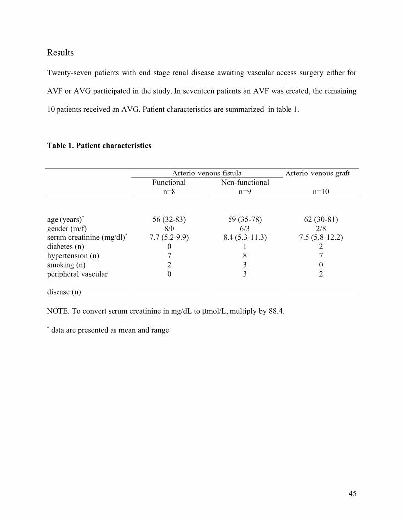
Twenty-seven patients with end stage renal disease awaiting vascular access surgery either for
AVF or AVG participated in the study. In seventeen patients an AVF was created, the remaining
10 patients received an AVG. Patient characteristics are summarized in table 1.
Table 1. Patient characteristics
Arterio-venous fistulaFunctional Non-functional
Arterio-venous graft
n=8 n=9 n=10
age (years)* 56 (32-83) 59 (35-78) 62 (30-81)gender (m/f) 8/0 6/3 2/8serum creatinine (mg/dl)* 7.7 (5.2-9.9) 8.4 (5.3-11.3) 7.5 (5.8-12.2)diabetes (n) 0 1 2hypertension (n) 7 8 7smoking (n) 2 3 0peripheral vascular
disease (n)
0 3 2
NOTE. To convert serum creatinine in mg/dL to µmol/L, multiply by 88.4.
* data are presented as mean and range
45
Venous distensibility
VD of patients receiving AVG (n=10) was 0.44±0.05 mL/mmHg and 0.56±0.04 mL/mmHg in
patients receiving AVF (n=17)(p=0.2). VD in patients with an unsuccessful AVF eight weeks
after surgery (AVF-, n=9) was 0.46±0.03 mL/mmHg, while in patients with a successful AVF
(AVF+, n=8) VD was 0.66±0.05 mL/mmHg (p=0.003, Figure 1). The mean duration of
hospitalization was 5.3 days (range 1-21 days) in AVF- and 3.6 days (1-8 days) in AVF+
(p=0.53).
Figure 1 shows that all 7 patients with VD of 0.50 mL/mmHg or lower had a non-functional AVF
(100%). Only 2 out of 10 patients with VD higher than 0.50 mL/mmHg had a non-functional
AVF (20%). This difference was statistically significant by Fisher’s exact test (p=0.002).
Application of this criterion yielded a sensitivity of 100% (8/8), a specificity of 78% (7/9), a
positive predictive value of 80% (8/10) and a negative predictive value of 100% (7/7). The
outcome of 15 patients was correctly predicted by VD (88%).
Two patients with non-functional AVF had VD above 0.50 mL/mmHg. Angiography showed a
severe venous stenosis a few centimeters from the anastomosis in both AVF. A pseudoaneurysm
was seen at the stenotic region in one AVF. Both AVF’s were surgically revised 3 months after
the first operation. One AVF could be used for hemodialysis after revision, the other occluded.
Of the other 7 non-functional AVF 3 never matured: these patients went for secondary access
surgery. Four AVF’s could be cannulated for hemodialysis only after 100 days (range 84-132
days). Angiography showed an arterial stenosis in one patient and a venous stenosis and with
extended collateral circulation in another. After 3 percutaneous transluminal angioplasty (PTA)
procedures these AVF could be used for dialysis. The remaining 2 AVF showed no significant
stenoses at angiography and finally matured.
46
No differences were found in the brachial and radial artery and cephalic vein luminal diameters
between AVF+ and AVF- . No significant correlation was found between VD and cephalic vein
diameter. Results of preoperative duplex ultrasonography are depicted in more detail in table 2.
Table 2. Duplex ultrasonography data of patients with AVF
Functional AVF Non-functional AVFn=8 n=9
Luminal diameter (mm)* • Radial artery 2.1±0.1 2.0±0.2 Ns• Brachial artery 4.3±0.4 4.1±0.3 Ns• Cephalic vein 1.9±0.4 1.9±0.2 Ns• Subclavian vein 8.0±1.3 8.5±0.7 Ns
Brachial artery flow (mL/min)* 44.1±13.8 34.3±8.0 Ns
* data are presented as mean±sd
47
Discussion
This prospective study evaluates venous forearm function rather than anatomy prior to AVF
creation for the first time. The results of this study support our hypothesis that apart from
structural changes, the functional properties of forearm veins are important in the adaptive
response to increased blood flow after AVF creation. Furthermore, functional data of forearm
venous vasculature do not correspond with anatomical data. Our data suggest that VD predicts
successful AVF maturation, whereas venous and arterial diameters do not.
Previous studies found that in patients with end-stage renal failure forearm veins are less
distensible in comparison to healthy controls (22,25). Several authors reported increased venous
intimal and media thickness in renal failure (26,27). Wali et al. showed accumulation of collagen
fibers in the vein wall (27). Also, venous wall edema may account for decreased venous
distensibility (28). The importance of endothelial function in arterial vasodilatation after AVF
creation is demonstrated by several authors (15-19). In addition, Tronc et al. (15) demonstrated
less venous dilatation in response to increased blood flow in L-NAME treated rabbits, suggesting
a critical role of the endothelial cells in venous remodeling as well. However, the influence of the
endothelium on VD remains to be elucidated.
Studies on the use of ultrasound prior to AVF creation have shown varying results and many
have used AVF patency as primary outcome rather than the ability to provide adequate blood
flow for hemodialysis (13,14). Our study could not demonstrate differences in success rates at
any cut-off point for radial artery or cephalic vein diameter. This is in contrast with the a post hoc
analysis of Wong et al., who showed that luminal radial artery or cephalic vein diameter of less
than 1.6 mm was associated with AVF failure (14). Actually, only 6 out of 60 patients had vessel
diameters this small. Malovr et al described a significant difference in success rate of patients
48
with a radial artery diameter of 1.5 mm or less (45%) and patients with a radial artery diameter
above 1.5 mm (92%)(13). However, definition of AVF failure was not provided in this study.
This difference in study results can, at least in part, be explained by our study design, which
selected patients with a radial artery diameter under 1.5 mm for AVG. The question whether
these patients can undergo AVF creation successfully, cannot be answered definitively.
Numerous studies have evaluated the utility of Doppler ultrasound access flow in the immediate
postoperative period subsequent to AVF creation (6,14,29-31). Although several authors
(6,29,30) demonstrated higher intraoperative access flow in successful AVF, others failed to
show any correlation between intraoperative access flow and outcome (14,31). This difference is
frequently attributed to vessel spasm during operation, resulting in poor fistula flow. This would
explain the better correlation between 1-day postoperative access flow and AVF outcome as
demonstrated by Wong et al. (14). In our study, day one postoperative duplex flow was measured
in 12 patients, and was also found to be higher in functional AVF (n=6) when compared to non-
functional AVF (n=6), however large variations were found within both groups, limiting its
predictive value in clinical practice (data not shown).
The majority of female patients received AVG in our study. Several studies have reported that
female patients are much less likely to dialyze with a fistula (2,32,33). VD was significantly
higher in males (p=0.0002). Also, female patients were found to have smaller radial arteries and
cephalic veins (data not shown). Thus, both functional and anatomical differences must be
responsible for this observation.
Our study has certain limitations. Because of the limited number of patients, the effect of
confounding factors on our results could not be evaluated. Indeed, factors like peripheral vascular
disease, female gender and diabetes may have influenced VD in a negative way in the group of
patients with non-functional AVF’s. However, currently these factors are never used to select
49
access type in daily clinical practice. VD, on the other hand, possibly reflects endothelial
dysfunction in these patients, and may be used as a practical tool to estimate the risk of AVF
failure in the individual patient.
The time required for fistula maturation varies among patients. We have chosen an arbitrary time
period of 8 weeks in our study, which is in agreement with DOQI guidelines (3). The Work
Group does not advise use of the fistula within the first month after construction because
premature cannulation of a fistula may result in a higher incidence of infiltration with associated
compression of the vessel by hematoma and permanent loss of the fistula. Allowing the fistula to
mature for 3 months before use may be ideal. However, the Work Group did not reach consensus
on this topic. In fact, allowing 3-month maturation would not have changed our results, since
final cannulation in the 5 patients in the non-functional AVF group, was only possible after 100
days, usually after PTA or surgical revision. Furthermore, patients with functional AVF’s were
cannulated after 41 days.
Considering our data provided proof of the principle that functional vessel wall characteristics
predict fistula maturation, the preoperative evaluation of the forearm vasculature should not only
focus on resting static diameters. In view of the large potential benefits of optimization of the
matching of patients and access type, further and larger studies of novel techniques are urgently
needed.
50
References
1. Churchill DN, Taylor W, Cook RJ, LaPlante P, Barre P, Cartier P, Fay WP, Goldstein MB,
Jindal K, Mandin H, McKenzie JK, Muirhead N, Parfrey PS, Posen GA, Slaughter D, Ulan
RA, Werb R.: Canadian hemodialysis morbidity study. Am J Kidney Dis 1992;14:214-34
2. Pisoni RL, Young EW, Dykstra DM, Greenwood RN, Hecking E, Gillispie B, Wolfe RA,
Goodkin DA, Held PJ: Vascular access use in Europe and the United States: results from the
DOPPS. Kidney Int 2002; 61:305-16
3. National Kidney Foundation: K/DOQI clinical practice guidelines for vascular access. Am J
Kidney Dis 2001; 37(Suppl 1): S137-S181
4. Kinnaert P, Vereerstraeten P, Toussaint C, Van Geertruyden J: Nine years' experience with
internal arteriovenous fistulas for haemodialysis: a study of some factors influencing the
results. Br J Surg 1977; 64:242-6
5. Windus DW: Permanent vascular access: a nephrologist's view. Am J Kidney Dis 1993;
21:457-71
6. Elfström J, Thomsen M: The prognostic value of blood-flow measurements during
construction of arteriovenous fistulae. Scand J Urol Nephrol 1981; 15(3):323-26
7. Kalman PG, Pope M, Bhola C, Richardson R: A practical approach to vascular access for
hemodialysis and predictors of success. J Vasc Surg 1999; 30:727-33
8. Silva MB, Hobson RW, Pappas PJ, Jamil Z, Araki T, Goldberg MC, Gwertzman G, Padberg
FT: A strategy for increasing use of autogenous hemodialysis access procedures: impact of
preoperative non-invasive evaluation. J Vasc Surg 1998; 27:302-8
51
9. Yerdel MA, Kesenci M, Yazicioglu KM, Döşeyen Z, Türkçapar AG, Anadol E: Effect of
haemodynamic variables on surgically created arteriovenous fistula flow. Nephrol Dial
Transplant 1997; 12:1684-88
10.Lin SL, Huang CH, Chen HS, Hsu WA, Yen CJ, Yen TS: Effects of age and diabetes on
blood flow rate and primary outcome of newly created hemodialysis arteriovenous fistulas.
Am J Nephrol 1998; 18:96-100
11.Lazarides MK, Iatrou CE, Karanikas ID, Kaperonis NM, Petras DI, Zirogiannis PN, Dayantas
JN: Factors affecting the lifespan of autologous and synthetic arteriovenous access routes for
haemodialysis. Eur J Surg 1996; 162:297-301
12. Malovrh M: Native arteriovenous fistula: preoperative evaluation. Am J Kidney Dis 2002;
39:1218-25
13.Malovrh M: Non-invasive evaluation of vessels by duplex sonography prior to construction
of arteriovenous fistulas for hemodialysis. Nephrol Dial Transplant 1998; 13:125-9
14.Wong V, Ward R, Taylor J, Selvakumar S, How TV, Bakran A: Factors associated with early
failure of arteriovenous fistulae for haemodialysis access. Eur J Endovasc Surg 1996; 12:207-
13
15. Tronc F, Wassef M, Esposito B, Henrion D, Glagov S, Tedgui A: Role of NO in flow-
induced remodeling of the rabbit common carotid artery. Arterioscler Thromb Vasc Biol
1996; 16:1256-62
16. Tronc F, Mallat Z, Lehoux S, Wassef M, Esposito B, Tedgui A: Role of metalloproteinases in
blood flow-induced arterial enlargement: interaction with NO. Arterioscler Thromb Vasc Biol
2000; 20:120-6
17. Tuttle JL, Nachreiner RD, Bhuller AS, Condict KW, Connors BA, Herring BP, Dalsing MC,
52
Unthank JL: Shear level influences resisance artery remodeling: wall dimensions, cell
density, and eNOS expression. Am J Physiol Heart Physiol 2001; H1380-9
18. Masuda H, Zhuang YJ, Singh TM, Kawamura K, Murakami M, Zarins CK, Glacov S:
Adaptive remodeling of internal elastic lamina and endothelial lining during flow-induced
arterial enlargement. Arteriol Thromb Vasc Biol 1999; 19:2298-307
19. Guzman RJ, Abe K, Zarins CK: Flow-induced arterial enlargement is inhibited by
suppression of nitric oxide synthase activity in vivo. Surgery 1997; 122: 273-9
20. Anderson TJ: Assessment and treatment of endothelial dysfunction in humans. J Am Coll
Cardiology 1999; 34:631-8
21. London GM, Marchais SJ, Guerin AP, Metivier F, Adda H: Arterial structure and function in
end-stage renal disease. Nephrol Dial Transplant 2002; 17:1713-24
22. Kooman JP, Wijnen JA, Draaijer P, van Bortel LM, Gladziwa U, Peltenburg HG, Struyker-
Boudier HA, van Hooff JP, Leunissen KM: Compliance and reactivity of the peripheral
venous system in chronic intermittent hemodialysis. Kidney Int 1992; 41:1041-8
23. Christ F, Gamble J, Baschnegger H, Gartside IB: Relationship between venous pressure and
tissue volume during venous congestion plethysmography in man. J Physiol 1997; 503:463-7
24. Halliwill JR, Minson CT, Joyner MJ: Measurement of limb venous compliance in humans:
technical considerations and physiological findings. J Appl Physiol 1999; 87:1555-63
25. Bradley JR, Evans DB, Cowley AJ: Abnormalities of the peripheral circulation in patients
with chronic renal failure. Nephrol Dial Transplant 1988; 3:412-6
26. Kooman JP, Daemen MJ, Wijnen R, Verluyten-Goessens MJ, van Hooff JP, Leunissen KM:
Morphological changes of the venous system in uremic patients. A histopathologic study.
Nephron 1995; 69:454-8
27. Wali MA, Eid RA, Al-Homrany MA: Smooth muscle changes in the cephalic vein of renal
53
failure patients before use as an arteriovenous fistula (AVF). J Smooth Muscle Res 2002;
38:75-85
28. Simon G, Pamnani MB, Overbeck HW: Decreased venous compliance in dogs with chronic
renal hypertension. Proc Soc Exp Biol Med 1976; 152:122-5
29. Johnson CP, Zhu Y, Matt C, Pelz C, Roza AM, Adams MB: Prognostic value of
intraoperative blood flow measurements in vascular access surgery. Surgery 1998; 124:729-
38
30. Won T, Jang JW, Lee S, Han JJ, Park YS, Ahn JH: Effects of intraoperative blood flow on
the early patency of radiocephalic fistulas. Ann Vasc Surg 2000; 14:468-72
31. Anderson CB, Etheredge EE, Harter HR, Graft RJ, Codd JE, Newton WT: Local blood flow
characteristics of arteriovenous fistulas in the forearm for dialysis. Surg Gynaecol Obstet
1977; 144:531-3
32. Ifudu O, Macey LJ, Homel P, Hyppolite JC, Hong J, Sumrani N, Distant D, Sommer BG,
Friedman EA: Determinants of type of initial hemodialysis vascular access. Am J Nephrol
1997; 17:425-7
33. Allon M, Ornt DB, Schwab SJ, Rasmussen C, Delmez JA, Greene T, Kusek JW, Martin AA,
Minda S: Factors associated with the prevalence of arteriovenous fistulas in hemodialysis
patients in the HEMO study. Hemodialysis (HEMO) Study Group. Kidney Int 2000; 58:2178-
85
54
Chapter 4
ROLE OF FOREARM BLOOD FLOW RESERVE IN EARLY
FAILURE OF NEWLY CREATED ARTERIO-VENOUS
HEMODIALYSIS FISTULAE
Joke van der Linden
Thomas W. Lameris
Anton H. van den Meiracker
André A.E.A de Smet
Peter J. Blankestijn
Marinus A. van den Dorpel
55
Abstract
Background. The autologous AV-fistula is the vascular access of choice in hemodialysis patients
because of superior long-term survival due to a lower risk for thrombosis and infection. Access
surgery should be followed by adequate fistula maturation, i.e., a gradual increase of vessel
diameter, and thus increase in access flow. We hypothesized that preoperative measurement of
the arterial forearm blood flow (FBF) reserve predicts successful fistula development.
Methods and patients. Prior to surgery, we measured FBF during incremental infusion of both an
endothelium-dependent vasodilator, metacholine (MCh), and an endothelium-independent
vasodilator, sodium nitroprusside (SNP), using venous occlusion plethysmography. Twenty-one
patients (age 62 3, (mean sem), 10M /11F) with end-stage renal failure awaiting access surgery
were studied.
Results. In patients with successful AVF maturation (n=17) the AUC of both vasodilators was
higher than in patients with AVF failure (n=4)(MCh: 122.2 9.8 U vs. 79.1 5.8, p<0.05; SNP:
255.9 19.4 vs. 127.8 29.4, p<0.01). The absolute FBF increase was also significantly different
between both groups (MCh:10.3 0.9 vs. 6.2 1.0 mL/100mL/min, p=0.03; SNP: 11.8 1.1 vs. 5.4
1.4 mL/100mL/min, p<0.01). No AVF failure was observed when AUC was above 100 U after
MCh (n=11) (p<0.05), or when AUC was above 200 U after SNP (n=13) (p<0.01). Baseline
MAP and FBF was not different (107 4 vs. 109 5 mmHg, N.S.; 3.4 0.7 vs.4.6 0.4, N.S.). Duplex
radial artery diameter did not differ between patients with successful or unsuccessful AVF
maturation (2.4 0.2 vs. 2.8 0.3, N.S.).
Conclusions. We conclude that with measurement of FBF reserve, successful or unsuccessful
AVF maturation can be predicted accurately. Endothelial function does not seem to play a role, as
56
endothelium-dependent and -independent vasodilators had similar effects. In addition to
anatomical mapping, functional preoperative evaluation may improve AVF outcome.
Introduction
Adequate blood flow through an arterio-venous access is mandatory for successful hemodialysis
in patients with end-stage renal disease (ESRD). According to DOQI-guidelines, the native
radiocephalic arterio-venous fistula (AVF) as described by Brescia and Cimino, is the access of
first choice (1). Patency rates of AVF are clearly superior when compared to the outcome of
arterio-venous grafts (AVG), which is the result of much less thrombotic and infectious events in
AVF. However, it is known that up to 15% of all newly created AVF fail shortly after surgery as
a result of thrombosis. These so called early failures are frequently regarded as technical errors,
although several hemodynamic parameters may also play a role.
After creation of an AVF for hemodialysis, blood flow through the radial artery will increase as a
result of vasodilatation and vascular remodeling. Girerd et al. found a 1.4 fold increase in arterial
diameter while the intima media thickness remained unchanged, clearly suggesting a remodeling
of the arterial wall (2). This adaptive response appears to be crucial in reducing wall shear stress
to baseline values. Major calcification and stiffening of the radial artery wall will inhibit proper
vasodilatation, which will lead to inadequate arterial inflow of the AVF. Vasodilatation after
AVF-creation is primarily caused by acute release of nitric oxide by endothelial cells, so-called
endothelium-dependent vasodilatation. Also, endothelial cells play a central role in vascular
remodeling. This is illustrated by the observation that endothelial denudation and inhibition of
NO by L-NAME abolish arterial enlargement in animal models of arterio-venous fistula (3-5).
57
Endothelium-dependent vasodilatation of forearm capacitance vessels is affected by many
diseases, among which several are known to cause renal failure, like diabetes and hypertension
(6). Also, in patients with different stages of renal failure defective endothelial vasodilator
function has been demonstrated (7).
We hypothesized that the forearm blood flow reserve, i.e. the increase of forearm blood flow as a
result of vasodilatation, is an important determinant of failure of newly created AVF in
hemodialysis patients. Therefore, we determined whether forearm blood flow reserve in patients
with end-stage renal failure awaiting vascular access surgery, was predictive of early failure. To
discriminate the influence of the endothelium in early fistula failure, we measured both,
endothelium dependent and endothelium independent forearm vasodilatation, using forearm
venous occlusion plethysmography. Duplex radial artery diameter will be evaluated in order to
find any relationship between functional and anatomical data. Furthermore, the number of
surgical or percutaneous procedures needed to obtain a functional AVF was determined.
58
Subjects and methods
Subjects
Patients with end-stage renal disease (ESRD) requiring hemodialysis and awaiting AVF creation
were included after written informed consent. The trial protocol was approved by the institute’s
Medical Ethics Committee. Based on physical examination and preoperatively determined duplex
parameters, the vascular surgeon decided the feasibility of creating an AVF in the individual
patient. Patients with no visible and/or a tortuous short cephalic vein and/or radial artery diameter
less than 1.5 mm received an AVG and did not participate in the study. The patients were asked
to refrain from smoking and caffeine or alcohol containing beverages for at least 12 hours prior to
the study. All vasoactive medication was discontinued 7 days before the study. Within one month
after Duplex ultrasonography and forearm strain gauge plethysmography patients underwent
vascular access surgery.
AVF which could be used for successful two needle hemodialysis, judged by a panel of
experienced dialysis nurses who were unaware of the results of all additional forearm studies,
were defined as functional AVF. Successful dialysis was defined as the ability to provide at least
250 mL/min dialyzer pump flow. After surgery, all patients received anticoagulant therapy
(acenocoumarol) for six months.
Methods
Duplex ultrasonography
Pre-operative duplex scanning of the upper extremity was performed with the HDI 3000
Ultrasound System (ATL Ultrasound Bothell, USA). A 2-D linear electronic probe, pulse wave
Doppler and color wave Doppler at 5.0 MHz were used. The internal diameter of the radial artery
59
at the wrist was measured using M-mode technique. A tourniquet to increase vascular size was
not used. The patency of both the proximal cephalic and subclavian vein was examined.
Four weeks after surgery Duplex ultrasonography was performed to examine the AVF for
development of stenotic lesions. Patients with significant stenotic lesions (>50% of the luminal
diameter) were referred for percutaneous transluminal angioplasty within 2 weeks.
Forearm strain-gauge plethysmography
Venous occlusion strain-gauge plethysmography is a frequently used technique for measurement
of forearm blood flow and venous compliance (8). Throughout the years the method has been
standardized and computerized, resulting in a reasonably simple and reliable technique (9-11). It
works on the principle that during short-term occlusion of venous return, the rate of distension of
the forearm is proportional to the rate of arterial inflow into the forearm. Provided that the arterial
blood pressure remains constant, changes in flow reflect changes in smooth muscle tone in small
arteries and arterioles.
The experiments were performed in the afternoon in quiet air-conditioned room at constant
temperature (20-24ºC). Subjects were studied in supine position with arms supported 10 cm
above the level of the right atrium. The arm chosen for vascular access surgery, which was the
non-dominant arm in the majority of subjects, was used for measurements. Forearm volume was
measured by water displacement. The brachial artery was cannulated (cannula of 1.0 × 45 mm)
after local anesthesia with lidocaine (1%). A mercury-in-silastic strain gauge was positioned
around the widest part of the forearm. The strain gauge was connected to a plethysmograph
(model periflow SU 4, Janssen Scientific Instruments, Beerse, Belgium) with electronic
calibration for percentual volume changes and a built-in flow integrator module. To enable off-
60
line data analysis, the plethysmograph was connected to an A/D converter (Dataq Instruments,
model DI 420, Akron, OH, USA) for electronic data storage. A venous occlusion cuff was placed
around the upper arm. Inflation was achieved using an ECG-triggered rapid cuff inflator (Janssen
Scientific Instruments), inflating the cuff to 60 mmHg during 4 heartbeats and deflating it during
the next 3 heartbeats.
Baseline FBF (mL /100ml forearm/min) (FBFBL) was measured after a resting period of at least
30 minutes after intra-arterial cannulation of the brachial artery. During this measurement saline
was infused at 1 mL/min. Then, FBF was measured during incremental infusions (dose range
0.1-10 µg/kg/min) of metacholine (MCh), to examine endothelium-dependent vasodilatation.
Each dosing step lasted 4 minutes. Sodium nitroprusside (SNP) was used (dose range 1-20 µ
g/kg/min) at 4-minute dosing steps to examine endothelium-independent vasodilatation. Between
the various experiments sufficient time was allowed for FBF to return to baseline levels. Mean
arterial blood pressure (MAP) and heart rate were measured continuously using the Finapress
blood pressure monitor (Ohmeda, Inglewood, CO, USA).
Statistical analysis
Results are given as means±S.E.M. Dose-response curves were constructed for the vasodilator
effects of MCh and SNP. Absolute change in FBF (dFBFABS), area under curve (AUC) for MCh
and SNP and Duplex radial artery diameter were compared in patients with and without
functional AVF, using Students’ t-tests. P-values below 0.05 were considered statistically
significant.
61
Results
Twenty-one consecutive patients with ESRD awaiting AVF creation were included into the
study. All patients received an brachio-cephalic forearm fistula. In 4 patients the AVF occluded
(AVF-NF), 3 of which in the first week after surgery. The remaining 17 AVF matured enough to
enable 2-needle dialysis, and were functional after 78±11 days (range 43-157, AVF-F). Six AVF
developed one or more significant stenotic lesions. Most lesions (6/11) were found in the venous
outflow tract of the AVF, 3 were located at the anastomosis and the other 2 were arterial stenoses.
After percutaneous transluminal angioplasty these AVF matured and were successfully
cannulated for hemodialysis. The remaining 11 patients had a functional AVF within eight
weeks. Duplex ultrasonography 4 weeks after surgery could not demonstrate any significant
stenoses. Patient characteristics are depicted in Table 1.
62
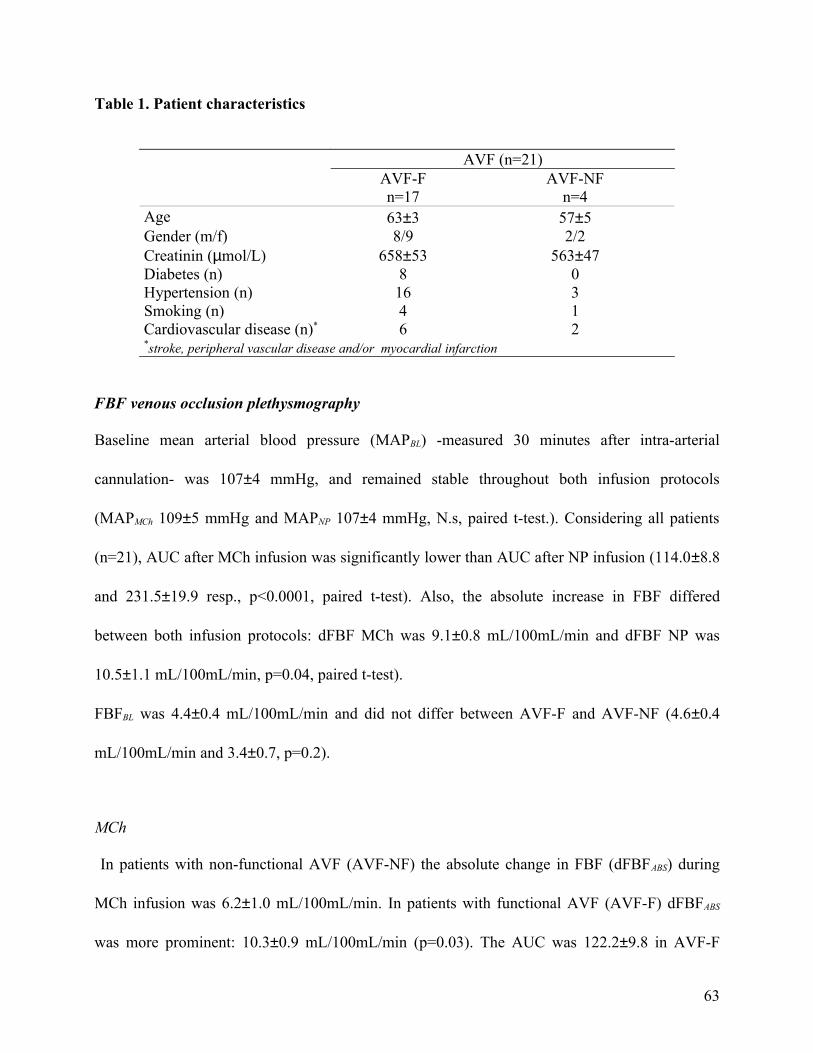
Table 1. Patient characteristics
AVF (n=21)AVF-F AVF-NFn=17 n=4
Age 63±3 57±5Gender (m/f) 8/9 2/2Creatinin (µmol/L) 658±53 563±47Diabetes (n) 8 0Hypertension (n) 16 3Smoking (n) 4 1Cardiovascular disease (n)* 6 2*stroke, peripheral vascular disease and/or myocardial infarction
FBF venous occlusion plethysmography
Baseline mean arterial blood pressure (MAPBL) -measured 30 minutes after intra-arterial
cannulation- was 107±4 mmHg, and remained stable throughout both infusion protocols
(MAPMCh 109±5 mmHg and MAPNP 107±4 mmHg, N.s, paired t-test.). Considering all patients
(n=21), AUC after MCh infusion was significantly lower than AUC after NP infusion (114.0±8.8
and 231.5±19.9 resp., p<0.0001, paired t-test). Also, the absolute increase in FBF differed
between both infusion protocols: dFBF MCh was 9.1±0.8 mL/100mL/min and dFBF NP was
10.5±1.1 mL/100mL/min, p=0.04, paired t-test).
FBFBL was 4.4±0.4 mL/100mL/min and did not differ between AVF-F and AVF-NF (4.6±0.4
mL/100mL/min and 3.4±0.7, p=0.2).
MCh
In patients with non-functional AVF (AVF-NF) the absolute change in FBF (dFBFABS) during
MCh infusion was 6.2±1.0 mL/100mL/min. In patients with functional AVF (AVF-F) dFBFABS
was more prominent: 10.3±0.9 mL/100mL/min (p=0.03). The AUC was 122.2±9.8 in AVF-F
63
patients and 79.1±5.8 in AVF-NF patients (p=0.045). Furthermore, all 4 failures had dFBFABS and
AUC values lower than the average dFBFABS and AUC values of the whole series (9.1
mL/100mL/min and 114 respectively, Figure 1.). No fistula failure was seen in patients with
dFBF ABS ≥ 10 mL/100mL/min (0/9). Four failures occurred in 12 patients with dFBF ABS < 10
mL/100mL/min. This difference was insignificant by Fisher’s exact test (p=0.1). It is noteworthy
that all 4 fistula failures occurred in 10 patients with AUC < 100, while no failures were seen in
11 patients with AUC ≥ 100. This difference was statistically significant by Fisher’s exact test
(p=0.04).
NP
The dFBFABS during NP infusion was significantly higher in AVF-F than in AVF-NF patients
(11.8±1.1 mL/100mL/min and 5.4±1.4, respectively, p=0.01). The AUC was 255.9±19.4 in AVF-
F and 127.8±29.4 in AVF-NF patients (p=0.008). Again, all non-functional AVF had lower
dFBFABS and AUC than the averages of the total group (10.5 mL/100mL/min and 231.5, Figure
1.). No differences in the risk of fistula failure were found between patients with dFBFABS <10
and patients with dFBFABS ≥10 mL/100mL/min. Of 8 patients with AUC <200, 50% of AVF
failed, while none of the AVF of patients with AUC ≥200 failed (13/13), which was a significant
difference (p=0.01, Fisher’s exact test).
Duplex radial artery diameter
No significant differences were found between radial artery diameter of patients with AVF-F and
AVF-NF (2.4±0.2 and 2.8±0.3 mm, respectively, p=0.2). Interestingly, all 4 non-functional
fistulae had radial artery diameters of at least 2.2 mm.
64
Discussion
Under ideal circumstances, in which a group of skilled vascular surgeons is responsible for
vascular access surgery, the success of a newly constructed AVF must depend on several
hemodynamic variables affecting the in- and outflow of the AVF. Since AVF construction is
directly followed by increased arterial blood flow, hypothetically, higher forearm blood flow
capacitance, should be associated with better outcome of AVF. To provide evidence for this
hypothesis, we measured forearm blood flow changes after infusion of the vasodilatant
substances MCh and SNP, using venous occlusion plethysmography. To our knowledge, this is
the first time invasively measured forearm blood flow capacity is related to outcome of AVF.
This study shows that pre-operative forearm blood flow capacity is predictive of early AVF
failure. Furthermore, our data indicate that AVF failure is not the result of pure endothelial
dysfunction, because endothelial-independent forearm blood flow capacity is also diminished in
patients with AVF failure. Finally, we showed that arterial diameter is not predictive of early
AVF failure.
We demonstrated that patients with insufficient forearm blood flow capacity are likely to
encounter primary AVF failure. Especially, the AUC of dose response curves seems predictive of
AVF outcome. An AUC of 100 in MCh and 200 in NP seems a prerequisite of short term
success of the AVF. To our knowledge forearm blood flow capacity using venous occlusion
plethysmography was never examined as a determinant of fistula failure. However, Malovr et al.
demonstrated a significantly lower arterial blood flow increase and higher vascular resistance
after reactive hyperemia after reopening a clenched fist in patients with nonfunctional AVF in
compare to patients with functional AVF (12,13). In agreement with Yerdel et al., baseline
forearm blood (14) did not differ between patients with functional and not-functional AVF.
65
Therefore, differences in concentration of drug reaching the tissues, influencing the response to
it, can be neglected.
We also hypothesized that the endothelium, responsible for direct nitric oxide release after AVF
construction, is of importance in immediate AVF failure. Our data indeed demonstrate
disturbance of MCh induced vasodilatation with relative preservation of NP induced
vasodilatation, indicating endothelial dysfunction in ESRD. However, both determinants (AUC
MCh and AUC NP) were found to be predictive of immediate AVF failure, suggesting structural
arterial wall changes besides endothelial dysfunction. Indeed, in a recent study preoperative
Duplex evaluation demonstrated severe arterial wall changes – wall thickening and calcification-
in almost 40% of all patients awaiting vascular access surgery (13). Since our patients are
comparable with respect to severity of renal dysfunction, it is reasonable to believe that these
structural changes are also of importance in our study population.
In the past, several studies demonstrated worse AVF outcome in women and elderly (15-17).
Because of small numbers, our data do not allow multivariate analysis of these clinical variables,
but we found no differences in age and gender between patients with functional and non-
functional AVF. It is possible that our study design, which selected patients with a radial artery
diameter less than 1.5 mm for AVG, has eliminated mostly women and elderly from the study.
However, within the group with functional AVF, almost 90% of interventions for stenotic lesions
took place in women older than 65 years.
With the advent of mercury-in- rubber strain gauges, the technique of forearm venous occlusion
plethysmography is accurate and widely used to probe mechanisms of human vascular control
Venous occlusion plethysmography has the advantage of measurement of total forearm blood
flow. However, clinical implementation of venous occlusion plethysmography, prior to access
surgery, is not simple. First, this technique is very time-consuming and requires expensive hard-
66
and software. Second, and even more important, it is an invasive measurement. Although we
didn’t encounter any complications in our study, theoretically, formation of haematoma could
interfere with arterial inflow. In vessels with relatively small diameter, such as the radial artery,
estimation of volume flow by duplex sonography, using cross-sectional area, is inherently
inaccurate. However, Malovr et al. described an alternative approach to measure blood flow
capacitance in the radial artery (12). After releasing a fist the triphasic Doppler waveform in the
radial artery should normally change to a low-resistance biphasic waveform, caused by post-
ischemic dilatation of peripheral arterioles and consecutive reduction of peripheral resistance.
Patients who lacked this conversion showed significantly more AVF failure. It would be of
interest to study both techniques within one patient to determine the correlation of both
modalities.
Although the number of patients is small, necessitating further prospective trials, our study
provides valuable information on the influence of arterial inflow capacitance on fistula failure. Of
course, arterial inflow is not the only factor in fistula maturation, and always depends on
sufficient outflow as well. Considering the amount of stenoses found in the outflow tract at post-
operative Duplex-examination, it is possible that inappropriate venous distensibility is of
importance in the maturation of AVF that survive the first period after surgery. Ideally, the
measurement of pre-operative forearm blood flow capacitance may direct the operation to be
performed under more suitable conditions, or may result in primary placement of graft material,
in order to overcome fistula failure in patients with unsuitable vascular anatomy. In order to use
our findings in daily clinical practice, future studies should focus on easier, non-invasive ways to
examine the role of forearm blood flow capacity in early fistula failure.
67
References
1. National Kidney Foundation. K/DOQI clinical practice guidelines for vascular access. Am J
Kidney Dis 2001; 37 (Suppl 1):S137-S181
2. Girerd X, London G, Boutouyrie P, Mourad JJ, Safar M, Laurent S: Remodeling of the radial
artery in response to a chronic increase in shear stress. Hypertension 1996; 27:799-803
3. Tohda K, Masuda H, Kawamura K, Shozawa T: Difference in dilatation between
endothelium-preserved and –desquamated segments in the flow-loaded rat common carotid
artery. Arterioscler Thromb 1992; 12:519-28
4. Guzman RJ, Abe K, Zarins CK: Flow-induced arterial enlargement is inhibited by
suppression of nitric oxide synthase activity in vivo. Surgery 1997; 122:273-80
5. Tronc F, Wassef M, Esposito B, Henrion D, Glagov S, Tedgui A: Role of NO in flow-
induced remodeling of the rabbit common carotid artery. Arterioscler Thromb Vasc Biol
1996; 16:1256-62
6. Anderson TJ: Assessment and treatment of endothelial dysfunction in humans. J Am Coll
Cardiology 1999; 34:631-8
7. London GM, Marchais SJ, Guerin AP, Metivier F, Adda H: Arterial structure and function in
end-stage renal disease. Nephrol Dial Transplant 2002; 17: 1713-24
8. Lind L, Sarabi M, Millgård J: Methodological aspects of the evaluation of endothelium-
dependent vasodilatation in the human forearm. Clin Physiol 1998; 18(2):81-7
9. Petrie JR, Ueda S, Morris AD, Murray LS, Elliot HL, Connell JMC: How reproducible is
bilateral forearm plethysmography? Br J Clin Pharmocol 1998; 45:131-9
10. Chang PC, Van Brummelen P: Calibration and variability of forearm blood flow measured by
strain gauge plethysmography. J Cardiovasc Pharmocol 1987; 10: S123-5
68
11.Chang PC, Verlinde R, Bruning TA, Van Brummelen P: A micro-computer-based, R-wave
triggered system for hemodynamic measurements in the forearm. Comput Biol Med 1988;
18:157-63
12. Malovrh M: Non-invasive evaluation of vessels by duplex sonography prior to construction
of arteriovenous fistulas for hemodialysis. Nephrol Dial Transplant 1998; 13:125-29.
13. Malovrh M: Native arteriovenous fistula: preoperative evaluation. Am J Kidney Dis 2002;
39:1218-25
14. Yerdel MA, Kesenci M, Yazicioglu KM, Döşeyen Z, Türkçapar AG, Anadol E: Effect of
haemodynamic variables on surgically created arteriovenous fistula flow. Nephrol Dial
Transplant 1997; 12: 1684-8
15. Miller CD, Robbin ML, Allon M: Gender differences in outcomes of arteriovenous fistulas in
hemodialysis patients. Kidney Int 2003; 63:346-52
16. Wong V, Ward R, Taylor J, Selvakumar S, How TV, Bakran A: Factors associated with early
failure of arteriovenous fistulae for haemodialysis access. Eur J Endovasc Surg 1996; 12:207-
13
17.Tordoir JHM, Hoeneveld H, Eikelboom BC, Kitslaar PJEHM: The correlation between
clinical and Duplex ultrasound parameters and the development of complications in arterio-
venous fistulae for haemodialysis. Eur J Vasc Surg 1990; 4:179-84
69

Chapter 5
GRAFT SURVEILLANCE: VENOUS PRESSURE, ACCESS
FLOW, OR THE COMBINATION?
Kidney Int 2001 Apr;59(4):1551-8
Joke van der Linden
Johannes H.M. Smits
E. Chris Hagen
Esther C. Modderkolk-Cammeraat
Geert W. Feith
Hein A. Koomans
Marinus A. van den Dorpel
Peter J. Blankestijn
70
Abstract
Background. Increased venous pressure (VP) and decreased access flow (Qa) are predictors of
dialysis access graft thrombosis. VP is easily obtainable. Qa assessment requires a special device
and takes more time. Aims of our randomized multi-center studies were to compare outcome in
patients with grafts monitored by VP or Qa (study A) or monitored by VP or the combination of
VP and Qa (study B).
Methods. We performed VP measurements that consisted of weekly VP at a pumpflow of 200
mL/min (VP200) and the ratio of VP0/MAP. Qa was measured every 8 weeks with the Transonic
HD01 hemodialysis monitor. Threshold levels for referral for angiography were VP200 >150
mmHg or VP0/MAP >0.5 (both at 3 consecutive dialysis sessions), or Qa <600 mL/min.
Subsequent therapy consisted of either PTA or surgery.
Results. Total follow-up was 80.5 patient-years for 125 grafts. The vast majority of a total of 131
positive tests was followed by angiography and corrective intervention. In study A, the rate of
thromboses not preceded by a positive test was 0.19 and 0.24 per patient-year (P = NS) and in
study B it was 0.32 versus 0.28 per patient-year (P = NS). Survival curves were not significantly
different between the subgroups.
Conclusions. These data demonstrate that standardized monitoring of either VP or Qa or the
combination of both and subsequent corrective intervention can reduce thrombosis rate in grafts
to below the recommended quality of care standard (i.e. 0.5 per patient-year, NKF-DOQI). These
surveillance strategies are equally effective in reducing thrombosis rates.
71
Introduction
Thrombosis remains a major problem in vascular access for hemodialysis, particularly in
polytetrafluoroethylene (PTFE) grafts. It accounts for considerable morbidity and mortality with
an annual cost of close to $1 billion in the United States and is responsible for 17-25% of all
hospitalizations in dialysis patients (1-3).
Thrombosis occurs at a rate of 0.5 to 2.5 events per patient-year (4-8). In most cases thrombosis
is associated with the presence of stenoses at the venous anastomosis or in the outflow tract (9-
13). Stenosis increases resistance over the flow tract. Because the graft has no autoregulating
capacities, blood flow (Qa) drops and venous pressures (VP) rise. These variables have been
shown to predict thrombosis. More importantly, several studies demonstrated that referral for
corrective intervention based on these parameters can prevent thrombosis (14-18).
We and others confirmed that patients with outflow stenosis have on average a higher VP and/or
lower Qa (14,19). However, VP did not correlate with Qa. In other words not all patients with
high VP had low Qa, indicating that not all patients who are at risk for thrombosis can be
identified by VP measurements. We also showed that inflow resistance (that is resistance of the
flow tract upstream of the venous needle) comprises a substantial and very variable part of total
graft resistance. Indeed, several studies have indicated that in up to 29% of thrombosis cases,
stenoses may be located in the arterial part of the graft (13, 20-23). The inflow resistance is not
reflected by VP measurements, whereas Qa measurements are a reflection of total graft
resistance. This could make VP less effective as selection parameter for patients at risk for
thrombosis than Qa. In contrast to Qa measurements, VP can be measured by the dialysis
machine, is easy to obtain and requires little time investment. Furthermore, some studies have
convincingly indicated that the use of VP measurements as a selection variable for diagnostic and
72
subsequent corrective procedures, results in thrombosis rates between 0.2 and 0.4 events per
patient-year (6,14,16). Although we provided the theoretical basis that Qa measurements are
better than VP measurements, the question is whether Qa measurements really confer additional
benefit in patients who are monitored by VP. In other words, when simple clinical variables such
as VP are used, is there any additional benefit when periodic Qa measurements are added to the
surveillance protocol?
Our hypothesis for the present studies was as follows: Qa measurements are better than VP
measurements in identifying patients at risk for thrombosis. As a consequence: referral of patients
for corrective interventions based on Qa measurements alone or on the combination of VP and
Qa reduces thrombosis rate more than referral based on VP alone.
73
Methods
Patients
Five dialysis centers participated in this study. All patients with a hemodialysis access PTFE graft
were eligible to enter into the study. Exclusion criteria were inability to give informed consent
and contrast allergy.
Studies were approved by the Institutional Ethical Review Committees. Informed consent was
obtained from all patients.
Surveillance protocols
Two surveillance studies were conducted concurrently. In study A patients were assigned to
weekly VP measurements (Group A1) or periodic Qa measurements (Group A2). In study B,
patients were assigned to weekly VP measurements (Group B1) or the combination of VP
measurements and periodic Qa measurements (Group A2). Study A was instituted in one center
(Rotterdam) and study B in four. Within the centers the patients were prospectively and at
random allocated to one of the subgroups of each study (Fig. 1).
Venous pressure monitoring
Measurements were done once weekly, within the first half-hour of dialysis, and included both
dynamic and static VP measurements. When one or both measurements were positive (see
Indication for referral for angiography), measurements were repeated the next dialysis session.
All centers had dialysis machines with digital pressure displays. Maintenance and calibration was
done according to the manufacturer’s recommendations on a regular basis by an experienced
technician.
74
Dynamic venous pressure
Digitally displayed dynamic VPs were assessed with a fixed pump flow set at 200 mL/min
(VP200), as described by Schwab et al (16). Ten subsequent readings on the dialyzer display
were averaged. After the measurement the pump flow was set to the original level.
Static venous pressure
Static VP (VP0) was assessed with zero pump flow with the tubing out of the air lock in order to
avoid automatic closure of the tubing after the pump was shut off. VP0 was divided by mean
arterial pressure (MAP) to correct for blood pressure differences as described by Besarab et al
(14). No correction for height differences was made. MAP was calculated by taking two times
diastolic pressure plus systolic pressure divided by three.
Access flow measurement
Measurements were done every eight weeks. Qa was measured with the Transonic Hemodialysis
Monitor (Transonic Systems Inc, Ithaca, NY). The theoretical background, bench validation and
in vivo validation are described in detail in previous papers (24,25). Periodic calibration was
done by the local distributor. Qa determination consisted of the average of three consecutive
measurements. If Qa levels reached between 600 and 800 mL/min the measurement was repeated
after one month. All measurements were done with a dialyzer blood flow of more than 200
mL/min.
Indication for referral for angiography
Referral of Group A1 patients for angiography was based on three consecutive dialysis sessions
with elevated static and/or dynamic VP. Patients in Group A2 underwent angiography when Qa
75
fell below 600 mL/min. Indication for referral of Group B1 patients for angiography was an
elevated static and/or dynamic VP, similar to Group A1. In Group B2 an elevated static and/or
dynamic VP and/or Qa below 600 mL/min were reasons for referral. The threshold level for the
static VP ratio was 0.5, according to the method described by Besarab et al [14]. Threshold for
dynamic VP was 150 mmHg.
Angiography was done to determine presence and location of the stenosis. If a stenosis of >50%
was present, the primary choice of treatment was percutaneous transluminal angioplasty (PTA).
After an intervention patients continued the same surveillance mode as before. All thrombotic
events and interventions (elective or therapeutic) were recorded.
Data analysis
Thrombosis-free survival rates between the subgroups in both groups were tested with the log
rank test. Curves were made with Kaplan-Meier survival analysis showing event-free graft
survival. An event was defined as a thrombotic event without a preceding positive test, either VP
(Group A1, B1 and B2) or Qa (Group A2 and B2). Differences between incidence rates of
thrombosis and of intervention were calculated with Poisson regression analysis. A P value of
less than 0.05 was considered significant.
Power analysis
For calculation of group sizes we made the following assumptions. Our historical thrombosis rate
was 1.2 event per patient-year (7), but improved after intensifying access surveillance using
dynamic VP measurements to approximately 0.85. At that time however, no specific surveillance
protocol was used. We expected the thrombosis rate in the Qa groups to be lower than in the VP
76

groups based on considerations described in the Introduction. We calculated that with a follow-up
of 100 patient-year a thrombosis rate difference of 0.25 could be demonstrated (α = 0.05, β =
0.20). Obviously, for differences greater than 0.25 less follow-up is needed.
77

Results
Data were evaluated after a follow-up of approximately two times 40 patient-years. An interim
analysis showed that results in VP monitored grafts were much better than anticipated. This
justified the conclusion that continuation of the study was not likely to result in clinically
significant differences.
Patient characteristics
Study A included 53 PTFE grafts (51 patients). Twenty-five grafts were monitored by VP (Group
A1) and 28 grafts by Qa measurements (Group A2). Study B included 72 grafts (68 patients).
Thirty-one grafts were assigned to Group B1 (VP) and 41 grafts to Group B2 (VP + Qa) (Fig. 1).
In study A 19 patients were lost during follow up (8 in Group A1, 11 in Group A2). Reasons
included death or abstinence of dialysis therapy (n=12), transplantation (n=3), refusal of further
graft monitoring (n=2), change to peritoneal dialysis (n=1) or to another center (n=1). In study B
15 patients were lost to follow-up (8 in Group B1, 7 in Group B2). Reasons were death (n=8),
transplantation (n=4), abstinence of dialysis therapy (n=1), and change to peritoneal dialysis
(n=2).
Demographic patient characteristics for both studies are depicted in Table 1. There were no
significant differences in age, percentage diabetics, race, or time on hemodialysis therapy, in
either study A or B.
78
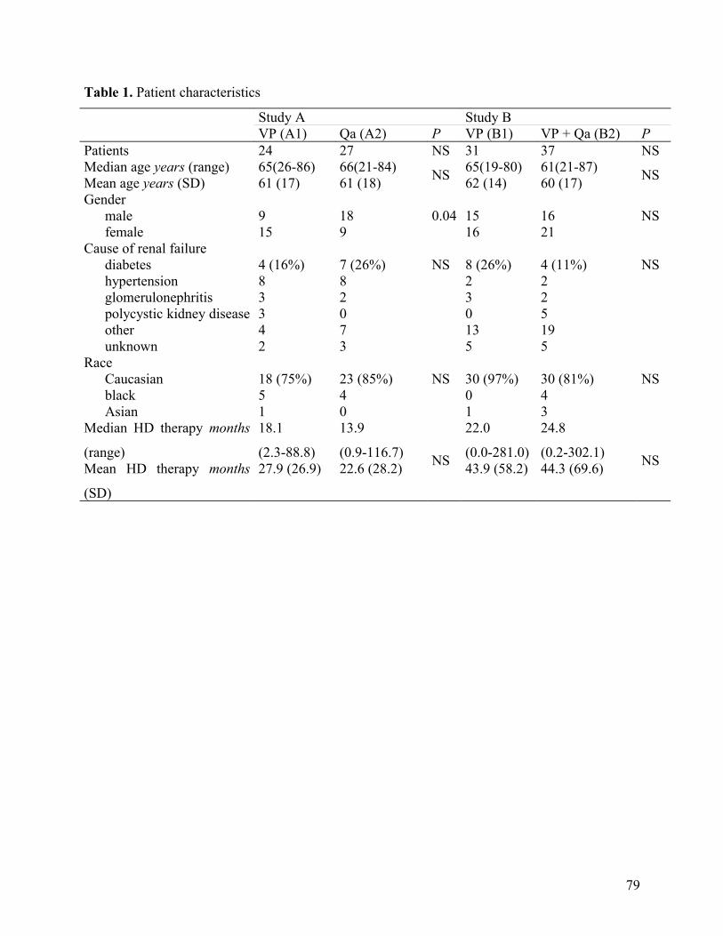
Table 1. Patient characteristics
Study A Study BVP (A1) Qa (A2) P VP (B1) VP + Qa (B2) P
Patients 24 27 NS 31 37 NSMedian age years (range) 65(26-86) 66(21-84)Mean age years (SD) 61 (17) 61 (18) NS 65(19-80) 61(21-87)
62 (14) 60 (17) NS
Gendermale 9 18 0.04 15 16 NSfemale 15 9 16 21
Cause of renal failurediabetes 4 (16%) 7 (26%) NS 8 (26%) 4 (11%) NShypertension 8 8 2 2glomerulonephritis 3 2 3 2polycystic kidney disease 3 0 0 5other 4 7 13 19unknown 2 3 5 5
RaceCaucasian 18 (75%) 23 (85%) NS 30 (97%) 30 (81%) NSblack 5 4 0 4Asian 1 0 1 3
Median HD therapy months
(range)
18.1
(2.3-88.8)
13.9
(0.9-116.7)Mean HD therapy months
(SD)
27.9 (26.9) 22.6 (28.2) NS
22.0
(0.0-281.0)
24.8
(0.2-302.1)43.9 (58.2) 44.3 (69.6) NS
79
Outcome
Study A
Total follow up was 37.8 patient-years. Graft monitoring resulted 59 times in positive tests
followed by 55 angiograms, which subsequently resulted in 48 PTA procedures and 7 surgical
interventions. Table 2 shows which test(s) led to these interventions. In all cases of Group A1 it
was an increased VP0/MAP, which led to the intervention. In the vast majority of angiograms
venous stenoses were present (Table 3). Graft characteristics and outcomes of individual patient
groups are outlined in Table 4 and 5.
Table 2. Which positive test led to intervention?
Study A Study BVP (A1) Qa (A2) VP (B1) VP + Qa (B2)
VP0/MAP 15 - 12 6VP200 0 - 4 0VP0/MAP + VP200a 15 - 9 8Qa - 25 - 3Qa +VPb - - - 13Total (PTA) 30 (26) 25 (22) 25 (23) 30 (26)a both tests were positive
b VP means either a positive VP0/MAP, or a positive VP200, or both
80
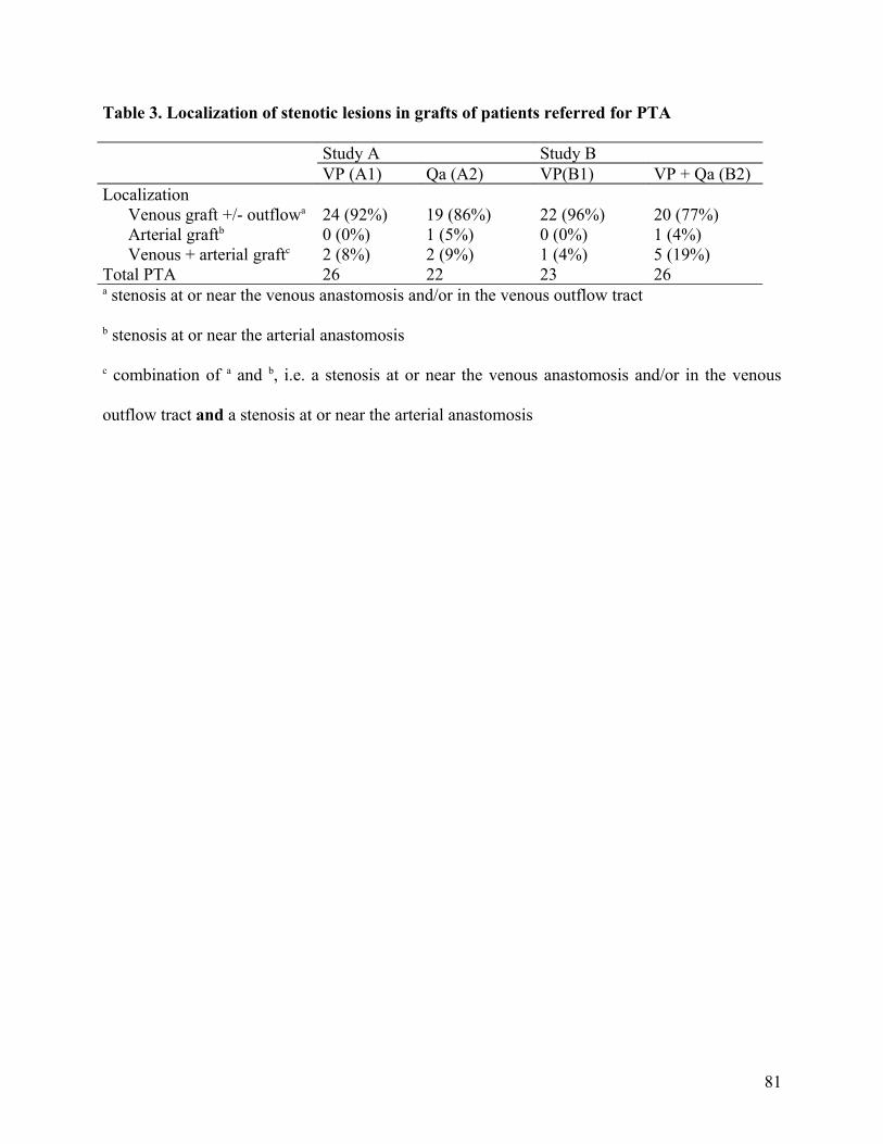
Table 3. Localization of stenotic lesions in grafts of patients referred for PTA
Study A Study BVP (A1) Qa (A2) VP(B1) VP + Qa (B2)
LocalizationVenous graft +/- outflowa 24 (92%) 19 (86%) 22 (96%) 20 (77%)Arterial graftb 0 (0%) 1 (5%) 0 (0%) 1 (4%)Venous + arterial graftc 2 (8%) 2 (9%) 1 (4%) 5 (19%)
Total PTA 26 22 23 26a stenosis at or near the venous anastomosis and/or in the venous outflow tract
b stenosis at or near the arterial anastomosis
c combination of a and b, i.e. a stenosis at or near the venous anastomosis and/or in the venous
outflow tract and a stenosis at or near the arterial anastomosis
81
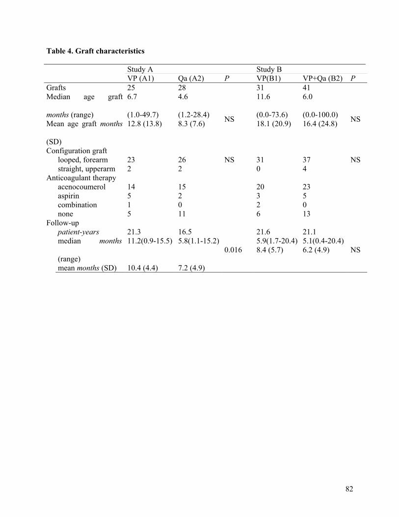
Table 4. Graft characteristics
Study A Study BVP (A1) Qa (A2) P VP(B1) VP+Qa (B2) P
Grafts 25 28 31 41Median age graft
months (range)
6.7
(1.0-49.7)
4.6
(1.2-28.4)Mean age graft months
(SD)
12.8 (13.8) 8.3 (7.6) NS
11.6
(0.0-73.6)
6.0
(0.0-100.0)18.1 (20.9) 16.4 (24.8) NS
Configuration graftlooped, forearm 23 26 NS 31 37 NSstraight, upperarm 2 2 0 4
Anticoagulant therapyacenocoumerol 14 15 20 23aspirin 5 2 3 5combination 1 0 2 0none 5 11 6 13
Follow-uppatient-years 21.3 16.5median months
(range)
11.2(0.9-15.5) 5.8(1.1-15.2)
mean months (SD) 10.4 (4.4) 7.2 (4.9)
0.016
21.6 21.15.9(1.7-20.4) 5.1(0.4-20.4)8.4 (5.7) 6.2 (4.9) NS
82
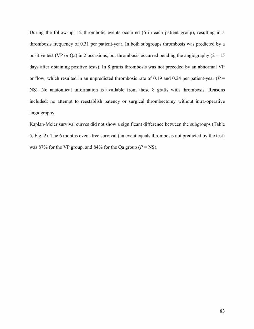
During the follow-up, 12 thrombotic events occurred (6 in each patient group), resulting in a
thrombosis frequency of 0.31 per patient-year. In both subgroups thrombosis was predicted by a
positive test (VP or Qa) in 2 occasions, but thrombosis occurred pending the angiography (2 – 15
days after obtaining positive tests). In 8 grafts thrombosis was not preceded by an abnormal VP
or flow, which resulted in an unpredicted thrombosis rate of 0.19 and 0.24 per patient-year (P =
NS). No anatomical information is available from these 8 grafts with thrombosis. Reasons
included: no attempt to reestablish patency or surgical thrombectomy without intra-operative
angiography.
Kaplan-Meier survival curves did not show a significant difference between the subgroups (Table
5, Fig. 2). The 6 months event-free survival (an event equals thrombosis not predicted by the test)
was 87% for the VP group, and 84% for the Qa group (P = NS).
83
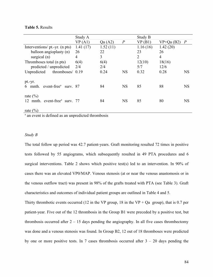
Table 5. Results
Study A Study BVP (A1) Qa (A2) P VP (B1) VP+Qa (B2) P
Interventions/ pt.-yr. (n pts) 1.41 (17) 1.52 (11) 1.16 (16) 1.42 (20)balloon angioplasty (n) 26 22 23 26surgical (n) 4 3 2 4
Thromboses total (n pts) 6(4) 6(4) 12(10) 18(16)predicted / unpredicted 2/4 2/4 5/7 12/6
Unpredicted thromboses/
pt.-yr.
0.19 0.24 NS 0.32 0.28 NS
6 mnth. event-freea surv.
rate (%)
87 84 NS 85 88 NS
12 mnth. event-freea surv.
rate (%)
77 84 NS 85 80 NS
a an event is defined as an unpredicted thrombosis
Study B
The total follow up period was 42.7 patient-years. Graft monitoring resulted 72 times in positive
tests followed by 55 angiograms, which subsequently resulted in 49 PTA procedures and 6
surgical interventions. Table 2 shows which positive test(s) led to an intervention. In 90% of
cases there was an elevated VP0/MAP. Venous stenosis (at or near the venous anastomosis or in
the venous outflow tract) was present in 98% of the grafts treated with PTA (see Table 3). Graft
characteristics and outcomes of individual patient groups are outlined in Table 4 and 5.
Thirty thrombotic events occurred (12 in the VP group, 18 in the VP + Qa group), that is 0.7 per
patient-year. Five out of the 12 thrombosis in the Group B1 were preceded by a positive test, but
thrombosis occurred after 2 – 15 days pending the angiography. In all five cases thrombectomy
was done and a venous stenosis was found. In Group B2, 12 out of 18 thromboses were predicted
by one or more positive tests. In 7 cases thrombosis occurred after 3 – 20 days pending the
84
angiography (all of these seven grafts showed a venous stenosis after thrombectomy), and in 5
cases intervention was not done for various reasons, including graft infection, poor clinical
condition and switch to peritoneal dialysis. In 13 cases thrombosis was not preceded by a positive
test, resulting in a thrombosis frequency of 0.32 (Group B1) and 0.28 (Group B2) (P = NS). Ten
of these 13 thromboses were treated by radiological thrombolysis. Stenoses were found at the
venous anastomosis (n=5, Group B1; n=3, Group B2), at the arterial anastomosis (n=1, Group
B2), or at both anastomoses (n=1, Group B2). In 3 grafts it was impossible to obtain anatomical
information, because no attempt was made to reestablish patency.
Kaplan-Meier survival curves did not show a significant difference between the subgroups (Table
5, Fig. 3). The 6 months event-free survival (an event equals thrombosis not predicted by the test)
was 85% for the VP group, and 88% for the VP + Qa group (P = NS).
85
Discussion
This randomized, prospective multi-center trial allows a number of important conclusions.
Firstly, we confirm that thrombosis rates in patients monitored and selected for corrective
interventions based on VP or Qa can be maintained below the quality of care standards
formulated by the NKF-DOQI committee (4). Secondly, we show that thrombosis rates in groups
monitored by VP or Qa alone or by the combination of tests do not differ. Thirdly, only a small
minority of patients was selected for corrective interventions by Qa alone in the group with
combined monitoring of VP and Qa. Fourthly, after obtaining abnormal tests subsequent
diagnostic and interventional procedures should be instituted on short notice. It seems likely that
the number of thromboses during the waiting time can be reduced. Finally, we confirm that static
VP is more effective than dynamic VP for monitoring dialysis grafts.
The present study confirms the usefulness of VP and Qa measurements as access surveillance
variables. Previously, we have presented the theoretical basis for the assumption that Qa
measurements are better than VP (i.e. dynamic) measurements as a monitoring tool (summarized
in (26)). This is based on the fact that some stenoses are located in the arterial flow tract that is
upstream of the venous needle. These lesions increase resistance and reduce Qa without
increasing VP, possibly even decreasing VP. Several studies have indicated that arterial stenoses
occur in up to 29% of thrombosis cases (20-23). In the present study, we found a strong
predominance of venous lesions in the patients referred for angiography. In some patients having
a graft thrombosis without a preceding positive test, thrombectomy was done. Also, in those
patients venous lesions were more likely than arterial lesions.
86
Qa measurements have already been recognized as the preferred monitoring tool for vascular
access surveillance (4,27). However, these recommendations are primarily based on clinical
studies with a non-randomized or observational setup (19,25,28-30). To our knowledge, only one
study compared Qa with static VP as surveillance variable. Sands et al (8) showed that
intervention based on monthly Qa measurement or on monthly static VP measurements reduced
thrombosis rates in comparison with non-monitored controls. Some important differences
between the two studies exist. The present study also includes a program combining VP and Qa.
Theoretically, this combination is likely to be the most effective program (18). Furthermore, the
study population of Sands et al consisted predominantly of AV fistulae (almost two-third in the
control and monitored groups) which are less likely to clot. Also, threshold for referral for
angiogram (750 mL/min vs. 600 mL/min in our Qa groups), and frequency of measurements
(monthly VP vs. weekly in our VP groups) differed. There is some controversy about the optimal
frequency. Some advocate the use of dynamic pressures weekly and static VP every two weeks
(4), others suggest to measure dynamic VP every dialysis (27), or static VP every week (31). In
the present study, we measured both dynamic and static VP once weekly during the same dialysis
session. Our results indicate that graft surveillance using VP or Qa measurements strictly
organized as in the present study results in identical thrombosis rates. This does not exclude the
possibility that in patients who develop more arterial lesions for whatever reason, surveillance
using Qa would result in lower thrombosis rates than using VP measurements.
In our study most referrals for intervention in patients monitored by VP measurements alone
(Group A1 and B1) were based on static VP measurements. Although dynamic VP was
successfully used as part of an access surveillance program in some studies (6,11,16,32), there is
convincing evidence that dynamic VP does not accurately reflect true intra-access pressure, and
87

therefore, does not reflect resistance caused by stenosis formation (14). Dynamic VP is highly
biased by pump flow, blood tubing, needle size, and blood pressure (33). Static VP
measurements, on the other hand, particularly when corrected for mean arterial pressure, avoid
these potential confounders.
Also in Group B2 (combination of VP and Qa measurements) most referrals were based on VP
and only 3 out of 30 referrals were based on Qa alone. These data indicate that when VP
measurements are well organized, adding periodic Qa measurements is of limited value (Group
B1 versus B2). On the other hand, study A clearly shows that when no VP measurements are
done periodic Qa measurements result in identical thrombosis rates, proving the effectiveness of
Qa as surveillance variable.
In this study 21 of the 42 thromboses occurred despite preceding positive tests. Sixteen
thromboses occurred 2 to 20 days after obtaining the abnormal tests pending further diagnostic
and correctional interventions. These results further support the idea that an increased VP and/or
low Qa indeed predict imminent thrombosis. It seems likely that immediate institution of further
treatment could have decreased the number of thromboses.
The 21 thromboses, which were not preceded by positive tests, should not all be considered as
failures of the tests. Flow is directly related to blood pressure. Blood pressure may show
considerable variability in hemodialysis patients and is likely to reach its lowest levels in the first
hours after the hemodialysis session and especially during the night (34). Therefore, it is possible
that flow is adequate at the time measurements are done, but reaches levels associated with
thrombosis in the interdialytic period.
Several issues need to be addressed with respect to the present study.
88
Firstly, our power calculations were based on the assumption that there would be a difference in
thrombosis rates between VP and Qa monitored patients. However, the strict adherence to the
surveillance protocols improved our results using VP measurements when compared to our
historic controls. Differences in the present study were not statistically significant. The results
indicated that continuation of the study was not likely to result in clinically relevant differences.
Secondly, we measured Qa every two months, but repeated the measurement after one month if
Qa reached levels between 600 and 800 mL/min. This frequency was mainly based on feasibility
considerations. It remains unclear, whether a higher frequency of Qa measurements, as was
suggested by others (8,27) would decrease the already low thrombosis rate further. Furthermore,
we can not exclude the possibility that decreasing the frequency of VP measurements, for
instance every 2 weeks, as was suggested elsewhere (4), would result in identical results.
Additionally, threshold levels for selection for referral may vary. We used a level of 600 mL/min
as Qa threshold level for referral. We based our choice on earlier results (35) and the NKF-DOQI
considerations (4). Others have chosen 650 or 750 mL/min (8,27). This may affect outcome. A
recent meta-analysis by Paulson et al (36) concluded that a single Qa measurement did not appear
to have enough accuracy to be a clinically useful predictor of graft thrombosis or failure.
Furthermore, it has been suggested that a decrease in Qa over time of >15% is particularly
predictive of impending graft failure (29). The present study was not specifically designed to
study that issue. However, in the patients who were monitored by Qa measurements (Group A2
and B2) none of the unpredicted thromboses were preceded by a decrease in Qa of more than
15%. As a consequence, none of these thromboses would have been predicted by this criterion.
Furthermore, in the patients who did not reach the threshold levels of the present study but did
show a decrease in Qa of more than 15% (n = 11 (A2), n = 8 (B2); average decrease: 1090
mL/min to 740 mL/min) none experienced thrombosis. So, our data do not support the
89
hypothesis that a decrease in Qa of >15% as selection parameter would confer any benefit when
added to either of the Qa protocols. Also, a recent study showed that a decrease in Qa over time
had little predictive value (37).
In unmonitored PTFE grafts the total graft access event rate related to thrombosis or stenosis was
1.51 per patient-year (7) of which 1.24 event per patient-year (82%) was caused by thrombosis.
In the present studies the total graft event rate (related to thrombosis and stenosis) was 1.88
(range 1.69-2.27). On average, all thrombotic episodes accounted for 0.52 event per patient-year
(28%) and the unpredicted thromboses accounted for 0.26 event per patient-year (14%). So, the
introduction of a well structured surveillance program resulted in a substantial decrease in
interventions for treatment of thrombosis at the expense of an increase of the number of elective
interventions, mainly PTA. Our study was not designed as a cost effectiveness analysis. It seems
likely, however, that this shift in type of interventions is associated with a decrease in access
related morbidity and mortality.
What may be the implications of the present study for everyday clinical practice? We showed that
using the surveillance protocol of this study outcome with respect to thrombosis rate is equal in
Qa monitored grafts and in VP monitored grafts and that there is no rationale in combining the
two methods. It is important to note that our population contained fewer diabetics than the
average US dialysis population. It is possible that this affects outcome, since inflow stenosis
(which will not be detected by VP measurements) may occur more frequently in diabetics. Each
dialysis center should decide which method is best suitable for their dialysis practice. VP
measurements are easy to obtain and require little time investment. However, they need
discipline. Qa measurements are more time consuming and a special device is needed. We
showed that by measuring Qa every 4 to 8 weeks identical results can be obtained as with weekly
90
VP assessments. The decision which method to use, will be primarily based on the preferences of
those involved in the access care and on the possibilities to implement a certain surveillance
strategy.
In conclusion, our studies show that standardized monitoring of VP or Qa or the combination of
both and subsequent corrective intervention can decrease thrombosis rates to below 0.5 per
patient-year, which is recommended by the Vascular Access Task Force of the NKF-DOQI
Committee as quality of care standard (4). When applying the present protocol, VP, Qa, or the
combination as variables for selection of patients for corrective intervention are equally effective
in reducing thrombosis rates in hemodialysis access grafts.
91
References
1 Feldman HI, Kobrin S, Wasserstein A: Hemodialysis vascular access morbidity. J Am Soc
Nephrol 1996; 7:523-35
2 USRDS: The economic cost of ESRD, vascular access procedures, and Medicare spending
for alternative modalities of treatment. United States Renal Data System. Am J Kidney Dis
1997; 30 (Suppl 1):S160-S177
3 Feldman HI, Held PJ, Hutchinson JT, Stoiber E, Hartigan MF, Berlin JA: Hemodialysis
vascular access morbidity in the United States. Kidney Int 1993; 43:1091-6
4 National Kidney Foundation. NKF-DOQI clinical practice guidelines for vascular access. Am
J Kidney Dis 1997; 30 (Suppl 3):S152-S191
5 Beathard GA: Thrombolysis for the treatment of thrombosed dialysis access grafts: a
nephrologist's view. Semin Dial 1995; 8:162-5
6 Cayco AV, Abu-Alfa AK, Mahnensmith RL, Perazella MA: Reduction in arteriovenous graft
impairment: results of a vascular access surveillance protocol. Am J Kidney Dis 1998;
32:302-8
7 Bosman PJ, Blankestijn PJ, van der Graaf Y, Heintjes RJ, Koomans HA, Eikelboom BC: A
comparison between PTFE and denatured homologous vein grafts for haemodialysis access: a
prospective randomised multicentre trial. The SMASH Study Group. Eur J Vasc Endovasc
Surg 1998; 16:126-32
92
8 Sands JJ, Jabyac PA, Miranda CL, Kapsick BJ: Intervention based on monthly monitoring
decreases hemodialysis access thrombosis. ASAIO J 1999; 45:147-50
9 Bassiouny HS, White S, Glagov S, Choi E, Giddens DP, Zarins CK: Anastomotic intimal
hyperplasia: mechanical injury or flow induced. J Vasc Surg 1992; 15:708-16
10 Kanterman RY, Vesely TM, Pilgram TK, Guy BW, Windus DW, Picus D: Dialysis access
grafts: anatomic location of venous stenosis and results of angioplasty. Radiology 1995;
195:135-9
11 Roberts AB, Kahn MB, Bradford S, Lee J, Ahmed Z, Fitzsimmons J, Ball D: Graft
surveillance and angioplasty prolongs dialysis graft patency. J Am Coll Surg 1996; 183:486-
92
12 Safa AA, Valji K, Roberts AC, Zieglet TW, Hye RJ, Oglevie SB: Detection and treatment of
dysfunctional hemodialysis access grafts: effect of a surveillance program on graft patency
and the incidence of thrombosis. Radiology 1996; 199:653-7
13 Smits HF, Van Rijk PP, Van Isselt JW, Mali WP, Koomans HA, Blankestijn PJ: Pulmonary
embolism after thrombolysis of hemodialysis grafts. J Am Soc Nephrol 1997; 8:1458-61
14 Besarab A, Sullivan KL, Ross RP, Moritz MJ: Utility of intra-access pressure monitoring in
detecting and correcting venous outlet stenoses prior to thrombosis. Kidney Int 1995;
47:1364-73
15 Beathard GA: Percutaneous transvenous angioplasty in the treatment of vascular access
stenosis. Kidney Int 1992; 42:1390-7
93

16 Schwab SJ, Raymond JR, Saeed M, Newman GE, Dennis PA, Bollinger RR: Prevention of
hemodialysis fistula thrombosis. Early detection of venous stenoses. Kidney Int 1989; 36:707-
11
17 Sands JJ, Miranda CL: Prolongation of hemodialysis access survival with elective revision.
Clin Nephrol 1995; 44:329-33
18 Smits JHM, Blankestijn PJ: Haemodialysis access: the case for prospective monitoring. Curr
Opin Nephrol Hypertens 1999; 8:685-90
19 Bosman PJ, Boereboom FT, Smits HF, Eikelboom BC, Koomans HA, Blankestijn PJ:
Pressure or flow recordings for the surveillance of hemodialysis grafts. Kidney Int 1997;
52:1084-8.
20 Beathard GA: Mechanical versus pharmacomechanical thrombolysis for the treatment of
thrombosed dialysis access grafts. Kidney Int 1994; 45:1401-6
21 Roberts AC, Valji K, Bookstein JJ, Hye RJ: Pulse-spray pharmacomechanical thrombolysis
for treatment of thrombosed dialysis access grafts. Am J Surg 1993; 166:221-5
22 Valji K, Bookstein JJ, Roberts AC, Oglevie SB, Pittman C, O’Neill MP: Pulse-spray
pharmacomechanical thrombolysis of thrombosed hemodialysis access grafts: long-term
experience and comparison of original and current techniques. Am J Roentgenol 1995;
164:1495-1500
23 Swan TL, Smyth SH, Ruffenach SJ, Berman SS, Pond GD: Pulmonary embolism following
hemodialysis access thrombolysis/thrombectomy. J Vasc Interv Radiol 1995; 6:683-6
94
24 Krivitski NM: Theory and validation of access flow measurement by dilution technique
during hemodialysis. Kidney Int 1995; 48:244-50
25 Bosman PJ, Boereboom FT, Bakker CJ, Mali WP, Eikelboom BC, Blankestijn PJ, Koomans
HA: Access flow measurements in hemodialysis patients: in vivo validation of an ultrasound
dilution technique. J Am Soc Nephrol 1996; 7:966-9
26 Blankestijn PJ, Smits JH: How to identify the haemodialysis access at risk of thrombosis? Are
flow measurements the answer? Nephrol Dial Transplant 1999; 14:1068-71
27 Jindal KK, Ethier JH, Lindsay RM, Barre PE, Kappel JE, Carlisle EJ, Common A: Clinical
practice guidelines for vascular access. J Am Soc Nephrol 1999; 10 (Suppl 13):S297-S305
28 May RE, Himmelfarb J, Yenicesu M, Knights S, Ikizler TA, Schulman G, Hernanz Schulman
M, Shyr Y, Hakim RM: Predictive measures of vascular access thrombosis: a prospective
study. Kidney Int 1997; 52:1656-62
29 Neyra NR, Ikizler TA, May RE, Himmelfarb J, Shulman G, Shyr Y, Hakim RM: Change in
access blood flow over time predicts vascular access thrombosis. Kidney Int 1998; 54:1714-9
30 Wang E, Schneditz D, Nepomuceno C, Lavarias V, Martin K, Morris AT, Levin NW:
Predictive value of access blood flow in detecting access thrombosis. ASAIO J 1998;
44:M555-M8
31 Besarab A, Frinak S, Sherman RA, Goldman J, Dumler F, Devita MV, Kapoian T, Al-Saghir
F, Lubkowski T: Simplified measurement of intra-access pressure. J Am Soc Nephrol 1998;
9:284-9
95

32 Allon M, Bailey R, Ballard R, Deierhoi MH, Hamrick K, Oser R, Rhynes VK, Robbins ML,
Saddekni S, Zeigler ST: A multidisciplinary approach to hemodialysis access: prospective
evaluation. Kidney Int 1998; 53:473-9
33 Besarab A, Dorrell S, Moritz M, Michael H, Sullivan K: Determinants of measured dialysis
venous pressure and its relationship to true intra-access venous pressure. ASAIO Trans 1991;
37:M270-M1
34 Luik AJ, Struijk DG, Gladziwa U, von Olden RW, von Hooff JP, de Leeuw PW, Leunissen
KM: Diurnal blood-pressure variations in haemodialysis and CAPD patients. Nephrol Dial
Transplant 1994; 9:1616-21
35 Bosman PJ, Boereboom FTJ, Eikelboom BC, Koomans HA, Blankestijn PJ: Graft flow as a
predictor of thrombosis in hemodialysis grafts. Kidney Int 1998; 54:1726-30
36 Paulson WD, Ram SJ, Birk CG, Work J: Does blood flow accurately predict thrombosis or
failure of hemodialysis synthetic grafts? A meta-analysis. Am J Kidney Dis 1999; 34:478-85
37 Paulson WD, Ram SJ, Birk CG, Zapczynski M, Martin SR, Work J: Accuracy of decrease in
blood flow in predicting hemodialysis graft thrombosis. Am J Kidney Dis 2000; 35:1089-95
96
Chapter 6
SHORT- AND LONG-TERM FUNCTIONAL EFFECT OF
PERCUTANEOUS TRANSLUMINAL ANGIOPLASTY IN
HEMODIALYSIS VASCULAR ACCESS
J Am Soc Nephrol 2002 Mar;13(3):715-20
Joke van der Linden
Johannes H.M. Smits
Jan H. Assink
Derk W. Wolterbeek
Jan J. Zijlstra
Gijs H.T. de Jong
Marinus A. van den Dorpel
Peter J. Blankestijn
97
Abstract
Background. Efficacy of percutaneous transluminal angioplasty (PTA) is usually expressed as the
angiographic result. Access flow (Qa) offers a means to quantify the functional effect. This study
was performed to evaluate the short-term functional and angiographic effect of PTA and to
determine the longevity of the functional effect during follow-up.
Methods. Patients with an arteriovenous graft (AVG) or fistula (AVF) eligible for PTA (Qa<600
mL/min) were included. Ultrasound-dilution derived Qa was measured shortly before PTA and
periodically after PTA, starting 1 week after the procedure. The short-term effect was expressed
as the increase in Qa and as the reduction of stenosis. The long-term effect was expressed as
patency and as the decline in Qa after PTA.
Results. Ninety-eight PTA procedures in 60 patients (65 AVG, 33 AVF) were analyzed. Qa
improved from 371±17 to 674±30 mL/min in AVG, and from 304±24 to 638±51 mL/min in AVF
(both P<0.0001). In 66% (AVG) and 50% (AVF) flow increased to levels >600 mL/min. Degree
of stenosis decreased from 65±3 % to 17±2 % in AVG and from 72±5 % to 23±7 % in AVF
(both P<0.005). Reduction of stenosis did not correlate with ∆Qa (r2=0.066). Six-month
unassisted patency after PTA was 25% for AVG and 50% for AVF. Decline in Qa was 3.7±0.8
ml/min/day in AVG and 1.8±0.9 ml/min/day in AVF. Qa before PTA and ∆Qa correlated with
the subsequent decline in Qa (P<0.005).
Conclusions. Access flow increases after PTA, however in a substantial percentage not to a level
>600 mL/min. Qa before PTA and the increase in Qa correlated with long-term outcome,
whereas angiographic results did not. The present data combined with the literature suggest that
there is an optimal timing for PTA.
98
Introduction
Vascular access complications account for considerable morbidity and mortality in hemodialysis
patients. In the United States they are responsible for up to 25% of all hospitalizations in dialysis
patients (1-3). The European Dialysis and Transplant Association does not collect data on this
issue, but it is likely that data in Europe and elsewhere will be comparable.
Thrombosis is the leading cause of vascular access complication. It is almost always associated
with the presence of stenosis. Percutaneous transluminal angioplasty (PTA) is an accepted
treatment of stenotic lesions (3). Routine surveillance programs for the early detection of stenoses
followed by angioplasty have been shown to substantially reduce the number of thromboses per
patient year (4-7). However, repetitive PTA treatment is often necessary, since re-stenosis
frequently occurs. Although the short term success rates of PTA range from 85% to 98% (8),
patency at 6 months follow-up varies from 38% to 63% (4,9-11).
Several studies have shown that angiographic degree of the stenotic lesion before and after PTA
is poorly related with its subsequent patency (9,11-14). Recently, the SCVIR Technology
Assessment Committee recommended that PTA efficacy should be expressed by both
angiographic and functional parameters (15). In particular, access flow (Qa) measurements offer
the opportunity to quantify and follow up the functional effect of PTA.
The purpose of this study is to assess access function of patients undergoing PTA. We quantified
the short-term functional and angiographic effect of PTA. Additionally, we determined the
longevity of the functional effect during follow-up. Finally, we addressed the question whether
functional variables are predictive for long-term outcome.
99
Patients and methods
Patients
This prospective observational intervention study was done in 9 Dutch hemodialysis centers, in
which a well-defined surveillance protocol was instituted as part of the routine patient care. All
chronic hemodialysis patients with permanent arteriovenous grafts (AVG) and fistulae (AVF),
who were referred for angiography because predetermined Qa threshold levels were reached,
were eligible to enter the study. The surveillance protocol included periodic Qa measurements
and angiography with PTA in thus selected patients.
Qa surveillance protocol
Qa was measured at least every 8 weeks using the ultrasound hemodilution technique (Transonic
Systems Inc, Ithaca NY). The bench and clinical validations are presented elsewhere (16,17).
The surveillance protocol has been discussed in detail previously (7). In brief, Qa determination
consisted of the average of three single measurements within the first 30 minutes of the dialysis
session at a fixed pump flow (> 200 mL/min). If Qa levels reached values between 600 and 800
mL/min the measurement was repeated at least every 4 weeks. Patients were referred for
angiography whenever Qa was below 600 mL/min. Patients referred for angiography based on
other criteria, for instance frequent miscanulation, swelling of the arm or high venous pressure,
were excluded. Patients with a history of allergy to iodinated contrast agents were also not
included into the study.
100
Angiography and percutaneous transluminal angioplasty (PTA)
Angiography and PTA procedures were done as soon as possible (usually < 7 days) after the low
Qa level (< 600 mL/min) was diagnosed. Digital subtraction angiography was performed to
visualize the complete vascular access and to locate the stenosis. Any luminal reduction of 50%
or more was treated by percutaneous transluminal angioplasty (PTA) during the same session.
The same PTA technique was used for all patients. First, the stenotic lesion was crossed with a
guide wire (Boston Scientific Corporation, Watertown, MA). Then a sheath was introduced
(Cordis Europe N.V., Roden, The Netherlands). The PTA balloon catheter was then passed over
the guide wire to the location of the stenosis. In general, the high-pressure balloons had a
diameter of 6 mm although larger balloons (up to 10 mm) could be used for large proximal veins
(Bülach, Switzerland). At the stenotic site the balloon was inflated to at least 10 atmospheres of
pressure and held for approximately 2 minutes. In resistant cases pressures up to 20 atmospheres
were used and held for 10 minutes. No heparin, vasodilators or local anesthetics were given
during the procedure. Immediately after PTA an angiogram was obtained to evaluate the result of
the procedure. The interventional radiologist considered the PTA procedure successful when the
residual diameter of the stenosis was less than 25%. After PTA, Qa measurements were done
within one week and then at least at 4 weeks intervals. In case Qa fell below 600 mL/min,
patients were referred for angiography and repeat PTA was performed if necessary.
Outcome variables and statistical analysis
The short-term functional effect of PTA on Qa was evaluated by calculation of ∆Qa, i.e. the
difference between Qa before (Qapre) and just after PTA (Qapost). The long-term functional effect
was assessed as the time to the next intervention, if applicable. When there were 3 or more Qa
101
measurements after the PTA, the decline in Qa was determined (in ml/min/day). Resistance was
calculated as the ratio of mean arterial blood pressure (MAP) and Qa.
Angiographic data were analyzed by an independent radiologist. Only cases that had adequate
biplanar angiograms available of the stenotic areas both before and after PTA were included.
Stenosis degree was assessed before (baseline) and after PTA (post-PTA) as the ratio of maximal
lumen reduction and an adjacent normal graft/vessel diameter (expressed in percentage: 0 % = no
stenosis, 100 % = occlusion). Stenosis reduction due to PTA was calculated by the difference
between baseline stenosis and post-PTA stenosis.
Data are presented as mean ± SEM, unless indicated otherwise. Differences of Qa and ∆Qa
values between different time points and subgroups of patients were calculated with two-tailed t-
tests. Pearson analysis was used for correlation purposes. Post-PTA primary patency, i.e. the
period of time that elapsed following intervention until access thrombosis or re-intervention
(surgical and/or radiological), was calculated using life-table analysis. To compare post-PTA
survival of AVF and AVG the (two-sided) log-rank test was performed. P values of less than
0.05 were considered significant.
102
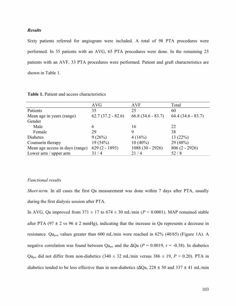
Results
Sixty patients referred for angiogram were included. A total of 98 PTA procedures were
performed. In 35 patients with an AVG, 65 PTA procedures were done. In the remaining 25
patients with an AVF, 33 PTA procedures were performed. Patient and graft characteristics are
shown in Table 1.
Table 1. Patient and access characteristics
AVG AVF TotalPatients 35 25 60Mean age in years (range) 62.7 (37.2 - 82.6) 66.8 (34.6 - 83.7) 64.4 (34.6 - 83.7)Gender Male 6 16 22 Female 29 9 38Diabetes 9 (26%) 4 (16%) 13 (22%)Coumarin therapy 19 (54%) 10 (40%) 29 (48%)Mean age access in days (range) 629 (2 - 1893) 1088 (30 - 2926) 806 (2 - 2926)Lower arm / upper arm 31 / 4 21 / 4 52 / 8
Functional results
Short-term. In all cases the first Qa measurement was done within 7 days after PTA, usually
during the first dialysis session after PTA.
In AVG, Qa improved from 371 ± 17 to 674 ± 30 mL/min (P < 0.0001). MAP remained stable
after PTA (97 ± 2 vs 96 ± 2 mmHg), indicating that the increase in Qa represents a decrease in
resistance. Qapost values greater than 600 mL/min were reached in 62% (40/65) (Figure 1A). A
negative correlation was found between Qapre and the ∆Qa (P = 0.0019, r = -0.38). In diabetics
Qapre did not differ from non-diabetics (340 ± 32 mL/min versus 386 ± 19, P = 0.20). PTA in
diabetics tended to be less effective than in non-diabetics (∆Qa, 228 ± 50 and 337 ± 41 mL/min
103
respectively, P = 0.06). The age of the AVG and of the patient did not correlate with ∆Qa. In 15
patients multiple procedures were done (Table 2). In patients who were treated twice, ∆Qa was
355 ± 68 ml/min and 424 ± 79 ml/min after the first and second PTA respectively (n = 15, P =
ns). In patients who were treated 3 times, ∆Qa was 315 ± 98, 484 ± 112 and 345 ± 51 ml/min
after the first, second and third PTA respectively (n = 10, P = ns). In 15 patients, the PTA was the
first intervention on the AVG. ∆Qa in those patients did not differ from those who had a second
or later PTA (287 ± 61 vs. 313 ± 41 mL/min, P = 0.72). Unassisted patency did not differ either.
In AVF, Qa improved from 304 ± 24 to 638 ± 51 mL/min (P < 0.0001) (Figure 1B), 52% (17/33)
of PTA procedures resulted in Qapost levels of more than 600 mL/min. The ∆Qa was not related to
Qapre in AVF (r = -0.06). Diabetes or age of the AVF or patient did not significantly affect the
results in AVF. Only four patients underwent 2 PTA procedures (Table 2) and showed a ∆Qa of
541 ± 173 mL/min and 285 ± 144 mL/min after the first and second PTA respectively (P = 0.09).
104
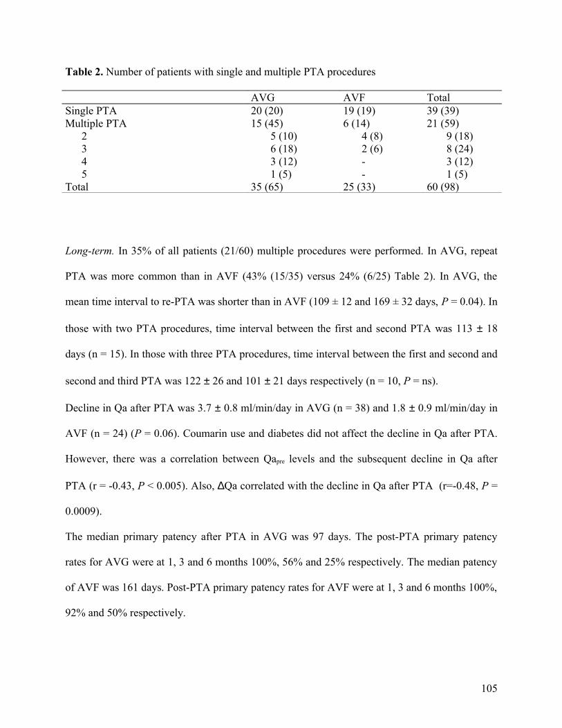
Table 2. Number of patients with single and multiple PTA procedures
AVG AVF TotalSingle PTA 20 (20) 19 (19) 39 (39)Multiple PTA 15 (45) 6 (14) 21 (59)
2 5 (10) 4 (8) 9 (18)3 6 (18) 2 (6) 8 (24)4 3 (12) - 3 (12)5 1 (5) - 1 (5)
Total 35 (65) 25 (33) 60 (98)
Long-term. In 35% of all patients (21/60) multiple procedures were performed. In AVG, repeat
PTA was more common than in AVF (43% (15/35) versus 24% (6/25) Table 2). In AVG, the
mean time interval to re-PTA was shorter than in AVF (109 ± 12 and 169 ± 32 days, P = 0.04). In
those with two PTA procedures, time interval between the first and second PTA was 113 ± 18
days (n = 15). In those with three PTA procedures, time interval between the first and second and
second and third PTA was 122 ± 26 and 101 ± 21 days respectively (n = 10, P = ns).
Decline in Qa after PTA was 3.7 ± 0.8 ml/min/day in AVG (n = 38) and 1.8 ± 0.9 ml/min/day in
AVF (n = 24) (P = 0.06). Coumarin use and diabetes did not affect the decline in Qa after PTA.
However, there was a correlation between Qapre levels and the subsequent decline in Qa after
PTA (r = -0.43, P < 0.005). Also, ∆Qa correlated with the decline in Qa after PTA (r=-0.48, P =
0.0009).
The median primary patency after PTA in AVG was 97 days. The post-PTA primary patency
rates for AVG were at 1, 3 and 6 months 100%, 56% and 25% respectively. The median patency
of AVF was 161 days. Post-PTA primary patency rates for AVF were at 1, 3 and 6 months 100%,
92% and 50% respectively.
105
Log-rank comparison analysis of intervention-free survival curves demonstrated a significant
difference in favor of the AVF (P = 0.031).
During the available follow-up period no unpredicted thrombotic events (i.e. accesses
thrombosed with a Qa >600 mL/min) occurred. In 6 AVG with a Qa that remained below 600
mL/min after PTA, thrombosis occurred within weeks after PTA. These are considered predicted
thromboses. Furthermore, some thromboses occurred in patients with low Qa while waiting for
PTA. These accesses were not included in this analysis, because no PTA procedure was
performed.
Angiographic results
In AVG, 48 (74 %) PTA procedures were performed on single lesions (38 (58 %) venous, 9 (14
%) midgraft, and 1 (2 %) arterial stenosis). Seventeen (26 %) PTA procedures were done on two
stenotic lesions (12 (18 %) showed a venous and midgraft stenosis, and 5 (8 %) had an arterial
and venous stenosis). Log rank survival analysis showed no difference in patency between AVG
with a single lesion versus those with multiple lesions (P = 0.74). Seven (21 %) PTA procedures
in AVF were of true anastomotic lesions, twenty (61 %) were performed on venous lesions (in
most cases located within the first few centimeters from the anastomosis) and 6 (18 %) on
combined (venous and anastomotic) lesions. In 92% of all PTA procedures, angiographic
improvement of the stenosis was achieved using a 6-mm balloon. Larger balloons were
occasionally needed for proper dilatation of stenoses in proximal AVF veins. In all cases PTA
was reported to be successful, i.e. residual luminal reduction of 25% or less.
In AVG, baseline stenosis was 65 ± 3 % and post-PTA stenosis 17 ± 2 % (n = 33, P < 0.0001).
No correlation was found between baseline stenosis and post-PTA stenosis (r = -0.14, P = 0.47)
or Qapost (r = -0.05, P = 0.76). Baseline stenosis correlated with Qapre (r = -0.48, P = 0.008). No
106
correlation was found between angiographic (= stenosis reduction) and functional (∆Qa)
improvement (r2 = 0.066) or between baseline stenosis and subsequent decline in Qa (r = -0.02, P
= 0.93). Additionally, neither the stenosis reduction (P = 0.29) or the post-PTA stenosis (P =
0.07) correlated with the decline in Qa after PTA. Log rank survival analysis of the lower and
upper 50th percentile baseline stenosis revealed no difference in survival (P = 0.90).
When the AVG group was divided in a group with Qapost <600 mL/min (n = 24) and a group with
Qapost >600 mL/min (n = 41), we found that there was no difference in angiographic results
between both groups. The group with Qapost <600 mL/min showed a stenosis of 66 ± 4 % before
and 19 ± 4 % after PTA, while the group with Qapost >600 mL/min had a stenosis of 63 ± 4 %
before and 14 ± 3 % after PTA (P = NS). The group with Qapost <600 mL/min had a Qapre of 336
± 24 mL/min and a Qapost of 441 ± 22 mL/min (∆Qa = 105 ± 24 mL/min). Those with Qapost >600
mL/min had a Qapre of 392 ± 22 mL/min and a Qapost of 811 ± 29 mL/min (∆Qa = 419 ± 40
mL/min). Of the 24 patients with Qapost <600 mL/min, 6 thrombosed within 4 weeks after PTA, 8
had a surgical correction without repeat angiogram, in 10 cases repeat angiograms showed 61%
stenosis. Two of those patients were subsequently referred for surgical revision, and eight had
another PTA.
For the AVF, baseline stenosis was 72 ± 5 % and post-PTA stenosis was 23 ± 7 % (n = 8, P =
0.0039).
Discussion
To our knowledge this is the first study in AVG and AVF reporting both angiographic and
functional results of PTA. The present data contain novel information. We show that
angiographic results do not correlate with functional results. Importantly, we demonstrate that
functional variables are predictive for long-term outcome, whereas angiographic results are not.
107
The study confirms recent data indicating that PTA results in a direct increase in Qa of
approximately 250 mL/min (18). We also confirm that a substantial percentage of PTA is not
successful. Finally, time to repeat PTA in the present study is substantially shorter than in other
studies (9,18,19). This suggests a more rapid recurrence of stenosis in the present study. The
combined data of these studies suggest that there is an optimal moment of PTA.
Based on the vast experience reported in the literature the NKF-K/DOQI taskforce has suggested
PTA as one of the preferred treatments of vascular access stenosis (3). In most studies, the post-
PTA stenosis is used to express the efficiency of a PTA procedure. However, the post-PTA
stenosis poorly predicts patency after PTA (9,11-14). Recently, the SCVIR Technology
Assessment Committee recommended reporting both angiographic and functional data as
efficacy variables of PTA (15).
Our patients all had a Qa below 600 mL/min, which especially in grafts is a strong predictor for
imminent thrombosis (3,20-23). They all had a baseline stenosis of 50% or more, which was
treated by PTA. We confirm earlier data that on average Qa increases with approximately 250-
300 mL/min (18,24). In the patients with adequate angiograms, the post-PTA stenosis in AVG
was in almost all cases 25% or less. This is considered to be an adequate angiographic result (3).
However, the decrease in resistance did not correlate with the stenosis reduction, indicating that
angiographic improvement does not necessarily represent functional improvement. Preliminary
data in a small group of patients also indicated that the stenosis reduction did not correlate with
the increase in Qa (14). Results in diabetics did not differ from those in non-diabetics. The
percentage of PTA procedures resulting in a Qapost above the threshold value of 600 mL/min, was
66% in AVG and 50% in AVF. Schwab et al. defined failure of PTA as an increase in Qa of less
than 20%, which occurred in 21% of grafts (18). This lack of effect may be caused by rapid recoil
108
of the stenotic lesion, occurring in the period between PTA and first Qa measurement.
Intravascular ultrasound after PTA showed that immediate elastic recoil occurred in 50% of the
stenotic lesions (25). The present findings may also indicate that other stenotic lesions, which
importantly contribute to overall resistance, were not identified and not treated. Qa measurements
during or immediately after PTA in the intervention room could be helpful to optimize procedure
results (24).
In patients who needed multiple procedures, similar increases in Qa were obtained in subsequent
PTA procedures. This finding supports earlier data indicating that patency after repeat PTA does
not decrease (9). Lumsden et al. randomized patients with greater than 50% stenosis to have
either a PTA or no PTA and found that outcome did not differ (12). Later the same authors re-
analyzed their data and reported that patency did improve but only in grafts without prior
angioplasty or thrombosis (13). In the present study only in a minority of AV grafts, the PTA
included in this study was the first intervention. We were unable to confirm the earlier results,
that outcome of a first intervention is better than that of a second or later intervention.
Usually, long-term results of PTA are quantified as primary patency. In AVG, it varies from 38–
64% at 6 months and 10–40% at 12 months (8). We found an intervention-free primary patency
at 6 months of 25% in AVG. The median time to next PTA was 97 days, as compared to 5.8
months in the study by Schwab et al. (18). Their results seem substantially better than the present
results, whereas the short-term functional effects in the two studies are comparable. Some of the
differences between these studies may be important in this respect. In the present study, AV
grafts were almost exclusively localized in the lower arm, whereas Schwab’s study mainly
included upper arm AVG. We selected patients when the threshold of 600 mL/min was reached,
whereas in the other study most patients were referred when Qa showed a decrease of more than
109
20% or more. As a result, both Qa before and after PTA differed considerably, i.e. in the present
study 371 ± 17 to 674 ± 30 mL/min and in the previous study approximately 750 and 950
mL/min (18). The decline in Qa after PTA correlated with the Qapre level, suggesting that the
severity of stenosis before PTA is predictive for the rate of stenosis recurrence. In patients with a
Qapre level between 100 and 350 mL/min the decline in Qa was 4.1 ± 1.0 ml/min/day versus 2.7 ±
1.2 ml/min/day for the patients with a Qapre level between 350 and 600 ml/min. Further support
for this notion comes from patients who underwent PTA because of high venous pressure and
who were not included in this study. In these patients (n = 12) who had Qapre levels between 600
and 800 ml/min, the decline in Qa was 1.6 ± 1.8 ml/min/day, which perfectly corresponds with
the 5.8 months between consecutive interventions as reported by Schwab et al. (18). Whereas
baseline stenosis correlated with Qapre, post-PTA results (both post-PTA stenosis and stenosis
reduction) did not correlate with the subsequent decline in Qa. These data suggest that Qa and
therefore resistance is more predictive for longevity of effect of PTA than the angiographic
variables.
Also, the correlation between the ∆Qa and the decline in Qa deserves comment. It is likely that a
higher ∆Qa is an indication of a greater dilation of the stenosis, probably corresponding with
more tissue injury. This may favor more rapid stenosis recurrence. Indeed, there is some
indication that a less traumatic dilation, for instance by a cutting balloon, results in less activation
of growth factors (reviewed in (23)).
The combined results of the present study and the study by Schwab et al. (18) warrant the start of
a new discussion on the optimal timing of PTA. It is tempting to hypothesize that post-PTA
patency in AVG is related to Qapre and/or the functional result of the PTA. If this turns out to be
the case, PTA should be done as soon as a decrease in Qa is found, as is advocated by some
110
(18,26), instead of when a low Qa level is reached as is proposed by others (7,27). In such cases a
“mild” PTA may result in a better long-term outcome than a more vigorous one. Quality of
patient care and cost effectiveness of PTA may benefit importantly from properly designed
studies addressing this hypothesis.
Possibly the best way of quantifying the long-term effect of PTA is by calculating secondary
patencies. However, the present study was not designed to address this question. Many patients
were included in the surveillance and intervention program of the present study long after the day
they had the access implanted. Some of them had undergone interventions before their inclusion
in the study, making it impossible the express true secondary patency of AV grafts followed by
the surveillance and intervention program as in the present study. However, survival estimation
of our secondary patency rates for AVG revealed a 6-month patency of 85 %, a 1-year patency of
79 %. In AVF, survival analysis showed a 6-month patency of 89 % and a 1-year patency of 82
%. With the limitation outlined above in mind, we can conclude that our secondary patency is
comparable to the data in the literature on secondary patency rates (3,8).
The association between Qa and risk for thrombosis in AVF is less well documented than in
AVG. AVF with Qa levels less than 300 to 500 mL/min can still remain patent (3). K/DOQI
recommends that AVF should be monitored as AVG. The efficacy of PTA in AVF was
comparable to that in AVG. The long-term effect in AVF seems substantially better than in AVG.
Primary (post-PTA) patency in AVF ranges from 47-67% at 6 months and 16-62% after 12
months (8). We found intervention-free primary patencies at 6 months of 50% and a median
survival of 161 days for AVF. None of the investigated variables were predictive for the long-
term result in AVF.
111
In conclusion, access flow increases after PTA, however in a substantial percentage not to a level
> 600 mL/min. Qa before PTA and the increase in Qa correlated with long-term outcome,
whereas angiographic results did not. The present data combined with the literature suggest that
there is an optimal timing for PTA.
References
1. Churchill DN, Taylor DW, Cook RJ, LaPlante P, Barre P, Cartier P, Fay WP, Goldstein
MB, Jindal K, Mandin H: Canadian Hemodialysis Morbidity Study. Am J Kidney Dis 1992;
19:214-34
2. Feldman HI, Held PJ, Hutchinson JT, Stoiber E, Hartigan MF, Berlin JA: Hemodialysis
vascular access morbidity in the United States. Kidney Int 1993; 43:1091-6
3. National Kidney Foundation. K/DOQI clinical practice guidelines for vascular access. Am J
Kidney Dis 2001; 37 (Suppl 1):S137-S181
4. Schwab SJ, Raymond JR, Saeed M, Newman GE, Dennis PA, Bollinger RR: Prevention of
hemodialysis fistula thrombosis. Early detection of venous stenoses. Kidney Int 1989; 36:
707-11
5. Besarab A, Sullivan KL, Ross RP, Moritz MJ: Utility of intra-access pressure monitoring in
detecting and correcting venous outlet stenoses prior to thrombosis. Kidney Int 1995; 47:
1364-73
112
6. Safa AA, Valji K, Roberts AC, Ziegler TW, Hye RJ, Oglevie SB: Detection and treatment
of dysfunctional hemodialysis access grafts: effect of a surveillance program on graft
patency and the incidence of thrombosis. Radiology 1996; 199:653-7
7. Smits JHM, van der Linden J, Hagen EC, Modderkolk-Cammeraat EC, Feith GW,
Koomans HA, van den Dorpel MA, Blankestijn PJ: Graft surveillance: venous pressure,
access flow, or the combination? Kidney Int 2001; 59:1551-8
8. Gray RJ: Percutaneous intervention for permanent hemodialysis access: a review. J Vasc
Interv Radiol 1997; 8:313-27
9. Beathard GA: Percutaneous transvenous angioplasty in the treatment of vascular access
stenosis. Kidney Int 1992; 42:1390-7
10. Glanz S, Gordon DH, Butt KM, Hong J, Lipkowitz GS: The role of percutaneous
angioplasty in the management of chronic hemodialysis fistulas. Ann Surg 1987; 206:777-
81
11. Kanterman RY, Vesely TM, Pilgram TK, Guy BW, Windus DW, Picus D: Dialysis access
grafts: anatomic location of venous stenosis and results of angioplasty. Radiology 1995;
195:135-9
12. Lumsden AB, MacDonald MJ, Kikeri D, Cotsonis GA, Harker LA, Martin LG:
Prophylactic balloon angioplasty fails to prolong the patency of expanded
113
polytetrafluoroethylene arteriovenous grafts: results of a prospective randomized study. J
Vasc Surg 1997; 26:382-90
13. Martin LG, MacDonald MJ, Kikeri D, Cotsonis GA, Harker LA, Lumsden AB:
Prophylactic angioplasty reduces thrombosis in virgin ePTFE arteriovenous dialysis grafts
with greater than 50% stenosis: subset analysis of a prospectively randomized study. J Vasc
Interv Radiol 1999; 10:389-96
14. Ahya SN, Windus DW, Vesely TM, Lattimore BA: Utility of radiologic criteria for
predicting access flow after percutaneous transluminal angioplasty [Abstract]. J Am Soc
Nephrol 1999; 10 (ASN Program and Abstracts): 200A
15. Gray RJ, Sacks D, Martin LG, Trerotola SO: Reporting standards for percutaneous
interventions in dialysis access. Technology Assessment Committee. J Vasc Interv Radiol
1999; 10:1405-15
16. Krivitski NM: Theory and validation of access flow measurement by dilution technique
during hemodialysis. Kidney Int 1995; 48:244-50
17. Bosman PJ, Boereboom FT, Bakker CJ, Mali WP, Eikelboom BC, Blankestijn PJ, Koomans
HA: Access flow measurements in hemodialysis patients: in vivo validation of an
ultrasound dilution technique. J Am Soc Nephrol 1996; 7:966-9
18. Schwab SJ, Oliver MJ, Suhocki P, McCann R: Hemodialysis arteriovenous access:
detection of stenosis and response to treatment by vascular access blood flow. Kidney Int
2001; 59:358-62
114
19. Turmel-Rodrigues L, Pengloan J, Baudin S, Testou D, Abaza M, Dahdah G, Mouton A,
Blanchard D: Treatment of stenosis and thrombosis in haemodialysis fistulas and grafts by
interventional radiology. Nephrol Dial Transplant 2000; 15:2029-36
20. Bosman PJ, Boereboom FT, Smits HF, Eikelboom BC, Koomans HA, Blankestijn PJ:
Pressure or flow recordings for the surveillance of hemodialysis grafts. Kidney Int 1997; 52:
1084-8
21. Blankestijn PJ, Smits JH: How to identify the haemodialysis access at risk of thrombosis?
Are flow measurements the answer? Nephrol Dial Transplant 1999; 14:1068-71
22. Smits JHM, Blankestijn PJ: Thrombosis-free hemodialysis grafts: a possibility for the next
century? Semin Dial 1999; 12:44-9
23. Smits JHM, Blankestijn PJ: Haemodialysis access: the case for prospective monitoring.
Curr Opin Nephrol Hypertens 1999; 8:685-90
24. Vesely T, Gherardini D, Starostin D, Krivitski N: Preliminary experiences using
intravascular blood flow monitor (IBFM) during vascular access angioplasty [Abstract]. J
Am Soc Nephrol 1999; 10 (ASN Program and Abstracts): 221A
25. Davidson CJ, Newman GE, Sheikh KH, Kisslo K, Stack RS, Schwab SJ: Mechanisms of
angioplasty in hemodialysis fistula stenoses evaluated by intravascular ultrasound. Kidney
Int 1991; 40:91-5
115
26. Neyra NR, Ikizler TA, May RE, Himmelfarb J, Schulman G, Shyr Y, Hakim RM: Change
in access blood flow over time predicts vascular access thrombosis. Kidney Int 1998;
54:1714-9
27. Sands JJ, Jabyac PA, Miranda CL, Kapsick BJ: Intervention based on monthly monitoring
decreases hemodialysis access thrombosis. ASAIO J 1999; 45:147-50
Chapter 7
COAGULATION AND HAEMODIALYSIS ACCESS
THROMBOSIS
Nephrol Dial Transplant 2000 Nov;15(11):1755-60
Johannes H.M. Smits
Joke van der Linden
Peter J. Blankestijn
Ton J. Rabelink
116
Introduction
An increased thrombotic tendency is an important cause of complications in patients on chronic
hemodialysis leading not only to possibly fatal complications like ischaemic heart disease, or
stroke, but also to thrombosis of the vascular access (1). This latter complication remains the
main problem in vascular access for haemodialysis, particularly in polytetrafluoroethylene
(PTFE) grafts. It accounts for considerable morbidity and mortality with an estimated annual cost
of close to $1 billion in the United States. Moreover, vascular access complications mainly
consisting of thrombotic events are responsible for 17-25% of all hospitalizations in dialysis
patients (2-4).
In most cases thrombosis is associated with low access blood flow (5-7). The most important
reason for a decreasing access blood flow is intimal hyperplasia formation at the venous
anastomosis or in the outflow tract of the graft (8-13). However, not all decreases in access blood
flow are related to intimal hyperplasia or stenosis formation. Other causes for low access flow
leading to access thrombosis have been proposed (Table 1). Hypotension, hypovolaemia, or
external compression may be involved in these non-stenotic thrombotic events (14). Also, there
has been a growing appreciation of the role of increased hypercoagulability found in these
patients.
117
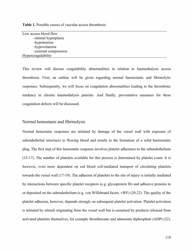
Table 1. Possible causes of vascular access thrombosis
Low access blood flow-intimal hyperplasia-hypotension-hypovolaemia-external compression
Hypercoagulability
This review will discuss coagulability abnormalities in relation to haemodialysis access
thrombosis. First, an outline will be given regarding normal haemostatic and fibrinolytic
responses. Subsequently, we will focus on coagulation abnormalities leading to the thrombotic
tendency in chronic haemodialysis patients. And finally, preventative measures for these
coagulation defects will be discussed.
Normal hemostasis and fibrinolysis
Normal hemostatic responses are initiated by damage of the vessel wall with exposure of
subendothelial structures to flowing blood and results in the formation of a solid haemostatic
plug. The first step of this hemostatic response involves platelet adherence to the subendothelium
(15-17). The number of platelets available for this process is determined by platelet count. It is
however, even more dependent on red blood cell-mediated transport of circulating platelets
towards the vessel wall (17-19). The adhesion of platelets to the site of injury is initially mediated
by interactions between specific platelet receptors (e.g. glycoprotein Ib) and adhesive proteins in
or deposited on the subendothelium (e.g. von Willebrand factor, vWF) (20-22). The quality of the
platelet adhesion, however, depends strongly on subsequent platelet activation. Platelet activation
is initiated by stimuli originating from the vessel wall but is sustained by products released from
activated platelets themselves, for example thromboxane and adenosine diphosphate (ADP) (22).
118
Platelet activation leads to expression of additional receptors on platelet membranes which
support platelet interaction with subendothelium (e.g. glycoprotein IIb/IIIa) (22-25). Most
importantly, however, these receptors mediate platelet-platelet interactions resulting in aggregate
formation.
Normal hemostasis also involves initiation of coagulation at sites of vessel wall injury that starts
with activation of factor VII from flowing blood by tissue factor present in the vessel wall. The
key products of the coagulation cascade are thrombin and fibrinogen. Thrombin proteolytically
converts fibrinogen to insoluble fibrin, which in its turn activates factor XIII that causes cross-
linking of fibrin fibres (26). With collagen, thrombin is also a main stimulus of platelet activation
and aggregation (22). Fibrin is able to stabilize platelet aggregates and other cellular elements in
a shear stress resistant network. Pathological thrombin formation is prevented by natural
anticoagulant systems, of which antithrombin III and the vitamin K-dependent protein C systems
are the most important ones. In addition to these anticoagulant systems the fibrinolytic system
generates plasmin by the action of tissue plasminogen activator on plasminogen. Plasmin
dissolves the fibrin clot and thereby prevents pathological thrombus formation. Thus, normal
hemostatic responses require both coagulation and platelet-dependent processes.
In hemodialysis patients complex coagulation abnormalities occur ranging from bleeding to
thrombosis (1,27). On the one hand, the enhanced bleeding tendency in these patients is primarily
based on functional platelet abnormalities and defective adhesion to the vessel wall (28,29). On
the other hand, a variety of coagulation abnormalities contribute to an increased thrombotic
tendency.
119
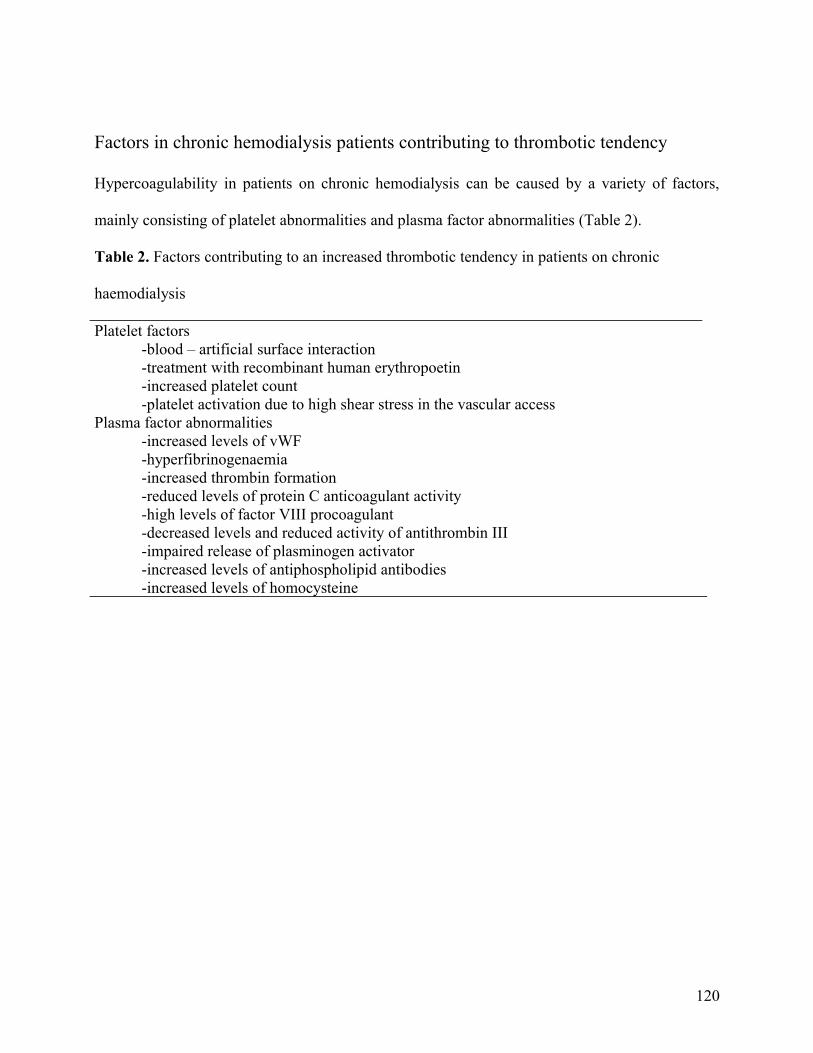
Factors in chronic hemodialysis patients contributing to thrombotic tendency
Hypercoagulability in patients on chronic hemodialysis can be caused by a variety of factors,
mainly consisting of platelet abnormalities and plasma factor abnormalities (Table 2).
Table 2. Factors contributing to an increased thrombotic tendency in patients on chronic
haemodialysis
Platelet factors-blood – artificial surface interaction-treatment with recombinant human erythropoetin-increased platelet count-platelet activation due to high shear stress in the vascular access
Plasma factor abnormalities-increased levels of vWF-hyperfibrinogenaemia-increased thrombin formation-reduced levels of protein C anticoagulant activity-high levels of factor VIII procoagulant-decreased levels and reduced activity of antithrombin III-impaired release of plasminogen activator-increased levels of antiphospholipid antibodies -increased levels of homocysteine
120
Platelet abnormalities
Platelet abnormalities are common in patients on hemodialysis. Paradoxically, most studies on
platelet abnormalities in haemodialysis patients have focused on adhesion defects leading to an
increased bleeding tendency. So, first of all, do these platelets actually play a role in the
thrombotic tendency seen in patients on hemodialysis? In other words, are there abnormal
circumstances leading to an increase of thromboembolic complications in a setting of
dysfunctional platelets? Indeed, there are indications that this is the case. Although these platelet
abnormalities exist which favour a bleeding tendency, other circumstances may actually lead to
an increase in thrombotic complications. First of all, although some studies suggest a decreased
membrane expression or an abnormal activation of platelet receptors (glycoprotein Ib and
IIb/IIIa) (30,31), an increase in platelet receptor number may be related to frequent access
obstruction (32). Also, haemodialysis is thought to activate platelets by adherence to the
extracorporeal circuit (33,34). In addition to extracorporeal activation, high shear stress and
turbulence in the vascular access may be responsible for further platelet activation (35,36).
Another condition favouring vascular access clotting is that the artificial surface of the PTFE
graft, and to a lesser extent the native arteriovenous (AV) fistula, promotes the adhesion of
fibrinogen (37,38). Serum fibrinogen, which is increased in hemodialysis patients, adheres to
glycoprotein IIb/IIIa on the surface of the platelet. Once attached to the surface (solid phase
fibrinogen) it can also bind to inactivated platelets, thereby activate them, and further enhance
platelet deposition on the surface (37). Indeed, Windus et al. (39) have shown that platelets are
deposited along the vascular access surface. Another possible complementary explanation could
be that clotting factor (contact factors like factor XII) deposition may lead to local thrombin
formation, platelet activation, and enhanced platelet deposition (40).
121
Adherent platelets may release factors (platelet-derived growth factor (PDGF), for example)
which lead to or enhance intimal hyperplasia in the vascular access and thereby reduce blood
flow through the access, which allows other activated or non-activated platelets to aggregate
more easily. This creates, despite a bleeding tendency, a favourable situation for thrombosis.
Plasma factor abnormalities
Uraemic subjects have higher levels of fibrinogen while at the same time thrombin formation is
increased (41,42). Song et al. (43) showed that a high plasma fibrinogen level is an independent
risk factor for vascular access failure in hemodialysis patients. In addition, levels of antithrombin
III may be decreased and antithrombin III activity may be reduced (44,45). The subsequent
increase of in vivo fibrinogen-fibrin transformation is reflected by increased fibrinopeptide A
formation.
Erdem et al. (46) also provided evidence for a substantial contribution of the vascular access
itself to the modulation of the thrombotic tendency. By taking blood samples from the vascular
access and from contralateral large veins in end-stage renal disease (ESRD) patients and from
peripheral veins in control subjects, they showed not only a difference in parameters of
thrombotic tendency between peripheral vein samples in ESRD patients and controls, but more
importantly, also between vascular access samples and peripheral vein samples in the same
ESRD patient group.
Others have shown that in antiphospholipid antibodies are predictive of vascular access
thrombosis (45,47-49). These antibodies include lupus anticoagulant (LA), anticardiolipin
antibodies (ACA), and antiphosphatidyl serine antibodies. Higher titres of both ACA and LA
antibodies have been demonstrated in patients with ESRD than in the general population
(48,50,51). Elevated LA antibody titres are present in up to one-third of hemodialysis patients
122
(48). Elevated ACA antibody titres have been found in 0-29% of them (48,52,53), with a greater
prevalence in patients with AV grafts (22%) than with native AV fistulae (6%) (47). However,
the association between elevated ACA antibody titre and increased access thrombosis is not fully
established yet. While several studies found no association (48,52,54), Prakash et al. (47)
demonstrated in a retrospective study a 3.7 times increased risk of recurrent thrombosis in
patients with PTFE grafts having elevated ACA antibody titres. The latter finding was recently
confirmed in a prospective study: the survival time of PTFE grafts in patients with elevated titres
was significantly shorter than in patients with normal titres. Interestingly, this difference was not
found in patients with native AV fistula (53).
Hyperhomocysteinaemia is an independent risk factor for recurrent and early-onset venous
thrombosis in patients with normal renal function (55-57). Fasting homocysteine levels have been
shown to be elevated in patients with ESRD (58) and may contribute to atherosclerosis and
subsequent cardiovascular events in haemodialysis patients (59,60). The underlying mechanism
by which hyperhomocysteinaemia provokes thrombosis is uncertain. It may involve a change in
factor V and protein C complex activity as well as effects on platelet function (56,61,62).
Studies on the association of homocysteine and access thrombosis are limited and the results are
controversial. In a retrospective study by Manns et al. (52) no association between homocysteine
level and access thrombosis was found. Two recent prospective studies showed conflicting
results. Shemin et al. (63) demonstrated a 4% increase in the risk of access thrombosis with each
1µmol/l increase of plasma homocysteine concentration for both AV fistulae and grafts.
Similarly, Ducloux et al. (64) suggested that hyperhomocysteinaemia is a risk factor for vascular
access thrombosis. However, such an association could not be demonstrated in a population of 96
patients with only native AV fistulae. The patients with the lowest levels even appeared to have
an increased mortality risk (65). These conflicting differences in outcome may be related to the
123
type of fistula, to the length of follow-up, and to other variables. Obviously, long-term
prospective studies with serial, instead of occasional, determinations of plasma homocysteine are
needed to solve this issue.
Therapeutic considerations
Anti-platelet therapy
The usefulness of antiplatelet therapy for the maintenance of internal hemodialysis access devices
was reviewed by the Antiplatelet Trialists’ Collaboration Group (66). Outcomes of nine placebo-
controlled randomized trials showed an occlusion rate of 17% in the antiplatelet groups vs 39% in
the control groups. Antiplatelet regimens consisted of ticlopidine (five studies), aspirin (two
studies), and sulphinpyrazone (two studies). Unfortunately, most of the evaluated studies were
conducted in the late 1970s and the beginning of the 1980s. At that time some of the
achievements in modern dialysis were not yet available, and dialysis populations differed from
those of today. Moreover, these studies had only a short follow-up (mean 2 months). Finally, the
analysis report was difficult to interpret in terms of excess bleeding due to antiplatelet therapy.
Only one randomized, placebo-controlled clinical trial comparing access thrombosis frequency in
patients treated with antiplatelet therapy (dipyridamole 75 mg orally 3 times a day, or aspirin 325
mg orally daily) was conducted in the last decade. Surprisingly, dipyridamole alone, which is
considered a relatively weak platelet inhibitor, had the best outcome, while patients on aspirin
had the highest frequency of thrombosis, even higher than with placebo or the combination of
dipyridamole and aspirin (67). The results of this study became more understandable after the
authors conducted a series of in vitro experiments using vascular smooth muscle cells (68,69).
They could show that aspirin potentiated PDGF-induced vascular smooth-muscle cell
124
proliferation by shunting arachidonic acid from cyclo-oxygenase into lipoxygenase pathways. On
the other hand, dipyridamole profoundly inhibited PDGF- and bFGF-induced vascular smooth-
muscle cell proliferation. This suggests that the observed direct effect of aspirin and dipyridamole
on vascular smooth-muscle cell proliferation (rather than the antiplatelet effect) is a better
explanation for the reported clinical efficiency of dipyridamole. Gastrointestinal bleeding
occurred in 16% of the treated patients vs 8% in the placebo group. Unfortunately, no anatomical
data or functional data (e.g. vascular access flow) was collected in the randomized trial (67). This
could have provided further in vivo evidence for the experimental in vitro data.
Finally, Windus et al. (70) showed that aspirin and ticlopidine both reduced dialysis-associated
platelet deposition in PTFE grafts, although they did not completely prevent it.
Oral anticoagulation
Current opinions on the use of systemic anticoagulant therapy to improve access patency is
primarily based on personal belief rather than on evidence. Not surprisingly, the reason is that
systematic data on this subject are very limited. Interestingly, despite the lack of consensus
nephrologists do not refrain from prescribing anticoagulants. As a consequence numerous
different anticoagulant strategies exist from centre to centre as to patient selection, dosing
scheme, and treatment duration.
In 1967, the effect of coumadin in reducing the clotting frequency of AV Scribner shunts was
first reported in three dialysis patients (71). A few years later coumadin was shown to prolong
cannula shunt life in non-uraemic sheep (72). However, in several recent studies aimed at
evaluating risk factors for vascular access dysfunction, anticoagulants were either not used in the
study population or their use was not mentioned. Nevertheless, the use of anticoagulants was
advocated based on the hypercoagulable state often found in the dialysis patients under study
125
(48,73). In a recent Japanese study in 83 dialysis patients with AV fistula dysfunction, no
association was found with prior anticoagulant use or prothrombin time (74). However, the
criteria mentioned for fistula dysfunction were vague and the distribution of various access types
(AV fistula or PTFE) in this population was not mentioned. Dialysis patients with a history of
early vascular access graft loss or frequent thrombosis showed a twofold prolongation of access
survival time and less frequent clotting after initiation of coumadin (53,75). However, these
results were based on small numbers and obtained with patients being their own controls.
Bleeding complications seen in these studies occurred in patients with high international
normalized ratio (INR) levels. Also, LeSar et al. (45) showed in a subgroup of dialysis patients
with frequent PTFE graft thrombosis and a hypercoagulable state (i.e. prevalence of
antiphospholipid antibodies, anti-thrombin III and protein C system abnormalities), that oral
anticoagulant therapy effectively decreased thrombosis frequency, particularly with INR values
maintained at 2.7-3.0. However, the incidence of significant bleeding complications in this subset
of patients was 10% per year. By administering coumadin, Valeri et al. (53) increased the
survival of access grafts in 16 patients with elevated ACA antibody titres and frequent access
thrombosis. However, the absolute duration of graft survival in the treated group was not
impressive (48 ± 12 days in the untreated group vs 103 ± 26 days in the patients on coumadin
with target INR values of 2.0-3.0). Two patients suffered major bleeding complications, although
both had an overshoot of target INR (> 8).
Conclusion
Despite the bleeding tendency of chronic hemodialysis patients, vascular access thrombosis is a
frequent complication. Hypercoagulability is one of the causes contributing to the high frequency
126
of access thrombosis. The hypercoagulable state can be explained by platelet and coagulation
factor abnormalities. Unfortunately, few randomized placebo-controlled trials were conducted
using antiplatelet or oral anticoagulation therapy. Therefore, no evidence-based consensus has
been established regarding pharmacological prevention of access thrombosis. It still needs to be
determined whether the potential benefits of anticoagulation and antiplatelet therapy outweigh the
risk of adverse events. In the meantime it seems reasonable to give some form of anticoagulant
therapy based on pathophysiological considerations and the high incidence of thrombotic
complications. Our group recently demonstrated that graft flow measurements could effectively
predict thrombotic vascular access events (76). Risk tables that take into account such parameters
as well as plasma markers of hypercoagulability may help to develop rationally designed trials
and guidelines.
127
References
1. Lindner A, Charra B, Sherrard DJ, Scribner BH Accelerated atherosclerosis in prolonged
maintenance hemodialysis. N Engl J Med 1974; 290:697-701
2. Feldman HI, Kobrin S, Wasserstein A: Hemodialysis vascular access morbidity. J Am
Soc Nephrol 1996; 7:523-35
3. USRDS: The economic cost of ESRD, vascular access procedures, and Medicare
spending for alternative modalities of treatment. United States Renal Data System. Am J
Kidney Dis 1997; 30 (Suppl 1):S160-S77
4. Feldman HI, Held PJ, Hutchinson JT, Stoiber E, Hartigan MF, Berlin JA: Hemodialysis
vascular access morbidity in the United States. Kidney Int 1993; 43:1091-6
5. Bosman PJ, Boereboom FTJ, Eikelboom BC, Koomans HA, Blankestijn PJ: Graft flow as
a predictor of thrombosis in hemodialysis grafts. Kidney Int 1998; 54:1726-30
6. Smits JHM, Blankestijn PJ: Haemodialysis access: the case for prospective monitoring.
Curr Opin Nephrol Hypertens 1999; 8:685-90
7. Diskin CJ, Stokes TJJ, Pennell AT: Pharmacologic intervention to prevent hemodialysis
vascular access thrombosis. Nephron 1993; 64:1-26
8. Bassiouny HS, White S, Glagov S, Choi E, Giddens DP, Zarins CK: Anastomotic intimal
hyperplasia: mechanical injury or flow induced. J Vasc Surg 1992; 15:708-16
128
9. Kanterman RY, Vesely TM, Pilgram TK, Guy BW, Windus DW, Picus D: Dialysis
access grafts: anatomic location of venous stenosis and results of angioplasty. Radiology
1995; 195:135-9
10.Roberts AB, Kahn MB, Bradford S, Lee J, Ahmed Z, Fitzsimmons J, Ball D: Graft
surveillance and angioplasty prolongs dialysis graft patency. J Am Coll Surg 1996;
183:486-92
11.Safa AA, Valji K, Roberts AC, Ziegler TW, Hye RJ, Oglevie SB: Detection and treatment
of dysfunctional hemodialysis access grafts: effect of a surveillance program on graft
patency and the incidence of thrombosis. Radiology 1996; 199:653-7
12.Smits HF, Van Rijk PP, Van Isselt JW, Mali WP, Koomans HA, Blankestijn PJ:
Pulmonary embolism after thrombolysis of hemodialysis grafts. J Am Soc Nephrol 1997;
8:1458-61
13.Smits JHM, Blankestijn PJ: Thrombosis-free hemodialysis grafts: a possibility for the
next century? Semin Dial 1999; 12:44-9
14.Windus DW: Permanent vascular access: a nephrologist's view. Am J Kidney Dis 1993;
21:457-71
15.Sakariassen KS, Bolhuis PA, Sixma JJ: Platelet adherence to subendothelium of human
arteries in pulsatile and steady flow. Thromb Res 1980; 19:547-59
16.Baumgartner HR: The role of blood flow in platelet adhesion, fibrin deposition, and
formation of mural thrombi. Microvasc Res 1973; 5:167-79
129
17.Turitto VT, Weiss HJ, Baumgartner HR: Rheological factors influencing platelet
interaction with vessel wall surfaces. J Rheol 1979; 23:735-49
18.Aarts PA, Bolhuis PA, Sakariassen KS, Heethaar RM, Sixma JJ: Red blood cell size is
important for adherence of blood platelets to artery subendothelium. Blood 1983; 62:214-
7
19.Aarts PA, Heethaar RM, Sixma JJ: Red blood cell deformability influences platelets-
vessel wall interaction in flowing blood. Blood 1984; 64:1228-33
20.Stel HV, Sakariassen KS, de Groot PG, van Mourik JA, Sixma JJ: Von Willebrand factor
in the vessel wall mediates platelet adherence. Blood 1985; 65:85-90
21.Coller BS, Peerschke EI, Scudder LE, Sullivan CA: Studies with a murine monoclonal
antibody that abolishes ristocetin-induced binding of von Willebrand factor to platelets:
additional evidence in support of GPIb as a platelet receptor for von Willebrand factor.
Blood 1983; 61:99-110
22.Sixma JJ, Wester J: The hemostatic plug. Semin Hematol 1977; 14:265-99
23.Niiya K, Hodson E, Bader R, Byers-Ward V, Koziol JA, Plow EF, Ruggeri ZM:
Increased surface expression of the membrane glycoprotein IIb/IIIa complex induced by
platelet activation. Relationship to the binding of fibrinogen and platelet aggregation.
Blood 1987; 70:475-83
24.Ruggeri ZM, De Marco L, Gatti L, Bader R, Montgomery RR. Platelets have more than
one binding site for von Willebrand factor. J Clin Invest 1983; 72:1-12
130

25.Solum NO: Procoagulant expression in platelets and defects leading to clinical disorders.
Arterioscler Thromb Vasc Biol 1999; 19:2841-6
26.Furie B, Furie BC: Molecular and cellular biology of blood coagulation. N Engl J Med
1992; 326:800-6
27.Eberst ME, Berkowitz LR: Hemostasis in renal disease: pathophysiology and
management. Am J Med 1994; 96:168-79
28.Castillo R, Lozano T, Escolar G, Revert L, Lopez J, Ordinas A: Defective platelet
adhesion on vessel subendothelium in uremic patients. Blood 1986; 68:337-42
29.Escolar G, Cases A, Bastida E Garrido M, Jopez J, Revert L, Castillo R, Ordinas A:
Uremic platelets have a functional defect affecting the interaction of von Willebrand
factor with glycoprotein IIb-IIIa. Blood 1990; 76:1336-40
30.Sloand JA, Sloand EM: Studies on platelet membrane glycoproteins and platelet function
during hemodialysis. J Am Soc Nephrol 1997; 8:799-803
31.Benigni A, Boccardo P, Galbusera M Monteagudo J, De Marco L, Remuzzi G, Ruggeri
ZM: Reversible activation defect of the platelet glycoprotein IIb-IIIa complex in patients
with uremia. Am J Kidney Dis 1993; 22:668-76
32.Liani M, Salvati F, Tresca E Di Paolo G, Vitacollona L, Golado M, Velussi C:
Arteriovenous fistula obstruction and expression of platelet receptors for von Willebrand
factor and for fibrinogen (glycoproteins GPIb and GPIIb/IIIa) in hemodialysis patients.
Int J Artif Organs 1996; 19:451-4
131
33.Hakim RM, Schafer AI: Hemodialysis-associated platelet activation and
thrombocytopenia. Am J Med 1985; 78:575-80
34.Cases A, Reverter JC, Escolar G Sanz C, Lopez-Pedret J, Revert L, Ordinas A: Platelet
activation on hemodialysis: influence of dialysis membranes. Kidney Int 1993; 43 (Suppl
41): S217-S20
35.Turitto VT, Hall CL: Mechanical factors affecting hemostasis and thrombosis. Thromb
Res 1998; 92:S25-S31
36.Viener A, Aviram M, Better OS, Brook JG: Enhanced in vitro platelet aggregation in
hemodialysis patients. Nephron 1986; 43:139-43
37.Savage B, Ruggeri ZM: Selective recognition of adhesive sites in surface-bound
fibrinogen by glycoprotein IIb-IIIa on nonactivated platelets. J Biol Chem 1991;
266:11227-33
38.Salzman EW, Lindon J, McManama G, Ware JA: Role of fibrinogen in activation of
platelets by artificial surfaces. Ann N Y Acad Sci 1987; 516:184-95
39.Windus DW, Santoro S, Royal HD: The effects of hemodialysis on platelet deposition in
prosthetic graft fistulas. Am J Kidney Dis 1995; 26:614-21
40.Colman RW, Scott CF, Schmaier AH, Wachtfogel YT, Pixley RA, Edmunds LHJ:
Initiation of blood coagulation at artificial surfaces. Ann N Y Acad Sc. 1987; 516:253-67
41.Sagripanti A, Cupisti A, Baicchi U, Ferdeghini M, Morelli E, Barsotti G: Plasma
parameters of the prothrombotic state in chronic uremia. Nephron 1993; 63:273-8
132

42.Tomura S, Nakamura Y, Deguchi F, Ando R, Chida Y, Marumo F: Coagulation and
fibrinolysis in patients with chronic renal failure undergoing conservative treatment.
Thromb Res 1991; 64:81-90
43.Song IS, Yang WS, Kim SB, Lee JH, Kwon TW, Park JS: Association of plasma
fibrinogen concentration with vascular access failure in hemodialysis patients. Nephrol
Dial Transplant 1999; 14:137-41
44.Lai KN, Yin JA, Yuen PM, Li PK: Effect of hemodialysis on protein C, protein S, and
antithrombin III levels. Am J Kidney Dis 1991; 17:38-42
45.LeSar CJ, Merrick HW, Smith MR: Thrombotic complications resulting from
hypercoagulable states in chronic hemodialysis vascular access. J Am Coll Surg 1999;
189:73-9
46.Erdem Y, Haznedaroglu IC, Celik I, Yalcin AU, Yasavul U, Turgan C, Caglar S:
Coagulation, fibrinolysis and fibrinolysis inhibitors in haemodialysis patients:
contribution of arteriovenous fistula. Nephrol Dial Transplant 1996; 11:1299-1305
47.Prakash R, Miller CC, Suki WN: Anticardiolipin antibody in patients on maintenance
hemodialysis and its association with recurrent arteriovenous graft thrombosis. Am J
Kidney Dis 1995; 26:347-52
48.Brunet P, Aillaud MF, San Marco M, Philip-Joet C, Dussol B, Bernard D, Juhan-Vague I,
Berland Y: Antiphospholipids in hemodialysis patients: relationship between lupus
anticoagulant and thrombosis. Kidney Int 1995; 48:794-800
133
49.Prieto LN, Suki WN: Frequent hemodialysis graft thrombosis: association with
antiphospholipid antibodies. Am J Kidney Dis 1994; 23:587-90
50.Garcia-Martin F, De Arriba G, Carrascosa T, Moldenhauer F, Martin-Escobar E, Val J,
Saiz F: Anticardiolipin antibodies and lupus anticoagulant in end-stage renal disease.
Nephrol Dial Transplant 1991; 6:543-7
51.Sitter T, Schiffl H: Anticardiolipin antibodies in patients on regular hemodialysis: an
epiphenomenon? Nephron 1993; 64:655-6
52.Manns BJ, Burgess ED, Parsons HG, Schaefer JP, Hyndman ME, Scott-Douglas NW:
Hyperhomocysteinemia, anticardiolipin antibody status, and risk for vascular access
thrombosis in hemodialysis patients. Kidney Int 1999; 55:315-20
53.Valeri A, Joseph R, Radhakrishnan J: A large prospective survey of anti-cardiolipin
antibodies in chronic hemodialysis patients. Clin Nephrol 1999; 51:116-21
54.George J, Aron A, Levy Y, Gilburd B, Ben-David A, Renaudineau Y, Zonana-Nachach
A, Youinou P, Harats D, Shoenfeld Y: Anti-cardiolipin, anti-endothelial-cell and anti-
malondialdehyde-LDL antibodies in uremic patients undergoing hemodialysis:
relationship with vascular access thrombosis and thromboembolic events. Hum
Antibodies 1999; 9:125-31
55.Boushey CJ, Beresford SA, Omenn GS, Motulsky AG: A quantitative assessment of
plasma homocysteine as a risk factor for vascular disease. Probable benefits of increasing
folic acid intakes. JAMA 1995; 274:1049-57
134
56.Den Heijer M, Blom HJ, Gerrits WB, Rosendaal FR, Haak HL, WijermansPW, Bos GM:
Is hyperhomocysteinaemia a risk factor for recurrent venous thrombosis? Lancet 1995;
345:882-5
57.Selhub J, D'Angelo A: Relationship between homocysteine and thrombotic disease. Am J
Med Sci 1998; 316:129-41
58.Bostom AG, Lathrop L: Hyperhomocysteinemia in end-stage renal disease: prevalence,
etiology, and potential relationship to arteriosclerotic outcomes. Kidney Int 1997; 52:10-
20
59.Bostom AG, Shemin D, Verhoef P, Nadeau MR, Jacques PF, Selhub J, Dworkin L,
Rosenberg IH: Elevated fasting total plasma homocysteine levels and cardiovascular
disease outcomes in maintenance dialysis patients. A prospective study. Arterioscler
Thromb Vasc Biol 1997; 17:2554-8
60.Moustapha A, Naso A, Nahlawi M, Gupta A, Arheart KL, Jacobsen DW, Robinson K,
Dennis VW: Prospective study of hyperhomocysteinemia as an adverse cardiovascular
risk factor in end-stage renal disease. Circulation 1998; 97: 138-41
61.McCully KS, Carvalho AC: Homocysteine thiolactone, N-homocysteine thiolactonyl
retinamide, and platelet aggregation. Res Commun Chem Pathol Pharmacol 1987;
56:349-60
62.Graeber JE, Slott JH, Ulane RE, Schulman JD, Stuart MJ: Effect of homocysteine and
homocystine on platelet and vascular arachidonic acid metabolism. Pediatr Res 1982;
16:490-3
135
63.Shemin D, Lapane KL, Bausserman L, Kanaan E, Kahn S, Dworkin L, Bostom AG:
Plasma total homocysteine and hemodialysis access thrombosis: a prospective study. J
Am Soc Nephrol 1999; 10:1095-9
64.Ducloux D, Pascal B, Jamali M, Gibey R, Chalopin JM: Is hyperhomocysteinaemia a risk
factor for recurrent vascular access thrombosis in haemodialysis patients? Nephrol Dial
Transplant 1997; 12:2037-8
65.Sirrs S, Duncan L, Djurdjev O, Nussbaumer G, Ganz G, Frohlich J, Levin A:
Homocyst(e)ine and vascular access complications in haemodialysis patients: insights into
a complex metabolic relationship. Nephrol Dial Transplant 1999; 14:738-43
66.Collaborative overview of randomised trials of antiplatelet therapy- II: Maintenance of
vascular graft or arterial patency by antiplatelet therapy. Antiplatelet Trialists'
Collaboration. BMJ 1994; 308:159-68
67.Sreedhara R, Himmelfarb J, Lazarus JM, Hakim RM: Anti-platelet therapy in graft
thrombosis: results of a prospective, randomized, double-blind study. Kidney Int 1994;
45:1477-83
68.Harvey R, Bredenberg CE, Couper L, Himmelfarb J: Aspirin enhances platelet-derived
growth factor-induced vascular smooth muscle cell proliferation. J Vasc Surg 1997;
25:689-95
69.Himmelfarb J, Couper L: Dipyridamole inhibits PDGF- and bFGF-induced vascular
smooth muscle cell proliferation. Kidney Int 1997; 52:1671-7
136
70.Windus DW, Santoro SA, Atkinson R, Royal HD: Effects of antiplatelet drugs on
dialysis-associated platelet deposition in polytetrafluoroethylene grafts. Am J Kidney Dis
1997; 29:560-4
71.Wing AJ, Curtis JR, De Wardener HE: Reduction of clotting in Scribner shunts by long-
term anticoagulation. Br Med J 1967; 3:143-5
72.Boyd SJJ, Dennis MB, Nogami RT, Cole JJ, Scribner BH: Prophylactic coumadin and A-
V cannula function. J Appl Physiol 1974; 37:6-7
73.Ambühl PM, Wüthrich RP, Korte W, Schmid L, Krapf R:. Plasma hypercoagulability in
haemodialysis patients: impact of dialysis and anticoagulation. Nephrol Dial Transplant
1997; 12:2355-64
74.Masaki T, Yorioka N, Kyuden Y, Yamashita K, Taniguchi Y, Yamakido M, Tanaka J,
Harada S: Factors influencing arteriovenous fistula dysfunction in Japanese patients on
chronic hemodialysis. Int J Artif Organs 1999; 22:14-7
75.Tevlin MT, Portner ME, Kraus MG, Dulaney JT: Clotting frequency of hemodialysis
accesses before and after systemic anticoagulation with coumadin. J Am Soc Nephrol
1996; 7:1421 (Abstract)
76.Blankestijn PJ, Smits JH: How to identify the haemodialysis access at risk of thrombosis?
Are flow measurements the answer? Nephrol Dial Transplant 1999; 14:1068-71
137

Chapter 8
SUMMARY AND GENERAL DISCUSSION
138
Summary and general discussion
Since the introduction of the AV fistula and the use of interposition graft little improvement has
been made in the vascular access field. Still, vascular access related complications, are one of the
most important reasons for patient hospitalization, morbidity and even mortality (1,2).
Interestingly, the costs of vascular access related care was found to be more than fivefold higher
for patients with AVG compared with patients with a functioning AVF (3). In an attempt to
improve overall patency rates and reduce access related costs, the NFK-DOQI committee
currently recommends that in any dialysis center the majority of new dialysis patients should
have a primary AVF constructed (4,5). Unfortunately, creation of an AVF is not always possible
as a consequence of prior vascular access surgery, insufficient caliber of forearm vessels or
sclerosis caused by prior venipunctures. Although less thrombotic and infectious complications
occur in AVF, the AVF is not ideal, i.e. 100% successful. Adequate maturation of AVF, i.e.
sufficient dilatation and arterialization, fails in up to 30% of all newly created fistulae, resulting
in delayed initiation of dialysis treatment or placement of temporary central venous dialysis
catheters, and related morbidity (6-10). Optimalization of patient selection could improve AVF
patency rates and access related morbidity. Considering the increasing age of hemodialysis
patients, prolongation of the dialysis therapy, and the worldwide increase of the number of
patients requiring hemodialysis, the number of AV grafts will probably increase rather than
decrease. As a consequence, vascular access complications, particularly thrombosis, will continue
to challenge vascular access care in the future.
In chapter 3 we examined the importance of pre-operative forearm venous distensibility with
respect to AVF maturation. With the recognition of the superiority of the AVF and the increasing
139
comorbidity of the hemodialysis population, efforts are made to evaluate the vasculature of the
arm of the patient prior to access surgery. This prospective study evaluates forearm venous
function rather than anatomy prior to AVF creation for the first time. The results of this study
confirm our hypothesis that apart from structural changes, the functional properties of forearm
veins are important in the adaptive response to increased blood flow after AVF creation.
Furthermore, functional data of forearm vasculature do not correspond with anatomical data.
Importantly, we demonstrate that venous distensibility is predictive of the potential success of
AVF construction and adequate maturation, whereas venous and arterial diameters are not.
Finally, our study confirms recent data indicating that AVF flow measured one day after surgery
is higher in AVF with successful maturation in compare to AVF that fail to mature (11).
Considering our data support the hypothesis that functional vessel wall characteristics predict
fistula maturation, the preoperative evaluation of the forearm vasculature should not only focus
on resting static diameters. In view of the large potential benefits of optimization of the matching
of patients and access type, further and larger studies of novel techniques are urgently needed.
In chapter 4, we determined whether forearm blood flow capacity in patients with end-stage
renal failure awaiting vascular access surgery, is predictive of early failure of AVF. We
hypothesized that forearm blood flow capacity, i.e. the increase of forearm blood flow as a result
of vasodilatation, is an important determinant of failure of newly created AVF in hemodialysis
patients. To discriminate the influence of the endothelium in early fistula failure, we measured
both endothelium dependent (i.e. metacholine-induced) and endothelium independent (i.e.
sodium nitroprusside-induced) forearm vasodilatation, using forearm venous occlusion
plethysmography. To our knowledge, this is the first time invasively measured forearm blood
flow capacity is related to outcome of AVF. This study shows that pre-operative forearm blood
140
flow capacity is predictive of early AVF failure. Especially, the AUC of dose response curves
seems predictive of AVF outcome. An AUC of 100 in MCh and 200 in NP seems a prerequisite
of short term success of the AVF. Furthermore, our data indicate that AVF failure is not the result
of pure endothelial dysfunction, because endothelial-independent forearm blood flow capacity is
also diminished in patients with AVF failure. Structural arterial wall changes can be responsible
for this finding (12). Finally, we showed that arterial diameter is not predictive of early AVF
failure. Venous occlusion plethysmography has the advantage of measurement of total forearm
blood flow. However, clinical implementation of venous occlusion plethysmography prior to
access surgery is not simple. First, this technique is very time-consuming and expensive devices
are needed. Second, and even more important, it is an invasive measurement. Future research
should focus on easier and non-invasive methods to measure pre-operative arterial flow reserve.
Chapter 5 explored the effect of monitoring and selection for corrective interventions based on
VP and/or Qa, on thrombosis rates of AVG. This study confirms the usefulness of VP and Qa
measurements as access surveillance variables: thrombosis rates in our patients can be
maintained below the quality of care standards formulated by the NKF-DOQI committee (13).
Based on the hypothesis that stenoses located in the flow tract upstream the venous needle would
not by picked up by VP measurements, we expected Qa measurements, or combined
measurement of Qa and VP, to be superior as a monitoring tool, in compare to VP measurements
alone (14). This hypothesis is not confirmed in our study: thrombosis rates in groups monitored
by VP or Qa alone or by the combination of tests do not differ. We found a strong predominance
of venous lesions in the patients referred for angiography or thrombectomy. In less than 5% of
patients referred for angioplasty arterial lesions were found. It is possible that in patients who
develop more arterial lesions, surveillance using Qa would result in lower thrombosis rates than
141
using VP measurements. Our study confirms previous evidence that dynamic VP does not
accurately reflect resistance caused by stenosis formation, since most referrals for intervention in
patients monitored by VP measurements alone were based on static VP measurements (15).
Dynamic VP is highly biased by pump flow, blood tubing, needle size, and blood pressure (16).
Static VP measurements, on the other hand, particularly when corrected for mean arterial
pressure, avoid these potential confounders.
Our study results indicate that the decision which method to use, (static) VP or Qa, can in fact be
based on the preferences of those involved in the access care. There is no rationale in combining
VP and Qa monitoring. It is important to bear in mind that a monitoring strategy can only be
successful when abnormal tests are followed by diagnostic and interventional procedures on short
notice. This is illustrated by the fact that almost one third of all thrombotic events in this study
occurred in patients awaiting angiography.
In chapter 6 we assessed hemodialysis access function in patients undergoing PTA. To our
knowledge this is the first study in AVG and AVF reporting both angiographic and functional
results of PTA. This study confirms earlier data that PTA results in an immediate access flow
increase of about 250 mL/min (17). Although post-PTA stenosis poorly predicts patency after
PTA, the post-PTA stenosis is frequently used to express the efficiency of the procedure (18). We
demonstrated that adequate angiographic improvement, i.e. 25% or less post-PTA stenosis, does
not necessarily correspond with adequate functional improvement, i.e. access flow above 600
mL/min. More important, we found functional parameters are predictive for long-term outcome,
whereas angiographic results are not. Whereas almost all PTA procedures were angiographically
successful, only 66% of AVG and 50% of AVF obtained adequate access flow. This may be
caused by rapid elastic recoil of the stenotic lesion or may reflect other unidentified and thus
142
untreated stenoses. Access flow measurements during, or just after the intervention could be
helpful to overcome this problem and optimize PTA results (22-24). Finally, time to repeat PTA
in our study is substantially shorter than in other studies (19-21,25). This suggests a more rapid
recurrence of stenosis in the present study. This difference may reflect differences in stenosis
severity prior to PTA, as a result of more strict selection criteria for referral for angiography. The
combined data of these studies suggest there is an optimal moment of PTA.
Chapter 7 reviews coagulability abnormalities in relation to hemodialysis access thrombosis.
An increased thrombotic tendency is an important cause of ischaemic heart disease, stroke, and
vascular access thrombosis in chronic hemodialysis patients (26). Although most cases of access
thrombosis are associated with low access blood flow, caused by intimal hyperplasia in the
venous outflow tract, in a number of cases, thrombosis is not preceded by low access flow
measured during hemodialysis. Hypotension, hypovolemia and external compression during the
interdialytic period are proposed mechanisms of these non-stenotic thrombotic events (27).
However, several coagulability abnormalities may attribute to access thrombosis. Platelet
abnormalities are common in patients on hemodialysis. Obviously, dysfunctional platelets favor a
bleeding tendency. However, other circumstances may create a favorable situation for thrombotic
complications. Adherence of platelets to the extracorporal circuit, high shear stress and
turbulence in the vascular access may be responsible for exaggerated platelet activation (28-31).
The artificial surface of the polytetrafluoroethylene material of AVG promotes adhesion of
fibrinogen on platelet receptors, thereby activating them, and adhancing platelet deposition on the
surface (32). High plasma fibrinogen and elevated titers of antiphospholipide antibodies are
shown to increase the risk of access failure (33-38). Hyperhomocysteinemia has also been
proposed as potential risk factor of access thrombosis (39,40). However, studies on this subject
143
are limited and the results are controversial. Obviously, long-term prospective studies with serial,
instead of occasional determinations of plasma homocysteine are needed to address this problem.
What are the clinical implications of these findings? Unfortunately, few randomized placebo-
controlled trials were conducted using antiplatelet or oral anticoagulation therapy. Therefore, no
evidence-based consensus has been established regarding pharmacological prevention of access
thrombosis. Furthermore, it remains questionable whether the benefits of antithrombotic therapy
outweigh the risk of bleeding complications. In the meantime it seems reasonable to give some
form of anticoagulant therapy based on pathophysiological considerations and the high risk of
thrombotic complications. Future research on hemodynamic factors affecting the lifespan of
vascular accesses should also take into account plasma markers of hypercoagulability. Because
personal belief is an important factor in prescribing anticoagulant therapy, it seems unlikely that
long-term placebo-controlled trials will be started in the near future.
Future perspectives
The nephrologist is primarily responsible for the management of patients with renal failure, both
before and after initiation of dialysis treatment. Together with the patient he makes the decision
as to when hemodialysis should begin. According to the National Kidney Foundation’s recent
Clinical Practice Guidelines, patients with ESRD should be prepared for dialysis when their
glomerular filtration rate is less than 25 mL/min. This provides sufficient time for maturation and
incorporation of the access. More accurate coordination between the placement of a fistula or
graft and the initiation of dialysis may avoid the need of central venous catheters. Furthermore,
timely referral to a nephrologist is associated with a higher proportion of patients starting
hemodialysis on a functioning AVF and a lower overall mortality. Planning a vascular access
144
strategy for the individual patient well in advance of dialysis however, is best accomplished in a
multidisciplinary setting, in which all efforts are made to create native AVF if possible.
Doppler ultrasound has been the most extensively studied and widely used test to guide access
creation. However, we have demonstrated that seemingly adequate vessel diameter alone does
not guarantee successful outcome of AVF. The influence of functional variables like forearm
blood flow reserve and venous distensibility on the outcome of AVF has never been studied
before and should be addressed extensively in future studies. Pre-existing endothelial dysfunction
and structural wall changes are proposed mechanisms of forearm vessel dysfunction in patients
with ESRD. Once the role of endothelial dysfunction on outcome of AVF is clarified, it would be
very interesting to explore the effects of medications that could prevent endothelial cell injury
prior to access surgery, like statins or angiotensin converting enzyme inhibitors. Intimal
hyperplasia is a well-known cause of access thrombosis. However, it has become clear that these
intimal changes can be demonstrated in most of the apparently normal cephalic veins in ESRD
patients at the time of AVF construction (41). Although several efforts are made to decrease
intimal hyperplasia after access creation, it would be of interest to study the effect of
pharmacological inhibition of intimal hyperplasia in advance of access creation.
Patients aged 65 or more undergoing dialysis, have a life-expectancy of approximately 3 years,
and life-expectancy in the oldest age groups is very limited. Therefore, dialysis in the elderly
remains a challenge and raises the question whether it is proper to offer expensive and
complicated treatment to a growing worldwide population of older patients with renal failure.
Although DOQI Vascular Access Guidelines strongly recommand the contruction of AVF in
elderly, it seems reasonable to consider other options in elderly patients with short life
expectancy from other comorbidities or chronic hypotension. Recently, the Dialock system, a
device totally implanted under the pectoral skin and inserted in the subclavian vein, was
145
demonstrated to be safe and effective in “complex” hemodialysis patients. Infection and
complication rates of this device were even lower in compare to permanent central venous
catheters and AVG (42,43). It remains to be elucidated whether this device is useful in the
elderly. Also, the cost-effectiveness of this approach needs to be addressed.
Over the past years the effectiveness of access surveillance has been demonstrated. Prospective
surveillance of vascular accesses for hemodynamically significant stenoses, and subsequent
referring for percutaneous transluminal angioplasty or surgical revision, improves patency rates
and decreases the incidence of thrombosis. At present, access flow (Qa) and venous pressure
measurements are preferred techniques that can be used in surveillance of both AVG and AVF.
However, structural access surveillance still needs to be introduced in many dialysis centers. An
adequate surveillance strategy can only succeed in an enthusiastic team with affinity for the
vascular access, consisting of a nephrologist, vascular surgeon, radiologist and nursing staff.
146
References
1. Powe NR, Jaar B, Furth SL, Hermann J, Briggs W: Septicaemia in dialysis patients:
incidence, risk factors and prognosis. Kidney Int 1999; 55: 1081-90
2. Schwab SJ, Beathard G: The hemodialysis catheter conundrum: hate living with them, but
can’t live without them. Kidney Int 1999; 56: 1-17
3. Lee H, Manns B, Taub K, Ghali WA, Dean S, Johnson D, Donaldson C: Cost analysis of
ongoing care of patients with end-stage renal disease: the impact of dialysis modality and
dialysis access. Am J Kidney Dis 2002; 40:611-22
4. National Kidney Foundation. NKF-DOQI clinical practice guidelines for vascular access. Am
J Kidney Dis 1997; 30 (Suppl 3): S152-S191
5. National Kidney Foundation. K/DOQI clinical practice guidelines for vascular access. Am J
Kidney Dis 2001; 37 (Suppl 1): S137-S181
6. Elfström J, Thomsen M: The prognostic value of blood-flow measurements during
construction of arteriovenous fistulae. Scand J Urol Nephrol 1981;15(3):323-6
7. Kalman PG, Pope M, Bhola C, Richardson R, Sniderman KW: A practical approach to
vascular access for hemodialysis and predictors of success. J Vasc Surg 1999; 30:727-33
8. Silva MB, Hobson RW, Pappas PJ, Jamil Z, Araki T, Goldberg MC, Gwertzman G, Padberg
FT: A strategy for increasing use of autogenous hemodialysis access procedures: impact of
preoperative non-invasive evaluation. J Vasc Surg 1998; 27:302-8
9. Yerdel MA, Kesenci M, Yazicioglu KM, Döşeyen Z, Türkçapar AG, Anadol E: Effect of
haemodynamic variables on surgically created arteriovenous fistula flow. Nephrol Dial
Transplant 1997; 12:1684-88
147
10.Lin SL, Huang CH, Chen HS, Hsu WA, Yen CJ, Yen TS: Effects of age and diabetes on
blood flow rate and primary outcome of newly created hemodialysis arteriovenous fistulas.
Am J Nephrol 1998; 18:96-100
11.Wong V, Ward R, Taylor J, Selvakumar S, How TV, Bakran A: Factors associated with early
failure of arteriovenous fistulae for haemodialysis access. Eur J Endovasc Surg 1996; 12:207-
13
12. Malovrh M: Native arteriovenous fistula: preoperative evaluation. Am J Kidney Dis 2002;
39:1218-25
13. National Kidney Foundation. NKF-DOQI clinical practice guidelines for vascular access. Am
J Kidney Dis 1997; 30 (Suppl 3):S152-S91
14.Blankestijn PJ, Smits JH: How to identify the haemodialysis access at risk of thrombosis? Are
flow measurements the answer? Nephrol Dial Transplant 1999; 14:1068-71
15.Besarab A, Sullivan KL, Ross RP, Moritz MJ: Utility of intra-access pressure monitoring in
detecting and correcting venous outlet stenoses prior to thrombosis. Kidney Int 1995;
47:1364-73
16.Besarab A, Dorrell S, Moritz M, Michael H, Sullivan K: Determinants of measured dialysis
venous pressure and its relationship to true intra-access venous pressure. ASAIO Trans 1991;
37:M270-M271
17.Schwab SJ, Oliver MJ, Suhocki P, McCann R: Hemodialysis arteriovenous access: detection
of stenosis and response to treatment by vascular access blood flow. Kidney Int 2001;
59:358-62
18.Beathard GA: Percutaneous transvenous angioplasty in the treatment of vascular access
stenosis. Kidney Int 1992; 42:1390-7
148
19.Kanterman RY, Vesely TM, Pilgram TK, Guy BW, Windus DW, Picus D: Dialysis access
grafts: anatomic location of venous stenosis and results of angioplasty. Radiology 1995;
195:135-9
20.Lumsden AB, MacDonald MJ, Kikeri D, Cotsonis GA, Harker LA, Martin LG: Prophylactic
balloon angioplasty fails to prolong the patency of expanded polytetrafluoroethylene
arteriovenous grafts: results of a prospective randomized study. J Vasc Surg 1997; 26:382-90
21.Martin LG, MacDonald MJ, Kikeri D, Cotsonis GA, Harker LA, Lumsden AB: Prophylactic
angioplasty reduces thrombosis in virgin ePTFE arteriovenous dialysis grafts with greater
than 50% stenosis: subset analysis of a prospectively randomized study. J Vasc Interv Radiol
1999; 10:389-96
22.Ahya SN, Windus DW, Vesely TM, Lattimore BA: Utility of radiologic criteria for predicting
access flow after percutaneous transluminal angioplasty [Abstract]. J Am Soc Nephrol 10
(ASN Program and Abstracts): 1999; 200A
23.Davidson CJ, Newman GE, Sheikh KH, Kisslo K, Stack RS, Schwab SJ: Mechanisms of
angioplasty in hemodialysis fistula stenoses evaluated by intravascular ultrasound. Kidney Int
1991; 40:91-5
24.Vesely T, Gherardini D, Starostin D, Krivitski N: Preliminary experiences using intravascular
blood flow monitor (IBFM) during vascular access angioplasty [Abstract]. J Am Soc Nephrol
10 (ASN Program and Abstracts): 1999; 221A
25.Turmel-Rodrigues L, Pengloan J, Baudin S, Testou D, Abaza M, Dahdah G, Mouton A,
Blanchard D: Treatment of stenosis and thrombosis in haemodialysis fistulas and grafts by
interventional radiology. Nephrol Dial Transplant 2000; 15:2029-36
26.Lindner A, Charra B, Sherrard DJ, Scribner BH: Accelerated atherosclerosis in prolonged
maintenance hemodialysis. N Engl J Med 1974; 290: 697-701
149

27.Windus DW: Permanent vascular access: a nephrologist's view. Am J Kidney Dis 1993; 21:
457-71
28.Hakim RM, Schafer AI: Hemodialysis-associated platelet activation and thrombocytopenia.
Am J Med 1985; 78:575-80
29.Cases A, Reverter JC, Escolar G, Sanz C, Lopez-Pedret J, Revert L, Ordinas A: Platelet
activation on hemodialysis: influence of dialysis membranes. Kidney Int 1993; 43 (Suppl
41): S217-S220
30.Turitto VT, Hall CL: Mechanical factors affecting hemostasis and thrombosis. Thromb Res
1998; 92:S25-S31
31.Viener A, Aviram M, Better OS, Brook JG: Enhanced in vitro platelet aggregation in
hemodialysis patients. Nephron 1986; 43:139-43
32.Savage B, Ruggeri ZM: Selective recognition of adhesive sites in surface-bound fibrinogen
by glycoprotein IIb-IIIa on nonactivated platelets. J Biol Chem 1991; 266: 11227-33
33.Salzman EW, Lindon J, McManama G, Ware JA: Role of fibrinogen in activation of platelets
by artificial surfaces. Ann N Y Acad Sci 1987; 516:184-95
34.Song IS, Yang WS, Kim SB, Lee JH, Kwon TW, Park JS: Association of plasma fibrinogen
concentration with vascular access failure in hemodialysis patients. Nephrol Dial Transplant
1999; 14:137-41
150
35.LeSar CJ, Merrick HW, Smith MR: Thrombotic complications resulting from
hypercoagulable states in chronic hemodialysis vascular access. J Am Coll Surg 1999;
189:73-9
36.Prakash R, Miller CC, Suki WN: Anticardiolipin antibody in patients on maintenance
hemodialysis and its association with recurrent arteriovenous graft thrombosis. Am J Kidney
Dis 1995; 26:347-52
37.Brunet P, Aillaud MF, San Marco M, Philip-Joet C, Dussol B, Bernard D, Juhan-Vague I,
Berland Y: Antiphospholipids in hemodialysis patients: relationship between lupus
anticoagulant and thrombosis. Kidney Int 1995; 48:794-800
38.Prieto LN, Suki WN: Frequent hemodialysis graft thrombosis: association with
antiphospholipid antibodies. Am J Kidney Dis 1994; 23:587-90
39.Shemin D, Lapane KL, Bausserman L, Kanaan E, Kahn S, Dworkin L, Bostom AG: Plasma
total homocysteine and hemodialysis access thrombosis: a prospective study. J Am Soc
Nephrol 1999; 10:1095-9
40.Ducloux D, Pascal B, Jamali M, Gibey R, Chalopin JM: Is hyperhomocysteinaemia a risk
factor for recurrent vascular access thrombosis in haemodialysis patients? Nephrol Dial
Transplant 1997; 12:2037-8
41.Wali MA, Eid RA, Dewan M, Al-Homrany MA: Intimal changes in the cephalic vein of renal
failure patients before arterio-venous fistula (AVF) construction. J Smooth Muscle Res 2003;
39:95-105
151
42.Canaud B, Morena M, Leray-Moragues H: Dialock. results of a French multicenter study.
Nephrology 2001; 22:391-7
43.Hessinger M, Zitta S, Holzer H, Tiesenhausen K: Dialock - an alternative to Permcath and
access graft. Zentralbl Chir 2003; 128:753-6
Dankwoord
Zoals waarschijnlijk voor elke promovendus geldt, was ook dit proefschrift een enorme
bevalling. Sterker nog, een bevalling is minder erg! Gedurende geruime tijd en met ruime
tussenpozen, ben ik bezig geweest met het onderzoek wat heeft geleid tot het schrijven van dit
proefschrift. Ik heb hierbij gaandeweg heel veel mensen leren kennen, die stuk voor stuk hebben
bijgedragen aan mijn motivatie het onderzoek ook waardig af te sluiten met een promotie!
Klinisch onderzoek staat of valt bij de bereidwilligheid van patiënten. Een ereplaats in deze
opsomming is dan ook wel verdiend. Allereerst wil ik dan ook alle dialyse en pre-dialyse
patiënten bedanken voor hun medewerking aan de onderzoeken. Hun continue doorzettingskracht
en getoonde interesse in mijn vorderingen hebben mij erg geïnspireerd. Daarnaast heeft het ook
veel gezellige momenten opgeleverd, met name tijdens de flowmetingen op de dialyseafdeling.
Voor het schrijven van dit proefschrift is de wetenschappelijke en/of sociale bijdrage van vele
anderen onmisbaar, daarom wil ik graag de volgende personen bedanken.
Uit angst conventioneel te zijn heb ik me mogen scharen tussen maar liefst twee co-promotoren.
Dr. M.A. van den Dorpel, beste René, ik ben ervan overtuigd dat ik het niet had gered zonder
jouw geduld en doorzettingsvermogen. Jij hebt de gave alles vanuit een positieve kant te
benaderen. Daarnaast heeft onze langdurige samenwerking ook hele gezellige en grappige
152
momenten opgeleverd. Het heeft wat koude grond psychologie van jouw zijde gevergd, maar het
resultaat is er! Wat zul je blij zijn dat het af is (in meerder opzichten). Je relativerende oordeel
over patiëntenzorg blijkt in de praktijk trouwens volledig te kloppen!
Dr. P.J. Blankestijn, beste Peter, ik bewaar goede herinneringen aan onze telefonische meetings,
waarin in korte tijd spijkers met koppen geslagen konden worden. Dr. J.H.M. Smits, beste
Sander, jij hebt het er al even (!) opzitten. Het voelt echter nog steeds alsof ik onze eer moet
verdedigen! Ik hoop dat jij wel bij mijn verdediging aanwezig kunt zijn! Zonder jullie inbreng
van Utrechtse zijde, had mijn proefschrift nooit tot stand kunnen komen.
Prof. Dr. W. Weimar, beste Willem, hartelijk bedankt dat je mijn promotor hebt willen zijn. De
leden van de beoordelingscommissie, Prof.dr. P.M.T. Pattynama, Prof.dr. H. van Urk en Prof.dr.
H.A.P. Pols, wil ik bedanken voor hun deelname in de promotiecommissie en het kritisch lezen
van mijn proefschrift.
Het is een onmogelijke taak alle medewerkers van de dialyse-afdeling van het Clara Ziekenhuis,
persoonlijk te bedanken. Desalniettemin zijn jullie stuk voor stuk onmisbaar geweest voor het
verzamelen van mijn data, het motiveren van patiënten om mee te werken aan mijn onderzoek en
niet te vergeten het motiveren van deze dokter! Gezellig waren ook de droppings, waarbij ik eens
bijna mijn rug brak in een greppel, doordat een zekere broeder het nodig vond om op mijn rug te
springen. Hanneke Pernot, als onze eerste echte onderzoeksnurse heb je mijn werk een stuk
gemakkelijker gemaakt, zeker toen ik weer in de kliniek zat. Jouw doortastendheid en zeker ook
brutaliteit hebben geleid tot heel wat data. Wat hebben we veel geouwehoerd, gemopperd, maar
ook gelachen! Hiervoor mijn eeuwige dank!
Voor mijn onderzoek was de samenwerking met de vaatchirurgen, Dr. Yo en André de Smet,
maar ook de afdeling Radiologie, zowel radiologen als laboranten., van meet af aan een must. Via
153

deze weg wil ik hen dan ook bedanken voor het laagdrempelige overleg en hun bereidwilligheid.
Op deze plaats verdient ook Drs Burgmans speciale aandacht. Lieve Ine, het was zeker geen
makkelijke tijd, maar gezellig was het altijd wel. Met name aan San Antonio (ik heb nog leuke
but-foto’s) en Tours bewaar ik hele goede herinneringen!
Na alle flow- en drukmetingen, brak het plethysmografie-tijdperk aan. Dr. Van den Meirakker,
beste Ton, ik wil je bedanken voor de mogelijkheden die ik, als niet EMC-er kreeg, gebruik te
maken van ‘jouw’afdeling en je wetenschappelijke input bij het opstellen van de protocollen, en
het schrijven van mijn artikelen. Dr. Lameris, lieve Thomas, aan jou ben ik veel dank
verschuldigd. Of het nou ging om een postertje, de plethysmograaf, een abstract, een artikel of
een persoonlijk advies, je was er altijd! Vergeleken bij jou voelde ik me altijd een complete
wetenschappelijke analfabeet. Als ik iets sneller was geweest, had het anders gelopen.
Desalniettemin, alle lof en mijn eeuwige dank! Als ik aan plethysmografie denk, denk ik direct
aan de meiden van het Inwendige I lab. Bianca, Marjolein en Evelien, het waren tropenjaren!
Werkelijk alles wat mis kon gaan ging mis, maar met z’n allen wisten we het probleem toch altijd
weer op te lossen. Jullie geduld, vindingrijkheid maar vooral ook gezelligheid, is onvergetelijk!
Lieve Jacqueline, Jack voor intimi, hoe kan ik jou beter bedanken, dan vragen –alsnog- mijn
getuige te zijn bij deze ‘bruiloft in mijn ééntje’. In die lange tijd dat ik je nu ken ben je er altijd
voor me geweest en heb je me ook altijd gesteund, no matter what! Met jou kan ik keihard lachen
én janken, liefst allemaal op één avond. Ik ben er erg trots op dat je mijn vriendin bent en dat jij
als paranymf aan mijn zijde zal staan op deze belangrijke dag in mijn leven.
Dr. Wittich, lieve Philip, in ruil voor al je gezelligheid en ontspannende momenten, vergeef ik je
dat ik niks aan je heb als paranymf (eigen woorden!). Het levert hoe dan ook een mooi plaatje op,
en een hoop stof tot discussie. Ik ben blij dat je erbij bent!
154
Lieve ouders, jullie wil ik bedanken voor al jullie steun de afgelopen jaren. Zowel in praktische
(de zorg voor mij en Boris), als in mentale zin (jullie hebben me altijd vrijgelaten om die dingen
te doen die ik graag wilde doen), zijn jullie onmisbaar geweest voor mijn carrière en niet op de
laatste plaats de afronding van dit proefschrift. Ma, dit is nou een moment om eens niet
bescheiden te zijn: je bent fantastisch in alles wat je doet!
Lieve Rob, ik heb het je niet gemakkelijk gemaakt de afgelopen jaren. Ik ben jou oneindig veel
dank verschuldigd voor je eeuwige geloof in mij. Mede daardoor kwam het boekje af. Je hebt me
enorm veel gegeven, waarbij Boris bovenaan staat en ons altijd zal blijven verbinden!
Tenslotte wil ik alle familie en vrienden bedanken voor de zo broodnodige ontspanning in tijden
van stress. Ik ben zeker niet de meest attente maar wel de meest chaotische zus en vriendin
denkbaar, maar nu deze last van mijn schouders is gevallen, durf ik jullie beterschap te beloven!
Lieve X., zonder sentimenteel te worden, hoop ik met heel mijn hart, dat everlasting eeuwig
duurt!
Allerliefste Boris, mijn o zo grote maar nog kleine man, mama is er klaar mee! Het wordt weer
eens tijd voor een vliegreisje dacht ik zo...
155
Curriculum Vitae
Joke van der Linden werd op 21 maart 1972 geboren te Dordrecht en is getogen in Zwijndrecht.
Zij behaalde in 1990 haar Atheneum diploma op Openbare Scholen Gemeenschap Walburg. In
datzelfde jaar startte zij met haar studie Geneeskunde aan de Erasmus Universiteit Rotterdam,
alwaar zij in 1994 het doctoraal Geneeskunde behaalde. In afwachting van haar co-schappen liep
zij enkele maanden stage op de afdeling Chirurgie in het Tyks Ziekenhuis in Türku, Finland. In
november 1996 legde zij haar artsexamen af. Daarna startte zij als arts-assistent bij de afdeling
Interne Geneeskunde in het St. Clara Ziekenhuis te Rotterdam (Dr. A.F. Grootendorst). In
afwachting van een opleidingsplaats, heeft zij gedurende twee jaar onderzoek verricht bij de
afdeling Nefrologie binnen dit ziekenhuis. Hier werd het idee opgevat te gaan promoveren in het
veld van de shuntzorg. In december 2000 is zij begonnen met de opleiding tot internist in het
Erasmus M.C. te Rotterdam (opleiders Prof. Dr. H.A.P. Pols en Dr. J.C.L.M. van Saase). Aan het
einde van haar eerste opleidingsjaar beviel zij van haar zoon Boris. Na haar zwangerschapsverlof
keerde zij gedurende drie jaar terug naar het M.C.R.Z. locatie Clara (voorheen St. Clara
Ziekenhuis) te Rotterdam. Het promotie onderzoek werd in die tijd voortgezet. In januari 2005 is
zij inmiddels gestart aan het aandachtsgebied Endocrinologie in het Erasmus M.C. (Dr. A.J. van
der Lely).
156
Bibliography
Van der Linden J, Sinnige HAW, van den Dorpel MA. Gustatory sweating and diabetes. Neth J
Med 2000; 56(4):159-62
Smits JH, van der Linden J, Blankestijn PJ, Rabelink TJ. Coagulation and haemodialysis access
thrombosis. Nephrol Dial Transplant 2000; 15(11):1755-60
Smits JH, van der Linden J, Hagen EC, Modderkolk-Cammeraat EC, Feith GW, Koomans HA,
van den Dorpel MA, Blankestijn PJ. Graft surveillance: venous pressure, access flow, or the
combination? Kidney Int 2001; 59(4):1551-8
De Rave S, Tilanus HW, van der Linden J, de Man RA, vd Berg B, Hop W, IJzermans JNM,
Metselaar HJ.The importance of orthotopic liver transplantation in acute hepatic failure. Transpl
Int 2002;15(1):29-33
Van der Linden J, Smits JH, Assink JH, Wolterbeek DW, Zijlstra JJ, de Jong GH, van den
Dorpel MA, Blankestijn PJ. Short- and long-term functional effects of percutaneous transluminal
angioplasty in hemodialysis vascular access. J Am Soc Nephrol 2002; 13(3):715-20
Van der Linden J, Lameris TW, van den Meiracker AH, André A.E.A de Smet AAEA,
Blankestijn PJ, van den Dorpel MA. Forearm venous distensibility predicts successful arterio-
venous fistula. Submitted
157
Van der Linden J, Lameris TW, van den Meiracker AH, de Smet AAEA, Blankestijn PJ, van
den Dorpel MA. Role of forearm blood flow reserve in early failure of newly created arterio-
venous hemodialysis fistulae. Submitted
Van der Linden J, van den Dorpel MA. Percutaneous transluminal angioplasty of failing
hemodialysis grafts and fistulae. Saudi J Kidney Dis Transplant 2004
158