Ind 90020640
4
Plant Cell Reports (1990) 8:586-589 Transformation of cotton (Gossypium hirsutum L . ) via particle bombardment* Pla nt Cel l Reports © Springer-Verlag 1990 John J. Finer 1 and Michael D. M cMullen 1, 2 Department of Agronomy and Ohio State Bioteehnology Center 1, and Corn and Soybean Research Unit, Agricultural Research Service, USDA z, Ohio Agricultural Research and Development Center, The Ohio State University, Wooster, OH 44691, USA Received Nove mber 15, 1989/Revised version received January 17, 1990 - Com mun icated by G. C. Phillips ABSTRACT Embryogenic suspension cultures of cotton (Gossypium hirsutum L.) were subjected to particle bombardment, where high density particles carrying plasmid DNA were accelerated towards the embryogenic plant cells. The plasmid DNA coating the particles encoded hygromycin resistance. One to two weeks following bombardment, embryogenic cotton cells were placed in proliferation medium containing i00 #g/ml hygromycin. Clumps of tissue which grew in the presence of hygromycin were subcultured at low density into fresh hygromycin-containing prolifera- tion medium. Following sequenti~l transfer of embryogenic tissue to development and then germina- tion media, plants were recovered from transgenic embryogenic tissue. Southern hybridization con- firmed the presence of the hygromycin resistance gene in embryogenic suspension culture tissue and regenerated plants. Abbreviations: 2,4-D = 2,4-dichlorophenoxyacetic acid, GUS = ~-glucuronidase, Aph IV = aminoglyco- side phosphotransferase type IV INTRODUCTION Before gene transfer in certain crop plants becomes routine, efficient transformation systems must first be developed. Agrobacterium-mediated transformation is the most commonly used method for gene transfer in plants (Horsch et al., 1985). Although Agrobacterium has been used successfully for transformation of a number of different plant species, difficulties exist due to limited host range, low efficiency of transformation, problems with removal of Agrobacterium following transforma- tion, and manipulations of DNA in wide host range plasmids. Electroporation of protoplasts has also been used successfully for production of transgenic plants (Paszkowski et al., 1984). Electroporation avoids the . limited host range barriers of Agrobacterium, but requires the use of protoplast-to-whole plant systems. Recently, transformation of plant cells (Klein et al., 1988a) and shoot tips (McCabe et al., 1988) has been reported using particle bombardment. The principle of particle bombardment is the accelera- tion of small particles carrying DNA towards plant cells. Following penetration of the plant cells by the particles, the DNA disassociates from the particles and can then be expressed. Crops which typically have been difficult to transform using conventional Agrobacterium-mediated and protoplast transformation techniques may be better suited for transformation via particle bombardment. In this paper, we report on the transformation of embryogenic suspension cultures of cotton (Gossypium hirsutum L.) using particle bombardment. This is the first published report of recovery of transgenic plants using particle bombardment-mediated transfor- mation of embryogenic tissues. MATERIALS AND METHODS Initiation and Maintenance of Embryo~enic Suspension Cultures: Embryogenic suspension cultures of cotton (GossTpium hirsutum L. cv. 'Coker 310') were initiated as described previously (Finer, 1988). Following initiation of the embryogenic suspension cultures in a medium containing 0.5 mg/l picloram, the tissue was then transferred to and maintained in the cotton embryo proliferation medium (CEFM) which contained 5 mg/l 2,4-D instead of picloram. Following a one month lag period, cultures were subcultured weekly. For subculture, 0.5 ml packed cell volume of proliferating embryogenic tissue was transferred to 35 ml of fresh CEPM medium. To transfer the embryogenic tissue, i0 ml of the one week old suspension cultures was taken up in a i0 ml wide-mouth pipet. The tip of the pipet was placed squarely on the bottom of the flask and the medium was blown out until air bubbles escaped, leaving approximately 0.5 ml packed cell volume (as measured by reading the calibrations on the pipet). To resuspend and transfer the tissue, fresh liquid medium was taken up into the pipet and the medium and cells were pipetted out. Preparation of DNA and TunKsten Pellets: The plasmid pUCGUS was made by subcloning the GUS gene as a HindIIl/EcoRI fragment from pBII21 (Jefferson et al., 1987) into pUCIIg. The efficienc y of particle bombardment was initially monitored using pUCGUS and counting the number of cells showing transient expression of the GUS gene (Jefferson, 1987). The plasmid pCIB709 (Rothstein er al., 1987; Horn et al., 1988) was used for all long-term transformation experiments. This plasmid contains the AphIV gene (Gritz and Davies, 1983) flanked by a CaMV 35S promoter and terminator. The AphIV gene encodes a protein which modifies and detoxifies the antibiotic hygromycin B. DNA was precipitated onto i.i #m (avg. diameter) tungsten pellets using a modified CaCl precipitation procedure (Klein et al., 1988a). For precipitation of DNA, 5 M1 of undigested plasmid DNA (i #g/#l) was added to 25 #i of 100 mg/ml tungsten pellets in water. Next, 25 #i of 2.5 M CaCI 2 was added to the suspension, followed by I0 #i of 0.i M spermidine. After allowing the pellets to settle for 5 minutes, 50 #i of the supernatant was removed. The concentrated pellet mixture was gently resuspended and 2 #i was removed for bombardment. * Salaries and research support were provided by State and Federal funds appropriated to OSU/OARDC and USDA-ARS. Mention of trademark or proprietary products does not constitute a guarantee or warranty of the product by OSU/OARDC or USDA, and also does not imply approval to the exclusion of other products that may also be suitable. Journal Article No. 354-89 Offprint requests to: J. J. Finer
-
Upload
atul-pharate -
Category
Documents
-
view
215 -
download
0
Transcript of Ind 90020640
8/6/2019 Ind 90020640
http://slidepdf.com/reader/full/ind-90020640 1/4
Plant Cell Reports (1990) 8:586-589
Transformation of cotton (Gossypium hirsutum L.)
via particle bombardment*
Plant CellReports© Springer-Verlag 1990
J o h n J . F i n e r 1 a n d M i c h a e l D . M c M u l l e n 1, 2
Depar tm ent of Agronom y and Ohio S ta te Bio teehnology Center 1, and Co rn and Soybean Research Uni t , Agr icu l tura l Research Serv ice ,
US DA z, Ohio Agr icu l tura l Research and D evelopme nt Center , The Ohio S ta te Univers i ty , Woos ter , OH 44691, U SA
Received Nove mb er 15, 1989/Revised version received Janu ary 17, 1990 - Com mun icate d by G. C. Phil l ips
ABSTRACT
Embryogenic suspen sion cultures of cotton
(Gossypium hirsutum L.) were subjected to particle
bombardment, wher e high density particles carrying
plasmid DNA were accelerated towards the embryogenic
plant cells. The plasmid DNA coating the particles
encoded hygromycin resistance. One to two weeksfollowing bombardment, embryogenic cotton cells were
placed in proliferat ion medium con taining i00 #g/ml
hygromycin. Clumps of tissue which grew in the
presence of hygromycin were subcultured at low
density into fresh hygromycin-con taining prolifera-
tion medium. Following sequenti~l transfer of
embryogenic tissue to development and then germina-
tion media, plants were recovered from transgenic
embryogenic tissue. Southern hybridization con-
firmed the presence of the hygromyci n resistance
gene in embryogenic suspension culture tissue and
regenerated plants.
Abbreviations: 2,4-D = 2,4-dichlorophenoxyacetic
acid, GUS = ~-glucu ronidas e, Aph IV = aminogly co-
side phosphotransfer ase type IV
INTRODUCTION
Before gene transfer in certain crop plants
becomes routine, efficient transformation systems
must first be developed. Agrobacterium-mediated
transformation is the most commonly used method for
gene transfer in plants (Horsch et al., 1985).
Although Agrobacterium has been used successfully
for transformation of a number of different plant
species, difficulties exist due to limited host
range, low efficiency of transformation, problems
with removal of Agrobacterium following transforma-
tion, and manipulations of DNA in wide host range
plasmids. Electroporatio n of protoplasts has also
been used successfully for productio n of transgenic
plants (Paszkowski et al., 1984). Electroporat ion
avoids the . limited host range barriers of
Agrobacterium, but requires the use of
protoplast-to-whole plant systems.
Recently, transformation of plant cells (Klein
et al., 1988a) and shoot tips (McCabe et al., 1988)
has been reported using particle bombardment. The
principle of particle bombardment is the accelera-
tion of small particles carrying DNA towards plant
cells. Following penetration of the plant cells by
the particles, the DNA disassociates from the
particles and can then be expressed. Crops which
typically have been difficult to transform using
conventional Agrobacterium-mediated and protoplast
transformation techniques may be better suited for
transformation via particle bombardment. In this
paper, we report on the transformation of
embryogenic suspension cultures of cotton (Gossypium
hirsutum L.) using particle bombardment. This is
the first published report of recovery of transgenic
plants using particle bombardment-media ted transfor-
mation of embryogenic tissues.
MATERIALS AND METHODS
Initiation and Maintenance of Embryo~enic Suspension
Cultures: Embryogenic suspe nsion cultures of cotton
(GossTpium hirsutum L. cv. 'Coker 310') were
initiated as described previously (Finer, 1988).
Following initiation of the embryogenic suspension
cultures in a medium containing 0.5 mg/l picloram,
the tissue was then transferred to and maintain ed in
the cotton embryo proliferation medium (CEFM) which
contained 5 mg/l 2,4-D instead of picloram.
Following a one month lag period, cultures were
subcultured weekly. For subculture, 0.5 ml packed
cell volume of proliferating embryogenic tissue was
transferred to 35 ml of fresh CEPM medium. To
transfer the embryogenic tissue, i0 ml of the one
week old su spension cultures was taken up in a i0 ml
wide-mouth pipet. The tip of the pipet was placed
squarely on the bottom of the flask and the medium
was blown out until air bubbles escaped, leaving
approximately 0.5 ml packed cell volume (as measured
by reading the calibrations on the pipet). To
resuspend and transfer the tissue, fresh liquid
medium was taken up into the pipet and the medium
and cells were pipetted out.
Preparation of DNA and TunKsten Pellets: The
plasmid pUCGUS was made by subcloning the GUS gene
as a HindIIl/EcoRI fragment from pBII21 (Jefferson
et al., 1987) into pUCIIg. The efficienc y of
particle bombardment was initially monitored using
pUCGUS and counting the number of cells showing
transient expression of the GUS gene (Jefferson,
1987). The plasmid pCIB709 (Rothstein er al., 1987;
Horn et al., 1988) was used for all long-term
transformation experiments. This plasmid contains
the AphIV gene (Gritz and Davies, 1983) flanked by aCaMV 35S promoter and terminator. The AphIV gene
encodes a protein which modifies and detoxifies the
antibiotic hygromycin B. DNA was precipitated onto
i.i #m (avg. dia meter) tungst en pellets u sing a
modified CaCl precipita tion procedure (Klein et
al., 1988a). For preci pita tion of DNA, 5 M1 of
undigeste d plasmi d DNA (i #g/#l) was added to 25 #i
of 100 mg/ml tungsten pellets in water. Next, 25 #i
of 2.5 M CaCI 2 was added to the su spension, follo wed
by I0 #i of 0.i M spermidine. After allowing the
pellet s to settle for 5 minutes, 50 #i of the
supernatant was removed. The concentrated pellet
mixture was gently resuspended and 2 #i was removed
for bombardment.
* Sa la r ies and research suppor t were provided by S ta te and Federa l funds appropr ia ted to OSU/OARDC and USDA-ARS. Ment ion of
t rademark or propr ie ta ry products does no t cons t itu te a guaran tee or war ran ty of the product by OSU /O AR DC or USDA , and a l so doesnot imply approva l to the exc lus ion of o ther products tha t m ay a l so be su i table . Journa l Ar t ic le No. 354-89
Offprint requests to: J. J. Finer
8/6/2019 Ind 90020640
http://slidepdf.com/reader/full/ind-90020640 2/4
Preparation of Plant Tissue for Bombardment:
Approxim ately 0.5 ml packed cell volume of embryo-
genic suspension culture tissue (taken four days
after subculture) was transfer red and dispersed in a
3.5 cm diameter Petri dish. The liquid medium was
removed with a pipet and the tissue was covered wit h
a sterile 500 pm pore size nylon screen. Open Petri
dishes, containing the tissue which was covered with
nylon mesh, were placed in a laminar-flow hood for
10-15 minutes to facilitate partial drying of the
surfac e of the tissue. The 3.5 em Petri di sh wasplaced in the center of a 9 cm Petri dish
immediately prior to bombardment. Bombardments were
performed using a DuPont Biolistics TM Particle
Delivery System (Model BPG). Each Petri dish
containing plant tissue was bombarded once.
Selection for Transgenic Clones: Bombarded embryo-
genie cotton tissues were resuspended in CEPM
medium. Three days following bombardment (one week
following the previous subculture), the suspension
cultures were subcultured as described above for
routine maintenance. One week following this
subculture, the proliferatin g cultures were subcul-
tured into CEPM m edium containing I00 #g/ml
hygromycin (CEPMHyg). Hygromycin (Calbiochem) was
filter-ste rilized prior to addition to media. The
CEPMHyg medium was replaced with fresh CEPMHyg
medium after one additional week.
Four to six weeks following the initial
bombardment, clumps of yellow embryogenic tissue
(0.5-1.5 ram diameter) were selec ted and pla ced in
125 ml deLong flasks containing 30 ml of CEPMHyg.
In the initial experiments, 5-10 hygromycin-res is-
tant clumps of embryogenic tissue were pooled and
placed in a single flask to establish hygromyc in-re-
sistant cell lines. In subsequent experiments,
single clumps of hygromyein- resistant tissue were
placed indiv idually into flasks to establish true
clones of transgenic tissue. After 1-2 months of
further growth of selected tissues, prolif erating
embryogenic tissue could be maintain ed by standard
subculture in CEPMHyg. Embryogenic tissues were
periodic ally removed from CEPMHyg for embryo
development and Southern hybridization analyses.
Embryo Development and Germination: For embryo
development, clumps of hygromyci n-resista nt embryo-
genie tissues were placed on a medium c ontaining MS
salts (Mura shige and Skoog, 1962), B5 vita mins
(Gamborg et al., 1968), 3% sucrose, 50 mM glutamine,
i00 >g/ml hygromyci n and 0.8% agar (pH 5.7). Mature
embryos, which were obtai ned after 4 weeks on this
medium, were then placed on germinatio n medi um
(Finer, 1988). The germination mediu m contained
mod ifi ed MS salts (no NH4NO 3 and 2X KNO3), B5
vitamins , 3% sucrose, and 0.2% Gelrit e (pH 5.7).
After root and shoot elongation, the plantlets were
transferred to pots containing a i:i:i mixture of
vermiculite, top soil, and peat, and covered with
beakers. Plantlets were gradually exposed to
ambient humidity over a two week period and placed
in the greenhouse.
DNA Extraction a nd Southern Hybridiz ation Analysis:
DNA was extracted from proliferat ing embryogenic
tissue using the CTAB procedure (Saghai-Maroof et
al., 1984). For extraction of DNA from leaf tissue,
nuclei were first prepared (G. Anderson and G.
Galau, pers comm). For isolation of nuclei, 0.5 g
of leaf tissue was ground to a powder in liquid
nitrogen and placed in i0 ml of cold homogenati on
buff er (HB; 10 mM Tris buff er (pH 9.5), i0 mM
Na2EDTA, 80 mM KCI, 0.5 M sucrose, 0.05% ~-merc ap-
toethanol, 0.25% Tri ton X-100, 4 mM spermidine).
Nuclei in HB were sequentially filtered throu gh 500
and I00 ~m nylon filters, pe lleted at 1,000xg, and
washed 2x in HB by centrifuga tion and resuspension.
Partially purif ied nuclei were then resupended in
HB, layered over HB containing 61% sucrose, and
again centrifug ed at 1,000xg. Pelleted nuclei were
then used for DNA extraction using the miniprep
procedure (Dellaporta et al., 1983).
587
DNAs were digested with Hindlll, which cuts
pCIB709 once just upstream from the CaMV 35S
promoter. For digestion of DNA extracted from leaf
tissue, spermidine was added to the enzyme mix to a
final concentration of 5 mM. Digested and undigest-
ed DNAs were electrophores ed on a 0.8% agarose gel.
The DNA in the gels was trea ted wit h 0.2 N HCI, 2x
for 15 min followed with 0.5 M NaOH/O.I M 1.5 M
NaCI, 2x for 30 min and f inally i M NH4C2H3Oz/0.1 M
NaOH, for 40 min. The DNA was transferred (Vollrath
et a2., 1988) to nylon membranes (Zetaprobe-BioRad)overnight by capillary transfer using i M
NH4CzH302/0.1 M NaOH. The membranes were bake d and
then prehybridized for 4-6 hr at 65°C in 50 mM Tris
pH 8.0, 5x SSC, 2x Denhardt's, I0 mM Na2EDTA , 0.2%
SDS, and 62.5 >g/ml salmon sperm DNA.
The BamHl fragment from pCIB709 (containing the
coding region of the hygromycin resistance gene) was
random-prime labeled (Feinberg and Vogelstein, 1983)
and used for hybridization. Membranes were hy-
bridized in the same solution as above but
conta ining labele d probe (0.5-2 x 10⁶ cpm/ml) and
10% sodium dextran sulfate. After hybridiz ation at
65°C for 24-48 hr, the membranes were first wash ed
5x in 2x SSC/O.I% SDS at 65°C and then washed 5x in
0.1% SSC/0.1% SDS at 65°C. Hybridizatio n was
visuali zed b y exposure of the membranes to Kodak
XAR-5 film at -70°C with intensifying screens.
RESULTS AND DISCUSSION
Bombardment: The preparation of plant tissue and
pellets reported here differs somewhat from a
previously reported procedure (Klein et al., 1988a).
Problems initially were encountered with clumping of
pellets prior to bombardmen t and severe damage to
the tissue and culture plate during the actual
bombardment. Pellets did not adhere to each other
as tightly if they were permitted to settle out
prior to bombardment rather than being subje cted to
centrifugation. In addition, loss of plant tissues
was avoided if the cultures were covered with the
nylon screen. The nylon screen helped to retain the
tissue in the bombarde d dish and may have less ened
the damaging impact from the particle suspension.
As the open area of the screen was only 57%,approximately 43% of the particles were retai ned by
the screen, resulting in darkening of the center of
the screens with repeated usage.
Sel ect ion fo___rr ran sKe nie Clones : One mon th fol low-
ing bombardment, tissues which were resistant to
hygromycin could be visually selected and removed
for further culture. Embryogenic cott on tissue
which was hygromyein-sensi tive turned white after a
3 week exposure to 100 ~g/ml hygromycin, whereas
hygromyein-r esistant embryogenic cotton tissue was
yellow-gr een if viewed under a dissecting micro-
scope. Using an inverted microscope, hy-
gromycin-sensitiv e tissues appeared dense while
resistant, viable clumps were translucent and
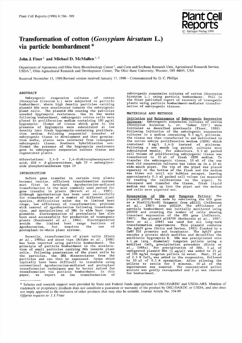
characteristically yellow-brown (Fig. i).
Individual clumps of hygromycin -resistant em-bryogenie tissue could be easily identified and
separately cultured. A single clump, less than
0.5 mm in diameter could be used to establish a
prolific, embryogenie culture after 1-2 months of
subsequent culture. Low inoeulum subculture of
embryogenic suspension culture tissue of cotton has
been previously rep orted (Finer, 1988). The ability
of embryogenic tissue of cotton to survive and
proliferate at low inoculum densities permitte d the
survival and regrowth of small amounts of transgenic
tissue under hygromyc in selection.
An average of approximately 30 stable transgenic
clones were obtained from each separate bombardment
using pCIB709. Particle bombardment of cotton cells
with pUCGUS gave rise to an average of 4,351 cells
per bombardme nt wh ich expressed GUS transiently.
These 30 clones represent a transient-to- stableconversion frequency of approximatel y 0.7%. A
8/6/2019 Ind 90020640
http://slidepdf.com/reader/full/ind-90020640 3/4
588
Embrvogenic cultures Plants
cont llne 2 llne 7 llne 8 11ne ii cont tzans
H U H U H U H U H H U H
Fig. I) Clump of selected, hygromycin-resistant,
embryogenic cotton suspension culture tissue
surrounded by dense, hygromycin-sensitive
tissues.
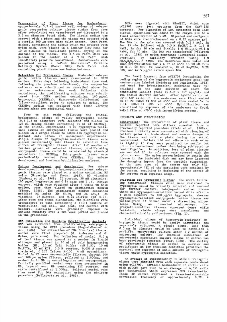
Fig. 2) Hygromycin-re sistant (left) and sensitive
(right) cotto n embryos on embryo development
medium containing I00 #g/ml hygromyein.
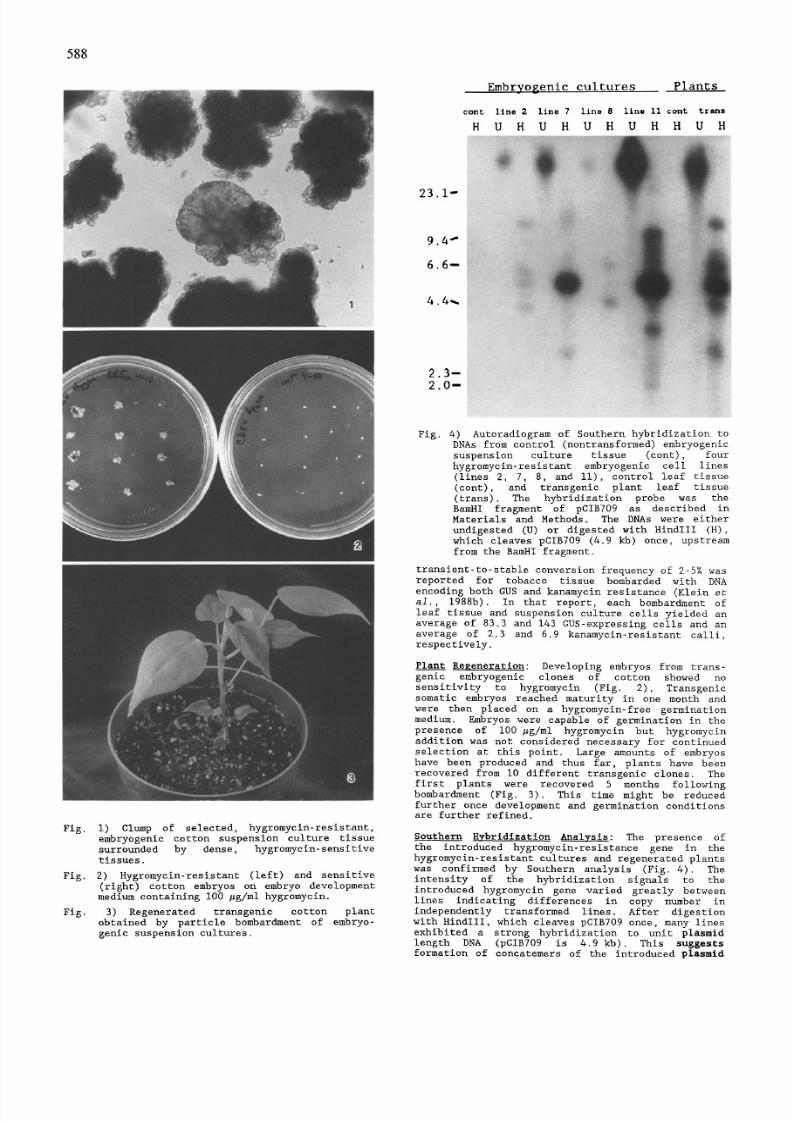
Fig. 3) Regenera ted transgenic cotton plant
obtained by particle bombardm ent of embryo-
genie suspension cultures.
23.1-
9.4 ~
6.6--
4.4~
2.3--
2.0--
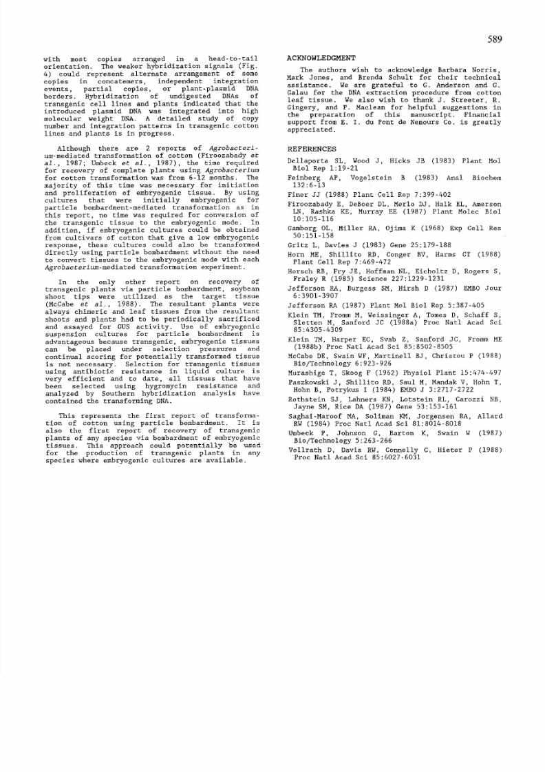
Fig. 4) Autoradi ogram of Southern hybridi zation to
DNAs from control (nontransformed) embryogenic
suspension culture tissue (eont), four
hygremyci n-resistan t embryogenic cell lines
(lines 2, 7, 8, and II), control leaf tissue
(cont), and transgenic plant leaf tissue
(trans). The hybridization probe was the
BamHI fragment of pCIB709 as described in
Materials and Methods. The DNAs were either
undigeste d (U) or digested with HindIII (H),
which cleaves pCIB709 (4.9 kb) once, up stream
from the BamHI fragment.
transient-to-st able conversion freque ncy of 2-5% wasreported for tobacco tissue bombarded with DNA
encoding both GUS and kanamycin resistance (Klein et
ai., 1988b). In that report, each bombardm ent of
leaf tissue and suspension culture cells yielded an
average of 83.3 and 143 GUS-express ing cells and an
average of 2.3 and 6.9 kanamycin-r esistant calli,respectively.
Plant Regeneration: Developing embryos from trans-
genie embryogenic clones of cotton showed no
sensitivity to hygromyc in (Fig. 2). Transgenic
somatic embryos reached maturity in one month and
were then placed on a hygromycin-fr ee germination
medium. Embryos were capable of germination in the
presence of i00 #g/ml hygro mycin but hygrom yein
addition was not considered necessary for continued
selection at this point. Large amounts of embryos
have been produced and thus far, plants have beenrecovered from i0 different transgenic clones. The
first plants were recovered 5 months following
bomb ardment (Fig. 3). This time might be reduced
further once development and germination conditionsare further refined.
Southern Hybridization Anal~sis: The presence of
the introduced hygromycin-resi stance gene in the
hygromyc in-resis tant cultures and regenerat ed plants
was confi rmed by Southern analysis (Fig. 4). The
intensity of the hybridi zation signals to the
introduced hygromycin gene varied greatly between
lines indicating differen ces in copy number in
independently transformed lines. After digestion
with HindIII, which cleaves pCIB709 once, many lines
exhibited a strong hybridization to unit plasmid
length DNA (pCIB709 is 4.9 kb). This suggests
formation of concatemers of the introduced plasmid
8/6/2019 Ind 90020640
http://slidepdf.com/reader/full/ind-90020640 4/4
with most copies arrange d in a head-to-tail
orientation. The weaker hybridizati on signals (Fig.
4) could represent alternate arrangement of some
copies in concatemers, independent integration
events, partial copies, or plant-plasm id DNA
borders. Hybridiz ation of undigested DNAs of
transgenie cell lines and plants indicated that the
introduced plasmid DNA was integrat ed into high
molecular weight DNA. A detailed study of copy
number and integration patterns in transgenic cotton
lines and plants is in progress.
Although there are 2 reports of Agrobacteri-
um-mediated transformation of cotton (Firoozabady et
al., 1987; Umbeck et al., 1987), the time required
for recovery of complete plants using Agrobacterium
for cotton transformation was from 6-12 months. The
majority of this time was necessary for initiation
and proliferati on of embryogenic tissue. By using
cultures that were initially embryogenic for
particle bombardment -mediated transformation as in
this report, no time was required for conversion of
the transge nic tissue to the embryo genic mode. In
addition, if embryogenic cultures could be obtained
from cultivars of cotton that give a low embryogenic
response, these cultures could also be transform ed
directly using particle bombardment without the need
to convert tissues to the embryogenie mode with each
AErobacterium-mediated transformation experiment.
In the only other report on recovery of
transgenic plants via particle bombardment, soybean
shoot tips were utilized as the target tissue
(McCabe et al., 1988). The resultant plants were
always chimeric and leaf tissues from the resultant
shoots and plants had to be periodically sacrificed
and assayed for GUS activity. Use of embryogenic
suspension cultures for particle bombardment is
advantageous because transgenic, embryogenic tissues
can be placed under sel ection pressures and
continual scoring for potentially transformed tissue
is not necessary. Selection for transgenie tissues
using antibiotic resistance in liquid culture is
very efficient and to date, all tissues that have
been selected using hygromycin re sistance and
analyzed by Southern hybridization analysis havecontained the transforming DNA.
This represents the first report of transforma-
tion of cotton using particle bombardment. It is
also the first report of recovery of transgenic
plants of any species via bombardment of embryogenic
tissues. This approach could potentially be used
for the production of transgenic plants in any
species where embryogenic cultures are available.
589
ACKNOWLEDGMENT
The authors wish to acknowledge Barbara Norris,
Mark Jones, and Brenda Sehult for their technical
assistance. We are grateful to G. Ande rson and G.
Galau for the DNA extraction procedure from cotton
leaf tissue. We also wish to thank J. Streeter, R.
Gingery, and P. Maclean for helpful suggestions in
the preparation of this manuscript. Financial
support from E. I. du Pont de Nemours Co. is greatly
appreciated.
REFERENCES
Della porta SL, Wood J, Hicks JB (1983) Plant Mol
Biol Rep 1:19-21
Feinberg AP, Vogelstein B (1983) Anal Biochem
132:6-13
Finer JJ (1988) Plant Cell Rep 7:399-402
Firoozabady E, DeBoer DL, Merlo DJ, Halk EL, Amerson
LN, Rashka KE, Murray EE (1987) Plant Molec B iol
10:105-116
Gamborg OL, Miller RA, Ojima K (1968) Exp Cell Res
50:151-158
Gritz L, Davies J (1983) Gene 25:179-188
Horn ME, Shillito RD, Conger BV, Harms CT (1988)
Plant Cell Rep 7:469-472Horsc h RB, Fry JE, Hoff man NL, Eicholtz D, Rogers S,
Fraley R (1985) Science 227:1 229-123 1
Jeff erso n RA, Burgess SM, Hirsh D (1987) EMBO Jour
6:3901-3907
Jefferson RA (1987) Plant Mol Biol Rep 5:387-405
Klei n TM, From m M, Weissing er A, Tomes D, Sehaff S,
Slette n M, Sanfo rd JC (1988a) Proc Natl Acad Sci
85:4305-4309
Klei n TM, Harper EC, Svab Z, Sanford JC, F romm ME
(1988b) Proc Natl Acad Sci 85:8502-8505
McCabe DE, Swain WF, Martine ll BJ, Christ ou P (1988)
Bio/Technology 6:923-926
Muras hige T, Skoog F (1962) Physiol Plant 15:474-4 97
Paszko wski J, Shil lito RD, Saul M, Manda k V, Hohn T,
Hohn B, Potrykus I (1984) EMBO J 3:2717- 2722
Rothstein SJ, Lahners KN, Lotstein RL, Carozzi NB,
Jayne SM, Rice DA (1987) Gene 53:153-161
Saghai-Maroof MA, Soli man KM, Jorgensen RA, Allard
RW (1984) Proc Natl Acad Sei 81:8014-8018
Umbe ck P, John son G, Barton K, Swain W (1987)
Bio/Technology 5:263-266
Vollrath D, Davis RW, Connelly C, Hieter P (1988)
Proc Natl Acad Sci 85:6027-6031