Gastrulation signalling makes the round go long · 2014-05-02 · greek word for gut, ‘gaster’,...
114
Gastrulation signalling makes the round go long Gastrulatie signalering maakt het van rond naar lang (met een samenvatting in het Nederlands) Proefschrift Ter verkrijging van de graad van doctor aan de Universiteit Utrecht op gezag van de rector magnificus, prof.dr. W.H. Gispen, ingevolge het besluit van het college voor promoties in het openbaar te verdedigen op woensdag 15 november 2006 des middags te 2.30 uur. Door John Christian Jopling geboren op 13 maart 1971 te London, UK
Transcript of Gastrulation signalling makes the round go long · 2014-05-02 · greek word for gut, ‘gaster’,...

Gastrulation signalling makes the round go long
Gastrulatie signalering maakt het van rond naar lang(met een samenvatting in het Nederlands)
ProefschriftTer verkrijging van de graad van doctor aan de Universiteit Utrecht op gezag van de rector
magnificus, prof.dr. W.H. Gispen, ingevolge het besluit van het college voor promoties in het openbaar te verdedigen op woensdag 15 november 2006 des middags te 2.30 uur.
DoorJohn Christian Jopling
geboren op 13 maart 1971 te London, UK
Promoter : Prof. Dr. R.H.A. Plasterk Co-Promoter : Dr. J. den Hertog
Hubrecht Laboratory, Netherlands Institute for Developmental Biology of the Royal
Netherlands Academy of Arts and Sciences (KNAW),Uppsalalaan 8, 3584 CT Utrecht, The Netherlands
The work described in this thesis was performed at the Hubrecht Laboratory (Netherlands Institute for Developmental Biology), within the Graduate School for Developmental Biology and supported by the Royal Netherlands Academy of Arts and Sciences (KNAW) and by an
EU Research Training Network Grant (HPRN-CT-2000-00085).
Table of Contents
Chapter1- Introduction. 5
Chapter2- Essential role for Csk upstream of Fyn and Yes in zebrafish gastrulation. 27
Chapter3-Fyn/Yes and non-canonical Wnt signaling converge on RhoA in vertebrate gastrulation cell movements. 41
Chapter4-Shp2 controls zebrafish gastrulation cell movements via Fyn, Yes and RhoA. 55
Chapter5-Noonan and Leopard syndrome mutations
in Shp2 induce gastrulation defects in zebrafish. 71
Chapter6-Discussion. 87
Summary 105Samenvatting 109Aknowledgements 111Curriculum Vitae 113List of publications 113


Chapter1
Introduction

Gastrulation“It is not birth, marriage or death but gastrulation which is truly the most important time in your life” Lewis Wolpert 1986. Coming from the greek word for gut, ‘gaster’, gastrulation represents a series of cell movements that occur during early development of multi-cellular organisms. These movements result in the formation of the three germ layers, mesoderm, ectoderm and endoderm and in so doing create the basic body-plan of the developing embryo (Warga and Kimmel, 1990).
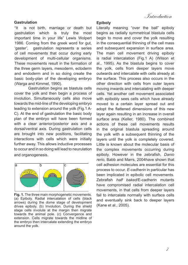
Gastrulation begins as blastula cells cover the yolk and then begin a process of involution. Simultaneously cells also move towards the mid-line of the developing embryo leading to extension around the yolk (Fig.1 A-C). At the end of gastrulation the basic body plan of the embryo will have been formed with a clear anterior/posterior axis and a dorsal/ventral axis. During gastrulation cells are brought into new positions, facilitating interactions with cells which were initially further away. This allows inductive processes to occur and in so doing will lead to neurulation and organogenesis.
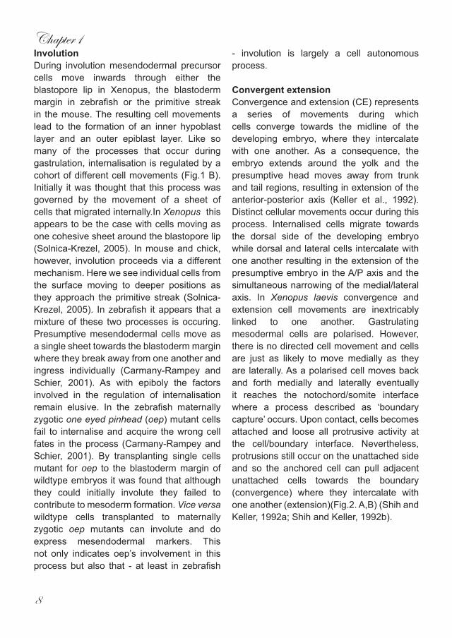
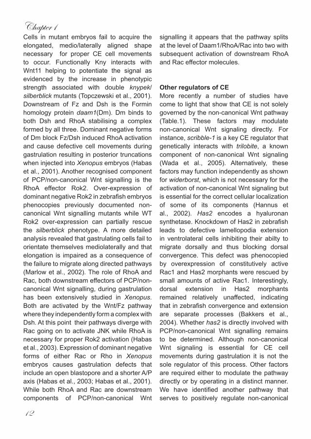
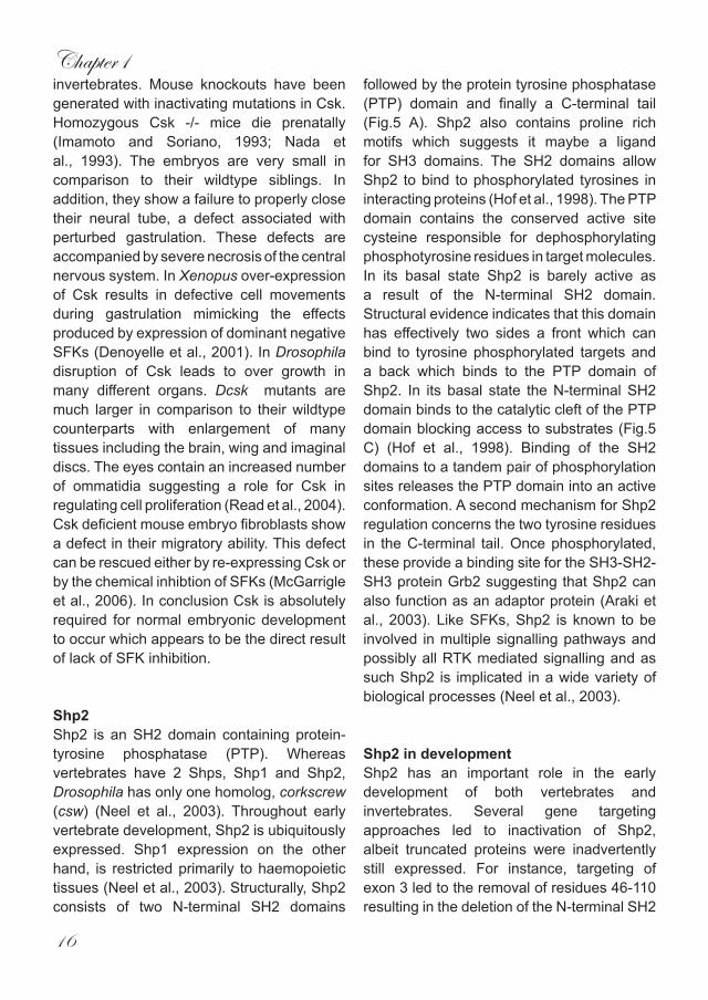
Fig. 1. The three main morphogenetic movements. (a) Epiboly. Radial intercalation of cells (black arrows) during the dome stage of development drives epiboly. (b) Involution. During the shield stage cells involute at the margin then migrate towards the animal pole. (c) Convergence and extension. Cells migrate towards the midline of the embryo then intercalate extending the embryo around the yolk.
EpibolyLiterally meaning “over the ball” epiboly begins as radially symmetrical blastula cells begin to move and cover the yolk resulting in the consequential thinning of the cell mass and subsequent expansion in surface area. The main cell movement driving epiboly is radial intercalation (Fig.1 A) (Wilson et al., 1995). As the blastula begins to cover the yolk, cells from deeper layers move outwards and intercalate with cells already at the surface. This process also occurs in the other direction with cells from outer layers moving inwards and intercalating with deeper cells. Yet another cell movement associated with epibobly sees cells which have already moved to a certain layer spread out and adopt the flattened dimensions of this new layer again resulting in an increase in overall surface area (Keller, 1980). The combined actions of these cell movements results in the original blastula spreading around the yolk with a subsequent thinning of the layers until the yolk is completely covered. Little is known about the molecular basis of the complex movements occurring during epiboly. However in the zebrafish, Danio rerio, Babb and Marrs, 2004have shown that cell adhesion molecules are essential for this process to occur. E-cadherin in particular has been implicated in epibolic cell movements. Zebrafish half baked/E-cadherin mutants have compromised radial intercalation cell movements, in that cells from deeper layers fail to intercalate normally with surface cells and eventually sink back to deeper layers (Kane et al., 2005).
Introduction
7
Gastrulation“It is not birth, marriage or death but gastrulation which is truly the most important time in your life” Lewis Wolpert 1986. Coming from the greek word for gut, ‘gaster’, gastrulation represents a series of cell movements that occur during early development of multi-cellular organisms. These movements result in the formation of the three germ layers, mesoderm, ectoderm and endoderm and in so doing create the basic body-plan of the developing embryo (Warga and Kimmel, 1990).
Gastrulation begins as blastula cells cover the yolk and then begin a process of involution. Simultaneously cells also move towards the mid-line of the developing embryo leading to extension around the yolk (Fig.1 A-C). At the end of gastrulation the basic body plan of the embryo will have been formed with a clear anterior/posterior axis and a dorsal/ventral axis. During gastrulation cells are brought into new positions, facilitating interactions with cells which were initially further away. This allows inductive processes to occur and in so doing will lead to neurulation and organogenesis.
Fig. 1. The three main morphogenetic movements. (a) Epiboly. Radial intercalation of cells (black arrows) during the dome stage of development drives epiboly. (b) Involution. During the shield stage cells involute at the margin then migrate towards the animal pole. (c) Convergence and extension. Cells migrate towards the midline of the embryo then intercalate extending the embryo around the yolk.
EpibolyLiterally meaning “over the ball” epiboly begins as radially symmetrical blastula cells begin to move and cover the yolk resulting in the consequential thinning of the cell mass and subsequent expansion in surface area. The main cell movement driving epiboly is radial intercalation (Fig.1 A) (Wilson et al., 1995). As the blastula begins to cover the yolk, cells from deeper layers move outwards and intercalate with cells already at the surface. This process also occurs in the other direction with cells from outer layers moving inwards and intercalating with deeper cells. Yet another cell movement associated with epibobly sees cells which have already moved to a certain layer spread out and adopt the flattened dimensions of this new layer again resulting in an increase in overall surface area (Keller, 1980). The combined actions of these cell movements results in the original blastula spreading around the yolk with a subsequent thinning of the layers until the yolk is completely covered. Little is known about the molecular basis of the complex movements occurring during epiboly. However in the zebrafish, Danio rerio, Babb and Marrs, 2004have shown that cell adhesion molecules are essential for this process to occur. E-cadherin in particular has been implicated in epibolic cell movements. Zebrafish half baked/E-cadherin mutants have compromised radial intercalation cell movements, in that cells from deeper layers fail to intercalate normally with surface cells and eventually sink back to deeper layers (Kane et al., 2005).
a b c
“It is not birth, marriage or death but gastrulation which is truly the most important time in your life” Lewis Wolpert 1986. Coming from the greek word for gut, ‘gaster’, gastrulation represents a series of cell movements that occur during early development of multi-cellular organisms. These movements result in the formation of the three germ layers, mesoderm, ectoderm and endoderm and in so doing create the basic body-plan of the developing embryo (Warga and Kimmel, 1990).
Chapter1
8
InvolutionDuring involution mesendodermal precursor cells move inwards through either the blastopore lip in Xenopus, the blastoderm margin in zebrafish or the primitive streak in the mouse. The resulting cell movements lead to the formation of an inner hypoblast layer and an outer epiblast layer. Like so many of the processes that occur during gastrulation, internalisation is regulated by a cohort of different cell movements (Fig.1 B). Initially it was thought that this process was governed by the movement of a sheet of cells that migrated internally.In Xenopus this appears to be the case with cells moving as one cohesive sheet around the blastopore lip (Solnica-Krezel, 2005). In mouse and chick, however, involution proceeds via a different mechanism. Here we see individual cells from the surface moving to deeper positions as they approach the primitive streak (Solnica-Krezel, 2005). In zebrafish it appears that a mixture of these two processes is occuring. Presumptive mesendodermal cells move as a single sheet towards the blastoderm margin where they break away from one another and ingress individually (Carmany-Rampey and Schier, 2001). As with epiboly the factors involved in the regulation of internalisation remain elusive. In the zebrafish maternally zygotic one eyed pinhead (oep) mutant cells fail to internalise and acquire the wrong cell fates in the process (Carmany-Rampey and Schier, 2001). By transplanting single cells mutant for oep to the blastoderm margin of wildtype embryos it was found that although they could initially involute they failed to contribute to mesoderm formation. Vice versa wildtype cells transplanted to maternally zygotic oep mutants can involute and do express mesendodermal markers. This not only indicates oep’s involvement in this process but also that - at least in zebrafish
- involution is largely a cell autonomous process. Convergent extensionConvergence and extension (CE) represents a series of movements during which cells converge towards the midline of the developing embryo, where they intercalate with one another. As a consequence, the embryo extends around the yolk and the presumptive head moves away from trunk and tail regions, resulting in extension of the anterior-posterior axis (Keller et al., 1992). Distinct cellular movements occur during this process. Internalised cells migrate towards the dorsal side of the developing embryo while dorsal and lateral cells intercalate with one another resulting in the extension of the presumptive embryo in the A/P axis and the simultaneous narrowing of the medial/lateral axis. In Xenopus laevis convergence and extension cell movements are inextricably linked to one another. Gastrulating mesodermal cells are polarised. However, there is no directed cell movement and cells are just as likely to move medially as they are laterally. As a polarised cell moves back and forth medially and laterally eventually it reaches the notochord/somite interface where a process described as ‘boundary capture’ occurs. Upon contact, cells becomes attached and loose all protrusive activity at the cell/boundary interface. Nevertheless, protrusions still occur on the unattached side and so the anchored cell can pull adjacent unattached cells towards the boundary (convergence) where they intercalate with one another (extension)(Fig.2. A,B) (Shih and Keller, 1992a; Shih and Keller, 1992b).
Introduction
9
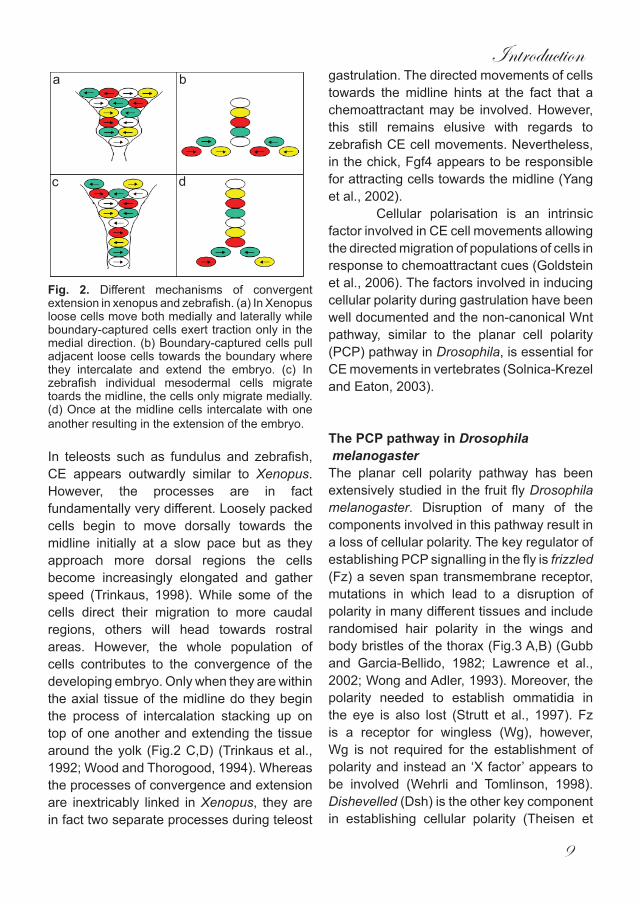
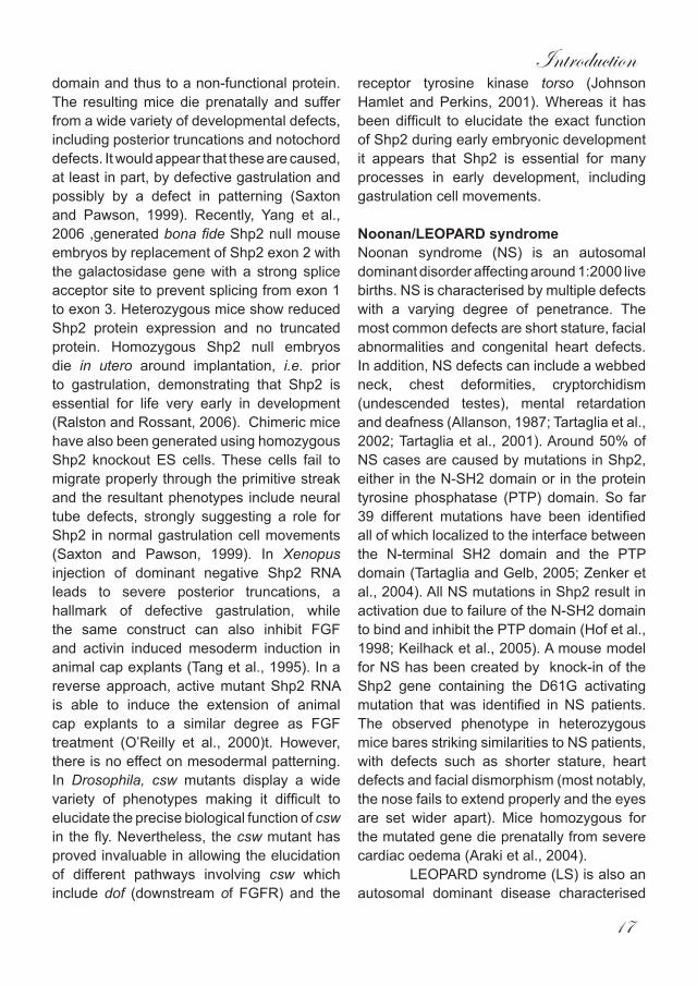
Fig. 2. Different mechanisms of convergent extension in xenopus and zebrafish. (a) In Xenopus loose cells move both medially and laterally while boundary-captured cells exert traction only in the medial direction. (b) Boundary-captured cells pull adjacent loose cells towards the boundary where they intercalate and extend the embryo. (c) In zebrafish individual mesodermal cells migrate toards the midline, the cells only migrate medially. (d) Once at the midline cells intercalate with one another resulting in the extension of the embryo.
In teleosts such as fundulus and zebrafish, CE appears outwardly similar to Xenopus. However, the processes are in fact fundamentally very different. Loosely packed cells begin to move dorsally towards the midline initially at a slow pace but as they approach more dorsal regions the cells become increasingly elongated and gather speed (Trinkaus, 1998). While some of the cells direct their migration to more caudal regions, others will head towards rostral areas. However, the whole population of cells contributes to the convergence of the developing embryo. Only when they are within the axial tissue of the midline do they begin the process of intercalation stacking up on top of one another and extending the tissue around the yolk (Fig.2 C,D) (Trinkaus et al., 1992; Wood and Thorogood, 1994). Whereas the processes of convergence and extension are inextricably linked in Xenopus, they are in fact two separate processes during teleost
gastrulation. The directed movements of cells towards the midline hints at the fact that a chemoattractant may be involved. However, this still remains elusive with regards to zebrafish CE cell movements. Nevertheless, in the chick, Fgf4 appears to be responsible for attracting cells towards the midline (Yang et al., 2002).
Cellular polarisation is an intrinsic factor involved in CE cell movements allowing the directed migration of populations of cells in response to chemoattractant cues (Goldstein et al., 2006). The factors involved in inducing cellular polarity during gastrulation have been well documented and the non-canonical Wnt pathway, similar to the planar cell polarity (PCP) pathway in Drosophila, is essential for CE movements in vertebrates (Solnica-Krezel and Eaton, 2003).
The PCP pathway in Drosophila melanogasterThe planar cell polarity pathway has been extensively studied in the fruit fly Drosophila melanogaster. Disruption of many of the components involved in this pathway result in a loss of cellular polarity. The key regulator of establishing PCP signalling in the fly is frizzled (Fz) a seven span transmembrane receptor, mutations in which lead to a disruption of polarity in many different tissues and include randomised hair polarity in the wings and body bristles of the thorax (Fig.3 A,B) (Gubb and Garcia-Bellido, 1982; Lawrence et al., 2002; Wong and Adler, 1993). Moreover, the polarity needed to establish ommatidia in the eye is also lost (Strutt et al., 1997). Fz is a receptor for wingless (Wg), however, Wg is not required for the establishment of polarity and instead an ‘X factor’ appears to be involved (Wehrli and Tomlinson, 1998). Dishevelled (Dsh) is the other key component in establishing cellular polarity (Theisen et
a b
c d
Chapter1
10
al., 1994)and again while Dsh is required for Wg signalling, Wg is not involved in the polarisation of cells.
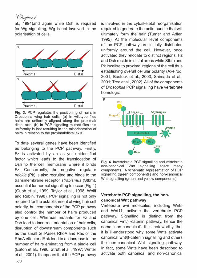
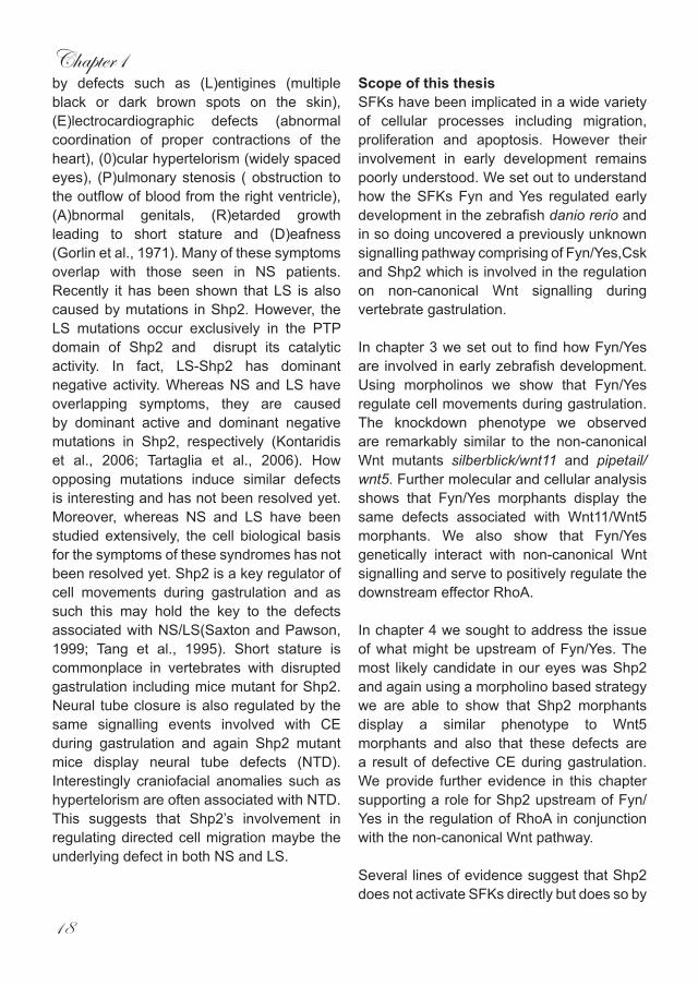
Fig. 3. PCP regulates the positioning of hairs in Drosophlia wing hair cells. (a) In wildtype flies hairs are uniformly aligned along the proximal/distal axis. (b) In PCP signaling mutant flies this uniformity is lost resulting in the misorientation of hairs in relation to the proximal/distal axis.
To date several genes have been identified as belonging to the PCP pathway. Firstly, Fz is activated by an as yet unidentified factor which leads to the translocation of Dsh to the cell membrane where it binds Fz. Concurrently, the negative regulator prickle (Pk) is also recruited and binds to the transmembrane receptor strabismus (Stbm), essential for normal signalling to occur (Fig.4) (Gubb et al., 1999; Taylor et al., 1998; Wolff and Rubin, 1998). PCP signalling is not only required for the establishment of wing hair cell polarity, but components of the PCP pathway also control the number of hairs produced by one cell. Whereas mutants for Fz and Dsh lead to incorrect orientation of hair cells, disruption of downstream components such as the small GTPases RhoA and Rac or the RhoA effector dRok lead to an increase in the number of hairs eminating from a single cell (Eaton et al., 1996; Strutt et al., 1997; Winter et al., 2001). It appears that the PCP pathway
is involved in the cytoskeletal reorganisation required to generate the actin bundle that will ultimately form the hair (Turner and Adler, 1995). At the molecular level components of the PCP pathway are initially distributed uniformly around the cell. However, once activated they relocate to distinct regions, Fz and Dsh reside in distal areas while Stbm and Pk localise to proximal regions of the cell thus establishing overall cellular polarity (Axelrod, 2001; Bastock et al., 2003; Shimada et al., 2001; Tree et al., 2002). All of the components of Drosophila PCP signalling have vertebrate homologs.
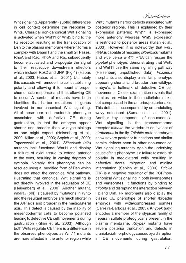
Fig. 4. Invertebrate PCP signalling and vertebrate non-canonical Wnt signalling share many components. A schematic representation of PCP signalling (green components) and non-canonical Wnt signalling (green and yellow components).
Vertebrate PCP signalling, the non-canonical Wnt pathway Vertebrate wnt molecules, including Wnt5 and Wnt11, activate the vertebrate PCP pathway. Signalling is distinct from the canonical wnt/β-catenin pathway, hence the name ‘non-canonical’. It is noteworthy that it is ill-understood why some Wnts activate canonical wnt/β-catenin signalling and others the non-canonical Wnt signaling pathway. In fact, some Wnts have been described to activate both canonical and non-canonical
a
b
a
Introduction
11
Wnt signaling. Apparently, (subtle) differences in cell context determine the response to Wnts. Classical non-canonical Wnt signaling is activated when Wnt11 or Wnt5 bind to the Fz receptor resulting in the translocation of Dsh to the plasma membrane where it forms a complex with Daam1 and the small GTPases, RhoA and Rac. RhoA and Rac subsequently become activated and propogate the signal to their respective downstream effectors which include Rok2 and JNK (Fig.4) (Habas et al., 2003; Habas et al., 2001). Ultimately this cascade will remodel the cell establishing polarity and allowing it to mount a proper chemotactic response and thus allowing CE to occur. A number of mutants have been identified that harbor mutations in genes involved in non-canonical Wnt signalling. All of these bear a characteristic phenotype associated with defective CE during gastrulation, in that the embryos appear shorter and broader than wildtype siblings as one might expect (Heisenberg et al., 2000; Kilian et al., 2003; Sepich et al., 2000; Topczewski et al., 2001). Silberblick (slb) mutants lack functional Wnt11 and display a failure of axial tissue to extend anterior to the eyes, resulting in varying degrees of cyclopia. Notably, this phenotype can be rescued using a modified form of Dsh which does not affect the canonical Wnt pathway, illustrating that canonical Wnt signalling is not directly involved in the regulation of CE (Heisenberg et al., 2000). Another mutant, pipetail (ppt) is caused by mutations in Wnt5 and the resultant embryos are much shorter in the A/P axis and broader in the medio/lateral axis. This defect is caused by the inability of mesendodermal cells to become polarised leading to defective CE cell movements during gastrulation (Kilian et al., 2003). Although both Wnts regulate CE there is a difference in the observed phenotypes as Wnt11 mutants are more affected in the anterior region while
Wnt5 mutants harbor defects associated with posterior regions. This is explained by their expression patterns; Wnt11 is expressed more anteriorly whereas Wnt5 expression is restricted to posterior areas (Kilian et al., 2003). However, it is noteworthy that wnt5 RNA is capable of rescuing silberblick mutants and vice versa wnt11 RNA can rescue the pipetail phenotype, demonstrating that Wnt5 and Wnt11 use the same signalling pathway (Heisenberg unpublished data). Frizzled2 morphants also display a similar phenotype appearing shorter and broader than wildtype embyo’s, a hallmark of defective CE cell movements. Closer examination reveals that somites are wider in the medio/lateral axis but compressed in the anterior/posterior axis. This defect is accompanied by an undulating wavy notochord (Sumanas et al., 2001). Another key component of non-canonical Wnt signalling is the transmembrane receptor trilobite the vertebrate equivalent of strabismus in the fly. Trilobite mutant embryos show severe posterior truncations and similar somite defects seen in other non-canonical Wnt signalling mutants. Again the underlying cause of this phenotype is a failure to establish polarity in mediolateral cells resulting in defective dorsal migration and midline intercalation (Sepich et al., 2000). Prickle (Pk) is a negative regulator of the PCP/non-canonical Wnt signalling in both invertebrates and vertebrates. It functions by binding to trilobite and disrupting the interaction between Fz and Dsh. Pk morphants also display the classic CE phenotype of shorter /broader embryos with wide/compressed somites (Carreira-Barbosa et al., 2003). Knypek (kny) encodes a member of the glypican family of heparan sulfate proteoglycans present in the plasma membrane. Knypek mutants have severe posterior truncation and defects in craniofacial morphology caused by a disruption in CE movements during gastrulation.
Chapter1
12
Cells in mutant embryos fail to acquire the elongated, medio/laterally aligned shape necessary for proper CE cell movements to occur. Functionally Kny interacts with Wnt11 helping to potentiate the signal as evidenced by the increase in phenotypic strength associated with double knypek/silberblick mutants (Topczewski et al., 2001). Downstream of Fz and Dsh is the Formin homology protein daam1(Dm). Dm binds to both Dsh and RhoA stabilising a complex formed by all three. Dominant negative forms of Dm block Fz/Dsh induced RhoA activation and cause defective cell movements during gastrulation resulting in posterior truncations when injected into Xenopus embryos (Habas et al., 2001). Another recognised component of PCP/non-canonical Wnt signalling is the RhoA effector Rok2. Over-expression of dominant negative Rok2 in zebrafish embryos phenocopies previously documented non-canonical Wnt signalling mutants while WT Rok2 over-expression can partially rescue the silberblick phenotype. A more detailed analysis revealed that gastrulating cells fail to orientate themselves mediolaterally and that elongation is impaired as a consequence of the failure to migrate along directed pathways (Marlow et al., 2002). The role of RhoA and Rac, both downstream effectors of PCP/non-canonical Wnt signalling, during gastrulation has been extensively studied in Xenopus. Both are activated by the Wnt/Fz pathway where they independently form a complex with Dsh. At this point their pathways diverge with Rac going on to activate JNK while RhoA is necessary for proper Rok2 activation (Habas et al., 2003). Expression of dominant negative forms of either Rac or Rho in Xenopus embryos causes gastrulation defects that include an open blastopore and a shorter A/P axis (Habas et al., 2003; Habas et al., 2001). While both RhoA and Rac are downstream components of PCP/non-canonical Wnt
signalling it appears that the pathway splits at the level of Daam1/RhoA/Rac into two with subsequent activation of downstream RhoA and Rac effector molecules.
Other regulators of CEMore recently a number of studies have come to light that show that CE is not solely governed by the non-canonical Wnt pathway (Table.1). These factors may modulate non-canonical Wnt signaling directly. For instance, scribble-1 is a key CE regulator that genetically interacts with trilobite, a known component of non-canonical Wnt signaling (Wada et al., 2005). Alternatively, these factors may function independently as shown for widerborst, which is not necessary for the activation of non-canonical Wnt signaling but is essential for the correct cellular localization of some of its components (Hannus et al., 2002). Has2 encodes a hyaluronan synthetase. Knockdown of Has2 in zebrafish leads to defective lamellopodia extension in ventrolateral cells inhibiting their abilty to migrate dorsally and thus blocking dorsal convergence. This defect was phenocopied by overexpression of constitutively active Rac1 and Has2 morphants were rescued by small amounts of active Rac1. Interestingly, dorsal extension in Has2 morphants remained relatively unaffected, indicating that in zebrafish convergence and extension are separate processes (Bakkers et al., 2004). Whether has2 is directly involved with PCP/non-canonical Wnt signalling remains to be determined. Although non-canonical Wnt signaling is essential for CE cell movements during gastrulation it is not the sole regulator of this process. Other factors are required either to modulate the pathway directly or by operating in a distinct manner. We have identified another pathway that serves to positively regulate non-canonical
Introduction
13
Wnt signalling and the control of CE cell movements during gastrulation. Our findings are described in detail in Chapters 2-5 of this thesis. The components that we have uncovered are the src family kinase (SFK) members Fyn and Yes, the protein tyrosine phosphatase (PTP) Shp2 and the negative SFK regulator c-terminal Src kinase (Csk).
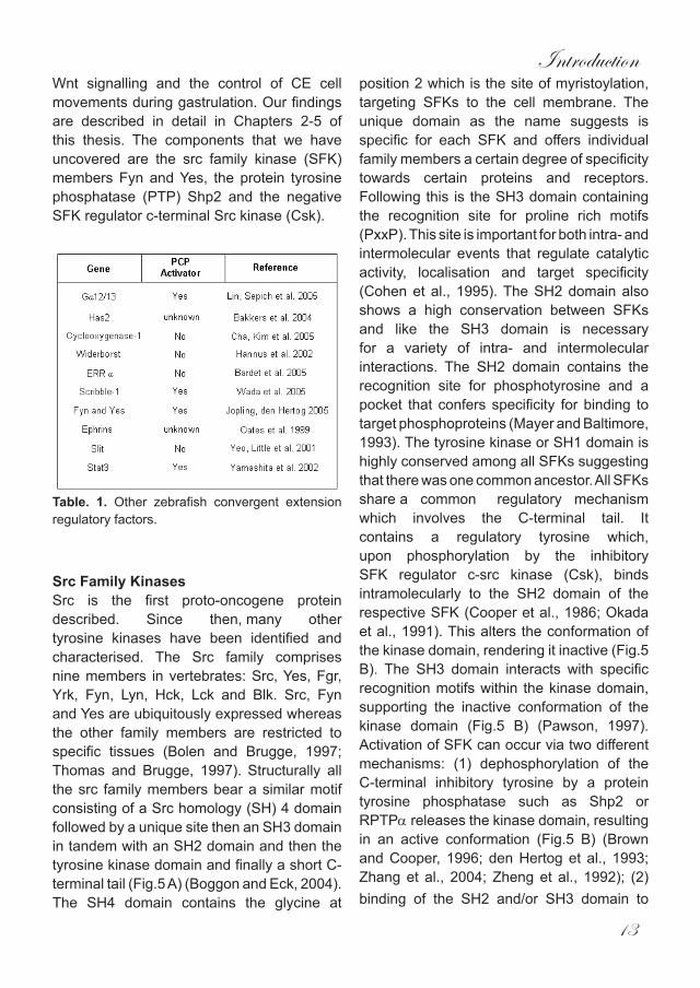
Table. 1. Other zebrafish convergent extension regulatory factors.
Src Family KinasesSrc is the first proto-oncogene protein described. Since then, many other tyrosine kinases have been identified and characterised. The Src family comprises nine members in vertebrates: Src, Yes, Fgr, Yrk, Fyn, Lyn, Hck, Lck and Blk. Src, Fyn and Yes are ubiquitously expressed whereas the other family members are restricted to specific tissues (Bolen and Brugge, 1997; Thomas and Brugge, 1997). Structurally all the src family members bear a similar motif consisting of a Src homology (SH) 4 domain followed by a unique site then an SH3 domain in tandem with an SH2 domain and then the tyrosine kinase domain and finally a short C-terminal tail (Fig.5 A) (Boggon and Eck, 2004).The SH4 domain contains the glycine at
position 2 which is the site of myristoylation, targeting SFKs to the cell membrane. The unique domain as the name suggests is specific for each SFK and offers individual family members a certain degree of specificity towards certain proteins and receptors. Following this is the SH3 domain containing the recognition site for proline rich motifs (PxxP). This site is important for both intra- and intermolecular events that regulate catalytic activity, localisation and target specificity (Cohen et al., 1995). The SH2 domain also shows a high conservation between SFKs and like the SH3 domain is necessary for a variety of intra- and intermolecular interactions. The SH2 domain contains the recognition site for phosphotyrosine and a pocket that confers specificity for binding to target phosphoproteins (Mayer and Baltimore, 1993). The tyrosine kinase or SH1 domain is highly conserved among all SFKs suggesting that there was one common ancestor. All SFKs share a common regulatory mechanismwhich involves the C-terminal tail. It contains a regulatory tyrosine which, upon phosphorylation by the inhibitory SFK regulator c-src kinase (Csk), binds intramolecularly to the SH2 domain of the respective SFK (Cooper et al., 1986; Okada et al., 1991). This alters the conformation of the kinase domain, rendering it inactive (Fig.5 B). The SH3 domain interacts with specific recognition motifs within the kinase domain, supporting the inactive conformation of the kinase domain (Fig.5 B) (Pawson, 1997). Activation of SFK can occur via two different mechanisms: (1) dephosphorylation of the C-terminal inhibitory tyrosine by a protein tyrosine phosphatase such as Shp2 or RPTPα releases the kinase domain, resulting in an active conformation (Fig.5 B) (Brown and Cooper, 1996; den Hertog et al., 1993; Zhang et al., 2004; Zheng et al., 1992); (2) binding of the SH2 and/or SH3 domain to
Chapter1
14
higher affinity ligands releases the inhibitory conformation, leading to activation of kinase activity (Alonso et al., 1995). SFKs are known to interact with a wide variety of cellular components via there SH3 and SH2 domains, including growth factor receptors, cytokine receptors and various cytoplasmic regulatory elements (Thomas and Brugge, 1997). Because of this diversity SFKs have been implicated in a wide variety of cellular processes including migration and apoptosis (Altun-Gultekin and Wagner, 1996; Canman et al., 1995).
SFKs in developmentAlthough the role of SFK in cell signalling and tumorigenicity is well established (Summy and Gallick, 2003) relatively little is known about their function in embryonic development. All SFK genes have been disrupted in the mouse, resulting in relatively mild phenotypes in four cases and no apparent phenotype in the others. Src, Fyn and Yes are broadly expressed during mouse development, and double knockout mice either die perinatally (Src/Fyn and Src/Yes) or undergo degenerative renal changes (Fyn/Yes) (Stein et al., 1994). Other knockout combinations have also been generated which result in phenotypes ranging from severe bone defects (Src/Hck) (Lowell et al., 1996) to the disruption of T-cell development (Fyn/Lck) (Groves et al., 1996).In Xenopus leavis, RNA expression of dominant-negative mutants of the Src family members, Src, Fyn and Yes disrupt gastrulation movements resulting in an inability to close the blastopore ,while co-injection of the same inhibiting mutants into animal pole explants blocks the activin induced elongation (Denoyelle et al., 2001). However, they do not compromise the ability of FGF’s to stimulate
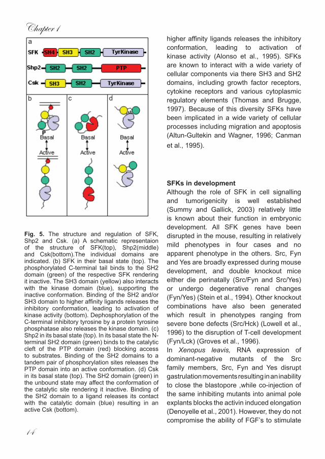
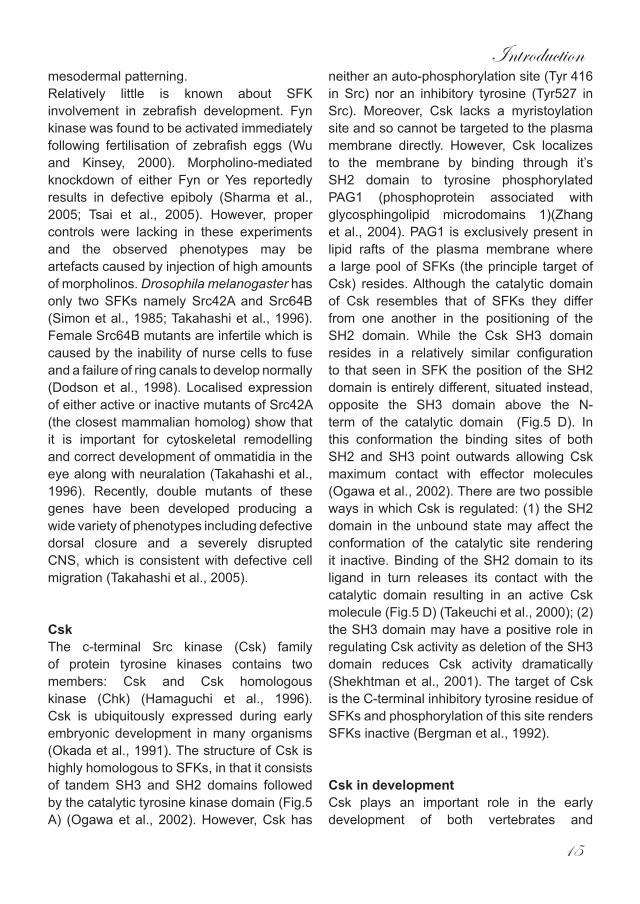
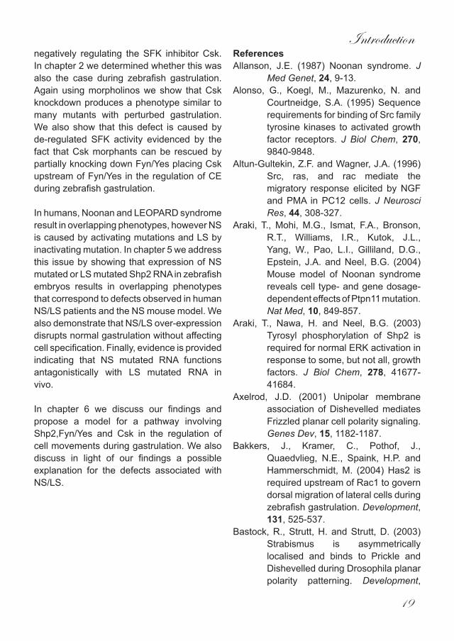
Fig. 5. The structure and regulation of SFK, Shp2 and Csk. (a) A schematic representaion of the structure of SFK(top), Shp2(middle) and Csk(bottom).The individual domains are indicated. (b) SFK in their basal state (top). The phosphorylated C-terminal tail binds to the SH2 domain (green) of the respective SFK rendering it inactive. The SH3 domain (yellow) also interacts with the kinase domain (blue), supporting the inactive conformation. Binding of the SH2 and/or SH3 domain to higher affinity ligands releases the inhibitory conformation, leading to activation of kinase activity (bottom). Dephosphorylation of the C-terminal inhibitory tyrosine by a protein tyrosine phosphatase also releases the kinase domain. (c) Shp2 in its basal state (top). In its basal state the N-terminal SH2 domain (green) binds to the catalytic cleft of the PTP domain (red) blocking access to substrates. Binding of the SH2 domains to a tandem pair of phosphorylation sites releases the PTP domain into an active conformation. (d) Csk in its basal state (top). The SH2 domain (green) in the unbound state may affect the conformation of the catalytic site rendering it inactive. Binding of the SH2 domain to a ligand releases its contact with the catalytic domain (blue) resulting in an active Csk (bottom).
a
b c d
Introduction
15
mesodermal patterning.Relatively little is known about SFK involvement in zebrafish development. Fyn kinase was found to be activated immediately following fertilisation of zebrafish eggs (Wu and Kinsey, 2000). Morpholino-mediated knockdown of either Fyn or Yes reportedly results in defective epiboly (Sharma et al., 2005; Tsai et al., 2005). However, proper controls were lacking in these experiments and the observed phenotypes may be artefacts caused by injection of high amounts of morpholinos. Drosophila melanogaster has only two SFKs namely Src42A and Src64B (Simon et al., 1985; Takahashi et al., 1996). Female Src64B mutants are infertile which is caused by the inability of nurse cells to fuse and a failure of ring canals to develop normally (Dodson et al., 1998). Localised expression of either active or inactive mutants of Src42A (the closest mammalian homolog) show that it is important for cytoskeletal remodelling and correct development of ommatidia in the eye along with neuralation (Takahashi et al., 1996). Recently, double mutants of these genes have been developed producing a wide variety of phenotypes including defective dorsal closure and a severely disrupted CNS, which is consistent with defective cell migration (Takahashi et al., 2005).
CskThe c-terminal Src kinase (Csk) family of protein tyrosine kinases contains two members: Csk and Csk homologous kinase (Chk) (Hamaguchi et al., 1996). Csk is ubiquitously expressed during early embryonic development in many organisms (Okada et al., 1991). The structure of Csk is highly homologous to SFKs, in that it consists of tandem SH3 and SH2 domains followed by the catalytic tyrosine kinase domain (Fig.5 A) (Ogawa et al., 2002). However, Csk has
neither an auto-phosphorylation site (Tyr 416 in Src) nor an inhibitory tyrosine (Tyr527 in Src). Moreover, Csk lacks a myristoylation site and so cannot be targeted to the plasma membrane directly. However, Csk localizes to the membrane by binding through it’s SH2 domain to tyrosine phosphorylated PAG1 (phosphoprotein associated with glycosphingolipid microdomains 1)(Zhang et al., 2004). PAG1 is exclusively present in lipid rafts of the plasma membrane where a large pool of SFKs (the principle target of Csk) resides. Although the catalytic domain of Csk resembles that of SFKs they differ from one another in the positioning of the SH2 domain. While the Csk SH3 domain resides in a relatively similar configuration to that seen in SFK the position of the SH2 domain is entirely different, situated instead, opposite the SH3 domain above the N-term of the catalytic domain (Fig.5 D). In this conformation the binding sites of both SH2 and SH3 point outwards allowing Csk maximum contact with effector molecules (Ogawa et al., 2002). There are two possible ways in which Csk is regulated: (1) the SH2 domain in the unbound state may affect the conformation of the catalytic site rendering it inactive. Binding of the SH2 domain to its ligand in turn releases its contact with the catalytic domain resulting in an active Csk molecule (Fig.5 D) (Takeuchi et al., 2000); (2) the SH3 domain may have a positive role in regulating Csk activity as deletion of the SH3 domain reduces Csk activity dramatically (Shekhtman et al., 2001). The target of Csk is the C-terminal inhibitory tyrosine residue of SFKs and phosphorylation of this site renders SFKs inactive (Bergman et al., 1992).
Csk in developmentCsk plays an important role in the early development of both vertebrates and
Chapter1
16
invertebrates. Mouse knockouts have been generated with inactivating mutations in Csk. Homozygous Csk -/- mice die prenatally (Imamoto and Soriano, 1993; Nada et al., 1993). The embryos are very small in comparison to their wildtype siblings. In addition, they show a failure to properly close their neural tube, a defect associated with perturbed gastrulation. These defects are accompanied by severe necrosis of the central nervous system. In Xenopus over-expression of Csk results in defective cell movements during gastrulation mimicking the effects produced by expression of dominant negative SFKs (Denoyelle et al., 2001). In Drosophila disruption of Csk leads to over growth in many different organs. Dcsk mutants are much larger in comparison to their wildtype counterparts with enlargement of many tissues including the brain, wing and imaginal discs. The eyes contain an increased number of ommatidia suggesting a role for Csk in regulating cell proliferation (Read et al., 2004). Csk deficient mouse embryo fibroblasts show a defect in their migratory ability. This defect can be rescued either by re-expressing Csk or by the chemical inhibtion of SFKs (McGarrigle et al., 2006). In conclusion Csk is absolutely required for normal embryonic development to occur which appears to be the direct result of lack of SFK inhibition.
Shp2 Shp2 is an SH2 domain containing protein-tyrosine phosphatase (PTP). Whereas vertebrates have 2 Shps, Shp1 and Shp2, Drosophila has only one homolog, corkscrew (csw) (Neel et al., 2003). Throughout early vertebrate development, Shp2 is ubiquitously expressed. Shp1 expression on the other hand, is restricted primarily to haemopoietic tissues (Neel et al., 2003). Structurally, Shp2 consists of two N-terminal SH2 domains
followed by the protein tyrosine phosphatase (PTP) domain and finally a C-terminal tail (Fig.5 A). Shp2 also contains proline rich motifs which suggests it maybe a ligand for SH3 domains. The SH2 domains allow Shp2 to bind to phosphorylated tyrosines in interacting proteins (Hof et al., 1998). The PTP domain contains the conserved active site cysteine responsible for dephosphorylating phosphotyrosine residues in target molecules. In its basal state Shp2 is barely active as a result of the N-terminal SH2 domain. Structural evidence indicates that this domain has effectively two sides a front which can bind to tyrosine phosphorylated targets and a back which binds to the PTP domain of Shp2. In its basal state the N-terminal SH2 domain binds to the catalytic cleft of the PTP domain blocking access to substrates (Fig.5 C) (Hof et al., 1998). Binding of the SH2 domains to a tandem pair of phosphorylation sites releases the PTP domain into an active conformation. A second mechanism for Shp2 regulation concerns the two tyrosine residues in the C-terminal tail. Once phosphorylated, these provide a binding site for the SH3-SH2-SH3 protein Grb2 suggesting that Shp2 can also function as an adaptor protein (Araki et al., 2003). Like SFKs, Shp2 is known to be involved in multiple signalling pathways and possibly all RTK mediated signalling and as such Shp2 is implicated in a wide variety of biological processes (Neel et al., 2003).
Shp2 in developmentShp2 has an important role in the early development of both vertebrates and invertebrates. Several gene targeting approaches led to inactivation of Shp2, albeit truncated proteins were inadvertently still expressed. For instance, targeting of exon 3 led to the removal of residues 46-110 resulting in the deletion of the N-terminal SH2
Introduction
17
domain and thus to a non-functional protein. The resulting mice die prenatally and suffer from a wide variety of developmental defects, including posterior truncations and notochord defects. It would appear that these are caused, at least in part, by defective gastrulation and possibly by a defect in patterning (Saxton and Pawson, 1999). Recently, Yang et al., 2006 ,generated bona fide Shp2 null mouse embryos by replacement of Shp2 exon 2 with the galactosidase gene with a strong splice acceptor site to prevent splicing from exon 1 to exon 3. Heterozygous mice show reduced Shp2 protein expression and no truncated protein. Homozygous Shp2 null embryos die in utero around implantation, i.e. prior to gastrulation, demonstrating that Shp2 is essential for life very early in development (Ralston and Rossant, 2006). Chimeric mice have also been generated using homozygous Shp2 knockout ES cells. These cells fail to migrate properly through the primitive streak and the resultant phenotypes include neural tube defects, strongly suggesting a role for Shp2 in normal gastrulation cell movements (Saxton and Pawson, 1999). In Xenopus injection of dominant negative Shp2 RNA leads to severe posterior truncations, a hallmark of defective gastrulation, while the same construct can also inhibit FGF and activin induced mesoderm induction in animal cap explants (Tang et al., 1995). In a reverse approach, active mutant Shp2 RNA is able to induce the extension of animal cap explants to a similar degree as FGF treatment (O’Reilly et al., 2000)t. However, there is no effect on mesodermal patterning. In Drosophila, csw mutants display a wide variety of phenotypes making it difficult to elucidate the precise biological function of csw in the fly. Nevertheless, the csw mutant has proved invaluable in allowing the elucidation of different pathways involving csw which include dof (downstream of FGFR) and the
receptor tyrosine kinase torso (Johnson Hamlet and Perkins, 2001). Whereas it has been difficult to elucidate the exact function of Shp2 during early embryonic development it appears that Shp2 is essential for many processes in early development, including gastrulation cell movements.
Noonan/LEOPARD syndromeNoonan syndrome (NS) is an autosomal dominant disorder affecting around 1:2000 live births. NS is characterised by multiple defects with a varying degree of penetrance. The most common defects are short stature, facial abnormalities and congenital heart defects. In addition, NS defects can include a webbed neck, chest deformities, cryptorchidism (undescended testes), mental retardation and deafness (Allanson, 1987; Tartaglia et al., 2002; Tartaglia et al., 2001). Around 50% of NS cases are caused by mutations in Shp2, either in the N-SH2 domain or in the protein tyrosine phosphatase (PTP) domain. So far 39 different mutations have been identified all of which localized to the interface between the N-terminal SH2 domain and the PTP domain (Tartaglia and Gelb, 2005; Zenker et al., 2004). All NS mutations in Shp2 result in activation due to failure of the N-SH2 domain to bind and inhibit the PTP domain (Hof et al., 1998; Keilhack et al., 2005). A mouse model for NS has been created by knock-in of the Shp2 gene containing the D61G activating mutation that was identified in NS patients. The observed phenotype in heterozygous mice bares striking similarities to NS patients, with defects such as shorter stature, heart defects and facial dismorphism (most notably, the nose fails to extend properly and the eyes are set wider apart). Mice homozygous for the mutated gene die prenatally from severe cardiac oedema (Araki et al., 2004).
LEOPARD syndrome (LS) is also an autosomal dominant disease characterised
Chapter1
18
by defects such as (L)entigines (multiple black or dark brown spots on the skin), (E)lectrocardiographic defects (abnormal coordination of proper contractions of the heart), (0)cular hypertelorism (widely spaced eyes), (P)ulmonary stenosis ( obstruction to the outflow of blood from the right ventricle), (A)bnormal genitals, (R)etarded growth leading to short stature and (D)eafness (Gorlin et al., 1971). Many of these symptoms overlap with those seen in NS patients. Recently it has been shown that LS is also caused by mutations in Shp2. However, the LS mutations occur exclusively in the PTP domain of Shp2 and disrupt its catalytic activity. In fact, LS-Shp2 has dominant negative activity. Whereas NS and LS have overlapping symptoms, they are caused by dominant active and dominant negative mutations in Shp2, respectively (Kontaridis et al., 2006; Tartaglia et al., 2006). How opposing mutations induce similar defects is interesting and has not been resolved yet. Moreover, whereas NS and LS have been studied extensively, the cell biological basis for the symptoms of these syndromes has not been resolved yet. Shp2 is a key regulator of cell movements during gastrulation and as such this may hold the key to the defects associated with NS/LS(Saxton and Pawson, 1999; Tang et al., 1995). Short stature is commonplace in vertebrates with disrupted gastrulation including mice mutant for Shp2. Neural tube closure is also regulated by the same signalling events involved with CE during gastrulation and again Shp2 mutant mice display neural tube defects (NTD). Interestingly craniofacial anomalies such as hypertelorism are often associated with NTD. This suggests that Shp2’s involvement in regulating directed cell migration maybe the underlying defect in both NS and LS.
Scope of this thesis SFKs have been implicated in a wide variety of cellular processes including migration, proliferation and apoptosis. However their involvement in early development remains poorly understood. We set out to understand how the SFKs Fyn and Yes regulated early development in the zebrafish danio rerio and in so doing uncovered a previously unknown signalling pathway comprising of Fyn/Yes,Csk and Shp2 which is involved in the regulation on non-canonical Wnt signalling during vertebrate gastrulation.
In chapter 3 we set out to find how Fyn/Yes are involved in early zebrafish development. Using morpholinos we show that Fyn/Yes regulate cell movements during gastrulation. The knockdown phenotype we observed are remarkably similar to the non-canonical Wnt mutants silberblick/wnt11 and pipetail/wnt5. Further molecular and cellular analysis shows that Fyn/Yes morphants display the same defects associated with Wnt11/Wnt5 morphants. We also show that Fyn/Yes genetically interact with non-canonical Wnt signalling and serve to positively regulate the downstream effector RhoA.
In chapter 4 we sought to address the issue of what might be upstream of Fyn/Yes. The most likely candidate in our eyes was Shp2 and again using a morpholino based strategy we are able to show that Shp2 morphants display a similar phenotype to Wnt5 morphants and also that these defects are a result of defective CE during gastrulation. We provide further evidence in this chapter supporting a role for Shp2 upstream of Fyn/Yes in the regulation of RhoA in conjunction with the non-canonical Wnt pathway.
Several lines of evidence suggest that Shp2 does not activate SFKs directly but does so by
Introduction
19
negatively regulating the SFK inhibitor Csk. In chapter 2 we determined whether this was also the case during zebrafish gastrulation. Again using morpholinos we show that Csk knockdown produces a phenotype similar to many mutants with perturbed gastrulation. We also show that this defect is caused by de-regulated SFK activity evidenced by the fact that Csk morphants can be rescued by partially knocking down Fyn/Yes placing Csk upstream of Fyn/Yes in the regulation of CE during zebrafish gastrulation.
In humans, Noonan and LEOPARD syndrome result in overlapping phenotypes, however NS is caused by activating mutations and LS by inactivating mutation. In chapter 5 we address this issue by showing that expression of NS mutated or LS mutated Shp2 RNA in zebrafish embryos results in overlapping phenotypes that correspond to defects observed in human NS/LS patients and the NS mouse model. We also demonstrate that NS/LS over-expression disrupts normal gastrulation without affecting cell specification. Finally, evidence is provided indicating that NS mutated RNA functions antagonistically with LS mutated RNA in vivo.
In chapter 6 we discuss our findings and propose a model for a pathway involving Shp2,Fyn/Yes and Csk in the regulation of cell movements during gastrulation. We also discuss in light of our findings a possible explanation for the defects associated with NS/LS.
ReferencesAllanson, J.E. (1987) Noonan syndrome. J
Med Genet, 24, 9-13.Alonso, G., Koegl, M., Mazurenko, N. and
Courtneidge, S.A. (1995) Sequence requirements for binding of Src family tyrosine kinases to activated growth factor receptors. J Biol Chem, 270, 9840-9848.
Altun-Gultekin, Z.F. and Wagner, J.A. (1996) Src, ras, and rac mediate the migratory response elicited by NGF and PMA in PC12 cells. J Neurosci Res, 44, 308-327.
Araki, T., Mohi, M.G., Ismat, F.A., Bronson, R.T., Williams, I.R., Kutok, J.L., Yang, W., Pao, L.I., Gilliland, D.G., Epstein, J.A. and Neel, B.G. (2004) Mouse model of Noonan syndrome reveals cell type- and gene dosage-dependent effects of Ptpn11 mutation. Nat Med, 10, 849-857.
Araki, T., Nawa, H. and Neel, B.G. (2003) Tyrosyl phosphorylation of Shp2 is required for normal ERK activation in response to some, but not all, growth factors. J Biol Chem, 278, 41677-41684.
Axelrod, J.D. (2001) Unipolar membrane association of Dishevelled mediates Frizzled planar cell polarity signaling. Genes Dev, 15, 1182-1187.
Bakkers, J., Kramer, C., Pothof, J., Quaedvlieg, N.E., Spaink, H.P. and Hammerschmidt, M. (2004) Has2 is required upstream of Rac1 to govern dorsal migration of lateral cells during zebrafish gastrulation. Development, 131, 525-537.
Bastock, R., Strutt, H. and Strutt, D. (2003) Strabismus is asymmetrically localised and binds to Prickle and Dishevelled during Drosophila planar polarity patterning. Development,

Chapter1
20
130, 3007-3014.Bergman, M., Mustelin, T., Oetken, C.,
Partanen, J., Flint, N.A., Amrein, K.E., Autero, M., Burn, P. and Alitalo, K. (1992) The human p50csk tyrosine kinase phosphorylates p56lck at Tyr-505 and down regulates its catalytic activity. Embo J, 11, 2919-2924.
Boggon, T.J. and Eck, M.J. (2004) Structure and regulation of Src family kinases. Oncogene, 23, 7918-7927.
Bolen, J.B. and Brugge, J.S. (1997) Leukocyte protein tyrosine kinases: potential targets for drug discovery. Annu Rev Immunol, 15, 371-404.
Brown, M.T. and Cooper, J.A. (1996) Regulation, substrates and functions of src. Biochim Biophys Acta, 1287, 121-149.
Canman, C.E., Gilmer, T.M., Coutts, S.B. and Kastan, M.B. (1995) Growth factor modulation of p53-mediated growth arrest versus apoptosis. Genes Dev, 9, 600-611.
Carmany-Rampey, A. and Schier, A.F. (2001) Single-cell internalization during zebrafish gastrulation. Curr Biol, 11, 1261-1265.
Carreira-Barbosa, F., Concha, M.L., Takeuchi, M., Ueno, N., Wilson, S.W. and Tada, M. (2003) Prickle 1 regulates cell movements during gastrulation and neuronal migration in zebrafish. Development, 130, 4037-4046.
Cohen, G.B., Ren, R. and Baltimore, D. (1995) Modular binding domains in signal transduction proteins. Cell, 80, 237-248.
Cooper, J.A., Gould, K.L., Cartwright, C.A. and Hunter, T. (1986) Tyr527 is phosphorylated in pp60c-src: implications for regulation. Science, 231, 1431-1434.
den Hertog, J., Pals, C.E., Peppelenbosch,
M.P., Tertoolen, L.G., de Laat, S.W. and Kruijer, W. (1993) Receptor protein tyrosine phosphatase alpha activates pp60c-src and is involved in neuronal differentiation. Embo J, 12, 3789-3798.
Denoyelle, M., Valles, A.M., Lentz, D., Thiery, J.P. and Boyer, B. (2001) Mesoderm-independent regulation of gastrulation movements by the src tyrosine kinase in Xenopus embryo. Differentiation, 69, 38-48.
Dodson, G.S., Guarnieri, D.J. and Simon, M.A. (1998) Src64 is required for ovarian ring canal morphogenesis during Drosophila oogenesis. Development, 125, 2883-2892.
Eaton, S., Wepf, R. and Simons, K. (1996) Roles for Rac1 and Cdc42 in planar polarization and hair outgrowth in the wing of Drosophila. J Cell Biol, 135, 1277-1289.
Goldstein, B., Takeshita, H., Mizumoto, K. and Sawa, H. (2006) Wnt signals can function as positional cues in establishing cell polarity. Dev Cell, 10, 391-396.
Gorlin, R.J., Anderson, R.C. and Moller, J.H. (1971) The leopard (multiple lentigines) syndrome revisited. Laryngoscope, 81, 1674-1681.
Groves, T., Smiley, P., Cooke, M.P., Forbush, K., Perlmutter, R.M. and Guidos, C.J. (1996) Fyn can partially substitute for Lck in T lymphocyte development. Immunity, 5, 417-428.
Gubb, D. and Garcia-Bellido, A. (1982) A genetic analysis of the determination of cuticular polarity during development in Drosophila melanogaster. J Embryol Exp Morphol, 68, 37-57.
Gubb, D., Green, C., Huen, D., Coulson, D., Johnson, G., Tree, D., Collier, S. and Roote, J. (1999) The balance
Introduction
21
between isoforms of the prickle LIM domain protein is critical for planar polarity in Drosophila imaginal discs. Genes Dev, 13, 2315-2327.
Habas, R., Dawid, I.B. and He, X. (2003) Coactivation of Rac and Rho by Wnt/Frizzled signaling is required for vertebrate gastrulation. Genes Dev, 17, 295-309.
Habas, R., Kato, Y. and He, X. (2001) Wnt/Frizzled activation of Rho regulates vertebrate gastrulation and requires a novel Formin homology protein Daam1. Cell, 107, 843-854.
Hamaguchi, I., Yamaguchi, N., Suda, J., Iwama, A., Hirao, A., Hashiyama, M., Aizawa, S. and Suda, T. (1996) Analysis of CSK homologous kinase (CHK/HYL) in hematopoiesis by utilizing gene knockout mice. Biochem Biophys Res Commun, 224, 172-179.
Hannus, M., Feiguin, F., Heisenberg, C.P. and Eaton, S. (2002) Planar cell polarization requires Widerborst, a B’ regulatory subunit of protein phosphatase 2A. Development, 129, 3493-3503.
Heisenberg, C.P., Tada, M., Rauch, G.J., Saude, L., Concha, M.L., Geisler, R., Stemple, D.L., Smith, J.C. and Wilson, S.W. (2000) Silberblick/Wnt11 mediates convergent extension movements during zebrafish gastrulation. Nature, 405, 76-81.
Hof, P., Pluskey, S., Dhe-Paganon, S., Eck, M.J. and Shoelson, S.E. (1998) Crystal structure of the tyrosine phosphatase SHP-2. Cell, 92, 441-450.
Imamoto, A. and Soriano, P. (1993) Disruption of the csk gene, encoding a negative regulator of Src family tyrosine kinases, leads to neural tube defects
and embryonic lethality in mice. Cell, 73, 1117-1124.
Johnson Hamlet, M.R. and Perkins, L.A. (2001) Analysis of corkscrew signaling in the Drosophila epidermal growth factor receptor pathway during myogenesis. Genetics, 159, 1073-1087.
Kane, D.A., McFarland, K.N. and Warga, R.M. (2005) Mutations in half baked/E-cadherin block cell behaviors that are necessary for teleost epiboly. Development, 132, 1105-1116.
Keilhack, H., David, F.S., McGregor, M., Cantley, L.C. and Neel, B.G. (2005) Diverse biochemical properties of Shp2 mutants. Implications for disease phenotypes. J Biol Chem, 280, 30984-30993.
Keller, R., Shih, J. and Domingo, C. (1992) The patterning and functioning of protrusive activity during convergence and extension of the Xenopus organiser. Dev Suppl, 81-91.
Keller, R.E. (1980) The cellular basis of epiboly: an SEM study of deep-cell rearrangement during gastrulation in Xenopus laevis. J Embryol Exp Morphol, 60, 201-234.
Kilian, B., Mansukoski, H., Barbosa, F.C., Ulrich, F., Tada, M. and Heisenberg, C.P. (2003) The role of Ppt/Wnt5 in regulating cell shape and movement during zebrafish gastrulation. Mech Dev, 120, 467-476.
Kontaridis, M.I., Swanson, K.D., David, F.S., Barford, D. and Neel, B.G. (2006) PTPN11 (Shp2) mutations in LEOPARD syndrome have dominant negative, not activating, effects. J Biol Chem, 281, 6785-6792.
Lawrence, P.A., Casal, J. and Struhl, G. (2002) Towards a model of the organisation of planar polarity and pattern in the

Chapter1
22
Drosophila abdomen. Development, 129, 2749-2760.
Lowell, C.A., Niwa, M., Soriano, P. and Varmus, H.E. (1996) Deficiency of the Hck and Src tyrosine kinases results in extreme levels of extramedullary hematopoiesis. Blood, 87, 1780-1792.
Marlow, F., Topczewski, J., Sepich, D. and Solnica-Krezel, L. (2002) Zebrafish Rho kinase 2 acts downstream of Wnt11 to mediate cell polarity and effective convergence and extension movements. Curr Biol, 12, 876-884.
Mayer, B.J. and Baltimore, D. (1993) Signalling through SH2 and SH3 domains. Trends Cell Biol, 3, 8-13.
McGarrigle, D., Shan, D., Yang, S. and Huang, X.Y. (2006) Role of tyrosine kinase Csk in G protein-coupled receptor- and receptor tyrosine kinase-induced fibroblast cell migration. J Biol Chem, 281, 10583-10588.
Nada, S., Yagi, T., Takeda, H., Tokunaga, T., Nakagawa, H., Ikawa, Y., Okada, M. and Aizawa, S. (1993) Constitutive activation of Src family kinases in mouse embryos that lack Csk. Cell, 73, 1125-1135.
Neel, B.G., Gu, H. and Pao, L. (2003) The ‘Shp’ing news: SH2 domain-containing tyrosine phosphatases in cell signaling. Trends Biochem Sci, 28, 284-293.
Ogawa, A., Takayama, Y., Sakai, H., Chong, K.T., Takeuchi, S., Nakagawa, A., Nada, S., Okada, M. and Tsukihara, T. (2002) Structure of the carboxyl-terminal Src kinase, Csk. J Biol Chem, 277, 14351-14354.
Okada, M., Nada, S., Yamanashi, Y., Yamamoto, T. and Nakagawa, H. (1991) CSK: a protein-tyrosine kinase involved in regulation of src
family kinases. J Biol Chem, 266, 24249-24252.
O’Reilly, A.M., Pluskey, S., Shoelson, S.E. and Neel, B.G. (2000) Activated mutants of SHP-2 preferentially induce elongation of Xenopus animal caps. Mol Cell Biol, 20, 299-311.
Pawson, T. (1997) New impressions of Src and Hck. Nature, 385, 582-583, 585.
Ralston, A. and Rossant, J. (2006) How signaling promotes stem cell survival: trophoblast stem cells and Shp2. Dev Cell, 10, 275-276.
Read, R.D., Bach, E.A. and Cagan, R.L. (2004) Drosophila C-terminal Src kinase negatively regulates organ growth and cell proliferation through inhibition of the Src, Jun N-terminal kinase, and STAT pathways. Mol Cell Biol, 24, 6676-6689.
Saxton, T.M. and Pawson, T. (1999) Morphogenetic movements at gastrulation require the SH2 tyrosine phosphatase Shp2. Proc Natl Acad Sci U S A, 96, 3790-3795.
Sepich, D.S., Myers, D.C., Short, R., Topczewski, J., Marlow, F. and Solnica-Krezel, L. (2000) Role of the zebrafish trilobite locus in gastrulation movements of convergence and extension. Genesis, 27, 159-173.
Sharma, D., Holets, L., Zhang, X. and Kinsey, W.H. (2005) Role of Fyn kinase in signaling associated with epiboly during zebrafish development. Dev Biol, 285, 462-476.
Shekhtman, A., Ghose, R., Wang, D., Cole, P.A. and Cowburn, D. (2001) Novel mechanism of regulation of the non-receptor protein tyrosine kinase Csk: insights from NMR mapping studies and site-directed mutagenesis. J Mol Biol, 314, 129-138.
Shih, J. and Keller, R. (1992a) Cell motility
Introduction
23
driving mediolateral intercalation in explants of Xenopus laevis. Development, 116, 901-914.
Shih, J. and Keller, R. (1992b) Patterns of cell motility in the organizer and dorsal mesoderm of Xenopus laevis. Development, 116, 915-930.
Shimada, Y., Usui, T., Yanagawa, S., Takeichi, M. and Uemura, T. (2001) Asymmetric colocalization of Flamingo, a seven-pass transmembrane cadherin, and Dishevelled in planar cell polarization. Curr Biol, 11, 859-863.
Simon, M.A., Drees, B., Kornberg, T. and Bishop, J.M. (1985) The nucleotide sequence and the tissue-specific expression of Drosophila c-src. Cell, 42, 831-840.
Solnica-Krezel, L. (2005) Conserved patterns of cell movements during vertebrate gastrulation. Curr Biol, 15, R213-228.
Solnica-Krezel, L. and Eaton, S. (2003) Embryo morphogenesis: getting down to cells and molecules. Development, 130, 4229-4233.
Stein, P.L., Vogel, H. and Soriano, P. (1994) Combined deficiencies of Src, Fyn, and Yes tyrosine kinases in mutant mice. Genes Dev, 8, 1999-2007.
Strutt, D.I., Weber, U. and Mlodzik, M. (1997) The role of RhoA in tissue polarity and Frizzled signalling. Nature, 387, 292-295.
Sumanas, S., Kim, H.J., Hermanson, S. and Ekker, S.C. (2001) Zebrafish frizzled-2 morphant displays defects in body axis elongation. Genesis, 30, 114-118.
Summy, J.M. and Gallick, G.E. (2003) Src family kinases in tumor progression and metastasis. Cancer Metastasis Rev, 22, 337-358.
Takahashi, F., Endo, S., Kojima, T. and Saigo,
K. (1996) Regulation of cell-cell contacts in developing Drosophila eyes by Dsrc41, a new, close relative of vertebrate c-src. Genes Dev, 10, 1645-1656.
Takahashi, M., Takahashi, F., Ui-Tei, K., Kojima, T. and Saigo, K. (2005) Requirements of genetic interactions between Src42A, armadillo and shotgun, a gene encoding E-cadherin, for normal development in Drosophila. Development, 132, 2547-2559.
Takeuchi, S., Takayama, Y., Ogawa, A., Tamura, K. and Okada, M. (2000) Transmembrane phosphoprotein Cbp positively regulates the activity of the carboxyl-terminal Src kinase, Csk. J Biol Chem, 275, 29183-29186.
Tang, T.L., Freeman, R.M., Jr., O’Reilly, A.M., Neel, B.G. and Sokol, S.Y. (1995) The SH2-containing protein-tyrosine phosphatase SH-PTP2 is required upstream of MAP kinase for early Xenopus development. Cell, 80, 473-483.
Tartaglia, M. and Gelb, B.D. (2005) Noonan syndrome and related disorders: genetics and pathogenesis. Annu Rev Genomics Hum Genet, 6, 45-68.
Tartaglia, M., Kalidas, K., Shaw, A., Song, X., Musat, D.L., van der Burgt, I., Brunner, H.G., Bertola, D.R., Crosby, A., Ion, A., Kucherlapati, R.S., Jeffery, S., Patton, M.A. and Gelb, B.D. (2002) PTPN11 mutations in Noonan syndrome: molecular spectrum, genotype-phenotype correlation, and phenotypic heterogeneity. Am J Hum Genet, 70, 1555-1563.
Tartaglia, M., Martinelli, S., Stella, L., Bocchinfuso, G., Flex, E., Cordeddu, V., Zampino, G., Burgt, I., Palleschi,
Chapter1
24
A., Petrucci, T.C., Sorcini, M., Schoch, C., Foa, R., Emanuel, P.D. and Gelb, B.D. (2006) Diversity and Functional Consequences of Germline and Somatic PTPN11 Mutations in Human Disease. Am J Hum Genet, 78, 279-290.
Tartaglia, M., Mehler, E.L., Goldberg, R., Zampino, G., Brunner, H.G., Kremer, H., van der Burgt, I., Crosby, A.H., Ion, A., Jeffery, S., Kalidas, K., Patton, M.A., Kucherlapati, R.S. and Gelb, B.D. (2001) Mutations in PTPN11, encoding the protein tyrosine phosphatase SHP-2, cause Noonan syndrome. Nat Genet, 29, 465-468.
Taylor, J., Abramova, N., Charlton, J. and Adler, P.N. (1998) Van Gogh: a new Drosophila tissue polarity gene. Genetics, 150, 199-210.
Theisen, H., Purcell, J., Bennett, M., Kansagara, D., Syed, A. and Marsh, J.L. (1994) dishevelled is required during wingless signaling to establish both cell polarity and cell identity. Development, 120, 347-360.
Thomas, S.M. and Brugge, J.S. (1997) Cellular functions regulated by Src family kinases. Annu Rev Cell Dev Biol, 13, 513-609.
Topczewski, J., Sepich, D.S., Myers, D.C., Walker, C., Amores, A., Lele, Z., Hammerschmidt, M., Postlethwait, J. and Solnica-Krezel, L. (2001) The zebrafish glypican knypek controls cell polarity during gastrulation movements of convergent extension. Dev Cell, 1, 251-264.
Tree, D.R., Shulman, J.M., Rousset, R., Scott, M.P., Gubb, D. and Axelrod, J.D. (2002) Prickle mediates feedback amplification to generate asymmetric planar cell polarity signaling. Cell,
109, 371-381.Trinkaus, J.P. (1998) Gradient in convergent
cell movement during Fundulus gastrulation. J Exp Zool, 281, 328-335.
Trinkaus, J.P., Trinkaus, M. and Fink, R.D. (1992) On the convergent cell movements of gastrulation in Fundulus. J Exp Zool, 261, 40-61.
Tsai, W.B., Zhang, X., Sharma, D., Wu, W. and Kinsey, W.H. (2005) Role of Yes kinase during early zebrafish development. Dev Biol, 277, 129-141.
Turner, C.M. and Adler, P.N. (1995) Morphogenesis of Drosophila pupal wings in vitro. Mech Dev, 52, 247-255.
Wada, H., Iwasaki, M., Sato, T., Masai, I., Nishiwaki, Y., Tanaka, H., Sato, A., Nojima, Y. and Okamoto, H. (2005) Dual roles of zygotic and maternal Scribble1 in neural migration and convergent extension movements in zebrafish embryos. Development, 132, 2273-2285.
Warga, R.M. and Kimmel, C.B. (1990) Cell movements during epiboly and gastrulation in zebrafish. Development, 108, 569-580.
Wehrli, M. and Tomlinson, A. (1998) Independent regulation of anterior/posterior and equatorial/polar polarity in the Drosophila eye; evidence for the involvement of Wnt signaling in the equatorial/polar axis. Development, 125, 1421-1432.
Wilson, E.T., Cretekos, C.J. and Helde, K.A. (1995) Cell mixing during early epiboly in the zebrafish embryo. Dev Genet, 17, 6-15.
Winter, C.G., Wang, B., Ballew, A., Royou, A., Karess, R., Axelrod, J.D. and Luo, L. (2001) Drosophila Rho-associated
Introduction
25
kinase (Drok) links Frizzled-mediated planar cell polarity signaling to the actin cytoskeleton. Cell, 105, 81-91.
Wolff, T. and Rubin, G.M. (1998) Strabismus, a novel gene that regulates tissue polarity and cell fate decisions in Drosophila. Development, 125, 1149-1159.
Wong, L.L. and Adler, P.N. (1993) Tissue polarity genes of Drosophila regulate the subcellular location for prehair initiation in pupal wing cells. J Cell Biol, 123, 209-221.
Wood, A. and Thorogood, P. (1994) Patterns of cell behaviour underlying somitogenesis and notochord formation in intact vertebrate embryos. Dev Dyn, 201, 151-167.
Wu, W. and Kinsey, W.H. (2000) Fertilization triggers activation of Fyn kinase in the zebrafish egg. Int J Dev Biol, 44, 837-841.
Yang, X., Dormann, D., Munsterberg, A.E. and Weijer, C.J. (2002) Cell movement patterns during gastrulation in the chick are controlled by positive and negative chemotaxis mediated by FGF4 and FGF8. Dev Cell, 3, 425-437.
Zenker, M., Buheitel, G., Rauch, R., Koenig, R., Bosse, K., Kress, W., Tietze, H.U., Doerr, H.G., Hofbeck, M., Singer, H., Reis, A. and Rauch, A. (2004) Genotype-phenotype correlations in Noonan syndrome. J Pediatr, 144, 368-374.
Zhang, S.Q., Yang, W., Kontaridis, M.I., Bivona, T.G., Wen, G., Araki, T., Luo, J., Thompson, J.A., Schraven, B.L., Philips, M.R. and Neel, B.G. (2004) Shp2 regulates SRC family kinase activity and Ras/Erk activation by controlling Csk recruitment. Mol Cell, 13, 341-355.
Zheng, X.M., Wang, Y. and Pallen, C.J. (1992) Cell transformation and activation of pp60c-src by overexpression of a protein tyrosine phosphatase. Nature, 359, 336-339.

Chapter2
Essential role for Csk upstream of Fyn and Yes in zebrafish gastrulation
Chris Jopling and Jeroen den Hertog*
Hubrecht LaboratoryNetherlands Institute for Developmental Biology
Uppsalalaan 83584 CT UtrechtThe Netherlands
* Address correspondence to Jeroen den HertogTel: +31 30 2121800Fax: +31 30 2516464
e-mail: [email protected] title: Csk in zebrafish development
Submitted

Csk in zebrafish gastrulation
29
Abstract
Morphogenetic cell movements during gastrulation shape the vertebrate embryo body plan. Non-canonical Wnt signaling has been established to regulate convergence and extension cell movements that mediate anterior-posterior axis elongation. In recent years, many other factors have been implicated in the process by modulation of non-canonical Wnt signaling or by different, unknown mechanisms. We have found that the Src family kinases, Fyn and Yes, are required for normal convergence and extension cell movements in zebrafish embryonic development and they signal in parallel to non-canonical Wnts, eventually converging on a common downstream factor, RhoA. Here, we report that Csk, a negative regulator of Src family kinases has a role in gastrulation cell movements as well. Surprisingly, Csk knock down induced a phenotype that was similar to the defects observed after knock down of Fyn and Yes, in that gastrulation cell movements were impaired, without affecting cell fate. The Csk knock down phenotype was rescued by simultaneous partial knock down of Fyn and Yes. We conclude that Csk acts upstream of Fyn and Yes to control vertebrate gastrulation cell
movements.
Chapter2
30
IntroductionA series of morphogenetic cell
movements during gastrulation results in the formation of the three germ layers, endoderm, mesoderm and ectoderm and in so doing creates the basic bodyplan of the developing embryo (Warga and Kimmel, 1990). Convergence and extension (CE) represents one of this series of movements during which cells converge towards the midline of the developing embryo, forming the medial/lateral axis, where they intercalate with one another and so extend around the yolk giving rise to the anterior/posterior axis (Keller et al., 1992). In vertebrates this process is governed primarily by the non-canonical Wnt pathway which is similar to the planar cell polarity (PCP) pathway identified in Drosophila (Solnica-Krezel and Eaton, 2003).
The non-canonical Wnt pathway becomes activated when Wnt11 or Wnt5 bind to the Frizzled7 receptor resulting in the translocation of Dishevelled to the plasma membrane where it forms a complex with Daam1, RhoA and Rac. RhoA and Rac subsequently become activated and propogate the signal to their respective downstream effectors, including Rok and JNK (Habas et al., 2001, 2003). Ultimately this cascade will remodel the cell establishing polarity and allowing it to mount a proper chemotactic response (Goldstein et al., 2006). A number of mutants have been identified that harbor mutations in genes regulating this process (Heisenberg et al., 2000; Sepich et al., 2000; Topczewski et al., 2001; Kilian et al., 2003). The phenotype that all of these mutants have in common is that the embryos are shorter and broader as one might expect if CE has been disrupted. More recently a number of studies have come to light that show that CE is not solely governed by the non-canonical Wnt pathway. Other factors involved include Gα12/13 (Lin et al., 2005), Has2 (Bakkers et al., 2004), Cyclooxygenase-1 (Cha et al., 2005), Widerborst (Hannus et al., 2002), ERR α (Bardet et al., 2005),
Scribble-1 (Wada et al., 2005), Fyn and Yes (Jopling and den Hertog, 2005), Ephrins (Oates et al., 1999), Slit (Yeo et al., 2001) and Stat3 (Yamashita et al., 2002). These serve to either modulate non-canonical Wnt signaling directly, highlighted by the recent finding that scribble-1 is a key CE regulator that genetically interacts with trilobite, a known component of non-canonical Wnt signaling (Wada et al., 2005). Alternatively, they function independently of it as shown with widerborst, which is not necessary for the activation of non-canonical Wnt signaling but is essential for the correct cellular localization of some of its components (Hannus et al., 2002). Recently, we have shown that signaling through the Src family kinases (SFK) Fyn and Yes converges with non-canonical Wnt signaling and serves to modulate the activity of the small GTPase RhoA during CE cell movements (Jopling and den Hertog, 2005). Furthermore, we have shown that the protein-tyrosine phosphatase (PTP) Shp2, an indirect activator of SFKs (Zhang et al., 2004), is also involved in regulating CE during gastrulation via Fyn/Yes and RhoA (CJ and JdH, unpublished data). Csk antagonizes Shp2 and inhibits SFKs by phosphorylation of a regulatory tyrosine in their COOH-terminus, rendering SFKs inactive (Nada et al., 1991). Therefore, we asked the question ”Is Csk involved in CE during vertebrate gastrulation?”. Csk knockout mice die prenatally with a complex range of phenotypes including neural tube defects all of which are consistent with defective cell movements during gastrulation (Nada et al., 1993). Mouse knockouts such as looptail and scribble also display neural tube defects (Murdoch et al., 2001, 2003) while their zebrafish homologs, trilobite and scribble-1 respectively, have been linked directly to the regulation of non-canonical Wnt signaling and show disrupted CE movements during gastrulation (Sepich et al., 2000; Wada et al., 2005). Cultured fibroblast cells deficient for csk fail to migrate properly in response to various stimuli such as the growth factors PDGF and EGF, a
Csk in zebrafish gastrulation
31
defect that can be rescued by the inhibition of SFKs (McGarrigle et al., 2006). These cells also show defective actin cytoskeletal remodelling, a hallmark of defective non-canonical Wnt signaling (Shimada et al., 2001; Wechezak and Coan, 2005; Aspenstrom et al., 2006). In Xenopus, overexpression of csk mRNA results in defective gastrulation cell movements, mimicking the phenotype caused by expression of dominant negative SFKs (Denoyelle et al., 2001). Here we show that morpholino mediated knockdown of Csk in zebrafish results in defective morphogenetic cell movements during gastrulation without affecting overall cell fate, similar to the phenotype observed when positive regulators such as Shp2 and Fyn/Yes are knocked down. We also show that Csk exerts its effects through the negative regulation of Fyn and Yes.
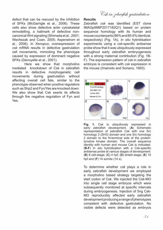
ResultsZebrafish csk was identified (EST clone IMAGp998P2017182Q1) based on protein sequence homology with its human and mouse counterparts (86% and 85.6% identical, respectively) (Fig. 1A). In situ hybridisation experiments using a csk-specific antisense probe show that it was ubiquitously expressed throughout early zebrafish embryogenesis with a strong maternal contribution (Fig. 1B-F). The expression pattern of csk in zebrafish embryos is consistent with csk expression in the mouse (Imamoto and Soriano, 1993).
Fig. 1. Csk is ubiquitously expressed in early zebrafish development. (A) Schematic representation of zebrafish Csk with one Src homology 3 (SH3) domain and one Src homology 2 domain to the N-terminal side of the protein-tyrosine kinase domain. The overall sequence identity with human and mouse Csk is indicated. (B-F) In situ hybridization with a Csk-specific antisense probe at various stages of development: (B) 8 cell-stage; (C) 4 hpf; (D) shield stage; (E) 10 hpf and (F) 14 somite (14 s).
To determine whether csk plays a role in early zebrafish development we employed a morpholino based strategy targeting the start codon of Csk. We injected the Csk-MO into single cell stage embryos which were subsequently monitored at specific intervals during embryogenesis. Injection of 5ng Csk-MO reproducibly affected early zebrafish development producing a range of phenotypes consistent with defective gastrulation. No visible defects were detected as embryos
a b c
d e f
Chapter2
32
progressed through epiboly. Only at 10hpf it became apparent that the embryos had failed to extend properly around the yolk,similar to the phenotype seen in Fyn/Yes morphants (Fig. 2A-C). At 3dpf, embryos were visibly shorter than un-injected controls (Fig. 2D). This was further confirmed by measuring the overall length of injected embryos from anterior to posterior at 3 dpf (Fig. 2E). The average length of wildtype embryos remained virtually invariant whereas morphant embryos were significantly shorter. Because morpholinos in
general can produce non-specific side effects (Nasevicius and Ekker, 2000) we needed to establish that the observed defects associated with Csk knockdown were not artefactual. To this end, we (co-) injected varying amounts of RNA encoding human csk which is not recognized by the Csk-MO. We found that injection of 150pg of human csk RNA by itself did not affect early zebrafish development morphologically (data not shown). However, co-injection of this amount of csk RNA with 5ng Csk-MO restored normal body length
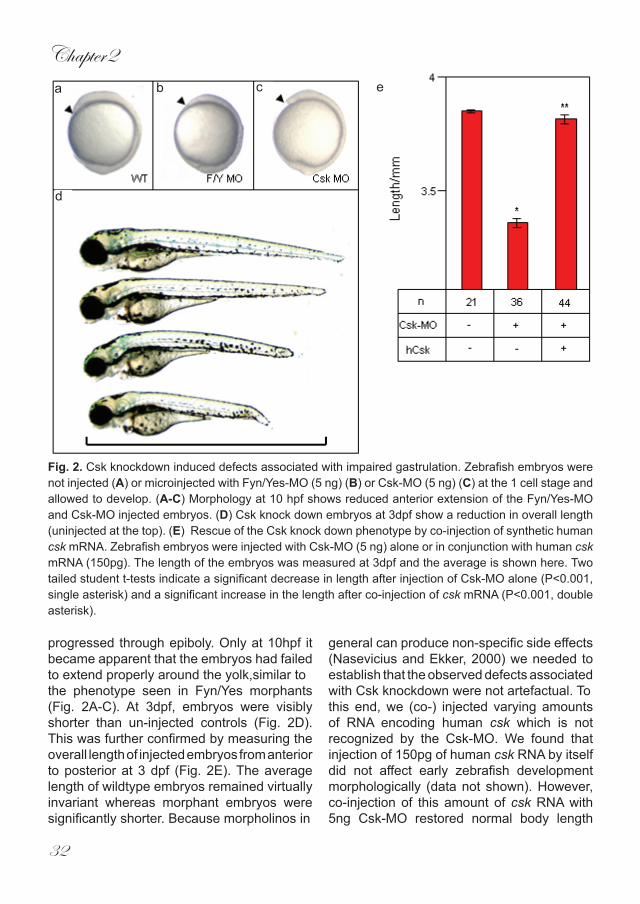
Fig. 2. Csk knockdown induced defects associated with impaired gastrulation. Zebrafish embryos were not injected (A) or microinjected with Fyn/Yes-MO (5 ng) (B) or Csk-MO (5 ng) (C) at the 1 cell stage and allowed to develop. (A-C) Morphology at 10 hpf shows reduced anterior extension of the Fyn/Yes-MO and Csk-MO injected embryos. (D) Csk knock down embryos at 3dpf show a reduction in overall length (uninjected at the top). (E) Rescue of the Csk knock down phenotype by co-injection of synthetic human csk mRNA. Zebrafish embryos were injected with Csk-MO (5 ng) alone or in conjunction with human csk mRNA (150pg). The length of the embryos was measured at 3dpf and the average is shown here. Two tailed student t-tests indicate a significant decrease in length after injection of Csk-MO alone (P<0.001, single asterisk) and a significant increase in the length after co-injection of csk mRNA (P<0.001, double asterisk).
a b c
d
e

Csk in zebrafish gastrulation
33
(Fig. 1E) and rescued overall morphology(data not shown), indicating that the defects caused by Csk-MO injection were a direct result of specific Csk knockdown.Next we investigated whether the observed defects were due to defective cell movement or incorrect cell specification, two very different processes which can give rise to similar phenotypes. To address this issue we performed in situ hybridization on Csk-MO injected embryos using a panel of markers that are all known to be involved in cell specification. Bone morphogenetic protein 2b (bmp2b) specifies ventral cell fates but remained unaffected when Csk was knocked down (Fig. 3A,B). The expression of Chordin (chd), a dorsalising factor, persisted when compared to un-injected controls (Fig. 3C,D) which was also the case for goosecoid (gsc), another dorsal specific gene expressed in the zebrafish organiser (Fig. 3E,F). Finally, we also found that the expression of the mesendodermal marker notail (ntl) was unaffected in Csk morphants (Fig. 3G,H). These results clearly show that cell fate in zebrafish embryos was not affected by Csk knockdown, suggesting that the observed defects were due to morphogenetic cell movements that occured during gastrulation.
The gastrulation defect we observed in Csk morphants may also have been caused by the mis-expression of known CE regulators such as Wnt11 and Wnt5 or Fyn and Yes. However, the expression of all these genes remained unaffected following Csk-MO injection (Fig. 3I-P), suggesting that it is more likely that Csk is directly involved in the regulation of gastrulation cell movements.Incorrect specification of the brain can result in embryos which lack certain brain structures. Obviously, this defect would make embryos appear to be shorter than wildtype embryos. For example, Six3 morphants have a severely reduced telencephalon when compared to uninjected controls and they appear to be shorter than uninjected controls (Ando et al., 2005). Because Csk morphants failed to extend properly around the yolk at
10hpf (Fig. 1C) we wondered whether this was due to defective gastrulation or simply because they lack anterior structures. Six3 is expressed in the developing forebrain of zebrafish embryos, pax2 in the midbrain-hindbrain boundary and krox20 labels rhombomeres 3 and 5. The expression of all of these genes was not affected in Csk morphants, indicating that these structures were present (Fig. 3A-F). However, the expression patterns of all 3 markers shifted posteriorly (Fig. 3A-F). When viewed from the dorsal side the expression patterns of six3 and pax2 were also broader than the control embryos (Fig. 3I-L). 8hpf embryos express the axial mesendodermal marker cyclops (cyc) during gastrulation. The cyc expression pattern was clearly broader and shorter in Csk-MO injected embryos than in un-injected control embryos (Fig. 3M,N).
Fig. 3. Csk knock down did not affect cell specification, nor expression of known regulators of CE cell movements. Csk-MO injected embryos were fixed at 6 hpf and in situ hybridization was done with various probes: (A,B) bone morphogenetic protein 2b, bmp; (C,D) chordin, chd; (E,F) goosecoid, gsc; (G,H) no tail, ntl; (I,J) wnt11; (K,L) yes; (M,N) wnt5; (O,P) fyn. Either lateral views (A-D,K,L,O,P) or animal pole views (E-H,I,J,M,N) are depicted here.
a b c d
e f g h
i j k l
m n o p
WTWT Csk MO Csk MO
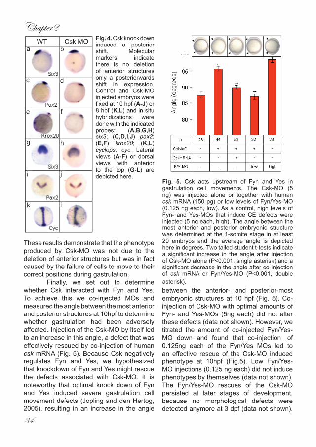
Chapter2
34
These results demonstrate that the phenotype produced by Csk-MO was not due to the deletion of anterior structures but was in fact caused by the failure of cells to move to their correct positions during gastrulation.
Finally, we set out to determine whether Csk interacted with Fyn and Yes. To achieve this we co-injected MOs and measured the angle between the most anterior and posterior structures at 10hpf to determine whether gastrulation had been adversely affected. Injection of the Csk-MO by itself led to an increase in this angle, a defect that was effectively rescued by co-injection of human csk mRNA (Fig. 5). Because Csk negatively regulates Fyn and Yes, we hypothesized that knockdown of Fyn and Yes might rescue the defects associated with Csk-MO. It is noteworthy that optimal knock down of Fyn and Yes induced severe gastrulation cell movement defects (Jopling and den Hertog, 2005), resulting in an increase in the angle
Fig. 4. Csk knock down induced a posterior shift. Molecular markers indicate there is no deletion of anterior structures only a posteriorwards shift in expression. Control and Csk-MO injected embryos were fixed at 10 hpf (A-J) or 8 hpf (K,L) and in situ hybridizations were done with the indicated probes: (A,B,G,H) six3; (C,D,I,J) pax2; (E,F) krox20; (K,L) cyclops, cyc. Lateral views (A-F) or dorsal views with anterior to the top (G-L) are depicted here.
Fig. 5. Csk acts upstream of Fyn and Yes in gastrulation cell movements. The Csk-MO (5 ng) was injected alone or together with human csk mRNA (150 pg) or low levels of Fyn/Yes-MO (0.125 ng each, low). As a control, high levels of Fyn- and Yes-MOs that induce CE defects were injected (5 ng each, high). The angle between the most anterior and posterior embryonic structure was determined at the 1-somite stage in at least 20 embryos and the average angle is depicted here in degrees. Two tailed student t-tests indicate a significant increase in the angle after injection of Csk-MO alone (P<0.001, single asterisk) and a significant decrease in the angle after co-injection of csk mRNA or Fyn/Yes-MO (P<0.001, double asterisk).
a b
c d
e f
g h
i j
k l
WT Csk MO
between the anterior- and posterior-most embryonic structures at 10 hpf (Fig. 5). Co-injection of Csk-MO with optimal amounts of Fyn- and Yes-MOs (5ng each) did not alter these defects (data not shown). However, we titrated the amount of co-injected Fyn/Yes- MO down and found that co-injection of 0.125ng each of the Fyn/Yes MOs led to an effective rescue of the Csk-MO induced phenotype at 10hpf (Fig.5). Low Fyn/Yes-MO injections (0.125 ng each) did not induce phenotypes by themselves (data not shown). The Fyn/Yes-MO rescues of the Csk-MO persisted at later stages of development, because no morphological defects were detected anymore at 3 dpf (data not shown).
Csk in zebrafish gastrulation
35
These results are consistent with Csk knock down leading to hyperactivation of Fyn and Yes, causing the observed gastrulation defects. Partial knock down of Fyn and Yes in turn rescued the Csk knock down phenotype, placing Fyn and Yes downstream of Csk in zebrafish gastrulation cell movements.
DiscussionHere we provide evidence that Csk has an essential role in early zebrafish development. We show here that csk is expressed throughout early zebrafish development (Fig. 1). Knock down of Csk resulted in CE defects that were morphologically detectable at 10hpf (Fig. 2A-C). The Csk knock down defects were reminiscent of previously documented morphants and mutants with compromised gastrulation (Topczewski et al., 2001; Yamashita et al., 2002; Jopling and den Hertog, 2005). At later stages the embryos were visibly shorter than wildtype siblings, a common feature of CE mutants such as Pipetail (Wnt5) and Trilobite (Sepich et al., 2000; Kilian et al., 2003). Csk phosphorylates and inhibts SFKs and is known to be involved in the regulation of a number of pathways which include growth factors and cell-cell signaling molecules (McGarrigle et al., 2006; Vidal et al., 2006). Some of these may regulate cell specification, which - when disrupted - can produce embryos with similar defects to those produced when CE is defective (Schulte-Merker et al., 1994; Schier et al., 1997; Rebagliati et al., 1998). However, the expression patterns of a panel of cell specification markers remained unaltered in Csk morphants (Fig. 3), indicating that the defects we observed were not caused by defective cell fate determination. Moreover, anterior patterning markers were still expressed, albeit there was a distinct posterior shift in expression (Fig. 4), similar to that observed in other CE defective morphants such as Stat3 (Yamashita et al., 2002). Our results indicate that Csk is essential for normal gastrulation cell movements.
Csk negatively regulates SFKs by phosphorylation of their COOH-terminal tyrosine. We demonstrate that Csk knockdown in zebrafish embryos was rescued by partial Fyn/Yes knock down (Fig. 5), indicating that Csk indeed is genetically upstream of Fyn and Yes in zebrafish development. Moreover, our results indicate that the Csk knock down phenotype is caused by hyperactivation of the
Chapter2
36
SFKs, Fyn and Yes. Interestingly, Csk knock out fibroblast cells display a migratory defect that is rescued by a SFK inhibitor (McGarrigle et al., 2006), indicating that rescue of the loss of Csk by inhibition of SFK activity is not unprecedented.
Modulation of Csk expression in other species induces phenotypes that are consistent with gastrulation defects as well. For instance, Csk over-expression in Xenopus phenocopies the gastrulation defect caused by expressing dominant negative SFKs (Denoyelle et al., 2001). Homozygous mouse knockouts with a targeted mutation in Csk lack functional Csk and harbor consitutively activated SFKs. The phenotype of these mice is compatible with defective gastrulation as well (Nada et al., 1993). We found that knock down of the SFKs, Fyn and Yes, induced gastrulation cell movement defects (Jopling and den Hertog, 2005), similar to the Csk knock down defects described here. The paradox that is emerging is that over-expression of a factor causes the same defects as knocking it out or down. The concept that “too much or not enough” can produce similar effects is not new and a number of studies have reported this finding in distinct biological processes, including gastrulation. For instance, overexpression of Rok2, a downstream effector of the non-canonical Wnt pathway, produces a similar phenotype as expression of a dominant negative form (Marlow et al., 2002). Similarly, Galpha12 and 13 are involved in the control of CE movements and over-expression of WT Galpha12/13 RNA produces a similar defect as Galpha12/13 knock down (Lin et al., 2005). It appears that an activity window exists for these factors. If overall activity falls outside of this window (either positively or negatively) the resulting phenotypes are very similar. This model may explain why knockdown of Csk produces similar defects as Fyn/Yes knockdowns, despite their antagonistic roles in cell signaling.
In conclusion, we show here that Csk is required for normal vertebrate gastrulation
to occur. The Csk knockdown phenotype is reminiscent of other CE defective phenotypes both morphologically and molecularly. Because we were able to rescue the Csk phenotype by knocking down Fyn and Yes, Csk is genetically directly upstream of Fyn and Yes in the regulation of cell movements during vertebrate gastrulation.
Materials and Methods
Zebrafish and in situ hybridizationZebrafish were kept and the embryos were staged as described before (Westerfield, 1995). In situ hybridizations were done essentially as described (Thisse et al., 1993) using probes specific for wnt5, bmp2b, chd, cyc, ntl, gsc, six3, pax2 and krox20 (generous gifts from various members of the zebrafish community) and fyn, yes, wnt11 and csk (RZPD ID’s: UCDMp611J0321Q114, MPMGp609A1681Q8, MPMGp637F0720Q2 and IMAGp998P2017182Q1, respectively from www.rzpd.de, Berlin, Germany).
Morpholinos, RNA and injectionsAntisense MOs were designed to include the start ATG of the respective cDNAs and ordered from GeneTools (Philomath, OR, USA): Csk, 5’- GCCAGGTCGCCTCAAAG GTAGACAT. The Fyn and Yes MOs were described before (Jopling and den Hertog, 2005). 5’ capped sense RNAs were synthesized using a construct encoding csk and the mMessage mMachine kit (Ambion, Austin, TX, USA). Ranges of MO (0.1 – 5 ng) were injected into embryos of the AB strain at the 1 cell stage and phenotypes were assessed at the indicated stages.
AcknowledgementsThe authors would like to thank all who have contributed probes for the in situ hybridizations described here. This work was supported by a European Union (FP5) Research Training Network (HPRN-CT-2000-00085).
Csk in zebrafish gastrulation
37
ReferencesAndo H, Kobayashi M, Tsubokawa T, Uyemura
K, Furuta T, Okamoto H. (2005) Lhx2 mediates the activity of Six3 in zebrafish forebrain growth. Dev Biol. 287, 456-468.
Aspenstrom P, Richnau N, Johansson AS (2006) The diaphanous-related formin DAAM1 collaborates with the Rho GTPases RhoA and Cdc42, CIP4 and Src in regulating cell morphogenesis and actin dynamics. Exp Cell Res, in press
Bakkers J, Kramer C, Pothof J, Quaedvlieg NE, Spaink HP, Hammerschmidt M (2004) Has2 is required upstream of Rac1 to govern dorsal migration of lateral cells during zebrafish gastrulation. Development 131, 525-537
Bardet PL, Horard B, Laudet V, Vanacker JM (2005) The ERRalpha orphan nuclear receptor controls morphogenetic movements during zebrafish gastrulation. Dev Biol. 281, 102-111.
Cha YI, Kim SH, Solnica-Krezel L, Dubois RN (2005) Cyclooxygenase-1 signaling is required for vascular tube formation during development. Dev Biol. 282, 274-283.
Denoyelle M, Valles AM, Lentz D, Thiery JP, Boyer B (2001) Mesoderm-independent regulation of gastrulation movements by the src tyrosine kinase in Xenopus embryo. Differentiation 69, 38-48.
Goldstein B, Takeshita H, Mizumoto K, Sawa H (2006) Wnt signals can function as positional cues in establishing cell polarity. Dev Cell. 10, 391-396.
Habas R, Kato Y, He X (2001) Wnt/Frizzled activation of Rho regulates vertebrate gastrulation and requires a novel Formin homology protein Daam1. Cell 107, 843-854.
Habas R, Dawid IB, He X (2003) Coactivation of Rac and Rho by Wnt/Frizzled signaling is required for vertebrate
gastrulation. Genes Dev 17, 295-309.
Hannus M, Feiguin F, Heisenberg CP, Eaton S (2002) Planar cell polarization requires Widerborst, a B’ regulatory subunit of protein phosphatase 2A. Development 129, 3493-3503.
Heisenberg CP, Tada M, Rauch GJ, Saude L, Concha ML, Geisler R, Stemple DL, Smith JC, Wilson SW (2000) Silberblick/Wnt11 mediates convergent extension movements during zebrafish gastrulation. Nature 405, 76-81.
Imamoto A, Soriano P (1993) Disruption of the csk gene, encoding a negative regulator of Src family tyrosine kinases, leads to neural tube defects and embryonic lethality in mice. Cell 73, 1117-1124.
Jopling C, den Hertog J (2005). Fyn/Yes and non-canonical Wnt signalling converge on RhoA in vertebrate gastrulation cell movements. EMBO Rep. 5, 426-431.
Keller R, Shih J, Domingo C (1992) The patterning and functioning of protrusive activity during convergence and extension of the Xenopus organiser. Development Suppl., 81-91.
Kilian B, Mansukoski H, Barbosa FC, Ulrich F, Tada M, Heisenberg CP (2003) The role of Ppt/Wnt5 in regulating cell shape and movement during zebrafish gastrulation. Mech Dev. 120, 467-476.
Lin F, Sepich DS, Chen S, Topczewski J, Yin C, Solnica-Krezel L, Hamm H (2005) Essential roles of G{alpha}12/13 signaling in distinct cell behaviors driving zebrafish convergence and extension gastrulation movements. J Cell Biol 169, 777-787.
Marlow F, Topczewski J, Sepich D, Solnica-Krezel L (2002) Zebrafish Rho kinase 2 acts downstream of wnt11 to mediate cell polarity and effective convergent extension movements.
Chapter2
38
Curr Biol 12, 876-884.McGarrigle D, Shan D, Yang S, Huang XY
(2006) Role of tyrosine kinase Csk in G protein-coupled receptor- and receptor tyrosine kinase-induced fibroblast cell migration. J Biol Chem 281, 10583-10588.
Murdoch JN, Doudney K, Paternotte C, Copp AJ, Stanier P (2001) Severe neural tube defects in the loop-tail mouse result from mutation of Lpp1, a novel gene involved in floor plate specification. Hum Mol Genet. 10, 2593-2601.
Murdoch JN, Henderson DJ, Doudney K, Gaston-Massuet C, Phillips HM, Paternotte C, Arkell R, Stanier P, Copp AJ. (2003) Disruption of scribble (Scrb1) causes severe neural tube defects in the circletail mouse. Hum Mol Genet 15, 87-98.
Nada S, Okada M, MacAuley A, Cooper JA, Nakagawa H (1991) Cloning of a complementary DNA for a protein-tyrosine kinase that specifically phosphorylates a negative regulatory site of p60c-src. Nature 351, 69-72.
Nada S, Yagi T, Takeda H, Tokunaga T, Nakagawa H, Ikawa Y, Okada M, Aizawa S (1993) Constitutive activation of Src family kinases in mouse embryos that lack Csk. Cell 73, 1125-1135.
Nasevicius A, Ekker SC (2000). Effective targeted gene ‘knockdown’ in zebrafish. Nat Genet 26, 216-220.
Oates AC, Lackmann M, Power MA, Brennan C, Down LM, Do C, Evans B, Holder N, Boyd AW (1999) An early developmental role for eph-ephrin interaction during vertebrate gastrulation. Mech Dev 83, 77-94.
Rebagliati MR, Toyama R, Haffter P, Dawid IB. Cyclops encodes a nodal-related factor involved in midline signaling. Proc Natl Acad Sci U S A. 95, 9932-9937.
Schier AF, Neuhauss SC, Helde KA, Talbot
WS, Driever W (1997) The one-eyed pinhead gene functions in mesoderm and endoderm formation in zebrafish and interacts with no tail. Development 124, 327-342.
Schulte-Merker S, van Eeden FJ, Halpern ME, Kimmel CB, Nusslein-Volhard C. (1994) No tail (ntl) is the zebrafish homologue of the mouse T (Brachyury) gene. Development 120, 1009-1015.
Sepich DS, Myers DC, Short R, Topczewski J, Marlow F, Solnica-Krezel L. (2000) Role of the zebrafish trilobite locus in gastrulation movements of convergence and extension. Genesis 27, 159-173.
Shimada Y, Usui T, Yanagawa S, Takeichi M, Uemura T (2001) Asymmetric colocalization of Flamingo, a seven-pass transmembrane cadherin, and Dishevelled in planar cell polarization. Curr Biol. 11, 859-863.
Solnica-Krezel L, Eaton S (2003) Embryo morphogenesis: getting down to cells and molecules. Development 130, 4229-4233.
Thisse C, Thisse B, Schilling TF, Postlethwait JH (1993) Structure of the zebrafish snail1 gene and its expression in wild-type, spadetail and no tail mutant embryos. Development 119, 1203-1215.
Topczewski J, Sepich DS, Myers DC, Walker C, Amores A, Lele Z, Hammerschmidt M, Postlethwait J, Solnica-Krezel L (2001) The zebrafish glypican knypek controls cell polarity during gastrulation movements of convergent extension. Dev Cell 1, 251-264.
Vidal M, Larson DE, Cagan RL (2006) Csk-deficient boundary cells are eliminated from normal Drosophila epithelia by exclusion, migration, and apoptosis. Dev Cell 10, 33-44.
Wada H, Iwasaki M, Sato T, Masai I, Nishiwaki Y, Tanaka H, Sato A, Nojima Y,

Csk in zebrafish gastrulation
39
Okamoto H (2005) Dual roles of zygotic and maternal Scribble1 in neural migration and convergent extension movements in zebrafish embryos. Development 132, 2273-2285.
Warga RM, Kimmel CB (1990) Cell movements during epiboly and gastrulation in zebrafish. Development 108, 569-580.
Wechezak AR, DE Coan (2005) Dvl2 silencing in postdevelopmental cells results in aberrant cell membrane activity and actin disorganization. J Cell Physiol 202, 867-873.
Westerfield M (1995). The Zebrafish Book. Univ. Oregon Press, Salem, Oregon.
Yamashita S, Miyagi C, Carmany-Rampey A, Shimizu T, Fujii R, Schier AF, Hirano T (2002) Stat3 Controls Cell Movements during Zebrafish Gastrulation. Dev Cell 2, 363-375.
Yeo SY, Little MH, Yamada T, Miyashita T, Halloran MC, Kuwada JY, Huh TL, Okamoto H (2001) Overexpression of a slit homologue impairs convergent extension of the mesoderm and causes cyclopia in embryonic zebrafish. Dev Biol 230, 1-17.
Zhang SQ, Yang W, Kontaridis MI, Bivona TG, Wen G, Araki T, Luo J, Thompson JA, Schraven BL, Philips MR, Neel BG (2004) Shp2 regulates SRC family kinase activity and Ras/Erk activation by controlling Csk recruitment. Mol Cell 13, 341-355.

Chapter3
Fyn/Yes and non-canonical Wnt signaling converge on RhoA in vertebrate gastrulation cell movements
Chris Jopling and Jeroen den Hertog+
Hubrecht LaboratoryUppsalalaan 8, 3584 CT Utrecht, The Netherlands
+ corresponding author:Tel: +31 30 2121800Fax: +31 30 2516464
e-mail: [email protected]
EMBO reports (2005) 6, 426-431

Fyn/Yes in vertebrate gastrulation
43
Abstract
Convergent extension (CE) cell movements during gastrulation mediate extension of the anterior-posterior body axis of vertebrate embryos. Non-canonical Wnt5 and Wnt11 signaling are essential for normal CE movements in vertebrate gastrulation. Here we show that morpholino (MO)-mediated double knock down of the Fyn and Yes tyrosine kinases in zebrafish embryos impaired normal CE cell movements, resembling the silberblick and pipetail mutants, caused by mutations in wnt11 and wnt5, respectively. Co-injection of Fyn/Yes- and Wnt11- or Wnt5-MOs was synergistic, but wnt11 or wnt5 RNA did not rescue the Fyn/Yes knock down nor vice versa. Remarkably, active RhoA rescued the Fyn/Yes knock down as well as the Wnt11 knock down, indicating that Fyn/Yes and Wnt11 signaling converged on RhoA. Our results demonstrate that Fyn and Yes act together with non-canonical Wnt signaling via RhoA in CE
cell movements during gastrulation.
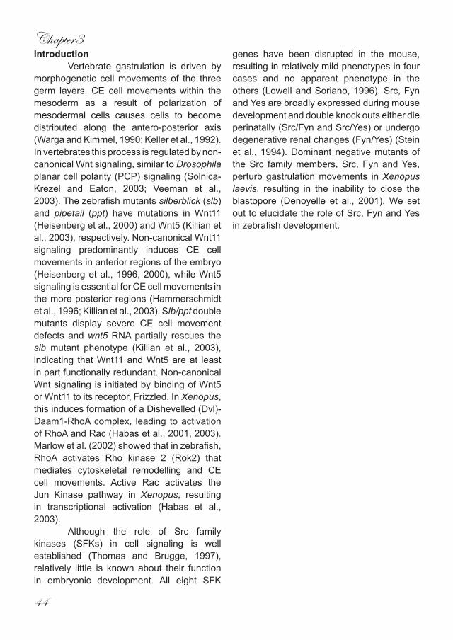
Chapter3
44
IntroductionVertebrate gastrulation is driven by
morphogenetic cell movements of the three germ layers. CE cell movements within the mesoderm as a result of polarization of mesodermal cells causes cells to become distributed along the antero-posterior axis (Warga and Kimmel, 1990; Keller et al., 1992). In vertebrates this process is regulated by non-canonical Wnt signaling, similar to Drosophila planar cell polarity (PCP) signaling (Solnica-Krezel and Eaton, 2003; Veeman et al., 2003). The zebrafish mutants silberblick (slb) and pipetail (ppt) have mutations in Wnt11 (Heisenberg et al., 2000) and Wnt5 (Killian et al., 2003), respectively. Non-canonical Wnt11 signaling predominantly induces CE cell movements in anterior regions of the embryo (Heisenberg et al., 1996, 2000), while Wnt5 signaling is essential for CE cell movements in the more posterior regions (Hammerschmidt et al., 1996; Killian et al., 2003). Slb/ppt double mutants display severe CE cell movement defects and wnt5 RNA partially rescues the slb mutant phenotype (Killian et al., 2003), indicating that Wnt11 and Wnt5 are at least in part functionally redundant. Non-canonical Wnt signaling is initiated by binding of Wnt5 or Wnt11 to its receptor, Frizzled. In Xenopus, this induces formation of a Dishevelled (Dvl)-Daam1-RhoA complex, leading to activation of RhoA and Rac (Habas et al., 2001, 2003). Marlow et al. (2002) showed that in zebrafish, RhoA activates Rho kinase 2 (Rok2) that mediates cytoskeletal remodelling and CE cell movements. Active Rac activates the Jun Kinase pathway in Xenopus, resulting in transcriptional activation (Habas et al., 2003). Although the role of Src family kinases (SFKs) in cell signaling is well established (Thomas and Brugge, 1997), relatively little is known about their function in embryonic development. All eight SFK
genes have been disrupted in the mouse, resulting in relatively mild phenotypes in four cases and no apparent phenotype in the others (Lowell and Soriano, 1996). Src, Fyn and Yes are broadly expressed during mouse development and double knock outs either die perinatally (Src/Fyn and Src/Yes) or undergo degenerative renal changes (Fyn/Yes) (Stein et al., 1994). Dominant negative mutants of the Src family members, Src, Fyn and Yes, perturb gastrulation movements in Xenopus laevis, resulting in the inability to close the blastopore (Denoyelle et al., 2001). We set out to elucidate the role of Src, Fyn and Yes in zebrafish development.
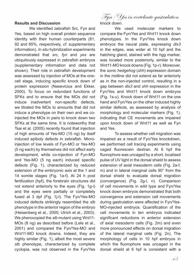
Fyn/Yes in vertebrate gastrulation
45
Results and DiscussionWe identified zebrafish Src, Fyn and
Yes, based on high overall protein sequence identity with their human counterparts (81, 92 and 85%, respectively, cf. supplementary information). In situ hybridization experiments demonstrated that src, fyn and yes are ubiquitously expressed in zebrafish embryos (supplementary information and data not shown). Their role in zebrafish development was assessed by injection of MOs at the one-cell stage, inducing specific knock down of protein expression (Nasevicius and Ekker, 2000). To focus on redundant functions of SFKs and to ensure that the MOs did not induce inadvertent non-specific defects, we titrated the MOs to amounts that did not induce a phenotype on their own and we co-injected the MOs in pairs to knock down two SFKs at the same time. It is noteworthy that Tsai et al. (2005) recently found that injection of high amounts of Yes-MO (15 ng) by itself induced epiboly defects in zebrafish. Micro-injection of low levels of Fyn-MO or Yes-MO (5 ng each) by themselves did not affect early development, while co-injection of Fyn-MO and Yes-MO (5 ng each) induced specific defects (Fig. 1), characterized by reduced extension of the embryonic axis at the 1 and 14 somite stages (Fig. 1a-f). At 24 h post fertilization (hpf), the forebrain structures did not extend anteriorly to the eyes (Fig. 1g-i) and the eyes were partially or completely fused at 3 dpf (Fig. 1j-l). The Fyn/Yes-MO induced defects strikingly resembled the slb phenotype in the anterior region of the embryo (Heisenberg et al., 2000; Ulrich et al., 2003). We phenocopied the slb mutant using Wnt11-MOs (8 ng) as described before (Lele et al., 2001) and compared the Fyn/Yes-MO and Wnt11-MO knock downs. Indeed, they are highly similar (Fig. 1), albeit the most severe slb phenotype, characterized by complete cyclopia, was not observed in the Fyn/Yes
knock down.We used molecular markers to
compare the Fyn/Yes and Wnt11 knock down phenotypes. In the Fyn/Yes knock down embryos the neural plate, expressing dlx3 in the edges, was wider at 10 hpf and the hatching gland, stained with the hgg marker, was located more posteriorly, similar to the Wnt11-MO knock downs (Fig. 1p-r). Moreover, the sonic hedgehog (shh) expression domain in the midline did not extend as far anteriorly as in the non-injected control, resulting in a gap between dlx3 and shh expression in the Fyn/Yes and Wnt11 knock down embryos (Fig. 1s-u). Knock down of Wnt11 on the one hand and Fyn/Yes on the other induced highly similar defects, as assessed by analysis of morphology and molecular markers (Fig. 1), indicating that CE movements are impaired upon knock down of Wnt11 as well as Fyn and Yes.
To assess whether cell migration was impaired as a result of Fyn/Yes knockdown, we performed cell tracing experiments using caged fluorescein dextran. At 6 hpf the fluorophore was uncaged by a short, localised pulse of UV light in the dorsal shield to assess extension of axial mesoderm cells (Fig. 2a-f, m) and in lateral marginal cells 90° from the dorsal shield to evaluate dorsal migration (convergence) (Fig. 2g-l, n). Comparison of cell movements in wild type and Fyn/Yes knock down embryos demonstrated that both convergence and extension cell movements during gastrulation were affected in Fyn/Yes-MO-injected embryos. Quantification of the cell movements in ten embryos indicated significant reductions in anterior extension of axial mesoderm cells (Fig. 2m) and even more pronounced effects on dorsal migration of the lateral marginal cells (Fig. 2n). The morphology of cells in 10 hpf embryos in which the fluorophore was uncaged in the dorsal shield at 6 hpf is consistent with a
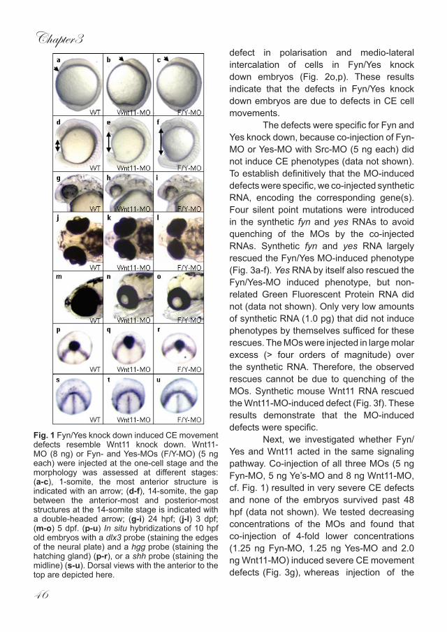
Chapter3
46
defect in polarisation and medio-lateral intercalation of cells in Fyn/Yes knock down embryos (Fig. 2o,p). These results indicate that the defects in Fyn/Yes knock down embryos are due to defects in CE cell movements.
The defects were specific for Fyn and Yes knock down, because co-injection of Fyn-MO or Yes-MO with Src-MO (5 ng each) did not induce CE phenotypes (data not shown). To establish definitively that the MO-induced defects were specific, we co-injected synthetic RNA, encoding the corresponding gene(s). Four silent point mutations were introduced in the synthetic fyn and yes RNAs to avoid quenching of the MOs by the co-injected RNAs. Synthetic fyn and yes RNA largely rescued the Fyn/Yes MO-induced phenotype (Fig. 3a-f). Yes RNA by itself also rescued the Fyn/Yes-MO induced phenotype, but non-related Green Fluorescent Protein RNA did not (data not shown). Only very low amounts of synthetic RNA (1.0 pg) that did not induce phenotypes by themselves sufficed for these rescues. The MOs were injected in large molar excess (> four orders of magnitude) over the synthetic RNA. Therefore, the observed rescues cannot be due to quenching of the MOs. Synthetic mouse Wnt11 RNA rescued the Wnt11-MO-induced defect (Fig. 3f). These results demonstrate that the MO-induced defects were specific.
Next, we investigated whether Fyn/Yes and Wnt11 acted in the same signaling pathway. Co-injection of all three MOs (5 ng Fyn-MO, 5 ng Ye’s-MO and 8 ng Wnt11-MO, cf. Fig. 1) resulted in very severe CE defects and none of the embryos survived past 48 hpf (data not shown). We tested decreasing concentrations of the MOs and found that co-injection of 4-fold lower concentrations (1.25 ng Fyn-MO, 1.25 ng Yes-MO and 2.0 ng Wnt11-MO) induced severe CE movement defects (Fig. 3g), whereas injection of the
Fig. 1 Fyn/Yes knock down induced CE movement defects resemble Wnt11 knock down. Wnt11-MO (8 ng) or Fyn- and Yes-MOs (F/Y-MO) (5 ng each) were injected at the one-cell stage and the morphology was assessed at different stages: (a-c), 1-somite, the most anterior structure is indicated with an arrow; (d-f), 14-somite, the gap between the anterior-most and posterior-most structures at the 14-somite stage is indicated with a double-headed arrow; (g-i) 24 hpf; (j-l) 3 dpf; (m-o) 5 dpf. (p-u) In situ hybridizations of 10 hpf old embryos with a dlx3 probe (staining the edges of the neural plate) and a hgg probe (staining the hatching gland) (p-r), or a shh probe (staining the midline) (s-u). Dorsal views with the anterior to the top are depicted here.
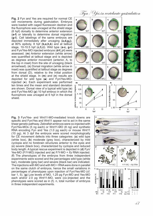
Fyn/Yes in vertebrate gastrulation
47
Fig. 2 Fyn and Yes are required for normal CE cell movements during gastrulation. Embryos were loaded with caged fluorescein dextran and the fluorophore was uncaged at the shield stage (6 hpf) dorsally to determine anterior extension (a-f) or laterally to determine dorsal migration (g-l). Cell labelings of the same embryos are depicted immediately after uncaging (a,d,g,j), at 80% epiboly, 8 hpf (b,e,h,k) and at tailbud stage, 10-10.5 hpf (c,f,i,l). Wild type (a-c, g-i) and Fyn/Yes-MO injected embryos (d-f, j-l) were assessed. (m) Anterior extension (white arrow) was quantified at tailbud stage and is depicted as degrees anterior movement (anterior, A, to the top in inset) from the site of uncaging (black arrowhead). (n) Dorsal migration (white arrow in inset) was quantified at tailbud stage as degrees from dorsal (D), relative to the initial position at the shield stage. In (m) and (n) results are depicted for wild type (■) and Fyn/Yes-MO-injected (●). Each experiment was performed ten times and the mean and standard deviation are shown. Dorsal view of a typical wild type (o) and Fyn/Yes-MO (p) 10 hpf embryo in which the fluorophore was uncaged at 6 hpf in the dorsal shield.
Fig. 3 Fyn/Yes- and Wnt11-MO-mediated knock downs are specific and Fyn/Yes and Wnt11 appear not to act in the same linear genetic pathway. Zebrafish embryos were co-injected with Fyn/Yes-MOs (5 ng each) or Wnt11-MO (8 ng) and synthetic RNA encoding Fyn and Yes (1.0 pg each) or mouse Wnt11 (10 pg). At 3 dpf the embryos were scored morphologically for CE movement defects into three categories: (a) wild type (white box), (b) moderate (grey box), characterized by mild cyclopia and no forebrain structures anterior to the eyes and (c) severe (black box), characterized by cyclopia and reduced body length. A typical rescue experiment is depicted in (d) Fyn/Yes-MO (F/Y-MO) injected and (e) F/Y-MO + f/y RNA injected. (f) The phenotypes of the embryos from three independent experiments were scored and the percentages wild type (white bar), moderate (grey bar) and severe (black bar) are indicated. The injections with MO and with MO + RNA were done in parallel on the same clutch of embryos, hence the small variations in percentages of phenotypes upon injection of Fyn/Yes-MO (cf. bar 1, 5). (g) Low levels of MO, 1.25 pg Fyn-MO and Yes-MO each and/or 2.0 pg Wnt11-MO, were (co-)injected and the phenotypes were scored as in (f). n, total number of embryos in three independent experiments.
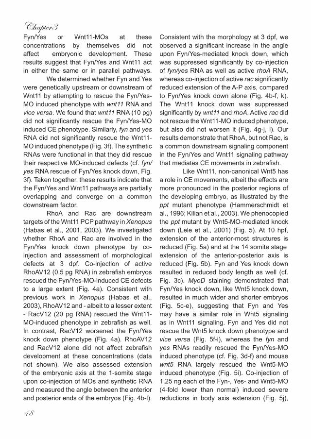
Chapter3
48
Fyn/Yes or Wnt11-MOs at these concentrations by themselves did not affect embryonic development. These results suggest that Fyn/Yes and Wnt11 act in either the same or in parallel pathways.
We determined whether Fyn and Yes were genetically upstream or downstream of Wnt11 by attempting to rescue the Fyn/Yes-MO induced phenotype with wnt11 RNA and vice versa. We found that wnt11 RNA (10 pg) did not significantly rescue the Fyn/Yes-MO induced CE phenotype. Similarly, fyn and yes RNA did not significantly rescue the Wnt11-MO induced phenotype (Fig. 3f). The synthetic RNAs were functional in that they did rescue their respective MO-induced defects (cf. fyn/yes RNA rescue of Fyn/Yes knock down, Fig. 3f). Taken together, these results indicate that the Fyn/Yes and Wnt11 pathways are partially overlapping and converge on a common downstream factor.
RhoA and Rac are downstream targets of the Wnt11 PCP pathway in Xenopus (Habas et al., 2001, 2003). We investigated whether RhoA and Rac are involved in the Fyn/Yes knock down phenotype by co-injection and assessment of morphological defects at 3 dpf. Co-injection of active RhoAV12 (0.5 pg RNA) in zebrafish embryos rescued the Fyn/Yes-MO-induced CE defects to a large extent (Fig. 4a). Consistent with previous work in Xenopus (Habas et al., 2003), RhoAV12 and - albeit to a lesser extent - RacV12 (20 pg RNA) rescued the Wnt11-MO-induced phenotype in zebrafish as well. In contrast, RacV12 worsened the Fyn/Yes knock down phenotype (Fig. 4a). RhoAV12 and RacV12 alone did not affect zebrafish development at these concentrations (data not shown). We also assessed extension of the embryonic axis at the 1-somite stage upon co-injection of MOs and synthetic RNA and measured the angle between the anterior and posterior ends of the embryos (Fig. 4b-l).
Consistent with the morphology at 3 dpf, we observed a significant increase in the angle upon Fyn/Yes-mediated knock down, which was suppressed significantly by co-injection of fyn/yes RNA as well as active rhoA RNA, whereas co-injection of active rac significantly reduced extension of the A-P axis, compared to Fyn/Yes knock down alone (Fig. 4b-f, k). The Wnt11 knock down was suppressed significantly by wnt11 and rhoA. Active rac did not rescue the Wnt11-MO induced phenotype, but also did not worsen it (Fig. 4g-j, l). Our results demonstrate that RhoA, but not Rac, is a common downstream signaling component in the Fyn/Yes and Wnt11 signaling pathway that mediates CE movements in zebrafish.
Like Wnt11, non-canonical Wnt5 has a role in CE movements, albeit the effects are more pronounced in the posterior regions of the developing embryo, as illustrated by the ppt mutant phenotype (Hammerschmidt et al., 1996; Kilian et al., 2003). We phenocopied the ppt mutant by Wnt5-MO-mediated knock down (Lele et al., 2001) (Fig. 5). At 10 hpf, extension of the anterior-most structures is reduced (Fig. 5a) and at the 14 somite stageextension of the anterior-posterior axis is reduced (Fig. 5b). Fyn and Yes knock down resulted in reduced body length as well (cf. Fig. 3c). MyoD staining demonstrated that Fyn/Yes knock down, like Wnt5 knock down, resulted in much wider and shorter embryos (Fig. 5c-e), suggesting that Fyn and Yes may have a similar role in Wnt5 signaling as in Wnt11 signaling. Fyn and Yes did not rescue the Wnt5 knock down phenotype and vice versa (Fig. 5f-i), whereas the fyn and yes RNAs readily rescued the Fyn/Yes-MO induced phenotype (cf. Fig. 3d-f) and mouse wnt5 RNA largely rescued the Wnt5-MO induced phenotype (Fig. 5i). Co-injection of 1.25 ng each of the Fyn-, Yes- and Wnt5-MO (4-fold lower than normal) induced severe reductions in body axis extension (Fig. 5j),
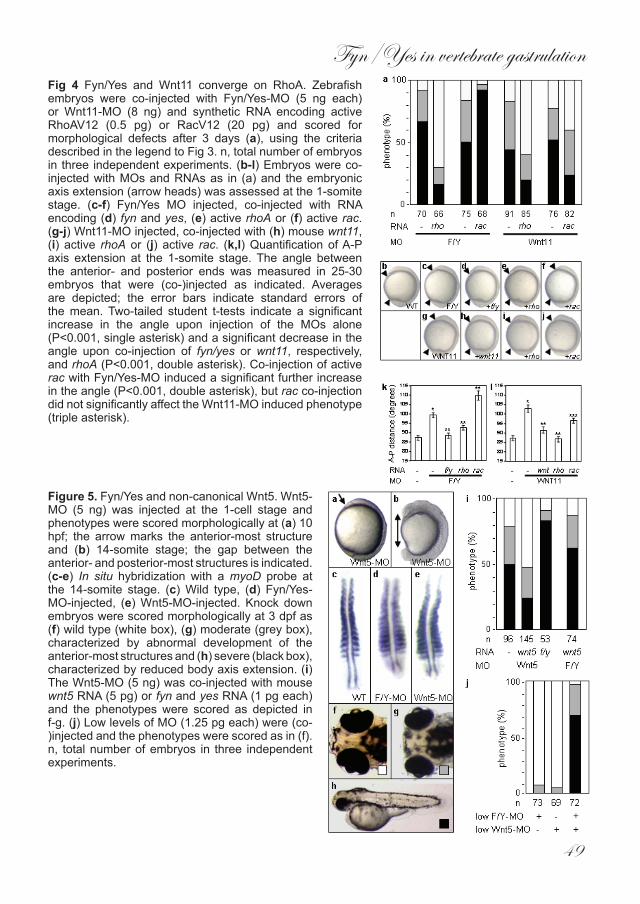
Fyn/Yes in vertebrate gastrulation
49
Fig 4 Fyn/Yes and Wnt11 converge on RhoA. Zebrafish embryos were co-injected with Fyn/Yes-MO (5 ng each) or Wnt11-MO (8 ng) and synthetic RNA encoding active RhoAV12 (0.5 pg) or RacV12 (20 pg) and scored for morphological defects after 3 days (a), using the criteria described in the legend to Fig 3. n, total number of embryos in three independent experiments. (b-l) Embryos were co-injected with MOs and RNAs as in (a) and the embryonic axis extension (arrow heads) was assessed at the 1-somite stage. (c-f) Fyn/Yes MO injected, co-injected with RNA encoding (d) fyn and yes, (e) active rhoA or (f) active rac. (g-j) Wnt11-MO injected, co-injected with (h) mouse wnt11, (i) active rhoA or (j) active rac. (k,l) Quantification of A-P axis extension at the 1-somite stage. The angle between the anterior- and posterior ends was measured in 25-30 embryos that were (co-)injected as indicated. Averages are depicted; the error bars indicate standard errors of the mean. Two-tailed student t-tests indicate a significant increase in the angle upon injection of the MOs alone (P<0.001, single asterisk) and a significant decrease in the angle upon co-injection of fyn/yes or wnt11, respectively, and rhoA (P<0.001, double asterisk). Co-injection of active rac with Fyn/Yes-MO induced a significant further increase in the angle (P<0.001, double asterisk), but rac co-injection did not significantly affect the Wnt11-MO induced phenotype (triple asterisk).
Figure 5. Fyn/Yes and non-canonical Wnt5. Wnt5-MO (5 ng) was injected at the 1-cell stage and phenotypes were scored morphologically at (a) 10 hpf; the arrow marks the anterior-most structure and (b) 14-somite stage; the gap between the anterior- and posterior-most structures is indicated. (c-e) In situ hybridization with a myoD probe at the 14-somite stage. (c) Wild type, (d) Fyn/Yes-MO-injected, (e) Wnt5-MO-injected. Knock down embryos were scored morphologically at 3 dpf as (f) wild type (white box), (g) moderate (grey box), characterized by abnormal development of the anterior-most structures and (h) severe (black box), characterized by reduced body axis extension. (i) The Wnt5-MO (5 ng) was co-injected with mouse wnt5 RNA (5 pg) or fyn and yes RNA (1 pg each) and the phenotypes were scored as depicted in f-g. (j) Low levels of MO (1.25 pg each) were (co-)injected and the phenotypes were scored as in (f). n, total number of embryos in three independent experiments.
Chapter3
50
whereas 1.25 ng of these MOs by themselves did not affect development at all. These results suggest that Fyn and Yes may be involved in Wnt5 signaling in a similar fashion as in Wnt11 signaling.
Our data indicate that Fyn/Yes and non-canonical Wnts converge on RhoA in CE movements during vertebrate gastrulation. Active RhoA rescued the Fyn/Yes as well as the Wnt11 knock down (Fig. 4). Fyn/Yes and Wnt11 may act independently in parallel pathways, activating different pools of RhoA, hence the ability of dominant active RhoAV12 to rescue both the Fyn/Yes and the Wnt11 knock downs. However, the combined knock down of Fyn/Yes and Wnt11, inducing severe CE defects (Fig. 3g) indicates that Fyn/Yes and Wnt11 signaling have common downstream components. Our results are consistent with data from C. elegans, where Wnt and Src signaling were postulated to act in parallel upstream of Rho to specify endoderm and to orient the cell division axis in the the EMS cell (Bei et al., 2002). In Xenopus, Wnt11 signaling bifurcates downstream of the Dvl/ Daam1 complex, leading to activation of RhoA and Rac (Habas et al., 2003). The finding that RhoA, but not Rac, rescued the Fyn/Yes knock down demonstrates that Fyn and Yes act downstream of the Dvl/Daam1 complex and upstream of RhoA. Fyn/Yes-induced activation of RhoA in zebrafish may be due to inactivation of RhoGAP, similar to Src64-mediated inactivation of p190RhoGAP in Drosophila (Billuart et al., 2001), which is consistent with direct p190RhoGAP phosphorylation by SFKs (Brouns et al., 2001). Moreover, Fyn/Yes-induced RhoA activation may also be due to direct phosphorylation and activation of a GTP exchange factor for RhoA (Schuebel et al., 1998).
In conclusion, our results demonstrate that Fyn and Yes are required for normal CE cell movements during gastrulation and
provide a starting point to further unravel the interaction between SFKs and non-canonical Wnt signaling.
Fyn/Yes in vertebrate gastrulation
51
MethodsZebrafish and in situ hybridizationZebrafish were kept and the embryos were staged as described before (Westerfield, 1995). In situ hybridizations were done essentially as described (Thisse et al., 1993), using probes specific for dlx3, shh, myoD (gifts of Jeremy Wegner, Jean-Paul Concordet and Eric Weinberg, respectively) and hgg1 (RZPD ID: IMAGp998O098963Q; RZPD, www.rzpd.de, Berlin, Germany).
Morpholinos, RNAs and injectionsAntisense MOs were targeted close to the start ATG of the respective cDNAs and ordered from GeneTools (Philomath, OR, USA): Src, 5’-GCCTCGTCGAAAACCACAC-GAAATG; Fyn, 5’- TGTCCTTACATTGCA-CACAG CCCAT; Yes, 5’-CCTCTTTACTCTT-GACACAGC CCAT. The Wnt11- and Wnt5-MO were described before (Lele et al., 2001). Fyn and Yes cDNAs were ampli-fied by PCR using oligonucleotides with four silent point mutations to avoid quench-ing of the MOs by the co-injected RNAs: mutant Fyn: 5’- ATGGGCTGCGTACAGT-GCAAGGACAAAGAGGCA, mutant Yes: 5’- ATGGGCTGCGTAAAAAGCAAAGAG-GACAAGGGT. 5’ capped sense RNAs were synthesized using constructs encoding mu-tant Fyn and Yes, mouse Wnt11 and Wnt5 (gift of Andy McMahon) and active RhoAV12 and RacV12 (gift of Boudewijn Burgering) and the mMessage mMachine kit (Ambion, Austin, TX, USA). Ranges of MO (0.1 – 10 ng) and synthetic RNA (0.5 pg – 1.0 ng) were injected into embryos of the AB or TL strain at the 1 – 2 cell stage and phenotypes were assessed at the indicated stages.
Cell tracingEmbryos were (co-)injected at the one cell stage with 0.25% 4,5-dimethoxy-2-nitrobenzyl (DMND)-caged fluorescein dextran (10,000 MW; Molecular Probes, Leiden, the Netherlands). Uncaging was done as described (Bakkers et al., 2004) at shield stage (6 hpf) using an Axioplan microscope, equipped with a UV light source, adjustable pinhole and 40X objective. Pictures were taken immediately following uncaging, at 80% epiboly (8hpf) and tailbud stage (10 hpf). The angles for dorsal convergence and anterior extension were determined using NIH imaging software.
Accession numbers Sequences of zebrafish cDNAs reported here were deposited in the EMBL database: src, AJ620750; fyn, AJ620748; yes, AJ620749.
AcknowledgementsWe thank Jeremy Wegner, Jean-Paul Concordet, Eric Weinberg, Andy McMahon and Boudewijn Burgering for reagents, Jeroen Bakkers for discussions and his help setting up the cell tracing experiments, Stefan Schulte-Merker for critical reading of the manuscript and members of the den Hertog, van Eeden and Zivkovic labs for advice and discussions. This work was supported in part by an EU Research Training Network Grant (HPRN-CT-2000-00085).
Chapter3
52
ReferencesBakkers J, Kramer C, Pothof J, Quaedvlieg NE,
Spaink HP, Hammerschmidt M (2004) Has2 is required upstream of Rac1 to govern dorsal migration of lateral cells during zebrafish gastrulation. Development 131: 525-537
Bei Y, Hogan J, Berkowitz LA, Soto M, Rocheleau CE, Pang KM, Collins J, Mello, CC (2002) SRC-1 and Wnt signaling act together to specify endoderm and to control cleavage orientation in early C. elegans embryos. Dev Cell. 3: 113-125
Billuart P, Winter CG, Maresh A, Zhao X, Luo L (2001) Regulating axon branch stability: the role of p190 RhoGAP in repressing a retraction signaling pathway. Cell 107: 195-207
Brouns M, Matheson SF, Settleman J (2001) p190 RhoGAP is the prinicpal Src substrate in brain and regulates axon outgrowth, guidance and fasciculation. Nat. Cell Biol. 3: 361-367
Denoyelle M, Valles AM, Lentz D, Thiery JP, Boyer B (2001) Mesoderm-independent regulation of gastrulation movements by the src tyrosine kinase in Xenopus embryo. Differentiation 69: 38-48
Habas R, Kato Y, He, X (2001) Wnt/Frizzled activation of Rho regulates vertebrate gastrulation and requires a novel Formin homology protein Daam1. Cell 107: 843-854
Habas R, Dawid IB, He X (2003) Coactivation of Rac and Rho by Wnt/Frizzled signaling is required for vertebrate gastrulation. Genes Dev. 17: 295-309
Hammerschmidt M et al (1996) Mutations affecting morphogenesis during gastrulation and tail formation in the zebrafish, Danio rerio. Development 123: 143-151
Heisenberg CP et al (1996) Genes involved in
forebrain development in the zebrafish, Danio rerio. Development 123: 191-203
Heisenberg CP, Tada M, Rauch GJ, Saude L, Concha ML, Geisler R, Stemple DL, Smith JC, Wilson SW (2000) Silberblick/Wnt11 mediates convergent extension movements during zebrafish gastrulation. Nature 405: 76-81
Keller R, Shih J, Domingo C (1992) The patterning and functioning of protrusive activity during convergence and extension of the Xenopus organiser. Development Suppl. 81-91
Kilian B, Mansukoski H, Barbosa FC, Ulrich F, Tada M, Heisenberg CP (2003) The role of Ppt/Wnt5 in regulating cell shape and movement during zebrafish gastrulation. Mech Dev. 120: 467-476
Lele Z, Bakkers J, Hammerschmidt M (2001) Morpholino phenocopies of the swirl, snailhouse, somitabun, minifin, silberblick, and pipetail mutations. Genesis 30: 190-194
Lowell CA, Soriano P (1996) Knockouts of Src-family kinases: Stiff bones, wimpy T cells and bad memories. Genes Dev. 10: 1845-1857
Marlow F, Topczewski J, Sepich D, Solnica-Krezel L (2002) Zebrafish Rho kinase 2 acts downstream of wnt11 to mediate cell polarity and effective convergent extension movements. Curr. Biol. 12: 876-884
Nasevicius A, Ekker SC (2000) Effective targeted gene ‘knockdown’ in zebrafish. Nat.Genet. 26: 216-220
Schuebel KE, Movilla N, Rosa JL, Bustelo XR (1998) Phosphorylation-dependent and constitutive activation of Rho proteins by wild-type and oncogenic Vav-2. EMBO J. 17: 6608-6621
Fyn/Yes in vertebrate gastrulation
53
Solnica-Krezel L, Eaton S (2003) Embryo morphogenesis: getting down to cells and molecules. Development 130: 4229 – 4233
Stein PL, Vogel H, Soriano P (1994) Combined deficiencies of Src, Fyn and Yes tyrosine kinases in mutant mice. Genes Dev. 8: 1999-2007
Thisse C, Thisse B, Schilling TF, Postlethwait JH (1993) Structure of the zebrafish snail1 gene and its expression in wild-type, spadetail and no tail mutant embryos. Development 119: 1203-1215
Thomas SM, Brugge JS (1997) Cellular functions regulated by Src family kinases. Annu. Rev. Cell Dev. Biol. 13: 513-609
Tsai WB, Zhang X, Sharma D, Wu W, Kinsey WH (2005) Role of Yes kinase during early zebrafish development. Dev Biol 277: 129-141
Ulrich F et al (2003) Slb/Wnt11 controls hypoblast cell migration and morphogenesis at the onset of zebrafish gastrulation. Development 130: 5375-5384
Veeman MT, Axelrod JD, Moon RT (2003) A second Canon: Functions and mechanisms of β-Catenin-independent Wnt signaling. Dev. Cell 5: 367-377
Warga RM, Kimmel CB (1990) Cell movements during epiboly and gastrulation in zebrafish. Development 108: 569-580
Westerfield M (1995) The Zebrafish Book. Univ. Oregon Press, Salem, Oregon

Chapter4
Shp2 controls zebrafish gastrulation cell movements via Fyn,
Yes and RhoA
Chris Jopling and Jeroen den Hertog*
Hubrecht LaboratoryNetherlands Institute for Developmental Biology
Uppsalalaan 83584 CT UtrechtThe Netherlands
* corresponding author:[email protected]: +31 30 2121800Fax: +31 30 2516464
Keywords: Shp2/ zebrafish/ gastrulation/ convergence and extension/ Fyn and Yes/ RhoA
Submitted

Shp2 controls zebrafish gastrulation
57
Abstract
Convergence and extension (CE) cell movements during gastrulation mediate extension of the anterior-posterior body axis of vertebrate embryos. Here we show that knock down of the protein-tyrosine phosphatase Shp2 in zebrafish embryos affected CE cell movements, but not cell specification. Shp2 acted upstream of the Src family kinases Fyn and Yes and the small GTPase RhoA. Our results show that Shp2 is required for normal CE cell movements via Fyn/Yes and RhoA in vertebrate gastrulation which may have implications for the role of Shp2
in human disease.
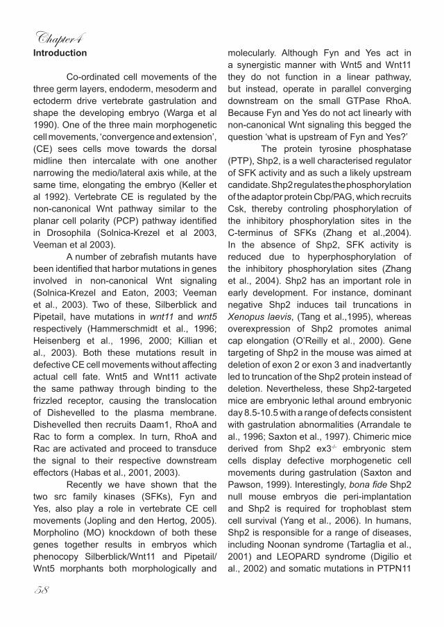
Chapter4
58
Introduction
Co-ordinated cell movements of the three germ layers, endoderm, mesoderm and ectoderm drive vertebrate gastrulation and shape the developing embryo (Warga et al 1990). One of the three main morphogenetic cell movements, ‘convergence and extension’, (CE) sees cells move towards the dorsal midline then intercalate with one another narrowing the medio/lateral axis while, at the same time, elongating the embryo (Keller et al 1992). Vertebrate CE is regulated by the non-canonical Wnt pathway similar to the planar cell polarity (PCP) pathway identified in Drosophila (Solnica-Krezel et al 2003, Veeman et al 2003). A number of zebrafish mutants have been identified that harbor mutations in genes involved in non-canonical Wnt signaling (Solnica-Krezel and Eaton, 2003; Veeman et al., 2003). Two of these, Silberblick and Pipetail, have mutations in wnt11 and wnt5 respectively (Hammerschmidt et al., 1996; Heisenberg et al., 1996, 2000; Killian et al., 2003). Both these mutations result in defective CE cell movements without affecting actual cell fate. Wnt5 and Wnt11 activate the same pathway through binding to the frizzled receptor, causing the translocation of Dishevelled to the plasma membrane. Dishevelled then recruits Daam1, RhoA and Rac to form a complex. In turn, RhoA and Rac are activated and proceed to transduce the signal to their respective downstream effectors (Habas et al., 2001, 2003).
Recently we have shown that the two src family kinases (SFKs), Fyn and Yes, also play a role in vertebrate CE cell movements (Jopling and den Hertog, 2005). Morpholino (MO) knockdown of both these genes together results in embryos which phenocopy Silberblick/Wnt11 and Pipetail/Wnt5 morphants both morphologically and
molecularly. Although Fyn and Yes act in a synergistic manner with Wnt5 and Wnt11 they do not function in a linear pathway, but instead, operate in parallel converging downstream on the small GTPase RhoA. Because Fyn and Yes do not act linearly with non-canonical Wnt signaling this begged the question ‘what is upstream of Fyn and Yes?’ The protein tyrosine phosphatase (PTP), Shp2, is a well characterised regulator of SFK activity and as such a likely upstream candidate. Shp2 regulates the phosphorylation of the adaptor protein Cbp/PAG, which recruits Csk, thereby controling phosphorylation of the inhibitory phosphorylation sites in the C-terminus of SFKs (Zhang et al.,2004). In the absence of Shp2, SFK activity is reduced due to hyperphosphorylation of the inhibitory phosphorylation sites (Zhang et al., 2004). Shp2 has an important role in early development. For instance, dominant negative Shp2 induces tail truncations in Xenopus laevis, (Tang et al.,1995), whereas overexpression of Shp2 promotes animal cap elongation (O’Reilly et al., 2000). Gene targeting of Shp2 in the mouse was aimed at deletion of exon 2 or exon 3 and inadvertantly led to truncation of the Shp2 protein instead of deletion. Nevertheless, these Shp2-targeted mice are embryonic lethal around embryonic day 8.5-10.5 with a range of defects consistent with gastrulation abnormalities (Arrandale te al., 1996; Saxton et al., 1997). Chimeric mice derived from Shp2 ex3-/- embryonic stem cells display defective morphogenetic cell movements during gastrulation (Saxton and Pawson, 1999). Interestingly, bona fide Shp2 null mouse embryos die peri-implantation and Shp2 is required for trophoblast stem cell survival (Yang et al., 2006). In humans, Shp2 is responsible for a range of diseases, including Noonan syndrome (Tartaglia et al., 2001) and LEOPARD syndrome (Digilio et al., 2002) and somatic mutations in PTPN11
Shp2 controls zebrafish gastrulation
59
contribute to leukemogenesis (Tartaglia et al., 2006). However, because Shp2 is involved in multiple pathways it has been difficult to elucidate the cell biological functions of Shp2 during early embryogenesis. Here we show that MO-mediated knockdown of Shp2 in zebrafish embryos resulted in defective CE movements during gastrulation. Despite the wide variety of pathways that are known to involve Shp2, cell fate remains unaffected upon Shp2 knockdown. We demonstrate that the Shp2 knockdown defects involve signaling through downstream components Fyn and Yes converging with non-canonical Wnt signaling at or upstream of the small GTPase RhoA. Our results are consistent with a role for Shp2 in vertebrate gastrulation cell movements.
ResultsWe identified zebrafish Shp2 (EST
clone IRAKp961F15101Q2) based on protein sequence homology with its human and mouse counterparts (91.2% and 90.3% identical, respectively) (Supplementary Fig. 1). In situ hybridisation experiments show that shp2 is ubiquitously expressed throughout early zebrafish development (Supplementary Fig. 1), consistent with previously unpublished data (Thisse and Thisse, 2004). To determine the role of Shp2 during early embryogenesis, we designed a Shp2-MO targeting the start codon and injected this at the one cell stage. We found that 1ng Shp2-MO consistently produced specific defects in embryonic development. The first visible defect is a failure of the embryo to extend properly around the yolk at 10hpf (Fig. 1A-C). At later stages (4dpf) embryos were shorter and developed a hammerhead phenotype similar to Wnt5 morphants (Fig. 1D-F). To further investigate the phenotypic relationship between the Shp2 and Wnt5 morphants, we compared alcian
blue cartilage staining of 4dpf embryo’s. The cartilaginous structures in the head of both morphants reside more posteriorly than in un-injected controls. Notably, Meckel’s cartilage (black asterisk) did not extend anterior to the eyes in Shp2-MO or Wnt5-MO injected embryos and the ceratohyal (red asterisk) clearly did not extend as far anteriorly as in the wild type (Fig. 1G-I). To establish that the observed defects were specific for Shp2 knockdown, we co-injected RNA encoding human shp2 which differs in sequence and is not targeted by the Shp2-MO. Co-injection of 300pg of human shp2 RNA which did not induce defects by itself (data not shown) with 1 ng Shp2-MO rescued all defects (Fig. 1J), indicating that the MO-induced defects were a direct result of specific Shp2 knockdown. It is noteworthy that injection of higher amounts of the Shp2-MO (up to 10 ng) induced more severe defects. However, because these defects were not completely rescued by co-injection of human shp2 mRNA (data not shown), all Shp2 knock downs described here were done with 1 ng Shp2-MO. Shp2 is known to be involved in many different signaling cascades (Neel et al., 2003). Some of these pathways are important for proper cell specification during embryogenesis and defective signaling leads to a variety of phenotypes including defects in the A-P axis. This raised the possibilty that the defects we observed were due to incorrect cell specification. To address this issue, we used a panel of well-characterised in-situ markers.Bome morphogenetic protein 2b (bmp2b) isinvolved in the specification of ventral cell fates but was not affected by knockdown of Shp2 (Fig. 2A,B). Expression of the dorsalising factor chordin (chd) remained the same (Fig. 2C,D) as did the dorsal specific gene goosecoid (gsc) which is expressed in the organiser (Fig. 2E,F).
Chapter4
60
Fig. 1. Shp2 induced defects phenocopy Wnt5 knock down and are specific. Zebrafish embryos were not injected (A,D,G) or microinjected with Wnt5-MO (5 ng) (B,E,H) or Shp2-MO (1.0 ng) (C,F,I,) at the 1 cell stage and allowed to develop. (A-C) Morphology at 10 hpf shows reduced anterior extension of the Wnt5-MO and Shp2-MO injected embryos. (D-F) Morphology of the Wnt5 and Shp2 knock down embryos show a mild hammerhead-like phenotype. (G-I) Alcian blue staining of the cartilage in the head confirms the reduced protrusion of the anterior-most structures in the head. Meckel’s cartilage is indicated with a black asterisk, the ceratohyal with a red asterisk. (J) Rescue of the Shp2 knock down phenotype by co-injection of synthetic human shp2 mRNA. Zebrafish embryos were (co-)injected with Shp2-MO (1.0 ng) and 300 pg human shp2 mRNA. The embryos were scored at 4 dpf for the hammerhead-like phenotype. A large percentage of the Shp2-MO induced phenotype was rescued by co-injection of shp2 mRNA.
Expression of the mesendodermal marker notail (ntl) also remained unchanged in Shp2 morphants (Fig. 2G,H). In summary, knockdown of Shp2 did not alter cell fate in early zebrafish embryos, suggesting it has a role in CE cell movements, rather than cell specification.To investigate whether disrupted CE movements were indeed the principle cause of the Shp2 knockdown induced defects, we performed cell tracing experiments using caged fluorescein. Embryos at the one cell stage were injected with either the caged
fluorophore alone or in conjunction with the Shp2-MO and then allowed to develop as normal. At 6hpf a cluster of cells within the dorsal shield was labeled by uncaging the fluorescein with a short, localised pulse of UV light. The group of cells was then monitored every 2 hours during gastrulation. The distance the cells migrated is directly proportional to embryonic extension. Repeating the process at 90 degrees to the shield gives an effective measurement of how far the mesendodermal cells converge towards the dorsal midline.
a b c
d e f
g h i
j

Shp2 controls zebrafish gastrulation
61
A comparison of wild type and Shp2-MO injected embryos revealed that Shp2 knockdown resulted in a significant reduction in the capability of cells to migrate both toward the dorsal midline (convergence) and anteriorly around the yolk (extension), indicating that Shp2 is required for correct CE during vertebrate gastrulation (Fig. 3A,B). The CE defects were further verified using well characterised molecular markers. The loss of genes involved in correct cell specification of the brain can result in embryos lacking certain brain structures. For example,FGF8 mutants and morphants lack the cerebellum and the midbrain-hindbrain boundary (Reifers et al., 2000; Draper et al., 2001). Obviously, embryos which lack certain structures in the brain would appear to be shorter than wildtype embryos. At 10hpf Shp2 morphants
do not extend properly around the yolk (Fig. 1). To ensure this is the result of defective CE movements and is not due to the deletion of anterior structures, we examined the expression of anterior specific genes: six3 is expressed in the forebrain, pax2 in the midbrain-hindbrain boundary and krox20 labels rhombomeres 3 and 5. Expression of all of these genes persisted in the Shp2 morphants, indicating that the structures these markers delineate were present. However, the expression patterns of these 3 genes were shifted posteriorly (Fig. 3C-H) and - most markedly in the case of pax2 and six3 - this posterior shift was accompanied by a broader expression pattern when viewed form the dorsal side (Fig. 3I-L). At 8hpf cyclops (cyc) is expressed in axial mesendodermal cells of gastrulating embryos. We found that Shp2 morphants clearly have a shorter and broader expression pattern when compared to un-injected controls (Fig. 3M,N).These results demonstrate that both convergence and extension cell movements were disrupted following knockdown of Shp2.
Shp2 might regulate CE cell movements by modulation of expression of the non-canonical Wnts or Fyn and Yes, known regulators of CE cell movements. However, the expression of wnt11, wnt5, fyn and yes remained unaffected in Shp2-MO injected embryos (Fig. 2I-P). Because Shp2 knock down did not affect the expression of known CE regulators, we hypothesize that Shp2 has a more direct role in CE signaling. To investigate how Shp2 is involved in CE cell signaling, we co-injected sub-optimal concentrations of Shp2-, Fyn/Yes- and Wnt5-MOs either alone or in unison. For Shp2- and Fyn/Yes-MO co-injections we used the in situ markers hgg1, which labels the hatching gland and dlx3, which marks the anterior neural plate at 10hpf. Previously we have shown that optimal injection (5ng
WT WT Shp2 MOShp2 MOa b c d
e f g h
i j k l
m n o p
Fig. 2. Shp2 knock down did not affect cell specification, nor expression of known regulators of CE cell movements. Shp2-MO injected embryos were fixed at 6 hpf and in situ hybridization was done with various probes: (A,B) bone morphogenetic protein 2b, bmp; (C,D) chordin, chd; (E,F) goosecoid, gsc; (G,H) no tail, ntl; (I,J) wnt11; (K,L) yes; (M,N) wnt5; (O,P) fyn. Either lateral views (A-D, K,L,O,P) or animal pole views (E-H, I,J,M,N) are depicted here.
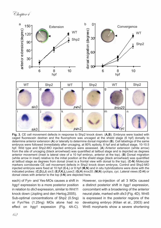
Chapter4
62
each) of Fyn- and Yes-MOs causes a shift in hgg1 expression to a more posterior position in relation to dlx3 expression, similar to Wnt11 knock down (Jopling and den Hertog,2005). Sub-optimal concentrations of Shp2 (0.5ng) or Fyn/Yes (1.25ng) MOs alone had no effect on hgg1 expession (Fig. 4A-C).
However, co-injection of all 3 MOs caused a distinct posterior shift in hgg1 expression, concomitant with a broadening of the anterior neural plate, marked with dlx3 (Fig. 4D). Wnt5 is expressed in the posterior regions of the developing embryo (Kilian et al., 2003) and Wnt5 morphants show a severe shortening
a bExtension Convergence
WT WT
Shp2Shp2
hpfhpf
WT WT WTShp2 Shp2 Shp2c d e f g h
i j k l m n
Fig. 3. CE cell movement defects in response to Shp2 knock down. (A,B). Embryos were loaded with caged fluorescein dextran and the fluorophore was uncaged at the shield stage (6 hpf) dorsally to determine anterior extension (A) or laterally to determine dorsal migration (B). Cell labelings of the same embryos were followed immediately after uncaging, at 80% epiboly, 8 hpf and at tailbud stage, 10-10.5 hpf. Wild type and Shp2-MO injected embryos were assessed. (A) Anterior extension (white arrow) from the site of uncaging (black arrowhead) was quantified at tailbud stage and is depicted as degrees anterior movement (inset is lateral view of a 10 hpf embryo, anterior at the top). (B) Dorsal migration (white arrow in inset) relative to the initial position at the shield stage (black arrowhead) was quantified at tailbud stage as degrees from dorsal (inset is a frontal view with dorsal to the top). (C-N) Molecular markers corroborate CE cell movement defects in Shp2 knock down embryos. Control and Shp2-MO injected embryos were fixed at 10 hpf (C-L) or 8 hpf (M,N) and in situ hybridizations were done with the indicated probes: (C,D,I,J) six3; (E,F,K,L) pax2; (G,H) krox20; (M,N) cyclops, cyc. Lateral views (C-H) or dorsal views with anterior to the top (I-N) are depicted here.
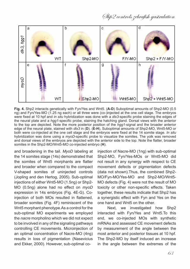
Shp2 controls zebrafish gastrulation
63
and broadening in the tail. MyoD labeling at the 14 somites stage (14s) demonstrated that the somites of Wnt5 morphants are flatter and broader when compared to the compact V-shaped somites of uninjected controls (Jopling and den Hertog, 2005). Sub-optimal injections of either Wnt5-MO (1.5ng) or Shp2-MO (0.5ng) alone had no effect on myoD expression in 14s embryos (Fig. 4E-G). Co-injection of both MOs resulted in flattened, broader somites (Fig. 4F) reminiscent of the Wnt5 morphant phenotype.As a control for the sub-optimal MO experiments we employed the nacre morpholino which we did not expect to be involved in any of the signaling pathways controlling CE movements. Microinjection of an optimal concentration of Nacre-MO (4ng) results in loss of pigmentation (Nasevicius and Ekker, 2000). However, sub-optimal co-
injection of Nacre-MO (1ng) with sub-optimal Shp2-MO, Fyn/Yes-MOs or Wnt5-MO did not result in any synergy with respect to CE movement defects or pigmentation defects (data not shown).Thus, the combined Shp2-MO/Fyn-MO/Yes-MO and Shp2-MO/Wnt5-MO defects (Fig. 4) were not the result of MO toxicity or other non-specific effects. Taken together, these results indicate that Shp2 has a synergistic effect with Fyn and Yes on the one hand and Wnt5 on the other.
Next, we investigated how Shp2 interacted with Fyn/Yes and Wnt5.To this end, we co-injected MOs with synthetic mRNAs and assessed CE movement defects by measurement of the angle between the most anterior and posterior tissues at 10 hpf. The Shp2-MO by itself induced an increase in the angle between the extremes of the
a b c d
e f g h
Fig. 4. Shp2 interacts genetically with Fyn/Yes and Wnt5. (A-D) Suboptimal amounts of Shp2-MO (0.5 ng) and Fyn/Yes-MO (1.25 ng each) or all three were (co-)injected at the one cell stage. The embryos were fixed at 10 hpf and in situ hybridization was done with a dlx3-specific probe staining the edges of the neural plate and a hgg1-specific probe, staining the hatching gland. Dorsal views with the anterior to the top are depicted. Note the more posterior position of the hgg1-signal and the broader anterior edge of the neural plate, stained with dlx3 in (D). (E-H), Suboptimal amounts of Shp2-MO, Wnt5-MO or both were co-injected at the one cell stage and the embryos were fixed at the 14 somite stage. In situ hybridization was done using a myoD-specific probe to visualize the somites. The yolk was removed and dorsal views of the embryos are depicted with the anterior side to the top. Note the flatter, broader somites in the Shp2-MO/Wnt5-MO co-injected embryo (H).
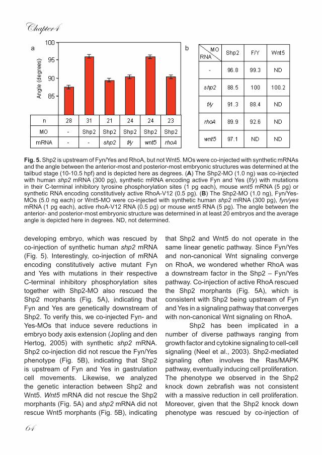
Chapter4
64
developing embryo, which was rescued by co-injection of synthetic human shp2 mRNA (Fig. 5). Interestingly, co-injection of mRNA encoding constitutively active mutant Fyn and Yes with mutations in their respective C-terminal inhibitory phosphorylation sites together with Shp2-MO also rescued the Shp2 morphants (Fig. 5A), indicating that Fyn and Yes are genetically downstream of Shp2. To verify this, we co-injected Fyn- and Yes-MOs that induce severe reductions in embryo body axis extension (Jopling and den Hertog, 2005) with synthetic shp2 mRNA. Shp2 co-injection did not rescue the Fyn/Yes phenotype (Fig. 5B), indicating that Shp2 is upstream of Fyn and Yes in gastrulation cell movements. Likewise, we analyzed the genetic interaction between Shp2 and Wnt5. Wnt5 mRNA did not rescue the Shp2 morphants (Fig. 5A) and shp2 mRNA did not rescue Wnt5 morphants (Fig. 5B), indicating
that Shp2 and Wnt5 do not operate in the same linear genetic pathway. Since Fyn/Yes and non-canonical Wnt signaling converge on RhoA, we wondered whether RhoA was a downstream factor in the Shp2 – Fyn/Yes pathway. Co-injection of active RhoA rescued the Shp2 morphants (Fig. 5A), which is consistent with Shp2 being upstream of Fyn and Yes in a signaling pathway that converges with non-canonical Wnt signaling on RhoA. Shp2 has been implicated in a number of diverse pathways ranging from growth factor and cytokine signaling to cell-cell signaling (Neel et al., 2003). Shp2-mediated signaling often involves the Ras/MAPK pathway, eventually inducing cell proliferation. The phenotype we observed in the Shp2 knock down zebrafish was not consistent with a massive reduction in cell proliferation. Moreover, given that the Shp2 knock down phenotype was rescued by co-injection of
ba
Fig. 5. Shp2 is upstream of Fyn/Yes and RhoA, but not Wnt5. MOs were co-injected with synthetic mRNAs and the angle between the anterior-most and posterior-most embryonic structures was determined at the tailbud stage (10-10.5 hpf) and is depicted here as degrees. (A) The Shp2-MO (1.0 ng) was co-injected with human shp2 mRNA (300 pg), synthetic mRNA encoding active Fyn and Yes (f/y) with mutations in their C-terminal inhibitory tyrosine phosphorylation sites (1 pg each), mouse wnt5 mRNA (5 pg) or synthetic RNA encoding constitutively active RhoA-V12 (0.5 pg). (B) The Shp2-MO (1.0 ng), Fyn/Yes-MOs (5.0 ng each) or Wnt5-MO were co-injected with synthetic human shp2 mRNA (300 pg), fyn/yes mRNA (1 pg each), active rhoA-V12 RNA (0.5 pg) or mouse wnt5 RNA (5 pg). The angle between the anterior- and posterior-most embryonic structure was determined in at least 20 embryos and the average angle is depicted here in degrees. ND, not determined.
Shp2 controls zebrafish gastrulation
65
active RhoA (Fig. 5), the Ras/MAPK signaling pathway appeared not to be involved. Deletion of Shp2 in other species also does not appear to induce massive defects in cell proliferation during early development. For instance, a Drosophila mutant with a mutation in the Shp2 homolog, corckscrew (csw) displays multiple defects that are not directly linked to defects in cell proliferation. (Perkins et al., 1992). In Xenopus, dominant negative Shp2 leads to tail truncations due to defective gastrulation (Tang et al., 1995). Interestingly, active Shp2 induces elongation of animal cap explants which is subsequently blocked by co-expression of dominant negative RhoA (O’Reilly et al., 2000), suggesting a similar signaling pathway as we observed in early zebrafish embryos. Homozygous mouse embryos with a targeted Shp2 exon 2 or exon 3 express a truncated form of Shp2 and die in utero around day 8.5-10.5 (Arrandale et al., 1996; Saxton et al., 1997). The root cause of this lethality remains unknown. Chimeric embryos generated with homozygous Shp2 exon 3 mutant ES cells display gastrulation defects, consistent with defective morphogenetic cell movements as a result of Shp2 ablation (Saxton and Pawson, 1999). Recently, Yang et al. (2006) generated bona fide Shp2 null mouse embryos by replacement of Shp2 exon 2 with the β-galactosidase gene with a strong splice acceptor site to prevent splicing from exon 1 to exon 3. Heterozygous mice show reduced Shp2 protein expression and no truncated protein. Homozygous Shp2 null embryos died in utero around implantation, i.e. prior to gastrulation, demonstrating that Shp2 is essential for life very early in development. In fact, conditional knock outs showed that Shp2 is essential for trophoblast stem cell survival, which explains early lethality of Shp2 null mouse embryos (Yang et al., 2006). Zebrafish Shp2 morphants survived and the first defects we
observed were around gastrulation. Maternal Shp2 expression in zebrafish (Supplementary Fig. 1B) may rescue pre-gastrulation lethality. Moreover, MO-mediated knock down is not complete and low levels of remaining Shp2 expression may be sufficient for zebrafish survival.
We demonstrate here that Shp2 has an important role in zebrafish development, which is consistent with previous reports documenting the role of Shp2 in other species. Interstingly, Shp2 dysfunction has also been linked to several different human diseases. Germline mutations in PTPN11, the gene that encodes Shp2, causes the well-characterized Noonan and LEOPARD syndromes, whereas somatic mutations in PTPN11 contribute to leukemogenesis (Tartaglia et al., 2001; Digilio et al., 2002; Tartaglia et al., 2006). Noonan and LEOPARD syndromes are relatively common syndromes with partially overlapping clinical manifestations, including facial anomalies, distinct congenital heart defects, pectus deformities, hearing loss and growth retardation and distinct pigmentary changes (Gorlin et al., 1971). Surprisingly, the mutations that were found in Shp2 from Noonan patients affect Shp2 regulation, leading to constitutive activation of Shp2 catalytic activity (Keilhack et al., 2005), whereas the LEOPARD Shp2 mutants have greatly reduced catalytic activities (Kontaridis et al., 2006). How mutations in Shp2 with opposing effects on catalytic activity induce overlapping syndromes remains to be determined. Yet some of the symptoms of Noonan and LEOPARD patients are consistent with cell migration defects early in development and it will be interesting to see if defects in directed cell movement are at the basis of these syndromes.
Here, we show that Shp2 acts upstream of Fyn/Yes by co-injection of low amounts of Shp2- and Fyn/Yes-MOs that act
Chapter4
66
synergistically, showing there is a genetic interaction. Importantly, co-injection of synthetic RNAs encoding active Fyn and Yes rescued the Shp2-MO induced phenotype, demonstrating that Shp2 acts upstream of Fyn and Yes in gastrulation cell movements. Shp2 indirectly activates SFKs through dephosphorylation of Cbp/PAG (Zhang et al., 2004) and it is likely that this pathway is involved in gastrulation cell movements, although we cannot exclude that Shp2 affects Fyn and Yes activity in a different manner. Additional PTPs may be involved in regulation of Fyn and Yes during zebrafish gastrulation as well. We and others have identified receptor PTPα as a direct activator of Src (Zheng et al., 1992; den Hertog et al., 1993). However, whereas knock down of RPTPα induced pleiotropic effects, defects in CE cell movements during gastrulation were not observed (van der Sar et al., 2002; CJ and JdH, unpublished observation).
In conclusion, we demonstrate here that Shp2, an indirect activator of SFKs acts upstream of Fyn and Yes in gastrulation cell movements. Active RhoA rescued the Shp2 knock down phenotype, consistent with Shp2 being upstream of Fyn and Yes, which in turn signal through RhoA in a signaling pathway parallel to non-canonical Wnt signaling.
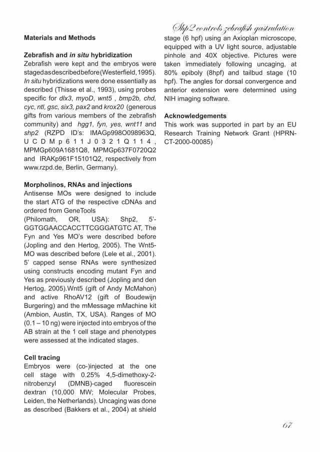
Shp2 controls zebrafish gastrulation
67
Materials and Methods
Zebrafish and in situ hybridizationZebrafish were kept and the embryos were staged as described before (Westerfield, 1995). In situ hybridizations were done essentially as described (Thisse et al., 1993), using probes specific for dlx3, myoD, wnt5 , bmp2b, chd, cyc, ntl, gsc, six3, pax2 and krox20 (generous gifts from various members of the zebrafish community) and hgg1, fyn, yes, wnt11 and shp2 (RZPD ID’s: IMAGp998O098963Q, U C D M p 6 1 1 J 0 3 2 1 Q 1 1 4 , MPMGp609A1681Q8, MPMGp637F0720Q2 and IRAKp961F15101Q2, respectively from www.rzpd.de, Berlin, Germany).
Morpholinos, RNAs and injectionsAntisense MOs were designed to include the start ATG of the respective cDNAs and ordered from GeneTools (Philomath, OR, USA): Shp2, 5’-GGTGGAACCACCTTCGGGATGTC AT, The Fyn and Yes MO’s were described before (Jopling and den Hertog, 2005). The Wnt5-MO was described before (Lele et al., 2001). 5’ capped sense RNAs were synthesized using constructs encoding mutant Fyn and Yes as previously described (Jopling and den Hertog, 2005).Wnt5 (gift of Andy McMahon) and active RhoAV12 (gift of Boudewijn Burgering) and the mMessage mMachine kit (Ambion, Austin, TX, USA). Ranges of MO (0.1 – 10 ng) were injected into embryos of the AB strain at the 1 cell stage and phenotypes were assessed at the indicated stages.
Cell tracingEmbryos were (co-)injected at the one cell stage with 0.25% 4,5-dimethoxy-2-nitrobenzyl (DMNB)-caged fluorescein dextran (10,000 MW; Molecular Probes, Leiden, the Netherlands). Uncaging was done as described (Bakkers et al., 2004) at shield
stage (6 hpf) using an Axioplan microscope, equipped with a UV light source, adjustable pinhole and 40X objective. Pictures were taken immediately following uncaging, at 80% epiboly (8hpf) and tailbud stage (10 hpf). The angles for dorsal convergence and anterior extension were determined using NIH imaging software.
AcknowledgementsThis work was supported in part by an EU Research Training Network Grant (HPRN-CT-2000-00085)
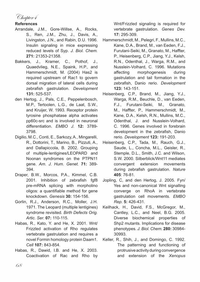
Chapter4
68
ReferencesArrandale, J.M., Gore-Willse, A., Rocks,
S., Ren, J.M., Zhu, J., Davis, A., Livingston, J.N., and Rabin, D.U. 1996. Insulin signaling in mice expressing reduced levels of Syp. J. Biol. Chem. 271: 21353-21358.
Bakkers, J., Kramer, C., Pothof, J., Quaedvlieg, N.E., Spaink, H.P., and Hammerschmidt, M. (2004) Has2 is required upstream of Rac1 to govern dorsal migration of lateral cells during zebrafish gastrulation. Development 131: 525-537.
den Hertog, J., Pals, C.E., Peppelenbosch, M.P., Tertoolen, L.G., de Laat, S.W., and Kruijer, W. 1993. Receptor protein tyrosine phosphatase alpha activates pp60c-src and is involved in neuronal differentiation. EMBO J. 12: 3789-3798.
Digilio, M.C., Conti, E., Sarkozy, A., Mingarelli, R., Dottorini, T., Marino, B., Pizzuti, A., and Dallapiccola, B. 2002. Grouping of multiple-lentigines/LEOPARD and Noonan syndromes on the PTPN11 gene. Am. J. Hum. Genet. 71: 389-394.
Draper, B.W., Morcos, P.A., Kimmel, C.B. 2001. Inhibition of zebrafish fgf8 pre-mRNA splicing with morpholino oligos: a quantifiable method for gene knockdown. Genesis 30: 154-156.
Gorlin, R.J., Anderson, R.C., Moller, J.H. 1971. The Leopard (multiple lentigines) syndrome revisited. Birth Defects Orig. Artic. Ser. 07: 110-115.
Habas, R., Kato, Y. and He, X. 2001. Wnt/Frizzled activation of Rho regulates vertebrate gastrulation and requires a novel Formin homology protein Daam1. Cell 107: 843-854.
Habas, R., Dawid, I.B. and He, X. 2003. Coactivation of Rac and Rho by
Wnt/Frizzled signaling is required for vertebrate gastrulation. Genes Dev. 17: 295-309.
Hammerschmidt, M., Pelegri, F., Mullins, M.C., Kane, D.A., Brand, M., van Eeden, F.J., Furutani-Seiki, M., Granato, M., Haffter, P., Heisenberg, C.P., Jiang, Y.J., Kelsh, R.N., Odenthal, J., Warga, R.M., and Nusslein-Volhard, C. 1996. Mutations affecting morphogenesis during gastrulation and tail formation in the zebrafish, Danio rerio. Development 123: 143-151.
Heisenberg, C.P., Brand, M., Jiang, Y.J., Warga, R.M., Beuchle, D., van Eeden, F.J., Furutani-Seiki, M., Granato, M., Haffter, P., Hammerschmidt, M., Kane, D.A., Kelsh, R.N., Mullins, M.C., Odenthal, J. and Nusslein-Volhard, C. 1996. Genes involved in forebrain development in the zebrafish, Danio rerio. Development 123: 191-203.
Heisenberg, C.P., Tada, M., Rauch, G.J., Saude, L., Concha, M.L., Geisler, R., Stemple, D.L., Smith, J.C. and Wilson, S.W. 2000. Silberblick/Wnt11 mediates convergent extension movements during zebrafish gastrulation. Nature 405: 76-81.
Jopling, C, and den Hertog, J. 2005. Fyn/Yes and non-canonical Wnt signalling converge on RhoA in vertebrate gastrulation cell movements. EMBO Rep. 5: 426-431.
Keilhack, H., David, F.S., McGregor, M., Cantley, L.C., and Neel, B.G. 2005. Diverse biochemical properties of Shp2 mutants. Implications for disease phenotypes. J. Biol. Chem. 280: 30984-30993.
Keller, R., Shih, J., and Domingo, C. 1992. The patterning and functioning of protrusive activity during convergence and extension of the Xenopus

Shp2 controls zebrafish gastrulation
69
organiser. Development Suppl. 81-91.
Kilian, B., Mansukoski, H., Barbosa, F.C., Ulrich, F., Tada, M., and Heisenberg, C.P. 2003. The role of Ppt/Wnt5 in regulating cell shape and movement during zebrafish gastrulation. Mech Dev. 120: 467-476.
Kontaridis, M.I., Swanson, K.D., David, F.S., Barford, D., and Neel, B.G. 2006. PTPN11 (Shp2) mutations in LEOPARD syndrome have dominant negative, not activating, effects. J. Biol. Chem. 281: 6785-6792.
Nasevicius, A. and Ekker, S.C. 2000. Effective targeted gene ‘knockdown’ in zebrafish. Nat.Genet. 26: 216-220.
Neel, B.G., Gu, H., and Pao, L. 2003. The ‘Shp’ing news: SH2 domain-containing tyrosine phosphatases in cell signaling. Trends Biochem. Sci. 28: 284-293.
O’Reilly, A.M., Pluskey, S., Shoelson, S.E., and Neel, B.G.2000. Activated mutants of SHP-2 preferentially induce elongation of Xenopus animal caps. Mol Cell Biol. 20: 299-311.
Perkins, L.A., Larsen, I., and Perrimon, N. 1992. corkscrew encodes a putative protein tyrosine phosphatase that functions to transduce the terminal signal from the receptor tyrosine kinase torso. Cell 70: 225-236.
Reifers, F., Bohli, H., Walsh, E.C., Crossley, P.H., Stainier, D.Y., and Brand, M. 1998. Fgf8 is mutated in zebrafish acerebellar (ace) mutants and is required for maintenance of midbrain-hindbrain boundary development and somitogenesis. Development 125: 2381-2395.
Saxton, T. M., Henkemeyer, M., Gasca, S., Shen, R., Rossi, D. J., Shalaby, F., Feng, G. S. and Pawson, T. 1997. Abnormal mesoderm patterning in
mouse embryos mutant for the SH2 tyrosine phosphatase Shp-2. EMBO J. 16: 2352-2364.
Saxton, T.M., and Pawson, T. 1999. Morphogenetic movements at gastrulation require the SH2 tyrosine phosphatase Shp2. Proc. Natl. Acad. Sci. USA 96: 3790-3795.
Solnica-Krezel, L. and Eaton, S. 2003. Embryo morphogenesis: getting down to cells and molecules. Development 130: 4229 – 4233.
Tang, T.L., Freeman, R.M. Jr., O’Reilly, A.M., Neel, B.G., and Sokol, S.Y. 1995. The SH2-containing protein-tyrosine phosphatase SH-PTP2 is required upstream of MAP kinase for early Xenopus development. Cell 80: 473-483.
Tartaglia, M., Mehler, E.L., Goldberg, R., Zampino, G., Brunner, H.G., Kremer, H., van der Burgt, I., Crosby, A.H., Ion, A., Jeffery, S., Kalidas, K., Patton, M.A., Kucherlapati, R.S., Gelb, B.D. 2001. Mutations in PTPN11, encoding the protein tyrosine phosphatase SHP-2, cause Noonan syndrome. Nat Genet. 29: 465-468.
Tartaglia, M., Martinelli, S., Stella, L., Bocchinfuso, G., Flex, E., Cordeddu, V., Zampino, G., van der Burgt, I., Palleschi, A., Petrucci, T.C., Sorcini, M., Schoch, C., Foa, R., Emanuel, P.D., and Gelb, B.D. 2006. Diversity and Functional Consequences of Germline and Somatic PTPN11 Mutations in Human Disease. Am. J. Hum. Genet. 78: 279-290.
Thisse, C., Thisse, B., Schilling, T. F. and Postlethwait, J. H. 1993. Structure of the zebrafish snail1 gene and its expression in wild-type, spadetail and no tail mutant embryos. Development 119: 1203-1215.
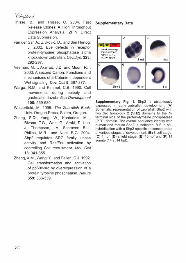
Chapter4
70
Thisse, B., and Thisse, C. 2004. Fast Release Clones: A High Throughput Expression Analysis. ZFIN Direct Data Submission.
van der Sar, A., Zivkovic, D., and den Hertog, J. 2002. Eye defects in receptor protein-tyrosine phosphatase alpha knock-down zebrafish. Dev.Dyn. 223: 292-297.
Veeman, M.T., Axelrod, J.D. and Moon, R.T. 2003. A second Canon: Functions and mechanisms of β-Catenin-independent Wnt signaling. Dev. Cell 5: 367-377.
Warga, R.M. and Kimmel, C.B. 1990. Cell movements during epiboly and gastrulation in zebrafish. Development 108: 569-580
Westerfield, M. 1995. The Zebrafish Book. Univ. Oregon Press, Salem, Oregon.
Zhang, S.Q., Yang, W., Kontaridis, M.I., Bivona, T.G., Wen, G., Araki, T., Luo, J., Thompson, J.A., Schraven, B.L., Philips, M.R., and Neel, B.G. 2004. Shp2 regulates SRC family kinase activity and Ras/Erk activation by controlling Csk recruitment. Mol. Cell 13: 341-355.
Zheng, X.M., Wang, Y., and Pallen, C.J. 1992. Cell transformation and activation of pp60c-src by overexpression of a protein tyrosine phosphatase. Nature 359: 336-339.
Supplementary Data
Supplementary Fig. 1. Shp2 is ubiquitously expressed in early zebrafish development. (A) Schematic representation of zebrafish Shp2 with two Src homology 2 (SH2) domains to the N-terminal side of the protein-tyrosine phosphatase (PTP) domain. The overall sequence identity with human and mouse Shp2 is indicated. B-F In situ hybridization with a Shp2-specific antisense probe at various stages of development: (B) 8 cell-stage; (C) 4 hpf; (D) shield stage; (E) 10 hpf and (F) 14 somite (14 s, 14 hpf).
a b c
d e f
Chapter5
Noonan and Leopard syndrome mutations in Shp2 induce gastrulation defects in zebrafish
Chris Jopling, Daphne van Geemen and Jeroen den Hertog*
Hubrecht LaboratoryNetherlands Institute for Developmental Biology
Uppsalalaan 83584 CT UtrechtThe Netherlands
* corresponding author:Tel: +31 30 2121800Fax: +31 30 2516464
e-mail: [email protected]

Noonan and Leopard syndrome
73
Abstract
Noonan syndrome (NS) and leopard syndrome (LS) are relatively common disorders with partially overlapping symptoms, including short stature, hyperteleorism and cardiac defects. NS and LS are caused by mutations in the protein-tyrosine phosphatse Shp2. Interestingly, the NS mutations activate catalytic activity, whereas LS mutations result in dominant negative Shp2. Here, we expressed mutant Shp2 with mutations that correspond to mutations identified in NS or LS patients in zebrafish embryos. We found that NS- or LS-Shp2 expressing embryos are shorter with craniofacial defects and cardiac edema. At 10 hpf, the embryos did not extend around the yolk as far as the non-injected control, a hallmark of gastrulation cell movement defects. A panel of markers suggested that indeed cell specification is not affected, but proper cell migration is impaired. Co-injections of different Shp2 mutants indicated that NS- and LS-Shp2 did not cooperate. Our results indicate that the symptoms in NS and LS patients may at
least in part be caused by gastrulation cell movement defects.
Chapter5
74
Introduction Noonan syndrome (NS) is an
autosomal dominant disorder affecting around 1:2000 live births. NS is characterised by multiple defects with a varying degree of penetrance, the most common defects are short stature, facial abnormalities and congenital heart defects. In addition, NS defects can include a webbed neck, chest deformities, cryptorchidism (undescended testes), mental retardation and deafness (Allanson 1987; Tartaglia, Mehler et al. 2001; Tartaglia, Kalidas et al. 2002). Around 50% of NS cases are caused by mutations in Shp2, either in the N-terminal SH2 (N-SH2) domain or in the protein tyrosine phosphatase (PTP) domain. So far, 39 different mutations have been identified all of which lead to activated forms of Shp2 (Zenker, Buheitel et al. 2004; Tartaglia and Gelb 2005). In its basal state Shp2 is barely active as a result of the N-SH2 domain. This domain effectively has two functional sides: a front side which binds tyrosine phosphorylated targets and a back side which binds to the PTP domain of Shp2, thus providing a mechanism for regulation. The N-SH2 binds via its backside to the catalytic cleft of the PTP domain blocking it from dephosphorylating substrates. Binding of the SH2 domain(s) to phosphoproteins disrupts the N-SH2 – PTP interaction, activating Shp2 catalytic activity. It is likely that all mutations that cause NS result in activated forms of Shp2 due to failure of the N-SH2 domain to bind to and inhibit the PTP domain Keilhack et al., 2005 (Hof, Pluskey et al. 1998).
A mouse model for NS has been created by knock-in of the Shp2 gene containing the D61G activating mutation that was originally identified in human patients. The observed phenotype in heterozygous mice bares striking similarities to NS patients, with defects such as short stature, facial dismorphism and multiple cardiac defects.
Mice homozygous for the mutated gene die prenatally from severe cardiac edema and liver necrosis (Araki, Mohi et al. 2004).
LEOPARD syndrome (LS) is an autosomal dominant disease characterised by defects such as (L)entigines (multiple black or dark brown spots on the skin), (E)lectrocardiographic defects (abnormal coordination of proper contractions of the heart), (0)cular hypertelorism (widely spaced eyes), (P)ulmonary stenosis (obstruction to the outflow of blood from the right ventricle), (A)bnormal genitals, (R)etarded growth leading to short stature and (D)eafness (Gorlin, Anderson et al. 1971). Many of these symptoms overlap with those seen in NS patients. Recently, it has been shown that LS is also caused by mutations in Shp2. These mutations occur exclusively in the PTP domain of Shp2 disrupting its catalytic activity and leading to dominant negative forms (Kontaridis, Swanson et al. 2006; Tartaglia, Martinelli et al. 2006). This leads to the conundrum “how do mutations in Shp2 with diametrically opposed effects on activity lead to similar developmental defects?” Shp2 has been extensively studied in a number of different species which has shed some light on the possible root cause(s) of the defects observed in NS and LS patients. In particular, Shp2 is associated with defective cell movements during gastrulation. Xenopus laevis animal cap explant assays have for many years been used to study cell movements during gastrulation. Activin and Fibroblast Growth Factor (FGF) induce extension of the explant (Smith and Howard 1992; Isaacs 1997). Similarly expression of mutant, active Shp2 RNA, containing mutations comparable to those found in NS patients (D61A and E76A), also induces elongation of animal cap explants, indicating a direct role for Shp2 in the regulation of cell movements during gastrulation (O’Reilly, Pluskey et al. 2000).

Noonan and Leopard syndrome
75
Interestingly the elongation induced by active Shp2 can be blocked by expression of dominant negative RhoA. In a complementary study the expression of dominant negative Shp2 RNA (dnShp2) (a truncation of the PTP domain with no similarities to LS mutations) blocks FGF and activin induced animal cap elongation, illustrating Shp2’s involvement in the regulation of gastrulation cell movements (Tang, Freeman et al. 1995). Moreover, expression of dnShp2 in Xenopus embryos disrupts gastrulation and leads to severe posterior truncations and an open blastopore and neural tube (Tang, Freeman et al. 1995). Shp2 knockout mice have been generated by deletion of the N-SH2 domain (Saxton, Henkemeyer et al. 1997). Homozygous mice die prenatally due to defective gastrulation resulting in posterior truncations and defects in the node and notochord. Chimeric mice derived from Shp2 homozygous mutant ES cells fail to gastrulate normally and show severe neural tube defects (NTD)(Saxton and Pawson 1999). In the Shp2 exon 3 mutant mice, described above, still a truncated form of Shp2 is expressed. Recently, gene targeted mice were derived that do not express any form of Shp2 anymore and these mouse embryos die much earlier, around implantation, which is linked to reduced numbers of trophoblast giant cells (Yang et al., 2006).
In this study we show that expression of NS- or LS-Shp2 in zebrafish embryos results in overlapping phenotypes which correspond to defects observed in human NS/LS patients. We also demonstrate that NS/LS expression disrupts normal gastrulation without affecting cell specification. Finally we provide evidence that NS-Shp2 and LS-Shp2 do not act synergistically. Our results provide insights into the cell biological cause of NS and LS in humans and suggest that at least some of the symptoms of NS and LS patients are caused by gastrulation defects.
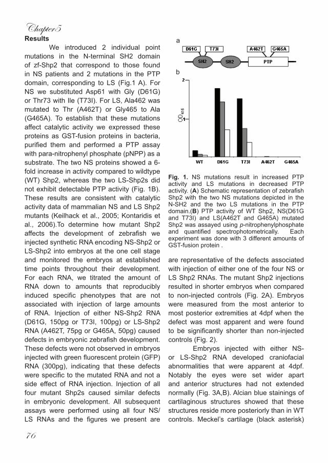
Chapter5
76
ResultsWe introduced 2 individual point
mutations in the N-terminal SH2 domain of zf-Shp2 that correspond to those found in NS patients and 2 mutations in the PTP domain, corresponding to LS (Fig.1 A). For NS we substituted Asp61 with Gly (D61G) or Thr73 with Ile (T73I). For LS, Ala462 was mutated to Thr (A462T) or Gly465 to Ala (G465A). To establish that these mutations affect catalytic activity we expressed these proteins as GST-fusion proteins in bacteria, purified them and performed a PTP assay with para-nitrophenyl phosphate (pNPP) as a substrate. The two NS proteins showed a 6-fold increase in activity compared to wildtype (WT) Shp2, whereas the two LS-Shp2s did not exhibit detectable PTP activity (Fig. 1B). These results are consistent with catalytic activity data of mammalian NS and LS Shp2 mutants (Keilhack et al., 2005; Kontaridis et al., 2006).To determine how mutant Shp2 affects the development of zebrafish we injected synthetic RNA encoding NS-Shp2 or LS-Shp2 into embryos at the one cell stage and monitored the embryos at established time points throughout their development. For each RNA, we titrated the amount of RNA down to amounts that reproducibly induced specific phenotypes that are not associated with injection of large amounts of RNA. Injection of either NS-Shp2 RNA (D61G, 150pg or T73I, 100pg) or LS-Shp2 RNA (A462T, 75pg or G465A, 50pg) caused defects in embryonic zebrafish development. These defects were not observed in embryos injected with green fluorescent protein (GFP) RNA (300pg), indicating that these defects were specific to the mutated RNA and not a side effect of RNA injection. Injection of all four mutant Shp2s caused similar defects in embryonic development. All subsequent assays were performed using all four NS/LS RNAs and the figures we present are
are representative of the defects associated with injection of either one of the four NS or LS Shp2 RNAs. The mutant Shp2 injections resulted in shorter embryos when compared to non-injected controls (Fig. 2A). Embryos were measured from the most anterior to most posterior extremities at 4dpf when the defect was most apparent and were found to be significantly shorter than non-injected controls (Fig. 2). Embryos injected with either NS- or LS-Shp2 RNA developed craniofacial abnormalities that were apparent at 4dpf. Notably the eyes were set wider apart and anterior structures had not extended normally (Fig. 3A,B). Alcian blue stainings of cartilaginous structures showed that these structures reside more posteriorly than in WT controls. Meckel’s cartilage (black asterisk)
Fig. 1. NS mutations result in increased PTP activity and LS mutations in decreased PTP activity. (A) Schematic representation of zebrafish Shp2 with the two NS mutations depicted in the N-SH2 and the two LS mutations in the PTP domain.(B) PTP activity of WT Shp2, NS(D61G and T73I) and LS(A462T and G465A) mutated Shp2 was assayed using p-nitrophenylphosphate and quantified spectrophotometrically. Each experiment was done with 3 different amounts of GST-fusion protein .
a
b
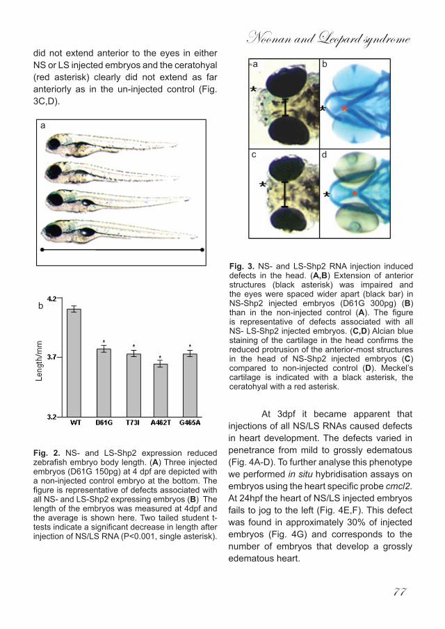
Noonan and Leopard syndrome
77
did not extend anterior to the eyes in either NS or LS injected embryos and the ceratohyal (red asterisk) clearly did not extend as far anteriorly as in the un-injected control (Fig. 3C,D).
At 3dpf it became apparent that injections of all NS/LS RNAs caused defects in heart development. The defects varied in penetrance from mild to grossly edematous (Fig. 4A-D). To further analyse this phenotype we performed in situ hybridisation assays on embryos using the heart specific probe cmcl2. At 24hpf the heart of NS/LS injected embryos fails to jog to the left (Fig. 4E,F). This defect was found in approximately 30% of injected embryos (Fig. 4G) and corresponds to the number of embryos that develop a grossly edematous heart.
Fig. 2. NS- and LS-Shp2 expression reduced zebrafish embryo body length. (A) Three injected embryos (D61G 150pg) at 4 dpf are depicted with a non-injected control embryo at the bottom. The figure is representative of defects associated with all NS- and LS-Shp2 expressing embryos (B) The length of the embryos was measured at 4dpf and the average is shown here. Two tailed student t-tests indicate a significant decrease in length after injection of NS/LS RNA (P<0.001, single asterisk).
Fig. 3. NS- and LS-Shp2 RNA injection induced defects in the head. (A,B) Extension of anterior structures (black asterisk) was impaired and the eyes were spaced wider apart (black bar) in NS-Shp2 injected embryos (D61G 300pg) (B) than in the non-injected control (A). The figure is representative of defects associated with all NS- LS-Shp2 injected embryos. (C,D) Alcian blue staining of the cartilage in the head confirms the reduced protrusion of the anterior-most structures in the head of NS-Shp2 injected embryos (C) compared to non-injected control (D). Meckel’s cartilage is indicated with a black asterisk, the ceratohyal with a red asterisk.
a b
c d
a
b

Chapter5
78
Fig. 4. NS/LS RNA injection results in heart defects at 3dpf. (A) non-injected control. (B) Mild heart defect. (C) Intermediate heart defect. (D) Gross heart defect. The figures are representative of defects associated with all NS- and LS-Shp2 injected embryos. (E-F) cmcl2 in situ h y b r i d i z a t i o n marks the heart and facilitates analysis of heart jogging at 24 hpf (E) Non-injected control with normal heart jogging (red asterisk). (F) Injected embryo showing defective cardiac jogging (red asterisk) (G) Quantification of heart jogging of NS- or LS-Shp2 RNA injected embryos stained with cmcl2, depicted as % non-jogging.
Next, we analysed 10hpf stage embryos and assessed them for defective gastrulation by measurement of the angle between the most anterior and posterior tissues at 10hpf. At 10hpf, injection of each of the NS- and LS-Shp2s caused a significant increase in this angle, indicating that gastrulation has been disrupted (Fig. 5)
Incorrect cell specification can result in embryos that lack certain brain structures. Obviously, this defect would make embryos appear to be shorter than wildtype counterparts. To investigate whether NS- or LS-Shp2 expression affects specification of the brain, we performed in situ hybridisation assays utilising a panel of brain patterning markers. Six3 is expressed in the developing
forebrain of zebrafish embryos while pax2 is expressed in the midbrain-hindbrain boundary. The expression of both of these genes remained unaffected upon injection of either NS- or LS-Shp2 RNAs, indicating that these structures were present (Fig. 6B,F). However, it is evident that the expression patterns of both these genes are shifted posteriorly (Fig. 6B,F). When viewed from the dorsal side the expression patterns of six3 and pax2 are also visibly broader than the control embryos (Fig. 6D,H), which is consistent with gastrulation cell movement defects.To assess whether or not the observed defects were caused by incorrect cell specification, we performed in situ hybridization on NS- or LS-Shp2 injected embryos, using a panel of well-documented
Fig. 5. NS- and LS-Shp2 RNA injection results in defective gastrulation at 10hpf. The angle between the most anterior and posterior embryonic structure was determined at the 1-somite stage and the average angle is depicted here in degrees. Two tailed student t-tests indicate a significant increase in the angle after injection NS/LS RNA (P<0.001, single asterisk). The number of embryos used here is indicated (n).
a
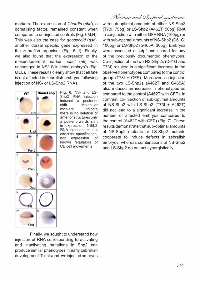
Noonan and Leopard syndrome
79
markers. The expression of Chordin (chd), a dorsalising factor, remained constant when compared to un-injected controls (Fig. 6M,N). This was also the case for goosecoid (gsc), another dorsal specific gene expressed in the zebrafish organiser (Fig. 6I,J). Finally, we also found that the expression of the mesendodermal marker notail (ntl) was unchanged in NS/LS injected embryo’s (Fig. 6K,L). These results clearly show that cell fate is not affected in zebrafish embryos following injection of NS- or LS-Shp2 RNAs.
Finally, we sought to understand how injection of RNA corresponding to activating and inactivating mutations in Shp2 can produce similar phenotypes in early zebrafish development. To this end, we injected embryos
with sub-optimal amounts of either NS-Shp2 (T73I, 75pg) or LS-Shp2 (A462T, 50pg) RNA in conjunction with either GFP RNA (100pg) or with sub-optimal amounts of NS-Shp2 (D61G, 100pg) or LS-Shp2 (G465A, 30pg). Embryos were assessed at 4dpf and scored for any of the previously documented phenotypes. Co-injection of the two NS-Shp2s (D61G and T73I) resulted in a significant increase in the observed phenotypes compared to the control group (T73I + GFP). Moreover, co-injection of the two LS-Shp2s (A462T and G465A) also induced an increase in phenotypes as compared to the control (A462T with GFP). In contrast, co-injection of sub-optimal amounts of NS-Shp2 with LS-Shp2 (T73I + A462T) did not lead to a significant increase in the number of affected embryos compared to the control (A462T with GFP) (Fig. 7). These results demonstrate that sub-optimal amounts of NS-Shp2 mutants or LS-Shp2 mutants cooperate to induce defects in zebrafish embryos, whereas combinations of NS-Shp2 and LS-Shp2 do not act synergistically.
Fig. 6. NS- and LS-Shp2 RNA injection induced a posterior shift. Molecular markers indicate there is no deletion of anterior structures only a posteriorwards shift in expression. NS/LS RNA injection did not affect cell specification, nor expression of known regulators of CE cell movements.
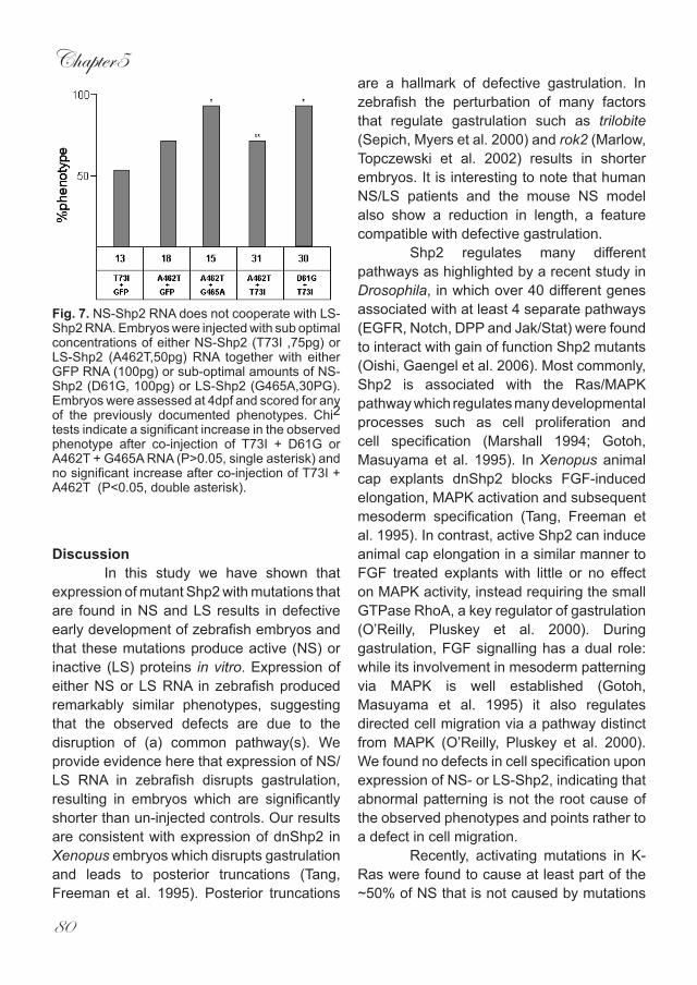
Chapter5
80
DiscussionIn this study we have shown that
expression of mutant Shp2 with mutations that are found in NS and LS results in defective early development of zebrafish embryos and that these mutations produce active (NS) or inactive (LS) proteins in vitro. Expression of either NS or LS RNA in zebrafish produced remarkably similar phenotypes, suggesting that the observed defects are due to the disruption of (a) common pathway(s). We provide evidence here that expression of NS/LS RNA in zebrafish disrupts gastrulation, resulting in embryos which are significantly shorter than un-injected controls. Our results are consistent with expression of dnShp2 in Xenopus embryos which disrupts gastrulation and leads to posterior truncations (Tang, Freeman et al. 1995). Posterior truncations
are a hallmark of defective gastrulation. In zebrafish the perturbation of many factors that regulate gastrulation such as trilobite (Sepich, Myers et al. 2000) and rok2 (Marlow, Topczewski et al. 2002) results in shorter embryos. It is interesting to note that human NS/LS patients and the mouse NS model also show a reduction in length, a feature compatible with defective gastrulation.
Shp2 regulates many different pathways as highlighted by a recent study in Drosophila, in which over 40 different genes associated with at least 4 separate pathways (EGFR, Notch, DPP and Jak/Stat) were found to interact with gain of function Shp2 mutants (Oishi, Gaengel et al. 2006). Most commonly, Shp2 is associated with the Ras/MAPK pathway which regulates many developmental processes such as cell proliferation and cell specification (Marshall 1994; Gotoh, Masuyama et al. 1995). In Xenopus animal cap explants dnShp2 blocks FGF-induced elongation, MAPK activation and subsequent mesoderm specification (Tang, Freeman et al. 1995). In contrast, active Shp2 can induce animal cap elongation in a similar manner to FGF treated explants with little or no effect on MAPK activity, instead requiring the small GTPase RhoA, a key regulator of gastrulation (O’Reilly, Pluskey et al. 2000). During gastrulation, FGF signalling has a dual role: while its involvement in mesoderm patterning via MAPK is well established (Gotoh, Masuyama et al. 1995) it also regulates directed cell migration via a pathway distinct from MAPK (O’Reilly, Pluskey et al. 2000). We found no defects in cell specification upon expression of NS- or LS-Shp2, indicating that abnormal patterning is not the root cause of the observed phenotypes and points rather to a defect in cell migration.
Recently, activating mutations in K-Ras were found to cause at least part of the ~50% of NS that is not caused by mutations
Fig. 7. NS-Shp2 RNA does not cooperate with LS-Shp2 RNA. Embryos were injected with sub optimal concentrations of either NS-Shp2 (T73I ,75pg) or LS-Shp2 (A462T,50pg) RNA together with either GFP RNA (100pg) or sub-optimal amounts of NS-Shp2 (D61G, 100pg) or LS-Shp2 (G465A,30PG). Embryos were assessed at 4dpf and scored for any of the previously documented phenotypes. Chi2 tests indicate a significant increase in the observed phenotype after co-injection of T73I + D61G or A462T + G465A RNA (P>0.05, single asterisk) and no significant increase after co-injection of T73I + A462T (P<0.05, double asterisk).
Noonan and Leopard syndrome
81
in Shp2 (Schubbert, Zenker et al. 2006). Whether K-Ras mediates its effects through sustained activation of MAPK remains to be determined. Alternatively, K-Ras may exert its effects through different pathways, including RhoA and Rac1, established regulators of gastrulation cell movements. It is noteworthy that active K-Ras inhibits both RhoA and Rac1 in pancreatic carcinoma cells (Dreissigacker, Mueller et al. 2006). Moreover, expression of active K-Ras in NIH3T3 cells leads to reduced expression of RhoA (Guerrero, Casanova et al. 2000), again establishing a regulatory link between K-Ras and RhoA. RhoA is a likely integrator of NS signalling, as we have shown that morpholino-induced knock down of Shp2 induced gastrulation defects that were rescued by co-injection of RNA encoding active RhoA (CJ and JdH, unpublished results)..
Embryos injected with NS- or LS-Shp2 RNA develop craniofacial defects similar to NS/LS patients and the NS mouse model, in that anterior structures fail to extend normally and eyes are spaced wider apart. Although there is no hard evidence to suggest that this phenotype is caused by defective gastrulation it is noteworthy that mutants with disrupted gastrulation such as wnt5 (Hammerschmidt, Pelegri et al. 1996) and knypek (Topczewski, Sepich et al. 2001) also develop similar anomalies. However, other mutant zebrafish such as gonzo/site1protease also acquire comparable craniofacial defects yet gastrulation remains unaffected (Schlombs, Wagner et al. 2003). A possible explanation is failure to close the neural tube. Hypertelorism (widely spaced eyes) often associates with NTD (Harris and Juriloff 1999; Martin Mateos, Perez Duenas et al. 2000) as evidenced by mice mutant for the ski gene which develop NTD (Colmenares, Heilstedt et al. 2002). Convergence and extension cell movements regulated by the non-canonical Wnt pathway are essential for
the closure of the neural tube. Mice mutant for dishevelled, looptail, crash and scribble all develop NTD (Hamblet, Lijam et al. 2002; Doudney and Stanier 2005) a defect that is associated with Shp2 knockout mice as well (Saxton, Henkemeyer et al. 1997). In accordance with the role of these genes in the mouse, their zebrafish homologs dishevelled, trilobite, flamingo and scribble-1, respectively all regulate convergence and extension during gastrulation (Heisenberg, Tada et al. 2000; Sepich, Myers et al. 2000; Formstone and Mason 2005; Wada, Iwasaki et al. 2005). Therefore, it is tempting to speculate that the craniofacial defects associated with NS/LS maybe caused by mild NTD and while not directly linked to gastrulation the same pathways may still regulate this process.
One of the most striking phenotypes produced by injection of NS/LS RNA is defective heart formation and the associated gross edema at 3dpf. The nature of this phenotype appears to be caused, at least in part, by defective jogging of the heart at around 24hpf suggesting that left/right (L/R) asymmetry has been disrupted. While genes such as bmp, nodal and lefty are known to have crucial roles in the establishment of L/R asymmetry these do not directly transduce their signals via Shp2 (Burdine and Schier 2000). L/R asymmetry requires a properly developed midline, in that zebrafish mutants that lack a normal midline often display compromised gastrulation (Bisgrove, Essner et al. 2000). Whether this is responsible for the NS/LS induced heart phenotype remains to be determined. However, it should be noted that non-canonical Wnt signalling is required for midline convergence and normal heart development (Matsui, Raya et al. 2005). A more likely explanation for the observed heart jogging defect does not involve defective gastrulation but instead requires intact FGF signalling. FGF8 is also responsible for
Chapter5
82
establishing L/R asymmetry as evidenced by defective heart jogging in zebrafish ace/fgf8 mutants (Albertson and Yelick 2005). Shp2 plays an important role in transducing signals initiated by FGFs (Saxton, Ciruna et al. 2000) which may explain the phenotype caused by NS/LS expression. Interestingly, homozygous NS mutant mice develop a grossly edematous heart, similar to zebrafish embryos expressing NS- or LS-Shp2, which raises the question of whether defects in cardiac jogging are the root cause of congenital heart defects seen in NS/LS patients. In support of this notion, heterotaxy, a disease associated with abnormal cardiac jogging, produces similar heart defects to those seen in NS/LS patients including endocardial cushion defects and pulmonary stenosis (Kathiriya and Srivastava 2000).
Finally, we sought to determine how diametrically opposing mutations can cause similar defects. We established that co-injection of two different NS mutated RNAs led to an increase in the number of affected embryos. Likewise, co-injection of two different LS mutated RNAs produces a similar effect. However, co-injection of NS RNA with LS RNA does not result in an increase in the number of affected embryos. It appears that the number of affected embryos is similar in the NS/LS double injected embryos as upon single injection of LS-Shp2. This suggests either that NS and LS do not function by the same mechanism, or that additive/ synergistic effects are canceled out by the opposing effects of NS- and LS-Shp2. Careful titrations of NS- and LS-Shp2 may provide insight into whether NS- and LS-Shp2 act by the same mechanism or not. Nevertheless, the observation that activation and inhibition of the same factor induces similar phenotypes is not unprecedented. Micro-injection of RNA encoding Rok2 or Galpha12/13 induces similar gastrulation defects as knock down of
Rok2 or Galpha12/13, respectively (Marlow, Topczewski et al. 2002; Lin, Sepich et al. 2005). It appears that an activity window exists for these factors. If overall activity falls outside of this window (either positively or negatively) the resulting phenotypes are very similar.
In conclusion we show here that expression of NS mutated or LS mutated RNA in zebrafish embryos leads to overlapping phenotypes similar to the observation of NS/LS patients. The developmental defects we observed in zebrafish are at least in part due to gastrulation cell movement defects, suggesting that the corresponding symptoms in human patients, most notably short stature and possibly also hyperteleorism and cardiac defects are due to gastrulation defects.
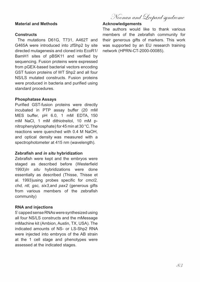
Noonan and Leopard syndrome
83
Material and Methods
Constructs The mutations D61G, T731, A462T and G465A were introduced into zfShp2 by site directed mutagenesis and cloned into EcoR1/BamH1 sites of pBSK11 and verified by sequencing. Fusion proteins were expressed from pGEX-based bacterial vectors encoding
GST fusion proteins of WT Shp2 and all four NS/LS mutated constructs. Fusion proteins were produced in bacteria and purified using standard procedures.
Phosphatase AssaysPurified GST-fusion proteins were directly incubated in PTP assay buffer (20 mM MES buffer, pH 6.0, 1 mM EDTA, 150 mM NaCl, 1 mM dithiotreitol, 10 mM p-nitrophenylphosphate) for 45 min at 30 °C. The reactions were quenched with 0.4 M NaOH, and optical density was measured with a spectrophotometer at 415 nm (wavelength).
Zebrafish and in situ hybridizationZebrafish were kept and the embryos were staged as described before (Westerfield 1993)In situ hybridizations were done essentially as described (Thisse, Thisse et al. 1993)using probes specific for cmcl2, chd, ntl, gsc, six3,and pax2 (generous gifts from various members of the zebrafish community)
RNA and injections5’ capped sense RNAs were synthesized using all four NS/LS constructs and the mMessage mMachine kit (Ambion, Austin, TX, USA). The indicated amounts of NS- or LS-Shp2 RNA were injected into embryos of the AB strain at the 1 cell stage and phenotypes were assessed at the indicated stages.
AcknowledgementsThe authors would like to thank various members of the zebrafish community for their generous gifts of markers. This work was supported by an EU research training network (HPRN-CT-2000-00085).
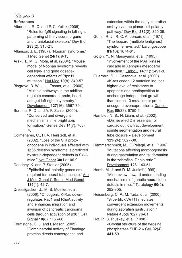
Chapter5
84
ReferencesAlbertson, R. C. and P. C. Yelick (2005).
“Roles for fgf8 signaling in left-right patterning of the visceral organs and craniofacial skeleton.” Dev Biol 283(2): 310-21.
Allanson, J. E. (1987). “Noonan syndrome.” J Med Genet 24(1): 9-13.
Araki, T., M. G. Mohi, et al. (2004). “Mouse model of Noonan syndrome reveals cell type- and gene dosage-dependent effects of Ptpn11 mutation.” Nat Med 10(8): 849-57.
Bisgrove, B. W., J. J. Essner, et al. (2000). “Multiple pathways in the midline regulate concordant brain, heart and gut left-right asymmetry.” Development 127(16): 3567-79.
Burdine, R. D. and A. F. Schier (2000). “Conserved and divergent mechanisms in left-right axis formation.” Genes Dev 14(7): 763-76.
Colmenares, C., H. A. Heilstedt, et al. (2002). “Loss of the SKI proto-oncogene in individuals affected with 1p36 deletion syndrome is predicted by strain-dependent defects in Ski-/- mice.” Nat Genet 30(1): 106-9.
Doudney, K. and P. Stanier (2005). “Epithelial cell polarity genes are required for neural tube closure.” Am J Med Genet C Semin Med Genet 135(1): 42-7.
Dreissigacker, U., M. S. Mueller, et al. (2006). “Oncogenic K-Ras down-regulates Rac1 and RhoA activity and enhances migration and invasion of pancreatic carcinoma cells through activation of p38.” Cell Signal 18(8): 1156-68.
Formstone, C. J. and I. Mason (2005). “Combinatorial activity of Flamingo proteins directs convergence and
extension within the early zebrafish embryo via the planar cell polarity pathway.” Dev Biol 282(2): 320-35.
Gorlin, R. J., R. C. Anderson, et al. (1971). “The leopard (multiple lentigines) syndrome revisited.” Laryngoscope 81(10): 1674-81.
Gotoh, Y., N. Masuyama, et al. (1995). “Involvement of the MAP kinase cascade in Xenopus mesoderm induction.” Embo J 14(11): 2491-8.
Guerrero, S., I. Casanova, et al. (2000). «K-ras codon 12 mutation induces higher level of resistance to apoptosis and predisposition to anchorage-independent growth than codon 13 mutation or proto-oncogene overexpression.» Cancer Res 60(23): 6750-6.
Hamblet, N. S., N. Lijam, et al. (2002). «Dishevelled 2 is essential for cardiac outflow tract development, somite segmentation and neural tube closure.» Development 129(24): 5827-38.
Hammerschmidt, M., F. Pelegri, et al. (1996). “Mutations affecting morphogenesis during gastrulation and tail formation in the zebrafish, Danio rerio.” Development 123: 143-51.
Harris, M. J. and D. M. Juriloff (1999). “Mini-review: toward understanding mechanisms of genetic neural tube defects in mice.” Teratology 60(5): 292-305.
Heisenberg, C. P., M. Tada, et al. (2000). “Silberblick/Wnt11 mediates convergent extension movements during zebrafish gastrulation.” Nature 405(6782): 76-81.
Hof, P., S. Pluskey, et al. (1998). «Crystal structure of the tyrosine phosphatase SHP-2.» Cell 92(4): 441-50.
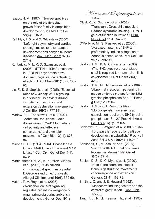
Noonan and Leopard syndrome
85
Isaacs, H. V. (1997). “New perspectives on the role of the fibroblast growth factor family in amphibian development.” Cell Mol Life Sci 53(4): 350-61.
Kathiriya, I. S. and D. Srivastava (2000). “Left-right asymmetry and cardiac looping: implications for cardiac development and congenital heart disease.” Am J Med Genet 97(4): 271-9.
Kontaridis, M. I., K. D. Swanson, et al. (2006). «PTPN11 (Shp2) mutations in LEOPARD syndrome have dominant negative, not activating, effects.» J Biol Chem 281(10): 6785-92.
Lin, F., D. S. Sepich, et al. (2005). “Essential roles of G{alpha}12/13 signaling in distinct cell behaviors driving zebrafish convergence and extension gastrulation movements.” J Cell Biol 169(5): 777-87.
Marlow, F., J. Topczewski, et al. (2002). “Zebrafish Rho kinase 2 acts downstream of Wnt11 to mediate cell polarity and effective convergence and extension movements.” Curr Biol 12(11): 876-84.
Marshall, C. J. (1994). “MAP kinase kinase kinase, MAP kinase kinase and MAP kinase.” Curr Opin Genet Dev 4(1): 82-9.
Martin Mateos, M. A., B. P. Perez Duenas, et al. (2000). “Clinical and immunological spectrum of partial DiGeorge syndrome.” J Investig Allergol Clin Immunol 10(6): 352-60.
Matsui, T., A. Raya, et al. (2005). «Noncanonical Wnt signaling regulates midline convergence of organ primordia during zebrafish development.» Genes Dev 19(1):
164-75.Oishi, K., K. Gaengel, et al. (2006).
“Transgenic Drosophila models of Noonan syndrome causing PTPN11 gain-of-function mutations.” Hum Mol Genet 15(4): 543-53.
O’Reilly, A. M., S. Pluskey, et al. (2000). “Activated mutants of SHP-2 preferentially induce elongation of Xenopus animal caps.” Mol Cell Biol 20(1): 299-311.
Saxton, T. M., B. G. Ciruna, et al. (2000). «The SH2 tyrosine phosphatase shp2 is required for mammalian limb development.» Nat Genet 24(4): 420-3.
Saxton, T. M., M. Henkemeyer, et al. (1997). “Abnormal mesoderm patterning in mouse embryos mutant for the SH2 tyrosine phosphatase Shp-2.” Embo J 16(9): 2352-64.
Saxton, T. M. and T. Pawson (1999). “Morphogenetic movements at gastrulation require the SH2 tyrosine phosphatase Shp2.” Proc Natl Acad Sci U S A 96(7): 3790-5.
Schlombs, K., T. Wagner, et al. (2003). “Site-1 protease is required for cartilage development in zebrafish.” Proc Natl Acad Sci U S A 100(24): 14024-9.
Schubbert, S., M. Zenker, et al. (2006). “Germline KRAS mutations cause Noonan syndrome.” Nat Genet 38(3): 331-6.
Sepich, D. S., D. C. Myers, et al. (2000). “Role of the zebrafish trilobite locus in gastrulation movements of convergence and extension.” Genesis 27(4): 159-73.
Smith, J. C. and J. E. Howard (1992). “Mesoderm-inducing factors and the control of gastrulation.” Dev Suppl: 127-36.
Tang, T. L., R. M. Freeman, Jr., et al. (1995).

Chapter5
86
“The SH2-containing protein-tyrosine phosphatase SH-PTP2 is required upstream of MAP kinase for early Xenopus development.” Cell 80(3): 473-83.
Tartaglia, M. and B. D. Gelb (2005). “Noonan syndrome and related disorders: genetics and pathogenesis.” Annu Rev Genomics Hum Genet 6: 45-68.
Tartaglia, M., K. Kalidas, et al. (2002). «PTPN11 mutations in Noonan syndrome: molecular spectrum, genotype-phenotype correlation, and phenotypic heterogeneity.» Am J Hum Genet 70(6): 1555-63.
Tartaglia, M., S. Martinelli, et al. (2006). «Diversity and Functional Consequences of Germline and Somatic PTPN11 Mutations in Human Disease.» Am J Hum Genet 78(2): 279-90.
Tartaglia, M., E. L. Mehler, et al. (2001). «Mutations in PTPN11, encoding the protein tyrosine phosphatase SHP-2, cause Noonan syndrome.» Nat Genet 29(4): 465-8.
Thisse, C., B. Thisse, et al. (1993). “Structure of the zebrafish snail1 gene and its expression in wild-type, spadetail and no tail mutant embryos.” Development 119(4): 1203-15.
Topczewski, J., D. S. Sepich, et al. (2001). “The zebrafish glypican knypek controls cell polarity during gastrulation movements of convergent extension.” Dev Cell 1(2): 251-64.
Wada, H., M. Iwasaki, et al. (2005). «Dual roles of zygotic and maternal Scribble1 in neural migration and convergent extension movements in zebrafish embryos.» Development 132(10): 2273-85.
Westerfield, M. (1993). The Zebrafish book : a guide for the laboratory use of zebrafish (Brachydanio rerio). Eugene. Or., University of Oregon Press.
Zenker, M., G. Buheitel, et al. (2004). “Genotype-phenotype correlations in Noonan syndrome.” J Pediatr 144(3): 368-74.

Chapter6
Discussion

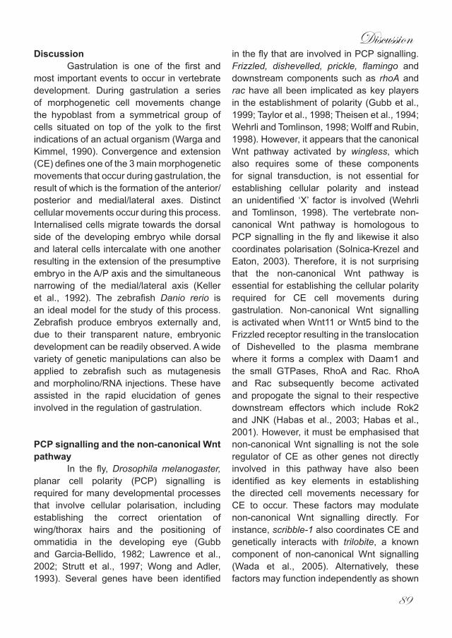
Discussion
89
DiscussionGastrulation is one of the first and
most important events to occur in vertebrate development. During gastrulation a series of morphogenetic cell movements change the hypoblast from a symmetrical group of cells situated on top of the yolk to the first indications of an actual organism (Warga and Kimmel, 1990). Convergence and extension (CE) defines one of the 3 main morphogenetic movements that occur during gastrulation, the result of which is the formation of the anterior/posterior and medial/lateral axes. Distinct cellular movements occur during this process. Internalised cells migrate towards the dorsal side of the developing embryo while dorsal and lateral cells intercalate with one another resulting in the extension of the presumptive embryo in the A/P axis and the simultaneous narrowing of the medial/lateral axis (Keller et al., 1992). The zebrafish Danio rerio is an ideal model for the study of this process. Zebrafish produce embryos externally and, due to their transparent nature, embryonic development can be readily observed. A wide variety of genetic manipulations can also be applied to zebrafish such as mutagenesis and morpholino/RNA injections. These have assisted in the rapid elucidation of genes involved in the regulation of gastrulation.
PCP signalling and the non-canonical Wnt pathway
In the fly, Drosophila melanogaster, planar cell polarity (PCP) signalling is required for many developmental processes that involve cellular polarisation, including establishing the correct orientation of wing/thorax hairs and the positioning of ommatidia in the developing eye (Gubb and Garcia-Bellido, 1982; Lawrence et al., 2002; Strutt et al., 1997; Wong and Adler, 1993). Several genes have been identified
in the fly that are involved in PCP signalling. Frizzled, dishevelled, prickle, flamingo and downstream components such as rhoA and rac have all been implicated as key players in the establishment of polarity (Gubb et al., 1999; Taylor et al., 1998; Theisen et al., 1994; Wehrli and Tomlinson, 1998; Wolff and Rubin, 1998). However, it appears that the canonical Wnt pathway activated by wingless, which also requires some of these components for signal transduction, is not essential for establishing cellular polarity and instead an unidentified ‘X’ factor is involved (Wehrli and Tomlinson, 1998). The vertebrate non-canonical Wnt pathway is homologous to PCP signalling in the fly and likewise it also coordinates polarisation (Solnica-Krezel and Eaton, 2003). Therefore, it is not surprising that the non-canonical Wnt pathway is essential for establishing the cellular polarity required for CE cell movements during gastrulation. Non-canonical Wnt signalling is activated when Wnt11 or Wnt5 bind to the Frizzled receptor resulting in the translocation of Dishevelled to the plasma membrane where it forms a complex with Daam1 and the small GTPases, RhoA and Rac. RhoA and Rac subsequently become activated and propogate the signal to their respective downstream effectors which include Rok2 and JNK (Habas et al., 2003; Habas et al., 2001). However, it must be emphasised that non-canonical Wnt signalling is not the sole regulator of CE as other genes not directly involved in this pathway have also been identified as key elements in establishing the directed cell movements necessary for CE to occur. These factors may modulate non-canonical Wnt signalling directly. For instance, scribble-1 also coordinates CE and genetically interacts with trilobite, a known component of non-canonical Wnt signalling (Wada et al., 2005). Alternatively, these factors may function independently as shown
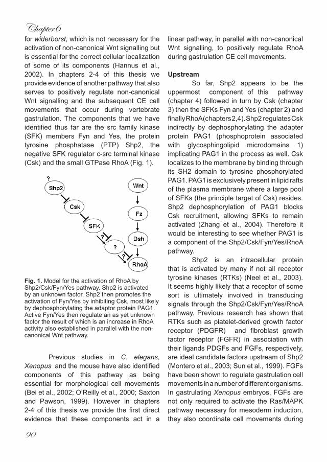
Chapter6
90
for widerborst, which is not necessary for the activation of non-canonical Wnt signalling but is essential for the correct cellular localization of some of its components (Hannus et al., 2002). In chapters 2-4 of this thesis we provide evidence of another pathway that also serves to positively regulate non-canonical Wnt signalling and the subsequent CE cell movements that occur during vertebrate gastrulation. The components that we have identified thus far are the src family kinase (SFK) members Fyn and Yes, the protein tyrosine phosphatase (PTP) Shp2, the negative SFK regulator c-src terminal kinase (Csk) and the small GTPase RhoA (Fig. 1).
Previous studies in C. elegans, Xenopus and the mouse have also identified components of this pathway as being essential for morphological cell movements (Bei et al., 2002; O’Reilly et al., 2000; Saxton and Pawson, 1999). However in chapters 2-4 of this thesis we provide the first direct evidence that these components act in a
linear pathway, in parallel with non-canonical Wnt signalling, to positively regulate RhoA during gastrulation CE cell movements. Upstream
So far, Shp2 appears to be the uppermost component of this pathway (chapter 4) followed in turn by Csk (chapter 3) then the SFKs Fyn and Yes (chapter 2) and finally RhoA (chapters 2,4). Shp2 regulates Csk indirectly by dephosphorylating the adapter protein PAG1 (phosphoprotein associated with glycosphingolipid microdomains 1) implicating PAG1 in the process as well. Csk localizes to the membrane by binding through its SH2 domain to tyrosine phosphorylated PAG1. PAG1 is exclusively present in lipid rafts of the plasma membrane where a large pool of SFKs (the principle target of Csk) resides. Shp2 dephosphorylation of PAG1 blocks Csk recruitment, allowing SFKs to remain activated (Zhang et al., 2004). Therefore it would be interesting to see whether PAG1 is a component of the Shp2/Csk/Fyn/Yes/RhoA pathway.
Shp2 is an intracellular protein that is activated by many if not all receptor tyrosine kinases (RTKs) (Neel et al., 2003). It seems highly likely that a receptor of some sort is ultimately involved in transducing signals through the Shp2/Csk/Fyn/Yes/RhoA pathway. Previous research has shown that RTKs such as platelet-derived growth factor receptor (PDGFR) and fibroblast growth factor receptor (FGFR) in association with their ligands PDGFs and FGFs, respectively, are ideal candidate factors upstream of Shp2 (Montero et al., 2003; Sun et al., 1999). FGFs have been shown to regulate gastrulation cell movements in a number of different organisms. In gastrulating Xenopus embryos, FGFs are not only required to activate the Ras/MAPK pathway necessary for mesoderm induction, they also coordinate cell movements during
Fig. 1. Model for the activation of RhoA by Shp2/Csk/Fyn/Yes pathway. Shp2 is activated by an unknown factor. Shp2 then promotes the activation of Fyn/Yes by inhibiting Csk, most likely by dephosphorylating the adaptor protein PAG1. Active Fyn/Yes then regulate an as yet unknown factor the result of which is an increase in RhoA activity also established in parallel with the non-canonical Wnt pathway.
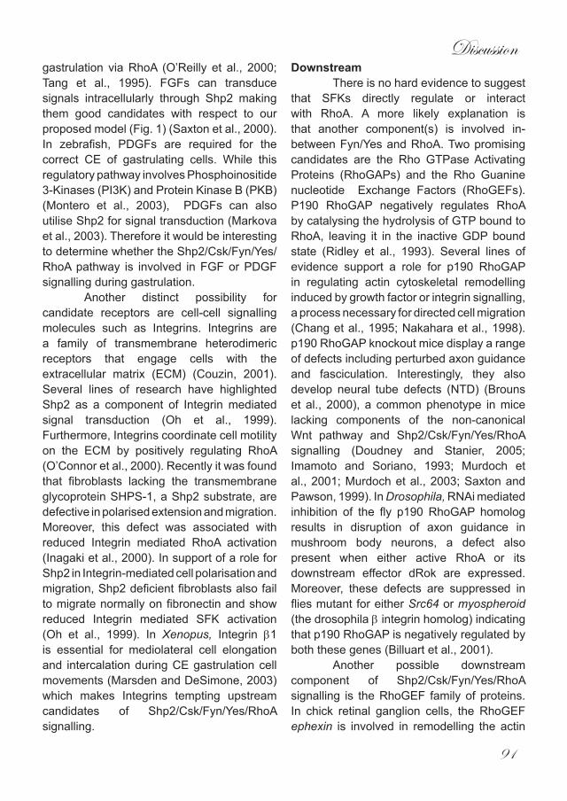
Discussion
91
gastrulation via RhoA (O’Reilly et al., 2000; Tang et al., 1995). FGFs can transduce signals intracellularly through Shp2 making them good candidates with respect to our proposed model (Fig. 1) (Saxton et al., 2000). In zebrafish, PDGFs are required for the correct CE of gastrulating cells. While this regulatory pathway involves Phosphoinositide 3-Kinases (PI3K) and Protein Kinase B (PKB) (Montero et al., 2003), PDGFs can also utilise Shp2 for signal transduction (Markova et al., 2003). Therefore it would be interesting to determine whether the Shp2/Csk/Fyn/Yes/RhoA pathway is involved in FGF or PDGF signalling during gastrulation.
Another distinct possibility for candidate receptors are cell-cell signalling molecules such as Integrins. Integrins are a family of transmembrane heterodimeric receptors that engage cells with the extracellular matrix (ECM) (Couzin, 2001). Several lines of research have highlighted Shp2 as a component of Integrin mediated signal transduction (Oh et al., 1999). Furthermore, Integrins coordinate cell motility on the ECM by positively regulating RhoA (O’Connor et al., 2000). Recently it was found that fibroblasts lacking the transmembrane glycoprotein SHPS-1, a Shp2 substrate, are defective in polarised extension and migration. Moreover, this defect was associated with reduced Integrin mediated RhoA activation (Inagaki et al., 2000). In support of a role for Shp2 in Integrin-mediated cell polarisation and migration, Shp2 deficient fibroblasts also fail to migrate normally on fibronectin and show reduced Integrin mediated SFK activation (Oh et al., 1999). In Xenopus, Integrin β1 is essential for mediolateral cell elongation and intercalation during CE gastrulation cell movements (Marsden and DeSimone, 2003) which makes Integrins tempting upstream candidates of Shp2/Csk/Fyn/Yes/RhoA signalling.
DownstreamThere is no hard evidence to suggest
that SFKs directly regulate or interact with RhoA. A more likely explanation is that another component(s) is involved in-between Fyn/Yes and RhoA. Two promising candidates are the Rho GTPase Activating Proteins (RhoGAPs) and the Rho Guanine nucleotide Exchange Factors (RhoGEFs). P190 RhoGAP negatively regulates RhoA by catalysing the hydrolysis of GTP bound to RhoA, leaving it in the inactive GDP bound state (Ridley et al., 1993). Several lines of evidence support a role for p190 RhoGAP in regulating actin cytoskeletal remodelling induced by growth factor or integrin signalling, a process necessary for directed cell migration (Chang et al., 1995; Nakahara et al., 1998). p190 RhoGAP knockout mice display a range of defects including perturbed axon guidance and fasciculation. Interestingly, they also develop neural tube defects (NTD) (Brouns et al., 2000), a common phenotype in mice lacking components of the non-canonical Wnt pathway and Shp2/Csk/Fyn/Yes/RhoA signalling (Doudney and Stanier, 2005; Imamoto and Soriano, 1993; Murdoch et al., 2001; Murdoch et al., 2003; Saxton and Pawson, 1999). In Drosophila, RNAi mediated inhibition of the fly p190 RhoGAP homolog results in disruption of axon guidance in mushroom body neurons, a defect also present when either active RhoA or its downstream effector dRok are expressed. Moreover, these defects are suppressed in flies mutant for either Src64 or myospheroid (the drosophila β integrin homolog) indicating that p190 RhoGAP is negatively regulated by both these genes (Billuart et al., 2001).
Another possible downstream component of Shp2/Csk/Fyn/Yes/RhoA signalling is the RhoGEF family of proteins. In chick retinal ganglion cells, the RhoGEF ephexin is involved in remodelling the actin

Chapter6
92
cytoskeleton during growth cone collapse, a process necessary for normal axon guidance. Furthermore, analysis of fibroblast cells has shown that ephexin is activated by SFK phosphorylation while pharmacological inhibiton of SFKs or expression of Csk also disrupts growth cone collapse, thus establishing a direct link between SFKs and RhoGEFs during cytoskeletal remodelling (Knoll and Drescher, 2004).
In Drosophila the RhoGEF pebble is part of a signalling cascade initiated by the FGFR heartless. This cascade is necessary for coordinating mesodermal cell migration during gastrulation. Flies mutant for the pebble gene show defects in the characteristic cell shape changes that occur during migration of mesodermal cells indicating a direct role for this RhoGEF in the regulation of cell movements during gastrulation (Schumacher et al., 2004).
Studies in Xenopus have indicated that yet another RhoGEF, XLfc, is required for CE movements during gastrulation. Nocodazole treatment of animal cap explants inhibits the CE movements necessary for gastrulation to occur. Subsequently morpholino mediated knockdown of XLfc or expression of dominant negative RhoA can rescue the defects caused by nocodazole treatment, confirming Xlfc’s involvement in CE (Kwan and Kirschner, 2005). A separate study in Xenopus identified the RhoGEF, xNET1, as being a key regulator of gastrulation. XNET1 is specific for RhoA with little or no affinity for Rac and cdc42. In addition, expression of mutant and WT xNET1 RNA severely disrupts gastrulation (Miyakoshi et al., 2004).
Further support for a role of RhoGEFs in regulating gastrulation is supplied by qauttro(quo), a RhoGEF present in the zebrafish danio rerio. Developing embryos in which quo has been perturbed fail to gastrulate normally, a defect caused by failure
of mesodermal cells to converge towards the midline (Daggett et al., 2004). Interestingly, similar anomalies are associated with other morphants such as Has2 in which gastrulation is also disrupted (Bakkers et al., 2004).
It is obvious that both RhoGAPs and RhoGEFs are involved in the coordination of migratory events necessary for normal gastrulation. Both families are also regulated by SFK phosphorylation which either inhibits RhoGAPs (Billuart et al., 2001) or activates RhoGEFs (Knoll and Drescher, 2004) making either family putative candidates for inclusion in the Shp2/Csk/Fyn/Yes/RhoA signalling pathway.
Molecular mechanismsIn Drosophila, components of PCP
signalling are asymmetrically distributed in cells where PCP has been established. Initially, these components are distributed uniformly around the cell. However, once activated, they relocate to distinct regions, frizzled and dishevelled reside in distal areas while strabismus and prickle localise to proximal regions of the cell thus establishing overall cellular polarity (Axelrod, 2001; Bastock et al., 2003; Shimada et al., 2001). A mechanism has been described for how this asymmetric localisation of PCP components occurs. Drosophila mutants for the 3 prickle isoforms pkm, pkpk, and pksple develop PCP associated defects such as incorrect wing hair orientation. Furthermore, when prickle is absent, frizzled and dishevelled cease to be asymmetrically distributed in wing hair cells. Prickle localises to the proximal side of wing hair cells and here it binds to and inhibits dishevelled localisation. Dishevelled is required for the localised accumulation of the frizzled receptor which is now lost on the proximal side. This establishes polarity within the cell as active frizzled/dishevelled signalling is present on the distal side of the
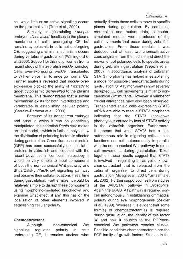
Discussion
93
cell while little or no active signalling occurs on the proximal side (Tree et al., 2002).
Similarly, in gastrulating Xenopus embryos, dishevelled localises to the plasma membrane of cells undergoing CE but remains cytoplasmic in cells not undergoing CE, suggesting a similar mechanism occurs during vertebrate gastrulation (Wallingford et al., 2000). Support for this notion comes from a recent study of the zebrafish prickle homolog. Cells over-expressing prickle transplanted to WT embryos fail to undergo normal CE. Further analysis revealed that prickle over-expression blocked the ability of frizzled7 to target cytoplasmic dishevelled to the plasma membrane. This demonstrates that a similar mechanism exists for both invertebrates and vertebrates in establishing cellular polarity (Carreira-Barbosa et al., 2003).
Because of its transparent embryos and ease in which it can be genetically manipulated, the zebrafish Danio rerio makes an ideal model in which to further analyse how the distribution of polarising factors is effected during gastrulation. Green fluorescent protein (GFP) has been successfully used to label proteins in zebrafish and, coupled with the recent advances in confocal microscopy, it would be very simple to label components of both the non-canonical Wnt pathway and Shp2/Csk/Fyn/Yes/RhoA signalling pathway and observe their cellular locations in real time during gastrulation. Furthermore, it would be relatively simple to disrupt these components using morpholino-mediated knockdown and examine what effect, if any, this has on the localisation of other elements involved in establishing cellular polarity.
ChemoattractantAlthough non-canonical Wnt
signalling regulates polarity in cells undergoing CE, it remains unclear what
actually directs these cells to move to specific places during gastrulation. By combining morpholino and mutant data, computer-simulated models were produced of the cell movements that occur during zebrafish gastrulation. From these models it was deduced that at least two chemoattractive cues originate from the midline and direct the movement of polarised cells to specific areas during zebrafish gastrulation (Sepich et al., 2005). In accordance, analysis of zebrafish STAT3 morphants has helped in establishing a model for possible chemoattractants during gastrulation. STAT3 morphants show severely disrupted CE cell movements, similar to non-canonical Wnt mutants. However, a number of crucial differences have also been observed. Transplanted shield cells expressing STAT3 mRNA are able to rescue STAT3 morphants indicating that the STAT3 knockdown phenotype is caused by loss of STAT3 activity in the zebrafish organiser. Furthermore, it appears that while STAT3 has a cell-autonomous role in migrating cells, it also functions non-cell autonomously in parallel with the non-canonical Wnt pathway to direct cell movements during gastrulation. Taken together, these results suggest that STAT3 is involved in regulating an as yet unknown chemoattractant that is released from the zebrafish organiser to direct cells during gastrulation (Miyagi et al., 2004; Yamashita et al., 2002). Further support comes from studies of the JAK/STAT pathway in Drosophila. Again, the JAK/STAT pathway is required non-cell autonomously in establishing ommatidial polarity during eye morphogenesis (Zeidler et al., 1999). Whereas it is evident that some form(s) of chemoattractant(s) is required during gastrulation, the identity of this factor ‘X’ and how it couples to the PCP/non-canonical Wnt pathways remains elusive. Possible candidate chemoattractants are the FGF family of growth factors. Studies in the
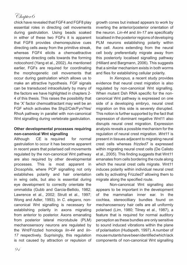
Chapter6
94
chick have revealed that FGF4 and FGF8 play essential roles in directing cell movements during gastrulation. Using beads soaked in either of these two FGFs it is apparent that FGF8 provides chemorepulsive cues directing cells away from the primitive streak, whereas FGF4 elicits a chemoattractive response directing cells towards the forming notocohord (Yang et al., 2002). As mentioned earlier, FGFs are required for coordinating the morphogenetic cell movements that occur during gastrulation which allows us to make an attractive hypothesis. FGF signals can be transduced intracellularly by many of the factors we have highlighted in chapters 2-4 of this thesis. This raises the possibility that the ‘X’ factor chemoattractant may well be an FGF which activates the Shp2/Csk/Fyn/Yes/RhoA pathway in parallel with non-canonical Wnt signalling during vertebrate gastrulation.
Other developmental processes requiring non-canonical Wnt signallingAlthough CE is required for normal gastrulation to occur it has become apparent in recent years that polarised cell movements regulated by the non-canonical Wnt pathway are also required by other developmental processes. This is most apparent in Drosophila, where PCP signalling not only establishes polarity and hair orientation in wing cells, but also is essential during eye development to correctly orientate the ommatidia (Gubb and Garcia-Bellido, 1982; Lawrence et al., 2002; Strutt et al., 1997; Wong and Adler, 1993). In C. elegans, non-canonical Wnt signalling is necessary for establishing polarity in axons migrating from anterior to posterior. Axons emanating from posterior lateral microtubule (PLM) mechanosensory neurons are regulated by the Wnt/Frizzled homologs lin-44 and lin-17 respectively. Suprisingly, this regulation is not caused by attraction or repulsion of
growth cones but instead appears to work by inverting the anterior/posterior orientation of the neuron. Lin-44 and lin-17 are specifically localised in the posterior regions of developing PLM neurons establishing polarity within the cell. Axons extending from the neural cell body preferentially migrate away from this posteriorly localised signalling pathway (Hilliard and Bargmann, 2006). This suggests that a similar mechanism exists in both worms and flies for establishing cellular polarity.
In Xenopus, a recent study provides evidence that neural crest migration is also regulated by non-canonical Wnt signalling. When mutant Dsh RNA specific for the non-canonical Wnt pathway is expressed on one side of a developing embryo, neural crest migration on this side is severely disrupted. This notion is further supported by the fact that expression of dominant negative Wnt11 also disrupts neural crest migration. Expression analysis reveals a possible mechanism for the regulation of neural crest migration. Wnt11 is present in tissues adjacent to migrating neural crest cells whereas frizzled7 is expressed within migrating neural crest cells (De Calisto et al., 2005). This finding suggests that Wnt11 emanates from cells bordering the route along which the neural crest cells migrate. Wnt11 induces polarity within individual neural crest cells by activating Frizzled7 allowing them to migrate along the specified route.
Non-canonical Wnt signalling also appears to be important in the development of the mammalian inner ear. In the cochlea, stereocilliary bundles found on mechanosensory hair cells are all uniformly polarised (Lim, 1980; Tilney et al., 1987), a feature that is required for normal auditory perception as these bundles are only sensitive to sound induced vibrations within the plane of polarisation (Hudspeth, 1997). A number of mouse mutants have been identified which lack components of non-canonical Wnt signalling

Discussion
95
including vangl2, scribble1, celsr1 and ptk7 all of which show defects in the polarity of stereocilliary bundles in mechanosensory hair cells (Curtin et al., 2003; Kibar et al., 2001; Lu et al., 2004; Murdoch et al., 2001; Murdoch et al., 2003). While the actual molecular basis for establishing this polarity in the mammalian inner ear remains unknown it does bare a striking similarity to the polarity needed to establish the correct orientation of hair cells in the wing and thorax of Drosophila. Therefore, it is tempting to speculate that the same molecular mechanism exists in mammals.
It appears that non-canonical Wnt signalling is not solely confined to polarising cells during CE in gastrulating embryos. It may also be necessary for other developmental processes that require cellular polarity. It would be interesting therefore to examine whether the Shp2/Csk/Fyn/Yes/RhoA pathway we have identified in chapters 2-4 is also involved in other non-canonical Wnt signalling dependent developmental processes or whether it only functions during gastrulation to regulate directed movement in cells undergoing CE.
Neural tube defectsNeural tube defects (NTD) are a
relatively common set of birth abnormalities effecting around 1:1000 live births (Copp and Bernfield, 1994; Copp et al., 2003). The simplest way to imagine neural tube closure is to picture a flat sheet of cells that during the course of development rolls up to form a hollow tube like structure. However, in reality this process is much more complex and requires the concerted action of a number of different cell movements and shape changes (Wallingford, 2005). In recent years it has become apparent that CE cell movements are also involved in effecting the closure of the neural tube (Keller, 2002). Once the neural plate has thickened and the neural folds have
elevated, CE cell movements convert the initially short/wide neural tube into a long/thin structure in much the same manner as during gastrulation (Keller, 2002). This effectively brings the elevated neural folds closer together allowing them to fuse and thus elicit closure of the tube. Correct non-canonical Wnt signalling is an absolute necessity for normal neural tube closure (Doudney and Stanier, 2005). This has been elegantly demonstrated in Xenopus. Disruption of Dishevelled has no effect on neural fold elevation or fusion but instead results in defective CE movements of cells within the neural tube (Wallingford et al., 2000). Because cells fail to both migrate towards and extend along the midline, the distance between the neural folds is too great leading to a failure in fusion.
Mouse models have been created that display NTD in a bid to further understand these defects in mammals (Copp et al., 2003). As the genes that are required for neural tube closure come to light it is exciting to note that many components of non-canonical Wnt signalling are disrupted in mice with NTDs. Dishevelled, scibble1, ptk7, celsr1(flamingo) and strabismus all cause severe NTDs when they are disrupted in mice (Curtin et al., 2003; Hamblet et al., 2002; Kibar et al., 2001; Lu et al., 2004; Murdoch et al., 2003) while their zebrafish homologs are all required for CE during embryonic gastrulation (Formstone and Mason, 2005; Matsui et al., 2005; Sepich et al., 2000; Wada et al., 2005). Furthermore, chick and mouse embryos cultured in Rok inhibitors, a downstream effector of non-canonical Wnt signalling, also develop NTD (Wei et al., 2001). It is striking that many genes that regulate CE during gastrulation in zebrafish are also required for effective neural tube closure to occur in mammals which makes the zebrafish an attractive model for identifying possible factors that may be absent in patients suffering from NTD.
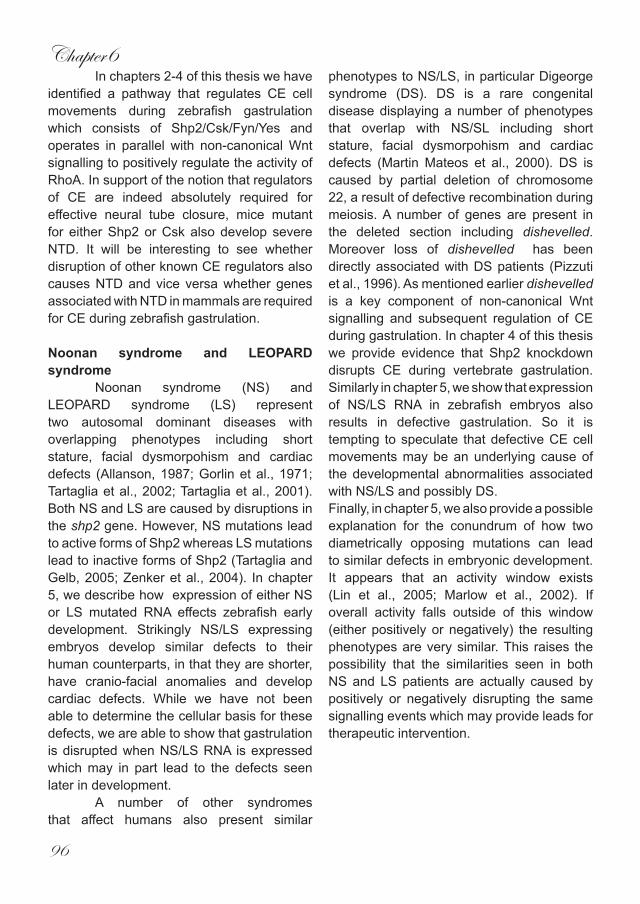
Chapter6
96
In chapters 2-4 of this thesis we have identified a pathway that regulates CE cell movements during zebrafish gastrulation which consists of Shp2/Csk/Fyn/Yes and operates in parallel with non-canonical Wnt signalling to positively regulate the activity of RhoA. In support of the notion that regulators of CE are indeed absolutely required for effective neural tube closure, mice mutant for either Shp2 or Csk also develop severe NTD. It will be interesting to see whether disruption of other known CE regulators also causes NTD and vice versa whether genes associated with NTD in mammals are required for CE during zebrafish gastrulation.
Noonan syndrome and LEOPARD syndrome
Noonan syndrome (NS) and LEOPARD syndrome (LS) represent two autosomal dominant diseases with overlapping phenotypes including short stature, facial dysmorpohism and cardiac defects (Allanson, 1987; Gorlin et al., 1971; Tartaglia et al., 2002; Tartaglia et al., 2001). Both NS and LS are caused by disruptions in the shp2 gene. However, NS mutations lead to active forms of Shp2 whereas LS mutations lead to inactive forms of Shp2 (Tartaglia and Gelb, 2005; Zenker et al., 2004). In chapter 5, we describe how expression of either NS or LS mutated RNA effects zebrafish early development. Strikingly NS/LS expressing embryos develop similar defects to their human counterparts, in that they are shorter, have cranio-facial anomalies and develop cardiac defects. While we have not been able to determine the cellular basis for these defects, we are able to show that gastrulation is disrupted when NS/LS RNA is expressed which may in part lead to the defects seen later in development.
A number of other syndromes that affect humans also present similar
phenotypes to NS/LS, in particular Digeorge syndrome (DS). DS is a rare congenital disease displaying a number of phenotypes that overlap with NS/SL including short stature, facial dysmorpohism and cardiac defects (Martin Mateos et al., 2000). DS is caused by partial deletion of chromosome 22, a result of defective recombination during meiosis. A number of genes are present in the deleted section including dishevelled. Moreover loss of dishevelled has been directly associated with DS patients (Pizzuti et al., 1996). As mentioned earlier dishevelled is a key component of non-canonical Wnt signalling and subsequent regulation of CE during gastrulation. In chapter 4 of this thesis we provide evidence that Shp2 knockdown disrupts CE during vertebrate gastrulation. Similarly in chapter 5, we show that expression of NS/LS RNA in zebrafish embryos also results in defective gastrulation. So it is tempting to speculate that defective CE cell movements may be an underlying cause of the developmental abnormalities associated with NS/LS and possibly DS.Finally, in chapter 5, we also provide a possible explanation for the conundrum of how two diametrically opposing mutations can lead to similar defects in embryonic development. It appears that an activity window exists (Lin et al., 2005; Marlow et al., 2002). If overall activity falls outside of this window (either positively or negatively) the resulting phenotypes are very similar. This raises the possibility that the similarities seen in both NS and LS patients are actually caused by positively or negatively disrupting the same signalling events which may provide leads for therapeutic intervention.
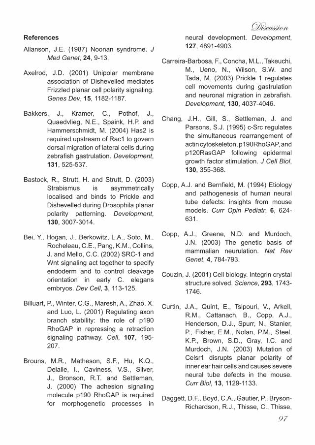
Discussion
97
References
Allanson, J.E. (1987) Noonan syndrome. J Med Genet, 24, 9-13.
Axelrod, J.D. (2001) Unipolar membrane association of Dishevelled mediates Frizzled planar cell polarity signaling. Genes Dev, 15, 1182-1187.
Bakkers, J., Kramer, C., Pothof, J., Quaedvlieg, N.E., Spaink, H.P. and Hammerschmidt, M. (2004) Has2 is required upstream of Rac1 to govern dorsal migration of lateral cells during zebrafish gastrulation. Development, 131, 525-537.
Bastock, R., Strutt, H. and Strutt, D. (2003) Strabismus is asymmetrically localised and binds to Prickle and Dishevelled during Drosophila planar polarity patterning. Development, 130, 3007-3014.
Bei, Y., Hogan, J., Berkowitz, L.A., Soto, M., Rocheleau, C.E., Pang, K.M., Collins, J. and Mello, C.C. (2002) SRC-1 and Wnt signaling act together to specify endoderm and to control cleavage orientation in early C. elegans embryos. Dev Cell, 3, 113-125.
Billuart, P., Winter, C.G., Maresh, A., Zhao, X. and Luo, L. (2001) Regulating axon branch stability: the role of p190 RhoGAP in repressing a retraction signaling pathway. Cell, 107, 195-207.
Brouns, M.R., Matheson, S.F., Hu, K.Q., Delalle, I., Caviness, V.S., Silver, J., Bronson, R.T. and Settleman, J. (2000) The adhesion signaling molecule p190 RhoGAP is required for morphogenetic processes in
neural development. Development, 127, 4891-4903.
Carreira-Barbosa, F., Concha, M.L., Takeuchi, M., Ueno, N., Wilson, S.W. and Tada, M. (2003) Prickle 1 regulates cell movements during gastrulation and neuronal migration in zebrafish. Development, 130, 4037-4046.
Chang, J.H., Gill, S., Settleman, J. and Parsons, S.J. (1995) c-Src regulates the simultaneous rearrangement of actin cytoskeleton, p190RhoGAP, and p120RasGAP following epidermal growth factor stimulation. J Cell Biol, 130, 355-368.
Copp, A.J. and Bernfield, M. (1994) Etiology and pathogenesis of human neural tube defects: insights from mouse models. Curr Opin Pediatr, 6, 624-631.
Copp, A.J., Greene, N.D. and Murdoch, J.N. (2003) The genetic basis of mammalian neurulation. Nat Rev Genet, 4, 784-793.
Couzin, J. (2001) Cell biology. Integrin crystal structure solved. Science, 293, 1743-1746.
Curtin, J.A., Quint, E., Tsipouri, V., Arkell, R.M., Cattanach, B., Copp, A.J., Henderson, D.J., Spurr, N., Stanier, P., Fisher, E.M., Nolan, P.M., Steel, K.P., Brown, S.D., Gray, I.C. and Murdoch, J.N. (2003) Mutation of Celsr1 disrupts planar polarity of inner ear hair cells and causes severe neural tube defects in the mouse. Curr Biol, 13, 1129-1133.
Daggett, D.F., Boyd, C.A., Gautier, P., Bryson-Richardson, R.J., Thisse, C., Thisse,
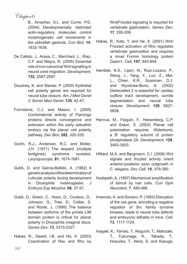
Chapter6
98
B., Amacher, S.L. and Currie, P.D. (2004) Developmentally restricted actin-regulatory molecules control morphogenetic cell movements in the zebrafish gastrula. Curr Biol, 14, 1632-1638.
De Calisto, J., Araya, C., Marchant, L., Riaz, C.F. and Mayor, R. (2005) Essential role of non-canonical Wnt signalling in neural crest migration. Development, 132, 2587-2597.
Doudney, K. and Stanier, P. (2005) Epithelial cell polarity genes are required for neural tube closure. Am J Med Genet C Semin Med Genet, 135, 42-47.
Formstone, C.J. and Mason, I. (2005) Combinatorial activity of Flamingo proteins directs convergence and extension within the early zebrafish embryo via the planar cell polarity pathway. Dev Biol, 282, 320-335.
Gorlin, R.J., Anderson, R.C. and Moller, J.H. (1971) The leopard (multiple lentigines) syndrome revisited. Laryngoscope, 81, 1674-1681.
Gubb, D. and Garcia-Bellido, A. (1982) A genetic analysis of the determination of cuticular polarity during development in Drosophila melanogaster. J Embryol Exp Morphol, 68, 37-57.
Gubb, D., Green, C., Huen, D., Coulson, D., Johnson, G., Tree, D., Collier, S. and Roote, J. (1999) The balance between isoforms of the prickle LIM domain protein is critical for planar polarity in Drosophila imaginal discs. Genes Dev, 13, 2315-2327.
Habas, R., Dawid, I.B. and He, X. (2003) Coactivation of Rac and Rho by
Wnt/Frizzled signaling is required for vertebrate gastrulation. Genes Dev, 17, 295-309.
Habas, R., Kato, Y. and He, X. (2001) Wnt/Frizzled activation of Rho regulates vertebrate gastrulation and requires a novel Formin homology protein Daam1. Cell, 107, 843-854.
Hamblet, N.S., Lijam, N., Ruiz-Lozano, P., Wang, J., Yang, Y., Luo, Z., Mei, L., Chien, K.R., Sussman, D.J. and Wynshaw-Boris, A. (2002) Dishevelled 2 is essential for cardiac outflow tract development, somite segmentation and neural tube closure. Development, 129, 5827-5838.
Hannus, M., Feiguin, F., Heisenberg, C.P. and Eaton, S. (2002) Planar cell polarization requires Widerborst, a B’ regulatory subunit of protein phosphatase 2A. Development, 129, 3493-3503.
Hilliard, M.A. and Bargmann, C.I. (2006) Wnt signals and frizzled activity orient anterior-posterior axon outgrowth in C. elegans. Dev Cell, 10, 379-390.
Hudspeth, A. (1997) Mechanical amplification of stimuli by hair cells. Curr Opin Neurobiol, 7, 480-486.
Imamoto, A. and Soriano, P. (1993) Disruption of the csk gene, encoding a negative regulator of Src family tyrosine kinases, leads to neural tube defects and embryonic lethality in mice. Cell, 73, 1117-1124.
Inagaki, K., Yamao, T., Noguchi, T., Matozaki, T., Fukunaga, K., Takada, T., Hosooka, T., Akira, S. and Kasuga,

Discussion
99
M. (2000) SHPS-1 regulates integrin-mediated cytoskeletal reorganization and cell motility. Embo J, 19, 6721-6731.
Keller, R. (2002) Shaping the vertebrate body plan by polarized embryonic cell movements. Science, 298, 1950-1954.
Keller, R., Shih, J. and Domingo, C. (1992) The patterning and functioning of protrusive activity during convergence and extension of the Xenopus organiser. Dev Suppl, 81-91.
Kibar, Z., Vogan, K.J., Groulx, N., Justice, M.J., Underhill, D.A. and Gros, P. (2001) Ltap, a mammalian homolog of Drosophila Strabismus/Van Gogh, is altered in the mouse neural tube mutant Loop-tail. Nat Genet, 28, 251-255.
Knoll, B. and Drescher, U. (2004) Src family kinases are involved in EphA receptor-mediated retinal axon guidance. J Neurosci, 24, 6248-6257.
Kwan, K.M. and Kirschner, M.W. (2005) A microtubule-binding Rho-GEF controls cell morphology during convergent extension of Xenopus laevis. Development, 132, 4599-4610.
Lawrence, P.A., Casal, J. and Struhl, G. (2002) Towards a model of the organisation of planar polarity and pattern in the Drosophila abdomen. Development, 129, 2749-2760.
Lim, D.J. (1980) Cochlear anatomy related to cochlear micromechanics. A review. J Acoust Soc Am, 67, 1686-1695.
Lin, F., Sepich, D.S., Chen, S., Topczewski, J., Yin, C., Solnica-Krezel, L. and Hamm, H. (2005) Essential roles of G{alpha}12/13 signaling in distinct cell behaviors driving zebrafish convergence and extension gastrulation movements. J Cell Biol, 169, 777-787.
Lu, X., Borchers, A.G., Jolicoeur, C., Rayburn, H., Baker, J.C. and Tessier-Lavigne, M. (2004) PTK7/CCK-4 is a novel regulator of planar cell polarity in vertebrates. Nature, 430, 93-98.
Markova, B., Herrlich, P., Ronnstrand, L. and Bohmer, F.D. (2003) Identification of protein tyrosine phosphatases associating with the PDGF receptor. Biochemistry, 42, 2691-2699.
Marlow, F., Topczewski, J., Sepich, D. and Solnica-Krezel, L. (2002) Zebrafish Rho kinase 2 acts downstream of Wnt11 to mediate cell polarity and effective convergence and extension movements. Curr Biol, 12, 876-884.
Marsden, M. and DeSimone, D.W. (2003) Integrin-ECM interactions regulate cadherin-dependent cell adhesion and are required for convergent extension in Xenopus. Curr Biol, 13, 1182-1191.
Martin Mateos, M.A., Perez Duenas, B.P., Iriondo, M., Krauel, J. and Gean Molins, E. (2000) Clinical and immunological spectrum of partial DiGeorge syndrome. J Investig Allergol Clin Immunol, 10, 352-360.
Matsui, T., Raya, A., Kawakami, Y., Callol-Massot, C., Capdevila, J., Rodriguez-Esteban, C. and Izpisua Belmonte, J.C. (2005) Noncanonical
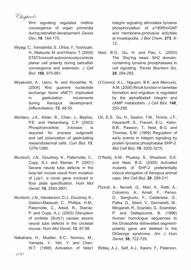
Chapter6
100
Wnt signaling regulates midline convergence of organ primordia during zebrafish development. Genes Dev, 19, 164-175.
Miyagi, C., Yamashita, S., Ohba, Y., Yoshizaki, H., Matsuda, M. and Hirano, T. (2004) STAT3 noncell-autonomously controls planar cell polarity during zebrafish convergence and extension. J Cell Biol, 166, 975-981.
Miyakoshi, A., Ueno, N. and Kinoshita, N. (2004) Rho guanine nucleotide exchange factor xNET1 implicated in gastrulation movements during Xenopus development. Differentiation, 72, 48-55.
Montero, J.A., Kilian, B., Chan, J., Bayliss, P.E. and Heisenberg, C.P. (2003) Phosphoinositide 3-kinase is required for process outgrowth and cell polarization of gastrulating mesendodermal cells. Curr Biol, 13, 1279-1289.
Murdoch, J.N., Doudney, K., Paternotte, C., Copp, A.J. and Stanier, P. (2001) Severe neural tube defects in the loop-tail mouse result from mutation of Lpp1, a novel gene involved in floor plate specification. Hum Mol Genet, 10, 2593-2601.
Murdoch, J.N., Henderson, D.J., Doudney, K., Gaston-Massuet, C., Phillips, H.M., Paternotte, C., Arkell, R., Stanier, P. and Copp, A.J. (2003) Disruption of scribble (Scrb1) causes severe neural tube defects in the circletail mouse. Hum Mol Genet, 12, 87-98.
Nakahara, H., Mueller, S.C., Nomizu, M., Yamada, Y., Yeh, Y. and Chen, W.T. (1998) Activation of beta1
integrin signaling stimulates tyrosine phosphorylation of p190RhoGAP and membrane-protrusive activities at invadopodia. J Biol Chem, 273, 9-12.
Neel, B.G., Gu, H. and Pao, L. (2003) The ‘Shp’ing news: SH2 domain-containing tyrosine phosphatases in cell signaling. Trends Biochem Sci, 28, 284-293.
O’Connor, K.L., Nguyen, B.K. and Mercurio, A.M. (2000) RhoA function in lamellae formation and migration is regulated by the alpha6beta4 integrin and cAMP metabolism. J Cell Biol, 148, 253-258.
Oh, E.S., Gu, H., Saxton, T.M., Timms, J.F., Hausdorff, S., Frevert, E.U., Kahn, B.B., Pawson, T., Neel, B.G. and Thomas, S.M. (1999) Regulation of early events in integrin signaling by protein tyrosine phosphatase SHP-2. Mol Cell Biol, 19, 3205-3215.
O’Reilly, A.M., Pluskey, S., Shoelson, S.E. and Neel, B.G. (2000) Activated mutants of SHP-2 preferentially induce elongation of Xenopus animal caps. Mol Cell Biol, 20, 299-311.
Pizzuti, A., Novelli, G., Mari, A., Ratti, A., Colosimo, A., Amati, F., Penso, D., Sangiuolo, F., Calabrese, G., Palka, G., Silani, V., Gennarelli, M., Mingarelli, R., Scarlato, G., Scambler, P. and Dallapiccola, B. (1996) Human homologue sequences to the Drosophila dishevelled segment-polarity gene are deleted in the DiGeorge syndrome. Am J Hum Genet, 58, 722-729.
Ridley, A.J., Self, A.J., Kasmi, F., Paterson,
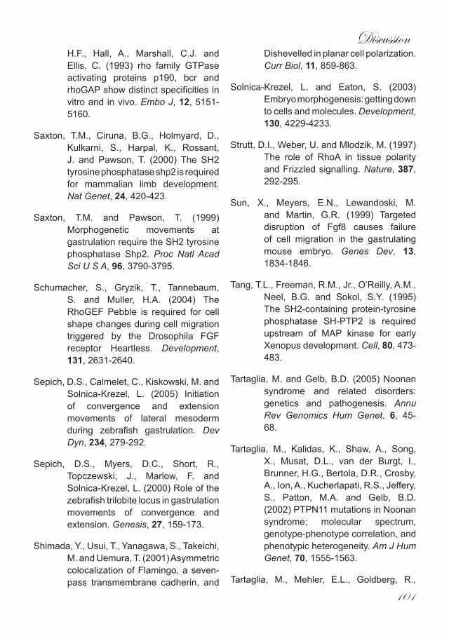
Discussion
101
H.F., Hall, A., Marshall, C.J. and Ellis, C. (1993) rho family GTPase activating proteins p190, bcr and rhoGAP show distinct specificities in vitro and in vivo. Embo J, 12, 5151-5160.
Saxton, T.M., Ciruna, B.G., Holmyard, D., Kulkarni, S., Harpal, K., Rossant, J. and Pawson, T. (2000) The SH2 tyrosine phosphatase shp2 is required for mammalian limb development. Nat Genet, 24, 420-423.
Saxton, T.M. and Pawson, T. (1999) Morphogenetic movements at gastrulation require the SH2 tyrosine phosphatase Shp2. Proc Natl Acad Sci U S A, 96, 3790-3795.
Schumacher, S., Gryzik, T., Tannebaum, S. and Muller, H.A. (2004) The RhoGEF Pebble is required for cell shape changes during cell migration triggered by the Drosophila FGF receptor Heartless. Development, 131, 2631-2640.
Sepich, D.S., Calmelet, C., Kiskowski, M. and Solnica-Krezel, L. (2005) Initiation of convergence and extension movements of lateral mesoderm during zebrafish gastrulation. Dev Dyn, 234, 279-292.
Sepich, D.S., Myers, D.C., Short, R., Topczewski, J., Marlow, F. and Solnica-Krezel, L. (2000) Role of the zebrafish trilobite locus in gastrulation movements of convergence and extension. Genesis, 27, 159-173.
Shimada, Y., Usui, T., Yanagawa, S., Takeichi, M. and Uemura, T. (2001) Asymmetric colocalization of Flamingo, a seven-pass transmembrane cadherin, and
Dishevelled in planar cell polarization. Curr Biol, 11, 859-863.
Solnica-Krezel, L. and Eaton, S. (2003) Embryo morphogenesis: getting down to cells and molecules. Development, 130, 4229-4233.
Strutt, D.I., Weber, U. and Mlodzik, M. (1997) The role of RhoA in tissue polarity and Frizzled signalling. Nature, 387, 292-295.
Sun, X., Meyers, E.N., Lewandoski, M. and Martin, G.R. (1999) Targeted disruption of Fgf8 causes failure of cell migration in the gastrulating mouse embryo. Genes Dev, 13, 1834-1846.
Tang, T.L., Freeman, R.M., Jr., O’Reilly, A.M., Neel, B.G. and Sokol, S.Y. (1995) The SH2-containing protein-tyrosine phosphatase SH-PTP2 is required upstream of MAP kinase for early Xenopus development. Cell, 80, 473-483.
Tartaglia, M. and Gelb, B.D. (2005) Noonan syndrome and related disorders: genetics and pathogenesis. Annu Rev Genomics Hum Genet, 6, 45-68.
Tartaglia, M., Kalidas, K., Shaw, A., Song, X., Musat, D.L., van der Burgt, I., Brunner, H.G., Bertola, D.R., Crosby, A., Ion, A., Kucherlapati, R.S., Jeffery, S., Patton, M.A. and Gelb, B.D. (2002) PTPN11 mutations in Noonan syndrome: molecular spectrum, genotype-phenotype correlation, and phenotypic heterogeneity. Am J Hum Genet, 70, 1555-1563.
Tartaglia, M., Mehler, E.L., Goldberg, R.,
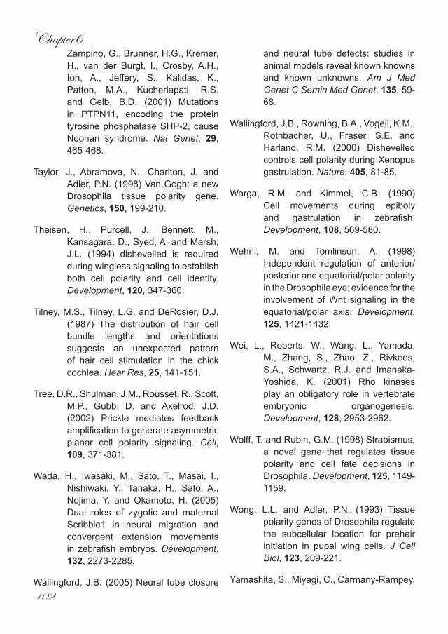
Chapter6
102
Zampino, G., Brunner, H.G., Kremer, H., van der Burgt, I., Crosby, A.H., Ion, A., Jeffery, S., Kalidas, K., Patton, M.A., Kucherlapati, R.S. and Gelb, B.D. (2001) Mutations in PTPN11, encoding the protein tyrosine phosphatase SHP-2, cause Noonan syndrome. Nat Genet, 29, 465-468.
Taylor, J., Abramova, N., Charlton, J. and Adler, P.N. (1998) Van Gogh: a new Drosophila tissue polarity gene. Genetics, 150, 199-210.
Theisen, H., Purcell, J., Bennett, M., Kansagara, D., Syed, A. and Marsh, J.L. (1994) dishevelled is required during wingless signaling to establish both cell polarity and cell identity. Development, 120, 347-360.
Tilney, M.S., Tilney, L.G. and DeRosier, D.J. (1987) The distribution of hair cell bundle lengths and orientations suggests an unexpected pattern of hair cell stimulation in the chick cochlea. Hear Res, 25, 141-151.
Tree, D.R., Shulman, J.M., Rousset, R., Scott, M.P., Gubb, D. and Axelrod, J.D. (2002) Prickle mediates feedback amplification to generate asymmetric planar cell polarity signaling. Cell, 109, 371-381.
Wada, H., Iwasaki, M., Sato, T., Masai, I., Nishiwaki, Y., Tanaka, H., Sato, A., Nojima, Y. and Okamoto, H. (2005) Dual roles of zygotic and maternal Scribble1 in neural migration and convergent extension movements in zebrafish embryos. Development, 132, 2273-2285.
Wallingford, J.B. (2005) Neural tube closure
and neural tube defects: studies in animal models reveal known knowns and known unknowns. Am J Med Genet C Semin Med Genet, 135, 59-68.
Wallingford, J.B., Rowning, B.A., Vogeli, K.M., Rothbacher, U., Fraser, S.E. and Harland, R.M. (2000) Dishevelled controls cell polarity during Xenopus gastrulation. Nature, 405, 81-85.
Warga, R.M. and Kimmel, C.B. (1990) Cell movements during epiboly and gastrulation in zebrafish. Development, 108, 569-580.
Wehrli, M. and Tomlinson, A. (1998) Independent regulation of anterior/posterior and equatorial/polar polarity in the Drosophila eye; evidence for the involvement of Wnt signaling in the equatorial/polar axis. Development, 125, 1421-1432.
Wei, L., Roberts, W., Wang, L., Yamada, M., Zhang, S., Zhao, Z., Rivkees, S.A., Schwartz, R.J. and Imanaka-Yoshida, K. (2001) Rho kinases play an obligatory role in vertebrate embryonic organogenesis. Development, 128, 2953-2962.
Wolff, T. and Rubin, G.M. (1998) Strabismus, a novel gene that regulates tissue polarity and cell fate decisions in Drosophila. Development, 125, 1149-1159.
Wong, L.L. and Adler, P.N. (1993) Tissue polarity genes of Drosophila regulate the subcellular location for prehair initiation in pupal wing cells. J Cell Biol, 123, 209-221.
Yamashita, S., Miyagi, C., Carmany-Rampey,
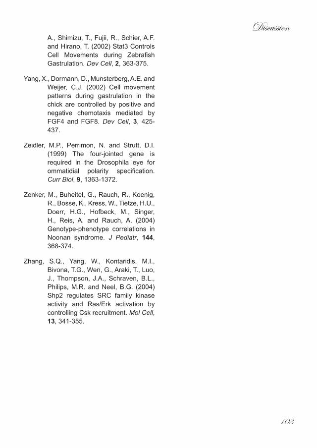
Discussion
103
A., Shimizu, T., Fujii, R., Schier, A.F. and Hirano, T. (2002) Stat3 Controls Cell Movements during Zebrafish Gastrulation. Dev Cell, 2, 363-375.
Yang, X., Dormann, D., Munsterberg, A.E. and Weijer, C.J. (2002) Cell movement patterns during gastrulation in the chick are controlled by positive and negative chemotaxis mediated by FGF4 and FGF8. Dev Cell, 3, 425-437.
Zeidler, M.P., Perrimon, N. and Strutt, D.I. (1999) The four-jointed gene is required in the Drosophila eye for ommatidial polarity specification. Curr Biol, 9, 1363-1372.
Zenker, M., Buheitel, G., Rauch, R., Koenig, R., Bosse, K., Kress, W., Tietze, H.U., Doerr, H.G., Hofbeck, M., Singer, H., Reis, A. and Rauch, A. (2004) Genotype-phenotype correlations in Noonan syndrome. J Pediatr, 144, 368-374.
Zhang, S.Q., Yang, W., Kontaridis, M.I., Bivona, T.G., Wen, G., Araki, T., Luo, J., Thompson, J.A., Schraven, B.L., Philips, M.R. and Neel, B.G. (2004) Shp2 regulates SRC family kinase activity and Ras/Erk activation by controlling Csk recruitment. Mol Cell, 13, 341-355.


Summary


Summary
107
SummaryDuring gastrulation a complex series
of cell movements leads to the formation of the three germ layers mesoderm, endoderm and ectoderm and the formation of the basic embryonic body plan. As gastrulation proceeds, cells converge towards the midline of the developing embryo, where they intercalate with one another. As a consequence, the embryo extends around the yolk and the presumptive head moves away from trunk and tail regions, resulting in extension of the anterior-posterior axis. This series of morphogenetic movements are known as convergent extension (CE), as introduced in detail in Chapter 1.
To achieve this feat, cells must become polarised which will allow them to migrate in the required direction. Cellular polarity during vertebrate CE is primarily coordinated by non-canonical Wnt signalling. Analysis of mutant and morphant zebrafish has established a pathway initiated by either Wnt11 or Wnt 5 that results in the activation of the small GTPases RhoA and Rac. This effectively remodels the cell, establishing polarity in the process. However, non–canonical Wnt signalling is not the sole regulator of CE. In Chapters 2-4 of this thesis we have identified another pathway that converges with non-canonical Wnt signalling and serves to positively regulate the activity of RhoA during vertebrate gastrulation.
In Chapter 2 we sought to determine how the Src Family Kinases, Fyn and Yes, are involved in early vertebrate development. Using morpholinos we knocked down Fyn and Yes and found that the resultant phenotype bore striking similarities to both Wnt11 and Wnt5 mutant zebrafish. Further analysis proved that Fyn/Yes morphants have defective CE cell movements during gastrulation which required the small GTPase RhoA. However, Fyn and Yes are not linear components of
the non-canonical Wnt pathway. Instead they appear to act in parallel converging on or upstream of RhoA. This obviously led to the question what is upstream of Fyn and Yes.
Previous research in Xenopus and the mouse has identified the C-terminal Src kinase (Csk) as a key regulator of vertebrate gastrulation. As its name suggests, Csk phosphorylates the inhibitory tyrosine residue in the C-terminal tail of SFKs leading to inactivation. In Chapter 3, using a morpholino-based strategy, we knocked down Csk during early zebrafish development and found that disruption of Csk results in defective gastrulation cell movements. Importantly, we were able to rescue the Csk morphants by partially knocking down Fyn and Yes, indicating that Csk is directly upstream of these two SFKs.
We next sought to determine what is upstream of Csk. The most promising candidate was Shp2, as Shp2 negatively regulates Csk by dephosphorylating the membrane bound adaptor protein PAG1. Csk is now no longer targeted to the plasma membrane and cannot inhibit membrane bound SFKs. In Chapter 4 of this thesis we demonstrate that Shp2 was necessary for CE cell movements during zebrafish gastrulation. These results are consistent with data from Xenopus and mouse. In addition, we show that Shp2 is upstream of Fyn and Yes by epigenetic analyses. Moreover, Shp2 knock down was rescued by active RhoA, placing Shp2 in a pathway parallel to non-canonical Wnt signalling. Our data, together with biochemical data, suggest a model which sees Shp2 negatively regulate Csk, the inhibitor of Fyn and Yes which in turn act upstream of RhoA in gastrulation cell movements.
Because of Shp2’s involvement in early embryonic development we sought to address the issue of whether the defects associated with human Noonan
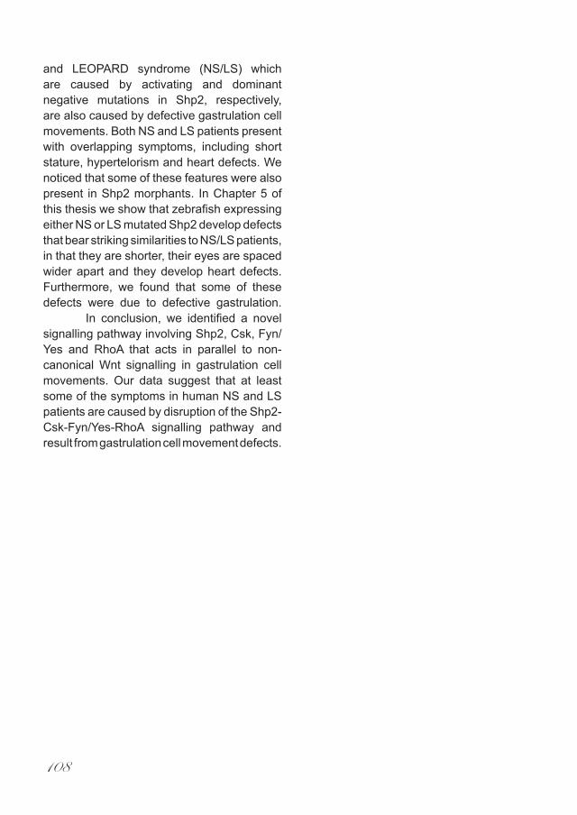
108
and LEOPARD syndrome (NS/LS) which are caused by activating and dominant negative mutations in Shp2, respectively, are also caused by defective gastrulation cell movements. Both NS and LS patients present with overlapping symptoms, including short stature, hypertelorism and heart defects. We noticed that some of these features were also present in Shp2 morphants. In Chapter 5 of this thesis we show that zebrafish expressing either NS or LS mutated Shp2 develop defects that bear striking similarities to NS/LS patients, in that they are shorter, their eyes are spaced wider apart and they develop heart defects. Furthermore, we found that some of these defects were due to defective gastrulation.
In conclusion, we identified a novel signalling pathway involving Shp2, Csk, Fyn/Yes and RhoA that acts in parallel to non-canonical Wnt signalling in gastrulation cell movements. Our data suggest that at least some of the symptoms in human NS and LS patients are caused by disruption of the Shp2-Csk-Fyn/Yes-RhoA signalling pathway and result from gastrulation cell movement defects.

Samenvatting
109
SamenvattingTijdens gastrulatie leidt een complexe
reeks celbewegingen tot de vorming van de drie kiemlagen, endoderm, mesoderm en ectoderm en tot de vorming van het basale embryonale lichaamsplan. Terwijl gastrulatie voortgaat, komen de cellen samen van de zijkanten naar het midden van het ontwikkelende embryo, waar zij zich onderling schikken. Dientengevolge, strekt het embryo zich rond de dooier uit en het (toekomstige) hoofd verwijdert zich van onderlichaam en staart, resulterend in verlenging van de kop-staart as. Deze reeks morfogenetische bewegingen heet convergentie en extensie (CE), zoals in detail geïntroduceerd in Hoofdstuk 1.
Om CE te bewerkstelligen, moeten de cellen gepolariseerd worden wat het hen mogelijk maakt in de vereiste richting te migreren. De cellulaire polariteit tijdens CE van gewervelde dieren wordt hoofdzakelijk gereguleerd door Wnt-signalering. De analyse van mutant en morphant zebravissen heeft een signaaltransductie-pad blootgelegd die door Wnt11 of Wnt5 wordt geactiveerd en resulteert in de activering van de kleine GTPases, RhoA en Rac. Effectief remodelleert dit de cel, wat leidt tot polarisatie. Wnt signalering is niet de enige regulator van CE. In de hoofdstukken 2-4 van dit proefschrift hebben wij een ander signaaltransductie-pad geïdentificeerd die met Wnt signalering samenkomt en leidt tot activering van RhoA tijdens de gastrulatie van gewervelde dieren.
In Hoofdstuk 2 wilden wij bepalen wat de rol is van twee Kinases van de Src Familie (SFKs), Fyn en Yes, bij de vroege ontwikkeling van gewervelde dieren. Met gebruik van morpholinos reduceerden wij de expressie van Fyn en Yes in zebravis embryos en wij vonden dat het resulterende fenotype opvallend leek op zowel Wnt11 als Wnt5 mutant zebravissen. Verdere analyse toonde aan dat CE celbewegingen verstoord
waren in de Fyn/Yes morphants en dat de kleine GTPase RhoA hier een rol in had. Fyn en Yes zijn daarentegen geen intrinsieke componenten van het Wnt signaaltransductie-pad. In plaats daarvan hebben Fyn en Yes een rol in een parallel signaaltransductie-pad dat samenkomt met Wnt signalering op of net boven RhoA. Dit leidde ons tot de vraag wat Fyn en Yes reguleert.
Eerder onderzoek in Xenopus en in de muis heeft C-terminal Src kinase (Csk) geïdentificeerd als zeer belangrijke regulator van gastrulatie in gewervelde dieren. Zoals de naam al aangeeft, fosforyleert Csk de inhibitoire tyrosine in de C-terminus van SFKs wat tot hun inactivering leidt. In Hoofdstuk 3 zijn morpholinos gebruikt om de expressie van Csk te reduceren tijdens de vroege ontwikkeling van de zebravis en wij hebben gevonden dat dit leidt tot een verstoring van CE celbewegingen tijdens de gastrulatie. Wij konden dit fenotype gedeeltelijk herstellen door Fyn en Yes expressie te reduceren, wat er op wijst dat Csk een directe regulator is van deze twee SFKs.
Vervolgens wilden wij bepalen wat Csk reguleert. De meest veelbelovende kandidaat was Shp2, aangezien Shp2 Csk negatief reguleert door het membraan-gebonden adapter eiwit, PAG1 te defosforyleren. Csk transloceert nu niet meer naar het plasmamembraan en kan de membraan-gebonden SFKs niet meer remmen. In Hoofdstuk 4 van dit proefschrift tonen wij aan dat Shp2 noodzakelijk is voor CE celbewegingen tijdens de gastrulation van de zebravis. Deze resultaten komen overeen met resultaten in Xenopus en in de muis. Bovendien tonen wij met epigenetische analyses aan dat Shp2 Fyn en Yes reguleert. Voorts werd het Shp2 fenotype teniet gedaan door actief RhoA, wat Shp2 in een signaaltransductie-pad plaatst, parallel aan Wnt-signalering. Onze resultaten, samen met
110
biochemische gegevens, suggereren een model waarin Shp2 een negatieve regulator is van Csk, de inhibitor van Fyn en Yes, die op hun beurt een rol hebben als regulator van RhoA in celbewegingen tijdens gastrulatie.
Vanwege de betrokkenheid van Shp2 bij de vroege embryonale ontwikkeling vroegen wij ons af of de symptomen die geassocieerd zijn met Noonan syndroom (NS) of LEOPARD syndroom (LS), die veroorzaakt worden door respectievelijk activerende en dominant negatieve mutaties in Shp2, ook veroorzaakt worden door ontregelde celbewegingen tijdens gastrulatie. Patiënten met NS en LS vertonen overlappende symptomen, zoals een korte gestalte, hypertelorisme en hartafwijkingen. Wij hebben opgemerkt dat sommige van deze eigenschappen zich ook manifesteerden in Shp2 morphants. In Hoofdstuk 5 van dit proefschrift tonen wij aan dat zebravis embryos die mutant NS of LS Shp2 tot expressie brengen defecten vertonen die opvallende gelijkenissen vertonen met NS en LS patiënten, in zoverre dat zij korter zijn, hun ogen verder uit elkaar staan en zij hartafwijkingen ontwikkelen. Voorts vonden wij dat sommige van deze tekorten aan defecten tijdens de gastrulatie toe te schrijven waren.
Wij hebben een nieuw signaaltransductie-pad geïdentificeerd, bestaande uit Shp2, Csk, Fyn/Yes en RhoA dat parallel loopt aan Wnt signalering in celbewegingen tijdens gastrulatie. Onze data suggereren dat ten minste enkele symptomen in menselijke NS en LS patiënten veroorzaakt worden door verstoring van Shp2-Csk-Fyn/Yes-RhoA signalering die resulteren in defecten in celbewegingen tijdens de gastrulatie.

111
Acknowledgements
‘The crowd are on the pitch, they think its all over, it is now!!’ or should it be, in the words of Karen Carpenter, ‘We’ve only just begun’ . Anyhow which ever way I go from here is irrelevant for the time being because in the here and now I have just finished my PhD and am absolutely over the moon and although I do take great pride in this wonderful achievement I would be a fool to imagine that it was done alone. In fact my promotion will be the culmination of a lot of work and support from many many people who have guided me through the last 5 years and for which I am truly grateful.So let’s just take a moment and look around at some of the heroes and heroines who work there magic at the Hubrecht. To my left is John, one of the friendliest people I have ever met and while I agree life can be a drama don’t forget it can also be a thriller. Behind me is Arnoud, I’ll miss our quality discussions about the finer things in life (football and Schwarzenegger) over a beer or 12. In the corner is Fiona, I know sometimes you’ve found it tough but you never give up, something I’ve always admired, however your timekeeping will always be like you, a little Spanish. At the back sitting quietly is Andrei, always ready to listen (even with the talkmeister) but underneath simmers competitive/aggressive Andrei the table tennis warrior, Ana has her hands full but you’ll both go far. Next door we have the French connection, Adele, I agree it should be renamed from PTEN to PUTAIN! It’s just a shame these people don’t recognise proper cheese when they smell it but remember the worerr awri. Also in the room is Mark, when gastrulation gets you down drop me a line, I’ve been through it too. Simone hang in there your too intelligent but have you added to the wit loaf mountain under the oudegracht? My student Daphne I hope you enjoyed your time with me and I know your gonna be a star. Further a field we have Dana who, along with Freek, set me off down the wonderful road of zebrafish embryology one which I intend to carry on traveling. And of course one of the zebrafish grandmasters, Stefan, I have appreciated all your advice and your genuine interest in how things are going, one day I’ll pull my finger out. Mr Bakkers downstairs, I don’t think there is much I have done here that hasn’t had some kind of input from you. No longer with us( figuratively speaking) are Thea, your calming influence helped me endure what can sometimes be a hectic pace, and Big John Verhees, we will still meet up and plan how to take over the world. Rui, who taught me how to deal with the Dutch mentality (ignore it) and Sander who first welcomed me into the Hubrecht and kept me smiling throughout (it’s a speed punk thang). Oh and Pantelis I think the need to keep stocked up on Greek fags is as good as any for taking regular! holidays. And lets not forget the Usual Suspects- Marco (I’m still up for kneecapping the hedgehog people), Ruben (Sanchez), Bas, Pim, Ewart, Bart, Wigard, Olaf and Jeroen thanks for many memorable evenings (well I can remember how they started, its the endings that are a bit hazy). Ellen I’ve enjoyed chatting in the evening with you but have you got the pointing under control yet? Leon thanks for help with the confocal maybe if we had more time. And of course the people who cared for my many fishes, Peter(it will be strange going to another aquarium without you running the show), Mark, Bertje, Marc, Erma, Bjorn, Sanne, Petra and

112
Miep. And to all my zebrafish colleagues (we will probably keep bumping into each other in the future). And let’s not forget outside the Lab, my best mate Marleen who has supported me and kept me going through the good and the bad times, it’s a struggle for you at the moment I know but remember I am always there. And finally the man who made it all possible, the Boss, The Chief, the Head Honcho, Jeroen. I have had a few bosses in my time but you now provide the bench mark by which all others will be judged thank you for making me a scientist. So once again thank you all, I’m outta here.
113
Curriculum Vitae
John Christian Jopling was born on the 13th March 1971 in London. He completed his secondary education at Abbotsford High School. After working for a number of years in a variety of jobs he attended Kings College London where he studied biochemistry graduating in 1998 with a first class honors degree. Following this he worked as a research assistant in the Department of Developmental Biology at University College London while also studying for a MSc in neuroscience. He attained his MSc in 2001 and in the same year began a PhD at the Hubrecht Laboratory (Netherlands Institute for Developmental Biology), Utrecht, the Netherlands, studying factors involved in the regulation of gastrulation cell movements during vertebrate development. The research described in this thesis was carried out under the supervision of Dr Jeroen den Hertog.
Publications
Jopling C, den Hertog J (2005). Fyn/Yes and non-canonical Wnt signalling converge on RhoA in vertebrate gastrulation cell movements. EMBO Rep. 5, 426-431.
Rosendaal M, Jopling C (2003). Hematopoietic capacity of connexin43 wild-type and knock-out fetal liver cells not different on wild-type stroma. Blood. 2003 Apr 15;101(8):2996-8.
Jopling C, Rosendaal M (2001). A cautionary tale: how to delete mouse haemopoietic stem cells with busulphan. Br J Haematol. 2001 Jun;113(4):970-4.
Jopling C, den Hertog J (2006). Essential role for Csk upstream of Fyn and Yes in zebrafish gastrulation. Submitted
Jopling C, den Hertog J (2006). Shp2 controls zebrafish gastrulation cell movements via Fyn, Yes and RhoA. Submitted