Doxycycline induces bone repair and changes in Wnt signalling
9
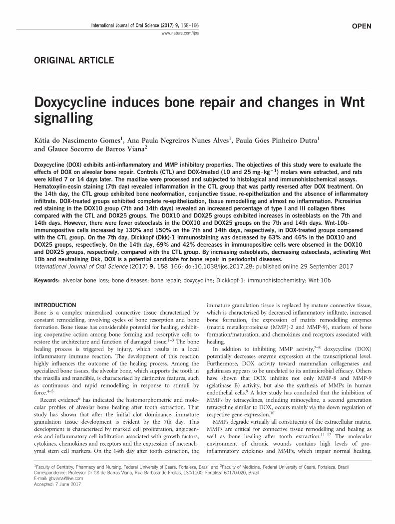
OPEN ORIGINAL ARTICLE Doxycycline induces bone repair and changes in Wnt signalling Kátia do Nascimento Gomes 1 , Ana Paula Negreiros Nunes Alves 1 , Paula Góes Pinheiro Dutra 1 and Glauce Socorro de Barros Viana 2 Doxycycline (DOX) exhibits anti-inflammatory and MMP inhibitory properties. The objectives of this study were to evaluate the effects of DOX on alveolar bone repair. Controls (CTL) and DOX-treated (10 and 25 mg . kg - 1 ) molars were extracted, and rats were killed 7 or 14 days later. The maxillae were processed and subjected to histological and immunohistochemical assays. Hematoxylin-eosin staining (7th day) revealed inflammation in the CTL group that was partly reversed after DOX treatment. On the 14th day, the CTL group exhibited bone neoformation, conjunctive tissue, re-epithelization and the absence of inflammatory infiltrate. DOX-treated groups exhibited complete re-epithelization, tissue remodelling and almost no inflammation. Picrosirius red staining in the DOX10 group (7th and 14th days) revealed an increased percentage of type I and III collagen fibres compared with the CTL and DOX25 groups. The DOX10 and DOX25 groups exhibited increases in osteoblasts on the 7th and 14th days. However, there were fewer osteoclasts in the DOX10 and DOX25 groups on the 7th and 14th days. Wnt-10b- immunopositive cells increased by 130% and 150% on the 7th and 14th days, respectively, in DOX-treated groups compared with the CTL group. On the 7th day, Dickkopf (Dkk)-1 immunostaining was decreased by 63% and 46% in the DOX10 and DOX25 groups, respectively. On the 14th day, 69% and 42% decreases in immunopositive cells were observed in the DOX10 and DOX25 groups, respectively, compared with the CTL group. By increasing osteoblasts, decreasing osteoclasts, activating Wnt 10b and neutralising Dkk, DOX is a potential candidate for bone repair in periodontal diseases. International Journal of Oral Science (2017) 9, 158–166; doi:10.1038/ijos.2017.28; published online 29 September 2017 Keywords: alveolar bone loss; bone diseases; bone repair; doxycycline; Dickkopf-1; immunohistochemistry; Wnt-10b INTRODUCTION Bone is a complex mineralised connective tissue characterised by constant remodelling, involving cycles of bone resorption and bone formation. Bone tissue has considerable potential for healing, exhibit- ing cooperative action among bone forming and resorptive cells to restore the architecture and function of damaged tissue. 1–3 The bone healing process is triggered by injury, which results in a local inflammatory immune reaction. The development of this reaction highly influences the outcome of the healing process. Among the specialized bone tissues, the alveolar bone, which supports the tooth in the maxilla and mandible, is characterised by distinctive features, such as continuous and rapid remodelling in response to stimuli by force. 4–5 Recent evidence 6 has indicated the histomorphometric and mole- cular profiles of alveolar bone healing after tooth extraction. That study has shown that after the initial clot dominance, immature granulation tissue development is evident by the 7th day. This development is characterised by marked cell proliferation, angiogen- esis and inflammatory cell infiltration associated with growth factors, cytokines, chemokines and receptors and the expression of mesench- ymal stem cell markers. On the 14th day after tooth extraction, the immature granulation tissue is replaced by mature connective tissue, which is characterised by decreased inflammatory infiltrate, increased bone formation, the expression of matrix remodelling enzymes (matrix metalloproteinase (MMP)-2 and MMP-9), markers of bone formation/maturation, and chemokines and receptors associated with healing. In addition to inhibiting MMP activity, 7–8 doxycycline (DOX) potentially decreases enzyme expression at the transcriptional level. Furthermore, DOX activity toward mammalian collagenases and gelatinases appears to be unrelated to its antimicrobial efficacy. Others have shown that DOX inhibits not only MMP-8 and MMP-9 (gelatinase B) activity, but also the synthesis of MMPs in human endothelial cells. 9 A later study has concluded that the inhibition of MMPs by tetracyclines, including minocycline, a second generation tetracycline similar to DOX, occurs mainly via the down regulation of respective gene expression. 10 MMPs degrade virtually all constituents of the extracellular matrix. MMPs are critical for connective tissue remodelling and healing as well as bone healing after tooth extraction. 11–12 The molecular environment of chronic wounds contains high levels of pro- inflammatory cytokines and MMPs, which impair normal healing. 1 Faculty of Dentistry, Pharmacy and Nursing, Federal University of Ceará, Fortaleza, Brazil and 2 Faculty of Medicine, Federal University of Ceará, Fortaleza, Brazil Correspondence: Professor Dr GS de Barros Viana, Rua Barbosa de Freitas, 130/1100, Fortaleza 60170-020, Brazil E-mail: [email protected] Accepted: 7 June 2017 International Journal of Oral Science (2017) 9, 158–166 www.nature.com/ijos
Transcript of Doxycycline induces bone repair and changes in Wnt signalling
OPEN
ORIGINAL ARTICLE
Doxycycline induces bone repair and changes in Wntsignalling
Kátia do Nascimento Gomes1, Ana Paula Negreiros Nunes Alves1, Paula Góes Pinheiro Dutra1
and Glauce Socorro de Barros Viana2
Doxycycline (DOX) exhibits anti-inflammatory and MMP inhibitory properties. The objectives of this study were to evaluate the
effects of DOX on alveolar bone repair. Controls (CTL) and DOX-treated (10 and 25 mg . kg−1) molars were extracted, and rats
were killed 7 or 14 days later. The maxillae were processed and subjected to histological and immunohistochemical assays.
Hematoxylin-eosin staining (7th day) revealed inflammation in the CTL group that was partly reversed after DOX treatment. On
the 14th day, the CTL group exhibited bone neoformation, conjunctive tissue, re-epithelization and the absence of inflammatory
infiltrate. DOX-treated groups exhibited complete re-epithelization, tissue remodelling and almost no inflammation. Picrosirius
red staining in the DOX10 group (7th and 14th days) revealed an increased percentage of type I and III collagen fibres
compared with the CTL and DOX25 groups. The DOX10 and DOX25 groups exhibited increases in osteoblasts on the 7th and
14th days. However, there were fewer osteoclasts in the DOX10 and DOX25 groups on the 7th and 14th days. Wnt-10b-
immunopositive cells increased by 130% and 150% on the 7th and 14th days, respectively, in DOX-treated groups compared
with the CTL group. On the 7th day, Dickkopf (Dkk)-1 immunostaining was decreased by 63% and 46% in the DOX10 and
DOX25 groups, respectively. On the 14th day, 69% and 42% decreases in immunopositive cells were observed in the DOX10
and DOX25 groups, respectively, compared with the CTL group. By increasing osteoblasts, decreasing osteoclasts, activating Wnt
10b and neutralising Dkk, DOX is a potential candidate for bone repair in periodontal diseases.
International Journal of Oral Science (2017) 9, 158–166; doi:10.1038/ijos.2017.28; published online 29 September 2017
Keywords: alveolar bone loss; bone diseases; bone repair; doxycycline; Dickkopf-1; immunohistochemistry; Wnt-10b
INTRODUCTION
Bone is a complex mineralised connective tissue characterised byconstant remodelling, involving cycles of bone resorption and boneformation. Bone tissue has considerable potential for healing, exhibit-ing cooperative action among bone forming and resorptive cells torestore the architecture and function of damaged tissue.1–3 The bonehealing process is triggered by injury, which results in a localinflammatory immune reaction. The development of this reactionhighly influences the outcome of the healing process. Among thespecialized bone tissues, the alveolar bone, which supports the tooth inthe maxilla and mandible, is characterised by distinctive features, suchas continuous and rapid remodelling in response to stimuli byforce.4–5
Recent evidence6 has indicated the histomorphometric and mole-cular profiles of alveolar bone healing after tooth extraction. Thatstudy has shown that after the initial clot dominance, immaturegranulation tissue development is evident by the 7th day. Thisdevelopment is characterised by marked cell proliferation, angiogen-esis and inflammatory cell infiltration associated with growth factors,cytokines, chemokines and receptors and the expression of mesench-ymal stem cell markers. On the 14th day after tooth extraction, the
immature granulation tissue is replaced by mature connective tissue,which is characterised by decreased inflammatory infiltrate, increasedbone formation, the expression of matrix remodelling enzymes(matrix metalloproteinase (MMP)-2 and MMP-9), markers of boneformation/maturation, and chemokines and receptors associated withhealing.In addition to inhibiting MMP activity,7–8 doxycycline (DOX)
potentially decreases enzyme expression at the transcriptional level.Furthermore, DOX activity toward mammalian collagenases andgelatinases appears to be unrelated to its antimicrobial efficacy. Othershave shown that DOX inhibits not only MMP-8 and MMP-9(gelatinase B) activity, but also the synthesis of MMPs in humanendothelial cells.9 A later study has concluded that the inhibition ofMMPs by tetracyclines, including minocycline, a second generationtetracycline similar to DOX, occurs mainly via the down regulation ofrespective gene expression.10
MMPs degrade virtually all constituents of the extracellular matrix.MMPs are critical for connective tissue remodelling and healing aswell as bone healing after tooth extraction.11–12 The molecularenvironment of chronic wounds contains high levels of pro-inflammatory cytokines and MMPs, which impair normal healing.
1Faculty of Dentistry, Pharmacy and Nursing, Federal University of Ceará, Fortaleza, Brazil and 2Faculty of Medicine, Federal University of Ceará, Fortaleza, BrazilCorrespondence: Professor Dr GS de Barros Viana, Rua Barbosa de Freitas, 130/1100, Fortaleza 60170-020, BrazilE-mail: [email protected]: 7 June 2017
International Journal of Oral Science (2017) 9, 158–166www.nature.com/ijos
This process is positively affected by DOX.13–14 Previously, thesystemic administration of DOX has been found to prevent both rootresorption and bone loss in rats.15
Furthermore, the Wnt signalling pathway (involving proteins thatpass signals into a cell through cell surface receptors) plays a key rolein bone development and homeostasis. Wnt ligands promote bonegrowth, thereby leading to the hypothesis that Wnt signallingactivation may stimulate bone healing.16 Dysregulation of this pathwaygreatly inhibits bone formation and the healing process, thus suggest-ing that this pathway has an essential role in bone regeneration.17 Wntsignalling is activated by wounding and participates in every stage ofthe healing process, from the control of inflammation and pro-grammed cell death to the mobilisation of stem cell reservoirs withinthe wound site.18
Furthermore, signalling via the classical Wnt pathway is critical forbone deposition and bone remodelling. Among other mechanisms,this signalling pathway is regulated by Dickkopf proteins (Dkks),which bind and promote the internalisation of lipoprotein receptor-related protein (LRP)5 or LRP6. Blocking these Wnt receptorcomponents effectively downregulates Wnt signalling. Dkk has beenimplicated in bone formation and bone diseases. The induction of theWnt signalling pathway promotes bone formation, whereas itsinactivation by Dkk leads to osteopenic states.19
Given the anti-inflammatory and MMPs inhibitory properties ofDOX, the objectives of the present work were to use histological andhistomorphometric methods to evaluate the effects of DOX onalveolar bone repair after tooth extraction in rats. In addition, weperformed immunohistochemistry detecting Wnt-10b and its inhibitorDkk-1.
MATERIALS AND METHODS
DrugsKetamine hydrochloride was purchased from Holliday-Scott SA,Argentina, and xylazine was obtained from Kensol Konig, Brazil.Doxycycline hydrochloride was obtained from EMS Laboratory, SãoPaulo, Brazil. Antibodies to Wnt-10b and Dkk-1 were obtained fromSanta Cruz Biotechnology Inc., Dallas, Texas, or from ABCAM Inc.,Cambridge, MA, USA. All other drugs and reagents were ofanalytical grade.
Animals and experimental designMale Wistar rats (150–200 g) from the Animal House of the FederalUniversity of Ceará (UFC) were maintained in appropriated cages (fiveanimals per cage) and given free access to water and food. The studyprotocol was submitted to and approved by the UFC InstitutionalEthics Committee and was performed according to the Guide for theCare and Use of Laboratory Animals (National Institutes of Health(NIH), USA, 2011).
Tooth extractionsThe animals (n= 5 per group) were divided into the following groups:two control (CTL) groups treated with saline for 7 or 14 days and fourgroups of DOX-treated rats, two of which were treated with DOX atdoses of 10 or 25 mg � kg− 1, p.o., once daily for 7 days, and two ofwhich were treated with the same doses of DOX for 14 days. Thetreatments started 1 h before the surgical procedure and continued for7 or 14 days. For tooth extractions, the animals were anaesthetizedwith ketamine (80 mg � kg− 1, i.p.) and xylazine (10 mg � kg− 1, i.p.).Each animal was placed in the dorsal decubitus position, and themouth was kept open with rubber bands. The upper molars on theright side were extracted using a lever movement with a 3S
Hollemback spatula adapted to the size of the teeth. Duringthe surgical procedure, the area was irrigated continuously withsaline. After the extraction, the surgical site was subjected to gauzecompression to avoid haemorrhage and to aid in clot removal.After 7 and 14 post-surgery days, the animals were euthanized viadecapitation. The maxillas were removed and fixed in bufferedformol for 48 h. Then, hemi-maxillas were separated and deminer-alised with a 10% buffered EDTA solution for 30 days andwere processed for histological (hematoxylin-eosin (HE)staining) and immunohistochemical assays for Wnt-10b andDickkopf (Dkk)-1.
Hematoxylin/eosin histological analysesFrom each animal, different alveolar regions (coronal, middle andapical) and slices from the left hemi-maxilla (no tooth extraction),which corresponded to the same region of the first left superior molar,were obtained. For qualitative analyses, the following parameters wereanalysed: polymorphonuclear and mononuclear cell inflammatoryinfiltrates, conjunctive tissue remodelling and number of osteoclasts.For these analyses, the Particle Analysis-Cell Counter and ImageJsoftware (NIH, USA) were used. The analyses were performed at the7th and 14th days after the surgical procedure.
Histological analyses for picrosirius redPicrosirius red (PSR) staining was used to evaluate the types ofcollagen fibres that were present in the tissue and involved in alveolarbone repair. Under appropriate conditions, type I collagen fibresappear with a reddish-yellow colour, whereas type III collagen fibresare greenish. In the present work, the analyses were performed at the7th and 14th days after the tooth extraction using both non-polarisedand polarised images, and the data quantification was performed withthe ImageJ software (NIH, USA).
Immunohistochemistry assaysHemi-maxilla slices (5 μm) from animals of all study groups werefixed in 10% buffered formol, for 48 h, followed by 70% alcohol. Thesections were embedded in paraffin wax, and slices were processed onappropriate glass slides. These samples were placed in an oven at58 °C for 10 min, then subjected to deparaffinization in xylol,rehydration in alcohol at decreasing concentrations and washing indistilled water and phosphate buffered saline (PBS) (0.1 mol � L− 1
sodium phosphate buffer, pH 7.2) for 10 min. Endogenous peroxidasewas blocked with a 3% hydrogen peroxide solution (20 min), andprotein was blocked with 5% BSA for 40 min. The slices wereincubated overnight with the primary anti-goat Wnt-10b antibody(1:100 dilution) or a polyclonal anti-rabbit Dkk-1 antibody (1:200dilution). After being washed with PBS, the slices were incubated withthe biotinylated anti-rabbit secondary antibody (1:400 dilution) for30 min. After being washed, the slices were incubated with thestreptavidin–peroxidase-complex for 30 min, washed again with PBSand stained with 0.1% DAB (in 3% hydrogen peroxide). Finally, theglass slides were washed in distilled water and counterstained withMayer’s hematoxylin, washed in tap water, dehydrated in alcohol (atincreasing concentrations), diaphonized in xylol and mounted onEntelan for optic microscopy examination. The immunostainingintensity was quantified with the ImageJ software (NIH, USA).
Statistical analysesFor parameters analysed in HE staining, scores were calculated, andthe data were subjected to the non-parametric Kruskal–Wallis test,followed by the Dunn post hoc test. The results are presented as
Doxycycline induces bone repairKN Gomes et al
159
International Journal of Oral Science
median and extreme values. The results (means± s.e.m.) of thehistomorphometric analyses and quantitative variables were subjectedto two-way ANOVA, followed by the Tukey post hoc test. The resultswere considered significant at Po0.05.
RESULTS
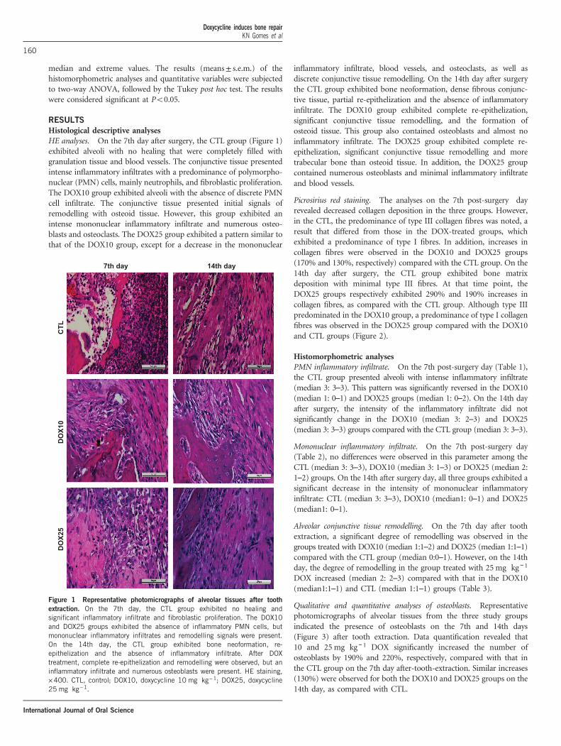
Histological descriptive analysesHE analyses. On the 7th day after surgery, the CTL group (Figure 1)exhibited alveoli with no healing that were completely filled withgranulation tissue and blood vessels. The conjunctive tissue presentedintense inflammatory infiltrates with a predominance of polymorpho-nuclear (PMN) cells, mainly neutrophils, and fibroblastic proliferation.The DOX10 group exhibited alveoli with the absence of discrete PMNcell infiltrate. The conjunctive tissue presented initial signals ofremodelling with osteoid tissue. However, this group exhibited anintense mononuclear inflammatory infiltrate and numerous osteo-blasts and osteoclasts. The DOX25 group exhibited a pattern similar tothat of the DOX10 group, except for a decrease in the mononuclear
inflammatory infiltrate, blood vessels, and osteoclasts, as well asdiscrete conjunctive tissue remodelling. On the 14th day after surgerythe CTL group exhibited bone neoformation, dense fibrous conjunc-tive tissue, partial re-epithelization and the absence of inflammatoryinfiltrate. The DOX10 group exhibited complete re-epithelization,significant conjunctive tissue remodelling, and the formation ofosteoid tissue. This group also contained osteoblasts and almost noinflammatory infiltrate. The DOX25 group exhibited complete re-epithelization, significant conjunctive tissue remodelling and moretrabecular bone than osteoid tissue. In addition, the DOX25 groupcontained numerous osteoblasts and minimal inflammatory infiltrateand blood vessels.
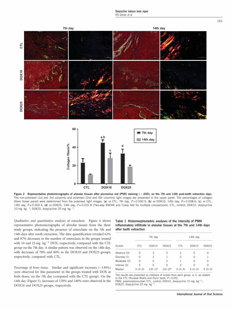
Picrosirius red staining. The analyses on the 7th post-surgery dayrevealed decreased collagen deposition in the three groups. However,in the CTL, the predominance of type III collagen fibres was noted, aresult that differed from those in the DOX-treated groups, whichexhibited a predominance of type I fibres. In addition, increases incollagen fibres were observed in the DOX10 and DOX25 groups(170% and 130%, respectively) compared with the CTL group. On the14th day after surgery, the CTL group exhibited bone matrixdeposition with minimal type III fibres. At that time point, theDOX25 groups respectively exhibited 290% and 190% increases incollagen fibres, as compared with the CTL group. Although type IIIpredominated in the DOX10 group, a predominance of type I collagenfibres was observed in the DOX25 group compared with the DOX10and CTL groups (Figure 2).
Histomorphometric analysesPMN inflammatory infiltrate. On the 7th post-surgery day (Table 1),the CTL group presented alveoli with intense inflammatory infiltrate(median 3: 3–3). This pattern was significantly reversed in the DOX10(median 1: 0–1) and DOX25 groups (median 1: 0–2). On the 14th dayafter surgery, the intensity of the inflammatory infiltrate did notsignificantly change in the DOX10 (median 3: 2–3) and DOX25(median 3: 3–3) groups compared with the CTL group (median 3: 3–3).
Mononuclear inflammatory infiltrate. On the 7th post-surgery day(Table 2), no differences were observed in this parameter among theCTL (median 3: 3–3), DOX10 (median 3: 1–3) or DOX25 (median 2:1–2) groups. On the 14th after surgery day, all three groups exhibited asignificant decrease in the intensity of mononuclear inflammatoryinfiltrate: CTL (median 3: 3–3), DOX10 (median1: 0–1) and DOX25(median1: 0–1).
Alveolar conjunctive tissue remodelling. On the 7th day after toothextraction, a significant degree of remodelling was observed in thegroups treated with DOX10 (median 1:1–2) and DOX25 (median 1:1–1)compared with the CTL group (median 0:0–1). However, on the 14thday, the degree of remodelling in the group treated with 25 mg � kg− 1DOX increased (median 2: 2–3) compared with that in the DOX10(median1:1–1) and CTL (median 1:1–1) groups (Table 3).
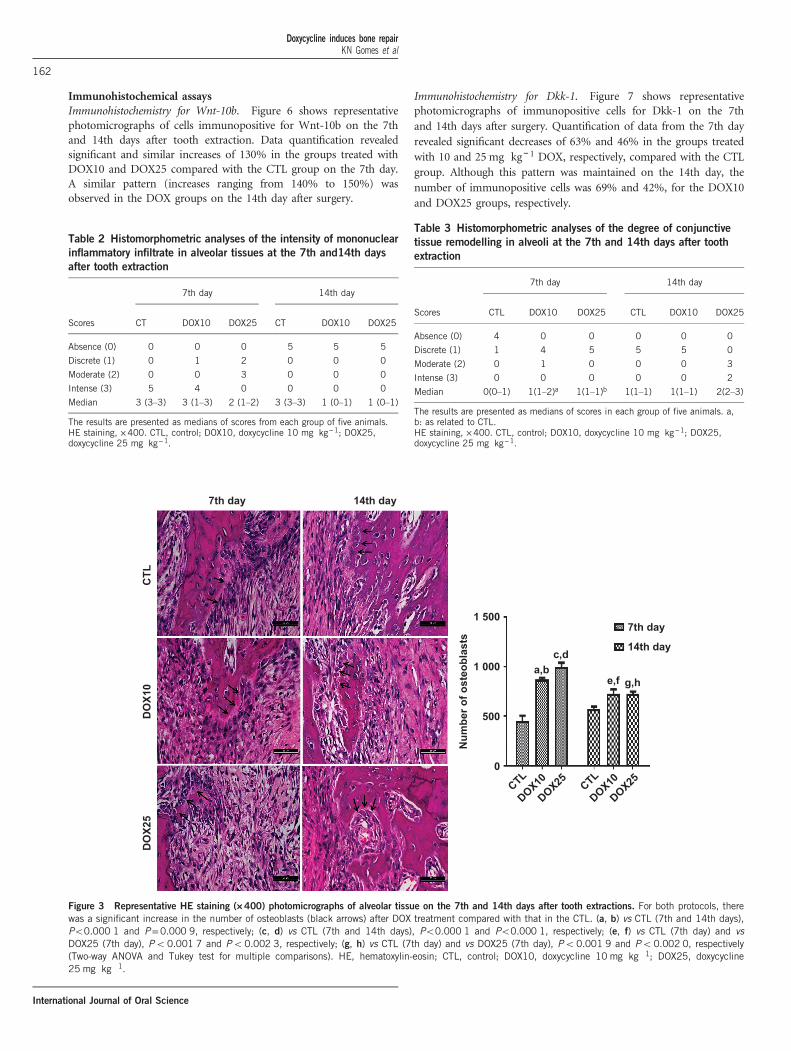
Qualitative and quantitative analyses of osteoblasts. Representativephotomicrographs of alveolar tissues from the three study groupsindicated the presence of osteoblasts on the 7th and 14th days(Figure 3) after tooth extraction. Data quantification revealed that10 and 25 mg � kg− 1 DOX significantly increased the number ofosteoblasts by 190% and 220%, respectively, compared with that inthe CTL group on the 7th day after-tooth-extraction. Similar increases(130%) were observed for both the DOX10 and DOX25 groups on the14th day, as compared with CTL.
CTL
7th day 14th day
DO
X25
DO
X10
Figure 1 Representative photomicrographs of alveolar tissues after tooth
extraction. On the 7th day, the CTL group exhibited no healing andsignificant inflammatory infiltrate and fibroblastic proliferation. The DOX10and DOX25 groups exhibited the absence of inflammatory PMN cells, butmononuclear inflammatory infiltrates and remodelling signals were present.On the 14th day, the CTL group exhibited bone neoformation, re-epithelization and the absence of inflammatory infiltrate. After DOXtreatment, complete re-epithelization and remodelling were observed, but aninflammatory infiltrate and numerous osteoblasts were present. HE staining,×400. CTL, control; DOX10, doxycycline 10 mg � kg−1; DOX25, doxycycline25 mg � kg−1.
Doxycycline induces bone repairKN Gomes et al
160
International Journal of Oral Science
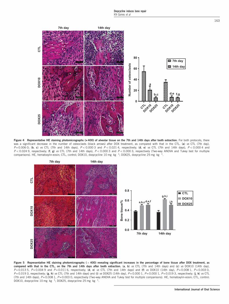
Qualitative and quantitative analyses of osteoclasts. Figure 4 showsrepresentative photomicrographs of alveolar tissues from the threestudy groups, indicating the presence of osteoclasts on the 7th and14th days after tooth extraction. The data quantification revealed 62%and 87% decreases in the number of osteoclasts in the groups treatedwith 10 and 25 mg � kg− 1 DOX, respectively, compared with the CTLgroup on the 7th day. A similar pattern was observed on the 14th day,with decreases of 78% and 80% in the DOX10 and DOX25 groups,respectively, compared with CTL.
Percentage of bone tissue. Similar and significant increases (~130%)were observed for this parameter in the groups treated with DOX atboth doses, on the 7th day (compared with the CTL group). On the14th day (Figure 5), increases of 170% and 140% were observed in theDOX10 and DOX25 groups, respectively.
7th day 14th day
DO
X25
60
a,b
cd
Col
lage
n fib
ers/
% 40
20
0
DO
X10
CTL
DOX25DOX10CTL
7th day
14th day
Figure 2 Representative photomicrographs of alveolar tissues after picrosirius red (PSR) staining ( ´ 200), on the 7th and 14th post-tooth extraction days.
The non-polarised (1st and 3rd columns) and polarised (2nd and 4th columns) light images are presented in the upper panel. The percentages of collagenfibres (lower panel) were determined from the polarised light images. (a) vs CTL, 7th day, P=0.000 5; (b) vs DOX10, 14th day, P=0.008 6; (c) vs CTL,14th day, P=0.005 4; (d) vs DOX25, 14th day, P=0.031 8 (Two-way ANOVA and Tukey test for multiple comparisons). CTL, control; DOX10, doxycycline10 mg � kg�1; DOX25, doxycycline 25 mg � kg�1.
Table 1 Histomorphometric analyses of the intensity of PMN
inflammatory infiltrate in alveolar tissues at the 7th and 14th days
after tooth extraction
7th day 14th day
Scores CTL DOX10 DOX25 CTL DOX10 DOX25
Absence (0) 0 3 2 2 5 4
Discrete (1) 0 2 1 0 0 1
Moderate (2) 0 0 2 1 0 0
Intense (3) 5 0 0 2 0 0
Median 3 (3–3) 1(0–1)a 1(0–2)b 3 (3–3) 3 (2–3) 3 (3–3)
The results are presented as medians of scores from each group. a, b: as relatedto the CTL (Kruskal–Wallis and Dunn tests, Po0.05).PMN, polymorphonuclear; CTL, control; DOX10, doxycycline 10 mg � kg−1;DOX25, doxycycline 25 mg � kg−1.
Doxycycline induces bone repairKN Gomes et al
161
International Journal of Oral Science
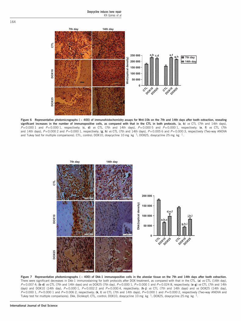
Immunohistochemical assaysImmunohistochemistry for Wnt-10b. Figure 6 shows representativephotomicrographs of cells immunopositive for Wnt-10b on the 7thand 14th days after tooth extraction. Data quantification revealedsignificant and similar increases of 130% in the groups treated withDOX10 and DOX25 compared with the CTL group on the 7th day.A similar pattern (increases ranging from 140% to 150%) wasobserved in the DOX groups on the 14th day after surgery.
Immunohistochemistry for Dkk-1. Figure 7 shows representativephotomicrographs of immunopositive cells for Dkk-1 on the 7thand 14th days after surgery. Quantification of data from the 7th dayrevealed significant decreases of 63% and 46% in the groups treatedwith 10 and 25 mg � kg− 1 DOX, respectively, compared with the CTLgroup. Although this pattern was maintained on the 14th day, thenumber of immunopositive cells was 69% and 42%, for the DOX10and DOX25 groups, respectively.
Table 2 Histomorphometric analyses of the intensity of mononuclear
inflammatory infiltrate in alveolar tissues at the 7th and14th days
after tooth extraction
7th day 14th day
Scores CT DOX10 DOX25 CT DOX10 DOX25
Absence (0) 0 0 0 5 5 5
Discrete (1) 0 1 2 0 0 0
Moderate (2) 0 0 3 0 0 0
Intense (3) 5 4 0 0 0 0
Median 3 (3–3) 3 (1–3) 2 (1–2) 3 (3–3) 1 (0–1) 1 (0–1)
The results are presented as medians of scores from each group of five animals.HE staining, ×400. CTL, control; DOX10, doxycycline 10 mg � kg−1; DOX25,doxycycline 25 mg � kg−1.
Table 3 Histomorphometric analyses of the degree of conjunctive
tissue remodelling in alveoli at the 7th and 14th days after tooth
extraction
7th day 14th day
Scores CTL DOX10 DOX25 CTL DOX10 DOX25
Absence (0) 4 0 0 0 0 0
Discrete (1) 1 4 5 5 5 0
Moderate (2) 0 1 0 0 0 3
Intense (3) 0 0 0 0 0 2
Median 0(0–1) 1(1–2)a 1(1–1)b 1(1–1) 1(1–1) 2(2–3)
The results are presented as medians of scores in each group of five animals. a,b: as related to CTL.HE staining, ×400. CTL, control; DOX10, doxycycline 10 mg � kg−1; DOX25,doxycycline 25 mg � kg−1.
7th day
1 5007th day
14th day
Num
ber o
f ost
eobl
asts
1 000 a,bc,d
e,f g,h
500
0
14th day
DO
X25
DO
X10
CTL
DOX25
DOX10CTL
DOX25
DOX10CTL
Figure 3 Representative HE staining (×400) photomicrographs of alveolar tissue on the 7th and 14th days after tooth extractions. For both protocols, therewas a significant increase in the number of osteoblasts (black arrows) after DOX treatment compared with that in the CTL. (a, b) vs CTL (7th and 14th days),Po0.000 1 and P=0.000 9, respectively; (c, d) vs CTL (7th and 14th days), Po0.000 1 and Po0.000 1, respectively; (e, f) vs CTL (7th day) and vsDOX25 (7th day), Po0.001 7 and Po0.002 3, respectively; (g, h) vs CTL (7th day) and vs DOX25 (7th day), Po0.001 9 and Po0.002 0, respectively(Two-way ANOVA and Tukey test for multiple comparisons). HE, hematoxylin-eosin; CTL, control; DOX10, doxycycline 10 mg � kg�1; DOX25, doxycycline25 mg � kg�1.
Doxycycline induces bone repairKN Gomes et al
162
International Journal of Oral Science
7th day 14th day
CTL
DO
X25
DO
X10
7th day
CTL
DOX10
DOX25 CTL
DOX10
DOX25
80
Num
ber o
f ost
eocl
asts
60
a
b,c d,e f,g
40
20
0
14th day
Figure 4 Representative HE staining photomicrographs (×400) of alveolar tissue on the 7th and 14th days after tooth extraction. For both protocols, therewas a significant decrease in the number of osteoclasts (black arrows) after DOX treatment, as compared with that in the CTL. (a) vs CTL (7th day),P=0.006 0; (b, c) vs CTL (7th and 14th days), Po0.000 3 and Po0.021 4, respectively; (d, e) vs CTL (7th and 14th days), Po0.000 4 andPo0.024 9, respectively; (f, g) vs CTL (7th and 14th days), Po0.000 3 and Po0.000 3, respectively (Two-way ANOVA and Tukey test for multiplecomparisons). HE, hematoxylin-eosin; CTL, control; DOX10, doxycycline 10 mg � kg�1; DOX25, doxycycline 25 mg � kg�1.
7th day
0.6
0.8
a,b,c d,e,f
g,h,iCTLDOX10
DOX25j,k
Bon
e tis
sue/
%
0.4
0.2
0.0
14th day
7th day 14th day
DO
X25
DO
X10
CTL
Figure 5 Representative HE staining photomicrographs ( ´ 400) revealing significant increases in the percentage of bone tissue after DOX treatment, as
compared with that in the CTL, on the 7th and 14th days after tooth extraction. (a, b) vs CTL (7th and 14th days) and (c) vs DOX10 (14th day),P=0.013 5, P=0.004 9 and P=0.011 6, respectively; (d, e) vs CTL (7th and 14th days) and (f) vs DOX10 (14th day), P=0.008 1, P=0.003 0,P=0.019 3, respectively; (g, h) vs CTL (7th and 14th days) and (i) vs DOX25 (14th day), P=0.000 1, P=0.000 1, P=0.019 3, respectively; (j, k) vs CTL(7th and 14th days), P=0.008 1, P=0.003 0, respectively (Two-way ANOVA and Tukey test for multiple comparisons). HE, hematoxylin-eosin; CTL, control;DOX10, doxycycline 10 mg � kg�1; DOX25, doxycycline 25 mg � kg�1.
Doxycycline induces bone repairKN Gomes et al
163
International Journal of Oral Science
7th day
250 000 a,b c,d e,f g,h
Wnt
/(opt
ical
den
sity
)
200 000
150 000
100 000
50 000
0
14th day
7th day14th day
DO
X25
DO
X10
CTL
DOX25
DOX10CTL
DOX25
DOX10CTL
Figure 6 Representative photomicrographs ( ´ 400) of immunohistochemistry assays for Wnt-10b on the 7th and 14th days after tooth extraction, revealing
significant increases in the number of immunopositive cells, as compared with that in the CTL in both protocols. (a, b) vs CTL (7th and 14th days),P=0.000 1 and P=0.000 1, respectively; (c, d) vs CTL (7th and 14th days), P=0.000 5 and P=0.000 1, respectively; (e, f) vs CTL (7thand 14th days), P=0.000 2 and P=0.000 1, respectively; (g, h) vs CTL (7th and 14th days), P=0.005 6 and P=0.000 3, respectively (Two-way ANOVAand Tukey test for multiple comparisons). CTL, control; DOX10, doxycycline 10 mg � kg�1; DOX25, doxycycline 25 mg � kg�1.
DO
X25
DO
X10
CTL
DOX25
DOX10CTL
DOX25
DOX10CTL
7th day 14th day
200 000
150 000
100 000
Dkk
/(opt
ical
den
sity
)
50 000
a,b,c
d,e,f
g,h,i
j,k,l
0
Figure 7 Representative photomicrographs ( ´ 400) of Dkk-1 immunopositive cells in the alveolar tissue on the 7th and 14th days after tooth extraction.
There were significant decreases in Dkk-1 immunostaining for both protocols after DOX treatment, as compared with that in the CTL. (a) vs CTL (14th day),P=0.007 4; (b–d) vs CTL (7th and 14th days) and vs DOX25 (7th day), P=0.000 1, P=0.000 1 and P=0.024 8, respectively; (e–g) vs CTL (7th and 14thdays) and DOX10 (14th day), P=0.000 1, P=0.002 2 and P=0.000 4, respectively; (h–j) vs CTL (7th and 14th days) and vs DOX25 (14th day),P=0.000 1, P=0.000 1 and P=0.006 2, respectively; (k, l) vs CTL (7th and 14th days), P=0.000 1 and P=0.000 2, respectively (Two-way ANOVA andTukey test for multiple comparisons). Dkk, Dickkopf; CTL, control; DOX10, doxycycline 10 mg � kg�1; DOX25, doxycycline 25 mg � kg�1.
Doxycycline induces bone repairKN Gomes et al
164
International Journal of Oral Science
DISCUSSION
In the present work, DOX significantly decreased PMN and mono-nuclear inflammatory infiltrates in alveolus tissue on the 7th and 14thdays after tooth extraction in the model of bone repair in rats. Wehave previously demonstrated that DOX presents potent anti-inflammatory and antioxidant effects.20 These properties are associatedwith inhibition of iNOS and TNF-alpha and are probably related toDOX’s actions on alveolar tissue inflammatory infiltrates, as shown inthe present work.At the lower dose, the DOX group, compared with the CTL group,
exhibited significant increases in collagen fibres on the 7th and 14thdays after tooth extraction, thus indicating better organisation andbone neoformation, as evaluated by PSR staining. Although nodifference in the percentage of collagen fibres was observed betweenthe DOX25 and CTL groups, the DOX25 group presented a significantnumber of type I collagen fibres. Type I collagen is primarily observedin bone, dentin, tendon, dermis and gingival tissues and is also themain extracellular component.21–23 Specific staining of the extracel-lular matrix components with PSR is especially helpful in studyingtissue remodelling.24 The stain specifically binds to collagen fibrils ofvarying diameters, and was thus used to distinguish type I from typeIII collagen fibres.The enhancement of birefringence promoted by the PSR-
polarisation method is therefore specific for collagenous structurescomposed of aggregates of oriented molecules.25 A previous study 26
has demonstrated an increase in collagen fibre organisation over time.In addition, the authors have observed collagen maturation over 3, 7and 14 days during orthodontic tooth movements in rats. DOXinhibits MMP enzymes and prevents bone loss.27 In addition, byincreasing type I collagen fibres, DOX accelerates collagen maturationand bone repair, as observed in the present study.MMPs are a family of enzymes that play a key role in maintaining
and remodelling the extracellular matrix of connective tissue.28
Recently, low doses of orally administered DOX for 7 days have beenfound to enhance bone formation in the alveolar sockets of rats.29 Thebone matrix is composed primarily of type I collagen fibres, which area marker of osteoblast differentiation. Bone strength depends on morethan quantity and mainly depends on quality, which is characterisedby the geometry and shape of bones, the microarchitecture oftrabecular bone, bone turnover, and the presence of minerals andcollagen.30
We demonstrated that at both 10 and 25 mg � kg− 1 doses, DOXsignificantly and dose-dependently decreased the number of osteo-clasts, as compared with the CTL group on the 7th day post-surgery. Asimilar pattern was also observed on the 14th day. DOX inducesosteoclasts apoptosis, which occurs independently of the inhibition ofMMPs.31 Furthermore, DOX decreases mononuclear inflammatoryinfiltrates and osteoclast numbers, thereby preventing inflammatorybone resorption.32
Previously,33 fewer osteoclasts have been found to be present inDOX-treated animals in a surgically induced osteoclast recruitmentmodel. Later evidence has indicated significant decreases in rootresorption and the numbers of odontoclasts, osteoclasts and mono-nuclear cells on the root surface of DOX-treated rats.34 The authorsconcluded that DOX, at low doses, may have an inhibitory effect onorthodontically induced resorptive activity. Furthermore, DOX hasbeen shown to effectively inhibit osteoclastogenesis and to affectmature osteoclast fate.35
In contrast, we demonstrated that DOX significantly and dose-dependently increased the number of osteoblasts, as compared withthat in the CTL group, on the 7th day. Although an increase was also
observed on the 14th day post-surgery, the effect was smaller, and nosignificant differences were observed among groups. Long-termexposure of human bone marrow osteoblastic cells to DOX inducesa significant increase in the number of osteoblastic cells, therebyyielding a proportional amount of normal mineralised matrix.36 Theseeffects suggest that this drug may potentially be used to increase boneformation. Other in vitro results have indicated the effect of DOX onosteoblastic proliferation and differentiation.37 The authors of thatstudy have concluded that DOX appears to enhance maturation anddifferentiation, rather than proliferation, and may thus be beneficial inthe treatment of periodontal disease and periodontal regeneration.Wnt proteins play a very important role in several mammalian
physiological processes, including embryogenesis, organ developmentand regeneration, and cell migration and proliferation.38 Osteogenesisis induced in response to bone morphogenic protein 2 (BMP2)stimulation and is sustained by Wnt signalling. However, the presenceof Dkk-1, an inhibitor of Wnt signalling, results in osteogenesisinhibition, and during the repair process, the expression of many Wntligands and receptors is upregulated.39–40 Wnt pathways regulate bonemass and are active during fracture repair, and an increase in theiractivity accelerates bone regeneration.41 Thus, the activation of Wntpathways has the potential to improve bone healing, and itsdysregulation greatly inhibits bone formation and healingprocesses.17
Evidence from preclinical studies has indicated that Dkk neutraliza-tion and/or Wnt signalling enhancement may prove effective in thetreatment of bone pathologies.42 In addition, Wnt signalling by itself isa very attractive target for therapeutic interventions related to skeletalhomoeostasis and bone repair.43 Thus, as we have shown here, Wntsignalling may be a potential target for DOX, and this result maystimulate translational studies towards including this drug in the clinicin the near future.In the present work, we provided the first evidence that DOX
inhibits the Dkk-1 pathway while activating Wnt signalling. Theseresults were immunohistochemically evaluated through both theincrease of Wnt-10b and the decrease in Dkk-1 in immunostainedcells on the 7th and 14th days after tooth extraction. Although theexact mechanism of Wnt signalling during bone development isdependent on a complex microenvironment, data from severallaboratories suggest that Wnt-3a, Wnt-5a, Wnt-7b and Wnt-10b arecentral to osteoblast differentiation.44
Clinical and experimental studies suggest a clear role for Wntsignalling in the regulation of bone formation, repair and remodelling.Thus, Wnt pathway activation accelerates bone regeneration.45 How-ever, Dkk-1 counteracts Wnt-mediated effects on bone differentiationand adipogenesis. Thus, the neutralization of inhibitors of Wntsignalling, such as Dkk-1, is a promising therapeutic strategy in bonediseases.46
Wnt signalling is widely accepted to be required for the differentia-tion of osteoprogenitors into osteoblasts, and the Wnt pathwayantagonist Dkk-1 causes bone destruction and the inhibition of bonerepair.47 Recent evidence has indicated that selective inhibition of theWnt pathway by Dkk-1 decreases osteoarthritis in mice.48 Further-more, diseases such as periodontitis are characterised by inflammationand bone loss, and the activation of PMN cells leads to the release ofproinflammatory cytokines and the recruitment of phagocytes andlymphocytes.49 Previously, we have demonstrated that DOX haspotent anti-inflammatory activity.20 In addition, minocycline, thesecond generation tetracycline similar to DOX, offers protection in amodel of periodontal disease in normal and diabetic rats(Ph.D. thesis,data not published).
Doxycycline induces bone repairKN Gomes et al
165
International Journal of Oral Science
CONCLUSIONS
In the present study, we demonstrated that owing to its ability toincrease osteoblast numbers and decrease osteoclast numbers, butmainly through its activation of Wnt-1b and neutralization of Dkk-1,DOX may be a potential candidate for use in bone repair in severalpathologies, including periodontal diseases.
ACKNOWLEDGEMENTS
We are grateful for the financial support of the Brazilian National ResearchCouncil (CNPq) and the technical assistance of Ms. Maria Vilani RodriguesBastos.
1 Yang XH, Qin L, Liang WG et al. New bone formation and microstructure assessed bycombination of confocal laser scanning microscopy and differential interferencecontrast microscopy. Calcif Tissue Int 2014; 94(3): 338–347.
2 Tsiridis E, Upadhyay N, Giannoudis P. Molecular aspects of fracture healing: which arethe important molecules? Injury 2007; 38(Suppl 1): S11–S25.
3 Lin Z, Rios HF, Volk SL et al. Gene expression dynamics during bone healing andosseointegration. J Periodontol 2011; 82(7): 1007–1017.
4 Pagni G, Pellegrini G, Giannobile WV et al. Postextraction alveolar ridge preservation:biological basis and treatments. Int J Dent 2012; 2012: 151030.
5 Tan WL, Wong TL, Wong MC et al. A systematic review of post-extractional alveolar hardand soft tissue dimensional changes in humans. Clin Oral Implants Res 2012;23(Suppl 5): 1–21.
6 Vieira AE, Repeke CE, Ferreira SB Jr et al. Intramembranous bone healingprocess subsequent to tooth extraction in mice: micro-computed tomography,histomorphometric and molecular characterization. PLoS One 2015; 10(5):e0128021.
7 Nip LH, Uitto VJ, Golub LM. Inhibition of epithelial cell matrix metalloproteinases bytetracyclines. J Periodontal Res 1993; 28(5): 379–385.
8 Uitto VJ, Firth JD, Nip L et al. Doxycycline and chemically modified tetracyclines inhibitgelatinase A (MMP-2) gene expression in human skin keratinocytes. Ann N Y Acad Sci1994; 732: 140–151.
9 Hanemaaijer R, Visser H, Koolwijk P et al. Inhibition of MMP synthesis by doxycyclineand chemically modified tetracyclines (CMTs) in human endothelial cells. Adv Dent Res1998; 12(2): 114–118.
10 Sadowski T, Steinmeyer J. Effects of tetracyclines on the production of matrixmetalloproteinases and plasminogen activators as well as of their natural inhibitors,tissue inhibitor of metalloproteinases-1 and plasminogen activator inhibitor-1. InflammRes 2001; 50(3): 175–182.
11 Gorustovich AA, Steimetz T, Nielsen FH et al. Histomorphometric study of alveolar bonehealing in rats fed a boron-deficient diet. Anat Rec 2008; 291(4): 441–447.
12 Pasternak B, Fellenius M, Aspenberg P. Doxycycline impairs tendon repair in rats. ActaOrthop Belg 2006; 72(6): 756–760.
13 Stechmiller J, Cowan L, Schultz G. The role of doxycycline as a matrix metalloproteinaseinhibitor for the treatment of chronic wounds. Biol Res Nurs 2010; 11(4): 336–344.
14 Wilcox JR, Covington DS, Paez N. Doxycycline as a modulator of inflammation inchronic wounds. Wounds 2012; 24(12): 339–349.
15 Grevstad HJ. Doxycycline prevents root resorption and alveolar bone loss in rats afterperiodontal surgery. Scand J Dent Res 1993; 101(5): 287–291.
16 Minear S, Leucht P, Jiang J et al. Wnt proteins promote bone regeneration. Sci TranslMed 2010; 2(29): 29ra30.
17 Chen Y, Alman BA. Wnt pathway, an essential role in bone regeneration. J Cell Biochem2009; 106(3): 353–362.
18 Whyte JL, Smith AA, Helms JA. Wnt signaling and injury repair. Cold Spring HarbPerspect Biol 2012; 4(8): a008078.
19 Kim JH, Liu X, Wang JH et al. Wnt signaling in bone formation and its therapeuticpotential for bone diseases. Ther Adv Musculoskelet Dis 2013; 5(1): 13–31.
20 Leite LM, Carvalho AG, Ferreira PL et al. Anti-inflammatory properties of doxycyclineand minocycline in experimental models: an in vivo and in vitro comparative study.Inflammopharmacology 2011; 19 (2): 99–110.
21 Junqueira LC, Carneiro J. Histologia Básica. 8th ed. Rio de Janeiro: Guanabara Koogan,1995.
22 Tandelilin RTC. Augmentation of demineralized bone matrix post-tooth extractionincreases the density of gingival collagen fiber of rabbit mandible. Indonesian J DentRes 2010; 1(1): 9–16.
23 Kaku M, Yamauchi M. Mechano-regulation of collagen biosynthesis in periodontalligament. J Prosthodont Res 2014; 58(4): 193–207.
24 Lattouf R, Younes R, Lutomski D et al. Picrosirius red staining: a useful tool to appraisecollagen networks in normal and pathological tissues. J Histochem Cytochem 2014;62(10): 751–758.
25 Montes GS, Junqueira LC. The use of the Picrosirius-polarization method for the studyof the biopathology of collagen. Mem Inst Oswaldo Cruz 1991; 86(Suppl 3): 1–11.
26 Retamoso LB, Da Cunha T de M, Knop LA et al. Organization and quantification of thecollagen fibers in bone formation during orthodontic tooth movement. Micron 2009;40(8): 827–830.
27 Kinugawa S, Koide M, Kobayashi Y et al. Tetracyclines convert the osteoclastic-differentiation pathway of progenitor cells to produce dendritic cell-like cells. J Immunol2012; 188(4): 1772–1781.
28 Bedi A, Fox AJ, Kovacevic D et al. Doxycycline-mediated inhibition of matrixmetalloproteinases improves healing after rotator cuff repair. Am J Sports Med 2010;38(2): 308–317.
29 Shahabooei M, Razavi SM, Minaiyan M et al. A histomorphometric study of the effect ofdoxycycline and erythromycin on bone formation in dental alveolar socket of rat. AdvBiomed Res 2015; 4: 71.
30 Viguet-Carrin S, Garnero P, Delmas PD. The role of collagen in bone strength.Osteoporos Int 2006; 17(3): 319–336.
31 Holmes SG, Still K, Buttle DJ et al. Chemically modified tetracyclines act throughmultiple mechanisms directly on osteoclast precursors. Bone 2004; 35(2): 471–478.
32 Bezerra MM, Brito GA, Ribeiro RA et al. Low-dose doxycycline prevents inflammatorybone resorption in rats. Braz J Med Biol Res 2002; 35(5): 613–616.
33 Grevstad HJ, Bøe OE. Effect of doxycycline on surgically induced osteoclast recruitmentin the rat. Eur J Oral Sci 1995; 103(3): 156–159.
34 Mavragani M, Brudvik P, Selvig KA. Orthodontically induced root and alveolar boneresorption: inhibitory effect of systemic doxycycline administration in rats. Eur J Orthod2005; 27(3): 215–225.
35 Zhang C, Tang TT, Ren WP et al. Inhibiting wear particles-induced osteolysis withdoxycycline. Acta Pharmacol Sin 2007; 28(10): 1603–1610.
36 Gomes PS, Fernandes MH. Effect of therapeutic levels of doxycycline and minocyclinein the proliferation and differentiation of human bone marrow osteoblastic cells. ArchOral Biol 2007; 52(3): 251–259.
37 Almazin SM, Dziak R, Andreana S et al. The effect of doxycycline hyclate, chlorhexidinegluconate, and minocycline hydrochloride on osteoblastic proliferation and differentia-tion in vitro. J Periodontol 2009; 80(6): 999–1005.
38 Moon RT, Bowerman B, Boutros M et al. The promise and perils of Wnt signalingthrough beta-catenin. Science 2002; 296(5573): 1644–1646.
39 Luu HH, Song WX, Luo XJ et al. Distinct roles of bone morphogenetic proteins inosteogenic differentiation of mesenchymal stem cells. J Orthopaedic Res 2007; 25(5):665–677.
40 Xu HYN, Duan J, Ning DD et al. Role of Wnt signaling in fracture healing. BMB Rep2014; 47(12): 666–672.
41 Secreto FJ, Hoeppner LH, Westendorf JJ. Wnt signaling during fracture repair. CurrOsteoporos Rep 2009; 7(2): 64–69.
42 Gregory CA, Gunn WG, Reyes E et al. How Wnt signaling affects bone repair bymesenchymal stem cells from the bone marrow. Ann N Y Acad Sci 2005; 1049:97–106.
43 Pinzone JJ, Hall BM, Thudi NK et al. The role of Dickkopf-1 in bone development,homeostasis, and disease. Blood 2009; 113(3): 517–525.
44 Leucht P, Helms JA. Wnt signaling: an emerging target for bone regeneration. J AmAcad Orthop Surg 2015; 23(1): 67–68.
45 Issack PS, Helfet DL, Lane JM. Role of Wnt signaling in bone remodeling and repair.HSS J 2008; 4(1): 66–70.
46 Hoeppner LH, Secreto FJ, Westendorf JJ. Wnt signaling as a therapeutic target for bonediseases. Expert Opin Ther Targets 2009; 13(4): 485–496.
47 Krause U, Ryan DM, Clough BH et al. An unexpected role for a Wnt-inhibitor: Dickkopf-1 trigers a novel cancer survival mechanism through modulation of aldehyde-dehydrogenase-1 activity. Cell Death Dis 2014; 5: e1093.
48 Funck-Brentano T, Bouaziz W, Marty C et al. Dkk-1-mediated inhibition of Wntsignaling in bone ameliorates osteoarthritis in mice. Arthritis Rheum 2014; 66(11):3028–3039.
49 Benedetto AD, Gigante I, Colucci S et al. Periodontal disease: linking the primaryinflammation to bone loss. Clin Dev Biol 2013; 2013: 503754.
This work is licensed under a Creative Commons Attribution 4.0International License. The images or other third party material in this
article are included in the article’s Creative Commons license, unless indicated otherwisein the credit line; if thematerial isnot includedunder theCreativeCommons license,userswill need toobtainpermission fromthe licenseholder to reproduce thematerial. Toviewacopy of this license, visit http://creativecommons.org/licenses/by/4.0/
r The Author(s) 2017
Doxycycline induces bone repairKN Gomes et al
166
International Journal of Oral Science
![Aditya Ramamoorthy Vwani Roychowdhury Sudhir Kumar Singh ... · arXiv:0804.1840v2 [cs.GT] 1 Mar 2009 Selfish Distributed Compression over Networks: Correlatio n Induces Anarchy Aditya](https://static.fdocuments.nl/doc/165x107/5fc709302f9f8c342f71fcb2/aditya-ramamoorthy-vwani-roychowdhury-sudhir-kumar-singh-arxiv08041840v2-csgt.jpg)