
Cátia Moreira, Maria J. Ramos * and Pedro A. Fernandes
26
molecules Article Glutamine Synthetase Drugability beyond Its Active Site: Exploring Oligomerization Interfaces and Pockets Cátia Moreira, Maria J. Ramos * and Pedro A. Fernandes UCIBIO, REQUIMTE, Departamento de Química e Bioquímica, Faculdade de Ciências, Universidade do Porto, 4169-007 Porto, Portugal; [email protected] (C.M.); [email protected] (P.A.F.) * Correspondence: [email protected]; Tel.: +351-220-408-017 Academic Editors: James W. Gauld and Leif A. Eriksson Received: 8 June 2016; Accepted: 4 August 2016; Published: 8 August 2016 Abstract: Background: Glutamine synthetase (GS) is a crucial enzyme to the nitrogen cycle with great commercial and pharmaceutical value. Current inhibitors target the active site, affecting GS activity indiscriminately in all organisms. As the active site is located at the interface between two monomers, the protein-protein interface (PPI) of GSs gains a new role, by providing new targets for enzyme inhibition. Exploring GSs PPI could allow for the development of inhibitors selective for specific organisms. Here we map the PPI of three GSs—human (hsGS), maize (zmGS) and Mycobacterium tuberculosis (mtGS)—and unravel new drugable pockets. Methods: The PPI binding free energy coming from key residues on three GSs from different organisms were mapped by computational alanine scan mutagenesis, applying a multiple dielectric constant MM-PBSA methodology. The most relevant residues for binding are referred as hot-spots. Drugable pockets on GS were detected with the Fpocket software. Results and Conclusions: A total of 23, 19 and 30 hot-spots were identified on hsGS, zmGS and mtGS PPI. Even possessing differences in the hot-spots, hsGS and zmGS PPI are overall very similar. On the other hand, mtGS PPI differs greatly from hsGS and zmGS PPI. A novel drugable pocket was detected on the mtGS PPI. It seems particularly promising for the development of selective anti-tuberculosis drugs given its location on a PPI region that is highly populated with hot-spots and is completely different from the hsGS and zmGS PPIs. Drugs targeting this pockets should be inactive on eukaryotic GS II enzymes. Keywords: protein-protein interactions; computational alanine scanning mutagenesis; drugable pockets; glutamine synthetase; Mycobacterium tuberculosis; human; maize; inhibition; drug discovery 1. Introduction Glutamine Synthetase (GS, EC 6.3.1.2, also known as γ-glutamyl:ammonia ligase) is a widely spread metalloenzyme. Together with glutamate synthetase, GS plays a crucial role in the nitrogen and carbon cycle, actively controlling the levels of inorganic nitrogen incorporated into organic molecules by performing key reactions that influence both assimilation and ammonification processes. Various reactions are catalysed by GS. The most important and well-studied is the ATP-dependent synthesis of glutamine by combining glutamate and ammonium, commonly denominated as the biosynthetic reaction [1–4]. Different isoforms of this enzyme are expressed, varying in both sequence and structure. They are classified into three categories: GS I, with 450 to 470 residues per monomer, combined into homododecameric oligomers with 622 symmetry (two rings superimposed, each with 6 monomers); GS II, with 350 to 420 residues per monomer, arranged in homodecameric oligomers with 522 symmetry (two rings superimposed, each with five monomers); and GS III, containing around 700 residues per Molecules 2016, 21, 1028; doi:10.3390/molecules21081028 www.mdpi.com/journal/molecules
Transcript of Cátia Moreira, Maria J. Ramos * and Pedro A. Fernandes
molecules
Article
Glutamine Synthetase Drugability beyond Its ActiveSite: Exploring Oligomerization Interfacesand PocketsCátia Moreira, Maria J. Ramos * and Pedro A. Fernandes
UCIBIO, REQUIMTE, Departamento de Química e Bioquímica, Faculdade de Ciências, Universidade do Porto,4169-007 Porto, Portugal; [email protected] (C.M.); [email protected] (P.A.F.)* Correspondence: [email protected]; Tel.: +351-220-408-017
Academic Editors: James W. Gauld and Leif A. ErikssonReceived: 8 June 2016; Accepted: 4 August 2016; Published: 8 August 2016
Abstract: Background: Glutamine synthetase (GS) is a crucial enzyme to the nitrogen cycle withgreat commercial and pharmaceutical value. Current inhibitors target the active site, affecting GSactivity indiscriminately in all organisms. As the active site is located at the interface betweentwo monomers, the protein-protein interface (PPI) of GSs gains a new role, by providing newtargets for enzyme inhibition. Exploring GSs PPI could allow for the development of inhibitorsselective for specific organisms. Here we map the PPI of three GSs—human (hsGS), maize (zmGS)and Mycobacterium tuberculosis (mtGS)—and unravel new drugable pockets. Methods: The PPIbinding free energy coming from key residues on three GSs from different organisms were mappedby computational alanine scan mutagenesis, applying a multiple dielectric constant MM-PBSAmethodology. The most relevant residues for binding are referred as hot-spots. Drugable pockets onGS were detected with the Fpocket software. Results and Conclusions: A total of 23, 19 and 30 hot-spotswere identified on hsGS, zmGS and mtGS PPI. Even possessing differences in the hot-spots, hsGS andzmGS PPI are overall very similar. On the other hand, mtGS PPI differs greatly from hsGS and zmGSPPI. A novel drugable pocket was detected on the mtGS PPI. It seems particularly promising forthe development of selective anti-tuberculosis drugs given its location on a PPI region that is highlypopulated with hot-spots and is completely different from the hsGS and zmGS PPIs. Drugs targetingthis pockets should be inactive on eukaryotic GS II enzymes.
Keywords: protein-protein interactions; computational alanine scanning mutagenesis; drugablepockets; glutamine synthetase; Mycobacterium tuberculosis; human; maize; inhibition; drug discovery
1. Introduction
Glutamine Synthetase (GS, EC 6.3.1.2, also known as γ-glutamyl:ammonia ligase) is a widelyspread metalloenzyme. Together with glutamate synthetase, GS plays a crucial role in the nitrogenand carbon cycle, actively controlling the levels of inorganic nitrogen incorporated into organicmolecules by performing key reactions that influence both assimilation and ammonification processes.Various reactions are catalysed by GS. The most important and well-studied is the ATP-dependentsynthesis of glutamine by combining glutamate and ammonium, commonly denominated as thebiosynthetic reaction [1–4].
Different isoforms of this enzyme are expressed, varying in both sequence and structure.They are classified into three categories: GS I, with 450 to 470 residues per monomer, combined intohomododecameric oligomers with 622 symmetry (two rings superimposed, each with 6 monomers);GS II, with 350 to 420 residues per monomer, arranged in homodecameric oligomers with 522 symmetry(two rings superimposed, each with five monomers); and GS III, containing around 700 residues per
Molecules 2016, 21, 1028; doi:10.3390/molecules21081028 www.mdpi.com/journal/molecules
Molecules 2016, 21, 1028 2 of 26
monomer, also organized in homodecameric oligomers (two rings superimposed, each with fivemonomers) but with the inter-ring interface head to head (instead of tail to tail, as found in GS I andGS II). Reports of nanotube-like supramolecular assemblies are also mentioned in the literature [5,6].
At first, it was believed that GS I was exclusive from prokaryotes and GS II was mostly found ineukaryotes, being GS III restricted to a few organisms from both kingdoms [7–9]. Today it is knownthat a single organism can encode more than one class of GS [9], and that the distinctive line whichdistributes GS classes in different kingdoms is more blurred.
The initial studies of GS were conducted only on GS I, more abundant in bacteria and easierto study, with the intent of gathering knowledge on the function of this rather important enzyme.Later on, GS studies began to bisect into two main areas of research: agriculture and medicine.In the agricultural field, studies aim to unravel GS II features, allowing for the improvement of cropsyields, accomplished by using GS inhibitors as herbicides together with genetically modified cropsresistant to the herbicide. Efforts have been pursued also to enhance the GS activity to improvecrops yields by an increased assimilation of nitrogen (or to achieve the same yield with the use ofsmaller amounts of fertilizers). An example of this is the herbicide phosphinotricin, also known asglufosinate or BASTA®. Glufosinate, together with glyphosate, is one of the cornerstones of the billiondollar industry of genetically modified resistant to herbicides (GM-HR) crops [10]. Glufosinate isas large-spectrum herbicide used on weed control, which can be combined with GM-HR crops thattolerate this herbicide. However, since it can inhibit the GSs from any organism, their use to increasecrop productivity is only confined to resistant crops. Additionally, there are major concerns abouttheir safety. Evidences indicate that glufosinate may be a major danger with both environmentaland human health repercussions [11–16]. In the pharmaceutical field, studies aimed to differentiatebacterial GS I from mammal GS II have been carried out with the expectation of developing newantibiotics, especially against tuberculosis where GS has proven, in vitro, to be an excellent target foranti-tuberculosis drug development [17–21]. Multiple efforts have been done in the last two decades toattain novel inhibitors of GS that can act as anti-tuberculosis therapeutic agents. A recent review on thistopic, by Mowbray and co-workers [17], describes the inhibitors currently targeting mtGS. They canbe divided into two categories, depending on the binding location. The first category encompassesamino acids analogues, like MSO or PPT, that act as suicide inhibitors that compete for the bindingsite of glutamate/glutamine, and once bound to GS become phosphorylated. Since this region ofthe active site is highly conserved amongst all GS, this inhibitors raise concerns about their safety.The second category is dedicated to inhibitors targeting the ATP binding site, which normally arebigger hydrophobic heterocycles that bind to a larger and less conserved region of the active site,aiming more selective inhibition than inhibitors such PPT or MSO. Inhibitors from both categorieshave been already co-crystalized with mtGS, providing insights about their action mechanism [21–26].Either way, all of them bind in the active site or its vicinity. But caution is needed when developingdrugs targeting GS inhibition, since irregularities in human GS activity/expression are associated withseveral diseases and its deficiency leads to neonatal death caused by multi-organ failure [27–29].
Since GSs possess such a variety of roles, so important in metabolism, they need to be subjectedto tight regulation, acting on many levels, from the gene expression to enzymes modulation. There areinsightful reviews on this matter for bacterial GS (mostly E. coli [5]) and plants GS [30,31]. In mammals,GS has distinct roles in the metabolism, depending on the tissue location: in the brain, it regulates thelevels of toxic ammonia and neurotoxic glutamate, and in the liver it participates in the eliminationof ammonia. There are several studies trying to create an integrated picture of human GS regulation,mostly directed to astrocytes and hepatic cells [32–35].
GS has stimulated scientific interest for many reasons—its crucial role, its complex regulation,its multiple structures and its potential to industry—leading several groups worldwide to dedicatetheir efforts to unravel more pieces of knowledge on GS, hoping that the combination of these pieceswould allow the manipulation of GS activity to our benefit. Here, new pieces of the GS puzzle aresupplied, providing information regarding the GS oligomerization interfaces and drugable pockets.
Molecules 2016, 21, 1028 3 of 26
Until today, all strategies for GS inhibition pass through targeting its active site. However, as it lies in awell-conserved region, between the C-terminus of one monomer and the N-terminus of the adjacentmonomer, problems of selectivity towards distinct organisms became unavoidable. In order to achievethat kind of selectivity, less conserved regions need to be targeted for inhibition. Therefore, detailedknowledge about the PPI in three distinct GSs, from human, maize and tuberculosis pathogen, has beengathered here. The choice of these three GSs was made upon the availability of structural data, whichis fundamental to accomplish this study, combined with the necessity to collect new information thatcould revolutionize the search for novel herbicides and anti-tuberculosis drugs. Additionally, novelpockets located outside the catalytic site were searched for and their characteristics were analysed interms of drugability.
When analysing the PPI interactions, we need to look at the residues present in the interface andinfer their importance to the establishment of that same interface. One of the best and most acceptedways to do so is by measuring the variation of the binding free energy of the complex induced by themutation of a given residue to an alanine (∆∆Gbind), a residue with a small, almost non-interactingside chain. If a residue important for binding is mutated into an alanine, the binding free energyof the complex should rise, given that a stabilizing contribution is lost. That is the principle behindalanine scanning mutagenesis. Therefore, in order to properly evaluate the individual contributionof the residues found in hsGS, zmGS and mtGS PPI we need to: (1) identify the residues present inthe interface; mutate them by alanine; (2) calculate the binding free energy for both the wild type andmutated complex; (3) compare the obtained binding free energies between the mutated complex andthe wild type complex (∆∆Gbind). All analysed residues, from here onwards, will be classified as hotspots (HS)—if their mutation to alanine increases the binding free energy in 4 kcal¨mol´1 or more—aswarm spots (WS)—if their mutation to alanine results on an increase on the binding free energybetween 2 and 4 kcal¨mol´1—or as null spots (NS)—if their mutation to alanine does not increase thebinding free energy in more than 2 kcal¨mol´1. The intervals that define HS, WS and NS can vary fromauthor to author, but the numbers chosen here are the most commonly used. It is commonly acceptedthat a variation superior to 2 kcal¨mol´1 reveals important residues on the PPI [36–39]. A variationgreater than 4.2 kcal¨mol´1 will lower the association constant by at least 1000 fold.
The discovery of small-molecule inhibitors targeting PPI is a challenging goal to achieve. However,it is a strategy with increasing interest among computational chemists [38,40–42]. In fact, some recentworks used computer simulations that allowed the discovery of cryptic drugable binding sites, thatin some cases lead to FDA approved drugs [43–46]. Given the intrinsic importance of the PPI in GSsenzymes, plus the location of the active site across the PPI, development of small-molecule inhibitorstargeting the less conserved GS PPI could allow the establishment of directed inhibitors that are specificfor a subset of GSs. If this is achieved and GS oligomerization is inhibited or destabilized, disruption ofGS activity is obtained by the non-formation or malformation of the active site. But to do that two mainquestions need to be answered: are the PPI on GS belonging to distinct organism different enough?Which are the distinctive features of different GSs enzymes?
2. Results
Before analyzing the results some remarks have to be made regarding the residues nomenclatureused in the following sections. Since we are studying PPI in a homo-oligomeric enzyme, interfacialinteractions will be established between residues from equivalent monomers. A “central” monomerwill be defined and colored in pink in all figures. The other binding partner will be named as “adjacentmonomer”. If the adjacent interacting monomer is from the same ring (intra-ring interaction; monomercolored in blue in all figures), the residue from that monomer will be followed by a single quotationmark (e.g., Y100’). If the residue is arising from the adjacent interacting monomer located on the otherring (inter-ring interaction; monomer colored in green in all figures) an asterisk will be placed on theresidue name (e.g., Y100*).
Molecules 2016, 21, 1028 4 of 26
2.1. hsGS PPI
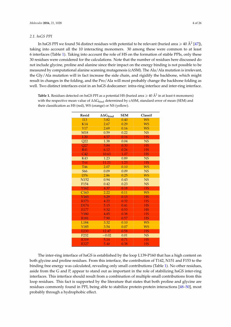
In hsGS PPI we found 54 distinct residues with potential to be relevant (buried area ě 40 Å2 [47]),taking into account all the 10 interacting monomers. 30 among these were common to at least6 interfaces (Table 1). Taking into account the role of HS on the formation of stable PPIs, only these30 residues were considered for the calculations. Note that the number of residues here discussed donot include glycine, proline and alanine since their impact on the energy binding is not possible to bemeasured by computational alanine scanning mutagenesis (cASM). The Ala/Ala mutation is irrelevant,the Gly/Ala mutation will in fact increase the side chain, and rigidify the backbone, which mightresult in changes in the folding, and the Pro/Ala will most probably change the backbone folding aswell. Two distinct interfaces exist in an hsGS dodecamer: intra-ring interface and inter-ring interface.
Table 1. Residues detected on hsGS PPI as a potential HS (buried area ě 40 Å2 in at least 6 monomers)with the respective mean value of ∆∆Gbind determined by cASM, standard error of mean (SEM) andtheir classification as HS (red), WS (orange) or NS (yellow).
Resid ∆∆Gbind SEM ClassifI13 3.82 0.40 WSK14 2.67 0.29 WSY17 2.69 0.16 WSM18 0.59 0.22 NSL20 4.57 0.16 HSQ22 1.38 0.04 NSQ27 5.84 0.30 HSR41 6.12 0.26 HSC42 10.63 1.07 HSK43 1.23 0.89 NST44 11.21 1.23 HST46 2.07 0.10 WSS66 0.09 0.09 NSD76 2.86 0.25 WS
N152 0.94 0.43 NSF154 0.42 0.23 NSY162 4.37 0.18 HSC163 2.22 0.11 WSV165 5.29 0.15 HSR173 4.22 0.32 HSD174 5.15 0.41 HSE177 9.52 0.53 HSY180 4.85 0.38 HSR181 7.90 0.57 HSL184 3.32 0.10 WSY185 3.54 0.07 WSE230 12.47 0.58 HSF232 ´0.02 0.01 NSR319 5.14 0.71 HSR327 5.40 0.38 HS
The inter-ring interface of hsGS is established by the loop L139-P160 that has a high content onboth glycine and proline residues. From this interface, the contribution of T142, N151 and F153 to thebinding free energy was calculated, revealing only small contributions (Table 1). No other residues,aside from the G and P, appear to stand out as important in the role of stabilizing hsGS inter-ringinterfaces. This interface should result from a combination of multiple small contributions from thisloop residues. This fact is supported by the literature that states that both proline and glycine areresidues commonly found in PPI, being able to stabilize protein-protein interactions [48–50], mostprobably through a hydrophobic effect.
Molecules 2016, 21, 1028 5 of 26
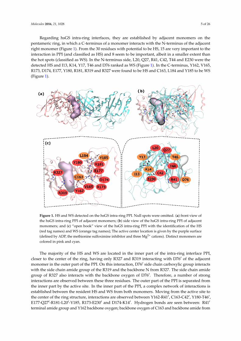
Regarding hsGS intra-ring interfaces, they are established by adjacent monomers on thepentameric ring, in which a C-terminus of a monomer interacts with the N-terminus of the adjacentright monomer (Figure 1). From the 30 residues with potential to be HS, 15 are very important to theinteraction in PPI (and classified as HS) and 8 seem to be important, albeit in a smaller extent thanthe hot spots (classified as WS). In the N-terminus side, L20, Q27, R41, C42, T44 and E230 were thedetected HS and I13, K14, Y17, T46 and D76 ranked as WS (Figure 1). In the C-terminus, Y162, V165,R173, D174, E177, Y180, R181, R319 and R327 were found to be HS and C163, L184 and Y185 to be WS(Figure 1).
Molecules 2016, 21, 1028 5 of 26
Regarding hsGS intra-ring interfaces, they are established by adjacent monomers on the pentameric ring, in which a C-terminus of a monomer interacts with the N-terminus of the adjacent right monomer (Figure 1). From the 30 residues with potential to be HS, 15 are very important to the interaction in PPI (and classified as HS) and 8 seem to be important, albeit in a smaller extent than the hot spots (classified as WS). In the N-terminus side, L20, Q27, R41, C42, T44 and E230 were the detected HS and I13, K14, Y17, T46 and D76 ranked as WS (Figure 1). In the C-terminus, Y162, V165, R173, D174, E177, Y180, R181, R319 and R327 were found to be HS and C163, L184 and Y185 to be WS (Figure 1).
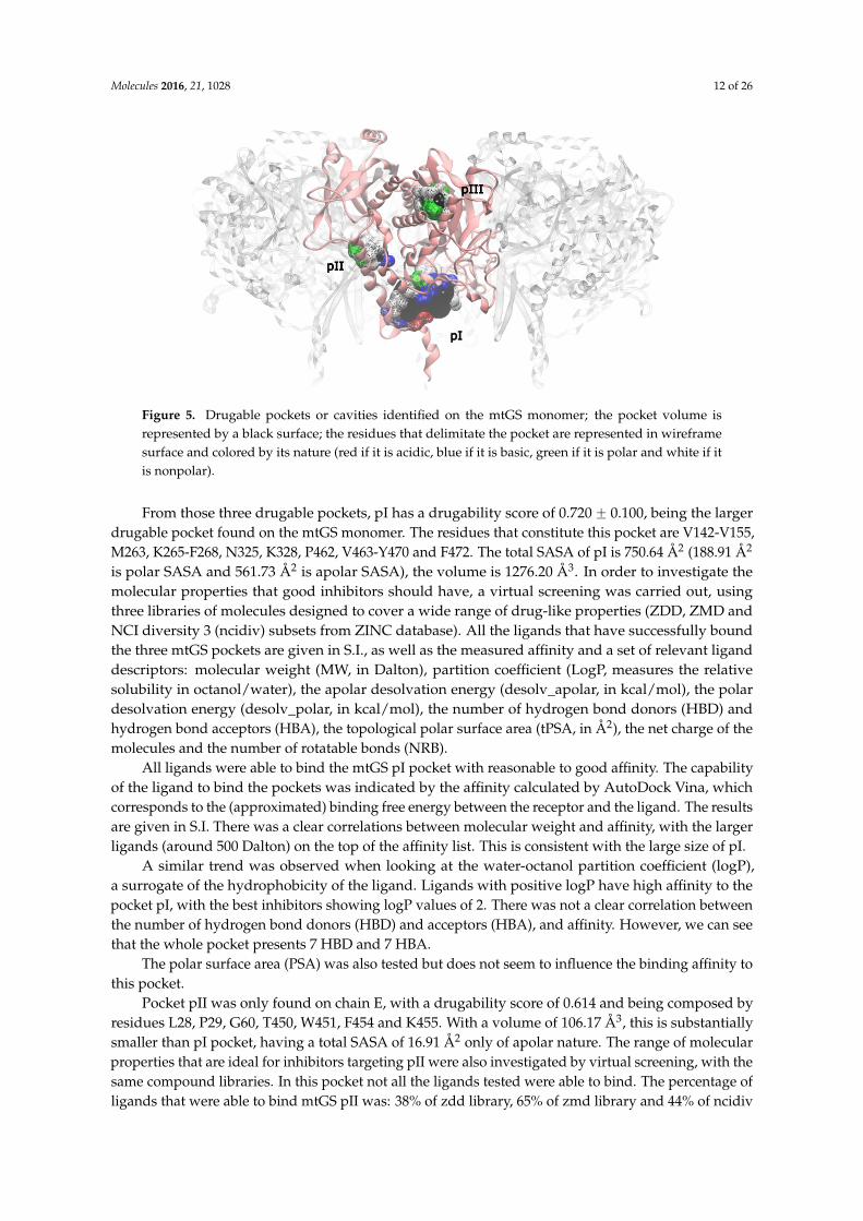
Figure 1. HS and WS detected on the hsGS intra-ring PPI. Null spots were omitted. (a) front view of the hsGS intra-ring PPI of adjacent monomers; (b) side view of the hsGS intra-ring PPI of adjacent monomers; and (c) “open book” view of the hsGS intra-ring PPI with the identification of the HS (red tag names) and WS (orange tag names); The active center location is given by the purple surface (defined by ADP, the methionine sulfoximine inhibitor and three Mg2+ cations). Distinct monomers are colored in pink and cyan.
The majority of the HS and WS are located in the inner part of the intra-ring interface PPI, closer to the center of the ring, having only R327 and R319 interacting with D76′ of the adjacent monomer in the outer part of the PPI. On this interaction, D76′ side chain carboxylic group interacts with the side chain amide group of the R319 and the backbone N from R327. The side chain amide group of R327 also interacts with the backbone oxygen of D76′. Therefore, a number of strong interactions are observed between these three residues. The outer part of the PPI is separated from the inner part by the active site. In the inner part of the PPI, a complex network of interactions is established between the resident HS and WS from both monomers. Moving from the active site to the center of the ring structure, interactions are observed between Y162-R41′, C163-C42′, Y180-T46′, E177-Q27′-R181-L20′-Y185, R173-E230′ and D174-K14′. Hydrogen bonds are seen between: R41′ terminal amide group and Y162 backbone oxygen; backbone oxygen of C163 and backbone amide from C42′; side chain Y180 and backbone amine of T46; E177 side chain carboxylic group with Q27′ side chain amide group; Q27′ side chain carbonyl group and terminal amide of R181 that also interacts with backbone carboxyl
Figure 1. HS and WS detected on the hsGS intra-ring PPI. Null spots were omitted. (a) front view ofthe hsGS intra-ring PPI of adjacent monomers; (b) side view of the hsGS intra-ring PPI of adjacentmonomers; and (c) “open book” view of the hsGS intra-ring PPI with the identification of the HS(red tag names) and WS (orange tag names); The active center location is given by the purple surface(defined by ADP, the methionine sulfoximine inhibitor and three Mg2+ cations). Distinct monomers arecolored in pink and cyan.
The majority of the HS and WS are located in the inner part of the intra-ring interface PPI,closer to the center of the ring, having only R327 and R319 interacting with D761 of the adjacentmonomer in the outer part of the PPI. On this interaction, D761 side chain carboxylic group interactswith the side chain amide group of the R319 and the backbone N from R327. The side chain amidegroup of R327 also interacts with the backbone oxygen of D761. Therefore, a number of stronginteractions are observed between these three residues. The outer part of the PPI is separated fromthe inner part by the active site. In the inner part of the PPI, a complex network of interactions isestablished between the resident HS and WS from both monomers. Moving from the active site tothe center of the ring structure, interactions are observed between Y162-R411, C163-C421, Y180-T461,E177-Q271-R181-L201-Y185, R173-E2301 and D174-K141. Hydrogen bonds are seen between: R411
terminal amide group and Y162 backbone oxygen; backbone oxygen of C163 and backbone amide from
Molecules 2016, 21, 1028 6 of 26
C421; side chain Y180 and backbone amine of T46; E177 side chain carboxylic group with Q271 sidechain amide group; Q271 side chain carbonyl group and terminal amide of R181 that also interacts withbackbone carboxyl group of L20; and finally, K141 side chain amide with side chain carboxylic groupof D174. C163-C42’ interactions has already been described as important on other studies performedon a distinct organism, reveling that disulfide bridges between this two residues can regulate GSactivity [51]. Some aliphatic-π interactions are also observed between L201-Y185 side chains. These areonly the interactions established between HS and WS residues of distinct monomers. Some WScontribute to the PPI by interacting with HS of the same monomer, stabilizing it. This is exemplified bythese two examples in which I131 and L184 are interacting with Y171 and Y180, respectively. From theseresults, it is possible to see that the intra-ring interface is the desirable place to develop PPI inhibitorsto inhibit hsGS that would not only inhibit the oligomerization but also disrupt the activity of thisenzyme (given that the catalytic site is located in this interface).
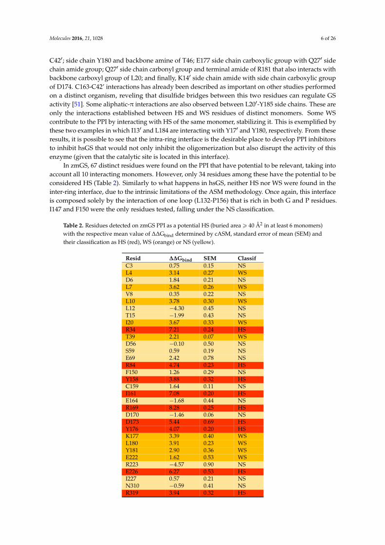
In zmGS, 67 distinct residues were found on the PPI that have potential to be relevant, taking intoaccount all 10 interacting monomers. However, only 34 residues among these have the potential to beconsidered HS (Table 2). Similarly to what happens in hsGS, neither HS nor WS were found in theinter-ring interface, due to the intrinsic limitations of the ASM methodology. Once again, this interfaceis composed solely by the interaction of one loop (L132-P156) that is rich in both G and P residues.I147 and F150 were the only residues tested, falling under the NS classification.
Table 2. Residues detected on zmGS PPI as a potential HS (buried area ě 40 Å2 in at least 6 monomers)with the respective mean value of ∆∆Gbind determined by cASM, standard error of mean (SEM) andtheir classification as HS (red), WS (orange) or NS (yellow).
Resid ∆∆Gbind SEM ClassifC3 0.75 0.15 NSL4 3.14 0.27 WSD6 1.84 0.21 NSL7 3.62 0.26 WSV8 0.35 0.22 NSL10 3.78 0.30 WSL12 ´4.30 0.45 NST15 ´1.99 0.43 NSI20 3.67 0.33 WSR34 7.21 0.24 HST39 2.21 0.07 WSD56 ´0.10 0.50 NSS59 0.59 0.19 NSE69 2.42 0.78 NSR84 4.74 0.23 HSF150 1.26 0.29 NSY158 3.88 0.32 HSC159 1.64 0.11 NSI161 7.08 0.20 HSE164 ´1.68 0.44 NSR169 8.28 0.25 HSD170 ´1.46 0.06 NSD173 5.44 0.69 HSY176 4.07 0.20 HSK177 3.39 0.40 WSL180 3.91 0.23 WSY181 2.90 0.36 WSE222 1.62 0.53 WSR223 ´4.57 0.90 NSE226 6.27 0.53 HSI227 0.57 0.21 NSN310 ´0.59 0.41 NSR319 3.94 0.32 HS
Molecules 2016, 21, 1028 7 of 26
2.2. zmGS PPI
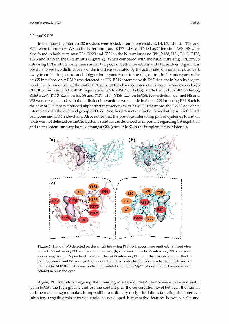
In the intra-ring interface 32 residues were tested. From these residues, L4, L7, L10, I20, T39, andE222 were found to be WS on the N-terminus and K177, L180 and Y181 as C-terminus WS. HS werealso found in both terminus: R34, R223 and E226 in the N-terminus and R84, Y158, I161, R169, D173,Y176 and R319 in the C-terminus (Figure 2). When compared with the hsGS intra-ring PPI, zmGSintra-ring PPI is at the same time similar but poor in both interactions and HS residues. Again, it ispossible to see two distinct parts of the interface separated by the active site, one smaller outer part,away from the ring centre, and a bigger inner part, closer to the ring centre. In the outer part of thezmGS interface, only R319 was detected as HS. R319 interacts with D67 side chain by a hydrogenbond. On the inner part of the zmGS PPI, some of the observed interactions were the same as in hsGSPPI. It is the case of Y158-R341 (equivalent to Y162-R411 on hsGS), Y176-T391 (Y180-T461 on hsGS),R169-E2261 (R173-E2301 on hsGS) and Y181-L101 (Y185-L201 on hsGS). Nevertheless, distinct HS andWS were detected and with them distinct interactions were made in the zmGS intra-ring PPI. Such isthe case of I201 that established aliphatic-π interactions with Y176. Furthermore, the R2231 side chaininteracted with the carboxyl group of D177. Another distinct interaction was that between the L101
backbone and K177 side-chain. Also, notice that the previous interacting pair of cysteines found onhsGS was not detected on zmGS. Cysteine residues are described as important regarding GS regulationand their content can vary largely amongst GSs (check file S2 in the Supplementary Material).
Molecules 2016, 21, 1028 7 of 26
N310 −0.59 0.41 NS R319 3.94 0.32 HS
2.2. zmGS PPI
In the intra-ring interface 32 residues were tested. From these residues, L4, L7, L10, I20, T39, and E222 were found to be WS on the N-terminus and K177, L180 and Y181 as C-terminus WS. HS were also found in both terminus: R34, R223 and E226 in the N-terminus and R84, Y158, I161, R169, D173, Y176 and R319 in the C-terminus (Figure 2). When compared with the hsGS intra-ring PPI, zmGS intra-ring PPI is at the same time similar but poor in both interactions and HS residues. Again, it is possible to see two distinct parts of the interface separated by the active site, one smaller outer part, away from the ring centre, and a bigger inner part, closer to the ring centre. In the outer part of the zmGS interface, only R319 was detected as HS. R319 interacts with D67 side chain by a hydrogen bond. On the inner part of the zmGS PPI, some of the observed interactions were the same as in hsGS PPI. It is the case of Y158-R34′ (equivalent to Y162-R41′ on hsGS), Y176-T39′ (Y180-T46′ on hsGS), R169-E226′ (R173-E230′ on hsGS) and Y181-L10′ (Y185-L20′ on hsGS). Nevertheless, distinct HS and WS were detected and with them distinct interactions were made in the zmGS intra-ring PPI. Such is the case of I20′ that established aliphatic-π interactions with Y176. Furthermore, the R223′ side chain interacted with the carboxyl group of D177. Another distinct interaction was that between the L10′ backbone and K177 side-chain. Also, notice that the previous interacting pair of cysteines found on hsGS was not detected on zmGS. Cysteine residues are described as important regarding GS regulation and their content can vary largely amongst GSs (check file S2 in the Supplementary Material).
Figure 2. HS and WS detected on the zmGS intra-ring PPI. Null spots were omitted. (a) front view of the hsGS intra-ring PPI of adjacent monomers; (b) side view of the hsGS intra-ring PPI of adjacent monomers; and (c) “open book” view of the hsGS intra-ring PPI with the identification of the HS (red tag names) and WS (orange tag names); The active center location is given by the purple surface (defined by ADP, the methionine sulfoximine inhibitor and three Mg2+ cations). Distinct monomers are colored in pink and cyan.
Figure 2. HS and WS detected on the zmGS intra-ring PPI. Null spots were omitted. (a) front viewof the hsGS intra-ring PPI of adjacent monomers; (b) side view of the hsGS intra-ring PPI of adjacentmonomers; and (c) “open book” view of the hsGS intra-ring PPI with the identification of the HS(red tag names) and WS (orange tag names); The active center location is given by the purple surface(defined by ADP, the methionine sulfoximine inhibitor and three Mg2+ cations). Distinct monomers arecolored in pink and cyan.
Again, PPI inhibitors targeting the inter-ring interface of zmGS do not seem to be successful(as in hsGS): the high glycine and proline content plus the conservation level between the humanand the maize enzyme makes it impossible to rationally design inhibitors targeting this interface.Inhibitors targeting this interface could be developed if distinctive features between hsGS and
Molecules 2016, 21, 1028 8 of 26
zmGS associated with drugable pockets could be found. However, such circumstances were notmet (see below).
2.3. mtGS PPI
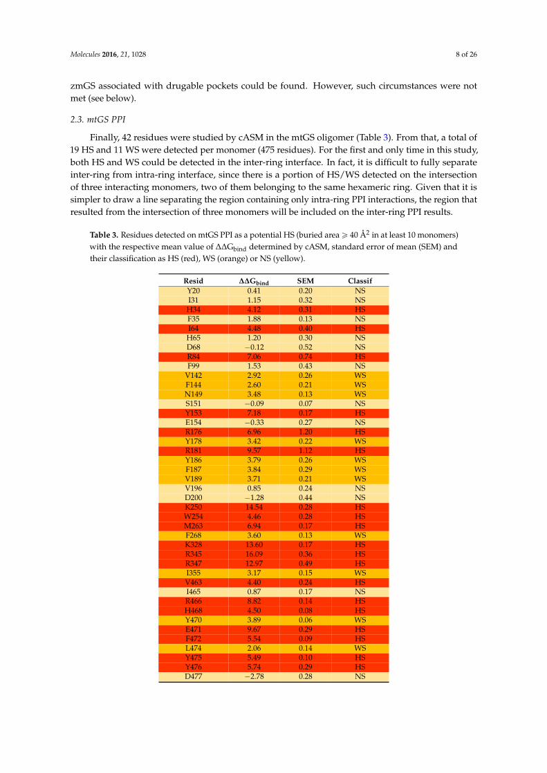
Finally, 42 residues were studied by cASM in the mtGS oligomer (Table 3). From that, a total of19 HS and 11 WS were detected per monomer (475 residues). For the first and only time in this study,both HS and WS could be detected in the inter-ring interface. In fact, it is difficult to fully separateinter-ring from intra-ring interface, since there is a portion of HS/WS detected on the intersectionof three interacting monomers, two of them belonging to the same hexameric ring. Given that it issimpler to draw a line separating the region containing only intra-ring PPI interactions, the region thatresulted from the intersection of three monomers will be included on the inter-ring PPI results.
Table 3. Residues detected on mtGS PPI as a potential HS (buried areaě 40 Å2 in at least 10 monomers)with the respective mean value of ∆∆Gbind determined by cASM, standard error of mean (SEM) andtheir classification as HS (red), WS (orange) or NS (yellow).
Resid ∆∆Gbind SEM ClassifY20 0.41 0.20 NSI31 1.15 0.32 NSH34 4.12 0.31 HSF35 1.88 0.13 NSI64 4.48 0.40 HSH65 1.20 0.30 NSD68 ´0.12 0.52 NSR84 7.06 0.74 HSF99 1.53 0.43 NS
V142 2.92 0.26 WSF144 2.60 0.21 WSN149 3.48 0.13 WSS151 ´0.09 0.07 NSY153 7.18 0.17 HSE154 ´0.33 0.27 NSR176 6.96 1.20 HSY178 3.42 0.22 WSR181 9.57 1.12 HSY186 3.79 0.26 WSF187 3.84 0.29 WSV189 3.71 0.21 WSV196 0.85 0.24 NSD200 ´1.28 0.44 NSK250 14.54 0.28 HSW254 4.46 0.28 HSM263 6.94 0.17 HSF268 3.60 0.13 WSK328 13.60 0.17 HSR345 16.09 0.36 HSR347 12.97 0.49 HSI355 3.17 0.15 WSV463 4.40 0.24 HSI465 0.87 0.17 NSR466 8.82 0.14 HSH468 4.50 0.08 HSY470 3.89 0.06 WSE471 9.67 0.29 HSF472 5.54 0.09 HSL474 2.06 0.14 WSY475 5.49 0.10 HSY476 5.74 0.29 HSD477 ´2.78 0.28 NS
Molecules 2016, 21, 1028 9 of 26
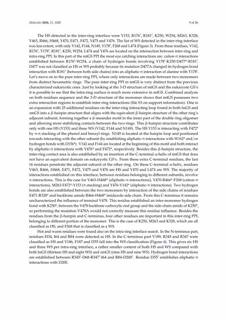
The HS detected in the inter-ring interface were Y153, R1761, R1811, K250, W254, M263, K328,V463, R466, H468, Y470, E471, F472, Y475 and Y476. The list of WS detected in the inter-ring interfacewas less extent, with only V142, F144, N149, Y1781, F268 and L474 (Figure 3). From these residues, V142,R1761, Y1781, R1811, K250, W254, L474 and Y476 are located on the intersection between inter-ring andintra-ring PPI. In this part of the mtGS PPI the most eye catching interactions are: cation-π interactionsestablished between R1761-W254, a chain of hydrogen bonds involving Y1781-K250-D477*-R1811.D477 was not classified as HS or WS probably because its mutation D477A changed its hydrogen bondinteraction with R1811 (between both side chains) into an aliphatic-π interaction of alanine with Y1781.Let’s move on to the pure inter-ring PPI, where only interactions are made between two monomersfrom distinct hexametric rings. The pure inter-ring PPI in mtGS is very distinct from the previouscharacterized eukaryotic ones. Just by looking at the 3-D structure of mtGS and the eukaryote GS’sit is possible to see that the intra-ring surface is much more extensive in mtGS. Combined analysison both residues sequence and the 3-D structure of the monomer shows that mtGS possesses twoextra interaction regions to establish inter-ring interactions (file S3 on support information). One isan expansion with 25 additional residues on the inter-ring interacting loop found in both hsGS andzmGS into a β-hairpin structure that aligns with the equivalent β-hairpin structure of the other ring’sadjacent subunit, forming together a β-meander motif in the inner part of the double ring oligomerand allowing more stabilizing contacts between the two rings. This β-hairpin structure contributesonly with one HS (Y153) and three WS (V142, F144 and N149). The HS Y153 is interacting with F472*by π-π stacking of the phenol and benzyl rings. N149 is located at the hairpin loop and positionedtowards interacting with the other subunit (by establishing aliphatic-π interactions with W162* and/orhydrogen bonds with D156*). V142 and F144 are located at the beginning of this motif and both interactby aliphatic-π interactions with Y476* and F472*, respectively. Besides this β-hairpin structure, theinter-ring contact area is also established by an insertion of the C-terminal α-helix of mtGS that doesnot have an equivalent domain on eukaryotic GS’s. From these extra C-terminal residues, the last16 residues penetrate the adjacent subunit of the other ring. On these C-terminal α-helix, residuesV463, R466, H468, E471, F472, Y475 and Y476 are HS and Y470 and L474 are WS. The majority ofinteractions established on this interface, between residues belonging to different subunits, involveπ interactions. This is the case for V463-H468* (aliphatic-π interactions), Y470-R466*-F268 (cation-πinteractions), M263-F472*-Y153 (π stacking) and Y476-V142* (aliphatic-π interactions). Two hydrogenbonds are also established between the two monomers by interaction of the side chains of residuesE471-R328* and backbone amide R466-H468* imidazole side chain. From this C-terminus it remainsuncharacterized the influence of terminal V478. This residue established an inter-monomer hydrogenbond with K250*, between the V478 backbone carboxylic end group and the side-chain amide of K250*,so performing the mutation V478A would not correctly measure this residue influence. Besides theresidues from the β-hairpin and C-terminus, four other residues are important in this inter-ring PPI,belonging to different portion of the monomer. This is the case of K250, M263 and K328, which are allclassified as HS, and F268 that is classified as a WS.
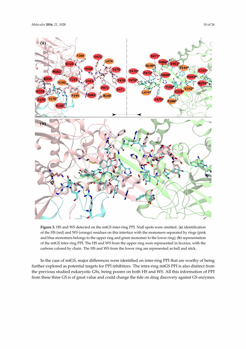
Hot and warm residues were found also on the intra-ring interface search. In the N-terminus part,residues H34, I64 and R84 were detected as HS. In the C-terminus part V189, R345 and R347 wereclassified as HS and Y186, F187 and I355 fall into the WS classification (Figure 4). This gives six HSand three WS per intra-ring interface, a rather smaller content of both HS and WS compared withboth hsGS (thirteen HS and eight WS) and zmGS (nine HS and nine WS). Hydrogen bond interactionsare established between R3451-D68-R3471-I64 and R84-D2001. Residue I3551 establishes aliphatic-πinteractions with D200.
Molecules 2016, 21, 1028 10 of 26
Molecules 2016, 21, 1028 10 of 26
Figure 3. HS and WS detected on the mtGS inter-ring PPI. Null spots were omitted. (a) identification of the HS (red) and WS (orange) residues on this interface with the monomers separated by rings (pink and blue monomers belongs to the upper ring and green monomer to the lower ring); (b) representation of the mtGS inter-ring PPI. The HS and WS from the upper ring were represented in licorice, with the carbons colored by chain. The HS and WS from the lower ring are represented as ball and stick.
Hot and warm residues were found also on the intra-ring interface search. In the N-terminus part, residues H34, I64 and R84 were detected as HS. In the C-terminus part V189, R345 and R347 were classified as HS and Y186, F187 and I355 fall into the WS classification (Figure 4). This gives six HS and three WS per intra-ring interface, a rather smaller content of both HS and WS compared with both hsGS (thirteen HS and eight WS) and zmGS (nine HS and nine WS). Hydrogen bond interactions are established between R345′-D68-R347′-I64 and R84-D200′. Residue I355′ establishes aliphatic-π interactions with D200.
Figure 3. HS and WS detected on the mtGS inter-ring PPI. Null spots were omitted. (a) identificationof the HS (red) and WS (orange) residues on this interface with the monomers separated by rings (pinkand blue monomers belongs to the upper ring and green monomer to the lower ring); (b) representationof the mtGS inter-ring PPI. The HS and WS from the upper ring were represented in licorice, with thecarbons colored by chain. The HS and WS from the lower ring are represented as ball and stick.
In the case of mtGS, major differences were identified on inter-ring PPI that are worthy of beingfurther explored as potential targets for PPI inhibitors. The intra-ring mtGS PPI is also distinct fromthe previous studied eukaryotic GSs, being poorer on both HS and WS. All this information of PPIfrom these three GS is of great value and could change the tide on drug discovery against GS enzymes.
Molecules 2016, 21, 1028 11 of 26Molecules 2016, 21, 1028 11 of 26
Figure 4. HS and WS detected on the mtGS intra-ring PPI. Null spots were omitted. (a) front view of the hsGS intra-ring PPI of adjacent monomers; (b) side view of the hsGS intra-ring PPI of adjacent monomers; and (c) “open book” view of the hsGS intra-ring PPI with the identification of the HS (red tag names) and WS (orange tag names); The active center location is given by the purple surface (defined by ADP, the methionine sulfoximine inhibitor and three Mg2+ cations). Distinct monomers are colored in pink and cyan.
In the case of mtGS, major differences were identified on inter-ring PPI that are worthy of being further explored as potential targets for PPI inhibitors. The intra-ring mtGS PPI is also distinct from the previous studied eukaryotic GSs, being poorer on both HS and WS. All this information of PPI from these three GS is of great value and could change the tide on drug discovery against GS enzymes.
2.4. Novel Drugable Pockets
To gather useful information that allows for the development of novel GS inhibitors with selectivity towards the GS from a single organism or at least a small group of organisms, novel drugable pockets, away from the active site, should be scanned. To achieve that goal, the Fpocket software was employed using an isolated monomer as target from all three studied GSs (hsGS, zmGS and mtGS). Pockets were detected in all GSs, with a maximum number of pocket of 11 on hsGS (chain C), 12 pockets on zmGS (chain A) and 24 pockets on mtGS (chain F). From the detected pockets, only those with a drugability score above 0.5 were considered as drugable pockets [52].
Applying this restriction, one drugable pocket was detected on hsGS (drugability score around 0.605 ± 0.001) and another one on zmGS (drugability score around 0.622 ± 0.032). In both cases, the location of the drugable pocket was overlapping the active site position and so they do not really constitute new pockets in terms of a new inhibition strategy. On mtGS, three drugable pockets were
Figure 4. HS and WS detected on the mtGS intra-ring PPI. Null spots were omitted. (a) front viewof the hsGS intra-ring PPI of adjacent monomers; (b) side view of the hsGS intra-ring PPI of adjacentmonomers; and (c) “open book” view of the hsGS intra-ring PPI with the identification of the HS(red tag names) and WS (orange tag names); The active center location is given by the purple surface(defined by ADP, the methionine sulfoximine inhibitor and three Mg2+ cations). Distinct monomers arecolored in pink and cyan.
2.4. Novel Drugable Pockets
To gather useful information that allows for the development of novel GS inhibitors withselectivity towards the GS from a single organism or at least a small group of organisms, noveldrugable pockets, away from the active site, should be scanned. To achieve that goal, the Fpocketsoftware was employed using an isolated monomer as target from all three studied GSs (hsGS, zmGSand mtGS). Pockets were detected in all GSs, with a maximum number of pocket of 11 on hsGS(chain C), 12 pockets on zmGS (chain A) and 24 pockets on mtGS (chain F). From the detected pockets,only those with a drugability score above 0.5 were considered as drugable pockets [52].
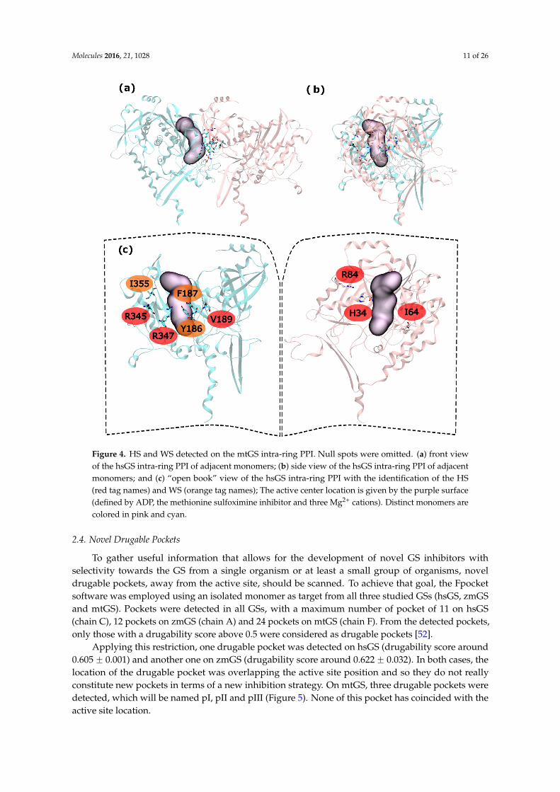
Applying this restriction, one drugable pocket was detected on hsGS (drugability score around0.605 ˘ 0.001) and another one on zmGS (drugability score around 0.622 ˘ 0.032). In both cases, thelocation of the drugable pocket was overlapping the active site position and so they do not reallyconstitute new pockets in terms of a new inhibition strategy. On mtGS, three drugable pockets weredetected, which will be named pI, pII and pIII (Figure 5). None of this pocket has coincided with theactive site location.
Molecules 2016, 21, 1028 12 of 26
Molecules 2016, 21, 1028 12 of 26
detected, which will be named pI, pII and pIII (Figure 5). None of this pocket has coincided with the active site location.
Figure 5. Drugable pockets or cavities identified on the mtGS monomer; the pocket volume is represented by a black surface; the residues that delimitate the pocket are represented in wireframe surface and colored by its nature (red if it is acidic, blue if it is basic, green if it is polar and white if it is nonpolar).
From those three drugable pockets, pI has a drugability score of 0.720 ± 0.100, being the larger drugable pocket found on the mtGS monomer. The residues that constitute this pocket are V142-V155, M263, K265-F268, N325, K328, P462, V463-Y470 and F472. The total SASA of pI is 750.64 Å2 (188.91 Å2 is polar SASA and 561.73 Å2 is apolar SASA), the volume is 1276.20 Å3. In order to investigate the molecular properties that good inhibitors should have, a virtual screening was carried out, using three libraries of molecules designed to cover a wide range of drug-like properties (ZDD, ZMD and NCI diversity 3 (ncidiv) subsets from ZINC database). All the ligands that have successfully bound the three mtGS pockets are given in S.I., as well as the measured affinity and a set of relevant ligand descriptors: molecular weight (MW, in Dalton), partition coefficient (LogP, measures the relative solubility in octanol/water), the apolar desolvation energy (desolv_apolar, in kcal/mol), the polar desolvation energy (desolv_polar, in kcal/mol), the number of hydrogen bond donors (HBD) and hydrogen bond acceptors (HBA), the topological polar surface area (tPSA, in Å2), the net charge of the molecules and the number of rotatable bonds (NRB).
All ligands were able to bind the mtGS pI pocket with reasonable to good affinity. The capability of the ligand to bind the pockets was indicated by the affinity calculated by AutoDock Vina, which corresponds to the (approximated) binding free energy between the receptor and the ligand. The results are given in S.I. There was a clear correlations between molecular weight and affinity, with the larger ligands (around 500 Dalton) on the top of the affinity list. This is consistent with the large size of pI.
A similar trend was observed when looking at the water-octanol partition coefficient (logP), a surrogate of the hydrophobicity of the ligand. Ligands with positive logP have high affinity to the pocket pI, with the best inhibitors showing logP values of 2. There was not a clear correlation between the number of hydrogen bond donors (HBD) and acceptors (HBA), and affinity. However, we can see that the whole pocket presents 7 HBD and 7 HBA.
The polar surface area (PSA) was also tested but does not seem to influence the binding affinity to this pocket.
Pocket pII was only found on chain E, with a drugability score of 0.614 and being composed by residues L28, P29, G60, T450, W451, F454 and K455. With a volume of 106.17 Å3, this is substantially smaller than pI pocket, having a total SASA of 16.91 Å2 only of apolar nature. The range of molecular properties that are ideal for inhibitors targeting pII were also investigated by virtual screening, with
Figure 5. Drugable pockets or cavities identified on the mtGS monomer; the pocket volume isrepresented by a black surface; the residues that delimitate the pocket are represented in wireframesurface and colored by its nature (red if it is acidic, blue if it is basic, green if it is polar and white if itis nonpolar).
From those three drugable pockets, pI has a drugability score of 0.720 ˘ 0.100, being the largerdrugable pocket found on the mtGS monomer. The residues that constitute this pocket are V142-V155,M263, K265-F268, N325, K328, P462, V463-Y470 and F472. The total SASA of pI is 750.64 Å2 (188.91 Å2
is polar SASA and 561.73 Å2 is apolar SASA), the volume is 1276.20 Å3. In order to investigate themolecular properties that good inhibitors should have, a virtual screening was carried out, usingthree libraries of molecules designed to cover a wide range of drug-like properties (ZDD, ZMD andNCI diversity 3 (ncidiv) subsets from ZINC database). All the ligands that have successfully boundthe three mtGS pockets are given in S.I., as well as the measured affinity and a set of relevant liganddescriptors: molecular weight (MW, in Dalton), partition coefficient (LogP, measures the relativesolubility in octanol/water), the apolar desolvation energy (desolv_apolar, in kcal/mol), the polardesolvation energy (desolv_polar, in kcal/mol), the number of hydrogen bond donors (HBD) andhydrogen bond acceptors (HBA), the topological polar surface area (tPSA, in Å2), the net charge of themolecules and the number of rotatable bonds (NRB).
All ligands were able to bind the mtGS pI pocket with reasonable to good affinity. The capabilityof the ligand to bind the pockets was indicated by the affinity calculated by AutoDock Vina, whichcorresponds to the (approximated) binding free energy between the receptor and the ligand. The resultsare given in S.I. There was a clear correlations between molecular weight and affinity, with the largerligands (around 500 Dalton) on the top of the affinity list. This is consistent with the large size of pI.
A similar trend was observed when looking at the water-octanol partition coefficient (logP),a surrogate of the hydrophobicity of the ligand. Ligands with positive logP have high affinity to thepocket pI, with the best inhibitors showing logP values of 2. There was not a clear correlation betweenthe number of hydrogen bond donors (HBD) and acceptors (HBA), and affinity. However, we can seethat the whole pocket presents 7 HBD and 7 HBA.
The polar surface area (PSA) was also tested but does not seem to influence the binding affinity tothis pocket.
Pocket pII was only found on chain E, with a drugability score of 0.614 and being composed byresidues L28, P29, G60, T450, W451, F454 and K455. With a volume of 106.17 Å3, this is substantiallysmaller than pI pocket, having a total SASA of 16.91 Å2 only of apolar nature. The range of molecularproperties that are ideal for inhibitors targeting pII were also investigated by virtual screening, with thesame compound libraries. In this pocket not all the ligands tested were able to bind. The percentage ofligands that were able to bind mtGS pII was: 38% of zdd library, 65% of zmd library and 44% of ncidiv
Molecules 2016, 21, 1028 13 of 26
library. The preferred molecular weight is around 200 Dalton, but can vary from 50 to 570 Dalton.Also a logP near 0 was found to be more suitable for molecules that can be ligands to pII.
At last, pIII presented a drugability score of 0.636˘ 0.076, located in a region delimited by residuesA128-F130, N279-L281, M289, T301, A302, Y305, L365, L384 and I391. This pocket is substantiallysmaller than pI and with similar dimensions of pII, having only a volume of 93.09 Å3 with a total SASAof 16.34 Å2 (4.57 Å2 is polar SASA and 11.77 Å2 is apolar SASA). The ligands that bind this pocket arescarce, in particular when compared to the previous pockets. Only a small percentage of the ligandswas able to bind pIII (1.2% of the zdd library, 5.7% of the zmd library and 0.1% of the ncidiv library).The molecular weight can go from 50 to 250 Dalton, with a preference for smaller molecules. The rangeof logP for binders is wide, it goes from ´4 to 3, with a preference towards hydrophilic molecules.
Both pII and pIII are small pockets, with a volume unfavorable to fit a larger sized drug. They areable to bind small sized drugs, but only with reasonable to low affinity. Even having similar volume,pII pocket fits larger molecules than pI, given that its location is nearer to the surface that pIII. Drugs tothis two pockets would be difficult to achieve. On the other hand, pI looks very promising. The volumeof pI allows fitting a molecule that could reach a molecular weight of 1000 Dalton, which is abouttwo times the size of a large bioavailable drug. Furthermore, the polar SASA of pI is similar, butabove the 140 Å2 threshold for bioavailability [53], which means that the pocket can accommodatea large bioavailable drug and has enough hydrogen bond donors and acceptors to satisfy typicaldruglike compounds.
3. Discussion
As mentioned before, PPI interactions were studied on three distinct GSs from three distinctorganisms: human, maize and the bacteria responsible for tuberculosis. All three enzymes dependon oligomerization to become active, given that the active site is located in between monomers.Inhibitors directed to this enzyme are currently on high demand, as can be seen by the number ofrecent papers dedicated to design new inhibitors of GS [14,17,18,20–24,26,54–59]. However, all of themare designed to act on the GS active site, lacking on specificity against one particular organism orgroup of organisms. Attempts have been made to develop specific mtGS inhibitors by exploitingthe small differences in the active site residues located in the ATP/ADP binding region. However,given the level of conservation seen on the active site, development of such specific inhibitors seemsquite challenging.
Another approach would be targeting the PPI with small inhibitors directed to interact specificallywith HS, blocking the oligomerization and subsequently the active site formation. This is the focus ofthis work.
From the results presented here it is clear that significant differences exist between prokaryoticGSI and eukaryotic GSII PPIs, mostly at inter-ring PPI. This PPI in GSII is almost inexistent, consistingonly of a contact between two loops rich in glycine and proline. Glycine rich linkers are already beendocumented and are common in proteins [60]. Their function relies mainly on connecting distinctdomains inside a protein but still allowing them to be flexible [60]. In both zmGS and hsGS theinter-ring interacting loop constitutes a linker between the C-terminus and N-terminus domain (file S2on support information), a probable reason to have such a high amount of glycine in its constitution.The second most common residue in this loop is proline. Contrary to glycine, proline-rich motifsare described as critical in PPI associated with intracellular signaling and pathways [48–50]. It ispossible that proline is the main responsible for the establishment of inter-ring PPI on eukaryotic GSs.Naturally disordered motifs have been described as important on PPI associated with signaling [61–66].Despite their great interest, their disordered nature makes it difficult, if not impossible, to rationallydesign inhibitors that target them.
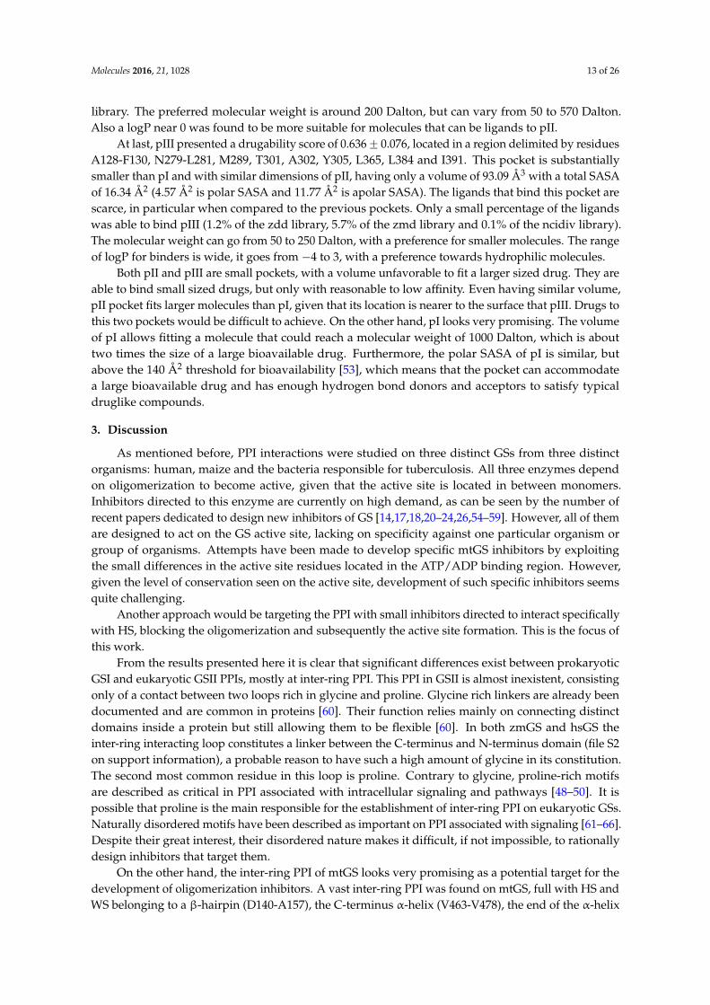
On the other hand, the inter-ring PPI of mtGS looks very promising as a potential target for thedevelopment of oligomerization inhibitors. A vast inter-ring PPI was found on mtGS, full with HS andWS belonging to a β-hairpin (D140-A157), the C-terminus α-helix (V463-V478), the end of the α-helix
Molecules 2016, 21, 1028 14 of 26
S234-W254, loop M263-F268 and a small α-helix (T323-V331). The first two (β-hairpin and C-terminusα-helix) are the motifs that are projected towards the other ring, being inserted on the other monomer.The remaining ones, together with the base of the β-hairpin and the C-terminus α-helix, form a “hat”that will receive the end part of the C-terminus α-helix from the other ring. From the drugable pocketsdetected here, the largest one is located on that “hat”. This pocket seems to be a very promising targetfor the binding of oligomerization inhibitors. More so, its inhibitors can be designed as small druglikemolecules, or even as peptides or peptidomimetics. For instances, peptides based on the C-terminusα-helix could be explored as drugs. The fact that these peptides lie outside the Lipinski space is nota problem for the treatment of tuberculosis. As this is a lung infection, it is perfectly viable to performthe administration through the upper respiratory tract.
Even if an inhibitor is successfully designed to act in this pocket, one can always hypothesizethat it will only eliminate the double ring formation, keeping the active site assembled. This wouldnot be an ideal situation, but it is important to keep in mind that: (1) the existence of such a big andimportant interface must have an influence on the activity of mtGS; (2) the inter-ring interface is closeto the intra-ring interface, having a region in which contacts between 3 distinct monomers do exist;and (3) any destabilization induced on the mtGS tertiary and quaternary structures should propagateto the intra-ring dimerization (i.e., to the active site) and alter the catalytic activity. It is also importantto notice that the mtGS studied here is encoded by glnA1 gene. There are studies point out glnA1 asthe only essential GS in Mycobacterium tuberculosis [67]. So inhibitors targeting the mtGS model herestudied should be enough to tackle the tuberculosis problem. An alignment of the four existent mtGScan be found on file S3 on support information.
The intra-ring PPI is much more conserved along GSs from different organisms. Still, somedifferences can be seen on both the HS/WS detection and on the comparative analysis of the threesequences (file S2 on support information). The N-terminus seems by far the most distinct portionof this PPI on the three GSs. The only coinciding HS/WS found were R41 and T46 of hsGS with R34and T39 of zmGS. The C-terminus portion of the intra-ring interaction, on other hand, has a moreconserved sequence and the presence of HS/WS, being almost identical on the two eukaryotic GS’sstudied. MtGS lacks a whole region of HS/WS (R173-Y185) on hsGS nomenclature. But, given thatno drugable pockets were detected here, inhibition of mtGS hexameric ring oligomerization seemsdifficult to be achieved. Nevertheless, intra-ring PPI seems to be stronger in hsGS and zmGS than inmtGS, so any destabilization in this bacterial enzyme quaternary structure could be of great importance.Either way, this novel knowledge can definitely guide future investigations regarding the developmentof oligomerization inhibitors towards GS enzymes.
Some remarks have to be made regarding the transferability of this new pieces of the GS puzzleto other enzymes that were not included on this study. An alignment containing sequences of GSbelonging to distinct organism that have at least one x-ray crystallographic structure deposited on thePDB was made and is available on file S3 on support information.
4. Materials and Methods
4.1. Selection of Crystallographic Structures
Different GS crystallographic structures were chosen from the Protein Data Bank website, oneprokaryotic GS I from Mycobacterium tuberculosis (mtGS; PDB accession code 2BVC, 2.1 Å resolution [25])and two eukaryotic GS II from Zea mays (zmGS; PDB accession code 2D3A, 2.63 Å resolution [68]) andHomo Sapiens (hsGS; PDB accession code 2QC8, 2.6 Å resolution [69]). Figure 6 illustrates the structureof these three enzymes. All the three GS structures were crystallized with the methionine sulfoximineinhibitor, ADP and 2 or 3 divalent cations (Mg2+ or Mn2+) in the active site. The choice of these threespecies lays upon their potential interest by the industry that is searching both anti-tuberculosis agentsand new herbicides. Also, it allows the study of both GSI (mtGS) and GSII (zmGS and hsGS).
Molecules 2016, 21, 1028 15 of 26
Molecules 2016, 21, 1028 15 of 26
both anti-tuberculosis agents and new herbicides. Also, it allows the study of both GSI (mtGS) and GSII (zmGS and hsGS).
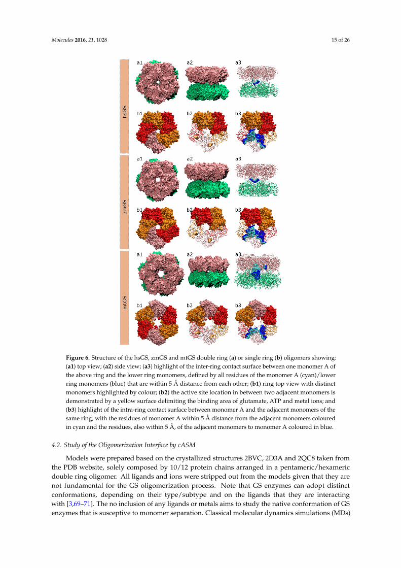
Figure 6. Structure of the hsGS, zmGS and mtGS double ring (a) or single ring (b) oligomers showing: (a1) top view; (a2) side view; (a3) highlight of the inter-ring contact surface between one monomer A of the above ring and the lower ring monomers, defined by all residues of the monomer A (cyan)/lower ring monomers (blue) that are within 5 Å distance from each other; (b1) ring top view with distinct monomers highlighted by colour; (b2) the active site location in between two adjacent monomers is demonstrated by a yellow surface delimiting the binding area of glutamate, ATP and metal ions; and (b3) highlight of the intra-ring contact surface between monomer A and the adjacent monomers of the same ring, with the residues of monomer A within 5 Å distance from the adjacent monomers coloured in cyan and the residues, also within 5 Å, of the adjacent monomers to monomer A coloured in blue.
4.2. Study of the Oligomerization Interface by cASM
Models were prepared based on the crystallized structures 2BVC, 2D3A and 2QC8 taken from the PDB website, solely composed by 10/12 protein chains arranged in a pentameric/hexameric double ring oligomer. All ligands and ions were stripped out from the models given that they are not fundamental for the GS oligomerization process. Note that GS enzymes can adopt distinct conformations, depending on their type/subtype and on the ligands that they are interacting with
Figure 6. Structure of the hsGS, zmGS and mtGS double ring (a) or single ring (b) oligomers showing:(a1) top view; (a2) side view; (a3) highlight of the inter-ring contact surface between one monomer A ofthe above ring and the lower ring monomers, defined by all residues of the monomer A (cyan)/lowerring monomers (blue) that are within 5 Å distance from each other; (b1) ring top view with distinctmonomers highlighted by colour; (b2) the active site location in between two adjacent monomers isdemonstrated by a yellow surface delimiting the binding area of glutamate, ATP and metal ions; and(b3) highlight of the intra-ring contact surface between monomer A and the adjacent monomers of thesame ring, with the residues of monomer A within 5 Å distance from the adjacent monomers colouredin cyan and the residues, also within 5 Å, of the adjacent monomers to monomer A coloured in blue.
4.2. Study of the Oligomerization Interface by cASM
Models were prepared based on the crystallized structures 2BVC, 2D3A and 2QC8 taken fromthe PDB website, solely composed by 10/12 protein chains arranged in a pentameric/hexamericdouble ring oligomer. All ligands and ions were stripped out from the models given that they arenot fundamental for the GS oligomerization process. Note that GS enzymes can adopt distinctconformations, depending on their type/subtype and on the ligands that they are interactingwith [3,69–71]. The no inclusion of any ligands or metals aims to study the native conformation of GSenzymes that is susceptive to monomer separation. Classical molecular dynamics simulations (MDs)
Molecules 2016, 21, 1028 16 of 26
were employed in all models in order to sample the phase space, using the AMBER 12 package [72].Hydrogens (assuming physiological protonated states), counter ions and water type TIP3P molecules(a minimum distance of 12 Å was left between the protein and the water box edges, tallying X, Y and Zwater molecules to the hsGS, zmGS and mtGS models) were added with the Leap program. The threeGS enzymes were described by ff03.r1 [73] parameters. Minimizations on Sander were performedfollowing a 4-stages minimization. Harmonic potentials (force constant of 50 kcal/mol/Å2) were usedat first to restrain the positions of selected atoms in the systems, and later gradually removed: stage 1(500 steps) all atoms except the ones from the water molecules were restrained; stage 2 (800 steps) therestrains at the hydrogen atoms were released; stage 3 (2500 steps) only the backbone chain atomswere restrained; and stage 4 (6000 steps) all the system was set free. After the system was fullyoptimized, 20 ps heating MDs, where temperature was raised from 0 to 300 K at constant volume,and 20 ns production MDs (NTP emsemble at 300K and 1bar controlled by Langevin thermostat andBerendsen barostat; 2 fs timestep, SHAKE algorithm used to fix the bond length between hydrogenatoms and heavy atoms) with PMEMD (Particle Mesh Ewald Molecular Dynamics) were performedon the models. For each model, 500 microstates were collected (every 8ps) in the initial (but with goodequilibration in all chains) nanoseconds of MDs (between 12th and 16th ns on hsGS, between the 10.4thand 14.4th ns on zmGS and between 8th and 12th ns on mtGS). It was assured that a good sampling ofstatistically-independent microstates was carried out. The RMSd (calculated using cpptraj program)of the three models MDs, both for each individual chain and the whole system, can be consulted inFigure A1 in the Appendix.
The CompASM plugin [47] for VMD was employed to select the residues located on the interface.Each monomer was selected as ligand against the remaining subunits of the decamer/dodecamer(defined as receptor). Then the NCSA (non-solvent contact area) method was employed to select thoseresidues among the ones located near the interface (within 5Å) that could strongly contribute to PPIestablishment (i.e., the ones that bury at least 40 Å2 upon dimerization) [47]. The complete list ofresidues selected to each chain can be consulted in Tables A1–A3 in the Appendix. This final selectionof residues allowed us to shrink the list of residues tested, enriching the chosen ones in HS and WS.Since HS and WS are the important residues for dimerization, it is only natural that they should befound almost always in the interface.
Models for the complex, ligand and receptor were created to be submitted to cASM, containingonly selected portions of the complete enzymes. In these models, the complexes contained 2 or3 interacting chains (depending if we were mutating the intra-ring or inter-ring residues), the ligandsmodel contained only one chain, and the receptor model contained 1 or 2 chains. The force fieldapplied on MM-PBSA calculations was ff99SB [74].
Alanine mutations were performed on the selected residues, by manual deletion of all side-chainatoms of the target residue up to Cβ. The Cγ was replaced by a hydrogen. The MM-PBSA.py [60]script from the AMBER 12 package [58] was employed to calculate the ∆Gbinding for each mutant andfor the wild-type. Depending on the residue mutated, three internal dielectric constants were used(as suggested by Moreira et al. [26]): for charged residues—glutamine, aspartate, arginine, lysine andhistidine—an internal dielectric 4 was employed; for polar residues—glutamine, asparagine, cysteine,tyrosine, serine and threonine—an internal dielectric 3 was used; and for apolar residues—valine,leucine, isoleucine, phenylalanine, methionine and tryptophan—an internal dielectric 2 was applied.For the solvent a dielectric constant of 80 was used. This method was shown to have comparableaccuracy to much more demanding free-energy schemes such as Thermodynamic Integration, at afraction of the computational cost [75], allowing it to be applied in the mutation of a very vast numberof residues. ∆∆Gbinding energies (Equation (1)), which allow to infer the contribution of the side-chainof residue to the establishment of the protein-protein association (Tables 1–3), were calculated as:
∆∆Gbinding “ ∆Gmut ´ ∆Gwt (1)
∆Gwt,mut “ Gcpx ´ ∆Greceptor ´ ∆Gligand (2)
Molecules 2016, 21, 1028 17 of 26
G “ Einternal ` Eelectrostatic ` EVDW ` Gepolar solvation ` Gnonpolar solvation ´ TS (3)
As the protein’s vibrational entropy is very difficult to calculate with satisfactory accuracy, wehave not included it in the calculations. As such we have only calculated relative binding free energies,but not the absolute binding free energies. The basic assumption is that when one calculates thedifference in binding free energy between the two association forms (wild type and mutated), thevibrational, rotational and translational entropy variations will mostly cancel out [36]. The solvationentropy is accounted for explicitly.
4.3. Search for Drugable Pockets
Crystallographic structures selected to integrate this study were analyzed by the Fpocketsoftware [76] in order to explore new drugable sites. Fpocket is a very fast, easy and free toolthat allows the identification of cavities on large proteins and protein complexes from a single PDBstructure or from multiple structures sampled in MDs. It also allows the gathering of information onthe cavity physicochemical environmental A particularly useful descriptor provided by Fpocket, thedrugability score [52], is of the utmost importance on this study. The search for pockets was made ona single monomer (repeated on the first six chains of each PDB). Each monomer was extracted from theX-ray structure of the oligomer, since the 3D structure of the monomer by itself was not determinedso far.
4.4. Docking of Molecules on the Drugable Pockets
Three compound libraries, that are known to contain molecules with a vast range ofphysical/chemical properties, were selected as ligands from the ZINC Database: ZINC Drug Dataset(zdd), Zinc molecules dataset (zmd) and NCI Diversity set 3 (ncidiv). The receptor was a monomerextracted from the crystalized oligomer of mtGS. Openbabel [77] was employed on both ligands andreceptor to add hydrogens at pH 7. Standard protonation states were used, with the ligands treated asflexible molecules and the receptor as a rigid molecule. The Autodock plugin to pymol was employedto select the region where the ligand docking should take place, region that is enclosure onto a boxwith the dimensions (in Å) of 26.25 ˆ 18.75 ˆ 18.75 centered on pI, 12.00 ˆ 14.62 ˆ 13.12 centeredon pII and 13.50 ˆ 12.00 ˆ 13.50 centered on pIII. Molecular docking of the libraries of compoundsinto the three novel pockets of mtGS was performed employing AutoDock Vina [78]. Ligands weresorted by the affinity of their best pose and the ones that presented a measured affinity greater than0 kcal/mol were excluded as putative ligands (since a positive affinity is an indicator that the ligandwill not bind the receptor on that region). The molecular properties of ligands with a negative affinityto the pockets (that can be consulted on file S1 on support information) were then evaluated to obtainthe range of properties that favor the binding of ligands to each mtGS new pockets. The analyzedproperties are the molecular weight (MW, in Dalton), the partition coefficient (LogP, measures therelative solubility in octanol/water), the apolar desolvation energy (desolv_apolar, in kcal/mol), thepolar desolvation energy (desolv_polar, in kcal/mol), the number of hydrogen bond donors (HBD)and hydrogen bond acceptors (HBA), the topological polar surface area (tPSA, in Å), the net charge ofthe molecule and the number of rotatable bonds (NRB).
Supplementary Materials: Supplementary materials can be accessed at: www.mdpi.com/1420-3049/21/8/1028/s1.
Acknowledgments: This work received financial support from the following institutions: EuropeanUnion (FEDER funds POCI/01/0145/FEDER/007728) and National Funds (FCT/MEC, Fundação paraa Ciência e Tecnologia and Ministério da Educação e Ciência) under the Partnership Agreement PT2020UID/MULTI/04378/2013.UID/MULTI/04378/2013; NORTE-01-0145-FEDER-000024, supported by NortePortugal Regional Operational Programme (NORTE 2020), under the PORTUGAL 2020 Partnership Agreement,through the European Regional Development Fund (ERDF); Fundação para a Ciência e a Tecnologia (FCT) throughproject EXCL-II/QEQ-COM/0394/2012. C.M. would like to thank the FCT for her SFRH/BD/84016/2012 grant.
Author Contributions: C.M., P.A.F. and M.J.R. conceived and designed the experiments; C.M. performed theexperiments, analyzed the data and wrote the paper.
Molecules 2016, 21, 1028 18 of 26
Conflicts of Interest: The authors declare no conflict of interest.
Appendix
Molecules 2016, 21, 1028 18 of 26
Conflicts of Interest: The authors declare no conflict of interest.
Appendix
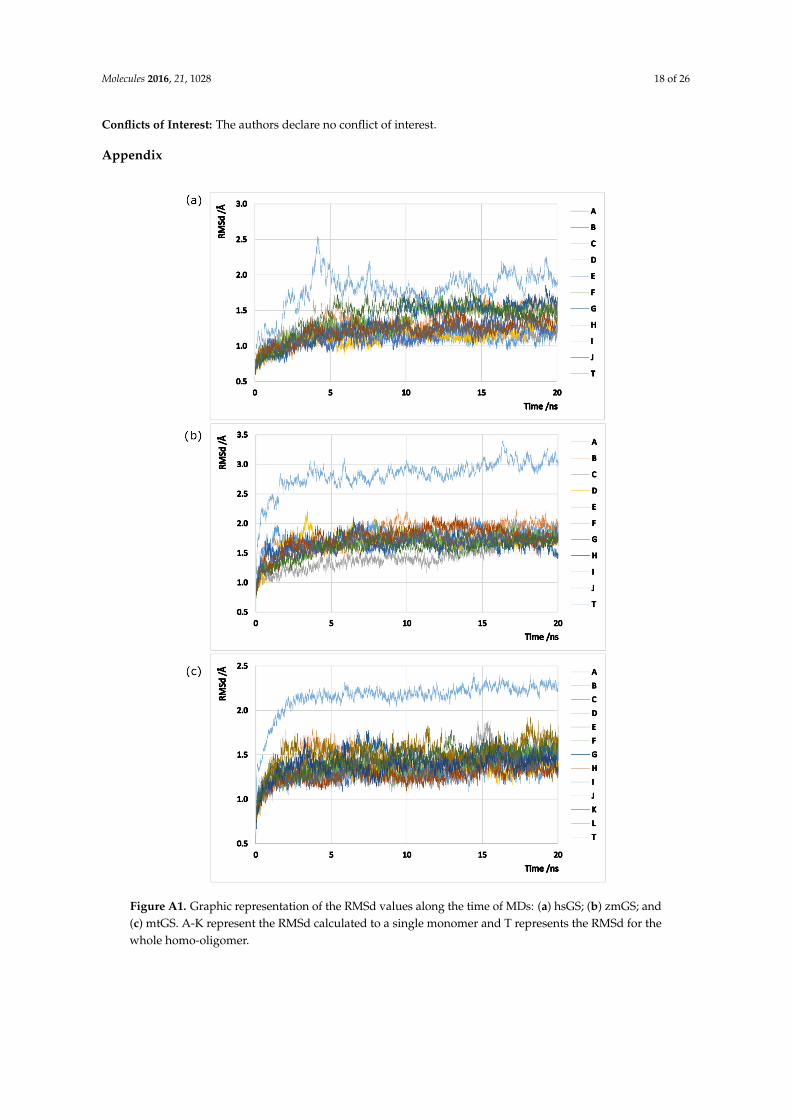
Figure A1. Graphic representation of the RMSd values along the time of MDs: (a) hsGS; (b) zmGS; and (c) mtGS. A-K represent the RMSd calculated to a single monomer and T represents the RMSd for the whole homo-oligomer.
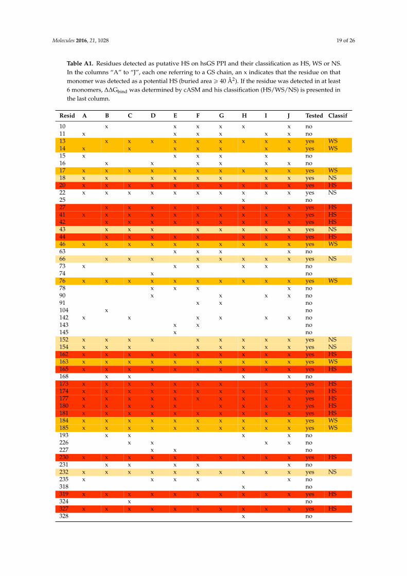
Table A1. Residues detected as putative HS on hsGS PPI and their classification as HS, WS or NS. In the columns “A” to “J”, each one referring to a GS chain, an x indicates that the residue on that monomer was detected as a potential HS (buried area ≥ 40 Å2). If the residue was detected in at least 6 monomers, ΔΔGbind was determined by cASM and his classification (HS/WS/NS) is presented in the last column.
Figure A1. Graphic representation of the RMSd values along the time of MDs: (a) hsGS; (b) zmGS; and(c) mtGS. A-K represent the RMSd calculated to a single monomer and T represents the RMSd for thewhole homo-oligomer.
Molecules 2016, 21, 1028 19 of 26
Table A1. Residues detected as putative HS on hsGS PPI and their classification as HS, WS or NS.In the columns “A” to “J”, each one referring to a GS chain, an x indicates that the residue on thatmonomer was detected as a potential HS (buried area ě 40 Å2). If the residue was detected in at least6 monomers, ∆∆Gbind was determined by cASM and his classification (HS/WS/NS) is presented inthe last column.
Resid A B C D E F G H I J Tested Classif
10 x x x x x x no11 x x x x x x no13 x x x x x x x x x yes WS14 x x x x x x x yes WS15 x x x x x no16 x x x x x x no17 x x x x x x x x x x yes WS18 x x x x x x x x yes NS20 x x x x x x x x x x yes HS22 x x x x x x x x x x yes NS25 x no27 x x x x x x x x x yes HS41 x x x x x x x x x x yes HS42 x x x x x x x x x yes HS43 x x x x x x x x yes NS44 x x x x x x x x yes HS46 x x x x x x x x x x yes WS63 x x x x no66 x x x x x x x x yes NS73 x x x x x no74 x no76 x x x x x x x x x x yes WS78 x x x x no90 x x x x no91 x x no104 x no142 x x x x x x no143 x x no145 x no152 x x x x x x x x x yes NS154 x x x x x x x x yes NS162 x x x x x x x x x x yes HS163 x x x x x x x x x x yes WS165 x x x x x x x x x x yes HS168 x x x x no173 x x x x x x x x yes HS174 x x x x x x x x x x yes HS177 x x x x x x x x x x yes HS180 x x x x x x x x x yes HS181 x x x x x x x x x x yes HS184 x x x x x x x x x x yes WS185 x x x x x x x x x x yes WS193 x x x x no226 x x x x no227 x x no230 x x x x x x x x x x yes HS231 x x x x x no232 x x x x x x x x x x yes NS235 x x x x x no318 x no319 x x x x x x x x x x yes HS324 x no327 x x x x x x x x x x yes HS328 x no
Molecules 2016, 21, 1028 20 of 26
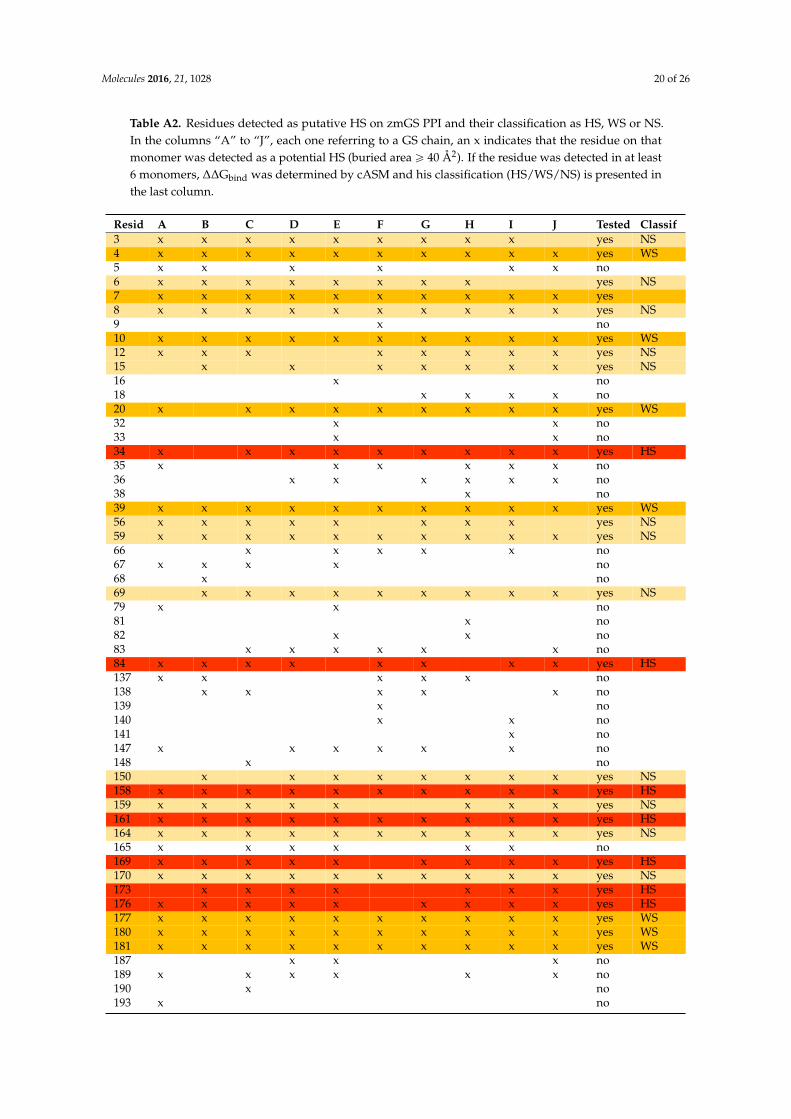
Table A2. Residues detected as putative HS on zmGS PPI and their classification as HS, WS or NS.In the columns “A” to “J”, each one referring to a GS chain, an x indicates that the residue on thatmonomer was detected as a potential HS (buried area ě 40 Å2). If the residue was detected in at least6 monomers, ∆∆Gbind was determined by cASM and his classification (HS/WS/NS) is presented inthe last column.
Resid A B C D E F G H I J Tested Classif3 x x x x x x x x x yes NS4 x x x x x x x x x x yes WS5 x x x x x x no6 x x x x x x x x yes NS7 x x x x x x x x x x yes8 x x x x x x x x x x yes NS9 x no10 x x x x x x x x x x yes WS12 x x x x x x x x yes NS15 x x x x x x x yes NS16 x no18 x x x x no20 x x x x x x x x x yes WS32 x x no33 x x no34 x x x x x x x x x yes HS35 x x x x x x no36 x x x x x x no38 x no39 x x x x x x x x x x yes WS56 x x x x x x x x yes NS59 x x x x x x x x x x yes NS66 x x x x x no67 x x x x no68 x no69 x x x x x x x x x yes NS79 x x no81 x no82 x x no83 x x x x x x no84 x x x x x x x x yes HS137 x x x x x no138 x x x x x no139 x no140 x x no141 x no147 x x x x x x no148 x no150 x x x x x x x x yes NS158 x x x x x x x x x x yes HS159 x x x x x x x x yes NS161 x x x x x x x x x x yes HS164 x x x x x x x x x x yes NS165 x x x x x x no169 x x x x x x x x x yes HS170 x x x x x x x x x x yes NS173 x x x x x x x yes HS176 x x x x x x x x x yes HS177 x x x x x x x x x x yes WS180 x x x x x x x x x x yes WS181 x x x x x x x x x x yes WS187 x x x no189 x x x x x x no190 x no193 x no
Molecules 2016, 21, 1028 21 of 26
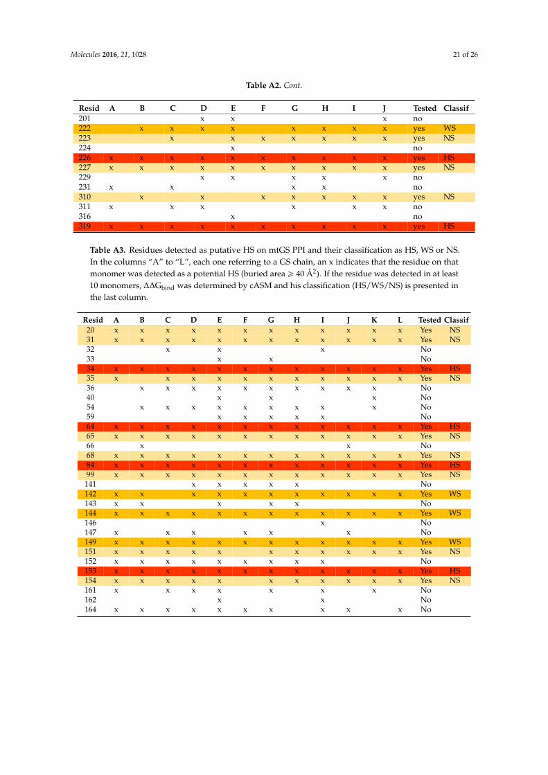
Table A2. Cont.
Resid A B C D E F G H I J Tested Classif201 x x x no222 x x x x x x x x yes WS223 x x x x x x x yes NS224 x no226 x x x x x x x x x x yes HS227 x x x x x x x x x x yes NS229 x x x x x no231 x x x x no310 x x x x x x x yes NS311 x x x x x x no316 x no319 x x x x x x x x x x yes HS
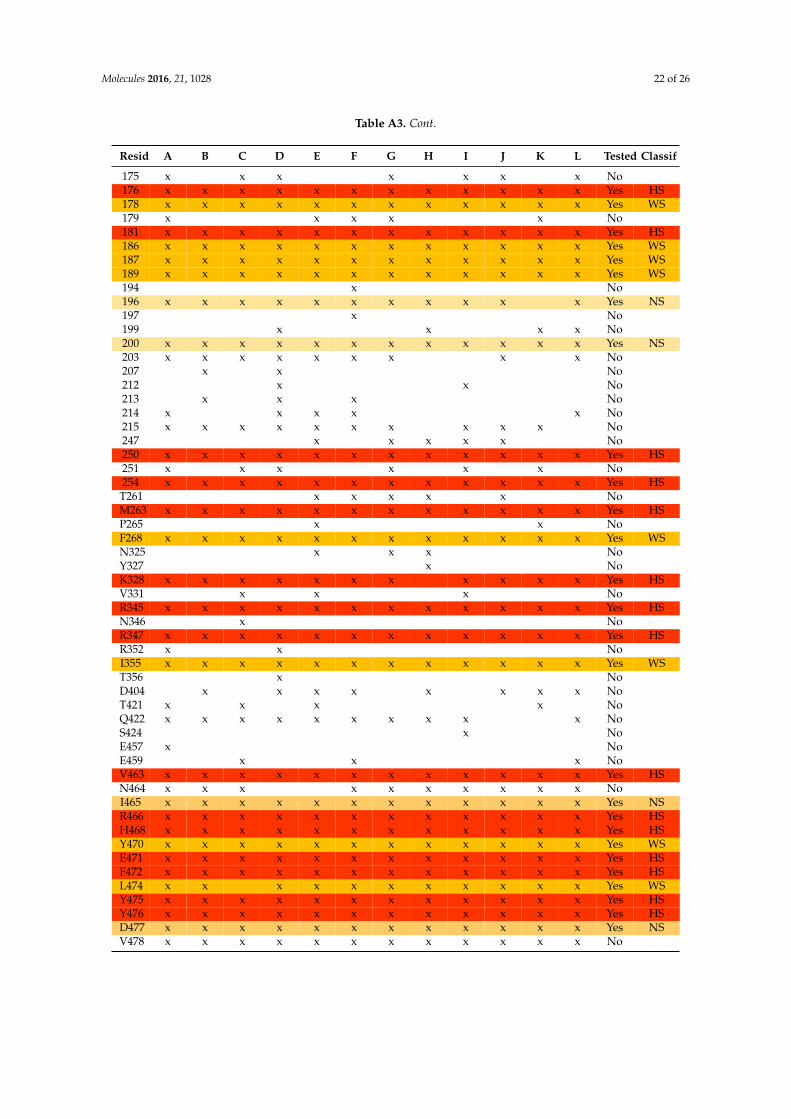
Table A3. Residues detected as putative HS on mtGS PPI and their classification as HS, WS or NS.In the columns “A” to “L”, each one referring to a GS chain, an x indicates that the residue on thatmonomer was detected as a potential HS (buried area ě 40 Å2). If the residue was detected in at least10 monomers, ∆∆Gbind was determined by cASM and his classification (HS/WS/NS) is presented inthe last column.
Resid A B C D E F G H I J K L Tested Classif20 x x x x x x x x x x x x Yes NS31 x x x x x x x x x x x x Yes NS32 x x x No33 x x No34 x x x x x x x x x x x x Yes HS35 x x x x x x x x x x x Yes NS36 x x x x x x x x x x No40 x x x No54 x x x x x x x x x No59 x x x x x No64 x x x x x x x x x x x x Yes HS65 x x x x x x x x x x x x Yes NS66 x x No68 x x x x x x x x x x x x Yes NS84 x x x x x x x x x x x x Yes HS99 x x x x x x x x x x x x Yes NS
141 x x x x x No142 x x x x x x x x x x x Yes WS143 x x x x x No144 x x x x x x x x x x x x Yes WS146 x No147 x x x x x x No149 x x x x x x x x x x x x Yes WS151 x x x x x x x x x x x Yes NS152 x x x x x x x x x No153 x x x x x x x x x x x x Yes HS154 x x x x x x x x x x x Yes NS161 x x x x x x x No162 x x No164 x x x x x x x x x x No
Molecules 2016, 21, 1028 22 of 26
Table A3. Cont.
Resid A B C D E F G H I J K L Tested Classif
175 x x x x x x x No176 x x x x x x x x x x x x Yes HS178 x x x x x x x x x x x x Yes WS179 x x x x x No181 x x x x x x x x x x x x Yes HS186 x x x x x x x x x x x x Yes WS187 x x x x x x x x x x x x Yes WS189 x x x x x x x x x x x x Yes WS194 x No196 x x x x x x x x x x x Yes NS197 x No199 x x x x No200 x x x x x x x x x x x x Yes NS203 x x x x x x x x x No207 x x No212 x x No213 x x x No214 x x x x x No215 x x x x x x x x x x No247 x x x x x No250 x x x x x x x x x x x x Yes HS251 x x x x x x No254 x x x x x x x x x x x x Yes HST261 x x x x x NoM263 x x x x x x x x x x x x Yes HSP265 x x NoF268 x x x x x x x x x x x x Yes WSN325 x x x NoY327 x NoK328 x x x x x x x x x x x Yes HSV331 x x x NoR345 x x x x x x x x x x x x Yes HSN346 x NoR347 x x x x x x x x x x x x Yes HSR352 x x NoI355 x x x x x x x x x x x x Yes WST356 x NoD404 x x x x x x x x NoT421 x x x x NoQ422 x x x x x x x x x x NoS424 x NoE457 x NoE459 x x x NoV463 x x x x x x x x x x x x Yes HSN464 x x x x x x x x x x NoI465 x x x x x x x x x x x x Yes NSR466 x x x x x x x x x x x x Yes HSH468 x x x x x x x x x x x x Yes HSY470 x x x x x x x x x x x x Yes WSE471 x x x x x x x x x x x x Yes HSF472 x x x x x x x x x x x x Yes HSL474 x x x x x x x x x x x Yes WSY475 x x x x x x x x x x x x Yes HSY476 x x x x x x x x x x x x Yes HSD477 x x x x x x x x x x x x Yes NSV478 x x x x x x x x x x x x No
Molecules 2016, 21, 1028 23 of 26
References
1. Rhee, S.G.; Chock, P.B. Mechanistic studies of glutamine synthetase from Escherichia coli: Kinetic evidence fortwo reaction intermediates in biosynthetic reaction. Proc. Natl. Acad. Sci. USA 1976, 73, 476–480. [CrossRef][PubMed]
2. Fedorova, K.; Kayumov, A.; Woyda, K.; Ilinskaja, O.; Forchhammer, K. Transcription factor TnrA inhibits thebiosynthetic activity of glutamine synthetase in Bacillus subtilis. FEBS Lett. 2013, 587, 1293–1298. [CrossRef][PubMed]
3. Almassy, R.J.; Janson, C.A.; Hamlin, R.; Xuong, N.H.; Eisenberg, D. Novel subunit[mdash]subunitinteractions in the structure of glutamine synthetase. Nature 1986, 323, 304–309. [CrossRef] [PubMed]
4. Moreira, C.; Ramos, M.J.; Fernandes, P.A. Reaction Mechanism of Mycobacterium Tuberculosis GlutamineSynthetase Using Quantum Mechanics/Molecular Mechanics Calculations. Chem Eur. J. 2016, 22, 9218–9225.[CrossRef] [PubMed]
5. Stadtman, E.R. The Story of Glutamine Synthetase Regulation. J. Biol. Chem. 2001, 276, 44357–44364.[CrossRef] [PubMed]
6. He, Y.-X.; Gui, L.; Liu, Y.-Z.; Du, Y.; Zhou, Y.; Li, P.; Zhou, C.-Z. Crystal structure of Saccharomycescerevisiae glutamine synthetase Gln1 suggests a nanotube-like supramolecular assembly. Proteins Struct.Funct. Bioinform. 2009, 76, 249–254. [CrossRef] [PubMed]
7. Ghoshroy, S.; Robertson, D.L. Molecular evolution of glutamine synthetase II and III in the chromalveolates1.J. Phycol. 2012, 48, 768–783. [CrossRef] [PubMed]
8. Kinoshita, S.; Isu, S.; Kaneko, G.; Yamada, H.; Hara, T.; Itoh, Y.; Watabe, S. The occurrence of eukaryotictype III glutamine synthetase in the marine diatom Chaetoceros compressum. Mar. Genom. 2009, 2, 103–111.[CrossRef] [PubMed]
9. Van Rooyen, J.M.; Abratt, V.R.; Belrhali, H.; Sewell, T. Crystal Structure of Type III Glutamine Synthetase:Surprising Reversal of the Inter-Ring Interface. Structure 2011, 19, 471–483. [CrossRef] [PubMed]
10. Brookes, G.; Barfoot, P. Economic impact of GM crops. GM Crops Food 2014, 5, 65–75. [CrossRef] [PubMed]11. Mao, Y.-C.; Hung, D.-Z.; Wu, M.-L.; Tsai, W.-J.; Wang, L.-M.; Ger, J.; Deng, J.-F.; Yang, C.-C. Acute human
Glufosinate-containing herbicide poisoning. Clin. Toxicol. 2012, 50, 396–402. [CrossRef] [PubMed]12. Inoue, Y.; Onodera, M.; Fujita, Y.; Fujino, Y.; Kikuchi, S.; Endo, S. Factors associated with severe effects
following acute glufosinate poisoning. Clin. Toxicol. 2013, 51, 846–849. [CrossRef] [PubMed]13. Hori, Y.; Tanaka, T.; Fujisawa, M.; Shimada, K. Toxicokinetics of DL-Glufosinate Enantiomer in Human
BASTA Poisoning. Biol. Pharm. Bull. 2003, 26, 540–543. [CrossRef] [PubMed]14. Occhipinti, A.; Berlicki, Ł.; Giberti, S.; Dziedzioła, G.; Kafarski, P.; Forlani, G. Effectiveness and mode of
action of phosphonate inhibitors of plant glutamine synthetase. Pest Manag. Sci. 2010, 66, 51–58. [CrossRef][PubMed]
15. Evstigneeva, Z.G.; Solov’eva, N.A.; Sidel’nikova, L.I. Methionine Sulfoximine and Phosphinothrycin:A Review of Their Herbicidal Activity and Effects on Glutamine Synthetase. Appl. Biochem. Microbiol.2003, 39, 539–543. [CrossRef]
16. Calas, A.-G.; Richard, O.; Même, S.; Beloeil, J.-C.; Doan, B.-T.; Gefflaut, T.; Même, W.; Crusio, W.E.;Pichon, J.; Montécot, C. Chronic exposure to glufosinate-ammonium induces spatial memory impairments,hippocampal MRI modifications and glutamine synthetase activation in mice. NeuroToxicology 2008, 29,740–747. [CrossRef] [PubMed]
17. Mowbray, S.; Kathiravan, M.; Pandey, A.; Odell, L. Inhibition of Glutamine Synthetase: A Potential DrugTarget in Mycobacterium tuberculosis. Molecules 2014, 19, 13161–13176. [CrossRef] [PubMed]
18. Lukasz, B. Inhibitors of Glutamine Synthetase and their Potential Application in Medicine. Mini-Rev.Med. Chem. 2008, 8, 869–878.
19. Harth, G.; Clemens, D.L.; Horwitz, M.A. Glutamine synthetase of Mycobacterium tuberculosis: Extracellularrelease and characterization of its enzymatic activity. Proc. Natl Acad. Sci. USA 1994, 91, 9342–9346.[CrossRef] [PubMed]
20. Harth, G.; Horwitz, M.A. Inhibition of Mycobacterium tuberculosis Glutamine Synthetase as a Novel AntibioticStrategy against Tuberculosis: Demonstration of Efficacy In Vivo. Infect. Immun. 2003, 71, 456–464. [CrossRef][PubMed]
Molecules 2016, 21, 1028 24 of 26
21. Couturier, C.; Silve, S.; Morales, R.; Pessegue, B.; Llopart, S.; Nair, A.; Bauer, A.; Scheiper, B.; Pöverlein, C.;Ganzhorn, A.; et al. Nanomolar inhibitors of Mycobacterium tuberculosis glutamine synthetase 1: Synthesis,biological evaluation and X-ray crystallographic studies. Bioorg. Med. Chem. Lett. 2015, 25, 1455–1459.[CrossRef] [PubMed]
22. Gising, J.; Nilsson, M.T.; Odell, L.R.; Yahiaoui, S.; Lindh, M.; Iyer, H.; Sinha, A.M.; Srinivasa, B.R.; Larhed, M.;Mowbray, S.L.; et al. Trisubstituted Imidazoles as Mycobacterium tuberculosis Glutamine Synthetase Inhibitors.J. Med. Chem. 2012, 55, 2894–2898. [CrossRef] [PubMed]
23. Nilsson, M.T.; Krajewski, W.W.; Yellagunda, S.; Prabhumurthy, S.; Chamarahally, G.N.; Siddamadappa, C.;Srinivasa, B.R.; Yahiaoui, S.; Larhed, M.; Karlén, A.; et al. Structural Basis for the Inhibition ofMycobacterium tuberculosis Glutamine Synthetase by Novel ATP-Competitive Inhibitors. J. Mol. Biol. 2009,393, 504–513. [CrossRef] [PubMed]
24. Berlicki, Ł.; Kafarski, P. Computer-aided analysis of the interactions of glutamine synthetase with itsinhibitors. Bioorg. Med. Chem. 2006, 14, 4578–4585. [CrossRef] [PubMed]
25. Krajewski, W.W.; Jones, T.A.; Mowbray, S.L. Structure of Mycobacterium tuberculosis glutamine synthetase incomplex with a transition-state mimic provides functional insights. Proc. Natl. Acad. Sci. USA 2005, 102,10499–10504. [CrossRef] [PubMed]
26. Gill, H.S.; Eisenberg, D. The Crystal Structure of Phosphinothricin in the Active Site of Glutamine SynthetaseIlluminates the Mechanism of Enzymatic Inhibition. Biochemistry 2001, 40, 1903–1912. [CrossRef] [PubMed]
27. Adeva, M.M.; Souto, G.; Blanco, N.; Donapetry, C. Ammonium metabolism in humans. Metabolism 2012, 61,1495–1511. [CrossRef] [PubMed]
28. Frieg, B.; Görg, B.; Homeyer, N.; Keitel, V.; Häussinger, D.; Gohlke, H. Molecular Mechanisms of GlutamineSynthetase Mutations that Lead to Clinically Relevant Pathologies. PLoS Comput. Biol. 2016, 12, e1004693.[CrossRef] [PubMed]
29. Häberle, J.; Shahbeck, N.; Ibrahim, K.; Hoffmann, G.F.; Ben-Omran, T. Natural course of glutamine synthetasedeficiency in a 3 year old patient. Mol. Genet. Metab. 2011, 103, 89–91. [CrossRef] [PubMed]
30. Betti, M.; García-Calderón, M.; Pérez-Delgado, C.M.; Credali, A.; Estivill, G.; Galván, F.; Vega, J.M.;Márquez, A.J. Glutamine Synthetase in Legumes: Recent Advances in Enzyme Structure and FunctionalGenomics. Int. J. Mol. Sci. 2012, 13, 7994–8024. [CrossRef] [PubMed]
31. Bernard, S.M.; Habash, D.Z. The importance of cytosolic glutamine synthetase in nitrogen assimilation andrecycling. New Phytol. 2009, 182, 608–620. [CrossRef] [PubMed]
32. Brusilow, S.W.; Koehler, R.C.; Traystman, R.J.; Cooper, A.J.L. Astrocyte Glutamine Synthetase: Importancein Hyperammonemic Syndromes and Potential Target for Therapy. Neurotherapeutics 2010, 7, 452–470.[CrossRef] [PubMed]
33. Suárez, I.; Bodega, G.; Fernández, B. Glutamine synthetase in brain: Effect of ammonia. Neurochem. Int. 2002,41, 123–142. [CrossRef]
34. Gebhardt, R.; Gaunitz, F.; Mecke, D. Heterogeneous (positional) expression of hepatic glutamine synthetase:Features, regulation and implications for hepatocarcinogenesis. Adv. Enzyme Regul. 1994, 34, 27–56.[CrossRef]
35. Gebhardt, R.; Baldysiak-Figiel, A.; Krügel, V.; Ueberham, E.; Gaunitz, F. Hepatocellular expression ofglutamine synthetase: An indicator of morphogen actions as master regulators of zonation in adult liver.Prog. Histochem. Cytochem. 2007, 41, 201–266. [CrossRef] [PubMed]
36. Moreira, I.S.; Fernandes, P.A.; Ramos, M.J. Hot spots—A review of the protein–protein interface determinantamino-acid residues. Proteins Struct. Funct. Bioinform. 2007, 68, 803–812. [CrossRef] [PubMed]
37. Moreira, I.S.; Fernandes, P.A.; Ramos, M.J. Computational Alanine Scanning Mutagenesis-an ImprovedMethodological Approach. J. Comput. Chem. 2007, 28, 644–654. [CrossRef] [PubMed]
38. Guo, W.; Wisniewski, J.A.; Ji, H. Hot spot-based design of small-molecule inhibitors for protein–proteininteractions. Bioorganic Med. Chem. Lett. 2014, 24, 2546–2554. [CrossRef] [PubMed]
39. Lise, S.; Archambeau, C.; Pontil, M.; Jones, D.T. Prediction of hot spot residues at protein-protein interfacesby combining machine learning and energy-based methods. BMC Bioinform. 2009, 10, 1–17. [CrossRef][PubMed]
40. Sheng, C.; Dong, G.; Miao, Z.; Zhang, W.; Wang, W. State-of-the-art strategies for targeting protein-proteininteractions by small-molecule inhibitors. Chem. Soc. Rev. 2015, 44, 8238–8259. [CrossRef] [PubMed]
Molecules 2016, 21, 1028 25 of 26
41. Arkin, M.R.; Tang, Y.; Wells, J.A. Small-molecule inhibitors of protein-protein interactions: Progressingtowards the reality. Chem. Biol. 2014, 21, 1102–1114. [CrossRef] [PubMed]
42. González-Ruiz, D.; Gohlke, H. Targeting Protein-protein Interactions with Small Molecules: Challenges andPerspectives for Computational Binding Epitope Detection and Ligand Finding. Curr. Med. Chem. 2006, 13,2607–2625. [CrossRef] [PubMed]
43. Schames, J.R.; Henchman, R.H.; Siegel, J.S.; Sotriffer, C.A.; Ni, H.; McCammon, J.A. Discovery of a NovelBinding Trench in HIV Integrase. J. Med. Chem. 2004, 47, 1879–1881. [CrossRef] [PubMed]
44. Summa, V.; Petrocchi, A.; Bonelli, F.; Crescenzi, B.; Donghi, M.; Ferrara, M.; Fiore, F.; Gardelli, C.;Gonzalez Paz, O.; Hazuda, D.J.; et al. Discovery of Raltegravir, a Potent, Selective Orally BioavailableHIV-Integrase Inhibitor for the Treatment of HIV-AIDS Infection. J. Med. Chem. 2008, 51, 5843–5855.[CrossRef] [PubMed]
45. Wassman, C.D.; Baronio, R.; Demir, Ö.; Wallentine, B.D.; Chen, C.-K.; Hall, L.V.; Salehi, F.; Lin, D.-W.;Chung, B.P.; Wesley Hatfield, G.; et al. Computational identification of a transiently open L1/S3 pocket forreactivation of mutant p53. Nat. Commun. 2013, 4, 1407. [CrossRef] [PubMed]
46. Pietrucci, F.; Vargiu, A.V.; Kranjc, A. HIV-1 Protease Dimerization Dynamics Reveals a Transient DruggableBinding Pocket at the Interface. Sci. Rep. 2015, 5, 18555. [CrossRef] [PubMed]
47. Ribeiro, J.V.; Cerqueira, N.M.F.S.A.; Moreira, I.S.; Fernandes, P.A.; Ramos, M.J. CompASM: An Amber-VMDalanine scanning mutagenesis plug-in. Theor. Chem. Acc. 2012, 131, 1–7. [CrossRef]
48. Kay, B.K.; Williamson, M.P.; Sudol, M. The importance of being proline: The interaction of proline-rich motifsin signaling proteins with their cognate domains. FASEB J. 2000, 14, 231–241. [PubMed]
49. Zarrinpar, A.; Bhattacharyya, R.P.; Lim, W.A. The Structure and Function of Proline Recognition Domains.Sci. Signal. 2003, 2003. [CrossRef] [PubMed]
50. Zondlo, N.J. Aromatic–Proline Interactions: Electronically Tunable CH/π Interactions. Acc. Chem. Res. 2013,46, 1039–1049. [CrossRef] [PubMed]
51. Masalkar, P.D.; Roberts, D.M. Glutamine synthetase isoforms in nitrogen-fixing soybean nodules: Distinctoligomeric structures and thiol-based regulation. FEBS Lett. 2015, 589, 215–221. [CrossRef] [PubMed]
52. Schmidtke, P.; Barril, X. Understanding and Predicting Druggability. A High-Throughput Method forDetection of Drug Binding Sites. J. Med. Chem. 2010, 53, 5858–5867. [CrossRef] [PubMed]
53. Veber, D.F.; Johnson, S.R.; Cheng, H.-Y.; Smith, B.R.; Ward, K.W.; Kopple, K.D. Molecular Properties thatInfluence the Oral Bioavailability of Drug Candidates. J. Med. Chem. 2002, 45, 2615–2623. [CrossRef][PubMed]
54. Allen, J.W.; Mutkus, L.A.; Aschner, M. Mercuric chloride, but not methylmercury, inhibits glutaminesynthetase activity in primary cultures of cortical astrocytes. Brain Res. 2001, 891, 148–157. [CrossRef]
55. Singh, U.; Panchanadikar, V.; Sarkar, D. Development of a Simple Assay Protocol for High-ThroughputScreening of Mycobacterium tuberculosis Glutamine Synthetase for the Identification of Novel Inhibitors.J. Biomol. Screen. 2005, 10, 725–729. [CrossRef] [PubMed]
56. Olof, L.; Luke, R.O.; Sherry, L.M.; Mikael, T.N.; Wojciech, W.K.; Anneli, N.; Anders, K.; Mats, L.Microwave-Enhanced alpha-Arylation of a Protected Glycine in Water:Evaluation of 3-PhenylglycineDerivatives as Inhibitors of the Tuberculosis Enzyme, Glutamine Synthetase. Comb. Chem. HighThroughput Screen. 2007, 10, 783–789.
57. Gxoyiya, B.S.B.; Kaye, P.T.; Kenyon, C. Benzimidazole-Derived ATP Analogues as Potential GlutamineSynthetase Inhibitors. Synth. Commun. 2010, 40, 2578–2587. [CrossRef]
58. Salisu, S.; Kenyon, C.; Kaye, P.T. Studies Towards the Synthesis of ATP Analogs as Potential GlutamineSynthetase Inhibitors. Synth. Commun. 2011, 41, 2216–2225. [CrossRef]
59. Odell, L.R.; Nilsson, M.T.; Gising, J.; Lagerlund, O.; Muthas, D.; Nordqvist, A.; Karlén, A.; Larhed, M.Functionalized 3-amino-imidazo[1,2-a]pyridines: A novel class of drug-like Mycobacterium tuberculosisglutamine synthetase inhibitors. Bioorg. Med. Chem. Lett. 2009, 19, 4790–4793. [CrossRef] [PubMed]
60. Reddy Chichili, V.P.; Kumar, V.; Sivaraman, J. Linkers in the structural biology of protein–protein interactions.Protein Sci. Publ. Protein Soc. 2013, 22, 153–167. [CrossRef] [PubMed]
61. Mohan, A.; Oldfield, C.J.; Radivojac, P.; Vacic, V.; Cortese, M.S.; Dunker, A.K.; Uversky, V.N. Analysis ofMolecular Recognition Features (MoRFs). J. Mol. Biol. 2006, 362, 1043–1059. [CrossRef] [PubMed]
62. Mészáros, B.; Tompa, P.; Simon, I.; Dosztányi, Z. Molecular Principles of the Interactions of DisorderedProteins. J. Mol. Biol. 2007, 372, 549–561. [CrossRef] [PubMed]
Molecules 2016, 21, 1028 26 of 26
63. Dunker, A.K. Another Window into Disordered Protein Function. Structure 2007, 15, 1026–1028. [CrossRef][PubMed]
64. Zhang, Y.; Stec, B.; Godzik, A. Between Order and Disorder in Protein Structures: Analysis of “DualPersonality” Fragments in Proteins. Structure 2007, 15, 1141–1147. [CrossRef] [PubMed]
65. Uversky, V.N.; Dunker, A.K. Understanding protein non-folding. Biochim. Biophys. Acta 2010, 1804, 1231–1264.[CrossRef] [PubMed]
66. Turoverov, K.K.; Kuznetsova, I.M.; Uversky, V.N. The protein kingdom extended: Ordered and intrinsicallydisordered proteins, their folding, supramolecular complex formation, and aggregation. Prog. Biophys.Mol. Biol. 2010, 102, 73–84. [CrossRef] [PubMed]
67. Harth, G.; Masleša-Galic, S.; Tullius, M.V.; Horwitz, M.A. All four Mycobacterium tuberculosis glnA genesencode glutamine synthetase activities but only GlnA1 is abundantly expressed and essential for bacterialhomeostasis. Mol. Microbiol. 2005, 58, 1157–1172. [CrossRef] [PubMed]
68. Unno, H.; Uchida, T.; Sugawara, H.; Kurisu, G.; Sugiyama, T.; Yamaya, T.; Sakakibara, H.; Hase, T.;Kusunoki, M. Atomic Structure of Plant Glutamine Synthetase: A key enzyme for plant productivity.J. Biol. Chem. 2006, 281, 29287–29296. [CrossRef] [PubMed]
69. Krajewski, W.W.; Collins, R.; Holmberg-Schiavone, L.; Jones, T.A.; Karlberg, T.; Mowbray, S.L. CrystalStructures of Mammalian Glutamine Synthetases Illustrate Substrate-Induced Conformational Changes andProvide Opportunities for Drug and Herbicide Design. J. Mol. Biol. 2008, 375, 217–228. [CrossRef] [PubMed]
70. Maurizi, M.R.; Ginsburg, A. Active site ligand stabilization of quaternary structures of glutamine synthetasefrom Escherichia coli. J. Biol. Chem. 1982, 257, 7246–7251. [PubMed]
71. Murray, D.S.; Chinnam, N.; Tonthat, N.K.; Whitfill, T.; Wray, L.V.; Fisher, S.H.; Schumacher, M.A. Structures ofthe Bacillus subtilis Glutamine Synthetase Dodecamer Reveal Large Intersubunit Catalytic ConformationalChanges Linked to a Unique Feedback Inhibition Mechanism. J. Biol. Chem. 2013, 288, 35801–35811.[CrossRef] [PubMed]
72. Case, D.A.; Darden, T.A.; Cheatham, T.E.; Simmerling, C.L.; Wang, J.; Duke, R.E.; Luo, R.; Walker, R.C.;Zhang, W.; Merz, K.M.; et al. AMBER 12; University of California: San Francisco, CA, USA, 2012.
73. Duan, Y.; Wu, C.; Chowdhury, S.; Lee, M.C.; Xiong, G.; Zhang, W.; Yang, R.; Cieplak, P.; Luo, R.; Lee, T.;et al. A point-charge force field for molecular mechanics simulations of proteins based on condensed-phasequantum mechanical calculations. J. Comput. Chem. 2003, 24, 1999–2012. [CrossRef] [PubMed]
74. Xu, L.; Sun, H.; Li, Y.; Wang, J.; Hou, T. Assessing the Performance of MM/PBSA and MM/GBSA Methods.3. The Impact of Force Fields and Ligand Charge Models. J. Phys. Chem. B 2013, 117, 8408–8421. [CrossRef][PubMed]
75. Martins, S.A.; Perez, M.A.S.; Moreira, I.S.; Sousa, S.F.; Ramos, M.J.; Fernandes, P.A. Computational AlanineScanning Mutagenesis: MM-PBSA vs. TI. J. Chem. Theory Comput. 2013, 9, 1311–1319. [CrossRef] [PubMed]
76. Le Guilloux, V.; Schmidtke, P.; Tuffery, P. Fpocket: An open source platform for ligand pocket detection.BMC Bioinform. 2009, 10, 1–11. [CrossRef] [PubMed]
77. O’Boyle, N.M.; Banck, M.; James, C.A.; Morley, C.; Vandermeersch, T.; Hutchison, G.R. Open Babel: An openchemical toolbox. J. Cheminformatics 2011, 3, 1–14. [CrossRef] [PubMed]
78. Trott, O.; Olson, A.J. AutoDock Vina: Improving the speed and accuracy of docking with a new scoringfunction, efficient optimization, and multithreading. J. Comput. Chem. 2010, 31, 455–461. [CrossRef][PubMed]
Sample Availability: Not Available.
© 2016 by the authors; licensee MDPI, Basel, Switzerland. This article is an open accessarticle distributed under the terms and conditions of the Creative Commons Attribution(CC-BY) license (http://creativecommons.org/licenses/by/4.0/).