
Budd-Chiari Syndrome and Portal Vein Thrombosis - RePub
166
Budd-Chiari Syndrome and Portal Vein Thrombosis Etiology and Treatment Jasper H. Smalberg
Transcript of Budd-Chiari Syndrome and Portal Vein Thrombosis - RePub
Budd-Chiari Syndrome and
Portal Vein Thrombosis
Etiology and Treatment
Jasper H. Smalberg
Financial support by the Dutch Heart Foundation for the publication of this thesis is gratefully acknowledged.
Publication of this thesis was financially supported by:Fonds Wetenschappelijk Onderzoek van de MPN Stichting, Alexion, Bayer, Celgene, Novartis, Pfizer, Shire, The J.E. Jurriaanse Stichting.
Cover: Veins of the hepatic portal system, Science Photo LibraryLay-out: Legatron Electronic PublishingPrinting: Ipskamp Drukkers BV, Enschede
ISBN/EAN: 978-94-6191-144-5
2012 ©J.H. Smalberg
No part of this thesis may be reproduced, stored in a retrieval system or transmitted in any form or by any means, without written permission of the author or, when appropriate, of the publishers of the publications.
Budd-Chiari Syndrome and
Portal Vein Thrombosis
Etiology and Treatment
Etiologie en behandeling van het Budd-Chiari syndroom en vena portae trombose
Proefschrift
ter verkrijging van de graad van doctor aan de
Erasmus Universiteit Rotterdam
op gezag van de
rector magnificus
Prof.dr. H.G. Schmidt
en volgens besluit van het College voor Promoties.
De openbare verdediging zal plaatsvinden op
donderdag 12 januari 2012 om 09.30 uur
door
Jasper Hoite Smalberg
geboren op 10 juli 1980 te Hellevoetsluis

PromoTiECommiSSiE
Promotor:Prof.dr. F.W.G. LeebeekProf.dr. H.L.A. Janssen
overige leden:Prof.dr. H.J. MetselaarProf.dr. R.J. Porte Prof.dr. P. Sonneveld
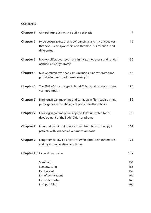
CoNTENTS
Chapter 1 General introduction and outline of thesis 7
Chapter 2 Hypercoagulability and hypofibrinolysis and risk of deep vein 15 thrombosis and splanchnic vein thrombosis: similarities and differences
Chapter 3 Myeloproliferative neoplasms in the pathogenesis and survival 35 of Budd-Chiari syndrome
Chapter 4 Myeloproliferative neoplasms in Budd-Chiari syndrome and 53 portal vein thrombosis: a meta-analysis
Chapter 5 The JAK2 46/1 haplotype in Budd-Chiari syndrome and portal 73 vein thrombosis
Chapter 6 Fibrinogen gamma prime and variation in fibrinogen gamma 89 prime genes in the etiology of portal vein thrombosis
Chapter 7 Fibrinogen gamma prime appears to be unrelated to the 103 development of the Budd-Chiari syndrome
Chapter 8 Risks and benefits of transcatheter thrombolytic therapy in 109 patients with splanchnic venous thrombosis
Chapter 9 Long-term follow-up of patients with portal vein thrombosis 121 and myeloproliferative neoplasms
Chapter 10 General discussion 137 Summary 151 Samenvatting 155 Dankwoord 159 List of publications 162 Curriculum vitae 163 PhD portfolio 165

C H A P T E r 1
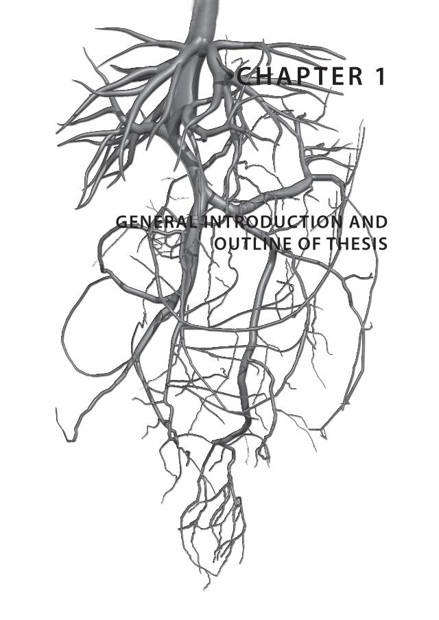
GENErAL iNTroDUCTioN AND oUTLiNE oF THESiS
8 | Chapter 1
Venous thrombosis is a common disorder with an annual incidence of around 1-2 cases per 1.000 individuals and is the third leading cause of cardiovascular morbidity and mortality in developed countries.1-4 Thrombosis may arise in any section of the venous system, but it typically occurs in the deep veins of the lower extremities. The major concern in these patients is pulmonary embolism, which can be fatal. A more common, but often disabling, complication of deep vein thrombosis and its sequelae is the post-thrombotic syndrome.5 Rarely, thrombosis may involve other venous sites. One of these uncommon manifestations of thrombosis is located in the splanchnic veins, which is accompanied by a considerable morbidity and mortality.
SPLANCHNiC VEiN THromBoSiS
Splanchnic vein thrombosis (SVT) encompasses hepatic vein thrombosis (Budd-Chiari syndrome, BCS), portal vein thrombosis (PVT), and mesenteric vein thrombosis. BCS and PVT are the two most frequent manifestations of SVT, and although in these disorders distinct venous sites are affected, simultaneous involvement of these venous districts is frequently encountered.6
The splanchnic venous system comprises the portal vein and its branches that direct blood flow from the gastrointestinal organs to the liver. The portal vein is formed by the union of the superior mesenteric vein and the splenic vein, and subdivides in a left and right branch, which are segmentally distributed throughout the liver. The terminal portal venules drain into the sinusoids, after which the blood flows from the small to large hepatic veins, ultimately reaching the inferior vena cava. BCS is defined as an obstruction of the hepatic venous outflow tract from the level of the small hepatic veins to the entrance of the inferior vena cava into the right atrium. Outflow obstruction caused by hepatic veno-occlusive disease and cardiac disorders is excluded from this definition. BCS is considered primary when obstruction of the venous tract is the result of an endoluminal lesion, i.e. thrombosis, and secondary when obstruction results from invasion by a local malignant tumor or from extrinsic compression by a tumor, cyst or abscess.7 BCS is a rare disorder with an annual incidence of about 0.2-0.8 per million inhabitants in the Western world, predominantly affecting young females.8-10 Main complications are the result of portal hypertension and liver dysfunction. The classical triad of symptoms in BCS consists of abdominal pain, ascites and hepatomegaly, frequently accompanied by a variable degree of alterations in liver biochemical tests. However, clinical presentation may range from absence of symptoms, in case of preservation of hepatic veins and/or formation of collaterals, to fulminant hepatic failure, with an acute or chronic development of symptoms ranging from weeks to months.6 With contemporary management, the survival rate is 87% at one year and 82% at two years.11
Chap
ter 1
9Introduction |
In PVT, the obstruction is located in the extra-hepatic portal vein, but involvement of the intra-hepatic portal, superior mesenteric and splenic vein may occur. Although PVT is considered a rare disorder, a recent autopsy study reported a prevalence of 1%.12 Clinically, PVT can be classified as acute or chronic, which represent successive stages of the same disease and share similar causes. Complications of portal hypertension, such as gastrointestinal bleeding from oesophageal varices and splenomegaly, are the most important clinical manifestations of PVT.6 Furthermore, if thrombosis extends into the mesenteric vein, there is a substantial risk of bowel infarction, which is the most severe complication of acute PVT.13 The prognosis of patients with PVT is mainly determined by the underlying cause. Survival of patients with non-cirrhotic, non-malignant PVT can be considered good. In this group of PVT patients, five- and ten-year survival rates are 90% and 80%, respectively.14 PVT patients with an underlying liver cirrhosis or hepato-biliary malignancy generally have an inferior prognosis.
EtiologyLocal risk factors for the development of BCS include solid malignancies, parasitic masses, cysts or abscesses that either compress or invade the venous tract.7 In the Western world, BCS is infrequently caused by local risk factors. PVT, on the other hand, is most often encountered as a complication of liver cirrhosis or hepatobiliary malignancies. Other frequent local risk factors for PVT are surgical trauma to the portal vein and inflammatory foci in the abdomen, which are often accompanied by an additional prothrombotic condition. The etiology of primary BCS and non-malignant, non-cirrhotic PVT often involves systemic, prothrombotic conditions. Recent studies with a near complete work-up showed that prothrombotic factors are present in up to 84% and 42% in primary BCS and non-malignant, non-cirrhotic PVT, respectively.11,15 These conditions largely overlap with the risk factors for common venous thrombosis and can be divided into genetic and acquired risk factors. Genetic risk factors include protein C, protein S and antithrombin deficiencies, the factor V Leiden mutation and the prothrombin G20210A gene variant. Acquired risk factors include antiphospholipid antibodies, paroxysmal nocturnal hemoglubinuria, hormonal factors, auto-immune diseases and myeloproliferative neoplasms (MPNs), which remarkably are the most prominent risk factor for the development of both BCS and PVT. MPNs are chronic clonal hematopoietic stem cell disorders characterized by an overproduction of mature and functional granulocytes, red blood cells and/or platelets.16 The exact pathogenetic mechanism of thrombosis in MPNs remains elusive, but besides characteristic erythrocytosis and thrombocytosis, platelet and leukocyte functional abnormalities appear critical.17 MPNs have been reported in approximately one third and one half of BCS and PVT patients, respectively.18 Diagnosis of MPNs in these patients is notoriously difficult. Portal hypertension, resulting from pre- or post-hepatic venous
10 | Chapter 1
obstruction, can lead to hypersplenism and hemodilution. Both these conditions may mask the characteristic peripheral blood cell changes and make diagnosis of MPN more difficult. Previously, diagnosis of MPNs in these patients often relied on bone marrow (BM) biopsy findings and growth of erythroid colonies in the absence of exogenous erythropoietin, referred to as spontaneous endogenous erythroid colonies (EEC). Patients were labelled as having so-called occult MPN when either bone marrow biopsy was highly suggestive of MPN or EEC was present, but in whom traditional criteria for MPN could not be fulfilled due to normal peripheral blood cell counts.19 In 2005, the JAK2V617F gain of function mutation was discovered, which is present in more than 95% of cases of polycythemia vera and 50% to 60% of essential thrombocythemia and primary myelofibrosis. JAK2V617F has radically changed the diagnostic landscape of MPNs and has been included as one of the cornerstones in the 2008 World Health Organization classification for hematological malignancies.20 Interestingly, the JAK2V617F mutation is not seen in nonmyeloid malignancies21 and therefore offers an additional tool to detect occult MPNs in BCS and PVT patients. Recent studies have consistently shown that the etiology of primary BCS and non-cirrhotic, non-malignant PVT must be considered multifactorial, as in common forms of venous thrombosis. Recent studies reported a combination of two or more genetic or acquired prothrombotic factors in 46% of BCS and 48% of PVT patients.11,15 In this large cohort of BCS patients, 18% of the patients even displayed three risk factors.
TreatmentRandomized clinical studies on the efficacy of the treatment options of BCS and PVT are lacking and current therapy guidelines are therefore based on cohort studies and expert opinions. In both disorders, prompt recognition and treatment of underlying disorders is recommended. As in common forms of venous thrombosis, anticoagulant therapy is the cornerstone of the management of BCS and PVT. The aim of anticoagulation is to reduce the risk of thrombus progression into adjacent vessels and to improve the rate of recanalization. Immediate therapy with low molecular weight heparin followed by life-long oral anticoagulant therapy is recommended for all patients with primary BCS, irrespective of whether an underlying prothrombotic disorder has been identified.22 Previous portal hypertension related bleeding is not considered a contraindication, provided that appropriate prophylaxis for recurrent bleeding is undertaken, for example using beta-blockers and/or endoscopic therapy. In BCS percutaneous transluminal angioplasty or insertion of a transjugular intrahepatic portosystemic shunt (TIPS) is warranted to induce decompression of the liver vasculature. Liver transplantation should be considered in deteriorating BCS patients in whom the disease cannot be controlled with the above described options.7,10,22
Chap
ter 1
11Introduction |
There is currently much debate on the optimal strategy of anticoagulant treatment in PVT patients, as potential beneficial effects of preventing extension or recurrent thrombosis may be outweighed by the inherent risk of bleeding complications. According to current consensus, non-cirrhotic patients with acute PVT should be treated with anticoagulant therapy for at least three months, unless a persisting underlying prothrombotic factor is present, in which case life-long treatment is recommended.22 Non-cirrhotic patients with chronic PVT may also be treated life-long in case of persisting prothrombotic factors, whereas it is generally discouraged in patients without a hypercoagulable state.22 There is currently no evidence to support the use of anticoagulant therapy in either acute or chronic PVT patients with concomitant cirrhosis.23 Adequate prophylaxis for bleeding complications by means of beta-blockers or endoscopic therapy in patients receiving anticoagulation therapy is essential. Although two recent series demonstrate that TIPS is feasible and effective in treating complications of portal hypertension in patients with liver cirrhosis and extensive PVT,24,25 there is currently insufficient evidence in favour of interventional therapy such as TIPS placement in patients with non-malignant, non-cirrhotic PVT.22 Thrombolytic therapy using streptokinase or recombinant tissue-plasminogen activator in patients with thrombosis of the splanchnic veins is controversial and its place in the treatment of these disorders is not fully established. Successful treatment has been reported either in patients with acute, extended thrombosis of the splanchnic veins, or as a rescue therapy in case of acute thrombosis during percutaneous transluminal angioplasty or TIPS insertion.26-33 However, evidence is mostly based on single case studies and small case series and these findings should therefore be interpreted with caution.
AimS AND oUTLiNE oF THiS THESiS
The focus of this thesis is on the etiology and treatment of patients with primary BCS and non-malignant, non-cirrhotic PVT. For this purpose, several studies addressing different aspects of the etiology and treatment of these disorders will be performed. In chapter 2, the current insights in the risk factors for commonly occurring venous thrombosis, in particular deep vein thrombosis and pulmonary embolism, are reviewed. We will also provide an overview of the risk factors for BCS and PVT. We discuss similarities, but some apparent differences in the risk profiles between these forms of venous thrombosis that may provide new insights into to the site-specificity of venous thrombosis. In chapter 3 we explore the etiology of BCS by means of a single-center cohort study and evaluate the presence of concomitant prothrombotic factors in patients who were previously diagnosed with an underlying MPN. In addition, we assessed the prevalence of the JAK2V617F mutation and investigate its clinical utility in the detection of occult MPNs.
12 | Chapter 1
Since the discovery of the somatic JAK2V617F mutation in 2005, numerous studies have been performed on the association between JAK2V617F and the development of SVT. In chapter 4 we report a meta-analysis in which we assess the prevalence of MPNs and its subtypes and JAK2V617F in both BCS and PVT, and evaluate the clinical value of screening for JAK2V617F in the detection of MPNs in patients without elevated peripheral blood counts. The discovery of the JAK2 46/1 haplotype in 2009 represents another crucial advance in the field of MPNs since the discovery of the JAK2V617F mutation. Individuals carrying the JAK2 46/1 haplotype not only preferentially acquire the JAK2V617F mutation, but also JAK2 exon 12 and MPL mutations. In chapter 5 we investigate whether the JAK2 46/1 haplotype is associated with the development of SVT, and determine whether JAK2 46/1 is associated with clinical and laboratory characteristics of SVT. This study is based on data obtained from a large cohort study of BCS and PVT patients initiated by the European Network of Vascular Disorders of the Liver (EN-Vie). In chapter 6 and 7 we explore a potential new risk factor for the development of PVT and BCS, respectively. Recent studies have shown that variation in the fibrinogen gamma gene (FGG) is associated with decreased fibrinogen γ’ levels and an increased risk of deep vein thrombosis. Using data obtained from the EN-Vie study, we assessed whether fibrinogen γ’ levels and variation in the FGG gene contribute to the development of non-malignant, non-cirrhotic PVT and primary BCS. Thrombolytic therapy in patients with SVT is controversial. In chapter 8 we present our single-center experience with locally delivered thrombolytic therapy in patients with acute, extended splanchnic venous thrombosis. Relatively little is known about the natural course of SVT in patients with an underlying MPN. In chapter 9 we study the long-term outcome and optimal management of PVT patients with an underlying MPN by means of a single-center retrospective cohort study. We focus on complications and treatment strategies that are relevant to this specific patient group. Interestingly, the value of treatment with aspirin in these patients has not yet been investigated. Finally, in chapter 10 the findings of our studies will be summarized and discussed.
Chap
ter 1
13Introduction |
rEFErENCES
1. Goldhaber SZ, Tapson VF. A prospective registry of 5,451 patients with ultrasound-confirmed deep vein thrombosis. Am J Cardiol 2004;93:259-62.
2. Heit JA. Venous thromboembolism: disease burden, outcomes and risk factors. J Thromb Haemost 2005;3:1611-7.
3. Naess IA, Christiansen SC, Romundstad P, Cannegieter SC, Rosendaal FR, Hammerstrom J. Incidence and mortality of venous thrombosis: a population-based study. J Thromb Haemost 2007;5:692-9.
4. Oger E. Incidence of venous thromboembolism: a community-based study in Western France. EPI-GETBP Study Group. Groupe d’Etude de la Thrombose de Bretagne Occidentale. Thromb Haemost 2000;83:657-60.
5. Kyrle PA, Eichinger S. Deep vein thrombosis. Lancet 2005;365:1163-74.
6. DeLeve LD, Valla DC, Garcia-Tsao G, American Association for the Study Liver D. Vascular disorders of the liver. Hepatology 2009;49:1729-64.
7. Janssen HL, Garcia-Pagan JC, Elias E, Mentha G, Hadengue A, Valla DC. Budd-Chiari syndrome: a review by an expert panel. J Hepatol 2003;38:364-71.
8. Hoekstra J, Janssen HL. Vascular liver disorders (I): diagnosis, treatment and prognosis of Budd-Chiari syndrome. Neth J Med 2008;66:334-9.
9. Rajani R, Melin T, Bjornsson E, et al. Budd-Chiari syndrome in Sweden: epidemiology, clinical characteristics and survival - an 18-year experience. Liver Int 2009;29:253-9.
10. Valla DC. Primary Budd-Chiari syndrome. J Hepatol 2009;50:195-203.
11. Darwish Murad S, Plessier A, Hernandez-Guerra M, et al. Etiology, management, and outcome of the Budd-Chiari syndrome. Ann Intern Med 2009;151:167-75.
12. Ogren M, Bergqvist D, Bjorck M, Acosta S, Eriksson H, Sternby NH. Portal vein thrombosis: prevalence, patient characteristics and lifetime risk: a population study based on 23,796 consecutive autopsies. World J Gastroenterol 2006;12:2115-9.
13. Hoekstra J, Janssen HL. Vascular liver disorders (II): portal vein thrombosis. Neth J Med 2009;67:46-53.
14. Janssen HL, Wijnhoud A, Haagsma EB, et al. Extrahepatic portal vein thrombosis: aetiology and determinants of survival. Gut 2001;49:720-4.
15. Plessier A, Darwish-Murad S, Hernandez-Guerra M, et al. Acute portal vein thrombosis unrelated to cirrhosis: a prospective multicenter follow-up study. Hepatology 2010;51:210-8.
16. Campbell PJ, Green AR. The myeloproliferative disorders. N Engl J Med 2006;355:2452-66.
17. Landolfi R, Di Gennaro L, Falanga A. Thrombosis in myeloproliferative disorders: pathogenetic facts and speculation. Leukemia 2008;22:2020-8.
18. Martinelli I, De Stefano V. Rare thromboses of cerebral, splanchnic and upper-extremity veins. A narrative review. Thromb Haemost 2010;103:1136-44.
19. Kiladjian JJ, Cervantes F, Leebeek FW, et al. The impact of JAK2 and MPL mutations on diagnosis and prognosis of splanchnic vein thrombosis: a report on 241 cases. Blood 2008;111:4922-9.
20. Vardiman JW, Thiele J, Arber DA, et al. The 2008 revision of the World Health Organization (WHO) classification of myeloid neoplasms and acute leukemia: rationale and important changes. Blood 2009;114:937-51.
21. Tefferi A, Skoda R, Vardiman JW. Myeloproliferative neoplasms: contemporary diagnosis using histology and genetics. Nat Rev Clin Oncol 2009;6:627-37.
22. de Franchis R. Revising consensus in portal hypertension: report of the Baveno V consensus workshop on methodology of diagnosis and therapy in portal hypertension. J Hepatol 2010;53:762-8.
23. Parikh S, Shah R, Kapoor P. Portal vein thrombosis. Am J Med 2010;123:111-9.
24. Han G, Qi X, He C, et al. Transjugular intrahepatic portosystemic shunt for portal vein thrombosis with symptomatic portal hypertension in liver cirrhosis. J Hepatol 2011;54:78-88.
14 | Chapter 1
25. Senzolo M, Tibbals J, Cholongitas E, Triantos CK, Burroughs AK, Patch D. Transjugular intrahepatic portosystemic shunt for portal vein thrombosis with and without cavernous transformation. Aliment Pharmacol Ther 2006;23:767-75.
26. Blum U, Haag K, Rossle M, et al. Noncavernomatous portal vein thrombosis in hepatic cirrhosis: treatment with transjugular intrahepatic portosystemic shunt and local thrombolysis. Radiology 1995;195:153-7.
27. Guglielmi A, Fior F, Halmos O, et al. Transhepatic fibrinolysis of mesenteric and portal vein thrombosis in a patient with ulcerative colitis: a case report. World J Gastroenterol 2005;11:2035-8.
28. Henao EA, Bohannon WT, Silva MB, Jr. Treatment of portal venous thrombosis with selective superior mesenteric artery infusion of recombinant tissue plasminogen activator. J Vasc Surg 2003;38:1411-5.
29. Hollingshead M, Burke CT, Mauro MA, Weeks SM, Dixon RG, Jaques PF. Transcatheter thrombolytic therapy for acute mesenteric and portal vein thrombosis. J Vasc Interv Radiol 2005;16:651-61.
30. Leebeek FW, Lameris JS, van Buuren HR, Gomez E, Madretsma S, Sonneveld P. Budd-Chiari syndrome, portal vein and mesenteric vein thrombosis in a patient homozygous for factor V Leiden mutation treated by TIPS and thrombolysis. Br J Haematol 1998;102:929-31.
31. Ozkan U, Oguzkurt L, Tercan F, Tokmak N. Percutaneous transhepatic thrombolysis in the treatment of acute portal venous thrombosis. Diagn Interv Radiol 2006;12:105-7.
32. Sharma S, Texeira A, Texeira P, Elias E, Wilde J, Olliff SP. Pharmacological thrombolysis in Budd Chiari syndrome: a single centre experience and review of the literature. J Hepatol 2004;40:172-80.
33. Sherigar R, Amir KA, Bobba RK, Arsura EL, Srinivas N. Abdominal pain secondary to pylephlebitis: an uncommon disease of the portal venous system, treated with local thrombolytic therapy. Dig Dis Sci 2005;50:983-7.
C H A P T E r 2
HYPErCoAGULABiLiTY AND HYPoFiBriNoLYSiS AND riSK oF
DEEP VEiN THromBoSiS AND SPLANCHNiC VEiN THromBoSiS: SimiLAriTiES AND DiFFErENCES
Jasper H. Smalberg1, Marieke J.H.A. Kruip1, Harry L.A. Janssen2,
Dingeman C. Rijken1, Frank W.G. Leebeek1 and Moniek P.M. de Maat1
Departments of Hematology1 and
Hepatology and Gastroenterology2,
Erasmus University Medical Center, Rotterdam,
The Netherlands.
Arterioscler Thromb Vasc Biol. 2011 Mar;31(3):485-93.
16 | Chapter 2
ABSTrACT
In this review we provide an overview of the risk factors for venous thromboembolism, focussing on hypercoagulability and hypofibrinolysis. In the first part of this review we discuss the risk factors for commonly occurring venous thrombosis, in particular deep vein thrombosis and pulmonary embolism. In the second part, we provide an overview of the risk factors for the Budd-Chiari syndrome (BCS) and portal vein thrombosis (PVT). These are two rare, life-threatening forms of venous thromboembolism located in the splanchnic veins. There are many similarities in the risk profiles of patients with common venous thrombosis and splanchnic vein thrombosis (SVT). Inherited thrombophilia and hypofibrinolysis increase the risk of both common venous thrombosis and SVT. However, there are also apparent differences. Myeloproliferative neoplasms and paroxysmal nocturnal hemoglobinuria have a remarkably high frequency in patients with thrombosis at these unusual sites, but are rarely seen in patients with common venous thrombosis. There are also clear differences in the underlying risk factors for BCS and for PVT, suggesting site-specificity of thrombosis even within the splanchnic venous system. These clear differences in underlying risk factors provide leads for further research on the site-specificity of venous thrombosis and the development of thrombosis at these distinct sites.
Chap
ter 2
17Risk factors for common VTE and SVT |
iNTroDUCTioN
Venous thromboembolism (VTE), with deep vein thrombosis (DVT) and pulmonary embolism (PE) as its two most common manifestations, is the third leading cause of cardiovascular morbidity and mortality in developed countries.1 VTE has an age-dependent incidence of 1 to 2 cases per 1.000 person-years, ranging from 1 in 100.000 in children to 1 in 100 in advanced age.2-4 Main complications of VTE are the post-thrombotic syndrome in DVT and acute death in case of PE.5
The term thrombophilia defines conditions that are associated with an increased risk of VTE, and is characterized by a hypercoagulable state or alterations in the fibrinolytic system leading to hypofibrinolysis.5,6 Common clinical features of thrombophilia are thrombosis at a young age, recurrent venous thrombosis, a positive family history of VTE, obstetric complications, and thrombosis located at unusual venous sites, such as the upper extremities veins, cerebral sinus and veins, retinal or the splanchnic veins.7 The role of thrombophilia in the pathogenesis of common VTE has been long established. Traditionally, the role of thrombophilia in the development of thrombosis at these uncommon venous locations has received relatively little attention. However, the understanding of the etiology of splanchnic vein thrombosis (SVT) has considerably increased during the past 10 years. SVT includes hepatic vein thrombosis (Budd-Chiari syndrome, BCS) and portal vein thrombosis (PVT), which are two rare, but life-threatening forms of venous thrombosis.8 In the first part of this review, we provide a concise overview of the risk factors that are associated with the development of DVT and PE, with an emphasis on the role of hypercoagulability and hypofibrinolysis. In the second part, we focus on the risk factors for BCS and PVT. Finally, we discuss similarities but also apparent differences in the risk profile between common VTE and BCS and PVT.
riSK FACTorS For CommoN VENoUS THromBoEmBoLiSm
Risk factors for common venous thrombosis can be divided into acquired or environmental factors and genetic risk factors. Acquired risk factors include immobilization, plaster casts, surgery, trauma, cancer, obesity, increasing age, myeloproliferative neoplasms, antiphospholipid syndrome, hormone replacement therapy, use of oral contraceptives, pregnancy and puerperium. Most of these acquired factors are causing stasis or hypercoagulability of blood, both known to predispose to venous thrombosis. Known genetic risk factors for venous thrombosis are deficiencies of antithrombin, protein C, protein S, and the Factor V Leiden (FVL) mutation and prothrombin 20210A gene variant (reviewed in 7).
18 | Chapter 2
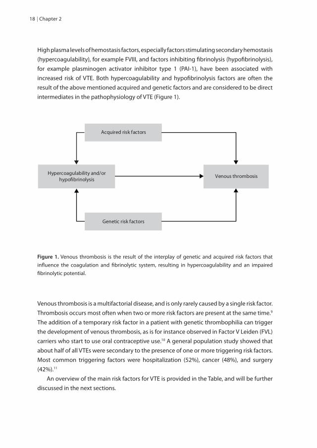
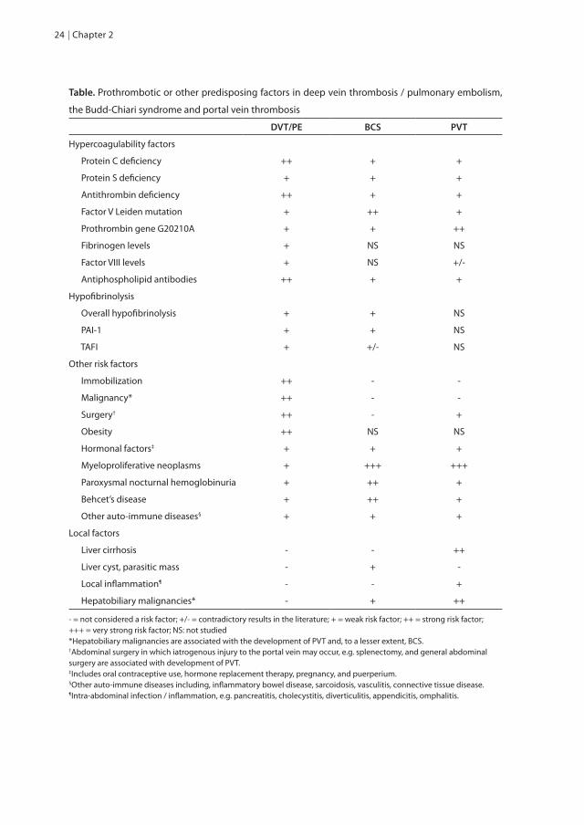
High plasma levels of hemostasis factors, especially factors stimulating secondary hemostasis (hypercoagulability), for example FVIII, and factors inhibiting fibrinolysis (hypofibrinolysis), for example plasminogen activator inhibitor type 1 (PAI-1), have been associated with increased risk of VTE. Both hypercoagulability and hypofibrinolysis factors are often the result of the above mentioned acquired and genetic factors and are considered to be direct intermediates in the pathophysiology of VTE (Figure 1).
Hypercoagulability and/orhypo�brinolysis
Genetic risk factors
Acquired risk factors
Venous thrombosis
Figure 1. Venous thrombosis is the result of the interplay of genetic and acquired risk factors that influence the coagulation and fibrinolytic system, resulting in hypercoagulability and an impaired fibrinolytic potential.
Venous thrombosis is a multifactorial disease, and is only rarely caused by a single risk factor. Thrombosis occurs most often when two or more risk factors are present at the same time.9 The addition of a temporary risk factor in a patient with genetic thrombophilia can trigger the development of venous thrombosis, as is for instance observed in Factor V Leiden (FVL) carriers who start to use oral contraceptive use.10 A general population study showed that about half of all VTEs were secondary to the presence of one or more triggering risk factors. Most common triggering factors were hospitalization (52%), cancer (48%), and surgery (42%).11 An overview of the main risk factors for VTE is provided in the Table, and will be further discussed in the next sections.
Chap
ter 2
19Risk factors for common VTE and SVT |
Hypercoagulability in common VTEHypercoagulability can be the result of common variation or specific mutations in coagulation factor genes. Testing for genetic risk factors has been shown to be effective in identifying individuals at risk for venous thrombosis. However, not all these genetic variants are consistently associated with risk of VTE. The strongest association with risk of a first venous thrombosis is seen for genetic variations that result in antithrombin, protein C or protein S deficiencies, with approximately 5 to 10-fold, 4 to 6-fold, and 1 to 10-fold increases in risk, respectively.12,13 Since these deficiencies are rare, the estimates come from retrospective studies, although prospective studies in asymptomatic family members showed similar results.14 These deficiencies are also associated with an increased risk of VTE recurrence. Consistent associations with venous thrombosis are observed with the prothrombin G20210A variant and Factor V Leiden mutation, which are associated with 3 and 7-fold increased risks, respectively.15,16 However, the association with VTE recurrence is unclear. Some studies reported an increased risk of recurrence for heterozygous carriers,17,18 but other, more recent studies did not confirm these findings.19-22 Heterozygosity for these genetic variants therefore does not have any consequence for the duration or intensity of anticoagulant treatment, but homozygosity and combinations with other risk factors are associated with an increased recurrence risk, and may need long-term treatment.23 However, a recent study found that homozygosity for the FVL mutation and/or the prothrombin variant or double heterozygosity for the FVL mutation and the prothrombin variant did not result in a high risk of recurrent venous thrombosis.24 Interestingly, the FVL mutation is a stronger risk factor for DVT than for isolated PE, which has been designated as the FVL paradox.25 To date, no explanation for this remarkable difference has been found.26 Individuals with antiphospholipid antibodies (APA) have also a rather pronounced (5-fold) increase in risk of a first venous thrombosis, and also the risk of recurrence is consistently increased.27 The combination of venous or arterial thrombosis and the presence of APA, or a combination of obstetric complications and the presence of APA, is defined as the antiphospholipid syndrome (APS). For prothrombotic conditions or changes in coagulation factors levels, such as acquired activated protein C resistance and increased levels of factor VIII, IX, XI and fibrinogen, the effects are moderate and not consistent.7,28,29 Determining these conditions or factor levels may increase the knowledge on the etiology, but will not directly affect the treatment of the patients. Hypercoagulability can be assessed using overall tests of coagulation, such as the endogenous thrombin generation potential. Thrombin converts fibrinogen into fibrin and is essential for acceleration of the coagulation cascade by activating several other coagulation factors. An increased endogenous thrombin potential has been associated with an increased risk of first VTE.30,31 Measurement of thrombin generation has also been
20 | Chapter 2
shown to be of use in identifying patients with a high recurrence risk of VTE,29,32-34 although this was not observed in all studies.31
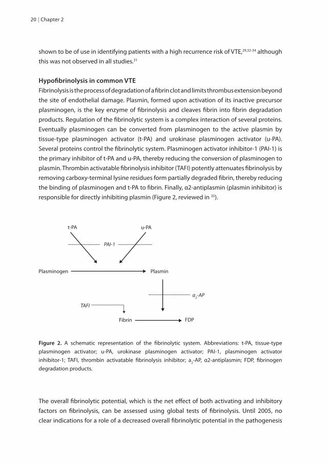
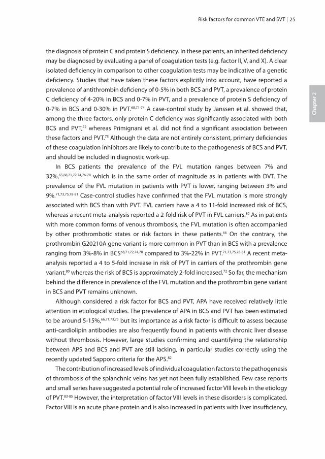
Hypofibrinolysis in common VTE Fibrinolysis is the process of degradation of a fibrin clot and limits thrombus extension beyond the site of endothelial damage. Plasmin, formed upon activation of its inactive precursor plasminogen, is the key enzyme of fibrinolysis and cleaves fibrin into fibrin degradation products. Regulation of the fibrinolytic system is a complex interaction of several proteins. Eventually plasminogen can be converted from plasminogen to the active plasmin by tissue-type plasminogen activator (t-PA) and urokinase plasminogen activator (u-PA). Several proteins control the fibrinolytic system. Plasminogen activator inhibitor-1 (PAI-1) is the primary inhibitor of t-PA and u-PA, thereby reducing the conversion of plasminogen to plasmin. Thrombin activatable fibrinolysis inhibitor (TAFI) potently attenuates fibrinolysis by removing carboxy-terminal lysine residues form partially degraded fibrin, thereby reducing the binding of plasminogen and t-PA to fibrin. Finally, α2-antiplasmin (plasmin inhibitor) is responsible for directly inhibiting plasmin (Figure 2, reviewed in 35).
t-PA u-PA
PAI-1
Plasminogen Plasmin
TAFI
Fibrin
α2-AP
FDP
Figure 2. A schematic representation of the fibrinolytic system. Abbreviations: t-PA, tissue-type plasminogen activator; u-PA, urokinase plasminogen activator; PAI-1, plasminogen activator inhibitor-1; TAFI, thrombin activatable fibrinolysis inhibitor; a2-AP, α2-antiplasmin; FDP, fibrinogen degradation products.
The overall fibrinolytic potential, which is the net effect of both activating and inhibitory factors on fibrinolysis, can be assessed using global tests of fibrinolysis. Until 2005, no clear indications for a role of a decreased overall fibrinolytic potential in the pathogenesis
Chap
ter 2
21Risk factors for common VTE and SVT |
of venous thrombosis were observed. In these older studies, the fibrinolytic potential was studied using global tests such as the euglobulin clot lysis time and the dilute whole blood clot lysis assay. Both these tests have a number of limitations.6 However, recent findings in two large case-control studies demonstrated an association between hypofibrinolysis and risk of VTE. In these studies, a plasma-based, tissue factor initiated and t-PA induced clot lysis assay was used. The clot lysis time (CLT) denotes the time needed from half maximal clot formation to half-maximal lysis of a plasma clot and represents a marker for the over-all fibrinolytic capacity.36 In the Leiden Thrombophilia Study (LETS)-study, a case control study on 469 patients with a first DVT and 469 healthy controls, a 2-fold increase in risk of DVT in individuals with a CLT above the 90th percentile was observed.37 In the Multiple Environmental and Genetic Assessment of risk factors for venous thrombosis (MEGA)-study, involving over >2000 patients with VTE and >2500 controls, these findings were confirmed, showing a similar relationship between risk of DVT or PE and hypofibrinolysis.38 The combination of hypofibrinolysis and risk factors associated with hypercoagulability was shown to result in a substantially greater risk than expected on the basis of the individual risks.38 In this study, oral contraceptive use in women with hypofibrinolysis was associated with a more than 20-fold increased risk of VTE. Hypofibrinolysis does not appear to be associated with risk of recurrence of VTE.39
When considering individual fibrinolytic factors, the literature on the role of PAI-1 and t-PA in venous thrombosis has been controversial. In the Longitudinal Investigation of Thromboembolism Etiology (LITE)-study, a large population-based prospective study on venous thrombosis in middle-aged and elderly patients, no association was found between levels of PAI-1 or t-PA/PAI-1 complex and the risk of venous thrombosis.40 Several other studies also failed to show an association between t-PA and PAI-1 and the risk of venous thrombosis.41-43 However, more recently, the above mentioned LETS-study demonstrated that elevated PAI-1 levels were associated with an elevated CLT, indicative of hypofibrinolysis, and with the risk of venous thrombosis.44 In this study t-PA levels were associated with venous thrombosis, but not with CLT, suggesting that t-PA levels are more likely to reflect other underlying risk factors. High TAFI levels have also been shown to be associated with a mildly increased risk of VTE, although not in thrombophilic families.45 In the LETS-study, TAFI levels above the 90th percentile increased the risk for VTE 1.7-fold compared to TAFI levels below the 90th percentile,46 which was later confirmed in an independent cohort.47 In addition, high TAFI levels have been associated with an increased risk of recurrence of VTE.48 TAFI levels and activity are partly determined by several common genetic variations, which have also been associated with risk of VTE.49
22 | Chapter 2
Studies on levels of plasminogen or α2-antiplasmin and the risk of venous thrombosis are scarce and often in small patient groups only.50-52 To this point, there is no clear evidence for a role of plasminogen or α2-antiplasmin levels in the development of VTE.6
other acquired risk factors for common VTEHospitalized patients have an increased risk for VTE since they are often exposed to one or more acquired risk factors for VTE, such as immobility, cancer, surgery, congestive heart failure, infections, or chronic kidney disease.53 Recent hospitalization for an acute medical disease is independently associated with 8-fold increased risk of VTE and accounts for almost one fourth of all VTE events.53
Cancer patients have an increased risk of venous thrombosis as a result of multiple factors, like activation of coagulation by tumor cells resulting in hypercoagulability, compression of veins by the tumor, hospitalization, surgery, and chemotherapy.54,55 VTE can be diagnosed in 4-20% of patients with cancer and is one of the leading causes of death in these patients.56 Myeloproliferative neoplasms are also associated with an increased risk of thrombotic complications including venous and arterial thrombosis and microcirculatory disorders, such as erythromelalgia. Risk of these complications is most pronounced in polycythemia vera and essential thrombocytosis.57
The risk of venous thrombosis in surgery depends on the type of surgery and patient characteristics.58 Surgery induces an acute phase reaction and plasma levels of many hemostasis factors increase in the days after surgery, which contributes to the prothrombotic condition in that period.59
Another well-known triggering risk factor is immobility. Immobility, mostly defined as bed rest for at least 4 days, increases the risk probably by stasis of blood flow in the venous system. Clinical settings with immobility are bed rest, and plaster casts or paresis of the legs. Also shorter periods of bed rest60 and minor injuries61 have been associated with an increased risk of venous thrombosis. Obesity (body mass index above 30 kg/m²) leads to a 2-3 fold increase in the risk of VTE and this increase in risk is even larger with severe obesity.40,62 Obesity is associated with hypercoagulability and hypofibrinolysis due to, amongst others, increased plasma levels of fibrinogen, factor VIII and especially of PAI-1.63
riSK FACTorS For SPLANCHNiC VEiN THromBoSiS
BCS is defined as an obstruction of the hepatic venous outflow tract from the level of the small hepatic veins to the entrance of the inferior vena cava into the right atrium.64 BCS is a rare disorder with an annual incidence of about 0.2-0.8 per million inhabitants in the Western world, predominantly affecting young females.65,66 The classical triad of symptoms
Chap
ter 2
23Risk factors for common VTE and SVT |
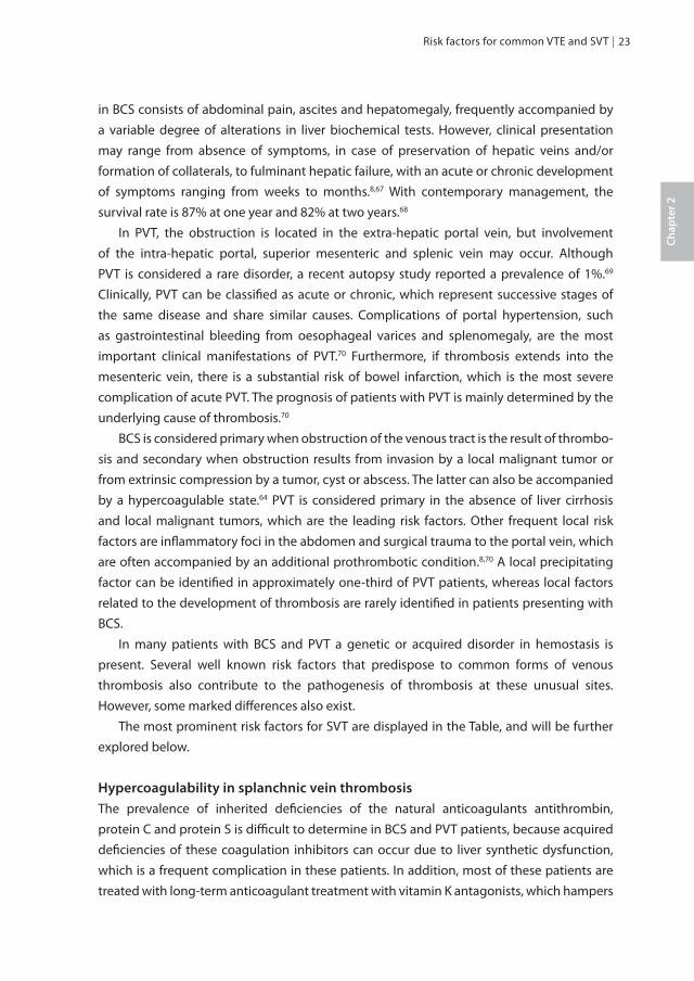
in BCS consists of abdominal pain, ascites and hepatomegaly, frequently accompanied by a variable degree of alterations in liver biochemical tests. However, clinical presentation may range from absence of symptoms, in case of preservation of hepatic veins and/or formation of collaterals, to fulminant hepatic failure, with an acute or chronic development of symptoms ranging from weeks to months.8,67 With contemporary management, the survival rate is 87% at one year and 82% at two years.68
In PVT, the obstruction is located in the extra-hepatic portal vein, but involvement of the intra-hepatic portal, superior mesenteric and splenic vein may occur. Although PVT is considered a rare disorder, a recent autopsy study reported a prevalence of 1%.69 Clinically, PVT can be classified as acute or chronic, which represent successive stages of the same disease and share similar causes. Complications of portal hypertension, such as gastrointestinal bleeding from oesophageal varices and splenomegaly, are the most important clinical manifestations of PVT.70 Furthermore, if thrombosis extends into the mesenteric vein, there is a substantial risk of bowel infarction, which is the most severe complication of acute PVT. The prognosis of patients with PVT is mainly determined by the underlying cause of thrombosis.70
BCS is considered primary when obstruction of the venous tract is the result of thrombo-sis and secondary when obstruction results from invasion by a local malignant tumor or from extrinsic compression by a tumor, cyst or abscess. The latter can also be accompanied by a hypercoagulable state.64 PVT is considered primary in the absence of liver cirrhosis and local malignant tumors, which are the leading risk factors. Other frequent local risk factors are inflammatory foci in the abdomen and surgical trauma to the portal vein, which are often accompanied by an additional prothrombotic condition.8,70 A local precipitating factor can be identified in approximately one-third of PVT patients, whereas local factors related to the development of thrombosis are rarely identified in patients presenting with BCS. In many patients with BCS and PVT a genetic or acquired disorder in hemostasis is present. Several well known risk factors that predispose to common forms of venous thrombosis also contribute to the pathogenesis of thrombosis at these unusual sites. However, some marked differences also exist. The most prominent risk factors for SVT are displayed in the Table, and will be further explored below.
Hypercoagulability in splanchnic vein thrombosisThe prevalence of inherited deficiencies of the natural anticoagulants antithrombin, protein C and protein S is difficult to determine in BCS and PVT patients, because acquired deficiencies of these coagulation inhibitors can occur due to liver synthetic dysfunction, which is a frequent complication in these patients. In addition, most of these patients are treated with long-term anticoagulant treatment with vitamin K antagonists, which hampers
24 | Chapter 2
Table. Prothrombotic or other predisposing factors in deep vein thrombosis / pulmonary embolism,
the Budd-Chiari syndrome and portal vein thrombosis
DVT/PE BCS PVT
Hypercoagulability factors
Protein C deficiency ++ + +
Protein S deficiency + + +
Antithrombin deficiency ++ + +
Factor V Leiden mutation + ++ +
Prothrombin gene G20210A + + ++
Fibrinogen levels + NS NS
Factor VIII levels + NS +/-
Antiphospholipid antibodies ++ + +
Hypofibrinolysis
Overall hypofibrinolysis + + NS
PAI-1 + + NS
TAFI + +/- NS
Other risk factors
Immobilization ++ - -
Malignancy* ++ - -
Surgery† ++ - +
Obesity ++ NS NS
Hormonal factors‡ + + +
Myeloproliferative neoplasms + +++ +++
Paroxysmal nocturnal hemoglobinuria + ++ +
Behcet’s disease + ++ +
Other auto-immune diseases§ + + +
Local factors
Liver cirrhosis - - ++
Liver cyst, parasitic mass - + -
Local inflammation¶ - - +
Hepatobiliary malignancies* - + ++
- = not considered a risk factor; +/- = contradictory results in the literature; + = weak risk factor; ++ = strong risk factor; +++ = very strong risk factor; NS: not studied*Hepatobiliary malignancies are associated with the development of PVT and, to a lesser extent, BCS. †Abdominal surgery in which iatrogenous injury to the portal vein may occur, e.g. splenectomy, and general abdominal surgery are associated with development of PVT.‡Includes oral contraceptive use, hormone replacement therapy, pregnancy, and puerperium.§Other auto-immune diseases including, inflammatory bowel disease, sarcoidosis, vasculitis, connective tissue disease.¶Intra-abdominal infection / inflammation, e.g. pancreatitis, cholecystitis, diverticulitis, appendicitis, omphalitis.
Chap
ter 2
25Risk factors for common VTE and SVT |
the diagnosis of protein C and protein S deficiency. In these patients, an inherited deficiency may be diagnosed by evaluating a panel of coagulation tests (e.g. factor II, V, and X). A clear isolated deficiency in comparison to other coagulation tests may be indicative of a genetic deficiency. Studies that have taken these factors explicitly into account, have reported a prevalence of antithrombin deficiency of 0-5% in both BCS and PVT, a prevalence of protein C deficiency of 4-20% in BCS and 0-7% in PVT, and a prevalence of protein S deficiency of 0-7% in BCS and 0-30% in PVT.68,71-74 A case-control study by Janssen et al. showed that, among the three factors, only protein C deficiency was significantly associated with both BCS and PVT,72 whereas Primignani et al. did not find a significant association between these factors and PVT.75 Although the data are not entirely consistent, primary deficiencies of these coagulation inhibitors are likely to contribute to the pathogenesis of BCS and PVT, and should be included in diagnostic work-up. In BCS patients the prevalence of the FVL mutation ranges between 7% and 32%,65,68,71,72,74,76-78 which is in the same order of magnitude as in patients with DVT. The prevalence of the FVL mutation in patients with PVT is lower, ranging between 3% and 9%.71,73,75,78-81 Case-control studies have confirmed that the FVL mutation is more strongly associated with BCS than with PVT. FVL carriers have a 4 to 11-fold increased risk of BCS, whereas a recent meta-analysis reported a 2-fold risk of PVT in FVL carriers.80 As in patients with more common forms of venous thrombosis, the FVL mutation is often accompanied by other prothrombotic states or risk factors in these patients.66 On the contrary, the prothrombin G20210A gene variant is more common in PVT than in BCS with a prevalence ranging from 3%-8% in BCS68,71,72,74,78 compared to 3%-22% in PVT.71,73,75,78-81 A recent meta-analysis reported a 4 to 5-fold increase in risk of PVT in carriers of the prothrombin gene variant,80 whereas the risk of BCS is approximately 2-fold increased.72 So far, the mechanism behind the difference in prevalence of the FVL mutation and the prothrombin gene variant in BCS and PVT remains unknown. Although considered a risk factor for BCS and PVT, APA have received relatively little attention in etiological studies. The prevalence of APA in BCS and PVT has been estimated to be around 5-15%,66,71,73,75 but its importance as a risk factor is difficult to assess because anti-cardiolipin antibodies are also frequently found in patients with chronic liver disease without thrombosis. However, large studies confirming and quantifying the relationship between APS and BCS and PVT are still lacking, in particular studies correctly using the recently updated Sapporo criteria for the APS.82
The contribution of increased levels of individual coagulation factors to the pathogenesis of thrombosis of the splanchnic veins has yet not been fully established. Few case reports and small series have suggested a potential role of increased factor VIII levels in the etiology of PVT.83-85 However, the interpretation of factor VIII levels in these disorders is complicated. Factor VIII is an acute phase protein and is also increased in patients with liver insufficiency,
26 | Chapter 2
which is frequently seen in BCS and PVT patients. Recently, Martinelli et al. described significantly elevated factor VIII levels in patients with primary PVT.86
Few studies have focused on the recurrence risk of thrombosis in SVT patients. Condat et al. assessed the outcome of PVT in relation to prothrombotic conditions in a cohort of 136 patients of whom 84 received anticoagulant therapy.87 In this study, an incidence rate of 5.5 per 100 person-years for all types of thrombotic events was reported and an underlying prothrombotic state was shown to be an independent predictor of recurrent thrombosis. An elevated endogenous thrombin potential has been associated with an increased risk of VTE. It might be expected that an increased endogenous thrombin potential also contributes to the development of BCS or PVT, but this has not yet been investigated.
Hypofibrinolysis in splanchnic vein thrombosisOnly few studies have assessed the role of the fibrinolytic system in the pathogenesis of BCS and PVT. De Bruijne et al. observed an association between SVT and genetic variation in the TAFI gene.88 A decreased risk of SVT in 147Thr/Thr homozygotes and a slightly, but not significantly, increased risk in carriers of the 325Ile variant was observed, suggesting a role for TAFI in the pathogenesis of SVT. Interestingly, the genotypes associated with an increased risk of SVT are associated with decreased TAFI levels,89 whereas an association between high TAFI levels and VTE risk has been consistently reported. There was a high degree of linkage disequilibrium between these two SNPs, making it difficult to assess the contribution of the individual SNPs. The increased risk of SVT in carriers of the 325Ile allele may be related to a TAFI variant with a greater antifibrinolytic potential but lowered antigen levels.90,91 The mechanism behind the contribution of the Ala147Thr SNP to an increased risk of thrombosis is unknown. Dayal et al. measured t-PA and PAI-1 levels in a relatively small study of 27 BCS patients.92 In this study, only three patients showed mildly increased levels of t-PA and PAI-1 compared to healthy controls. More recently, Hoekstra et al. extensively investigated components of the fibrinolytic system in 101 BCS patients.93 This study found significantly higher PAI-1 levels in BCS patients compared to controls, whereas TAFI and α2-antiplasmin levels were significantly lower. A subgroup of BCS patients showed clearly elevated CLTs, indicative of hypofibrinolysis. A CLT above the 90th or 95th percentile of controls was associated with a 2.4-fold and 3.4-fold increase in risk of BCS, respectively. Of note, analysis of SNPs of fibrinolysis proteins revealed no significant differences between cases and controls, but the number of studied individuals was limited and probably too small for analysis of genetic factors. These findings suggest that an impaired fibrinolytic potential contributes to the development of BCS. Although additional studies are warranted, both these studies indicate that, like in other forms of venous thrombosis, impaired fibrinolysis may also play a role in the pathogenesis of thrombosis of the splanchnic veins.

Chap
ter 2
27Risk factors for common VTE and SVT |
other risk factors for splanchnic vein thrombosisMyeloproliferative neoplasms (MPNs) are the most common underlying cause and can be identified in nearly half of BCS and about one-third of PVT patients,68,73,74,76,79,94-96 which is strikingly higher than in other forms of VTE. The most common gain of function mutation leading to development of MPN is JAK2V617F, which is found in nearly all cases of polycythemia vera and about half the cases of essential thrombocythemia and primary myelofibrosis.97 The JAK2V617F mutation has been described in 17% to 45% of unselected BCS and PVT patients.68,73,74,76,79,94-96 Screening for JAK2V617F is an important diagnostic tool to detect MPN in these patients and is now part of the standard diagnostic work-up in BCS and PVT.98 Portal hypertension, resulting from pre- or post hepatic venous obstruction, can lead to hypersplenism and hemodilution. Both these conditions may mask the characteristic peripheral blood cell changes and make diagnosis of MPN notoriously difficult. Therefore, also bone marrow histology should be performed, allowing for MPN diagnosis in patients without the JAK2V617F mutation. About half of the BCS and PVT patients with the JAK2V617F mutation as the only indication of an underlying MPN, develop an overt MPN during follow-up.99 A recent meta-analysis showed that JAK2V617F is rare in other forms of venous thrombosis, confirming the unique role of MPN in the pathogenesis of thrombosis at these distinct sites.99 The exact pathogenic mechanism of thrombotic complications in MPN remains elusive, but besides the characteristic erythrocytosis and thrombocytosis, platelet and leukocyte functional abnormalities seem critical.100
Paroxysmal nocturnal hemoglobinuria (PNH) is a rare, acquired haematological disorder of haematopoietic stem cells, which frequently has a devastating course and is specifically related to thrombosis at unusual sites. Remarkably, thrombosis of the splanchnic veins is a frequent complication, particularly of the hepatic veins and the inferior vena cava in which more than 45% of the thrombotic episodes are located, accounting for the majority of deaths in this disorder.101 PNH has been reported in 9-19% of tested BCS patients,74,102 whereas a prevalence of 0-2% has been reported in PVT.72,73 Several mechanisms, including intravascular hemolysis, increased platelet activation and aggregation, procoagulant microparticles resulting from complement-mediated platelet damage, hypofibrinolysis and increased tissue factor expression may contribute to the pathogenesis of venous thrombosis in PNH.103,104 Patients with a PNH cell population above 60% of the granulocytes, appear to be at greatest risk for thrombosis.103 Testing for PNH should be routinely performed in all BCS and PVT patients. A number of systemic, auto-immune-mediated diseases have been implicated in the pathogenesis of both BCS and PVT. Of these, Behcet’s disease is particularly associated with BCS. It represents the leading cause of BCS in areas where Behcet’s disease is highly prevalent.66 Other systemic diseases include inflammatory bowel disease, vasculitis, sarcoidosis and connective tissue disease. However, these account for only a minority of cases.66,70
28 | Chapter 2
Oral contraceptive use, pregnancy and puerperium are known risk factors for venous thrombosis, and are also established in BCS and PVT.66,105 However, an additional prothrombotic condition is often present in these women. Recently, a potentially new factor in the pathogenesis of BCS was identified. Talens et al. initially showed, using a proteomic approach, that apolipoprotein A1 (Apo A1) was decreased in 9 BCS patients compared to controls and subsequently validated these findings in a cohort of 101 BCS patients, in which Apo A1 levels were also significantly lower compared to controls.106 Apo A1 is the principal component of high density lipoprotein (HDL) cholesterol, which has been shown to be inversely associated with other forms of venous thrombosis,107-109 although this association was not observed in all studies.110 Low Apo A1 levels have also been associated with an increased risk of recurrence of common VTE.111
multifactorial etiology in splanchnic vein thrombosisEven more outspoken than in patients with DVT or PE, the etiology of primary BCS and PVT must be considered multifactorial. The recent EN-Vie studies reported a combination of two or more genetic or acquired prothrombotic factors in 46% of BCS and 48% of PVT patients.68,73 In this series of BCS patients, 18% of the patients even displayed three risk factors. Based on these findings, a complete hematological work-up, including inherited thrombophilia, APA, MPN and PNH should always be performed in BCS and PVT patients, irrespective of whether one prothrombotic factor has already been identified. This is in particular relevant for identifying MPN, which are also often accompanied by other prothrombotic factors, and require additional treatment, such as aspirin, or anti-proliferative treatment.
CLUES For SiTE-SPECiFiCiTY oF THromBoSiS
It is still unresolved why some patients develop thrombosis of the splanchnic veins, whereas most others with similar prothrombotic factors develop DVT or PE. In contrast to the vas-culature of the lower extremities, the splanchnic vasculature does not contain venous valves, which are well-known to be involved in the pathogenesis of DVT.112 Further research is needed to identify local factors that are involved in the pathogenesis of thrombosis at these distinct sites. In this respect, it has been speculated that endothelial cells of the splanchnic veins may interact with activated platelets and/or leukocytes and increased microparticles, which are characteristic features of MPN and PNH, two haematological disorders with a remarkable high frequency in SVT.113 Recently, the JAK2V617F mutation was demonstrated in the endothelial cells of two BCS patients, which indeed suggests a contribution of the endothelium to the development of thrombosis.114 An underlying mechanism, however, remains elusive. In addition, endothelial cells of the splanchnic veins are exposed to gut-
Chap
ter 2
29Risk factors for common VTE and SVT |
derived oral antigens and bacterial components from the gastrointestinal tract. Hepatic sinusoidal endothelial cells display immune tolerance which prevents a response to these factors.115 However, there is no evidence that the endothelial cells of the portal vein are similarly protected.113 It has therefore been hypothesized that these endothelial cells are chronically activated, making them particularly vulnerable to the disease-specific changes of PNH and MPN.113 These factors may be prothrombotic, resulting in an increased risk for SVT. Interestingly, there are also apparent differences in the etiology of BCS and PVT (Table). Although MPNs are the most frequent prothrombotic factor in both BCS and PVT, MPNs are clearly more common in BCS than in PVT. In addition, it is clear that the FVL mutation is more strongly associated with BCS than with PVT, whereas the opposite is true for the prothrombin gene variant. In BCS patients, the FVL mutation has even been specifically associated with involvement of thrombosis of the inferior vena cava.77 Finally, it is evident that PNH is more strongly associated with the development of BCS than of PVT. The understanding of the interaction of prothrombotic disorders and local factors in the etiology of BCS and PVT will play an essential role in the understanding of the pathogenesis of thrombosis at these unusual sites. Identification of distinct differences in the etiology with more common forms of venous thrombosis, and the remarkable differences in etiology even between BCS and PVT, needs further research.
CoNCLUSioN
The understanding of the etiology of VTE has improved over the years. VTE must be considered a multifactorial disease, in which the interplay of genetic or acquired factors is required for thrombosis formation. This prothrombotic tendency is caused by abnormalities in the coagulation or fibrinolysis pathways, leading to hypercoagulability or an impaired fibrinolysis. More general risk factors also contribute, partly through these pathways, to the development of thrombosis. An interesting aspect of VTE is its site-specificity. In contrast to DVT or PE, the cause of venous thrombosis at unusual sites, such as the splanchnic veins, remains to be elucidated. Although the etiology shows a considerable overlap with common forms of VTE, there are several remarkable differences that may prove to be a means towards a better understanding of the site-specificity of venous thrombosis.
30 | Chapter 2
rEFErENCES
1. Goldhaber SZ, Tapson VF, Committee DFS. A prospective registry of 5,451 patients with ultrasound-confirmed deep vein thrombosis. Am J Cardiol 2004;93:259-62.
2. Heit JA. Venous thromboembolism: disease burden, outcomes and risk factors. J Thromb Haemost 2005;3:1611-7.
3. Naess IA, Christiansen SC, Romundstad P, Cannegieter SC, Rosendaal FR, Hammerstrom J. Incidence and mortality of venous thrombosis: a population-based study. J Thromb Haemost 2007;5:692-9.
4. Oger E. Incidence of venous thromboembolism: a community-based study in Western France. EPI-GETBP Study Group. Groupe d’Etude de la Thrombose de Bretagne Occidentale. Thromb Haemost 2000;83:657-60.
5. Kyrle PA, Eichinger S. Deep vein thrombosis. Lancet 2005;365:1163-74.
6. Meltzer ME, Doggen CJ, de Groot PG, Rosendaal FR, Lisman T. The impact of the fibrinolytic system on the risk of venous and arterial thrombosis. Semin Thromb Hemost 2009;35:468-77.
7. Cushman M. Epidemiology and risk factors for venous thrombosis. Semin Hematol 2007;44:62-9.
8. DeLeve LD, Valla DC, Garcia-Tsao G, American Association for the Study Liver D. Vascular disorders of the liver. Hepatology 2009;49:1729-64.
9. Rosendaal FR. Venous thrombosis: a multicausal disease. Lancet 1999;353:1167-73.
10. Vandenbroucke JP, Rosing J, Bloemenkamp KW, et al. Oral contraceptives and the risk of venous thrombosis. N Engl J Med 2001;344:1527-35.
11. Cushman M, Tsai AW, White RH, et al. Deep vein thrombosis and pulmonary embolism in two cohorts: the longitudinal investigation of thromboembolism etiology. Am J Med 2004;117:19-25.
12. Koster T, Rosendaal FR, Briet E, et al. Protein C deficiency in a controlled series of unselected outpatients: an infrequent but clear risk factor for venous thrombosis (Leiden Thrombophilia Study). Blood 1995;85:2756-61.
13. Middeldorp S, van Hylckama Vlieg A. Does thrombophilia testing help in the clinical management of patients? Br J Haematol 2008;143:321-35.
14. Mahmoodi BK, Brouwer JL, Ten Kate MK, et al. A prospective cohort study on the absolute risks of venous thromboembolism and predictive value of screening asymptomatic relatives of patients with hereditary deficiencies of protein S, protein C or antithrombin. J Thromb Haemost 2010;8:1193-200.
15. Bertina RM, Koeleman BP, Koster T, et al. Mutation in blood coagulation factor V associated with resistance to activated protein C. Nature 1994;369:64-7.
16. Poort SR, Rosendaal FR, Reitsma PH, Bertina RM. A common genetic variation in the 3’-untranslated region of the prothrombin gene is associated with elevated plasma prothrombin levels and an increase in venous thrombosis. Blood 1996;88:3698-703.
17. Ridker PM, Miletich JP, Stampfer MJ, Goldhaber SZ, Lindpaintner K, Hennekens CH. Factor V Leiden and risks of recurrent idiopathic venous thromboembolism. Circulation 1995;92:2800-2.
18. Simioni P, Prandoni P, Lensing AW, et al. The risk of recurrent venous thromboembolism in patients with an Arg506-->Gln mutation in the gene for factor V (factor V Leiden). N Engl J Med 1997;336:399-403.
19. Baglin T, Luddington R, Brown K, Baglin C. Incidence of recurrent venous thromboembolism in relation to clinical and thrombophilic risk factors: prospective cohort study. Lancet 2003;362:523-6.
20. Christiansen SC, Cannegieter SC, Koster T, Vandenbroucke JP, Rosendaal FR. Thrombophilia, clinical factors, and recurrent venous thrombotic events. JAMA 2005;293:2352-61.
21. Eichinger S, Pabinger I, Stumpflen A, et al. The risk of recurrent venous thromboembolism in patients with and without factor V Leiden. Thromb Haemost 1997;77:624-8.
22. Rintelen C, Pabinger I, Knobl P, Lechner K, Mannhalter C. Probability of recurrence of thrombosis in patients with and without factor V Leiden. Thromb Haemost 1996;75:229-32.
23. Hirsh J, Guyatt G, Albers GW, Harrington R, Schunemann HJ, American College of Chest P. Antithrombotic and thrombolytic therapy: American College of Chest Physicians Evidence-Based Clinical Practice Guidelines (8th Edition). Chest 2008;133:110S-2S.

Chap
ter 2
31Risk factors for common VTE and SVT |
24. Lijfering WM, Middeldorp S, Veeger NJ, et al. Risk of recurrent venous thrombosis in homozygous carriers and double heterozygous carriers of factor V Leiden and prothrombin G20210A. Circulation 2010;121:1706-12.
25. Bounameaux H. Factor V Leiden paradox: risk of deep-vein thrombosis but not of pulmonary embolism. Lancet 2000;356:182-3.
26. van Stralen KJ, Doggen CJ, Bezemer ID, Pomp ER, Lisman T, Rosendaal FR. Mechanisms of the factor V Leiden paradox. Arterioscler Thromb Vasc Biol 2008;28:1872-7.
27. Farmer-Boatwright MK, Roubey RA. Venous thrombosis in the antiphospholipid syndrome. Arterioscler Thromb Vasc Biol 2009;29:321-5.
28. Folsom AR, Cushman M, Tsai MY, et al. A prospective study of venous thromboembolism in relation to factor V Leiden and related factors. Blood 2002;99:2720-5.
29. Hron G, Kollars M, Binder BR, Eichinger S, Kyrle PA. Identification of patients at low risk for recurrent venous thromboembolism by measuring thrombin generation. JAMA 2006;296:397-402.
30. Lutsey PL, Folsom AR, Heckbert SR, Cushman M. Peak thrombin generation and subsequent venous thromboembolism: the Longitudinal Investigation of Thromboembolism Etiology (LITE) study. J Thromb Haemost 2009;7:1639-48.
31. van Hylckama Vlieg A, Christiansen SC, Luddington R, Cannegieter SC, Rosendaal FR, Baglin TP. Elevated endogenous thrombin potential is associated with an increased risk of a first deep venous thrombosis but not with the risk of recurrence. Br J Haematol 2007;138:769-74.
32. Besser M, Baglin C, Luddington R, van Hylckama Vlieg A, Baglin T. High rate of unprovoked recurrent venous thrombosis is associated with high thrombin-generating potential in a prospective cohort study. J Thromb Haemost 2008;6:1720-5.
33. Eichinger S, Hron G, Kollars M, Kyrle PA. Prediction of recurrent venous thromboembolism by endogenous thrombin potential and D-dimer. Clin Chem 2008;54:2042-8.
34. Tripodi A, Legnani C, Chantarangkul V, Cosmi B, Palareti G, Mannucci PM. High thrombin generation measured in the presence of thrombomodulin is associated with an increased risk of recurrent venous thromboembolism. J Thromb Haemost 2008;6:1327-33.
35. Rijken DC, Lijnen HR. New insights into the molecular mechanisms of the fibrinolytic system. J Thromb Haemost 2009;7:4-13.
36. Guimaraes AH, de Bruijne EL, Lisman T, et al. Hypofibrinolysis is a risk factor for arterial thrombosis at young age. Br J Haematol 2009;145:115-20.
37. Lisman T, de Groot PG, Meijers JC, Rosendaal FR. Reduced plasma fibrinolytic potential is a risk factor for venous thrombosis. Blood 2005;105:1102-5.
38. Meltzer ME, Lisman T, Doggen CJ, de Groot PG, Rosendaal FR. Synergistic effects of hypofibrinolysis and genetic and acquired risk factors on the risk of a first venous thrombosis. PLoS Med 2008;5:e97.
39. Meltzer ME, Bol L, Rosendaal FR, Lisman T, Cannegieter SC. Hypofibrinolysis as a risk factor for recurrent venous thrombosis; results of the LETS follow-up study. J Thromb Haemost 2010;8:605-7.
40. Tsai AW, Cushman M, Rosamond WD, Heckbert SR, Polak JF, Folsom AR. Cardiovascular risk factors and venous thromboembolism incidence: the longitudinal investigation of thromboembolism etiology. Arch Intern Med 2002;162:1182-9.
41. Crowther MA, Roberts J, Roberts R, et al. Fibrinolytic variables in patients with recurrent venous thrombosis: a prospective cohort study. Thromb Haemost 2001;85:390-4.
42. Folsom AR, Cushman M, Heckbert SR, Rosamond WD, Aleksic N. Prospective study of fibrinolytic markers and venous thromboembolism. J Clin Epidemiol 2003;56:598-603.
43. Ridker PM, Vaughan DE, Stampfer MJ, et al. Baseline fibrinolytic state and the risk of future venous thrombosis. A prospective study of endogenous tissue-type plasminogen activator and plasminogen activator inhibitor. Circulation 1992;85:1822-7.
44. Meltzer ME, Lisman T, de Groot PG, et al. Venous thrombosis risk associated with plasma hypofibrinolysis is explained by elevated plasma levels of TAFI and PAI-1. Blood 2010;116:113-21.
32 | Chapter 2
45. Folkeringa N, Coppens M, Veeger NJ, et al. Absolute risk of venous and arterial thromboembolism in thrombophilic families is not increased by high thrombin-activatable fibrinolysis inhibitor (TAFI) levels. Thromb Haemost 2008;100:38-44.
46. van Tilburg NH, Rosendaal FR, Bertina RM. Thrombin activatable fibrinolysis inhibitor and the risk for deep vein thrombosis. Blood 2000;95:2855-9.
47. Libourel EJ, Bank I, Meinardi JR, et al. Co-segregation of thrombophilic disorders in factor V Leiden carriers; the contributions of factor VIII, factor XI, thrombin activatable fibrinolysis inhibitor and lipoprotein(a) to the absolute risk of venous thromboembolism. Haematologica 2002;87:1068-73.
48. Eichinger S, Schonauer V, Weltermann A, et al. Thrombin-activatable fibrinolysis inhibitor and the risk for recurrent venous thromboembolism. Blood 2004;103:3773-6.
49. Martini CH, Brandts A, de Bruijne EL, et al. The effect of genetic variants in the thrombin activatable fibrinolysis inhibitor (TAFI) gene on TAFI-antigen levels, clot lysis time and the risk of venous thrombosis. Br J Haematol 2006;134:92-4.
50. Brandt JT. Plasminogen and tissue-type plasminogen activator deficiency as risk factors for thromboembolic disease. Arch Pathol Lab Med 2002;126:1376-81.
51. Favier R, Aoki N, de Moerloose P. Congenital alpha(2)-plasmin inhibitor deficiencies: a review. Br J Haematol 2001;114:4-10.
52. Leebeek FW, Knot EA, Ten Cate JW, Traas DW. Severe thrombotic tendency associated with a type I plasminogen deficiency. Am J Hematol 1989;30:32-5.
53. Heit JA, O’Fallon WM, Petterson TM, et al. Relative impact of risk factors for deep vein thrombosis and pulmonary embolism: a population-based study. Arch Intern Med 2002;162:1245-8.
54. Horowitz N, Brenner B. Thrombophilia and cancer. Pathophysiol Haemost Thromb 2008;36:131-6.
55. Piccioli A, Falanga A, Baccaglini U, Marchetti M, Prandoni P. Cancer and venous thromboembolism. Semin Thromb Hemost 2006;32:694-9.
56. Khorana AA, Francis CW, Culakova E, Kuderer NM, Lyman GH. Thromboembolism is a leading cause of death in cancer patients receiving outpatient chemotherapy. J Thromb Haemost 2007;5:632-4.
57. Elliott MA, Tefferi A. Thrombosis and haemorrhage in polycythaemia vera and essential thrombocythaemia. Br J Haematol 2005;128:275-90.
58. Geerts WH, Pineo GF, Heit JA, et al. Prevention of venous thromboembolism: the Seventh ACCP Conference on Antithrombotic and Thrombolytic Therapy. Chest 2004;126:338S-400S.
59. Gabay C, Kushner I. Mechanisms of disease: acute-phase proteins and other systemic responses to inflammation. N Engl J Med 1999;340:448-54.
60. Beam DM, Courtney DM, Kabrhel C, Moore CL, Richman PB, Kline JA. Risk of thromboembolism varies, depending on category of immobility in outpatients. Ann Emerg Med 2009;54:147-52.
61. van Stralen KJ, Rosendaal FR, Doggen CJ. Minor injuries as a risk factor for venous thrombosis. Arch Intern Med 2008;168:21-6.
62. Stein PD, Beemath A, Olson RE. Obesity as a risk factor in venous thromboembolism. Am J Med 2005;118:978-80.
63. Mertens I, Van Gaal LF. Visceral fat as a determinant of fibrinolysis and hemostasis. Semin Vasc Med 2005;5:48-55.
64. Janssen HL, Garcia-Pagan JC, Elias E, Mentha G, Hadengue A, Valla DC. Budd-Chiari syndrome: a review by an expert panel. J Hepatol 2003;38:364-71.
65. Rajani R, Melin T, Bjornsson E, et al. Budd-Chiari syndrome in Sweden: epidemiology, clinical characteristics and survival - an 18-year experience. Liver Int 2009;29:253-9.
66. Valla DC. Primary Budd-Chiari syndrome. J Hepatol 2009;50:195-203.
67. Hoekstra J, Janssen HL. Vascular liver disorders (I): diagnosis, treatment and prognosis of Budd-Chiari syndrome. Neth J Med 2008;66:334-9.
68. Darwish Murad S, Plessier A, Hernandez-Guerra M, et al. Etiology, management, and outcome of the Budd-Chiari syndrome. Ann Intern Med 2009;151:167-75.
Chap
ter 2
33Risk factors for common VTE and SVT |
69. Ogren M, Bergqvist D, Bjorck M, Acosta S, Eriksson H, Sternby NH. Portal vein thrombosis: prevalence, patient characteristics and lifetime risk: a population study based on 23,796 consecutive autopsies. World J Gastroenterol 2006;12:2115-9.
70. Hoekstra J, Janssen HL. Vascular liver disorders (II): portal vein thrombosis. Neth J Med 2009;67:46-53.
71. Denninger MH, Chait Y, Casadevall N, et al. Cause of portal or hepatic venous thrombosis in adults: the role of multiple concurrent factors. Hepatology 2000;31:587-91.
72. Janssen HL, Meinardi JR, Vleggaar FP, et al. Factor V Leiden mutation, prothrombin gene mutation, and deficiencies in coagulation inhibitors associated with Budd-Chiari syndrome and portal vein thrombosis: results of a case-control study. Blood 2000;96:2364-8.
73. Plessier A, Darwish-Murad S, Hernandez-Guerra M, et al. Acute portal vein thrombosis unrelated to cirrhosis: a prospective multicenter follow-up study. Hepatology 2010;51:210-8.
74. Smalberg JH, Darwish Murad S, Braakman E, Valk PJ, Janssen HL, Leebeek FW. Myeloproliferative disease in the pathogenesis and survival of Budd-Chiari syndrome. Haematologica 2006;91:1712-3.
75. Primignani M, Martinelli I, Bucciarelli P, et al. Risk factors for thrombophilia in extrahepatic portal vein obstruction. Hepatology 2005;41:603-8.
76. Colaizzo D, Amitrano L, Tiscia GL, et al. Occurrence of the JAK2 V617F mutation in the Budd-Chiari syndrome. Blood Coagul Fibrinolysis 2008;19:459-62.
77. Deltenre P, Denninger MH, Hillaire S, et al. Factor V Leiden related Budd-Chiari syndrome. Gut 2001;48:264-8.
78. Xavier SG, Gadelha T, Pimenta G, et al. JAK2V617F mutation in patients with splanchnic vein thrombosis. Dig Dis Sci 2010;55:1770-7.
79. Colaizzo D, Amitrano L, Tiscia GL, et al. The JAK2 V617F mutation frequently occurs in patients with portal and mesenteric venous thrombosis. J Thromb Haemost 2007;5:55-61.
80. Dentali F, Galli M, Gianni M, Ageno W. Inherited thrombophilic abnormalities and risk of portal vein thrombosis. a meta-analysis. Thromb Haemost 2008;99:675-82.
81. Orr DW, Patel RK, Lea NC, et al. The prevalence of the activating JAK2 tyrosine kinase mutation in chronic porto-splenomesenteric venous thrombosis. Aliment Pharmacol Ther 2010;31:1330-6.
82. Miyakis S, Lockshin MD, Atsumi T, et al. International consensus statement on an update of the classification criteria for definite antiphospholipid syndrome (APS). J Thromb Haemost 2006;4:295-306.
83. Brandenburg VM, Frank RD, Wasmuth HE, Gartung C. Elevated levels of factor VIII:C as a possible risk factor for portal, splenic, and mesenteric vein thrombosis. Gastroenterology 2001;120:1563-4.
84. Koshy A, Jeyakumari M. High FVIII level is associated with idiopathic portal vein thrombosis in South India. Am J Med 2007;120:552 e9-11.
85. Shah SR, DasGupta A, Sharma A, et al. Thrombophilic conditions in non-cirrhotic portal vein thrombosis. Indian J Gastroenterol 2005;24:205-10.
86. Martinelli I, Primignani M, Aghemo A, et al. High levels of factor VIII and risk of extra-hepatic portal vein obstruction. J Hepatol 2009;50:916-22.
87. Condat B, Pessione F, Hillaire S, et al. Current outcome of portal vein thrombosis in adults: risk and benefit of anticoagulant therapy. Gastroenterology 2001;120:490-7.
88. de Bruijne EL, Darwish Murad S, de Maat MP, et al. Genetic variation in thrombin-activatable fibrinolysis inhibitor (TAFI) is associated with the risk of splanchnic vein thrombosis. Thromb Haemost 2007;97:181-5.
89. Frere C, Tregouet DA, Morange PE, et al. Fine mapping of quantitative trait nucleotides underlying thrombin-activatable fibrinolysis inhibitor antigen levels by a transethnic study. Blood 2006;108:1562-8.
90. Guimaraes AH, Bertina RM, Rijken DC. A new functional assay of thrombin activatable fibrinolysis inhibitor. J Thromb Haemost 2005;3:1284-92.
91. Schneider M, Boffa M, Stewart R, Rahman M, Koschinsky M, Nesheim M. Two naturally occurring variants of TAFI (Thr-325 and Ile-325) differ substantially with respect to thermal stability and antifibrinolytic activity of the enzyme. J Biol Chem 2002;277:1021-30.
34 | Chapter 2
92. Dayal S, Pati HP, Sharma MP. Tissue plasminogen activator and plasminogen activator inhibitor status in Budd-Chiari syndrome. Haemostasis 1996;26:284-7.
93. Hoekstra J, Guimaraes AH, Leebeek FW, et al. Impaired fibrinolysis as a risk factor for Budd-Chiari syndrome. Blood 2010;115:388-95.
94. De Stefano V, Fiorini A, Rossi E, et al. Incidence of the JAK2 V617F mutation among patients with splanchnic or cerebral venous thrombosis and without overt chronic myeloproliferative disorders. J Thromb Haemost 2007;5:708-14.
95. Kiladjian JJ, Cervantes F, Leebeek FW, et al. The impact of JAK2 and MPL mutations on diagnosis and prognosis of splanchnic vein thrombosis: a report on 241 cases. Blood 2008;111:4922-9.
96. Primignani M, Barosi G, Bergamaschi G, et al. Role of the JAK2 mutation in the diagnosis of chronic myeloproliferative disorders in splanchnic vein thrombosis. Hepatology 2006;44:1528-34.
97. Tefferi A, Skoda R, Vardiman JW. Myeloproliferative neoplasms: contemporary diagnosis using histology and genetics. Nat Rev Clin Oncol 2009;6:627-37.
98. Janssen HL, Leebeek FW. JAK2 mutation: The best diagnostic tool for myeloproliferative disease in splanchnic vein thrombosis? Hepatology 2006;44:1391-3.
99. Dentali F, Squizzato A, Brivio L, et al. JAK2V617F mutation for the early diagnosis of Ph- myeloproliferative neoplasms in patients with venous thromboembolism: a meta-analysis. Blood 2009;113:5617-23.
100. Landolfi R, Di Gennaro L, Falanga A. Thrombosis in myeloproliferative disorders: pathogenetic facts and speculation. Leukemia 2008;22:2020-8.
101. Ziakas PD, Poulou LS, Rokas GI, Bartzoudis D, Voulgarelis M. Thrombosis in paroxysmal nocturnal hemoglobinuria: sites, risks, outcome. An overview. J Thromb Haemost 2007;5:642-5.
102. Hoekstra J, Leebeek FW, Plessier A, et al. Paroxysmal nocturnal hemoglobinuria in Budd-Chiari syndrome: findings from a cohort study. J Hepatol 2009;51:696-706.
103. Brodsky RA. Narrative review: paroxysmal nocturnal hemoglobinuria: the physiology of complement-related hemolytic anemia. Ann Intern Med 2008;148:587-95.
104. Rachidi S, Musallam KM, Taher AT. A closer look at paroxysmal nocturnal hemoglobinuria. Eur J Intern Med 2010;21:260-7.
105. Valla DC, Condat B. Portal vein thrombosis in adults: pathophysiology, pathogenesis and management. J Hepatol 2000;32:865-71.
106. Talens S, Hoekstra J, Dirkx SP, et al. Proteomic analysis reveals that apolipoprotein a1 levels are decreased in patients with budd-chiari syndrome. J Hepatol 2010.
107. Ageno W, Becattini C, Brighton T, Selby R, Kamphuisen PW. Cardiovascular risk factors and venous thromboembolism: a meta-analysis. Circulation 2008;117:93-102.
108. Deguchi H, Pecheniuk NM, Elias DJ, Averell PM, Griffin JH. High-density lipoprotein deficiency and dyslipoproteinemia associated with venous thrombosis in men. Circulation 2005;112:893-9.
109. Doggen CJ, Smith NL, Lemaitre RN, Heckbert SR, Rosendaal FR, Psaty BM. Serum lipid levels and the risk of venous thrombosis. Arterioscler Thromb Vasc Biol 2004;24:1970-5.
110. Chamberlain AM, Folsom AR, Heckbert SR, Rosamond WD, Cushman M. High-density lipoprotein cholesterol and venous thromboembolism in the Longitudinal Investigation of Thromboembolism Etiology (LITE). Blood 2008;112:2675-80.
111. Eichinger S, Pecheniuk NM, Hron G, et al. High-density lipoprotein and the risk of recurrent venous thromboembolism. Circulation 2007;115:1609-14.
112. Martinelli I, De Stefano V. Rare thromboses of cerebral, splanchnic and upper-extremity veins. A narrative review. Thromb Haemost 2010;103:1136-44.
113. Aird WC. Vascular bed-specific thrombosis. J Thromb Haemost 2007;5 Suppl 1:283-91.
114. Sozer S, Fiel MI, Schiano T, Xu M, Mascarenhas J, Hoffman R. The presence of JAK2V617F mutation in the liver endothelial cells of patients with Budd-Chiari syndrome. Blood 2009;113:5246-9.
115. Aird WC. Phenotypic heterogeneity of the endothelium: II. Representative vascular beds. Circ Res 2007;100:174-90.

C H A P T E r 3
mYELoProLiFErATiVE NEoPLASmS iN THE PATHoGENESiS
AND SUrViVAL oF BUDD-CHiAri SYNDromE
Jasper H. Smalberg1, Sarwa Darwish Murad2, Eric Braakman1,
Peter J. Valk1, Harry L.A. Janssen2 and Frank W.G. Leebeek1.
Department of Hematology1 and
Department of Gastroenterology & Hepatology2 of the
Erasmus University Medical Center, Rotterdam, The Netherlands
Published in modified version in Haematologica. 2006 Dec;91(12):1712-3.
36 | Chapter 3
ABSTrACT
Myeloproliferative neoplasms (MPNs) are the leading predisposing factor for Budd-Chiari syndrome (BCS). There are several issues concerning the etiology, diagnosis and prognosis of MPNs in this rare form of venous thrombosis that need to be clarified. The aim of this study was to evaluate multifactorial etiology in BCS patients with an underlying MPN, to assess potential added value of the JAK2V617F mutation in the diagnostics of MPN in BCS and to determine the survival of MPN patients in BCS. All patients referred to a tertiary hospital with primary, non-malignant BCS between January 1980 and January 2006 were included in this study (n = 40). Patients were evaluated for MPN using current World Health Organization criteria. In addition to standard MPN work-up, JAK2V617F mutation analysis was performed in 17 patients. MPNs were present in 33% of the patients. In 38% of patients with an MPN, additional pro-thrombotic factors were present. In two patients suspect for essential thrombocythemia, who failed to meet WHO criteria, JAK2V617F mutation analysis lead to the identification of occult MPN. Ten-year survival was 92% (95% CI: 78%–100%) in patients with MPN versus 53% (95% CI: 28%–79%) in patients without MPN (P = .18). In conclusion, the multifactorial etiology in BCS patients with MPN highlights the necessity of extensive screening for other underlying pro-thrombotic conditions. JAK2V617F mutation analysis is of diagnostic importance in patients with BCS. Survival of BCS patients did not differ significantly between individuals with and without MPN.
Chap
ter 3
37MPN in BCS |
iNTroDUCTioN
Budd-Chiari syndrome (BCS) is a rare disorder caused by obstruction of the hepatic veins or the suprahepatic inferior vena cava.1 Clinically, the disease is characterized by a classic triad of hepatomegaly, ascites and abdominal pain. Patients may exhibit signs of portal hypertension and some develop acute liver failure. BCS is associated with one or more underlying pro-thrombotic conditions in at least 75% of the patients, including acquired and inherited coagulation disorders and paroxysmal nocturnal hemoglobinuria. However, the most important etiological factors for BCS are myeloproliferative neoplasms (MPNs).2
MPNs are chronic clonal haematopoietic stem cell disorders, characterised by proliferation in the bone marrow of one or more of the myeloid lineages, resulting in increased numbers of mature granulocytes, red blood cells and/or platelets in the peri-pheral blood.3 Increased red blood cells and platelets are the hallmarks of polycythemia vera (PV) and essential thrombocythemia (ET), respectively. The main clinical complication in these disorders is thrombosis, although haemorrhage may also occur. The prevalence of MPN in BCS patients depends on the diagnostic criteria used and is approximately 23–49% using conventional criteria for diagnosis,4,5 such as the World Health Organisation (WHO) criteria and Polycythemia Vera Study Group (PVSG) criteria. Several studies have focussed on the criteria for the diagnosis of MPN in patients with BCS. Particularly, these studies discussed the potential added value of spontaneous endogenous erythroid colony formation (EEC) in detecting so-called occult MPN.6 This term has been designated for patients who do not fulfil the conventional criteria for MPN, but in whom bone marrow culture showed spontaneous EEC. In a meta-analysis conducted in 1997, the diagnostic yield of spontaneous EEC as the sole criterion in the establishment of MPN in patients with BCS was reported to be 78%.7 However, diagnostic criteria for MPN and the additive value of spontaneous EEC are still a matter of debate.8,9
Recently, a clonal mutation in the JAK2 tyrosine kinase (JAK2V617F) is reported in a high proportion of patients with MPN. JAK2V617F is found in nearly all patients with PV and is seen in half of the patients with ET and myelofibrosis (MF).10-13 Interestingly, JAK2V617F was recently shown to occur in 59% of BCS patients and was reported to be of use in the characterization of occult MPN in BCS.14 However, its role in the diagnostics of MPN in BCS has not yet been fully determined. The aim of this study was to evaluate multifactorial etiology in BCS patients with MPN, to assess potential added value of the JAK2V617F mutation in the diagnostics of MPN in BCS and to determine the survival of MPN patients in BCS. To this end, we evaluated a large group of BCS patients for MPN and other pro-thrombotic conditions in a single center follow-up study.
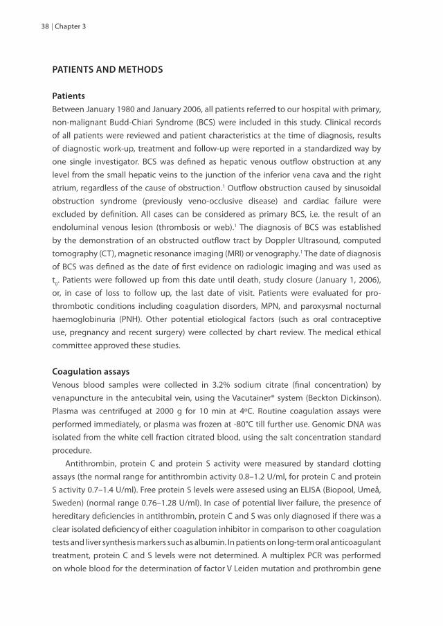
38 | Chapter 3
PATiENTS AND mETHoDS
Patients Between January 1980 and January 2006, all patients referred to our hospital with primary, non-malignant Budd-Chiari Syndrome (BCS) were included in this study. Clinical records of all patients were reviewed and patient characteristics at the time of diagnosis, results of diagnostic work-up, treatment and follow-up were reported in a standardized way by one single investigator. BCS was defined as hepatic venous outflow obstruction at any level from the small hepatic veins to the junction of the inferior vena cava and the right atrium, regardless of the cause of obstruction.1 Outflow obstruction caused by sinusoidal obstruction syndrome (previously veno-occlusive disease) and cardiac failure were excluded by definition. All cases can be considered as primary BCS, i.e. the result of an endoluminal venous lesion (thrombosis or web).1 The diagnosis of BCS was established by the demonstration of an obstructed outflow tract by Doppler Ultrasound, computed tomography (CT), magnetic resonance imaging (MRI) or venography.1 The date of diagnosis of BCS was defined as the date of first evidence on radiologic imaging and was used as t0. Patients were followed up from this date until death, study closure (January 1, 2006), or, in case of loss to follow up, the last date of visit. Patients were evaluated for pro-thrombotic conditions including coagulation disorders, MPN, and paroxysmal nocturnal haemoglobinuria (PNH). Other potential etiological factors (such as oral contraceptive use, pregnancy and recent surgery) were collected by chart review. The medical ethical committee approved these studies.
Coagulation assaysVenous blood samples were collected in 3.2% sodium citrate (final concentration) by venapuncture in the antecubital vein, using the Vacutainer® system (Beckton Dickinson). Plasma was centrifuged at 2000 g for 10 min at 4ºC. Routine coagulation assays were performed immediately, or plasma was frozen at -80°C till further use. Genomic DNA was isolated from the white cell fraction citrated blood, using the salt concentration standard procedure. Antithrombin, protein C and protein S activity were measured by standard clotting assays (the normal range for antithrombin activity 0.8–1.2 U/ml, for protein C and protein S activity 0.7–1.4 U/ml). Free protein S levels were assesed using an ELISA (Biopool, Umeå, Sweden) (normal range 0.76–1.28 U/ml). In case of potential liver failure, the presence of hereditary deficiencies in antithrombin, protein C and S was only diagnosed if there was a clear isolated deficiency of either coagulation inhibitor in comparison to other coagulation
tests and liver synthesis markers such as albumin. In patients on long-term oral anticoagulant treatment, protein C and S levels were not determined. A multiplex PCR was performed on whole blood for the determination of factor V Leiden mutation and prothrombin gene
Chap
ter 3
39MPN in BCS |
variant, as has been described earlier.15 The presence of lupus anticoagulant was tested by an APTT based and a diluted PT assay and anticardiolipin antibodies were tested by ELISA, as described earlier.16 Diagnosis of paroxysmal nocturnal hemoglobinuria (PNH) was based on flow cytometric analysis using antibodies directed against glycosylphosphatidylinositol-anchored proteins.
Diagnostic work-up and diagnostic criteria for myeloproliferative neoplasmsFor standardization of our MPN work-up, all available data were used to classify MPN according to the 2001 WHO guidelines.17-19 Since these guidelines have changed over time, and patients were evaluated for MPN according to the existing guidelines at time of diagnosis of BCS, this approach is limited by some missing data (e.g. bone marrow biopsies), especially in patients diagnosed in the beginning of the study. The designation “unclassified MPN” was applied to cases with definite features of MPN, but that failed to meet the criteria for a specific subtype, such as PV or ET. In addition to standard MPN work-up, the recently described JAK2V617F somatic point mutation was determined in patients of whom DNA was available. Total red blood cell (RBC) mass was determined as described before and expressed as a percentage of the mean normal predicted value. A value above 125% of the theoretical value was considered a significant increase. If RBC mass was not measured, hemoglobin (Hb) levels were used to assess the level of erythrocytosis. A significant increase was defined as a Hb level above 18.5 g/dl in men or 16.5 g/dl in women. The spontaneous erythroid colony formation (EEC) was determined in clonogenic assays as described earlier.20 Mononuclear cells (MNC) were isolated by ficoll density gradient centrifugation (d = 1.077, Nycomed, Oslo, Norway). MNC were added to Methocult H4230 (StemCell Technologies, Vancouver, Canada) supplemented with 2.5 ng/ml IL-3 with or without 3 U/ml Epo. Cells were plated at 0.4 x 10⁵ cells per ml in 24 well plates (0.25 ml/well) and incubated at 37°C in humidified atmosphere and 5 % CO2. After 14 days, BFU-e and Epo-independent (spontaneous) BFU-e were counted. Spontaneous BFU-e was considered positive when one or more Epo-independent BFU-e colonies had developed. For JAK2V617F mutation analysis, high-molecular-weight DNA was isolated from the white cell fraction of citrated blood, using the salt concentration standard procedure. The quality of the genomic DNA was verified by amplification of exon 11 of the FMS-like tyrosine kinase (FLT3) gene, using the primer set FLT3-11F 5’-CAATTTAGGTATGAAAGCC-3’ and FLT3-11R 5’-CAAACTCTAAATTTTCTCT-3’. Subsequently, a double allele-specific PCR was performed on each individual sample using the allele-specific forward primer JAK2-ASP 5’-TTTTAAATTATGGAGTATATT-3’ in combination with the reverse primers GJAK2MUT1 5’-TATAACTGAATAGTCCTACAGTG-3’ or GJAK2MUT2 5’-GTTGAACCTGCCATAATCTC-3’, i.e., the allele specific PCR with the primer set JAK2-ASP and GJAK2MUT1 was confirmed by the PCR using the primer set JAK2-ASP and GJAK2MUT1. All PCR’s were performed on 50ng high-
40 | Chapter 3
molecular-weight DNA (25mM dNTP, 15 pmol primers, 2mM MgCl2, Taq polymerase and 10xbuffer (Invitrogen Life Technologies, Breda, The Netherlands)). Cycling conditions were as follows: 1 cycle 5’ 94°C, 35 cycles 1’ 94°C, 1’ 54°C (FLT3) or 61.5°C (JAK2V617F), 1’ 72°C, and 1 cycle 7’ 72°C. PCR products were visualized using EtBr-gel electrophoresis.
Statistical analysisResults are expressed as median ± SD. All tests were 2-tailed with the level of significance set at P < .05. Comparison between patients with and without MPN was performed using the non-parametric Mann-Whitney U test for continuous variables and the Chi-square test for categorical variables. Survival rates were calculated by means of the Kaplan-Meier method and comparison of survival functions was based on log-rank testing. Statistical analyses were carried out in SPSS 11.5.0 for Windows (SPSS, Chicago, IL).
rESULTS
Patient characteristicsForty-one patients with the diagnosis of BCS were identified in our hospital. One patient with hepatic outflow obstruction resulting from malignancy was excluded. This left a total of 40 patients with primary BCS who were eligible for analysis. Median age was 28.4 years (18.4–53.3) and 26 patients (65%) were female. In nine patients (23%) additional portal vein thrombosis (PVT) was present. Ascites, hepatomegaly and abdominal pain were the most prevalent clinical symptoms. Sites of outflow obstruction were the hepatic veins (63%), inferior vena cava (5%) or both (33%). At liver biopsy, performed in 24 patients, evidence for cirrhosis was found in four (17%). Patient characteristics are presented in Table 1.
Underlying or concomitant disordersCoagulation disorders were found in 14 patients (35%). Antiphospholipid antibodies (APA) were detected at diagnosis of BCS in ten patients. In six patients aCL or lupus anticoagulant was measured only once. Four patients had APA that were confirmed after 12 weeks. Only patients in whom APA were confirmed were regarded as having APA. Oral contraceptives were used in 13 patients (52%). In four patients (10%) no underlying disorder could be found. The underlying or concomitant disorders are shown in Table 2.
myeloproliferative neoplasms in BCS A summary of the performed diagnostic work-up for MPN according to bone marrow biopsy and EEC is shown in Figure 1. Twenty-nine patients underwent complete MPN diagnostic work-up. In 21 of these patients an EEC was also performed. Eleven patients did not undergo a bone marrow biopsy nor was an EEC performed, for various reasons,
Chap
ter 3
41MPN in BCS |
Table 1. Patient characteristics at the time of diagnostic-work up in 40 patients with primary Budd-Chiari syndrome
N obtained data
Age (yrs)* 40 28.4 (18.4–53.3)
Male: female (%) 40 14:26 (35:65)
Clinical manifestations (%)
Abdominal pain 38 30 (79)
Jaundice 38 3 (8)
Leg edema 38 12 (32)
Ascites 39 33 (85)
Hepatomegaly 39 34 (87)
Splenomegaly 38 19 (49)
Encephalopathy 33 3 (8)
Variceal bleeding 33 2 (6)
Site of outflow obstruction (%)
Hepatic veins 40 25 (63)
Inferior vena cava 40 2 (5)
Combined hepatic veins and inferior vena cava 40 13 (33)
Membranous obstruction of inferior vena cava (%) 40 1 (3)
Portal vein obstruction (%) 40 9 (23)
Serum levels*
Albumin (g/L) 37 38 (21–50)
Bilirubin (μmol/L) 38 29.5 (7–143)
Platelet count (10E9/L) 36 243 (18–520)
ALT (ULN) 37 1.1 (0.2–68.0)
Alkaline phosphatase (ULN) 37 1.2 (0.5–4.2)
Hemoglobin (g/dl) 37 13.5 (7.3–18.7)
Sodium (mmol/L) 35 137 (123–144)
Creatinin (μmol/L) 35 66 (11.2–137)
Prothrombin time (sec) 34 16.8 (11.5–133.0)
Cirrhosis at liver biopsy (%) 24 4 (17)
Abbreviations: ULN, upper limits of normal value.*Median (range).
including refusal (n = 1), deteriorating clinical condition (n = 2), or the absence of peripheral blood count abnormalities and clinical features of MPN (n = 8). This latter group of patients, who were not suspect for having a primary haematological disorder, did not undergo bone marrow biopsy since they were worked-up earlier in our study period and biopsies were not yet part of the routine diagnostic work-up for BCS. However, most of these patients
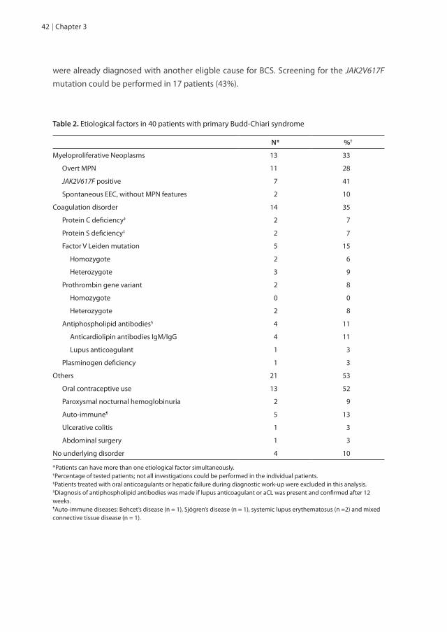
42 | Chapter 3
were already diagnosed with another eligble cause for BCS. Screening for the JAK2V617F mutation could be performed in 17 patients (43%).
Table 2. Etiological factors in 40 patients with primary Budd-Chiari syndrome
N* %†
Myeloproliferative Neoplasms 13 33
Overt MPN 11 28
JAK2V617F positive 7 41
Spontaneous EEC, without MPN features 2 10
Coagulation disorder 14 35
Protein C deficiency‡ 2 7
Protein S deficiency‡ 2 7
Factor V Leiden mutation 5 15
Homozygote 2 6
Heterozygote 3 9
Prothrombin gene variant 2 8
Homozygote 0 0
Heterozygote 2 8
Antiphospholipid antibodies§ 4 11
Anticardiolipin antibodies IgM/IgG 4 11
Lupus anticoagulant 1 3
Plasminogen deficiency 1 3
Others 21 53
Oral contraceptive use 13 52
Paroxysmal nocturnal hemoglobinuria 2 9
Auto-immune¶ 5 13
Ulcerative colitis 1 3
Abdominal surgery 1 3
No underlying disorder 4 10
*Patients can have more than one etiological factor simultaneously.†Percentage of tested patients; not all investigations could be performed in the individual patients.‡Patients treated with oral anticoagulants or hepatic failure during diagnostic work-up were excluded in this analysis.§Diagnosis of antiphospholipid antibodies was made if lupus anticoagulant or aCL was present and confirmed after 12 weeks.¶Auto-immune diseases: Behcet’s disease (n = 1), Sjögren’s disease (n = 1), systemic lupus erythematosus (n =2) and mixed connective tissue disease (n = 1).
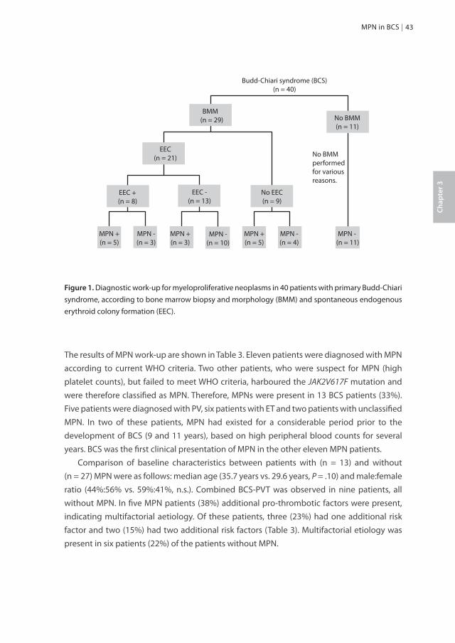
Chap
ter 3
43MPN in BCS |
Budd-Chiari syndrome (BCS)(n = 40)
No BMM(n = 11)
BMM (n = 29)
No BMM performed for variousreasons.
EEC (n = 21)
EEC +(n = 8)
EEC -(n = 13)
No EEC(n = 9)
MPN +(n = 5)
MPN -(n = 3)
MPN +(n = 3)
MPN -(n = 10)
MPN +(n = 5)
MPN -(n = 4)
MPN - (n = 11)
Figure 1. Diagnostic work-up for myeloproliferative neoplasms in 40 patients with primary Budd-Chiari syndrome, according to bone marrow biopsy and morphology (BMM) and spontaneous endogenous erythroid colony formation (EEC).
The results of MPN work-up are shown in Table 3. Eleven patients were diagnosed with MPN according to current WHO criteria. Two other patients, who were suspect for MPN (high platelet counts), but failed to meet WHO criteria, harboured the JAK2V617F mutation and were therefore classified as MPN. Therefore, MPNs were present in 13 BCS patients (33%). Five patients were diagnosed with PV, six patients with ET and two patients with unclassified MPN. In two of these patients, MPN had existed for a considerable period prior to the development of BCS (9 and 11 years), based on high peripheral blood counts for several years. BCS was the first clinical presentation of MPN in the other eleven MPN patients. Comparison of baseline characteristics between patients with (n = 13) and without (n = 27) MPN were as follows: median age (35.7 years vs. 29.6 years, P = .10) and male:female ratio (44%:56% vs. 59%:41%, n.s.). Combined BCS-PVT was observed in nine patients, all without MPN. In five MPN patients (38%) additional pro-thrombotic factors were present, indicating multifactorial aetiology. Of these patients, three (23%) had one additional risk factor and two (15%) had two additional risk factors (Table 3). Multifactorial etiology was present in six patients (22%) of the patients without MPN.
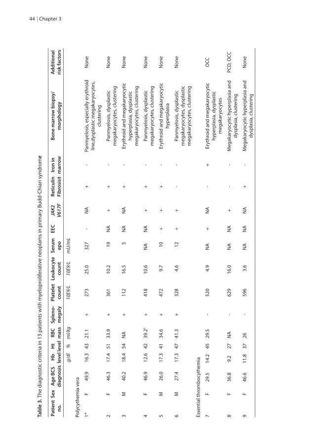
44 | Chapter 3
Tabl
e 3.
The
dia
gnos
tic c
riter
ia in
13
patie
nts
with
mye
lopr
olife
rativ
e ne
opla
ms
in p
rimar
y Bu
dd-C
hiar
i syn
drom
e
Patie
ntno
.Se
xA
ge B
CSdi
agno
sis
Hb
leve
lH
tle
vel
rBC
mas
sSp
leno
-m
egal
yPl
atel
et
coun
tLe
ukoc
yte
coun
tSe
rum
ep
oEE
CJA
K2V6
17F
retic
ulin
Fi
bros
is‡
iron
in
mar
row
Bone
mar
row
bio
psy/
mor
phol
ogy
Add
ition
al
risk
fact
ors
g/dl
%m
l/kg
10E9
/L10
E9/L
mU
/mL
Poly
cyth
emia
ver
a
1*F
49.9
16.3
4321
.1+
273
25.0
327
-N
A+
-Pa
nmye
losi
s, es
peci
ally
ery
thro
id
line,
dysp
last
ic m
egak
aryo
cyte
s, cl
uste
ring
Non
e
2F
46.3
17.4
5133
.9+
361
10.2
19
NA
++
-Pa
nmye
losi
s, dy
spla
stic
m
egak
aryo
cyte
s, cl
uste
ring
Non
e
3M
40.2
18.4
54N
A+
112
16.5
5
NA
NA
+-
Eryt
hroi
d an
d m
egak
aryo
cytic
hy
perp
lasi
a, d
yspl
astic
m
egak
aryo
cyte
s, cl
uste
ring
Non
e
4F
46.9
12.6
4239
.2†
+41
810
.6N
AN
A+
+-
Panm
yelo
sis,
dysp
last
ic
meg
akar
yocy
tes,
clus
terin
gN
one
5M
26.0
17.3
4134
.6+
472
9.7
10
++
+-
Eryt
hroi
d an
d m
egak
aryo
cytic
hy
perp
lasi
a N
one
6M
27.4
17.3
4741
.3+
328
4.6
12
++
--
Panm
yelo
sis,
dysp
last
ic
meg
akar
yocy
tes,
dysp
last
ic
meg
akar
yocy
tes,
clus
terin
g
Non
e
Esse
ntia
l thr
ombo
cyth
emia
7F
29.5
14.2
4529
.5-
520
4.9
NA
+N
A-
+Er
ythr
oid
and
meg
akar
yocy
tic
hype
rpla
sia,
dys
plas
tic
meg
akar
yocy
tes
OCC
8F
36.8
9.2
27N
A-
629
16.0
NA
NA
+-
-M
egak
aryo
cytic
hyp
erpl
asia
and
dy
spla
sia,
clu
ster
ing
PCD
, OCC
9F
46.6
11.8
3726
-59
6 3
.6N
AN
AN
A+
-M
egak
aryo
cytic
hyp
erpl
asia
and
dy
spla
sia,
clu
ster
ing
Non
e
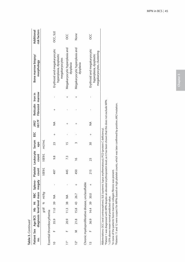
Chap
ter 3
45MPN in BCS |
Tabl
e 3.
Con
tinue
d
Patie
ntno
.Se
xA
ge B
CSdi
agno
sis
Hb
leve
lH
tle
vel
rBC
mas
sSp
leno
-m
egal
yPl
atel
et
coun
tLe
ukoc
yte
coun
tSe
rum
ep
oEE
CJA
K2V6
17F
retic
ulin
Fi
bros
is‡
iron
in
mar
row
Bone
mar
row
bio
psy/
mor
phol
ogy
Add
ition
al
risk
fact
ors
g/dl
%m
l/kg
10E9
/L10
E9/L
mU
/mL
Esse
ntia
l thr
ombo
cyth
emia
10F
35.9
11.0
39N
A-
497
9.8
23
+N
A+
-Er
ythr
oid
and
meg
akar
yocy
tic
hype
rpla
sia,
dys
plas
tic
meg
akar
yocy
tes
OCC
, SLE
11§
F20
.911
.338
NA
-44
5 7
.3 1
5-
+-
+M
egak
aryo
cytic
hyp
erpl
asia
and
dy
spla
sia
OCC
12§
M21
.815
.843
26.7
+45
016
3
-+
-+
Meg
akar
yocy
tic h
yper
plas
ia a
nd
dysp
lasi
aN
one
Chro
nic
mye
lopr
olife
rativ
e di
seas
e, u
ncla
ssifi
able
13F
36.9
14.4
3930
.0+
215
23 3
0+
NA
--
Eryt
hroi
d an
d m
egak
aryo
cytic
hy
perp
lasi
a, d
yspl
astic
m
egak
aryo
cyte
s, cl
uste
ring
OCC
Abb
revi
atio
ns: O
CC (o
ral c
ontr
acep
tives
); SL
E (s
yste
mic
lupu
s er
ythe
mat
osus
); PC
D (p
rote
in C
defi
cien
cy).
*Pat
ient
1 w
as d
iagn
osed
with
MPN
des
pite
an
elev
ated
ery
thro
poie
tin le
vel,
as it
has
bee
n sh
own
that
this
doe
s no
t exc
lude
MPN
. † >2
5% a
bove
mea
n no
rmal
pre
dict
ed v
alue
.‡ In
non
e of
the
patie
nts’
bone
mar
row
col
lage
n fib
rosi
s w
as p
rese
nt.
§ Patie
nts
11 a
nd 1
2 w
ere
susp
ect f
or M
PNs
base
d on
hig
h pl
atel
et c
ount
s, w
hich
was
late
r con
firm
ed b
y po
sitiv
e JA
K2 m
utat
ion.
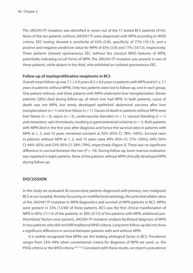
46 | Chapter 3
The JAK2V617F mutation was identified in seven out of the 17 tested BCS patients (41%). None of the ten patients without JAK2V617F were diagnosed with MPN according to WHO criteria. EEC testing showed a sensitivity of 63% (5/8), specificity of 77% (10/13), and a positive and negative predictive value for MPN of 63% (5/8) and 77% (10/13), respectively. Three patients showed spontaneous EEC without the classical WHO features of MPN, potentially indicating occult forms of MPN. The JAK2V617F mutation was present in two of these patients, while absent in the third, who exhibited an isolated spontaneous EEC.
Follow-up of myeloproliferative neoplasms in BCSOverall mean follow-up was 7.1 ± 6.9 years (8.2 ± 6.6 years in patients with MPN and 6.5 ± 7.1 years in patients without MPN). Only two patients were lost to follow-up, one in each group. One patient without, and three patients with MPN underwent liver transplantation. Eleven patients (28%) died during follow-up, of which two had MPN. In both patients, cause of death was not MPN, but newly developed epitheloid abdominal sarcoma after liver transplantation (n = 1) and liver failure (n = 1). Causes of death in patients without MPN were liver failure (n = 3), sepsis (n = 3), cardiovascular disorders (n = 1), variceal bleeding (n = 1) and mesenteric vein thrombosis, resulting in gastrointestinal ischemia (n = 1). Both patients with MPN died in the first year after diagnosis and hence the survival rates in patients with MPN at 1, 5, and 10 years remained constant at 92% (95% CI: 78%–100%). Survival rates in patients without MPN at 1, 5, and 10 years were 89% (95% CI: 77%–100%), 64% (95% CI: 44%–85%) and 53% (95% CI: 28%–79%), respectively (Figure 2). There was no significant difference in survival between the two (P = .18). During follow-up, bone marrow evaluation was repeated in eight patients. None of the patients without MPN clinically developed MPN during follow-up.
DiSCUSSioN
In this study we evaluated 40 consecutive patients diagnosed with primary, non-malignant BCS at our hospital, thereby focussing on multifactorial aetiology, the potential added value of the JAK2V617F mutation in MPN diagnostics and survival of MPN patients in BCS. MPNs were present in 33% (13/40) of these patients. BCS was the first clinical manifestation of MPN in 85% (11/13) of the patients. In 38% (5/13) of the patients with MPN, additional pro-thrombotic factors were present. JAK2V617F mutation analysis facilitated diagnosis of MPN in two patients who did not fulfil traditional WHO criteria. Long term follow-up did not show a significant difference in survival between patients with and without MPN. It is widely recognized that MPNs are the leading etiological factor in BCS. Prevalence ranges from 23%–49% when conventional criteria for diagnosis of MPN are used, i.e. the PVSG criteria or the WHO criteria.4,5,21,22 Consistent with these results, we report a prevalence
Chap
ter 3
47MPN in BCS |
Survival
121086420
Cum
ulat
ive
surv
ival
1,0
0,9
0,8
0,7
0,6
0,5
0,4
0,3
0,2
0,1
0,0
MPN
Non-MPN
P = 0.18
Figure 2. Survival in non-MPN patients (n = 27) and MPN patients (n = 13) with primary, non-malignant Budd-Chiari syndrome. Survival rates in patients with an underlying MPN at 1, 5, and 10 years were 92% (95% CI, 78%–100%), with number of patients at risk being 12, 8 and 5, respectively. Survival rates in patients without MPN at 1, 5, and 10 years were 89% (95% CI, 78%–100%), 64% (95% CI, 44%–84%) and 53% (95% CI, 28%–79%), with number of patients at risk being 22, 10 and 5, respectively.
of MPN of 33%. This is based on a population of consecutive patients over 26 years in a single center, therefore minimizing the risk of selection bias. None of our BCS patients without MPN at diagnosis developed MPN during follow up. In a meta-analysis conducted in 1997, a MPN prevalence of 78% has been reported when spontaneous EEC was used as the only diagnostic criterion.7 This result necessitates a balanced interpretation. First, several case series have suggested that EEC is a sensitive indicator for diagnosis of MPN.6,7,23,24 However, the value of EEC as the sole criterion has been questioned and it has been suggested that this variable should only be used as a minor criterion in diagnosing MPN. EEC has been reported in non-clonal causes of erythrocytosis and in healthy controls (false positive), while it has not been observed in about 50% of patients with well documented ET and in some patients with PV (false negative).25-29 Second, EEC assays are technically demanding and not amenable to external quality assurance. Third, the predictive value of spontaneous EEC as an indication for occult MPN has not yet been fully elucidated in large prospective studies. Furthermore, it should be noted that the meta-analysis with a reported prevalence of 78% is based on selected case reports and data-pooling of small case series, endangering a selection bias. Our view is strengthened by successive studies from one of the research
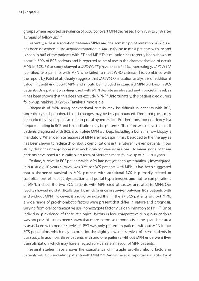
48 | Chapter 3
groups where reported prevalence of occult or overt MPN decreased from 75% to 31% after 15 years of follow up.6,21
Recently, a clear association between MPNs and the somatic point mutation JAK2V617F has been described.10 The acquired mutation in JAK2 is found in most patients with PV and is seen in half of the patients with ET and MF.10 This mutation has recently been shown to occur in 59% of BCS patients and is reported to be of use in the characterization of occult MPN in BCS.14 Our study showed a JAK2V617F prevalence of 41%. Interestingly, JAK2V617F identified two patients with MPN who failed to meet WHO criteria. This, combined with the report by Patel et al., clearly suggests that JAK2V617F mutation analysis is of additional value in identifying occult MPN and should be included in standard MPN work-up in BCS patients. One patient was diagnosed with MPN despite an elevated erythropoietin level, as it has been shown that this does not exclude MPN.30 Unfortunately, this patient died during follow-up, making JAK2V617F analysis impossible. Diagnosis of MPN using conventional criteria may be difficult in patients with BCS, since the typical peripheral blood changes may be less pronounced. Thrombocytosis may be masked by hypersplenism due to portal hypertension. Furthermore, iron deficiency is a frequent finding in BCS and hemodilution may be present.31 Therefore we believe that in all patients diagnosed with BCS, a complete MPN work-up, including a bone marrow biopsy is mandatory. When definite features of MPN are met, aspirin may be added to the therapy as has been shown to reduce thrombotic complications in the future.32 Eleven patients in our study did not undergo bone marrow biopsy for various reasons. However, none of these patients developed a clinically overt form of MPN at a mean follow-up of 7.7 ± 8.0 years. To date, survival in BCS patients with MPN had not yet been systematically investigated. In our study, 10-years survival was 92% for BCS patients with MPN. It has been suggested that a shortened survival in MPN patients with additional BCS is primarily related to complications of hepatic dysfunction and portal hypertension, and not to complications of MPN. Indeed, the two BCS patients with MPN died of causes unrelated to MPN. Our results showed no statistically significant difference in survival between BCS patients with and without MPN. However, it should be noted that in the 27 BCS patients without MPN, a wide range of pro-thrombotic factors were present that differ in nature and prognosis, varying from oral contraceptive use, homozygote factor V Leiden mutation to PNH.33 Since individual prevalence of these etiological factors is low, comparative sub-group analysis was not possible. It has been shown that more extensive thrombosis in the splanchnic area is associated with poorer survival.34 PVT was only present in patients without MPN in our BCS population, which may account for the slightly lowered survival of these patients in our study. In addition, three patients with and one patients without MPN underwent liver transplantation, which may have affected survival rate in favour of MPN patients. Several studies have shown the coexistence of multiple pro-thrombotic factors in patients with BCS, including patients with MPN.21,22 Denninger et al. reported a multifactorial

Chap
ter 3
49MPN in BCS |
aetiology in 41% of BCS patients with either occult or overt MPN. Our results showed that in 38% of the patients with MPN additional risk factors were present. These results enforce the necessity of extensive screening for additional pro-thrombotic conditions when one cause has already been found to optimize treatment options. In conclusion, we report MPNs in 33% of our BCS patients. In 38% of the patients with MPN, additional pro-thrombotic factors were present. Our study clearly confirms the importance of JAK2V617F mutation analysis in patients with BCS. Long term follow-up did not show a significant difference in survival between patients with and without MPN.
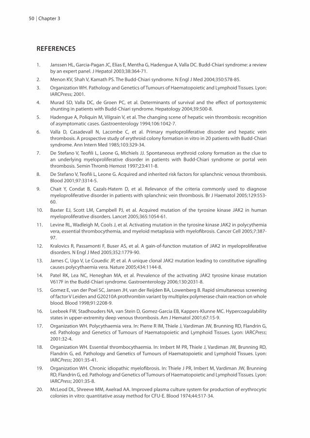
50 | Chapter 3
rEFErENCES
1. Janssen HL, Garcia-Pagan JC, Elias E, Mentha G, Hadengue A, Valla DC. Budd-Chiari syndrome: a review by an expert panel. J Hepatol 2003;38:364-71.
2. Menon KV, Shah V, Kamath PS. The Budd-Chiari syndrome. N Engl J Med 2004;350:578-85.
3. Organization WH. Pathology and Genetics of Tumours of Haematopoietic and Lymphoid Tissues. Lyon: IARCPress; 2001.
4. Murad SD, Valla DC, de Groen PC, et al. Determinants of survival and the effect of portosystemic shunting in patients with Budd-Chiari syndrome. Hepatology 2004;39:500-8.
5. Hadengue A, Poliquin M, Vilgrain V, et al. The changing scene of hepatic vein thrombosis: recognition of asymptomatic cases. Gastroenterology 1994;106:1042-7.
6. Valla D, Casadevall N, Lacombe C, et al. Primary myeloproliferative disorder and hepatic vein thrombosis. A prospective study of erythroid colony formation in vitro in 20 patients with Budd-Chiari syndrome. Ann Intern Med 1985;103:329-34.
7. De Stefano V, Teofili L, Leone G, Michiels JJ. Spontaneous erythroid colony formation as the clue to an underlying myeloproliferative disorder in patients with Budd-Chiari syndrome or portal vein thrombosis. Semin Thromb Hemost 1997;23:411-8.
8. De Stefano V, Teofili L, Leone G. Acquired and inherited risk factors for splanchnic venous thrombosis. Blood 2001;97:3314-5.
9. Chait Y, Condat B, Cazals-Hatem D, et al. Relevance of the criteria commonly used to diagnose myeloproliferative disorder in patients with splanchnic vein thrombosis. Br J Haematol 2005;129:553-60.
10. Baxter EJ, Scott LM, Campbell PJ, et al. Acquired mutation of the tyrosine kinase JAK2 in human myeloproliferative disorders. Lancet 2005;365:1054-61.
11. Levine RL, Wadleigh M, Cools J, et al. Activating mutation in the tyrosine kinase JAK2 in polycythemia vera, essential thrombocythemia, and myeloid metaplasia with myelofibrosis. Cancer Cell 2005;7:387-97.
12. Kralovics R, Passamonti F, Buser AS, et al. A gain-of-function mutation of JAK2 in myeloproliferative disorders. N Engl J Med 2005;352:1779-90.
13. James C, Ugo V, Le Couedic JP, et al. A unique clonal JAK2 mutation leading to constitutive signalling causes polycythaemia vera. Nature 2005;434:1144-8.
14. Patel RK, Lea NC, Heneghan MA, et al. Prevalence of the activating JAK2 tyrosine kinase mutation V617F in the Budd-Chiari syndrome. Gastroenterology 2006;130:2031-8.
15. Gomez E, van der Poel SC, Jansen JH, van der Reijden BA, Lowenberg B. Rapid simultaneous screening of factor V Leiden and G20210A prothrombin variant by multiplex polymerase chain reaction on whole blood. Blood 1998;91:2208-9.
16. Leebeek FW, Stadhouders NA, van Stein D, Gomez-Garcia EB, Kappers-Klunne MC. Hypercoagulability states in upper-extremity deep venous thrombosis. Am J Hematol 2001;67:15-9.
17. Organization WH. Polycythaemia vera. In: Pierre R IM, Thiele J, Vardiman JW, Brunning RD, Flandrin G, ed. Pathology and Genetics of Tumours of Haematopoietic and Lymphoid Tissues. Lyon: IARCPress; 2001:32-4.
18. Organization WH. Essential thrombocythaemia. In: Imbert M PR, Thiele J, Vardiman JW, Brunning RD, Flandrin G, ed. Pathology and Genetics of Tumours of Haematopoietic and Lymphoid Tissues. Lyon: IARCPress; 2001:35-41.
19. Organization WH. Chronic idiopathic myelofibrosis. In: Thiele J PR, Imbert M, Vardiman JW, Brunning RD, Flandrin G, ed. Pathology and Genetics of Tumours of Haematopoietic and Lymphoid Tissues. Lyon: IARCPress; 2001:35-8.
20. McLeod DL, Shreeve MM, Axelrad AA. Improved plasma culture system for production of erythrocytic colonies in vitro: quantitative assay method for CFU-E. Blood 1974;44:517-34.
Chap
ter 3
51MPN in BCS |
21. Denninger MH, Chait Y, Casadevall N, et al. Cause of portal or hepatic venous thrombosis in adults: the role of multiple concurrent factors. Hepatology 2000;31:587-91.
22. Janssen HL, Meinardi JR, Vleggaar FP, et al. Factor V Leiden mutation, prothrombin gene mutation, and deficiencies in coagulation inhibitors associated with Budd-Chiari syndrome and portal vein thrombosis: results of a case-control study. Blood 2000;96:2364-8.
23. Valla D, Casadevall N, Huisse MG, et al. Etiology of portal vein thrombosis in adults. A prospective evaluation of primary myeloproliferative disorders. Gastroenterology 1988;94:1063-9.
24. Reid CD, Chanarin I, Lewis J. Formes frustes in myeloproliferative disorders. Identification by the growth of an endogenous erythroid clone in vitro in patients with arterial vascular disease. Lancet 1982;1:14-5.
25. Dainiak N, Hoffman R, Lebowitz AI, Solomon L, Maffei L, Ritchey K. Erythropoietin-dependent primary pure erythrocytosis. Blood 1979;53:1076-84.
26. Dudley JM, Westwood N, Leonard S, Eridani S, Pearson TC. Primary polycythaemia: positive diagnosis using the differential response of primitive and mature erythroid progenitors to erythropoietin, interleukin 3 and alpha-interferon. Br J Haematol 1990;75:188-94.
27. Eridani S, Sawyer B, Pearson TC. Patterns of in vitro BFU-E proliferation in different forms of polycythaemia and in thrombocythaemia. Eur J Haematol 1987;38:363-9.
28. Masters GS, Baines P, Jacobs A. Erythroid colony growth from peripheral blood and bone marrow in polycythaemia. J Clin Pathol 1990;43:937-41.
29. Spivak JL. Polycythemia vera: myths, mechanisms, and management. Blood 2002;100:4272-90.
30. Thurmes PJ, Steensma DP. Elevated serum erythropoietin levels in patients with Budd-Chiari syndrome secondary to polycythemia vera: clinical implications for the role of JAK2 mutation analysis. Eur J Haematol 2006;77:57-60.
31. Valla DC. The diagnosis and management of the Budd-Chiari syndrome: consensus and controversies. Hepatology 2003;38:793-803.
32. Landolfi R, Marchioli R, Kutti J, et al. Efficacy and safety of low-dose aspirin in polycythemia vera. N Engl J Med 2004;350:114-24.
33. Leebeek FW, Lameris JS, van Buuren HR, Gomez E, Madretsma S, Sonneveld P. Budd-Chiari syndrome, portal vein and mesenteric vein thrombosis in a patient homozygous for factor V Leiden mutation treated by TIPS and thrombolysis. Br J Haematol 1998;102:929-31.
34. Murad SD, Valla DC, de Groen PC, et al. Pathogenesis and treatment of Budd-Chiari syndrome combined with portal vein thrombosis. Am J Gastroenterol 2006;101:83-90.

C H A P T E r 4
mYELoProLiFErATiVE NEoPLASmS iN BUDD-CHiAri SYNDromE
AND PorTAL VEiN THromBoSiS: A mETA-ANALYSiS
Jasper H. Smalberg1, Lidia Arends2,3, Dominique C. Valla4, Jean-Jacques Kiladjian5,
Harry L.A. Janssen6 and Frank W.G. Leebeek1
1Department of Hematology, Erasmus University Medical Center, Rotterdam, The Netherlands; 2Institute of Psychology, Erasmus University Rotterdam, Rotterdam, the Netherlands;
3Department of Biostatistics, Erasmus University Medical Center, Rotterdam, The Netherlands; 4Service d’Hépatologie, Hôpital Beaujon, Clichy, France;
5APHP, Hôpital Saint-Louis, Centre d’Investigations Cliniques, Paris, France; 6Department of Gastroenterology and Hepatology, Erasmus University Medical Center,
Rotterdam, The Netherlands.
Submitted
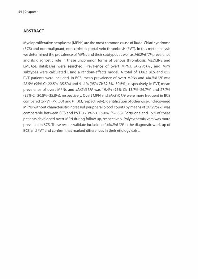
54 | Chapter 4
ABSTrACT
Myeloproliferative neoplasms (MPNs) are the most common cause of Budd-Chiari syndrome (BCS) and non-malignant, non-cirrhotic portal vein thrombosis (PVT). In this meta-analysis we determined the prevalence of MPNs and their subtypes as well as JAK2V617F prevalence and its diagnostic role in these uncommon forms of venous thrombosis. MEDLINE and EMBASE databases were searched. Prevalence of overt MPNs, JAK2V617F, and MPN subtypes were calculated using a random-effects model. A total of 1.062 BCS and 855 PVT patients were included. In BCS, mean prevalence of overt MPNs and JAK2V617F was 28.5% (95% CI: 22.5%–35.5%) and 41.1% (95% CI: 32.3%–50.6%), respectively. In PVT, mean prevalence of overt MPNs and JAK2V617F was 19.4% (95% CI: 13.7%–26.7%) and 27.7% (95% CI: 20.8%–35.8%), respectively. Overt MPN and JAK2V617F were more frequent in BCS compared to PVT (P < .001 and P = .03, respectively). Identification of otherwise undiscovered MPNs without characteristic increased peripheral blood counts by means of JAK2V617F was comparable between BCS and PVT (17.1% vs. 15.4%, P = .68). Forty-one and 15% of these patients developed overt MPN during follow up, respectively. Polycythemia vera was more prevalent in BCS. These results validate inclusion of JAK2V617F in the diagnostic work-up of BCS and PVT and confirm that marked differences in their etiology exist.
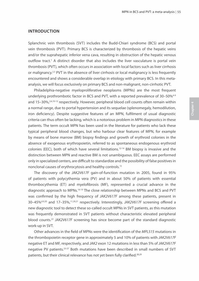
Chap
ter 4
55MPN in BCS and PVT: a meta-analysis |
iNTroDUCTioN
Splanchnic vein thrombosis (SVT) includes the Budd-Chiari syndrome (BCS) and portal vein thrombosis (PVT). Primary BCS is characterized by thrombosis of the hepatic veins and/or the suprahepatic inferior vena cava, resulting in obstruction of the hepatic venous outflow tract.1 A distinct disorder that also includes the liver vasculature is portal vein thrombosis (PVT), which often occurs in association with local factors such as liver cirrhosis or malignancy.2,3 PVT in the absence of liver cirrhosis or local malignancy is less frequently encountered and shows a considerable overlap in etiology with primary BCS. In this meta-analysis, we will focus exclusively on primary BCS and non-malignant, non-cirrhotic PVT. Philadelphia-negative myeloproliferative neoplasms (MPNs) are the most frequent underlying prothrombotic factor in BCS and PVT, with a reported prevalence of 30–50%4-9 and 15–30%,2,6,10-12 respectively. However, peripheral blood cell counts often remain within a normal range, due to portal hypertension and its sequelae (splenomegaly, hemodilution, iron deficiency). Despite suggestive features of an MPN, fulfilment of usual diagnostic criteria can thus often be lacking, which is a notorious problem in MPN diagnostics in these patients. The term occult MPN has been used in the literature for patients who lack these typical peripheral blood changes, but who harbour clear features of MPN, for example by means of bone marrow (BM) biopsy findings and growth of erythroid colonies in the absence of exogenous erythropoietin, referred to as spontaneous endogenous erythroid colonies (EEC), both of which have several limitations.12-14 BM biopsy is invasive and the distinction between MPN and reactive BM is not unambiguous. EEC assays are performed only in specialized centers, are difficult to standardize and the possibility of false positives in nonclonal causes of erythrocytosis and healthy controls.15
The discovery of the JAK2V617F gain-of-function mutation in 2005, found in 95% of patients with polycythemia vera (PV) and in about 50% of patients with essential thrombocythemia (ET) and myelofibrosis (MF), represented a crucial advance in the diagnostic approach to MPNs.16-19 The close relationship between MPNs and BCS and PVT was confirmed by the high frequency of JAK2V617F among these patients, present in 30–45%4,9,20 and 17–35%,11,20,21 respectively. Interestingly, JAK2V617F screening offered a new diagnostic tool to detect these so-called occult MPNs in SVT patients, as this mutation was frequently demonstrated in SVT patients without characteristic elevated peripheral blood counts.22 JAK2V617F screening has since become part of the standard diagnostic work-up in SVT. Other advances in the field of MPNs were the identification of the MPL515 mutations in the thrombopoietin receptor gene in approximately 5 and 10% of patients with JAK2V617F negative ET and MF, respectively, and JAK2 exon 12 mutations in less than 5% of JAK2V617F negative PV patients.23-27 Both mutations have been described in small numbers of SVT patients, but their clinical relevance has not yet been fully clarified.28,29
56 | Chapter 4
The aims of this systematic review and meta-analysis were: 1) to assess the prevalence of overt MPNs and JAK2V617F in BCS and PVT patients; 2) to quantify the detection rate of MPNs without characteristic increased peripheral blood counts by means of JAK2V617F screening; 3) to determine the frequency of MPN subtypes in BCS and PVT patients, and; 4) to evaluate the clinical relevance of the MPL and JAK2 exon 12 mutations in BCS and PVT patients.
mETHoDS
Search strategy and selection criteriaOne of the authors (J.S.) searched Ovid MEDLINE and EMBASE from 1980 to August 1st 2011. The search strategy was restricted to published data and the English language using the subject headings presented in the appendix. The search was supplemented by manually reviewing the reference list of retrieved articles and relevant reviews. Titles and abstracts of retrieved citations were screened and potentially suitable studies were read in full by J.S. and F.L. Studies were selected when the following criteria were met: 1) patients were diagnosed with primary BCS or non-cirrhotic, non-malignant PVT, or patients with an underlying malignancy or cirrhosis were explicitly mentioned; 2) information on MPNs and/or JAK2V617F, JAK2 exon 12 or MPL515 was provided; 3) the cohort consisted of patients in which patients with overt MPNs or other thrombophilic factors were not excluded; 4) splanchnic vein thrombosis was subdivided in BCS and PVT; 5) a minimum of ten patients were included. Disagreements were resolved after discussion or after having collected the opinion of a third reviewer (H.J.).
Data extractionJ.S. extracted data on each selected study (year of publication, study design, demograhics, criteria for diagnosing MPNs, number of patients included). Patients with BCS in the presence of a malignancy and PVT patients with a malignancy or cirrhosis were excluded from the analysis. Patients with combined BCS and PVT were classified as BCS patients according to common practice.4 Prevalence of MPNs, JAK2V617F, JAK2 exon 12 and MPL515 mutations were subsequently extracted. Patients fulfilling the traditional MPN criteria used in the various studies (mostly World Health Organization or Polycythemia Vera Study Group), either in the presence or absence of JAK2V617F, were considered to display characteristic increased peripheral blood counts and were classified as overt MPN. Patients not fulfilling the traditional criteria for MPN were assumed to lack these typical hematologic features. In these patients, MPNs identified by means of JAK2V617F were classified as MPN unclassifiable. To assess the diagnostic yield of JAK2V617F, these cases of otherwise undiscovered MPN were expressed as a proportion of total amount of patients tested for
Chap
ter 4
57MPN in BCS and PVT: a meta-analysis |
the JAK2V617F mutation. Corresponding authors were contacted in case essential data were not mentioned, with a reminder sent after two weeks.
Statistical analysisWeighted mean proportion and 95% confidence intervals (CI) of MPNs, JAK2V617F, and MPN subtypes prevalence were calculated using a random effects model. Differences in prevalence were calculated by means of Pearson’s chi square. All statistical tests were two-sided and P-values < .05 were considered statistically significant. Statistical heterogeneity was evaluated using the I2 statistic, which describes the percentage of variation across studies that is due to heterogeneity rather than chance, with a P-value < .05 representing statistically significant heterogeneity. If heterogeneity was present, the analyses were repeated removing one study a time to identify the source of heterogeneity. All analyses were performed with Comprehensive Meta Analysis 2.2 for Windows (Biostat, Englewood, USA) and the overall effects are presented as event rates with 95% CI.
rESULTS
Study identification and selectionWe identified 821 potentially relevant publications: 255 from MEDLINE and 566 from EMBASE. A total of 109 studies were duplicate, and 665 studies were excluded after title and abstracts screening according to predefined inclusion criteria. The remaining studies were retrieved in full for detailed evaluation. Six additional studies were identified: five from reference lists and one from our center30 which was under submission. Figure 1 shows the study selection process. Of the 53 retrieved studies, 21 were excluded because of the following reasons: in two studies patients with malignancies or liver cirrhosis were not excluded or explicitly mentioned,31,32 five studies were based on selected cohorts in which patients with overt MPNs or other thrombophilic factors were excluded,33-37 one study did not differentiate SVT into BCS and PVT,38 in four studies MPN criteria were not acceptable or unclear,14,39-41 and two studies included less than 10 patients.29,42 In addition, seven studies contained duplicate data.43-49 This resulted in 32 studies eligible for inclusion.
Study characteristics and qualityTable 1 and 2 summarizes the characteristics of the included studies for BCS and PVT, respectively. Study size ranged between 10 and 237 patients. Nineteen studies including 1.062 patients reported on MPNs and/or the JAK2V617F mutation in BCS patients. Fifteen studies including 855 patients reported on MPNs and/or the JAK2V617F mutation in PVT
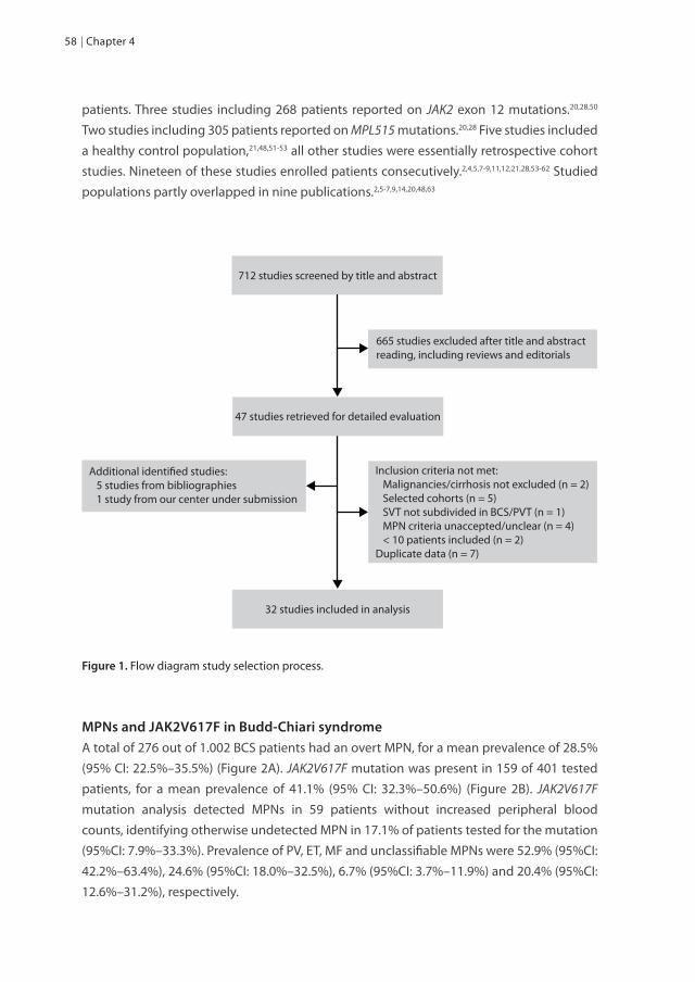
58 | Chapter 4
patients. Three studies including 268 patients reported on JAK2 exon 12 mutations.20,28,50 Two studies including 305 patients reported on MPL515 mutations.20,28 Five studies included a healthy control population,21,48,51-53 all other studies were essentially retrospective cohort studies. Nineteen of these studies enrolled patients consecutively.2,4,5,7-9,11,12,21,28,53-62 Studied populations partly overlapped in nine publications.2,5-7,9,14,20,48,63
712 studies screened by title and abstract
665 studies excluded after title and abstract reading, including reviews and editorials
Inclusion criteria not met: Malignancies/cirrhosis not excluded (n = 2) Selected cohorts (n = 5) SVT not subdivided in BCS/PVT (n = 1) MPN criteria unaccepted/unclear (n = 4) < 10 patients included (n = 2)Duplicate data (n = 7)
47 studies retrieved for detailed evaluation
Additional identi�ed studies: 5 studies from bibliographies 1 study from our center under submission
32 studies included in analysis
Figure 1. Flow diagram study selection process.
mPNs and JAK2V617F in Budd-Chiari syndromeA total of 276 out of 1.002 BCS patients had an overt MPN, for a mean prevalence of 28.5% (95% CI: 22.5%–35.5%) (Figure 2A). JAK2V617F mutation was present in 159 of 401 tested patients, for a mean prevalence of 41.1% (95% CI: 32.3%–50.6%) (Figure 2B). JAK2V617F mutation analysis detected MPNs in 59 patients without increased peripheral blood counts, identifying otherwise undetected MPN in 17.1% of patients tested for the mutation (95%CI: 7.9%–33.3%). Prevalence of PV, ET, MF and unclassifiable MPNs were 52.9% (95%CI: 42.2%–63.4%), 24.6% (95%CI: 18.0%–32.5%), 6.7% (95%CI: 3.7%–11.9%) and 20.4% (95%CI: 12.6%–31.2%), respectively.
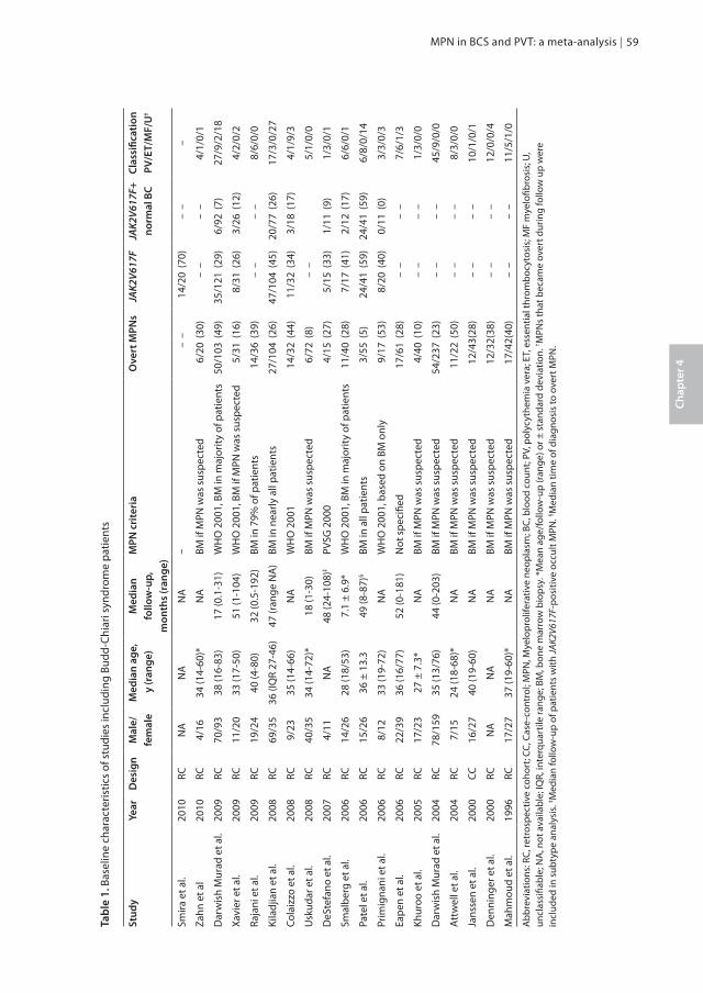
Chap
ter 4
59MPN in BCS and PVT: a meta-analysis |
Tabl
e 1.
Bas
elin
e ch
arac
teris
tics
of s
tudi
es in
clud
ing
Budd
-Chi
ari s
yndr
ome
patie
nts
Stud
y Ye
ar
Des
ign
mal
e/
fem
ale
med
ian
age,
y
(ran
ge)
med
ian
fo
llow
-up,
m
onth
s (r
ange
)
mPN
cri
teri
a
ove
rt m
PNs
JAK2
V617
FJA
K2V6
17F+
no
rmal
BC
Clas
sific
atio
n PV
/ET/
mF/
U†
Smira
et a
l.20
10RC
NA
NA
NA
––
–14
/20
(70)
––
–
Zahn
et a
l20
10RC
4/16
34 (1
4-60
)*N
ABM
if M
PN w
as s
uspe
cted
6/20
(30)
––
––
4/1/
0/1
Dar
wis
h M
urad
et a
l. 20
09RC
70/9
338
(16-
83)
17 (0
.1-3
1)W
HO
200
1, B
M in
maj
ority
of p
atie
nts
50/1
03(4
9)35
/121
(29)
6/92
(7)
27/9
/2/1
8
Xavi
er e
t al.
2009
RC11
/20
33 (1
7-50
)51
(1-1
04)
WH
O 2
001,
BM
if M
PN w
as s
uspe
cted
5/31
(16
)8/
31(2
6)3/
26(1
2)4/
2/0/
2
Raja
ni e
t al.
2009
RC19
/24
40 (4
-80)
32 (0
.5-1
92)
BM in
79%
of p
atie
nts
14/3
6(3
9)–
––
–8/
6/0/
0
Kila
djia
n et
al.
2008
RC69
/35
36 (I
QR
27-4
6)47
(ran
ge N
A)
BM in
nea
rly a
ll pa
tient
s27
/104
(26)
47/1
04(4
5)20
/77
(26)
17/3
/0/2
7
Cola
izzo
et a
l.20
08RC
9/23
35 (1
4-66
)N
AW
HO
200
114
/32
(44)
11/3
2(3
4)3/
18(1
7)4/
1/9/
3
Usk
udar
et a
l.20
08RC
40/3
534
(14-
72)*
18 (1
-30)
BM if
MPN
was
sus
pect
ed6/
72(8
)–
–
5/
1/0/
0
DeS
tefa
no e
t al.
2007
RC4/
11N
A48
(24-
108)
‡PV
SG 2
000
4/15
(27)
5/15
(33)
1/11
(9)
1/3/
0/1
Smal
berg
et a
l.20
06RC
14/2
628
(18/
53)
7.1
± 6.
9*
WH
O 2
001,
BM
in m
ajor
ity o
f pat
ient
s11
/40
(28)
7/17
(41)
2/12
(17)
6/6/
0/1
Pate
l et a
l.20
06RC
15/2
636
± 1
3.3
49 (8
-87)
§BM
in a
ll pa
tient
s3/
55(5
)24
/41
(59)
24/4
1(5
9)6/
8/0/
14
Prim
igna
ni e
t al.
2006
RC8/
1233
(19-
72)
NA
WH
O 2
001,
bas
ed o
n BM
onl
y9/
17(5
3)8/
20(4
0)0/
11(0
)3/
3/0/
3
Eape
n et
al.
2006
RC22
/39
36 (1
6/77
)52
(0-1
81)
Not
spe
cifie
d17
/61
(28)
––
––
7/6/
1/3
Khur
oo e
t al.
2005
RC17
/23
27 ±
7.3
*N
ABM
if M
PN w
as s
uspe
cted
4/40
(10)
––
––
1/3/
0/0
Dar
wis
h M
urad
et a
l. 20
04RC
78/1
5935
(13/
76)
44 (0
-203
)BM
if M
PN w
as s
uspe
cted
54/2
37(2
3)–
––
–45
/9/0
/0
Att
wel
l et a
l.20
04RC
7/15
24 (1
8-68
)*N
ABM
if M
PN w
as s
uspe
cted
11/2
2(5
0)–
––
–8/
3/0/
0
Jans
sen
et a
l.20
00CC
16/2
740
(19-
60)
NA
BM if
MPN
was
sus
pect
ed12
/43(
28)
––
––
10/1
/0/1
Den
ning
er e
t al.
2000
RCN
AN
AN
ABM
if M
PN w
as s
uspe
cted
12/3
2(38
)–
––
–12
/0/0
/4
Mah
mou
d et
al.
1996
RC17
/27
37 (1
9-60
)*N
ABM
if M
PN w
as s
uspe
cted
17/4
2(40
)–
––
–11
/5/1
/0
Abb
revi
atio
ns: R
C, re
tros
pect
ive
coho
rt; C
C, C
ase-
cont
rol;
MPN
, Mye
lopr
olife
rativ
e ne
opla
sm; B
C, b
lood
cou
nt; P
V, p
olyc
ythe
mia
ver
a; E
T, e
ssen
tial t
hrom
bocy
tosi
s; M
F m
yelo
fibro
sis;
U,
uncl
assi
fiabl
e; N
A, n
ot a
vaila
ble;
IQR,
inte
rqua
rtile
rang
e; B
M, b
one
mar
row
bio
psy.
*M
ean
age/
follo
w-u
p (r
ange
) or ±
sta
ndar
d de
viat
ion.
† MPN
s th
at b
ecam
e ov
ert d
urin
g fo
llow
up
wer
e in
clud
ed in
sub
type
ana
lysi
s. ‡ M
edia
n fo
llow
-up
of p
atie
nts
with
JAK2
V617
F-po
sitiv
e oc
cult
MPN
. § Med
ian
time
of d
iagn
osis
to o
vert
MPN
.
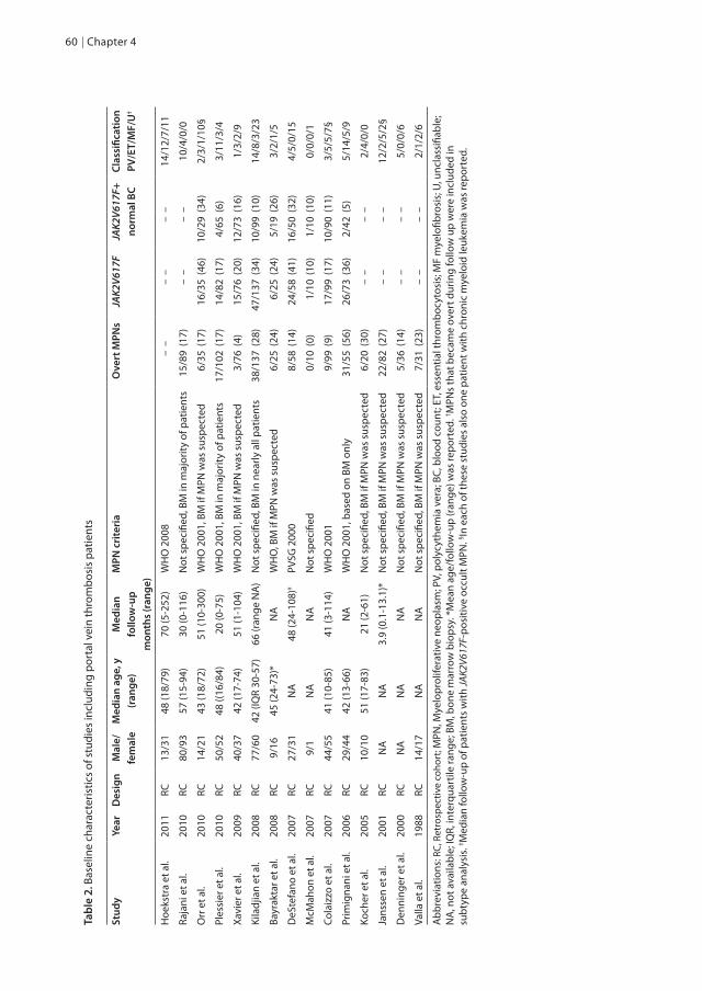
60 | Chapter 4
Tabl
e 2.
Bas
elin
e ch
arac
teris
tics
of s
tudi
es in
clud
ing
port
al v
ein
thro
mbo
sis
patie
nts
Stud
yYe
arD
esig
nm
ale/
fe
mal
em
edia
n ag
e, y
(r
ange
)m
edia
n
follo
w-u
p m
onth
s (r
ange
)
mPN
cri
teri
ao
vert
mPN
sJA
K2V6
17F
JAK2
V617
F+
norm
al B
CCl
assi
ficat
ion
PV/E
T/m
F/U
†
Hoe
kstr
a et
al.
2011
RC13
/31
48 (1
8/79
)70
(5-2
52)
WH
O 2
008
––
––
––
14/1
2/7/
11
Raja
ni e
t al.
2010
RC80
/93
57 (1
5-94
)30
(0-1
16)
Not
spe
cifie
d, B
M in
maj
ority
of p
atie
nts
15/8
9(1
7)–
––
–10
/4/0
/0
Orr
et a
l.20
10RC
14/2
143
(18/
72)
51 (1
0-30
0)W
HO
200
1, B
M if
MPN
was
sus
pect
ed6/
35(1
7)16
/35
(46)
10/2
9(3
4)2/
3/1/
10§
Ples
sier
et a
l.20
10RC
50/5
248
((16
/84)
20 (0
-75)
WH
O 2
001,
BM
in m
ajor
ity o
f pat
ient
s17
/102
(17)
14/8
2(1
7)4/
65(6
)3/
11/3
/4
Xavi
er e
t al.
2009
RC40
/37
42 (1
7-74
)51
(1-1
04)
WH
O 2
001,
BM
if M
PN w
as s
uspe
cted
3/76
(4)
15/7
6(2
0)12
/73
(16)
1/3/
2/9
Kila
djia
n et
al.
2008
RC77
/60
42 (I
QR
30-5
7)66
(ran
ge N
A)
Not
spe
cifie
d, B
M in
nea
rly a
ll pa
tient
s38
/137
(28)
47/1
37(3
4)10
/99
(10)
14/8
/3/2
3
Bayr
akta
r et a
l.20
08RC
9/16
45 (2
4-73
)*N
AW
HO
, BM
if M
PN w
as s
uspe
cted
6/25
(24)
6/25
(24)
5/19
(26)
3/2/
1/5
DeS
tefa
no e
t al.
2007
RC27
/31
NA
48 (2
4-10
8)‡
PVSG
200
08/
58(1
4)24
/58
(41)
16/5
0(3
2)4/
5/0/
15
McM
ahon
et a
l.20
07RC
9/1
NA
NA
Not
spe
cifie
d0/
10(0
)1/
10(1
0)1/
10(1
0)0/
0/0/
1
Cola
izzo
et a
l.20
07RC
44/5
541
(10-
85)
41 (3
-114
)W
HO
200
19/
99(9
)17
/99
(17)
10/9
0(1
1)3/
5/5/
7§
Prim
igna
ni e
t al.
2006
RC29
/44
42 (1
3-66
)N
AW
HO
200
1, b
ased
on
BM o
nly
31/5
5(5
6)26
/73
(36)
2/42
(5)
5/14
/5/9
Koch
er e
t al.
2005
RC10
/10
51 (1
7-83
)21
(2-6
1)N
ot s
peci
fied,
BM
if M
PN w
as s
uspe
cted
6/20
(30)
––
––
2/4/
0/0
Jans
sen
et a
l.20
01RC
NA
NA
3.9
(0.1
-13.
1)*
Not
spe
cifie
d, B
M if
MPN
was
sus
pect
ed22
/82
(27)
––
––
12/2
/5/2
§
Den
ning
er e
t al.
2000
RCN
AN
AN
AN
ot s
peci
fied,
BM
if M
PN w
as s
uspe
cted
5/36
(14)
––
––
5/0/
0/6
Valla
et a
l.19
88RC
14/1
7N
AN
AN
ot s
peci
fied,
BM
if M
PN w
as s
uspe
cted
7/31
(23)
––
––
2/1/
2/6
Abb
revi
atio
ns: R
C, R
etro
spec
tive
coho
rt; M
PN, M
yelo
prol
ifera
tive
neop
lasm
; PV,
pol
ycyt
hem
ia v
era;
BC,
blo
od c
ount
; ET,
ess
entia
l thr
ombo
cyto
sis;
MF
mye
lofib
rosi
s; U
, unc
lass
ifiab
le;
NA
, not
ava
ilabl
e; IQ
R, in
terq
uart
ile ra
nge;
BM
, bon
e m
arro
w b
iops
y. *
Mea
n ag
e/fo
llow
-up
(ran
ge) w
as re
port
ed. † M
PNs
that
bec
ame
over
t dur
ing
follo
w u
p w
ere
incl
uded
in
subt
ype
anal
ysis
. ‡ Med
ian
follo
w-u
p of
pat
ient
s w
ith JA
K2V6
17F-
posi
tive
occu
lt M
PN. § In
eac
h of
thes
e st
udie
s al
so o
ne p
atie
nt w
ith c
hron
ic m
yelo
id le
ukem
ia w
as re
port
ed.
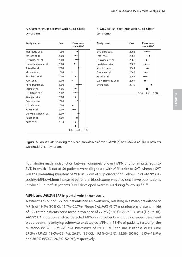
Chap
ter 4
61MPN in BCS and PVT: a meta-analysis |
A. overt mPNs in patients with Budd-Chiari syndrome
B. JAK2V617F in patients with Budd-Chiari syndrome
Study name Year Event rate and 95%CI
Mahmoud et al. 1996
Janssen et al. 2000
Denninger et al. 2000
Darwish Murad et al. 2004
Attwell et al. 2004
Khuroo et al. 2005
Smalberg et al. 2006
Patel et al. 2006
Primignani et al. 2006
Eapen et al. 2006
DeStefano et al. 2007
Kiladjian et al. 2008
Colaizzo et al. 2008
Uskudar et al. 2008
Xavier et al. 2009
Darwish Murad et al. 2009
Rajani et al. 2009
Zahn et al. 2010
0,00 0,50 1,00
Study name Event rate and 95%CI
Smalberg et al. 2006
Patel et al. 2006
Primignani et al. 2006
DeStefano et al. 2007
Kiladjian et al. 2008
Colaizzo et al. 2008
Xavier et al. 2009
Darwish Murad et al. 2009
Smira et al. 2010
0,00 0,50 1,00
Year
Figure 2. Forest plots showing the mean prevalence of overt MPNs (a) and JAK2V617F (b) in patients with Budd-Chiari syndrome.
Four studies made a distinction between diagnosis of overt MPN prior or simultaneous to SVT, in which 13 out of 50 patients were diagnosed with MPN prior to SVT, whereas SVT was the presenting symptom of MPN in 37 out of 50 patients.7,9,54,61 Follow-up of JAK2V617F-positive MPNs without increased peripheral blood counts was provided in two publications, in which 11 out of 28 patients (41%) developed overt MPNs during follow-up.52,61,64
mPNs and JAK2V617F in portal vein thrombosis A total of 173 out of 855 PVT patients had an overt MPN, resulting in a mean prevalence of MPNs of 19.4% (95% CI: 13.7%–26.7%) (Figure 3A). JAK2V617F mutation was present in 166 of 595 tested patients, for a mean prevalence of 27.7% (95% CI: 20.8%–35.8%) (Figure 3B). JAK2V617F mutation analysis detected MPNs in 70 patients without increased peripheral blood counts, identifying otherwise undetected MPNs in 15.4% of patients tested for the mutation (95%CI: 9.7%–23.7%). Prevalence of PV, ET, MF and unclassifiable MPNs were 27.5% (95%CI: 19.0%–38.1%), 26.2% (95%CI: 19.1%–34.8%), 12.8% (95%CI: 8.0%–19.9%) and 38.3% (95%CI: 26.3%–52.0%), respectively.
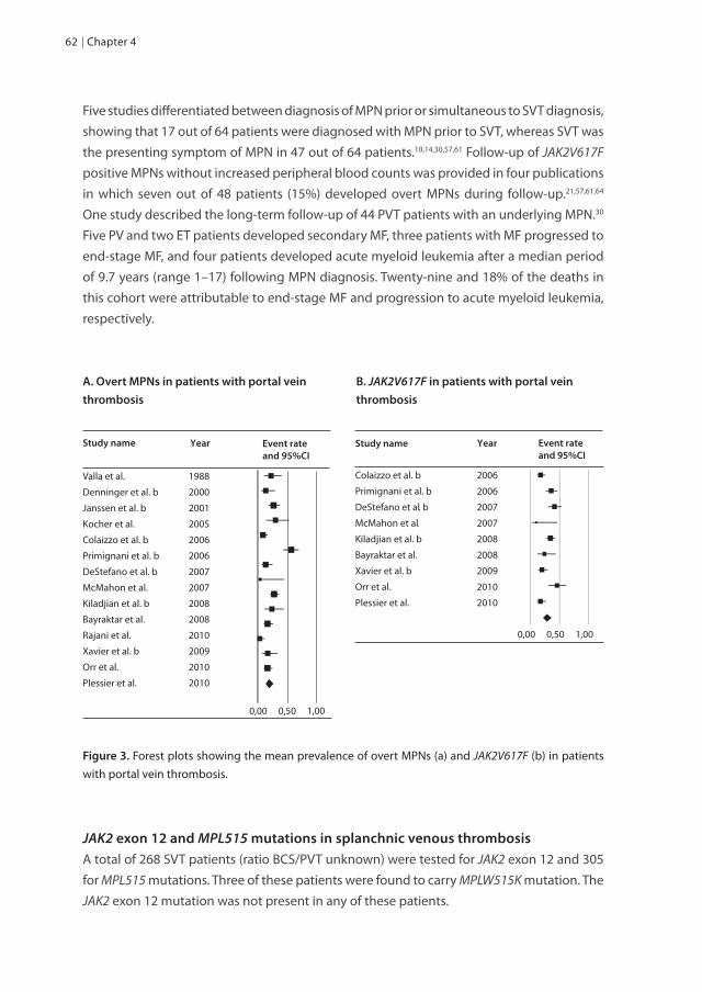
62 | Chapter 4
Five studies differentiated between diagnosis of MPN prior or simultaneous to SVT diagnosis, showing that 17 out of 64 patients were diagnosed with MPN prior to SVT, whereas SVT was the presenting symptom of MPN in 47 out of 64 patients.10,14,30,57,61 Follow-up of JAK2V617F positive MPNs without increased peripheral blood counts was provided in four publications in which seven out of 48 patients (15%) developed overt MPNs during follow-up.21,57,61,64 One study described the long-term follow-up of 44 PVT patients with an underlying MPN.30 Five PV and two ET patients developed secondary MF, three patients with MF progressed to end-stage MF, and four patients developed acute myeloid leukemia after a median period of 9.7 years (range 1–17) following MPN diagnosis. Twenty-nine and 18% of the deaths in this cohort were attributable to end-stage MF and progression to acute myeloid leukemia, respectively.
A. overt mPNs in patients with portal vein thrombosis
B. JAK2V617F in patients with portal vein thrombosis
Study name
0,00 0,50 1,00
Valla et al.
Denninger et al. b
Janssen et al. b
Kocher et al.
Colaizzo et al. b
Primignani et al. b
DeStefano et al. b
McMahon et al.
Kiladjian et al. b
Bayraktar et al.
Rajani et al.
Xavier et al. b
Orr et al.
Plessier et al.
1988
2000
2001
2005
2006
2006
2007
2007
2008
2008
2010
2009
2010
2010
Year Event rate and 95%CI
Study name
Colaizzo et al. b 2006
Primignani et al. b 2006
DeStefano et al. b 2007
McMahon et al. 2007
Kiladjian et al. b 2008
Bayraktar et al. 2008
Xavier et al. b 2009
Orr et al. 2010
Plessier et al. 2010
0,00 0,50 1,00
Year Event rate and 95%CI
Figure 3. Forest plots showing the mean prevalence of overt MPNs (a) and JAK2V617F (b) in patients with portal vein thrombosis.
JAK2 exon 12 and MPL515 mutations in splanchnic venous thrombosisA total of 268 SVT patients (ratio BCS/PVT unknown) were tested for JAK2 exon 12 and 305 for MPL515 mutations. Three of these patients were found to carry MPLW515K mutation. The JAK2 exon 12 mutation was not present in any of these patients.
Chap
ter 4
63MPN in BCS and PVT: a meta-analysis |
Differences between BCS and PVTPrevalence of both overt MPNs and JAK2V617F was significantly higher in BCS than in PVT (P < .001 and P = .03, respectively). There was no difference in identification rate of MPNs without increased peripheral blood counts by means of JAK2V617F between the two disorders (17.1% vs. 15.4%, P = .68). With regards to the subtype analysis, prevalence of PV was significantly higher in BCS patients (P = .001), whereas prevalence of MPNs unclassifiable was higher in PVT compared to BCS (P = .03) and there was a trend towards an increased frequency of MF in PVT (P = .09). There was no difference between the prevalence of ET (P = .77). These analyses were repeated including only publications since 2005 and excluding papers with potentially duplicated inclusion of patients, which showed the same results (data not shown).
i2 and heterogeneity amongst studiesA considerable heterogeneity amongst the studies was observed (P < .05). We therefore performed an additional analysis in which we excluded one study per analysis. This analysis showed that no single study significantly affected the point estimate of overt MPNs, JAK2V617F, or MPN subtypes in both BCS and PVT.
DiSCUSSioN
In this meta-analysis we assessed the role of MPNs in the etiology of primary BCS and non-malignant, non-cirrhotic PVT. The results showed a higher prevalence of overt MPNs in BCS compared to PVT patients, as well as a higher prevalence of the JAK2V617F mutation in BCS. Identification rate of MPNs without characteristic increased peripheral blood counts by means of JAK2V617F was comparable between the two disorders. Interestingly, our results indicate a difference in the distribution of underlying MPN subtype between BCS and PVT patients, PV being the most frequent MPN in BCS. Finally, MPL515 mutations were present in less than 1% of BCS and PVT series, whereas JAK2 exon 12 mutations have never been published so far in SVT patients. Two meta-analyses have previously evaluated the impact of the JAK2V617F mutation in SVT patients. In 2009, Dentali et al assessed the role of JAK2V617F in patients with various venous thrombosis, including SVT, deep vein thrombosis of the lower extremities or pulmonary embolism, cerebral vein thrombosis and retinal vein thrombosis.65 In this study, a remarkable high prevalence of JAK2V617F in SVT was reported, whereas its prevalence in other forms of VTE was similar to that of the general population. SVT was not subdivided into BCS and PVT which impedes comparison of MPNs and JAK2V617F prevalence between the two disorders. Qi et al. calculated the prevalence of JAK2V617F in BCS and PVT separately, and assessed its prevalence after exclusion of cases with pre-existing MPNs.66 In contrast to
64 | Chapter 4
those previously published studies, we set out to provide a complete overview of MPNs in the etiology of BCS and PVT. This included assessment of the prevalence of overt MPNs and JAK2V617F. Moreover, we quantified the identification of MPNs without characteristic increased peripheral blood counts by means of JAK2V617F and the rate of evolution to overt MPNs during follow-up. Finally, we determined the prevalence of MPN subtypes. In addition, we have compared BCS and PVT for each of these variables, as it is increasingly recognized that, despite several similarities, risk profiles are different between these patients.67 To achieve this goal, we have assessed all the publications regarding MPNs in SVT since 1980. The results of this meta-analysis indicate a high prevalence of overt MPNs in patients with SVT. The strong relation between MPNs and SVT is confirmed by the high prevalence of JAK2V617F in these patients. Interestingly, prevalence of both overt MPNs and JAK2V617F was significantly higher in BCS compared to PVT. This difference may be partially explained by the more prominent role of local risk factors, such as focal inflammatory lesions and injury to the portal venous system, in the development of PVT.68 This might contribute to the relatively limited role of general prothrombotic conditions reported in the etiology of PVT. Why MPNs and JAK2V617F are so strongly related to thrombosis of the splanchnic veins remains an unresolved issue. Further research is needed to identify associated factors that could be involved in the pathogenesis of thrombosis at these specific sites. In this respect, it has been speculated that endothelial cells of the splanchnic veins may interact with activated platelets and/or leukocytes and increased microparticles, which are characteristic features of MPNs.69 In addition, these endothelial cells have been shown to carry the JAK2V617F mutation and could be part of the malignant process.70
Being rare in patients with thrombosis outside the splanchnic area, and virtually absent in healthy individuals, JAK2V617F may be considered pathognomonic for MPNs in SVT patients. In these patients, portal hypertension and hypersplenism may mask elevated peripheral blood counts, even in the presence of active myeloproliferation. This meta-analysis for the first time systematically assessed the diagnostic yield of JAK2V617F screening in SVT patients. JAK2V617F screening identified MPN without characteristic increased peripheral blood counts in 17.1% and 15.4% of tested BCS and PVT patients, respectively. Its presence was associated with subsequent development of overt MPNs in 41% and 15% of BCS and PVT patients, respectively, which is lower than reported by Dentali et al.65 These findings clearly substantiate inclusion of JAK2V617F in the routine diagnostic work-up of SVT patients. Whether MPN specific treatment should be initiated in these patients, such as cytoreductive therapies or addition of aspirin to oral anticoagulant treatment, is a question that remains to be answered. One study described the long-term outcome of PVT patients with an underlying MPN.30 Twenty-nine and 18% of the deaths in this cohort were attributable to end-stage MF and progression to acute myeloid leukemia, respectively, indicating that risk of MPN progression is a clinical significant issue in these patients.
Chap
ter 4
65MPN in BCS and PVT: a meta-analysis |
We observed a marked difference between BCS and PVT patients regarding the distribution of MPN subtypes. PV was clearly more common in BCS compared to PVT. The prothrombotic effect of high hematocrit values in PV is well established.71 Under low-shear conditions, such as in the venous circulation, a high hematocrit has a more important impact on blood viscosity and causes a major disturbance to blood flow.72,73 This mechanism may be mediated by the interaction between adhesion molecules and red blood cells. Wautier et al described an increased adhesiveness of red blood cells in PV to human umbilical vein endothelial cells and elegantly showed that adhesion was inversely related to increasing shear stress, i.e. adhesion proved particularly increased at low shear rates.74 It is possible that variability in the expression of these molecules along the vascular tree along with differences in flow conditions might contribute to the site-specificity of thrombosis, as suggested by these authors.74 Indeed, the low-flow state in the hepatic veins compared to the portal venous system may participate in the higher frequency of PV in BCS. We also observed a statistical trend towards increased frequency of MF in PVT compared to BCS. Such difference could be due to the frequent presence of an important splenomegaly in MF, which may lead to external compression of the portal venous system and subsequent stasis of blood flow. These are new findings that deserve further evaluation in future studies. MPL515 mutations were reported in less than 1% of SVT patients, while the JAK2 exon 12 could not be found at all. The JAK2 exon 12 mutation has been described only once in both a PVT and BCS patient, but this was a case study.29 These results indicate that both mutations are infrequent in SVT patients, in agreement with their low frequency in MPNs compared to the JAK2V617F mutation.23-27 We therefore conclude that, unlike JAK2V617F, screening for these mutations is dispensable in the routine diagnostic approach of SVT patients. However, our analysis has several potential limitations. First, because of the rarity of both diseases, only observational studies have been published and could be included in this analysis, with their inherent risks of bias. However, a prospective design for rare thrombotic manifestations as PVT and BCS is probably unachievable. Second, a considerable heterogeneity amongst the included studies was noticed. We therefore performed all analyses using a random-effects model, thereby accounting for between study variance, next to within study variance. In addition to the random-effects analysis, which generates a conservative estimate, we performed an analysis in which we excluded one study at a time to assess its individual impact on the results. This analysis showed that none of the included studies significantly affected the estimated prevalence of overt MPNs, JAK2V617F, and its subtypes in both BCS and PVT. Third, diagnostic criteria for MPNs were not similar across studies. Notably, BM biopsy was not always routinely performed regardless of peripheral blood cell counts, which may have resulted in an underestimation of the prevalence of MPNs. Since this applies to both BCS and PVT series, the effect on the comparison between these two groups is presumably small, if at all present. Lastly, since the discovery of JAK2V617F in 2005, an increase in larger and better quality studies was observed. We therefore repeated
66 | Chapter 4
all analyses including only publications from that point in time. In addition, we excluded papers with potential overlap of patients. The same differences between BCS and PVT were observed. In conclusion, this meta-analysis shows a higher prevalence of both overt MPNs and JAK2V617F in BCS compared to PVT. Identification of MPNs without characteristic increased peripheral blood counts, of which a significant percentage developed overt MPN during follow-up, clearly justifies the inclusion of JAK2V617F screening in the routine diagnostic work-up of SVT. On the contrary, JAK2 exon 12 and MPL515 mutations are extremely rare in SVT and should not be used in the routine diagnostic approach of SVT patients. Altogether, our results are in line with the advancing insight that despite well-established similarities, marked differences in the etiology of BCS and PVT do exist.
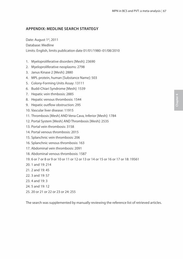
Chap
ter 4
67MPN in BCS and PVT: a meta-analysis |
APPENDix: mEDLiNE SEArCH STrATEGY
Date: August 1st, 2011Database: MedlineLimits: English, limits publication date 01/01/1980–01/08/2010
1. Myeloproliferative disorders [Mesh]: 236902. Myeloproliferative neoplasms: 27983. Janus Kinase 2 [Mesh]: 28804. MPL protein, human [Substance Name]: 5035. Colony-Forming Units Assay: 131116. Budd-Chiari Syndrome [Mesh]: 15397. Hepatic vein thmbosis: 28858. Hepatic venous thrombosis: 15449. Hepatic outflow obstruction: 29510. Vascular liver disease: 1191511. Thrombosis [Mesh] AND Vena Cava, Inferior [Mesh]: 178412. Portal System [Mesh] AND Thrombosis [Mesh]: 253513. Portal vein thrombosis: 315814. Portal venous thrombosis: 201515. Splanchnic vein thrombosis: 20616. Splanchnic venous thrombosis: 16317. Abdominal vein thrombosis: 209118. Abdominal venous thrombosis: 158719. 6 or 7 or 8 or 9 or 10 or 11 or 12 or 13 or 14 or 15 or 16 or 17 or 18: 1956120. 1 and 19: 21421. 2 and 19: 4522. 3 and 19: 5723. 4 and 19: 324. 5 and 19: 1225. 20 or 21 or 22 or 23 or 24: 255
The search was supplemented by manually reviewing the reference list of retrieved articles.
68 | Chapter 4
rEFErENCES
1. Janssen HL, Garcia-Pagan JC, Elias E, et al. Budd-Chiari syndrome: a review by an expert panel. J Hepatol 2003;38:364-71.
2. Janssen HL, Wijnhoud A, Haagsma EB, et al. Extrahepatic portal vein thrombosis: aetiology and determinants of survival. Gut 2001;49:720-4.
3. Ogren M, Bergqvist D, Bjorck M, Acosta S, Eriksson H, Sternby NH. Portal vein thrombosis: prevalence, patient characteristics and lifetime risk: a population study based on 23,796 consecutive autopsies. World J Gastroenterol 2006;12:2115-9.
4. Darwish Murad S, Plessier A, Hernandez-Guerra M, et al. Etiology, management, and outcome of the Budd-Chiari syndrome. Ann Intern Med 2009;151:167-75.
5. Darwish Murad S, Valla DC, de Groen PC, et al. Determinants of survival and the effect of portosystemic shunting in patients with Budd-Chiari syndrome. Hepatology 2004;39:500-8.
6. Denninger MH, Chait Y, Casadevall N, et al. Cause of portal or hepatic venous thrombosis in adults: the role of multiple concurrent factors. Hepatology 2000;31:587-91.
7. Mahmoud AE, Mendoza A, Meshikhes AN, et al. Clinical spectrum, investigations and treatment of Budd-Chiari syndrome. QJM 1996;89:37-43.
8. Rajani R, Melin T, Bjornsson E, et al. Budd-Chiari syndrome in Sweden: epidemiology, clinical characteristics and survival - an 18-year experience. Liver Int 2009;29:253-9.
9. Smalberg JH, Darwish Murad S, Braakman E, Valk PJ, Janssen HL, Leebeek FW. Myeloproliferative disease in the pathogenesis and survival of Budd-Chiari syndrome. Haematologica 2006;91:1712-3.
10. Bayraktar Y, Harmanci O, Buyukasik Y, et al. JAK2V617F mutation in patients with portal vein thrombosis. Dig Dis Sci 2008;53:2778-83.
11. Plessier A, Darwish-Murad S, Hernandez-Guerra M, et al. Acute portal vein thrombosis unrelated to cirrhosis: a prospective multicenter follow-up study. Hepatology 2010;51:210-8.
12. Valla D, Casadevall N, Huisse MG, et al. Etiology of portal vein thrombosis in adults. A prospective evaluation of primary myeloproliferative disorders. Gastroenterology 1988;94:1063-9.
13. De Stefano V, Teofili L, Leone G, Michiels JJ. Spontaneous erythroid colony formation as the clue to an underlying myeloproliferative disorder in patients with Budd-Chiari syndrome or portal vein thrombosis. Semin Thromb Hemost 1997;23:411-8.
14. Valla D, Casadevall N, Lacombe C, et al. Primary myeloproliferative disorder and hepatic vein thrombosis. A prospective study of erythroid colony formation in vitro in 20 patients with Budd-Chiari syndrome. Ann Intern Med 1985;103:329-34.
15. Spivak JL. Polycythemia vera: myths, mechanisms, and management. Blood 2002;100:4272-90.
16. Baxter EJ, Scott LM, Campbell PJ, et al. Acquired mutation of the tyrosine kinase JAK2 in human myeloproliferative disorders. Lancet 2005;365:1054-61.
17. Campbell PJ, Green AR. The myeloproliferative disorders. N Engl J Med 2006;355:2452-66.
18. James C, Ugo V, Le Couedic JP, et al. A unique clonal JAK2 mutation leading to constitutive signalling causes polycythaemia vera. Nature 2005;434:1144-8.
19. Kralovics R, Passamonti F, Buser AS, et al. A gain-of-function mutation of JAK2 in myeloproliferative disorders. N Engl J Med 2005;352:1779-90.
20. Kiladjian JJ, Cervantes F, Leebeek FW, et al. The impact of JAK2 and MPL mutations on diagnosis and prognosis of splanchnic vein thrombosis: a report on 241 cases. Blood 2008;111:4922-9.
21. Colaizzo D, Amitrano L, Tiscia GL, et al. The JAK2 V617F mutation frequently occurs in patients with portal and mesenteric venous thrombosis. J Thromb Haemost 2007;5:55-61.
22. Janssen HL, Leebeek FW. JAK2 mutation: The best diagnostic tool for myeloproliferative disease in splanchnic vein thrombosis? Hepatology 2006;44:1391-3.
23. Pardanani AD, Levine RL, Lasho T, et al. MPL515 mutations in myeloproliferative and other myeloid disorders: a study of 1182 patients. Blood 2006;108:3472-6.
Chap
ter 4
69MPN in BCS and PVT: a meta-analysis |
24. Pikman Y, Lee BH, Mercher T, et al. MPLW515L is a novel somatic activating mutation in myelofibrosis with myeloid metaplasia. PLoS Med 2006;3:e270.
25. Scott LM, Beer PA, Bench AJ, Erber WN, Green AR. Prevalance of JAK2 V617F and exon 12 mutations in polycythaemia vera. Br J Haematol 2007;139:511-2.
26. Scott LM, Tong W, Levine RL, et al. JAK2 exon 12 mutations in polycythemia vera and idiopathic erythrocytosis. N Engl J Med 2007;356:459-68.
27. Tefferi A, Skoda R, Vardiman JW. Myeloproliferative neoplasms: contemporary diagnosis using histology and genetics. Nat Rev Clin Oncol 2009;6:627-37.
28. Bergamaschi GM, Primignani M, Barosi G, et al. MPL and JAK2 exon 12 mutations in patients with the Budd-Chiari syndrome or extrahepatic portal vein obstruction. Blood 2008;111:4418.
29. Colaizzo D, Amitrano L, Tiscia GL, Grandone E, Guardascione MA, Margaglione M. A new JAK2 gene mutation in patients with polycythemia vera and splanchnic vein thrombosis. Blood 2007;110:2768-9.
30. Hoekstra J, Bresser EL, Smalberg JH, Spaander MC, Leebeek FW, Janssen HL. Long-term follow-up of patients with portal vein thrombosis and myeloproliferative neoplasms. J Thromb Haemost 2011;9:2208-14.
31. Tick LW, Nijkeuter M, Kramer MH, et al. High D-dimer levels increase the likelihood of pulmonary embolism. J Intern Med 2008;264:195-200.
32. Darwish Murad S, Plessier A, Hernandez-Guerra M, et al. Etiology, management, and outcome of the Budd-Chiari syndrome. Ann Intern Med 2009;151:167-75.
33. Boissinot M, Lippert E, Girodon F, et al. Latent myeloproliferative disorder revealed by the JAK2-V617F mutation and endogenous megakaryocytic colonies in patients with splanchnic vein thrombosis. Blood 2006;108:3223-4.
34. Goulding C, Uttenthal B, Foroni L, et al. The JAK2(V617F) tyrosine kinase mutation identifies clinically latent myeloproliferative disorders in patients presenting with hepatic or portal vein thrombosis. Int J Lab Hematol 2008;30:415-9.
35. Rossi D, Cresta S, Destro T, et al. JAK2V617F in idiopathic venous thromboembolism occurring in the absence of inherited or acquired thrombophilia. Br J Haematol 2007;138:813-4.
36. Lisman T, Bongers TN, Adelmeijer J, et al. Elevated levels of von Willebrand Factor in cirrhosis support platelet adhesion despite reduced functional capacity. Hepatology 2006;44:53-61.
37. Tondeur S, Boutruche S, Biron-Andreani C, Schved JF. Prevalence of the JAK2 V617F mutation associated with splanchnic vein thrombosis. A 10-year retrospective study. Thromb Haemost 2009;101:787-9.
38. Regina S, Herault O, D’Alteroche L, Binet C, Gruel Y. JAK2 V617F is specifically associated with idiopathic splanchnic vein thrombosis. J Thromb Haemost 2007;5:859-61.
39. Averbuch M, Aderka D, Winer Z, Levo Y. Budd-Chiari syndrome in Israel: predisposing factors, prognosis, and early identification of high-risk patients. J Clin Gastroenterol 1991;13:321-4.
40. Brown KM, Kaplan MM, Donowitz M. Extrahepatic portal venous thrombosis: frequent recognition of associated diseases. J Clin Gastroenterol 1985;7:153-9.
41. Cardin F, Graffeo M, McCormick PA, McIntyre N, Burroughs A. Adult “idiopathic” extrahepatic venous thrombosis. Importance of putative “latent” myeloproliferative disorders and comparison with cases with known etiology. Dig Dis Sci 1992;37:335-9.
42. P’ng S, Carnley B, Baker R, Kontorinis N, Cheng W. Undiagnosed myeloproliferative disease in cases of intra-abdominal thrombosis: the utility of the JAK2 617F mutation. Clin Gastroenterol Hepatol 2008;6:472-5.
43. Amitrano L, Guardascione MA, Scaglione M, et al. Prognostic factors in noncirrhotic patients with splanchnic vein thromboses. Am J Gastroenterol 2007;102:2464-70.
44. Chait Y, Condat B, Cazals-Hatem D, et al. Relevance of the criteria commonly used to diagnose myeloproliferative disorder in patients with splanchnic vein thrombosis. Br J Haematol 2005;129:553-60.
45. Colaizzo D, Amitrano L, Iannaccone L, et al. Gain-of-function gene mutations and venous thromboembolism: distinct roles in different clinical settings. J Med Genet 2007;44:412-6.
70 | Chapter 4
46. De Stefano V, Fiorini A, Rossi E, et al. High prevalence of the JAK2 V617F mutation in patients with extrahepatic portal vein thrombosis. Hepatology 2007;45:831-2; author reply 2-3.
47. Harmanci O, Ersoy O, Gurgey A, et al. The etiologic distribution of thrombophilic factors in chronic portal vein thrombosis. J Clin Gastroenterol 2007;41:521-7.
48. Janssen HL, Meinardi JR, Vleggaar FP, et al. Factor V Leiden mutation, prothrombin gene mutation, and deficiencies in coagulation inhibitors associated with Budd-Chiari syndrome and portal vein thrombosis: results of a case-control study. Blood 2000;96:2364-8.
49. Primignani M, Martinelli I, Bucciarelli P, et al. Risk factors for thrombophilia in extrahepatic portal vein obstruction. Hepatology 2005;41:603-8.
50. Fiorini A, Chiusolo P, Rossi E, et al. Absence of the JAK2 exon 12 mutations in patients with splanchnic venous thrombosis and without overt myeloproliferative neoplasms. Am J Hematol 2009;84:126-7.
51. McMahon C, Abu-Elmagd K, Bontempo FA, Kant JA, Swerdlow SH. JAK2 V617F mutation in patients with catastrophic intra-abdominal thromboses. Am J Clin Pathol 2007;127:736-43.
52. Patel RK, Lea NC, Heneghan MA, et al. Prevalence of the activating JAK2 tyrosine kinase mutation V617F in the Budd-Chiari syndrome. Gastroenterology 2006;130:2031-8.
53. Primignani M, Barosi G, Bergamaschi G, et al. Role of the JAK2 mutation in the diagnosis of chronic myeloproliferative disorders in splanchnic vein thrombosis. Hepatology 2006;44:1528-34.
54. Colaizzo D, Amitrano L, Tiscia GL, et al. Occurrence of the JAK2 V617F mutation in the Budd-Chiari syndrome. Blood Coagul Fibrinolysis 2008;19:459-62.
55. Khuroo MS, Al-Suhabani H, Al-Sebayel M, et al. Budd-Chiari syndrome: long-term effect on outcome with transjugular intrahepatic portosystemic shunt. J Gastroenterol Hepatol 2005;20:1494-502.
56. Kocher G, Himmelmann A. Portal vein thrombosis (PVT): a study of 20 non-cirrhotic cases. Swiss Med Wkly 2005;135:372-6.
57. Orr DW, Patel RK, Lea NC, et al. Prevalence of the activating JAK2 tyrosine kinase mutation in chronic porto-splenomesenteric venous thrombosis. Aliment Pharmacol Ther 2010.
58. Smalberg JH, Koehler E, Darwish Murad S, et al. The JAK2 46/1 haplotype in Budd-Chiari syndrome and portal vein thrombosis. Blood 2011;117:3968-73.
59. Smira G, Gheorghe L, Iacob S, Coriu D, Gheorghe C. Budd Chiari syndrome and V617F/JAK 2 mutation linked with the myeloproliferative disorders. J Gastrointestin Liver Dis 2010;19:108-9.
60. Uskudar O, Akdogan M, Sasmaz N, Yilmaz S, Tola M, Sahin B. Etiology and portal vein thrombosis in Budd-Chiari syndrome. World J Gastroenterol 2008;14:2858-62.
61. Xavier SG, Gadelha T, Pimenta G, et al. JAK2V617F Mutation in Patients with Splanchnic Vein Thrombosis. Dig Dis Sci 2009.
62. Zahn A, Gotthardt D, Weiss KH, et al. Budd-Chiari syndrome: long term success via hepatic decompression using transjugular intrahepatic porto-systemic shunt. BMC Gastroenterol 2010;10:25.
63. Eapen CE, Velissaris D, Heydtmann M, Gunson B, Olliff S, Elias E. Favourable medium term outcome following hepatic vein recanalisation and/or transjugular intrahepatic portosystemic shunt for Budd Chiari syndrome. Gut 2006;55:878-84.
64. De Stefano V, Fiorini A, Rossi E, et al. Incidence of the JAK2 V617F mutation among patients with splanchnic or cerebral venous thrombosis and without overt chronic myeloproliferative disorders. J Thromb Haemost 2007;5:708-14.
65. Lisman T, Leebeek FW, Mosnier LO, et al. Thrombin-activatable fibrinolysis inhibitor deficiency in cirrhosis is not associated with increased plasma fibrinolysis. Gastroenterology 2001;121:131-9.
66. Buffart LM, Roebroeck ME, Janssen WG, Hoekstra A, Hovius SE, Stam HJ. Comparison of instruments to assess hand function in children with radius deficiencies. J Hand Surg Am 2007;32:531-40.
67. Smalberg JH, Kruip MJ, Janssen HL, Rijken DC, Leebeek FW, de Maat MP. Hypercoagulability and hypofibrinolysis and risk of deep vein thrombosis and splanchnic vein thrombosis: similarities and differences. Arterioscler Thromb Vasc Biol 2011;31:485-93.
Chap
ter 4
71MPN in BCS and PVT: a meta-analysis |
68. DeLeve LD, Valla DC, Garcia-Tsao G, American Association for the Study Liver D. Vascular disorders of the liver. Hepatology 2009;49:1729-64.
69. Aird WC. Vascular bed-specific thrombosis. J Thromb Haemost 2007;5 Suppl 1:283-91.
70. Sozer S, Fiel MI, Schiano T, Xu M, Mascarenhas J, Hoffman R. The presence of JAK2V617F mutation in the liver endothelial cells of patients with Budd-Chiari syndrome. Blood 2009;113:5246-9.
71. Pearson TC, Wetherley-Mein G. Vascular occlusive episodes and venous haematocrit in primary proliferative polycythaemia. Lancet 1978;2:1219-22.
72. Landolfi R, Di Gennaro L, Falanga A. Thrombosis in myeloproliferative disorders: pathogenetic facts and speculation. Leukemia 2008;22:2020-8.
73. Landolfi R, Rocca B, Patrono C. Bleeding and thrombosis in myeloproliferative disorders: mechanisms and treatment. Crit Rev Oncol Hematol 1995;20:203-22.
74. Wautier MP, El Nemer W, Gane P, et al. Increased adhesion to endothelial cells of erythrocytes from patients with polycythemia vera is mediated by laminin alpha5 chain and Lu/BCAM. Blood 2007;110:894-901.

C H A P T E r 5
THE JAK2 46/1 HAPLoTYPE iN BUDD-CHiAri SYNDromE AND
PorTAL VEiN THromBoSiS
Jasper H. Smalberg1, Edith Koehler2, Sarwa Darwish Murad2, Aurelie Plessier3, Susana Seijo4,
Jonel Trebicka5, Massimo Primignani6, Moniek P.M. de Maat1, Juan-Carlos Garcia-Pagan4,
Dominique C. Valla3, Harry L.A. Janssen2 and Frank W.G. Leebeek1 for the European Network for
Vascular Disorders of the Liver (EN-Vie)
Departments of Hematology1 and Gastroenterology & Hepatology2, Erasmus University Medical
Center, Rotterdam, The Netherlands; 3Department of Hepatology, Hopital Beaujon, Assistance
Publique-Hopitaux de Paris, Inserm U773 and University Paris-7, Clichy, France; 4Hepatic
Hemodynamic Laboratory, Liver Unit, Institut de Malalties Digestives, IDIBAPS and Ciberehd,
Barcelona, Spain; 5Department of Internal Medicine I, University Hospital of Bonn, Bonn,
Germany; and 6Gastroenterology and Gastrointestinal Endoscopy Unit, Ospedale Policlinico,
Mangiagalli and Regina Elena Foundation, Milan, Italy.
Blood. 2011 Apr 14;117(15):3968-73.
74 | Chapter 5
ABSTrACT
The germline JAK2 46/1 haplotype has been associated with the development of JAK2V617F positive as well as JAK2V617F negative myeloproliferative neoplasms (MPNs). In this study we examined the role of the 46/1 haplotype in the etiology and clinical presentation of patients with splanchnic vein thrombosis (SVT), in which MPNs are the most prominent underlying etiological factor. The single-nucleotide polymorphism rs12343867, which tags 46/1, was genotyped in 199 SVT patients. The 46/1 haplotype was overrepresented in JAK2V617F positive SVT patients compared with controls (P < .01). Prevalence of the 46/1 haplotype in JAK2V617F negative SVT patients did not differ from the controls. However, JAK2V617F negative SVT patients with a proven MPN also exhibited an increased frequency of the 46/1 haplotype (P = .06). Interestingly, 46/1 was associated with increased erythropoiesis in JAK2V617F negative SVT patients. In conclusion, the 46/1 haplotype is associated with the development of JAK2V617F positive SVT. In addition, our findings in JAK2V617F negative SVT patients indicate an important role for the 46/1 haplotype in the etiology and diagnosis of SVT-related MPNs, independent of JAK2V617F, that requires further exploration.
Chap
ter 5
75JAK2 46/1 in BCS and PVT |
iNTroDUCTioN
The entity splanchnic vein thrombosis is used to indicate both the Budd-Chiari syndrome (BCS) and portal vein thrombosis (PVT). BCS is a rare disorder characterized by obstruction of the hepatic veins and/or the suprahepatic inferior vena cava.1,2 BCS is considered primary when resulting from thrombosis and secondary when obstruction of the venous tract results from compression or invasion by a tumour. Non-malignant, non-cirrhotic PVT is another infrequent thrombotic disorder involving the splanchnic vasculature.3 In both disorders, pathogenesis is largely dependent on the presence of systemic prothrombotic conditions which promote thrombus formation in the respective hepatic vessels. Myeloproliferative neoplasms (MPNs) are the leading cause of SVT and are diagnosed in one third to half of the patients with SVT patients.4 The most common gain of function mutation leading to development of MPNs is the JAK2V617F mutation, which is present in more than 95% of cases of polycythemia vera and 50% to 60% of essential thrombocythemia and primary myelofibrosis.5 The high prevalence of the JAK2V617F mutation in SVT patients, approximately 35% in unselected cases, confirms the unique relationship between SVT and MPNs.6-10 Interestingly, the JAK2V617F mutation has proved to be an important diagnostic tool to detect MPNs in SVT patients, considering that MPNs can be notoriously difficult to diagnose in patients presenting with SVT. Portal hypertension, resulting from pre- or post-sinusoidal venous congestion, leads to hypersplenism and hemodilution - both conditions which mask the characteristic peripheral blood cell changes in MPNs. Screening for JAK2V617F is an objective tool to diagnose MPNs in these patients and is now part of the standard diagnostic work-up in SVT.8,11
Recently, an association between a specific JAK2 haplotype and the risk of developing JAK2V617F positive MPNs was demonstrated.12-14 These studies show that the acquired JAK2V617F mutation is preferentially found within this particular, inherited haplotype, which we refer to as 46/1. The association is strong, with the odds of developing MPNs being three to fourfold higher in patients carrying 46/1 compared to non-carriers. One of the aforementioned studies also found an association between 46/1 and JAK2V617F negative MPNs,12 which has since been confirmed by other studies.15-17 Subsequently, it has been shown that 46/1 was also overrepresented in JAK2V617F negative MPNs carrying mutations across JAK2 exon 12 or MPL gene.18,19 These findings are of particular interest in SVT, because 46/1 may thus represent a new molecular marker for diagnosing MPNs in JAK2V617F negative SVT patients. The aim of our study was to assess whether 46/1 is associated with SVT, and to determine whether JAK2 46/1 is associated with distinct clinical and laboratory characteristics of SVT. To this end, we genotyped patients and healthy controls from a large European series of newly diagnosed, consecutive BCS and PVT patients.
76 | Chapter 5
mATEriALS AND mETHoDS
Study designWe performed a case-control study in which patients and controls were recruited from the European Network for Vascular Disease of the Liver (EN-Vie) study cohort, which has been described previously.6,9 The EN-Vie cohort consists of newly diagnosed patients with BCS or PVT, consecutively enrolled and prospectively followed in nine different European countries. At time of diagnosis and during follow-up, data concerning clinical condition and etiology and results of radiology, pathology and laboratory assessments were collected. From October 2003 to October 2005, a total of 163 BCS patients and 138 PVT patients were enrolled in the study. In addition, 105 healthy, unrelated, population-based controls, usually neighbours or friends of the patients, fulfilling the same age criteria and without a history of thrombosis were recruited. The EN-Vie study was approved by national and, if necessary, local ethical committees. Patients and controls agreed to participate in the study by written informed consent, in accordance with the Declaration of Helsinki.
DefinitionsBCS was defined as hepatic outflow obstruction regardless of the cause or level of obstruction, from the small hepatic veins to the entrance of the inferior vena cave into the right atrium. BCS was confirmed by radiographic imaging (ultrasonography, computed tomography, magnetic resonance imaging, or venography). Sinusoidal obstruction syndrome was excluded from this definition, as well as outflow obstruction occurring in the setting of heart failure, orthotopic liver transplantation or hepatobiliary cancer. Diagnostic criteria for PVT included radiographic imaging evidence of solid material in the portal vein lumen or in its left or right branch. PVT patients with cirrhosis or abdominal malignancies as well as patients with clinical, laboratory, or imaging evidence of non-cirrhotic liver disease, within a context of chronic alcoholism, viral hepatitis, autoimmune disease, Wilson’s disease, or iron overload were excluded.
Blood samplingBlood samples were collected from patients at time of diagnosis and controls by venapuncture in tubes containing 0.11M trisodium citrate. DNA was extracted from whole blood according to local standard methods. DNA samples were transported to the Erasmus University Medical Center in Rotterdam and stored at -70°C until analysis.
JAK2 46/1 genotyping and JAK2V617F mutation analysis Granulocyte DNA samples were genotyped using a Taqman single nucleotide polymorphism (SNP) assay for rs12343867 (C/T) (Applied Biosystems). The 46/1 haplotype is tagged by the C allele of this SNP.14 JAK2V617F mutation analysis was performed as previously described.8
Chap
ter 5
77JAK2 46/1 in BCS and PVT |
Statistical analysisResults are expressed as proportions for categorical variables and as medians and interquartile range (IQR; 25th–75th percentile) for continuous variables. Comparison between categorical variables was performed with chi-square testing and between continuous variables with the Kruskall-Wallis test. Odds ratios (OR) for SVT associated with 46/1 and corresponding 95% confidence intervals (CI) were calculated using logistic regression, adjusted for age and gender. To assess the association between 46/1 and SVT in the presence of other risk factors for SVT, a multivariate model was constructed in which the factor V Leiden mutation and the prothrombin G20210A variant were added, adjusted for age and gender. JAK2V617F was only present in SVT patients and not in controls, and could therefore not be included in the multivariate model. P-values were two-tailed and statistical significance was set at P < .05. All statistical analyses were conducted with PASW Statistics, version 17.0 (SPSS, Chicago, Illinois).
rESULTS
Patient characteristicsThe total EN-Vie cohort contains 163 BCS and 138 PVT patients and DNA samples for this study were available of 116 BCS patients, 96 PVT patients and 105 healthy controls. Of these, 107 BCS patients (92%), 92 PVT patients (96%) and 100 healthy controls (95%) were successfully genotyped and included in the current analysis. Patient characteristics and underlying thrombophilic risk factors are shown in Table 1. In BCS patients, median age at diagnosis was 38.1 years (IQR: 28–51) and 45 were males (42%). Twenty-three BCS patients (21%) had an underlying inherited thrombophilic factor, while in 83 patients (78%) an acquired prothrombotic disorder was diagnosed. MPNs were present in 42 patients, of which 34 were JAK2V617F positive. In PVT, median age at diagnosis was 49.8 years (IQR: 42–57) and 43 were males (47%). Twenty-two PVT patients (24%) were diagnosed with an inherited thrombophilia, whereas 56 patients (61%) had an acquired prothrombotic factor. MPNs were present in 24 PVT patients, of which 20 were JAK2V617F positive. Median age in the controls was 36.8 years (IQR: 27–50) and 40 were males (40%). None of the controls carried the JAK2V617F mutation, whereas the factor V Leiden mutation and prothrombin G20210A variant were present in 4% and 3% of the controls, respectively.
JAK2 46/1 haplotype and risk of SVT-related mPNSNP rs12343867 genotype distributions and the OR for SVT are presented in Table 2. Genotype distribution of our control group was in Hardy-Weinberg equilibrium and similar to those reported by previous studies and the Wellcome Trust Case-Control Consortium (WTCCC).12,17,20 In the overall group of SVT patients, there was no significant difference in
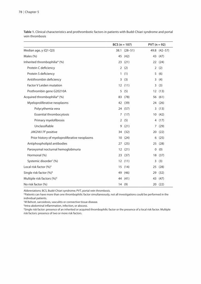
78 | Chapter 5
Table 1. Clinical characteristics and prothrombotic factors in patients with Budd-Chiari syndrome and portal vein thrombosis
BCS (n = 107) PVT (n = 92)
Median age, y (Q1-Q3) 38.1 (28–51) 49.8 (42–57)
Males (%) 45 (42) 43 (47)
Inherited thrombophilia* (%) 23 (21) 22 (24)
Protein C deficiency 2 (2) 2 (2)
Protein S deficiency 1 (1) 5 (6)
Antithrombin deficiency 3 (3) 3 (4)
Factor V Leiden mutation 12 (11) 3 (3)
Prothrombin gene G20210A 5 (5) 12 (13)
Acquired thrombophilia* (%) 83 (78) 56 (61)
Myeloproliferative neoplasms 42 (39) 24 (26)
Polycythemia vera 24 (57) 3 (13)
Essential thrombocytosis 7 (17) 10 (42)
Primary myelofibrosis 2 (5) 4 (17)
Unclassifiable 9 (21) 7 (29)
JAK2V617F positive 34 (32) 20 (22)
Prior history of myeloproliferative neoplasms 10 (24) 6 (25)
Antiphospholipid antibodies 27 (25) 25 (28)
Paroxysmal nocturnal hemoglobinuria 12 (21) 0 (0)
Hormonal (%) 23 (37) 18 (37)
Systemic disorder† (%) 12 (11) 3 (3)
Local risk factor (%)‡ 15 (14) 25 (28)
Single risk factor (%)§ 49 (46) 29 (32)
Multiple risk factors (%)§ 44 (41) 43 (47)
No risk factor (%) 14 (9) 20 (22)
Abbreviations: BCS, Budd-Chiari syndrome; PVT, portal vein thrombosis.*Patients can have more than one thrombophilic factor simultaneously, not all investigations could be performed in the individual patients.†M Behcet, sarcoidosis, vasculitis or connective tissue disease.‡Intra-abdominal inflammation, infection, or abscess.§Single risk factor: presence of an inherited or acquired thrombophilic factor or the presence of a local risk factor. Multiple risk factors: presence of two or more risk factors.
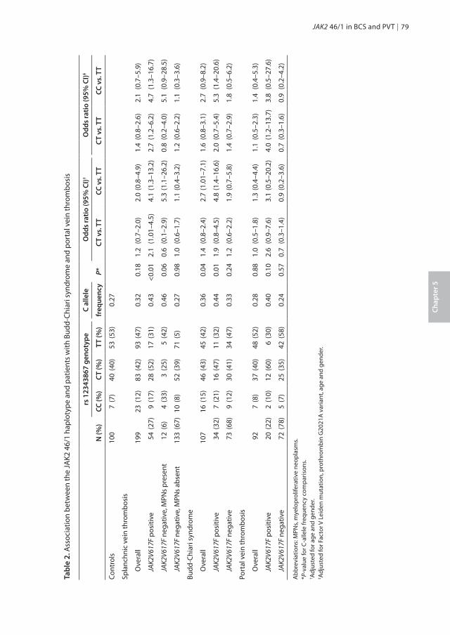
Chap
ter 5
79JAK2 46/1 in BCS and PVT |
Tabl
e 2.
Ass
ocia
tion
betw
een
the
JAK2
46/
1 ha
plot
ype
and
patie
nts
with
Bud
d-Ch
iari
synd
rom
e an
d po
rtal
vei
n th
rom
bosi
s
rs
123
4386
7 ge
noty
peC
alle
le
odd
s ra
tio (9
5% C
i)†
odd
s ra
tio (9
5% C
i)‡
N
(%)
CC (%
)CT
(%)
TT (%
)fr
eque
ncy
P*CT
vs.
TT
CC v
s. T
T
CT v
s. T
TCC
vs.
TT
Cont
rols
100
7(7
)40
(40)
53(5
3)0.
27
Spla
nchn
ic v
ein
thro
mbo
sis
Ove
rall
199
23(1
2)83
(42)
93(4
7)0.
320.
181.
2(0
.7–2
.0)
2.0
(0.8
–4.9
)1.
4(0
.8–2
.6)
2.1
(0.7
–5.9
)
JAK2
V617
F po
sitiv
e54
(27)
9(1
7)28
(52)
17(3
1)0.
43<0
.01
2.1
(1.0
1–4.
5)4.
1(1
.3–1
3.2)
2.7
(1.2
–6.2
)4.
7(1
.3–1
6.7)
JAK2
V617
F ne
gativ
e, M
PNs
pres
ent
12(6
)4
(33)
3(2
5)5
(42)
0.46
0.06
0.6
(0.1
–2.9
)5.
3(1
.1–2
6.2)
0.8
(0.2
–4.0
)5.
1(0
.9–2
8.5)
JAK2
V617
F ne
gativ
e, M
PNs
abse
nt13
3(6
7)10
(8)
52(3
9)71
(5)
0.27
0.98
1.0
(0.6
–1.7
)1.
1(0
.4–3
.2)
1.2
(0.6
–2.2
)1.
1(0
.3–3
.6)
Budd
-Chi
ari s
yndr
ome
Ove
rall
107
16(1
5)46
(43)
45(4
2)0.
360.
041.
4(0
.8–2
.4)
2.7
(1.0
1–7.
1)1.
6(0
.8–3
.1)
2.7
(0.9
–8.2
)
JAK2
V617
F po
sitiv
e34
(32)
7(2
1)16
(47)
11(3
2)0.
440.
011.
9(0
.8–4
.5)
4.8
(1.4
–16.
6)2.
0(0
.7–5
.4)
5.3
(1.4
–20.
6)
JAK2
V617
F ne
gativ
e73
(68)
9(1
2)30
(41)
34(4
7)0.
330.
241.
2(0
.6–2
.2)
1.9
(0.7
–5.8
)1.
4(0
.7–2
.9)
1.8
(0.5
–6.2
)
Port
al v
ein
thro
mbo
sis
Ove
rall
927
(8)
37(4
0)48
(52)
0.28
0.88
1.0
(0.5
–1.8
)1.
3(0
.4–4
.4)
1.1
(0.5
–2.3
)1.
4(0
.4–5
.3)
JAK2
V617
F po
sitiv
e20
(22)
2(1
0)12
(60)
6(3
0)0.
400.
102.
6(0
.9–7
.6)
3.1
(0.5
–20.
2)4.
0(1
.2–1
3.7)
3.8
(0.5
–27.
6)
JAK2
V617
F ne
gativ
e72
(78)
5(7
)25
(35)
42(5
8)0.
240.
570.
7(0
.3–1
.4)
0.9
(0.2
–3.6
)
0.7
(0.3
–1.6
)0.
9(0
.2–4
.2)
Abb
revi
atio
ns: M
PNs,
mye
lopr
olife
rativ
e ne
opla
sms.
*P-v
alue
for C
-alle
le fr
eque
ncy
com
paris
ons.
† Adju
sted
for a
ge a
nd g
ende
r. ‡ Ad
just
ed fo
r Fac
tor V
Lei
den
mut
atio
n, p
roth
rom
bin
G20
21A
var
iant
, age
and
gen
der.
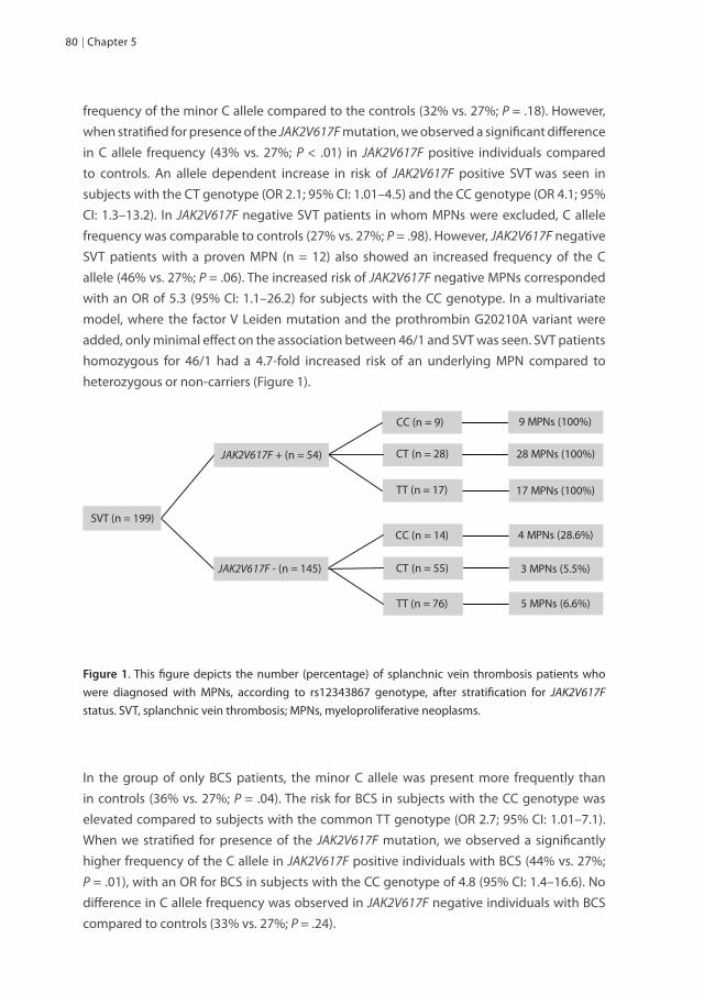
80 | Chapter 5
frequency of the minor C allele compared to the controls (32% vs. 27%; P = .18). However, when stratified for presence of the JAK2V617F mutation, we observed a significant difference in C allele frequency (43% vs. 27%; P < .01) in JAK2V617F positive individuals compared to controls. An allele dependent increase in risk of JAK2V617F positive SVT was seen in subjects with the CT genotype (OR 2.1; 95% CI: 1.01–4.5) and the CC genotype (OR 4.1; 95% CI: 1.3–13.2). In JAK2V617F negative SVT patients in whom MPNs were excluded, C allele frequency was comparable to controls (27% vs. 27%; P = .98). However, JAK2V617F negative SVT patients with a proven MPN (n = 12) also showed an increased frequency of the C allele (46% vs. 27%; P = .06). The increased risk of JAK2V617F negative MPNs corresponded with an OR of 5.3 (95% CI: 1.1–26.2) for subjects with the CC genotype. In a multivariate model, where the factor V Leiden mutation and the prothrombin G20210A variant were added, only minimal effect on the association between 46/1 and SVT was seen. SVT patients homozygous for 46/1 had a 4.7-fold increased risk of an underlying MPN compared to heterozygous or non-carriers (Figure 1).
CC (n = 14)
TT (n = 76)
CT (n = 55)
17 MPNs (100%)
5 MPNs (6.6%)
3 MPNs (5.5%)
4 MPNs (28.6%)
28 MPNs (100%)
9 MPNs (100%)CC (n = 9)
TT (n = 17)
CT (n = 28)
SVT (n = 199)
JAK2V617F + (n = 54)
JAK2V617F - (n = 145)
Figure 1. This figure depicts the number (percentage) of splanchnic vein thrombosis patients who were diagnosed with MPNs, according to rs12343867 genotype, after stratification for JAK2V617F status. SVT, splanchnic vein thrombosis; MPNs, myeloproliferative neoplasms.
In the group of only BCS patients, the minor C allele was present more frequently than in controls (36% vs. 27%; P = .04). The risk for BCS in subjects with the CC genotype was elevated compared to subjects with the common TT genotype (OR 2.7; 95% CI: 1.01–7.1). When we stratified for presence of the JAK2V617F mutation, we observed a significantly higher frequency of the C allele in JAK2V617F positive individuals with BCS (44% vs. 27%; P = .01), with an OR for BCS in subjects with the CC genotype of 4.8 (95% CI: 1.4–16.6). No difference in C allele frequency was observed in JAK2V617F negative individuals with BCS compared to controls (33% vs. 27%; P = .24).
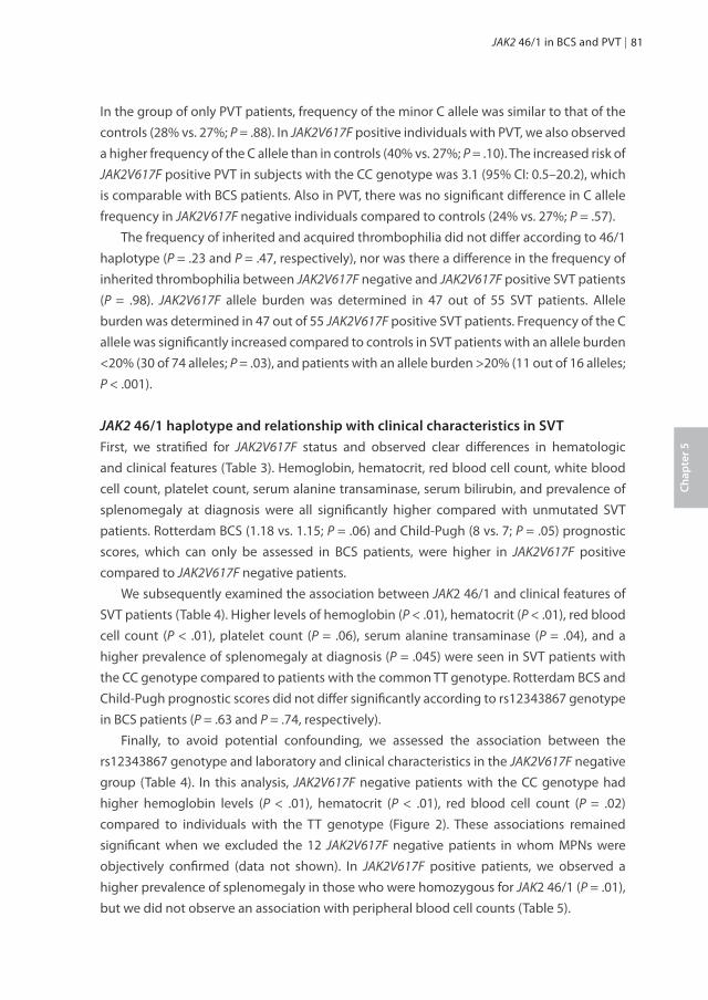
Chap
ter 5
81JAK2 46/1 in BCS and PVT |
In the group of only PVT patients, frequency of the minor C allele was similar to that of the controls (28% vs. 27%; P = .88). In JAK2V617F positive individuals with PVT, we also observed a higher frequency of the C allele than in controls (40% vs. 27%; P = .10). The increased risk of JAK2V617F positive PVT in subjects with the CC genotype was 3.1 (95% CI: 0.5–20.2), which is comparable with BCS patients. Also in PVT, there was no significant difference in C allele frequency in JAK2V617F negative individuals compared to controls (24% vs. 27%; P = .57). The frequency of inherited and acquired thrombophilia did not differ according to 46/1 haplotype (P = .23 and P = .47, respectively), nor was there a difference in the frequency of inherited thrombophilia between JAK2V617F negative and JAK2V617F positive SVT patients (P = .98). JAK2V617F allele burden was determined in 47 out of 55 SVT patients. Allele burden was determined in 47 out of 55 JAK2V617F positive SVT patients. Frequency of the C allele was significantly increased compared to controls in SVT patients with an allele burden <20% (30 of 74 alleles; P = .03), and patients with an allele burden >20% (11 out of 16 alleles; P < .001).
JAK2 46/1 haplotype and relationship with clinical characteristics in SVTFirst, we stratified for JAK2V617F status and observed clear differences in hematologic and clinical features (Table 3). Hemoglobin, hematocrit, red blood cell count, white blood cell count, platelet count, serum alanine transaminase, serum bilirubin, and prevalence of splenomegaly at diagnosis were all significantly higher compared with unmutated SVT patients. Rotterdam BCS (1.18 vs. 1.15; P = .06) and Child-Pugh (8 vs. 7; P = .05) prognostic scores, which can only be assessed in BCS patients, were higher in JAK2V617F positive compared to JAK2V617F negative patients. We subsequently examined the association between JAK2 46/1 and clinical features of SVT patients (Table 4). Higher levels of hemoglobin (P < .01), hematocrit (P < .01), red blood cell count (P < .01), platelet count (P = .06), serum alanine transaminase (P = .04), and a higher prevalence of splenomegaly at diagnosis (P = .045) were seen in SVT patients with the CC genotype compared to patients with the common TT genotype. Rotterdam BCS and Child-Pugh prognostic scores did not differ significantly according to rs12343867 genotype in BCS patients (P = .63 and P = .74, respectively). Finally, to avoid potential confounding, we assessed the association between the rs12343867 genotype and laboratory and clinical characteristics in the JAK2V617F negative group (Table 4). In this analysis, JAK2V617F negative patients with the CC genotype had higher hemoglobin levels (P < .01), hematocrit (P < .01), red blood cell count (P = .02) compared to individuals with the TT genotype (Figure 2). These associations remained significant when we excluded the 12 JAK2V617F negative patients in whom MPNs were objectively confirmed (data not shown). In JAK2V617F positive patients, we observed a higher prevalence of splenomegaly in those who were homozygous for JAK2 46/1 (P = .01), but we did not observe an association with peripheral blood cell counts (Table 5).
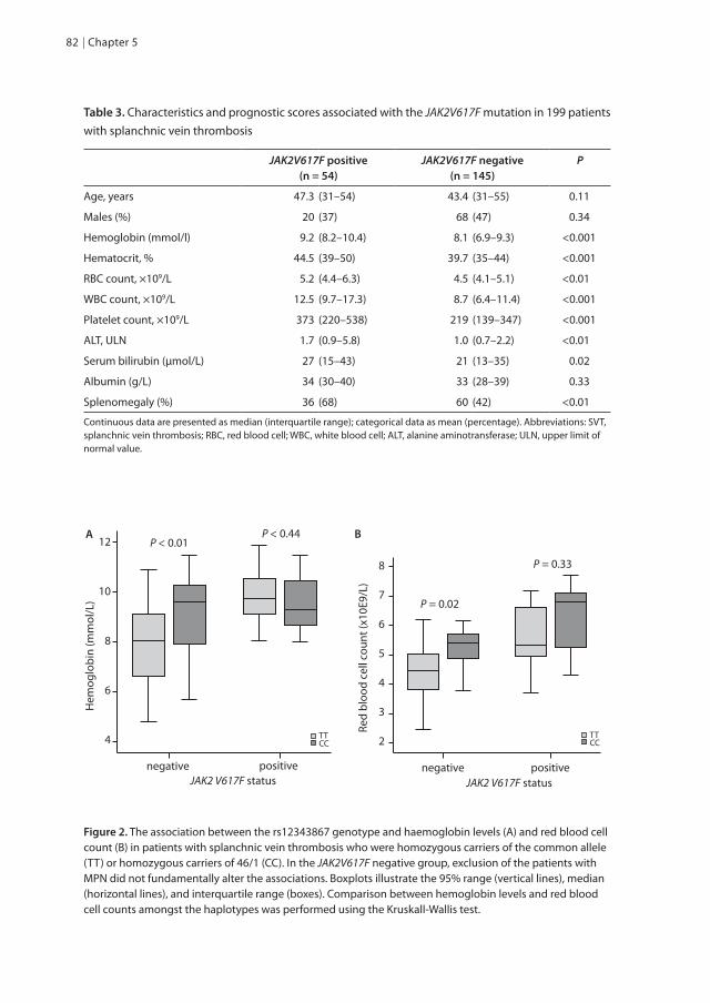
82 | Chapter 5
Table 3. Characteristics and prognostic scores associated with the JAK2V617F mutation in 199 patients with splanchnic vein thrombosis
JAK2V617F positive (n = 54)
JAK2V617F negative (n = 145)
P
Age, years 47.3 (31–54) 43.4 (31–55) 0.11
Males (%) 20 (37) 68 (47) 0.34
Hemoglobin (mmol/l) 9.2 (8.2–10.4) 8.1 (6.9–9.3) <0.001
Hematocrit, % 44.5 (39–50) 39.7 (35–44) <0.001
RBC count, ×109/L 5.2 (4.4–6.3) 4.5 (4.1–5.1) <0.01
WBC count, ×109/L 12.5 (9.7–17.3) 8.7 (6.4–11.4) <0.001
Platelet count, ×109/L 373 (220–538) 219 (139–347) <0.001
ALT, ULN 1.7 (0.9–5.8) 1.0 (0.7–2.2) <0.01
Serum bilirubin (μmol/L) 27 (15–43) 21 (13–35) 0.02
Albumin (g/L) 34 (30–40) 33 (28–39) 0.33
Splenomegaly (%) 36 (68) 60 (42) <0.01
Continuous data are presented as median (interquartile range); categorical data as mean (percentage). Abbreviations: SVT, splanchnic vein thrombosis; RBC, red blood cell; WBC, white blood cell; ALT, alanine aminotransferase; ULN, upper limit of normal value.
JAK2 V617F statusnegative positive
12
10
8
6
4
Hem
oglo
bin
(mm
ol/L
)
P < 0.01P < 0.44
TTCC
A
JAK2 V617F statusnegative positive
8
7
6
5
4
3
2
B
Red
bloo
d ce
ll co
unt (
x10E
9/L)
P = 0.02
P = 0.33
TTCC
Figure 2. The association between the rs12343867 genotype and haemoglobin levels (A) and red blood cell count (B) in patients with splanchnic vein thrombosis who were homozygous carriers of the common allele (TT) or homozygous carriers of 46/1 (CC). In the JAK2V617F negative group, exclusion of the patients with MPN did not fundamentally alter the associations. Boxplots illustrate the 95% range (vertical lines), median (horizontal lines), and interquartile range (boxes). Comparison between hemoglobin levels and red blood cell counts amongst the haplotypes was performed using the Kruskall-Wallis test.
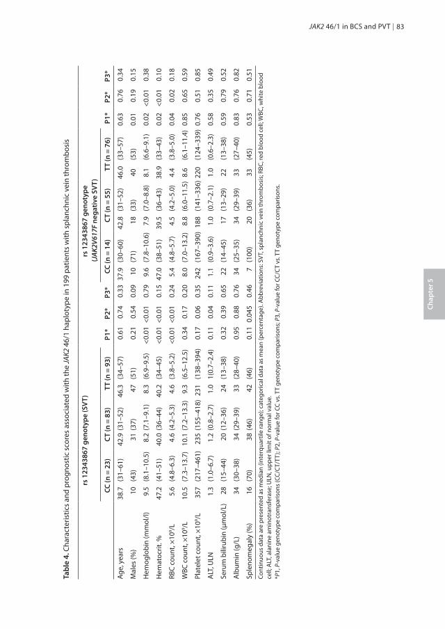
Chap
ter 5
83JAK2 46/1 in BCS and PVT |
Tabl
e 4.
Cha
ract
eris
tics
and
prog
nost
ic s
core
s as
soci
ated
with
the
JAK2
46/
1 ha
plot
ype
in 1
99 p
atie
nts
with
spl
anch
nic
vein
thro
mbo
sis
rs 1
2343
867
geno
type
(SV
T)
rs 1
2343
867
geno
type
(J
AK2V
617F
neg
ativ
e SV
T)
CC
(n =
23)
CT (n
= 8
3)TT
(n =
93)
P1
*P2
*P3
*CC
(n =
14)
CT (n
= 5
5)TT
(n =
76)
P1*
P2*
P3*
Age,
yea
rs38
.7(3
1–61
)42
.9(3
1–52
)46
.3(3
4–57
)0.
610.
740.
3337
.9(3
0–60
)42
.8(3
1–52
)46
.0(3
3–57
)0.
630.
760.
34
Mal
es (%
)10
(43)
31(3
7)47
(51)
0.21
0.54
0.09
10(7
1)18
(33)
40(5
3)0.
010.
190.
15
Hem
oglo
bin
(mm
ol/l)
9.5
(8.1
–10.
5)8.
2(7
.1–9
.1)
8.3
(6.9
–9.5
)<0
.01
<0.0
10.
799.
6(7
.8–1
0.6)
7.9
(7.0
–8.8
)8.
1(6
.6–9
.1)
0.02
<0.0
10.
38
Hem
atoc
rit. %
47.2
(41–
51)
40.0
(36–
44)
40.2
(34–
45)
<0.0
1<0
.01
0.15
47.0
(38–
51)
39.5
(36–
43)
38.9
(33–
43)
0.02
<0.0
10.
10
RBC
coun
t, ×1
09 /L5.
6(4
.8–6
.3)
4.6
(4.2
–5.3
)4.
6(3
.8–5
.2)
<0.0
1<0
.01
0.24
5.4
(4.8
–5.7
)4.
5(4
.2–5
.0)
4.4
(3.8
–5.0
)0.
040.
020.
18
WBC
cou
nt, ×
109 /L
10.5
(7.3
–13.
7)10
.1(7
.2–1
3.3)
9.3
(6.5
–12.
5)0.
340.
170.
208.
0(7
.0–1
3.2)
8.8
(6.0
–11.
5)8.
6(6
.1–1
1.4)
0.85
0.65
0.59
Plat
elet
cou
nt, ×
109 /L
357
(217
–461
)23
5(1
55–4
18)
231
(138
–394
)0.
170.
060.
3524
2(1
67–3
90)
188
(141
–336
)220
(124
–339
)0.
760.
510.
85
ALT
, ULN
1.3
(1.0
–6.7
)1.
2(0
.8–2
.7)
1.0
1(0.
7–2.
4)0.
110.
040.
111.
1(0
.9–3
.6)
1.0
(0.7
–2.1
)1.
0(0
.6–2
.3)
0.58
0.35
0.49
Seru
m b
iliru
bin
(μm
ol/L
)28
(15–
44)
20(1
2–36
)24
(13–
38)
0.32
0.39
0.65
22(1
4–45
)17
(13–
29)
22(1
3–38
)0.
590.
790.
52
Alb
umin
(g/L
)34
(30–
38)
34(2
9–39
)33
(28–
40)
0.95
0.88
0.76
34(2
5–35
)34
(29–
39)
33(2
7–40
)0.
830.
760.
82
Sple
nom
egal
y (%
)16
(70)
38(4
6)42
(46)
0.
110.
045
0.46
7(1
00)
20(3
6)33
(45)
0.53
0.71
0.51
Cont
inuo
us d
ata
are
pres
ente
d as
med
ian
(inte
rqua
rtile
rang
e); c
ateg
oric
al d
ata
as m
ean
(per
cent
age)
. Abb
revi
atio
ns: S
VT, s
plan
chni
c ve
in th
rom
bosi
s; R
BC, r
ed b
lood
cel
l; W
BC, w
hite
blo
od
cell;
ALT
, ala
nine
am
inot
rans
fera
se; U
LN, u
pper
lim
it of
nor
mal
val
ue.
*P1,
P-v
alue
gen
otyp
e co
mpa
rison
s (C
C/C
T/TT
); P2
, P-v
alue
for C
C vs
. TT
geno
type
com
paris
ons;
P3,
P-v
alue
for C
C/C
T vs
. TT
geno
type
com
paris
ons.
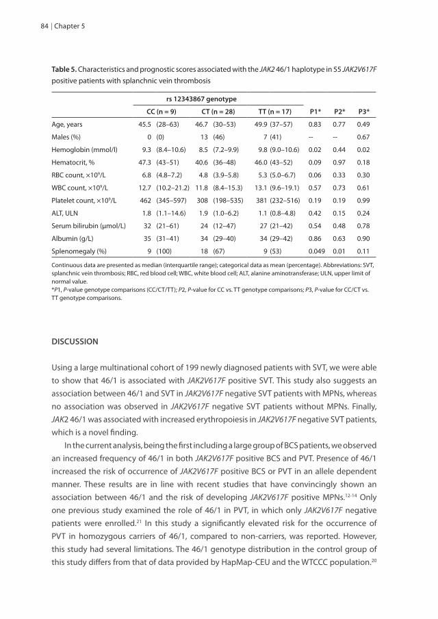
84 | Chapter 5
Table 5. Characteristics and prognostic scores associated with the JAK2 46/1 haplotype in 55 JAK2V617F positive patients with splanchnic vein thrombosis
rs 12343867 genotype
CC (n = 9) CT (n = 28) TT (n = 17) P1* P2* P3*
Age, years 45.5 (28–63) 46.7 (30–53) 49.9 (37–57) 0.83 0.77 0.49
Males (%) 0 (0) 13 (46) 7 (41) -- -- 0.67
Hemoglobin (mmol/l) 9.3 (8.4–10.6) 8.5 (7.2–9.9) 9.8 (9.0–10.6) 0.02 0.44 0.02
Hematocrit, % 47.3 (43–51) 40.6 (36–48) 46.0 (43–52) 0.09 0.97 0.18
RBC count, ×109/L 6.8 (4.8–7.2) 4.8 (3.9–5.8) 5.3 (5.0–6.7) 0.06 0.33 0.30
WBC count, ×109/L 12.7 (10.2–21.2) 11.8 (8.4–15.3) 13.1 (9.6–19.1) 0.57 0.73 0.61
Platelet count, ×109/L 462 (345–597) 308 (198–535) 381 (232–516) 0.19 0.19 0.99
ALT, ULN 1.8 (1.1–14.6) 1.9 (1.0–6.2) 1.1 (0.8–4.8) 0.42 0.15 0.24
Serum bilirubin (μmol/L) 32 (21–61) 24 (12–47) 27 (21–42) 0.54 0.48 0.78
Albumin (g/L) 35 (31–41) 34 (29–40) 34 (29–42) 0.86 0.63 0.90
Splenomegaly (%) 9 (100) 18 (67) 9 (53) 0.049 0.01 0.11
Continuous data are presented as median (interquartile range); categorical data as mean (percentage). Abbreviations: SVT, splanchnic vein thrombosis; RBC, red blood cell; WBC, white blood cell; ALT, alanine aminotransferase; ULN, upper limit of normal value.*P1, P-value genotype comparisons (CC/CT/TT); P2, P-value for CC vs. TT genotype comparisons; P3, P-value for CC/CT vs. TT genotype comparisons.
DiSCUSSioN
Using a large multinational cohort of 199 newly diagnosed patients with SVT, we were able to show that 46/1 is associated with JAK2V617F positive SVT. This study also suggests an association between 46/1 and SVT in JAK2V617F negative SVT patients with MPNs, whereas no association was observed in JAK2V617F negative SVT patients without MPNs. Finally, JAK2 46/1 was associated with increased erythropoiesis in JAK2V617F negative SVT patients, which is a novel finding. In the current analysis, being the first including a large group of BCS patients, we observed an increased frequency of 46/1 in both JAK2V617F positive BCS and PVT. Presence of 46/1 increased the risk of occurrence of JAK2V617F positive BCS or PVT in an allele dependent manner. These results are in line with recent studies that have convincingly shown an association between 46/1 and the risk of developing JAK2V617F positive MPNs.12-14 Only one previous study examined the role of 46/1 in PVT, in which only JAK2V617F negative patients were enrolled.21 In this study a significantly elevated risk for the occurrence of PVT in homozygous carriers of 46/1, compared to non-carriers, was reported. However, this study had several limitations. The 46/1 genotype distribution in the control group of this study differs from that of data provided by HapMap-CEU and the WTCCC population.20
Chap
ter 5
85JAK2 46/1 in BCS and PVT |
Most notable, the frequency of controls homozygous for the 46/1 was relatively low (2.8%) compared to the HapMap-CEU (6.0%) and WTCCC population (6.5%). In fact, the control group of that study was not in Hardy-Weinberg equilibrium. This has important implications, especially since the reported associations were for homozygous carriers of 46/1 compared to non-carriers. In our study, both controls and patients are representative samples from multiple European countries. This is supported by the fact that our control group is in Hardy-Weinberg equilibrium and distribution of 46/1 is consistent with HapMap-CEU and the WTCCC population. We therefore believe our results on the association between 46/1 and PVT are valid and representative for the European population. The increased frequency of 46/1 was observed in JAK2V617F positive SVT patients with a mutant allele burden <20% as well as patients with a JAK2V617F allele burden >20%. Interestingly, the frequency of 46/1 in JAK2V617F negative SVT patients with a proven MPN was higher compared to the controls (46% vs. 27%), although not significantly (P = .06). While we are aware that this group of patients is small, this finding is consistent with the increasing evidence that 46/1 is associated with both JAK2V617F positive and JAK2V617F negative MPNs.15-17
An obvious question is whether 46/1 is of additional value in the diagnostic work-up of SVT patients. Our study suggests that 46/1 may be used as a diagnostic tool in the risk assessment of MPNs in SVT patients in addition to the JAK2V617F mutation. In a potential diagnostic algorithm, JAK2V617F clearly is the first tool to screen for MPNs in SVT patients – the presence of the JAK2V617F mutation is highly suggestive, if not pathognomonic, for MPNs in these patients. However, also JAK2V617F negative SVT patients may fulfill the criteria for MPNs. Our study shows, that in this specific subgroup of JAK2V617F negative SVT patients, homozygous carriers of 46/1 had a 4.7-fold higher chance of MPNs compared to heterozygous carriers or non-carriers. It can be argued that a more aggressive search for MPNs must be instituted in these patients. A bone marrow biopsy is quintessential in these patients, but it might also be considered to intensify the screening program for MPNs during follow up in these patients. This may facilitate timely recognition and treatment of underlying MPNs. Additional studies are needed to extend our observations, before definite conclusions on the potential role of 46/1 in the work-up of SVT patients can be drawn. We observed a strong association between JAK2V617F positivity and clinical and laboratory characteristics at the moment of diagnosis of SVT, which is in line with previous reports on the association between JAK2V617F and elevated peripheral blood cell counts,8,22-25 and that between JAK2V617F and splenomegaly.8,23,24 One of these studies also demonstrated an association between JAK2V617F and altered liver function tests and unfavourable prognostic scores in BCS.8 In the present study we confirm these findings. The significant association between homozygous carriers of 46/1 and increased erythropoiesis in JAK2V617F negative SVT patients was unexpected. Hemoglobin, hematocrit and red blood cell count were all approximately 20% higher compared to individuals with
86 | Chapter 5
the TT genotype. Also, we observed a higher prevalence of splenomegaly in JAK2V617F positive patients homozygous for 46/1. This is the first study to demonstrate an association between 46/1 and laboratory and clinical characteristics. Up to this point, four studies examined this association earlier, in which no significant association was observed.15-18 One study examined the association between 46/1 and the number of granulocyte-macrophage colony forming units (CFU-GM) in peripheral blood.12 Carriers of 46/1 grew significantly fewer CFU-GM, consistent with the hypothesis that 46/1 might be functionally different from other JAK2 alleles. Our findings of an increased erythropoiesis support the theory that 46/1 indeed might be functionally different. Theoretically, it is possible that this is specific for SVT patients, especially in light of the unique relationship between MPN and SVT. Clearly, these findings deserve further research. In conclusion, we observed a clear association between the 46/1 haplotype and the development of JAK2V617F positive SVT. This study also provides the first support of an increased frequency of 46/1 in JAK2V617F negative SVT patients with a proven MPN, and suggests a potential role for 46/1 as a diagnostic tool in SVT in addition to JAK2V617F. Unexpectedly, 46/1 was associated with an elevated erythropoiesis in JAK2V617F negative SVT patients, indicating that the 46/1 allele might be functionally different from other alleles.
Chap
ter 5
87JAK2 46/1 in BCS and PVT |
rEFErENCES
1. Janssen HL, Garcia-Pagan JC, Elias E, et al. Budd-Chiari syndrome: a review by an expert panel. J Hepatol 2003;38:364-71.
2. Valla DC. Primary Budd-Chiari syndrome. J Hepatol 2009;50:195-203.
3. DeLeve LD, Valla DC, Garcia-Tsao G, American Association for the Study Liver D. Vascular disorders of the liver. Hepatology 2009;49:1729-64.
4. Martinelli I, De Stefano V. Rare thromboses of cerebral, splanchnic and upper-extremity veins. A narrative review. Thromb Haemost 2010;103:1136-44.
5. Tefferi A, Skoda R, Vardiman JW. Myeloproliferative neoplasms: contemporary diagnosis using histology and genetics. Nat Rev Clin Oncol 2009;6:627-37.
6. Darwish Murad S, Plessier A, Hernandez-Guerra M, et al. Etiology, management, and outcome of the Budd-Chiari syndrome. Ann Intern Med 2009;151:167-75.
7. De Stefano V, Fiorini A, Rossi E, et al. Incidence of the JAK2 V617F mutation among patients with splanchnic or cerebral venous thrombosis and without overt chronic myeloproliferative disorders. J Thromb Haemost 2007;5:708-14.
8. Kiladjian JJ, Cervantes F, Leebeek FW, et al. The impact of JAK2 and MPL mutations on diagnosis and prognosis of splanchnic vein thrombosis: a report on 241 cases. Blood 2008;111:4922-9.
9. Plessier A, Darwish-Murad S, Hernandez-Guerra M, et al. Acute portal vein thrombosis unrelated to cirrhosis: a prospective multicenter follow-up study. Hepatology 2010;51:210-8.
10. Smalberg JH, Darwish Murad S, Braakman E, Valk PJ, Janssen HL, Leebeek FW. Myeloproliferative disease in the pathogenesis and survival of Budd-Chiari syndrome. Haematologica 2006;91:1712-3.
11. Janssen HL, Leebeek FW. JAK2 mutation: The best diagnostic tool for myeloproliferative disease in splanchnic vein thrombosis? Hepatology 2006;44:1391-3.
12. Jones AV, Chase A, Silver RT, et al. JAK2 haplotype is a major risk factor for the development of myeloproliferative neoplasms. Nat Genet 2009;41:446-9.
13. Kilpivaara O, Mukherjee S, Schram AM, et al. A germline JAK2 SNP is associated with predisposition to the development of JAK2(V617F)-positive myeloproliferative neoplasms. Nat Genet 2009;41:455-9.
14. Olcaydu D, Harutyunyan A, Jager R, et al. A common JAK2 haplotype confers susceptibility to myeloproliferative neoplasms. Nat Genet 2009;41:450-4.
15. Andrikovics H, Nahajevszky S, Koszarska M, et al. JAK2 46/1 haplotype analysis in myeloproliferative neoplasms and acute myeloid leukemia. Leukemia 2010.
16. Pardanani A, Lasho TL, Finke CM, et al. The JAK2 46/1 haplotype confers susceptibility to essential thrombocythemia regardless of JAK2V617F mutational status-clinical correlates in a study of 226 consecutive patients. Leukemia 2010;24:110-4.
17. Tefferi A, Lasho TL, Patnaik MM, et al. JAK2 germline genetic variation affects disease susceptibility in primary myelofibrosis regardless of V617F mutational status: nullizygosity for the JAK2 46/1 haplotype is associated with inferior survival. Leukemia 2010;24:105-9.
18. Jones AV, Campbell PJ, Beer PA, et al. The JAK2 46/1 haplotype predisposes to MPL-mutated myeloproliferative neoplasms. Blood 2010;115:4517-23.
19. Olcaydu D, Skoda RC, Looser R, et al. The ‘GGCC’ haplotype of JAK2 confers susceptibility to JAK2 exon 12 mutation-positive polycythemia vera. Leukemia 2009;23:1924-6.
20. Wellcome Trust Case Control C. Genome-wide association study of 14,000 cases of seven common diseases and 3,000 shared controls. Nature 2007;447:661-78.
21. Colaizzo D, Tiscia GL, Bafunno V, et al. The JAK2 rs12343867 CC genotype frequently occurs in patients with splanchnic venous thrombosis without the JAK2V617F mutation: a retrospective study. J Thromb Haemost 2010;8:413-6.
22. Orr DW, Patel RK, Lea NC, et al. The prevalence of the activating JAK2 tyrosine kinase mutation in chronic porto-splenomesenteric venous thrombosis. Aliment Pharmacol Ther 2010;31:1330-6.

88 | Chapter 5
23. Patel RK, Lea NC, Heneghan MA, et al. Prevalence of the activating JAK2 tyrosine kinase mutation V617F in the Budd-Chiari syndrome. Gastroenterology 2006;130:2031-8.
24. Primignani M, Barosi G, Bergamaschi G, et al. Role of the JAK2 mutation in the diagnosis of chronic myeloproliferative disorders in splanchnic vein thrombosis. Hepatology 2006;44:1528-34.
25. Xavier SG, Gadelha T, Pimenta G, et al. JAK2V617F mutation in patients with splanchnic vein thrombosis. Dig Dis Sci 2010;55:1770-7.
C H A P T E r 6
FiBriNoGEN GAmmA PrimE AND VAriATioN iN FiBriNoGEN GAmmA PrimE GENES iN THE ETioLoGY oF
PorTAL VEiN THromBoSiS
Jasper H. Smalberg1, Edith Koehler2, Sarwa Darwish Murad2, Aurelie Plessier3, Susana Seijo4,
Jonel Trebicka5, Massimo Primignani6, Moniek P.M. de Maat1, Juan-Carlos Garcia-Pagan4,
Dominique C. Valla3, Harry L.A. Janssen2 and Frank W.G. Leebeek1 for the European Network for
Vascular Disorders of the Liver (EN-Vie)
Departments of Hematology1 and Gastroenterology & Hepatology2, Erasmus University Medical
Center, Rotterdam, The Netherlands; 3Department of Hepatology, Hopital Beaujon, Assistance
Publique-Hopitaux de Paris, Inserm U773 and University Paris-7, Clichy, France; 4Hepatic
Hemodynamic Laboratory, Liver Unit, Institut de Malalties Digestives, IDIBAPS and Ciberehd,
Barcelona, Spain; 5Department of Internal Medicine I, University Hospital of Bonn, Bonn,
Germany; and 6Gastroenterology and Gastrointestinal Endoscopy Unit, Ospedale Policlinico,
Mangiagalli and Regina Elena Foundation, Milan, Italy.
Submitted
90 | Chapter 6
ABSTrACT
Fibrinogen γ’, a protein variant of fibrinogen, and genetic variation in fibrinogen γ genes (FGG) have been associated with susceptibility to venous thrombosis. In this study we evaluated whether fibrinogen γ’ and FGG haplotypes were associated with risk of portal vein thrombosis (PVT). We performed a case-control study using patients and healthy controls from the European Network for Vascular Disease of the Liver study cohort, a large multinational study of newly diagnosed, consecutive patients with non-cirrhotic, non-malignant PVT. Fibrinogen γ’, total fibrinogen levels and FGG haplotypes were determined in 106 PVT patients and 103 healthy controls. We observed decreased levels of fibrinogen γ’ relative to total fibrinogen levels (γ’ ratio) in PVT patients (0.119 vs. 0.153; P < .001). A γ’ ratio in the lowest tertile was associated with a 5-fold increased risk of PVT (95% CI: 2.2–11.2). We observed an increased frequency of FGG-H2, which was significantly associated with a decreased γ’ ratio, in PVT patients compared to controls (OR 1.63; 95% CI: 0.94–2.81, P = .08). In conclusion, this case-control study provides the first evidence that the γ’ ratio and the FGG-H2 haplotype may contribute to the development of PVT.
Chap
ter 6
91Fibrinogen γ' in PVT |
iNTroDUCTioN
Portal vein thrombosis (PVT) is a rare manifestation of venous thrombosis that often leads to portal hypertension.1 The etiology of PVT is diverse and involves both local and systemic prothrombotic factors. Liver cirrhosis and hepatobiliary malignancies are considered the most common local etiological factors. As with other types of venous thrombosis, it is increasingly recognized that the development of non-cirrhotic, non-malignant PVT is associated with systemic risk factors such as inherited and acquired thrombophilia. These include natural anticoagulant deficiencies, the factor V Leiden mutation and prothrombin G20210A gene variant, paroxysmal nocturnal hemoglobinuria, myeloproliferative neoplasms and hormonal factors. However, in around 25% of these patients still no underlying condition can be identified,2 which emphasizes the need to identify new etiological factors. Fibrinogen, the central protein in the haemostasis pathway, is composed of two sets of three different chains (α, β and γ), and these chains each consist of several forms with different properties.3 The distribution of these forms, in particular of the fibrinogen γ’ ratio, may contribute to an increased risk of venous thrombosis. Approximately 8–15% of a healthy person’s total fibrinogen is composed of fibrinogen γ’.4 The γ’ chain arises from an alternative splicing process and polyadenylation of the primary mRNA transcript, in which a unique 20 amino acid extension substitutes four amino acids at the C-terminal of the more common γA chain.5 This extension has several important functional consequences, as fibrinogen γ’ has both antithrombotic (inhibits thrombin and platelet aggregation) and prothrombotic properties (enhances factor XIII activity and fibrin cross-linking).4
There have been several reports on the plasma levels of fibrinogen γ’ and the ratio of fibrinogen γ’ to total plasma fibrinogen levels (γ’ ratio) and its relationship to thrombotic disease. Two independent studies demonstrated that reduced fibrinogen γ’ levels and a reduced γ’ ratio were associated with an increased risk of deep venous thrombosis (DVT).6,7 In addition, both studies showed that a particular γ’ chain gene haplotype termed FGG-H2 was associated with an increased risk of thrombosis. The association between FGG-H2 and risk of venous thrombosis was further established by Grünbacher et al and Uitte de Willige et al.8,9
The aim of our study was to assess whether fibrinogen γ’ and genetic variation of the FGG gene are associated with risk of PVT. Therefore, we measured fibrinogen γ’ levels, total fibrinogen levels and genotyped patients and healthy controls from a large European series of newly diagnosed, consecutive PVT patients.
92 | Chapter 6
PATiENTS AND mETHoDS
Study designWe performed a case-control study in which patients and controls were recruited from the European Network for Vascular Disease of the Liver (EN-Vie) study cohort, which has been described previously.2 The EN-Vie cohort included newly diagnosed patients with PVT, consecutively enrolled in nine different European countries. From October 2003 to October 2005, a total of 138 PVT patients were enrolled in the study. In addition, 105 healthy, population-based controls without a history of thrombosis were recruited. The EN-Vie study was approved by national or local ethical committees from participating centers. Patients and controls agreed to participate in the study by written informed consent, in accordance with the Declaration of Helsinki.
DefinitionsDiagnostic criteria for PVT included radiographic evidence of solid material in the portal vein lumen or in its left or right branch. PVT patients with cirrhosis or abdominal malignancies as well as patients with clinical, laboratory, or radiographic evidence of chronic liver disease, within a context of chronic alcoholism, viral hepatitis, autoimmune disease, Wilson’s disease, or iron overload were excluded.
Blood samplingBlood samples were collected from EN-Vie patients (at time of diagnosis) and controls by venipuncture in tubes containing 0.11M trisodium citrate. Plasma was prepared by centrifugation at 2000g for 10 minutes. DNA was extracted from whole blood according to local standard methods. Both plasma and DNA from the EN-Vie samples were transported to the Erasmus University Medical Center in Rotterdam and stored at -70°C until analysis.
Fibrinogen γ’ and fibrinogen measurementFibrinogen γ’ (γA/γ’ plus γ’/’γ’ fibrinogen) antigen levels were measured by an enzyme-linked immunosorbent assay as described previously, with minor modifications.7 Plastic 96-well microtiter plates (Nunc maxisorp, Roskilde, Denmark) were coated with mouse anti-human γ’ fibrinogen (2.G2.H9; Millipore, Billerica, MA, USA). Fibrinogen γ’ was tagged with horseradish peroxidase-conjugated rabbit anti-human fibrinogen (DAKO A/S, Glostrup, Denmark). Pooled normal plasma, calibrated against purified human fibrinogen γ’, was used as a calibrator. For this study, the mean coefficient of intra-assay variation was 3.2%. Total fibrinogen levels were assessed according to von Clauss on a fully automated blood coagulation analyser (Sysmex CA-1500 system, Siemens Healthcare Diagnostics, Breda, the Netherlands). Normal reference plasma was used as reference plasma (Precision BioLogic, Dartmouth, Canada).
Chap
ter 6
93Fibrinogen γ' in PVT |
Genetic analysisHaplotypes of FGG were determined by genotyping single nucleotide polymorphism (SNP) 10034C>T [rs2066865] and SNP 9340T>C [rs1049636] using a TaqMan assay (Applied Biosystems). Haplotypes could be determined in 97% of the subjects. Common alleles (CT) were designated FGG-H1; alleles with the minor allele for SNP 10034C>T were designated FGG-H2 (TT); and alleles with the minor allele for SNP 9340T>C were designated FGG-H3 (CC). FGG-H1, FGG-H2, and FGG-H3 were defined as in previous publications.6-8
Statistical analysisA log-transformation of fibrinogen γ’ levels, total fibrinogen levels and γ’ ratio was performed to obtain a normal distribution, which was assessed using the Kolmogorov-Smirnov test for normality. Differences of fibrinogen γ’, total fibrinogen levels, and γ’ ratio between groups were examined using analysis of variance (ANOVA), in which we routinely adjusted for age and gender. Tertiles of the fibrinogen γ’ ratio, based on levels in the control subjects, were constructed to study the association between the γ’ ratio and risk of thrombosis. Using logistic regression, with adjustment for age and gender, the odds ratios (OR) for thrombosis associated with an altered γ’ ratio and corresponding 95% confidence intervals (CI) were calculated. Correlations were assessed using the Pearson’s correlation coefficient. All P-values were two-tailed and statistical significance was set at P < .05. Hardy Weinberg equilibrium for each SNP was tested by chi-square analysis. These analyses were conducted with PASW statistics, version 17.0 (SPSS, Chicago, Illinois). The association between haplotypes, taking the haplotype ambiguity into account, and fibrinogen γ’ levels, total fibrinogen levels, fibrinogen γ’ ratio, and risk of PVT were determined using Haplo.Stats version 1.4.4 (http://cran.r-projects.org/src/contrib/Descriptions/haplo.stats.html).
rESULTS
Patient characteristicsThe total EN-Vie cohort contains 138 PVT patients. Plasma and/or DNA samples for this study were available of 106 PVT patients and 103 healthy controls. Patient characteristics at baseline are shown in Table 1. In PVT, median age at diagnosis was 49.1 years (interquartile range, IQR: 41–58) and 52 were females (49%). Median age in the controls was 36.9 years (IQR: 28–50) and 60 were females (58%).
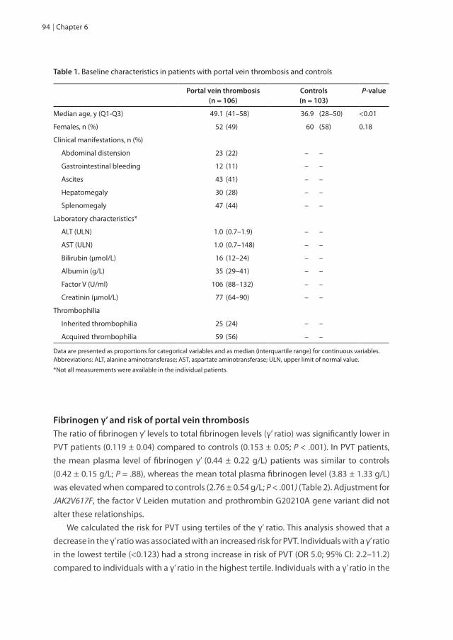
94 | Chapter 6
Table 1. Baseline characteristics in patients with portal vein thrombosis and controls
Portal vein thrombosis
(n = 106)Controls (n = 103)
P-value
Median age, y (Q1-Q3) 49.1 (41–58) 36.9 (28–50) <0.01
Females, n (%) 52 (49) 60 (58) 0.18
Clinical manifestations, n (%)
Abdominal distension 23 (22) – –
Gastrointestinal bleeding 12 (11) – –
Ascites 43 (41) – –
Hepatomegaly 30 (28) – –
Splenomegaly 47 (44) – –
Laboratory characteristics*
ALT (ULN) 1.0 (0.7–1.9) – –
AST (ULN) 1.0 (0.7–148) – –
Bilirubin (μmol/L) 16 (12–24) – –
Albumin (g/L) 35 (29–41) – –
Factor V (U/ml) 106 (88–132) – –
Creatinin (μmol/L) 77 (64–90) – –
Thrombophilia
Inherited thrombophilia 25 (24) – –
Acquired thrombophilia 59 (56) – –
Data are presented as proportions for categorical variables and as median (interquartile range) for continuous variables. Abbreviations: ALT, alanine aminotransferase; AST, aspartate aminotransferase; ULN, upper limit of normal value.
*Not all measurements were available in the individual patients.
Fibrinogen γ’ and risk of portal vein thrombosisThe ratio of fibrinogen γ’ levels to total fibrinogen levels (γ’ ratio) was significantly lower in PVT patients (0.119 ± 0.04) compared to controls (0.153 ± 0.05; P < .001). In PVT patients, the mean plasma level of fibrinogen γ’ (0.44 ± 0.22 g/L) patients was similar to controls (0.42 ± 0.15 g/L; P = .88), whereas the mean total plasma fibrinogen level (3.83 ± 1.33 g/L) was elevated when compared to controls (2.76 ± 0.54 g/L; P < .001) (Table 2). Adjustment for JAK2V617F, the factor V Leiden mutation and prothrombin G20210A gene variant did not alter these relationships. We calculated the risk for PVT using tertiles of the γ’ ratio. This analysis showed that a decrease in the γ’ ratio was associated with an increased risk for PVT. Individuals with a γ’ ratio in the lowest tertile (<0.123) had a strong increase in risk of PVT (OR 5.0; 95% CI: 2.2–11.2) compared to individuals with a γ’ ratio in the highest tertile. Individuals with a γ’ ratio in the
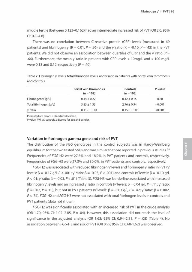
Chap
ter 6
95Fibrinogen γ' in PVT |
middle tertile (between 0.123–0.162) had an intermediate increased risk of PVT (OR 2.0; 95% CI: 0.8–4.8) There was no correlation between C-reactive protein (CRP) levels (measured in 69 patients) and fibrinogen γ’ (R = 0.01, P = .96) and the γ’ ratio (R = -0.10, P = .42) in the PVT patients. We did not observe an association between quartiles of CRP and the γ’ ratio (P = .66). Furthermore, the mean γ’ ratio in patients with CRP levels < 10mg/L and > 100 mg/L were 0.13 and 0.12, respectively (P = .40).
Table 2. Fibrinogen γ’ levels, total fibrinogen levels, and γ’ ratio in patients with portal vein thrombosis and controls
Portal vein thrombosis (n = 102)
Controls (n = 103)
P-value
Fibrinogen γ’ (g/L) 0.44 ± 0.22 0.42 ± 0.15 0.88
Total fibrinogen (g/L) 3.83 ± 1.33 2.76 ± 0.54 <0.001
γ’ ratio 0.119 ± 0.04 0.153 ± 0.05 <0.001
Presented are means ± standard deviation.P-value: PVT vs. controls, adjusted for age and gender.
Variation in fibrinogen gamma gene and risk of PVTThe distribution of the FGG genotypes in the control subjects was in Hardy-Weinberg equilibrium for the two tested SNPs and was similar to those reported in previous studies.7-9 Frequencies of FGG-H2 were 27.5% and 18.9% in PVT patients and controls, respectively. Frequencies of FGG-H3 were 27.3% and 30.0%, in PVT patients and controls, respectively. FGG-H2 was associated with reduced fibrinogen γ’ levels and fibrinogen γ’ ratio in PVT (γ’ levels: β = -0.12 g/l, P < .001; γ’ ratio: β = -0.03, P < .001) and controls (γ’ levels: β = -0.10 g/l, P < .01; γ’ ratio: β = -0.03, P < .01) (Table 3). FGG-H3 was borderline associated with increased fibrinogen γ’ levels and an increased γ’ ratio in controls (γ’ levels: β = 0.04 g/l, P = .11; γ’ ratio: β = 0.02, P = .10), but not in PVT patients (γ’ levels: β = -0.03 g/l, P = .42; γ’ ratio: β = 0.002, P < .74). FGG-H2 and FGG-H3 were not associated with total fibrinogen levels in controls and PVT patients (data not shown). FGG-H2 was significantly associated with an increased risk of PVT in the crude analysis (OR 1.70; 95% CI: 1.02–2.85, P = .04). However, this association did not reach the level of significance in the adjusted analysis (OR 1.63; 95% CI: 0.94–2.81, P = .08) (Table 4). No association between FGG-H3 and risk of PVT (OR 0.99; 95% CI: 0.60-1.62) was observed.
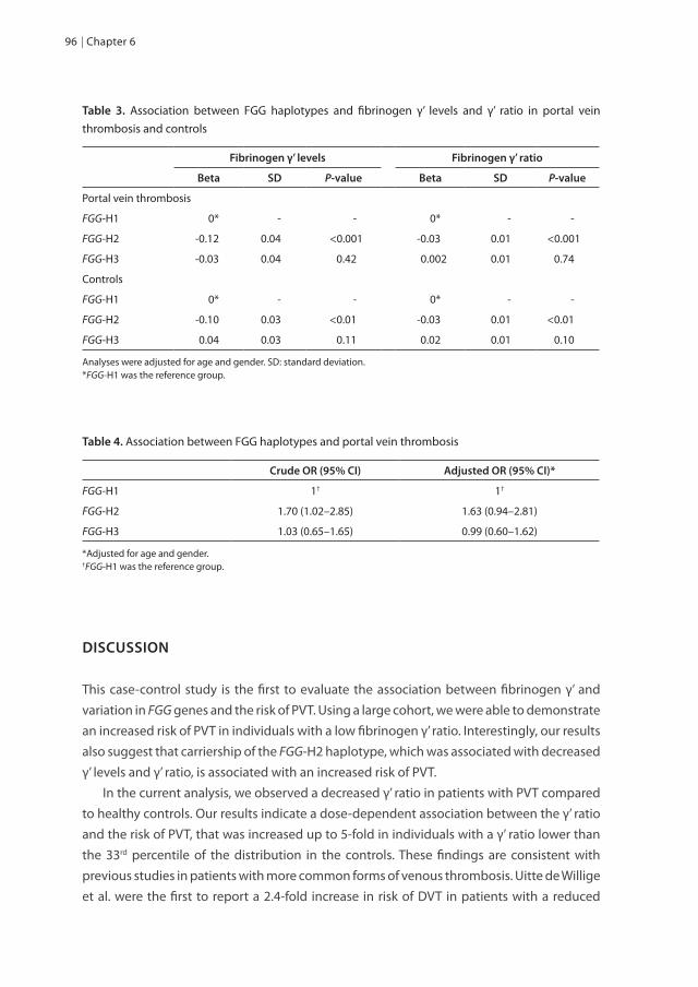
96 | Chapter 6
Table 3. Association between FGG haplotypes and fibrinogen γ’ levels and γ’ ratio in portal vein thrombosis and controls
Fibrinogen γ’ levels Fibrinogen γ’ ratio
Beta SD P-value Beta SD P-value
Portal vein thrombosis
FGG-H1 0* - - 0* - -
FGG-H2 -0.12 0.04 <0.001 -0.03 0.01 <0.001
FGG-H3 -0.03 0.04 0.42 0.002 0.01 0.74
Controls
FGG-H1 0* - - 0* - -
FGG-H2 -0.10 0.03 <0.01 -0.03 0.01 <0.01
FGG-H3 0.04 0.03 0.11 0.02 0.01 0.10
Analyses were adjusted for age and gender. SD: standard deviation.*FGG-H1 was the reference group.
Table 4. Association between FGG haplotypes and portal vein thrombosis
Crude or (95% Ci) Adjusted or (95% Ci)*
FGG-H1 1† 1†
FGG-H2 1.70 (1.02–2.85) 1.63 (0.94–2.81)
FGG-H3 1.03 (0.65–1.65) 0.99 (0.60–1.62)
*Adjusted for age and gender. †FGG-H1 was the reference group.
DiSCUSSioN
This case-control study is the first to evaluate the association between fibrinogen γ’ and variation in FGG genes and the risk of PVT. Using a large cohort, we were able to demonstrate an increased risk of PVT in individuals with a low fibrinogen γ’ ratio. Interestingly, our results also suggest that carriership of the FGG-H2 haplotype, which was associated with decreased γ’ levels and γ’ ratio, is associated with an increased risk of PVT. In the current analysis, we observed a decreased γ’ ratio in patients with PVT compared to healthy controls. Our results indicate a dose-dependent association between the γ’ ratio and the risk of PVT, that was increased up to 5-fold in individuals with a γ’ ratio lower than the 33rd percentile of the distribution in the controls. These findings are consistent with previous studies in patients with more common forms of venous thrombosis. Uitte de Willige et al. were the first to report a 2.4-fold increase in risk of DVT in patients with a reduced
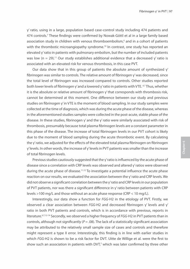
Chap
ter 6
97Fibrinogen γ' in PVT |
γ’ ratio, using in a large, population based case-control study including 474 patients and 474 controls.7 These findings were confirmed by Nowak-Göttl et al in a large family based association study in children with venous thromboembolism,6 and in a cohort of patients with the thrombotic microangiopathy syndrome.10 In contrast, one study has reported an elevated γ’ ratio in patients with pulmonary embolism, but the number of included patients was low (n = 29).11 Our study establishes additional evidence that a decreased γ’ ratio is associated with an elevated risk for venous thrombosis, in this case PVT. Our data show that in this group of patients the absolute amount of synthesized γ’ fibrinogen was similar to controls. The relative amount of fibrinogen γ’ was decreased, since the total level of fibrinogen was increased compared to controls. Other studies reported both lower levels of fibrinogen γ’ and a lowered γ’ ratio in patients with VTE. 6,7 Thus, whether it is the absolute or relative amount of fibrinogen γ’ that corresponds with thrombosis risk, cannot be determined at this moment. One difference between our study and previous studies on fibrinogen γ’ in VTE is the moment of blood sampling. In our study samples were collected at the time of diagnosis, which was during the acute phase of the disease, whereas in the aforementioned studies samples were collected in the post-acute, stable phase of the disease. In these studies, fibrinogen γ’ and the γ’ ratio were similarly associated with risk of thrombosis, presumably because total plasma fibrinogen levels are a constant parameter in this phase of the disease. The increase of total fibrinogen levels in our PVT cohort is likely due to the moment of blood sampling during the acute thrombotic event. By calculating the γ’ ratio, we adjusted for the effects of the elevated total plasma fibrinogen on fibrinogen γ’ levels. In other words, the increase of γ’ levels in PVT patients was smaller than the increase of total fibrinogen levels. Previous studies cautiously suggested that the γ’ ratio is influenced by the acute phase of disease since a correlation with CRP levels was observed and altered γ’ ratios were observed during the acute phase of disease.11,12 To investigate a potential influence the acute phase reaction on our results, we evaluated the association between the γ’ ratio and CRP levels. We did not observe a significant correlation between the γ’ ratio and CRP levels in our population of PVT patients, nor was there a significant difference in γ’ ratio between patients with CRP levels >100 mg/L and those without an acute phase response (CRP < 10 mg/L). Interestingly, our data show a function for FGG-H2 in the etiology of PVT. Firstly, we observed a clear association between FGG-H2 and decreased fibrinogen γ’ levels and γ’ ratio in both PVT patients and controls, which is in accordance with previous, reports in literature.6,7,12-14 Secondly, we observed a higher frequency of FGG-H2 in PVT patients than in controls, although not significantly (P = .08). The lack of a statistically significant association may be attributed to the relatively small sample size of cases and controls and therefore might represent a type II error. Interestingly, this finding is in line with earlier studies in which FGG-H2 is shown to be a risk factor for DVT. Uitte de Willige et al. were the first to show such an association in patients with DVT,7 which was later confirmed by three other
98 | Chapter 6
studies.6,8,9 The increased risk of PVT in carriers of the FGG-H2 haplotype corresponded with a 63% increase in risk per minor allele, which is in the same order of magnitude as reported earlier in other types of venous thrombosis.6-8 It has been proposed that FGG-H2 influences thrombosis risk by reducing fibrinogen γ’ levels and the γ’ ratio, which subsequently results in an elevated risk for venous thrombosis.6,7 Our findings may constitute additional evidence for this relation, although it is evident that additional studies are needed to extend our observations before we can evaluate the clinical relevance of these findings. An obvious question is how the fibrinogen γ’ chain may be related to the development of venous thrombosis. The alternative carboxy terminus of the fibrinogen γ’ variant contains an additional high-affinity binding site for thrombin. Studies indicate that this additional region inhibits thrombin activity,15 may act as an inhibitor of the intrinsic pathway by reducing thrombin generation,16,17 and may serve as a reservoir for active thrombin in the fibrin clot.18 Furthermore, the fibrinogen γ’ chain lacks the αIIbβ3 integrin which is required for platelet binding,19-21 and inhibits thrombin binding to GpIbα and thrombin cleavage of PAR1 on platelets,22,23 both of which inhibit platelet aggregation. Clearly, these mechanisms represent an antithrombotic tendency. A decrease in fibrinogen γ’ may therefore lead to a reduction of antithrombotic activity and subsequently result in an elevated risk of developing thrombosis. The γ’ chain also displays prothrombotic properties. It enhances plasma factor XIII activity and fibrin cross-linking, which have been shown to reduce fibrinolysis rates.24,25 In addition, clots made from purified fibrinogen γ’ display thinner fibers, increased branching and a reduced pore size, which are consistent with a prothrombotic phenotype.26,27 Our work, and that of others, suggests that the antithrombotic mechanisms of fibrinogen γ’ predominate in the pathogenesis of venous thrombosis. The strength of our study is that we used the largest cohort to date of consecutive patients with newly diagnosed non-cirrhotic, non-malignant PVT. Our study also has some limitations that need to be discussed. First, we performed a case-control study which has some inherent imperfections in its design. Because patients were included after the diagnosis, we cannot rule out the possibility that the altered fibrinogen γ’ ratio is the consequence rather than the cause of the disease. However, a prospective design is unfeasible for rare thrombotic manifestations as PVT. On the other hand, we found a clear association between genetic variation in FGG genes and fibrinogen γ’ levels and γ’ ratio, as has been reported before. In addition, our results suggest an association between FGG-H2 and risk of PVT. These associations are independent of time of blood sampling, and provide a clear indication that fibrinogen γ’ may indeed play a role in the pathophysiology of thrombosis at this uncommon site. Secondly, our control group had a mean age that was somewhat lower than in the group of PVT patients. Since there was a significant overlap in age between these groups, we statistically adjusted for the difference on this variable using regression methods.

Chap
ter 6
99Fibrinogen γ' in PVT |
In conclusion, we observed a clear association between a decreased γ’ ratio and PVT. In addition, this study provides the first support of an increased frequency of FGG-H2 in PVT, which may contribute to the development of thrombosis at this uncommon site by decreasing the γ’ ratio.
100 | Chapter 6
rEFErENCES
1. DeLeve LD, Valla DC, Garcia-Tsao G, American Association for the Study Liver D. Vascular disorders of the liver. Hepatology 2009;49:1729-64.
2. Plessier A, Darwish-Murad S, Hernandez-Guerra M, et al. Acute portal vein thrombosis unrelated to cirrhosis: a prospective multicenter follow-up study. Hepatology 2010;51:210-8.
3. Mosesson MW, Siebenlist KR, Meh DA. The structure and biological features of fibrinogen and fibrin. Ann N Y Acad Sci 2001;936:11-30.
4. Uitte de Willige S, Standeven KF, Philippou H, Ariens RA. The pleiotropic role of the fibrinogen gamma’ chain in hemostasis. Blood 2009;114:3994-4001.
5. Chung DW, Davie EW. gamma and gamma’ chains of human fibrinogen are produced by alternative mRNA processing. Biochemistry 1984;23:4232-6.
6. Nowak-Gottl U, Weiler H, Hernandez I, et al. Fibrinogen alpha and gamma genes and factor VLeiden in children with thromboembolism: results from 2 family-based association studies. Blood 2009;114:1947-53.
7. Uitte de Willige S, de Visser MC, Houwing-Duistermaat JJ, Rosendaal FR, Vos HL, Bertina RM. Genetic variation in the fibrinogen gamma gene increases the risk for deep venous thrombosis by reducing plasma fibrinogen gamma’ levels. Blood 2005;106:4176-83.
8. Grunbacher G, Weger W, Marx-Neuhold E, et al. The fibrinogen gamma (FGG) 10034C>T polymorphism is associated with venous thrombosis. Thromb Res 2007;121:33-6.
9. Uitte de Willige S, Pyle ME, Vos HL, et al. Fibrinogen gamma gene 3’-end polymorphisms and risk of venous thromboembolism in the African-American and Caucasian population. Thromb Haemost 2009;101:1078-84.
10. Mosesson MW, Hernandez I, Raife TJ, et al. Plasma fibrinogen gamma’ chain content in the thrombotic microangiopathy syndrome. J Thromb Haemost 2007;5:62-9.
11. Cheung EY, Vos HL, Kruip MJ, den Hertog HM, Jukema JW, de Maat MP. Elevated fibrinogen gamma’ ratio is associated with cardiovascular diseases and acute phase reaction but not with clinical outcome. Blood 2009;114:4603-4; author reply 4-5.
12. Cheung EY, Uitte de Willige S, Vos HL, et al. Fibrinogen gamma’ in ischemic stroke: a case-control study. Stroke 2008;39:1033-5.
13. Lovely RS, Falls LA, Al-Mondhiry HA, et al. Association of gammaA/gamma’ fibrinogen levels and coronary artery disease. Thromb Haemost 2002;88:26-31.
14. Mannila MN, Lovely RS, Kazmierczak SC, et al. Elevated plasma fibrinogen gamma’ concentration is associated with myocardial infarction: effects of variation in fibrinogen genes and environmental factors. J Thromb Haemost 2007;5:766-73.
15. Mosesson MW. Update on antithrombin I (fibrin). Thromb Haemost 2007;98:105-8.
16. Meh DA, Siebenlist KR, Mosesson MW. Identification and characterization of the thrombin binding sites on fibrin. J Biol Chem 1996;271:23121-5.
17. Mosesson MW. Antithrombin I. Inhibition of thrombin generation in plasma by fibrin formation. Thromb Haemost 2003;89:9-12.
18. Fredenburgh JC, Stafford AR, Leslie BA, Weitz JI. Bivalent binding to gammaA/gamma’-fibrin engages both exosites of thrombin and protects it from inhibition by the antithrombin-heparin complex. J Biol Chem 2008;283:2470-7.
19. Farrell DH, Thiagarajan P. Binding of recombinant fibrinogen mutants to platelets. J Biol Chem 1994;269:226-31.
20. Harfenist EJ, Packham MA, Mustard JF. Effects of variant gamma chains and sialic acid content of fibrinogen upon its interactions with ADP-stimulated human and rabbit platelets. Blood 1984;64:1163-8.
Chap
ter 6
101Fibrinogen γ' in PVT |
21. Hettasch JM, Bolyard MG, Lord ST. The residues AGDV of recombinant gamma chains of human fibrinogen must be carboxy-terminal to support human platelet aggregation. Thromb Haemost 1992;68:701-6.
22. Lancellotti S, Rutella S, De Filippis V, Pozzi N, Rocca B, De Cristofaro R. Fibrinogen-elongated gamma chain inhibits thrombin-induced platelet response, hindering the interaction with different receptors. J Biol Chem 2008;283:30193-204.
23. Lovely RS, Rein CM, White TC, et al. gammaA/gamma’ fibrinogen inhibits thrombin-induced platelet aggregation. Thromb Haemost 2008;100:837-46.
24. Falls LA, Farrell DH. Resistance of gammaA/gamma’ fibrin clots to fibrinolysis. J Biol Chem 1997;272:14251-6.
25. Collet JP, Nagaswami C, Farrell DH, Montalescot G, Weisel JW. Influence of gamma’ fibrinogen splice variant on fibrin physical properties and fibrinolysis rate. Arterioscler Thromb Vasc Biol 2004;24:382-6.
26. Cooper AV, Standeven KF, Ariens RA. Fibrinogen gamma-chain splice variant gamma’ alters fibrin formation and structure. Blood 2003;102:535-40.
27. Siebenlist KR, Mosesson MW, Hernandez I, et al. Studies on the basis for the properties of fibrin produced from fibrinogen-containing gamma’ chains. Blood 2005;106:2730-6.

C H A P T E r 7
FiBriNoGEN GAmmA PrimE APPEArS To BE UNrELATED
To THE DEVELoPmENT oF THE BUDD-CHiAri SYNDromE
Jasper H. Smalberg1, Edith Koehler2, Sarwa Darwish Murad2, Aurelie Plessier3, Susana Seijo4,
Jonel Trebicka5, Massimo Primignani6, Moniek P.M. de Maat1, Juan-Carlos Garcia-Pagan4,
Dominique C. Valla3, Harry L.A. Janssen2 and Frank W.G. Leebeek1 for the European Network for
Vascular Disorders of the Liver (EN-Vie).
Departments of Hematology1 and Gastroenterology & Hepatology2, Erasmus University Medical
Center, Rotterdam, The Netherlands; 3Department of Hepatology, Hopital Beaujon, Assistance
Publique-Hopitaux de Paris, Inserm U773 and University Paris-7, Clichy, France; 4Hepatic
Hemodynamic Laboratory, Liver Unit, Institut de Malalties Digestives, IDIBAPS and Ciberehd,
Barcelona, Spain; 5Department of Internal Medicine I, University Hospital of Bonn, Bonn,
Germany; and 6Gastroenterology and Gastrointestinal Endoscopy Unit, Ospedale Policlinico,
Mangiagalli and Regina Elena Foundation, Milan, Italy.
Submitted
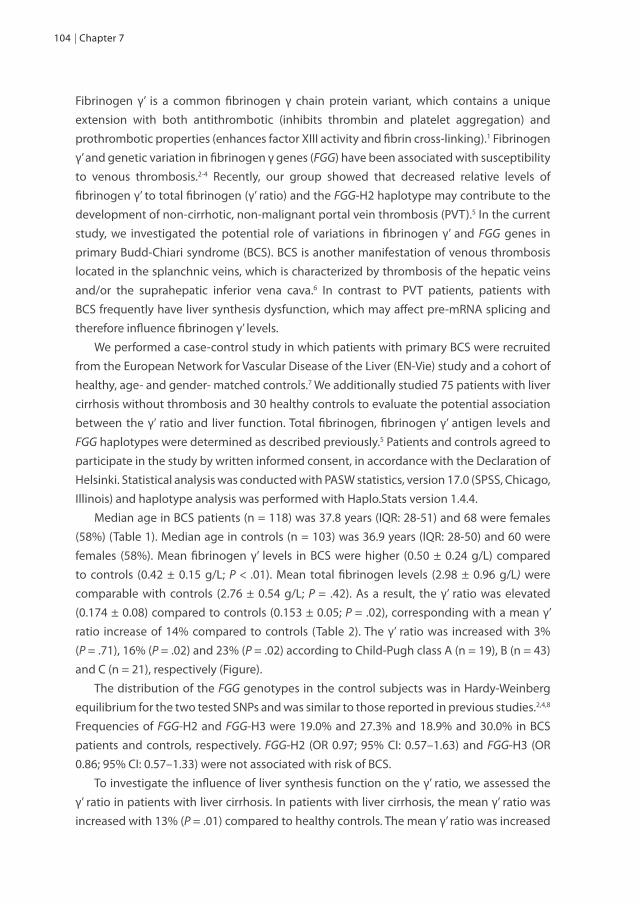
104 | Chapter 7
Fibrinogen γ’ is a common fibrinogen γ chain protein variant, which contains a unique extension with both antithrombotic (inhibits thrombin and platelet aggregation) and prothrombotic properties (enhances factor XIII activity and fibrin cross-linking).1 Fibrinogen γ’ and genetic variation in fibrinogen γ genes (FGG) have been associated with susceptibility to venous thrombosis.2-4 Recently, our group showed that decreased relative levels of fibrinogen γ’ to total fibrinogen (γ’ ratio) and the FGG-H2 haplotype may contribute to the development of non-cirrhotic, non-malignant portal vein thrombosis (PVT).5 In the current study, we investigated the potential role of variations in fibrinogen γ’ and FGG genes in primary Budd-Chiari syndrome (BCS). BCS is another manifestation of venous thrombosis located in the splanchnic veins, which is characterized by thrombosis of the hepatic veins and/or the suprahepatic inferior vena cava.6 In contrast to PVT patients, patients with BCS frequently have liver synthesis dysfunction, which may affect pre-mRNA splicing and therefore influence fibrinogen γ’ levels. We performed a case-control study in which patients with primary BCS were recruited from the European Network for Vascular Disease of the Liver (EN-Vie) study and a cohort of healthy, age- and gender- matched controls.7 We additionally studied 75 patients with liver cirrhosis without thrombosis and 30 healthy controls to evaluate the potential association between the γ’ ratio and liver function. Total fibrinogen, fibrinogen γ’ antigen levels and FGG haplotypes were determined as described previously.5 Patients and controls agreed to participate in the study by written informed consent, in accordance with the Declaration of Helsinki. Statistical analysis was conducted with PASW statistics, version 17.0 (SPSS, Chicago, Illinois) and haplotype analysis was performed with Haplo.Stats version 1.4.4. Median age in BCS patients (n = 118) was 37.8 years (IQR: 28-51) and 68 were females (58%) (Table 1). Median age in controls (n = 103) was 36.9 years (IQR: 28-50) and 60 were females (58%). Mean fibrinogen γ’ levels in BCS were higher (0.50 ± 0.24 g/L) compared to controls (0.42 ± 0.15 g/L; P < .01). Mean total fibrinogen levels (2.98 ± 0.96 g/L) were comparable with controls (2.76 ± 0.54 g/L; P = .42). As a result, the γ’ ratio was elevated (0.174 ± 0.08) compared to controls (0.153 ± 0.05; P = .02), corresponding with a mean γ’ ratio increase of 14% compared to controls (Table 2). The γ’ ratio was increased with 3% (P = .71), 16% (P = .02) and 23% (P = .02) according to Child-Pugh class A (n = 19), B (n = 43) and C (n = 21), respectively (Figure). The distribution of the FGG genotypes in the control subjects was in Hardy-Weinberg equilibrium for the two tested SNPs and was similar to those reported in previous studies.2,4,8 Frequencies of FGG-H2 and FGG-H3 were 19.0% and 27.3% and 18.9% and 30.0% in BCS patients and controls, respectively. FGG-H2 (OR 0.97; 95% CI: 0.57–1.63) and FGG-H3 (OR 0.86; 95% CI: 0.57–1.33) were not associated with risk of BCS. To investigate the influence of liver synthesis function on the γ’ ratio, we assessed the γ’ ratio in patients with liver cirrhosis. In patients with liver cirrhosis, the mean γ’ ratio was increased with 13% (P = .01) compared to healthy controls. The mean γ’ ratio was increased
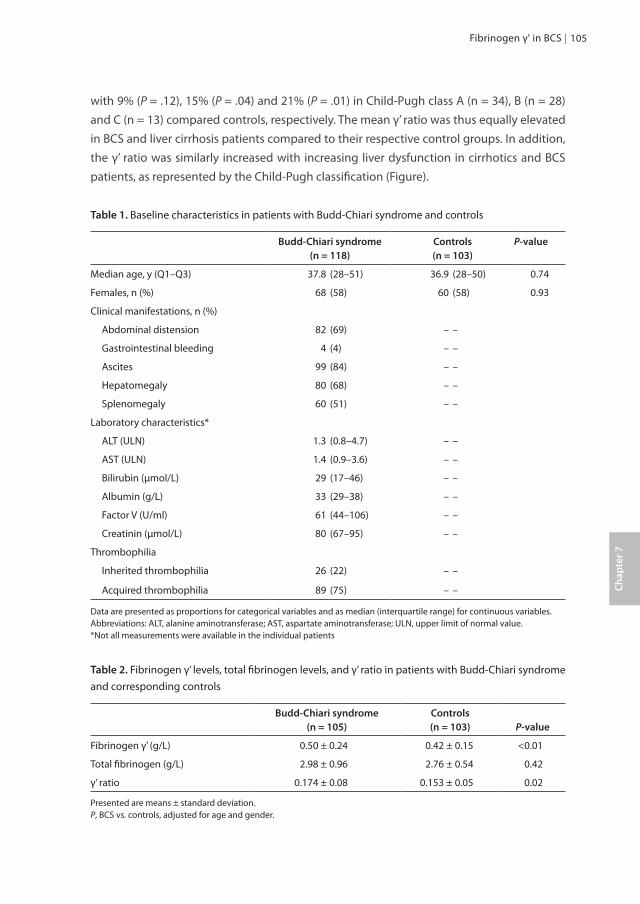
Chap
ter 7
105Fibrinogen γ' in BCS |
with 9% (P = .12), 15% (P = .04) and 21% (P = .01) in Child-Pugh class A (n = 34), B (n = 28) and C (n = 13) compared controls, respectively. The mean γ’ ratio was thus equally elevated in BCS and liver cirrhosis patients compared to their respective control groups. In addition, the γ’ ratio was similarly increased with increasing liver dysfunction in cirrhotics and BCS patients, as represented by the Child-Pugh classification (Figure).
Table 1. Baseline characteristics in patients with Budd-Chiari syndrome and controls
Budd-Chiari syndrome
(n = 118)Controls (n = 103)
P-value
Median age, y (Q1–Q3) 37.8 (28–51) 36.9 (28–50) 0.74
Females, n (%) 68 (58) 60 (58) 0.93
Clinical manifestations, n (%)
Abdominal distension 82 (69) – –
Gastrointestinal bleeding 4 (4) – –
Ascites 99 (84) – –
Hepatomegaly 80 (68) – –
Splenomegaly 60 (51) – –
Laboratory characteristics*
ALT (ULN) 1.3 (0.8–4.7) – –
AST (ULN) 1.4 (0.9–3.6) – –
Bilirubin (μmol/L) 29 (17–46) – –
Albumin (g/L) 33 (29–38) – –
Factor V (U/ml) 61 (44–106) – –
Creatinin (μmol/L) 80 (67–95) – –
Thrombophilia
Inherited thrombophilia 26 (22) – –
Acquired thrombophilia 89 (75) – – Data are presented as proportions for categorical variables and as median (interquartile range) for continuous variables. Abbreviations: ALT, alanine aminotransferase; AST, aspartate aminotransferase; ULN, upper limit of normal value.*Not all measurements were available in the individual patients
Table 2. Fibrinogen γ’ levels, total fibrinogen levels, and γ’ ratio in patients with Budd-Chiari syndrome and corresponding controls
Budd-Chiari syndrome (n = 105)
Controls (n = 103) P-value
Fibrinogen γ’ (g/L) 0.50 ± 0.24 0.42 ± 0.15 <0.01
Total fibrinogen (g/L) 2.98 ± 0.96 2.76 ± 0.54 0.42
γ’ ratio 0.174 ± 0.08 0.153 ± 0.05 0.02
Presented are means ± standard deviation.P, BCS vs. controls, adjusted for age and gender.
106 | Chapter 7
*
*
*
*
125%
100%
75%
50%
25%
0%
Budd-Chiari syndrome Liver cirrhosis
γ’ ra
tios
ControlsChild-Pugh class AChild-Pugh class BChild-Pugh class C
Figure. This figure depicts the mean fibrinogen γ’ ratios in patients with Budd-Chiari syndrome and liver cirrhosis, which were standardized according to the levels in the corresponding, healthy controls. *denote P-values <0.05 and represent the difference in γ’ ratio in Child-Pugh class compared to corresponding, healthy controls, adjusted for age and gender.
Our study shows that the γ’ ratio is strongly dependent upon liver function and that the observed elevated γ’ ratio in BCS is likely to be caused by liver dysfunction. It is well known that liver synthesis function is often affected in patients presenting with BCS due to the venous outflow obstruction and subsequent damage of liver parenchymal cells.9 Alterations in pre-mRNA splicing have also been reported for other proteins in patients with chronic liver dysfunction.10 The γ’ ratio was comparable to controls in the subgroup of BCS patients with a preserved liver synthesis function (Child-Pugh A), suggesting that the γ’ ratio is not associated with BCS. Neither did we observe an association between risk of thrombosis and FGG haplotypes, as has been reported for other forms of venous thrombosis2-4 and appears to be present in PVT.5
Our data thus indicate a difference in the involvement of the fibrinogen γ’ chain in the pathogenesis of BCS and PVT, which is an unexpected finding. Both are forms of venous thrombosis and their etiology generally shows a considerable overlap.9 On the other hand,

Chap
ter 7
107Fibrinogen γ' in BCS |
it is increasingly recognized that despite several similarities, also apparent differences in the risk profile of patients with BCS and PVT exist.11 The different role of fibrinogen γ’ in clot formation may be related to differences in shear stress conditions or vascular bed specific factors, as has been suggested previously.1
In conclusion, the results of this multicenter case-control study indicate that fibrinogen γ’ is not associated with the development of BCS. These findings provide additional evidence that clear differences in the risk profile of BCS and PVT patients exist.
108 | Chapter 7
rEFErENCES
1. Uitte de Willige S, Standeven KF, Philippou H, Ariens RA. The pleiotropic role of the fibrinogen gamma’ chain in hemostasis. Blood 2009;114:3994-4001.
2. Grunbacher G, Weger W, Marx-Neuhold E, et al. The fibrinogen gamma (FGG) 10034C>T polymorphism is associated with venous thrombosis. Thromb Res 2007;121:33-6.
3. Nowak-Gottl U, Weiler H, Hernandez I, et al. Fibrinogen alpha and gamma genes and factor VLeiden in children with thromboembolism: results from 2 family-based association studies. Blood 2009;114:1947-53.
4. Uitte de Willige S, de Visser MC, Houwing-Duistermaat JJ, Rosendaal FR, Vos HL, Bertina RM. Genetic variation in the fibrinogen gamma gene increases the risk for deep venous thrombosis by reducing plasma fibrinogen gamma’ levels. Blood 2005;106:4176-83.
5. Smalberg JH, Koehler E, Darwish Murad S, et al. Fibrinogen gamma prime and variation in fibrinogen gamma genes in the etiology of portal vein thrombosis. Submitted.
6. Janssen HL, Garcia-Pagan JC, Elias E, Mentha G, Hadengue A, Valla DC. Budd-Chiari syndrome: a review by an expert panel. J Hepatol 2003;38:364-71.
7. Darwish Murad S, Plessier A, Hernandez-Guerra M, et al. Etiology, management, and outcome of the Budd-Chiari syndrome. Ann Intern Med 2009;151:167-75.
8. Uitte de Willige S, Pyle ME, Vos HL, et al. Fibrinogen gamma gene 3’-end polymorphisms and risk of venous thromboembolism in the African-American and Caucasian population. Thromb Haemost 2009;101:1078-84.
9. DeLeve LD, Valla DC, Garcia-Tsao G, American Association for the Study Liver D. Vascular disorders of the liver. Hepatology 2009;49:1729-64.
10. Berasain C, Goni S, Castillo J, Latasa MU, Prieto J, Avila MA. Impairment of pre-mRNA splicing in liver disease: mechanisms and consequences. World J Gastroenterol 2010;16:3091-102.
11. Smalberg JH, Kruip MJ, Janssen HL, Rijken DC, Leebeek FW, de Maat MP. Hypercoagulability and hypofibrinolysis and risk of deep vein thrombosis and splanchnic vein thrombosis: similarities and differences. Arterioscler Thromb Vasc Biol 2011;31:485-93.
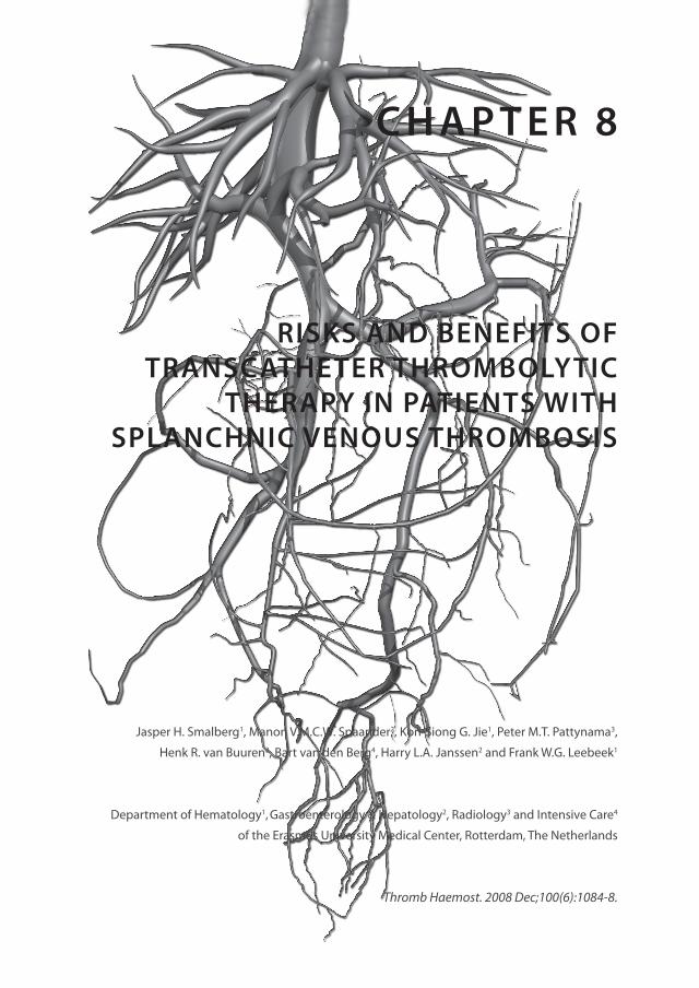
C H A P T E r 8
riSKS AND BENEFiTS oF TrANSCATHETEr THromBoLYTiC
THErAPY iN PATiENTS WiTH SPLANCHNiC VENoUS THromBoSiS
Jasper H. Smalberg1, Manon V.M.C.W. Spaander2, Kon-Siong G. Jie1, Peter M.T. Pattynama3,
Henk R. van Buuren2, Bart van den Berg4, Harry L.A. Janssen2 and Frank W.G. Leebeek1
Department of Hematology1, Gastroenterology & Hepatology2, Radiology3 and Intensive Care4
of the Erasmus University Medical Center, Rotterdam, The Netherlands
Thromb Haemost. 2008 Dec;100(6):1084-8.
110 | Chapter 8
ABSTrACT
Thrombolytic therapy in patients with acute, extended splanchnic venous thrombosis is controversial. Here we present our single-center experience with locally delivered, thrombolytic therapy in these patients. All consecutive patients (n = 12) with acute, extended splanchnic venous thrombosis who underwent local thrombolytic therapy in our hospital, were included in this study. Thrombolytic therapy was successful for three thrombotic events and partially successful for four thrombotic events. Two patients developed minor procedure related bleeding (17%). Six patients (50%) developed major procedure related bleeding, with a fatal outcome in two. In conclusion, local thrombolytic therapy in patients with acute, extended splanchnic vein thrombosis is found to be associated with a high rate of procedure related bleeding. Therefore, thrombolysis should be reserved for patients in whom the venous flow cannot be restored by using conventional anticoagulant therapy or stent placement across the thrombosed vessel segment.
Chap
ter 8
111Thrombolytic therapy in SVT |
iNTroDUCTioN
Splanchnic venous thrombosis (SVT) comprises thrombosis of the hepatic veins and the portal venous system. It includes the Budd-Chiari syndrome (BCS), characterized by hepatic outflow obstruction,1 and portal vein thrombosis (PVT).2 SVT is often related to prothrombotic disorders3,4 or caused by underlying haematological diseases that predispose to hypercoagulability, such as myeloproliferative diseases and paroxysmal nocturnal haemoglobinuria.5,6 One of the treatment options in SVT patients is locally delivered transcatheter thrombolytic therapy, which is mainly used in patients with acute, combined thrombosis of both portal and hepatic veins. Several case studies or small case series describe successful thrombolytic therapy in SVT patients with a low incidence of bleeding complications.7-14 However, these reports should be interpreted with caution in view of a potential publication bias of successful cases. Only two larger studies have been performed in which success and complication rates vary widely.15,16 Therefore, it is difficult to properly assess the risk/benefit ratio and thrombolytic therapy remains controversial. Here we present our single-center experience with local thrombolytic therapy in a series of consecutive patients with SVT.
PATiENTS AND mETHoDS
All SVT patients who underwent locally delivered thrombolytic therapy in our hospital, between January 1996 and December 2007, were included in this study. Clinical records of all patients were reviewed and demographics, etiology, clinical presentation, duration of symptoms, details of thrombolytic therapy, outcome, complications and follow-up were reported in a standardized way. The diagnosis of SVT was established by computed tomography, transcatheter portography, magnetic resonance imaging or Doppler Ultrasound. The obstruction was graded partially when there was a significant remaining patent vein lumen or complete. The Child-Pugh score was used as an indication of liver function prior to thrombolytic therapy in patients with cirrhosis and BCS. Patients were followed from initiation of thrombolysis until death, study closure (December 31, 2007), or, in case of loss to follow up, the last date of visit. Survival rates were calculated by means of the Kaplan-Meier method. Patients were considered for thrombolysis if they met the following criteria. First, only patients with an acute thrombosis (duration of symptoms ≤14 days) were considered for thrombolytic therapy. Second, thrombolytic therapy was only used in patients with thrombosis in two or more vessel systems in the splanchnic area, e.g. thrombosis of the hepatic veins combined with PVT. Third, in most patients thrombolysis was considered after standard anticoagulant therapy had proven insufficient, i.e. in patients exhibiting
112 | Chapter 8
a progressive thrombosis or a clinical deterioration despite anticoagulation. Informed consent was obtained from each patient after informing them on the possible risks and benefits of the treatment. To facilitate creation of the transhepatic tract from the liver vein (inferior caval vein in BCS) to the portal vein during transjugular intrahepatic portosystemic shunt (TIPS) placement, a marker catheter was introduced in the portal vein by the direct percutaneous transhepatic route and by using ultrasound guidance. The decision to deliver trombolytic therapy through a percutaneously inserted transhepatic catheter, or through a catheter placed through a pre-existing or specially created TIPS, was not a constant but rather evolved over time – influenced also by literature suggesting the benefit of additional TIPS for thrombolysis.17-19 Thrombolysis was considered successful when vessel patency and blood flow were restored, as observed during venography, and considered partially successful when flow was restored in some, but not all, thrombosed veins. Thrombolysis was considered unsuccessful when no lysis was obtained in any of the occluded vessels. Thrombolytic therapy was discontinued in case of resolution of the thrombus or in case of severe bleeding. All patients were initially treated by APTT-controlled (APTT ratio 2.2–2.9, reflecting an anti Xa activity of 0.3–0.7 U/ml) infusion of unfractionated heparin (UFH) or a therapeutic dose of low molecular weight heparin (LMWH, dalteparin, 200 U/kg BW s.c.). Recombinant tissue type plasminogen activator (Actilyse®, Boehringer-Ingelheim, Germany), rtPA, was used as thrombolytic agent and was delivered by means of a locally placed catheter. Patients were treated by continuous infusion of 2-4 mg/hr rtPA, in some cases preceded by a 4–10 mg bolus injection. In one patient urokinase was administered. During thrombolysis all patients were admitted to an intensive care unit for hemodynamic monitoring. Low dose UFH was continued during thrombolytic treatment (10.000 IU/ 24 hours). After thrombolytic treatment was stopped, UFH was again administered in a therapeutic dosage and was discontinued once the patient was adequately anticoagulated with vitamin K antagonists. After discharge from hospital, patients were followed at the outpatient department of our hospital.
rESULTS
Patient characteristicsTwelve patients (58% male) with a mean age of 32 years (range 26-53) underwent thrombolytic therapy and were included in this study. Six patients had BCS with additional thrombosis in the portal system. The majority of patients had an underlying pro-thrombotic haematological disorder. Patient characteristics, including etiology and indication for thrombolytic therapy, are presented in Table 1.
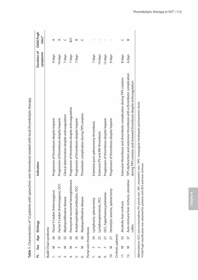
Chap
ter 8
113Thrombolytic therapy in SVT |
Tabl
e 1.
Cha
ract
eris
tics
of 1
2 pa
tient
s w
ith s
plan
chni
c ve
in th
rom
bosi
s tr
eate
d w
ith lo
cal t
hrom
boly
tic th
erap
y
Pt.
Sex
Age
Etio
logy
indi
catio
nD
urat
ion
of
sym
ptom
sCh
ild-P
ugh
clas
s*
Budd
-Chi
ari s
yndr
ome
1.M
26Fa
ctor
V L
eide
n (h
eter
ozyg
ous)
Prog
ress
ion
of th
rom
bosi
s de
spite
hep
arin
9 da
ysA
2.F
30Fa
ctor
V L
eide
n (h
omoz
ygou
s), O
CCPr
ogre
ssio
n of
thro
mbo
sis
desp
ite h
epar
in14
day
sA
3.M
33M
yelo
prol
ifera
tive
dise
ase
Clin
ical
det
erio
ratio
n de
spite
ant
icoa
gula
tion
7 da
ysC
4.M
30Pa
roxy
smal
noc
turn
al h
emog
lobi
nuria
Prog
ress
ion
of th
rom
bosi
s de
spite
ant
icoa
gula
tion
7 da
ysB/
C
5.F
26A
ntip
hosp
holip
id a
ntib
odie
s, O
CCPr
ogre
ssio
n of
thro
mbo
sis
desp
ite h
epar
in7
days
B
6.M
38M
yelo
prol
ifera
tive
dise
ase
Thro
mbo
tic c
ompl
icat
ion
durin
g TI
PS c
reat
ion
C
Port
al v
ein
thro
mbo
sis
7.F
34Ly
mph
oma,
spl
enec
tom
yEx
tens
ive
post
-spl
enec
tom
y th
rom
bosi
s 7
days
-
8.F
25Ly
mph
angi
omat
osis
, OCC
Exte
nsiv
e PV
and
MV
thro
mbo
sis
14 d
ays
-
9.F
27O
CC, h
yper
hom
ocys
tein
emia
Prog
ress
ion
of th
rom
bosi
s de
spite
hep
arin
12 d
ays
-
10.
M27
Hem
olyt
ic a
nem
ia, s
plen
ecto
my
Prog
ress
ion
of th
rom
bosi
s de
spite
hep
arin
9 da
ys-
Cirr
hotic
pat
ient
s
11.
M53
Alc
ohol
ic li
ver c
irrho
sis
Exte
nsiv
e th
rom
bosi
s an
d th
rom
botic
com
plic
atio
n du
ring
TIPS
cre
atio
n8
days
C
12.
M37
Auto
-imm
une
liver
cirr
hosi
s, ul
cera
tive
colit
isTI
PS d
ysfu
nctio
n an
d ex
tens
ive
thro
mbo
sis
with
a th
rom
botic
com
plic
atio
n du
ring
TIPS
revi
sion
and
rene
wed
thro
mbo
sis
desp
ite a
ntic
oagu
latio
n6
days
B
Abb
revi
atio
ns: O
CC: o
ral c
ontr
acep
tives
; PV:
por
tal v
ein
; MV
: mes
ente
ric v
ein
; TIP
S : t
rans
jugu
lar i
ntra
hepa
tic p
orto
syst
emic
shu
nt.
*Chi
ld-P
ugh
clas
sific
atio
n w
as o
btai
ned
for p
atie
nts
with
BCS
and
live
r cirr
hosi
s.
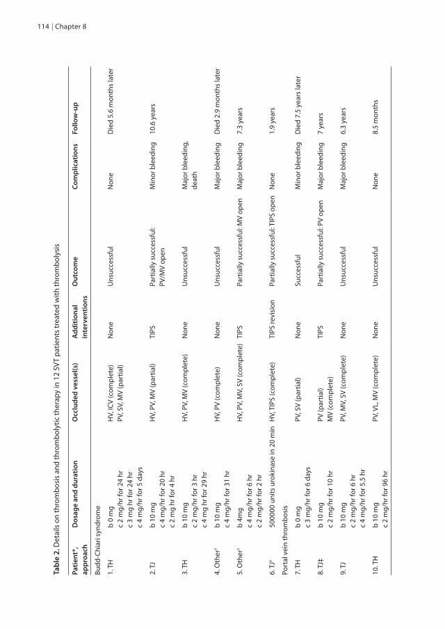
114 | Chapter 8
Tabl
e 2.
Det
ails
on
thro
mbo
sis
and
thro
mbo
lytic
ther
apy
in 1
2 SV
T pa
tient
s tr
eate
d w
ith th
rom
boly
sis
Patie
nt*,
ap
proa
chD
osag
e an
d du
ratio
no
cclu
ded
vess
el(s
)A
dditi
onal
in
terv
entio
nso
utco
me
Com
plic
atio
nsFo
llow
-up
Budd
-Chi
ari s
yndr
ome
1. T
Hb
0 m
g c
2 m
g/hr
for 2
4 hr
c 3
mg
hr fo
r 24
hrc
4 m
g/hr
for 5
day
s
HV,
ICV
(com
plet
e)PV
, SV,
MV
(par
tial)
Non
eU
nsuc
cess
ful
Non
eD
ied
5.6
mon
ths
late
r
2. T
Jb
10 m
g c
4 m
g/hr
for 2
0 hr
c 2
mg
hr fo
r 4 h
r
HV,
PV,
MV
(par
tial)
TIPS
Part
ially
suc
cess
ful:
PV
/MV
open
M
inor
ble
edin
g 10
.6 y
ears
3. T
Hb
10 m
gc
2 m
g/hr
for 3
hr
c 4
mg
hr fo
r 29
hr
HV,
PV,
MV
(com
plet
e)N
one
Uns
ucce
ssfu
lM
ajor
ble
edin
g,
deat
h
4. O
ther
†b
10 m
gc
4 m
g/hr
for 3
1 hr
HV,
PV
(com
plet
e)N
one
Uns
ucce
ssfu
lM
ajor
ble
edin
gD
ied
2.9
mon
ths
late
r
5. O
ther
†b
4mg
c 4
mg/
hr fo
r 6 h
rc
2 m
g/hr
for 2
hr
HV,
PV,
MV,
SV
(com
plet
e)TI
PSPa
rtia
lly s
ucce
ssfu
l: M
V op
enM
ajor
ble
edin
g7.
3 ye
ars
6. T
J‡50
0000
uni
ts u
roki
nase
in 2
0 m
in H
V, T
IPS
(com
plet
e)TI
PS re
visi
onPa
rtia
lly s
ucce
ssfu
l: TI
PS o
pen
Non
e1.
9 ye
ars
Port
al v
ein
thro
mbo
sis
7. T
Hb
0 m
gc
3 m
g/hr
for 6
day
sPV
, SV
(par
tial)
Non
eSu
cces
sful
Min
or b
leed
ing
Die
d 7.
5 ye
ars
late
r
8. T
J‡b
10 m
gc
2 m
g/hr
for 1
0 hr
PV (p
artia
l)M
V (c
ompl
ete)
TIPS
Part
ially
suc
cess
ful:
PV o
pen
Maj
or b
leed
ing
7 ye
ars
9. T
Jb
10 m
gc
2 m
g/hr
for 6
hr
c 4
mg/
hr fo
r 5.5
hr
PV, M
V, S
V (c
ompl
ete)
Non
eU
nsuc
cess
ful
Maj
or b
leed
ing
6.3
year
s
10. T
Hb
10 m
gc
2 m
g/hr
for 9
6 hr
PV, V
L, M
V (c
ompl
ete)
Non
eU
nsuc
cess
ful
Non
e8.
5 m
onth
s
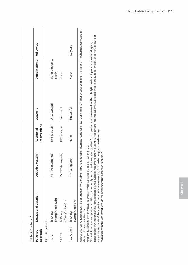
Chap
ter 8
115Thrombolytic therapy in SVT |
Tabl
e 2.
Con
tinue
d
Patie
nt*,
ap
proa
chD
osag
e an
d du
ratio
no
cclu
ded
vess
el(s
)A
dditi
onal
in
terv
entio
nso
utco
me
Com
plic
atio
nsFo
llow
-up
Cirr
hotic
pat
ient
s
11. T
J‡b
10 m
gc
4 m
g/hr
for 1
2 hr
PV, T
IPS
(com
plet
e)TI
PS re
visi
onU
nsuc
cess
ful
Maj
or b
leed
ing,
de
ath
12.1
TJ
b 10
mg
c 2
mg/
hr fo
r 6 h
rPV
, TIP
S (c
ompl
ete)
TIPS
revi
sion
Succ
essf
ulN
one
12.2
Oth
er†
b 10
mg
c 2
mg/
hr fo
r 6 h
rM
V (c
ompl
ete)
Non
eSu
cces
sful
Non
e1.
7 ye
ars
Abb
revi
atio
ns: T
H, t
rans
hepa
tic; T
J, tr
ansj
ugul
ar; P
V, p
orta
l vei
n; H
V, h
epat
ic v
eins
; MV,
mes
ente
ric v
eins
; SV,
spl
enic
vei
n; IC
V, in
ferio
r cav
al v
ein;
TIP
S, tr
ansj
ugul
ar in
trah
epat
ic p
orto
syst
emic
sh
unt;
b, b
olus
; c, c
ontin
uous
.*P
atie
nt 1
2 ex
hibi
ted
two
thro
mbo
tic e
vent
s, w
hich
wer
e su
bdiv
ided
in 1
2.1
and
12.2
.† Pa
tient
4: c
athe
ter w
as p
ositi
oned
util
izin
g a
prev
ious
ly c
reat
ed p
orto
cava
l shu
nt; p
atie
nt 5
: mut
iple
cat
hete
rs w
ere
used
for t
hrom
boly
tic tr
eatm
ent:
perc
utan
eous
tran
shep
atic
, tr
ansj
ugul
ar-t
rans
hepa
tic a
nd a
cat
hete
r pla
ced
in th
e su
perio
r mes
ente
ric a
rter
y; p
atie
nt 1
2: th
e ca
thet
er fo
r thr
ombo
lysi
s w
as p
ositi
oned
in th
e su
perio
r mes
ente
ric a
rter
y be
caus
e of
ex
tens
ive
trom
bosi
s of
the
supe
rior m
esen
teric
vei
n ex
tend
ing
far i
nto
the
perip
hera
l vei
n br
anch
es.
‡ A m
arke
r cat
hete
r was
intr
oduc
ed v
ia th
e pe
rcut
aneo
us tr
ansh
epat
ic a
ppro
ach.
116 | Chapter 8
Details of thrombolytic therapy and outcomeTwelve patients were treated with thrombolytic therapy for thirteen thrombotic events. One patient (pt. 12) was treated twice, because of renewed thrombosis five days after initial successful thrombolysis despite heparin therapy. Route of delivery of the thrombolytic agent was percutaneous transhepatic (n = 4), transjugular transhepatic (n = 6) or other (n = 3). The total percutaneous transhepatic catheters amounted to eight, in which they were either used as a marker catheter to facilitate TIPS placement, or as an indwelling catheter trough which the thrombolytic agent was administered. TIPS insertion/revision was performed in six patients. Thrombolysis was successful for three thrombotic events in two patients (pt. 7 and 12). Therapy was partially successful in four patients (pt. 2, 5, 6 and 8). Details of thrombolytic therapy are presented in Table 2. Five out of six procedures which were combined with a TIPS insertion/revision were (partially) successful.
ComplicationsTwo patients developed minor bleeding, not requiring transfusion, after cessation of thrombolytic therapy (pt 2 and 7). Six patients developed major bleeding necessitating discontinuation of thrombolytic treatment. In two of these patients, thrombolysis had been partially successful at that time (pt. 5 and 8). One patient died directly related to bleeding (pt. 3). This BCS patient had additional PVT and MVT and developed a fatal massive sub-capsular liver haemorrhage resulting in hypovolemic shock. Patient 11 died two weeks after thrombolytic therapy, which was complicated by bacterial peritonitis and renal failure after massive abdominal bleeding. The patient eventually died from multiple organ failure. The death of this patient was considered to be partly caused by procedure related bleeding. Four patients developed major, but nonfatal, bleeding, including retroperitoneal bleeding (pt 4), intraperitoneal bleeding (pt 5), massive intra-abdominal bleeding (pt 8) and a subcapsular liver haematoma (pt 9) (Table 2).
Clinical parameters related to outcomeLaboratory evaluation showed a marked decrease of fibrinogen in patient 2, 3, and 11 (pre-treatment values of 3.6 g/l, 2.2 g/l, and 2.5 g/l, respectively [normal 1.5–3.6 g/l]) to a nadir of 0.6 g/l, 0.3 g/l and 0.7 g/l, respectively, during thrombolytic treatment. Patient 5 already had a pre-treatment fibrinogen of 0.7 g/l and remained <1.0 g/l during thrombolysis. These four patients all developed bleeding complications (of which three major bleeding). The two patients with a fatal outcome showed the lowest fibrinogen levels of all patients (0.3 g/l and 0.4 g/l) during thrombolytic therapy. None of the four patients without bleeding complications had fibrinogen levels <1.0 g/l (Table 3).
Chap
ter 8
117Thrombolytic therapy in SVT |
The Child-Pugh score was obtained for six patients with BCS and two patients with liver cirrhosis. Six patients had a Child-Pugh score >7 and were considered to have liver dysfunction. Four of these patients developed major bleeding complications, of which two were fatal. The two patients with a fatal outcome exhibited the highest Child-Pugh scores. Bleeding complications were also observed in four of six patients with a normal liver function, of which two were major (Table 3).
Table 3. Fibrinogen levels and liver function during thrombolytic therapy in relation to bleeding complications
Number of patients
major bleedingn (%)
minor bleedingn (%)
No complicationsn (%)
Fibrinogen <1.0 g/l 4 3* (75%) 1 (25%) 0 (0 %)
Fibrinogen >1.0 g/l 8 3 (38%) 1 (13%) 4 (50%)
Liver dysfunction 6 4* (67%) 0 (0 %) 2 (33%)
No liver dysfunction 6 2 (33%) 2 (33%) 2 (33%)
*Outcome was fatal in two patients.
Follow-upThe mean time of follow-up was 3.7 years (range 3 days – 10.6 years). Survival according to Kaplan-Meier analysis at 1, 3 and 5 years was 67% (95% CI: 40%–93%). One patient was lost to follow-up. Patient 1 and 4 died 5.6 and 2.9 months after thrombolysis, respectively, due to multiple complications of combined BCS and PVT, which were not related to thrombolysis. Patient 7 died 7.5 years after thrombolysis from lymphoma.
DiSCUSSioN
Both systemic and local administration of thrombolytics have been described in the treatment of SVT, but the latter is preferred.15,16,20 Despite several successful single-case reports and two larger case-series, thrombolytic treatment remains controversial.7-16 In our study, one of the largest series on thrombolysis in SVT patients, we report slightly lower success rates accompanied by a previously unreported high rate of procedure related bleeding. When thrombolytic therapy is indicated, TIPS placement, creating a patent portal venous outflow tract to the systemic venous circulation, followed by local administration of thrombolytic agents in the portal vein appears an effective approach. This is supported by previous reports.17-19 In three out of six TIPS procedures, major bleeding occurred and one patients eventually died, however, it is difficult to determine whether this is related to the
118 | Chapter 8
TIPS procedure itself. The invasive procedure necessary for using local thrombolysis has a great overlap with the TIPS procedure. Therefore, it seems prudent to simultaneously create a TIPS when performing thrombolysis, as a patent TIPS has been shown to be beneficial in terms of clinical improvement and survival in SVT patients.21-23
The relation between fibrinogen levels and bleeding risk suggests that, for safety reasons, it is prudent to monitor fibrinogen levels during thrombolysis and fibrinogen should be supplemented if levels fall <1.0 g/l. Our results indicate that liver dysfunction might be a risk factor for procedure related bleeding, implying that thrombolysis should be cautiously used in these patients. The association between the percutaneous transhepatic approach and bleeding concurs with previous reports, where also the transjugular approach was preferred.16,24,25
A limitation of our study is that it is a single-center, retrospective study with a limited number of patients. Patients are heterogeneous in terms of etiology, clinical presentation, extent of thrombosis, baseline health status and details of thrombolytic therapy. Our study population is highly selected from a large number of cases referred to our specialized tertiary care center and most of the patients had a deteriorating clinical course before thrombolytic therapy was initiated. The strength of our study is that it includes all patients that have been treated avoiding publication bias of successful cases. In conclusion, in a series of consecutive patients receiving locally delivered thrombolysis for acute, extended SVT we found a previously unreported high rate of bleeding complications. These patients generally have a poor prognosis26 and it is up to the treating physician to determine whether the potential benefits of thrombolysis outweigh the apparent risks associated with the procedure. Based on our findings, and previous reports in the literature, we have come to the following preferred approach in patients with acute, extended SVT. Start with conventional anticoagulant therapy and in case of insufficient results proceed to TIPS placement. If venous flow cannot be restored by either of these treatment modalities, local thrombolytic therapy, administered via transjugular approach, can be considered.
Chap
ter 8
119Thrombolytic therapy in SVT |
rEFErENCES
1. Janssen HL, Garcia-Pagan JC, Elias E, Mentha G, Hadengue A, Valla DC. Budd-Chiari syndrome: a review by an expert panel. J Hepatol 2003;38:364-71.
2. Valla DC, Condat B. Portal vein thrombosis in adults: pathophysiology, pathogenesis and management. J Hepatol 2000;32:865-71.
3. de Bruijne EL, Murad SD, de Maat MP, et al. Genetic variation in thrombin-activatable fibrinolysis inhibitor (TAFI) is associated with the risk of splanchnic vein thrombosis. Thromb Haemost 2007;97:181-5.
4. Dentali F, Galli M, Gianni M, Ageno W. Inherited thrombophilic abnormalities and risk of portal vein thrombosis. A meta-analysis. Thromb Haemost 2008;99:675-82.
5. Poulou LS, Xila V, Rokas GI, Karianakis G, Bartzoudis D, Ziakas PD. Temporal trends in mortality rates from visceral vein thrombosis in paroxysmal nocturnal haemoglobinuria: An optimistic view. Thromb Haemost 2008;99:642-5.
6. Smalberg JH, Murad SD, Braakman E, Valk PJ, Janssen HL, Leebeek FW. Myeloproliferative disease in the pathogenesis and survival of Budd-Chiari syndrome. Haematologica 2006;91:1712-3.
7. Guglielmi A, Fior F, Halmos O, et al. Transhepatic fibrinolysis of mesenteric and portal vein thrombosis in a patient with ulcerative colitis: a case report. World J Gastroenterol 2005;11:2035-8.
8. Henao EA, Bohannon WT, Silva MB, Jr. Treatment of portal venous thrombosis with selective superior mesenteric artery infusion of recombinant tissue plasminogen activator. J Vasc Surg 2003;38:1411-5.
9. Kercher KW, Sing RF, Watson KW, Matthews BD, LeQuire MH, Heniford BT. Transhepatic thrombolysis in acute portal vein thrombosis after laparoscopic splenectomy. Surg Laparosc Endosc Percutan Tech 2002;12:131-6.
10. Kuo GP, Brodsky RA, Kim HS. Catheter-directed thrombolysis and thrombectomy for the Budd-Chiari syndrome in paroxysmal nocturnal hemoglobinuria in three patients. J Vasc Interv Radiol 2006;17:383-7.
11. Leebeek FW, Lameris JS, van Buuren HR, Gomez E, Madretsma S, Sonneveld P. Budd-Chiari syndrome, portal vein and mesenteric vein thrombosis in a patient homozygous for factor V Leiden mutation treated by TIPS and thrombolysis. Br J Haematol 1998;102:929-31.
12. Nakai M, Sato M, Sahara S, et al. Transhepatic catheter-directed thrombolysis for portal vein thrombosis after partial splenic embolization in combination with balloon-occluded retrograde transvenous obliteration of splenorenal shunt. World J Gastroenterol 2006;12:5071-4.
13. Ozkan U, Oguzkurt L, Tercan F, Tokmak N. Percutaneous transhepatic thrombolysis in the treatment of acute portal venous thrombosis. Diagn Interv Radiol 2006;12:105-7.
14. Sherigar R, Amir KA, Bobba RK, Arsura EL, Srinivas N. Abdominal pain secondary to pylephlebitis: an uncommon disease of the portal venous system, treated with local thrombolytic therapy. Dig Dis Sci 2005;50:983-7.
15. Hollingshead M, Burke CT, Mauro MA, Weeks SM, Dixon RG, Jaques PF. Transcatheter thrombolytic therapy for acute mesenteric and portal vein thrombosis. J Vasc Interv Radiol 2005;16:651-61.
16. Sharma S, Texeira A, Texeira P, Elias E, Wilde J, Olliff SP. Pharmacological thrombolysis in Budd Chiari syndrome: a single centre experience and review of the literature. J Hepatol 2004;40:172-80.
17. Blum U, Haag K, Rossle M, et al. Noncavernomatous portal vein thrombosis in hepatic cirrhosis: treatment with transjugular intrahepatic portosystemic shunt and local thrombolysis. Radiology 1995;195:153-7.
18. Nolte W, Figulla HR, Ringe B, et al. [TIPSS in the Budd-Chiari syndrome with portal vein thrombosis]. Dtsch Med Wochenschr 1997;122:116-21.
19. Uflacker R. Applications of percutaneous mechanical thrombectomy in transjugular intrahepatic portosystemic shunt and portal vein thrombosis. Tech Vasc Interv Radiol 2003;6:59-69.
20. Bogin V, Marcos A, Shaw-Stiffel T. Budd-Chiari syndrome: in evolution. Eur J Gastroenterol Hepatol 2005;17:33-5.
120 | Chapter 8
21. Murad SD, Luong TK, Pattynama PM, Hansen BE, van Buuren HR, Janssen HL. Long-term outcome of a covered vs. uncovered transjugular intrahepatic portosystemic shunt in Budd-Chiari syndrome. Liver Int 2008;28:249-56.
22. Murad SD, Valla DC, de Groen PC, et al. Pathogenesis and treatment of Budd-Chiari syndrome combined with portal vein thrombosis. Am J Gastroenterol 2006;101:83-90.
23. Senzolo M, Tibbals J, Cholongitas E, Triantos CK, Burroughs AK, Patch D. Transjugular intrahepatic portosystemic shunt for portal vein thrombosis with and without cavernous transformation. Aliment Pharmacol Ther 2006;23:767-75.
24. Aytekin C, Boyvat F, Kurt A, Yologlu Z, Coskun M. Catheter-directed thrombolysis with transjugular access in portal vein thrombosis secondary to pancreatitis. Eur J Radiol 2001;39:80-2.
25. Eapen CE, Velissaris D, Heydtmann M, Gunson B, Olliff S, Elias E. Favourable medium term outcome following hepatic vein recanalisation and/or transjugular intrahepatic portosystemic shunt for Budd Chiari syndrome. Gut 2006;55:878-84.
26. Mahmoud AE, Helmy AS, Billingham L, Elias E. Poor prognosis and limited therapeutic options in patients with Budd-Chiari syndrome and portal venous system thrombosis. Eur J Gastroenterol Hepatol 1997;9:485-9.
C H A P T E r 9
LoNG-TErm FoLLoW-UP oF PATiENTS WiTH PorTAL
VEiN THromBoSiS AND mYELoProLiFErATiVE NEoPLASmS
Jildou Hoekstra¹, Esmay L. Bresser¹, Jasper H. Smalberg², Manon V.M.C.W. Spaander¹,
Frank W.G. Leebeek², Harry L.A. Janssen¹
Departments of Gastroenterology and Hepatology1 and Hematology2, Erasmus University
Medical Center, Rotterdam, The Netherlands
J Thromb Haemost. 2011 Nov;9(11):2208-14.
122 | Chapter 9
ABSTrACT
Myeloproliferative neoplasms (MPN) are frequently identified as an underlying cause in patients with non-cirrhotic portal vein thrombosis (PVT). The aim of this study was to describe the long-term outcome of patients with PVT and MPN. A cohort study was performed including all adults patients referred to our hospital between 1980 and 2008 with noncirrhotic, non-malignant PVT and confirmed MPN. A total of 44 patients (70% female) were included, with a median age at PVT-diagnosis of 48 years (range 18–79). In 31 patients (70%) PVT was the first manifestation of a MPN. Additional risk factors for thrombosis were present in 20 patients (45%). Median follow-up was 5.8 years (range 0.4–21). Twenty-three patients (52%) were treated with oral anticoagulants after diagnosis of PVT, of whom 15 (34%) received long-term therapy. During follow-up, 17 patients (39%) experienced at least one episode of gastrointestinal bleeding. Additional thrombotic events occurred in 12 patients (27%). Twelve patients (27%) had progression of the underlying MPN. Seventeen patients (39%) died at a median age of 64 years (range 30–88). Death was directly related to end-stage MPN in 8 patients (47%) and to a new thrombotic event in 3 patients (18%). No patients died from gastrointestinal bleeding. In conclusion, PVT is often the presenting symptom of an underlying MPN, highlighting the need for thorough screening for this disease. Recurrent thrombosis is a common and severe complication in patients with PVT and MPN. Mortality is primarily related to the underlying MPN and not to complications of portal hypertension.
Chap
ter 9
123Long-term follow-up of MPN in PVT |
iNTroDUCTioN
Development of thrombosis in the extrahepatic portal vein is an uncommon finding. The etiology of portal vein thrombosis (PVT) is heterogeneous and involves both local and systemic prothrombotic factors.1 Liver cirrhosis and hepatobiliary malignancies are considered to be the most important local precipitating factors of PVT.2,3 In the absence of cirrhosis or local tumors, PVT has a better survival. Often there is a prominent role of systemic thrombophilia in these patients.4,5 The most common systemic risk factor encountered in patients with non-cirrhotic, non-malignant PVT is a myeloproliferative neoplasm (MPN).6 MPNs encompass a group of disorders that share features of increased myeloproliferation, resulting in overproduction of mature, functional blood cells. The three main Philadelphia chromosome negative MPNs are polycythemia vera (PV), essential thrombocythemia (ET) and idiopathic myelofibrosis (IMF).7
MPNs are found as an underlying cause in approximately 20–30% of the patients with non-cirrhotic, non-malignant PVT.8,9 Especially in young adults, development of PVT can be a first manifestation of an underlying MPN. However, diagnosis of MPN in these patients can be difficult because typical signs in peripheral blood (e.g. elevated hemoglobin or platelets) may be absent due to hypersplenism and other consequences of portal hypertension.10 Although a number of studies have been published concerning the distribution of etiological factors and clinical presentation of patients with PVT, little is known about the subgroup of cases with confirmed MPN. Due to the rarity of this disease, scarce data is available on the long-term outcome of these patients, including the incidence of recurrent thrombosis, complications of portal hypertension, survival and causes of death. To describe the natural disease course and main complications, we conducted a cohort study of patients with non-cirrhotic, non-malignant PVT and an underlying MPN. Additionally, we determined long-term survival and causes of death in these patients.
mETHoDS
Study populationPatients with PVT and MPN were identified using our computerised hospital registration system. We selected all adult patients who were registered with a diagnosis of PVT between January 1980 and December 2008. Patients with cirrhosis, Budd-Chiari syndrome and/or overt hepatobiliary malignant disease were excluded. For all patients clinical data was collected on medical history, disease presentation, treatment, clinical events during follow-up and outcome through review of the medical charts. PVT was defined as a partial or complete thrombotic obstruction of the main portal vein, as documented by appropriate radiological abdominal imaging (i.e. duplex
124 | Chapter 9
ultrasound, computed tomography, magnetic resonance imaging or venography) or during laparotomy. Date of PVT diagnosis was defined as the date that imaging first demonstrated thrombosis. The presence of cirrhosis was excluded through liver biopsy, imaging and/or the absence of biochemical evidence of liver failure. Subtype of MPN was retrospectively classified according to the WHO 2008 guidelines, confirmed by an expert in the field (FWL).11 Diagnostic work-up for MPN included peripheral blood cell counts, bone marrow morphological examination, endogenous erythroid colony (EEC) formation assessment, erythropoietin levels and/or JAK2V617F mutation analysis. Follow-up was from date of diagnosis of PVT until death, study closure in May 2009, or, in case of loss to follow-up, date of last visit.
StatisticsQuantitative data were expressed as median values, whereas percentages were used for qualitative data. Differences between subgroups were studied using the Mann Whitney U and Chi square test for continuous and categorical variables, respectively. Survival curves were calculated using the Kaplan-Meier method and comparison of survival functions was based on log-rank testing. Overall survival rates were measured from the date of PVT diagnosis and patient survival was censored at death or loss to follow-up. All statistical tests were 2-sided, with P-values of <.05 denoting statistical significance. Statistical analyses were performed using the Statistical Package for the Social Sciences (SPSS) version 16.0 software package for Windows (Chicago, IL).
rESULTS
Patient characteristicsA total of 44 patients with PVT and an underlying MPN were included in this study. Median age at diagnosis of PVT was 48 years and 70% of the patients was female. General characteristics at time of PVT diagnosis are summarized in Table 1. Based on clinical presentation and results of imaging, 13 patients (30%) presented with acute PVT, whereas the remaining 31 patients already had signs of a portal cavernoma and/or portal hypertension (e.g. gastroesophageal varices, variceal bleeding or splenomegaly) at time of diagnosis, consistent with chronic PVT. In 30 patients (68%) diagnosis of MPNs fulfilled WHO 2008 criteria. Three other patients without overt features of MPNs were JAK2V617F positive, confirming MPNs in these cases. The remaining eleven patients, in whom the current WHO criteria were not strictly met, had highly suspect bone marrow and/or laboratory findings. These individuals were all diagnosed with MPN by their treating hematologists. Serum erythropoietin levels and/or EEC formation were measured in 18 patients (41%). The JAK2V617F mutation was present in
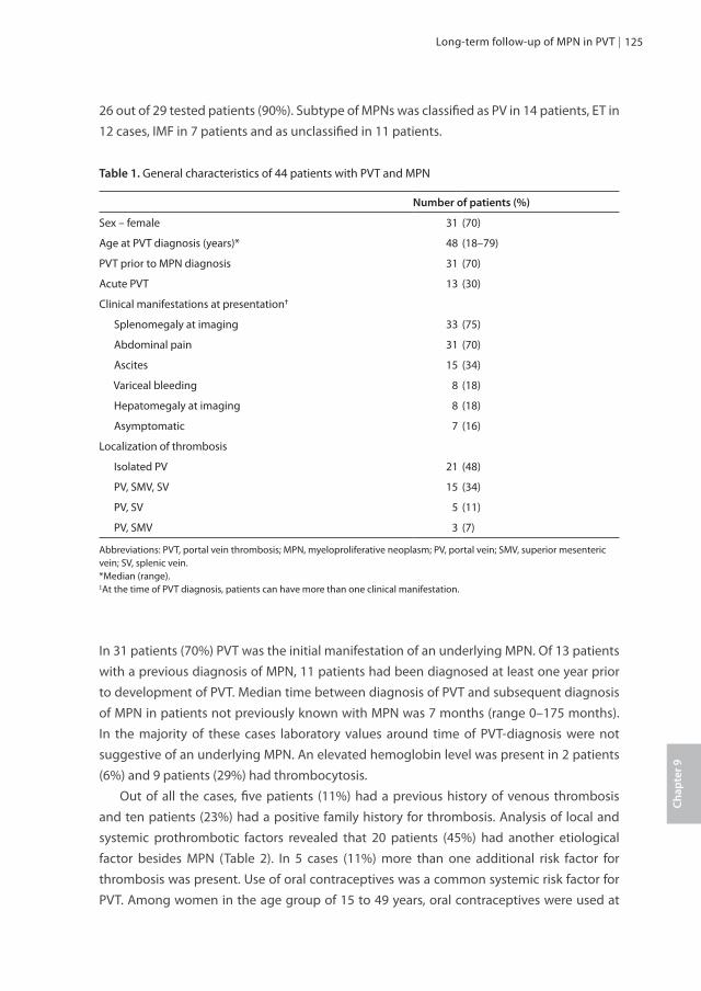
Chap
ter 9
125Long-term follow-up of MPN in PVT |
26 out of 29 tested patients (90%). Subtype of MPNs was classified as PV in 14 patients, ET in 12 cases, IMF in 7 patients and as unclassified in 11 patients.
Table 1. General characteristics of 44 patients with PVT and MPN
Number of patients (%)
Sex – female 31 (70)
Age at PVT diagnosis (years)* 48 (18–79)
PVT prior to MPN diagnosis 31 (70)
Acute PVT 13 (30)
Clinical manifestations at presentation†
Splenomegaly at imaging 33 (75)
Abdominal pain 31 (70)
Ascites 15 (34)
Variceal bleeding 8 (18)
Hepatomegaly at imaging 8 (18)
Asymptomatic 7 (16)
Localization of thrombosis
Isolated PV 21 (48)
PV, SMV, SV 15 (34)
PV, SV 5 (11)
PV, SMV 3 (7)
Abbreviations: PVT, portal vein thrombosis; MPN, myeloproliferative neoplasm; PV, portal vein; SMV, superior mesenteric vein; SV, splenic vein. *Median (range).†At the time of PVT diagnosis, patients can have more than one clinical manifestation.
In 31 patients (70%) PVT was the initial manifestation of an underlying MPN. Of 13 patients with a previous diagnosis of MPN, 11 patients had been diagnosed at least one year prior to development of PVT. Median time between diagnosis of PVT and subsequent diagnosis of MPN in patients not previously known with MPN was 7 months (range 0–175 months). In the majority of these cases laboratory values around time of PVT-diagnosis were not suggestive of an underlying MPN. An elevated hemoglobin level was present in 2 patients (6%) and 9 patients (29%) had thrombocytosis. Out of all the cases, five patients (11%) had a previous history of venous thrombosis and ten patients (23%) had a positive family history for thrombosis. Analysis of local and systemic prothrombotic factors revealed that 20 patients (45%) had another etiological factor besides MPN (Table 2). In 5 cases (11%) more than one additional risk factor for thrombosis was present. Use of oral contraceptives was a common systemic risk factor for PVT. Among women in the age group of 15 to 49 years, oral contraceptives were used at
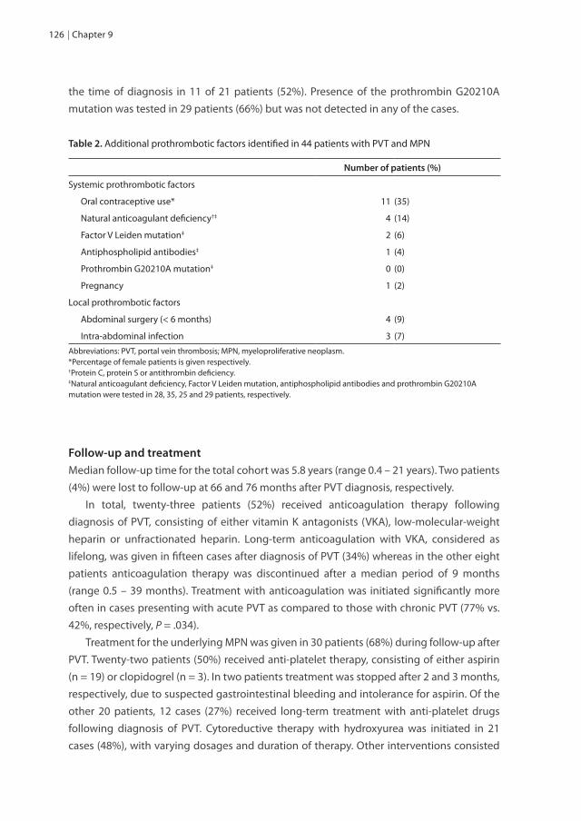
126 | Chapter 9
the time of diagnosis in 11 of 21 patients (52%). Presence of the prothrombin G20210A mutation was tested in 29 patients (66%) but was not detected in any of the cases.
Table 2. Additional prothrombotic factors identified in 44 patients with PVT and MPN
Number of patients (%)
Systemic prothrombotic factors
Oral contraceptive use* 11 (35)
Natural anticoagulant deficiency†‡ 4 (14)
Factor V Leiden mutation‡ 2 (6)
Antiphospholipid antibodies‡ 1 (4)
Prothrombin G20210A mutation‡ 0 (0)
Pregnancy 1 (2)
Local prothrombotic factors
Abdominal surgery (< 6 months) 4 (9)
Intra-abdominal infection 3 (7)
Abbreviations: PVT, portal vein thrombosis; MPN, myeloproliferative neoplasm.*Percentage of female patients is given respectively.†Protein C, protein S or antithrombin deficiency.‡Natural anticoagulant deficiency, Factor V Leiden mutation, antiphospholipid antibodies and prothrombin G20210A mutation were tested in 28, 35, 25 and 29 patients, respectively.
Follow-up and treatmentMedian follow-up time for the total cohort was 5.8 years (range 0.4 – 21 years). Two patients (4%) were lost to follow-up at 66 and 76 months after PVT diagnosis, respectively. In total, twenty-three patients (52%) received anticoagulation therapy following diagnosis of PVT, consisting of either vitamin K antagonists (VKA), low-molecular-weight heparin or unfractionated heparin. Long-term anticoagulation with VKA, considered as lifelong, was given in fifteen cases after diagnosis of PVT (34%) whereas in the other eight patients anticoagulation therapy was discontinued after a median period of 9 months (range 0.5 – 39 months). Treatment with anticoagulation was initiated significantly more often in cases presenting with acute PVT as compared to those with chronic PVT (77% vs. 42%, respectively, P = .034). Treatment for the underlying MPN was given in 30 patients (68%) during follow-up after PVT. Twenty-two patients (50%) received anti-platelet therapy, consisting of either aspirin (n = 19) or clopidogrel (n = 3). In two patients treatment was stopped after 2 and 3 months, respectively, due to suspected gastrointestinal bleeding and intolerance for aspirin. Of the other 20 patients, 12 cases (27%) received long-term treatment with anti-platelet drugs following diagnosis of PVT. Cytoreductive therapy with hydroxyurea was initiated in 21 cases (48%), with varying dosages and duration of therapy. Other interventions consisted
Chap
ter 9
127Long-term follow-up of MPN in PVT |
of regular phlebotomies (n = 10), treatment with alpha-interferon (n = 8), busulfan (n = 3) or anagrelide (n = 1). Of the thirteen patients diagnosed with MPN prior to PVT, seven cases were being treated for MPN at the time of PVT. One patient was on hydroxyurea therapy, two patients were treated with aspirin, two patients received a combination of aspirin and hydroxyurea and in another two patients regular phlebotomies were performed. Levels of hemoglobin and platelets in these patients were all within the normal range around time of PVT.
ComplicationsThe most common complications that occurred during follow-up are summarized in Table 3. In twelve patients (27%) at least one other thrombotic event occurred in addition to PVT with a total of 14 thrombotic events. Most frequent localizations were extension of thrombosis into the splenic and/or superior mesenteric vein (n = 3) and ischemic stroke (n = 3). Recurrent thrombosis occurred in 3 of 7 patients (43%) carrying additional thrombophilic factors as compared to 9 of 37 (24%) patients without thrombophilia (P = .37). Three patients experienced a new episode of thrombosis despite treatment with VKA at that time. Comparing cases with and without long-term VKA therapy, there was no significant difference in the frequency of additional thrombotic events (20% vs. 31%, P = .50). A new venous thrombosis occurred in 1 of 15 cases (7%) on long-term VKA as compared to 7 of 29 cases (24%) not receiving anticoagulation (P = .23). None of the patients that developed recurrent thrombosis were being treated with anti-platelet drugs when thrombosis developed. A schematic overview of the relationship between long-term use of VKA or antiplatelet therapy and the occurrence of new thrombotic events is given in Figure 1. After recurrent thrombosis, another 7 patients were started on lifelong treatment with either VKA (n = 3) or aspirin (n = 4). Around the time of PVT diagnosis an upper gastro-intestinal endoscopy was performed in 38 patients, of whom 30 patients (79%) had esophagogastric varices. Grade III-IV esophageal varices according to Pacquet were found in 18 patients (47%). Overall, 32 documented episodes of variceal bleeding occurred in 17 patients (39%), most frequently resulting from esophageal varices. Endoscopic treatment was the first-line therapy during active bleeding episodes. Variceal bleeding was the presenting symptom of PVT in eight patients (18%) and recurrent bleeding occurred in four of these patients. Of 36 patients that had no signs of bleeding at presentation, 9 patients (25%) experienced at least one bleeding event during follow-up and five cases had recurrent bleeding. Long-term treatment with anticoagulation did not appear to influence the risk of variceal bleeding. Of 36 patients without variceal bleeding at diagnosis, 16 cases were given long-term anticoagulation therapy. Subsequent bleeding episodes during follow-up occurred in two patients on anticoagulation (13%) and in seven patients not treated with long-term anticoagulation (35%, P = .25).
128 | Chapter 9
Table 3. Complications during follow-up of 44 patients with PVT and MPN
Number of patients (%)
Episode of gastrointestinal bleeding 17 (39)
Events per person* 1 (1–5)
Time between PVT and first bleeding event (yrs)†* 2.3 (0.5–5.1)
Additional thrombotic event‡ 12 (27)
Venous thrombosis 9 (20)
Extension of thrombosis to SMV or SV 3
Jugular and/or axillary vein thrombosis 3
Pulmonary embolism 1
Femoral vein thrombosis 1
Sinus sagittalis thrombosis 1
Arterial thrombosis§ 5 (11)
Ischemic CVA 3
Arterial thrombosis of lower extremity 1
Intestinal infarction due to occlusion of celiac artery 1
Time between PVT and subsequent thrombosis (yrs) 7.5 (0–18)
Progression of MPN 12 (27)
Secondary myelofibrosis 7 (16)
Acute myeloid leukemia§ 4 (9)
End-stage myelofibrosis¶ 5 (11)
Time between MPN diagnosis and disease progression (yrs) 9.7 (1–17)
Abbreviations: PVT, portal vein thrombosis; MPN, myeloproliferative neoplasm; CVA, cerebral vascular accident. *Median (range).†Only considered in patients that did not present with an episode of bleeding at diagnosis (n = 9).‡One patiënt experienced two additional episodes of venous thrombosis, one patient experienced two arterial thrombotic events. §Two patients progressed to AML after developing secondary myelofibrosis.¶Two patients developed end-stage myelofibrosis after first progressing to secondary myelofibrosis.
Progression of the underlying MPN occurred in 12 patients (27%) after a median period of 9.7 years (range 1 – 17) following MPN diagnosis. Five patients with PV and two patients with ET developed secondary myelofibrosis, of which two cases subsequently progressed to acute myeloid leukemia (AML). Additionally, one patient with PV and one patient with IMF also developed AML, for a total of four patients with AML. Another three patients with IMF progressed to end-stage myelofibrosis. One patient with secondary myelofibrosis underwent successful allogenic stem cell transplantation and in one patient AML was successfully treated by intensive chemotherapy.
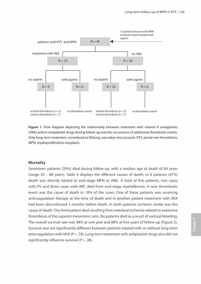
Chap
ter 9
129Long-term follow-up of MPN in PVT |
N = 44
N = 29N = 15
N = 9 N = 6 N = 23 N = 6
patients with PVT and MPN
treatment with VKA no VKA
no aspirin with aspirin no aspirin with aspirin
13 patients known with MPN of whom 4 were treated with aspirin
arterial thrombosis (n = 2)venous thrombosis (n = 1)
no thrombotic events arterial thrombosis (n = 2)venous thrombosis (n = 7)
no thrombotic events
Figure 1. Flow diagram depicting the relationship between treatment with vitamin K antagonists (VKA) and/or antiplatelet drugs during follow-up and the occurrence of additional thrombotic events. Only long-term treatment, considered as lifelong, was taken into account. PVT, portal vein thrombosis; MPN, myeloproliferative neoplasm.
mortalitySeventeen patients (39%) died during follow-up, with a median age at death of 64 years (range 30 – 88 years). Table 4 displays the different causes of death. In 8 patients (47%) death was directly related to end-stage MPN or AML. A total of five patients, two cases with PV and three cases with IMF, died from end-stage myelofibrosis. A new thrombotic event was the cause of death in 18% of the cases. One of these patients was receiving anticoagulation therapy at the time of death and in another patient treatment with VKA had been discontinued 3 months before death. In both patients ischemic stroke was the cause of death. The third patient died resulting from intestinal ischemia related to extensive thrombosis of the superior mesenteric vein. No patients died as a result of variceal bleeding. The overall survival rate was 98% at one year and 88% at five years of follow-up (Figure 2). Survival was not significantly different between patients treated with or without long-term anticoagulation with VKA (P = .78). Long-term treatment with antiplatelet drugs also did not significantly influence survival (P = .38).
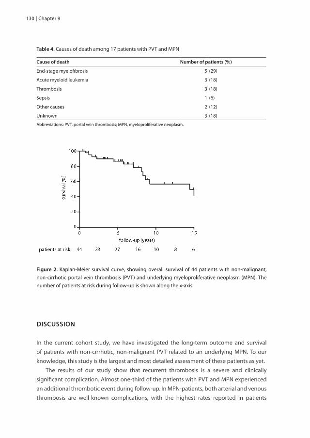
130 | Chapter 9
Table 4. Causes of death among 17 patients with PVT and MPN
Cause of death Number of patients (%)
End-stage myelofibrosis 5 (29)
Acute myeloid leukemia 3 (18)
Thrombosis 3 (18)
Sepsis 1 (6)
Other causes 2 (12)
Unknown 3 (18)
Abbreviations: PVT, portal vein thrombosis; MPN, myeloproliferative neoplasm.
Figure 2. Kaplan-Meier survival curve, showing overall survival of 44 patients with non-malignant, non-cirrhotic portal vein thrombosis (PVT) and underlying myeloproliferative neoplasm (MPN). The number of patients at risk during follow-up is shown along the x-axis.
DiSCUSSioN
In the current cohort study, we have investigated the long-term outcome and survival of patients with non-cirrhotic, non-malignant PVT related to an underlying MPN. To our knowledge, this study is the largest and most detailed assessment of these patients as yet. The results of our study show that recurrent thrombosis is a severe and clinically significant complication. Almost one-third of the patients with PVT and MPN experienced an additional thrombotic event during follow-up. In MPN-patients, both arterial and venous thrombosis are well-known complications, with the highest rates reported in patients
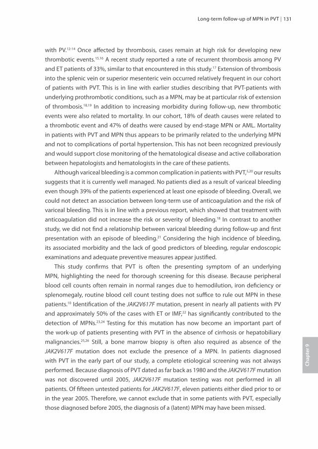
Chap
ter 9
131Long-term follow-up of MPN in PVT |
with PV.12-14 Once affected by thrombosis, cases remain at high risk for developing new thrombotic events.15,16 A recent study reported a rate of recurrent thrombosis among PV and ET patients of 33%, similar to that encountered in this study.17 Extension of thrombosis into the splenic vein or superior mesenteric vein occurred relatively frequent in our cohort of patients with PVT. This is in line with earlier studies describing that PVT-patients with underlying prothrombotic conditions, such as a MPN, may be at particular risk of extension of thrombosis.18,19 In addition to increasing morbidity during follow-up, new thrombotic events were also related to mortality. In our cohort, 18% of death causes were related to a thrombotic event and 47% of deaths were caused by end-stage MPN or AML. Mortality in patients with PVT and MPN thus appears to be primarily related to the underlying MPN and not to complications of portal hypertension. This has not been recognized previously and would support close monitoring of the hematological disease and active collaboration between hepatologists and hematologists in the care of these patients. Although variceal bleeding is a common complication in patients with PVT,5,20 our results suggests that it is currently well managed. No patients died as a result of variceal bleeding even though 39% of the patients experienced at least one episode of bleeding. Overall, we could not detect an association between long-term use of anticoagulation and the risk of variceal bleeding. This is in line with a previous report, which showed that treatment with anticoagulation did not increase the risk or severity of bleeding.18 In contrast to another study, we did not find a relationship between variceal bleeding during follow-up and first presentation with an episode of bleeding.21 Considering the high incidence of bleeding, its associated morbidity and the lack of good predictors of bleeding, regular endoscopic examinations and adequate preventive measures appear justified. This study confirms that PVT is often the presenting symptom of an underlying MPN, highlighting the need for thorough screening for this disease. Because peripheral blood cell counts often remain in normal ranges due to hemodilution, iron deficiency or splenomegaly, routine blood cell count testing does not suffice to rule out MPN in these patients.10 Identification of the JAK2V617F mutation, present in nearly all patients with PV and approximately 50% of the cases with ET or IMF,22 has significantly contributed to the detection of MPNs.23,24 Testing for this mutation has now become an important part of the work-up of patients presenting with PVT in the absence of cirrhosis or hepatobiliary malignancies.25,26 Still, a bone marrow biopsy is often also required as absence of the JAK2V617F mutation does not exclude the presence of a MPN. In patients diagnosed with PVT in the early part of our study, a complete etiological screening was not always performed. Because diagnosis of PVT dated as far back as 1980 and the JAK2V617F mutation was not discovered until 2005, JAK2V617F mutation testing was not performed in all patients. Of fifteen untested patients for JAK2V617F, eleven patients either died prior to or in the year 2005. Therefore, we cannot exclude that in some patients with PVT, especially those diagnosed before 2005, the diagnosis of a (latent) MPN may have been missed.
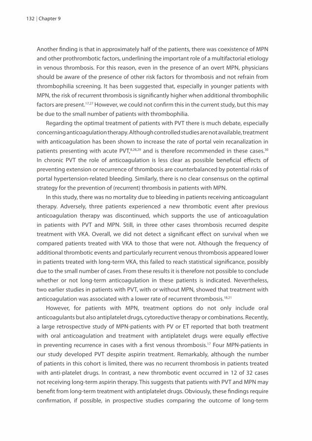
132 | Chapter 9
Another finding is that in approximately half of the patients, there was coexistence of MPN and other prothrombotic factors, underlining the important role of a multifactorial etiology in venous thrombosis. For this reason, even in the presence of an overt MPN, physicians should be aware of the presence of other risk factors for thrombosis and not refrain from thrombophilia screening. It has been suggested that, especially in younger patients with MPN, the risk of recurrent thrombosis is significantly higher when additional thrombophilic factors are present.17,27 However, we could not confirm this in the current study, but this may be due to the small number of patients with thrombophilia. Regarding the optimal treatment of patients with PVT there is much debate, especially concerning anticoagulation therapy. Although controlled studies are not available, treatment with anticoagulation has been shown to increase the rate of portal vein recanalization in patients presenting with acute PVT,8,28,29 and is therefore recommended in these cases.30 In chronic PVT the role of anticoagulation is less clear as possible beneficial effects of preventing extension or recurrence of thrombosis are counterbalanced by potential risks of portal hypertension-related bleeding. Similarly, there is no clear consensus on the optimal strategy for the prevention of (recurrent) thrombosis in patients with MPN. In this study, there was no mortality due to bleeding in patients receiving anticoagulant therapy. Adversely, three patients experienced a new thrombotic event after previous anticoagulation therapy was discontinued, which supports the use of anticoagulation in patients with PVT and MPN. Still, in three other cases thrombosis recurred despite treatment with VKA. Overall, we did not detect a significant effect on survival when we compared patients treated with VKA to those that were not. Although the frequency of additional thrombotic events and particularly recurrent venous thrombosis appeared lower in patients treated with long-term VKA, this failed to reach statistical significance, possibly due to the small number of cases. From these results it is therefore not possible to conclude whether or not long-term anticoagulation in these patients is indicated. Nevertheless, two earlier studies in patients with PVT, with or without MPN, showed that treatment with anticoagulation was associated with a lower rate of recurrent thrombosis.18,21
However, for patients with MPN, treatment options do not only include oral anticoagulants but also antiplatelet drugs, cytoreductive therapy or combinations. Recently, a large retrospective study of MPN-patients with PV or ET reported that both treatment with oral anticoagulation and treatment with antiplatelet drugs were equally effective in preventing recurrence in cases with a first venous thrombosis.17 Four MPN-patients in our study developed PVT despite aspirin treatment. Remarkably, although the number of patients in this cohort is limited, there was no recurrent thrombosis in patients treated with anti-platelet drugs. In contrast, a new thrombotic event occurred in 12 of 32 cases not receiving long-term aspirin therapy. This suggests that patients with PVT and MPN may benefit from long-term treatment with antiplatelet drugs. Obviously, these findings require confirmation, if possible, in prospective studies comparing the outcome of long-term
Chap
ter 9
133Long-term follow-up of MPN in PVT |
treatment with VKA to short-term anticoagulation followed by treatment with antiplatelet agents, both in terms of bleeding risk and prevention of recurrent thrombosis. Until general treatment recommendations become available, awareness of the high rate of thrombosis recurrence in PVT-patients with MPN appears crucial and treatment for the underlying MPN should probably be actively pursued. Although our study provides several important insights, it has some limitations that deserve comment. First, although this is the largest cohort of patients with PVT and MPN described until now, the sample size is still relatively small and data was obtained retrospectively. Another potential limitation of this study is that the observation period ranged from 1980 to 2009. Over this time period, the clinical management of patients with PVT and MPN has changed. For example, the awareness of screening for the presence of a MPN in patients with PVT has increased over the past years, especially since the discovery of the JAK2V617F mutation. Moreover, treatment for MPN has also changed over time. Aspirin has become a standard of treatment after landmark trials showed a reduction of thrombotic events in patients treated with aspirin.31 Nevertheless, we did not detect a significant difference in survival when we compared patients diagnosed before 2000 to those diagnosed thereafter (data not shown). Despite these limitations, we believe that our findings contribute to the further understanding of this rare combination of disorders, especially since large, controlled studies are lacking and hardly feasible. In conclusion, this study shows that PVT is often the presenting symptom of an underlying MPN, underlining that the presence of a MPN should always be thoroughly investigated in these patients. In addition to a high incidence of portal hypertension-related variceal bleeding, recurrent thrombosis is also a common and severe complication in patients with PVT and MPN. Treatment with antiplatelet drugs may prove to be important in the prevention of additional thrombotic events, but this requires further study. Mortality in patients with non-cirrhotic PVT and concomitant MPN is primarily related to the underlying MPN and not to complications of portal hypertension.
134 | Chapter 9
rEFErENCES
1. Primignani M. Portal vein thrombosis, revisited. Dig Liver Dis 2010;42:163-70.
2. Cohen J, Edelman RR, Chopra S. Portal vein thrombosis: a review. Am J Med 1992;92:173-82.
3. Ogren M, Bergqvist D, Bjorck M, Acosta S, Eriksson H, Sternby NH. Portal vein thrombosis: prevalence, patient characteristics and lifetime risk: a population study based on 23,796 consecutive autopsies. World J Gastroenterol 2006;12:2115-9.
4. Janssen HL, Wijnhoud A, Haagsma EB, et al. Extrahepatic portal vein thrombosis: aetiology and determinants of survival. Gut 2001;49:720-4.
5. Sogaard KK, Astrup LB, Vilstrup H, Gronbaek H. Portal vein thrombosis; risk factors, clinical presentation and treatment. BMC Gastroenterol 2007;7:34.
6. Valla D, Casadevall N, Huisse MG, et al. Etiology of portal vein thrombosis in adults. A prospective evaluation of primary myeloproliferative disorders. Gastroenterology 1988;94:1063-9.
7. Vardiman JW, Thiele J, Arber DA, et al. The 2008 revision of the World Health Organization (WHO) classification of myeloid neoplasms and acute leukemia: rationale and important changes. Blood 2009;114:937-51.
8. Plessier A, Darwish-Murad S, Hernandez-Guerra M, et al. Acute portal vein thrombosis unrelated to cirrhosis: a prospective multicenter follow-up study. Hepatology 2010;51:210-8.
9. Primignani M, Martinelli I, Bucciarelli P, et al. Risk factors for thrombophilia in extrahepatic portal vein obstruction. Hepatology 2005;41:603-8.
10. McNamara C, Juneja S, Wolf M, Grigg A. Portal or hepatic vein thrombosis as the first presentation of a myeloproliferative disorder in patients with normal peripheral blood counts. Clin Lab Haematol 2002;24:239-42.
11. Tefferi A, Vardiman JW. Classification and diagnosis of myeloproliferative neoplasms: the 2008 World Health Organization criteria and point-of-care diagnostic algorithms. Leukemia 2008;22:14-22.
12. Polycythemia vera: the natural history of 1213 patients followed for 20 years. Gruppo Italiano Studio Policitemia. Ann Intern Med 1995;123:656-64.
13. Barbui T, Carobbio A, Cervantes F, et al. Thrombosis in primary myelofibrosis: incidence and risk factors. Blood 2010;115:778-82.
14. Jensen MK, de Nully Brown P, Nielsen OJ, Hasselbalch HC. Incidence, clinical features and outcome of essential thrombocythaemia in a well defined geographical area. Eur J Haematol 2000;65:132-9.
15. Cortelazzo S, Viero P, Finazzi G, D’Emilio A, Rodeghiero F, Barbui T. Incidence and risk factors for thrombotic complications in a historical cohort of 100 patients with essential thrombocythemia. J Clin Oncol 1990;8:556-62.
16. Marchioli R, Finazzi G, Landolfi R, et al. Vascular and neoplastic risk in a large cohort of patients with polycythemia vera. J Clin Oncol 2005;23:2224-32.
17. De Stefano V, Za T, Rossi E, et al. Recurrent thrombosis in patients with polycythemia vera and essential thrombocythemia: incidence, risk factors, and effect of treatments. Haematologica 2008;93:372-80.
18. Condat B, Pessione F, Hillaire S, et al. Current outcome of portal vein thrombosis in adults: risk and benefit of anticoagulant therapy. Gastroenterology 2001;120:490-7.
19. Stringer MD, Heaton ND, Karani J, Olliff S, Howard ER. Patterns of portal vein occlusion and their aetiological significance. Br J Surg 1994;81:1328-31.
20. Merkel C, Bolognesi M, Bellon S, et al. Long-term follow-up study of adult patients with non-cirrhotic obstruction of the portal system: comparison with cirrhotic patients. J Hepatol 1992;15:299-303.
21. Amitrano L, Guardascione MA, Scaglione M, et al. Prognostic factors in noncirrhotic patients with splanchnic vein thromboses. Am J Gastroenterol 2007;102:2464-70.
22. Kralovics R, Passamonti F, Buser AS, et al. A gain-of-function mutation of JAK2 in myeloproliferative disorders. N Engl J Med 2005;352:1779-90.
Chap
ter 9
135Long-term follow-up of MPN in PVT |
23. Colaizzo D, Amitrano L, Tiscia GL, et al. The JAK2 V617F mutation frequently occurs in patients with portal and mesenteric venous thrombosis. J Thromb Haemost 2007;5:55-61.
24. Primignani M, Barosi G, Bergamaschi G, et al. Role of the JAK2 mutation in the diagnosis of chronic myeloproliferative disorders in splanchnic vein thrombosis. Hepatology 2006;44:1528-34.
25. Janssen HL, Leebeek FW. JAK2 mutation: The best diagnostic tool for myeloproliferative disease in splanchnic vein thrombosis? Hepatology 2006;44:1391-3.
26. Kiladjian JJ, Cervantes F, Leebeek FW, et al. The impact of JAK2 and MPL mutations on diagnosis and prognosis of splanchnic vein thrombosis: a report on 241 cases. Blood 2008;111:4922-9.
27. Ruggeri M, Gisslinger H, Tosetto A, et al. Factor V Leiden mutation carriership and venous thromboembolism in polycythemia vera and essential thrombocythemia. Am J Hematol 2002;71:1-6.
28. Condat B, Pessione F, Helene Denninger M, Hillaire S, Valla D. Recent portal or mesenteric venous thrombosis: increased recognition and frequent recanalization on anticoagulant therapy. Hepatology 2000;32:466-70.
29. Turnes J, Garcia-Pagan JC, Gonzalez M, et al. Portal hypertension-related complications after acute portal vein thrombosis: impact of early anticoagulation. Clin Gastroenterol Hepatol 2008;6:1412-7.
30. de Franchis R. Evolving consensus in portal hypertension. Report of the Baveno IV consensus workshop on methodology of diagnosis and therapy in portal hypertension. J Hepatol 2005;43:167-76.
31. Landolfi R, Marchioli R, Kutti J, et al. Efficacy and safety of low-dose aspirin in polycythemia vera. N Engl J Med 2004;350:114-24.

C H A P T E r 10
GENErAL DiSCUSSioN
138 | Chapter 10
The objective of this thesis was to investigate the etiology and management of splanchnic vein thrombosis (SVT), in particular primary Budd-Chiari syndrome (BCS) and non-cirrhotic, non-malignant portal vein thrombosis (PVT). We focussed on myeloproliferative neoplasms (MPNs), which are the most frequent underlying risk factors for both disorders. Several studies were initiated to provide further insights into the role of MPNs in the etiology of BCS and PVT, the challenges concerning the diagnosis of MPNs in this specific patient population and the optimal treatment strategy in SVT patients with an underlying MPN. In addition, we set out to identify potential new etiological factors and evaluated a controversial treatment option, i.e. thrombolysis, for patients with extended thrombosis of the splanchnic veins. Several clinical studies are based on data from the European Network for Vascular Disorders of the Liver (EN-Vie). Newly diagnosed patients with primary BCS or non-malignant, non-cirrhotic PVT were consecutively enrolled from nine European countries and general data on etiology, clinical presentation and follow-up was collected, which has resulted in two unique patient cohorts. Furthermore, several single-center cohort studies from our tertiary referral center were performed.
SiTE-SPECiFiCTY oF THromBoSiS
The haemostatic system ultimately leads to the formation of a blood clot in case of a damaged vessel, but also maintains blood in the fluid state within the circulation. It represents a balance between procoagulant and anticoagulant factors – a shift in this balance towards the anticoagulant side of the spectrum may cause bleeding, whereas a shift towards the procoagulant side may result in thrombosis. The majority of hypercoagulable states are associated with systemic conditions, but nevertheless lead to a local thrombotic event. This implies that certain mechanisms are involved in the site-specificity of thrombosis, i.e. that specific risk factors and/or local interaction underlie the development of thrombosis at different sites.1 A well-known example of the contribution of local factors to the development of venous thrombosis can be observed in deep vein thrombosis (DVT) of the lower extremities. The veins of the lower extremities contain venous valves, which guide the direction of blood flow and prevent reflux. It is well established that these venous valves predispose to DVT through stasis of blood and valve pocket hypoxia.2,3
In contrast to the vasculature of the lower extremities, the splanchnic vasculature does not contain venous valves. This implies that the pathophysiology of thrombosis at splanchnic sites must be related to other factors. We reviewed studies on prothrombotic factors in common venous thromboembolism (VTE) and SVT and showed that, despite a considerable overlap, several interesting differences in underlying prothrombotic factors exist.4 Most notably, there is a remarkable high frequency of MPNs and paroxysmal nocturnal hemoglobinuria (PNH) in SVT. A unique feature of the splanchnic veins is the
Chap
ter 1
0
139General discussion |
exposure of the endothelial cells to gut-derived oral antigens and bacterial components from the gastrointestinal tract. Hepatic sinusoidal endothelial cells are known to display immune tolerance which prevents them from having an undesirable response to these factors, whereas there is no evidence that the endothelial cells of the portal vein are similarly protected.5 It has been hypothesized that these endothelial cells may be chronically activated or express site-specific genes making them particularly susceptible to disease-specific changes of MPNs and PNH, such as an elevated blood viscosity, activated leukocytes and/or platelets, increased microparticles, hemolysis and activated complement.1 In addition, these endothelial cells have been shown to carry the JAK2V617F mutation, suggesting a local involvement in the development of BCS.6 Interestingly, there are also apparent differences in the risk profiles between BCS and PVT. PNH and the Factor V Leiden (FVL) mutation are more strongly associated with the development of BCS than with PVT, whereas the opposite is true for the prothrombin gene variant. In BCS patients, the FVL mutation has even been specifically associated with involvement of thrombosis of the inferior vena cava.7 MPN are the most common cause in both BCS and PVT, but the distribution of the three main subtypes of MPN differed between BCS and PVT in the EN-Vie cohort.8-10 Polycythemia vera (PV) was the most common subtype in BCS, whereas essential thrombocytosis (ET) and myelofibrosis (MF) were more often present in PVT patients. Triggered by these findings we performed a meta-analysis, in which we showed that MPNs are significantly more frequent in BCS than in PVT and that there is indeed a difference in distribution of MPN subtypes, PV being more frequent in BCS.11 The prothrombotic effect of high hematocrit values in PV is well established.12 A high hematocrit has an important impact on blood viscosity and causes a major disturbance to blood flow.13,14 Wautier et al described an increased adhesiveness of red blood cells in PV to human umbilical vein endothelial cells and elegantly showed that adhesion was inversely related to shear stress.15 The increased adhesion of red blood cells was mediated by erythroid Lutheran blood group/basal cell adhesion molecule (Lu/BCAM) and endothelial laminin α-5. Variability in the expression of adhesion molecules along the vascular tree along with differences in flow conditions might contribute to the site-specificity of thrombosis, as suggested by these authors, and might also participate in the higher frequency of PV in BCS. The remarkable differences in risk profile between patients with common VTE and SVT, along with the more subtle differences between BCS and PVT, clearly support the concept of site-specific thrombosis. Studying the interaction between these distinctive prothrombotic disorders and vascular bed-specific factors will likely play an essential role in understanding the pathophysiology of these disorders.
140 | Chapter 10
ETioLoGY
myeloproliferative neoplasmsAs with other types of venous thrombosis, it is increasingly recognized that non-cirrhotic, non-malignant PVT and primary BCS are associated with inherited and acquired prothrombotic conditions.16 Strikingly, MPNs are associated with an unusually high rate of BCS and PVT and are considered to be the most prevalent underlying prothrombotic factor in these disorders. Several studies have assessed the prevalence of MPN in BCS, with a prevalence ranging from 30% to 50%.17-21 In line with these findings, we report a prevalence of MPNs of 33% in a single-center, retrospective cohort study of 40 patients with primary BCS.22 In PVT patients, a prevalence of MPNs between 15% and 30% is reported.10,19,23-25 However, the precise prevalence of MPNs in these disorders is unclear, as peripheral blood cell counts often remain within a normal range, due to portal hypertension and its sequelae (splenomegaly, hemodilution, iron deficiency).26,27 Despite suggestive features of an MPN, fulfilment of usual diagnostic criteria can often be lacking, which is a notorious problem in MPN diagnostics in these patients. The discovery of the JAK2V617F mutation in 2005, found in nearly all patients with PV and in approximately 50% of patients with ET and MF, represented a crucial advance in the diagnostic approach to MPNs.28-31 In 2006, we demonstrated the JAK2V617F mutation in 41% of BCS patients from our local cohort, which facilitated diagnosis of MPN in patients suspect for essential thrombocytosis who failed to meet official MPN criteria. Combined with a previous report by Patel et al.,32 these findings clearly showed that JAK2V617F screening offered a new diagnostic tool to detect occult MPNs in SVT patients. The high frequency of the JAK2V617F mutation in SVT patients was confirmed by other studies, present in 30-45%17,33,34 and 17-35%10,33,35 of BCS and PVT patients, respectively. All these studies clearly demonstrated the added value of JAK2V617F in MPN diagnostics in these patients. Many studies have been published on the role of MPNs in the SVT both before and after the discovery of JAK2V617F. Inherent to the low incidence of SVT, however, these studies often lack statistical power. We therefore performed a meta-analysis, including 1.062 primary BCS and 855 non-malignant, non-cirrhotic PVT patients.11 Pooling of studies enabled us to show a significantly higher prevalence of both overt MPNs (28.5% vs. 19.4%) and JAK2V617F (41.1% vs. 27.7%) in BCS compared to PVT patients. JAK2V617F screening identified MPN without characteristic elevated peripheral blood counts in 17.1% and 15.4% of BCS and PVT patients, respectively. Presence of JAK2V617F was associated with development of overt MPN in 41% and 15% of BCS and PVT patients with these so-called occult MPNs, clearly justifying its inclusion in the routine diagnostic work-up of these patients. In contrast, JAK2 exon 12 and MPL515 mutations were found to be extremely rare in SVT, in agreement with their low frequency in MPNs compared to the JAK2V617F mutation.36-40 Screening for these mutations is therefore considered dispensable in the routine diagnostic approach of SVT patients.
Chap
ter 1
0
141General discussion |
Recently, it was shown by several groups that the JAK2V617F, JAK2 exon12 and MPL mutations are not acquired randomly, but instead preferentially arise on a specific constitutional JAK2 haplotype, designated 46/1.33,41-44 To investigate a potential association between the JAK2 46/1 haplotype and SVT, we performed a case-control study using DNA from 199 BCS and PVT patients from the EN-Vie cohort and corresponding healthy controls. We showed that the JAK2 46/1 haplotype was associated with the development of JAK2V617F positive SVT-related MPN, providing additional evidence that this mutation preferentially occurs within this particular, inherited haplotype.45 We observed an increased frequency of the JAK2 46/1 haplotype in JAK2V617F negative patients with a proven MPN (P = .06). In this specific subgroup of patients, homozygous carriers of the JAK2 46/1 haplotype had an approximate 4.7-fold higher chance of MPNs compared to heterozygous carriers or non-carriers. These findings suggest a potential role for the JAK2 46/1 haplotype in the etiology and diagnosis of SVT-related MPNs. For example, one could consider intensifying screening for MPNs during follow up in homozygous carriers of the JAK2 46/1 haplotype to facilitate timely recognition and treatment of underlying MPNs. However, the association between the JAK2 46/1 haplotype and SVT-related MPNs could not be reproduced in a subsequent study.46 Additional studies are therefore required before definite conclusions on the potential role of JAK2 46/1 in the etiology and diagnostic work-up of SVT patients can be drawn.
Fibrinogen γ’: a new risk factor for splanchnic vein thrombosis?In around 15-25% of SVT patients no underlying condition can be identified,8,10 which emphasizes the need to identify new etiological factors. Fibrinogen γ’, a common variant of the fibrinogen γ chain, contains a unique extension with both antithrombotic, i.e. inhibition of thrombin and platelet aggregation, and prothrombotic properties, i.e. enhancement of factor XIII activity and fibrin cross-linking.47 Decreased levels of fibrinogen γ’ and genetic variation in fibrinogen γ genes (FGG) have been associated with susceptibility to venous thrombosis.48-50 To explore whether these factors also contribute to the development of PVT, we performed a case-control study using plasma samples of PVT patients from the EN-Vie cohort and healthy controls. Our results showed that fibrinogen γ’ and variation in FGG genes can potentially contribute to the development of PVT.51 Fibrinogen γ’ to total fibrinogen levels (γ’ ratio) were clearly lower in PVT patients compared to healthy controls. The risk of PVT was increased up to 5-fold in individuals with a γ’ ratio lower than the 33rd percentile of the distribution in the controls. In addition, a particular γ’ chain gene haplotype termed FGG-H2, which was associated with a decreased γ’ ratio in both patients and healthy controls, was overrepresented in PVT patients (P = .08). Altogether, these findings show that FGG-H2 can potentially contribute to the development of PVT by reducing the γ’ ratio. This study establishes additional evidence that a decreased γ’ ratio is associated with an elevated risk for venous thrombosis, in this case PVT. Additional studies are needed to extend our observations and to determine the clinical relevance of these findings.
142 | Chapter 10
Prompted by these results, we performed a case-control study with BCS patients from the EN-Vie cohort and healthy, age- and gender- matched controls.52 In contrast to PVT patients, liver synthesis function is often affected in BCS patients due to the venous outflow obstruction and subsequent damage of liver parenchymal cells, whereas it is generally preserved in patients with non-cirrhotic, non-malignant PVT.16 This may affect pre-mRNA splicing, as has been reported for other proteins in patients with chronic liver dysfunction,53 and therefore influence fibrinogen γ’ levels. To investigate the influence of liver synthesis function on the γ’ ratio, we therefore also assessed the γ’ ratio in patients with liver cirrhosis. In contrast to our findings in PVT patients, BCS appeared to be associated with an increased γ’ ratio compared to controls. However, we showed that the γ’ ratio is strongly dependent upon liver function and that the observed elevated γ’ ratio in BCS is likely to be caused by liver dysfunction. The γ’ ratio was comparable to controls in the subgroup of BCS patients with a preserved liver synthesis function (Child-Pugh A), nor did we observe an association between risk of thrombosis and FGG haplotypes, both suggesting that the γ’ ratio is not associated with BCS. Hence, our findings argue against a role for fibrinogen γ’ in the etiology of BCS. Overall, our data indicate that the fibrinogen γ’ chain may be involved in the pathogenesis of PVT, but not of BCS. These findings provide additional evidence that clear differences in the risk profile of BCS and PVT patients exist. The different role of fibrinogen γ’ in clot formation may be related to differences in shear stress conditions or vascular bed specific factors. Clearly, several issues regarding the association between fibrinogen γ’ and venous thrombosis deserve further research. Most important, prospective studies are required to assess whether fibrinogen γ’ levels already deviate prior to the thrombotic event. In addition, the functional role of fibrinogen γ’ in clot formation and the potential contribution of vascular bed-specific factors and different shear stress conditions should be further investigated.
multifactorial etiologyThe etiology of primary BCS and PVT must be considered multifactorial, even more so than in patients with common VTE. The recent EN-Vie studies reported a combination of two or more genetic or acquired prothrombotic factors in 46% of BCS and 48% of PVT patients.8,54 Indeed, 38% and 45% of BCS and PVT patients from our local cohorts with an underlying MPN had additional pro-thrombotic factors. Based on these findings, a complete hematological work-up, including inherited and acquired thrombophilia, should be performed in BCS and PVT patients, even if a prothrombotic factor has already been identified. This is especially relevant for the identification of MPNs, which may require additional treatment, such as cytoreductive therapy or addition of aspirin to oral anticoagulant treatment.55
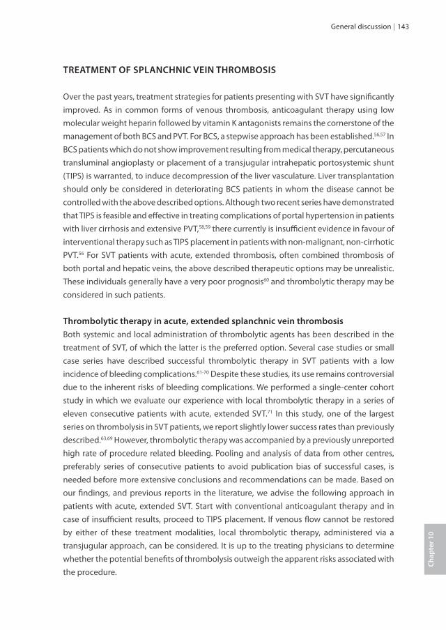
Chap
ter 1
0
143General discussion |
TrEATmENT oF SPLANCHNiC VEiN THromBoSiS
Over the past years, treatment strategies for patients presenting with SVT have significantly improved. As in common forms of venous thrombosis, anticoagulant therapy using low molecular weight heparin followed by vitamin K antagonists remains the cornerstone of the management of both BCS and PVT. For BCS, a stepwise approach has been established.56,57 In BCS patients which do not show improvement resulting from medical therapy, percutaneous transluminal angioplasty or placement of a transjugular intrahepatic portosystemic shunt (TIPS) is warranted, to induce decompression of the liver vasculature. Liver transplantation should only be considered in deteriorating BCS patients in whom the disease cannot be controlled with the above described options. Although two recent series have demonstrated that TIPS is feasible and effective in treating complications of portal hypertension in patients with liver cirrhosis and extensive PVT,58,59 there currently is insufficient evidence in favour of interventional therapy such as TIPS placement in patients with non-malignant, non-cirrhotic PVT.56 For SVT patients with acute, extended thrombosis, often combined thrombosis of both portal and hepatic veins, the above described therapeutic options may be unrealistic. These individuals generally have a very poor prognosis60 and thrombolytic therapy may be considered in such patients.
Thrombolytic therapy in acute, extended splanchnic vein thrombosisBoth systemic and local administration of thrombolytic agents has been described in the treatment of SVT, of which the latter is the preferred option. Several case studies or small case series have described successful thrombolytic therapy in SVT patients with a low incidence of bleeding complications.61-70 Despite these studies, its use remains controversial due to the inherent risks of bleeding complications. We performed a single-center cohort study in which we evaluate our experience with local thrombolytic therapy in a series of eleven consecutive patients with acute, extended SVT.71 In this study, one of the largest series on thrombolysis in SVT patients, we report slightly lower success rates than previously described.63,69 However, thrombolytic therapy was accompanied by a previously unreported high rate of procedure related bleeding. Pooling and analysis of data from other centres, preferably series of consecutive patients to avoid publication bias of successful cases, is needed before more extensive conclusions and recommendations can be made. Based on our findings, and previous reports in the literature, we advise the following approach in patients with acute, extended SVT. Start with conventional anticoagulant therapy and in case of insufficient results, proceed to TIPS placement. If venous flow cannot be restored by either of these treatment modalities, local thrombolytic therapy, administered via a transjugular approach, can be considered. It is up to the treating physicians to determine whether the potential benefits of thrombolysis outweigh the apparent risks associated with the procedure.
144 | Chapter 10
Long-term treatment in patients with myeloproliferative neoplasmsTo study the long-term outcome and optimal management of PVT patients with an underlying MPN, we performed a single-center, retrospective cohort study.55 It is well established that patients with MPN, after their first thrombotic episode, remain at high risk for developing new thrombosis.72-74 Similarly, we showed that recurrent thrombosis is a severe and frequent complication in PVT patients with MPN. Almost one-third of these PVT patients developed an additional thrombotic event during follow-up, whereas 18% of death causes were related to a thrombotic event. In addition, 47% of deaths were caused by end-stage MPN or transformation to acute myeloid leukemia. Mortality in this group of patients thus appears primarily determined by underlying MPNs rather than by complications of portal hypertension. There is still debate on the optimal strategy of anticoagulant treatment in PVT patients, as potential beneficial effects of preventing extension or recurrent thrombosis may be outweighed by the inherent risk of bleeding complications.16,56 Overall, we did not observe a significant effect on survival between patients treated with oral anticoagulants compared to those who were not. In addition to oral anticoagulants, additional treatment options for this specific patient group are available, such as phlebotomy, antiplatelet drugs, cytoreductive therapy or combinations. Aspirin has a well-established role in the prevention of thrombosis in PV patients,75 and although it has not yet prospectively been shown to reduce the incidence of thrombosis in ET, retrospective studies suggested a similar benefit in high-risk patients.76-78 The value of treatment with antiplatelet therapy in PVT patients with an underlying MPN had not yet been investigated. Although the studied population was relatively small, there was no recurrent thrombosis in 12 patients treated with antiplatelet drugs. In contrast, recurrent thrombosis occurred in 12 of 32 patients (38%) not receiving this long-term treatment. Additional studies are needed to extend these observations, preferably of a randomized, prospective design. In these studies, it would be interesting to compare the outcome of treatment of PVT and BCS patients with merely anticoagulants compared to treatment with oral anticoagulants combined with antiplatelet agents, in terms of recurrence of thrombosis and bleeding risk.
FUTUrE rESEArCH
The studies presented in this thesis have focussed on the etiology and treatment of primary BCS and non-malignant, non-cirrhotic PVT. The understanding of the etiology of SVT has considerably increased in recent years. However, there are still promising fields to explore. Immature platelets represent a small percentage of circulating platelets and are hemostatically more active than mature platelets.79 Elevated numbers have been described in PV and ET, both characterized by an increased risk of thrombosis, and other thrombotic
Chap
ter 1
0
145General discussion |
disorders.80,81 Recently, it was demonstrated that the JAK2V617F mutation is associated with an increased number of these immature platelets in patients with PV and ET, which might contribute to the prothrombotic phenotype in these patients.82 It would be interesting to assess the role of immature platelets in the development of SVT, especially in light of the unique role of MPNs in the etiology of SVT. The same applies to microparticles and platelet reactivity, which have also been shown to contribute to the development of thrombosis.83-85 The endogenous thrombin generation potential in SVT patients, which is an overall test of coagulation that has been associated with both an increased risk of first86,87 as well as recurrent88-91 VTE is another promising area for further study. Finally, one of the classic underlying prothrombotic disorders that has received relatively little attention so far is the antiphospholipid syndrome. Large studies confirming and quantifying this relation in SVT using the updated Sapporo Criteria are still lacking. There is still no clear consensus with regard to the anticoagulant treatment of non-malignant, non-cirrhotic PVT. Although shown to prevent recurrent thrombosis, it has been associated with an increased risk of gastrointestinal bleeding and is currently used with relative caution in these patients.56 Nearly all studies on the role of anticoagulant therapy in PVT patients had a retrospective design. Prospective, randomized trials are therefore urgently needed to fully establish the place of oral anticoagulants in the treatment of PVT patients. The introduction of a new class of oral agents, that directly inhibit thrombin or factor Xa and do not require laboratory monitoring, has been an important development in the field of anticoagulation. Several clinical trials have been published that compare these new agents to low molecular weight heparin and vitamin K antagonists in the prevention of stroke in atrial fibrillation and treatment of common VTE.92-96 These studies demonstrated a similar or even favourable efficacy and safety profile compared to treatment with low molecular weight heparin. These findings may ultimately represent a revolution in therapeutic options, not only for patients with common venous thrombosis, but also for SVT patients. Clinical trials comparing vitamin K antagonist with direct inhibitors of thrombin or factor Xa in the treatment of SVT should be actively pursued. Finally, studies are needed that address the optimal treatment strategy of SVT patients with an underlying MPN. The role of platelet inhibitors in the treatment of this specific subgroup of SVT patients clearly deserves further research. The discovery of JAK2V617F has resulted in the development of JAK2 inhibitors. Phase I and II studies show that JAK2 inhibitors are effective at decreasing constitutional symptoms and reducing spleen size in patients with MF.97,98 Data from ongoing phase III trials in MF and PV patients will further refine the therapeutic role of these agents in MPN. The role of JAK2 inhibitors needs first to be established in MPN before one can speculate on a potential role of these agents in the management of SVT patients with an underlying MPN, but results appear promising.
146 | Chapter 10
rEFErENCES
1. Aird WC. Vascular bed-specific thrombosis. J Thromb Haemost 2007;5 Suppl 1:283-91.
2. Hamer JD, Malone PC, Silver IA. The PO2 in venous valve pockets: its possible bearing on thrombogenesis. Br J Surg 1981;68:166-70.
3. Lurie F, Kistner RL, Eklof B, Kessler D. Mechanism of venous valve closure and role of the valve in circulation: a new concept. J Vasc Surg 2003;38:955-61.
4. Smalberg JH, Kruip MJ, Janssen HL, Rijken DC, Leebeek FW, de Maat MP. Hypercoagulability and hypofibrinolysis and risk of deep vein thrombosis and splanchnic vein thrombosis: similarities and differences. Arterioscler Thromb Vasc Biol 2011;31:485-93.
5. Aird WC. Phenotypic heterogeneity of the endothelium: II. Representative vascular beds. Circ Res 2007;100:174-90.
6. Sozer S, Fiel MI, Schiano T, Xu M, Mascarenhas J, Hoffman R. The presence of JAK2V617F mutation in the liver endothelial cells of patients with Budd-Chiari syndrome. Blood 2009;113:5246-9.
7. Deltenre P, Denninger MH, Hillaire S, et al. Factor V Leiden related Budd-Chiari syndrome. Gut 2001;48:264-8.
8. Darwish Murad S, Plessier A, Hernandez-Guerra M, et al. Etiology, management, and outcome of the Budd-Chiari syndrome. Ann Intern Med 2009;151:167-75.
9. Hoekstra J C-CM, Garcia-Pagan JC, Fabris FM, Murad SD, Elias E, Trebicka J, Primignani M, Sejiio S, Valla DC, Leebeek FW, Janssen HL. Etiological factors underlying Budd-Chiari syndrome and portal vein thrombosis. Hepatology 2010 Sept; 52(4): 896A-896A S1.
10. Plessier A, Darwish-Murad S, Hernandez-Guerra M, et al. Acute portal vein thrombosis unrelated to cirrhosis: a prospective multicenter follow-up study. Hepatology 2010;51:210-8.
11. Smalberg JH SJ, Arends L, Valla DC, Kiladjian JJ, Janssen HL, Leebeek FW. et al. Myeloproliferative neoplasms in Budd-Chiari syndrome and portal vein thrombosis: a meta-analysis. Submitted.
12. Pearson TC, Wetherley-Mein G. Vascular occlusive episodes and venous haematocrit in primary proliferative polycythaemia. Lancet 1978;2:1219-22.
13. Landolfi R, Di Gennaro L, Falanga A. Thrombosis in myeloproliferative disorders: pathogenetic facts and speculation. Leukemia 2008;22:2020-8.
14. Landolfi R, Rocca B, Patrono C. Bleeding and thrombosis in myeloproliferative disorders: mechanisms and treatment. Crit Rev Oncol Hematol 1995;20:203-22.
15. Wautier MP, El Nemer W, Gane P, et al. Increased adhesion to endothelial cells of erythrocytes from patients with polycythemia vera is mediated by laminin alpha5 chain and Lu/BCAM. Blood 2007;110:894-901.
16. DeLeve LD, Valla DC, Garcia-Tsao G, American Association for the Study Liver D. Vascular disorders of the liver. Hepatology 2009;49:1729-64.
17. Darwish Murad S, Plessier A, Hernandez-Guerra M, et al. Etiology, management, and outcome of the Budd-Chiari syndrome. Ann Intern Med 2009;151:167-75.
18. Darwish Murad S, Valla DC, de Groen PC, et al. Determinants of survival and the effect of portosystemic shunting in patients with Budd-Chiari syndrome. Hepatology 2004;39:500-8.
19. Denninger MH, Chait Y, Casadevall N, et al. Cause of portal or hepatic venous thrombosis in adults: the role of multiple concurrent factors. Hepatology 2000;31:587-91.
20. Mahmoud AE, Mendoza A, Meshikhes AN, et al. Clinical spectrum, investigations and treatment of Budd-Chiari syndrome. QJM 1996;89:37-43.
21. Rajani R, Melin T, Bjornsson E, et al. Budd-Chiari syndrome in Sweden: epidemiology, clinical characteristics and survival - an 18-year experience. Liver Int 2009;29:253-9.
22. Smalberg JH, Murad SD, Braakman E, Valk PJ, Janssen HL, Leebeek FW. Myeloproliferative disease in the pathogenesis and survival of Budd-Chiari syndrome. Haematologica 2006;91:1712-3.
Chap
ter 1
0
147General discussion |
23. Bayraktar Y, Harmanci O, Buyukasik Y, et al. JAK2V617F mutation in patients with portal vein thrombosis. Dig Dis Sci 2008;53:2778-83.
24. Janssen HL, Wijnhoud A, Haagsma EB, et al. Extrahepatic portal vein thrombosis: aetiology and determinants of survival. Gut 2001;49:720-4.
25. Valla D, Casadevall N, Huisse MG, et al. Etiology of portal vein thrombosis in adults. A prospective evaluation of primary myeloproliferative disorders. Gastroenterology 1988;94:1063-9.
26. Martinelli I, De Stefano V. Rare thromboses of cerebral, splanchnic and upper-extremity veins. A narrative review. Thromb Haemost 2010;103:1136-44.
27. Valla DC. Primary Budd-Chiari syndrome. J Hepatol 2009;50:195-203.
28. Baxter EJ, Scott LM, Campbell PJ, et al. Acquired mutation of the tyrosine kinase JAK2 in human myeloproliferative disorders. Lancet 2005;365:1054-61.
29. Campbell PJ, Green AR. The myeloproliferative disorders. N Engl J Med 2006;355:2452-66.
30. James C, Ugo V, Le Couedic JP, et al. A unique clonal JAK2 mutation leading to constitutive signalling causes polycythaemia vera. Nature 2005;434:1144-8.
31. Kralovics R, Passamonti F, Buser AS, et al. A gain-of-function mutation of JAK2 in myeloproliferative disorders. N Engl J Med 2005;352:1779-90.
32. Patel RK, Lea NC, Heneghan MA, et al. Prevalence of the activating JAK2 tyrosine kinase mutation V617F in the Budd-Chiari syndrome. Gastroenterology 2006;130:2031-8.
33. Kiladjian JJ, Cervantes F, Leebeek FW, et al. The impact of JAK2 and MPL mutations on diagnosis and prognosis of splanchnic vein thrombosis: a report on 241 cases. Blood 2008;111:4922-9.
34. Smalberg JH, Darwish Murad S, Braakman E, Valk PJ, Janssen HL, Leebeek FW. Myeloproliferative disease in the pathogenesis and survival of Budd-Chiari syndrome. Haematologica 2006;91:1712-3.
35. Colaizzo D, Amitrano L, Tiscia GL, et al. The JAK2 V617F mutation frequently occurs in patients with portal and mesenteric venous thrombosis. J Thromb Haemost 2007;5:55-61.
36. Pardanani AD, Levine RL, Lasho T, et al. MPL515 mutations in myeloproliferative and other myeloid disorders: a study of 1182 patients. Blood 2006;108:3472-6.
37. Pikman Y, Lee BH, Mercher T, et al. MPLW515L is a novel somatic activating mutation in myelofibrosis with myeloid metaplasia. PLoS Med 2006;3:e270.
38. Scott LM, Beer PA, Bench AJ, Erber WN, Green AR. Prevalance of JAK2 V617F and exon 12 mutations in polycythaemia vera. Br J Haematol 2007;139:511-2.
39. Scott LM, Tong W, Levine RL, et al. JAK2 exon 12 mutations in polycythemia vera and idiopathic erythrocytosis. N Engl J Med 2007;356:459-68.
40. Tefferi A, Skoda R, Vardiman JW. Myeloproliferative neoplasms: contemporary diagnosis using histology and genetics. Nat Rev Clin Oncol 2009;6:627-37.
41. Jones AV, Campbell PJ, Beer PA, et al. The JAK2 46/1 haplotype predisposes to MPL-mutated myeloproliferative neoplasms. Blood 2010;115:4517-23.
42. Jones AV, Chase A, Silver RT, et al. JAK2 haplotype is a major risk factor for the development of myeloproliferative neoplasms. Nat Genet 2009;41:446-9.
43. Olcaydu D, Harutyunyan A, Jager R, et al. A common JAK2 haplotype confers susceptibility to myeloproliferative neoplasms. Nat Genet 2009;41:450-4.
44. Olcaydu D, Skoda RC, Looser R, et al. The ‘GGCC’ haplotype of JAK2 confers susceptibility to JAK2 exon 12 mutation-positive polycythemia vera. Leukemia 2009;23:1924-6.
45. Smalberg JH, Koehler E, Darwish Murad S, et al. The JAK2 46/1 haplotype in Budd-Chiari syndrome and portal vein thrombosis. Blood 2011;117:3968-73.
46. Kouroupi E, Kiladjian JJ, Chomienne C, et al. The JAK2 46/1 haplotype in splanchnic vein thrombosis. Blood 2011;117:5777-8.
47. Uitte de Willige S, Standeven KF, Philippou H, Ariens RA. The pleiotropic role of the fibrinogen gamma’ chain in hemostasis. Blood 2009;114:3994-4001.
148 | Chapter 10
48. Grunbacher G, Weger W, Marx-Neuhold E, et al. The fibrinogen gamma (FGG) 10034C>T polymorphism is associated with venous thrombosis. Thromb Res 2007;121:33-6.
49. Nowak-Gottl U, Weiler H, Hernandez I, et al. Fibrinogen alpha and gamma genes and factor VLeiden in children with thromboembolism: results from 2 family-based association studies. Blood 2009;114:1947-53.
50. Uitte de Willige S, de Visser MC, Houwing-Duistermaat JJ, Rosendaal FR, Vos HL, Bertina RM. Genetic variation in the fibrinogen gamma gene increases the risk for deep venous thrombosis by reducing plasma fibrinogen gamma’ levels. Blood 2005;106:4176-83.
51. Smalberg JH, Koehler E, Darwish Murad S, et al. Fibrinogen gamma prime and variation in fibrinogen gamma genes in the etiology of portal vein thrombosis. Submitted.
52. Smalberg JH, Koehler E, Darwish Murad S, et al. Fibrinogen gamma prime appears to be unrelated to the development of the Budd-Chiari syndrome. Submitted.
53. Berasain C, Goni S, Castillo J, Latasa MU, Prieto J, Avila MA. Impairment of pre-mRNA splicing in liver disease: mechanisms and consequences. World J Gastroenterol 2010;16:3091-102.
54. Plessier A, Darwish-Murad S, Hernandez-Guerra M, et al. Acute portal vein thrombosis unrelated to cirrhosis: a prospective multicenter follow-up study. Hepatology 2010;51:210-8.
55. Hoekstra J BE, Smalberg JH, Spaander V, Leebeek FW, Janssen HL. Long-term follow-up of patients with portal vein thrombosis and myeloproliferative neoplasms. Accepted for publication in J Thromb Haemost.
56. de Franchis R. Revising consensus in portal hypertension: report of the Baveno V consensus workshop on methodology of diagnosis and therapy in portal hypertension. J Hepatol 2010;53:762-8.
57. Janssen HL, Garcia-Pagan JC, Elias E, Mentha G, Hadengue A, Valla DC. Budd-Chiari syndrome: a review by an expert panel. J Hepatol 2003;38:364-71.
58. Han G, Qi X, He C, et al. Transjugular intrahepatic portosystemic shunt for portal vein thrombosis with symptomatic portal hypertension in liver cirrhosis. J Hepatol 2011;54:78-88.
59. Senzolo M, Tibbals J, Cholongitas E, Triantos CK, Burroughs AK, Patch D. Transjugular intrahepatic portosystemic shunt for portal vein thrombosis with and without cavernous transformation. Aliment Pharmacol Ther 2006;23:767-75.
60. Mahmoud AE, Helmy AS, Billingham L, Elias E. Poor prognosis and limited therapeutic options in patients with Budd-Chiari syndrome and portal venous system thrombosis. Eur J Gastroenterol Hepatol 1997;9:485-9.
61. Guglielmi A, Fior F, Halmos O, et al. Transhepatic fibrinolysis of mesenteric and portal vein thrombosis in a patient with ulcerative colitis: a case report. World J Gastroenterol 2005;11:2035-8.
62. Henao EA, Bohannon WT, Silva MB, Jr. Treatment of portal venous thrombosis with selective superior mesenteric artery infusion of recombinant tissue plasminogen activator. J Vasc Surg 2003;38:1411-5.
63. Hollingshead M, Burke CT, Mauro MA, Weeks SM, Dixon RG, Jaques PF. Transcatheter thrombolytic therapy for acute mesenteric and portal vein thrombosis. J Vasc Interv Radiol 2005;16:651-61.
64. Kercher KW, Sing RF, Watson KW, Matthews BD, LeQuire MH, Heniford BT. Transhepatic thrombolysis in acute portal vein thrombosis after laparoscopic splenectomy. Surg Laparosc Endosc Percutan Tech 2002;12:131-6.
65. Kuo GP, Brodsky RA, Kim HS. Catheter-directed thrombolysis and thrombectomy for the Budd-Chiari syndrome in paroxysmal nocturnal hemoglobinuria in three patients. J Vasc Interv Radiol 2006;17:383-7.
66. Leebeek FW, Lameris JS, van Buuren HR, Gomez E, Madretsma S, Sonneveld P. Budd-Chiari syndrome, portal vein and mesenteric vein thrombosis in a patient homozygous for factor V Leiden mutation treated by TIPS and thrombolysis. Br J Haematol 1998;102:929-31.
67. Nakai M, Sato M, Sahara S, et al. Transhepatic catheter-directed thrombolysis for portal vein thrombosis after partial splenic embolization in combination with balloon-occluded retrograde transvenous obliteration of splenorenal shunt. World J Gastroenterol 2006;12:5071-4.
Chap
ter 1
0
149General discussion |
68. Ozkan U, Oguzkurt L, Tercan F, Tokmak N. Percutaneous transhepatic thrombolysis in the treatment of acute portal venous thrombosis. Diagn Interv Radiol 2006;12:105-7.
69. Sharma S, Texeira A, Texeira P, Elias E, Wilde J, Olliff SP. Pharmacological thrombolysis in Budd Chiari syndrome: a single centre experience and review of the literature. J Hepatol 2004;40:172-80.
70. Sherigar R, Amir KA, Bobba RK, Arsura EL, Srinivas N. Abdominal pain secondary to pylephlebitis: an uncommon disease of the portal venous system, treated with local thrombolytic therapy. Dig Dis Sci 2005;50:983-7.
71. Smalberg JH, Spaander MV, Jie KS, et al. Risks and benefits of transcatheter thrombolytic therapy in patients with splanchnic venous thrombosis. Thromb Haemost 2008;100:1084-8.
72. Besses C, Cervantes F, Pereira A, et al. Major vascular complications in essential thrombocythemia: a study of the predictive factors in a series of 148 patients. Leukemia 1999;13:150-4.
73. Cortelazzo S, Viero P, Finazzi G, D’Emilio A, Rodeghiero F, Barbui T. Incidence and risk factors for thrombotic complications in a historical cohort of 100 patients with essential thrombocythemia. J Clin Oncol 1990;8:556-62.
74. Marchioli R, Finazzi G, Landolfi R, et al. Vascular and neoplastic risk in a large cohort of patients with polycythemia vera. J Clin Oncol 2005;23:2224-32.
75. Landolfi R, Marchioli R, Kutti J, et al. Efficacy and safety of low-dose aspirin in polycythemia vera. N Engl J Med 2004;350:114-24.
76. Jensen MK, de Nully Brown P, Nielsen OJ, Hasselbalch HC. Incidence, clinical features and outcome of essential thrombocythaemia in a well defined geographical area. Eur J Haematol 2000;65:132-9.
77. Randi ML, Rossi C, Fabris F, Menapace L, Girolami A. Aspirin seems as effective as myelosuppressive agents in the prevention of rethrombosis in essential thrombocythemia. Clin Appl Thromb Hemost 1999;5:131-5.
78. van Genderen PJ, Mulder PG, Waleboer M, van de Moesdijk D, Michiels JJ. Prevention and treatment of thrombotic complications in essential thrombocythaemia: efficacy and safety of aspirin. Br J Haematol 1997;97:179-84.
79. Harrison P, Robinson MS, Mackie IJ, Machin SJ. Reticulated platelets. Platelets 1997;8:379-83.
80. Grove EL, Hvas AM, Kristensen SD. Immature platelets in patients with acute coronary syndromes. Thromb Haemost 2009;101:151-6.
81. Ryningen A, Apelseth T, Hausken T, Bruserud O. Reticulated platelets are increased in chronic myeloproliferative disorders, pure erythrocytosis, reactive thrombocytosis and prior to hematopoietic reconstitution after intensive chemotherapy. Platelets 2006;17:296-302.
82. Panova-Noeva M, Marchetti M, Buoro S, et al. JAK2V617F mutation and hydroxyurea treatment as determinants of immature platelet parameters in essential thrombocythemia and polycythemia vera patients. Blood 2011;118:2599-601.
83. Owens AP, 3rd, Mackman N. Microparticles in hemostasis and thrombosis. Circ Res 2011;108:1284-97.
84. Snoep JD, Roest M, Barendrecht AD, De Groot PG, Rosendaal FR, Van Der Bom JG. High platelet reactivity is associated with myocardial infarction in premenopausal women: a population-based case-control study. J Thromb Haemost 2010;8:906-13.
85. Trappenburg MC, van Schilfgaarde M, Marchetti M, et al. Elevated procoagulant microparticles expressing endothelial and platelet markers in essential thrombocythemia. Haematologica 2009;94:911-8.
86. Lutsey PL, Folsom AR, Heckbert SR, Cushman M. Peak thrombin generation and subsequent venous thromboembolism: the Longitudinal Investigation of Thromboembolism Etiology (LITE) study. J Thromb Haemost 2009;7:1639-48.
87. van Hylckama Vlieg A, Christiansen SC, Luddington R, Cannegieter SC, Rosendaal FR, Baglin TP. Elevated endogenous thrombin potential is associated with an increased risk of a first deep venous thrombosis but not with the risk of recurrence. Br J Haematol 2007;138:769-74.
150 | Chapter 10
88. Besser M, Baglin C, Luddington R, van Hylckama Vlieg A, Baglin T. High rate of unprovoked recurrent venous thrombosis is associated with high thrombin-generating potential in a prospective cohort study. J Thromb Haemost 2008;6:1720-5.
89. Eichinger S, Hron G, Kollars M, Kyrle PA. Prediction of recurrent venous thromboembolism by endogenous thrombin potential and D-dimer. Clin Chem 2008;54:2042-8.
90. Hron G, Kollars M, Binder BR, Eichinger S, Kyrle PA. Identification of patients at low risk for recurrent venous thromboembolism by measuring thrombin generation. JAMA 2006;296:397-402.
91. Tripodi A, Legnani C, Chantarangkul V, Cosmi B, Palareti G, Mannucci PM. High thrombin generation measured in the presence of thrombomodulin is associated with an increased risk of recurrent venous thromboembolism. J Thromb Haemost 2008;6:1327-33.
92. Connolly SJ, Ezekowitz MD, Yusuf S, et al. Dabigatran versus warfarin in patients with atrial fibrillation. N Engl J Med 2009;361:1139-51.
93. Granger CB, Alexander JH, McMurray JJ, et al. Apixaban versus warfarin in patients with atrial fibrillation. N Engl J Med 2011;365:981-92.
94. Landman GW, Gans RO. Oral rivaroxaban for symptomatic venous thromboembolism. N Engl J Med 2011;364:1178; author reply
95. Patel MR, Mahaffey KW, Garg J, et al. Rivaroxaban versus warfarin in nonvalvular atrial fibrillation. N Engl J Med 2011;365:883-91.
96. Schulman S, Kearon C, Kakkar AK, et al. Dabigatran versus warfarin in the treatment of acute venous thromboembolism. N Engl J Med 2009;361:2342-52.
97. Pardanani A, Gotlib JR, Jamieson C, et al. Safety and efficacy of TG101348, a selective JAK2 inhibitor, in myelofibrosis. J Clin Oncol 2011;29:789-96.
98. Verstovsek S, Kantarjian H, Mesa RA, et al. Safety and efficacy of INCB018424, a JAK1 and JAK2 inhibitor, in myelofibrosis. N Engl J Med 2010;363:1117-27.
151Summary |
SUmmArY
The term splanchnic vein thrombosis (SVT) is used to indicate both the Budd-Chiari syndrome (BCS) and portal vein thrombosis (PVT), which are rare, but potentially life-threatening, forms of venous thrombosis. In PVT, the extrahepatic portal vein is obstructed, whereas primary BCS is characterized by thrombosis of the hepatic veins and/or the suprahepatic inferior vena cava, resulting in obstruction of the hepatic venous outflow tract. The various chapters of this thesis describe studies that investigated the etiology, clinical outcome and management of primary BCS and non-malignant, non-cirrhotic PVT. Several studies were based on data from the European Network for Vascular Disorders of the Liver (EN-Vie). Newly diagnosed patients with primary BCS or non-malignant, non-cirrhotic PVT were consecutively enrolled from nine European countries and general data on etiology, clinical presentation and follow-up was collected, which has resulted in two unique patient cohorts. Furthermore, several single-center cohort studies from our tertiary referral center were performed. A review of the risk factors for commonly occurring venous thrombosis, in particular deep vein thrombosis and pulmonary embolism, and SVT is presented in chapter 2. The results show that both common venous thromboembolism (VTE) and SVT are associated with the presence of systemic, inherited or acquired prothrombotic conditions. There are many similarities in the risk profiles of patients with common venous thrombosis and SVT. However, there are also apparent differences. In particular, MPNs and paroxysmal nocturnal hemoglobinuria (PNH) have a remarkable high frequency in SVT. Intriguingly, there are even differences in underlying prothrombotic conditions between BCS and PVT patients. PNH and the Factor V Leiden mutation are more strongly associated with BCS than with PVT, whereas the opposite is true for the prothrombin gene variant. In addition, MPNs are more frequent in BCS than PVT. These exceptional differences in risk profiles between patients with common venous thrombosis, and even between BCS and PVT patients, support the concept of site-specific thrombosis and may prove to be a means towards a better understanding of the pathophysiology of these disorders. MPNs are the leading predisposing factor for both BCS and PVT. Despite suggestive features of an MPN, fulfilment of usual criteria is often lacking, which is a notorious problem for diagnosing MPNs in this patient population. In chapter 3 we evaluate 40 BCS patients for MPNs and other pro-thrombotic conditions in a retrospective, single-center cohort study. In addition, we evaluate the added value of the JAK2V617F mutation in MPN diagnostics. MPNs were present in 33% of the patients. In 38% of these patients additional pro-thrombotic factors were present, highlighting the necessity of a complete hematological work-up, even if a prothrombotic factor has already been identified. This is particularly relevant for identifying MPNs, which may require additional treatment, such as cytoreductive therapy or
152 |
addition of aspirin to oral anticoagulant treatment. Screening for JAK2V617F identified MPN patients suspect for essential thrombocythemia, who failed to meet WHO criteria, showing that JAK2V617F screening could offer a new tool to detect occult MPNs in these patients. Survival of BCS patients did not differ significantly between individuals with and without MPNs, although comparison between the groups may have been confounded by extent of thrombosis and a difference in performed liver transplantations, both to the detriment of non-MPN patients. Studies on the role of MPNs in the etiology of SVT have steadily increased over time, but interest on this topic has truly gained momentum since the discovery of JAK2V617F in 2005. Inherent to the low incidence of SVT, however, these studies often lack statistical power. A meta-analysis of these studies is presented in chapter 4, in which the prevalence of MPNs, their subtypes as well as JAK2V617F prevalence and its diagnostic role in SVT was assessed. Pooling of studies enabled us to show a significantly higher prevalence of both overt MPNs (28.5% vs. 19.4%) and JAK2V617F (41.1% vs. 27.7%) in BCS compared to PVT patients. Moreover, a remarkable difference in the distribution of underlying MPN subtype was observed, polycythemia vera being more frequent in BCS. These differences support the theory that the occurrence of thrombosis is not random, but rather the result of the interaction of underlying prothrombotic factors and vascular bed-specific factors. JAK2V617F screening identified MPN without typical elevated peripheral blood counts in 17.1% and 15.4% of BCS and PVT patients, respectively. Presence of JAK2V617F was associated with subsequent development of overt MPNs in 41% and 15% of BCS and PVT patients, respectively, clearly justifying its inclusion in the routine diagnostic work-up of these patients. In contrast, JAK2 exon 12 and MPL515 mutations were found to be extremely rare in SVT, and are therefore dispensable in the routine diagnostic approach of these patients. In 2009, it was demonstrated that JAK2V617F is not acquired randomly, but preferentially arises on a constitutional JAK2 haplotype, designated 46/1. In chapter 5 we perform a case-control study to investigate a potential association between the JAK2 46/1 haplotype and SVT, using DNA samples from BCS and PVT patients from the EN-Vie cohort and corresponding healthy controls. The JAK2 46/1 haplotype was associated with JAK2V617F positive SVT-related MPN, providing additional evidence that this mutation preferentially occurs within this particular haplotype. We also observed an increased frequency of the JAK2 46/1 haplotype in JAK2V617F negative patients with a proven MPN (P = .06). In this specific subgroup of patients, homozygous carriers of the JAK2 46/1 haplotype had an approximate 4.7-fold higher chance of MPNs compared to heterozygous carriers or non-carriers. The JAK2 46/1 haplotype was associated with increased erythropoiesis in JAK2V617F negative SVT patients, which is a new finding and supports the theory that the JAK2 46/1 haplotype might be functionally different from other JAK2 alleles. Altogether, these findings indicate a
153Summary |
potential role for the JAK2 46/1 haplotype in the etiology and diagnosis of SVT-related MPNs that requires further exploration. Fibrinogen γ’ levels and genetic variation in fibrinogen γ genes (FGG) have been associated with susceptibility to venous thrombosis. To assess the potential contribution of these factors to the development of PVT, we performed a case control-study using DNA and plasma samples from PVT patients from the EN-Vie cohort and healthy controls (chapter 6). We show that fibrinogen γ’ and variation in FGG genes can potentially contribute to the development of PVT. Fibrinogen γ’ to total fibrinogen levels (γ’ ratio) were clearly lower in PVT patients compared to healthy controls. The risk of PVT was increased up to 5-fold in individuals with a γ’ ratio lower than the 33rd percentile of the distribution in the controls. In addition, a particular γ’ chain gene haplotype termed FGG-H2, which was associated with a decreased γ’ ratio in both patients and healthy controls, was overrepresented in PVT patients (P = .08). Altogether, these findings indicate that FGG-H2 can potentially contribute to the development of PVT by reducing the γ’ ratio. In chapter 7, we assess the role of fibrinogen γ’ and FGG haplotypes in BCS using DNA and plasma samples from BCS patients from the EN-Vie cohort and healthy, age- and gender- matched controls. In contrast to PVT patients, liver synthesis function is often affected in patients presenting with BCS, whereas it is generally preserved in patients with non-cirrhotic, non-malignant PVT. Importantly, alterations in pre-mRNA splicing have been reported for other proteins in patients with chronic liver dysfunction. To investigate the influence of liver synthesis function on the γ’ ratio, we therefore also assessed the γ’ ratio in patients with liver cirrhosis. In contrast to our findings in PVT patients, BCS appeared to be associated with an increased γ’ ratio compared to controls. However, we showed that the γ’ ratio is strongly dependent upon liver function. The mean γ’ ratio was equally elevated in BCS and liver cirrhosis patients. In addition, the γ’ ratio was similarly increased with increasing liver dysfunction in cirrhotics and BCS patients, as represented by the Child-Pugh classification. The observed elevated γ’ ratio in BCS is therefore likely to be caused by liver dysfunction. The γ’ ratio was comparable to controls in the subgroup of BCS patients with a preserved liver synthesis function (Child-Pugh A), nor did we observe an association between risk of thrombosis and FGG haplotypes, both suggesting that the γ’ ratio is not associated with BCS. Hence, our findings argue against a role for fibrinogen γ’ in the etiology of BCS. Several case studies or small case series describe successful thrombolytic therapy in SVT patients with a low incidence of bleeding complications. Despite these studies, thrombolytic therapy remains controversial due to the inherent risk of severe bleeding complications. In chapter 8, we present our single-center experience with local thrombolytic therapy in a series of eleven consecutive patients with acute, extended SVT. Thrombolytic therapy was successful for three thrombotic events and partially successful for four thrombotic events. Two patients developed minor procedure related bleeding. Six patients developed major
154 |
procedure related bleeding, with a fatal outcome in two. With this study, one of the largest on this field, we reported slightly lower success rates than previously published, accompanied by a previously unreported high rate of procedure related bleeding. Therefore, thrombolysis should be reserved for patients in whom the venous flow cannot be restored by using conventional anticoagulant therapy, stenting of the thrombosed vessel segment or TIPS placement. Chapter 9 describes the results of a single-center retrospective cohort study focusing on the long-term outcome and management of 44 PVT patients with an underlying MPN. It is shown that recurrent thrombosis is a severe and frequent complication in this specific patient group. Almost one-third of the patients developed an additional thrombotic event during follow-up, whereas 18% of death causes were related to a thrombotic event. In addition, 47% of deaths were caused by end-stage MPN or transformation to acute myeloid leukemia. We did not observe a significant effect on survival between patients treated with anticoagulants compared to those who were not. The value of treatment with antiplatelet therapy in these patients had not yet been investigated. Although the studied population was relatively small, there was no recurrent thrombosis in 12 patients treated with antiplatelet drugs. In contrast, recurrent thrombosis occurred in 12 of 32 patients (38%) not receiving this long-term treatment. The results presented in this chapter show that recurrent thrombosis is an important cause of morbidity in this group of patients, and that mortality appears primarily determined by underlying MPNs rather than by complications of portal hypertension. Additional studies are needed to evaluate the potential benefits of antiplatelet therapy in SVT patients with an underlying MPN, preferably of a prospective design.
155Samenvatting |
SAmENVATTiNG
Splanchnische veneuze trombose (SVT) is een zeldzame vorm van veneuze trombose, gelokaliseerd in het veneuze deel van de splanchnische circulatie en omvat als voornaamste entiteiten het Budd-Chiari syndroom (BCS) en vena portae trombose (VPT). In VPT is de extrahepatische vena portae geoccludeerd, terwijl primaire BCS wordt gekenmerkt door trombose van de hepatische venen en/of de suprahepatische vena cava inferior, resulterend in een belemmering van de veneuze uitvloed van de lever. In dit proefschrift worden studies beschreven die gericht zijn op het nader in kaart brengen van de etiologie, het ziekteverloop en de behandeling bij patiënten met primaire BCS en non-cirrotische, non-maligne VPT. Een belangrijk deel van deze studies is gebaseerd op twee unieke patiëntencohorten die het resultaat zijn van een Europees samenwerkingsverband (European Network for Vascular Disorders of the Liver, EN-Vie), waarbij vanuit negen landen patiëntengegevens zijn verzameld van nieuwe gevallen van primaire BCS en non-cirrotische, non-maligne VPT. Daarnaast werden verschillende cohortstudies verricht met patiënten uit ons tertiaire ziekenhuis. In hoofdstuk 2 wordt een overzicht gepresenteerd van de risicofactoren voor SVT en de meer gebruikelijk vormen van veneuze trombose, zoals diep veneuze trombose (DVT) en longembolie (LE). Primaire SVT is net als DVT/LE geassocieerd met systemische, erfelijke of verworven protrombotische factoren en er blijken veel overeenkomsten in de risicoprofielen van deze patiënten te bestaan. Er zijn echter ook duidelijke verschillen waarneembaar. Zo hebben MPN en paroxysmale nachtelijke hemoglobinurie (PNH) een opmerkelijk hoge frequentie bij SVT patiënten. Er blijken zelfs evidente verschillen te bestaan in de onderliggende protrombotische factoren tussen BCS en VPT patiënten. Zo zijn PNH en de Factor V Leiden mutatie sterker geassocieerd met BCS dan met VPT, terwijl het tegenovergestelde geldt voor de protrombine gen variant. Daarnaast komen MPN vaker voor bij BCS dan bij VPT. De beschreven verschillen in het risicoprofiel tussen patiënten met SVT en DVT/LE, en zelfs tussen BCS en VPT patiënten, ondersteunen de theorie dat er mogelijk een relatie bestaat tussen bepaalde etiologische factoren en het ontstaan van trombose op specifieke locaties in het vaatbed. Het doorgronden van deze opmerkelijke verschillen zal zonder enige twijfel leiden tot een beter begrip van de pathofysiologie van deze bijzondere ziektebeelden. Myeloproliferatieve neoplasieën (MPN) zijn de meest voorkomende onderliggende oorzaak bij zowel BCS als VPT. Het diagnosticeren van MPN bij deze specifieke patiëntenpopulatie is complex, aangezien de typische MPN kenmerken vaak ontbreken als gevolg van portale hypertensie (splenomegalie, hemodilutie, ijzergebrek). In hoofdstuk 3 presenteren we een retrospectieve cohort studie waarbij 40 BCS patiënten worden geëvalueerd voor MPN en andere protrombotische factoren. Tevens wordt in deze studie
156 |
de toegevoegde waarde van de JAK2V617F mutatie in het diagnosticeren van MPN onderzocht. Bij 33% van de BCS patiënten was sprake van een onderliggende MPN. Bij 38% van deze patiënten met een MPN waren additionele protrombotische factoren aanwezig. Deze resultaten benadrukken de noodzaak van een volledige hematologische diagnostiek bij BCS patiënten, zelfs als er al een protrombotische factor is geïdentificeerd. Dit is met name relevant voor het herkennen van een onderliggend MPN, aangezien deze mogelijk additionele behandeling vereist, zoals het toevoegen van aspirine of cytoreductieve therapie aan de reguliere antistollingstherapie. Screening voor JAK2V617F identificeerde MPN patiënten waarbij een sterke verdenking op essentiële trombocytose bestond, maar waarbij niet werd voldaan aan de officiële MPN criteria. Dit suggereert dat screening voor JAK2V617F een nieuw diagnosticum kan bieden om occulte MPN te identificeren bij deze patiënten. Er werd geen significant verschil in overleving van BCS patiënten aangetoond tussen patiënten met en zonder onderliggend MPN. Vergelijking van deze twee groepen wordt echter bemoeilijkt door een verschil in uitgebreidheid van trombose en het aantal uitgevoerde levertransplantaties tussen de groepen, beiden ten nadele van patiënten zonder onderliggend MPN. Sinds de ontdekking van de JAK2V61F mutatie in 2005 is onderzoek naar de rol van MPN in de etiologie van SVT in een stroomversnelling geraakt. Inherent aan de lage incidentie van deze ziektebeelden is bij deze studies vaak sprake van een gebrek aan statistische power. We hebben daarom een meta-analyse van studies uitgevoerd uit de periode januari 1980 tot augustus 2011 (hoofdstuk 4). Hierin worden de prevalenties van MPN, de verschillende subtypes en JAK2V617F bepaald en wordt de toegevoegde waarde van JAK2V617F in de MPN diagnostiek gekwantificeerd. Pooling van deze studies stelde ons in staat om een significant hogere prevalentie vast te stellen van zowel manifeste MPN (28.5% vs. 19.4%) als JAK2V617F (41.1% vs. 27.7%) bij BCS ten opzichte van VPT patiënten. Bovendien werd een opmerkelijk verschil in de distributie van de onderliggende MPN subtypes waargenomen, waarbij polycythemia vera vaker voorkwam bij BCS patiënten. Deze verschillen steunen de theorie dat trombose niet onwillekeurig optreedt, maar dat verschillen in risicofactoren en vaatbed-specifieke factoren gerelateerd kunnen zijn aan de locatie van de trombose. JAK2V617F screening identificeerde MPN zonder de karakteristiek verhoogde perifere bloedwaarden bij respectievelijk 17.1% en 15.4% van de BCS en VPT patiënten. JAK2V617F was geassocieerd met de ontwikkeling van manifeste MPN gedurende follow-up bij respectievelijk 41% en 15% van deze BCS en VPT patiënten. Deze gegevens tonen duidelijk de toegevoegde waarde van JAK2V617F screening aan en rechtvaardigen diens opname in de hedendaagse routine diagnostiek van SVT patiënten. JAK2 exon 12 en MPL515 mutaties bleken zeer zeldzaam in SVT en screening voor deze mutaties is daarom overbodig in de routine diagnostiek van deze patiënten. In 2009 werd aangetoond dat JAK2V617F niet willekeurig wordt verworven, maar bij voorkeur ontstaat op een constitutioneel JAK2 haplotype, aangeduid met 46/1. Met behulp
157Samenvatting |
van DNA van zowel BCS als PVT patiënten uit het EN-Vie cohort en corresponderende, gezonde controles, is onderzoek verricht naar de mogelijke associatie tussen het JAK2 46/1 haplotype en SVT (hoofdstuk 5). Deze studie laat zien dat het JAK2 46/1 haplotype was geassocieerd met JAK2V617F positieve SVT-gerelateerde MPN, wat aanvullend bewijs levert dat deze mutatie inderdaad bij voorkeur plaatsvindt in dit specifieke haplotype. Het JAK2 46/1 haplotype bleek ook in verhoogde mate aanwezig te zijn bij JAK2V617F negatieve patiënten met een bewezen MPN (P = .06). In deze specifieke groep van patiënten hadden homozygote dragers van het JAK2 46/1 haplotype een ca. 4.7-maal verhoogde kans op een onderliggende MPN, vergeleken met patiënten heterozygoot voor het JAK2 46/1 haplotype of niet dragers. Tevens bleek het JAK2 46/1 haplotype geassocieerd met een verhoogde erythropoiese bij JAK2V617F negatieve SVT patiënten. Dit is een nieuwe bevinding en vormt bewijs voor de theorie dat JAK2 46/1 haplotype functioneel verschilt van andere JAK2 allelen. De bevindingen in dit hoofdstuk suggereren een mogelijke rol voor JAK2 46/1 haplotype in de etiologie en diagnose van SVT-gerelateerde MPN. De hoeveelheid fibrinogeen γ’ en genetische variatie in fibrinogeen γ genen (FGG) zijn geassocieerd met het optreden van veneuze trombose. Om de potentiële bijdrage van deze factoren aan de ontwikkeling van VPT nader te onderzoeken is in hoofdstuk 6 een case-control studie verricht met behulp van DNA en plasmamonsters van VPT patiënten uit de EN-Vie studie en gezonde controles. Deze studie laat zien dat fibrinogeen γ’ en variatie in FGG genen mogelijk kunnen bijdragen aan het ontstaan van VPT. De relatieve hoeveelheid fibrinogeen γ’ ten opzichte van de totale hoeveelheid fibrinogeen (γ’ ratio) bleek verlaagd bij VPT patiënten, waarbij een γ’ ratio in het laagste tertiel correspondeerde met een 5-voudig verhoogd risico op VPT. Tevens bleek het FGG haplotype-2, welke was geassocieerd met een verlaagde γ’ ratio bij zowel VPT patiënten als controles, oververtegenwoordigd bij VPT patiënten (P = .08). Deze bevindingen vormen het eerste bewijs dat FGG-H2 mogelijk bijdraagt aan de ontwikkeling van VPT door middel van het verminderen van de γ’ ratio. In hoofdstuk 7 wordt een case-control studie beschreven waarin we de rol van fibrinogeen γ’ en FGG haplotypes onderzoeken in BCS patiënten. Voor deze studie werd gebruik gemaakt van DNA en plasmamonsters van BCS patiënten uit de EN-Vie studie en gezonde, in leeftijd en geslacht overeenkomende controles. De leverfunctie is vaak verminderd bij BCS patiënten, terwijl deze over het algemeen niet is aangedaan bij patiënten met non-cirrotische, non-maligne VPT. Dit is relevant omdat veranderingen in pre-mRNA splicing eerder zijn beschreven voor andere eiwitten bij patiënten met chronische leverdysfunctie en derhalve van invloed kunnen zijn op de hoeveelheid fibrinogeen γ’ en de γ’ ratio. Om de invloed van leversynthese op de γ’ ratio te onderzoeken hebben we de hoeveelheid fibrinogeen γ’ en totaal fibrinogeen gemeten bij patiënten met levercirrose. In tegenstelling tot de verlaagde γ’ ratio bij VPT patiënten werd bij BCS patiënten juist een verhoogde γ’ ratio gevonden. Echter, we tonen aan de γ’ ratio sterk afhankelijk is van de leverfunctie. De γ’ ratio was in vergelijkbare mate toegenomen bij BCS patiënten en
158 |
patiënten met levercirrose. Tevens bleek de γ’ ratio bij BCS en cirrose patiënten gelijkmatig te stijgen met het oplopen van de Child-Pugh classificatie, die de ernst van leverdysfunctie representeert. De verhoogde γ’ ratio bij BCS patiënten is derhalve waarschijnlijk het gevolg van leverdysfunctie. Het feit dat de γ’ ratio bij BCS patiënten met een relatief goede leverfunctie vergelijkbaar was met gezonde controles en dat FGG haplotypes niet waren geassocieerd met BCS, pleit tegen een mogelijke rol van fibrinogeen γ’ in de etiologie van BCS. Verscheidene case-studies en kleine patiënten series beschrijven de succesvolle behandeling van SVT patiënten middels trombolytische therapie en rapporteren een lage incidentie van bloedingscomplicaties. Desondanks blijft deze therapie controversieel vanwege het risico op zeer ernstige bloedingen. In hoofdstuk 8 presenteren wij onze ervaring met lokale trombolytische therapie in een serie van 11 opeenvolgende patiënten met acute, uitgebreide SVT. Trombolyse was succesvol bij drie van deze patiënten en partieel succesvol bij vier patiënten. Bij twee patiënten ontstond een relatief kleine, procedure gerelateerde bloeding. Bij zes patiënten trad een grote bloedingscomplicatie op, met een fatale uitkomst in twee van deze gevallen. Met deze studie, een van de grootste op dit gebied, rapporteren wij een iets lager slagingspercentage van trombolyse dan voorheen gerapporteerd, echter vergezeld van een niet eerder gemeld hoog percentage bloedingscomplicaties. Trombolyse dient daarom te worden voorbehouden aan patiënten bij wie de veneuze flow niet kan worden hersteld door middel van antistolling, percutane transluminale angioplastiek met eventuele stentplaatsing of plaatsing van een transjugulaire intrahepatische portosystemische shunt. Hoofdstuk 9 beschrijft de resultaten van een cohort studie waarbij het ziektebeloop en behandeling van 44 VPT patiënten met een onderliggende MPN nader in kaart wordt gebracht. Deze lange termijn vervolg studie laat duidelijk zien dat recidiverende trombose een ernstige en frequente complicatie in deze specifieke groep patiënten is. Bij één-derde van de patiënten ontwikkelde zich een recidief trombose tijdens follow-up, terwijl 18% van de doodsoorzaken gerelateerd waren aan een trombotisch event. Daarnaast waren 47% van de sterfgevallen veroorzaakt door eindstadium MPN of transformatie naar acute myeloïde leukemie. Er werd geen verschil in overleving waargenomen tussen patiënten behandeld met en zonder antistolling. Tot op heden was de toegevoegde waarde van trombocytenaggregatieremmers bij deze groep patiënten nog niet onderzocht. Alhoewel de bestudeerde populatie relatief klein was, werd er bij 12 patiënten behandeld met trombocytenaggregatieremmers geen recidief trombose vastgesteld, terwijl dit wel op-trad bij 12 van de 32 (38%) patiënten die deze behandeling niet kregen. De resultaten in dit hoofdstuk laten zien dat recidief trombose een belangrijke bron van morbiditeit en mor-taliteit is in VPT patiënten met een onderliggende MPN. Mortaliteit lijkt primair gere- lateerd aan de onderliggende MPN in plaats van de complicaties van portale hypertensie. Aanvullende studies zijn nodig om de potentiele voordelen van trombocytenaggregatie-remmers bij SVT patiënten met een onderliggende MPN nader te bestuderen, bij voorkeur prospectief van aard.
159Dankwoord |
DANKWoorD
Het afronden van dit boekje was niet mogelijk geweest zonder de hulp van anderen. De volgende mensen wil ik daarom graag bedanken. Prof.dr. F.W.G. Leebeek, beste Frank, al in mijn eerste jaar van de studie Geneeskunde ben ik onder jouw begeleiding begonnen op de lijn van de vasculaire leverziekten. Hiermee bood je me de kans en het vertrouwen om mij al vroeg op onderzoek te richten en gaf je me bovendien de ruimte om dit op een zelfstandige wijze te doen, iets wat zeker niet vanzelfsprekend is voor iemand in die fase van zijn opleiding. Gaandeweg werd ik enthousiaster en werd steeds duidelijker dat het hier een eigenlijk om een begin van een promotietraject ging. Niet in de laatste plaats door jouw enthousiasme en motivatie. Ik heb je begeleiding als buitengewoon prettig ervaren: open, motiverend en met ruimte voor zelfstandigheid, maar ook altijd scherp en kritisch. Ik heb enorm veel respect voor je drive en doorzettingsvermogen en de manier waarop je je hebt gemanifesteerd in het veld van trombose en hemostase. En het feit dat je dit hebt weten te bereiken en toch bovenal een echte clinicus bent gebleven. Ik heb veel waarde gehecht aan onze gesprekken. Ik hoop dat ik ook na deze promotie hiervoor nog bij je mag aankloppen. Veel dank voor de mogelijkheden die je me hebt geboden. Prof.dr. H.L.A. Janssen, beste Harry, ik heb veel bewondering voor de wijze waarop je, naast het onderzoek naar Hepatitis B en C, je al jaren geleden hebt toegelegd op de vasculaire leverziekten, weesziekten waar maar weinig aandacht en middelen voor beschikbaar zijn. Dit heeft uiteindelijk geresulteerd in een bijzondere Europese samenwerking en het EN-Vie cohort, inmiddels een begrip in de wereld van vasculaire leverziekten. Het is niet verwonderlijk dat hier al vele mooie publicaties uit zijn voortgekomen en zeker nog zullen volgen in de toekomst. Zonder jou zou deze bijzondere samenwerking tussen de Hematologie en Hepatologie dan ook niet van de grond zijn gekomen. Heel veel dank voor je begeleiding tijdens dit onderzoek. Prof.dr. H.J. Metselaar, prof.dr R.J. Porte en prof.dr. P. Sonneveld wil ik bedanken voor het plaatsnemen in de kleine commissie. Prof.dr. U.H.W. Beuers, dr. P.A.W. te Boekhorst en Dr. P.W. Kamphuisen wil ik bedanken voor het zitting nemen in de promotiecommissie. De stollingsgroep. Moniek, bedankt dat ik altijd bij je kon aankloppen voor een korte vraag over statistiek of fibrinogeen. Meestal bleken die vragen toch niet zo kort. Dit heb ik zeer gewaardeerd. Dick, stiekem is er niemand meer die over de basale stolling weet dan jij, terwijl jij de bescheidenheid zelve bent. Als jij een stuk beoordeelt kom je altijd met positieve kritiek en opbouwende opmerkingen, waardoor de stukken altijd beter worden. Marieke, altijd scherp tijdens het maandagmiddag werkoverleg, leuk dat we samen dat stuk voor ATVB hebben kunnen schrijven. Ana, Tamara, Elim, Simone, Janine, dank voor de gezellige tijd. Joyce en Reinilde, dank voor jullie hulp en vooral geduld in het lab. Ik heb het oprecht leuk gevonden en denk daar met plezier aan terug. Yvonne, in de korte tijd heb ik
160 |
je leren kennen als een fijne en gezellige collega, succes met het voortzetten van het WIN-onderzoek. Goran, in de eerste jaren van mijn onderzoek een vaste bewoner van de 13e. Je bent een bijzonder persoon, ik heb veel respect voor je. Marianne en Eva, toch wel mijn maatjes tijdens mijn tijd op de hematologie, alhoewel we eigenlijk nooit met zijn drieën tegelijk hebben gewerkt door een combinatie van coschappen en zwangerschap. Eva, in de laatste jaren heb je met al je enthousiasme niet alleen een prachtstudie opgezet en een zeer mooi proefschrift afgeleverd, maar tussendoor ook nog even een gezin gesticht. Dat doet niet iedereen je na. Fijn dat de radiotherapie zo goed bevalt. Marianne, inmiddels weer mijn collega in Dordrecht, nu als arts-assistent in de kliniek. Je bent gewoon een heel fijne collega. Succes met de laatste loodjes van je promotie. Binnenkort maar met zijn drieën naar Ivy. Een maandje of twee heb ik bij de datamanagers op de kamer gezeten. Een beetje gek zijn jullie natuurlijk wel, maar jullie dragen voor een groot deel bij aan de gezelligheid op de L-vleugel. Anita, daarna kreeg jij mij plots als extra kamergenoot. Het was even aftasten in het begin, maar ik heb echt enorm veel gelachen met je. Dat maakte het een fijne plek om elke dag te werken. Marc, dank voor de gezellige tijd, veel geluk in Tilburg. Drie van de in dit proefschrift beschreven studies zijn gebaseerd op het EN-Vie cohort. Ik wil graag alle betrokken artsen bedanken voor hun medewerking aan de deze unieke studie en het includeren van de patiënten. De medeauteurs wil ik graag bedanken voor hun waardevolle bijdrage aan de betreffende studies. Dan de personen van de andere afdelingen met wie ik aan de verschillende studies heb samengewerkt. Allereerst natuurlijk Sarwa. Ik denk met veel plezier terug aan onze samenwerking. Ik heb veel respect voor de wijze waarop je de EN-Vie studie hebt weten op te zetten. Een uniek cohort voor patiënten met deze zeldzame ziektebeelden, waarop een aantal studies uit mijn proefschrift zijn gebaseerd. Het EN-Vie cohort zal zeker ook nog in de toekomst een bron blijken voor mooie publicaties. Dank voor al deze inspanningen, maar vooral voor je gezelligheid en advies. Jildou, ook met jou heb ik ook met veel plezier samengewerkt. Je heb inmiddels zelf een heel fraai proefschrift afgeleverd en een aantal van mijn studies borduren zonder enige twijfel voort op jouw werk, met name het aspect van de site-specificity. Edith, het laatste jaar hebben wij met goed resultaat samengewerkt aan een aantal studies, veel succes met je verdere promotietraject. Lidia, de meta-analyse had aardig wat voeten in de aarde. Dank dat je hiervoor tijd hebt kunnen vrijmaken in je drukke agenda, uiteindelijk is het niet voor niets geweest en hebben we een mooi stuk kunnen schrijven. Ik wil natuurlijk ook alle medewerkers van het secretariaat van de afdeling Hematologie bedanken voor alle ondersteuning tijdens mijn promotie. Ik ben ook dank verschuldigd aan het secretariaat van de MDL.

161Dankwoord |
Sinds een klein jaar ben ik werkzaam binnen de afdeling Interne Geneeskunde van het Albert Schweitzer Ziekenhuis in Dordrecht. Ik wil de maatschap en groep assistenten graag bedanken voor de gezellige werksfeer en samenwerking. Jan-Willem en Jeroen, mooi dat jullie erbij zijn als paranimf tijdens deze promotie. We kennen elkaar inmiddels al aardig wat jaren. Tegelijk in 1998 gestart met economie en toch zo verschillend terecht gekomen. De één als financial controller, de ander in de private equity wereld in Londen. Wie had toen gedacht dat we hier nu zo zouden staan. We zien elkaar natuurlijk niet meer zo vaak als vroeger, des te mooier vind ik het om dagen als deze samen met jullie te kunnen beleven. Dit boekje was waarschijnlijk niet tot stand gekomen zonder een luisterend oor en advies van vrienden. Jullie weten wie ik bedoel. Bedankt daarvoor. Lieve moeder en Cor, Frank en Taco, negen jaar geleden vertelde ik dat ik erover dacht om alsnog Geneeskunde te gaan studeren. Vanaf het allereerste moment hebben jullie me hierin gesteund. Nu staan we hier en rond ik mijn promotie af. Ik besef mij maar al te goed dat dit zonder jullie steun niet mogelijk was geweest en prijs mij rijk dat ik jullie naast me heb.
162 |
LiST oF PUBLiCATioNS
1. Hoekstra J, Bresser EL, Smalberg JH, Spaander MC, Leebeek FW, Janssen HL. Long-term follow-up of patients with portal vein thrombosis and myeloproliferative neoplasms. J Thromb Haemost. 2011 Nov;9(11):2208-14.
2. Smalberg JH, Koehler E, Darwish Murad S, Plessier A, Seijo S, Trebicka J, et al. The JAK2 46/1 haplotype in Budd-Chiari syndrome and portal vein thrombosis. Blood. 2011 Apr 14;117(15):3968-73.
3. Smalberg JH, Kruip MJ, Janssen HL, Rijken DC, Leebeek FW, de Maat MP. Hypercoagulability and hypofibrinolysis and risk of deep vein thrombosis and splanchnic vein thrombosis: similarities and differences. Arterioscler Thromb Vasc Biol. 2011 Mar;31(3):485-93.
4. Smalberg JH, Leebeek FW. Superimposed coagulopathic conditions in cirrhosis: infection and endogenous heparinoids, renal failure, and endothelial dysfunction. Clin Liver Dis. 2009 Feb;13(1):33-42.
5. Smalberg JH, Spaander MV, Jie KS, Pattynama PM, van Buuren HR, van den Berg B, et al. Risks and benefits of transcatheter thrombolytic therapy in patients with splanchnic venous thrombosis. Thromb Haemost. 2008 Dec;100(6):1084-8.
6. Smalberg JH, De Maat MP, Leebeek FW. Absence of the JAK2 V617F mutation in patients with arterial thrombosis without overt myeloproliferative disease. J Thromb Haemost. 2008 Sep;6(9):1606-7.
7. Smalberg JH, Murad SD, Braakman E, Valk PJ, Janssen HL, Leebeek FW. Myeloproliferative disease in the pathogenesis and survival of Budd-Chiari syndrome. Haematologica. 2006 Dec;91(12):1712-3.
8. Smalberg, J.H. (2005), Marktwerking: eigen vermogen en solvabiliteit van ziekenhuizen, Handboek Financiële Bedrijfsvoering in de Zorg, (pp. II 1.11- 1 – 1.11-13).
163Curriculum vitae |
CUrriCULUm ViTAE
Jasper Hoite Smalberg werd op 10 juli 1980 geboren te Hellevoetsluis. Het middelbaar onderwijs werd gevolgd aan het G.S.G. Helinium te Hellevoetsluis, alwaar hij in 1998 zijn atheneum diploma behaalde. In september 1998 ging hij Economie studeren aan de Erasmus Universiteit Rotterdam na uitgeloot te zijn voor de studie Geneeskunde. Na in 2003 een semester gestudeerd te hebben aan de Faculty of Business and Economics van de University of Sydney, Sydney (Australië) behaalde hij in september 2004 zijn doctoraal en rondde hij deze studie af. In september van dat zelfde jaar begon hij aan de studie Geneeskunde aan de Erasmus Universiteit Rotterdam. In 2010 werd het artsexamen cum laude afgerond. Tevens behaalde hij gedurende die periode (2006-2010) een Master of Science in Clinical Research aan de Netherlands Institute for Health Sciences te Rotterdam. Vanaf maart 2005 tot en met januari 2011 was hij werkzaam op de afdeling Hematologie van het Erasmus MC te Rotterdam, grotendeels in deeltijd verband naast de studie Geneeskunde en het laatste jaar met een voltijd aanstelling als arts-onderzoeker. Gedurende deze periode werden onder leiding van Prof.dr. F.W.G. Leebeek van de afdeling Hematologie en Prof.dr. H.L.A. Janssen van de afdeling Maag-, Darm- en Leverziekten de studies beschreven in dit proefschrift uitgevoerd. In februari 2011 is hij gestart met de opleiding tot Maag-Darm-Leverarts aan het Erasmus MC (opleider dr. R.A. de Man). De vooropleiding Interne Geneeskunde volgt hij gedurende twee jaar in het Albert Schweitzer ziekenhuis te Dordrecht (opleider dr. E.F.H. van Bommel).

165PhD portfolio |
PHD PorTFoLio
Summary of PhD training activities
Name PhD student: Jasper Hoite Smalberg
Erasmus MC, Department of Hematology
Research School: COEUR
PhD period: March 2005 – January 2011
Promotores: Prof.dr. F.W.G. Leebeek, Prof.dr. H.L.A. Janssen
General Courses
Master of Science in Clinical Research. Netherlands Institute for Health Sciences, Rotterdam, The Netherlands.
2006-2010
Poster Presentations
Myeloproliferative disorders in the pathogenesis and survival of Budd-Chiari syndrome. 48th Annual meeting of the American Society of Hematology (ASH), Orlando, Florida, United States of America.
2006
JAK2 germline genetic variation in Budd-Chiari syndrome and portal vein thrombosis. 52nd Annual meeting of the American Society of Hematology (ASH), Orlando, Florida, United States of America.
2010
Fibrinogen γ’ in the Budd-Chiari syndrome: results from a multicenter case-control study. 52nd Annual meeting of the American Society of Hematology (ASH), Orlando, Florida, United States of America.
2010
Myeloproliferative neoplasms in Budd-Chiari syndrome and portal vein thrombosis: a meta-analysis. 62nd Annual meeting of the American Society for the study of Liver Disease (AASLD), San Francisco, California, United States of America.
2011
Fibrinogen γ’ in the Budd-Chiari syndrome and portal vein thrombosis. 62nd Annual meeting of the American Society for the study of Liver Disease (AASLD), San Francisco, California, United States of America.
2011
oral Presentations
Het Budd-Chiari syndroom. Vascular Rounds, nascholingscursus Erasmus MC, Rotterdam, The Netherlands.
2010
166 |
Budd-Chiari syndroom & myeloproliferatieve ziekten. 12de regionale nascholing Hematologie, Rotterdam, The Netherlands.
2010
(inter)national conferences
48th Annual meeting of the American Society of Hematology (ASH), Orlando, Florida, United States of America.
2006
3rd International Conference on Coagulopathy in Liver Disease, Groningen, The Netherlands.
2009
Joint Symposium of the Dutch Society of Thrombosis and Hemostasis & British Society of Thrombosis and Hemostasis, Noordwijkerhout, The Netherlands.
2010
52nd Annual meeting of the American Society of Hematology (ASH), Orlando, Florida, United States of America.
2010
62nd Annual meeting of the American Society for the study of Liver Disease (AASLD), San Francisco, California, United States of America.
2011
Teaching activities
Budd-Chiari syndroom en vena portae trombose. Lecture for 2nd year medical students participating in a 4-week course on blood coagulation and related disorders.
2011


![Alan G. Pocinki, M.D....[Louisias et al., 2013; Cheung & Vadas, 2015]. In general populations - Arnold Chiari malformation may trigger cardiovascular dysregulation. [Ireland et al.,](https://static.fdocuments.nl/doc/165x107/60bba3554b1f7e60dc7ee645/alan-g-pocinki-md-louisias-et-al-2013-cheung-vadas-2015-in.jpg)

