3D Tissue Mimicking Biophantoms for Ultrasound Imaging: … · Tamer Mohamed Dr. Konrad Walus Prof....
26
3D Tissue Mimicking Biophantoms for Ultrasound Imaging: Bioprinting and Image Analysis Shekoofeh Azizi, Sharareh Bayat, Ajay Rajaram, Emran M. A. Anas, Tamer Mohamed, Konrad Walus, Purang Abolmaesumi, and Parvin Mousavi
Transcript of 3D Tissue Mimicking Biophantoms for Ultrasound Imaging: … · Tamer Mohamed Dr. Konrad Walus Prof....
3D Tissue Mimicking Biophantoms
for Ultrasound Imaging:
Bioprinting and Image Analysis
Shekoofeh Azizi, Sharareh Bayat, Ajay Rajaram, Emran M. A. Anas,
Tamer Mohamed, Konrad Walus, Purang Abolmaesumi, and Parvin Mousavi
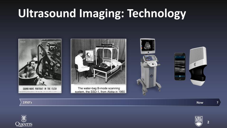
Ultrasound Imaging: Technology
2
1950’s Now ?
3
Past Today Future
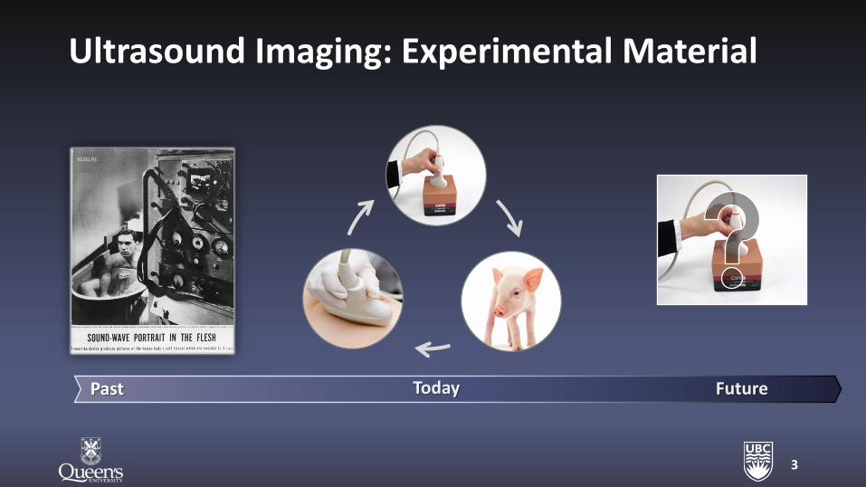
Ultrasound Imaging: Experimental Material
4
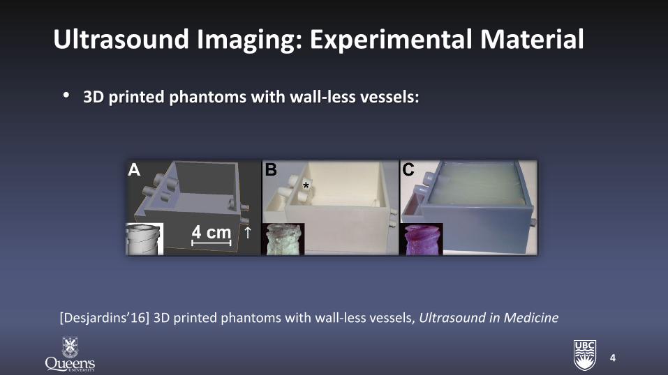
• 3D printed phantoms with wall-less vessels:
[Desjardins’16] 3D printed phantoms with wall-less vessels, Ultrasound in Medicine
Ultrasound Imaging: Experimental Material
5
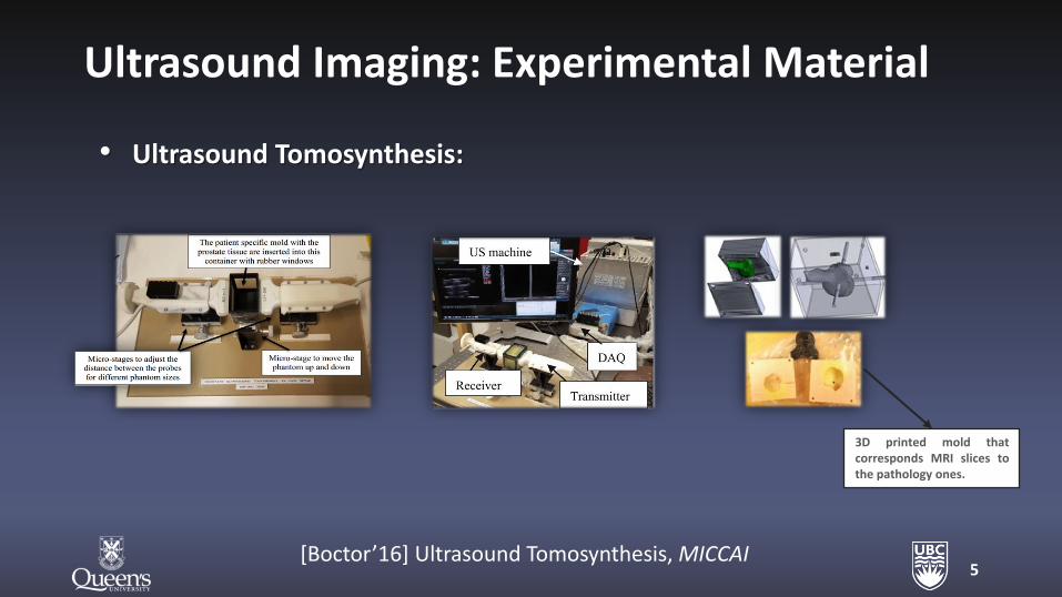
3D printed mold that corresponds MRI slices to the pathology ones.
• Ultrasound Tomosynthesis:
[Boctor’16] Ultrasound Tomosynthesis, MICCAI
Ultrasound Imaging: Experimental Material
6
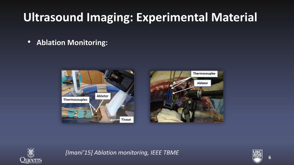
• Ablation Monitoring:
[Imani’15] Ablation monitoring, IEEE TBME
Ultrasound Imaging: Experimental Material
7
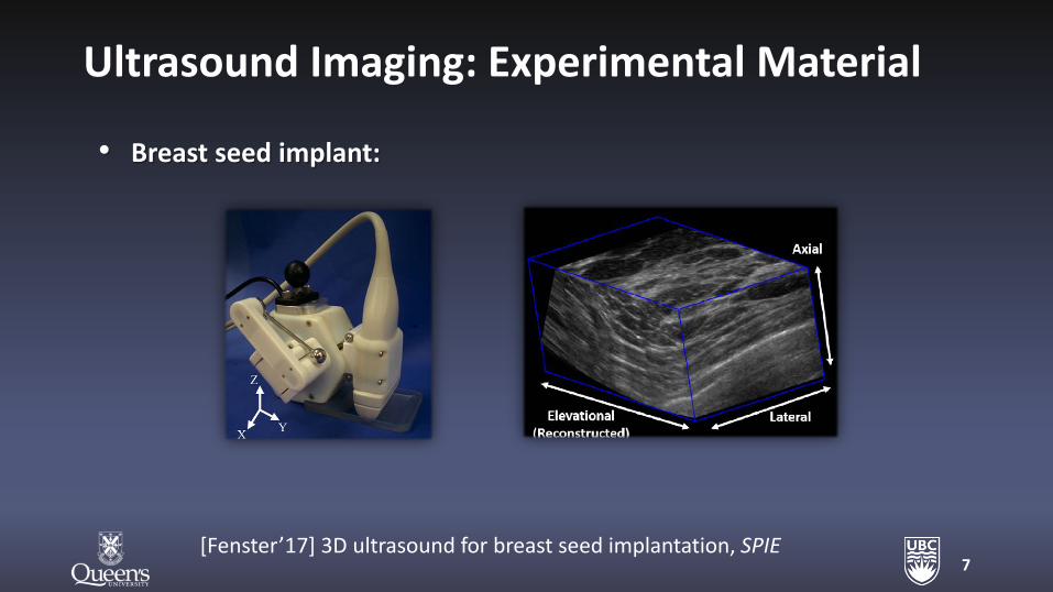
• Breast seed implant:
[Fenster’17] 3D ultrasound for breast seed implantation, SPIE
Ultrasound Imaging: Experimental Material
3D-Printing of Phantoms
8
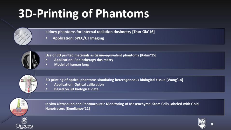
3D printing of optical phantoms simulating heterogeneous biological tissue [Wang’14] Application: Optical calibration Based on 3D biological data
Use of 3D printed materials as tissue-equivalent phantoms [Kalim’15] Application: Radiotherapy dosimetry Model of human lung
kidney phantoms for internal radiation dosimetry [Tran-Gia’16]
Application: SPEC/CT Imaging
In vivo Ultrasound and Photoacoustic Monitoring of Mesenchymal Stem Cells Labeled with Gold Nanotracers [Emelianov’12]
Ultrasound Imaging: Research
9
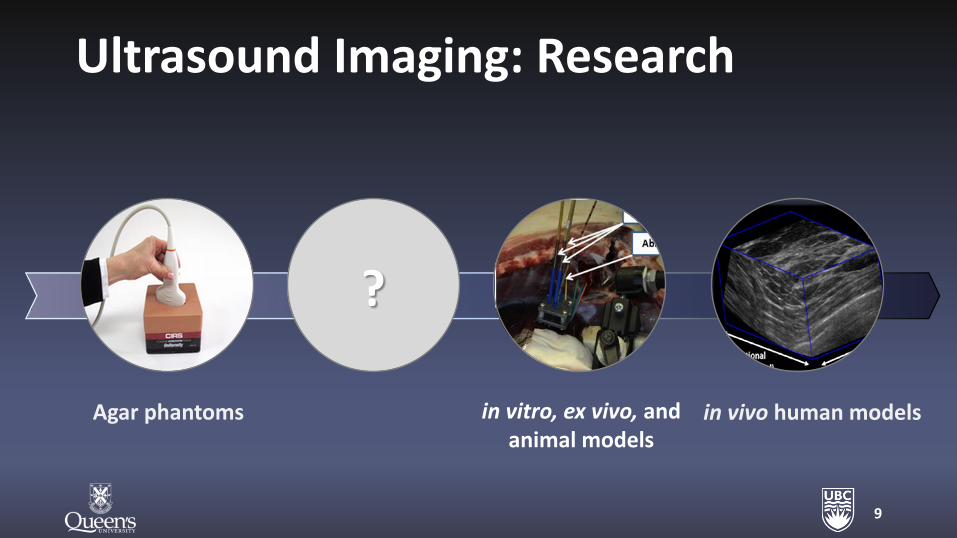
Agar phantoms in vivo human models in vitro, ex vivo, and animal models
?
10
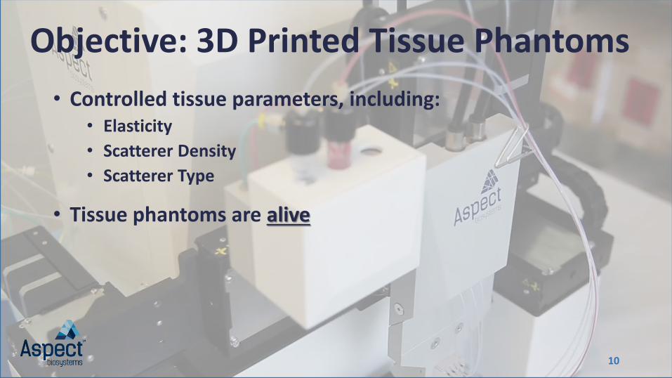
Objective: 3D Printed Tissue Phantoms
• Controlled tissue parameters, including: • Elasticity
• Scatterer Density
• Scatterer Type
• Tissue phantoms are alive
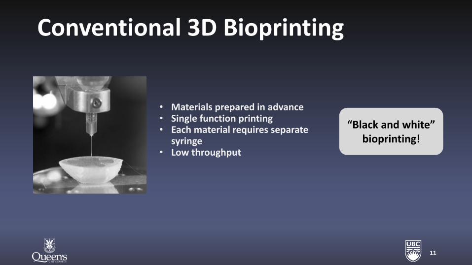
Conventional 3D Bioprinting
• Materials prepared in advance • Single function printing • Each material requires separate
syringe • Low throughput
11
“Black and white” bioprinting!
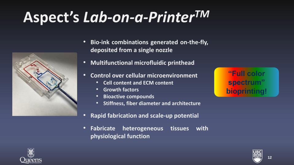
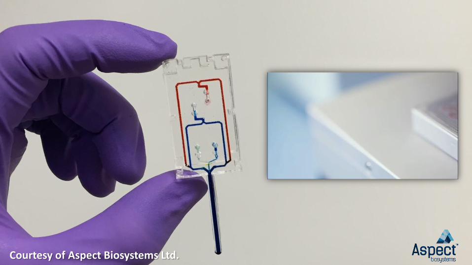
Aspect’s Lab-on-a-PrinterTM
12
“Full color
spectrum”
bioprinting!
• Bio-ink combinations generated on-the-fly, deposited from a single nozzle
• Multifunctional microfluidic printhead
• Control over cellular microenvironment • Cell content and ECM content • Growth factors • Bioactive compounds • Stiffness, fiber diameter and architecture
• Rapid fabrication and scale-up potential
• Fabricate heterogeneous tissues with physiological function
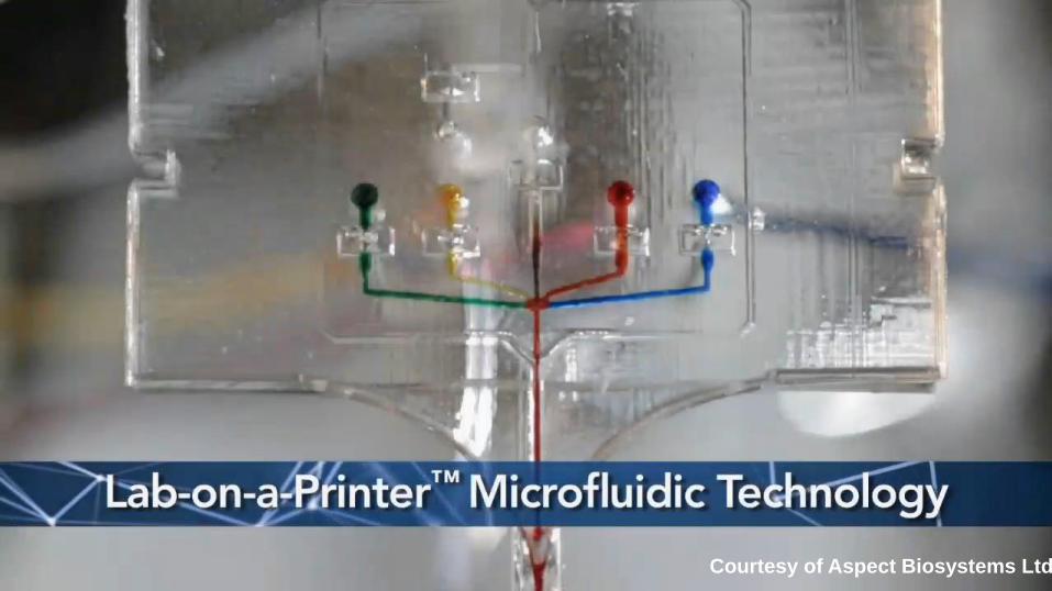
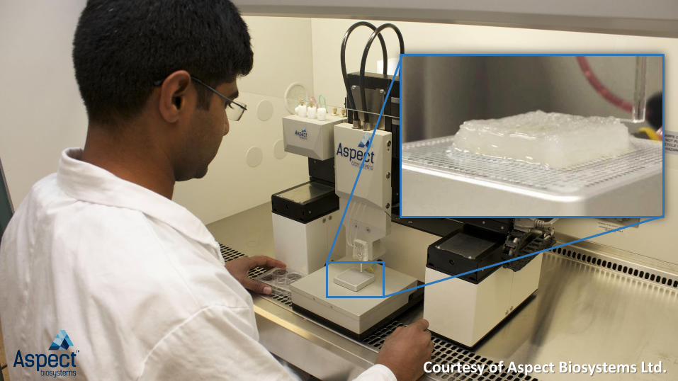
Courtesy of Aspect Biosystems Ltd.
Courtesy of Aspect Biosystems Ltd.
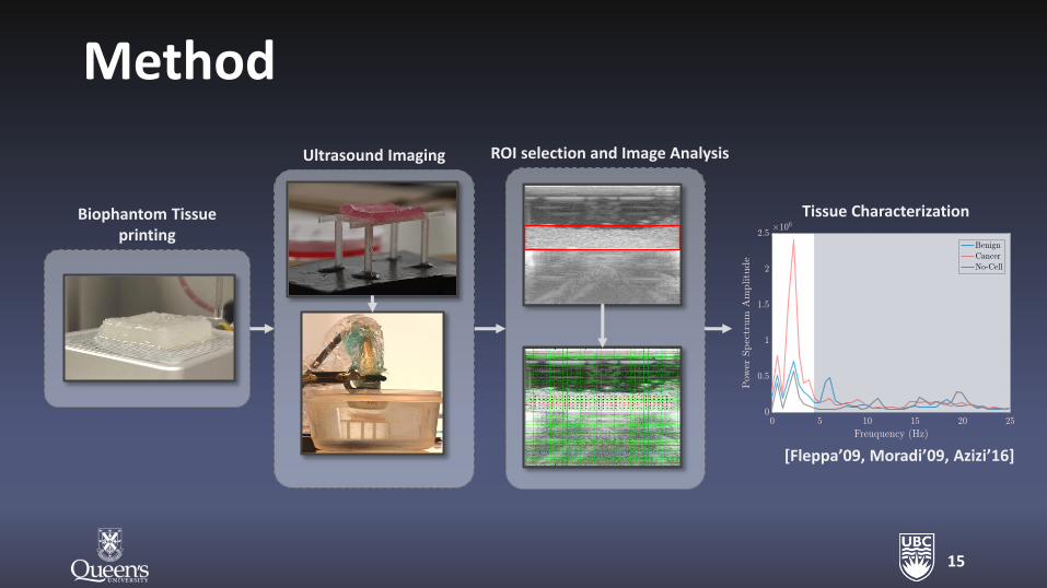
Method
15
Biophantom Tissue printing
Ultrasound Imaging ROI selection and Image Analysis
Tissue Characterization
[Fleppa’09, Moradi’09, Azizi’16]
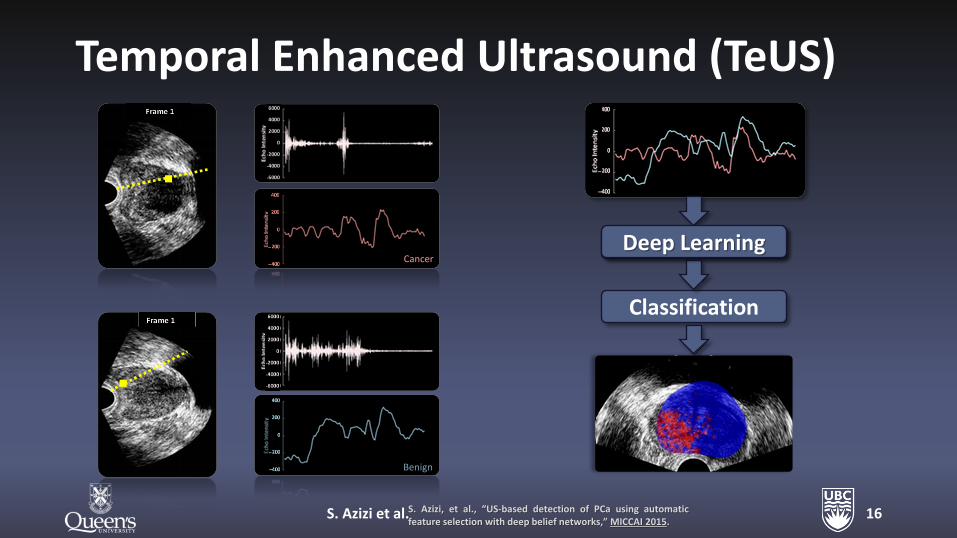
Temporal Enhanced Ultrasound (TeUS)
S. Azizi et al. 16
Cancer
Benign
S. Azizi, et al., “US-based detection of PCa using automatic feature selection with deep belief networks,” MICCAI 2015.
Feature Learning
Classification
Deep Learning
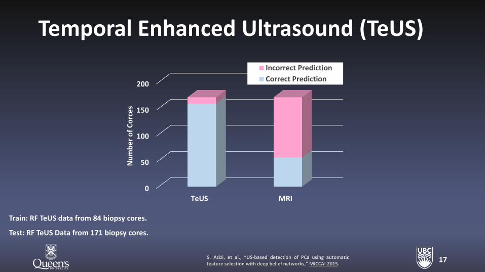
Temporal Enhanced Ultrasound (TeUS)
17 S. Azizi, et al., “US-based detection of PCa using automatic feature selection with deep belief networks,” MICCAI 2015.
0
50
100
150
200
TeUS MRI
Nu
mb
er
of
Co
rce
s
Incorrect Prediction
Correct Prediction
Train: RF TeUS data from 84 biopsy cores.
Test: RF TeUS Data from 171 biopsy cores.
Courtesy of Aspect Biosystems Ltd.
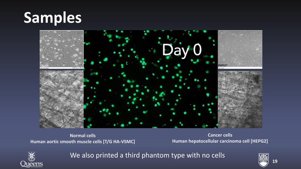
Samples
19
Normal cells Human aortic smooth muscle cells [T/G HA-VSMC]
Cancer cells Human hepatocellular carcinoma cell [HEPG2]
We also printed a third phantom type with no cells
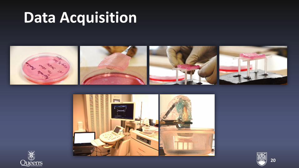
Data Acquisition
20
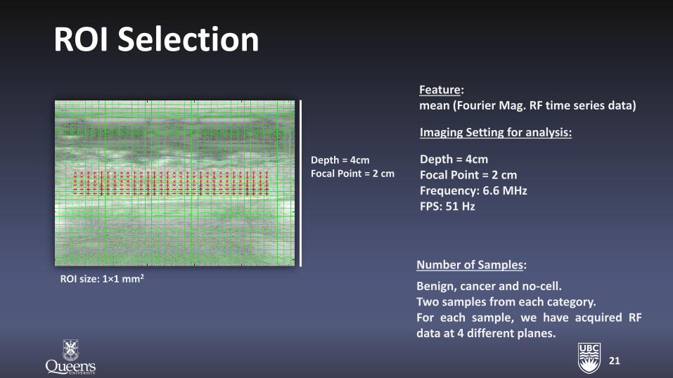
ROI Selection
21
Depth = 4cm Focal Point = 2 cm
ROI size: 1×1 mm2
Imaging Setting for analysis:
Depth = 4cm Focal Point = 2 cm Frequency: 6.6 MHz FPS: 51 Hz
Feature: mean (Fourier Mag. RF time series data)
Number of Samples:
Benign, cancer and no-cell. Two samples from each category. For each sample, we have acquired RF data at 4 different planes.
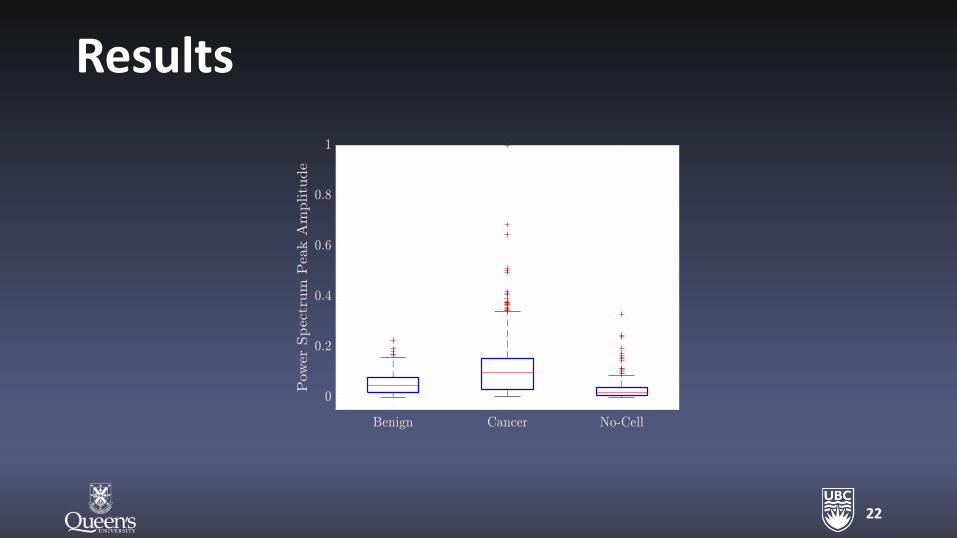
Results
22
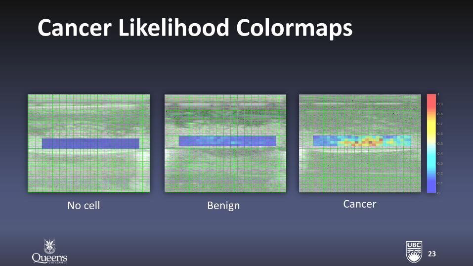
Cancer Likelihood Colormaps
23
No cell Benign Cancer
Conclusion
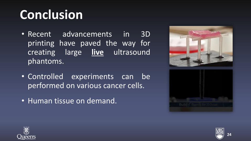
• Recent advancements in 3D printing have paved the way for creating large live ultrasound phantoms.
• Controlled experiments can be performed on various cancer cells.
• Human tissue on demand.
24
Thanks!
Shekoofeh Azizi Dr. Sharareh Bayat
Dr. Ajay Rajaram Dr. Emran M. A. Anas
Tamer Mohamed Dr. Konrad Walus
Prof. Purang Abolmaesumi Prof. Parvin Mousavi

26