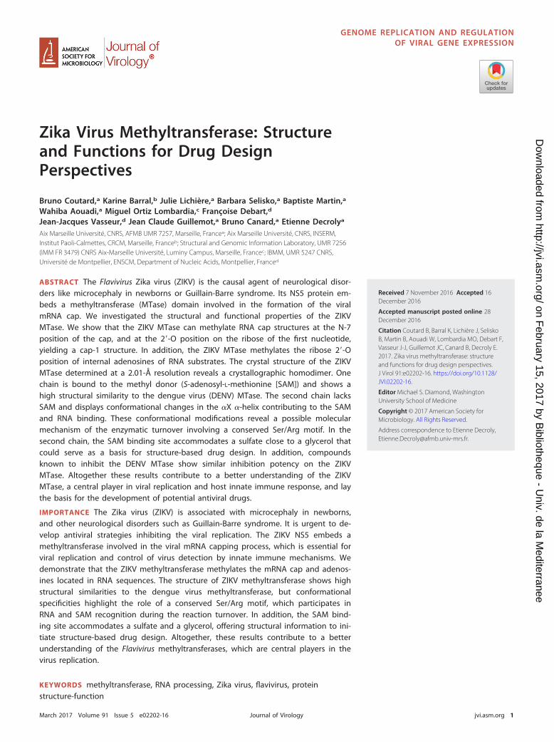
Zika Virus Methyltransferase: Structure and Functions for ... · Zika Virus Methyltransferase:...
15
Zika Virus Methyltransferase: Structure and Functions for Drug Design Perspectives Bruno Coutard, a Karine Barral, b Julie Lichière, a Barbara Selisko, a Baptiste Martin, a Wahiba Aouadi, a Miguel Ortiz Lombardia, c Françoise Debart, d Jean-Jacques Vasseur, d Jean Claude Guillemot, a Bruno Canard, a Etienne Decroly a Aix Marseille Université, CNRS, AFMB UMR 7257, Marseille, France a ; Aix Marseille Université, CNRS, INSERM, Institut Paoli-Calmettes, CRCM, Marseille, France b ; Structural and Genomic Information Laboratory, UMR 7256 (IMM FR 3479) CNRS Aix-Marseille Université, Luminy Campus, Marseille, France c ; IBMM, UMR 5247 CNRS, Université de Montpellier, ENSCM, Department of Nucleic Acids, Montpellier, France d ABSTRACT The Flavivirus Zika virus (ZIKV) is the causal agent of neurological disor- ders like microcephaly in newborns or Guillain-Barre syndrome. Its NS5 protein em- beds a methyltransferase (MTase) domain involved in the formation of the viral mRNA cap. We investigated the structural and functional properties of the ZIKV MTase. We show that the ZIKV MTase can methylate RNA cap structures at the N-7 position of the cap, and at the 2=-O position on the ribose of the first nucleotide, yielding a cap-1 structure. In addition, the ZIKV MTase methylates the ribose 2=-O position of internal adenosines of RNA substrates. The crystal structure of the ZIKV MTase determined at a 2.01-Å resolution reveals a crystallographic homodimer. One chain is bound to the methyl donor (S-adenosyl-L-methionine [SAM]) and shows a high structural similarity to the dengue virus (DENV) MTase. The second chain lacks SAM and displays conformational changes in the X -helix contributing to the SAM and RNA binding. These conformational modifications reveal a possible molecular mechanism of the enzymatic turnover involving a conserved Ser/Arg motif. In the second chain, the SAM binding site accommodates a sulfate close to a glycerol that could serve as a basis for structure-based drug design. In addition, compounds known to inhibit the DENV MTase show similar inhibition potency on the ZIKV MTase. Altogether these results contribute to a better understanding of the ZIKV MTase, a central player in viral replication and host innate immune response, and lay the basis for the development of potential antiviral drugs. IMPORTANCE The Zika virus (ZIKV) is associated with microcephaly in newborns, and other neurological disorders such as Guillain-Barre syndrome. It is urgent to de- velop antiviral strategies inhibiting the viral replication. The ZIKV NS5 embeds a methyltransferase involved in the viral mRNA capping process, which is essential for viral replication and control of virus detection by innate immune mechanisms. We demonstrate that the ZIKV methyltransferase methylates the mRNA cap and adenos- ines located in RNA sequences. The structure of ZIKV methyltransferase shows high structural similarities to the dengue virus methyltransferase, but conformational specificities highlight the role of a conserved Ser/Arg motif, which participates in RNA and SAM recognition during the reaction turnover. In addition, the SAM bind- ing site accommodates a sulfate and a glycerol, offering structural information to ini- tiate structure-based drug design. Altogether, these results contribute to a better understanding of the Flavivirus methyltransferases, which are central players in the virus replication. KEYWORDS methyltransferase, RNA processing, Zika virus, flavivirus, protein structure-function Received 7 November 2016 Accepted 16 December 2016 Accepted manuscript posted online 28 December 2016 Citation Coutard B, Barral K, Lichière J, Selisko B, Martin B, Aouadi W, Lombardia MO, Debart F, Vasseur J-J, Guillemot JC, Canard B, Decroly E. 2017. Zika virus methyltransferase: structure and functions for drug design perspectives. J Virol 91:e02202-16. https://doi.org/10.1128/ JVI.02202-16. Editor Michael S. Diamond, Washington University School of Medicine Copyright © 2017 American Society for Microbiology. All Rights Reserved. Address correspondence to Etienne Decroly, [email protected]. GENOME REPLICATION AND REGULATION OF VIRAL GENE EXPRESSION crossm March 2017 Volume 91 Issue 5 e02202-16 jvi.asm.org 1 Journal of Virology on February 15, 2017 by Bibliotheque - Univ. de la Mediterranee http://jvi.asm.org/ Downloaded from
Transcript of Zika Virus Methyltransferase: Structure and Functions for ... · Zika Virus Methyltransferase:...
Zika Virus Methyltransferase: Structureand Functions for Drug DesignPerspectives
Bruno Coutard,a Karine Barral,b Julie Lichière,a Barbara Selisko,a Baptiste Martin,a
Wahiba Aouadi,a Miguel Ortiz Lombardia,c Françoise Debart,d
Jean-Jacques Vasseur,d Jean Claude Guillemot,a Bruno Canard,a Etienne Decrolya
Aix Marseille Université, CNRS, AFMB UMR 7257, Marseille, Francea; Aix Marseille Université, CNRS, INSERM,Institut Paoli-Calmettes, CRCM, Marseille, Franceb; Structural and Genomic Information Laboratory, UMR 7256(IMM FR 3479) CNRS Aix-Marseille Université, Luminy Campus, Marseille, Francec; IBMM, UMR 5247 CNRS,Université de Montpellier, ENSCM, Department of Nucleic Acids, Montpellier, Franced
ABSTRACT The Flavivirus Zika virus (ZIKV) is the causal agent of neurological disor-ders like microcephaly in newborns or Guillain-Barre syndrome. Its NS5 protein em-beds a methyltransferase (MTase) domain involved in the formation of the viralmRNA cap. We investigated the structural and functional properties of the ZIKVMTase. We show that the ZIKV MTase can methylate RNA cap structures at the N-7position of the cap, and at the 2=-O position on the ribose of the first nucleotide,yielding a cap-1 structure. In addition, the ZIKV MTase methylates the ribose 2=-Oposition of internal adenosines of RNA substrates. The crystal structure of the ZIKVMTase determined at a 2.01-Å resolution reveals a crystallographic homodimer. Onechain is bound to the methyl donor (S-adenosyl-L-methionine [SAM]) and shows ahigh structural similarity to the dengue virus (DENV) MTase. The second chain lacksSAM and displays conformational changes in the �X �-helix contributing to the SAMand RNA binding. These conformational modifications reveal a possible molecularmechanism of the enzymatic turnover involving a conserved Ser/Arg motif. In thesecond chain, the SAM binding site accommodates a sulfate close to a glycerol thatcould serve as a basis for structure-based drug design. In addition, compoundsknown to inhibit the DENV MTase show similar inhibition potency on the ZIKVMTase. Altogether these results contribute to a better understanding of the ZIKVMTase, a central player in viral replication and host innate immune response, and laythe basis for the development of potential antiviral drugs.
IMPORTANCE The Zika virus (ZIKV) is associated with microcephaly in newborns,and other neurological disorders such as Guillain-Barre syndrome. It is urgent to de-velop antiviral strategies inhibiting the viral replication. The ZIKV NS5 embeds amethyltransferase involved in the viral mRNA capping process, which is essential forviral replication and control of virus detection by innate immune mechanisms. Wedemonstrate that the ZIKV methyltransferase methylates the mRNA cap and adenos-ines located in RNA sequences. The structure of ZIKV methyltransferase shows highstructural similarities to the dengue virus methyltransferase, but conformationalspecificities highlight the role of a conserved Ser/Arg motif, which participates inRNA and SAM recognition during the reaction turnover. In addition, the SAM bind-ing site accommodates a sulfate and a glycerol, offering structural information to ini-tiate structure-based drug design. Altogether, these results contribute to a betterunderstanding of the Flavivirus methyltransferases, which are central players in thevirus replication.
KEYWORDS methyltransferase, RNA processing, Zika virus, flavivirus, proteinstructure-function
Received 7 November 2016 Accepted 16December 2016
Accepted manuscript posted online 28December 2016
Citation Coutard B, Barral K, Lichière J, SeliskoB, Martin B, Aouadi W, Lombardia MO, Debart F,Vasseur J-J, Guillemot JC, Canard B, Decroly E.2017. Zika virus methyltransferase: structureand functions for drug design perspectives.J Virol 91:e02202-16. https://doi.org/10.1128/JVI.02202-16.
Editor Michael S. Diamond, WashingtonUniversity School of Medicine
Copyright © 2017 American Society forMicrobiology. All Rights Reserved.
Address correspondence to Etienne Decroly,[email protected].
GENOME REPLICATION AND REGULATIONOF VIRAL GENE EXPRESSION
crossm
March 2017 Volume 91 Issue 5 e02202-16 jvi.asm.org 1Journal of Virology
on February 15, 2017 by B
ibliotheque - Univ. de la M
editerraneehttp://jvi.asm
.org/D
ownloaded from
First identified in 1947 in Uganda, Zika virus (ZIKV) reemerged in Brazil in early 2015,and the virus has continued to disseminate to South and North America in 2016.
More than 2 million suspected cases have been reported (1), and ZIKV is now consid-ered a major public health threat by the World Health Organization (WHO). Whereas thebite of an infected Aedes aegypti mosquito is the main cause of ZIKV transmission, directhuman-to-human transmissions by sexual contacts have also been reported (2). ZIKVinfection was initially considered a minor infection, often asymptomatic, causing mainlyZika fever with rash, arthralgia, and conjunctivitis. However, this outbreak revealed thatZIKV infecting pregnant women can be transmitted to the fetus, causing fetal loss,microcephaly, and other serious brain defects in newborns (3). In addition, Guillain-Barre syndrome, a rare autoimmune disease affecting the peripheral nervous system,and other severe neurological disorders have also been associated with ZIKV infectionin adults (4). In the absence of a vaccine and antiviral drugs, the only way to reducevirus transmission is to limit the propagation of Aedes mosquito vectors. In view of thecurrent expanding outbreak, it is urgent to develop a vaccine and antiviral strategiesblocking essential enzymes involved in ZIKV replication.
ZIKV belongs to the Flavivirus genus, which contains other important humanpathogens such as dengue virus (DENV), West Nile virus (WNV), or yellow fever virus(YFV). Flaviviruses are enveloped viruses containing a single-stranded positive-senseRNA genome of around 11,000 nucleotides, which is decorated by a cap structure. Thecap structure consists of a guanosine residue linked through a 5=-5= triphosphate bondto the 5= end of mRNA (GpppN) (5). In flaviviruses, the cap structure is methylated atthe nitrogen in position 7 of the guanosine (cap-0 structure, or mGpppN) and at the2=-oxygen atom (2=-O) of the N1 ribose (cap-1 structure, or mGpppNm) (6, 7). The N-7methylation of the cap structure is essential for RNA stability and stimulates theirtranslation into viral protein by recognizing the translation initiation factor eIF4E (8).The 2=-O-methylation of the cap structure was demonstrated to protect viral RNA frombeing recognized by host cell sensors such as RIG-I and MDA5 that stimulate theproduction of interferons (9–11). In turn, interferon-stimulated genes, such as IFIT-1, candetect miscapped RNA and restrict their translation into proteins (12, 13). DENV mutantviruses lacking N-7-methyltransferase (MTase) activity show strong replication defectsin infected cells, whereas those altering the 2=-O-MTase activity show only attenuatedphenotypes (7, 14), as most cell lines used in the laboratory for virus replication aredeprived of the RIG-I/MDA5 antiviral pathway. In contrast, it was demonstrated that a2=-O-MTase knockout virus barely replicates in infected mice and elicits a stronghumoral and cellular antiviral response (15, 16). Thus, viral RNA capping represents anattractive antiviral strategy (17, 18) since it should inhibit viral replication and/oraccelerate virus clearance upon stimulation of the innate antiviral response.
In Flavivirus, the genome replication is driven by NS5, the RNA-dependent RNApolymerase (RdRp), which is associated with other viral nonstructural proteins in amembrane-bound replication-transcription complex. In addition to the RdRp domain,NS5 also harbors an N-terminal MTase domain, which methylates the cap structure ofnascent RNA at both the N-7 and 2=-O positions (6, 7). Initially, biochemical studiesperformed with recombinant NS5 proteins or MTase domains (NS5-MTase) of DENV,WNV and other flaviviruses have confirmed the 2=-O-MTase activity using short syn-thetic RNA substrates in the presence of the methyl donor S-adenosyl-L-methionine(SAM, or AdoMet) (6, 19). The N-7-MTase activity was next demonstrated using longerRNA, with an optimal activity detected when a RNA containing the highly conservedstem loop A (SLA) hairpin structure was used (7, 20). The DENV MTase domain was laterreported to methylate the 2=-O-ribose position of internal adenosines of the RNA (21).Nevertheless, the role of these methylations is still poorly understood. The structures ofseveral Flavivirus NS5-MTases have been solved (6, 7), and the structure of ZIKV MTasehas recently been determined by Coloma et al. (22). They adopt a canonical MTaseRossmann fold with a seven-stranded �-sheet surrounded by four �-helices. The MTasecore structure closely resembles the catalytic domain of other viral and cellular SAM-dependent MTases with a conserved SAM binding site in close vicinity of the conserved
Coutard et al. Journal of Virology
March 2017 Volume 91 Issue 5 e02202-16 jvi.asm.org 2
on February 15, 2017 by B
ibliotheque - Univ. de la M
editerraneehttp://jvi.asm
.org/D
ownloaded from
K-D-K-E catalytic tetrad, the latter being a signature of 2=-O-MTases. In addition,structures determined in the presence of cap analogues, GTP or RNA reveal a capbinding pocket where the cap guanosine is held during 2=-O-methylation by stackinginteractions with a phenylalanine (F24 residue in the DENV NS5 sequence) and an RNAbinding zone where the RNA chain downstream of the cap structure is accommodatedby a basic groove (4, 6, 7, 22–25).
Enzyme-based screenings or structure-based drug design have identified DENV,WNV, and YFV MTase inhibitors targeting either the SAM/SAH (S-adenosyl-L-homocysteine, the coproduct of the methylation) binding pocket (26–28), the capbinding pocket (29), or allosteric sites (30, 31). As ZIKV is closely related, we initiated acomparative study of ZIKV MTase with other Flavivirus MTases with the aim of subse-quently developing original inhibitors or repurposing inhibitors targeting DENV MTasefor antiviral research on ZIKV. Here we describe the structural and functional analysis ofthe ZIKV MTase domain as well as the evaluation of inhibitors developed for DENVMTase on ZIKV MTase.
RESULTSProduction and purification of ZIKV MTase constructs. The ZIKV genome en-
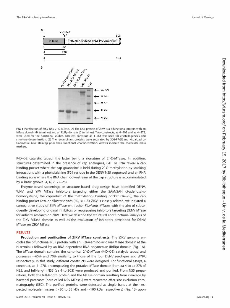
codes the bifunctional NS5 protein, with an �264-amino-acid (aa) MTase domain at theN terminus followed by an RNA-dependent RNA polymerase (RdRp) domain (Fig. 1A).The MTase domain contains the canonical 2=-O-MTase (K-D-K-E) catalytic tetrad andpossesses �65% and 70% similarity to those of the four DENV serotypes and WNV,respectively. In this study, different constructs were designed. For functional assays, aconstruct, aa 4 –278, encompassing the putative MTase domain from aa 4 to aa 278 ofNS5, and full-length NS5 (aa 4 to 903) were produced and purified. From NS5 prepa-rations, both the full-length protein and the MTase domain resulting from cleavage bybacterial proteases (here called NS5-MTaseC) were recovered after size exclusion chro-matography (SEC). The purified proteins were detected as single bands at their ex-pected molecular masses (�30 to 35 kDa and �100 kDa, respectively) (Fig. 1B) upon
FIG 1 Purification of ZIKV NS5 2=-O-MTase. (A) The NS5 protein of ZIKV is a bifunctional protein with anMTase domain (N terminus) and an RdRp domain (C terminus). Two constructs, aa 4 –903 and aa 4 –278,were used for the functional studies, whereas construct aa 1–264 was used for crystallogenesis andstructure determination. (B) The recombinant proteins were separated by SDS-PAGE and visualized byCoomassie blue staining prior their functional characterization. Arrows indicate the molecular massmarkers.
The Zika Virus Methyltransferase Journal of Virology
March 2017 Volume 91 Issue 5 e02202-16 jvi.asm.org 3
on February 15, 2017 by B
ibliotheque - Univ. de la M
editerraneehttp://jvi.asm
.org/D
ownloaded from

SDS-PAGE analysis. The apparent molecular mass observed for NS5-MTaseC is similar tothose of the ZIKV and DENV MTase domains (Fig. 1B).
ZIKV MTase crystallizes as a homodimer in which the two protomers showconformational differences. As the original MTase construct (aa 4 –278) did notcrystallize, we generated another that includes the complete N terminus but is devoidof the linker between the MTase and RdRp domains (aa 1–264; PDB code 5M5B). Thisconstruct allowed determination of the crystal structure of the ZIKV MTase domain. Thestructure was solved by molecular replacement using the DENV3 NS5 MTase/SAM (PDBcode 3P97) as the template model and refined to a resolution of 2.01 Å, with Rwork andRfree values of 16.0% and 19.1%, respectively. The crystal belongs to space group P1with the following unit cell parameters: a � 37.6 Å, b � 64.1 Å, and c � 72.0 Å and � �
113.1°, � � 97.8°, and � � 92.0°. Data collection and refinement statistics are reportedin Table 1. The ZIKV MTase domain crystallized with two molecules in the asymmetricunit (Fig. 2A), whereas size exclusion chromatography indicates that the proteinbehaves as a monomer in solution. The low-interaction surface area (about 500 Å2)suggests that dimerization occurs during crystallization as previously observed forModoc virus (MODV) MTase (32). The two protomers (A and B chains) are similarlyfolded, with significant differences arising from disorder in residues 43 to 58 of chainB (Fig. 2A). A superimposition of 244 C� atoms in the A and B chains of the protein
TABLE 1 Data collection and refinement statistics
Parameter Value(s) for SAM complexa
Data collection and phasing statisticsSpace group P1Unit cell parameters
a, b, c (Å) 37.6, 64.1, 72.0�, �, � (°) 113.1, 97.8, 92.0
Resolution range (Å) 65.41–2.01 (2.02–2.01)No. of unique reflections 40,055 (398)Completeness (%) 99.3 (93.9)Multiplicity 3.3 (2.5)Rmeas
b 0.157 (0.454)I/��I� 5.8 (2.0)
Refinement statisticsResolution range (Å) 65.41–2.01No. of reflections:
Used for refinement 40,054Used for Rfree calculation 2,105
Rwork (%)c 16.0Rfree (%)d 19.1No. of atoms
Protein chain A 2,077Protein chain B 1,963Ligand/ion (SAM/glycerol/others) 27/6/21Water 375
B factors (Å2)Protein chain A 28.0Protein chain B 28.2Ligand/ion (SAM/glycerol/others) 25.3/49.5/80.1Water 37.9
RMSD from idealityBond lengths (Å) 0.010Bond angles (°) 0.96
Ramanchandran plot (%)Residues in most favored regions 97.40Residues in disallowed regions 0
aValues in parentheses are for the outermost resolution shell of data.bRmeas � �|Iobs � Iav|/�Iav, over all symmetry-related observations.cRwork � �|Fobs � �|Fcalc|�|/�|Fobs|, over all reflections included in the refinement.dRfree is calculated with 5% of reflections excluded from the refinement. In this formula, �|Fcalc|� denotes theexpectation value of |Fcalc| under the probability distribution used to define the likelihood function that ismaximized in the refinement.
Coutard et al. Journal of Virology
March 2017 Volume 91 Issue 5 e02202-16 jvi.asm.org 4
on February 15, 2017 by B
ibliotheque - Univ. de la M
editerraneehttp://jvi.asm
.org/D
ownloaded from
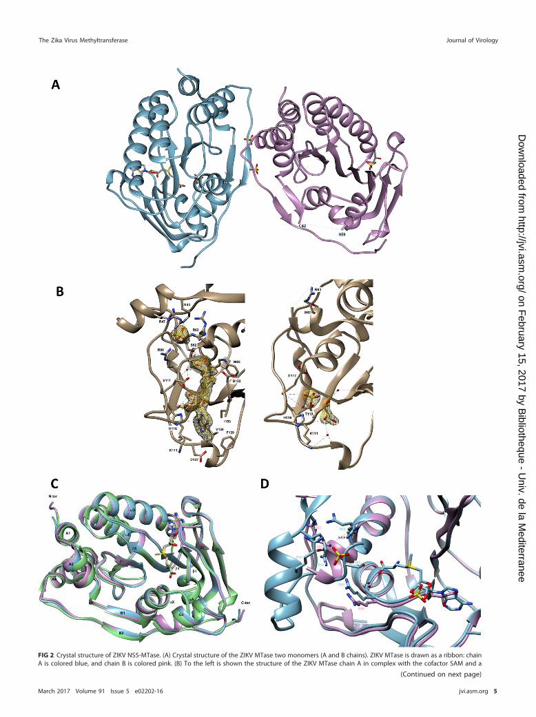
FIG 2 Crystal structure of ZIKV NS5-MTase. (A) Crystal structure of the ZIKV MTase two monomers (A and B chains). ZIKV MTase is drawn as a ribbon: chainA is colored blue, and chain B is colored pink. (B) To the left is shown the structure of the ZIKV MTase chain A in complex with the cofactor SAM and a
(Continued on next page)
The Zika Virus Methyltransferase Journal of Virology
March 2017 Volume 91 Issue 5 e02202-16 jvi.asm.org 5
on February 15, 2017 by B
ibliotheque - Univ. de la M
editerraneehttp://jvi.asm
.org/D
ownloaded from
resulted in an RMSD of 0.52 Å. Chain A consists of a canonical MTase core (residues 61to 229, construct numbering PDB code 5M5B) folded into a seven-stranded �-sheet (�1to �7) surrounded by four �-helices (�X, �A, �D, and �E), as shown in Fig. 2A and C.Appended to the core are an N-terminal extension (residues 8 to 60) and a C-terminalextension (residues 230 to 272). The N-terminal subdomain comprises a helix-turn-helixmotif followed by a �-strand and an �-helix (A1, A2, B1, and A3). The C-terminalsubdomain consists of an �-helix followed by a �-strand (A4 and B2). Inspection of theelectron density maps reveals strong additional electron density in the SAM bindingpocket, allowing the unambiguous modeling of SAM. Because no SAM is added duringpurification or crystallization, its presence in the structure must have originated from E.coli. The SAM molecule is bound in the central cleft formed of �-strands �1, �2, and �4and �-helices �X and �A (Fig. 2A and B). The SAM adenine base is accommodatedwithin a hydrophobic pocket defined by the side chains of Val138, Phe139, and Ile153and stabilized by hydrogen bonds with a carboxylic oxygen from Asp137 and thenitrogen main-chain atoms of Lys111 and Val138 (Fig. 2B, left panel). The ribose moietyis hydrogen bonded to the Glu117 carboxyl group. The sugar is also bridged via a watermolecule to the side chain of Glu117. The methionine tail of the SAM molecule ispositioned by interactions with the side chains of Ser62, Trp93, and Asp152, as well aswith the main chain of Gly92. The methionine carboxylate is also bridged via a singlewater molecule to the side chain of Glu117 and to the backbone of Arg90. Threemolecules of sulfate interact with chain A. One of them is bound via a cluster formedby arginines Arg43, Arg47, Arg63, and Arg90 (Fig. 2D). The structure of chain A is highlysimilar to those of DENV-3 MTase (RMSD of 0.79 Å with PDB code 3P97 chain A) and ofZIKV MTase recently determined by Coloma et al. (RMSD of 0.29 Å with PDB code 5KQRchain A) (22). There is no difference in SAM binding with the latter structure (PDB code5KQR), and the sulfate bound via a cluster formed by arginines Arg43, Arg47, Arg63,and Arg90 overlaps perfectly with the phosphate described by Coloma et al. (22).Structural differences from DENV-3 MTase are limited to small changes in the confor-mation of three solvent-exposed loops (residues 55 to 58, 178 to 183, and 251 to 254),as shown by superimposition of several structures (Fig. 2C).
We observe significant conformational changes between chain A and chain B ofZIKV MTase (RMSD of 0.52 Å) likely favored by their different crystal environments.Indeed, the chain B environment does not allow this loop to adopt the conformationobserved in chain A. However, it is unlikely that crystal packing is the only factorinvolved since similar disorder has been observed, under different crystal packing, forthe MODV MTase (PDB code 2WA2) (32). We conclude that the intrinsic flexibility of thisloop, at least in the absence of the RNA substrate, has also contributed to building ofthis crystal form. As illustrated in Fig. 2A and D, three main differences are noteworthyin chain B, as follows. (i) There is no supporting electron density for residues 43 to 58(part of the B1 �-strand, the A3 �-helix, and the following loop (Fig. 2A]). A similarabsence of electron density has already been described for one of the two chains of theMODV MTase (32), suggesting a certain degree of flexibility in this region. (ii) Down-stream of the aa 43–58 flexible region, the �X �-helix is kinked (Fig. 2D), with itsN-terminal part (residues 61 to 67) taken away from this helix axis. (iii) There is noadditional electron density characterizing the methyl donor SAM in the SAM bindingsite. Interestingly, in chain B of the MODV MTase, a SAM molecule is present, even if the
FIG 2 Legend (Continued)sulfate ion. To the right is shown the structure of ZIKV MTase chain B in complex with glycerol- and sulfate-bound molecules. ZIKV MTase is drawn as aribbon and colored gray. Small red spheres represent water molecules around the binding sites. Hydrogen bond interactions are shown as black dottedlines. The amino acid residues shown are those establishing Van der Waals interactions with bound molecules. In the insets, electron density maps areshown as a yellow mesh contoured at 1.5 � around bound molecules. (C) Superposition of the ZIKV MTase chain A (PDB code 5M5B [in blue]), of the ZIKVMTase described by Coloma et al. (22) (PDB code 5KQR [in pink]), and of the DENV MTase (PDB code 3P97 [in green]) structures bound with SAM. (D)Superposition of ZIKV MTase chain A (drawn as a blue ribbon) and of ZIKV MTase chain B (drawn as a pink ribbon). Shown is a view of the cluster formedby arginines Arg43, Arg47, Arg63, and Arg90 and of the SAM binding site. The distance between Arg63 from chain A and chain B is shown as black dottedlines. For panels A through D, MTases are drawn as ribbons. The small molecules, SAM, glycerol, and sulfate ions are shown in cylinder representation withgray indicating the carbon atom. Nitrogen, oxygen, and sulfur are colored blue, red, and yellow, respectively.
Coutard et al. Journal of Virology
March 2017 Volume 91 Issue 5 e02202-16 jvi.asm.org 6
on February 15, 2017 by B
ibliotheque - Univ. de la M
editerraneehttp://jvi.asm
.org/D
ownloaded from

homologous region aa 35–56 appears to also be disordered. However, the kink ob-served in the �X �-helix of ZIKV MTase chain B is absent from MODV MTase chain B. Thekink has two main structural consequences. First, the side chain of Ser62 cannot interactanymore with the methionine tail of the SAM, as observed in chain A (Fig. 2B, leftpanel). Second, a cluster formed by arginines of the RNA binding groove (Arg43, Arg47,Arg63, and Arg90) in chain A is distorted in chain B and does not accommodate anysulfate molecule (Fig. 2D). In the absence of SAM, two polar molecules, a sulfate and aglycerol close by (�3.2 Å), are held in the SAM binding pocket of chain B (Fig. 2A andB). The sulfate molecule is positioned by hydrogen bonds with the side chain of His116,the main chain of Glu117, and with the bound glycerol nearby. It is also bridged via asingle water molecule to the main chains of Gly112 and Glu117, as well as to theside-chain hydroxyl of Thr110. Moreover, the glycerol molecule participates in van derWaals interactions with the main chains of Gly87 and Lys111 and is stabilized byhydrogen bonds with the sulfate and the main-chain amide of Lys111. The glycerol isalso bridged via two water molecules to the main chains of Asp137 and Val138 for thefirst one and to the main chains of Gly87 and Asp152 for the second one. Such astructure with compounds bound in the SAM binding site could serve as a basis forstructure-based drug design.
Full-length ZIKV NS5 displays a 2=-O-MTase activity that is not regulated by thepolymerase domain. The 2=-O-MTase activity was measured comparatively using the
three ZIKV proteins (Fig. 1B). Proteins were incubated with a short RNA substratebearing an unmethylated cap structure, GpppAC4, in the presence of a radiolabeledmethyl donor ([3H]SAM). The amount of [3H]CH3 transferred onto RNA was quantifiedwith a DEAE filter binding assay. ZIKV NS5, NS5-MTaseC, and the ZIKV NS5-MTasedomain exhibit similar enzymatic activities (Fig. 3A). The level of detected enzymaticactivities is about 30% lower than the control activity of DENV NS5-MTase. ZIKVNS5-MTase activity is thus not stimulated by the presence of the RdRp domain in cis.In addition, we compared the enzymatic activity of the ZIKV MTase domain usingthe unmethylated substrate GpppAC4 and a substrate methylated at the N-7 position,mGpppAC4. The methyl transfer efficiency is similar (Fig. 3B), suggesting that bothsubstrates can be methylated at the 2=-O position of the adenosine ribose, as alreadyreported for DENV and WNV MTases (19, 33). Additionally, the ZIKV MTase domainshows an optimal activity between pH 8 and 9 (Fig. 3C). Time course experimentsindicate that the 2=-O-MTase functions without additional magnesium (not shown). Wenext addressed sequence specificity using short RNAs consisting of the 5= ends ofvarious viral RNAs starting either with A or G (Fig. 3D). Although RNA methylationefficiency by ZIKV MTase varies, we could not observe any obvious preference forFlavivirus GpppA-RNAs or GpppG-RNAs.
The ZIKV MTase requires long structured RNA to methylate the N-7 position ofthe cap. An SLA hairpin structure is present at the 5= end of Flavivirus genomes. Thisstructure is required for the detection of the N-7-MTase activity of WNV and DENV (7,20). First, we compared the 5= ends of ZIKV and DENV. The seven first residues of theZIKV RNA are strictly identical to those of DENV, and ZIKV RNA forms a hairpin structuresimilar to that of DENV. N-7-methylation assays driven by ZIKV NS5-MTase were thusconducted on SLA-containing DENV RNAs with various 5=-capped ends (GpppA-RNA75,mGpppA-RNA75, GpppAm-RNA75, and mGpppAm-RNA75). mGpppA-DENV75 and GpppAm-DENV75 are both significantly better methylated than mGpppAm-DENV75 (P � 0.001),indicating that N-7- and 2=-O-methylations occur (Fig. 3E). In addition, the high meth-ylation level observed with mGpppA-DENV75 suggests that the 2=-O-methylation ismore efficient than the N-7-methylation under the applied experimental conditions.Interestingly, MTase activity using RNA already methylated at the N-7- and 2=-Opositions (mGpppAm-DENV75) was also detected. This observation suggests that addi-tional methylations downstream of the cap structure might occur (described below).Altogether, these results show that, as observed for other flaviviruses, the ZIKV MTasecan perform cap 2=-O-methylation on short RNAs without sequence specificity, whereas
The Zika Virus Methyltransferase Journal of Virology
March 2017 Volume 91 Issue 5 e02202-16 jvi.asm.org 7
on February 15, 2017 by B
ibliotheque - Univ. de la M
editerraneehttp://jvi.asm
.org/D
ownloaded from

FIG 3 Characterization of the three MTase activities carried by ZIKV MTase. The ZIKV MTase activity wasdetermined by monitoring the transfer of tritiated methyl groups (in counts per minute) from SAM tovarious synthetic RNAs. The bar graph presents the mean value and the standard deviation from 3independent experiments. (A) Comparison of the MTase activities carried by the recombinant proteinsusing GpppAC4 RNA as methyl acceptor. Shown is the enzymatic activity of ZIKV MTase domain on (B) small
(Continued on next page)
Coutard et al. Journal of Virology
March 2017 Volume 91 Issue 5 e02202-16 jvi.asm.org 8
on February 15, 2017 by B
ibliotheque - Univ. de la M
editerraneehttp://jvi.asm
.org/D
ownloaded from
cap N-7-methylation does indeed require longer RNAs containing the Flavivirus con-served SLA structure.
The ZIKV MTase methylates single-strand RNA at the 2=-OH group of internaladenosines. Since the ZIKV MTase is active on double-methylated cap-1 RNAs (Fig. 3E),the methyl transfer on internal residues of RNAs was further investigated using un-capped RNAs in the presence of a 2-times-higher ZIKV MTase concentration than in theprevious experiments. Figure 3F indicates that a 13-mer RNA bearing a 5=-triphosphatecorresponding to the nascent 5= end of DENV RNA can be methylated, confirming thatZIKV MTase methylates uncapped RNA putatively on internal positions. In order tofurther characterize this activity, homopolymeric RNAs bearing a 5=-OH group weretested (Fig. 3F). ZIKV NS5-MTase can methylate a 5=-OH-A27 RNA, but not 5=-OH-U27,-G27, and -C27 RNAs, indicating that ZIKV MTase methylates adenosine residues. SinceZIKV harbors a 2=-O-MTase catalytic tetrad, we inferred that the MTase activity targetsthe 2=-O of the ribose of the adenosine residues. This activity was confirmed bydemonstrating that an RNA previously methylated at all 2=-O positions (Am27), is notmethylated by the ZIKV MTase (Fig. 3G). In addition, the internal adenosine 2=-O-methylation does not specifically occur in the vicinity of the RNA 5= end since RNAsalready methylated on the first (AmA26) or the two first (AmAmA25) adenosines weremethylated (Fig. 3G).
The 5= ends of Flavivirus mRNAs are organized in an SLA structure, and manyadenosines are engaged in double-strand stretches. We thus investigated whether theinternal methylation could be selective for single-strand RNA (ssRNA) or double-strandRNA (dsRNA). We compared the MTase activities on A27 and on A27 previously annealedwith a complementary U27 RNA. No MTase activity was detected using dsRNA (A27
U27) (Fig. 3H) confirming that ZIKV MTase methylates preferentially ssRNA. In conclu-sion, the ZIKV MTase preferentially methylates the 2=-O position of internal adenosineresidues that are not involved in dsRNA structures.
Selected DENV MTase inhibitors show increased potency on ZIKV MTase. Ourstructural and biochemical analyses indicate that the ZIKV MTase shares commonstructural and functional characteristics with DENV MTase (RMSD of 0.79 Å for PDB code3P97 chain A). We thus tested whether some compounds already documented toinhibit DENV MTase might also be active against the ZIKV MTase. SAM analogues andcap analogues were tested as well as allosteric inhibitors designed from a fragment-based screening campaign performed against DENV MTase (31). For this purpose, bothinternal methylation and cap 2=-O-methylation were monitored on GpppAC4 and A27
RNA substrates, respectively, with increasing concentrations of the investigated inhib-itors. We found that SAM analogues such as SAH and sinefungin inhibit cap-dependentZIKV 2=-O-MTase activity with 50% inhibitory concentrations (IC50) in the low micro-molar range (Fig. 4A; Table 2). These compounds also abrogate internal methylation,but SAH shows a lower efficacy against internal methylation than sinefungin, whichinhibits both methylations in a similar manner. Cap analogues (GpppA, mGpppA,GpppG, and mGpppG) bind to the DENV cap binding site by stacking interactions (34)and might therefore block RNA cap recognition, thus limiting cap methylation. Thismight also interfere with recognition and binding of uncapped RNA for internalmethylation since the cap binding site might extend the RNA binding zone. GpppG andmGpppG show more potent inhibitions than GpppA and mGpppA on cap 2=-O-methylation, with IC50 of 72 �M and 184 �M, respectively, versus 491 �M and 293 �M(Fig. 4B and C; Table 2). In addition, cap analogues inhibit internal methylations, to alesser extent though, suggesting that the MTase cap binding site forms a part of theRNA binding zone but is not essential for internal methylation of an A27 RNA.
FIG 3 Legend (Continued)GpppAC4 and mGpppAC4 RNA, (C) 75 nucleotides of capped RNA with various 5=-end modifications(GpppA-RNA, mGpppA-RNA GpppAmA-RNA and mGpppAmA-RNA), (D) different uncapped RNAs, (E) un-capped poly(A) oligomers with various 2=-O-methylated positions (A27, no methylation; AmA26, first position2=-O-methylated; AmAm25, first and second positions 2=-O-methylated; Am27, all positions 2=-O-methylated),and (F) ssRNA A27 and dsRNA A27-U27. CTL neg, enzymatic assay performed in the absence of RNA.
The Zika Virus Methyltransferase Journal of Virology
March 2017 Volume 91 Issue 5 e02202-16 jvi.asm.org 9
on February 15, 2017 by B
ibliotheque - Univ. de la M
editerraneehttp://jvi.asm
.org/D
ownloaded from
Finally, we tested whether six compounds (1 to 6 in Table 2) targeting an allostericsite of DENV MTase (30, 31) could inhibit ZIKV MTase. All of the compounds selected onDENV MTase inhibit ZIKV cap 2=-O-MTase activity (Fig. 4D and Table 2). Compounds 1to 6 exhibit lower IC50 for ZIKV 2=-O-MTase activity than those for DENV MTase,suggesting that they are more potent on ZIKV MTase. In addition, all compoundsinhibiting the cap-2=-O-MTase activity also inhibit the internal methylation, but withhigher IC50 (Table 2), indicating that internal and cap 2=-O-MTase activity use the samecatalytic mechanism. These data also indicate that the structural and functional simi-larities between ZIKV and DENV MTase pave the way for the development of pan-viralinhibitors targeting the members of the Flavivirus genus.
DISCUSSION
The development of therapeutics against emerging arboviruses, such as ZIKV, is amajor challenge. Acquisition of basic scientific knowledge on the emerging viral agentis often slow, whereas outbreaks can be sudden and unpredictable. In this work, wefocus on the structure/function analysis of ZIKV NS5-MTase, closely related to that ofDENV, and involved in the cap structure methylation. The latter is essential for viral RNAtranslation into proteins and the hiding of viral genomes from detection by cellularantiviral pathways (9, 15). The ZIKV MTase presented here crystallizes as a homodimer.Both protomers show a canonical MTase fold similar to those of DENV3 or WNV MTases,as well as those of other flaviviruses with SAM and cap binding sites close to theK-D-K-E catalytic tetrad. The first protomer of the ZIKV MTase crystallographic dimer
TABLE 2 Inhibition of the ZIKV 2=-O-MTase activity by various DENV MTaseinhibitorsa
aShown is a summary of the IC50 and the standard deviation (SD) deduced from Fig. 4 using the log(inhibitor) versus response variable slope equation and compared with the IC50 previously publishedfor DENV MTase. The IC50 are an average from two to three independent measurements. nd, notdetermined.
Coutard et al. Journal of Virology
March 2017 Volume 91 Issue 5 e02202-16 jvi.asm.org 10
on February 15, 2017 by B
ibliotheque - Univ. de la M
editerraneehttp://jvi.asm
.org/D
ownloaded from
contains the methyl donor SAM. It shows a high structural similarity to the ZIKV MTasestructure recently determined by Coloma et al. (22). In this protomer, the SAM adeninebase is held within a conserved hydrophobic pocket formed of �-strands �1, �2, and�4 and �-helices �X and �A. The leaving methyl group of the SAM is closely positionedto the catalytic site. The structure reveals that an arginine cluster, localized at the RNAbinding site, binds a sulfate molecule (Fig. 2D). The homologous arginine residues werealready reported to play a key role in the recruitment of RNA (25). Especially, residuescorresponding to ZIKV MTase Arg63 and Arg90 (Arg57 and Arg84 in DENV, respectively)bind to the phosphate groups of the RNA during the 2=-O-methylation (25). The sulfatemolecule bound via a cluster formed by arginines Arg43, Arg47, Arg63, and Arg90 (Fig.2D) is closely located in between two phosphodiester groups of bound RNA in thestructure described by Zhao et al. (25) (namely P3 U and P4 U from chain B, withdistances for S-P3 of 3 Å and for S-P4 of 4 Å). In this cluster formed by arginines Arg43,Arg47, Arg63, and Arg90, the main chain of ZIKV MTase (chain A) and DENV MTase(chain B [23]) structures overlap perfectly. Only slight conformational changes are notedfor some side chains (mostly Arg63 and Arg90) due to the accommodation of boundRNA. This slight difference in positioning between sulfate and phosphate is probablydue to the fact that sulfate can accommodate more freely than RNA. We thus speculatethat the sulfate identified in the ZIKV protomer A mimics a phosphate group of viralRNA and holds the arginines in an RNA-bound conformation. In chain B, these argininesare not organized in a cluster and the sulfate ion is absent, possibly mimicking theRNA-free enzyme (Fig. 2D). In this conformation, the region encompassing residues 43to 58 is disordered and the �X helix is kinked, moving Arg63 away from the cluster. As
FIG 4 ZIKV MTase inhibition. The MTase assay was performed as indicated in Materials and Methods by incubating ZIKV NS5-MTase with GpppAC4 (2=-O-capmethylation) or A27 RNA (internal 2=-O-methylation). The methyl transfer onto RNA is measured by filter binding assay in the presence of increasingconcentrations of inhibitors. The graph presents the mean value and standard deviation from 3 independent experiments. Shown is inhibition of cap2=-O-MTase and internal methylation activity by (A) SAH and sinefungin, (B and C) cap analogues, and (D) compounds having allosteric inhibition (31).
The Zika Virus Methyltransferase Journal of Virology
March 2017 Volume 91 Issue 5 e02202-16 jvi.asm.org 11
on February 15, 2017 by B
ibliotheque - Univ. de la M
editerraneehttp://jvi.asm
.org/D
ownloaded from
a consequence, Ser62 can no longer stabilize the SAM (or S-adenosyl-L-homocysteine[SAH]) molecule bound to the MTase. The structural differences between two protom-ers could thus provide possible snapshots of the ZIKV MTase after the recruitment of acapped RNA together with a SAM molecule (chain A) or after the release of themethylated capped RNA and the SAH molecule (chain B). This hypothesis wouldpropose a key role for Arg63 in the enzymatic reaction turnover.
In addition, the structure highlights a hydrophobic cavity adjacent to the SAMbinding site, which is conserved among Flavivirus MTases. This site can serve forstructure-based drug design approaches. Indeed, this cavity can accommodate SAHderivatives substituted at the N6 position of the adenosine base, leading to inhibitionof DENV MTase (27). The second protomer (chain B) presents significant differences.Instead a glycerol molecule and a sulfate ion were closely accommodated into the SAMbinding site, providing a unique opportunity to serve as a basis for structure-baseddrug design and more especially for a “fragment-linking” strategy (35).
The functional characterization demonstrates that the ZIKV MTase harbors N-7 and2=-O-methytransferase activities contributing to the formation of a cap-1 structure onthe viral mRNA. The interface between the MTase and the RdRp domains has beenreported to be essential for the replication regulation of dengue virus (36–38) orJapanese encephalitis virus (39). It is also known that the MTase domain contributes toan efficient initiation and elongation of the RNA polymerization by the dengue virusRdRp (40). Conversely, in this study, we show that the 2=-O-MTase activity of ZIKV MTaseon short capped RNAs is not activated by the RdRp domains. However, it is noteworthythat our biochemical assay might not be optimal to look for a possible interplaybetween the two domains as the RNA used by the MTase is not neo-synthesized by theRdRp. As observed for its close relatives DENV and WNV, the 2=-O-MTase activity can bedetected using different short RNA substrates with no obvious sequence specificity (19,33). In contrast the N-7-MTase activity requires an RNA substrate forming a hairpinstructure mimicking the SLA structure conserved among flaviviruses (7, 20). We inferthat both cap N-7- and 2=-O-MTase activities play an essential role in the virus life cycle.The N-7-methylation of the viral RNA cap by ZIKV NS5-MTase allows recognition by thetranslation initiation factor eIF4E and stimulation of translation into viral proteins (8). On theother hand, 2=-O-methylation of the cap is expected to mask the presence of exogenousviral RNAs from host cell sensors such as RIG-I and MDA5 (9–11), which induce theproduction of interferons. In addition, 2=-O-methylation of viral RNA is thought to preventtranslation restriction by interferon-stimulated genes (ISGs) such as the IFIT-1 gene (12, 13).Thus, molecules inhibiting ZIKV MTase activities are expected to translate into a strongantiviral effect (41). Apart from the RNA-cap MTase activities, we observed that ZIKVNS5-MTase also methylates the 2=-O position of adenosine residues located at internalpositions of the RNA substrate. This activity is mainly detected on adenosine residues usinghomopolymeric ssRNAs but not dsRNAs, which barely interact with the ZIKV NS5-MTase.Similar internal methylation on adenosine residues was already reported for DENV MTase(21), but the role of such methylations is still elusive.
The close structural and functional similarity of ZIKV MTase to other FlavivirusMTases suggests that work previously made on DENV MTase may help in the devel-opment of antiviral compounds targeting ZIKV MTase. To further assess this, weevaluated three classes of DENV MTase inhibitors, cap analogues, SAM analogues, andallosteric inhibitors on ZIKV MTase. Our results show that these molecules can serve asa starting point to identify MTase inhibitors. As expected, we observe that cap and SAManalogues, already described to inhibit viral MTases, show a similar inhibitory effect onZIKV MTase. However, when targeting the SAM binding site, the main challengeremains compound selectivity to avoid inhibition of cellular MTases. In this respect, thestructure of ZIKV MTase reveals that a specificity increase might be achievable using theconserved hydrophobic cavity adjacent to the SAM binding site, as in the case of DENVMTase (27). Another option to develop selective compounds is to target allosteric—i.e.,noncatalytic—sites. We thus tested compounds previously discovered using DENVMTase and targeting an allosteric site structurally conserved between ZIKV and DENV
Coutard et al. Journal of Virology
March 2017 Volume 91 Issue 5 e02202-16 jvi.asm.org 12
on February 15, 2017 by B
ibliotheque - Univ. de la M
editerraneehttp://jvi.asm
.org/D
ownloaded from

enzymes (31). Similarly, higher inhibition effects were observed on ZIKV MTase cap2=-O-MTase activity.
In summary, we present here the first structure/function study of the ZIKV MTase.This study reveals the high similarities between DENV and ZIKV MTase structures. Inaddition we also describe an original structure of the ZIKV MTase in the absence of SAMshowing conformational changes in the RNA binding groove together with the SAMbinding pocket. The comparison of both structures might reveal the molecular basisdriving the enzymatic turnover of flavivirus MTases. Finally, we provide a strong basisfor the development of small molecules targeting MTase activities carried by ZIKV NS5.This study highlights the need to improve the basic science knowledge and antiviralresearch on virus families with high emerging potential.
MATERIALS AND METHODSPlasmid constructs. For the functional study, the coding sequence of ZIKV NS5 (aa 4 –903) and the
ZIKV NS5 MTase (aa 4 –278) domain were synthesized (Genscript) based on the sequence of the ZIKVstrain H/PF/2013 (GenBank accession no. KJ776791.2) and then cloned into a pQE30 (Qiagen) plasmidwith an N-terminal His6 tag. For the structural study, the coding sequence of ZIKV NS5 MTase (aa 1–264)was amplified by PCR starting from the synthetic gene and subcloned in pDEST14 (Thermo FisherScientific) with an N-terminal His6 tag.
Expression and purification of NS5 and NS5-MTase domain proteins. The MTase domains (aa4 –278 and aa 1–264) were produced in Escherichia coli T7 Express Iq (New England BioLabs). Cells weregrown in Terrific Broth at 37°C until the optical density at 600 nm (OD600) reached 0.6. Protein expression wasthen induced by 0.5 mM IPTG (isopropyl-�-D-thiogalactopyranoside) at 17°C overnight. Bacteria were har-vested by centrifugation. The bacterial pellets from a 2-liter bacterial culture were resuspended in 100 ml lysisbuffer (50 mM Tris-HCl [pH 8], 300 mM NaCl, 5% glycerol, 0.1% Triton, 10 �g/ml DNase I, 2 tablets of EDTA-freeantiprotease cocktail [Roche], 0.25 mg/ml lysozyme). After 30 min of incubation at 4°C, the cells weresonicated and clarified by centrifugation prior to immobilized metal affinity chromatography (IMAC) purifi-cation on a 5-ml His prep column (GE Healthcare), with elution in 50 mM Tris-HCl, 300 mM NaCl, and 250 mMimidazole (pH 8.0). The eluted protein was then loaded on a 16/60 Superdex 200 (GE Healthcare) equilibratedin a mixture of 10 mM HEPES, 500 mM NaCl, glycerol 5%, and 1 mM dithiothreitol (DTT [pH 7.5]).
The protocol for the production and purification of the ZIKV NS5 was adapted from the protocoldeveloped for DENV NS5 (40). After the IMAC purification, a size exclusion chromatography (SEC) stepwas applied to separate the cleaved MTase and the full-length NS5 using a Superdex S75 HR 16/20column (GE Healthcare) preequilibrated in a mixture of 50 mM HEPES (pH 7.5), 750 mM NaCl, 10%glycerol, and 10 mM DTT. Proteins were concentrated and stored at �20°C after adding glycerol to a finalconcentration of 40%.
Radioactive methyltransferase assay. The enzymatic assays were carried out in 40 mM Tris-HCl (pH8.0), 1 mM DTT, 2 �M SAM, and 0.33 �M [3H]SAM (PerkinElmer) in the presence of 0.7 �M synthetic RNAswith various 5=-end modifications (triphosphorylated, pppRNA; unmethylated cap, GpppARN; cap-0,mGpppARN, or 2=-O-methylated cap, GpppGmRNA). Purified ZIKV NS5 and NS5-MTase were added to finalconcentrations of 0.75 �M, except for the internal methylation assay, which was performed in thepresence of 1.5 �M MTase.
Reaction mixtures were incubated at 30°C and stopped after 30 min except for the time courseexperiments by a 10-fold dilution of the reaction mixture in 20 �M ice-cold SAH. Samples were thentransferred to DEAE cellulose filters (PerkinElmer) by using a Filtermat Harvester apparatus (PackardInstruments). The unincorporated [3H]SAM was removed from the filter by several washing steps with 10mM ammonium formate (pH 8.0), with H2O, and with absolute ethanol, before drying the DEAE filters.The filters transferred into plastic bags, BetaplateScint (Wallac) scintillation fluid was added beforequantification of [3H]methyl groups transferred onto RNA substrates in counts per minute using a Wallac1450 MicroBetaTriLux liquid scintillation counter.
For the MTase inhibition assays, 0.75 �M ZIKV NS5-MTase was mixed with the inhibitor candidatebefore the addition of SAM and RNA substrate to start the reaction. The final concentration of dimethylsulfoxide (DMSO) in the reaction mixtures was below 5%, and control reactions were performed in thepresence of similar DMSO concentrations. Reaction mixtures were incubated at 30°C for 30 min andanalyzed by filter binding assay as described above.
The IC50s of SAH, sinefungin, cap analogues, and compounds 1 to 6 were determined with GraphPadPrism using the log (inhibitor) versus response variable slope equation.
Synthesis of RNA substrates. Various short RNA sequences were chemically synthesized on a solidsupport using an ABI 394 synthesizer. After RNA elongation with 2=-O-pivaloyloxymethyl phosphoramid-ite monomers (42) (Chemgenes, USA), the 5=-hydroxyl group of RNA still anchored to the solid supportwas phosphorylated, and the resulting H-phosphonate derivative was oxidized and activated into aphosphoroimidazolidate derivative to react with either pyrophosphate to give a 5=-triphosphate RNA(pppRNA) (43) or GDP to give GpppRNA (44). After deprotection and release from the solid support, theGpppRNA sequences were purified by ion-exchange– high-performance liquid chromatography (IEX-HPLC) and validated to be 95% pure by IEX-HPLC analysis, and they were unambiguously characterizedby matrix-assisted laser desorption ionization–time of flight (MALDI-TOF) mass spectrometry. SubsequentN-7-methylation of the purified GpppRNAs was performed enzymatically using N-7-hMTase (44).
The Zika Virus Methyltransferase Journal of Virology
March 2017 Volume 91 Issue 5 e02202-16 jvi.asm.org 13
on February 15, 2017 by B
ibliotheque - Univ. de la M
editerraneehttp://jvi.asm
.org/D
ownloaded from

Crystallization conditions. The ZIKV MTase (aa 1–264) was concentrated to 5 mg/ml for crystalli-zation screenings in sitting drops. After 2 weeks, crystals were obtained and optimized in 150 mM sodiumcitrate (pH 5.6) and 1.5 M ammonium sulfate. Crystals were transferred to a cryoprotectant solution ofthe same reservoir solution containing 10% glycerol for a few seconds and flash-cooled in liquid nitrogen.
Data collection, automated data processing, and refinement. Diffraction data were collected atthe SOLEIL synchrotron (Proxima 1). We used AutoPROC (45) for data processing. Images were processedand scaled using XDS (46) and Aimless (47). Phaser (48) was used for molecular replacement. Thestructure of DENV3 NS5-MTase/AdoMet (PDB code 3P97) was used as the template model. The first ZIKVMTase model was built using ARP/wARP (49), and autoBUSTER (50) was used for structure refinement.Iterative cycles of model building were done with the program COOT (51). The quality of the refinedstructures was assessed with MOLPROBITY (52). The crystal structure was determined at a resolution of2.01 Å and belonged to space group P1 with the following unit cell parameters: a � 37.6 Å, b � 64.1 Å,and c � 72.0 Å and � � 113.1°, � � 97.8°, and � � 92.0°. Gesamt (53) was used to superimpose theobtained model with DENV3 MTase (PDB code 3P97), ZIKV MTase (PDB code 5KQR), and MODV MTase(PDB code no. 2WA1) and to determine RMSD values. Figures were generated with Chimera.
Accession number(s). The crystal structure of the Zika virus MTase construct generated in this studywas deposited in the Protein Data Bank (PDB) under code 5M5B.
ACKNOWLEDGMENTSWe are grateful to Corinne Sallamand, Alexandre Blanjoie, and Théo Guez for
technical assistance with the chemical synthesis and purification of the short cappedRNA oligonucleotides.
This work was supported by the European program H2020 under the ZIKAllianceproject (grant agreement 734548) and the EVAg Research Infrastructure (grant agree-ment 653316) and by the French research agency ANR (VMTaseIn, grant ANR-ST14-ASTR-0026, and FragVir, grant ANR-13-JS07-0006-01). B.M. is the recipient of a DGA-MRIS scholarship and W.A. the recipient of a scholarship from the “MéditerranéeInfection” Foundation.
REFERENCES1. Walker WL, Lindsey NP, Lehman JA, Krow-Lucal ER, Rabe IB, Hills SL,
Martin SW, Fischer M, Staples JE. 2016. Zika virus disease cases—50States and the District of Columbia, January 1–July 31, 2016. MMWRMorb Mortal Wkly Rep 65:983–986. https://doi.org/10.15585/mmwr.mm6536e5.
2. Musso D, Roche C, Robin E, Nhan T, Teissier A, Cao-Lormeau VM. 2015.Potential sexual transmission of Zika virus. Emerg Infect Dis 21:359 –361.https://doi.org/10.3201/eid2102.141363.
3. Ventura CV, Maia M, Bravo-Filho V, Góis AL, Belfort R. 2016. Zika virus inBrazil and macular atrophy in a child with microcephaly. Lancet 387:228.https://doi.org/10.1016/S0140-6736(16)00006-4.
4. Cao-Lormeau V-M, Blake A, Mons S, Lastere S, Roche C, VanhomwegenJ, Dub T, Baudouin L, Teissier A, Larre P, Vial A-L, Decam C, Choumet V,Halstead SK, Willison HJ, Musset L, Manuguerra J-C, Despres P, FournierE, Mallet H-P, Musso D, Fontanet A, Neil J, Ghawche F. 2016. Guillain-Barre syndrome outbreak associated with Zika virus infection in FrenchPolynesia: a case-control study. Lancet 387:1531–1539. https://doi.org/10.1016/S0140-6736(16)00562-6.
5. Decroly E, Ferron F, Lescar J, Canard B. 2011. Conventional and uncon-ventional mechanisms for capping viral mRNA. Nat Rev Microbiol 10:51– 65. https://doi.org/10.1038/nrmicro2675.
6. Egloff MP, Benarroch D, Selisko B, Romette JL, Canard B. 2002. An RNAcap (nucleoside-2=-O-)-methyltransferase in the flavivirus RNA polymer-ase NS5: crystal structure and functional characterization. EMBO J 21:2757–2768. https://doi.org/10.1093/emboj/21.11.2757.
7. Zhou Y, Ray D, Zhao Y, Dong H, Ren S, Li Z, Guo Y, Bernard KA, Shi P-Y,Li H. 2007. Structure and function of flavivirus NS5 methyltransferase. JVirol 81:3891–3903. https://doi.org/10.1128/JVI.02704-06.
8. Muthukrishnan S, Both GW, Furuichi Y, Shatkin AJ. 1975. 5=-Terminal7-methylguanosine in eukaryotic mRNA is required for translation. Na-ture 255:33–37. https://doi.org/10.1038/255033a0.
9. Züst R, Cervantes-Barragan L, Habjan M, Maier R, Neuman BW, ZiebuhrJ, Szretter KJ, Baker SC, Barchet W, Diamond MS, Siddell SG, Ludewig B,Thiel V. 2011. Ribose 2=-O-methylation provides a molecular signaturefor the distinction of self and non-self mRNA dependent on the RNAsensor Mda5. Nat Immunol 12:137–143. https://doi.org/10.1038/ni.1979.
10. Schuberth-Wagner C, Ludwig J, Bruder AK, Herzner AM, Zillinger T,
Goldeck M, Schmidt T, Schmid-Burgk JL, Kerber R, Wolter S, Stümpel JP,Roth A, Bartok E, Drosten C, Coch C, Hornung V, Barchet W, KümmererBM, Hartmann G, Schlee M. 2015. A conserved histidine in the RNAsensor RIG-I controls immune tolerance to 2=O-methylated self RNA.Immunity 43:41–52. https://doi.org/10.1016/j.immuni.2015.06.015.
11. Devarkar SC, Wang C, Miller MT, Ramanathan A, Jiang F, Khan AG, PatelSS, Marcotrigiano J. 2016. Structural basis for m7G recognition and2=-O-methyl discrimination in capped RNAs by the innate immunereceptor RIG-I. Proc Natl Acad Sci U S A 113:596 – 601. https://doi.org/10.1073/pnas.1515152113.
12. Daffis S, Szretter KJ, Schriewer J, Li J, Youn S, Errett J, Lin T-Y, SchnellerS, Zust R, Dong H, Thiel V, Sen GC, Fensterl V, Klimstra WB, Pierson TC,Buller RM, Gale M, Shi P-Y, Diamond MS. 2010. 2=-O methylation of theviral mRNA cap evades host restriction by IFIT family members. Nature468:452– 456. https://doi.org/10.1038/nature09489.
13. Kumar P, Sweeney TR, Skabkin MA, Skabkina OV, Hellen CUT, Pestova TV.2014. Inhibition of translation by IFIT family members is determined bytheir ability to interact selectively with the 5=-terminal regions of cap0-,cap1- and 5=ppp-mRNAs. Nucleic Acids Res 42:3228 –3245. https://doi.org/10.1093/nar/gkt1321.
14. Dong H, Chang DC, Xie X, Toh YX, Chung KY, Zou G, Lescar J, Lim SP, ShiPY. 2010. Biochemical and genetic characterization of dengue virusmethyltransferase. Virology 405:568 –578. https://doi.org/10.1016/j.virol.2010.06.039.
15. Li S-H, Dong H, Li X-F, Xie X, Zhao H, Deng Y-Q, Wang X-Y, Ye Q, Zhu S-Y,Wang H-J, Zhang B, Leng Q-B, Zuest R, Qin E-D, Qin C-F, Shi P-Y. 2013.Rational design of a flavivirus vaccine by abolishing viral RNA 2=-Omethylation. J Virol 87:5812–5819. https://doi.org/10.1128/JVI.02806-12.
16. Züst R, Dong H, Li X-F, Chang DC, Zhang B, Balakrishnan T, Toh Y-X, JiangT, Li S-H, Deng Y-Q, Ellis BR, Ellis EM, Poidinger M, Zolezzi F, Qin C-F, ShiP-Y, Fink K. 2013. Rational design of a live attenuated dengue vaccine:2=-O-methyltransferase mutants are highly attenuated and immuno-genic in mice and macaques. PLoS Pathog 9:e1003521. https://doi.org/10.1371/journal.ppat.1003521.
17. Ferron F, Decroly E, Selisko B, Canard B. 2012. The viral RNA cappingmachinery as a target for antiviral drugs. Antiviral Res 96:21–31. https://doi.org/10.1016/j.antiviral.2012.07.007.
18. Lim SP, Noble CG, Shi P-Y. 2015. The dengue virus NS5 protein as a target
Coutard et al. Journal of Virology
March 2017 Volume 91 Issue 5 e02202-16 jvi.asm.org 14
on February 15, 2017 by B
ibliotheque - Univ. de la M
editerraneehttp://jvi.asm
.org/D
ownloaded from
for drug discovery. Antiviral Res 119:57– 67. https://doi.org/10.1016/j.antiviral.2015.04.010.
19. Ray D, Shah A, Tilgner M, Guo Y, Zhao Y, Dong H, Deas TS, Zhou Y, Li H,Shi P-Y. 2006. West Nile virus 5=-cap structure is formed by sequentialguanine N-7 and ribose 2=-O methylations by nonstructural protein 5. JVirol 80:8362– 8370. https://doi.org/10.1128/JVI.00814-06.
20. Barral K, Sallamand C, Petzold C, Coutard B, Collet A, Thillier Y, Zimmer-mann J, Vasseur JJ, Canard B, Rohayem J, Debart F, Decroly E. 2013.Development of specific dengue virus 2=-O- and N7-methyltransferaseassays for antiviral drug screening. Antiviral Res 99:292–300. https://doi.org/10.1016/j.antiviral.2013.06.001.
21. Dong H, Chang DC, Hua MHC, Lim SP, Chionh YH, Hia F, Lee YH, KukkaroP, Lok S-M, Dedon PC, Shi P-Y. 2012. 2=-O methylation of internaladenosine by flavivirus NS5 methyltransferase. PLoS Pathog 8:e1002642.https://doi.org/10.1371/journal.ppat.1002642.
22. Coloma J, Jain R, Rajashankar KR, García-Sastre A, Aggarwal AK. 2016.Structures of NS5 methyltransferase from Zika virus. Cell Rep 16:3097–3102. https://doi.org/10.1016/j.celrep.2016.08.091.
23. Bollati M, Alvarez K, Assenberg R, Baronti C, Canard B, Cook S, CoutardB, Decroly E, de Lamballerie X, Gould EA, Grard G, Grimes JM, HilgenfeldR, Jansson AM, Malet H, Mancini EJ, Mastrangelo E, Mattevi A, Milani M,Moureau G, Neyts J, Owens RJ, Ren J, Selisko B, Speroni S, Steuber H,Stuart DI, Unge T, Bolognesi M. 2010. Structure and functionality inflavivirus NS-proteins: perspectives for drug design. Antiviral Res 87:125–148. https://doi.org/10.1016/j.antiviral.2009.11.009.
24. Yap LJ, Luo D, Chung KY, Lim SP, Bodenreider C, Noble C, Shi P-Y, LescarJ. 2010. Crystal structure of the dengue virus methyltransferase bound toa 5=-capped octameric RNA. PLoS One 5:e12836. https://doi.org/10.1371/journal.pone.0012836.
25. Zhao Y, Soh TS, Lim SP, Chung KY, Swaminathan K, Vasudevan SG, ShiP-Y, Lescar J, Luo D. 2015. Molecular basis for specific viral RNA recog-nition and 2=-O-ribose methylation by the dengue virus nonstructuralprotein 5 (NS5). Proc Natl Acad Sci U S A 112:14834 –14839. https://doi.org/10.1073/pnas.1514978112.
26. Milani M, Mastrangelo E, Bollati M, Selisko B, Decroly E, Bouvet M, CanardB, Bolognesi M. 2009. Flaviviral methyltransferase/RNA interaction: struc-tural basis for enzyme inhibition. Antiviral Res 83:28 –34. https://doi.org/10.1016/j.antiviral.2009.03.001.
27. Lim SP, Sonntag LS, Noble C, Nilar SH, Ng RH, Zou G, Monaghan P,Chung KY, Dong H, Liu B, Bodenreider C, Lee G, Ding M, Chan WL, WangG, Jian YL, Chao AT, Lescar J, Yin Z, Vedananda TR, Keller TH, Shi PY.2011. Small molecule inhibitors that selectively block dengue virusmethyltransferase. J Biol Chem 286:6233– 6240. https://doi.org/10.1074/jbc.M110.179184.
28. Brecher M, Chen H, Liu B, Banavali NK, Jones SA, Zhang J, Li Z, Kramer LD,Li H. 2015. Novel broad spectrum inhibitors targeting the flavivirusmethyltransferase. PLoS One 10:e0130062. https://doi.org/10.1371/journal.pone.0130062.
29. Idrus S, Tambunan U, Zubaidi AA. 2012. Designing cyclopentapeptideinhibitor as potential antiviral drug for dengue virus ns5 methyl-transferase. Bioinformation 8:348 –352. https://doi.org/10.6026/97320630008348.
30. Coutard B, Decroly E, Li C, Sharff A, Lescar J, Bricogne G, Barral K. 2014.Assessment of dengue virus helicase and methyltransferase as targetsfor fragment-based drug discovery. Antiviral Res 106:61–70. https://doi.org/10.1016/j.antiviral.2014.03.013.
31. Benmansour F, Trist I, Coutard B, Decroly E, Querat G, Brancale A, BarralK. 2017. Discovery of novel dengue virus NS5 methyltransferase non-nucleoside inhibitors by fragment-based drug design. Eur J Med Chem125:865– 880. https://doi.org/10.1016/j.ejmech.2016.10.007.
32. Jansson AM, Jakobsson E, Johansson P, Lantez V, Coutard B, De Lam-ballerie X, Unge T, Jones TA. 2009. Structure of the methyltransferasedomain from the Modoc virus, a flavivirus with no known vector. ActaCrystallogr Sect D Biol Crystallogr 65:796 – 803. https://doi.org/10.1107/S0907444909017260.
33. Selisko B, Peyrane FF, Canard B, Alvarez K, Decroly E. 2010. Biochemicalcharacterization of the (nucleoside-2=O)-methyltransferase activity ofdengue virus protein NS5 using purified capped RNA oligonucleotides7MeGpppACn and GpppACn. J Gen Virol 91:112–121. https://doi.org/10.1099/vir.0.015511-0.
34. Egloff MP, Decroly E, Malet H, Selisko B, Benarroch D, Ferron F, CanardB. 2007. Structural and functional analysis of methylation and 5=-RNAsequence requirements of short capped RNAs by the methyltransferase
domain of dengue virus NS5. J Mol Biol 372:723–736. https://doi.org/10.1016/j.jmb.2007.07.005.
35. Erlanson DA, Fesik SW, Hubbard RE, Jahnke W, Jhoti H. 2016. Twentyyears on: the impact of fragments on drug discovery. Nat Rev DrugDiscov 15:605– 619. https://doi.org/10.1038/nrd.2016.109.
36. Klema VJ, Ye M, Hindupur A, Teramoto T, Gottipati K, Padmanabhan R,Choi KH. 2016. Dengue virus nonstructural protein 5 (NS5) assemblesinto a dimer with a unique methyltransferase and polymerase interface.PLoS Pathog 12:e1005451. https://doi.org/10.1371/journal.ppat.1005451.
37. Zhao Y, Soh TS, Zheng J, Chan KWK, Phoo WW, Lee CC, Tay MYF,Swaminathan K, Cornvik TC, Lim SP, Shi PY, Lescar J, Vasudevan SG, LuoD. 2015. A crystal structure of the dengue virus NS5 protein reveals anovel inter-domain interface essential for protein flexibility and virus repli-cation. PLoS Pathog 11:e1004682. https://doi.org/10.1371/journal.ppat.1004682.
38. Zhao Y, Soh TS, Ki Chan KW, Yin Fung SS, Swaminathan K, Lim SP, ShiP-Y, Huber T, Lescar J, Luo D, Vasudevan SG. 2015. Flexibility of NS5methyltransferase-polymerase linker region is essential for denguevirus replication. J Virol 89:10717–10721. https://doi.org/10.1128/JVI.01239-15.
39. Lu G, Gong P. 2013. Crystal structure of the full-length Japanese en-cephalitis virus NS5 reveals a conserved methyltransferase-polymeraseinterface. PLoS Pathog 9:e1003549. https://doi.org/10.1371/journal.ppat.1003549.
40. Potisopon S, Priet S, Collet A, Decroly E, Canard B, Selisko B. 2014. Themethyltransferase domain of dengue virus protein NS5 ensures efficientRNA synthesis initiation and elongation by the polymerase domain.Nucleic Acids Res 42:11642–11656. https://doi.org/10.1093/nar/gku666.
41. Brecher M, Chen H, Li Z, Banavali NK, Jones SA, Zhang J, Kramer LD, LiH. 2015. Identification and characterization of novel broad-spectruminhibitors of the flavivirus methyltransferase. ACS Infect Dis 1:340 –349.https://doi.org/10.1021/acsinfecdis.5b00070.
42. Lavergne T, Bertrand JR, Vasseur JJ, Debart F. 2008. A base-labile groupfor 2=-OH protection of ribonucleosides: a major challenge for RNAsynthesis. Chemistry 14:9135–9138. https://doi.org/10.1002/chem.200801392.
43. Zlatev I, Lavergne T, Debart F, Vasseur JJ, Manoharan M, Morvan F. 2010.Efficient solid-phase chemical synthesis of 5=-triphosphates of DNA, RNA,and their analogues. Org Lett 12:2190 –2193. https://doi.org/10.1021/ol1004214.
44. Thillier Y, Decroly E, Morvan F, Canard B, Vasseur J-J, Debart F. 2012.Synthesis of 5= cap-0 and cap-1 RNAs using solid-phase chemistrycoupled with enzymatic methylation by human (guanine-N7)-methyltransferase. RNA 18:856 – 868. https://doi.org/10.1261/rna.030932.111.
45. Vonrhein C, Flensburg C, Keller P, Sharff A, Smart O, Paciorek W, WomackT, Bricogne G. 2011. Data processing and analysis with the autoPROCtoolbox. Acta Crystallogr Sect D Biol Crystallogr 67:293–302. https://doi.org/10.1107/S0907444911007773.
46. Kabsch W. 2010. Xds. Acta Crystallogr Sect D Biol Crystallogr 66:125–132.https://doi.org/10.1107/S0907444909047337.
47. Evans PR, Murshudov GN. 2013. How good are my data and what is theresolution? Acta Crystallogr Sect D Biol Crystallogr 69:1204 –1214.https://doi.org/10.1107/S0907444913000061.
48. McCoy AJ, Grosse-Kunstleve RW, Adams PD, Winn MD, Storoni LC, ReadRJ. 2007. Phaser crystallographic software. J Appl Crystallogr 40:658 – 674. https://doi.org/10.1107/S0021889807021206.
49. Langer G, Cohen SX, Lamzin VS, Perrakis A. 2008. Automated macromo-lecular model building for X-ray crystallography using ARP/wARP version7. Nat Protoc 3:1171–1179. https://doi.org/10.1038/nprot.2008.91.
50. Bricogne G, Blanc E, Brandl M, Flensburg C, Keller P, Paciorek W, RoversiP, Sharff A, Smart OS, Vonrhein C, Womack TO. 2011. BUSTER version2.11.2. Global Phasing Ltd, Cambridge, United Kingdom.
51. Emsley P, Lohkamp B, Scott WG, Cowtan K. 2010. Features and devel-opment of Coot. Acta Crystallogr Sect D Biol Crystallogr 66:486 –501.https://doi.org/10.1107/S0907444910007493.
52. Davis IW, Leaver-Fay A, Chen VB, Block JN, Kapral GJ, Wang X, MurrayLW, Arendall WB, Snoeyink J, Richardson JS, Richardson DC. 2007.MolProbity: all-atom contacts and structure validation for proteins andnucleic acids. Nucleic Acids Res 35(Web Server issue):W375–W383.
53. Krissinel E. 2012. Enhanced fold recognition using efficient short frag-ment clustering. J Mol Biochem 1:76 – 85.
The Zika Virus Methyltransferase Journal of Virology
March 2017 Volume 91 Issue 5 e02202-16 jvi.asm.org 15
on February 15, 2017 by B
ibliotheque - Univ. de la M
editerraneehttp://jvi.asm
.org/D
ownloaded from