repub.eur.nl Marcel Hubert Marie.pdfrepub.eur.nl
142
ISOLATION OF HUMAN DNA REPAIR GENES BASEDON NUCLEOTIDE SEQUENCE CONSERVATION Marcel Koken
Transcript of repub.eur.nl Marcel Hubert Marie.pdfrepub.eur.nl
ISOLATION OF HUMAN DNA REPAIR GENES BASEDON
NUCLEOTIDE SEQUENCE CONSERVATION
Marcel Koken

ISOLATION OF HUMAN DNA REPAIR GENES BASED ON
NUCLEOTIDE SEQUENCE CONSERVATION
ISOLATIE VAN HUMANE DNA HERSTEL GENEN OP BASIS
VAN NUCLEOTIDE SEQUENTIE CONSERVATIE
PROEFSCHRIFT
TER VERKRIJGING VAN DE GRAAD VAN DOCTOR AAN
DE ERASMUS UNIVERSITEIT ROTTERDAM
OP GEZAG VAN DE RECTOR MAGNIFICUS
PROF. DR. P. W. C. AKKERMANS, M.A.
EN VOLGENS BESLUIT VAN HET COLLEGE VOOR PROMOTIES
DE OPENBARE VERDEDIGING ZAL PLAATSVINDEN OP
WOENSDAG 23 OKTOBER 1996 OM 13 UUR 45
DOOR
MARCEL HUBERT MARIE KOKEN
GEBOREN TE KERKRADE
PROMOTIE·COMMIssm
PROMOTOREN:
OVERIGE LEDEN:
Prof. dr. D. Bootsma
Prof. dr. J.H.J. Hoeijmakers
Prof. dr. J.A. Grootegoed
Prof. dr. ir. P. van de Putte
Dr. E.C. Zwarthoff
Dit proefschrift werd bewerkt binnen de vakgroep Celbiologie en Genetica van de Faculteit der
Geneeskunde en Gezondheidswetenschappen van de Erasmus Universiteit Rotterdam. De vakgroep
maakt deel uit van het Medisch Genetisch Centrum Zuid-West Nederland. Het onderzoek en deze
uitgave werden financieel gesteund door de Nederlandse Kankerbestrijding - Koningin Wilhelmina
Fonds.
/~ Gedrukt door: Drukkerij Haveka B.V .• Alblasserdam
Omslag: Lcu Wouters
Dans Ie champ de l' expérimentation Ie
hasard ne favorise que l' esprit préparé.
Louis Pasteur
Imagination is more important than
knowiedge.
Albert Einstein
Voor mijn moeder die nooit ophield
mij te stimuleren en te helpen, en voor Manon die voortdurend probeerde dit
boekje (letterlijk) teniet te doen.
CONTENTS
Chapter I.
Aim of the Thesis 8
General Introduction & Conclusions
DNA repair: -Generalintroduction to DNA repair
-Cloned human DNA excision [epair genes
8
9
11
IS
IS
Ubiquitin:
References
Chapter H.
-Cloning methods for DNA repair genes
-General introduction to ubiquitin
-Ubiquitin conjugation pathway
-Involvement ofubiquitin in ceHular processes 16
-A choice for or against degradation: linkage types 17
-Ubiquitin genes & Fusion proteins 17
-Ubiquitin-like proteins 18
-Ubiquitin-specific proteases 19
-Ubiquitin in protein degradation 20
-Ubiquitin degradation signaIs:
N-rule system and RAD6 20
Ubiquitin Fusion Degradation 25
"2nd Codon rule" and Destruction box 25
-Ubiquitin in anti-proteolysis, protein structure and folding 26
-DNA repair and ubiquitination 27
-HHR23B, DOA4 and p53 27
-RAD6 mutant, gene and protein 28
-RAD6 and histones 29
-RAD6 targets 32
34
51
The rhp6+ gene of Schizosacclzaromyces pombe: a structural aod functional
homologue of the RAD6 gene from the distantly related yeast Saccharolllyces
cerevÎsÎae. (1990) EMBO J. 9: 1423-1430.
Chapter Hl.
Dhr6, a Drosophila homologue of the yeast DNA repair gene RAD6. (1991)
Proc.Natl.Acad.Sci. USA 88: 3832-3836.
60
ChapterIV. Structural and functional conservation of two human homologues of the yeast
DNA repair gene RAD6. (1991) Proc.Nafl.Acad.Sci.USA 88: 8865-8869.
ChapterV.
Localization of two human homologues. HHR6A and HHR6B, of the yeast
DNA repair gene RAD6 to cbromasomes Xq24-25 and 5q23-31. (1991) Gel/omics
12: 447-453.
ChapterVI.
Expression of the human ubiquitin-conjugating DNA repair enzymes HHR6A
and 6B suggests a role in spermatogenesis and cbromatin modification. (1996)
Dev.Biol. 173: 119-132.
Chapter VII. Inactivation of the HR6B ubiquitin-conjugating DNA repair enzyme in miee
causes male sterility associated with cbromatin modification. (1996) eell 86:
799-810.
Summary
Samenvatting
Curriculum vitae
List of PublicatIons
Nawoord
66
72
80
95
108
111
115
117
121

I
I I
I I
I
I
I I
I I
I
I
I I
I I
I
AlM
Thc aim of thc work described in this thesis was thc development (and subsequent application) of a genera! method for the isolation of human (DNA repair) genes using probes from already cloned homologous counterparts in athef organisms. Af ter many initial problems developing and optimising this method, it was used for thc isolation of two groups of genes: thc Drosoplzila melallogaster l • SchizosaccJzarol1lyces pombe (unpublished) and Saccharomyces cerevisiae2 homologues of the TFIIH component XPBCIERCC3, and an Ss.pombe, a Drosophila and two human homologues of the yeast post-replication repair gene RAD6. Only the characterisation of the RAD6 homologous genes and polypeptides, implicated in the ubiquitin pathway, will be described in this thesis, Chapters II to VII. Publications related to the other part of the work conducted in the context of this PhD thesis can be found in the list of publications at page 117
GENERAL INTRODUCTION & CONCLUSIONS
General inlroduclion 10 DNA rep air For all organisms it is of vital importance to secure reliability of genetic information.
The gen ome of the cell is constanUy under attack by a plethora of DNA damaging agents (e.g. the UV component of the sunlight). Therefore all living beings had to develop effieient systems to recognise and rernove DNA injury. Lesions in DNA -if unrepaired- have inunediate deleterious effects on transcription and replication, or after fixation into permanent
mutations, they cao change the coding potentialof genes. This last feature imp lies also that correct removal of lesions from the DNA is of utmost importance for the prevention of cancer
or congenital aberratiolls in higher organisms. Since numerous genotoxic compounds exist, each of which can induce a wide spectrum
of lesions, it is not surprisillg that most organisms acquired a network of partially overlapping repair pathways to recognise and remove these different adducts from their DNA. In the
bacterium Esclzericlzia eaU several of the biochemical pathways leadillg to the elimination of DNA damage are rather well understood. The work described in this thesis concerns mainly
two major repaÎr mechanisms: post-replication repair (PRR) and nucleotide excision repair (NER) (For reviews on both subjects see3'IO). Post-replication repair, a poorly understood error-prone system, is thought to pennit the replication machinery to bypass lesions in the
DNA strands. This, may occur either via mutagenic trans-iesioll DNA synthesis or by rein1tiation of DNA replication behind the lesiou, in which case the single-stranded gap -opposite of the damage- is filled in using the newly synthesised complementary daughter strand as template. In both models the lesioll is not removed but only tolerated, which implies that - if not repaired - it still eau cause mutations. Therefore, PRR, which is also known as
"daughter-strand gap repair" has to be considered mainly as a damage-tolerance process. As in contrast to the NER pathway, only very liWe is known about the molecular mechanism of
PRR, this part of the introduction wiII essentially focus on the DNA excision repair system. Nucleotide excision repair in E.co/i is a process in which a minimum of 6 proteins
participates in the elimination of a wide range of structurally diverse DNA lesions (For
8
review see ll ). A complex consisting of two molecules UvrA and one molecule UvrB is thought to scan the DNA for loc al distortions caused by the damage. Af ter tracing DNA injury, the UvrA2B complex unwinds partially the DNA around the lesionl2. The two UvrA proteins leave the complex, and UvrB attaches more tightly inducing astrong eonformational change in the double helix around the damage. The bound UvrB molecule and the frozen DNA structure serve as tag for a tbird polypeptide, UvrC. The UvrBC complex makes incisions in the damaged strand; UvrC at the eighth phosphodiester bond 5', and it is unknown whether UvrC, UvrB or both are responsible for cleaving at the fifth phosphodiester bond 3' of the lesionl3 . A second DNA-helicase, UvrD, subsequently removes the damaged part from the DNA backbone, and DNA polymerase and ligase fill in and close the gap.
The E.coli system is relatively simple when compared to the process in eukaryotes, where in yeast and man already more than fifteen different NER genes are identified, and in part cloned (For details on the different genes and cloning methods, see below). Many of the cloned human NER genes were isolated by DNA transfection of normal genomic or cDNA into repair-deficient mutant eells. These were either laboratory-derived rodent cells or eell lînes from patients with one of several rare repair disorders. Individuals with the autosomal reeessive disease xeroderma pigmentosum (XP), characterised by hypersensitivity to sunlight (UV), pigmentation abnormalities and a high incidence of skin tumours in the sun-exposed areas, led Cleaver in 1968 to think that this illness could be a DNA repair disorder (For review seeI4). Cell fusion experiments showed th at the XP (as well as above mentioned rodent mutant) cell lines could be divided into at least eight (and eleven) complementation groups. Seven of the XP groups are severely disturbed in the incision step of the nucleotide excision repair pathway, whereas cells of the eighth group (the variants) have problems in post-replication repair l5 . These XP variants were of special interest as the second part of this thesis describes the isolation of homologous RAD6 genes which are implicated in postreplication repair.
Since the deseription of XP as a NER disorder in the late seventies, many reports appeared in the literature associating diverse diseases with one of the repair pathways. Although most of these links are still rather uncertain, some well-documented genetic instability syndromes have been found. Two of them, Cockayne's syndrome (CS)6,16 and trichothiodystrophy (TTD)17, were shown to represent different forms of the XP-NER syndrome (see below), which, however, unlike XP and other putative DNA repair disorders as Bloomls syndrome18, Faneoni's anaemia19,20, or ataxia telangieetasia21 -25 , were not found to be associated with a high cancer incidence (For an extensive discus sion on this subject, seeIO,26).
Cloned human DNA excision repair genes A short description of the DNA excision repair genes isolated thus far, mostly by DNA
transfections to above-mentioned mutant celllines, is given below to illustrate the high level of evolutionary conservation which exists in the DNA excision repair pathway.
The first human NER gene isolated, ERCCI, was cloned27 via the correction of the UVsensitive, DNA excision repair-deficient, Chinese hamster mutant eell lines of rodent complementation group 1 (For review see28). The 32 kD protein is homologous to the yeast
9
NER protein RAD 10, and shares at its C-terminus additional regions of similarity with parts of the prokaryotie E.coli UvrA and UvrC polypeptides29. Recently ERCCI was shown to be complexed with the correcting activities of ERCC4, ERCCII and XP-F celllines30,3l. The recent cloning of ERCC432.33 showed it to be partially homologous to yeast RAD!. Moreover, the same gene corrects by DNA transfeetion or microneedie injection the repair defect of the ERCC4, ERCCI! and XPF mutants, and several causative mutations from these cell lines have been characterised. In analogy with the situation in yeast, where RAD 10 interacts with RADI34, ERCCI forms a tight complex with ERCC4 and induces an endonucleolytic cleavage at the transition of a single-stranded to a double-stranded DNA region, only in the strand carrying the 3' single stranded end33,35,36. This is consistent with the idea that this complex is implicated in making the 5' incision of the NER process.
ERCC537 , isolated by transfections of CHO complementation group 5 cells, is apparently identieal to the XPG correcting factor which was cloned, in a way by accident, using a systemic lupus erythematosus autoimmune serum38. Thc gene was also isolated using PCR amplification with degenerated primers designed from the homologOlIs Schizosaccharoll/yces pOli/be RAD2 and RAD13 genes39,40. The cDNA encodes an acidic helix-loop-helix protein partially resembling the yeast RAD2 protein. In analogy with RAD2, and the related FEN-I (implicated in the joining of Okasaki fragments), XPG may display structure-specific ss-endonuclease activity4l.43 which, like ERCC!/ERCC4, might be
required for the incision step of NER. ERCC644, which corrects CHO complementation group 6, was shown to be affected in
Cockayne's syndrome (CS) patients of CS-complementation group B. CS patients are characterised by a small stature, wizened appearance, sUllwsellsitivity, and often mental and physical retardation, but no elevated risk for callcers (For review, see45). The proteill, of which the yeast homologue (RAD26) has recently been isolated46, represents a putative DNA helicase implicated in preferential repair l6,47. This process couples DNA excision repair to transcription, assufÎllg the preferential reparatioll of the coding DNA strand in transcriptionally active genes48A9,
Thc XPA gene isolated after tedious transfeetions of mause DNA inta XPA cells5o,
encodes a Zn2+finger protein which is likely to be directly involved in recognition of thymidine dimers by the excisian repair system. Shawn ta be very weIl canserved during evolution5l , it was not unexpected that the yeast RADI4 NER protein was found to be its yeast homologue52.
XPC was cloned twiee. A partial cDNA was isolated by DNA transfection of an XPC cellline with a cDNA library cloned into an extrachromosomally replicating EBNA-vector53. The encaded hydraphilic pratein is related in its C-terminal region ta, but not necessarily the homologue of, yeast RAD454. Two years later Masutani et al. cloned the gene again55. Using an in vitro repair assay, they isalated a protein fraction whieh complemented XPC eell extracts. After determination of the N-terminal amino acid sequence of the two proteins. p 125 and p58, present in the correcting fraction, now a fulliength XPC cDNA was isolated. The second protein (p58), necessary for complete correction and forming a complex with XPC, appeared to be an ancient ubiquitin-fusion protein, HHR23B (see below). A homologue of the
10
protein, HHR23A was also reported, but apparently not involved in the XPC-HHR23B protein complex55
ERCC2 56, and in ref.57 and ERCe3 58. isolated by corrcction of rodent complemcntation group 2 and 3 mutants, appear to be involved in xeroderma pigmentosum, complementation groups D (ERCC2) and B (ERCC3). Mutations in both genes are underlying also two other hereditary diseases, Trichothiodystrophy (ITD) and Cockayne's syndrome, whieh eo-oecm also in some of the XPD patients (in ref.'7), and in the three XPB (2xCS, IxITD) patients described to date (ref.59 and uupublished data). A substantial fraction of TTD patients, comprising tlrree complementation groups, display a repair-defective phenotype17, They were originally described as having a problem with their sulphur metabolism, leadiug to sulphurdeficient brittie hair (For review seo6o). Both ERCC2 and ERCC3 (as weil as ERCC6) are members of a recently defined group of DNAIRNA helicases, as they share seven consecutive amino acid domains characterising this family61,62, ERCC2 was shown to be the human homologue of yeast RAD3. We and others demonstrated that ERCC3 is also weil conserved in evolution1,2,63,64 1eading to the isolation of a thus far unknown yeast mutant, RAD2S2, In view of receut data this conservation is not surprising as ERCC2 (RAD3), ERCC3 (SSL2/RAD25), SSLI, TFBI, 2 and 3, CCL! aud KIN28, together with one or more as yet uncharacterised proteins, were shown to constitute the general basal transcript ion factor TFIIH65,66 (For reviews seo67.6"). Apparently, these proteins (and perhaps also ERCC6) have all a primary task in transcription llext to their repair functions7o. The protein complex can explain why mutations in different proteins give very similar diseases (XPB and D), as a mutated component deregulates apparently the total complex. Moreover, it can also explain how, perhaps depending on the type ofmutation, different illnesses like XP, CS and ITD can originate lO,71,n.
As almost all constituents of the general transeription maehinery are very well conserved in the course of evolution73 , it is also probably that this eould be a general phenomenon for DNA repair enzymes as the listing above may indicate. The work described in this thesis was initiated, when only ERCCI was isolated, to prove the overall eonservation of repair genes and to use thls conservation for the isolation of additional human DNA repair genes.
Cloning methods rOl' DNA repair genes The classical method for the isolation of marmnalian repair genes (explained in detail in
ref.27) uses the transfeetion of normal hunmn or mouse gel10mie DNA into Chinese hamster or human mutant eeH lines, respectively. After selection of clones resistant to the DNA damaging agent, genomic DNA is isolated for a consecutive round of transfection which reduces considerably the amount of co-incorporated irrelevant human or mause sequences. Finally, the correcting human (or mouse) DNA is isolated from the hamster (or hu man)
background by standard molecular biological techniques. Although this method has important pitfalls (e.g. the amollnt and length of intact genomie DNA whieh is stably taken lip by the transfected cells 74), it has thus far sllceessfully been used for the isolation of most of the DNA excision repair genes, i.e. ERCC], ERCC21XPD, ERCC3IXPB, ERCC4, ERCC5IXPG, ERCC6/CSB ,XPA and XPG.
11
A second methad - aften tried but thus far not very fruitful in the repair field, probably due ta the low expression levels of repair proteins - consists in purification of the correcting proteins, after whlch the correspollding genes have to be isolated by molecular biological techniques. In this approach micro-injection of proteill extracts combined with the Unscheduled DNA Synthesis assay75 or lil vitro repair systems are the essential screening methods55,76-78, This approach has led thus far to the isolation of two human repair genes: tbe XPC gene, isolated as part of a complex with HHR23B by Masutani et al. (see also above), and the DNA ligase I gene, which was previously thought to be implicated in the repair disorder Bloom's syndrome79,80. However, recent evidence contradicted this finding but showed that the gene is disturbed in a cellline derived from a unique patient, 46BR81
-83 ,
Moreover, recently the real Blaam's syndrome gene was isolated and appears to exhibit homology with the RecQ helicases, a subfamily of DExH box-containing DNA/RNA helicases18
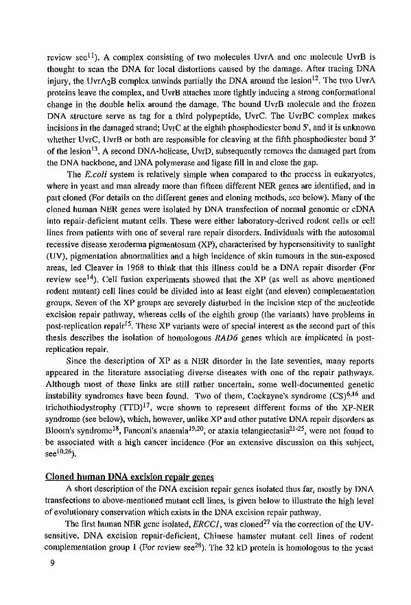
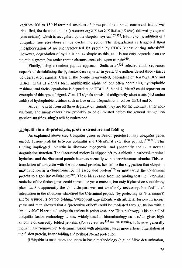
Finally, as in the yeasts SaccJzaromyces cerevisiae (baker's yeast)84 and ScJzizosaccharom)'ces pombe85 a large number of DNA repair mutants had been isolated, it would be very convenient to utilise the many correcting yeast genes c10ned for the isolation of human counterparts. Also in view of the limited number of human and hamster repair mutants (and the tedious transfection experiments) it was tempting to try to develop as first part of this thesis a general method for isolation of homologues of genes already cloned from other species. From the lessons learned in the course of this work it becarne obvious that, when trying to isolate similar genes in other organisms, it is essential to take the, evolutionary direction which is followed into account. Descending the evolutionary ladder (e.g. from man to Drosophila) via low stringency hybridisations using standardised methods86 is relatively easy, as the isolation starts from a complex genome (3x10⁶kb) with large introns and thus many possibilities for accidental homology to the relatively simple genome of the fly (IxlOSkb). However, when cloning a human homologue of a yeast gene (genome size: !x104kb) the whole complexity of the human genome is encountered. Therefore, the cloning strategy had to be adapted several times, otherwise many small regions with fortuitous homology would have been isolated. The small protein domain shown in Fig.1 (Ec09), for instance, was isolated by screening DrosopJzila cDNA libraries with the total yeast RADl
gene.
r Ad. 2 minor coat protein V
K D.lllelal/ogaster cDNA Ec09
E S.cerevisiae RAD!
Region of nucleotide identity between Ec09 and RAD!: 15' GGG AAG GAC GAC GAC GAT 3'1
Figure 1. Representation of the dOll13inal homology found between a Drosophila cDNA clone (Ec09), RADi, and the adellovirus minor coat protein V.
12
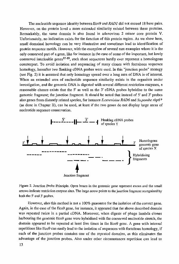
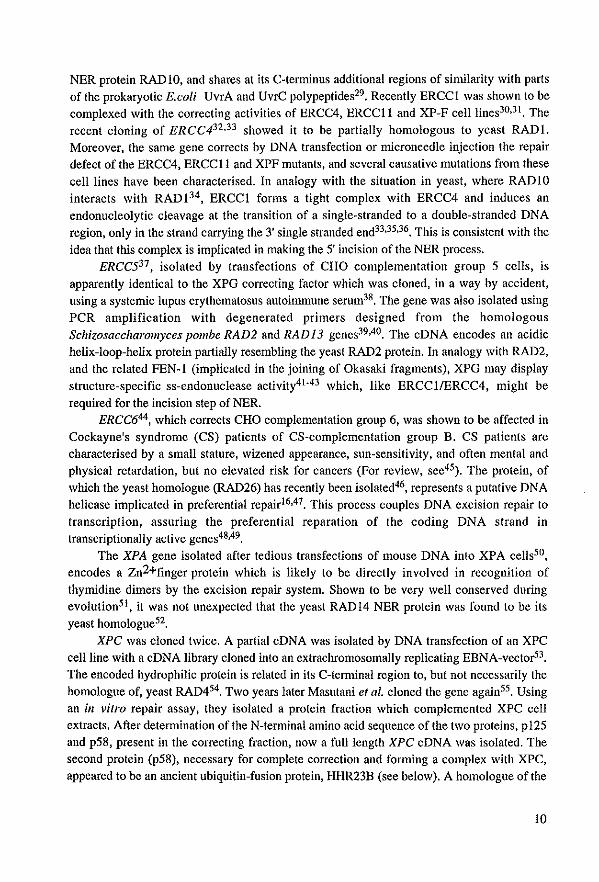
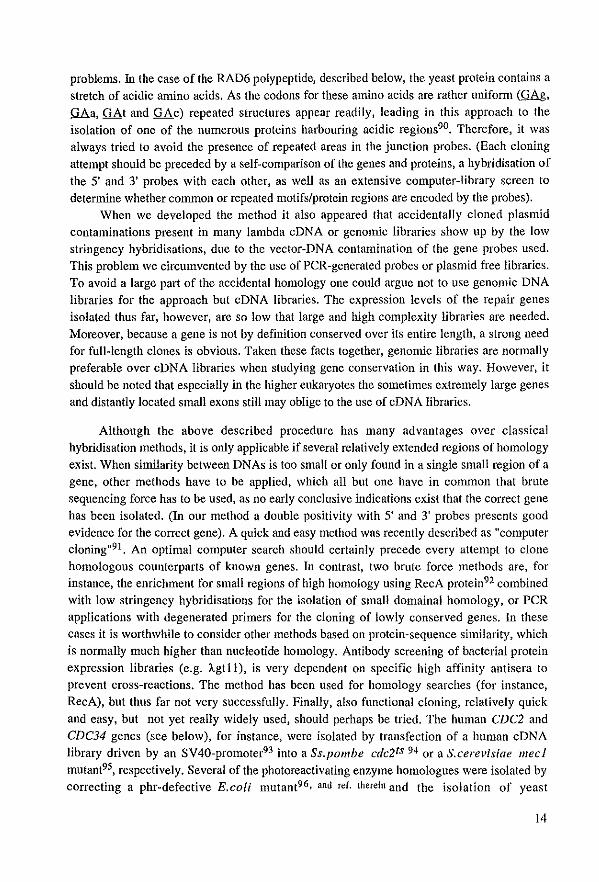
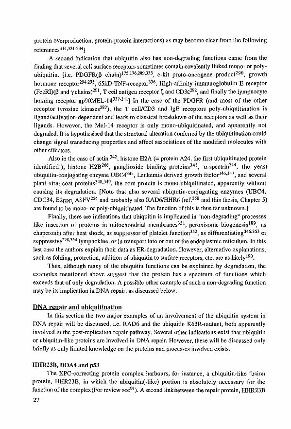
The nucleotide sequence identity between Ec09 and RAD1 did not exceed 18 base pairs. However, on the protein level a more extended similarity existed between these proteins. Remarkably t the same domain is also found in adenovirus 2 minor care protein V. Unfortunately, no il1dication exists for the function of thls protein region. As we show here, small domainal homology cao be very illustrative and sometimes lead ta identification of protein sequence motifs. However, with the exception of several rare examples where it is the only conserved part of a gene, like for instanee in the case of same of the important. but lowly conserved interleukin genes87-89 • sueh short sequences hardly ever represent a homologous counterpart. Ta avoid isolation and sequencing of mauy clones with fortuitous sequence homology, hereafter two flanking cDNA probes were used. In thls "junction probe" strategy (see Fig. 2) it is assumed that only homology spread over a long area of DNA is of interest.
When an extended area of nucleotide sequence similarity exists in the organism under investigation, and the genomic DNA is digested witlt several different restriction enzymes, a reasonabIe chance exists that the 5' as weil as the 3' cDNA probes hybridise to the same
genomic fragment; the junction fragment. It should be noted that instead of 5' and 3' probes also genes from distantly related species, for instance S.cerevisiae RAD6 and Ss.pombe rhp6+
(as done in Chapter H), can be used, at least if the two genes do not display large areas of nucleotide sequence conservation.
5' 3' ~--------IJ-- - - ~
Flanking cDNA probes of species Y
5' + t I t + I 3' Homologous _.t __ I:C?::::Io_!,-...t==::::r. __ .I'_I:CDIo..c._.!I.....tC:Ioo_,-__ =t:::::t.'I.._"';' genOlntc gene
Junction fragment
of species X
} Hybridising fragments
Figure 2. Junction Probe Principle. Open boxes in the genomic gene represent exons and the small arrows indicate restriction enzyme sites. The large arrow points to the junction fragment recagnised by bath the 5' and 3' probes.
However, also thls methad is not a 100% guarantee for the isolation of the correct gene. Again, in the case of the Ec09 gene, for instanee, it appeared that the above described domail1 was repeated twice in a partial cDNA. Moreover, when digestl-: of phage lambda clones harbouring the genomic Eco9 gene were hybridised with the conserved nucleotide stretch, the domain appeared to be repeated at least five times in the Ec09 gene. A gene with intel1lal repetitions like Ec09 can easily lead to the isolation of sequences with fortuitous homology, if each of the junction probes contains one of the repeated domains, as tltis eliminates the advantage of the junction probes. Also under other circumstances repetition can lead to
13
problems. In the case of the RAD6 polypeptide, described below, the yeast protein contains a stretch of acidic amino acids. As thc codons for these amino acids are rather unifonn (GAg, QAa, GAt and GAc) repeated structures appear readily, leading in th is approach to the isolation of onc of the numerous proteins harbouring acid ie regions90• Therefore, it was always tricd to avoid thc presence of repeated areas in thc junction probes. (Each cloning attempt should be preceded by a self~cornparison of thc genes and proteins. a hybridisation of the 5' and 3' probes with each ather, as well as an extensive computer-library screen to determine whether common or repeated motifs/protein regions are cncoded by the probes).
When we developed the method it also appeared that accidentally cloned plasmid contaminatiolls present in many lambda cDNA or genomlc libraries show up by thc low stringency hybridisations. due to thc vector-DNA contamination of the gene probes used. This problcm we circumvented by the use of PCR-generated probes or plasmid free libraries. To avoid a large part of the accidental homology one could arguc not to use genomic DNA libraries for the approach but cDNA libraries. The expression levels of the repair genes isolated thus far, however, are so low that large and high complexity librarics are needed. Moreover, because a gene is not by definition conserved over its entire length, astrong need for full-length clones is obvious. Taken these facts together, genomic libraries are normally preferabie over cDNA libraries when studying gene conservation in this way. However, it should be noted that especially in the higher eukaryotes the sometimes extremely large genes and distantly located small exons still may oblige to the use of cDNA libraries.
Although the above described procedure has many advantages over classic al hybridisation methods, it is only applicable if several relatively extended regions of homology exist. When similarity between DNAs is toa smalt or only found in a single small region of a gene, other methads have to be applied, which all but one have in common that brute sequencing force has to be used, as na early eonclusive indications exist that the correct gene has been isolated. (In our method a double positivity with 5' and 3' probes presents good evidence for the correct gene). A quick and easy method was recently described as "computer cloning"91. An optimal computer search should certainly precede every attempt to clone homologous counterparts of known genes. In contrast, two brute force methods are, for instance, the enrichment for small regions of high homology using RecA protein92 combined with low stringency hybridisations for the isolation of small domainal homology, or PCR appIications with degenerated primers for the cloning of lowly conserved genes. In these cases it is worthwhile to consider other methods based on protein-sequence similarity, which is normally much higher than nucleotide homology. Antibody screening of bacterial protein expression libraries (e.g. Àgtl1), is very dependent on specific high affinity antisera to prevent cross-reaetions. The method has been used for homolagy searches (far instanee, RecA), but thus far not very successfully. Finally, also functional cloning, relatively quick and easy, but not yet really widely used, should perhaps be tried. The human CDC2 and CDC34 genes (see belaw), for instanee, were isolated by transfection of a human eDNA library driven by an SV40-promoter93 into a Ss.pombe cdc2ts 94 or a S.cerevisiae meel mutant95, respectively. Several of the photoreactivating enzyme homologues were isalated by correcting a phrwdefective E.coli mutant96, and rer. therein and the isolation of yeast
14
topoisomerase II with Drosophila topoll was feasible with the sectoring/selection-method
described by Kranz et 01.97•
In our case the junction-probe strategy resulted in the isolation of two groups of genes;
the Dl'osophila l , Ss.polI/be (unpublished) and S.cel'evisiae2 hamalagues of ERCC3, and the Ss.polI/be9s, Drosophila99 and humanloo hamalagues of RAD6/UBC2. These latter genes are
the subject of this thesis, and as RAD6 has been shawn ta play a role in the nbiqnitin pathway, this system wiII be reviewed in the following part of the introduction,
General illiroductiall 10 ubiquitill
"Ubiquitin is too small and too abundant to be important; you should change your
research subject!" (Tald ta A. Haas abaut eleven years aga).
To state this about ubiquitin, one of the proteins most conserved in evolution, is nowadays impossible in view of the plethora of processes in which this "giant dwarf' plays a major role, Because it is impossible to cover the ubiquitin field within the Iimits of this
introduction, the major topics wiII be highlighted, especially thase in relatian with DNA repair (For a bevy of recent reviews see lOl- lJ I),
The -6x 10⁷ malecnles of this 76 amina acid protein faund in each cell of aur body
make it one of the most abundant polypeptides106 , Ubiquitin has been detected in a wide variety of organisms ranging from archaebacteriae Il2-114 to man, and rccently even in a
eubacterium, the cyanobactcrium Anabaella variabilis 115, Considered as the slowest evol\'ing
proteÎIl known 116, it allowed in the 1.2 billion years of evolution which separate yeast from man only three amino acid changes to occur l14 ,
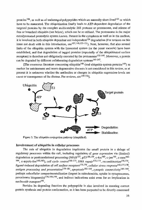
Ubiquitill cOlljugatioll patbway In a cell the majority of the ubiquitin molecules are not found as free protein, but
conjugated to other polypeptides lO6 , The linkage reaction and the proteins performing it, are apparently almast as canserved as ubiquitin itselfIOO.117.121(Fig.3).
Conjugation commences when the C-tenninal glycine residue of a ubiquitin molecule is
activated by one of the ubiquitin-activating cnzymes (referred to as Uba or El), which uses ATP to form a high energy thiol ester intermediate, that is covalently lillked to an internal cysteine residue of the EI-molecule 120, This protein-complcx is able to donate the 76 amino acids proteill to ane of a growing family of ubiquitin-conjugating enzymes (Ubc or E2)(e.g.
RAD6/UBC2). The E2-ubiquitin complex links the ubiquitill maiety via its C-tennillal glycine residue to the e-NH2 group of a lysine residue in the target protein, with or without
the help of a member of a family of ubiquitin ligases (Ubr or E3). The question whether (all) the E3-proteins are only docking proteins or bind ubiquitin to themselves, and thus actually
perform an enzymatic activity, rcmains to be resolved l22,
Originally, ubiquitination was shown to be involved in specific (extra-lysosomal) targeted degradation of the bulk of mislacalised, improperly processed, fareign or damaged
15
proteins 106, as weil as of undamaged polypeptides whieh are naturolly short-livedl23 or which have to be maturated. The ubiquitination finally leads to ATP-dependent degradation of the
targeted proteins by the complex multicatalytic 26S protease or proteasome, and release of free or branched ubiquitin (sec below), which can be re-utilised. The proteasome is the major
extralysosomal proteolytic system known. Present in the cytoplaSlll as weIl as in the nucleus, it is involved in both ubiquitin dependent and independent124 degradation (por reviews on thls issue not dealt with in this introduction, see107,11O,125-131). Note, however, that a180 several
links of the ubiquitin system with the lysosomal system (or the yeast vacuole) have been established, and th at degradation of tagged proteins (especially of the ubiquitinated surface
receptors) is therefore not obligatorily executed by the proteasome 132-144• Moreover, a protein can be degraded by different collaborating degradation systClnsI45.147.
[Thc enormous literature conceming ubiquitill148 (and ubiquitin-system proteins l49) as marker for autoimmune and neuro-degenerative diseases is not considered in this review, as at
present it is unknown whether the antibodies or changes in ubiquitin expression-Ievels are cause or consequence of the disease. For reviews, see 150-156].
- ........ ~ Degradation AlP --.... ~ Stabilisation
Figure 3_ The ubiquitîn conjugation pathway (simplified).
Involvement ofubiquitin in cellular processes
Thc role of ubiquitin in degradation implicates the sm all protein in a deluge of regulatory processes within the cell, inc1uding regulation of geile expressioll via (limited) degradation or posttranslational processing (NFkBI57, p53158.159, c_fosl60, c_junI61 , c_mos I62-164, c_myc/c_fosI60,165), eell cyc/e eontroI166-171. DNA repairl72.173. recombillatiOll170.174,
ligand-indlleed degradatioll of eell smface reeeptorsl75.176, cel/ular stress respoJlse142.177.178,
alltige1l, processi1lg mld preselltation179-18l, apoptosisl82-184, s)'naptic eOllIleetivity l85-188,
perhaps subcellular compartme1ltalizatÎoJl (import in mitochondria, uptake in synaptosomes, peroxisome biogcnesis)174,189,190, and indirect indications exist even for aIl implication in
nucleoside transport l91 .
Besides its degrading function the polypeptide is also illvolved in assuring correct
protein synthcsis and protein conformatioIl, as it has been purported to be directly concerned
16
in the (re?)folding of (damaged?) proteins. The protein also seems to have anti-degrading functions, maybe due ta its lnvolvement in folding, as in certain circumstances ubiquitin
protects against breakdown (see bel ow).
A choice fol' or against degradation: Iinkage types As far as \Vltat is known, the choice for or against degradation of ubiquitinated targets
depends on two facts. First, whether the polypeptide is mono- or poly-ubiquitinated, and second, where in the target protein-backbone ubiquitin is attached. It was found that proteins ean contaill either single ubiquitin molecules (mono-ubiquitination) or tree structures of branched ubiquitin (poly-ubiquitination). These Iatter structures, whose formation is often
dependent on the presence of an E3 enzyme, consist of ubiquitin molecules which are linked via their C-terminal glycine to specific intemallysine residues of another ubiquitin molecule. n is generally assumed that at least the lysine 48 (KA8) poly-ubiquitination leads to breakdownl92,193. The existence of K-6, K-11, K-29 and K-63 poly-ubiquitination has also recently been describedl73,194-196. And altllOugh RAD6 can make K-6 tree stmctmes on
histone H2B in the absence of an E3 protein, the function of this linkage type remains unknownl96. The human E2 enzyme EPF149.197 is making K-ll linkages, which like K-29
and K-48 poly-ubiquitination, are involved in protein breakdown198. Finally K-63 polyubiquitination, performed by RAD6 in an E3-dependent mannef, is apparently a poor iuduccr of degradation. This conjugation-type has recently been implicated in DNA repair, perhaps with a regulatory function. A yeast mutant whieh is unable to perform the K-63 linkage shows a phenotype which in part is comparable to that of a rad6 deletion mutaIlt 173 (see
below). Mono-ubiquitination is llonnally not involved in breakdown but in the
stabilising/folding functions (see below), although, in the case of the artificially-made ubiquitin-proline-p-galactosidase199, and in the case of o:_globin2OO,201 1t may be sufficient for degradation. In broader tcrms, all these results suggest that ubiquitin is a versatile signal in which different ubiquitin chain configurations are used for different funetions. A single
ubiquitin conjugating enzyme is apparently able to perform different linkages (for RAD6: K-6, K-48 and K-63) dependent on the target and the E3 involvedl96.
Ubiquitin genes & Fusion proteins When the first ubiquitin genes were cloned, it appeared that all organisms harboured
many functional copies as weIl as many pseudogenes202 . Moreover, always at least one of the
genes was a poly-ubiquitin gene, harboudng a highly varia bie number of ubiquitin coding elements in a head-to-tail arrangement, and thus encodillg a poly-ubiquitin precursor protein [e.g. 3 to 9 copies (man)203,204, 14 (Arabidopsis)205, 11 (Caellol'habditis), 2 to more then 40
(trypanosomatidae), 18 (Dl'osophila), 7 (maize), 6 (sunflower), 5 (yeast)106]. The polyubiquitin genes are in general inducible in the stress response (e.g. heat shock)178,206, in
contrast to the mono-ubiquitin gen es. The mono-ubiquitin gelles are often fusion genes as
they encode the ubiquitin moiety in frame with a C-temlÎnal extension peptide (CEP)207. The CEPs were found to represent two types of small very conserved ribosomal polypeptides, implicating ubiquitin in ribosome biosynthesis; the CEP80 proteins (with a variabie length of
17
76, 80 or 81 amino acid residues) are found to be identical to ribosomal protein S27a which is part of the 40S particle, and the ribosomal CEP52 proteins whieh represent the IAO peptide, a constituent of the 60S or large ribosomal subunit20S-211. This fundamental finding led to quite a few publications deseribing the same phenomenon in other organisms [CEP52: Chlamydomollas212 , Arabidopsis213 , Tetrahymella (CEP53)214, Dictyostelium21S-217, Drosophila21S , ehieken219 , and man220, CEP80: Neurospora(78)221 , Dietyostetium (78)217,
maize(79)222, Arabidopsis(80)213, and Drosophila223], illustrating the evolutionaty eonserva
tion of the C-terminal fusion partners.
Ubiquitin-like proteins The search for C-terminal extcnsion protcins led furthermore to the discovery of ncw
types of rlbosomal fusion proteins, like for instanee the Nicotiana tabacum CEP72 protein224
whieh is related to the CEP52 proteins. In addition the family of ubiquitin-like sequenees (Uhl) was expanded as several
authors in their search for ubiquitin fusions, identified ubiquitin-like proteins with C-tenninal extensions22S-228 or proteins with a C-terminal (I) ubiquitin-like extension("NEPs")229. The
family ofubi-like protein sequenees is eonstanUy growing (Tabie I).
Table I: Ubiquitin-like proteins, Ubl-CEP fnsjQus:
1. Caenorhabditis CEP93225
2. Rat ribosomal protein S30 (also known as Fau protein or lymphokine MNSp226-228) NEP· Ubl fnsfons:
3. Tbc Ub! moiety fused to the C-terminal end of mammalian splicing factor SF3a120 and its yeast homologue, PRP21p229
Normal UbJs!
4. Xenopus Ania and b proteins fused to a Zn2+ -finger protein230
5. 15kD interferon-induced ISG15 gene product UCRP23 1-235
6. NEDD8 protein236.237 7 Chinese hamster (and mouse) CHUB2 gene238
8. Earthwonn EiselliafetMa Andrei Ub1239
9. X-chromosomal GdX protein240
10. BAT3 polypeptide241
11. Baculoviral v-ubi protein242
12. DNA excision repair proteins HHR23A and B55
13. Positive regulator subunit pl8 of the sm general transcriptionfelongation factor 243 14. A whole group ofnon-expressed Ubl-pseudogenes in Arabidopsis205
The idea that these Ub!'s ean replaee normal ubiquitin in its funetions, is thus far only founded on the detaHed analysis of UCRP. This di-ubiquitin-like protein is weakly homologous to normal ubiquitin and was shown to be conjugated to cellular proteins in vivo244. The ubiquitin-like proteins in Table I should therefore probably be divided into two different classes. A first group of funetional "weil" eonserved (especially the C-tenninal glycine residues) ubiquitin-like proteins whieh have similar funetions as classical ubiquitin but are involved in parallel pathways (UCRP, NEDD8, Ubl-Fau, Ubl-CEP93, SF3aI20, v-ubi
18
and although less likely, Ubl-Anla and b). And a second class of ancient "normal ubiquitin"
fusion proteins which lost the cleavage site between ubiquitin and the CRterminal extellsion (BAT3, GdX, sm pI8, CHUB2, sm pl8 and HHR23). As because of this most of the
evolutionary pressure on the ubiquitin moiety was lost, the coding sequence slowly challged. The "stabiHsing function" (see below), however, was probably retained, and is apparently absoJutely required for correct function of the fused partner243 .
With the identification of the ubiquitin-like molecules the complexity of the system is
increasing even further. If narmal ubiquitin is already impIlcated in the plethora of processes specified in this introduction, \Vhat wiJl be the function of these ubiquitin homologues and why did they evolve? What wiII be the function ofrecently cloned EI-like proteins245.248 or
of the different virus-encoded proteins: the ubiquitin(-like) proteins of baculovirus (vubi242,249,250), bovine viral diarrhoea virus25 1,252, and Finkel-Biskis-Reilly murine sarcoma virus226,227, and the E2 protein UBCvl (related to RADG) of African swine fever virus253-255?
Do these vlruscs use the ubiquitin system for their benefit in a simllar way as Human Papilloma Virus 16 and 18, whose E6 protein interacts with a ceUular E3 protein, EG-AP, and forces it to recognise p53, leading to the poly-ubiquitination and degradation of th is antioncogeneI59,17I,256,257? Or do they try to escape the attacks by the ceU's degradation systems
by titrating the cellular ubiqnitin with non-conjugatable ubiquitin homologues or by mirnickillg their proper ubiquitination with their own E2's249?
Ubiquitiu speel/ic proteases Although ubiquitin carboxyl-terminal hydrolases or ubiquitin specific protcases (UBP's)
\Vere known ta exist. and ta be implicated in the production of mono-ubiquitin from the polyubiquitin precursors, the isolation and cloning of these hydrolases was also accelerated due ta the identification of the ubiquitin-CEP fusions.
Same of them were shown ta remove smalt peptides or single amino acids from the Cterminal end of ubiquitin258,259, and ta be necessary for the maturation of the last ubiquitin moiety of a poly-ubiquitin protein. This last ubiquitin-copy of a poly-ubiquitin gene often
contains some additional C-tenninal amino acids, probably to prevent the non-branched poly
ubiquitin molecules from partic1pating in the linkage reaetions. A second class is implicated in the production of single ubiquitin~moieties from the poly~ubiquitin precursors, or in the maturation of the C~terminal fusion proteins260,26l, Finally, the third group of ubiquitin Iyases, to which the human oncogene product Tre-2 or its yeast homologues DOA4262 or
UBP5263 belong, releases andlor degrades poly-ubiquitin trees. Proteins from the last group are implicated in the rescue of faulty-targetted proteins or to recuperate free ubiquitin for reutilization after degradation of the tagged proteins264-266, Note, however, that degradation of tree-structures is not absolutely necessary as they also can be re-used directly, Moreover, free tree-structures can made by certain E2 enzymes independent of the presence of a target protein267-269,
With the identification of the UBPs the description of the ubiquitin system components is complete: single ubiquitin can be made from the fusion or poly~ubiquitin gene products; EI, E2 and E3 proteins can do their work; the proteasome degrades the targetted proteins; and finally the poly-ubiquitin trees can be recuperated to yield again free ubiquitin.
19

In the next paragraphs the implications of ubiquitin in degradation and in antiproteolysis, protein structure and folding wil! be briefly discussed. The chapter finishes with a summary of our CUlTent knowledge on the role of ubiquitin in DNA repair and chromatin structure, which is of course obligatorily linked to one of the E2-enzymes, RAD6.
Ubiquitin in protein degradation AH living cells have to reglIlate the content and composition of their resident proteins,
but the mechanisms by which this is done are not weIl known. Intracellular protein degradation is important in determining steady state and fluctuations of protein concentrations as weIl as for the generation of protein fragments that act as hormones, antigens, or other effectors. Breakdown can be regulated by innate properties of the protein substrate (e.g. PEST270_ or KFERQ271-sequences), or by chemical modifications (e.g. ubiquitin) which mark
them for breakdown, in other words which confer metabolic instability. The initial event leading to degradation mayor may not involve I) proteolysis, 2) non-proteolytic (covalent) modifications (e.g.oxidation of methionines, ubiquitin conjugation, AANDENYALAAtagging272 [i.e. A COOH-terminal peptide-sequence, thus far only detected in E.coli, which is linked to a protein while it is being translated from an erroneous mRNA which does not encode a stopcodon. This tagged incomplete protein is subsequently degraded by tag-specilic proteases. The process involves a new RNA type (with both a transfer and messenger function (tmRNA)) and a switch ofthe translation machinery from the defective mRNA to the tmRNA. It represents a magnificent quality control mechanism for defective.mRNAs], 3)
denaturation or unfolding of the protein, or 4) sequestration in cytoplasmic or nuclear "organelles". These processes, however, have to be selective as an enormous heterogeneity in degradation rates exists for the different proteins in the cell. Ubiquitination is one of the ways to achieve such a selectivity. Although the number of natural degradation-targets of the ubiquitin machinery273 starts to grow (see Table Il, pg. 21), the issue of what determines the specificity of the ubiquitin ligation system i.e. the degradation signals for commltment of certain proteins to degradation is not yet resolved.
Ubiquitin degradation signals The only general prerequisite for degradation of a protein via ubiquitination is the
obligatory presence of a lysine residue to whose E-amino group the ubiquitin moiety will be
finally attached31O. The other additional structural features of a substrate which are recognised by the ligation system are for the moment not weIl known, and rather non-uniform31O(see below). To complicate the situation it even appeared that proteills which do not contain any degradatioll-sigllals themselves can be degraded by their interaction with other polypeptides or subunits which only serve as (undegradable) tag for the ubiquitin machinery (transrecognition) 199,280,286,311 .
N-rule system and RAD6 Varshavsky and co-workers identified the first of the ubiquitin-system degradation
signais; the presence of a free alpha-alllÎllo group (For extensive reviews and detailed explanations, seeI99.312).
20
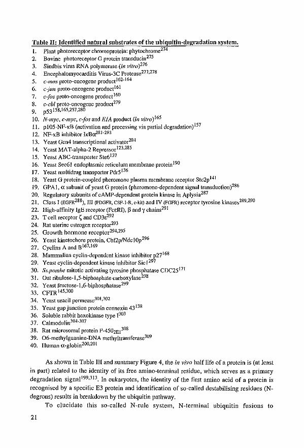
T.ble IJ: Identified natm'al substr.tes of the ubiguitin-degradation system.
1. Plant photoreceptor chromoprotein: phytochrome274
2. Bovine photoreceptor G protein transducin275
3. Sindbis virus RNA polymerase Uil vitro)276
4. Encephalomyocarditis Virus-3e Protease277,278 5. c-mos proto-oncogene product162.164 6. c-jull prota-oncogene product161
7. c..Jos proto-oncogene product 160
8. c-cbl proto-oncogene product279
9. p53158.l65,257,280
10. N-myc, c-myc. c10s and E1A product (ill vitro)165 11. pl05-NF-KB (activation and processing via partial degradation) 157 12. NP-KB inhibitor 11<B0:281 -283 13. Yeast Gcn4 transcriptional activator284
14, Yeast MAT-alpha-2 Repressorl23.285
15. Yeast ABC-transporter Ste6137
16. Yeast Sec61 endoplasmic reticulum membrane proteinI90
17. Yeast multidrug transporter PdrS l36
18. Yeast G protein-coupled pheromone plasma membrane receptor Ste2p141 19. GPAl, (J. subunit ofyeast G protein (pheromone-dependent signal transductionp86 20. Regulatory subunits of cAMP-dependent protein kinase in Aplysia287
21. Class 1 (EGFR288), III (pDGFR, esp·l-R, c-kit) and IV (FGFR) receptor tyrosine kinases289,290 22. High-affinity [gE receptor (FeeR!), pand y ehains29t
23. T eell receptor ç and CD3e292
24. Rat uterine estrogen receptor293
2S. Growth honnone receptor294,295 26. Yeast kinetoehore protein, Cbf2plNdelOp296 27. Cyclins A and B167,169
28. Marnmalian cyclin-dependent kinase inhibitor p27168
29. Yeast cyclin-dependent kinase inhibitor Sicl297
30. Ss.pombe mitotic activating tyrosine phosphatase CDC2S t7l
31. Dat ribulose-l ,5-biphosphate carboxylase298
32. Yeast fructose-l,6-biphosphatase299
33. CFfR 145,300
34. Yeast uracil permease30l ,302 35. Yeast gap junction protein connexin 43 138
36. Soluble rabbit hexokinase type 1303
37. Calmodulin304.307
38. Rat microsomal protein P-4501E1308 39. 06-methylguanine-DNA methyltransferase309
40. Human (J._globin2OO,201
As shown in Table lil and summary Figure 4, the in vivo half life of a protein is (at least
in part) related to the identity of its free amino-terminal residue, which serves as a primary
degradation signal l99,3l3. In eukaryates. the identity of the first amino acid of a protein is
recognised by aspecific E3 protein aud ideutificaliou of so-called destabilisiug residues (N
degrons) results in breakdown by the ubiquitiu pathway,
Ta elucidate th is so-called N-rule system, N-terminal ubiquitin fusions to
21
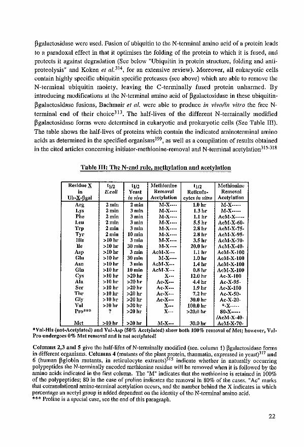
pgalactosidase were used. Fusion of ubiquitin to the N-terminal amino acid of a protein leads
to a paradoxal effect in that it optimises the folding of the protein to which it is fused, and
protects it against degradation (Sec below "Ubiquitin in protein stmcture, folding and antiproteolysis H and Koken et al. 314 , for an extensive review). Moreover, all eukaryotic eells contain highly specific ubiquitin specific proteases (see above) which are able to remove the
N-terminal ubiquitin moiety, leaving the C-terminally fused protein unharmed. By
introducing modifications at the N-terminal amino acid of flgalactosidase in these ubiquitin
flgalactosidase fusions, Bachmair el al. were able ta produce ill vivo/in vitro the free N
terminal end of their choice3l3. The half-lives of the different N-terminally modified
pgalactosidase forms were determined in eukaryotic and prokaryotic cells (See Table III).
The table shows the half-lives of proteins which contain the indicated aminoterminal amino
acids as detennined in the specified organisms199, as weil as a compilation ofresults obtained in the cited articles conceming initiator-methionine-removal and N-terminal acetylation31S-318
Table 111: The N·end rule, melhylation and acelvlation
ResidueX tl/2 tl/2 Mefhionine t1l2 MetWonine in E.eoU Yenst Rcmoval Reticulo· Removal
Ub·X·pgal ill vim Acetylation cyfes ;11 vifro Acetylation Arg 2 min 2 min !\I·X .... 1.0 hr M-X .... · Lys 2 min 3 min M-X·--- 1.3hr M·X .... • Pbe 2min 3min 1\l-X···· 1.1 br AcM·X .. ··· Leo 2min 3 min M·X···· S.S hr AcM·X·60· Trp 2 min 3min M·X .. ·- 2.8hr AcM.X·75· Tyr 2min lOrum M·X .... 2.8hr AcM-X-95-His >10hr 3 mln M·X .... 3.5 hr AcM·X-70· Ile >10 hr 30 min M-X .. •• 20.0 hr AcM·X·40·
Asp >10 hr 3 mln Aci\l-X .. • 1.1 hr AcM.X·JOO Glo >10hr 30 min M·X·· .. 1.0hr AcM-X·I00 Asn >10hr 3 mln AcM·X .. • 1.4hr AcM.X.I00 Gin >10hr 10min AcM·X .. • O.Shr AcM·X-I00 eys >10 hr >20hr X .. • 12.0 hr Ac·X-IOO Ala >10 hr >20hr Ac-X .. • 4.4 hr Ac·X·95· Ser >10 hr >20hr Ac·X ... 1.9 hr Ac-X·JOO Thr >10hr >20 hr Ac·X .. · 7.2hr Ac·X·SO· Gly >10hr >20 hr Ac·X··· 30.0 hr Ac-X-20· Val >10 hr >20hr X-.. 100.0 hr *·X· .... Pro*** ? >20hr X-·- >20.0 hr SO·X .... ·
IAcM-X·40-Met >10hr >20 hr !\I·X .. • 30.0 hr Ac!\I·X·70·
*Val·His (not-Acetylated) and Val·Asp (50% Acetylated) show bollt 100% removal or Met; however, Val. Pro undergoes 0% Met removal and is nof acetylated!
Columns 2,3 nnd 5 give the half-lifes of N-tenninally modified (see. column 1) pgalactosidase fonns in different organisms. Columns 4 (mutants ofthe plant~rotein, thaumatin, expressed in yeast)317 and 6 (human pglobin mutants, in reticulocyte extracts) 15 illdicate whether in naturally occurring polypeptides the N-terminally encoded methionine residue will be removed when it is followed by the amino acids indicated in the first column. The "M" indicates that the methionine is retained in 100% of the polypeptides; 80 in the case of proline indicates the removal in 80% of the cases. "Ac" marks that cotranslational amino-tenninal acetylation occurs, and the number behind the X indicates in which percentage an acetyl group is added dependent on the identity of the N-terminal amino acid. *** Proline is a special case, see the end of this paragraph.
22
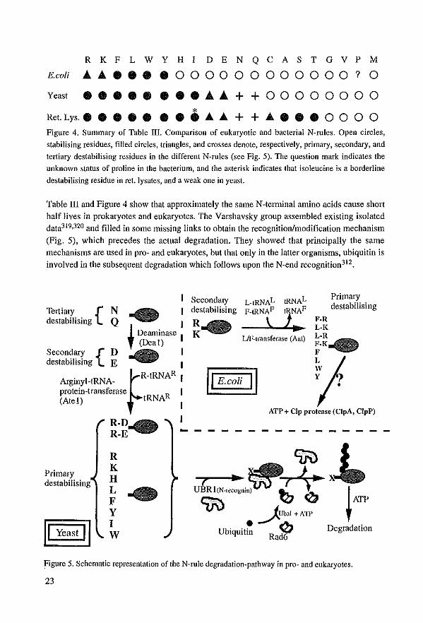
R K F L W Y H I DEN Q CAS TGV P M
E.coli ............ 0 0 0 0 0 0 0 0 0 0 0 0 ? 0
Y- ................ ++00000000
Ret. Lys.. • • • • • • • ..... ..... + + ........ 0 0 0 0 Figure 4. Summary of Table lIl. Comparison of eukaryotic and bacterial N-rules. Open circles,
stabilising residues, filled circles, triangles. and crosses denote, respectively, primary, secondary, and
tertiary destabilising residues in the different N-rules (see Fig. 5). The question mark indicates the
unknown status of proline in the bacterium, and the asterisk indicates Ihat isoleucine is a borderline
destabilising residue in eet. Iysates, and a weak one in yeast.
Table mand Figure 4 show that approximately the same N-terminal amino ac1ds cause short half lives in prokaryotes and eukaryotes. The Varshavsky group assembIed existing isolated data319,320 and filled in same missing links to obtain the recognition/modification mechanism
(Fig. 5), whieh precedes the actual degradation. They showed that principally the same mechanisms are used In pro- and eukaryotes, but that only in the latter organisms, ublquitin is involved in the subsequent degradation which follows upon the N-end recognition312.
Tertiary {N destabilising Q I
I Deaminase I , (Deal)
Secondary {D I destabilising E I
~R-tRNAR I
Arginyl-tRNA-protein-transferase R I (AteI) tRNA I
I
SecondalY destabilising
L·IRNAL P·IRNAF
\ UF-lransferase (Aat)
F
Primary destabilising
jI '-roN n 'I ATP + Clp protease (ClpA, ClpP)
- - - --
Primary destabilising
R K H L F Y I W
ctri) JUbaJ + All'
Ubiquit! 0 Degradation Rad6
Figure 5. Schematic representation of the N-rule degradationwpathway in prowand eukaryotes.
23
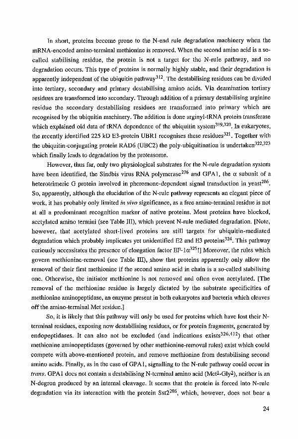
In short, proteins become prone to the N-end rule degradation machinery when the
mRNA-encoded amino-tenninal methionine is removed. When the second amino acid is a so
called stabilising residue, the protein is not a target for the N-ru1e pathway, and no
degradation occurs. This type of proteins is normally highly stabie, and their degradation is
apparently independent of the ubiquitin pathway312. The destabilising residues can be divided
into tertiary, secondary and primary destabilising amino acids. Via deamination tertiary
residues are transfonned into secondary. Tllrough addition of a primary destabilising arginine
residue the secondary destabilising residues are transformed into primary which are
recognised by the ubiquitin machinery. The addition is done arginyl-tRNA protein transferase
which explained old data of tRNA dependenee of the ubiquitin system319,320. In eukaryotes,
the recently identified 225 kD E3-protein VBR I recognises these residues32l . Together with
the ubiquitin-eonjugating protein RAD6 (VBC2) the poly-ubiquitination is undertaken322,323
whieh finally leads to degradation by the proteasome.
However, thus far, only two physiologieal substrates for the N-rule degradation system
have been identified, the Sindbis vims RNA polymerase276 and GPAI, the " subunit of a
heterotrimeric G protein involved in pheromone-dependent signal transduction in yeast286.
So, apparently, aIthough the elucidation of the N-ru1e pathway represents an elegant pieee of
work, it has probably only lirnited ill vivo significance. as a free amino-terminal residue is not
at all a predominant recognition marker of native proteins. Most proteins have blocked.
acetylated amino tennini (see Table lIl), whieh prevent N-ru1e mediated degradation. [Note,
however, that acetylated short-lived proteins are still targets for ubiquitin-mediated
degradation whieh probably implicates yet unidentified E2 and E3 proteins324• This pathway
curiously necessitates the presence of elongation factor EF_la325 !] Moreover, the rules which
govern methionine-removal (see Table lIl), show that proteins apparently only allow the
remaval of their first metWonine if the second amino acid in chain is a so-called stabilising
one. Otherwise. the initiator methionine is not removed and of ten even acetylated. [The
remaval of the methionine residue is largely dictated by the substrate specificities of
methionine aminopeptidase, all enzyme present in both eukaryotes and bacteria which cleaves
offthe amino-tenninal Met residue.]
So, it is likely that this pathway wiII only be used for proteins whieh have lost their N
tenninal residues. exposing now destabilising residues, or for protein fragments, generated by
endopeptidases. It can also not be excluded (and indications exists326,412) that other
methionine aminopeptidases (governed by other methionine-removal mIes) exist which could
compete with above-mentioned protein. and remove methionine from destabilising second
amino aeids. Finally, as in the case ofGPAl, signalling to the N-mle pathway eould oeeur in
trans. OPAl does not contain a destabilising N-terminal amino acid (MetL Oly2), neither is an
N-degron produeed by an internal cIeavage. It seems that the protein is forced illtO N-rule
degradation via its interaction with the protein Sst2286, which, however, does not bear a
24
destabilising N-terminus (MetL VaI2) either327. From the above data it is not clear which
factors determine the N-rule mediated degradation of the GPA I protein.
Ubiquitin Fusion Degradation When resolving the N-rule, an N-terminal praline was found ta be a stabilisÎng residue
(Tabie III). However, the ubiquitin-proline-~gal fusion protein (used to produce proline-~gal)
was found to be extremely short-lived (tl/2;7 min)312! As explained in the introduction to the
N-rule, the fusion of ubiquitin to the N-terminal amino acid of a polypeptide normally
stabilises the C-terminal fusion partner. However, an N-tenninal praline. or ubiquitin-fusions
in which the C-terminal glycine-76 residue of ubiquitin is modified or absent e.g. Ub Va176_ V
~ga1198 leads to a short half-life. The removal of the ubiquitin-moiety, which is supposed to
accur almost co-translationally, is in this type of fusions extremely slow or even absent (if the
C-terminal glycine is absent). This "proline discrepancy" led (again) the Varshavsky group to
decorticate the phenomenon, leading to yet another degradation pathway, the UFD (!J.biquitin
Eusion !2egradation). In this pathway the "non-removable" N-terminal ubiquitin is recognised
as degradation signa!. The targeting of a ubiquitin fusion by the UFD pathway results in the
poly-ubiquitination (K48 or K29-poly-ubiquitination, dependent on the C-terminal partner) of
the fusion's "non-removable" ubiquitin moiety, a step required for the subsequent proteasomal
degradation. Thus far five genes were isolated from this pathway, but details are not yet
known198. Moreover,like long time for the N-rule pathway, substrates are still unrecognised,
although some of the "non-removable" ubiquitin-like fusion proteins (see "Ubiquitin-like
proteins") are good candidates.
"2nd Codon rule" and Deslruclion box
Another breakdown signal different from the free N-tenninals of the N-rule pathway or
the non-removable N-terminal ubiquitin was detected in the C-lllOS proto-oncogene product.
This protein which is implicated in cell cycle control is degraded by the ubiquitin pathway.
The second (proline-2) and third (serine-3) N-tenninal amino acids were shown to determine
the half-life of this polypeptide162-164. As shown above, proline was found as a stabilising
residue in the N-rule. And as said before, the proteins containing such residues are normally
stabie, and not dependent 011 ubiquitin for their degradation312. c-mos, however, is unstable
when its third residue (Ser-3) is in an unphosphorylated state. This situation favoms the
recognition of proline-2 by an E3 protein. This interactionleads then to quick degradation of
the unphosphorylated protein via the ubiquitin pathway. However, if sefÎne-3 is
phosphorylated, proline-2 is not recognised anymore, and no ubiquitin tree can be added to
lysine-34 of c-mos.
The fourtlt ubiquitin-degradation signal known is the so-called destruction box found in
A- and B-type cyclins, which are quickly degraded at the end of mitosis. In the highly
25
varlable 100 to 150 N-terminal residues of these proteins a small conserved island was
identified, the destruction box (consensus: Arg-X-X-Leu-X-X-Ile/(Leu)-X-(Asn), followed by dispersed
lysine residues), whlch is recognised by the ubiquitin system 167,328, leading to the addition of a
ubiquitin tree elsewhere in the cyclin molecule. The degradation is triggered upon
phosphorylation of an uncharacterised E3 protein by CDC2 kinase during mitosis329 .
However, degradation of cyclin is not as simple as thls, as it is not only dependent on the
ubiquitin system, but under certain circumstances also upon calpain330.
Finally, using a random peptide approach, Sadis et al.326 selected small sequences
capable of destabilising the pgalactosidase reporter in yeas!. The authors detect three classes
of degradation signais: Class J, the N-rule re-invented, dependent on RAD6/UBC2 and
UBRI. Class II signals farm amphipathic alpha helices aften containing hydrophobic
residues, and their degradation is dependent on UBC4, 5, 6 and 7. Mat«2 could represent an
example of this type of signa!. Class III signals consist of obligatorily-short tracts (4-5 amino
acids) of hydrophobic residues such as Leu or ne. Degradation involves UBC4 and 5.
As can be seen from all these degradation signaIs, they are for the moment rather non
uniform, and many others have probably to be elucidated befare the general recognition
mechanism (if existing?) will be understood.
Ubiguitin in anti-proteolysis. protein structure nnd folding As explained above (see Ubiquitin genes & Fusion proteins) many ubiquitin genes
encode- fusion-proteins between ubiquitin and C-terminal extension peptides208,21O. This
finding implicated ubiquitin in ribosome biogenesis, and apparently not in its normal
degradation function. The C-terminal moiety is clipped off by a ubiquitin carboxyl-terminal
hydrolase and the ribosomal protein interacts normally with other ribosome subunits. This co
translation of ubiquitin with the ribosomal proteins has led to the suggestion that ubiquitin
may function as a chaperonin for the associated protein21O or may target the C-terminal
protein to a specifîc cellular site208. These ideas came from the finding that the C-terminal
moieties of the fusion genes could correct the yeast mutants, but only if placed on a multicopy
plasmid. Sa, apparently the ubiquitin-part was not absolutely necessary, but facilitated
integration in the ribosome, stabilised the C-tenninal peptide (by protecting its N-terminus?)
and/or assured its correct folding. Subsequent experiments with artificiaI fusions in E.coli,
yeast and man showed that a "protective effect" could be mediated through fusion with a
"removable" N-terminal ubiquitin molecule (otherwise, see UFD pathway). This so-called
ubiquitin-fusion technology is now widely used in biotechnology as it aften gives high
amounts of correctly folded proteins (For review see314 and ref. thercin). It is now generally
thought that "removable" N-tenninal fusion with ubiquitin causes more efficient translation of
the fusion protein, better folding and perhaps N-end protection.
[Ubiquitin is used more and more in basic methodology (e.g. half-live determination,
26
protein overproduction, protein-protein interactions) as may become c1ear from the following
references314,331-334]
A second indication that ubiquitin also has non-degrading functions came from the finding that several ceIl surface receptors sometimes contain covalently linked mono- or polyubiquitin. [i.e. PDGFR(P chain)175,176,289,335, c-kit proto-oncogene product290, growth
hormone receptor294,295, 65kD-TNF-receptor336, High-affinity immunoglobulin E receptor (FceRI)(p and ychains)291, T cell anligen receptor ç and CD3e292, and finally the lymphocyte
homing receptor gp90MEL_14337-341] In the case of the PDGFR (and most of the other
receptor tyrosine kinases289), the T cell/CD3 and IgE receptors poly-ubiquitinalion is ligand/activation-dependent and leads to classical breakdown of the receptors as weIl as their ligands. However, the Mel-14 receptor is only mono-ubiquitinated, and apparently not degraded. It is hypothesised that the structural alteralion conferred by the ubiquitinalion could change signal transducing properties and affect associations of the modified molecules with other effectors.
Also in the case of actin 342, histone H2A (;;; protein A24, the first ubiquitinated protein
idenlified!), histone H2B266, ganglioside binding proteins343, u-spectdn344 , the yeast ubiquitill-conjugating enzyme UBC4345, Leukemia derived growth factor346,347, and several plant vi ral coat proteins348,349, the core protein is mono-ubiquitinated, apparently without causing its degradalion. [Note that also several ubiquitin-conjugating enzymes (UBC4, CDC34, E2EPF, ASFV254 and probably also RAD6/HRR6 (ref.350 and this thesis, Chapter 5) are found to be mono- or poly-ubiquitinated. The function of this is thus far unknown.]
Finally, there are indications that ubiquitin is implicated in "non-degrading" processes like insertion of proteins in mitochondrial membranes351 , peroxisome biogenesis l89, as chaperonin after heat shock, as suppressor of platelet function352, as differentiating346,353 or suppressive228,354 lymphokine, or in transport into or out of the endoplasmie reticulum. In this last case the authors explain their data as ER-degradation. However, alternative explanations. such as folding, protection, addition ofubiquitin to surface receptors, etc. are as likely190.
Thus, although many of the ubiquitin functions can be explained by degradation, the examples mentioned above suggest that the protein has a spectmill of functions which exceeds that of only degradation. A possible other example of such a non-degrading function may be its implication in DNA repair, as discussed below.
DNA repair and ubiquitlnatlon In this section the two major examples of an involvement of the ubiquitin system in
DNA repair will be discussed, i.e. RAD6 and the ubiquitin K63R-mutant, both apparently involved in the post-repHcation repair pathway. Several other indications exist that ubiquitin or ubiquitin-like proteins are involved in DNA repair. However, these will be discussed only briefly as only limited knowledge on the proteins and processes involved exists.
HHR23B, DOA4 and pS3 The XPC-correctillg proteill complex harbours, for instance, a ubiquitin-like fusion
protein, HHR23B, in which the ubiquitin(-like) portion is absolutely necessary for ihe function of the complex(For review see91 ). A second linkbetween the repair protein, HHR23B
27
and the ubiqtiitin system is the presence of the higWy conserved ..... 50 amino acids UBA domain, a stmcture of unknown function that is found in several ubiquitin hydrolases, E2 and E3 proteins, as weIl as in several protein klnases355,356,
Another example of involvement of thc ubiquitin pathway in repair precesses is the yeast DOA4 gene which is related to the human fre-2 or meuse Unp oncogene262,263, This gene encodes a de-ubiquitinating enzyme involved in the recycling of ubiquitin late in the proteolytic pathway. In contrast to for instance the YUHI, UBPI, 2 and 3 hydrolases, DOA4 is rat her essential as deletion of the gene results in poor growth and a severe inhibition of general proteolysis, The deletien mutant is very sensitive to UV and y-irradiation, sporulation is almast absent, and the degradation of bath N-rnle and UFD-target proteins is inhibited (10-20 fold).
Yet atlOtiler aspect of the role of ubiquitin in DNA repair is its invelvement in the degradation of "checkpoint proteinU p53, The p53 turnour suppressor protein co-ordinates multiple responses to DNA injury. DNA damage causes an increase of functional p53 in the cell. Increase in pS3 activity leads to cell cycle arrest whieh allows the cell to repair its DNA injury. When damage is beyond repair, pS3 activates the apoptosis pathway and the cell dies. Proper regulation of thls crucial protein is of utmost importance, , .. and ubiquitin is part of the regulatory processes affecting pS3 158,159.165,257,280.357.
RAD6 mutant;gene and protein When our initial attempts to isolate Drosophila DNA excision repair genes with probes
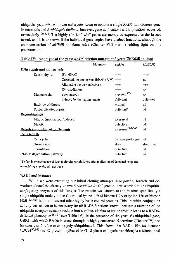
from their possible yeast counterparts turned out negative, probably due toa low conservation (RADJ, RAD2 and RAD7) or toa much domainal similarity (RAD3), our interest was raised by the yeast gene RAD6, the putative "master-gene" of yeast post-replication repair (For extensive reviews on the mutants, the gene and protein, see4,5,358-361 )(For RAD6 3D structure see362). As explained in the first chapter, patients of the so-called XP-variant group have a defect in this post-replication repair pathway, and a human RAD6 gene could represent a candidate gene for this repair disorder. However, at that moment, we were very weIl aware of the fact that RAD6-deficient mutants display a very severe and pleiotropic phenotype (see Table IV), in contrast to the human XP-variants which in general present only very mild XPfeatures. (Note that in preliminary experiments thus far na obvious changes of HHR6A or B RNAiprotein expression were found in several XP variant celllines studied, data not shown.)
The 172 amino acids yeast RAD6 protein whieh corrects the rad6 mutant phenotype was at the time of its isolation devoid of any recognisable domains and showed as a single peculiarity a long acidic amino acid sequence at its C_terminus366,367. The "acid ic tail" was subsequently shown to be indispensable for sporulation, but not necessary for induced mutagenesis and DNA repair368. The yeast protein's expression is induced by DNA damage and during meiosis, but rernains constant during the mitotic cen cycle369 ,370, In this it resembles several other proteins implicated in the ubiquitin or RAD6 pathways369,371,372, Bath induction phenomena were, however not conserved in the RAD6 (or RAD23) homologues of higher eukaryotes (see Chapter V and ref.91 ), pointing to differences in regulation between lower and higher organisms. In rats the mRNA is induced upon fasting and decreases upon insulin treatrnent, providing a first example of hormon al regulation of the
28
ubiquitin system373 . A1llower eukaryotes seem to eontain a single RAD6 homologous gene.
In mammals and Arabidapsis thaliaIla, however, gene duplications and triplications aecuITed, respective1y lOO,374, Thc highly similar "twill" genes are mostly co-expressed in thc tissues tested, and it is unknown if thc individual gene copies have distinct functions, aIthough thc eharaeterisation of mHR6B knoekout miee (Chapter VII) starts shedding light on this
phenomenon.
Table IV: Phenotype of the reast RAD6 deletion mutant and reast VbK63R mutant
Mutant-> rad6L1 UbK63R DNA rcvair nnd mutagenesis
Sensitivity to: UV,4NQO
Mutagenesis:
Crosstinking agents (eg.8MOP + UV)
Alkylating agents (eg.MMS) Xly-irradiation Spontaneous
lnduced by damaging agents
Excision of dimers
Post-repHcation repair
Recombination
Mitotic (spontaneous/induced)
Meiotic
Retrotrallsposition of Tv elements
Cell Growth
Cell eyele
Growth rate
Sporulation
-N-rulc degradation pathway
+++ +++ +++ +++ elevated363
deficient
nonnal
deficient*
increased
defective increased363-365
S-phase prolonged
slow
defective
defective
*Defcct in reappcarance of high molecular weight DNA after replication of damagcd templates
wt=wild type levels, nd= not done
RAD6 ond bistones
+++ ncl
+++ wt
\Vt deficicnt
ncl ncl
nd nd
nd
\Vt almost wt
\Vt
\Vt
While we \Vere executing our initial cloning attempts in Ss.pambe, Jentsch and coworkers cloned the already known S.cerevisiae RAD6 gene in their seareh for the ubiquitinconjugating enzymes of this fungus. The protein was shown to add ill vi/ra specifically a single ubiquitin-moiety to the C-terminallysine-119 of histone H2A or lysine-120 of histone H2B 172,375, but not to several other highly basic control proteins. This ubiquitin-conjugation activity was shown to be necessary for all RAD6 functions kllown, because a mutatioll of the ubiquitin-acceptor cysteine residue into a valine, alanine or sefÏlle residue leads to a RAD6-deficient phenotype376.317 (see Table IV). In the presenee of the yeast E3 ubiquitin-ligase,
VBRI, with whieh RAD6 interacts through its highly eonserved N-tenl1inus (Chapter IV), the histones can in vi/ra even be poly-ubiquitinated. This shows that RAD6, like for instanee CDC3495.166 (an E2 protein implieated in GI-S phase eell eycle transition) is a bifunetional
29
enzyme competent in both E3-independent and E3-dependent conjugation reactions l95(see for tbis bifunctionality also I97). This poly-ubiquitination of histones is dependent on the acidic tai! of yeast RAD6. Therefore sporulation, which is tai!-dependent, needs apparently polyubiquitination. whereas DNA repair and mutagenesis involve only mono-ubiqitination. AIthough these ideas are generally accepted, the function of histone mono- and polyubiquitination by RAD6 in vivo and its implication in DNA repair remain a subject of debate.
--- Eukaryotic DNA is organised innucleosomes: a stretch of -146 base pairs of DNA is wound around a histone octamer which consists of two subunits of histones H2A( 14kD), H2B(l4kD), H3(l5.3kD) and H4(l1.2kD) [(H2A:H2B12H32H42)]. The nucleosomes are connected by 50-100 base pair stretches of DNA to which, in (higher413) eukaryotes, a molecule of histone Hl(22kD) binds which stabilises the higher order chromatin stmcture resulting in the compact "30nm" fibers. The degree of local packing has to be tightly regulated, as it has been shown that the chromatin is highly condensed in regions containing quiescent genes and more accessible in regions of transcriptional activity. It is now generally admitted that this regulation probably takes place through a variety of non-permanent posttranslational modifications; methylation, acetylation, phosphorylation, poly(ADP)ribosylation and ubiquitination of the flexible N- or C-terminal domains of the different nucleosome components. However, although extensive, often contradictory, literature exists on this subject, no really clear relationship between aspecific modification and its implication in transcription, replication, DNA repair, or spermiogenesis has been demonstrated, with the exception of lysine-acetylation and phosphorylatioll. (por an extensive review on the subject of histones and their modifications, see378 .)
Acetylation is found to arfect 5-10% of the N-terminal flexible domains of the core histones. These core histones are mainly present in transcriptionally active regions of the chromatin. Acetylation is thought to neutralise the net positive charge of the basic histone proteïns, and in that way it would contribute to opening up the chromatin.
Serine/threonine-phosphorylation of histones Hl and H3 is thought to counter-act acetylation thus favouring chromatin-condensation. Hl is moderately phosphorylated duriug S phase, but throughout 02 phosphorylation increases to reach a hyperphosphorylated state of all Hls at metaphase. Immediately upon nuclear division Hls are dephosphorylated to Sphase levels. Just before metaphase bistone H3 is also phosphorylated.
Histones can be methylated irreversibly on lysine residues, a modification of which the function is not known at present379. Poly(ADP)ribosylation is thought to cause loc al chromatin decondensation and is almost exclusively found upon illtroduction of DNA strand breaks, and thus probably important for DNA repair380,381.
FinaIly, mono~ubiquitination of the C-tenninal flexible domains of histones was shown to occur principallyon histones H2A and H2B. 5-15% ofhistones H2A in higher eukaryotes alld ..... 2% of H2B are mono~ubiquitinated in vivo. Note, however, that these percentages vary enormously from cell to cell and organism to organism382.383. Ubiquitination is supposed to
open up the chromatin, as it introduces a major structural perturbation due to the size of the 76 amillo acids proteill. However, na such structural changes are detected at present (by for instance DNAse I footprinting)384.385. During the cell cycle uH2A and uH2B are present throughout S-phase and 02-phase up to prophase. From prophase to metaphase histones are
30
deubiquitinated, but immediately re~ubiquitinated in anaphase. The modification is important as for insta nee in the El~ts mutant eeIl Hne, ts85, it was shown th at with reduced ubiquitination cells arrest close to the S/G2 boundary of the cell cycle, accompanied by a loss of uH2A. Mono-ubiquitinated histones are very stabIe and ubiquitin is thus apparently not involved in breakdown of these molecules. Some reports show an association of especially uH2B with active DNA sequences387-391(and a higher affinity of uH2A for AT-rich DNA386), which is contradicted by others392.393. Thus although mono~ubiquitinated histones exist, and
although they seem important, their precise function is still completely unknown. ~--
As outlined above RAD6 is able to mono- and poly-ubiquitinate histones ;11 vitro. However, the implication of RAD6 in the ubiquitination of histones in vivo remains a point of debate, as may become clear from the following arguments.
First, it seems thus far impossible to detect ubiquitinated histones in the yeast S.cerevisiae. This orgallism apparelltly contains very few, if any, ubiquitinated histones (less than the detection limit of 0.1 % of all histones). The C-terminal amino acids of H2A (and H2B), which in man harbour the unique ubiquitin-attachment site (Lysine-1l9/120), are very weIl conserved in evolution and shown to be essential for yeast viability. Swerdlow and coworkers wanted to test whether this same lysine residue is also used for ubiquitination in yeast. Therefore, the two normal H2A histone genes were replaced by a gene copy mutated in the (for yeast putative) ubiquitination site. This caused no detectable phenotypic change in growth (solidlliquid medium, different temperatmes, heat-killing, osmotic killing, use of alternative carbon sources), sporulation and 254nm UV radiation sensitivity383. Therefore, S.cerevisiae which contains mainly uncondensed/active chromatin, does either not need ubiquitination or ubiquitinates only a very smal1, undetectable, proportion of its histones at another site in the molecule.
Second, histones are a general target protein used for in vi/ra testing of ubiquitinconjugating enzymes and many of these enzymes are able to add, mostly without high specificity, a single or multiple ubiquitin moieties to these basic molecules166,254,375,394-398.
In the case of rabbit E214kD, the rabbit homologue of yeast RAD6, Haas et al. have shown that ill vitra this protein can weakly mono-ubiquitinate histones, but reaction kinetics and constants let these authors to consider the reaction as a~specific in an ill vivo situation375 . No specific poly-ubiquitination of histones can be detected with the rabbit protein375.397. It was however shown that poly-ubiquitination of histones can be performed by the yeast protein in vitro, and that it is dependent on the acidic tail350. Without "acidic tail" yeast RAD6 can only mono-ubiquitinate histones. So the tail is important for poly-ubiquitination of histones and sporulation, but can be missed for mono-ubiquitination of histones (with questionable specificity!) and thus for DNA repair and DNA mutagenesis. However, the Caenarhabditis elegans (which contains also an acidic tail sequence) and the Arabidopsis RAD6 homologues are apparently completely unable to ubiquitinate histones H2A or H2B ;1/ vitro399,4oo. So, taken these results together, the proposition that histones are iJl vivo targets for the RAD6 or its homologues is unlikely. It is more plausible that RAD6, like many other E2's, ubiquitinates histones with low affinity and that these are not its real targets. The phenomenon of histone-poly-ubiquitination by RAD6 alone (without E3-protein), an activity
31
which is only displayed by the yeast protein, probably represents "an artefact" as the acid ie tail could cause a higher but a-specific affinity of RAD6 for the basic histones.
RAD6 targets What are the real targets for RAD6, especially in DNA repair and during late
spermatogenesis? Histones would be ideal targets due to thcir requirement for DNA repair, meiosis or more broadly spennatogenesis. However, due to above-mentioncd results it seems at least unlikely that histones present one of the RAD6 targets in a normal cello Whether they are targetted in special ceH types, like for instanee spermatids, in thc prescnce of a specif1c E3 protein (which might target RAD6 or increase its affinity) remains to be established. (Preliminary experlments studying ubiquitination in mHR6B knockout mice could indicate that histones in spermatids might still be a target for HHR6B (W.M. Baarends & H. Roest, pers. comm.)).
Also the interaction of RAD6 with the ssDNA-binding protein RAD 18 is in favaur of a function of RAD6 in close contact with the DNA 402,403. The Prakash group provided
indications that RADI8 can transport RAD6 to the DNA, where the protein then could perforrn its function. They al80 showed that the interaction site bctwecn RAD6 and RAD 18 is well conserved in evolution because the interaction of yeast RAD 18 is also possible with Ss.polI/be rhp6+ and human HHR6A aud 6B proteins402 . This is rather remarkable as the RAD18 protein, in contrast to RAD6, is not very weIl conserved in evolution40I . Since only very short regions of amino acid sequence similarity exist, it is possible that the interaction surface between the two proteins is probably not a linear sequence but a three dimensional one.
However, althaugh the rad6 phenotype and these last findings still favour a ftiuction of RAD6 nearby the DNA, the only three "reai" RAD6 targets identified thus far (all recognised with the help of aspecific E3_protein405 ) are not strictly DNA-assaciated:
I) RAD6 is the E2 protein involved in degradation of N-rule targets322,323 (see abave). It interacts specifically via its very conserved N~terminus (see Chapter 4) with the yeast VBR I-encoded E3 protein322.406. In reticulocyte Iysates the reaction is independent of the presence of its acidic tail323 . However, in vivo, in yeast, the acidic tail is important for N~rule degradation407.408 Yeast RAD6 has apparently three ways of recognising targets: alone, tail dependent, unassisted~C-terminus independent, and E3-a'ssisted C~terminus independent408 [.
The tirst mode of action is apparently lost in higher eukaryotes, as the tail is absent from these homologues (see Chapters IT, III and N)]. Thus far the only N-mle target protein for which it has been proven that it involves RAD6, is GPA I, the ex subunit of a yeast G protein implicated in pheromone~dependent signal transduction286.
2) Gcn4 is a yeast transcriptiollal activator of the bZIP family involved in regulation of the biosynthesis of amino acids and purines. lts normal rapid degradation, dependent upon CDC34 and RAD6, is inhibited under starvation conditions284 •
3) The p27 mammalian cell cyc1e protein is an inhibitor of cyc1in-dependent kinases. Bath iJl vivo and iJl vitro, the protein is degraded by the ubiquitin-proteasome pathway. The human ubiquitin~col1jugatil1g enzymes RAD6 and UBC3 were specifically iuvolved in its ubiquitination 168. It is for the moment unknown whether Gcn4 or p27 are N-rule targets.
32
Note that measurement of reaction kinetics on total retieuloeyte proteins favour the implication of RAD6/E2-14K in E3-assisted poly-ubiquitination, although monoubiquitination was also observed but relatively non-specific375 . This poly-ubiquitination, however, does not always seem to occur via the normal1ysine-48 of ubiquitin (which is used by RAD6 and UBRI in the N-rule degradation pathway)'95. Recently, it was shown that RAD6 is also capable of forming K-6 (to histones H2B in the absence of an E3 protein) and K-63 (made by RAD6 in an E3-dependent manner) linkages, bath apparently not involved in degradation l96. The K-63 poly-ubiquitination of RAD6 is the most interesting for DNA repair. Haas and co-workers eonstrueted a yeast in which they replaeed the normal four ubiquitin genes by a ubiquitin mutant encoding an arginine instead of lysine-63 (strain UbK63R)173, whieh prevents the addition of other ubiquitin moieties at that position of the molecule. The strain grows at wild type levels (see Table III) and degrades a set of short-Iived N-rule proteins. However, the strain which is shown to be epistatie with a rad6L1 mutant is highly sensitive to DNA damaging agents and has a deficiency in DNA damage-induced mutagenesis. Also the most abundant, but rather restricted family of multiubiquitin-protein conjugates found in wild type S.cerevisiae cells is completely absent. Sa, apparently, this mutant ubiquitin is involved in the DNA mutagenesis pathway. The rad6 phenotype is much more pleiotropic, which suggests that for sporulatioll, resistance to y-rays and N-nlle degradation, the RAD6 protein uses another type of lysine linkage or mono-ubiquitinates its targets. In the RAD6MUbK63R double mutant a relative high UV resistance is observed when compared to the single md6L1 mutant which indieates that UbK63R is a partial suppressor of RAD6. This suggests that the K-63 ubiquitination persists in the absence of RAD6, and that other ubiquitin conjugating proteins (making K-63 linkages) partieipate in other RAD6-independent repair pathways which work more effieiently in the absence of K-63.
So, in conclusion, as already indicated above, all the data suggest that ubiquitin is a
very versatiIe signal, as different ubiquitin ehain configurations can be used to perform different funetions. A single ubiquitin conjugating enzyme, like for instanee RAD6, is able to perform different linkages (Le. K-6, K-48 and K-63) dependent on the type of target/process and the E3 protein involved. The type of linkage as weil as tlle E2 and E3 protein involved, determilles whether a protein can be degraded, deactivated, correctly folded, proteeted against degradation, stabilised, ... leading to a complex pleiotropie phenotype and implicatiolls in many different functions .. .
Ubiquitin: small, but very powerful!
In the next chapters the isolation and characterisation of RAD6-homologous genes and proteins is described from the distantly related yeast Schizosaccharom)'ces pombe98 , the fly Drosophila melanogastel'99 and a duplicated locus from man 1OO.409,41O. (Note that tlle HHR6B gene was isolated three times independentlylOO.117.191.411.) In the course of thls work, also RAD6 proteins from Arabidopsis thaliaIla and wheat374.400, Caenorhabditis elegalls399 , rat373
33
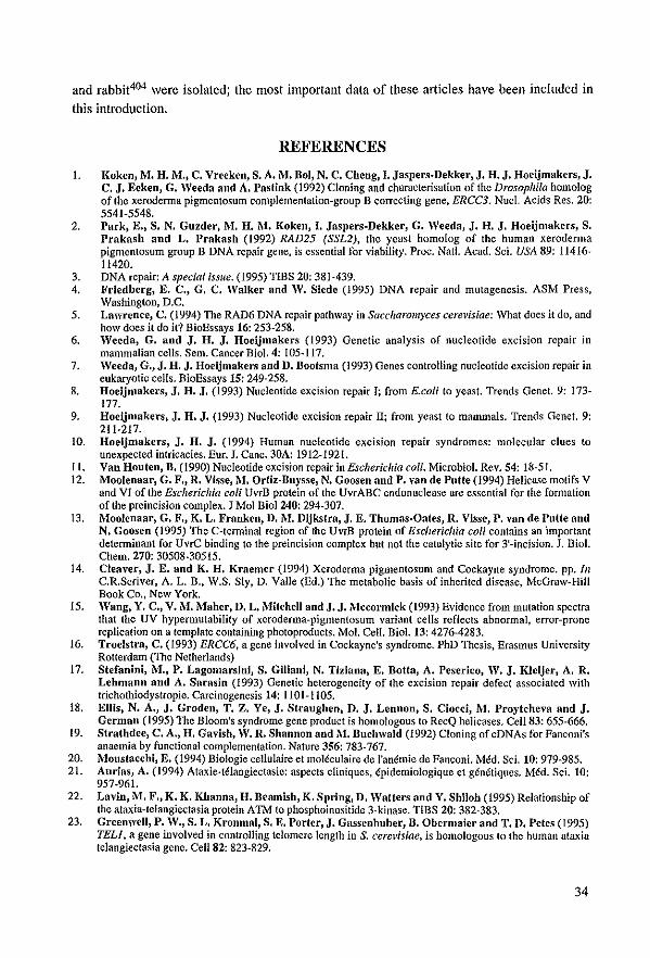
and rabbit404 were isolated; the most important data of these articles have been included in this introduction.
REFERENCES
1. Koken, M. H. M., C. Vreeken, S. A.l\l. Dol, N. C. Cheng, J. Jaspers.Dekker, J. H. J. Hoeijmakers, J. C. J. Eeken, G. Weeda and A. Pasfink (1992) Cloning and characterisation of the Drosophila homolog of the xeroderma pigmentosum complementation*group B correcting gene, ERce3. NucI. Acids Res. 20: 5541-5548.
2, Park, E., S. N. Guzder, M. H. M. Koken, J. Jaspers-Dekker, G. Weeda, J. H. J. Hoeijmakers, S. Prakash nnd L. Prakash (1992) RAD25 (SSL2), the yeast homolog of the humun xeroderma pigmenlosum group B DNA repuir gene, is essential for viability, Proc, Nat!, Acad, Sci. USA 89: 11416-11420.
3. DNA repair: A special issue. (1995) TIBS 20; 381*439. 4, Friedberg, E. C., G. C. Walker and W. Slcde (1995) DNA rerair and mutagenesis. ASM Press,
Washingion, D,C. 5. Lawrence, C. (1994) The RAD6 DNA repuir pathway in Saccharom)'ces cerevisiae: What does il do, and
how does it do it? BioEssays 16: 253-258, 6. Weeda, G. and J. H. J. Hoeijmakers (1993) Genetic analysis of nucleotide excision rerair in
mammalian cells. Sem. Cancer Bio!. 4: 105*117, 7. Weeda, G., J. H. J. Hoeijmakers and D. Bootsma (1993) Genes controlling nucleotide excision rerair in
eukaryotic cells. BioEssays 15: 249-258. 8, Hoeijmakers, J. H. J. (1993) Nucleolide excision repair I; from E.eoli to yeust. Trends Genet. 9: 173*
177. 9. Hoeijmakers, J. H. J. (1993) Nucleotide excision repair II; from yeast to mammals. Trends Genet. 9:
211-217. 10. Hoeijmakers, J. H. J. (1994) Human nucleotide excision repair syndromes: molecular clues to
unexpected intricacies. Eur. J, Cane. 30A: 1912-1921. 11. Van Houten, B. (1990) Nucleotide eXCÎsion repair in Eseherichia coli. Mierobiol. Rev. 54: lis-5I. 12. Mooienaar, G. F., R. Visse, M. Ortil-Buysse, N. Goosen and P. van de Putte (1994) Helicase motifs V
and VI of the Eseherichia eoU UvrB pratein of the UvrABC endonuclease are essential for the famlatian ofthe prcincisian complex. J Mol Biol240: 294-307.
13. MooIcnaar, G. F., K. L. Franken, D.M. Dijkstra, J. E. Thomas-Oates, R. Visse, P. van de Putte and N. Goosen (1995) The C-terrninal region of the UvrB rrotein of Escherichia coU contains an important detcnninant far UvrC binding to the preineision complex but not the catalytic site far 3'-inCÎsion. J. Biol. Chem. 270: 30508-30515.
14. Cleaver, J. E. and K. H. Kraemer (1994) Xeroderma rigmentasum and Cockayne syndrome. pr. lil c'R.Seriver, A. L. B., W.S. Sly, D, Valle (Ed.) The metabolic basis of inherited disease, MeGraw~Hill Book Co" New York.
15, Wang, Y. C., V. Î\-'I. Maher, D. L. Mitchell and J. J.1\lccormick (1993) Evidence from mutation spectra that Ihe UV hyrermutability of xeroderma-pigmentosum variant cells reflects abnormal, error-prone replication on a template containing photoproducis. Mol. Cell. Biot. 13: 4276-4283.
16. Troelsfra, C. (1993) ERCC6, a gene involvcd in Cockayne's syndrome. PhD Thesis, Erasmus University Rotterdam (The Netherlands)
17. Stefanini, M., P. Lagomarsini, S. Giliani, N. Tiziana, E. Bofta, A. Peserico, W. J. KJeijer, A. R. Lehmann and A. Sarasin (1993) Genelie heterogeneity of the excision rerair defect associated with triehothiodystropie, Carcinogenesis 14: 1101* 1105,
18. Ellis, N. A., J. Groden, T. Z. Ye, J. Straughen, D. J. Lcnnon, S. Ciocci, M. Proytcheva and J. German (1995) TIle Bloom's syndrome gene product is homologous 10 RecQ helicases, Cell83: 655-666.
19. Strafhdce, C. A., H. Gavish, W. R. Shannon and M. Buchwald (1992) Cloning of cDNAs for Fanconi's anaemia by funetional complementalion. Nature 356: 783-767.
20. Mousfacchi, E. (1994) Biologie cellulaire et moléculaire de I'anérnie de Fanconi. Méd, Sci. 10: 979*985. 21. Aurias, A. (1994) Ataxie·télangiectasie: aspects cliniques, épidemiologique et génétiques. Méd. Sci. 10:
957-961. 22. Lavin, M. F., K. K. Khanna, H. Dcamish, K. Spring,D. Watters and Y. Shiloh (1995) Relationship of
the ataxia-telangiectasia protein ATM 10 phosphoinositide 3-kinase. TIBS 20: 382*383, 23. Grecnwell, P. W., S. L. Kronmal, S. E. Porfcr, J. Gassenhuber, B. Obermaier aud T. D. Pefes (1995)
TELl, a gene involved in controlling telomere length in S. cere~,isiae. is homologous 10 the humun ataxia telangiectasia gene. Ce1l82: 823·829,
34
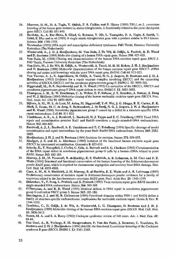
24. Morrow, D. M., D. A. Tagle, Y. Shiloh, F. S. Collins and P. Hieter (1995) TEU, an S. cere~'isiae homolog of the human gene mutated in alaxia telangiectasia, is functionally relaled 10 Ihe yeasl checkpoint gene MEel. Ce1l82: 831-840.
25. Savitsky, K., A. Bar-Shim, S. GUad, G. RotulllD, Y. Ziv, L. VnnagaUe, D. A. Tagle, S. Smith, T. Uziel, S. Sfez nnd a. et (1995) A single alaxia telangiectasia gene with a producl similar to PI-3 kinase. Science 268: 1749-1753.
26. Vermeuleo (1995) DNA repair and transcription deficiency syndromes. PhD Thesis, Erasmus University Rotterdam (The Netherlands)
27. Westerveld, A., J. H. J. Hoeijmakers, M. Van Duio, J. De Wit, H. Odijk, A. Pas tink, R. D. Wood aod D. Bootsma (1984) Molecular c10ning of a human DNA repair gene. Nature 310: 425-429.
28. Vao Duin, 1\1. (1988) Cloning nnd characterization of Ihc human DNA-cxcision repair gene ERCC-I. PhD ThesÎs, Erasmus University Rotterdam (Tbc Nctherlands)
29. Van Duin, 1\1., J. De Wit, H. Odijk, A. Westen'eld, A. Yasui, 1\1. H. M. Koken, J. H. J. Hoeijmakers aud D. Bootsma (1986) Molecular characlerization of the human excision repair gene ERCC/: cDNA cloning and amino acid homology wilh Ihe yeast DNA repair gene RADIO. Ce1l44: 913-923.
30. Van Vuuren, A. J., E. Appeldoorn, H. Odijk, A. Yasui, N. G. J. Jaspers, D. Bootsma and J. H. J. Hoeijmakers (1993) Evidence for arepair enzyme complex involving ERCCI and the correcling activities ofERCC4, ERCCII and the xeroderma pigmentosum group F. EMBO J. 12: 3693-701.
31. Biggerstaff, 1\1., D. E. Szymkowski and R. D. Wood (1993) Co-correction of Ihe ERCCI, ERCC4 and xeroderma pigmenlosum group F DNA repair defects ill vitro. EMBO J. 12: 3685-3692.
32. Thompson, L. H., K. W. Brookman, C. A. Weber, E. P. SaJazar, J. T. Reardon, A. Sanear, Z. Deng and M. J. SicUiano (1994) Molecular cIoning of Ihe human nuc1eolide-excision repair gene ERCC4. Proc. Natl. Acad. Sci. USA 91: 6855·6859.
33. Sijbers, A.1\I., W. L. de Laat, 1\1. Ariza, M. Biggerstaff, Y.-F. Wei, J. G .. Moggs, B. K. Carter, B. K. Shell, E. Evans, 1\1. C. de Jong, S. Radernakers, J. de Rooij, N. G. J. Jaspers, J. H. J. Hoeijmakers and R. Wood (1996) Xeroderma pigmentosum group F causcd by a defect in a slruclure-specific DNA repair endonucIease. Ce1l86: 811-822,
34. Tomkinson, A. E., A. J. BardweIl, L. BardweIl, N. J. Tappe and E. C. Friedberg (1993) Yeast DNA repair and recombination proteins Radl and RadIO conslÎtute a single+stranded-DNA endonucIease. Nature 362: 860-862.
35. DardweIl, A. J., L. DardweIl, A. E. Tomkinson and E. C. Friedberg (1994) Specific cleavage of model recombinalion and repair inlermediates by Ibe yeast Radl-RadlO DNA endonucIease. Science 265: 2082-2085.
36. Hoeijmakers, J. H. J. and D. Bootsma (1994) Incisions for excision. Nature 371: 654-655. 37. l\Iudgctt, J. S. and 1\1. A. Madnnes (1990) Isolation of the functional human excision repair gene
ERCC5 by intercosmid recombination. Genomics 8: 623-633. 38. Scherly, D., T. Nouspikel, J. Corlet, C. Uela, A. Bairoch aod S. G. Clarkson (1993) Complemenlalion
of Ihe DNA repair defect in xeroderma pigmenlosum group GeelIs by a human eDNA rclated 10 yeasl RAD2. Nature 363: 182-185.
39. 1\1urray, J. M., 1\'1. Tavassoll, R. al-Harithy, K. S. Sheldriek, A. R. Lehmano, A. 1\1. Carr aDd F. Z. WaUs (1994) Structural and functional conservalion of the human homolog of Ihe Schizosaccharomyces pombe RAD2 gene, which is required for chromosome segregalion and recovery from DNA damage. Mol. Cello Bio!. 14: 4878·4888.
40. Carr, A. M., K. S. Sheldriek, J.1\1. Murray, R. al-Harilhy, F. Z. Watts and A. R. Lehmann (1993) Evolutionary conservation of excision repair in Schizosaccharolll)"ces pOli/be: evidence for a family of sequences related 10 Ihe SaccIJaromyces cerevislae RAD2 gene. Nucl. Acids Res. 21: 1345-1349.
41. Habraken, Y., P. Sung, L. Prakash aDd S. Prakash (1993) Yeast excision repair gene RAD2 encodes a single-slranded DNA endonucIease. Nature 366: 365-368.
42. O'Donovan, A. and R. D. Wood (1993) Idenlical defects in DNA repair in xeroderma pigmentosum group G and rodent ERCC group 5. Nature 363: 185-188.
43. Harriogton, J. J. and 1\1. R. Lieberman (1994) Functional domains wilhin FEN-I and RAD2 definc a fantily of structure-specific endonuc1cases: implications for nucleotide excision repair. Genes & Dev. 8: 1344-1355.
44. Troelstra, C., H. Odijk, J. de Wit, A. Westencid, L. H. Thompson, D. BootsoUl aod J. H. J. Hoeijmakers (1990) Molecular cIoning of Ihe human DNA excision repair gene ERCC6. Mol. Cello Biol. 10: 5806-5813.
45. Nanee, M. A. and S. A. Beery (1992) Cockayne syndrome: review of 140 cases. Am. J. Med. Gen. 42: 68-84.
46. Van Gooi, A., R. Verhage, S. 1\1. Swagemakers, P. Van der Putte, J. Brouwer, C. TroeJstra, D.
35
Bootsma and J. H. J. Hoeijmakers (1994) RAD26, Ihe functional S.cere~'isiae homolog of Ihe Cockayne syndrome B gene ERCC6. EMBO J.13: 5361-5369.
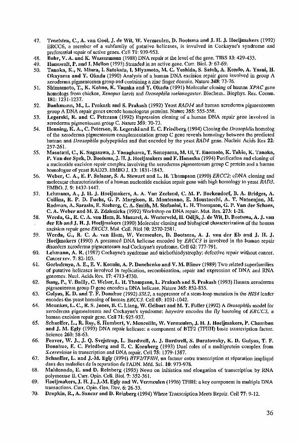
47. Troelstra, C., A. van Gooi, J. de Wit, W. Vcrmculen, D. BoofsDla and J. H. J. Hoeijmakers (1992) ERCC6, a member of a subfamily of putative helicases, is involved in Cockaync's syndrome and preferential repair of active genes. Cell 71: 939*953.
48. Bohr, V. A. aDd K. WasscrmanD (1988) DNA repair at the Icvel ofthe gene. TIBS 13: 429-433. 49. Hanawalf, P. and I. Mellon (1993) Stranded in an acth'e gene. CUIT. Biol. 3: 67-69. 50. Tanaka, K., N. Minra, I. Safokafa, I. Miyamoto, M. C. Yoshida, S. Satoh, A. Kondo, A. Yasui, H.
Okayama and Y. Okada (1990) Analysis of a human DNA excision repair gene involved in group A xerodemla pigmentosum group and containing a zinc finger domain. Nature 348: 73-76.
51. Shimamoto, T., K. Kohno, K. Tanaka and Y. Okada (1991) Molecular cloning ofhuman XPAC gene homologs from dIkken, Xenopus lae~'is and Drosophila melallogaster. Biochem .. Biophys. Res. Comm. 181: 1231-1237.
52. Bankmann, M., L. Prnknsh nnd S. Prakash (1992) Yeast RAD14 and human xeroderma pigmentosum group A DNA repair genes encode homologous prolcins. Nature 355: 555-558.
53. Legerski, R. and C. Peferson (1992) Expression cloning of a human DNA repair gene involved in xeroderma pigmcntosum group C. Nature 359: 70-73.
54. Henning, K. A., C. Peferson, R. Legerski nDd E. C. Fricdbcrg (1994) Cloning the Drosophila homolog of Ihc xcrodenna pigmentosum complemcntation group C gene reveals homology between the predicled human and Drosophila polypeptides and that encoded by the yeast RAD4 gene. Nucleic Acids Rcs 22: 257-261.
55. Masutani, C., K. Sugasawa, J. Yanaglsawa, T. Sonoyama, 1\'1. Ui, T. Enomoto, K. Takio, K. Tanaka, P. VaD der Spek, D. Boofsma, J. H. J. Hoeijmakers nDd F. Hanaoka (1994) Purification and cloning of a nucleotide excision repair complex involving thc xerodemla pigmentosum group C proteÎll and a human homologuc ofyeast RAD23. EMBO J. 13: 1831*1843.
56. Weber, C. A., E. P. Salazar, S. A. Stewart aDd L. H. Thompson (1990) ERCC2: cDNA cloning and molecu;ar characterization of a human nucleotide excision repair gene with high homology 10 yeast RAD3. EMBO. J. 9: 1437-1447.
57. Lehmann, A., J. H. J. Hoeijmakers, A. A. Van Zeeland, C. M. P. Backendorf, B. A. Bridges, A. Collins, R. P. D. Fuchs, G. P. I\'1argison, R. Montesano, E. Moustacchi, A. T. Natal'ajan, M. Radman, A. Sarasln, E. Seeberg, C. A. Smith, M. Stefanini, L. H. Thompson, G. P. Van der Schans, C. A. Weber and M. Z. Zdzienicka (1992) Workshop on DNA repair. Mut. Res. 273: 1-28.
58. Weeda, G., R. C. A. van Ham, R. MasureI, A. Westerveld, H. Odijk, J. de Wit, D. Boofsma, A. J. van der Eb aDd J. H. J. Hoeljmakcrs (1990) Molecular cloning and biological characterization of the human excision repair genc ERCC3. Mol. Cell. Bioll0; 2570-2581.
59. Weeda, G., R. C. A. van Ham, W. Vermeulen, D. Bootsma, A. J. van der Eb and J. H. J. Hoeljmakers (1990) A presumed DNA helicase encoded by ERCC3 is involved in the human repair disorders xerodemla pigmentosum and Cockayne's syndromc. Cc1l62: 777-791.
60. Lehmann, A. R. (1987) Cockayne's syndromc and trichothiodystrophy: defective repair without cancer. Cancer rev. 7: 82-103.
61. Gorbalenya, A. E., E. V. Koonin, A. P. Donehenko and V.l\l. Dlinov (1989) Two related superfamilies of pUlative helicases involved in replicatîon, recombination, repair and expression of DNA and RNA genomes. Nuc!. Acids Rcs.17: 4713-4730.
62. Sung, P., V. Bailly, C. Weber, L. H. Thompson, L. Prakash aod S. Prakash (1993) Human xeroderma pigmenlosum group D gene encodes a DNA helicase. Nature 365: 852-855.
63. Gulyas, K. D. and T. F. Donahue (1992) SSL2, a suppressor ofa stem-loop mutation in the HIS41eader encodes the yeast homolog ofhuman ERCC3. Ce1l69: 1031-1042.
64. l\.ofounkcs, L. C., R. S. JODes, B. C. Liang, W. Gelbart and M. T. Fuller (1992) A Drosophila model for xeroderma pigmentosum and Cockayne's syndrome: haywire encodes the fly homolog of ERCC3, a human excision repair gene. Ce1l71: 925-937.
65. Sehaeffer, L., R. Roy, S. Humbert, V. Moneollin, W. Vermeulen, J. H. J. Hoeijmakers, P. Chambon and J. M. Egly (1993) DNA repair helicase: a component of BTFZ (TFIIlI) basic transcription factor. Science 260: 58-63.
66. Feaver, W. J., J. Q. S,,'ejstrup, L. BardweIl, A. J. BardweIl, S. Buratowsky, K. D. Gulyas, T. F. Donahue, E. C. Friedberg and E. C. Kornbcrg (1993) Dual roles of a multiprotein complex from S.cere~'isiae in trallscription and DNA repair. Ce1l75: 1379-1387.
67. Schaeffer, L. and J •• M. Egly (1994) BTF2rrFlIH, un facteur entre transcription et réparation impliqué dans des maladies de la reparation de I'ADN. Méd. Sci. 10: 973-978.
68. Maldonado, E. and D. Reinberg (1995) News on inilialion and elongation of transcription by RNA polymerase IT. Curr. Opin. CeU. Bio!. 7: 352-361.
69. Hoeljmakers, J. H. J., J.·M. Egly and W. Vermeulen (1996) TFIIH: a key component in multiple DNA trallsactions. CUIT. Opin. Gen. Dev. 6: 26-33.
70. Drapkin, R., A. Sanear and D. Reinberg (1994) Where Transcriplion Meets Repair. Cell77: 9-12.
36
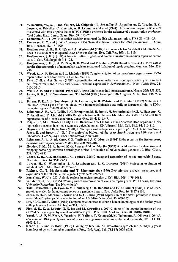
71. Vermeulen, W., A. J. van Vuuren, 1\1. Chipoulet, L. Schaeffer, E. Appeldoorn, G. Weeda, N. G. Jaspers, A. Priestley, C. F. Arletf, A. R. Lehmann and a. et (1994) Three unusual repair deficiencies associated wÎth transcription factor BTF2 (TFIIH): evidence for Ihe existence of a transcription syndrome. Cold Spring Harb. Symp. Quant. BioJ. 59: 317~329.
72. Lehmann, A. R. (1995) Nucleotide excision repair and the link wilh transcription. TIBS 20: 402·405. 73. Conaway, R. C. and J. W. Conaway (1993) General initiation factors for RNA polymerase 11. Annu.
Rev. Biochem .. 62: 161·190. 74. Hoeijmakers, J. H., H. Odijk and A. Westerveld (1987) Differences between rodent and human cell
lines in Ihe amounl of integrated DNA after transfection. Exp. Cell. Res. 169: 111·119. 75. Hoeijmakers, J. H. J. (1987) Characterization of genes and proleins involvcd in excision repair of human
cells. J. Cell. Sci. Suppl. 6: 111·125. 76. Hoeijmakers, J. H. J., A. P. Eker, R. D. Wood and P. Robins (1990) Use of;1I 1';1'0 and;1I virro assays
for Ihe characlerizalion of mammalian excision repair and isolation ofrcpair proteins. Mut. Res. 236: 223· 238.
77. Wood, R. D., P. Robins and T. Lindahl (1988) Complementation ofthe xeroderma pigmentosum DNA repair defect in cell-frce extracts. Ce1l53: 97-106.
78. Park, C.-H. and A. Sancar (t993) Reconslitution of mammalian cxcision repair activity wilh mulant cell-free extracts and XPAC and EReel proteins expressed in Escherichia eoU. Nuct. Acids Res. 21: 5110-S116.
79. Willls, A. E. and T. Lindahl (1987) DNA ligase I deficiency in Bloom's syndrome. Nature 325: 355-357. 80. Lasko, D. D., A. E. Tomkinson and T. Lindahl (1990) Eukaryotic DNA ligases. Mutat. Res. 236: 277·
287. 81. Barnes, D. E., A. E. Tomkinson, A. R. Lehmann, A. D. Websfer and T. Lindahl (1992) Mutalions in
the DNA ligase I gene of an individual with immunodeficiencies and cellular hypersensitivity to DNAdamaging agenls. Ce1l69: 495-503.
82. Lehmann, A. R., A. E. Willis, B. C. Broughton, M. R. James, H. Steingrimsdoftir, S. A. Harcourt, C. F. Arletf and T. Lindahl (1988) Relation between Ihe human fibroblast strain 46BR and eelllines representative of Bloom's syndrome. Canccr Res. 48: 6343-6347.
83. Peigent, C., M. S. Satoh, G. Daly, D. E. Barnes and T. Lindahl (1994) Aberrant DNA repair and DNA replication due 10 an inherited enzymatic defect in human DNA ligase I. Mol. Cell. BioL 14: 310-317.
84. Haynes, R. H. and B. A. Kunz (1981) DNA repair and mulagenesis in yeast. pp. 371-414. I1I Stralem, J., Jones, E. and Broach, J. (Ed.) The molecular biology of Ihe yeast Sacclwromyees: Life cycle and inheritance. Cold Spring Harbor Laboratories, New York.
85. Lehmann, A. R., A. i\l. Carr, F. Z. Watts aDd J. M. Murray (1991) DNA repair in the fission yeast, Sehizosaceharomyces pOli/be. Mutat. Res, 250; 205-210.
86. Howley, P. i\1., M. A. Israel, M.-F. Law and M. A. Martin (1979) A rapid methad for detecling and mapping bomology between beterologous DNAs -Evaluation of polyomavirus genomes-. J. Biol. Cbem. 254: 4876·4883.
87. Cohen, D. R., A. J. Hapel and I. G. Young (1986) Cloning and expression ofthe rat interleukin-3 gene. Nuc!. Acids Res.14: 3641-3658.
88. Burger, H., G. Wagemaker, J. A. Leunissen and L. C. Dorssers (1994) Molecular evolulion of interleukin-3. J. Mol. Evol. 39: 255-267.
89. Richter, G., T. Blankenstein and T. Diamantstein (1990) Evolutionary aspeets, structure, and expression of the rat interleukin 4 gene. Cytokine 2: 221-228.
90, Earnshaw, W. C. (1987) Anionic regions in nuelear proteins. J. Cel! Biol. 105: 1479-1482, 91. van der Spek, P. J. (l995) Cloning and characterization of excision repair genes. PhD Thesis, Erasmus
University Rotterdam (The Netherlands) 92. Taidi-Jaskowski, B., D. Tyan, S. M. Honigberg, C. R. Radding aud F. C. Grumet (1988) Usc of RccA
protein to enrich for homologous genes in a genomic library. Nuc!. Acids Res. 16: 8157-8169. 93. Jones, R. H., S. i\1oreno, P. Nurse and N. C. Jones (1988) Expression orthe SV40 promoter in fission
yeast: identification and eharacterizalion of an AP-l-like factor. Ce1l53: 659-667. 94. Lee, .M. G. and P. Nurse (t987) Complemenlation used to clone a human homologue oftbe fission yeast
een cycle control gene ede2. Nature 327: 31-35. 95. Plan, S. E., K. A. Leppig, H. N. Do and M. Groudine (1993) Cloning of lbc buman homolog of thc
CDC34 een cycle gene by eomplementation in yeast. Proc. Natl. Acad. Sci. USA 90: 10484-10488. 96. Yasui, A., A. P. M. Eker, S. Yasuhira, H. Yajima, T. Kobayashi, M. Takao and A. Oikawa (1994) A
new cJass of DNA phololyases present in various organisms including a-placental manunals. EMBO J. 13: 6143-6151.
97. Kranz, J. E. and C. Holm (1990) Cloning by function: An alternative approach for identifying yeast homologs of genes from olher organisms. Proc, Nat!. Acad, Sci. USA 87: 6629-6633.
37
98. Reynolds, P., M. H. M. Koken, J. H. J. Hoeijmakers, S. Prakash nnd L. Prakash (1990) 'nlC rhp6+ gene of Schizosaccharomyces poll/be: a structural and functional homolog of the RAD6 gene from the distantly related yeast Saceharom)'ces eerevisiae. EMBO. J. 9: 1423·1430.
99. Koken, M. H. M., P. Reynolds, D. Boofsmn, J. H. J. Hoeijmakers, S. Pmkash nnd L. Prnkash (1991) Dhr6, a Drosophila homolog of the yeast DNA-repair gene RAD6. Proc. Natl. Acad. Sei. USA 88; 3832-3836.
100. Koken, M. H. M., P. Reynolds, I. Jaspers-Dekker, L. Prakash, S. Prakash, D. Bootsma and J. H. J. Hoeijmakers (1991) Structural and functional conservation of two human homolags of the yeast DNA repair gene RAD6. Proc. Natl. Acad. Sci. USA 88: 8865-8869.
101. Hershko, A. and A. Clechanover (1992) The Ubiquitin System for Protein Degradation. Ann. Rev. Biochcm. 61: 761-807.
102. Flnley, D. nnd V. Chau (1991) Ubiquitination. Annu. Rev. Cell. Biol. 7: 25-69. 103. Dice, J. F. (1987) Molecular determinants of protein half-lives in eukaryotie eells [erratum: FASEB J.
(1988) 2: 2262]. FASEB J.1: 349-357. 104. Cook, J. nnd P. B. Chock (1988) Ubiquitin: a review on a ubiquitous biofaetor in cukaryotie eeils.
Biofaetors 1: 133-146. 105. Ciecbanover, A. (1991) Thc ubiquitin-mcdiated system for intracellular protein degradation. J. Basic.
Clin. Physiol. Pharmaeol. 2: 141-159. 106. Rechsteiner, M. (1988) Ubiquitin. Plenum, Ncw York. 107. Jentsch, S. (1992) The Ubiquitin-Conjugation System. Ann. Rev. Gen. 26: 179-207. 108. Vierstra, R. D. (1993) Protein Degradation in PIants. Ann. Rev. Plant Phys. Plant Mol. Biol. 44: 385-410. 109. Jentsch, S., W. Seufert, T. Sommer nnd H. A. Reins (1990) Ubiquitin-conjugating enzymes: novel
regulators of eukaryotic cells. TIDS 15: 195-198. 110. Ciechanover, A. (1994) The ubiquitin-proteasome proteolytic pathway. Ce1l79: 13-21. 111. Jennissen, H. P. (1995) Ubiquitin and the enigma of intracellular protein degradation. Eur. J. Biochem ..
231: 1-30. 112. Maupin Furlow, J. A. and J. G. Ferry (1995) A proteasome from the methanogenic archaeon
Me/lwllosarcil/a /hemwphila. J. Chem. Biol. 270; 28617·28622. 113. Wolf, S., F. Lottspeich and W. Baumeister (1993) Ubiquitin Found in the Archacbacterium
17lenlloplasma acidophil//IJI. FEBS Lelt. 326: 42-44. 114. Wostmann, C., E. Tannich and T. Bakker·Grumvnld (1992) Ubiquitin of ElIIamoeba his/oly/ica
deviates in six amino acid residues from tbe consensus of all other known ubiquitins. FEBS Lelt. 308; 54-58.
115. Durner, J. nnd P. Boger (1995) Ubiquitin in the prokaryote AlIabaelIa \'ariabilis. 1. Chem. Bio!' 270; 3720-3725.
116. Sharp, P. M. and W. H. Li (1987) Ubiquitin genes as a paradigm of concerted evolution of tandem repcats. J. Mol. Evo!' 25; 58·64.
117. Schneider, R., C. Eckerskorn, F. Lottspeich and i\'l. Schweiger (1990) The human ubiquitin carrier prolein E2(Mr::: 17,000) is homo!ogous 10 the yeast DNA repair gene RAD6. EMBO. J. 9; 1431-1435.
118. TreIer, M., W. Seufert and S. Jentsch (1992) Drosophila UbcDl encodes a highly conserved ubiquitincOluugating enzymc involved in selecth'e protein degradation. EMBO 1.11; 367-372.
119. Haffield, P. M., J. CaJlis nnd R. D. Viersfra (1990) Cloning of ubiquitin activating enzyme from whea! and expression of a functional protein in Eseherichia eoU. J. Biol. Chem. 265; 15813-15817.
120. Handley, P. M., M. Mueckler, N. R. Siegel, A. Ciechanover nnd A. L. Schwarfz (1991) Moleeular doning, sequcnee, and tissue distribution of the human ubiquitin-aetivating enzyme EI [erratum: Prae Nat! Acad Sci USA (1991) 88: 7456]. Proc. Nat!. Acad. sci. USA 88: 258-262.
121. McGrnfh, J. P., S. Jentsch and A. Varshavsky (1991) UBAl: an essential yeast gene cncoding ubiquitin-aetivating enzyme. EMBO J. 10: 227-236.
122. Scheffner, M., U. Nuber and J. M. Huibregtse (1995) Protein ubiquitination involving an EI-E2-E3 enzymc ubiquitin thioester cascade. Nature 373: 81-83.
123. Hochstrasser, M., M. J. Ellison, V. Cbau and A. Vnrsbavsky (1991) The short-livcd MAT alpha 2 transcriptional regulator is ubiquitinated in vivo. Proc. Nat!. Acad. Sei. USA 88: 4606·4610.
124. Murakami, Y., S. Matsufuji, T. KameJi, S. Haynshi, K. Igarashi, T. Tamum, K. Tanaka and A. Ichihara (1992) Ornithine decarboxylase is degraded by thc 26s-proleasome without ubiquitination. Nature 360: 597-599.
125. Monaco, J. J. and D. Nandi (1995) The gelleties of proteasomes and antigen processing. Annu. Rev. Genetics 29: 729-754.
126. HUt, W. ond D. H. Wolf (1996) Proteasomes: destruction as a programme. Trends Bioehem. Sei. 21: 96-!O2.
127. Goldberg, A. L. (1995) Functions of the protcasome: the lysis at Ihe end ofthe tunnel [eommentJ. Science 268: 522-523.
128. Sighting the cellular shredder [editoriaI].(l995) Nat. Struct. Biol. 2; 427·428.
38
129. Rubin, D. M. aDd D. FiDley (1995) Proteolysis. The proteasome: a protein-degrading organelIe? Curr. Bio!. 5: 854-858.
130. Löwe, J., D. Stock, D. Jap, P. Zwickl, W. Bnumeister nnd R. Huber (1995) Crystal structure ofthe 20S proteasome from the archaeon T. acidophilulII at 3,4 À Resolution. Science 268: 533-539.
131. Weissman, J. S., P. D. Sigler nnd A. L. Horwich (1995) From the cradle to the grave: ring complexes in the life ofa protein [comment]. Science 268: 523-524.
132. Sehwartz, A. L., A. Ciechanover, R. A. Brandt aDd H. J. Geuze (1988) Immunoelectron microscopie localization ofubiquitin in hepatoma cells. EMBO J. 7: 2961-2966.
133. Laszlo, L., F. J. Doherty, N. U. Osborn and R. J.l\Iayer (1990) Ubiquitinated protein conjugates are specifically enriched in the lysosomal system of fibroblasls. FEBS Let!. 261: 365-368.
134. Simeon, A., I. J. van der Klei, M. Veenhuis aDd D. H. Wolf (1992) Ubiquitin. a central component of selective cytoplasmic proteolysis, is linked to proteins residing at the locus of non-selective proteolysis, the vacuole. FEBS Let!. 301: 231-235.
135. Lenk, S. E., W. A. Dunn, J. S. Trausch, A. CiechanO'\'cr and A. L. Schwartz (1992) Ubiquitinactivating enzyme. El, is associated with maturation of autophagic vacuoles. J. Cen Biol. 301-308.
136. Egner, R. and K. Kuchier (1996) Tbe yeast multidrug transporter Pdr5 of the plasma membrane is ubiquitinated prior to endocytosis and degradation in the vacuole. FEBS Let!, 378: 177-181,
137. Kölling, R. and C. P. HollcDberg (1994) The ABC-transporter SIe6 accumulates in the plasma membrane in a ubiquitinated foml in endocytosis mutanls. EMBO J. 13: 3261-3271,
138, Laing, J. G. and E. C. Deyer (1995) 111e gap junction protein connexin43 is degraded via the ubiquitin protcasome palhway. J, Chem, Biol. 270: 26399-26403.
139, Raposo, G., H. M. van Santen, R. Leijendekker, H. J. Geuze and H. L. Ploegh (1995) Misfolded major histocompatibility complex c1ass I molecules accumulate in an expanded ER-Golgi intermediate compartment. J. Cell Bio1.131: 1403-1419.
140. Ciechano\'er, A., R. Gropper and A. L. Schwarlz (1991) Tbe ubiquitin-activating cnzyme is requircd for Iysosomal degradation of cellular proteins under stress. Biomed. Biochim. Acta, 50: 321-332,
141. Hleke, L. and H. Riezmnn (1996) Ubiquitination of a yeasl plasma membrane receptor signals its ligandstimulated endocytosis, Ce1184: 277-287,
142, Gropper, R., R. A. Brandt, S. Elias, C. F. Bearer, A. Mayer, A. L. Schwartz and A. Cfechanover (1991) The ubiquitin-activating enzyme, EI, is required for slress-induced lysosomal degradation of cellular proteins. J. Biol. Chem. 266: 3602-3610.
143, Deers, E. P., T. N. Moreno and J. Callis (1992) Subcellular localÎzation of ubiquitin and ubiquitinatcd proteins in Arabidopsis tlwlialla. J. Biol. Chem. 15432-15439.
144. Hurtley, S.M. (1996) Lysosomal degradation ofubiquitin-tagged receptors (Perspeclives). Scicnce 271: 617.
145. Jemen, T. J., l\I .. A. Loo, S. Pind, D. B. Williams, A. L. Goldberg nDd J. R. Rlordan (1995) Multiple proteolytie systcms, including the protcasome, contribute to CFfR processing, Ce1l83: 129-135.
146. Jariel.Enconlre, I., M. Pariat, F. Martin, S. Carlllo, C. Salvat and M. Pieehaczyk (1995) Ubiquitinylation is not an absolute requirement for degradation of c-Jun protein by the 26 S proteasome. J. Chem. Biol. 270: 11623-1l627,
147, Gonen, H., I. Stancovski, D. Shkedy, T. Hadari, B. Bercovich, E. Bengal, S. Mesilati, O. AhuHatoum, A. L. Schwarlz and A. CiechaoO\'cr (1996) Isolation, characlerization, and partiat purification of a novel ubiquitin·protein ligasc, E3, Targcting ofprotein substratcs vÎa multiple and distinct recognition signals and conjugating enzymes. J Biol Chcm 271: 302·310,
148, Muller, S., J. P. Briand and 1\1. H. Van Regcnmorfel (1988) Presence of antibodies to ubiquitin during the autoimmune response associated with systemic lupus erythematosus, Proc. Nat!. Acad, Sci. USA 8S: 8176-8180.
149. Liu, Z., L. A. Diaz, A. L. Haas nnd G. J. Giudice (1992) cDNA Cloning of a novel human ubiquitin carrier protein - an antigenie domain spccifically rccognized by endemie pemphigus foliaceus autoantibodies is encoded in a secondary reading frame of this human epidcrmal transcript. 1. Biol. Chcm. 15829-15835.
150, Argiles, J. M. nDd F. J. Lopez-Soriano (1996) The ubiquitin-dependant proteolylic pathway in skelctal muscIe: its rolc in pathological states. Trends Prot. Sci. 17: 223-226.
151. Muller, S. and L. M. Schwarfz (1995) Ubiquitin in homcostasis, development and diseasc. BioEssays 17: 677-684.
152, Mayer, R. J., l\.I. Landon, L. Laszlo, G. Lennox and J. Lowe (1992) Protein processing in Iysosomesthe new thcrapeutie target in neurodegencrative discase, Lancet 156-159,
153. Lowe, J., R. J. Mayer and M. Landon (1993) Ubiquitin in neurodegencrative diseases, Brain Pathol. 3: 55-65.
154. Mayer, R. J., J. Lowe, M. Landon, H. McDermott, J. TuckwelI, F. Doherty and L. Laszlo (1991) Ubiquitin and the lysosollle system: Illolecular illllllunopathology reveals the conncction. Biomed. Biochim. Acta 50: 333-341.
39
155. I\'Iaycr, R. J., J. Lowe aDd i\I. Lnndon (1991) Ubiquitin and the molecular pathology of chronic degenerative diseases [cditorial]. J. Pathol.163: 279-281.
156. Mayer, R. J., J. Arnold, L. Laszlo, ~'I. Landon aDd J. Lowe (1991) Ubiquitin in health and disease. Biochim. Biophys. Acta 1089: 141-157.
157. Palombella, V. J., O. J. Rando, A. L. Goldberg nnd T. l\1aniatis (1994) The ubiquitin-proteasome pathway is rcquircd for processing the NFkBI precursor protein and the activation ofNFkB. CeU78: 773-785.
158. SchelTner, 1\'1., K. i\..Junger, J. M. Huibregfse nnd P. M. Howley (1992) Targeted degradation of the retinoblastoma protein by human papillomavirus-E7-E6 fusion proteins. EMBO J. 11: 2425-2431.
159. Molinari, M. and J. 1\1Uner (1995) p53 in complex with DNA is resistant to ubiquitin-dependent proteolysis in the presence ofHPV-16 E6. Oncogene 10: 1849-1854.
160. Tsurumi, C., N. Ishida, T. Tamura, A. Kakizuka, E. Nishida, E. Okumura, T. Kishimoto, 1\'1. Inagaki, K. Okazaki, N. Sagatn and et al. (1995) Degradation of c-Fos by the 26S protcasome is accelerated by c-Jun and multiple protein kinases. Mol. Cell. Biol. 15: 5682-5687.
161. Treier, M., L.M. Staszewski and D. Bohmann (1994) Ubiquitin-dependant c-jun degradation in vivo is mediated by the d domain. Ce1l78: 787-798.
162. Nishizawa, M., N. Furuno, K. Okazaki, H. Tanaka, Y. Ogawa and N. Sagata (1993) Degradation of MOS by the N-temnnal proline (pro(2))- dependent ubiquitin pathway on fertilization of xenopus eggs -possible significanee of natural selection for pro(2) in mos. EMBO J. 12: 4021-4027.
163. Nisbizawa, M., K. Okazaki, N. Furuno, N. Wafanabe aDd N. Sagata (1992) The 2nd-Codon Rule and AutophosphoryJation Govern the Stability and Activity of Mos During the Meiotic CeU Cycle in Xenopus-Oocytes. EMBO J. 11: 2433-2446.
164. Ishida, N., K. Tanaka, T. Tamura, M. Nishizawa, K. Okazaki, N. Sagata and A. Ichihara (1993) MOS is degraded by Ihe 26s proteasome in a ubiquitin-dependent fashion. FEBS Let!. 324: 345-348.
165_ Ciechanover, A., J. A. DiGiuseppe, B. Bercolich, A. Orian, J. D. Richter, A. L. Schwarlz and G.l\!. Brodeur (1991) Degradation of nuelear oncoproteins by the ubiquitin system il/ vitro. Proc. Nat!. Acad. Sci. USA 88: 139-143.
166. Goebl, M. G., J. Yoehem, S. Jenfsch, J. P.McGrath, A. Varshavsky aDd n. nyers (1988) The yeast cell cycle gene CDC34 encodes a ubiquitin-conjugating enzyme. ScÎence 241: 1331-1335.
167. Glotzer, M., A. W. Murray and M. W. Kirschner (1991) Cyclin is degraded by the ubiquitin pathway. Nature 349: 132-138.
168. Pagano, 1\'1., S. W. Tam, A.M. Theodoras, P. Beer Romero, G. Del Sal, V. Chau, P. R. Yew, G. F. DraeUa and .M. RoJfe (1995) Role of Ihe ubiquitin-proteasome palhway in regulating abundance of the cyclin-dependent kinase inhibitor p27. Science 269: 682-685.
169. seurert, W., B. Futseher and S. Jenfseh (1995) Role of a ubiquitin-conjugating enzyme in degradation of S- and M-cyclins. Nature 373: 78-81.
170. Al·Khodairy, F., T. Enoch, J. i\I. Hagan and A. M. Carr (1995) 'Ille Schizosaccharolll)'ces [Jombe IIl/s5 gene encodes a ubiquitin conjugating enzyme required for nonnal mitosis. J. Cello Sci. 108: 475-486.
171. Nefsky, n. aDd D. Beach (1996) Publ acts as an E6-AP-like protein ubiquitin ligase in the degradation of CDC25. EMBO J. 15: 1301-1312.
172. Jentseh, S., J. P. McGrath aDd A. VarshaYSky (1987) The yeas! DNA repuir gene RAD6 encodes a ubiquitin-conjugating enzyme. Nature 329: 131-134.
173. Spenee, J., S. Sadis, A. L. Haas nnd D. Finley (1995) A ubiquitin mutant with specific defects in DNA repair and multiubiquitination. Mol. Cell. Biol. 15: 1265-1273_
174. Kovalenko, O. V., A. W. Plug, T. Haaf, D. K. Gonda, T. Ashley, D. C. Ward, C.l\!. Radding and E. I. Golub (1996) Mammalian ubiquitin-conjugating enzyme UBe9 interacts with Rad5l rccombination protein and localizes in senaptonemal complexes. Proc. Nat!. Acad. Sci. U S A 93: 2958-2963.
175. Morl, S., C. H. Heldin and L. ClaessoD·welsh (1993) Ligand-induced ubiquitination of the plateletderived growth factor beta-receptor plays a negative regulatory role in its milogenic signaling. J. Biol. Chem. 268: 577-583.
176. Mori, S., C. H. Heldin and L. CJaesson·Welsh (1992) Ligand-induced polyubiquitination of Ihe plateletderived growth factor beta-receptor. J. Bio!. Chem. 267: 6429-6434.
177_ Burel, C., V. 1\Iezger, .M. Pinto, 1\1. Rallu, S. Trigon and M. Morange (1992) Mammalian heat shock protein families - expression and functions. Experientia 48: 629-634.
178. Fornace, A. J., J. Alamo, l\l. C. Hollander aDd E. Lamoreaux (1989) Ubiquitin mRNA is a major stress-induced transcript in mammalian cells. Nud. Acids Res. 17: 1215-1230.
179. l\oliehalek, 1\1. T., E. P. Grant, C. Gramm, A. L. Goldberg and K. L. Rock (1993) A Role for the Ubiquitin-Dependent Proteolytic Pathway in MHC Class I-Restricted Antigen Presentation. Nature 363: 552-554.
180. Rock, K. L., C. Grarum, L. RothsfeiD, K. CJark, R. Stein, L. Diek, D. Hwang and A. L. Goldberg (1994) Inhibitors of the proteasome block the degradation of most cell proteins and the generation of peptides on MHC Class 1 molecules. Ce1l78: 761-771.
40
181. Tanaka, K. (1994) Role of proteasomes modified by interferon-gamma in antigen processing. J. Leukoc. Biol. 56: 571-575.
182. Delic, J., M. Morange and H. Magdelenat (1993) Ubiquitin pathway involvement in human lymphocyte gamma-irradiation-induced apoptosis. Mol. Cel!. Biol. 13: 4875-4883.
183. Schwarfz, L. M., S. W. Smith, M. Joncs and n. A. Osborne (1993) Do all programmed cell deaths oecur via apoptosis. Proc. Natl. Acad. Sci. USA 90: 980-984.
184. Dmello, S. R. and C. Galli (1993) SGP2, Ubiquitin, 14K Lectin and RP8 messenger R..'lAs are not induced in neuronal apoptosis. Neuroreport 4: 355-358.
185. Muralidhar, M. G. and J. B. Thomas (1993) The Drosophila Belldless gene encodes a neural protein related to ubiquitin-conjugating enzymes. Neuron 11: 253-266.
186. Meyer, E.M., C. 1\'1. West and V. Chau (1986) Antîbodies directed against ubiquitin inhibit high affinity [3H]choline uptake in rat cerebral cortieal synaptosomes. J. Biol. Chem. 261: 14365-14368.
187. Meyer, E.M., C. M. West, B. R. Stevens, V. Chau, 1\'1. T. Nguyen andJ. H. Judkins (1987) Ubiquitindirected antibodies inhibit neuronallransparters in rat brain synaptosomes. J. Neuroehem. 49: 1815-1819.
188. Oh, C. E., R.McMahon, S. Denzer and M. A. Tanouye (1994) Belldfess, a Drosophifa gene affecting neuronal connectivity, encodes a ubiquitin-conjugating enzyme homolog. J. Neurosci. 14: 3166-3179.
189. Wiebel, F. F. and W.-H. Kunau (1992) 1l1e Pas2 protein essential far peroxisome biogenesis is related to ubiquitin-conjugating enzymes. Nature 359: 73-76.
190. Sommer, T. and S. Jentsch (1993) A protein translocation defect linked 10 ubiquitin conjugation at the endoplasmie reticulum. Nature 365: 176-179.
191. Woffendln, C., Z. Chen, K. Staskus, E. F. Retzel and P. G. W. Plagemann (1991) Mammalian mRNAs encoding protein c10sely related 10 ubiquitin-conjugaling enzyme encaded by yeast DNA repair gene RAD6. Biochim. Biophys. Acta 1090: 81-85.
192. Chau, V., J. W. Tobias, A. Bachmair, D. MarrioU, D. J. Eeker, D. K. Gonda and A. Varsbavsky (1989) A multiubiquitin chain is confined to specific lysine in a targeted short-lived protein. Sciencc 243: 1576-1583.
193. Gregori, L., t\-'I. S. Pooseh, G. Cousins and V. Chau (1990) A uniform isopeptidc-linked multiubiquitin chain is suffieient ta target substrate for degradation in ubiquitin-mediated proteolysis. J. Biol. Chem. 265: 8354-8357.
194. Arnason, T. and M. J. Ellison (1994) Stress resistance in Saccharom)'ces cerevisiae is strongly correlated with assembly of a navel type ofmultiubiquitin chain. Mol. Cell. Biol. 14: 7876-7883.
195. Haas, A. L., P. B. Reback and V. Chau (1991) Ubiquitin conjugation by the yeast RAD6 and CDC34 gene products. Comparison to their putative rabbil homologs, E2(20K) and E2(32K). J. Chem. Biol. 266: 5104-5112.
196. Daboshlna, O. V. ond A. L. Haas (1996) Novel multiubiquitin ehain Iinkages catalyzed by the conjugating enzymes E2EPF and RAD6 are recognized by 26 S proteasome subunit 5. J. Biol. Chem. 271: 2823-2831.
197. Liu, Z., A. L. Haas, L. A. Diaz, C. A. Conrad and G. J. Giudice (1996) Characterization of a novel kcratinoeytc ubiquitin carrier protein. J. Biol. Chem. 271: 2817·2822.
198. Johnson, E. S., P. C. Ma, J. M. Ota and A. Vnrshaysky (1995) A proteolytic pathway tbat recognizcs ubiquitin as a degradation signa!. J. Chem. Biol. 270; 17442-17456.
199. Varshavsky, A. (1992) TIle N-End Rule. Ce1l69: 725-735. 200. Shaeffer, J. R. (1994) Mono-ubiquitinated alpba globin is an intemlediate in tbe ATP-dependent
proteolysis of alpha globin. J. Chem. Biol. 269: 22205-22210. 201. Shaeffer, J. R. nnd 1\'1. A. Kanla (1995) Degradation of monoubiquitinated alpha-globin by 26S
proteasomes. Bioehem. 34: 4015-4021. 202. Kirchhoff, L. V., K. S. Kim, D. 1\'1. Engmao aod J. E. Donelson (1988) Ubiquitin genes in
trypanosomatidae. J. Biol. Chem. 263: 12698-12704. 203. CowJand, J. B., O. Wiborg and J. Vuust (1988) Human ubiquitin genes: one member of the UhB gene
subfamily is a tetrameric non-processed pseudogene. PEBS Lett. 231: 187-191. 204. Baker, R. T. and P. G. Board (1987) The human ubiquitin gene family: structure of a gene and
pseudogenes from the UhB subfamily. Nuc!. Acids Res. 15: 443-463. 205. Callis, J., T. Carpenter, C. W. Sun and R. D. Vierstra (1995) Structure and eyolution of genes
eneoding polyubiquitin and ubiquitin-like proteins in Arahidopsis thaliaIIa ecotype Columbia. Geneties 139: 921-939.
206. Finley, D., E. Ozkaynak and A. Varshavsky (1987) The yeast polyubiquitin gene is essential for resistance to high lemperalures, starvation. and other stresses. CeU 48: 1035-1046.
207. Redman, K. L. and M. Rechsteiner (1988) Extended reading frame of a ubiquitin gene encodes a stabie, conserved, basic protein. J. Biol. Chem. 263: 4926-4931.
208. Redmao, K. L. and M. Rechsteiner (1989) Identification of the long ubiquitin extension as ribosomal protein 527a. Nature 338: 438-440.
209. Warner, J. R. (1989) Ubiquitin. A marriage of canvenience or necessity? [N&V). Nature 338: 379.
41
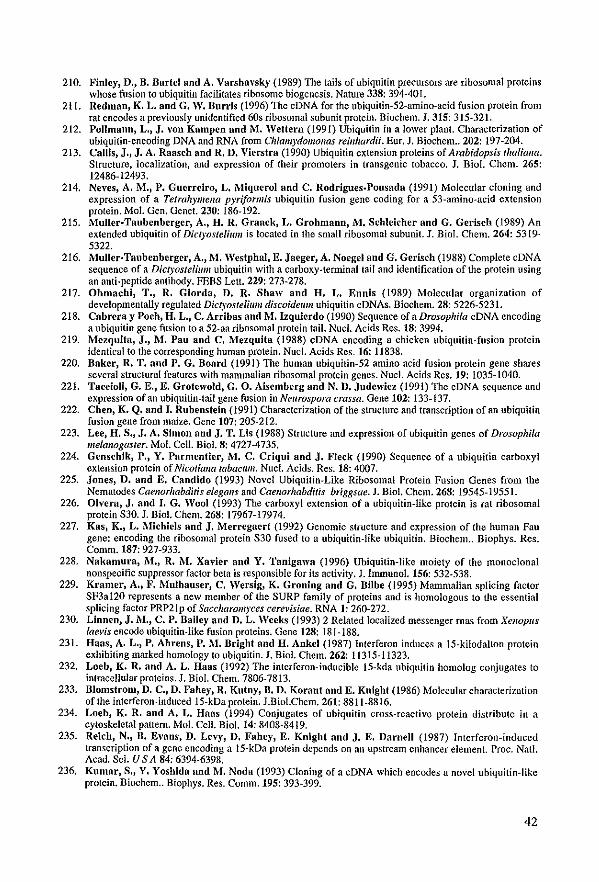
210. Finlcy, D., B. Barfel and A. Varsha"sky (1989) The tails af ubiquilin precursors are ribosomal proteins whose fusion to ubiquitin facilitates ribosome biogenesis. Nature 338: 394-401.
211. Redman, K. L. and G. W. Bunis (1996) The cDNA for the ubiquilin-52-amino-acid fusion protein from rat encactes a previous1y unidentified 60s ribosomal subunit protein. Biochem. J. 315: 315-321.
212. Pollmann, L., J. "on Kampen and M. Weltern (1991) Ubiquitin in a lower plant. Characterization of ubiquitin-encoding DNA and RNA from Chlamydolllollas reillltardii. Eur. J. Biochem .. 202: 197-204.
213. CaUis, J., J. A. Raaseh and R. D. VÎerstra (1990) Ubiquitin extension proteins of Arabidopsis thaliaIIa. Structure, localization, and expression of their promoters in transgenic tobacco. J. Bio!. Chcm. 265: 12486-12493.
214. Neves, A. M., P. Guerrciro, L. Miquerol and C. Rodrigues·Pousada (1991) Molecular c10ning and expression of a Tetrah)'lIIella pyriformis ubiquitin fusion gene coding for a 53-amina-acid extension protein. Mo!. Gen. Genet. 230: 186-192.
215. Muller·Taubenbcrger, A., H. R. Graack, L. Grohmann, .M. Schleicher and G. Gerisch (1989) An extended ubiquitin of Dictyos/elilllll is locatcd in the small ribosomal subunil. J. Bio!. Chem. 264: 5319-5322.
216. Muller·Taubenberger, A., M. Westphal, E. Jaeger, A. Noegel and G. Gerisch (1988) Complete cDNA sequence of a DiclyosteliulII ubiquitin with a carboxy-terminaltaiJ and identification of the protein using an anti-peptide antibody. FEBS Lelt. 229: 273-278.
217. Ohmachi, T., R. Giorda, D. R. Shaw and H. L. Ennis (1989) Molecular organization of developmentally regulated Dict)'osteli//1/I discoidel/l11 ubiquitin cONAs. Biochem. 28: 5226-5231.
218. Cabrera y Poch, H. L., C. Arribas and M. Izquierdo (1990) Sequence of a Drosophila cDNA encoding a ubiquitin gene fusionto a 52-aa ribosomal protein tail. Nucl. Acids Res. 18: 3994.
219. Mczquita, J., M. Pau and C. IHezquita (1988) cDNA encoding a chicken ubiquitin-fusion protein identical to the corresponding human protein. Nuc!. Acids Res. 16: 11838.
220. Baker, R. T. aud P. G. Board (1991) The human ubiquitin-52 amino acid fusion protein gene shares several structural features with mammalian ribosomal protein gencs. Nuc!. Acids Res. 19: 1035-1040.
221. TaccioIi, G. E., E. Grotewold, G. O. Aisemberg and N. D. Judenicz (1991) The cONA sequence and expression of an ubiquitin-tail gene fusion in Neurospora crassa. Gene 102: 133-137.
222. Chen, K. Q. and J. Rubenstein (1991) Characterization of the structure and transcription of an ubiquitin fusion gene from maize. Gene 107: 205-212.
223. Lee, H. S., J. A. Simon aud J. T. Lis (1988) Structure and expression ofubiquitin genes of Drosophila melanogaster. Mol. Cello Biol. 8: 4727-4735.
224. Genschik, P., Y. Parmentier, 1\'1. C. Criqui and J. Fleck (1990) Sequence of a ubiquitin carboxyl extension protein of Nicotiana tabacll11l. Nuc!. Acids. Res.18: 4007.
225. Jones, D. and E. Candido (1993) Novel Ubiquitin-Like Ribosomal Protein Fusion Genes from the Nematodes Caellorhahditis elegans and Caellorhabditis briggsae. J. Biol. Chem. 268: 19545-19551.
226. Olvera, J. and I. G. Wool (1993) Thc carboxyl extension of a ubiquitin-like protein is rat ribosomal protein S30. J. Biol. Chem. 268: 17967-17974.
227. Kas, KJ L. Michiels and J. l\'1erregaert (1992) Genomic slructure and expression of the human Fau gene: encoding Ihe ribosomal protein S30 fused to a ubiquitin-like ubiquitin. Biochem .. Biophys. Res. Comm. 187: 927-933.
228. Nakamura, M., R. M. Xavier and Y. Tanigawa (1996) Ubiquitin-likc moiety of the monoclonal nonspecific suppressor factor bela is responsible for its actîvity. J. Immunol. 156: 532-538.
229. Kramer, A., F. Mulhauser, C. Wersig, K. Groning and G, Bilbe (1995) Mammalian splicing factor SF3a120 represenls a new member of the SURP family of proteins and is homologous to the essential splicing factor PRP21p of Saccharomyces cerevisiae. RNA 1: 260-272.
230. Linnen, J. M., C. P. Bailey and D. L. Weeks (1993) 2 Related localized messenger mas from XeJlopus laevis encode ubiquitin·like fusion proteins. Gene 128: 181-188.
231. Haas, A. L., P. Abrens. P. M. Dright and H. Ankel (1987) Interferon induces a 15-kilodalton protein exhibiting marked homology ta ubiquitin. J. Biol. Chem. 262: 11315-11323.
232. Locb, K. R. and A. L. Haas (1992) The intcrferon-inducible 15-kda ubiquitin homolag conjugates to intracellular proteins. J. Biol. Chem. 7806-7813.
233. Blomstrom, D. C., D. Fahey, R. Kulny, D. D. Korani and E. Knight (1986) Molecular charncterization of the interferon~induced I5-kDa protein. J.Biol.Chem. 261: 8811-8816.
234. Loeb, K. R. and A. L. Haas (1994) Conjugates of ubiquitin cross-reactive protein distributc in a cytoskcletal pattern. Mol. Cello Biol. 14: 8408-8419.
235. Reich, N., B. Evans, D. Levy, D. Fahey, E. Knight and J. E. DamelI (1987) Interferon-induccd transcription of a gene encoding a 15-kOa protein depends on an upstream enhancer element. Proc. Natl. Acad. Sci. U S A 84: 6394-6398.
236. Kumar, S., Y. Yoshida and M. Noda (1993) ClonÎng of a cDNA which encodes a novel ubiquitin-like protein. Biochem .. Biophys. Res. Comm. 195: 393-399.
42
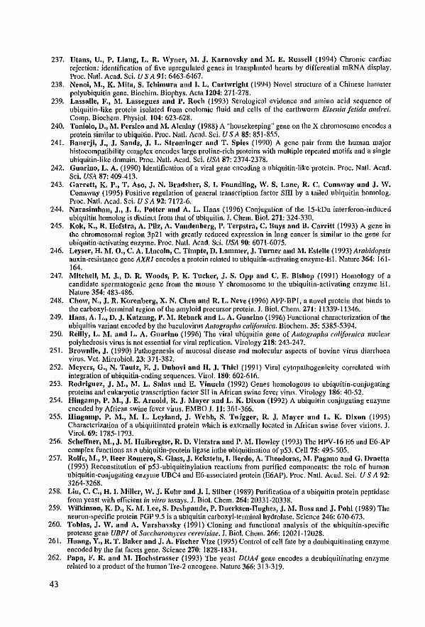
237. Vtans, U., P. Liang, L. R. Wyncr, 1\-1. J. Karnovsky nnd M. E. Russell (1994) Chronic cardiac rejection: identification of five upregulated genes in transplanted hearts by differential mRNA display. Proc. Nat!. Acad. Sci. U S A 91: 6463-6467.
238. Nenoi, M., K. Mita, S. Jchimura and J. L. Cartwright (1994) Novel structure of a Chinese hamster polyubiquitin gene. Biochim. Biophys. Acta 1204: 271-278.
239. LassalIe, F., M. Lassegues nnd P. Roch (1993) Serological evidence and amino acid sequence of ubiquitin-like protein isolated from coelomic fluid and cells of the earthworm Eisenia fe/ida andrei, Comp. Biochem, Physiol. 104: 623-628,
240, Toniolo, D., M. Persico and M. Alcalay (1988) A "housekeeping" gene on the X chromosome encodes a prolein similar 10 ubiquitin. Proc. Nat!. Aead, ScL U S A 85: 851-855.
241. Danerji, J., J. Sands, J. L. Strominger anrl T. Spies (1990) A gene pair from Ihe human major histoeompatibility complex encodes large proline-rieh proteins with multiple repeated motifs and a single ubiquitin-Jike domain. Proc. NalJ. Acad. SeL USA 87: 2374-2378,
242. Guarino, L. A. (1990) Identification of a viral gene encoding a ubiquitin-like protein. Proc. Nat!. Acad. ScL USA 87: 409-413.
243. Garrett, K. P., T. Aso, J. N. Bradshcr, S. J. FoundHng, W. S. Lane, R. C. Conaway and J. W. Conaway (1995) Positive regulation of general transcription factor sm by a tailed ubiquitin homolog. Proc. Nat!. Acad. Sci. U SA 92: 7172·6.
244. Narasimhan, J., J. L. Potter and A. L. Haas (1996) Conjugation of the 15-kDa interferon-induced ubiquitin homolog is dislinct from that ofllbiquilin. J. Chem. Bio!. 271: 324-330.
245. Kok, K., R. Hofstra, A. Pilz, A. Vandenberg, P. Terpstra, C. Bu)'s nnd D. Carritt (1993) A gene in the ehromosomal region 3p21 with greatly reduced expression in lung cancer Îs similar 10 the gene for lIbiquitin-aetivating enzyme. Proc. Natl. Acad. Sei. USA 90: 6071-6075.
246. Leyser, H. M.O., C. A. LlncoJn, C. Timpte, D. Lammer, J. Turner and M. Estene (1993) Arabidopsis auxin-resistance gene AXRI encodes a protein related to lIbiquitin-activating enzyme-El. Nature 364: 161-164.
247. Mitchell, 1\'1. J., D. R. Woods, P. K. Tucker, J. S. Opp and C. E. Bishop (1991) Homology of a candidate spennatogenie gene from the mouse Y chromosome 10 the lIbiqllitin-actÎvating enzymc El. Nature 354: 483·486.
248. Chow, N., J. R. Korenberg, X. N. Chen and R. L. Neve (1996) APP-BPI, a noyet protein that binds 10 the earboxyl-terminal region of the amyloid precursor protein. J. Biol. Chem. 271: 11339-11346.
249. Haas, A. L., D. J. Katzung, P. M. Reback and L. A. Guarino (1996) Funetional charaeterization ofthe ubiquitin variant encoded by the baculovirus Alltographa californica. Biochem. 35: 5385-5394.
250. Reilly, L. 1\1. and L. A. Guarino (1996) The yiral ubiqllitin gene of Alltographa californica nuclear polyhedrosis virus is not essential for viral replication. Virology 218: 243-247.
251. Brownlie, J. (1990) Pathogenesis of O1ucosal disease and molecular aspects of bovine virus diarrhoea virus. Vet. Microbiol. 23: 371-382.
252. Meyers, G., N. Tautz, E. J. Dubovi and H. J. Thiel (1991) Viral cytopathogenicity correlated with integration of ubiquitin-coding sequences. Virol. 180: 602-616.
253. Rodriguez, J. M., M. L. Salas and E. Vinuela (1992) Genes homologous to ubiquitin-conjugating proteins and eukaryotie transcription factor sn in Afriean swine fever virus. Virology 186: 40-52.
254. Hingamp, P •. M., J. E. Arnold, R. J. i\layer and L. K. Dixon (1992) A ubiquitin conjugating enzyme encoded by African swine feyer virus. EMBO J.11: 361-366.
255. Hingamp, P. M., M. L. Lcyland, J. Webb, S. Twigger, R. J. Mayer and L. K. Dixon (1995) Characterization of a ubiquitinated protein which is externally locatcd in African swine fever virions. J. Virol. 69: 1785-1793.
256. Scheffner, M., J. M. Huibregtse, R. D. Vierstra and P. i\I. Howley (1993) The HPV-16 E6 and E6-AP complex functions as a ubiquitin-protein ligase inthe ubiquitination of p53. Cell 75: 495-505.
257. Rolfe, M., P. Deer Romero, S. Glass, J. Eckstein, J. Derdo, A. Theodoras, 1\1. Pagano nnd G. Draetta (1995) Reconstitution of p53-ubiquitinylation reactions from purified components: Ihe role of human ubiquitin-conjugating enzyme UDC4 and E6-associated protein (E6AP). Proc. Nat!. Acad. Sci. U S A 92: 3264-3268.
258. Liu, C. C., H. I. MiIler, W. J. Kohr and J. I. SUber (1989) Purification ofa ubiquitin protein peptidase from ycast with efficient;lI vitro assays. J. Biot. Chem. 264: 20331-20338.
259. Wilklnson, K. D., K. 1\1. Lee, S. Deshpande, P. Ducrksen·Hughes, J.l\I. Boss and J. Pohl (1989) The nellron-specific protein PGP 9.5 is a ubiquitin carboxyl-terminal hydrolase. Science 246: 670-673.
260. Tobias, J. W. and A. Varshaysky (1991) Cloning and fllnetional analysis of the ubiquitin-specific protease gene UBPl of Saccharolll)'ces cerevisiae. J. Biol. Chem. 266: 12021-12028.
261. Huang, Y., R. T. Baker and J. A. Fischer Vize (1995) Control of eell fate by a deubiquitinating enzyme encoded by the fat faeets gene. Science 270: 1828-1831.
262. Papa, F. R. and i\1. Hochstrasser (l993) The yeast DOA4 gene encodes a deubiquitinating enzyme relatcd to a product of the human Tre-2 oncogene. Nature 366: 313-319.
43
263. Xiao, W., T. Fontanie and M. Tang (1994) UBP5 encodes a putative yeast ubiquitin-specific protease that is related to the human Tre-2 oncogene product. Yeast 10: 1497-502,
264. Wilkinson, K. D., V. L. Tashayev, L. B. O'Connor, C. N. Larsen, E. Kasperek and C. M. Piekart (1995) Mctabolism of the polyubiquitin degradation signal: structure, mechanislll, and roie of isopeptidasc T. Biochem. 34: 14535-46.
265. Hadari, T.) J. V. Warms, r. A. Rose and A. Hershko (1992) A ubiquitin C-terminal isopeptîdasc that acts on polyubîquitin chains. Roie in protein degradation. J. Bio!. Chem. 267: 719-727.
266. Kanda, F., D. E. Sykes, H. Yasudo. A. A. Sandberg and S •. Matsui (1986) Substrate recognition of isopeptidase: specific cleavage of the epsilon-(alpha-glycyl)lysine linkage in ubiquitin-protein conjugates. Biochim. Biophys. Acta 870: 64-75.
267. Van Nocker, S. and R. D. Vierstra (1993) Multiubiquitin chains Jinked tluough Iysine-48 are abundant in ~'i\'O and are competent intermediates in the ubiquitin proteolytic pathway. J. Bio!' Chem. 268: 24766-24773.
268. Van Nocker, S. and R. D. Vierstra (1991) Cloning and characterization of a 20-kDa ubiquitin carrier protein from wheat that calalyzes multiubiquitin chain formation illvitro. Proc. Nat!. Acad. Sci. U S A 88: 10297-10301.
269. van Nocker. S., J. M. Walker and R. D. Vierstra (1996) The Arabidopsls thalial/a UBC1/13/14 genes encode a family of multiubiquitin chain-fonning E2 enzymes. J. Biol. Chem. 271: 12150-12158.
270. Rogers, S., R. Wells and M. Rechsteiner (1986) Amino acid sequences comnlOn to rapidly degraded proteins: thc PEST hypothesis. Science 234: 364-368.
271. Terlecky, S. R., H. L. Chiaog, T. S. Olsoo and J. F. Dice (1992) Protein and peptide binding and stimulation of in vitro Iysosomal proteolysis by the 73-kDa heat shock cognate protein. J. Biol. Chem. 267: 9202-9209.
272. Jentseh, S. (1996) When proteins receive deadly messages at birth (perspectives). Science 271: 955-956. 273. Rechsteiner, 1\'1. (1991) Natural substrates ofthe ubiquitin proteolytic palhway. Cell 66: 615-618. 274_ Jabben, M., J. Shanklin aud R. D. Vierstra (1989) Ubiquitin-phylochrome conjugales. Pool dynamics
during 111 vivo phytochrome degradation. J. Biol. Chem. 264: 4998-5005. 275. Obin, M., T. Nowell and A. Taylor (1994) The photoreceptor G-protein transducin (Gt) is a subslrate for
ubiquitin-dependent proteolysis. Biochem. Biophys. Res. Comm. 200: 1169-1176. 276. de Groot, R. J., T. Rumenapf, R. J. Kulm, E. G. Strauss and J. H. Strauss (l99J) Sindbis virus RNA
polymerase is degraded by the N-end rule pathway. Proc. Nat!. Acad. Sci. USA 88: 8967-8971. 277. Oberst, M. D., T. J. Gollan, M. Gupfa, S. R. Peura, J. D. Zydlewski, P. Sudarsanan and T. G.
Lawson (1993) The Encephalomyocarditis virus-3e protease is rapidly degraded by an ATP-dependent proteolylic system in reticulocyte Iysate. Virology 193: 28-40.
278. Lawson, T. G., D. L. Gronros, J. A. Weruer, A. C. Wey. A. 1\1. DiGeorgc, J. L. Lockhart, J. W. Wilson and P. L. Winfrode (1994) The encephalomyocarditis virus 3e protease is a substrate for the ubiquitin-mediated proteolytic system. J. Biol. ehem. 269: 28429-28435.
279. Wang, Y., Y. G. Yeung, W. Y. Langdon and E. R. Stanley (1996) c-Cbl is transiently tyrosinephosphorylated, ubiquitinated, and membrane-targeted following CSF-I stimulation of macrophages. J. Chem. Biol. 271: 17-20.
280. Schcffner, M .• D. A. Werucss, J. J\.I. Huibregfse, A. J. Leyine nnd P . .M. Howley (1990) The E6 oncoprotein encoded by human papillomavirus type 16 nnd 18 promotes the degradation ofp53. Ce1l63: 1129-1136.
281. Li, C.·C. H., R. J\.I. Dai and D. L. Longo (1995) lnactivation of NP-kappa B inhibitor I kappa B alpha: ubiquitin-dependent proteolysis and its dcgradation product. Biochem. Biophys, Res. eomm. 215: 292-301.
282. Didonafo, J., F. Mercurio, C. Rosefte, J. Wu-Li, H. Suyang, S. Ghosh and.M. Karin (1996) Mapping of the inducible IKB phosphorylation sites that signal ils ubiquitination and dcgration. ?vfol. Cel!. Dio!. 16: 1295-1304.
283. Daldi, L., K. Drawn, G. Franzoso and U. SiebcnIist (1996) Critical role for Jysines 21 and 22 in signalinduced, ubiquitin-mediated proteolysis of I kappa B-alpha. J. Chem. Biol. 271: 376-379.
284. Kornitzer, D., D. Raboy, R. G. Kulka and G. R. Fink (1994) ReguJated degradalion of the transcription factor Gcn4. EMBO J, 13: 6021-6030.
285. Chen, P., P. Johnson, T. Somlllcr, S. Jenfsch and 1\1. Hochstrasser (1993) Multiple ubiquitinconjugating enzymes participale in the il/ vivo degradation of the yeasl MAT-alpha-2 reprcssor. eelJ 74: 357-369.
286. Madura, K. and A. Varshaysky (1994) Degradation of Ga by the N-elld rule pathway. Science 265: 1454-1458.
287. Hegde, A. N., A. L. GoJdberg and J. H. Sclnrartz (1993) Regulatory subunits of cAMP-dependent prolein kinases are degradcd after conjugation to ubiquîtin - a molecular mechanism underlying long-term synaptic pJasticity. Proc. Nat!. Acad. Sci. USA 90: 7436-7440.
44
288. Galcheva·Gargova, Z" S. J, Theroux and R. J. Davis (1995) The epidermal growth factor receptor is covalently linked to ubiquitin. Oncogene 11: 2649-2655.
289. Mori, S" L. Claesson Welsh, Y. Okuyama and Y. Saito (1995) Ligand-induced polyubiquitination of receptor tyrosine kinases. Biochem. Biophys. Res. Comm. 213: 32-39.
290. Miyazawa, K., K. Toyama, A. Gotoh, P. C. Hendrie, C, Mantel and H, E. Broxmeyer (1994) Liganddependent polyubiquitination of c-kit gene product: a possible mcchanism of receptor down modulation in M07e cells. Blood 83: 137-145.
291. Paolini, R. and J.·P. Kinet (1993) CeU surface control ofthe multiubiquitination and deubiquitination of high immunoglobulin E receptors. EMBO J. 12: 779-786.
292. CenciareUi, C" D. Hou, K. C. Hsu, B. L. RcUahan, D. L, Wiest, H. T. Smith, V. A, Fried aDd A. M. WeissmaD (1992) Activation-induced ubiquitination of the T-cell antigen receptor. Science 257: 795-797.
293. Nirmala, P. B. and R. V. Thampan (1995) Ubiquitination of the rat uterine estrogen receptor: dependence on estradiol. Biochem. Biophys. Res. Comm. 213: 24-31.
294. Leung, D. W., S. A. Spencer, G. Cachianes, R. G. Hammonds, C. Collins, W. J. Henzel, R. Barnard, M. J. Waters and W. J. Wood (1987) Growth honnone receptor and serum binding protein: purification, cloning and expression. Nature 330: 537-543.
295. Strous, G. J., P. van Kerkhof, R. Goycrs, A. Ciechanoycr and A. L. Schwartz (1996) The ubiquitin conjugation system is required for ligand-induced endocytosis and degradation of the growth receptor. EMBO J.15: 3806·3812.
296. Yoon, H. J. aud J. Carbon (1995) Genetic and Biochemical interactions between an essential kinetochore protein, Cbf2p/NdclOp, and the CDC34 ubiquitin-conjugating enzyme. Mol. Cel!. Biol. 15: 4835-4842.
297. Schneider, B. L., Q •• H. Yang and A. B. Futchcr (1996) Linkage of replication to start by the Cdk inhibitor Sicl. Science 272: 560-562.
298. Veierskov, B., J. B. Ferguson and i\l. Layyee (1992) Conjugation of ubiquitin to proteins during grcening of etiolatcd oat plants. J. Plant Phys. 749-754.
299. Schork, S. M., M. Thumm and D. H. Wolf (1995) CataboJite inactivation of fructose-l,6-bisphosphatase of Saccharo11i)'ces cere\'isiae. Degradation occurs via the ubiquitin pathway. J. Chem. Biol. 270: 26446-26450.
300. Ward, C. L., S. Omura and R. R. Kopito (1995) Degradation of CFfR by the ubiquitin-proteasome pathway. Ce1l83: 121-127.
301. Volland, C., J. 1\1, Galan, D. Urban Grimal, G. Devilliers and R. Haguenauer.Tsapis (1994) Endocytose and degradation of the uracil penncase of S. cere\'isiae under stress conditions: possible role of ubiquitin. Folia Microbiol. Praha 39: 554-557.
302. Galan, J. M., V. l\loreau, B. Andre, C, Volland and R. Haguenaucr-Tsapis (1996) Ubiquitination mediated by the NpilpIRsp5p ubiquitin-protein ligase is required for endocytosis of the yeast uracil pennease. J. Biol. Chem. 271: 10946-10952.
303. Magnani, M., R. Crinelli, A. Antonelli, A. Casabianca and G. Scrafini (1994) The soIuble but not mitochondrially bound hexokinase is a substrate for the ATP- and ubiquitin-dependent proteolytic system. Biochim. Biophys. Acta 1206: 180-190.
304. Majelschak, M., 1\l. Laub and H. P. Jennissen (1993) A ubiquityl-calmodulin synthetase that effectively recognizes the Ca-2+-free fonn of calmodulin. FEBS Lelt. 315: 347-352.
305. Parag, H. A., D. Dilllitrovsky, B. Raboy and R. G. Kulka (1993) Selective ubiquitination of Calmodulin by UBC4 and a putative ubiquitin protein ligase (E3) from Saccharolll)'ces cerevisiae. FEBS Lelt. 325: 242-246.
306. Jennissen, H. P., G. Botzet, M. Majetschak, M. Laub, R. Ziegcnhagcn aDd A. Demil'oglou (1992) Ca(2+ )-dependent ubiquitination of calmodulin in yeast. FEBS-Let! 296: 51-56.
307. Jennissen, H. P. and fl-l. Laub (1988) Ubiquitin-calmodulin conjugating activity from cardiac musc1e. Biol. Chem. Hoppe Seyler 369: 1325-1330.
308. Roberls, B. J., B. J. Song, Y. Soh, S. S. Park aod S. E. Shoaf (1995) Ethanol induces CYP2El by protein stabilization. Role of ubiquitin conjugation in the rapid degradation of CYP2El. J. Chem. Biol. 270: 29632-29635.
309. Srivcnugopal, K. S., X. H. Yuan, H. S. Friedlllan aDd F. AU·Osman (1996) Ubiquitination·dependent proteolysis of 06-methyIguanine-DNA methyltransfcrase in human and murine tumor cells following inactivation with 06-benzylguanine or 1,3-bis(2-chloroethyl)-I- nitrosourea. Biochcm. 35: 1328-1334.
310. Bachmair, A. aod A. Varshavsky (1989) The degradation signal in a short-lived protein. Cell 56: 1019-1032.
311. Johnson, E. S., D. K. Gonda and A. Varshavsky (1990) Cis-trans recognition and subunit-specific degradation of short-lived proteins. Nature 346: 287-291.
312. Gonda, D. K., A. Bachmair, J. Wunning, J. W. Tobias, W. S. Lanes and A. Varcbavsky (1989) Universality and structure ofthe N-rule. J. Biol. Chem. 264: 16700-16712.
45
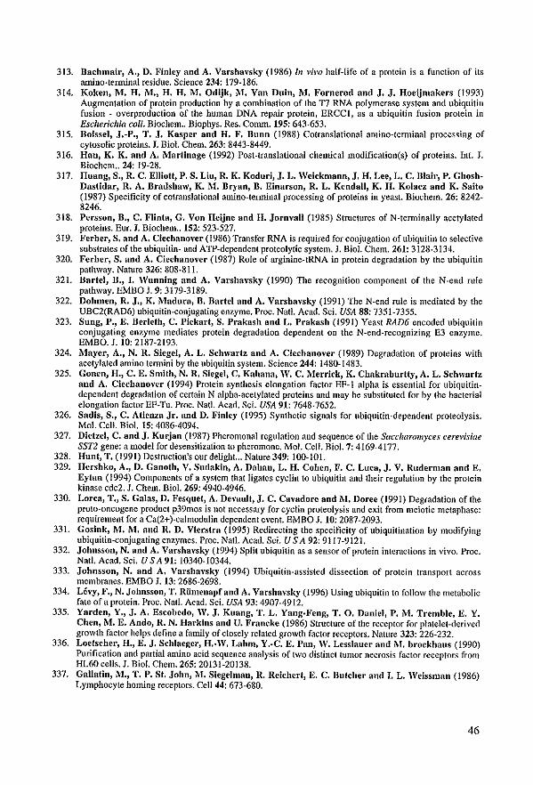
313. Bachmair, A., D. Finley and A. Varshavsky (1986) /11 l'b'o half·Iifc of a protcin is a funclion of ils amino-terminal residue. Science 234: 179·186.
314. Koken, M. H. M'J H. H. M. Odijk, M. Van DuinJ lVI. Fornerod and J. J. Hoeijmakers (1993) Augmentation of prolein production by a combination of the TI RNA polymerase system and ubiquitin fusion· overproduction of the human DNA repair protein, ERCCl, as a ubiquitin fusÎon protein in Escherichia eaU. Biochem .. Biophys. Res. Comm. 195: 643-653.
315, Boissel, J'-P'J T. J. Kasper aud H. F. Buoo (1988) Cotranslational amino·temlinal processing of cytosolic proteins. J, Bio1. Chem. 263: 8443-8449,
316, HaDJ K. K. and A. l\-Iartinage (1992) Post-translational chemical modification(s) of proteins. Int. J, Bîochem" 24: 19-28,
317. Huang, S.) R. C. Elliott) P. S. Liu, R. K. Koduri, J. L. Weiekmann, J. H. Lee, L. C. Blalr, P. GhoshDastidar, R. A. Bradshaw, K. 1\1. BryaoJ B. Einarson, R. L. KendalI, K. H. Kolacz and K. Saito (1987) Specificity of cotranslational amino-terminal processing of proteins in yeast. Biochem. 26: 8242· 8246.
318. Persson, n., C. Flinta, G. Von Heijne and H. Jorovall (1985) Stmctures of N-terminally acetylated proteins. Eur. J. Biochem .. 152: 523-527.
319. Ferber, S. aod A. Cieehanover (1986) Transfer RNA is required for conjugation ofubiquitin to selective subslrates ofthe ubiquilin- and ATP-dependent proteolytic system. J. Bio!. Chem. 261: 3128·3134.
320. Ferber, S. aod A. Ciechanover (1987) Role of arginine-tRNA in protein degradation by the ubiquitin pathway. Nature 326: 808·811.
321. Bartel, D., J. Wuooiog and A. Varshavsky (1990) The recognition component of the N-end TUle pathway. EMBO J. 9: 3179-3189.
322. DohmenJ R. J., K. Madura, n. Dartel and A. Varsha\'sky (1991) The N-end mIe is mediated by the UBC2(RAD6) ubiquitin-conjugating enzyme, Proc. Natt. Acad, Sci. USA 88: 7351·7355.
323. Sung, P., E. Derleth, C. Pickart, S. Prakash and L. Prakash (1991) Yeast RAD6 eneoded ubiquitin conjugating enzyme mediates protein degradation dependent on the N-end-recognizing E3 enzyme. EMBO. J. 10: 2187-2193.
324. Maycr, A., N. R. Siegel, A. L. Schwartz and A. Cicchaoover (1989) Degradation of proleins with acetylated amino termini b)' the ubiquitin system. Science 244: 1480-1483.
325. Gonen, H., C. E. Smith, N. R. Siegel, C. Kahana) W. C. I\'Ierrick, K. Chakraburtty, A. L. Schwarfz aod A. Ciechanoycr (1994) Protein synthesis elongation factor EF·I alpha is essential far ubiquitin· dependent degradation of eertain N alpha-acetylated proteins and may be substituted for by the bacterial elongation factor Ep·Tu. Proc. Natl. Acad. Sci. USA 91: 7648-7652.
326. Sadis, S., C. Atienza Jr. and D. Finley (1995) Synthetic signals for ubiquitin-depcndent proteolysis. Mol. CeU. Bia!. 15: 4086-4094.
327. Dietzel, C. nnd J. KurjnD (1987) Pheromonal regulation and sequence of the Saccharomyces cerevlslae SST2 gene: a model for desensitization 10 pheromone, Mol. Cell. Bio!. 7: 4169-4177.
328. Hunt, T. (1991) Destmction's our delight,,, Nature 349: 100·101. 329, Hershko, A., D. Gauoth, V. Sudakin, A. Dahan, L. H. CollenJ F. C. Luca, J. V. Ruderman aod E.
Eytan (1994) Componcnls ofa system that ligatcs cyclin 10 ubiquitin and their regulation by thc protein kinase edc2. J. Chem, Diol. 269: 4940·4946.
330. Lorea, T., S. GalasJ D. Fesquct, A. Devault) J. C. Cavadore aud i\I. Doree (1991) Degradation of Ihe proto-oncogene product p39mos is not necessary for eyelin proteolysis and exit from meiotic metaphase: requirement for a Ca(2+)·calmodulin dependent event. EMBO J. 10: 2087-2093.
331. Gosink, M. M. and R. D. Vierstra (1995) Redirecting the specificit)' of ubiquilination by modifying ubiquitin-conjugating enzymes. Proc. Nalt. Acad. Sci. U S A 92: 9117-9121.
332. Johnsson, N. and A. Varshavsky (1994) Split ubiquitin as a sensor of protein intcraclÎons in vivo. Proc. Nat!. Acad. ScL U SA 91: 10340-10344.
333. Johnsson, N. aDd A. Varshavsky (1994) Ubiquitin-assisled dissection of protein transport across membranes. EMBO J. 13: 2686-2698.
334. Lévy, F., N. JohnssonJ T. Rümenapfand A. Varsha\'sky (1996) Using ubiquitin 10 follow the metabolic fateofaprotein. Proc. Nal!. Acad. ScL USA 93: 4907-4912.
335, Yarden, Y'J J. A. Escobcdo, lV. J. Kuang, T. L. Yang.Feng, T. O. Dauiel, P. 1\'1. Trembie, E. Y. Chen, M. E. Ando, R. N. Harkins and U. Francke (1986) Structure of the receptor for platelet-derived growth factor helps define a famil)' of closely related growth factor receptors. Nature 323: 226·232.
336. Loefscher, H., E. J. Schlaeger, H.-W. Lahm, Y.-e. E. Pan, W. Lesslauer and M. brockhaus (1990) Purification and parlial amino acid scquence analysis of two distinct tumor necrosis factor reeeptors from HL60 cells. J. Biol. Chem. 265: 20131·20138.
337. Gallatio, .M., T. P. St. Joho, !\I. Siegelman, R. Reiehert, E. C. Huteher and J. L. Wcissmall (1986) Lymphocyte homing reeeptors. Ce1l44: 673-680.
46
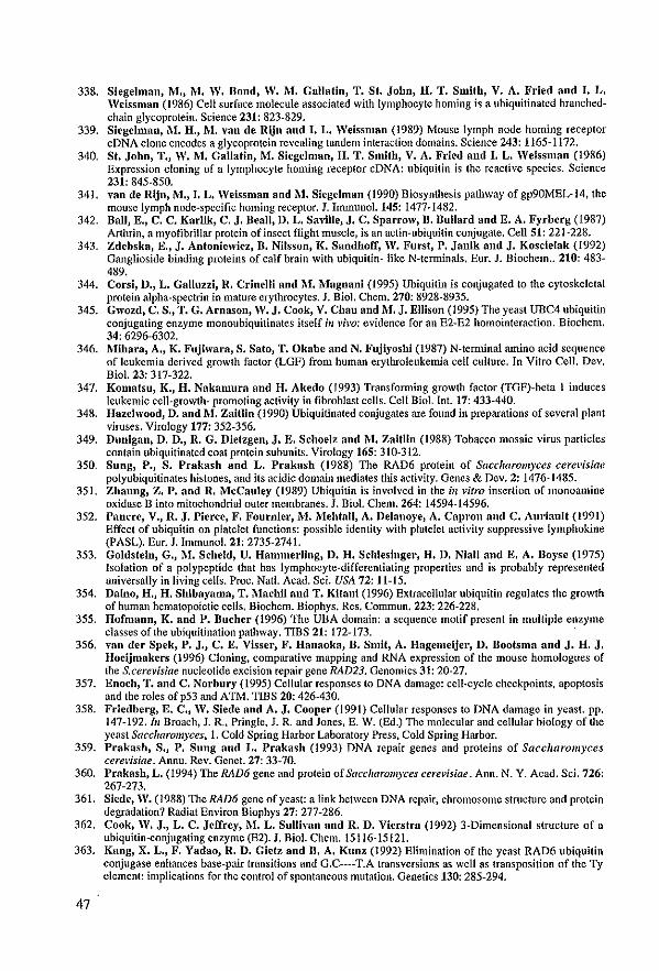
338. Siegelman, lV!., i\.-1. W. Dond, W. M. Gallatin, T. St. Jobn, H. T. Smith, V. A. Fried nnd I. L. Weissman (1986) Cel! surface molecule assoeiated with Iymphocyte homing is a ubiquitinated branchedchain glycoprotein. Science 231: 823-829.
339. Sicgclman, M. 11., M. van de Rijn and J. L. Weissman (1989) Mouse lymph node homing receptor cDNA elone encodes a glycoprotein revealing tandem interaction domains. Seience 243: 1165-1172.
340. St. John, T., W. M. Gallatin, M. Siegelman, 11. T. Smith, V. A. Fricd and I. L. Weissman (1986) Expression cloning of a Iymphoeyte homing receptor cDNA: ubiquitin is the reactive species. Science 231: 845-850.
341. van de Rijn, M., I. L. Weissman and M. Siegelman (1990) Biosynthesis pathway of gp90MEL-14, the mouse lymph node-speeific homing receptor, J. Immunol.145: 1477-1482.
342. Dali, E., C. C. Karlik, C. J. DeaU, D. L. Savillc, J. C. Sparrow, B. Bullard and E. A. Fyrberg (1987) Arthrin, a myofibrillar protein of insect rught muscle, is an actin-ubiquitin conjugate. Cell 51: 221-228.
343. Zdcbska, E" J. Antoniewicz, D. Nilsson, K. Sandhoff, W. Furst, P. Janik and J. Koscielak (1992) Oanglioside binding proleins of calf brain with ubiquitin- like N-terminals. Eur. J. Biochem .. 210: 483-489.
344. Corsi, D., L. Galluzzi, R. Crinelli and M.l\-1agnanl (1995) Ubiquilin is conjugated 10 Ihe cytoskeletal protein alpha-spectrin in mature erythrocytes. J. Bio!. Chem. 270: 8928-8935.
345. Gwozd, C. S., T. G. Arnason, W. J. Cook, V. Chau and M. J. Ellison (1995) The yeast UBC4 ubiquitin conjugating enzyme monoubiquitinates itself ill l'h'o: evidence for an E2-E2 homointeraction. Biochem. 34: 6296-6302.
346. Mihara, A., K. Fujlwara, S. Sato, T. Okabe and N. Fujiyoshi (1987) N-temunal amino acid sequence of leukemia derived growth factor (LOF) from human erythroleukemia cel! culture. In Vitro Cell. Dev, Biol. 23: 317-322.
347. Komatsu, K., 11. Nakamura and H. Akedo (1993) Transforming growth factor (TGF)-beta 1 induces leukemie cell-growth- promoting activity in fibroblast cells. CeU Biol. Int. 17: 433-440.
348. Hazelwood, D. and M. ZalUin (1990) Ubiquitinated conjugates are found in preparations of several plant viruses, Virology 177: 352-356.
349. Dunigan, D. D., R. G. Diefzgen, J. E. SchoeJz and M. Zaltlln (1988) Tobacco mosaic virus particles contain ubiquitinated coat protein subunÎ!s. Virology 165: 310-312.
350. Sung, P., S. Prakash and L. Prakash (1988) The RAD6 protein of Saccharom)'ces cerew'siae polyubiquitinates histones, and its acidic domain mediates tbis activity. Genes & Ocv. 2: 1476-1485.
351. Zhaung, Z. P. and R. McCauley (1989) Ubiquitin is involved in the ill vitro insertion of monoamine oxidase n into mitochondrial outer membranes. J. Biol. Chem. 264: 14594-14596.
352. Pancre, V., R. J. Pierce, F. Fournier, M. Mehtali, A. Delanoye, A. Capron and C. Auriault (1991) Effect of ubiquitin on platelet functions: possible idenlity with platelet activity suppressive Iymphokine (PASL). Eur. J. Immunol. 21: 2735-2741.
353. Goldstein, G., M. Scheid, U. Hammerling, D. H. Schlesinger, 11. D. Niall and E. A. Doyse (1975) Isolation of a polypeptide that has lymphocyte-differentiating propertics and is probably represented universally in living cells. Proc. Nat!. Acad. Sci. USA 72: 11-15.
354. Dalno, 11., H. Shibayama, T. Machii and T. Kitani (1996) Extracellular ubiquitin regulates the growth of human hematopoietic ceUs. Biochem. Diophys. Res. Commun. 223: 226-228.
355. Hofmann, K. and P. Bucher (1996) The UBA domain: a scquencc motif present in multiple enzyme classes of the ubiquitînation pathway. TIBS 21: 172-173.
356. van der Spek, P. J., C. E. Visser, F. Hanaoka, B. SmU, A. lIagemeijer, D. Dootsma and J. H. J. Hoeijmakers (1996) Cloning, comparative mapping and RNA expression of the mouse homologues of the S.cerevisiae nucleotide excision repair gene RAD23. Genomîcs 31: 20-27.
357. Enoch, T. and C. Norbury (1995) Cellular responses to DNA damage: eell-cycle checkpoints, apoptosis and lbc roles of p53 and ATIvf. TIBS 20: 426-430.
358. Friedberg, E. C., W. Slede and A. J. Cooper (1991) Cellular responses 10 DNA damage in yeast. pp. 147-192./11 Broach, J. R.. Pringle, J. R. and Jones. E. W. (Ed.) The molecular and cellular biology of the yeast Saccharo11l)'ces, 1. Cold Spring Harbor Laboratory Press, Cold Spring Harbor.
359. Prakasb, S., P. Sung and L. Prakash (1993) DNA repair genes and proteins of Saccharomyces cerevisÎae. Annu. Rev. Genet. 27: 33-70.
360. Prakash, L. (1994) The RAD6 gene and protein of Saccharom)'ces cere~'isiae. Ann. N. Y. Acad. Sei. 726: 267-273.
361. Siede, W. (1988) The RAD6 gene of yeast: a link between DNA repair. ehromosome structure and protein degradation? Radiat Environ Biophys 27: 277-286.
362. Cook, W. J., L. C. Jeffrey, M. L. Sullivan and R. D. Vierstra (1992) 3-Dimensional structure of a ubiquitin-conjugating enzynle (E2). J. Bio!. Chem. 15116-15121.
363. Kang, X. L., F. Yadao, R. D. Gie(z and B. A. Kunz (1992) E1imination of the yeast RAD6 ubiquitin conjugase enhances base-pair transitions and G.C----T.A transversions as well as transposition of the Ty clement: implications for the control of spontaneous mutation. GenetÎCs 130: 285-294.
47
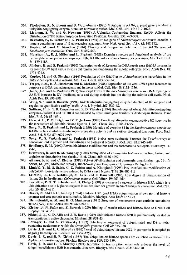
364. Picologlou) S.) N. Drown nnd S. W. Liebman (1990) Mutations in RAD6. a yeast gene encoding a ubiquitin-eonjugating enzyme, stimulate retrotransposition. Mol. Cell. Biol. 10: 1017-1022.
365. Liebman, S. W. and G. Newnam (l993) A Ubiquitin-Conjugating Enzyme, RAD6, Affects the Distribution ofTyl Retrotransposon Integration Positions. Genetics 133: 499-508.
366. Reynolds, P., S. Weber and L. Prakash (l985) RAD6 gene of Saccharolll)'ces cerevisiae encodes a protein containing a tract of 13 eonsecutive aspartates. Proc. Natl. Acad. Sci. U S A 82: 168-172.
367. Kupiec, M. and G. Simchen (1984) Cloning and integrative deletion of the RAD6 geile of Saccharolll)'ces cerevisiae. Curr. Gen. 8: 559~566.
368. MorrisoD) A., E. J. I\Illler aDd L. Prakash (1988) Domain strueture and functÎonal analysis of the carboxyl-terminal polyacidic sequence of the RAD6 protein of Saccltarom)'ces cerevisiae. Mol. Cello Biol. 8: 1179-1185.
369. Madura) K. and S. Prakasb (1990) Transcript levels of S.cerevisiae DNA repaiT gene RAD23 inerease in response to UV light and in meiosis but remain constant during the mitotic ceU cyc1e. Nuc!. Acids Res. 18: 4737-4742.
370. Kupiec, l\I. and G. Simchen (1986) Regulation of the RAD6 gene of Saccharolll)'ces cere)'isiae in Ihe mitotic ceIl cycle and in meiosis. Mol. Gen. Genet. 203: 538-543.
371. Treger, J. M., K. A. Heichman and K. MeEntee (1988) Expression oflhe yeast UBJ4 gene incrcascs in response 10 DNA-damaging agents and in meiosis. Mol. Cell. Bio1. 8: 1132-1136.
372. Jones, J. S. and L. Prakash (199l) Transcript levels of the Saccharolll)'ces cere)'isiae DNA repair gene RADJ8 increase in UV irradialed cells and during meiosis but not during the mitotic cell cycle. Nuc1. Acids Res. 19: 893-898.
373. Wing, S. S. nnd D. Damille (1994) 14-kDa ubiquitin-conjugating enzyme: structure of the rat gene and regulation upon fusting and by insulin. Am, J. Physiol. 267: E39-48.
374. Sullivan, M. L., T. D. Carpenter aod R. D. Vierstra (1994) Homologues ofwheat ubiquitin·conjugatiog enzymes-~TaUBCl and TaUBC4 are eneoded by small fllultigene families in Arabidopsis thaliana. Plant Mol. Biol. 24: 651-661.
375. Haas} A. L.} P. M. Bright aod V. E. Jackson (1988) Functional diversÎty among putative E2 isozymes in the mechanism ofubiquitin-histone ligation. J. BioL Chem. 263: 13268-13275.
376. Sung, P.} S. Prakash aod L. Prakash (1990) Mulalion of cysteine-88 in the Saccharomyces cere)'isiae RAD6 protein abolishes lts ubiquitin-eonjugating activity and its various biological functions. Proc. Natl. Acad. Sci. U S A 87: 2695-2699.
377. Sung, P., S. Prakash nnd L. Prakash (1991) Stabie ester conjugate between the Saccharomyces cerevislae RAD6 prolein and ubiquitin has no biological activity. J. Mol. Biol. 221: 745-749.
378. Dradbury, E. M. (1992) Reversibie histone modifications and the chromosome cell cycle. BioEssays 14: 9-16.
379. Desrosicrs, R. and R. M. Tanguay (1988) Methylation of Drosophifa histones at proline, lysine, and arginine residues during heat shock. J, Biol. Chem. 263: 4686-4692.
380. AUhaus, F. R. aod C. Richter (1987) Poly-ADP-ribosylalion and chromatin organization. pp. 59-. lil Solioz. M. (Ed.) Molecular Biology, Biochemistry and Biophysics 37, Springer Verlag, berlin.
381. Lindahl) T., M. S. Safob, G. G. Poirier aod A. Klunglaod (1995) Post-Iranslalional modificalion of poly(ADP-ribose)polymerase induced by DNA strand breaks. TIBS 20: 405-411.
382. Ericsson, C., I. L. Goldknopf, M. Lezzi and D. DanehoIt (1986) Low degree of ubiquitinalion of histone 2A in Ihe dipteran ChiroJlo11/us tellfalls. Cell Diftèr. 19: 263-269.
383. Swerdlow, P. S., T. Schuster aod D. Finley (1990) A conserved sequence in histone H2A which is a ubiquitination site in higher eucaryoles is not required for growth in Sacclwrolll)'ces cerevisiae. Mol. CelI. BioIIO: 4905-4911.
384. Davics) N. aod G. G. Liodsey (1994) Histone H2B (and H2A) ubiquitination allows nonnal hislone octamer and core particle reconstitution. Biochim. Biophys. Acta 1218: 187-193.
385. Kleinschmidf, A. M. and H. G. Marfinson (1981) Structure of nucleosome eore particles cOlltailling uH2A (A24). Nuc1. Acids Res. 9: 2423-2431.
386. Kirdar, B., N. Dalay and E. Dcrmck (1989) Binding of protein uH2A and histone H2A 10 DNA. Cell Biophys. 14: 43-51.
387. Nickel, D. E., C. D. Allis aDd J. R. Davie (1989) Ubiquitinated histone H2B is preferentially localed in lranscriptionally active chromatin. Biochem. 28: 958·63.
388. Levinger, L. nnd A. Varsha\'sky (1982) Selective arrangement of ubiquitinated and DI protcinconlaining nucleosomes within the Drosophila genome. CeU 28: 375~380.
389. Davie, J. R. aod L. C. l\Iurphy (1990) Level of ubiquitinated hÎstonc H2B in chromatin is coupled to ongoillg transcription. Bioenem. 29: 4752-4757.
390. Davie, J. R. aod n. E. Nickel (1987) The ubiquilinated histone species are enriched in histonc Hl~ depleted chromatin regions. Biochim Biophys Acta 909: 183-189.
391. Davic, J. R. and L. C. Murpby (1994) Inhibition of transcription selectively reduccs Ihc level of ubiquitinated histone H2B in chromatin. Biochem. Biophys. Res. Comm. 203: 344~350.
48
392. Huang, S. Y., M. B. Barnard, .M. Xu, S. l\'lafsui, S .. M. Rose and W. T. Garrard (1986) The aetive immunoglobulin kappa ehain gene is paekaged by non-ubiquitin-conjugated nucleosomes. Proc. Nat!. Acad. ScL U S A 83: 3738-3742.
393. Erlcsson, C., 1. L. Goldknopfand B. Daneholt (1986) Inhibition of transcription does not affect the total amount of ubiquitinated histone 2A in chromatin. Exp. Cello Res. 167: 127-134.
394. Blumenfeld, N., Ho Gonen, A. Mayer, C. E. Smith, N. R. Siegel, A. L. Schwartz and A. Ciechanoyer (1994) Purification and eharaeterization of a novel species ofubiquitin-earrier protein, E2, that is involved in degradation of non·"N-end rule" protein substrates. J. Bio!' Chem. 269: 9574-9581.
395. Kaiser, P., W. Seufert, L. Hofferer, B. Kofler, C. Sachsenmaier, 11. Herzog, S. Jentseh, M. Sebweiger and R. Scbneider (1994) A human ubiquitin-conjugating enzyme homologous to yeast DBC8. J. Chem. Bial. 269: 8797-8802.
396. KJemperer, N. S., E. S. Berleth aud C. M. Pickart (1989) A novel, arsenite-sensitive E2 ofthe ubiquitin pathway: purification and properties. Biochem. 28: 6035-6041.
397. Pickart, e.M. aud A. T. Vella (1988) Ubiquitin carrier protein-catalyzed ubiquitin transfer to rustones. Mechanism and specificity. J. Bio!. Chem. 263: 15076-15082,
398. Wing, S. S. and P. Jain (1995) Molecular doning, expression and characterization of a ubiquitin conjugation enzyme (E2(17)kB) highly expressed in rat testis. Biochem. J 305: 125-132.
399. LeggeH, D. S., D. Jones and E. P. Candido (1995) Caenorhabditis elegans UBC-I, a ubiquitinconjugating enzyme homologous to yeast RAD6/UBC2, contains a novel carboxy-terminal extension that is conserved in nematodes, DNA. Cell. Biol. 14: 883-891.
400. Sulliyan, M. L. and R. D. Vierstra (1991) Cloning of a 16-kDa ubiquitin carrier protein from wheat and Arabidopsis t"aUana, Identification of functional domains by ifll'itro mutagenesis. J. Chem. Biol. 266: 23878-23885.
401. Yoon, J. H., B. J. Lee and H. S. Kang (1995) TheAspergillus IIvsH gene encodes a product homologous to yeas! RADJ8 and neurospora UVS-2. Mol. Gen. Genet. 248: 174-181.
402. Bailly, V., J. Lamb, P. Sung, S. Prakash and L. Prakasb (1994) Specific complex fonnation between yeast RAD6 and RAD18 proteins: a potential mechanism for targeting RAD6 ubiquitin-eonjugating activity to DNAdamage sites. Genes & Del'. 8: 81 1-820.
403. Cassier.Cbauyat, C. and F. Fabre (1991) A similar defect in UV·induced mutagenesis conferred by the rad6 and radJ8 mulations of Saccharolllyces cere~'isiae. Mulat. Res. 254: 247-253.
404. Wing, S. S., F. Dumas and D. llanville (1992) A rabbit reticulocyte ubiquitin carrier protein that supports ubiquitin·dependent proteolysis (E2(14k) is homologous to the yeast DNA repair gene RAD6. J. Bial. Chem. 267: 6495-6501.
405. SharoD, G., B. Raboy, H. A. Parag, D. Dimitroysky and R. G. Kulka (1991) RAD6 gene product of Sacc"arolll)'ces cerevisiae requires a putative ubiquitin protein ligase (E3) for the ubiquitination of certain proteins. 1. Chem. Biol. 266: 15890-15894.
406. Watkins, J. F., P. Sung, S. Prakasb and L. Prakash (1993) The extremely conserved amino tenninus of RAD6 ubiquîtin·eonjugating enzyme is essential for amino·end rule-dependent protein degradation. Genes & Dev. 7: 250-261.
407. Madura, K., R. J. Dohmen and A. Varshaysky (1993) N-recogninlUbc2 Interactions in the N-end Rule Pathway. J. Biol. Chem. 268: 12046-12054.
408. Raboy, B. nnd R. G. Kulka (1994) Role of the C-terminus of Saccharomyces cerevisiae ubiquitinconjugaling enzyme (Rad6) in substrate and ubiquitin-protein-ligase (E3-R) interactions. Eur J Biochem. 221: 247-251.
409. Koken, M. H. n'l., E.M. SmU, J. Jaspers·Dekker, B. A. Oostra, A. Hagemeijer, D. Bootsn18 aud J.II. J. Hoeijmakers (1992) Localization of two human homologs, HHR6A and HHR6B, of the yeast DNA repair gene RAD6 to chroruosomes Xq24-q25 and 5q23-q31. Genomics 12: 447-453.
410. Koken, 1\1. H.M., J. W. Hoogerbrugge, J. Jasper Dekker, J. de Wit, R. WiUemsen, H. P. Roest, J. A. Grootegoed and J. H. J. Hoeijmakers (1996) Expression of the ubiquitin-conjugating DNA repair enzymes HHR6A and B suggests a role in spermatogenesis and chromatin modification. Dev. Biol. 173: 119-132.
411. Kaiser, P., H. A. Mansour, T. Greeten, B. Auer, 1\1. Schweiger and R. Schneider (1994) The human ubiquitin-conjugating enzyme UbeHI is involved in the repair of UV-damaged, alkylated and cross-linked DNA. FEBS Lett. 350: 1-4.
412, Keeling, P.J. and W.F. Doolittle (1996) Methionine aminopeptidase-l: the MAP of the mitochondrion? 413. Landsmann, D. (1996) Histone Hl in Saccharolll)'ces cerevisiae: a double mystcry solvcd? TffiS 21:287-
288.
49

Chapter IJ
The rhp6+ geile of Schizosaccharomyces pombe: a structural alld functiollal homologue of the RAD6 gene from the distalltly related yeast Saccharomyces cerevisiae.

Ths EMBO Journal vol.9 nO.5 pp.1423-1430, 1990
The rhp6+ gene of Schizosaccharomyces pombe: a structural and functional homolog of the RAD6 gene from the distantly related yeast Saccharomyces cerevisiae
P.Reynolds, M.H.M.Koken', J.H.J.Hoeijmakers1, S.Prakash2 and L.Prakash
Th;:partment of Biophysics, University of Rochester School of MeJicine, Rochester, NY 14&-12, USA, lTh;:partment ofCell Diology and Gene/ks, ErasffiUS University, PO Box 1738, 3000 Dr Rotterdam, The Nelherlands and 2Th;:partment of Biology, Uniwrsity of Rochestcr, Rh'er Campus Station, Rochester, NY 14627. USA
Communicated by D.BootslTL1
n.e RAD6 gene of Saccharom)'ces cere~';siae encodes a ubiquitin conjugating enyzme and is re(lulred for DNA repair, DNA-damage-illduced mutagenesis and sporuJation. Here, we show that RAD6 and the rhp~ gene from the distalltly related yeast Schizosaccharolll)'ces pOli/be share a high degree of slrueluraJ and fllllctiollal homoJogy. The predomillantly aeidic carboxyl-terminal 21 amÎllo acids present in the RAD6 protein are absent in the rhp6+ -ellcoded proteln; otherwise, the two proteins are ycry simiJar, nith 77% ldentleal residlles. Ukc rad6, null llIutations of the rhp6+ gene confer a defect in DNA repair, UV mutagellesis and sporulatlon, and the RAD6 and rhp6+ gelles can functionally subslÎlute for one another. These obseryatiollS suggest that functional illteractiollS between RAD6 (rhp6+) protein and othel' components of the DNA rep.'lir complex ha\'c been COJlserYCd amollg eukaryotes. Ke)' words: DNA repair/E2 enzyme/RAD6 gene/r1lp~ gene/Schizosacclwromyces pOli/be
Introduction
The RAD6 gene of Sacclmromyces cerevisiae is involved in a variety of cellular processes. rad6 mutants are highly scnsiti\'e 10 numerous DNA danlaging agents, including UV, -y-rays and alkylating agents (Cox and Parry, 1968; Game and Mortimer, 1974; Prakash, 1974) and are defectÎ\'e in mutation induction by these agents (Prnkash, 1974; Lawrence and Christensen, 1976; McKee and Lawrence, 1979). rad6 nlUtants are defeclive in post-repIîcalion rcpair of UV damage: DNA strand discontinuities left during DNA repIîcation in the newly synthesized DNA strand across from the non-coding UV lesion remain unrepaircd in rad6 mutants (Prakash, 1981). rad6 mutanls are also defeclh'c in sporulation (Game et al., 1980; Montelone et al., 1981), and they grow poorly and have pOOl' plating eftlciency.
The RAD6-encoded protein (Mr 19.7 kd) possesses a highly acidic carboxyl lerminus in which 20 of the 23 residues are acid ie (Reynolds et al" 1985). The polyacidic sequence of RAD6 protein forms a disordered Iinear structure that is appended to the globular domain constiluted by the first 149 residues (Morrisoll et al" 1988). RAD6 protein is a ubiquitin..çonjugating enzyme (E2) (Jentsch el al., 1987) that mediates the attachment of multiple
molecules of ubiquitin to hislones H2A and H2B ill l'itro (Sung el al" 1988), Multiple ubiquitination ofhistones may eftèct all open chromatin configurntioll, or it may mark histones for degradation by the ATP-dependent proteolytic system (Hershko el al., 1984a,b; Hershko and Ciechanover, 1986). The acidic domain of RAD6 is required for the multiple ubiquitination ofhislones (Sung el al., 1988). rad6 mutants hearing a deletion of the acidic sequence fail to sporulate, but the DNA repair and UV mutagenesis functiolls are not aftèctcd (MorrÎson el al" 1988). Mutation ofthe sole cysteine residue (eys-88) in RAD6 to alanine or valine abrogates its E2 activity, and these mutants resembie rad6 null mutants in being defective in DNA repair, UV mutagenesis and sponJlation (Sung et al., 1990), suggesting that RAD6 mediates all of its cellular functions via its role as an E2 enzyme.
Because of the central role of RAD6 in DNA repair and in DNA-damage-induced mutagenesis, we have become interestcd in determining whether RAD6 is conserved among eukaryotes, A high degree of conservation of RAD6 would also suggest a parallel evolutionary conservation of proteins with which RAD6 might interact in its various cellular roles, In tbis paper, we report our studies with the RAD6 homolog from the evolutionarily divergent fission yeast Schizosaccharom)'ces pombe. Phylogenetic studies with SS ribosomal RNAs indicate that S.poll/be is evolutionarily closer 10 Homo sapiens than to S.cere~'isiae (Huysmans el al., 1983). S.pombe also resembles the higher eukaryotes in the control of the milotic cell cycle (RusselI and Nurse, 1986; RusselI et al., 1989), in the presence of introns in many of its gelles and in the sequence requircments for the splicing of introns (Käufer el al., 1985; RusselI and Nurse, 1986). Therefore, a comparisoll of the structure and function of RAD6 from these Iwo divergent ycast spe<:ies should provide a gocx:l measure of evolutionary conscrvation of RAD6 arnong eukaryotes.
Dur studies indicate a remarkable similarity iJl the amino acid sequences of the proteins encoded by the S.cere~'isiae RAD6 gene and by its homolog in S,pombe, rhp6+ (rad homolog in S.p.ombe-§). The major diftèrence between the two proteins is that the rhp6+ protein laeks 21 carboxyllerminal acidic residues present in RAD6, Like rad6 mutations, null mutations of rhp6+ confer a defect in DNA repair, mutagenesis and sporulation, We alstt show that the RAD6 and rhp6+ genes can funetionally suhstitute for anc allother,
Results
Clon/ng of rhp6+, the S,pombe hom%g of RAD6 Southem bIots of S.pombe gellomic DNA were probed with the S.cere\'Ïsiae 0,5 kb EcoRl DNA fragment contaiiting the rad6-149 allele (Morrison et al., 1988), in which the lasl 23 codons of the RAD6 gene from nucleotide positiol15 +448 to +516 (Reynolds et a/., 1985) are deleled. A single
51
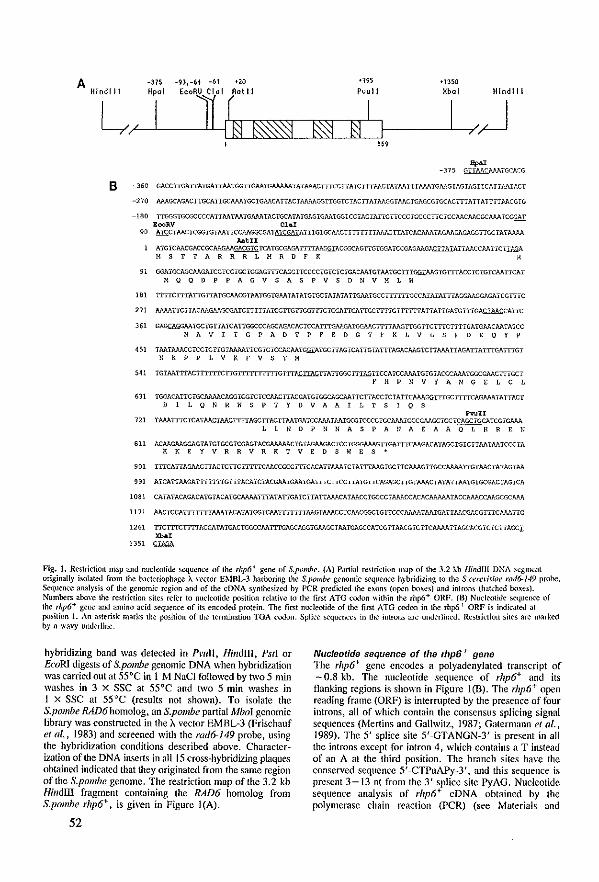
A -375 -9),-61 -61 '20 <195
Pvul1 +1350
L/,,L-' LI _E"RlllL[U'/][~tll~~~~~81 r-~I /~ '"
Hindi! 1 Hpal Xbal Hlndlll
B -360
-270
-180
-"
" '"' m
'" m
SU
'" on
on
", '"
1081
1171
1261
1351
GACCTIGAITAlGATIMIGGTIG..'IAlGAAAAAIAlMACTIICCITAICTr'McrAïMTTrAAATGMGiAGiAGrrCATrJ._".TAC'
MAGCAGACTI'GCATTGCAAAïGCiGAACAITACiMMGGITGGTCiACITAïAAGGlAACTGAC-.ÇGTGCACITTAITATi-iTl>ACGTG
TIG(;GiGCGCCCCATTMïM1GAN>.TACI'GCATATûAGiGAAiGGîCCiACTArrGITCCCIGCCCITCiCCAACMCGCMAiCGGAT RooRV ClaI gCTAACTCGGiGTM'f-rCCt'.l.GGCGATATCGATATI'iGiGCAACITITTrTrAAJ.SrrATCACAAA'fAGJ\AGAGAGGTIGC'A'fMM
AatII ATGICMCGACCGCAAG.>,AGAffiTCiCATGeGAGAITITAAG!.il:ACGGCAGTiGiGGAKCGAGMGACilllIATTA. ... CCl>ATTCI1l-L:.A M S T ï A R R R L M R D F K R
GGATGCAGC1\AGATCCTCCTGCIGG.".GTTICAGCITCCCCiGiCICrGACAAiGiAATGCTTTcm,:MGTGITTACCICiGlCAATTCAT M Q Q D P P A G V SAS P v S D ft V H L W
ITTICITI'AïTGITATGCMCGTMTGGiWoATATATGTGCiAiATATIGJ\AîGCCllllllGCCATAïATTIAGGMGGAGAïCGTTiC
MMTICHACMGMGCGAKTrITrATGCITGITGGTITCTGGJ..ITGAITGCITTTGITTITTAITAITGATGiITGN:J1!.M;.CATIC
GAGQ.GGMTGCTGITA1CAHGGCCCAGCAGACACrCCAITTGMGATGGA!,CIT-TIMGITGGi-TCTTiCTTTTGATGMCMTA'CC ft A V I i G PAD T prE D G T r K L V L S F D E Q Y P
TMTMACCTCCTCITGrMAAITCGTCTCCACMI~I'GCHAGTCATTGTATTTAGACMG1CITA.>,ATIAGATIATTTGATTIGT
ft K P P L V K rvs T M
TGrMTITACITITITCITGl i i 111 j:lllG )",n~HAITGGCriIAGITCCATCCAAATGiGIACGeAM'i'GGCGMCITlGCT r H P ft V Y A ft GEL C L
TGGACATICTGCAAAACAGGrGGTCTCCAACTIACGATGiGGCAGCJ..AHCITACCTCIATTCAMGGIllGCTITiCAGAAATATTAC, DIL Q ft R W S P T Y D V A AlL T S 1 Q S
PwH TMATITCiCAT~TI-~ITACTTMTGATCCAAATJ!.ATGCGîCCCCiGCAMTGCCGJ\AGCTGCTCAGCTGCATCGiGMA
L L ft D P ft N A SPA ft A E A A 0 L H REN
ACAAGAAGGAGTATGIGCGKGAGIACGl\J\AAACiGTAGAAGACTCCIGGG.-\AAGITGATITCMGACATAGCIGiCilMiAATCCCTA K K E Y V R R V R KTV E D S W ES'
TITCAITAGAACITACTCITCITITTCMCCGCC1TiCACATT~.A'ICTAnTAAGiGCITCAAAGilGCCMAAITG'MC'ATAGI"-'''.
ATCAHMGATITITITGITiACATCTACGAATGMTGAlilCrrCCTIA'IGITCAGAGCTTG,MACTATATTM'IGiGCGACIAGiCA
CA'IATACAGACA'lGTACATGCN>.AAïTlAiAHGAKïrAITAAA.CAIMCCiGCCClAAA.CCACACMMAiACCAAA.CCMGCGÇJ;M
AACTCCATIT'ITITAAATACATATGGTCMITIT'iTAAGTAAACCICAACGGCTGITCCCAAMTMiGAITMCGACGITTCAMITC
TICTTICITITACGATA'IGACTGGCCMI"i-IGAGCAGGTGMGC,AA'IGAGCCATCGïrMCGICTICAAMITAGCACGiCrCTTAc-cI ><b., ~
Fig. J. Restriction JJ13p and nucleotide scquence of the rhp6' gene of S.pombe. (A) Partia! restricIion map of th,:, 3.2 J...b Ililldl1l DNA ~egrnent originally i,olatect from !he bacteriophage À wctor EMFlL-3 harboring the S.pombe genonlic sequenc,:, hybridizing to the S.{wcliûOt' rad6-149 probe. Sequencc anal)'sis of !he genornic region and of lhe cDNA s)'nthesizcd by peR predkted th,:, exons (ojXn boxcs) and inlrons (hatchcd bows). Numbers above the r,:,stric/ion sites rder to nudcolide po,ilion rc!ati\"c to the first ATG codon \\ithin the rhp6+ ORF. (8) Nucle-otide se-queoc,:, of lh,:, rhp6+ gene and amino acid sequeoce of its encoded protein. The first nucleotide of thc first ATO codon in !he rhp6+ ORF is indkateJ at position 1. An asterisk marks the position of !he temllnation TOA codon. SpIke sequences in !he introns are underlined. Restrktion sitt'S are marked by a wa\'y underline.
hybridizing band was detected in PI'IlIl, Ril/dlII, PstI or EcoRI digests of S.pombe genomic DNA when hybridization was carried out at 55°C in 1 M NaCi followed by two 5 min washes in 3 x SSC at 55°C and two 5 min washes in 1 X SSC at 55°C (results not shown). To isolate the S.pombe /UW6homolog, an S.pombe partial Mbol genomic library was constructcd in the À vector EMBL-3 (Frischauf et al., 1983) and screened with the rad6-J49 probe, using the hybridization conditions described above. Characterization of the DNA inserts in aU 15 cross-hybridizing plaques obtained indicated that they originated from the same region of the S.pombe genome. The restriction map of the 3.2 kb Hindm fragment containing the RAD6 homolog from S.pombe rhp6+, is given in Figure l(A).
52
Nucleotide sequence of the thp6+ gene The rhp6+ gene cncooes a polyadenylated transcript of -0.8 kb. The nucleotide sequence of rhp6+ and its flanking regions is shown in Figure 1(B). The rhp6+ open reading frame (ORF) is internlpted by the presence of four introns, all of which contain the consensus splicing signa! sequences (Mertins and Gallwitz, 1987; Gatermann el al., 1989). The 5' splice site 5'-GTANGN-3' is present in all the introns except for intron 4, whieh contains a T instead of an A at the third position. The branch sites have the conserved sequence 5'-CTPuAPy-3', and this sequence is present 3-}3 nt from the 3' spike site PyAG. Nucleotide sequence analysis of rhp6+ cDNA obtaÎned by the polymerasc chain rcaction (PCR) (see Materials and
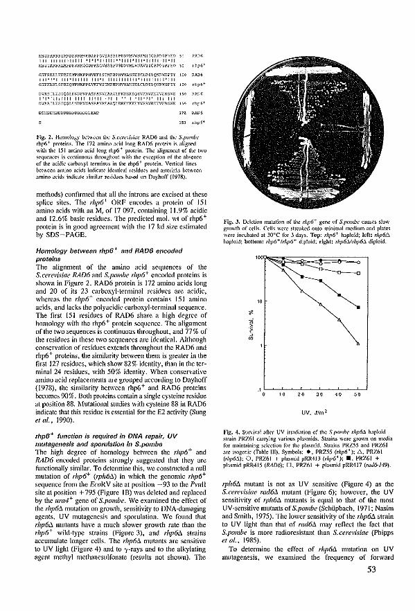
~:5,P A.~"RL1'::'<!If"R."'~~!IM ~ GV,,/<SP n Ni\:X'J"";~Ift-'lll Gh\D;:PïED ~,) RAD 6 111111111111111 '1'1'111111"1111'111'11111111'11 P-"'TABRRL'(?DFKR.'~X!OPP"'GVS1<SPVS D:nrXLh~IAVII G~ 110 i ?ftO ~~ .- [,P 6'
G"RLLLEFDEE 'iP~IKPPH1rr:FLSE~HP)lVYII::GE lew I LQ:;R .... pn: 1 c ~ FJ.t> 6 111'1'1111'111111111'111111111111'111111111'111 G"KLVLSFDEQYF1IKPPLVKFV5:lY.FHP11VYIISGELCLDIlC.:",XSr,Y 1 C 0 d,p 6+
D\'lIS IL, SIQSLFI;clPl;PASP A!"lEAA,LFIillHTSQY\lKRVKE:iVEKS"o""E 1\ 0 Rl\Il6
111'111111111111 Iijll 111 1 " I '11'11" 111 111 D'l~J<IL 'SIQSLI.SDP1~illSP A'illEAAt;lI.RRE!iKKEYVRRVRK:iVEDSh"E 1\ ~ rtl' 6'
(ln:-'.0[O~DDUUUDUUUDDUEAD 172 RAD6
l~l rhp6'
Fig. 2. Homo!ogy bet\\<'\'n the S.("crCl"isiuc RAD6 and the S.pombc rhp6+ proteins. The [72 amino add long RAD6 protein is aligned with the 151 amino add long rhp6 + prolein. The alignment of the two sequeoces is conÜnuous throoghout with the exception of the absence of the addk l'Jroo.\yl lemlinus in the rhp6 + protein. Vertkal lines b.otI\oOen amino adds indical<, id,;ntical röidues and a,lerilks b.otll'oOen amino acids indicJt,; similar rdduö ba,ed on Dayhoff (1978).
methods) conl1rmcd that all the introns are excised at these splice sites. The rhp6+ ORF encodes a protein of 151 amino acids with an Mrof 17 ('1)7, containing 11.9% acidie and 12.6% basic residues. The predicted mol. \Vt of rhp6 + protein is in good agreement with the 17 kd size estimaled by SDS-PAGE.
Homology between rhp6 + and RAD6 encoded proteins The alignment of the amino acid sequences of the S.cerel'ÎsÎae RAD6 and S.pombe rhp6+ encoded proteins is shown in Figure 2. RAD6 protein is 172 amino acids long ;md 20 of its 23 carboxyl-terminal residues are acidic, whereas the rhp6+ encoded protein contains 151 amino acids, and lacks the polyacidic carboxyl-terminal sequence. The first 151 residues of RAD6 share a high degrce of homology with the rhp6 + protein sequence. The alignment of the two sequences is continuous throughout, and 77% of the residues in these two scquenccs are identical. Although conservatÎon of residues extends Ihroughout the RAD6 and rhp6+ proteins, the similarity between them is greater in the first 127 residucs, which show 82% identity, than in the terminal 24 residucs, with 50% identity. When conservative amino acid replacements are grouped according 10 Dayhoff (1978), the similarity between rhp6+ and RAD6 proteins bccomc-s 90%. Both proteins contain a single cystcine residue at position 88. ~-lutational studies with cysleine 88 in RAD6 indicate that Ihis residue is essential for the E2 activity (Sung et al., 1990).
rhp6+ function is required in DNA repair, UV mutagenesis and sporulation in S.pombe The high degree of homology between the rhp6+ and RAD6 encoded proteins strongly suggested that they arc functiona][y similar. To detemline this, wc constmcled a null nmtatÎon of rhp6+ (rphM.) in which the genomic rhp6+ sequence from the EcoRV site at position -93 to thc PI'IlIl site at position + 795 (Figure IB) was dcletcd and replaced by the Itra4+ gene of S.pombe. We examined Ihe effect of the rhp6!l mUlation on growth, scnsitivity to DNA-damaging agents, UV mutagcnesis and sporulation. We found that rhp6il mulants have a much slmver growth rate than the rhp6+ wild-type straÎns (Figure 3), and rhp6ó. strains accumulate longer cells. The rhp6il mutants are sensitive to UV light (Figure 4) and 10 ')'-rays and to the aU,:ylating agent methyl methancsulfonate (rcsults not shown). Thc
Fig. J. D.;ktion nmtalion of Ihe r/Jp6+ gene of S,pml/hc çauses slow gro\\th of cells. Cclls wcre streah--d onl0 minimal ll1I.--dium and pla!es \\ere incubaled at 30°C for 3 da)"s. Top: rhp6+ haploid: Icf!: rhp6~ haploid: bottom: rhp6+frhp6+ diploid; rlght: rhp6J.frhp6J. diploid.
10
I "'
UV. Jfm 1
Eg, 4. Survival Jfter UV irw,dialion of the S.pombl' rhp6J. haploid sirain PRZ61 carf)'ing variOlI, pb<mids. Sirains \\ere grown on media for maintaining ~kclion for the pla.<mid. S\rains PRZ55 and PRZ61 are io;ogcnic (Tab1e 111). S)"mbols: ., PRZ55 (rl!p6+); 6, PRZ61 (lhp6.1); 0, PRZ61 + p[asmid pRR413 (rl!p6 f ); _, PRZ61 + plasmid pRR415 (RAD6); 0, PRZ61 + plasmid pRR417 (rud6-149}.
1],h6ó. mutant is not as UV sensitive (Figure 4) as the S.cerel'isiae rad6ó. mutant (Figure 6); howevcr, thc UV sensitivity of rph6ó. mutants is equal 10 that of thc most UV-sensitive mutants of S.pombe (Schüpbach, 1971; Nasim and Smith, 1975). Thc lowcr sensitivity of the rhp6il struin to UV light than that of rad6ó. ma)' reflect the fact that S.pombe is more radioresistant than S.cere\'isiae (Phipps et al., 1985).
To determine the effect of rhp6il mutation on UV mutagenesis, we examined the frequeney of forward
53
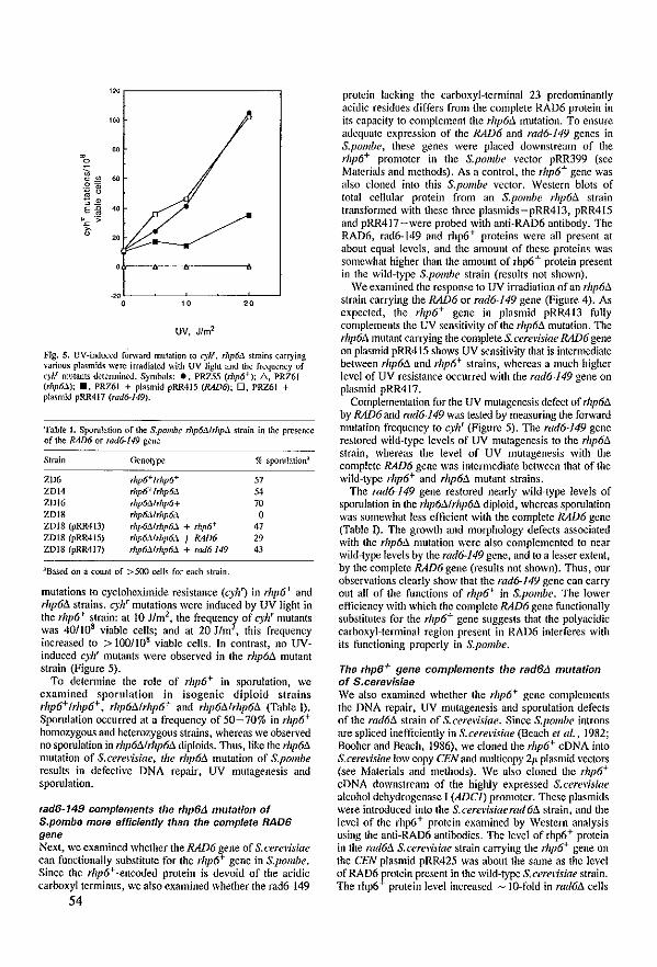
"" '00
~ . 0
" c"" ., ~! E{i .,
".<:: ":;: ~ 0
'"
UV, J/m2
llg. 5. UV-indu0.--d forward ffiulatjon 10 cyh'. rhpM struins carrying various plasmids wcre irradialeJ \\ith UV light and !he frcqucncy of C)h' mlltanlS detennined. Symbols: •• PRZ55 (mp6+); D., PRZ61 (rhp6J.); ., PRZ61 + plasmid pRR415 (RAD6); 0, PRZ6l + pla,mid pRR417 (rad6-J49).
Table J. Sporulation of !he S.pmllhe rhp6Mrhpil. strain in \he prescoce of the RAD6 or rod6-149 gene
Struin Genotype ~ 5porulation'
ZD6 rhp6+ ',111'6+ 57 ZDI4 rhp6+lrhpM " ZOl6 rhp6!JJrhp6+ 70 ZDI8 rhp6!11rhp6!J. 0 ZDl8 (pRR4i3) rhp6Mrhp6::' + rhp6+ 47 ZDI8 (pRR415) rhp6MrhpM t RAD6 29 ZDI8 (pRR417) rhp6Mrhp6!J. + md6-149 43
aBas.ed on a COllO! of >500 eclls for each stmin.
mutations 10 cyclohcximide rcsislance (cyh') in rhp6+ and rhp6!l strains. cyJ{ mutations wcrc induced by UV light in the rhp6+ slraÎn: at 10 J/m2, the frequencl of cyl{ mutants was 40/108 viabie cells; and at 20 J/m , this frequency increased to > 100/108 viabie cells. In contrast, na UVinduced cyJ{ mutants were observed in Ihe rhp6!J. mutant strain (Figure 5).
Ta delennine the role of rhp6+ in sporulation, we cxamined sporulation in isogenic diploid slrains rhp6+ Irhp6+, rhp6A1rhp6+ and rllp61l1rhp6!J. (Tabie 1). Sporulation occurred al a frequency of 50-70% in rhp6+ homozygous and helerozygous strains, whercas we observed no sporulation in rllp6A1rhp6!J. diploids. Thus,like the rhp6!J. mutation of S.cere~isiae, file rhp6A mutation of S.pombe results in defective ONA repair, UV mutagenesis and sporulalion.
rad6-149 camp/ements the rhp6L1 mutat/on of S.pombe more efficlently than the complete RAD6 gene Next, we examined whether the RAD6 gene of S. cere~<fsiae can functionally substitute for the rhp6+ gene in S.pombe. Since the rhp6+ -encoded protein is devoid of the acidic carboxyl terminus, we also cxamined whether the rad6-149
54
protein lacking the carboxyl-terminal 23 predominantly acidic residues differs from the complete RA06 protein in its capacity 10 complement the rhp6A mutation. To cnsure adequate expression of the RAD6 and rad6-l49 genes in S.pombe, these genes were placed downstream of the rllp6+ promoter in the S.pombe vector pRR399 (see Materials and methods). As acontrol, the rhp6+ gene was also c1011ed into this S.pombe vector. Western blots of total cellular protein from an S.pombe rhp6!J. strain transformed with these thrce plasmids-pRR413, pRR415 and pRR417-were probed with antÎ-RA06 antibody. The RAD6, rad6-149 and rhp6+ proteins were all present at ahout equal levels, and the alllount of these proteins was somewhat higher {han the amount of rhp6 + protein present in the wild-type S.pombe strain (results not shown).
We exalllined the response to UV irradiation of an rhp6A strain carrying the RAD6 or rad6-149 gene (Figure 4). As expected, the rhp6+ gene in plasmid pRR413 fully complements Ihe UV sensitivity of Ihe rllp6A mutation. The rhp6A mutant carrying the complete S. cerevisiae RAD6 gene on plasmîd pRR415 shows UV sensitivity that is intennediate between rhp6!J. and rhp6+ strains, whereas a much higher level of UV resistance occurred with the rad6-l49 gene on plasmid pRR417.
Complelllentation for thc UV mutagenesis defect of rhp6tJ. by RAD6 and rad6-149 was tested hy mea~uring!he forv.'ard mutation frequency to cyhr (Figurc 5). The rad6-149 gene rcstored wild-type levels of UV mutagenesis to the rhp6A strain, whereas the level of UV mutagenesis with the complete RAD6 gene was inlermedialc bctwecn that of the wild-type rhp6+ and rhp6tJ. mutant strains.
The rad6-149 gene restored nearly wild-type levels of sporulatÎon in !he rhp6A1rhp6tJ. diploid, whereas sporulation was somewhat Icss cfficient with the complete RAD6 gene (TabIc I). The growth and morphology defects associated with !he rhp611 mUlation were also complemented la near wild-type levels by Ihe rad6-149 gene, and la alesser extent, by the complete RAD6 gene (results not shown). Thus, our observations cIearly show that the rad6-149 gene can carry out all of the functions of rhp6+ in S.pombe. The lower efficiency with which thc complete RAD6 gene functionally substitutes for the rllp6+ gene suggests that the polyacidic carboxyl-terminal region present in RAD6 interferes with its functioning propcrly in S.pombe.
The rhp6+ gene complements the rad6L1 mutation of S.cerevisiae We also examined whether the rhp6+ gene complements the DNA repair, UV mutagenesis and spomlation defects ofthe rad6!:J. strain of S.cerevisiae. Since S.pombe introns are spliced inefficienlly in S.cere~'isiae (Beach et al., 1982; Booher and Brach, 1986), we c1oned!he rllp6+ cONA into S. cere~isiae low copy CEN and multicopy 2/1 plasmid vectors (sec l\'faterials and methods). We also cIoned the rhp6+ cDNA dowllstream of the highly expressed S.cerevisiae alcohol dehydrogenase I (ADCl) promoter. These plasmids were introduced into the S.cerel'isiaerad6ll struin, and the level of the rhp6+ protein examincd by Western atlUlysis using the anti-RAD6 antibodies. The level of rhp6+ protein in the rad6!J. S.cere\isiae strain carrying the rhp6+ gene on the CEN plasmid pRR425 was about the same as the Icyel of RAD6 frotein present in the wild-type S. cerc\isiae strain. The rhp6 protein level increased - JO-rold in rad6tJ. cells
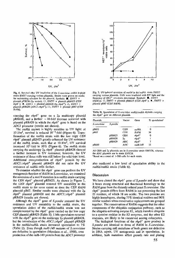
10
."
1 '" ..
.Q1
.00' 0 10 20 30 40
UV, J/m2
Fig. 6. Survival after UV irradiation of the S.cert'lisiae nll{6fJ. haploïd struin EMY7 carrying various plasmids. Strains were grol\TI on nll.--dia for m:üntaining seleçtion for the plasmid. Symbols: _, EMY7 + plasmid pTB236 (lp wetor); O. EMY7 + p\asmid pRR425 (CEN Thp6+); ., EMY7 + plasmid pRR428 (2;.< ,hp6+j; 6, EMY7 + pJasmid pRR429 (ADC/::rhp6+); D, EMY7 + p!asmid pR67 (CEN RAD6).
carryîng the rhpó+ gene on a 2~ multkopy plasmid pRR428, and a further -IQ-fald increase occurred with plasmid pRR429 in which the rhp6+ gene is fused on the ADCl promoter (resuils not shown).
Thc rad6A mutant is highly sensitivc 10 UV light: at 10 J/m2, survival is reduced 1O-4-fold (Figure 6), Transformation of the rad6A strain with the low eopy eEN rhp6+ plasmid pRR425 greatly enhaneed the uv resislanee of the rad6ó. strain, such that at 10 11m2 , UV survival increased 16'-fold to 10% (Figure 6). The rad6A. strain carrying the multicopy 21' rhp6+ plasmid pRR428 showed a fimher increase in UV rcsisfanee; however, the UV resistance of these eells was still below the wild-type level. Additional overproduction of rhp6+ protcin by the ADCl::rhp6+ plasmid pRR429 did not mise the UV resistanee of rad6A. eelIs further.
To examine whelher the rhp6+ gene ean perform the UV mutagenesis funetion of RAD6 in S.cere\'Ïsiae, we examined Ihe reversion of a met14 mutation in a rad6ó. strain eanying the CEN rhp6+ plasmid pRR425. As shown in Figure 7, the CEN ,hp6+ plasmid restored UV mutability 10 the rad6ó. strain 10 the same extenl as does the CEN RAD6 plasmid pR67. Similar rcsults were obtaincd with the 2Jl rhp6+ plasmid pRR428 and the ADC1::rhp6+ plasmid pRR429 (results not shown).
Although Ihe rhp6+ gene of S.pombe reslored the UV resistance and UV mutability to the rad6A. strain, the sporulation defect of the rad6l11rad6ó. strain was not complemented by the rhp6+ gene earried on the low eopy CEN plasmid fRR425 (Tabie II). Little sporulation occurred with the rhp6 gene on the multicopy 2Jl plasmid pRR428, but the introduetion oftheADC/::rhp6+ plasmid pRR429 in Ihe rad6ó.lrad6ó. strain increased sporulation la 7% (Tab!e IJ). Even Ihough rad6-149 mutanls of S.cerevisiae arc defecIÎ\'e in sporulalion (Morrison el al., 1988), overpnxluetion of Ihe rad6-149 protein from the ADel promoter
.40
'" , 100 0
lï 80
g~ 0 0
60 ~ .. "D
t- .~ 40 w
" 20
-20 0 2 , 10
UV, J/m 2
}'jg. 7. UV-induced reversion of me/14 in the'rod6.:l ~truin EMY7 carrying various plasmids. Cells were irradiated with uv light and the frequency of MEP rewrtants detemuned. S)'mbols: ., EMY7 (rhp6!::"); O. EMY7 + plasmid pRR425 (CEN r/lp6+); ., EMY7 + pla.>mid pR67 (CEN RAD6),
Table 1I. Sporulation of S.cemiJiae rad6.:l1md6!J. diploids canying the rl1p6+ gene on different plasmicts
Plasmids Vector <koc ~ sporulation'
S,cere)üiae S.pombe
pR611 CEN mdM 0 pR67 CEN RAD6 3-l
pRR425 eEN rhp6+ 0 pRR428 2, rhp6+ pRR429 Anc rhp6+
pR619 AOC rad6-149 12
All CEN and 21-' plasmids are in S.cereoisiae struin EMY26, whereas \he ADC plasmids are in strain EMY28. aBased on a count of > 500 (('lis for each strnin.
also eonferrcd a law leyel of spontlation ability to the rad6ó.1rad6A. strain (Tabie II).
Discussion
We have c10ned the rltp6+ gene of S.pombe and show that it bears strong struetural and funetional homology 10 the RAD6 gene from the distantly related yeast S.cerelüiae. The rhp6+ protein diffcrs from RAD6 in not possessing tbe last 21 residues, of which 18 arc acidic. The two proteins are highly homologous, sharing 77% identical residues and 90% similar residucs when eonseryalive rcplaeements are grouped logeiher. The conservation of RAD6 suggests thaI the olher components of the ubiquitin conjugation pathway, such as tbe ubiquitin-activating enzyme El, whieh transfers ubiquitin to a cysteine residue in the E2 enzymes, and !he olher E2 enzymes, are likely 10 he çonserYed among eukaryotes.
The biologica! funetions of the rhp6+ gene product in S.polllbe are identieal la those of RAD6 in S.cerel'isiae. Strains carrying null mutations of both genes are defective in DNA repair, UV mutagenesis and in sporulatioll. In addition, bath mutatians affect growth rale and plating
55
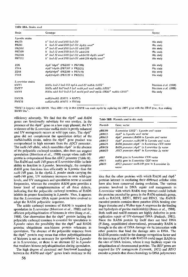
Table UIA. Strains u,ed
Strain
S.pombc strains PR7..55 PRZ61
Genotyp.:
11- 'eu/~3l llra.J.DI8 Ip/-IJI 11- felll-3l lIW.f.DI8I.rs/-1J1 rhp6J.::llrar h+ (eul·3l ura4.D18 l)"sl-131 ade6-210 h·· (<'111-32 IIfd-l.Df8/Y5/-f3f ade6-216
Source
PRZI07 PRZI09 PRZI19 PRZI21
h~ feul-J2 ImrUJ/8 (y5/-131 adc6-210 rhp6.'l :lIrar h- leul-J2 um-l.Dl8 lysl-131I1dc6-216 ,hp6J. :/lfd.J+
this stud)' thls stud)' this stud)' this stud)' this stud)' this stud)'
Z06 ZDI4 ZDI6 ZDI8
rhp6+ frhp6+ (PRZI07 X PRZI09) rhp6+lrilp6:1 (PRZI07 x PRZI21) rhp6:1/rhp6+ (PRZI09 x PRZI19) rhp6J./rhp6:1 (PRZI19 x PRZJ2l)
this study
thls stud)' Ihis stud)' Ihls study
S.CCfé'IÜiIlC ~tmins
EMYl MATa Iclll-J leu2-lll I,pl Û ura3-52 rad6û::LEU2+ Morrison ct 111. (1988) Morrison Cl al. (1988) thls study
EMY7' MATo ade5 his71f1l2-3 /)"sl m<ll4 paS 11m3 md6:1::I.EU2+ EMY8 MATo adeS his7lm2-J IJsl mell4 peiS 11m3 Irplû::U~J3+ rod6J. :LEU2+
EMY26 EMY28
r//(/6:1/r(/<l6J. (EMYl x EMY7j md6J.lrmf6J. IEMYI x EMY8)
'EMY7 is isogenic with EMY8. Thcy differ nni)' in that EMY8 lIas made (rplû by replacing Ihe!RP1 gene wÎlh Ihe URM gene, thus making EMY8 Ura +.
efficiency adversely. We find that the rhp6+ and RAD6 genes can functionally substitute for one another. In the presence of the rhp6+ gene on a low copy plasmid, the UV rc.sistancc ofthe S.cere\isiae l"lu/6!l. strain is greatly enhanced and UV mutagenesis occurs at wild type rates. The rhp6+ gene did not complement the sporulation defect of the rad6Mrad6!l. strain, unless the rhp6+ gene product was overproduced in high amounts from the ADCl promoIer. The rad6-l49 aIleie, whieh resembles rhp6+ in the absence of the polyacidic carboxyl terminus, also does nol support sporulation (Morrison et al., 1988) except when rad6~149 protein is overproduced from the ADCl promoter (fabie 11). The RAD6 and rad6-149 genes of S.cercvisiae diffcr in their ability to function in S.pombe. Interestingly, the complete RAD6 gene functions less efficiently in S.pombe than the rad6-149 gene. In the rhp61l S. pombe strain carrying the rad6-149 gene, UV resistancc increa~es to near wild-type levels, and UV mutagensis and sporulation oçcur at normal frcqucncies. whercas the complete RAD6 gene provides a lower level of complementation of all these defects. indicating that the polyacidic carboxyl terminus of RAD6 inhibits its proper functioning in S.pombe. Thus, it appears that the S.cerevÎsÎae DNA repair proteins have evolved to adapt the RAD6 pol)'acidic sequence.
The acidic carboxyl terminus of RAD6 is required for sporulation in S.cerel'isiae (Morrison et al .• 1988) and for efficient polYllbiquitination of histones in l'itro (Sung et al., 1988). Dur observation that the rhp6 + protein lacking the polyacidic carboxyl terminus is essential for sporulation in S.pombe raises the possibility that the rhp6 + and RAD6 proteins ubiquitinate non-histone protein subslrates in sporulation. The absence of the polyacidic sequence from the rhp6+ protein may mean that either pol)'ubiquitination of histones is not as necessary for sporulation in S.polllbe as in S.cerel'isiae, or there is an altemate E2 in S.pombe that mcdiate.s histone polyubiquitination during sporulation.
The high degree of structllral and functional homology between the RAD6 and rhp6+ genes lends crcdence to the
56
Table lllB. Plasmids u>ed in Ihis ~tudy
Plasmids
pRR399 pRR413 pRR415 pRR417 pRR425 pRR428 pRR429
pR67 pR611 pR619
Gene; I"eetor
S.cerellsillC LEU2 f : S.pombc ,,,sl vector rhp6+ in S.pombc ors! y.xtor rhp6+ promoter::RAIJ6 in S.pombe arsl vector
rhp6+ prolllotcr::rad6-149 in S.pomhc ars/ vector RAD6 prolllotcr::rhp6+ in S.Cé'rel1siac CEN vector RAD6 prolllotcr::rhp6+ in S.Cé'TI'lijiae 2p. y.xtor ADel promaler::rhp6+ in S.("crelisiac 2!1 I·.xtor
RAD6 gene in S.CI'Tf.'lisiae eEN \·cdor md6:1 gene in S.ccrelisiae CEN \·.xtOT ADCI promOler::rad6-149 gene in S.cereli5iac 2!1 wetoT
idca that the other proteins with which RAD6 and rhp6+ protcins intcract in mediating their different cellular roles have also been conscrycd during evolution. The various proteins involved in DNA repair and mutagenesis in S.cercl'isÎae with which RAD6 may interact could inc1ude thc proteins cncodcd by genes in the RAD6 epistasis group, sllch as RADI8, REV1, REV2 and REV3. The RADl8-encooed protein contains three putath'c DNA binding zinc finger domains and a Walker type A sequencc for the binding and hydrol)'sis of purine nuc1eolide(s) (Jones et al., 1988). Bath rad6 and radl8 mlltanls are highly defcctivc in post~ replication repair of UV~damaged DNA (Prakash, 1981). Since the RAD6 prolein by itself does not bind DNA (P.Sung, unpublished observations), presumably RAD6 is brought to Ihe site of DNA damage via its interaction with other proteins thaI bind the damage sites in DNA. The RADI8 prolein could be the damagc rccognition factor and the interacton of RAD6 wilh RAD 18 could target RAD6 to the sites of DNA Icsions, whcrc it may facilitate repair via ubiquitination of chromosomal proteins. Thc REV genes are required for UV mutagenesis (Lemontt, 1971) and REV3 encodes a prolcin that shows homology 10 DNA polymerases
(Morrison et al .• 1989). RAD6 could also beao inlcgral part of the error-prone repair complex. Our finding of strong conservalion between Ihe RAD6 and rhp6+ genes suggcsis that olher componcnls of RAD6/rhp6+-dependcnt DNA repair and mUlagenesis machine!)' have a1so been conservcd among cuka!)'otes.
Materials and methods
Yeast s/rains and media S.pomhc slrains, originally obtainM frum A.Klar, A.Nasim and V.Sim.mi.s, were ll.5ed to genemte!he 5trains lisled in Tab!e 1II(A). S.cuel"isiae ~tmins used in !his sludy are also Ii,ted in Tab!e lII{A). GrO\lth, minimal and sporulation media for S.cerelisia{' \\ere pref\lred 3..'> uescriocd previously (She-rrnan el 111., 1986), and media for S.pomhe lI"ere prepaR"'d as descnD.-tl by OulZ et 111. (1974) and Nurse (1975). S.pombe Slr,lln CBS356 (Yea.\t Slock Center, o.:lft, The Nelherlands) wa, uscd for preparing the genornic DNA library.
GenetIc analyses Standartl genetk lechniques for S.pomb,' (Gutz ct uI., (974) :md for S.arelüiae (Sh.:rman et al., 1986) were used.
Ttansformation and o/he, proeedutes Yeast transfomutions wefe p..:rfomJoed aeçording to tho! IJ1Çthod of Ito et lil. (1983). Ecoli trJnsfoJ1JL1tions and DNA treatll~nt wen': çarried oul by prel'ioosly publi<h.."Xi mcthods (/Ilaniatis <I al., 198"1; Frisduuf ct al., 1983).
Su"'inl after UV irrJdiatioll and induction of mutations by LW llght were as tkscribed prcviously (Morrison el al .• 1988).
Iso!atioll of tota! RNA and po!y(A) Rt'lA from S.pombe anti Northern h)'bridizations werc as u.;scrilx"'d by Madura and PrJkash (1986). Polyacr~,-!amide gd ek •• 1rophoresis wa, (;,1"1"kJ out by!he melhOO of I...a.;mmli (l970). prep.m!tion of anti-RAD6 antibody and Westcm b!otting were a, desçribed by Mom.on el al. (1988). Thc nucleotide sequence ofthe rhp6+ gene ""as u.;lennilll.--d by Ihe dcm;y chain temlÎnalion melhod of Sanger el 111. (1977) using ({ct_J5Sj_ thio)triphosprole (Biggin ct û/., 1983). DNA frJgments obtaill<."'d by a l·m';l)' of restriction elllymcs riXognilÎng sh-basc and four-base S<."'quences I\ere doned into MD derilalÎve phagcs.
Cons/wet/on of S.pombe p/asmids and generatio/l of a ge/lomle rhp6+ de/et/on mutation in S.pombe To facilitate genelk manipubtions with Ih.! rllp6+ gene, !he 3.2 kb Hindili DNA fragment containing!he Spomhe rlzp6+ gene (Figure IA) was c10ned into pUCIS in \\hich tho! AmlI site had b..--.::n deleted and the 321 bp PmlI fwgnkOnt sp:rnning the polylinker had ocen rep!acet.l by a Hilldlll site for doning th.! 3.2 kb lIilldlll frJglJ1Çnt, generJling th.! plasmid pRR4~.
Plasmid pRR39-t contains the rhp6+ gene on Ihe 3.2 I.b Hilldlll DNA fraglJ1Çnl (Fîgure IA), in \Ihich !he blunt·ended 1.81.b HindlIl um.Jt fragment (Grimm et al., 1988) has replaçed the ,lIp6+ geile from \he EmRV site at -9310 the Pnlll site at +795 (Figure IB). The ,.esulling 4.1 kb HlildllJ fraglJ1Çnt from pRR3'N was uS<."'d 10 trJnsform um4.D/8 Spombe StraÎIIS to UrJ + • 1he slo\\' growing trJllSforrnants were e.\.JIJÛned by Southem blotting of geoomic DNA and shOlIn tO("aIT)'!he rlzp6.1 mulation (results not shown). The frequcney of genomic rhp6!J. lllulalions among Urat trJnsfoJTIl.1Ilts was -2~.
Iso/at/on of Ihp6+ cDNA and cfo/lfng info S.ee'8vislae vee/olS Plasmid pRR404 was gapped al the unique Amll and Plull sites in Ih~ first arnl last cxons of rhp6+ (Figure I). The gap was fiJkd by a 362 nt Aa/Il ~ Pmll fragment çonlaining rhp6+ çDNA prepared by PCR (Saiki rt af., 1985), using the prolocol tl~scrib...--d by ROlenberg el al. (1989). The Iwo oligonuc!eotide primers employcd for PCR were 89.023: 5'-TITCACGATOCAGl.'TGAGCA-3', whkh hybridizes to ,hp6+ mRNA and spans the Pndl site in the last cxon of Ihe rhp6+ gene; and 89.024: 5'-ACCGCAAGAAGACGTCTCAT-J', \Ihkh hybridizes 10 Ihe DNA stmoo coding for rhp6+- mRNA and span.> the Aatll sile. The Pndl site and the Aar" siles are illdkalcd in bold IeUers in 89.013 and 89.024 resJl<."'Ctil·ely. 1he 362 nt reaclion proouct was purified from an agafOSC gel and 5ubjecloo (0 a siXond rOlJlld of PCR. This alllp!îfied fraglJ1Çnt I\a, digested ""ith AarlJ and PmlI and doned inlO gapped pla,mid pRR404, gencrating pJasmid pRR405. Th.! cDNA sequençe of r/lp6+ ill plasmid pRR405 was confirm."'d by tlitkmy s.."'qUCncing using oligonudeotidcs 89.023 anu 89.014 as prilJ1Çrs.
Th.:: rhp6+ cDNA was c101led down,lream of !he RA!J6 promoter in
various S.cere"l"isiae \"ector> by using!he following ~Ir.ltegy. E('oRI linkcrs wae ilL~rted al !he fil!ed in Clalsite 61 nt up~lream ofthe first ATO çodon in th.! rop6 + open reading frame (ORF) and at !he filletl in.\OOI site 481 nt dO\lmtream of !he TGA lerminatioll çodon of rhp6+ (Figure 1B) The resulting I kb EcoRI fr~gnkOnt containing!he enlire rllp6 1 ORF 1\ as doned dowmtream of \he RAD6 promoter in !he CENplasmid pR611 and the 2p. muhkopy plasmid pTB136 (Morrisun ct al., 1988), generJling plasmids pRR425 and pRR418 rcs~Ç\i\'e\y. To obtain higher Jerels uf e.\preS5ion of Thp6+ , the rhp6+ ORF lIas c10ned do\\nstream of !he S.('erCliJiae alcohol tkhydrogemse promoter I (ADCl) in plilSmid pSCW131 (SungCI al., 1987), generating pbsmid pRR429.
Cfo/ling of RAD6 and rad6·149 into plasmids for propagatIon In S.pombe A new ptmnid \'iXlor, tksignatetl pRR399, was co~lruç[ed for propagalion in s.pombe. A 1.1 kb EwRI frdgITl<"nl çonlaining!he Spombe arsl sequençe (Los,on and Lanoule, 1983; Heyer el al., 1986) lIas imerted into YlpJacl28, an S.ccmisiar integrating 1·C\.""\or wrrying!he LEU2 gene (Gietz alld Sugino, 1988), 10 generale pRR399. Prior to doning into pRR399, !he RA!J6 and rad6-U9 genes were p!açed under Ihe "ontrol of the ,lIp6+ promoter by fint doning caçh ofth<:m inlo pRR381. pRR381 \las coostru. .. ""\ed by çulting pRR404 with Clal at posilion -61 {rigu~ 0, filling in the ClaI site, then digesting with Pmll, am.:! auaching EcoRllinkers; this creates a gap de!cling 85Sf of the rhp6+ ORF. The RAD6 genc on tho! 0.61 kb E("ORI fragment from positions -48 to +565, \\hkh indudes tho! enlire RAD6 ORP along with 49 j' Ilanking oucleotides and 66 3' Ilanking Ollcleotides (Reynolds et al., 1985), and Ihe rad6-N9 gellc on Ihe 0.57 kb EcoRI fragment (Morrisoll et af., 1988") were then eaçh inserted into th.! l:j:oRI site of pRR381, generating p!asmid~ pRR409 and pRR411 respeaively. Th.: rhp6+ promoter::RAD6 and rhp6+ promoler;:rad6-1.J9 genes from pJ3.\mids pRR409 and pRR411 were doned into pRR399 as 3 kb HimHiI fragn~nts. generaling pbsmids pRR4(5 and pRR417 res~Ç\jre!y.
A summar)' of pb"lllids u~ed in thi, stud)' is gil'en in Table llJ{B).
Acknowledgements
We thank Palrick Sung for dhçus.,ions. Amar Klaf, Anwar Nasim and Vie,turs Siman..i.s for S.pambc strdins, V.SiOlanis for plasmids, and Vivian Chu for liXhnicaJ assi.tanee. We;m: gratcM 10 Sue Re}'nold~ fOf assistance io synthesiûng ,lIp6+ çDNA, to Mitçh Roten~rg for guidanee with the polymeralC çhain rt'a<:tion and 10 Pmf6S0r D.Boot.>n13 for çontinll<."'d suPJXlrt. This work was ~upported by Publk Heal!h Service grJn~ OMI9161 and CA41261 from tho! Nalionalln,tilutes of Heahh and by Ihe Dutçh Cancer Society and the European Comillunity Contmçt no. BJ6-141-NL
References
Beach,D., Durkael,B. and i':urse,P. (1981) "'murc, 300, 706-709. Biggin.M.D., Gibson.T.l. and Hong.G.F. (1983) pf(/('. Nalf. Acad. Sd.
USA, 80, 3963 - 3965. Booher,R. and Bcach,D. (l9116) /IIof. Cdl. Biof., 6, 3513-3530. Cox,B.S. amf Parr)'.J.M. (1968) Mlllal. Hes., 6, 37-55. D.lyhoff,M.O. (ed.) (l978) 111 Af/a.! of Prolrill Sequt"Ilc.' (!lul SlmçfIlf<'.
i':~tîonal Biomedical Re~afÇh Foundation, WashingtOIl, DC, Vol. 5, Supp!. 3. p. ]..t5.
Fmçhauf,A.-'\f., Lehmeh.H., Pouslka,A. and MurrJ)',N. {l983)J. Mo/. Biol .. 170, SD-8-tl.
Gamc,J.C. and Mortimer,R.K. (l974) MUM. Res.. 24, 281-291. Game,J.C .. Lamb,T.J., BrJun,R.J., Resnkk,M. alld Roth,R.M. (1980)
Gelletks, 94, 5 (-68. Galermann,K.B., Hoffmann,A., Rm.ellocrg,G.H. am.:! Käufer,N.F. (l9S9)
Mol. Cdl. Biol., 9. 1516-1535. Gietl,R.D. and Suginu,A. (1988) Genc, 74, 5D-534. GriIlUD,C., Kohli.1 .• Murray,J. and Maundrell,K. (1988) Mol. Gm. Gf/l(f ..
215. 81-86. Gull,H .• Hes!ot,H., Leup.lld,U. and Loprkno,N. (1974) In King,R.C. (eJ.),
SdûzoJaccl!aromrces pombe. Plenum PreS5, i':CI\' York. Vul. I, pp. 395 -446.
Hershko,A. and Cicchanorer,A. (986) P"'x. Nuddc Acht, Re5., 33, 19-56.
Hcrshko,A., Leshinlk)·,E., Ganoth,D. and Hdler,H. f1984a) Pmc. Nat!. Acad. Sd. USA, SJ, 1619-1623.
Hershko,A .• Heller,H., E)tan,E., Kaklij,G. anu Ro~e,J.A. (1984b) p",c. NaI!. Amd. Sci. USA, 81, 7011-7015.
Heyer,W.·D .. Sipkzki,M. and Kohli,J. ([91\6) Mol. Cdl. Biuf., 6, SO-S9.
57
Hu}'smans,E., Dams,E., Vandenberghc,A. and DcWachter,R. (1983) NI/deie Acids RH., 11,2871-2880.
Ito,H., Fukuda,Y., Murata,K. and Klmura,A. (1983) J. BacterioI., 153, 163-168.
Jentsch,S., McGrath,J.P. and Vaclu\"~ky,A. (1987) Nature, 329, 131-134. Jone5,J.S., Weber,S. and Prakash,L. (1988) Nudde Acids Res., 16,
7119-7131. Käufer,N., Simanis,V. and Nurse,P. (985) Na/llre, 318, 78-80. Laemmli,U.K. (1970) Nature, 227, 680-685. Lawrcnce,C.W. and Christensen,R. (1976) Geneties, 82, 207-232. Lemontt,J. (1971) Generies, 68, 21-33. Losson,R. and Lanoute,F. (1983) Cel!, 32, 371-377. Madura,K. and Prakash,S. (1986) J. Bacterio!., 166, 914-923. Maniatis,R., Frit.>ch,E.F. and Sambrook,J. (198"2) Mo!endar Cloning: A
lnbomtor)' MaJl!Iil!. Cold Spring Harbor Laboratm)', Cold Spring Hafbor, NY.
McKee,R.H. and Lawrence,C.W. (1919) Gem'rics, 93, 361-313. Mertins,P. and Gallwitz,D. (1987) EMBOJ., 6,1751-1763. Manlelone,B.A., Prakash,S. and Prakash,L. (\981) Mol. Geil. Genet., 184,
410-415. Morrison,A., MilIeT,E.I. and Prakash,L. (1988) Mol. Cello Biol., 8,
1179-1185. Morrison,A., Chris/ensen,R.B., Alley,J., Bxk,A.K., Bemstine,E.G.,
Lemontt,J.F. and Lawrence,C.W. (1989) J. Bactfriol., 171,5659-5667. Nasim,A. and Smith,B.P. (1975) Genetics, 79, 573-582. Nurse,P. (1975) Nature, 256, 547-551. Phipps,J., Nasim,A. and MilIer,R.D. (1985) Ad~'. Gelift., 23, 1-72. Prakash,L. (1974) Genetics, 78,1101-1118. Prakash,L. (1981) Mol. Gen. Genet., 1s.t, 471-478. Reynolds,P., Weber,S. and Prakash,L. (1985) Proc. Nat!. Acad. Sd. USA,
82, 168-172. Rotenberg,M.D, Chow,L.T. and Broker,T.R. (1989) Virofog)', 172,
489-491. RusseU,P. and Nurse,P. (1986) Cel!, 45, 781-782. RusseH,P., Moreno,S. and Reed,S.1. (1989) Cel!, 57, 295-303. Saiki,R.K., Scharf,S., Faloona,F., MuHis,K.B., Hom,G.T., Erlkh,H.A.
and Amheim,N. (1985) Sdence, 230, 1350-1354. Sanger,F., Nkklen,S. and Coulson,A.R. (1977) Proc. NaJ!. Acod. &1. USA,
74,5-.\63-5467. SchupbJ.<:h,M. (1971) Mulat. Res., IJ, 361-371. Shennan,F., Fink,G.R. and Hicks,J.B. (1986) Methodsin reMt Genetics:
Inbomtol)' Couf5e Manual. Cold Spring Harbor LaOOra/or)' Pre.ss, Cold Spring Haroor, NY.
SUng,P., Prakash,L., Matson,S.W. anti Prakash,S. (1987) Proc. Nall. Acad. Sd. USA, s.a, 8951-8955.
Sung,P., Prakash,S. and Prakash,L. (1988) Gents Dn'., 2, 1416-1485. Sung,P., Prakash,S. and Prakash,L. (990) Proc. Natl. Acad. Sd. USA,
87, in prcss.
RcceÏl"èd on Febnlill)· 5, 1990
58
Chapter III
Dhr6, a Drosophila homo/agile of the yeast DNA repair geile RAD6

Proc. Nafl. Acad. Sci. USA Vol. 88, pp. 3832-3836, May 1991 GeneHes
DIt/'6, a Dl'osopltila homolog of the yeast DNA-rep air g'ene RAD6 (ublqultln conjugalion/E2 enzyme/DNA damage/UV mutagenesls/sporulatlon)
MARCEL KOKEN*, PAUL REYNOLDSt, DIRK BOOTSMA*, JAN HOEIJMAKERS*, SATYA PRAKASH+, AND LOUISE PRAKASHt§
·lXparlment of CeH Biology and Genetics. Erasmus Uni\'(rsily. P.O. Box 1738. 3000 DR Ro1terdam, TheNetherlands; tOepartment of Biophysiçs, Unh'ersity of Rochester School of Medicine. 601 Elmwood A\'enue, Rochester, NY 14642; and tlXpartment of Biology, Unh'ersity of Rochester. Rochesler, NY 14627
CQ/II/IIul1Ïfatetl by Rir:!wrd B. Set{oll', Jall/lary 31, 1991
ABSTRACT The RAD6 geile of Ihe yeasl Sactlraro11l)'ces cere~';siae is requlred for DNA repair, for DNA damageInduced mutagenesis, and for sporulatlon, and it encodes a ubIqultln·conjugating enzyme, We ha\'e cloned the RAD6 homolog trom Drosophila lIIelU1/Ogas/er and find that lts en· coded prolein displays a \'ery high degree of Idenlily In amino acid sequence wilh the homologous RAD6 proleins from the (wo dh'ergenl yeasts, S. cere.'isiae and Schi1.osaccharo11l)'ces pombe, and trom human. Genetic complemenlatlon studIes Indlcate Ihal Ihe Drosophila RAD6 homolog can funclionally substilule for IheS. cere~'isiaeRAD6 gene in lts DNA.repalr and UV.mutagenesls funcllons but eanllot substitute in sporulatlon. The high degree of slruclural and tuncllonal conseryallon of RAD61n eukaryolic e\'olullon suggests Ihat the \'arlous protein eomponenls Inyolnd in RAD6·depelldelll DNA repair and mulagenesis functIons haw also been conseryed.
The RAD6 gene of Saccllaromyces cerel'Îsiae plays a key role in a number of cellular processes. rad6 mutanls display extreme sensitivity 10 numerous chemical and physical DNAdamaging agents and are defeclive in mutation induction by these agents and in postreplication repair of UV-damaged DNA (1). In addition, rad6 mutants do not undergo sporu· lation and grow poorly. RAD6 encodes a 172·amino acid protein of 20 kDa (2) containing a globular domain thai consists of approximately the first 149-amino acid residues and an extended, carboxyl-termÎnallaii in which 20 ofthe 23 amino acids are acidÎc (3). RAD6 is a ubiquitin·conjugating enzyme, E2 (4), that has been shown 10 atlach multiple molecules ofubiquitin 10 histones H2A and H2B ill vi/ro (5). The acidic-tail domain of RAD6 is important for polyubiquitinalion of histones (5), as weil as for sporulation (3). Deletion mulalion ofthe acidic-Iail domain, however, has na effect on DNA repair or UV mutagenesis (3). The single centrally localed cysteinc residue at position 88 is crucial for all RAD6 functions because its subslilution by either alanine or valine inactivates Ihioester formation with ubiquitin and produces a rad6 null phenotype (6),
The RAD6 homolog, rhp6+ from thc fission yeast Schilosacclwrom)'ces pombe, shows a high degree of strucWral similarity 10 the Sa. cerel'Îsiae RAD6 gene, excepl Ihat the rhp6+ protein lacks the predominantly acidic carboxyllerminal21 residues presenl in Ihe RAD6protein (7), Like the rad6d mutation, the rhp6d mutation confers a defect in DNA repair, UV mutagenesis, and sporulation. The RAD6 and rhp6+ genes can functionally subslitute for one another. The RAD6 gene complements the DNA-repair, UV-mutagenesis. and sporulation defects of the rhp6d mutant, whereas the rhp6+ gene complemenls the DNA-repair and UVmutagenesis defects of rad6d but does not complement the sporulation defect. Uke rhp6+ • the protcin encoded by Ihe
human RAD6 homolog (E217K) also does not possess the acidic·tail domain (8).
In Ihis paper, we report the cJoningofthe Dhr6 (Drosophila homolog of RAD6) gene of Drosophifa melallogaster~ and show that it is a stmclural and functional homolog oflhe Sa. cerel'isiae RAD6 gene, DII,-6 encodes Iwo Iran scripts of 1.3 kHobases (kb) and 2.1 kb, whîch differ at their 3' end. The Dhr6 open reading frame encodes a protein of 151 amino acids of M r 17,207, and it lacks Ihe carboxyl-terminal acidic· tail domain.
MATERlALS AND METHODS
Yeast Sirains. Yeast strains EMY7 and EMY8 are isogenic, and both are radM derivatives ofstrain 839 (MATa ade5 his7 leu2-3 lysl mef/4 pef5 ura3) constructed by Ihe genereplacemcnt method (9). Slrains EMY7 and EMY8 were conslructed by replacing the entire RAD6 open reading frame of slrains 839 and EMY6, respectively, by the yeas! LEU2 gene. Strain EMY6 was derived from 839 by replacing the TRP/ gene with the yeas! URA3 gene; subsequent growth was on 5·f1uoroorotic acid 10 select for IIra3 cells (10).
Plasmids. The 2.2-kb EcoRl fragment containing the Drosophila mela/logaster Dhr6 gene, in which the translation-initiating ATG codon is 96 bases from the artiftcial EcoRI site al the 5' end of the fragment, was cJoned downstream of the Sa. cerel';siae RAD6 promoter in the 2-.um URA3 plasmid pTB236 (5), generating plasmid pRR449, and downstream ofthe Sa. cere~'isiae highly expresscd constitutive alcohol dehydrogenase I promoter (ADCI) in the TRPI plasmid pSCW231 (11), generating pRR454.
ConslruetlonofD. melallogasterGenomlc Llbrary. A partial Mbo I digesl of high·molecular-wcight genomic DNA of thc D, melanogasler cell line DM-2 was size·fractionated on a 1.5-5 M salt gradient, and the fragments with an average sizc of15-20 kb were ligaled 10 a BamHI-cJeaved EMBL3 vector, packaged;/I \'ilro, and lransduced into bacterial slrain LE392, as described (12), The !ibrary, consisling of 4 x 10⁶ primary plaques (i.e., =-400 D. me/anogaster genome equivalents), was screened with the 550-base·pair (bp) EcoRI fragment from the yeast rad6-149·conlaining plasmid pR615 (3) under conditions specified (7l.
Norlhern Blotting, DelermlnaUon of Transcript Illitiation Sile by Prlmer.Extenslon, and Nucleollde-Sequence Analysls. IsolaHon oflotal RNA with the LiCl/urea melhod, preparation of poly(A)+ RNA by Iwo conseClltÎve passages over oJigo(dT) columns, and Northern (RNA) blotting protocols were all according to Manialis et al, (13). For primer cxtension, the method described by Maniatis et al. (13) was followed. In brief, an 18-bp anlisense synthetic oligonucleo· tide (dR6.4; Y·CCACTCGTGTrGTGTIGG-3') was an-
60
nealed to 3 pg of Drosophila eell line DM-2 poly(A)+ RNA in a hybridization mixturecontaining 30% (vol/vol) formamide. Hybridization oceurred at 25, 30, and 35cC overnightj only the result of 35°C is shown. The reverse transeriptase reaetion using murine reverse Iranseriptase and 5 mM dNTPs was done during2 hrat 37"C. After RNase trealmenl, Ihe reaetion products were separated on a Hydrolink (AT Biochem, Malvern, PA) sequencing gel, next 10 a sequencing ladder of the genomic fragment.
Sequenee analysis on double-stranded DNA was done by using the TI polymerase modification (pharmacia) of the dideoxynuc1eotide chain-termination method (14)j exonuclease III-prepared deletion elones were used for sequencing one DNA strand, and derived oligonuc1eotides were used for the sequenee of the complementary DNA strand. For separaUon of the fragments we used Hydrolink (AT Biochem) sequeneing gels.
RESULTS
Cloning, Nucleollde-Sequence Analysl.s, and TranscriptIon of (he Dhr6 Gene. Southern and immunoblot analyses using the Sa. cerHisiae RAD6 gene and polyclonal anti-RAD6 antibodies indicated the presence of a single RAD6 homolog in D, melallogasler. To isolate this homolog, a D. mefallogaster genomic Á library was constructed (12) and screened with the Sa. cere~'isiae 0.5-kb EcoRI DNA fragment containing the racl6-/49 allelc that encodes a protein laeking the last 23 predominantly acidic residues (3). A duplieate filter was screened with the Sc. pOli/be rhp6+ probe. Fourteen plaques hybridizing with both probes were purified, and the genomic DNA inserts were partially charaeterized. All inserts appeared 10 be derived from the same D. mefallogaster genomic region because their restriction maps overlapped and they showed cross-hybridization. The physical map of Ihe Dhr6 gene and flanking regions is presented in Fig. lA, and the nucleotide sequence is shown in Fig. lB. The Dhr6 gene consists of three exons, and the sequences of the intron--exon boundaries are all consistent with the consensus donor mag lGTragl and acceptor (y)""llX nyAG!g splicing signals (19). The loosely defined splicing branchpoint consensus sequenee YNYTRAY (20) ean be tentatively identified in both introns proximal to the splice-acceptor sites allhe usual distance of 20-40 nucleotides.
For cloning the Dhr6 eDNA, a Á glll eDNA library (21) prepared from adult Drosophila head RN A was sereened using Ihe Dhr6 295-bp BamHI fragment as a probe (Fig. lA). From Ihis library, seven c10nes were isolated for whieh eDNA inserts ranged from 0.9 to 2.6 kb. The inserts of two ofthe longest eDNAs were subcloned and used as probes for Northern blot analysis. Two hybridizing mRNA species of 1.3 and 2.1 kb were detected in poly(A)+ RNA of the D. me/af/ogaSler ceilline DM-2 (Fig. IC, lane 1). Nucleotidesequenee analysis ora 2.l-kb cDNA clone indicated that this cDNA began at position + 1 (Fig. lB), which eoincides with Ihe transcription initiation site, as determined by primer extension (12; Fig. lD), and ended at position +2511 (Fig. lB). Clearly, this eDNA is a represenlative of the 2.l-kb mRNA species. At Ihe 3' end of this cDNA. an optimal polyadenylylation signal, AATAAA, is found 16 hp 5' ofthe c1eavage/poly(A) addition site (YA) (22). The olher 2.6-kb cDNA clone initiated within the first intron and ended at nucleotide 1765 (Fig. 18), whieh coincides with the position where the 1.3-kb mRNA is expected to terminate as aresuIt of alternative polyadenylylation at a suboptimal polyadenylylation signal AATIAAA that oeeurs in 12% of mRNAs compiled from many species (22). Downstream of this polyadenylylalion signal is a putative K (K = GIT) cluster (22), whieh is supposed 10 be necessary for efficient polyadenylylation. This cDNA clone. presumably derived from a par-
61
lially proeessed mRNA, likely represents the 1.3-kb mRNA species. This interprelation is consistent with the resulls of the hybridization of diftèrent 3' Dhr6 probes (Fig. IC, lanes 2 and 3).
The Dhr6 open reading frame encodes a protein of 151 amino acids with a ealculated AIr of 17,207 (Fig. 1B). The observed size of the Dhr6 protein on SDS/PAGE is in close agreement with the predicted moleeular weight (data not shown).
Structural Consenatlon between Dllr6 and lis Homolog in Sa. cerel';s;ae, Sc. pombe, and Human, Fig. 2A shows the alignment ofthe amino acid sequence oflhe RAD6 protein of Sa. cerevisiae, wÎth the Dhr6protein ofD. melallogas/er, and with Ihe reeently published sequences of the proteins eneoded by the Sc. pombe (7) and human RAD6 homologs (8). The Sa. cerel'isiae RAD6 protein is 172 aotino acids longand eontains a highly acidic carboxyllerminus. This polyaeidic region is absent in the RAD6 homologs from Sc. pombe, D. melallogllster, and human. The Sc. pombe and D. melallogaster RAD6 homologs eaeh contain 151 amino acid residues, and the human homolog eontains 152 residues. Alignment of aU four sequences is continuous throughout without any gaps, and they share a high degree of idenlity. Fig. 28 presents the incîdenee of identical residues shared among different RAD6 homologs. The Dhr6 protein shares =70% identity with the RAD6 homologs from the two yeasts, and the frequency of identieal residues shared between the Drosophifa and human genes rises 10 85%. An even higher degree of similarity is observed among these proteins if eonservative amino acid substitutions are eonsidered equivalent (Fig. 2A). Two regions of the RAD6 protein have been particularly conserved in evolution. The first 15 amino acids are identical in all the RAD6 homologs, exeept for one change in the Sc. pombe protein. The other very eonserved region nanks the active-site cysteine residue at posÎlion 88, whieh is involved in thioester formalion with ubiquitin (6).
Complemenlation of the Sa. cere.·isiae rad611 Mutation by the Dhr6 Gene. The structural homology between Dhr6 and RAD6 suggests that Dhr6 functions in a manner sÎmilar to RAD6. To examine this, we tested whether Dhr6 can funetionally substitule for RAD6 in Sa. cerel'isiae. TheDhr6 gene was c10ned downstream of the RAD6 promoter in the 2-pm mullicopy yeas! plasmid pRR449 and downstream of the highly expressed eonstitutive ADCI promoter in plasmid pRR454. As judged by immunoblotting using anti-RAD6 antibodies, the level of Dhr6 protein in the Sa. cerel'isiae rad6A mutant strain earrying the plasmid pRR449 was approxÎmately the same as the leveiofRAD6proiein in RAD6+ yeast eells. and it was a few-fold higher in rad6A cells harboring the plasmid pRR454 (data not shown). The Dhr6 gene increases thc UV resislance ofthe rad6A mutant strain (Fig. 3A). At 10 J/m2, the Dhm gene conferred inereases of 2-3 fold and 4-5 fold in the slope of the UV survival curves ofthc rad6A strain earrying the plasmid pRR449 or pRR454, respeetively; survival, however. did not reaeh that of the wiJd-type strain. The Dhr6 gene also eomplemented the y-ray sensitivity of Ihe rad6A strain (Fig. 3B). The rad6A strain earrying plasmid pRR454 had nearly wild-type levels of y-ray resistanee. In addition, Dhr6 restored wild-type levels of UV mUlagenesis to the rad6A strain (Fig. 3C). In contrast, the sporulation defect of the rad6Alracl6A strain was not ameliorated by Ihe Dhr6 gene in plasmid pRR449, and only a low level of sporulation, =3%, oeeurred in rad6Alrad6A strain earrying the ADCI Dhr6 plasmid pRR454. This effeçt on sporulation is expeeted beeause the acidic domain of RAD6, whieh is missing in Dhr6, is required for sporulation in Sa. cerel'isiae. The racl6-149 mulation of Sa. cerel'isiae lacking the entire polyaeidic carboxyl terminus is defeetive in sporulation (3), and overproduetion ofthe rad6-149 mutant protein permits a low level of sporulation (7).
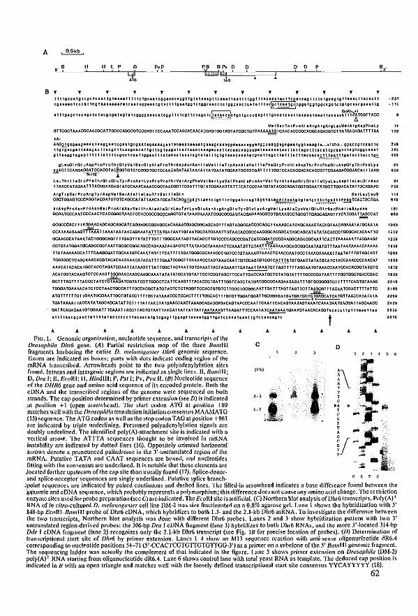
A ' O.5kb
" B ~ H , P 0 p"p PB BPV 0 0 0 0 f ~3' ,
ql fumj,,., ,
", 110
B , , , , , , , , , , , , , ! I t Igcca tg lege Icu' tgl .... t \ \ I te Ig.Ulogg.C.Clgg t \g Ictaug t Ic.u t 'u, \ I tggl t \ .c.utoo \ tego HIOI \ \ IC tgolglg t uu. I UCI~\ \ ·231 cgll"O lee U lIeg t"_ .. uelleu t 10g.u.g \ lil \ Ig .. tgg tClggeucc Ic Iggou. tCI la \ t \ 1<@eleutgOll000tggtgg.lgCle\gIQCOlCgO .. t\g • \ 11
~ ~.1 • t I tglgo toelgele U.II Igl tag t Ilcgggg .... a tegg t I \ leg t Icclgc tc~cc!. locl;cccgig t t 10cOClcu, U .... U .. t lao 1111 \ t tCATCGTTACC
11. ISuTh,P,oAI .A,gAtgAtgl.~Vtt""gAtpPh.ty U OTTcacTAJ.J.cacMCOCATTOCCCAGClCQTCOlAIlTTCCMATCCAACACAACACOAGTOOTAIlTATCGCTaTG~TCMCACCCOCACOCAQAOOTCTTATGACUQATTTTM 12i
", AMgIglgug.uctuggIUcgulgeguugllugutlclllcllllcgU'gC."ggU.lO.gg .. ggCligglgCO"\ggc ... gt __ .. H~<) .. ,.gIOOgtc .. clc Ilg~.cOU ,u.gl<:l \ \ Icg t Ilouoc.c&\ Ig \ Icg 'Ugl tOl t t ... I ""Q"." t 1I0 .. 0 •• ~ .. QI U .... I tu I 1.1 11;0 111I0Illgo. ... lIgI0;;.""1 g I! lI;gllglg t 1111 1111 t Icgeclollllgg"l t IIU ... I .. lllglIllI lig Ig U .. C ... U IgOl1 tig. I 111111 t I 11. nc,:. ~ ~~~.".I.llg"II.1 1I0.lgE.
gl • ...al nOl "AlpP,op,oTh,aryVIISUal yA! .P,oT~ 'AlpAI"A'"1 leV. t IleT,pA,"AIIVII II,ph,al rP,oH; ,A,pTII,p,ontal vHp(lryr~ ,P".lYI • gACI lCAAI3AOOA TCCACCT AcgooTa1CTCO::laTOOTCCAACAaA TAAlAAT A TT A TOA T A TOOM 100TaraAl T r TTOOTCCACAOOACACACCCTTOlAA(UC):)(lJ"ACA n TAAO - II!=HI ...
llvTh, I 1131 up",TlHal ual uTyrProA,"LyoP,oP,olh,VIIArgPhIVIIS., Ly,VII Ph,HI tProA,nV,1 Ty,Al IAtpUI ral yl r ,CyollvAlpl Illoc{l! nA In 1 TMCCA T AOAA TT 1 AC<XiAAOAGT ATCCMACMAC03C(':AACO:lTl00A 1 T TGT A l00MAaT A TT TCA lCCCM TaTA T AOOCA3A l00l00AA 1 A TOOTTOOACA lAT TaCA3AAC
AfgT ,pS, ,P,oA,gTy ,A,pVIISIIAIII lel,vTn ,S .. I r..:ll n SI rl,vleuS
COCl00AaTCCCAool AOlA laTalCAOCCA T A Tl MCA lCCH ACA01.!.g I g I UO 10 Ig. I I c I gg 1 I 0 0 Ig 11 g 0 ti \I.g.~ ~ ~ ~ .'.I.g I g I1 Ig '.1. ~ ~ !F~.I. 1I e!!i TCACrOOTQA
• rA,pP'.A,"P roA. nSI' P ,oAllAtnS, rTh ,AIlAIlOl nL,uTy'LYlal "AlnAfgAfgG I uTy rOl uLYIA,gVol Ly.AIICr.Vllal ~OlnSI ,Phtil IA.p". GOlA lCCCM TCCCAACTCAccaaclAACTCCAocaccaco:::AOOTOT A 1 AANlM.M lCOlCOOOAOTAOOA(lAJ.OCQ10laMAGCCTQCQT TGAOCAaA31TlCA l00A TT WYXA 1
• ;S;;;S
acocccAOITl ~HlCAOCAOCA TC.o.oo.o.accooAGCCACMAACQA{l(X)QÇHJCAGl T T NlT AOOOlAOCGCAocr 1 AAAOCA TCAacAAACT ACAaT MOMlAAA TAlacM 1 A
CCATAA)"(lAAQTTTMATlMAIMlAACHlMAl~.l.r.!~TGAIMITaTMTOOTATGAMTTGTMCACOOCCMCGQCAooaoAOAOONlCATATAIACACCCTQ03CACACAATTC
OCMCCCATAAATAOlao:lCAGTT11aAGTTATATlTGlTTOOCCAAINlTTACACCTTarccccAacccoATCACCOJATCC«!AAOOCAGAOOCMTTCACTTMAMATTAOOMGT ~
CCICTATOOATocAaCCOOIAATTacacooooAOCCNlAAAIMQACTCTTA1AAOClMAAACTCAAATGTTCT~.f.T.~~acA{l(X)QÇAAIAATGTTTMTAATMAACAAAM
TT A 1 AMAACCA TT1CAJ.c'lC(lA T TGCAA TOTCAAA T NlI TTCA 1 T T T AGA TOCOOOCAAACCC~GTCCTal AAAACI AAACTCAACCAA TGCCT AAAOOAAM TT M T A T r TaT NlTAaT
TOOTaaolCAOC~MC~OJTACACAAOAAAC~1~CATTTTOAATCCOOTTQA.V..O,CCTA3TAAACAAltatCCAGrGTOOTC".T.r.~~TGTQAACTATATarA1C1ACCM.ACCCCMC~1
AJ.ACATACACATOOTAOOTAaAl00AAlTAIAHlTTTATTOOCT~OAAATTOTOAMTTACTA'JAAATTGA1~TalTAGTnTtAOCMtaTAAACCMTCCAC~COOl~TOIG
~C~ TOOI ACMGTCTCCMG~C~AGCAAGCMA T A 1 A 1 A lGC~ laT ~ T ToorCOOT AGCTTCC~ 1 ta~CCM IOC1CT A tarA TTTTOOOOlA T AA TT T TooTaaoTGACCQAC
acT TT AClT T T ~CCCI A lCTCT AJ.GA TOJTA lOO1100ccTCA T TCOGT T rAC~CCCraA T TTOOTCA30T A taA lOOCCC~AGAAGAGA TTTGTGoo:n:nCTTTl11OAGTGT AAHl
TOOQA lGAAMCA 1CTooi MCTQCCA TT TGCCACQOT A TaT ACTC101 AOOlTCC~CraTGTCTIOOClacoocM 1 TGA 1 T TAaTT AG T IClT MI)/ClJHT T ACM TOOAC1111 A 11C
ATalTTTTTGTTaAATGCIlAATTOOTGTAccnnG01ATMACCCTCCACT1TlTOOOAC11TaraTTOOATOOATTQ(Xl(lA0AlG~T~QTfMCCATATA1A
TGA T MAA 1 TACCA T A T AOCAac~ T A TTal T 11 ~ TT MCT ATaMCCMCT AAAGOAlMOOAOACAGT MCCCM nCAA 1 TC~CAGT M T Nll AM lCMAGAA TGJ.'XiA 11 AClCAACC
QA TTCAaACAA TaTOOM TT TCAAA TT ACCTT ACTaT AA TTMCA Tl A TT A TT A TT M 1 AM3TTMGA TTTCCAA T A TC~A 1 MA TGAMGT MCACAoo 1I~ Ho t t g lito .. Ilu
t , , , , , , , , , , , , , c o
, .. '" ... " ." .. '" 'H ... '" ."
FIG. 1. Genomic organization, nucleotide sequence, and trunscripts of the Dwsophila Dhm geile. (A) Partial restriction map of the three BUil/Hl fragments harhoring the entire D. lI/e/ulIO!:uster Dhm genomic sequence. Exons are indicated as hoxesj parts with dots indicate coding region of the mRNA transcribed. Arrowheads point 10 the two polyadenylylation sites found. Introns aod intergenic regions are indicated as single Hnes. B, BUil/Hij 0, Dm I; E, EcoRI; H, HindlIl; P, Pst I; Pv, Pl·uIl. (8) Nucleotide sequence of the DHR6 gene and amino acid sequence of its encoded protein. Both the cDNA and the transcribed regions of the genome were sequenced on both strands. The cap position detennined by primer extension (see Dl is indicated .at position +1 (open arrowhead). The start codon ATG at position +89 matches weil with the Drosophila translation initiation consensus MAAMATG (15) sequence, The ATG codon as weil as the stop codon TAG at position +963 are indicated by triple underlining. Presumed pol)'adenylylation signals are doubly underlined. The identified poly(A)·attachment site is indicated with a ,'ertical arro\f. The ATITA sequences thought to be im'oh'ed in mRNA instability are indicated b)' dotted Hnes (16). OppositeI)' oriented horizontal arrows denote a prooounced palindrome in the 3'·uotranslated region of the mRNA. Putative TATA and CAAT sequences are boxed, and nucleotides fitting with the consensus are underlined. It is notabie that these elements are Joçated fur1her upstream ofthe cap site than usually found (17). Splke·donor·
, , , . , ,
(kb)
, , , H , ,
aod splice-acceptor sequences are singly underlined. Putative splice branch. GA" C
;point sequences are indicated b)' paired continuous and dashed Hoes. The filled·in arrowhead indicates a base difference found bet ween the gq'oomic and cDNA sequence, which probably represents a polymorphism; this difference does not cause any amino acid change. The restriction enzyme sites used for probe preparation (see G) are indicated. The EcoRI site is artificiaJ. (G) Northern blot anal)'sis of Dhr6 transcripts. Pol)'(A) + RNA of in I'itro·cultured D. me!(l/log(lster cellline DM·2 was size fractionated on a 0.8% agarose gel. Lane 1 shows the hybridization with 5' 148·bp EcoRI-BamHI probe of Dhr6 cDNA, which hybridizes to both I.]· and the 2.l·kb Dhr6 mRNA. To investigate the difference between the Iwo transcripts, Northern blot analysis was done with different Dhr6 probes. Lanes 2 and 3 show hybridization pattem with two 3' untranslated region·derived probes: the 306-bp Dra I cDNA fragment (lane 3) hybridizes 10 both Dhr6 RNAs, aod the more 3'·!ocated 314·bp Dde I cDNA fragment (Iane 2) recognizes onl)' the 2.I·kb Dhr6 transcript (see Fig. lB for precise location of probes). (D) Determination of transcriptional start site of Dhr6 by primer extension. Lanes 1-4 show an MB sequence reaction with anti·sense oligonucleotide dR6.4
'corresponding 10 nucleotide positions 54-71 (5'·CCACfCGTGTIGTGTIGG·3') as a primer on a subcJone ofthe 5' B(lmHI genomic fragment. The sequencing ladder was actually the complement of that indicated in the figure. Lane 5 shows primer extension on DIOSOphiftl (DM·2) poly(A)+ RNA starting from oligonucJeotide dR6.4. Lane 6 shows conlrollane with total )'east RNA as template. The dedueed cap position is indicated in B with an open triangle and matches weil with the loosely defined transcriptional start site consensus yYCA YYYYY (18).
62
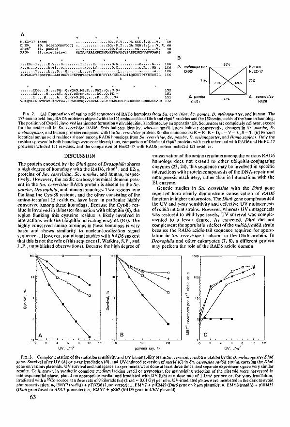
A HUE2-17 DHR6 rhp6+RA"
(I:lan) (D. llelanogaster) (S. ponbe) (S.cerevisiae)
••••••••••••••• f,Q •• P.V ••• GA.SE!I.i.Q ••• V. 40 ••••••••••••••• LQ •• P.T ••• GA.'fDlI.i.i. .. V. ",40 ••• T •••••••••••• QQ.P.A •••••• vS •••• l. •. V. 40 HSIPARRRIMRDFKRMKEDAPPGVS1\SPLPDNVHVWNAMI 40
B F •• EG •• F ••••• k.v ••• S •••••••• T.r ••• K •••••••• D.S •••••••••• s ••••• S ••• 106 F •• H ••• F ••••• k.Ti. .T •••••••• T.r,v.KV ••••••• O.G, •••••••• ,s.R ••• SA.. 1{16 ••••••• F ••••• k.v.S ••• Q •••••• L ••• V.T ••••••••••• l. •••..... s •••••• A •• 106 OHm HuE2-17
D. melanogasler ~S5% Human
IGPADIPYEC>GTFRLLLEFDEEYPNKPPHVKFLSEHFHPNVYAlIGEICLDILQNRh"TPTYDVASII, 106 , 71% 71% 6$% 7if.,(,
• ••••• LOe ••• N •••• SQ •• Q. YQeN. RE. E ••• SAI. • Q •• H .S~ 152 • ••••• LS •••• ll. ••• ST •• Q. Y. el/rRE. E •••• AC •• Q. FI. ~ 151
S. cerevls/ae RAOO
• ••••• L •••• N ••••• A ••• Q. HreN. KE •• r •• rK ••• D ••• S' 151 S. pombe 77% TSIQSLfNDP1IPASPJlJIVEAATLFKDHKSQYVKRVKETVEKSWEDDMDDMDDDDDDDDDDDDDE"'O. 172 fhp6t
FIG. 2. (A) Comparison of amino acid sequcnces of RAD6 homologs from Sa. cert'risiae, Sc. pOli/be, D. me!alloxaster. and human. The ln-amino acid-long RAD6 protein is aligned with the 151 amine acids ofDhr6 and rhp6 + proteins and the 152 amine aeids ofthe human homolog. The position ofCys-88, involved in thioesterformation with ubiquitin, is indieated by an open triangle. Sequences are completely coJinear. except for the acidic tail in Sa. cerel';siae RAD6. Dols indicate identity, whereas small letters indieate conservati\'e çhanges in Sc. pombe, D. me/aflogaster, and human proteins çompared with the Sa. cern'isiae protein. Similaraminoadds: R = K, E = D, I = V = L, S = T. (D) Percent identical amino acid residues shared among RAD6 homologs from Sa. arel'isiae, Sc. pombe, D. me!allogasler, and Homo sapiells. Only the residues present in both homologs were çonsidered; thus, camparisan afDhr6 and rhp6+ proteins with eachotherand with RAD6and HuE2-17 prateins included 151 residues, and the comparison of HuE2-17 with RAD6 protein included 152 residues.
DISCUSSION
The protein encoded by the Dlu6 gene of Drosophila shares a high degree of homology with the RAD6. rhp6+ , and E217k proteins of Sa. cerel'isiae, Sc. pOli/be, and humao, respectively. However, the acidic carboxyl-terminal domain present in the Sa. cerel'isiae RAD6 protein is absent in the Sc. pOli/be, Drosophila, and human homologs. Two regions, one flanking the Cys-SS residue, and the other consisting of the amioo-terminal 15 residues, have been in particular highly conserved among these homologs. Because the Cys-SS residue is involved in thioester formation wÎth ubiquitin (6), the region nanking this cysteinc residue is likely involved in interactions with the ubiquitin-aclivating enzyme (El). The high!y conserved amino terminus in these homo!ogs is very basic and shows similarity to nucJear-localization signa! sequences. However, mutationaI studies with RAD6 suggest that this is oot the role ofthis sequence (J. Walkins, S.P., and L.P., unpublished observations). Becallse the high degree of
$ $
I <i ~
<
" "
A 8
conservation ofthe amino terminus among the various RAD6 homologs does not extend to olher ubiquitin-coojugating enzymes (23, 24). this sequence may be involved io specific interactions with protein components ofthe DNA-repair and mutagenesis machinery, mther than in interactions with the El enzymc.
Genetic studies in Sa. cerel'isiae with Ihe Dhr6 gene reported here cJearly demonstrate conservation of RAD6 function in higher eukaryotes. The Dhr6 gene complemented the UV and y-ray sensitivity and defective UV mutagene sis of rad6à mutant strains. However, whereas UV mutagenesis was restored to wild-type levels, UV survival was complemented to alesser degree. As expected, Dhm did not complement the sporulation defect ofthe rad6à/rad6à strain because the RAD6 acidic-!ail sequence required for sporulation in Sa. cerel'isiae is absent in the Dhr6 protein. In Drosophila and other eukaryotes (7, S), a different protein may perform the role of the RAD6 acidic domain.
'000 ,-----------,
~ 8 '" ~ .$ 0
" [ B
II ~ , r w ~ C
,Ol oc----ê-"-é,~__:~__:~-',co~-""
UV, Jfm 2
o'oL~~~--~,éoc-~~"-~,"oc-~~
gamma fay, kr
., 0 •
UV, Jfm
, " " ,
FIO.3. Complementation ofthe rndiation sensitivity and UV immutability ofthe Sa. cerel'isiae rad6û mutatÎon by the D. me/al/ognster Dhro gene. Sur.'ival after UV (A) or y-ray irrndiation (Dj, and UV-induced reversion of metJ4 (C) in Sa. cerel'jsiae rad6û strains carrying the Dhro gene on various plasmids. UV survival and mutagene sis experiments were done at least three times, and separate experiments gave very similar results. Cclls grown in synthetic complete medium lacking uracil or tryptophan for maintaining selection of the plasmid were harvested in mid·exponential phase, plated on· appropriate media, and irradiated with UV light at a dose rate of 1 Jlm 2 per sec or, for y-ray irradiation, irrndiated with a wCo source at adose rateof9 kilorads (kr) (1 rad = 0.01 Oy) per min. UV-irradiated plates were incubated in the dark to avoid photoreactivation. _, EMY7 (rad6û) + pTB236 (2 pm vector); 0, EMY7 + pRR449 (Dhr6 gene on 2 pm plasmid);., EMY8 (rad6û) + pRR454 (Dhro gene fused ta ADCI promoter); 0, EMY7 + pR67 (RAD6 gene in CEN plasmidl,
63
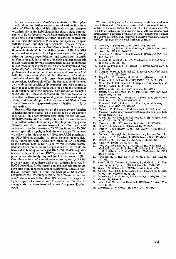
Future studies with dhr6/dhr6 mutants in Drosophila should allow the further examination of various funclional roles of Dhr6 in this highly differentialed and complex organism. Hy in situ hybridization in salivary gland chromosomes of D. melallogasler, we have localized the Dhr6 gene 10 a single sile at position 82D near the base of the righl arm of the third chromosome. P element mutagenesis, coupled with the use of strains bearing deficiencies in the Dhr6 region, should permit a screen for dhr6/dhr6 mulants. Sludies with these mulanls should furlher define the role of Dhr6 in DNA repair and mutagenesis in a higher eukaryote. Because of defective meiotic recombination and sporulation in rad6/ rad6 mutants (25, 26), studies of meiosis and gamelogenesis in dhr6/ dltr6 mulanls may be particularly revealing about Ihe role of Dhr6 in these processes. In higher eukaryotes, during spermalogenesis chromatin-bound histones are replaced by protamines. The requirement ofthe acidic domain ofRAD6 both for sporulation (3) and for atlachment of multiple molecules of ubiquitin 10 hislones (5) suggests that during sporulation, RAD6 could effect the degradation of histones by the ubiquitin-specific ATP-dependent protease complex. Even though Dhr6 does not possess Ihe acidic-Iail domain, it could still function in Ihis capacity in association with a highly acid ic prolein. Because considerably more information is available on Drosopllila spermatogenesis than yeast sporulation, studies on the possible involvement of Dhr6 in turnover ofhistones during spermatogenesis might be particularly informative_
These studies demonstrate thai the slruclure and function of RAD6 has been consçrvcd to a rcmarkable degree among eukaryotes. This conservation very Iikely reflects the evolutionary constraints on RAD6 protein due 10 its interactions with protein factors functioning in the ubiquitin conjugation pathway and wilh proteins involved in DNA repair and mutagenesis. In Sa. cerevisiae, RAD6 functions wÎth RADI8 in postreplication rcpair, as both the rad6 and radl8 mulanls are defective in Ihis process (1)- Because RAD6 protein has no DNA-binding capacity (P_ Sung, personal communicalion), Înteraclion with RAD18 may target the RAD6 protein 10 the damage sites in DNA. The RADJ8-encoded protcin contains three potential zinc-finger domains that could be involved in binding 10 damaged DNA (27). RAD6 mayalso interact with the REVI and REV3 proteins because all three proteins arc absolutely required for UV mutarenesis (28). Dur observations of evolutionary conservation of RAD6 protcin suggest that these and olher proteins involved in RAD6-dependent DNA repair and mutagenesis processes have al50 been conserved among eukaryotes. Because both Ihe Sc. pombe rhp6+ (7) and Ihe Drosophila DIlr6 genes complement the UV-mutagene sis defect ofthe Sa_ cere~'isiae rad6Jl strain much belter than UV survival, we expect a higher degrce of conservation of proleins that function in mutagenesis than those involved in error-free postreplication repair_
We thank Ed Stephenson for determining the chromosomallocation of Dhr6 and P. Sung for çrîticism of the manuscript. We are grateful to J. Jaspers Dekker for excellent technical assistance. We thank P. M. Salvaterra for providing the .\ gUl Drosophi/a head cDNA library. Supported by the Dutch Cancer Society (project IKR 88-2 and 90-20) and by U.S. Public Health Service Grants GM19261 and CA41261 from the National Institutes of Health.
L Prakash, L. (1981) Mol. Gen. Genet_ 184,471-478. 2. Reynolds, Po, Weber, S_ & Prakash, L. (1985) Proc. Natl.
Acad. Sci. USA 82, 168-172. 3. Morrison,A., l.fi!ler, E. J. & Prakash, L. (1988) Mol. Cell. Biol_
8, 1179-1185. 4. Jentsch, S., McGrath, J. P. & Varshavsky, A. (1987) Nafllre
(l.<mdoll) 32.9, 131-134_ 5. Sung, P., Prakash, S. & Prakash, L. (1988) Genes Del'. 2,
1476-1485. 6. Sung, P., Prakash, S. & Prakash, L. (1990) Proc_ Natl. Awd.
Sci_ USA 87, 2695-2699_ 7. Reynolds, P., Koken, M. H_ M., Hoeijmakers, J_ H. J.,
Prakash, S. & Prakash, L. (1990) EMBO 1. 9, 1423-1430. 8. Schneider, R., Eckershom,C., Lottspeich, F. & Schweiger, M.
(1990) EMBO J. 9, 1431-1435. 9. Rothstein, R. (1983) Met//Ods Ef/UllIol_ 101,202-211.
10. Boeke, J. D_, La Croute, F. & Fink, G. R. (1984) Mol. Gef/_ Genel. 197,345-346_
11. Sung, P., Prakash, L., Weber, S. & Prakash, S_ (1987) Proc. Natl. Acad. Sci_ USA 84, 6O-t5-6O-t9_
12. Frischauf, A.-M., Lehrach, H., Poustka, A. & Murray, N. (1983) J_ Mol. Biol_ 170,827-842_
13. Maniatis, T" Fritsch, E. F. & Sambrook, J. (1982) Mo/eCIIlllf Cloning: A l_aboralor)' Malllwi (Cold Spring Harbor Lab., Cold Spring Harbor, NY).
14. Sanger, R., Nicklen, S_ & Coulson, A. R_ (1977) Prae. Natl. Acad_ Sc/. USA 74, 5463-5467.
15. Cavener, D. R. (1987) Nudeic Acids Res. 15, 1353-1361. 16. Shaw, G_ & Kamen, R. (1986) Cel/46, 659~667. 17. Bucher, P. & Trifonov, E. N_ (1986) Nuddc Acids Res. 14,
10009-10026. 18. Corden, J., Wasylyk, B., Buchwalder, A., Sassone-Corsi, P.,
Kedinger, C. & Chambon, P. (1980) Sc/ence 209,1406-1414. 19_ Moun!, S. M. (1982) NI/dek Acids Res. 10,459-472. 20. Keiler, W_ (1984) Ce1/39, 423-425. 21. Itoh, N., Slemmon, J. R" Hawke, D. H., Williamson, R.,
Morita, E.,ltakura, K., Roberts, E., Shively. J. E., Crav.ford, G_ D. & Salvaterrn, P. M. (1986) Proc. Ntil/. Acad. Sci. USA 83,4081-4085.
22. Bimstiel, M. L., Busslinger. M. & Strub, K. (1985) Cell 41, 349~359.
23. Goebl, M. G., Yochem, 1., Jentsch, S., McGrath, J. P., Varshavsky, A. & Byers, B. (1988) Sc/ellce 241, 1331-1335_
24. Seufert, W. & Jentsch, S. (1990) EMBO J. 9, 543-550. 25. Game, 1. Co, Lamb, T_ 1., Braun, R.l., Resnick, R. & Roth,
R. M. (1980) Genetics 9-1, 51-68_ 26. MonteJone, B. A., Prakash, S. & Prakash, L. (1981) Mol. Geil.
Gelle/. 184,410-415. 27. Jones,J. S., Weber, S. &Prakash, L. (1988) NudeicAcids Res.
16,7119-7131. 28. Lawrence, C. W. (1982) Adl'. Gellel. 21, 173-254.
64


Chapter IV
Structural alld fClIIctiollal C01/servatioll of (wo humall homologues of the yeast DNA repair gene RAD6

Proc. Natl. Acad. Sci. USA Vol. 88, pp. 8865-8869, October 1991 BiochelIllslry
Structural and functional conservation of two human homologs of the yeast DNA repair gene RAD6
(ublqultln conjugatlon/E2 eDzyme/DNA damage/UV mulagenesis/sporulalloD)
MARCEL H. M. KOKEN*, PAUL REYNOLDSt, IRIS JASPERS-DEKKER*, LOUISE PRAKASHt, SATYA PRAKASHH, DIRK BOOTSMA*, AND JAN H. J. HOEIJMAKERS*
·Departmenl of CeU Biology and G~netics, Emsmus University. P.O. Box 1738, 3000DR Rotterdam, The Netherlands; fDepartment of Biophysics, UniHrsity of Rochester School of Medicine, Rochester, NY 14642; and fDepartmenl of Biology, University of Rochester, Rh'er Campus Station, Rochester, NV 14627
Communic(lted by James V. Neef, Jul)' I, 199/
ABSTRACT The RAD6 gene of SacclUlrom)'ces cere.'isiae encodes a ublquitin·conjugating enzyme (E2) Ihat Is requlred for DNA repair, damage-Induced mutagenesls, and sporula' tion. We have cloned Ihe (wo human RAD6 homologs, deslgnafed lIHR6A and HHR6B. The hro 152·amino acid human profeins share 95% sequence ldenflty wUh each other and =70% and =85% o\'erall idenfity wUh Ihe homologs from yeasts (S. cere.'isiae and Schizosaccharom)'ces pombe) and Drosopllifa mefallogasler, respeeth'ely. Nellher of the human RAD6 homologs possesses Ihe acid ie C·terminal sequence present in the S. cere~'isiae RAD6 protein. GeneIIc complemen· tatlon experlments reYCal that HHR6A as weil as HHR6B can carry oul the DNA repaIr and mufagenesls funelions of RAD6 in S, cere.'is/ae rad611 mulants.
The Saccllaromyces cerel'/siae RAD6 gene plays a key role in DNA repair and DNA damage-induced mUlagenesis. rad6 mutants are extremely sensitive 10 a plethora of DNAdamaging agents, ineluding UY irradiation, x·rays, and alkylating agenls; they are defective in poslreplicatlon repair of UY-damaged DNA, in mutagenesis indueed by DNA damaging agenls, and in sporulation (for a review, see ref. 1). Transposition ofTy elemenls is enhaneed in rad6 mutants (2). RAD6 eneodes a prolein of 172 amino acids (3) with a globular domain consisting ofapproximately the firs! 149 amino acids and an extended, predominantly acidic eterminus (4). RAD6 is a ubiquitin-conjugating enzyme (5, 6). Ubiquitin, a highly conserved, 76-amino acid polypeptide is covalently atlached 10 many cellular proleins and targets them for seleetive degradation, (re)folding, or slabilization (for recent reviews, see refs. 7-9). Ubiquitination is carried out by a family of proteins in a multistep rcaction im'olving a ubiquitin· activating enzyme (El) that binds and subscquently transfers a ubiquitin moiety to one of a set of ubiquitin-colÛugating enzymes (E2). The E2 enzyme ligatcs ubiquitin directly to a larget protein, wilh or without the help ofa ubiquitin protein ligase (E3). RAD6 polyubiquitinates histones H2A and H2B ;11 VUFO without the involvemcnt ofE3, and the acidicdomain of RAD6 is required for multiple ubiquitination of histones (6). It is possible thaI the protein is implicated in modifying chromatin structure as part of the processes that are disturbed in a rad6 mutant, ineluding repair and mutagenesis.
RAD6 is highly conserved among eukaryoles. Previously, we eloned the rhp6+ gene ofthe fission yeast Scltil.osaccharomyces pombe and showed thaI it is a slructural and functional homology of RAD6 (10). We have also isolated a RAD6 homolog, Dhr6, from Drosopltila melallOgasfer (11). In this paper, we present Ihe eloning, scquence analysis, and functional studies with the RAD6 homologs from human. '11 In
contrast 10 yeasl and Drosopltila, where RAD6 is a single copy gene, inlerestingly, in human, the RAD6 homologous gene is duplicated, and the proteins encoded bythe two genes HHR6A (human homolog of RAD6) and HI/R6B share 95% identical amino acid residues. We also show that the HHR6A and HHR6B genes complement the DNA repair and UV mutagenesis defects ofthe S. cerevisiae rad6 mulant.
MATERlALS AND METHODS
Reslriction Enzyme Digests and Southern Blof Llbrary Hybrldlzatlons. Restriction enzyme digestions were performed according la the manufacturer's descriptions. B10ts were prepared on Zeta-Probe (Dio-Rad) using the alkaline-blotting procedure as recommcnded by the manufacturer. DNA probes were labeled by the random-priming method as described (12). Unless slated otherwise, hybridization of Sc. pombe and Drosopltila probes to human DNA oecurred overnight at 55"C and hybridization of human probes to human DNA Was at MOC in a hybridization mixture containing 10x Denhardt's solution (2% Ficoll/2% bovine serum albumin/2% polyvinylpyrrolidone)/l0% dexlran sulfate/ 0.1% SDS/3x slandard saline citrote (SSC)/50 mg of soni· cated salmon sperm DNA per liter. Washings for hybridizations involving different species were performed for 5 min in 3x SSC twiee and for 5 min in Ix SSC onee at 55"C. For hybridizations within a species, washings were done twice for 20 min each in 3x SSC, twice for20 min each in Ix SSe, and twice for 20 min each in O.3X SSC al 65"C.
Norlhern Blotting and Nucleotide Sequenee Analysis. Isolation oftolal RNA by the LiCl/urea method, preparation of poly(A)+ RNA by two consecutive passages over oligo(dD cellulose columns, and Northern blotting protocols were all according to Sambrook el al. (13). Sequcnce analysis on double-stranded DNA was done by the TI polymerase modification (Pharmacia) of the dideoxynucleotide chainterminalion method (14) using sequence-derived oligonueleotides and exonuelcase III prepared deletion elones for sequencing both stronds. For separation ofthe fragments we used Hydrolink (AT Biochem, Malvern, PA) sequencing gels.
Yeast Sirains, Media, and GenetIe Analyses, The S. cerevisiae haploïd strains used in this study were the rad6!J. strains EMY1 (MATa leu2-3 leu2-!!2 trpl IIra3-52 rad6::LEUr)and EMY8 (MATa ade5Itis71eu2-31)'sl mef14 pet15 ura3 Irpi::URAJ+ rad6::LEUr). The rad6ó./radM diploid EMY28 was constructed by mating EMYl and EMY8. UY irradiation conditions and media for determining survival and mUlagenesis after UV exposure and sporulation
66
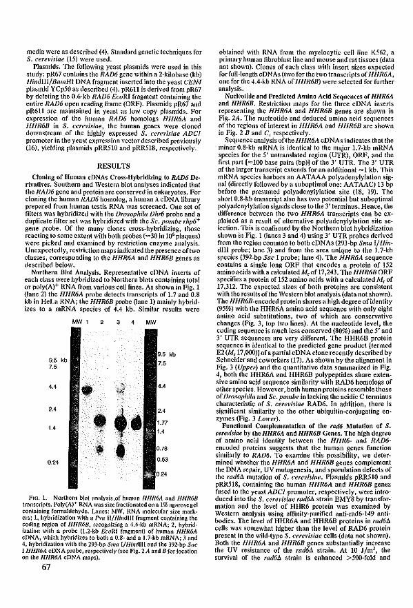
media were as described (4). Standard genetic techniques for S. cere\'isiae (15) were used.
Plasmids. Thc following yeast plasmids were used in this study: pR67 contains the RAD6 gene within a 2-kilobase (kb) llindIII/BamHI DNA fragment inserted into the yeast CEN4 plasmid YCp50 as described (4). pR611 is derived from pR67 by deleting the 0.6·kb RAD6 EcoRI fragment containing the entire RAD6 open reading frame (ORF). Plasmids pR67 and pR611 are maintained in yeast as low copy plasmids. For expression of thc hu man RAD6 homologs HHR6A and HHR6B in S. cere\'isiae, the human genes were c10ned downstream of the highly expressed S. cerel'isiae ADCI promoter in the yeast expression vector described previously (16), yielding plasmids pRR510 and pRR518, respectively.
RESULTS
Clonlng of lIuman cDNAs Cross.Hybrldlzing to RAD6 De· rivatlvcs. Southem and Western blot analyses indicated that the RAD6 gene and protein are conscrved in eukaryotes. For c10ning the human RAD6 homolog, a human À cDNA Iibrary prepared from human testis RNA was screened. One set of filters was hybridized with the Drosophila DI,,6 probe and a duplicatc filter set was hybridized with the Sc. pombe rhp6+ gene probe. Of thc many c10nes cross-hybridizing, those reacting 10 some extent with both probes (=30 in 106 plaques) were picked and examined by restriction enzymc analysis. Unexpectedly, restriction maps indicated the presence oftwo classes, corresponding to the HHR6A and HHR6B genes as described below.
Norlhern Blof Analysis. Representative cDNA inserts of each c1ass were hybridized to Northern blots containing total or poly(A)+ RNA from various celllines. As shown in Fig. 1 (lane 2) the HHR6A probe detects transcripts of 1.7 and 0.8 kb in HeLa RNA; the HHR6B probe (Iane 1) mainly hybridizes to a mRNA species of 4.4 kb. Similar results were
MW 1 2 3 • MW
kb 9.5 kb 7.5
••• • 2.4
t 1.. • • 0.24
FIG. 1. Northem blo! anatysis,of human HHR6A and HHR6B transnipts. Poly(A)+ RNA was size fractionated on a 1% agarosegel containing fonnaldehyde. Lanes: MW, RNA molecular size mark· ers; 1, hybridization with a Pl'lt II/Hlndlll fragment containing the coding region of HHR6B, recognizing a 4A-kb mRNA; 2, hybrid. ization wilh a probe (l.2-kb EcoRI fragment) of human HHR6A cDNA, which hybridizes 10 bolh a 0.8- and a l.7-kb mRNA; 3 and 4, hybridizalion wilh Ihe 293-bp Sma I/Hindlll and the 392-bp Sac I llllR6A cDNA probe, respeclively (see Fig. 2A and B forlocation on the HHR6A cDNA maps).
67
obtaincd with RNA from thc myclocytic cell line K562, a primary human fibroblast Iinc and mouse and rat tissues (data not shown). Clones of each class with insert sizes expected forfull-Iength cDNAs (two for the two transcripts of 1l11R6A, one for the 4.4-kb RNA of HHR6B) were selected for further analysis.
Nucleotide and Predictcd Amlno Acid Sequences of HHR6A and HHR6B. Restriction maps for the three cDNA inserts representing the HHR6A and HHR6B genes are shown in Fig. 2A. Thc nucleotide and deduced amino acid sequences of the regions of interest in HHR6A and I/I/R6B are shown in Fig. 2 Band C, respectively.
Sequenceanalysis oftheHHR6A cDNAs indicates that the minor O.8-kb mRNA is identical to the major 1.7-kb mRNA species for the 5' untranslated region (UTR), ORF, and the first part [=100 base pairs (bp)] of the 3' UTR. Thc 3' UTR ofthe !arger transcript extends for an additional =1 kb. This mRNA species harbors an AATAAA po!yadenylylation signa! (direetly followed by a suboplimal one: AATAAC) 13 bp before the presumed polyadenylylation site (18, 19). The short O.8-kb transcript also has two potential but suboptimal polyadenylylation signalsclose 10 the 3' terminus. Hence, the difference between the two HHR6A transcripts ean be explained as a result of altemative polyadenylylation site seleetion. This is eonfirmed by the Northem blot hybridization shown in Fig. 1 (Ianes 3 and 4) using 3' UTR pro bes derived from Ihe region common to both cDNAs (293·bp Sma I/HindIll probe; lane 3) and from the area unique to the 1.7-kb species (392-bp Sac I probe; lane 4). The HHR6A scquence eontains a single long ORF that encodes a prolein of 152 aminoacids with a calculated M.ofl7 ,243. The HHR6B ORF specifies a protein of 152 amino acids with a ealculated Mr of 17 ,312. The expected sizes of both proteins are consistent with the results ofthe Western blot analysis (data not shown). The flHR6B-eneoded protein shares a high degree ofidentity (95%) with the HHR6A amino acid scquence with only eight amino acid substitutions, two of whieh are conservative changes (Fig. 3, top two Iines). At the nucleotide level, the coding sequence is much less conserved (80%) and the 5' and 3' UTR sequenees are very different. The HHR6D protein sequence is identical to the predieted gene product [termed E2(Mr 17,000)] ofapartial cDNA clone recently described by Schneider and coworkers (17). As shown by the alignment in Fig. 3 (Upper) and the quantitative data summarized in Fig. 4. both the HHR6A and HHR6B polypeptides share extensive amino acid sequenee similarity with RAD6 homologs of other species. Howevcr, both human proteins resembie those of Drosophifa and Sc. pombe in laeking thc acidic eterminus characteristic of S. cerel'Îsiae RAD6. In addition, there is significant similarity to the other ubiquitin·eonjugating en· zymes (Fig. 3 Lower).
Funclional CompIementalIon of the rad6 Mufatlon of S. cew'isiae by IbeHHR6A and HHR6B Genes. The high degree of amino acid identity between the HHR6- and RAD6-encoded proteins suggests that the human genes function similarly to RAD6. 1'0 examine this possibility, we deler· mined whether the HHR6A and HHR6B genes complement the DNA repair. UV mutagenesis, and spondation defects of the rad61l mutation of S. cerel'isiae. Plasmids pRR510 and pRR518, containing the human 1I11R6A and HHR6B genes fused to the yeast ADel promoter, respectively, were introduced into the S. cere\'isiae rad61l strain EMY8 by transfor· mation and the level of HHR6 protein was examined by Western analysis using affinity-purified anti·rad6-149 anti· bodies. The level of HHR6A and HHR6B proteins in rad61l ceJls was somewhat higher than the level of RAD6 protein present in the wild-type S. cerel'isiae cells (data not shown). Both the HHR6A and HHR6B genes substantially increase the UV resistance of the rad61l strain. At 10 l/m2, the survival of the rad6tJ. strain is enhanced >500-fold and
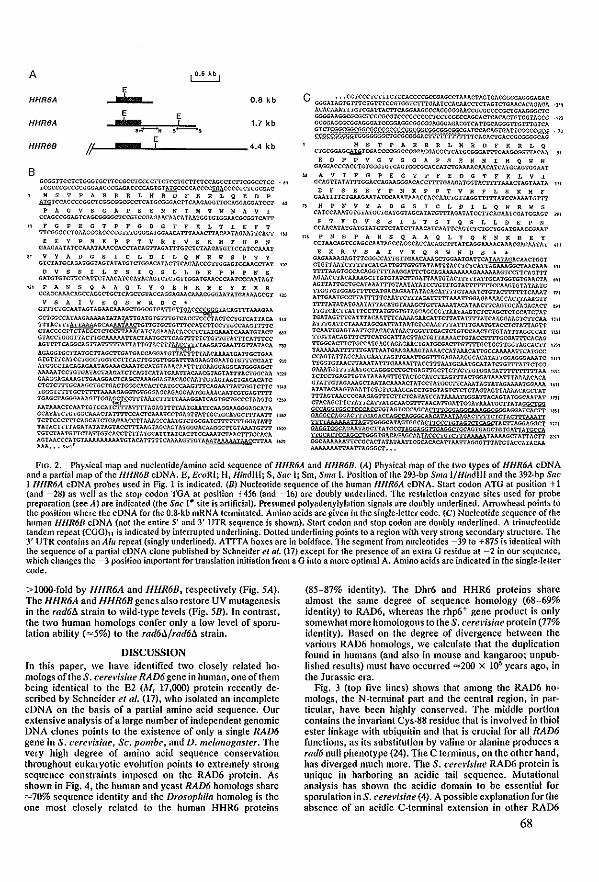
A ~ E I
HHR8A 0.8 kb
E E HHR6A I ! 1.7 kb , . .--.., ,-,
E HHR6B 1/ ! 4.4 kb
B
I
GCGGTICCTCiCGGïCcrr=CCCCTICTCC'iGCi'ICICCAGccrcr:rCGGCcrcc .• t '1CGCCCGCCG=AACÇÇGAOACCCCAGICî:l!l!i;CCCC1;CCC~C=CGCGAC
M S T PAR R R L K R D F ~ R L Q E D P ADiiCCACCCeGGcrC==CA'1GCGGGACrTCMCAG<lTIGCAG<lAGGAïccr ..
P A G V S GAP SEN NIK V W N A V I CCACCçca1..GiCA=CCGICDJAGAAClU.CATMIGGTGTGGMCGCCCICA=
., FGPEGTPfGDGTYKLTIEfT TI=CCTGMGGGACCCCG:ITTGGGGAT-GGMCAtTrAAAC'ITA,CM1AGM7N.:A<:r '1;)
E E Y P N K P PTV R rvs R H r H P N GMGMlA',ÇCAAATAAACCACCfACAGITAGATI'TGTCiCiAAGATGI"lCCAiCCAAAT
IT V'fADGSICLDILQNRWSPTY GICTATCCAGATGCîAG1ATATGICniGACATAcr1:CAGAACCGTI-G<lAGlCCMCCiAT
o V S S 1 LTS 1 Q S L L DEP N P N S GATGIGTcrrCCAT'TCi MCATCCATACAGICICIGTICGA TCAACCCAAn;çCN\'iAGT
'" PANSQAAQLYQENKREYEKR CC1;GCAMCAGCCAGCCTGCICAGcl'GTACCAGGAGMCAAA=AATATGMMGCGT
V S A I V E Q S W Roe • Grr:rCH';CAATAGTAGAACAAA=G(;CGIG}'TIG~ÇçCffifGTACAGi'ITAJo.J..GAA
~=i~~~~~~~i~~~~~~~~~,;~~~~~ l<~ CTACCCCrTCTACCererCCITAAAQ,TCAGMMCA=crATGAM!CMATGTACT 60\.)
G!ACCIGGGITACrTGCJV.AM17ACIAATGCITCAGI'TTI'TCIGTIGTA1'ITQ,'I'T'l'CC AGITITc.\GGCAG17ATI'TITATIAl'!GiACTI7~~AAGA!GAA17GTIA7AQ, rn AGAGGTGC'ITATGCITAGC17GATGACc.\GGATGITA T'ITTIAACAAAATGAITGCiGAA GrGl'ITCATCCTGGCiGGTCcrrQ,CITGiGl"iGGATUAGAAG1GAA lGTGITi'GGAA! 9'"A A~CC!ACAGAGAATAGAMCAMTCCATGTAMCA.~MTrGAAG<lAGGQ,TGGÇA= AAAAATCCIGTGATACTAAGATCTc.\Gic.\iAiCAATIACAACGiAGiAHTACIGGCAA .~ GAAGGAGAAAGITGAAG<lACICAGcrAMGGAGTAQ,GCMTIGTAGTMCIGACAc.\iC CiCiCI"ITGCAAGCTGCTGACiGGGCACACiCATGCCAAG'I'T'l'c.\GAAl"iA=lCT'JC 'TU TGGGTTTTICCiTITlMAAGAGGTGTCCGAGCAGAG<lAA1G<lAAACAATCG!CAGTTIT TCAGCTAGGGAAAGI"fGGAf,srrCiTl'AATCTI'ITfAAAG<lATCAGTGCIGCCCTAAGrG " .. MTAAACrCAA'I'TGTCCATCITrAITITAGAGiTITMWAATICAAG<lAAGGÇAGCATA GCATATCTGTGGCAAACiAITITCCAcrCAMTCCiGAGTTA17GCIGCATGCl"ITAATI' .:ru TcrrCCCITICAGCATCIGAGAACCIT AAAGCCAATGTCIGCGATCTi'iTI"TIGGATATT 'l'ATACH'TrAGA T ATATAGTACCITTAAGiAGc.\G! A'iGGGACAAGGCITC! AAATGTIT '~)Q 'r'GTcrAATGITCTATIG1CACCiTrTATGCATTTATCACi'iCC},J,J..:.CIAAC'f'ITGCACA AGTMCCCA'tGTAAAAAAAAA'I'G1ACAT'ITTICMMGI"ffiTAMIMAMTAA=AA ' .... AAA ••• ,-",,'
c GGGAT;'Gi~~~~~~~~~~~~~~~~~=~~ci~~~i '!Tl ACACAAATTIG1CGA17ACTICAOOAAGCCCAGGGGGAACCGCGCCCCGCIGAAGGGCIC GGGGAAGGeGCGCTCCCGCGTCCCGcccccrCCICCCCCAGCACTcACACIGTGGiAGCG GCOOAGGGCG<3AGGGATCCGGAGGCG<3CGGAGGGAGACGiCA17GCAGGGTIGl"I'JGiCA
~~~~r~~Wfimi~;~~i~~~~~~ -~ HST PAR R R ~ M R D F K R ~ Q
CTGCGGAG~ic(;ACCCCGGCCCGGAGGA=TGCGGGAl'ITCM=rn.CAA s.
E 0 P P V G V S a A P SEN NIM Q W N GAGGACCCACCiGiGGGTGTCAGl'GGCGCACCAiCTGAAAJ,CAACA'rCATGCAGTGGAAT
n A v I F G PEG T P F E 0 G T F K L V I GCAGITATA1'ITGGACQ,GAAGG<lACACClTITGAAGATGGTACTTITAAAcrAGTAATA
E F S E E Y P N K P PTV R F L S K M F GAATITICTGAAGAAT ATttAAATAAACCACCAACTGITAG-G7'i'lTr ATCCAAAATG'I'T'l'
" HPNVYADaSIC~DI~QNRWS CATCc.\AATGl'GTATGC'IGA'fGGT AGCATA1'GrITAQATATCCITCAGAATCGA TGGAGT
P T Y D V S S I LTS I Q S ~ ~ 0 E P N CCMCATA1'GATGTA'fcrrCiATCITAACA'fCAATTCAGTCTCIGCI'GGA'I'GAACc(;AAT
"1 P N SPA N S Q A A Q L Y Q ENK R E Y CCTMCAGTCCAGCCAATAGCQ,GGCAGCACAGCl'l'iAiCAG<lMMCAAACGAGAATAT
NOS • •
UI
.,
I.,
FIO.2. Ph}'sical map lUId nucleotidejamino acid sequence of IIl1R6A and III1R6B. (Al Physical map of the two lypes of HHR6A cDNA and a parfial map ofthe Ill/R6B cDNA. E, Ef'oRI; H, llindlll; S, SUf' I; Sm, Smu I. Position ofthe 293-bp Smu I/ffindlll and the 392.bp Suc I Hl/R6A cDNA probes used in Fig. lis indicated. (B) Nucleotide sequence of the human HHR6A cDNA. Start codon ATG at position + 1 (and -28) as weil as lhe stop codon TGA al position +456 (and -16) are doubly underlined. The restriction enzyme sites used for probe preparntion (see Al are indieated Uhe Suc 1# sile is artificia\). Presumed polyadenylylalion signals are doubly underlined. Arrowhead points to the position where the cDNA for the O.8-kb mRNA tenninated. Amino acids are given in the single·leuer code. (Cl Nucleotide sequence ofthe human HHR6B cDNA (not the entire 5' and 3' UTR sequence is shown). Start codon and stop codon are doubly underlined. A trinucleotide tandem repeal {CGG)u is indicated by interrupted underlining. Dotted underlining points toa region wilh very strong secondary struclure. The 3' UTR conlains an AlII repeat (singly underlined). ATTTA boxes are in boldface. The segmenl from nucleotides -39 to +875 is identieal wilh the sequence ofa partial cDNA clone published by Schneider el al. (17) except for the presence of an extra G residue at -2 in our sequence, whieh changes the -3 position importanl for translation Înitiation from a G inlo a more optimal A. Amino acids are indieated in the single-Ieuer code.
> 1QOO-fold by llllR6A and HHR6B, respectively (Fig. SA). The IJIJR6A and IJIJR6B genes also restore UV mutagenesis in the rad6A strain to wild-type levels (Fig. SB). In contrast, the two human homologs confer only a Jow level of sporu· lation abilily (=5%) to the rad6ilJrad6il strain.
DISCUSSION In this paper, we have identified two c1ose!y related ho· mologs oflhe S. cerevisiae RAD6 gene in hu man, one ofthem being identicallo the E2 (Mr 17,000) protein recenlJy de· scribed by Schneider et al. (17), who isolated an incomplete cDNA on the basis of a partial amino acid sequence. Dur extensive analysis ofa large number of independent genomic DNA clones points to the existence of only a single RAD6 gene in S. cerel'isiae, Sc. pOli/be, and D. melal/ogas/er. The very high degree of amino acid sequence conservation throughout eukaryotic evolulion points to extremely strong sequence constraints imposed on the RAD6 protein. As shown in Fig. 4, the human and yeast RAD6 homologs share =70% sequence identity and the Drosophila homolog is the one most c10sely relatcd to the human HHR6 proteins
(85-87% identity). The Dhr6 and HHR6 proteins share almost the same degree of sequence homology (68-69% identity) to RAD6, whereas the rhp6+ gene product is only somewhat more homologous to the S. cerel'isiae protcin (77% idcntity). Based on the degree of divergence between the various RAD6 homologs, we calculale that the duplication found in hUll1ans (and also in mouse and kangaroo; unpub· lished results) must have occurred =200 x lW years ago, in the Jurassic era.
Fig. 3 (top five Iines) shows that among the RAD6 ho· mologs, the N-terminal part and the central region, in par· ticular, have been highly conscrved. The middle portion contains the invariant Cys-88 residue that is involved in thiol ester linkage with ubiquitin and that is crucial for all RAD6 functions, as its substitution by valine or alanine produces a rad6 null phenotype (24). The C terminus, on the olher hand, has diverged much more. Thc S. cerel'isiae RAD6 protein is unique in harboring an acidic tai! sequence. Mutational analysis has shown the acidic domain 10 be essential for sporulation in S. cere~'isiae(4). A possible cxplanation forthe absence of an acidic C·tenninal extension in other RAD6
68
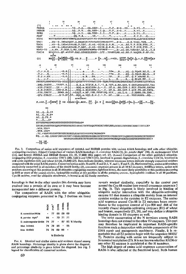
(1) HHR6A HHR6B DHR6 lhp6' RAD6 uaC1 UBC4 UBC5 lthE2-23 CDC34
(2)
.. W~ _ _ _
• •••••••••••••• LQ •• P.A ••• GA.SEN-, i ..... V. F •• E-G •• FG •••• k.Ti •• T ••••••• • •••••••••••••• LQ •• P. V ••• GA.SEN-, i.Q ••. V. F. ,E-G •• F ••••• k. vi. ,S ••••••• • •••••••••••••• LQ •• P.T •• ,GA.TON-, i. i ... V. F •• H-, •• F ••••• k.Ti •• T ••• '" • • •• T •••••••••••• QQ. P.A •••••• VS.-, •• 1 ••• V ••••• - ••• F ••••• k. v.S ••• Q •• " •
HSTPARRRLMRDFKRHKEOAPPGVSASPLPD-NVHVWNAMIIGPA-DTPYEDGTFRLLLEFDEEYPHKP • • RA--k. iMkaIQAV. d. PAAHltLEFVSeSOiHHLKGTFl •• P-G •••• G. K. Vvoi. VPM ••• F •• • ,sS--k,iAkeLSDLER.P.TSC •• G,vG,-DIYH.Q,S.H •• ,-.s •• AG.V,F,siH.PTd.,F •• • • sS--k. iAkeLSDLGR. P .Ase •• 0. vG. -Dl 'iN.Q. S. H •• S-. s •• AG. V. F. sin. PTd •• F •• •• s,Sk •• E.DLM.L.HS.yKVDMINDGMHe-FFVHFH---- •• K-.sI.QG.VWkvRv.LT.A •• Y.S HS~SLLL.QY.eLTDP.KAI.SFHIELEDDS-.iFT •• IGVMVLNE.SI.HG.F.kAQMR.P.dF.FS.
;.(SUS
KS~§~R~~+O~~OD-p ___ 1 ______ 0 R+--wg--++GP- g§ïÇ-gO~F~+-F~~YP~P
,,, ,,, .T.r.v .K •••••••• -o.s .... " .... 5 ••••• S ••••• - •••• LOe ••• N •••• sQ •• Q. YQeu .RE.E ••• S .T.r ••• K •••••••• -o.s •••••••••• 5 ••••• S ••••• - •••• LOe ••• U •••• sQ •• Q. YQeU .RE.E ••• S .T. r. v.KV ••••••• -O.G ••.••••••• s.R ••• SA •••• - •••• lS •••• U •••• ST •• Q. Y .eNrRE.E •••• • L ••• v .T •••••••• -. •• • •••••••• s •••••• A •••• - •••• L. " .N ••••• A ••• Q.HreN.KE •• r •• r PHVKFlSEHFHPIIVYA-NGE C OILQlIRh'TPTYDVASILTS-IQSLFNOPNPASPANVEMTLFKDHKSQYVKRVK • kHQ. otKVY ••• isSVT. A •••••• K. A.s. VITIK. A. I. -l.A. LQS. E. ND. QDA. V. QHYLRDrESFll. TM .kis.TtKIY ••• iN.- •• N •••••• KDQ.s.ALTlSKv.L.-.C •• LT.A •• OD.LVP.I.HiV.TDrPK.EATAR .k.N.TtKIY ••• iNS-S.N •••••• KDQ.s.ALTlSKv.L.-.C •• LT.A •• DD.LVP.I.Qiv.TD.AK.EATA • • SiG.TNKIY •••• DEMS 'Sj' : .viNQT. s.MF .1VU.FEVFlPQ. LLY ••• SO.L.G ••• S.MMRD.IIA.Etlk •• • QFr.TPAIY ••••• R-D.R • s •• H'QSo' s. VQT. E.v. I .-. V •• LE ••• IN ••••• d •• VoYrKHPE •• KQ •••
Gl:>PlfI~H'OnT
P_+OF_§_+~HPNy_~ OG-+C OI~O_WSp~r~L-S L-SLL05ÀNP-Bpt"v-0tA-t--oB~Y~X-
,,, , AI •• Q •• R.C* AI. .Q •• II.S* Ac •• Q.FI. * K ••• 0 ••• S* ETVEKSWEOOMOOMDDDDDDDDDDDDDEAD~
Lh7RLVASETSNGQKGNVEESOLYGIDHDLIOEFESQGFEKDKIVEVLRRLGVKSLDPNDNNTANRIIEELLK* .WTK.YAV* .h'TK. YAV* .Yc.rYAKPEDISPEEEEEESDEELSDAEGYDSGOEAIHGHADP* HE •• r • KQ • I PKGFIMPTSESAYISQSKLDEPESNKDMADUFWYDS DLDDDENGSVILQDDDYDDGNNHIPFEDDDV
YNYNDUDDDDERIEFEDDDDDDDDSIDNDSVMDRKQPNKAEDESEDVEDVERVSKKI*
FIG. 3. Comparison of amine add sequences of HHR6A and HHR6B proteins with various RAD6 homologs and with other ubiquitinconjugating enzymes. (Upper) Comparison ofvurious RAD6 homologs: S. cerel'isiae RAD6 (3), Sc. pombe rhp6+ (10), D. melarlogasler Dhr6 (11), and human HHR6A and HHR6B (human E2; AIr 17,000) (this paper; ref. 17). (LQwer) Comparison of the other published ubiquitinconjugating (132) proteins; S, cerel'isiae UBC1 (20); UnC4 and UBC5 (21), involved in protein degradationi S. cere~'iJiae CDC34, involved in eeU eycle regulation (22); and wheat E2(Mr 23,000) (23). Dols indicate identity, whereas lowerease letters indicate strongly eonserved rcsidues eompared with the yeast RAD6 protein. Conserved amino acids: Rand K; E and D; I, V, and L; Tand S, (1), Horizontal bars, aminoacid residues exclusively conserved in all members oflhe RAD6 family; (2), consensus sequence present in all 10 E2 enzymes. Doldface lelters, amine acid residues occurring at Ihis position in all 10 ubiquitin·conjugating enzymes; lightface letters, the most likely possibility at thls position (occurring in 80% or more of the cases); circles, hydrophilic residue at lhis posÎlion in alilhe proteins; crosses, hydrophobic residues in all 10 positions. Cys-88 residue, used for ubiquitin attachment, is boxed in all E2 family members.
homologs is that in the olher species this domain may have evolved into a protein of its own or it may have become incorporated into a different prolein.
The comparison of RAD6 with the other ubiquitinconjugating enzymes presented in Fig. 3 (bottom six lines)
, ~ m 8 • • • " " " ~ ~
, , , 0 , ,
S. eerevls!aEl RAD6 77 6S 6. 69
S. pombe rhp6+ .4 70 71 71
O. melanoga$tElr OHR6 74 77 .7 85 % Identity
Man HHR6A 74 7. 90 95
Man HHAaB 74 7. '9 9.
%Sîmilarity
Fm. 4. Identical and similar amino acid residues shared among RAD6 homologs. Percentage identity is given above Ihe diagonal, and percentage similarity is given below the diagonal. See Fig. 3 legend for classification of conserved residues,
69
reveals marked similarity, especially in Ihe central part around the Cys-SS residue (sec overall consensus sequence 2 in Fig. 3). This segment is Iikely involved in binding of ubiquitîn andjor interaction with the ubiquitin-activating enzyme El that donates a ubiquitin moiety from an internal cysteine residue 10 the cysleine in E2 enzymes. The amino acid sequence around eys-SS in E2 enzymes bears resemblance 10 the sequence context of Cys-908 and ·866 of the recently cloned ubiquitin·activating enzymes (El) of wheat and human, respectively (25, 26), and may define a ubiquitin binding domain in El enzymes as weil.
The strict conservation of the N terminus among RAD6 homologs does nol extend to the olher E2 enzymes. This part may therefore be implicated in important RAD6-specific functions such as interaction with protein components of Ihe DNA repair and mUlagenesis machinery. Finally, it is remarkable that all E2 proteins begin with the sequence MS(Sj n. Proteins starting with serine are frequently subject 10 N-terminal acetylation (27)_ It is not known whelher RAD6 or any olher E2 enzyme is acelylated at the N terminus.
The high degree of amino acid sequence conservation of RAD6 is also reflected at the functionallevel. Both human
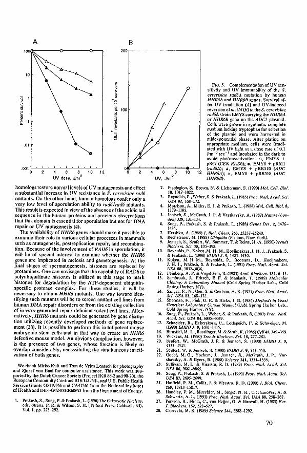
A B 10,0Il;,-------<>----o--, 200,------------,
.01
.001 L~~~~~~~~~--' o 2 4 6 8 10 12
UV dose, J/m'L o 2
homologs restore normalieveis of UV mutagenesis and effect a substantial increase in UV resistance in S. cerc\'isiac rad6 mutants. On the other hand, human homologs confer only a very low level of sporulation ability 10 rad6/rad6 mulants. This result is expected in view ofthe absenceoftheacidic tail sequence in the human proteins and prevÎous observations that this domain is essential for sporulation but not for DNA repair or UV mutagenesis (4).
The availability of 1I11R6 genes should make it possible 10 examine their role in various cellular processes in mammals such as mutagenesis, postreplication repair, and recombination. Because of Ihe involvement of RAD6 in sporulation, it will be of special interest to examine whether the HHR6 genes are implicated in meiosis and gametogenesis. At the flnal stages of spermalogenesis, histones are replaced by prolamines. One can envisage that Ihe capability ofRAD6 10 polyubiquitinate histones is ulilized al this stage to mark hislones for degradation by Ihe A TP-dependent ubiquitinspecific protease complex. For these studies, it will be necessary 10 obtain HHR6 mutanls. One way toward identifying such mutants wil! be 10 screen mutant celllines from human DNA repair disorders or from the existing collection of in vitro generated repair-deficient rode nt ceillines. Alternalively, HHR6 mutanls could be generated by gene disruption utilizing recently developed melhods of gene replacement (28). It is passible 10 perform Ihis in totipotent mouse embryonic stem eells and in thaI way la ereale an HHR6 defeetive mouse model. An obvious eamplication, however, is Ihe presence of Iwo genes, whose function is likely 10 overlap cansiderably, necessitating Ihe simultaneous inaclivation of both genes.
We thank Mirko Kuit nnd Tom de Vries Lentsch for photography and Sjozef van Daal for computer assistance. This work was sup· ported by the Dutch Cancer Society (Project IKR 88-2 and 90-20), the European CommunityContract Bl6-141-NL, and U.S. Public Health Service Gronts GM19261 and CA41261 from the National Institutes ofHealth and DE-FG01·88ER60621 from the Department ofEnergy.
1. Prokash, S., Sung, P. & Prakash, L. (1990) The EllkaryalÏC' Nucleus, eds. Straus, P. R. & Wilson, S. H. (felford Press, CaIdweIl, NI), Vol. I, pp. 275-292.
4 6 8 10 UV, Jfm'L
FIG. 5. Complementation of UV sensitivity and UV Îmmutability of the S. cerel';siae rad6Jl mutation by human HlIR6A and HHR6B genes. Survival afIer UV irradiation (A) and UV-induced reversionofmet14(B) in theS. cen'\'isiae. rad6tJ. stroin EMY8 carrying the HHRM or HlIR6B gene on the ADel plasmid, eells were grown in synthetic complete medium lacking tryptophan for selection of the plasmid and were harvested in midexponential phase. After plating on appropriate medium, eells were irradialed v.ith UV light at a dose rale of 0.1 J'm-2'sec-I and incubated in the dark 10 avoid photoreactivation. 0, EMY8 + pR67 (CEN RAD6); e, EMY8 + pR611 (radM); "', EMY8 + pRR510 (ADC
12 HHR6A); t;. EMY8 + pRR518 (ADC HHR6B).
2. Picologlou, S., Brown, N. & Lieberman, S. (1990) Mol. Celt. Biol. 10, 1017-1022.
3. Reynolds,P., Weber,S. & Prakash, L. (1985) Proc. Na/I. Acad. Sci. USA 82, 168-172.
4. Morrison, A., Miller, E. J. & Prakash, L. (1988) Mol. Cell. Biol. 8, 1179-1185.
5. Jentsch, S., McGrath, J. P. & Varshavsky, A. (1987) Na/ure (LondOI/) 329, 131-134.
6. Sung, P., Prnkash, S. & Prnkash, L. (1988) Genes De\'. 2, 1476-1485.
7. Hmhko, A. (1988) I. Bial. Chem. 263, 15237-15240. 8. Rechsteiner, M. (1988) UbiqllÎtin (Plenum, New York). 9. Jentseh, S., Seufert, W., Sommer, T. & Reins, H.-A. (1990) Trends
Biochem. Sei. 15, 195-198. 10. Reynolds,P.,Koken,M. H. M.,Hoeijmakers,J. H. J.,Prakash,S.
& Prakash, L. (1990) EMBO 1.9,1423-1430. H. Koken, M. H. M., Reynolds, P., Bootsma, 0., Hoeijmakers,
J. H. J., Prakash, S. & Prakash, L. (1991) Proc. Na/I. Acad. Sci. USA 88, 3832-3836.
12. Feinberg, A. P. & Vogelstein, B. (1983) Allal. Biochem. 132,6-13. 13. Sambrook, J., Fritsch, E. F. & Maniatis. T. (1989) Mole('lIlar
Cloning: A Labara/OI)' Marmot (Cold Spring Harbor Lab., Cold Spring Harbor, NY).
14, SangeT, F., Nicklen, S. & CouJson, A. R. (1977) Proc. No/I. Acad. Sci. USA 82, 168-172.
15. Sherman, F., Fink, G. R. & Hicks. J. B. (1986) Me/hads in Yeas/ Gene/iC!: Lahora/of)' Course Manual (Cold Spring Harbor Lab., Cold Spring Harbor, NY),
16. Sung, P., Prakash, L., Weber, S. & Prakash, S. (1987) Proc. Nall. Acad. Sci. USA 84, 6G4S-fiO.I9.
17. Schneider, R., Eckerskom, C., Lottspeich, F. & Schweiger, M. (1990) EMBOJ. 9,1431-1435.
18. Bimstiel,M. L., Busslinger,M. & Strub, K. (1985)CeIl41,349-359. 19. Wickens, M. (1990) Trends Biochem. SC!. 15,277-281. 20. Scufert, W., McGrath, J. P. & Jentseh, S, (1990) EMBO J. 9,
4535-4541. 21. Seufert, W. & Jentseh, S. (1990) EMBO J. 9, 543-550. 22. Goebl, M. G., Yochem, J., Jentseh, S., McGroth, J. P., Var
shavsky, A. & B)'ers, B. (1988) Science 241, 1331-1335. 23. Sullivan, M. L. & Vierstrn, R. D. (1989) Proc. Na/I, Acad. Sci.
USA 86, 9861-9865. 24. Sung, P., Prakash, S, & Prakash, L. (1990) Proc. Nall. Acod, Sci.
USA 87, 2695-2699. 25. Hatfield, P. M., Callis, J. & Vierstrn, R. D. (1990)/. Biol. Chem.
265,15813-15817. 26. Handley, P.M., Mueckter, 1.1., Siegel, N. R., Ciechanover, A, &
Schwartz, A. L. (1991) Proc. Nall. Acad. Sci. USA 88, 258-262, 27. Persson, B., Flinta, C., von Heijne, G. & Jömvall, H. (1985) Ellr.
I. Biochem. 152, 523-527. 28. Capecchi, M. R. (1989) Sc!enee 244, 1288-1292.
70


Chapter V
Localization of two hllmall IlOmologlles, HHR6A and HHR6B, of the yeast DNA I'epair geile RAD6 to chromosomes Xq24-25 alld 5q23-31

I
I
GRNOMICS 12,447--453 (1992)
Localization of Two Human Homologs, HHR6A and HHR6B, of the Yeast DNA Repair Gene RAD6 to Chromosomes
Xq24-q2SandSq23-q31
M. H. M. KOKEN,' E. M. E. SMIT, I. JASPERS·DEKKER, B. A. OOSTRA, A. HAGEMEIJER, D. BOOTSMA, AND J. H. J. HOEIJMAKERS
Department of Ceff Bio/ogy and Genetics, Medical Genetics Center, Erasmus University, P.O. Box 1738, 3000DR Rotterdam, The Netherlands
Received luly 19, 1991; revised October 10, 1991
'rhe chromosomalloealizalions of two closely relaled human DNA rcpair genes, llllR6A and llllR6B, were delermined by in situ hybridization with hiotinylated probes. HHR6A and HHR6B (human homolog of yeast RAn6) eneode ubiquitineonjugating enzymes (E2 enzymes), likely 10 be involved in postreplicalion repair and induced mulagenesis. TheHHR6B gene was assigncd to human chromosomc 5q23-q31, whcreas the HHR6A gene was localized on the human X chromosorne (Xq24-q25). This latter assignment was confirmed wUh an X-specific human-mouscjhamsler somatic eeU hybrid panel. Southern blot analysis points 10 an X and an autosomallocalization of HHR6A and HHR6B, respectivcly, in Ihc mouse. Thc potcntlal involvemenl of these genes in human genetic disorders is discussed. © 1992 MaMml~P ... s, In~.
INTRODucnON
Recently, we reported the cloning oftwo human genes, designated HHR6A and HHR6B, homologous to the Saccharomyces cerevisiae RAD6 gene (Koken et al., 1991b). As deduced from the very pleiotropie phenotype of yeast rad6il mutants, t,he RAD6 protein plays an important roie in various cellular processes, including postreplication repair (a poorly defined, error-prone repair pathway), damage-induced mutagenesis, sporulation, and recombination (for a review, see Prakash et al., 1990). The RAD6 functions are accomplished by a 172-amino-aeid protein with an N-ferminal globular structure and an extended C-terminal aeidie tail (Reynolds et al., 1985). 'fhe acidie domain is specifically required for sporulation but is not essential for the other RAD6 functiOllS (Morrison et al., 1988). An important finding concerning the biochemical activity of the RAD6 protein was the discovery t,hat. the gene eneodes a ubiquitin-conjugating enzyme (Jentsch et al" 1987). Ubiquitin, a widespread, highly conserved 76-amino-acid polypeptide, is
llHR6A nnd HHR6B are not HGMW approved gene sYlllbols. I To whom correspondence should be addressed.
covalently attached to specifie cellular proteins that in this way are targeted for selective degradation, (re}folding, or stabilization (for recent reviews, see Hershko, 1988; Reehsteiner, 1988; Jentsch eta!., 1990). Ubiquitination of proteins occurs in a multistep reaction. First, a ubiquitin-activating enzyme (or El enzyme) binds and activates a ubiquitin molecule. This is subsequently transferred to one of a set of ubiquitin-conjugatillg enzymes (or E2 enzymes). The E2 enzyme ligates t.he ubiquitin moiet.y to a target protein with or without the help of an E3 ubiquit.in protein ligase molecule. The RADG protein was found to attaeh one (Jentsch et al., 1987) or multiple (Sung et al., 1988) ubiquitin moieties to histones H2A and H2B in uitro. If histones are also the main targets of RAD6 in uiuo, it is likely that RAD6 mediates chromatin remodeling required for t.he processes impaired in a rad6û mutant.
RAD6 is very st.rongly conserved in eukaryotie evoluHon, and this property permitted us to clone by evolutionary walking two human homologs (Koken et al., 1991b) using the Schizosaccharomyces pombe (Reynolds et al., 1990) and Drosophila melarwgaster (Koken et al., 1991a) homologs as "intermediates." The human HHR6A and HHR6B proteins (HHR for human homolog of RAD6) share ~95% amino acid sequence identity with eaeh oUler and ~70% amino acid identity wit.h their yeast counterparts, but notably lack the acidie Cterminal domain, the occurrence of which seems to be limited to S. cereuisiae RAD6. r ... loreover, the human polypeptides were found to substitute funct.ionally for the repair and mutagenesis functions of RAD6 in a S. cereuisiae rad6il mutant but not for its role in sporulation. This indicates that the proteins of the repair and mutagenesis maehinery with which RAD6 interacts are also conserved to a significant extent. belween man and yeast. Furthermore, it is likely that. the HHR6 proteins in man have a funetion similar to that of RAD6 in yeast, i.e., catalyzing ubiquitin conjugation as an essential step in t.he repair and mutagenesis pathways. This conclusion makes the gene a eandidate for human inherited
72
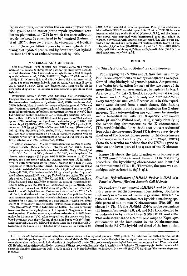
repair disorders, in particular the variant cOIllplementatiOll group of t,he cancer-prone repair syndrome xeroderma pigmentosum (XP) in which the postreplication repair pathway is considered to be impaired (Lehmann et al., 1975). Here we present the chromosomallocalization of these two human genes by in situ hybridization using biotinylated pro bes and by Southern blot hybridizations to DNA of rodentjhuman ceU hybrids.
1tATERIALS AND METHODS Cell lineslDNitS. The somatic eell hybrids conlalning vaTÏous
parts of the human X chlOmosome used in this sludy have been describcd elsewhere. The hamsterfhuman hybrids were X3000, Xq24-qtcr (Nussbaum et al., 19S6); 90SKlBlS, Xq24--q26 (Schonk et 0/., 19S9); 8121, Xpler--q27.1; and 238-1, Xpter--q27.2 (Patterson et 0/., 1987). The mousefhuman hybrids were RJK734, Xq26--qter (Scott et al., 1979); and CY34A, Xq24--q27 (Suthers et al., 19S9) See Fig. 2B for schematic diagram of the human X-chromosome segments in these hybrids.
Restrictioll enzyme digests and Southem blot hybridizations. Enzyme digestions and Southern blotting procedures were essentially tho same as described previously (Koken et al., 1991b; Sambrook et af., 1989). In brief, 20 pg of reslriction enzyme·dig(>sted genomic DNA was size-fraclionated on O.S~ agarose gels and transferred onto nylon (ZetaplOhe) membranes. Hybridization occurred overnight at 65°C in a hybridization buffer containing lOX Denhardt's solution, 10% dOll:tran sulfate, 0.1% SDS, 3X SSC, and 50 pg/ml sonicated salmon sperm DNA. Washings were performed extensi\'ely up to 0.3X SSC containingO.l% SOS at 65°C. The L7-kb HHR6A cDNA probe, H28, containsa full-length HHR6A cDNA on a Sall fragment (Koken et al., 1991b). The HHR6B eDNA probe, H13"-8' harbors the complete HHR6B open reading fwme on an O.S-kb fragment starting with an rutificial BcoRI site at the position ofthe ATG and ending at a nalural BroRI site in the cDNA (Koken et al., 1991b).
In situ hybridization_ III situ hybridization was performed essenfiany as described (Lanclegent et al., 1985; Pinkel el al., 1986). Human lymphocyte metaphase spreads were trcated with 100 pg RNase A/mi in 2X SSC for 1 h at 37°C, rinsed in 2X SSC, and dehydrated in alcohol. After a pepsin (0.1 pg/ml 0.01 N HCI) treatment at 37°C for 10 min, the slides were washed in PBS, postfixed with 1% formaldehyde in PBS containing 50 mM MgCI2 , washed for 5 min in PBS, dehydrated in ethanol, and air-dried. The hybridization mÎltture (10 pI per slide) eonsisted of 50% formamide, 2x SSC, 40 mM sodium phosphate (pH 7.0), 10% doxtwn sulfate 50 ng labeled probe, 1 pg sonicaled salmon sperm DNA, and 1 pg Bscherichia cali tUNA. The genomic probes, B3.0, B2.3, 112.7, HO.75, and HS2.7 (HHR6A) and E2.3, E6.0, E4.5, and E1.3 (IlIlR6B), representing most of the genomic region of bath genl'S (Koken et ol., manuscript in prcparation), were biotin-Iabeled. A cocktail of the genomic prohes for each gene was used for ill situ hybridizalion. Probes were denatured al 70°C for 5 min in hybridization mixture (specified aboye). Competition for repeat sequences present in the genomic subclones was achieved by incubation for 6 h (HHR6A probes) or 2days (IlIlR6B) witha 100 times eltcess of thymus DNA (HHR6A) or a 1000 times excess ofhuman con DNA (IIHR6B) at 37°C in hybridi?ation buffer. This was Ilecessary because ofthe elttremely high content ofrepeats in the genomic dones used as probes. Thechromosomespreads weredenatured in 70% formamide for 2.5 min at ?OoC. After competition, the probes were incubated oyernight with tho slides and then washed once with 50% formamide in 2x SSC at 39°C followed by three times for 5 min in 2X SSC, three times for 5 min in O.lX SSC at GOoC, and ollce for 5 min in 4x
SSC, 0.05% Tween20 at room temperature. Finally, the slides were blocked in 4X SSC, 5% nonfat dry milk for 20 min al 37°C. Slides were incubated with 5 pg avidin D-FITC (Vector, U.S.A.), and the fluorescent signaI was amplified with biolinylatcd goat antÎ-avidin D, washed, dehydrated with ethanol, and air-dried_ The slides were embedded and stained in 9 pruts glycerol containing 2.3% (w/v) l,4-diazobicyclo-(2,2,2)-octane (DABCQ) and 1 part 0.2 MTris-HCl, 0.02% NaNJ , pH 8.0, containing 4',6'-diamino-2-phenylindole (DAPI) to a final cOllcentration of 0.5 pglpl.
RESULTS
In Situ Hybridization to Metaphase Chromosomes
For mapping the HHR6A and HHR6B loci, in situ hybridization experiments on metaphase spreads were performed lIsing biotinylated genomic probes. A representative in situ hybridization for each ofthe two genes ofthe more t.han 50 metaphases analyzed is depicted in Fig. 1. As shown in Fig. IA (HHR6A), a specific signal (arrow) is found on the long arm of only one chromosome in every metaphase analyzed. Because cells in this experiment were derived from a male donor, this finding strougly suggests that t.he gene is located on the X chromosome. This interpretation was conftrmed by simultaneous hybridization with an X-specific centromere prohe, pBamX5 (Willard et al., 1983), clearly identifying the hybridizing chromosome as the X chromosome. [The weak hybridization with the centromeric regions of four otller chromosomes (9 and 17) is due to cross-hybridization of the X-centromere probe to the centromeres of chromosomes 9 and 17 (Willard aud Waye, 1987).) From t.hese results we deduce that t.he HHR6A gene resides on the lower part of the q arm of the X chromosome.
Figure IB shows the hybridization with biotinylated HHR6B gene probes (arrows). Using the DAPI staining procedure, the hybridizing chromosome was identified as chromosome 5 (Fig_ 1B). Therefore, the gene was unambiguously assigned to 6q23-q3L
Southem Hybridization of HHR6A Probes 10 DNA of a Panel of HumanjRodent Somatic Cel! Hybrids
To confinu the assignment of HHR6A aud to obtain a more precise subchromosomal localization, Southern blot analysis was carried out using genomic DNA from a panel of human-mousejhamster hybrids containing specific parts of the human X chromosome (Fig. 2B). As shown in Fig. 2A the HHR6A cDNA probe recognizes the human fragments (3.0, 2.6, and 0.75 kb, indicated by arrowheads) in hybrid celllines X3000, 8121, and 2384. This indicates that the HHR6A gene maps on Xq24-q25 ceutromeric of the breakpoint in the X chromosome found in the RJK734 hybrid and distal ofthe breakpoint
FIG. 1. In situ hybridization of metaphase chromosomes to biotinylatcd genomÎC HHR6 probes. (A) Hybridization with a cocktail ofall genomic 1I1IR6A probes specified uncler Material and Methods. The arrow Îndicates Ihe hybridization signaion chromosome Xq. This chromosome shows also the X-specific hybridization ofthe pBamX5 probe. The prohe weakIy closs-hybridizes to chromosomes 9 and 17 as indicated. (B) Hybridization with a cocktail ofaIl genomic HHR6B probes (indicated under Materials and Methods). The arrowspoint 10 the regions with aspecific signal on chromosome 5q23--q31. In panels I the in situ hybridization is shown. In panels II the DAPI banding ofthe same melaphases is shown.
73
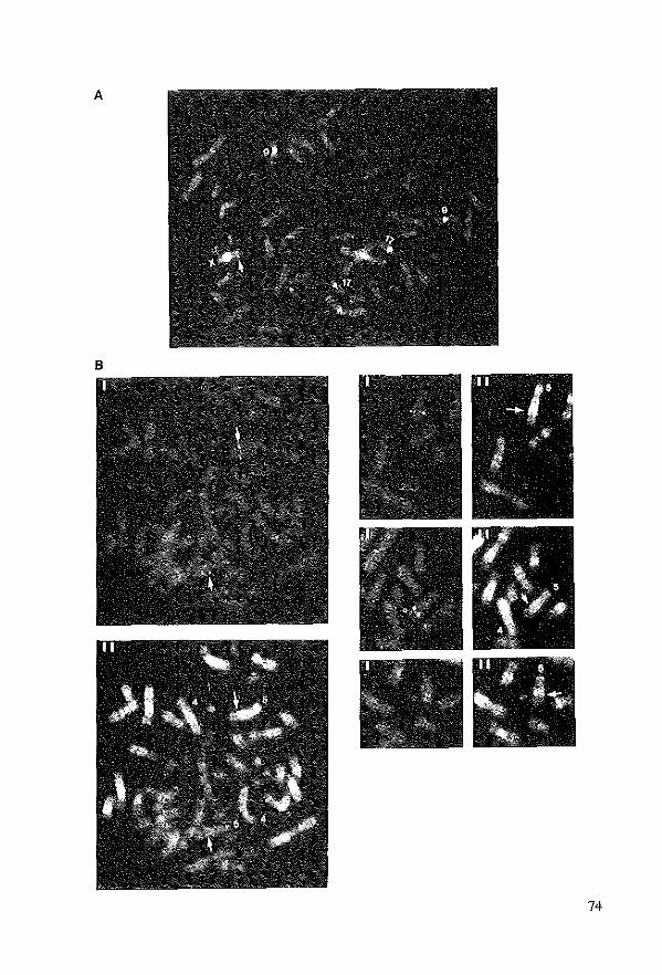
A
B
74
B
A • ~ " 0 " w 0
~ c " 0 N E • 0 , ~ " • " " a: N '"
MW ~b}
., .... '" 4111 5.0 .. ~ ....... 3.0
'"" .W - 2.6
" I I 11
"" ... 1.1 " I " ....... " .. 0.75 HHRSA + + + + + Hyb/Jd "" .. "'", +\9" 1,-1;> "b"'1; 0,<>
'il I' Vo ~ l',s> '4 '",
FIG. 2. (A) Southern blot analysis ofX-specific hybrid panel with the HIlR6/\ cDNA probe. Thc source ofthe gcnomic DNA is inclicated. DNA is digcsted with HilldIli nnd size-fmctionated on an 0.8% agarose gel. Tbc molecular weight (MW) indicated on the right refers to tha hyhridizing frllgments at the corresponding posiHons in the autoradiogram. Tbc fragments of 3.0, 2.6, nnd 0.75 kb are of the human IIIIR6A gene. (H) Reprcsentation ofthe human X-chlOmosome fragments (indicated by tinas) retained in the rodent/human hybrids used in tbis shldy. Thc + or - sÎgn above the hybrid-narnes indicates whether or not DNA fIOm tbis specific celiline hybridizes with the human probe.
in the X3000 hybrid cellIine, confirming the data found via in situ hybridization. Hybridizations to t.he somatic ceU hybrids 90BK1B18 and CY34A were also positive with HHR6A probes (data not shown).
Chromosomal LocalizatioTi of the Mouse Homologs of HHR6A alid HHR6B
To assess whether also in the mouse one gene is 10-cated on the X chromosome and the ot.her on an autosame, a Southern blot with equal amounts of genomic DNA from a male and femaie mouse was hybridized consecutively with both human cDNA probes. As shown in Fig. 3, the hybridization with the HHR6A gene c1eady shows an approximately twafold difference in hybridizaHon intensity between the DNA of the male and the female mouse, whereas with the HHR6B probe and the same blot, na difference between male- and female-derived DNA is detectabie. This strongly suggests that also in the mause the HHR6A gene is X-linked, whereas the HHR6B gene is on an autosame.
DISCUSSION
Localizatio!l of Genes lnvolved in DNA Repair or in Ubiquitin 8ystems
This paper describes the localization of two human homologs, HHR6A and HHR6B, of the yeast DNA repair gene RAD6 to human chromosomes Xq24-q25 and 5q23-q31, respectively. The HHR6A gene is the first human DNA repair gene lacated on X. Among the DNA repair genes isolated thus far, no clustering is apparent,
75
with the possible exception of the q13.2 area of chromosame 19 onto which at least three repair genes have been lacalized (Mohrenweiser et al., 1989; \Veeda et al., 1991; Smeets et al., 1990; Thompson, 1989). This, however, could be due at least in part to the presenee of large regions of hemizygosity in the Chinese hamster eells used to generate the repair mutant eeU lines with which these three genes were cloned. The hemizygosity favors the isolation of mutants in genes located in those areas (Siciliano et al., 1983).
In contrast to a dispersed localization of DNA repair genes over the genome, it is of interest to note that a clustering of genes far different components of the ubiquitin system may exist on the X chromosome. \Vith the exception ofubiquitin itself, encoded by several polyubiquitin and ubiquitin fusion genes on a number of different autosomes (\Vebb et al., 1990), the other two ubiquitin-system genes cloned thus far are both located on X. The GdX gene (HGMW symbol DXS254), with exten~ sive hamology to ubiquitin, has been localized onto Xq28 (Tonialo et al., 1988). Moreover, the gene for one of the human ubiquitin-activating enzymes (El, HGMW gene symbol UBE1) has been assigned to the X chromosome (Ohtsuba and Nishimoto, 1988; Kudo et al., 1991), more precisely to Xpll.2-p11.4 (Zackenhaus and Sheinin, 1990; Handley et al., 1991; McGrath et al., 1991).
Duplication of HHR6
In the lower eukaryotes (8. cerevisiae, S. pombe, and D. melarzogaster), we eould identify only a single RAD6 locus situated on an autosame (Reynolds et al., 1990; Koken et al., 1991a). As ealculated from divergenee data,
HHR6A HHR6B Probe MWlld"-l!o'So'ONA
kb
4.5
2.'
1.1
0.'
0.5 . --0.34 -
FIG. 3. Southern blot analysis ofgenomic liver DNA from a male and a femele mouse. MW: Molecuier weight marker, i.e., phega" DNA digested with PstI. Left: The autorüdiogram of a blot with HindfII + EroRI + BamHI triply digested mouse DNA hybridized with e human HHR6A cDNA probe. i\IiddJe: An autoradiogram of the same blot hybridized with a human HHR6B cDNA probe. Hight: A photograph ofthe ethidium-stained genomic gel. The probes used and the ó (male) or 2 (femala) sex ofmouse from which the DNA was isolated are indicated above the autoradiogram.
the two HHR6 genes in human and mouse (unpublished data) may have arisen from a gene duplication event in the Jurassic era about 200 million years ago, early in the history of mammais, i.e., weIl before the separation of evolutionary lines Jeading to rodents and primates. Duplieation has several advantages and is more often found for essential genes. One advantage could be that it permits differential gene regulation and/or functional divergence of the proteins.
Finally, the synteny conservation for the X chromosome between mouse and man as found for HHR6A supports Ohno's law that there is astrong selection against chromosomal rearrangements involving the sex chromosomes and autosomes (Ohno, 1969; Nadeau, 1989).
The Chromosomal Context of HHR6A and HHR6B; Possible Involvement of HHR6 in Human Disorders
Yeast rad6D. mutant cells show a very pleiotropie phenotype, with sensitivity to many DNA damaging agents, a defect in postreplication repair, no induced mutagenesis, and a complete lack of sporulation. In human, only ceUs of a single syndrome are known to be aft'ected in postreplication repair: the variant complementation
group of the rare DNA repair disorder, xeroderma pigmentosum (XP) (Lehmann et al., 1975). In this complementation group, constituting about 30% of all XP patients, no indications favoring an X-Iinkage have been found. This renders it unlikely t.hat. HHR6A is the gene responsible for this disorder. However, the HHR6B gene remains a possible candidate, although ceUs from XP variant patients have an elevated frequency of uv-in~ duced mutations, and in t.hat respect differ from the yeastphenotype (Maheretal., 1976; Myhr et al., 1979). A systematic search for abnormalities in DNA, mRNA, or protein structure or expression in (families 00 XP variant patients should resolve this issue. In addition, two mammalian post.replication repair-deficient ceU mu~ tants that are potent.ial HHR6A mutants have been characterized, UV1 of Chinese hamster origin (Hentosh et al., 1990) and .SVM (derived from Indian Munt jac) (PiIlidge et al., 1986) .
Two human disorders have been assigned to the q24~ q25 region of the X cluomosome where HHR6A is 10-cated (Human Gene Mapping 10 and 10.5): first, the X-linked lymphoproliferat.ive syndrome, which results in fatal infeetious mononucleosis, hypogammaglobulinemia, and malignant Iymphoma-celis from these patients seem to be dist.urbed in t.he appropriate immune response to Epstein-Barr virus (Skare et al., 1989); and second, the oculocerebrorenal syndrome of Lowe, char~ acteTÎzed by congenital cataract, mental retardation, and a defective renaI tubular function (Reilly et al., 198B). Although these diseases apparently map to the same region of the X cluomosome as HHR6A, to our knowledge there is no evidence for a DNA repair defect associated with any of them. A final X-linked disorder not assigned ta a certain subchromosomal region with a possible defect. in DNA repair is the N syndrome. Patients sutfering from this disease display mental retar~ dation, malformat.ions, development ofT-cellleukemia, and chromosome breakage (Floy et al., 1990). The last two phenotypic traits resembie those of the DNA repair disorder Fanconi anemia. Although it has been proposed that malfunction of DNA polymerase a (X-linked) could be the cause for N syndrome, the evidence is based on aphidicolin inhibitioJl studies which provide only indirect indications.
HHR6B resides in a region of cluomosome 5 containing a large cluster of growth factor genes, i.e., the genes for IL3, IL4, IL5, and CSF2 (Human Gene Mapping 10 and 10.5). These genes have recently been assigned to chromosome 11 in mouse (ATCC/NIH,1990). 'I'hepossibility exists that-due to synteny conservation-the murine HHR6B gene is also located on this chromosome. In situ hybridization should be performed to verify this proposition. Thus far, t.he human 5q23-q31 region has not been associated with any hereditary disease (Humun Gene Mapping 10 and 10.5). '1'0 our knowiedge, the only syndrome to be linked to chromosome 5 with a possible defect in DNA repair is Gardner syndrome (HGMW gene symbol APC), a dominant disorder with a predispo· sition to cancer, especially of the large intest.Îlle. It has been found that ceUs from same of these patients are
76
hypersensitive to uv light, X rays, and mitomyein C (Little et 01., 1980); however, thus far no specific repair defect has been reported in eells of these patients (Henson et al., 1983). Because post.replication repair was not investigated, a possible involvement of HHR6B in this disorder is not ruled out on the basis of these findings. However, the recent cloning ofthe APC gene, responsibie for familial adenomatous polyposis (FAP) and Gardner sYlldrome (I{inzler et al., 1991), excludes any link with HHR6B.
It is reasonable to assume that HHR6A and HHR6B have largely overlapping functions in view of their high sequence homology and their ability to complement yeast rad6 repair functions. This functional redundancy would require the unlikely event of simultaneous inactivation ofboth HHR6 genes for clinical symptoms to become manifest. Alternatively, eonsidering the pleiotropic and severe yeust rad6 phenotype, it is possible that inaetivation of one or both HHR6 genes is let.hal in mammais. These propositions couldprovide an explanaHon for the possible absence of known disorders associated wit.h HHR6. The recently developed Illethodology of targeted gene replacement in mouse embryonal stem eeUs (Capeechi, 1989) opens the possibilit.y ofgenerating HHR6-defective celllines or mice in the laboratory. In that way the role of these genes at the level of the ceU and organism eau be established.
ACKNOWLEDGMENTS
We are indebted to Des. D. Nelson (Houston) and B. Wiednga (Nijmegen) for providing us with thc DNAs from the rodent/human Xch:romosome-specific hybrids; Dr. H. Willa:rd (Stanford) for providing us with the pBamX5 probe; Mirko Kuit and Tom de Vlies Lentsch for photographYi and DIS. A. Gemts van Kessel (Nijmegen) and J. den Dunnen (Leiden) for initial efforts to map these two genes. This work was supported by tho Dutch Cancer Society (P:rojed IKR 88·02 and 90-20) and European Community Contract B16-141-NL.
REFERENCES
ATCCjNIH (1990). Repository Catalogue ofHuman and Mouse DNA probes and Libralies, September 1990, p. 23.
Capecchi, 1\1. R. (1989). Alteringthc genome by homologous :recombination. Science 244: 12B8-1292.
Floy, KM., Hess, R 0., !lnd Meisner, L. F. (1990). DNA polymew.se alpha defect in the N syndrome. Am. J. Med. Genet. 35: 301-305.
Handley, P. M., Mueckler, M., Siegel, N. R, Ciechano\'el, A., and Schwartz, A. L. (1991). Molocular dOlling, sequence, alld tissue dist:ribution ofthc human ubiquitin~aclivating enzyme El. Proc. Nat/. Acad. Sci. US,t 88: 258-262 and 7456.
Henson, P., Fornace, A. J., and Little, J. B. (1983). Normal :repair of ultraviolet-induced DNA damage in a hype:rsensitivc strain offibroblasts fIOm a patient ... oith Garrlner's syndrome. Mutat. Res. 112: 383-395.
Hentosh, P., Collins, A. R S., Correll, L., Fomace, A. J., Giaccia, A., and Waldren, C. A. (1990). Genetie and biodHlmical cha:racterization ofthe CHO-UV-l mutant rlefective in postreplication :recov(1)' of DNA. Cancer Res. 50: 2356-2362.
Hershko, A. (1988). Ubiquitin-mediated protcin degradation. J. Rial. Chem. 263: 15237-15240.
Human Gene Mapping 10 and 10.5 (1990). Cytogenet. ('eli Genet. 55.
Jentsch, S., McGrath, J. P., and Varshavsky, A. (1987). The yeast
77
DNA :repair gene Rt1fJ6 encodes a ubiquitin-conjugating enzymc. Nature 329: 131-134.
Jentseh, S., Scufert, W., Sommer, '1'., and Reins H-A. (1990). Uhiquitin-conjugating enzyIDes: No\'el regulators of euka:ryotic cells. Trends Biochem. Sci. 15: 195-198.
Kinzler, KW., Nilbert, i\I. C., Su, L-K., Vogelstein, B., Bryan, T. M., Levy, D. R, Smith, KJ., Preisinger, A. C., Hedge, P., McKechnie, D., Finniear, R, Markham, A., G:roffen, J., Boguski, M. S., Altschul, S. F., HOlii, A., Ando, H., l\fiyoshi, Y., 1\1iki, V., Nishisho, I., and Nakamura, Y. (1991). Identification of FAP genes f:rom chromosome 5q21. Seience 253: 661-669.
Koken, M. H. 1\1., Reynolds, P., Bootsma, D., Hoeijmakers, J. H. J., Prakash, S., and P:rakash, L. (1991a). Dhr6, a Drosophila homolog ofthe yeast DNA-repair geneRAD6. Proc. Nat{. Acad. Sei. USA 88: 3832-3836.
Koken, M. H. M., Reynolds, P., Jaspels-Dekker, 1., Pcakash, L, Prakash, S., Bootsma, D., and Hoeijmakers,J. H.J. (1991b). Structural and fllnctiollal conservation of two human homologs of the yeast DNA repair gene RADS. Proc. Not{. Acad. Bei. USA 88: 8865-8869.
Kudo, M., Sugasawa, K, Hod, T-A., Ellomoto, T., Hanaoka, F., and Ui, 1\1. (1991). Human \lbiquitin-activating enzyme (El): Compensation for heat-labile mOllse El and its gene loealization on the X ch:romosome. Exp. Cell Res. 192: 110-117.
Landegent, J. E., Jansen in de \Val, N., Van Ommen, G-J. B., Baas, 1<'., De Vijlder, J. J. M., Van Duijn, P., and Van der Ploeg, M. (1985). Chromosomal localization of a ullique gene by non-autoradiog:raphic in situ hybridization. Nal1lre 317: 175-177.
Lehmann, A. R., Kirk-Bell, S., Arlelt, C. F., Paterson, M. C., Lohman, P. H. M., de Weerd-Kastelein, E. A., andBootsma, D. (1975). Xeroderma pigmentosum ceUs \\ith normalievels of excision repair have a defect in DNA synthesis af ter UV-irradiatioll. Proc. NaU. ACad. Sci. USA 72: 219-223.
Little, J. B., Nove, J., and Weichselbaum, R. R (1980). Abnormal sensitivity of diploid skin fibroblasts f:rom a family with Ga:rdner's syndrome to the lethal effects ofX-ir:radiation, ultra\'iolet light and mitomycin-C. Mufat. Hes. 70: 241-250.
l\faher, V. M., OueUelte, L. 1\1., Cunen, R D., and McCormick, J. J. (1976). Frequency of ultraviolet light-induced mutations is higher in xeroderma pigmentosufll variant rells than in normal human cells. Na/ure 261: 593-595.
l\fcGrath, J. P., Jentseh, S., and Varshavsky, A. (1991). UBAl; An esselltial yeast gene encoding ubiquitin-actjvating enzyme. EMBO J. 10: 227-236.
l\fohrenweiser, H. \\'., Canano, A. V., Fertitta, A., Peny, B., 'rhomp~ son, L. H., Tucker,J. D., and Weber, C. A. (1989). Refined mapping of the three DNA repair gems, ERCCI, ERCC2, and XRCCI, on chromosome 19. Cytogenet. Cell Genet. 52: 11-14.
l\forrisoll, A., i\Bller, E. J., and Prakash, I •. (1988). Domain structure and functional analysis of the carboxyl-terminal polyacidic sequence of the RAD6 pJOtein of Sacclzaromyccs cereuisiae Mol. Cel/. Bio/. 8: 1179-1185.
l\f)'h:r, B. C., Tumbull, D., and DiPaolo, J. A. (1979). lTItcaviolet mutagenrsis of nOlmal and xeroderma pigmentosum variant human fibroblasts. Mulat. Res. 62: 3oU-353.
Nadeau, J. H. (l989). Maps of linkage and synteny homologies between mouse and man. Trends Genet. 5: 82-86.
Nussbaum, R. L., Airhart, S.O., and Ledbetter, D. H. (1986). A rodent-human hybrid containing Xq24-qter t:ranslocated to a hamster chromosome exprrsses the Xq27 folate-sensitivc f:ragile site. Am. J. Med. Genet. 23: 457-466.
Ohno, S. (1969). Evolution ofsex chromosomes in mammais. Annu. Reu. Genet. 3: 495-524.
Ohtsubo, M., nnd Nishimoto, T. (1988). The gene coding a ubiquitinactivating enzyme ma)' locate on X chromosome. Biochem. Biophys. Res. Commull. 153: 1173-1178.
Patterson, M., Schwartz, C., Bell, M., Sauer, S., Hofker, M., 'I'lask, B., Van den Engh, G., and Davirs, K E. (1987). Physical mappingstud-
ies on thc human X chromosome in the region Xq27-Xqter. Genomies 1: 297-306.
Pillidge, L., Musk, S. R. R.,Johnson, R. '1'., and Waldren, C. A. (1986). Excessive chromosomc {ragility and abundance of sister-chromatid exchanges induced by UV in an Indianmuntjac cellline defective in post replication (daughter strand) repair_ Mulat. Res. 166: 265-273.
Pinkel, D., Straume, T., and Gray, J. (1986). Cytogenetic analysis using quantitative highly sensiti\'e, fluorescence hybridization. Pro<:. Nat!. Acad. Sci. USA 83: 2934-2938.
Prakash, S., Sung, P., and Prakash, L. (1990). In "The Eukaryotic Nucleus" (Po R. Straus andS. H. Wilson, Eds.), Vol. I, pp. 275-292, Telford Press, CaldweIl, NJ.
Rechsteiner, M. (198.8). "Ubiquitin," Plenum Press, Ncw York.
Reilly, D. S., Lewis, R. A., Ledbetter, D. H., and Nussbaum, R. L. (198.8). Tightly Iinked flanking markers for the Lowe OlCulocerebrorenal syndrome, with application to carrier assessment. Am. J. Hum. Genet. 42: 748-755.
Rcynolds, P., Weber, S., and Prakash, L. JI985). RAD6 gene of Saccharamyces cerevisiac encodes a protein containing a tract of 13 cOllsecutive aspartates. Proc. Nat!. Acad. Sci. USA 82: 168-172.
Reynolds, P., Koken, M. H. M., Hoeijmakers, J. H. J., Prakash, S., und Prakash, L. (1990). Thc rhp6t gene of Schiwsaecharomyees pombe: A structural and functional homolog ofthe RAD6 gene {rom the distantly related yeast Saccharomyces cerevisiae. EMBO J. 0: 1423-1430.
Sambrook, J., Fritsch, E. F., and Maniatis, R. (1989). "Molecular Cloning: A Laboratory Manual," Cold Spring Harbor Laboratory Press, Cold Spring Harbor, NY.
Schneider, R., Eckerskom, C., Lottspeich, F., and Schwcigcr, M. (l990). The human ubiquitin caflier protein E2 (Mr = 17000) is homologous to theyeast DNA repairgencRAD6. EMBOJ. 9: 1431-1435.
Schonk, D., Coerwinkel-Driessen, M., Van Dalen, 1., Oerlemans, F., Smeets, B., Schepens, J., Hulsebos, T., Cockburn, D., Boyd, Y., Davis, M., Rettig, W., Shaw, D., Roses, A., Ropers, H., and 'Vieringa, B. (1989). Definition of subchromosomal intervals around the myotonic dystrophy gene region at 19q. Genomics 4: 384-396.
Scott, A. F" Phillips, J. A., and Migeon, B. R. (1979). DNA restriction endonuclease analysis for 1000alization of human {J. and b·globin genes on chromosame 11. Proc. Natf. Acad. Sri. USA 76: 4663--4665,
Siciliano, M. J., Stallings, R. L., Adair, G. M., lIumphrey, R. M., and Siciliano, J. (1983). Provisional assignmenl of TPl, GPl, and
PEPD to Chinese hamster autosomes 8 and 9: A cytogenetic basis for functional haploidy of a autosomal Iinkage group in CHO eells. Cytogenet. Cell Genet. 35: 15-20.
Skare, J. C., Sullivan, J. L., and Milunsky, A. (1989). Mapping thc mutation causing the X-linked lynlphoproliferati\'e syndrome in relation to restriction length polymorphisms on Xq. Hum. Genet. 82: 349-353.
Smeets, H., Bachinsky, L., Coerwinkel, 11.1., Schepens, J., Hoeijmakers, J. H. J., Van Duin, M., Grzeschik, K-H., 'Yeber, C. A., De Jong, P., Siciliano, M. J., and Wieringa, B. (1990). A long-range restriction map ofthe human chromosome 19q13region: Closephyskal linkage between CKMM and the ERGC! and ERCC2 genes. Am. J. Hum. Genet. 46: 492-501.
Sung, P., Prakash, S., and Prakash, L. (198.8). The RAD6 protein of Saccharomyces cerevisiae polyubiquitinates histones, and its acidic domain mediates this activity. Genes Dev. 2: 1476--1485.
Suthers, G. K, Callen, D. F., Hyland, V.J., Kozman,M. H.,Baker,E., Eyle, H., Harper, P. S., Roberts, S. H., Hors-CayJa, M. C., Davies, K E.,Bell, M. V., andSutherIanrl, G. R. (I989). A new DNA marker' tightly linked to the fragiIe X 1000us (FRAXA). Science 246: 1298--1300.
l'hompson, L. H. (1989). Somatic cell genetics approach to dissecting mammalian DNA repair. Environ. lila!. lIlufogen. 14: 264-281.
Tonioio, D., Persico, M., and Alcalay, M. (1988). A "housekeeping" gene on the X chromosome encodes a protein similar to ubiquitin. Proc. Natl. Acad. Sci. USA B5: 851--855.
Webb, G. C., Baker, R. T., Fagan, K, andBoard, P. G. (1990). Localizalion ofthe human UbB polyubiquitin gene to chromosome band 17pl1.1-17pI2. Am. J. Hum. Genet. 46: 308-315.
'Yeeda, G., Wiegant, J., Van der Ploeg, M., Geurts van Kessel, A. H. M., Van der Eb, A. J., and Hoeijmakers, J. H. J. (1991). Localization ofthe xeroderma pigmcntosum group B-correcting gene ERCC-3 to human chromosome 2q21. Genomies 10: lO35-lO·W.
Willard, H. F., Smith, K. D., and Sutherland, J. (1983). Isolation and characterization of a major tandem repeat family from the human X chromosome. Nucfeic Acids Res. 11: 2017-2033.
Willard, H. F., and Waye, J. S. (1987). Hierarchical order in chromosome-specific human alpha satellite DNA. Trends Genet. 3: 192-198.
Zackenhaus, K, and Sheinin, R. (1990). Molecular cloning, primary structure and expression ofthe human X Iinked AIS9 gene cDNA which complements Ihe ts AIS9 mouse Lcell defect in DNA replication. EMBO J. 9: 2923-2929.
78

Chapter VI
Expressioll of the !mmall Ilbiqllitin-colljngatillg DNA repair enZyllles HHR6A alld 6B sllggests a mie ill sperlllatogellesis alld chromatin 1II0dificatioll

DEVElOP,\1ENTAL BlOLOGY 173, 119-I,U (1996)
Article No. 0011
Expression of the Ubiquitin-Conjugating DNA Repair Enzymes HHR6A and B Suggests a Role in Spermatogenesis and Chromatin Modification
Marcel H. M. Koken:,l Jos W. Hoogerbrugge,t Iris Jaspers-Dekker: Jan de Wit,' Rob Willemsen, * Henk P. Roest, * J. Anton Grootegoed, t aml Jan H. J. Hoeijmakers*,2 • Depaltlllenl of Geil Biology alld Genetics, Medical Genetics GentIe, Erasn1t1s University, P.O. Box 1738 3000DR Rotterdam, The Netl1eIlands; and tDeparrmem of EndocIÎlloJogy alld Reproduction, EraS11l1IS Ul1iveISity, Rotterdam, The NetheIlands
RAD6, a lllelllber of the expanding family of ubiquitin-conjugating (E2) enzymes, functions in the so-called "N-rule" protein breakdown patlrway of Stlccbtlromyces cerevisitle. In l'Uro, the protein cau attach one or multiple lIbiquitill (Ub) moieties to bistones H2A and Band tcigger their E3-depelldent degradation. Rad6 llllltants display a relllarkably pleiotropic phenotypc, implicating the protein in DNA damagc·induccd llllltagenesis, postrepIication repair, repression of retrotransposition, and sporulation. RAD6 transcription is strongly induced upon UV exposure and inllleiosis, suggesting that it is part of a damage-il1duced response pathway and that it is involved in meiotic recombillatioll. It is postulated that the protein exerts its fllnctions by lllodulating chroma tin structure. Previollsly, we have cloned two hUlllall homologs of tltis gene (designated HHR6A alld HHR6BJ and demonstrated that they partiall)' complement the yeast defect. Here ·we present a detailed characterisation of their expression at the transcript alld proteÎIl levels. Both HHR6 proteins, resoh'ed by 2-dimcnsional illllllunoblot anal}'sis, are expressed in alllllallllllalian tissues and cell t)'pes examined, indicatillg tbat both geil es are fllnctional alld constitutively expressed. Although the proteins are highly conserved, the UV illduction present in yeast is not presen'ed, pOÎltting to important differences in dalllage response bet ween yeast and malllillais. Absence of alteratiolls in HHR6 transcripts or proteitt UpOll heat shock and during the cell cycle sllggests that the proteins are not involved in stress response or cell cycle regulatioll. Elevated levels of HHR6 trauscripts and proteins were fOlll1d in testis. Enhanced HHR6 expression did not coincide with meiotic recombination but with the replacement of histolles by transitioll prot eins. Imttlllnohistochemistry demon· strated that the HHR6 proteins are located in the nucleus, cousistent with a [unctiollallink with chrolllatin. Electron microscop}' combined with immunogold labeling revealed a preferentiallocalisation of HHR6 in euchromatin areas, suggesting that the proteill is associated with transcriptionally active regions. Dur findings support the idea that both HHR6 gelles have overlapping, constitutive fllltctions related to chromatin conformation and that they have a specific role in spelluatogenesis, involving Ub-mediated bistone degradatioll. ;) 1996 ACJd~mic Prm, III~.
INTRODUCTION
Sacc1wromyces cerevisiae rad6 mutants display an extremely pleiotropie phenotype: hypersensitivity to a remarkably wide spectrum of genotoxic agcnts, defccts in damage-induced mutagenesis, postreplieation repaÏr, re-
1 Present address: CNRS UPR43 H6pÎtal St. Louis, Bätimcnt IN· SERM, 16 Rue de Ia Gmilge aux Belles, 75010 Paris, Fnmce.
1. To whom correspondente should he addrcsscd.
pression of retrotransposition and sporulation (for a recent review see Lawrence, 1994). The RAD6(UBG2) gene en· codes a 172-amino·acid protcin {M, 19.7 kDa) jReynolds et 01., 1985) which was shown to be a ubiquitin-conjugating {El) enzyme jJentsch et al., 1987). Ubiquitin (Ub), a highly conserved 76-amino-acid polypeptide, can be attached to many cellular proteins, after whieh it functions as a signal for selective degradation, {re)folding, or stabilisation. This implicates the protcin in a variety of processes, ranging from cellular stress response, mitochondrial protein import, and peroxisomal biogenesis to apoptosis {for reviews see: Cie-
80
chanover, 1994; Finley and Chau, 1991; Hershko and Cie· chanover, 1992; Jentsch, 1992; Jentsch et al., 1990; Rechsteiner, 1988). Ubiquitination occurs by a cascade of reaebons: ATP-dependent aetivation of Ub by binding to a Ub·aetivating enzyme (El), transfer of the uh moiety to a member of a family of Ub-conjugating enzymes (UBC, or E2 enzymes), whieh in turn covalently attaches its activated Ub to the target protein, with or without the assistance of a ubiquitin-protein ligase (Eaj. Experiments have shown that yeast RAD6 is able to link iJl \'itro one fJentsch et al., 1987) or multiple (Sung et al., 1988) ubiquitin molecules to a specif1c lysine residuein the earboxyl terminus ofhistones H2A and H2B (Thomc et al., 1987). In vitro, the presenee of an E3 molecule causes tagged artiflcial substrates to be quickly braken down (Dohmen et al., 1991; Sung et al., 1991). RAD6 is one of the E2s involved in breakdown of prateins via the so-cal1ed "N~rule" {Bachmair et al., 1986; Dohmen et al., 1991; Varshavsky, 1992; Watkins et al., 1993J. The N-rule, whieh is conserved thrOllghout cvolution from bacteria to mammais, is derived from the observation that the N-tenninal amino acid of a protein is one of the determinants for its iJl vivo half-life (Bachmair et al., 1986). When histones are also one of the targets of RAD6 iJl vivo, it is likely that dus E2 enzyme mediatcs the chromatin changes required for postreplication repair and sporulation.
Via evolutionary walking using nucleotide homology we have isolated homologous RAD6 genes from ScJlizosaccharomyces pombe (named rhp6+) (Reynolds et al., 1990), Dro· sophila 11lelallogaster {Dhr6J (Koken et al., 1991a), and a dllplicated locus in man, HHR6A and HHR6B (Koken et al., 199Ib). HHR6A is located on the X ehromosome at position q24-25, and HHR6B on 5q23-31 (Koken et al., 1992J. The RAD6 homologs of these species share >68% sequence identity. A unique feature of yeast RAD6, not {ound in the other homologs, is the presence of a 20-amino-acid acidie C~terminus, which is essential for sporulation and the polyubiquitination of histones in vitro, but not for monoubiquitÎnation (Sung et al., 1988). Due to their high stmctural conservation, all RAD6 homologues are ahle to functionally correct yeast rad66 mutants with respect to UV survival and UV-indueed mutagenesis, but not with respect to the defect in spondation, whieh requires the acidic tail {Koken et al., 1991a,b; Reynolds et al., 1990J. RAD6 in yeast is UV-inducible consistent with the idea that RAD6 plays an important role in an inducible cellular response to genotoxic insult. In addition, the gene is induced in meiosis coinciding with meiotic recombination. In mammals damage-induced eellular responses have received eonsiderable attention in recent years. Links observed with eell cycIe progression, DNA repair processes and genome stabllity, replication and transcription, as weIl as apoptosis have been the topic of intensive research. Therefore, it was of considerahle interest to investigate whether the strong structural and functional eonservatÎon of RAD6 also includes its generegulatory properties. In view of the presumed role of RAD6 in chromatin transactions and sporulation we examined in detail the involvement of HHR6 in spermatogenesis, in
81
which meiotie recombination and extensive ehromatin relllodeling takc plaee. Finally, this study intended to provide chles as to whether the two lughly homologous human genes have acquired distinct fllnctions detectable in the way they are expressed, thus giving an evolutionary rationale for the gene duplication that has occurred at the heginning of the origin of mammaIs.
MATERlALS AND METHODS
RNA and DNA Manipulotions
Standard DNA lllanipulations were done as described (Sambrook et al., 1989). Total RNA was isolated by means of the LiCI/urea precipitation procedure (Sambrook et al., 1989). RNA samples were lractionated on 1 % agarose farm; aldehyde gels (Fourney et al., 1988) and transferred onto nylon membranes (Zetaprobc, Hybond). RNA hybridisation's took place ovemight at 56°C in 3x SSC and washings until O.3x SSC were performed as detailcd clsewhere (Sambrook et al., 1989). Although HHR6A and Bare very homologous at the proteÎlllevel the gencs do not show any crosshybridisation at the nucleotide level under the conditions specified above (as shown by Figs. IA and 2A).
UV-Light ond Heat Shock Treatment
HeLa eeUs, primary human fibroblasts, or primary human keratinocytes (Gibbs et al., 1990) were groWll in a 1:1 mixture of Dulbecco-Vogt medium (DMEM) and Ham's FlO medium supplemented with 10% foetal eaU serum, 0.75 mg/ml penicillin, 1.25 mg/ml streptomycin, and 2,92 mg/ mI glutamine. When reaching near confluency cells were washed twiee in phosphate-buffered saline {PBS} and irradiated with I, 5, or 10 Jlm1
• Keratinocytes were cultured as described (Gibbs et al., 1990) {40 Jlm2J. Af ter UV exposure and two additional wasmngs with PBS, the eonditioned medium was added to the eells. For the heat-shock treatment, cells were placed at 41°C for 2 hrl af ter whieh they were returned to 3rc. Thc time point of irradiation or transfer to the 41°C incubator was considered t = O. At different time points cells were isolated by scraping, washed in PBS, and quickly frozen in liquid nitrogen.
Cell Cycle SynclJronisation by Double Tl1}'lnidinB Block
Thc synchronisation of HeLa ceils was done with a double thyrnidine (TdR) block (Calavazi et al., 1966). The synchrOlusation was checked on a fluorescence activated cell sorter; a cell sample was stailled with propidiurn iodide and the DNA content was detennined (Vindelov et al., 1983), The S phase peak appeared 1 hr af ter the second TdR block had been released, Af ter 8 hr the peak of G2 cells appeared (74% pure), af ter 9 hr that of mitotic ceUs {s8% pure} ap-
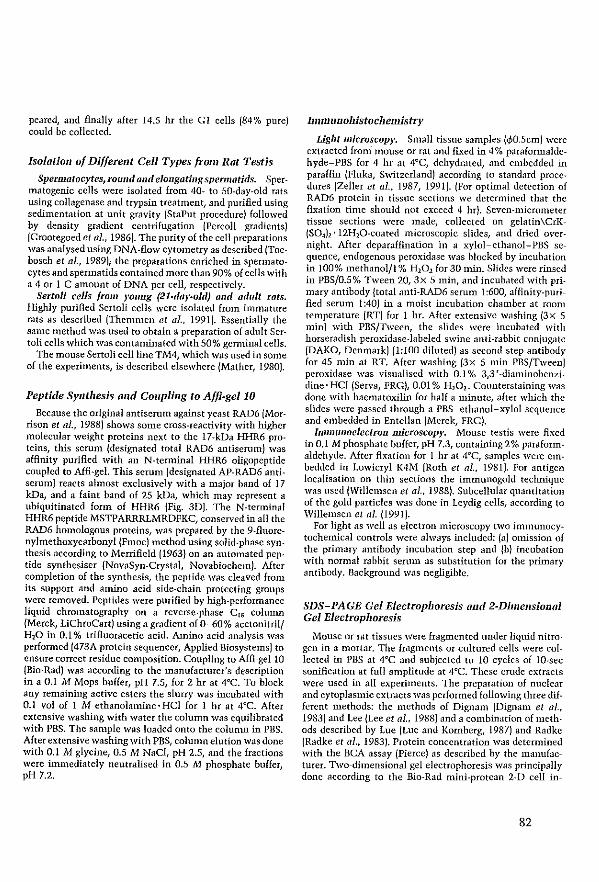
peared, and finaH}' after 14.5 hr the GI ceUs (84% pure) could he collected.
Isolotion of Different Cell Types {rom Rat Testis
Spermatocytes, rOlllld and eJongatÎllg spemlOtids. Spermatogenic ceUs were isoJated from 40- to 50-day-old rats using collagenase and trypsin treatment, and purified using sedimentation at unit gravÎty (StaPut procedure) followed by density gradient centrifugation (Percoll gradients) (Grootegoed et al., 1986). The purity of the ceU preparations was analysed using DNA-flow eytometry as described (Toebosch et al., 1989); the preparations enriched in spennatocytes and spennatids eontained more than 90% of ceUs with a 4 or 1 C amount of DNA per ceIl, respectively_
Sertoli cells trom yOllng (21-day-oJd) mul adllit rats. Highly purified Sertoli cells were isolated from immature rats as described (Themmen et al., 1991). EssentiaUy the same method was used to obtain a preparation of adult SertoH eeUs which was contaminated with 50% genninal cells.
The mouse Sertoli ceilline TM4, which was used in same of the experiments, is described elscwhere (Mather, 1980).
Peptide SYlltllesis mul COllpling to Affi-gel 10
Because the original antiserum against yeast RAD6 (Morrison et al., 1988) shows same cross-reactivity with higher molecular weight proteins next to the 17-kDa HHR6 proteins, trus scrum (designated total RAD6 antiserum) was afflnity purified with an N-terminal HHR6 oligopeptide coupled to Affi-gel. This serum (designated AP-RAD6 antiserum) reaets almost exclllsivcly with a major band of 17 kDa, and a faint band of 25 kDa, whieh may represent a ubiquitinated farm of HHR6 (Fig. 30). The N-terminal HHR6 peptide MSTPARRRLMRDFKC, conserved in all the RAD6 homologous proteins, was prepared by the 9-fluorenylmethoxycarbonyl (Fmoc) method using solid-phase synthesis according to Merrificld (1963) on an automated peptide synthesiser (NovaSyn-Crystat NovabiochemJ. Af ter completion of the synthesis, the peptide was cleaved from its support and amino acid side-ehain protecting grollps were removed. Peptides were purified by high-performance liquid chromatography on a reverse-phase CIS coluum (Merek, LiChroCart) using a gradient of 0-60% acetonitril/ H20 in 0.1 % trifluoracetie acid. Amino acid analysis was perfomled (473A protcîn sequencer, Applied Bios}'stems) to ensure correct residue composition. Coupling to Affi-gellO (Bio-Rad) was according to the manufacturer's description in a 0.1 M Mops buffer, pH 7.5, for 2 hr at 4°C. To block any remaining aetive esters the slurry was ineubated with 0.1 vol of 1 M ethanolamine·HCI for 1 hr at 4°C. Af ter extensÎvc washing with water the column was equilibrated with PBS. Thc sample was loaded onto the eolumn in PBS. Afterextensive washingwith PBS, column elution was done with 0.1 M glycine, 0.5 M NaCl, pH 2.5, and the fractions were immcdiately ncutralised in 0.5 M phosphate buffer, pH 7.2.
IIllIUllnollistocl1emistry
Light microscopy. SmaU tissue samples !.pO.5em) were extracted from mouse or rat and fixed in 4% pamfonnaldehyde-PBS for 4 hr at 4°C, dehydrated, and embedded in paraffin (Fluka, Switzerland) according to standard procedures (Zeiler et al., 1987, 1991). (Por optimal detection of RAD6 proteÎn in tissue sections we determined that the fixation time should not exceed 4 hr). Seven-micrometer tissue sections were made, coUected on gelatin\CrK{S04h' 12I-hO-coated microscopie slides, and dricd O\'ernight. Alter deparaffmation in a xylol-ethanol-PBS se~ queIlce, endogenous peroxidase was blocked by ineubation in 100% methanolfl % HlOl for 30 min. Slidcs were rinsed in PBS/0.5% Tween 20, 3x 5 min, and incubated with primary antibody (total allti-RAD6 semm 1:600, affinity-purified serum 1:40) in a moist incubation chamber at room temperature (RT) for 1 hr. Altcr extensive washing (3x 5 min) with PES/Tween, the slides were incubated with horseradish peroxidase-Iabeled swine anti-rabbit conjugate (OAKO, Denmark) (l:lOO diluted) as second step antibody for 45 min at RT. After washing (3x 5 min PBS/Tween) peroxidase was visualised ",ith 0.1 % 3,3'-diaminobenzidine' HCI {Serva, FRGJ, 0.01 % H101. Counterstaining was done with haematoxilin for half a minute, af ter wh.ich the slides were passed through a PBS-ethanol-xylol scquence and embedded in EnteUan (Merek, FRG).
lnllnunoelectron microscopy. Mouse testis were fixed in 0.1 M phosphate buffer, pH 7.3, contain.Îng 2% pamformaldehyde. Af ter fixation for 1 hr at 4°C, samples were embedded in Low.icryl K4M (Roth et al., 1981). For antigen localisation on thin sections the immunogold teclmique was used (Willemsen et al., 1988). Subcellular quantitation of the gold particles was done in Leydig celIs, according to Willemsen et al. (1991).
For light as weIl as electron microscopy two immunocytochem.ical eontrols were alw;lys included: (a) omission of the primary antibody incubation step aod lb) incubation with normal rabbit senlln as substitution for the primary antibody. Background was negligible.
SDS-PAGE Gel Electrophoresis and 2-Dimellsional Gel ElectroplJOresis
Mouse or rat tissues were fragmented under liquid nitro· gen in 11 lllortar. The frllgmcnts or cultured cells were collccted in PBS at 4°C and subjected to 10 cycles of lO-sec sonifieation at ful! amplitude at 4°C. These crode extracts were llsed in all experiments. The prcpamtion of nuclear and eytoplasmic extracts was performed foUowing three different methods: the methods of Dignam (Oignam et al., 1983) and Lee (Lee et al., 1988) and a combination of methods described by Lue (Luc and Kornberg, 1987) <Ind Radke (Radke et lil., 1983). Protein concentmtion was detennilled with the BCA assay {PierceJ as described by the manufacturer. Two-dimensional gel electrophoresis was principally done according to the Bio-Rad mini-protcan 2-0 ceil in-
82
struction lllanual and as described by Luider et al. (1992). Gels with a length of 6.5 cm were prepan":d in glass tubes 2-mm diameter. Next, 0.25 mi Bio-lyte 3j5 and 0.25 mI Biolyte 4/6 amphoIytcs (Bio-Rad), 2 mIlO% Triton X-lOO, 5.5 g urea (Merck), 1.33 mI acrylamide solution (28.3% acrylamide and 1.62% piperazinediacrylamide (Bio-Rad)), 1.97 mI distilled water, 10 pI 10% ammonium persulfate (BioRad), and 10 pI N,N,N',N'-tetramethylethylenediamine (Bio-Rad) were mixed and allowed to polymerise in the glass tubes at 3 rc for about an hour. The gels were covered with overlay buffer (9 M urca, 0.8% Bio·lyte 3/5, 0.2% Bio-Iyte 5/7, bromophenol blue) and preelectrophoresis was perfOflllCd at 200, 300, 400 V for 10, 20, and 20 min, respectively. As upper chamber buffer a 20 mM NaOH solution was used, whcreas 100 mM H3P04 served as Iower chamber buffer.
Subsequently, samples were diluted with an equal volume of sample buffer{2% SDS, 10% glycerol, 62.5 mM Trisj HCI, pH 6.8, bromophenol blue, 0.1 % dithiotreitol(DIT)), boiled for 3 min, and chilled on ice. Samples were diluted with an equal volume of lysis buffer (0_5 M urea, 2% Triton X-IOO, 0.1 % DIT, 1.6% Bio-Iyte 3/5,0.4% Bio-lyte 5/7 in distilled water). One hundred lllicroliters containing 50 pg protein was loaded under 40 plof 1:1 diluted overlay buffer and electrophoresis was performed for 3.5 hr at 600 V.
The gels were gently removed from the glass tubes and equilibratcd against sample buffer for about 90 min untU the pH indicator in the acid part of the tube gel became blue. The tubes \Vere directly loaded on a 2.25-mlll-thick 15% SDS-polyacrylamide rninigel (Bio-Rad) and electrophorcsis was perfofllled with 0.1 M sodium acetate in the anode buffer {Christy et al., 1989J at 100 V (stacking) and subsequently ISO V until the bromophenol blue reached the bOttOlll of the gel. Blotting was done onto O.4S-pm polyvinylidenedifluoride (PVDF) membranes (Millipore), according to the manufacturer's description, using eleetrotransfer. Blots were blocked in nonfat milk for 1 hr and inclIbated with the primary antibody diluted in nonfat milk (anti-yeast RAD6, 1;2000 or AP-RAD6 antiserum, 1;100) at 4°C, ovemight. Af ter extensive washing with PBS/O.S% Tween 20, the second antibody (goat anti-rabbit antibodies, alkaline phosphatase labeled (TAGO, Inc.)) was ineubated for 1 hr at 4°C, in a 1:1000 dilution in PBS/Tween. Af ter severd additional wasmngs with PBS/Tween the antigenantibody complexes were visualised with the staining method described by Blake (Blake et al., 1984). Carhamylate carbonic anhydrase (CA) (Pharmacia lab.) and 2-D SDSPAGE standards (Bio-Rad) were used as standards for isoelectrofocusing_ Prestained protein molecular weight markers (Gibco\BRL) \Vere used in the second dimension.
For normal SDS-polyacrylamide gel electrophoresis l.S· mm-thick 11 or 15% gels were used with a 4% stacking gel. About 20 pg protein extract was loaded, and electrophoresis was perfonned with 0.1 M sodium acetate in the anode buffer (Christy et al., 1989) at 50 V (stacking) and ISO V (nmningJ. Blotting, antibody incubations, and staining were done as described above.
83
In VHro Transcription Olul in Vitro Translation
The construct H2S"c, eontaining a I.7-kb cDNA of human HHR6A {Koken et al., 1991bJ, was lincarised with Xbal. The H28m construct differs from the previously rcported HHR6A sequence in that at nucleotide position 146 {Koken et al., 1991bJ, a mutation derived from cDNAamplifkation in Esc1wricbia coli or as peR artefact (resuIting in the reported guanine) has been changed into the genomically encoded adenine, resulting in a change of the previously reported glycine into aspartic acid {amino acid POSttion 49J. pPHB2 containing an 0.8-kb EcoRI fragment, harbouring the complete open reading frame of HHR6B (Koken et al., 1991b), was linearised with BomHI. In vitro transcription as well as in vitro translation were done exactly as described (Promega, 1991).
RESULTS
UV·Induced Expressioll
In yeast, RAD6 transcription is induccd upon UV irradiation (psixfold) within 30 min after exposure (Madura et al., 1990). To investigatc whether this feature of RAD6 is evolutionarily conserved, Northern blot analysis for HHR6A and HHR6B was performed on RNA from exponentially growing cells, UV irradiated with 1,5, or 10 J/rn1
, and harvested af ter different incuhation periods. Thc bIot was subsequently probed with the cDNA probes for HHR6A (1.7-kb Sa1I fragment) and HHR6B (0.8-kb EcoRI fragment) (Koken et al., 1991bJ, the UV-inducible collagenase or methallotheionin na genes (Angel et al., 1986) (positive control for UV induction), and GAPDH jBenham et al., 1984J (included as an internal standard for quantitation). HeLa cells, primary human fibroblasts, and primary foreskin-derived keratinocytes were investigated. The latter cell types arc also subjected to UV irradiation in the body and are therefore the most relevant targets to study UV induction_ Figure IA shows the result of a "physiological" dose of 1 J/m1 UV irradiation on primary fibroblasts. When correctcd for the slight variation in RNA amounts per lane {sec GAPDH hybridisation} it appears that irradiatioll did not result in a significant increase of human HHR6A {0.8 and 1. 7 kb} and HHR6B (1.2 and 4.4 kb) mRNAs, under conditions where collagenase (Fig. IA) and metallotheionin (not shown) displayed dear induction. Also experiments using other UV doses, time courses, and cells did not result in detectable induction (data not shown}. To examinc the possibility that HHR6A and 68 may be induced at thc protcin (translational) leyel instead of the mRNA levcllike in yeast, protein expression was studied using affinity-purified anti-yeast RAD6 antisemm. Westem analysis after 10 J/m2 UV irradiation of HeLa cells (Fig. !B) failed to reveal a significant increase in the amount of the 17-kDa HHR6 proteins (for details on the antisemm see below, aod under Materials and MethodsJ. We conclude that in contrast to S. cerevisil1cneithcr of the human genes is inducible by UV.
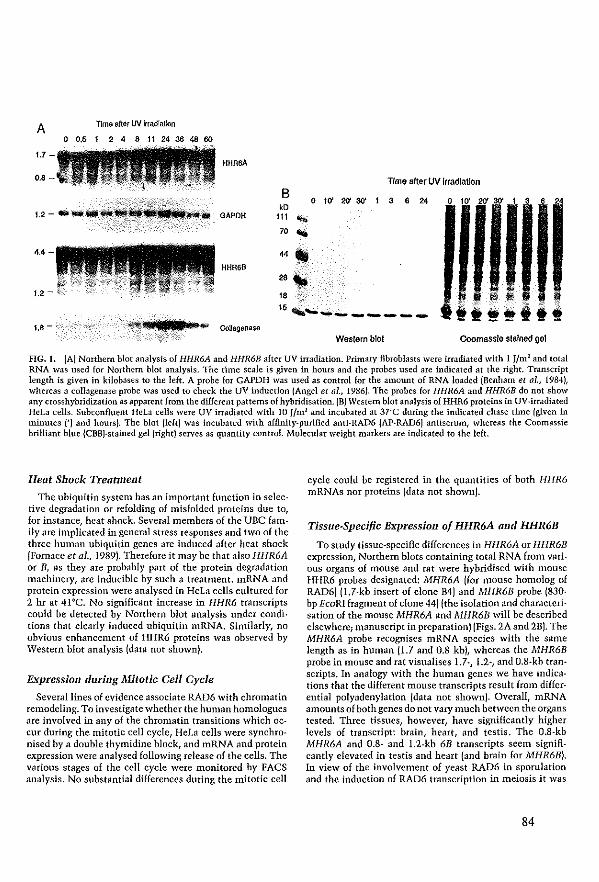
A Tlma after uv Irrad'atlon
o 0.5 2 4 8 11 24 36 48 60
1.7 HHR6Ä
0.' Time after W Jrrad!atlon
B 0 1~ 20' 30'
kD 1 3 6 2' o 10' 20' 30' 1 3 6 24
111 ... 70 ... 44 ..
HHR6B
28'" " 15,~ ______ _
1.8 -
Western blot Coomassle stalned gel
FIG. I. (A) Northem blot anal>'sis of HHR6A and HHR6B after UV irradiation. Primary flbroblasts were irmdiated with I Jlrn1 and total RNA was used for Northem b10t analysis. Thc time seale is given in hours and the probes used are indicated at the right. Transcript length is given in kilobases to the left. A probc for GAPDH was used as wntrol for the aillount of RNA loadcd (Benham et al., 1984), whereas acollagenase probe was used to check the uv induction (Angel et al., 1986). Thc probes for llHR6A and HHR6B do not show anr crosshybridization as apparent from the different pattems of hybridisatioll. (HJ Western blot analysis of HHR6 proteins in UV-irradiated Hela cells. Subçonfluent HeLa cells were UV irradiatCd with 10 J/rn l and inçubated at 3rC during the indiçated chase time (given in minutes (') and haurs). The bIot (IeEt) was inçubatcd with afBnity-purifled anti-RAD6 (AP-RAD6) antiserum, whereas the Coomassie brilliant blue (CBB)-stained gel (right) serves as quantity çontrol. Molecular weight markers are indicated to the left.
Heat Shock TreatmeI1t
The ubiquitin system has an important function in selective degradation or refolding of misfolded proteins due to, for instanee, heat shock. Several members of the UBe family are implicated in general stress responses and two of the three human ubiquitin genes are indueed af ter heat shoek jFornaee et al., 1989). Therefore it may be that also HHR6A or B, as they are probably part of the protein degradation machinery, arc inducible by sueh a treatment. mRNA and protein expression werc analysed in Hela eells cultured for 2 lu at 41"C, No significant increase in HHR6 transcripts eould be detected by Northern blot analysis under conditions that clearly induced ubiquitin mRNA. SiOlilarly, no obvious enhancement of HHR6 proteins was observed by Western blat analysis jdata not shown).
Expressioll duriI1g ltfitotic Gel1 Cycle
Severallines of evidence associate RAD6 with chromatin remodeling. Ta investigate whether the human homologues are involved in any of the ehromatin transitions whieh occm during the mitotic celI cyele, HeLa eells were synchronised by a double thymidine bloek, and mRNA and protein expression were analysed fol1owing release of the ceUs. The various stages of thc eell cyele were monitorcd by FACS analysis. No substantial differenees during the mitotic eeH
cyele could be registered in the quantitics of both HHR6 mRNAs nor proteîns (data not ShOWll).
Tissue·Speci/ic Expression of HHR6A and HHR6B
To study tissue-specific differcnccs in HHR6A or HHR6B expression, Northern hlots containing total RNA from various organs of mouse and rat were hybridised with mouse HHR6 probes designated: MHR6A (for nlOuse homolog of RAD6) j1.7-kb insert of clone B4) and MHR6B probe j830-bp EcoRI fragment of clone 441 (the isolation and charactcrisation of the mousc MHR6A and MHR6B will be described e1sewherc; manuscript in preparationl !Figs. 2A and 2BI. The MHR6A probe reeognises mRNA species with the same lcngth as in human (l.7 and 0.8 kb), whereas the MHR6B probe in mouse and rat visualises 1.7·, 1.2·, and O.8·kb transcripts. In analogy with the human genes we have indicaHons that the different mouse transcripts rcsult from diffcrential polyadenylation (data not shown). Overall, mRNA amounts of both genes do not vary much betwcen the organs tested. Three tissues, however, have significantly higher levels of transcript: brain, heart, and testis. Thc O.8-kb MHR6A and 0.8- and 1.2-kb 6B transeripts seem significandy elevated in testis and heart fand brain for MHR6B). In view of the involvement of yeast RAD6 in sporulation and thc induction of RAD6 transcription in meiosis it was
84
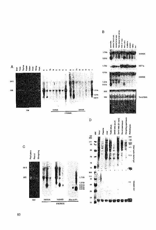
0.
c I
~ r ~
1> ~ Et ~
28S
18S ,
0" HIiRSA
85
,-f '" b "'" +
HHRSB
PROBES:
D
"" 200
111 .... 70 ... .. • " ...
B
t.71ill
O.8kb
1.7kb
4.4kb
1.2'b O,8kt
283
183
F
"" '"
I, " .1 15 -
J 1.7 kb
" t. 1.2 kb 0.8 kb 0.6 kb 0.5ko
, , 200
" 111 ,0j 70 .. 'J ... ".
HHR6A
l1li1 ~,n'H hEF1.
HHR6B
Tola/RNA
~ ~
• ! E , • • " 1 ~ •
,~ ;:: ""
i .. '" ,%; i. ,
HHR6B Mouse PI
PROBES: :: t •• • ......
of interest to investigate the HHR6 expression in testis in more detail.
Testis-Specific Expression of HHR6 111RNAs
The mammalian testis is organised in tubular struetures surrounded by perîtubu1ar myoid eells, hormone producing Leydig eells, and blood vessels (for a general review see Johnson and Everitt, 1984). In the tubules Sertoli cells support the developing genn eells_ Complieated processes of differentiation, growth, and mitotic and meiotic divisions take place when spermatogonia develop into spermatozoa. Afterseveral mitotic divisions the spermatogoniadjfferentiate and give rise to primary spermatocytes. These cells undergo meiotic divisions which convert them first into sec~ ondary spermatocytes and then into round spermatids. Subsequent differentiatîon results in elongated spermatids and finally spermatozoa, whieh have lost most of their eytoplasm and possess highly compacted DNA. Dramatic changes in chromatin composition aceur throughout spermatogenesis, starting with synthesis of testis-specifie histones in spennatocytes and culminating in the total replacement of histones by transition proteins and lIltimately by protamines (Bueci et a1., 1982; Smith el al., 1992).
Ta examine HHR6 expression during this process, germ cells and Sertoli cells of rat testis were isolated and tested for HHR6 mRNA and protein expression (Figs. 2B-2D). RNA from testes of 21-day-old rats, in which spermatogenesis is not yet complete and only a small number of round spermatids is present, did not contain detectable amounts of the abundant testis-specific O.8-kb transcripts (Fig. 2BI. Ta determine when these transcripts appear dllring spermatogenesis, HHR6 expression was fol1owed in testis of young rats during the first S8 days of postnatallife. lnerease of 0.8-kb transcripts takes place around Days 28-34 af ter birth of the rat, at the time when round and elongating spermatids become abundant (data not shownJ. Figure 2C shows that the highly expressed O_S-kb mRNAs of both HHR6A and HHR6B are found mainly in round spermatids with a low level of expression in elongating spennatids_ As a control the elongating spemlatids are shown to contain protamine 1 mRNA. The 1.2-kb HHR6B transcript is clevatcd in Scrtoli
eells (Fig. 2BI, and bath the 1.2- and the 1.7-kb mRNA species in paehytene spermatocytes (Fig. 2Cj, whereas they are absent in the haploid cell types.
For several other genes, the observation has been made that although significant amounts of mRNA are present in the testis no corresponding protein can be detected. It is speeulated that these transcripts result from dysregulated gene expression due to the cxtensive chromatin remodeling taking place in spermatogenesis (reviewed by Ivell, 1992). To examine whether both HHR6 mRNAs in testis are translated into protein, Western blot analysis was perfonned. Total ceU extracts of different tissues and of various cell types were incubated whh affinity-purified anti-yeast RAD6 antibody (AP-RAD6 antibody). This antiserum detects exc1usively a major protein band of 17 kDa, the calculated moleclllar weight of HHR6A/B {Koken et al., 1991bJ, in addition to a faint band of 25 kDa, which may rcpresent a ubiquitinated form of HHR6 (Figs. 2D and 3J. HHR6 quantities var)' from tissue to tissue; reticulocytes and adult Sertoli cells contain high amounts, whereas immature Sertoli eeUs and Iiver harbour re1atively smaU qllantities. (Compare also the amounts of protein loaded in eaeh lane, Fig. SD, upper panel.) HHR6 proteins arc also clearly detected in rollnd and elongating spermatids.
IdeJltification of HHR6A and HHR6B in TotaI Protein Extracts and Detemlinatioll of Re/ative Amoullts
The two highly homologous and similar-sizcd HHR6 proteins are not separated in one dimension {Fig. 2DI. To verify that both 17-kDa proteins are synthesised and to determine theÎr relative abundance in total Hela cell extracts, 2-D gel electrophoresis foUowed by immunodetection with the polyclonal anti-yeast RAD6 serum was applied. Figllre 3 (top panel, arrowheads) shows the presenee of two 17-kDa polypeptides visualised by the tota1 anti-yeast RAD6 antiserum. (The other dots present on the immunoblot probably represent other El enzymes recogniscd by the polyclonal anti-yeast RAD6 antiserum.) To establish whieh of the 17-kDa spots represents human HHR6A (calculated pI 4.91 (Skoog and Wichman, 1986JJ or HHR6B (calculated pI 4.76),
FIG. 2. (A) Expression of HHR6A and HHR6B mRNAs in mouse tissues. The tissues used arc inrlicated (ahhreviated) ahove the panels. Left panel: Tota! RNA gel to serve as eontro! for the amount of RNA loadcd. Thc two right panels: autor'ldiograms ohtained af ter hybridisation with the probes indicatcd. (BI Expression of HHR6A and HHR6B mRNAs in testiClllar cells aod tissues of the rat. The probes used are indicated to the right. The slained tota! RNA gel and hybridisation with a human elongation factor 1 u IhEPu) probe (BTands et al., 1986) serve as quaotity controls. The length of the different transcripts is indicated 10 the left.l!so!ated and cultured) Sertoli cclIs and testis were isolated from 21-day-old rats and spennatids hom 40- 10 50-day-old rats; all other tissues from adult animals.lC! Northern b!ot analysis of HHR6A and HHR6R during the later stages of rat spermatogenesis_ A blot containing total RNA isolated from different germ-line ceBs was hybridiscd with thc indicatcd probes. The cthidium bromide~stained gel serves as quantity control, whercas hybridisation with a mouse prolamine I gene probe Imouse PI) was used to check the purity of the cell fTactions used (Hecht, 1986). The transcript lcngth (kb) is indicated to the light. (0) Expression of HHR6 proteins in different human, rabbit, and rat tissues. A Western blol containing lOta! protein extraets of rat, human (placenta/HeLa), and rabbil Ireticulocyte lysate) cells or tissues was incubated with AP-RAD6 antiserum. Top; the CBB-stainerl gel; boltom: Western blol. Tissues are indicated above the CBB-stained geL The molecular weight marker (MW) is indicated 10 the left.
86
the Hela cell extract was mixed with [3SS]methioninc_la_ helcd HHR6A or HHR6B ohtained through i11 ~,itro tmnslaHon (Fig, 3, bottom four panels), The left spot comigrates with the i11 ~'itto-translated J5S_labeled HHR6A, whereas the protein at the right coincides with thc position of 35S_ labeled HHR6B. In support of this identification is our observation that the right dot (HHR6B) migrates at a slightly higher molecular wcight than the lelt dot {HHR6A). This is consistent with thc calculated molccular weight for bath protcins (HHR6A 17.243, HHR6B 17.312), Thc two proteins
1 Weslemblot
Autoradiogram .. "'. I •
+"S-HHR6A
1st~
-, I I .
pH4
_CA
Westemblot
Autofadfogram
+ "S-HHR68
FIG. 3. Identification of HHR6A and HHR6B in total celI extucts by 2-dimensional gel electrophoresis. Top: Tot,l! Hela extract was lractionatcd by 2-dimensional gel electrophoresis. From !eft to right the Rrst dimension: isoelectrofocusing over a gradient from pH 6 to 4, and from top to bottom: sizc·fractîonation by SDS-PAGE. The boxed "2nd" indicates the molecular weight marker for the second dimcnsion given in kDa. C.A. marks the position of the carbamy!ated marker (Carbonic anhydrasc, Bio-Rad), used as slandard in IEF. The smaU l7-kDa band to the lef! of HHR6A and HHR6B (arrows) is a gel artefact whieh is sometimcs encountered. BottOlll 4 panels: Mixing experiment to prove thc HHR6A and IlliR6B idcntity. Tota! Hcla extract was mixed with PIS]methioninc·labeled in vitlO translatcd HHR6A jleftl or HHR6B RNA (right) and separJted in 2-dimensions. The gels werc blotted onto PVDF membranes, and HHR6A and HHR6B were visualised with anti-yeast RAD6 antihodies (not afflnity purified) jWestcm blot]. The blolS were subsequently exposed to Kodak XAR5 film, and the rcsulting autoradiogram is shown in Ihe lower panels. Thc arrows indieate HHR6A (left two panels) and HHR6B fright two panels]. Thc anowhead indieatcs a form of HHR6A or HHR6B pro· tein wbich was formed in thc reticulocyte !ysate, and which ma)' represent a ubiqllitinatcd form of HHR6: calculated pI 5.26 IHHR6A+ubiquitin] and pI 5.10 {HHR6B+lIbiquitin] (Amold and Gevers, 1990). The !ower molecu!ar wcight spots on thc autoradio· gram probab!y represent breakdown products or are derived hom incomplete synthesis of the HHR6 proteins.
87
HHR6"
Hela eaIJs
Uver
RaUc. Lysale
Tesl1s
pi (BasJc)
+ (Addie)
A 8
-
pi (BaSIc)
+ (Addie)
A 8
-
----~---.
Sertoticells
Round Sperma~ds
Elongating Spermatlds
Spermatozoa
FIG.... HHR6A and HHR6B protein expression in different tis· sues. 2-Dimcnsional gel electrophoresis was perforrned to scparJte the two human HHR6 proteins in different human (HeLl!placenta), rabbit jrcticuloeyte lysate), and rat cells Ol tissues {young Sertoli ceUs, adult liver, testis, round spermatids, elongating spennatids, and spermatozoa]. (A minor contamination with some elongating spermatids lor c)'toplasntic fragments thereof) cannot he com· pletel)' exeluded in the spennatozoa lraction.] The eeU or tissue SOUIce is indieated to the rigilt and lelt. The direction of the pH gradicnt is given above thc figure.
seem to behave on gel according to their ealculated isoelectric points as judged by cOlllparison wHh 2-D SDS-PAGE standards (Bio-Rad) and earbamylated carbonic anhydrase {PharmaciaJ. Minor madiflcations, sneh as phosphorylation or aeetylation, whieh shift proteins in a pH gradient, are not entircly excIuded. Prcliminary i11 \'ivo phosphorylation experiments, howcver, do not provide indieations that a significant fraction of onc of the HHR6 proteins is phosphorylated.
Two·dimensional gels werc a180 used to examine the tissuc-specific expression of both HHR6 proteins. Figure 4 shows that the ratios between HHR6A and HHR6B proteins may vary significantly between different eells and tissues. Hela celIs have more HHR6A than HHR6B, total testis harbours equal amounts, whereas placenta eontaulS more of the B protein. Bath HHR6 proteins are present in round and elongating spermaticls, and trace amounts of protein may be detected even in nlt epididymal spermatozoa.
(Subcellular) Localisation of HHR6A mld HHR6B The affinity-purified antibodies were also used far tissue
scctions of different mouse organs. eells with a clear,
mainly nuclear, staining reaction could be deteetcd with this antiserum in all tissues examined {data not shownJ. In view of the RNA expressîon (Fig. 2) and the induetion of yeast RAD6 during mcîotic reeombination, mouse testis was studied in more detail. In testis sections from 21-dayaId miee only a weak reaetion with the antibod}' was visible (data not shown). However, spccîfically in adult testis {Fig. SDI strong positive cells were observed. Round spermatids whieh eontain high amounts of HHR6A and HHR6B tmnscripts (Fig. 2) display a positive antibody staining /arrowheadsJ, whereas clongating spennatids do not show a visible reaction.Some stailling mayalso be present in the primary spermatocytes. Somewhat to our surprise, the strongest signal was deteeted in adult Sertoli cells (Fig. SDI. In addition, the Leydig eeUs in the interstitial tissue showed staining IFig.5D}.
The immunohistochemieal analysis of tissue sections examined by light mieroseopy strongly suggests that the HHR6 proteins arc localised in the nucleus. To independently confirm and further extend this intracellular localisation, immunoelectron microseopy was used, applying different nxation and embcdding conditions. Thin sections of mouse testis tissue were incubated with antiserum, and the reaction was visualiscd by iuullunogold labeling. As shown by Fig. SA most HHR6 can be found in the Sertoli cell nucleus, whereas eytoplasm and the chamcteristie nucleolus (Fig. SB) are almost devaid of gold particles. Leydig cells and to a somewhat lesser ex tent ronnd spermatids (Fig. SC) present a similar picture, dlthough, like in light microscopy, the amount of protein deteeted is lower. Table 1 shows that the proteins arc strongly enriched in the euchromatin regions of the nucleus. Heterochromatin and nucleolus are markedly devoid of gold particles. Similar observations \Vere made in ather cell types. The Sertoli cell in Fig. SA may even provide evidence for clustering of gold particles in certain restricted areas of the nucleus. The Lowicryl-embedding procedure allows only the direct surface to reaet and therefore avoids penetratioil and accessibility problems. Thercfore a quantitative analysis of gold particles is permitted. The subnuclear localisation of the protein in Leydig cells was determined by counting the grains in the ûifferent eompartments. In these cells the distinction between cu- and heterochromatin edn be made easHy in these cells whieh display relatively high levels of RAD6 protcins (Fig. 50).
DISCUSSION
The present report concerns a systematie analysis of the expression of two closely related HHR6 genes at the RNA and protcin leve1. Our findings are relevant in two direetions; the involvement of repair genes in the uv response in higher organisms and the reIationship of HHR6 with lllodification of chroma tin stmcture in the process of spermatogenesis.
In recent years, cellular responses to environmental geno-
taxie stresses have gained increasing interest. Exposure of cells tG DNA injur)' triggers a cascade of reactions, including intricate signal transduction pathways resulting in altered expression of nllIllerous genes. A universal response to inHicted gene damage in normal cells is arrest of cell eycle progression. This gives DNA repair meehanisms time to remove the DNA lesions before they can give rise to pemlanent mutations. In lower organisms several repair systems have been shown to he inducible by genotoxic agents. The SOS response, controlled by the LexA/RecA regulon, mediatcs a rapid activation of the main eompanents of the nucleotide excision repair in E. coH In yeast a specific snbset of repair genes including RAD6 is damageinduced (for a review sec Hoeijmakers, 1993a,b). Among the numerous nlllmmalian damage-inducible gent:'s analysed to date, however, no known repair genes have been recognised cxcept for the 06G-methyl transfemse. On the other hand, several members of the ubiquitin pathway ineluding ubiquitin itself appeared to be induced upon \'ariOliS stress trcatments (for a review see Fomace, 1992). The availability of a human wpair gene, whose UV inducibility in yeast is weIl. documented, pemlitted for the first time investigations regarding to what extent the involvement of repair in this response is conserved in eukaryotie evolution. Careful analysis in a variety of human celIs and utilizing different UV ûoses failed to provide indications for any significant UV illdllcibility of HHR6A nor HHR6B. This suggests that the DNA repair component of the UV responses in yeast and man are different. Om recent findings with another repair gene, RAD23, whose transcription is enhanced after UV irradiation in yeast, but not in man, conflrm this idea {Madura and Prakash, 1990, and m:mnseript in preparationJ.
These findings support the notion that a fundilmental differenee exist in this regard between the unicellular organisms E. coU and yeast, and mammais. Thc relatively constant environment in which mammalian cells live may have obviated the need for a UV repair response.
Like with UV, we failed to find an induction of HHR6 RNA or protein upon heat shock and during the Illitotie eeIl cyele. Instead Northern and imlllunoblot analysis demonstrate that both genes arc eonstitlltivcly expressed. These results arc consistent with a fnnetion of HHR6 that does not involve thc general stress response {whieh seems to require the ubiquitin-conjugatillg enzymes, DECI, 4, and SJ nor cell cycle-related processes (in whieh UBC3 and 9 are thought to he implicated) {Ciechanover, 1994J.
The two veT}' similar 17-kDa HHR6 protcins were separated by 2-dimensional geI electrophoresis and visualised by Western blot analysis. Mixing with in \'itro-Iabelcd HHR6A or B protein permitted identification of eaeh of the gene products. The results suggest that-except for a possihle ubiquitination-no extensive posttranslational modifications detectahle by 2-D gel eleetrophoresis oceur in a significant fraetion of the HHR6 molecules. Overall, thc expression pattcms of HHR6A and B ressemble each other, and Oll this basis wc cannot assign aspecific distinct functioll to each of the genes.
88
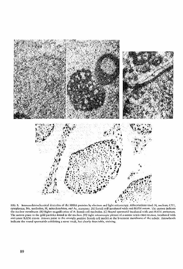
, "9
$. . .' .fi~ . ..... #If!(""~ _,~.'~ ri /.,11."," I'"
A.4t,' ',.~ :'4"~! .' " *'lL~<r,. ;lJf
'\ •• ~' "'~ .. ;,,;,"~. j , ~f,/:~~ " ' ~;. ' ;4~:,f'''~:~ , ". *
FIG. 5. lmmunohistochcmical detection of the HHR6 proteins by electron and light microscopy. Abhreviations used: Nt nucleus; eYT, cytoplasma) Nu, nucleolus; M, mitochondrion; and Ac, acrosome. (A) Sertoli eeli incubated with anti-RAD6 serum, Thc <lrrows indicatc the nuclear membmlle. (BJ Higher magnificat ion of A: Scrtoli cell nucleolus. (C) Round spermatid incubatcd with anti-RAD6 antiserum. Thc aIIOW5 point to the gold particles laund in the nucleus. (0) Light microscopie picture of a mouse testis cross scction, incuhated with anti-yeast RAD6 scrum. Arrows point 10 the strongly positive Sertoli eeU nuclei at thc basement memhrane of the tubulc. Arrowheads indicatc the round spcrmatids exhibiting a more weak, hut clearly deteçtable, staining.
89
TABLE 1
Quantitation of HHR6 Protein in Lcydig eells
Total no. of gold surface No. of gold
particles counted parlicles Factor of counted pm1 per pmz enrichment
Euchromatin 1820 87.1 20.9 21 Heterochromatin 43 14.0 3.1 3 Nucleolus 8 7.86 1.0
Hawever, dear quantitative differences exist for the mammalian RAD6 proteins and mRNAs between the cells and tissues examined. Brain, heart, and testis show elevated levels of specific mRNAs ·when campared to the ather tissues tested. Yeast RAD6 has been shown ta be meiotically induced, cainciding with the time when meiotic recombination takes place [Madura et a1., 1990). However, we find high amounts of shorter mammalian RAD6 transeripts specifically in postmeiotic eells, i.e., round and elongating spermatids, whereas paehytene spermatoeytes have levels below our detection. This renders aspecific involvement in meiotic recombination less likely. The analysis presented here shows that the increase in mRNA and protein quantities of both RAD6 homalagues during spermatagenesis co· incides with the developmental stage when somatic and testis-speciflc histones are remaved from the chromatin and replaced by transition proteins and subsequently protamines jKistler et aL, 1987}. The inductioll of the twa flflR6 genes as representatives of the ubiquitin pathway at this time of spermatagenesis is not without precedent. The chieken ubiquitin II gene is induced at approximately the same stage of spermatogenesis jMezquita and Mezquita, 1991; Rocamora and Agell, 1990) and increased ubiquitillation of histOlle H2A is observed fOliva and Dixon, 1991}. A ubiquitill-activating enzyme El encoded by the Y·chromosomal Sb}' gene exhibits testis·specific expression [Mitchell et al., 1991}. This gene and the X-chromosomal hom01og Sbx show postmeiotic transcription in the mause (Hendriksen et a1., 1995). Interestingly, recently another E2 enzyme !E217w) was also found to be high1y expressed in rat testis [Wing and Jain, 1995}. These observations together with the i11 vitro demanstration that RAD6 is able to ubiquitinate histones !fentsch et al., 1987; Sung et aL, 1988!, provide indirect evidence for involvement of ubiquitin and HHR6 in the chromatin remodeling processes during spermatogenesis. The idea that HHR6 plays a role in restructuring chromatin mayaIso fUIther pertain to the present flnding of very high anlOunts of HHR6 in retieulocyte Iysates (see Fig. 4), since these cells have undergone extensive chromatin modification prior ta nudear elimination. FinaUy, a con· served role of RAD6 in gross structural alterations of chra· matin is consistent with the observation that RAD6 as weIl as rhp6+ are essentiaI for sporulation (Morrison et a1., 1988; Reynolds et aL, 1990) again a process known to involve
dras tic chromosomal changes. In this respect spem1atogenesis may represent an advanced phenocopy of the process of sporulation.
The intracellular location of the HHR6 proteins in the euchromatic part of the nucleus as indicated in this study by immunoelectron mieroscopy is consistent with a funetion in chroma tin transactions. In the immunogold labeling experiments we have used the Lowieryl methad which. allows only the direct surface to react. This eliminates possible artefacts inherent ta some immunohistochemical procedures that are caused by unequal accessibility of antigenic detem1inants in different locations [Posthuma et al., 1987}. Biochemica1 cell fraetiollation studies (sec Materials and Methods for details on the procedure) suggest that a substantial fraetian of the HHR6 proteins is only weakly associated with the euchroOlatin.
High transcript levels of bath HHR6 gencs are found in round spermatids at a stage more than 2 weeks after the fonl1ation of thc heteroehromatic sex vesicle which becames visible in early pachytene spermatoeytes (Stefanini et aL, 1974). Since the human alld the mouse HHR6A gene are located on the X cluomosame (Koken et aL, 1992), it is remarkable that the total amount of HHR6A transcripts seems to even increase during the transition of pachytene spermatocytes into round spennatids. This findinghas been described clsewhere in more detailfHendriksen et al., 1995) and is explained by postmciotie transcription. Hence, HHR6A is likcly to represent one of the few recently discovered examples of XjY·chromosomal genes whose gene produets may play an important role in postmeiotic stages of spermatogenesis [reviewed in Hendriksen et aL, 1995). Western blot analysis confirmed that bath HHR6 protcins are indeed present in the germ celIs. This was important to verify, sillce it has been found for other genes that significant quantitics of testis-specific, shorter transcripts accumulate in spennatids, but that no corresponding proteins can be detected (Cape1 et al., 1993; Ivell, 19921. It has been speculated that these transcripts could result from dysreguIated gene expression due to the extensive chromatin remodeling events that take place in this stage of spermato· genesis (Ivell, 1992). It cannot entirely be ruled out that the presence of m·IR6A protein in spermatids results from an extremely long protein half·life following synthesis in earlier stages of spennatogenesis. The HHR6B mRNA is transIated more efficiently or the protein is more stabie than HflR6A as may be concluded from the larger amount of HHR6B in e10ngating spermatids [Fig. 4J. The transcription of the flHR6A gene in spennatids, togetherwith the notion that different ratios of the A and B protcins are present in different cells and tissues, suggests that bath proteins have aspecific task whieh cannot be completcly taken over by the very homologous counterpart. Selective inactivation of either one or both of these genes, e.g., by gene targeting in totipotent mouse ES cells from whieh mutant miee straius can be obtained, shou1d reveal what is the specific role of each of the proteins and to whieh extent their functÎons overlap. Very recendy, we have succeeded in generating
90
HHR6B knockout mouse mutants (H.P.R., unpublished resuitsj. The phenotype of these mice comprises spccific defccts in spcrmatogcnesis that are compietcly consistent with the above ideas. This finding underlines the significance of the observations described here. Thus the roie of this ubiquitin-conjlIgating enzyme in spermatogcnesis in mammals may be an advanced phenocopy of the involvement of yeast RAD6 in sporulation.
ACKNOWLEDGMENTS
We thank Drs. L. anu S. FC;lkash for the generous gift of thc RAD6 polyelonal antiscrum, Drs. F. I. Bcnham for the GAPDH probe, P. Henlieh for the probes for collagenase and metallothcionin, T. Butt for ubiquitin probes, L Laird for thc human elongation factor la gene, and D. Meijer for the mouse protamine 1 probe. Wc arc very thankful for thc help of the following colleagues: Dr. Theo Luider for help with the 2·dimensional gel electrophoresis, Ton Verkerk for FACS analysis, Dr. Clallde Backendorf anu Li Bin Ma for some of RNA samples of UV-irradiated celIs, Dr. André Hoogeveen for preparation of synthetic oligopcptides, Wim Vermeulen for microinjection experiments, Maarten Fomerod for the protein samples of cell cyele synchronisation experimcnts, Drs. Carel Mcij· ers, A.."e} Themmen, and Leen Blok for helphll discussions, and Jan van KL.lVeren for helpful discussions and excellent technical assistance. Dr. D. Bootsma is acknowledged for critical reading of the manuscript and continuous support. We are gC;lteful to .Mirko Kuit anli Tom de Vries Lentsch for photography, and Sjozef van Baal for computer assistance. This work was supported by the Dutch Cancer Society (Project IKR 88-2, 90-20 and 92-118) anu the Europcan Community (Contract BI6.141-NL).
REFERENCES
Angel, p., Poting, A., Mallick, U., Ramsdorf, H. J., SchoIp, M., and Herrlich, P. (1986). Induction of metallotheionein and other mRNA species by carcinogens and tumor promoters in prinIary human skin flbroblasts. Mol. Gen BioJ. 6, 1760-1766.
Amold, J. E., and Ge\'ers, W. (1990). Auto-ubiquitination of uhiquitin-acth'atÎng cnzymes from chicken breilst muscIe. Biochem.,. 267,751-757.
Bachmair, A., Finic}', 0., aud Varshavsky, A. (1986). In vivo ruIflife of a protein is a function of its amino-tenninal residue. Scienee 234, 179-186.
Benham, F. J., Hougkînson, S., and Davies, K. E. (1984). A glyceraldehydc-3·phosphate dehydrogenasc pseudogcne on the short arm of thc human X chromosome deflnes a multigene family. BAlBQ J. 3, 2635-2640.
Blake, M. S., Johnston, K. H., Russell-Iones, G. I., ;lnd GOlschlich, E. C. (1984). A upid, sensitive mcthod for dctection of alkaline phosphatase-conjugated anti-antibod>' on western blots. AndJ. Hiochem. 136, 175-179.
Branus, J. H. G. M., M;lassen, J. A., Van Hemerl, F. J., Alllons, R., anu Moller, W. (1986). The primM)' stmcture of thc a-subunit of human e!oug;ltion factor J. EUI. f. Biochem. 155, 167-171.
Bucci, 1. R., Broek, W. A., and MeistIÎeh, M. 1. (1982). DistributÎon anu synthesis of histone 1 subfraetions during spennatogenesis in the rat. EAp. Cello Res. 1·10, 111-118.
Capel, B., Swaiu, A., Nicolis, S., Hacker, A., \Valter, M., Koopman,
91
P., Gooufellow, P., and Lovell-Badge, R. (1993). Circular transcripts of the testis·detem1ining gene sry in adult mouse testis. CeU?3,1019-1OJO.
Christy, K. G., iaTart, D. B., and Ostcrhoudt, H. W. (1989). ModiRcalions for SDS-PAGE of proteins. Biowchniques 7, 692-693.
Ciechanover, A. (1994). The ubiquitin·proteasome protcolytic pathway. Gell 79, 13-21.
Dignam, J. 0., Lebovitz, R. M., and Roeder, R. G. (1983). Accurate transcription initiation by RNA polymerase II in a 50luble extract from isolated mammalian nuclei. Nucleic Acids Res. 11, 1475-1489.
Dohmen, R. J., Madura, K., BarteI, B., and Varshavsky, A. (1991). The N-end rule is meuiated by the unC2(RAD6) ubiquitin-eonjugilling enzyme. Proc. Natl. Acad. Sci. USA 88, 7351-7355.
Hnley, 0., and Chau, V. 0991). Ubiquitination. Annu. Rev. Cello Biol. 7,25-69.
Fomace, A. J. (1992). Mammalian genes induced by raUiation: Activation of genes associated with growth control. AlillU. Rev. Genet. 26, S07 -526.
Fomace, A. J., Alamo, 1., Hollander, hl ... c., and Lamorcaux, E. (1989). Ubiquitin mRNA is a major stress-induced transcript in mammalian celIs. Nucleic Acids Res. 17, 1215-1230.
Foumey, R. M., Miyakoshi, J., DJY, R. S., and Patcrsou, M. C. 11988). Northem Blotting: Effident RNA staining anu Transfer. FOCUS 10, 5-7.
Galavazi, G., Schenk, H., and Bootsma, 0.11966). SynchroIlÎzation of mammalian cells in VitlO by inhibition of the DNA synthesis I: Optimal conditions. Exp. Cel1 Re·s. 41,428-437.
Gibbs, S., Lohmann, F., Teubel, W. J., Van der Putte, P., anu BackendorE, C. 11990). Char,lcterization of thc human spr2 promoter: Induction aIter UV irrJdiation of TPA Ircatment and regulation during differentiation of cultured primary keratinocytes. Nucleic Acids Res. 18,4401-4407.
Grootegoed, J. A., lansen, R., and Van der Molen, H. J. (1986). Effect of glucose all ATP dephosphorylation in rat spennatids. f. RepIOd. Fertil. 77,99-107.
Hecht, N. B. {I9861. Molecular and ceJlular cndocrinology of the testis. 111 "Molecular and Cellular Endocrinology of the Testis" IM. Stefanini, M. Conti, R. Geremia, and E. Ziparo, Eds.), pp. 199-213. Elsevier, Amsterdam.
Hendriksen, P. J. M., Hoogerbrugge, J. W., Themmcn, A. P. N., Koken, M. H. M., Hocijmakers, J. H. J., Oostra, B. A., Van der Lende, T., and Grootegoed, J. A. (1995). Fostrneiotic trall5cription of X and Y chromosome gencs during spermatogenesis in the mouse. De\'. Bio1. 170,730-733.
Hcrshko, A., and CiechanoveI, A. !I992). Tbe ubiquitin s)'stem fOI protein degradatîcn. Al1lll!. Rtl\'. BioehenJ. 761-807.
Hoeijmakers, J. H. J.1I993a). Nucleotide exeision repau Ij hom E. cali to yeast. Trends Genet. 9, 173-177.
Hoeijmakers, J. H. I· (1993b). Nucleotide excision repair IJ; from yeast to marnmals. Trends Genet. 9,211-217.
Ivcll, R. (1992). "All that glisters is not gold" Common testisgcne tr,lllscripts are not always what thcy seem. Int. J. Andro1. IS, 85-92.
Jentsch, S. (1992). The uhiqlIitin·conjugation system. Allllll. Rev. Genet. 26, 179-207.
Jentsch, S., McGrath, I. P., and Varshavsky, A. (1987). The yeast DNA Iepair gene RAD6 ellcodes a ubiquitin-conjugating enzyllle. Nature 329,131-134.
Jentsch, S., Sellfert, W., Sommer, T., aud Reins, H. A. (1990J. Ubiquitin-conjugating enzymes: Novel regulators of eukaryotic ceiIs. Trends Biochem. Sci. IS, 195-198.
Johnson, M. H., ;md Everitt, B. J. (1984). "E.ssential Reproduction." Blackwell Sci., Oxford, London, Edinborough, Boston.
KistIcr, W. P., Heidaran, M. A, Cole, K.O., Kandala, J. c., and Showman, R. M. {l9871- lil "Cell Biology of the Testis and Epidid· ymis" (B. J. D. M. C. OrgcbinCrist, Ed.), pp. 102-111. The New York Academy of Sciences, New York.
Koken, M. H. M., Reynolds, P., Bootsma, 0., Hocijmakcrs, J. H. J., prakash, S., and Prakash, L. (1991a). DhI6, a DIOsophila humolog of the yeast DNA.repair gene RAD6. Ptoc. NatJ. Acad. Sci. USA 88, 3832-3836.
Koken, M. H. M., Reynolds, P., raspers· Dekker, I., prakash, L., Pra· kash, S., Bootsma, D., and HOeijmakers, J. H. J. (199Ih). Struetural andfunetional conservation of two human homologs of the yeast DNA repair gene RAD6. PIOC. Nati. Acad. ser USA 88, 8865-8869.
Koken, M. H. M., Smit, E. M., Jaspers·Dekker, I., 005tra, B. A., Hagemeijer, A., Bootsma, 0., and Hoeijmakers, J. H. J. (l992). loealization of two human homologs, HHR6A and HHR6B, of the yeast DNA repair gene RAD6 to chTOmosomcs Xq24·q25 and 5q23·q31. Genomics 12, 447-453.
lawrencc, C. (19941. The RAD6 DNA repair pathway in Saccharo· myces cerevisiae: What does it do, and how does it do hl BioEs· says 16,253-258.
lee, K. A W., Bindereif, A, and Green, M. R. (1988). A smalI· seale procedure for preparation of nucleJr extracts that Sl.lpport eincient transcription and pre·mRNA splicing. Gelle Alla/. Tech. 5,22-31.
lue, N. F., and Komberg, R. D. (19871- Accurate initiation at RNA polymerJse II promoters in extracts from SaccllOromyces cere~'is· iae. Proc. Natl. Acad. Sc/. USA 84, 8839-8843.
luider, T. M., Peters·van der Sanden, M. J. H., Molenaar, ,. c., Tibboel, 0., van der Kamp, A. W. M., and Mcijers, J. H. C. (1992). Char,lcteruation of HNK-l antigens during Ionnation of the avian enteric nerveus system. Dewlopment 115, 561-572.
Madura, K., and Prakash, S. (1990). Transcript levels of S. ccrevisiae DNA repair gene RAD23 inere.He in response to UV light ,md in meiosis but remain constant during the mitetie ceU cycle. Nucleic Acids Res. IS, 4737-4742.
Madura, K., Prakash, S., and prakash, L. /1990). Expression of the Saccharom}'ces cerevisiae DNA repair gene RAD6 that encodes a ubiquitin conjugating enzyme, increases in response to DNA damage and in meiosis hut remains constant during the mitotie ceU cycle. Nucleic Acids Re-s. 18, 771-778.
Mathcr, J. P. (1980). EstJblishment Jnd eb.nacterizJtion of two distinct mouse testicularepithelial celllines. Biol. Reprod. 23,243-252.
MerriBeld, R. B. /1963). Solid phase peptide synthesis. The synthe· sis of J tetrapeptide. f. !lm. Cbem. Soc. 85,2149-2154.
MezquÎta, J., and Mezquita, C. (1991). Ch.n'lelerization of a ehicken polyubiquitin gene preferentially expressed during spermatogen· esis. FEBS Lett. 279, 69-72.
IVlitchell, 11. '., Woods, D. R., Tueker, P. K., Opp, J. S., and Bishop, C. E. (1991). Homology of J cJndidale spemlatogenie gene from the mouse Y chromosame to the ubiquitin.activating enzyme El. Nature 354, 483-486.
Morrison, A., Miller, E. J., and Prakash, L. (1988). Domain structure and funetionJI analysis of tbe carbm."yl.terminal polyacidic sequence ol the RAD6 pwtein of Saccharomyces cerevisiae. Mol. Cell. Riol. 8, 1179-1185.
Oliva, R., and Dixan, G. H. (1991). Vcrtehr,lte protamine genes and the histone.to.pwtamine replacement reaction. Progr. Nucleic Acid Res. Mol. Riol. 40, 25-94.
Posthuma, G., Slot, J. W., and Geuze, H. J. (1987). usefulness of thc immunogold teehnique in quantitation of a soluble protein in ultm·thin seetions.l. Histocbem. Cytochem. 35, 405.
Promega (1991). Promega protocols and applications guide. Madi· son, \VI.
Radkc, K., Carter, V. c., Moss, P., Dehazya, P., Schliwa, M., and Martin, G. S. {l983J. Membrane association of a 36,OOD·dalton suhstrate lor tyrosine phosphorylation in chicken embryo nbro· blasts transfomled by avian sarcoma viruses. ,. Gell Biol. 97, 1601-1611.
Rechstciner, M. (1988). "Ubiquitin." Plenum, New YOlk. Reynolds, P., Koken, M. H. M., Hoeijmakers, J. H. J., PrJkash, S.,
and prakash, L.{l9901. The rhp6+ gene ol SchizosaccllllIOmyces pombe: A structural and functional homolog of the RAD6 gene from the distantly rebted yeast Saccharomyces cerevisiae. EMBO f. 9, 1423-1430.
Rcynolds, P., Weber, S., and Prakash, L.fI9851. RAD6 gene of Sac· cbaroIllyces. cerevisiae emodes a protein contJining a tuet of 13 consecutive aspartates. Proc. NatI. Acad. Sci. USA 8l, 168-172.
Rocamora, N., and Agcll, N. {l990). Methylation of ehick Uhl and UblI polyubiquitin gems and their differcntial expression during spermatogenesis. Biochelll. f. 267, 821-829.
Roth, T., BendJ)'an, M., Carlemalm, E., Villigcr, W., and Garavito, M.fI981). Enhancement of stmctural preservation and immuno· cytochemical staining in low temperature embedded panere.Hic tissue. I. Histochem. Cytochem. 29,663-671.
Sambrook, J., Fritsch, E. F., and Maniatis, T. [1989). "Molecular Cloning: A laboratory Manua!." Cold Spring Harbor Labomtory Press, Cold Spring Harbor, NY.
Skoog, B., and Wichman, A. /1986). CaJculation of the isoeIectric points of polypeptides from the amine acid composition. Trends Ana!. Chelll. 5, 82-83.
Smitb, F. P., Tres, L. L., and KierszenhJum, A. L.fI992). Expression of testis·specilk histone genes during the development of mt sperIllatogenic cells in vHro. Del'. Dynam. 193,49-57.
Stefanilli, M., De Martino, c., D'Agostino, A., Agrestini, A., and Monesi, V. {l974). NucieolJr activity of rat pIÎIllary spermato· cyles. Exp. eeIl Re5. 86, 166-170.
Sung, P., Berleth, E., Pickalt, c., Prakash, S., and Prakash, L. (1991). Yeast RAD6 eneoded ubiquitin conjugating cnzynle mediates pIOtein degradation dependent on the N.end.recognizing E3 en· zyme. EMBO I. 10,2187-2193.
Sung, P., Prakash, S., and Prakash, L. (1988). The RAD6 protein of Saecharomyces cerevisiae pol)'ubiquitinatcs histones, and its acidic domain mediates this activity. Genes De~'. 2, 1476-1485.
Themmen, A. P. N., Blok, 1. J., Post, M., Baarends, W. M., T.W., H., Pamlenticr, M., Vassart, G., and Grootegoed, r. A. (1991). Follitropin receptor down .regtdation invol yes J eMiP·dependent post·transeriptional decrease of receptor mRNA expressioll. Mol. eell. Elldacrinol. 78, R7-RI3.
Thome, A. W., Sautiere, P., Briand, G., and Crane·Rohinson, C. (1987). The stmcture of ubiqllitinated histone HlS. EMRO J. 6, 1005-1010.
Toebosch, A. M. W., Brusse, R' I Velkerk, A., and Grootegoed, J. A. (1989). QUJntitative cvaluMion of the maintenance and develop· ment of spermatocytes and round spermatids in cultured tubule fr,lgments from immature rat testis. Tmem. ,. Androl. 12,360-374.
Varshavsky, A. (1992). The N·end mie. eell 725-735. Villdelov, L. L., Christenseu, I. J., and Nissen, N. J. (1983). A deter·
gent·trypsin method for the preparation of nuclei lor flow cyto· metric DNA analysis. Cytometry 3, 323-327.
92
Watkins, J. F., Sung, P., Prakash, S., and prakash, L. (1993). The extremely conserved amino tenninus of RAD6 ubiquilin-conjugating enzyme is essential for amino-end rule-dependent protein degradation. Genes Dev. 7, 250-261.
Willemsen, R., Brueken, R., Sorber, C. W. J., Hoogeveen, A. T., Wisselaar, H. A., Van Dongen, J. M., and Reuser, A. J. J. (1991J. A quantitative inlinunoelectronlllicroscopic study on soluble, membrane-associated and melllbranc-bound lysoson1al enzymes in hUlllan intestinal epithelial cells. Histochem. T. 23, 467-47,1
Willemsen, R., Van Dongen, J. M., Aerls, J. M. F. G., Schram, A. W., Tager, J. M., Goudsmit, R., and Reuser, A. J. J. (1988). An immunoelectron microscopic study of glucoeerebrosidase in t}'Pe I Gauscher's discase splcen. UltrostlUct. Pathol. 12,471-478.
93
Wing, S. S., and ]ain, P. (1995). Molecular doning, expression and characterization of a ubiquitin·conjugating enzymc (E2mnl highly expressed in rat testis. Biochem. T. 305, 125-132.
Zeiler, R., Bloeh, K.O., Williams, B. S., Arceci, R. J., and Seidman, C. E. (1987]. Localized expression of the atrial natriurctic factor gene during cardi,IC embryogenesis. GeJU's Dn'. 1,693-698.
Zeiler, R., Rogers, M., aud Watkins, S. (1991 J. Current protocols in molecular biology. 111 "Cunent protocols in i\lolccular Biology" (F. M. Ausubcl, R. Brent, R. E. Kingston, D. D. Moore, J. G. Seidman, J. A. Smith, and K. 5tmhl, Eds.), chapter 14. GrecI}e Publishing Associates &. Wiley-Intersciencc, New York.
Receîved for publication March 21, 1995 Accepted September 16, 1995
Chapter VII
lllactivatioll of t!ze HR6B IIbiqllitill-colljllgatillg DNA repair ellZyllle ill mice callses male sterility associated wit!z c!zromatill modificatioll

CeH, Vol. 86, 799-810, September 8, 1997, Copyright©1996 by Cell Press
Inactivation of the HR6B ubiquitin-conjugating DNA repair enzyme in mice causes male sterility associated with chromatin modification
H.P. Roest', J. van Klaveren', J. de WIt', C.G. van Gurp'. M.H.M. KOkenf ,4, M, Vermey3, J.H. van Rollen2• J.W. Hoogerbrugge2, J.T.M. Vreeburg 2, W.M. Baarends2, D. Bootsma', J.A. Grootegoed2, and J.H.J. Hoellmakers
"•
'MGC-Oepartment of CeU Blo!ogy and GeneticsFaculty of MedÎcÎne and Health Sciences Erasmus University Rotterdam p.e. Box 1738, 3000 OR Rotterdam, The Nelherlands Phone: (+31) 10 4087199 Fax: (+31) 10 4380225, 20epartment of Endocrlnology and Reproduction, and 3Department of Pathology, Faculty of Medicine and Health Sciences, Erasmus University Rotterdam, p.e.Box 1738, 3000 OR Rotterdam, The Netherlands. 4Present address: CNRS UPR90-51 "Palhologle Cellulaire: Aspects Moléculalras at Viraux", Hópital SI. louIs, 1 Avenue Clauda Vettafaux, 75475 Peris Cedex 10, France.
Summary
The ublqultln-conlugatlng yeast enzyme RAD6 and lts strongly conserved human homologs hHR6A and hHR6B, are Impllcated In post replIcatIon repair and damageInduced mutagenesis. The yeast proteln Is also requlred for sporulatlon, end may modulate chromatIn structure via hlstone ublquitlnatlon. We report the phenotype of the flrst anlmal mutant in the ubiqultin pathway: Inactlvation of the h H R 6 Bhomologous gene in mlce causes male Infertillty. Derallment of spermatogenesls becomes over! during the post-melotlc condensatIon of chromatIn In spermatlds. These flndlngs provide a parallel between yeast sporulatlon and mammallan spermatogenesls, and strongly Impllcate hHR6-dependent ublqultlnatlon In chromatin remodelllng. Since heterozygous male mice and even knockout female mi ce are completely normal and able to transmlt the defect, similar hHR6B mutations may cause male InfertllUy In man.
IntroductIon
The ublqulltn systam plays a key role in numerous cellular processes, Includlng metaboltc homeostasls, stress response, organelIe biosynthesls, cel! cyde regulatIon, DNA repak, apoptosis, antigen processing and gene expression (for recent reviews see (Cleehanover, 1994; Hochslrasser, 1995)). This seleetlve, non-Iysosomal proteolylle palhway medlated by the 26S protease complex delermInes the half-lile of cruelal proteins such as p53, cyclins, transcription laclors, and cytosoHe polypeptides (Chau et at., 1989; Clechanover et ar., 1991; Glotzer et at., 1991; Komllzer et ar., 1994; Seulert et al., 1995). Furthermore. Ihls pathway Is ImpHeated in stabHizaUon, relolding, and translocatIon of a diverse range ol proleins. The ubiquilin palhway Involves a thrae or
four step ub!qultln thloester cascade. The hlghly eonserved 76 amlno acid ublqultln molecule Is lirst activated by the ubiquilin-activating enzyme (E1) Ihrough formation ol a high-energy thioester linkage ol lis C-termÎnal glycine wilh a cyslelne residue in Ihe E1 proteln ilsell. Subsequently, the ublquitin molely is translerred 10 a cysteine ol one ol the ubiquitinconjugaling (UBC or E2) enzymes, which in turn attach Ihe ublqullln pari onlo a target proteln wlth or without Ihe asslstance of a ublquitln'proleln 11gase (E3). The latter step may Involve a Ihlrd Ihloester linkage with the E3 enzyme (Scheffner et aL, 1995) and resulls in the formation ol an isopeptide bond of Ihe activaled C-termlnal glycine of ublquilin with the N-Iermlnus or Ihe t-NH2 group of an Interna! lysine resldue of the $ubstrete pOlypeptide. POlyubiquitination is thought to mark prot eins for dagradatlon, whereas mono·ublquitlnatlon may serve other lunctions (Chau el aL, 1989).
A key role in this pathway is exerted by a growing famUy ol UBC enzym es, which In yeast already comprlses 12 membars (Hochstrasser, 1995). An extreme example ol Ihe plelolropie Involvement of Ihls class ol proteins in ceUular processes is presented by the yeasl S.cerevÎsÎa8 RAD6 gene product. Strains mutated In RAD6 display defects in post-replication daughter strand gap repair, causing pronounced sensitivity to alt kinds of DNA damaging agents, Inereased sponlaneous mutatIon frequency and concomitant toss of damage·induced mutagenesis. In addillon, rad6 null elleles exhibil cell cycle perturbanca, tempereluresensitive growth, inabitity to sporulale, and increased relrolransposltlon (see lawrence, 1994). The cruclal fInding concarnlng Ihe funcllon of Ihe prola!n was made by Jentsch et al. (1987), who identified RA06 as Ihe firsl ublquUinconjugatlng enzyme, able 10 mono- and polyubiqultinate hlstones 2A and 2B In Vi/ro, The protein has baen demonslrated 10 mediate N-end ru!e prole!n degradatIon (Dohmen et aL, 1991) and lts hlghly eonserved N-Ierminus is implicated in Inleraction wlth Ihe UBR1 proleln (Madura et aL, 1993). lts in vitro abllity 10 ublqulUnale hlslones has led to Ihe suggesllon thai RAD6 medlates its functions by modulatlng chromatin struclure as an essentia! part ol the DNA Iransactions (repair, replication) In whlch 11 Is Implleated. However, direct demonslratlon of the role of the prolein in chromatIn dynamlcs Is still lacking. In normat mammalian ceUs, a substanIlal fraellon ol histone H2A. and, to alesser extenl, H2B, Is present in a ublquitfnaled form (Goldknopl and Busch, 1980; Thome et al., 1987). Allhough histones are frequently uti!Îzed as subsiraias for ubiquitin-conjugaUon, Ihe lunctlon of ublquitinaHon of histones In vivo Is largely unexplorad, and it is unknown whelher and 10 wha! ex!enl RAD6 Is involved In Ihls posHranslational modification.
Previously, we cloned RAD6 homo!ogs of several higher eukaryotes including two close[y related hu man homologs, de slgnated hHR6A and hHR6B (for human Homologs of RAD6) (Koken el al., 1991b). The
95
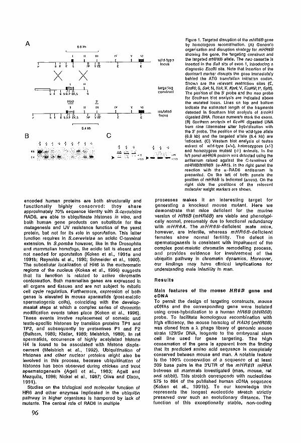
A 6.6 kb
" I I ~ ii , , ,
" X X
"' I I I m "' , , E S,KI' EK,S
IlEO
" '\"I;
I I ii E S,K!' EX.S "
5.4 kb
B
Hf!it1lf
'" I lil m
"-'" I I ii ,~
c
",'
I I ,
" I
.:;;.;.,;....;. ....
" "!" wild·type , locus
largetillg construct
" "!" mutated , locus
S4 ,:~ jIj:,;yiifl! ».....:; :::, >- - -:::,
Figure 1. Targeted disruplion ol Ihe mHR68 gene by homologous recombination. (A) Genomlc organlsallon and disruption slrategy lor mHR68 showing Ihe gene, Ihe largeling construcl and Ihe largeted mHR6B aIleIe. The neo cassette Is lnserted in Ihe Sen site ol exon 1, Introduc!ng a diagnosllc EcoA! site. Note thai insertion ol Iha domlnanl marker disrupls Ihe gene Immedialety behlnd Iha AlG translallon iniliaHon codon. Shown ara tha relevant restriction sites (E, EcoAl; S, SaA; N, NsA; K, Kpr/.; V, EcoR.V; P, SphI). Iha posiHon ol the 3' probe and Ihe neo proba lor Soulhern blol analysls are indicated abova tha mulated locus. Unes on top and bottom Indlcata tha estlmaled Jength of the Iragmants dataclad In Southern blol analysls ol EcoR/ digested DNA. Roman numerals mark tha axons. (B) Soulhern analysls of EcoR/ digested DNA from nlne littermates alter hybridization with Iha 3' probe. Ihe position ol tha wild·type allele (6.8 kb) and Ihe targeted allele (5.4 kb) are Indicaled. (C) Western blot analysls ol testes ax/ract of wild·type (+I+), haterozygous (+/-) and homozygous mulant (./-) animais. In the left panel mHR68 protein was detected uslng tha anUserum ralsed agalnst Ihe C·termlnus ol mHR6BIIlHR6B (u-A81). In Ihe right panel Iha raactlon with Ihe u-RAD6 antiserum Is prasented. On Iha lalt of both panels the position ol mHR6B Is Indicated (aHow). On Ihe right slda the positIons of the relevant molecular weight markers ara shown.
encoded human proteins are bath structurally and functlonally hlghly conserved: they share approximalely 70% sequence Idenlity with S.c8r8vlslae RAD6, are able 10 ublquitinate hlstones In vi/ro, and both human gene produets can suhslitule for Ihe mulagenesls and UV reslslance functIon of Ihe yeast proteln, but nol for ils mIe in sporulalion. This lalter funcUon rsqulres In S,cerevlslae an acldle C·termlnal extension, In S.pombe however, liks In Ihs Drosophl1a and mammalian homo!ogs, Ihe aeldle laU Is absent and not needed lor sporulation (Koken et ar., 1991a and 1991b; Reynolds et alo, 1990; Schneider et al" 1990). The subcellular localizaUon of HR6 In the euchromatic regIons of the nucleus (Koken et al., 1996) suggests th at lts funclion Is related to active chromatfn conformation. Bath mammalian genes are expressed In alt organs and tissues and are nol subjecl la milotic cell cycle regulaHon. Furthermore, expression of bath genes Is elevaled in mouse spermatids (post-meioHc spermalogenic cells), colnelding with Ihe developmenlal sleps al whlch a complex series of chromalÎn modlflcalfon events takes place (Koken et at, 1996). These events Involve replacemenl of somatic and leslfs-specJflc hlstones by transition proteins TPl and TP2, and subsequently by prolamines Pl and P2 (Barhom, 1989; KIstIer, 1989; Meistrieh, 1989). In rat spermatlds, occurrence of highly acetylated hislone H4 Is 'ound la be associaled wilh hislone displacement (Melslrich el aL, 1992). Ublqullfnalfon of hislones and olher nuclear proteins mlght also be Involved in Ihis process, because ublqulUnatlon of hlslones has been observed during chicken and Irout spermalogenesls (AgelI et aL, 1983; Agell and Mezquila, 1988; Nickel el ar., 1987; Oliva and Dlxon, 1991),
Studies on Ihe blological and molecular funcUon of HR6 and other enzymes implicaled in the ubiquilin palhway in higher organlsms Is hampered by lack of mulanls. The central role of RAD6 In multiple
96
processes makes iI an inleresting large 1 for generaUng a knockoul mouse mulant. Here we demonstrale Ih al mlce deflclent for the murine verslon of HR68 {mHR68} are viabIe and phenotyplcally normal, presumably due 10 functional redundancy wllh mHR6A. The mHR68-delicienl male mlce, however, are InfertiIe, whereas mHR68-deficienl females show normal ferillily. The defect in spermatogenesis is consistent wilh impalrment of Ihe complex post-melotic chromalin remodelling process, and provIdes evldence for Involvemenl of the ubiquitin palhway In chromatin dynamics. Moreover, our flndlngs may have clinical Impllcatlons for underslanding male inlertility in man.
Results
Maln features of the mouse HR6B gene and cONA Ta permit Ihe design of largeting conslructs, mouse cDNAs and the correspondlng gene were Isolated using cross-hybridizatlon to a human HR6B (hHR68) probe. Ta lacHJtate homologous recombination wilh high efflclency, the mouse homolog of RAD6 (mHR68) was cloned from a À phage fibrary of genom!o mouse siraln 129/Sv DNA, Isogenlo 10 Ihe embryonal slem cell IIne used lor gene targetlng. The hIgh conservation of the gene Is apparent Irom the finding thai its predieled amino acid sequence is completely conserved belween mouse and man. A nOlable feature Is the 100% conservation ol a sequence ol al least 309 base pairs in Ihe 3'UTR of the mHR68 mRNA belween all mammals investigaled (man, mouse, rat and rabbit). Thls slrelch corresponds with nucleoli des 575 10 884 of the publlshed human cDNA sequence {Koken el al., 1991b}. Ta our knowledge th Is represenls the longesl nucleotide stretch slrictly preserved over such an evolulionary dislance. The function ol thls excepUonally stabIe, non-coding
nucleot!de sequence elemenl is unknown. Figure lA presenls Ihe architeclure of Ihe murine
m H R 6 B gene. The gene spans a region of approximalely 15 kb and Is comprised of six exons. Interestlngly, Ihe locatIon of two inlrons Îs exacUy preserved In Drosophifa and even S. pombe (Koken et al., 1991a; Aeynolds el al., 1990), presumably reflecting a high imporlance for the gene. The gene was mapped on mouse chromosome 13 In a region syntenic wilh human chromosome 5, and evidence was oblained for a pseudogene on mouse chromosome 11 (Aoller et al., 1995).
Inactlvatlon of the mHR6B gene and generatIon of mouse mutants In deslgnlng a knockoul targeling construct, we envlsloned the possibility thai any Iruncaled mHR6B prolein may exert unpredlclable elfecls. Particularly Ihe highly conserved N-termlnus, encoding a site lor proteln-proteln Interaction, could interlere wilh other processes resulting in semi-dominant consequences. Therelore, we chose to inactivale Ihe mHR68 gene Immediately alter Ihe translational slart codon by insertion ol the dominanlse!eclable neomycln or hygromycln marker, ruling out Ihe synlhesls ol any part of Ihe prolein. The largeUng construct depicled In Figure 1A conlains 3.5 kb and 2.8 kb ol homology al Ihe 5' and 3' slde flanking !he dominant-seleclable marker, respectively. Two verslons, each with a dHferent selectable marker, were constructed 10 permit Inactivation ol both autosomally localed alleles In ES cells.
Translecllon of the neo cassette-containing largeling construct (Figure 1A) by electroporation and selection lor sta bIe uplake of Ihe dominant selectable marker gene yielded a frequency ol 16% largeted Iranslormants (27 homologous recombinanls/166 tolal transformanis; no selecHon was applled againsl random integration). Homologous recombinanls were checked tor accurate InlegraUon ol Ihe construct by Soulhern blol aflalysls using exlerna! and inlernal pro bes and were lound to be correct (dala nol shown). The multiple engagemenls ol RAD6 on the one hand and the presence ol a 95% Identical mHA6A proteln on the olher, make 11 dilficuH to predlct a phenolype lor a mHR6Bdeliclent mouse. To find out whelher a homozygous mHR68 inactivaUon Is viabIe, at least at the cellular level, Ihe second aUele was targeted using Ihe hygro casselle-contalnlng construct. The Irequency of largeUng directed 10 Ihe wild-type aUele was 8% (11/143), indlcaling Ihat there was no selection againsl mHR68 InaclivaUon and Ihal inacUvation of both mHR68 alleles Is not lelha!. Therelore, we perlormed injectiofl of ES cells ol Iwo independeni, neomycln-resistanl clones (80 and 134) Inlo biastocysis of C57BU6 mice, resulting In the generation of chlmaeras. Male chimaeras Irom both Independent clones were bred and bolh gave germline Iransmission. Soulhern brot analysis on DNA isolated Irom tal I biopsies was used 10 delermine Ihe genotype of Ihe offsprJng. HybridizatIon with Ihe 3' external probe vlsualized a 6.8 kb EcoAI fragment In Ihe case of a normal allele and a 5.4 kb fragment lor a targeted allele (Figure 1 B). Heterozygotes were interbred and yielded homozygous mHR68 mulants wilh Ihe expecled Mendelian Irequency. The results of a represenlative Huer are shown In Figure 18.
We verWed that Ihe largeling ol mHR68 Indeed resulted In a null-mutatlon al Ihe ANA and proleln
levels. Northern blol analysis confirmed absence of significant amounts ol mHR68 Iranscripis (dala not shown), indicaUng Ihal Ihe presence ol the domInant marker Inlerfered wlth proper transcription and/or processing of Ihe altered mRNA. The mHA6A and mHR68 proteins, like hHR6A and hHR6B, are 95% Identical and migrate al Ihe same molecular welght In SOS-PAGE. To dlsllngulsh batwaen these highly homologous polypepHdes, we look advanlaga of tha fact Ihat wllhln the 14 C-terminal amino acids, Ihe A and 8 produets djffer al 2 posJl/ons. A peptide Idenlical 10 Ihe 14 C·termlnal amlno acids ol HA6B was synlhesized and utlllzed 10 raise a polyclonal anllserum Ihal specifically recognlzes Ihls proleln. Sloce In testis both proleins are expressed In hIgh quanlities, lolal testis exlracls ware analysed. Figure tC (Ieft panel) shows thai no mHA68 prolein is detecled in mHR68-/- mlce, whereas Ihe proleln Is present In mHR68+/- aod mHR68+/+ lillermales. The decrease in Intensity in Ihe lestis exlract of Ihe heterozygous anlmal suggests that Ihese animals conlain roughly approximately half Ihe amount of mHA6B proleln as compared to Ihe normal animaIs. Thls argues against upregulatlon ol the unlargeled allale to compensate for the loss of expression ol the largeled copy. An antiserum againsl yeasl RAD6, recognlzlng both HABA and HR6B (Koken el al., 1996), shows a positive reaction In the mHA6B'/- sample, indicating thaI the mHR6A gene is expressed (Figure 1C, right panel). These results verily the nuH stalus ol Ihe mHR68 mulation and also show Ihal mHA6A prolein is still present.
Phenotyple eharaeterlstles of mHR6B-/- mlee and ce lis The mHR68-1- mlce proved normally viabIe with a lifespan exceeding 14 monlhs. Excepl for Ihe feature dlscussed below, no apparent phenolypical or pathological abnormalilies were lound. Furlhermore, no differences were noled belween Ihe main phenolypic characterist!cs ol Ihe mHR68-1- mice derived Irom the independenUy largeted ES recomblnanls and belween mlce from crossings between different slrains (129xFV8/J, 129x C57BU6). This rules out Ihe possibilily thai by acctdent other genetic alteraUons had occurred Ihal mighl influence Ihe phenotype or Ihal Ihe genetic background Is of major importance. Since AAD6 in yeast accounts for much of Ihe cellular resislance agalnsl a wide speclrum of genoloxlc agenls, we lnvesligated UV aod y-ray sensitivily in mouse cells. To lest UV sensitivity, mouse embryonic fibroblast cell !ines were established Irom mHR68+1+, +1-, and -I- mice and tesled for Iheir cellular survival, as measured by [3HI-thymidine incorporallon, after irradiation w!th different doses ol UV. For y-ray sensillvlty Ihe doublelargeted ES cell line was irradialed and cloning efficIency was compared wilh irradlated, nontargeled ES cells. No differences belween mHR6B·deflclent and -proficlenl cells were observed for Ihese DNA damaging agenls (dala nol shown). Thus no overl defect in DNA repair was delected. Thls Is possibly caused by a redundant effect ol a lunclional mHR6A gene.
Spermatogenesls In mHR6B-deficlent mlce In breedlng experimenls, lt soon became apparent that Ihe mHR68·1- male mice were conslslently intertife. Copulatory behaviour was judged 10 be normal,
97
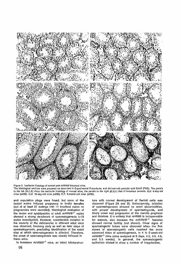
Figure 2. Testicular hlstology of normat and mHR6B knockout mlce. The hlstologlcal secUolls were prepared as descrlbed In Experimental Procedures, and stalned with periodic acid Schiff (PAS). The p:lI1els to the lelt (A,e,E) show the lesticular hlslology of norma! m!CXl, Ihe panels 10 the right (B,O,F) thai of knockoul animaIs. A.B: a·day-old mlCXl (x400): C,D: 4o-day·old mIca (x200); E,F: g-month-old mIca (x200).
and copulation plugs were lound, but none of Ihe tested males induced pregnancy in lerWe lemales (out ol at least 27 malings with 11 knockoul ma1es no pregnancJes were recorded). Histologlca1 evaluation of the testes and epldidymldes ol adult mHR6B·j- males showed astrong derallmenl of spermatogenesis (> 10 males Investigated). However, conslderable varlalion in the severily ol Ihe deficiencies in different adult mlce was observed, Involving early as weil as laler steps of spermalogenesis, precluding Identification of the exact slep at which spermatogenesls Is affected. Therelore, the onsel ol spermalogenesls was closely 10110wed in these mlce.
In Immature mHR6B-j- mlce, an intact lubularslruc-
98
lure with normal development ol Sefloli cells was observed (Figure 2A and B). Subsequently, in!!iatlon of spermalogenesis showed no over! abnormal!tles, with proper development of sparmalogonia, and timely onsel and progression ol Ihe meiolIe prophase and divisions. It is unlikely that mHR6B is indispensable lor meiosis, a[so because Ihe mHR6B-j- lemales showed normal lerlillty (not shown). Clear signs of spermatogenlc tallure were observed when Ihe flrst waves ol spermatogenic ce lis reached the more advanced sleps of spermiogenesis, In 4 10 5-week-old mHR6B·j· mice (mlce analysed al 8 days, 2.5, 3.5, 4.5, and 5.5 weeks). In general, Ihe spermatogenlc epilhellum slarted to show a number of trregufarities,
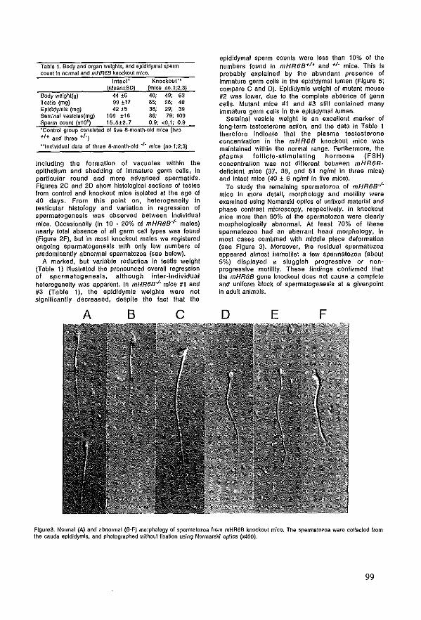
Tabls 1. Body anti organ weighls, and epJdidymal sparm counlln norrnaJ and mHR6B knockout mica.
Intact' Knockout .. (Mean±SD) (mJce no. 1 ;2;3)
Body welght{g) 44 ±6 40; 49; 63 Testis (mg) 99 ±17 55; 26; 48 Eplcfldym!s (mg) 42 i5 38; 29; 39 Semlnal veslclss(mg) 109 ±16 86; 79; 109 Sperm count (xt06) 15.5±2.7 0.9; -:0.1; 0.9 'Contro! group conslsted of flve 8·month·old mlce (r.vo +1+ and three +/')
"Individual data of three 8-month·o!d .,. mice (no.l;2;3)
Includlng Ihe formatIon of vacuoles wilhln Ihe epithelium and shedding of Immature germ cells, In parlJcular round and more advanced spermaUds. Figures 2C and 20 show histo!ogical sacllons of testes from contral and knockout mica isolaled at Ihe age of 40 days. From Ihls point on, heterogenelty in tesUcular hlstology and variation in regres sion of spermalogenesis was observed between Individual mlce. OccasionaHy (in 10 - 20% of mHR6B-/- males) nearly lolal absence of all germ cell types was found (Figure 2F), bul in most knockout malss we regislered ongolng sparmatogenesis with only low numbers of predomlnanUy abnormal spermatozoa (see below).
A marked, but variabIe reduction In testis weight (Tabla 1) Illustrated Ihe pronounced overall regression of spermalogenesis, allhough Inler·lndividual helerogeneity was apparent. In mHR68-/' mice #1 and #3 (Tabie 1), the epldldymls welghls were not signrflcantly decreased, despite Ihe fact thai Ihe
A B c
epldidymal sperm counts were less Ihan 10% of the numbers found in mHR68+1+ and +/- mlce. Thls Is probably explained by Ihe abundant presence of Immature germ ceUs in the epididymal lumen (Figure 5; compare C and 0). Epldidymis welght of mutant mouse #2 was lower, due 10 the complete absence of germ ceUs. Mutant mlce #1 and #3 still contalned many immature germ ceUs In the epldldymal lumen_
Seminal vesicle welght is an excellent marker of long-term testosterane aCllon, and Ihe data In Table 1 therefore indlcate Ih al the plasma testosterone concenlration In Ihe mHR68 knockout mice was malntained within Ihe normal range. Furthermore, the plasma fOllicle-sllmulallng hormone (FSH) concenlraUon was not different between mHR6B· deficient mice (37, 38, and 51 nglml in three mice) and inlact mice (40 ± 6 nglml in five mice).
To study Ihe remalnlng spermatozoa of mHR68-/mice In more detail, morphology and molillty were examlned uslng Nomarski optfcs of unfixed material and phase contrast mlcroscopy, respeclively. In knockout mice more Ihan 90% of Ihe spermatozoa were c!early morphologicaUy abnorma!. At leasl 70% of these spermatozoa had an aberrant head morphology, In most cases comblnad wllh middle piece deformalJon (see Figure 3). Moreover, the residual spermatozoa appeared almosl Immotile: a few spermatozoa {about 5%} displayed a slugglsh progressfve or nonprogresslve molJlity. These flndings confirmed Ihal Ihe mHR68 gene knockoul does not cause a complete and uniform block of spermalogenesls al a givenpoint in adult animaIs.
o E F
Flgure3. Norrna! (A) and abnormal (B-F) ffiOrphology ol spermatozoa from mHR6B knockout micEl. Ths spermatozoa warEl col1ecled from ths cauda epldidymls, and photographed without lixalion uslng Normarski opties (x400)_
99
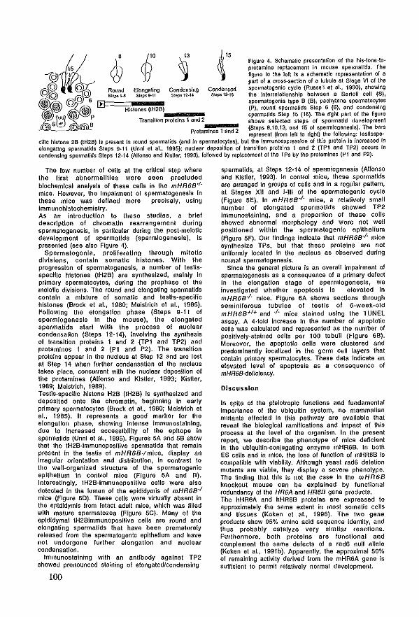
Round Elonga\Ïng Steps 9-11
Condenslng St-ep512·14
Condensed St"9.1~16
Figure 4. SchematIc presentation of Iha hls·lone-Icprotamine replacemant in mousa spermallds. Ths flgure 10 Iha lelt Is a schamaUc representation of a part of a cross-section of a tubule at Stage VI cf Ihe spermaloganic cyele (Russeli et aL, 1990), showing Ihs lntetrelalionshlp balween a Sertel! cell (S), spermatogonia type 8 (B), pachytene sparmatocytes
Hislones (IH2B)c-.""!,,,,,!,_~ lP), (ound spermatlds Siep 6 (6), and condenslng • spermatld$ Siep 15 (15). The righl part of lhe figure
Transition proleins 1 end 2 ... ~_~ shows selecled sleps of spermaUd davelopmenl (Steps 8,10,13, and 15 of spermlogenssls). Tha bars
ProlamInes 1 end 2 rapresanl (Irom [elt 10 right) Ihs following: testlsspe-
eme hlstone 28 (tH2B) Is present In tound spermatlds (and In spermaIOl:ytes), but Iha rmmunoa)lprasslon ol thls prolain Is incraased ~n elongaling spermatids Sleps 9·11 (Unnr at al., 1995); nuclaar depositIon ol translUon proteins 1 and 2 (TP1 and TP2) ocrors In
condenslng spermatids Sieps 12·14 (Allonso and KlstIer, 1993), lollowed by rep!acamant ol Ihe TPs by Iha pro!amlnas (P1 and P2).
The low number of ceUs al Ihe critlcal step where the fjrst abnormaHties we re seen precluded blochemical analysls of these ceUs in the mHR68'/mlce. However, Ihs Impairment of spermatogenesis In these mlce was deflned more precisely, using Immunohistoehemlslry. As an intro duel Ion to these studies, a brief descriptIon ol chromalin rearrangemenl durlng spermatogenesis, in part!cular during the post-meiolie development of spermallds {spermiogenesis}, Is presented (see also Figure 4).
Spermalogonla, prolileratlng through mltolic divlsions, contaln somatIc histones. Wilh Ihe progression ol spermalogenesls, a number ol lestlsspecIfIc histones (IH2B) are synthesized, mainly In prlmary spermatocyles, during the prophase of Ihe melollc divisions. The round and elongaUng spermatids conlaln a mixture of somalic and leslis·specHic histones (Brock el al., 1980; Meislrich el al., 1985). Following the elongation ph ase (Sleps 9-11 ol spermiogenesls In Ihe mouse), Ihe elongated spermatids slarl wlth Ihe process ol nuctear condensation (Steps 12-14), involvlng Ihe synlhesis of IransUton proteins 1 and 2 (TPt and TP2) and protamines 1 and 2 (PI and P2). The transition proleIns appear in Ihs nucleus al Step 12 and are lost at Slep 14 when lurther condensation ol Ihe nucleus takes place, concurrent with Ihe nuclear deposItion ol Ihe protamines (Allonso and KisUer, 1993; Kisller, 1989; Melslrich, 1989). Teslis·speclfic histona H2B (IH2B) is synthesized and deposlted onlo Ihe chromatin, beginning in early primary spermalocytes (Brock et al., 1980; Me!slrich el aL, 1985). n represents a good marker lor Ihe elongatIon phase, showing Intense immunostaining, due 10 increased accessibillty of Ihe epitope In spermatids (Unnl et al., 1995). Figures 5A and SB show that Ihe IH2B·lmmunoposilive spermaUds Ihat remaln presenl In Ihe lestis ol mHR68-/mlee, display an Irregular orienlatlon and distribution, In contrast to Ihe well-organized slruclure of the spermalogenic epithelium In control mice (Figure 5A and B). Inleresllngly, tH2B-lmmunopositive cells were also delecled in the lumen of the epididymls ol mHR68-1
mice (Figure 50). These ceUs were virtually absent In Ihe epidldymis Irom Intacl adult mice, wh1ch was lilled wllh mature spermatozoa (Figure SC). Many ol Ihe epldldymal tH2Blmmunopos!live ce lis are round and elongating spermatids thai have been prematurely released Irom Ihe spermatogenle epithelium and have nol undergone lurlher elongatlon and nuclear condensation.
Immunoslalnlng wilh an anl1body against TP2 showed pronounced slalning of elongatedJcondenslng
100
spermatids, at Steps 12-14 of spermiogenesis (Allonso and Kistter, 1993). In control mice, these spermatids are arranged in groups ol eeUs and in a regular paltem, al Stages XII and '-lil of Ihe spermalogenlc cycle (Figure 5E). In mHR68-1- mlce, a relatively smal! nu mb er of elongated spermatids showed TP2 Immunostalnlng, and a proportion ol these ce lis showed abnormal morphology and were not weil posilloned wlthln Ihe spermatogenic epllhelium (FJgure SF). Our lindings indlcale that mHR68-1- mice syntheslze TPs, bul Ihat Ihese proteins are not unlformly localed in the nucleus as observed during normal spermalogenesls.
Since Ihe general picture Is an overall impairment of spermatogenesis as a consequence of a primary delect in the elongation stage of spermlogenesis, we Investigaled whether apoptosls is elevaled In mHR6B-I- mlce. Figure 6A shows sections through seminlferous tubules of testis ol 6-waek-old mHR6B+I+ and -I· mlce stained using Ihe TUNEL assay. A 4·lold increase In the number ol apoptotic cells was calculaled and represented as Ihe number of poslt1vely-stalned ceUs per 100 tubull (Figure 6B). Moreover, Ihe apoptotic cells were cluslered and predomlnantly locallzed in the germ cell layers Ihal contain primary spermatocyles. These data Indicale an elevaled level ol apoptosls as a consequenee of mHR68-deficlency.
DIscussIon
In spile ol the pleiotropIe lunctions and lundamenlal Importance of the ublquitin system, no mammalian mulants aflected In this palhway are avalIabIe that reveal Ihe biologleal ramificalions and impact ol Ihis process al Ihe level ol the organism. In Ihe present reporl, we describe Ihe phenolype of mice deflclenl In Ihe ublqu1t!n-conjugating enzyma mHR6B. In bolh ES cells and in mlce, Ihe 10ss ol function of mHR6B is compalible wilh viabllily. Although yeast rad6 deletion mutants are viabIe, Ihey display a severe phenolype. The finding thaI this is not the case In Ihe mHR68 knockout mouse can ba explained by funclivnal redundancy ol Ihe HR6A and HR68 gene produels. The hHR6A and hHR6B proteins are expressed to approximalely Ihe same extent in most somaUe ceUs and tissues (Koken el al., 1996). The two gene produets show 95% amlno acid sequence identity, and thus probably calalyze very simIlar reaclions. Furthermore, both proteIns are lunclional and complement the same delecls of a rad6 null allele (Koken et al., 1991b). Apparently, Ihe approxlmal 50% of remaining aetivily derived Irom Ihe mHR6A gene is sufflclenl 10 permil relatively normal developmenL
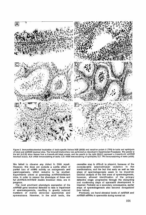
Flgure 5. Immunohlstochemlcal locallzaUon of tesUs.speciflo hlstone H2B (IH2B) and transitIon proteln 2 (TP2) In lestis and epldidymls ol Intact end mHR68 knockoul mIca. Tha Immunohls\ochemlslry was parformad as describad In Exparimenlal Procedures. The panels 10 Ihe lelt (A,e,E) show tissues Irom a g·month-old Intact mousa, and Ihe panels 10 Iha r1ght (B,O,F) represent a g·month-old mHR68 knockout mOUS9. A,B: aH2B ImmunO$lalnlng or tastis; e,o: IH2B Immunoslalnlllg of epldldymls; E,F: TP2 Immunostalnlng or les\ls (x400).
We lalled 10 observe any defecl In DNA repak. However, Ihls does nol exclude a subIIe effecl of parllal loss of mHR6 actlvlly on mulagenesls and carcinogenesis, whlch remalns 10 be studled. Experlmenls almed al generatlng mHR6Adeflcient mlce, In order 10 assess Ihe phenolype ollhese and full mHR6AlmHR6B double knockout mlce, are In progress.
The mosl promlnenl phenolyplc express!on of Ihe mHR6B gene knockoul detecled 10 date is Impairment of spermatogenesis, resulUng In greatly reduced numbers of malnly abnormal spermatlds and spermalozoa. However, In Ihe adult leslis, Ihe
causatlve slep Is difflcult 10 pinpoInt, because of Ihe considerable Inlerlndlvldual variallon In Ihe manifestations, and Ihe facl Ihal early as weil as late steps of spermalogenesls seem 10 be Impalred. Oetal!ed analysls of Ihe first wave of spermatogenesls, however, allowed IdenUflcatlon of the prlmary delective slage: progression Ihrough Ihe elongating and condenslng steps of spermat1d developmenl Is Impalred. Probably as a secondary consequence, earlier steps ol spermatogenesls also become deregulated (see below).
Prevlously, we found e!evaled levels of mHR6A and mHR6B mRNAs In spermatids during narmal rat
lOl
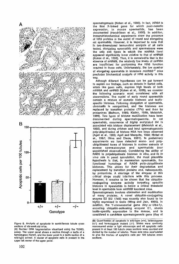
A
B
+/+ +/Genotype
Flgufe 6. Analysls of apopiosis In semlniferous tubule cross secUons of slx-week-old mloo. . (A) Nuclear DNA fragmentatlon visuallzed uslng Ihe TUNEl assay. TM upper panel shows a section through a lestis of a homo~gous mutant, end the lower panel Is a teslis section of a wild-type anima!. A clusler of apoplotic eells Is present In the upper lelt corner of Ihe upper panel.
102
spermalogenesis (Koken el ar., 1996). In fact, HR6A Is the fifS! X-Ilnked gene lor whlch post-malotic expression, In mouse spermatlds, has been documenled (Hendriksen at al.. 1995). In additIon, imrnunohlslochemlcal experiments show Ihe presence of HR6 proteins in Ihe nuclei ol round and elongating rat spermalids. However, n Is Important 10 note thai In two-dimensional Immunoblot analysis of all cells tested, elongating spermatids and spermatozoa were Ihe only cell types In which the mHR6A level appeared slgn!f!cantly ]ower relative to Ihat of mHR6B (Koken el at, 1996). Thus, il Is conceivabJe thai in Ihe absence of mHR6B, the relatively low levels of mHR6A are Insufflclenl for performing Ihe HR6 function requlred In these ceUs. Unfortunalely, the [ow number of elongaling spermaUds in Immature mHR6B'/- mice precludes biochemicai analysis of HR6 activity In this way.
AlIhough different hypotheses can be put forward 10 explaln our findings, such as delecls in Sertotl ce1ls, which IIke germ cells, express high levels ol both mHR6A and mHR6B (Koken et aL, 1996), we consider Ihe following scenario most consislent with all observatIons. The nuclei ol early round spermalids conlain a mixture of somatic hislones and testisspecific hlstones. Following elongation of spermaUds. chromatin Is reorganlzed, and the hlstones are rep[aced by transitlon proteins (TPs) and then by protamines (Balhorn, 1989; Klsller, 1989; Meistrich, 1989). Two types ol histone modifIcation have been documenled during spermalogenesls_ In rat spermatids, occurrence of hlghly acetylaled H4 Is associaled with h!slone displacemenl (Melstrlch et al., 1992), and during chicken and troul spermatogenesls poly·ublquJtlnation of hlstone H2A has been observed (AgelI et aL, 1983; Agell and Mezquita, 1988; Nicke! et al.. 1987; Ollva and Dixon, 1991). In preliminary experimenls we have detected mono- and polyublqultinated forms ol hlstones In nuclear exlracls of mouse spermatocytes and spermatlds (our unpubtlshed observallons). ConsiderIng Ihe abllily ol RAD6 10 polyublqultlnate hislones In vi/ro, and ils in vivo role in yeast sporulalion, Ihe most plauslble hypothesis Is thai. in mammalian spermalids, Ihe lunclional homologs of RAD6 poly-ublquilinate h!stones. This aHows for thelr degradation and replacement by transmon proteins and, subsequently, by protamlnes. A shortage of Ihe enzyme at thls critica I stage could Interlere wlth thls process. However, It remains 10 be shown that the ubiqultinconjugatlng enzyme actlvlty targeUng speclflc histones In spermallds Is, befow a critical threshold level In spermatids from mHR6B knockoul mice. Spermatogenesls Involves elimination and modiricatlon of many proleIns. A novel ubiqulUn-conjugating enzyme E2 (E2 17kB) was recenlly also found to be highly expressed In testis (Wlng and Jain, 1995). In additIon. the Y-chromosomal gene Sbyor Ube1y, encodlng ubiquilin-acllvaling enzyme Ei, shows testis-specUlc expression In Ihe mouse, and Is consldered a candidate spermatogenesls gene (Kay et
(B) Quantilicallon of apopiosis In wild·type (+1+), helerozygous (+I-) and homozygous mutanl (-1-). Slides ware randJmly manoeuvred under a Ilght microscops and all apoplotic ceHs presenl In at laast 100 lubuIa cross sections were counlad and divldad by Ihs numbar of lubulas< These dala ware reca!culated 10 glve Iha number of apop!otlc caHs per 100 lubuias cross secUons.
aL, 1991: Mitchall al aL, 1991). TasUcular axprasslon Is dapendanl on Iha prasance of garm caUs (Mitchall at al., 1991) and Ube1y mRNA has been detected in round spermatlds (Hendriksen et at., 1995). The homologous gene on Ihe X chromosoma (Sbx or Vbe 1 x) Is expressed In all male and female tissues (Milchell at aL, 1991), and also In spermatogenlc eeUs (Handriksen et alo, 1995). Assuming thai disturbance of chromatin remooeting in spermatids of mHR68-daftclent mlce is the primary cause of lhe InfertJltty, how can thls defect laad 10 formatIon of vaeuo[es In Sertoll ceUs and the release of Immature garm ceUs? A clue Is provided by a recent finding on the effect ol ectoplc expression of avlan protamine (galline) in spermatids of transgenlc mlce. This expression induces dlsruptJon ol the norma! dense chromatJn slruclure of spermatozoa, and resulls In Inlertllity (Rhlm et at., 1995). As lor mHR68'/' mlce, the spermatogenlc epithelium of these transgenlc males showed many vacuoles and loss of Immalure germ ce lis. Thus, disruption of chromatIn conformatfon by ectopic prolamIne expression leads to very similar types of spermatogenlc abnormalities as observed In mHR68'/' mlce, In agreement wilh Ihe Idea that HR68 deficiency affects chromatIn conformatIon. Posslbly, Sertoli eeUs are adversely affecled by degenerallng late spermatlds. These spermalids mighl release protamines, whlch are known to exert toxlc effects on epithelial eelis (Peterson and Gruenhaupt, 1992). In eoncordance wJlh Ihis, Ihe clustered apoptosls of primary spermatocytes In lestIs of mHR68-j
- mlce (Flgure 6) pOints 10 local Sertoll eeU damage, It Is not elear why most ol the male mHR68 knockout mlce show productIon of spermatozoa wlth a wlde range ol morphological abnormallUes, Other defects In spermalogenesis affecllng spermatocytes rather Ihan spermallds, can also give rise 10 abnormal spermatozoa, Such a defect In spermalogenes!s was observed In mice Ihal we re mulaled in Ihe DNA mismatch repak gene pMS2 (Baker et al., 1995). Thls defecl resulls In abnormal ehromosomal synapsis in meiosIs and male lnfertilily, wilh production of a smalt number of spermalozoa with abnormal morphology. In a conslderable number of male inferlilily patlenls, the cause of Ihe InferUlily mighl be relaled 10 dlslurbance ol the hlstone-to-protamlne replacemenl during spermalogenesis, Several reports deseribe Ihal sperm Irom InfertIIe men can show abnormal protein compiemenIs, wllh persistent elevaled levels of hlstones and/or an allered protamine P1/P2 rallo (Chevailtler et al., 1987; De Yebra et ar., 1993; Foresta et al" 1992). NOlwilhstandlng the relative genetIc uniformlty of HR68-j - mlce, a marked variatIon ol testis hlstology and sperm morphology was observed. The pronounced vartabllity in features is reminiscent of the test/cu lar manjleslat/ons assoclaled wllh Infertility in man. The lacl, Ihal an HR68 defecl In mice ean be transmilled not only by helerozygous carriers bul even by homozygous knockout temales enhances Ihe posslblllty, that Ihe ldentlcal human enzyme may be Impllcated In male inlerliilly eondillons, Probably as many as one In three ol alt cases ol human male inlerllllty are ol unknown testlcular orlgin, These cases cannot be explalned by chromosome abnormaliUes, endocrine dysfunclion, elc. (Wong et al., 1973). In unexplalned male inferllllty, there is often Ihe productIon ol a low number ol spermatozoa (o!lgozoospermla) and/or abnormal sperm
morphology (teratozoospermia) (Aitken el at., 1995), Severa! haltmarks ol thls variabIe condilJon are shared with mHR68-j - mlce. Apotentlal Involvement of a defect In the ublquilin-palhway in cases ol human male Infertllily Is presenUy under lnvesligalion. A linal implication from Ihe flndings reported here Is Ihe parallel emerglllg belween spermalogenesls In mamma!s and sporulalion in yeasl (Game el ar., 1974, Montelone et at., 1981), The laller process also appeared to be accompanled by gross changes In chromaUn conlormatlon In whlch RAD6 may play a simIlar role as HR6B in higher organisms. Interestingly, the yeast UBC1 enzyme Is found 10 be requlred for recovery of growth alter germInation of ascospores (Jenlsch, 1992), Thls enzyme may thus accomptish Ihe reverse ol Ihe reacllon eatalysed by RAD6, namely Ihe decondensatlon of chromatin,
Experlmentel Procedures
Isolellon end sequence of murine mHR68 cON A clones A 784 bp H/nalll·BamHi cONA fragment conla!nlng Ihe complele open reading frame (ORF) of Ihe human hHR68 gene, !ncluding 5' and 3' lIanking sequences (176 and 149 nucleoUdes, respectively) (Koken el ar., 1996), was used 10 screen an 129/01a mouse testis library (A ZAP) for hHR68 homologous mouse cONAs. Seven positive plaques were Iso!aled of whlch 2 contalned the complete ORF, The nucleotide sequenea of the ORF ol mHR6B and (part ol) Ihe 3' unlranslaled region ware delermlned uslng T7-po!ymerase (Pharmacla Blolech, Uppsala, Sweden) and deposlled In Ihe GenbankJEMBl nucleotide sequence dalabase under accession number X968S9.
ConstructIon of the mouse mHR6S-targetlng vector and Iransfeelion An EMBl-3 À phage genomlc library construcled Irom tha CCE ES caU Hne derived from mouse siraln f29/Sv (a kind gift of dr. G. Grosveld) was screened with Ihe 784 bp human HR68 cDNA fragmen\. Posiliva genomlc clones wera rescreened with a y,32P-ATP labeled primer, complementary 10 nucleolides 28·69 of Iha mouse HR6B coding region, A genomlc clone was lsolated, deslgnated G28, encompasslng Ihe exons encoding Ihe 5' end of lhe mHR6B COding region. Thls genomlc clone was digested with SaA and subcloned In pTZ19R (Pharmacla Blotech), The two SaA subclonGs flanklng Ihe Salt·site at the 5' end of Ihe ORF, were cloned Inlo Ihe vector pGEM-7Zf(+) (promega Corp., Madlson WI), In Ihls way. a unlque Kpnl·site was crealed allhls position. A cassetIe with Ihe neomycln reslslance gene driven by Ihe TI(promoter (Thomas and Capecchl, 1987) was Inserted allhls Kpnl site, resulting in a targeting vector wilh 3.2 and 3.5 kb of homologqus sequences flankln9 the mulatlon al Ihe 3' and 5' poslUon, respectively. Thls nao cassetIe was Insarled In Ihe anllsense orlenlation wilh raspect 10 Ihe IranscripHonal orienlatlon of Ihe mHR68 gene. The resu!ting plasmld was tinearized wilh Ns~, reducing'lhe homologous region 3' of Ihe neo cassette 10 2.8 kb, and electroporated Inlo 129fOla-de<ived Ef4 ES ealls (a kind gift of dr. A Berns, NKI, Amsterdam, The Nelherlands) as described earrier (Zhou et aL, 1995), G418
(GenetJcln®; Gibco BRL, Gallhersburg MD) was added 24 hrs after electroporation (fina! concentration: 200).lgfml), and the cells wera malnlalned under selecllon for 6-8 days. Genomic DNA Irom Individual, neomycln·reslstanl dOMS was digested with EcaRl or Sphl and analyzed by Southern blotling using a 0.7 kb EcoRf-EcoRV probe positioned Immediately 3' of Ihe largeting construc\. Targeted dones, with Ihe correct hybridizing EcaRt fragments, wera subsequenUy screened wilh a fragment of the neo·reslstance gene as a probe 10 confirm proper homo!ogous recombination, To oblain double largeted ES cells, a second targeUng conslrucl was made In a similar way with a cassetIe conlalnlng a hygromycin reslstance gene under Ihe conlrol of Ihe POKpromoier (Riele Ie el al" 1990) lnstead of Ihe neo cassette.
103
GeneratIon of mHR6B-dellclent mlce Cells ol G418·reslstanl, homologous recombinant clones were karyolyped and ES cells Irom Iwo Independent clones with 40 normal chromosomes were used lor Injection Into 3,5 day-old blaslocysls Isolaled Irom pregnant C57BU6 lemales as described previously (Zhou et al., 1995). Male chlmaerlc mica were mated with FVB/J lemales to obtaln heterozygote anImais. Germline IransmissIon was observed in Ihe coat color ollhe Fl offspring. Genom)c DNA was Isolaled from lail bîopsles, digested wilh EcoRI, resolved In 1% agarose, bIolled and probed With Ihe 0.7 kb EcoRI·EcoRV dfagnostlc probe 10 assess the genotype (Flgure IA). Helerozygous slblings were maled to generate mHR6B·j - animais.
AntIbody productIon and Immunoblolllng A peptide ol 15 amlno aclds, resembling Ihe C·terminal end ol the mHR6BlhHR6B proleln Including an additional cystefne, KRVSAIVEQSWNDSC, was synlhesized on an automated pep~'de synthesizer (Novabiochem AG, Laülelfingen, Swilserland) as described earller (Koken el al., 1996). One mg peptide was dlssolved In PBS and coupted 10 activaled carrier proteln (keyhole lampet hemocyanin) according to the manulacturer's guidellnes (PIerce, Rocklord, IL). A rabbi! was pllmed by intracutaneous appllcallon ol the antlgen mixed with Freund's complete adjuvans. The lirst booslol Ihe antigen was applied aFter live weeks and Ihe second bocst alter 10 weeks. For boosting Ihe antigen was mixed wilh Freund's incomplete adjuvans. Blood was collecled 14 days aFter the second boost. PreparatIon ol crude tissue exlracls, separaUon ol sample contents, electroblotling and antigen delecl!on were carried Gul as descn"bed by Koken at al. (1996). The primary anlibody, ralsed againSI the C-termlnat peilde, was used In a 1:250 dilution. The blots were devaloped uslng hOlseladish peloxidase (Blosource International, Camarillo, CA) as Ihe secondary antibody and vlsualized uslng ECl (Amelsham International plc, UWe Challon!, England).
Cell survival after Irradlallon UV sensiHvity was determlned assaylng Ihe InCOlp018UOn of [3HJ·thymldina by proliferaling libroblasls at var/ous doses ol UV. In short, cells were pu!se-!aballed for one hOUf, lncubaled in unlabelled medium lor one hour, Iysed and IncorporaUon waS quantifled uslng a sclnUUallon counler. CeU Survival Is expressed as the ratio ol [3HJ incorporation In Irradiated and non-!rradlaled primary mouse embryonie libroblasls (A.M. Sijbers el al., submltted). fonlzing radiatIon sensilivily was determlned by comparing the colony lorming abilily ollargeled ES cells aFter 60Co-Irradiation essentially as described by (Taalman el al., 1983). After Irradiation cells were seeded on BRL-conditioned ES medium in 60 mm pelridishes. Cells were grown lor 6-10 days, lixed and slalned. The number of colonles were counled and compared with non·targeted ES cells Irealed In Ihe same way.
Hormone and tissue welght delermfnatlon Blood was collected by orbitar sinus puncture under elher anaesthasla. Then, Ihe anlmals were killed by cervical dis!ocation and the lestes, epldldymldes and seminar vesleles wele dissected ou I. Ol each anlmal, one lestis and lts aUached epldidymis ware fixed uslng Bouin's solution, and Ihe other testis and epldidymls were welghed. The laiIer epldldymls was homogenlzed in phosphate·bullered saline, 10 ecunt Ihe numbar ol spermatozoa using a Neubauer haemocytomeler. Cells wllh a head and a laH were regarded as sperm cells, Irrespaclive ol mo/phologleal abnormalitles. The concentration ol lomclestimulating hormone (FSH) in Iha circulatlon was eslimaled by radiolmmunoassay in 2 volumas of serum (Dunaart el al., 1975). !he concent/alion ol FSH Is expressed in {erms ol Iha standard NIDDK AP-2. !he Inter·assay coelfident of variation was 10%.
Hlstology Testes were Isolaled and punclured lot gooo penetration of the Ijxative. Tesles and epldidymldes were fixed lor 48 hOUfS In Bouln's Ijxative at room temperature, the Ijxalive was extracted Wilh 70% elhanollor 2-3 days and Ihe tissues were embedded In paraffin. Mounted secUons (4·6 jlm) were deparaffinlzed, rehydrated and stalned wilh Ihe periodie acld/Schilf sulfite
104
leuecluchsln (PAS) reaction or used In immunohistochemfstry.
Immunohlslochemlstry Immunohlstochemlcal 10callzallon of teslis-specific transilien protefn 2 (TP2) was performed uslng rabbi! anti·ral polyclonal antibody (A1fonso and KIstter, 1993) (kindly provided by dl. KlsUer). Immuno!ocalization of lestls·speciflc hlstone 2B (tH2B) was parlolmed using a mouse monoc1onal IgG ralsed agalnst Iyroslne hydroxylase (TH: Boehringer Mannhelm GmbH, Mannhelm, Germany), whlch Is known 10 also Immunoreacl wilh rat IH2B In tissue seclions bacause of sequence homology al Ihe N·termlnl ol TH and tH28 (Unnl et al., 1995). Testis and epldldymls secUons were mounted on stldes coated with 3·aminopropyllriatoxysilana (Sigma Co., SI. louis 1.10), and kepi al 60'C ovarnlghl. The tissues were dewaxed In xylene, and endogenous peroxidase was blocked with a 20 mln Incubation In 3% H202 In methanol. An antigen relrieval slep was performed, lor tlssues prapared for anti-TP2 slalning, by healing the sections In 0.011.1 sodium citrate buffer (pH 6.0) In a mlcrowava oven al 700W (4x5 mln). !his was nol necessary lor sections prepared lor antl·TH immunostalnlng. Non-specille antibody bInding was blocked wHh normal goal serum (Dako, Gloslrup, Denmark), diluted 1:10 in 5% (w/v) eSA In PBS (pH 7A). !he tissues were Ihen placed In a Sequenza Immunostalner (Shandon Sdentific Ltd, Runcorn, England) and Incubaled al 4°C overnlght wilh Ihe primary anlibody, diluted 1:10,000 lor anti-TP2 and 1:100 lor antl·TH In 5% BSA (w/v) In PBS. Immunoslalnlng was performed uslng blolinyJated goal anti-rabbi! or goal anti·mouse Jmmunoglobulln where approprlale (Dako) lor 30 mln, streplavidin·peroxidase (Dako) lor 30 mln, and metal-enhanced dlamlnobenzldine (Pletce) for 7 mln. The secUons were counterslalned for 15 sec with Mayer's hematoxylin, and viewed with a Zelss Axioskop 20 light microscope at magniflcatlons l00x and 400x. Contro! secUons were Incubaled wilh 5% BSA (w/v) In PBS withoul Ihe primary antibody and subsequently processed as described abova.
Nuelear DNA fragmentatIon labeling (TU NEL) llssues were lixed for 16 hours al 4"C In PBS contalning 3.6% formaldehyde and embedded In parafIin. SecUons (4-6 jlm) were mounled on AAS·coated glass slides, dawaxed and pretreated wlth protelnase·K (Sigma) and peroxidase as dascribed elsewhete (GavrielI et a!., 1992). SlIdes ware subsequently washed In TdT-bufler lor 5 mJnutes (Gorczyca at al, 1993), and lncubaled lor at laasl 30 mlnutes in TdT·buffer conlaln!ng 0.01 mM Blo\ln·16·dUTP (Boahringer Mannheim) and OAU/jll TdTenzyme (promega). !he enzymalio reaction was slopped by Incubation In TB·buffer and \he sectiens were washed (Gavriell et al., 1992). Slides were Ihen Incubaled with slreplABComplexi horseradish peroxidase conjugale (Dako) lor 30 mlnules and washed In PBS. dUTP-blolin labeled ceHs were vlsualized wilh DlamlnobenzidineAHCI (Sigma). Cells wera counlerslalned wilh 0,2% (wlv) nuclear lasl red/5% (wlv) At2(S04)3 lor 10 seconds and rinsed in lap waler lor 10 min.
Acknowledgemenls
The aulhors are Indebied 10 Dr. G. Weeda and I. Donker for advlee and Inslruclions wilh (he mouse embryo lechnology. AnllbOOies, largeting TP2, were kindly provided by Or. S. Kistler. Prepubllshed Information concernlng Immunohlstochemlstry ol IH2B was made available 10 us by Dr. M. Meisirieh. We would like 10 thank Drs. S. KIstIer and M. Melstrich for thelr valuable advise and discusslons, M. Kuil lor pholographlc work, and Dr. M. McKay for crillcally reading the manuscript. The work was supported by tha Dutch Cancer Society (IKA 92-118, and 88-02), and Ihe Divislon ol Medical Sclences ol ths Du!ch Sclentilic Organlsalion (NWO, proJecls 900·501·093 and 903·44-138).
Relerences
Agell, N., Chlva, l.I., and Mezquila, C. (1983). Changes In nudear conlenl of protaln conjugale hls!one H2A·ublquilin duling roosIer spa/matogenesls. FEBS letters 155, 209-212.
Agell, N., and Mezquita, C. (1988). CeHular conlent ol ubiquitin and lormanon of ublqultfn conJugales durlng chicken
spermatogenesls. Biochemica! Joumal 250, 883-889.
Alfonso, P.J., and KlsUer, W.S. (1993). lmmunohlstochemlca! locallzalion ol spermaHd nuc1ear translHon proteln 2 In the testes ol rals and mlce. Blol. Aeprod. 48, 522-529.
Aitken, A.J., Baker, HW.G., Irvine, D.S. (1995). On the nalure ol semen quality and lnlertility. Hum. Reprod. 10, 248·249.
Baker, S.M., Bonner, C.E., Zhang, l., Plug, AW., Robatzek, M., Warren, G., EllioU, E.A, Vu, J., Ashlay, T., Arnhelm, N., FJavell, AA, and Uskay, A.M. (1995). Male mlce defective In the DNA mIsmatch repalt gana PMS2 exhibJt abnorma! chromosome synapsis In meIosIs. CeH 82, 309·319.
Balhom, R. (1989). Mammallan prolamlnes: slructure and molecular Inleractlons. In Molecular Biology of Chromosome FuncUon, KW. Adolph, ed. (New Vork: Springer), pp. 366-395.
Broek, WA, TrosUa, P.K., and Melslrich, M.l. (1980). Melotic synlhasls ol testis hlstones In the rat. Proc. NaU. Acad. Sel. USA 77, 371-375.
Chau, V., Tobias, J.W., Bachmalr, A., Marrio!l, 0., Eeker, D.J., Gonda, D.K., and Varshavsky, A. (1989). A multiublquitin chaln is conlined 10 specifio lysIne In a largeled shorHived plotein. Sclence 243, 1576·1583.
Chevalfliar, P., Mauro, N., Faneux, 0., Jouannet, P., and David, G. (1987). Anomalous protaln complement ol sparm nuclei In some Inlertile man. Lancel H, 806·807.
Clechanover, A. (1994). The ubiquiUn·proteasome proteolytic pathway. Cell 79, 13-21.
Clechanover, A, DiGiuseppe, JA, Bercovich, B., Orlan, A .. Alchter, J.D., Schwarlz, Al., and Brodeur, G.M. (1991). DegradaUon ol nudeat oncoprotelns by the ubiquitin proteolysis system În vitro. PIOC. Natl. Acad. Scl. USA 88, 139·143.
De Yeb/a, l., Ballescà, J.L., Vaorel!, JA, Bassas, L., and Ollva, A. (1993). Complete selective absence ol protamine P2 In humans. J. Blol. Chem. 268, 10553·10557.
Dohmen, A.J., Madura, K, Bartel, B., and Varshavsky, A. (1991). The N·end rule Is mediated by the UBC2 (AAD6) ublquitinconJugating enzyme. Proc. Nall. Acad. ScL USA 88, 7351-7355.
Dulfaart, J .. Kenl, J., and Ayle, M. (1975). Serum gonadolrophln concentratIons In Inlanlile lemale mlce. J. Aeprod. Ferli1. 43, 189-192.
Foresla, C., ZOIZ!, 1.1., Rossala, 1.1., and Varolto, A. (1992). Sperffi nuclear Instability and stalnlng with aniline blue: abno/mal persistanee ol hlstones in spermatozoa In Infertile men. Int. J. Andro!. 15, 330·337.
Game, J.C., and Mortimer, A.K (1974). A genelic study ol X'l8y senslllve mutants In yaast. Mulation Res. 24, 281-292.
Gavrlell, V., Sherman, V., and Ben-Sasson, S.A. (1992). IdentifIcatIon ol programmed ce1l death In si/u vla specUlc labeling of nuclear DNA Iragmenlallon. J. Cell Biol. 119, 493-501.
Glolzer, 1.1., Murray, A.W, and Kirschner, M.W. (1991). Cyclin Is degraded by the ubiquitin palhway. Nature 249, 132·138.
Goldknopi, I.L., and Busch, H. (1980). N·bromosuccinimide Iragmenlatlon of proteln A24 (uH2A): a indicaUon that ublquitln IS Ihe precursor of conJugalion In vivo. Biocham. Biophys. Aas. Commun. 98, 1724-1731.
Gorczyca, W., Gong, J .. and Oarzynkiewlcz, Z. (1993). Deteclion of DNA strand breaks in individual apoptotic cells by the In situ terminal deoxynucleolldyl translerase and nick IranslatIon assays. Cancer Research 53, 1945-1951.
Hendriksen, P.J.M., Hooge/brugge, J.W., Themmen, AP.N., Koken, M.H.M .. Hoeijmakers, J.H.J., Oostra, BA, Van der Lende, T., and Grootegoed, J.A. (1995). Poslmelolio Iranscription of X and V chromosomal gen es durÎng spermalogenesls In the mouse. Developm. Blol. 170, 730-733.
Hochstrasser, M. (1995). Ublquilin, proteasomes, and Ihe regulatIon of inlracellular proteln degradaHon. Curr. Opln. Cefl BloL 7, 215-223.
Jentseh, S. (1992). Ublquilin.nependant protaln degradation: a cellular perspective. Trends Cel! Blol. 2, 98·103.
Jentsch, S., McGrath, J.P., and Varshavsky, A. (1987). The yeasl DNA repall gene RAD6 encodes a ubJquitin--conjugating en-lYme. Nature 329, 131-134.
Kay, G.F., Ashworth, A .. Penny, G.O., Dunlop, 1.1., Swilt, S., Brockdorff, N., and Aastan, S. (1991). A candidate spermatogenesis gane on Ihe mouse Y chromosome is homologous 10 ublquitin-activatlng enzyme Et. Nature 354, 486-489.
KIstIer, W.S. (1989). Struclure ol lestls-specilic histones, spermalld transition proleJns, and Ihelr genes in mammais. In Histonas and Olher BasIc Nuclear PIoteins., L.s. Hnilica, G.S. Sieln and J.L. Stein, eds. (Boca Aaton, FL: CRC Press), pp.331-346.
Koken, M., Aeynolds, P., Booisma, D., Hoeijmakers, J., Prakash, S., and Prakash, L. (1991a). Dhr6, a Drosophila homolog ol the yaasl DNA-repalr gene RAD6. Proc. Nall. Acad. Scl. USA 88, 3832-3836.
Koken, M.H.M., Reynolds, P., Jaspers-Dekker, I., Prakash, L., Prakash, S., Bootsma, 0., and HoeijmakerS, J.H.J. (1991b). Slruc!u181 and lunctiooal conservation ol tivo human homologs ol the yeast DNA repair gene RAD6. Proo. Nall. Acad. Scl. USA 88, 8865-8869.
Koken, M.H.M., Hoogerbrugge, J.W., Jaspers-Dekker, I., de Wil, J., Willemsen, A., Roesi, H.P., Grootegoed, J.A., and Hoeljmakers, J.H.J. (1996). Expression ol the ubiquilin.conjugaling DNA repair e0-lYmes HHR6A and B suggests a rele In spermatogenesls and chromalin modilicalion. Developm. BioL 173, 119·132.
Komitzer, D., Aaboy, 8., Kulka, A.G., and Flnk, G.R. (1994). Aegulated degladation ol the transcription lactor Gcn4. EMBO J. 13, 6021-6030.
Lawtence, C. (1994). lhe RAD6 repair palhway In Saccharomyces cerevislae: what does it do, and how does il do it? BloAssays 16, 253·258.
Madura, K., Dohmen, R.J., and VarShavsky, A. (1993). Nrecognin/Ubc2 Interactions In Ihe N·end rule pathway. J. Blol. Chem. 268, 12046-12054.
Melslrlch, M.L. (1989). Hislone and basic nuclear proleln transitions In mammalian spermatogenesis. In Hislones and Other BasIc Nuclear Proteins, l.S. Hnîl1ca, G.S. SteÎn and J.l. Sieln, eds. (Boca Aaton, FL: eAC Press), pp. 165-182.
Melslrich, M.L, Buccl, L.R., Trostle-Weige, P.K., and Broek, WA (1985). Hislone varIa nis In ral spermatogonia and primary spermatocytes. Developm. Blo!. 112, 230-240.
Meislrich, M.L., lrostle-Welge, P.K, Un, A., Bhalnagar, Y.M., and Allis, C.D. (1992). High!y acetylaled H4 Is associaled wlth hlslona displacemenl In ral spermatlds. Mol. Aeprod. Developmen! 31, 170-18t.
Mitchell, M.J., Woods, O.A., lucker, P.K., Opp, J.S., and BishOp, C.E. (1991). Homology of a candidate spermalogenesis gene Irom the mouse Y chromosoma to the ublqu1tin.acHvaling enzyme EI. Nature 354, 483-486.
Montelone, BA, Prakash, S., and Prakash, l. (1981). Recomblnal1on end mutagenesIs In rad8 mulanls of Sacchsromyces cerevÎslae: evidence lor multiple lunctions of tha RAD6 gel19. Mol. Gen. Genet. 184, 410·415.
Nickel, B.E., Aoth, S.Y., Cook, A.G., Aflis, C.D .. and Davle, J.R. (1987). Changes In Ihe hlstone H2A variant H2A.Z and polyubJquiUnated hlstone species In developing Irout teslis. Blochemislry 26, 4417-4421.
Oliva, R., and Dixon, G.H. (1991). Vertebrate protamine ganas and the hlstone-Io·protamlne replacemant (oaction. Progf. in Nuc!. Acid Res. Mol. Blol. 40, 25·94.
Peterson, M.W., and Gruenhaupt, D. (1992). ProtamIne inte/acHon with Ihe epilhellal celt sur/ace. J. Appl. PhysloJ. 72, 236·241.
Aeyno!ds, P., Koken, M.H.M., Hoeijmakers, J.H.J., Prakash, S., end Prakash, l. (1990). The rhp6' gene ol Schlzosaccharomyces pcmbe: a slructural end funclional homolog ol the RAD6 gene Irom the distanUy related yeast Sacchsromyces cerevÎslae. EM80 J. 9, 1423-1430.
105
Rhlm, JA, Connor, W., Dixon, G.H., Harendza, C.J., Evenson, D.P., Palmiter, R.o., and Brinster, R.l. (1995). Expression ol an avian prolamine In Iransgenlc mlce dlsrupls chromaUn slruclore In spermatozoa. Bio1. Reprod. 52, 20·32.
Riele Ie, H., Robonos Maandag, E., Clarke, A., Hooper, M., and Bems, A. (1990). Consecotive inactivallon of both alleles of Ihe plm-1 prolo·oncogene by homologoos recombinatIon In embryonlc stem cells. Natore 348, 649·651.
Roller, M.L., lossle, A.C., Koken, M.H.M., Sml!, EM.E., Hagemeljer, A., and Camper, SA (1995). localizatlon of seqoences relaled 10 Ihe homan RAD6 DNA repair gene on mCOSG chromosomes 11 end 13. Mamm. Genome 6, 305·305.
Rosse\!, l.O., Etllln, RA., Sinha Hiklm, A.P., and Clegg, E.D. (1990). Hlslologlcal and hlslopalholcglcal evaloat1on of the testis. (Cache River Press, Clearwaler FL).
Scheffner, M., Nober, U., and Hulbreglse, J.M. (1995). Prole!n ublquilinallon Involving an El·E2·E3 enzyma ub!quitin thloesler cascade. Nature 373, 81·83.
Schnelder, A., Eckerskorn, C., lollspe1ch, F., end SChwelger, M. (1990). The human ublqultln carrier prole!n E2(M(::: 17 000) Is homologous 10 the yeasl DNA repaIr gene RAD6. EMBO J., 9, 1431-1435.
Seulert, W., Futcher, B., and Jenlsch, S. (1995). Role ol a ublquilin·conjugaling enzyme In degradatlon ol S· and M·phase cyclins. Nature 373, 78·81.
Taalman, R.o.F.M., Jaspers, N.G.J., Scheres, J.M.J.C., de Wil, J., end Hostlnx, T.J.W. (1983). Hypersens!livily 10 lonlz!ng radiaiion, In Vl/fO, In e new chromosomal breakage discrder, Ihe Nijmegen Breakage Syndrome. Mutatlon Ras. 112, 23,32.
Thomas, K.R., and Capecchl, M.R. (1987). Slte·dlrecled mulagenasls by gene targeUng In mouse embryo·derlved stem cells. CeH 51, 503'512.
Thome, AW" Sauliare, P., Br/and, G., and Crane·Roblnson, C. (1987). The slruclure ol ublquilinated hlstone H2B. EMBO J. 6, 1005·1010.
Unn!, E., Mayerholer, A, lOang, Y., Bhamagar, Y.M., Russen, LD., end Melslrlch, M.l. (1995). Increased accasslbllity ol Ihe N· terminus ol testls'speclf!c hlslone IH2B 10 antIbodies In elongaUng spermatlds. Mo1. Reprod. Developm. 42, 210-219.
Wing, S.S., and Jaln, P. (1995). Molecular clonlng, expressIon and characterlzatlon of a ub!qultln enzyma (E217kol hlghly expressed In rallestis. Blochem. J. 305, 125·132.
Wong, T.·W., Siraus, F.H., end Warner, N.E. (1973). Tesllcu!ar blopsy In the sludy ol male Inferlilily. Arch. Pathol. 95, 151· 159.
Zhou, X.Y., Morreau, H., Rottler, A., DavIs, 0., Bonien, E., GlIIemans, N., Wenger, 0., Grosveld, F.G., Doherty, P., Suzukl, K, Grosveld, C.G., and d'Azzo, A. (1995). Mouse model lor the Iysozomal disorder galacloslalldosls end correctIon of the phenotype wilh overexpresslng erythrold precursor celts. Genes & Developmenl 9, 2623·2634.
106

Summary Samenvatting

Summary
For each organism it is very important to ensure that the gCllCtiC infOlmation encoded by its DNA is kept intact and that it is transmitted faithfully to daughter cells or to the next generation. However, many chemical and physical agents (f.i. the UV component of the sunlight) cause adducts in DNA whlch hamper directly the transcription and replication processes. When in addition sueh DNA damage is fixed ioto mutations, the coding information for proteins or the regulatory regions of genes cao be affected which may result in inham defects, eeU death or malfunctioning Îllcluding onset of caIlCer.
Ta avoid these deleterious consequences all organisms have developed a complex network of DNA repair mechanisms, two of which "nucleotide excision" and "postreplication" repair are of special interest for the studies performed in this thesis. Since recently, it is known that both processes are strongly conserved in evolution. The main objective of the thesis project was the exploitation of th is (at the beginning of the thesisperiod putative) evolutionary conservation for the isolation of homologous genes from man starting from known yeast genes, and vice versa.
To thls aim a general method was developed and applied for the cloning of Drosophila
melallogaster (fruitfly), Sacclzaro11lyces cerevisiae (haker's yeast) and Sclzizosacclzaromyces pOli/be homologues of the human nucleotide excision repair gene XPB/ERCC3. In addition this strategy was utilized starting with the RAD6 DNA postreplication repair gene from S. cerevisiae for the isolation of Ss.pombe, D.melal1ogaster and as later appeared two human homologues of this yeast gene. Ta detect primarily meaningful nucleotide sequence homology and to avoid small regions with fortuitous homology, the "junetion probe strategy" was developed. This approach assumes that a truely homologOtlS gene wil! display crosshybridisation over an extended DNA area. The basic idea is the use of two non-overlapping flanking DNA probes, and to search for clones which hybridize simultaneously with both probes. This introduces a selectivity for extended regions of nucleotide sequence identity and greatly diminishes fortuÏtous cross hybridization which is one of the major problerns when low-stringency hybridizations are performed.
The human XPB/ERCC3 protein is part of a multi-protein complex called TFJIH with a dua} function: initiation of bas al transcription and nucleotide excision repair. The isolation of the homologous genes from D.melallogaster, Ss. pombe and S.cerevisiae using the above approach is described in references 12, 14 and 15 ofthe publications list (pg. 117).
The PhD thesis concerns the different homologues of the yeast gene RAD6. Yeast RAD6 is the "master gene of post replication repair" , an ill-defined process of DNA damagetolerance and damage-induced mutagenesis. The RAD6 gene encodes a small 172 amino acids protein with a highly acidic C-terminal tail sequence. The proteül was shown to be a ubiquitin-conjugating enzyme i.e. an enzyme capable of coupling ubiquitin, the most conserved polypeptide known, to a variety of protein targets. Ubiquitin is implicated in an extremely large number of functions ratlging from proteill breakdown, DNA repair, or ceU cycle con trol to protein folding and protection against proteolysis. As the RAD6 protein
108
corrects a pleiotropie mutant phenotype in yeast (TabIe IV, pg.29) this suggests that this polypeptide too is important for processes as different as DNA damage-induced mutagenesis, postreplication repair, protCitl degradation via the N-rule pathway, repression of
retrotransposition and sporulation. Thc dOlling and characterisation of thc different homologues led to several important
conclusions concerning the mode of action of the RAD6 protein. Analysis of the Ss.pombe
I'hp6+ (Chapter II), Drosophila Dh/'6 (Chapter III), and the duplicated human genes, HHR6A
and 6B (Chapters N, VI and VII), indicated that they alllack the acidic tail sequences. Thus this C-terminal domain of yeast rad6 protein was probably lost in the course of evolution in the ather species or, alternatively, it was gained by the yeast gene. Thc presence of this protein region which was shown ta be absolutely necessary for sporulation and N-rule protein degradation in the baker's yeast, was found to hamper the complete correction of the spomlation defect of an Ss.pombe rhp6+ deletion mutant. However, the homologous Ss.pombe gene, which doesnot encode itself a tail sequence can correct almost completely the full rad6 mutant phenotype (inc1uding the sporulation deficiency). The Drosophila gene Dhr6, and the two human hOJllologues HHR6A and 6B, correct the RAD6 defects in UVinduced mutagenesis and sensitivity to DNA damaging agents, but are uoable to complement the sporulation deficiency of a yeast rad6tJ mutant.
Based on sequence divergence we estimated that about 200 million years ago a geneduplication took place leading to two RAD6 homologous genes in probably all mammaIs. In man the two genes, HHR6A and HHR6B, are localised on chrOlllOsomes X and 5, respectively (Chapter V). The study of the expression of both these genes first showed that in contrast to the situation encountered in yeast the two human or mouse genes are not induced upon UV irradiation (Chapter VI). This points probably to a fundamental difference in UV response between unicellulan organisms which have to continuously adapt lo a changing environment, and the situation encountered in higher multicellular eukaryotes which have created a relatively stabie microenvironment for their cells.
An extensive study of mRNA and protein expression in different tissues showed furthermore that both genes and proteins are ubiquitously expressed. However, quantitative differences between various tissues exist and the relatively high mRNA content in testis led us to examine this tissue in more detail. Both the HHR6A and HHR6B genes are expressed until very late during spennatogenesis on the RNA and protein level. This is mther remarkable for two reasons. First, for many genes, RNA is found in the different stages of spennatogenesis, however only for a very limited set also the correspondillg proteins are present, probably implicating that only these few are indeed functioning in the complicated processes of differclltiation, growth and mitotic and meiotic divisions taking place during spennatogenesis. For HHR6 its functional presence is indeed corroborated by the resuIts of the HHR6B knockout mice (Chapter VII), which present a severe spermatogenesis defect.
Second, it was generally assumed that during late spermatogcnesis X- and Y -linked genes are transcriptionally sileneed. However, the mRNA and protein of the X-linked HHR6A gene are present in large amounts several days af ter meiosis took place. This renders the inactivation dogma questionable at least for this gene.
109
The specific expres sion during late spennatogenesis is in favour of a function for both genes in the processes taking place after meiosis, in particular the replacement of normal histones by transition proteins or protarnines, necessary for the chromatin condensation which occurs during the formation of spermatozoa. Moreover, the phenotype of the HHR6Bknockout mouse model, recently constructed in the laboratory, shows a deregulation of the spermatogenesis process which becornes obvious post-meiotically and results in a male-sterile phenotype because only abnormal sperm cells are formed. Female knockout mice are completely normal as probably the HHR6A gene complements for the HHR6B deletion. In conclusion, it is reasonable to assume that both genes have largely complementary functions, and that the implication of the human HHR6 genes in late spermatogenesis may represent a phenocopy of the implication of yeast RAD6 in sporulation.
110

Samenvatting
Voor ieder levend organisme is het van het grootste belang om de vitale erfelijke informatie, die is opgeslagen in de volgorde van de basen in het DNA, intact te houden zodat die kopiegetrouw aan dochtercellen, of aan de volgende generatie kan worden doorgegeven.
Echter veel chemische en fysische agentia (bijvoorbeeld het UV licht afkomstig van de zon) brengen schade aan in het DNA, welke het normale verloop van transcriptie en replicatie processen kunnen verstoren. Als deze DNA-beschadigingen bovendien in blijvende mutaties
worden omgezet, kan de informatie, welke codeert voor eiwitten, veranderingen ondergaan,
evenals de DNA-gebieden die de expressie van genen reguleren. Dit kan resulteren in erfelijke defecten, cel dood of erger nog het fout functioneren van de cel, wat bijvoorbeeld kan leiden
tot kanker. Om al deze problemen te voorkomen, hebben ane organismen een complex netwerk van
DNA-herstel mechanismen ontwikkeld. Het werk beschreven in dit proefschrift heeft betrekking op twee van zulke systemen, namelijk het "nucleotide excisie" en "postreplicatie"
herstel. Sinds kort is het duidelijk geworden dat beide processen erg geconserveerd zijn gedurende de evolutie, en het onderzoeksproject had als hoofddoelstelling het uitbuiten van deze (bij de aanvang van het onderzoek "mogelijkelI) evolutionaire conservering voor het
isoleren van homologe menselijke genen, gebruikmakend van reeds gecJoneerde gist genen, en vice versa.
Hiertoe werd een algemeen toepasbare methode ontwikkeld. Deze is toegepast voor het isoleren van een homoloog Drosophila melanogaster (fruitvlieg), Saccharol1lyces cerevisiae
(bakkers gist) en Schizosaccharomyces pombe gen van het humane nucleotide excisie herstel gen XPB/ERCC3. Anderzijds, werd dezelfde strategie toegepast, beginnend met het postreplicatie herstel gen RAD6 van de bakkers gist, voor de isolatie van een Ss. pombe, D.melanogaster, en zoals later bleek twee humane homologen. Om betekenisvolle nucleotide
sequentie homologie te detecteren, en om te vermijden dat kleine stukjes toevallige homologie worden opgepikt, is een nieuwe methode ontwikkeld. Deze "flankerende probe benadering" heeft als grondslag de gedachte dat een "echt" homoloog gen meestal kruishybridisatie zal vertonen over een uitgebreide DNA regio. Het principe berust op het gebnIÎk van twee niet
overlappende DNA probes, en het alleen isoleren van clones, die met beide probes hybridiseren. Dit zorgt voor het selecteren van verspreide nucleotide sequentie identiteit, en
vermindert drastisch het isoleren van clones die alleen hybridiseren op grond van unieke kleine gebieden van sequentie gelijkheid. Zo wordt een van de grote beperkingen van lage
stringentie hybridisatie technieken opgeheven.
Het humane XPBlERCC3 eiwit maakt deel uit van een groot eiwit complex, TFllH genaamd, met een dubbele functie: het initiëren van basale transcriptie en nucleotide excisie
herstel. De isolatie van de homologe genen uit D.melallogaster, Ss. pombe en S.cerevisiae, gebruikmakend van bovenbeschreven principe, is beschreven in referenties 12, 14 en 15 van de publicatie lijst (pag. 117).
111
Het proefschrift behandelt daarentegen alleen de isolatie en karakterisering van verschillende homologen van het gist postreplicatie herstel gen RAD6. Gist RAD6 is het dominerende gen van het postreplicatie herstel mechanisme, een belangrijk, maar grotendeels onbegrepen proces van DNA-schade tolerantie en schade-geïnduceerde mutagenese. Dit gen codeert voor een eiwit van 172 residuen met een reeks zure aminozuren aan de carboxyterminus. Het is een ubiquitine-conjugerend enzym, dat ubiquitine, het meest geconserveerde polypeptide dat bekend is, koppelt aan een grote aantal andere eiwitten. Ubiquitine is betrokken bij talloze processen in de cel variërend van eiwit afbraak. DNA herstel, cel cyclus regulatie, tot eiwit vouwing en zelfs bescherming tegen eiwit afbraak. RAD6 corrigeert een pleiotroop gist fenotype (zie Tabel IV, pag.29), hetgeen suggereert dat ook dit gen erg belangrijk is voor diverse processen zoals DNA-schade-ge'fnduceerde mutagenese, postreplicatie herstel, eiwitatbraak via de "N-rule pathway", repressie van retrotranspositie en sporulatie.
Het isoleren en karakteriseren van de verschillende RAD6-homologe genen heeft tot verschillende belangrijke conclusies geleid met betrekking tot h,t werkingsmechanisme van dit eiwit. Allereerst toonde de analyse van het Ss. pOli/be rhp(j+ (Hoofdstuk lI), het Drosophila Dhr6 (Hoofdstuk III), en de gedupliceerde humane genen, HHR6A en 6 B
(Hoofdstukken IV, VI en VII), aan, dat de eiwitten, die gecodeerd worden door deze genen, allen de "zure staart" sequentie missen. Blijkbaar ging dit C-terminale domein, essentieel voor gist RAD6, gedurende de evolutie verloren, of, een andere mogelijkheid is dat het bakkersgist eiwit. in tegensteIHng tot de andere organismen. de' zure staart tijdens de evolutie heeft verkregen. De "zure staart"-sequentie van het bakkersgist RAD6 is absoluut noodzakelijk voor het correct verlopen van de sporulatie en "N-rule"-eiwit afbraak. Daarentegen verstoort de aanwezigheid van zulk een zure C-terminale sequentie het correcte verloop van het sporulatie proces in een Ss. pOli/be rhp6+ deletie mutant. Het homologe Ss. pOli/be gen, echter, dat zoals gezegd geen "zure staart" bevat, corrigeert volledig het rad6 mutante fenotype, inclusief de sporulatie deficiëntie. Het Drosophila gen, Dhr6, als ook de twee humane homologen, HHR6A en 6B, corrigeren alleen de defecten in de UV-geinduceerde mutagenese en de gevoeligheid voor de DNA beschadigende agentia, maar niet de problemen met de sporulatie.
Op grond van sequentie divergentie hebben we kunnen berekenen dat er ongeveer 200 miljoenjaar geleden een gen-duplicatie plaatsvond,leidend tot twee homologe RAD6 genen in waarschijnlijk alle zoogdieren. In de mens zijn de twee genen, HHR6A en HHR6B, gelokaliseerd op, respectievelijk, de chromosomen X en 5 (Hoofdstuk V). De analyse van het transcriptie patroon van beide genen toonde aan, dat in tegenstelling tot de situatie in gist, de twee menselijke of muize genen niet door UV licht geïnduceerd kunnen worden (Hoofdstuk VI). Dit wijst waarschijnlijk op een fundamenteel verschil tussen de reactie op bestraling met UV licht tussen de eencellige organismen, die zich voortdurend moeten aanpassen aan veranderende omgevingsvomstandigheden, en de multicellulaire organismen, die een redelijk stabiel milieu gecreëerd hebben voor hun cellen.
De analyse van de mRNA en eiwit expressie in verschillende weefsels toonde ook aan, dat beide genen en eiwitten waarschijnlijk overal in het lichaam tot expressie komen. Er zijn, echter, kwantitatieve verschillen tussen de verschillende weefsels, en de grote hoeveelheid HHR6 RNA en eiwit in testis leidde ons er toe dit weefsel aan een meer gedetailleerde analyse
112
te onderwerpen. Beide genen worden tot laat in de spermatogenese geëxpresseerd zowel op het RNA- als het eiwit-niveau. Dit is opmerkelijk om twee redenen. Allereerst, wordt er voor veel genen RNA gevonden in de verschillende stadia van de spermatogenese, tenvijl er echter voor slechts een gering aantal ook het corresponderende eiwit aanwezig is. Dit wijst er waarschijnlijk op, dat alleen deze laatste kleine groep genen daadwerkelijk werkzaam is in de gecompliceerde processen van differentiatie en groei na de mitotische en meiotische delingen die plaatsvinden tijdens de spermatogenese. Voor HHR6 wordt deze hypothese ondersteund
door het fenotype van muize-mutanten, waarin wij het HHR6B gen hebben uitgeschakeld, hetgeen een ernstig spermatogenese defect veroorzaakt (Hoofdstuk VII). Ten tweede wordt algemeen aangenomen dat gedurende de late spermatogenese, de transcriptie van genen die op het X en Y chromosoom gelegen zijn, wordt onderdrukt. Er is echter een grote hoeveelheid mRNA en eiwit aanwezig afkomstig van het X -gebonden HHR6A gen, enkele dagen nadat de meiose reeds heeft plaatsgevonden. Dit gegeven maakt inactivatie, althans van dit gen, onwaarschijnlijk, hetgeen van belang is voor het concept van transcriptie-inactivatie.
De specifieke expressie van de beide genen tijdens de laatste stadia van de spermatogenese is waarschijnlijk indicatief voor een functie van beide eiwitten in de processen, die plaats~inden na de meiose. Dit betreft ondermeer het vervangen van de normale histonen door transitie-eiwitten, en later door protamines, hetgeen nodig is voor de chromatine-condensatie gedurende de laatste etappes van de spermatogenese. De HHR6B
uknockouttl muizen vertonen een ontsporing van de spermatogenese, die zichtbaar wordt na de meiose en resulteert in een mannelijk steriliteits syndroom. VrouweJijke mutant muizen zijn normaal, evenals de overige weefsels in de mannelijke mutant muizen. Waarschijnlijk kan het homologe HHR6A gen de HHR6B-deletie in de "knockout" muizen complementeren. Daarom is het aannemelijk, dat beide genen een grotendeels overlappende functie vervullen. De betrokkenheid van de humane HHR6 genen in de spermatogenese zou een feno-copie kunnen zijn van de betrokkenheid van gist RAD6 gen in het sporulatie proces.
113


Curriculum vitae

NAME ADDRESS (personal)
ADDRESS (professional)
DATE AND PLACE OF BIRTH NATIONALITY MOTHER TONGUE OTHER LANGUAGES
EDUCATIONAL HISTORY:
• Sept. 1973 - May 1979
• Sept. 1979 - Aug.l982
• Aug. 199:; Feb. 1984
• Feb. 1984 Sept. 1984
• Sept. 1984 Dec. 1985
• January 30 th 1986
• Feb. 1986 - Nov.l992
• Nov.1992 - Oetobre 1994
• Oet. 1994
Curriculum vitae
Koken, Marcel H. M. 15, AlIée Darius Milhaud, Appt. 116 75019 Paris, France Phone: 33.1-42.08.70.60. CNRS UPR9051 Hiipital St. Louis "Pathologie Cellulaire: Aspects moléculaires et viraux" I, Avenue Claude Vellefaux 75475 Paris Cedex 10 Tel: 33.1-42.06.31.53. Fax: 33.1-42.06.48.57. E-mail: koken@infobiogenJr June 15th, 1961, Kerkrade (The Netherlands). Duteh. Duteh. English. French, Gennan.
Secondary School: Gymnasium "Rolduc". Subjects of examinatÎon: Duteh, German, Greek, Physics, Biology, Chemistry. Mathematics I and IT. "Kandidaats"exam "Medica! Biology", Universityof Utrecht and Free University of Amsterdam. (Dutch equivalent B.Sc.)
"Experimental Oncology" Div. Immunology, Dutch Cancer Institute, Amsterdam. Supervisor: Or. J.E. de Vries. DNAX Research Institute of Molecular and CelJular Biology, Inc. Dept. of Human Jmmunology. Palo Alto, CA, U.S.A. Phone: 1-415-8529196.
"Experimental Pathology" Dept. Experimental Pathology, Pathology Institute of the Free University, Amsterdam. Supervisor: Dr. R.I. Scheper Div. Experimental Pathology, Pathological Institute of the Free University of Amsterdam. de Boelelaan 1117, 1081HV, Amsterdam. Phone: 31-20-5484031.
"Molecular Biology" Dept. Cell Biology and Genetics, Erasmus University, Rotterdam. Supervisor: Or. I.H.J. Hoeijmakers. Dept. Cel! Biology and Genetics, Erasmus University, Dr. Molewaterplein 50 3015GE Rotterdam. Phone: 31-10-4087199.
"Doctoraal"exam ("Cum Laude")(Dutch equivalent M.Sc.).
Ph.D. Thesis: "Isolation of human DNA repair genes based on nucleotide sequence conservation". Supervisors: Prof. Dr. D. Bootsma and Prof. Dr. J.H.J. Hoeijmakers. Dept. Cel! Biology and Genetics, Erasmus University, Dr.Molewaterplein 50, 3015GE Rotterdam, Phonc: 31-10-4087186
EMBO longtenn fellowship (2 years)"Characterization of the PML gene and protein" Supervisor: Prof. Dr. H. de Thé. CNRS UPR43: Laboratoire de Rétrovirus et Rétrotransposons des Vertébrés Université Paris VII, 1, Avenue Claude Vellefaux 75475 Paris Cedex 10, Phone:33-1-42063153
CNRS position as "Chargé de Recherche, dasse 1" Höpilal SLLouis, CNRS UPR90-51: Pathologie Cellulaire: Aspects moléculaires el viraux, 1, Avenue Claude Vellcfaux 75475 Paris Cedex 10.
115


List of Publications

List of Publications
1184-1. Yssel, H., De Vries, J.E., Koken, M., Van Blitterswijk, W. and Spits, H. (1984). Serum-free medium for generation and propagation of functional hu man cytotoxic and helper T eeU clones. J.lmm.Meth. 72: 219-227.
2/86-1. Tan, B.T.G., Limpens, J., Koken, M., Valster, H. and Scheper, R.J. (1986). Local adrninistration of various cytostatic drugs aftcr subcutaneous inullunization enhances delayed-type hypersensitivity reaction ta shecp red blood cells in mice. Scalld.J.bmllllllol. 23: 605-609.
3/86-2. Van Duin, M., de Wit, J., Odijk, H., Westerveld, A., Yasui, A., Koken, M.H.M., Hoeijmakers, J.H.J. and Bootsma, D. (1986). Molecular eharaeterisation of the human excision repair gene ERCC-l: cDNA doning and amino acid homology with the yeast DNA repair gene RADIO. Ce1/44: 913-923.
4/87-1. Van Duin, M., Koken, M.H.M., Van den Tol, J., ten Dijke, P., Westerveld, A., Bootsma, D. and Hoeijmakers, J.HJ. (1987). Genomic eharactel'isation of the human excision repair gene ERCC-l. NI/c1.Acids Res. 22: 9195-9213.
5/87-2. Bootsma, D., Koken. M.H.M., van Duin, M" Westerveld, A., Yasui, A., Prakash, S. and Hoeijmakers, J.HJ. Homology of manmlalian, Drosoplzila. yeast and E.coli repaic genes. In: 'Radiaf;oll Research' Proceedillgs ofthe 811t illtemattona! cOllgress of radiatiol/ research, Edil/burgh, July 1987. volume 2. (E.M.Fielden, J.F.Fowler, J.H.Hendry and D.Scott eds. Taylor and Francis, London, New York, Philadelphia. (1987) pp.412-417.
6/89-1. Hoeijmakers, J.H.J., van Duin, M., Koken. M .. Yasui, A., Jaspers, N.GJ., Westerveld, A. and Bootsma, D. Isolation and characterisation of genes involved in mammalian excision repair. In: 'Proceedings of International Congress 011
DNA damage al/d Repair'. (A.CasteUani ed.) Plenum Publishing Comp. Ltd., New York and London. (1989) pp.27-36.
7/89-2. Yasui, A., Eker, A.P.M. and Koken, M. (1989). Existence and expression of photoreactivation repair genes in various yeast species. MutatÎon Res. 217: 3-10.
8/90-1. Gradwohl, G., de Mureia, J.M., Molinete, M., Simonin, F., Koken, M.H.M., Hoeijmakers, J.HJ. and de Murcia, G. (1990). The seeond zinc fingel' domain of poly(ADP-ribose) polymerase targets single strand break specifieity. Proc.Natl.Sci.USA. 87: 2990-2994.
9/90-2. Reynolds, P., Koken, M.H.M., Hoeijmakers, J.H.J., Prakash, S. and Prakash, L. (1990). The rhp6+ gene of Schizosaccharomyces pOli/be: a stnlctural and functional homolog of the RAD6 gene from the distantly l'elated yeast Saccharomyces cerevisiae. EMBO J. 9: 1423-1430.
10191-1. Koken, M., Reynolds, P., Bootsma, D., Hoeijmakers, J., Prakash, S. and Prakash, L. (1991). Dhr6, a Drosophila homolog of the yeast DNA repair gen RAD6. Proc.Natl.Acad.Sci.USA. 88: 3832-3836.
1119t-2. Koken, M.H.M., Reynolds, P., Jaspers-Dekkel', I., Prakash, L. Prakash, S., Bootsma, D. and Hoeijmakers, J.H.J. (1991) Structural and functiollal conservation of two human homologs of the yeast DNA repair gene RAD6. Proc.Natl.Acad.Sci.USA. 88: 8865-8869.
12/91-3. Broughton, B.C., Barbet, N., Murray, J., Watts, F.Z., Koken, M.H.M., Lehmann, A.R. and Carr, A.M. (1991) Assignment of ten DNA repair genes from Schizosaccharomyces pombe to the chromosomal Not! restriction fragments. Mol.Gel/.Gellet. 228: 470-472.
117
13/91-4. Koken. M.H.M .. SIIlÎt, E.M.E., Jaspers-Dekker, 1., Oostra, B., Hagemeijer, A., Bootsma, D. and Hoeijmakers, J.HJ. (1991) LocaHzation of two human homologs, HHR6A and HHR6B, of the yeast DNA repair gene RAD6 to chromosomes Xq24-25 and 5q23-31. Gel/om;cs 12: 447-453.
14/92-1. Park, E., Guzder, S.N., Koken, M.H.M., Jaspers-Dekker, 1., Weeda, G., Hoeijmakers, J.HJ., Prakash,S. and Prakash, L. (1992) RAD25 (SSL2), the yeast homolog of the human xeroderma pigmentosum group B DNA repair gene, is essential for viability. Proc. Natl. Acad. Sci. USA 89: 11416-11420.
15/92-2. Koken, M.H.M., Vreeken, c., Bol, S.A.M., Cheng, N.C., Jaspers-Dekker, 1., Hoeijmakers, J.HJ., Eeken, J.CJ., Weeda, G. and Pastink A. (1992) Cloning and characterization of the DrosophUa homolog of the xeroderma pigmentosum complementation-group B correcting gene, ERCC3. NI/cl. Acids Res. 20: 5541-5548.
16/93-1. Daniel, M-T., Koken, M.H.M., Romagné, 0., Barbey, S., Bazarbachi, A., StadIer, M., Guillemin, M-C., Degos, L., Chomienne, C. and De TM, H. (1993) PML protein expres sion in hematopoietic and acute promyelocytic leukemia eells. Blood 6: 1858-1867.
17/93-2. Koken, M.H.M., Odijk, H.H.M., Van Duin, M., Fomerod, M. and Hoeijmakers, J.HJ. (1993) Augmentation of protein production by a combination of the TI RNA polymerase system and Ubiquitin fusion.-Overproduction of the human DNA repair protein, EReel, as a ubiquitin fusion protein in Escherichia coli-. Biophys. Biochem. Res. Comm. 195: 643-653.
18/93-3. Hoeijmakers, J.HJ., Weeda, G., Troelstra, c., Koken, M.H.M., Van der Spek, P.J. and Bootsma, D. (1993) Molecular anaIysis of human DNA repair genes and syndromes. Im. J. Callcer 53: 162-163
19/94-1. Koken, M.H.M., Puvion-Dutilleul, F., Guillemin, M.C., Viron, A, Unares-Cruz, G., Stuurman, N., De Jong, L. , Szostecki, C., Chomienne, C., Degos, L., Puvion, E.and de Thé, H. (1994) The t(15;17) translocation alters the structure of a nudear body in a RA-reversible fashion EMBO J . 13: 1073-1083.
20/94-2. H. de Thé, M. Koken, M. StadIer, M.T. Daniel, E. Puvion, C. Chomienne, L. Degos. (1994) Un nouveau compartiment nudéaire, révélé par des auto-anticorps de la cirrhose biliaire primitive, pourrait-être impliqué dans la pathogénèse de la leucémie aiguë promyélocytaire. Médecille/Sciellces 10: 577-582.
21195-1. Koken, M,H.M., Linares-Cruz, L., Quignon, F., Viron, A., Chelbi-Alix, M.K., Sobczak-Thépot,J., Juhlin, L., Degos, L., Calvo, F. and de Thé, H. (1995) The PML growth-suppressor has au altered expressioll in human oncogene sis. Ol/cogel/e 10: 1315-1324.
22195-2. Roller, M.L., Lossie, A.C., Koken, M,H.M., Smit, E.M.E., Hagemeijer, A. and Camper, S.A. (1995) Localization of sequences related to the human RAD6 DNA repair gene on mouse cluomosomes 11 and 13. Mammaliau Gellome 6: 305-306.
23/95-3. Saïb, A., Koken, M.H.M., Van der Spek, P., Périès, J. and de Thé, H. (1995) Involvement of a spliced and defective Human Foamy Virus in the establishment ofchronic infection. J. Virology 69: 5261-5268.
24/95-4. Koken, M,H.M., Saïb, A. and de Thé, H. A novel C4HC3 zinc finger motif. (1995) C.R.Acad.Sci. 318: 733-739.
25/95-5. Puvion-Dutilleul, F., Chelbi-Alix, M., Koken, M.H.M" Quignon, F., Puvion, E. and de Thé, H. (1995) Adenovirus infection induces rearrangements in the intranuclear distribution of the nuelear body-associated PML protein.Exp. Cell Res. 218: 9-16.
26/95-6. StadIer, M., Chelbi-Alix, M.K., Koken, M.H.M., Venturini, L., Lee, C., Saib, A., Quignon, F., Pelicano, L., Guillemin, M-C., Schindler, C. and de Thé, H. (1995) Transcriptional induction of the PML growth suppressor gene by interferons is mediated through an ISRE and a GAS element. Ol/cogene 11: 2565-2573.
118
27/95·7. Dhordain, P., Albagli, 0., Ansieau, S., Koken, M.H.M., Deweindt, c., Quief, S., Lantoine, D., Leutz, A., Kerkaert, J-P. and Leprince, D. (1995) The BTBIPOZ domain targets the LAZ3/BCL6 oncoprotein to nuclear dots and mediates homodimerisation in vivo. Oncogene 11: 2689-2697.
28/95·8. Hendriksen, P.J.M., Hoogerbrugge, J.W., Themmen, AP.N., Koken, M.H.M., Hoeijmakers, LHJ., Oostra, B.A., Van der Lende, T. and Grootegoed, J.A. (1995) Post-meiotic expression of X and Y chromosomal genes during spermatogenesis in the mouse. Dev. Biol. 170: 730-733.
29195·9. Koken, M.H.M., Unares-Cruz, L., Puvion-Dutilleul, F. , Puvion, E., Quignon, F., Stadler, M., Chelbi-Alix, M.K., Guillemin, M.C., Sobczak-Thépot,J., Calvo, F., Degos, L. and de TM, H. (1995) The nuclear body associated-PML protein is an interferon induced anti-oncogene which has an altered expression during oncogenesis. J. Cello Biol. SlIppl.21B: p.139.
30/95·10. Chelbi-Alix, M.K., Pelicano, L., Quignon, F., Koken, M.H.M., StadIer, M., Pavlovic, J., Degos, L. and De Thé, H. (1995) Induction of PML by interferons in normal and APL celIs. Lellkemia 9: 2027-2033.
31196·1. Koken, M.H.M., Hoogerbrugge, J.W., Jaspers-Dekker, I., De Wit, J., WilIemsen, R., Roest, H.P., Grootegoed, J.A. and Hoeijmakers, J.H.J. (1996) Expression of the human ubiquitin-conjugating DNA repair enzymes HHR6A and 6B suggests a role in spermatogenesis and chromatin modification. Dev.Biol. 173: 119-132.
32/96·2. Dent, AL., YewdelI, J., Puvion-Dutilleul, F., Koken, M.H.M., de Thé, H. and Staudt, L.M. (1996) LYSP100-associated nuelear dOInains (LANDs): Description of a new class of subnuclear structures and their relationship to PML nuclear bodies. Blood 88: 1423-1436.
33/96·3. Chelbi-Alix, M.K., Pelicano, L., Quignon, F., Koken, M.H.M. and De Thé, H. (1996) PML is a primary target gene of interferon and could mediate some of its biological activities. Regulation of Cell Growth Differentiation and Genetics in Cancer. A.S.Tsiftsoglou, ed. (in press).
34/96·4. Roest, H.P., van Klaveren, J., de Wit, J., van Gurp, C.G., Koken, M.H.M., Vermey, M., van Roijen, J.H., Hoogerbrugge, LW., Vreeburg, J.T.M., Baarends, W.M., Bootsma, D., Grootegoed, lA, Hoeijmakers, J.HJ. (1996) Inactivation of the HR6B ubiquitin-conjugating repair enzyme in mice causes male sterility associated with chromatin modification. Cell 86: 799-810.
119


Nawoord

Nawoord
Daar ik onder bepaalde omstandigheden niet van al teveel woorden houd, en omdat ik er zeker van ben dat ik (ook met veel woorden) iemand ga vergeten: "Iedereen die hoe dan ook een bijdrage aan dit boekje geleverd heeft, ... BEDANKT". Vooral diegenen die maar niet ophielden te zeggen: Is 't nou nog nie af? Hoewel "koki" natuurlijk zelf wel kan beslissen wanneer tie iets doet! O.k. dan moet het maar!
Mijn promotoren, de professoren D. Bootsma en J.H,J. Hoeijmakers, bedank ik voor de mogelijkheid die ze mij geboden hebben om de proefschrift periode te beginnen, het werk te verrichten, maar vooral ook, omdat ze gewacht hebben op de afronding ervan. Beste Jan, ondanks de (te?)vele discussies heb ik veel van je geleerd. Bedankt voor de begeleiding en het zorgvuldig corrigeren van de artikelen en het "boekje". De leden van de kleine commissie bedank ik voor het kritisch lezen van dit boekje voordat het naar de drukker kon.
Mama, dich m6s iech natuurlieg 't mieëtste danke. Dit h6ddele buchsje woar noeëts veëdieg kómme wens doe miech nit aaf en tsouw mot i hauwts jekaid wen 't werm ins uit jong. Doe zaats dan dat 6ch anger dinger wie wissensjaf in 't leëve wiechtieg znnt, en dat tt venl wiechtiger woar urn inne jouwe miensj tse weëde en tse lieëre van dinger die me zelf en angere verkiëd dunt. Sjaad dat 't zoeë lang jedoerd hat, en dats doe en de omma 't nit mieë jezieë hat. Och doe papa, doe omma, en doe "het klingt" (knrr) op ing jouw tsweide plaatsj hat uur d'r och vuur jezörgd durch de dinger die die uur jezaad of jedoa hat, dat iech, oane dat 't oesentlich nüedieg woar, dit buchsje doch veëdieg ha jemaat.
Domi et manon, à vans deux aussi je dois dire mille mercis pour la bonne maison, la "bouffe",les mots et gestes chaleureux queje retrouve chaque soir, bien qne mes horaires ne s'améliorent pas du tont. Les bêtises, mais aussi les mots sincères de ma fine me font réaliser beancoup de choses. Pour moi, vons denx, vans êtes les plus importantes.
Iris (J.-D.), toen ik uiteindelijk in april 1989 hulp kreeg werd dit erg gewaardeerd, en ik vind het nog steeds jammer dat je in maart 1992 voor een niet-wetenschappelijke bezigheid koos (die echter zeker zo remunererend is; zoniet meer!) Zonder jou was veel van dit proefschrift nooit verschenen. Ook bedankt dat je mijn paranimf wilt wezen tijdens het uur U.
Mijn andere para"nimf', Geert, jij natuurlijk ook bedankt voor het op je nemen van deze belangrijke functie. Ik zal natuurlijk ook onze laatste gezamenlijke jaren aan de Rotterdamse "bellch" nooit vergeten.
Jorille Witte, Henri Schrijnemakers, Ngan Ching Cheng, en last but not least Josephil1e De Jongh, bedankt voor jullie "manuele" bijdrage in dit proefschrift.
Marcel van Duin, en vooral ook Annelies de Klein, jullie moet ik bedanken voor de introductie in de moleculaire biologie. Marcel, de jaren die ik samen met jou aan een "bench" zat, reken ik tot mijn beste, ondanks dat het de moeilijkste jaren voor dit proefschrift waren.
121
Verder iedereen bedankt voor de meestal goede ambiance en af en toe een goede "woordenwisseling": Repair: Jan v. K., Nicole, Anneke, Peter (Peer, "nog sneller clonerenU), Koos, Andries, Jan de W., Christine, Hanneke, Jacolien, Akira, André E., Marianne H., Joke, Hanny en Wim, Alain, Ingrid, Esther en natuurlijk ook Rita (ja hoor, je hoort er bij! En natuurlijk nog effe extra bedankt voor de hulp bij de laatste loodjes) Natuurlijk ook' Gerard Grasveld, je oude en nieuwe groep (Dies, Marieke, André, Maarten, Nike, Ronald en computer-wizard, Sjozet). Ad K. et "PHupkes" Sandra en d'r "mensen" (Hans, Nienke, Niels, Erik). De "oude" mensen van de vierentwintigste die ik allemaal nog beneden gekend heb. Zeker niet verg-eten' Rein, voor onze jarenlange "enzymen-handel". Piet voor het verwezenlijken van de meest gekke ideeën (en dat het nog lang zo mag blijven). Jopie, Ellyen Joke, jullie "keuke-diensten" waren natuurlijk essentieel voor dit boekje. Tar (in de goede oude tijd), Mirko, Tom en Ruud voor het "foto-gebeuren". Lau Wouters heel erg bedankt voor het ontwerpen van mijn mooie "boekjes-buitenkant". Le nouveau labo, Ali S., Luis ~ Frédée, Hugues, Annabelle, Maurizio, Michael & Letizia (bien ecdt!), AH B., Mounira, Valérie, Christophe, Marie-Claude et les gens de la haut, en ook onze tijdelijke-Fransoos: Johan de R. C'est en partie de votre faute que la réalisation de man petit bouquin a duré si longtemps. Mais d'accord, j'aime quand même la France et vous ça va aussi! Eh, je blague, merci pour les embêtements et la bonne ambiance.
Uiteindelijk moet ik natuurlijk het KWF bedanken dat ze me gedurende vier jaar betaald hebben, en dat ze zelfs zo lang na het beëindigen van het praktische werk nog in de kosten van dit boekje hebben willen bijdragen.
122