PERIHILAR CHOLANGIOCARCINOMA, IMPROVING …
186
PERIHILAR CHOLANGIOCARCINOMA, IMPROVING PROGNOSTICATION AND PALLIATIVE TREATMENT Marcia Patricia Gaspersz
Transcript of PERIHILAR CHOLANGIOCARCINOMA, IMPROVING …

PERIHILAR CHOLANGIOCARCINOMA, IMPROVING PROGNOSTICATION AND PALLIATIVE TREATMENT
Marcia Patricia Gaspersz
ISBN: 978-94-6380-281-9Layout by: ProefschriftOntwerp.nlPrinted by: ProefschriftMaken, www.proefschriftmaken.nl
© Marcia Patricia Gaspersz
The printing of this thesis was financially supported by: ChipSoft, Erbe Nederland B.V, Hyperbaar Geneeskundig Centrum Rijswijk, Pfizer, Rabobank, Servier Nederland Farma, Raadsheeren B.V.

PERIHILAR CHOLANGIOCARCINOMA, IMPROVING PROGNOSTICATION AND PALLIATIVE TREATMENT
Perihilair cholangiocarcinoom, het verbeteren van het prognosticeren en de palliatieve behandeling
Proefschrift
ter verkrijging van de graad van doctor aan deErasmus Universiteit Rotterdam
op gezag van derector magnificus
Prof.dr. R.C.M.E. Engels
en volgens besluit van het College voor Promoties.De openbare verdediging zal plaatsvinden op
24 april 2019 om 11.30 uur
door
Marcia Patricia Gasperszgeboren te ’s Gravenhage

Promotiecommissie:
Promotor: Prof.dr. J.N.M. Ijzermans
Overige leden: Prof.dr. T.M. van Gulik Prof.dr. C.H.C. Dejong Prof.dr. M.J. Bruno
Copromotor: Dr. B. Groot Koerkamp


Table of contents
Chapter 1. General introduction and outline of the thesis
Part 1 - Prognostic tools in perihilar cholangiocarcinoma
Chapter 2. A preoperative prognostic model to predict surgical success in patients with perihilar cholangiocarcinoma
Chapter 3. The prognostic value of portal vein and hepatic artery involvement in patients with perihilar cholangiocarcinoma
Chapter 4. Validation of the Mayo Clinic staging system in determining prognoses of patients with perihilar cholangiocarcinoma
Chapter 5. Evaluation of the new American Joint Committee on Cancer Staging Manual 8th edition for perihilar cholangiocarcinoma
Part 2 – Palliative treatment in patients with unresectable perihilar cholangiocarcinoma
Chapter 6. Conditional Survival in patients with unresectable perihilar cholangiocarcinoma
Chapter 7. Success, complication, and mortality rate of initial biliary drainage in patients with unresectable perihilar cholangiocarcinoma
Chapter 8. Translating the ABC-02 trial into daily practice: outcome of palliative treatment in patients with unresectable biliary tract cancer treated with gemcitabin and cisplatin
SummarySummary and future perspectivesNederlandse samenvatting
AppendicesList of publicationsList of contributing authorsPhD PortfolioDankwoordCurriculum Vitae
9
21
23
41
63
81
97
99
119
139
153155163
169171175179181185

1CHAPTER 1

1 General introduction and outline of the thesis


General introduction and outline of the thesis
1
| 11
Background
Cholangiocarcinoma is a malignancy that arises from the epithelial cells of the bile ducts. It is the second most common malignancy in the liver after hepatocellular carcinoma and the incidence is on the rise worldwide.1-4 Cholangiocarcinoma can be divided into three groups based on anatomical location: distal, intrahepatic and perihilar cholangiocarcinoma. Perihilar cholangiocarcinoma (PHC), formerly known as Klatskin tumor, accounts for about 60-70% of all cholangiocarcinomas and is located at the confluence of the left and right hepatic ducts. The incidence of PHC is about 1 to 2 patients per 100,000 in Western countries.5 A number of risk factors for PHC have been identified. Primary sclerosing cholangitis (PSC) is the main risk factor for PHC in the USA and Western countries. In South-East Asia, liver fluke infestation is an important risk factor for the development of PHC.
Presentation and diagnostic evaluation Patients with PHC usually present with obstructive jaundice, abdominal pain, and weight loss.6 The diagnostic work-up of these patients typically starts with laboratory analysis and imaging. Laboratory analysis includes bilirubin levels and Carbohydrate Antigen (CA) 19-9. Several studied have shown CA 19-9 to be a tumor marker for PHC and other biliary tumors with a sensitivity and specificity of 53-77% and 76-92%, respectively.7,8
Imaging is an essential part of the diagnostic work-up in patients with PHC. In addition to assessing resectability, determination of the presence of distant metastases or lymphadenopathy is of great importance in patients with PHC.9 Initial ultrasonography may demonstrate intrahepatic biliary dilatation with a decompressed distal bile duct. Diagnosis and staging are further determined primarily using computed tomography (CT) and magnetic resonance imaging (MRI). CT, especially contrast high-resolution CT scans, has shown high accuracy assessing resectability in PHC with a sensitivity of 94%.10 CT scans also have a reported detection rate of portal vein and hepatic artery involvement of 87% and 93%, respectively.11 On the contrary, the sensitivity in the detection of regional lymphadenopathy is a mere 54%.11 MRI is less accurate in determining vascular involvement but does give a clearer image of the intrahepatic growth of the tumor and infiltration of the duct wall.9,12
Pathological confirmation of PHC is difficult as histological and cytological material of the tumor is hard to obtain. Endoscopic brush can be attempted during endoscopic retrograde cholangiopancreatography (ERCP); it’s is a relatively safe way to obtain pathological confirmation but has a low yield and a sensitivity of only 20-30% in PHC.13,14 The development of fluorescent in situ hybridization (FISH) in the recent years did improve the sensitivity of the endoscopic brush.15 Fine needle aspiration (FNA) and fine needle
Chapter 112 |
biopsy (FNB) have better sensitivity and specificity than endoscopic brush but are more invasive.14,16 However, both FNA and FNB of the primary tumor have been associated with increased risk of seeding metastases and should not be performed in patients eligible for curative surgical resection or liver transplantation.14,17 If a patient has suspicious lymph nodes, endoscopic ultrasound (EUS) with FNA has the highest sensitivity for assessment of regional lymphadenopathy and can be considered to determine any lymph node metastasis.14
Despite different methods used to obtain pathological confirmation, definitive pathological confirmation is not always feasible in all PHC patients. Therefore, preoperative pathologic confirmation is not required prior to resection or transplantation if the multidisciplinary tumor board finds PHC the most probable diagnosis based on imaging and laboratory results.18
Prognosis and stagingThe only curative treatment for PHC is surgical resection.5,19 Overall survival (OS) differs significantly between patients with resectable and unresectable disease. A median survival of 40 months has been reported in resected patients.2 Unfortunately, only about 20% of all patients are eligible for a curative-intent surgical resection as the majority of patients has metastatic or locally advanced disease at presentation or during explorative laparotomy.1-3 The median OS for these patients is only about 1 year.1,20
Accurate staging and prognostication of PHC patients is essential because of the large differences in OS between different treatment groups. Staging and resectability are determined primarily using CT and MRI. There are several available staging systems for patients with PHC, for example the American Joint Committee on Cancer (AJCC) staging system, The Blumgart staging system, The Mayo Clinic staging system and the Bismuth staging system.1,20,21 Although it has been suggested that these staging systems can be used for prognostication in both resectable and unresectable patients, most of these staging systems were developed to determine the extent of the disease and assess resectability.
SurgerySurgery for PHC is complex and therefore mainly performed in specialized tertiary referral centers. Currently, surgical resection is still the only possible curative treatment. However, even after careful preoperative selection, 50% of patients scheduled for exploratory surgery are found to have locally advanced or metastatic disease.
Standard surgery involves (extended) hemihepatectomy with extrahepatic bile duct resection and en-bloc lymphadenectomy.5,22 If patients undergoing exploratory surgery

General introduction and outline of the thesis
1
| 13
have vascular involvement, venous or arterial reconstruction may be required in order to obtain a complete resection.20,23 The goal of surgery is to completely remove all tumor tissue (R0 resection), while maintaining an adequate future liver remnant.5 However, even in those highly selected patients who do undergo a resection, about 36% to 45% have an incomplete resection of the malignant lesion with microscopic residual tumor (R1 resection).2,24,25 An R1 resection is an unexpected and unwanted outcome of surgery and results in a median OS of about 12-21 months, considerably inferior to the median OS of about 40-65 months of patients with a R0 resection margin.26-28
A previous study from the Mayo Clinic suggested neoadjuvant chemoradiation followed by liver transplantation in patients with unresectable early stage (I or II) PHC or underlying primary sclerosing cholangitis.29 Although this study shows very promising results with a posttransplant 5-year recurrence-free survival of 65%, liver transplantation with neoadjuvant chemoradiation is currently only performed in highly selected patients.30
Palliative treatmentSadly, the majority of patients with PHC have unresectable tumors at the time of first presentation. Palliative treatment options for these patients are limited and include biliary drainage and systemic chemotherapy. Due to tumor growth and subsequent obstruction of the bile duct many palliative patients with PHC will require palliative biliary drainage. Palliative biliary drainage includes endoscopic and percutaneous biliary drainage. The currently recommended chemotherapy regime for patients with metastatic or locally advanced disease PHC is the combination of Gemcitabine and Cisplatin, based on the ABC-02 trial.31 This landmark trial showed significant survival advantage for patients receiving Gemcitabine plus Cisplatin compared to those receiving Gemcitabine alone (11.7 months versus 8.1 months; P < 0.001).

Chapter 114 |
Aims and outline of this thesis
The research in this thesis addresses prognostication in patients with perihilar cholangiocarcinoma in part 1 and improvement of palliative care in part 2.
Part 1 - Prognostic tools in perihilar cholangiocarcinomaEven after careful selection, up to 50% of patients with potentially resectable PHC on imaging will ultimately not undergo a resection due to metastatic or locally advanced disease found during surgical exploration. Furthermore, of those patients who do undergo a resection, about 36% to 45% have an unexpected incomplete resection (R1).2,24 Finally, even after successful resection, patients may have a dismal outcome as liver surgery for PHC has a high postoperative 90-day mortality rate, in Western series between 5% and 18%.32-35 In chapter 2 we aimed to develop and validate a preoperative prognostic model to predict surgical success in patients with resectable PHC on imaging which can be used in shared decision making.
Although vascular involvement has a prominent role in almost all available predictive scores and staging models, the prognostic value of vascular involvement had not been investigated in a large cohort. In chapter 3 we investigated the prognostic value of unilateral and main/bilateral involvement of the portal vein and hepatic artery on imaging in patients with PHC.
In chapter 4 we describe the external validation of the Mayo Clinic staging system.20 This model includes clinical and radiological parameters available during standard work-up and is applicable to all PHC patients, regardless of subsequent treatment.20
The staging system that is most widely used to determine prognosis and appropriate treatment is defined by the American Joint Committee on Cancer (AJCC). This staging system is updated every few years to take into account new developments. The 8th edition of the AJCC staging system was implemented in 2018 and included significant changes for nodal and metastasis stage. The previous edition described lymph node stage based on the location of the lymph node metastases; regional nodes were classified as N1 and any nodes beyond the hepatoduodenal ligament as N2. The latest edition defines nodal status based on the number of the lymph node metastases regardless of the location of the lymph nodes with N1 being 1-3 metastatic lymph nodes and N2 being 4 or more metastatic lymph nodes. Any nodes beyond the hepatoduodenal ligament are considered M1. In chapter 5 we evaluated the 8th edition of the AJCC staging system and compared the prognostic value of the latest edition to the previous one.
General introduction and outline of the thesis
1
| 15
Part 2 - Palliative treatment in patients with unresectable perihilar cholangiocarcinoma.Unfortunately, the majority of patients with PHC is considered unresectable at first presentation, either due to metastases or locally advanced disease. These patients are only eligible for palliative care.
Prognostic models for cancer patients typically report survival from the time of presentation or the start of treatment.2,20,36 However, a patient’s life expectancy may change over time. Conditional survival (CS) takes into account the number of years the patient has already survived as it is defined as the survival probability that is calculated after a certain length of survival. CS estimates are especially interesting in patients with unresectable PHC, because most patients die in the first year and life expectancy improves considerably after surviving one or more years. Therefore, we estimated CS for patients with unresectable PHC in chapter 6.
Patients with PHC may present with jaundice due to the biliary obstruction of the tumor. Relief of the biliary obstruction through biliary drainage may resolve jaundice and improve the physical wellbeing of patients. Percutaneous transhepatic biliary drainage (PTBD) and endoscopic retrograde cholangiopancreatography (ERCP) are the two most commonly used methods in biliary drainage. Although most PHC tumors are unresectable at the time of presentation, there has been no report on succes of initial drainage or drainage related complications for unresectable PHC patients. In chapter 7 we investigated the success, complication, and mortality rate of initial biliary drainage in these palliative PHC patients. We found that initial biliary drainage in patients with unresectable PHC has a low success rate and a high 90-day mortality rate.
To date, the recommended chemotherapy regime for patients with metastatic or locally advanced disease PHC is the combination of Gemcitabine and Cisplatin as defined in ABC-02 trial.31 The ABC- 02 trial employed strict inclusion criteria that the majority of unresectable PHC patients do not meet. In chapter 8 we investigated the outcomes of this palliative treatment in patients with advanced biliary tract cancer treated with Gemcitabin and Cisplatin in daily practice and outside of the trial criteria. We found that the median OS of patients who received chemotherapy and met the criteria of the ABC-02 trial, was comparable with patients who received chemotherapy and did not meet these criteria.
Chapter 116 |
References
1. Jarnagin WR, Fong Y, DeMatteo RP, et al. Staging, resectability, and outcome in 225 patients with
hilar cholangiocarcinoma. Ann Surg. 2001;234(4):507-517; discussion 517-509.
2. Groot Koerkamp B, Wiggers JK, Gonen M, et al. Survival after resection of perihilar
cholangiocarcinoma-development and external validation of a prognostic nomogram. Ann
Oncol. 2015;26(9):1930-1935.
3. Ruys AT, van Haelst S, Busch OR, Rauws EA, Gouma DJ, van Gulik TM. Long-term survival in hilar
cholangiocarcinoma also possible in unresectable patients. World J Surg. 2012;36(9):2179-2186.
4. Ghouri YA, Mian I, Blechacz B. Cancer review: Cholangiocarcinoma. J Carcinog. 2015;14:1.
5. Hartog H, Ijzermans JN, van Gulik TM, Groot Koerkamp B. Resection of Perihilar
Cholangiocarcinoma. Surg Clin North Am. 2016;96(2):247-267.
6. Jarnagin W, Winston C. Hilar cholangiocarcinoma: diagnosis and staging. HPB (Oxford).
2005;7(4):244-251.
7. Qin XL, Wang ZR, Shi JS, Lu M, Wang L, He QR. Utility of serum CA19-9 in diagnosis of
cholangiocarcinoma: in comparison with CEA. World J Gastroenterol. 2004;10(3):427-432.
8. Patel AH, Harnois DM, Klee GG, LaRusso NF, Gores GJ. The utility of CA 19-9 in the diagnoses
of cholangiocarcinoma in patients without primary sclerosing cholangitis. Am J Gastroenterol.
2000;95(1):204-207.
9. Hennedige TP, Neo WT, Venkatesh SK. Imaging of malignancies of the biliary tract- an update.
Cancer Imaging. 2014;14:14.
10. Aloia TA, Charnsangavej C, Faria S, et al. High-resolution computed tomography accurately
predicts resectability in hilar cholangiocarcinoma. Am J Surg. 2007;193(6):702-706.
11. Vilgrain V. Staging cholangiocarcinoma by imaging studies. HPB (Oxford). 2008;10(2):106-109.
12. Choi JY, Kim MJ, Lee JM, et al. Hilar cholangiocarcinoma: role of preoperative imaging with
sonography, MDCT, MRI, and direct cholangiography. AJR Am J Roentgenol. 2008;191(5):1448-
1457.
13. Selvaggi SM. Biliary brushing cytology. Cytopathology. 2004;15(2):74-79.
14. Blechacz B, Komuta M, Roskams T, Gores GJ. Clinical diagnosis and staging of cholangiocarcinoma.
Nat Rev Gastroenterol Hepatol. 2011;8(9):512-522.
15. Halling KC, Kipp BR. Fluorescence in situ hybridization in diagnostic cytology. Hum Pathol.
2007;38(8):1137-1144.
16. Blechacz B, Gores GJ. Cholangiocarcinoma: advances in pathogenesis, diagnosis, and treatment.
Hepatology. 2008;48(1):308-321.
17. Heimbach JK, Sanchez W, Rosen CB, Gores GJ. Trans-peritoneal fine needle aspiration biopsy of
hilar cholangiocarcinoma is associated with disease dissemination. HPB (Oxford). 2011;13(5):356-
360.
18. Rosen CB, Darwish Murad S, Heimbach JK, Nyberg SL, Nagorney DM, Gores GJ. Neoadjuvant
therapy and liver transplantation for hilar cholangiocarcinoma: is pretreatment pathological
confirmation of diagnosis necessary? J Am Coll Surg. 2012;215(1):31-38; discussion 38-40.
General introduction and outline of the thesis
1
| 17
19. Pichlmayr R, Lamesch P, Weimann A, Tusch G, Ringe B. Surgical treatment of cholangiocellular
carcinoma. World J Surg. 1995;19(1):83-88.
20. Chaiteerakij R, Harmsen WS, Marrero CR, et al. A new clinically based staging system for perihilar
cholangiocarcinoma. Am J Gastroenterol. 2014;109(12):1881-1890.
21. Edge SB, Compton CC. The American Joint Committee on Cancer: the 7th edition of the AJCC
cancer staging manual and the future of TNM. Ann Surg Oncol. 2010;17(6):1471-1474.
22. Soares KC, Kamel I, Cosgrove DP, Herman JM, Pawlik TM. Hilar cholangiocarcinoma: diagnosis,
treatment options, and management. Hepatobiliary Surg Nutr. 2014;3(1):18-34.
23. Groot Koerkamp B, Wiggers JK, Allen PJ, et al. American Joint Committee on Cancer staging for
resected perihilar cholangiocarcinoma: a comparison of the 6th and 7th editions. HPB (Oxford).
2014;16(12):1074-1082.
24. Saxena A, Chua TC, Chu FC, Morris DL. Improved outcomes after aggressive surgical resection
of hilar cholangiocarcinoma: a critical analysis of recurrence and survival. Am J Surg.
2011;202(3):310-320.
25. Hermanek P, Wittekind C. The pathologist and the residual tumor (R) classification. Pathol Res
Pract. 1994;190(2):115-123.
26. Witzigmann H, Berr F, Ringel U, et al. Surgical and palliative management and outcome in
184 patients with hilar cholangiocarcinoma: palliative photodynamic therapy plus stenting is
comparable to r1/r2 resection. Ann Surg. 2006;244(2):230-239.
27. Schiffman SC, Reuter NP, McMasters KM, Scoggins CR, Martin RC. Overall survival peri-hilar
cholangiocarcinoma: R1 resection with curative intent compared to primary endoscopic
therapy. J Surg Oncol. 2012;105(1):91-96.
28. Ito F, Agni R, Rettammel RJ, et al. Resection of hilar cholangiocarcinoma: concomitant liver
resection decreases hepatic recurrence. Ann Surg. 2008;248(2):273-279.
29. Darwish Murad S, Kim WR, Harnois DM, et al. Efficacy of neoadjuvant chemoradiation, followed
by liver transplantation, for perihilar cholangiocarcinoma at 12 US centers. Gastroenterology.
2012;143(1):88-98 e83; quiz e14.
30. Gores GJ, Darwish Murad S, Heimbach JK, Rosen CB. Liver transplantation for perihilar
cholangiocarcinoma. Dig Dis. 2013;31(1):126-129.
31. Valle J, Wasan H, Palmer DH, et al. Cisplatin plus gemcitabine versus gemcitabine for biliary tract
cancer. N Engl J Med. 2010;362(14):1273-1281.
32. Wiggers JK, Groot Koerkamp B, Cieslak KP, et al. Postoperative Mortality after Liver Resection for
Perihilar Cholangiocarcinoma: Development of a Risk Score and Importance of Biliary Drainage
of the Future Liver Remnant. J Am Coll Surg. 2016;223(2):321-331 e321.
33. Nagino M, Ebata T, Yokoyama Y, et al. Evolution of surgical treatment for perihilar
cholangiocarcinoma: a single-center 34-year review of 574 consecutive resections. Ann Surg.
2013;258(1):129-140.
34. Nuzzo G, Giuliante F, Ardito F, et al. Improvement in perioperative and long-term outcome after
surgical treatment of hilar cholangiocarcinoma: results of an Italian multicenter analysis of 440
patients. Arch Surg. 2012;147(1):26-34.
Chapter 118 |
35. Kaiser GM, Paul A, Sgourakis G, et al. Novel prognostic scoring system after surgery for Klatskin
tumor. Am Surg. 2013;79(1):90-95.
36. Wang Y, Li J, Xia Y, et al. Prognostic nomogram for intrahepatic cholangiocarcinoma after partial
hepatectomy. J Clin Oncol. 2013;31(9):1188-1195.

General introduction and outline of the thesis
1
| 19

PART 1
Part 1Prognostic tools in perihilar
cholangiocarcinoma

2CHAPTER 2
2 A Preoperative Prognostic Model to Predict Surgical Success in Patients with Perihilar Cholangiocarcinoma
M.P. Gaspersz | S. Buettner | E. Roos | J.L.A. van Vugt | R.J.S. Coelen | J. Vugts | J.K. Wiggers | P.J. Allen | M.G. Besselink | O.R.C. Busch | E.J. Belt | M.I. D’Angelica | R.P. DeMatteo | J. de Jonge | T.P. Kingham | W.G. Polak | F.E.J.A. Willemssen | T.M. van Gulik | W.R. Jarnagin | J.N.M. IJzermans | B. Groot Koerkamp.
J Surg Oncol. 2018 Sep;118(3):469-476
Background: Patients with resectable perihilar cholangiocarcinoma (PHC) on imaging have a substantial risk of metastatic or locally advanced disease, incomplete (R1) resection, and 90-day mortality. Our aim was to develop a preoperative prognostic model to predict surgical success, defined as a complete (R0) resection without 90-day mortality, in patients with resectable PHC on imaging.
Study Design: PHC patients who underwent exploratory laparotomy in three tertiary referral centers were identified. Multivariable logistic regression was performed to identify preoperatively available prognostic factors. A prognostic model was developed using data from two European centers, and validated in one American center.
Results: In total, 671 PHC patients underwent exploratory laparotomy. In the derivation cohort, surgical success was achieved in 102 of 331 patients (30.8%). No resection was performed in 176 patients (53.2%) because of metastatic or locally advanced disease. Of the 155 patients (46.8%) who underwent a resection, 38 (24.5%) had an R1-resection. Of the remaining 117 (35.3%), 15 (12.8%) had 90-day mortality. Independent poor prognostic factors for surgical success were identified and a preoperative prognostic model was developed with a concordance-index of 0.71. External validation showed good concordance (0.70).
Conclusion: Surgical success was achieved in only 30% of PHC patients undergoing exploratory laparotomy. Abs
trac
t
Prognostic model surgical success
2
| 25
Introduction
Perihilar cholangiocarcinoma (PHC) is the second most common primary malignancy in the liver.1 PHC is located at the biliary confluence and originates from the bile duct epithelium. The only curative treatment for PHC is complete surgical resection. 1,2 Many PHC patients selected for surgical exploration have unfavorable outcomes. Up to 50% of patients with potentially resectable PHC on imaging will not undergo a resection because of occult metastatic or locally advanced disease found at surgical exploration.3,4 Furthermore, of the patients who undergo resection, about 36% to 45% have an unexpected incomplete (R1) resection.3,5 The median overall survival (OS) of patients with an R1 resection margin is about 12-21 months which is considerably inferior to the median OS of about 40-65 months of patients with an R0 resection margin.6-9 Finally, liver surgery for PHC has a high postoperative 90-day mortality rate i.e. between 5% and 18% in Western centers.10-12
Patients do not benefit from exploratory laparotomy if an occult metastatic or locally advanced disease is found, in the event of 90‐day postoperative mortality, or if an incomplete (R1) resection is performed. Surgical success can therefore be defined as a complete (R0) resection without 90-day mortality. Prediction of success during exploratory laparotomy remains challenging, despite improvements in preoperative work-up, with contrast‐enhanced computed tomography (CT), magnetic resonance cholangio‐pancreatography (MRCP), and the development of several staging systems.5,13 A prognostic model based on variables available at presentation can inform patients and enhance shared decision making when considering surgery in patients with resectable PHC on imaging. The aim of this study was to develop and validate a preoperative prognostic model to predict surgical success in patients with resectable PHC on imaging.
Materials and Methods
Study population and data acquisitionPatients with suspected PHC from three high-volume liver surgery centers were included. For the derivation cohort, all consecutive patients who underwent exploratory laparotomy for suspected resectable PHC on imaging were identified between 2002 and 2014 from two centers; Erasmus MC University Medical Center, Rotterdam, the Netherlands and the Academic Medical Center, Amsterdam, the Netherlands. For the validation cohort, all consecutive patients treated between 1991 and 2015 in the Memorial Sloan Kettering Cancer Center, New York, USA, were selected. The institutional review board (IRB) of all participating centers approved this study.
Chapter 226 |
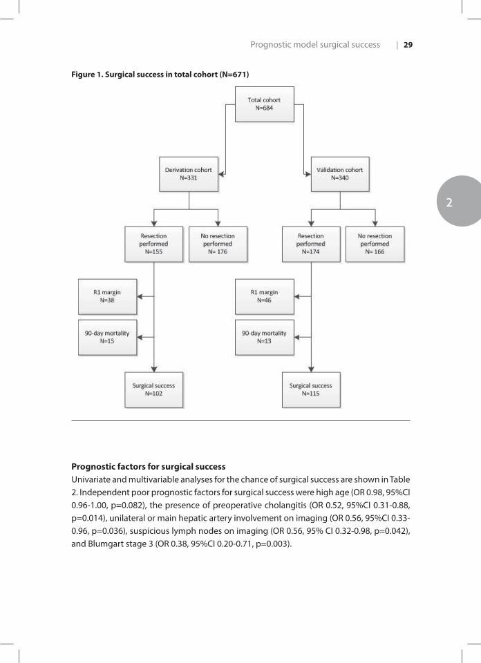
Suspected PHC was defined as a mass or malignant-appearing stricture at or near the biliary confluence, arising between the origin of the cystic duct and the segmental bile ducts.14 A multidisciplinary team diagnosed PHC based on clinical characteristics, radiological characteristics, endoscopic findings, and follow-up, if histopathological evidence was not available. Patients who were found to have metastases at staging laparoscopy were excluded, because they did not proceed to exploratory laparotomy.
Patient and tumor characteristics, clinical parameters, and laboratory results were retrospectively collected from medical archives in all centers. Preoperative cholangitis was defined by the presence of fever, abdominal pain, or leukocytosis requiring biliary drainage.10,15,16 Imaging at the time of presentation was reviewed by an attending abdominal radiologist to reassess tumor diameter, presence of suspicious lymph nodes, presence of distant metastases, and vascular involvement. Suspicious lymph nodes were defined as nodes larger than 1 cm in short-axis diameter, with central necrosis, an irregular border, or hyper-attenuation compared to portal phase liver parenchyma.17 Vascular involvement was defined as tumor contact of at least 180 degrees to the unilateral (both homolateral and contralateral) or main portal vein or hepatic artery. Surgical success was defined as a complete (R0) resection without 90-day mortality.
Patient managementManagement of PHC was relatively similar across all three centers. Presenting patients were discussed at a multidisciplinary meeting. Most patients underwent preoperative biliary drainage of the anticipated future liver remnant (FLR). Portal vein embolization was performed when the anticipated FLR was considered inadequate. Staging laparoscopy was performed increasingly in all centers. Patients with occult metastatic or unresectable disease at staging laparoscopy were not considered for exploratory laparotomy. At exploratory laparotomy, no resection was performed in patients with occult metastatic or locally advanced disease, precluding a complete resection with adequate liver remnant.13 Metastatic disease was defined as the presence of distant metastases or lymph node metastases beyond the hepatoduodenal ligament (N2).18 In the derivation cohort, patients were not considered for pre- or postoperative chemotherapy in compliance with Dutch guidelines at the time; in the validation cohort perioperative chemotherapy was considered at the discretion of the treating physician.19-21
Statistical analysesStatistical analyses were performed using SPSS version 23.0 and R version 3.3.3 (http://www.rproject.org). Overall survival (OS) was calculated from the date of first presentation in the tertiary referral center. Continuous data were reported as median with interquartile range (IQR) and compared using the non-parametric Mann-Whitney-U test. Categorical parameters were reported as counts and percentages and compared using Fisher’s exact

Prognostic model surgical success
2
| 27
or Chi-squared test as appropriate. Survival was estimated using the Kaplan Meier method and difference across groups was tested using the log-rank test.
The model was derived using the two Dutch cohorts. To identify preoperative factors predictive of surgical success of exploratory laparotomy, a univariable and multivariable logistic regression analysis was performed. Outcomes of the logistic regression analyses were reported as odds ratios (ORs) with their 95% confidence interval (95% CI). Missing values were imputed using the mice package and 50 imputations. Known prognostic factors were evaluated in univariable and multivariable analyses using the rms package. Factors were selected for the final model using a stepwise backward selection method based on the Akaike Information Criterion (AIC). A nomogram was developed using the independent prognostic factors. Model discrimination was evaluated by Harrell’s concordance index (c-index). External validation of the prognostic model was performed in the database of the Memorial Sloan Kettering Cancer Center. To visualize calibration, calibration curves were estimated for the derivation and validation cohorts. All tests were two‐sided, and P < 0.050 was used to define statistical significance.
Results
Patient and treatment characteristics
In total, 671 PHC patients underwent an exploratory laparotomy. The derivation cohort included 331 PHC patients. Table 1 presents the baseline patient characteristics and compares the baseline characteristics of patients with and without surgical success. Surgical success was achieved in 102 patients (30.8%)(Figure 1). No resection was performed in 176 patients (53.2%). Of the 155 patients (46.8%) who underwent a resection, 38 (24.5%) had an R1 resection. Of the remaining 117 patients (35.3%) with an R0 resection, 15 (12.8%) had 90-day postoperative mortality. Of the 176 patients in which no resection was performed, reasons for not performing a resection were: distant metastases (n=62, 35.2%), locally advanced disease precluding a complete resection (n=61, 34.7%), N2 lymph node metastases (n=49, 27.8%), or poor perioperative cardiac or pulmonary condition (n=4, 2.3%).
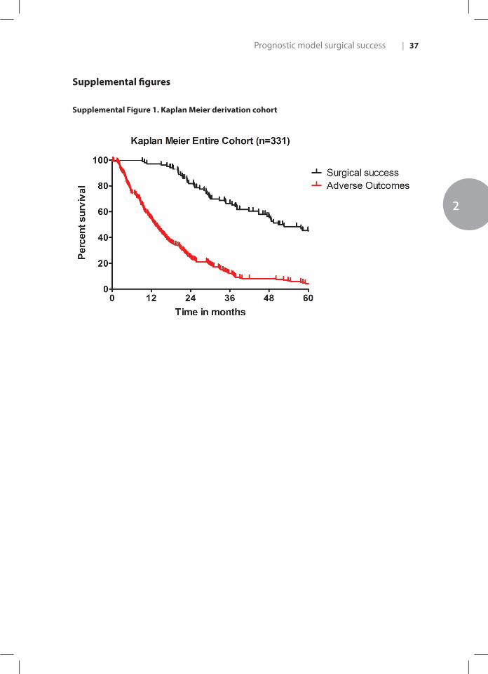
The median OS of the derivation cohort was 20.7 (95%CI 17.8-23.6) months. The median OS of patients with surgical success was 50.9 months (95%CI 39.6-62.2), compared to 13.1 months (95%CI 11.3-15.0) in patients without surgical success (p<0.001; Supplemental Figure 1).

Chapter 228 |
Table 1. Baseline characteristics of derivation cohort (n=331)
Characteristic All patients Surgical Success
(N=102)
No Surgical Success
(N=229)
P value
Age at first presentation, years 64 (54-70) 63 (53-69) 64 (55-70) 0.600
Gender, males 220 (66.5) 65 (63.7) 155 (67.7) 0.481
Primary sclerosing cholangitis 9 (2.7) 3 (2.9) 6 (2.6) 0.868
BMI, kg/m 24.9 (22.2-27.0) 25 (22.1-26.7) 24.7 (22.3-27.0) 0.776
ECOG performance status0123
190 (57.8)90 (27.4)35 (10.6)14 (4.3)
66 (64.7)29 (28.4)6 (5.9)1 (1.0)
124 (54.6)61 (26.9)29 (12.8)13 (5.7)
0.043
Jaundice at presentation 260 (80.2) 76 (75.2) 184 (82.5) 0.128
Bilirubin (mg/dL)1
≥ 14.6 mg/dL (i.e. >250 µmol/L)9.6 (5-16)56 (20.8)
7.7 (3-13)11 (13.4)
11.6 (7-17)45 (24.1)
0.8190.048
CA 19.9 (U/mL)2
≥ 1000 U/mL173.5 (44-642)36 (19.4)
87 (27-278)5 (9.1)
215 (51-870)31 (23.7)
0.2110.022
Preoperative cholangitis # 163 (49.7) 36 (35.3) 127 (56.2) < 0.001
Tumor size >3cm on imaging 94 (28.6) 24 (23.5) 70 (30.8) 0.175
Suspicious lymph nodes on imaging‡ 127 (38.4) 26 (25.5) 101 (44.1) 0.002
Blumgart Stage 4
123
125 (38.0)86 (26.1)118 (35.9)
54 (53.5)25 (24.8)22 (21.8)
71 (31.1)61 (26.8)96 (42.1)
< 0.001
Bismuth-Corlette StageI / IIIIIAIIIBIV
50 (15.7)93 (29.2)82 (25.7)94 (29.5)
29 (12.8)62 (27.4)59 (26.1)76 (33.6)
21 (22.6)31 (33.3)23 (24.7)18 (19.4)
0.006
Portal vein involvement*Unilateral involvementMain/bilateral involvement
152 (46.1)123 (37.2)29 (8.8)
38 (37.3)34 (33.3)4 (3.9)
114 (49.8)89 (38.9)25 (10.9)
0.032
Hepatic artery involvement*Unilateral involvementMain/bilateral involvement
145 (43.8)132 (39.9)13 (3.9)
30 (29.4)28 (27.4)2 (2.0)
115 (50.2)104 (45.4)11 (4.8)
< 0.001
Lobar atrophy, yes 78 (23.6) 23 (22.5) 55 (24.0) 0.771
Categorical parameters are presented as counts (percentages) and continuous parameters as median (interquartile range). Abbreviations: BMI, body mass index; CA 19.9, carbohydrate antigen 19.9; # Cholangitis before or at presentation was considered present if a patient had fever, abdominal pain or required biliary drainage.10,15,16
‡ Nodes along the cystic duct, common bile, duct, hepatic artery and portal vein were classified as N1 and periaortic, pericaval, SMA, and celiac nodes as N2.18
*Tumor contact of at least 180 degrees to the portal vein or hepatic artery and included main, bilateral, or unilateral involvement on contrast-enhanced CT or MRI imaging.
Missing data for: 163 patients1; 145 patients2
Prognostic model surgical success
2
| 29
Figure 1. Surgical success in total cohort (N=671)
Prognostic factors for surgical successUnivariate and multivariable analyses for the chance of surgical success are shown in Table 2. Independent poor prognostic factors for surgical success were high age (OR 0.98, 95%CI 0.96-1.00, p=0.082), the presence of preoperative cholangitis (OR 0.52, 95%CI 0.31-0.88, p=0.014), unilateral or main hepatic artery involvement on imaging (OR 0.56, 95%CI 0.33-0.96, p=0.036), suspicious lymph nodes on imaging (OR 0.56, 95% CI 0.32-0.98, p=0.042), and Blumgart stage 3 (OR 0.38, 95%CI 0.20-0.71, p=0.003).

Chapter 230 |
Derivation model for surgical success A preoperative prognostic nomogram was developed based on the five independent prognostic factors from the multivariable analysis (Figure 2). For example, a 40-year-old patient (75 points) without preoperative cholangitis (53 points), without hepatic artery involvement (47 points) or suspicious lymph nodes on imaging (58 points), and Blumgart Stage 2 (43 points), would have a 57% chance of surgical success. On the contrary, a 70-year-old patient (25 points) with preoperative cholangitis (0 points), with unilateral hepatic artery involvement (0 points) and Blumgart stage 3 (0 points) would only have a 7% chance of surgical success. The prognostic model had a c-index of 0.710. A calibration plot of the prognostic model is shown in Supplemental Figure 2.
Table 2. Univariate and multivariable analysis for surgical success.
Univariate Multivariable
OR (95% CI) p value OR (95% CI) p value
Age 0.99 (0.97-1.01) 0.559 0.98 (0.96-1.00) 0.082
Sex (male) 0.84 (0.51-1.37) 0.481
BMI ≥25 (kg/m²) 1.10 (0.68-1.78) 0.711
ECOG (WHO) performance status1-23-4
Ref0.16 (0.02-1.26) 0.082
Bilirubin ≥ 14.6 mg/dL (i.e. >250 µmol/L) 0.49 (0.24-1.00) 0.051
CA 19.9 >1000 (U/mL) 0.32 (0.12-0.88) 0.027
Cholangitis before or at presentation# 0.43 (0.26-0.69) 0.001 0.52 (0.31-0.88) 0.014
Tumor size >3cm 0.69 (0.40-1.18) 0.176
Suspicious lymph nodes on imaging‡ 0.44 (0.26-0.74) 0.002 0.56 (0.32-0.98) 0.042
Blumgart Stage 4
123
Ref0.54 (0.30-0.97)0.30 (0.17-0.54)
0.038<0.001
Ref0.64 (0.34-1.20)0.38 (0.20-0.71)
0.1630.003
Portal vein involvement* 0.59 (0.37-0.96) 0.033
Hepatic artery involvement* 0.41 (0.25-0.68) <0.001 0.56 (0.33-0.96) 0.036
Lobar atrophy, yes 0.88 (0.64-1.23) 0.472
Abbreviations: OR, Odds Ratio; 95% CI, 95% confidence interval; BMI, body mass index; CA 19.9, carbohydrate antigen 19.9# Cholangitis before or at presentation was considered present if a patient had fever, abdominal pain or required biliary drainage.10,15,16
‡ Nodes along the cystic duct, common bile, duct, hepatic artery and portal vein were classified as N1 and periaortic, pericaval, SMA, and celiac nodes as N2.18
*Tumor contact of at least 180 degrees to the portal vein or hepatic artery and included main, bilateral, or unilateral involvement on contrast-enhanced CT or MRI imaging.

Prognostic model surgical success
2
| 31
Figu
re 2
. Pro
gnos
tic
mod
el

Chapter 232 |
External validationThe validation cohort consisted of 340 PHC patients undergoing exploratory laparotomy. Surgical success was achieved in 128 patients (37.6%)(Figure 1). No resection was performed in 166 patients (48.8%). Of the 174 patients (51.2%) who underwent a resection, 46 (26.4%) had an R1 resection. Of the remaining 128 patients (35.3%) with an R0 resection, 13 (10.2%) had 90-day postoperative mortality.
The C-statistic of the preoperative prognostic model to predict surgical success in the validation cohort was 0.703. A calibration plot of the prognostic model in the validation cohort is shown in Supplemental Figure 3. The calibration curve showed that the prognostic model slightly underestimated the chance of successful resection in the validation cohort.
Discussion
We found that the chance of surgical success (i.e. complete resection without 90-day mortality) in 671 PHC patients undergoing exploratory laparotomy was disappointing: 31% in the Dutch derivation cohort and 36% in the US validation cohort. Poor prognostic factors for surgical success included higher age, the presence of preoperative cholangitis, tumor involvement of the unilateral or main hepatic artery on imaging, suspicious lymph nodes on imaging, and Blumgart stage 3. A preoperative model to predict surgical success using these five factors showed good concordance in both the derivation and external validation cohort.
This is the first preoperative prognostic model for surgical success in all potentially resectable PHC patients undergoing explorative laparotomy. Most surgical PHC studies are aimed at the minority of patients who undergo a resection, and few studies focus on patient selection for exploratory laparotomy and resection.4,13,22,23 Currently, even after diagnostic work-up and careful patient selection, two-thirds of patients have unexpected metastasis or locally advanced disease at exploratory laparotomy, an incomplete (R1) resection, or die within 90 days after surgery. Better patient selection could prevent patients from undergoing high-risk surgery with little or no survival benefit. The proposed prognostic model may help patients and clinicians to set individualized and realistic expectations for surgical success. The predicted chance of surgical success is below 10% in some patients; these patients may decide to forgo surgery. Although the risk factors for R1 resection and 90-day mortality might differ, we opted to make a composite outcome in order to generate an easily applicable tool for clinical success, and, vice versa, adverse outcomes.
Prognostic model surgical success
2
| 33
Age has been associated with OS in patients with PHC.13 A recent staging system, based on variables at the time of diagnosis, identified age as an independent predictor associated with survival after accounting for treatment modalities.13 Furthermore, another recently developed nomogram designed to predict prognosis of PHC patients also recognized age as a prognostic factor.22 In particular, advanced age was an important risk factor for postoperative liver failure and mortality.10
Preoperative cholestasis and cholangitis, both spontaneous and caused by instrumentation of the bile ducts, has been previously reported as a risk factor for mortality after hepatobiliary resection.10,24-26 Preoperative cholangitis may impede surgical success, because it is associated with more extensive biliary involvement and infection predisposes to postoperative liver failure and death. The risk of 90-day mortality after liver resection was increased by preoperative cholangitis, even in patients with a large future liver remnant FLR.10,26
Vascular involvement has been previously identified as a risk factor for poor survival and is part of most staging systems.4,13 A recent study from our group found that unilateral or main hepatic artery involvement is an independent prognostic factor for survival, while portal vein involvement was not.17 Hepatic artery involvement may be a surrogate for more advanced disease or facilitate distant spread of cancer cells. However, our study was underpowered to rule out that main portal vein involvement is a prognostic factor, because relatively few patients underwent a resection and reconstruction of the main portal vein.
The Blumgart staging system is based on biliary extent of the tumor, unilateral and main portal vein involvement, and the presence of unilateral hepatic atrophy.4 The Blumgart score was developed to predict resectability. It was found to predict both resectability and R0 resection. Therefore, it is not surprising that the Blumgart staging was identified as one of the independent predictive factors for surgical success.
We did not include recurrence in our prognostic model, because unfortunately most patients will eventually have recurrent disease. A previous study of our group showed that perihilar cholangiocarcinoma will recur in the majority of patients (76%) after curative intent resection.27
The current study should be viewed in the light of several limitations. First, the results of this study cannot be extrapolated to some high volume Asian centers that have published a very low 90-day postoperative mortality of 0 to 3%.9 Furthermore, staging laparoscopy was rarely performed in the early era of the cohort. The chance of surgical success of exploratory laparotomy increases when staging laparoscopy shows no occult metastatic
Chapter 234 |
disease. Because of differences in diagnostic work-up, patient selection, and treatment between centers, it is impossible to find a perfect validation cohort. This explains that our prognostic model somewhat underestimated the chance of surgical success in the validation cohort. Patients with suspicious lymph nodes beyond the hepatoduodenal ligament (e.g., celiac or aortocaval) have M1 disease that can be detected with endoscopic lymph node biopsy. During the study period, this technique was only used in recent years.
Conclusions
We developed and validated a preoperative model to predict surgical success in patients with potentially resectable PHC. This prognostic model, based on variables available at presentation, shows the chance of surgical success of individual patients and may help clinicians with patient selection. Patients with a low chance of surgical success may decide to refrain from exploratory laparotomy.

Prognostic model surgical success
2
| 35
References
1. Hartog H, Ijzermans JN, van Gulik TM, Groot Koerkamp B. Resection of Perihilar
Cholangiocarcinoma. Surg Clin North Am. 2016;96(2):247-267.
2. Pichlmayr R, Lamesch P, Weimann A, Tusch G, Ringe B. Surgical treatment of cholangiocellular
carcinoma. World J Surg. 1995;19(1):83-88.
3. Saxena A, Chua TC, Chu FC, Morris DL. Improved outcomes after aggressive surgical resection
of hilar cholangiocarcinoma: a critical analysis of recurrence and survival. Am J Surg.
2011;202(3):310-320.
4. Jarnagin WR, Fong Y, DeMatteo RP, et al. Staging, resectability, and outcome in 225 patients with
hilar cholangiocarcinoma. Ann Surg. 2001;234(4):507-517; discussion 517-509.
5. Groot Koerkamp B, Wiggers JK, Gonen M, et al. Survival after resection of perihilar
cholangiocarcinoma-development and external validation of a prognostic nomogram. Ann
Oncol. 2015;26(9):1930-1935.
6. Witzigmann H, Berr F, Ringel U, et al. Surgical and palliative management and outcome in
184 patients with hilar cholangiocarcinoma: palliative photodynamic therapy plus stenting is
comparable to r1/r2 resection. Ann Surg. 2006;244(2):230-239.
7. Schiffman SC, Reuter NP, McMasters KM, Scoggins CR, Martin RC. Overall survival peri-hilar
cholangiocarcinoma: R1 resection with curative intent compared to primary endoscopic
therapy. J Surg Oncol. 2012;105(1):91-96.
8. Ito F, Agni R, Rettammel RJ, et al. Resection of hilar cholangiocarcinoma: concomitant liver
resection decreases hepatic recurrence. Ann Surg. 2008;248(2):273-279.
9. Nagino M, Ebata T, Yokoyama Y, et al. Evolution of surgical treatment for perihilar
cholangiocarcinoma: a single-center 34-year review of 574 consecutive resections. Ann Surg.
2013;258(1):129-140.
10. Wiggers JK, Groot Koerkamp B, Cieslak KP, et al. Postoperative Mortality after Liver Resection for
Perihilar Cholangiocarcinoma: Development of a Risk Score and Importance of Biliary Drainage
of the Future Liver Remnant. J Am Coll Surg. 2016;223(2):321-331 e321.
11. Nuzzo G, Giuliante F, Ardito F, et al. Improvement in perioperative and long-term outcome after
surgical treatment of hilar cholangiocarcinoma: results of an Italian multicenter analysis of 440
patients. Arch Surg. 2012;147(1):26-34.
12. Kaiser GM, Paul A, Sgourakis G, et al. Novel prognostic scoring system after surgery for Klatskin
tumor. Am Surg. 2013;79(1):90-95.
13. Chaiteerakij R, Harmsen WS, Marrero CR, et al. A new clinically based staging system for perihilar
cholangiocarcinoma. Am J Gastroenterol. 2014;109(12):1881-1890.
14. Edge SB BD, Compton CC, Fritz AG, Greene FL, Trotti A, editors. AJCC cancer staging manual (7th
ed). New York, NY: Springer; 2010.
15. van der Gaag NA, Rauws EA, van Eijck CH, et al. Preoperative biliary drainage for cancer of the
head of the pancreas. N Engl J Med. 2010;362(2):129-137.
Chapter 236 |
16. Wiggers JK, Coelen RJ, Rauws EA, et al. Preoperative endoscopic versus percutaneous
transhepatic biliary drainage in potentially resectable perihilar cholangiocarcinoma (DRAINAGE
trial): design and rationale of a randomized controlled trial. BMC Gastroenterol. 2015;15:20.
17. van Vugt JLA, Gaspersz MP, Coelen RJS, et al. The prognostic value of portal vein and hepatic
artery involvement in patients with perihilar cholangiocarcinoma. HPB (Oxford). 2017.
18. Edge SB, Compton CC. The American Joint Committee on Cancer: the 7th edition of the AJCC
cancer staging manual and the future of TNM. Ann Surg Oncol. 2010;17(6):1471-1474.
19. Skipworth JR, Olde Damink SW, Imber C, Bridgewater J, Pereira SP, Malago M. Review article:
surgical, neo-adjuvant and adjuvant management strategies in biliary tract cancer. Aliment
Pharmacol Ther. 2011;34(9):1063-1078.
20. Anderson C, Kim R. Adjuvant therapy for resected extrahepatic cholangiocarcinoma: a review of
the literature and future directions. Cancer Treat Rev. 2009;35(4):322-327.
21. Takada T, Amano H, Yasuda H, et al. Is postoperative adjuvant chemotherapy useful for
gallbladder carcinoma? A phase III multicenter prospective randomized controlled trial in
patients with resected pancreaticobiliary carcinoma. Cancer. 2002;95(8):1685-1695.
22. Buettner S, van Vugt JL, Gani F, et al. A Comparison of Prognostic Schemes for Perihilar
Cholangiocarcinoma. J Gastrointest Surg. 2016;20(10):1716-1724.
23. Coelen RJ, Ruys AT, Wiggers JK, et al. Development of a Risk Score to Predict Detection of
Metastasized or Locally Advanced Perihilar Cholangiocarcinoma at Staging Laparoscopy. Ann
Surg Oncol. 2016;23(Suppl 5):904-910.
24. Sano T, Shimada K, Sakamoto Y, Yamamoto J, Yamasaki S, Kosuge T. One hundred two
consecutive hepatobiliary resections for perihilar cholangiocarcinoma with zero mortality. Ann
Surg. 2006;244(2):240-247.
25. Sakata J, Shirai Y, Tsuchiya Y, Wakai T, Nomura T, Hatakeyama K. Preoperative cholangitis
independently increases in-hospital mortality after combined major hepatic and bile duct
resection for hilar cholangiocarcinoma. Langenbecks Arch Surg. 2009;394(6):1065-1072.
26. Olthof PB, Wiggers JK, Groot Koerkamp B, et al. Postoperative Liver Failure Risk Score: Identifying
Patients with Resectable Perihilar Cholangiocarcinoma Who Can Benefit from Portal Vein
Embolization. J Am Coll Surg. 2017.
27. Groot Koerkamp B, Wiggers JK, Allen PJ, et al. Recurrence Rate and Pattern of Perihilar
Cholangiocarcinoma after Curative Intent Resection. J Am Coll Surg. 2015;221(6):1041-1049.
Prognostic model surgical success
2
| 37
Supplemental figures
Supplemental Figure 1. Kaplan Meier derivation cohort

Chapter 238 |
Supplemental Figure 2. Calibration plot of proposed prognostic model to predict surgical success in derivation cohort

Prognostic model surgical success
2
| 39
Supplemental Figure 3. Calibration plot of proposed prognostic model to predict surgical success in validation cohort

3CHAPTER 3
3 The Prognostic Value of Portal Vein and Hepatic Artery Involvement in Patients with Perihilar Cholangiocarcinoma
van Vugt JL* | Gaspersz MP* | Coelen RJS | Vugts J | Labeur T | de Jonge J | Polak WG | Busch ORC | Besselink MG | IJzermans JN | Nio CY | van Gulik TM | Willemssen FEJA* | Groot Koerkamp B*
* both authors contributed equally to this study
HPB (Oxford). 2018 Jan;20(1):83-92

Background: Although several classifications of perihilar cholangiocarcinoma (PHC) include vascular involvement, its prognostic value has not been investigated. Our aim was to assess the prognostic value of unilateral and main/bilateral involvement of the portal vein (PV) and hepatic artery (HA) on imaging in patients with PHC.
Methods: All patients with PHC between 2002-2014 were included regardless of stage or management. Vascular involvement was defined as apparent tumor contact of at least180 degrees to the PV or HA on imaging. Kaplan-Meier method with log-rank test was used to compare overall survival (OS) between groups. Cox regression was used for multivariable analysis. Subgroup analysis was performed in patients undergoing surgery.
Results: In total, 674 patients were included with a median OS of 12.2 (95% CI 10.6-13.7) months. Patients with unilateral PV involvement had a median OS of 13.3 (11.0-15.7) months, compared with 14.7 (11.7-17.6) in patients without PV involvement (p=0.12). Patients with main/bilateral PV involvement had an inferior median OS of 8.0 (5.4-10.7, p<0.001) months.Median OS for patients with unilateral HA involvement was 10.6 (9.3-12.0) months compared with 16.9 (13.2-20.5) in patients without HA involvement (p<0.001). Patients with main/bilateral HA involvement had an inferior median OS of 6.9 (3.3-10.5, p<0.001). Independent poor prognostic factors for OS included unilateral and main/bilateral HA involvement but not PV involvement.
Discussion: Both unilateral and main HA involvement are independent poor prognostic factors for OS in patients presenting with PHC, whereas PV involvement is not.Abs
trac
t

Vascular involvement in PHC
3
| 43
Introduction
Perihilar cholangiocarcinoma (PHC) is the most common bile duct cancer and arises at or near the confluence of the right and left main bile duct. The annual incidence in Western countries is about 2 per 100.000.1 Patients usually present with obstructive jaundice, abdominal pain, and weight loss.2 Surgical resection is the only potentially curative option for patients with PHC, resulting in a median overall survival (OS) of about 40 months.3 Unfortunately, only about 20% of all patients are eligible for a curative-intent surgical resection because the majority of patients has metastatic or locally advanced disease at presentation or during explorative laparotomy.4-6
Staging and resectability are determined primarily using computed tomography (CT) and magnetic resonance imaging (MRI). Most staging systems consider vascular involvement of the tumor to determine prognosis and resectability. Apparent vascular involvement on imaging is typically defined as tumor contact of at least 180 degrees.7 Actual involvement on pathological examination is only evaluated in patients who undergo an en-bloc resection of the tumor and (branches of ) the portal vein (PV) or hepatic artery (HA). The American Joint Committee on Cancer (AJCC) staging system has a prominent role for unilateral PV or HA involvement (i.e. stage T3) and main PV or HA involvement (i.e. stage T4).8 The DeOliveira/Clavien classification also requires detailed assessment of both unilateral and main HA and PV involvement.7 The Mayo Clinic staging system considered any tumor contact with the PV or HA a poor prognostic factor.9 The Blumgart staging system was developed to predict resectability based on unilateral and main PV involvement of the tumor in addition to biliary extent and hepatic atrophy.4
Differences between the staging systems demonstrate disagreement about which aspect of vascular involvement is most important: PV or HA involvement, and unilateral or main/bilateral involvement. The prognostic value of unilateral and main PV or HA involvement has not been evaluated in a large group of PHC patients. The aim of this study was to investigate the prognostic value of unilateral and main/bilateral involvement of the PV and HA on imaging in patients with PHC, regardless of subsequent treatment.
Methods
Study population and data acquisitionAll consecutive patients with suspected PHC between 2002 and 2014 in the Erasmus MC University Medical Center, Rotterdam, The Netherlands and the Academic Medical Center (AMC), Amsterdam, The Netherlands, were identified through a systematic search in all medical files, discharge letters, reports of multidisciplinary hepatopancreatobiliary team
Chapter 344 |
meetings, and operative and pathology reports. All PHC care in our region is centralized and all patients are being referred to one of the specialized centers according to a national protocol. All patients referred for curative-intent surgery, palliative treatment, or best supportive care were included.
PHC was defined as a mass or malignant-appearing stricture at or near the biliary confluence, arising between the origin of the cystic duct and the segmental bile ducts.10 If no histopathological evidence was obtained, the multidisciplinary hepatopancreatobiliary team determined the diagnosis based on clinical, radiological, endoscopic and laboratory findings, and follow-up. Patients with hilar-invasive intrahepatic cholangiocarcinoma, gallbladder carcinoma, cystic duct carcinoma, and distal cholangiocarcinoma were excluded, as well as patients who had no imaging available for review. We also excluded patients who underwent treatment (e.g., resection or chemotherapy) prior to referral or who visited our centers for a single biliary drainage without further follow-up at our centers.
Demographics (e.g., age, gender), clinical parameters (e.g., cholangitis), and laboratory results (e.g., bilirubin and carbohydrate antigen (CA) 19.9 levels) were collected from medical records. Cholangitis was defined by the presence of fever, abdominal complaints, or leukocytosis requiring biliary drainage.11-13
Experienced abdominal radiologists revised imaging (i.e. contrast-enhanced CT and/or MRI or MRI with cholangiopancreatography (MRCP)) performed at the time of first presentation. Parameters assessed on imaging were radial diameter of the tumor, biliary extent of the tumor (Bismuth-Corlette classification)14, clinical AJCC staging (7th edition), presence of lymph node and distant metastases, lobar atrophy, and vascular involvement. The clinical AJCC (7th edition) stages I and II were pooled, because T1 (stage I) and T2 (stage 2) cannot be distinguished on imaging.8 Suspicious lymph nodes were defined as nodes larger than 1 cm in short-axis diameter, with central necrosis, an irregular border, or hyper-attenuating compared to portal phase liver parenchyma. Nodes along the cystic duct, common bile duct, hepatic artery and portal vein were classified as N1; involvement of periaortic, pericaval, superior mesenteric artery, and celiac nodes as N2, according to the AJCC staging (7th edition).8 Vascular involvement was defined as apparent tumor contact of at least 180 degrees to the PV or HA. It was classified separately for PV and HA as main, bilateral, or unilateral involvement.8, 15, 16 Vascular involvement was mainly assessed on contrast-enhanced CT imaging. MRI was only used in the few patients with unavailable contrast-enhanced CT.
The Institutional Review Boards of both centers approved the study and the need for informed consent was waived.
Vascular involvement in PHC
3
| 45
Diagnostic work-up and treatment algorithmThe diagnostic work-up and treatment algorithm were performed as previously described and were comparable between the two centers. In short, diagnostic work-up included contrast-enhanced CT and/or MRI/MRCP. Metastatic disease was defined, according to the AJCC staging (7th edition), as the presence of distant metastases or lymph node metastases beyond the hepatoduodenal ligament (N2).8 Locally advanced disease was defined as invasion of surrounding organs or vascular or biliary involvement that precluded an R0 resection.9
Exploratory laparotomy was rarely performed in patients with stage IVb disease (i.e. N2 or M1) or with main/bilateral HA involvement on imaging. Patients did not receive adjuvant chemotherapy in compliance with Dutch guidelines.17-19 Palliative systemic chemotherapy (gemcitabine with or without cisplatin) was considered for patients with locally advanced or metastatic disease. Patients who did not receive chemotherapy, received best supportive care. Liver transplantation was only performed in highly selected patients based on a nationwide protocol since 2014.20, 21
Statistical analysesAll statistical analyses were performed using SPSS for Windows (IBM Corp., Armonk, NY, USA), version 22. Continuous data are reported as mean with standard deviation (SD) or median with interquartile range (IQR), depending on the normality of distribution. Categorical parameters are reported as counts and percentages. Proportions were compared with Fischer’s exact or Chi-squared test, whereas means were compared with t-test.
Univariate analyses were performed by Kaplan–Meier estimates of survival probabilities, partial likelihood estimation for hazard ratios and the log-rank and score tests for comparisons. A Cox proportional hazard regression model was used for multivariable modeling using backward selection with all variables that were significantly associated (p<0.05) with overall survival (OS) in univariate analysis; CA 19.9 was excluded from multivariable analysis, because values were missing in 324 (48.1%) patients. Furthermore, subgroup analyses were performed for patients without suspected distant metastases on imaging and patients who underwent resection.
Survival status was updated using the municipal records database on May 9th, 2016. Overall survival was calculated from the date of first presentation in the tertiary referral center.
Chapter 346 |
Results
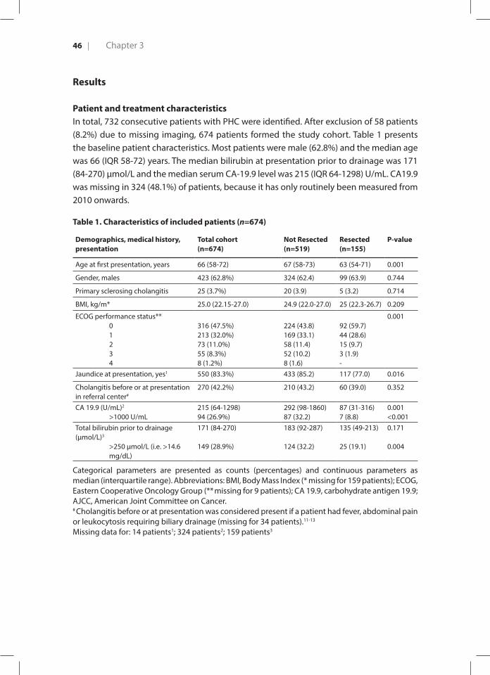
Patient and treatment characteristicsIn total, 732 consecutive patients with PHC were identified. After exclusion of 58 patients (8.2%) due to missing imaging, 674 patients formed the study cohort. Table 1 presents the baseline patient characteristics. Most patients were male (62.8%) and the median age was 66 (IQR 58-72) years. The median bilirubin at presentation prior to drainage was 171 (84-270) µmol/L and the median serum CA-19.9 level was 215 (IQR 64-1298) U/mL. CA19.9 was missing in 324 (48.1%) of patients, because it has only routinely been measured from 2010 onwards.
Table 1. Characteristics of included patients (n=674)
Demographics, medical history, presentation
Total cohort(n=674)
Not Resected(n=519)
Resected(n=155)
P-value
Age at first presentation, years 66 (58-72) 67 (58-73) 63 (54-71) 0.001
Gender, males 423 (62.8%) 324 (62.4) 99 (63.9) 0.744
Primary sclerosing cholangitis 25 (3.7%) 20 (3.9) 5 (3.2) 0.714
BMI, kg/m* 25.0 (22.15-27.0) 24.9 (22.0-27.0) 25 (22.3-26.7) 0.209
ECOG performance status** 0 1 2 3 4
316 (47.5%)213 (32.0%)73 (11.0%)55 (8.3%)8 (1.2%)
224 (43.8)169 (33.1)58 (11.4)52 (10.2)8 (1.6)
92 (59.7)44 (28.6)15 (9.7)3 (1.9)-
0.001
Jaundice at presentation, yes1 550 (83.3%) 433 (85.2) 117 (77.0) 0.016
Cholangitis before or at presentation in referral center#
270 (42.2%) 210 (43.2) 60 (39.0) 0.352
CA 19.9 (U/mL)2
>1000 U/mL215 (64-1298) 94 (26.9%)
292 (98-1860)87 (32.2)
87 (31-316)7 (8.8)
0.001<0.001
Total bilirubin prior to drainage (µmol/L)3
>250 µmol/L (i.e. >14.6 mg/dL)
171 (84-270)
149 (28.9%)
183 (92-287)
124 (32.2)
135 (49-213)
25 (19.1)
0.171
0.004
Categorical parameters are presented as counts (percentages) and continuous parameters as median (interquartile range). Abbreviations: BMI, Body Mass Index (* missing for 159 patients); ECOG, Eastern Cooperative Oncology Group (** missing for 9 patients); CA 19.9, carbohydrate antigen 19.9; AJCC, American Joint Committee on Cancer.# Cholangitis before or at presentation was considered present if a patient had fever, abdominal pain or leukocytosis requiring biliary drainage (missing for 34 patients).11-13
Missing data for: 14 patients1; 324 patients2; 159 patients3
Vascular involvement in PHC
3
| 47
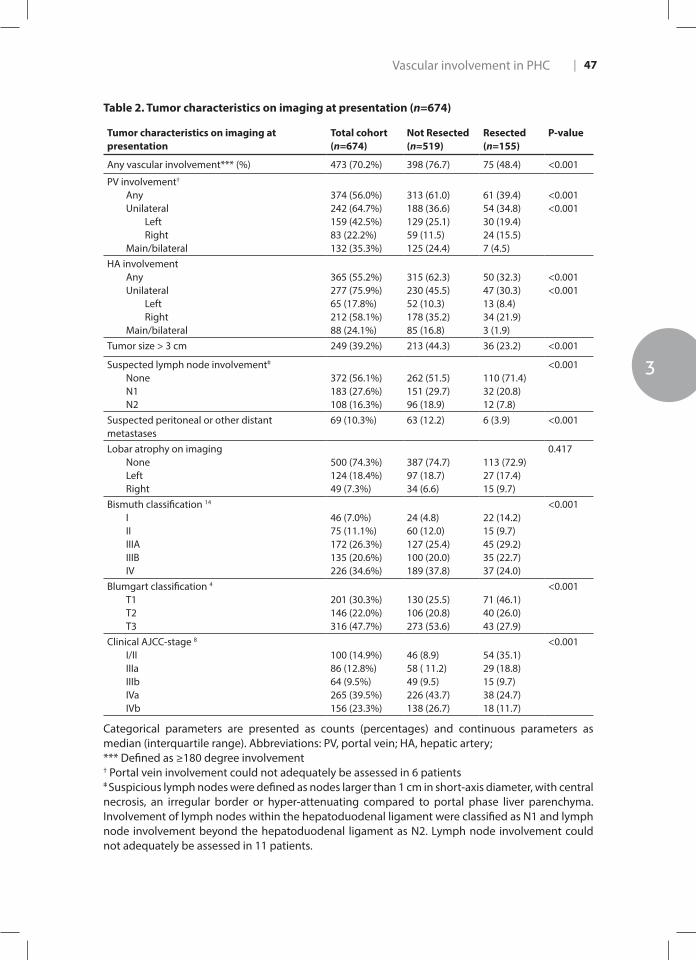
Table 2. Tumor characteristics on imaging at presentation (n=674)
Tumor characteristics on imaging at presentation
Total cohort(n=674)
Not Resected(n=519)
Resected(n=155)
P-value
Any vascular involvement*** (%) 473 (70.2%) 398 (76.7) 75 (48.4) <0.001
PV involvement†
AnyUnilateral
LeftRight
Main/bilateral
374 (56.0%)242 (64.7%)159 (42.5%)83 (22.2%)132 (35.3%)
313 (61.0)188 (36.6)129 (25.1)59 (11.5) 125 (24.4)
61 (39.4)54 (34.8)30 (19.4)24 (15.5)7 (4.5)
<0.001<0.001
HA involvement AnyUnilateral
LeftRight
Main/bilateral
365 (55.2%)277 (75.9%)65 (17.8%)212 (58.1%)88 (24.1%)
315 (62.3)230 (45.5)52 (10.3)178 (35.2)85 (16.8)
50 (32.3)47 (30.3)13 (8.4)34 (21.9)3 (1.9)
<0.001<0.001
Tumor size > 3 cm 249 (39.2%) 213 (44.3) 36 (23.2) <0.001
Suspected lymph node involvementǂ NoneN1N2
372 (56.1%)183 (27.6%)108 (16.3%)
262 (51.5)151 (29.7)96 (18.9)
110 (71.4)32 (20.8)12 (7.8)
<0.001
Suspected peritoneal or other distant metastases
69 (10.3%) 63 (12.2) 6 (3.9) <0.001
Lobar atrophy on imagingNoneLeftRight
500 (74.3%)124 (18.4%)49 (7.3%)
387 (74.7)97 (18.7)34 (6.6)
113 (72.9)27 (17.4)15 (9.7)
0.417
Bismuth classification 14
IIIIIIAIIIBIV
46 (7.0%)75 (11.1%)172 (26.3%)135 (20.6%)226 (34.6%)
24 (4.8)60 (12.0)127 (25.4)100 (20.0)189 (37.8)
22 (14.2)15 (9.7)45 (29.2)35 (22.7)37 (24.0)
<0.001
Blumgart classification 4
T1T2T3
201 (30.3%)146 (22.0%)316 (47.7%)
130 (25.5)106 (20.8)273 (53.6)
71 (46.1)40 (26.0)43 (27.9)
<0.001
Clinical AJCC-stage 8
I/IIIIIaIIIbIVaIVb
100 (14.9%)86 (12.8%)64 (9.5%)265 (39.5%)156 (23.3%)
46 (8.9)58 ( 11.2)49 (9.5)226 (43.7)138 (26.7)
54 (35.1)29 (18.8)15 (9.7)38 (24.7)18 (11.7)
<0.001
Categorical parameters are presented as counts (percentages) and continuous parameters as median (interquartile range). Abbreviations: PV, portal vein; HA, hepatic artery;*** Defined as ≥180 degree involvement† Portal vein involvement could not adequately be assessed in 6 patientsǂ Suspicious lymph nodes were defined as nodes larger than 1 cm in short-axis diameter, with central necrosis, an irregular border or hyper-attenuating compared to portal phase liver parenchyma. Involvement of lymph nodes within the hepatoduodenal ligament were classified as N1 and lymph node involvement beyond the hepatoduodenal ligament as N2. Lymph node involvement could not adequately be assessed in 11 patients.

Chapter 348 |
Clinical AJCC staging categorized 100 (14.9%) patients in stage I/II, 150 (22.4%) patients in stage III, and 421 (62.7%) patients in stage IV, of whom 265 (39.5%) patients with stage IVa (i.e. T4 tumor with lymph node involvement) and 156 (23.3%) patients with stage IVb (i.e. distant metastases or N2 lymph node involvement) (Table 2).
Initially, a total of 331 (49.1%) patients were considered potentially resectable and fit to undergo major liver resection. A staging laparoscopy was performed in 207 (30.8%) patients, and 155 (74.9%) patients eventually underwent a resection. Two patients (0.6%) underwent a liver transplantation (Supplementary Table 1). The remaining 176 (53.2%) patients underwent an exploratory laparotomy or laparoscopy without resection, because of the presence of occult metastases (n=111, 63.1%) or locally advanced disease (n=61, 34.7%). About half of all patients were ineligible for a curative-intent resection based on imaging (n=343, 50.9%); 113 (33.9%) due to distant metastases, 30 (8.7%) due to lymph node metastases beyond the hepatoduodenal ligament (N2), 117 (34.1%) due to locally advanced disease, and 83 (24.1%) patients did not undergo surgery due to advanced age, comorbidities, or the inability to reach adequate biliary drainage. Of the 343 patients that were deemed unresectable at presentation, 33 (9.6%) received chemotherapy, mostly (n=23, 6.7%) gemcitabine plus cisplatin. All other patients were deemed ineligible or opted out of chemotherapy and received best supportive care consisting of symptom relieve and biliary drainage if indicated.
SurvivalThe median OS (95% confidence interval (CI)) of the entire cohort was 12.2 (10.6-13.7) months. A total of 608 patients (90.2%) had died at last follow-up with a median follow-up of patients alive at last follow-up of 46.5 months. Median OS was significantly different between treatment groups with a median OS of 37.7 (95% CI 28.1-47.2) months in patients who underwent a resection, 12.8 (11.1-14.4) months in patients who underwent laparotomy or laparoscopy without resection, and 8.0 (6.9-9.2) months in patients who did not undergo a laparotomy of laparoscopy (p<0.001).
Vascular involvement on imagingTumor characteristics on imaging at presentation are shown in table 2. Any vascular involvement of the tumor was observed in 473 (70.2%) patients. Involvement of the PV was observed in 374 (56.0%) of all patients; 242 patients (64.7%) had unilateral PV involvement and 132 patients (35.3%) had main or bilateral PV involvement. HA involvement was observed in 365 (54.6%) patients. Of these, 277 (75.9%) patients had unilateral involvement and 88 (24.1%) patients main/bilateral involvement. Contralateral vascular involvement (e.g., a Bismuth IIIa with left HA involvement) was a common cause of locally advanced disease (Supplementary table 2).
Vascular involvement in PHC
3
| 49
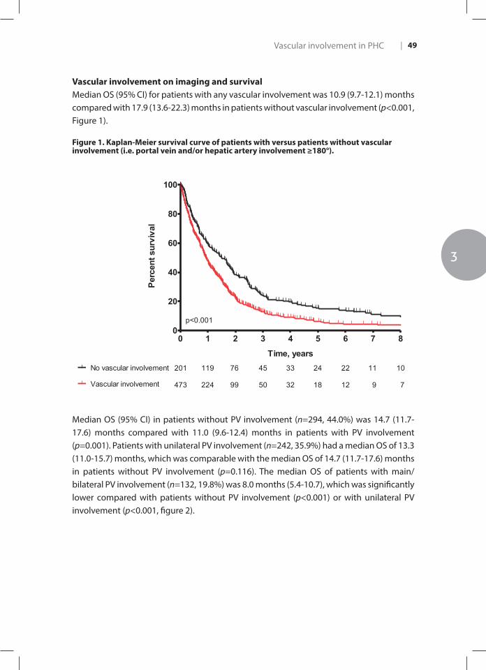
Vascular involvement on imaging and survivalMedian OS (95% CI) for patients with any vascular involvement was 10.9 (9.7-12.1) months compared with 17.9 (13.6-22.3) months in patients without vascular involvement (p<0.001, Figure 1).
Figure 1. Kaplan-Meier survival curve of patients with versus patients without vascular involvement (i.e. portal vein and/or hepatic artery involvement ≥180°).Figure 1. Kaplan-Meier survival curve of patients with versus patients without vascular involvement (i.e. portal vein and/or hepatic artery involvement ≥180°, unilateral or main/bilateral).
0 1 2 3 4 5 6 7 80
20
40
60
80
100
Vascular involvement
No vascular involvement 201
473
119
224
76
99
45
50
33
32
24
18
22
12
11
9
10
7
p<0.001
Time, years
Perc
ent s
urvi
val
Median OS (95% CI) in patients without PV involvement (n=294, 44.0%) was 14.7 (11.7-17.6) months compared with 11.0 (9.6-12.4) months in patients with PV involvement (p=0.001). Patients with unilateral PV involvement (n=242, 35.9%) had a median OS of 13.3 (11.0-15.7) months, which was comparable with the median OS of 14.7 (11.7-17.6) months in patients without PV involvement (p=0.116). The median OS of patients with main/bilateral PV involvement (n=132, 19.8%) was 8.0 months (5.4-10.7), which was significantly lower compared with patients without PV involvement (p<0.001) or with unilateral PV involvement (p<0.001, figure 2).

Chapter 350 |
Figure 2. Kaplan-Meier survival curve of patients without portal vein involvement versus patients with unilateral or main/bilateral portal vein involvement.
Figure 2. Kaplan-Meier survival curve of patients without portal vein involvement versus patients with unilateral or main/bilateral portal vein involvement.
0 1 2 3 4 5 6 7 80
20
40
60
80
100
None
Unilateral 294
242
132
163
128
49
93
63
18
54
31
9
38
18
8
28
11
3
25
6
2
12
6
2
10
6
1Main/bilateral
Time, years
Perc
ent s
urvi
val
Median OS (95% CI) in patients without HA involvement (n=296, 44.8%) was 16.9 (13.2-20.5) months compared with 10.3 (8.9-11.7) months in patients with HA involvement (p<0.001). Patients with unilateral HA involvement (n=277, 41.1%) had a median OS of 10.6 (9.3-12.0) months, which was significantly lower compared with the median OS of 16.9 (13.2-20.5) months of patients without HA involvement (p<0.001)(Figure 3). Patients with main/bilateral HA involvement (n=88, 13.3%) had a median OS of 6.9 (3.3-10.5) months, compared with 10.6 (9.3-12.0) months for patients with unilateral HA involvement (p<0.001) (Supplementary Fig. 1).

Vascular involvement in PHC
3
| 51
Figure 3. Kaplan-Meier survival curve of patients without hepatic artery involvement versus patients with unilateral or main/bilateral hepatic artery involvement.
Figure 3. Kaplan-Meier survival curve of patients without hepatic artery involvement versus patients with unilateral or main/bilateral hepatic artery involvement.
0 1 2 3 4 5 6 7 80
20
40
60
80
100
None
Unilateral
296
277
88
174
129
35
103
58
12
60
60
4
43
18
3
32
7
2
27
5
2
16
3
1
14
2
1Main/bilateral
Time, years
Perc
ent s
urvi
val
Multivariable analysisThe multivariable survival analysis is shown in table 3. Both unilateral (hazard ratio (HR) 1.26, 95% CI 1.00-1.58, p=0.048) and main/bilateral HA involvement (HR 1.74, 95% CI 1.19-2.52, p=0.004) were independent poor prognostic factors. Main/bilateral PV involvement was not an independent prognostic factor (HR 1.22, 95% CI 0.88-1.70, p=0.233). Comparable results were observed when age, serum bilirubin level, and tumor size were entered as continuous covariates.
Outcomes after surgeryOf the 155 patients who underwent resection, only 7 (4.5%) had main/bilateral PV involvement and 3 (1.9%) had main/bilateral HA involvement on imaging (Supplementary table 3).

Chapter 352 |
Table 3. Univariate and multivariable Cox regression analysis for predictors of overall survival
Univariate Multivariable
HR (95% CI) p value HR (95% CI) p value
Age ≥75 years 1.70 (1.39-2.08) <0.001 1.80 (1.40-2.30) <0.001
Sex (male) 1.04 (0.89-1.22) 0.665
BMI ≥25 (kg/m²) 1.00 (0.84-1.18) 0.991
ECOG (WHO) performance status 1-2 3-4
Ref1.83 (1.41-2.39) <0.001 2.02 (1.38-2.95) <0.001
Bilirubin ≥ 14.6 mg/dL (i.e. >250 µmol/L) 1.62 (1.32-1.98) <0.001 1.50 (1.20-1.87) <0.001
CA 19.9 >1000 (U/mL)* 2.24 (1.74-2.87) <0.001
Cholangitis before or at presentation# 1.13 (0.96 -1.34) 0.140
Tumor size >3cm 1.52 (1.28-1.80) <0.001 1.47 (1.18-1.83) 0.001
Suspicious lymph nodes on imaging‡
N0 N1 N2
Ref1.22 (1.01-1.47)1.54 (1.24-1.92)
0.040<0.001
1.00 (0.79-1.26)1.31 (0.99-1.73)
0.9680.055
Suspected distant metastases on imaging 1.76 (1.37-2.27) <0.001 1.71 (1.19-2.48) 0.004
Lobar atrophy on imaging 0.94 (0.78-1.12) 0.479
PV involvement None Unilateral Main/bilateral
Ref1.15 (0.96-1.38)1.70 (1.38-2.11)
0.132<0.001
0.87 (00.68-1.11)1.22 (0.88-1.70)
0.2620.233
HA involvement None Unilateral Main/bilateral
Ref1.45 (1.22-1.73)1.96 (1.53 -2.51)
<0.001<0.001
1.26 (1.00-1.58)1.74 (1.19-2.52)
0.0480.004
Bismuth IV 1.28 (1.08-1.51) 0.005 1.14 (0.92-1.42) 0.239
Abbreviations: HR, hazard ratio; 95% CI, 95% confidence interval; BMI, body mass index; CA 19.9, carbohydrate antigen 19.9; PV, portal vein; HA, hepatic artery. # Cholangitis before or at presentation was considered present if a patient had fever, abdominal pain or leukocytosis requiring biliary drainage.11-13
‡ Nodes along the cystic duct, common bile, duct, hepatic artery and portal vein were classified as N1 and periaortic, pericaval, SMA, and celiac nodes as N2.8
The final model included 534 patients who had complete data on all variables.* This parameter was not included in the multivariable model due to a high percentage of missing values.
In a subgroup analysis of patients who underwent resection, neither unilateral PV or HA involvement, nor main/bilateral PV involvement were significantly associated with OS, whereas main/bilateral HA involvement was associated with worse OS in both univariate analysis (HR 4.34 [95% CI 1.03-18.25], p=0.045) and multivariable analysis ((HR 5.49, [95% CI 1.17-25.74], p=0.031) (Supplementary Table 4).

Vascular involvement in PHC
3
| 53
Patients who underwent a resection had a longer OS compared with patients without resection regardless of the presence of PV involvement: 41.9 versus 10.1 months (p<0.001) without PV involvement, 36.6 versus 10.4 months (p<0.001) with unilateral PV involvement, and 18.7 versus 7.5 months (p=0.049) with main/bilateral involvement.
Patients who underwent resection had a longer OS compared with patients without resection and without HA involvement or with unilateral HA involvement: 37.7 versus 11.1 months (p<0.001) and 36.7 versus 9.6 months (p<0.001), respectively. However, no significant difference in OS could be demonstrated between patients with main/bilateral HA involvement who underwent resection compared with those with main/bilateral HA involvement who did not undergo resection: 18.7 versus 6.9 months (p=0.537). In patients who did not undergo surgical resection, we found that main/bilateral PV involvement and any HA involvement were not independently associated with OS, whereas unilateral PV involvement was independently associated with poor OS (HR 0.75, 95% CI 0.58–0.96, p = 0.024) (Supplementary Table 5).
Discussion
In this study of 674 patients with PHC we found that both unilateral and main/bilateral HA involvement on imaging at presentation are independent poor prognostic factors for OS. PV involvement, whether unilateral or main, was not an independent poor prognostic factor. Other independent poor prognostic factors were age above 75 years, ECOG performance status 3 or 4, serum bilirubin level above 250 umol/L (i.e. 14.6 mg/dL), tumor size above 3 cm, and distant metastatic disease.
Most published studies have focused on PHC patients referred for curative intent resection.4, 13 The present study evaluated all consecutive patients, regardless of subsequent treatment, including 421 patients (62.7%) with stage IV disease at presentation. We found a median OS of 16.9 months without HA involvement, 10.6 months with unilateral HA involvement, and 6.9 months with main or bilateral involvement (p<0.001). The AJCC staging system, which has also been developed in all patients with PHC regardless of subsequent treatment, already incorporated progressive HA involvement as a poor prognostic factor;8 unilateral HA involvement upstages a patient from stage II to III, and stage IVA includes all patients with main/bilateral HA involvement. The Mayo Clinic staging system does not specifically consider HA involvement, although a patient with any vascular involvement has at least Mayo stage II disease.9 The Mayo model may improve by distinguishing unilateral and main/bilateral HA involvement. The DeOliveira/Clavien classification does require assessment of both unilateral and main/bilateral HA involvement, although this

Chapter 354 |
classification was not based on actual patient level data.7 The Blumgart staging system does not take HA involvement into account.4
We found that unilateral PV involvement was not a prognostic factor and main/bilateral PV involvement was only a prognostic factor in univariate analysis. In contrast to these findings, PV involvement has a prominent role in staging systems.4, 7-9 The AJCC staging system incorporates PV involvement;8 unilateral PV involvement upstages a patient from stage II to III, and stage IVA includes all patients with main/bilateral PV involvement. A comparison of the 6th and 7th editions of the AJCC showed that PV involvement was even given a more important role as unilateral PV involvement migrated from stage II in the 6th edition to stage III in the 7th edition.22 However, we found the same median OS for patients without and with unilateral PV involvement. The Mayo Clinic staging system does consider PV involvement as a prognostic factor; a patient with any vascular involvement has at least Mayo stage II disease. The Mayo model could be simplified by leaving out consideration of PV involvement. The DeOliveira/Clavien classification requires assessment of unilateral and main/bilateral PV involvement. Unilateral PV involvement could be removed from the classification, because it does not determine resectability or survival. The only reason to keep main/bilateral PV involvement in a classification is that PV resection is associated with increased postoperative mortality.13 The Blumgart staging system considers both unilateral (T2) and main/bilateral PV involvement (T3) to determine resectability.4 Based on our findings, this model may improve by replacing PV by HA involvement. Particularly, because surgeons in most Western centers tend to perform PV resections for PHC while they rarely perform resections requiring HA reconstructions.1, 23
It is unclear whether HA involvement is in the causal pathway for poor prognosis in PHC patients. Tumor contact with the HA could facilitate distant spread of cancer cells. Alternatively, HA involvement may simply reflect more advanced disease. Patients with main or bilateral HA involvement require HA reconstruction for a complete resection. These patients were typically considered to have locally advanced (unresectable) disease in the present study, which showed no survival benefit in patients who underwent resection compared with those who did not undergo resection. HA reconstruction results in poor short- and long-term outcomes 24-26 and should be reserved for highly selected patients.27 A Japanese study including 224 patients undergoing resection for PHC showed that HA reconstruction was the strongest independent poor prognostic factor with no 3-year survivors.28 A subgroup-analysis to determine the effects of concomitant PV and HA involvement showed that patients with only PV involvement had a median OS of 14.2 months whereas patients with only HA involvement had an inferior OS of 10.6 months. Patients with both PV and HA involvement had an OS of 10.0 months. This suggests that involvement of the HA is responsible for the decreased survival in patients with concomitant involvement of the PV and HA. Poor OS in patients with main or bilateral HA
Vascular involvement in PHC
3
| 55
involvement in the present study probably reflected advanced disease rather than the result of withholding surgery.
The current study has several limitations. We did not investigate inter-observer variability of vascular involvement. Furthermore, we only considered the commonly used 180 degrees cut-off for vascular involvement, because no continuous data on degrees of vascular involvement was collected.7 Future studies should investigate inter-observer agreement and compare cut-off values for vascular involvement on imaging. Vascular involvement on imaging may not coincide with vascular invasion during surgery or on pathology. Pathological confirmation of apparent vascular involvement on imaging was often missing, because only 23% of the cohort underwent resection. Moreover, pathology reports of these resected specimens did not always describe vascular invasion of each vessel. However, the aim of the current study was to assess the prognostic value of vascular involvement on imaging in all patients with PHC, rather than focussing on the small subgroup of patients who underwent a resection. A previous study showed that vascular involvement on imaging had a sensitivity and specificity for PV involvement of 92.3% and 90.2%, and for HA involvement of 100% and 90%, respectively.16 Finally, the poor outcome of patients with hepatic arterial involvement may be explained to some extent by our approach of not performing a resection if an arterial reconstruction is required. However, others found very poor outcomes in patients undergoing arterial reconstruction for PHC. While our cohort of 674 patients was relatively large for a rare disease, it is still possible that it was too small to identify weaker prognostic factors (type II error), particularly in the subgroup analyses. Larger multinational registries are needed to reassess our findings.
In conclusion, this study demonstrated that both unilateral and main HA involvement are independent poor prognostic factor for OS in patients presenting with PHC, whereas PV involvement is not. Future studies should confirm whether both unilateral and main PV involvement could indeed be removed from future editions of the AJCC staging system for PHC. Poor prognosis for patients with unilateral HA involvement (T3, stage III) and main HA involvement (T4, stage IV) seems adequately reflected in the 7th edition.

Chapter 356 |
References
1. Hartog H, Ijzermans JN, van Gulik TM, Groot Koerkamp B. Resection of Perihilar
Cholangiocarcinoma. Surg Clin North Am 2016; 96: 247-267.
2. Jarnagin W, Winston C. Hilar cholangiocarcinoma: diagnosis and staging. HPB (Oxford) 2005; 7:
244-251.
3. Pichlmayr R, Lamesch P, Weimann A et al. Surgical treatment of cholangiocellular carcinoma.
World J Surg 1995; 19: 83-88.
4. Jarnagin WR, Fong Y, DeMatteo RP et al. Staging, resectability, and outcome in 225 patients with
hilar cholangiocarcinoma. Ann Surg 2001; 234: 507-517; discussion 517-509.
5. Groot Koerkamp B, Wiggers JK, Gonen M et al. Survival after resection of perihilar
cholangiocarcinoma-development and external validation of a prognostic nomogram. Ann
Oncol 2015; 26: 1930-1935.
6. Ruys AT, van Haelst S, Busch OR et al. Long-term survival in hilar cholangiocarcinoma also
possible in unresectable patients. World J Surg 2012; 36: 2179-2186.
7. Deoliveira ML, Schulick RD, Nimura Y et al. New staging system and a registry for perihilar
cholangiocarcinoma. Hepatology 2011; 53: 1363-1371.
8. Edge SB, Compton CC. The American Joint Committee on Cancer: the 7th edition of the AJCC
cancer staging manual and the future of TNM. Ann Surg Oncol 2010; 17: 1471-1474.
9. Chaiteerakij R, Harmsen WS, Marrero CR et al. A new clinically based staging system for perihilar
cholangiocarcinoma. Am J Gastroenterol 2014; 109: 1881-1890.
10. Edge SB BD, Compton CC, Fritz AG, Greene FL, Trotti A, editors. AJCC cancer staging manual (7th
ed). New York, NY: Springer; 2010.
11. van der Gaag NA, Rauws EA, van Eijck CH et al. Preoperative biliary drainage for cancer of the
head of the pancreas. N Engl J Med 2010; 362: 129-137.
12. Wiggers JK, Coelen RJ, Rauws EA et al. Preoperative endoscopic versus percutaneous
transhepatic biliary drainage in potentially resectable perihilar cholangiocarcinoma (DRAINAGE
trial): design and rationale of a randomized controlled trial. BMC Gastroenterol 2015; 15: 20.
13. Wiggers JK, Groot Koerkamp B, Cieslak KP et al. Postoperative Mortality after Liver Resection for
Perihilar Cholangiocarcinoma: Development of a Risk Score and Importance of Biliary Drainage
of the Future Liver Remnant. J Am Coll Surg 2016; 223: 321-331 e321.
14. Bismuth H, Nakache R, Diamond T. Management strategies in resection for hilar
cholangiocarcinoma. Ann Surg 1992; 215: 31-38.
15. Lu DS, Reber HA, Krasny RM et al. Local staging of pancreatic cancer: criteria for unresectability
of major vessels as revealed by pancreatic-phase, thin-section helical CT. AJR Am J Roentgenol
1997; 168: 1439-1443.
16. Okumoto T, Sato A, Yamada T et al. Correct diagnosis of vascular encasement and longitudinal
extension of hilar cholangiocarcinoma by four-channel multidetector-row computed
tomography. Tohoku J Exp Med 2009; 217: 1-8.
Vascular involvement in PHC
3
| 57
17. Skipworth JR, Olde Damink SW, Imber C et al. Review article: surgical, neo-adjuvant and adjuvant
management strategies in biliary tract cancer. Aliment Pharmacol Ther 2011; 34: 1063-1078.
18. Anderson C, Kim R. Adjuvant therapy for resected extrahepatic cholangiocarcinoma: a review of
the literature and future directions. Cancer Treat Rev 2009; 35: 322-327.
19. Takada T, Amano H, Yasuda H et al. Is postoperative adjuvant chemotherapy useful for
gallbladder carcinoma? A phase III multicenter prospective randomized controlled trial in
patients with resected pancreaticobiliary carcinoma. Cancer 2002; 95: 1685-1695.
20. Friman S, Foss A, Isoniemi H et al. Liver transplantation for cholangiocarcinoma: selection is
essential for acceptable results. Scand J Gastroenterol 2011; 46: 370-375.
21. Robles R, Figueras J, Turrion VS et al. Spanish experience in liver transplantation for hilar and
peripheral cholangiocarcinoma. Ann Surg 2004; 239: 265-271.
22. Groot Koerkamp B, Wiggers JK, Allen PJ et al. American Joint Committee on Cancer staging for
resected perihilar cholangiocarcinoma: a comparison of the 6th and 7th editions. HPB (Oxford)
2014; 16: 1074-1082.
23. Abbas S, Sandroussi C. Systematic review and meta-analysis of the role of vascular resection in
the treatment of hilar cholangiocarcinoma. HPB (Oxford) 2013; 15: 492-503.
24. Nagino M, Nimura Y, Nishio H et al. Hepatectomy with simultaneous resection of the portal
vein and hepatic artery for advanced perihilar cholangiocarcinoma: an audit of 50 consecutive
cases. Ann Surg 2010; 252: 115-123.
25. Nagino M, Ebata T, Yokoyama Y et al. Evolution of surgical treatment for perihilar
cholangiocarcinoma: a single-center 34-year review of 574 consecutive resections. Ann Surg
2013; 258: 129-140.
26. Matsuyama R, Mori R, Ota Y et al. Significance of Vascular Resection and Reconstruction in
Surgery for Hilar Cholangiocarcinoma: With Special Reference to Hepatic Arterial Resection and
Reconstruction. Ann Surg Oncol 2016; 23: 475-484.
27. Peng C, Li C, Wen T et al. Left hepatectomy combined with hepatic artery resection for hilar
cholangiocarcinoma: A retrospective cohort study. Int J Surg 2016; 32: 167-173.
28. Shimizu H, Kimura F, Yoshidome H et al. Aggressive surgical resection for hilar cholangiocarcinoma
of the left-side predominance: radicality and safety of left-sided hepatectomy. Ann Surg 2010;
251: 281-286.
29. Coelen RJS, Gaspersz MP, Labeur TA, van Vugt JLA, van Dieren S, Willemssen F, et al. Validation
of the Mayo Clinic Staging System in Determining Prognoses of Patients With Perihilar
Cholangiocarcinoma. Clin Gastroenterol Hepatol. 2017;15(12):1930-9 e3.

Chapter 358 |
Supplemental tables
Supplementary table 1. Treatment groups
Treatment
Surgical resectionExtrahepatic bile duct resection alone(Extended) Left hemihepatectomy(Extended) Right hemihepatectomySegment 4-5 resectionCentral resectionTransplantation
Exploratory laparotomy or laparoscopy without resection No laparotomy, initially deemed unresectable
ChemotherapyBest supportive care
155 (23.0%)22 (14.2%)60 (38.7%)65 (41.9%)4 (2.6%)2 (1.3%)2 (1.3%)176 (26.1%)343 (50.9%)33 (9.6%)310 (90.4%)
Supplementary table 2. Vascular involvement stratified by Bismuth-Corlette classification showing contralateral vascular involvement.
Bismuth IIIa (n=172)
Bismuth IIIb (n=135)
Bismuth IV (n=226)
Portal vein involvement
Main/bifurcation 10 (5.8) 10 (7.4) 24 (10.8)
Left 12 (7.0) 71 (52.6) 66 (29.9)
Right 51 (29.7) 3 (2.2) 26 (11.8)
Left and right 17 (9.9) 6 (4.4) 36 (16.3)
Hepatic artery involvement
Main/bifurcation 7 (4.1) 10 (7.4) 18 (8.3)
Left 2 (1.2) 30 (22.2) 28 (12.9)
Right 86 (50.3) 18 (13.3) 74 (34.1)
Left and right 5 (2.9) 6 (4.4) 24 (11.1)
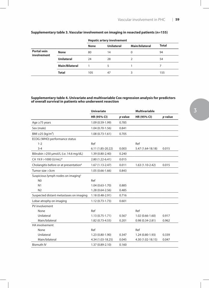
Vascular involvement in PHC
3
| 59
Supplementary table 3. Vascular involvement on imaging in resected patients (n=155)
Hepatic artery involvement
None Unilateral Main/bilateral Total
Portal vein involvement
None 80 14 0 94
Unilateral 24 28 2 54
Main/Bilateral 1 5 1 7
Total 105 47 3 155
Supplementary table 4. Univariate and multivariable Cox regression analysis for predictors of overall survival in patients who underwent resection
Univariate Multivariable
HR (95% CI) p value HR (95% CI) p value
Age ≥75 years 1.09 (0.59-1.99) 0.785
Sex (male) 1.04 (0.70-1.56) 0.841
BMI ≥25 (kg/m²) 1.08 (0.73-1.61) 0.705
ECOG (WHO) performance status1-23-4
Ref6.11 (1.85-20.22) 0.003
Ref5.47 (1.64-18.18) 0.015
Bilirubin >250 µmol/L (i.e. 14.6 mg/dL) 1.39 (0.80-2.40) 0.240
CA 19.9 >1000 (U/mL)* 2.80 (1.22-6.41) 0.015
Cholangitis before or at presentation# 1.67 (1.13-2.47) 0.011 1.63 (1.10-2.42) 0.015
Tumor size >3cm 1.05 (0.66-1.66) 0.843
Suspicious lymph nodes on imaging‡
N0N1N2
Ref1.04 (0.63-1.70)1.28 (0.64-2.56)
0.8850.485
Suspected distant metastases on imaging 1.18 (0.48-2.91) 0.716
Lobar atrophy on imaging 1.12 (0.73-1.73) 0.601
PV involvementNoneUnilateralMain/bilateral
Ref1.13 (0.75-1.71)1.82 (0.73-4.55)
0.5670.201
Ref1.02 (0.66-1.60)0.98 (0.34-2.81)
0.9170.962
HA involvementNoneUnilateralMain/bilateral
Ref1.23 (0.80-1.90)4.34 (1.03-18.25)
0.3470.045
Ref1.24 (0.80-1.93)4.30 (1.02-18.15)
0.3390.047
Bismuth IV 1.37 (0.89-2.10) 0.160

Chapter 360 |
Supplementary table 5. Univariate and multivariable Cox regression analysis for predictors of overall survival in patients who did not undergo surgical resection
Univariate Multivariable
HR (95% CI) p value HR (95% CI) p value
Age ≥75 years 1.79 (1.44-2.22) <0.001 1.70 (1.31-2.21) <0.001
Sex (male) 1.08 (0.90-1.30) 0.397
BMI ≥25 (kg/m²) 1.08 (0.90-1.31) 0.412
ECOG (WHO) performance status1-23-4
Ref1.26 (0.96-1.65) 0.103
Bilirubin >250 µmol/L (i.e. 14.6 mg/dL) 1.44 (1.15-1.79) 0.001 1.46 (1.15-1.84) 0.002
CA 19.9 >1000 (U/mL)* 1.90 (1.46-2.49) <0.001
Cholangitis before or at presentation# 0.96 (0.80-1.15) 0.634
Tumor size >3cm 1.40 (1.17-1.69) <0.001 1.60 (1.27-2.01) <0.001
Suspicious lymph nodes on imaging‡
N0N1N2
Ref1.03 (0.84-1.27)1.24 (0.98-1.57)
0.7490.076
Suspected distant metastases on imaging 1.63 (1.24-2.13) <0.001 1.96 (1.36-2.82) <0.001
Lobar atrophy on imaging 0..83 (0.68-1.02) 0.076
PV involvementNoneUnilateralMain/bilateral
Ref0.97 (0.79-1.18)1.14 (0.91-1.43)
0.7330.258
Ref0.75 (0.58-0.96)0.98 (0.72-1.33)
0.0240.898
HA involvementNoneUnilateralMain/bilateral
Ref1.15 (0.94-1.39)1.26 (0.97-1.63)
0.1700.087
Ref0.98 (0.76-1.27)1.03 (0.71-1.49)
0.8800.876
Bismuth IV 1.10 (0.92-1.32) 0.305

Vascular involvement in PHC
3
| 61

4CHAPTER 4

4 Validation of the Mayo Clinic Staging System in Determining Prognoses of Patients With Perihilar Cholangiocarcinoma.
Coelen RJS | Gaspersz MP | Labeur T | van Vugt JL | van Dieren S | Willemssen FEJA | Nio CY | IJzermans JN | Klumpen HJ | Groot Koerkamp B | van Gulik TM
Clin Gastroenterol Hepatol. 2017 Dec;15(12):1930-1939
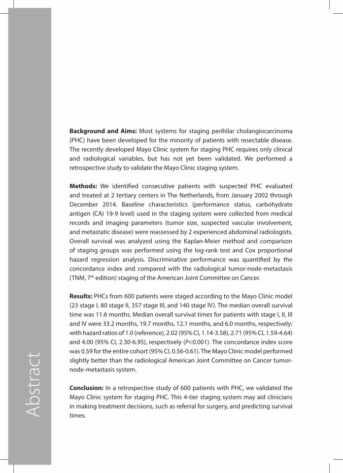
Background and Aims: Most systems for staging perihilar cholangiocarcinoma (PHC) have been developed for the minority of patients with resectable disease. The recently developed Mayo Clinic system for staging PHC requires only clinical and radiological variables, but has not yet been validated. We performed a retrospective study to validate the Mayo Clinic staging system.
Methods: We identified consecutive patients with suspected PHC evaluated and treated at 2 tertiary centers in The Netherlands, from January 2002 through December 2014. Baseline characteristics (performance status, carbohydrate antigen (CA) 19-9 level) used in the staging system were collected from medical records and imaging parameters (tumor size, suspected vascular involvement, and metastatic disease) were reassessed by 2 experienced abdominal radiologists. Overall survival was analyzed using the Kaplan-Meier method and comparison of staging groups was performed using the log-rank test and Cox proportional hazard regression analysis. Discriminative performance was quantified by the concordance index and compared with the radiological tumor-node-metastasis (TNM, 7th edition) staging of the American Joint Committee on Cancer.
Results: PHCs from 600 patients were staged according to the Mayo Clinic model (23 stage I, 80 stage II, 357 stage III, and 140 stage IV). The median overall survival time was 11.6 months. Median overall survival times for patients with stage I, II, III and IV were 33.2 months, 19.7 months, 12.1 months, and 6.0 months, respectively; with hazard ratios of 1.0 (reference), 2.02 (95% CI, 1.14-3.58), 2.71 (95% CI, 1.59-4.64) and 4.00 (95% CI, 2.30-6.95), respectively (P<0.001). The concordance index score was 0.59 for the entire cohort (95% CI, 0.56-0.61). The Mayo Clinic model performed slightly better than the radiological American Joint Committee on Cancer tumor-node-metastasis system.
Conclusion: In a retrospective study of 600 patients with PHC, we validated the Mayo Clinic system for staging PHC. This 4-tier staging system may aid clinicians in making treatment decisions, such as referral for surgery, and predicting survival times. Abs
trac
t

Validation of Mayo Clinic System for PHC
4
| 65
Background and Aims
Perihilar cholangiocarcinoma (PHC) is the most common malignancy arising from the biliary tree and has a poor prognosis.1 Long-term survival is achieved with curative-intent resection and has been reported with a median overall survival (OS) of 19 to 40 months and 5-year survival rates of 13% to 40% in high-volume centers.2-4 Unfortunately, only 10% to 20% of patients are eligible to undergo resection. Liver transplantation with neoadjuvant chemoradiation for locally advanced tumors is only performed in highly selected patients.5 Limited treatment options are available for the remaining patients. Local ablative therapies, including intraluminal radiofrequency ablation or photodynamic therapy, seem promising techniques for locally advanced PHC but require further investigation.6-8 Systemic chemotherapy with gemcitabine plus cisplatin is the standard palliative therapy, resulting in a median OS of about 12 months.9
Accurate staging of PHC patients is important for informing patients about prognosis and guiding clinicians in treatment selection. However, most staging systems have been developed for the minority of patients with resectable disease. The Bismuth-Corlette classification and Memorial Sloan Kettering Cancer Center (MSKCC) staging system were developed to determine surgical strategy and resectability, rather than to predict survival.10-12 The American Joint Committee on Cancer/Union for International Cancer Control (AJCC/UICC) tumor-node-metastasis (TNM) system, the international cholangiocarcinoma group staging system, and the MSKCC/Academic Medical Center (AMC) nomogram were designed to predict prognosis, but require pathological parameters available only after resection.4, 13-15
Recently, a clinically based staging system was proposed by the Mayo Clinic, which uses only clinical and radiological parameters that are available for all PHC patients, regardless of subsequent treatment.16 This four-tier staging system may aid clinicians with treatment decisions (e.g. surgical referral) and predicting survival in PHC patients as its performance to do so was superior to the TNM staging system in the derivation cohort. The staging system can also be useful in the stratification of patients in clinical trials by obtaining groups with comparable predicted survival outcomes. However, as external validation of models is desired before implementation in clinical practice, we aimed to assess the external validity of the Mayo Clinic staging system for PHC patients.
Chapter 466 |
Methods
Study populationAll consecutive patients with suspected PHC who were evaluated and treated at the AMC in Amsterdam and the Erasmus University Medical Center in Rotterdam, The Netherlands, from January 2002 until December 2014 were identified using keywords and diagnostic codes from the electronic patient registration systems. Medical records were reviewed and patients were screened for eligibility. PHC was defined as a tumor mass or a malignant appearing stricture at or near the biliary confluence.13 We excluded patients with hilar-invasive intrahepatic cholangiocarcinoma with the tumor’s center proximal to the secondary biliary branches, gallbladder carcinoma, cystic duct carcinoma and distal cholangiocarcinoma. Patients who underwent treatment other than biliary drainage (e.g., chemotherapy) prior to initial presentation at one of these centers were also excluded. Furthermore, patients who were not fully evaluated at one of these centers but only referred for a single biliary drainage intervention, were excluded. A waiver was granted from the Institutional Review Board at both centers for approval of this study.
The definitive diagnosis of PHC was established based on surgical histopathology following resection (N=160), histopathology from intraoperative, endoscopic, or percutaneous ultrasound-guided biopsy (N=414), and positive cytology from endoscopic brush or fine needle aspiration and suspicious cytology in absence of primary sclerosing cholangitis (PSC) (N=70). In absence of histopathological confirmation, the diagnosis was determined by the multidisciplinary hepatopancreaticobiliary team, based on clinical symptoms, radiological and endoscopic imaging, laboratory tests including tumor markers, and follow-up (N=88).
Diagnostic work-up and treatment algorithmStandard work-up included multiphase contrast-enhanced computed tomography in at least the arterial and portal venous phase, and/or contrast-enhanced magnetic resonance imaging with cholangiopancreatography. Staging and resectability assessment were performed according to the Bismuth-Corlette classification and MSKCC criteria.10, 12 Tumors were considered locally advanced if they invaded surrounding organs or when excessive vascular or biliary involvement precluded an R0 or R1 resection. Excessive vascular involvement was defined as the need to perform hepatic arterial reconstruction in any patient, or portal vein reconstruction in high surgical risk patients.17 Tumors were considered metastatic in the presence of distant metastases or lymph node metastases beyond the hepatoduodenal ligament (N2 lymph nodes).No adjuvant chemotherapy was given after resection, in compliance with Dutch guidelines. Patients with locally advanced or metastatic disease, either at presentation or at laparotomy, were assessed for palliative systemic chemotherapy. Before 2010, chemotherapy included
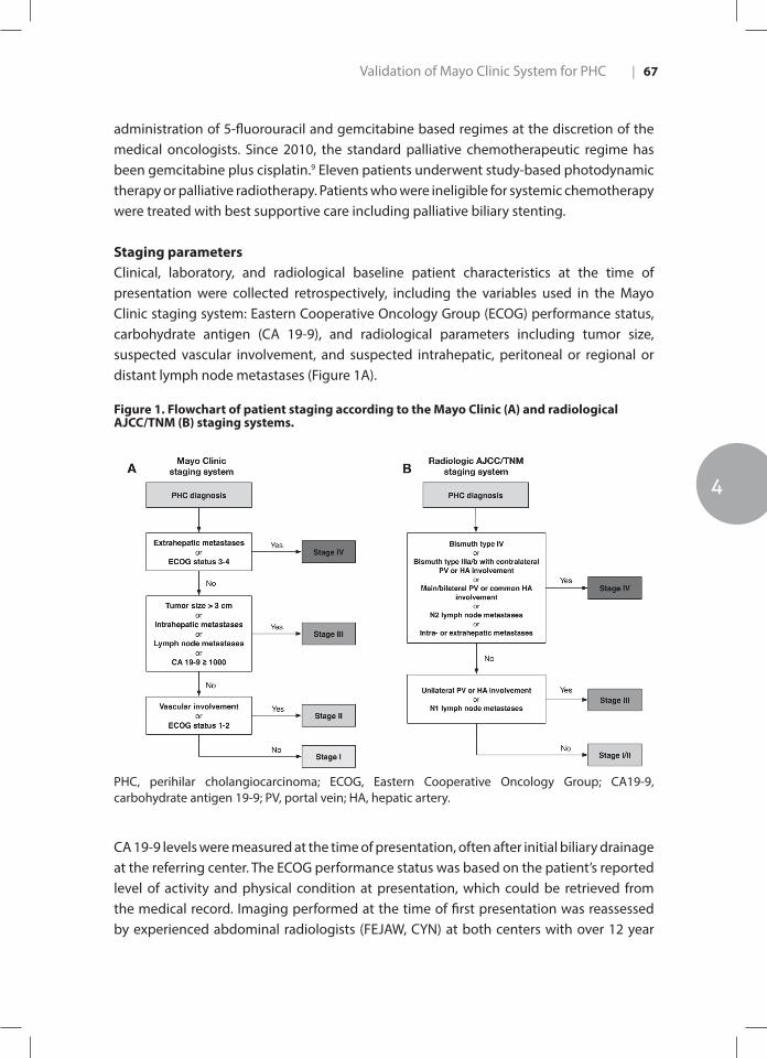
Validation of Mayo Clinic System for PHC
4
| 67
administration of 5-fluorouracil and gemcitabine based regimes at the discretion of the medical oncologists. Since 2010, the standard palliative chemotherapeutic regime has been gemcitabine plus cisplatin.9 Eleven patients underwent study-based photodynamic therapy or palliative radiotherapy. Patients who were ineligible for systemic chemotherapy were treated with best supportive care including palliative biliary stenting.
Staging parametersClinical, laboratory, and radiological baseline patient characteristics at the time of presentation were collected retrospectively, including the variables used in the Mayo Clinic staging system: Eastern Cooperative Oncology Group (ECOG) performance status, carbohydrate antigen (CA 19-9), and radiological parameters including tumor size, suspected vascular involvement, and suspected intrahepatic, peritoneal or regional or distant lymph node metastases (Figure 1A).
Figure 1. Flowchart of patient staging according to the Mayo Clinic (A) and radiological AJCC/TNM (B) staging systems.
PHC, perihilar cholangiocarcinoma; ECOG, Eastern Cooperative Oncology Group; CA19-9, carbohydrate antigen 19-9; PV, portal vein; HA, hepatic artery.
CA 19-9 levels were measured at the time of presentation, often after initial biliary drainage at the referring center. The ECOG performance status was based on the patient’s reported level of activity and physical condition at presentation, which could be retrieved from the medical record. Imaging performed at the time of first presentation was reassessed by experienced abdominal radiologists (FEJAW, CYN) at both centers with over 12 year

Chapter 468 |
experience in liver imaging. They were blinded for clinical information and the eventual treatment that patients underwent. Tumor size was defined as the maximum transverse diameter of the hilar tumor. Vascular involvement was defined as more than 180 degrees circumferential tumor contact or as clear distortion, narrowing or occlusion of the portal venous system and/or (branches of ) the hepatic artery.12, 15 Suspicious lymph nodes were defined as nodes larger than 1 cm in short-axis diameter, with central necrosis, an irregular border or hyper-attenuating compared to portal phase liver parenchyma.18
Statistical analysisStatistical analyses were performed using IBM SPSS Statistics for Windows Version 23.0 (IBM Corp., Armonk, NY, USA) and R Version 3.1.2 (R Foundation for Statistical Computing, Vienna, Austria). Continuous variables are presented as mean with standard deviation (s.d.) or median with range for non-normally distributed data. Categorical variables are expressed as counts and percentages. Overall survival was defined as the time from initial presentation at one of both centers to the date of death or last follow-up. Survival status was checked using the municipal records database on May 9, 2016.
Patients were staged into any of the four stage groups according to the Mayo Clinic system (Figure 1A). If data required for classifying patients in the staging system were missing, patients were excluded from further analyses. Multiple imputation was tested as a method to correct for missing data (especially CA 19-9), but results were similar to full-case analysis. Also, upon performing a sensitivity analysis with imputation of the lowest (best case scenario) and highest (worst case scenario) CA 19-9 values, results did not improve compared to full-case analysis (Supplemental Information). Therefore, data were handled with full-case analysis based on all patients that could be staged according to the Mayo Clinic system. Median OS of the four stage groups was analyzed using the Kaplan-Meier method and compared with the log-rank test. Univariable Cox proportional hazard regression analysis was used to calculate hazard ratios (HRs) with 95% confidence intervals (95% CI) for the different staging groups. A P value <0.05 was considered to indicate statistical significance. The assumption of proportional hazards (i.e. the effect of a given variable is constant over time) was checked by plotting the log-minus log survival curves with parallel lines indicating that the assumption was held.
Discriminative performance of the staging system was analyzed by calculating the concordance (C-) index using Harrell’s method.19 The C-index quantifies the probability that in a pair of randomly selected patients, the patient with the worst predicted outcome (i.e. higher Mayo Clinic stage) has a shorter survival than the other patient. A C-index of 1 indicates perfect concordance, whereas a C-index of 0.5 implies no predictive ability. A formal calibration test to measure the model’s goodness of fit could not be applied to this
Validation of Mayo Clinic System for PHC
4
| 69
four-tier model, but mean survival time for each Mayo stage in our cohort was compared to the original cohort from Mayo Clinic.
The predictive performance of the Mayo Clinic model was compared to the 7th edition of the AJCC/TNM staging system. All PHC patients were assigned to the AJCC/TNM stages based on radiological parameters (Figure 1B). For example, involvement of the main portal vein or bilateral second-order biliary radicals (Bismuth-Corlette type IV) on imaging were considered as stage IV. Stages I and II were merged as tumor depth and tumor invasion in surrounding adipose tissue cannot be accurately assessed on imaging.
Several subgroup analyses were performed. First, the performance of the staging systems was analyzed for subgroups of patients based on treatment. Patients were categorized into two groups: 1) patients with potentially resectable PHC who underwent exploratory laparotomy (including completed and aborted resection), and 2) patients with locally advanced or metastasized PHC at presentation. Patients who underwent liver transplantation were categorized in the laparotomy group.
Secondly, in order to assess whether a high Mayo Clinic stage may have implications for treatment selection, we separately analyzed the survival outcomes of stage III and IV patients who did and did not undergo exploratory laparotomy.
Results
Patient characteristicsBetween 2002 and 2014, 732 consecutive patients were evaluated and treated for PHC. Because of 1 or more missing parameters, 132 (18.0%) patients could not be staged by the Mayo Clinic system (Supplementary Table). Characteristics of the remaining 600 patients including demographics and clinical-, radiological-, and surgical parameters are shown in Table 1. Three-hundred twenty-six patients (54.3%) had unresectable PHC at presentation, 274 (45.7%) underwent laparotomy, and in 117 (19.5%) patients a resection was performed. Only two patients underwent liver transplantation.
Median follow-up of the included cohort was 83.6 months and median OS (95% CI) was 11.6 months (10.2-12.9). At the time of last follow-up, 550 of 600 (91.7%) patients had died.
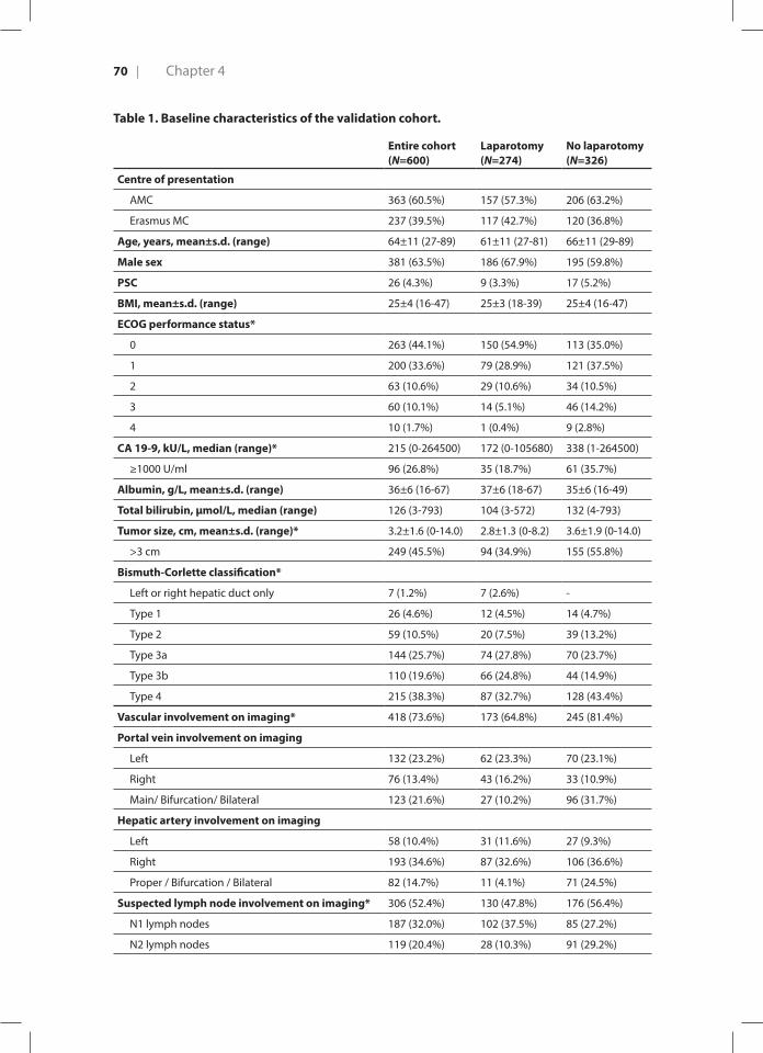
Chapter 470 |
Table 1. Baseline characteristics of the validation cohort.
Entire cohort (N=600)
Laparotomy(N=274)
No laparotomy(N=326)
Centre of presentation
AMC 363 (60.5%) 157 (57.3%) 206 (63.2%)
Erasmus MC 237 (39.5%) 117 (42.7%) 120 (36.8%)
Age, years, mean±s.d. (range) 64±11 (27-89) 61±11 (27-81) 66±11 (29-89)
Male sex 381 (63.5%) 186 (67.9%) 195 (59.8%)
PSC 26 (4.3%) 9 (3.3%) 17 (5.2%)
BMI, mean±s.d. (range) 25±4 (16-47) 25±3 (18-39) 25±4 (16-47)
ECOG performance status*
0 263 (44.1%) 150 (54.9%) 113 (35.0%)
1 200 (33.6%) 79 (28.9%) 121 (37.5%)
2 63 (10.6%) 29 (10.6%) 34 (10.5%)
3 60 (10.1%) 14 (5.1%) 46 (14.2%)
4 10 (1.7%) 1 (0.4%) 9 (2.8%)
CA 19-9, kU/L, median (range)* 215 (0-264500) 172 (0-105680) 338 (1-264500)
≥1000 U/ml 96 (26.8%) 35 (18.7%) 61 (35.7%)
Albumin, g/L, mean±s.d. (range) 36±6 (16-67) 37±6 (18-67) 35±6 (16-49)
Total bilirubin, µmol/L, median (range) 126 (3-793) 104 (3-572) 132 (4-793)
Tumor size, cm, mean±s.d. (range)* 3.2±1.6 (0-14.0) 2.8±1.3 (0-8.2) 3.6±1.9 (0-14.0)
>3 cm 249 (45.5%) 94 (34.9%) 155 (55.8%)
Bismuth-Corlette classification*
Left or right hepatic duct only 7 (1.2%) 7 (2.6%) -
Type 1 26 (4.6%) 12 (4.5%) 14 (4.7%)
Type 2 59 (10.5%) 20 (7.5%) 39 (13.2%)
Type 3a 144 (25.7%) 74 (27.8%) 70 (23.7%)
Type 3b 110 (19.6%) 66 (24.8%) 44 (14.9%)
Type 4 215 (38.3%) 87 (32.7%) 128 (43.4%)
Vascular involvement on imaging* 418 (73.6%) 173 (64.8%) 245 (81.4%)
Portal vein involvement on imaging
Left 132 (23.2%) 62 (23.3%) 70 (23.1%)
Right 76 (13.4%) 43 (16.2%) 33 (10.9%)
Main/ Bifurcation/ Bilateral 123 (21.6%) 27 (10.2%) 96 (31.7%)
Hepatic artery involvement on imaging
Left 58 (10.4%) 31 (11.6%) 27 (9.3%)
Right 193 (34.6%) 87 (32.6%) 106 (36.6%)
Proper / Bifurcation / Bilateral 82 (14.7%) 11 (4.1%) 71 (24.5%)
Suspected lymph node involvement on imaging* 306 (52.4%) 130 (47.8%) 176 (56.4%)
N1 lymph nodes 187 (32.0%) 102 (37.5%) 85 (27.2%)
N2 lymph nodes 119 (20.4%) 28 (10.3%) 91 (29.2%)
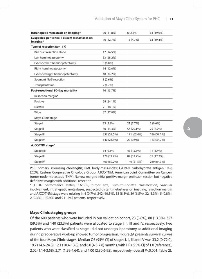
Validation of Mayo Clinic System for PHC
4
| 71
Intrahepatic metastasis on imaging* 70 (11.8%) 6 (2.2%) 64 (19.9%)
Suspected peritoneal / distant metastases on imaging*
76 (12.7%) 13 (4.7%) 63 (19.4%)
Type of resection (N=117)
Bile duct resection alone 17 (14.5%)
Left hemihepatectomy 33 (28.2%)
Extended left hemihepatectomy 8 (6.8%)
Right hemihepatectomy 14 (12.0%)
Extended right hemihepatectomy 40 (34.2%)
Segment 4b/5 resection 3 (2.6%)
Transplantation 2 (1.7%)
Post-resectional 90-day mortality 16 (13.7%)
Resection margin*
Positive 28 (24.1%)
Narrow 21 (18.1%)
Wide 67 (57.8%)
Mayo Clinic stage
Stage I 23 (3.8%) 21 (7.7%) 2 (0.6%)
Stage II 80 (13.3%) 55 (20.1%) 25 (7.7%)
Stage III 357 (59.5%) 171 (62.4%) 186 (57.1%)
Stage IV 140 (23.3%) 27 (9.9%) 113 (34.7%)
AJCC/TNM stage*
Stage I/II 54 (9.1%) 43 (15.8%) 11 (3.4%)
Stage III 128 (21.7%) 89 (32.7%) 39 (12.2%)
Stage IV 409 (69.2%) 140 (51.5%) 269 (84.3%)
PSC, primary sclerosing cholangitis; BMI, body-mass-index; CA19-9, carbohydrate antigen 19-9; ECOG: Eastern Cooperative Oncology Group; AJCC/TNM, American Joint Committee on Cancer/tumor-node-metastasis (TNM). Narrow margin: initial positive margin on frozen section but negative definitive margin with additional resection.* ECOG performance status, CA19-9, tumor size, Bismuth-Corlette classification, vascular involvement, intrahepatic metastases, suspected distant metastases on imaging, resection margin and AJCC/TNM stage were missing in 4 (0.7%), 242 (40.3%), 53 (8.8%), 39 (6.5%), 32 (5.3%), 5 (0.8%), 2 (0.3%), 1 (0.9%) and 9 (1.5%) patients, respectively.
Mayo Clinic staging groupsOf the 600 patients who were included in our validation cohort, 23 (3.8%), 80 (13.3%), 357 (59.5%) and 140 (23.3%) patients were allocated to stage I, II, III and IV, respectively. Two patients who were classified as stage I did not undergo laparotomy as additional imaging during preoperative work-up showed tumor progression. Figure 2A presents survival curves of the four Mayo Clinic stages. Median OS (95% CI) of stages I, II, III and IV was 33.2 (0-72.0), 19.7 (14.6-24.8), 12.1 (10.4-13.8), and 6.0 (4.3-7.8) months, with HRs (95% CI) of 1.0 (reference), 2.02 (1.14-3.58), 2.71 (1.59-4.64), and 4.00 (2.30-6.95), respectively (overall P<0.001; Table 2).
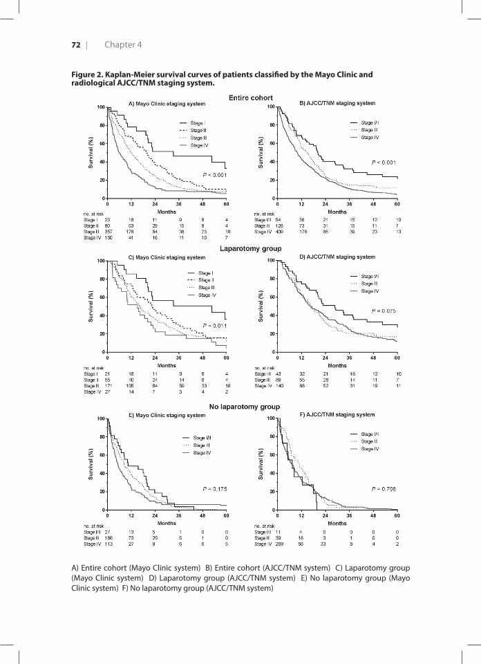
Chapter 472 |
Figure 2. Kaplan-Meier survival curves of patients classified by the Mayo Clinic and radiological AJCC/TNM staging system.
A) Entire cohort (Mayo Clinic system) B) Entire cohort (AJCC/TNM system) C) Laparotomy group (Mayo Clinic system) D) Laparotomy group (AJCC/TNM system) E) No laparotomy group (Mayo Clinic system) F) No laparotomy group (AJCC/TNM system)
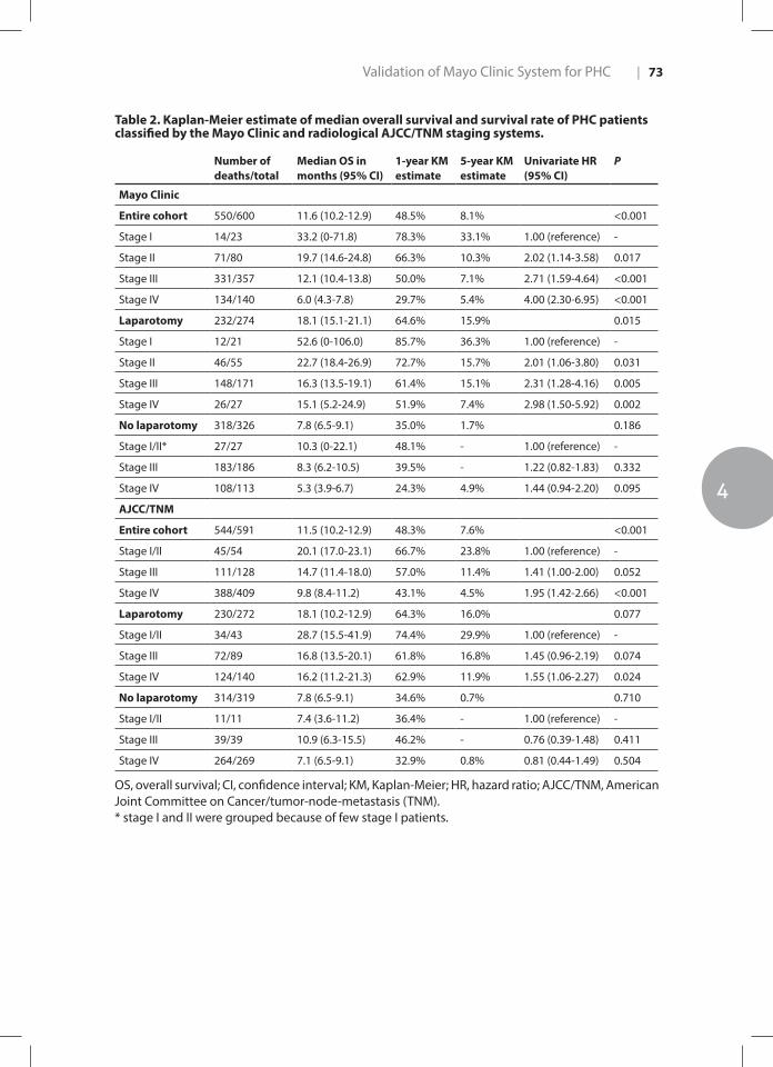
Validation of Mayo Clinic System for PHC
4
| 73
Table 2. Kaplan-Meier estimate of median overall survival and survival rate of PHC patients classified by the Mayo Clinic and radiological AJCC/TNM staging systems.
Number of deaths/total
Median OS in months (95% CI)
1-year KM estimate
5-year KM estimate
Univariate HR (95% CI)
P
Mayo Clinic
Entire cohort 550/600 11.6 (10.2-12.9) 48.5% 8.1% <0.001
Stage I 14/23 33.2 (0-71.8) 78.3% 33.1% 1.00 (reference) -
Stage II 71/80 19.7 (14.6-24.8) 66.3% 10.3% 2.02 (1.14-3.58) 0.017
Stage III 331/357 12.1 (10.4-13.8) 50.0% 7.1% 2.71 (1.59-4.64) <0.001
Stage IV 134/140 6.0 (4.3-7.8) 29.7% 5.4% 4.00 (2.30-6.95) <0.001
Laparotomy 232/274 18.1 (15.1-21.1) 64.6% 15.9% 0.015
Stage I 12/21 52.6 (0-106.0) 85.7% 36.3% 1.00 (reference) -
Stage II 46/55 22.7 (18.4-26.9) 72.7% 15.7% 2.01 (1.06-3.80) 0.031
Stage III 148/171 16.3 (13.5-19.1) 61.4% 15.1% 2.31 (1.28-4.16) 0.005
Stage IV 26/27 15.1 (5.2-24.9) 51.9% 7.4% 2.98 (1.50-5.92) 0.002
No laparotomy 318/326 7.8 (6.5-9.1) 35.0% 1.7% 0.186
Stage I/II* 27/27 10.3 (0-22.1) 48.1% - 1.00 (reference) -
Stage III 183/186 8.3 (6.2-10.5) 39.5% - 1.22 (0.82-1.83) 0.332
Stage IV 108/113 5.3 (3.9-6.7) 24.3% 4.9% 1.44 (0.94-2.20) 0.095
AJCC/TNM
Entire cohort 544/591 11.5 (10.2-12.9) 48.3% 7.6% <0.001
Stage I/II 45/54 20.1 (17.0-23.1) 66.7% 23.8% 1.00 (reference) -
Stage III 111/128 14.7 (11.4-18.0) 57.0% 11.4% 1.41 (1.00-2.00) 0.052
Stage IV 388/409 9.8 (8.4-11.2) 43.1% 4.5% 1.95 (1.42-2.66) <0.001
Laparotomy 230/272 18.1 (10.2-12.9) 64.3% 16.0% 0.077
Stage I/II 34/43 28.7 (15.5-41.9) 74.4% 29.9% 1.00 (reference) -
Stage III 72/89 16.8 (13.5-20.1) 61.8% 16.8% 1.45 (0.96-2.19) 0.074
Stage IV 124/140 16.2 (11.2-21.3) 62.9% 11.9% 1.55 (1.06-2.27) 0.024
No laparotomy 314/319 7.8 (6.5-9.1) 34.6% 0.7% 0.710
Stage I/II 11/11 7.4 (3.6-11.2) 36.4% - 1.00 (reference) -
Stage III 39/39 10.9 (6.3-15.5) 46.2% - 0.76 (0.39-1.48) 0.411
Stage IV 264/269 7.1 (6.5-9.1) 32.9% 0.8% 0.81 (0.44-1.49) 0.504
OS, overall survival; CI, confidence interval; KM, Kaplan-Meier; HR, hazard ratio; AJCC/TNM, American Joint Committee on Cancer/tumor-node-metastasis (TNM).* stage I and II were grouped because of few stage I patients.
Chapter 474 |
Subgroup survival analysis of different treatment groupsMedian OS (95% CI) of patients in the laparotomy group (regardless of subsequent resection) and patients who did not undergo laparotomy was 18.1 (15.1-21.1) and 7.8 (6.5-9.1) months, respectively (P<0.001; Table 2). Median OS (95% CI) of patients who underwent resection was 37.7 (25.6-49.7) months.
Prognostic performance of the Mayo Clinic staging system was then evaluated for the two treatment groups. Overall survival including 1- and 5-year estimates and HRs (95% CI) are presented in Table 2. Median OS among stage I, II, III and IV patients who underwent laparotomy was 52.6, 22.7, 16.3, and 15.1 months (P=0.011; Figure 2C), with HRs of 1.0 (reference), 2.01, 2.31 and 2.98, respectively (overall P=0.015). Resectability rate in stage I, II, III and IV was 62%, 56%, 37% and 33%, respectively (P=0.016).
Among patients who presented with locally advanced or metastatic disease on preoperative imaging and did not undergo laparotomy, median OS of stage I/II, III, and IV was 10.3, 8.3, and 5.3 months (P=0.184, Table 2 and Figure 2E), with HRs of 1.0 (reference), 1.22 and 1.44 (overall P=0.186). Although there was no overall statistical significant difference, stage I/II and III patients had better 1-year survival rates than stage IV patients; i.e. 48% and 40%, respectively, versus 24%.
Radiological AJCC/TNM staging groupsOf the 600 patients in the validation cohort, 54, 128, and 409 patients were allocated to radiological AJCC/TNM stage I/II, III and IV, respectively. Median OS (95% CI) of patients in stages I/II, III, and IV was 20.1 (17.0-23.1), 14.7 (11.4-18.0), and 9.8 (8.4-11.2) months (P<0.001, Figure 2B), with HRs (95% CI) of 1.00 (reference), 1.41 (1.00-2.00), and 1.95 (1.42-2.66) (overall P<0.001; Table 2). There was no statistically significant difference in OS across stages when evaluating the radiological AJCC/TNM staging separately for patients who did and did not undergo exploratory laparotomy (Table 2 and Figure 2D/F).
Predictive performanceIn the entire cohort, the C-index (95% CI) of the Mayo Clinic staging system was 0.59 (0.56-0.61). When patients were categorized by treatment group, C-indices (95% CI) were 0.56 (0.52-0.60) for the laparotomy group and 0.54 (0.51-0.57) for the group of patients that did not undergo laparotomy.C-indices (95% CI) of the radiological AJCC staging system were 0.56 (0.54-0.58) for the entire cohort, 0.53 (0.49-0.57) for the laparotomy group and 0.52 (0.50-0.54) for the no laparotomy group.
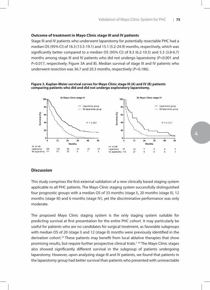
Validation of Mayo Clinic System for PHC
4
| 75
Outcome of treatment in Mayo Clinic stage III and IV patientsStage III and IV patients who underwent laparotomy for potentially resectable PHC had a median OS (95% CI) of 16.3 (13.5-19.1) and 15.1 (5.2-24.9) months, respectively, which was significantly better compared to a median OS (95% CI) of 8.3 (6.2-10.5) and 5.3 (3.8-6.7) months among stage III and IV patients who did not undergo laparotomy (P<0.001 and P=0.017, respectively; Figure 3A and B). Median survival of stage III and IV patients who underwent resection was 36.7 and 20.3 months, respectively (P=0.186).
Figure 3. Kaplan-Meier survival curves for Mayo Clinic stage III (A) and IV (B) patients comparing patients who did and did not undergo exploratory laparotomy.
Discussion
This study comprises the first external validation of a new clinically based staging system applicable to all PHC patients. The Mayo Clinic staging system successfully distinguished four prognostic groups with a median OS of 33 months (stage I), 20 months (stage II), 12 months (stage III) and 6 months (stage IV), yet the discriminative performance was only moderate.
The proposed Mayo Clinic staging system is the only staging system suitable for predicting survival at first presentation for the entire PHC cohort. It may particularly be useful for patients who are no candidates for surgical treatment, as favorable subgroups with median OS of 20 (stage I) and 12 (stage II) months were previously identified in the derivation cohort.16 These patients may benefit from local ablative therapies that show promising results, but require further prospective clinical trials.7, 20 The Mayo Clinic stages also showed significantly different survival in the subgroup of patients undergoing laparotomy. However, upon analyzing stage III and IV patients, we found that patients in the laparotomy group had better survival than patients who presented with unresectable

Chapter 476 |
PHC, suggesting that the staging system is not likely to guide in the selection of patients for laparotomy. The model may, nonetheless, be useful for patient counseling and stratification of patients for clinical trials into cohorts with comparable expected survival outcomes.
The Mayo Clinic model performed slightly better than the radiological AJCC/TNM system as demonstrated by the Kaplan-Meier survival curves and higher concordance score, but with overlapping confidence intervals. However, concordance of the Mayo Clinic staging system was only moderate as reflected by a C-index of 0.59 for the entire cohort. This is likely caused by differences in study population between the derivation and validation cohort. The staging system was developed in a center where many patients had PSC as underlying disease and underwent liver transplantation, while only 4% had PSC and only two patients underwent transplantation in the present cohort. Furthermore, we observed several differences in patient selection for treatment allocation. While patients in our laparotomy group often had radiological suspicion on vascular involvement (65%) or lymph node metastases (48%), the vast majority of patients subjected to laparotomy in the derivation cohort had less advanced tumors. Only 23% of those patients had signs of vascular involvement by tumor and only 1% had lymph node metastases. Obviously, dissimilarities in treatment selection have led to a different distribution of prognostic risk factors in our cohort, consequently affecting the concordance.21 In addition, differences in survival at each Mayo stage and particularly stage III-IV may be caused by a higher percentage of patients with high ECOG performance scores and (extra)hepatic metastases in our cohort. Consequently, patients in Mayo stage III and IV were more likely to have more than one poor prognostic factor. The staging system’s performance might be improved for individual institutions by adding cohort-specific prognostic factors or by altering the value of each variable in the model. For example, there may be a suggestion that hepatic arterial involvement (and not portal vein involvement) could further enhance staging for PHC.22
The clinical and radiological parameters used in the model are part of the standard diagnostic work-up and therefore readily available. However, several limitations apply to the included variables. Remarkably, the majority (60%) of our patients were classified as stage III, mainly because tumor size above 3 centimeters and suspected lymph node metastases were prevalent. These features are known to be important prognostic risk factors, especially after resection of PHC.4, 23 However, the diagnostic accuracy of radiological imaging to detect lymph node metastases is poor and may lead to misclassification of patients.24-26 Radiological parameters, such as tumor diameter and vascular involvement, are also highly dependent on the quality of imaging and physician’s expertise. Furthermore, ECOG performance status is also subject to inter-observer variability.27 In addition, depending on the success of relieving cholestasis and occurrence

Validation of Mayo Clinic System for PHC
4
| 77
of biliary drainage-related complications, performance status may change over time. For example, patients who are initially classified as high clinical stage may physically improve and become candidates for surgical exploration. Finally, the level of serum CA 19-9 depends on the extent of biliary obstruction and on whether biliary drainage has been performed.28, 29 In accordance with the Mayo Clinic staging system, CA 19-9 levels were not corrected for bilirubin levels.
Strengths of our study are the multicenter design including the two largest tertiary referral centers for PHC in the Netherlands. The validation cohort to which the staging model was applied included 600 PHC patients and and was a large series. The standardized multidisciplinary assessment in two expert centers and re-evaluation of all radiological images by experienced radiologists using clear definitions has aided in the construction of a robust dataset.
Limitations of our study include the inherent drawbacks of the retrospective design. 18% of patients could not be staged due to missing data. Serum CA19- 9 values especially were missing because this analysis was not performed routinely before 2010. Consequently, we were unable to classify several patients who potentially would have been allocated to stage I or II. After all, individuals who were excluded from the analyses had less advanced tumors and a more favorable prognosis (Supplementary Table). Considering the limited value of multiple imputation when the amount of missing data is high (i.e., for CA 19-9), we committed to full case analysis.30 We also did not investigate inter-observer variability of the reassessed radiological characteristics but reevaluation was performed by two highly experienced liver radiologists.
In conclusion, this study has shown that the Mayo Clinic staging system for patients with PHC was applicable to an external cohort including patients undergoing standard resection rather than liver transplantation. Despite some limitations, the model provided successful prognostic stratification in the entire cohort of PHC. Given the moderate discriminative performance, the staging system may be further improved for centers in which liver transplantation is not the most common treatment for PHC patients. Interestingly, the model has potential to distinguish advanced PHC patients who may be candidates for local therapies.

Chapter 478 |
References
1. Nakeeb A, Pitt HA, Sohn TA, et al. Cholangiocarcinoma. A spectrum of intrahepatic, perihilar, and
distal tumors. Ann Surg. 1996;224:463-473; discussion 473-465.
2. Popescu I, Dumitrascu T. Curative-intent surgery for hilar cholangiocarcinoma: prognostic
factors for clinical decision making. Langenbecks Arch Surg. 2014;399:693-705.
3. Ito F, Cho CS, Rikkers LF, Weber SM. Hilar cholangiocarcinoma: current management. Ann Surg.
2009;250:210-218.
4. Groot Koerkamp B, Wiggers JK, Gonen M, et al. Survival after resection of perihilar
cholangiocarcinoma-development and external validation of a prognostic nomogram. Ann
Oncol. 2016;27:753.
5. Gores GJ, Darwish Murad S, Heimbach JK, Rosen CB. Liver transplantation for perihilar
cholangiocarcinoma. Dig Dis. 2013;31:126-129.
6. Laquiere A, Boustiere C, Leblanc S, Penaranda G, Desilets E, Prat F. Safety and feasibility of
endoscopic biliary radiofrequency ablation treatment of extrahepatic cholangiocarcinoma.
Surg Endosc. 2016;30:1242-1248.
7. Wagner A, Denzer UW, Neureiter D, et al. Temoporfin improves efficacy of photodynamic therapy
in advanced biliary tract carcinoma: A multicenter prospective phase II study. Hepatology.
2015;62:1456-1465.
8. Strand DS, Cosgrove ND, Patrie JT, et al. ERCP-directed radiofrequency ablation and
photodynamic therapy are associated with comparable survival in the treatment of unresectable
cholangiocarcinoma. Gastrointest Endosc. 2014;80:794-804.
9. Valle J, Wasan H, Palmer DH, et al. Cisplatin plus gemcitabine versus gemcitabine for biliary tract
cancer. N Engl J Med. 2010;362:1273-1281.
10. Bismuth H, Nakache R, Diamond T. Management strategies in resection for hilar
cholangiocarcinoma. Ann Surg. 1992;215:31-38.
11. Bismuth H, Corlette MB. Intrahepatic cholangioenteric anastomosis in carcinoma of the hilus of
the liver. Surg Gynecol Obstet. 1975;140:170-178.
12. Jarnagin WR, Fong Y, DeMatteo RP, et al. Staging, resectability, and outcome in 225 patients
with hilar cholangiocarcinoma. Ann Surg. 2001;234:507-517; discussion 517-509.
13. Edge SB, Byrd DR, Compton CC, Fritz AG, Greene FL, Trotti A. AJCC cancer staging manual (7th
ed). New York, NY: Springer. 2010.
14. Groot Koerkamp B, Wiggers JK, Allen PJ, et al. American Joint Committee on Cancer staging for
resected perihilar cholangiocarcinoma: a comparison of the 6th and 7th editions. HPB (Oxford).
2014;16:1074-1082.
15. Deoliveira ML, Schulick RD, Nimura Y, et al. New staging system and a registry for perihilar
cholangiocarcinoma. Hepatology. 2011;53:1363-1371.
16. Chaiteerakij R, Harmsen WS, Marrero CR, et al. A new clinically based staging system for perihilar
cholangiocarcinoma. Am J Gastroenterol. 2014;109:1881-1890.

Validation of Mayo Clinic System for PHC
4
| 79
17. Wiggers JK, Groot Koerkamp B, Cieslak KP, et al. Postoperative Mortality after Liver Resection for
Perihilar Cholangiocarcinoma: Development of a Risk Score and Importance of Biliary Drainage
of the Future Liver Remnant. J Am Coll Surg. 2016;223:321-331.e321.
18. Lee HY, Kim SH, Lee JM, et al. Preoperative assessment of resectability of hepatic hilar
cholangiocarcinoma: combined CT and cholangiography with revised criteria. Radiology.
2006;239:113-121.
19. Harrell FE, Jr., Lee KL, Mark DB. Multivariable prognostic models: issues in developing models,
evaluating assumptions and adequacy, and measuring and reducing errors. Stat Med.
1996;15:361-387.
20. Patel J, Rizk N, Kahaleh M. Role of photodynamic therapy and intraductal radiofrequency
ablation in cholangiocarcinoma. Best Pract Res Clin Gastroenterol. 2015;29:309-318.
21. Pencina MJ, D’Agostino RB, Sr. Evaluating Discrimination of Risk Prediction Models: The C
Statistic. Jama. 2015;314:1063-1064.
22. van Vugt JLA, Gaspersz MP, Coelen RJS, et al. The prognostic value of portal vein and hepatic
artery involvement in patients with perihilar cholangiocarcinoma. HPB (Oxford) 2017.
23. Regimbeau JM, Fuks D, Pessaux P, et al. Tumour size over 3 cm predicts poor short-term
outcomes after major liver resection for hilar cholangiocarcinoma. By the HC-AFC-2009 group.
HPB (Oxford). 2014.
24. Noji T, Kondo S, Hirano S, et al. CT evaluation of paraaortic lymph node metastasis in patients
with biliary cancer. J Gastroenterol. 2005;40:739-743.
25. Noji T, Kondo S, Hirano S, Tanaka E, Suzuki O, Shichinohe T. Computed tomography evaluation
of regional lymph node metastases in patients with biliary cancer. Br J Surg. 2008;95:92-96.
26. Ruys AT, van Beem BE, Engelbrecht MR, Bipat S, Stoker J, Van Gulik TM. Radiological staging
in patients with hilar cholangiocarcinoma: a systematic review and meta-analysis. Br J Radiol.
2012;85:1255-1262.
27. Paiva CE, Siquelli FA, Santos HA, et al. The Functionality Assessment Flowchart (FAF): a new
simple and reliable method to measure performance status with a high percentage of
agreement between observers. BMC Cancer. 2015;15:501.
28. Mann DV, Edwards R, Ho S, Lau WY, Glazer G. Elevated tumour marker CA19-9: clinical
interpretation and influence of obstructive jaundice. Eur J Surg Oncol. 2000;26:474-479.
29. John AR, Haghighi KS, Taniere P, Esmat ME, Tan YM, Bramhall SR. Is a raised CA 19-9 level
diagnostic for a cholangiocarcinoma in patients with no history of sclerosing cholangitis ? Dig
Surg. 2006;23:319-324.
30. Dong Y, Peng CY. Principled missing data methods for researchers. Springerplus. 2013;2:222.

5CHAPTER 5
5 Evaluation of the new American Joint Committee on Cancer Staging Manual 8th edition for perihilar cholangiocarcinoma
Gaspersz MP | Buettner S | van Vugt JL | de Jonge J | Polak WG | Doukas M | IJzermans JNM | Groot Koerkamp B | Willemssen FEJA
J Gastrointest Surg. 2019 Feb 12
Background: The aim was to compare the prognostic accuracy of cross-sectional imaging of the 7th and 8th editions of the American Joint Committee on Cancer (AJCC) staging system for perihilar cholangiocarcinoma (PHC).
Study Design: All patients with PHC between 2002-2014 were included. Imaging at the time of presentation was reassessed and clinical Tumor-Nodal-Metastasis (cTNM) stage was determined according to the 7th and 8th edition of the AJCC staging system. Comparison of the prognostic accuracy was performed using the concordance index (c-index).
Results: A total of 248 PHC patients were included; 45 patients (18.1%) underwent a curative-intent resection, whereas 203 patients (81.9%) did not because they were unfit for surgery or were diagnosed with locally advanced or metastatic disease during work-up. Prognostic accuracy was comparable between the 7th and 8th edition (c-index 0.57 vs 0.58). For patients who underwent a curative-intent resection, the prognostic accuracy of the 8th edition (0.67) was higher than the 7th
(0.65). For patients who did not undergo a curative-intent resection, the prognostic accuracy was poor in both the 7th as the 8th edition (0.54 vs 0.57).
Conclusion: The 7 th and 8 th editions of the AJCC staging system for PHC have comparable prognostic accuracy. Prognostic accuracy was particularly poor in unresectable patients. Abs
trac
t

Evaluation of AJCC 8th edition
5
| 83
Introduction
Perihilar cholangiocarcinoma (PHC) is the most common malignancy of the bile ducts.1
Overall survival differs strongly between PHC patients, ranging from 12 months in palliative treatment to 40 months after curative-intent resection.2-4 Prognostic studies typically focus on patients undergoing curative-intent resection. However, the majority of patients with PHC have metastatic or locally advanced disease at the time of presentation.2,5-7
One of the most commonly used staging systems is the American Joint Committee on Cancer (AJCC) staging system. Recently the AJCC released the AJCC 8th edition cancer staging manual, which came into effect on January 1st 2018. The 7th edition of the AJCC staging system was the first to stage PHC and distal cholangiocarcinoma separately. The new 8th edition for PHC contains four significant changes (Table 1a and 1b). Bilateral second-order bile duct involvement (i.e. Bismuth classification IV) is no longer classified as T4 in the 8th edition. Other reasons for T4 (e.g., main portal vein involvement) are reclassified as stage IIIb rather than stage IVa. Positive lymph nodes beyond the hepatoduodenal ligament (e.g., aortocaval or celiac nodes) have become M1 disease (stage IVb) rather than N2 disease in the 7th edition. Instead, in the 8th edition N2 disease (stage IVa) is classified as 4 or more positive regional lymph nodes.
AJCC staging systems are intended to be applicable to all cancer patients, regardless whether they undergo curative-intent resection, palliative treatment, or best supportive care. As the majority of patients with PHC is not eligible for curative-intent resection, the AJCC staging involves assessment of cross-sectional imaging in most patients, rather than pathological evaluation of resected tumor specimens. Therefore, the aim of this retrospective study was to evaluate the 8th edition of the AJCC staging system for all patients with PHC and compare the prognostic value of the 7th and 8th editions of the AJCC staging system for PHC.

Chapter 584 |
Table 1a. American Joint Committee on Cancer (AJCC) staging system by tumor-node-metastasis (TNM) stage on imaging
Stage AJCC, 7th edition AJCC, 8th edition
Tumor (T) stage
T1Tumor confined to the bile duct, with extension up to the muscle layer or fibrous tissue
T2a Tumor invades beyond the wall of the bile duct to surrounding adipose tissue
T2b Tumor invades adjacent hepatic parenchyma
T3 Tumor invades unilateral branches of the PV or HA
T4 Tumor invades main PV or its branches bilaterally, or the common hepatic artery, second-order bile ducts bilaterally, unilateral second-order bile ducts with contralateral portal vein or hepatic artery involvement
Tumor invades main PV or its branches bilaterally, or the common hepatic artery, or unilateral second-order biliary radicals with contralateral portal vein or hepatic artery involvement.
Node (N) stage
N0 No regional lymph node metastasis No regional lymph node metastasis
N1 Regional lymph node metastasis: hilar (along CBD, cystic duct, HA or PV)
One to three positive lymph nodes typically involving the hilar, cystic duct, common bile duct, hepatic artery, posterior pancreatoduodenal and portal vein lymph nodes
N2 Metastasis to periaortic, pericaval, SMA or coeliac lymph nodes
Four or more positive lymph nodes from the sites described for N1
Metastasis (M) stage
M0 No distant metastasis No distant metastasis
M1 Distant metastasis Distant metastasis (includes lymph node metastatis distant to the hepatoduodenal ligament.)
Table 1b. American Joint Committee on Cancer (AJCC) staging system
AJCC, 7th edition AJCC, 8th edition
Stage T N M Stage T N M
0 is 0 0 0 is 0 0
I 1 0 0 I 1 0 0
II 2 0 0 II 2a-b 0 0
IIIa 3 0 0 IIIa 3 0 0
IIIb 1-3 1 0 IIIb 4 0 0
- 0 IIIc Any 1 0
IVa 4 Any 0 IVa Any 2 0
IVb Any
Any
2
Any
0
1
IVb Any Any 1
Evaluation of AJCC 8th edition
5
| 85
Materials and Methods
Study population and data acquisitionAll patients with PHC between 2002 and 2014 in Erasmus MC University Medical Center, Rotterdam, the Netherlands were included. PHC was defined as a mass or malignant-appearing stricture at or near the biliary confluence, arising between the origin of the cystic duct and the segmental bile ducts.8 A multidisciplinary team diagnosed PHC based on clinical characteristics, radiological characteristics, endoscopic findings, and follow-up, if histopathological evidence was not available.9 Patient and tumor characteristics, clinical parameters, and laboratory results were retrospectively collected from electronic patient records.
Experienced abdominal radiologists revised all imaging from the time of first presentation. Tumor diameter, presence and location of suspicious lymph nodes, presence of distant metastases, and vascular involvement was reassessed. Suspicious lymph nodes were defined as nodes larger than 1.0 cm in short-axis diameter, with central necrosis, an irregular border, or hyper-attenuation compared to liver parenchyma in the portal-venous contrast-enhancement phase.9,10 Vascular involvement was defined as tumor contact of at least 180 degrees to the unilateral or main portal vein or hepatic artery.9 Tumor-Nodal-Metastasis (TNM) stage was determined according to both the 7th and 8th edition of the AJCC staging system (Table 1a). TNM stages I and II were combined, since cT1 (stage I) and cT2 (stage 2) cannot be reliably distinguished on imaging.11 The Institutional Review Boards of Erasmus MC University Medical Center approved the study and the need for informed consent was waived.
Statistical analysesStatistical analyses were performed using IBM SPSS Statistics for Windows Version 21.0 (IBM Corp., Armonk, NY, USA) and R (a language and environment for statistical computing) Version 3.3.3 for Windows (R Foundation for Statistical Computing, Vienna, Austria). Continuous data are reported as median with interquartile range (IQR). Categorical parameters are reported as counts and percentages. Survival was measured from the date of first presentation. Survival probabilities were estimated using the Kaplan–Meier method and compared with the log-rank test. Survival status was updated using the municipal records database on December 21th, 2017.
Comparison of the staging systems was performed using the concordance index (c-index) and Brier score. The concordance index (c-index) is used to evaluate whether a staging system can correctly discriminate between two patients at different stages of disease. It is calculated as the probability that for two random patients with different stages, the patient at the lower stage has a longer survival. A c-index of 0.5 means that the predictive

Chapter 586 |
ability is no better than random chance. A c-index of 0.7 indicates a good model and an c-index of 1 means perfect prediction. The Brier score is used to measure the difference between observed and predicted survival per stage. As opposed to c-indices, a lower Brier score is better and a score of 0 means total accuracy, while a score of 0.250 indicates no prognostic value.
Results
Patient characteristicsA total of 248 patients were included; 45 patients (18.1%) underwent a curative-intent resection and 203 patients (81.9%) did not undergo a curative intent resection because they were unfit for surgery or were diagnosed with locally advanced or metastatic disease during work-up (Figure 1). Patient characteristics are summarized in Table 2. The median age was 65 years (IQR: 55-73) and 150 patients (60.5%) were male. Most patients (n= 106, 44.0%) had an ECOG performance status of 0 and 87 patients (35.1%) had a tumor larger than 3 cm on imaging. Unilateral involvement of the portal vein was observed in 87 patients (35.2%) and main/bilateral involvement in 38 (15.4%). Unilateral involvement of the hepatic artery was observed in 107 patients (43.1%) and main/bilateral involvement in 27 (10.9%). The median OS (95% confidence interval (CI)) of the entire cohort was 9.7 months (8.0-11.5).
Staging and stage transitionsThe 7th edition of the AJCC staging categorized 33 (13.3%) patients in TNM stage I/II, 78 (31.5%) in stage IIIA, 25 (10.1%) in stage IIIB, 41 (16.5%) in stage IVA, and 71 (28.6%) patients in stage IVB. The 8th edition of the AJCC staging categorized 33 (13.3%) patients in stage I/II, 78 (31.5%) in stage IIIA, 11 (4.4%) in stage IIIB, 35 (14.1%) in stage IIIC, 20 (8.1%) in stage IVA, and 71 (28.6%) patients in stage IVB.
Table 3 is a cross-tabulation of stage distribution and transitions for the AJCC stages for the 7th and 8th editions. A total of 53 patients (21.4%) were reclassified when considering substages (e.g., stage IIIa and IIIb) and 35 patients (14.1%) considering only the major stages (i.e. stage I, II, III, or IV). Staging according to the 8th edition upstaged 25 patients (10.1%) and downstaged 28 patients (11.3%) of patients in comparison with the 7th edition. Patients with N1 disease (stage IIIb) in the 7th edition were upstaged to IIIc (if 1-3 positive lymph nodes) or IVa (if 4 or more positive lymph nodes) in the 8th edition. Most patients with T4 disease (stage IVa) in the 7th edition were downstaged to IIIb (if node-negative) or IIIc (if 1-3 positive lymph nodes) in the 8th edition.

Evaluation of AJCC 8th edition
5
| 87
Figure 1. Flow diagram of patient cohort
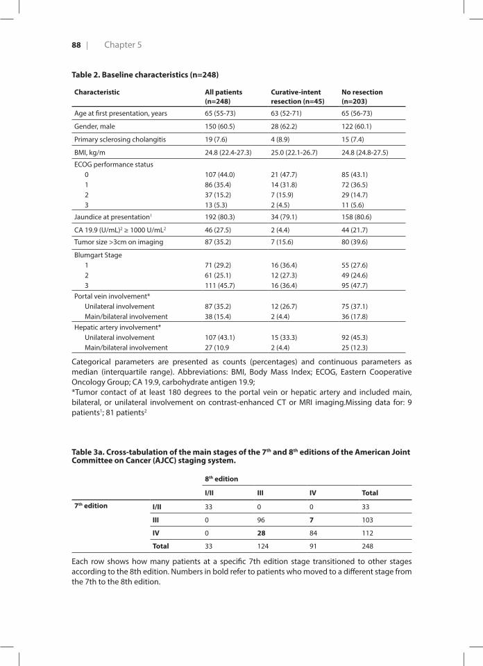
Chapter 588 |
Table 2. Baseline characteristics (n=248)
Characteristic All patients (n=248)
Curative-intent resection (n=45)
No resection (n=203)
Age at first presentation, years 65 (55-73) 63 (52-71) 65 (56-73)
Gender, male 150 (60.5) 28 (62.2) 122 (60.1)
Primary sclerosing cholangitis 19 (7.6) 4 (8.9) 15 (7.4)
BMI, kg/m 24.8 (22.4-27.3) 25.0 (22.1-26.7) 24.8 (24.8-27.5)
ECOG performance status0123
107 (44.0)86 (35.4)37 (15.2)13 (5.3)
21 (47.7)14 (31.8)7 (15.9)2 (4.5)
85 (43.1)72 (36.5)29 (14.7)11 (5.6)
Jaundice at presentation1 192 (80.3) 34 (79.1) 158 (80.6)
CA 19.9 (U/mL)2 ≥ 1000 U/mL2 46 (27.5) 2 (4.4) 44 (21.7)
Tumor size >3cm on imaging 87 (35.2) 7 (15.6) 80 (39.6)
Blumgart Stage123
71 (29.2)61 (25.1)111 (45.7)
16 (36.4)12 (27.3)16 (36.4)
55 (27.6)49 (24.6)95 (47.7)
Portal vein involvement*Unilateral involvementMain/bilateral involvement
87 (35.2)38 (15.4)
12 (26.7)2 (4.4)
75 (37.1)36 (17.8)
Hepatic artery involvement*Unilateral involvementMain/bilateral involvement
107 (43.1)27 (10.9
15 (33.3)2 (4.4)
92 (45.3)25 (12.3)
Categorical parameters are presented as counts (percentages) and continuous parameters as median (interquartile range). Abbreviations: BMI, Body Mass Index; ECOG, Eastern Cooperative Oncology Group; CA 19.9, carbohydrate antigen 19.9;*Tumor contact of at least 180 degrees to the portal vein or hepatic artery and included main, bilateral, or unilateral involvement on contrast-enhanced CT or MRI imaging.Missing data for: 9 patients1; 81 patients2
Table 3a. Cross-tabulation of the main stages of the 7th and 8th editions of the American Joint Committee on Cancer (AJCC) staging system.
8th edition
I/II III IV Total
7th edition I/II 33 0 0 33
III 0 96 7 103
IV 0 28 84 112
Total 33 124 91 248
Each row shows how many patients at a specific 7th edition stage transitioned to other stages according to the 8th edition. Numbers in bold refer to patients who moved to a different stage from the 7th to the 8th edition.
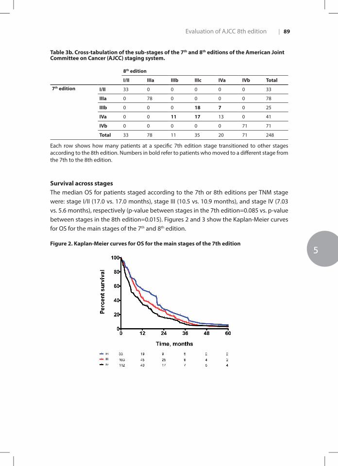
Evaluation of AJCC 8th edition
5
| 89
Table 3b. Cross-tabulation of the sub-stages of the 7th and 8th editions of the American Joint Committee on Cancer (AJCC) staging system.
8th edition
I/II IIIa IIIb IIIc IVa IVb Total
7th edition I/II 33 0 0 0 0 0 33
IIIa 0 78 0 0 0 0 78
IIIb 0 0 0 18 7 0 25
IVa 0 0 11 17 13 0 41
IVb 0 0 0 0 0 71 71
Total 33 78 11 35 20 71 248
Each row shows how many patients at a specific 7th edition stage transitioned to other stages according to the 8th edition. Numbers in bold refer to patients who moved to a different stage from the 7th to the 8th edition.
Survival across stagesThe median OS for patients staged according to the 7th or 8th editions per TNM stage were: stage I/II (17.0 vs. 17.0 months), stage III (10.5 vs. 10.9 months), and stage IV (7.03 vs. 5.6 months), respectively (p-value between stages in the 7th edition=0.085 vs. p-value between stages in the 8th edition=0.015). Figures 2 and 3 show the Kaplan-Meier curves for OS for the main stages of the 7th and 8th edition.
Figure 2. Kaplan-Meier curves for OS for the main stages of the 7th edition

Chapter 590 |
Figure 3. Kaplan-Meier curves for OS for the main stages of the 8th edition
Prognostic accuracy Table 4 shows the concordance indices and Brier scores for the two editions of the AJCC staging system. Prognostic accuracy of the 8th editions of the main stages of the AJCC staging systems was slightly higher than the 7th edition (c-index 0.59 vs 0.61). Expanding the 7th edition to include substages (e.g., IIIa and IIIb) slightly diminished its prognostic accuracy (c-statistic from 0.59 to 0.57). Expansions of the 8th edition also diminished its prognostic accuracy (c-statistic from 0.61 to 0.58). Prognostic accuracy was comparable between the expanded 7th and 8th AJCC staging systems (c-index 0.57 vs 0.58).
Subgroup analysis was performed to determine the prognostic accuracy of the AJCC staging system editions across treatment groups (Table 4). In both the 7th as the 8th editions, the AJCC staging system performed better in the subgroup of patients who underwent a curative-intent resection compared to the entire cohort (0.65 vs 0.57 in the 7th edition, 0.67 vs 0.58 in the 8th edition). The 8th edition did have a slightly better prognostic value compared to the 7th edition in this subgroup (c-index of 0.65 vs 0.67). Although the prognostic accuracy of the 8th edition of the AJCC staging system in patients who did not undergo a resection was slightly better when compared to the 7th edition (0.54), the prognostic accuracy was still very poor with a c-index of 0.57 in both the main as expanded staging system.
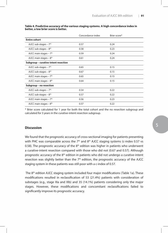
Evaluation of AJCC 8th edition
5
| 91
Table 4. Predictive accuracy of the various staging systems. A high concordance index in better, a low brier score is better.
Concordance-index Brier score*
Entire cohort
AJCC sub-stages – 7th 0.57 0.24
AJCC sub-stages – 8th 0.58 0.23
AJCC main stages – 7th 0.59 0.24
AJCC main stages – 8th 0.61 0.24
Subgroup – curative-intent resection
AJCC sub-stages – 7th 0.65 0.15
AJCC sub-stages – 8th 0.67 0.15
AJCC main stages – 7th 0.65 0.15
AJCC main stages – 8th 0.64 0.15
Subgroup – no resection
AJCC sub-stages – 7th 0.54 0.22
AJCC sub-stages – 8th 0.57 0.22
AJCC main stages – 7th 0.56 0.23
AJCC main stages – 8th 0.57 0.22
* Brier score calculated for 1 year for both the total cohort and the no resection subgroup and calculated for 3 years in the curative-intent resection subgroup.
Discussion
We found that the prognostic accuracy of cross-sectional imaging for patients presenting with PHC was comparable across the 7th and 8th AJCC staging systems (c-index 0.57 vs 0.58). The prognostic accuracy of the 8th edition was higher in patients who underwent a curative-intent resection compared with those who did not (0.67 and 0.57). Although prognostic accuracy of the 8th edition in patients who did not undergo a curative-intent resection was slightly better than the 7th edition, the prognostic accuracy of the AJCC staging system in these patients was still poor with a c-index of 0.57.
The 8th edition AJCC staging system included four major modifications (Table 1a). These modifications resulted in reclassification of 53 (21.4%) patients with consideration of substages (e.g., stage IIIa and IIIb) and 35 (14.1%) patients considering only the major stages. However, these modifications and concomitant reclassifications failed to significantly improve its prognostic accuracy.
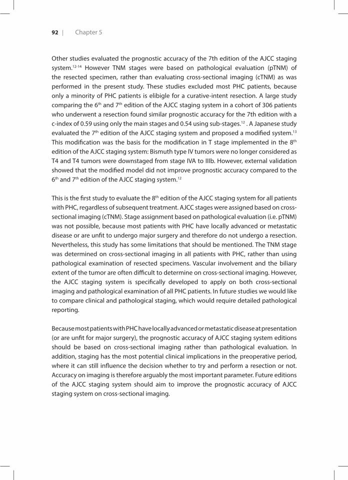
Chapter 592 |
Other studies evaluated the prognostic accuracy of the 7th edition of the AJCC staging system.12-14 However TNM stages were based on pathological evaluation (pTNM) of the resected specimen, rather than evaluating cross-sectional imaging (cTNM) as was performed in the present study. These studies excluded most PHC patients, because only a minority of PHC patients is elibigle for a curative-intent resection. A large study comparing the 6th and 7th edition of the AJCC staging system in a cohort of 306 patients who underwent a resection found similar prognostic accuracy for the 7th edition with a c-index of 0.59 using only the main stages and 0.54 using sub-stages.12 . A Japanese study evaluated the 7th edition of the AJCC staging system and proposed a modified system.13 This modification was the basis for the modification in T stage implemented in the 8th edition of the AJCC staging system: Bismuth type IV tumors were no longer considered as T4 and T4 tumors were downstaged from stage IVA to IIIb. However, external validation showed that the modified model did not improve prognostic accuracy compared to the 6th and 7th edition of the AJCC staging system.12
This is the first study to evaluate the 8th edition of the AJCC staging system for all patients with PHC, regardless of subsequent treatment. AJCC stages were assigned based on cross-sectional imaging (cTNM). Stage assignment based on pathological evaluation (i.e. pTNM) was not possible, because most patients with PHC have locally advanced or metastatic disease or are unfit to undergo major surgery and therefore do not undergo a resection. Nevertheless, this study has some limitations that should be mentioned. The TNM stage was determined on cross-sectional imaging in all patients with PHC, rather than using pathological examination of resected specimens. Vascular involvement and the biliary extent of the tumor are often difficult to determine on cross-sectional imaging. However, the AJCC staging system is specifically developed to apply on both cross-sectional imaging and pathological examination of all PHC patients. In future studies we would like to compare clinical and pathological staging, which would require detailed pathological reporting.
Because most patients with PHC have locally advanced or metastatic disease at presentation (or are unfit for major surgery), the prognostic accuracy of AJCC staging system editions should be based on cross-sectional imaging rather than pathological evaluation. In addition, staging has the most potential clinical implications in the preoperative period, where it can still influence the decision whether to try and perform a resection or not. Accuracy on imaging is therefore arguably the most important parameter. Future editions of the AJCC staging system should aim to improve the prognostic accuracy of AJCC staging system on cross-sectional imaging.

Evaluation of AJCC 8th edition
5
| 93
Conclusions
The prognostic accuracy of the 8th edition of the AJCC staging system was similar to the 7th edition. Prognostic accuracy was particularly poor in the majority of PHC patients who did not undergo a resection. Future editions of the AJCC staging system should aim to improve the prognostic accuracy of AJCC staging system on cross-sectional imaging.
Chapter 594 |
References
1. Jarnagin W, Winston C: Hilar cholangiocarcinoma: diagnosis and staging. HPB (Oxford)
2005;7:244-251.
2. Jarnagin WR, Fong Y, DeMatteo RP, et al.: Staging, resectability, and outcome in 225 patients
with hilar cholangiocarcinoma. Ann Surg 2001;234:507-517; discussion 517-509.
3. Chaiteerakij R, Harmsen WS, Marrero CR, et al.: A new clinically based staging system for perihilar
cholangiocarcinoma. Am J Gastroenterol 2014;109:1881-1890.
4. Pichlmayr R, Lamesch P, Weimann A, et al.: Surgical treatment of cholangiocellular carcinoma.
World J Surg 1995;19:83-88.
5. Groot Koerkamp B, Wiggers JK, Gonen M, et al.: Survival after resection of perihilar
cholangiocarcinoma-development and external validation of a prognostic nomogram. Ann
Oncol 2015;26:1930-1935.
6. Ruys AT, van Haelst S, Busch OR, et al.: Long-term survival in hilar cholangiocarcinoma also
possible in unresectable patients. World J Surg 2012;36:2179-2186.
7. Shen W, Punyanitya M, Wang Z, et al.: Total body skeletal muscle and adipose tissue volumes:
estimation from a single abdominal cross-sectional image. J Appl Physiol (1985) 2004;97:2333-
2338.
8. Edge SB BD, Compton CC, Fritz AG, Greene FL, Trotti A, editors.: AJCC cancer staging manual
(7th ed). New York, NY: Springer; 2010.
9. van Vugt JLA, Gaspersz MP, Coelen RJS, et al.: The prognostic value of portal vein and hepatic
artery involvement in patients with perihilar cholangiocarcinoma. HPB (Oxford) 2017.
10. Coelen RJS, Gaspersz MP, Labeur TA, et al.: Validation of the Mayo Clinic Staging System in
Determining Prognoses of Patients With Perihilar Cholangiocarcinoma. Clin Gastroenterol
Hepatol 2017;15:1930-1939 e1933.
11. Edge SB, Compton CC: The American Joint Committee on Cancer: the 7th edition of the AJCC
cancer staging manual and the future of TNM. Ann Surg Oncol 2010;17:1471-1474.
12. Groot Koerkamp B, Wiggers JK, Allen PJ, et al.: American Joint Committee on Cancer staging for
resected perihilar cholangiocarcinoma: a comparison of the 6th and 7th editions. HPB (Oxford)
2014;16:1074-1082.
13. Ebata T, Kosuge T, Hirano S, et al.: Proposal to modify the International Union Against Cancer
staging system for perihilar cholangiocarcinomas. Br J Surg 2014;101:79-88.
14. Juntermanns B, Sotiropoulos GC, Radunz S, et al.: Comparison of the sixth and the seventh
editions of the UICC classification for perihilar cholangiocarcinoma. Ann Surg Oncol 2013;20:277-
284.

Evaluation of AJCC 8th edition
5
| 95

PART 2

Part 2Palliative treatment in patients
with unresectable perihilar cholangiocarcinoma

6CHAPTER 6
6 Conditional survival in patients with unresectable perihilar cholangiocarcinoma
Gaspersz MP | Buettner S | van Vugt JL | Roos E | Coelen RJS | Vugts J | Belt EJ | de Jonge J | Polak WG | Willemssen FEJA | van Gulik TM | IJzermans JNM | Groot Koerkamp B
HPB (Oxford). 2017 Nov;19(11):966-971

Background: Conditional survival is the life expectancy from a point in time for a patient who has survived a specific period after presentation. The aim of the study was to estimate conditional survival for patients with unresectable perihilar cholangiocarcinoma.
Methods: Patients with unresectable perihilar cholangiocarcinoma from two academic hospitals in the Netherlands between 2002 and 2012 were assessed. A multivariable Cox proportional hazards analysis was performed to identify risk factors associated with overall survival. Survival was estimated using the Kaplan Meier method to evaluate factors associated with overall survival.
Results: In total, 572 patients were included. Overall survival was 42% at one year and 6% at three years. The conditional chance of surviving three years was 15% at 1 year and increased to 38% at 2 years. Independent poor prognostic factors for overall survival were age (≥65 years), tumor size (>3 cm) on imaging, bilirubin levels (>250 µmol/L), CA19-9 level at presentation (>1000 U/ml) , and suspected distant metastases on imaging. The conditional survival of patients with and without these prognostic factors was comparable after patients survived the first two or more years.
Conclusions: The conditional chance of surviving for patients with unresectable perihilar cholangiocarcinoma increases with time. Poor prognostic factors become less relevant once patients have survived two years. Abs
trac
t

Conditional survival PHC palliative treatment
6
| 101
Introduction
Nation-wide perihilar cholangiocarcinoma (PHC) registries show that only about 15% of PHC patients undergo surgical resection, because the majority of patients have metastatic or locally advanced disease at the moment of presentation.1-4 The median overall survival (OS) for these patients is about 1 year.1, 5
Prognostic models for cancer patients typically report survival from the time of presentation or the start of treatment.2, 4, 5 However, a patient’s life expectancy changes over time and prognostic or predictive factors may be time-dependent. Conditional survival (CS) takes into account the number of years the patient has already survived as it is defined as the survival probability that is calculated after a certain length of survival. CS only takes into account the patients that have survived up to that point and could therefore be considered as an updated estimate of life expectancy.6 Estimated CS has been reported in several malignancies including patients with resected PHC.7-9
CS estimates are especially interesting in patients with unresectable PHC, because most patients die in the first year and life expectancy improves considerably after surviving one or more years. Therefore, the aim of this study was to estimate CS for patients with unresectable PHC.
Methods
Study population and data acquisitionA systematic search in all medical records, discharge letters, minutes of multidisciplinary hepatopancreatobiliary team meetings, and operative and pathology reports was performed and all consecutive patients with unresectable PHC between 2002 and 2014 in the Erasmus MC University Medical Center, Rotterdam, The Netherlands, and the Academic Medical Center (AMC), Amsterdam, The Netherlands, were identified. PHC was defined as a mass or malignant-appearing stricture at or near the biliary confluence, arising between the origin of the cystic duct and the segmental bile ducts.10 If histopathological evidence was not obtained, the diagnosis was established by the multidisciplinary hepatopancreatobiliary team based on clinical, radiological, endoscopic and laboratory findings, and follow-up.
Patients were considered unresectable based on imaging at presentation or at staging laparoscopy or exploratory laparotomy. Reasons for unresectability on imaging or at exploratory laparotomy were metastatic disease, locally advanced disease, or patients who were unfit for surgery. Metastatic disease was defined, according to the AJCC staging

Chapter 6102 |
(7th edition), as the presence of distant metastases or lymph node metastases beyond the hepatoduodenal ligament (N2).11 If no pathological confirmation of suspicious lymph nodes was obtained, lymph node metastases were defined as nodes larger than 1 cm in short-axis diameter, with central necrosis, an irregular border or hyper-attenuating compared to portal phase liver parenchyma.12, 13 Locally advanced disease was defined as invasion of surrounding organs or vascular or biliary involvement that precluded an R0 resection, either during exploratory surgery or on imaging.5 Patients with hilar-invasive intrahepatic cholangiocarcinoma, gallbladder carcinoma, cystic duct carcinoma, and distal cholangiocarcinoma were excluded. All patients with unresectable perihilar cholangiocarcinoma were treated with best supportive care including palliative biliary drainage if necessary.
Baseline patient and tumor characteristics, clinical parameters (e.g., cholangitis), and laboratory results (e.g., bilirubin and carbohydrate antigen (CA) 19.9 levels) were collected retrospectively from medical records. Cholangitis was defined by the presence of fever, abdominal complaints, or leukocytosis requiring biliary drainage.14-16 Experienced abdominal radiologists revised imaging (i.e. contrast-enhanced CT and/or MRI or MRI with cholangiopancreatography (MRCP)) performed at the time of first presentation and tumor characteristics, presence of lymph node and distant metastases, and vascular involvement reassessed. The radiologists were blinded for clinical information and the eventual treatment that patients had undergone. Vascular involvement was defined as apparent tumor contact of at least 180 degrees to the portal vein or hepatic artery.11, 17, 18
Vascular involvement was mainly assessed on contrast-enhanced CT imaging. MRI was only used in the few patients with unavailable contrast-enhanced CT. The Institutional Review Boards of both centers approved the study and the need for informed consent was waived.
Statistical analysesContinuous data are reported as mean with standard deviation (SD) or median with interquartile range (IQR), depending on the normality of distribution. Categorical parameters are reported as counts and percentages. Proportions were compared using the Chi-squared test. Univariable analyses were performed by Kaplan– Meier estimates of survival probabilities and the log- rank and score tests for comparisons. A Cox proportional hazard regression model was used for multivariable modeling using backward selection with all variables that were significantly associated (p<0.20) with overall survival (OS) in univariate analysis. For serum bilirubin and CA 19-9, a large proportion of values were missing. Values were determined to be missing at random. For the multivariable model, multiple imputation was performed using the mice package for R 3.3.1 (https://cran.r-project.org/). Jaundice at presentation was excluded from the multivariable analysis,

Conditional survival PHC palliative treatment
6
| 103
because of high correlation with bilirubin level at presentation. In order to assess correlation between continuous variables, Spearman’s correlation coefficient was utilized.
Survival status was obtained from the municipal records database on May 9th, 2016. The OS was calculated from the date of first presentation in the tertiary referral center and patients alive at the last moment of follow-up were censored. Conditional survival (CS) was estimated as the probability of surviving an additional number of “y” years given that a patient had already survived for “x” years and was calculated as CS(y|x) = S(x + y)/ S(x), with S(x) representing the OS at x years estimated using the Kaplan–Meier method.7 For example, the CS for surviving another year among patients who had already survived 4 years, CS(1|4), was calculated by dividing the 5-year Kaplan–Meier survival estimate S(5) by the 4-year Kaplan–Meier survival estimate S(4).9, 19-21 All analyses were performed using SPSS 22.0 (IBM, New York). All tests were 2-sided and P<0.05 defined statistical significance.
Results
Patient characteristics A total of 572 patients with unresectable PHC met the inclusion criteria and formed the study cohort. Patient characteristics are detailed in Table 1. Median age was 68 (interquartile range 59-74). Conclusive pathological confirmation was obtained in 63% (n=358) of patients and percentage of PHC patients with pathological confirmation was similar between patients that died within 3 years from time of presentation (63%) and patients that survived longer (56%, p=0.57).
In total, 387 patients (68%) had unresectable PHC at the moment of presentation and 185 patients (32%) were found to have unresectable disease at staging laparoscopy or exploratory laparotomy. Of the patients with unresectable disease at the time of presentation, 124 (34%) patients had locally advanced disease on imaging, 34 (9%) patients had N2 lymph nodes, 123 (33%) patients had distant metastases at presentation, and 91 (24%) patients were considered unfit to tolerate a resection or did not want to undergo surgery. Of the patients diagnosed with unresectable PHC at staging laparoscopy or exploratory laparotomy, 64 patients (35%) had locally advanced disease that precluded a curative resection, 50 patients (27%) had N2 lymph nodes, 66 patients (26%) had distant metastases and 5 patients (3%) had medical reasons to forgo a curative–intent resection at the time of exploration.

Chapter 6104 |
Table 1. Patient characteristics and survival estimates.
Variable # of patients (%)
Patient survival, %
P ValueMedian 1 y 2 y 3 y 4 y 5 yAll patients 572 (100) 10 42 16 6 4 2Age, y
< 65 247 (43) 11 48 222 7 4 3≥ 65 325 (57) 8 37 12 6 3 2 0.002
SexFemale 213 (37) 10 42 18 7 4 3Male 359 (63) 10 42 15 6 4 2 0.638
Primary sclerosing cholangitisNo 543 (96) 10 42 16 6 4 2Yes 24 (4) 11 42 13 4 0 0 0.605
Reported weight lossNo 106 (22) 10 44 12 3 1 0Yes 367 (78) 10 41 16 6 4 2 0.720
WHO performance classification0 – 2 427 (76) 10 43 16 5 3 13 – 4 132 (24) 7 32 12 10 9 7 0.706
Jaundice at presentationNo 82 (25) 12 51 20 5 3 1Yes 472 (85) 10 40 15 6 4 3 0.130
Bilirubin at presentation≤ 250 umol/L (14.6mg/dL) 325 (73) 10 44 18 7 5 3> 250 umol/L (14.6mg/dL) 119 (27) 8 34 6 1 0 0 < 0.001
CA19-9 at presentation≤ 1000 U/mL 194 (68) 12 49 21 7 4 2> 1000 U/mL 91 (32) 7 25 5 3 3 1 < 0.001
Nodal status on imagingN0 279 (51) 10 43 24 6 4 2N+ 264 (49) 9 40 14 5 3 2 0.339
Tumor size on imaging, cm≤ 3 283 (57) 11 47 18 8 5 3> 3 217 (43) 8 36 14 3 2 1 0.001
Vascular involvementNo 121 (23) 10 44 17 6 4 3Yes 400 (77) 10 41 15 5 3 2 0.294
Suspected distant metastasesNo 487 (87) 11 45 16 6 4 2Yes 70 (13) 4 20 10 6 4 1 < 0.001
Treatment groupUnresectable at Exploratory laparotomy 185 (32) 13 54 20 8 5 2Unresectable at presentation 387 (68) 8 36 14 5 3 3 < 0.001
Abbreviations: WHO, World Health Organization; CA19-9, Carbohydrate Antigen 19-9.
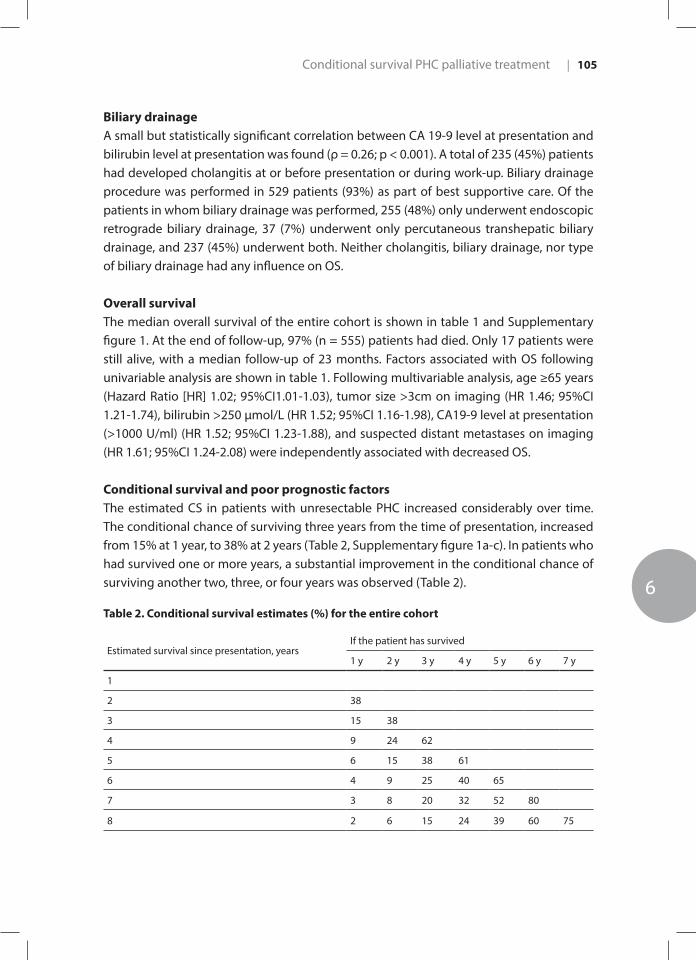
Conditional survival PHC palliative treatment
6
| 105
Biliary drainageA small but statistically significant correlation between CA 19-9 level at presentation and bilirubin level at presentation was found (ρ = 0.26; p < 0.001). A total of 235 (45%) patients had developed cholangitis at or before presentation or during work-up. Biliary drainage procedure was performed in 529 patients (93%) as part of best supportive care. Of the patients in whom biliary drainage was performed, 255 (48%) only underwent endoscopic retrograde biliary drainage, 37 (7%) underwent only percutaneous transhepatic biliary drainage, and 237 (45%) underwent both. Neither cholangitis, biliary drainage, nor type of biliary drainage had any influence on OS.
Overall survivalThe median overall survival of the entire cohort is shown in table 1 and Supplementary figure 1. At the end of follow-up, 97% (n = 555) patients had died. Only 17 patients were still alive, with a median follow-up of 23 months. Factors associated with OS following univariable analysis are shown in table 1. Following multivariable analysis, age ≥65 years (Hazard Ratio [HR] 1.02; 95%CI1.01-1.03), tumor size >3cm on imaging (HR 1.46; 95%CI 1.21-1.74), bilirubin >250 µmol/L (HR 1.52; 95%CI 1.16-1.98), CA19-9 level at presentation (>1000 U/ml) (HR 1.52; 95%CI 1.23-1.88), and suspected distant metastases on imaging (HR 1.61; 95%CI 1.24-2.08) were independently associated with decreased OS.
Conditional survival and poor prognostic factorsThe estimated CS in patients with unresectable PHC increased considerably over time. The conditional chance of surviving three years from the time of presentation, increased from 15% at 1 year, to 38% at 2 years (Table 2, Supplementary figure 1a-c). In patients who had survived one or more years, a substantial improvement in the conditional chance of surviving another two, three, or four years was observed (Table 2).
Table 2. Conditional survival estimates (%) for the entire cohort
Estimated survival since presentation, yearsIf the patient has survived
1 y 2 y 3 y 4 y 5 y 6 y 7 y
1
2 38
3 15 38
4 9 24 62
5 6 15 38 61
6 4 9 25 40 65
7 3 8 20 32 52 80
8 2 6 15 24 39 60 75
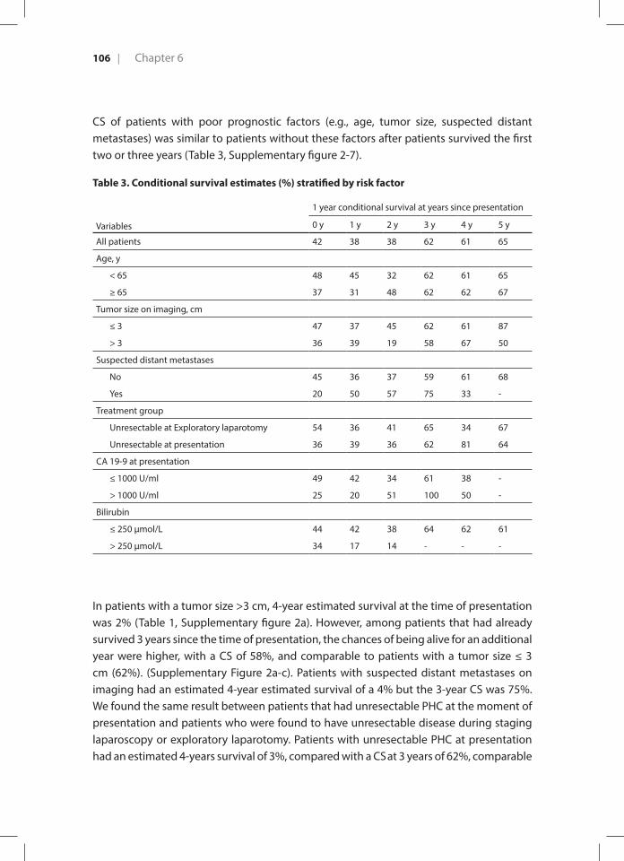
Chapter 6106 |
CS of patients with poor prognostic factors (e.g., age, tumor size, suspected distant metastases) was similar to patients without these factors after patients survived the first two or three years (Table 3, Supplementary figure 2-7).
Table 3. Conditional survival estimates (%) stratified by risk factor
Variables
1 year conditional survival at years since presentation
0 y 1 y 2 y 3 y 4 y 5 y
All patients 42 38 38 62 61 65
Age, y
< 65 48 45 32 62 61 65
≥ 65 37 31 48 62 62 67
Tumor size on imaging, cm
≤ 3 47 37 45 62 61 87
> 3 36 39 19 58 67 50
Suspected distant metastases
No 45 36 37 59 61 68
Yes 20 50 57 75 33 -
Treatment group
Unresectable at Exploratory laparotomy 54 36 41 65 34 67
Unresectable at presentation 36 39 36 62 81 64
CA 19-9 at presentation
≤ 1000 U/ml 49 42 34 61 38 -
> 1000 U/ml 25 20 51 100 50 -
Bilirubin
≤ 250 µmol/L 44 42 38 64 62 61
> 250 µmol/L 34 17 14 - - -
In patients with a tumor size >3 cm, 4-year estimated survival at the time of presentation was 2% (Table 1, Supplementary figure 2a). However, among patients that had already survived 3 years since the time of presentation, the chances of being alive for an additional year were higher, with a CS of 58%, and comparable to patients with a tumor size ≤ 3 cm (62%). (Supplementary Figure 2a-c). Patients with suspected distant metastases on imaging had an estimated 4-year estimated survival of a 4% but the 3-year CS was 75%. We found the same result between patients that had unresectable PHC at the moment of presentation and patients who were found to have unresectable disease during staging laparoscopy or exploratory laparotomy. Patients with unresectable PHC at presentation had an estimated 4-years survival of 3%, compared with a CS at 3 years of 62%, comparable
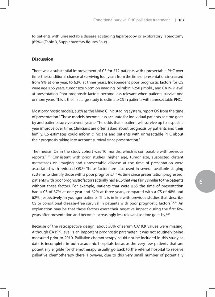
Conditional survival PHC palliative treatment
6
| 107
to patients with unresectable disease at staging laparoscopy or exploratory laparotomy (65%) (Table 3, Supplementary figures 3a-c).
Discussion
There was a substantial improvement of CS for 572 patients with unresectable PHC over time; the conditional chance of surviving four years from the time of presentation, increased from 9% at one year, to 62% at three years. Independent poor prognostic factors for OS were age ≥65 years, tumor size >3cm on imaging, bilirubin >250 µmol/L, and CA19-9 level at presentation. Poor prognostic factors become less relevant when patients survive one or more years. This is the first large study to estimate CS in patients with unresectable PHC.
Most prognostic models, such as the Mayo Clinic staging system, report OS from the time of presentation.5 These models become less accurate for individual patients as time goes by and patients survive several years.7 The odds that a patient will survive up to a specific year improve over time. Clinicians are often asked about prognosis by patients and their family. CS estimates could inform clinicians and patients with unresectable PHC about their prognosis taking into account survival since presentation.8
The median OS in the study cohort was 10 months, which is comparable with previous reports.22,23 Consistent with prior studies, higher age, tumor size, suspected distant metastases on imaging and unresectable disease at the time of presentation were associated with reduced OS.3,5 These factors are also used in several available staging systems to identify those with a poor prognosis.5,11 As time since presentation progressed, patients with poor prognostic factors actually had a CS that was fairly similar to the patients without these factors. For example, patients that were ≥65 the time of presentation had a CS of 37% at one year and 62% at three years, compared with a CS of 48% and 62%, respectively, in younger patients. This is in line with previous studies that describe CS or conditional disease–free survival in patients with poor prognostic factors.7,9,24 An explanation may be that these factors exert their negative impact during the first few years after presentation and become increasingly less relevant as time goes by.9,24
Because of the retrospective design, about 50% of serum CA19.9 values were missing. Although CA19.9 level is an important prognostic parameter, it was not routinely being measured prior to 2010. Palliative chemotherapy could not be included in this study as data is incomplete in both academic hospitals because the very few patients that are potentially eligible for chemotherapy usually go back to the referral hospital to receive palliative chemotherapy there. However, due to this very small number of potentially

Chapter 6108 |
eligible patients and because chemotherapy only lengthens survival with mere months, this data would have limited effect on the results
Another limitation is that the majority of patients missed pathological confirmation of PHC. However, the percentage of PHC patients with pathological confirmation was similar between patients that died within 3 years from time of presentation and patients that survived longer.
As time since presentation goes by, CS is a more relevant measure of prognosis for patients with unresectable PHC. Poor prognostic factors become increasingly less relevant as patients survive one or more years. CS estimates may help patients and caregivers in making decisions during follow-up.

Conditional survival PHC palliative treatment
6
| 109
References
1. Jarnagin WR, Fong Y, DeMatteo RP, Gonen M, Burke EC, Bodniewicz BJ, et al. Staging, resectability,
and outcome in 225 patients with hilar cholangiocarcinoma. Ann Surg. 2001;234(4):507-17;
discussion 17-9.
2. Groot Koerkamp B, Wiggers JK, Gonen M, Doussot A, Allen PJ, Besselink MG, et al. Survival after
resection of perihilar cholangiocarcinoma-development and external validation of a prognostic
nomogram. Ann Oncol. 2015;26(9):1930-5.
3. Ruys AT, van Haelst S, Busch OR, Rauws EA, Gouma DJ, van Gulik TM. Long-term survival in hilar
cholangiocarcinoma also possible in unresectable patients. World J Surg. 2012;36(9):2179-86.
4. Wang Y, Li J, Xia Y, Gong R, Wang K, Yan Z, et al. Prognostic nomogram for intrahepatic
cholangiocarcinoma after partial hepatectomy. J Clin Oncol. 2013;31(9):1188-95.
5. Chaiteerakij R, Harmsen WS, Marrero CR, Aboelsoud MM, Ndzengue A, Kaiya J, et al. A new
clinically based staging system for perihilar cholangiocarcinoma. Am J Gastroenterol.
2014;109(12):1881-90.
6. Janssen-Heijnen ML, Gondos A, Bray F, Hakulinen T, Brewster DH, Brenner H, et al. Clinical
relevance of conditional survival of cancer patients in europe: age-specific analyses of 13
cancers. J Clin Oncol. 2010;28(15):2520-8.
7. Buettner S, Margonis GA, Kim Y, Gani F, Ethun CG, Poultsides G, et al. Conditional probability of
long-term survival after resection of hilar cholangiocarcinoma. HPB (Oxford). 2016;18(6):510-7.
8. Mertens AC, Yong J, Dietz AC, Kreiter E, Yasui Y, Bleyer A, et al. Conditional survival in pediatric
malignancies: analysis of data from the Childhood Cancer Survivor Study and the Surveillance,
Epidemiology, and End Results Program. Cancer. 2015;121(7):1108-17.
9. Spolverato G, Kim Y, Ejaz A, Alexandrescu S, Marques H, Aldrighetti L, et al. Conditional
Probability of Long-term Survival After Liver Resection for Intrahepatic Cholangiocarcinoma: A
Multi-institutional Analysis of 535 Patients. JAMA Surg. 2015;150(6):538-45.
10. Edge SB BD, Compton CC, Fritz AG, Greene FL, Trotti A, editors. AJCC cancer staging manual (7th
ed). New York, NY: Springer; 2010.
11. Edge SB, Compton CC. The American Joint Committee on Cancer: the 7th edition of the AJCC
cancer staging manual and the future of TNM. Ann Surg Oncol. 2010;17(6):1471-4.
12. Lee HY, Kim SH, Lee JM, Kim SW, Jang JY, Han JK, et al. Preoperative assessment of resectability
of hepatic hilar cholangiocarcinoma: combined CT and cholangiography with revised criteria.
Radiology. 2006;239(1):113-21.
13. Coelen RJS, Gaspersz MP, Labeur TA, van Vugt JLA, van Dieren S, Willemssen F, et al. Validation
of the Mayo Clinic Staging System in Determining Prognoses of Patients with Perihilar
Cholangiocarcinoma. Clin Gastroenterol Hepatol. 2017.
14. van der Gaag NA, Rauws EA, van Eijck CH, Bruno MJ, van der Harst E, Kubben FJ, et al. Preoperative
biliary drainage for cancer of the head of the pancreas. N Engl J Med. 2010;362(2):129-37.
15. Wiggers JK, Coelen RJ, Rauws EA, van Delden OM, van Eijck CH, de Jonge J, et al. Preoperative
endoscopic versus percutaneous transhepatic biliary drainage in potentially resectable perihilar
Chapter 6110 |
cholangiocarcinoma (DRAINAGE trial): design and rationale of a randomized controlled trial.
BMC Gastroenterol. 2015;15:20.
16. Wiggers JK, Groot Koerkamp B, Cieslak KP, Doussot A, van Klaveren D, Allen PJ, et al.
Postoperative Mortality after Liver Resection for Perihilar Cholangiocarcinoma: Development
of a Risk Score and Importance of Biliary Drainage of the Future Liver Remnant. J Am Coll Surg.
2016;223(2):321-31 e1.
17. Lu DS, Reber HA, Krasny RM, Kadell BM, Sayre J. Local staging of pancreatic cancer: criteria for
unresectability of major vessels as revealed by pancreatic-phase, thin-section helical CT. AJR
Am J Roentgenol. 1997;168(6):1439-43.
18. Okumoto T, Sato A, Yamada T, Takase K, Matsuhashi T, Tsuda M, et al. Correct diagnosis of
vascular encasement and longitudinal extension of hilar cholangiocarcinoma by four-channel
multidetector-row computed tomography. Tohoku J Exp Med. 2009;217(1):1-8.
19. Nathan H, de Jong MC, Pulitano C, Ribero D, Strub J, Mentha G, et al. Conditional survival after
surgical resection of colorectal liver metastasis: an international multi-institutional analysis of
949 patients. J Am Coll Surg. 2010;210(5):755-64, 64-6.
20. Mayo SC, Nathan H, Cameron JL, Olino K, Edil BH, Herman JM, et al. Conditional survival in
patients with pancreatic ductal adenocarcinoma resected with curative intent. Cancer.
2012;118(10):2674-81.
21. Cucchetti A, Piscaglia F, Cescon M, Ercolani G, Terzi E, Bolondi L, et al. Conditional survival
after hepatic resection for hepatocellular carcinoma in cirrhotic patients. Clin Cancer Res.
2012;18(16):4397-405.
22. Liu Y, Sun J, Ma D, Jin B, Shao Z, Wang J, et al. Palliative Locoregional Treatment for Unresectable
Nonmetastatic Klatskin Tumor: Percutaneous Transhepatic Biliary Drainage Combined With
3-dimensional Conformal Radiotherapy. J Clin Gastroenterol. 2016;50(6):513-7.
23. Buettner S, Margonis GA, Kim Y, Gani F, Ethun CG, Poultsides GA, et al. Changing Odds of Survival
Over Time among Patients Undergoing Surgical Resection of Gallbladder Carcinoma. Ann Surg
Oncol. 2016;23(13):4401-9.
24. Dong J, Zhu Y, Ma F, Ren Y, Lu J, Wang Z, et al. Conditional disease-free survival after liver
transplantation for hepatocellular carcinoma: A two-center experience. Medicine (Baltimore).
2016;95(31):e4383.
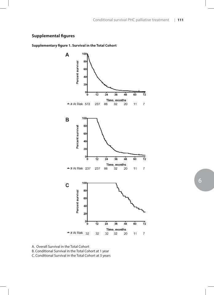
Conditional survival PHC palliative treatment
6
| 111
Supplemental figures
Supplementary figure 1. Survival in the Total Cohort
A. Overall Survival in the Total CohortB. Conditional Survival in the Total Cohort at 1 yearC. Conditional Survival in the Total Cohort at 3 years

Chapter 6112 |
Supplementary figure 2. Survival Stratified by Tumor Size
A. Overall Survival Stratified by Tumor SizeB. Conditional Survival Stratified by Tumor Size at 1 yearC. Conditional Survival Stratified by Tumor Size at 3 years

Conditional survival PHC palliative treatment
6
| 113
Supplementary figure 3. Survival Stratified by Treatment Group
A. Overall Survival Stratified by Treatment GroupB. Conditional Survival Stratified by Treatment Group at 1 yearC. Conditional Survival Stratified by Treatment Group at 3 years
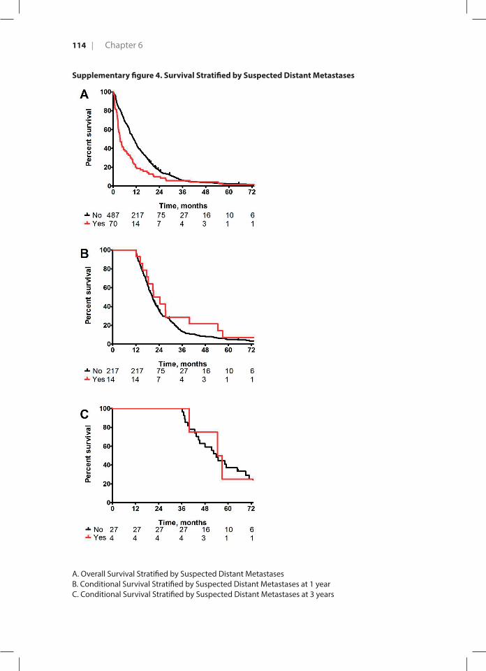
Chapter 6114 |
Supplementary figure 4. Survival Stratified by Suspected Distant Metastases
A. Overall Survival Stratified by Suspected Distant MetastasesB. Conditional Survival Stratified by Suspected Distant Metastases at 1 yearC. Conditional Survival Stratified by Suspected Distant Metastases at 3 years
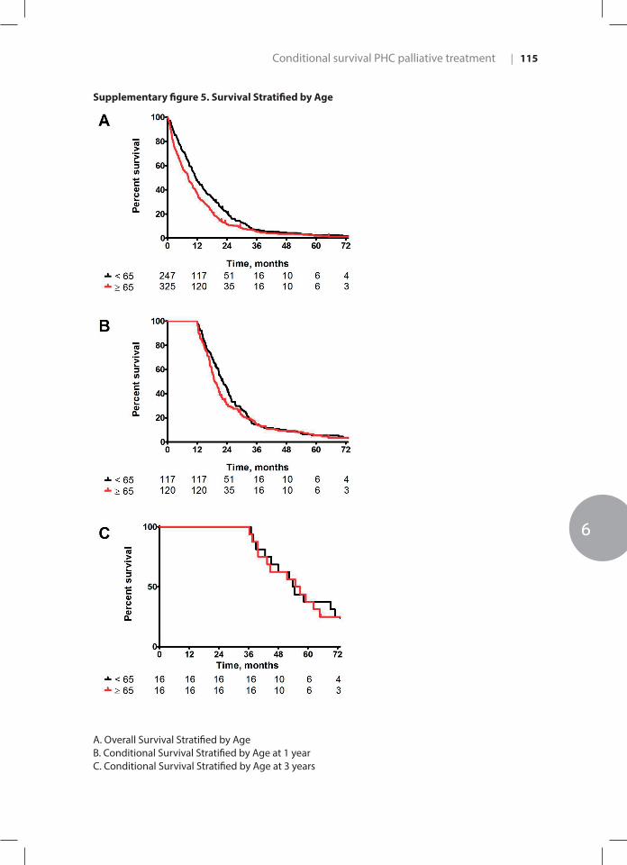
Conditional survival PHC palliative treatment
6
| 115
Supplementary figure 5. Survival Stratified by Age
A. Overall Survival Stratified by AgeB. Conditional Survival Stratified by Age at 1 yearC. Conditional Survival Stratified by Age at 3 years

Chapter 6116 |
Supplementary figure 6. Survival Stratified by CA 19-9 at presentation
A. Overall Survival Stratified by CA 19-9 at presentationB. Conditional Survival Stratified by CA 19-9 at presentation at 1 yearC. Conditional Survival Stratified by CA 19-9 at presentation at 3 years
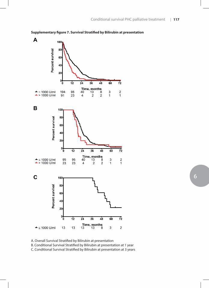
Conditional survival PHC palliative treatment
6
| 117
Supplementary figure 7. Survival Stratified by Bilirubin at presentation
A. Overall Survival Stratified by Bilirubin at presentationB. Conditional Survival Stratified by Bilirubin at presentation at 1 yearC. Conditional Survival Stratified by Bilirubin at presentation at 3 years

7CHAPTER 7

7 Success, Complication, and Mortality Rate of Initial Biliary Drainage in Patients with Unresectable Perihilar Cholangiocarcinoma
Gaspersz MP | van Vugt JL | Roos E | Coelen RJS | Vugts J | Bruno M | van Eijck CHJ | van Hooft J | van Lienden KP | de Jonge J | Polak WG | Poley J | Pek C | Moelker A | Willemssen FEJA | van Gulik TM | Hol L | IJzermans JNM | Groot Koerkamp B
Submitted

Background: Patients with unresectable perihilar cholangiocarcinoma (PHC) require biliary drainage to relieve symptoms and allow for palliative systemic chemotherapy. Biliary drainage is complex in these patients because of isolation of segmental bile ducts. The aim of this study was to establish the success, complication, and mortality rates of the initial biliary drainage procedure in patients with unresectable PHC at presentation.
Methods: Patients with unresectable PHC who underwent initial endoscopic (EBD) or percutaneous transhepatic biliary drainage (PTBD) between 2002-2014 were included. Success of drainage was defined as successful biliary stent or drain placement, without unscheduled re-intervention within 14 days, and serum bilirubin levels <50µmol/L or a more than 50% decrease in serum bilirubin. Severe complications and mortality were recorded for 90-days after initial drainage.
Results: In total, 186 patients were included; 161 (86.6%) underwent initial EBD and 25 (13.4%) initial PTBD. Success of initial drainage was observed in 84 patients (45.2%), including 76 (47.2%) after EBD and 8 (32.0%) after PTBD. Reasons for unsuccessful initial drainage were: no drain or stent placed in 39 patients (21.0%), unplanned re-intervention in 52 patients (28.0%), and bilirubin >50 µmol/L (or not halved) in 11 patients (5.9%). Severe drainage-related complications were observed in 19 patients (11.8%) after EBD and in 3 (11.5%) after PTBD. Overall, 20 patients (10.8%) died within 30 days and 66 (35.5%) within 90 days. Most patients (77.3%) with 90-day mortality had no metastatic disease.
Conclusions: Initial biliary drainage in patients with unresectable PHC has a low success rate of 45.2% and a high 90-day mortality rate of 35.5%. Abs
trac
t
Initial biliary drainage in unresectable PHC
7
| 121
Introduction
Perihilar cholangiocarcinoma (PHC) is the most common malignancy of the bile duct. PHC arises from the epithelial cells at or near the biliary confluence.1 Patients with PHC typically present with painless jaundice due to biliary obstruction caused by the tumor. Relief of biliary obstruction through biliary drainage can resolve jaundice and liver dysfunction as well as improve the wellbeing of patients.2 Percutaneous transhepatic biliary drainage (PTBD) and endoscopic retrograde biliary drainage (EBD) are the two approaches most frequently used for biliary drainage in Western countries.
The majority of patients with PHC have unresectable disease (i.e. locally advanced or metastatic) on imaging at the time of presentation.1,3 The median overall survival (OS) of patients with unresectable disease is about 6 months.4 Most patients with PHC die from cholangitis or liver failure due to progressive biliary obstruction rather than widespread metastatic disease.5 Palliative chemotherapy with gemcitabine plus cisplatin may improve median OS with about 3 months.6 However, patients are only eligible for systemic chemotherapy after adequate biliary drainage (i.e. bilirubin below 50 µmol/L or 2.9 mg/dL).
Most studies have focused on outcomes of preoperative biliary drainage in patients with resectable PHC.7-9 Because of progressive isolation of segmental bile ducts, biliary drainage can be even more challenging in patients with unresectable PHC.10 The goal of biliary drainage in the palliative setting is twofold: to improve the wellbeing of patients and to allow for systemic chemotherapy. Patients often have complications after initial biliary drainage (e.g., cholangitis) and reinterventions may be needed because of inadequate biliary drainage. In particular in the palliative setting, the goal of initial biliary drainage is to avoid complications and re-interventions.
The aim of this study was to evaluate the success, severe complication, and mortality rates of initial palliative biliary drainage in patients with unresectable PHC.
Methods
Study population and data acquisitionPatients with unresectable PHC who underwent an initial drainage procedure between 2002 and 2014 were retrospectively identified in two tertiary referral centers in the Netherlands: Erasmus MC University Medical Center in Rotterdam, and the Academic Medical Center (AMC) in Amsterdam.
Chapter 7122 |
All patients were discussed at a multidisciplinary meeting at the tertiary referral center. Initial drainage procedure could be performed in one of the tertiary referral centers or in referring hospitals. Patients were considered to have unresectable disease in the event of locally advanced or metastatic PHC on imaging at the time of presentation or when they were physically unfit for surgery.1 Metastatic (stage IV) PHC was defined as the presence of distant metastases or lymph node metastases beyond the hepatoduodenal ligament (AJCC staging, 7th edition).11 If no pathological confirmation of suspicious lymph nodes was obtained, positive lymph node metastases were defined on imaging as nodes larger than 1 cm in short-axis diameter, nodes with central necrosis, or an irregular border or hyper-attenuation compared to portal phase liver parenchyma.12,13 Locally advanced disease was defined as invasion of surrounding organs or vascular and biliary involvement that precluded an R0 resection with an adequate future liver remnant.3 Patients were only included if they had no prior drainage procedure (i.e. drainage naïve).9 Therefore, patients with primary sclerosing cholangitis (PSC) were excluded as these patients often undergo biliary drainage procedures before PHC develops. Patients were also excluded if a detailed report of the initial drainage procedure was not available.
Success of drainage was defined as successful biliary stent or catheter placement, without scheduled re-intervention within 14 days and serum bilirubin levels under 50 µmol/L or a more than 50% decrease in serum bilirubin after 14 days. A decrease of 50% of serum bilirubin at 14 days was also considered to be successful drainage because the rate of bilirubin decrease after stenting depends on the serum bilirubin prior to drainage.14 Planned re-intervention within 14 days (i.e. drainage performed in two or more stages) was not considered failure of initial biliary drainage.
Severe drainage-related complications included cholangitis, acute cholecystitis, acute pancreatitis, bile duct injury, duodenal perforation, and cardiopulmonary complications. Cholangitis was defined as both fever (i.e. body temperature >38.5°C) and leukocytosis (i.e. ≥10 x109/L), without clinical or radiological evidence of acute cholecystitis, and requiring a re-intervention.9,15,16 Acute cholecystitis was defined as radiologic diagnosis of cholecystitis, in combination with fever and leukocytes, requiring percutaneous drainage or cholecystectomy. Acute pancreatitis was defined by abdominal pain and a serum concentration of pancreatic enzymes (amylase or lipase) ≥3 times the upper limit of normal requiring at least one night of hospitalization.15 Overall survival was defined as the time between initial drainage procedure and date of death or date of last follow-up.
Data on initial biliary drainage were collected from medical records until 90 days after drainage, including indication for drainage, bilirubin serum levels before and after initial drainage, and severe drainage-related complications and survival. Endoscopic stent placement during initial biliary drainage was mostly frequently performed with plastic

Initial biliary drainage in unresectable PHC
7
| 123
stents in both tertiary referral centers, although the use of metal stents has increased since 2010. If initial drainage procedure was not performed in one of the tertiary referral centers, data was collected at the referring hospital were initial drainage procedure was performed.
Experienced abdominal radiologists revised the contrast-enhanced computed tomography (CT) and/or magnetic resonance imaging (MRI) performed at the time of presentation. Parameters reassessed on imaging were tumor diameter, Bismuth-Corlette classification17, presence of suspected lymph nodes, distant metastases, lobar atrophy, and vascular involvement. The Institutional Review Boards of both centers approved the study and the need for informed consent was waived.
Statistical analysisContinuous variables are presented as means with standard deviation (SD) if normally distributed or as median with interquartile range (IQR) if not normally distributed. Categorical parameters are reported as counts and percentages. Proportions were compared with Fischer’s exact or Chi-squared test, whereas medians were compared with Mann-Whitney U. Univariable analyses were performed using binary logistic regression and multivariable analyses using all variables with a p<0.20 in univariable analyses. Kaplan-Meier method with log-rank test was used for survival outcomes. Survival status was retrieved from the municipal records with the last update on 21-12-2017. All analyses were conducted using IBM SPSS Statistics for Windows (version 21.0. Armonk, NY: IBM Corp, USA).
Results
Patient characteristicsA total of 186 drainage-naïve patients with unresectable PHC underwent initial biliary drainage; EBD in 161 patients (86.6%) and PTBD in 25 patients (13.4%). Table 1 presents baseline patient characteristics. At the time of last follow-up 182 patients (97.8%) had died. The median overall survival (OS) of the entire cohort was 6.4 (95% confidence interval (CI): 4.5-8.3) months (Figure 1). Thirteen patients (7.0%) received palliative systemic chemotherapy. Two patients (1.1%) underwent palliative radiotherapy, and 1 patient (0.5%) photodynamic therapy.

Chapter 7124 |
Table 1. Baseline patient characteristics (n=186)
Demographics, exam, and laboratory values
Total cohort (% or IQR))
EBD (% or IQR)N=161
PTBD (% or IQR)N=25
p-value
Age at first presentation, years≥75 years
71.5 (62-77)73 (39.2)
71.5 (62-78)65 (40.4)
71.3 (62-75)8 (32.0)
0.3030.425
Gender, males 105 (56.5) 90 (55.9) 15 (60.0) 0.701
BMI, kg/m2 25.0 (22.9-27.4) 25 (23.0-27.4) 24 (20.7-28.3) 0.193
ECOG performance status1
01234
67 (36.0)67 (36.0)25 (13.4)19 (10.2)1 (0.5)
58 (37.2)57 (36.5)23 (14.7)17 (10.9)1 (0.6)
9 (39.1)10 (43.5)2 (8.7)2 (8.7)-
0.903
CA 19.9 (U/mL)2
≥ 1000 U/mL324 (105-2172)33 (17.7)
299 (100-2377)28 (34.1)
454 (195-1871)5 (45.5)
0.6600.462
Highest bilirubin pre-drainage, median3
<50 µmol/L248 (138-377)6 (4.6)
232 (138-375)5 (4.6)
284 (203-384)1 (4.5)
0.3790.993
Tumor characteristics on imaging at presentation
Tumor size, cm>3 cm
3.0 (2.3-3.9)81 (43.5)
3 (2.3-3.9)70 (43.5)
2.6 (2.4-4.1)11 (44.0)
0.8800.625
Suspicious lymph nodes on imaging‡4
N0N1N2
93 (53.8)40 (23.1)40 (23.1)
80 (54.1)33 (22.3)35 (23.6)
13 (52.0)7 (28.0)5 (20.0)
0.802
Suspected distant metastases on imaging*5 27 (15.1) 24 (15.6) 3 (12.0) 0.642
Any vascular involvement#
PV involvement6
UnilateralMain/Bilateral
HA involvement7
UnilateralMain/Bilateral
129 (68.8)107 (57.6)60 (32.3)47 (25.3)105 (56.5)72 (38.7)33 (17.7)
110 (76.9)92 (64.36)52 (36.4)40 (28.0)91 (64.5)61 (43.3)30 (21.3)
18 (75.0)16 (61.5)8 (33.3)7 (29.2)14 (53.8)11 (45.8)3 (12.5)
0.8370.8620.960
0.5590.595
Lobar atrophy on imaging8
NoneLeftRight
124 (71.7)9 (4.8)40 (21.4)
106 (71.6)7 (4.7)35 (23.6)
18 (72.0)2 (8.0)5 (20)
0.755
Bismuth classification 17, 9
IIIIIIAIIIBIV
11 (6.6)23 (13.9)34 (20.5)28 (16.9)71 (42.2)
11 (7.7)20 (14.1)28 (19.7)24 (16.9)59 (41.5)
-3 (12.5)6 (25.0)4 (16.7)11 (44.8)
0.687
Abbreviations: 95% CI, 95% confidence interval; BMI, body mass index; ECOG, Eastern Cooperative Oncology Group; CA 19.9, carbohydrate antigen 19.9; IQR, interquartile range# Defined as ≥180 degree tumor involvement on imaging‡ Nodes along the cystic duct, common bile, duct, hepatic artery and portal vein were classified as N1 and periaortic, pericaval, SMA, and celiac nodes as N2.11 Missing for 13 patientsData missing for: 7 patients1, 93 patients2, 55 patients3, 13 patients4, 7 patients5, 19 patients6, 21 patients7, 13 patients8, 20 patients9.
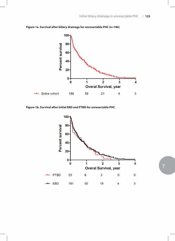
Initial biliary drainage in unresectable PHC
7
| 125
Figure 1a. Survival after biliary drainage for unresectable PHC (n=186)
Figure 1b. Survival after initial EBD and PTBD for unresectable PHC
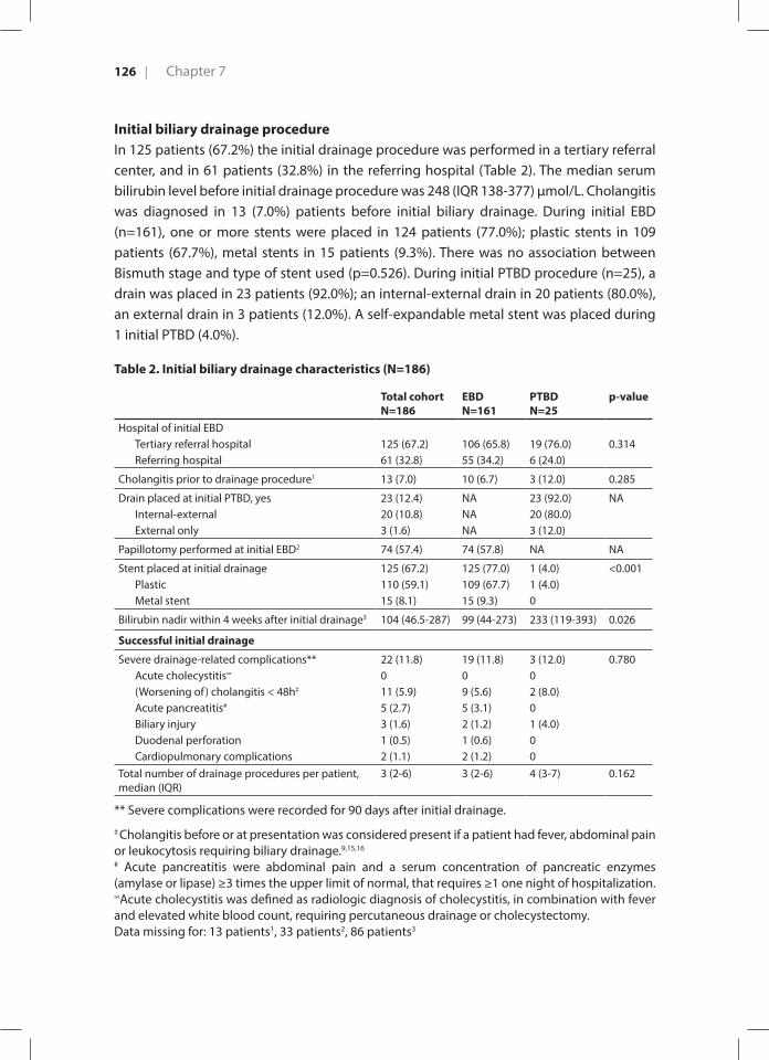
Chapter 7126 |
Initial biliary drainage procedureIn 125 patients (67.2%) the initial drainage procedure was performed in a tertiary referral center, and in 61 patients (32.8%) in the referring hospital (Table 2). The median serum bilirubin level before initial drainage procedure was 248 (IQR 138-377) µmol/L. Cholangitis was diagnosed in 13 (7.0%) patients before initial biliary drainage. During initial EBD (n=161), one or more stents were placed in 124 patients (77.0%); plastic stents in 109 patients (67.7%), metal stents in 15 patients (9.3%). There was no association between Bismuth stage and type of stent used (p=0.526). During initial PTBD procedure (n=25), a drain was placed in 23 patients (92.0%); an internal-external drain in 20 patients (80.0%), an external drain in 3 patients (12.0%). A self-expandable metal stent was placed during 1 initial PTBD (4.0%).
Table 2. Initial biliary drainage characteristics (N=186)
Total cohortN=186
EBDN=161
PTBDN=25
p-value
Hospital of initial EBDTertiary referral hospitalReferring hospital
125 (67.2)61 (32.8)
106 (65.8)55 (34.2)
19 (76.0)6 (24.0)
0.314
Cholangitis prior to drainage procedure1 13 (7.0) 10 (6.7) 3 (12.0) 0.285
Drain placed at initial PTBD, yesInternal-externalExternal only
23 (12.4)20 (10.8)3 (1.6)
NANANA
23 (92.0)20 (80.0)3 (12.0)
NA
Papillotomy performed at initial EBD2 74 (57.4) 74 (57.8) NA NA
Stent placed at initial drainagePlasticMetal stent
125 (67.2)110 (59.1)15 (8.1)
125 (77.0)109 (67.7)15 (9.3)
1 (4.0)1 (4.0)0
<0.001
Bilirubin nadir within 4 weeks after initial drainage3 104 (46.5-287) 99 (44-273) 233 (119-393) 0.026
Successful initial drainage
Severe drainage-related complications** Acute cholecystitis∞
(Worsening of ) cholangitis < 48h‡
Acute pancreatitis#
Biliary injuryDuodenal perforationCardiopulmonary complications
22 (11.8)011 (5.9)5 (2.7)3 (1.6)1 (0.5)2 (1.1)
19 (11.8)09 (5.6)5 (3.1)2 (1.2)1 (0.6)2 (1.2)
3 (12.0)02 (8.0)01 (4.0)00
0.780
Total number of drainage procedures per patient, median (IQR)
3 (2-6) 3 (2-6) 4 (3-7) 0.162
** Severe complications were recorded for 90 days after initial drainage.
‡ Cholangitis before or at presentation was considered present if a patient had fever, abdominal pain or leukocytosis requiring biliary drainage.9,15,16 # Acute pancreatitis were abdominal pain and a serum concentration of pancreatic enzymes (amylase or lipase) ≥3 times the upper limit of normal, that requires ≥1 one night of hospitalization. ∞Acute cholecystitis was defined as radiologic diagnosis of cholecystitis, in combination with fever and elevated white blood count, requiring percutaneous drainage or cholecystectomy. Data missing for: 13 patients1, 33 patients2, 86 patients3

Initial biliary drainage in unresectable PHC
7
| 127
Success of initial biliary drainage Success of initial drainage was achieved in 84 patients (45.2%); 76 patients (47.2%) after EBD and 8 patients (32.0%) after PTBD (Figure 2). Reasons for unsuccessful initial drainage were: no drain or stent placed in 39 patients (21.0%), unplanned re-intervention within 14 days in 52 patients (28.0%), and bilirubin level above 50 µmol/L (or not halved) after 14 days in 11 patients (5.9%) (Figure 2).
Figure 2. Flowchart Success of initial drainage (n=186)
At initial EBD, no stent was placed (i.e. CBD not cannulated or stricture not passed) in 37 patients (23.0%), 38 patients (23.6%) needed an unplanned re-intervention within 14 days, and another 10 patients (6.2%) had bilirubin levels above 50 µmol/L (or not halved) after 14 days. After initial PTBD, success of drainage was observed in 8 (32.0%) patients; in 2 patients (8.0%) no drain or stent was placed, 14 patients (56.0%) needed an unplanned re-intervention within 14 days, and 1 patients (4.0%) had bilirubin levels above 50 µmol/L (or not halved) after 14 days. At univariable and multivariable analysis, no prognostic

Chapter 7128 |
factors for successful initial drainage were found (Supplementary Table 1). In particular, superiority of EBD over PTBD (OR 0.46, 95% CI: 0.20-1.12, p= 0.123) and tertiary referral over referring hospital (OR 1.82, 95% CI: 0.97-3.44, p=0.064) could not be demonstrated.
Severe drainage-related complications after initial biliary drainageSevere drainage-related complications after initial EBD were observed in 19 patients (11.8%); 9 patients (5.6%) developed new onset cholangitis, 5 (3.1%) acute pancreatitis, 2 (1.2%) bile duct injury, 1 (0.6%) duodenal perforation, and 2 (1.2%) cardiopulmonary complications (Table 2). Of the two patients with bile duct injuries, 1 patient underwent a re-intervention under general anesthesia and the other patient developed sepsis and died. Severe drainage-related complications after initial PTBD were observed in 3 patients (12.0%); 2 patients (8.0%) developed cholangitis and 1 (4.0%) a biliary injury.
Mortality after initial biliary drainageThe 30-day mortality rate after initial drainage for the entire cohort was 10.8% (n=20) and the 90-day mortality rate was 35.5% (n=66). The majority of patients with 90-day mortality (77.3%) had no evidence of metastatic disease. Most patients with 90-day mortality had advanced disease and were frail: 21 patients (31.8%) had M1 disease, 8 patients (12.1%) had main/bilateral involvement of the hepatic artery, 4 patients (6.1%) had a WHO performance status of 3 or 4, and 20 patients (30.3%) were ≥75 years old.No statistically significant poor prognostic factors for 90-day mortality were identified in univariate and multivariable analysis (Supplementary Table 2). In particular, no difference in 90-day mortality was found between initial EBD and PTBD, or whether EBD patients received a plastic or a metal stent.
Subsequent biliary drainage proceduresAfter initial EBD, a second drainage procedure was performed in 128 (79.5%) patients at some point during palliative care (Supplementary Figure 1); 107 patients (66.5%) underwent another EBD and 21 (13.0%) underwent a PTBD as a second drainage procedure. After initial PTBD, a second drainage procedure was performed in 24 (96.0%) patients at some point during palliative care; 20 patients (80%) underwent another PTBD and 4 (16.0%) underwent an EBD as a second drainage procedure. The median period between the initial and second drainage procedure was 10 (5-28) days. The median number of drainage procedures during the entire palliative period was 3 (IQR 2-6) and 66 (35.5%) of all patients underwent 5 or more drainage procedures.
Initial biliary drainage in unresectable PHC
7
| 129
Discussion
We found a success rate of 45% for initial biliary drainage in 186 drainage-naïve patients with unresectable PHC. The most common reasons for unsuccessful initial drainage was failure to access the biliary tree or pass the stricture in 21.0% and unplanned re-interventions in 28.0%. The rate of severe drainage-related complications was about 12% in both initial EBD and PTBD. The overall 30-day and 90-day mortality rates after initial drainage for the entire cohort were 10.8% and 35.5%, respectively.
Similar success rates of biliary drainage were observed in a study comparing metal and plastic stents in 108 patients with unresectable PHC. They found a successful drainage rate of 70% for metal stents and 46% for plastic stents (p=0.011).18 However, the success rate was overestimated in this study, as patients were included only after successful cannulation. Moreover, persistent high serum bilirubin levels after drainage was still considered successful drainage. Persistence of high serum bilirubin levels is an important outcome in unresectable PHC, since patients are only eligible for systemic chemotherapy if total serum bilirubin levels are below 50 µmol/L.19
Initial biliary drainage has been associated with high complication rates in patients with resectable PHC.20-22 Severe drainage-related complication rate in our cohort of unresectable disease was 11.8%. Patients with unresectable disease often have more advanced disease with progressive isolation of segmental bile ducts and worse WHO performance status. A previous study of patients with unresectable PHC reported even higher complication rates with post-drainage pancreatitis in 17.4% of all patients, post-drainage cholangitis in 11.1% in patients with metal stents, and 19.6% in patients with plastic stents.18
The 30- and 90-day mortality rates after initial biliary drainage in our cohort were 10.8% and 35.5%. No difference in 90-day mortality was found between initial EBD and PTBD. A previous randomized controlled trial (RCT) comparing metal and plastic stents in patients with unresectable PHC found an even higher 30-day mortality rate of 24.1% after plastic stents and 33.3% after metal stents.18 Most patients died in the absence of metastatic disease on imaging. Inadequate biliary drainage and complications of biliary drainage, leading to cholangitis and clinical deterioration, appear to be the root cause of death in these patients. Another contributor to the high mortality is the vulnerability of these patients as reflected by advanced age (39% above 75 years) and poor WHO performance status (10.7% 3 or 4). The median OS in our cohort of 6.4 months is similar to those reported in previous studies on EBD in patients with unresectable malignant hilar biliary strictures.18, 23, 24
Chapter 7130 |
No difference in complications or mortality rate was found in our study between patients receiving a plastic or a metal stent. However, only 15 patients (8.1%) received a metal stent at initial biliary drainage. Several studies have reported on the differences in outcome between metal or plastic stents in EBD in unresectable PHC patients.4, 18 An RCT found that EBD drainage with metal stents led to an increased successful drainage rate and increased survival.18 The authors found a very high post-EBD cholangitis rate of 24.0% and a post-EBD pancreatitis rate of a 14.8% in patients receiving plastic stents.18 However, in this current study including both plastic and metal stents, post-EBD cholangitis and pancreatitis rates were only 5.6% and 3.1%, respectively. A retrospective study on preoperative EBD drainage in 260 patients with resectable PHC found that the success rate of metal stents was similar to plastic stents.25
This study has several limitations. Due to the retrospective nature, details related to the biliary drainage procedure (e.g., sphincterotomy) were often missing and complication rates may have been underreported. Moreover, patients without a detailed drainage report had to be excluded, which may have led to selection bias. Finally, the number of patients undergoing initial PTBD was too low to identify clinically relevant differences compared to EBD.
In this study, success of drainage was defined as successful biliary stent or catheter placement, without scheduled re-intervention within 14 days and serum bilirubin levels under 50 µmol/L or a more than 50% decrease in serum bilirubin after 14 days. We realize that this is a very high standard for success that may never be met in all patients. However, the observed success rate of 45% and a 90-day mortality rate of 36% clearly leave room for improvement. The rate of successful initial drainage may increase when experienced endoscopists and interventional radiologists perform the procedures. A difference between tertiary referral and referring centers could not be demonstrated in the present study. However, this study may have been too small to detect such a difference as illustrated by the wide confidence interval (OR 1.82, 95% CI: 0.97-3.44, p=0.064).
Future studies should focus on improving the success rate of palliative initial biliary drainage. The need for re-intervention might decrease by more liberal use of metal stents and placement of multiple stents at initial biliary drainage.18 Currently, practice variation regarding palliative biliary drainage is considerable. Prospective nationwide or international registries could identify best practices. Moreover, standardization and optimization of the PTBD management may help reduce the complication rate in these patients.26 A randomized comparison of EBD and PTBD for initial biliary drainage in patients with unresectable PHC is needed. Finally, future studies should evaluate new techniques like radiofrequency ablation with stenting and endoscopic ultrasound (EUS) guided stenting.27, 28

Initial biliary drainage in unresectable PHC
7
| 131
In conclusion, we found that Initial biliary drainage in patients with unresectable PHC has a low success rate of 45.2% and a high 90-day mortality rate of 35.5%.
Chapter 7132 |
References
1. Jarnagin WR, Fong Y, DeMatteo RP, Gonen M, Burke EC, Bodniewicz BJ, et al. Staging, resectability,
and outcome in 225 patients with hilar cholangiocarcinoma. Ann Surg. 2001;234(4):507-17;
discussion 17-9.
2. George C, Byass OR, Cast JE. Interventional radiology in the management of malignant biliary
obstruction. World J Gastrointest Oncol. 2010;2(3):146-50.
3. Chaiteerakij R, Harmsen WS, Marrero CR, Aboelsoud MM, Ndzengue A, Kaiya J, et al. A new
clinically based staging system for perihilar cholangiocarcinoma. Am J Gastroenterol.
2014;109(12):1881-90.
4. Almadi MA, Barkun A, Martel M. Plastic vs. Self-Expandable Metal Stents for Palliation in
Malignant Biliary Obstruction: A Series of Meta-Analyses. Am J Gastroenterol. 2017;112(2):260-
73.
5. Anderson CD, Pinson CW, Berlin J, Chari RS. Diagnosis and treatment of cholangiocarcinoma.
Oncologist. 2004;9(1):43-57.
6. Valle J, Wasan H, Palmer DH, Cunningham D, Anthoney A, Maraveyas A, et al. Cisplatin plus
gemcitabine versus gemcitabine for biliary tract cancer. N Engl J Med. 2010;362(14):1273-81.
7. Kloek JJ, van der Gaag NA, Aziz Y, Rauws EA, van Delden OM, Lameris JS, et al. Endoscopic and
percutaneous preoperative biliary drainage in patients with suspected hilar cholangiocarcinoma.
J Gastrointest Surg. 2010;14(1):119-25.
8. Kim KM, Park JW, Lee JK, Lee KH, Lee KT, Shim SG. A Comparison of Preoperative Biliary Drainage
Methods for Perihilar Cholangiocarcinoma: Endoscopic versus Percutaneous Transhepatic
Biliary Drainage. Gut Liver. 2015;9(6):791-9.
9. Wiggers JK, Coelen RJ, Rauws EA, van Delden OM, van Eijck CH, de Jonge J, et al. Preoperative
endoscopic versus percutaneous transhepatic biliary drainage in potentially resectable perihilar
cholangiocarcinoma (DRAINAGE trial): design and rationale of a randomized controlled trial.
BMC Gastroenterol. 2015;15:20.
10. Boulay BR, Birg A. Malignant biliary obstruction: From palliation to treatment. World J
Gastrointest Oncol. 2016;8(6):498-508.
11. Edge SB, Compton CC. The American Joint Committee on Cancer: the 7th edition of the AJCC
cancer staging manual and the future of TNM. Ann Surg Oncol. 2010;17(6):1471-4.
12. Lee HY, Kim SH, Lee JM, Kim SW, Jang JY, Han JK, et al. Preoperative assessment of resectability
of hepatic hilar cholangiocarcinoma: combined CT and cholangiography with revised criteria.
Radiology. 2006;239(1):113-21.
13. Coelen RJS, Gaspersz MP, Labeur TA, van Vugt JLA, van Dieren S, Willemssen F, et al. Validation
of the Mayo Clinic Staging System in Determining Prognoses of Patients with Perihilar
Cholangiocarcinoma. Clin Gastroenterol Hepatol. 2017.
14. Weston BR, Ross WA, Wolff RA, Evans D, Lee JE, Wang X, et al. Rate of bilirubin regression after
stenting in malignant biliary obstruction for the initiation of chemotherapy: how soon should
we repeat endoscopic retrograde cholangiopancreatography? Cancer. 2008;112(11):2417-23.

Initial biliary drainage in unresectable PHC
7
| 133
15. van der Gaag NA, Rauws EA, van Eijck CH, Bruno MJ, van der Harst E, Kubben FJ, et al. Preoperative
biliary drainage for cancer of the head of the pancreas. N Engl J Med. 2010;362(2):129-37.
16. Wiggers JK, Groot Koerkamp B, Cieslak KP, Doussot A, van Klaveren D, Allen PJ, et al.
Postoperative Mortality after Liver Resection for Perihilar Cholangiocarcinoma: Development
of a Risk Score and Importance of Biliary Drainage of the Future Liver Remnant. J Am Coll Surg.
2016;223(2):321-31 e1.
17. Bismuth H, Corlette MB. Intrahepatic cholangioenteric anastomosis in carcinoma of the hilus of
the liver. Surg Gynecol Obstet. 1975;140(2):170-8.
18. Sangchan A, Kongkasame W, Pugkhem A, Jenwitheesuk K, Mairiang P. Efficacy of metal and
plastic stents in unresectable complex hilar cholangiocarcinoma: a randomized controlled trial.
Gastrointest Endosc. 2012;76(1):93-9.
19. Stein A, Arnold D, Bridgewater J, Goldstein D, Jensen LH, Klumpen HJ, et al. Adjuvant
chemotherapy with gemcitabine and cisplatin compared to observation after curative intent
resection of cholangiocarcinoma and muscle invasive gallbladder carcinoma (ACTICCA-1 trial)
- a randomized, multidisciplinary, multinational phase III trial. BMC Cancer. 2015;15:564.
20. Wiggers JK, Groot Koerkamp B, Coelen RJ, Doussot A, van Dieren S, Rauws EA, et al. Percutaneous
Preoperative Biliary Drainage for Resectable Perihilar Cholangiocarcinoma: No Association with
Survival and No Increase in Seeding Metastases. Ann Surg Oncol. 2015;22 Suppl 3:S1156-63.
21. Iacono C, Ruzzenente A, Campagnaro T, Bortolasi L, Valdegamberi A, Guglielmi A.
Role of preoperative biliary drainage in jaundiced patients who are candidates for
pancreatoduodenectomy or hepatic resection: highlights and drawbacks. Ann Surg.
2013;257(2):191-204.
22. Al Mahjoub A, Menahem B, Fohlen A, Dupont B, Alves A, Launoy G, et al. Preoperative
Biliary Drainage in Patients with Resectable Perihilar Cholangiocarcinoma: Is Percutaneous
Transhepatic Biliary Drainage Safer and More Effective than Endoscopic Biliary Drainage? A
Meta-Analysis. J Vasc Interv Radiol. 2017;28(4):576-82.
23. Mukai T, Yasuda I, Nakashima M, Doi S, Iwashita T, Iwata K, et al. Metallic stents are more
efficacious than plastic stents in unresectable malignant hilar biliary strictures: a randomized
controlled trial. J Hepatobiliary Pancreat Sci. 2013;20(2):214-22.
24. Paik WH, Park YS, Hwang JH, Lee SH, Yoon CJ, Kang SG, et al. Palliative treatment with self-
expandable metallic stents in patients with advanced type III or IV hilar cholangiocarcinoma: a
percutaneous versus endoscopic approach. Gastrointest Endosc. 2009;69(1):55-62.
25. Grunhagen DJ, Dunne DF, Sturgess RP, Stern N, Hood S, Fenwick SW, et al. Metal stents: a bridge
to surgery in hilar cholangiocarcinoma. HPB (Oxford). 2013;15(5):372-8.
26. Pek CJ, van Dijk M, Groot Koerkamp B, Moelker A, van Eijck CHJ. A National Survey on Peri-
interventional Management of Percutaneous Transhepatic Biliary Drainage. Surg Laparosc
Endosc Percutan Tech. 2017;27(4):253-6.
27. Figueroa-Barojas P, Bakhru MR, Habib NA, Ellen K, Millman J, Jamal-Kabani A, et al. Safety and
efficacy of radiofrequency ablation in the management of unresectable bile duct and pancreatic
cancer: a novel palliation technique. J Oncol. 2013;2013:910897.
Chapter 7134 |
28. Nguyen-Tang T, Binmoeller KF, Sanchez-Yague A, Shah JN. Endoscopic ultrasound (EUS)-guided
transhepatic anterograde self-expandable metal stent (SEMS) placement across malignant
biliary obstruction. Endoscopy. 2010;42(3):232-6.

Initial biliary drainage in unresectable PHC
7
| 135
Supplemental data
Supplementary Table 1. Univariate and multivariable logistic regression analysis for predictors of successful initial biliary drainage
Univariate Multivariable
OR (95% CI) p value OR (95% CI) p value
Age ≥75 years 1.00 (0.56-1.81) 0.992
Sex (male) 0.81 (0.45-1.45) 0.472
Cholangitis prior to initial drainage 1.04 (0.33-3.22) 0.952
ECOG (WHO) performance status1-23-4
Ref1.01 (0.40-2.58)
Ref0.977
Tumor size >3cm 1.01 (0.55-1.86) 0.973
PV involvement#
NoneUnilateralMain/bilateral
Ref0.82 (0.40-1.68)1.4 (0.66-3.05)
Ref0.5810.375
HA involvement#
NoneUnilateralMain/bilateral
Ref1.11 (0.56-2.21)1.78 (0.75-4.19)
Ref0.7740.190
Bismuth III or IV 0.63 (0.29-1.34) 0.229
Hospital of initial drainage procedureReferring hospitalTertiary referral hospital
Ref1.75 (0.93-3.27)
Ref0.083
Ref1.82 (0.97-3.44)
Ref0.064
Method of initial biliary drainageEBDPTBD
Ref0.53 (0.22-1.29)
Ref0.160
Ref0.46 (0.20-1.21)
Ref0.123
Type of stent (EBD only)Metal stentPlastic
Ref0.35 (0.09-1.30)
Ref0.117
Abbreviations: HR, hazard ratio; 95% CI, 95% confidence interval; ECOG, Eastern Cooperative Oncology Group; ‡ Nodes along the cystic duct, common bile, duct, hepatic artery and portal vein were classified as N1 and periaortic, pericaval, SMA, and celiac nodes as N2.11
# Defined as ≥180 degree tumor involvement on imaging
Chapter 7136 |
Supplementary Table 2: Univariate and multivariable logistic regression analysis for predictors of 90-day mortality
Univariate Multivariable
OR (95% CI) p value OR (95% CI) p value
Age ≥75 years 1.11 (0.60-2.06) 0.731
Sex (male) 1.58 (0.86-2.93) 0.144 1.55 (0.77-3.14) 0.218
Cholangitis prior to drainage procedure 1.18 (0.37-3.79) 0.779
ECOG (WHO) performance status1-23-4
Ref0.77 (0.28-2.10)
Ref0.606
Tumor size >3cm 1.59 (0.84-3.03) 0.156 1.04 (0.94-1.16) 0.444
Suspicious lymph nodes on imaging‡
N0N1N2
Ref0.96 (0.44-2.12)1.64 (0.77-3.49)
Ref0.9250.203
Suspected distant metastases on imaging 1.36 (0.59-3.15) 0.471
PV involvementNoneUnilateralMain/bilateral
Ref0.93 (0.44-1.98)0.96 (0.43-2.14)
Ref0.8470.918
HA involvementNoneUnilateralMain/bilateral
Ref1.43 (0.68-3.00)1.64 (0.67-4.03)
Ref0.3430.277
Bismuth III or IV 0.51 (0.24-1.12) 0.088 0.50 (0.22-1.13) 0.097
Hospital of initial drainage procedureReferring hospitalTertiary referral hospital
Ref0.78 (0.41-1.47) 0.443
Approach of initial biliary drainageEBDPTBD
Ref1.25 (0.53-2.96) 0.612
Type of stent (EBD only)Metal stent Plastic stent
Ref1.06 (0.34-3.31)
Ref0.926
Success of initial drainage 0.93 (0.51-1.70) 0.804
Severe drainage-related complications 1.30 (0.52-3.23) 0.572
Abbreviations: HR, hazard ratio; 95% CI, 95% confidence interval; ECOG, Eastern Cooperative Oncology Group; ‡ Nodes along the cystic duct, common bile, duct, hepatic artery and portal vein were classified as N1 and periaortic, pericaval, SMA, and celiac nodes as N2.11
# Defined as ≥180 degree tumor involvement on imaging
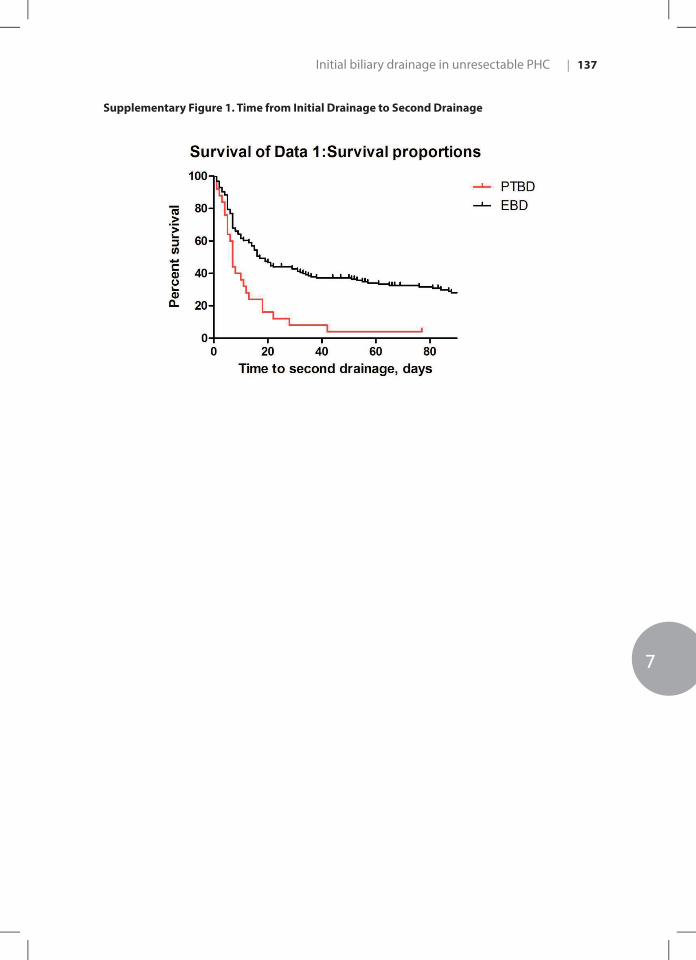
Initial biliary drainage in unresectable PHC
7
| 137
Supplementary Figure 1. Time from Initial Drainage to Second Drainage

8CHAPTER 8

8 Translating the ABC-02 trial into daily practice: outcome of palliative treatment in patients with unresectable biliary tract cancer treated with gemcitabin and cisplatin
Dierks J | Gaspersz MP | Belkouz A | van Vugt JLA | Coelen RJS | de Groot JWB | ten Tije A | Meijer WG | Pruijt H | van Voorthuizen T | van Spronsen DJ | Rentinck Mv | ten Oever D | Smit JM | Otten HM | van Gulik TM | Wilmink JW | Groot Koerkamp B | Klümpen H
Acta Oncol. 2018 Jun;57(6):807-812
Background: Biliary tract cancer (BTC) is an uncommon cancer with an unfavorable prognosis. Since 2010, the standard of care for patients with unresectable BTC is palliative treatment with gemcitabine plus cisplatin, based on the landmark phase III ABC-02 trial. This current study aims to evaluate the efficacy and safety of gemcitabine and cisplatin in patients with unresectable cholangiocarcinoma and gallbladder cancer in daily practice that meet the criteria for the ABC-02 trial in comparison to patients who did not.
Methods: Patients diagnosed with unresectable BTC between 2010 and 2015 with an indication for gemcitabine and cisplatin were included. We divided these patients into 3 groups: (I) patients who received chemotherapy and met the criteria of the ABC-02 trial, (II) patients who received chemotherapy and did not meet these criteria and (III) patients who had an indication for chemotherapy, but received best supportive care without chemotherapy. Primary outcome was overall survival (OS) and secondary outcome was progression free survival (PFS).
Results: We collected data of 208 patients, of which 138 (66.3%) patients received first line chemotherapy with gemcitabine and cisplatin. Median OS of 69 patients in group I, 63 patients in group II and 65 patients in group III was 9.6 months (95%CI: 6.7- 12.5), 9.5 months (95%CI: 7.7- 11.3) and 7.6 months (95%CI: 5.0-10.2), respectively. Median PFS was 6.0 months (95%CI: 4.4- 7.6) in group I and 5.1 months (95%CI: 3.7- 6.5) in group II. Toxicity and number of dose reductions (p=0.974) were comparable between the two chemotherapy groups.
Conclusion: First-line gemcitabine and cisplatin is an effective and safe treatment for patients with unresectable BTC who do not meet the eligibility criteria for the ABC-02 trial. Median OS, PFS and treatment side effects were comparable between the patients who received chemotherapy (group I versus group II). Abs
trac
t
Translating ABC-02 into daily practice
8
| 141
Introduction
Biliary tract cancer (BTC) is an uncommon cancer in developed countries consisting of cholangiocarcinoma and gall bladder cancer. The incidence of gall bladder cancer and extrahepatic cholangiocarcinoma in the European Union is 3.2 and 5.4/100.000 per year for males and females, respectively.1 There are approximately 600 new cases of BTC in the Netherlands per year, and the incidence is rising.2 Surgical resection is the only curative treatment for patients with BTC but most patients have (locally) advanced disease or metastasis at presentation and are not eligible for surgical resection.3,4
Patients with unresectable BTC are currently treated with the combination of gemcitabine and cisplatin, based on the Phase III ABC-02 trial. This trial demonstrated a significant survival advantage of this combination without the addition of substantial toxicity compared to gemcitabine monotherapy.5 The promising results of this clinical trial led to incorporating this treatment regimen, consisting of 1000mg/m2 gemcitabine and 25mg/m2 cisplatin in a 3-weekly cycle with administrations on day one and eight, in national and international guidelines, including the European Society for Medical Oncology (ESMO) guideline.6
Although this combination of gemcitabine plus cisplatin showed survival advantage when compared with gemcitabine alone, this regimen was studied in a group of patients complying with the inclusion criteria of the ABC-02 trial. However, most patients in clinical practice do not fulfill these criteria and the efficacy and toxicity of this regimen has not been evaluated in these patients. Furthermore, the effect of gemcitabine plus cisplatin in patients with unresectable BTC has not been compared with patients receiving best supportive care.
No difference in median overall survival (OS) between trial and non-trial patients was observed in similar retrospective studies in colorectal and breast cancer.7,8 In a study performed in men with metastatic castration resistant prostate cancer, it has been demonstrated that treatment in daily practice is associated with a shorter survival and more toxicity compared with men treated in a clinical trial. 9
Because of the poor prognosis of patients with BTC and the possible adverse effects of chemotherapy, it is important to know if this ABC-02 chemotherapy treatment regimen could also be used in patients who do not fulfil the original inclusion criteria.5 Therefore the aim of this study was to evaluate the efficacy and safety of gemcitabine and cisplatin in daily practice in unresectable BTC patients who do not meet the eligibility criteria for the ABC-02 trial, compared with those who meet the inclusion criteria for this trial.

Chapter 8142 |
To be able to answer this question it is necessary to study a subsequent population of patients that is treated in daily practice with the combination of gemcitabine and cisplatin. In addition, we aimed to compare patients who received chemotherapy and patients who received best supportive care.
Methods
Study population and data acquisitionAll patients with unresectable BTC between January 2010 and January 2015 in the Academic Medical Center (AMC), Amsterdam, The Netherlands and Erasmus MC University Medical Center (EMC), Rotterdam, The Netherlands were identified. Referring hospitals were contacted for additional data on the referred BTC patients. BTC was defined as intra- or extrahepatic cholangiocarcinoma or gallbladder cancer. If no histopathological evidence was obtained, the diagnosis was established by the multidisciplinary hepatopancreatobiliary team based on clinical, radiological, endoscopic, laboratory findings and follow-up. Patients were deemed unresectable when distant metastases were present or when radical resection was not possible due to locally advanced disease. Also recurrent disease after surgery was considered as unresectable. Patients were excluded if they received first-line chemotherapy other than gemcitabine and cisplatin. Patient characteristics (e.g. age, sex), clinical parameters (e.g. cholangitis), laboratory results (e.g. white-cell count, platelet count), chemotherapy treatment details (e.g. dose, number of cycles, toxicity) and previous interventions (Surgery, Percutaneous Transhepatic Drainage (PTC), Endoscopic Retrograde Cholangio Pancreatography (ERCP), radiotherapy) were collected from medical records.
The study population was divided into three groups: (I) patients who received chemotherapy and met the criteria of the ABC-02 trial, (II) patients who received chemotherapy and did not meet these criteria and (III) patients who were eligible for chemotherapy but received best supportive care without any chemotherapy. The criteria used in the ABC-02 trial5 are: histopathological or cytologic confirmation, an Eastern Cooperative Oncology Group (ECOG) performance status of 0, 1 or 2, a total serum bilirubin level of 1.5 times the upper limit of the normal (ULN) range or less, liver-enzyme levels of 5 times the ULN range or less, levels of serum urea and serum creatinine of 1.5 times the ULN range or less and a calculated glomerular filtration of 45ml/min or higher. The Institutional Review Boards of both centers approved the study and the need for informed consent was waived. Toxicity was scored using the Common Terminology Criteria for Adverse Events (CTCAE) version 4.03.10 Radiologic response evaluation was assessed using Response Evaluation Criteria in Solid Tumors (RECIST).11
Translating ABC-02 into daily practice
8
| 143
Study objectivesThe primary objective was to compare OS in patients treated with gemcitabine plus cisplatin who met the criteria of the ABC-02 trial versus patients who do not fulfil the inclusion criteria of the ABC-02 trial. Secondary objectives were to investigate differences in progression free survival (PFS) and toxicity between the two predefined groups. We also compared these data with the patients that were eligible for treatment but opted for best supportive care instead of chemotherapy (group III).
Statistical AnalysisAll statistical analyses were performed using SPSS version 22 (IBM corp.). Descriptive statistics for categorical variables were reported as percentages, and continuous variables were reported as medians and ranges. Categorical variables were compared using Chi square test and continuous variables were compared using the independent-samples t-test.
Dose intensity was calculated as the cumulative dose of gemcitabine per body surface area divided by the time between the date of the first administration and the end of the last cycle of chemotherapy. The same calculation was used to calculate dose intensity of cisplatin. The dose intensity of 100% gemcitabine was defined as 666.7 mg/m2/week and for cisplatin as 16.7mg/m2/week.6
The OS of BTC patients who received chemotherapy was calculated from the date of first gemcitabine and cisplatin administration. Survival of the patients who did not receive chemotherapy was calculated from the date of initial diagnosis to death with censoring for patients alive at the last moment of follow-up. An additional calculation was performed where OS of BTC patients who received chemotherapy was calculated from the date of initial diagnose to be able to compare this to patients who did not receive chemotherapy. The PFS in patients receiving chemotherapy was calculated from the date of first gemcitabine and cisplatin administration to the date on which radiological or clinical progression was determined with censoring for patients with no progressive disease at the end of follow-up (1st of July 2015). The Kaplan-Meier method was used to estimate OS. Multivariate analysis was performed with Cox Proportional Hazards Model. Survival status was updated using the municipal records database on the 10th of February 2016.
Results
Patients and treatmentIn total, 208 patients with unresectable BTC were identified and formed the study cohort (Figure 1). The majority of our cohort received gemcitabine and cisplatin treatment (n=138, 66.3%). Of these, 74 (53.6%) patients received chemotherapy and met the criteria of the

Chapter 8144 |
ABC-02 trial (group I) and 64 (46.4%) patients received chemotherapy and did not meet these criteria (group II; see table 1). Seventy patients were eligible for chemotherapy, but received best supportive care without any chemotherapy (group III). Patient’s choice was the most frequent reason for not receiving chemotherapy. Table 2 presents the baseline patient characteristics. Most patients were male (54.3%) and the median age of patients who received chemotherapy and the patients who received best supportive care was 63 and 72 years, respectively.
Figure 1. Flow chart
Table 1. Did not meet the criteria of the ABC-02 trial based on variable
Group II(N= 64)
Age > 18 years n(%) 0 (0.0)
Histological or cytologic diagnosis n(%) 5 (7.8)
ECOG PS n(%) 1 (1.5)
Serum bilirubin level > 1.5 times ULN n(%) 15 (23.4)
Serum liver- enzyme levels > 5 times ULN n(%) 55 (85.9)
Serum ureum and Creatinine level > 1.5 times ULN n(%) 2 (3.1)
eGFR < 45ml/min n(%) 2 (3.1)
Table 1: ECOG PS, Eastern Cooperative Oncology Group performance status. ULN, upper limit of normal. Liver enzyme levels including: alanine- transaminase, aspartate- transaminase, gamma- glutamyltransferase and alkaline phosphatase. eGFR, calculated glomerular filtration.
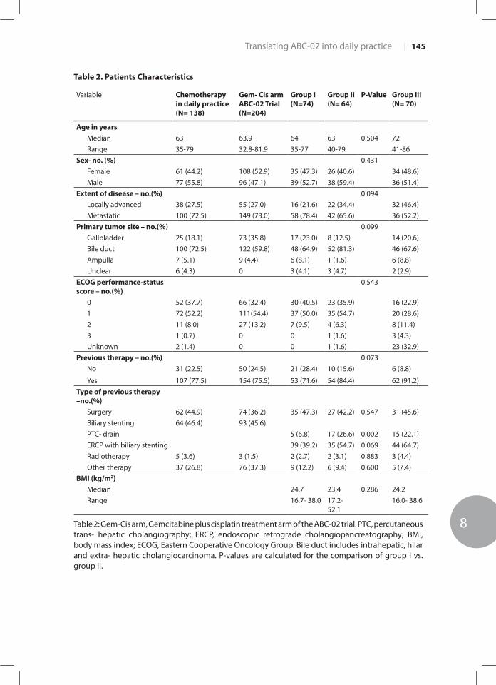
Translating ABC-02 into daily practice
8
| 145
Table 2. Patients Characteristics
Variable Chemotherapy in daily practice(N= 138)
Gem- Cis arm ABC-02 Trial(N=204)
Group I(N=74)
Group II(N= 64)
P-Value Group III(N= 70)
Age in yearsMedian 63 63.9 64 63 0.504 72Range 35-79 32.8-81.9 35-77 40-79 41-86
Sex- no. (%) 0.431Female 61 (44.2) 108 (52.9) 35 (47.3) 26 (40.6) 34 (48.6)Male 77 (55.8) 96 (47.1) 39 (52.7) 38 (59.4) 36 (51.4)
Extent of disease – no.(%) 0.094Locally advanced 38 (27.5) 55 (27.0) 16 (21.6) 22 (34.4) 32 (46.4)Metastatic 100 (72.5) 149 (73.0) 58 (78.4) 42 (65.6) 36 (52.2)
Primary tumor site – no.(%) 0.099Gallbladder 25 (18.1) 73 (35.8) 17 (23.0) 8 (12.5) 14 (20.6)Bile duct 100 (72.5) 122 (59.8) 48 (64.9) 52 (81.3) 46 (67.6)Ampulla 7 (5.1) 9 (4.4) 6 (8.1) 1 (1.6) 6 (8.8)Unclear 6 (4.3) 0 3 (4.1) 3 (4.7) 2 (2.9)
ECOG performance-status score – no.(%)
0.543
0 52 (37.7) 66 (32.4) 30 (40.5) 23 (35.9) 16 (22.9)1 72 (52.2) 111(54.4) 37 (50.0) 35 (54.7) 20 (28.6)2 11 (8.0) 27 (13.2) 7 (9.5) 4 (6.3) 8 (11.4)3 1 (0.7) 0 0 1 (1.6) 3 (4.3)Unknown 2 (1.4) 0 0 1 (1.6) 23 (32.9)
Previous therapy – no.(%) 0.073No 31 (22.5) 50 (24.5) 21 (28.4) 10 (15.6) 6 (8.8)
Yes 107 (77.5) 154 (75.5) 53 (71.6) 54 (84.4) 62 (91.2)Type of previous therapy –no.(%)
Surgery 62 (44.9) 74 (36.2) 35 (47.3) 27 (42.2) 0.547 31 (45.6)Biliary stenting 64 (46.4) 93 (45.6)PTC- drain 5 (6.8) 17 (26.6) 0.002 15 (22.1)ERCP with biliary stenting 39 (39.2) 35 (54.7) 0.069 44 (64.7)Radiotherapy 5 (3.6) 3 (1.5) 2 (2.7) 2 (3.1) 0.883 3 (4.4)Other therapy 37 (26.8) 76 (37.3) 9 (12.2) 6 (9.4) 0.600 5 (7.4)
BMI (kg/m2)Median 24.7 23,4 0.286 24.2Range 16.7- 38.0 17.2-
52.116.0- 38.6
Table 2: Gem-Cis arm, Gemcitabine plus cisplatin treatment arm of the ABC-02 trial. PTC, percutaneous trans- hepatic cholangiography; ERCP, endoscopic retrograde cholangiopancreatography; BMI, body mass index; ECOG, Eastern Cooperative Oncology Group. Bile duct includes intrahepatic, hilar and extra- hepatic cholangiocarcinoma. P-values are calculated for the comparison of group I vs. group II.

Chapter 8146 |
Group I had more patients with gallbladder tumors and less patients with bile duct tumors compared to group II (gallbladder: 23.0% versus 12.5%; bile duct: 64.9% versus 81.3% P= 0,303). In group II, significantly more patients had interventions prior to chemotherapy, most frequently a PTC (6.8% versus 26.6% P= 0,002). The majority of patients in group III had bile duct as primary tumor site (67.6%) and 91.2% of group III patients underwent a previous therapy.
Overall survival and progression free survivalThe median OS of the entire cohort was 8.8 (95% confidence interval (CI) 7.5- 10.1) months. Patients who received chemotherapy and met the criteria of the ABC-02 trial had a median OS, calculated from date of first administration of chemotherapy, of 9.6 (95% CI 6.7- 12.5) months, which was comparable with 9.5 (95% CI 7.7- 11.3) months in patients who received chemotherapy and did not meet these criteria (p=0.731; Figure 2A). If OS is calculated from date of initial diagnosis, all patients treated with gemcitabine plus cisplatin (group I and II combined) had a median OS of 14.8 (95% CI 11.7-17.8) months compared with patients who received best supportive care without chemotherapy of
Figure 2. Survival rates: (A) overall survival (B) progression-free survival
Translating ABC-02 into daily practice
8
| 147
7.6 (95% CI 5.0-10.2) months (p<0.001). Median PFS of patients in group I was 6.0 (95% CI 4.4- 7.6) months, compared to 5.1 (95% CI 3.7- 6.5) months of patients in group II (p= 0,495; Figure 2B). In a multivariable analysis that included WHO performance status, Body Mass Index (BMI), extent of disease, primary tumor site and previous therapies, we did not identify receiving chemotherapy according to the ABC-02 criteria or not as an independent prognostic factor for survival (HR 0.83, 95%CI 0.56-1.24).
Chemotherapy treatment Table 3 shows the comparison of outcomes of the patients who received chemotherapy and met the criteria of the ABC-02 trial versus those who received chemotherapy and did not meet these criteria. The dose-intensity is comparable between both groups. The main reason for ceasing chemotherapy treatment was disease progression.
Table 3. Treatment and outcomes
Variable Group I(N=73)
Group II(N=64)
P-Value
Cycles per patient 0.688
Median (range) 6 (1-16) 6 (1-16)
Dose Gemcitabine (mg/m2) 0.286
Median (range) 17400 (1720-55186) 19200 (1910-51200)
Dose Cisplatin (mg/m2) 0.544
Median (range) 425 (25-1388) 482 (48-1388)
Dose reduction – no. (%) 0.974
No 50 (68.5) 44 (68.8)
Yes 23 (31.5) 20 (31.3)
Dose intensity Gemcitabine – no.(%) 0.550
≥ 95% 23 (31.9) 25 (39.7)
85-94% 16 (22.2) 11 (17.5)
75-84% 10 (13.9) 5 (7.9)
<75% 23 (31.9) 22 (34.9)
Dose intensity Cisplatin– no.(%) 0.343
≥ 95% 20 (27.8)) 26 (41.3)
85-94% 17 (23.6) 12 (19.0)
75-84% 8 (11.1) 8 (12.7)
<75% 27 (37.5) 17 (27.0)
Reason chemotherapy stopped – no.(%) 0.729
Toxicity 14 (19.2) 10 (15.6)
Progressive disease 25 (34.2) 26 (40.6)
Other reason 16 (21.9) 16 (25.0)
Table 3: For one patient in both groups dose intensity could not be calculated.

Chapter 8148 |
Toxicity Table 4 shows the grade 3 and 4 toxicity of the chemotherapy treatment in group I and II. Patients in group II more often had a decrease in platelet count as a result of chemotherapy treatment (8.2% versus 18.8%, P= 0,079). Other toxicities were comparable between the two groups.
Table 4. Grade 3 or 4 toxic effects during treatment
Variable Chemotherapy in daily practice (N=137)
Group I(N=73)
Group II(N=64)
P-Value
Hematologic toxic effects – no.(%)nono.(%)
Decreased white-cell count 16 (11.7) 9 (12.3) 7 (10.9) 0.153
Decreased platelet count 18 (13.1) 6 (8.2) 12 (18.8) 0.079
Decreased haemoglobin count 9 (6.6) 6 (8.2) 3 (4.7) 0.289
Decreased neutrophil count 45(32.8) 26 (35.6) 19(29.7) 0.423
Liver function – no.(%)
Increased alanine aminotransferase level 6 (4.4) 4 (5.5) 2 (3.1) 0.503
Other abnormal liver function 26 (19.0) 10 (13.7) 16 (25.0) 0.224
Non- hematologic toxic effects – no.(%)
Infection 8 (11.0) 6 (9.4) 0.941
Fatigue/nausea/vomiting 3 (4.1) 2 (3.1) 0.867
Renal function 2 (2.7) 0 0.407
Table 4: Toxicity according to CTCAE 4.0. For one patient in group I, there were no data available on toxicity. P-value were calculated for the comparison of group I versus group II.
Discussion
In this study we found that patients receiving treatment with gemcitabine and cisplatin who did not meet the inclusion criteria of the ABC-02 trial, have comparable OS, PFS, toxicity and chemotherapy dose reduction rates compared with patients who fulfilled the inclusion criteria of the ABC-02 trial.
Biliary tract cancer (BTC) is an uncommon cancer with a poor prognosis. The ABC-02 trial demonstrated that a chemotherapy combination of gemcitabine plus cisplatin was associated with a significant survival advantage without the addition of substantial toxicity compared to gemcitabine monotherapy. However, inclusion criteria are strict and most patients in clinical practice do not fulfil the inclusion criteria of this ABC-02 trial. To provide
Translating ABC-02 into daily practice
8
| 149
more evidence about the efficacy and safety of chemotherapy in the heterogeneous group of unresectable BTC patients, it is necessary to extend inclusion criteria.12
To our best knowledge this is the largest retrospective analysis since gemcitabine and cisplatin has become the standard chemotherapeutic regimen for advanced BTC. One of the strengths of this study is the large patient population with BTC derived from two specialized centers, including detailed data on the diagnosis, treatment, toxicity and tumor evaluation. All patients received the same standard treatment according to the ESMO guidelines.6 Not all of these patients are treated in the specialized centers, but were referred back to peripheral hospitals to receive the chemotherapy.
When comparing our study cohort with the ABC-02 trial cohort, there are several differences to note. Considering patients’ characteristics, the primary tumor site in patients treated in the ABC-02 trial was more often gallbladder and less often bile duct (gallbladder: 18.1% versus 35.8%; bile duct: 72.5% versus 59.8%). The median OS in patients receiving chemotherapy in our study cohort was 9.5 (95% CI 7.9-11.1) months, compared with a median OS of 11.7 (95% CI 9.5-14.3) months in the ABC-02 trial. Median PFS of patients treated in daily practice was 5.6 (95% CI 4.5-6.7) months compared with 8.0 (95% CI 6.6-8.6) months in the ABC-02 trial.
The different survival between the patients in our study cohort and the ABC-02 trial can be explained by the differences in the patients’ characteristics such as primary tumor site and previous therapies. More previous therapies might lead to a selection bias, because patients might have a positive effect of these other therapies besides the chemotherapy. In the ABC-02 trial, more patients had gallbladder cancer in comparison to our study cohort. As seen in the ABC-02 trial, there was a higher partial response rate to chemotherapy in patients with gallbladder cancer compared to patients with cholangiocarcinoma. In general, patients with gallbladder cancer have a shorter OS compared to cholangiocarcinoma, but this might be different in patients who are treated with gemcitabine and cisplatin. 13 The way OS is calculated, can also contribute to a difference in OS between our cohort and the OS in patients in the ABC-02 trial. In the ABC-02 trial, OS is calculated from date of randomisation in comparison with date of first administration of chemotherapy in our study cohort.
In our study cohort, we also observed a higher toxicity rate than in the ABC-02 trial. Neutropenia and thrombocytopenia occurred more frequently in our study. Since this is a retrospective analysis, not all toxicities were reported systematically, which may have led to an underestimation of toxicity. The higher toxicity rate in our study, resulting in more dose reductions, might be a possible explanation for the lower median survival in patients treated in daily practice. Moreover, the higher toxicity rate in our study cohort can be explained by less restrictive inclusion criteria for part of the patients in comparison to the ABC-02 trial.

Chapter 8150 |
The AMC and EMC are highly specialized tertiary institutes for BTC, which may have caused a selection bias, because the majority of these referred patients needed drainage and may possibly have had a worse baseline situation than patients who are not referred to the AMC or EMC. This could explain the difference in median OS and PFS between patients who received chemotherapy in daily practice and patients in the ABC-02 trial (OS 9.5 versus 11.7 months; PFS 5.6 versus 8.0 months), because patients without drainage problems are more likely not to be referred to a highly specialized tertiary institute.
In contrast with a recent similar retrospective analysis in 26 metastatic BTC patients, we observed a difference in OS in patients treated with gemcitabine and cisplatin in daily practice compared with patients in the ABC-02 trial.14 Almost half of these patients (49%) received 2nd or 3rd line chemotherapy, which may explain the higher OS in the other retrospective analysis (9.5 vs. 10.5 months).14
When comparing patients who received chemotherapy in daily practice and met the criteria of the ABC-02 trial and patients who did not meet these criteria, we found no differences in median OS or PFS. Patients in group I had a higher 2.5- year survival rate in comparison with patients in group II (2.5- year survival: 12.9% vs. 5.1% respectively). This suggests a treatment advantage of chemotherapy, after careful patient selection, based on criteria used in the original clinical trials.
Although eligible for chemotherapy treatment, 70 patients received best supportive care rather than chemotherapy. These patients were older than patients who did receive chemotherapy. Although, the ECOG performance status was missing in 32.9% (n= 23) of patients that received best supportive care, the impaired clinical condition of these patients may explain why they did not receive any chemotherapy but best supportive care instead.
Several limitations of the current study should be mentioned. Since it is a retrospective analysis, not all required data were systematically reported and therefore not available. The administrations of chemotherapy and toxicities were not reported unambiguously.
In conclusion, our study shows that patients who did not meet the inclusion criteria of the ABC-02 trial but received an identical chemotherapy regimen, had comparable OS, PFS, toxicity and chemotherapy dose reduction rates compared with patients who did fulfil the ABC-02 trial inclusion criteria. Patients with unresectable BTC who received gemcitabine plus cisplatin had a better OS than patients who received best supportive care in real life practice. Patients with unresectable BTC who do not meet the original inclusion criteria used in the ABC-02 trial should still be considered for gemcitabine plus cisplatin treatment.
Translating ABC-02 into daily practice
8
| 151
References
1. Cidon EU. Resectable Cholangiocarcinoma : Reviewing the Role of Adjuvant Strategies.
2016;(Table 2):43-48. doi:10.4137/CMO.S32821.TYPE.
2. Intergraal Kankercentrum Nederland. www.cijfersoverkanker.nl. Accessed June 15, 2015.
3. Jarnagin WR, Fong Y, DeMatteo RP, et al. Staging, resectability, and outcome in 225 patients
with hilar cholangiocarcinoma. Ann Surg. 2001;234(4):507-517; discussion 517-519.
doi:10.1097/00000658-200110000-00010.
4. Hartog H, Ijzermans JNM, van Gulik TM, et al. Resection of Perihilar Cholangiocarcinoma. Surg
Clin North Am. 2016;96(2):247-267. doi:10.1016/j.suc.2015.12.008.
5. Valle J, Wasan H, Palmer DH, et al. Cisplatin plus Gemcitabine versus Gemcitabine for Biliary
Tract Cancer. N Engl J Med. 2010;362(14):1273-1281. doi:10.1056/NEJMoa0908721.
6. Valle JW, Borbath I, Khan S a, et al. Biliary cancer: ESMO Clinical Practice Guidelines for diagnosis,
treatment and follow-up. Ann Oncol Off J Eur Soc Med Oncol. 2016;27(suppl 5):v28-v37.
doi:10.1093/annonc/mdw324.
7. Mol L, Koopman M, van Gils CWM, et al. Comparison of treatment outcome in metastatic
colorectal cancer patients included in a clinical trial versus daily practice in The Netherlands.
Acta Oncol. 2013;52(5):950-955. doi:10.3109/0284186X.2013.777158.
8. Gallagher CM, More K, Masaquel A, et al. Survival in patients with non-metastatic breast cancer
treated with adjuvant trastuzumab in clinical practice. Springerplus. 2016;5(1):395. doi:10.1186/
s40064-016-2008-9.
9. Templeton a. J, Vera-Badillo FE, Wang L, et al. Translating clinical trials to clinical practice:
Outcomes of men with metastatic castration resistant prostate cancer treated with docetaxel
and prednisone in and out of clinical trials. Ann Oncol. 2013;24(12):2972-2977. doi:10.1093/
annonc/mdt397.
10. Services H. Common Terminology Criteria for Adverse Events v4.0 (CTCAE). Disponible en: http://
evs.nci.nih.gov/ftp1/CTCAE/CTCAE_4.03_2010-06-14_QuickReference_5x7.pdf. 2010;2009.
11. Eisenhauer E a., Therasse P, Bogaerts J, et al. New response evaluation criteria in solid tumours:
Revised RECIST guideline (version 1.1). Eur J Cancer. 2009;45(2):228-247. doi:10.1016/j.
ejca.2008.10.026.
12. Park JO, Oh D-Y, Hsu C, et al. Gemcitabine Plus Cisplatin for Advanced Biliary Tract Cancer: A
Systematic Review. Cancer Res Treat. 2015;47(3):343-361. doi:10.4143/crt.2014.308.
13. Gallardo J, Betzabe R, Villanueva L, et al. Gallbladder Cancer, a Different Disease that Needs
Individual Trials. J Clin Oncol. 2005;23(30):7753-7754. doi:10.1200/JCO.2005.02.7524.
14. Agarwal R, Sendilnathan A, Siddiqi NI, et al. Advanced biliary tract cancer: clinical outcomes
with ABC-02 regimen and analysis of prognostic factors in a tertiary care center in the United
States. J Gastrointest Oncol. 2016;6(7):996-1003. doi:10.21037/jgo.2016.09.10.

SSUMMARY
S Summary
Summary and future perspectivesNederlandse samenvatting


Summary and future perspectives
S
| 155
English summary
Part 1 - Prognostic tools in perihilar cholangiocarcinomaThe outlook for patients with perihilar cholangiocarcinoma (PHC) is dismal. Up to 50% of patients with resectable PHC on imaging will not undergo resection as occult metastatic or locally advanced disease is found at surgical exploration. In addition, of those patients who do undergo a resection, about 36% to 45% are found to have an incomplete (R1) resection. Finally, liver surgery for PHC has a high postoperative 90-day mortality rate in Western series between 5% and 18%. Therefore, we aimed in chapter 2 to identify independent prognostic factors for surgical success and to develop and validate a preoperative prognostic model to predict surgical success, defined as a complete (R0) resection without 90-day mortality, in patients with resectable PHC on imaging. We included a total of 684 PHC patients that underwent an exploratory laparotomy and included a derivation and validation cohort. The derivation cohort consisted of 331 patients and surgical success was achieved in 102 patients (30.8%). Independent prognostic factors for surgical success were younger age, no preoperative cholangitis, no unilateral or main involvement of the hepatic artery on imaging, no suspicious lymph nodes on imaging, and Blumgart stage I or II. A preoperative prognostic model based on these five independent prognostic factors was developed with a concordance index (c-index) of 0.71. The validation cohort consisted of 353 PHC patients undergoing exploratory laparotomy. External validation showed good concordance with a c-index of 0.71. This study showed that surgical success (R0 resection without postoperative mortality) was achieved in only 30% of PHC patients undergoing exploratory laparotomy. This preoperative model to predict surgical success may be used in shared decision making.
To improve the prognostic value of current staging systems, we investigated the prognostic value of vascular involvement on imaging in patients with PHC. We performed a retrospective cohort study as presented in chapter 3. A total number of 674 patients was studied and all imaging at the time of first presentation was reassessed. Involvement of the portal vein was observed in 374 (56.0%) patients and hepatic artery was observed in 365 (54.6%) patients. We found that both unilateral and main HA involvement were independent poor prognostic factors for overall survival (OS) in patients presenting with PHC, whereas PV involvement was not. In patients undergoing resection, PV involvement and unilateral HA involvement did not influence survival. This implies that current staging systems may be improved by including main HA involvement as one of the predictive factors.
Chapter 4 describes the external validation of a new staging system from the Mayo Clinic, that is applicable to all patients with PHC regardless of subsequent treatment. The staging system assigns patients to one of four stages, depending on the patients’ performance

Summary156 |
status, serum Ca19-9 level, and radiological parameters including tumor size, suspected vascular involvement, and metastatic disease. We were able to apply this staging system to a cohort of 600 patients from two specialized tertiary referral centers. Median overall survival of stages I, II, III and IV was 33, 20, 12 and 6 months respectively. The model may be used to inform patients about prognosis and may aid in stratification of patients for clinical trials. However, since the discriminative performance was moderate, as indicated by a c-index of 0.59, the model requires improvement prior to clinical implementation.
In chapter 5 we evaluated the 8th edition of the AJCC staging system and compared the prognostic value with the previous edition. We included 248 patients with PHC and high-quality imaging available for reassessment of AJCC stage. We found no improvement of the prognostic value of the 8th edition of the AJCC staging system with all sub-stages in our cohort as compared to the 7th edition, as indicated by a c-index of 0.570 and 0.576, respectively. When analyzing a subgroup of resected patients, the prognostic value was slightly better compared to the entire cohort, and the prognostic value of the 8th edition was slightly better than the previous one in this subgroup (c-index of 0.613 vs 0.605). Interestingly, the AJCC staging system was specifically developed for both resectable and unresectable PHC patients, and to be used on imaging rather than surgical findings. However, the prognostic accuracy for unresectable patients was even worse and did not show any improvement when compared to the 7th edition. For unresectable patients, the c-index for both the 7th and the 8th edition was a mere 0.550. Since discriminative performance was moderate, the AJCC staging system may need modifications to improve its prognostic accuracy in patients with PHC.
Part 2 - Palliative treatment in patients with unresectable perihilar cholangiocarcinomaAs the majority of patients is not eligible for curative resection we investigated the outcome of palliative treatment and initiatives to optimize palliative care.Most patients with unresectable PHC die within the first year after diagnosis, however life expectancy improves considerably after surviving one or more years. Therefore, an updated estimate of life expectancy after one year could be of help in patients with unresectable PHC. Conditional survival (CS) predicts survival and takes into account the number of years the patient has already survived. In chapter 6 we studied CS for patients with unresectable PHC and found a substantial improvement of CS for 572 patients with unresectable PHC over time; the conditional chance of surviving four years from the time of presentation, increased from 9.1% at one year, to 62.3% at three years. Independent poor prognostic factors for OS were age ≥65 years, tumor size >3cm on imaging, suspected distant metastases on imaging, and unresectable disease on imaging. Poor prognostic factors became less relevant when patients survived one or more years. This is the first large study to estimate CS in patients with unresectable PHC. Conditional
Summary and future perspectives
S
| 157
survival can serve as a more valuable estimate in predicting long-term survival in patients with unresectable PHC.
Chapter 7 shows the results of a study in which we investigated the success and complication rate of initial biliary drainage in patients with unresectable PHC. A total of 187 patients with unresectable PHC were included; 161 (86.1%) underwent initial EBD and 26 (13.9%) initial PTBD. Success of initial drainage was observed in only 96 patients (48,1%). Severe drainage-related complications after EBD were observed in 19 patients (11.8%). Three PTBD patients (11.5%) developed severe drainage-related complications. The 90-day mortality rate after initial drainage procedure was 34.8%.Based on the ABC-02 trial, the combination of Gemcitabine plus Cisplatin is the recommended chemotherapy regime for patients with metastatic or locally advanced disease PHC. This trial showed a survival benefit of Gemcitabine plus Cisplatin when compared to Gemcitabine monotherapy. However, the ABC-02 trial maintained very strict inclusion criteria and most clinical patients do not meet these criteria. In chapter 8 we investigated the outcomes of this chemotherapy regimen in patients with advanced biliary tract cancer (BTC) treated with Gemcitabin and Cisplatin outside of the trial criteria. In this study, we found that patients undergoing Gemcitabine and Cisplatin who did not meet the inclusion criteria of the ABC-02 trial had comparable OS, PFS, toxicity and chemotherapy dose reduction rates compared to patients who did fulfil the inclusion criteria of the ABC-02 trial. Patients with unresectable BTC who do not meet the original inclusion criteria used in the ABC-02 trial should still be considered for Gemcitabine plus Cisplatin treatment.

Summary158 |
Future perspectives
This thesis investigated several prognostic tools for PHC and palliative care in patients with unresectable PHC. Results from our studies show the urgency for improvement. Prognosis for patients with PHC is still very poor. Although many improvements have been made in recent years, the majority of patients are still unresectable at the time of presentation.1, 2
Prognostic models for PHC currently fall short, especially for patients with unresectable disease. The AJCC staging system is the most commonly used prognostic model but showed poor prognostic accuracy, especially in unresectable patients.3 This suggest one staging system with good prognostics accuracy for both resectable and unresectable patient might not be conceivable in PHC. Future research should investigate whether a combination of two different staging systems, one based on imaging for unresectable patients and one for resectable patients, can further improve individual patient prognostication. Another way to improve prognostic models is the development of risk score calculators on mobile phones. This way, prognostic models no longer need to be simplified and a more accurate prediction can be made.
Major improvements can be made in earlier detection of PHC. Upcoming biomarkers include matrix metalloproteinases, serotonin and bile acids and emerging techniques include the development of tumor organoids and circulating tumor DNA derived from both serum and bile.4, 5 Hopefully, future translational studies are able to identify these biomarkers and techniques that may help with early diagnosis and targeted therapies for PHC.
Most studies on PHC are aimed at patients with resectable disease. As 80% of patients have unresectable disease, more studies should focus on these patients. Improving care for unresectable patients may have a positive impact on many more patients than studies on resectable patients will.
Although it is important to try and improve overall survival in unresectable patients, we should also consider the quality of life of patients during the palliative phase. Palliative biliary drainage might be required in unresectable patients with severe cholangitis. However, clinicians should take into account that the high complication rate and the low success rate of the drainage procedure itself may have a significant negative impact on the quality of life during the limited time these patients still have. Future studies should focus on improving the failure and complication rate of palliative biliary drainage in patients with PHC. This may result in increased quality of life, increased resectability, and a higher proportion of patients eligible for chemotherapy.
Summary and future perspectives
S
| 159
Currently, the standard of care for patients with metastatic or locally advanced disease PHC consists of Gemcitabine plus Cisplatin.6 Although this showed a survival benefit when compared to Gemcitabine monotherapy, toxicity with this regimen is high and this palliative regimen only extends survival with a few months. Gemcitabine plus Cisplatin combination has not been compared head to head with other combinations. At this time, there is no available agent or regimen that leads to objective tumor shrinkage or extends survival beyond 15 months. Therefore, the need for new chemotherapeutical agents or regiments is urgent and the future use of patient-derived novel targeted treatments may be helpful.
Preoperative and adjuvant chemotherapy are currently not indicated for PHC. However, new and ongoing trials may change this is the near future. The BILCAP trial aimed to investigate the effect of adjuvant Capcitabine after surgery in patients with biliary tract cancers. Recently, it was reported that this trial showed a survival benefit for patients who received Capcitabine after surgery as compared to those who only underwent surgery (51 months vs 36 months, p=0.097).7 The ACTICCA trial is a multicenter randomized controlled phase III trial designed to assess the clinical performance of adjuvant Gemcitabine with Cisplatin vs. Capcitabine in patients with biliary tract tumor after curative resection.8 The results of these trials may change the perspective on adjuvant chemotherapy.

Summary160 |
References
1. Jarnagin WR, Fong Y, DeMatteo RP, Gonen M, Burke EC, Bodniewicz BJ, et al. Staging, resectability,
and outcome in 225 patients with hilar cholangiocarcinoma. Ann Surg. 2001;234(4):507-17;
discussion 17-9.
2. Groot Koerkamp B, Wiggers JK, Gonen M, Doussot A, Allen PJ, Besselink MG, et al. Survival after
resection of perihilar cholangiocarcinoma-development and external validation of a prognostic
nomogram. Ann Oncol. 2015;26(9):1930-5.
3. Edge SB, Compton CC. The American Joint Committee on Cancer: the 7th edition of the AJCC
cancer staging manual and the future of TNM. Ann Surg Oncol. 2010;17(6):1471-4.
4. Wang B, Chen L, Chang HT. Potential diagnostic and prognostic biomarkers for
cholangiocarcinoma in serum and bile. Biomark Med. 2016;10(6):613-9.
5. Razumilava N, Gores GJ. Cholangiocarcinoma. Lancet. 2014;383(9935):2168-79.
6. Valle J, Wasan H, Palmer DH, Cunningham D, Anthoney A, Maraveyas A, et al. Cisplatin plus
gemcitabine versus gemcitabine for biliary tract cancer. N Engl J Med. 2010;362(14):1273-81.
7. John Neil P, Richard F, Daniel HP, Raj P, Darius M, David Alan A, et al. Adjuvant capecitabine for
biliary tract cancer: The BILCAP randomized study. Journal of Clinical Oncology. 2017;35(15_
suppl):4006-.
8. Stein A, Arnold D, Bridgewater J, Goldstein D, Jensen LH, Klumpen HJ, et al. Adjuvant
chemotherapy with gemcitabine and cisplatin compared to observation after curative intent
resection of cholangiocarcinoma and muscle invasive gallbladder carcinoma (ACTICCA-1 trial)
- a randomized, multidisciplinary, multinational phase III trial. BMC Cancer. 2015;15:564.

Summary and future perspectives
S
| 161

Nederlandse Samenvatting
S
| 163
Nederlandse samenvatting
Deel 1 – Prognostische tools voor perihilair cholangiocarcinoomDe prognose van patiënten met een perihilair cholangiocarcinoom is zeer somber. Meer dan 50% van patiënten met een resectabele tumor op beeldvorming, blijkt peroperatief toch occulte metastasen of lokaal uitgebreide ziekte te hebben en ondergaat geen resectie. Tevens heeft 36-45% van de gereseceerde patiënten achteraf een incomplete resectie (R1). Tot slotte kent leverchirurgie een hoge postoperatieve 90-dagen mortaliteit van 5-18%. Daarom hebben we ons in hoofdstuk 2 gericht op het identificeren van onafhankelijke prognostische factoren voor een succesvolle chirurgische resectie en het ontwikkelen en valideren van een preoperatief prognostisch model om een succesvolle chirurgische resectie te kunnen voorspellen. Een succesvolle chirurgische resectie was gedefinieerd als een complete (R0) resectie zonder 90-dagen mortaliteit bij patiënten met een resectabel PHC op beeldvorming. Wij includeerden in totaal 684 patiënten die een proeflaparotomie ondergingen, verdeeld in een derivatie en validatie cohort. Het derivatie cohort bestond uit 331 patienten. Een succesvolle chirurgische resectie werd gerealiseerd bij 102 (30.8%) van de patiënten. Onafhankelijke prognostische factoren voor een succesvolle chirurgische ingreep waren jongere leeftijd, de preoperatieve aanwezigheid van cholangitis, de afwezigheid van unilaterale of hoofdstam betrokkenheid van de arteria hepatica op beeldvorming, de afwezigheid van voor metastasen suspecte lymfeklieren op beeldvorming en Blumgart stadium I of II. Een preoperatief model gebaseerd op deze vijf prognostische variabelen werd ontwikkeld met een concordance index (c-index) van 0.71. Deze studie toonde aan dat een succesvolle chirurgisch resectie (R0 zonder 90-dagen mortaliteit) maar bij 30% van de patiënten werd bereikt die een proeflaparotomie ondergingen. Dit preoperatieve model om een succesvolle chirurgische resectie te voorspellen kan gebruikt worden bij shared decision making.
Om de voorspellende waarde van de huidige staging systemen te verbeteren onderzochten we de prognostische waarde van vasculaire betrokkenheid van PHC. We verrichten een retrospectieve cohortstudie zoals te lezen is in hoofdstuk 3. In totaal werden 674 patiënten geïncludeerd in deze studie en alle beeldvorming die was verricht bij eerste presentatie van de ziekte werd opnieuw beoordeeld. Betrokkenheid van de vena portae werd geobserveerd bij 374 (56.0%) patiënten en betrokkenheid van de arteria hepatica werd geobserveerd bij 365 (54.6%) patiënten. Zowel unilaterale als hoofdstam betrokkenheid van de arteria hepatica bleken onafhankelijk negatief prognostische factoren voor overall survival (OS) bij patiënten met PHC. Betrokkenheid van de vena portae was geen onafhankelijke prognostische factor. Betrokkenheid van de vena portae en unilaterale betrokkenheid van de arteria hepatica had geen invloed op de overleving van patiënten die een resectie ondergingen. Dit suggereert dat de huidige staging
Summary164 |
systemen mogelijk verbeterd kunnen worden door het includeren van hoofdstam van de betrokkenheid arteria hepatica als een van de prognostische factoren.
Hoofdstuk 4 beschrijft de externe validatie van een nieuw staging model van de Mayo Clinic dat ontwikkeld is voor alle patiënten met PHC, ongeacht de behandeling. Het staging systeem wijst patiënten een van de vier stadia toe, afhankelijk van de performance status van een patiënt, het serum Ca 19-9 en diverse radiologische parameters waaronder de grootte van de tumor, verdenking op vasculaire betrokkenheid en gemetastaseerde ziekte. We hebben het staging model kunnen toepassen op een cohort van 600 patiënten van twee gespecialiseerde tertiaire centra. De mediane overleving van stage I, II, III en IV was respectievelijk 33, 20, 12, en 6 maanden. Het model zou waardevolle informatie kunnen geven over de prognose en kan helpen bij het stratificeren van patiënten voor klinische trials. Het onderscheidend vermogen was echter matig en met een c-index van 0.59 zijn er verbeteringen nodig voordat het model werkelijk geïmplementeerd kan worden in de kliniek.
Het meest gebruikte stadierings model om prognose en juiste behandelstrategie te bepalen is het staging system van de American Joint Committee on Cancer (AJCC). In hoofdstuk 5 evalueren we de 8ste editie van het AJCC staging systeem en vergelijken we deze met de 7de editie. We includeerden 248 patiënten met PHC waarbij beeldvorming beschikbaar was voor herbeoordeling aan de hand van het nieuwe AJCC staging systeem. We vonden geen verbetering van de prognostische waarde van de 8ste editie van het AJCC staging system ten opzichte van de 7de editie in ons cohort. Dit bleek uit de c-index van respectievelijk 0.570 and 0.576. De prognostische waarde van het AJCC staging system was aanzienlijk beter in een subgroep van gereseceerde patiënten in vergelijking met het gehele cohort. Tevens bleek de c-index van de 8ste editie van deze subgroep beter dan die van de 7de editie (0.613 vs. 0.605). Het AJCC staging system is ontwikkeld voor zowel gereseceerde als ongereseceerde PHC-patiënten en zou zowel peroperatief en postoperatief toepasbaar zijn. Echter blijkt uit onze studie dat de prognostische waarde van het AJCC staging system bij de irresectabele patiënten slecht is en ook niet verbeterd in de 8ste editie. Voor irresectabele patiënten was de c-index van de 7de en 8ste editie maar 0.550. Gezien het zeer matige onderscheidende vermogen van het AJCC staging system zijn er aanpassingen nodig om de prognostische waarde te verbeteren bij patiënten met PHC.
Deel 2 - De palliatieve behandeling van patiënten met perihilair cholangiocarcinoomOmdat de meerderheid van patiënten met PHC niet in aanmerking komt voor curatieve resectie onderzochten we de uitkomsten van palliatieve behandelingen en nieuwe initiatieven om de palliatieve behandeling te optimaliseren.

Nederlandse Samenvatting
S
| 165
De meeste patiënten met irresectabel PHC overlijden binnen 1 jaar na het stellen van de diagnose. Echter verbetert de te verwachten overleving als patiënten een of meer jaren na de diagnose nog steeds in leven zijn. Patiënten zouden gebaat kunnen zijn bij een aangepaste prognose van de te verwachtte overleving. Conditional survival (CS) voorspelt de overleving maar neemt hierbij het aantal jaren dat de patiënt reeds overleefd heeft mee in de voorspelling. In hoofdstuk 6 analyseerden we de CS voor patiënten met irresectabel PHC. We vonden een substantiële verbetering van de CS voor de 572 geanalyseerde irresectabele patiënten over de tijd. De conditionele kans om 4 jaar na presentatie nog in leven te zijn liep op van 9.1% op 1 jaar na de diagnose naar 62.3% op 3 jaar na de diagnose. Onafhankelijke prognostische factoren voor een slechtere OS waren een leeftijd van 65 jaar of ouder, een tumor groter dan 3cm op beeldvorming, de verdenking op metastasen op afstand op beeldvorming en irresectabele ziekte op beeldvorming. Onafhankelijke prognostische factoren voor een slechtere OS waren minder relevant nadat patiënten het eerste jaar na de diagnose nog in leven waren. Conditional survival kan fungeren als een accuratere en waardevollere voorspeller van lang termijn overleving in PHC-patiënten met gemetastaseerde of irresectabele ziekte
Hoofdstuk 7 toont de resultaten van een studie waarbij we het succes en het aantal complicaties van initiële biliaire drainage bij patiënten met PHC hebben onderzocht. In totaal werden er 187 patiënten met een irresectabel PHC geïncludeerd: 161 (86.1%) ondergingen een endoscopische retrograde cholangio pancreaticografie (ERCP) als initiële drainage poging en 26 (13.9%) patiënten ondergingen een percutane transhepatische drainage (PTC) als initiële drainage poging. De initiële drainage was maar bij 96 patiënten (48.1%) succesvol. Ernstige drainage-gerelateerde complicaties na ERCP werden geobserveerd bij 19 patiënten (11.8%). Drie PTC-patiënten (11.5%) ontwikkelden ernstige drainage-gerelateerde complicaties na de initiële PTC-drainage. De 90-dagen mortaliteit na initiële drainage was 34.8%.
Naar aanleiding de resultaten van de ABC-02 studie is de combinatietherapie van Gemcitabine en Cisplatin op dit moment het geadviseerde regiem voor patiënten met gemetastaseerd of irresectabel PHC. De ABC-02 studie toonde een langere overleving met Gemcitabine en Cisplatin in vergelijking met Gemcitabine monotherapie. De ABC-02 studie had echter zeer strenge in- en exclusiecriteria. Het merendeel van de patiënten met gemetastaseerd of irresectabel PHC voldoet niet aan deze criteria. In hoofdstuk 8 onderzochten wij de uitkomsten van combinatietherapie van Gemcitabine en Cisplatin bij patiënten met gemetastaseerd of irresectabel PHC die niet voldeden aan de criteria van de ABC-02 studie (patiënten die in de ABC-02 trial geexcludeerd zouden worden). Onze studie toonde dat patiënten die behandeld werden met Gemcitabine en Cisplatin maar niet voldeden aan de criteria van de ABC-02 studie een vergelijkbare overall survival, progressievrije overleving, toxiciteit en aantal dosisaanpassingen hadden in vergelijking

Summary166 |
met patiënten die wel voldeden aan de criteria. Uit deze studie blijkt dat patiënten met gemetastaseerd of irresectabel PHC die niet voldoen aan de criteria van de ABC-02 trial alsnog in aanmerking kunnen komen voor combinatietherapie van Gemcitabine en Cisplatin.

Nederlandse Samenvatting
S
| 167

AAPPENDICES

A Appendices
List of publicationsList of contributing authorsPhD PortfolioDankwoordCurriculum vitae

List of publications
A
| 171
List of Publications
In this thesis
Evaluation of the new American Joint Committee on Cancer Staging Manual 8th edition for perihilar cholangiocarcinoma. M.P. Gaspersz, S. Buettner, J.L. van Vugt, J. de Jonge, W.G. Polak, M. Doukas, J.N.M. IJzermans, B. Groot Koerkamp, F.E.J.A. Willemssen.J Gastrointest Surg. 2019 Feb 12
A Preoperative Prognostic Model to Predict Surgical Success in Patients with Perihilar Cholangiocarcinoma. M.P. Gaspersz, S. Buettner, E. Roos, J.L.A. van Vugt, R.J.S. Coelen, J. Vugts, J.K. Wiggers, P.J. Allen, M.G. Besselink, O.R.C. Busch, E.J. Belt, M.I. D’Angelica, R.P. DeMatteo, J. de Jonge, T.P. Kingham, W.G. Polak, F.E.J.A. Willemssen, T.M. van Gulik, W.R. Jarnagin, J.N.M. IJzermans, B. Groot Koerkamp. 2018 Sep;118(3):469-476
Translating the ABC-02 trial into daily practice: Outcome of palliative treatment in patients with advanced biliary tract cancer treated with gemcitabin and cisplatin. J. Dierks, M.P. Gaspersz, A. Belkouz, J.L.A. van Vugt , R.J.S. Coelen, J.W.B. de Groot, A. ten Tije, W.G. Meijer, H. Pruijt, T. van Voorthuizen, D.J. van Spronsen, M. Rentinck, D. ten Oever, J.M. Smit, H.M. Otten, T.M. van Gulik, J.W. Wilmink, B. Groot Koerkamp, H. Klumpen.Acta Oncol. 2018 Jun;57(6):807-812
The Prognostic Value of Portal Vein and Hepatic Artery Involvement in Patients with Perihilar Cholangiocarcinoma. J.L.A. van Vugt, M.P. Gaspersz, R.J.S. Coelen, J. Vugts, T.A. Labeur, J. de Jonge, W.G. Polak, J.N.M. IJzermans, C.Y. Nio, T.M. van Gulik, F.E.J.A. Willemssen, B. Groot Koerkamp.HPB (Oxford). 2018 Jan;20(1):83-92
Conditional Survival in Patients with Unresectable Perihilar Cholangiocarcinoma. M.P. Gaspersz, S. Buettner, J.L.A. van Vugt, E. Roos, R.J.S. Coelen, E.J. Belt, J. de Jonge, W.G. Polak, F.E.J.A. Willemssen, T.M. van Gulik, J.N.M. IJzermans, B. Groot Koerkamp.HPB (Oxford). 2017 Nov;19(11):966-971
Validation of the Mayo Clinic Staging System in Determining Prognoses of Patients With Perihilar Cholangiocarcinoma. R.J.S. Coelen, M.P. Gaspersz, T.A. Labeur, J.L.A. van Vugt, S. van Dieren, F.E.J.A. Willemssen, C.Y. Nio, J.N.M. IJzermans, H. Klümpen, B. Groot Koerkamp, T.M. van Gulik. Clin Gastroenterol Hepatol. 2017 Dec;15(12):1930-1939

Appendices172 |
Not in this thesis
Growth of Focal Nodular Hyperplasia is not a reason for surgical intervention but patients should be referred to a tertiary referral centre. M.E. Broker, A.J. Klompenhouwer, M.P. Gaspersz, A.M.E. Alleleyn, R.S. Dwarkasing, I.C. Pieters, R.A. de Man, J.N.M. Ijzermans. World J Surg. 2018 May;42(5):1506-1513
Low Skeletal Muscle Density is Associated with Early Death in Patients with Perihilar Cholangiocarcinoma Regardless of Subsequent Treatment. J.L.A. van Vugt, M.P. Gaspersz, J. Vugts, S. Buettner, S Levolger, R.W.F. de Bruin, W.G. Polak, J. de Jonge, F.E.J.A. Willemssen, B. Groot Koerkamp, J.N.M. IJzermans.Dig Surg. 2018 Feb 16
A model including sarcopenia surpasses the MELD score in predicting waiting list mortality in cirrhotic liver transplant candidates: a competing risk analysis in a national cohort. J.L.A. van Vugt, L.J.M. Alferink, S. Buettner, M.P. Gaspersz, D. Bot, S. Darwish Murad, S. Feshtali, P.M.A. van Ooijen, W.G. Polak, R.J. Porte, B. van Hoek, A.P. van den Berg, H.J. Metselaar, J.N.M. IJzermans.J Hepatol. 2017 Dec 6. pii: S0168-8278(17)32474
Human Primary Liver Cancer -derived Organoid Cultures for disease modelling and drug screening.L. Broutier, G. Mastrogiovanni, M.M.A. Verstegen, H.E. Francies, L. Morrill Gavarró, C.R Bradshaw, G.E. Allen, R. Arnes-Benito, O. Sidorova, M.P. Gaspersz, N. Georgakopoulos, B. Koo, S. Dietman, S.E. Davies, R.K. Praseedom, R. Lieshout, J.N.M. IJzermans, S.J. Wigmore, K. Saeb-Parsy, M.J. Garnett, L.J.W. van der Laan, M. Huch. Nat Med. 2017 Dec;23(12):1424-1435
Low Skeletal Muscle Mass is Associated with Increased Hospital Expenditure in Patients Undergoing Cancer Surgery of the Alimentary Tract. J.L.A. van Vugt, S. Buettner, S. Levolger, R.R.J. Coebergh van den Braak, M. Suker, M.P. Gaspersz, R.W.F. de Bruin, C. Verhoef, C.H.C. van Eijck, N. Bossche, B. Groot Koerkamp, J.N.M. IJzermans. PLoS One. 2017 Oct 31;12(10):e0186547
Retrospective study on timing of resection of hepatocellular adenoma. A.J. Klompenhouwer, M.E. Broker, M.G.J. Thomeer, M.P. Gaspersz, R.A. de Man, J.N.M. IJzermans. Br J Surg. 2017 Nov;104(12):1695-1703

List of publications
A
| 173
Inflammatory and multiple hepatocellular adenoma are associated with a higher BMI. M.E.E. Broker, M.P. Gaspersz, A.J. Klompenhouwer, B.E. Hansen, T. Terkivatan, P. Taimr, R. Dwarkasing, M.G.J. Thomeer, R.A. de Man, J.N.M. IJzermans. Eur J Gastroenterol Hepatol. 2017 Oct;29(10):1183-1188
Antibodies Against Immune Checkpoint Molecules Restore Functions of Tumor-infiltrating T cells in Hepatocellular Carcinomas. G. Zhou, D. Sprengers, P. Boor, H. Schutz, M. Doukas, W.G. Polak, J. de Jonge, M.P. Gaspersz, A. Pedroza-Gonzalez, H. Dong, K. Thielemans, J.N.M. IJzermans, M. Bruno, J. Kwekkeboom. Gastroenterology. 2017 Oct;153(4):1107-1119.e10
Survival after Resection of Perihilar Cholangiocarcinoma in Patients with Lymph Node Metastases. S. Buettner, J.L.A. van Vugt, M.P. Gaspersz, R.J.S. Coelen, E. Roos, T.A. Labeur, G. A. Margonis, C.G. Ethun, S.K. Maithel, G. Poultsides, T. Tran, K. Idrees, C.A. Isom, R.C. Fields, B. Krasnick, S.M. Weber, A. Salem, R. C.G. Martin, C. Scoggins, P. Shen, H.D. Mogal, C. Schmidt, E. Beal, I. Hatzaras, R. Shenoy, J.N.M. IJzermans, T.M. van Gulik, T.M. Pawlik, B. GrootKoerkamp. HPB (Oxford). 2017 Aug;19(8):735-740
Evidence of good prognosis of hepatocellular adenoma in post-menopausal women. A.J. Klompenhouwer, D. Sprengers, F.E.J.A. Willemssen, M.P. Gaspersz, J.N.M. Ijzermans, R.A. de Man. J Hepatol. 2016 Dec;65(6):1163-1170
The new European Society of Cardiology guidelines on myocardial revascularisation: an appraisal. S.T. Nauta, M. Gaspersz, J.W. Deckers. Heart. 2012 Jan;98(1):11-4
Comparison of six-year clinical outcome of sirolimus- and paclitaxel-eluting stents to bare-metal stents in patients with ST-segment elevation myocardial infarction: an analysis of the RESEARCH and T-SEARCH registries. C. Simsek, M. Magro, E. Boersma, Y. Onuma, S. Nauta, J Daemen, M. Gaspersz, R.J. van Geuns, W. van der Giessen, R. van Domburg, P. Serruys. J Invasive Cardiol. 2011 Aug;23(8):336-41
The unrestricted use of sirolimus- and paclitaxel-eluting stents results in better clinical outcomes during 6-year follow-up than bare-metal stents: an analysis of the RESEARCH and T-SEARCH registries. C. Simsek, M. Magro, E. Boersma, Y. Onuma, S. Nauta, M.P. Gaspersz, W. van der Giessen, R. van Domburg, P. Serruys, JACC Cardiovasc Interv. 2010 Oct;3(10):1051-8


List of contributing authors
A
| 175
List of contributing authors
JL van Vugt, S Buettner, JNM IJzermans, J de Jonge, WG Polak, B Groot Koerkamp, J Vugts, CHJ van Eijck, C PekDepartment of Surgery, Erasmus University Medical Center, Rotterdam, the Netherlands
FEJA WillemssenDepartment of Radiology, Erasmus University Medical Center, Rotterdam, the Netherlands
L Hol, JW Poley, M BrunoDepartment of Gastroenterology, Erasmus University Medical Center, Rotterdam, the Netherlands
M DoukasDepartment of Pathology, Erasmus University Medical Center, Rotterdam, the Netherlands
RJS Coelen, E Roos, TM van Gulik, MGH Besslink, ORC Busch, S van DierenDepartment of Surgery, Academic Medical Center, Amsterdam, the Netherlands
J van HooftDepartment of Gastroenterology, Academic Medical Center, Amsterdam, the Netherlands
T Labeur, A Belkouz, H Klümpen, J Dierks, JW Wilmink Department of Oncology, Academic Medical Center, Amsterdam, the Netherlands
CY NioDepartment of Radiology, Academic Medical Center, Amsterdam, the Netherlands
PJ Allen, MI D’Angelica, TP Kingham, WR JarnaginDepartment of Surgery, Memorial Sloan Kettering Cancer Center, New York, NY, United States of America
RP DeMatteoDepartment of Surgery, University of Pennsylvania, Philadelphia, PA, United States of America
E BeltDepartment of Surgery, Albert Schweitzer Hospital, Dordrecht, the Netherlands
Appendices176 |
JWB de GrootDepartment of Medical Oncology, Isala Oncologisch Centrum, Zwolle, The Netherlands
A ten TijeDepartment of Medical Oncology, Amphia Ziekenhuis, Breda, The Netherlands
WG MeijerDepartment of Medical Oncology, Westfries Gasthuis, Hoorn, The Netherlands
H PruijtDepartment of Medical Oncology, Jeroen Bosch Ziekenhuis, ‘s Hertogenbosch, The Netherlands
T van VoorthuizenDepartment of Medical Oncology, Rijnstate Ziekenhuis, Arnhem, The Netherlands
DJ van SpronsenDepartment of Medical Oncology, Canisius Wilhelmina Ziekenhuis, Nijmegen, The Netherlands
M RentinckDepartment of Medical Oncology, Tergooi Ziekenhuis, Hilversum, The Netherlands
D ten OeverDepartment of Medical Oncology, Noordwest Ziekenhuisgroep, Alkmaar, The Netherlands
JM SmitDepartment of Medical Oncology, Gelre Ziekenhuis, Apeldoorn, The Netherlands
HM OttenDepartment of Medical Oncology, Medical Center Slotervaart, Amsterdam, The Netherlands

List of contributing authors
A
| 177


PhD Portfolio
A
| 179
PhD Portfolio
Name PhD student: Marcia GasperszPhD period: 2014 -2017Erasmus MC Department: SurgeryPromotor: Jan N.M. IJzermansResearch School: Molecular Medicine
1. PHD training Year ECTS
General courses- Survival Analyses Course 2016 0.5- Cursus GraphPad Prism version 6 2016 0.3- Open Clinica Cursus 2015 0.1- Basic Introduction Course SPSS 2015 1.0- Cursus Wetenschappelijke integriteit /research integrity 2015 0.3- BROK (Basiscursus Regelgeving Klinisch Onderzoek) 2014 1.8
Specific courses- Basic and Translational Oncology Course 2015 1.8- Biomedical Research Techniques XIV Course 2015 1.5
Oral and poster presentations- European Assoc. for the study of the liver (EASL), Annual Meeting 2018 1- United European Gastroenterology (UEG), Annual Meeting 2017 1- European Assoc. for the study of the liver (EASL), Annual Meeting 2017 1- American Hepato-Pancreato-Biliary Assoc. (AHPBA), Annual Meeting 2017 4- Society of Surgical Oncology, Annual Cancer Symposium 2017 1- Nederlandse Vereniging van Heelkunde Najaarsdag 2016 2- American Assoc. of the study of Liver Disease, The Liver Week 2016 2- Molecular Medicine Day 2016 2
Attendance at (inter)national Conferences and Seminars- Symposium Current and Future perspectives in Primary Liver Tumors 2017 1- Molecular Medicine Day 2017 1- Chirurgendagen/Annual meeting Dutch association of Surgery 2015-2017 3- The Erasmus MC Liver day 2015-2017 3- International Liver Cancer association Annual Meeting 2015 1- Journal clubs (GIO, choco)
2. Teaching
- Examination of Basic Life Support of medical students 2015-2017 1
- Tutor first year medical students 2015-2017 2


Dankwoord
A
| 181
Dankwoord
Dit proefschrift was nooit tot stand gekomen zonder hulp van vele anderen. Ik wil graag iedereen bedanken die heeft bijgedragen. In het bijzonder wil ik graag wil ik de volgende mensen bedanken:
Geachte promotor prof.dr. J.N.M. Ijzermans, beste professor, bedankt voor alle kansen en mogelijkheden die u mij heeft gegeven in de afgelopen jaren. Een van onze eerste gesprekken eindigde in de slappe lach (BOHICA) en deze vrolijke toon hebben we altijd vastgehouden. Dank voor uw begeleiding, alle goede gesprekken en uw enthousiasme. Ik vond het een eer.
Geachte co-promotor, beste Bas, bedankt voor de mogelijkheid die je mij hebt gegeven toen mijn initiële onderzoek in het lab wat anders liep dan verwacht. Ik waardeer het enorm dat je mij zo hebt geholpen en begeleid. Bedankt voor alles.
Geachte leescommissie, professor van Gulik, professor Dejong en professor Bruno, dank voor uw inspanning bij het beoordelen van mijn proefschrift en uw bereidheid om plaats te nemen in mijn leescommissie. Geachte leden van de grote commissie, dank voor uw interesse, expertise en oppositie.
Beste co-auteurs, allen bedankt voor de prettige samenwerking en jullie bijdrage aan dit proefschrift.
Beste collega’s uit het Reinier de Graaf Gasthuis, dankzij jullie begrip en flexibiliteit kon ik beginnen aan dit promotietraject. Dank voor de korte maar leuke tijd.
Lieve Ijsvogels, wat heb ik het naar mijn zin gehad bij jullie. Ik wil jullie allemaal graag bedanken voor de leerzame en mooie tijd. En belofte maakt schuld: lieve Piet en Milko, het is gelukt, dank jullie wel.
Beste collega’s uit het Maasstad Ziekenhuis, dank voor het warme welkom. Ik kijk uit naar de komende jaren.
Lieve onderzoekers, ik heb een hele leuke promotietijd gehad en dat komt voor een groot deel door jullie. Bedankt voor jullie gezelschap en afleiding. Lieve Carola, dank voor al je hulp de afgelopen jaren.
Lieve vrienden en vriendinnen, zonder jullie was ik allang gek geworden. Bedankt voor jullie liefde en begrip.
Appendices182 |
Lieve Mariette en Julia, wat een eer dat jullie vandaag naast mij willen staan. Dank voor jullie hulp bij deze dag, jullie steun en natuurlijk jullie vriendschap.
Lieve Bien en Tom, jullie kunnen alles relativeren en steunen mij altijd. Ik ben jullie hier heel erg dankbaar voor.
Lieve pap en mam, ik heb alles aan jullie te danken en zonder jullie had ik hier niet gestaan. Dank voor jullie onvoorwaardelijke steun en liefde.

Dankwoord
A
| 183
Persoonlijk dankwoord


Curriculum vitae
A
| 185
Curriculum vitae
Marcia Patricia Gaspersz was born on June 17th, 1989 in ‘s Gravenhage, The Netherlands. After graduating from the Christelijk Lyceum Delft in 2007, she started medical school at the Erasmus University Rotterdam. During her study she did, among other things, a clinical internship at SUNY Downstate Medical Center, New York City, USA and a six month research internship at Imperial College, London, UK. After obtaining her medical degree in June 2014, she started as a surgical resident at the Department of Surgery of the Reinier de Graaf Hospital, Delft, The Netherlands, under the supervision of dr. M. van der Elst. Shortly after, she started as a PhD-candidate at the Department of Surgery at the Erasmus University Medical Center, Rotterdam, The Netherlands, under the supervision of prof. dr. J.N.M. IJzermans which has resulted in this thesis. In January 2018 she started working as a surgical resident at the Department of Surgery at the IJsselland Hospital in Capelle aan den IJssel, The Netherlands, under the supervision of dr. P.G. Doornebosch. Marcia has started her surgical residency training in January 2019 in the Maasstad Hospital, Rotterdam, The Netherlands, under the supervision of dr. R.A. Klaassen.